US12295886B2 - Protection for direct selective laser trabeculoplasty - Google Patents
Protection for direct selective laser trabeculoplasty Download PDFInfo
- Publication number
- US12295886B2 US12295886B2 US17/273,323 US201917273323A US12295886B2 US 12295886 B2 US12295886 B2 US 12295886B2 US 201917273323 A US201917273323 A US 201917273323A US 12295886 B2 US12295886 B2 US 12295886B2
- Authority
- US
- United States
- Prior art keywords
- optic
- eye
- treatment beams
- optical filter
- exit window
- Prior art date
- Legal status (The legal status is an assumption and is not a legal conclusion. Google has not performed a legal analysis and makes no representation as to the accuracy of the status listed.)
- Active, expires
Links
Images


Classifications
-
- A—HUMAN NECESSITIES
- A61—MEDICAL OR VETERINARY SCIENCE; HYGIENE
- A61F—FILTERS IMPLANTABLE INTO BLOOD VESSELS; PROSTHESES; DEVICES PROVIDING PATENCY TO, OR PREVENTING COLLAPSING OF, TUBULAR STRUCTURES OF THE BODY, e.g. STENTS; ORTHOPAEDIC, NURSING OR CONTRACEPTIVE DEVICES; FOMENTATION; TREATMENT OR PROTECTION OF EYES OR EARS; BANDAGES, DRESSINGS OR ABSORBENT PADS; FIRST-AID KITS
- A61F9/00—Methods or devices for treatment of the eyes; Devices for putting in contact-lenses; Devices to correct squinting; Apparatus to guide the blind; Protective devices for the eyes, carried on the body or in the hand
- A61F9/007—Methods or devices for eye surgery
- A61F9/008—Methods or devices for eye surgery using laser
- A61F9/00821—Methods or devices for eye surgery using laser for coagulation
-
- A—HUMAN NECESSITIES
- A61—MEDICAL OR VETERINARY SCIENCE; HYGIENE
- A61F—FILTERS IMPLANTABLE INTO BLOOD VESSELS; PROSTHESES; DEVICES PROVIDING PATENCY TO, OR PREVENTING COLLAPSING OF, TUBULAR STRUCTURES OF THE BODY, e.g. STENTS; ORTHOPAEDIC, NURSING OR CONTRACEPTIVE DEVICES; FOMENTATION; TREATMENT OR PROTECTION OF EYES OR EARS; BANDAGES, DRESSINGS OR ABSORBENT PADS; FIRST-AID KITS
- A61F9/00—Methods or devices for treatment of the eyes; Devices for putting in contact-lenses; Devices to correct squinting; Apparatus to guide the blind; Protective devices for the eyes, carried on the body or in the hand
- A61F9/007—Methods or devices for eye surgery
- A61F9/008—Methods or devices for eye surgery using laser
- A61F9/00825—Methods or devices for eye surgery using laser for photodisruption
- A61F9/0084—Laser features or special beam parameters therefor
-
- A—HUMAN NECESSITIES
- A61—MEDICAL OR VETERINARY SCIENCE; HYGIENE
- A61F—FILTERS IMPLANTABLE INTO BLOOD VESSELS; PROSTHESES; DEVICES PROVIDING PATENCY TO, OR PREVENTING COLLAPSING OF, TUBULAR STRUCTURES OF THE BODY, e.g. STENTS; ORTHOPAEDIC, NURSING OR CONTRACEPTIVE DEVICES; FOMENTATION; TREATMENT OR PROTECTION OF EYES OR EARS; BANDAGES, DRESSINGS OR ABSORBENT PADS; FIRST-AID KITS
- A61F9/00—Methods or devices for treatment of the eyes; Devices for putting in contact-lenses; Devices to correct squinting; Apparatus to guide the blind; Protective devices for the eyes, carried on the body or in the hand
- A61F9/007—Methods or devices for eye surgery
- A61F9/008—Methods or devices for eye surgery using laser
- A61F9/009—Auxiliary devices making contact with the eyeball and coupling in laser light, e.g. goniolenses
-
- A—HUMAN NECESSITIES
- A61—MEDICAL OR VETERINARY SCIENCE; HYGIENE
- A61F—FILTERS IMPLANTABLE INTO BLOOD VESSELS; PROSTHESES; DEVICES PROVIDING PATENCY TO, OR PREVENTING COLLAPSING OF, TUBULAR STRUCTURES OF THE BODY, e.g. STENTS; ORTHOPAEDIC, NURSING OR CONTRACEPTIVE DEVICES; FOMENTATION; TREATMENT OR PROTECTION OF EYES OR EARS; BANDAGES, DRESSINGS OR ABSORBENT PADS; FIRST-AID KITS
- A61F9/00—Methods or devices for treatment of the eyes; Devices for putting in contact-lenses; Devices to correct squinting; Apparatus to guide the blind; Protective devices for the eyes, carried on the body or in the hand
- A61F9/007—Methods or devices for eye surgery
- A61F9/008—Methods or devices for eye surgery using laser
- A61F2009/00844—Feedback systems
- A61F2009/00846—Eyetracking
-
- A—HUMAN NECESSITIES
- A61—MEDICAL OR VETERINARY SCIENCE; HYGIENE
- A61F—FILTERS IMPLANTABLE INTO BLOOD VESSELS; PROSTHESES; DEVICES PROVIDING PATENCY TO, OR PREVENTING COLLAPSING OF, TUBULAR STRUCTURES OF THE BODY, e.g. STENTS; ORTHOPAEDIC, NURSING OR CONTRACEPTIVE DEVICES; FOMENTATION; TREATMENT OR PROTECTION OF EYES OR EARS; BANDAGES, DRESSINGS OR ABSORBENT PADS; FIRST-AID KITS
- A61F9/00—Methods or devices for treatment of the eyes; Devices for putting in contact-lenses; Devices to correct squinting; Apparatus to guide the blind; Protective devices for the eyes, carried on the body or in the hand
- A61F9/007—Methods or devices for eye surgery
- A61F9/008—Methods or devices for eye surgery using laser
- A61F2009/00861—Methods or devices for eye surgery using laser adapted for treatment at a particular location
- A61F2009/00868—Ciliary muscles or trabecular meshwork
-
- A—HUMAN NECESSITIES
- A61—MEDICAL OR VETERINARY SCIENCE; HYGIENE
- A61F—FILTERS IMPLANTABLE INTO BLOOD VESSELS; PROSTHESES; DEVICES PROVIDING PATENCY TO, OR PREVENTING COLLAPSING OF, TUBULAR STRUCTURES OF THE BODY, e.g. STENTS; ORTHOPAEDIC, NURSING OR CONTRACEPTIVE DEVICES; FOMENTATION; TREATMENT OR PROTECTION OF EYES OR EARS; BANDAGES, DRESSINGS OR ABSORBENT PADS; FIRST-AID KITS
- A61F9/00—Methods or devices for treatment of the eyes; Devices for putting in contact-lenses; Devices to correct squinting; Apparatus to guide the blind; Protective devices for the eyes, carried on the body or in the hand
- A61F9/007—Methods or devices for eye surgery
- A61F9/008—Methods or devices for eye surgery using laser
- A61F2009/00885—Methods or devices for eye surgery using laser for treating a particular disease
- A61F2009/00891—Glaucoma
Definitions
- the present invention relates to ophthalmological devices and methods for the treatment of glaucoma, ocular hypertension (OHT), and other diseases.
- a radiation source typically a laser, irradiates the trabecular meshwork in an eye of a patient with one or more treatment beams, thus lowering the intraocular pressure in the eye.
- U.S. Pat. No. 4,848,894 describes a contact lens made either with a laser-reflecting or absorbing layer embedded in a transparent optical lens material, or formed as a layer on the convex side of such a material.
- the layer may be a Fabry-Perot reflector or a thin-film or holographically formed reflective or absorptive interference filter, or an absorbing layer.
- U.S. Pat. No. 4,966,452 describes a contact lens for use in connection with transscleral cyclophotocoagulation.
- the lens has a planar entry surface and a frustoconically-shaped exit surface that contacts the sclera surrounding the cornea.
- the central portion of the lens is opaque to prevent stray laser light from entering the optical portion of the eye during laser application.
- U.S. Pat. No. 6,948,815 describes individually marked contact lenses for protection of one's eyes from harmful radiation.
- Contact lenses are coated or treated to be absorptive or reflective to a preselected wavelength or wavelengths.
- the lenses contain one or more identification areas on each lens to demonstrate that the lenses are being worn and to indicate the proper applications with which the lenses should be used and/or the wavelengths for which the lens is protective.
- the identification area which should be visible when worn to third parties and/or the person wearing the lenses, consists of markings such as colored bands or shaded areas in the region around the iris. Different colors or color patterns of the markings indicate which wavelengths the lens protects against.
- Other safety features may include fluorescent markers, added features and devices to facilitate placement and retention in the eye, pick-up or release.
- U.S. Pat. No. 8,004,764 describes a filter and method for filtering an optical beam.
- One embodiment of the filter is an optical filter for filtering an incident light beam, comprising an optically effective material characterized by: a light transmittance of less than 1% for wavelengths below 420 nm; and a light transmittance for wavelengths complementary and near complementary to wavelengths below 420 nm that, combined with the transmittance for wavelengths below 420 nm, will yield a filtered light beam having a luminosity of about 90% and an excitation purity of 5% or less.
- the complementary wavelengths can be wavelengths above about 640 nm, wavelengths above about 660 nm, and/or wavelengths from about 540 nm to about 560 nm.
- U.S. Pat. No. 9,480,599 describes an ophthalmic laser ablation system with various optional features, some especially suitable for non-penetrating filtration surgery on an eye.
- focusing of an ablation laser uses a movable lens coupled to a pair of converging light sources, which converge at the focal distance of the lens.
- laser ablation settings are selected for optimal ablation and minimal amount of thermal damage of a layer of percolating scleral tissue.
- the device includes a therapeutic laser source, optic-mechanical Maxwellian view coupling, Maxwellian view control, and real-time electronic registration of the therapeutic beam location.
- an apparatus including an optical unit.
- the optical unit includes a light source, one or more beam-directing elements, and a radiation source.
- the radiation source is configured to irradiate an eye of a patient with one or more treatment beams by emitting the treatment beams toward the beam-directing elements, while the eye fixates on the light source by virtue of the light source transmitting visible light.
- the apparatus further includes an optical filter configured to inhibit passage of the treatment beams, but not the visible light, therethrough, while interposing between the beam-directing elements and a pupil of the eye.
- the apparatus further includes an eye-stabilizing structure, the radiation source is configured to irradiate the eye by emitting the treatment beams through the structure, and the optical filter is coupled to the structure.
- a proximal end of the structure is configured to couple to optical unit.
- the optical filter is coupled to a distal end of the structure.
- the apparatus further includes a contact optic including the optical filter, and the contact optic is coupled to the distal end of the structure and is configured to contact the eye.
- a distal end of the structure is configured to contact the eye, and the optical filter is mounted to an inner wall of the structure.
- the optical filter is mounted between 0.5 and 20 mm from the distal end of the structure.
- the distal end of the structure includes a contact optic configured to contact the eye.
- the distal end of the structure includes a contact ring configured to contact the eye.
- the apparatus further includes one or more longitudinal elements extending between the inner wall of the structure and the optical filter, the optical filter being mounted to the inner wall via the longitudinal elements.
- an inner wall of the structure is configured to absorb the treatment beams.
- a treatment-beam wavelength of the treatment beams is between 200 and 11000 nm, and a visible-light wavelength of the visible light is between 350 and 850 nm.
- the optical filter is configured to inhibit the passage of the treatment beams by absorbing the treatment beams.
- the optical fit s configured to inhibit the passage of the treatment beams by reflecting the treatment beams.
- the optical filter includes an anti-reflective surface configured to reduce reflection of the treatment beams.
- the apparatus further includes an optic including:
- the apparatus further includes a contact optic including the optical filter, and the contact optic is configured to contact the eye.
- a thickness of the contact optic is less than 2 mm.
- a diameter of the contact optic is less than 10 mm.
- the apparatus further includes a longitudinal element extending between the front face and the optical filter, the optical filter being coupled to the front face via the longitudinal element.
- a method including interposing an optical filter between one or more beam-directing elements and a pupil of an eye of a patient.
- the method further includes, while the optical filter interposes between the beam-directing elements and the pupil, using a light source, transmitting visible light through the optical filter and, while the eye fixates on the light source by virtue of the light source transmitting the visible light, irradiating the eye with one or more treatment beams by emitting the treatment beams toward the beam-directing elements, the optical filter being configured to inhibit passage of the treatment beams therethrough.
- the optical filter belongs to a contact optic, and interposing the optical filter includes interposing the optical filter by contacting the eye with the contact optic.
- a system including a camera, configured to acquire an image of an eye of a patient, a radiation source, and a controller.
- the controller is configured to identify a static region in a field of view of the camera that, in the image, is outside a limbus of the eye, and to treat one or more target regions of the eye, by, for each target region, ascertaining that the target region is not within the static region, and in response to the ascertaining, causing the radiation source to irradiate the target region.
- a method including, using a camera, acquiring an image of an eye of a patient.
- the method further includes identifying a static region in a field of view of the camera that, in the image, is outside a limbus of the eye.
- the method further includes treating one or more target regions of the eye, by, for each target region, ascertaining that the target region is not within the static region, and in response to the ascertaining, irradiating the target region.
- an apparatus including an optical unit.
- the optical unit ludes a light source, configured to transmit visible light, one or more beam-directing elements, and a radiation source, configured to irradiate a first eye of a patient with one or more treatment beams by emitting the treatment beams toward the beam-directing elements such that the beam-directing elements direct the treatment beams along an optical path to the first eye.
- the apparatus further includes an optical filter configured to inhibit the visible light and the treatment beams from reaching a pupil of the first eye.
- the light source is displaced from the optical path towards a second eye of the patient, and the radiation source is configured to irradiate the first eye while the second eye fixates on the light source by virtue of the light source transmitting the visible light.
- the light source is displaced from the optical path by 18-44 mm.
- a method including interposing an optical filter, which is opaque to visible light transmitted by a light source and to treatment beams emitted by a radiation source, between (i) one or more beam-directing elements and (ii) a pupil of a first eye of a patient.
- the method further includes, while a second eye of the patient fixates on the light source by virtue of the light source transmitting the visible light, using the radiation source, irradiating the first eye with the treatment beams by emitting the treatment beams toward the beam-directing elements.
- the beam-directing elements direct the treatment beams along an optical path to the first eye, and the method further includes, prior to irradiating the first eye, displacing the light source from the optical path towards the second eye.
- FIG. 1 is a schematic illustration of a system for performing a trabeculoplasty procedure, in accordance with some embodiments of the present invention
- FIG. 2 is a schematic illustration of a trabeculoplasty device, in accordance with some embodiments of the present invention.
- FIGS. 3 A-B are schematic illustrations of an eye-stabilizing structure as viewed from the side and from the front, respectively, in accordance with some embodiments of the present invention
- FIGS. 4 A-B are schematic illustrations of an eye-stabilizing structure as viewed from the side and from the front, respectively, in accordance with some embodiments of the present invention.
- FIGS. 5 - 6 are schematic illustrations of an optical filter coupled to an optical unit, in accordance with some embodiments of the present invention.
- FIG. 7 is a schematic illustration of a technique for orienting an eye for treatment, in accordance with some embodiments of the present invention.
- FIG. 8 is a schematic illustration of an image of an eye treated in accordance with some embodiments of the present invention.
- Embodiments of the present invention provide an automated trabeculoplasty device configured to perform a trabeculoplasty procedure on an eye safely and efficiently.
- the trabeculoplasty device comprises a controller and an optical unit, which comprises a radiation source (typically a laser), a camera and one or more illumination sources for imaging, one or more beam-directing elements, and a light source.
- a radiation source typically a laser
- the light source functions as a fixation target for the eye.
- the controller in response to feedback from the camera, causes the beam-directing elements to direct beams of radiation, which are emitted by the radiation source, toward targeted regions of the eye, which are typically located in the vicinity of the limbus of the eye.
- Embodiments of the present invention further provide an optical filter that is opaque to the treatment beams, but not to the visible light transmitted by the light source.
- the optical filter protects the retina of the eye from any stray radiation beams without inhibiting the eye from fixating on the fixation target.
- the optical filter belongs to a contact optic, which is worn in the eye without being held in place by any other element of the system.
- the optical filter is mounted within or at the end of an eye-stabilizing structure, such as a hollow frustum-shaped structure, extending between the optical unit and the eye.
- the eye-stabilizing structure may perform additional functions; for example, the eye-stabilizing structure may hold the eye open, stabilize the eye, and/or absorb any misaimed treatment beams.
- the optical filter overlays the exit window of the optical unit, is embedded within the exit window, or is otherwise coupled to the optical unit.
- the peripheral portion of the eye lying outside the target regions is also protected.
- This protection may be provided by an additional (physical) optical filter.
- this protection may be provided by a virtual fitter, in that the controller may identify a region in the camera's FOV in which the peripheral portion of the eye is located, and then inhibit the treatment beams from being fired at this region.
- the techniques described herein may also be applied to automatic photocoagulation procedures, iridotomy procedures, corneal procedures, capsulectomy procedures, lens removals, or any other relevant ophthalmological procedures.
- the target of the radiation may include the trabecular meshwork and/or any other suitable portion of the eye.
- Embodiments of the present invention may be used to treat glaucoma, ocular hypertension (OHT), and other diseases.
- an optical element is said to be “opaque” to visible light transmitted therethrough if the element attenuates the light (by reflection and/or absorption) to an intensity that is less than the minimum intensity at which an average human eye is able to perceive the light.
- the optical element is said to be opaque to the radiation if the element attenuates the radiation (by reflection and/or absorption) to an intensity that is less than the maximum permissible exposure (MPE) value for the radiation as defined in any relevant standard, such as the International Electrotechnical Commission (IEC) 60825 standard.
- MPE maximum permissible exposure
- an optical element is said to be “transparent” to radiation if the element is not opaque to the radiation.
- FIG. 1 is a schematic illustration of a system 20 , comprising a trabeculoplasty device 21 , for performing a trabeculoplasty procedure, in accordance with some embodiments of the present invention.
- FIG. 2 is a schematic illustration of trabeculoplasty device 21 , in accordance with some embodiments of the present invention.
- Trabeculoplasty device 21 comprises an optical unit 30 and a controller 44 .
- Optical unit 30 comprises one or more beam-directing elements, comprising, for example, one or more galvo mirrors 50 , which may be referred to collectively as a “galvo scanner,” and/or a beam combiner 56 .
- Optical unit 30 further comprises a radiation source 48 , which is configured to irradiate an eye 25 of a patient 22 with one or more treatment beams 52 by emitting the treatment beams toward the beam-directing elements such that the beams are directed by the beam-directing elements toward the eye.
- controller 44 aims the beam-directing elements at the desired target region on eye 25 such that the beam is directed, by the beam-directing elements, toward the target region.
- the beam may be deflected by galvo mirrors 50 toward beam combiner 56 , and then deflected by the beam combiner such that the beam impinges on the target region.
- each beam impinges on the eye with a non-infinitesimal spot size
- the present application generally describes each beam as impinging on a “region” of the eye, whose area is a function of the spot size, rather than impinging at a “point” on the eye.
- the beam thus follows an optical path 92 , which extends from the most downstream of the beam-directing elements—such as beam combiner 56 —to eye 25 .
- the respective paths of the beams vary slightly from each other due to the small distances between the target regions; however, given that this variation is very slight, the present description refers to a single optical path 92 followed by all the treatment beams.
- the radiation source comprises a laser, such as an EksplaTM NL204-0.5K-SH laser.
- the laser may be modified to include an attenuator, an energy meter, and/or a mechanical shutter.
- the radiation source may comprise any other suitable emitter.
- the treatment beams comprise visible light.
- the treatment beams may comprise non-visible radiation, such as microwave radiation, infrared radiation, X-ray radiation, gamma radiation, or ultraviolet radiation.
- the wavelength of the treatment beams is between 200 and 11000 nm, e.g., 500-850 nm, such as 520-540 nm, e.g., 532 nm.
- Each treatment beam 52 may have an elliptical (e.g., a circular) shape, a square shape, or any other suitable shape.
- Optical unit 30 further comprises a camera 54 .
- camera 54 is typically aligned, at least approximately, with optical path 92 ; for example, the angle between optical path 92 and a hypothetical line extending from eye 25 to the camera may be less than 15 degrees.
- the camera is positioned behind beam combiner 56 , such that the camera receives light via the beam combiner.
- camera 54 acquires at least one image of eye 25 .
- controller 44 and/or a user of the system such as an ophthalmologist or another physician, may define and/or modify the target regions of the eye that are to be irradiated.
- controller 44 may define one or more virtual filters, as further described below with reference to FIG. 8 .
- camera 54 may acquire multiple images of the patient's eye at a relatively high frequency. Controller 44 may process these images and, in response thereto, control radiation source 48 and the beam-directing elements so as to irradiate the target regions of the eye.
- Optical unit 30 further comprises a light source 66 , which is aligned, at least approximately, with optical path 92 .
- the angle between optical path 92 and a hypothetical line extending from the end of path 92 on eye 25 to light source 66 may be less than 20 degrees, such as less than 10 degrees.
- Light source 66 is configured to function as a fixation target 64 by transmitting visible light 68 . (It is noted that visible light 68 may also be described as a “fixation beam” or as “fixation light.”)
- patient 22 is instructed to fixate eye 25 on light source 66 .
- eye 25 fixates on the light source, such that the eye's line-of-sight is approximately coincident with optical path 92 (due to the light source being approximately aligned with the optical path) and the eye is relatively stable.
- the radiation source irradiates the eye with treatment beams 52 .
- Light source 66 may also help orient the eye while an aiming beam is swept across the eye prior to the procedure, as further described below with reference to FIG. 8 .
- light source 66 comprises a light emitter, such as a light emitting diode (LED).
- the light source comprises a reflector configured to reflect light emitted from a light emitter.
- the wavelength of visible light 68 which may be higher or lower than that of the treatment beams, is between 350 and 850 nm.
- visible light 68 may be red, with a wavelength of 600-700 nm, while the treatment beams may be green, with a wavelength of 527-537 nm.
- the optical unit comprises an optical bench, and at least some of the aforementioned optical components belonging to the optical unit, such as the radiation source, the galvo mirrors, and the beam combiner, are coupled to the optical bench.
- the optical unit further comprises a front face 33 , through which the treatment beams and visible tight 68 pass.
- optical unit 30 may comprise an encasement 31 , which at least partially encases the optical bench and comprises front face 33 .
- Encasement 31 may be made of a plastic, a metal, and/or any other suitable material.
- front face 33 may be attached to, or may be an integral part of, the optical bench.
- front face 33 is shaped to define an opening 58 , through which the treatment beams and visible light 68 pass.
- the front face comprises an exit window in lieu of opening 58 , such that visible light 68 and treatment beams 52 pass through the exit window.
- the exit window may be made of a plastic, a glass, or any other suitable material.
- optical unit 30 further comprises one or more illumination sources 60 comprising, for example, one or more LEDs, such as white-light or infrared LEDs.
- the optical unit may comprise a ring of LEDs surrounding opening 58 .
- controller 44 may cause illumination sources 60 to intermittently flash light at the eye, as described in International Patent Application PCT/IB2019/055564, whose disclosure is incorporated herein by reference. This flashing may facilitate the imaging performed by the camera, and may further help constrict the pupil of the eye. (For ease of illustration, the electrical connection between controller 44 and illumination sources 60 is not shown explicitly in FIG. 2 .)
- illumination sources 60 are coupled to front face 33 , as shown in FIG. 2 .
- the optical unit may comprise a plurality of beam emitters 62 (comprising, for example, respective laser diodes), which are configured to shine a plurality of triangulating range-finding beams on the eye, e.g., as described in International Patent Application PCT/IB2019/055564.
- beam emitters 62 are coupled to front face 33 , as shown in FIG. 2 . In other embodiments, beam emitters 62 are coupled directly to the optical bench.
- Optical unit 30 is mounted onto an XYZ stage unit 32 , which is controlled by a control mechanism 36 , such as a joystick. Using control mechanism 36 , the user of system 20 may position the optical unit (e.g., by adjusting the distance of the optical unit from the eye) prior to treating the eye.
- XYZ stage unit 32 comprises locking elements configured to inhibit motion of the stage unit following the positioning of the stage unit.
- XYZ stage unit 32 comprises one or more motors 34 , and control mechanism 36 is connected to interface circuitry 46 . As the user manipulates the control mechanism, interface circuitry 46 translates this activity into appropriate electronic signals, and outputs these signals to controller 44 . In response to the signals, the controller controls the motors of the XYZ stage unit.
- XYZ stage unit 32 is controlled manually by manipulating the control mechanism.
- the XYZ stage unit may comprise a set of gears instead of motors 34 .
- System 20 further comprises a headrest 24 , comprising a forehead rest 26 and a chinrest 28 .
- headrest 24 further comprises an immobilization strap 27 , configured to secure the patient's head from behind and thus keep the patient's head pressed against the headrest.
- headrest 24 and XYZ stage unit 32 are both mounted onto a surface 38 , such as a tray or tabletop.
- a surface 38 such as a tray or tabletop.
- the headrest is L-shaped, and is attached to the side, rather than the top, of surface 38 .
- the XYZ stage unit is mounted onto surface 38 , and the headrest is attached to the XYZ stage unit.
- the optical unit while irradiating the patient's eye, the optical unit is directed obliquely upward toward the eye while the eye gazes obliquely downward toward the optical unit, such that optical path 92 is oblique.
- the optical path may be oriented at an angle ⁇ of between five and twenty degrees with respect to the horizontal.
- this orientation reduces occlusion of the patient's eye by the patient's upper eyelid and associated anatomy.
- the oblique orientation of the optical path is achieved by virtue of the optical unit being mounted on a wedge 40 , which is mounted on the XYZ stage unit.
- the optical unit is mounted onto the XYZ stage unit via wedge 40 . (Wedge 40 is omitted from FIG. 2 .)
- System 20 further comprises a monitor 42 , configured to display the images of the eye acquired by the camera.
- Monitor 42 may be attached to optical unit 30 or disposed at any other suitable location, such as on surface 38 next to device 21 .
- monitor 42 comprises a touch screen, and the user inputs commands to the system via the touch screen.
- system 20 may comprise any other suitable input devices, such as a keyboard or a mouse, which may be used by the user.
- monitor 42 is connected directly to controller 44 over a wired or wireless communication interface. In other embodiments, monitor 42 is connected to controller 44 via an external processor, such as a processor belonging to a standard desktop computer.
- controller 44 is disposed within XYZ stage unit 32 . In other embodiments, controller 44 is disposed externally to the XYZ stage unit. Alternatively or additionally, the controller may cooperatively perform at least some of the functionality described herein with another, external processor.
- controller 44 is implemented in hardware, e.g., using one or more Application-Specific Integrated Circuits (ASICs) or Field-Programmable Gate Arrays (FPGAs).
- controller 44 may perform at least some of the functionality described herein by executing software and/or firmware code.
- controller 44 may comprise a central processing unit (CPU) and random access memory (RAM).
- Program code, including software programs, and/or data may be loaded into the RAM for execution and processing by the CPU.
- the program code and/or data may be downloaded to the controller in electronic form, over a network, for example.
- the program code and/or data may be provided and/or stored on non-transitory tangible media, such as magnetic, optical, or electronic memory.
- non-transitory tangible media such as magnetic, optical, or electronic memory.
- Such program code and/or data when provided to the controller, produce a machine or special-purpose computer, configured to perform the tasks described herein.
- the controller comprises a system on module (SOM), such as the VarisiteTM DART-MX8M.
- trabeculoplasty device 21 further comprises an optical filter 70 , which is opaque to the treatment beams but not to visible light 68 .
- the optical filter is also transparent to the light emitted by illumination sources 60 and/or the light emitted by beam emitters 62 .
- Optical filter 70 is configured to inhibit passage of the treatment beams, but not visible light 68 , therethrough, while interposing between the beam-directing elements and the pupil of eye 25 . (For ease of description, this act of interposing is referred to as “covering” the pupil.)
- the optical filter protects the retina of the eye while allowing the eye to fixate on fixation target 64 .
- the optical filter is elliptical, e.g., circular.
- the optical filter may attenuate the treatment beams with an optical density (OD) that is between one and six, or even greater than six.
- optical filter 70 inhibits the passage of the treatment beams by absorbing the treatment beams.
- optical filter 70 may comprise an absorptive plastic and/or an absorptive glass, such as a material used in the Thorlabs LG14 laser safety glasses or the Laser Safety Industries 35-235 laser safety glasses.
- the optical filter may comprise an anti-reflective surface configured to reduce reflection of the treatment beams.
- the optical filter may comprise an absorptive material having a surface that is etched (e.g., subwavelength surface textured) or coated (e.g., with a dielectric coating) to reduce reflections therefrom.
- the aforementioned coating may comprise a single layer or multiple layers.
- both surfaces of the optical filter are anti-reflective; the front anti-reflective surface, which faces the optical unit, protects the user of the system from any primary reflections, while the back anti-reflective surface protects the eye from any secondary reflections.
- only one the surfaces, such as the front surface is anti-reflective.
- the optical filter may inhibit the passage of the treatment beams by reflecting the treatment beams.
- the optical filter may comprise a piece of transparent material (such as plastic or glass, for example) coated with a thin-film or broadband reflective coating. Such a coating may be applied to the front surface of the piece of material, which faces the optical unit, and/or the back surface, which faces the eye.
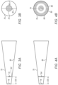
- the trabeculoplasty device further comprises a contact optic 72 , which comprises the optical filter and is configured to contact the eye.
- contact optic 72 is elliptical, e.g., circular.
- contact optic 72 contacts eye 25 , with optical filter 70 covering the pupil of the eye.
- the term “contact” may include contact via an optical coupling fluid or a gel.
- the contact optic is curved, so as to conform to the shape of the eyeball of eye 25 .
- the thickness of the contact optic is less than 2 mm, such that the contact optic may be worn in the eye without being held in place.
- contact optic 72 comprises the optical filter along with a transparent portion 74 , which surrounds the optical filter and is transparent to the treatment beams and to visible light 68 .
- transparent portion 74 is annular-elliptical, e.g., annular-circular.
- the diameter d 1 of the contact optic is typically 12-17 mm, with the diameter d 2 of the optical filter typically being less than 10 mm.
- optical filter 70 covers the pupil of the eye (and, typically, at least part of the cornea surrounding the pupil), while transparent portion 74 covers the targeted vicinity of the limbus. Hence, the vicinity of the limbus may be irradiated by the treatment beams and may also be imaged by camera 54 .
- contact optic 72 may comprise a transparent optic having a central portion coated with a reflective coating, which constitutes the optical filter, along with an uncoated peripheral portion.
- the optical filter may be embedded in the center of a transparent optic. Such embedding may be performed using diffusion bonding, welding, soldering, or an adhesive.
- the contact optic further comprises another optical filter that surrounds the transparent portion and inhibits passage of the treatment beams therethrough, as described below with reference to FIGS. 4 A-B .
- the contact optic comprises the optical filter without comprising transparent portion 74 .
- diameter d 1 (which is equal to diameter d 2 due to the absence of transparent portion 74 ) is typically less than 10 mm, such that, during the procedure, the limbus and/or sclera of the eye are exposed.
- corrective contact lenses typically have a larger diameter so as not to discomfort the wearer, the inventors have realized that contact optic 72 is unlikely to discomfort the patient despite its small size, particularly if anesthetic eye drops are applied to the eye before the contact optic is placed in the eye.
- the trabeculoplasty device further comprises an eye-stabilizing structure 76 , through which the various types of radiation described herein pass.
- the length of structure 76 is between 10 and 70 mm.
- optical filter 70 is typically coupled to structure 76 .
- contact optic 72 may be coupled to the distal end of the structure, such that optical filter 70 is coupled to the distal end of structure 76 by virtue of the contact optic comprising the optical filter.
- the thickness of the contact optic is typically greater than 2 mm; for example, the thickness may be between 3 and 20 mm.
- the edge of the contact optic may be coupled to the distal end of the structure using a mechanical attachment mechanism, such as a screw, and/or any suitable adhesive.
- optical filter 70 may be coupled to structure 76 in other ways, as further described below with reference to FIGS. 3 A-B and 4 A-B.
- structure 76 is hollow, such that the radiation described herein passes through air within the structure.
- structure 76 may comprise a frustum-shaped or cylindrically-shaped tube, which may be made from metal, glass, or any other suitable material.
- the structure comprises a continuous wall.
- structure 76 is shaped to define one or more openings.
- structure 76 may comprise a wire structure, such as a wire mesh. As further described below with reference to FIG. 8 , an advantage of such embodiments is that the user may view the eye through the openings.
- structure 76 comprises a frustum-shaped or cylindrically-shaped transparent piece of material (e.g., glass), such that the radiation passes through the material.
- material e.g., glass
- Structure 76 typically provides several advantages, alternatively or additionally to holding optical filter 70 .
- the distal end of structure 76 may retract the eyelids of eye 25 .
- the distal end of the structure may stabilize the eye, i.e., inhibit the eye from moving.
- structure 76 may facilitate placing the optical unit at the correct distance from the patient.
- the inner and/or outer surface of structure 76 may be configured to absorb any misaimed treatment beams or scattered light, and/or to block any external light from interfering with the camera.
- the inner and/or outer surface of the structure may comprise black flat paint, black flocked paper, or Vantablack.
- the proximal end of the structure is configured to couple to the optical unit, e.g., via a spring.
- the trabeculoplasty device may further comprise a pressure sensor coupled to structure 76 and configured to measure the pressure applied to the structure. Based on the pressure measurement, the user may position the optical unit such that the pressure applied to the structure, which is equivalent to the pressure applied to the eye, is within a predefined range suitable for stabilizing the eye without hurting the patient.
- the structure is not coupled to the optical unit, but rather, is held by the user of system 20 during the procedure.
- structure 76 is disposable, and a different respective structure is used for each patient.
- the optical unit may comprise an exit window in lieu of opening 58 .
- the optical filter may overlay, or may be embedded within, the exit window.
- controller 44 may process images of the eye to calculate the position of each of the target regions.
- the controller identifies contact optic 72 in each of the images, and verifies that the contact optic is in the correct location relative to the eye.
- the controller may also calculate the position of the target region with reference to the position of the contact optic.
- the contact optic is opaque to wavelengths to which the camera is sensitive.
- the camera may be sensitive only to wavelengths below 600 nm, and the contact optic may be opaque to wavelengths below 600 nm.
- the controller does not identify the contact optic in any of the images.
- a finger, a speculum, or another tool may be used to retract one or both of the eyelids of eye 25 .
- device 21 may comprise any suitable components.
- FIGS. 3 A-B are schematic illustrations of structure 76 as viewed from the side and from the front, respectively, in accordance with some embodiments of the present invention.
- FIGS. 4 A-B are also schematic illustrations of structure 76 as viewed from the side and from the front, respectively, in accordance with some embodiments of the present invention.
- the optical filter is situated proximally to the distal end of eye-stabilizing structure 76 , such that the distal end of the eye-stabilizing structure, but not the optical filter, contacts the eye.
- the optical filter may be mounted to the inner wall 80 of structure 76 , e.g., at a distance D 0 of 0.5-20 mm from the distal end of the structure.
- diameter d 2 of the optical filter ( FIG. 2 ) is matched to distance D 0 , such that the optical filter covers the pupil without covering the targeted regions of the eye. In other words, for a smaller D 0 , d 2 is made larger, and vice versa.
- the optical filter is mounted to inner wall 80 via one or more longitudinal elements 82 , such as stiff wires or posts, extending between the inner wall and the optical filter.
- the trabeculoplasty device comprises an optic 84 , which comprises optical filter 70 along with transparent portion 74 .
- the edge of optic 84 may be coupled to inner wall 80 using a mechanical attachment mechanism, such as a screw, and/or any suitable adhesive. (Since optic 84 does not need to conform to the shape of the eye, optic 84 is typically not curved.)
- optic 84 further comprises another optical filter 86 that surrounds transparent portion 74 and is configured to inhibit the passage of the treatment beams.
- optical filter 86 is annular-elliptical, e.g., annular-circular.
- optical filter 86 may comprise an absorptive material and/or a thin-film reflective coating, such that optical filter 86 absorbs and/or reflects the treatment beams.
- optical filter 86 comprises the same materials as does optical filter 70 and/or is etched in the same way as is optical filter 70 .
- the distal end of the structure may comprise a contact optic 78 configured to help stabilize the eye by contacting the eye.
- contact optic 78 or at least the portion of the contact optic that covers the treated vicinity of the limbus, is transparent to the treatment beams and to visible light.
- the diameter of contact optic 78 is between 12 and 17 mm.
- contact optic 78 is shaped to define an opening
- the trabeculoplasty device further comprises (i) a suction-applying device, such as a pump, and (ii) a tube having a proximal end connected to the suction-applying device and a distal end passing through the opening.
- the suction-applying device may apply a suction force through the tube, thus helping the contact optic to stabilize the eye.
- the distal end of the structure may comprise a contact ring configured to help stabilize the eye by contacting the eye.
- the contact ring contacts the eye outside of, and sufficiently far from (e.g., at least 2 mm from), the limbus, such that the portion of the eye targeted by the treatment beams, which typically includes the limbus and/or part of the sclera, is exposed to the treatment beams and to the camera.
- the diameter of the contact ring may be between 14 and 18 mm.
- the contact ring does not contact the eye continuously, but rather, only over several (e.g., between three and ten) points of contact.
- suction may be applied through the contact ring, as described above for contact optic 78 .
- the contact ring may retract the eyelids of the eye.
- FIGS. 5 - 6 are schematic illustrations of optical filter 70 coupled to optical unit 30 , in accordance with some embodiments of the present invention.
- optical filter 70 is coupled to the optical unit.
- the optical filter may overlay, or may be embedded within, an exit window 90 belonging to front face 33 .
- the optical filter may comprise a thin-film reflective coating that coats exit window 90 .
- a longitudinal element 88 such as a stiff wire or a post, may extend between the front face and the optical filter, and the optical filter may be coupled to the front face via longitudinal element 88 .
- the proximal end of longitudinal element 88 may be coupled to front face 33 (e.g., to exit window 90 ), with the distal end of the longitudinal element coupled to the optical filter.
- longitudinal element 88 typically passes through the eye-stabilizing structure.
- the longitudinal element is parallel to optical path 92 .
- the distance between the front face of the optical unit and the optical filter which is generally equal to the length of longitudinal element 88 , is between 10 and 50 mm, and/or the distance between the optical filter and the eye is between 0.1 and 20 mm.
- the optical filter may be coupled to the optical bench belonging to the optical unit.
- the optical filter may be coupled to the optical bench downstream from the most downstream of the beam-directing elements, such as between beam combiner 56 and opening 58 ( FIG. 2 ), such that the optical filter interposes between the most downstream of the beam-directing elements and the pupil of the eye.
- optical filter 70 is opaque both to the treatment beams and to the fixation light, i.e., the visible light transmitted by light source 66 ( FIG. 2 ).
- the optical filter may comprise a metal or ceramic disk.
- the optical filter inhibits both the visible light and the treatment beams from reaching the pupil of eye 25 , eye 25 may nonetheless be oriented for treatment, as described immediately below.
- the optical filter may be manufactured more cheaply and easily, without compromising the safety and efficacy of the procedure.
- the light source may be coupled to the optical bench parallel to or downstream from the optical filter.
- the optical filter and the light source may each be coupled to the optical bench between the most downstream of the beam-directing elements and opening 58 , such that (i) the optical filter interposes between the most downstream of the beam-directing elements and the pupil of the eye, and (ii) light source 66 is disposed next to optical path 92 , parallel to or downstream from the optical filter.
- the light source may be disposed behind the optical filter, as in FIG. 2 ; however, one or more reflectors coupled to the optical bench may reflect the fixation light through opening 58 , such that the fixation light may reach eye 25 without needing to pass through the optical filter.
- eye 25 may be oriented by causing the untreated eye of the patient to fixate on light source 66 .
- the patient prior to the procedure, the patient is instructed to fixate the untreated eye on the light source.
- the untreated eye fixates on the light source, such that eye 25 , by virtue of moving in tandem with the untreated eye, gazes approximately along optical path 92 .
- the treated eye may be irradiated with the treatment beams.
- FIG. 7 is a schematic illustration of an alternative technique for orienting eye 25 for treatment, in accordance with some embodiments of the present invention.
- FIG. 7 shows a schematic overhead view of optical unit 30 and patient 22 .
- the position of the light source is adjustable.
- the light source may be mounted on a slider, such that the distance of the light source from optical path 92 is variable.
- the position of the light source is varied responsively to the degree to which the patient's eyes can converge onto the light source.
- the light source may be aligned, at least approximately, with optical path 92 , as described above with reference to FIG. 2 .
- untreated eye 23 fixates on the light source along a first line-of-sight 93
- eye 25 gazes toward the light source along a second line-of-sight 95 , which is approximately coincident with optical path 92 .
- the light source may be displaced from the optical path towards the untreated eye, typically along an axis parallel to the interpupillary axis of the patient.
- the light source may be placed at a distance D 1 from the optical path that is approximately equal to the interpupillary distance of the patient; thus, for example, D 1 may be between 45 and 80 mm.
- the untreated eye fixates on the light source along an alternate first line-of-sight 93 ′, such that second line-of-sight 95 is approximately coincident with optical path 92 .
- a single position of the light source is used for all patients.
- the light source may be displaced from the optical path towards the untreated eye, typically along an axis parallel to the interpupillary axis of the patient, by a distance that is less than, such as 30%-70% of, an average or median interpupillary distance for a suitable population of patients.
- the light source may be displaced from the optical path by between 18 and 44 mm.
- this position may help second line-of-sight 95 to be sufficiently coincident with optical path 92 , regardless of the degree to which the patient's eyes converge onto the light source.
- the camera Prior to the procedure, acquires image 94 . Based on the image, the user defines one or more target regions 110 of the eye that are to be irradiated. Typically, as described in International Patent Application PCT/IB2019/055564, target regions 110 are defined semi-automatically, based on input from the user via a graphical user interface (GUI) 96 displayed on monitor 42 . Typically, at least part of each target region is located near (e.g., within 2 mm of) the limbus 98 of the eye; for example, each target region may overlap limbus 98 .
- GUI graphical user interface
- the controller executes a mock trabeculoplasty by causing radiation source 48 ( FIG. 2 ) to sweep an aiming beam across the target regions.
- the aiming beam may be emitted by an additional radiation source, such as a lower-power laser, that is collinear with radiation source 48 .
- the aiming beam differs from the treatment beams at least in that the energy of the aiming beam is subtherapeutic.
- camera 54 FIG. 2
- the user may view the aiming beam, as it impinges on the eye, through an opening in the structure.
- the user starts the procedure, typically by entering an appropriate input via GUI 96 .
- the target regions of the eye are irradiated by radiation source 48 , i.e., the radiation source fires respective treatment beams at the target regions.
- the camera acquires images of the eye at a relatively high frequency (e.g., at a frequency greater than 40 Hz or 50 Hz).
- controller 44 by executing image-processing software, tracks motion of the eye, e.g., by identifying the center of the limbus in each image.
- the controller prior to the irradiation of each target region, locates the target region in the FOV of the camera, and then controls the beam-directing elements such that a treatment beam impinges on the target region.
- the eye-stabilizing structure may sufficiently stabilize the eye such as to obviate the need for high-frequency motion tracking.
- the camera may acquire images of the eye, and the controller may check for movement of the eye, at a lower frequency; alternatively, the image acquisition and motion tracking may be omitted entirely.
- high-frequency motion tracking may be omitted if only a small number of treatment beams are fired.
- the radiation source may emit a single treatment beam—shaped, for example, as an ellipse or an arc—that simultaneously irradiates all the target regions.
- optical filter 70 may protect the retina of the eye by covering the pupil 104 of the eye.
- optical filter 70 may also cover at least part of the cornea 108 surrounding the pupil, thus protecting cornea 108 .
- a peripheral optical filter such as optical filter 86 belonging to optic 84 (described above with reference to FIGS. 4 A-B ), may protect the sclera 106 of the eye by covering sclera 106 .
- the optical filter is opaque to the camera (i.e., to the frequencies to which the camera is sensitive).
- the controller may identify the optical filter in the images acquired by the camera, and may further verify that the optical filter is covering the pupil, e.g., as described above with reference to FIG. 2 .
- the optical filter is transparent to the camera. ( FIG. 8 assumes that the optical filter is transparent to the camera; nevertheless, for clarity, the edge of the optical filter is shown in FIG. 8 .)
- the eye is protected using a virtual filter, implemented in the software and/or firmware run by the controller.
- controller 44 FIG. 1
- controller 44 causes camera 54 to acquire image 94 .
- the controller based on the image, identifies one or more static regions in the FOV of the camera that include non-targeted portions of the eye.
- the controller ascertains that the target region is not within any of the static regions.
- the controller In response to ascertaining that the target region is not within any of the static regions, the controller causes the radiation source to irradiate the target region; otherwise, the treatment beam is not fired. Furthermore, in some embodiments, the controller inhibits the beam-directing elements from being aimed at any of the static regions even while the radiation source is inactive.
- the controller may identify a central static region encompassing pupil 104 in the image. Subsequently, the controller may inhibit the radiation source from firing any treatment beams at the central static region.
- the central static region may thus protect the retina of the eye in the event that optical filter 70 is not used, or provide extra protection to that provided by the optical filter.
- the peripheral static region includes the portion of the camera's FOV lying outside an elliptical (e.g., circular) boundary 100 , which, in image 94 , may be located outside limbus 98 at a distance of between 1 and 5 mm from the limbus.
- an elliptical boundary 100 which, in image 94 , may be located outside limbus 98 at a distance of between 1 and 5 mm from the limbus.
- each of the static regions is “static” by virtue of being defined in terms of the FOV of the camera, such that the position of the region is not adjusted by the controller even in response to detected motion of the eye.
- the eye is protected even in the event of an (unlikely) error in the motion tracking performed by the controller.
Landscapes
- Health & Medical Sciences (AREA)
- Ophthalmology & Optometry (AREA)
- Optics & Photonics (AREA)
- Physics & Mathematics (AREA)
- Heart & Thoracic Surgery (AREA)
- Surgery (AREA)
- Engineering & Computer Science (AREA)
- Biomedical Technology (AREA)
- Nuclear Medicine, Radiotherapy & Molecular Imaging (AREA)
- Vascular Medicine (AREA)
- Life Sciences & Earth Sciences (AREA)
- Animal Behavior & Ethology (AREA)
- General Health & Medical Sciences (AREA)
- Public Health (AREA)
- Veterinary Medicine (AREA)
- Eye Examination Apparatus (AREA)
- Laser Surgery Devices (AREA)
Abstract
Description
-
- the optical filter; and
- a transparent portion that surrounds the optical filter and is transparent to the treatment beams.
-
- the optical filter is a first optical filter, and
- the optic further includes a second optical filter that surrounds the transparent portion and is configured to inhibit the passage of the treatment beams.
-
- the optical unit includes a front face, and
- the optical filter is coupled to the front face.
-
- the front face includes an exit window,
- the light source is configured to transmit the visible tight, and the radiation source is configured to emit the treatment beams, through the exit window, and
- the optical filter overlays the exit window.
-
- the front face includes an exit window,
- the light source is configured to transmit the visible light, and the radiation source is configured to emit the treatment beams, through the exit window, and
- the optical filter is embedded within the exit window.
-
- the front face includes an exit window,
- the light source is configured to transmit the visible light, and the radiation source is configured to emit the treatment beams, through the exit window, and
- the longitudinal element extends between the exit window and the optical filter.
Claims (40)
Priority Applications (1)
| Application Number | Priority Date | Filing Date | Title |
|---|---|---|---|
| US17/273,323 US12295886B2 (en) | 2018-10-28 | 2019-10-23 | Protection for direct selective laser trabeculoplasty |
Applications Claiming Priority (3)
| Application Number | Priority Date | Filing Date | Title |
|---|---|---|---|
| US201862751629P | 2018-10-28 | 2018-10-28 | |
| US17/273,323 US12295886B2 (en) | 2018-10-28 | 2019-10-23 | Protection for direct selective laser trabeculoplasty |
| PCT/IB2019/059058 WO2020089737A1 (en) | 2018-10-28 | 2019-10-23 | Protection for direct selective laser trabeculoplasty |
Related Parent Applications (1)
| Application Number | Title | Priority Date | Filing Date |
|---|---|---|---|
| PCT/IB2019/059058 A-371-Of-International WO2020089737A1 (en) | 2018-10-28 | 2019-10-23 | Protection for direct selective laser trabeculoplasty |
Related Child Applications (1)
| Application Number | Title | Priority Date | Filing Date |
|---|---|---|---|
| US19/051,279 Division US20250177209A1 (en) | 2018-10-28 | 2025-02-12 | Protection for direct selective laser trabeculoplasty |
Publications (2)
| Publication Number | Publication Date |
|---|---|
| US20210322214A1 US20210322214A1 (en) | 2021-10-21 |
| US12295886B2 true US12295886B2 (en) | 2025-05-13 |
Family
ID=70463532
Family Applications (2)
| Application Number | Title | Priority Date | Filing Date |
|---|---|---|---|
| US17/273,323 Active 2042-05-22 US12295886B2 (en) | 2018-10-28 | 2019-10-23 | Protection for direct selective laser trabeculoplasty |
| US19/051,279 Pending US20250177209A1 (en) | 2018-10-28 | 2025-02-12 | Protection for direct selective laser trabeculoplasty |
Family Applications After (1)
| Application Number | Title | Priority Date | Filing Date |
|---|---|---|---|
| US19/051,279 Pending US20250177209A1 (en) | 2018-10-28 | 2025-02-12 | Protection for direct selective laser trabeculoplasty |
Country Status (6)
| Country | Link |
|---|---|
| US (2) | US12295886B2 (en) |
| EP (1) | EP3870124A4 (en) |
| JP (1) | JP7448973B2 (en) |
| CN (1) | CN112912041B (en) |
| IL (1) | IL282049A (en) |
| WO (1) | WO2020089737A1 (en) |
Families Citing this family (5)
| Publication number | Priority date | Publication date | Assignee | Title |
|---|---|---|---|---|
| US11771596B2 (en) | 2010-05-10 | 2023-10-03 | Ramot At Tel-Aviv University Ltd. | System and method for treating an eye |
| EP2568938A1 (en) | 2010-05-10 | 2013-03-20 | Ramot at Tel-Aviv University Ltd | System for treating glaucoma by directing electromagnetic energy to the limbal area of an eye |
| IL323052A (en) | 2018-07-02 | 2025-10-01 | Belkin Vision Ltd | Direct selective laser trabeculoplasty |
| EP3870124A4 (en) | 2018-10-28 | 2022-08-03 | Belkin Vision Ltd. | Protection for direct selective laser trabeculoplasty |
| JP7808557B2 (en) * | 2020-07-19 | 2026-01-29 | ベルキン ビジョン リミティド | Automated capsulotomy |
Citations (283)
| Publication number | Priority date | Publication date | Assignee | Title |
|---|---|---|---|---|
| US2635502A (en) | 1948-06-17 | 1953-04-21 | Richards John Mark | Instrument for testing eyes |
| US3594072A (en) | 1969-12-04 | 1971-07-20 | Biometrics Inc | Head-holding fixture for use with visual instruments |
| US4587257A (en) | 1984-12-14 | 1986-05-06 | Alcon Laboratories, Inc. | Control of ocular bleeding using clonidine derivatives |
| US4641349A (en) | 1985-02-20 | 1987-02-03 | Leonard Flom | Iris recognition system |
| EP0224322A1 (en) | 1985-09-12 | 1987-06-03 | Summit Technology, Inc. | Surface erosion using lasers |
| US4718418A (en) | 1983-11-17 | 1988-01-12 | Lri L.P. | Apparatus for ophthalmological surgery |
| US4848894A (en) | 1988-06-01 | 1989-07-18 | The United States Of America As Represented By The Secretary Of The Army | Contact lens with laser protection |
| US4966452A (en) | 1989-04-27 | 1990-10-30 | Ocular Instruments, Inc. | Contact lens for laser surgery |
| FR2655837A1 (en) | 1989-12-15 | 1991-06-21 | Hanna Khalil | SURFACE TREATMENT MASK AND DEVICE FOR EYE SURGERY OR LASER OPTICAL LENS PRODUCTION. |
| US5049147A (en) | 1989-04-06 | 1991-09-17 | Danon Nissim N | Apparatus for computerized laser surgery |
| US5123902A (en) | 1988-09-13 | 1992-06-23 | Carl-Zeiss-Stiftung | Method and apparatus for performing surgery on tissue wherein a laser beam is applied to the tissue |
| US5141506A (en) * | 1985-10-22 | 1992-08-25 | York Kenneth K | Systems and methods for creating substrate surfaces by photoablation |
| US5151909A (en) | 1990-10-16 | 1992-09-29 | Laserscope | Frequency doubled solid state laser having programmable pump power modes and method for controllable lasers |
| WO1992016259A1 (en) | 1991-03-13 | 1992-10-01 | Iris Medical Instruments, Inc. | Contact probe for laser cyclophotocoagulation |
| US5152760A (en) | 1989-03-17 | 1992-10-06 | The General Hospital Corporation | Non-invasive sclerostomy |
| WO1993012727A1 (en) | 1991-12-24 | 1993-07-08 | Laser Engineering, Inc. | Decoupled dual-beam control system |
| WO1993016631A1 (en) | 1992-02-27 | 1993-09-02 | Phoenix Laser Systems, Inc. | Automated laser workstation for high precision surgical and industrial interventions |
| WO1994012092A1 (en) | 1992-11-20 | 1994-06-09 | The Johns Hopkins University | Identification and treatment of eye neovascular membranes |
| WO1994016425A1 (en) | 1993-01-12 | 1994-07-21 | Iatrotech, Inc. | Laser calibration device |
| US5370641A (en) | 1992-05-22 | 1994-12-06 | O'donnell, Jr.; Francis E. | Laser trabeculodissection |
| EP0651982A1 (en) | 1993-11-08 | 1995-05-10 | Khalil Hanna | Mask intended to be interposed in a laser to apply a treatment, especially superficial ablation to an organ or device, particularly cornea, a lens or a cornea implant; process and apparatus making use of such a mask |
| US5422899A (en) | 1994-05-10 | 1995-06-06 | Premier Laser Systems, Inc. | High repetition rate mid-infrared laser |
| WO1995015134A1 (en) | 1993-12-02 | 1995-06-08 | Sunrise Technologies, Inc. | Laser system for reshaping the cornea |
| US5479222A (en) | 1993-11-15 | 1995-12-26 | Volk; Donald A. | Indirect ophthalmoscopy lens system and adapter lenses |
| EP0689811A1 (en) | 1994-06-29 | 1996-01-03 | Luis Antonio Ruiz | Apparatus for performing presbyopia corrective surgery |
| US5537164A (en) | 1994-12-20 | 1996-07-16 | Smith; Alan D. | Retroilluminating indirect gonioprism |
| US5549596A (en) | 1993-07-07 | 1996-08-27 | The General Hospital Corporation | Selective laser targeting of pigmented ocular cells |
| US5598007A (en) | 1994-03-21 | 1997-01-28 | Intermec Corporation | Symbology reader with fixed focus spotter beam |
| WO1998022016A2 (en) | 1996-11-22 | 1998-05-28 | Velde Frans J Van De | Scanning laser ophthalmoscope optimized for retinal microphotocoagulation |
| US5786883A (en) | 1991-11-12 | 1998-07-28 | Pilkington Barnes Hind, Inc. | Annular mask contact lenses |
| US5865830A (en) | 1989-06-07 | 1999-02-02 | Parel; Jean-Marie | Noncontact laser microsurgical apparatus |
| WO1999018868A1 (en) | 1997-10-10 | 1999-04-22 | Visx Incorporated | Eye tracking device for laser eye surgery using corneal margin detection |
| US5982789A (en) | 1995-11-22 | 1999-11-09 | Light Solutions Corporation | Pulsed laser with passive stabilization |
| US6027216A (en) | 1997-10-21 | 2000-02-22 | The Johns University School Of Medicine | Eye fixation monitor and tracker |
| US6030376A (en) * | 1996-12-27 | 2000-02-29 | Nidek Co., Ltd. | Corneal surgical apparatus |
| US6033396A (en) | 1995-11-06 | 2000-03-07 | Huang; David | Apparatus and method for performing laser thermal keratoplasty with minimized regression |
| US6059772A (en) | 1995-03-10 | 2000-05-09 | Candela Corporation | Apparatus and method for treating glaucoma using a gonioscopic laser trabecular ablation procedure |
| US6096029A (en) | 1997-02-24 | 2000-08-01 | Laser Skin Toner, Inc. | Laser method for subsurface cutaneous treatment |
| US6099522A (en) | 1989-02-06 | 2000-08-08 | Visx Inc. | Automated laser workstation for high precision surgical and industrial interventions |
| US6099521A (en) | 1998-05-26 | 2000-08-08 | Shadduck; John H. | Semiconductor contact lens cooling system and technique for light-mediated eye therapies |
| US6146375A (en) | 1998-12-02 | 2000-11-14 | The University Of Michigan | Device and method for internal surface sclerostomy |
| US6159202A (en) * | 1995-09-29 | 2000-12-12 | Nidex Co., Ltd. | Corneal surgery apparatus |
| US6258082B1 (en) | 1999-05-03 | 2001-07-10 | J. T. Lin | Refractive surgery and presbyopia correction using infrared and ultraviolet lasers |
| US6263879B1 (en) | 1998-11-10 | 2001-07-24 | J. T. Lin | Treatment of presbyopia and other eye disorders using a scanning laser system |
| US6267752B1 (en) | 1999-08-05 | 2001-07-31 | Medibell Medical Vision Technologies, Ltd. | Multi-functional eyelid speculum |
| US6267756B1 (en) | 1986-03-08 | 2001-07-31 | G. Rodenstock Instrumente Gmbh | Apparatus for the observation and the treatment of the eye using a laser |
| US6286960B1 (en) * | 1999-03-30 | 2001-09-11 | Nidek Co., Ltd. | Ophthalmic apparatus |
| US20010027314A1 (en) | 1995-10-20 | 2001-10-04 | Peyman Gholam A. | Intrastromal corneal modification via laser |
| US6319274B1 (en) | 1998-06-22 | 2001-11-20 | John H. Shadduck | Devices and techniques for light-mediated stimulation of trabecular meshwork in glaucoma therapy |
| US6325792B1 (en) | 1991-11-06 | 2001-12-04 | Casimir A. Swinger | Ophthalmic surgical laser and method |
| WO2001095842A1 (en) | 2000-06-16 | 2001-12-20 | Volk Optical, Inc. | Aspheric iridectomy/iridotomy treatment lens |
| US20020013572A1 (en) * | 2000-05-19 | 2002-01-31 | Berlin Michael S. | Delivery system and method of use for the eye |
| US20020013573A1 (en) | 1995-10-27 | 2002-01-31 | William B. Telfair | Apparatus and method for tracking and compensating for eye movements |
| US20020026179A1 (en) * | 2000-03-31 | 2002-02-28 | Nidek Co., Ltd. | Ophthalmic surgery apparatus |
| US6414980B1 (en) | 1999-10-12 | 2002-07-02 | Coherent, Inc. | Laser rod thermalization |
| WO2002064031A2 (en) | 2001-02-09 | 2002-08-22 | Sensomotoric Instruments Gmbh | Multidimensional eye tracking and position measurement system |
| US6454763B1 (en) | 1999-08-05 | 2002-09-24 | Paradigm Medical Industries Inc. | Laser surgical handpiece with photon trap |
| WO2002087442A1 (en) | 2001-04-27 | 2002-11-07 | Bausch & Lomb Incorporated | Iris pattern recognition and alignment |
| US6530916B1 (en) | 1999-11-15 | 2003-03-11 | Visx, Incorporated | Uniform large area ablation system and method |
| US6569104B2 (en) | 1998-07-16 | 2003-05-27 | Canon Kabushiki Kaisha | Blood vessel detecting apparatus |
| US6585723B1 (en) * | 1998-09-04 | 2003-07-01 | Nidek Co., Ltd. | Corneal surgery apparatus |
| US20030179344A1 (en) | 1996-11-22 | 2003-09-25 | Van De Velde Frans J. | Scanning laser ophthalmoscope optimized for selective retinal microphotocoagulation |
| US20030225398A1 (en) | 2002-05-28 | 2003-12-04 | Neil Zepkin | Zoom device for eye tracker control system and associated methods |
| US6673062B2 (en) | 2000-03-14 | 2004-01-06 | Visx, Inc. | Generating scanning spot locations for laser eye surgery |
| US6676655B2 (en) | 1998-11-30 | 2004-01-13 | Light Bioscience L.L.C. | Low intensity light therapy for the manipulation of fibroblast, and fibroblast-derived mammalian cells and collagen |
| US6685317B2 (en) | 2000-06-13 | 2004-02-03 | Massie Research Laboratories, Inc. | Digital eye camera |
| US20040039378A1 (en) | 2000-06-01 | 2004-02-26 | Lin Charles P. | Selective photocoagulation |
| US6698886B2 (en) | 2001-04-24 | 2004-03-02 | Ocular Instruments, Inc. | Iridotomy and trabeculoplasty goniolaser lens |
| WO2004027487A1 (en) | 2002-09-18 | 2004-04-01 | Ellex Medical Pty Ltd | Ophthalmic laser system |
| US6736806B2 (en) | 2000-06-21 | 2004-05-18 | Luis Antonio Ruiz | Controllable liquid crystal matrix mask particularly suited for performing ophthamological surgery, a laser system with said mask and a method of using the same |
| US6761713B2 (en) | 2000-11-08 | 2004-07-13 | Lisa Laser Products Ohg Fuhrberg & Teichmann | Medical laser unit |
| US20040196431A1 (en) | 2003-04-07 | 2004-10-07 | Arkadiy Farberov | Optical device for intraocular observation |
| CN1579351A (en) | 2003-08-07 | 2005-02-16 | 天津医科大学 | Automatic-controlled laser operation machine for glaucoma filtration operation |
| US20050096639A1 (en) | 2000-05-08 | 2005-05-05 | Michael Slatkine | Non-penetrating filtration surgery |
| US20050107774A1 (en) | 1999-05-03 | 2005-05-19 | Lin J. T. | Methods and apparatus for presbyopia correction using ultraviolet and infrared lasers |
| US6899707B2 (en) | 2001-01-29 | 2005-05-31 | Intralase Corp. | Applanation lens and method for ophthalmic surgical applications |
| US20050185138A1 (en) | 2004-02-19 | 2005-08-25 | Visx, Incorporated | Methods and systems for differentiating left and right eye images |
| US20050197655A1 (en) * | 1995-10-27 | 2005-09-08 | Telfair William B. | Method and apparatus for removing corneal tissue with infrared laser radiation and short pulse mid-infrared parametric generator for surgery |
| US6942656B2 (en) | 2000-05-12 | 2005-09-13 | Ceramoptec Industries, Inc. | Method for accurate optical treatment of an eye's fundus |
| US6948815B2 (en) | 2003-07-28 | 2005-09-27 | Ceramoptec Industries, Inc. | Laser safety contact lenses |
| US20050254009A1 (en) | 2004-05-12 | 2005-11-17 | Chris Baker | Motorized patient support for eye examination or treatment |
| EP1602321A1 (en) | 2004-06-02 | 2005-12-07 | SensoMotoric Instruments GmbH | Method and apparatus for image-based eye tracking for retinal diagnostic or surgery device |
| US6979328B2 (en) | 2001-01-18 | 2005-12-27 | The Regents Of The University Of California | Minimally invasive glaucoma surgical instrument and method |
| US20050288745A1 (en) | 2004-06-28 | 2005-12-29 | Andersen Dan E | Method and device for optical ophthalmic therapy |
| US20050286019A1 (en) | 2004-06-10 | 2005-12-29 | Wiltberger Michael W | Scanning ophthalmic fixation method and apparatus |
| US7027233B2 (en) | 2001-10-12 | 2006-04-11 | Intralase Corp. | Closed-loop focal positioning system and method |
| US20060100677A1 (en) | 2003-12-24 | 2006-05-11 | Blumenkranz Mark S | Patterned laser treatment of the retina |
| US20060176913A1 (en) | 2005-02-04 | 2006-08-10 | Jds Uniphase Corporation, State Of Incorporation: Delaware | Passively Q-switched laser with adjustable pulse repetition rate |
| US20060195076A1 (en) | 2005-01-10 | 2006-08-31 | Blumenkranz Mark S | Method and apparatus for patterned plasma-mediated laser trephination of the lens capsule and three dimensional phaco-segmentation |
| US20060224147A1 (en) * | 2005-03-31 | 2006-10-05 | Nidek Co., Ltd. | Ophthalmic laser treatment apparatus |
| WO2006119349A2 (en) | 2005-04-29 | 2006-11-09 | Novadaq Technologies, Inc. | Choroid and retinal imaging and treatment system |
| WO2006119584A1 (en) | 2005-05-13 | 2006-11-16 | Customvis Plc | Fast response eye tracking |
| US20060265030A1 (en) | 2004-11-12 | 2006-11-23 | Light Bioscience, Llc | System and method for photodynamic cell therapy |
| WO2006128038A2 (en) | 2005-05-26 | 2006-11-30 | Ntk Enterprises, Inc. | Device, system, and method for epithelium protection during cornea reshaping |
| US20070081166A1 (en) | 2005-09-29 | 2007-04-12 | Bioptigen, Inc. | Portable Optical Coherence Tomography (OCT) Devices and Related Systems |
| US20070129709A1 (en) | 2005-12-01 | 2007-06-07 | Andersen Dan E | System and method for minimally traumatic ophthalmic photomedicine |
| JP2007151739A (en) | 2005-12-02 | 2007-06-21 | Hoya Corp | Contact lens for ophthalmic surgery |
| US20070159600A1 (en) | 2003-04-08 | 2007-07-12 | Medibell Medical Vision Technologies, Ltd. | Transcleral opthalmic illumination method and system |
| US7252661B2 (en) | 2003-12-23 | 2007-08-07 | Alcon Refractivehorizons, Inc. | Method and system for patient optical fixation |
| CA2640203A1 (en) | 2006-02-03 | 2007-08-16 | Light Bioscience, Llc | Low intensity light therapy for treatment of retinal, macular, and visual pathway disorders |
| WO2007103349A2 (en) | 2006-03-07 | 2007-09-13 | The Trustees Of Princeton University | Devices, methods and compositions for presbyopia correction using ultrashort pulse lasers |
| US20070213693A1 (en) | 2004-08-27 | 2007-09-13 | Ellex Medical Pty Ltd | Selective ophthalmic laser treatment |
| US7282046B2 (en) | 2004-01-22 | 2007-10-16 | Peter M. Adams, Doug P. Adams, and John Sullivan, Collectively as the Stockholder Representative Committee | Glaucoma treatment method |
| US20080027418A1 (en) * | 2006-07-12 | 2008-01-31 | Ntk Enterprises, Inc. | Deuterated ocular solutions for LTK and other surgical eye procedures |
| US7353829B1 (en) | 1996-10-30 | 2008-04-08 | Provectus Devicetech, Inc. | Methods and apparatus for multi-photon photo-activation of therapeutic agents |
| US20080089481A1 (en) | 2006-10-16 | 2008-04-17 | Oraya Therapeutics, Inc. | Portable orthovoltage radiotherapy |
| US7371230B2 (en) | 2001-01-29 | 2008-05-13 | Amo Development, Llc | Ocular fixation and stabilization device for ophthalmic surgical applications |
| US20080161781A1 (en) | 2005-02-19 | 2008-07-03 | Mcardle George J | Apparatus and Processes For Preventing or Delaying Onset or Progression of Age-Related Cataract |
| US20080167642A1 (en) | 2006-11-10 | 2008-07-10 | Palanker Daniel V | System And Method For Determining Dosimetry In Ophthalmic Photomedicine |
| WO2008112236A1 (en) | 2007-03-13 | 2008-09-18 | Optimedica Corporation | Computer guided patterned laser trabeculoplasty |
| US20080234667A1 (en) | 2005-09-27 | 2008-09-25 | Stefan Lang | System and Method for the Treatment of a Patients Eye Working at High Speed |
| US20080255546A1 (en) | 2005-10-24 | 2008-10-16 | Leonid Sergeevich Orbachevski | Eye Accommodation Recovery |
| CN101411607A (en) | 2007-10-16 | 2009-04-22 | 张光荣 | Device for photographing conjunctiva and sclera of eyeball |
| US20090137993A1 (en) | 2007-09-18 | 2009-05-28 | Kurtz Ronald M | Methods and Apparatus for Integrated Cataract Surgery |
| US20090157062A1 (en) | 2007-12-13 | 2009-06-18 | Christoph Hauger | Systems and methods for treating glaucoma and systems and methods for imaging a portion of an eye |
| US20090247997A1 (en) | 2008-04-01 | 2009-10-01 | Amo Development, Llc | Ophthalmic laser apparatus, system, and method with high resolution imaging |
| US20100002837A1 (en) | 2006-12-13 | 2010-01-07 | Oraya Therapeutics, Inc. | Orthovoltage radiotherapy |
| US20100057059A1 (en) | 2006-11-29 | 2010-03-04 | Nidek Co., Ltd. | Corneal surgery apparatus |
| US20100076419A1 (en) | 2007-10-30 | 2010-03-25 | Iridex Corporation | Contact Probe for the Delivery of Laser Energy |
| US20100142767A1 (en) | 2008-12-04 | 2010-06-10 | Alan Duncan Fleming | Image Analysis |
| JP2010148635A (en) | 2008-12-25 | 2010-07-08 | Topcon Corp | Ophthalmic apparatus for laser medical treatment |
| CN201537172U (en) | 2009-11-12 | 2010-08-04 | 深圳普门科技有限公司 | Photon therapeutic equipment for glaucoma |
| WO2010094353A1 (en) | 2009-02-18 | 2010-08-26 | Eye Tech Care | A method of treating an ocular pathology by applying ultrasound to the trabecular meshwork and device thereof |
| US7792249B2 (en) | 2007-12-23 | 2010-09-07 | Oraya Therapeutics, Inc. | Methods and devices for detecting, controlling, and predicting radiation delivery |
| WO2010113193A1 (en) | 2009-04-01 | 2010-10-07 | Centervue S.P.A. | Instrument for eye examination |
| US20100324543A1 (en) | 2007-09-18 | 2010-12-23 | Kurtz Ronald M | Method And Apparatus For Integrating Cataract Surgery With Glaucoma Or Astigmatism Surgery |
| WO2011017002A2 (en) | 2009-07-29 | 2011-02-10 | Alcon Lensx, Inc. | Optical system with movable lens for ophthalmic surgical laser |
| EP2301421A1 (en) | 2009-09-29 | 2011-03-30 | OD-OS GmbH | Ophthalmic apparatus and method of operating the same |
| US20110144627A1 (en) | 2009-12-15 | 2011-06-16 | Smith Ronald T | Multi-spot laser probe |
| WO2011085274A1 (en) | 2010-01-08 | 2011-07-14 | Optimedica Corporation | System for modifying eye tissue and intraocular lenses |
| US20110190741A1 (en) | 2008-08-25 | 2011-08-04 | Thomas Deisinger | Coupling of an eye to a laser device |
| US8004764B2 (en) | 2004-10-29 | 2011-08-23 | Novartis Ag | Color compensating retinal safety filter |
| CN102193182A (en) | 2011-04-28 | 2011-09-21 | 中国科学院自动化研究所 | Moveable reflector laser collimator, moveable reflector target surface sensor and laser collimating method thereof |
| US8048065B2 (en) | 2001-12-21 | 2011-11-01 | Sensomotoric Instruments Gmbh | Method and apparatus for eye position registering and tracking |
| WO2011151812A1 (en) | 2010-05-10 | 2011-12-08 | Ramot At Tel-Aviv University Ltd. | System for treating glaucoma by directing electromagnetic energy to the limbal area of an eye |
| US20120016349A1 (en) | 2001-01-29 | 2012-01-19 | Amo Development, Llc. | Hybrid ophthalmic interface apparatus and method of interfacing a surgical laser with an eye |
| US8109635B2 (en) | 2004-08-12 | 2012-02-07 | Ophthalmic Imaging Systems | Integrated retinal imager and method |
| US20120050308A1 (en) | 2010-08-30 | 2012-03-01 | Canon Kabushiki Kaisha | Image processing apparatus, image processing method, image processing system, and computer readable memory |
| US20120083772A1 (en) | 2010-09-30 | 2012-04-05 | Curveright Llc | Corneal treatment system and method |
| US20120089134A1 (en) | 2010-10-11 | 2012-04-12 | Christopher Horvath | Contactless Photodisruptive Laser assisted Cataract Surgery |
| US8160113B2 (en) | 2009-07-21 | 2012-04-17 | Mobius Photonics, Inc. | Tailored pulse burst |
| US20120259321A1 (en) | 2011-04-10 | 2012-10-11 | Vanessa Isabella Vera | Systems and methods to deliver photodisruptive laser pulses into tissue layers of the anterior angle of the eye |
| US20120283557A1 (en) | 2011-05-05 | 2012-11-08 | Berlin Michael S | Methods and Apparatuses for the Treatment of Glaucoma using visible and infrared ultrashort laser pulses |
| WO2013004255A1 (en) | 2011-07-04 | 2013-01-10 | Wavelight Gmbh | Device and method for a laser-assisted eye surgery treatment system |
| WO2013035091A1 (en) | 2011-09-07 | 2013-03-14 | Visionix Ltd. | Double function tilting head ophthalmic instrument |
| WO2013059481A1 (en) | 2011-10-21 | 2013-04-25 | Bausch & Lomb Incorporated | System and method for lowering iop by creation of microchannels in trabecular meshwork using a femtosecond laser |
| WO2013059564A1 (en) | 2011-10-19 | 2013-04-25 | Iridex Corporation | Grid pattern laser treatments and methods |
| EP2602005A1 (en) | 2007-06-04 | 2013-06-12 | Oraya Therapeutics, INC. | Device for ocular alignment and coupling of ocular structures |
| US8465478B2 (en) | 2009-07-24 | 2013-06-18 | Lensar, Inc. | System and method for performing LADAR assisted procedures on the lens of an eye |
| US8475433B2 (en) | 2007-03-14 | 2013-07-02 | Wavelight Ag | Apparatus for coupling an element to the eye |
| US20130204236A1 (en) | 2011-12-01 | 2013-08-08 | Nanophthalmos, Llc | Method and system for laser ocular surgery |
| WO2013122711A1 (en) | 2012-02-15 | 2013-08-22 | Google Inc. | Heads-up display including eye tracking |
| US8545020B2 (en) | 2006-07-07 | 2013-10-01 | Od-Os Gmbh | Ophthalmoscope including therapy beam targeting |
| US20130289450A1 (en) | 2012-04-25 | 2013-10-31 | Gregg S. Homer | Method for Laser Treatment for Glaucoma |
| WO2013165689A1 (en) | 2012-04-30 | 2013-11-07 | Clarity Medical Systems, Inc. | Ophthalmic wavefront sensor operating in parallel sampling and lock-in detection mode |
| RU2499582C1 (en) | 2012-10-25 | 2013-11-27 | Федеральное государственное бюджетное учреждение "Межотраслевой научно-технический комплекс "Микрохирургия глаза" имени академика С.Н. Федорова" Министерства здравоохранения и социального развития Российской Федерации | Method for laser treatment of primary open-angle glaucoma with narrow angle of iris |
| US20130317570A1 (en) | 2012-05-25 | 2013-11-28 | Jeffrey K. Luttrull | System and process for retina phototherapy |
| WO2014018104A1 (en) | 2012-07-25 | 2014-01-30 | Elenza, Inc. | Method and apparatus for performing a posterior capsulotomy |
| WO2014025862A1 (en) | 2012-08-07 | 2014-02-13 | RegenEye, L.L.C. | Ocular collar stent for treating narrowing of the irideocorneal angle |
| US8679100B2 (en) | 2009-07-29 | 2014-03-25 | Alcon Lensx, Inc. | Optical system with multiple scanners for ophthalmic surgical laser |
| US20140094785A1 (en) | 2007-10-05 | 2014-04-03 | Topcon Medical Laser Systems, Inc. | Semi-automated ophthalmic photocoagulation method and apparatus |
| US20140114297A1 (en) | 2012-10-24 | 2014-04-24 | Optimedica Corporation | Graphical user interface for laser eye surgery system |
| US8709029B2 (en) | 2006-07-11 | 2014-04-29 | Refocus Ocular, Inc. | Apparatus and method for securing ocular tissue |
| US8708491B2 (en) | 2008-07-25 | 2014-04-29 | Lensar, Inc. | Method and system for measuring an eye |
| US20140128731A1 (en) | 2012-11-02 | 2014-05-08 | Optimedica Corporation | Optical surface identification for laser surgery |
| US20140128852A1 (en) | 2012-11-02 | 2014-05-08 | Optimedica Corporation | Interface force feedback in a laser eye surgery system |
| US20140128851A1 (en) | 2012-11-02 | 2014-05-08 | Optimedica Corporation | Low voltage communication between subsystems in a laser eye surgery system |
| US8771261B2 (en) | 2006-04-28 | 2014-07-08 | Topcon Medical Laser Systems, Inc. | Dynamic optical surgical system utilizing a fixed relationship between target tissue visualization and beam delivery |
| WO2014132162A1 (en) | 2013-02-26 | 2014-09-04 | Belkin Laser Ltd. | System for glaucoma treatment |
| US20140276681A1 (en) | 2013-03-15 | 2014-09-18 | Optimedica Corporation | Microfemtotomy methods and systems |
| US8845625B2 (en) | 2010-01-22 | 2014-09-30 | Optimedica Corporation | Method and apparatus for automated placement of scanned laser capsulorhexis incisions |
| US20140307077A1 (en) | 2013-04-10 | 2014-10-16 | Delta ID Inc. | Apparatuses and methods for iris imaging |
| US8903468B2 (en) | 2010-10-13 | 2014-12-02 | Gholam Peyman | Laser coagulation of an eye structure from a remote location |
| WO2014191031A1 (en) | 2013-05-29 | 2014-12-04 | Wavelight Gmbh | Apparatus for optical coherence tomography of an eye and method for optical coherence tomography of an eye |
| US8939965B2 (en) | 2009-09-29 | 2015-01-27 | Od-Os Gmbh | Ophthalmic apparatus and method of operating the same |
| US8968279B2 (en) | 2003-03-06 | 2015-03-03 | Amo Manufacturing Usa, Llc | Systems and methods for qualifying and calibrating a beam delivery system |
| WO2015069197A1 (en) | 2013-11-11 | 2015-05-14 | Optotek D.O.O. | Ophthalmic laser device |
| US9055896B2 (en) | 2009-09-29 | 2015-06-16 | Od-Os Gmbh | Ophthalmoscope for observing an eye |
| US20150164635A1 (en) | 2012-08-07 | 2015-06-18 | RegenEye, L.L.C. | Method of Reducing the Occurrence of Macular and Neuroretinal Degenerations by Alleviating Age Related Retinal Stresses as a Contributing Factor in a Mammalian Eye |
| RU2553507C1 (en) | 2014-04-24 | 2015-06-20 | федеральное государственное бюджетное учреждение "Межотраслевой научно-технический комплекс "Микрохирургия глаза" имени академика С.Н. Федорова" Министерства здравоохранения Российской Федерации | Method of combined laser treatment of primary narrow angle glaucoma |
| EP2301424B1 (en) | 2009-09-29 | 2015-07-08 | OD-OS GmbH | Ophthalmoscope with a laser device |
| US20150223683A1 (en) | 2014-02-10 | 2015-08-13 | Labyrinth Devices, Llc | System For Synchronously Sampled Binocular Video-Oculography Using A Single Head-Mounted Camera |
| WO2015130821A2 (en) | 2014-02-27 | 2015-09-03 | Iridex Corporation | Illuminated treatment probe for delivering laser energy |
| AU2015210430A1 (en) | 2011-11-07 | 2015-09-03 | Alcon Inc. | Retinal laser surgery |
| US20150266706A1 (en) | 2012-09-06 | 2015-09-24 | Kawasaki Jukogyo Kabushiki Kaisha | Conveyance system and conveyance method of conveyance system |
| US20150272782A1 (en) | 2014-03-26 | 2015-10-01 | Optimedica Corporation | Confocal laser eye surgery system |
| CN105138996A (en) | 2015-09-01 | 2015-12-09 | 北京上古视觉科技有限公司 | An Iris Recognition System with Liveness Detection Function |
| US20160008172A1 (en) | 2014-01-15 | 2016-01-14 | The Regents Of The University Of Colorado, A Body Corporate | Non-invasive devices for lowering intraocular pressure |
| US20160008169A1 (en) | 2014-07-14 | 2016-01-14 | Novartis Ag | Movable wide-angle ophthalmic surgical system |
| JP2016013255A (en) | 2014-07-01 | 2016-01-28 | 株式会社ニデック | Laser therapy device |
| WO2016018864A1 (en) | 2013-10-15 | 2016-02-04 | Lensar, Inc. | Iris registration method and system |
| US20160067035A1 (en) | 2014-09-09 | 2016-03-10 | Staar Surgical Company | Intraocular Lens With Central Hole For Improved Fluid Flow And Minimized Light Scattering |
| US20160067087A1 (en) | 2014-09-09 | 2016-03-10 | LumiThera, Inc. | Wearable devices and methods for multi-wavelength photobiomodulation for ocular treatments |
| US20160089269A1 (en) | 2011-06-09 | 2016-03-31 | Christopher Horvath | Laser system for eye surgery |
| US20160095752A1 (en) | 2013-04-17 | 2016-04-07 | Optimedica Corporation | Corneal topography measurements and fiducial mark incisions in laser surgical procedures |
| WO2016058931A2 (en) | 2014-10-17 | 2016-04-21 | Carl Zeiss Meditec Ag | Systems and method for short pulse laser eye surgery |
| US20160113816A1 (en) | 2013-03-15 | 2016-04-28 | Aleyegn, Inc. | Scleral translocation elasto-modulation methods and apparatus |
| US9351878B2 (en) | 2005-01-11 | 2016-05-31 | Carl Zeiss Meditec Ag | Safety mechanism for laser treatment apparatus |
| EP1856774B1 (en) | 2005-02-24 | 2016-06-08 | Iridex Corporation | Laser system with short pulse characteristics and its methods of use |
| WO2016156760A1 (en) | 2015-04-01 | 2016-10-06 | Alphanov Centre Technologique Optique Et Lasers | Ophthalmic surgical apparatus |
| US9480599B2 (en) | 2008-12-31 | 2016-11-01 | I Optima Ltd. | Device and method for laser assisted deep sclerectomy |
| CN205698218U (en) | 2016-03-15 | 2016-11-23 | 北京太和康明医疗科技有限责任公司 | Multi-point scanning light coagulates instrument |
| WO2016187436A1 (en) | 2015-05-19 | 2016-11-24 | The Regents Of The University Of Colorado, A Body Corporate | Multifocal, electromagnetic treatment of ocular tissues |
| US9504609B2 (en) | 2007-09-10 | 2016-11-29 | Alcon Lensx, Inc. | Apparatus, systems and techniques for interfacing with an eye in laser surgery |
| US20160346126A1 (en) | 2012-05-25 | 2016-12-01 | Ojai Retinal Technology, Llc | System and process for neuroprotective therapy for glaucoma |
| US20160354241A1 (en) | 2014-02-28 | 2016-12-08 | Excel-Lens, Inc. | Laser assisted cataract surgery |
| US20160367399A1 (en) | 2011-12-19 | 2016-12-22 | Alcon Lensx, Inc. | Intra-surgical optical coherence tomographic imaging of cataract procedures |
| WO2016207739A1 (en) | 2015-06-24 | 2016-12-29 | Wavelight Gmbh | Apparatus for eye laser surgery for performing a transepithelial photorefractive keratectomy |
| US20170000647A1 (en) | 2015-07-01 | 2017-01-05 | Optimedica Corporation | Sub-nanosecond laser surgery system utilizing multiple pulsed laser beams |
| US20170038284A1 (en) | 1998-10-23 | 2017-02-09 | Babak Nemati | Systems for augmenting optical transmission through biological tissues |
| WO2017023296A1 (en) | 2015-08-03 | 2017-02-09 | Optimedica Corporation | System for modifying eye tissue and intraocular lenses |
| WO2017031570A1 (en) | 2015-08-27 | 2017-03-02 | Patrick Gooi | Method and system for laser automated trabecular excision |
| DE202016006265U1 (en) | 2016-09-30 | 2017-03-20 | Carl Zeiss Meditec Ag | Device for fixing a patient's head for ophthalmic devices |
| US20170087014A1 (en) | 2015-09-24 | 2017-03-30 | Visionage Therapies, Llc | Sonic and Ultrasonic Methods and Apparatus for Treatment of Glaucoma |
| US9622911B2 (en) | 2010-09-30 | 2017-04-18 | Cxl Ophthalmics, Llc | Ophthalmic treatment device, system, and method of use |
| WO2017069819A1 (en) | 2015-10-23 | 2017-04-27 | Ojai Retinal Technology, Llc | System and process for retina phototherapy |
| EP2695016B1 (en) | 2011-04-01 | 2017-05-03 | Lensar, Inc. | System for laser generated corneal and crystalline lens incisions using a variable f/ optical system with aspheric contact interface to the cornea or rotating and adaptive optics |
| US20170127938A1 (en) | 2012-01-24 | 2017-05-11 | Duke University | Systems and methods for obtaining low-angle circumferential optical access to the eye |
| US20170184875A1 (en) | 2014-03-24 | 2017-06-29 | Menicon Singapore Pte Ltd. | Apparatus and methods for controlling axial growth with an ocular lens |
| US20170246033A1 (en) | 2011-06-23 | 2017-08-31 | Amo Development, Llc | Ophthalmic range finding |
| US9782232B1 (en) | 2016-04-25 | 2017-10-10 | Novartis Ag | Automated intraocular pressure tamponade |
| US20170360604A1 (en) | 2015-05-07 | 2017-12-21 | Tyb Llc | Remote ophthalmic system and related methods |
| US9849032B2 (en) | 2013-03-13 | 2017-12-26 | Optimedica Corporation | Laser eye surgery system |
| WO2018005796A1 (en) | 2016-06-30 | 2018-01-04 | Iridex Corporation | Handheld ophthalmic laser system with replaceable contact tips and treatment guide |
| US9877633B2 (en) | 2012-07-25 | 2018-01-30 | Intuitive Surgical Operations, Inc | Efficient and interactive bleeding detection in a surgical system |
| WO2018021780A1 (en) | 2016-07-27 | 2018-02-01 | 주식회사 루트로닉 | Ophthalmic treatment device and method of controlling same |
| US9889043B2 (en) | 2006-01-20 | 2018-02-13 | Lensar, Inc. | System and apparatus for delivering a laser beam to the lens of an eye |
| WO2018049246A1 (en) | 2016-09-08 | 2018-03-15 | Aleyegn Technologies Llc | Glaucoma treatment methods and apparatus |
| US20180085257A1 (en) | 2016-09-28 | 2018-03-29 | Kelo Tec Inc | Systems and Methods for Laser Eye Surgery |
| JP2018051210A (en) | 2016-09-30 | 2018-04-05 | 株式会社ニデック | Ophthalmic surgical system, ophthalmic surgical system control program, and ophthalmic surgical microscope |
| US20180104477A1 (en) | 2016-10-13 | 2018-04-19 | Ronald Michael Kurtz | System for temporary nonpharmacologic constriction of the pupil |
| US20180125708A1 (en) | 2015-03-30 | 2018-05-10 | Carl Zeiss Meditec Ag | Ophthalmological laser therapy device |
| US9968483B2 (en) | 2012-09-13 | 2018-05-15 | Kabushiki Kaisha Topcon | Laser treatment apparatus |
| EP3329839A1 (en) | 2016-12-05 | 2018-06-06 | Nidek Co., Ltd. | Ophthalmic apparatus |
| US20180168737A1 (en) | 2016-12-15 | 2018-06-21 | Novartis Ag | Adaptive image registration for ophthalmic surgery |
| US10022457B2 (en) | 2005-08-05 | 2018-07-17 | Gholam A. Peyman | Methods to regulate polarization and enhance function of cells |
| US20180214305A1 (en) | 2017-01-31 | 2018-08-02 | Optimedica Corporation | Methods and systems for laser ophthalmic surgery that provide for iris exposures below a predetermined exposure limit |
| US20180221199A1 (en) | 2017-02-06 | 2018-08-09 | Sensor, LLC | Apparatus with filter to treat macular degeneration and method of treating macular degeneration |
| WO2018152020A1 (en) | 2017-02-15 | 2018-08-23 | Iridex Corporation | Method and eye mask apparatus for treating an eye using a broad area light source |
| KR20180106113A (en) | 2017-03-17 | 2018-10-01 | 단국대학교 천안캠퍼스 산학협력단 | Methods For Treating Damaged Cells Or Tissues Using Low Level Lasers |
| US10143590B2 (en) | 2012-09-07 | 2018-12-04 | Optimedica Corporation | Methods and systems for performing a posterior capsulotomy and for laser eye surgery with a penetrated cornea |
| WO2018232397A1 (en) | 2017-06-16 | 2018-12-20 | Berlin Michael S | Methods and systems for oct guided glaucoma surgery |
| KR20190022216A (en) | 2017-08-25 | 2019-03-06 | 주식회사 메디웨일 | Eye image analysis method |
| US20190078073A1 (en) | 2001-11-01 | 2019-03-14 | Pthera LLC | Enhanced Stem Cell Therapy and Stem Cell Production Through the Administration of Low Level Light Energy |
| US10244991B2 (en) | 2014-02-17 | 2019-04-02 | Children's National Medical Center | Method and system for providing recommendation for optimal execution of surgical procedures |
| US20190099291A1 (en) | 2016-03-18 | 2019-04-04 | Aleyegn Technologies Llc | Effective ocular lens positioning methods and apparatus |
| US20190105200A1 (en) | 2017-03-31 | 2019-04-11 | Annmarie Hipsley | Systems and methods for ocular laser surgery and therapeutic treatments |
| US20190105519A1 (en) | 2015-10-06 | 2019-04-11 | Aleyegn Technologies Llc | Ultrasound directed cavitational methods and system for ocular treatments |
| US10258507B2 (en) | 2015-07-08 | 2019-04-16 | Optimedica Corporation | Methods and systems for laser scan location verification and laser surgical systems with laser scan location verification |
| US20190117459A1 (en) | 2017-06-16 | 2019-04-25 | Michael S. Berlin | Methods and Systems for OCT Guided Glaucoma Surgery |
| US20190142636A1 (en) | 2014-09-09 | 2019-05-16 | LumiThera, Inc. | Devices and methods for non-invasive multi-wavelength photobiomodulation for ocular treatments |
| US20190151146A1 (en) | 2016-07-27 | 2019-05-23 | Lutronic Corporation | Ophthalmic treatment device and control method therefor |
| WO2019109125A1 (en) | 2017-12-04 | 2019-06-13 | Ellex Medical Pty Ltd | Photobiomodulation device for treating retinal disease |
| US20190247225A1 (en) | 2009-01-21 | 2019-08-15 | Carl Zeiss Meditec Ag | Device and method for producing control data for the surgical correction of defective eye vision |
| US20190269554A1 (en) | 2013-02-01 | 2019-09-05 | Alcon Lensx, Inc. | Bi-radial patient interface |
| US10441465B2 (en) | 2014-03-26 | 2019-10-15 | Optimedica Corporation | Registration of LOI fiducials with camera |
| US10456209B2 (en) | 2010-10-13 | 2019-10-29 | Gholam A. Peyman | Remote laser treatment system with dynamic imaging |
| US20190344076A1 (en) | 2016-06-22 | 2019-11-14 | Pedro Irazoqui | Wireless glaucoma therapy |
| US10478342B2 (en) | 2011-07-29 | 2019-11-19 | Carl Zeiss Meditec Ag | Ophthalmologic laser device and method for preventing and treating aftercataract |
| US20190358085A1 (en) | 2013-01-16 | 2019-11-28 | Amo Development, Llc | Robust laser cutting methods for ophthalmic surgery |
| US10524656B2 (en) | 2005-10-28 | 2020-01-07 | Topcon Medical Laser Systems Inc. | Photomedical treatment system and method with a virtual aiming device |
| WO2020008323A1 (en) | 2018-07-02 | 2020-01-09 | Belkin Laser Ltd. | Direct selective laser trabeculoplasty |
| WO2020012841A1 (en) | 2018-07-11 | 2020-01-16 | 株式会社トプコン | Photocoagulation device, control method of photocoagulation device, program, and recording media |
| WO2020018242A1 (en) | 2018-07-16 | 2020-01-23 | Vialase,. Inc. | Integrated surgical system and method for treatment in the irido-corneal angle of the eye |
| WO2020018436A1 (en) | 2018-07-16 | 2020-01-23 | The Regents Of The University Of California | Surgical treatment for glaucoma |
| WO2020050308A1 (en) | 2018-09-06 | 2020-03-12 | キヤノン株式会社 | Image processing device, image processing method and program |
| US20200078216A1 (en) | 2018-09-07 | 2020-03-12 | ViaLase Inc. | Non-invasive and minimally invasive laser surgery for the reduction of intraocular pressure in the eye |
| US20200093639A1 (en) | 2018-09-21 | 2020-03-26 | John A. McCall, JR. | Device for ophthalmic surgery and method of use therefor |
| US10617564B1 (en) | 2006-05-10 | 2020-04-14 | Apple Inc. | Area scanning photomedicine device and method |
| WO2020089737A1 (en) | 2018-10-28 | 2020-05-07 | Belkin Laser Ltd. | Protection for direct selective laser trabeculoplasty |
| WO2020093060A2 (en) | 2018-11-02 | 2020-05-07 | Aleyegn Technologies Llc | Laser therapy for treatment and prevention of eye diseases |
| WO2020183342A1 (en) | 2019-03-13 | 2020-09-17 | Belkin Laser Ltd. | Automated laser iridotomy |
| US20200306080A1 (en) | 2017-09-21 | 2020-10-01 | Aleyegn Technologies Llc | Angle-opening glaucoma treatment methods and apparatus |
| US20200345546A1 (en) | 2010-05-10 | 2020-11-05 | Ramot At Tel Aviv University Ltd. | System and method for treating an eye |
| US20200352785A1 (en) | 2018-07-16 | 2020-11-12 | Vialase, Inc. | Method, system, and apparatus for imaging and surgical scanning of the irido-corneal angle for laser surgery of glaucoma |
| US10849789B2 (en) | 2008-03-13 | 2020-12-01 | Amo Development, Llc | Methods and systems for ophthalmic measurements and laser surgery and methods and systems for surgical planning based thereon |
| WO2021026538A1 (en) | 2019-08-08 | 2021-02-11 | Senogen, Inc. | Systems, methods, and apparatus for pressure-wave ocular therapy |
| WO2021048723A1 (en) | 2019-09-12 | 2021-03-18 | Belkin Laser Ltd. | Selective laser stimulation of corneal stem cells |
| EP2854729B1 (en) | 2012-05-25 | 2021-03-31 | Ojai Retinal Technology, LLC | System for retina phototherapy |
| WO2021170664A1 (en) | 2020-02-28 | 2021-09-02 | Carl Zeiss Meditec Ag | System, contact device, and method for producing a contact device |
| WO2022223690A1 (en) | 2021-04-23 | 2022-10-27 | Carl Zeiss Meditec Ag | Device for automatically laser-treating the trabecular meshwork |
| US20230201037A1 (en) | 2016-10-14 | 2023-06-29 | Ellex Medical Pty Ltd | Treatment laser with reflex mirror |
| US20230226372A1 (en) | 2020-06-24 | 2023-07-20 | Bridge Labs Llc | Systems, methods, and apparatus for ocular laser therapy |
Family Cites Families (10)
| Publication number | Priority date | Publication date | Assignee | Title |
|---|---|---|---|---|
| JP3338717B2 (en) * | 1993-01-11 | 2002-10-28 | 興和株式会社 | Ophthalmic equipment |
| JP2000185072A (en) * | 1998-12-24 | 2000-07-04 | Nidek Co Ltd | Laser protecting goggle and laser treatment device having it |
| US20080015660A1 (en) * | 2006-07-13 | 2008-01-17 | Priavision, Inc. | Method And Apparatus For Photo-Chemical Oculoplasty/Keratoplasty |
| CN101657165A (en) * | 2007-03-13 | 2010-02-24 | 眼科医疗公司 | Computer guided patterned laser trabeculoplasty |
| DE102008011836A1 (en) * | 2008-02-28 | 2009-09-03 | Carl Zeiss Meditec Ag | Ophthalmological device and method for observation, examination, diagnosis and / or therapy of an eye |
| JP5325485B2 (en) * | 2008-07-10 | 2013-10-23 | 株式会社トプコン | Laser surgical device |
| EP2459134A4 (en) * | 2009-07-29 | 2012-12-12 | Alcon Lensx Inc | Optical system for ophthalmic surgical laser |
| JP2015084965A (en) * | 2013-10-31 | 2015-05-07 | 株式会社ニデック | Ophthalmic laser surgery device and ophthalmic surgery control data creation program |
| US11331220B2 (en) * | 2014-10-17 | 2022-05-17 | Amo Development, Llc | Automatic patient positioning within a laser eye surgery system |
| US10542302B2 (en) | 2017-09-13 | 2020-01-21 | Amazon Technologies, Inc. | Distributed multi-datacenter video packaging system |
-
2019
- 2019-10-23 EP EP19877990.2A patent/EP3870124A4/en active Pending
- 2019-10-23 CN CN201980070459.XA patent/CN112912041B/en active Active
- 2019-10-23 JP JP2021516473A patent/JP7448973B2/en active Active
- 2019-10-23 WO PCT/IB2019/059058 patent/WO2020089737A1/en not_active Ceased
- 2019-10-23 US US17/273,323 patent/US12295886B2/en active Active
-
2021
- 2021-04-04 IL IL282049A patent/IL282049A/en unknown
-
2025
- 2025-02-12 US US19/051,279 patent/US20250177209A1/en active Pending
Patent Citations (343)
| Publication number | Priority date | Publication date | Assignee | Title |
|---|---|---|---|---|
| US2635502A (en) | 1948-06-17 | 1953-04-21 | Richards John Mark | Instrument for testing eyes |
| US3594072A (en) | 1969-12-04 | 1971-07-20 | Biometrics Inc | Head-holding fixture for use with visual instruments |
| US4718418A (en) | 1983-11-17 | 1988-01-12 | Lri L.P. | Apparatus for ophthalmological surgery |
| US4587257A (en) | 1984-12-14 | 1986-05-06 | Alcon Laboratories, Inc. | Control of ocular bleeding using clonidine derivatives |
| US4641349A (en) | 1985-02-20 | 1987-02-03 | Leonard Flom | Iris recognition system |
| EP0224322A1 (en) | 1985-09-12 | 1987-06-03 | Summit Technology, Inc. | Surface erosion using lasers |
| US4941093A (en) | 1985-09-12 | 1990-07-10 | Summit Technology, Inc. | Surface erosion using lasers |
| US5141506A (en) * | 1985-10-22 | 1992-08-25 | York Kenneth K | Systems and methods for creating substrate surfaces by photoablation |
| US6267756B1 (en) | 1986-03-08 | 2001-07-31 | G. Rodenstock Instrumente Gmbh | Apparatus for the observation and the treatment of the eye using a laser |
| US4848894A (en) | 1988-06-01 | 1989-07-18 | The United States Of America As Represented By The Secretary Of The Army | Contact lens with laser protection |
| US5123902A (en) | 1988-09-13 | 1992-06-23 | Carl-Zeiss-Stiftung | Method and apparatus for performing surgery on tissue wherein a laser beam is applied to the tissue |
| US6099522A (en) | 1989-02-06 | 2000-08-08 | Visx Inc. | Automated laser workstation for high precision surgical and industrial interventions |
| US20040059321A1 (en) | 1989-02-06 | 2004-03-25 | Visx, Incorporated | Automated laser workstation for high precision surgical and industrial interventions |
| US5152760A (en) | 1989-03-17 | 1992-10-06 | The General Hospital Corporation | Non-invasive sclerostomy |
| US5049147A (en) | 1989-04-06 | 1991-09-17 | Danon Nissim N | Apparatus for computerized laser surgery |
| US4966452A (en) | 1989-04-27 | 1990-10-30 | Ocular Instruments, Inc. | Contact lens for laser surgery |
| US6210399B1 (en) | 1989-06-07 | 2001-04-03 | University Of Miami, School Of Medicine, Department Of Ophthalmology | Noncontact laser microsurgical method |
| US5865830A (en) | 1989-06-07 | 1999-02-02 | Parel; Jean-Marie | Noncontact laser microsurgical apparatus |
| FR2655837A1 (en) | 1989-12-15 | 1991-06-21 | Hanna Khalil | SURFACE TREATMENT MASK AND DEVICE FOR EYE SURGERY OR LASER OPTICAL LENS PRODUCTION. |
| US5151909A (en) | 1990-10-16 | 1992-09-29 | Laserscope | Frequency doubled solid state laser having programmable pump power modes and method for controllable lasers |
| WO1992016259A1 (en) | 1991-03-13 | 1992-10-01 | Iris Medical Instruments, Inc. | Contact probe for laser cyclophotocoagulation |
| US6325792B1 (en) | 1991-11-06 | 2001-12-04 | Casimir A. Swinger | Ophthalmic surgical laser and method |
| US5786883A (en) | 1991-11-12 | 1998-07-28 | Pilkington Barnes Hind, Inc. | Annular mask contact lenses |
| WO1993012727A1 (en) | 1991-12-24 | 1993-07-08 | Laser Engineering, Inc. | Decoupled dual-beam control system |
| WO1993016631A1 (en) | 1992-02-27 | 1993-09-02 | Phoenix Laser Systems, Inc. | Automated laser workstation for high precision surgical and industrial interventions |
| US5370641A (en) | 1992-05-22 | 1994-12-06 | O'donnell, Jr.; Francis E. | Laser trabeculodissection |
| WO1994012092A1 (en) | 1992-11-20 | 1994-06-09 | The Johns Hopkins University | Identification and treatment of eye neovascular membranes |
| WO1994016425A1 (en) | 1993-01-12 | 1994-07-21 | Iatrotech, Inc. | Laser calibration device |
| US5549596A (en) | 1993-07-07 | 1996-08-27 | The General Hospital Corporation | Selective laser targeting of pigmented ocular cells |
| EP0651982A1 (en) | 1993-11-08 | 1995-05-10 | Khalil Hanna | Mask intended to be interposed in a laser to apply a treatment, especially superficial ablation to an organ or device, particularly cornea, a lens or a cornea implant; process and apparatus making use of such a mask |
| US5479222A (en) | 1993-11-15 | 1995-12-26 | Volk; Donald A. | Indirect ophthalmoscopy lens system and adapter lenses |
| WO1995015134A1 (en) | 1993-12-02 | 1995-06-08 | Sunrise Technologies, Inc. | Laser system for reshaping the cornea |
| US5598007A (en) | 1994-03-21 | 1997-01-28 | Intermec Corporation | Symbology reader with fixed focus spotter beam |
| US5422899A (en) | 1994-05-10 | 1995-06-06 | Premier Laser Systems, Inc. | High repetition rate mid-infrared laser |
| EP0689811A1 (en) | 1994-06-29 | 1996-01-03 | Luis Antonio Ruiz | Apparatus for performing presbyopia corrective surgery |
| US5537164A (en) | 1994-12-20 | 1996-07-16 | Smith; Alan D. | Retroilluminating indirect gonioprism |
| US6514241B1 (en) | 1995-03-10 | 2003-02-04 | Candela Corporation | Apparatus and method for treating glaucoma using a gonioscopic laser trabecular ablation procedure |
| US6059772A (en) | 1995-03-10 | 2000-05-09 | Candela Corporation | Apparatus and method for treating glaucoma using a gonioscopic laser trabecular ablation procedure |
| US6159202A (en) * | 1995-09-29 | 2000-12-12 | Nidex Co., Ltd. | Corneal surgery apparatus |
| US20010027314A1 (en) | 1995-10-20 | 2001-10-04 | Peyman Gholam A. | Intrastromal corneal modification via laser |
| US20020013573A1 (en) | 1995-10-27 | 2002-01-31 | William B. Telfair | Apparatus and method for tracking and compensating for eye movements |
| US20050197655A1 (en) * | 1995-10-27 | 2005-09-08 | Telfair William B. | Method and apparatus for removing corneal tissue with infrared laser radiation and short pulse mid-infrared parametric generator for surgery |
| US6033396A (en) | 1995-11-06 | 2000-03-07 | Huang; David | Apparatus and method for performing laser thermal keratoplasty with minimized regression |
| US5982789A (en) | 1995-11-22 | 1999-11-09 | Light Solutions Corporation | Pulsed laser with passive stabilization |
| US7353829B1 (en) | 1996-10-30 | 2008-04-08 | Provectus Devicetech, Inc. | Methods and apparatus for multi-photon photo-activation of therapeutic agents |
| WO1998022016A2 (en) | 1996-11-22 | 1998-05-28 | Velde Frans J Van De | Scanning laser ophthalmoscope optimized for retinal microphotocoagulation |
| US20030179344A1 (en) | 1996-11-22 | 2003-09-25 | Van De Velde Frans J. | Scanning laser ophthalmoscope optimized for selective retinal microphotocoagulation |
| US6030376A (en) * | 1996-12-27 | 2000-02-29 | Nidek Co., Ltd. | Corneal surgical apparatus |
| US6096029A (en) | 1997-02-24 | 2000-08-01 | Laser Skin Toner, Inc. | Laser method for subsurface cutaneous treatment |
| WO1999018868A1 (en) | 1997-10-10 | 1999-04-22 | Visx Incorporated | Eye tracking device for laser eye surgery using corneal margin detection |
| US6027216A (en) | 1997-10-21 | 2000-02-22 | The Johns University School Of Medicine | Eye fixation monitor and tracker |
| US6099521A (en) | 1998-05-26 | 2000-08-08 | Shadduck; John H. | Semiconductor contact lens cooling system and technique for light-mediated eye therapies |
| US6319274B1 (en) | 1998-06-22 | 2001-11-20 | John H. Shadduck | Devices and techniques for light-mediated stimulation of trabecular meshwork in glaucoma therapy |
| US6569104B2 (en) | 1998-07-16 | 2003-05-27 | Canon Kabushiki Kaisha | Blood vessel detecting apparatus |
| US6585723B1 (en) * | 1998-09-04 | 2003-07-01 | Nidek Co., Ltd. | Corneal surgery apparatus |
| US20170038284A1 (en) | 1998-10-23 | 2017-02-09 | Babak Nemati | Systems for augmenting optical transmission through biological tissues |
| US6263879B1 (en) | 1998-11-10 | 2001-07-24 | J. T. Lin | Treatment of presbyopia and other eye disorders using a scanning laser system |
| US9192780B2 (en) | 1998-11-30 | 2015-11-24 | L'oreal | Low intensity light therapy for treatment of retinal, macular, and visual pathway disorders |
| US6676655B2 (en) | 1998-11-30 | 2004-01-13 | Light Bioscience L.L.C. | Low intensity light therapy for the manipulation of fibroblast, and fibroblast-derived mammalian cells and collagen |
| US6146375A (en) | 1998-12-02 | 2000-11-14 | The University Of Michigan | Device and method for internal surface sclerostomy |
| US6286960B1 (en) * | 1999-03-30 | 2001-09-11 | Nidek Co., Ltd. | Ophthalmic apparatus |
| US20050107774A1 (en) | 1999-05-03 | 2005-05-19 | Lin J. T. | Methods and apparatus for presbyopia correction using ultraviolet and infrared lasers |
| US6258082B1 (en) | 1999-05-03 | 2001-07-10 | J. T. Lin | Refractive surgery and presbyopia correction using infrared and ultraviolet lasers |
| US6454763B1 (en) | 1999-08-05 | 2002-09-24 | Paradigm Medical Industries Inc. | Laser surgical handpiece with photon trap |
| US6267752B1 (en) | 1999-08-05 | 2001-07-31 | Medibell Medical Vision Technologies, Ltd. | Multi-functional eyelid speculum |
| US6414980B1 (en) | 1999-10-12 | 2002-07-02 | Coherent, Inc. | Laser rod thermalization |
| US6530916B1 (en) | 1999-11-15 | 2003-03-11 | Visx, Incorporated | Uniform large area ablation system and method |
| US6673062B2 (en) | 2000-03-14 | 2004-01-06 | Visx, Inc. | Generating scanning spot locations for laser eye surgery |
| US20020026179A1 (en) * | 2000-03-31 | 2002-02-28 | Nidek Co., Ltd. | Ophthalmic surgery apparatus |
| US20050096639A1 (en) | 2000-05-08 | 2005-05-05 | Michael Slatkine | Non-penetrating filtration surgery |
| US6942656B2 (en) | 2000-05-12 | 2005-09-13 | Ceramoptec Industries, Inc. | Method for accurate optical treatment of an eye's fundus |
| US20080108934A1 (en) | 2000-05-19 | 2008-05-08 | Berlin Michael S | Delivery system and method of use for the eye |
| US20020013572A1 (en) * | 2000-05-19 | 2002-01-31 | Berlin Michael S. | Delivery system and method of use for the eye |
| US20040039378A1 (en) | 2000-06-01 | 2004-02-26 | Lin Charles P. | Selective photocoagulation |
| US6685317B2 (en) | 2000-06-13 | 2004-02-03 | Massie Research Laboratories, Inc. | Digital eye camera |
| WO2001095842A1 (en) | 2000-06-16 | 2001-12-20 | Volk Optical, Inc. | Aspheric iridectomy/iridotomy treatment lens |
| US6736806B2 (en) | 2000-06-21 | 2004-05-18 | Luis Antonio Ruiz | Controllable liquid crystal matrix mask particularly suited for performing ophthamological surgery, a laser system with said mask and a method of using the same |
| US6761713B2 (en) | 2000-11-08 | 2004-07-13 | Lisa Laser Products Ohg Fuhrberg & Teichmann | Medical laser unit |
| US6979328B2 (en) | 2001-01-18 | 2005-12-27 | The Regents Of The University Of California | Minimally invasive glaucoma surgical instrument and method |
| US20120016349A1 (en) | 2001-01-29 | 2012-01-19 | Amo Development, Llc. | Hybrid ophthalmic interface apparatus and method of interfacing a surgical laser with an eye |
| US6899707B2 (en) | 2001-01-29 | 2005-05-31 | Intralase Corp. | Applanation lens and method for ophthalmic surgical applications |
| US7371230B2 (en) | 2001-01-29 | 2008-05-13 | Amo Development, Llc | Ocular fixation and stabilization device for ophthalmic surgical applications |
| WO2002064031A2 (en) | 2001-02-09 | 2002-08-22 | Sensomotoric Instruments Gmbh | Multidimensional eye tracking and position measurement system |
| US6698886B2 (en) | 2001-04-24 | 2004-03-02 | Ocular Instruments, Inc. | Iridotomy and trabeculoplasty goniolaser lens |
| WO2002087442A1 (en) | 2001-04-27 | 2002-11-07 | Bausch & Lomb Incorporated | Iris pattern recognition and alignment |
| US7027233B2 (en) | 2001-10-12 | 2006-04-11 | Intralase Corp. | Closed-loop focal positioning system and method |
| US20190078073A1 (en) | 2001-11-01 | 2019-03-14 | Pthera LLC | Enhanced Stem Cell Therapy and Stem Cell Production Through the Administration of Low Level Light Energy |
| US8048065B2 (en) | 2001-12-21 | 2011-11-01 | Sensomotoric Instruments Gmbh | Method and apparatus for eye position registering and tracking |
| US20030225398A1 (en) | 2002-05-28 | 2003-12-04 | Neil Zepkin | Zoom device for eye tracker control system and associated methods |
| WO2004027487A1 (en) | 2002-09-18 | 2004-04-01 | Ellex Medical Pty Ltd | Ophthalmic laser system |
| US20140135753A1 (en) | 2002-09-18 | 2014-05-15 | Ellex Medical Pty Ltd | Ophthalmic laser system |
| US8968279B2 (en) | 2003-03-06 | 2015-03-03 | Amo Manufacturing Usa, Llc | Systems and methods for qualifying and calibrating a beam delivery system |
| US20040196431A1 (en) | 2003-04-07 | 2004-10-07 | Arkadiy Farberov | Optical device for intraocular observation |
| US20070159600A1 (en) | 2003-04-08 | 2007-07-12 | Medibell Medical Vision Technologies, Ltd. | Transcleral opthalmic illumination method and system |
| US6948815B2 (en) | 2003-07-28 | 2005-09-27 | Ceramoptec Industries, Inc. | Laser safety contact lenses |
| CN1579351A (en) | 2003-08-07 | 2005-02-16 | 天津医科大学 | Automatic-controlled laser operation machine for glaucoma filtration operation |
| US7252661B2 (en) | 2003-12-23 | 2007-08-07 | Alcon Refractivehorizons, Inc. | Method and system for patient optical fixation |
| US20060100677A1 (en) | 2003-12-24 | 2006-05-11 | Blumenkranz Mark S | Patterned laser treatment of the retina |
| US7282046B2 (en) | 2004-01-22 | 2007-10-16 | Peter M. Adams, Doug P. Adams, and John Sullivan, Collectively as the Stockholder Representative Committee | Glaucoma treatment method |
| US20050185138A1 (en) | 2004-02-19 | 2005-08-25 | Visx, Incorporated | Methods and systems for differentiating left and right eye images |
| US20050254009A1 (en) | 2004-05-12 | 2005-11-17 | Chris Baker | Motorized patient support for eye examination or treatment |
| EP1602321A1 (en) | 2004-06-02 | 2005-12-07 | SensoMotoric Instruments GmbH | Method and apparatus for image-based eye tracking for retinal diagnostic or surgery device |
| US8811657B2 (en) | 2004-06-02 | 2014-08-19 | Od-Os Gmbh | Method and apparatus for image-based eye tracking for retinal diagnostic or surgery device |
| US20050286019A1 (en) | 2004-06-10 | 2005-12-29 | Wiltberger Michael W | Scanning ophthalmic fixation method and apparatus |
| US20050288745A1 (en) | 2004-06-28 | 2005-12-29 | Andersen Dan E | Method and device for optical ophthalmic therapy |
| US8109635B2 (en) | 2004-08-12 | 2012-02-07 | Ophthalmic Imaging Systems | Integrated retinal imager and method |
| US20070213693A1 (en) | 2004-08-27 | 2007-09-13 | Ellex Medical Pty Ltd | Selective ophthalmic laser treatment |
| US8004764B2 (en) | 2004-10-29 | 2011-08-23 | Novartis Ag | Color compensating retinal safety filter |
| US20060265030A1 (en) | 2004-11-12 | 2006-11-23 | Light Bioscience, Llc | System and method for photodynamic cell therapy |
| US20060195076A1 (en) | 2005-01-10 | 2006-08-31 | Blumenkranz Mark S | Method and apparatus for patterned plasma-mediated laser trephination of the lens capsule and three dimensional phaco-segmentation |
| US8403921B2 (en) | 2005-01-10 | 2013-03-26 | Optimedica Corporation | Method and apparatus for patterned plasma-mediated laser trephination of the lens capsule and three dimensional phaco-segmentation |
| US9351878B2 (en) | 2005-01-11 | 2016-05-31 | Carl Zeiss Meditec Ag | Safety mechanism for laser treatment apparatus |
| US20060176913A1 (en) | 2005-02-04 | 2006-08-10 | Jds Uniphase Corporation, State Of Incorporation: Delaware | Passively Q-switched laser with adjustable pulse repetition rate |
| US20080161781A1 (en) | 2005-02-19 | 2008-07-03 | Mcardle George J | Apparatus and Processes For Preventing or Delaying Onset or Progression of Age-Related Cataract |
| EP1856774B1 (en) | 2005-02-24 | 2016-06-08 | Iridex Corporation | Laser system with short pulse characteristics and its methods of use |
| US20060224147A1 (en) * | 2005-03-31 | 2006-10-05 | Nidek Co., Ltd. | Ophthalmic laser treatment apparatus |
| WO2006119349A2 (en) | 2005-04-29 | 2006-11-09 | Novadaq Technologies, Inc. | Choroid and retinal imaging and treatment system |
| US20080204658A1 (en) | 2005-05-13 | 2008-08-28 | Customvis Plc | Fast Response Eye Tracking |
| WO2006119584A1 (en) | 2005-05-13 | 2006-11-16 | Customvis Plc | Fast response eye tracking |
| WO2006128038A2 (en) | 2005-05-26 | 2006-11-30 | Ntk Enterprises, Inc. | Device, system, and method for epithelium protection during cornea reshaping |
| US10022457B2 (en) | 2005-08-05 | 2018-07-17 | Gholam A. Peyman | Methods to regulate polarization and enhance function of cells |
| US20080234667A1 (en) | 2005-09-27 | 2008-09-25 | Stefan Lang | System and Method for the Treatment of a Patients Eye Working at High Speed |
| US20070081166A1 (en) | 2005-09-29 | 2007-04-12 | Bioptigen, Inc. | Portable Optical Coherence Tomography (OCT) Devices and Related Systems |
| US20080255546A1 (en) | 2005-10-24 | 2008-10-16 | Leonid Sergeevich Orbachevski | Eye Accommodation Recovery |
| US10524656B2 (en) | 2005-10-28 | 2020-01-07 | Topcon Medical Laser Systems Inc. | Photomedical treatment system and method with a virtual aiming device |
| US20200107724A1 (en) | 2005-10-28 | 2020-04-09 | Topcon Medical Laser Systems, Inc. | Photomedical treatment system and method with a virtual aiming device |
| US20070129709A1 (en) | 2005-12-01 | 2007-06-07 | Andersen Dan E | System and method for minimally traumatic ophthalmic photomedicine |
| JP2007151739A (en) | 2005-12-02 | 2007-06-21 | Hoya Corp | Contact lens for ophthalmic surgery |
| US9889043B2 (en) | 2006-01-20 | 2018-02-13 | Lensar, Inc. | System and apparatus for delivering a laser beam to the lens of an eye |
| CA2640203A1 (en) | 2006-02-03 | 2007-08-16 | Light Bioscience, Llc | Low intensity light therapy for treatment of retinal, macular, and visual pathway disorders |
| WO2007103349A2 (en) | 2006-03-07 | 2007-09-13 | The Trustees Of Princeton University | Devices, methods and compositions for presbyopia correction using ultrashort pulse lasers |
| US8771261B2 (en) | 2006-04-28 | 2014-07-08 | Topcon Medical Laser Systems, Inc. | Dynamic optical surgical system utilizing a fixed relationship between target tissue visualization and beam delivery |
| US10617564B1 (en) | 2006-05-10 | 2020-04-14 | Apple Inc. | Area scanning photomedicine device and method |
| US8545020B2 (en) | 2006-07-07 | 2013-10-01 | Od-Os Gmbh | Ophthalmoscope including therapy beam targeting |
| US8709029B2 (en) | 2006-07-11 | 2014-04-29 | Refocus Ocular, Inc. | Apparatus and method for securing ocular tissue |
| US20080027418A1 (en) * | 2006-07-12 | 2008-01-31 | Ntk Enterprises, Inc. | Deuterated ocular solutions for LTK and other surgical eye procedures |
| WO2008118198A2 (en) | 2006-10-16 | 2008-10-02 | Oraya Therapeutics, Inc. | Ocular radiosurgery |
| US20080089481A1 (en) | 2006-10-16 | 2008-04-17 | Oraya Therapeutics, Inc. | Portable orthovoltage radiotherapy |
| US8995618B2 (en) | 2006-10-16 | 2015-03-31 | Oraya Therapeutics, Inc. | Portable orthovoltage radiotherapy |
| EP2992931B1 (en) | 2006-10-16 | 2017-08-09 | Carl Zeiss Meditec, Inc. | Ocular radiosurgery |
| US8442185B2 (en) | 2006-10-16 | 2013-05-14 | Oraya Therapeutics, Inc. | Orthovoltage radiosurgery |
| US7693259B2 (en) | 2006-10-16 | 2010-04-06 | Oraya Therapeutics, Inc. | Orthovoltage radiotherapy |
| US20180344527A1 (en) | 2006-11-10 | 2018-12-06 | Topcon Medical Laser Systems, Inc. | System and method for determining dosimetry in ophthalmic photomedicine |
| US20080167642A1 (en) | 2006-11-10 | 2008-07-10 | Palanker Daniel V | System And Method For Determining Dosimetry In Ophthalmic Photomedicine |
| US20100057059A1 (en) | 2006-11-29 | 2010-03-04 | Nidek Co., Ltd. | Corneal surgery apparatus |
| US20100002837A1 (en) | 2006-12-13 | 2010-01-07 | Oraya Therapeutics, Inc. | Orthovoltage radiotherapy |
| US8568393B2 (en) | 2007-03-13 | 2013-10-29 | Topcon Medical Laser Systems, Inc. | Computer guided patterned laser trabeculoplasty |
| WO2008112236A1 (en) | 2007-03-13 | 2008-09-18 | Optimedica Corporation | Computer guided patterned laser trabeculoplasty |
| US8475433B2 (en) | 2007-03-14 | 2013-07-02 | Wavelight Ag | Apparatus for coupling an element to the eye |
| US8630388B2 (en) | 2007-06-04 | 2014-01-14 | Oraya Therapeutics, Inc. | Method and device for ocular alignment and coupling of ocular structures |
| EP2602005A1 (en) | 2007-06-04 | 2013-06-12 | Oraya Therapeutics, INC. | Device for ocular alignment and coupling of ocular structures |
| US9504609B2 (en) | 2007-09-10 | 2016-11-29 | Alcon Lensx, Inc. | Apparatus, systems and techniques for interfacing with an eye in laser surgery |
| US20100324543A1 (en) | 2007-09-18 | 2010-12-23 | Kurtz Ronald M | Method And Apparatus For Integrating Cataract Surgery With Glaucoma Or Astigmatism Surgery |
| US20090137993A1 (en) | 2007-09-18 | 2009-05-28 | Kurtz Ronald M | Methods and Apparatus for Integrated Cataract Surgery |
| US10925768B2 (en) | 2007-10-05 | 2021-02-23 | Topcon Medical Laser Systems, Inc. | Semi-automated ophthalmic photocoagulation method and apparatus |
| US20140094785A1 (en) | 2007-10-05 | 2014-04-03 | Topcon Medical Laser Systems, Inc. | Semi-automated ophthalmic photocoagulation method and apparatus |
| CN101411607A (en) | 2007-10-16 | 2009-04-22 | 张光荣 | Device for photographing conjunctiva and sclera of eyeball |
| US20100076419A1 (en) | 2007-10-30 | 2010-03-25 | Iridex Corporation | Contact Probe for the Delivery of Laser Energy |
| US20090157062A1 (en) | 2007-12-13 | 2009-06-18 | Christoph Hauger | Systems and methods for treating glaucoma and systems and methods for imaging a portion of an eye |
| US7792249B2 (en) | 2007-12-23 | 2010-09-07 | Oraya Therapeutics, Inc. | Methods and devices for detecting, controlling, and predicting radiation delivery |
| US10849789B2 (en) | 2008-03-13 | 2020-12-01 | Amo Development, Llc | Methods and systems for ophthalmic measurements and laser surgery and methods and systems for surgical planning based thereon |
| US20090247997A1 (en) | 2008-04-01 | 2009-10-01 | Amo Development, Llc | Ophthalmic laser apparatus, system, and method with high resolution imaging |
| US8708491B2 (en) | 2008-07-25 | 2014-04-29 | Lensar, Inc. | Method and system for measuring an eye |
| US20110190741A1 (en) | 2008-08-25 | 2011-08-04 | Thomas Deisinger | Coupling of an eye to a laser device |
| US20100142767A1 (en) | 2008-12-04 | 2010-06-10 | Alan Duncan Fleming | Image Analysis |
| JP2010148635A (en) | 2008-12-25 | 2010-07-08 | Topcon Corp | Ophthalmic apparatus for laser medical treatment |
| US9480599B2 (en) | 2008-12-31 | 2016-11-01 | I Optima Ltd. | Device and method for laser assisted deep sclerectomy |
| EP2391318B1 (en) | 2008-12-31 | 2017-12-20 | I Optima Ltd. | Device and method for laser assisted deep sclerectomy |
| US20190247225A1 (en) | 2009-01-21 | 2019-08-15 | Carl Zeiss Meditec Ag | Device and method for producing control data for the surgical correction of defective eye vision |
| WO2010094353A1 (en) | 2009-02-18 | 2010-08-26 | Eye Tech Care | A method of treating an ocular pathology by applying ultrasound to the trabecular meshwork and device thereof |
| WO2010113193A1 (en) | 2009-04-01 | 2010-10-07 | Centervue S.P.A. | Instrument for eye examination |
| US8160113B2 (en) | 2009-07-21 | 2012-04-17 | Mobius Photonics, Inc. | Tailored pulse burst |
| US8465478B2 (en) | 2009-07-24 | 2013-06-18 | Lensar, Inc. | System and method for performing LADAR assisted procedures on the lens of an eye |
| US8679100B2 (en) | 2009-07-29 | 2014-03-25 | Alcon Lensx, Inc. | Optical system with multiple scanners for ophthalmic surgical laser |
| WO2011017002A2 (en) | 2009-07-29 | 2011-02-10 | Alcon Lensx, Inc. | Optical system with movable lens for ophthalmic surgical laser |
| US8920407B2 (en) | 2009-07-29 | 2014-12-30 | Alcon Lensx, Inc. | Optical system for ophthalmic surgical laser |
| US8939965B2 (en) | 2009-09-29 | 2015-01-27 | Od-Os Gmbh | Ophthalmic apparatus and method of operating the same |
| EP2301421A1 (en) | 2009-09-29 | 2011-03-30 | OD-OS GmbH | Ophthalmic apparatus and method of operating the same |
| US9532712B2 (en) | 2009-09-29 | 2017-01-03 | Od-Os Gmbh | Ophthalmoscope having a laser device |
| EP2301425B1 (en) | 2009-09-29 | 2019-11-20 | OD-OS GmbH | Ophthalmoscope for observing an eye |
| EP2301424B1 (en) | 2009-09-29 | 2015-07-08 | OD-OS GmbH | Ophthalmoscope with a laser device |
| US9055896B2 (en) | 2009-09-29 | 2015-06-16 | Od-Os Gmbh | Ophthalmoscope for observing an eye |
| CN201537172U (en) | 2009-11-12 | 2010-08-04 | 深圳普门科技有限公司 | Photon therapeutic equipment for glaucoma |
| US20110144627A1 (en) | 2009-12-15 | 2011-06-16 | Smith Ronald T | Multi-spot laser probe |
| WO2011085274A1 (en) | 2010-01-08 | 2011-07-14 | Optimedica Corporation | System for modifying eye tissue and intraocular lenses |
| US20110172649A1 (en) | 2010-01-08 | 2011-07-14 | Optimedica Corporation | Method and system for modifying eye tissue and intraocular lenses |
| US10449091B2 (en) | 2010-01-22 | 2019-10-22 | Optimedica Corporation | Method and apparatus for automated placement of scanned laser capsulorhexis incisions |
| US9495743B2 (en) | 2010-01-22 | 2016-11-15 | Optimedica Corporation | Method and apparatus for automated placement of scanned laser capsulorhexis incisions |
| JP6083823B2 (en) | 2010-01-22 | 2017-02-22 | オプティメディカ・コーポレイション | Device for automatic placement of capsulotomy by scanning laser |
| US8845625B2 (en) | 2010-01-22 | 2014-09-30 | Optimedica Corporation | Method and apparatus for automated placement of scanned laser capsulorhexis incisions |
| US10363169B2 (en) | 2010-05-10 | 2019-07-30 | Tel Hashomer Medical Research Infrastructure And Services Ltd. | System and method for treating an eye |
| US20190343680A1 (en) | 2010-05-10 | 2019-11-14 | Ramot At Tel Aviv University Ltd. | System and Method for Treating an Eye |
| US20130123761A1 (en) | 2010-05-10 | 2013-05-16 | Tel Hashomer Medical Research Infrastructure And Services Ltd. | System and method for treating an eye |
| US20130218145A1 (en) | 2010-05-10 | 2013-08-22 | Tel Hashomer Medical Research Infrastructure And Services Ltd. | System and method for treating an eye |
| US20200345546A1 (en) | 2010-05-10 | 2020-11-05 | Ramot At Tel Aviv University Ltd. | System and method for treating an eye |
| US11564836B2 (en) * | 2010-05-10 | 2023-01-31 | Ramot At Tel Aviv University Ltd. | System and method for treating an eye |
| WO2011151812A1 (en) | 2010-05-10 | 2011-12-08 | Ramot At Tel-Aviv University Ltd. | System for treating glaucoma by directing electromagnetic energy to the limbal area of an eye |
| WO2011163508A2 (en) | 2010-06-24 | 2011-12-29 | Alcon Lensx, Inc. | Method and apparatus for integrating cataract surgery with glaucoma or astigmatism surgery |
| US20120050308A1 (en) | 2010-08-30 | 2012-03-01 | Canon Kabushiki Kaisha | Image processing apparatus, image processing method, image processing system, and computer readable memory |
| US20120083772A1 (en) | 2010-09-30 | 2012-04-05 | Curveright Llc | Corneal treatment system and method |
| US9622911B2 (en) | 2010-09-30 | 2017-04-18 | Cxl Ophthalmics, Llc | Ophthalmic treatment device, system, and method of use |
| US20120089134A1 (en) | 2010-10-11 | 2012-04-12 | Christopher Horvath | Contactless Photodisruptive Laser assisted Cataract Surgery |
| US8903468B2 (en) | 2010-10-13 | 2014-12-02 | Gholam Peyman | Laser coagulation of an eye structure from a remote location |
| US10456209B2 (en) | 2010-10-13 | 2019-10-29 | Gholam A. Peyman | Remote laser treatment system with dynamic imaging |
| US10684449B2 (en) | 2011-04-01 | 2020-06-16 | Lensar, Inc. | System and method for laser generated corneal and crystalline lens incisions using a variable F/# optical system with aspheric contact interface to the cornea or rotating and adaptive optics |
| US20200379216A1 (en) | 2011-04-01 | 2020-12-03 | Lensar, Inc. | System and Method for Laser Generated Corneal and Crystalline Lens Incisions using a Variable F/# Optical System with Aspheric Contact Interface to the Cornea or Rotating and Adaptive Optics |
| EP2695016B1 (en) | 2011-04-01 | 2017-05-03 | Lensar, Inc. | System for laser generated corneal and crystalline lens incisions using a variable f/ optical system with aspheric contact interface to the cornea or rotating and adaptive optics |
| US20150313759A1 (en) | 2011-04-10 | 2015-11-05 | Fs-Eye, Llc | Systems and methods to deliver photodisruptive laser pulses into tissue layers of the anterior angle of the eye |
| US20200146887A1 (en) | 2011-04-10 | 2020-05-14 | Fs-Eye, Llc | Systems and methods for treating glaucoma with photodisruptive laser pulses |
| US20120259321A1 (en) | 2011-04-10 | 2012-10-11 | Vanessa Isabella Vera | Systems and methods to deliver photodisruptive laser pulses into tissue layers of the anterior angle of the eye |
| CN102193182A (en) | 2011-04-28 | 2011-09-21 | 中国科学院自动化研究所 | Moveable reflector laser collimator, moveable reflector target surface sensor and laser collimating method thereof |
| US10064757B2 (en) | 2011-05-05 | 2018-09-04 | Michael S. Berlin | Methods and apparatuses for the treatment of glaucoma using visible and infrared ultrashort laser pulses |
| US20120283557A1 (en) | 2011-05-05 | 2012-11-08 | Berlin Michael S | Methods and Apparatuses for the Treatment of Glaucoma using visible and infrared ultrashort laser pulses |
| US20160089269A1 (en) | 2011-06-09 | 2016-03-31 | Christopher Horvath | Laser system for eye surgery |
| US20170246033A1 (en) | 2011-06-23 | 2017-08-31 | Amo Development, Llc | Ophthalmic range finding |
| EP2729099B1 (en) | 2011-07-04 | 2019-11-20 | WaveLight GmbH | Device and method for a laser-assisted eye surgery treatment system |
| US20140135747A1 (en) | 2011-07-04 | 2014-05-15 | Wavelight Gmbh | Device and method for a laser-assisted eye-surgery treatment system |
| WO2013004255A1 (en) | 2011-07-04 | 2013-01-10 | Wavelight Gmbh | Device and method for a laser-assisted eye surgery treatment system |
| US10478342B2 (en) | 2011-07-29 | 2019-11-19 | Carl Zeiss Meditec Ag | Ophthalmologic laser device and method for preventing and treating aftercataract |
| US9220407B2 (en) | 2011-09-07 | 2015-12-29 | Visionix Ltd. | Double function tilting head ophthalmic instrument |
| WO2013035091A1 (en) | 2011-09-07 | 2013-03-14 | Visionix Ltd. | Double function tilting head ophthalmic instrument |
| WO2013059564A1 (en) | 2011-10-19 | 2013-04-25 | Iridex Corporation | Grid pattern laser treatments and methods |
| WO2013059481A1 (en) | 2011-10-21 | 2013-04-25 | Bausch & Lomb Incorporated | System and method for lowering iop by creation of microchannels in trabecular meshwork using a femtosecond laser |
| US20130103011A1 (en) | 2011-10-21 | 2013-04-25 | Robert Edward Grant | System and Method for Lowering IOP by Creation of Microchannels in Trabecular Meshwork Using a Femtosecond Laser |
| US9849034B2 (en) | 2011-11-07 | 2017-12-26 | Alcon Research, Ltd. | Retinal laser surgery |
| AU2015210430A1 (en) | 2011-11-07 | 2015-09-03 | Alcon Inc. | Retinal laser surgery |
| US20130204236A1 (en) | 2011-12-01 | 2013-08-08 | Nanophthalmos, Llc | Method and system for laser ocular surgery |
| US20160367399A1 (en) | 2011-12-19 | 2016-12-22 | Alcon Lensx, Inc. | Intra-surgical optical coherence tomographic imaging of cataract procedures |
| US20170127938A1 (en) | 2012-01-24 | 2017-05-11 | Duke University | Systems and methods for obtaining low-angle circumferential optical access to the eye |
| WO2013122711A1 (en) | 2012-02-15 | 2013-08-22 | Google Inc. | Heads-up display including eye tracking |
| US20130289450A1 (en) | 2012-04-25 | 2013-10-31 | Gregg S. Homer | Method for Laser Treatment for Glaucoma |
| EP3517081B1 (en) | 2012-04-25 | 2020-11-25 | Gregg Homer | Application of electromagnetic radiation to the human iris |
| WO2013165689A1 (en) | 2012-04-30 | 2013-11-07 | Clarity Medical Systems, Inc. | Ophthalmic wavefront sensor operating in parallel sampling and lock-in detection mode |
| US10278865B2 (en) | 2012-05-25 | 2019-05-07 | Ojai Retinal Technology, Llc | Process for neuroprotective therapy for glaucoma |
| US10299961B2 (en) | 2012-05-25 | 2019-05-28 | Ojai Retinal Technology, Llc | System for neuroprotective therapy for glaucoma |
| US20160346126A1 (en) | 2012-05-25 | 2016-12-01 | Ojai Retinal Technology, Llc | System and process for neuroprotective therapy for glaucoma |
| EP2854729B1 (en) | 2012-05-25 | 2021-03-31 | Ojai Retinal Technology, LLC | System for retina phototherapy |
| US20130317570A1 (en) | 2012-05-25 | 2013-11-28 | Jeffrey K. Luttrull | System and process for retina phototherapy |
| WO2014018104A1 (en) | 2012-07-25 | 2014-01-30 | Elenza, Inc. | Method and apparatus for performing a posterior capsulotomy |
| US9877633B2 (en) | 2012-07-25 | 2018-01-30 | Intuitive Surgical Operations, Inc | Efficient and interactive bleeding detection in a surgical system |
| WO2014025862A1 (en) | 2012-08-07 | 2014-02-13 | RegenEye, L.L.C. | Ocular collar stent for treating narrowing of the irideocorneal angle |
| US20150164635A1 (en) | 2012-08-07 | 2015-06-18 | RegenEye, L.L.C. | Method of Reducing the Occurrence of Macular and Neuroretinal Degenerations by Alleviating Age Related Retinal Stresses as a Contributing Factor in a Mammalian Eye |
| US20150266706A1 (en) | 2012-09-06 | 2015-09-24 | Kawasaki Jukogyo Kabushiki Kaisha | Conveyance system and conveyance method of conveyance system |
| AU2017258835B2 (en) | 2012-09-07 | 2019-12-19 | Amo Development, Llc | Method and systems for performing a posterior capsulotomy and for laser eye surgery with a penetrated cornea |
| US10143590B2 (en) | 2012-09-07 | 2018-12-04 | Optimedica Corporation | Methods and systems for performing a posterior capsulotomy and for laser eye surgery with a penetrated cornea |
| US9968483B2 (en) | 2012-09-13 | 2018-05-15 | Kabushiki Kaisha Topcon | Laser treatment apparatus |
| US20140114297A1 (en) | 2012-10-24 | 2014-04-24 | Optimedica Corporation | Graphical user interface for laser eye surgery system |
| RU2499582C1 (en) | 2012-10-25 | 2013-11-27 | Федеральное государственное бюджетное учреждение "Межотраслевой научно-технический комплекс "Микрохирургия глаза" имени академика С.Н. Федорова" Министерства здравоохранения и социального развития Российской Федерации | Method for laser treatment of primary open-angle glaucoma with narrow angle of iris |
| US20140128731A1 (en) | 2012-11-02 | 2014-05-08 | Optimedica Corporation | Optical surface identification for laser surgery |
| US20140128851A1 (en) | 2012-11-02 | 2014-05-08 | Optimedica Corporation | Low voltage communication between subsystems in a laser eye surgery system |
| US20140128852A1 (en) | 2012-11-02 | 2014-05-08 | Optimedica Corporation | Interface force feedback in a laser eye surgery system |
| US20190358085A1 (en) | 2013-01-16 | 2019-11-28 | Amo Development, Llc | Robust laser cutting methods for ophthalmic surgery |
| US20190269554A1 (en) | 2013-02-01 | 2019-09-05 | Alcon Lensx, Inc. | Bi-radial patient interface |
| WO2014132162A1 (en) | 2013-02-26 | 2014-09-04 | Belkin Laser Ltd. | System for glaucoma treatment |
| US10702416B2 (en) | 2013-02-26 | 2020-07-07 | Belkin Laser Ltd. | System for glaucoma treatment |
| US9849032B2 (en) | 2013-03-13 | 2017-12-26 | Optimedica Corporation | Laser eye surgery system |
| US20200360187A1 (en) | 2013-03-13 | 2020-11-19 | Amo Development, Llc | Laser eye surgery system |
| US20140276681A1 (en) | 2013-03-15 | 2014-09-18 | Optimedica Corporation | Microfemtotomy methods and systems |
| US20160113816A1 (en) | 2013-03-15 | 2016-04-28 | Aleyegn, Inc. | Scleral translocation elasto-modulation methods and apparatus |
| US20140307077A1 (en) | 2013-04-10 | 2014-10-16 | Delta ID Inc. | Apparatuses and methods for iris imaging |
| US20160095752A1 (en) | 2013-04-17 | 2016-04-07 | Optimedica Corporation | Corneal topography measurements and fiducial mark incisions in laser surgical procedures |
| WO2014191031A1 (en) | 2013-05-29 | 2014-12-04 | Wavelight Gmbh | Apparatus for optical coherence tomography of an eye and method for optical coherence tomography of an eye |
| WO2016018864A1 (en) | 2013-10-15 | 2016-02-04 | Lensar, Inc. | Iris registration method and system |
| WO2015069197A1 (en) | 2013-11-11 | 2015-05-14 | Optotek D.O.O. | Ophthalmic laser device |
| US20160008172A1 (en) | 2014-01-15 | 2016-01-14 | The Regents Of The University Of Colorado, A Body Corporate | Non-invasive devices for lowering intraocular pressure |
| US20150223683A1 (en) | 2014-02-10 | 2015-08-13 | Labyrinth Devices, Llc | System For Synchronously Sampled Binocular Video-Oculography Using A Single Head-Mounted Camera |
| US10244991B2 (en) | 2014-02-17 | 2019-04-02 | Children's National Medical Center | Method and system for providing recommendation for optimal execution of surgical procedures |
| WO2015130821A2 (en) | 2014-02-27 | 2015-09-03 | Iridex Corporation | Illuminated treatment probe for delivering laser energy |
| US20160354241A1 (en) | 2014-02-28 | 2016-12-08 | Excel-Lens, Inc. | Laser assisted cataract surgery |
| US20170184875A1 (en) | 2014-03-24 | 2017-06-29 | Menicon Singapore Pte Ltd. | Apparatus and methods for controlling axial growth with an ocular lens |
| US20200038245A1 (en) | 2014-03-26 | 2020-02-06 | Optimedica Corporation | Registration of loi fiducials with camera |
| US20150272782A1 (en) | 2014-03-26 | 2015-10-01 | Optimedica Corporation | Confocal laser eye surgery system |
| US10441465B2 (en) | 2014-03-26 | 2019-10-15 | Optimedica Corporation | Registration of LOI fiducials with camera |
| RU2553507C1 (en) | 2014-04-24 | 2015-06-20 | федеральное государственное бюджетное учреждение "Межотраслевой научно-технический комплекс "Микрохирургия глаза" имени академика С.Н. Федорова" Министерства здравоохранения Российской Федерации | Method of combined laser treatment of primary narrow angle glaucoma |
| JP2016013255A (en) | 2014-07-01 | 2016-01-28 | 株式会社ニデック | Laser therapy device |
| US20160008169A1 (en) | 2014-07-14 | 2016-01-14 | Novartis Ag | Movable wide-angle ophthalmic surgical system |
| US20190142636A1 (en) | 2014-09-09 | 2019-05-16 | LumiThera, Inc. | Devices and methods for non-invasive multi-wavelength photobiomodulation for ocular treatments |
| US20160067035A1 (en) | 2014-09-09 | 2016-03-10 | Staar Surgical Company | Intraocular Lens With Central Hole For Improved Fluid Flow And Minimized Light Scattering |
| AU2015315113B2 (en) | 2014-09-09 | 2020-03-05 | LumiThera, Inc. | Multi-wavelength phototherapy devices, systems, and methods for the non-invasive treatment of damaged or diseased tissue |
| US20160067087A1 (en) | 2014-09-09 | 2016-03-10 | LumiThera, Inc. | Wearable devices and methods for multi-wavelength photobiomodulation for ocular treatments |
| EP3191040B1 (en) | 2014-09-09 | 2020-07-15 | Lumithera, Inc. | Multi-wavelength phototherapy devices for the non-invasive treatment of damaged or diseased tissue |
| US20170340483A1 (en) | 2014-10-17 | 2017-11-30 | Carl Zeiss Meditec Ag | Systems for short pulse laser eye surgery |
| WO2016058931A2 (en) | 2014-10-17 | 2016-04-21 | Carl Zeiss Meditec Ag | Systems and method for short pulse laser eye surgery |
| US20180125708A1 (en) | 2015-03-30 | 2018-05-10 | Carl Zeiss Meditec Ag | Ophthalmological laser therapy device |
| WO2016156760A1 (en) | 2015-04-01 | 2016-10-06 | Alphanov Centre Technologique Optique Et Lasers | Ophthalmic surgical apparatus |
| US20170360604A1 (en) | 2015-05-07 | 2017-12-21 | Tyb Llc | Remote ophthalmic system and related methods |
| WO2016187436A1 (en) | 2015-05-19 | 2016-11-24 | The Regents Of The University Of Colorado, A Body Corporate | Multifocal, electromagnetic treatment of ocular tissues |
| WO2016207739A1 (en) | 2015-06-24 | 2016-12-29 | Wavelight Gmbh | Apparatus for eye laser surgery for performing a transepithelial photorefractive keratectomy |
| US20170000647A1 (en) | 2015-07-01 | 2017-01-05 | Optimedica Corporation | Sub-nanosecond laser surgery system utilizing multiple pulsed laser beams |
| US10258507B2 (en) | 2015-07-08 | 2019-04-16 | Optimedica Corporation | Methods and systems for laser scan location verification and laser surgical systems with laser scan location verification |
| WO2017023296A1 (en) | 2015-08-03 | 2017-02-09 | Optimedica Corporation | System for modifying eye tissue and intraocular lenses |
| US20180235462A1 (en) | 2015-08-27 | 2018-08-23 | Patrick GOOI | Method And System For Laser Automated Trabecular Excision |
| WO2017031570A1 (en) | 2015-08-27 | 2017-03-02 | Patrick Gooi | Method and system for laser automated trabecular excision |
| CN105138996A (en) | 2015-09-01 | 2015-12-09 | 北京上古视觉科技有限公司 | An Iris Recognition System with Liveness Detection Function |
| US20170087014A1 (en) | 2015-09-24 | 2017-03-30 | Visionage Therapies, Llc | Sonic and Ultrasonic Methods and Apparatus for Treatment of Glaucoma |
| US20190105519A1 (en) | 2015-10-06 | 2019-04-11 | Aleyegn Technologies Llc | Ultrasound directed cavitational methods and system for ocular treatments |
| WO2017069819A1 (en) | 2015-10-23 | 2017-04-27 | Ojai Retinal Technology, Llc | System and process for retina phototherapy |
| CN205698218U (en) | 2016-03-15 | 2016-11-23 | 北京太和康明医疗科技有限责任公司 | Multi-point scanning light coagulates instrument |
| US20190099291A1 (en) | 2016-03-18 | 2019-04-04 | Aleyegn Technologies Llc | Effective ocular lens positioning methods and apparatus |
| US9782232B1 (en) | 2016-04-25 | 2017-10-10 | Novartis Ag | Automated intraocular pressure tamponade |
| US20190344076A1 (en) | 2016-06-22 | 2019-11-14 | Pedro Irazoqui | Wireless glaucoma therapy |
| WO2018005796A1 (en) | 2016-06-30 | 2018-01-04 | Iridex Corporation | Handheld ophthalmic laser system with replaceable contact tips and treatment guide |
| US20190151146A1 (en) | 2016-07-27 | 2019-05-23 | Lutronic Corporation | Ophthalmic treatment device and control method therefor |
| WO2018021780A1 (en) | 2016-07-27 | 2018-02-01 | 주식회사 루트로닉 | Ophthalmic treatment device and method of controlling same |
| WO2018049246A1 (en) | 2016-09-08 | 2018-03-15 | Aleyegn Technologies Llc | Glaucoma treatment methods and apparatus |
| US20180207029A1 (en) | 2016-09-08 | 2018-07-26 | Aleyegn Technologies Llc | Glaucoma treatment methods and apparatus |
| US20180085257A1 (en) | 2016-09-28 | 2018-03-29 | Kelo Tec Inc | Systems and Methods for Laser Eye Surgery |
| DE202016006265U1 (en) | 2016-09-30 | 2017-03-20 | Carl Zeiss Meditec Ag | Device for fixing a patient's head for ophthalmic devices |
| JP2018051210A (en) | 2016-09-30 | 2018-04-05 | 株式会社ニデック | Ophthalmic surgical system, ophthalmic surgical system control program, and ophthalmic surgical microscope |
| US20180104477A1 (en) | 2016-10-13 | 2018-04-19 | Ronald Michael Kurtz | System for temporary nonpharmacologic constriction of the pupil |
| US20230201037A1 (en) | 2016-10-14 | 2023-06-29 | Ellex Medical Pty Ltd | Treatment laser with reflex mirror |
| EP3329839A1 (en) | 2016-12-05 | 2018-06-06 | Nidek Co., Ltd. | Ophthalmic apparatus |
| US20180168737A1 (en) | 2016-12-15 | 2018-06-21 | Novartis Ag | Adaptive image registration for ophthalmic surgery |
| US20180214305A1 (en) | 2017-01-31 | 2018-08-02 | Optimedica Corporation | Methods and systems for laser ophthalmic surgery that provide for iris exposures below a predetermined exposure limit |
| US11890051B2 (en) * | 2017-02-06 | 2024-02-06 | Sensor, LLC | Apparatus with filter to treat macular degeneration and method of treating macular degeneration |
| US20180221199A1 (en) | 2017-02-06 | 2018-08-09 | Sensor, LLC | Apparatus with filter to treat macular degeneration and method of treating macular degeneration |
| WO2018152020A1 (en) | 2017-02-15 | 2018-08-23 | Iridex Corporation | Method and eye mask apparatus for treating an eye using a broad area light source |
| KR20180106113A (en) | 2017-03-17 | 2018-10-01 | 단국대학교 천안캠퍼스 산학협력단 | Methods For Treating Damaged Cells Or Tissues Using Low Level Lasers |
| US20190105200A1 (en) | 2017-03-31 | 2019-04-11 | Annmarie Hipsley | Systems and methods for ocular laser surgery and therapeutic treatments |
| US20210338484A1 (en) | 2017-03-31 | 2021-11-04 | Ace Vision Group, Inc. | Systems and methods for ocular laser surgery and therapeutic treatments |
| WO2018232397A1 (en) | 2017-06-16 | 2018-12-20 | Berlin Michael S | Methods and systems for oct guided glaucoma surgery |
| US20190117459A1 (en) | 2017-06-16 | 2019-04-25 | Michael S. Berlin | Methods and Systems for OCT Guided Glaucoma Surgery |
| KR20190022216A (en) | 2017-08-25 | 2019-03-06 | 주식회사 메디웨일 | Eye image analysis method |
| US20200306080A1 (en) | 2017-09-21 | 2020-10-01 | Aleyegn Technologies Llc | Angle-opening glaucoma treatment methods and apparatus |
| WO2019109125A1 (en) | 2017-12-04 | 2019-06-13 | Ellex Medical Pty Ltd | Photobiomodulation device for treating retinal disease |
| US12109149B2 (en) * | 2018-07-02 | 2024-10-08 | Belkin Vision Ltd. | Avoiding blood vessels during direct selective laser trabeculoplasty |
| WO2020008323A1 (en) | 2018-07-02 | 2020-01-09 | Belkin Laser Ltd. | Direct selective laser trabeculoplasty |
| WO2020012841A1 (en) | 2018-07-11 | 2020-01-16 | 株式会社トプコン | Photocoagulation device, control method of photocoagulation device, program, and recording media |
| US20200352785A1 (en) | 2018-07-16 | 2020-11-12 | Vialase, Inc. | Method, system, and apparatus for imaging and surgical scanning of the irido-corneal angle for laser surgery of glaucoma |
| WO2020018242A1 (en) | 2018-07-16 | 2020-01-23 | Vialase,. Inc. | Integrated surgical system and method for treatment in the irido-corneal angle of the eye |
| WO2020018436A1 (en) | 2018-07-16 | 2020-01-23 | The Regents Of The University Of California | Surgical treatment for glaucoma |
| WO2020050308A1 (en) | 2018-09-06 | 2020-03-12 | キヤノン株式会社 | Image processing device, image processing method and program |
| US20200078216A1 (en) | 2018-09-07 | 2020-03-12 | ViaLase Inc. | Non-invasive and minimally invasive laser surgery for the reduction of intraocular pressure in the eye |
| US20200093639A1 (en) | 2018-09-21 | 2020-03-26 | John A. McCall, JR. | Device for ophthalmic surgery and method of use therefor |
| WO2020089737A1 (en) | 2018-10-28 | 2020-05-07 | Belkin Laser Ltd. | Protection for direct selective laser trabeculoplasty |
| US20220031503A1 (en) | 2018-11-02 | 2022-02-03 | Aleyegn Technologies Llc | Laser therapy for treatment and prevention of eye diseases |
| WO2020093060A2 (en) | 2018-11-02 | 2020-05-07 | Aleyegn Technologies Llc | Laser therapy for treatment and prevention of eye diseases |
| WO2020183342A1 (en) | 2019-03-13 | 2020-09-17 | Belkin Laser Ltd. | Automated laser iridotomy |
| WO2021026538A1 (en) | 2019-08-08 | 2021-02-11 | Senogen, Inc. | Systems, methods, and apparatus for pressure-wave ocular therapy |
| WO2021048723A1 (en) | 2019-09-12 | 2021-03-18 | Belkin Laser Ltd. | Selective laser stimulation of corneal stem cells |
| WO2021170664A1 (en) | 2020-02-28 | 2021-09-02 | Carl Zeiss Meditec Ag | System, contact device, and method for producing a contact device |
| US20230226372A1 (en) | 2020-06-24 | 2023-07-20 | Bridge Labs Llc | Systems, methods, and apparatus for ocular laser therapy |
| WO2022223690A1 (en) | 2021-04-23 | 2022-10-27 | Carl Zeiss Meditec Ag | Device for automatically laser-treating the trabecular meshwork |
Non-Patent Citations (132)
| Title |
|---|
| "Smart Selecta Duet—Your Smart Selection for Glaucoma Care," Product Brochure, pp. 1-6, The Lumenis Group of Companies, year 2018. |
| Acktar, "Fractal Black Coating", Product Information, pp. 1-5, year 2017. |
| Acktar, "Magic Black Coatings", Product Information, pp. 1-6, year 2017. |
| Alward et al., "Principles of Gonioscopy," Color Atlas of Gonioscopy, American Academy of Opthalmology, pp. 1-10, Nov. 8, 2017, as downloaded from https://www.aao.org/education/disease-review/principles-of-gonioscopy. |
| Arany, "Photobiomodulation therapy: Easy to do, but difficult to get right", LaserFocusWorld, pp. 1-6, Jul. 31, 2019 downloaded from www.laserfocusworld.com/lasers-sources/article/14037967/photobiomodulation-therapyeasy-to-do-but-difficult-to-get-right, pp. 22-24, year 2019. |
| Ascott et al., "Trabecular Repopulation by Anterior Trabecular Meshwork Cells After Laser Trabeculoplasty", American Journal of Ophthalmology, vol. 107, issue 1, pp. 1-6, Jan. 1989. |
| Ashik et al., "The precision of ophthalmic biometry using calipers," Canadian Journal of Ophthalmology, vol. 48, Issue 6, pp. 1-13, Dec. 2013. |
| AU Application # 2019297135 Office Action dated Jan. 5, 2022. |
| AU Application # 2019297135 Office Action dated Sep. 30, 2021. |
| AU Application # 2020345067 Office Action dated Nov. 30, 2022. |
| AU Application # 2021311097 Office Action dated Sep. 28, 2023. |
| AU Application # 2021369792 Office Action dated Mar. 21, 2024. |
| AU Application # 2022211843 Office Action dated Jan. 8, 2024. |
| AU Application # 2022211843 Office Action dated Sep. 27, 2023. |
| Balalzsi, "Noncontact Thermal Mode Nd:YAG Laser Transscleral Cyclocoagulation in the Treatment of Glaucoma," Ophthalmology, vol. 98, pp. 1858-1863, year 1991. |
| Barkana et al., "Selective Laser Trabeculoplasty", Survey of Ophthalmology, vol. 52, No. 6, pp. 634-653, year 2007. |
| Barnes et al., "Control of Intraocular Pressure Elevations after Argon Laser Trabeculoplasty: Comparison of Brimonidine 0.2% to Apraclonidine 1.0%," Opthalmology, vol. 106, No. 10, pp. 2033-2037, year 1999. |
| Borzabadi-Farahani, "Effect of low-level laser irradiation on proliferation of human dental mesenchymal stem cells; a systemic review", Journal of Photochemistry and Photobiology B: Biology, vol. 162, pp. 577-582, Sep. 2016. |
| Brackley et al., "Lecture: Using the Slit Lamp Microscope to Visualize the Ocular Structures," Video Clip, pp. 1-2, Sep. 17, 2022, as downloaded from https://www.youtube.com/watch?v=1E-sEhy9tBo. |
| Breazzano et al., "Analysis of Schwalbe's Line (Limbal Smooth Zone) by Scanning Electron Microscopy and Optical Coherence Tomography in Human Eye Bank Eyes," Journal of Ophthalmic and Vision Research, vol. 8, issue 1, pp. 9-16, Jan. 2013. |
| Bruce et al., "Zoom in on Gonioscopy," Review of Optometry, pp. 1-8, Sep. 1, 2016, as downloaded from https://www.reviewofoptometry.com/article/zoom-in-on-gonioscopy. |
| Cao et al., "Peripheral Iridotomy," Medscape 25, pp. 1-12, Jun. 15, 2020. |
| Chen et al., "A Comparison between 90 degrees and 180 degrees Selective Laser Trabeculoplasty," Journal of Glaucoma, vol. 13, issue 1, p. 1, Feb. 2004. |
| Cloudy Nights LLC, "Cloudy Nights—Equipment Discussions—ATM", Optics and DIY Forum, pp. 1-5, Feb. 15, 2015. |
| CN Application # 201980043641.6 Office Action dated Feb. 18, 2022. |
| CN Application # 2019800436416 Office Action dated Aug. 17, 2022. |
| CN Application # 201980070459X Office Action dated Dec. 23, 2022. |
| CN Application # 2020800163407 Office Action dated Feb. 4, 2023. |
| CN Application # 2020800563096 Office Action dated Jul. 1, 2023. |
| Danielson et al., Fixed High-Energy versus Standard Titrated Energy Settings for Selective Laser Trabeculoplasty, Journal of Glaucoma Publish Ahead of Print, Wolters Kluwer Health, Inc., pp. 1-16, year 2023. |
| Das et al., "Sclera Recognition—A Survey", 2nd IAPR Asian Conference on Pattern Recognition, pp. 1-5, Naha, Japan, Nov. 5-8, 2013. |
| Defense Tech, "Anti-Laser Contact Lenses", Product Information, p. 1-1, Nov. 29, 2004. |
| Desco et al., "Effect of prophylactic brimonidine on bleeding complications after cataract surgery," European Journal of Ophthalmology, vol. 15, pp. 228-232, year 2005. |
| Dueker et al., "Stimulation of Cell Division by Argon and Nd:YAG Laser Trabeculoplasty in Cynomolgus Monkeys," Investigative Ophthalmology & Visual Science, vol. 31, No. 1, pp. 115-124, year 1990. |
| EP Applicatian # 21845437.9 Search Report dated Jun. 19, 2024. |
| EP Application # 19830473.5 Office Action dated Sep. 3, 2024. |
| EP Application # 19830473.5 Search Report dated Feb. 28, 2022. |
| EP Application # 19877990.2 Office Oction dated May 13, 2024. |
| EP Application # 19877990.2 Search Report dated Jul. 5, 2022. |
| EP Application # 20201567.3 Office Action dated Jun. 6, 2023. |
| EP Application # 20201567.3 Search Report dated Jun. 22, 2021. |
| EP Application # 20769533.9 Search Report dated Nov. 8, 2022. |
| EP Application # 20864109.2 Search Report dated Aug. 10, 2023. |
| EP Application # 21885460.2 Search Report dated Aug. 26, 2024. |
| EP Application # 24158977.9 Search Report dated May 15, 2024. |
| Espana et al., "Existence of Corneal Endothelial Slow-Cycling Cells," Investigative Ophthalmology & Visual Science, vol. 56, No. 6, pp. 3827-3837, Jun. 2015. |
| Franco et al., "Effect of Second SLT on IOP," Investigative Ophthalmology & Visual Science, vol. 48, pp. May 1-2, 2007. |
| Gaasterland, "Laser Therapies: Iridotomy, Iridoplasty, and Trabeculoplasty," as appears in "The Glaucoma Book: A Practical Evidence-Based Approach to Patient Care," Springer, p. 722, year 2010. |
| Gazzard et al., "Selective Laser Trabeculoplasty versus Drops for Newly Diagnosed Ocular Hypertension and Glaucoma: The LiGHT RCT," Health Technology Assessment, NHS, vol. 23, issue 31, pp. 1-132, Jun. 2019. |
| Gazzard, "A Brief Guide to Gonioscopy," Video Clip, Optometry today, pp. 1-2, May 21, 2015, as downloaded from https://www.youtube.com/watch?v=8yTTbHWxUik. |
| Geffen et al., "Transscleral Selective Laser Trabeculoplasty Without a Gonioscopy Lens", Journal of Glaucoma, Inc, vol. 26, No. 3, pp. 201-207, Mar. 2017. |
| Goldsmith et al., "Anterior Chamber Width Measurement by High-Speed Optical Coherence Tomography," Ophthalmology, vol. 112, No. 2, pp. 238-244, year 2005. |
| Gonzalez et al., "Limbal Stem Cells: Identity, Developmental Origin, and Therapeutic Potential," WIREs Developmental Biology, Wiley, vol. 7, issue 2, pp. 1-23, Mar. 2018. |
| Gophotonics, "NL200 series," Data Sheet, pp. 1-3, Jun. 29, 2017. |
| Goyal et al., "Effect of primary selective laser trabeculoplasty on tonographic outflow facility—a randomised clinical trial," British Journal of Ophthalmology, BMJ Publishing Group, vol. 94, issue 11, pp. 1-22, year 2010. |
| Grulkowski et al., "Anterior segment imaging with Spectral OCT system using a high-speed CMOS camera," Optics Express, vol. 17, No. 6, p. 4842-4858, year 2009. |
| Hong et al., "Effect of Prophylactic Brimonidine Instillation on Bleeding during Strabismus Surgery in Adults," American Journal of Ophthalmology, vol. 144, No. 3, pp. 469-470, Sep. 2007. |
| Hong et al., "Repeat Selective Laser Trabeculoplasty," Journal of Glaucoma, vol. 18, issue 3, pp. 180-183, Mar. 2009. |
| Husain, "Laser Peripheral Iridotomy—Practical Points", YouTube presentation, p. 1, Sep. 28, 2016, downloaded from https://www.youtube.com/watch?=Azxzsv31yls. |
| Idex Helath & Science LLC, "532 nm StopLine® single-notch filter," Product Details, pp. 1-2, year 2023 as downloaded from https://www.idex-hs.com/store/product-detail/nf03_532e_25/fl-009362?cat_id=products&node=individual_optical_filters. |
| IEC standard 60825-1, "Safety of Laser Products", Edition 1.2, pp. 1-122, years 2001-2008. |
| IN Application # 201948052117 Office Action dated Feb. 16, 2022. |
| IN Application # 202147003401 Office Action dated Jan. 13, 2022. |
| International Application # PCT/IB2019/055564 search report dated Oct. 10, 2019. |
| International Application # PCT/IB2020/058300 Search Report dated Dec. 27, 2020. |
| International Application # PCT/IB2021/054187 Search Report dated Jul. 30, 2021. |
| International Application # PCT/IB2021/059821 Search Report dated Apr. 7, 2022. |
| International Application # PCT/IB2023/060104 Search Report Dec. 26, 2023. |
| International Application # PCT/IB2023/061472 Search Report dated Feb. 29, 2024. |
| Ivandic et al., "Early Diagnosis of Ocular Hypertension Using a Low-Intensity Laser Irradiation Test", Photomedicine and Laser Surgery, vol. 00, No. 00, pp. 1-5, year 2009. |
| JP Application # 2020561860 Office Action dated Feb. 7, 2023. |
| JP Application # 2020561860 Office Action dated Jun. 13, 2023. |
| JP Application # 2020561860 Office Action dated Oct. 31, 2023. |
| JP Application # 2021516473 Office Action dated Jun. 20, 2023. |
| JP Application # 2021516473 Office Action dated Nov. 7, 2023. |
| JP Application # 2021536316 Office Action dated Oct. 24, 2023. |
| JP Application # 2022508451 Office Action dated Mar. 5, 2024. |
| JP Application # 2023217477 Office Action dated Jul. 9, 2024. |
| Kara, "Bleeding in Retinal Images Using Image Processing", A Thesis submitted to the graduate school of applied sciences of Near East University, pp. 1-79, Nicosia, Larnaca, year 2019. |
| Kasuga et al., "Trabecular Meshwork Length in Men and Women by Histological Assessment," Current Eye Research, Early Online, pp. 1-5, Jun. 2012. |
| Katta et al., "Optical Coherence Tomography Image-Guided Smart Laser Knife for Surgery," Lasers in Surgery and Medicine, Wiley Online Library, pp. 1-11, Jul. 2017. |
| Kaya et al., "Designing a Pattern Stabilization Method Using Scleral Blood Vessels for Laser Eye Surgery", International Conference on Pattern Recognition, pp. 698-701, Istanbul, Turkey, Aug. 23-26, 2010. |
| Kelley et al., "Stem Cells in the Trabecular Meshwork: Present and Future Promises," Experimental Eye Research, vol. 88, issue 4, pp. 747-751, Apr. 2009. |
| Kim et al., "Diagnosis of Corneal Limbal Stem Cell Deficiency," Current Opinion in Ophthalmology, Wolters Kluwer Health, Inc., vol. 28, No. 4, pp. 355-362, Jul. 2017. |
| Kim et at., "Effect of Prophylactic Topical Brimonidine (0.15%) Administration on the Development of Subconjunctival Hemorrhage after Intravitreal Injection," Retina, The Journal for Retinal and Vitreous Diseases, vol. 31, No. 2, pp. 389-392, year 2011. |
| Kohnen et al., "Internal Anterior Chamber Diameter using Optical Coherence Tomography Compared with White-To-White Distances using Automated Measurements," Journal of Cataract & Refractive Surgery, vol. 32, pp. 1809-1813, Nov. 2006. |
| Laser Safety Industries, "Filter Specifications", pp. 1-5, year 2008. |
| Leung et al., "Anterior chamber angle imaging with optical coherence tomography," Eye, vol. 25, pp. 261-267, year 2011. |
| Liu et al., "Real-time visual analysis of microvascular blood flow for critical care," CVPR2015 paper as Open Access Version, provided by the Computer Vision Foundation, pp. 2217-2225, year 2015. |
| Mequio et al., "Efficacy of Repeat Selective Laser Trabeculoplasty," Investigative Ophthalmology & Visual Science, vol. 48, p. 1, year 2007. |
| Nagar et al., "A randomised, prospective study comparing selective laser trabeculoplasty with latanoprost for the control of intraocular pressure in ocular hypertension and open angle glaucoma," British Journal of Ophthalmology, vol. 89, pp. 1413-1417, year 2005. |
| Navilas Operator Manual, Document Version 2.10, 2012 OD-OS GmbH, pp. 1-94, Sep. 2012. |
| Nolan et al., "Gonioscopy skills and techniques," Community Eye Health Journal, vol. 34, No. 112, pp. 40-42, year 2021. |
| Norden, "Effect of Prophilactic Brimonidine on Bleeding Complications and Flap Adherence After Laser in situ Keratomileusis," Journal of Refractive Surgery, vol. 18, No. 4, pp. 468-471, Jul./Aug. 2002. |
| Nowell et al., "Corneal Epithelial Stem Cells and their Niche at a Glance," Cell Science at a Glance, vol. 130, Issue 6, pp. 1021-1025, year 2017. |
| Nozaki et al., "Patterned Laser Trabeculoplasty with PASCAL streamline 577", Investigative Ophthalmology & Visual Science, vol. 54, p. 1867, Jun. 2013. |
| Pasquali et al., "Dilute brimonidine to improve patient comfort and subconjunctival hemorrhage after LASIK," Journal of Refractive Surgery, vol. 29, pp. 469-475, year 2013. |
| Pinnamaneni et al., "Concise Review: Stem Cells in the Corneal Stroma," Stem Cells, vol. 30, issue 6, pp. 1059-1063, year 2012. |
| Prophotonix, "Green Laser Modules," Product Information, pp. 1-8, year 2024, as downloaded from https://www.prophotonix.com/led-and-laser-products/laser-modules/laser-modules-color/green-laser-modules/. |
| Radcliffe et al., "Energy Dose-Response in Selective Laser Trabeculoplasty: A Review," Journal of Glaucoma, vol. 31, pp. e49-e68, year 2022. |
| Root, "How to perform a Laser Iridotomy (Video)," pp. 1-14, year 2010, as downloaded from https://timroot.com/how-to-perform-a-laser-iridotomy-video/. |
| Sacks et al., "Non-contact direct selective laser trabeculoplasty: light propagation analysis," Biomedical Optics Express, vol. 11, pp. 2889-2904, year 2020. |
| Sacks et al., U.S. Appl. No. 17/136,052, filed Dec. 29, 2020. |
| SensoMotoric Instruments GmbH (SMI), "SG 3000", Product Flyer, pp. 1-2, year 2010. |
| Sepehr, "Corneal Endothelial Cell Dysfunction: Etiologies and Management," Therapeutic Advances in Opthalmology, pp. 1-19, year 2018. |
| SG Application # 11202010437T Office Action dated May 13, 2022. |
| SG Application # 11202010437T Office Action Dec. 5, 2023. |
| Shields et al., "Noncontact Transscleral ND:YAG Cyclophotocoagulation: A Long-Term Follow-Up of 500 Patients," Transactions of the American Ophthalmological Society, vol. XCII, pp. 271-287, year 1994. |
| Smith et al., "Light scatter from the central human cornea", Journal "Eye", issue 4, pp. 584-588, year 1990. |
| Sridhar, "Anatomy of Cornea and Ocular Surface," Indidan Journal of Ophthalmology, vol. 66, issue 2, pp. 190-194, year 2018. |
| Surrey Nanosystems Ltd, "Vantablack", Data Sheet, pp. 1-4, Mar. 1, 2016. |
| Tasman et al., "The Wills Eye Hospital Atlas of Clinical Ophthalmology," Lippincott Williams & Wilkins, p. 158, year 2001. |
| Thorlabs, "Laser Safety Glasses", Product Information, p. 1, Nov. 3, 2014. |
| Thorlabs, Inc., "CPS520—Collimated Laser Diode Module, 520 nm, 4.5 mW, Elliptical Beam, Ø11 mm," Product Details, pp. 1-1, years 1999-2023, as downloaded from https://www.thorlabs.com/thorproduct.cfm?partnumber=CPS520. |
| Turati et al., "Patterned Laser Trabeculoplasty", Ophthalmic Surgery, Lasers and Imaging , vol. 41, No. 5, pp. 538-545, 2010. |
| U.S. Appl. No. 16/420,194 Office Action dated Aug. 5, 2022. |
| U.S. Appl. No. 16/420,194 Office Action dated Jul. 22, 2021. |
| U.S. Appl. No. 16/935,236 Office Action dated Jan. 6, 2022. |
| U.S. Appl. No. 16/935,236 Office Action dated Jun. 16, 2022. |
| U.S. Appl. No. 16/935,236 Office Action dated Nov. 7, 2022. |
| U.S. Appl. No. 16/935,236 Office Action dated Sep. 15, 2022. |
| U.S. Appl. No. 17/136,052 Office Action dated Dec. 22, 2023. |
| U.S. Appl. No. 17/254,279 Office Action dated Dec. 20, 2021. |
| U.S. Appl. No. 17/427,926 Office Action dated Aug. 27, 2024. |
| U.S. Appl. No. 17/427,926 Office Action dated Dec. 22, 2023. |
| U.S. Appl. No. 17/427,926 Office Action dated May 9, 2024. |
| U.S. Appl. No. 17/427,926 Office Action dated Oct. 17, 2023. |
| U.S. Appl. No. 17/735,153 Office Action dated Dec. 18, 2023. |
| Vogel et al., "Optical properties of human sclera, and their consequences for transscleral laser applications.", Lasers In Surgery and Medicine , vol. 11, pp. 331-340, 1991. |
| Walshe et al., "Serial Explant Culture Provides Novel Insights into the Potential Location and Phenotype of Corneal Endothelial Progenitor Cells," Experimental Eye Research, vol. 127, pp. 9-13, year 2014. |
| Yakopson et al., "Brimonidine 0.1% vs. Apraclonidine 0.5% for Prevention of Intraocular Pressure Elevation after Selective Laser Trabeculoplasty," Investigative Ophthalmology & Visual Science, vol. 49, p. 1234, May 2008. |
| Zhang et al., "Perioperative Medications for Preventing Temporarily Increased Intraocular Pressure after Laser Trabeculoplasty (Review)," Cochrane Database of Systematic Reviews 2017, issue 2, pp. 1-117, year 2017. |
Also Published As
| Publication number | Publication date |
|---|---|
| US20210322214A1 (en) | 2021-10-21 |
| JP7448973B2 (en) | 2024-03-13 |
| EP3870124A1 (en) | 2021-09-01 |
| IL282049A (en) | 2021-05-31 |
| WO2020089737A1 (en) | 2020-05-07 |
| CN112912041B (en) | 2023-06-06 |
| JP2022511341A (en) | 2022-01-31 |
| EP3870124A4 (en) | 2022-08-03 |
| US20250177209A1 (en) | 2025-06-05 |
| CN112912041A (en) | 2021-06-04 |
Similar Documents
| Publication | Publication Date | Title |
|---|---|---|
| US20250177209A1 (en) | Protection for direct selective laser trabeculoplasty | |
| JP6832319B2 (en) | Systems and methods for retinal phototherapy | |
| CN112351756B (en) | Direct selective laser trabeculoplasty | |
| ES2799154T3 (en) | Glaucoma treatment system | |
| CN110325153B (en) | Method and eyeshade apparatus for treating an eye using a wide area light source | |
| EP3937867B1 (en) | Automated laser iridotomy | |
| KR101644503B1 (en) | Apparatus and method for creating incisions in a human cornea | |
| US20170035608A1 (en) | Using intense pulsed light to lighten eye color | |
| AU2020345067B2 (en) | Selective laser stimulation of corneal stem cells | |
| HK40052970B (en) | Protection for direct selective laser trabeculoplasty | |
| HK40052970A (en) | Protection for direct selective laser trabeculoplasty | |
| HK40064738A (en) | Selective laser stimulation of corneal stem cells | |
| HK40064738B (en) | Selective laser stimulation of corneal stem cells | |
| WO2025169204A1 (en) | Eye shields for using in optical treatment systems | |
| HK40054309A (en) | Automated laser iridotomy |
Legal Events
| Date | Code | Title | Description |
|---|---|---|---|
| AS | Assignment |
Owner name: BELKIN LASER LTD., ISRAEL Free format text: ASSIGNMENT OF ASSIGNORS INTEREST;ASSIGNORS:SACKS, ZACHARY;LEMANN-BLUMENTHAL, DARIA;REEL/FRAME:055486/0940 Effective date: 20210215 |
|
| FEPP | Fee payment procedure |
Free format text: ENTITY STATUS SET TO UNDISCOUNTED (ORIGINAL EVENT CODE: BIG.); ENTITY STATUS OF PATENT OWNER: LARGE ENTITY |
|
| FEPP | Fee payment procedure |
Free format text: ENTITY STATUS SET TO SMALL (ORIGINAL EVENT CODE: SMAL); ENTITY STATUS OF PATENT OWNER: LARGE ENTITY |
|
| STPP | Information on status: patent application and granting procedure in general |
Free format text: DOCKETED NEW CASE - READY FOR EXAMINATION |
|
| AS | Assignment |
Owner name: BELKIN VISION LTD., ISRAEL Free format text: CHANGE OF NAME;ASSIGNOR:BELKIN LASER LTD.;REEL/FRAME:058594/0380 Effective date: 20210621 |
|
| STPP | Information on status: patent application and granting procedure in general |
Free format text: NON FINAL ACTION MAILED |
|
| STPP | Information on status: patent application and granting procedure in general |
Free format text: RESPONSE TO NON-FINAL OFFICE ACTION ENTERED AND FORWARDED TO EXAMINER |
|
| STPP | Information on status: patent application and granting procedure in general |
Free format text: FINAL REJECTION MAILED |
|
| STPP | Information on status: patent application and granting procedure in general |
Free format text: RESPONSE AFTER FINAL ACTION FORWARDED TO EXAMINER |
|
| STPP | Information on status: patent application and granting procedure in general |
Free format text: NOTICE OF ALLOWANCE MAILED -- APPLICATION RECEIVED IN OFFICE OF PUBLICATIONS |
|
| STPP | Information on status: patent application and granting procedure in general |
Free format text: AWAITING TC RESP., ISSUE FEE NOT PAID |
|
| ZAAB | Notice of allowance mailed |
Free format text: ORIGINAL CODE: MN/=. |
|
| STPP | Information on status: patent application and granting procedure in general |
Free format text: NOTICE OF ALLOWANCE MAILED -- APPLICATION RECEIVED IN OFFICE OF PUBLICATIONS |
|
| FEPP | Fee payment procedure |
Free format text: ENTITY STATUS SET TO UNDISCOUNTED (ORIGINAL EVENT CODE: BIG.); ENTITY STATUS OF PATENT OWNER: LARGE ENTITY |
|
| STCF | Information on status: patent grant |
Free format text: PATENTED CASE |