US12234461B2 - Targeted non-viral DNA insertions - Google Patents
Targeted non-viral DNA insertions Download PDFInfo
- Publication number
- US12234461B2 US12234461B2 US18/463,589 US202318463589A US12234461B2 US 12234461 B2 US12234461 B2 US 12234461B2 US 202318463589 A US202318463589 A US 202318463589A US 12234461 B2 US12234461 B2 US 12234461B2
- Authority
- US
- United States
- Prior art keywords
- cells
- cell
- dna template
- rnp
- gfp
- Prior art date
- Legal status (The legal status is an assumption and is not a legal conclusion. Google has not performed a legal analysis and makes no representation as to the accuracy of the status listed.)
- Active
Links
Images


Classifications
-
- C—CHEMISTRY; METALLURGY
- C12—BIOCHEMISTRY; BEER; SPIRITS; WINE; VINEGAR; MICROBIOLOGY; ENZYMOLOGY; MUTATION OR GENETIC ENGINEERING
- C12N—MICROORGANISMS OR ENZYMES; COMPOSITIONS THEREOF; PROPAGATING, PRESERVING, OR MAINTAINING MICROORGANISMS; MUTATION OR GENETIC ENGINEERING; CULTURE MEDIA
- C12N9/00—Enzymes; Proenzymes; Compositions thereof; Processes for preparing, activating, inhibiting, separating or purifying enzymes
- C12N9/14—Hydrolases (3)
- C12N9/16—Hydrolases (3) acting on ester bonds (3.1)
- C12N9/22—Ribonucleases [RNase]; Deoxyribonucleases [DNase]
-
- A—HUMAN NECESSITIES
- A61—MEDICAL OR VETERINARY SCIENCE; HYGIENE
- A61K—PREPARATIONS FOR MEDICAL, DENTAL OR TOILETRY PURPOSES
- A61K48/00—Medicinal preparations containing genetic material which is inserted into cells of the living body to treat genetic diseases; Gene therapy
- A61K48/0008—Medicinal preparations containing genetic material which is inserted into cells of the living body to treat genetic diseases; Gene therapy characterised by an aspect of the 'non-active' part of the composition delivered, e.g. wherein such 'non-active' part is not delivered simultaneously with the 'active' part of the composition
- A61K48/0016—Medicinal preparations containing genetic material which is inserted into cells of the living body to treat genetic diseases; Gene therapy characterised by an aspect of the 'non-active' part of the composition delivered, e.g. wherein such 'non-active' part is not delivered simultaneously with the 'active' part of the composition wherein the nucleic acid is delivered as a 'naked' nucleic acid, i.e. not combined with an entity such as a cationic lipid
-
- C—CHEMISTRY; METALLURGY
- C12—BIOCHEMISTRY; BEER; SPIRITS; WINE; VINEGAR; MICROBIOLOGY; ENZYMOLOGY; MUTATION OR GENETIC ENGINEERING
- C12N—MICROORGANISMS OR ENZYMES; COMPOSITIONS THEREOF; PROPAGATING, PRESERVING, OR MAINTAINING MICROORGANISMS; MUTATION OR GENETIC ENGINEERING; CULTURE MEDIA
- C12N15/00—Mutation or genetic engineering; DNA or RNA concerning genetic engineering, vectors, e.g. plasmids, or their isolation, preparation or purification; Use of hosts therefor
- C12N15/09—Recombinant DNA-technology
- C12N15/10—Processes for the isolation, preparation or purification of DNA or RNA
- C12N15/102—Mutagenizing nucleic acids
-
- C—CHEMISTRY; METALLURGY
- C12—BIOCHEMISTRY; BEER; SPIRITS; WINE; VINEGAR; MICROBIOLOGY; ENZYMOLOGY; MUTATION OR GENETIC ENGINEERING
- C12N—MICROORGANISMS OR ENZYMES; COMPOSITIONS THEREOF; PROPAGATING, PRESERVING, OR MAINTAINING MICROORGANISMS; MUTATION OR GENETIC ENGINEERING; CULTURE MEDIA
- C12N15/00—Mutation or genetic engineering; DNA or RNA concerning genetic engineering, vectors, e.g. plasmids, or their isolation, preparation or purification; Use of hosts therefor
- C12N15/09—Recombinant DNA-technology
- C12N15/11—DNA or RNA fragments; Modified forms thereof; Non-coding nucleic acids having a biological activity
- C12N15/113—Non-coding nucleic acids modulating the expression of genes, e.g. antisense oligonucleotides; Antisense DNA or RNA; Triplex- forming oligonucleotides; Catalytic nucleic acids, e.g. ribozymes; Nucleic acids used in co-suppression or gene silencing
- C12N15/1138—Non-coding nucleic acids modulating the expression of genes, e.g. antisense oligonucleotides; Antisense DNA or RNA; Triplex- forming oligonucleotides; Catalytic nucleic acids, e.g. ribozymes; Nucleic acids used in co-suppression or gene silencing against receptors or cell surface proteins
-
- C—CHEMISTRY; METALLURGY
- C12—BIOCHEMISTRY; BEER; SPIRITS; WINE; VINEGAR; MICROBIOLOGY; ENZYMOLOGY; MUTATION OR GENETIC ENGINEERING
- C12N—MICROORGANISMS OR ENZYMES; COMPOSITIONS THEREOF; PROPAGATING, PRESERVING, OR MAINTAINING MICROORGANISMS; MUTATION OR GENETIC ENGINEERING; CULTURE MEDIA
- C12N15/00—Mutation or genetic engineering; DNA or RNA concerning genetic engineering, vectors, e.g. plasmids, or their isolation, preparation or purification; Use of hosts therefor
- C12N15/09—Recombinant DNA-technology
- C12N15/87—Introduction of foreign genetic material using processes not otherwise provided for, e.g. co-transformation
- C12N15/90—Stable introduction of foreign DNA into chromosome
-
- C—CHEMISTRY; METALLURGY
- C12—BIOCHEMISTRY; BEER; SPIRITS; WINE; VINEGAR; MICROBIOLOGY; ENZYMOLOGY; MUTATION OR GENETIC ENGINEERING
- C12N—MICROORGANISMS OR ENZYMES; COMPOSITIONS THEREOF; PROPAGATING, PRESERVING, OR MAINTAINING MICROORGANISMS; MUTATION OR GENETIC ENGINEERING; CULTURE MEDIA
- C12N2310/00—Structure or type of the nucleic acid
- C12N2310/10—Type of nucleic acid
- C12N2310/20—Type of nucleic acid involving clustered regularly interspaced short palindromic repeats [CRISPR]
-
- C—CHEMISTRY; METALLURGY
- C12—BIOCHEMISTRY; BEER; SPIRITS; WINE; VINEGAR; MICROBIOLOGY; ENZYMOLOGY; MUTATION OR GENETIC ENGINEERING
- C12N—MICROORGANISMS OR ENZYMES; COMPOSITIONS THEREOF; PROPAGATING, PRESERVING, OR MAINTAINING MICROORGANISMS; MUTATION OR GENETIC ENGINEERING; CULTURE MEDIA
- C12N2320/00—Applications; Uses
- C12N2320/50—Methods for regulating/modulating their activity
- C12N2320/53—Methods for regulating/modulating their activity reducing unwanted side-effects
Definitions
- the present invention is directed to compositions and methods for editing the genome of a cell.
- the inventors have discovered that large nucleotide sequences, for example, sequences greater than about 200 nucleotides in length, can be inserted into a targeted region in the genome of a cell.
- integration of sequences greater than about 200 nucleotides in length occurs while reducing off-target effects and/or reducing loss of cell viability.
- the present invention provides a method of editing the genome of a cell, the method comprising: a) providing a Cas9 ribonucleoprotein complex (RNP)-DNA template complex comprising: (i) the RNP, wherein the RNP comprises a Cas9 nuclease domain and a guide RNA, wherein the guide RNA specifically hybridizes to a target region of the genome of the cell, and wherein the Cas9 nuclease domain cleaves the target region to create an insertion site in the genome of the cell; and (ii) a double-stranded or single-stranded DNA template, wherein the size of the DNA template is greater than about 200 nucleotides, wherein the 5′ and 3′ ends of the DNA template comprise nucleotide sequences that are homologous to genomic sequences flanking the insertion site, and wherein the molar ratio of RNP to DNA template in the complex is from about 3:1 to about 100:1; and b) introducing the RNP-DNA template
- the DNA template is a linear DNA template. In some examples, the DNA template is a single-stranded DNA template. In certain embodiments, the single-stranded DNA template is a pure single-stranded DNA template.
- the RNP-DNA template complex is formed by incubating the RNP with the DNA template for about one to about thirty minutes, at a temperature of about 20° to 25° C. In some embodiments, the RNP-DNA template complex and the cell are mixed prior to introducing the RNP-DNA template complex into the cell.
- the RNP comprises a Cas9 nuclease. In some embodiments, the RNP comprises a Cas9 nickase. In some embodiments, the RNP-DNA template complex comprises at least two structurally different RNP complexes. In some embodiments, the at least two structurally different RNP complexes contain structurally different Cas9 nuclease domains In some embodiments, the at least two structurally different RNP complexes contain structurally different guide RNAs.
- each of the structurally different RNP complexes comprises a Cas9 nickase, and the structurally different guide RNAs hybridize to opposite strands of the target region.
- introducing the RNP-DNA template complex into the cell comprises electroporation.
- the molar ratio of of RNP to DNA template is from about 5:1 to about 15:1. In some embodiments, the molar ratio of RNP to DNA template is from about 5:1 to about 10:1. In some embodiments, the molar ratio of RNP to DNA template is from about 8:1 to about 12:1. In some embodiments, the DNA template is at a concentration of about 2.5 pM to about 25 pM. In some embodiments, the size of the DNA template is greater than about 1 kb. In some embodiments, the amount of DNA template is about 1 ⁇ g to about 10 ⁇ g.
- the RNP-DNA template complex is introduced into about 1 ⁇ 10 5 to about 2 ⁇ 10 6 cells.
- the cell is a primary hematopoietic cell or a primary hematopoietic stem cell.
- the primary hematopoietic cell is an immune cell.
- the immune cell is a T cell.
- the T cell is a regulatory T cell, an effector T cell, or a na ⁇ ve T cell.
- the T cell is a CD8 + T cell.
- the T cell is a CD4 + CD8 + T cell.
- the present application includes the following figures.
- the figures are intended to illustrate certain embodiments and/or features of the compositions and methods, and to supplement any description(s) of the compositions and methods.
- the figures do not limit the scope of the compositions and methods, unless the written description expressly indicates that such is the case.
- FIG. 1 shows low cell viability after electroporation of high concentrations of naked DNA necessary to achieve a workable editing efficiency in a cell.
- FIG. 2 shows that complexing the DNA template (plasmid) with the RNP, by a brief room temperature incubation prior to addition of cells when electroporating, reduces the viability loss normally seen upon electroporation of an amount of long, plasmid dsDNA.
- FIG. 3 shows that complexing the DNA template (linear, double-stranded DNA (dsDNA) template) with the RNP, by a brief room temperature incubation prior to addition of cells when electroporating, reduces the viability loss normally seen upon electroporation of long, linear, double-stranded DNA.
- dsDNA linear, double-stranded DNA
- FIG. 4 shows that an exemplary molar ratio of about 10:1 RNP to DNA template maintains both efficiency of integration as well as viability, post electroporation.
- FIG. 5 shows that an exemplary molar ratio of about 10:1 RNP to DNA template balances the effects of viability loss and efficiency, and maximizes the number of integration positive cells.
- FIG. 6 shows that an exemplary molar ratio of about 10:1 RNP to DNA template allows for high efficiency insertion of large templates greater than about 750 base pairs in size.
- FIG. 7 shows that insertion of long DNA templates can still result in an amount of off-target integration.
- FIG. 8 shows that off-target integration can be reduced by using a long single-stranded DNA (ssDNA) template as a donor.
- ssDNA long single-stranded DNA
- FIG. 9 shows that the non-viral integrations disclosed herein can be inserted using two gRNAs and a Cas9 nickase (D10A), which prevents off target dsDNA breaks.
- D10A Cas9 nickase
- FIGS. 10 A-F show that CRISPR/Cas9 RNP co-electroporation reduces dsDNA induced viability loss.
- a linear dsDNA template (a homology directed repair template, 1350 bps long, targeting a GFP fusion to RAB11A, FIG. 11 A ) electroporated into primary human T cells cause marked viability loss with increasing amounts of template. Electroporation of the same amount of dsDNA template along with 100 pmols of RNP surprisingly increased viability.
- B For both plasmid and linear dsDNA templates, addition of an RNP increased viability post electroporation.
- ssDNA oligo donor nucleotides ssODNs.
- C RNPs must be delivered concurrently with DNA to see increased viability. T cells from two donors were each electroporated twice with an eight hour rest in between electroporations. While two electroporations so closely interspersed caused a high degree of cell death, delivery of the RNP and linear dsDNA template could be delivered separately. However, an initial RNP electroporation did not increase viability when a DNA template was subsequently electroporated in comparison to cells that received DNA first and RNP second.
- FIGS. 11 A-F show the development of efficient large non-viral gene targeting.
- A Systematic analysis of the effects of cell culture and stimulation conditions, RNP and DNA template formulations, and electroporation conditions via 96-well high-throughput electroporations enabled rapid optimization of both cell viability (total number of live cells in culture) and HDR efficiency (% of cells GFP positive).
- B Schematic of a long (1350 bp) linear dsDNA template encoding a GFP sequence flanked by regions homologous to the N-terminus of the housekeeping gene RAB11A (not drawn to scale).
- the GFP sequence can be seamlessly introduced via homology directed repair (HDR) to generate an endogenously-tagged RAB11A-GFP fusion protein.
- HDR homology directed repair
- C Primary human T cells were cultured for 2 days using varying combinations of T cell receptor (TCR) stimulation and cytokines prior to electroporation of RAB11A targeting RNP and HDR template, followed by varying culture conditions for 5 days post-electroporation.
- TCR T cell receptor
- cytokines prior to electroporation of RAB11A targeting RNP and HDR template
- D Among RNP and HDR template concentrations tested here, optimal GFP insertion into RAB11A was achieved at intermediate concentrations of the reagents. Further testing ( FIG.
- FIGS. 12 A-B show non-viral gene targeting enables rapid and efficient genetic engineering in primary human T cells.
- A Diagrammatic timeline of non-viral gene targeting. Approximately one week is required to design, order from commercial suppliers, and assemble any novel combination of genomic editing reagents (gRNA along with homology directed repair template). Two days prior to electroporation, primary human T cells isolated from blood or various other sources ( FIG. 15 ) are stimulated. dsDNA HDR templates can be made easily by PCR followed by a SPRI purification to achieve a highly concentrated product suitable for electroporation.
- Viability is used to refer to the percentage of live cells relative to an equivalent population that went through all protocol steps except for the actual electroporation (No electroporation control). The trough in live cells after electroporation was empirically determined to come two days following, and all viability measures have been recorded at that time point unless otherwise noted. The term efficiency is used to refer to the percentage of live cells in culture expressing the “knocked in” exogenous sequence (such as GFP). Finally, the total number of cells positive for the desired integration was calculated by multiplying the efficiency by the absolute cell count. Methodological changes that maximized efficiency often were not always optimal for the total number of positive cells, and vice-versa.
- FIGS. 13 A-D show optimization of primary human T cell stimulation for non-viral gene targeting.
- A Alternative pre-electroporation stimulation conditions were applied for two days prior to electroporation.
- B Alternative ratios of beads to cells showed an optimal 1:1 ratio along with removal of beads prior to electroporation.
- C Non-bead based CD3/CD28/CD2 stimulation yielded lower editing efficiencies than CD3/CD28 beads at optimal ratio.
- D Commercial XVivo15 media achieved similar viability but higher editing efficiencies compared to RPMI.
- the serum-free Immunocult media also enabled high-efficiency editing of human primary CD3 + T cells.
- Efficiency of GFP insertion (dsDNA RAB11A-GFP HDRT) and the absolute count of total GFP+ cells was performed 4 days following electroporation. Two dots per condition represent the values obtained from two healthy blood donors.
- FIGS. 14 A-D show optimization of primary human T cell handling post-electroporation.
- A Electroporation of CD3 + T cells from healthy human donors at day 2 or day 3 post stimulation achieved efficient targeted GFP integration. Dual electroporations at both days, while increasing efficiency slightly, drastically reduced the viability when a DNA template was included in the two electroporations ( FIG. 10 ).
- B Additional CD3/CD28 stimulation after electroporation reduced proliferative potential.
- C High doses of IL-2 post-electroporation improved both efficiency and viability. Further addition of IL-7 and IL-15, unlike during pre-electroporation stimulation ( FIG. 13 ) did not contribute to improved editing.
- D Post culture density has little effects on insertion efficiency. Efficiency of GFP insertion (dsDNA RAB11A-GFP HDRT) and the absolute count of total GFP+ cells was performed 4 days following electroporation. Two dots per condition represent the values obtained from two healthy blood donors.
- FIGS. 15 A-B show efficient non-viral gene targeting in fresh and frozen T cells isolated from multiple sources.
- a dsDNA RAB11A-GFP HDR template was inserted into both fresh and frozen T cells from two healthy donors. High rates of GFP insertion were seen in both conditions, demonstrating the adaptability of non-viral gene targeting to research or clinical protocols that require freezing of cells.
- B Similarly, high efficiencies of GFP targeted integration were seen in primary human CD3 + T cells isolated from whole blood, a plasma apheresis residual, as well as leukapheresis.
- FIGS. 16 A-B show optimization of RNP and HDR template formulations for non-viral gene targeting.
- A Across three donors, a consistent trend appeared that electroporation of increasing amounts of dsDNA HDR template (RAB11A-GFP) gradually reduced cell viability, while also increasing efficiency, but that intermediate concentrations tested of both HDR template and RNP gave the greatest total number of GFP+ cells.
- B Further targeted optimization series in three additional donors yielded an optimal formulation of 4 ugs of HDR template electroporated concurrently with 50 pmols of RNP. Efficiency of GFP insertion and the absolute count of total GFP+ cells was performed 4 days following electroporation. Multiple dots per graph (B) represent technical replicates.
- FIGS. 17 A-C show optimization of electroporation parameters for delivery of large non-viral HDR templates.
- A Raw data shown here is summarized in FIG. 11 E . Systematic variation of electroporation conditions on a Lonza 4D nucleofector. The ultimately chosen pulse code, EH115, was consistently the most efficient code when using the electroporation buffer Lonza P3. Other alternative codes, such as EO-148 optimized for total positive cells.
- B Confirmatory testing of a subset of electroporation conditions also identified pulse code EO-155 in OMEM buffer as a moderate efficiency but high total positive cell combination.
- Electroporating a total volume (RNP+HDRT+Cells) of 24 uL made a large contribution to cell viability and maintained high efficiency. Electroporation volumes above 24 uL commonly cause electroporation failures. Efficiency of dsDNA RAB11A-GFP insertion (A, C) or dsDNA BATF-GFP insertion (B) and the absolute count of total GFP+ cells was performed 4 days following electroporation.
- FIGS. 18 A-D show the diverse applications of non-viral gene targeting in primary human T cells.
- A High efficiency genome targeting with GFP-fusion constructs could be achieved in multiple endogenous genes in primary human T cells using non-viral HDR templates and corresponding RNPs.
- B Confocal microscopy of living, primary human T cells 7 days after electroporation of the indicated HDR template confirmed the specificity of fusion-protein targeting. Scale bar in each image is 5 um.
- C Non-viral targeting of GFP-fusion constructs to the RAB11A and CD4 genes in bulk human primary T cells.
- RAB11A-fusions were GFP positive in both CD4+ and CD8+ cells, whereas CD4+-fusions were only positive in CD4 + T cells (representative flow cytometry above, quantification below).
- D Primary human T cells were engineered to express GFP fused to the endogenous transcription factor, BATF. At 11 Days post electroporation, nuclei were isolated and CUT&RUN was performed. GFP-BATF and total BATF chromatin interaction sites were identified using anti-GFP or anti-BATF antibodies. Flow cytometry to assay viability and efficiency was performed at 4 days after electroporation (A, C, D). Displayed data is representative of at least two different donors.
- FIGS. 19 A-B show reproducible non-viral gene targeting across target loci.
- A Four days after electroporation of one of five different GFP templates along with a corresponding RNP into primary CD3+CD8+ T cells from six healthy donors, GFP expression is observed across both templates and donors. Note the consistency in GFP expression levels within GFP positive cells across donors for each of the five loci (higher in TUBA1B and ACTB, lower in RAB11A and FBL tags).
- B Graphical summary of the percentage of GFP insertion in (A).
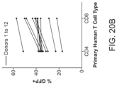
- FIGS. 20 A-B show reproducible non-viral gene targeting in a cohort of healthy donors.
- A A constant dsDNA RAB11A-GFP HDR template and RNP was electroporated using the optimized conditions developed for non-viral gene targeting in cells obtained from a cohort of twelve healthy donors. While there was significant variability in GFP insertion percentage among individual donors, all achieved robust integration of GFP (range 22% to 57% in CD8+ T cells). Some GFP expression was seen in cells electroporated with the dsDNA RAB11A-GFP HDR template with an off-target RNP targeting CXCR4 compared to no-electroporation controls.
- B Summary graph of GFP insertion percentages in (A). Across the 12 healthy donor cohort slightly higher rates of in GFP expression was seen in CD3+CD8+ T cells (mean 42.0%) compared to CD3+CD4+ T cells (mean 35.2%).
- FIG. 21 shows endogenous tagging of transcription factor BATF for analysis of chromatin occupancy.
- FIGS. 22 A-E show combinatorial non-viral gene targeting.
- A Simultaneous electroporation of HDR templates to create RAB11A-GFP and/or RAB11A-mCherry fusions in primary human T cells. A distinct population of dual GFP+mCherry+ cells was found when both templates are introduced concurrently, consistent with bi-allelic targeting.
- B The potential genotypes for individual cells in the quadrants are defined by expression of the two fluorophores. The observed level of bi-allelic integrations is higher in cells that acquire at least one integration than expected by chance ( FIG. 23 ).
- FIGS. 23 A-G show modeling and analysis of bi-allelic HDR integrations by insertion of multiple fluorescent proteins into the same locus.
- A The possible cellular phenotypes when two fluorescent proteins are inserted into the same locus.
- B The genotypes of two of these phenotypic populations are immediately known. Cells without any functional insertions (bottom left quadrant, genotype A), must have a NA/NA genotype (where NA indicates an allele without HDR, including WT alleles and NHEJ edited alleles).
- Dual fluorescent cells top right quadrant, genotype E
- must have acquired one copy of each template assuming an autosomal target locus and no off-target integrations, and would have a genotype of GFP/RFP.
- C The total percentage of cells with bi-allelic HDR integrations must be the sum of genotypes D, E, and F. While the proportion of cells with genotype E (dual fluor positives) is immediately apparent from the phenotypes, genotypes D and F are not.
- Application of a simple probability model allow for the de-convolution of the multiple genotypes in the single fluor positive phenotypes, and thus an estimation of the true percentage of cells homozygous for HDR.
- FIGS. 24 A-B show multiplexed integrations showing that acquisition of HDR integration at one locus increases likelihood of HDR at additional loci.
- A Two HDR template permutations from a set of six dsDNA HDR templates (targeting RAB11A, CD4, and CLTA; each site with GFP or RFP) were electroporated into CD3 + T cells isolated from healthy human donors. Four days after electroporation of the indicated two HDR templates along with their two respective on-target RNPs, the percentage of cells positive for each template was analyzed when gating on cells either positive or negative for the other template.
- FIGS. 25 A-F show D10A nickase and ssDNA HDR templates reduce off-target integrations.
- A Combinations of Cas9 RNPs and a RAB11A-GFP dsDNA HDR template were electroporated into primary human T cells. dsDNA template alone, or with an RNP containing a scrambled gRNA matching no sequence in the human genome yielded small but detectable amounts of GFP expression, which was noticeably increased when a dsDNA template is electroporated with a gRNA targeting a site different from the targeted RAB11A-GFP integration site (the “off-target RNP” targets CXCR4 Exon 1).
- Electroporation of a ssDNA HDR template reduced the off-target integrations to the limit of detection (comparable to levels with no template electroporated) both with no nuclease added and at induced off-target dsDNA breaks (Off-target gRNA+Cas9).
- E-F For integration of GFP fusion at the RAB11A site, use of a D10A nickase with a ssDNA HDR template reduced the on-target HDR (GFP integration with on-target gRNA) compared to Cas9 with a dsDNA template, but strongly reduced off-target integrations to undetectable levels. All fluorescent readouts were performed 4 days post-electroporation. Displayed data is representative of at least two different donors (A and E) or the averages of two different donors (C, D, and F) with standard deviation shown
- FIGS. 26 A-D show fluorescent estimation and quantification of off-target integration events across multiple HDR templates.
- A Diagram of HDR mediated insertions at the N-terminus of a target locus (not drawn to scale). The homology arms specify the exact sequence where the insert (a GFP tag in this case) will be inserted, allowing for scarless integration of exogenous sequences. As a GFP fusion protein is created, GFP fluorescence will be seen as a result of this on-target integration, dependent on an RNP cutting adjacent to the integration site.
- Double stranded DNA can be integrated via homology-independent repair mechanisms at off-target sites through either random integration at naturally occurring dsDNA breaks, or potentially at induced double stranded breaks, such as those at the off-target cut sites of the RNP.
- This effect can be harnessed to allow for targeted integration of a dsDNA sequence at a desired induced dsDNA break (HITI) in senescent cell types lacking the ability to do HDR, but crucially the entirety of the dsDNA template is integrated, including any potential homology arms.
- the homology arms contain a promoter sequence (such as for N-terminal fusion tags), these off target integrations can drive observable expression of the inserted sequence without the desired correct HDR insertion.
- FIGS. 27 A-B show GFP expression across a HDR template versus gRNA matrix.
- A GFP expression was analyzed in CD3+CD4+ primary human T cells from a healthy donor 7 days following electroporation of a matrix of dsDNA HDR templates and their corresponding gRNAs, along with a CXCR4 gRNA and a no RNP control. As expected with a dsDNA template, off-target integrations were seen across combinations, but for all gRNAs and HDR templates the highest GFP expression was seen in the on-target condition.
- B Heat map summary of flow cytometry data in (A). One HDR template, a C-terminal GFP fusion tag into the nuclear factor FBL, had consistently higher off-target expression across gRNAs.
- FIGS. 28 A-D show efficient HDR in primary human T cells using a Cas9 nickase.
- A Diagram of the genomic locus containing the first exon of RAB11A. Use of spCas9 with a single guide RNA (gRNA 1) along with a dsDNA HDR template integrating a GFP in frame with RAB11A directly after the start codon results in efficient GFP expression ( FIG. 11 F ). Use of a Cas9 nickase (D10A variant) with two gRNAs could reduce the chances of off-target cutting.
- B-C A series of single gRNAs as well as dual gRNA combinations were tested for GFP insertion efficiency at the RAB11A N-terminal locus.
- FIGS. 29 A-H show reduced Treg frequencies and defective Treg suppressive capacity in subjects with two loss of function IL2RA mutations.
- CD4+FoxP3 + T cells are predominantly CD25hiCD127lo.
- a CD127lo CD4+FoxP3+ population is present, but does not express IL2RA.
- C Clinical phenotyping performed at two separate sites confirms compound heterozygotes possess normal frequencies of CD127lo FoxP3+ cells.
- D Deficiency in IL2RA surface expression in compound heterozygote 3 led to aberrant downstream signalling as measured by pStat5 expression after stimulation with IL-2, but not IL-7 or IL-15.
- CD4+CD25hiCD127lo Tregs from the c.530 and c.800 single heterozygote family members were isolated and their suppressive ability was assayed as in (F).
- H Dot plot summaries of Treg suppressive ability in cells from healthy donor, CD25-deficient compound heterozygotes (F) and CD25+/ ⁇ c.530 or c.800 heterozygotes (G).
- CD3+CD4+CD127loCD45RO+TIGIT+“Tregs” from compound heterozygotes showed no suppressive ability
- conventional CD4+CD25hiCD127lo Tregs from the single heterozygote family members showed some suppressive capacity, consistent with their lack of pronounced clinical phenotype compared to the compound hets.
- FIGS. 30 A-E show monogenic autoimmune mutation corrected by non-viral gene targeting in primary human T cells.
- A Three siblings in a family carry two different IL2RA (encoding high-affinity IL-2 receptor, CD25) mutations (c.530A>G creating a stop codon in IL2RA exon 4; c.800delA, creating a frameshift mutation in IL2RA exon 8 which causes an almost 100 amino acid run-on).
- IL2RA encoding high-affinity IL-2 receptor, CD25
- mutations c.530A>G creating a stop codon in IL2RA exon 4; c.800delA, creating a frameshift mutation in IL2RA exon 8 which causes an almost 100 amino acid run-on.
- B These three compound heterozygote siblings show greatly reduced, but not completely absent, cell surface expression of IL2RA on their primary T cells.
- C 7 days after non-viral gene targeting, targeted T cells showed increased phosphorylation levels of Stat5 upon IL-2 stimulation compared to non-targeted controls.
- IL2RA+ T cells from the three compound heterozygote donors include an increased level of FoxP3+ cells compared to non-targeted cells or healthy donor cells.
- E Non-viral gene targeting and correction of the c.530 mutation is possible and efficient using an optimized therapeutic reagent set (D10A nickase along with ssDNA HDR template). T cells from one compound heterozygote donor were stained for IL2RA surface expression after 9 days of ex-vivo expansion following electroporation (2 days following re-stimulation).
- FIGS. 31 A-D show identification of compound heterozygous mutations in IL2RA and design of corrective CRISPR-Cas9 genome targeting reagents.
- A Initial genetic testing of the proband using an in-house targeted next-generation sequencing multi-gene panel of over 40 genes known to be involved in monogenic forms of diabetes was negative. Subsequent exome sequencing in the trio of proband and parents revealed two causative mutations in the IL2RA gene. The mother possessed a single heterozygous mutation (c.530G>A) in exon 4 of IL2RA (SEQ ID NO: 1) (AGACAAGGTRGACCCAGCC), resulting in a premature stop codon.
- the genomic sequences including the specified mutations (SEQ ID NO: 3)(CAAAATGACCCACGGGAAGACAAGGTAGACCC) for c.530G>A allele and SEQ ID NO: 4 (GACTTTGTTACACCACTACAGGAGGAGAGTA) for c.800delA Allele)) were used to design CRISPR-Cas9 genome targeting reagents to correct the two IL2RA mutations.
- a gRNA was designed to cut adjacent to the site of each mutation, 8 bps away for c.530 mutation, and 7 bps away for c.800.
- an HDR template ((SEQ ID NO: 5) (ACAAGATGGACCC) for c.530 mutation and (SEQ ID NO: 6)(AGGAGAAAGAGTA for c.800)) was designed including the corrected sequence as well as a silent mutation in a degenerate base to disrupt the PAM sequence (“NGG”) for each guide RNA.
- the corrected allele+silent PAM disruption sequence for c.530 (CAAAATGACCCACGGGAAGACAAGATGGACCC) (SEQ ID NO: 7) and c.800 (SEQ ID NO: 8) (GACTTTGTTACACCACTACAGGAGAAAGAGTA) are shown.
- FIGS. 32 A-C show HDR mediated correction of IL2RA c.530A>G loss of function mutation.
- A Unlike the gRNA targeting the c.800delA mutation at the C-terminus of IL2RA, the gRNA targeting the c.530A>G mutation (causing a stop codon in an interior exon) results in substantial ( ⁇ 90%) knockdown of IL2RA in a healthy donor and single heterozygotes (c.800 Het 2 and 3) 2 days following electroporation of the RNP alone (Blue) into CD3 + T cells.
- CD25 surface expression in T cells from the compound heterozygotes is only seen when an HDR template is included. In all three compound heterozygotes, the dsDNA HDR template yielded greater percentages of CD25+ cells.
- B Increased pStat5 signaling in response to IL-2 stimulation (200 U/mL) 7 days following electroporation in CD3+ T cells from compound heterozygote patients undergoing HDR-mediated mutation correction compared to no electroporation or RNP only controls.
- C Similarly, increased proportions of CD25+ FoxP3 + cells are seen 9 days following electroporation in the HDR correction conditions in compound heterozygote patients.
- Electroporations were performed according to optimized non-viral genome targeting protocol set forth in the examples. For ssODN electroporations, 100 pmols in 1 uL H 2 O were electroporated.
- FIGS. 33 A-C show non-HDR mediated correction of IL2RA c.800delA frameshift loss of function mutation.
- A Histograms of CD25 surface expression in CD3 + T cells in all children from a family carrying two loss-of-function IL2RA mutations, including three compound heterozygotes that express minimal amounts of IL2RA on their surface (No electroporation, Grey).
- FIGS. 34 A-B show diminished HDR potential and altered clinical phenotype in compound heterozygote IL2RA loss-of-function patient receiving immunosuppressants.
- A Flow cytometric analysis of GFP expression 6 days following electroporation of a positive HDR control RAB11A-GFP dsDNA HDR template into CD3 + T cells from the indicated patients revealed lower GFP expression in the three compound heterozygotes compared to their two c.800 heterozygote siblings.
- both c.800 heterozygotes as well as compound het 1 and 2 were within the general range observed across healthy donors, whereas compound het 3 had lower GFP expression than any healthy donor analyzed.
- FIGS. 35 A-D show multiple methods to produce long ssDNA HDR templates.
- A If a large enough amount of long single stranded DNA sequence could be produced for electroporation, off-target integrations could be reduced without overly compromising on-target efficiency.
- One method involves a two-step selective exonuclease digestion that specifically degrades one strand of a PCR product that has been labeled by 5′ phosphorylation, easily added onto a PCR primer prior to amplification.
- B A second ssDNA production method based on sequential in-vitro transcription (IVT) and reverse transcription (RT) reaction was also applied.
- IVTT in-vitro transcription
- RT reverse transcription
- RNA/DNA hybrid is formed which can be easily transformed into a long ssDNA template by incubation in sodium hydroxide (selectively degrades RNA strand).
- C At 2 days post-electroporation, viability in CD3 + T cells electroporated with only a ssDNA template was higher than those electroporated with only a dsDNA template ( FIG. 11 ).
- D A ssDNA RAB11A-GFP HDR template showed high efficiency GFP integration similar to dsDNA templates, and maintained high efficiency integrations at higher molar amounts of template, potentially due to increased viability (C) as well as less mass per mole of DNA template. Individual points represent at least two healthy donors (C, D).
- nucleic acid refers to deoxyribonucleic acids (DNA) or ribonucleic acids (RNA) and polymers thereof in either single- or double-stranded form. Unless specifically limited, the term encompasses nucleic acids containing known analogues of natural nucleotides that have similar binding properties as the reference nucleic acid and are metabolized in a manner similar to naturally occurring nucleotides. Unless otherwise indicated, a particular nucleic acid sequence also implicitly encompasses conservatively modified variants thereof (e.g., degenerate codon substitutions), alleles, orthologs, SNPs, and complementary sequences as well as the sequence explicitly indicated.
- DNA deoxyribonucleic acids
- RNA ribonucleic acids
- degenerate codon substitutions may be achieved by generating sequences in which the third position of one or more selected (or all) codons is substituted with mixed-base and/or deoxyinosine residues (Batzer et al., Nucleic Acid Res. 19:5081 (1991); Ohtsuka et al., J. Biol. Chem. 260:2605-2608 (1985); and Rossolini et al., Mol. Cell. Probes 8:91-98 (1994)).
- the term nucleic acid is used interchangeably with gene, cDNA, and mRNA encoded by a gene.
- gene can refer to the segment of DNA involved in producing or encoding a polypeptide chain. It may include regions preceding and following the coding region (leader and trailer) as well as intervening sequences (introns) between individual coding segments (exons). Alternatively, the term “gene” can refer to the segment of DNA involved in producing or encoding a non-translated RNA, such as an rRNA, tRNA, guide RNA (e.g., a small guide RNA), or micro RNA
- Treating refers to any indicia of success in the treatment or amelioration or prevention of the disease, condition, or disorder, including any objective or subjective parameter such as abatement; remission; diminishing of symptoms or making the disease condition more tolerable to the patient; slowing in the rate of degeneration or decline; or making the final point of degeneration less debilitating.
- the treatment or amelioration of symptoms can be based on objective or subjective parameters; including the results of an examination by a physician.
- the term “treating” includes the administration of the compounds or agents of the present invention to prevent or delay, to alleviate, or to arrest or inhibit development of the symptoms or conditions associated with a disease, condition or disorder as described herein.
- therapeutic effect refers to the reduction, elimination, or prevention of the disease, symptoms of the disease, or side effects of the disease in the subject.
- Treating” or “treatment” using the methods of the present invention includes preventing the onset of symptoms in a subject that can be at increased risk of a disease or disorder associated with a disease, condition or disorder as described herein, but does not yet experience or exhibit symptoms, inhibiting the symptoms of a disease or disorder (slowing or arresting its development), providing relief from the symptoms or side-effects of a disease (including palliative treatment), and relieving the symptoms of a disease (causing regression).
- Treatment can be prophylactic (to prevent or delay the onset of the disease, or to prevent the manifestation of clinical or subclinical symptoms thereof) or therapeutic suppression or alleviation of symptoms after the manifestation of the disease or condition.
- treatment includes preventative (e.g., prophylactic), curative or palliative treatment.
- a “promoter” is defined as one or more a nucleic acid control sequences that direct transcription of a nucleic acid.
- a promoter includes necessary nucleic acid sequences near the start site of transcription, such as, in the case of a polymerase II type promoter, a TATA element.
- a promoter also optionally includes distal enhancer or repressor elements, which can be located as much as several thousand base pairs from the start site of transcription.
- Polypeptide “peptide,” and “protein” are used interchangeably herein to refer to a polymer of amino acid residues. As used herein, the terms encompass amino acid chains of any length, including full-length proteins, wherein the amino acid residues are linked by covalent peptide bonds.
- complementary refers to specific base pairing between nucleotides or nucleic acids.
- Complementary nucleotides are, generally, A and T (or A and U), and G and C.
- subject an individual.
- the subject is a mammal, such as a primate, and, more specifically, a human.
- Non-human primates are subjects as well.
- subject includes domesticated animals, such as cats, dogs, etc., livestock (for example, cattle, horses, pigs, sheep, goats, etc.) and laboratory animals (for example, ferret, chinchilla, mouse, rabbit, rat, gerbil, guinea pig, etc.).
- livestock for example, cattle, horses, pigs, sheep, goats, etc.
- laboratory animals for example, ferret, chinchilla, mouse, rabbit, rat, gerbil, guinea pig, etc.
- veterinary uses and medical uses and formulations are contemplated herein.
- the term does not denote a particular age or sex. Thus, adult and newborn subjects, whether male or female, are intended to be covered.
- patient or subject may be used interchangeably and can refer to a subject afflicted with a
- CRISPR/Cas refers to a widespread class of bacterial systems for defense against foreign nucleic acid.
- CRISPR/Cas systems are found in a wide range of eubacterial and archaeal organisms.
- CRISPR/Cas systems include type I, II, and III sub-types. Wild-type type II CRISPR/Cas systems utilize an RNA-mediated nuclease, Cas9 in complex with guide and activating RNA to recognize and cleave foreign nucleic acid.
- Guide RNAs having the activity of both a guide RNA and an activating RNA are also known in the art. In some cases, such dual activity guide RNAs are referred to as a small guide RNA (sgRNA).
- sgRNA small guide RNA
- Cas9 homologs are found in a wide variety of eubacteria, including, but not limited to bacteria of the following taxonomic groups: Actinobacteria, Aquificae, Bacteroidetes-Chlorobi, Chlamydiae-Verrucomicrobia, Chlroflexi, Cyanobacteria, Firmicutes, Proteobacteria, Spirochaetes, and Thermotogae.
- An exemplary Cas9 protein is the Streptococcus pyogenes Cas9 protein. Additional Cas9 proteins and homologs thereof are described in, e.g., Chylinksi, et al., RNA Biol. 2013 May 1; 10(5): 726-737; Nat.
- RNA-mediated nuclease e.g., of bacterial or archeal orgin, or derived therefrom.
- RNA-mediated nuclases include the foregoing Cas9 proteins and homologs thereof, and include but are not limited to, CPF1 (See, e.g., Zetsche et al., Cell, Volume 163, Issue 3, p 759-771, 22 Oct. 2015).
- Cas9 ribonucleoprotein complex and the like refers to a complex between the Cas9 protein, and a crRNA (e.g., guide RNA or small guide RNA), the Cas9 protein and a trans-activating crRNA (tracrRNA), the Cas9 protein and a small guide RNA, or a combination thereof (e.g., a complex containing the Cas9 protein, a tracrRNA, and a crRNA guide RNA).
- a crRNA e.g., guide RNA or small guide RNA
- tracrRNA trans-activating crRNA
- Cas9 protein and a small guide RNA e.g., a complex containing the Cas9 protein, a tracrRNA, and a crRNA guide RNA
- the phrase “editing” in the context of editing of a genome of a cell refers to inducing a structural change in the sequence of the genome at a target genomic region.
- the editing can take the form of inserting a nucleotide sequence into the genome of the cell.
- the nucleotide sequence can encode a polypeptide or a fragment thereof.
- Such editing can be performed by inducing a double stranded break within a target genomic region, or a pair of single stranded nicks on opposite strands and flanking the target genomic region.
- Methods for inducing single or double stranded breaks at or within a target genomic region include the use of a Cas9 nuclease domain, or a derivative thereof, and a guide RNA, or pair of guide RNAs, directed to the target genomic region.
- introducing in the context of introducing a RNP-DNA template complex refers to the translocation of the RNP-DNA template complex from outside a cell to inside the cell. In some cases, introducing refers to translocation of the RNP-DNA template complex from outside the cell to inside the nucleus of the cell.
- Various methods of such translocation are contemplated, including but not limited to, electroporation, contact with nanowires or nanotubes, receptor mediated internalization, translocation via cell penetrating peptides, liposome mediated translocation, and the like.
- heterologous refers to what is not normally found in nature.
- heterologous sequence refers to a sequence not normally found in a given cell in nature.
- a heterologous nucleotide or protein sequence may be: (a) foreign to its host cell (i.e., is exogenous to the cell); (b) naturally found in the host cell (i.e., endogenous) but present at an unnatural quantity in the cell (i.e., greater or lesser quantity than naturally found in the host cell); or (c) be naturally found in the host cell but positioned outside of its natural locus.
- primary cell or primary stem cell refers to a cell that has not been transformed or immortalized.
- Such primary cells can be cultured, sub-cultured, or passaged a limited number of times (e.g., cultured 0, 1, 2, 3, 4, 5, 6, 7, 8, 9, 10, 11, 12, 13, 14, 15, 16, 17, 18, 19, or 20 times).
- the primary cells are adapted to in vitro culture conditions.
- the primary cells are isolated from an organism, system, organ, or tissue, optionally sorted, and utilized directly without culturing or sub-culturing.
- the primary cells are stimulated, activated, or differentiated.
- primary T cells can be activated by contact with (e.g., culturing in the presence of) CD3, CD28 agonists, IL-2, IFN- ⁇ , or a combination thereof.
- hematopoietic stem cell refers to a type of stem cell that can give rise to a blood cell. Hematopoietic stem cells can give rise to cells of the myeloid or lymphoid lineages, or a combination thereof. Hematopoietic stem cells are predominantly found in the bone marrow, although they can be isolated from peripheral blood, or a fraction thereof. Various cell surface markers can be used to identify, sort, or purify hematopoietic stem cells. In some cases, hematopoietic stem cells are identified as c-kit + and lin ⁇ .
- human hematopoietic stem cells are identified as CD34 + , CD59 + , Thy1/CD90 + , CD38 lo/ ⁇ , C-kit/CD117 + , lin ⁇ .
- human hematopoietic stem cells are identified as CD34 ⁇ , CD59 + , Thy1/CD90 + , CD38 lo/ ⁇ , C-kit/CD117 + , lin ⁇ .
- human hematopoietic stem cells are identified as CD133 + , CD59 + , Thy1/CD90 + , CD38 lo/ ⁇ , C-kit/CD117 + , lin ⁇ .
- mouse hematopoietic stem cells are identified as CD34 lo/ ⁇ , SCA-1 + , Thy1 +/lo , CD38 + , C-kit + , lin ⁇ .
- the hematopoietic stem cells are CD150 + CD48 ⁇ CD244 ⁇ .
- hematopoietic cell refers to a cell derived from a hematopoietic stem cell.
- the hematopoietic cell may be obtained or provided by isolation from an organism, system, organ, or tissue (e.g., blood, or a fraction thereof).
- an hematopoietic stem cell can be isolated and the hematopoietic cell obtained or provided by differentiating the stem cell.
- Hematopoietic cells include cells with limited potential to differentiate into further cell types.
- hematopoietic cells include, but are not limited to, multipotent progenitor cells, lineage-restricted progenitor cells, common myeloid progenitor cells, granulocyte-macrophage progenitor cells, or megakaryocyte-erythroid progenitor cells.
- Hematopoietic cells include cells of the lymphoid and myeloid lineages, such as lymphocytes, erythrocytes, granulocytes, monocytes, and thrombocytes.
- the hematopoietic cell is an immune cell, such as a T cell, B cell, macrophage, a natural killer (NK) cell or dendritic cell.
- the cell is an innate immune cell.
- T cell refers to a lymphoid cell that expresses a T cell receptor molecule.
- T cells include, but are not limited to, na ⁇ ve T cells, stimulated T cells, primary T cells (e.g., uncultured), cultured T cells, immortalized T cells, helper T cells, cytotoxic T cells, memory T cells, regulatory T cells, natural killer T cells, combinations thereof, or sub-populations thereof.
- T cells can be CD4 + , CD8 + , or CD4 + and CD8 + .
- T cells can be helper cells, for example helper cells of type T h 1, T h 2, T h 3, T h 9, T h 17, or T FH .
- T cells can be cytotoxic T cells.
- T cells can be alpha/Beta T cells or gamma/delta T cells.
- the T cell is a CD4 + CD25 hi CD127 lo regulatory T cell.
- the T cell is a regulatory T cell selected from the group consisting of Tr1, Th3, CD8+CD28 ⁇ , Treg17, and Qa-1 restricted T cells, or a combination or sub-population thereof.
- the T cell is a FOXP3 + T cell.
- the T cell is a CD4 + CD25 lo CD127 hi effector T cell.
- the T cell is a CD4 + CD25 lo CD127 hi CD45RA hi CD45RO ⁇ na ⁇ ve T cell.
- a T cell can be a recombinant T cell that has been genetically manipulated.
- the recombinant T cell has a recombinant (e.g., mutated or heterologous) T cell receptor or a chimeric antigen receptor (CAR).
- the T cell receptor can have one or more mutations in a complementarity determining region of a T cell receptor to alter antigen specificity.
- the T cell receptor can be mutated (e.g., in the endodomain) to increase or decrease signaling.
- the T cell receptor can be replaced with a heterologous T cell receptor.
- the T cell receptor can be replaced with a polypeptide having a different receptor domain, such as an antibody or antibody fragment.
- the T cell receptor is a chimeric receptor containing a targeting domain (e.g., an antibody fragment), a transmembrane domain, and an intracellular or endodomain domain.
- the endodomain can contain one or more signaling domains and/or adaptor domains to provide robust T cell activation and anti-antigen activity.
- non-homologous end joining refers to a cellular process in which cut or nicked ends of a DNA strand are directly ligated without the need for a homologous template nucleic acid. NHEJ can lead to the addition, the deletion, substitution, or a combination thereof, of one or more nucleotides at the repair site.
- homology directed repair refers to a cellular process in which cut or nicked ends of a DNA strand are repaired by polymerization from a homologous template nucleic acid.
- the homologous template nucleic acid can be provided by homologous sequences elsewhere in the genome (sister chromatids, homologous chromosomes, or repeated regions on the same or different chromosomes).
- an exogenous template nucleic acid can be introduced to obtain a specific HDR-induced change of the sequence at the target site. In this way, specific mutations can be introduced at the cut site.
- a single-stranded DNA template or a double-stranded DNA template refers to a DNA oligonucleotide that can be used by a cell as a template for HDR.
- the single-stranded DNA template or a double-stranded DNA template has at least one region of homology to a target site.
- the single-stranded DNA template or double-stranded DNA template has two homologous regions flanking a region that contains a heterologous sequence to be inserted at a target cut site.
- compositions and methods recites various aspects and embodiments of the present compositions and methods. No particular embodiment is intended to define the scope of the compositions and methods. Rather, the embodiments merely provide non-limiting examples of various compositions and methods that are at least included within the scope of the disclosed compositions and methods. The description is to be read from the perspective of one of ordinary skill in the art; therefore, information well known to the skilled artisan is not necessarily included.
- compositions and methods for editing the genome of a cell are provided herein.
- the inventors have surprisingly discovered that large nucleotide sequences, for example, nucleotide sequences greater than about 200 nucleotides or base pairs in length, can be inserted into the genome of a cell, in the absence of a viral vector.
- nucleotide sequence greater than about 200 nucleotides or base pairs in length can be inserted into the genome of a primary immune cell, in the absence of a viral vector
- Integration of large nucleic acids for example nucleic acids greater than 200 nucleotides in size, into cells, can be limited by low efficiency of integration, off-target effects and/or loss of cell viability.
- Described herein are methods and compositions for achieving integration of a nucleotide sequence, for example, a nucleotide sequence greater than about 200 nucleotides in size, into the genome of a cell. In some methods the efficiency of integration is increased, off-target effects are reduced and/or loss of cell viability is reduced.
- Methods for editing the genome of a cell can include a) providing a Cas9 ribonucleoprotein complex (RNP)-DNA template complex comprising: (i) the RNP, wherein the RNP comprises a Cas9 nuclease domain and a guide RNA, wherein the guide RNA specifically hybridizes to a target region of the genome of the cell, and wherein the Cas9 nuclease domain cleaves the target region to create an insertion site in the genome of the cell; and (ii) a double-stranded or single-stranded DNA template, wherein the size of the DNA template is greater than about 200 nucleotides, wherein the 5′ and 3′ ends of the DNA template comprise nucleotide sequences that are homologous to genomic sequences flanking the insertion site, and wherein the molar ratio of RNP to DNA template in the complex is from about 3:1 to about 100:1; and b) introducing the RNP-DNA template complex into the cell.
- RNP Cas9
- the methods described herein provide an efficiency of delivery of the RNP-DNA template complex of at least about 20%, 25%, 30%, 35%, 40%, 45%, 50%, 55%, 60%, 65%, 70%, 75%, 80%, 85%, 90%, 95%, 97.5%, 99%, 99.5%, 99%, or higher.
- the efficiency is determined with respect to cells that are viable after introducing the RNP-DNA template into the cell.
- the efficiency is determined with respect to the total number of cells (viable or non-viable) in which the RNP-DNA template is introduced into the cell.
- the efficiency of delivery can be determined by quantifying the number of genome edited cells in a population of cells (as compared to total cells or total viable cells obtained after the introducing step).
- Various methods for quantifying genome editing can be utilized. These methods include, but are not limited to, the use of a mismatch-specific nuclease, such as T7 endonuclease I; sequencing of one or more target loci (e.g., by sanger sequencing of cloned target locus amplification fragments); and high-throughput deep sequencing.
- loss of cell viability is reduced as compared to loss of cell viability after introduction of naked DNA into a cell or introduction of DNA into a cell using a viral vector.
- the reduction can be a reduction of at least 10%, 20%, 30%, 40%, 50%, 60%, 70%, 80%, 90%, 100% or any percentage in between these percentages.
- off-target effects of integration are reduced as compared to off-target integration after introduction of naked DNA into a cell or introduction of DNA into a cell using a viral vector.
- the reduction can be a reduction of at least 10%, 20%, 30%, 40%, 50%, 60%, 70%, 80%, 90%, 100% or any percentage in between these percentages.
- the methods described herein provide for high cell viability of cells to which the RNP-DNA template has been introduced.
- the viability of the cells to which the RNP-DNA template has been introduced is at least about 20%, 25%, 30%, 35%, 40%, 45%, 50%, 55%, 60%, 65%, 70%, 75%, 80%, 85%, 90%, 95%, 97.5%, 99%, 99.5%, 99%, or higher.
- the molar ratio of RNP to DNA template can be from about 3:1 to about 100:1.
- the molar ratio can be from about 5:1 to 10:1, from about 5:1 to about 15:1, 5:1 to about 20:1; 5:1 to about 25:1; from about 8:1 to about 12:1; from about 8:1 to about 15:1, from about 8:1 to about 20:1, or from about 8:1 to about 25:1.
- the DNA template is at a concentration of about 2.5 pM to about 25 pM.
- the concentration of DNA template can be about 2.5, 3, 3.5, 4, 4.5, 5, 5.5, 6, 6.5, 7, 7.5, 8, 8.5, 9, 9.5, 10, 10.5, 11, 11.5, 12, 12.5, 13, 13.5, 14, 14.5, 15, 15.5, 16, 16.5, 17, 17.5, 18, 18.5, 19, 19.5, 20, 20.5, 21, 21.5, 22, 22.5, 23, 23.5, 24, 24.5, 25 pM or any concentration in between these concentrations.
- the size or length of the DNA template is greater than about 200 bp, 250 bp, 300 bp, 350 bp, 400 bp, 450 bp, 500 bp, 550 bp, 600 bp, 650 bp, 700 bp, 750 bp, 800 bp, 850 bp, 900 bp, 1 kb, 1.1 kb, 1.2 kb, 1.3 kb, 1.4 kb, 1.5 kb, 1.6 kb, 1.7 kb, 1.8 kb, 1.9 kb, 2.0 kb, 2.1 kb, 2.2 kb, 2.3 kb, 2.4 kb, 2.5 kb, 2.6 kb, 2.7 kb, 2.8 kb, 2.9 kb, 3 kb, 3.1 kb, 3.2 kb, 3.3 kb, 3.4 kb, 3.5 kb, 3.6 kb,
- the size of the DNA template can be about 200 bp to about 500 bp, about 200 bp to about 750 bp, about 200 bp to about 1 kb, about 200 bp to about 1.5 kb, about 200 bp to about 2.0 kb, about 200 bp to about 2.5 kb, about 200 bp to about 3.0 kb, about 200 bp to about 3.5 kb, about 200 bp to about 4.0 kb, about 200 bp to about 4.5 kb, about 200 bp to about 5.0 kb.
- the amount of DNA template is about 1 ⁇ g to about 10 ⁇ g.
- the amount of DNA template can be about 1 ⁇ g to about 2 ⁇ g, about 1 ⁇ g to about 3 ⁇ g, about 1 ⁇ g to about 4 ⁇ g, about 1 ⁇ g to about 5 ⁇ g, about 1 ⁇ g to about 6 ⁇ g, about 1 ⁇ g to about 7 ⁇ g, about 1 ⁇ g to about 8 ⁇ g, about 1 ⁇ g to about 9 ⁇ g, about 1 ⁇ g to about 10 ⁇ g.
- the amount of DNA template is about 2 ⁇ g to about 3 ⁇ g, about 2 ⁇ g to about 4 ⁇ g, about 2 ⁇ g to about 5 ⁇ g, about 2 ⁇ g to about 6 ⁇ g, about 2 ⁇ g to about 7 ⁇ g, about 2 ⁇ g to about 8 ⁇ g, about 2 ⁇ g to about 9 ⁇ g, or 2 ⁇ g to about 10 ⁇ g.
- the amount of DNA template is about 3 ⁇ g to about 4 ⁇ g, about 3 ⁇ g to about 5 ⁇ g, about 3 ⁇ g to about 6 ⁇ g, about 3 ⁇ g to about 7 ⁇ g, about 3 ⁇ g to about 8 ⁇ g, about 3 ⁇ g to about 9 ⁇ g, or about 3 ⁇ g to about 10 ⁇ g.
- the amount of DNA template is about 4 ⁇ g to about 5 ⁇ g, about 4 ⁇ g to about 6 ⁇ g, about 4 ⁇ g to about 7 ⁇ g, about 4 ⁇ g to about 8 ⁇ g, about 4 ⁇ g to about 9 ⁇ g, or about 4 ⁇ g to about 10 ⁇ g.
- the amount of DNA template is about 5 ⁇ g to about 6 ⁇ g, about 5 ⁇ g to about 7 ⁇ g, about 5 ⁇ g to about 8 ⁇ g, about 5 ⁇ g to about 9 ⁇ g, or about 5 ⁇ g to about 10 ⁇ g. In some embodiments, the amount of DNA template is about 6 ⁇ g to about 7 ⁇ g, about 6 ⁇ g to about 8 ⁇ g, about 6 ⁇ g to about 9 ⁇ g, or about 6 ⁇ g to about 10 ⁇ g. In some embodiments, the amount of DNA template is about 7 ⁇ g to about 8 ⁇ g, about 7 ⁇ g to about 9 ⁇ g, or about 7 ⁇ g to about 10 ⁇ g.
- the amount of DNA template is about 8 ⁇ g to about 9 ⁇ g, or about 8 ⁇ g to about 10 ⁇ g. In some embodiments, the amount of DNA template is about 9 ⁇ g to about 10 ⁇ g. In some cases, the size of the DNA template is large enough and in sufficient quantity to be lethal as naked DNA.
- the DNA template encodes a heterologous protein or a fragment thereof.
- the DNA template includes regulatory sequences, for example, a promoter sequence and/or an enhancer sequence to regulate expression of the heterologous protein or fragment thereof after insertion into the genome of a cell.
- the DNA template is a linear DNA template. In some cases, the DNA template is a single-stranded DNA template. In some cases, the single-stranded DNA template is a pure single-stranded DNA template. As used herein, by “pure single-stranded DNA” is meant single-stranded DNA that substantially lacks the other or opposite strand of DNA. By “substantially lacks” is meant that the pure single-stranded DNA lacks at least 100-fold more of one strand than another strand of DNA.
- the RNP-DNA template complex is formed by incubating the RNP with the DNA template for less than about one minute to about thirty minutes, at a temperature of about 20° C. to about 25° C.
- the RNP can be incubated with the DNA template for about 5 seconds, 10 seconds, 15 seconds, 20 seconds, 25 seconds, 30 seconds, 35 seconds, 40 seconds, 45 seconds, 50 seconds, 55 seconds, 1 minute, 2 minutes, 3 minutes, 4 minutes, 5 minutes, 6 minutes, 7 minutes, 8 minutes, 9 minutes, 10 minutes, 11 minutes, 12 minutes, 13 minutes, 14 minutes, 15 minutes, 16 minutes, 17 minutes, 18 minutes, 19 minutes, 20 minutes, 21 minutes, 22 minutes, 23 minutes, 24 minutes, 25 minutes, 26 minutes, 27 minutes, 28 minutes, 29 minutes or 30 minutes or any amount of time in between these times, at a temperature of about 20° C., 21° C., 22° C., 23° C., 24° C., or 25° C.
- the RNP can be incubated with the DNA template for less than about one minute to about one minute, for less than about one minute to about 5 minutes, for less than about 1 minute to about 10 minutes, for about 5 minutes to 10 minutes, for about 5 minutes to 15 minutes, for about 10 to about 15 minutes, for about 10 minutes to about 20 minutes, or for about 10 minutes to about 30 minutes, at a temperature of about 20° C. to about 25° C.
- the RNP-DNA template complex and the cell are mixed prior to introducing the RNP-DNA template complex into the cell.
- introducing the RNP-DNA template complex comprises electroporation.
- Methods, compositions, and devices for electroporating cells to introduce a RNP-DNA template complex can include those described in the examples herein. Additional or alternative methods, compositions, and devices for electroporating cells to introduce a RNP-DNA template complex can include those described in WO/2006/001614 or Kim, J. A. et al. Biosens. Bioelectron. 23, 1353-1360 (2008). Additional or alternative methods, compositions, and devices for electroporating cells to introduce a RNP-DNA template complex can include those described in U.S. Patent Appl. Pub. Nos. 2006/0094095; 2005/0064596; or 2006/0087522.
- Additional or alternative methods, compositions, and devices for electroporating cells to introduce a RNP-DNA template complex can include those described in Li, L. H. et al. Cancer Res. Treat. 1, 341-350 (2002); U.S. Pat. Nos. 6,773,669; 7,186,559; 7,771,984; 7,991,559; 6,485,961; 7,029,916; and U.S. Patent Appl. Pub. Nos: 2014/0017213; and 2012/0088842.
- Additional or alternative methods, compositions, and devices for electroporating cells to introduce a RNP-DNA template complex can include those described in Geng, T. et al. J. Control Release 144, 91-100 (2010); and Wang, J., et al. Lab. Chip 10, 2057-2061 (2010).
- the Cas9 protein can be in an active endonuclease form, such that when bound to target nucleic acid as part of a complex with a guide RNA or part of a complex with a DNA template, a double strand break is introduced into the target nucleic acid.
- the double strand break can be repaired by NHEJ to introduce random mutations, or HDR to introduce specific mutations.
- Various Cas9 nucleases can be utilized in the methods described herein. For example, a Cas9 nuclease that requires an NGG protospacer adjacent motif (PAM) immediately 3′ of the region targeted by the guide RNA can be utilized.
- PAM NGG protospacer adjacent motif
- Such Cas9 nucleases can be targeted to any region of a genome that contains an NGG sequence.
- Cas9 proteins with orthogonal PAM motif requirements can be utilized to target sequences that do not have an adjacent NGG PAM sequence.
- Exemplary Cas9 proteins with orthogonal PAM sequence specificities include, but are not limited to, CFP
- the Cas9 protein is a nickase, such that when bound to target nucleic acid as part of a complex with a guide RNA, a single strand break or nick is introduced into the target nucleic acid.
- a pair of Cas9 nickases, each bound to a structurally different guide RNA, can be targeted to two proximal sites of a target genomic region and thus introduce a pair of proximal single stranded breaks into the target genomic region.
- nickases can provide enhanced specificity because off-target effects are likely to result in single nicks, which are generally repaired without lesion by base-excision repair mechanisms.
- Exemplary Cas9 nickases include Cas9 nucleases having a D10A or H840A mutation.
- the RNP comprises a Cas9 nuclease. In some embodiments, the RNP comprises a Cas9 nickase. In some embodiments, the RNP-DNA template complex comprises at least two structurally different RNP complexes. In some embodiments, the at least two structurally different RNP complexes contain structurally different Cas9 nuclease domains In some embodiments, the at least two structurally different RNP complexes contain structurally different guide RNAs.
- each of the structurally different RNP complexes comprises a Cas9 nickase, and the structurally different guide RNAs hybridize to opposite strands of the target region.
- a plurality of RNP-DNA templates comprising structurally different ribonucleoprotein complexes is introduced into the cell.
- a Cas9 protein can be complexed with a plurality (e.g., 2, 3, 4, 5, or more, e.g., 2-10, 5-100, 20-100) of structurally different guide RNAs to target insertion of a DNA template at a plurality of structurally different target genomic regions.
- cells include, but are not limited to, eukaryotic cells, prokaryotic cells, animal cells, plant cells, fungal cells and the like.
- the cell is a mammalian cell, for example, a human cell.
- the cell can be in vitro, ex vivo or in vivo.
- the cell can also be a primary cell, a germ cell, a stem cell or a precursor cell.
- the precursor cell can be, for example, a pluripotent stem cell, or a hematopoietic stem cell.
- the cell is a primary hematopoietic cell or a primary hematopoietic stem cell.
- the primary hematopoietic cell is an immune cell.
- the immune cell is a T cell.
- the T cell is a regulatory T cell, an effector T cell, or a na ⁇ ve T cell.
- the T cell is a CD4 + T cell.
- the T cell is a CD8 + T cell.
- the T cell is a CD4 + CD8 + T cell.
- the T cell is a CD4 ⁇ CD8 ⁇ T cell.
- the cells are removed from a subject, modified using any of the methods described herein and administered to the patient.
- any of the constructs described herein is delivered to the patient in vivo. See, for example, U.S. Pat. No. 9,737,604 and Zhang et al. “Lipid nanoparticle-mediated efficient delivery of CRISPR/Cas9 for tumor therapy,” NPG Asia Materials Volume 9, page e441 (2017).
- the RNP ⁇ DNA template complex is introduced into about 1 ⁇ 10 5 to about 2 ⁇ 10 6 cells.
- the RNP ⁇ DNA template complex can be introduced into about 1 ⁇ 10 5 to about 5 ⁇ 10 5 cells, about 1 ⁇ 10 5 to about 1 ⁇ 10 6 , 1 ⁇ 10 5 to about 1.5 ⁇ 10 6 , 1 ⁇ 10 5 to about 2 ⁇ 10 6 , about 1 ⁇ 10 6 to about 1.5 ⁇ 10 6 cells or about 1 ⁇ 10 6 to about 2 ⁇ 10 6 .
- the methods and compositions described herein can be used for generation, modification, use, or control of recombinant T cells, such as chimeric antigen receptor T cells (CAR T cells).
- CAR T cells can be used to treat or prevent cancer, an infectious disease, or autoimmune disease in a subject.
- one or more gene products are inserted or knocked-in to a T cell to express a heterologous protein (e.g., a chimeric antigen receptor (CAR)).
- a heterologous protein e.g., a chimeric antigen receptor (CAR)
- the plurality of cells comprises primary hematopoietic cells or primary hematopoietic stem cells.
- the primary hematopoietic cells are immune cells.
- the immune cells are T cells.
- the T cells are regulatory T cells, effector T cells, or na ⁇ ve T cells.
- the T cells are CD8 + T cells.
- the T cells are CD4 + CD8 + T cells.
- any subset or combination of these is also specifically contemplated and disclosed. This concept applies to all aspects of this disclosure including, but not limited to, steps in methods using the disclosed compositions. Thus, if there are a variety of additional steps that can be performed, it is understood that each of these additional steps can be performed with any specific method steps or combination of method steps of the disclosed methods, and that each such combination or subset of combinations is specifically contemplated and should be considered disclosed.
- Example I The data provided in Example I were generated as outlined in the protocol below.
- a molar ratio of about 10:1 RNP to DNA template maintained both efficiency of integration as well as viability, post electroporation ( FIG. 4 ). However, ratios ranging from 3:1 to about 100:1 also worked. Using a ratio of about 10:1 RNP to DNA template balanced the effects of viability loss and efficiency, and achieved the maximal number of integration positive cells ( FIG. 5 ). This ratio also allowed for high efficiency insertion of large templates (>750 bps) ( FIG. 6 ).
- dsDNA Templates have Some Off-Target Integrations which is Reduced Using ssDNA Templates
- Insertion of long DNA templates can result in a small amount of off-target integration ( FIG. 7 ), which is similar to off-target integration seen when using an AAV as the donor template.
- some of the methods provided herein use a long ssDNA template as the donor, which results in reduced off-target integrations ( FIG. 8 ).
- off-target dsDNA breaks (which can be repaired via NHEJ as mutations) introduced by Cas9.
- the high efficiency non-viral integrations disclosed herein can be inserted using two gRNAs and a Cas9 nickase (D10A) ( FIG. 9 ), which prevents off target dsDNA breaks.
- PBMCs Peripheral blood mononuclear cells
- STMate tubes SepMate tubes
- T cells were isolated from PBMCs from all cell sources by magnetic negative selection using an EasySep Human T Cell Isolation Kit (STEMCELL, per manufacturer's instructions). Unless otherwise noted, isolated T cells were stimulated and used directly (fresh).
- T cells were stimulated for 2 days with anti-human CD3/CD28 magnetic dynabeads (ThermoFisher) at a beads to cells concentration of 1:1, along with a cytokine cocktail of IL-2 at 200 U/mL (UCSF Pharmacy), IL-7 at 5 ng/mL (ThermoFisher), and IL-15 at 5 ng/mL (Life Tech).
- IL-2 a cytokine cocktail of IL-2 at 200 U/mL (UCSF Pharmacy)
- IL-7 at 5 ng/mL
- IL-15 5 ng/mL
- T cells were cultured in media with IL-2 at 500 U/mL. Throughout culture T cells were maintained at an approximate density of 1 million cells per mL of media. Every 2-3 days post-electroporation additional media was added, along with additional fresh IL-2 to bring the final concentration to 500 U/mL, and cells were transferred to larger culture vessels as necessary to maintain a density of 1 million cells/mL.
- RNPs were produced by annealing of a two-component gRNA to Cas9, as previously described (7, 16). Briefly, crRNAs and tracrRNAs were chemically synthesized (Dharmacon, IDT), and recombinant Cas9-NLS, D10A-NLS, or dCas9-NLS were recombinantly produced and purified (QB3 Macrolab). Lyophilized RNA was resuspended in Tris-HCL (7.4 pH) with 150 mM KCl at a concentration of 160 uM, and stored in aliquots at ⁇ 80° C.
- crRNA and tracrRNA aliquots were thawed, mixed 1:1 by volume, and incubated at 37° C. for 30 min to form an 80 uM gRNA solution.
- Recombinant Cas9 and variants stored at 40 uM in 20 mM HEPES-KOH pH 7.5, 150 mM KCl, 10% glycerol, 1 mM DTT, were then mixed 1:1 by volume with the 80 uM gRNA (2:1 gRNA to Cas9 molar ratio) at 37° C. for 15 min to form an RNP at 20 uM.
- RNPs were generally electroporated immediately after complexing.
- Double stranded DNA HDRT sequences were generated from PCR products. Novel HDR sequences were constructed using Gibson Assemblies to place the HDR template sequence, consisting of the homology arms (commonly synthesized as gBlocks from IDT) and the desired insert (such as GFP) into a cloning vector for sequence confirmation and future propagation. These plasmids were used as templates for high-output PCR amplification (Kapa Hotstart polymerase). PCR amplicons (the dsDNA HDRT) were SPRI purified (1.0 ⁇ ) and eluted into a final volume of 3 uL H2O per 100 uL of PCR reaction input. Concentrations of HDRTs were analyzed by nanodrop with a 1:20 dilution. The size of the amplified HDRT was confirmed by gel electrophoresis in a 1.0% agarose gel.
- the DNA of interest was amplified via PCR using one regular, non-modified PCR primer and a second phosphorylated PCR primer.
- the DNA strand that will be amplified using the phosphorylated primer will be the strand that will be degraded using this method. This allows to either prepare a single stranded sense or single stranded antisense DNA using the respective phosphorylated PCR primer.
- the phosphorylated strand of the PCR product was degraded via subsequent treatment with two enzymes, Strandase Mix A and Strandase Mix B, for 5 minutes (per 1 kb) at 37° C., respectively.
- Enzymes were deactivated by a 5 minute incubation at 80° C. Resulting ssDNA HDR templates were SPRI purified (1.0 ⁇ ) and eluted in H2O.
- a more detailed protocol for the Guide-itTM Long ssDNA Production System (Takara Bio USA, Inc. #632644) can be found at the manufacturer's website.
- ssDNA donors were synthesized by reverse transcription of an RNA intermediate followed by hydrolysis of the RNA strand in the resulting RNA:DNA hybrid product, as described in (28). Briefly, the desired HDR donor was first cloned downstream of a T7 promoter and the T7-HDR donor sequence amplified by PCR. RNA was synthesized by in vitro transcription using HiScribe T7 RNA polymerase (New England Biolabs) and reverse-transcribed using TGIRT-III (InGex). Following reverse transcription, NaOH and EDTA were added to 0.2 M and 0.1 M respectively and RNA hydrolysis carried out at 95° C. for 10 min.
- reaction was quenched with HCl, the final ssDNA product purified using Ampure XP magnetic beads (Beckman Coulter) and eluted in sterile RNAse-free H2O. ssDNA quality was analyzed by capillary electrophoresis (Bioanalyzer, Agilent).
- RNPs and HDR templates were electroporated 2 days following initial T cell stimulation.
- T cells were harvested from their culture vessels and magnetic CD3/CD28 dynabeads were removed by placing cells on a magnet for 2 minutes.
- de-beaded cells were centrifuged for 10 minutes at 90 g, aspirated, and resuspended in the Lonza electroporation buffer P3 at 20 uL buffer per one million cells.
- Lonza electroporation buffer P3 at 20 uL buffer per one million cells.
- one million T cells were electroporated per well using a Lonza 4D 96-well electroporation system with pulse code EH115. Alternate cell concentrations from 200,000 up to 2 million cells per well showed lower efficiencies.
- Alternate electroporation buffers were used as indicated, but had different optimal pulse settings (EO155 for OMEM buffer). Unless otherwise indicated, 2.5 uLs of RNPs (50 pmols total) were electroporated, along with 2 uLs of HDR Template at 2 ugs/uL (4 ugs HDR Template total).
- HDRTs were first aliquoted into wells of a 96-well polypropylene V-bottom plate. RNPs were then added to the HDRTs and allowed to incubate together at RT for at least 30 seconds. Finally, cells resuspended in electroporation buffer were added, briefly mixed by pipetting with the HDRT and RNP, and 24 uLs of total volume (cells+RNP+HDRT) was transferred into a 96 well electroporation cuvette plate.
- Flow cytometric analysis was performed on an Attune NxT Accustic Focusing Cytometer (ThermoFisher). Surface staining for CD3-APC-eFluor 780 (SK7, eBiosciences), CD4-PerCP (SK3, Tonbo), CD8-PE-Cy7 (SK1, BD), IL2RA/CD25-APC (BC96, Tonbo). Intracellular phosphorylation staining was performed using pStat5(Y694)-PacBlue (clone 47, BD). Intracellular cytokine staining for FoxP3 was performed using FoxP3-AF488 (206D, Biolegend).
- Samples were prepared by drop casting 10 ⁇ l of suspended live T cells solution onto a 3 ⁇ 1′′ microscope slide onto which a 25 mm2 coverslip was placed. Imaging was performed on an upright configuration Nikon A1r laser scanning confocal microscope. Excitation was achieved through a 488 nm OBIS laser (Coherent). A long working distance (LWD) 60 ⁇ Plan Apo 1.20 NA water immersion objective was used with additional digital zoom achieved through the NIS-Elements software. Images were acquired under “Galvano” mirror settings with 2 ⁇ line averaging enabled and exported as TIFF to be analyzed in FIJI (ImageJ, NIH).
- FIJI ImageJ, NIH
- CUT&RUN was performed on epitope-tagged primary human T cells 11 days after electroporation and 4 days after re-stimulation with anti-CD3/anti-CD28 beads (untagged cells were not electroporated). Approximately 20% and 10% of electroporated cells showed GFP-BATF expression as determined by flow cytometry in donor 1 and donor 2 samples, respectively.
- CUT&RUN was performed as described in (18) using anti-GFP (ab290), anti-BATF (sc-100974), and rabbit anti-mouse (ab46540) antibodies. Briefly, 6 million cells (30 million cells for anti-GFP CUT&RUN in GFP-BATF-containing cells) were collected and washed.
- Nuclei were isolated and incubated rotating with primary antibody (GFP or BATF) for 2 hours at 4° C.
- BATF CUT&RUN samples were incubated an additional hour with rabbit anti-mouse antibody.
- nuclei were incubated with proteinA-micrococcal nuclease (kindly provided by the Henikoff lab) for one hour at 4° C.
- Nuclei were equilibrated to OC and and MNase digestion was allowed to proceed for 30 minutes. Solubilized chromatin CUT&RUN fragments were isolated and purified.
- Paired-end sequencing libraries were prepared and run on Illumina Nextseq machines and sequencing data was processed as described in Skene and Henikoff, “An efficient targeted nuclease strategy for high resolution mapping of DNA binding sites,” Elife 6 (2017) doi: 10.7554/eLife.21856. For peak calling and heatmap generation, reads mapping to centromeres were filtered out.
- TLA sequencing was performed by Cergentis as previously described 16 .
- data analysis of integration sites and transgene fusions was performed by Cergentis as previously described 16.
- TLA sequencing was performed in two healthy donors, each edited at the RABI IA locus with either a dsDNA or ssDNA HDR template to integrate a GFP fusion Sequencing reads showing evidence of primer dimers or primer bias (i.e. greater than 99% of observed reads came from single primer set) were removed.
- CD4+ T cells were enriched using the EasySep Human CD4+ T cell enrichment kit (STEMCELL Technologies).
- CD3+CD4+CD127loCD45RO+ TIGIT+ Treg-enriched cells from IL2RA-deficient subjects and HD as well as CD3+CD4+CD25hiCD127lo Tregs from CD25+/ ⁇ individuals were sorted by flow cytometry.
- CD3+CD4+CD25-CD127+ responder T cells were labeled with CellTrace CF SE (Invitrogen) at 5 Tregs and HD Tresps were co-cultured at a 1:1 ratio in the presence of beads loaded with anti-CD2, anti-CD3 and anti-CD28 (Treg Suppression Inspector; Miltenyi Biotec) at a 1 bead: 1 cell ratio.
- co-cultures were analyzed by FACS for CFSE dilution.
- % inhibition is calculated using the following formula: 1 ⁇ (% proliferation with Tregs/% proliferation of stimulated Tresps without Tregs).
- Ex-vivo expanded Tregs and T effector cells from a healthy control and a patient with IL2RA compound heterozygous mutations were thawed and stained. Live cells were sorted based on expression of CD25 and CD62L markers directly into ZymoResearch M-digestion Buffer (2 ⁇ ) (cat #D5021-9) supplemented with proteinase K. The lysate was incubated at 65° C. for greater than 2 hours and then frozen.
- Bisulfite conversion and pyrosequencing of the samples was performed by EpigenDx (assay ID ADS783-FS2) to interrogate the methylation status of 9 CpG sites intron 1 of the FOXP3 gene, spanning ⁇ 2330 to ⁇ 2263 from ATG.
- the expected % of homozygous cells can be calculated directly from the observed total % of cells with at least one HDR, and can then be compared the observed % of homozygous cells calculated by taking into account the information provided by integration of two separate fluorophores.
- the proband is a Caucasian infant who presented at 15 weeks of age after vomiting, fussiness and tachypnea led to medical evaluation that revealed severe diabetic ketoacidosis and serum glucose level of 920 mg/dL.
- a week after diagnosis, testing for GAD65, IA-2 and insulin autoantibodies was negative; however, autoimmune diabetes was confirmed when repeat antibody tests at 5-7 months of age in three different laboratories showed positive results for IA-2 and insulin autoantibodies, as well as very high levels of GAD65 antibodies in two of the laboratories [42.8 nmol/L ( ⁇ 0.02) at Mayo Laboratories and 896 IU/mL (0.0-5.0) at Barbara Davis Center].
- CD25-deficient children have an almost complete loss of IL2-RA cell surface expression on T cells and therefore virtually no detectable CD3+CD4+CD25hiCD127lo Tregs in their blood, whereas family relatives carrying heterozygous IL2RA mutation display decreased CD25 expression on their Tregs ( FIG. 34 ).
- frequencies of CD3+CD4+CD127loFOXP3+ T cells in CD25-deficient subjects resemble those in HD and CD25+/ ⁇ individuals, thereby suggesting that Tregs may develop in the absence of IL2-Ra function ( FIG. 34 ).
- Tregs without CD25 expression
- CD3+CD4+CD127loCD45RO+ TIGIT+ Treg-enriched cells from CD25-deficient subjects showed a defective ability to suppress the proliferation of responder T cells (Tresps) as compared to HD counterparts ( FIG. 34 ).
- Tregs from relatives with a single heterozygous IL2RA mutation could inhibit Tresp proliferation, although with suboptimum capacity ( FIG. 34 ).
- correcting functional IL2-Ra expression on the surface of FOXP3 + T cells from these patients may represent a valuable approach for developing an ex vivo gene therapy.
- Human T cells can be purified from blood, engineered ex vivo, and then returned to circulation through autologous transplantation.
- Engineered T cells are being developed to treat cancer and infectious diseases (Fesnak et al. “Engineered T cells: the promise and challenges of cancer immunotherapy,” Nat. Rev. Cancer 16, 566-581 (2016); and Esensten et al. “Engineering Therapeutic T Cells: From Synthetic Biology to Clinical Trials,” Annu. Rev. Pathol. 12, 305-330 (2017)).
- T cells These cell-based treatments depend on the ability to genetically reprogram T cells, for example to enhance their ability to recognize and attack specific antigens (Roybal et al. “Synthetic Immunology: hacking Immune Cells to Expand Their Therapeutic Capabilities,” Annu. Rev. Immunol. 35, 229-253 (2017).
- Cell-based therapies involving modified regulatory T cells (Tregs) designed to suppress inflammation are being developed for autoimmune diseases and organ transplantation (Bluestone et al. “Type 1 diabetes immunotherapy using polyclonal regulatory T cells,” Sci. Transl. Med. 7, 315ra189 (2015).
- Lentiviruses have been the primary means to introduce gene constructs such as chimeric antigen receptors (CARs) (Kalos et al., “T cells with chimeric antigen receptors have potent antitumor effects and can establish memory in patients with advanced leukemia” Sci. Transl. Med. 3, 95ra73 (2011).
- CARs chimeric antigen receptors
- sequence specific nucleases such as Cas9, TALEN or zinc-finger nuclease (ZFN) can be electroporated into T cells (Schumann et al., “Generation of knock-in primary human T cells using Cas9 ribo nucleoproteins,” Proceedings of the National Academy of Sciences. 112, 10437-10442 (2015); and Perez et al. “Establishment of HIV-1 resistance in CD4 + T cells by genome editing using zinc-finger nucleases,” Nat. Biotechnol.
- Non-viral targeting could be used to correct a pathogenic mutation that causes Treg dysfunction and monogenic autoimmune disease. Described herein is a family where two children have developed early onset autoimmune disease and a third has autoantibodies suggesting a very high risk of type 1 diabetes (T1D) and identified the causal loss-of-function mutations in IL2RA through exome sequencing. IL2RA is critical for regulatory T cell function and immune homeostasis. With the non-viral CRISPR genome targeting methods provided herein, efficient mutation correction, which restored cell surface expression of IL2RA along with functional downstream signaling, was achieved. Non-viral genome targeting in primary human immune cells will enable functional studies and correction of mutations in cells from patients. Cell therapies coupled with improved gene targeting (non-viral templates, high efficiency and specificity, and long targeting constructs) hold enormous promise for treatment of autoimmune diseases as well as immune deficiencies, infectious diseases, organ transplantation and cancer immunotherapy.
- Non-viral genome targeting was optimized in primary human T cells. The protocol was adjusted for efficiency of target integration, cell viability, and the total number of integration-positive cells ( FIG. 11 A and FIG. 12 ). Cas9 RNPs were electroporated along with a dsDNA HDR template designed to introduce an N-terminal GFP-fusion to the housekeeping gene RAB11A ( FIG. 11 B ). High-throughput flow cytometry performed 3-5 days after electroporation was used to monitor integration and cell viability. First, stimulation and cytokine treatments, both before and after electroporation, that markedly increased rates of gene targeting ( FIG. 11 C and FIGS. 13 and 14 ) were identified. These conditions allowed efficient targeting in fresh or frozen primary T cells isolated from a variety of sources ( FIG.
- Fusion tags not only permitted imaging of endogenous proteins, but also could be used for biochemical targeting of specific proteins.
- ChIP-Seq, and more recently CUT & RUN are widely used to map transcription factor binding sites; however these assays are often limited by availability of effective and specific antibodies.
- As a proof-of-principle anti-GFP antibodies were used to perform CUT & RUN in primary T cells where the endogenous gene encoding BATF, a critical TF, had been targeted to generate a GFP-fusion. Binding sites identified with anti-GFP CUT & RUN closely matched the sites identified with anti-BATF antibody ( FIG. 18 D and FIG. 21 ).
- HDR templates One of the major concerns using HDR templates, especially for therapeutic applications, is the potential for off-target integrations. This has been observed even when integrase-deficient AAVs were used as donor templates (Dever et al., “CRISPR/Cas9 ⁇ -globin gene targeting in human haematopoietic stem cells,” Nature 539, 384-389 (2016)). Similar evidence of functional off-target integrations using a linear dsDNA template for non-viral gene targeting was found here. Double-stranded DNA templates can integrate in an HDR-independent manner at sites of naturally occurring dsDNA breaks (Murnane et al. “Recombination events during integration of transfected DNA into normal human cells,” Nucleic Acids Res.
- non-viral gene targeting was performed using the D10A Cas9 nickase variant.
- This variant requires that two gRNAs bind and cleave in close proximity to produce a double strand break, thus reducing the number of off-target dsDNA breaks (Miyaoka et al., “Systematic quantification of HDR and NHEJ reveals effects of locus, nuclease, and cell type on genome-editing,” Sci. Rep.
- ssDNA HDR templates were generated with two methods that produce the large amounts of long ssDNA required for electroporation ( FIG. 35 ).
- ssDNA HDR templates reduced functional off-target integrations approximately 100-fold, while maintaining efficient on-target integration ( FIG. 25 D ). It was possible to use D10A Cas9 nickase with ssDNA templates. In these experiments, although on-target integration rates were reduced, non-specific integrations were reduced to background levels seen without template ( FIGS. 25 E and F).
- D10A Cas9 nickase and ssDNA HDR templates can be employed to reduce the rates of integration arising from off-target induced double strand breaks and naturally occurring breaks respectively, which may make this an attractive method for therapeutic modification of patient T cells.
- IL-2 alpha receptor also known as CD25
- IL2RA IL-2 alpha receptor
- CD25 Sharfe et al. “Human immune disorder arising from mutation of the alpha chain of the interleukin-2 receptor,” Proc. Natl. Acad. Sci. U.S.A 94, 3168-3171 (1997); Caudy et al.
- CD25 deficiency causes an immune dysregulation, polyendocrinopathy, enteropathy, X-linked—like syndrome, and defective IL-10 expression from CD4 lymphocytes,” J. Allergy Clin. Immunol. 119, 482-487 (2007); and Goudy et al., “Human IL2RA null mutation mediates immunodeficiency with lymphoproliferation and autoimmunity,” Clin. Immunol. 146, 248-261 (2013)) was identified. IL2RA is essential for Tregs and immune homeostasis (Sakaguchi et al. “Immunologic self-tolerance maintained by activated T cells expressing IL-2 receptor alpha-chains (CD25).
- the IL2RA-deficient children have an almost complete loss of IL2RA cell surface expression and therefore virtually no detectable CD3+CD4+CD25hiCD127lo Tregs in their blood, whereas family relatives carrying heterozygous IL2RA mutations display decreased IL2RA expression on their Tregs ( FIG. 30 ).
- frequencies of CD3+CD4+CD127loFOXP3+ T cells in the IL2RA-deficient subjects resemble those in healthy donors (HD) and heterozygous family members, suggesting that Treg-like cells develop and persist despite the IL2RA mutations.
- Tregs without CD25 expression
- CD3+CD4+CD127loCD45RO+ TIGIT+ Treg-enriched cells from CD25-deficient subjects showed a defective ability to suppress the proliferation of responder T cells (Tresps) as compared to HD counterparts ( FIG. 29 ).
- Tregs from relatives with a single heterozygous IL2RA mutation could inhibit Tresp proliferation, albeit with suboptimum capacity ( FIG. 29 ).
- correcting functional IL2RA expression on the surface of FOXP3 + T cells from these patients may represent a valuable approach for developing an ex vivo gene therapy.
- Treg cells produced by targeted correction could limit some of the potential risks of hematopoietic stem cell transplantation.
- Following correction of the c.530A>G mutation cells were able to functionally signal through IL2RA, the high-affinity IL-2 receptor.
- the modified cells demonstrated increased STATS phosphorylation, a hallmark of productive signaling ( FIG. 31 C and FIGS. 33 and 34 ).
- flow cytometry confirmed that a fraction of IL2RA corrected cells expressed FOXP3, a critical transcriptional factor in Tregs ( FIG. 30 D and FIGS. 32 and 33 ).
- IL2RA endogenous gene encoding IL2RA is under tight control by multiple cis-regulatory elements that constitute a super-enhancer (Farh et al., “Genetic and epigenetic fine mapping of causal autoimmune disease variants,” Nature 518, 337-343 (2015); and Simeonov et al. “Discovery of Stimulation-Responsive Immune Enhancers with Unbiased CRISPR Activation,” Nature 549 (7670): 111-115 (2017). Therefore, therapeutic correction of IL2RA is likely to depend on specific repair of the gene in its endogenous genomic locus.
- Non-viral gene targeting enables efficient insertion of defined sequences throughout the genome of primary human T cells. These insertions can range from the introduction or correction of single base pair mutations, to integration of large functional sequences and tags at endogenous loci, and multiplexed integrations throughout the genome are possible. For therapeutic applications of engineered T cells, off-target integrations can be significantly reduced by using D10A Cas9 nickase and a ssDNA HDR template. The methods and results provided herein will enable the accelerated development of engineered T cell therapies and the treatment of genetic disease.
Landscapes
- Health & Medical Sciences (AREA)
- Life Sciences & Earth Sciences (AREA)
- Genetics & Genomics (AREA)
- Engineering & Computer Science (AREA)
- Chemical & Material Sciences (AREA)
- Bioinformatics & Cheminformatics (AREA)
- Biomedical Technology (AREA)
- Organic Chemistry (AREA)
- Zoology (AREA)
- Wood Science & Technology (AREA)
- Biotechnology (AREA)
- Molecular Biology (AREA)
- General Engineering & Computer Science (AREA)
- General Health & Medical Sciences (AREA)
- Biochemistry (AREA)
- Microbiology (AREA)
- Biophysics (AREA)
- Plant Pathology (AREA)
- Physics & Mathematics (AREA)
- Medicinal Chemistry (AREA)
- Crystallography & Structural Chemistry (AREA)
- Mycology (AREA)
- Pharmacology & Pharmacy (AREA)
- Epidemiology (AREA)
- Animal Behavior & Ethology (AREA)
- Public Health (AREA)
- Veterinary Medicine (AREA)
- Micro-Organisms Or Cultivation Processes Thereof (AREA)
Abstract
Description
-
- Clinical protocols/donor consent were established
- Isolation of PBMCs was performed with SepMate using the manufacturer's protocol.
- Isolation of Bulk T Cells was performed with EasySep using the manufacturer's protocol.
- Freezing was performed with Bambanker medium using the manufacturer's protocol.
- 20 million cells per mL
- Thawing
- 1 mL Roswell Park Memorial Institute Medium (RPMI) added on top of thawed cells, which were then combined and washed in media
- Cells rested in media only overnight prior to stimulation
Primary T Cell Culture
- Media
- RPMI+10% FBS
- XVivol5+5% FBS; or
- Immunocult (Serum free)
- Useful for culturing cells in a serum free environment
- Stimulation
- 1:1 CD3/CD28 magnetic Dynabeads
- Ratios of 0.25:1 up to 2:1 can be used
- Magnetic bead removal prior to electroporation can improve efficiency
- 1:1 CD3/CD28 magnetic Dynabeads
- Cytokines
- Pre-electroporation
- IL-2 at 200 U/mL (essential)
- IL-7 at 5 ng/mL (non-essential)
- IL-15 at 5 ng/mL (non-essential)
- Post-Electroporation
- IL-2 at 500 U/mL (essential)
- Culture Density
- Pre-electroporation
- 1×10⁶ cells per mL of culture media
- Commonly 1 mL into 24 well plates, 30 mL into T75 flask, or 70 mL into T175 flask
- 1×10⁶ cells per mL of culture media
- Post-electroporation
-
Day 0—0.25 106 electroporated cells into 1 well of 96 well round bottom plate in 200 uL media -
Day 2—Wells topped up with 100 uL fresh media (with post-electroporation cytokines at 3× concentration) -
Day 4—Transferred into 500 uL media in 48 Well plates with fresh cytokines for further expansion, subsequently split every 2-3 days to keep at ˜1×10⁶ cells per mL culture, each time adding fresh cytokines
RNP Production
-
- Pre-electroporation
- 160 uM crRNA mixed 1:1 with 160 uM tracrRNA
- aliquoted stocks stored at −80° C.
- Lyophilized RNA resuspended in Tris-HCL (7.4 pH) with 150 mM MgCl
- crRNA and tracrRNAs purchased from either Dharmacon or IDT, tracrRNA from respective manufacturer always used with its crRNAs
- aliquoted stocks stored at −80° C.
- Incubated for 30 Min at 37° C.
- Produces 80 uM gRNA
- 80 uM gRNA mixed 1:1 with 40 uM Cas9
- Tube mixed by tapping side until Cas9 precipitate comes into solution
- Incubated for 15 Min at 37° C.
- Produces 20 uM RNP
- RNP can be immediately used, stored briefly at 4° C. prior to use, or stored long term at −80° C. and used after thawing
Homology Directed Repair Template (HDRT) Production - Construction
- HDRT sequences were constructed from PCR products and GeneBlocks (IDT) using Gibson Assemblies to place the final HDRT including 5′ and 3′ homology arms and the desired insert into a cloning vector for future propagation
- Production
- Linear dsDNA HDRT sequences were produced by high-output PCR amplification (Kapa Hotstart polymerase)
- PCR amplicons were SPRI purified and concentrated into a final volume of 4 uL H2O per 100 uL of PCR reaction input
- Concentrations of HDRTs were analyzed by nanodrop with a 1:20 dilution
- Purity was assayed by gel electrophoresis
Primary T Cell Electroporations
- Electroporation Parameters
- Cell Number—1,000,000 (as low as 200,000 or as high as 2,000,000 will work)
- Cell Volume—20 uL (this amount can vary between about 10 μl and about 20 μl)
- Cells spun for 10 minutes at 90G, aspirated, and resuspended in electroporation buffer immediately prior to electroporation
- Electroporation Buffer-P3
- Alternate buffers, including P2 and OMEM also work, but have different optimal pulse codes
- Buffer P2 yields higher viability with lower efficiencies, but similar total number of positive cells
- OMEM Buffer with optimal pulse code (E0155) yields similar viabilities and efficiencies as P3 with its optimal pulse code
- RNP Volume—about 0.5 uL (50 pmols)
- As low 1 uL (20 pmols) and as high as 5 uL (100 pmols) work
- Optimal RNP amount varies with the amount of HDRT, however, an exemplary molar ratio of approximately 10:1 RNP to HDRT works.
- HDRT Volume—about 1 uL
- Volumes can vary between about 0.5 μL and about 2 uLs.
- HDRT Total Amount—about 5 pmols
- Lower and higher amounts are both possible with varying efficiencies
- Total electroporation volume—about 24 uL
- Pulse Code-EH115
- Many other pulse codes possible, but EH115 has proven to be the most efficient
- Electroporation Protocol
- First, HDRTs were aliquoted into wells of a 96-well polypropylene V-Bottom plate corresponding to the wells of the 96-well electroporation plate
- Indicated RNP were then similarly added to the 96-well polypropylene V-Bottom plate
- HDRTs and RNPs were incubated together at RT for 5 minutes
- As little as 30 seconds shows no difference in efficacy
- It is important that HDRT and RNP are incubated together before cells are added
- Finally, cells were resuspended in electroporation buffer, and 20 uL of cells were added to each well of the 96-well polypropylene V-Bottom plate and mixed by pipetting up and down three times with the HDRT and RNP already in the well
- 24 uL of Cell+RNP+HDRT mixture was transferred from each well into the
- corresponding well of the 96 well electroporation plate for electroporation
- Post-electroporation handling
- Immediately following electroporation, 80 uL of pre-warmed media was added to each well of the electroporation plate
- Plate was incubated in a 37° C. incubator for 15 minutes
- No post-electroporation incubation was slightly less efficient. Incubations of about 15 minutes to about 60 minutes are possible without loss of efficiency.
- Cells were transferred from electroporation plate into culture plates at densities described above
- Commonly, electroporation plate was split into 4 identical, 96 well round bottom plates prefilled with media and cytokines.
Results
- Commonly, electroporation plate was split into 4 identical, 96 well round bottom plates prefilled with media and cytokines.
-
- Assumption 1: There are no off-target integrations at other sites besides the target locus that contribute to fluorescent phenotypes.
- Assumption 2: Integration of a specific second fluorescent protein (i.e. RFP) does not depend on which fluorescent protein was integrated at the cell's other allele (i.e. GFP or RFP integrations one the first allele are equally likely to have an RFP integration at the second).
-
- % GFP−RFP−
- % GFP+RFP−
- % GFP−RFP+
- % GFP+RFP+
-
- 1) Genotype A=NA/NA=% GFP−RFP−
- 2) Genotype E=GFP/RFP=% GFP+RFP+
3) Genotype B+Genotype D=GFP/NA+GFP/GFP=% GFP+RFP−
4) Genotype C+Genotype F=RFP/NA+RFP/RFP=% GFP−RFP+
5) Probability of being GFP+ given being RFP+ =P(GFP|RFP)=(% GFP+RFP+)/(% RFP++% GFP+RFP+)
6) Probability of being RFP+ given being GFP+=P(RFP|GFP)=(% GFP+RFP+)/(% GFP++% GFP+RFP+)
7) D=P(GFP|RFP)*B
8) F=P(RFP|GFP)*C
9) B=% GFP+RFP−/(1+ (% GFP+RFP+)/(% RFP++% GFP+RFP+))
10) C=% GFP−RFP+/(1+(% GFP+RFP+)/(% GFP++% GFP+RFP+))
11) D=% GFP+RFP− −B
12) F=% GFP−RFP+ −C
-
- Observed % Cells Heterozygous=B+C
- Observed % Cells Homozygous=D+E+F
- Observed % Cells with at least 1 insertion=B+C+D+E+F=1−A=1-% GFP−RFP−
- Observed % Alleles that have a GFP=(B+E+2D)/2
- Observed % Alleles that have a RNP=(C+E+2F)/2
- Observed % Alleles with an insertion=% AllelesGFP+% Alleles
-
- p=HDR allele (GFP or RFP)
- q=non-HDR allele (NA)
- X=% of cells observed to have at least one HDR
13) p+q=1
14) p 2+2*p*q+q 2=1
15) X=p 2+2*p*q
16) q=(1−X)1/2
17) p=1−q
18) p=1−(1−X)1/2
p2 will give then give the expected % of cells homozygous for HDR integration if HDR template insertion was random among the target alleles:
-
- 19) p2=2−2(1−X)1/2−X
-
- 1. Mother (37):
- a. Pneumonia as a child—explained as viral
- b. Ear infections as a child treated with antibiotics
- c. Tooth problems (perhaps related to antibiotics)
- d. Her father developed insulin dependent diabetes in his 30's. He had a low WBC and also had nummular dermatitis of the scalp.
- e. Her mother had lupus
- 2. Father (44)
- a. Moroccan descent
- b. No major medical problems
- c. Some possible concern this his response time to common viral infections may be prolonged.
- 3. Affected Child (14)
- a. Immune thrombocytopenic purpura: (+ anti-platelet antibodies)
- b. Neutropenia (anti-neutrophil Ab)
- c. Autoimmune hemolytic anemia (DAT+i.e. direct Coombs+)
- d. Nummular dermatitis of the scalp
- e. Hypercellular bone marrow: inverted CD4/CD8 ratio (0.36).
- f. Mouth ulcers
- g. Ear infections treated with tubes
- h. Diarrhea as a child
- i. 46XX— no known chromosomal abnormality
- j. Flow cytometry of peripheral blood: 82.7% of CD45+ cells are CD3+ and 5.9% are CD19+. CD19+CD5+ cells are the deficient B cells. 43.6% of CD45+ cells are CD8+ with an inverted CD4/CD8 ratio (0.6). There is a relative increase in TCR (alpha beta)+CD3+ CD4− CD8− T lymphocytes (26% of TCR alpha beta+CD3+ cells and 5% of CD45+ leukocytes).
- k. Has been treated with immunosuppression including prednisone (20 mg), IgG-pro-IgA, Flonase nasal spray and topical steroids and Symbicort. Also treated with Neupogen.
- 4. Affected Child
- a. 3+ diabetes autoantibodies (anti-GAD, MIAA, ICA, negative ZnT8 and ICA512/IA-2) normal OGTT
- b. Ear infections treated with tubes at 1 yr.
- c. Eczema in the winter
- 5. Unaffected Daughter (15)
- a. Allergies, but otherwise healthy
- 6. Affected Son (4)
- a. Eczema in winter
- b. Positive test for HSV
- c. Insulin dependent diabetes within the first year of life, C-peptide<0.1 at presentation, anti-GAD ab+(>30 (nl<1 U/ml) 1 yr after dx but negative at dx, ICA512 Ab+(1.3 (nl<1.0)) 1 yr after dx but negative at dx
- 7. Unaffected Daughter (9)
- a. Asthma
Genetic Testing to Identify IL2RA Mutations
- a. Asthma
- 1. Mother (37):
| TABLE 1 | ||||||
| IL2RA | Autoimmune | |||||
| Mutation | Disease | Clinical | Auto- | |||
| Patient | Sex | Status | Phenotype | History | antibodies | |
| c.530 Het | F | c.530A>G/ | None | | None | |
| 1 | WT | infections | ||||
| (Mother) | ||||||
| c.800 Het | M | c.800delA/ | None | No medical | None | |
| 1 (Father) | WT | issues | ||||
| c.800 Het | F | c.800delA/ | | Allergies | None | |
| 2 | WT | |||||
| c.800 Het | F | c.800delA/ | None | No | None | |
| 3 | WT | issues | ||||
| Comp | M | c.530A>G/ | |
Insulin | anti-GAD, | |
| |
c.800delA | Diabetes | dependent | ICA512 | ||
| diabetes in | ||||||
| first year of | ||||||
| life | ||||||
| Comp | M | c.530A>G/ | Diabetes | Ear infections; | anti-GAD, | |
| |
c.800delA | Auto- | Eczema | MIAA, ICA | ||
| antibodies | ||||||
| Comp | F | c.530A>G/ | Immune | Ear infections; | | |
| Het | ||||||
| 3 | c.800delA | Thrombo- | Hemolytic | |||
| cytopenia | anemia; | |||||
| Purpura; | Nummular | |||||
| Autoimmune | dermatitis; | |||||
| Neutropenia | Hypercellular | |||||
| bone marrow | ||||||
| with inverted | ||||||
| CD4/CD8 | ||||||
| ratio; Mouth | ||||||
| ulcers | ||||||
Claims (20)
Priority Applications (2)
| Application Number | Priority Date | Filing Date | Title |
|---|---|---|---|
| US18/463,589 US12234461B2 (en) | 2017-06-15 | 2023-09-08 | Targeted non-viral DNA insertions |
| US19/044,829 US20250320504A1 (en) | 2017-06-15 | 2025-02-04 | Targeted non-viral dna insertions |
Applications Claiming Priority (6)
| Application Number | Priority Date | Filing Date | Title |
|---|---|---|---|
| US201762520117P | 2017-06-15 | 2017-06-15 | |
| US201762552180P | 2017-08-30 | 2017-08-30 | |
| PCT/US2018/037919 WO2018232356A1 (en) | 2017-06-15 | 2018-06-15 | Targeted non-viral dna insertions |
| US201916622843A | 2019-12-13 | 2019-12-13 | |
| US17/498,531 US11814624B2 (en) | 2017-06-15 | 2021-10-11 | Targeted non-viral DNA insertions |
| US18/463,589 US12234461B2 (en) | 2017-06-15 | 2023-09-08 | Targeted non-viral DNA insertions |
Related Parent Applications (1)
| Application Number | Title | Priority Date | Filing Date |
|---|---|---|---|
| US17/498,531 Division US11814624B2 (en) | 2017-06-15 | 2021-10-11 | Targeted non-viral DNA insertions |
Related Child Applications (1)
| Application Number | Title | Priority Date | Filing Date |
|---|---|---|---|
| US19/044,829 Continuation US20250320504A1 (en) | 2017-06-15 | 2025-02-04 | Targeted non-viral dna insertions |
Publications (2)
| Publication Number | Publication Date |
|---|---|
| US20240117360A1 US20240117360A1 (en) | 2024-04-11 |
| US12234461B2 true US12234461B2 (en) | 2025-02-25 |
Family
ID=64659467
Family Applications (5)
| Application Number | Title | Priority Date | Filing Date |
|---|---|---|---|
| US16/622,843 Abandoned US20200362355A1 (en) | 2017-06-15 | 2018-06-15 | Targeted non-viral dna insertions |
| US17/326,252 Abandoned US20210388362A1 (en) | 2017-06-15 | 2021-05-20 | Targeted non-viral dna insertions |
| US17/498,531 Active US11814624B2 (en) | 2017-06-15 | 2021-10-11 | Targeted non-viral DNA insertions |
| US18/463,589 Active US12234461B2 (en) | 2017-06-15 | 2023-09-08 | Targeted non-viral DNA insertions |
| US19/044,829 Pending US20250320504A1 (en) | 2017-06-15 | 2025-02-04 | Targeted non-viral dna insertions |
Family Applications Before (3)
| Application Number | Title | Priority Date | Filing Date |
|---|---|---|---|
| US16/622,843 Abandoned US20200362355A1 (en) | 2017-06-15 | 2018-06-15 | Targeted non-viral dna insertions |
| US17/326,252 Abandoned US20210388362A1 (en) | 2017-06-15 | 2021-05-20 | Targeted non-viral dna insertions |
| US17/498,531 Active US11814624B2 (en) | 2017-06-15 | 2021-10-11 | Targeted non-viral DNA insertions |
Family Applications After (1)
| Application Number | Title | Priority Date | Filing Date |
|---|---|---|---|
| US19/044,829 Pending US20250320504A1 (en) | 2017-06-15 | 2025-02-04 | Targeted non-viral dna insertions |
Country Status (13)
| Country | Link |
|---|---|
| US (5) | US20200362355A1 (en) |
| EP (1) | EP3638317A4 (en) |
| JP (3) | JP7275054B2 (en) |
| KR (2) | KR20240027153A (en) |
| CN (2) | CN111344020A (en) |
| AU (2) | AU2018283405B2 (en) |
| BR (1) | BR112019026625A2 (en) |
| CA (1) | CA3067382A1 (en) |
| IL (1) | IL271389B1 (en) |
| MX (3) | MX2019015188A (en) |
| MY (1) | MY200418A (en) |
| SG (1) | SG11201912179SA (en) |
| WO (1) | WO2018232356A1 (en) |
Families Citing this family (18)
| Publication number | Priority date | Publication date | Assignee | Title |
|---|---|---|---|---|
| BR112019026625A2 (en) | 2017-06-15 | 2020-06-30 | The Regents Of The University Of California | targeted non-viral DNA inserts |
| SG11202003798TA (en) | 2017-10-27 | 2020-05-28 | Univ California | Targeted replacement of endogenous t cell receptors |
| WO2019222545A1 (en) | 2018-05-16 | 2019-11-21 | Synthego Corporation | Methods and systems for guide rna design and use |
| WO2019232253A1 (en) * | 2018-05-30 | 2019-12-05 | The Regents Of The University Of California | Gene editing of monogenic disorders in human hematopoietic stem cells -- correction of x-linked agammaglobulinemia (xla) |
| AU2020239050A1 (en) * | 2019-03-11 | 2021-11-04 | Vivasor, Inc. | Improved process for integration of DNA constructs using RNA-guided endonucleases |
| AU2021332334A1 (en) * | 2020-08-27 | 2023-05-04 | Tmunity Therapeutics Inc. | Vector-free process for manufacture of engineered immune cells |
| CN114107292B (en) * | 2020-08-27 | 2024-03-12 | 阿思科力(苏州)生物科技有限公司 | Gene editing system and method for site-directed insertion of exogenous gene |
| CN113046381A (en) * | 2021-04-12 | 2021-06-29 | 南华大学 | Method for separating specific protein-DNA complex in organism, fusion protein and preparation method thereof |
| PE20241173A1 (en) | 2021-10-14 | 2024-05-28 | Arsenal Biosciences Inc | IMMUNE CELLS THAT HAVE CO-EXPRESSED HCRNA AND LOGIC GATE SYSTEMS |
| US20250305003A1 (en) * | 2022-05-10 | 2025-10-02 | Editas Medicine, Inc. | Genome editing of cells |
| EP4587571A2 (en) | 2022-09-13 | 2025-07-23 | Arsenal Biosciences, Inc. | Immune cells having co-expressed tgfbr shrnas |
| EP4587570A2 (en) | 2022-09-16 | 2025-07-23 | Arsenal Biosciences, Inc. | Immune cells with combination gene perturbations |
| EP4673167A1 (en) | 2023-02-27 | 2026-01-07 | Adaptive Biotechnologies Corp. | Therapeutic t cell receptors targeting kras g12d |
| KR20250158048A (en) | 2023-03-03 | 2025-11-05 | 아스널 바이오사이언시스, 인크. | Systems targeting PSMA and CA9 |
| EP4680748A1 (en) | 2023-03-13 | 2026-01-21 | Arsenal Biosciences, Inc. | Synthetic pathway activators |
| AU2024303521A1 (en) * | 2023-06-14 | 2026-01-15 | Arsenal Biosciences, Inc. | Non-viral cell engineering |
| US20250297255A1 (en) | 2024-03-20 | 2025-09-25 | Arsenal Biosciences, Inc. | Systems targeting tmprss4 and slc34a2 |
| WO2025199346A1 (en) | 2024-03-20 | 2025-09-25 | Arsenal Biosciences, Inc. | Antigen binding proteins that bind tmprss4 and methods of use thereof |
Citations (99)
| Publication number | Priority date | Publication date | Assignee | Title |
|---|---|---|---|---|
| WO2000041566A1 (en) | 1999-01-12 | 2000-07-20 | Sanagamo Biosciences, Inc. | Regulation of endogenous gene expression in cells using zinc finger proteins |
| WO2001083751A2 (en) | 2000-04-28 | 2001-11-08 | Sangamo Biosciences, Inc. | Methods for binding an exogenous molecule to cellular chromatin |
| WO2003033701A1 (en) | 2001-10-11 | 2003-04-24 | Gene Expression Technologies Limited | Control of gene expression using a complex of an oligonucleotide and a regulatory peptide |
| WO2003080809A2 (en) | 2002-03-21 | 2003-10-02 | Sangamo Biosciences, Inc. | Methods and compositions for using zinc finger endonucleases to enhance homologous recombination |
| WO2004092194A2 (en) | 2003-04-18 | 2004-10-28 | International Centre For Genetic Engineering And Biotechnology | Chimeric polypeptides and their use |
| WO2004108883A2 (en) | 2003-06-10 | 2004-12-16 | Toolgen, Inc. | Transducible dna-binding proteins |
| US20050136040A1 (en) | 2001-10-11 | 2005-06-23 | Imperial College Innovations Limited | Control of gene expression using a complex of an oligonucleotide and a regulatory peptide |
| WO2005123962A2 (en) | 2004-06-14 | 2005-12-29 | The University Of Texas At Austin | Gene targeting in eukaryotic cells by group ii intron ribonucleoprotein particles |
| WO2007025097A2 (en) | 2005-08-26 | 2007-03-01 | Danisco A/S | Use |
| WO2008021207A2 (en) | 2006-08-11 | 2008-02-21 | Dow Agrosciences Llc | Zinc finger nuclease-mediated homologous recombination |
| WO2011059836A2 (en) | 2009-10-29 | 2011-05-19 | Trustees Of Dartmouth College | T cell receptor-deficient t cell compositions |
| WO2011072246A2 (en) | 2009-12-10 | 2011-06-16 | Regents Of The University Of Minnesota | Tal effector-mediated dna modification |
| US20110265198A1 (en) | 2010-04-26 | 2011-10-27 | Sangamo Biosciences, Inc. | Genome editing of a Rosa locus using nucleases |
| US8133733B2 (en) | 2003-10-24 | 2012-03-13 | Gencia Corporation | Nonviral vectors for delivering polynucleotides to target tissues |
| US20120110685A1 (en) | 2009-01-12 | 2012-05-03 | Ulla Bonas | Modular dna-binding domains and methods of use |
| US20130236504A1 (en) | 2012-03-06 | 2013-09-12 | Medical University Of South Carolina | Delivery System for Enhancing Drug Efficacy |
| WO2013176772A1 (en) | 2012-05-25 | 2013-11-28 | The Regents Of The University Of California | Methods and compositions for rna-directed target dna modification and for rna-directed modulation of transcription |
| WO2014089290A1 (en) | 2012-12-06 | 2014-06-12 | Sigma-Aldrich Co. Llc | Crispr-based genome modification and regulation |
| WO2014093661A2 (en) | 2012-12-12 | 2014-06-19 | The Broad Institute, Inc. | Crispr-cas systems and methods for altering expression of gene products |
| US20140189896A1 (en) | 2012-12-12 | 2014-07-03 | Feng Zhang | Crispr-cas component systems, methods and compositions for sequence manipulation |
| KR20140101203A (en) | 2013-02-08 | 2014-08-19 | 이정민 | Auto buffing machine |
| WO2014144761A2 (en) | 2013-03-15 | 2014-09-18 | The General Hospital Corporation | Increasing specificity for rna-guided genome editing |
| WO2014153470A2 (en) | 2013-03-21 | 2014-09-25 | Sangamo Biosciences, Inc. | Targeted disruption of t cell receptor genes using engineered zinc finger protein nucleases |
| US20140294773A1 (en) | 2011-12-30 | 2014-10-02 | Wageningen Universiteit | Modified cascade ribonucleoproteins and uses thereof |
| WO2014161821A1 (en) | 2013-04-02 | 2014-10-09 | Bayer Cropscience Nv | Targeted genome engineering in eukaryotes |
| US20140349402A1 (en) | 2011-11-18 | 2014-11-27 | Board Of Regents, The University Of Texas System | Car+ t cells genetically modified to eliminate expression of t-cell receptor and/or hla |
| US20140357530A1 (en) | 2012-12-12 | 2014-12-04 | The Broad Institute Inc. | Functional genomics using crispr-cas systems, compositions, methods, knock out libraries and applications thereof |
| US20150016954A1 (en) | 2011-01-25 | 2015-01-15 | United Technologies Corporation | Blade outer air seal assembly and support |
| US20150071903A1 (en) | 2013-09-06 | 2015-03-12 | President And Fellows Of Harvard College | Use of cationic lipids to deliver cas9 |
| WO2015048690A1 (en) | 2013-09-27 | 2015-04-02 | The Regents Of The University Of California | Optimized small guide rnas and methods of use |
| US20150110762A1 (en) | 2013-10-17 | 2015-04-23 | Sangamo Biosciences, Inc. | Delivery methods and compositions for nuclease-mediated genome engineering |
| WO2015073867A1 (en) | 2013-11-15 | 2015-05-21 | The United States Of America, As Represented By The Secretary, Department Of Health & Human Services | Engineering neural stem cells using homologous recombination |
| US20150140664A1 (en) | 2013-11-19 | 2015-05-21 | President And Fellows Of Harvard College | Large Gene Excision and Insertion |
| WO2015089462A1 (en) | 2013-12-12 | 2015-06-18 | The Broad Institute Inc. | Delivery, use and therapeutic applications of the crispr-cas systems and compositions for genome editing |
| WO2015086795A1 (en) | 2013-12-13 | 2015-06-18 | Cellectis | Cas9 nuclease platform for microalgae genome engineering |
| WO2015089419A2 (en) | 2013-12-12 | 2015-06-18 | The Broad Institute Inc. | Delivery, use and therapeutic applications of the crispr-cas systems and compositions for targeting disorders and diseases using particle delivery components |
| WO2015089486A2 (en) | 2013-12-12 | 2015-06-18 | The Broad Institute Inc. | Systems, methods and compositions for sequence manipulation with optimized functional crispr-cas systems |
| US20150164954A1 (en) | 2009-11-10 | 2015-06-18 | Sangamo Biosciences, Inc. | Targeted disruption of t cell receptor genes using engineered zinc finger protein nucleases |
| WO2015117021A1 (en) | 2014-01-31 | 2015-08-06 | Factor Bioscience Inc. | Methods and products for nucleic acid production and delivery |
| WO2015115903A1 (en) | 2014-02-03 | 2015-08-06 | Academisch Ziekenhuis Leiden H.O.D.N. Lumc | Site-specific dna break-induced genome editing using engineered nucleases |
| US20150259704A1 (en) | 2012-12-17 | 2015-09-17 | President And Fellows Of Harvard College | RNA-Guided Human Genome Engineering |
| US20150283265A1 (en) | 2005-08-05 | 2015-10-08 | Gholam A. Peyman | Methods to regulate polarization and enhance function of cells |
| US20150322457A1 (en) | 2012-10-23 | 2015-11-12 | Toolgen Incorporated | Composition for cleaving a target dna comprising a guide rna specific for the target dna and cas protein-encoding nucleic acid or cas protein, and use thereof |
| CA2951707A1 (en) | 2014-06-10 | 2015-12-17 | Massachusetts Institute Of Technology | Method for gene editing |
| US9221886B2 (en) | 2009-04-28 | 2015-12-29 | President And Fellows Of Harvard College | Supercharged proteins for cell penetration |
| WO2016021972A1 (en) | 2014-08-06 | 2016-02-11 | College Of Medicine Pochon Cha University Industry-Academic Cooperation Foundation | Immune-compatible cells created by nuclease-mediated editing of genes encoding hla |
| WO2016036754A1 (en) | 2014-09-02 | 2016-03-10 | The Regents Of The University Of California | Methods and compositions for rna-directed target dna modification |
| WO2016049251A1 (en) | 2014-09-24 | 2016-03-31 | The Broad Institute Inc. | Delivery, use and therapeutic applications of the crispr-cas systems and compositions for modeling mutations in leukocytes |
| US20160102322A1 (en) | 2014-10-09 | 2016-04-14 | Life Technologies Corporation | Crispr oligonucleotides and gene editing |
| WO2016097751A1 (en) | 2014-12-18 | 2016-06-23 | The University Of Bath | Method of cas9 mediated genome engineering |
| WO2016118697A1 (en) | 2015-01-21 | 2016-07-28 | Phaserx, Inc. | Methods, compositions, and systems for delivering therapeutic and diagnostic agents into cells |
| WO2016123578A1 (en) | 2015-01-30 | 2016-08-04 | The Regents Of The University Of California | Protein delivery in primary hematopoietic cells |
| WO2016135559A2 (en) | 2015-02-23 | 2016-09-01 | Crispr Therapeutics Ag | Materials and methods for treatment of human genetic diseases including hemoglobinopathies |
| WO2016172359A2 (en) | 2015-04-24 | 2016-10-27 | The Regents Of The University Of California | Systems for detecting, monitoring or treating diseases or conditions using engineered cells and methods for making and using them |
| US9512446B1 (en) | 2015-08-28 | 2016-12-06 | The General Hospital Corporation | Engineered CRISPR-Cas9 nucleases |
| US20160354487A1 (en) | 2013-12-12 | 2016-12-08 | The Broad Institute Inc. | Compositions and methods of use of crispr-cas systems in nucleotide repeat disorders |
| WO2016196388A1 (en) | 2015-05-29 | 2016-12-08 | Juno Therapeutics, Inc. | Composition and methods for regulating inhibitory interactions in genetically engineered cells |
| WO2016205680A1 (en) | 2015-06-17 | 2016-12-22 | The Uab Research Foundation | Crispr/cas9 complex for introducing a functional polypeptide into cells of blood cell lineage |
| WO2016205703A1 (en) | 2015-06-17 | 2016-12-22 | The Uab Research Foundation | Crispr/cas9 complex for genomic editing |
| US20170000743A1 (en) | 2015-07-02 | 2017-01-05 | Vindico NanoBio Technology Inc. | Compositions and Methods for Delivery of Gene Editing Tools Using Polymeric Vesicles |
| US20170016027A1 (en) | 2015-07-13 | 2017-01-19 | Sangamo Biosciences, Inc. | Delivery methods and compositions for nuclease-mediated genome engineering |
| WO2017035659A1 (en) | 2015-09-01 | 2017-03-09 | The Hospital For Sick Children | Method and compositions for removing duplicated copy number variations (cnvs) for genetic disorders and related uses |
| WO2017053729A1 (en) | 2015-09-25 | 2017-03-30 | The Board Of Trustees Of The Leland Stanford Junior University | Nuclease-mediated genome editing of primary cells and enrichment thereof |
| WO2017058751A1 (en) | 2015-09-28 | 2017-04-06 | North Carolina State University | Methods and compositions for sequence specific antimicrobials |
| WO2017062451A1 (en) | 2015-10-05 | 2017-04-13 | Precision Biosciences, Inc. | Genetically-modified cells comprising a modified human t cell receptor alpha constant region gene |
| WO2017070169A1 (en) | 2015-10-19 | 2017-04-27 | The Methodist Hospital | Crispr-cas9 delivery to hard-to-transfect cells via membrane deformation |
| WO2017070429A1 (en) | 2015-10-22 | 2017-04-27 | Regents Of The University Of Minnesota | Methods involving editing polynucleotides that encode t cell receptor |
| WO2017070056A1 (en) | 2015-10-20 | 2017-04-27 | 10X Genomics, Inc. | Methods and systems for high throughput single cell genetic manipulation |
| WO2017079673A1 (en) | 2015-11-04 | 2017-05-11 | Fate Therapeutics, Inc. | Genomic engineering of pluripotent cells |
| WO2017091630A1 (en) | 2015-11-23 | 2017-06-01 | The Regents Of The University Of California | Tracking and manipulating cellular rna via nuclear delivery of crispr/cas9 |
| WO2017093969A1 (en) * | 2015-12-04 | 2017-06-08 | Novartis Ag | Compositions and methods for immunooncology |
| US20170175128A1 (en) | 2014-04-18 | 2017-06-22 | Editas Medicine, Inc. | Crispr-cas-related methods, compositions and components for cancer immunotherapy |
| WO2017115128A2 (en) | 2015-12-30 | 2017-07-06 | Avectas Limited | Vector-free delivery of gene editing proteins and compositions to cells and tissues |
| US20170211075A1 (en) | 2015-12-18 | 2017-07-27 | Sangamo Biosciences, Inc. | Targeted disruption of the t cell receptor |
| WO2017156484A1 (en) | 2016-03-11 | 2017-09-14 | Bluebird Bio, Inc. | Genome edited immune effector cells |
| WO2017177137A1 (en) | 2016-04-07 | 2017-10-12 | Bluebird Bio, Inc. | Chimeric antigen receptor t cell compositions |
| US20170290858A1 (en) | 2014-10-31 | 2017-10-12 | The Trustees Of The University Of Pennsylvania | Altering Gene Expression in Modified T Cells and Uses Thereof |
| WO2017181110A1 (en) | 2016-04-14 | 2017-10-19 | Fred Hutchinson Cancer Research Center | Compositions and methods to program therapeutic cells using targeted nucleic acid nanocarriers |
| WO2017180989A2 (en) | 2016-04-15 | 2017-10-19 | Memorial Sloan Kettering Cancer Center | Transgenic t cell and chimeric antigen receptor t cell compositions and related methods |
| US20170296676A1 (en) | 2016-04-14 | 2017-10-19 | Fred Hutchinson Cancer Research Center | Compositions and methods to program therapeutic cells using targeted nucleic acid nanocarriers |
| WO2017186550A1 (en) | 2016-04-29 | 2017-11-02 | Basf Plant Science Company Gmbh | Improved methods for modification of target nucleic acids |
| WO2017189336A1 (en) | 2016-04-25 | 2017-11-02 | The Regents Of The University Of California | Methods and compositions for genomic editing |
| WO2017210334A1 (en) | 2016-05-31 | 2017-12-07 | Massachusetts Institute Of Technology | Hydrodynamically controlled electric fields for high throughput transformation & high throughput parallel transformation platform |
| WO2017220527A1 (en) | 2016-06-20 | 2017-12-28 | Glycotope Gmbh | Means and methods for modifying multiple alleles |
| US9855297B2 (en) | 2013-05-29 | 2018-01-02 | Cellectis | Methods for engineering T cells for immunotherapy by using RNA-guided CAS nuclease system |
| US9890393B2 (en) | 2013-05-29 | 2018-02-13 | Cellectis | Methods for engineering T cells for immunotherapy by using RNA-guided CAS nuclease system |
| WO2018035387A1 (en) | 2016-08-17 | 2018-02-22 | The Broad Institute, Inc. | Novel crispr enzymes and systems |
| WO2018039084A1 (en) | 2016-08-20 | 2018-03-01 | The Regents Of The University Of California | High-throughput system and method for the temporary permeablization of cells |
| KR20180023840A (en) | 2016-08-26 | 2018-03-07 | 닛토덴코 가부시키가이샤 | Adhesive tape |
| WO2018068135A1 (en) | 2016-10-12 | 2018-04-19 | Feldan Bio Inc. | Rationally-designed synthetic peptide shuttle agents for delivering polypeptide cargos from an extracellular space to the cytosol and/or nucleus of a target eukaryotic cell, uses thereof, methods and kits relating to same |
| WO2018073391A1 (en) | 2016-10-19 | 2018-04-26 | Cellectis | Targeted gene insertion for improved immune cells therapy |
| WO2018073393A2 (en) | 2016-10-19 | 2018-04-26 | Cellectis | Tal-effector nuclease (talen) -modified allogenic cells suitable for therapy |
| WO2018094291A1 (en) | 2016-11-18 | 2018-05-24 | Christopher Bradley | Massively multiplexed homologous template repair for whole-genome replacement |
| US20180161447A1 (en) | 2016-12-14 | 2018-06-14 | Ligandal, Inc. | Methods and compositions for nucleic acid and protein payload delivery |
| US20190136263A1 (en) * | 2016-04-25 | 2019-05-09 | Universität Basel | Allele editing and applications thereof |
| WO2019089610A1 (en) | 2017-10-30 | 2019-05-09 | Pact Pharma, Inc. | Primary cell gene editing |
| US20200048606A1 (en) | 2016-12-30 | 2020-02-13 | The Regents Of The University Of California | Methods for Selection and Generation of Genome Edited T Cells |
| US11033584B2 (en) | 2017-10-27 | 2021-06-15 | The Regents Of The University Of California | Targeted replacement of endogenous T cell receptors |
| US11814624B2 (en) | 2017-06-15 | 2023-11-14 | The Regents Of The University Of California | Targeted non-viral DNA insertions |
Family Cites Families (12)
| Publication number | Priority date | Publication date | Assignee | Title |
|---|---|---|---|---|
| US6773669B1 (en) | 1995-03-10 | 2004-08-10 | Maxcyte, Inc. | Flow electroporation chamber and method |
| US6090617A (en) | 1996-12-05 | 2000-07-18 | Entremed, Inc. | Flow electroporation chamber with electrodes having a crystalline metal nitride coating |
| US7029916B2 (en) | 2001-02-21 | 2006-04-18 | Maxcyte, Inc. | Apparatus and method for flow electroporation of biological samples |
| WO2002086134A2 (en) | 2001-04-23 | 2002-10-31 | Amaxa Gmbh | Buffer solution for electroporation and a method comprising the use of the same |
| DK1456345T3 (en) | 2001-08-22 | 2016-09-26 | Maxcyte Inc | Apparatus and method for electroporation of biological samples |
| EP1577378B1 (en) | 2004-03-15 | 2009-07-29 | Lonza Cologne AG | Vessel and device for generating electric fields in independent reaction spaces |
| EP1766057B1 (en) | 2004-05-12 | 2014-12-17 | Maxcyte, Inc. | Methods and devices related to a regulated flow electroporation chamber |
| AU2005257583A1 (en) | 2004-06-12 | 2006-01-05 | Digital Bio Technology Co., Ltd. | Electroporator having an elongated hollow member |
| EP1607484B1 (en) | 2004-06-14 | 2014-04-02 | Lonza Cologne GmbH | Method and circuitry for treating biological material |
| US7991559B2 (en) | 2004-11-30 | 2011-08-02 | Maxcyte Inc. | Computerized electroporation |
| EP2279253B1 (en) | 2008-04-09 | 2016-11-16 | Maxcyte, Inc. | Engineering and delivery of therapeutic compositions of freshly isolated cells |
| US20120088842A1 (en) | 2008-07-18 | 2012-04-12 | Maxcyte, Inc. | Methods for optimizing electroporation |
-
2018
- 2018-06-15 BR BR112019026625-9A patent/BR112019026625A2/en unknown
- 2018-06-15 CN CN201880053088.XA patent/CN111344020A/en active Pending
- 2018-06-15 KR KR1020247005507A patent/KR20240027153A/en not_active Ceased
- 2018-06-15 AU AU2018283405A patent/AU2018283405B2/en active Active
- 2018-06-15 US US16/622,843 patent/US20200362355A1/en not_active Abandoned
- 2018-06-15 CN CN202410546208.7A patent/CN118599915A/en active Pending
- 2018-06-15 MX MX2019015188A patent/MX2019015188A/en unknown
- 2018-06-15 CA CA3067382A patent/CA3067382A1/en active Pending
- 2018-06-15 SG SG11201912179SA patent/SG11201912179SA/en unknown
- 2018-06-15 WO PCT/US2018/037919 patent/WO2018232356A1/en not_active Ceased
- 2018-06-15 JP JP2019569305A patent/JP7275054B2/en active Active
- 2018-06-15 KR KR1020207001148A patent/KR102720855B1/en active Active
- 2018-06-15 EP EP18817563.2A patent/EP3638317A4/en active Pending
- 2018-06-15 MY MYPI2019007478A patent/MY200418A/en unknown
-
2019
- 2019-12-12 IL IL271389A patent/IL271389B1/en unknown
- 2019-12-13 MX MX2024006956A patent/MX2024006956A/en unknown
- 2019-12-13 MX MX2025009289A patent/MX2025009289A/en unknown
-
2021
- 2021-05-20 US US17/326,252 patent/US20210388362A1/en not_active Abandoned
- 2021-10-11 US US17/498,531 patent/US11814624B2/en active Active
-
2023
- 2023-05-02 JP JP2023076182A patent/JP2023100828A/en active Pending
- 2023-09-08 US US18/463,589 patent/US12234461B2/en active Active
-
2025
- 2025-02-04 US US19/044,829 patent/US20250320504A1/en active Pending
- 2025-02-28 JP JP2025031513A patent/JP2025078701A/en active Pending
- 2025-09-26 AU AU2025238070A patent/AU2025238070A1/en not_active Abandoned
Patent Citations (147)
| Publication number | Priority date | Publication date | Assignee | Title |
|---|---|---|---|---|
| US6824978B1 (en) | 1999-01-12 | 2004-11-30 | Sangamo Biosciences, Inc. | Regulation of endogenous gene expression in cells using zinc finger proteins |
| US20020160940A1 (en) | 1999-01-12 | 2002-10-31 | Case Casey C. | Modulation of endogenous gene expression in cells |
| WO2000041566A1 (en) | 1999-01-12 | 2000-07-20 | Sanagamo Biosciences, Inc. | Regulation of endogenous gene expression in cells using zinc finger proteins |
| US20030087817A1 (en) | 1999-01-12 | 2003-05-08 | Sangamo Biosciences, Inc. | Regulation of endogenous gene expression in cells using zinc finger proteins |
| WO2001083751A2 (en) | 2000-04-28 | 2001-11-08 | Sangamo Biosciences, Inc. | Methods for binding an exogenous molecule to cellular chromatin |
| US20020064802A1 (en) | 2000-04-28 | 2002-05-30 | Eva Raschke | Methods for binding an exogenous molecule to cellular chromatin |
| WO2001083751A3 (en) | 2000-04-28 | 2002-08-29 | Sangamo Biosciences Inc | Methods for binding an exogenous molecule to cellular chromatin |
| US20050136040A1 (en) | 2001-10-11 | 2005-06-23 | Imperial College Innovations Limited | Control of gene expression using a complex of an oligonucleotide and a regulatory peptide |
| US20090082250A1 (en) | 2001-10-11 | 2009-03-26 | Imperial Collegeinnovations Limited | Control of gene expression using a complex of an oligonucleotide and a regulatory peptide |
| US20120077270A1 (en) | 2001-10-11 | 2012-03-29 | Imperial College Innovations Limited | Control of Gene Expression Using a Complex of an Oligonucleotide and a Regulatory Peptide |
| WO2003033701A1 (en) | 2001-10-11 | 2003-04-24 | Gene Expression Technologies Limited | Control of gene expression using a complex of an oligonucleotide and a regulatory peptide |
| WO2003080809A2 (en) | 2002-03-21 | 2003-10-02 | Sangamo Biosciences, Inc. | Methods and compositions for using zinc finger endonucleases to enhance homologous recombination |
| US20030232410A1 (en) | 2002-03-21 | 2003-12-18 | Monika Liljedahl | Methods and compositions for using zinc finger endonucleases to enhance homologous recombination |
| WO2004092194A2 (en) | 2003-04-18 | 2004-10-28 | International Centre For Genetic Engineering And Biotechnology | Chimeric polypeptides and their use |
| WO2004108883A2 (en) | 2003-06-10 | 2004-12-16 | Toolgen, Inc. | Transducible dna-binding proteins |
| US20060182736A1 (en) | 2003-06-10 | 2006-08-17 | Kim Jin-Soo | Transducible dna-binding proteins |
| US8133733B2 (en) | 2003-10-24 | 2012-03-13 | Gencia Corporation | Nonviral vectors for delivering polynucleotides to target tissues |
| US20070254291A1 (en) | 2004-06-14 | 2007-11-01 | Xiaoxia Cui | Gene Targeting in Eukaryotic Cells by Group II Intron Ribonucleoprotein Particles |
| WO2005123962A2 (en) | 2004-06-14 | 2005-12-29 | The University Of Texas At Austin | Gene targeting in eukaryotic cells by group ii intron ribonucleoprotein particles |
| US20150283265A1 (en) | 2005-08-05 | 2015-10-08 | Gholam A. Peyman | Methods to regulate polarization and enhance function of cells |
| US20100093617A1 (en) | 2005-08-26 | 2010-04-15 | Rodolphe Barrangou | Use |
| US20140199767A1 (en) | 2005-08-26 | 2014-07-17 | Dupont Nutrition Biosciences Aps | Use |
| WO2007025097A2 (en) | 2005-08-26 | 2007-03-01 | Danisco A/S | Use |
| WO2008021207A2 (en) | 2006-08-11 | 2008-02-21 | Dow Agrosciences Llc | Zinc finger nuclease-mediated homologous recombination |
| US20120110685A1 (en) | 2009-01-12 | 2012-05-03 | Ulla Bonas | Modular dna-binding domains and methods of use |
| US9221886B2 (en) | 2009-04-28 | 2015-12-29 | President And Fellows Of Harvard College | Supercharged proteins for cell penetration |
| WO2011059836A2 (en) | 2009-10-29 | 2011-05-19 | Trustees Of Dartmouth College | T cell receptor-deficient t cell compositions |
| US20150164954A1 (en) | 2009-11-10 | 2015-06-18 | Sangamo Biosciences, Inc. | Targeted disruption of t cell receptor genes using engineered zinc finger protein nucleases |
| US9758775B2 (en) | 2009-12-10 | 2017-09-12 | Regents Of The University Of Minnesota | TAL effector-mediated DNA modification |
| US8586363B2 (en) | 2009-12-10 | 2013-11-19 | Regents Of The University Of Minnesota | TAL effector-mediated DNA modification |
| US20110145940A1 (en) | 2009-12-10 | 2011-06-16 | Voytas Daniel F | Tal effector-mediated dna modification |
| WO2011072246A2 (en) | 2009-12-10 | 2011-06-16 | Regents Of The University Of Minnesota | Tal effector-mediated dna modification |
| WO2011139336A1 (en) | 2010-04-26 | 2011-11-10 | Sangamo Biosciences, Inc. | Genome editing of a rosa locus using nucleases |
| US20110265198A1 (en) | 2010-04-26 | 2011-10-27 | Sangamo Biosciences, Inc. | Genome editing of a Rosa locus using nucleases |
| US20150016954A1 (en) | 2011-01-25 | 2015-01-15 | United Technologies Corporation | Blade outer air seal assembly and support |
| US20140349402A1 (en) | 2011-11-18 | 2014-11-27 | Board Of Regents, The University Of Texas System | Car+ t cells genetically modified to eliminate expression of t-cell receptor and/or hla |
| US20140294773A1 (en) | 2011-12-30 | 2014-10-02 | Wageningen Universiteit | Modified cascade ribonucleoproteins and uses thereof |
| WO2013134349A1 (en) | 2012-03-06 | 2013-09-12 | Clemson University | Delivery systems for enhancing drug efficacy |
| US20130236504A1 (en) | 2012-03-06 | 2013-09-12 | Medical University Of South Carolina | Delivery System for Enhancing Drug Efficacy |
| WO2013176772A1 (en) | 2012-05-25 | 2013-11-28 | The Regents Of The University Of California | Methods and compositions for rna-directed target dna modification and for rna-directed modulation of transcription |
| US20140068797A1 (en) | 2012-05-25 | 2014-03-06 | University Of Vienna | Methods and compositions for rna-directed target dna modification and for rna-directed modulation of transcription |
| US20150344912A1 (en) | 2012-10-23 | 2015-12-03 | Toolgen Incorporated | Composition for cleaving a target dna comprising a guide rna specific for the target dna and cas protein-encoding nucleic acid or cas protein, and use thereof |
| US20150322457A1 (en) | 2012-10-23 | 2015-11-12 | Toolgen Incorporated | Composition for cleaving a target dna comprising a guide rna specific for the target dna and cas protein-encoding nucleic acid or cas protein, and use thereof |
| WO2014089290A1 (en) | 2012-12-06 | 2014-06-12 | Sigma-Aldrich Co. Llc | Crispr-based genome modification and regulation |
| US20170073705A1 (en) | 2012-12-06 | 2017-03-16 | Sigma-Aldrich Co. Llc | Crispr-based genome modification and regulation |
| US20160017366A1 (en) | 2012-12-06 | 2016-01-21 | Sigma-Aldrich Co. Llc | Crispr-based genome modification and regulation |
| US20160298135A1 (en) | 2012-12-06 | 2016-10-13 | Sigma-Aldrich Co. Llc | Crispr-based genome modification and regulation |
| US20160298134A1 (en) | 2012-12-06 | 2016-10-13 | Sigma-Aldrich Co. Llc | Crispr-based genome modification and regulation |
| US20160298138A1 (en) | 2012-12-06 | 2016-10-13 | Sigma-Aldrich Co. Llc | Crispr-based genome modification and regulation |
| US20170191082A1 (en) | 2012-12-06 | 2017-07-06 | Sigma-Aldrich Co. Llc | Crispr-based genome modification and regulation |
| WO2014093661A2 (en) | 2012-12-12 | 2014-06-19 | The Broad Institute, Inc. | Crispr-cas systems and methods for altering expression of gene products |
| US20140357530A1 (en) | 2012-12-12 | 2014-12-04 | The Broad Institute Inc. | Functional genomics using crispr-cas systems, compositions, methods, knock out libraries and applications thereof |
| US8795965B2 (en) | 2012-12-12 | 2014-08-05 | The Broad Institute, Inc. | CRISPR-Cas component systems, methods and compositions for sequence manipulation |
| US20140189896A1 (en) | 2012-12-12 | 2014-07-03 | Feng Zhang | Crispr-cas component systems, methods and compositions for sequence manipulation |
| US20150259704A1 (en) | 2012-12-17 | 2015-09-17 | President And Fellows Of Harvard College | RNA-Guided Human Genome Engineering |
| US20170044569A9 (en) | 2012-12-17 | 2017-02-16 | President And Fellows Of Harvard College | RNA-Guided Human Genome Engineering |
| US20160160210A1 (en) | 2012-12-17 | 2016-06-09 | President And Fellows Of Harvard College | RNA-Guided Human Genome Engineering |
| KR20140101203A (en) | 2013-02-08 | 2014-08-19 | 이정민 | Auto buffing machine |
| WO2014144761A2 (en) | 2013-03-15 | 2014-09-18 | The General Hospital Corporation | Increasing specificity for rna-guided genome editing |
| US20160024524A1 (en) | 2013-03-15 | 2016-01-28 | The General Hospital Corporation | Increasing Specificity for RNA-Guided Genome Editing |
| WO2014153470A2 (en) | 2013-03-21 | 2014-09-25 | Sangamo Biosciences, Inc. | Targeted disruption of t cell receptor genes using engineered zinc finger protein nucleases |
| US20140301990A1 (en) | 2013-03-21 | 2014-10-09 | Ospedale San Raffaele Srl | Targeted disruption of t cell receptor genes using engineered zinc finger protein nucleases |
| US20160053274A1 (en) | 2013-04-02 | 2016-02-25 | Bayer Cropscience Nv | Targeted genome engineering in eukaryotes |
| WO2014161821A1 (en) | 2013-04-02 | 2014-10-09 | Bayer Cropscience Nv | Targeted genome engineering in eukaryotes |
| US9855297B2 (en) | 2013-05-29 | 2018-01-02 | Cellectis | Methods for engineering T cells for immunotherapy by using RNA-guided CAS nuclease system |
| US9890393B2 (en) | 2013-05-29 | 2018-02-13 | Cellectis | Methods for engineering T cells for immunotherapy by using RNA-guided CAS nuclease system |
| US20150071903A1 (en) | 2013-09-06 | 2015-03-12 | President And Fellows Of Harvard College | Use of cationic lipids to deliver cas9 |
| WO2015035136A2 (en) | 2013-09-06 | 2015-03-12 | President And Fellows Of Harvard College | Delivery system for functional nucleases |
| US9526784B2 (en) | 2013-09-06 | 2016-12-27 | President And Fellows Of Harvard College | Delivery system for functional nucleases |
| US20160200779A1 (en) | 2013-09-06 | 2016-07-14 | President And Fellows Of Harvard College | Delivery system for functional nucleases |
| WO2015048690A1 (en) | 2013-09-27 | 2015-04-02 | The Regents Of The University Of California | Optimized small guide rnas and methods of use |
| WO2015057980A1 (en) | 2013-10-17 | 2015-04-23 | Sangamo Biosciences, Inc. | Delivery methods and compositions for nuclease-mediated genome engineering |
| US20150110762A1 (en) | 2013-10-17 | 2015-04-23 | Sangamo Biosciences, Inc. | Delivery methods and compositions for nuclease-mediated genome engineering |
| WO2015073867A1 (en) | 2013-11-15 | 2015-05-21 | The United States Of America, As Represented By The Secretary, Department Of Health & Human Services | Engineering neural stem cells using homologous recombination |
| US20160264999A1 (en) | 2013-11-15 | 2016-09-15 | The United States of America, as represented by the Secretary, Department of Health & Human | Engineering neural stem cells using homologous recombination |
| US20150140664A1 (en) | 2013-11-19 | 2015-05-21 | President And Fellows Of Harvard College | Large Gene Excision and Insertion |
| WO2015089462A1 (en) | 2013-12-12 | 2015-06-18 | The Broad Institute Inc. | Delivery, use and therapeutic applications of the crispr-cas systems and compositions for genome editing |
| WO2015089419A2 (en) | 2013-12-12 | 2015-06-18 | The Broad Institute Inc. | Delivery, use and therapeutic applications of the crispr-cas systems and compositions for targeting disorders and diseases using particle delivery components |
| US20160354487A1 (en) | 2013-12-12 | 2016-12-08 | The Broad Institute Inc. | Compositions and methods of use of crispr-cas systems in nucleotide repeat disorders |
| WO2015089419A9 (en) | 2013-12-12 | 2015-11-05 | The Broad Institute Inc. | Delivery, use and therapeutic applications of the crispr-cas systems and compositions for targeting disorders and diseases using particle delivery components |
| WO2015089486A2 (en) | 2013-12-12 | 2015-06-18 | The Broad Institute Inc. | Systems, methods and compositions for sequence manipulation with optimized functional crispr-cas systems |
| WO2015086795A1 (en) | 2013-12-13 | 2015-06-18 | Cellectis | Cas9 nuclease platform for microalgae genome engineering |
| US20160304893A1 (en) | 2013-12-13 | 2016-10-20 | Cellectis | Cas9 nuclease platform for microalgae genome engineering |
| WO2015117021A1 (en) | 2014-01-31 | 2015-08-06 | Factor Bioscience Inc. | Methods and products for nucleic acid production and delivery |
| WO2015115903A1 (en) | 2014-02-03 | 2015-08-06 | Academisch Ziekenhuis Leiden H.O.D.N. Lumc | Site-specific dna break-induced genome editing using engineered nucleases |
| US20170175128A1 (en) | 2014-04-18 | 2017-06-22 | Editas Medicine, Inc. | Crispr-cas-related methods, compositions and components for cancer immunotherapy |
| CA2951707A1 (en) | 2014-06-10 | 2015-12-17 | Massachusetts Institute Of Technology | Method for gene editing |
| WO2016021972A1 (en) | 2014-08-06 | 2016-02-11 | College Of Medicine Pochon Cha University Industry-Academic Cooperation Foundation | Immune-compatible cells created by nuclease-mediated editing of genes encoding hla |
| WO2016036754A1 (en) | 2014-09-02 | 2016-03-10 | The Regents Of The University Of California | Methods and compositions for rna-directed target dna modification |
| US20180044700A1 (en) | 2014-09-02 | 2018-02-15 | The Regents Of The University Of California | Methods and compositions for rna-directed target dna modification |
| WO2016049251A1 (en) | 2014-09-24 | 2016-03-31 | The Broad Institute Inc. | Delivery, use and therapeutic applications of the crispr-cas systems and compositions for modeling mutations in leukocytes |
| WO2016057951A2 (en) | 2014-10-09 | 2016-04-14 | Life Technologies Corporation | Crispr oligonucleotides and gene editing |
| US20160102322A1 (en) | 2014-10-09 | 2016-04-14 | Life Technologies Corporation | Crispr oligonucleotides and gene editing |
| US20170290858A1 (en) | 2014-10-31 | 2017-10-12 | The Trustees Of The University Of Pennsylvania | Altering Gene Expression in Modified T Cells and Uses Thereof |
| US20180312848A1 (en) | 2014-10-31 | 2018-11-01 | The Trustees Of The University Of Pennsylvania | Altering Gene Expression in Modified T Cells and Uses Thereof |
| US20170335331A1 (en) | 2014-10-31 | 2017-11-23 | The Trustees Of The University Of Pennsylvania | Altering Gene Expression in CART Cells and Uses Thereof |
| WO2016097751A1 (en) | 2014-12-18 | 2016-06-23 | The University Of Bath | Method of cas9 mediated genome engineering |
| WO2016118697A9 (en) | 2015-01-21 | 2016-09-29 | Phaserx, Inc. | Methods, compositions, and systems for delivering therapeutic and diagnostic agents into cells |
| WO2016118697A1 (en) | 2015-01-21 | 2016-07-28 | Phaserx, Inc. | Methods, compositions, and systems for delivering therapeutic and diagnostic agents into cells |
| WO2016123578A1 (en) | 2015-01-30 | 2016-08-04 | The Regents Of The University Of California | Protein delivery in primary hematopoietic cells |
| EP3250693B1 (en) | 2015-01-30 | 2021-05-26 | The Regents of The University of California | Protein delivery in primary hematopoietic cells |
| WO2016135559A2 (en) | 2015-02-23 | 2016-09-01 | Crispr Therapeutics Ag | Materials and methods for treatment of human genetic diseases including hemoglobinopathies |
| WO2016172359A2 (en) | 2015-04-24 | 2016-10-27 | The Regents Of The University Of California | Systems for detecting, monitoring or treating diseases or conditions using engineered cells and methods for making and using them |
| WO2016196388A1 (en) | 2015-05-29 | 2016-12-08 | Juno Therapeutics, Inc. | Composition and methods for regulating inhibitory interactions in genetically engineered cells |
| WO2016205703A1 (en) | 2015-06-17 | 2016-12-22 | The Uab Research Foundation | Crispr/cas9 complex for genomic editing |
| WO2016205680A1 (en) | 2015-06-17 | 2016-12-22 | The Uab Research Foundation | Crispr/cas9 complex for introducing a functional polypeptide into cells of blood cell lineage |
| WO2017004509A1 (en) | 2015-07-02 | 2017-01-05 | Ghoroghchian P Peter | Compositions and methods for delivery of gene editing tools using polymeric vesicles |
| US20170000743A1 (en) | 2015-07-02 | 2017-01-05 | Vindico NanoBio Technology Inc. | Compositions and Methods for Delivery of Gene Editing Tools Using Polymeric Vesicles |
| US20170016027A1 (en) | 2015-07-13 | 2017-01-19 | Sangamo Biosciences, Inc. | Delivery methods and compositions for nuclease-mediated genome engineering |
| WO2017011519A1 (en) | 2015-07-13 | 2017-01-19 | Sangamo Biosciences, Inc. | Delivery methods and compositions for nuclease-mediated genome engineering |
| US9512446B1 (en) | 2015-08-28 | 2016-12-06 | The General Hospital Corporation | Engineered CRISPR-Cas9 nucleases |
| WO2017035659A1 (en) | 2015-09-01 | 2017-03-09 | The Hospital For Sick Children | Method and compositions for removing duplicated copy number variations (cnvs) for genetic disorders and related uses |
| WO2017053729A1 (en) | 2015-09-25 | 2017-03-30 | The Board Of Trustees Of The Leland Stanford Junior University | Nuclease-mediated genome editing of primary cells and enrichment thereof |
| WO2017058751A1 (en) | 2015-09-28 | 2017-04-06 | North Carolina State University | Methods and compositions for sequence specific antimicrobials |
| WO2017062451A1 (en) | 2015-10-05 | 2017-04-13 | Precision Biosciences, Inc. | Genetically-modified cells comprising a modified human t cell receptor alpha constant region gene |
| WO2017070169A1 (en) | 2015-10-19 | 2017-04-27 | The Methodist Hospital | Crispr-cas9 delivery to hard-to-transfect cells via membrane deformation |
| WO2017070056A1 (en) | 2015-10-20 | 2017-04-27 | 10X Genomics, Inc. | Methods and systems for high throughput single cell genetic manipulation |
| WO2017070429A1 (en) | 2015-10-22 | 2017-04-27 | Regents Of The University Of Minnesota | Methods involving editing polynucleotides that encode t cell receptor |
| WO2017079673A1 (en) | 2015-11-04 | 2017-05-11 | Fate Therapeutics, Inc. | Genomic engineering of pluripotent cells |
| WO2017091630A1 (en) | 2015-11-23 | 2017-06-01 | The Regents Of The University Of California | Tracking and manipulating cellular rna via nuclear delivery of crispr/cas9 |
| WO2017093969A1 (en) * | 2015-12-04 | 2017-06-08 | Novartis Ag | Compositions and methods for immunooncology |
| US20170211075A1 (en) | 2015-12-18 | 2017-07-27 | Sangamo Biosciences, Inc. | Targeted disruption of the t cell receptor |
| WO2017115128A2 (en) | 2015-12-30 | 2017-07-06 | Avectas Limited | Vector-free delivery of gene editing proteins and compositions to cells and tissues |
| WO2017156484A1 (en) | 2016-03-11 | 2017-09-14 | Bluebird Bio, Inc. | Genome edited immune effector cells |
| WO2017177137A1 (en) | 2016-04-07 | 2017-10-12 | Bluebird Bio, Inc. | Chimeric antigen receptor t cell compositions |
| US20170296676A1 (en) | 2016-04-14 | 2017-10-19 | Fred Hutchinson Cancer Research Center | Compositions and methods to program therapeutic cells using targeted nucleic acid nanocarriers |
| WO2017181110A1 (en) | 2016-04-14 | 2017-10-19 | Fred Hutchinson Cancer Research Center | Compositions and methods to program therapeutic cells using targeted nucleic acid nanocarriers |
| WO2017180989A2 (en) | 2016-04-15 | 2017-10-19 | Memorial Sloan Kettering Cancer Center | Transgenic t cell and chimeric antigen receptor t cell compositions and related methods |
| US20190136263A1 (en) * | 2016-04-25 | 2019-05-09 | Universität Basel | Allele editing and applications thereof |
| WO2017189336A1 (en) | 2016-04-25 | 2017-11-02 | The Regents Of The University Of California | Methods and compositions for genomic editing |
| WO2017186550A1 (en) | 2016-04-29 | 2017-11-02 | Basf Plant Science Company Gmbh | Improved methods for modification of target nucleic acids |
| WO2017210334A1 (en) | 2016-05-31 | 2017-12-07 | Massachusetts Institute Of Technology | Hydrodynamically controlled electric fields for high throughput transformation & high throughput parallel transformation platform |
| WO2017220527A1 (en) | 2016-06-20 | 2017-12-28 | Glycotope Gmbh | Means and methods for modifying multiple alleles |
| WO2018035387A1 (en) | 2016-08-17 | 2018-02-22 | The Broad Institute, Inc. | Novel crispr enzymes and systems |
| WO2018039084A1 (en) | 2016-08-20 | 2018-03-01 | The Regents Of The University Of California | High-throughput system and method for the temporary permeablization of cells |
| KR20180023840A (en) | 2016-08-26 | 2018-03-07 | 닛토덴코 가부시키가이샤 | Adhesive tape |
| WO2018068135A1 (en) | 2016-10-12 | 2018-04-19 | Feldan Bio Inc. | Rationally-designed synthetic peptide shuttle agents for delivering polypeptide cargos from an extracellular space to the cytosol and/or nucleus of a target eukaryotic cell, uses thereof, methods and kits relating to same |
| WO2018073391A1 (en) | 2016-10-19 | 2018-04-26 | Cellectis | Targeted gene insertion for improved immune cells therapy |
| WO2018073393A2 (en) | 2016-10-19 | 2018-04-26 | Cellectis | Tal-effector nuclease (talen) -modified allogenic cells suitable for therapy |
| WO2018094291A1 (en) | 2016-11-18 | 2018-05-24 | Christopher Bradley | Massively multiplexed homologous template repair for whole-genome replacement |
| US20180161447A1 (en) | 2016-12-14 | 2018-06-14 | Ligandal, Inc. | Methods and compositions for nucleic acid and protein payload delivery |
| US20200048606A1 (en) | 2016-12-30 | 2020-02-13 | The Regents Of The University Of California | Methods for Selection and Generation of Genome Edited T Cells |
| US11814624B2 (en) | 2017-06-15 | 2023-11-14 | The Regents Of The University Of California | Targeted non-viral DNA insertions |
| US11033584B2 (en) | 2017-10-27 | 2021-06-15 | The Regents Of The University Of California | Targeted replacement of endogenous T cell receptors |
| US11083753B1 (en) | 2017-10-27 | 2021-08-10 | The Regents Of The University Of California | Targeted replacement of endogenous T cell receptors |
| US11331346B2 (en) | 2017-10-27 | 2022-05-17 | The Regents Of The University Of California | Targeted replacement of endogenous T cell receptors |
| WO2019089610A1 (en) | 2017-10-30 | 2019-05-09 | Pact Pharma, Inc. | Primary cell gene editing |
Non-Patent Citations (82)
| Title |
|---|
| Amaxa Human CD34+ Cell Nucleofactor Kit Booklet, Lonza, Filed with Notice of Opposition for EP Patent No. 3250693, Mar. 3, 2022, 4 pages. |
| Amaxa Human T Cell Nucleofactor Kit Booklet, Lonza, Filed with Notice of Opposition for EP Patent No. 3250693, Mar. 3, 2022, 5 pages. |
| Annex 1 (Assignment document for U.S. Appl. No. 62/209,711)) filed with Response to Notice of Opposition for EP Patent No. 3250693 Aug. 2, 2022, 7 pages. |
| Annex 2 (Assignment document for U.S. Appl. No. 62/110,187) filed with Response to Notice of Opposition for EP Patent No. 3250693 Aug. 2, 2022, 10 pages. |
| Application No. CN201880053088.X, Office Action mailed Sep. 21, 2022, 12 pages. |
| Application No. IL271389, Office Action mailed Dec. 19, 2022, 4 pages. |
| Application No. JP2019-569305, Office Action mailed Jul. 5, 2022. |
| ATCC BJ Fibroblast production information file with Notice of Opposition for EP Patent No. 3250693 on Mar. 3, 2022, 1 page. |
| Auxiliary Requests 1-55 (Clean version) filed with Response to Notice of Opposition for EP Patent No. 3250693, Aug. 2, 2022, 110 pages. |
| Auxiliary Requests 1-55 (Marked-up version) filed with Response to Notice of Opposition for EP Patent No. 3250693, Aug. 2, 2022, 165 pages. |
| Baldanzi et al., "SAP-Mediated Inhibition of Diacylglycerol Kinase .alpha. Regulates TCR-Induced Diacylglycerol Signaling," The Journal of Immunology, vol. 187, No. 11, Dec. 1, 2011, pp. 5941-5951. |
| Carbonnel et al., Extensive Small Intestinal T-cell Lymphoma of Low-Grade Malignancy Associated with a New Chromosomal Translocation, Cancer, vol. 73, No. 4, Feb. 15, 1994, pp. 1286-1291. |
| Chira et al., "Progresses Towards Safe and Efficient Gene Therapy Vectors," Oncotarget, vol. 6, No. 31, 2015, pp. 30675-30703. |
| Chira et al., Progresses Towards Safe and Efficient Gene Therapy Vectors, Oncotarget, vol. 6, No. 31, Oct. 13, 2015, pp. 30675-30703. |
| Code of Federal Regulations Patents, Trademarks and Copyrights, Excerpts from Appendix R—Patent Rules of the USPTO's Manual of Patent Examining Procedures (MPEP) and MPEP 605.01 filed with Notice of Opposition for EP Patent No. 3250693, Mar. 3, 2022, 9 pages. |
| Cornu et al., "Refining Strategies to Translate Genome Editing to the Clinic," Nat. Med. 23, 2017, pp. 415-423. |
| Cover Sheet, U.S. Appl. No. 62/110,187, filed with Notice of Opposition for EP Patent No. 3250693 on Mar. 3, 2022. |
| Declaration by Dr. Theodore Roth, filed with Notice of Opposition for EP Patent No. 3250693 on Mar. 3, 2022, 3 pages. |
| Declaration by Dr. Theodore Roth, Filed with Notice of Opposition for EP Patent No. 3250693, Mar. 3, 2022, 3 pages. |
| EA202090046, Office Action, Sep. 23, 2021, 2 pages. |
| English translation of passages in Korean Application No. 10-2014-0101203 filed with Notice of Opposition for EP Patent No. 3250693 on Mar. 3, 2022. |
| EP 16744244.1—Response to Communication Pursuant to Rules 70(2) and 70a(2) EPC, Feb. 20, 2019, 6 pages. |
| EP Patent No. 3250693, Notice of Opposition, Mar. 3, 2022, 22 pages. |
| EP18817563.2, Extended European Search Report, Feb. 12, 2021, 8 pages. |
| Eurasian Application No. 202090046, Office Action mailed on Apr. 3, 2024, 6 pages. |
| European Application No. 16744244.1, Notice of Opposition mailed on Mar. 3, 2022, 22 pages. |
| European Application No. 3250693, Response to Notice of Opposition mailed on Aug. 2, 2022, 20 pages. |
| European Application No. 3250693, Response to Notice of Opposition, Annex 1 (Assignment document for U.S. Appl. No. 62/209,711) mailed on Aug. 2, 2022, 7 pages. |
| European Application No. 3250693, Response to Notice of Opposition, Annex 2 (Assignment document for U.S. Appl. No. 62/110,187) mailed on Aug. 2, 2022, 10 pages. |
| European Application No. 3250693, Response to Notice of Opposition, Auxiliary Requests 1-55 (Clean version) mailed on Aug. 2, 2022, 110 pages. |
| European Application No. 3250693, Response to Notice of Opposition, Auxiliary Requests 1-55 (Marked-up version) mailed on Aug. 2, 2022, 165 pages. |
| European Application No. 3250693, Response to Notice of Opposition, Main Request (Clean version) mailed on Aug. 2, 2022, 2 pages. |
| European Application No. 3250693, Response to Notice of Opposition, Main Request (Marked up version) mailed on Aug. 2, 2022, 3 pages. |
| European Application No. 3250693, Written Submission in Opposition, Further Written Submission in Opposition by Grunecker Patent-Und Rechtsanwalte mailed on Dec. 9, 2022, 9 pages. |
| European Application No. 3250693, Written Submission in Opposition, Further Written Submission in Opposition by Grunecker Patent-Und Rechtsanwalte mailed on Mar. 15, 2023, 351 pages. |
| Excerpts from Appendix R—Patent Rules of the USPTO's Manual of Patent Examining Procedures (MPEP) and MPEP 605.01 filed with Notice of Opposition for EP Patent No. 3250693 on Mar. 3, 2022, 9 pages. |
| Eyquem et al., "Targeting a CAR to the TRAC Locus with CRISPR/Cas9 Enhances Tumour Rejection," Nature, vol. 543, No. 7643, 2017, pp. 113-117. |
| Foley et al., "HCV T Cell Receptor Chain Modifications to Enhance Expression, Pairing, and Antigen Recognition in T Cells for Adoptive Transfer," Molecular Therapy Oncolytics, vol. 5, Jun. 16, 2017, pp. 105-115. |
| Fountain et al. "Transfection of primary human skin fibroblasts by electroporation." Gene 68:167-172 (1988). |
| Further Written Submission in Opposition of European Patent No. 3 250 693 by Grunecker Patent-Und Rechtsanwalte, submitted on Dec. 9, 2022, 9 pages. |
| Further Written Submission in Opposition of European Patent No. 3 250 693 by Grunecker Patent-Und Rechtsanwalte, submitted on Mar. 15, 2023, 351 pages. |
| Glusman et al., "Comparative Genomics of the Human and Mouse T Cell Receptor Loci," Immunity, vol. 15, No. 3, Sep. 2001, pp. 337-349. |
| Gwiazda et al., High Efficiency CRISPR/Cas9-mediated Gene Editing in Primary Human T-cells Using Mutant Adenoviral E4orf6/E1b55k "Helper" Proteins, Molecular Therapy, vol. 24, No. 9, Sep. 2016, pp. 1570-1580. |
| He et al., "Non-Viral and Viral Delivery Systems for CRISPR-Cas9 Technology in the Biomedical Field," Science China Life Sciences, vol. 60, No. 5, May 2017, pp. 458-467. |
| Hornung et al., "Intracellular DNA Recognition," Nat. Rev. Immunol. 10, 2010, pp. 123-130. |
| Hurez et al., "Gene delivery into Primary T cells," Immunologic Research 26/1-3: 131-141 (2002). |
| International Application No. PCT/US2016/015836, PCT Request mailed on Jan. 29, 2016, 5 pages. |
| Kalos et al., "T cells with chimeric antigen receptors have potent antitumor effects and can establish memory in patients with advanced leukemia," Sci. Transl. Med., 3(95), Aug. 10, 2011, 21 pages. |
| Kaneko et al., "Simple Knockout by Electroporation of Engineered Endonucleases into Intact Rat Embryos," Scientific Reports vol. 4, No. 6382, Oct. 1, 2014. |
| Karenko et al., Primary Cutaneous T-Cell Lymphomas Show a Deletion or Translocation Affecting NAV3, the Human UNC-53 Homologue, Cancer Research, vol. 65, No. 18, Sep. 15, 2005, pp. 8101-8110. |
| Kim et al., "Highly efficient RNA-guided genome editing in human cells via delivery of purified Cas9 ribonucleoproteins," Genome Research 24: 1012-1019, Nov. 6, 2014, 16 pages. |
| Kolata et al., "Swift Gene-Editing Method May Revolutionize Treatments for Cancer and Infectious Diseases," The New York Times, Jul. 11, 2018, p. 13. |
| Lambert et al. "Electroporation-mediated uptake of proteins into mammalian cells,"Biochem. Cell. Biol. 68: 729-734 (1990). |
| Lee et al., "Nanoparticle Delivery of Cas9 Ribonucleoprotein and Donor DNA in Vivo Induces Homology-Directed DNA Repair," Nat. Biomed. Eng., vol. 1, Oct. 2, 2017, pp. 889-901. |
| Li et al., "Optimization of Genome Engineering Approaches with the CRISPR/Cas9 System," Plos One, vol. 9, Issue 8, Aug. 28, 2014, pp. 1-10. |
| Liang et al., "Enhanced CRISPR/Cas9-Mediated Precise Genome Editing by Improved Design and Delivery of gRNA, Cas9 Nuclease, and Donor DNA," Journal of Biotechnology, vol. 241, Jan. 10, 2017, pp. 136-146. |
| Lonza Amaxa Nucleofactor Technology Booklet, filed with Notice of Opposition for EP Patent No. 3250693 on Mar. 3, 2022, 12 pages. |
| Lonza Human CD34+ Cell Nucleofactor Kit Booklet, filed with Notice of Opposition for EP Patent No. 3250693 on Mar. 3, 2022, 4 pages. |
| Lonza Human T Cell Nucleofactor Kit Booklet, filed with Notice of Opposition for EP Patent No. 3250693 on Mar. 3, 2022, 5 pages. |
| Main Request (Clean version) filed with Response to Notice of Opposition for EP Patent No. 3250693, Aug. 2, 2022, 2 pages. |
| Main Request (Marked up version) filed with Response to Notice of Opposition for EP Patent No. 3250693, Aug. 2, 2022, 3 pages. |
| Mandal et al. "Efficient Ablation of Genes in Human Hematopoietic Stem and Effector Cells using CRISPR/Cas9," Cell Stem Cells 15: 643-652, Nov. 6, 2014, 11 pages. |
| Monjezi et al., Enhanced Car T-Cell Engineering Using Non-Viral Sleeping Beauty Transposition from Minicircle Vectors, Leukemia, vol. 31, No. 1, Jan. 2017, pp. 186-194. |
| Okamoto et al., "A Promising Vector for TCR Gene Therapy: Differential Effect of siRNA, 2A Peptide, and Disulfide Bond on the Introduced TCR Expression," Molecular Therapy—Nucleic Acids, vol. 1, No. 12, Dec. 18, 2012. |
| PCT Request for International Application No. PCT/US2016/015836, Jan. 29, 2016, 5 pages. |
| PCT/US2018/037919, International Preliminary Report on Patentability, Dec. 26, 2019, 6 pages. |
| PCT/US2018/037919, International Search Report and Written Opinion, Sep. 18, 2018, 9 pages. |
| Qasim and Thrasher, "Progress and prospects for engineered T cell therapies," British Journal of Haematology 166: 818-829, Jun. 17, 2014. |
| Response to Notice of Opposition for EP Patent No. 3250693, Aug. 2, 2022, 20 pages. |
| Rosenberg et al., Adoptive Cell Transfer as Personalized Immunotherapy for Human Cancer, Science, vol. 348, No. 6230, Apr. 3, 2015, pp. 62-68. |
| Rosenberg, "Adoptive Cell Transfer as Personalized Immunotherapy for Human Cancer," Science vol. 80, No. 348, 2015, pp. 62-68, Apr. 3, 2015. |
| Roth et al., "Reprogramming Human T Cell Function and Specificity with Non-Viral Genome Targeting," Nature, vol. 559, No. 7714, Jul. 19, 2018, 41 pages. |
| Roybal et al. "Synthetic Immunology: Hacking Immune Cells to Expand Their Therapeutic Capabilities," Annu. Rev. Immunol. 35, 229-253, Apr. 26, 2017. |
| Sadelain et al., "Therapeutic T Cell Engineering," Nature 545, May 25, 2017, pp. 423-431. |
| Schumann et al. 2015. Generation of knock-in primary human T cells using Cas9 ribonucleoproteins. PNAS. vol. 12, No. 33, 10437-10442 (Year: 2015). * |
| Schumann et al., Generation of Knock-In Primary Human T Cells Using Cas9 Ribonucleoproteins, Proceedings of the National Academy of Sciences, vol. 112, No. 33, Aug. 18, 2015, pp. 10437-10442. |
| Science Daily, Jul. 11, 2018, www.sciencedaily.com/releases/2018/07/180711131204.htm. |
| U.S. Appl. No. 16/622,843, Final Office Action, mailed Jul. 15, 2022, 7 pages. |
| U.S. Appl. No. 16/622,843, Non-Final Office Action, mailed Jun. 3, 2022, 12 pages. |
| U.S. Appl. No. 62/110,187, Filed with Notice of Opposition for EP Patent No. 3250693, Cover Sheet, Mar. 3, 2022, 2 pages. |
| Verhoeven et al., "Lentiviral Vector Gene Transfer into Human T Cells," Methods Mol. Biol. vol. 506, 2009, pp. 97-114. |
| Zhao et al., "High-Efficiency Transfection of Primary Human and Mouse T Lymphocytes Using RNA Electroporation," Mol. Ther., vol. 13, No. 1, Jan. 2006, pp. 151-159. |
Also Published As
| Publication number | Publication date |
|---|---|
| SG11201912179SA (en) | 2020-01-30 |
| MX2024006956A (en) | 2024-06-24 |
| MX2019015188A (en) | 2020-08-03 |
| US20240117360A1 (en) | 2024-04-11 |
| MY200418A (en) | 2023-12-25 |
| AU2018283405B2 (en) | 2025-06-26 |
| EP3638317A4 (en) | 2021-03-17 |
| IL271389B1 (en) | 2025-11-01 |
| CN118599915A (en) | 2024-09-06 |
| KR102720855B1 (en) | 2024-10-25 |
| IL271389A (en) | 2020-01-30 |
| US20250320504A1 (en) | 2025-10-16 |
| CA3067382A1 (en) | 2018-12-20 |
| KR20240027153A (en) | 2024-02-29 |
| JP2020524998A (en) | 2020-08-27 |
| EP3638317A1 (en) | 2020-04-22 |
| MX2025009289A (en) | 2025-09-02 |
| JP7275054B2 (en) | 2023-05-17 |
| CN111344020A (en) | 2020-06-26 |
| JP2023100828A (en) | 2023-07-19 |
| BR112019026625A2 (en) | 2020-06-30 |
| US20210388362A1 (en) | 2021-12-16 |
| KR20200018645A (en) | 2020-02-19 |
| US11814624B2 (en) | 2023-11-14 |
| JP2025078701A (en) | 2025-05-20 |
| WO2018232356A1 (en) | 2018-12-20 |
| US20220064653A1 (en) | 2022-03-03 |
| AU2025238070A1 (en) | 2025-10-23 |
| US20200362355A1 (en) | 2020-11-19 |
| AU2018283405A1 (en) | 2020-01-16 |
Similar Documents
| Publication | Publication Date | Title |
|---|---|---|
| US12234461B2 (en) | Targeted non-viral DNA insertions | |
| US12398193B2 (en) | Targeted replacement of endogenous T cell receptors | |
| HK40115875A (en) | Targeted non-viral dna insertions | |
| EA049987B1 (en) | TARGETED VIRUS-FREE DNA INSERTION | |
| TW202430630A (en) | Modified immune cells and uses thereof | |
| HK40037128A (en) | Targeted replacement of endogenous t cell receptors | |
| EA050071B1 (en) | TARGETED REPLACEMENT OF ENDOGENOUS T-CELL RECEPTORS | |
| Roth et al. | Correction of autoimmune IL2RA mutations in primary human T cells using non-viral genome targeting |
Legal Events
| Date | Code | Title | Description |
|---|---|---|---|
| FEPP | Fee payment procedure |
Free format text: ENTITY STATUS SET TO UNDISCOUNTED (ORIGINAL EVENT CODE: BIG.); ENTITY STATUS OF PATENT OWNER: LARGE ENTITY |
|
| FEPP | Fee payment procedure |
Free format text: ENTITY STATUS SET TO SMALL (ORIGINAL EVENT CODE: SMAL); ENTITY STATUS OF PATENT OWNER: LARGE ENTITY |
|
| STPP | Information on status: patent application and granting procedure in general |
Free format text: DOCKETED NEW CASE - READY FOR EXAMINATION |
|
| AS | Assignment |
Owner name: THE REGENTS OF THE UNIVERSITY OF CALIFORNIA, A CALIFORNIA CORPORATION, CALIFORNIA Free format text: ASSIGNMENT OF ASSIGNORS INTEREST;ASSIGNORS:ROTH, THEODORE LEE;MARSON, ALEXANDER;REEL/FRAME:066984/0691 Effective date: 20180607 |
|
| STPP | Information on status: patent application and granting procedure in general |
Free format text: NON FINAL ACTION MAILED |
|
| STPP | Information on status: patent application and granting procedure in general |
Free format text: RESPONSE TO NON-FINAL OFFICE ACTION ENTERED AND FORWARDED TO EXAMINER |
|
| STPP | Information on status: patent application and granting procedure in general |
Free format text: NOTICE OF ALLOWANCE MAILED -- APPLICATION RECEIVED IN OFFICE OF PUBLICATIONS |
|
| FEPP | Fee payment procedure |
Free format text: ENTITY STATUS SET TO UNDISCOUNTED (ORIGINAL EVENT CODE: BIG.); ENTITY STATUS OF PATENT OWNER: LARGE ENTITY |
|
| STPP | Information on status: patent application and granting procedure in general |
Free format text: PUBLICATIONS -- ISSUE FEE PAYMENT VERIFIED |
|
| STCF | Information on status: patent grant |
Free format text: PATENTED CASE |