US12226075B2 - Endoscope with variable profile tip - Google Patents
Endoscope with variable profile tip Download PDFInfo
- Publication number
- US12226075B2 US12226075B2 US17/448,030 US202117448030A US12226075B2 US 12226075 B2 US12226075 B2 US 12226075B2 US 202117448030 A US202117448030 A US 202117448030A US 12226075 B2 US12226075 B2 US 12226075B2
- Authority
- US
- United States
- Prior art keywords
- endoscope
- distal tip
- expandable
- working channel
- hub
- Prior art date
- Legal status (The legal status is an assumption and is not a legal conclusion. Google has not performed a legal analysis and makes no representation as to the accuracy of the status listed.)
- Active
Links
Images


Classifications
-
- A—HUMAN NECESSITIES
- A61—MEDICAL OR VETERINARY SCIENCE; HYGIENE
- A61B—DIAGNOSIS; SURGERY; IDENTIFICATION
- A61B1/00—Instruments for performing medical examinations of the interior of cavities or tubes of the body by visual or photographical inspection, e.g. endoscopes; Illuminating arrangements therefor
- A61B1/00064—Constructional details of the endoscope body
- A61B1/00071—Insertion part of the endoscope body
- A61B1/0008—Insertion part of the endoscope body characterised by distal tip features
-
- A—HUMAN NECESSITIES
- A61—MEDICAL OR VETERINARY SCIENCE; HYGIENE
- A61B—DIAGNOSIS; SURGERY; IDENTIFICATION
- A61B1/00—Instruments for performing medical examinations of the interior of cavities or tubes of the body by visual or photographical inspection, e.g. endoscopes; Illuminating arrangements therefor
- A61B1/00064—Constructional details of the endoscope body
- A61B1/00103—Constructional details of the endoscope body designed for single use
-
- A—HUMAN NECESSITIES
- A61—MEDICAL OR VETERINARY SCIENCE; HYGIENE
- A61B—DIAGNOSIS; SURGERY; IDENTIFICATION
- A61B1/00—Instruments for performing medical examinations of the interior of cavities or tubes of the body by visual or photographical inspection, e.g. endoscopes; Illuminating arrangements therefor
- A61B1/00112—Connection or coupling means
- A61B1/00114—Electrical cables in or with an endoscope
-
- A—HUMAN NECESSITIES
- A61—MEDICAL OR VETERINARY SCIENCE; HYGIENE
- A61B—DIAGNOSIS; SURGERY; IDENTIFICATION
- A61B1/00—Instruments for performing medical examinations of the interior of cavities or tubes of the body by visual or photographical inspection, e.g. endoscopes; Illuminating arrangements therefor
- A61B1/00112—Connection or coupling means
- A61B1/00121—Connectors, fasteners and adapters, e.g. on the endoscope handle
-
- A—HUMAN NECESSITIES
- A61—MEDICAL OR VETERINARY SCIENCE; HYGIENE
- A61B—DIAGNOSIS; SURGERY; IDENTIFICATION
- A61B1/00—Instruments for performing medical examinations of the interior of cavities or tubes of the body by visual or photographical inspection, e.g. endoscopes; Illuminating arrangements therefor
- A61B1/005—Flexible endoscopes
-
- A—HUMAN NECESSITIES
- A61—MEDICAL OR VETERINARY SCIENCE; HYGIENE
- A61B—DIAGNOSIS; SURGERY; IDENTIFICATION
- A61B1/00—Instruments for performing medical examinations of the interior of cavities or tubes of the body by visual or photographical inspection, e.g. endoscopes; Illuminating arrangements therefor
- A61B1/012—Instruments for performing medical examinations of the interior of cavities or tubes of the body by visual or photographical inspection, e.g. endoscopes; Illuminating arrangements therefor characterised by internal passages or accessories therefor
- A61B1/015—Control of fluid supply or evacuation
-
- A—HUMAN NECESSITIES
- A61—MEDICAL OR VETERINARY SCIENCE; HYGIENE
- A61B—DIAGNOSIS; SURGERY; IDENTIFICATION
- A61B1/00—Instruments for performing medical examinations of the interior of cavities or tubes of the body by visual or photographical inspection, e.g. endoscopes; Illuminating arrangements therefor
- A61B1/012—Instruments for performing medical examinations of the interior of cavities or tubes of the body by visual or photographical inspection, e.g. endoscopes; Illuminating arrangements therefor characterised by internal passages or accessories therefor
- A61B1/018—Instruments for performing medical examinations of the interior of cavities or tubes of the body by visual or photographical inspection, e.g. endoscopes; Illuminating arrangements therefor characterised by internal passages or accessories therefor for receiving instruments
-
- A—HUMAN NECESSITIES
- A61—MEDICAL OR VETERINARY SCIENCE; HYGIENE
- A61B—DIAGNOSIS; SURGERY; IDENTIFICATION
- A61B1/00—Instruments for performing medical examinations of the interior of cavities or tubes of the body by visual or photographical inspection, e.g. endoscopes; Illuminating arrangements therefor
- A61B1/04—Instruments for performing medical examinations of the interior of cavities or tubes of the body by visual or photographical inspection, e.g. endoscopes; Illuminating arrangements therefor combined with photographic or television appliances
- A61B1/05—Instruments for performing medical examinations of the interior of cavities or tubes of the body by visual or photographical inspection, e.g. endoscopes; Illuminating arrangements therefor combined with photographic or television appliances characterised by the image sensor, e.g. camera, being in the distal end portion
-
- A—HUMAN NECESSITIES
- A61—MEDICAL OR VETERINARY SCIENCE; HYGIENE
- A61B—DIAGNOSIS; SURGERY; IDENTIFICATION
- A61B1/00—Instruments for performing medical examinations of the interior of cavities or tubes of the body by visual or photographical inspection, e.g. endoscopes; Illuminating arrangements therefor
- A61B1/06—Instruments for performing medical examinations of the interior of cavities or tubes of the body by visual or photographical inspection, e.g. endoscopes; Illuminating arrangements therefor with illuminating arrangements
-
- A—HUMAN NECESSITIES
- A61—MEDICAL OR VETERINARY SCIENCE; HYGIENE
- A61B—DIAGNOSIS; SURGERY; IDENTIFICATION
- A61B1/00—Instruments for performing medical examinations of the interior of cavities or tubes of the body by visual or photographical inspection, e.g. endoscopes; Illuminating arrangements therefor
- A61B1/00064—Constructional details of the endoscope body
- A61B1/00071—Insertion part of the endoscope body
- A61B1/00078—Insertion part of the endoscope body with stiffening means
-
- A—HUMAN NECESSITIES
- A61—MEDICAL OR VETERINARY SCIENCE; HYGIENE
- A61B—DIAGNOSIS; SURGERY; IDENTIFICATION
- A61B1/00—Instruments for performing medical examinations of the interior of cavities or tubes of the body by visual or photographical inspection, e.g. endoscopes; Illuminating arrangements therefor
- A61B1/00064—Constructional details of the endoscope body
- A61B1/00071—Insertion part of the endoscope body
- A61B1/0008—Insertion part of the endoscope body characterised by distal tip features
- A61B1/00082—Balloons
-
- A—HUMAN NECESSITIES
- A61—MEDICAL OR VETERINARY SCIENCE; HYGIENE
- A61B—DIAGNOSIS; SURGERY; IDENTIFICATION
- A61B1/00—Instruments for performing medical examinations of the interior of cavities or tubes of the body by visual or photographical inspection, e.g. endoscopes; Illuminating arrangements therefor
- A61B1/00131—Accessories for endoscopes
- A61B1/00135—Oversleeves mounted on the endoscope prior to insertion
Definitions
- the described technology relates generally to devices and methods for imaging within a body lumen, cavity or other enclosed space, and, more particularly, it relates to a single-use electronic endoscope for various uses (e.g., for arthroscopic knee surgery).
- Diagnosing and treating patients often involves examination of internal organs and structures.
- open surgery a large surgical cut or incision is made in the patient's skin and flesh. Doing so permits the doctor to see directly into and access the area being treated.
- large surgical wounds cause significant patient pain, involve use of powerful anesthetics and analgesics such as narcotics to keep the patient comfortable during and after surgery, often take significant time to heal and limit post-surgical patient activity (particularly if muscle is cut to access the treatment area).
- Arthroscopy is increasingly popular.
- Common arthroscopic procedures examine and treat damaged tissue within various body joints, such as by removal or repair of torn cartilage portions of the meniscus, ligament, and tendon reconstruction, removal of loose debris, and trimming or shaving damaged articular cartilage.
- More than 4 million knee arthroscopies and more than 1.4 million shoulder arthroscopies are performed worldwide each year, according to the American Orthopedic Society for Sports Medicine.
- Other joints such as the shoulder, elbow, ankle, hip, and wrist can also be viewed through arthroscopy.
- An endoscope has a light source and a camera.
- Fiberscopes or fiber-optic endoscopes include both illumination fibers or light guides to direct light which illuminates the field of view and imaging fiber bundles to transfer the image of an illuminated area to the camera.
- diagnostic arthroscopy after introducing the device into the patient's joint, the doctor shines light into that joint.
- the camera provides an image of the joint, which is then viewed on a video monitor. By viewing the joint of interest through the device, the doctor does not need to make a large incision.
- Sterile fluid can be used to expand the joint, which increases visibility in the joint area and makes it easier for the doctor to work.
- endoscopes are used to actively treat or operate on patients' joints.
- the doctor inserts the endoscope into the joint. Additional holes or incisions can be provided to allow other tools to be used during the surgery to cut, shave, remove particles in the joint, or repair tissue.
- the endoscope can include a working channel that allows surgical tools (e.g., biopsy forceps and other tools) to slide in and out of the joint.
- Operative endoscopes with working channels for arthroscopy are typically 3-4 mm in diameter. This makes the overall procedure more invasive and more taxing on the patient than when smaller diagnostic endoscopes are used.
- Operational arthroscopic procedures can have life-threatening risks.
- General anesthesia with its attendant risks, can be used, particularly for more interventional operative arthroscopy.
- doctors use sterile techniques and equipment.
- U.S. Pat. No. 6,840,909 to Gatto describes a device for removal of tissue and cells from breast ducts. At its distal end, the device has a rigid or semi-rigid cannula tube. This cannula has an outer diameter ranging from 0.5 mm to approximately 1.2 mm, and acts as a guide tube for an endoscope. To obtain biopsy cells and tissue, a physician manipulates the cannula tube itself to scrape cells free of tissue. Injecting saline into the area followed by application of a vacuum withdraws the water and scraped cells from the patient.
- FIG. 5 shows the tip of the insertion end of the endoscope, which has a flexible polyamide sheath. Enclosed within the sheath are an image-bundle focus lens, two laser-focus lenses, and optical fiber bundles used for illumination.
- FIG. 7 b shows a surgical tool 750 that can be inserted through the disposable cannula 700 after the distal end 702 has been inserted inside the body. That distal end 702 is made flexible so that, after insertion into the patient, the entire distal tip 702 of the cannula can be expanded radially.
- an electronic endoscope has a hub.
- a shaft extends from the hub.
- An expandable distal tip extends from the shaft.
- An image sensor within the distal tip has a field of view external from the endoscope.
- a light source within the distal tip emits light within the image sensor's field of view.
- Tools can be passed from the hub, along the shaft within a working channel, and to the distal tip.
- a variable profile working channel at the distal tip allows one or more tools to pass the image sensor and into its field of view without increasing the overall size of the endoscope at the proximal shaft.
- the working channel changes from a relatively compact, generally noncircular cross-sectional shape to a different and enlarged cross-sectional shape. This shape change accommodates tools passing through the working channel, but also allows the endoscope to have a relatively low profile when first inserted into the patient.
- a single-use endoscope with a distal tip has a hub portion enclosing a light source and an insertion portion (that portion capable of being inserted into the patient's body) extending from the hub portion.
- the insertion portion includes an expandable outer sheath at the distal tip of the endoscope.
- a light transmission system within the insertion portion conveys light from the light source and projects that light from the distal tip and onto a subject to be illuminated.
- An image sensor located approximately at the center of the distal tip, picks up an image of the illuminated subject.
- a variable profile working channel extends from the hub portion to the distal tip of the endoscope.
- a low-profile or reduced-profile configuration of the working channel fits within at least a portion of space defined between the image sensor and the expandable outer sheath.
- An enlarged-profile configuration has a cross-sectional shape sufficient to allow tools to pass the image sensor and to extend out from the endoscope's distal tip.
- the expandable outer sheath has a generally noncircular shape at the distal tip.
- an endoscope has a proximal end, a distal end, and a field of view at its distal end.
- a hub is at or near the proximal end.
- An insertion portion extends from the hub towards the distal end.
- An expandable distal tip extends from the insertion portion to the distal end and has a sensor. The sensor picks up an image within the field of view.
- a working channel, a flushing lumen and a light-guide extend within the insertion portion from the hub to the distal end.
- the endoscope has a cross-section with the flushing lumen, the light-guide, a generally circular cross-section of the working channel and a cable extending from the sensor to the hub.
- the cross-section differs at another point along the distal tip.
- This second cross-section has the sensor, the working channel, the flushing lumen and the light-guide. Both the working channel and the flushing lumen in this second cross-section have independently variable profiles. Each transforms from a generally noncircular, low-profile configuration to an enlarged-profile configuration. The enlarged-profile configuration allows one or more tools to pass through the working channel or permits liquid to travel through the flushing lumen.
- the ratio of the length of the endoscope portion with the expandable or elastomeric working channel (1) to the outer diameter of the insertion portion (D) ranges from about 5:1 to about 1:1, and preferably is less than about 4:1, and most preferably less than about 2:1.
- the expandable working channel assumes the enlarged-profile without moving the image sensor relative to the light transmission system.
- the expandable distal tip can have an elastomeric sheath, which assumes a generally noncircular cross-sectional shape when either the working channel or the flushing lumen is enlarged.
- the camera moves slightly in one direction while the tip expands to allow a tool to pass. Once the tool emerges and expansion is done, the camera can be repositioned. Having passed the camera, the tool can be pushed forward or back with a range of motion that does not cause further camera movement.
- the expandable outer sheath, the flushing lumen and the variable profile working channel can be made from various sterilizable, biocompatible polymeric materials. Each can be made from materials having biocompatible elastomeric tubing, which can be the same materials or different materials.
- the flushing lumen and the variable profile working channel can also be made from biocompatible non-elastomeric materials.
- the enlarged working channel is capable of accommodating passage of a tool having a circular cross-sectional shape with a diameter equal to at least 50% of the outer diameter of the insertion portion (D), preferably equal to at least 60% of D, and most preferably equal to at least 95% of D.
- the outer sheath assumes a generally noncircular cross-sectional shape.
- the described electronic endoscopes are useful for various operative or therapeutic procedures (e.g., arthroscopy, gall stone intervention, gynecologic endoscopy, kidney stone intervention, otolaryngologic endoscopy, and urologic endoscopy).
- Such disposable endoscopes do not need re-sterilization, and can provide good visualization in a relatively small package, because the portion of the endoscope inserted into the patient has an outer diameter of about 2 mm or less. This facilitates therapeutic endoscopic use by general practice doctors in their offices on an outpatient basis and avoids delays and costs associated with scheduling procedures to occur in hospital operating rooms.
- FIG. 1 is a schematic view of an illustrative endoscope constructed in accordance with the principles outlined in this application;
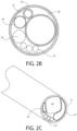
- FIG. 2 a is a perspective view of the expandable tip in the non-expanded configuration of the endoscope of FIG. 1 ;
- FIG. 2 b is a cross-sectional view of the expandable tip of FIG. 1 taken along a section of FIG. 2 a;
- FIG. 2 c is a perspective view of the expandable tip in the non-expanded configuration of an endoscope with a non-circular camera;
- FIG. 3 is a view of the non-expanded tip components of the endoscope of FIGS. 2 a and 2 b underneath the outer cover sheaths;
- FIG. 4 is a view of the expandable tip in an expanded configuration of the endoscope of FIG. 1 ;
- FIG. 5 is a close-up view of the coupling hub of FIG. 1 with the distal hub enclosure removed;
- FIG. 6 is a view of components inside the coupling hub of FIG. 1 with the full enclosure removed;
- FIG. 7 is a view of the PCA (Printed Circuit Assembly) board inside the coupling hub of FIG. 1 ;
- FIG. 8 is a schematic view of an endoscope illumination system.
- This application provides an electronic endoscope with a variable profile tip, in particular a single-use or disposable, low-cost, electronic endoscope with a variable profile working channel.
- the electronic endoscope has a hub, which remains outside the patient's body.
- the hub is used by the doctor to manipulate the endoscope.
- An elongated, flexible shaft extends from that hub. This is the portion of the endoscope, that can be inserted into the patient's body.
- An expandable tip extends from the end of the shaft that is farthest away from the hub. This is the distal tip.
- This expandable distal tip has sensors that allow the doctor to see inside the patient's body and a working channel, which allows the doctor to treat or operate on nearby structures.
- a complementary metal oxide semiconductor (CMOS) image sensor is positioned within the distal tip and has a field of view external from the endoscope, although other sensors can also be used.
- One or more illuminating fibers within the distal tip emit light within the image sensor's field of view.
- CMOS complementary metal oxide semiconductor
- the distal tip has a variable profile working channel.
- the working channel permits one or more tools (e.g., ablation devices, cannulas, dissectors, electrodes, forceps, graspers, knot pushers, laser fibers, needle holders, suction and irrigation instruments, trocars, and other tools) to be passed within the endoscope from the hub to the image sensor's forward field of view.
- the variable profile working channel can change shape to allow the tools to pass alongside the image sensor.
- the expandable working channel can change from a generally noncircular cross-sectional shape to a different and enlarged cross-sectional shape which permits tools to pass.
- Another endoscope has a hub that encloses a light source. The doctor uses the hub to manipulate the endoscope.
- An insertion portion extends from the hub. At the distal tip of the endoscope (furthest from the hub), the insertion portion has an expandable outer sheath.
- a light transmission system conveys light from the hub and onto a subject to be illuminated beyond the distal tip.
- An image sensor is positioned at approximately the center of the endoscope's distal tip. The sensor picks up an image of the illuminated subject.
- a variable profile working channel extends from the hub to the distal tip of the endoscope.
- the working channel is positioned inside the expandable outer sheath.
- a low-profile configuration of the working channel fits within a space defined between the image sensor and the expandable outer sheath.
- An enlarged-profile configuration of the working channel permits tools to travel past the image sensor and out of the distal tip.
- the expandable outer sheath has a generally noncircular shape at the distal tip.
- a further endoscope has a proximal end closest the doctor, a distal end at the opposite end and a field of view at that distal end.
- the hub is at the proximal end.
- the portion of the endoscope which extends from the hub towards the distal end can be termed the “insertion portion.” Depending on the doctor's needs, some or the entire insertion portion can be inserted into the patient. The hub remains outside the patient.
- An expandable distal tip extends from the insertion portion to the distal end and has a sensor configured to pick up an image within the field of view.
- a working channel, a flushing lumen, and a light-guide extend within the insertion portion from the hub to the distal end.
- the endoscope has a cross-section, which includes the flushing lumen, the light-guide, a generally circular cross-section of the working channel, and a cable extending from the sensor to the hub.
- the endoscope has a different cross-section. That different cross-section has the image sensor, the working channel, the flushing lumen, and the light-guide. Both the working channel and the flushing lumen are capable of changing their profiles, and are capable of doing so independently of one another. Each can assume a generally noncircular, low-profile configuration. Each can also assume an enlarged-profile to accommodate a tool passing through the working channel or to accommodate liquid passing through the flushing lumen.
- Each of the endoscopes described briefly above has an expandable distal tip, a variable profile working channel and, optionally, an expandable flushing lumen.
- the materials from which these three structures are made can be the same or different.
- Biocompatible elastomeric material can be used to make all three structures (e.g., silicone rubber, thermoplastic elastomer (TPE)).
- TPEs include copolyester elastomers (e.g., ARNITEL® from DSM), polyether block amide (e.g., PEBAX® from Arkema), polyether polyester block copolymers (e.g., HYTREL® from Du Pont), polyolefin elastomers (e.g., ENGAGE® from Dow Chemical), polyurethane elastomer (e.g., PELLETHANE® from Dow Chemical), styrene block copolymers (e.g., EVOPRENE® from AlphaGary), styrene-butadiene block copolymers (e.g., STYROFLEX® from BASF), styrene-ethylene-butylene-styrene block copolymers (e.g., KRATON® from Kraton Polymers), and thermoplastic vulcanizates (e.g., SANTOPRENE® and GEOLAST® from ExxonMo
- the working channel and the optional flushing lumen also can be made from non-elastomeric materials, which can change from a low-profile configuration to an enlarged-profile configuration (e.g., poly(ethylene-vinyl acetate) (PEVA), polyimide, polytetrafluoroethylene (PTFE), or polyvinyl chloride (PVC)).
- PEVA poly(ethylene-vinyl acetate)
- PTFE polytetrafluoroethylene
- PVC polyvinyl chloride
- an endoscope 1 has an expandable tip 2 , a shaft 3 , a coupling hub 4 , a connector assembly 5 , and a USB connector 13 .
- the endoscope 1 shown has an overall working length (combined length of the shaft 3 and the distal tip 2 ) of between 5 cm and 200 cm, preferably between 10 cm and 100 cm, and most preferably between 12 cm and 60 cm.
- the working length should be sufficient to allow the tip of the endoscope 1 to be positioned within the patient's body so that the relevant anatomy can be seen, while maintaining the coupling hub 4 outside the patient's body.
- the shaft 3 extends from the distal tip 2 to the coupling hub 4 .
- the shaft 3 transmits torque (rotation) applied about a longitudinal axis of the shaft 3 .
- Torque applied to the proximal end of the shaft is transferred along the length of the shaft 3 and to the distal tip 2 .
- the shaft 3 is flexible and can be bent about a transverse axis of the shaft in a relatively small bend radius. This allows the endoscope to be maneuvered around anatomical structures during medical procedures. However, in other applications, some or the entire shaft 3 is rigid or semi-rigid.
- the shaft 3 can be made from any biocompatible material with appropriate strength characteristics (e.g., providing flexibility and strength in tension and compression, as well as appropriate torque transfer from the proximal to the distal end).
- Materials from which the shaft 3 can be made include biocompatible polyamides, polyesters, polyetheretherketones, polyetherurethanes, polyimides, polytetrafluoroethylene, and polyurethane epoxies.
- reinforcing materials can be incorporated into the shaft 3 .
- Such reinforcing materials include copper alloys, nickel alloys (e.g., Nitinol), stainless steel, and high modulus plastics such as polyimides.
- the coupling hub 4 contains one or more connectors 8 , 9 , 10 for tools, flushing fluid, and a stylet, as well as optional electronics.
- the coupling hub 4 has a size and shape which accommodates these components.
- Electronics within the coupling hub 4 include a PCA with circuitry to operate the endoscope and a camera signal transmission system that are discussed below.
- the PCA can function as an interconnection to external control circuitry outside of the endoscope. If electronics internal to the hub 4 are not otherwise desired, the hub 4 can be quite small and can serve as a connection joint between the other tubes, wires, and optical fibers.
- the hub 4 is made of a biocompatible plastic, such as polycarbonate, acrylic, acrylonitrile butadiene styrene (ABS), cast epoxy, and thermoset plastics.
- a biocompatible plastic such as polycarbonate, acrylic, acrylonitrile butadiene styrene (ABS), cast epoxy, and thermoset plastics.
- ABS acrylonitrile butadiene styrene
- thermoset plastics formed metal housings using biocompatible materials, such as several grades of stainless steel or titanium, are also possible.
- the coupling hub 4 has a distal portion 18 and proximal portion 19 .
- the coupling hub 4 includes one or more ports or connectors 8 , 9 , 10 , and strain relief features 11 , 12 .
- These connectors 8 , 9 , 10 include seals to provide fluid tight seal between coupling hub 4 and the connectors 8 , 9 , 10 (e.g., a LUER-LOK® lock component).
- the connectors 8 , 9 , 10 attach firmly to the coupling hub 4 and allow doctors to introduce tools and fluids at the hub 4 that can be used at the distal tip 2 of the endoscope. This can be achieved by gluing, heat-welding, potting with thermoset plastic or epoxy, RF welding, screwing them on with a threaded connection, solvent bonding, ultrasonic welding, or combinations of these processes. Any or all of these three connectors 8 , 9 , 10 can be attached to a flexible tube, which enters the hub 4 .
- the flushing channel connector 8 allows fluid to be introduced at the hub 4 , which can travel through the flushing lumen 24 .
- the stylet channel connector 9 allows for a stylet (not shown) to be inserted at the hub 4 and into the stylet channel 27 .
- the stylet is intended to influence the shape of the endoscope's flexible shaft 3 .
- a malleable and resilient wire made of materials such as 300-series stainless steel can be bent into a desired curvature or angle, and inserted through the stylet channel causing the flexible endoscope to conform to such curvature or angle.
- the working channel connector 10 allows tools to be passed from the hub 4 through the working channel 28 , within the shaft 3 , and down to the working channel portion 23 within the distal tip 2 .
- variable profile working channel Although the endoscope shown has a single variable profile working channel, it is contemplated that more than one working channel can be included. When multiple working channels are employed, they may be the same size or different sizes. Some or all of the working channels can have the variable profile feature. For example, a 5 mm endoscope could have two 1.2 mm working channels, both of which have the variable profile feature.
- FIG. 1 shows three connectors 8 , 9 , 10 for the flushing lumen, stylet, and working channel, respectively. It is contemplated that as few as one connector could be used, or as many connectors as needed for the flushing lumen, stylet, and working channel features of the particular endoscope. For example, an endoscope with two working channels, a stylet, and a flushing lumen could have four separate connectors.
- the electrical cable 5 and USB connector 13 provide an interface between circuitry within the coupling hub 4 and external devices.
- a single cable 5 extends from the coupling hub 4 to the USB connector 13 .
- This cable 5 has conductors for powering a light source within the coupling hub 4 and has contacts for transmitting signals outputted by the camera 22 to the USB connector 13 .
- Multiple cables can be used with, for example, a first cable 5 conducting power to a light emitting diode (LED) light source in the coupling hub 4 , and a separate second cable (not shown) transmitting signals receives from the camera 22 .
- LED light emitting diode
- the USB connector 13 connects to external control/display devices or into an electronic interface box, which translates the USB signals into those used by the external control or display devices.
- the USB connector 13 also provides power for the electronic endoscope.
- wired connections e.g., HDMI
- wireless connections e.g., Bluetooth, WiFi
- Strain relief features 11 , 12 protect the hub 4 and its components from these forces.
- FIG. 1 shows a distal strain relief 11 and a proximal strain relief 12 .
- Strain relief features 11 , 12 can be made from various materials. These include relatively hard injection molded thermoplastics (e.g., acrylonitrile butadiene styrene (ABS)), or more flexible materials such the TPEs previously identified. When a TPE is used, it preferably has a higher durometer value than the expandable tip components.
- ABS acrylonitrile butadiene styrene
- FIGS. 2 a and 2 b show the distal end of the endoscope 1 in both perspective and cross-sectional views.
- the expandable tip 2 has an expandable outer cover 21 .
- the structure within the expandable tip 2 is shown, and includes a variable profile working channel 23 , an expandable flushing channel 24 , three illumination fibers 25 , and a camera 22 .
- the distal tip 2 is long enough to house the camera 22 .
- the distal tip 2 has a length from the end of the shaft 3 to the distal most part of the endoscope of between 3 mm and 50 mm, preferably between 7 mm and 15 mm, and most preferably between 8 mm and 10 mm.
- the distal tip 2 can enlarge and contract.
- the distal tip 2 In a low-profile state, the distal tip 2 has a reduced cross-sectional area with an outer diameter of between about 1.5 mm and 20 mm, preferably between about 1.5 mm and 5 mm, and most preferably between about 1.5 mm and about 2.0 mm.
- the distal tip 2 In an enlarged-profile or expanded state, the distal tip 2 has an enlarged cross-sectional area that will accommodate passage of one or more tools through the working channel.
- the enlarged-profile accommodates passage of a tool having a circular cross-sectional shape with a diameter of between 1.8 mm and 20 mm, preferably between 2 mm and 5 mm, and most preferably between 1 mm and 2 mm.
- the profile change allows tools to pass the camera 22 , and ultimately to exit the distal tip of the endoscope.
- the illumination fibers 25 are depicted as three flexible fiber-optic light-guides. These fibers 25 carry light from a light source in the coupling hub 4 to the distal tip 2 of the endoscope, illuminating a field of view. Although three fibers 25 are shown in FIG. 2 A , as few as one fiber can be used or as many fibers as can fit within the allowable space so long as they provide sufficient light for camera 22 . Illumination fibers 25 can be made of glass, PMMA or other light transmitting materials. Combinations of different sized fibers 25 can be used, as long as they fit within the cross-sectional area of the distal tip 2 in its low-profile configuration and provide sufficient illumination intensity.
- the camera 22 and illumination fibers 25 are positioned so the area to be imaged is sufficiently illuminated.
- the camera 22 can be approximately in the center of the distal tip 2 , with the working channel 23 , the flushing lumen 24 and the illumination fibers 25 angularly distributed around the circumference (e.g., the working channel 23 separated by about 120° from the flushing lumen 24 and by an equal amount from the illumination fibers 25 ).
- the working channel 23 and flushing channel 24 relative to each other and relative to the camera 22 or the illumination fibers 25 are possible.
- the outer cover 21 , the working channel 23 , and the flushing channel 24 are made of sterilizable polymeric materials.
- these structures are configured so that they can change shape or expand.
- Each of these structures can be made from the same material, or the materials can be different.
- each can be constructed of biocompatible elastomeric tubing (e.g., latex rubber, silicone rubber, or various USP Class 6 compatible TPEs). Exemplary TPEs are mentioned above.
- a single-use endoscope sterilization by the original manufacturer can be done in bulk (e.g., using ethylene oxide gas, gamma radiation, steam).
- a single-use device can have tubing connections that butt against one another but still allow a small crack, gap, or void at the joint.
- Materials suitable for the bulk sterilization such as by ethylene oxide gas, can also be used to construct the endoscope without concern for the materials having to be compatible with multiple exposure to sterilization chemicals such as glutaraldehyde.
- Coatings can be applied to the outside of the endoscope.
- coatings could provide anti-bacterial or anti-microbial properties (e.g., copper ions, silver ions). Coatings can also be used to allow doctors to detect certain conditions. For example, special peptides and other formulations can be applied that detect presence of bacterial contamination or biomarkers of other sorts.
- a UV-cured adhesive potting compound is applied at the distal tip and flows between the components, while it is viscous. Excess potting compound is removed and UV light applied to cure or harden the material. Other materials can also be used, such as 2-part epoxies. Application of the potting compounds may be limited to areas that do not interfere with the expandable distal tip sections (e.g., by fixtures that limit the distribution area of the compound), and/or use materials that do not bond to those expandable sections.
- the working channel 23 has a low-profile configuration.
- the low-profile working channel 23 of a 2 mm endoscope can have a 0.5 mm diameter within the shaft 3 and then assumes a more compact configuration within the distal tip 2 so that it fits the space between the expandable outer cover 21 and the camera 22 .
- the low-profile working channel 23 has a generally lunate cross-sectional shape.
- the ends of the lunate cross-sectional shape can be rounded (as in FIG. 2 A ), or bent more sharply. Other shapes are possible, including circular, reniform, oblong, oval, etc.
- the working channel 23 also has an enlarged-profile configuration to allow tools to pass the camera 22 .
- the enlarged-profile working channel 23 of 2 mm endoscope is large enough to allow tools with an outer diameter of approximately 1.2 mm to pass through it.
- the camera 22 is a CMOS color camera, which is biocompatible and waterproof.
- the camera provides a field of view external from the endoscope. A forward field of view would look beyond the distal tip of the endoscope. However, the field of view can also be at an angle external from the endoscope (e.g., an off-angle-looking endoscope such as 30°.
- the CMOS image sensor includes a multi-element lens assembly or gradient refractive index (GRIN) lens providing a field of view of 30-180°, preferably of 50-130°, and most preferably 60-120°.
- the effective image resolution preferably is at least 10,000 pixels, more preferably at least 40,000 pixels, and most preferably at least 60,000 pixels, although image resolutions of 1 megapixel or greater are possible.
- Other sensors can be used instead of or in conjunction with the CMOS device so long as they provide sufficient image resolution. For example, a charge-coupled device (CCD) could be used.
- CCD charge-coupled device
- an optical prism can be added to modify the particular angular view of the image sensor.
- the prism has a reflective surface that “tilts” the viewing cone of the image sensor by a predetermined amount, such as 30°, 70°, etc.
- the prism may be made of glass or any clear polymer, such as acrylic or polycarbonate.
- the prism could be bonded using optically-clear epoxy to the flat distal surface of the camera. More preferably, the prism could replace the final camera lens in the original manufacture of the device.
- the camera sensor can be further affixed to the structure of the endoscope.
- the camera sensor can be affixed directly to the light guide to provide additional support.
- the camera cable can be adhered to the sheath cover, the working channel, or both.
- the camera cable can be bonded to the inside of the sheath cover, just proximal to the expandable distal tip section. These further connections provide added stiffness.
- FIG. 2 b The cross-sectional view of FIG. 2 b is along section A-A of FIG. 2 a , and passes through the elongated shaft 3 .
- a flexible, braided sheath cover 26 is made from a stainless steel reinforced polyimide material, although other materials can be used.
- the braided sheath cover 26 contains five structures.
- the working channel 28 in this section is a flexible tube with a generally circular cross-sectional shape.
- the working channel 28 can be constructed from any sterilizable polymeric material than can be affixed to the variable profile working channel 23 shown in FIGS. 1 and 2 a , or working channel 28 and variable profile working channel 23 can made from the same length of tubing.
- the material of working channel 28 can be the same material as that of variable profile working channel portion 23 , or they can be made from different materials.
- the working channel within the shaft 3 can be made from non-elastomeric polymeric materials (e.g., poly(ethylene-vinyl acetate) (PEVA), polyimide, polytetrafluoroethylene (PTFE), polyvinyl chloride (PVC)) or from biocompatible elastomeric tubing (e.g., latex rubber, silicone rubber, or various USP Class 6 compatible TPEs).
- PEVA poly(ethylene-vinyl acetate)
- PTFE polytetrafluoroethylene
- PVC polyvinyl chloride
- biocompatible elastomeric tubing e.g., latex rubber, silicone rubber, or various USP Class 6 compatible TPEs.
- the flushing channel 24 can have any cross-sectional shape within the shaft 3 so long as sufficient fluid can be passed.
- the flushing channel 24 shown in FIG. 3 is made from a single length of elastomeric tubing.
- the proximal portion of flushing channel 24 within the shaft 3 can be made from non-elastomeric material (e.g., a polyimide tube) and coupled to an elastomeric tip section. Suitable elastomeric materials for the flushing channel 24 are identified above.
- a stylet channel 27 allows introduction into the endoscope of a stylet (not shown), which is a slender probe, typically made from a metal. When the stylet is introduced, it provides additional stiffness. This can facilitate proper positioning of the endoscope's distal tip within the patient. In addition, the stylet can impart a particular shape to the endoscope (e.g., a particular curve or bend). The stylet channel 27 should resist puncture by the stylet tip. Reinforcement is optional.
- This stylet channel 27 is made from various polymeric tubing materials, such as PEVA, polyimide, PTFE, or PVC. The stylet channel 27 is shown to have a circular profile, but can have any profile that allows the stylet to pass through it.
- a camera cable 29 extends from the camera 22 (shown in FIG. 2 a ) to the coupling hub 4 (shown in FIG. 1 ).
- the camera cable can be a simple signal conducting wire (e.g., a 24 AWG gauge copper wire with a diameter of about 0.52 mm), or a ribbon cable with several insulated conductors.
- Exemplary conductor compositions include copper, copper alloys, MP35N, DFT, platinum, platinum/iridium, tungsten, gold, and stainless steel.
- the conductors can be bare, tinned, silver-plated, or gold-plated.
- the ribbon cable can be constructed with four conductors: (1) a ground, (2) a serial data line, (3) a serial clock line, and (4) a power line (e.g., using 41 AWG silver plated copper conductors).
- FEP fluorinated ethylene propylene
- PFA perfluoroalkoxy
- PTFE polytetrafluoroethylene
- the ribbon cable can be constructed with four conductors: (1) a ground, (2) a serial data line, (3) a serial clock line, and (4) a power line (e.g., using 41 AWG silver plated copper conductors).
- FIG. 2 c shows the distal end of another endoscope made with a non-circular camera 22 that is radially offset from the center of the endoscope.
- the working channel portion 23 within the distal tip 2 and the flushing lumen 24 are adjacent the camera 22 and the elastomeric tip cover 21 .
- FIG. 3 shows the endoscope of FIG. 2 a without outer covers 21 and 26 .
- the coupling sleeve 31 is a thin-wall cylindrical material that joins the elastomeric tip cover 21 and the flexible braided sheath 26 .
- Coupling sleeve 31 depicted in FIG. 3 can be made from polyimide, stainless steel, polyurethane or other thin-wall tubing. This coupling sleeve 31 is bonded to the inside surfaces of the braided flexible sheath 26 , and the elastomeric expandable tip cover 21 .
- Joining can be by adhesive bonding, heat welding, mechanical ring-clamping or solvent bonding, or other methods for durably bonding these components.
- the stylet channel 27 is a thin-wall tubing similar to that described previously for other working channels such as polyimide, PTFE, or other tubing, and which has a durable stop 33 at its distal tip, such as a plug of hard plastic such as acrylic, ABS, or other material bondable or mechanically securable to the stylet channel, which is intended to prevent the stylet from puncturing or damaging other components.
- the tubing also can be crimped or folded over at the end to create a stop if the tip of the stylet is rounded. This can be in addition to the hard plastic plug or instead of it.
- the deflector ramp provides an angled, relatively hard surface to guide the distal tip of the tool around the proximal edge of the camera and into the distal tip expansion area of the working channel.
- This deflector ramp can be made of hard plastic such as acrylic or ABS, or from a thin stamped stainless steel metal piece, and may be curved to match the outside diameter of the working channel shaft.
- the working channel has a generally circular cross-section 28 within the shaft 3 and a variable profile section 23 within the distal tip 2 .
- the ramp 34 is affixed to the endoscope.
- the ramp can be adhered to the proximal surface of the camera 22 , to the expandable work channel 23 , to the sheath cover 26 , or to combinations of these structures.
- a UV-curable epoxy or other adhesive can be used for this purpose.
- the ramp 34 defines an angle relative to the longitudinal axis of the endoscope that can range from about 30° to about 60°, and preferably ranges between 40° and 50°, and most preferably about 45°.
- a ratio of the length of the ramp 34 to the overall outer diameter of the endoscope at the distal tip is less than about 1:2, preferably is less than about 1:3, and most preferably is less than about 1:5.
- a coupling sleeve 35 can be used when the circular cross-section working channel shaft 28 and the expandable working channel 23 are made from different materials or different sections of tubing that are affixed together. If these are made using a single length of tubing, coupling sleeve 35 can be omitted. Unlike the outer sheath coupling sleeve 31 (which fits inside the outer cover of the expandable tip cover 21 ) and the flexible, braided sheath cover 26 (which allows the endoscope to maintain an even outer diameter), working channel coupling sleeve 35 fits on the outside of a joint between the circular cross-section working channel shaft 28 and the expandable working channel 23 to preserve a consistent inner diameter for tool insertion and removal.
- the coupling sleeve 35 can be a short piece of thin-wall polymeric tubing (e.g., a 4-5 mm of 0.0.254 mm wall thickness polyimide tubing, which is obtainable from Putnam Plastics or Vention Medical).
- variable profile distal tip allows the doctor to insert the endoscope through a minimal incision or puncture size (e.g., for a 12 gauge needle) while it is in a low-profile configuration. Once the endoscope is in the area to be treated, the tip section is enlarged. FIG. 4 illustrates the distal tip 2 in an enlarged-profile configuration.
- Such a configuration is achieved, for example, by passing a full 1.0 mm diameter tool through the working channel tip 23 , and/or flushing fluid (0.9% saline in sterile water) through the flushing channel 24 at a rate sufficient to expand the flushing channel 24 at the distal tip 2 .
- FIGS. 5 and 6 show details of the coupling hub 4 .
- the flushing channel tube 51 is attached to flushing channel connector 8 .
- the stylet channel tube 52 is attached to the stylet channel connector 9 .
- the working channel shaft 53 is attached to the working channel connector 10 .
- tubes 51 , 52 , 53 attach to connectors, such as Luer or Luer lock connectors. Solvent bonding, adhesive bonding, or press fit can be used to make this connection.
- the proximal hub enclosure 7 is removed to reveal the PCA 61 , the proximal end of the camera cable 29 , and the proximal end of the illumination fibers 25 .
- FIG. 7 is a close-up view of the PCA 61 .
- Camera cable 29 connects to the PCA 61 using connector 72 .
- LED 71 is an LED surface-mounted chip with the proximal ends of the illumination fibers 25 placed flush against the light emitting surface area of LED 71 .
- non-LED illumination sources can be used (e.g., halogen incandescent, xenon light, diode lasers) so long as the particular illumination chosen can be picked up by the camera or sensor.
- An epoxy provides a secure attachment, although other methods can be appropriate.
- Mounting screws 73 attach the PCA 61 to studs molded into the proximal hub enclosure 7 .
- Other PCA mounting techniques can be used, including capturing the PCA 61 between the proximal hub enclosure portion 19 and the distal hub enclosure portion 6 .
- Attachment of the two-hub enclosure portions 6 , 7 , the strain reliefs 11 , 12 and, in turn, their attachments to the electronic cable 5 , the coupling hub 4 , and endoscope shaft 3 are watertight.
- Such watertight connections can be achieved by a number of means including flexible adhesive, solvent bonds, heat seals, rubber grommets and other seals.
- the PCA 61 circuitry controls illumination intensity of the light source 71 and operates the camera 22 .
- the PCA 61 circuits translate signals to and from an external control/display (not shown). These circuits convert signals into the patterns, voltages, timing, etc., needed by, or sent from the camera 22 and used for the illumination source 71 . Such translation can include converting the signals into a Universal Serial Bus (USB) standard.
- Multi-layer or stacked multi-layer circuits with embedded software (e.g., firmware) and Field Programmable Logic Arrays (FPGAs) translate and communicate the signals.
- the PCA 61 includes a wireless transceiver and a power source (e.g., battery).
- the battery powers the PCA 61 and the image sensor 7 .
- the wireless transceiver interfaces with external control and display devices. For example, the PCA 61 wirelessly transmits image signals from the image sensor 7 to an external display.
- signal translation can be accomplished externally of the endoscope.
- the PCA 61 functions as an interconnection and routes signals to a cable 5 , which then connects to an external control/display unit (not shown).
- FIG. 8 illustrates an illumination configuration for the electronic endoscope.
- numerous LEDs and illumination fibers or fiber bundles can be arranged to assure sufficient illumination intensity to achieve the desired field of view and depth of field.
- LED 82 if small enough, can be mounted adjacent to the camera 22 within the expandable tip section 2 .
- Illumination fibers 81 are routed from the LED 82 to reach the outside of the camera 22 at the distal tip 2 .
- Such an illumination subassembly can be rigid.
- the illumination subassembly can be varied, so long as sufficient illumination intensity to achieve the desired field of view and depth of field.
- a shaped light guide can be used to replace optical illumination fibers, as long as the Numerical Aperture and illumination field pattern of the light emitted from the light guide is compatible with the field of view of the camera 22 .
- the LED can be mounted within the shaft section 3 or can be exterior to the endoscope, instead of within the hub enclosure 4 .
- a 2 mm endoscope with expandable working channel and an LED light source in the hub similar to that shown in FIG. 1 is formed as described below.
- Two injection-molded ABS pieces 6 , 7 (available from Dow Chemical) form the hub enclosure.
- the strain relief tube 12 is an injection molded component made from Shore A 60 TPE.
- the cable assembly 5 is electrically connected to a printed circuit assembly (PCA) 61 .
- PCA 61 is a small, multi-layer printed circuit board with surface mount integrated circuits to convert USB signals and communications to/from the camera into the signal levels and timing required by the camera specifications.
- the PCA 61 is affixed to the proximal hub enclosure 7 using four screws 73 . See FIG. 7 .
- the proximal hub enclosure has an opening for the working channel.
- a working channel connector 10 provides a fluid tight seal between the proximal hub enclosure 7 and the working channel. Insert connector 10 (a commercially available LUER-LOK® lock component) into the proximal hub enclosure 7 .
- the sheath cover 26 is a 29.2 cm length of braided stainless steel reinforced polyimide tubing (Putnam Plastics catalog number 142 -0045; 1.88 mm OD, 1.689 mm ID).
- the distal strain relief tube 11 is an injection molded component made of Shore A 60 TPE. Insert this tube through an opening in hub enclosure 6 . Slide the sheath cover 26 through the distal strain relief 11 for access to the interior of the hub enclosure 6 . Solvent bond with cyclohexanone to affix the sheath cover 26 to distal strain relief tube 11 .
- the outer sheath 21 / 26 is formed from the polyimide and silicone tubing using a manufacturing mandrel.
- the working channel is also formed using a mandrel.
- Slide a29.2 cm length of polyimide tubing 28 (Vention Medical catalog number 141-0083; 0.0505 in OD ⁇ 0.048 in ID) onto a mandrel of appropriate diameter.
- Join these components together with a low-viscosity ultraviolet light cure adhesive (208-CTH-F flexible, water resistant catheter bonding adhesive available from Dymax Corp.). Together the working channel assembly 23 / 28 / 35 is 30 cm long.
- ABS acrylonitrile butadiene styrene
- the camera is a micro ScoutCamTM 1.2 (Medigus, Ltd. of Omer, Israel). This camera 22 is cylindrical in shape and measures 1.2 mm in diameter by 5 mm in length, and provides an effective image resolution of about 44,880 pixels.
- Camera cable 29 extends from camera 22 , runs within the cover assembly but along the outside of the assembled working channel, and connects to PCA 61 at the camera connector 72 . See FIG. 7 .
- PCA 61 controls camera 22 and receives images from it.
- Three optical illumination fibers 25 (outer diameter of 0.25 mm, from LightHouse LEDs as catalog number 0.25MMFIBERENDGLOW) also run within the cover assembly along the outside of the working channel assembly until they are adjacent to the camera 22 . Adhere proximal ends of the illumination fibers 25 onto the light source 71 , a Luxeon® C power LED (available from the Philips Lumileds Lighting Company) with UV-cured adhesive (208-CTH-F adhesive available from Dymax Corp.).
- a Luxeon® C power LED available from the Philips Lumileds Lighting Company
- UV-cured adhesive 208-CTH-F adhesive available from Dymax Corp.
- This provides an endoscope with a 30 cm working length, a 2 mm OD, and variable profile distal tip and working channel.
- Such an endoscope can be used for a variety of endoscopic applications including arthroscopic joint examination and surgery for hands, shoulders, knees, etc.
- An endoscope similar to that described in Example 1 is formed with a variable profile flushing lumen 24 in addition to the variable profile working channel as illustrated in FIG. 2 A .
- a 30 cm length of TPE tubing (Vention Medical PEBAX® tubing catalog number 115-1289; 0.0.279 mm ID, 0.1143 mm wall thickness, 0.51 mm OD, Shore A 63) forms the flushing lumen 24 .
- the process for making the endoscope is similar to Example 1. Differences are discussed below.
- flushing lumen 24 is made from a non-expandable material. Instead of PEBAX® tubing, the flushing lumen 24 is made from polyimide tubing (Vention Medical catalog number 141-0023; 0.508 mm OD, 0-457 mm ID).
- the flushing lumen is manufactured to provide that feature.
- the creased and flattened distal tip of the flushing lumen 24 expands as fluid passes within the flushing lumen 24 . This, in turn, expands the outer cover 21 . See FIGS. 3 and 4 .
- An endoscope similar to that described in Example 1 is configured for wireless communication with an external display and a control device (e.g., a PC or tablet computer).
- the PCA 61 has wireless transmitting and receiving components.
- Commonly available button-style batteries are included within the hub assembly to power PCA 61 .
- An antenna wire is included and attached to the PCA 61 . Connect a switch in series with the battery. The batteries provide sufficient power to allow the endoscope system to function for several hours.
- the camera 22 is an Awaiba NanEye camera (available from AWAIBA Lda). This camera 22 measures 1.1 mm ⁇ 1.1 mm ⁇ 1.7 mm long with a diagonal measurement of 1.41 mm, and provides an effective image resolution of about 62,500 pixels.
- FIG. 2 c shows a cross-sectional view of this endoscope at the distal tip.
- the outer sheath 21 / 26 is formed from a 29.2 cm length of a braided stainless steel reinforced polyimide tubing (1.72 mm OD, 1.57 mm ID, available from Vention Medical catalog 142-0042), an 8 mm length of silicone rubber tubing with an ID of about 1.689 mm, and a thin-walled polyimide coupling tube 31 with an OD of about 1.689 mm.
- the working channel is formed from thin-wall polyurethane tubing (Vention Medical catalog number 115-0565; 1.346 mm OD, 0.089 mm wall, 1.168 mm ID).
- the camera is the NanEye camera described above in Example 5.
- the illumination light guides (optical fibers) are 0.125 mm OD (0.1 mm core diameter, Edmund Optics stock #57-061) and eight total fibers are used for the illumination.
- the four-conductor cable from the NanEye camera 22 is connected to PCA 61 for signal connection to a cable suitable for external use and possible signal processing circuits are included on PCA 61 to convert the signals to USB standards or other desired configuration.
- PCA 61 has memory containing calibration information for the specific NanEye camera being used.
Landscapes
- Health & Medical Sciences (AREA)
- Life Sciences & Earth Sciences (AREA)
- Surgery (AREA)
- Biomedical Technology (AREA)
- Medical Informatics (AREA)
- Optics & Photonics (AREA)
- Pathology (AREA)
- Radiology & Medical Imaging (AREA)
- Biophysics (AREA)
- Engineering & Computer Science (AREA)
- Physics & Mathematics (AREA)
- Heart & Thoracic Surgery (AREA)
- Nuclear Medicine, Radiotherapy & Molecular Imaging (AREA)
- Molecular Biology (AREA)
- Animal Behavior & Ethology (AREA)
- General Health & Medical Sciences (AREA)
- Public Health (AREA)
- Veterinary Medicine (AREA)
- Endoscopes (AREA)
- Instruments For Viewing The Inside Of Hollow Bodies (AREA)
Abstract
Description
Claims (20)
Priority Applications (2)
| Application Number | Priority Date | Filing Date | Title |
|---|---|---|---|
| US17/448,030 US12226075B2 (en) | 2015-08-07 | 2021-09-17 | Endoscope with variable profile tip |
| US19/053,870 US20250255464A1 (en) | 2015-08-07 | 2025-02-14 | Endoscope with variable profile tip |
Applications Claiming Priority (3)
| Application Number | Priority Date | Filing Date | Title |
|---|---|---|---|
| US14/821,579 US9913570B2 (en) | 2015-08-07 | 2015-08-07 | Endoscope with variable profile tip |
| US15/885,527 US11141045B2 (en) | 2015-08-07 | 2018-01-31 | Endoscope with variable profile tip |
| US17/448,030 US12226075B2 (en) | 2015-08-07 | 2021-09-17 | Endoscope with variable profile tip |
Related Parent Applications (1)
| Application Number | Title | Priority Date | Filing Date |
|---|---|---|---|
| US15/885,527 Continuation US11141045B2 (en) | 2015-08-07 | 2018-01-31 | Endoscope with variable profile tip |
Related Child Applications (1)
| Application Number | Title | Priority Date | Filing Date |
|---|---|---|---|
| US19/053,870 Continuation US20250255464A1 (en) | 2015-08-07 | 2025-02-14 | Endoscope with variable profile tip |
Publications (2)
| Publication Number | Publication Date |
|---|---|
| US20220211249A1 US20220211249A1 (en) | 2022-07-07 |
| US12226075B2 true US12226075B2 (en) | 2025-02-18 |
Family
ID=57984180
Family Applications (4)
| Application Number | Title | Priority Date | Filing Date |
|---|---|---|---|
| US14/821,579 Active 2035-11-06 US9913570B2 (en) | 2015-08-07 | 2015-08-07 | Endoscope with variable profile tip |
| US15/885,527 Active 2037-05-04 US11141045B2 (en) | 2015-08-07 | 2018-01-31 | Endoscope with variable profile tip |
| US17/448,030 Active US12226075B2 (en) | 2015-08-07 | 2021-09-17 | Endoscope with variable profile tip |
| US19/053,870 Pending US20250255464A1 (en) | 2015-08-07 | 2025-02-14 | Endoscope with variable profile tip |
Family Applications Before (2)
| Application Number | Title | Priority Date | Filing Date |
|---|---|---|---|
| US14/821,579 Active 2035-11-06 US9913570B2 (en) | 2015-08-07 | 2015-08-07 | Endoscope with variable profile tip |
| US15/885,527 Active 2037-05-04 US11141045B2 (en) | 2015-08-07 | 2018-01-31 | Endoscope with variable profile tip |
Family Applications After (1)
| Application Number | Title | Priority Date | Filing Date |
|---|---|---|---|
| US19/053,870 Pending US20250255464A1 (en) | 2015-08-07 | 2025-02-14 | Endoscope with variable profile tip |
Country Status (7)
| Country | Link |
|---|---|
| US (4) | US9913570B2 (en) |
| EP (1) | EP3331419B1 (en) |
| JP (1) | JP7043075B2 (en) |
| KR (1) | KR102617407B1 (en) |
| CN (2) | CN108135445B (en) |
| AU (2) | AU2016304752A1 (en) |
| WO (1) | WO2017027299A1 (en) |
Families Citing this family (80)
| Publication number | Priority date | Publication date | Assignee | Title |
|---|---|---|---|---|
| US20070167681A1 (en) | 2001-10-19 | 2007-07-19 | Gill Thomas J | Portable imaging system employing a miniature endoscope |
| US9913570B2 (en) | 2015-08-07 | 2018-03-13 | Enlightenvue Llc | Endoscope with variable profile tip |
| JP7082052B2 (en) | 2015-09-03 | 2022-06-07 | ネプチューン メディカル インク. | A device for advancing the endoscope in the small intestine |
| US20170100016A1 (en) * | 2015-10-12 | 2017-04-13 | Covidien Lp | Continuous flow scope configuration with optional tool usage |
| US20170224956A1 (en) * | 2016-02-10 | 2017-08-10 | Cook Medical Technologies Llc | Steerable catheter |
| CN110191667B (en) | 2016-08-18 | 2022-06-03 | 海王星医疗公司 | Device and method for enhancing the visual effects of the small intestine |
| US11096560B2 (en) | 2016-09-23 | 2021-08-24 | Meditrina, Inc. | Endoscope with multiple image sensors |
| US20180160893A1 (en) * | 2016-12-12 | 2018-06-14 | Meditrina, Inc. | Endoscope and method of use |
| CN106943114B (en) * | 2017-04-27 | 2019-01-08 | 泗洪县正心医疗技术有限公司 | A kind of expandable endoscope in end |
| US10568521B2 (en) * | 2017-05-19 | 2020-02-25 | Santa Clara University | Fluidic pressure actuated optical probes for improved field of view in tissue imaging |
| US20180338673A1 (en) * | 2017-05-26 | 2018-11-29 | Covidien Lp | Surgical sheath and surgical apparatus including the same |
| EP3590403B1 (en) * | 2017-06-01 | 2021-09-08 | Hoya Corporation | Endoscope |
| KR102651931B1 (en) * | 2017-06-30 | 2024-03-29 | 인라이튼뷰 엘엘씨 | Endoscopic system and its method of use |
| CN111065311B (en) | 2017-07-20 | 2022-06-10 | 海王星医疗公司 | Dynamic Rigidized Outer Sleeve |
| US11051682B2 (en) * | 2017-08-31 | 2021-07-06 | Wm & Dg, Inc. | Medical devices with camera and methods of placement |
| US12156640B2 (en) | 2017-10-11 | 2024-12-03 | Meditrina, Inc. | Endoscope and method of use |
| EP3694390B1 (en) * | 2017-10-11 | 2023-12-20 | Meditrina, Inc. | Endoscope |
| WO2019087254A1 (en) * | 2017-10-30 | 2019-05-09 | オリンパス株式会社 | Endoscope system |
| WO2019118484A2 (en) | 2017-12-12 | 2019-06-20 | Convergascent Llc | Multi-use endoscopes and associated systems and methods |
| WO2019143709A1 (en) | 2018-01-16 | 2019-07-25 | The Regents Of The University Of Colorado, A Body Corporate | Medical devices including textured inflatable balloons |
| US11577056B2 (en) | 2018-01-16 | 2023-02-14 | Aspero Medical, Inc. | Medical devices including textured inflatable balloons |
| US20220240762A1 (en) * | 2018-01-16 | 2022-08-04 | The Regents Of The University Of Colorado, A Body Corporate | Split overtube assembly |
| US11730928B2 (en) | 2018-01-16 | 2023-08-22 | Aspero Medical, Inc. | Split overtube assembly |
| US12357164B2 (en) | 2018-01-23 | 2025-07-15 | Clarus Medical, Llc | Medical device inspection scope |
| US12109081B2 (en) * | 2018-03-16 | 2024-10-08 | Clarus Medical, Llc | Medical device inspection scope |
| WO2019152377A1 (en) | 2018-01-30 | 2019-08-08 | Hsu George Chao Chih | Surgical device and methods |
| JP6948454B2 (en) * | 2018-02-27 | 2021-10-13 | 富士フイルム株式会社 | Adhesives for endoscopes, cured products, endoscopes and methods for manufacturing endoscopes |
| CN112004453B (en) * | 2018-03-13 | 2024-08-27 | 梅迪特瑞纳公司 | Endoscope and how to use it |
| KR102085537B1 (en) * | 2018-03-26 | 2020-03-06 | 서울대학교병원 | Endoscope and endoscope for paranasal sinuses |
| EP3801187B1 (en) | 2018-05-31 | 2024-02-07 | Neptune Medical Inc. | Device for enhanced visualization of the small intestine |
| WO2019246266A1 (en) * | 2018-06-19 | 2019-12-26 | Boston Scientific Scimed, Inc. | Endoscopic handle attachment for use with suture based closure device |
| CA3106275A1 (en) | 2018-07-19 | 2020-01-23 | Neptune Medical Inc. | Dynamically rigidizing composite medical structures |
| EP3829413B1 (en) * | 2018-08-27 | 2025-04-30 | Meditrina, Inc. | Endoscope |
| US10687698B2 (en) * | 2018-09-12 | 2020-06-23 | Enlightenvue Llc | Direct endoluminal- and/or endovascular-illumination systems and methods of use thereof |
| TWI691320B (en) * | 2018-10-03 | 2020-04-21 | 新視電科技有限公司 | Non-power steering equipment |
| WO2020097361A1 (en) * | 2018-11-07 | 2020-05-14 | Meditrina, Inc. | Endoscope and method of use |
| CN112969401A (en) * | 2018-11-07 | 2021-06-15 | 理查德·沃尔夫有限公司 | Endoscopic instrument |
| WO2020105158A1 (en) * | 2018-11-22 | 2020-05-28 | オリンパス株式会社 | Guide tube, treatment system, and guide tube attachment method |
| DE102018133368A1 (en) * | 2018-12-21 | 2020-06-25 | Konstantin Bob | Endoscope with an expandable working channel |
| WO2020146812A1 (en) * | 2019-01-11 | 2020-07-16 | Dragonfly Endoscopy Llc | Endoscopic device and methods of use thereof |
| CA3134694A1 (en) * | 2019-03-29 | 2020-10-08 | Coopersurgical, Inc. | Endoscopic devices and related methods |
| US12329473B2 (en) | 2019-04-17 | 2025-06-17 | Neptune Medical Inc. | Dynamically rigidizing composite medical structures |
| US11793392B2 (en) | 2019-04-17 | 2023-10-24 | Neptune Medical Inc. | External working channels |
| EP3952721B1 (en) * | 2019-04-18 | 2025-11-12 | Meditrina, Inc. | Endoscope |
| US11903557B2 (en) | 2019-04-30 | 2024-02-20 | Psip2 Llc | Endoscope for imaging in nonvisible light |
| AU2020279633B2 (en) * | 2019-05-17 | 2025-08-14 | Boston Scientific Scimed, Inc. | Systems and devices for an endoscope tubeless working channel |
| DE102019114763A1 (en) * | 2019-06-03 | 2020-12-03 | Olympus Winter & Ibe Gmbh | Endoscope with glued light guide |
| CN110251053B (en) * | 2019-07-24 | 2021-05-28 | 文曼科技(深圳)有限公司 | Endoscope special for breast duct |
| US12336751B2 (en) | 2019-08-26 | 2025-06-24 | Aulea Medical, Inc. | Surgical device and methods |
| US11957310B2 (en) * | 2019-11-19 | 2024-04-16 | Boston Scientific Scimed, Inc. | Medical systems, devices, and related methods |
| FR3106268B1 (en) * | 2020-01-17 | 2022-04-22 | Axess Vision Tech | Distal endoscope head with enlarged working channel |
| EP4117503A4 (en) * | 2020-03-12 | 2023-11-29 | Covidien LP | Methods and systems for realtime monitoring and cleaning of a laparoscopic lens |
| US11590358B2 (en) * | 2020-03-26 | 2023-02-28 | Osypka Technologies Llc | System and method of using endobronchial ultraviolet light therapy to treat patients infected with COVID-19 coronavirus, SARS, CoV-2 |
| AU2021245989A1 (en) | 2020-03-30 | 2022-10-27 | Neptune Medical Inc. | Layered walls for rigidizing devices |
| KR20230027067A (en) * | 2020-05-26 | 2023-02-27 | 넵튠 메디컬 인코포레이티드 | hardening device |
| CN111803011A (en) * | 2020-06-23 | 2020-10-23 | 珠海普生医疗科技有限公司 | Variable multi-channel tube, variable endoscope and use method of variable endoscope |
| EP4171365A4 (en) * | 2020-06-25 | 2024-07-24 | Kaliber Labs Inc. | Probes, systems, and methods for computer-assisted landmark or fiducial placement in medical images |
| US11259695B2 (en) | 2020-07-21 | 2022-03-01 | Meditrina, Inc. | Endoscope and method of use |
| EP4213717A1 (en) * | 2020-09-15 | 2023-07-26 | Boston Scientific Scimed Inc. | Devices and systems for an endoscopic procedure |
| CN114533211B (en) * | 2020-11-26 | 2025-07-08 | 微创优通医疗科技(上海)有限公司 | Medical catheters and medical devices |
| CN112842525B (en) * | 2021-01-27 | 2022-06-10 | 北京航空航天大学 | An endoscopic laser ablation catheter |
| CN112842523B (en) * | 2021-01-27 | 2022-05-17 | 北京航空航天大学 | An eccentric endoscopic laser catheter |
| JP2024505529A (en) | 2021-01-29 | 2024-02-06 | ネプチューン メディカル インク. | Device and method for preventing unintended movement of dynamically rigidifying devices |
| AU2022233165A1 (en) | 2021-03-10 | 2023-09-28 | Neptune Medical Inc. | Control of robotic dynamically rigidizing composite medical structures |
| WO2022221342A1 (en) * | 2021-04-12 | 2022-10-20 | Mark Ruiz | Systems and methods for ai-assisted medical image annotation |
| WO2022221341A1 (en) | 2021-04-12 | 2022-10-20 | Chandra Jonelagadda | Systems and methods for using image analysis in superior capsule reconstruction |
| WO2022221580A1 (en) * | 2021-04-15 | 2022-10-20 | The Regents Of The University Of Colorado, A Body Corporate | Split overtube assembly |
| CN113116517A (en) * | 2021-04-20 | 2021-07-16 | 广州迪光医学科技有限公司 | Ablation aspiration laser catheter |
| EP4329584A1 (en) * | 2021-04-26 | 2024-03-06 | Stryker Corporation | Systems and methods for arthroscopic visualization |
| CN113520284A (en) * | 2021-06-24 | 2021-10-22 | 江苏华圣伦医疗器械有限公司 | Endoscope |
| CN114343544B (en) * | 2021-12-17 | 2024-10-22 | 北京大学 | Endoscope with movable front end |
| CN116803328A (en) * | 2022-03-25 | 2023-09-26 | 奥林巴斯医疗株式会社 | Methods for positioning medical devices and disposal equipment |
| KR20250003955A (en) | 2022-04-27 | 2025-01-07 | 넵튠 메디컬 인코포레이티드 | Sanitary outer covering for endoscope |
| EP4321858A1 (en) * | 2022-08-08 | 2024-02-14 | viZaar Industrial imaging AG | Probe for optical inspection, maintenance and repair of machines, electric generators, turbines and steam generators |
| WO2024226665A2 (en) * | 2023-04-25 | 2024-10-31 | Enlightenvue, Inc. | Tip structure and methods for endoscopic procedures |
| CN121398732A (en) * | 2023-08-09 | 2026-01-23 | 帕万·库马尔·古普塔 | Flexible endoscope |
| WO2025054618A1 (en) | 2023-09-07 | 2025-03-13 | Neptune Medical Inc. | Pressure rigidization apparatuses and methods |
| WO2025072977A1 (en) | 2023-09-28 | 2025-04-03 | Neptune Medical Inc. | Telescoping robot |
| TWI888986B (en) * | 2023-11-03 | 2025-07-01 | 筑波醫電股份有限公司 | Detecting device |
| CN120477682A (en) * | 2025-07-21 | 2025-08-15 | 湖南省华芯医疗器械有限公司 | Insertion part, endoscope and suction system |
Citations (123)
| Publication number | Priority date | Publication date | Assignee | Title |
|---|---|---|---|---|
| US4470407A (en) | 1982-03-11 | 1984-09-11 | Laserscope, Inc. | Endoscopic device |
| US4576145A (en) | 1983-02-22 | 1986-03-18 | Sumitomo Electric Industries, Ltd. | Fiberscope |
| JPH01104238A (en) | 1987-10-16 | 1989-04-21 | Terumo Corp | Catheter tube |
| JPH0221292A (en) | 1987-07-11 | 1990-01-24 | Tatsuo Hikage | Pull on-off carrying device for timepiece |
| US4961738A (en) | 1987-01-28 | 1990-10-09 | Mackin Robert A | Angioplasty catheter with illumination and visualization within angioplasty balloon |
| JPH03139345A (en) | 1989-10-24 | 1991-06-13 | Aloka Co Ltd | Device for ultrasonic operation |
| US5025778A (en) * | 1990-03-26 | 1991-06-25 | Opielab, Inc. | Endoscope with potential channels and method of using the same |
| US5090959A (en) | 1987-04-30 | 1992-02-25 | Advanced Cardiovascular Systems, Inc. | Imaging balloon dilatation catheter |
| US5106368A (en) * | 1990-04-20 | 1992-04-21 | Cook Incorporated | Collapsible lumen catheter for extracorporeal treatment |
| US5116317A (en) | 1988-06-16 | 1992-05-26 | Optimed Technologies, Inc. | Angioplasty catheter with integral fiber optic assembly |
| US5152277A (en) | 1987-07-23 | 1992-10-06 | Terumo Kabushiki Kaisha | Catheter tube |
| US5217001A (en) * | 1991-12-09 | 1993-06-08 | Nakao Naomi L | Endoscope sheath and related method |
| US5263928A (en) | 1991-06-14 | 1993-11-23 | Baxter International Inc. | Catheter and endoscope assembly and method of use |
| US5263931A (en) | 1990-02-14 | 1993-11-23 | Advanced Cardiovascular Systems, Inc. | Balloon catheter for dilating a prostatic urethra |
| US5308323A (en) | 1988-06-06 | 1994-05-03 | Sumitomo Electric Industries. Ltd. | Multiple compartment balloon catheter |
| US5323765A (en) | 1991-12-03 | 1994-06-28 | Brown Michael G | Apparatus and method for endoscopic surgery |
| JPH06181879A (en) | 1992-12-17 | 1994-07-05 | Olympus Optical Co Ltd | Endoscope |
| WO1995018562A1 (en) | 1994-01-05 | 1995-07-13 | Endomedical Technologies Inc. | Collapsible access channel system |
| US5464394A (en) | 1993-06-08 | 1995-11-07 | American Biomed, Inc. | Multilumen percutaneous angioscopy catheter |
| US5749889A (en) * | 1996-02-13 | 1998-05-12 | Imagyn Medical, Inc. | Method and apparatus for performing biopsy |
| US5765568A (en) | 1994-05-27 | 1998-06-16 | Heartport, Inc. | Catheter system and method for venting the left ventricle |
| US5772628A (en) * | 1996-02-13 | 1998-06-30 | Imagyn Medical, Inc. | Surgical access device and method of constructing same |
| US5810776A (en) * | 1996-02-13 | 1998-09-22 | Imagyn Medical, Inc. | Method and apparatus for performing laparoscopy |
| JPH10267634A (en) | 1997-03-21 | 1998-10-09 | Olympus Optical Co Ltd | Tubular observing device |
| US6110104A (en) | 1992-11-25 | 2000-08-29 | Olympus Optical Co., Ltd. | Endoscope system having an endoscope capable of being guided into an endoscope cover |
| US20020028986A1 (en) | 2000-09-07 | 2002-03-07 | Thompson Robert Lee | Light source for use with scopes |
| US20020128536A1 (en) | 2001-03-12 | 2002-09-12 | Arie Zigler | Centering mechanism for probe |
| US20030065318A1 (en) | 2001-09-28 | 2003-04-03 | Rajesh Pendekanti | Method and tool for epicardial ablation around pulmonary vein |
| US20030088210A1 (en) | 1999-12-09 | 2003-05-08 | Laszlo Miskolczi | Regulated gas delivery apparatus for gas-column angioscopy |
| US6585639B1 (en) | 2000-10-27 | 2003-07-01 | Pulmonx | Sheath and method for reconfiguring lung viewing scope |
| US20030130564A1 (en) * | 2002-01-04 | 2003-07-10 | Stephen Martone | Endoscope assemblies having working channels with reduced bending and stretching resistance |
| US20030181785A1 (en) | 2002-03-07 | 2003-09-25 | Thomas Viebach | Endoscope shaft comprising a movable end portion |
| US20040003819A1 (en) | 1999-04-09 | 2004-01-08 | Evalve, Inc. | Methods and apparatus for cardiac valve repair |
| US20040097788A1 (en) | 2002-05-30 | 2004-05-20 | Mourlas Nicholas J. | Apparatus and methods for coronary sinus access |
| US6808520B1 (en) * | 1991-12-13 | 2004-10-26 | Endovascular Technologies, Inc. | Dual valve, flexible expandable sheath and method |
| US20040225191A1 (en) | 2003-03-04 | 2004-11-11 | Olympus Corporation | Endoscopic treatment system and anastomotic method using this system |
| US6827710B1 (en) * | 1996-11-26 | 2004-12-07 | Edwards Lifesciences Corporation | Multiple lumen access device |
| US20050049525A1 (en) | 2003-08-28 | 2005-03-03 | Olympus Corporation | Ultrasonic treatment device and ultrasonic treatment system |
| US20050075711A1 (en) | 2003-10-03 | 2005-04-07 | Neary Anthony J. | Balloon catheter with selectable diameter and expandable length |
| US6958035B2 (en) | 2002-10-15 | 2005-10-25 | Dusa Pharmaceuticals, Inc | Medical device sheath apparatus and method of making and using same |
| US20050272975A1 (en) | 2004-03-23 | 2005-12-08 | Mcweeney John O | In-vivo visualization system |
| CN1719997A (en) | 2002-10-15 | 2006-01-11 | Dusa医药品公司 | Medical instrument sheath device and method of making and using same |
| US20060064059A1 (en) | 2004-09-21 | 2006-03-23 | Mark Gelfand | Treatment of infarct expansion by partially occluding vena cava |
| US20060184048A1 (en) | 2005-02-02 | 2006-08-17 | Vahid Saadat | Tissue visualization and manipulation system |
| US20060235458A1 (en) | 2005-04-15 | 2006-10-19 | Amir Belson | Instruments having an external working channel |
| US20060258906A1 (en) * | 2005-05-16 | 2006-11-16 | Binmoeller Kenneth F | Systems and methods to facilitate endoscopic |
| US20070129605A1 (en) | 2004-02-05 | 2007-06-07 | Polydiagnost Gmbh | Endoscope comprising a flexible probe |
| US20070142709A1 (en) * | 2003-08-04 | 2007-06-21 | Vision-Sciences, Inc. | Sheath with channel for endoscope |
| US20070287886A1 (en) | 2005-02-02 | 2007-12-13 | Voyage Medical Inc. | Tissue visualization and manipulation systems |
| US20080058836A1 (en) | 2006-08-03 | 2008-03-06 | Hansen Medical, Inc. | Systems and methods for performing minimally invasive procedures |
| US20080058591A1 (en) | 2005-10-25 | 2008-03-06 | Voyage Medical, Inc. | Tissue visualization device and method variations |
| JP4074169B2 (en) | 2002-09-25 | 2008-04-09 | オリンパス株式会社 | Ultrasonic transceiver unit |
| US20080091062A1 (en) | 2005-02-07 | 2008-04-17 | Smart Medical Systems, Ltd. | Endoscope assembly |
| US20080146872A1 (en) * | 2006-11-07 | 2008-06-19 | Gruber William H | Mechanical distension systems for performing a medical procedure in a remote space |
| US20080194999A1 (en) | 2007-02-09 | 2008-08-14 | Norihiro Yamaha | Ultrasonic treatment apparatus and treatment method |
| US20080243176A1 (en) * | 2006-12-01 | 2008-10-02 | Barry Weitzner | Guide tube systems and methods |
| US20080294037A1 (en) | 2007-05-23 | 2008-11-27 | Jacob Richter | Apparatus and Method for Guided Chronic Total Occlusion Penetration |
| US20090076331A1 (en) * | 2007-09-05 | 2009-03-19 | Vision - Sciences Inc. | Compact endoscope tip and method for constructing same |
| US20090082623A1 (en) | 2006-06-07 | 2009-03-26 | Rothe Chris A | Visualization and treatment via percutaneous methods and devices |
| US7625337B2 (en) * | 2003-01-17 | 2009-12-01 | Gore Enterprise Holdings, Inc. | Catheter assembly |
| US20100063358A1 (en) * | 2006-07-07 | 2010-03-11 | Fred Kessler | Channeled flexible sleeve for medical articles |
| US20100081873A1 (en) | 2008-09-30 | 2010-04-01 | AiHeart Medical Technologies, Inc. | Systems and methods for optical viewing and therapeutic intervention in blood vessels |
| US20100160729A1 (en) * | 2008-12-19 | 2010-06-24 | Smith Paul J | Systems and methods for directing instruments to varying positions at the distal end of a guide tube |
| US20100261962A1 (en) * | 2007-03-21 | 2010-10-14 | Friedberg Joseph S | Natural orifice transluminal endoscopic surgery overtube and method of introducing multiple endoscopes |
| US7837699B2 (en) | 2004-01-13 | 2010-11-23 | Olympus Corporation | Ultrasonic treatment apparatus |
| US20100298642A1 (en) * | 2009-05-19 | 2010-11-25 | Ethicon Endo-Surgery, Inc. | Manipulatable guide system and methods for natural orifice translumenal endoscopic surgery |
| US20110054326A1 (en) | 2009-06-19 | 2011-03-03 | Vanderbilt University | Direct visualization catheter |
| JP2011067399A (en) | 2009-09-25 | 2011-04-07 | Hoya Corp | Endoscope cover |
| US20110092766A1 (en) * | 2007-01-25 | 2011-04-21 | Niti Surgical Solutions Ltd. | Tapered lumens for multi-lumen sleeves used in endoscopic procedures |
| US20110124960A1 (en) * | 2009-10-07 | 2011-05-26 | St Onge Andre D | Devices for introducing multiple instruments and methods of use |
| US20110160715A1 (en) * | 2009-12-31 | 2011-06-30 | Isaac Ostrovsky | Collapsible irrigation channel for procedures involving fluid circulation |
| CN102137615A (en) | 2008-09-01 | 2011-07-27 | 阿克塞斯视觉技术公司 | Instrument for an endoscope |
| US8016752B2 (en) * | 2003-01-17 | 2011-09-13 | Gore Enterprise Holdings, Inc. | Puncturable catheter |
| US8038598B2 (en) | 2006-05-15 | 2011-10-18 | Baystate Health, Inc. | Balloon endoscope device |
| JP2011529724A (en) | 2008-07-30 | 2011-12-15 | アクラレント インコーポレイテッド | Revolving prism type endoscope |
| US20120095292A1 (en) | 2010-10-18 | 2012-04-19 | Gunday Erhan H | Anchored Guidewire |
| US20120238815A1 (en) | 2011-03-17 | 2012-09-20 | Fujifilm Corporation | Insertion aid for endoscope |
| US20130023920A1 (en) | 2010-03-09 | 2013-01-24 | Gad Terliuc | Balloon endoscope and methods of manufacture and use thereof |
| US20130053644A1 (en) | 2011-08-31 | 2013-02-28 | Boston Scientific Scimed, Inc. | Extendible flexible sheath |
| WO2013064060A1 (en) | 2011-11-01 | 2013-05-10 | Long Gang | Soft tube for use in medical endoscope |
| US20130131445A1 (en) * | 2011-11-23 | 2013-05-23 | Boston Scientific Scimed, Inc. | Tissue and stone removal device and related methods of use |
| US20130172674A1 (en) * | 2011-12-29 | 2013-07-04 | Cook Medical Technologies Llc | Space-optimized visualization catheter with camera train holder in a catheter with off-centered lumens |
| US20140024897A1 (en) | 2011-03-31 | 2014-01-23 | Fujifilm Corporation | Insertion assisting tool for endoscope |
| US20140039253A1 (en) | 2009-04-09 | 2014-02-06 | University Of Utah Research Foundation | Optically Guided Feeding Tube, Catheters and Associated Methods |
| US20140081080A1 (en) * | 2011-09-20 | 2014-03-20 | Alfred P. Intoccia, Jr. | Access devices and related methods of use |
| US20140088362A1 (en) | 2011-03-07 | 2014-03-27 | Smart Medical Systems Ltd | Balloon-equipped endoscopic devices and methods thereof |
| US20140249569A1 (en) | 2002-07-12 | 2014-09-04 | Covidien Lp | Catheter with occluding cuff |
| JP2014226338A (en) | 2013-05-23 | 2014-12-08 | コニカミノルタ株式会社 | Endoscope |
| US20150065794A1 (en) * | 2012-03-21 | 2015-03-05 | Martin Knight | Cannula |
| US9033870B2 (en) * | 2004-09-24 | 2015-05-19 | Vivid Medical, Inc. | Pluggable vision module and portable display for endoscopy |
| US20150150442A1 (en) | 2012-06-05 | 2015-06-04 | The Regents Of The University Of California | Endovascular probe |
| US20150314110A1 (en) | 2014-05-05 | 2015-11-05 | Hansen Medical, Inc. | Balloon visualization for traversing a vessel |
| US20150351611A1 (en) | 2014-06-10 | 2015-12-10 | Edward A. HLOZEK | Direct view optical cardiac catheter |
| US20150352337A1 (en) | 2013-02-21 | 2015-12-10 | Olympus Corporation | Device for sustained release of liquid, endoscope having the same, and instrument for endoscopic surgery having the same |
| US20160095508A1 (en) | 2013-05-21 | 2016-04-07 | Gad Terliuc | Endoscope reprocessing system and method |
| US20160095500A1 (en) | 2013-06-11 | 2016-04-07 | Olympus Corporation | Endoscope |
| US20160144155A1 (en) | 2013-07-01 | 2016-05-26 | Avinger, Inc. | Occlusion sheath for imaging catheter |
| US9370295B2 (en) | 2014-01-13 | 2016-06-21 | Trice Medical, Inc. | Fully integrated, disposable tissue visualization device |
| JP2016182302A (en) | 2015-03-26 | 2016-10-20 | 大正医科器械株式会社 | Balloon catheter having a hollow structure |
| US20160324412A1 (en) * | 2013-11-21 | 2016-11-10 | Motus Gi Medical Technologies Ltd. | Apparatus and method for coupling between a colonoscope and add-on tubes |
| US9498108B1 (en) * | 2015-04-28 | 2016-11-22 | Opportunity/Discovery Llc | Disposable sheath device |
| US20160367120A1 (en) | 2015-06-19 | 2016-12-22 | Children's Medical Center Corporation | Optically Guided Surgical Devices |
| US20170027433A1 (en) | 2005-08-08 | 2017-02-02 | Smart Medical Systems Ltd. | Balloon guided endoscopy |
| US20170027458A1 (en) | 2011-10-28 | 2017-02-02 | Three Rivers Cardiovascular Systems Inc. | System and apparatus comprising a multi-sensor catheter for right heart and pulmonary artery catheterization |
| US20170035277A1 (en) * | 2015-08-07 | 2017-02-09 | Robert C. Kucharski | Endoscope with Variable Profile Tip |
| US20170354318A1 (en) | 2005-12-30 | 2017-12-14 | Intuitive Surgical Operations, Inc. | Methods and Apparatus to Shape Flexible Entry Guides for Minimally Invasive Surgery |
| US20170360281A1 (en) * | 2014-12-19 | 2017-12-21 | University Hospitals Health System, Inc. | Accessory medical device introduction apparatus for endoscopes |
| US20180084971A1 (en) | 2016-09-23 | 2018-03-29 | Meditrina, Inc. | Endoscope with multiple image sensors |
| US20180160893A1 (en) | 2016-12-12 | 2018-06-14 | Meditrina, Inc. | Endoscope and method of use |
| US20180184892A1 (en) | 2017-01-04 | 2018-07-05 | Meditrina, Inc. | Endoscope and method of use |
| US20180184885A1 (en) * | 2015-06-29 | 2018-07-05 | Gyrus Acmi, Inc., D.B.A. Olympus Surgical Technologies America | Sheath for an endoscope |
| US20180326144A1 (en) | 2017-05-12 | 2018-11-15 | Meditrina, Inc. | Endoscope system and method of use |
| US20180338673A1 (en) | 2017-05-26 | 2018-11-29 | Covidien Lp | Surgical sheath and surgical apparatus including the same |
| US20190104932A1 (en) | 2017-10-11 | 2019-04-11 | Csaba Truckai | Endoscope and method of use |
| US10285571B2 (en) | 2012-10-25 | 2019-05-14 | Laryngoport Ltd. | Pre-shaped rigid port |
| US20190254649A1 (en) | 2016-06-15 | 2019-08-22 | Intuitive Surgical Operations, Inc. | Systems and methods of integrated real-time visualization |
| US20190343372A1 (en) | 2009-12-15 | 2019-11-14 | Lumendi Ltd. | Method and apparatus for manipulating the side wall of a body lumen or body cavity so as to provide increased visualization of the same and/or increased access to the same, and/or for stabilizing instruments relative to the same |
| US20190380715A1 (en) | 2016-12-05 | 2019-12-19 | Shaare Zedek Scientific Ltd. | Endoscopic cuffs |
| US10582835B2 (en) | 2016-03-01 | 2020-03-10 | Cook Medical Technologies Llc | Flexible endoscopic support system |
| US10582834B2 (en) | 2010-06-15 | 2020-03-10 | Covidien Lp | Locatable expandable working channel and method |
| US20200155190A1 (en) | 2017-06-30 | 2020-05-21 | Enlightenvue Llc | Endoscopy systems and methods of use thereof |
| US10687698B2 (en) | 2018-09-12 | 2020-06-23 | Enlightenvue Llc | Direct endoluminal- and/or endovascular-illumination systems and methods of use thereof |
| US20200237200A1 (en) | 2017-09-18 | 2020-07-30 | Veena Moktali | A digital device facilitating body cavity screening and diagnosis |
Family Cites Families (6)
| Publication number | Priority date | Publication date | Assignee | Title |
|---|---|---|---|---|
| JPS59111109A (en) * | 1982-12-15 | 1984-06-27 | Sumitomo Electric Ind Ltd | Manufacture of optical fiber sensor |
| JP3574590B2 (en) * | 1999-04-14 | 2004-10-06 | ペンタックス株式会社 | Endoscope flexible tube |
| US6840909B2 (en) | 2002-03-25 | 2005-01-11 | Acueity, Inc. | Apparatus and method for intraductal cytology |
| US8858425B2 (en) | 2004-09-24 | 2014-10-14 | Vivid Medical, Inc. | Disposable endoscope and portable display |
| US8323181B2 (en) | 2011-02-17 | 2012-12-04 | Apurba Mukherjee | Endoscope with variable incident light and laser source platform |
| US11089951B2 (en) | 2016-02-26 | 2021-08-17 | Meditrina, Inc. | Endoscopic imaging and display system |
-
2015
- 2015-08-07 US US14/821,579 patent/US9913570B2/en active Active
-
2016
- 2016-08-03 KR KR1020187006567A patent/KR102617407B1/en active Active
- 2016-08-03 CN CN201680058679.7A patent/CN108135445B/en active Active
- 2016-08-03 CN CN202111172641.1A patent/CN114129119A/en active Pending
- 2016-08-03 EP EP16835655.8A patent/EP3331419B1/en active Active
- 2016-08-03 AU AU2016304752A patent/AU2016304752A1/en not_active Abandoned
- 2016-08-03 WO PCT/US2016/045417 patent/WO2017027299A1/en not_active Ceased
- 2016-08-03 JP JP2018526491A patent/JP7043075B2/en active Active
-
2018
- 2018-01-31 US US15/885,527 patent/US11141045B2/en active Active
-
2021
- 2021-05-11 AU AU2021202993A patent/AU2021202993A1/en not_active Abandoned
- 2021-09-17 US US17/448,030 patent/US12226075B2/en active Active
-
2025
- 2025-02-14 US US19/053,870 patent/US20250255464A1/en active Pending
Patent Citations (153)
| Publication number | Priority date | Publication date | Assignee | Title |
|---|---|---|---|---|
| US4470407A (en) | 1982-03-11 | 1984-09-11 | Laserscope, Inc. | Endoscopic device |
| US4576145A (en) | 1983-02-22 | 1986-03-18 | Sumitomo Electric Industries, Ltd. | Fiberscope |
| US4961738A (en) | 1987-01-28 | 1990-10-09 | Mackin Robert A | Angioplasty catheter with illumination and visualization within angioplasty balloon |
| US5090959A (en) | 1987-04-30 | 1992-02-25 | Advanced Cardiovascular Systems, Inc. | Imaging balloon dilatation catheter |
| JPH0221292A (en) | 1987-07-11 | 1990-01-24 | Tatsuo Hikage | Pull on-off carrying device for timepiece |
| US5152277A (en) | 1987-07-23 | 1992-10-06 | Terumo Kabushiki Kaisha | Catheter tube |
| JPH01104238A (en) | 1987-10-16 | 1989-04-21 | Terumo Corp | Catheter tube |
| US5308323A (en) | 1988-06-06 | 1994-05-03 | Sumitomo Electric Industries. Ltd. | Multiple compartment balloon catheter |
| US5116317A (en) | 1988-06-16 | 1992-05-26 | Optimed Technologies, Inc. | Angioplasty catheter with integral fiber optic assembly |
| JPH03139345A (en) | 1989-10-24 | 1991-06-13 | Aloka Co Ltd | Device for ultrasonic operation |
| US5263931A (en) | 1990-02-14 | 1993-11-23 | Advanced Cardiovascular Systems, Inc. | Balloon catheter for dilating a prostatic urethra |
| US5025778A (en) * | 1990-03-26 | 1991-06-25 | Opielab, Inc. | Endoscope with potential channels and method of using the same |
| US5106368A (en) * | 1990-04-20 | 1992-04-21 | Cook Incorporated | Collapsible lumen catheter for extracorporeal treatment |
| US5503616A (en) * | 1991-06-10 | 1996-04-02 | Endomedical Technologies, Inc. | Collapsible access channel system |
| US5263928A (en) | 1991-06-14 | 1993-11-23 | Baxter International Inc. | Catheter and endoscope assembly and method of use |
| US5323765A (en) | 1991-12-03 | 1994-06-28 | Brown Michael G | Apparatus and method for endoscopic surgery |
| US5217001A (en) * | 1991-12-09 | 1993-06-08 | Nakao Naomi L | Endoscope sheath and related method |
| US5353783A (en) * | 1991-12-09 | 1994-10-11 | Nakao Naomi L | Endoscopic method using sheath |
| US6808520B1 (en) * | 1991-12-13 | 2004-10-26 | Endovascular Technologies, Inc. | Dual valve, flexible expandable sheath and method |
| US6110104A (en) | 1992-11-25 | 2000-08-29 | Olympus Optical Co., Ltd. | Endoscope system having an endoscope capable of being guided into an endoscope cover |
| JPH06181879A (en) | 1992-12-17 | 1994-07-05 | Olympus Optical Co Ltd | Endoscope |
| US5464394A (en) | 1993-06-08 | 1995-11-07 | American Biomed, Inc. | Multilumen percutaneous angioscopy catheter |
| WO1995018562A1 (en) | 1994-01-05 | 1995-07-13 | Endomedical Technologies Inc. | Collapsible access channel system |
| US5765568A (en) | 1994-05-27 | 1998-06-16 | Heartport, Inc. | Catheter system and method for venting the left ventricle |
| US5772628A (en) * | 1996-02-13 | 1998-06-30 | Imagyn Medical, Inc. | Surgical access device and method of constructing same |
| US5810776A (en) * | 1996-02-13 | 1998-09-22 | Imagyn Medical, Inc. | Method and apparatus for performing laparoscopy |
| US5749889A (en) * | 1996-02-13 | 1998-05-12 | Imagyn Medical, Inc. | Method and apparatus for performing biopsy |
| US6827710B1 (en) * | 1996-11-26 | 2004-12-07 | Edwards Lifesciences Corporation | Multiple lumen access device |
| JPH10267634A (en) | 1997-03-21 | 1998-10-09 | Olympus Optical Co Ltd | Tubular observing device |
| US20040003819A1 (en) | 1999-04-09 | 2004-01-08 | Evalve, Inc. | Methods and apparatus for cardiac valve repair |
| US20030088210A1 (en) | 1999-12-09 | 2003-05-08 | Laszlo Miskolczi | Regulated gas delivery apparatus for gas-column angioscopy |
| US20020028986A1 (en) | 2000-09-07 | 2002-03-07 | Thompson Robert Lee | Light source for use with scopes |
| US6585639B1 (en) | 2000-10-27 | 2003-07-01 | Pulmonx | Sheath and method for reconfiguring lung viewing scope |
| US20020128536A1 (en) | 2001-03-12 | 2002-09-12 | Arie Zigler | Centering mechanism for probe |
| US20030065318A1 (en) | 2001-09-28 | 2003-04-03 | Rajesh Pendekanti | Method and tool for epicardial ablation around pulmonary vein |
| US6740030B2 (en) * | 2002-01-04 | 2004-05-25 | Vision Sciences, Inc. | Endoscope assemblies having working channels with reduced bending and stretching resistance |
| US20040193007A1 (en) * | 2002-01-04 | 2004-09-30 | Stephen Martone | Endoscope assemblies having working channels with reduced bending and stretching resistance |
| US20060079735A1 (en) * | 2002-01-04 | 2006-04-13 | Stephen Martone | Endoscope assemblies having working channels with reduced bending and stretching resistance |
| US20030130564A1 (en) * | 2002-01-04 | 2003-07-10 | Stephen Martone | Endoscope assemblies having working channels with reduced bending and stretching resistance |
| US7056284B2 (en) * | 2002-01-04 | 2006-06-06 | Vision Sciences, Inc. | Endoscope assemblies having working channels with reduced bending and stretching resistance |
| US20030181785A1 (en) | 2002-03-07 | 2003-09-25 | Thomas Viebach | Endoscope shaft comprising a movable end portion |
| US20040097788A1 (en) | 2002-05-30 | 2004-05-20 | Mourlas Nicholas J. | Apparatus and methods for coronary sinus access |
| US20140249569A1 (en) | 2002-07-12 | 2014-09-04 | Covidien Lp | Catheter with occluding cuff |
| JP4074169B2 (en) | 2002-09-25 | 2008-04-09 | オリンパス株式会社 | Ultrasonic transceiver unit |
| CN1719997A (en) | 2002-10-15 | 2006-01-11 | Dusa医药品公司 | Medical instrument sheath device and method of making and using same |
| US6958035B2 (en) | 2002-10-15 | 2005-10-25 | Dusa Pharmaceuticals, Inc | Medical device sheath apparatus and method of making and using same |
| US8016752B2 (en) * | 2003-01-17 | 2011-09-13 | Gore Enterprise Holdings, Inc. | Puncturable catheter |
| US7625337B2 (en) * | 2003-01-17 | 2009-12-01 | Gore Enterprise Holdings, Inc. | Catheter assembly |
| US20040225191A1 (en) | 2003-03-04 | 2004-11-11 | Olympus Corporation | Endoscopic treatment system and anastomotic method using this system |
| US20070142709A1 (en) * | 2003-08-04 | 2007-06-21 | Vision-Sciences, Inc. | Sheath with channel for endoscope |
| US20050049525A1 (en) | 2003-08-28 | 2005-03-03 | Olympus Corporation | Ultrasonic treatment device and ultrasonic treatment system |
| US20050075711A1 (en) | 2003-10-03 | 2005-04-07 | Neary Anthony J. | Balloon catheter with selectable diameter and expandable length |
| US7837699B2 (en) | 2004-01-13 | 2010-11-23 | Olympus Corporation | Ultrasonic treatment apparatus |
| US8333691B2 (en) | 2004-02-05 | 2012-12-18 | Polydiagnost Gmbh | Endoscope comprising a flexible probe |
| US20070129605A1 (en) | 2004-02-05 | 2007-06-07 | Polydiagnost Gmbh | Endoscope comprising a flexible probe |
| US20050272975A1 (en) | 2004-03-23 | 2005-12-08 | Mcweeney John O | In-vivo visualization system |
| US20060064059A1 (en) | 2004-09-21 | 2006-03-23 | Mark Gelfand | Treatment of infarct expansion by partially occluding vena cava |
| US9033870B2 (en) * | 2004-09-24 | 2015-05-19 | Vivid Medical, Inc. | Pluggable vision module and portable display for endoscopy |
| US20070287886A1 (en) | 2005-02-02 | 2007-12-13 | Voyage Medical Inc. | Tissue visualization and manipulation systems |
| US20060184048A1 (en) | 2005-02-02 | 2006-08-17 | Vahid Saadat | Tissue visualization and manipulation system |
| US20080091062A1 (en) | 2005-02-07 | 2008-04-17 | Smart Medical Systems, Ltd. | Endoscope assembly |
| WO2006113544A2 (en) | 2005-04-15 | 2006-10-26 | Amir Belson | Instruments having an external working channel |
| US20060235458A1 (en) | 2005-04-15 | 2006-10-19 | Amir Belson | Instruments having an external working channel |
| JP2008538709A (en) | 2005-04-15 | 2008-11-06 | ネオガイド システムズ, インコーポレイテッド | Instrument with external working channel |
| US20060258906A1 (en) * | 2005-05-16 | 2006-11-16 | Binmoeller Kenneth F | Systems and methods to facilitate endoscopic |
| US20170027433A1 (en) | 2005-08-08 | 2017-02-02 | Smart Medical Systems Ltd. | Balloon guided endoscopy |
| US20190191983A1 (en) | 2005-08-08 | 2019-06-27 | Smart Medical Systems Ltd. | Balloon guided endoscopy |
| US20080058591A1 (en) | 2005-10-25 | 2008-03-06 | Voyage Medical, Inc. | Tissue visualization device and method variations |
| US20170354318A1 (en) | 2005-12-30 | 2017-12-14 | Intuitive Surgical Operations, Inc. | Methods and Apparatus to Shape Flexible Entry Guides for Minimally Invasive Surgery |
| US8038598B2 (en) | 2006-05-15 | 2011-10-18 | Baystate Health, Inc. | Balloon endoscope device |
| US20090082623A1 (en) | 2006-06-07 | 2009-03-26 | Rothe Chris A | Visualization and treatment via percutaneous methods and devices |
| US20100063358A1 (en) * | 2006-07-07 | 2010-03-11 | Fred Kessler | Channeled flexible sleeve for medical articles |
| US20080058836A1 (en) | 2006-08-03 | 2008-03-06 | Hansen Medical, Inc. | Systems and methods for performing minimally invasive procedures |
| US20080146872A1 (en) * | 2006-11-07 | 2008-06-19 | Gruber William H | Mechanical distension systems for performing a medical procedure in a remote space |
| US20080146873A1 (en) * | 2006-11-07 | 2008-06-19 | Adams Ronald D | Methods for performing a medical procedure |
| US20080243176A1 (en) * | 2006-12-01 | 2008-10-02 | Barry Weitzner | Guide tube systems and methods |
| US20110092766A1 (en) * | 2007-01-25 | 2011-04-21 | Niti Surgical Solutions Ltd. | Tapered lumens for multi-lumen sleeves used in endoscopic procedures |
| US20080194999A1 (en) | 2007-02-09 | 2008-08-14 | Norihiro Yamaha | Ultrasonic treatment apparatus and treatment method |
| US20100261962A1 (en) * | 2007-03-21 | 2010-10-14 | Friedberg Joseph S | Natural orifice transluminal endoscopic surgery overtube and method of introducing multiple endoscopes |
| US20080294037A1 (en) | 2007-05-23 | 2008-11-27 | Jacob Richter | Apparatus and Method for Guided Chronic Total Occlusion Penetration |
| US10588497B2 (en) | 2007-09-05 | 2020-03-17 | Cogentix Medical, Inc. | Compact endoscope tip and method for constructing same |
| US10244928B2 (en) | 2007-09-05 | 2019-04-02 | Cogentix Medical, Inc. | Compact endoscope tip and method for constructing same |
| US20090076331A1 (en) * | 2007-09-05 | 2009-03-19 | Vision - Sciences Inc. | Compact endoscope tip and method for constructing same |
| JP2011529724A (en) | 2008-07-30 | 2011-12-15 | アクラレント インコーポレイテッド | Revolving prism type endoscope |
| US20110184233A1 (en) * | 2008-09-01 | 2011-07-28 | Axess Vision Technology | Instrument for an endoscope |
| CN102137615A (en) | 2008-09-01 | 2011-07-27 | 阿克塞斯视觉技术公司 | Instrument for an endoscope |
| US8784298B2 (en) * | 2008-09-01 | 2014-07-22 | Axess Vision Technology | Instrument for an endoscope |
| JP2012504019A (en) | 2008-09-30 | 2012-02-16 | エイハート メディカル テクノロジーズ, インコーポレイテッド | Systems and methods for optical viewing and therapeutic intervention in blood vessels |
| US20100081873A1 (en) | 2008-09-30 | 2010-04-01 | AiHeart Medical Technologies, Inc. | Systems and methods for optical viewing and therapeutic intervention in blood vessels |
| US20130225926A1 (en) * | 2008-12-19 | 2013-08-29 | Boston Scientific Scimed, Inc. | Systems and methods for directing instruments to varying positions at the distal end of a guide tube |
| US20100160729A1 (en) * | 2008-12-19 | 2010-06-24 | Smith Paul J | Systems and methods for directing instruments to varying positions at the distal end of a guide tube |
| US8425406B2 (en) * | 2008-12-19 | 2013-04-23 | Boston Scientific Scimed, Inc. | Systems and methods for directing instruments to varying positions at the distal end of a guide tube |
| US8734330B2 (en) * | 2008-12-19 | 2014-05-27 | Boston Scientific Scimed, Inc. | Systems and methods for directing instruments to varying positions at the distal end of a guide tube |
| US20140039253A1 (en) | 2009-04-09 | 2014-02-06 | University Of Utah Research Foundation | Optically Guided Feeding Tube, Catheters and Associated Methods |
| US20100298642A1 (en) * | 2009-05-19 | 2010-11-25 | Ethicon Endo-Surgery, Inc. | Manipulatable guide system and methods for natural orifice translumenal endoscopic surgery |
| US20110054326A1 (en) | 2009-06-19 | 2011-03-03 | Vanderbilt University | Direct visualization catheter |
| JP2011067399A (en) | 2009-09-25 | 2011-04-07 | Hoya Corp | Endoscope cover |
| US20110124960A1 (en) * | 2009-10-07 | 2011-05-26 | St Onge Andre D | Devices for introducing multiple instruments and methods of use |
| US20140378771A1 (en) | 2009-10-07 | 2014-12-25 | Boston Scientific Scimed, Inc. | Devices for introducing multiple instruments and methods of use |
| US20190343372A1 (en) | 2009-12-15 | 2019-11-14 | Lumendi Ltd. | Method and apparatus for manipulating the side wall of a body lumen or body cavity so as to provide increased visualization of the same and/or increased access to the same, and/or for stabilizing instruments relative to the same |
| US20110160715A1 (en) * | 2009-12-31 | 2011-06-30 | Isaac Ostrovsky | Collapsible irrigation channel for procedures involving fluid circulation |
| US20130023920A1 (en) | 2010-03-09 | 2013-01-24 | Gad Terliuc | Balloon endoscope and methods of manufacture and use thereof |
| US20180333043A1 (en) | 2010-03-09 | 2018-11-22 | Smart Medical Systems Ltd. | Balloon endoscope and methods of manufacture and use thereof |
| US20200281450A1 (en) | 2010-03-09 | 2020-09-10 | Smart Medical Systems Ltd. | Balloon endoscope and methods of manufacture and use thereof |
| US10582834B2 (en) | 2010-06-15 | 2020-03-10 | Covidien Lp | Locatable expandable working channel and method |
| US20120095292A1 (en) | 2010-10-18 | 2012-04-19 | Gunday Erhan H | Anchored Guidewire |
| US20200164186A1 (en) | 2011-03-07 | 2020-05-28 | Smart Medical Systems Ltd. | Balloon-equipped endoscopic devices and methods thereof |
| US20140088362A1 (en) | 2011-03-07 | 2014-03-27 | Smart Medical Systems Ltd | Balloon-equipped endoscopic devices and methods thereof |
| US20120238815A1 (en) | 2011-03-17 | 2012-09-20 | Fujifilm Corporation | Insertion aid for endoscope |
| US20140024897A1 (en) | 2011-03-31 | 2014-01-23 | Fujifilm Corporation | Insertion assisting tool for endoscope |
| US20130053644A1 (en) | 2011-08-31 | 2013-02-28 | Boston Scientific Scimed, Inc. | Extendible flexible sheath |
| US20140081080A1 (en) * | 2011-09-20 | 2014-03-20 | Alfred P. Intoccia, Jr. | Access devices and related methods of use |
| US20170027458A1 (en) | 2011-10-28 | 2017-02-02 | Three Rivers Cardiovascular Systems Inc. | System and apparatus comprising a multi-sensor catheter for right heart and pulmonary artery catheterization |
| WO2013064060A1 (en) | 2011-11-01 | 2013-05-10 | Long Gang | Soft tube for use in medical endoscope |
| US20130131445A1 (en) * | 2011-11-23 | 2013-05-23 | Boston Scientific Scimed, Inc. | Tissue and stone removal device and related methods of use |
| US20130172674A1 (en) * | 2011-12-29 | 2013-07-04 | Cook Medical Technologies Llc | Space-optimized visualization catheter with camera train holder in a catheter with off-centered lumens |
| US20150065794A1 (en) * | 2012-03-21 | 2015-03-05 | Martin Knight | Cannula |
| US20150150442A1 (en) | 2012-06-05 | 2015-06-04 | The Regents Of The University Of California | Endovascular probe |
| US10285571B2 (en) | 2012-10-25 | 2019-05-14 | Laryngoport Ltd. | Pre-shaped rigid port |
| US20150352337A1 (en) | 2013-02-21 | 2015-12-10 | Olympus Corporation | Device for sustained release of liquid, endoscope having the same, and instrument for endoscopic surgery having the same |
| US20190282078A1 (en) | 2013-05-21 | 2019-09-19 | Smart Medical Systems Ltd. | Endoscope reprocessing system and method |
| US20160095508A1 (en) | 2013-05-21 | 2016-04-07 | Gad Terliuc | Endoscope reprocessing system and method |
| JP2014226338A (en) | 2013-05-23 | 2014-12-08 | コニカミノルタ株式会社 | Endoscope |
| US20160095500A1 (en) | 2013-06-11 | 2016-04-07 | Olympus Corporation | Endoscope |
| US20160144155A1 (en) | 2013-07-01 | 2016-05-26 | Avinger, Inc. | Occlusion sheath for imaging catheter |
| US20160324412A1 (en) * | 2013-11-21 | 2016-11-10 | Motus Gi Medical Technologies Ltd. | Apparatus and method for coupling between a colonoscope and add-on tubes |
| US9370295B2 (en) | 2014-01-13 | 2016-06-21 | Trice Medical, Inc. | Fully integrated, disposable tissue visualization device |
| US20150314110A1 (en) | 2014-05-05 | 2015-11-05 | Hansen Medical, Inc. | Balloon visualization for traversing a vessel |
| US20150351611A1 (en) | 2014-06-10 | 2015-12-10 | Edward A. HLOZEK | Direct view optical cardiac catheter |
| US20170360281A1 (en) * | 2014-12-19 | 2017-12-21 | University Hospitals Health System, Inc. | Accessory medical device introduction apparatus for endoscopes |
| JP2016182302A (en) | 2015-03-26 | 2016-10-20 | 大正医科器械株式会社 | Balloon catheter having a hollow structure |
| US9498108B1 (en) * | 2015-04-28 | 2016-11-22 | Opportunity/Discovery Llc | Disposable sheath device |
| US20160367120A1 (en) | 2015-06-19 | 2016-12-22 | Children's Medical Center Corporation | Optically Guided Surgical Devices |
| US20180184885A1 (en) * | 2015-06-29 | 2018-07-05 | Gyrus Acmi, Inc., D.B.A. Olympus Surgical Technologies America | Sheath for an endoscope |
| US20170035277A1 (en) * | 2015-08-07 | 2017-02-09 | Robert C. Kucharski | Endoscope with Variable Profile Tip |
| US11141045B2 (en) | 2015-08-07 | 2021-10-12 | Enlightenvue Llc | Endoscope with variable profile tip |
| US9913570B2 (en) | 2015-08-07 | 2018-03-13 | Enlightenvue Llc | Endoscope with variable profile tip |
| US10582835B2 (en) | 2016-03-01 | 2020-03-10 | Cook Medical Technologies Llc | Flexible endoscopic support system |
| US20190254649A1 (en) | 2016-06-15 | 2019-08-22 | Intuitive Surgical Operations, Inc. | Systems and methods of integrated real-time visualization |
| US20180084971A1 (en) | 2016-09-23 | 2018-03-29 | Meditrina, Inc. | Endoscope with multiple image sensors |
| US20190380715A1 (en) | 2016-12-05 | 2019-12-19 | Shaare Zedek Scientific Ltd. | Endoscopic cuffs |
| US20180160893A1 (en) | 2016-12-12 | 2018-06-14 | Meditrina, Inc. | Endoscope and method of use |
| US20180184892A1 (en) | 2017-01-04 | 2018-07-05 | Meditrina, Inc. | Endoscope and method of use |
| US20180326144A1 (en) | 2017-05-12 | 2018-11-15 | Meditrina, Inc. | Endoscope system and method of use |
| US20180338673A1 (en) | 2017-05-26 | 2018-11-29 | Covidien Lp | Surgical sheath and surgical apparatus including the same |
| US20200155190A1 (en) | 2017-06-30 | 2020-05-21 | Enlightenvue Llc | Endoscopy systems and methods of use thereof |
| US11812985B2 (en) | 2017-06-30 | 2023-11-14 | Enlightenvue, Inc. | Endoscopy systems and methods of use thereof |
| US20200237200A1 (en) | 2017-09-18 | 2020-07-30 | Veena Moktali | A digital device facilitating body cavity screening and diagnosis |
| US20190104932A1 (en) | 2017-10-11 | 2019-04-11 | Csaba Truckai | Endoscope and method of use |
| US10687698B2 (en) | 2018-09-12 | 2020-06-23 | Enlightenvue Llc | Direct endoluminal- and/or endovascular-illumination systems and methods of use thereof |
| US11051685B2 (en) | 2018-09-12 | 2021-07-06 | Enlightenvue Llc | Direct endoluminal- and/or endovascular-illumination systems and methods of use thereof |
| US20220175233A1 (en) | 2018-09-12 | 2022-06-09 | Enlightenvue Llc | Direct endoluminal- and/or endovascular-illumination systems and methods of use thereof |
| US11832798B2 (en) | 2018-09-12 | 2023-12-05 | Enlightenvue, Inc. | Direct endoluminal-and/or endovascular-illumination systems and methods of use thereof |
Non-Patent Citations (1)
| Title |
|---|
| Patel, et al., Interventional radiology-operated endoscopy using the LithoVue disposable endoscope: Approach, technical success, clinical outcomes, and complications; Indian Journal of Radiology and Imaging, Jul.-Sep. 2018; 28(3): 350-353. |
Also Published As
| Publication number | Publication date |
|---|---|
| EP3331419B1 (en) | 2021-02-24 |
| JP7043075B2 (en) | 2022-03-29 |
| US20220211249A1 (en) | 2022-07-07 |
| US11141045B2 (en) | 2021-10-12 |
| US9913570B2 (en) | 2018-03-13 |
| CN108135445B (en) | 2021-10-26 |
| EP3331419A4 (en) | 2019-10-16 |
| CN108135445A (en) | 2018-06-08 |
| KR102617407B1 (en) | 2023-12-26 |
| US20170035277A1 (en) | 2017-02-09 |
| EP3331419A1 (en) | 2018-06-13 |
| AU2016304752A1 (en) | 2018-03-08 |
| WO2017027299A1 (en) | 2017-02-16 |
| CA2994946A1 (en) | 2017-02-16 |
| JP2018525197A (en) | 2018-09-06 |
| CN114129119A (en) | 2022-03-04 |
| AU2021202993A1 (en) | 2021-06-10 |
| US20180249888A1 (en) | 2018-09-06 |
| KR20180029263A (en) | 2018-03-20 |
| US20250255464A1 (en) | 2025-08-14 |
Similar Documents
| Publication | Publication Date | Title |
|---|---|---|
| US12226075B2 (en) | Endoscope with variable profile tip | |
| CN115348831B (en) | Modular endoscopy systems and methods | |
| JP5702737B2 (en) | Disposable sheath for use in imaging systems | |
| US20120029289A1 (en) | Optical Cap for Use With Arthroscopic System | |
| US20180140177A1 (en) | Flexible Digital Ureteroscope | |
| US20200154980A1 (en) | Miniature endoscopy system | |
| US20250213099A1 (en) | Systems and methods for robotic endoscope bending section | |
| CA2994946C (en) | Endoscope with variable profile tip | |
| WO2024226665A2 (en) | Tip structure and methods for endoscopic procedures | |
| US20240260820A1 (en) | Systems and methods for configurable endoscope bending section | |
| US11980342B2 (en) | Minimally invasive endoscope | |
| HK40080155A (en) | Systems and methods for modular endoscope | |
| CN114391791A (en) | Minimally invasive endoscope |
Legal Events
| Date | Code | Title | Description |
|---|---|---|---|
| FEPP | Fee payment procedure |
Free format text: ENTITY STATUS SET TO UNDISCOUNTED (ORIGINAL EVENT CODE: BIG.); ENTITY STATUS OF PATENT OWNER: SMALL ENTITY |
|
| FEPP | Fee payment procedure |
Free format text: ENTITY STATUS SET TO SMALL (ORIGINAL EVENT CODE: SMAL); ENTITY STATUS OF PATENT OWNER: SMALL ENTITY |
|
| STPP | Information on status: patent application and granting procedure in general |
Free format text: DOCKETED NEW CASE - READY FOR EXAMINATION |
|
| STPP | Information on status: patent application and granting procedure in general |
Free format text: NON FINAL ACTION MAILED |
|
| AS | Assignment |
Owner name: ENLIGHTENVUE, INC., COLORADO Free format text: CHANGE OF NAME;ASSIGNOR:ENLIGHTENVUE, LLC;REEL/FRAME:063485/0629 Effective date: 20161011 |
|
| STPP | Information on status: patent application and granting procedure in general |
Free format text: NON FINAL ACTION MAILED |
|
| STPP | Information on status: patent application and granting procedure in general |
Free format text: RESPONSE TO NON-FINAL OFFICE ACTION ENTERED AND FORWARDED TO EXAMINER |
|
| STPP | Information on status: patent application and granting procedure in general |
Free format text: FINAL REJECTION MAILED |
|
| STPP | Information on status: patent application and granting procedure in general |
Free format text: RESPONSE AFTER FINAL ACTION FORWARDED TO EXAMINER |
|
| STPP | Information on status: patent application and granting procedure in general |
Free format text: NOTICE OF ALLOWANCE MAILED -- APPLICATION RECEIVED IN OFFICE OF PUBLICATIONS |
|
| AS | Assignment |
Owner name: ENLIGHTENVUE, INC., COLORADO Free format text: CHANGE OF NAME;ASSIGNOR:ENLIGHTENVUE LLC;REEL/FRAME:069288/0852 Effective date: 20161011 Owner name: ENLIGHTENVUE LLC, COLORADO Free format text: ASSIGNMENT OF ASSIGNORS INTEREST;ASSIGNORS:KUCHARSKI, ROBERT C.;BLOOM, PAUL I.;BASADONNA, GIACOMO;AND OTHERS;SIGNING DATES FROM 20150801 TO 20150818;REEL/FRAME:069097/0247 |
|
| STPP | Information on status: patent application and granting procedure in general |
Free format text: PUBLICATIONS -- ISSUE FEE PAYMENT VERIFIED |
|
| STCF | Information on status: patent grant |
Free format text: PATENTED CASE |