US11001802B2 - Surface of a vessel with polystyrene, nitrogen, oxygen and a static sessile contact angle for attachment and cultivation of cells - Google Patents
Surface of a vessel with polystyrene, nitrogen, oxygen and a static sessile contact angle for attachment and cultivation of cells Download PDFInfo
- Publication number
- US11001802B2 US11001802B2 US16/120,040 US201816120040A US11001802B2 US 11001802 B2 US11001802 B2 US 11001802B2 US 201816120040 A US201816120040 A US 201816120040A US 11001802 B2 US11001802 B2 US 11001802B2
- Authority
- US
- United States
- Prior art keywords
- cells
- cell
- human
- surface modified
- plates
- Prior art date
- Legal status (The legal status is an assumption and is not a legal conclusion. Google has not performed a legal analysis and makes no representation as to the accuracy of the status listed.)
- Active
Links
- 229910052757 nitrogen Inorganic materials 0.000 title claims abstract description 102
- 229910052760 oxygen Inorganic materials 0.000 title claims abstract description 60
- 239000004793 Polystyrene Substances 0.000 title claims description 60
- 229920002223 polystyrene Polymers 0.000 title claims description 59
- 230000003068 static effect Effects 0.000 title claims description 8
- IJGRMHOSHXDMSA-UHFFFAOYSA-N Atomic nitrogen Chemical compound N#N IJGRMHOSHXDMSA-UHFFFAOYSA-N 0.000 title description 88
- QVGXLLKOCUKJST-UHFFFAOYSA-N atomic oxygen Chemical compound [O] QVGXLLKOCUKJST-UHFFFAOYSA-N 0.000 title description 30
- 239000001301 oxygen Substances 0.000 title description 30
- 210000004027 cell Anatomy 0.000 claims abstract description 714
- 239000000203 mixture Substances 0.000 claims abstract description 32
- 239000002099 adlayer Substances 0.000 claims abstract description 31
- 238000004113 cell culture Methods 0.000 claims abstract description 16
- 210000001778 pluripotent stem cell Anatomy 0.000 claims description 46
- 210000000130 stem cell Anatomy 0.000 claims description 33
- 238000004458 analytical method Methods 0.000 claims description 13
- 239000011159 matrix material Substances 0.000 claims description 4
- 102000000568 rho-Associated Kinases Human genes 0.000 abstract description 63
- 108010041788 rho-Associated Kinases Proteins 0.000 abstract description 63
- 238000000034 method Methods 0.000 abstract description 62
- 230000000694 effects Effects 0.000 abstract description 54
- 239000000758 substrate Substances 0.000 abstract description 45
- 150000001875 compounds Chemical class 0.000 abstract description 34
- 239000007787 solid Substances 0.000 abstract description 33
- 101800001318 Capsid protein VP4 Proteins 0.000 abstract description 24
- 102100027609 Rho-related GTP-binding protein RhoD Human genes 0.000 abstract description 24
- 230000002401 inhibitory effect Effects 0.000 abstract description 23
- 210000004962 mammalian cell Anatomy 0.000 abstract description 8
- IYOZTVGMEWJPKR-IJLUTSLNSA-N Y-27632 Chemical compound C1C[C@@H]([C@H](N)C)CC[C@@H]1C(=O)NC1=CC=NC=C1 IYOZTVGMEWJPKR-IJLUTSLNSA-N 0.000 description 140
- 210000001900 endoderm Anatomy 0.000 description 57
- 229910052799 carbon Inorganic materials 0.000 description 48
- 238000011282 treatment Methods 0.000 description 48
- 239000002609 medium Substances 0.000 description 45
- 230000004069 differentiation Effects 0.000 description 41
- 239000003636 conditioned culture medium Substances 0.000 description 40
- 230000012010 growth Effects 0.000 description 37
- 239000003590 rho kinase inhibitor Substances 0.000 description 35
- 238000010790 dilution Methods 0.000 description 32
- 239000012895 dilution Substances 0.000 description 32
- 210000001671 embryonic stem cell Anatomy 0.000 description 32
- 230000005764 inhibitory process Effects 0.000 description 31
- OKTJSMMVPCPJKN-UHFFFAOYSA-N Carbon Chemical compound [C] OKTJSMMVPCPJKN-UHFFFAOYSA-N 0.000 description 30
- 239000013078 crystal Substances 0.000 description 29
- 230000014509 gene expression Effects 0.000 description 28
- 238000005259 measurement Methods 0.000 description 28
- 239000012091 fetal bovine serum Substances 0.000 description 27
- 239000000725 suspension Substances 0.000 description 27
- 238000004833 X-ray photoelectron spectroscopy Methods 0.000 description 24
- 239000010410 layer Substances 0.000 description 23
- XLYOFNOQVPJJNP-UHFFFAOYSA-N water Substances O XLYOFNOQVPJJNP-UHFFFAOYSA-N 0.000 description 22
- 238000007747 plating Methods 0.000 description 21
- 238000001228 spectrum Methods 0.000 description 21
- 102000010834 Extracellular Matrix Proteins Human genes 0.000 description 20
- 108010037362 Extracellular Matrix Proteins Proteins 0.000 description 20
- 108090000379 Fibroblast growth factor 2 Proteins 0.000 description 20
- 102000003974 Fibroblast growth factor 2 Human genes 0.000 description 20
- 230000005757 colony formation Effects 0.000 description 20
- NOESYZHRGYRDHS-UHFFFAOYSA-N insulin Chemical compound N1C(=O)C(NC(=O)C(CCC(N)=O)NC(=O)C(CCC(O)=O)NC(=O)C(C(C)C)NC(=O)C(NC(=O)CN)C(C)CC)CSSCC(C(NC(CO)C(=O)NC(CC(C)C)C(=O)NC(CC=2C=CC(O)=CC=2)C(=O)NC(CCC(N)=O)C(=O)NC(CC(C)C)C(=O)NC(CCC(O)=O)C(=O)NC(CC(N)=O)C(=O)NC(CC=2C=CC(O)=CC=2)C(=O)NC(CSSCC(NC(=O)C(C(C)C)NC(=O)C(CC(C)C)NC(=O)C(CC=2C=CC(O)=CC=2)NC(=O)C(CC(C)C)NC(=O)C(C)NC(=O)C(CCC(O)=O)NC(=O)C(C(C)C)NC(=O)C(CC(C)C)NC(=O)C(CC=2NC=NC=2)NC(=O)C(CO)NC(=O)CNC2=O)C(=O)NCC(=O)NC(CCC(O)=O)C(=O)NC(CCCNC(N)=N)C(=O)NCC(=O)NC(CC=3C=CC=CC=3)C(=O)NC(CC=3C=CC=CC=3)C(=O)NC(CC=3C=CC(O)=CC=3)C(=O)NC(C(C)O)C(=O)N3C(CCC3)C(=O)NC(CCCCN)C(=O)NC(C)C(O)=O)C(=O)NC(CC(N)=O)C(O)=O)=O)NC(=O)C(C(C)CC)NC(=O)C(CO)NC(=O)C(C(C)O)NC(=O)C1CSSCC2NC(=O)C(CC(C)C)NC(=O)C(NC(=O)C(CCC(N)=O)NC(=O)C(CC(N)=O)NC(=O)C(NC(=O)C(N)CC=1C=CC=CC=1)C(C)C)CC1=CN=CN1 NOESYZHRGYRDHS-UHFFFAOYSA-N 0.000 description 20
- 239000006144 Dulbecco’s modified Eagle's medium Substances 0.000 description 19
- 239000000523 sample Substances 0.000 description 19
- 239000006145 Eagle's minimal essential medium Substances 0.000 description 18
- 238000012258 culturing Methods 0.000 description 18
- 238000004381 surface treatment Methods 0.000 description 18
- 108010082117 matrigel Proteins 0.000 description 17
- 230000008859 change Effects 0.000 description 16
- 230000009996 pancreatic endocrine effect Effects 0.000 description 16
- 239000006143 cell culture medium Substances 0.000 description 15
- 239000003102 growth factor Substances 0.000 description 15
- 230000002829 reductive effect Effects 0.000 description 15
- 238000010494 dissociation reaction Methods 0.000 description 14
- 230000005593 dissociations Effects 0.000 description 14
- 238000011534 incubation Methods 0.000 description 14
- 239000004417 polycarbonate Substances 0.000 description 14
- 229920000515 polycarbonate Polymers 0.000 description 14
- 108060005980 Collagenase Proteins 0.000 description 13
- 102000029816 Collagenase Human genes 0.000 description 13
- 108010010803 Gelatin Proteins 0.000 description 13
- 230000010261 cell growth Effects 0.000 description 13
- 229960002424 collagenase Drugs 0.000 description 13
- 239000008273 gelatin Substances 0.000 description 13
- 229920000159 gelatin Polymers 0.000 description 13
- 235000019322 gelatine Nutrition 0.000 description 13
- 235000011852 gelatine desserts Nutrition 0.000 description 13
- 239000003112 inhibitor Substances 0.000 description 13
- LFQSCWFLJHTTHZ-UHFFFAOYSA-N Ethanol Chemical compound CCO LFQSCWFLJHTTHZ-UHFFFAOYSA-N 0.000 description 12
- 210000002950 fibroblast Anatomy 0.000 description 12
- 239000001963 growth medium Substances 0.000 description 12
- 239000003550 marker Substances 0.000 description 12
- 102100031650 C-X-C chemokine receptor type 4 Human genes 0.000 description 11
- 101000922348 Homo sapiens C-X-C chemokine receptor type 4 Proteins 0.000 description 11
- 108010023082 activin A Proteins 0.000 description 11
- 238000004630 atomic force microscopy Methods 0.000 description 11
- -1 for example Polymers 0.000 description 11
- 125000000524 functional group Chemical group 0.000 description 11
- 230000001965 increasing effect Effects 0.000 description 11
- 210000002966 serum Anatomy 0.000 description 11
- 239000000243 solution Substances 0.000 description 11
- 210000001519 tissue Anatomy 0.000 description 11
- 102000004877 Insulin Human genes 0.000 description 10
- 108090001061 Insulin Proteins 0.000 description 10
- 108010052014 Liberase Proteins 0.000 description 10
- 230000027455 binding Effects 0.000 description 10
- 229940125396 insulin Drugs 0.000 description 10
- 230000001954 sterilising effect Effects 0.000 description 10
- 238000004659 sterilization and disinfection Methods 0.000 description 10
- 230000006907 apoptotic process Effects 0.000 description 9
- 230000001143 conditioned effect Effects 0.000 description 9
- 239000013256 coordination polymer Substances 0.000 description 9
- 238000000684 flow cytometry Methods 0.000 description 9
- 238000012423 maintenance Methods 0.000 description 9
- 238000010899 nucleation Methods 0.000 description 9
- 238000009832 plasma treatment Methods 0.000 description 9
- 229920000642 polymer Polymers 0.000 description 9
- 229920000089 Cyclic olefin copolymer Polymers 0.000 description 8
- 239000004713 Cyclic olefin copolymer Substances 0.000 description 8
- 102000018233 Fibroblast Growth Factor Human genes 0.000 description 8
- 108050007372 Fibroblast Growth Factor Proteins 0.000 description 8
- 210000003890 endocrine cell Anatomy 0.000 description 8
- 230000002255 enzymatic effect Effects 0.000 description 8
- 238000002474 experimental method Methods 0.000 description 8
- 229940126864 fibroblast growth factor Drugs 0.000 description 8
- 230000001976 improved effect Effects 0.000 description 8
- 239000004033 plastic Substances 0.000 description 8
- 229920003023 plastic Polymers 0.000 description 8
- 108090000623 proteins and genes Proteins 0.000 description 8
- 241000699666 Mus <mouse, genus> Species 0.000 description 7
- 101710183548 Pyridoxal 5'-phosphate synthase subunit PdxS Proteins 0.000 description 7
- 238000002835 absorbance Methods 0.000 description 7
- 239000001257 hydrogen Substances 0.000 description 7
- 229910052739 hydrogen Inorganic materials 0.000 description 7
- 239000000463 material Substances 0.000 description 7
- 230000035755 proliferation Effects 0.000 description 7
- 108091003079 Bovine Serum Albumin Proteins 0.000 description 6
- 101710201246 Eomesodermin Proteins 0.000 description 6
- 102100030751 Eomesodermin homolog Human genes 0.000 description 6
- 102000052651 Pancreatic hormone Human genes 0.000 description 6
- 101800001268 Pancreatic hormone Proteins 0.000 description 6
- 241000288906 Primates Species 0.000 description 6
- 230000015572 biosynthetic process Effects 0.000 description 6
- 239000011248 coating agent Substances 0.000 description 6
- 238000000576 coating method Methods 0.000 description 6
- 239000007789 gas Substances 0.000 description 6
- 210000001654 germ layer Anatomy 0.000 description 6
- 239000004025 pancreas hormone Substances 0.000 description 6
- 229940032957 pancreatic hormone Drugs 0.000 description 6
- 102000004169 proteins and genes Human genes 0.000 description 6
- 230000009257 reactivity Effects 0.000 description 6
- 102100023374 Forkhead box protein M1 Human genes 0.000 description 5
- 101000907578 Homo sapiens Forkhead box protein M1 Proteins 0.000 description 5
- 101000578254 Homo sapiens Homeobox protein Nkx-6.1 Proteins 0.000 description 5
- UFHFLCQGNIYNRP-UHFFFAOYSA-N Hydrogen Chemical compound [H][H] UFHFLCQGNIYNRP-UHFFFAOYSA-N 0.000 description 5
- 102100032063 Neurogenic differentiation factor 1 Human genes 0.000 description 5
- 108050000588 Neurogenic differentiation factor 1 Proteins 0.000 description 5
- 102100041030 Pancreas/duodenum homeobox protein 1 Human genes 0.000 description 5
- 210000000227 basophil cell of anterior lobe of hypophysis Anatomy 0.000 description 5
- 238000012512 characterization method Methods 0.000 description 5
- 230000001419 dependent effect Effects 0.000 description 5
- 210000002242 embryoid body Anatomy 0.000 description 5
- 210000004039 endoderm cell Anatomy 0.000 description 5
- NGOGFTYYXHNFQH-UHFFFAOYSA-N fasudil Chemical compound C=1C=CC2=CN=CC=C2C=1S(=O)(=O)N1CCCNCC1 NGOGFTYYXHNFQH-UHFFFAOYSA-N 0.000 description 5
- 229960002435 fasudil Drugs 0.000 description 5
- 239000007788 liquid Substances 0.000 description 5
- WDHRPWOAMDJICD-FOAQWNCLSA-N n-[2-[(3'r,3'as,6's,6as,6bs,7'ar,9r,11as,11br)-3',6',10,11b-tetramethyl-3-oxospiro[1,2,4,6,6a,6b,7,8,11,11a-decahydrobenzo[a]fluorene-9,2'-3,3a,5,6,7,7a-hexahydrofuro[3,2-b]pyridine]-4'-yl]ethyl]-6-(3-phenylpropanoylamino)hexanamide Chemical compound C([C@@H](C)C[C@@H]1[C@@H]2[C@H]([C@]3(C(=C4C[C@@H]5[C@@]6(C)CCC(=O)CC6=CC[C@H]5[C@@H]4CC3)C)O1)C)N2CCNC(=O)CCCCCNC(=O)CCC1=CC=CC=C1 WDHRPWOAMDJICD-FOAQWNCLSA-N 0.000 description 5
- 239000002243 precursor Substances 0.000 description 5
- 238000002360 preparation method Methods 0.000 description 5
- 230000008569 process Effects 0.000 description 5
- 241000894007 species Species 0.000 description 5
- 210000002438 upper gastrointestinal tract Anatomy 0.000 description 5
- CIWBSHSKHKDKBQ-JLAZNSOCSA-N Ascorbic acid Chemical compound OC[C@H](O)[C@H]1OC(=O)C(O)=C1O CIWBSHSKHKDKBQ-JLAZNSOCSA-N 0.000 description 4
- 102000012422 Collagen Type I Human genes 0.000 description 4
- 108010022452 Collagen Type I Proteins 0.000 description 4
- KCXVZYZYPLLWCC-UHFFFAOYSA-N EDTA Chemical compound OC(=O)CN(CC(O)=O)CCN(CC(O)=O)CC(O)=O KCXVZYZYPLLWCC-UHFFFAOYSA-N 0.000 description 4
- 102100024392 Insulin gene enhancer protein ISL-1 Human genes 0.000 description 4
- 102100025744 Mothers against decapentaplegic homolog 1 Human genes 0.000 description 4
- 102100038553 Neurogenin-3 Human genes 0.000 description 4
- 101710096141 Neurogenin-3 Proteins 0.000 description 4
- DFPAKSUCGFBDDF-UHFFFAOYSA-N Nicotinamide Chemical compound NC(=O)C1=CC=CN=C1 DFPAKSUCGFBDDF-UHFFFAOYSA-N 0.000 description 4
- 101710144033 Pancreas/duodenum homeobox protein 1 Proteins 0.000 description 4
- 101700032040 SMAD1 Proteins 0.000 description 4
- 102000004142 Trypsin Human genes 0.000 description 4
- 108090000631 Trypsin Proteins 0.000 description 4
- 238000011481 absorbance measurement Methods 0.000 description 4
- 230000001464 adherent effect Effects 0.000 description 4
- 239000012298 atmosphere Substances 0.000 description 4
- 210000003494 hepatocyte Anatomy 0.000 description 4
- 229940088597 hormone Drugs 0.000 description 4
- 239000005556 hormone Substances 0.000 description 4
- ZAVGJDAFCZAWSZ-UHFFFAOYSA-N hydroxyfasudil Chemical compound C1=CC=C2C(O)=NC=CC2=C1S(=O)(=O)N1CCCNCC1 ZAVGJDAFCZAWSZ-UHFFFAOYSA-N 0.000 description 4
- 238000002156 mixing Methods 0.000 description 4
- 230000001537 neural effect Effects 0.000 description 4
- 210000001811 primitive streak Anatomy 0.000 description 4
- 230000000644 propagated effect Effects 0.000 description 4
- 238000003753 real-time PCR Methods 0.000 description 4
- 238000007790 scraping Methods 0.000 description 4
- 230000003248 secreting effect Effects 0.000 description 4
- 230000003746 surface roughness Effects 0.000 description 4
- 230000004083 survival effect Effects 0.000 description 4
- 238000002054 transplantation Methods 0.000 description 4
- 239000012588 trypsin Substances 0.000 description 4
- 108091032973 (ribonucleotides)n+m Proteins 0.000 description 3
- HFDKKNHCYWNNNQ-YOGANYHLSA-N 75976-10-2 Chemical compound C([C@@H](C(=O)N[C@@H]([C@@H](C)CC)C(=O)N[C@@H](CC(N)=O)C(=O)N[C@@H](CCSC)C(=O)N[C@@H](CC(C)C)C(=O)N[C@@H]([C@@H](C)O)C(=O)N[C@@H](CCCNC(N)=N)C(=O)N1[C@@H](CCC1)C(=O)N[C@@H](CCCNC(N)=N)C(=O)N[C@@H](CC=1C=CC(O)=CC=1)C(N)=O)NC(=O)[C@H](CCCNC(N)=N)NC(=O)[C@H](CCCNC(N)=N)NC(=O)[C@H](CC(C)C)NC(=O)[C@H](CC(O)=O)NC(=O)[C@H](C)NC(=O)[C@H](C)NC(=O)[C@H](CC=1C=CC(O)=CC=1)NC(=O)[C@H](CCC(N)=O)NC(=O)[C@H](C)NC(=O)[C@H](CCSC)NC(=O)[C@H](CCC(N)=O)NC(=O)[C@H](CCC(O)=O)NC(=O)[C@H]1N(CCC1)C(=O)[C@@H](NC(=O)[C@H](C)NC(=O)[C@H](CC(N)=O)NC(=O)[C@H](CC(O)=O)NC(=O)CNC(=O)[C@H]1N(CCC1)C(=O)[C@H](CC=1C=CC(O)=CC=1)NC(=O)[C@@H](NC(=O)[C@H]1N(CCC1)C(=O)[C@H](CCC(O)=O)NC(=O)[C@H](CC(C)C)NC(=O)[C@H]1N(CCC1)C(=O)[C@H](C)N)C(C)C)[C@@H](C)O)C1=CC=C(O)C=C1 HFDKKNHCYWNNNQ-YOGANYHLSA-N 0.000 description 3
- 108010049931 Bone Morphogenetic Protein 2 Proteins 0.000 description 3
- 102100024506 Bone morphogenetic protein 2 Human genes 0.000 description 3
- 102100036008 CD48 antigen Human genes 0.000 description 3
- 102000024905 CD99 Human genes 0.000 description 3
- 108060001253 CD99 Proteins 0.000 description 3
- 102100037986 Dickkopf-related protein 4 Human genes 0.000 description 3
- 239000012591 Dulbecco’s Phosphate Buffered Saline Substances 0.000 description 3
- 108050002074 Fibroblast growth factor 17 Proteins 0.000 description 3
- 102100035308 Fibroblast growth factor 17 Human genes 0.000 description 3
- 108090000381 Fibroblast growth factor 4 Proteins 0.000 description 3
- 102100028072 Fibroblast growth factor 4 Human genes 0.000 description 3
- 108090000385 Fibroblast growth factor 7 Proteins 0.000 description 3
- 102100028071 Fibroblast growth factor 7 Human genes 0.000 description 3
- 108090000368 Fibroblast growth factor 8 Proteins 0.000 description 3
- 102100037680 Fibroblast growth factor 8 Human genes 0.000 description 3
- 108010067306 Fibronectins Proteins 0.000 description 3
- 102000016359 Fibronectins Human genes 0.000 description 3
- 102000051325 Glucagon Human genes 0.000 description 3
- 108060003199 Glucagon Proteins 0.000 description 3
- 108010090293 Growth Differentiation Factor 3 Proteins 0.000 description 3
- 102100035364 Growth/differentiation factor 3 Human genes 0.000 description 3
- 108700014808 Homeobox Protein Nkx-2.2 Proteins 0.000 description 3
- 102100028098 Homeobox protein Nkx-6.1 Human genes 0.000 description 3
- 102100030634 Homeobox protein OTX2 Human genes 0.000 description 3
- 108010048671 Homeodomain Proteins Proteins 0.000 description 3
- 102000009331 Homeodomain Proteins Human genes 0.000 description 3
- 101000716130 Homo sapiens CD48 antigen Proteins 0.000 description 3
- 101000951340 Homo sapiens Dickkopf-related protein 4 Proteins 0.000 description 3
- 101000584400 Homo sapiens Homeobox protein OTX2 Proteins 0.000 description 3
- 102000003855 L-lactate dehydrogenase Human genes 0.000 description 3
- 108700023483 L-lactate dehydrogenases Proteins 0.000 description 3
- 241001465754 Metazoa Species 0.000 description 3
- 101100310648 Mus musculus Sox17 gene Proteins 0.000 description 3
- ZMXDDKWLCZADIW-UHFFFAOYSA-N N,N-Dimethylformamide Chemical compound CN(C)C=O ZMXDDKWLCZADIW-UHFFFAOYSA-N 0.000 description 3
- 102000018886 Pancreatic Polypeptide Human genes 0.000 description 3
- 101150075928 Pax4 gene Proteins 0.000 description 3
- 239000013614 RNA sample Substances 0.000 description 3
- 238000011529 RT qPCR Methods 0.000 description 3
- 102000005157 Somatostatin Human genes 0.000 description 3
- 108010056088 Somatostatin Proteins 0.000 description 3
- 101000983124 Sus scrofa Pancreatic prohormone precursor Proteins 0.000 description 3
- 108091023040 Transcription factor Proteins 0.000 description 3
- 102000040945 Transcription factor Human genes 0.000 description 3
- SHGAZHPCJJPHSC-YCNIQYBTSA-N all-trans-retinoic acid Chemical compound OC(=O)\C=C(/C)\C=C\C=C(/C)\C=C\C1=C(C)CCCC1(C)C SHGAZHPCJJPHSC-YCNIQYBTSA-N 0.000 description 3
- 150000001408 amides Chemical class 0.000 description 3
- 150000001413 amino acids Chemical class 0.000 description 3
- 125000003118 aryl group Chemical group 0.000 description 3
- 125000004429 atom Chemical group 0.000 description 3
- 210000002459 blastocyst Anatomy 0.000 description 3
- 230000004663 cell proliferation Effects 0.000 description 3
- 238000004891 communication Methods 0.000 description 3
- 210000002744 extracellular matrix Anatomy 0.000 description 3
- 239000012530 fluid Substances 0.000 description 3
- MASNOZXLGMXCHN-ZLPAWPGGSA-N glucagon Chemical compound C([C@@H](C(=O)N[C@H](C(=O)N[C@@H](CCC(N)=O)C(=O)N[C@@H](CC=1C2=CC=CC=C2NC=1)C(=O)N[C@@H](CC(C)C)C(=O)N[C@@H](CCSC)C(=O)N[C@@H](CC(N)=O)C(=O)N[C@@H]([C@@H](C)O)C(O)=O)C(C)C)NC(=O)[C@H](CC(O)=O)NC(=O)[C@H](CCC(N)=O)NC(=O)[C@H](C)NC(=O)[C@H](CCCNC(N)=N)NC(=O)[C@H](CCCNC(N)=N)NC(=O)[C@H](CO)NC(=O)[C@H](CC(O)=O)NC(=O)[C@H](CC(C)C)NC(=O)[C@H](CC=1C=CC(O)=CC=1)NC(=O)[C@H](CCCCN)NC(=O)[C@H](CO)NC(=O)[C@H](CC=1C=CC(O)=CC=1)NC(=O)[C@H](CC(O)=O)NC(=O)[C@H](CO)NC(=O)[C@@H](NC(=O)[C@H](CC=1C=CC=CC=1)NC(=O)[C@@H](NC(=O)CNC(=O)[C@H](CCC(N)=O)NC(=O)[C@H](CO)NC(=O)[C@@H](N)CC=1NC=NC=1)[C@@H](C)O)[C@@H](C)O)C1=CC=CC=C1 MASNOZXLGMXCHN-ZLPAWPGGSA-N 0.000 description 3
- 229960004666 glucagon Drugs 0.000 description 3
- 210000003958 hematopoietic stem cell Anatomy 0.000 description 3
- 238000002347 injection Methods 0.000 description 3
- 239000007924 injection Substances 0.000 description 3
- 230000003993 interaction Effects 0.000 description 3
- 238000001000 micrograph Methods 0.000 description 3
- 239000002861 polymer material Substances 0.000 description 3
- 239000013641 positive control Substances 0.000 description 3
- 230000035935 pregnancy Effects 0.000 description 3
- 229930002330 retinoic acid Natural products 0.000 description 3
- 102000007268 rho GTP-Binding Proteins Human genes 0.000 description 3
- 108010033674 rho GTP-Binding Proteins Proteins 0.000 description 3
- 230000011664 signaling Effects 0.000 description 3
- NHXLMOGPVYXJNR-ATOGVRKGSA-N somatostatin Chemical compound C([C@H]1C(=O)N[C@H](C(N[C@@H](CO)C(=O)N[C@@H](CSSC[C@@H](C(=O)N[C@@H](CCCCN)C(=O)N[C@@H](CC(N)=O)C(=O)N[C@@H](CC=2C=CC=CC=2)C(=O)N[C@@H](CC=2C=CC=CC=2)C(=O)N[C@@H](CC=2C3=CC=CC=C3NC=2)C(=O)N[C@@H](CCCCN)C(=O)N[C@H](C(=O)N1)[C@@H](C)O)NC(=O)CNC(=O)[C@H](C)N)C(O)=O)=O)[C@H](O)C)C1=CC=CC=C1 NHXLMOGPVYXJNR-ATOGVRKGSA-N 0.000 description 3
- 229960000553 somatostatin Drugs 0.000 description 3
- 230000023895 stem cell maintenance Effects 0.000 description 3
- 239000000126 substance Substances 0.000 description 3
- 238000012360 testing method Methods 0.000 description 3
- 229960001727 tretinoin Drugs 0.000 description 3
- 239000013598 vector Substances 0.000 description 3
- OZFAFGSSMRRTDW-UHFFFAOYSA-N (2,4-dichlorophenyl) benzenesulfonate Chemical compound ClC1=CC(Cl)=CC=C1OS(=O)(=O)C1=CC=CC=C1 OZFAFGSSMRRTDW-UHFFFAOYSA-N 0.000 description 2
- 102100028285 DNA repair protein REV1 Human genes 0.000 description 2
- 102100031785 Endothelial transcription factor GATA-2 Human genes 0.000 description 2
- 108010011459 Exenatide Proteins 0.000 description 2
- 108090001047 Fibroblast growth factor 10 Proteins 0.000 description 2
- 102100028412 Fibroblast growth factor 10 Human genes 0.000 description 2
- 102100039290 Gap junction gamma-1 protein Human genes 0.000 description 2
- 102000012046 Hepatocyte Nuclear Factor 1-beta Human genes 0.000 description 2
- 108010061414 Hepatocyte Nuclear Factor 1-beta Proteins 0.000 description 2
- 108010086527 Hepatocyte Nuclear Factor 6 Proteins 0.000 description 2
- 102100029087 Hepatocyte nuclear factor 6 Human genes 0.000 description 2
- 102100028096 Homeobox protein Nkx-6.2 Human genes 0.000 description 2
- 101001066265 Homo sapiens Endothelial transcription factor GATA-2 Proteins 0.000 description 2
- 101000746078 Homo sapiens Gap junction gamma-1 protein Proteins 0.000 description 2
- 101000578258 Homo sapiens Homeobox protein Nkx-6.2 Proteins 0.000 description 2
- 101001053263 Homo sapiens Insulin gene enhancer protein ISL-1 Proteins 0.000 description 2
- 101000971533 Homo sapiens Killer cell lectin-like receptor subfamily G member 1 Proteins 0.000 description 2
- 101001116388 Homo sapiens Melatonin-related receptor Proteins 0.000 description 2
- 101000576323 Homo sapiens Motor neuron and pancreas homeobox protein 1 Proteins 0.000 description 2
- 101000979205 Homo sapiens Transcription factor MafA Proteins 0.000 description 2
- 101000687905 Homo sapiens Transcription factor SOX-2 Proteins 0.000 description 2
- 101000642523 Homo sapiens Transcription factor SOX-7 Proteins 0.000 description 2
- 101000713575 Homo sapiens Tubulin beta-3 chain Proteins 0.000 description 2
- 101710123134 Ice-binding protein Proteins 0.000 description 2
- 101710082837 Ice-structuring protein Proteins 0.000 description 2
- 108010085895 Laminin Proteins 0.000 description 2
- 102000007547 Laminin Human genes 0.000 description 2
- 102100027754 Mast/stem cell growth factor receptor Kit Human genes 0.000 description 2
- 101710182606 Mono-ADP-ribosyltransferase C3 Proteins 0.000 description 2
- 102100025170 Motor neuron and pancreas homeobox protein 1 Human genes 0.000 description 2
- 241001529936 Murinae Species 0.000 description 2
- 101100005911 Mus musculus Cer1 gene Proteins 0.000 description 2
- 101100013973 Mus musculus Gata4 gene Proteins 0.000 description 2
- 101100351017 Mus musculus Pax4 gene Proteins 0.000 description 2
- 108010037801 Myosin-Light-Chain Phosphatase Proteins 0.000 description 2
- NWIBSHFKIJFRCO-WUDYKRTCSA-N Mytomycin Chemical compound C1N2C(C(C(C)=C(N)C3=O)=O)=C3[C@@H](COC(N)=O)[C@@]2(OC)[C@@H]2[C@H]1N2 NWIBSHFKIJFRCO-WUDYKRTCSA-N 0.000 description 2
- 102000007354 PAX6 Transcription Factor Human genes 0.000 description 2
- 101150081664 PAX6 gene Proteins 0.000 description 2
- 229930040373 Paraformaldehyde Natural products 0.000 description 2
- 108091000080 Phosphotransferase Proteins 0.000 description 2
- 229920002873 Polyethylenimine Polymers 0.000 description 2
- 102100024270 Transcription factor SOX-2 Human genes 0.000 description 2
- 102100036730 Transcription factor SOX-7 Human genes 0.000 description 2
- 102000004357 Transferases Human genes 0.000 description 2
- 108090000992 Transferases Proteins 0.000 description 2
- 102000004338 Transferrin Human genes 0.000 description 2
- 108090000901 Transferrin Proteins 0.000 description 2
- 102000004887 Transforming Growth Factor beta Human genes 0.000 description 2
- 108090001012 Transforming Growth Factor beta Proteins 0.000 description 2
- 102100036790 Tubulin beta-3 chain Human genes 0.000 description 2
- 101710107540 Type-2 ice-structuring protein Proteins 0.000 description 2
- 230000006978 adaptation Effects 0.000 description 2
- 125000003277 amino group Chemical group 0.000 description 2
- 229960005070 ascorbic acid Drugs 0.000 description 2
- 235000010323 ascorbic acid Nutrition 0.000 description 2
- 239000011668 ascorbic acid Substances 0.000 description 2
- 239000007640 basal medium Substances 0.000 description 2
- 210000002469 basement membrane Anatomy 0.000 description 2
- 230000008901 benefit Effects 0.000 description 2
- 239000012620 biological material Substances 0.000 description 2
- 230000033228 biological regulation Effects 0.000 description 2
- 210000004369 blood Anatomy 0.000 description 2
- 239000008280 blood Substances 0.000 description 2
- 230000021164 cell adhesion Effects 0.000 description 2
- 230000032823 cell division Effects 0.000 description 2
- JUFFVKRROAPVBI-PVOYSMBESA-N chembl1210015 Chemical compound C([C@@H](C(=O)N[C@@H]([C@@H](C)CC)C(=O)N[C@@H](CCC(O)=O)C(=O)N[C@@H](CC=1C2=CC=CC=C2NC=1)C(=O)N[C@@H](CC(C)C)C(=O)N[C@@H](CCCCN)C(=O)N[C@@H](CC(=O)N[C@H]1[C@@H]([C@@H](O)[C@H](O[C@H]2[C@@H]([C@@H](O)[C@@H](O)[C@@H](CO[C@]3(O[C@@H](C[C@H](O)[C@H](O)CO)[C@H](NC(C)=O)[C@@H](O)C3)C(O)=O)O2)O)[C@@H](CO)O1)NC(C)=O)C(=O)NCC(=O)NCC(=O)N1[C@@H](CCC1)C(=O)N[C@@H](CO)C(=O)N[C@@H](CO)C(=O)NCC(=O)N[C@@H](C)C(=O)N1[C@@H](CCC1)C(=O)N1[C@@H](CCC1)C(=O)N1[C@@H](CCC1)C(=O)N[C@@H](CO)C(N)=O)NC(=O)[C@H](CC(C)C)NC(=O)[C@H](CCCNC(N)=N)NC(=O)[C@@H](NC(=O)[C@H](C)NC(=O)[C@H](CCC(O)=O)NC(=O)[C@H](CCC(O)=O)NC(=O)[C@H](CCC(O)=O)NC(=O)[C@H](CCSC)NC(=O)[C@H](CCC(N)=O)NC(=O)[C@H](CCCCN)NC(=O)[C@H](CO)NC(=O)[C@H](CC(C)C)NC(=O)[C@H](CC(O)=O)NC(=O)[C@H](CO)NC(=O)[C@@H](NC(=O)[C@H](CC=1C=CC=CC=1)NC(=O)[C@@H](NC(=O)CNC(=O)[C@H](CCC(O)=O)NC(=O)CNC(=O)[C@@H](N)CC=1NC=NC=1)[C@@H](C)O)[C@@H](C)O)C(C)C)C1=CC=CC=C1 JUFFVKRROAPVBI-PVOYSMBESA-N 0.000 description 2
- 125000003636 chemical group Chemical group 0.000 description 2
- 238000010367 cloning Methods 0.000 description 2
- 210000004748 cultured cell Anatomy 0.000 description 2
- 238000011161 development Methods 0.000 description 2
- 230000018109 developmental process Effects 0.000 description 2
- 210000003981 ectoderm Anatomy 0.000 description 2
- 210000002308 embryonic cell Anatomy 0.000 description 2
- 229960001519 exenatide Drugs 0.000 description 2
- 230000001605 fetal effect Effects 0.000 description 2
- 230000002068 genetic effect Effects 0.000 description 2
- 239000011521 glass Substances 0.000 description 2
- 230000005660 hydrophilic surface Effects 0.000 description 2
- 238000010166 immunofluorescence Methods 0.000 description 2
- 230000006872 improvement Effects 0.000 description 2
- 238000000338 in vitro Methods 0.000 description 2
- 108010090448 insulin gene enhancer binding protein Isl-1 Proteins 0.000 description 2
- 230000002452 interceptive effect Effects 0.000 description 2
- 230000006799 invasive growth in response to glucose limitation Effects 0.000 description 2
- OGQSCIYDJSNCMY-UHFFFAOYSA-H iron(3+);methyl-dioxido-oxo-$l^{5}-arsane Chemical compound [Fe+3].[Fe+3].C[As]([O-])([O-])=O.C[As]([O-])([O-])=O.C[As]([O-])([O-])=O OGQSCIYDJSNCMY-UHFFFAOYSA-H 0.000 description 2
- 210000003292 kidney cell Anatomy 0.000 description 2
- 230000007774 longterm Effects 0.000 description 2
- 230000001404 mediated effect Effects 0.000 description 2
- 210000003716 mesoderm Anatomy 0.000 description 2
- 239000002184 metal Substances 0.000 description 2
- 210000003632 microfilament Anatomy 0.000 description 2
- 238000000386 microscopy Methods 0.000 description 2
- 239000000178 monomer Substances 0.000 description 2
- 239000013642 negative control Substances 0.000 description 2
- 210000002569 neuron Anatomy 0.000 description 2
- 229960003966 nicotinamide Drugs 0.000 description 2
- 235000005152 nicotinamide Nutrition 0.000 description 2
- 239000011570 nicotinamide Substances 0.000 description 2
- 102000039446 nucleic acids Human genes 0.000 description 2
- 108020004707 nucleic acids Proteins 0.000 description 2
- 150000007523 nucleic acids Chemical class 0.000 description 2
- 235000015097 nutrients Nutrition 0.000 description 2
- 229920002866 paraformaldehyde Polymers 0.000 description 2
- 230000037361 pathway Effects 0.000 description 2
- 102000020233 phosphotransferase Human genes 0.000 description 2
- 229920003229 poly(methyl methacrylate) Polymers 0.000 description 2
- 229920002285 poly(styrene-co-acrylonitrile) Polymers 0.000 description 2
- 239000004926 polymethyl methacrylate Substances 0.000 description 2
- 229920001184 polypeptide Polymers 0.000 description 2
- 102000004196 processed proteins & peptides Human genes 0.000 description 2
- 108090000765 processed proteins & peptides Proteins 0.000 description 2
- 230000001737 promoting effect Effects 0.000 description 2
- 238000000746 purification Methods 0.000 description 2
- 230000009467 reduction Effects 0.000 description 2
- 230000001105 regulatory effect Effects 0.000 description 2
- 238000010079 rubber tapping Methods 0.000 description 2
- 238000012216 screening Methods 0.000 description 2
- 230000028327 secretion Effects 0.000 description 2
- 239000012679 serum free medium Substances 0.000 description 2
- 238000001179 sorption measurement Methods 0.000 description 2
- 238000012546 transfer Methods 0.000 description 2
- 239000012581 transferrin Substances 0.000 description 2
- 230000007704 transition Effects 0.000 description 2
- LAQPKDLYOBZWBT-NYLDSJSYSA-N (2s,4s,5r,6r)-5-acetamido-2-{[(2s,3r,4s,5s,6r)-2-{[(2r,3r,4r,5r)-5-acetamido-1,2-dihydroxy-6-oxo-4-{[(2s,3s,4r,5s,6s)-3,4,5-trihydroxy-6-methyloxan-2-yl]oxy}hexan-3-yl]oxy}-3,5-dihydroxy-6-(hydroxymethyl)oxan-4-yl]oxy}-4-hydroxy-6-[(1r,2r)-1,2,3-trihydrox Chemical compound O[C@H]1[C@H](O)[C@H](O)[C@H](C)O[C@H]1O[C@H]([C@@H](NC(C)=O)C=O)[C@@H]([C@H](O)CO)O[C@H]1[C@H](O)[C@@H](O[C@]2(O[C@H]([C@H](NC(C)=O)[C@@H](O)C2)[C@H](O)[C@H](O)CO)C(O)=O)[C@@H](O)[C@@H](CO)O1 LAQPKDLYOBZWBT-NYLDSJSYSA-N 0.000 description 1
- ZKKBWNOSVZIFNJ-UHFFFAOYSA-N 2-amino-3,7-dihydropurin-6-one;diphosphono hydrogen phosphate Chemical compound O=C1NC(N)=NC2=C1NC=N2.OP(O)(=O)OP(O)(=O)OP(O)(O)=O ZKKBWNOSVZIFNJ-UHFFFAOYSA-N 0.000 description 1
- IYOZTVGMEWJPKR-VOMCLLRMSA-N 4-[(1R)-1-aminoethyl]-N-pyridin-4-yl-1-cyclohexanecarboxamide Chemical compound C1CC([C@H](N)C)CCC1C(=O)NC1=CC=NC=C1 IYOZTVGMEWJPKR-VOMCLLRMSA-N 0.000 description 1
- IDDDVXIUIXWAGJ-DDSAHXNVSA-N 4-[(1r)-1-aminoethyl]-n-pyridin-4-ylcyclohexane-1-carboxamide;dihydrochloride Chemical compound Cl.Cl.C1CC([C@H](N)C)CCC1C(=O)NC1=CC=NC=C1 IDDDVXIUIXWAGJ-DDSAHXNVSA-N 0.000 description 1
- 108010088751 Albumins Proteins 0.000 description 1
- 102000009027 Albumins Human genes 0.000 description 1
- 102000002260 Alkaline Phosphatase Human genes 0.000 description 1
- 108020004774 Alkaline Phosphatase Proteins 0.000 description 1
- 241000894006 Bacteria Species 0.000 description 1
- 102000000905 Cadherin Human genes 0.000 description 1
- 108050007957 Cadherin Proteins 0.000 description 1
- 101100356682 Caenorhabditis elegans rho-1 gene Proteins 0.000 description 1
- 102000003952 Caspase 3 Human genes 0.000 description 1
- 108090000397 Caspase 3 Proteins 0.000 description 1
- 241000202252 Cerberus Species 0.000 description 1
- 102100025745 Cerberus Human genes 0.000 description 1
- 101710010675 Cerberus Proteins 0.000 description 1
- 108010035532 Collagen Proteins 0.000 description 1
- 102000008186 Collagen Human genes 0.000 description 1
- 102100030497 Cytochrome c Human genes 0.000 description 1
- 108010075031 Cytochromes c Proteins 0.000 description 1
- 108090000695 Cytokines Proteins 0.000 description 1
- 102000004127 Cytokines Human genes 0.000 description 1
- DWJXYEABWRJFSP-XOBRGWDASA-N DAPT Chemical compound N([C@@H](C)C(=O)N[C@H](C(=O)OC(C)(C)C)C=1C=CC=CC=1)C(=O)CC1=CC(F)=CC(F)=C1 DWJXYEABWRJFSP-XOBRGWDASA-N 0.000 description 1
- 102000004190 Enzymes Human genes 0.000 description 1
- 108090000790 Enzymes Proteins 0.000 description 1
- 229940122242 GTPase inhibitor Drugs 0.000 description 1
- 108700031316 Goosecoid Proteins 0.000 description 1
- 102000050057 Goosecoid Human genes 0.000 description 1
- 108090000031 Hedgehog Proteins Proteins 0.000 description 1
- 102000003693 Hedgehog Proteins Human genes 0.000 description 1
- 229920002971 Heparan sulfate Polymers 0.000 description 1
- 101150094793 Hes3 gene Proteins 0.000 description 1
- 102100024208 Homeobox protein MIXL1 Human genes 0.000 description 1
- 101001052462 Homo sapiens Homeobox protein MIXL1 Proteins 0.000 description 1
- 101001069749 Homo sapiens Prospero homeobox protein 1 Proteins 0.000 description 1
- 101000819074 Homo sapiens Transcription factor GATA-4 Proteins 0.000 description 1
- 101000819088 Homo sapiens Transcription factor GATA-6 Proteins 0.000 description 1
- 101000652324 Homo sapiens Transcription factor SOX-17 Proteins 0.000 description 1
- 101000976653 Homo sapiens Zinc finger protein ZIC 1 Proteins 0.000 description 1
- ZDXPYRJPNDTMRX-VKHMYHEASA-N L-glutamine Chemical compound OC(=O)[C@@H](N)CCC(N)=O ZDXPYRJPNDTMRX-VKHMYHEASA-N 0.000 description 1
- 229930182816 L-glutamine Natural products 0.000 description 1
- 241000713666 Lentivirus Species 0.000 description 1
- 102000018697 Membrane Proteins Human genes 0.000 description 1
- 108010052285 Membrane Proteins Proteins 0.000 description 1
- 102000002151 Microfilament Proteins Human genes 0.000 description 1
- 108010040897 Microfilament Proteins Proteins 0.000 description 1
- 101100043062 Mus musculus Sox7 gene Proteins 0.000 description 1
- 241000699670 Mus sp. Species 0.000 description 1
- 102000003505 Myosin Human genes 0.000 description 1
- 108060008487 Myosin Proteins 0.000 description 1
- 102000011131 Myosin-Light-Chain Phosphatase Human genes 0.000 description 1
- 108010025020 Nerve Growth Factor Proteins 0.000 description 1
- 102000015336 Nerve Growth Factor Human genes 0.000 description 1
- 101100519293 Neurospora crassa (strain ATCC 24698 / 74-OR23-1A / CBS 708.71 / DSM 1257 / FGSC 987) pdx-1 gene Proteins 0.000 description 1
- 102100037369 Nidogen-1 Human genes 0.000 description 1
- 102000007999 Nuclear Proteins Human genes 0.000 description 1
- 108010089610 Nuclear Proteins Proteins 0.000 description 1
- OAICVXFJPJFONN-UHFFFAOYSA-N Phosphorus Chemical compound [P] OAICVXFJPJFONN-UHFFFAOYSA-N 0.000 description 1
- 102000010995 Pleckstrin homology domains Human genes 0.000 description 1
- 108050001185 Pleckstrin homology domains Proteins 0.000 description 1
- 108010039918 Polylysine Proteins 0.000 description 1
- 102100033880 Prospero homeobox protein 1 Human genes 0.000 description 1
- 108010067787 Proteoglycans Proteins 0.000 description 1
- 102000016611 Proteoglycans Human genes 0.000 description 1
- 108010014608 Proto-Oncogene Proteins c-kit Proteins 0.000 description 1
- 102000016971 Proto-Oncogene Proteins c-kit Human genes 0.000 description 1
- 101150111584 RHOA gene Proteins 0.000 description 1
- 101100247004 Rattus norvegicus Qsox1 gene Proteins 0.000 description 1
- 102100037764 Serine/threonine-protein phosphatase PP1-beta catalytic subunit Human genes 0.000 description 1
- 108020004459 Small interfering RNA Proteins 0.000 description 1
- NINIDFKCEFEMDL-UHFFFAOYSA-N Sulfur Chemical compound [S] NINIDFKCEFEMDL-UHFFFAOYSA-N 0.000 description 1
- 239000005864 Sulphur Substances 0.000 description 1
- 206010043276 Teratoma Diseases 0.000 description 1
- 102100021380 Transcription factor GATA-4 Human genes 0.000 description 1
- 102100021382 Transcription factor GATA-6 Human genes 0.000 description 1
- 102100030243 Transcription factor SOX-17 Human genes 0.000 description 1
- 229920004890 Triton X-100 Polymers 0.000 description 1
- 239000013504 Triton X-100 Substances 0.000 description 1
- 108060008682 Tumor Necrosis Factor Proteins 0.000 description 1
- 102000000852 Tumor Necrosis Factor-alpha Human genes 0.000 description 1
- XSQUKJJJFZCRTK-UHFFFAOYSA-N Urea Chemical compound NC(N)=O XSQUKJJJFZCRTK-UHFFFAOYSA-N 0.000 description 1
- 102100023497 Zinc finger protein ZIC 1 Human genes 0.000 description 1
- 230000005856 abnormality Effects 0.000 description 1
- 230000003213 activating effect Effects 0.000 description 1
- 230000004913 activation Effects 0.000 description 1
- 230000001154 acute effect Effects 0.000 description 1
- 125000002723 alicyclic group Chemical group 0.000 description 1
- 125000001931 aliphatic group Chemical group 0.000 description 1
- 125000000217 alkyl group Chemical group 0.000 description 1
- 125000003368 amide group Chemical group 0.000 description 1
- 208000036878 aneuploidy Diseases 0.000 description 1
- 230000025164 anoikis Effects 0.000 description 1
- 230000003042 antagnostic effect Effects 0.000 description 1
- 239000005557 antagonist Substances 0.000 description 1
- 239000000427 antigen Substances 0.000 description 1
- 102000036639 antigens Human genes 0.000 description 1
- 108091007433 antigens Proteins 0.000 description 1
- 239000003963 antioxidant agent Substances 0.000 description 1
- 230000003078 antioxidant effect Effects 0.000 description 1
- 235000006708 antioxidants Nutrition 0.000 description 1
- 238000003556 assay Methods 0.000 description 1
- 239000011324 bead Substances 0.000 description 1
- 239000000560 biocompatible material Substances 0.000 description 1
- 210000000601 blood cell Anatomy 0.000 description 1
- 210000001124 body fluid Anatomy 0.000 description 1
- 239000010839 body fluid Substances 0.000 description 1
- 238000004364 calculation method Methods 0.000 description 1
- 239000004202 carbamide Substances 0.000 description 1
- 210000004413 cardiac myocyte Anatomy 0.000 description 1
- 239000003054 catalyst Substances 0.000 description 1
- 230000008568 cell cell communication Effects 0.000 description 1
- 230000030833 cell death Effects 0.000 description 1
- 239000002458 cell surface marker Substances 0.000 description 1
- 230000003833 cell viability Effects 0.000 description 1
- 230000001413 cellular effect Effects 0.000 description 1
- 238000005119 centrifugation Methods 0.000 description 1
- 238000007385 chemical modification Methods 0.000 description 1
- 238000006243 chemical reaction Methods 0.000 description 1
- 239000003153 chemical reaction reagent Substances 0.000 description 1
- 239000003795 chemical substances by application Substances 0.000 description 1
- 210000001612 chondrocyte Anatomy 0.000 description 1
- 210000000349 chromosome Anatomy 0.000 description 1
- 238000003776 cleavage reaction Methods 0.000 description 1
- 229920001436 collagen Polymers 0.000 description 1
- 230000002596 correlated effect Effects 0.000 description 1
- 230000000875 corresponding effect Effects 0.000 description 1
- 230000008878 coupling Effects 0.000 description 1
- 238000010168 coupling process Methods 0.000 description 1
- 238000005859 coupling reaction Methods 0.000 description 1
- 238000012136 culture method Methods 0.000 description 1
- 238000005520 cutting process Methods 0.000 description 1
- 230000002559 cytogenic effect Effects 0.000 description 1
- 210000000805 cytoplasm Anatomy 0.000 description 1
- 230000007423 decrease Effects 0.000 description 1
- 230000003247 decreasing effect Effects 0.000 description 1
- 230000032459 dedifferentiation Effects 0.000 description 1
- 230000001627 detrimental effect Effects 0.000 description 1
- 108010007093 dispase Proteins 0.000 description 1
- 231100000673 dose–response relationship Toxicity 0.000 description 1
- 230000003828 downregulation Effects 0.000 description 1
- 238000007877 drug screening Methods 0.000 description 1
- 238000011977 dual antiplatelet therapy Methods 0.000 description 1
- 230000009977 dual effect Effects 0.000 description 1
- 239000012636 effector Substances 0.000 description 1
- 230000005684 electric field Effects 0.000 description 1
- 238000001493 electron microscopy Methods 0.000 description 1
- 210000002889 endothelial cell Anatomy 0.000 description 1
- 239000002158 endotoxin Substances 0.000 description 1
- 230000002708 enhancing effect Effects 0.000 description 1
- 238000006911 enzymatic reaction Methods 0.000 description 1
- 229940088598 enzyme Drugs 0.000 description 1
- 239000003797 essential amino acid Substances 0.000 description 1
- 235000020776 essential amino acid Nutrition 0.000 description 1
- 238000011156 evaluation Methods 0.000 description 1
- 210000000646 extraembryonic cell Anatomy 0.000 description 1
- 210000003754 fetus Anatomy 0.000 description 1
- 238000001943 fluorescence-activated cell sorting Methods 0.000 description 1
- 210000001650 focal adhesion Anatomy 0.000 description 1
- 210000001035 gastrointestinal tract Anatomy 0.000 description 1
- 230000007045 gastrulation Effects 0.000 description 1
- 239000000499 gel Substances 0.000 description 1
- 238000012239 gene modification Methods 0.000 description 1
- 230000005017 genetic modification Effects 0.000 description 1
- 235000013617 genetically modified food Nutrition 0.000 description 1
- 210000004602 germ cell Anatomy 0.000 description 1
- 229910052736 halogen Inorganic materials 0.000 description 1
- 125000005843 halogen group Chemical group 0.000 description 1
- 150000002367 halogens Chemical class 0.000 description 1
- 230000036541 health Effects 0.000 description 1
- 210000002064 heart cell Anatomy 0.000 description 1
- 239000001307 helium Substances 0.000 description 1
- 229910052734 helium Inorganic materials 0.000 description 1
- SWQJXJOGLNCZEY-UHFFFAOYSA-N helium atom Chemical compound [He] SWQJXJOGLNCZEY-UHFFFAOYSA-N 0.000 description 1
- 210000003917 human chromosome Anatomy 0.000 description 1
- 150000002431 hydrogen Chemical class 0.000 description 1
- 125000004435 hydrogen atom Chemical group [H]* 0.000 description 1
- 230000002209 hydrophobic effect Effects 0.000 description 1
- 125000002887 hydroxy group Chemical group [H]O* 0.000 description 1
- 238000003384 imaging method Methods 0.000 description 1
- 239000007943 implant Substances 0.000 description 1
- 230000006698 induction Effects 0.000 description 1
- 230000001939 inductive effect Effects 0.000 description 1
- 230000002757 inflammatory effect Effects 0.000 description 1
- 238000001746 injection moulding Methods 0.000 description 1
- 229910052500 inorganic mineral Inorganic materials 0.000 description 1
- 230000010354 integration Effects 0.000 description 1
- 230000006623 intrinsic pathway Effects 0.000 description 1
- 238000002955 isolation Methods 0.000 description 1
- 210000003734 kidney Anatomy 0.000 description 1
- 239000003446 ligand Substances 0.000 description 1
- 230000004576 lipid-binding Effects 0.000 description 1
- 238000010859 live-cell imaging Methods 0.000 description 1
- 210000004072 lung Anatomy 0.000 description 1
- 239000006166 lysate Substances 0.000 description 1
- 238000004519 manufacturing process Methods 0.000 description 1
- 230000007246 mechanism Effects 0.000 description 1
- 210000004379 membrane Anatomy 0.000 description 1
- 239000012528 membrane Substances 0.000 description 1
- 108020004999 messenger RNA Proteins 0.000 description 1
- 239000011707 mineral Substances 0.000 description 1
- 210000003470 mitochondria Anatomy 0.000 description 1
- 229960004857 mitomycin Drugs 0.000 description 1
- 125000000896 monocarboxylic acid group Chemical group 0.000 description 1
- 230000000877 morphologic effect Effects 0.000 description 1
- 210000002894 multi-fate stem cell Anatomy 0.000 description 1
- 210000000663 muscle cell Anatomy 0.000 description 1
- 229940053128 nerve growth factor Drugs 0.000 description 1
- 210000003061 neural cell Anatomy 0.000 description 1
- 230000004031 neuronal differentiation Effects 0.000 description 1
- 108010008217 nidogen Proteins 0.000 description 1
- 150000002825 nitriles Chemical class 0.000 description 1
- 239000002777 nucleoside Substances 0.000 description 1
- 125000003835 nucleoside group Chemical group 0.000 description 1
- 210000004940 nucleus Anatomy 0.000 description 1
- 210000000056 organ Anatomy 0.000 description 1
- 230000008520 organization Effects 0.000 description 1
- 230000003204 osmotic effect Effects 0.000 description 1
- 230000002018 overexpression Effects 0.000 description 1
- 230000003647 oxidation Effects 0.000 description 1
- 238000007254 oxidation reaction Methods 0.000 description 1
- 230000000242 pagocytic effect Effects 0.000 description 1
- 239000008188 pellet Substances 0.000 description 1
- 230000002085 persistent effect Effects 0.000 description 1
- 150000003905 phosphatidylinositols Chemical class 0.000 description 1
- 229910052698 phosphorus Inorganic materials 0.000 description 1
- 239000011574 phosphorus Substances 0.000 description 1
- 210000002826 placenta Anatomy 0.000 description 1
- 229920000656 polylysine Polymers 0.000 description 1
- 239000013047 polymeric layer Substances 0.000 description 1
- 238000006116 polymerization reaction Methods 0.000 description 1
- 108010055896 polyornithine Proteins 0.000 description 1
- 229920002714 polyornithine Polymers 0.000 description 1
- 238000002203 pretreatment Methods 0.000 description 1
- 238000012545 processing Methods 0.000 description 1
- 101150070243 ptf1a gene Proteins 0.000 description 1
- 150000004728 pyruvic acid derivatives Chemical class 0.000 description 1
- 230000005855 radiation Effects 0.000 description 1
- 238000011536 re-plating Methods 0.000 description 1
- 238000009256 replacement therapy Methods 0.000 description 1
- 238000011160 research Methods 0.000 description 1
- 230000000284 resting effect Effects 0.000 description 1
- 238000003757 reverse transcription PCR Methods 0.000 description 1
- 239000011435 rock Substances 0.000 description 1
- 230000007017 scission Effects 0.000 description 1
- 238000006748 scratching Methods 0.000 description 1
- 230000002393 scratching effect Effects 0.000 description 1
- 108091005475 signaling receptors Proteins 0.000 description 1
- 102000035025 signaling receptors Human genes 0.000 description 1
- 210000002460 smooth muscle Anatomy 0.000 description 1
- 210000001082 somatic cell Anatomy 0.000 description 1
- 230000000920 spermatogeneic effect Effects 0.000 description 1
- 230000006641 stabilisation Effects 0.000 description 1
- 238000011105 stabilization Methods 0.000 description 1
- 229910001220 stainless steel Inorganic materials 0.000 description 1
- 239000010935 stainless steel Substances 0.000 description 1
- 210000003518 stress fiber Anatomy 0.000 description 1
- 230000008093 supporting effect Effects 0.000 description 1
- 238000004441 surface measurement Methods 0.000 description 1
- 238000004114 suspension culture Methods 0.000 description 1
- 230000002459 sustained effect Effects 0.000 description 1
- ZRKFYGHZFMAOKI-QMGMOQQFSA-N tgfbeta Chemical compound C([C@H](NC(=O)[C@H](C(C)C)NC(=O)CNC(=O)[C@H](CCC(O)=O)NC(=O)[C@H](CCCNC(N)=N)NC(=O)[C@H](CC(N)=O)NC(=O)[C@H](CC(C)C)NC(=O)[C@H]([C@@H](C)O)NC(=O)[C@H](CCC(O)=O)NC(=O)[C@H]([C@@H](C)O)NC(=O)[C@H](CC(C)C)NC(=O)CNC(=O)[C@H](C)NC(=O)[C@H](CO)NC(=O)[C@H](CCC(N)=O)NC(=O)[C@@H](NC(=O)[C@H](C)NC(=O)[C@H](C)NC(=O)[C@@H](NC(=O)[C@H](CC(C)C)NC(=O)[C@@H](N)CCSC)C(C)C)[C@@H](C)CC)C(=O)N[C@@H]([C@@H](C)O)C(=O)N[C@@H](C(C)C)C(=O)N[C@@H](CC=1C=CC=CC=1)C(=O)N[C@@H](C)C(=O)N1[C@@H](CCC1)C(=O)N[C@@H]([C@@H](C)O)C(=O)N[C@@H](CC(N)=O)C(=O)N[C@@H](CCC(O)=O)C(=O)N[C@@H](C)C(=O)N[C@@H](CC=1C=CC=CC=1)C(=O)N[C@@H](CCCNC(N)=N)C(=O)N[C@@H](C)C(=O)N[C@@H](CC(C)C)C(=O)N1[C@@H](CCC1)C(=O)N1[C@@H](CCC1)C(=O)N[C@@H](CCCNC(N)=N)C(=O)N[C@@H](CCC(O)=O)C(=O)N[C@@H](CCCNC(N)=N)C(=O)N[C@@H](CO)C(=O)N[C@@H](CCCNC(N)=N)C(=O)N[C@@H](CC(C)C)C(=O)N[C@@H](CC(C)C)C(O)=O)C1=CC=C(O)C=C1 ZRKFYGHZFMAOKI-QMGMOQQFSA-N 0.000 description 1
- 238000012090 tissue culture technique Methods 0.000 description 1
- 238000001890 transfection Methods 0.000 description 1
- 230000009466 transformation Effects 0.000 description 1
- 230000009261 transgenic effect Effects 0.000 description 1
- 210000004881 tumor cell Anatomy 0.000 description 1
- 238000011179 visual inspection Methods 0.000 description 1
- 229930003231 vitamin Natural products 0.000 description 1
- 239000011782 vitamin Substances 0.000 description 1
- 235000013343 vitamin Nutrition 0.000 description 1
- 229940088594 vitamin Drugs 0.000 description 1
- 238000005406 washing Methods 0.000 description 1
- DGVVWUTYPXICAM-UHFFFAOYSA-N β‐Mercaptoethanol Chemical compound OCCS DGVVWUTYPXICAM-UHFFFAOYSA-N 0.000 description 1
Images


Classifications
-
- C—CHEMISTRY; METALLURGY
- C12—BIOCHEMISTRY; BEER; SPIRITS; WINE; VINEGAR; MICROBIOLOGY; ENZYMOLOGY; MUTATION OR GENETIC ENGINEERING
- C12N—MICROORGANISMS OR ENZYMES; COMPOSITIONS THEREOF; PROPAGATING, PRESERVING, OR MAINTAINING MICROORGANISMS; MUTATION OR GENETIC ENGINEERING; CULTURE MEDIA
- C12N5/00—Undifferentiated human, animal or plant cells, e.g. cell lines; Tissues; Cultivation or maintenance thereof; Culture media therefor
- C12N5/06—Animal cells or tissues; Human cells or tissues
- C12N5/0602—Vertebrate cells
- C12N5/0603—Embryonic cells ; Embryoid bodies
- C12N5/0606—Pluripotent embryonic cells, e.g. embryonic stem cells [ES]
-
- C—CHEMISTRY; METALLURGY
- C12—BIOCHEMISTRY; BEER; SPIRITS; WINE; VINEGAR; MICROBIOLOGY; ENZYMOLOGY; MUTATION OR GENETIC ENGINEERING
- C12N—MICROORGANISMS OR ENZYMES; COMPOSITIONS THEREOF; PROPAGATING, PRESERVING, OR MAINTAINING MICROORGANISMS; MUTATION OR GENETIC ENGINEERING; CULTURE MEDIA
- C12N5/00—Undifferentiated human, animal or plant cells, e.g. cell lines; Tissues; Cultivation or maintenance thereof; Culture media therefor
-
- C—CHEMISTRY; METALLURGY
- C12—BIOCHEMISTRY; BEER; SPIRITS; WINE; VINEGAR; MICROBIOLOGY; ENZYMOLOGY; MUTATION OR GENETIC ENGINEERING
- C12N—MICROORGANISMS OR ENZYMES; COMPOSITIONS THEREOF; PROPAGATING, PRESERVING, OR MAINTAINING MICROORGANISMS; MUTATION OR GENETIC ENGINEERING; CULTURE MEDIA
- C12N2501/00—Active agents used in cell culture processes, e.g. differentation
- C12N2501/70—Enzymes
-
- C—CHEMISTRY; METALLURGY
- C12—BIOCHEMISTRY; BEER; SPIRITS; WINE; VINEGAR; MICROBIOLOGY; ENZYMOLOGY; MUTATION OR GENETIC ENGINEERING
- C12N—MICROORGANISMS OR ENZYMES; COMPOSITIONS THEREOF; PROPAGATING, PRESERVING, OR MAINTAINING MICROORGANISMS; MUTATION OR GENETIC ENGINEERING; CULTURE MEDIA
- C12N2509/00—Methods for the dissociation of cells, e.g. specific use of enzymes
-
- C—CHEMISTRY; METALLURGY
- C12—BIOCHEMISTRY; BEER; SPIRITS; WINE; VINEGAR; MICROBIOLOGY; ENZYMOLOGY; MUTATION OR GENETIC ENGINEERING
- C12N—MICROORGANISMS OR ENZYMES; COMPOSITIONS THEREOF; PROPAGATING, PRESERVING, OR MAINTAINING MICROORGANISMS; MUTATION OR GENETIC ENGINEERING; CULTURE MEDIA
- C12N2533/00—Supports or coatings for cell culture, characterised by material
Definitions
- the present invention relates to the field of mammalian cell culture, and provides methods and compositions for cell attachment to, cultivation on, and detachment from a solid substrate surface containing from at least about 0.5% N, a sum of 0 and N of greater than or equal to 17.2% and a contact angle of at least about 13.9 degrees, lacking a feeder cell layer and lacking an adlayer.
- the cells are treated with a compound capable of inhibiting Rho kinase activity.
- the cells are treated with a compound capable of inhibiting Rho activity.
- Cultivation of mammalian cells is one of many processes in the life and health sciences.
- Vessels for mammalian cell culture and analysis involving anchorage-dependent cells are often made of glass or a polymer, such as, for example, polystyrene, that frequently requires additional surface treatment to allow the cells to attach to the surface of the vessel.
- Such treatments may include applying an adlayer on the surface, for example, by adsorption, grafting or plasma polymerization techniques.
- the surface treatment may be via chemical modification of the vessel surface itself, which can be achieved by, for example, atmospheric corona, radio frequency vacuum plasma, DC glow discharge, and microwave plasma treatments. These surface treatments change the composition of elements and chemical groups in the surface. The particular chemistry that results depends on the surface treatment method, energy, and time, as well as the composition of the gasses used.
- U.S. Pat. No. 5,449,383 discloses a substrate comprising a bulk polymeric material; and a thin polymeric layer which is suitable for supporting cell growth, comprising a reorientation resistant polymer comprising plasma-polymerized amide monomers presenting amide groups for the attachment of cells, wherein said amide monomers are selected from the group of dimethyl formamide and amides having the formula R 1 —CO—N(R 2 )R 3 wherein R 1 is an aliphatic, alicyclic, or aromatic group, each of which may be optionally substituted by halogen atoms or hydroxyl groups, and R 2 and R 3 are each independently hydrogen or an alkyl group, and wherein said thin polymer layer promotes attachment and proliferation of said cells.
- EP0348969A1 discloses a method for endothelialization of a polymeric surface comprising contacting a polymeric surface with a plasma generated from a gaseous material comprising nitrogen whereby said polymeric surface is modified to contain surface amino groups, and applying to said modified surface sufficient endothelial cells to form a confluent layer of cells on said amino group-containing surface without a requirement for cell proliferation.
- EP0092302A2 discloses a method for influencing the growth of cell culture in a growth media on a substrate, characterized in that the surface chemistry of the substrate is modified by subjecting the surface of the substrate to a plasma, which is produced from carbon, hydrogen, oxygen, nitrogen, sulphur, phosphorus, a halogen, or a compound of any one of these elements.
- U.S. Pat. No. 6,617,152B2 discloses an apparatus for treating a polymeric substrate surface comprising: (a) a gas inlet, a microwave energy source and a plasma mixing chamber, the plasma mixing chamber in fluid communication with both the gas inlet and the microwave energy source; (b) a dual chambered treatment area having an inner treatment chamber contained within an outer treatment chamber, said inner treatment chamber having an opening in fluid communication with said outer chamber; (c) said plasma mixing chamber in fluid communication with said outer treatment chamber by means of an aperture; (d) a vacuum outlet line attached to said outer chamber; and (e) whereby said opening in said inner treatment chamber is aligned with said aperture, said opening being spaced from said aperture at predetermined distance.
- US2003/0180903A1 discloses a polymeric substrate having a working surface upon which cells can be cultured wherein the surface oxygen content is at least 25 percent as measured by electron microscopy for chemical analysis at depth about 50 Angstroms.
- WO2006114098 discloses a micro-structured biocompatible material for surgical implants and cell guiding tissue culture surfaces.
- the microstructure of the biomaterial surface is selected to promote growth of undifferentiated ES cells; promote neuronal differentiation of ES cells; or promote differentiation of ES cells.
- Bigdeli et al. J. Biotechnol. 133:146-153, 2008 describes a method of adaptation and/or selection of human ES cells to be cultivated without differentiation under feeder-cell free conditions and without prior treatment of the solid substrate surface with extracellular matrix protein, involving (i) changing media from medium conditioned by human diploid embryonic lung fibroblasts to medium conditioned by neonatal chondrocytes; (ii) then passaging the cells enzymatically from the mouse embryonic feeder cell layer to MatrigelTM-treated plates, then to CostarTM plates, and, finally, to PrimariaTM plates; and (iii) changing back to the first used medium again. Very few of the human ES cells subjected to this method gave rise to established cell lines, suggesting that this method involves selection of human ES cells to the culture conditions.
- HEK293 cell attachment may be enhanced by making an adlayer on the solid substrate surface, using, for example, extracellular matrix proteins, polylysine, polyornithine, or polyethyleneimine, before adding the HEK293 cells to the culture vessel. Preparing the adlayer is, however, time-consuming, and typically results in a non-sterile solid substrate with a shorter shelf life than the bare solid substrate. Therefore, there is a significant need for methods and materials for enhancing the attachment of HEK293 cells to solid substrates lacking an adlayer.
- ES cells embryonic stem cells
- Culture systems that employ these methods often use feeder cells or extracellular matrix proteins obtained from a different species than that of the stem cells being cultivated (xenogeneic material).
- Media obtained by exposure to feeder cells that is, media conditioned by cells other than undifferentiated ES cells, may be used to culture the ES cells, and media may be supplemented with animal serum.
- Reubinoff et al. (Nature Biotechnol. 18:399-404, 2000) and Thompson et al. (Science 282:1145-1147, 1998) disclose the culture of ES cell lines from human blastocysts using a mouse embryonic fibroblast feeder cell layer.
- Xu et al. discloses the use of MatrigelTM and laminin for treating solid substrate surfaces before feeder-cell free cultivation of human ES cells without differentiation.
- Vallier et al. J. Cell Sci. 118:4495-4509, 2005 discloses the use of fetal bovine serum for treating solid substrate surfaces before feeder-cell free cultivation of human ES cells without differentiation.
- WO2005014799 discloses conditioned medium for the maintenance, proliferation and differentiation of mammalian cells.
- Wanatabe et al. states “a ROCK inhibitor permits survival of dissociated human embryonic stem cells”, and demonstrate reduced dissociation-induced apoptosis, increases cloning efficiency (from approximately 1% to approximately 27%) and facilitation of subcloning after gene transfer, using mouse embryonic fibroblasts as feeder cells, collagen and MatrigelTM as extracellular matrix protein, and Y-27632 or Fasudil for inhibition of ROCK. Furthermore, dissociated human ES cells treated with Y-27632 were protected from apoptosis in serum-free suspension culture.
- Rho-GTPase has been implicated in the apoptosis of many cell types, including neurons, but the mechanism by which it acts is not fully understood.
- Rho and ROCK roles of Rho and ROCK in apoptosis during transplantation of embryonic stem cell-derived neural precursor cells were investigated. It was found that dissociation of neural precursors activates Rho and induces apoptosis. Treatment with the Rho inhibitor C3 exoenzyme and/or the ROCK inhibitor Y-27632 decreases the amount of dissociation-induced apoptosis (anoikis) by 20-30%.
- Membrane blebbing which is an early morphological sign of apoptosis; cleavage of caspase-3; and release of cytochrome c from the mitochondria are also reduced by ROCK inhibition.
- Yoneda et al. states “the homologous mammalian rho kinases (ROCK I and II) are assumed to be functionally redundant, based largely on kinase construct overexpression.
- Rho GTPases their major substrates are myosin light chain and myosin phosphatase. Both kinases are implicated in microfilament bundle assembly and smooth muscle contractility.
- analysis of fibroblast adhesion to fibronectin revealed that although ROCK II was more abundant, its activity was always lower than ROCK I.
- ROCK II phosphatidylinositol 3,4,5P 3
- endogenous ROCKs are distinctly regulated and in turn are involved with different myosin compartments.”
- Harb et al. discloses an essential role of the Rho-Rock-Myosin signaling axis for the regulation of basic cell-cell communications in both mouse and human ES cells, and would contribute to advance [sic] in medically compatible xeno-free environments for human pluripotent stem cells.
- xenogeneic material may be unsuitable for certain applications utilizing pluripotent stem cells.
- Alternative materials may be used.
- Stojkovic et al. Stem Cells 23:895-902, 2005 discloses the use of human serum for treating solid substrate surfaces before feeder-cell free cultivation of human ES cells without differentiation.
- An alternative culture system employs serum-free medium supplemented with growth factors capable of promoting the proliferation of ES cells.
- Cheon et al. disclose a feeder-cell free, serum-free culture system in which ES cells are maintained in unconditioned serum replacement medium supplemented with different growth factors capable of triggering ES cell self-renewal.
- Levenstein et al. disclose methods for the long-term culture of human ES cells in the absence of fibroblasts or conditioned medium, using media supplemented with basic fibroblast growth factor (FGF).
- FGF basic fibroblast growth factor
- US20050148070 discloses a method of culturing human ES cells in defined media without serum and without fibroblast feeder cells, the method comprising: culturing the stem cells in a culture medium containing albumin, amino acids, vitamins, minerals, at least one transferrin or transferrin substitute, at least one insulin or insulin substitute, the culture medium essentially free of mammalian fetal serum and containing at least about 100 ng/ml of a FGF capable of activating a FGF signaling receptor, wherein the growth factor is supplied from a source other than just a fibroblast feeder layer, the medium supported the proliferation of stem cells in an undifferentiated state without feeder cells or conditioned medium.
- US20050233446 discloses a defined media useful in culturing stem cells, including undifferentiated primate primordial stem cells.
- the media is substantially isotonic as compared to the stem cells being cultured.
- the particular medium comprises a base medium and an amount of each of basic FGF, insulin, and ascorbic acid necessary to support substantially undifferentiated growth of the primordial stem cells.
- U.S. Pat. No. 6,800,480 states “In one embodiment, a cell culture medium for growing primate-derived primordial stem cells in a substantially undifferentiated state is provided which includes a low osmotic pressure, low endotoxin basic medium that is effective to support the growth of primate-derived primordial stem cells.
- the basic medium is combined with a nutrient serum effective to support the growth of primate-derived primordial stem cells and a substrate selected from the group consisting of feeder cells and an extracellular matrix component derived from feeder cells.
- the medium further includes nonessential amino acids, an anti-oxidant, and a first growth factor selected from the group consisting of nucleosides and a pyruvate salt.”
- US20050244962 states: “In one aspect the invention provides a method of culturing primate embryonic stem cells. One cultures the stem cells in a culture essentially free of mammalian fetal serum (preferably also essentially free of any animal serum) and in the presence of fibroblast growth factor that is supplied from a source other than just a fibroblast feeder layer. In a preferred form, the fibroblast feeder layer, previously required to sustain a stem cell culture, is rendered unnecessary by the addition of sufficient fibroblast growth factor.”
- WO2005065354 discloses a defined, isotonic culture medium that is essentially feeder-free and serum-free, comprising: a. a basal medium; b. an amount of basic fibroblast growth factor sufficient to support growth of substantially undifferentiated mammalian stem cells, c. an amount of insulin sufficient to support growth of substantially undifferentiated mammalian stem cells; and d. an amount of ascorbic acid sufficient to support growth of substantially undifferentiated mammalian stem cells.
- WO2005086845 discloses a method for maintenance of an undifferentiated stem cell, said method comprising exposing a stem cell to a member of the transforming growth factor-beta (TGF ⁇ ) family of proteins, a member of the fibroblast growth factor (FGF) family of proteins, or nicotinamide (NIC) in an amount sufficient to maintain the cell in an undifferentiated state for a sufficient amount of time to achieve a desired result.
- TGF ⁇ transforming growth factor-beta
- FGF fibroblast growth factor
- NIC nicotinamide
- Pluripotent stem cells provide a potential resource for research and drug screening.
- large-scale culturing of human ES cell lines is problematic and provides substantial challenges.
- a possible solution to these challenges is to passage and culture the human ES cells as single cells.
- Single cells are more amenable to standard tissue culture techniques, such as, for example, counting, transfection, and the like.
- Nicolas et al. provide a method for producing and expanding human ES cell lines from single cells that have been isolated by fluorescence-activated cell sorting following genetic modification by lentivirus vectors (Stem Cells Dev. 16:109-118, 2007).
- US patent application US2005158852 discloses a method “for improving growth and survival of single human embryonic stem cells. The method includes the step of obtaining a single undifferentiated hES cell; mixing the single undifferentiated cell with an extracellular matrix to encompass the cell; and inoculating the mixture onto feeder cells with a nutrient medium in a growth environment”.
- Sidhu et al. (Stem Cells Dev. 15:61-69, 2006) describe the first report of three human ES cell clones, hES 3.1, 3.2 and 3.3, derived from the parent line hES3 by sorting of single-cell preparations by flow cytometry.
- passage and culture of human ES cells as single cells leads to genetic abnormalities and the loss of pluripotency. Culture conditions are important in the maintenance of pluripotency and genetic stability. Generally, passage of human ES cell lines is conducted manually or with enzymatic agents such as collagenase, liberase or dispase.
- Draper et al. note the presence of “karyotypic changes involving the gain of chromosome 17q in three independent human embryonic stem cell lines on five independent occasions.” (Nature Biotechnol. 22:53-54, 2004).
- Mitalipova et al. state “bulk passage methods . . . can perpetuate aneuploid cell populations after extended passage in culture, but may be used for shorter periods (up to at least 15 passages) without compromising the karyotypes . . . it may be possible to maintain a normal karyotype in hES cells under long-term manual propagation conditions followed by limited bulk passaging in experiments requiring greater quantities of hES cells than manual passage methods, alone, can provide”. (Nature Biotechnol. 23:19-20, 2005).
- Heng el al. state “the results demonstrated that the second protocol (trypsinization with gentle pipetting) is much less detrimental to cellular viability than is the first protocol (collagenase treatment with scratching). This in turn translated to higher freeze-thaw survival rates.” (Biotechnology and Applied Biochemistry 47:33-37, 2007).
- Hasegawa et al. state “we have established hESC sublines tolerant of complete dissociation. These cells exhibit high replating efficiency and also high cloning efficiency and they maintain their ability to differentiate into the three germ layers.” (Stem Cells 24:2649-2660, 2006).
- the present invention provides methods and compositions for the attachment, cultivation and detachment of cells to a solid substrate surface containing from at least about 0.5% N, a sum of O and N of greater than or equal to 17.2% and a contact angle of at least about 13.9 degrees, lacking a feeder cell layer and lacking an adlayer.
- the present invention provides a method to enhance the attachment of cells to a surface containing from at least about 0.5% N, a sum of O and N of greater than or equal to 17.2% and a contact angle of at least about 13.9 degrees, lacking a feeder cell layer and lacking an adlayer, comprising the steps of:
- the cells are maintained in culture after the cells attach to the surface. In an alternate embodiment the at least one compound is removed.
- the cells are detached from the surface by removing the at least one compound.
- the suspension of cells is a suspension of clusters of cells. In an alternate embodiment, the suspension of cells is a suspension of single cells.
- the cells are pluripotent stem cells. In an alternate embodiment, the cells are stem cells.
- the present invention provides a method to enhance the attachment of cells to a surface containing from at least about 0.9% N, a sum of O and N of greater than or equal to 22.3% and a contact angle of at least about 13.9 degrees, lacking a feeder cell layer and lacking an adlayer, comprising the steps of:
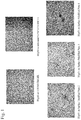
- FIG. 1 shows phase contrast micrographs (4 ⁇ ) of cells of the human ES cell line H1 that were passaged twice as clusters with LIBERASE on surface modified plates 2, 3 or 4. Images of cells of the human ES cell line H1, cultured on plates treated with a 1:30 dilution of MatrigelTM Nunclon DeltaTM plates are also shown.
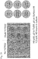
- FIG. 2 shows the effect of 10 ⁇ M Y-27632 on the attachment of human ES cells to surface modified plates.
- the figure shows phase contrast micrographs (4 ⁇ ) of cells of the human ES cell line H1 that were passaged twice as clusters on surface modified plates 3 and 4. Cells were then passaged onto surface modified plates 2, 3 or 4, in MEF conditioned medium containing 10 ⁇ M Y-27632. Cells were cultured for four days prior to taking the photographs. Cells cultured in the absence of Y-27632 were included as controls.
- FIG. 3 shows a schematic of the time-course of treatment of compounds on human ES cells cultured on the surface modified plates of the present invention.
- Cells of the human ES cell line H1 were passaged four times as clusters with LIBERASE treatment on surface modified plates 3, or 4, and cultured in MEF conditioned medium.
- Cells were treated for the first two days after passage with either 10 ⁇ M of the Rho Kinase inhibitor, Y-27632, or with 0.5 ng/ml of the Rho inhibitor, a cell permeable form of exoenzyme C3 transferase.
- Cells that were treated with the Rho Kinase inhibitor, Y-27632 and were thereafter treated for the first two days after each passage with Y-27632 on surface modified plate 3 are referred to as “7s”.
- Cells that were treated with the Rho Kinase inhibitor, Y-27632 and were thereafter treated for the first two days after each passage with Y-27632 on surface modified plate 4 are referred to as “3s”.
- Cells that were treated with the Rho inhibitor for two days and were then treated with the Rho Kinase inhibitor, Y-27632 for two days after each passage and thereafter treated for the first two days after passage with Y-27632 on surface modified plate 3 are referred to as “5s”.
- Is Cells that were treated with the Rho inhibitor for two days and were then treated with the Rho kinase inhibitor, Y-27632 for two days after each passage and thereafter treated for the first two days after passage with Y-27632 on surface modified plate 4 are referred to as “Is”.
- FIG. 4 shows the expression of markers associated with pluripotency and differentiation in human ES cells treated according to the protocol outlined in FIG. 6 as determined by qRT-PCR.
- FIG. 5 shows the expression of pluripotency markers in cells of the human ES cell line H1 as determined by flow cytometry at passage 4 (p4), passage 9 (p9), and again at passage 10, 11, or 12 (p10, p11, or p12).
- FIG. 6 shows immuno-fluorescent images of cells of the human ES cell line H1 were passaged serially as clusters with LIBERASE treatment on surface modified plate 4, and cultured in MEF conditioned medium. Expression of proteins associated with markers of pluripotency was detected in cells cultured for 11 passages on surface modified plate 4. Cells were treated with 10 ⁇ M Y-27632 for two days after each passage.
- FIG. 7 shows the ability for human ES cells to form definitive endoderm after culture on surface modified plates.
- Cells of the human ES cell line H1 were passaged 11 times as clusters with LIBERASE treatment on surface modified plates 3, or 4 and cultured in MEF conditioned medium.
- passage 8 and again at passage 10 or 11 (p10-11) cells were treated with DMEM:F12 media containing 0.5% FBS, 100 ng/ml Activin A, and 20 ng/ml Wnt3a for two days and then treated with DMEM:F12 media containing 2% FBS and 100 ng/ml Activin A for three more days.
- the y-axis on the graph shows the percent positive CXCR4 cells obtained by flow cytometry. See also Table 5.
- FIG. 8 shows the ability for human ES cells to form pancreatic endoderm after culture on surface modified plates.
- Cells of the human ES cell line H1 were passaged eight times as clusters with LIBERASE treatment on surface modified plates 3, or 4 and cultured in MEF conditioned medium.
- passage 8 (p8) cells were subjected to differentiation to definitive endoderm by treatment with DMEM:F12 media containing 0.5% FBS, 100 ng/ml Activin A, and 20 ng/ml Wnt3a for two days and then treated with DMEM:F12 media containing 2% FBS and 100 ng/ml Activin A for three more days.
- the cells were then further differentiated to embryonic foregut with four days of treatment with DMEM:F12 media containing 2% FBS, 100 ng/ml FGF-10, and 1 ⁇ M cyclopamine-KAAD.
- the cells were then differentiated to pancreatic endoderm with four days of treatment with DMEM:F12 media containing 1% B-27, 100 ng/ml FGF-10, 1 ⁇ M cyclopamine-KAAD and 2 ⁇ M retinoic acid.
- Cells were stained by immunofluorescence for PDX-1 (green) and E-cadherin (red) and total cell number was identified by Hoechst dye (blue).
- FIG. 9 shows the ability of human ES cells cultured on surface modified plates to form embryoid bodies.
- FIG. 10 shows the karyotype of human ES cells cultured on surface modified plate 4.
- FIG. 11 shows the effect of treatment with Rho kinase inhibitors (Y-27632 from EMD biosciences, Y-27632 from Sigma, Fasudil, and Hydroxyfasudil) on the attachment of human ES cells to surface modified plates.
- Rho kinase inhibitors Y-27632 from EMD biosciences, Y-27632 from Sigma, Fasudil, and Hydroxyfasudil
- Cells were cultured in medium containing the indicated compounds, at the concentrations listed, for three days. Cells were stained with crystal violet and images taken.
- FIG. 12 shows the dose-response of Y-27632 on the attachment of human ES cells to surface modified plates.
- Various concentrations of the Rho kinase inhibitor, Y-27632 was added to the cultures at a specified concentration (0, 1, 2, 4, or 10 ⁇ M Y-27632) for the first day.
- the cells were then maintained from day 2 onward in media containing 10 ⁇ M Y-27632 with daily media changes for five days. Media was removed from the plates on day five and the cells were stained with 0.5% crystal violet, and images taken.
- FIG. 13 shows the formation of human ES cell colonies four days after passage onto surface modified plates 2, 3, or 4 with or without 10 of ⁇ M Y-27632.
- FIG. 14 shows the formation of human ES cell colonies four days after passage onto MatrigelTM treated plates with or without 10 ⁇ M Y-27632.
- FIG. 15 shows the difference between continual and intermittent treatment of human ES cells with Y-27632, on attachment of cells to surface modified plates.
- FIGS. 16A and 16B depicts images of cells from the human ES cell line H9, that were passaged as single cells, seeded on to surface modified plate 3 in MEF conditioned media containing ( FIG. 16B ) or without ( FIG. 16A ) 10 ⁇ M Y-27632. The images were taken 24 hours after seeding.
- FIG. 17 depicts the expression of markers associated with pluripotency in cells from the human ES cell line H9, that were passaged as single cells for 5 passages, using TrypLETM Express, and plated onto surface modified plates 3 and 4, with or without 10 ⁇ M of Y-27632 (Y).
- the pluripotency markers are listed on the x-axis and the percentage of positive cells is shown on the y-axis.
- FIGS. 18A and 18B depict the total cell number of cells from the human ES cell line H9, that were passaged as single cells, plated onto surface modified plates 3 and 4, respectively.
- the effect of 10 ⁇ M of Y-27632 (Y) on cell number was examined on cells passaged on MatrigelTM (na ⁇ ve, N), and cells passaged 10 times on the surface modified plates (acclimated, A).
- the different cell conditions are listed on the x-axis and the number of cells divided by 10 4 is shown in the y-axis.
- FIG. 19 depicts the rate of growth of cells from the human ES cell line H9, that were passaged as single cells on MatrigelTM treated plates prior to the study.
- Cells were seeded at 10 4 /cm 2 and cultured in MEF conditioned media with or without 10 ⁇ M of Y-27632 on surface modified plates 3 and 4.
- the y-axis shows the number of cells collected 2, 3 or 4 days after seeding (divided by 10 4 ).
- FIG. 20 depicts the rate of growth of cells from the human ES cell line H9, that were passaged as single cells for 10 passages on surface modified plates prior to the study.
- Cells were seeded at 10 4 /cm 2 and cultured in MEF conditioned media with or without 10 ⁇ M of Y-27632 on surface modified plates 3 and 4.
- the y-axis shows the number of cells collected 2, 3 or 4 days after seeding (divided by 10 4 ).
- FIG. 21 depicts images of cells from the human ES cell line H9, that were passaged as single cells, seeded on to surface modified plates 2-4 and 13 in a 96-well format.
- the MEF conditioned media contained 10 ⁇ M of Y-27632. Images were taken 48 hours after seeding.
- FIG. 22 shows the ability of cells from the human ES cell line H9, that were passaged as single cells, seeded on to surface modified plates 3 and 4 to differentiate into definitive endoderm.
- the extent of formation of definitive endoderm was determined by measuring CXCR expression by flow cytometry.
- the effect of 10 ⁇ M Y-27632 on the formation of definitive endoderm was investigated. Cells were treated with Y-27632 during expansion. Cells expanded and differentiated on MatrigelTM were included as a control.
- the y-axis shows percent positive CXCR4 cells obtained by flow cytometry.
- FIG. 23 shows the ability of cells from the human ES cell line H9, that were passaged as single cells, seeded on to surface modified plates 3 and 4 to differentiate into pancreatic endoderm.
- Cells were plated onto the surface modified plates and cultured in MEF conditioned medium containing 10 ⁇ M Y-27632, and passaged 8 times on the surface modified plates prior to differentiation.
- the y-axis shows the fold increase of pancreatic differentiation marker expression (Ngn3, Pdx1, Insulin) by q-PCR at the posterior foregut stage (PF) and the hormone expressing endocrine cell stage (EN).
- FIG. 24 shows the attachment of human ES cells to surface modified plates. Passage 50 H9 human ES cells were plated at a 1:2 dilution on Surfaces 3 and 4, CellBINDTM, and PrimariaTM. Media was removed from the plates 24 hours after plating and the cells were stained with 0.5% crystal violet, and images taken. Arrows indicate colonies.
- FIG. 25 shows the attachment of human ES cells to surface modified plates. Passage 50 H9 human ES cells were plated at a 1:2 dilution on Surfaces 3 and 4, CellBINDTM, and PrimariaTM in the presence of various concentrations of Y-27632 (0, 1, 2, 4, 10 and 20 micromolar). Media was removed from the plates 24 hours after plating and the cells were stained with 0.5% crystal violet, and images taken. Colonies are dark spots on the well. Arrows are used to highlight colonies on the untreated wells.
- FIG. 26 shows the attachment of human ES cells to surface modified plates. Passage 53 H9 human ES cells were plated at a 1:3 dilution on Surfaces 2-4 and 13, CellBINDTM, and PrimariaTM in the absence or presence of Y-27632 (0 or 20 micromolar). Media was removed from the plates 48 hours after plating and the cells were stained with 0.5% crystal violet, and images taken. Colonies are dark spots on the well. Arrows are used to highlight colonies on the untreated wells.
- FIG. 27 shows the first attempt (October) and second attempt (December) to attach human H9 ES cells to surface modified plates 14 and 15 and an attempt to attach human H1 ES cells to surface modified plates 14 and 15.
- Passage 42 and passage 53 H9 human ES, and passage 57 H1 human ES cells were plated at a 1:2 or 1:3 dilution to the modified surfaces in the presence of 20 micromolar Y-27632.
- Media was removed from the plates 24-48 hours after plating and the cells were stained with 0.5% crystal violet, and images taken. Colonies are dark spots on the well. Arrows are used to highlight colonies on the plates.
- FIG. 28 shows the attachment of human ES cells to a surface modified plate 4 in defined media, mTeSRTM.
- Passage 50 H9 human ES cells were plated at a 1:2 dilution to the modified surfaces in the absence or presence of Y-27632 (0 or 20 micromolar) in wells that were untreated or treated with proteins (0.1% gelatin, 2% BSA, 0.34 mg/ml rat Collagen I, 1:1000 diluted MatrigelTM, or 1:5000 diluted MatrigelTM).
- Media was removed from the plates 48 hours after plating and the cells were stained with 0.5% crystal violet, and images taken. Colonies are dark spots on the well.
- FIG. 29 shows the water contact angles of surface modified plates measured over 11 weeks using the static sessile drop method.
- the first measurement was done one week after surface treatment and sterilization. Each data point represents the mean contact angle (one measurement on each of 7 drops).
- the contact angles on Nuclon DeltaTM and CellBINDTM plates were measured under the same experimental conditions as Surfaces 1-4 and 13, but the surface treatment and sterilization was done more than 12 weeks before the first measurement (Nuclon DeltaTM* was sterilized one week before the first measurement).
- FIG. 30 shows the density of negative charges on surface modified plates measured as reactivity of surfaces with positively charged crystal violet. Three samples of each surface were tested, and absorbance measurements on desorbed crystal violet from each sample were performed in triplicate. Mean and standard deviation of nine measurements are given.
- FIGS. 31A and 31B shows the effect of solid substrate surfaces and Y-27632 on attachment and growth of HEK293 cells in chemically defined, serum-free Pro293a-CDMTM medium ( FIG. 31A ) or EMEM medium supplemented with 10% fetal bovine serum ( FIG. 31B ).
- HEK293 cells were seeded in 96-well plates with CellBINDTM surface, Nunclon DeltaTM surface or Surface 4. The number of HEK293 cells attached to these surfaces is shown as a function of culture conditions and concentration of Y-27632.
- HEK293 cells cultured without Y-27632 in the medium were handled the same way as cells cultured with Y-27632, that is, for either 96-hours without a change of medium, or with a change of medium after 48 hours.
- Y-27632 enhanced attachment of HEK293 cells on Surface 4 and the CellBINDTM surface when applied at concentrations of 2.0 and 5.0 ⁇ M. Removing Y-27632 after 48 hours of incubation resulted in detachment of a significant number of cells from Surface 4 and the CellBINDTM surface. Mean and standard deviation of three measurements are shown.
- FIGS. 32A and 32B show the effect of solid substrate surfaces and Rho kinase inhibitors Y-27632 and H-1152 on growth of HEK293 cells in EMEM medium supplemented with 10% fetal bovine serum.
- HEK293 cells were seeded in Multidish 24-well plates with either Surface 4 (A) or a non-treated (but gamma irradiated; 25 kGy) polystyrene surface (B).
- FIGS. 33A and 33B show the effect of H-1152 and Surface 4 on HEK293-cell attachment and morphology.
- HEK293 cells were seeded in Multidish 12-well plates in EMEM medium supplemented with 10% fetal bovine serum and H-1152, and incubated for 67 hours in an automated, in-incubator microscope.
- Growth curves in FIG. 33A and photomicrographs in FIG. 33B show the general effect of H-1152 on HEK293-cell attachment and growth on surface 4, and the effect of a change of medium on HEK293-cell attachment and morphology on surface 4 in the presence or absence of H-1152.
- FIG. 34 shows growth curves over 3 passages for HEK293 cells grown on surface 4 and Nunclon DeltaTM surface in the absence or presence of 2.5 ⁇ M Y-27632.
- HEK293 cells in EMEM medium supplemented with 10% fetal bovine serum were passaged 3 times by trypsinization.
- FIG. 35 shows the effect of Rho kinase inhibition on the attachment of cells of the human embryonic stem cell line H1 to surface modified plates 4, 18 and 19, and PrimariaTM.
- Wells A&B were control wells on all surfaces.
- Wells C&D contained 10 ⁇ M Y-27632.
- Wells E&F contained 3 ⁇ M H1152-glycyl.
- Wells G&H contained 10 ⁇ M H1152-glycyl.
- FIG. 36 shows the effect of Rho kinase inhibition on the attachment of cells of the human embryonic stem cell line H1 to surface modified plate 30.
- (- -) no treatment.
- (RI) 3 ⁇ M H1152-glycyl.
- (MG) adlayer of 1:30 dilution of MATRIGEL.
- (MG+RI) adlayer of 1:30 dilution of MATRIGEL+3 ⁇ M H1152-glycyl.
- FIG. 37 shows the effect of Rho kinase inhibition on the attachment of cells of the human embryonic stem cell line H1 to surface modified plate 31.
- (- -) no treatment.
- (RI) 3 ⁇ M H1152-glycyl.
- (MG) adlayer of 1:30 dilution of MATRIGEL.
- (MG+RI) adlayer of 1:30 dilution of MATRIGEL+3 ⁇ M H1152-glycyl.
- FIG. 38 shows the effect of Rho kinase inhibition on the attachment of cells of the human embryonic stem cell line H1 to surface modified plate 32.
- (- -) no treatment.
- (RI) 3 ⁇ M H1152-glycyl.
- (MG) adlayer of 1:30 dilution of MATRIGEL.
- (MG+RI) adlayer of 1:30 dilution of MATRIGEL+3 ⁇ M H1152-glycyl.
- FIG. 39 shows the effect of Rho kinase inhibition on the attachment of cells of the human embryonic stem cell line H1 to surface modified plate 33.
- (- -) no treatment.
- (RI) 3 ⁇ M H1152-glycyl.
- (MG) adlayer of 1:30 dilution of MATRIGEL.
- (MG+RI) adlayer of 1:30 dilution of MATRIGEL+3 ⁇ M H1152-glycyl.
- FIG. 40 shows the effect of Rho kinase inhibition on the attachment of cells of the human embryonic stem cell line H1 to surface modified plate 34.
- (- -) no treatment.
- (RI) 3 ⁇ M H1152-glycyl.
- (MG) adlayer of 1:30 dilution of MATRIGEL.
- (MG+RI) adlayer of 1:30 dilution of MATRIGEL+3 ⁇ M H1152-glycyl.
- FIG. 41 shows the water contact angles of surface modified plates measured over 40 weeks using the static sessile drop method.
- FIG. 42 shows the water contact angles of surface modified plates using the static sessile drop method.
- FIG. 43 shows the density of negative charges on surface modified plates measured as reactivity of surfaces with positively charged crystal violet.
- FIG. 44 shows the density of negative charges on surface modified plates 4, 22-24 and 29 measured as reactivity of surfaces with positively charged crystal violet. Three samples of each surface were tested, and absorbance measurements on desorbed crystal violet from each sample were performed in triplicate. The negative charge density for surfaces 4, 22-24 and 29 was normalized to the negative charge density of the Nunclon DeltaTM surface. Mean and standard deviation of nine measurements are given.
- Adlayer refers to a layer that is formed on a surface of a solid substrate, by attaching molecules to the surface by either covalent (also known as grafting) or non-covalent (also known as adsorption) bonds.
- Molecules used in making an adlayer can, for example, be proteinaceous molecules, which may include, for example, extracellular matrix proteins, amino acids and the like, and non-biological molecules, such as, for example, polyethyleneimine.
- ⁇ -cell lineage refers to cells with positive gene expression for the transcription factor PDX-1 and at least one of the following transcription factors: NGN-3, Nkx2.2, Nkx6.1, NeuroD, Isl-1, HNF-3 beta, MAFA, Pax4, and Pax6.
- Cells expressing markers characteristic of the 3 cell lineage include 3 cells.
- Cells expressing markers characteristic of the definitive endoderm lineage refers to cells expressing at least one of the following markers: SOX-17, GATA-4, HNF-3 beta, GSC, Cerl, Nodal, FGF-8, Brachyury, Mix-like homeobox protein, FGF-4 CD48, eomesodermin (EOMES), DKK4, FGF-17, GATA-6, CXCR4, C-Kit, CD99, or OTX2.
- Cells expressing markers characteristic of the definitive endoderm lineage include primitive streak precursor cells, primitive streak cells, mesendoderm cells and definitive endoderm cells.
- Cells expressing markers characteristic of the pancreatic endoderm lineage refers to cells expressing at least one of the following markers: PDX-1, HNF-1beta, PTF-1 alpha, HNF-6, or HB9.
- Cells expressing markers characteristic of the pancreatic endoderm lineage include pancreatic endoderm cells.
- Cells expressing markers characteristic of the pancreatic endocrine lineage refers to cells expressing at least one of the following markers: NGN-3, NeuroD, Islet-1, PDX-1, NKX6.1, Pax-4, Ngn-3, or PTF-1 alpha.
- Cells expressing markers characteristic of the pancreatic endocrine lineage include pancreatic endocrine cells, pancreatic hormone expressing cells, and pancreatic hormone secreting cells, and cells of the 3-cell lineage.
- Definitive endoderm refers to cells which bear the characteristics of cells arising from the epiblast during gastrulation and which form the gastrointestinal tract and its derivatives. Definitive endoderm cells express the following markers: CXCR4, HNF-3 beta, GATA-4, SOX-17, Cerberus, OTX2, goosecoid, c-Kit, CD99, and Mixl1.
- Extracellular matrix proteins refers to proteinaceous molecules normally found between cells in the body or in the placenta. Extracellular matrix proteins can be derived from tissue, body fluids, such as, for example, blood, or media conditioned by non-recombinant cells or recombinant cells or bacteria.
- Extraembryonic endoderm refers to a population of cells expressing at least one of the following markers: SOX-7, AFP, and SPARC.
- HEK293 cells refers to a cell line generated by transformation of a culture of normal human embryonic kidney cells as described by Graham et al. (J. Gen. Virol. 36:59-72, 1977), and any cells derived from this parent cell line.
- Markers are nucleic acid or polypeptide molecules that are differentially expressed in a cell of interest.
- differential expression means an increased level for a positive marker and a decreased level for a negative marker.
- the detectable level of the marker nucleic acid or polypeptide is sufficiently higher or lower in the cells of interest compared to other cells, such that the cell of interest can be identified and distinguished from other cells using any of a variety of methods known in the art.
- Microx refers to a 3-dimensional support to which cells may attach.
- Mesendoderm cell refers to a cell expressing at least one of the following markers: CD48, eomesodermin (EOMES), SOX-17, DKK4, HNF-3 beta, GSC, FGF-17, GATA-6.
- EOMES eomesodermin
- SOX-17 SOX-17
- DKK4 HNF-3 beta
- GSC GSC
- FGF-17 GATA-6.
- Pantendocrine cell or “pancreatic hormone expressing cell” as used herein refers to a cell capable of expressing at least one of the following hormones: insulin, glucagon, somatostatin, and pancreatic polypeptide.
- Pantix hormone secreting cell refers to a cell capable of secreting at least one of the following hormones: insulin, glucagon, somatostatin, and pancreatic polypeptide.
- Pre-primitive streak cell refers to a cell expressing at least one of the following markers: Nodal, or FGF-8.
- Primary streak cell refers to a cell expressing at least one of the following markers: Brachyury, Mix-like homeobox protein, or FGF-4.
- “Surface” as used herein refers to the outermost layer of molecules of a solid substrate vessel or matrix intended for use in cell culture or analysis.
- the elemental composition, the roughness, and the wettability of the surface can be analyzed by X-Ray Photoelectron Spectroscopy (XPS), Atomic Force Microscopy (AFM), and contact angle measurement, respectively.
- “Surface modified plate” refers to a vessel containing any one of surfaces 1-34, described in Examples 16, 17 and 26, or plates containing surfaces that are sold under the trade names Nunclon DeltaTM, CostarTM, FalconTM, CellBINDTM, and PrimariaTM.
- the vessel can, for example, be made of a polymer, such as polystyrene (PS), cyclic olefin copolymer (COC), polycarbonate (PC), polymethyl methacrylate (PMMA), or styrene acrylonitrile copolymer (SAN).
- PS polystyrene
- COC cyclic olefin copolymer
- PC polycarbonate
- PMMA polymethyl methacrylate
- SAN styrene acrylonitrile copolymer
- Stem cells are undifferentiated cells defined by their ability at the single cell level to both self-renew and differentiate to produce progeny cells, including self-renewing progenitors, non-renewing progenitors, and terminally differentiated cells. Stem cells are also characterized by their ability to differentiate in vitro into functional cells of various cell lineages from multiple germ layers (endoderm, mesoderm and ectoderm), as well as to give rise to tissues of multiple germ layers following transplantation and to contribute substantially to most, if not all, tissues following injection into blastocysts.
- Stem cells are classified by their developmental potential as: (i) totipotent, meaning able to give rise to all embryonic and extraembryonic cell types; (ii) pluripotent, meaning able to give rise to all embryonic cell types; (iii) multipotent, meaning able to give rise to a subset of cell lineages, but all within a particular tissue, organ, or physiological system (for example, hematopoietic stem cells (HSC) can produce progeny that include HSC (self-renewal), blood cell restricted oligopotent progenitors and all cell types and elements (e.g., platelets) that are normal components of the blood); (iv) oligopotent, meaning able to give rise to a more restricted subset of cell lineages than multipotent stem cells; and (v) unipotent, meaning able to give rise to a single cell lineage (e.g., spermatogenic stem cells).
- HSC hematopoietic stem cells
- Differentiation is the process by which an unspecialized (“uncommitted”) or less specialized cell acquires the features of a specialized cell such as, for example, a nerve cell or a muscle cell.
- a differentiated or differentiation-induced cell is one that has taken on a more specialized (“committed”) position within the lineage of a cell.
- the term committed when applied to the process of differentiation, refers to a cell that has proceeded in the differentiation pathway to a point where, under normal circumstances, it will continue to differentiate into a specific cell type or subset of cell types, and cannot, under normal circumstances, differentiate into a different cell type or revert to a less differentiated cell type.
- Dedifferentiation refers to the process by which a cell reverts to a less specialized (or committed) position within the lineage of a cell.
- the lineage of a cell defines the heredity of the cell, that is, which cells it came from and what cells it can give rise to.
- the lineage of a cell places the cell within a hereditary scheme of development and differentiation.
- a lineage-specific marker refers to a characteristic specifically associated with the phenotype of cells of a lineage of interest and can be used to assess the differentiation of an uncommitted cell to the lineage of interest.
- Maintenance refers generally to cells placed in a growth medium under conditions that facilitate cell growth and/or division that may or may not result in a larger population of the cells.
- Passaging refers to the process of removing the cells from one culture vessel and placing them in a second culture vessel under conditions that facilitate cell growth and/or division.
- a specific population of cells, or a cell line is sometimes referred to or characterized by the number of times it has been passaged.
- a cultured cell population that has been passaged ten times may be referred to as a P10 culture.
- the primary culture that is, the first culture following the isolation of cells from tissue, is designated P0.
- the cells are described as a secondary culture (P1 or passage 1).
- P2 or passage 2 After the second subculture, the cells become a tertiary culture (P2 or passage 2), and so on.
- P1 or passage 1 the primary culture following the isolation of cells from tissue
- P1 or passage 1 the cells are described as a secondary culture (P1 or passage 1).
- P2 or passage 2 tertiary culture
- the expansion of cells (that is, the number of population doublings) during the period between passaging depends on many factors, including but not limited to the seeding density, substrate, medium, growth conditions, and time between passaging.
- the present invention provides a method to enhance the attachment of cells to a surface containing from at least about 0.9% N, a sum of O and N of greater than or equal to 22.3% and a contact angle of at least about 13.9 degrees, lacking a feeder cell layer and lacking an adlayer, comprising the steps of:
- the present invention provides a method to enhance the attachment of cells to a surface containing from at least about 0.5% N, a sum of O and N of greater than or equal to 17.2% and a contact angle of at least about 13.9 degrees, lacking a feeder cell layer and lacking an adlayer, comprising the steps of:
- the suspension of cells is a suspension of clusters of cells. In an alternate embodiment, the suspension of cells is a suspension of single cells.
- the cells are pluripotent stem cells. In an alternate embodiment, the cells are stem cells.
- the surface has an adlayer.
- the adlayer is an extracellular matrix component, such as, for example, those derived from basement membrane or that may form part of adhesion molecule receptor-ligand couplings.
- the adlayer is made from MATRIGEL (Becton Dickenson).
- MATRIGEL is a soluble preparation from Engelbreth-Holm Swarm tumor cells that gels at room temperature to form a reconstituted basement membrane.
- the proteinaceous adlayer may also be formed from laminin, fibronectin, proteoglycan, entactin, heparan sulfate, and the like, alone or in various combinations.
- the cells are maintained in culture after the cells attach to the surface.
- the at least one compound is removed after the cells attach to the surface.
- the cells are detached from the surface by removing the at least one compound.
- the suspension of cells is treated with at least one compound capable of inhibiting Rho kinase activity. In an alternate embodiment, the suspension of cells is treated with at least one compound capable of inhibiting Rho activity. In an alternate embodiment, the suspension of cells is treated with at least one compound capable of inhibiting Rho kinase activity and at least one compound capable of inhibiting Rho activity.
- the at least one compound capable of inhibiting Rho kinase activity is selected from the group consisting of: Y-27632, Fasudil, and Hydroxyfasudil.
- the at least compound capable of inhibiting Rho kinase activity is Y-27632.
- the at least one compound capable of inhibiting Rho kinase activity may be used at a concentration from about 0.1 ⁇ M to about 100 ⁇ M. In one embodiment, the at least one compound capable of inhibiting Rho kinase activity is used at a concentration of about 10 ⁇ M.
- the at least one compound capable of inhibiting Rho activity is a Rho GTPase inhibitor.
- the at least one compound capable of inhibiting Rho activity is exoenzyme C3 Transferase.
- the at least one compound capable of inhibiting Rho activity may be used at a concentration from about 0.01 ⁇ g/ml to about 5 ⁇ g/ml. In one embodiment, the at least one compound capable of inhibiting Rho activity is used at a concentration of about 0.5 g/ml.
- Surface modified plates suitable for use in the present invention may be vessels whose surfaces have been modified to contain from at least about 0.5% N, a sum of O and N of greater than or equal to 17.2% and a contact angle of at least about 13.9 degrees.
- the surface may be a 3-dimensional matrix, such as, for example, a porous scaffold, to which cells can attach.
- the surface modified plate comprises a plate whose surface contains from at least about 0.5% N, a sum of O and N of greater than or equal to 17.2% and a contact angle of at least about 13.9 degrees. In an alternate embodiment, the surface modified plate comprises a plate whose surface contains from at least about 0.5% N, a sum of O and N of greater than or equal to 19.5% and a contact angle of at least about 13.9 degrees.
- the surface modified plate comprises a plate whose surface contains from at least about 1.3% N, a sum of O and N of at least about 24.9%0 and a contact angle of at least about 20.7 degrees, which is referred herein as surface modified plate 1.
- the surface modified plate comprises a plate whose surface contains from at least about 1.7% N, a sum of O and N of at least about 29.6% and a contact angle of at least about 14.3 degrees, which is referred herein as surface modified plate 2.
- the surface modified plate comprises a plate whose surface contains from at least about 2.0% N, a sum of O and N of at least about 30.7% and a contact angle of at least about 18.4 degrees, which is referred herein as surface modified plate 3.
- the surface modified plate comprises a plate whose surface contains from at least about 2.1% N, a sum of O and N of at least about 30.2% and a contact angle of at least about 17.4 degrees, which is referred herein as surface modified plate 4.
- the surface modified plate comprises a plate whose surface contains from at least about 1.8% N, a sum of O and N of at least about 28.2% and a contact angle of at least about 18.8 degrees, which is referred herein as surface modified plate 13.
- the surface modified plate comprises a plate whose surface contains from at least about 1.0% N, a sum of O and N of at least about 27.8% and a contact angle of at least about 44.3 degrees, which is sold under the trade name CELLBIND.
- the surface modified plate comprises a plate whose surface contains from at least about 10.2% N, a sum of O and N of at least about 23.0% and a contact angle of at least about 39.5 degrees, which is sold under the trade name PRIMARIA.
- the elemental composition of the surface of the surface modified plates may be analyzed by X-Ray Photoelectron Spectroscopy (XPS).
- XPS also known as Electron Spectroscopy for Chemical Analysis (ESCA)
- ESA Electron Spectroscopy for Chemical Analysis
- an XPS analysis of a polystyrene (contains only carbon and hydrogen) solid sample would typically give greater than 97% carbon, less than 3% oxygen, and 0% nitrogen (hydrogen is not detected; different levels of oxygen may be detected due to oxidation of the polystyrene chains at the surface, for example, as a result of sterilization by irradiation) (Brevig et al., Biomaterials 26:3039-3053, 2005; Shen and Horbett, J. Biomed. Mater. Res. 57:336-345, 2001).
- the roughness of the surface of the surface modified plates may be analyzed by Atomic Force Microscopy (AFM).
- AFM Atomic Force Microscopy
- the wettability of the surface of the surface modified plates may be analyzed by measuring the contact angle.
- contact angle measurement by the static sessile drop method provides information on the interaction between the surface of a solid substrate and a liquid.
- the contact angle describes the shape of a liquid drop resting on the surface of the solid substrate, and is the angle of contact of the liquid on the surface of the solid substrate, measured within the liquid at the contact line where liquid, solid, and gas meet.
- a surface with a water contact angle larger than 90° is termed hydrophobic, and a surface with water contact angle less than 90° is termed hydrophilic.
- hydrophilic On extremely hydrophilic surfaces, that is, surfaces that have a high affinity for water, a water droplet will completely spread (an effective contact angle of 0°).
- the negative charge density of the surface of the surface modified plates may be analyzed by measuring the reactivity of the surface with crystal violet.
- Crystal violet carries a positive charge, which enables it to bind to negatively charged molecules and parts of molecules, for example, negatively charged functional groups present on a polymer surface.
- a surface with a high crystal violet reactivity has a higher density of negative charges than a surface with a low crystal violet reactivity, given that the surfaces have the same roughness and thus area.
- Pluripotent stem cells may express one or more of the stage-specific embryonic antigens (SSEA) 3 and 4, and markers detectable using antibodies designated Tra-1-60 and Tra-1-81 (Thomson et al., Science 282:1145 1998). Differentiation of pluripotent stem cells in vitro results in the loss of SSEA-4, Tra-1-60, and Tra-1-81 expression (if present) and increased expression of SSEA-1. Undifferentiated pluripotent stem cells typically have alkaline phosphatase activity, which can be detected by fixing the cells with 4% paraformaldehyde and then developing with Vector Red as a substrate, as described by the manufacturer (Vector Laboratories, Burlingame Calif.). Undifferentiated pluripotent stem cells also typically express Oct-4 and TERT, as detected by RT-PCR.
- SSEA stage-specific embryonic antigens
- pluripotent stem cells Another desirable phenotype of propagated pluripotent stem cells is a potential to differentiate into cells of all three germinal layers: endoderm, mesoderm, and ectoderm tissues. Pluripotency of stem cells can be confirmed, for example, by injecting cells into severe combined immunodeficient (SCID) mice, fixing the teratomas that form using 4% paraformaldehyde, and then examining them histologically for evidence of cell types from the three germ layers. Alternatively, pluripotency may be determined by the creation of embryoid bodies and assessing the embryoid bodies for the presence of markers associated with the three germinal layers.
- SCID severe combined immunodeficient
- Propagated pluripotent stem cell lines may be karyotyped using a standard G-banding technique and compared to published karyotypes of the corresponding primate species. It is desirable to obtain cells that have a “normal karyotype,” which means that the cells are euploid, wherein all human chromosomes are present and not noticeably altered.
- pluripotent stem cells include established lines of pluripotent cells derived from tissue formed after gestation, including pre-embryonic tissue (such as, for example, a blastocyst), embryonic tissue, or fetal tissue taken any time during gestation, typically but not necessarily before approximately 10-12 weeks gestation.
- pre-embryonic tissue such as, for example, a blastocyst
- embryonic tissue or fetal tissue taken any time during gestation, typically but not necessarily before approximately 10-12 weeks gestation.
- Non-limiting examples are established lines of human ES cells or human embryonic germ cells, such as, for example the human ES cell lines H1, H7, and H9 (WiCell).
- H1, H7, and H9 WiCell
- Also contemplated is use of the compositions of this disclosure during the initial establishment or stabilization of such cells, in which case the source cells would be primary pluripotent cells taken directly from the source tissues.
- cells taken from a pluripotent stem cell population already cultured in the absence of feeder cells as well as a pluripotent stem cell population already cultured in the presence of feeder cells.
- mutant human ES cell lines such as, for example, BG01v (BresaGen, Athens, Ga.).
- cells derived from adult human somatic cells such as, for examples, cells disclosed in Takahashi et al, Cell 131: 1-12 (2007).
- human ES cells are prepared as described by Thomson et al. (U.S. Pat. No. 5,843,780; Science 282:1145, 1998; Curr. Top. Dev. Biol. 38:133 ff., 1998; Proc. Natl. Acad. Sci. U.S.A. 92:7844, 1995).
- pluripotent stem cells are cultured on a layer of feeder cells or extracellular matrix protein that support the pluripotent stem cells in various ways, prior to culturing according to the methods of the present invention.
- pluripotent stem cells are cultured on a feeder cell layer that supports proliferation of pluripotent stem cells without undergoing substantial differentiation.
- the growth of pluripotent stem cells on a feeder cell layer without differentiation is supported using (i) Obtaining a culture vessel containing a feeder cell layer; and (ii) a medium conditioned by culturing previously with another cell type, or a non-conditioned medium, for example, free of serum or even chemically defined.
- pluripotent stem cells are cultured in a culture system that is essentially free of feeder cells, but nonetheless supports proliferation of pluripotent stem cells without undergoing substantial differentiation.
- the growth of pluripotent stem cells in feeder-cell free culture without differentiation is supported using (i) an adlayer on a solid substrate surface with one or more extracellular matrix proteins; and (ii) a medium conditioned by culturing previously with another cell type, or a non-conditioned medium, for example, free of serum or even chemically defined.
- pluripotent stem cells are cultured on a surface modified plate containing from at least about 0.5% N, a sum of O and N of greater than or equal to 17.2% and a contact angle of at least about 13.9 degrees in a medium conditioned by culturing previously with another cell type, or a non-conditioned medium, for example, free of serum or even chemically defined.
- cell culture medium suitable for use in the present invention may be found in US20020072117.
- Another example of cell culture medium suitable for use in the present invention may be found in U.S. Pat. No. 6,642,048.
- Another example of cell culture medium suitable for use in the present invention may be found in WO2005014799.
- Another example of cell culture medium suitable for use in the present invention may be found in Xu et al. (Stem Cells 22: 972-980, 2004).
- Another example of cell culture medium suitable for use in the present invention may be found in US20070010011.
- Another example of cell culture medium suitable for use in the present invention may be found in Cheon et al. (BioReprod DOI:10.1095/biolreprod. 105.046870; 19 Oct.
- cell culture medium suitable for use in the present invention may be found in Levenstein et al. (Stem Cells 24: 568-574, 2006). Another example of cell culture medium suitable for use in the present invention may be found in US20050148070. Another example of cell culture medium suitable for use in the present invention may be found in US20050233446. Another example of cell culture medium suitable for use in the present invention may be found in U.S. Pat. No. 6,800,480. Another example of cell culture medium suitable for use in the present invention may be found in US20050244962. Another example of cell culture medium suitable for use in the present invention may be found in WO2005065354. Another example of cell culture medium suitable for use in the present invention may be found in WO2005086845.
- Suitable culture media may also be made from the following components, such as, for example, Dulbecco's modified Eagle's medium (DMEM), Gibco #11965-092; Knockout Dulbecco's modified Eagle's medium (KO DMEM), Gibco #10829-018; Ham's F12/50% DMEM basal medium; 200 mM L-glutamine, Gibco #15039-027; non-essential amino acid solution, Gibco 11140-050; ⁇ -mercaptoethanol, Sigma #M7522; human recombinant basic fibroblast growth factor (bFGF), Gibco #13256-029.
- DMEM Dulbecco's modified Eagle's medium
- KO DMEM Knockout Dulbecco's modified Eagle's medium
- Ham's F12/50% DMEM basal medium 200 mM L-glutamine, Gibco #15039-027; non-essential amino acid solution, Gibco 11140-050; ⁇ -mercaptoethanol,
- pluripotent stem cells are propagated in culture, while maintaining their pluripotency. Changes in pluripotency of the cells with time can be determined by detecting changes in the levels of expression of markers associated with pluripotency. Alternatively, changes in pluripotency can be monitored by detecting changes in the levels of expression of markers associated with differentiation or markers associated with another cell type.
- pluripotent stem cells are propagated in culture and then treated in a manner that promotes their differentiation into another cell type.
- the other cell type may be a cell expressing markers characteristic of the definitive endoderm lineage.
- the cell type may be a cell expressing markers characteristic of the pancreatic endoderm lineage.
- the cell type may be a cell expressing markers characteristic of the pancreatic endocrine lineage.
- the cell type may be a cell expressing markers characteristic of the 3-cell lineage.
- Pluripotent stem cells treated in accordance with the methods of the present invention may be differentiated into a variety of other cell types by any suitable method in the art.
- pluripotent stem cells treated in accordance with the methods of the present invention may be differentiated into neural cells, cardiac cells, hepatocytes, and the like.
- pluripotent stem cells treated in accordance with the methods of the present invention may be differentiated into neural progenitors and cardiomyocytes according to the methods disclosed in WO2007030870.
- pluripotent stem cells treated in accordance with the methods of the present invention may be differentiated into hepatocytes according to the methods disclosed in U.S. Pat. No. 6,458,589.
- pluripotent stem cells may be differentiated into cells expressing markers characteristic of the definitive endoderm lineage according to the methods disclosed in D'Amour et al., Nature Biotechnol. 23:1534-1541, 2005.
- pluripotent stem cells may be differentiated into cells expressing markers characteristic of the definitive endoderm lineage according to the methods disclosed in Shinozaki et al., Development 131:1651-1662, 2004.
- pluripotent stem cells may be differentiated into cells expressing markers characteristic of the definitive endoderm lineage according to the methods disclosed in McLean et al., Stem Cells 25:29-38, 2007.
- pluripotent stem cells may be differentiated into cells expressing markers characteristic of the definitive endoderm lineage according to the methods disclosed in D'Amour el al., Nature Biotechnol. 24:1392-1401, 2006.
- Markers characteristic of the definitive endoderm lineage are selected from the group consisting of SOX17, GATA4, Hnf-3beta, GSC, Cerl, Nodal, FGF-8, Brachyury, Mix-like homeobox protein, FGF-4 CD48, eomesodermin (EOMES), DKK4, FGF-17, GATA6, CXCR4, C-Kit, CD99, and OTX2.
- Suitable for use in the present invention is a cell that expresses at least one of the markers characteristic of the definitive endoderm lineage.
- a cell expressing markers characteristic of the definitive endoderm lineage is a primitive streak precursor cell.
- a cell expressing markers characteristic of the definitive endoderm lineage is a mesendoderm cell.
- a cell expressing markers characteristic of the definitive endoderm lineage is a definitive endoderm cell.
- pluripotent stem cells may be differentiated into cells expressing markers characteristic of the pancreatic endoderm lineage according to the methods disclosed in D'Amour et al., Nature Biotechnol. 24:1392-1401, 2006.
- Markers characteristic of the pancreatic endoderm lineage are selected from the group consisting of Pdx1, HNF-1beta, PTF1a, HNF-6, HB9 and PROX1.
- Suitable for use in the present invention is a cell that expresses at least one of the markers characteristic of the pancreatic endoderm lineage.
- a cell expressing markers characteristic of the pancreatic endoderm lineage is a pancreatic endoderm cell.
- Pluripotent stem cells may be differentiated into cells expressing markers characteristic of the pancreatic endocrine lineage by any method in the art.
- pluripotent stem cells may be differentiated into cells expressing markers characteristic of the pancreatic endocrine lineage according to the methods disclosed in D'Amour et al., Nature Biotechnol. 24:1392-1401, 2006.
- pluripotent stem cells may be differentiated into cells expressing markers characteristic of the pancreatic endocrine lineage, by the methods disclosed in D'Amour et al., Nature Biotechnol. 24:1392-1401, 2006.
- a pancreatic endocrine cell is capable of expressing at least one of the following hormones: insulin, glucagon, somatostatin, and pancreatic polypeptide.
- Suitable for use in the present invention is a cell that expresses at least one of the markers characteristic of the pancreatic endocrine lineage.
- a cell expressing markers characteristic of the pancreatic endocrine lineage is a pancreatic endocrine cell.
- the pancreatic endocrine cell may be a pancreatic hormone-expressing cell.
- the pancreatic endocrine cell may be a pancreatic hormone-secreting cell.
- the pancreatic endocrine cell is a cell expressing markers characteristic of the ⁇ cell lineage.
- a cell expressing markers characteristic of the ⁇ cell lineage expresses Pdx1 and at least one of the following transcription factors: NGN-3, Nkx2.2, Nkx6.1, NeuroD, Isl-1, HNF-3 beta, MAFA, Pax4, and Pax6.
- a cell expressing markers characteristic of the ⁇ cell lineage is a ⁇ cell.
- the human ES cell lines H1 and H9 were initially maintained on mitomycin C inactivated primary mouse embryonic fibroblasts (MEF). The human ES cells were switched from MEF feeders to MatrigelTM (Becton-Dickinson, Bedford, Mass.) over repeated passages.
- MEF mitomycin C inactivated primary mouse embryonic fibroblasts
- Treatment of surfaces with MatrigelTM Growth Factor Reduced MatrigelTM was thawed at 4° C. and then diluted 1:30 in cold DMEM/F12 (Invitrogen, Carlsbad, Calif.). Volumes sufficient to cover the surface were added to each 6-cm dish (2 ml) or each well of a 6-well plate (1 ml), and incubated 1 hr at room temp. Treated surfaces were used within a few hours or stored at 4° C. up to two weeks.
- Human ES cell culture Undifferentiated human ES cell colonies (from either the H9 or H1 lines) were harvested from feeder layers by incubation in 1 mg/ml collagenase IV (Sigma-Aldrich, St. Louis, Mo.) in DMEM/F12 for 10 minutes, followed by scraping with a pipette. Cell clumps were pelleted by centrifugation at 600 ⁇ g for four minutes and the pellet dispersed gently with a 2-ml pipette to break colonies into small clusters of cells.
- collagenase IV Sigma-Aldrich, St. Louis, Mo.
- Human ES cells of the cell line H9 were grown as single cells according to the methods disclosed in US patent Application LFS5163USPSP, assigned to LifeScan Inc. Cells were passaged by treatment with TrypLETM Express for five minutes at 37° C., and seeded at 10,000 cells/cm 2 substrate surface.
- Human ES cells of the line H1, at passage 49 were maintained in MEF conditioned media on Nunclon DeltaTM plates treated with a 1:30 dilution of growth factor reduced MatrigelTM, prior to study. Cells were dissociated from the surface for passage by 1 mg/ml collagenase dissociation or by manual scraping.
- surface modified plates 2, 3, and 4 had attached ES cell colonies, which re-attached to the plates and grew following enzymatic dissociation. There was no apparent difference in rate of attachment or growth in gelatin or untreated wells from surface modified plates 2, 3, or 4.
- H1p49 ES cells added to surface modified plates 1 and 5-12 and to untreated or gelatin treated Nunclon DeltaTM plates, FalconTM plates, and CostarTM plates did not attach. The same cells did attach to plates treated with 1:30 dilution of growth factor reduced MatrigelTM, indicating that the cells were competent to attach to a substrate surface.
- the passage (p) 50 Cells were split at a 1 to 2 ratio and half of the sample was collected for RNA purification and tested for expression of pluripotency markers (Table 1). The other half of each sample was replated to surface modified plates. Colonies that formed at this passage (p51) also required 7 days of culturing before they were ready to be passaged, and the small colonies that developed after only 4 days of culturing are shown in FIG. 1 . These colonies maintained classical ES cell colony morphology.
- Human ES cells of the line H1, at passage 49 were maintained in MEF conditioned media on Nunclon DeltaTM plates treated with a 1:30 dilution of growth factor reduced MatrigelTM, prior to study. Cells were dissociated from the surface for passage by 1 mg/ml collagenase dissociation.
- the passage (p) 50 Cells were split at a 1 to 2 ratio and half of the sample was collected for RNA purification and tested for expression of pluripotency markers (Table 1). The other half of each sample was replated to surface modified plates. Colonies that formed at this passage (p51) also required 7 days of culturing before they were ready to be passaged, and the small colonies that developed after only 4 days of culturing are shown in FIG. 1 . These colonies maintained classical ES cell colony morphology.
- the cells were split at a 1 to 2 ratio and half of the passage 4 samples were plated in MEF conditioned media or MEF conditioned media supplemented with the Rho kinase (ROCK) inhibitor, Y-27632, at a 10 ⁇ M concentration in an attempt to improve cell growth kinetics.
- ROCK Rho kinase
- Cells were further passaged to at least 10 passages on surface modified plates 3 and 4 and were tested for the presence of markers associated with pluripotency: genes by qRT-PCR cell surface marker expression by flow cytometry; and immunofluorescence of cell surface and nuclear proteins ( FIGS. 4-6 ). Cellular pluripotency was also confirmed by testing their capacity to differentiate to definitive endoderm, pancreatic endoderm, and form embryoid bodies composed of the three germ layers ( FIGS. 7-9 ). Cells were also tested for karyotypic stability, and it was observed that cells could maintain a normal karyotype ( FIG. 10 ).
- Human ES cells of the line H9, at passage 40 were maintained in MEF conditioned media on Nunclon DeltaTM plates treated with a 1:30 dilution of growth factor reduced MatrigelTM, prior to study. Cells were dissociated from the surface for passage by 1 mg/ml collagenase dissociation or by manual scraping.
- Rho Kinase inhibitors from Sigma, St. Louis, Mo. or EMD, San Diego, Calif.
- Fasudil from Sigma, St. Louis, Mo. or EMD, San Diego, Calif.
- Hydroxyfasudil Sigma
- the effect of treating the cells continuously with a single dose of Y-27632 on attachment and growth was also tested.
- the cells were dosed with 0, 1, 4, or 10 ⁇ M Y-27632 for 4 days. Some binding was observed on surface modified plates without treatment (0 ⁇ M), however the optimal concentration to stimulate attachment and growth of ES cells was 10 ⁇ M Y-27632 (Table 4) on surface modified plates 2, 3, 4, 13.
- Rho Kinase inhibition does not increase cell proliferation, because cells treated with Y-27632 grow at a similar density as untreated cells ( FIG. 14 ). Instead, Y-27632 treatment maintains the attachment of cells to the surface and allows them to grow with normal proliferation kinetics ( FIG. 15 ). Removal of a Rho Kinase inhibitor from the growth media of ES cells plated in the presence of Rho kinase inhibitor results in detachment of the cells from the surface.
- Rho kinase inhibitor The formation of embryoid bodies with differentiation to the 3 germ lineages is accomplished by culturing ES cells in a suspension. Consequently, although later reapplication of a Rho kinase inhibitor restored attachment of cells ( FIG. 15 ), as expected, substantial differentiation of the ES cell culture was observed in samples where Rho kinase inhibitor was withdrawn for 24 hours of culture and cells were allowed to detach, grow in suspension for 24 hours and Rho kinase inhibitor was then reapplied.
- Human ES cells are pluripotent and have the ability to differentiate into all cell lineages. The pluripotent state of the cells must be maintained by the surface on which they grow. To determine if the surface modified plates can maintain human ES cell pluripotency, the human ES cells were passaged 38 times with collagenase and 38 times with TrypLETM Express followed by 5 passages on surface modified plate 3 (Surface 3), surface modified plate 4 (Surface 4) or MatrigelTM at 1:30 dilution. 10 ⁇ M of Y-27632 was added to the media of indicated samples. The expression, of pluripotency markers Tra-1-60, Tra-1-81, SSEA-3 and SSEA-4, was evaluated by flow cytometry. Results are shown in FIG. 17 . The percentage of positive cells is indicated on the y-axis. Single human ES cells grown on surface modified plates 3 and 4 can maintain their pluripotency.
- Rho Kinase Inhibition Promotes Adhesion and Growth of Cells from the Human Embryonic Stem Cell Line H9, Grown as Single Cells on Surface Modified Plates Upon Transfer from MatrigelTM
- H9 human ES cells were passaged 38 times with collagenase and 50 times with TrypLETM Express followed by seeding onto Surfaces modified plates 3 or 4 (na ⁇ ve cells).
- H9 human ES cells passaged 38 times with collagenase and 38 times with Triple Express followed by 9 passages on surface modified plate 3 (Surface 3, acclimated cells), or surface modified plate 4 (Surface 4, acclimated cells).
- Cells were seeded at a density of 10 4 /cm 2 in MEF conditioned media and grown for two days with or without the presence of 10 ⁇ M of Y-27632. Results are shown in FIGS.
- Y-27632 improves attachment of na ⁇ ve cells to surface modified plates 3 and 4.
- Y-27632 did not improve attachment of acclimated cells to surface modified plates 3 or 4.
- surface modified plate 3 improved attachment and/or growth of na ⁇ ve cells.
- Surface modified plate 4 improved attachment and/or growth of acclimated human ES cells. The cells were followed for a total of 4 days ( FIGS. 19 and 20 ).
- the na ⁇ ve single cells exhibited an increase growth rate when cultured with 10 ⁇ M Y-27632 with surface modified plate 3 showing a slight advantage ( FIG. 19 ).
- the acclimated single cells exhibited improved growth rates without the 10 ⁇ M of Y-27632 ( FIG. 20 ).
- One goal is to differentiate human ES cells into different cell lineages.
- human ES cells were passaged 38 times with collagenase and 38 times with TrypLETM Express followed by 9 passages on surface modified plate 3 (Surface 3), or surface modified plate 4 (Surface 4).
- surface modified plate 3 Surface 3
- surface modified plate 4 Surface modified plate 4
- As a positive control human ES cells were grown on MatrigelTM at 1:30 dilution. 10 ⁇ M of Y-27632 was added to the media during expansion of indicated cell samples. After cell expansion, the ability of the cultured cells to form definitive endoderm was evaluated.
- the cells were incubated for 3 days with FGF-7 (50 ng/ml; R&D Systems), the sonic hedgehog inhibitor, KAAD cyclopamine (2.5 ⁇ M; Sigma-Aldrich) and 2% FBS in DMEM-F12 medium. At this point, cells not treated with Y-27632 during expansion detached from the surface modified plates 3 and 4.
- FGF-7 50 ng/ml
- R&D Systems the sonic hedgehog inhibitor
- KAAD cyclopamine 2.5 ⁇ M
- FBS DMEM-F12 medium
- the cells treated with Y-27632 during expansion were incubated an additional four days with FGF-7 (50 ng/ml), KAAD cyclopamine (2.5 ⁇ M), Retinoic Acid (1 ⁇ M; Sigma-Aldrich) and 1% B27 (Invitrogen) in DMEM-F12 (posterior foregut stage, PF). After this time, cells were incubated an additional four days in Exendin 4 (50 ng/ml; Sigma-Aldrich), DAPT (1 ⁇ M; Calbiochem), and 1% B27 in DMEM-F12. Differentiation was continued to the pancreatic endoderm stage (EN).
- FGF-7 50 ng/ml
- KAAD cyclopamine 2.5 ⁇ M
- Retinoic Acid 1 ⁇ M
- Sigma-Aldrich Retinoic Acid
- B27 Invitrogen
- CMRL medium Invitrogen
- HGF human ES cells
- IGF IGF
- Exendin 4 50 ng/ml
- RNA samples were taken at stages PF and EN from one well of the surface modified plates 3 and 4. These samples were then analyzed by real-time PCR at this step for pancreatic markers Pdx1, Nkx6.1, Nkx2.2, Pax4, NeuroD, HNF3b, Ptf1a, Insulin and AFP. Evaluation of the same pancreatic endoderm markers was repeated at this stage.
- RNA samples from untreated human ES cells of the same line were subjected to real-time PCR in parallel to treated samples. Treated samples were normalized to untreated controls set to a fold change of 1. Pdx1 and insulin expression was monitored and compared between surface modified plates.
- pancreatic endoderm markers were observed from cells treated on surface modified plates 3 and 4, although expression was higher with cells treated on surface modified plate 3 ( FIG. 23 ). Both surface modified plates in the presence of Y-27632 during expansion can support the differentiation of single human ES cells to posterior foregut and pancreatic endoderm whereas single cells not treated with Y-27632 during expansion detached prior to posterior foregut differentiation.
- H9 human ES cells previously plated to 1:30 MatrigelTM treated plasticware and grown in MEF conditioned media supplemented with 8 ng/ml of bFGF were LIBERASE treated and plated to surface modified plates in MEF conditioned media supplemented with 8 ng/ml of bFGF and not otherwise treated or supplemented with increasing concentrations of Y-27632.
- 24 and 48 hours after plating H9 human ES cells to surface modified plates small colonies could be observed on Surfaces 2-4 and 13, and CellBINDTM, and PrimariaTM (cat. no. 353846, Becton Dickinson, Franklin Lakes, N.J.) with crystal violet stain ( FIGS. 24-26 ).
- H1 and H9 human ES cells previously plated to 1:30 MatrigelTM-treated plasticware and grown in MEF conditioned media supplemented with 8 ng/ml of bFGF were LIBERASE treated and plated to surface modified plates in MEF conditioned media supplemented with 8 ng/ml of bFGF and not otherwise treated or supplemented with 20 micromolar Y-27632.
- H1 human ES cells also attached to and formed colonies on both Surface 14 and 15 in media supplemented with 20 micromolar Y-27632, and this was more prevalent than the binding observed with H9 human ES cells. These data indicate that there is human ES cell line-to-line variability in attachment to and colony formation on solid substrate surfaces.
- H1 and H9 human ES cells previously plated to 1:30 MatrigelTM-treated plasticware and grown in MEF conditioned media supplemented with 8 ng/ml of bFGF were LIBERASE treated and plated to T25, T75, T150, and T175 flasks at a 1:2 or 1:3 seeding density onto various size flasks with modified surfaces.
- the cells were seeded in MEF conditioned media supplemented with 8 ng/ml of bFGF and 20 micromolar Y-27632.
- Human ES cell colonies were then allowed to grow, with daily media changes of MEF conditioned media supplemented with 8 ng/ml of bFGF and 20 micromolar Y-27632, until the plates were approximately 50% confluent.
- the media was changed to DMEM/F12 media containing 2% BSA, 100 ng/ml Activin A, 20 ng/ml Wnt3a, and 20 micromolar Y-27632 and the cells were maintained in this media for 2 days with daily media changes.
- the media was changed to DMEM/F12 media containing 2% BSA, 100 ng/ml Activin A, and 20 micromolar Y-27632.
- Cells were then released from the surface with TrypLE and assays by flow cytometry for expression of the definitive endoderm (DE) surface marker, CXCR4.
- DE definitive endoderm
- pancreatic endoderm could be formed from the definitive endoderm derived on surface modified plates in flask format
- the cells were incubated for an additional four days with Y-27632 (20 micromolar), FGF-7 (50 ng/ml), KAAD cyclopamine (2.5 micromolar), and 1% B27 (Invitrogen) in DMEM-F12 and then an additional four days in this media supplemented with Retinoic Acid (1 micromolar; Sigma-Aldrich) to differentiate the cells to a pancreatic endoderm stage.
- RNA samples were then taken and analyzed by real-time PCR for the pancreatic marker Pdx1. Treated samples were normalized to untreated controls set to a fold change of 1. It was observed that samples had increased levels of PDX1 versus undifferentiated human ES cells, with mRNA levels at least 256-fold higher in the differentiated cells than that observed in undifferentiated human ES cells.
- Surface modified plates were prepared by treating injection molded items using a corona plasma treatment or a microwave plasma treatment (Table 6).
- the polymer materials used in injection molding were polystyrene, polycarbonate, a blend of polycarbonate and polystyrene, and cyclic olefin copolymer.
- the surface modified plates were individually packed in plastic bags, then sterilized by gamma irradiation (25 kGy), and finally stored at room temperature until used in cell culture or surface characterization experiments.
- Surface modified plates 18, 30 and 31-32 were molded using the same polymer materials as surface modified plates 19, 33 and 34, respectively, but were not plasma treated. Surfaces 14 and 31 were not gamma irradiated.
- Corona plasma treatment was carried out in a metal vacuum chamber with only one electrode inside the chamber and electrically isolated from the inside of chamber (C-Lab Plasma; Vetaphone A/S, Denmark).
- the metal walls served as counter electrode (ground).
- a self-tuning corona generator generated the electrical field giving sufficient energy to generate plasma in the entire chamber.
- An item to be treated was placed at the bottom of the chamber.
- the chamber was closed and evacuated to a pressure of 10 ⁇ 2 mbar. At this pressure the valve to the vacuum pump was closed and the corona generator engaged.
- the generator was set to generate an output of 2000 W.
- the plasma was energized for 5 to 60 seconds.
- the gas inlet valve (air) was then opened, and the pressure in the chamber returned to atmospheric level.
- the microwave plasma treatment was carried out in a quarts vacuum chambers (Model 300-E for surface modified plates 5-12 and Model 440 for surface modified plates 14 and 15; both from Technics Plasma GmbH, Germany).
- the energy to generate the plasma was supplied by a 2.43 GHz microwave generator outside the chamber.
- An item to be treated was placed on a glass plate inside the chamber.
- the chamber was closed and evacuated to a pressure between 0.3 and 0.5 mbar.
- the valve to the vacuum pump was kept open, and the pressure was maintained at the desired value by adjusting gas (air or oxygen) flow with the gas inlet valve.
- the microwave generator was then engaged.
- the generator was set to generate an output of 500 or 600 W.
- the pump valve was then closed, and the air inlet valve was opened, in order to bring the pressure in the chamber to atmospheric level.
- Table 6 shows power, time, pressure, and gasses used in preparing surface modified plates by corona plasma or microwave plasma.
- FIG. 29 shows that surface modified plates 1-4 and 13 were of similar hydrophilicity and more hydrophilic (lower water contact angles) than Nunclon DeltaTM and CellBINDTM surfaces. The hydrophilicity of surface modified plates 1-4 and 13 was stable for at least 12 weeks after surface treatment and sterilization.
- CellBINDTM has previously been described as having a contact angle of 13.4 degrees (standard deviation of 4 degrees) [Corning Technical Report (2005), Corning® CellBIND® Surface: An Improved Surface for Enhanced Cell Attachment (CLS-AN-057 REV1) on catalog2.corning.com/Lifesciences/media/pdf/t_CellBIND_Improved_Surface_CLS_AN_057.pdf].
- the crystal violet bound to the surface was desorbed by addition of 1.5 ml of 0.1 M HCl in EtOH solution (99%) and incubating the dishes for 2 minutes at room temperature under gentle shaking (50 rpm). Absorbance of the HCl:EtOH solution with desorbed crystal violet was measured at 590 nm using an EnVision 2100 microplate reader (Perkin Elmer; Waltham, Mass., USA). Absorbance values were corrected for background absorbance of HCl:EtOH solution. The negative charge density was measured on three dishes per surface, and absorbance measurement was performed in triplicate for each dish.
- the negative charge density for surface modified plates is shown in FIG. 30 .
- the negative charge densities of Surfaces 1-4 and 13 were similar, but longer surface treatment time in the interval of 5-60 seconds tended to result in a lower surface negative charge density.
- Surfaces 1-4 and 13 had significantly lower negative charge densities than CellBINDTM surface and a Nunclon DeltaTM surface treated in 2007.
- Surfaces 1-4 and 13 had negative charge densities at the same level as a Nunclon DeltaTM surface treated in 2005, and significantly higher negative charge densities than PrimariaTM surface, FalconTM surface, and a non-treated (but sterilized) polystyrene surface.
- C1s spectra peaks were curve fit (best chi-squared fit), in order to identify and quantify the bonding environments for carbon in the surfaces, by using peak widths and energy locations for species as found in the literature (Table 8). The concentrations are reported in units of atomic percent, which were obtained by multiplying the area percent by the atomic concentration.
- Surfaces 2-4 and 13 were similar in terms of the carbon bonding environments. The proportion of carbon in C*—C—O—C—C* bonding environment was lower in Surfaces 2-4 and 13 than in the other surfaces analyzed. The proportion of carbon in O—[C ⁇ O]—O bonding environment was higher in Surfaces 2-4 and 13 than in the other surfaces analyzed.
- the proportion of carbon in C—[O]—C bonding environment was higher in Surfaces 1-4, Surface 13, CellBINDTM surface, and PrimariaTM surface than in the other surfaces analyzed.
- the energy loss peak resulted from an aromatic ⁇ * transition, and is an indicator of surface aromaticity.
- N1s spectra peaks were almost Gaussian and could not be curve fit.
- N1s spectra peaks were curve fit (best chi-squared fit), in order to identify and quantify the bonding environments for nitrogen in the surfaces, by using peak widths and energy locations for species as found in the literature (Table 9). The concentrations are reported in units of atomic percent, which were obtained by multiplying the area percent by the atomic concentration.
- the N1s signals from Nunclon DeltaTM, CellBINDTM, CostarTM, and FalconTM surfaces were weak, and it was, therefore, not possible to do identification of the bonding environments for nitrogen in these surfaces.
- N1s spectra were indistinguishable for surface modified plates 1-4 and 13, and data resulting from curve fitting of two representative N1s spectra is shown.
- the proportion of nitrogen in —NH 3 + bonding environment was higher in Surfaces 1-4 and 13 than in Surfaces 14 and 15 and PrimariaTM surface. Nitrogen in —NH 2 bonding environment was detected only in Surfaces 14 and 15 and PrimariaTM surface. Nitrogen in —NO 2 bonding environment was detected only in Surfaces 1-4 and 13, and in a single sample of Surface 15. Nitrogen in —NO 3 bonding environment was detected only in Surface 15 and PrimariaTM surface.
- CellBINDTM has previously been described as having an elemental composition of 70.4% carbon, 29.0% oxygen, 0.6% nitrogen, and ⁇ 0.01% other elements, and a relatively high concentration of C—[O]—C, C ⁇ O, and COOH/R groups, as analyzed by ESCA [Corning Technical Report (2005), Corning® CellBIND® Surface: An Improved Surface for Enhanced Cell Attachment (CLS-AN-057 REV1) on catalog2.corning.com/Lifesciences/media/pdf/t_CellBIND_Improved_Surface_CLS_AN_057.pdf].
- PrimariaTM has previously been described as having an elemental composition of 74.6% carbon, 14.1% oxygen, 11.1% nitrogen, and 0.2% other elements, with mainly nitrile (C ⁇ N) and urea [HN(C ⁇ O)NH] carbon-to-nitrogen bonding environments, as analyzed by ESCA.
- CellBINDTM plates supported human ES cell attachment and colony formation, but differentiation of the cells was observed prior to the first passage. Based upon cell morphology observations, PrimariaTM plates supported human ES cell attachment and colony formation, without signs of differentiation (passaging was not tested). Both oxygen (for example, Surface 2 versus Surface 14) and nitrogen (for example, PrimariaTM versus CostarTM; and Surfaces 2 and 13 versus CellBINDTM) content of surfaces had an effect on the ability of the surfaces to support human ES cell attachment and colony formation in the absence of Rho inhibition and Rho kinase inhibition.
- oxygen for example, Surface 2 versus Surface 14
- nitrogen for example, PrimariaTM versus CostarTM; and Surfaces 2 and 13 versus CellBINDTM
- Rho kinase inhibitor On surface modified plates 3 and 4, and in the presence of a Rho kinase inhibitor, human ES cells attached and formed colonies that expanded and could be passaged at least 10 times, giving rise to pluripotent cells with normal karyotype (karyotype tested only in cells grown on Surface 4). Both oxygen (for example, CellBINDTM versus Nunclon DeltaTM) and nitrogen (for example, PrimariaTM versus CostarTM; and Surfaces 2 and 13 versus CellBINDTM) content of surfaces had an effect on the ability of the surfaces to support human ES cell attachment and colony formation in the presence of Rho kinase inhibition.
- oxygen for example, CellBINDTM versus Nunclon DeltaTM
- nitrogen for example, PrimariaTM versus CostarTM
- Surfaces 2 and 13 versus CellBINDTM were an effect on the ability of the surfaces to support human ES cell attachment and colony formation in the presence of Rho kinase inhibition.
- Rho kinase inhibitor removal of Rho kinase inhibitor from human ES cell cultures cultured on surface modified plate 4 resulted in detachment of the human ES cells from the surface of the solid substrate. The cells could then be reattached to the surface by re-treatment with a Rho kinase inhibitor.
- Rho kinase inhibitor Given that enzymatic passage of human ES cells is a potential stressor and may cause karyotypic instability, using temporary removal of Rho kinase inhibitor to passage human ES cells could eliminate the stresses of enzymatic passage.
- human ES cells could also be passaged as single cells using enzymes like TrypLETM or AccutaseTM.
- enzymes like TrypLETM or AccutaseTM.
- human ES cell colonies dissociated into a suspension of single cells using TrypLETM attached to surface modified plates 3 and 4, and formed colonies that could be passaged at least five times and give rise to cells with pluripotency markers.
- Rho kinase inhibitor Removal of Rho kinase inhibitor from the human ES cell cultures prepared by passaging the cells as a suspension of single cells did not result in detachment of the human ES cells from the surface of the solid substrate, but resulted in colonies that grew faster than if the Rho kinase inhibitor was not removed.
- Human embryonic kidney cells 293 (HEK293, ECACC no. 85120602) were maintained in Eagle's Minimum Essential Medium (EMEM; Lonza, Verviers, Belgium) containing 10% fetal bovine serum (FBS; Lonza).
- EMEM Eagle's Minimum Essential Medium
- FBS fetal bovine serum
- the cells were adapted to Pro293a-CDM medium (Lonza), a chemically defined, serum-free medium optimized for cultivation of adherent HEK293, by gradually and over several passages using the sequential ratios of 3:1, 1:1, 1:3, 1:7, and finally 0:1 of serum-supplemented EMEM and Pro293a-CDM medium.
- HEK293 cells were seeded at 2.0 ⁇ 10 4 cells/cm 2 in 75-cm 2 flasks with Nunclon DeltaTM surface (Thermo Fisher Scientific, Roskilde, Denmark) and passaged at 70-80%/confluence using Trypsin/EDTA for dissociation.
- Pro293a-CDM medium 100 ⁇ l
- Y-27632 Sigma Chemical Co., St. Louis, Mo.
- concentrations of 1.0, 4.0 or 10 ⁇ M was dispensed in flat-bottomed, 96-well plates with Surface 4, Nunclon DeltaTM surface, or CellBINDTM surface.
- Another 100 ⁇ l of Pro293a-CDM medium with HEK293 cells was added to the wells (4.0 ⁇ 10 4 cells/cm 2 ). The cultures were then incubated at 37° C.
- LDH lactate dehydrogenase activity kit from Roche, Switzerland. Briefly, wells were washed with Pro293a-CDM medium, and adherent cells were lysed in 100 ⁇ l DPBS with 2% (v/v) Triton X-100 (Sigma Chemical Co.) during a 30-min incubation at 37° C. Lysate and 100- ⁇ l catalyst and dye reagent mixture were mixed and incubated in the dark at 25° C. for 30 min. The reaction was stopped by adding 50 ⁇ l of 1.0 M HCl, and the absorbance at 490 nm was measured in a microplate reader (Genios Pro; Tecan, Austria). The number of cells was calculated using the A490 values from these samples and from standards containing LDH from a known number of cells.
- LDH lactate dehydrogenase
- FIG. 31A The effect of the solid substrate surfaces and Y-27632 on attachment and growth of HEK293 cells in Pro293a-CDM medium is shown in FIG. 31A , where the 96-hour continuous exposure to Y-27632 is labeled “Y-27632 96 h on” and the 48-hour continuous exposure to Y-27632 followed by a change of medium and 48 hours of incubation in the absence of Y-27632 is labeled “Y-27632 48 h on/48 h off”. In the absence of Y-27632, HEK293 cells attached to all three surfaces. A change of medium after 48 hours of incubation resulted in significantly fewer cells in the cultures, measured after 96 hours of incubation.
- Y-27632 enhanced attachment of HEK293 cells on Surface 4 and CellBINDTM surface when applied at concentrations of 2.0 and 5.0 ⁇ M. Removing Y-27632 after 48 hours of incubation resulted in significant detachment of cells from all three surfaces.
- FIG. 31B The effect of the solid substrate surfaces and Y-27632 on attachment and growth of HEK293 cells in EMEM supplemented with 10% FBS is shown in FIG. 31B , where the 96-hour continuous exposure to Y-27632 is labeled “Y-27632 96 h on” and the 48-hour continuous exposure to Y-27632 followed by a change of medium and 48 hours of incubation in the absence of Y-27632 is labeled “Y-27632 48 h on/48 h off”. In the absence of Y-27632, HEK293 cells attached to all three surfaces.
- Y-27632 enhanced attachment of HEK293 cells on Surface 4 and CellBINDTM surface when applied at concentrations of 2.0 and 5.0 ⁇ M. Removing Y-27632 after 48 hours of incubation resulted in significant detachment of cells from Surface 4 and CellBINDTM.
- HEK293 cells were maintained in EMEM (Lonza) containing 10% FBS (Lonza). Cells were passaged at 70-80% confluence using Trypsin/EDTA for dissociation, and seeded at c 2.0 ⁇ 10 4 cells/cm 2 in 75-cm 2 flasks with Nunclon DeltaTM surface (Thermo Fisher Scientific, Roskilde, Denmark).
- EMEM (500 ⁇ l) supplemented with 10% FBS containing 1.0, 5.0, 10, 15 or 20 ⁇ M Y-27632 (Sigma Chemical Co.), or 0.4, 1.2, 1.6, 2.4 or 2.8 ⁇ M H-1152 (Calbiochem, EMD Chemicals Inc., Darmstadt, Germany) was dispensed in Multidish 24-well plates with either Surface 4 or a non-treated (but gamma irradiated; 25 kGy) polystyrene surface.
- Another 500 ⁇ l of EMEM supplemented with 10% FBS and containing HEK293 cells were added to the wells (2.0 ⁇ 10 4 cells/cm 2 ).
- the cultures were placed in an IncuCyteTM Plus (Essen Instruments, Michigan, USA) and incubated at 37° C. in a humidified atmosphere of 5% CO 2 in air.
- the IncuCyteTM Plus is an automated imaging platform, configured to fit inside a CO 2 incubator, and designed to provide kinetic, non-invasive live cell imaging by acquiring phase contrast images of the cells at user-defined times and locations within the cultures.
- the primary metric of the instrument is culture confluence, that is, the fraction of the surface that is covered by cells.
- the HEK293 cells were incubated for 72 hours without manipulations, and images were collected every two hours at 9 positions in triplicate cultures. Culture confluence was determined using the IncuCyteTM Plus software (v. 3.4.1.25966).
- FIG. 32A Increasing concentration of Y-27632 and H-1152 enhances attachment and growth of HEK293 cells on Surface 4
- FIG. 32B The effect of a non-treated cell culture surface and Y-27632 or H-1152 on attachment and growth of HEK293 is shown in ( FIG. 32B ). Growth and attachment of HEK293 cells was slightly enhanced in the presence of 10 ⁇ M Y-27632 and 0.6-1.2 ⁇ M H-1152. However, the enhancement of growth and attachment of HEK293 cells on a non-treated cell culture surface is insignificant in comparison to Surface 4.
- HEK293 cells were maintained in EMEM (Lonza) containing 10%° FBS (Lonza). Cells were passaged at 70-80% confluence using Trypsin/EDTA for dissociation, and seeded at c 2.0 ⁇ 10 4 cells/cm 2 in 75-cm 2 flasks with Nunclon DeltaTM surface (Thermo Fisher Scientific, Roskilde, Denmark).
- EMEM (1.0 ml) supplemented with 10% FBS containing 0.4, 0.8, 1.2, 1.6, 2.0, 2.4 or 2.8 ⁇ M H-1152 was dispensed in Multidish 12-well plates with Surface 4. Another 1.0 ml of EMEM supplemented with 106 FBS and containing HEK293 cells were added to the wells (4.0 ⁇ 10 4 cells/cm 2 ). The cultures were placed in an IncuCyteTM Plus, and incubated at 37° C. in a humidified atmosphere of 5% CO 2 in air for 42 hours (images were collected every 6 hours).
- EMEM 500 ⁇ l
- 10% FBS containing 5.0 ⁇ M Y-27632 was dispensed in wells of Multidish 24-well plates with Surface 4 or Nunclon DeltaTM surface.
- Another 500 ⁇ l of EMEM supplemented with 10% FBS and containing HEK293 cells was added to the wells (2.0 ⁇ 10 4 cells/cm 2 ), and the cultures were incubated at 37° C. in a humidified atmosphere of 5% CO 2 in air for 3 days.
- Cells were passaged by treatment with Trypsin/EDTA (Lonza, Verviers, Belgium) for two minutes at 37° C., and the total cell number was determined using a NucleoCount Cell Counter (Chemometec A/S, Allered, Denmark).
- HEK293 cells were seeded at 2.0 ⁇ 10 4 cells/cm 2 .
- the growth of HEK293 cells on Surface 4 and Nunclon DeltaTM surface was enhanced by the presence of 2.5 ⁇ M Y-27632 ( FIG. 34 ).
- H1 hES cells maintained on 1:30 MATRIGEL coated plasticware in MEF conditioned media supplemented with 8 ng/ml of bFGF were lifted by LIBERASETM enzymatic treatment and plated to surface modified 96 well format plates at a 1 to 2 dilution in MEF conditioned media supplemented with 8 ng/ml of bFGF.
- the cells were plated to modified surfaces 4, 18, or 19, or PrimariaTM.
- the cells were treated with either 10 ⁇ M of the Rho Kinase inhibitor Y-27632, or 3 or 10 M of the Rho Kinase inhibitor H-1152glycyl. Untreated cells served as controls. After 24 hours in culture the wells were aspirated, the cells were dried, and the wells were stained with Crystal violet.
- H1 hES cells maintained on 1:30 MATRIGEL coated plasticware in MEF conditioned media supplemented with 8 ng/ml of bFGF were lifted by TrypLETM enzymatic treatment and plated to surface modified 96 well format plates at a 1 to 3 dilution in MEF conditioned media supplemented with 8 ng/ml of bFGF.
- the cells were plated to modified surfaces 30, 31, 32, 33, or 34.
- the cells were treated with 3 ⁇ M of the Rho Kinase inhibitor H-1152glycyl. Untreated cells served as controls.
- cells were seeded to wells in the surface modified plate that were pre-treated with MatrigelTM. 24 hours after plating the media was changed with fresh MEF conditioned media supplemented with 8 ng/ml of bFGF, and for cells seeded in the presence of the Rho Kinase inhibitor the media was supplemented with 3 ⁇ M H-1152glycyl. After 48 hours in culture the wells were aspirated, the cells were dried, and the wells stained with Crystal violet.
- H1 hES cells maintained on 1:30 MATRIGEL coated plasticware in MEF conditioned media supplemented with 8 ng/ml of bFGF were lifted by LiberaseTM enzymatic treatment and plated to surface modified 60 mm dishes at a 1 to 3 dilution in MEF conditioned media supplemented with 8 ng/ml of bFGF.
- the cells were plated to surface modified plates 3, 4, 22, 23, 24 and 29.
- the cells were treated with 3 ⁇ M of the Rho Kinase inhibitor H-1152glycyl to plate the cells.
- the media was changed with fresh MEF conditioned media supplemented with 8 ng/ml of bFGF and 1 ⁇ M of the Rho Kinase inhibitor H-1152glycyl 24 hours after plating the cells.
- Cells seeded to modified surface 3, 4 or MatrigelTM coated plastic served as controls.
- the plates were observed by phase microscopy 24 and 48 hours after plating. It was observed that after 48 hours in culture, ES cell colonies had not attached to surface modified plates 22, 23, 24 or 29 plated with or without Rho Kinase inhibitor, while cells plated to surface modified plate 3 or 4 in the presence of Rho Kinase inhibitor did attach and spread.
- FIG. 41 shows that surface modified plates 1-4 and 13 were of similar hydrophilicity and more hydrophilic (lower water contact angles) than Nunclon DeltaTM and CellBINDTM surfaces. The hydrophilicity of surface modified plates 1-4 and 13 was stable for at least 41 weeks after surface treatment and sterilization.
- FIG. 42 shows that surface modified plates 5-12 were more hydrophilic (lower water contact angles) than Nunclon DeltaTM, CostarTM and FalconTM surfaces.
- the hydrophilicity of surfaces 5-12 was comparable to the hydrophilicity of the PrimariaTM surface, and higher than the hydrophilicity of surfaces 1-4 and 13 (shown in FIG. 41 ).
- the hydrophilicity of surface modified plates 22-24 and 33 was comparable to the hydrophilicity of surfaces 1-4 and 13 (shown in FIG. 41 ), whereas the hydrophilicity of surface 30 was comparable to the hydrophilicity of Nunclon DeltaTM, CostarTM and FalconTM surfaces.
- Surface modified plate 29 was significantly more hydrophilic than the other surfaces analysed.
- the dishes were washed three times with 3 ml MilliQ water for the dish formats and three times with 350 ⁇ l MilliQ water for microwell formats, and then dryed over night at 60° C.
- the crystal violet bound to the surface was desorbed by addition of 0.17 ml/cm 2 of 0.1 M HCl in EtOH solution (99%) and incubating the dishes for 2 minutes at room temperature under gentle shaking (50 rpm).
- Absorbance of the HCl:EtOH solution with desorbed crystal violet was measured at 590 nm using an EnVision 2100 microplate reader (Perkin Elmer; Waltham, Mass., USA). Absorbance values were corrected for background absorbance of HCl:EtOH solution.
- the negative charge density was measured on three dishes with surface modified plates 5-12, 22-24, 29, CellBINDTM, PrimariaTM and Nunclon DeltaTM, and absorbance measurements were performed in triplicate for each dish. For surface modified plates 18, 19, 30, 32, 33 and 34, one sample was tested with triplicate measurements.
- the negative charge densities of surface modified plates 5-12 were similar, and these surfaces had significantly lower negative charge densities than CellBINDTM surface and Nunclon DeltaTM surface, but significantly higher negative charge densities than the PrimariaTM surface ( FIG. 43 ).
- the negative charge densities of surface modified plates 19, 33 and 34 were significantly higher than the negative charge densities of surface modified plates 18, 30 and 32, the latter being the respective non-treated surfaces in the same polymer material.
- the negative charge densities of surface modified plates 22-24 and 29 were normalized to the negative charge density of the Nunclon DeltaTM surface, and FIG. 44 shows that surface modified plates 22-24 had higher negative charge densities than the Nunclon DeltaTM surface, whereas the negative charge density for surface modified plate 29 was significantly lower than the negative charge density of the Nunclon DeltaTM surface (and surface 4).
- Surface modified plates 5-12, 18, 19, 22-24, 29, 30, 31-34 were analyzed using XPS as described in Example 17. Surface elemental composition in units of atomic percent is shown in Table 12. All surfaces contained carbon, oxygen and nitrogen (hydrogen is not detected in XPS), except surface modified plates 31 and 32 (not plasma treated), which did not contain nitrogen. Surface modified plates 5-12 contained less oxygen than surface modified plates 1-4 and 13, but significantly more oxygen than CostarTM, FalconTM and Nunclon DeltaTM surfaces (shown in Table 7). Surface modified plates 5-12, were prepared by microwave plasma treatment while surface modified plates 1-4 and 13 were produced by corona plasma treatment.
- Surface modified plates 19, 33 and 34 which were prepared by Corona plasma treatment, but injection molded from other polymers than polystyrene (which was used in the preparation of surface modified plates 1-4 and 13), contained oxygen levels comparable to those of surface modified plates 1-4 and 13.
- Surface modified plates 22-24 contained less oxygen than surface modified plates 1-4 and 13.
- Surface modified plate 29 contained oxygen at a level comparable to surface modified plates 1-4 and 13.
- Surface modified plates 5-12, 19, 33 and 34 contained less nitrogen than surface modified plates 1-4 and 13, but more nitrogen than CostarTM, FalconTM and Nunclon DeltaTM surfaces (shown in Table 7).
- Surface modified plate 29 contained significantly more nitrogen than the other surfaces analysed, including the PrimariaTM surface.
- C1s spectra peaks were curve fit (best chi-squared fit), in order to identify and quantify the bonding environments for carbon in the surface modified plates, by using peak widths and energy locations for species as found in the literature (Table 13). The concentrations are reported in units of atomic percent, which were obtained by multiplying the area percent by the atomic concentration. All plasma-treated surfaces, except surfaces 10, 22-24 and 29, were similar in terms of the carbon bonding environments.
- the proportion of carbon in C—[O]—C was significantly higher in Surfaces 19, 33 and 34 than in surfaces 5-12, 18, 30 and 32 and surfaces 1-4 and 13 (shown in Table 8).
- the proportion of carbon in O—[C ⁇ O]—O bonding environment was lower in surfaces 5-12, 19, 33 and 34 than in surfaces 1-4 and 13.
- the proportion of carbon in C*—C—O—C—C* was significantly higher in surfaces 5-9, 11, 12, 19, 33 and 34 than in surfaces 1-4 and 13, but comparable to Nunclon DeltaTM and CellBINDTM surfaces.
- the proportion of carbon in C—O—C or C—NH 3 + bonding environment was lower in surfaces 5-12, 19, 33 and 34 than in surfaces 1-4 and 13, but was higher than in CostarTM, FalconTM, CellBINDTM and PrimariaTM surfaces.
- the proportion of carbon in C—O—C* ⁇ O bonding environment was higher in surfaces 19, 33 and 34 than in surfaces 5-12, and comparable to the level in surfaces 1-4 and 13.
- the proportion of carbon in C ⁇ O bonding environment was higher in surfaces 5-12 than in surfaces 19, 33 and 34, but lower than in surfaces 1-4 and 13.
- the proportion of carbon in CO 3 ⁇ bonding environment was higher in surfaces 5-12 than in surfaces 19, 33 and 34, and comparable to the level in surfaces 1-4 and 13.
- Surfaces 22-24 were similar in terms of carbon bonding environments.
- the proportion of carbon in C—[O]—C, O—[C ⁇ O]—O, C—O—C or C—NH 3 + , C—O—C* ⁇ O and C ⁇ O for surfaces 22-24 was significant lower than for surfaces 1-4 and 13.
- the proportion of carbon in CO 3 ⁇ and C*—C—O—C—C* bonding environments was higher for surfaces 22-24 than for surfaces 1-4 and 13.
- the carbon bonding environment of surface 29 was different from the carbon bonding environment of all other plasma-treated surfaces.
- the proportion of carbon in C—[O]—C was comparable to surfaces 1-4 and 13.
- the proportions of carbon in O—[C ⁇ O]—O, CO 3 ⁇ and C*—C—O—C—C* bonding environments were lower for surface 29 than for surfaces 1-4 and 13.
- the proportions of carbon in C—O—C or C—NH 3 + , C—O—C* ⁇ O and C ⁇ O bonding environments were higher for surface 29 than for surfaces 1-4 and 13.
- the energy loss peak resulted from an aromatic ⁇ * transition, and is an indicator of surface aromaticity.
- the O1s spectra peaks were almost Gaussian and could not be curve fit.
- N1s spectra peaks were curve fit (best chi-squared fit), in order to identify and quantify the bonding environments for nitrogen in the surfaces, by using peak widths and energy locations for species as found in the literature (Table 14). The concentrations are reported in units of atomic percent, which were obtained by multiplying the area percent by the atomic concentration. The proportion of nitrogen in —NH 3 + bonding environment in all surfaces, except surface 9, was lower than in surfaces 1-4 and 13. Nitrogen in —NH 2 bonding environment in surfaces 5-12, 19, 33 and 34 varied, but was higher than in surfaces 1-4 and 13.
Landscapes
- Health & Medical Sciences (AREA)
- Life Sciences & Earth Sciences (AREA)
- Engineering & Computer Science (AREA)
- Biomedical Technology (AREA)
- Genetics & Genomics (AREA)
- Zoology (AREA)
- Organic Chemistry (AREA)
- Biotechnology (AREA)
- Chemical & Material Sciences (AREA)
- Bioinformatics & Cheminformatics (AREA)
- Wood Science & Technology (AREA)
- Developmental Biology & Embryology (AREA)
- Reproductive Health (AREA)
- Gynecology & Obstetrics (AREA)
- Microbiology (AREA)
- Cell Biology (AREA)
- Biochemistry (AREA)
- General Engineering & Computer Science (AREA)
- General Health & Medical Sciences (AREA)
- Micro-Organisms Or Cultivation Processes Thereof (AREA)
- Apparatus Associated With Microorganisms And Enzymes (AREA)
Abstract
Description
- a. Obtaining a suspension of cells,
- b. Treating the suspension of cells with at least one compound selected from the group consisting of: a compound capable of inhibiting Rho kinase activity, and a compound capable of inhibiting Rho activity, and
- c. Adding the suspension of cells to the surface and allowing the cells to attach.
- a. Obtaining a suspension of cells, and
- b. Adding the suspension of cells to the surface and allowing the cells to attach.
- a. Obtaining a suspension of cells, and
- b. Adding the suspension of cells to the surface and allowing the cells to attach.
- a. Obtaining a suspension of cells,
- b. Treating the suspension of cells with at least one compound selected from the group consisting of: a compound capable of inhibiting Rho kinase activity, and a compound capable of inhibiting Rho activity, and
- c. Adding the suspension of cells to the surface and allowing the cells to attach.
| TABLE 1 |
| Expression of Pluripotency Markers in Cells of the Human Embryonic Stem Cell Line |
| H1 at |
| Culture | Marker |
| Condition | CFC1 | GATA2 | GJA7 | NANOG | OCT4 | SOX2 | SOX7 | TERT | TUBB3 | ZIC1 |
| Costar | 1.0 | 1.0 | 1.0 | 1.0 | 1.0 | 1.0 | 1.0 | 1.0 | 1.0 | 1.0 |
| 1:30 | ||||||||||
| MATRIGEL | ||||||||||
| Surface | 0.3 | 0.5 | 0.8 | 0.8 | 0.4 | 0.8 | 2.1 | 0.6 | 3.2 | 1.5 |
| Modified | ||||||||||
| |
||||||||||
| Gelatin | ||||||||||
| Surface | 1.0 | 0.4 | 0.9 | 0.9 | 0.7 | 1.1 | 1.9 | 0.7 | 2.8 | 1.5 |
| Modified | ||||||||||
| |
||||||||||
| No Coating | ||||||||||
| Surface | 0.3 | 0.5 | 0.7 | 0.7 | 0.3 | 0.6 | 0.8 | 1.0 | 3.1 | |
| Modified | ||||||||||
| |
||||||||||
| No Coating | ||||||||||
| Surface | 0.5 | 0.6 | 0.7 | 0.8 | 0.5 | 0.8 | 1.4 | 0.9 | 3.0 | 3.0 |
| Modified | ||||||||||
| |
||||||||||
| Gelatin | ||||||||||
| Surface | 0.3 | 0.5 | 0.7 | 0.8 | 0.5 | 0.6 | 1.9 | 1.0 | 3.9 | 0.9 |
| Modified | ||||||||||
| |
||||||||||
| No Coating | ||||||||||
| Surface | 0.3 | 0.7 | 0.8 | 1.3 | 0.5 | 0.8 | 1.4 | 1.2 | 1.0 | |
| Modified | ||||||||||
| |
||||||||||
| No Coating | ||||||||||
| Surface | 0.4 | 0.4 | 0.7 | 1.0 | 0.5 | 0.9 | 1.2 | 1.0 | 3.2 | 2.9 |
| Modified | ||||||||||
| |
||||||||||
| Gelatin | ||||||||||
| Surface | 0.5 | 0.6 | 1.1 | 1.2 | 0.9 | 1.4 | 2.3 | 1.3 | 4.1 | 1.5 |
| Modified | ||||||||||
| |
||||||||||
| No Coating | ||||||||||
| Surface | 1.0 | 0.3 | 1.8 | 1.0 | 0.8 | 1.3 | 0.8 | 0.9 | 2.1 | 1.7 |
| Modified | ||||||||||
| |
||||||||||
| No Coating | ||||||||||
| TABLE 2 |
| Expression of Pluripotency Markers in Cells of the Human Embryonic Stem Cell Line |
| H1 at Passage 53 Cultured on the Surface Modified Plates of the Present Invention |
| Culture | MARKER |
| Condition | CFC1 | GATA2 | GJA7 | MIXL1 | NANOG | OCT4 | SOX2 | SOX7 | TERT | TUBB3 |
| Costar | 1.0 | 1.0 | 1.0 | 1.0 | 1.0 | 1.0 | 1.0 | 1.0 | 1.0 | 1.0 |
| 1:30 | ||||||||||
| MATRIGEL | ||||||||||
| Surface | 6.22 | 33.25 | 1.42 | 3.98 | 0.13 | 0.46 | 0.42 | 2.81 | 0.50 | 3.69 |
| Modified | ||||||||||
| |
||||||||||
| Surface | 15.80 | 16.78 | 1.11 | 20.64 | 0.75 | 1.90 | 1.25 | 1.89 | 1.83 | 1.37 |
| Modified | ||||||||||
| |
||||||||||
| Surface | 11.41 | 17.58 | 8.61 | 14.06 | 4.46 | 20.22 | 6.39 | 1.91 | 14.42 | 31.53 |
| | ||||||||||
| Plate | ||||||||||
| 4 | ||||||||||
| TABLE 3 |
| Expression of Markers Characteristic of the Definitive Endoderm Lineage |
| in Cells of the Human Embryonic Stem Cell Line H1 at Passage 51 (p2) |
| and Passage 53 (p4) Cultured on the Surface Modified Plates of the |
| Present Invention, Treated with Activin A |
| % Expression of the surface marker CXCR4 following differentiation |
| of H1 human ES cells to definitive endoderm |
| p2 | | ||
| Surface # |
| 4 | 45.5 | 74.8 |
| TABLE 4 |
| Percent confluence (acquisition area occupied by objects) and total human H9 embryonic stem cell colonies greater than |
| 50K sq. microns in the acquisition area after one passage on the Surface Modified Plates of the Present Invention |
| PERCENT | |||||||||
| PERCENT | ACQUISITION | ACQUISITION | |||||||
| ACQUI- | ACQUI- | AREA | AREA | TOTAL hES | |||||
| NUNC | SITION | SITION | TOTAL | OCCUPIED | OCCUPIED | COLONIES > | ACQUI- | 10X | |
| MODI- | AREA | AREA | OBJECTS | BY hES | BY hES | 50K sq. | SITION | FIELD | |
| FIED | RI | OCCUPIED | OCCUPIED | IN ACQUI- | COLONIES > | COLONIES > | microns IN | AREA | AREA |
| SUR- | EXPO- | BY OBJECTS | BY OBJECTS | SITION | 50K sq. micron | 50K sq. microns | ACQUISITION | [sq. | [sq |
| FACE | SURE | [%] | [sq. microns] | AREA | [%] | [sq. microns] | AREA | micron] | micron] |
| 2 | 0 | 4.4 | 6.83E+06 | 942 | 3.6 | 5.61E+06 | 22 | 1.56E+08 | 6.02E+05 |
| 2 | 1 | 0.4 | 6.89E+05 | 348 | 0.0 | 5.33E+04 | 1 | 1.56E+08 | 6.02E+05 |
| 2 | 4 | 9.9 | 1.55E+07 | 2,433 | 8.1 | 1.26E+07 | 47 | 1.56E+08 | 6.02E+05 |
| 2 | 10 | 79.4 | 1.24E+08 | 1,146 | 79.2 | 1.24E+08 | 262 | 1.56E+08 | 6.02E+05 |
| 3 | 0 | 2.2 | 3.39E+06 | 1,601 | 1.2 | 1.82E+06 | 10 | 1.56E+08 | 6.02E+05 |
| 3 | 1 | 4.1 | 6.45E+06 | 1,347 | 3.0 | 4.74E+06 | 21 | 1.56E+08 | 6.02E+05 |
| 3 | 4 | 37.5 | 5.85E+07 | 3,230 | 33.9 | 5.29E+07 | 151 | 1.56E+08 | 6.02E+05 |
| 3 | 10 | 69.4 | 1.08E+08 | 1,587 | 67.8 | 1.06E+08 | 270 | 1.56E+08 | 6.02E+05 |
| 4 | 0 | 15.8 | 2.47E+07 | 1,131 | 26.0 | 4.05E+07 | 96 | 1.56E+08 | 6.02E+05 |
| 4 | 1 | 6.9 | 1.07E+07 | 1,507 | 5.7 | 8.96E+06 | 29 | 1.56E+08 | 6.02E+05 |
| 4 | 4 | 39.1 | 6.10E+07 | 2,180 | 36.8 | 5 74E+07 | 149 | 1.56E+08 | 6.02E+05 |
| 4 | 10 | 92.1 | 1.44E+08 | 449 | 91.8 | 1.43E+08 | 264 | 1.56E+08 | 6.02E+05 |
| 13 | 0 | 1.4 | 2.11E+06 | 254 | 0.9 | 1.45E+06 | 5 | 1.56E+08 | 6.02E+05 |
| 13 | 1 | 0.2 | 2.97E+05 | 301 | 0.0 | 0.00E+00 | 0 | 1.56E+08 | 6.02E+05 |
| 13 | 4 | 10.5 | 1.64E+07 | 6,200 | 5.2 | 8.17E+06 | 30 | 1.56E+08 | 6.02E+05 |
| 13 | 10 | 69.4 | 1.08E+08 | 1,587 | 67.8 | 1.06E+08 | 270 | 1.56E+08 | 6.02E+05 |
| μM concentration of Y-27632 for 96 hour | |||||||||
| RI exposure: culture | |||||||||
| TABLE 5 |
| Expression of Markers Characteristic of the Definitive Endoderm Lineage |
| in Cells of the Human Embryonic Stem Cell Line H1 at Passage 51 |
| Cultured on the Surface Modified Plates of the Present Invention, |
| Treated with Activin A |
| % Expression of the surface marker CXCR4 following differentiation |
| of H1 human ES cells to definitive endoderm |
| p8 | p10-11 | ||
| Surface #4 (1 s) | 62.7 | 55.5 | ||
| Surface #4 (3 s) | 68.4 | 41.4 | ||
| Surface #3 (5 s) | N/A | 55.6 | ||
| Surface #3 (7 s) | 62.5 | 52 | ||
| TABLE 6 |
| Preparation of the Surface Modified Plates of the Present Invention |
| Preparation of Surface Modified Plates |
| Power | Time | P | |||
| Surface | Polymer | (W) | (s) | (mbar) | |
| Corona Plasma |
| 1 | |
2000 | 5 | 1E−02 | |
| 13 | |
2000 | 10 | 1E−02 | |
| 2 | |
2000 | 15 | 1E−02 | |
| 3 | |
2000 | 30 | 1E−02 | |
| 4 | |
2000 | 60 | 1E−02 | |
| 19 | |
2000 | 60 | 1E−02 | |
| 33 | Polycarbonate/ |
2000 | 60 | 1E−02 | |
| 34 | |
2000 | 60 | 1E−02 | |
| Microwave Plasma |
| 5 | |
600 | 6 | 0.3-0.4 | |
| 6 | |
600 | 12 | 0.3-0.4 | |
| 7 | |
600 | 18 | 0.3-0.4 | |
| 8 | |
600 | 24 | 0.3-0.4 | |
| 9 | |
600 | 6 | 0.3-0.4 | |
| 10 | |
600 | 12 | 0.3-0.4 | |
| 11 | |
600 | 18 | 0.3-0.4 | |
| 12 | |
600 | 24 | 0.3-0.4 | |
| 14* | Polystyrene | 500 | 60 | 0.4-0.5 | |
| 15 | Polystyrene | 500 | 60 | 0.4-0.5 | Air |
| *Not sterilized by gamma irradiation | |||||
| TABLE 7 |
| Surface Elemental Composition of Surface Modified Plates as Determined by XPS |
| Surface Elemental Composition of Surface Modified Plates as Determined by XPS |
| Measurements on two samples and mean ± standard deviation (SD) is given in units of atomic percent |
| % Carbon | % Oxygen | % Nitrogen |
| Surface | 1 | 2 | Mean ± SD | 1 | 2 | Mean ± SD | 1 | 2 | Mean ± SD |
| Surface 1 | 74.2 | 76.1 | 75.1 ± 0.3 | 24.6 | 22.6 | 23.6 ± 1.4 | 1.3 | 1.3 | 1.3 ± 0.0 |
| Surface 2 | 69.0 | 72.0 | 70.5 ± 2.1 | 29.3 | 26.5 | 27.9 ± 2.0 | 1.8 | 1.6 | 1.7 ± 0.1 |
| Surface 3 | 68.5 | 70.2 | 69.4 ± 1.2 | 29.3 | 28.0 | 28.7 ± 0.9 | 2.2 | 1.8 | 2.0 ± 0.3 |
| Surface 4 | 69.2 | 70.5 | 69.9 ± 0.9 | 28.8 | 27.5 | 28.2 ± 0.9 | 2.1 | 2.0 | 2.1 ± 0.1 |
| Surface 13 * | 69.9 | 73.5 | 71.7 ± 2.5 | 27.9 | 24.9 | 26.4 ± 2.1 | 1.9 | 1.6 | 1.8 ± 0.2 |
| Surface 14 | 82.8 | 82.5 | 82.7 ± 0.2 | 15.6 | 15.6 | 15.6 ± 0.0 | 1.6 | 1.8 | 1.7 ± 0.1 |
| Surface 15 | 78.6 | 78.7 | 78.7 ± 0.1 | 20.0 | 20.0 | 20.0 ± 0.0 | 1.4 | 1.3 | 1.4 ± 0.1 |
| Nunclon Delta ™ | 84.8 | 84.6 | 84.7 ± 0.1 | 14.7 | 14.7 | 14.7 ± 0.0 | 0.5 | 0.6 | 0.6 ± 0.1 |
| CellBIND ™ | 72.3 | 72.2 | 72.2 ± 0.1 | 26.7 | 26.9 | 26.8 ± 0.1 | 1.0 | 0.9 | 1.0 ± 0.1 |
| Falcon ™ | 95.2 | 94.7 | 94.9 ± 0.4 | 4.7 | 5.1 | 4.9 ± 0.3 | 0.1 | 0.3 | 0.2 ± 0.1 |
| Costar ™ | 85.8 | 85.4 | 85.6 ± 0.3 | 13.8 | 14.4 | 14.1 ± 0.4 | 0.4 | 0.2 | 0.3 ± 0.1 |
| Primaria ™ | 77.7 | 76.5 | 77.0 ± 0.8 | 12.7 | 12.9 | 12.8 ± 0.1 | 9.7 | 10.6 | 10.2 ± 0.6 |
| * Other elements were detected at a concentration of 0.4% | |||||||||
| TABLE 8 |
| Carbon Bonding Environment by C1s Spectra Curve Fitting |
| Carbon Bonding Environment by C1s Spectra Curve Fitting |
| Atomic percent of each functional group is given as mean ± standard deviation (n = 2) |
| Functional groups and C1s binding energies in eV |
| C—O—C, | Energy | ||||||||
| C—C | C*—C—O—C—C* | C—NH3 + | C—[O]—C | C═O | C—O—C*═O | CO3 − | O—[C═O]—O | loss peak | |
| Surface | (284.6 eV) | (285.2 eV) | (286.1 eV) | (287.0 eV) | (287.9 eV) | (288.9 eV) | (289.8 eV) | (291.0 eV) | (292 eV) |
| Surface 1 | 34.5 ± 0.6 | 11.4 ± 0.7 | 14.1 ± 0.7 | 4.2 ± 0.5 | 4.5 ± 0.1 | 1.7 ± 0.1 | 1.7 ± 0.1 | 1.9 ± 0.1 | 0.55 ± 0.1 |
| Surface 2 | 31.7 ± 1.3 | 7.9 ± 1.5 | 13.4 ± 0.8 | 5.4 ± 0.3 | 4.3 ± 0.6 | 2.2 ± 0.1 | 2.7 ± 0.4 | 2.5 ± 0.4 | 0.45 ± 0.1 |
| Surface 3 | 31.5 ± 1.5 | 7.4 ± 2.0 | 12.7 ± 2.1 | 4.8 ± 0.1 | 4.8 ± 0.1 | 2.0 ± 0.1 | 2.8 ± 0.2 | 2.7 ± 0.0 | 0.35 ± 0.2 |
| Surface 4 | 31.6 ± 0.5 | 6.5 ± 2.7 | 14.1 ± 1.3 | 4.1 ± 0.0 | 4.7 ± 0.5 | 3.0 ± 0.5 | 2.6 ± 0.1 | 2.8 ± 0.3 | 0.40 ± 0.3 |
| Surface 13 | 33.6 ± 0.6 | 6.7 ± 3.2 | 13.2 ± 0.5 | 4.9 ± 0.4 | 4.9 ± 0.3 | 2.2 ± 0.2 | 3.0 ± 0.1 | 2.7 ± 0.4 | 0.45 ± 0.2 |
| Surface 14 | 49.7 ± 2.6 | 17.2 ± 3.5 | 8.1 ± 1.7 | 3.2 ± 0.3 | 2.2 ± 0.6 | 1.0 ± 0.1 | 0.8 ± 0.1 | 0.2 ± 0.0 | 0.15 ± 0.1 |
| Surface 15 | 46.7 ± 0.1 | 20.0 ± 0.0 | 11.2 ± 4.0 | 4.0 ± 0.9 | 2.9* | 1.9 ± 0.7 | 1.5 ± 1.0 | 0.3* | 0.10* |
| Nunclon | 36.2 ± 1.3 | 28.2 ± 1.3 | 7.9 ± 0.5 | 3.9 ± 0.6 | 3.5 ± 0.1 | 0.8 ± 0.0 | 1.0 ± 0.0 | 1.9 ± 0.1 | 1.1 ± 0.1 |
| Delta ™ | |||||||||
| CellBIND ™ | 27.6 ± 1.1 | 19.2 ± 0.6 | 5.7 ± 0.0 | 6.5 ± 2.0 | 5.7 ± 0.0 | 1.3 ± 1.0 | 3.0 ± 0.1 | 1.6 ± 2.0 | 0.60 ± 0.0 |
| Falcon ™ | 76.2 ± 3.2 | 12.0 ± 3.5 | 5.7 ± 1.0 | 0.1 ± 0.2 | 0.0 ± 0.0 | 0.0 ± 0.0 | 0.0 ± 0.1 | 0.6 ± 0.1 | 0.30 ± 0.4 |
| Costar ™ | 60.8 ± 6.0 | 12.5 ± 5.4 | 7.0 ± 2.0 | 2.1 ± 0.4 | 0.5 ± 0.3 | 0.2 ± 0.0 | 1.0 ± 0.2 | 0.8 ± 0.0 | 0.60 ± 0.1 |
| Primaria ™ | 50.1 ± 1.8 | 11.1 ± 0.2 | 1.9 ± 2.3 | 4.8 ± 0.2 | 3.1 ± 0.1 | 3.0 ± 0.1 | 0.5 ± 0.1 | 0.5 ± 0.0 | 0.30 ± 0.0 |
| *The functional group was identified in only one sample. | |||||||||
| TABLE 9 |
| Nitrogen Bonding Environment by N1s Spectra Curve Fitting |
| Nitrogen Bonding Environment by N1s Spectra Curve Fitting |
| Atomic percent of each functional group is given as mean ± standard deviation (n = 2) |
| Functional groups and N1s binding energies in eV |
| —NH2 | O═C—N—C═O | —NH3 + | —NO2 | —NO3 | |
| Surface | (398.8 eV) | (400.8 eV) | (401.8 eV) | (406.5 eV) | (407.0 eV) |
| Surface 1-4 | 0.0 | 46.9 ± 2.2 | 43.0 ± 0.4 | 10.2 ± 1.8 | 0.0 |
| and 13* | |||||
| |
4.0 ± 1.4 | 75.0 ± 1.4 | 21.0 ± 2.8 | 0.0 | 0.0 |
| |
6.0 ± 1.4 | 76.5 ± 3.5 | 14.0 ± 7.1 | 3.0** | 2.0 ± 0.0 |
| Primaria ™ | 11.0 ± 0.0 | 81.0 ± 0.0 | 4.0 ± 0.0 | 0.0 | 4.0 ± 0.0 |
| *N1s spectra were indistinguishable for Surface modified plates 1-4 and 13, and data resulting from curve fit of two representative N1s spectra is given. | |||||
| **The functional group was identified in only one sample. | |||||
| TABLE 10 |
| Surface Roughness of the Surface Modified Plates of the Present |
| Invention as Determined by AFM |
| Surface Roughness of the Surface Modified Plates of the Present |
| Invention as Determined by |
| 10 μm × 10 μm | ||
| scan | 500 nm × 500 nm scan |
| Surface | Ra (nm) | Rmax (nm) | Ra (nm) | Rmax (nm) |
| Surface modified |
2.40 | 20.97 | 0.13 | 2.35 |
| Surface modified |
2.27 | 17.38 | 0.42 | 4.40 |
| Surface modified |
2.49 | 22.44 | 0.17 | 2.00 |
| Surface modified |
1.77 | 13.83 | 0.32 | 5.20 |
| Surface modified |
2.14 | 17.99 | 0.18 | 2.30 |
| Nunclon Delta ™ | 1.75 | 15.22 | 0.17 | 1.67 |
| Cellbind ™ | 1.63 | 13.04 | 0.17 | 2.10 |
| TABLE 11 |
| Summary of the Results of the XPS Analysis of Surface Elemental Composition and Human Embryonic Stem Cell |
| Attachment and Colony Formation Experiments on the Surface Modified Plates of the Present Invention |
| hESC Attachent and |
| hESC Attachent and | Colony Formation | Sum of % |
| Colony Formation | (automated microscopy, | Nitrogen | Water contact | |||||
| Polymer | Surface | (visual inspection) | % confluence) | % Nitrogen | % Oxygen | and % | angle (degrees) |
| Surface | Material | Treatment | No RI | Y-27032 | H-1152 | Y-27632 | H-1152 | Mean | SD | Mean | SD | Oxygen | Mean | SD |
| 1 | PS | CP | − | ++ | ND | 31.4 | ND | 1.3 | 0.0 | 23.6 | 1.4 | 24.9 | 20.7 | 0.3 |
| 13 | PS | CP | +* | +++ | +++ | 82.7 | ND | 1.8 | 0.2 | 26.4 | 2.1 | 28.2 | 18.8 | 0.5 |
| 2 | PS | CP | +* | +++ | +++ | 48.6 | ND | 1.7 | 0.1 | 27.9 | 2.0 | 29.6 | 14.3 | 0.4 |
| 3 | PS | CP | +* | +++ | +++ | 67.5 | ND | 2.0 | 0.3 | 28.7 | 0.9 | 30.7 | 18.4 | 0.8 |
| 4 | PS | CP | +* | +++ | +++ | 75.3 | ND | 2.1 | 0.1 | 28.2 | 0.9 | 30.2 | 17.4 | 2.0 |
| 5 | PS | MP | − | + | ND | 2.9 | ND | 1.3 | 0.2 | 19.6 | 0.2 | 20.9 | 38.7 | 1.1 |
| 6 | PS | MP | − | + | ND | 6.6 | ND | 1.3 | 0.2 | 19.1 | 0.5 | 20.4 | 39.4 | 3.5 |
| 7 | PS | MP | − | + | ND | 4.3 | ND | 1.3 | 0.1 | 20.1 | 1.9 | 21.4 | 38.1 | 1.3 |
| 8 | PS | MP | − | + | ND | 5.7 | ND | 1.5 | 0.1 | 20.0 | 1.0 | 21.5 | 39.0 | 1.5 |
| 9 | PS | MP | − | + | ND | 18.3 | ND | 1.0 | 0.1 | 19.3 | 0.8 | 20.3 | 42.0 | 0.8 |
| 10 | PS | MP | − | + | ND | 11.5 | ND | 0.8 | 0.2 | 19.1 | 0.2 | 19.9 | 41.0 | 1.1 |
| 11 | PS | MP | − | + | ND | 10.7 | ND | 0.9 | 0.1 | 20.3 | 0.1 | 21.2 | 40.0 | 0.7 |
| 12 | PS | MP | − | + | ND | 19.3 | ND | 0.9 | 0.1 | 21.2 | 0.7 | 22.1 | 40.0 | 0.6 |
| 14 | PS | MP | − | + | ND | ND | ND | 1.7 | 0.1 | 15.6 | 0.0 | 17.3 | ND | |
| 15 | PS | MP | − | + | ND | ND | ND | 1.4 | 0.1 | 20.0 | 0.0 | 21.4 | ND | |
| 18 | COC | None | − | − | − | ND | ND | 0.0 | 0.0 | 2.3 | 0.1 | 2.3 | ND | |
| 19 | COC | CP | − | ++ | ++ | ND | ND | 1.3 | 0.1 | 25.4 | 1.0 | 26.7 | ND | |
| 22 | PS | − | ND | − | ND | ND | 1.6 | **** | 15.9 | **** | 17.5 | 21.4 | 0.7 | |
| 23 | PS | − | ND | − | ND | ND | 1.1 | 0.1 | 17.1 | 1.2 | 18.2 | 30.0 | 2.1 | |
| 24 | PS | − | ND | − | ND | ND | 1.3 | 0.4 | 16.7 | 2.7 | 18.0 | 27.7 | 3.3 | |
| 29 | PS | − | ND | − | ND | ND | 17.9 | 2.1 | 28.7 | 0.8 | 46.6 | 7.9 | 0.6 | |
| 30 | PS/PC | None | − | ND | − | ND | ND | 0.2 | 0.1 | 3.9 | 0.6 | 4.1 | 70.2 | 0.5 |
| 33 | PS/PC | CP | − | ND | ++ | ND | ND | 1.0 | 0.1 | 25.0 | 0.2 | 26.0 | 21.8 | 0.5 |
| 31 | PC | None | − | ND | − | ND | ND | 0.0 | 0.0 | 16.7 | 0.4 | 16.7 | ND | |
| 32 | PC | None | − | ND | − | ND | ND | 0.0 | 0.0 | 17.0 | 0.4 | 17.0 | ND | |
| 34 | PC | CP | − | ND | ++ | ND | ND | 1.1 | 0.1 | 23.9 | 0.6 | 25.0 | ND | |
| CellBIND ™ | PS | +** | ++ | ++ | ND | ND | 1.0 | 0.1 | 26.8 | 0.1 | 27.8 | 44.3 | 1.3 | |
| Nunclon | PS | − | − | − | ND | ND | 0.6 | 0.1 | 14.7 | 0.0 | 15.3 | 63.1 | 2.0 | |
| Delta ™ | ||||||||||||||
| Falcon ™ | PS | − | − | − | ND | ND | 0.2 | 0.1 | 4.9 | 0.3 | 5.1 | 75.1 | 2.9 | |
| Costar ™ | PS | − | − | − | ND | ND | 0.3 | 0.1 | 14.1 | 0.4 | 14.4 | 61.4 | 2.4 | |
| Primaria ™ | PS | +*** | +++ | +++ | ND | ND | 10.2 | 0.6 | 82.8 | 0.1 | 23.0 | 39.5 | 2.0 | |
| “−” means formation of less than 15 colonies per 10 cm2 | ||||||||||||||
| “+”, “++” and “+++” means some (15 or more colonies per 10 cm2), more, and most human ES cell attachment and colony formation, respectively | ||||||||||||||
| “RI” means Rho kinase inhibitor; | ||||||||||||||
| “ND” means experiment not done | ||||||||||||||
| “PS” means polystyrene; | ||||||||||||||
| “PC” means polycarbonate; | ||||||||||||||
| “PS/PC” means blend of polystyrene and polycarbonate; | ||||||||||||||
| “COC” means cyclic olefin copolymer; | ||||||||||||||
| “CP” means corona plasma; | ||||||||||||||
| “MP” means microwave plasma | ||||||||||||||
| *Human ES cells attach and grow into colonies that can be passaged about 3 times (then growth rate declines spontaneously) | ||||||||||||||
| **Human ES cells attach and grow into colonies that spontaneously differentiate before the first passaging | ||||||||||||||
| ***Human ES cells attach and grow into colonies (passaging not tested) | ||||||||||||||
| **** Only one sample available for analysis | ||||||||||||||
| TABLE 12 |
| Surface Elemental Composition as Determined by XPS |
| Surface Elemental Composition as Determined by XPS |
| Measurements on two samples (except Surface 22) and mean ± standard |
| deviation (SD) is given in units of atomic percent |
| % Carbon | % Oxygen | % Nitrogen |
| Surface | 1 | 2 | Mean ± SD | 1 | 2 | Mean ± SD | 1 | 2 | Mean ± SD |
| Surface 5 | 79.1 | 79.1 | 79.1 ± 0.0 | 19.5 | 19.8 | 19.6 ± 0.2 | 1.4 | 1.1 | 1.3 ± 0.2 |
| Surface 6 | 79.4 | 79.2 | 79.3 ± 0.1 | 19.5 | 18.8 | 19.1 ± 0.5 | 1.1 | 1.4 | 1.3 ± 0.2 |
| Surface 7 | 76.2 | 80.0 | 78.1 ± 2.7 | 21.5 | 18.8 | 20.1 ± 1.9 | 1.3 | 1.2 | 1.3 ± 0.1 |
| Surface 8 | 77.8 | 78.7 | 78.2 ± 0.6 | 20.8 | 19.3 | 20.0 ± 1.0 | 1.4 | 1.5 | 1.5 ± 0.1 |
| Surface 9 | 79.2 | 80.3 | 79.7 ± 0.8 | 19.9 | 18.7 | 19.3 ± 0.8 | 0.9 | 1.0 | 1.0 ± 0.1 |
| Surface 10 | 80.4 | 79.2 | 79.8 ± 0.8 | 19.0 | 19.3 | 19.1 ± 0.2 | 0.6 | 0.9 | 0.8 ± 0.2 |
| Surface 11 | 78.7 | 78.6 | 78.6 ± 0.1 | 20.4 | 20.3 | 20.3 ± 0.1 | 0.9 | 0.8 | 0.9 ± 0.1 |
| Surface 12 | 77.2 | 78.5 | 77.8 ± 0.9 | 21.8 | 20.7 | 21.2 ± 0.7 | 1.0 | 0.8 | 0.9 ± 0.1 |
| Surface 18* | 97.6 | 97.8 | 97.7 ± 0.1 | 2.4 | 2.2 | 2.3 ± 0.1 | 0.0 | 0.0 | 0.0 ± 0.0 |
| Surface 19 | 74.1 | 72.5 | 73.3 ± 1.1 | 24.7 | 26.2 | 25.4 ± 1.0 | 1.2 | 1.3 | 1.3 ± 0.1 |
| Surface 22 | 82.5 | 15.9 | 1.6 | ||||||
| Surface 23** | 82.2 | 79.1 | 80.7 ± 2.2 | 16.2 | 17.9 | 17.1 ± 1.2 | 1.0 | 1.1 | 1.1 ± 0.1 |
| Surface 24** | 84.0 | 79.6 | 81.8 ± 3.1 | 14.8 | 18.6 | 16.7 ± 2.7 | 1.0 | 1.6 | 1.3 ± 0.4 |
| Surface 29 | 52.5 | 54.5 | 53.5 ± 1.4 | 28.1 | 29.2 | 28.7 ± 0.8 | 19.4 | 16.4 | 17.9 ± 2.1 |
| Surface 30* | 96.3 | 95.5 | 95.9 ± 0.6 | 3.5 | 4.4 | 3.9 ± 0.6 | 0.2 | 0.1 | 0.2 ± 0.1 |
| Surface 31*,** | 83.0 | 81.3 | 82.2 ± 1.2 | 16.4 | 17.0 | 16.7 ± 0.4 | 0.0 | 0.0 | 0.0 ± 0.0 |
| Surface 32* | 83.3 | 82.7 | 83.0 ± 0.4 | 16.7 | 17.3 | 17.0 ± 0.4 | 0.0 | 0.0 | 0.0 ± 0.0 |
| Surface 33 | 74.2 | 73.7 | 73.9 ± 0.3 | 24.9 | 25.2 | 25.0 ± 0.2 | 0.9 | 1.0 | 1.0 ± 0.1 |
| Surface 34 | 74.7 | 75.5 | 75.1 ± 0.6 | 24.3 | 23.5 | 23.9 ± 0.6 | 1.1 | 1.0 | 1.1 ± 0.1 |
| *Not plasma treated. | |||||||||
| **Other elements were detected at a concentration of 0.2-2.0%. | |||||||||
| TABLE 13 |
| Carbon Bonding Environment by C1s Spectra Curve Fitting |
| Carbon Bonding Environment by C1s Spectra Curve Fitting |
| Atomic percent of each functional group is given based on one measurement on Surfaces |
| 5-12 and 22, and as mean ± standard deviation for the other surfaces (n = 2) |
| Functional groups and Cis binding energies (eV) |
| C—O—C, | Energy | ||||||||
| C—C | C*—C—O—C—C* | C—NH3 + | C—[O]—C | C═O | C—C—C*═O | CO3 − | O—[C=O]—O | loss peak | |
| Surface | (284.6 eV) | (285.2 eV) | (286.1 eV) | (287.0 eV) | (287.9 eV) | (288.9 eV) | (289.8 eV) | (291.0 eV) | (292 eV) |
| Surface 5 | 38.0 | 22.1 | 8.2 | 4.6 | 0.6 | 1.4 | 2.8 | 0.8 | 0.0 |
| Surface 6 | 34.3 | 26.0 | 10.6 | 5.2 | 1.1 | 1.3 | 1.0 | 0.1 | 0.0 |
| Surface 7 | 31.2 | 23.7 | 9.6 | 5.7 | 0.8 | 1.4 | 2.7 | 0.5 | 0.5 |
| Surface 8 | 31.7 | 25.2 | 9.6 | 5.0 | 0.7 | 1.6 | 2.8 | 0.7 | 0.5 |
| Surface 9 | 37.3 | 22.6 | 8.4 | 4.5 | 0.6 | 1.2 | 3.2 | 0.8 | 0.6 |
| Surface 10 | 51.5 | 9.6 | 11.3 | 4.0 | 0.0 | 0.8 | 1.6 | 0.0 | 1.6 |
| Surface 11 | 34.7 | 23.0 | 8.5 | 4.7 | 2.3 | 0.4 | 1.1 | 0.6 | 0.6 |
| Surface 12 | 36.3 | 21.0 | 8.1 | 4.8 | 1.2 | 1.2 | 3.4 | 0.7 | 0.5 |
| Surface 18* | 86.4 ± 1.9 | 0.0 | 9.8 ± 1.4 | 0.0 | 0.0 | 1.5 ± 0.7 | 0.0 | 0.0 | 0.0 |
| Surface 19 | 31.5 ± 1.5 | 18.7± 0.3 | 11 ± 0.8 | 10.3 ± 0.1 | 0.0 | 1.8 ± 0.5 | 0.0 | 0.0 | 0.0 |
| Surface 22 | 61.5 | 10.4 | 4.9 | 0.0 | 1.2 | 0.9 | 2.3 | 1.5 | 0.0 |
| Surface 23 | 62.5 ± 4.1 | 9.0 ± 0.7 | 4.2 ± 0.3 | 0.0 | 1.5 ± 0.6 | 0.8 ± 0.1 | 1.9 ± 0.0 | 0.9 ± 0.5 | 0.0 |
| Surface 24 | 66.2 ± 6.1 | 8.8 ± 0.4 | 2.4 ± 0.9 | 0.0 | 1.0 ± 0.6 | 1.2 ± 0.6 | 1.3 ± 0.6 | 1.1 ± 0.1 | 0.0 |
| Surface 29 | 17.8 ± 4.9 | 0.0 | 18.9 ± 1.3 | 5.4 ± 0.9 | 7.4 ± 5.0 | 4.2 ± 0.6 | 0.0 | 0.0 | 0.0 |
| Surface 30* | 84.3 ± 0.5 | 0.0 | 5.7 ± 0.1 | 1.9 ± 0.0 | 0.0 | 0.0 | 0.0 | 0.0 | 3.9 ± 0.1 |
| Surface 31* | 55.3 ± 0.6 | 9.3 ± 0.3 | 9.0 ± 0.1 | 3.5 ± 0.2 | 0.0 | 0.0 | 0.0 | 0.0 | 2.4 ± 0.1 |
| Surface 32* | 51.5 ± 8.5 | 11.2 ± 2.8 | 10.7 ± 3.5 | 3.3 ± 1.1 | 0.0 | 0.0 | 0.0 | 3.3 ± 0.0 | 2.9 ± 0.6 |
| Surface 33 | 27.7 ± 1.7 | 19.9 ± 0.9 | 11.1 ± 0.0 | 11.1 ± 0.0 | 0.0 | 4.1 ± 0.4 | 0.0 | 0.0 | 0.0 |
| Surface 34 | 29.2 ± 0.2 | 21.0 ± 0.1 | 10.9 ± 0.6 | 10.1 ± 0.5 | 0.0 | 3.7 ± 0.1 | 0.0 | 0.0 | 0.0 |
| *Not plasma treated. | |||||||||
| TABLE 14 |
| Nitrogen Bonding Environment by N1s Spectra Curve Fitting |
| Nitrogen Bonding Environment by N1s Spectra Curve Fitting |
| Atomic percent of each functional group is given based on one measurement on |
| Surfaces 5-12 and 22, and as mean ± standard deviation for the other surfaces (n = 2) |
| Functional groups and N1s binding energies in eV |
| —NH2 | O═C—N—C═O | —NH3 + | —NO2 | —NO3 | |
| Surface | (398.8 eV) | (400.8 eV) | (401.8 eV) | (406.5 eV) | (407.0 eV) |
| Surface 5 | 3.0 | 50.0 | 36.0 | 2.0 | 9.0 |
| Surface 6 | 46.0 | 26.0 | 11.0 | 5.0 | 12.0 |
| Surface 7 | 25.0 | 47.0 | 22.0 | 2.0 | 4.0 |
| Surface 8 | 13.0 | 56.0 | 26.0 | 1.0 | 4.0 |
| Surface 9 | 2.0 | 44.0 | 45.0 | 4.0 | 5.0 |
| Surface 10 | 8.0 | 71.0 | 17.0 | 2.0 | 2.0 |
| Surface 11 | 13.0 | 37.0 | 35.0 | 4.0 | 11.0 |
| Surface 12 | 6.0 | 52.0 | 31.0 | 2.0 | 9.0 |
| Surface 18* | ND** | ND | ND | ND | ND |
| Surface 19 | 19.0 ± 8.4 | 51.5 ± 6.3 | 24.0 ± 2.8 | 1.5 ± 0.7 | 4.0 ± 0.0 |
| Surface 22 | 22.0 | 24.0 | 54.0 | 0.0 | 0.0 |
| Surface 23 | 26.0 ± 1.4 | 51.0 ± 4.2 | 23.0 ± 5.7 | 0.0 | 0.0 |
| Surface 24 | 23.0 ± 17.0 | 47.0 ± 5.7 | 30.0 ± 11.3 | 0.0 | 0.0 |
| Surface 29 | 52.0 ± 9.9 | 35.0 ± 15.6 | 6.0 ± 2.8 | 3.5 ± 2.1 | 3.0 ± 1.4 |
| Surface 30* | ND | ND | ND | ND | ND |
| Surface 31* | ND | ND | ND | ND | ND |
| Surface 32* | ND | ND | ND | ND | ND |
| Surface 33 | 7.5 ± 0.7 | 53.5 ± 2.1 | 29.5 ± 0.7 | 5.0 ± 0.0 | 4.5 ± 2.1 |
| Surface 34 | 11.5 ± 2.1 | 55.5 ± 6.4 | 28.0 ± 7.1 | 4.0 ± 1.4 | 1.0 ± 0.0 |
| *Not plasma treated. | |||||
| **ND: analyzed, but not detected. | |||||
Claims (11)
Priority Applications (1)
| Application Number | Priority Date | Filing Date | Title |
|---|---|---|---|
| US16/120,040 US11001802B2 (en) | 2008-02-21 | 2018-08-31 | Surface of a vessel with polystyrene, nitrogen, oxygen and a static sessile contact angle for attachment and cultivation of cells |
Applications Claiming Priority (3)
| Application Number | Priority Date | Filing Date | Title |
|---|---|---|---|
| US3054408P | 2008-02-21 | 2008-02-21 | |
| US12/388,930 US10066203B2 (en) | 2008-02-21 | 2009-02-19 | Methods, surface modified plates and compositions for cell attachment, cultivation and detachment |
| US16/120,040 US11001802B2 (en) | 2008-02-21 | 2018-08-31 | Surface of a vessel with polystyrene, nitrogen, oxygen and a static sessile contact angle for attachment and cultivation of cells |
Related Parent Applications (1)
| Application Number | Title | Priority Date | Filing Date |
|---|---|---|---|
| US12/388,930 Division US10066203B2 (en) | 2008-02-21 | 2009-02-19 | Methods, surface modified plates and compositions for cell attachment, cultivation and detachment |
Publications (2)
| Publication Number | Publication Date |
|---|---|
| US20190249138A1 US20190249138A1 (en) | 2019-08-15 |
| US11001802B2 true US11001802B2 (en) | 2021-05-11 |
Family
ID=40651830
Family Applications (2)
| Application Number | Title | Priority Date | Filing Date |
|---|---|---|---|
| US12/388,930 Active 2031-09-12 US10066203B2 (en) | 2008-02-21 | 2009-02-19 | Methods, surface modified plates and compositions for cell attachment, cultivation and detachment |
| US16/120,040 Active US11001802B2 (en) | 2008-02-21 | 2018-08-31 | Surface of a vessel with polystyrene, nitrogen, oxygen and a static sessile contact angle for attachment and cultivation of cells |
Family Applications Before (1)
| Application Number | Title | Priority Date | Filing Date |
|---|---|---|---|
| US12/388,930 Active 2031-09-12 US10066203B2 (en) | 2008-02-21 | 2009-02-19 | Methods, surface modified plates and compositions for cell attachment, cultivation and detachment |
Country Status (11)
| Country | Link |
|---|---|
| US (2) | US10066203B2 (en) |
| EP (1) | EP2254990B1 (en) |
| JP (1) | JP5733986B2 (en) |
| KR (5) | KR20170001727A (en) |
| CN (2) | CN105886459A (en) |
| AU (1) | AU2009215516B2 (en) |
| BR (2) | BR122017025207B1 (en) |
| CA (2) | CA2959401C (en) |
| MX (1) | MX2010009251A (en) |
| RU (1) | RU2551772C2 (en) |
| WO (1) | WO2009105570A2 (en) |
Cited By (1)
| Publication number | Priority date | Publication date | Assignee | Title |
|---|---|---|---|---|
| US12090252B2 (en) | 2017-09-14 | 2024-09-17 | Riken | Method for producing retinal tissues |
Families Citing this family (60)
| Publication number | Priority date | Publication date | Assignee | Title |
|---|---|---|---|---|
| US8017395B2 (en) | 2004-12-17 | 2011-09-13 | Lifescan, Inc. | Seeding cells on porous supports |
| PT1888123E (en) | 2005-06-08 | 2013-03-13 | Janssen Biotech Inc | A cellular therapy for ocular degeneration |
| US8741643B2 (en) | 2006-04-28 | 2014-06-03 | Lifescan, Inc. | Differentiation of pluripotent stem cells to definitive endoderm lineage |
| US9080145B2 (en) | 2007-07-01 | 2015-07-14 | Lifescan Corporation | Single pluripotent stem cell culture |
| US9096832B2 (en) | 2007-07-31 | 2015-08-04 | Lifescan, Inc. | Differentiation of human embryonic stem cells |
| CN107574142B (en) | 2007-11-27 | 2021-07-06 | 生命扫描有限公司 | Differentiation of human embryonic stem cells |
| US20100087002A1 (en) * | 2008-02-21 | 2010-04-08 | Benjamin Fryer | Methods, Surface Modified Plates and Compositions for Cell Attachment, Cultivation and Detachment |
| CA2959401C (en) | 2008-02-21 | 2021-12-07 | Centocor Ortho Biotech Inc. | Methods, surface modified plates and compositions for cell attachment, cultivation and detachment |
| US8623648B2 (en) | 2008-04-24 | 2014-01-07 | Janssen Biotech, Inc. | Treatment of pluripotent cells |
| KR20180018839A (en) | 2008-06-30 | 2018-02-21 | 얀센 바이오테크 인코포레이티드 | Differentiation of pluripotent stem cells |
| US9234178B2 (en) | 2008-10-31 | 2016-01-12 | Janssen Biotech, Inc. | Differentiation of human pluripotent stem cells |
| CN102333862B (en) | 2008-10-31 | 2018-04-27 | 詹森生物科技公司 | Differentiation of human embryonic stem cells to the pancreatic endocrine lineage |
| MX356756B (en) * | 2008-11-20 | 2018-06-11 | Centocor Ortho Biotech Inc | Pluripotent stem cell culture on micro-carriers. |
| ES2584053T3 (en) * | 2008-11-20 | 2016-09-23 | Janssen Biotech, Inc. | Methods and compositions for cell binding and culture in flat substrates |
| EP2412800A1 (en) * | 2010-07-29 | 2012-02-01 | Koninklijke Nederlandse Akademie van Wetenschappen | Liver organoid, uses thereof and culture method for obtaining them |
| KR101785626B1 (en) | 2009-07-20 | 2017-10-16 | 얀센 바이오테크 인코포레이티드 | Differentiation of human embryonic stem cells |
| JP5819826B2 (en) | 2009-07-20 | 2015-11-24 | ヤンセン バイオテツク,インコーポレーテツド | Differentiation of human embryonic stem cells |
| WO2011011300A2 (en) | 2009-07-20 | 2011-01-27 | Centocor Ortho Biotech Inc. | Differentiation of human embryonic stem cells |
| KR101773538B1 (en) | 2009-12-23 | 2017-08-31 | 얀센 바이오테크 인코포레이티드 | Differentiation of human embryonic stem cells |
| AU2010333840B2 (en) | 2009-12-23 | 2016-01-07 | Janssen Biotech, Inc. | Differentiation of human embryonic stem cells |
| PH12012501686A1 (en) | 2010-03-01 | 2020-10-19 | Janssen Biotech Inc | Methods for purifying cells derived from pluripotent stem cells |
| US10852069B2 (en) | 2010-05-04 | 2020-12-01 | Fractal Heatsink Technologies, LLC | System and method for maintaining efficiency of a fractal heat sink |
| CN102884176B (en) | 2010-05-12 | 2017-09-05 | 詹森生物科技公司 | Differentiation of human embryonic stem cells |
| CN103068971B (en) | 2010-06-16 | 2021-08-31 | 米纳瓦生物技术公司 | reprogramming cancer cells |
| RU2620938C2 (en) | 2010-08-31 | 2017-05-30 | Янссен Байотек, Инк. | Differentiation of human embryonic stem cells |
| AU2011296383B2 (en) | 2010-08-31 | 2016-03-10 | Janssen Biotech, Inc. | Differentiation of pluripotent stem cells |
| MX355077B (en) | 2010-08-31 | 2018-04-03 | Janssen Biotech Inc | Differentiation of human embryonic stem cells. |
| US20120156777A1 (en) * | 2010-12-16 | 2012-06-21 | General Electric Company | Cell carrier, associated methods for making cell carrier and culturing cells using the same |
| US20120156772A1 (en) * | 2010-12-16 | 2012-06-21 | General Electric Company | Cell carrier and methods of making |
| US9453196B2 (en) | 2010-12-16 | 2016-09-27 | General Electric Company | Cell carrier, methods of making and use |
| US9453197B2 (en) | 2010-12-16 | 2016-09-27 | General Electric Company | Methods of making cell carrier |
| US9926523B2 (en) * | 2010-12-16 | 2018-03-27 | General Electric Company | Cell carriers and methods for culturing cells |
| US9534206B2 (en) * | 2010-12-16 | 2017-01-03 | General Electric Company | Cell carrier, associated methods for making cell carrier and culturing cells using the same |
| US9518249B2 (en) * | 2010-12-16 | 2016-12-13 | General Electric Company | Cell carrier, associated methods for making cell carrier and culturing cells using the same |
| EP2686418A4 (en) | 2011-03-17 | 2015-04-22 | Minerva Biotechnologies Corp | PROCESS FOR OBTAINING PLURIPOTENT STEM CELLS |
| KR101292265B1 (en) * | 2011-04-15 | 2013-08-01 | 메디칸(주) | Device and method for cell culture and isolation |
| US9388386B2 (en) | 2011-12-22 | 2016-07-12 | Janssen Biotech, Inc. | Differentiation of human embryonic stem cells into single hormonal insulin positive cells |
| US9434920B2 (en) | 2012-03-07 | 2016-09-06 | Janssen Biotech, Inc. | Defined media for expansion and maintenance of pluripotent stem cells |
| CN108103005A (en) | 2012-06-08 | 2018-06-01 | 詹森生物科技公司 | Differentiation of the human embryo stem cell to pancreatic endocrine cell |
| CN103834613B (en) * | 2012-11-27 | 2018-07-31 | 中国科学院上海生命科学研究院 | The method for preparing multipotency angiocarpy precursor and maintaining its cardiovascular differentiation capability |
| JP6529440B2 (en) | 2012-12-31 | 2019-06-12 | ヤンセン バイオテツク,インコーポレーテツド | Suspension and clustering of human pluripotent cells for differentiation to pancreatic endocrine cells |
| SG10201709338RA (en) | 2012-12-31 | 2017-12-28 | Janssen Biotech Inc | Culturing of human embryonic stem cells at the air-liquid interface for differentiation into pancreatic endocrine cells |
| ES2942484T3 (en) | 2012-12-31 | 2023-06-01 | Janssen Biotech Inc | Differentiation of human embryonic stem cells into pancreatic endocrine cells using HB9 regulators |
| US10370644B2 (en) | 2012-12-31 | 2019-08-06 | Janssen Biotech, Inc. | Method for making human pluripotent suspension cultures and cells derived therefrom |
| CN103087916B (en) * | 2013-01-17 | 2014-04-16 | 黑龙江省重生生物科技有限公司 | Cell culture plate coated with Matrigel as well as preparation method and application of cell culture plate |
| JP6595157B2 (en) * | 2014-03-04 | 2019-10-23 | 富士フイルム株式会社 | Cell imaging control apparatus and method, and program |
| CN106232801A (en) * | 2014-04-28 | 2016-12-14 | 维瓦生物细胞股份公司 | Automatic cytological is cultivated and harvesting apparatus |
| CN117821369A (en) | 2014-05-16 | 2024-04-05 | 詹森生物科技公司 | Use of small molecules to enhance MAFA expression in pancreatic endocrine cells |
| CN107075471A (en) * | 2014-10-08 | 2017-08-18 | 新加坡科技研究局 | Stem cell is divided into the method for hepatocyte lineage |
| WO2017095333A1 (en) * | 2015-11-30 | 2017-06-08 | Agency For Science, Technology And Research | A cell culture substrate and method of making thereof |
| EP3504315A4 (en) | 2016-08-27 | 2020-04-15 | 3D Biotek, LLC | Bioreactor |
| JP6957893B2 (en) | 2017-02-20 | 2021-11-02 | 東洋製罐グループホールディングス株式会社 | Cell culture method and cell culture system |
| CN111094548A (en) * | 2017-09-14 | 2020-05-01 | 国立研究开发法人理化学研究所 | Methods of increasing cone or rod cells based on dorsalized or ventrolateral signaling |
| CN112011463A (en) * | 2019-05-30 | 2020-12-01 | 苏州海狸生物医学工程有限公司 | Preparation method of suspension cell culture consumable |
| EP3983528A1 (en) * | 2019-06-14 | 2022-04-20 | FUJIFILM Cellular Dynamics, Inc. | Methods for the production of multiple lineages from induced pluripotent stem cells using charged surfaces |
| CN110791416A (en) * | 2019-11-11 | 2020-02-14 | 浙江赛宁生物科技有限公司 | Culture dish and culture dish manufacturing method |
| CN111286066A (en) * | 2020-02-28 | 2020-06-16 | 广州洁特生物过滤股份有限公司 | Preparation process of substrate hydrophilic surface and three-dimensional cell culture support |
| US20230035127A1 (en) * | 2021-07-21 | 2023-02-02 | University Of Maryland, College Park | Culture and differentiation of pluripotent stem cells |
| CN118369417A (en) | 2021-09-16 | 2024-07-19 | 生命技术公司 | Cell expansion methods and compositions used therein |
| WO2025259943A1 (en) | 2024-06-14 | 2025-12-18 | Bluerock Therapeutics Lp | Genomically stabilized pluripotent stem cell populations and manufacture and use thereof |
Citations (251)
| Publication number | Priority date | Publication date | Assignee | Title |
|---|---|---|---|---|
| US3209652A (en) | 1961-03-30 | 1965-10-05 | Burgsmueller Karl | Thread whirling method |
| US3845641A (en) | 1968-08-26 | 1974-11-05 | Binder Fa G | Method and apparatus for producing a knitted fabric with projecting barbs |
| US3935067A (en) | 1974-11-22 | 1976-01-27 | Wyo-Ben Products, Inc. | Inorganic support for culture media |
| US4499802A (en) | 1982-09-29 | 1985-02-19 | Container Graphics Corporation | Rotary cutting die with scrap ejection |
| US4537773A (en) | 1983-12-05 | 1985-08-27 | E. I. Du Pont De Nemours And Company | α-Aminoboronic acid derivatives |
| US4557264A (en) | 1984-04-09 | 1985-12-10 | Ethicon Inc. | Surgical filament from polypropylene blended with polyethylene |
| EP0092302B1 (en) | 1982-04-16 | 1988-01-07 | Becton Dickinson and Company | Chemically specific surfaces for influencing cell activity during culture |
| US4737578A (en) | 1986-02-10 | 1988-04-12 | The Salk Institute For Biological Studies | Human inhibin |
| EP0363125A2 (en) | 1988-10-03 | 1990-04-11 | Hana Biologics Inc. | Proliferated pancreatic endocrine cell product and process |
| WO1992019759A1 (en) | 1991-04-25 | 1992-11-12 | Chugai Seiyaku Kabushiki Kaisha | Reconstituted human antibody against human interleukin 6 receptor |
| EP0348969B1 (en) | 1988-07-01 | 1993-05-12 | Becton, Dickinson and Company | Method for rapid adherence of endothelial cells onto a surface and surfaces prepared thereby |
| US5215893A (en) | 1985-10-03 | 1993-06-01 | Genentech, Inc. | Nucleic acid encoding the ba chain prodomains of inhibin and method for synthesizing polypeptides using such nucleic acid |
| EP0617126A2 (en) | 1993-02-17 | 1994-09-28 | Ajinomoto Co., Inc. | Polypeptide capable of inhibiting the binding bewteen human IL-6 and its receptor |
| US5449383A (en) | 1992-03-18 | 1995-09-12 | Chatelier; Ronald C. | Cell growth substrates |
| US5525488A (en) | 1985-10-03 | 1996-06-11 | Genentech, Inc. | Nucleic acid encoding the mature α chain of inhibin and method for synthesizing polypeptides using such nucleic acid |
| US5567612A (en) | 1986-11-20 | 1996-10-22 | Massachusetts Institute Of Technology | Genitourinary cell-matrix structure for implantation into a human and a method of making |
| WO1996040172A1 (en) | 1995-06-07 | 1996-12-19 | Life Medical Sciences, Inc. | Compositions and methods for improving autologous fat grafting |
| EP0800829A1 (en) | 1994-12-29 | 1997-10-15 | Chugai Seiyaku Kabushiki Kaisha | Antitumor agent potentiator comprising il-6 antagonist |
| US5686090A (en) | 1993-01-28 | 1997-11-11 | Ethicon, Inc. | Multi-layered implant |
| US5713957A (en) | 1993-11-19 | 1998-02-03 | Ciba Vision Corporation | Corneal onlays |
| US5718922A (en) | 1995-05-31 | 1998-02-17 | Schepens Eye Research Institute, Inc. | Intravitreal microsphere drug delivery and method of preparation |
| US5759830A (en) | 1986-11-20 | 1998-06-02 | Massachusetts Institute Of Technology | Three-dimensional fibrous scaffold containing attached cells for producing vascularized tissue in vivo |
| US5770417A (en) | 1986-11-20 | 1998-06-23 | Massachusetts Institute Of Technology Children's Medical Center Corporation | Three-dimensional fibrous scaffold containing attached cells for producing vascularized tissue in vivo |
| US5780454A (en) | 1994-10-28 | 1998-07-14 | Proscript, Inc. | Boronic ester and acid compounds |
| WO1998047892A1 (en) | 1997-04-24 | 1998-10-29 | Ortho-Mcneil Corporation, Inc. | Substituted imidazoles useful in the treatment of inflammatory diseases |
| US5834308A (en) | 1994-04-28 | 1998-11-10 | University Of Florida Research Foundation, Inc. | In vitro growth of functional islets of Langerhans |
| US5843780A (en) | 1995-01-20 | 1998-12-01 | Wisconsin Alumni Research Foundation | Primate embryonic stem cells |
| US5888816A (en) | 1993-04-08 | 1999-03-30 | Human Cell Cultures Inc. | Cell cultures of and cell culturing method for nontransformed pancreatic, thyroid, and parathyroid cells |
| WO1999020741A1 (en) | 1997-10-23 | 1999-04-29 | Geron Corporation | Methods and materials for the growth of primate-derived primordial stem cells |
| US5908782A (en) | 1995-06-05 | 1999-06-01 | Osiris Therapeutics, Inc. | Chemically defined medium for human mesenchymal stem cells |
| US5914262A (en) | 1993-05-21 | 1999-06-22 | Smithkline Beecham P.L.C. | Process and apparatus for cell sorting |
| US5942435A (en) | 1993-05-14 | 1999-08-24 | The Board Of Trustees Of The University Of Illinois | Transgenic swine compositions and methods |
| US6001647A (en) | 1994-04-28 | 1999-12-14 | Ixion Biotechnology, Inc. | In vitro growth of functional islets of Langerhans and in vivo uses thereof |
| US6022743A (en) | 1986-04-18 | 2000-02-08 | Advanced Tissue Sciences, Inc. | Three-dimensional culture of pancreatic parenchymal cells cultured living stromal tissue prepared in vitro |
| WO2000029549A2 (en) | 1998-11-18 | 2000-05-25 | California Institute Of Technology | Low oxygen culturing of cells |
| US6087113A (en) | 1991-06-18 | 2000-07-11 | Case Western Reserve University | Monoclonal antibodies for human mesenchymal stem cells |
| WO2001023528A1 (en) | 1999-09-27 | 2001-04-05 | University Of Florida Research Foundation | Reversal of insulin-dependent diabetes by islet-producing stem cells, islet progenitor cells and islet-like structures |
| US6261549B1 (en) | 1997-07-03 | 2001-07-17 | Osiris Therapeutics, Inc. | Human mesenchymal stem cells from peripheral blood |
| WO2001051616A2 (en) | 2000-01-11 | 2001-07-19 | Geron Corporation | Techniques for growth and differentiation of human pluripotent stem cells |
| US6281012B1 (en) | 1999-10-12 | 2001-08-28 | Osiris Therapeutics, Inc. | Method of preparing suppressor T cells with allogeneic mesenchymal stem cells |
| US6306424B1 (en) | 1999-06-30 | 2001-10-23 | Ethicon, Inc. | Foam composite for the repair or regeneration of tissue |
| WO2001081549A2 (en) | 2000-04-27 | 2001-11-01 | Geron Corporation | Hepatocyte lineage cells derived from pluripotent stem cells |
| US6328960B1 (en) | 1998-03-18 | 2001-12-11 | Osiris Therapeutics, Inc. | Mesenchymal stem cells for prevention and treatment of immune responses in transplantation |
| US6331298B1 (en) | 1992-03-28 | 2001-12-18 | Renovo Limited | Wound healing and treatment of fibrotic disorders |
| US6333029B1 (en) | 1999-06-30 | 2001-12-25 | Ethicon, Inc. | Porous tissue scaffoldings for the repair of regeneration of tissue |
| WO2002046183A2 (en) | 2000-12-08 | 2002-06-13 | Ortho-Mcneil Pharmaceutical, Inc. | Indazolyl-substituted pyrroline compounds as kinase inhibitors |
| WO2002046197A1 (en) | 2000-12-08 | 2002-06-13 | Ortho-Mcneil Pharmaceutical, Inc. | Macroheterocylic compounds useful as kinase inhibitors |
| US6413773B1 (en) | 1998-06-01 | 2002-07-02 | The Regents Of The University Of California | Phosphatidylinositol 3-kinase inhibitors as stimulators of endocrine differentiation |
| US6436704B1 (en) | 2000-04-10 | 2002-08-20 | Raven Biotechnologies, Inc. | Human pancreatic epithelial progenitor cells and methods of isolation and use thereof |
| US6458593B1 (en) | 1999-01-21 | 2002-10-01 | Vitro Diagnostics, Inc. | Immortalized cell lines and methods of making the same |
| WO2002086107A2 (en) | 2001-04-19 | 2002-10-31 | DeveloGen Aktiengesellschaft für entwicklungsbiologische Forschung | A method for differentiating stem cells into insulin-producing cells |
| WO2002092756A2 (en) | 2001-05-15 | 2002-11-21 | Rappaport Family Institute For Research In The Medical Sciences | Insulin producing cells derived from human embryonic stem cells |
| CN1389565A (en) | 2002-07-08 | 2003-01-08 | 徐如祥 | Culture process of human nerve stem cell |
| US6509369B2 (en) | 1997-12-29 | 2003-01-21 | Ortho-Mcneil Pharmaceutical, Inc. | Anti-inflammatory compounds |
| US6521427B1 (en) | 1997-09-16 | 2003-02-18 | Egea Biosciences, Inc. | Method for the complete chemical synthesis and assembly of genes and genomes |
| WO2003026584A2 (en) | 2001-09-26 | 2003-04-03 | The General Hospital Corporation | Stem cells of the islets of langerhans and their use in treating diabetes mellitus |
| WO2003029445A1 (en) | 2001-09-27 | 2003-04-10 | Cardion Ag | Process for the production of cells exhibiting an islet-beta-cell-like state |
| WO2003033697A1 (en) | 2001-10-18 | 2003-04-24 | Ixion Biotechnology, Inc. | Conversion of liver stem and progenitor cells to pancreatic functional cells |
| WO2003042405A2 (en) | 2001-11-15 | 2003-05-22 | Children's Medical Center Corporation | Methods of isolation, expansion and differentiation of fetal stem cells from chorionic villus, amniotic fluid, and placenta and therapeutic uses thereof |
| WO2003050249A2 (en) | 2001-12-07 | 2003-06-19 | Geron Corporation | Islet cells from human embryonic stem cells |
| WO2003054169A1 (en) | 2001-12-21 | 2003-07-03 | Thromb-X Nv | Compositions for the in vitro derivation and culture of embryonic stem (es) cell lines with germline transmission capability |
| US6599323B2 (en) | 2000-12-21 | 2003-07-29 | Ethicon, Inc. | Reinforced tissue implants and methods of manufacture and use |
| WO2003062405A2 (en) | 2002-01-25 | 2003-07-31 | Yugengaisha Okuma Contactlens Kenkyujo | Method for inducing differentiation of embryonic stem cells |
| US6617152B2 (en) | 2001-09-04 | 2003-09-09 | Corning Inc | Method for creating a cell growth surface on a polymeric substrate |
| US20030180268A1 (en) | 2002-02-05 | 2003-09-25 | Anthony Atala | Tissue engineered construct for supplementing or replacing a damaged organ |
| US6626950B2 (en) | 2001-06-28 | 2003-09-30 | Ethicon, Inc. | Composite scaffold with post anchor for the repair and regeneration of tissue |
| WO2003095452A1 (en) | 2002-05-08 | 2003-11-20 | Janssen Pharmaceutica N.V. | Substituted pyrroline kinase inhibitors |
| US6656488B2 (en) | 2001-04-11 | 2003-12-02 | Ethicon Endo-Surgery, Inc. | Bioabsorbable bag containing bioabsorbable materials of different bioabsorption rates for tissue engineering |
| WO2003102134A2 (en) | 2002-05-28 | 2003-12-11 | Becton, Dickinson And Company | Pancreatic acinar cells into insulin producing cells |
| WO2003103972A1 (en) | 2002-06-06 | 2003-12-18 | Tonejet Limited | Ejection method and apparatus |
| US6670127B2 (en) | 1997-09-16 | 2003-12-30 | Egea Biosciences, Inc. | Method for assembly of a polynucleotide encoding a target polypeptide |
| US20040015805A1 (en) | 2002-07-22 | 2004-01-22 | Kidd David A. | Layout quality analyzer |
| WO2004011621A2 (en) | 2002-07-29 | 2004-02-05 | Es Cell International Pte Ltd. | Multi-step method for the differentiation of insulin positive, glucose |
| WO2004016747A2 (en) | 2002-08-14 | 2004-02-26 | University Of Florida | Bone marrow cell differentiation |
| US6703017B1 (en) | 1994-04-28 | 2004-03-09 | Ixion Biotechnology, Inc. | Reversal of insulin-dependent diabetes by islet-producing stem cells, islet progenitor cells and islet-like structures |
| US20040058412A1 (en) | 2002-09-20 | 2004-03-25 | Neuronyx, Inc. | Cell populations which co-express CD49c and CD90 |
| US6713446B2 (en) | 2001-01-25 | 2004-03-30 | The United States Of America As Represented By The Secretary Of The Department Of Health And Human Services | Formulation of boronic acid compounds |
| US20040062753A1 (en) | 2002-09-27 | 2004-04-01 | Alireza Rezania | Composite scaffolds seeded with mammalian cells |
| WO2004044158A2 (en) | 2002-11-08 | 2004-05-27 | The Johns Hopkins University | Human embryonic stem cell cultures, and compositions and methods for growing same |
| US20040106196A1 (en) | 2001-12-07 | 2004-06-03 | Fraser John K. | Systems and methods for treating patients with processed lipoaspirate cells |
| WO2004050827A2 (en) | 2002-12-05 | 2004-06-17 | Technion Research & Development Foundation Ltd. | Cultured human pancreatic islets, and uses thereof |
| US20040121460A1 (en) | 2001-01-24 | 2004-06-24 | Lumelsky Nadya L | Differentiation of stem cells to pancreatic endocrine cells |
| US20040121461A1 (en) | 2000-06-26 | 2004-06-24 | Renomedix Institute Inc. | Cell fractions containing cells capable of differentiating into neural cells |
| WO2004055155A2 (en) | 2002-12-16 | 2004-07-01 | Technion Research & Development Foundation Ltd. | Methods of preparing feeder cells-free, xeno-free human embryonic stem cells and stem cell cultures prepared using same |
| US20040132729A1 (en) | 1998-05-11 | 2004-07-08 | Francesco Salituro | Inhibitors of p38 |
| WO2004067001A1 (en) | 2003-01-29 | 2004-08-12 | Takeda Pharmaceutical Company Limited | Process for producing coated preparation |
| US20040161419A1 (en) | 2002-04-19 | 2004-08-19 | Strom Stephen C. | Placental stem cells and uses thereof |
| US20040171623A1 (en) | 2001-07-19 | 2004-09-02 | Reynolds Rachel Heulwen | Chemical compounds |
| WO2004073633A2 (en) | 2003-02-14 | 2004-09-02 | The Board Of Trustees Of The Leland Stanford Junior University | Methods and compositions for modulating the development of stem cells |
| US6793945B2 (en) | 1999-01-08 | 2004-09-21 | Sky High, Llc | Aqueous anti-apoptotic compositions |
| WO2004087885A2 (en) | 2003-03-27 | 2004-10-14 | Ixion Biotechnology, Inc. | Method for transdifferentiation of non-pancreatic stem cells to the pancreatic pathway |
| WO2004090110A2 (en) | 2003-03-31 | 2004-10-21 | Bresagen Inc. | Compositions and methods for the control, differentiation and/or manipulation of pluripotent cells through a gamma-secretase signaling pathway |
| US20040209901A1 (en) | 2000-10-23 | 2004-10-21 | Smithkline Beecham Corporation | Novel compounds |
| US20040220393A1 (en) | 2002-11-23 | 2004-11-04 | Ward Donna T. | Modulation of HIF1alpha and HIF2alpha expression |
| US6815203B1 (en) | 1999-06-23 | 2004-11-09 | Joslin Diabetes Center, Inc. | Methods of making pancreatic islet cells |
| US20040241761A1 (en) | 1999-12-13 | 2004-12-02 | Nora Sarvetnick | Markers for identification and isolation of pancreatic islet alpha and beta cell progenitors |
| WO2005001077A2 (en) | 2003-06-27 | 2005-01-06 | Ethicon, Incorporated | Repair and regeneration of ocular tissue using postpartum-derived cells |
| WO2005014799A1 (en) | 2003-08-12 | 2005-02-17 | Istituto Nazionale Per Le Malattie Infettive 'lazzaro Spallanzani' Irccs | Conditioned cell culture medium, method to obtain the same and use of it for maintenance, proliferation and differentiation of mammalian cells |
| US20050037488A1 (en) | 2001-08-06 | 2005-02-17 | Maisam Mitalipova | Alternative compositions and methods for the culture of stem cells |
| WO2005017117A2 (en) | 2003-08-14 | 2005-02-24 | Martin Haas | Multipotent amniotic fetal stem cells (mafsc) and banking of same |
| US20050063961A1 (en) | 2002-07-25 | 2005-03-24 | The Scripps Research Institute | Hematopoietic stem cells and methods of treatment of neovascular eye diseases therewith |
| US20050106727A1 (en) | 2003-08-15 | 2005-05-19 | Becton, Dickinson And Company | Peptides for enhanced cell attachment and growth |
| US20050118148A1 (en) | 2002-12-20 | 2005-06-02 | Roland Stein | Compositions and methods related to mammalian Maf-A |
| WO2005058301A1 (en) | 2003-12-17 | 2005-06-30 | Allergan, Inc. | Methods for treating retinoid responsive disorders using selective inhibitors of cyp26a and cyp26b |
| US20050148070A1 (en) | 2000-03-09 | 2005-07-07 | Thomson James A. | Cultivation of primate embryonic stem cells |
| WO2005063971A2 (en) | 2003-12-23 | 2005-07-14 | Cythera, Inc | Definitive endoderm |
| US20050158852A1 (en) | 2003-12-31 | 2005-07-21 | Industrial Technology Research Institute | Methods for culturing human embryonic stem cells |
| WO2005065354A2 (en) | 2003-12-31 | 2005-07-21 | The Burnham Institute | Defined media for pluripotent stem cell culture |
| WO2005080598A1 (en) | 2004-02-19 | 2005-09-01 | Dainippon Sumitomo Pharma Co., Ltd. | Method of screening somatic cell nucleus initializer |
| WO2005080551A2 (en) | 2004-02-12 | 2005-09-01 | University Of Newcastle Upon Tyne | Stem cells |
| CN1671835A (en) | 2001-12-28 | 2005-09-21 | 塞拉提斯股份公司 | Method for establishing pluripotent human blastocyst-derived stem cells |
| US20050208029A1 (en) | 2002-04-17 | 2005-09-22 | Akihiro Umezawa | Method of forming pancreatic beta cells from mesenchymal cells |
| WO2005086845A2 (en) | 2004-03-10 | 2005-09-22 | Regents Of The University Of California | Compositions and methods for growth of embryonic stem cells |
| WO2005097980A2 (en) | 2004-03-26 | 2005-10-20 | Geron Corporation | New protocols for making hepatocytes from embryonic stem cells |
| WO2005097977A2 (en) | 2004-04-01 | 2005-10-20 | Wisconsin Alumni Research Foundation | Differentiation of stem cells to endoderm and pancreatic lineage |
| US20050244962A1 (en) | 2000-03-09 | 2005-11-03 | Thomson James A | Cultivation of primate embryonic stem cells |
| US20050266554A1 (en) | 2004-04-27 | 2005-12-01 | D Amour Kevin A | PDX1 expressing endoderm |
| WO2005116073A2 (en) | 2004-04-27 | 2005-12-08 | Cythera, Inc. | Pdx1 expressing endoderm |
| US20060003446A1 (en) | 2002-05-17 | 2006-01-05 | Gordon Keller | Mesoderm and definitive endoderm cell populations |
| US20060003313A1 (en) | 2003-12-23 | 2006-01-05 | D Amour Kevin A | Methods for identifying factors for differentiating definitive endoderm |
| US6987110B2 (en) | 2002-06-05 | 2006-01-17 | Janssen Pharmaceutica N.V. | Substituted pyrrolines as kinase inhibitors |
| US20060030042A1 (en) | 2003-12-19 | 2006-02-09 | Ali Brivanlou | Maintenance of embryonic stem cells by the GSK-3 inhibitor 6-bromoindirubin-3'-oxime |
| WO2006016999A1 (en) | 2004-07-09 | 2006-02-16 | Cythera, Inc. | Methods for identifying factors for differentiating definitive endoderm |
| WO2006020919A2 (en) | 2004-08-13 | 2006-02-23 | University Of Georgia Research Foundation, Inc. | Compositions and methods for self-renewal and differentiation in human embryonic stem cells |
| WO2006026473A2 (en) | 2004-08-25 | 2006-03-09 | University Of Georgia Research Foundation, Inc. | METHODS AND COMPOSITIONS UTILIZING MYC AND GSK3ß TO MANIPULATE THE PLURIPOTENCY OF EMBRYONIC STEM CELLS |
| WO2006029197A1 (en) | 2004-09-08 | 2006-03-16 | Wisconsin Alumni Research Foundation | Medium and culture of embryonic stem cells |
| US20060148081A1 (en) | 2003-12-23 | 2006-07-06 | Olivia Kelly | Expansion of definitive endoderm cells |
| WO2006080952A2 (en) | 2005-01-24 | 2006-08-03 | Advanced Cell Technology, Inc. | Improved modalities for the treatment of degenerative diseases of the retina |
| WO2006083782A2 (en) | 2005-01-31 | 2006-08-10 | Es Cell International Pte Ltd. | Directed differentiation of embryonic stem cells and uses thereof |
| WO2006088867A2 (en) | 2005-02-15 | 2006-08-24 | Medistem Laboratories, Incorporated | Method for expansion of stem cells |
| WO2006094286A2 (en) | 2005-03-04 | 2006-09-08 | John O'neil | Adult pancreatic derived stromal cells |
| WO2006100490A1 (en) | 2005-03-23 | 2006-09-28 | The University Court Of The University Of Edinburgh | Culture medium containing kinase inhibitor, and use thereof |
| WO2006108361A1 (en) | 2005-04-15 | 2006-10-19 | Peking University | Method of inducing embryonic stem cells into pancreatic cells |
| WO2006113470A2 (en) | 2005-04-15 | 2006-10-26 | Geron Corporation | Cancer treatment by combined inhibition of proteasome and telomerase activities |
| WO2006114098A2 (en) | 2005-04-26 | 2006-11-02 | Aarhus Universitet | Biocompatible material for surgical implants and cell guiding tissue culture surfaces |
| WO2006126574A1 (en) | 2005-05-24 | 2006-11-30 | Kumamoto University | Method for induction of es cell differentiation |
| US20060281174A1 (en) | 2004-03-09 | 2006-12-14 | Gang Xu | Methods for generating insulin-producing cells |
| WO2006135824A1 (en) | 2005-06-10 | 2006-12-21 | Irm Llc | Compounds that maintain pluripotency of embryonic stem cells |
| WO2006137787A1 (en) | 2005-06-21 | 2006-12-28 | Ge Healthcare Bio-Sciences Ab | Method for cell culture |
| WO2006138433A2 (en) | 2005-06-14 | 2006-12-28 | The Regents Of The University Of California | Induction of cell differentiation by class i bhlh polypeptides |
| WO2007002086A2 (en) | 2005-06-22 | 2007-01-04 | Geron Corporation | Suspension culture of human embryonic stem cells |
| WO2007003525A2 (en) | 2005-06-30 | 2007-01-11 | Janssen Pharmaceutica N.V. | Cyclic anilino-pyridinotriazines as gsk-3 inhibitors |
| WO2007012144A1 (en) | 2005-07-29 | 2007-02-01 | Australian Stem Cell Centre Limited | Compositions and methods for growth of pluripotent cells |
| WO2007016485A2 (en) | 2005-07-29 | 2007-02-08 | Athersys, Inc. | Use of a gsk-3 inhibitor to maintain potency of cultured cells |
| WO2007026353A2 (en) | 2005-08-29 | 2007-03-08 | Technion Research & Development Foundation Ltd. | Media for culturing stem cells |
| WO2007027157A1 (en) | 2005-09-02 | 2007-03-08 | Agency For Science, Technology And Research | Method of deriving progenitor cell line |
| WO2007030870A1 (en) | 2005-09-12 | 2007-03-22 | Es Cell International Pte Ltd | Cardiomyocyte production |
| CN1946838A (en) | 2003-12-23 | 2007-04-11 | 赛瑟拉公司 | Definitive endoderm |
| US20070082397A1 (en) | 2003-07-17 | 2007-04-12 | Arik Hasson | Ex vivo progenitor and stem cell expansion for use in the treatment of disease of endodermally-derived organs |
| WO2007047509A2 (en) | 2005-10-14 | 2007-04-26 | Regents Of The University Of Minnesota | Differentiation of non-embryonic stem cells to cells having a pancreatic phenotype |
| WO2007051038A2 (en) | 2005-10-27 | 2007-05-03 | Cythera, Inc. | Pdx1-expressing dorsal and ventral foregut endoderm |
| US20070122903A1 (en) | 2005-05-27 | 2007-05-31 | Alireza Rezania | Amniotic fluid derived cells |
| WO2007069666A1 (en) | 2005-12-13 | 2007-06-21 | Kyoto University | Nuclear reprogramming factor |
| US20070141702A1 (en) | 2005-10-21 | 2007-06-21 | Lifeline Cell Technology | Parthenogenic activation of human oocytes for the production of human embryonic stem cells |
| US20070154981A1 (en) | 2003-02-14 | 2007-07-05 | The Board Of Trustees Of The Leland Stanford Junior University | Insulin-producing cells derived from stem cells |
| US20070155013A1 (en) | 2004-03-23 | 2007-07-05 | Toshihiro Akaike | Pluripotent stem cell growing method |
| WO2007082963A1 (en) | 2006-01-18 | 2007-07-26 | Fundación Instituto Valenciano De Infertilidad | Human embryo stem-cell lines and methods for using same |
| WO2007101130A2 (en) | 2006-02-23 | 2007-09-07 | Novocell, Inc. | Compositions and methods useful for culturing differentiable cells |
| WO2007103282A2 (en) | 2006-03-02 | 2007-09-13 | Cythera, Inc. | Endocrine precursor cells, pancreatic hormone-expressing cells and methods of production |
| US20070254359A1 (en) | 2006-04-28 | 2007-11-01 | Lifescan, Inc. | Differentiation of human embryonic stem cells |
| WO2007127927A2 (en) | 2006-04-28 | 2007-11-08 | Lifescan, Inc. | Differentiation of human embryonic stem cells |
| US20070259423A1 (en) | 2006-05-02 | 2007-11-08 | Jon Odorico | Method of differentiating stem cells into cells of the endoderm and pancreatic lineage |
| US20070264713A1 (en) | 2004-09-07 | 2007-11-15 | Rheinische Friedrich-Wilhelms-Universität | Scalable Process for Cultivating Undifferentiated Stem Cells in Suspension |
| US7297539B2 (en) | 2000-01-11 | 2007-11-20 | Geron Corporation | Medium for growing human embryonic stem cells |
| WO2007139929A2 (en) | 2006-05-25 | 2007-12-06 | The Burnham Institute For Medical Research | Methods for culture and production of single cell populations of human embryonic stem cells |
| WO2007143193A1 (en) | 2006-06-02 | 2007-12-13 | University Of Georgia Research Foundation, Inc. | Pancreatic and liver endoderm cells and tissue by differentiation of definitive endoderm cells obtained from human embryonic stems |
| CN101092606A (en) | 2006-06-23 | 2007-12-26 | 中日友好医院 | Method for in vitro amplifying, and in 3D solid culturing nerve stem |
| WO2007149182A2 (en) | 2006-06-19 | 2007-12-27 | Geron Corporation | Differentiation and enrichment of islet-like cells from human pluripotent stem cells |
| EP1873237A1 (en) | 2006-06-26 | 2008-01-02 | Millipore Corporation | Growth of embryonic stem cells |
| WO2008004990A2 (en) | 2006-07-06 | 2008-01-10 | Es Cell International Pte Ltd | Method for stem cell culture and cells derived therefrom |
| WO2008013664A2 (en) | 2006-07-26 | 2008-01-31 | Cythera, Inc. | Methods of producing pancreatic hormones |
| WO2008015682A2 (en) | 2006-08-02 | 2008-02-07 | Technion Research & Development Foundation Ltd. | Methods of expanding embryonic stem cells in a suspension culture |
| KR20080020098A (en) | 2006-08-30 | 2008-03-05 | 동아제약주식회사 | A medium composition for human embryonic stem cells containing low concentration of glucose and a method for differentiating human embryonic stem cells into insulin-producing cells or cell masses using the same, and insulin-producing cells or cell masses derived therefrom |
| WO2008035110A1 (en) | 2006-09-22 | 2008-03-27 | Riken | Stem cell culture medium and method |
| WO2008036447A2 (en) | 2006-06-26 | 2008-03-27 | Lifescan, Inc. | Pluripotent stem cell culture |
| US20080091234A1 (en) | 2006-09-26 | 2008-04-17 | Kladakis Stephanie M | Method for modifying a medical implant surface for promoting tissue growth |
| WO2008048647A1 (en) | 2006-10-17 | 2008-04-24 | Cythera, Inc. | Modulation of the phosphatidylinositol-3-kinase pathway in the differentiation of human embryonic stem cells |
| WO2008048671A1 (en) | 2006-10-18 | 2008-04-24 | University Of Illinois | Embryonic-like stem cells derived from adult human peripheral blood and methods of use |
| US7371576B2 (en) | 2002-09-06 | 2008-05-13 | Reneuron, Inc. | CD56 positive human adult pancreatic endocrine progenitor cells |
| US20080159994A1 (en) | 2005-01-28 | 2008-07-03 | Sakis Mantalaris | Methods for embryonic stem cell culture |
| WO2008086005A1 (en) | 2007-01-09 | 2008-07-17 | University Of South Florida | Compositions including triciribine and bortezomib and derivatives thereof and methods of use thereof |
| WO2008094597A2 (en) | 2007-01-30 | 2008-08-07 | University Of Georgia Research Foundation, Inc. | Early mesoderm cells, a stable population of mesendoderm cells that has utility for generation of endoderm and mesoderm lineages and multipotent migratory cells (mmc) |
| WO2008102118A1 (en) | 2007-02-19 | 2008-08-28 | Stem Cell Sciences (Uk) Ltd | Large scale production of stem cells |
| US20080241107A1 (en) | 2004-01-23 | 2008-10-02 | Copland Iii John A | Methods and Compositions For Preparing Pancreatic Insulin Secreting Cells |
| US20080260700A1 (en) | 2005-08-26 | 2008-10-23 | The Trustees Of Columbia University In The City Of New York | Generation of pancreatic endocrine cells from primary duct cell cultures and methods of use for treatment of diabetes |
| US7442548B2 (en) | 2004-09-08 | 2008-10-28 | Wisconsin Alumni Research Foundation | Culturing human embryonic stem cells in medium containing pipecholic acid and gamma amino butyric acid |
| US20090004152A1 (en) | 2006-03-02 | 2009-01-01 | Cythera, Inc. | Methods of producing pancreatic hormones |
| WO2009012428A2 (en) | 2007-07-18 | 2009-01-22 | Lifescan, Inc. | Differentiation of human embryonic stem cells |
| EP1391505B1 (en) | 2001-04-24 | 2009-01-28 | Ajinomoto Co., Inc. | Stem cells and method of separating the same |
| US20090029462A1 (en) | 2007-06-29 | 2009-01-29 | Cellular Dynamics International, Inc. | Automated method and apparatus for embryonic stem cell culture |
| WO2009018453A1 (en) | 2007-07-31 | 2009-02-05 | Lifescan, Inc. | Differentiation of human embryonic stem cells |
| US20090053182A1 (en) | 2007-05-25 | 2009-02-26 | Medistem Laboratories, Inc. | Endometrial stem cells and methods of making and using same |
| WO2009027644A2 (en) | 2007-08-24 | 2009-03-05 | Stichting Het Nederlands Kanker Instituut | Composition |
| WO2009048675A1 (en) | 2007-07-31 | 2009-04-16 | Lifescan, Inc. | Pluripotent stem cell differentiation by using human feeder cells |
| WO2009061442A1 (en) | 2007-11-06 | 2009-05-14 | Children's Medical Center Corporation | Method to produce induced pluripotent stem (ips) cells form non-embryonic human cells |
| WO2009070592A2 (en) | 2007-11-27 | 2009-06-04 | Lifescan, Inc. | Differentiation of human embryonic stem cells |
| RU2359030C1 (en) | 2008-03-19 | 2009-06-20 | Общество С Ограниченной Ответственностью "Лаборатория Клеточных Технологий" | Method for obtaining endotheliocytes from human embryonic stem cells (versions) |
| RU2359671C2 (en) | 2003-01-29 | 2009-06-27 | Такеда Фармасьютикал Компани Лимитед | Method of obtaining of preparation with covering |
| WO2009096902A1 (en) | 2008-01-31 | 2009-08-06 | Es Cell Internatioanal Pte Ltd | Method of differentiating stem cells |
| WO2009096049A1 (en) | 2008-02-01 | 2009-08-06 | Kyoto University | Differentiated cells originating in artificial pluripotent stem cells |
| EP2088190A1 (en) | 2006-11-09 | 2009-08-12 | Japan as represented by President of International Medical Center of Japan | Method for culture and passage of primate embryonic stem cell, and method for induction of differentiation of the embryonic stem cell |
| US20090203141A1 (en) | 2003-05-15 | 2009-08-13 | Shi-Lung Lin | Generation of tumor-free embryonic stem-like pluripotent cells using inducible recombinant RNA agents |
| WO2009101407A2 (en) | 2008-02-11 | 2009-08-20 | Cambridge Enterprise Limited | Improved reprogramming of mammalian cells, and the cells obtained |
| WO2009105570A2 (en) | 2008-02-21 | 2009-08-27 | Centocor Ortho Biotech Inc. | Methods, surface modified plates and compositions for cell attachment, cultivation and detachment |
| WO2009110215A1 (en) | 2008-03-03 | 2009-09-11 | 独立行政法人 科学技術振興機構 | Method for induction of ciliated cell differentiation |
| CN101541953A (en) | 2006-06-02 | 2009-09-23 | 佐治亚大学研究基金会 | Pancreatic and liver endoderm cells and tissue by differentiation of definitive endoderm cells obtained from human embryonic stems |
| US20090263896A1 (en) | 2008-04-21 | 2009-10-22 | Olivia Kelly | Methods for purifying endoderm and pancreatic endoderm cells derived from human embryonic stem cells |
| WO2009131568A1 (en) | 2008-04-21 | 2009-10-29 | Cythera, Inc. | Methods for purifying endoderm and pancreatic endoderm cells derived from human embryonic stem cells |
| US20090269845A1 (en) | 2008-04-24 | 2009-10-29 | Alireza Rezania | Pluripotent cells |
| WO2009132083A2 (en) | 2008-04-22 | 2009-10-29 | President And Fellows Of Harvard College | Compositions and methods for promoting the generation of pdx1+ pancreatic cells |
| US20090298178A1 (en) | 2008-06-03 | 2009-12-03 | D Amour Kevin Allen | Growth factors for production of definitive endoderm |
| CN101611016A (en) | 2006-10-17 | 2009-12-23 | 斯蒂菲尔实验室公司 | Talarazole metabolites |
| WO2009154606A1 (en) | 2008-06-03 | 2009-12-23 | Cythera, Inc. | Growth factors for production of definitive endoderm |
| US20090325293A1 (en) | 2008-04-24 | 2009-12-31 | Janet Davis | Treatment of pluripotent cells |
| WO2010002846A1 (en) | 2008-06-30 | 2010-01-07 | Centocor Ortho Biotech Inc. | Differentiation of pluripotent stem cells |
| WO2010000415A1 (en) | 2008-06-30 | 2010-01-07 | Eberhard-Karls-Universität Tübingen | Isolation and/or identification of stem cells having adipocytic, chondrocytic and pancreatic differentiation potential |
| US20100003749A1 (en) | 2003-08-27 | 2010-01-07 | Nobuko Uchida | Enriched Pancreatic Stem Cell and Progenitor Cell, Populations, and Methods For Identifying, Isolating and Enriching For Such Populations |
| US20100028307A1 (en) | 2008-07-31 | 2010-02-04 | O'neil John J | Pluripotent stem cell differentiation |
| US20100093053A1 (en) | 2008-03-17 | 2010-04-15 | Agency For Science, Technology And Research | Microcarriers for Stem Cell Culture |
| WO2010051223A1 (en) | 2008-10-31 | 2010-05-06 | Centocor Ortho Biotech Inc. | Differentiation of human embryonic stem cells to the pancreatic endocrine lineage |
| US20100112691A1 (en) | 2008-11-04 | 2010-05-06 | Chad Green | Stem Cell Aggregate Suspension Compositions and Methods of Differentiation Thereof |
| WO2010051213A1 (en) | 2008-10-31 | 2010-05-06 | Centocor Ortho Biotech Inc. | Differentiation of human embryonic stem cells to the pancreatic endocrine lineage |
| WO2010053472A1 (en) | 2008-11-04 | 2010-05-14 | Novocell, Inc. | Stem cell aggregate suspension compositions and methods for differentiation thereof |
| WO2010057039A2 (en) | 2008-11-14 | 2010-05-20 | Cythera, Inc. | Encapsulation of pancreatic cells derived from human pluripotent stem cells |
| WO2010059775A1 (en) | 2008-11-20 | 2010-05-27 | Centocor Ortho Biotech Inc. | Pluripotent stem cell culture on micro-carriers |
| US20110014703A1 (en) | 2009-07-20 | 2011-01-20 | Jean Xu | Differentiation of Human Embryonic Stem Cells |
| WO2011011300A2 (en) | 2009-07-20 | 2011-01-27 | Centocor Ortho Biotech Inc. | Differentiation of human embryonic stem cells |
| US20110104805A1 (en) | 2009-10-29 | 2011-05-05 | Centocor Ortho Biotech Inc. | Pluripotent Stem Cells |
| WO2011067465A1 (en) | 2009-12-04 | 2011-06-09 | Kristiina Rajala | Formulations and methods for culturing stem cells |
| US20110151561A1 (en) | 2009-12-23 | 2011-06-23 | Janet Davis | Differentiation of human embryonic stem cells |
| US20110151560A1 (en) | 2009-12-23 | 2011-06-23 | Jean Xu | Differentiation of human embryonic stem cells |
| WO2011096223A1 (en) | 2010-02-03 | 2011-08-11 | 独立行政法人国立がん研究センター | Induced hepatic stem cell and process for production thereof, and applications of the cell |
| WO2011108993A1 (en) | 2010-03-02 | 2011-09-09 | National University Of Singapore | Culture additives to boost stem cell proliferation and differentiation response |
| US20110229441A1 (en) | 2008-12-05 | 2011-09-22 | Association Francaise Contre Les Myopathies | Method and Medium for Neural Differentiation of Pluripotent Cells |
| WO2011123572A1 (en) | 2010-03-31 | 2011-10-06 | The Scripps Research Institute | Reprogramming cells |
| WO2011139628A1 (en) | 2010-04-25 | 2011-11-10 | Mount Sinai School Of Medicine | Generation of anterior foregut endoderm from pluripotent cells |
| US20110280842A1 (en) | 2008-08-22 | 2011-11-17 | Presidents And Fellows Of Harvard College | Methods of reprogramming cells |
| US20110281355A1 (en) | 2010-05-12 | 2011-11-17 | Centocor Ortho Biotech Inc. | Differentiation of Human Embryonic Stem Cells |
| WO2012019122A2 (en) | 2010-08-05 | 2012-02-09 | Wisconsin Alumni Research Foundation | Simplified basic media for human pluripotent cell culture |
| US20120052576A1 (en) | 2010-08-31 | 2012-03-01 | Janssen Biotech, Inc. | Differentiation of Pluripotent Stem Cells |
| WO2012117333A1 (en) | 2011-02-28 | 2012-09-07 | Stempeutics Research Malaysia Sdn Bhd | Isolation and expansion of adult stem cells, their therapeutic composition and uses thereof |
| US20120264209A1 (en) | 2006-05-02 | 2012-10-18 | Jon Odorico | Methods and devices for differentiating pluripotent stem cells into cells of the pancreatic lineage |
| WO2013055834A2 (en) | 2011-10-11 | 2013-04-18 | The New York Stem Cell Foundation | Er stress relievers in beta cell protection |
| WO2013055397A1 (en) | 2011-10-14 | 2013-04-18 | Children's Medical Center Corporation | Inhibition and enhancement of reprogramming by chromatin modifying enzymes |
| WO2013095953A1 (en) | 2011-12-22 | 2013-06-27 | Janssen Biotech, Inc. | Differentiation of human embryonic stem cells into single hormonal insulin positive cells |
| US20130224156A1 (en) | 2012-02-29 | 2013-08-29 | Riken | Method of producing human retinal pigment epithelial cells |
| WO2013184888A1 (en) | 2012-06-08 | 2013-12-12 | Janssen Biotech, Inc. | Differentiation of human embryonic stem cells into pancreatic endocrine cells |
| WO2014033322A1 (en) | 2012-09-03 | 2014-03-06 | Novo Nordisk A/S | Generation of pancreatic endoderm from pluripotent stem cells using small molecules |
| WO2014105546A1 (en) | 2012-12-31 | 2014-07-03 | Janssen Biotech, Inc. | Differentiation of human embryonic stem cells into pancreatic endocrine cells using hb9 regulators |
| WO2014152321A1 (en) | 2013-03-15 | 2014-09-25 | The Jackson Laboratory | Isolation of non-embryonic stem cells and uses thereof |
| US8859286B2 (en) | 2013-03-14 | 2014-10-14 | Viacyte, Inc. | In vitro differentiation of pluripotent stem cells to pancreatic endoderm cells (PEC) and endocrine cells |
Family Cites Families (2)
| Publication number | Priority date | Publication date | Assignee | Title |
|---|---|---|---|---|
| JPH0549689A (en) * | 1991-08-20 | 1993-03-02 | Sony Corp | Cell adhesive material and method for producing the same |
| RU2323252C1 (en) * | 2006-10-25 | 2008-04-27 | Антонина Ивановна Колесникова | Method for culturing human mesenchymal stem cells ex vivo |
-
2009
- 2009-02-19 CA CA2959401A patent/CA2959401C/en active Active
- 2009-02-19 CN CN201610089917.2A patent/CN105886459A/en active Pending
- 2009-02-19 BR BR122017025207-0A patent/BR122017025207B1/en active IP Right Grant
- 2009-02-19 CA CA2715878A patent/CA2715878C/en not_active Expired - Fee Related
- 2009-02-19 BR BRPI0908033-3A patent/BRPI0908033A2/en active Search and Examination
- 2009-02-19 CN CN2009801061086A patent/CN102046779A/en active Pending
- 2009-02-19 RU RU2010138801/10A patent/RU2551772C2/en not_active IP Right Cessation
- 2009-02-19 KR KR1020167036144A patent/KR20170001727A/en not_active Ceased
- 2009-02-19 KR KR1020167003806A patent/KR101731474B1/en not_active Expired - Fee Related
- 2009-02-19 MX MX2010009251A patent/MX2010009251A/en active IP Right Grant
- 2009-02-19 US US12/388,930 patent/US10066203B2/en active Active
- 2009-02-19 WO PCT/US2009/034558 patent/WO2009105570A2/en not_active Ceased
- 2009-02-19 KR KR1020197014456A patent/KR20190057164A/en not_active Ceased
- 2009-02-19 KR KR1020177033213A patent/KR102026622B1/en active Active
- 2009-02-19 KR KR1020107020265A patent/KR101597731B1/en not_active Expired - Fee Related
- 2009-02-19 AU AU2009215516A patent/AU2009215516B2/en not_active Ceased
- 2009-02-19 JP JP2010547755A patent/JP5733986B2/en not_active Expired - Fee Related
- 2009-02-19 EP EP09712342.6A patent/EP2254990B1/en not_active Not-in-force
-
2018
- 2018-08-31 US US16/120,040 patent/US11001802B2/en active Active
Patent Citations (323)
| Publication number | Priority date | Publication date | Assignee | Title |
|---|---|---|---|---|
| US3209652A (en) | 1961-03-30 | 1965-10-05 | Burgsmueller Karl | Thread whirling method |
| US3845641A (en) | 1968-08-26 | 1974-11-05 | Binder Fa G | Method and apparatus for producing a knitted fabric with projecting barbs |
| US3935067A (en) | 1974-11-22 | 1976-01-27 | Wyo-Ben Products, Inc. | Inorganic support for culture media |
| EP0092302B1 (en) | 1982-04-16 | 1988-01-07 | Becton Dickinson and Company | Chemically specific surfaces for influencing cell activity during culture |
| US4499802A (en) | 1982-09-29 | 1985-02-19 | Container Graphics Corporation | Rotary cutting die with scrap ejection |
| US4537773A (en) | 1983-12-05 | 1985-08-27 | E. I. Du Pont De Nemours And Company | α-Aminoboronic acid derivatives |
| US4557264A (en) | 1984-04-09 | 1985-12-10 | Ethicon Inc. | Surgical filament from polypropylene blended with polyethylene |
| US5665568A (en) | 1985-10-03 | 1997-09-09 | Genentech, Inc. | Nucleic acid encoding the mature βA chain of inhibin and method for synthesizing polypeptides using such nucleic acid |
| US5716810A (en) | 1985-10-03 | 1998-02-10 | Genentech, Inc. | Nucleic acid encoding the mature βB chain of inhibin and method for synthesizing polypeptides using such nucleic acid |
| US5525488A (en) | 1985-10-03 | 1996-06-11 | Genentech, Inc. | Nucleic acid encoding the mature α chain of inhibin and method for synthesizing polypeptides using such nucleic acid |
| US5215893A (en) | 1985-10-03 | 1993-06-01 | Genentech, Inc. | Nucleic acid encoding the ba chain prodomains of inhibin and method for synthesizing polypeptides using such nucleic acid |
| US4737578A (en) | 1986-02-10 | 1988-04-12 | The Salk Institute For Biological Studies | Human inhibin |
| US6022743A (en) | 1986-04-18 | 2000-02-08 | Advanced Tissue Sciences, Inc. | Three-dimensional culture of pancreatic parenchymal cells cultured living stromal tissue prepared in vitro |
| US5759830A (en) | 1986-11-20 | 1998-06-02 | Massachusetts Institute Of Technology | Three-dimensional fibrous scaffold containing attached cells for producing vascularized tissue in vivo |
| US5567612A (en) | 1986-11-20 | 1996-10-22 | Massachusetts Institute Of Technology | Genitourinary cell-matrix structure for implantation into a human and a method of making |
| US5770417A (en) | 1986-11-20 | 1998-06-23 | Massachusetts Institute Of Technology Children's Medical Center Corporation | Three-dimensional fibrous scaffold containing attached cells for producing vascularized tissue in vivo |
| EP0348969B1 (en) | 1988-07-01 | 1993-05-12 | Becton, Dickinson and Company | Method for rapid adherence of endothelial cells onto a surface and surfaces prepared thereby |
| EP0363125A2 (en) | 1988-10-03 | 1990-04-11 | Hana Biologics Inc. | Proliferated pancreatic endocrine cell product and process |
| WO1992019759A1 (en) | 1991-04-25 | 1992-11-12 | Chugai Seiyaku Kabushiki Kaisha | Reconstituted human antibody against human interleukin 6 receptor |
| US6087113A (en) | 1991-06-18 | 2000-07-11 | Case Western Reserve University | Monoclonal antibodies for human mesenchymal stem cells |
| US5449383A (en) | 1992-03-18 | 1995-09-12 | Chatelier; Ronald C. | Cell growth substrates |
| US6331298B1 (en) | 1992-03-28 | 2001-12-18 | Renovo Limited | Wound healing and treatment of fibrotic disorders |
| US5686090A (en) | 1993-01-28 | 1997-11-11 | Ethicon, Inc. | Multi-layered implant |
| EP0617126A2 (en) | 1993-02-17 | 1994-09-28 | Ajinomoto Co., Inc. | Polypeptide capable of inhibiting the binding bewteen human IL-6 and its receptor |
| US5888816A (en) | 1993-04-08 | 1999-03-30 | Human Cell Cultures Inc. | Cell cultures of and cell culturing method for nontransformed pancreatic, thyroid, and parathyroid cells |
| US5942435A (en) | 1993-05-14 | 1999-08-24 | The Board Of Trustees Of The University Of Illinois | Transgenic swine compositions and methods |
| US5914262A (en) | 1993-05-21 | 1999-06-22 | Smithkline Beecham P.L.C. | Process and apparatus for cell sorting |
| US5713957A (en) | 1993-11-19 | 1998-02-03 | Ciba Vision Corporation | Corneal onlays |
| US6001647A (en) | 1994-04-28 | 1999-12-14 | Ixion Biotechnology, Inc. | In vitro growth of functional islets of Langerhans and in vivo uses thereof |
| US5834308A (en) | 1994-04-28 | 1998-11-10 | University Of Florida Research Foundation, Inc. | In vitro growth of functional islets of Langerhans |
| US6703017B1 (en) | 1994-04-28 | 2004-03-09 | Ixion Biotechnology, Inc. | Reversal of insulin-dependent diabetes by islet-producing stem cells, islet progenitor cells and islet-like structures |
| US6083903A (en) | 1994-10-28 | 2000-07-04 | Leukosite, Inc. | Boronic ester and acid compounds, synthesis and uses |
| US5780454A (en) | 1994-10-28 | 1998-07-14 | Proscript, Inc. | Boronic ester and acid compounds |
| US6617317B1 (en) | 1994-10-28 | 2003-09-09 | Millennium Pharmaceuticals, Inc. | Boronic ester and acid compositions |
| US6297217B1 (en) | 1994-10-28 | 2001-10-02 | Millennium Pharmaceuticals, Inc. | Boronic ester and acid compounds, synthesis and uses |
| EP0800829A1 (en) | 1994-12-29 | 1997-10-15 | Chugai Seiyaku Kabushiki Kaisha | Antitumor agent potentiator comprising il-6 antagonist |
| US5843780A (en) | 1995-01-20 | 1998-12-01 | Wisconsin Alumni Research Foundation | Primate embryonic stem cells |
| US6200806B1 (en) | 1995-01-20 | 2001-03-13 | Wisconsin Alumni Research Foundation | Primate embryonic stem cells |
| US5718922A (en) | 1995-05-31 | 1998-02-17 | Schepens Eye Research Institute, Inc. | Intravitreal microsphere drug delivery and method of preparation |
| US5908782A (en) | 1995-06-05 | 1999-06-01 | Osiris Therapeutics, Inc. | Chemically defined medium for human mesenchymal stem cells |
| WO1996040172A1 (en) | 1995-06-07 | 1996-12-19 | Life Medical Sciences, Inc. | Compositions and methods for improving autologous fat grafting |
| WO1998047892A1 (en) | 1997-04-24 | 1998-10-29 | Ortho-Mcneil Corporation, Inc. | Substituted imidazoles useful in the treatment of inflammatory diseases |
| US6261549B1 (en) | 1997-07-03 | 2001-07-17 | Osiris Therapeutics, Inc. | Human mesenchymal stem cells from peripheral blood |
| US6521427B1 (en) | 1997-09-16 | 2003-02-18 | Egea Biosciences, Inc. | Method for the complete chemical synthesis and assembly of genes and genomes |
| US6670127B2 (en) | 1997-09-16 | 2003-12-30 | Egea Biosciences, Inc. | Method for assembly of a polynucleotide encoding a target polypeptide |
| WO1999020741A1 (en) | 1997-10-23 | 1999-04-29 | Geron Corporation | Methods and materials for the growth of primate-derived primordial stem cells |
| US6800480B1 (en) | 1997-10-23 | 2004-10-05 | Geron Corporation | Methods and materials for the growth of primate-derived primordial stem cells in feeder-free culture |
| US6509369B2 (en) | 1997-12-29 | 2003-01-21 | Ortho-Mcneil Pharmaceutical, Inc. | Anti-inflammatory compounds |
| US6328960B1 (en) | 1998-03-18 | 2001-12-11 | Osiris Therapeutics, Inc. | Mesenchymal stem cells for prevention and treatment of immune responses in transplantation |
| US20040132729A1 (en) | 1998-05-11 | 2004-07-08 | Francesco Salituro | Inhibitors of p38 |
| US6413773B1 (en) | 1998-06-01 | 2002-07-02 | The Regents Of The University Of California | Phosphatidylinositol 3-kinase inhibitors as stimulators of endocrine differentiation |
| WO2000029549A2 (en) | 1998-11-18 | 2000-05-25 | California Institute Of Technology | Low oxygen culturing of cells |
| US6793945B2 (en) | 1999-01-08 | 2004-09-21 | Sky High, Llc | Aqueous anti-apoptotic compositions |
| US6458593B1 (en) | 1999-01-21 | 2002-10-01 | Vitro Diagnostics, Inc. | Immortalized cell lines and methods of making the same |
| US6815203B1 (en) | 1999-06-23 | 2004-11-09 | Joslin Diabetes Center, Inc. | Methods of making pancreatic islet cells |
| US6365149B2 (en) | 1999-06-30 | 2002-04-02 | Ethicon, Inc. | Porous tissue scaffoldings for the repair or regeneration of tissue |
| US6333029B1 (en) | 1999-06-30 | 2001-12-25 | Ethicon, Inc. | Porous tissue scaffoldings for the repair of regeneration of tissue |
| US6534084B1 (en) | 1999-06-30 | 2003-03-18 | Ethicon, Inc. | Porous tissue scaffoldings for the repair or regeneration of tissue |
| US6306424B1 (en) | 1999-06-30 | 2001-10-23 | Ethicon, Inc. | Foam composite for the repair or regeneration of tissue |
| WO2001023528A1 (en) | 1999-09-27 | 2001-04-05 | University Of Florida Research Foundation | Reversal of insulin-dependent diabetes by islet-producing stem cells, islet progenitor cells and islet-like structures |
| US6281012B1 (en) | 1999-10-12 | 2001-08-28 | Osiris Therapeutics, Inc. | Method of preparing suppressor T cells with allogeneic mesenchymal stem cells |
| US20030082155A1 (en) | 1999-12-06 | 2003-05-01 | Habener Joel F. | Stem cells of the islets of langerhans and their use in treating diabetes mellitus |
| US20040241761A1 (en) | 1999-12-13 | 2004-12-02 | Nora Sarvetnick | Markers for identification and isolation of pancreatic islet alpha and beta cell progenitors |
| US20020072117A1 (en) | 2000-01-11 | 2002-06-13 | Chunhui Xu | Human feeder cells that support proliferation of undifferentiated pluripotent stem cells |
| US7297539B2 (en) | 2000-01-11 | 2007-11-20 | Geron Corporation | Medium for growing human embryonic stem cells |
| US20020151053A1 (en) * | 2000-01-11 | 2002-10-17 | Carpenter Melissa K. | Direct differentiation of human pluripotent stem cells and characterization of differentiated cells |
| US6642048B2 (en) | 2000-01-11 | 2003-11-04 | Geron Corporation | Conditioned media for propagating human pluripotent stem cells |
| WO2001051616A2 (en) | 2000-01-11 | 2001-07-19 | Geron Corporation | Techniques for growth and differentiation of human pluripotent stem cells |
| US7005252B1 (en) | 2000-03-09 | 2006-02-28 | Wisconsin Alumni Research Foundation | Serum free cultivation of primate embryonic stem cells |
| US20050244962A1 (en) | 2000-03-09 | 2005-11-03 | Thomson James A | Cultivation of primate embryonic stem cells |
| US20050148070A1 (en) | 2000-03-09 | 2005-07-07 | Thomson James A. | Cultivation of primate embryonic stem cells |
| US6436704B1 (en) | 2000-04-10 | 2002-08-20 | Raven Biotechnologies, Inc. | Human pancreatic epithelial progenitor cells and methods of isolation and use thereof |
| WO2001081549A2 (en) | 2000-04-27 | 2001-11-01 | Geron Corporation | Hepatocyte lineage cells derived from pluripotent stem cells |
| US6458589B1 (en) | 2000-04-27 | 2002-10-01 | Geron Corporation | Hepatocyte lineage cells derived from pluripotent stem cells |
| US20040121461A1 (en) | 2000-06-26 | 2004-06-24 | Renomedix Institute Inc. | Cell fractions containing cells capable of differentiating into neural cells |
| US20040209901A1 (en) | 2000-10-23 | 2004-10-21 | Smithkline Beecham Corporation | Novel compounds |
| WO2002046197A1 (en) | 2000-12-08 | 2002-06-13 | Ortho-Mcneil Pharmaceutical, Inc. | Macroheterocylic compounds useful as kinase inhibitors |
| WO2002046183A2 (en) | 2000-12-08 | 2002-06-13 | Ortho-Mcneil Pharmaceutical, Inc. | Indazolyl-substituted pyrroline compounds as kinase inhibitors |
| US6599323B2 (en) | 2000-12-21 | 2003-07-29 | Ethicon, Inc. | Reinforced tissue implants and methods of manufacture and use |
| US7410798B2 (en) | 2001-01-10 | 2008-08-12 | Geron Corporation | Culture system for rapid expansion of human embryonic stem cells |
| US20040121460A1 (en) | 2001-01-24 | 2004-06-24 | Lumelsky Nadya L | Differentiation of stem cells to pancreatic endocrine cells |
| US6958319B2 (en) | 2001-01-25 | 2005-10-25 | The United States Of America As Represented By The Department Of Health And Human Services | Formulation of boronic acid compounds |
| US6713446B2 (en) | 2001-01-25 | 2004-03-30 | The United States Of America As Represented By The Secretary Of The Department Of Health And Human Services | Formulation of boronic acid compounds |
| US6656488B2 (en) | 2001-04-11 | 2003-12-02 | Ethicon Endo-Surgery, Inc. | Bioabsorbable bag containing bioabsorbable materials of different bioabsorption rates for tissue engineering |
| WO2002086107A2 (en) | 2001-04-19 | 2002-10-31 | DeveloGen Aktiengesellschaft für entwicklungsbiologische Forschung | A method for differentiating stem cells into insulin-producing cells |
| US20050054102A1 (en) | 2001-04-19 | 2005-03-10 | Anna Wobus | Method for differentiating stem cells into insulin-producing cells |
| EP1391505B1 (en) | 2001-04-24 | 2009-01-28 | Ajinomoto Co., Inc. | Stem cells and method of separating the same |
| WO2002092756A2 (en) | 2001-05-15 | 2002-11-21 | Rappaport Family Institute For Research In The Medical Sciences | Insulin producing cells derived from human embryonic stem cells |
| US6626950B2 (en) | 2001-06-28 | 2003-09-30 | Ethicon, Inc. | Composite scaffold with post anchor for the repair and regeneration of tissue |
| US20040171623A1 (en) | 2001-07-19 | 2004-09-02 | Reynolds Rachel Heulwen | Chemical compounds |
| US20050037488A1 (en) | 2001-08-06 | 2005-02-17 | Maisam Mitalipova | Alternative compositions and methods for the culture of stem cells |
| US20030180903A1 (en) | 2001-09-04 | 2003-09-25 | Bryhan Marie D. | Cell growth surface on a polymeric substrate |
| US6617152B2 (en) | 2001-09-04 | 2003-09-09 | Corning Inc | Method for creating a cell growth surface on a polymeric substrate |
| WO2003026584A2 (en) | 2001-09-26 | 2003-04-03 | The General Hospital Corporation | Stem cells of the islets of langerhans and their use in treating diabetes mellitus |
| WO2003029445A1 (en) | 2001-09-27 | 2003-04-10 | Cardion Ag | Process for the production of cells exhibiting an islet-beta-cell-like state |
| JP2005506074A (en) | 2001-10-18 | 2005-03-03 | イクシオン・バイオテクノロジー・インコーポレーテッド | Conversion of hepatic stem and progenitor cells into functional pancreatic cells |
| US20050053588A1 (en) | 2001-10-18 | 2005-03-10 | Li Yin | Conversion of liver stem and progenitor cells to pancreatic functional cells |
| WO2003033697A1 (en) | 2001-10-18 | 2003-04-24 | Ixion Biotechnology, Inc. | Conversion of liver stem and progenitor cells to pancreatic functional cells |
| WO2003042405A2 (en) | 2001-11-15 | 2003-05-22 | Children's Medical Center Corporation | Methods of isolation, expansion and differentiation of fetal stem cells from chorionic villus, amniotic fluid, and placenta and therapeutic uses thereof |
| US7326572B2 (en) | 2001-12-07 | 2008-02-05 | Geron Corporation | Endoderm cells from human embryonic stem cells |
| US7033831B2 (en) | 2001-12-07 | 2006-04-25 | Geron Corporation | Islet cells from human embryonic stem cells |
| US20090093055A1 (en) | 2001-12-07 | 2009-04-09 | Fisk Gregory J | Islet Cells from Human Embryonic Stem Cells |
| CN1602351A (en) | 2001-12-07 | 2005-03-30 | 杰龙公司 | Islet cells derived from human embryonic stem cells |
| US20030138948A1 (en) | 2001-12-07 | 2003-07-24 | Fisk Gregory J. | Islet cells from human embryonic stem cells |
| US20060040387A1 (en) | 2001-12-07 | 2006-02-23 | Fisk Gregory J | Islet cells from human embryonic stem cells |
| US20040106196A1 (en) | 2001-12-07 | 2004-06-03 | Fraser John K. | Systems and methods for treating patients with processed lipoaspirate cells |
| WO2003050249A2 (en) | 2001-12-07 | 2003-06-19 | Geron Corporation | Islet cells from human embryonic stem cells |
| JP2006500003A (en) | 2001-12-07 | 2006-01-05 | ジェロン・コーポレーション | Islet cells derived from human embryonic stem cells |
| WO2003054169A1 (en) | 2001-12-21 | 2003-07-03 | Thromb-X Nv | Compositions for the in vitro derivation and culture of embryonic stem (es) cell lines with germline transmission capability |
| CN1671835A (en) | 2001-12-28 | 2005-09-21 | 塞拉提斯股份公司 | Method for establishing pluripotent human blastocyst-derived stem cells |
| WO2003062405A2 (en) | 2002-01-25 | 2003-07-31 | Yugengaisha Okuma Contactlens Kenkyujo | Method for inducing differentiation of embryonic stem cells |
| US20030180268A1 (en) | 2002-02-05 | 2003-09-25 | Anthony Atala | Tissue engineered construct for supplementing or replacing a damaged organ |
| US20050208029A1 (en) | 2002-04-17 | 2005-09-22 | Akihiro Umezawa | Method of forming pancreatic beta cells from mesenchymal cells |
| US20040161419A1 (en) | 2002-04-19 | 2004-08-19 | Strom Stephen C. | Placental stem cells and uses thereof |
| WO2003095452A1 (en) | 2002-05-08 | 2003-11-20 | Janssen Pharmaceutica N.V. | Substituted pyrroline kinase inhibitors |
| US20060003446A1 (en) | 2002-05-17 | 2006-01-05 | Gordon Keller | Mesoderm and definitive endoderm cell populations |
| WO2003102134A2 (en) | 2002-05-28 | 2003-12-11 | Becton, Dickinson And Company | Pancreatic acinar cells into insulin producing cells |
| US6987110B2 (en) | 2002-06-05 | 2006-01-17 | Janssen Pharmaceutica N.V. | Substituted pyrrolines as kinase inhibitors |
| WO2003103972A1 (en) | 2002-06-06 | 2003-12-18 | Tonejet Limited | Ejection method and apparatus |
| CN1389565A (en) | 2002-07-08 | 2003-01-08 | 徐如祥 | Culture process of human nerve stem cell |
| US20040015805A1 (en) | 2002-07-22 | 2004-01-22 | Kidd David A. | Layout quality analyzer |
| US20050063961A1 (en) | 2002-07-25 | 2005-03-24 | The Scripps Research Institute | Hematopoietic stem cells and methods of treatment of neovascular eye diseases therewith |
| WO2004011621A2 (en) | 2002-07-29 | 2004-02-05 | Es Cell International Pte Ltd. | Multi-step method for the differentiation of insulin positive, glucose |
| WO2004016747A2 (en) | 2002-08-14 | 2004-02-26 | University Of Florida | Bone marrow cell differentiation |
| US7371576B2 (en) | 2002-09-06 | 2008-05-13 | Reneuron, Inc. | CD56 positive human adult pancreatic endocrine progenitor cells |
| US20040058412A1 (en) | 2002-09-20 | 2004-03-25 | Neuronyx, Inc. | Cell populations which co-express CD49c and CD90 |
| US20040062753A1 (en) | 2002-09-27 | 2004-04-01 | Alireza Rezania | Composite scaffolds seeded with mammalian cells |
| WO2004044158A2 (en) | 2002-11-08 | 2004-05-27 | The Johns Hopkins University | Human embryonic stem cell cultures, and compositions and methods for growing same |
| US20040220393A1 (en) | 2002-11-23 | 2004-11-04 | Ward Donna T. | Modulation of HIF1alpha and HIF2alpha expression |
| WO2004050827A2 (en) | 2002-12-05 | 2004-06-17 | Technion Research & Development Foundation Ltd. | Cultured human pancreatic islets, and uses thereof |
| WO2004055155A2 (en) | 2002-12-16 | 2004-07-01 | Technion Research & Development Foundation Ltd. | Methods of preparing feeder cells-free, xeno-free human embryonic stem cells and stem cell cultures prepared using same |
| US20050118148A1 (en) | 2002-12-20 | 2005-06-02 | Roland Stein | Compositions and methods related to mammalian Maf-A |
| WO2004067001A1 (en) | 2003-01-29 | 2004-08-12 | Takeda Pharmaceutical Company Limited | Process for producing coated preparation |
| RU2359671C2 (en) | 2003-01-29 | 2009-06-27 | Такеда Фармасьютикал Компани Лимитед | Method of obtaining of preparation with covering |
| WO2004073633A2 (en) | 2003-02-14 | 2004-09-02 | The Board Of Trustees Of The Leland Stanford Junior University | Methods and compositions for modulating the development of stem cells |
| US20070155661A1 (en) | 2003-02-14 | 2007-07-05 | The Board Of Trustees Of The Leland Standord Junior University | Methods and compositions for modulating the development of stem cells |
| US20070154981A1 (en) | 2003-02-14 | 2007-07-05 | The Board Of Trustees Of The Leland Stanford Junior University | Insulin-producing cells derived from stem cells |
| WO2004087885A2 (en) | 2003-03-27 | 2004-10-14 | Ixion Biotechnology, Inc. | Method for transdifferentiation of non-pancreatic stem cells to the pancreatic pathway |
| WO2004090110A2 (en) | 2003-03-31 | 2004-10-21 | Bresagen Inc. | Compositions and methods for the control, differentiation and/or manipulation of pluripotent cells through a gamma-secretase signaling pathway |
| US20060194315A1 (en) | 2003-03-31 | 2006-08-31 | Condie Brian G | Compositions and methods for the control, differentiaton and/or manipulation of pluripotent cells through a gamma-secretase signaling pathway |
| US20090203141A1 (en) | 2003-05-15 | 2009-08-13 | Shi-Lung Lin | Generation of tumor-free embryonic stem-like pluripotent cells using inducible recombinant RNA agents |
| US20050037491A1 (en) | 2003-06-27 | 2005-02-17 | Sanjay Mistry | Repair and regeneration of ocular tissue using postpartum-derived cells |
| WO2005001077A2 (en) | 2003-06-27 | 2005-01-06 | Ethicon, Incorporated | Repair and regeneration of ocular tissue using postpartum-derived cells |
| US7413734B2 (en) | 2003-06-27 | 2008-08-19 | Ethicon, Incorporated | Treatment of retinitis pigmentosa with human umbilical cord cells |
| US20050054098A1 (en) | 2003-06-27 | 2005-03-10 | Sanjay Mistry | Postpartum cells derived from umbilical cord tissue, and methods of making and using the same |
| US20050058631A1 (en) | 2003-06-27 | 2005-03-17 | Kihm Anthony J. | Postpartum cells derived from placental tissue, and methods of making and using the same |
| US7510873B2 (en) | 2003-06-27 | 2009-03-31 | Ethicon, Incorporated | Postpartum cells isolated from umbilical cord tissue, and methods of making and using the same |
| US20070082397A1 (en) | 2003-07-17 | 2007-04-12 | Arik Hasson | Ex vivo progenitor and stem cell expansion for use in the treatment of disease of endodermally-derived organs |
| WO2005014799A1 (en) | 2003-08-12 | 2005-02-17 | Istituto Nazionale Per Le Malattie Infettive 'lazzaro Spallanzani' Irccs | Conditioned cell culture medium, method to obtain the same and use of it for maintenance, proliferation and differentiation of mammalian cells |
| WO2005017117A2 (en) | 2003-08-14 | 2005-02-24 | Martin Haas | Multipotent amniotic fetal stem cells (mafsc) and banking of same |
| US7569385B2 (en) | 2003-08-14 | 2009-08-04 | The Regents Of The University Of California | Multipotent amniotic fetal stem cells |
| US20050054093A1 (en) | 2003-08-14 | 2005-03-10 | Martin Haas | Multipotent amniotic fetal stem cells |
| US7157275B2 (en) | 2003-08-15 | 2007-01-02 | Becton, Dickinson And Company | Peptides for enhanced cell attachment and growth |
| US20050106727A1 (en) | 2003-08-15 | 2005-05-19 | Becton, Dickinson And Company | Peptides for enhanced cell attachment and growth |
| US20100003749A1 (en) | 2003-08-27 | 2010-01-07 | Nobuko Uchida | Enriched Pancreatic Stem Cell and Progenitor Cell, Populations, and Methods For Identifying, Isolating and Enriching For Such Populations |
| WO2005058301A1 (en) | 2003-12-17 | 2005-06-30 | Allergan, Inc. | Methods for treating retinoid responsive disorders using selective inhibitors of cyp26a and cyp26b |
| US20050187298A1 (en) | 2003-12-17 | 2005-08-25 | Allergan, Inc. | Methods for treating retinoid responsive disorders using selective inhibitors of CYP26A and CYP26B |
| US20060030042A1 (en) | 2003-12-19 | 2006-02-09 | Ali Brivanlou | Maintenance of embryonic stem cells by the GSK-3 inhibitor 6-bromoindirubin-3'-oxime |
| US20060003313A1 (en) | 2003-12-23 | 2006-01-05 | D Amour Kevin A | Methods for identifying factors for differentiating definitive endoderm |
| WO2005063971A2 (en) | 2003-12-23 | 2005-07-14 | Cythera, Inc | Definitive endoderm |
| US20060148081A1 (en) | 2003-12-23 | 2006-07-06 | Olivia Kelly | Expansion of definitive endoderm cells |
| CN1946838A (en) | 2003-12-23 | 2007-04-11 | 赛瑟拉公司 | Definitive endoderm |
| US7704738B2 (en) | 2003-12-23 | 2010-04-27 | Cythera, Inc. | Definitive endoderm |
| US7510876B2 (en) | 2003-12-23 | 2009-03-31 | Cythera, Inc. | Definitive endoderm |
| WO2005065354A2 (en) | 2003-12-31 | 2005-07-21 | The Burnham Institute | Defined media for pluripotent stem cell culture |
| US20050233446A1 (en) | 2003-12-31 | 2005-10-20 | Parsons Xuejun H | Defined media for stem cell culture |
| US20050158852A1 (en) | 2003-12-31 | 2005-07-21 | Industrial Technology Research Institute | Methods for culturing human embryonic stem cells |
| US20070010011A1 (en) | 2003-12-31 | 2007-01-11 | The Burnham Institute | Defined media for pluripotent stem cell culture |
| US20080241107A1 (en) | 2004-01-23 | 2008-10-02 | Copland Iii John A | Methods and Compositions For Preparing Pancreatic Insulin Secreting Cells |
| WO2005080551A2 (en) | 2004-02-12 | 2005-09-01 | University Of Newcastle Upon Tyne | Stem cells |
| WO2005080598A1 (en) | 2004-02-19 | 2005-09-01 | Dainippon Sumitomo Pharma Co., Ltd. | Method of screening somatic cell nucleus initializer |
| US20060281174A1 (en) | 2004-03-09 | 2006-12-14 | Gang Xu | Methods for generating insulin-producing cells |
| JP2008500809A (en) | 2004-03-09 | 2008-01-17 | ライフスキャン・インコーポレイテッド | Methods for generating insulin producing cells |
| WO2005086845A2 (en) | 2004-03-10 | 2005-09-22 | Regents Of The University Of California | Compositions and methods for growth of embryonic stem cells |
| US20070155013A1 (en) | 2004-03-23 | 2007-07-05 | Toshihiro Akaike | Pluripotent stem cell growing method |
| WO2005097980A2 (en) | 2004-03-26 | 2005-10-20 | Geron Corporation | New protocols for making hepatocytes from embryonic stem cells |
| US7585672B2 (en) | 2004-04-01 | 2009-09-08 | Wisconsin Alumni Research Foundation | Differentiation of stem cells to endoderm and pancreatic lineage |
| WO2005097977A2 (en) | 2004-04-01 | 2005-10-20 | Wisconsin Alumni Research Foundation | Differentiation of stem cells to endoderm and pancreatic lineage |
| US20050260749A1 (en) | 2004-04-01 | 2005-11-24 | Jon Odorico | Differentiation of stem cells to endoderm and pancreatic lineage |
| WO2005116073A2 (en) | 2004-04-27 | 2005-12-08 | Cythera, Inc. | Pdx1 expressing endoderm |
| US20050266554A1 (en) | 2004-04-27 | 2005-12-01 | D Amour Kevin A | PDX1 expressing endoderm |
| WO2006016999A1 (en) | 2004-07-09 | 2006-02-16 | Cythera, Inc. | Methods for identifying factors for differentiating definitive endoderm |
| WO2006020919A2 (en) | 2004-08-13 | 2006-02-23 | University Of Georgia Research Foundation, Inc. | Compositions and methods for self-renewal and differentiation in human embryonic stem cells |
| US8187878B2 (en) | 2004-08-13 | 2012-05-29 | University Of Georgia Research Foundation, Inc. | Methods for increasing definitive endoderm differentiation of pluripotent human embryonic stem cells with PI-3 kinase inhibitors |
| US20080268533A1 (en) | 2004-08-25 | 2008-10-30 | University Of Georgia Research Foundation, Inc. | Methods and Compositions Utilizing Myc and Gsk3Beta to Manipulate the Pluripotency of Embryonic Stem Cells |
| WO2006026473A2 (en) | 2004-08-25 | 2006-03-09 | University Of Georgia Research Foundation, Inc. | METHODS AND COMPOSITIONS UTILIZING MYC AND GSK3ß TO MANIPULATE THE PLURIPOTENCY OF EMBRYONIC STEM CELLS |
| US20070264713A1 (en) | 2004-09-07 | 2007-11-15 | Rheinische Friedrich-Wilhelms-Universität | Scalable Process for Cultivating Undifferentiated Stem Cells in Suspension |
| US7442548B2 (en) | 2004-09-08 | 2008-10-28 | Wisconsin Alumni Research Foundation | Culturing human embryonic stem cells in medium containing pipecholic acid and gamma amino butyric acid |
| US7449334B2 (en) | 2004-09-08 | 2008-11-11 | Wisconsin Alumni Research Foundation | Medium containing pipecholic acid and gamma amino butyric acid and culture of embryonic stem cells |
| WO2006029197A1 (en) | 2004-09-08 | 2006-03-16 | Wisconsin Alumni Research Foundation | Medium and culture of embryonic stem cells |
| WO2006036925A1 (en) | 2004-09-28 | 2006-04-06 | Wicell Research Institute, Inc. | Cultivation of primate embryonic stem cells |
| WO2006080952A2 (en) | 2005-01-24 | 2006-08-03 | Advanced Cell Technology, Inc. | Improved modalities for the treatment of degenerative diseases of the retina |
| US20080159994A1 (en) | 2005-01-28 | 2008-07-03 | Sakis Mantalaris | Methods for embryonic stem cell culture |
| US20060194321A1 (en) | 2005-01-31 | 2006-08-31 | Alan Colman | Directed differentiation of embryonic stem cells and uses thereof |
| WO2006083782A2 (en) | 2005-01-31 | 2006-08-10 | Es Cell International Pte Ltd. | Directed differentiation of embryonic stem cells and uses thereof |
| WO2006088867A2 (en) | 2005-02-15 | 2006-08-24 | Medistem Laboratories, Incorporated | Method for expansion of stem cells |
| WO2006094286A2 (en) | 2005-03-04 | 2006-09-08 | John O'neil | Adult pancreatic derived stromal cells |
| WO2006100490A1 (en) | 2005-03-23 | 2006-09-28 | The University Court Of The University Of Edinburgh | Culture medium containing kinase inhibitor, and use thereof |
| WO2006108361A1 (en) | 2005-04-15 | 2006-10-19 | Peking University | Method of inducing embryonic stem cells into pancreatic cells |
| WO2006113470A2 (en) | 2005-04-15 | 2006-10-26 | Geron Corporation | Cancer treatment by combined inhibition of proteasome and telomerase activities |
| WO2006114098A2 (en) | 2005-04-26 | 2006-11-02 | Aarhus Universitet | Biocompatible material for surgical implants and cell guiding tissue culture surfaces |
| WO2006126574A1 (en) | 2005-05-24 | 2006-11-30 | Kumamoto University | Method for induction of es cell differentiation |
| US20070122903A1 (en) | 2005-05-27 | 2007-05-31 | Alireza Rezania | Amniotic fluid derived cells |
| WO2006135824A1 (en) | 2005-06-10 | 2006-12-21 | Irm Llc | Compounds that maintain pluripotency of embryonic stem cells |
| WO2006138433A2 (en) | 2005-06-14 | 2006-12-28 | The Regents Of The University Of California | Induction of cell differentiation by class i bhlh polypeptides |
| WO2006137787A1 (en) | 2005-06-21 | 2006-12-28 | Ge Healthcare Bio-Sciences Ab | Method for cell culture |
| WO2007002086A2 (en) | 2005-06-22 | 2007-01-04 | Geron Corporation | Suspension culture of human embryonic stem cells |
| WO2007003525A2 (en) | 2005-06-30 | 2007-01-11 | Janssen Pharmaceutica N.V. | Cyclic anilino-pyridinotriazines as gsk-3 inhibitors |
| US20090087907A1 (en) | 2005-07-29 | 2009-04-02 | Alice Pebay | Compositions and Methods for Growth of Pluripotent Cells |
| WO2007012144A1 (en) | 2005-07-29 | 2007-02-01 | Australian Stem Cell Centre Limited | Compositions and methods for growth of pluripotent cells |
| WO2007016485A2 (en) | 2005-07-29 | 2007-02-08 | Athersys, Inc. | Use of a gsk-3 inhibitor to maintain potency of cultured cells |
| US20080260700A1 (en) | 2005-08-26 | 2008-10-23 | The Trustees Of Columbia University In The City Of New York | Generation of pancreatic endocrine cells from primary duct cell cultures and methods of use for treatment of diabetes |
| WO2007026353A2 (en) | 2005-08-29 | 2007-03-08 | Technion Research & Development Foundation Ltd. | Media for culturing stem cells |
| WO2007027157A1 (en) | 2005-09-02 | 2007-03-08 | Agency For Science, Technology And Research | Method of deriving progenitor cell line |
| WO2007030870A1 (en) | 2005-09-12 | 2007-03-22 | Es Cell International Pte Ltd | Cardiomyocyte production |
| WO2007047509A2 (en) | 2005-10-14 | 2007-04-26 | Regents Of The University Of Minnesota | Differentiation of non-embryonic stem cells to cells having a pancreatic phenotype |
| CN101310012A (en) | 2005-10-14 | 2008-11-19 | 明尼苏达大学董事会 | Differentiation of non-embryonic stem cells into cells with pancreatic phenotype |
| US20070141702A1 (en) | 2005-10-21 | 2007-06-21 | Lifeline Cell Technology | Parthenogenic activation of human oocytes for the production of human embryonic stem cells |
| WO2007051038A2 (en) | 2005-10-27 | 2007-05-03 | Cythera, Inc. | Pdx1-expressing dorsal and ventral foregut endoderm |
| US20070122905A1 (en) | 2005-10-27 | 2007-05-31 | D Amour Kevin A | PDX1-expressing dorsal and ventral foregut endoderm |
| JP2009513143A (en) | 2005-10-27 | 2009-04-02 | サイセラ,インコーポレイテッド | PDX1-expressing dorsal and ventral foregut endoderm |
| WO2007069666A1 (en) | 2005-12-13 | 2007-06-21 | Kyoto University | Nuclear reprogramming factor |
| WO2007082963A1 (en) | 2006-01-18 | 2007-07-26 | Fundación Instituto Valenciano De Infertilidad | Human embryo stem-cell lines and methods for using same |
| CN101410509A (en) | 2006-02-23 | 2009-04-15 | 诺沃塞尔公司 | Compositions and methods for culturing differentiable cells |
| US20080268534A1 (en) | 2006-02-23 | 2008-10-30 | Novocell, Inc. | Compositions and methods useful for culturing differentiable cells |
| WO2007101130A2 (en) | 2006-02-23 | 2007-09-07 | Novocell, Inc. | Compositions and methods useful for culturing differentiable cells |
| US20070259421A1 (en) | 2006-03-02 | 2007-11-08 | D Amour Kevin A | Endocrine precursor cells, pancreatic hormone-expressing cells and methods of production |
| US7993920B2 (en) | 2006-03-02 | 2011-08-09 | Viacyte, Inc. | Methods of producing pancreatic hormones |
| WO2007103282A2 (en) | 2006-03-02 | 2007-09-13 | Cythera, Inc. | Endocrine precursor cells, pancreatic hormone-expressing cells and methods of production |
| US20090004152A1 (en) | 2006-03-02 | 2009-01-01 | Cythera, Inc. | Methods of producing pancreatic hormones |
| WO2007127927A2 (en) | 2006-04-28 | 2007-11-08 | Lifescan, Inc. | Differentiation of human embryonic stem cells |
| US20070254359A1 (en) | 2006-04-28 | 2007-11-01 | Lifescan, Inc. | Differentiation of human embryonic stem cells |
| US20070259423A1 (en) | 2006-05-02 | 2007-11-08 | Jon Odorico | Method of differentiating stem cells into cells of the endoderm and pancreatic lineage |
| US20120264209A1 (en) | 2006-05-02 | 2012-10-18 | Jon Odorico | Methods and devices for differentiating pluripotent stem cells into cells of the pancreatic lineage |
| WO2007139929A2 (en) | 2006-05-25 | 2007-12-06 | The Burnham Institute For Medical Research | Methods for culture and production of single cell populations of human embryonic stem cells |
| CN101541953A (en) | 2006-06-02 | 2009-09-23 | 佐治亚大学研究基金会 | Pancreatic and liver endoderm cells and tissue by differentiation of definitive endoderm cells obtained from human embryonic stems |
| WO2007143193A1 (en) | 2006-06-02 | 2007-12-13 | University Of Georgia Research Foundation, Inc. | Pancreatic and liver endoderm cells and tissue by differentiation of definitive endoderm cells obtained from human embryonic stems |
| WO2007149182A2 (en) | 2006-06-19 | 2007-12-27 | Geron Corporation | Differentiation and enrichment of islet-like cells from human pluripotent stem cells |
| CN101092606A (en) | 2006-06-23 | 2007-12-26 | 中日友好医院 | Method for in vitro amplifying, and in 3D solid culturing nerve stem |
| EP1873237A1 (en) | 2006-06-26 | 2008-01-02 | Millipore Corporation | Growth of embryonic stem cells |
| WO2008036447A2 (en) | 2006-06-26 | 2008-03-27 | Lifescan, Inc. | Pluripotent stem cell culture |
| WO2008004990A2 (en) | 2006-07-06 | 2008-01-10 | Es Cell International Pte Ltd | Method for stem cell culture and cells derived therefrom |
| US20080267926A1 (en) | 2006-07-26 | 2008-10-30 | Laura Martinson | Methods of producing pancreatic hormones |
| US7534608B2 (en) | 2006-07-26 | 2009-05-19 | Cythera, Inc. | Methods of producing pancreatic hormones |
| WO2008013664A2 (en) | 2006-07-26 | 2008-01-31 | Cythera, Inc. | Methods of producing pancreatic hormones |
| WO2008015682A2 (en) | 2006-08-02 | 2008-02-07 | Technion Research & Development Foundation Ltd. | Methods of expanding embryonic stem cells in a suspension culture |
| KR20080020098A (en) | 2006-08-30 | 2008-03-05 | 동아제약주식회사 | A medium composition for human embryonic stem cells containing low concentration of glucose and a method for differentiating human embryonic stem cells into insulin-producing cells or cell masses using the same, and insulin-producing cells or cell masses derived therefrom |
| WO2008035110A1 (en) | 2006-09-22 | 2008-03-27 | Riken | Stem cell culture medium and method |
| US20080091234A1 (en) | 2006-09-26 | 2008-04-17 | Kladakis Stephanie M | Method for modifying a medical implant surface for promoting tissue growth |
| CN101611016A (en) | 2006-10-17 | 2009-12-23 | 斯蒂菲尔实验室公司 | Talarazole metabolites |
| WO2008048647A1 (en) | 2006-10-17 | 2008-04-24 | Cythera, Inc. | Modulation of the phosphatidylinositol-3-kinase pathway in the differentiation of human embryonic stem cells |
| WO2008048671A1 (en) | 2006-10-18 | 2008-04-24 | University Of Illinois | Embryonic-like stem cells derived from adult human peripheral blood and methods of use |
| EP2088190A1 (en) | 2006-11-09 | 2009-08-12 | Japan as represented by President of International Medical Center of Japan | Method for culture and passage of primate embryonic stem cell, and method for induction of differentiation of the embryonic stem cell |
| WO2008086005A1 (en) | 2007-01-09 | 2008-07-17 | University Of South Florida | Compositions including triciribine and bortezomib and derivatives thereof and methods of use thereof |
| WO2008094597A2 (en) | 2007-01-30 | 2008-08-07 | University Of Georgia Research Foundation, Inc. | Early mesoderm cells, a stable population of mesendoderm cells that has utility for generation of endoderm and mesoderm lineages and multipotent migratory cells (mmc) |
| WO2008102118A1 (en) | 2007-02-19 | 2008-08-28 | Stem Cell Sciences (Uk) Ltd | Large scale production of stem cells |
| US20090053182A1 (en) | 2007-05-25 | 2009-02-26 | Medistem Laboratories, Inc. | Endometrial stem cells and methods of making and using same |
| US20090029462A1 (en) | 2007-06-29 | 2009-01-29 | Cellular Dynamics International, Inc. | Automated method and apparatus for embryonic stem cell culture |
| US20100255580A1 (en) | 2007-07-18 | 2010-10-07 | Lifesccan, Inc. | Differentiation of Human Embryonic Stem Cells |
| WO2009012428A2 (en) | 2007-07-18 | 2009-01-22 | Lifescan, Inc. | Differentiation of human embryonic stem cells |
| WO2009048675A1 (en) | 2007-07-31 | 2009-04-16 | Lifescan, Inc. | Pluripotent stem cell differentiation by using human feeder cells |
| WO2009018453A1 (en) | 2007-07-31 | 2009-02-05 | Lifescan, Inc. | Differentiation of human embryonic stem cells |
| US20100015100A1 (en) | 2007-07-31 | 2010-01-21 | Jean Xu | Differentiation of human embryonic stem cells |
| WO2009027644A2 (en) | 2007-08-24 | 2009-03-05 | Stichting Het Nederlands Kanker Instituut | Composition |
| WO2009061442A1 (en) | 2007-11-06 | 2009-05-14 | Children's Medical Center Corporation | Method to produce induced pluripotent stem (ips) cells form non-embryonic human cells |
| US20090170198A1 (en) | 2007-11-27 | 2009-07-02 | Alireza Rezania | Differentiation of human embryonic stem cells |
| WO2009070592A2 (en) | 2007-11-27 | 2009-06-04 | Lifescan, Inc. | Differentiation of human embryonic stem cells |
| WO2009096902A1 (en) | 2008-01-31 | 2009-08-06 | Es Cell Internatioanal Pte Ltd | Method of differentiating stem cells |
| WO2009096049A1 (en) | 2008-02-01 | 2009-08-06 | Kyoto University | Differentiated cells originating in artificial pluripotent stem cells |
| WO2009101407A2 (en) | 2008-02-11 | 2009-08-20 | Cambridge Enterprise Limited | Improved reprogramming of mammalian cells, and the cells obtained |
| WO2009105570A2 (en) | 2008-02-21 | 2009-08-27 | Centocor Ortho Biotech Inc. | Methods, surface modified plates and compositions for cell attachment, cultivation and detachment |
| WO2009110215A1 (en) | 2008-03-03 | 2009-09-11 | 独立行政法人 科学技術振興機構 | Method for induction of ciliated cell differentiation |
| EP2479260B1 (en) | 2008-03-17 | 2016-01-06 | Agency For Science, Technology And Research | Microcarriers for stem cell culture |
| US20100093053A1 (en) | 2008-03-17 | 2010-04-15 | Agency For Science, Technology And Research | Microcarriers for Stem Cell Culture |
| RU2359030C1 (en) | 2008-03-19 | 2009-06-20 | Общество С Ограниченной Ответственностью "Лаборатория Клеточных Технологий" | Method for obtaining endotheliocytes from human embryonic stem cells (versions) |
| WO2009131568A1 (en) | 2008-04-21 | 2009-10-29 | Cythera, Inc. | Methods for purifying endoderm and pancreatic endoderm cells derived from human embryonic stem cells |
| US20090263896A1 (en) | 2008-04-21 | 2009-10-22 | Olivia Kelly | Methods for purifying endoderm and pancreatic endoderm cells derived from human embryonic stem cells |
| WO2009132083A2 (en) | 2008-04-22 | 2009-10-29 | President And Fellows Of Harvard College | Compositions and methods for promoting the generation of pdx1+ pancreatic cells |
| US20090269845A1 (en) | 2008-04-24 | 2009-10-29 | Alireza Rezania | Pluripotent cells |
| US20090325293A1 (en) | 2008-04-24 | 2009-12-31 | Janet Davis | Treatment of pluripotent cells |
| WO2009154606A1 (en) | 2008-06-03 | 2009-12-23 | Cythera, Inc. | Growth factors for production of definitive endoderm |
| US20090298178A1 (en) | 2008-06-03 | 2009-12-03 | D Amour Kevin Allen | Growth factors for production of definitive endoderm |
| WO2010002846A1 (en) | 2008-06-30 | 2010-01-07 | Centocor Ortho Biotech Inc. | Differentiation of pluripotent stem cells |
| US20120190111A1 (en) | 2008-06-30 | 2012-07-26 | Janet Davis | Differentiation of Pluripotent Stem Cells |
| US20100015711A1 (en) | 2008-06-30 | 2010-01-21 | Janet Davis | Differentiation of Pluripotent Stem Cells |
| WO2010000415A1 (en) | 2008-06-30 | 2010-01-07 | Eberhard-Karls-Universität Tübingen | Isolation and/or identification of stem cells having adipocytic, chondrocytic and pancreatic differentiation potential |
| US20100028307A1 (en) | 2008-07-31 | 2010-02-04 | O'neil John J | Pluripotent stem cell differentiation |
| US20110280842A1 (en) | 2008-08-22 | 2011-11-17 | Presidents And Fellows Of Harvard College | Methods of reprogramming cells |
| WO2010051223A1 (en) | 2008-10-31 | 2010-05-06 | Centocor Ortho Biotech Inc. | Differentiation of human embryonic stem cells to the pancreatic endocrine lineage |
| US20100112693A1 (en) | 2008-10-31 | 2010-05-06 | Alireza Rezania | Differentiation of Human Embryonic Stem Cells |
| WO2010051213A1 (en) | 2008-10-31 | 2010-05-06 | Centocor Ortho Biotech Inc. | Differentiation of human embryonic stem cells to the pancreatic endocrine lineage |
| WO2010053472A1 (en) | 2008-11-04 | 2010-05-14 | Novocell, Inc. | Stem cell aggregate suspension compositions and methods for differentiation thereof |
| US20120045830A1 (en) | 2008-11-04 | 2012-02-23 | Viacyte, Inc. | Stem cell aggregate suspension compositions and methods of differentiation thereof |
| US20100112691A1 (en) | 2008-11-04 | 2010-05-06 | Chad Green | Stem Cell Aggregate Suspension Compositions and Methods of Differentiation Thereof |
| WO2010057039A2 (en) | 2008-11-14 | 2010-05-20 | Cythera, Inc. | Encapsulation of pancreatic cells derived from human pluripotent stem cells |
| WO2010059775A1 (en) | 2008-11-20 | 2010-05-27 | Centocor Ortho Biotech Inc. | Pluripotent stem cell culture on micro-carriers |
| US20110229441A1 (en) | 2008-12-05 | 2011-09-22 | Association Francaise Contre Les Myopathies | Method and Medium for Neural Differentiation of Pluripotent Cells |
| GB2484873B (en) | 2009-07-20 | 2014-04-30 | Janssen Biotech Inc | Differentiation of pancreatic endoderm cells. |
| US20110014703A1 (en) | 2009-07-20 | 2011-01-20 | Jean Xu | Differentiation of Human Embryonic Stem Cells |
| WO2011011300A2 (en) | 2009-07-20 | 2011-01-27 | Centocor Ortho Biotech Inc. | Differentiation of human embryonic stem cells |
| US20110104805A1 (en) | 2009-10-29 | 2011-05-05 | Centocor Ortho Biotech Inc. | Pluripotent Stem Cells |
| WO2011067465A1 (en) | 2009-12-04 | 2011-06-09 | Kristiina Rajala | Formulations and methods for culturing stem cells |
| US20110151560A1 (en) | 2009-12-23 | 2011-06-23 | Jean Xu | Differentiation of human embryonic stem cells |
| US20110151561A1 (en) | 2009-12-23 | 2011-06-23 | Janet Davis | Differentiation of human embryonic stem cells |
| WO2011096223A1 (en) | 2010-02-03 | 2011-08-11 | 独立行政法人国立がん研究センター | Induced hepatic stem cell and process for production thereof, and applications of the cell |
| WO2011108993A1 (en) | 2010-03-02 | 2011-09-09 | National University Of Singapore | Culture additives to boost stem cell proliferation and differentiation response |
| WO2011123572A1 (en) | 2010-03-31 | 2011-10-06 | The Scripps Research Institute | Reprogramming cells |
| WO2011139628A1 (en) | 2010-04-25 | 2011-11-10 | Mount Sinai School Of Medicine | Generation of anterior foregut endoderm from pluripotent cells |
| US20110281355A1 (en) | 2010-05-12 | 2011-11-17 | Centocor Ortho Biotech Inc. | Differentiation of Human Embryonic Stem Cells |
| WO2012019122A2 (en) | 2010-08-05 | 2012-02-09 | Wisconsin Alumni Research Foundation | Simplified basic media for human pluripotent cell culture |
| US20120052576A1 (en) | 2010-08-31 | 2012-03-01 | Janssen Biotech, Inc. | Differentiation of Pluripotent Stem Cells |
| WO2012117333A1 (en) | 2011-02-28 | 2012-09-07 | Stempeutics Research Malaysia Sdn Bhd | Isolation and expansion of adult stem cells, their therapeutic composition and uses thereof |
| WO2013055834A2 (en) | 2011-10-11 | 2013-04-18 | The New York Stem Cell Foundation | Er stress relievers in beta cell protection |
| WO2013055397A1 (en) | 2011-10-14 | 2013-04-18 | Children's Medical Center Corporation | Inhibition and enhancement of reprogramming by chromatin modifying enzymes |
| WO2013095953A1 (en) | 2011-12-22 | 2013-06-27 | Janssen Biotech, Inc. | Differentiation of human embryonic stem cells into single hormonal insulin positive cells |
| US20130189777A1 (en) | 2011-12-22 | 2013-07-25 | Janssen Biotech, Inc. | Differentiation of Human Embryonic Stem Cells into Single Hormonal Insulin Positive Cells |
| US20130224156A1 (en) | 2012-02-29 | 2013-08-29 | Riken | Method of producing human retinal pigment epithelial cells |
| WO2013184888A1 (en) | 2012-06-08 | 2013-12-12 | Janssen Biotech, Inc. | Differentiation of human embryonic stem cells into pancreatic endocrine cells |
| WO2014033322A1 (en) | 2012-09-03 | 2014-03-06 | Novo Nordisk A/S | Generation of pancreatic endoderm from pluripotent stem cells using small molecules |
| WO2014105546A1 (en) | 2012-12-31 | 2014-07-03 | Janssen Biotech, Inc. | Differentiation of human embryonic stem cells into pancreatic endocrine cells using hb9 regulators |
| US20140186953A1 (en) | 2012-12-31 | 2014-07-03 | Janssen Biotech, Inc. | Differentiation of Human Embryonic Stem Cells Into Pancreatic Endocrine Cells Using HB9 Regulators |
| US8859286B2 (en) | 2013-03-14 | 2014-10-14 | Viacyte, Inc. | In vitro differentiation of pluripotent stem cells to pancreatic endoderm cells (PEC) and endocrine cells |
| WO2014152321A1 (en) | 2013-03-15 | 2014-09-25 | The Jackson Laboratory | Isolation of non-embryonic stem cells and uses thereof |
Non-Patent Citations (465)
| Title |
|---|
| (Guilak et al (Stem Cell, 5:17-26, 2009). |
| Abe, et al., Evidence That P13K, Rac, Rho, and Rho Kinase Are Involved in Basic Fibroblast Growth Factor—Stimulated Fibroblast—Collagen Matrix Contraction, Journal of Cellular Biochemistry, 2007, pp. 1290-1299, vol. 102. |
| Abeyta, et al., Unique Gene Expression Signatures of Independently-Derived Human Embryonic Stem Cells Lines, Human Molecular Genetics, Jan. 28, 2004, pp. 601-608, vol. 13, No. 6, Oxford University Press. |
| Abranches, et al., Expansion of Mouse Embryonic Stem Cells on Microcarriers, Biotechnology Bioengineering, Apr. 15, 2007, pp. 1211-1221, vol. 96, No. 6, Wiley InterScience. |
| Ackermann, et al., Molecular Regulation of Pancreatic B-Cell Mass Development, Maintenance, and Expansion, Journal of Molecular Endocrinology, 2007, pp. 193-206, vol. 38. |
| Adams, et al., Proteasome Inhibition in Cancer: Development of PS-341, Seminars in Oncology, 2001, pp. 613-619, vol. 28, No. 6. |
| Age-Related Eye Disease Study Research Group, a Randomized, Palcebo-Controlled, Clinical Trial of High-Dose Supplementation with Vitamins C and E, Beta Carotene, and Zinc for Age-Related Macular Degeneration and Vision Loss, Arch Ophthalmology, 2001, pp. 1417-1436, AREDS Report No. 8, vol. 119. |
| Ali, et al., Exploitation of Protein Kinase C: A Useful Target for Cancer Therapy, Cancer Treatment Reviews, 2009, pp. 1-8, vol. 35. |
| Allegrucci, et al., Differences between Human Embryonic Stem Cell Lines, Human Reproduction Update, Aug. 26, 2006, pp. 1-18, Advance Access. |
| Almond, et al., The Proteasome: A Novel Target for Cancer Chemotherapy, Leukemia, 2002, pp. 433-443, vol. 16. |
| Amit et al., Human Feeder Layers for Human Embryonic Stem Cells, Biology of Reproduction, Jan. 22, 2003, 68, No. 6, 2150-2156, Society for the Study of Reproduction, Inc. |
| Amit, et al., Clonally Derived Human Embryonic Stem Cell Lines Maintain Pluripotency and Proliferative Potential for Prolonged Periods of Culture, Developmental Biology, 2000, pp. 271-278, vol. 227. |
| Amit, et al., Dynamic Suspension Culture for Scalable Expansion of Undifferentiated Human Pluripotent Stem Cells, Nature Protocols, Apr. 7, 2011, pp. 572-579, vol. 6, No. 5. |
| Amit, et al., Feeder Layer- and Serum-Free Culture of Human Embryonic Stem Cells, Biology of Reproduction, 2004, pp. 837-845, vol. 70. |
| ANONYMOUS: "MeSH Descriptor Data: Point Mutation", NATIONAL LIBRARY OF MEDCINE - MEDICAL SUBJECT HEADINGS, pages 1, XP002553615, Retrieved from the Internet <URL:http://www.nlm.nih.gov/cgi/mesh/2009/MB_cgi?mode=&index=16339&view=expanded> |
| Arai, et al., Purification of Recombinant Activin A Using the Second Follistatin Domain of Follistatin-Related Gene (FLRG), Protein Expression & Purification, 2006, vol. 49, pp. 78-82. |
| Armstrong, et al., The Role of P13K/AKT, MAPK/ERK and NFK.kappa..beta. Signalling in the Maintenance of Human Embryonic Stem Cell Pluripotency and Viability Highlighted by Transcriptional Profiling and Functional Analysis, Human Molecular Genetics, 2006, vol. 15, No. 11, pp. 1894-1913. |
| Assady, et al., Insulin Production by Human Embryonic Stem Cells, Diabetes, 2001, pp. 1691-1697, vol. 50. |
| Baertschiger, et al., Mesenchymal Stem Cells Derived From Human Exocrine Pancrea Express Transcription Factors Implicated in Beta-Cell Development, Pancreas, 2008, pp. 75-84, vol. 37, No. 1. |
| Baetge, Production of B-Cells from Human Embryonic Stem Cells, Diabetes, Obesity, Metabolism, 2008, pp. 186-194, vol. 10, Supplement 4. |
| Bai, et al., Glucagon-Like Peptide-1 Enhances Production of Insulin in Insulin-Producing cells Derived from Mouse Embryonic Stem Cells, Journal of Endocrinology, 2005, pp. 343-352, vol. 186, No. 2. |
| Balajthy, et al., Molecular therapies., 2011, pp. 1-6. |
| Balsam, et al., Haematopoeitic Stem Cells Adopt Mature Haeatopoietic Fates in Ischaemic Myocardium, Nature, Apr. 8, 2004, pp. 668-673, Nature Publishing Group. |
| Bandyopadhyay, et al., Inhibition of Pulmonary and Skeletal Metastasis by a Transforming Growth Factor-B Type I Receptor Kinase Inhibitor, Cancer Research, 2006, pp. 6714-6721, vol. 66, No. 13. |
| Barclay, "The Leucocyte Antigen Factsbook", Academic Press, Second Edition, Elsevier 1997, 613 pgs. |
| Beers, et al., Passaging and Colony Expansion of Human Pluripotent Stem Cells by Enzyme-Free Dissociation in Chemically Defined Culture Conditions, Nature Protocols, 2012, pp. 2029-2040, vol. 7, No. 11. |
| Bellinger, et al., Swine Models of Type 2 Diabetes Mellitus: Insulin Resistance, Glucose Tolerance, and Cardiovascular Complications, ILAR Journal, 2006, pp. 243-258, vol. 47, No. 3. |
| Beltrami, et al., Adult Cardiac Stem Cells are Multipotent and Support Myocardial Regeneration, Cell, Sep. 19, 2003, pp. 763-776, vol. 114, Cell Press. |
| Best, et al., Embryonic Stem Cells to Beta-Cells by Understanding Pancreas Development, Molecular and Cellular Endorinology, 2008, pp. 86-94, vol. 288. |
| Bigdeli et al. (J. Biotechnol. 133:146-153, 2008). |
| Blin, et al., A Purified Population of Multipotent Cardiovascular Progenitors Derived from Primate Pluripotent Stem Cells Engrafts in Postmyocardial Infarcted Nonhumans Primates, The Journal of Clinical Investigation, Apr. 2010, pp. 1125-1139, vol. 120, No. 4. |
| Blyszczuk et al., Expression of Pax4 in embryonic stem cells promotes differentiation of nestin-positive progenitor and insulin-producing cells, Proceedings of the National Academy of Sciences, Feb. 4, 2003, 100-3, 998-1003, National Academy of Sciences. |
| Bo, et al., Research Progress of Pancreatic Islet Development and Pancreatic Stem Cells, Journal of Clinical Surgery, 2009, pp. 208-210, vol. 17, No. 3. |
| Bocian-Sobkowska, et al., Polyhormonal Aspect of the Endocrine Cells of the Human Fetal Pancreas, Histochem Cell Biol, 1999, pp. 147-153, vol. 112, Issue 2. |
| Bonner-Weir et al., In vitro cultivation of human islets from expanded ductal tissue, Proceedings of the National Academy of Sciences, Jul. 5, 2000, 97-14, 7999-8004, National Academy of Sciences. |
| Borowiak, et al., How to Make AB Cells, Current Opinion Cell Biology, 2009, pp. 727-732, vol. 21, Issue 6. |
| Borowitz, et al., Prognostic Significance of Fluorescence Intensity of Surface Marker . . . , Blood, Jun. 1, 1997, 3960-3966, 89-11, American Society of Hematology, Washington, D.C., US. |
| Braam, et al., Improved Genetic Manipulation of Human Embryonic Stem Cells, Nature Methods, May 2008, pp. 389-392, vol. 5, No. 5. |
| Brakenhoff et al., Development of a Human Interleukin-6 Receptor Antagonist, Journal of Biological Chemistry, Jan. 7, 1994, pp. 86-93, vol. 269-1, US. |
| Brambrink, et al., Sequential Expression of Pluripotency Markers During Direct Reprogramming of Mouse Somatic Cells, Cell Stem Cell, 2008, pp. 151-159, vol. 2. |
| Brevig et al., Biomaterials 26:3039-3053, 2005. |
| Brevini, et al., No Shortcuts to Pig Embryonic Stem Cells, Theriogenology, 2010, pp. 544-550, vol. 74. |
| Brimble, S., et al., The Cell Surface Glycosphingolipis SSEA-3 and SSEA-4 Are Not Essential for Human ESC Pluripotency, Stem Cells, Jan. 2007, pp. 54-62, vol. 25. |
| Bross, et al., Approval Summary for Bortezomib for Injection in the Treatment of Multiple Myeloma, Clinical Cancer Research, Jun. 15, 2004, pp. 3954-3964, vol. 10. |
| Brown, et al., Optimal Control of Blood Glucose: The Diabetic Patient or the Machine?, Science Translation Medicine, Apr. 14, 2010, pp. 1-5, vol. 2 Issue 27. |
| Burkard et al, Conditional Neuronal Nitric Oxide Synthase Overexpression Impairs Myocardial Contractility, Circulation Reseach, Jan. 18, 2007, pp. e32-e44, vol. 100. |
| Buta, et al., Reconsidering pluripotency tests: Do we still need teratoma assays?, Stem Cell Research, Mar. 26, 2013, pp. 552-562, vol. 11. |
| Buzzard et al. Nature Biotechnol. 22:381-382, 2004. |
| Cai, et al., Generation of Homogeneous PDX1+Pancreatic Progenitors from Human ES Cell-derived Endoderm Cells, Journal of Molecular Cell Biology, Nov. 12, 2009, pp. 50-60, vol. 2. |
| Cao, et al., High Glucose is Necssary for Complete Maturation of Pdx1-VP16-Expressing Hepatic Cells into Functional Insulin-Producing Cells, Diabetes, 2004, pp. 3168-3176, vol. 53. |
| Castaing, et al., Blood Glucose Normalization Upon Transplantation of Human Embryonic Pancreas into Beta-Cell-Deficient SCID Mice, Diabetologica, 2001, pp. 2066-2076, vol. 44. |
| CH C DUPONT-GILLAIN, Y ADRIAENSEN, S DERCLAYE, P G ROUXHET: "Plasma-Oxidized Polystyrene: Wetting Properties and Surface Reconstruction", JOURNAL OF THE AMERICAN CHEMICAL SOCIETY, AMERICAN CHEMICAL SOCIETY, US, vol. 16, no. 21, 19 September 2000 (2000-09-19), US, pages 8194 - 8200, XP002566369, ISSN: 0002-7863, DOI: 10.1021/la0003261 |
| CH. C. Dupont-Gillain, Y. Adriaensen, S. Derclaye, and P.G. Rouxhet: "Plasma-Oxidized Polystyrene: Wetting Properties and Surface Reconstruction" Langmuir, vol. 16, No. 21, Sep. 19, 2000, pp. 8194-8200, XP002566369. |
| Chambers, et al., Functional Expression Cloning of Nanog, a Pluripotency Sustaining Factor in Embryonic Stem Cells, Cell, May 30, 2003, pp. 643-655, vol. 113. |
| Chapple, et al., Unfolding Retinal Dystrophies: A Role for Molecular Chaperones?, Trends in Molecluar Medicine, 2001, pp. 414-421, vol. 7, No. 9. |
| Chen, et al., A Small Molecule that Directs Differentiation of Human ESCs into the Pancreatic Lineage, Nature Chemical Biology, Apr. 11, 2009, pp. 258-265, vol. 5, No. 4. |
| Chen, et al., Chemically Defined Conditions for Human iPSC Derivation and Culture, Nature Methods, 2011, pp. 424-429, vol. 8, Issue 5. |
| Chen, et al., Differentiation of Embryonic Stem Cells Towards Pancreatic Progenitor Cells and their Transplantation Into Strepozotocin-Induced Diabetic Mice, Cell Biology International, 2008, pp. 456-461, vol. 32. |
| Chen, et al., Differentiation of Rat Marrow Mesencymal Stem Cells in Pancreatic Islet Beta-Cells, World Journal of Gastroenterology, Oct. 15, 2004, 3016-3020, 10. |
| Chen, et al., Retinoic acid signaling is essential for pancreas development and promotes endocrine at the expense of exocrine cell differentiation in Xenopus, Developmental Biology, May 4, 2004, pp. 144-160, vol. 271. |
| Chen, et al., Scalable GMP Compliant Suspension Culture System for Human ES Cells, Stem Cell Research, 2012, pp. 388-402, vol. 8. |
| Cheon et al (BioReprod DOI:10.1095/biolreprod.105.046870;; Oct. 19, 2005. |
| Cheon et al BioReprod 77 2007. |
| Chetty, et al., A Simple Tool ti Improve Pluripotent Stem Cell Differentiation, Nature Methods, 2013, pp. 553-558, vol. 10, No. 6. |
| Choi, et al., In Vitro Trans-Differentiation of Rat Mesenchymal Cells into Insulin-Producing Cells by Rat Pancreatic Extract, Biochemical and Biophysical ResearchCommunications, 2005, pp. 1299-1305, vol. 330. |
| Cirulli, et al., Netrins: beyond the brain, Molecular Cell Biology, Apr. 2007, pp. 296-306, vol. 8. |
| Cohick, et al., The Insulin-Like Growth Factors, Annual Reviews Physiol, 1993, pp. 131-153, vol. 55, Annual Reviews Inc. |
| Condic, et al., Alternative Sources of Pluripotent Stem Cells: Ethical and Scientific Issues Revisited, Stem Cells and Development, 2010, pp. 1121-1129, vol. 19 Issue 8, Mary Ann Liebert, Inc. |
| Corbeil, et al., Rat Prominin, Like its Mouse and Human Orthologues, is a Pentaspan Membrane Glycoprotein, Biochemical and Biophysical Research Communications, 2001, pp. 939-944, vol. 285, No. 4. |
| Crane, et al., An Embryogenic Model to Explain Cytogenetic Inconsistencies Observed in Chorionic Villus Versus Fetal Tissue, Prenatal Diagnosis, 1988, pp. 119-129, vol. 8. |
| Cresta, et al., Phase I Study of Bortezomib with Weekly Paclitaxel in Patients with Advanced Solid Tumours, European Journal of Cancer, 2008, pp. 1829-1834, vol. 44. |
| Cure, et al., Improved Metabolic Control and Quality of Life in Seven Patients with Type 1 Diabetes Following Islet After Kidney Transplantation, Cell Therapy and Islet Transplantation, Mar. 27, 2008, pp. 801-812, vol. 85, No. 6. |
| Curr. Top. Dev. Biol. 38:133 ff., 1998. |
| D O H TEARE, N EMMISON, C TON-THAT, R H BRADLEY: "Cellular Attachment to Ultraviolet Ozone Modified Polystyrene Surfaces", LANGMUIR, AMERICAN CHEMICAL SOCIETY, US, vol. 16, no. 6, 21 March 2000 (2000-03-21), US, pages 2818 - 2824, XP002566368, ISSN: 0743-7463, DOI: 10.1021/la9907533 |
| D.O.H. Teare, N. Emmison, C. Ton-That, and R.H. Bradley: "Cellular Attachment to Ultraviolet Ozone Modified Polystyrene Surfaces" Langmiur, vol. 16, No. 6, Jan. 21, 2000 pp. 2818-2824 XP002566368. |
| Daheron, et al., LIF/STAT3 Signaling Fails to Maintain Self-Renewal of Human Embryonic Stem Cells, Stem Cells, 2004, pp. 770-778, vol. 22. |
| D'Amour et al, Production of pancreatic hormone-expressing endocrine cells from human embryonic stem cells, Nature Biotechnology, 2006, pp. 1392-1401, vol. 24. |
| D'Amour et al., Nature Biotechnol. 23:1534-1541, 2005. |
| D'Amour et al., Nature Biotechnol. 24:1392-1401, 2006. |
| Damy, et al., Increased Neuronal Nitric Oxide Synthase-Derived NO Production in the Failing Human Heart, Research Letters, Apr. 24, 2004, pp. 1365-1367, vol. 363. |
| DATABASE UniProt 1 July 1989 (1989-07-01), BURKART H: "RecName: Full=Inhibin beta B chain; AltName: Full=Activin beta-B chain; Flags: Precursor;", XP002553616 |
| David M. Chacko, et al., Survival and Differentiation of Cultured Retinal Progenitors Transplanted in the Subretinal Space of the Rat, Biochemical and Biophysical Research Communications, 2000, pp. 842-846, vol. 268, Academic Press. |
| De Coppi, et al., Isolation of Amniotic Stem Cell Lines with Potential for Therapy, Nature Biotechnology, 2007, pp. 100-106, vol. 25, No. 1. |
| De Rosa, 11-color, 13-parameter flow cytometry: Identification of . . . , Nature, Feb. 1, 2001, pp. 245-248, vol. 7-2, Nature Publishing Group, US. |
| Dekker, et al., Adhesion of Endothelial Cells and Adsorption of Serum Proteins on Gas Plasma-Treated Polytetrafluoroethylene, Biomaterials, 1991, pp. 130-138, vol. 12. |
| Denning, et al., Common Culture Conditions for Maintenance and Cardiomyocyte Differentiation of the Human Embryonic Stem Cell Lines, BG01 and HUES-7, Int. J. Del. Biol., 2006, pp. 27-37, vol. 50. |
| Deramaudt, et al., The PDX1 Homeodomain Transcription Factor Negatively Regulates the Pancreatic Ductal Cell-specific Keratin 19 Promoter, Journal of Biological Chemistry, 2006, pp. 38385-38395, vol. 281, No. 50. |
| Donovan, et al., The End of the Beginning for Pluripotent Stem Cells, Nature, Nov. 2001, pp. 92-97, vol. 414. |
| Dorrell, et al., Editorial, Stem Cell Research, 2008, pp. 155-156, vol. 1. |
| Doyle, et al., Cell and Tissue Culture: Laboratory Procedures in Biotechnology, Cell and Tiossue Culture: Laboratory Procedures in Biotechnology, 1995, Textbook, Textbook, Wiley. |
| Draper et al. Nature Biotechnol. 22:53-54, 2004. |
| Draper, et al., Surface Antigens of Human Embryonic Stem Cells: Changes Upon Differentiation in Culture, Journal Anatomy, 2002, pp. 249-258, vol. 200, Anatomical Society of Great Britain and Ireland. |
| Dufour, et al., Development of an Ectopic Site for Islet Transplantation Using Biodegradable Scaffolds, Tissue Engineering, 2005, pp. 1323-1331, vol. 11, No. 9/10. |
| Edlund, Pancreatic Organogenisis—Pancreatic Mechanisims and Implications for Therapy, Nature, Jul. 1, 2002, 3, 524-532, Nature Publishing Group. |
| Eguizabal, et al., Embryonic Stem Cells/Induced Pluriptent Stem Complete Meiosis from Human Induced Pluripotent Stem Cells, Stem Cells, 2011, pp. 1186-1195, vol. 29. |
| Ellerstrom, et al., Derivation of a Xeno-Free Human Embryonic Stem Cell Line, Stem Cells, 2006, pp. 2170-2176, vol. 24. |
| Ellerstrom, et al., Facilitated Expansion of Human Embryonic Stem Cells by Single-Cell Enzymatic Dissociation, Stem Cells, 2007, pp. 1690-1696, vol. 25, No. 7. |
| Ellmers, et al., Transforming Growth Factor-B Blockade Down-Regulates the Renin-Angiotensin System and Modifies Cardiac Remodling after Myoardial Infarction, Endocrinology, Jul. 24, 2008, pp. 5828-5834, vol. 149—Issue 11, The Endocrine Society. |
| Enzmann, et al., Enhanced Induction of RPE Lineage Markers in Pluripootent Neural Stem Cells Engrafted into the Adult Rat Subretinal Space, Ophthamology & Visual Science, Dec. 2003, pp. 5417-5422, vol. 44, No. 12, Association for Research in Vision and Ophthamology. |
| Eventov-Friedman, et al., Embryonic Pig Pancreatic Tissue Transplantation for the Treatment of Diabetes, PLoS Medicine, Jul. 2006, e215, pp. 1165-1177, vol. 3, Issue 7. |
| Ezashi, et al., Low 02 Tensions and the Prevention of Differentiation of hES Cells, Proceedings of the National Academy of Sciences of USA, Mar. 29, 2005, pp. 4783-4788, vol. 102, No. 13. |
| Fauza, Amniotic Fluid and Placental Stem Cells, Ballieres Best Practice and Research Clinical Obsterics and Gynaecology, 2004, pp. 877-891, vol. 18, No. 6. |
| Fidler et al., Selective Immunomodulation by the Antineoplastic Agent Mitoxantrone, Journal of Immunology, Jul. 15, 1986, 727-732, 137-2, American Society of Immunologists, US. |
| Findikli, et al., Establishment and characterization of new human embryonic stem cell lines, Reproductive BioMedicine Online, Mar. 3, 2005, pp. 617-627, vol. 10 Issue 5. |
| Fischer, et al., Residues in the C-Terminal Region of Activin A Determine Specificity for Follistatin and Type II Receptor Binding, Journal of Endocrinology, 2003, pp. 61-68, vol. 176, Society for Endocrinology. |
| Florio, et al., Activin A Stimulates Insulin Secretion in Cultured Human Pancreatic Islets, J. Endocrinol. Invest., 2000, pp. 231-234, vol. 23. |
| Fok, et al., Shear-Controlled Single-Step Mouse Embryonic Stem Cell Expansion and Embryoid Body-Based Differentiation, Stem Cells, 2005, pp. 1333-1342, vol. 23. |
| Foster, et al., Differentiation of Transplanted Microencapsulated Fetal Pancreatic Cells, Experimental Transplantation, Jun. 15, 2007, pp. 1440-1448, vol. 83, No. 11. |
| Frandsen et al., Activin B mediated induction of Pdx1 in human embryonic stemcell derived embryoid bodies, Biochemical and Biophysical Research Communications, Aug. 15, 2007, 362, 568-574, Elsevier Inc. |
| Frigui, et al., A Robust Competitive Clustering Algorithm With Applications in Computer Vision, IEEE Transactions on Pattern Analysis and Machine Intelligence, May 1, 1999, pp. 450-465, vol. 21, No. 5, IEEE, US. |
| Fung, et al., The Effect of Medical Therapy and Islet Cell Transplantation on Diabetic Nephropathy: An Interim Report, Transplantation, Jul. 15, 2007, pp. 17-22, vol. 84, No. 1. |
| Furue, et al., Heparin Promotes the Growth of Human Embryonic Stem Cells in a Defined Serum-Free Medium, Proceedings of the National Academy of Sciences, Sep. 9, 2008, pp. 13409-13414, vol. 105, No. 36. |
| Gadue, et al., WNT and TGB-B Signaling Are Required for the Induction of an in vitro Model of Primitive Streak Formation Using Embryonic Stem Cells, Proceedings of the National Academy of Sciences, Nov. 7, 2006, 103-45, 16806-16811, National Academy of Sciences. |
| Gaspar, et al., Inhibition of Transforming Growth Factor Signaling Reduces Pancreatic Adenocarcinoma Growth and Invasiveness, Molecular Pharmacology, 2007, pp. 152-161, vol. 72, Issue 1. |
| Gellibert, et al., Identification of 1,5-Naphthyridine Derivatives as a Novel Series of Potent and Selective TGF-B Type I Receptor Inhibitor, J. Med. Chem, 2004, pp. 4494-4506, vol. 47, No. 18. |
| Gershengorn et al., Epithelial-to-Mesenchymal Transition Generates Proliferative Human Islet Precursor Cells, Science, Dec. 24, 2004, 306, 2261-2264. |
| Gibco, Insulin-Transferin-Selenium-X 100X, Invitrogen Cell Culture, Apr. 2005, pp. 1, Form No. 3032. |
| Gibco, Solutions for Life Science Research and Drug Discovery, Catalogue Cell Culture Products, 2004-2005, pp. 1-4E, 281406 26 5 27. |
| Giltaire, et al., The CYP26 Inhibitor R115866 Potentiates the Effects of All-Trans Retinoic Acid on Cultured Human Epidermal Keratinocytes, British Journal of Dermatology, 2009, pp. 505-513, vol. 160. |
| Ginis, et al., Differences Between Human and Mouse Embryonic Stem Cells, Developmental Biology, 2004, pp. 360-380, vol. 269. |
| Gittest, Developmental Biology of the Pancreas: A comprehensive Review, Developmental Biology, 2009, pp. 4-35, vol. 326, No. 1. |
| Gomez, et al., Derivation of cat embryonic stem-like cells from in vitro-produced blastocysts on homologous and heterologous feeder cells, Theriogenology, May 11, 2010, pp. 498-515, vol. 74. |
| Gordon Weir., Do stem cells hold the key to a future cure for diabetes?, DiabetesVoice, Jun. 2008, pp. 29-31, vol. 53 Issue 2. |
| Gosden, et al., Amniotic Fluid Cell Types and Culture, British Medical Bulletin, 1983, pp. 348-354, vol. 39, No. 4. |
| Graham et al., (J. Gen. Virol. 36:59-72, 1977). |
| Gregg Duester, Retionoic Acid Synthesis and Signaling During Early Organogenesis, Cell, 2008, pp. 921-931, vol. 134. |
| Guillemain, et al., Glucose Is Necessary for Embryonic Pancreatic Endocrine Cell Differentiation, The Journal of Biological Chemistry, May 18, 2007, pp. 15228-15237, vol. 282 Issue 20. |
| Guo, et al., Efficient differentiation of insulin-producing cells from skin-derived stem cells, Cell Proliferation, 2009, pp. 49-62, vol. 42. |
| Guo, et al., Stem Cells to Pancreatic B-Cells: New Sources for Diabetes Cell Therapy, Endocrine Reviews, May 2009, pp. 214-227, vol. 30, No. 3, The Endocrine Society. |
| Hadley, et al., Extracellular Matrix Regulates Sertoli Cell Differentiation, Testicular Cord Formation, and Germ Cell Development In Vitro, The Journal of Cell Biology, Oct. 1985, 101, 1511-1522, Rockefeller University Press. |
| Hainsworth, et al., Retinal Capillar Basement Membrane Thickening in a Porcine Model of Diabetes Mellitus, Comp Med, 2002, pp. 523-529, vol. 52. |
| Hamann, et al., Phenotypic and Functional Separation of Memory and and Effector Human CD8+ T Cells, Journal of Experimental Medicine, Mar. 11, 1997, 1407-1418, 186-9, Rockefeller University Press, US. |
| HARB NICOLE , ARCHER TREVOR K, SATO NOBORU: "The Rho-Rock-Myosin Signaling Axis Determines Cell-Cell Integrity of Self-Renewing Pluripotent Stem Cells", PLOS ONE, PUBLIC LIBRARY OF SCIENCE, US, vol. 3, no. 8, 1 August 2008 (2008-08-01), US, pages E3001 - E3001-13, XP002530386, ISSN: 1932-6203, DOI: 10.1371/JOURNAL.PONE.0003001 |
| Harb Nicole et al. "The Rho-Rock-Myosin Signaling Axis Determines Cell-Cell Integrity of Self-Renewing Pluripotent Stem Cells", vol. 3, No. 8, Aug. 2008, Article: e3001. XP002530386 oi:10.1371/journal.pone.0003001, Aug. 2008). |
| Harmon, et al., GDF11 Modulates NGN3+ Islet Progenitor Cell Number and Promotes B-Cell Differentiation in Pancreas Development, Development, 2004, pp. 6163-6174, vol. 131. |
| Haruta, et al., In Vitro and In Vivo Characterization of Pigment Epithelieal Cells Differentiated from Primate Embryonic Stem Cells, Investigative Ophthalmology & Visual Science, Mar. 2004, pp. 1020-1025, vol. 45, No. 3, Association for Research in Vision and Ophthalmology. |
| Hasegawa et al. Stem Cells 24:2649-2660, 2006. |
| Hashemi, et al., A Placebo Controlled, Dose-Ranging, Safety Study of Allogenic Mesenchymal Stem Cells Injected by Endomyocardial Delivery after an Acute Myocardial Infarction, European Heart Journal, Dec. 11, 2007, pp. 251-259, vol. 29. |
| Hay, et al., Highly Efficient Differentiation of hESCs to Functional Hepatic Endoderm Requires ActivinA and Wnt3a Signaling, PNAS, 2008, pp. 12301-12306, vol. 105, No. 34. |
| Hebrok, et al., Notochord repression of endodermal Sonic hedgehog permits pancreas development, Genes & Development, Jun. 1, 1998, pp. 1705-1713, vol. 12 , Issue 11, Cold Spring Harbor Laboratory Press. |
| Heinis, et al., HIF1a and Pancreatic Beta-Cell Development, The FASEB Journal, 2012, pp. 2734-2742, vol. 26. |
| Heinis, et al., Oxygen Tension Regulates Pancreatic Beta-Cell Differentiation Through Hypoxia-Inducible Factor 1x, Diabetes, 2010, pp. 662-669, vol. 59. |
| Heit, et al., Embryonic Stem Cells and Islet Replacement in Diabetes Mellitus, Pediatric Diabetes, 2004, pp. 5-15, vol. 5. |
| Held, et al., The Effect of Oxygen Tension on Colony Formation and Cell Proliferation of Amniotic Fluid Cells In-Vitro, Prenatal Diagnosis, 1984, pp. 171-180, vol. 4, No. 3. |
| Henderson, et al., Preimplantation Human Embryos and Embryonic Stem Cells Show Comparable Expression of Stage-Specific Embryonic Antigens, Stem Cells, 2002, pp. 329-337, vol. 20. |
| Heng et al. Biotechnology and Applied Biochemistry 47:33-37, 2007. |
| Heremans, et al., Recapitulation of Embryonic Neuroendocrine Differentiation in Adult Human Pancreatic Duct Cells Expressing Neurogenin 3, The Journal of Cell Biology, 2002, pp. 303-311, vol. 159. |
| Herrera, Adult-Insulin-and Glucagon-Producing Cells Differentiate from Two Independent Cell Lineages, Development, 2000, pp. 2317-2322, vol. 127, No. 11. |
| Herzenberg, et al., Fluorescence-activated Cell Sorting, Scientific American, 1976, pp. 108-117, vol. 234, Scientific American, US. |
| Hess, et al., Bone Marrow-Derived Stem Cells Initiate Pancreatic Regeneration, Nature Biotechnology, Jul. 2003, pp. 763-770, vol. 21, No. 7. |
| Hiemisch, H., et al., Transcriptional Regulation in Endoderm Develoment: Characterization of an Enhancer Controlling Hnf3g Expression by Transgenesis and Targeted Mutagenesis, The EMBO Journal, 1997, 3995-4006, vol. 16(13), MX. |
| Ho, et al., Animal Cell Bioreactors, Animal Cell Bioreactors, 1991, 1-512, Hardcover, Butterworth-Heinemann. |
| Hoehn, et al., Morphological and Biochemical Heterogeneity of Amniotic Fluid Cells in Culture, Methods in Cell Biology, 1982, pp. 11-34, vol. 26, Academic Press, Inc. |
| Hoffman, et al., Characterization and Culture of Human Embryonic Stem Cells, Nature Biotechnology, 2005, pp. 699-708, vol. 23, No. 6. |
| Hori, et al., Growth inhibitors promote differentiation of insulin-producing tissue from embryonic stem cells, Proceedings of the National Academy of Sciences, Dec. 10, 2002, 99-25, 16105-16110, National Academy of Sciences. |
| Hosoya, et al., Induction of Differentiation of Undifferentiated Cells into Pancreatic Beta-Cells in Vertebrates, Int. J. Dev. Biol., 2012, pp. 313-323, vol. 56. |
| Hussain, et al., Stem-Cell Therapy for Diabetes Mellitus, Lancet, 2004, pp. 203-205, vol. 364. |
| Ianus, et al., In Vivo Derivation of Glucose-Competent Pancreatic Endocrine Cells from Bone Marrow Without Evidence of Cell Fusion, The Journal of Clinical Investigation, Mar. 2003, pp. 843-850, vol. 111, No. 6. |
| Inami, et al., Differentiation of Induced Pluripotent Stem Cells to Thymic Epithelial Cells by Phenotype, Immunology and Cell Biology, Jun. 24, 2010, pp. 1-8, doi:10.1038/icb.2010.96. |
| Inman, et al., SB-431542 is a Potent and Specific Inhibitor of Transforming Growth Factor-B Superfamily Type I Activing Receptor-Like Kinase (ALK) Receptors ALK4, ALK5, and ALK7, Molecular Pharmacology, 2002, pp. 65-74, vol. 62, No. 1. |
| Int' Anker, et al., Amniotic Fluid as a Novel Source of Mesenchymal Stem Cells for Therapeutic Transplantation, Blood, Aug. 15, 2003, pp. 1548-1549, vol. 102, No. 4. |
| Inzunza, et al., Derivation of Human Embryonic Stem Cell Lines in Serum Replacement Medium Using Postnatal Human Fibroblasts as Feeder Cells, Stem Cells, 2005, 23, 544-549, AlphaMed Press. |
| Itkin-Ansari, et al., Cell-Based Therapies for Diabetes: Progress Towards a Transplantable Human B Cell Line, Annals of the New York Academy of Sciences, 2003, pp. 138-147, vol. 1005, No. 1. |
| Jaenisch, et al., Stem Cells, the Molecular Circuitry of Pluripotency and Nuclear Reprogramming, Cell, Feb. 22, 2008, pp. 567-582, vol. 132, Elsevier Inc. |
| Jafary, et al., Differential effect of activin on mouse embryonic stem cell differentiation in insulin-secreting cells under nestin-positive selection and spontaneous differentiation protocols, Cell Biology International, 2008, 32, 278-286, Elsevier. |
| Jean, et al., Pluripotent genes in avian stem cells, Development Growth & Differentitaion, 2013, pp. 41-51, vol. 55. |
| Jeon, et al., Endocrine Cell Clustering During Human Pancreas Development, J Histochem Cytochem, 2009, pp. 811-824, vol. 57, Issue 9. |
| Jiang, et al., Generation of Insulin-Producing Islet-Like Clusters from Human Embryonic Stem Cells, Stem Cells, 2007, pp. 1940-1953, vol. 25, Issue 8. |
| Jiang, et al., In Vitro Derivation of Functional Insulin-Producing Cells from Human Embryonic Stem Cells, Cell Research, 2007, pp. 333-344, vol. 17. |
| Johansson, et al., Temporal Control of Neurogenin3 Activity in Pancreas Progenitors Reveals Competence Windows for the Generation of Different Endocrine Cell Types, Developmental Cell, Mar. 2007, pp. 457-465, vol. 12. |
| Kahan, Pancreatic Precursors and Differentiated Islet Cell Types from Murine Embryonic Stem Cells, Diabetes, Aug. 2003, pp. 2016-2042, vol. 52. |
| Kang et al (AATCC Review, pp. 29-33, 2004). |
| Karvonen, et al., Incidene of Childhood Type 1 Diabetes Worldwide, Diabetes Care, 2000, pp. 1516-1526, vol. 23, No. 10. |
| Kehoe D., et al., "Scalable Stirred-Suspension Bioreactor Culture of Human Pluripotent Stem Cells", Tissue Engineering: Part A, vol. 16, No. 2, Jan. 1, 2010, pp. 405-421. |
| Kelly, et al., Cell-Surface Markers for the Isolation of Pancreatic Cell Types Derived from Human Embryonic Stem Cells, Nature Biotechnology, 2011, pp. 750-756, vol. 29, Issue 8. |
| Kicic, et al., Differentiation of Marrow Stromal Cells into Photoreceptors in the Rat Eye, The Journal of Neuroscience, Aug. 27, 2003, pp. 7742-7749, vol. 23, Issue 21. |
| Kim, et al., Reprogrammed Pluripotent Stem Cells from Somatic Cells, International Journal of Stem Cells, 2011, pp. 1-8, vol. 4 Issue 1. |
| King, et al., Bioreactor development for stem cell expansion and controlled differentiation, Current Opinion in Chemical Biology, Jul. 25, 2007, pp. 394-398, vol. 11, Elsevier Ltd. |
| Kingsley, The TGF-B Superfamily: New Members, New Receptors, and New Genetic Tests of Function in Different Organisms, Genes & Development, 1994, pp. 133-146, vol. 8, Cold Spring Harbor Laboratory Press. |
| Kinkel, et al., Cyp26 Enzymes Function in Endoderm to Regulate Pancreatic Field Size, PNAS, May 12, 2009, pp. 7864-7869, vol. 106, No. 19. |
| Klajnert, et al., Fluorescence studies on PAMAM dendrimers interactions with bovine serum albumin, Bioelectrochemistry, 2002, pp. 33-35, vol. 55. |
| Kleinman et al., Basement Membrane Complexes with Biological Activity, Biochemistry, 1986, 25, 312-318, American Chemical Society. |
| Klimanskaya, et al., Human Embryonic Stem Cells Derived without Feeder Cells, Lancet, May 2005, pp. 1636-1641, vol. 365, No. 9471. |
| Koblas, et al., Differentiation of CD133-Positive Pancreatic Cells Into Insulin-Producing Islet-Like Cell Clusters, Transplantation Proceedings, 2008, pp. 415-418, vol. 40. |
| Kohen, et al., Characterization of Matrigel Interfaces During Defined Human Embryonic Stem Cell Culture, Characterization of Matrigel Interfaces During Defined Human Embryonic Stem Cell Culture, Sep. 3, 2010, pp. 6979, vol. 4. |
| Koller, et al., Effects of Synergistic Cytokine Combinations, Low Oxygen, and Irradiated Stroma on the Expansion of Human Cord Blood Progenitors, Blood, Jul. 15, 1992, pp. 403-411, vol. 80, No. 2. |
| Konstantinova, et al., EphA-Ephrin-A-Mediated Beta Cell Communication Regulates Insulin Secretion from Pancreatic Islets, Cell, Apr. 20, 2007, pp. 359-370, vol. 129. |
| KOYANAGI MASAOMI, ET AL: "Inhibition of the Rho/ROCK pathway reduces apoptosis during transplantation of embryonic stem cell-derived neural precursors.", JOURNAL OF NEUROSCIENCE RESEARCH, WILEY-LISS, US, vol. 86, no. 2, 1 February 2008 (2008-02-01), US, pages 270 - 280, XP002530383, ISSN: 0360-4012, DOI: 10.1002/jnr.21502 |
| Koyanagi, M et al (J Neurosci Res. Sep. 7, 2007 [Epub ahead of print]). |
| Koyanagi, Masaomi et al.: "Inhibition of the Rho/Rock pathway reduces apoptosis during transplantation of embryonic stem cell-derived neural precursors." Journal of Neuroscience Research Feb. 1, 2008, vol. 86, No. 2, pp. 270-280, XP002530383. |
| Kozikowski, et al., New Amide-Bearing Benzolactam-Based Protein Kinase C Modulators Induce Enhanced Secretion of the Amyloid Precuros Protein Metabolite sAPPa, J. Med. Chem., 2003, pp. 364-373, vol. 46, No. 3. |
| Krapcho et al., Synthesis and Antineoplastic Evaluations of 5,8-Bis[(aminoalkyl)amino]-1-azaanthracene-9,10-diones, Journal of Medical Chemistry, 1985, pp. 1124-1126, vol. 28, American Chemical Society. |
| KRAWETZ ROMAN J, LI X, RANCOURT D E: "Human embryonic stem cells: caught between a ROCK inhibitor and a hard place.", BIOESSAYS, JOHN WILEY & SONS LTD., GB, vol. 31, no. 3, 1 March 2009 (2009-03-01), GB, pages 336 - 343, XP002530382, ISSN: 0265-9247, DOI: 10.1002/BIES.200800157 |
| Krawetz Roman J. et al.: "Human Embryonic Stem Cells: caught between a Rock Inhibitor and a hard place." Bioessays: News and Reviews in Molecular, Cellular and Developmental Biology Mar. 2009, vol. 31, No. 3, Mar. 2009 pp. 336-343, XP002530382. |
| Kron, et al., Expression of Human Activin C Protein in Insect Larvae Infected with a Recombinant Baculovirus, Journal of Virological Methods, 1998, pp. 9-14, vol. 72. |
| Kroon, et al., Pancreatic Endoderm Derived from Human Embryonic Stem Cells Generates Glucose-Responsive Insulin-Secreting Cells in vivo, Nature Biotechnology, Apr. 2008, pp. 443-452, vol. 26, No. 4. |
| Krutzik, et al., Coordinate Analysis of Murine Immune Cell Surface Markers and Intracellular Phosphoproteins by Flow Cytometry, Journal of Immunology, May 30, 2005, 2357-2365, 175, American Association of Immunologists, Inc., US. |
| Ku et al., Committing Embryonic Stem Cells to Early Endocrine Pancreas In Vitro, Stem Cells, 2004, 22, 1205-1217, AlphaMed Press. |
| Kubo et al., Development of definitive endoderm from embryonic stem cells in culture, Development, 2004, 131, 1651-1662, The Company of Biologists. |
| Kubota,et al., Growth factors essential for self-renewal and expansion of mouse spermatogonial stem cells, cell Biology, Nov. 23, 2004, pp. 16489-16494, vol. 101 , Issue 47. |
| Kunisada, et al., Small molecules induce efficient differentiation into insulin-producing cells from human induced pluripotent stem cells, Stem Cell Research, Oct. 11, 2011, pp. 274-284, vol. 8. |
| Kurihara-Bergstrom, et al., Characterization of the Yucatan Miniature Pig Skin and Small Intestine for Pharmaceutical Applications, Laboratory Animal Science, 1986, pp. 396-399, vol. 36, No. 4. |
| Lanza et al., Characteristics and Characterization of Human Pluripotent Stem Cells, Stem Cell Anthology, 2010, pp. 141, 142, 144 and 146, 1st Edition. |
| Laplante Isabel et al.: "RhoA/Rock and Cdc42 regulate cell-cell contact and N-cadherin protein level during neurodetermination of P19 embryonal stem cells" Journal of Neurobiology, John Wiley and Sons., New York, NY US, vol. 60, No. 3, Sep. 5, 2004, pp. 289-307, XP002458301. |
| LAPLANTE ISABEL ET AL: "RhoA/ROCK and Cdc42 regulate cell-cell contact and N-cadherin protein level during neurodetermination of P19 embryonal stem cells", JOURNAL OF NEUROBIOLOGY, JOHN WILEY AND SONS., NEW YORK, NY, US, vol. 60, no. 3, 5 September 2004 (2004-09-05), US, pages 289 - 307, XP002458301, ISSN: 0022-3034, DOI: 10.1002/neu.20036 |
| Larsen, et al., Evaluation of B-Cell Mass and Function in the Gottingen Minipig, Diabetes, Obesity and Metabolism, 2007, pp. 170-179, vol. 9, Supplement 2, Blackwell Publishing Ltd. |
| Larsen, et al., Use of the Gootingen Minipig as a Model of Diabetes, with Special Focus on Type 1 Diabetes Research, ILAR Journal, 2004, pp. 303-313, vol. 45, No. 3. |
| Lavial, et al., Chicken Embryonic Stem Cells as a Non-Mammalian Embryonic Stem Cell Model, Development Growth Differentiation, Jan. 2010, pp. 101-114, vol. 52(1). |
| Lavon et al., The Effect of Overexpression of Pdx1 and Foxa2 on the Differentiation of Human Embryonic Stem Cells into Pancreatic Cells, Stem Cells, 2006, 24, 1923-1930, Alpha Med Press Cells. |
| Le Blanc, et al., Mesenchymal Stem Cells Inhibit and Stimulate Mixed Lymphocyte Cultures and Mitogenic Responses Independently of the Major Histocompatibility Complex, Scandinavian Journal of Immunology, 2003, pp. 11-20, vol. 57, Blackwell Publishing Ltd. |
| Lee et al., Establishment and Maintenance of Human Embryonic Stem Cell Lines on Human Feeder Cells Derived from Uterine Endometrium under Serum-Free Condition, Biology of Reproduction, Aug. 18, 2004, 72, 42-49. |
| Lee, et al., Available human feeder cells for the maintance of human embryonic stem cells, Reproduction, 2004, pp. 727-735, vol. 128. |
| Lee, et al., Human B-cell Precursors Mature into Functional Insulin-Producing Cells in an Immunoisolation Device: Implications for Diabetes Cell Thereapies, Transplantation, Apr. 15, 2009, pp. 983-991, vol. 87, No. 7. |
| Lee, et al., PKC-Inhibitors Sustain Self-Renewal of Mouse Embryonic Stem Cells Under Hypoxia in Vitro, Experimental and Molecular Medicine, Apr. 2010, pp. 294-301, vol. 43, No. 4. |
| Lee, et al., Protein Kinase A- and C-Induced Insulin Release from Ca2+−Insensitive Pools, Cellular Signalling, 2003, pp. 529-537, vol. 15. |
| Lee, et al., Retionic Acid-Induced Human Secretin Gene Expression in Neuronal Cells is Mediated by Cyclin-Dependent Kinase 1, Annals of the New York Academy of Sciences, 2006, pp. 393-398, vol. 1070. |
| Leeper, et al., Stem Cell Therapy for Vascular Regeneration Adult, Embryonic, and Induced Pluripotent Stem Cells, Circulation, Aug. 3, 2010, pp. 517-526, vol. 122, No. 5. |
| Leon-Quinto, et al., In Vitro Directed Differentiation of Mouse Embryonic Stem Cells into Insulin-Producing Cells, Diabetologia, 2004, pp. 1442-1451, vol. 47, No. 8. |
| Levenstein et al Stem Cells 24: 568-574, 2006. |
| Li, et al., Generation of Rat and Human Induced Pluripotent Stem Cells by Combining Genetic Reprogramming and Chemical Inhibitors, Cell Stem Cell, Jan. 9, 2009, pp. 16-19, vol. 4. |
| Li, et al., Pluripotency Can be Rapidly and Efficiently Induced in Human Amniotic Fluid-Derived Cells, Human Molecular Genetics, 2009, pp. 4340-4349, vol. 18, No. 22. |
| Lilja et al., Cyclin-dependent Kinase 5 Promotes Insulin Exocytosis, Journal of Biological Chemistry, Jul. 6, 2001, 36-7, 34199-34205, JBC Papers in Press. |
| Lim, et al., Proteome Analysis of Conditioned Medium from Mouse Embryonic Fibroblast Feeder Layers which Support the Growth of Human Embryonic Stem Cells, Proteomics, 2002, pp. 1187-1203, vol. 2. |
| Lin, C., et al., Coagulation Dysregulation as a Barrier to Xenotransplantation in the Primate, Transplant Immunology, 2009, pp. 75-80, vol. 21. |
| Liu, et al., A Novel Chemical-Defined Medium with bFGF and N2B27 Supplements Supports Undifferentiated Growth in Human Embryonic Stem Cells, Biochemical and Biophysical Research Communications, 2006, pp. 131-139, vol. 346. |
| Loh, et al., Genomic Approaches to Deconstruct Puripotency, Annu Rev Genomics Hum Genet, 2011, pp. 165-185, vol. 12. |
| Ludwig, et al., Defined Culture Media for Human Embryonic Stem Cells, Embryonic Stem Cells, 2007, pp. 1-16, Springer. |
| Ludwig, et al., Defined, Feeder-Independent Medium for human Embryonic Stem Cell Culture, Current Protocols in Stem Cell Biology, 2007, pp. 1C.2.1-1C.2.16, vol. 1, John Wiley & Sons, Inc. |
| Ludwig, et al., Derivation of Human Embryonic Stem Cells in Defined Conditions, Nature Biotechnology, Feb. 2006, pp. 185-187, vol. 24 No. 2. |
| Lumelsky, et al., Differentiation of Embryonic Stem Cells to Insulin-Secreting Structures Similar to Pancreatic Islets, Science, 2001, 292, 1389-1394, HighWire Press. |
| Lund, et al., Cell Transplantation as a Treatment for Retinal Disease, Progress in Retinal and Eye Research, 2001, pp. 415-449, vol. 20, No. 4, Elsevier Science Ltd. |
| Lund, et al., Retinal Transplantation: Progress and Problems in Clinical Application, Journal of Leukocyte Biology, Aug. 2003, pp. 151-160, vol. 74. |
| Lyttle, et al., Transcription Factor Expression in the Developing Human Fetal Endocrine Pancreas, Diabetologica, 2008, pp. 1169-1180, vol. 51, Spring-Verlag. |
| MacFarlane, et al., Glucose Stimulates Translocation of the Homeodomain Transcription Factor PDX1 from the Cytoplasm to the Nucleus in Pancreatic B-Cells, The Journal of Biological Chemistry, 1999, pp. 1011-1016, vol. 274, No. 2. |
| Maherali, et al., Directly Reprogrammed Fibroblasts Show Global Epigenetic Remodeling and Widespread Tissue Contribution, Cell Stem Cell, Jul. 2007, pp. 55-70, vol. 1, Elsevier, Inc. |
| Maimets, et al., Activation of p53 by nutlin leads to rapid differentiation of human embryonic stem cells, Oncogene, Jun. 2, 2008, pp. 5277-5287, vol. 27. |
| Mao, et al., The Reversal of Hyperglycaemia in Diabetic Mice Using PLGA Scaffolds Seeded with Islet-like Cells Derived from Human Embyonica Stem Cells, Biomaterials, 2009, pp. 1706-1714, vol. 30. |
| Maria-Jesus Obregon, Thyroid hormone and adipocyte differentiation, Thyroid, 2008, pp. 185-195, vol. 18 Issue 2. |
| Marshall, et al., Early Micro- and Macro-Angiopathy in the Streptozotocin, Research in Experimental Medicine, 1980, pp. 145-158, vol. 177, Springer-Verlag. |
| Marshall, et al., Isolation and Maintenance of Primate Embryonic Stem Cells, Methods in Molecular Biology, 2001, pp. 11-18, vol. 158. |
| Martin, et al., Bioreactors for Tissue Mass Culture: Design, Characterization, and Recent Advances, Biomaterials, Jul. 14, 2005, pp. 7481-7503, vol. 26. |
| Marzo, et al., Pancreatic Islets from Cyclin-Dependent Kinase 4/R24C (Cdk4) Knockin Mice have Significantly Increased Beta Cell Mass and are Physiologically Functional, Indicating that Cdk4 is a Potential Target for Pancreatic . . . , Diabetologia, 2004, pp. 686-694, vol. 47. |
| Mathis, et al., B-Cell Death During Progression to Diabetes, Nature, 2001, pp. 792-798, vol. 414. |
| Matveyenko, et al., Inconsistent Formation and Nonfunction of Insulin-Positive Cells from Pancreatic Endoderm Derived from Human Embyonic Stem Cells in Athymic Nude Rats, American Journal of Physiol Endocrinol Metab, 2010, pp. E713-E720, vol. 299. |
| McKiernan, et al., Directed Differentiation of Mouse Embryonic Stem Cells into Pancreatic-Like or Neuronal- and Glial-Like Phenotypes, Tissue Engineering, 2007, vol. 13, No. 10, pp. 2419-2430. |
| McLean et al., Stem Cells 25:29-38, 2007. |
| McLin, et al., Repression of WNT/(szligbeta)-6atenin Signaling in the Anterior Endoderm is Essential for Liver and Pancreas Development, Development, 2007, pp. 2207-2217, vol. 134, Issue 12. |
| McMahon, et al., Noggin-mediated antagonism of BMP signaling is required for growth and patterning of the neural tube and somite, Genes & Development, Mar. 16, 1998, pp. 1438-1452, vol. 12. |
| Meijer, et al., Pharmacological Inhibitors of Glycogen Synthase Kinase 3, Trends in Pharmacological Sciences, Sep. 2004, pp. 471-480, vol. 25, No. 9. |
| Micallef et al., Retinoic Acid Induces Pdx1-Positive Endoderm in Differentiating Mouse Embryonic Stem Cells, Diabetes, Feb. 2005, 54, 301-305, American Diabetes Association. |
| Miller, et al., The Pig as a Model for Human Nutrition, Annual Review of Nutrition, 1987, pp. 361-382, vol. 7, Annual Reviews Inc. |
| Milunsky, et al., Genetic Disorders and the Fetus: Diagnosis Prevention and Treatment, Pediatric and Developmental Pathology, 2011, pp. 84, vol. 14, Society for Pediatric Pathology. |
| Minami, et al., A Small Molecule that Promotes Cardiac Differentiation of Human Pluripotent Stem Cells Under Defined, Cytokine- and Xeno-free Conditions, Cell Reports, 2012, pp. 1448-1460, vol. 2, No. 5. |
| Misiti, et al., 3,5,30-Triiodo-L-Thyronine Enhances the Differentiation of a Human Pancreatic Duct Cell Line (hPANC-1) Towards a b-Cell-Like Phenotype, Journal of Cellular Physiology, 2005, pp. 286-296, vol. 204. |
| Mitalipova et al. Nature Biotechnol. 23:19-20, 2005. |
| Mitsui, et al., The Homeoprotein Nanog is Required for Maintenance of Pluripotency in Mouse Epiblast and ES Cells, Cell, May 30, 2003, pp. 631-642, vol. 113, Cell Press. |
| Miyamoto et al., Human Placenta Feeder Layers Support Undifferentiated Growth of Primate Embryonic Stem Cells, Stem Cells, 2004, 22, 433-440, AlphaMed Press. |
| Miyazaki et al., Regulated Expression of pdx-1 Promotes In Vitro Differentiation of Insulin-Producing Cells From Embryonic Stem Cells, Diabetes, Apr. 2004, 53, 1030-1037, American Diabetes Association. |
| Moore, et al., The Corneal Epithelial Stem Cell, DNA and Cell Biology, 2002, pp. 443-451, vol. 21, No. 5/6. |
| Moran, et al., Bidirectional-Barbed Sutured Knotless Running Anastomosis v Classic van Velthoven in a Model System, Journal of Endourology, 2007, pp. 1175-1177, vol. 21, No. 10. |
| Morrison, et al., Culture in Reduced Levels of Oxygen Promotes Clonogenic Sympathoadrenal Differentiation by Isolated Neural Crest Stem Cells, Journal of Neuroscience, Oct. 1, 2000, pp. 7370-7376, vol. 20, No. 19. |
| Movassat, et al., Keratinocyte Growth Factor and Beta-Cell Differentiation in Human Fetal Pancreatic Endocrine Precursor Cells, Diabetologia, 2003, pp. 822-829, vol. 46. |
| Muchamuel, et al., Preclinical Pharmacology and in Vitro Characterization of PR-047, An Oral Inhibitor of the 20s Proteasome, Blood, Nov. 16, 2008, p. 1257, vol. 112, No. 11. |
| Munoz, et al., Conventional Pluripotency Markers are Unspecific for Bovine Embryonic-Derived Cell-Lines, Theriogenology, 2008, pp. 1159-1164, vol. 69. |
| Murtha, et al., Evaluation of a Novel Technique for Wound Closure Using a Barbed Suture, Cosmetic, Aug. 2, 2005, pp. 1769-1780, vol. 117, No. 6. |
| Nakagawa, et al., Generation of Induced Pluripotent Stem Cells without Myc from Mouse and Human Fibroblasts, Generation of Induced Pluripotent Stem Cells without Myc from Mouse and Human Fibroblasts, Jan. 2008, pp. 101-106, vol. 26, No. 1. |
| Nakamura, et al., Ocular Surface Reconstruction Using Cultivated Mucosal Epithelial Stem Cells, Cornea, Oct. 2003, S75-S80, vol. 22, Supplement 1. |
| Nakase, et al., Myeloid Antigen, CD13, CD14, and/ or CD33 Expression Is Restricted to Certain Lymphoid Neoplasms, Hematopathology, Jun. 1996, pp. 761-768, vol. 105 Issue 6. |
| Narang, A., et al., Biological and Biomaterial Approaches for Improved Islet Transplantation, Pharmacological Review, Jun. 2006, pp. 194-243, vol. 58(2). |
| Nekrasov, et al., Induced pluripotent stem cells as a model for studying human diseases, Cellular Transplantology and Tissue Engineering, 2011, pp. 32-37, vol. 6 Issue 2 (English Abstract). |
| Nelson, et al., The Transcription Factors Nkx6.1 and Nkx6.2 Possess Equivalent Activities in Promoting Beta-Cell Fate Specification in Pdx1+ Pancreatic Progenitor Cells, Development, 2007, pp. 2491-2500, vol. 134. |
| Nicolas et al. Stem Cells Dev. 16:109-118, 2007. |
| Nie, et al., Scalable Passaging of Adherent Human Pluripotent Stem Cells, PLOS One, 2014, pp. 1-9, vol. 9, Issue 1. |
| Nishimura, et al., Expression of MafA in Pancreatic Progenitors is Detrimental for Pancreatic Development, Developmental Biology, 2009, pp. 108-120, vol. 333. |
| Nostro, et al., Generation of Beta Cells from Human Pluripotent Stem Cells: Potential for Regenerative Medicine, Seminars in Cell & Developmental Biology, 2012, pp. 701-710, vol. 23. |
| Nostro, et al., Stage-Specific Signaling Through TGF Family Members and WNT Regulates Patterning and Pancreatic Specification of Human Pluripotent Stem Cells, Development, 2011, pp. 861-871, vol. 138, Issue 5. |
| Odom, et al., Control of Pancreas and Liver Gene Expression by HNF Transcription Factors, Science, 2004, pp. 1378-1381, vol. 303, No. 5662. |
| Oh, et al., Human Embryonic Stem Cells: Technological Challenges Towards Therapy, Clinical and Experimental Pharmacology and Physiology, 2006, pp. 489-495, vol. 33. |
| Okita, et al., Generation of Germline-Competent Induced Pluripotent Stem Cells, Nature, Jul. 19, 2007, pp. 313-317, vol. 448. |
| Olmer, et al., Long Term Expansion of Undifferentiated Human iPS and ES Cells in Suspension Culture Using Defined Medium, Stem Cell Research, 2010, pp. 51-64, vol. 5. |
| Orlowski, et al., Safety and Antitumor Efficacy of the Proteasome Inhibitor Carfilzomib (PR-171) Dosed for Five Consecutive Days in Hematologic Malignancies: Phase 1 Results, Blood, 2007, Part 1, vol. 110, No. 11. |
| Osafune, et al., Marked differences in differentiation propensity among human embryonic stem cell lines, Nature Biotechnology, Feb. 17, 2008, pp. 313-315, vol. 26 Issue 3. |
| Osborne, et al., Some Current Ideas on the Pathogenesis and the Role of Neuroprotection in Glaucomatous Optic Neuropathy, European Journal of Ophthalmology, 2003, S19-S26, vol. 13, Supplement 3, Wichtig Editore. |
| Ostrom, et al., Retinoic Acid Promotes the Generation of Pancreatic Endocrine Progenitor Cells and Their Further Differentiation into B-Cells, PLOS One, Jul. 30, 2008, e2841, pp. 1-7, vol. 3, No. 7. |
| Ouziel-Yahalom, et al., Expansion and redifferentiation of adult human pancreatic islet cells, Biochemical and Biophysical Research Communications, Jan. 19, 2006, pp. 291-298, vol. 341. |
| Paling, et al., Regulation of Embryonic Stem Cell, Self-Renewal by Phosphoinositide 3-kinase-dependent Signaling, Journal of Biological Chemistry, 2004, vol. 279, No. 46, pp. 48063-48070. |
| Panchision, et al., Optimized Flow Cytometric Analysis of Central Nervous System Tissue Reveals Novel Functional Relationships Among Cells Expressing CD133, CD15, and CD24, Stem Cells, 2007, pp. 1560-1570, vol. 25. |
| Panepinto, et al., The Yucatan Miniature Pig: Characterization and Utilization in Biomedical Research, Laboratory Animal Science, Aug. 1986, pp. 344-347, vol. 36, No. 4, American Association for Laboratory Animal Science. |
| Pangas, et al., Production and Purification of Recombinant Human Inhibin and Activin, Journal of Endocrinology, 2002, pp. 199-210, vol. 172. |
| PARDO A M, ET AL: "Corning � CellBIND � Surface: An Improved Surface for Enhanced Cell Attachment(CORNING TECHNICAL REPORT 2005)", LIFE SCIENCE, PERGAMON PRESS, OXFORD, GB, 1 January 2005 (2005-01-01), GB, pages 1 - 8, XP002530385, ISSN: 0024-3205 |
| Pardo, A.M. et al.: "Corning Cell BIND Surface: An Improved Surface for Enhanced Cell Attachment", Corning Technical Report, 2005, XP002530385. |
| Paris, et al., Equine Embryos and Embryonic Stem Cells: Defining Reliable Markers of Pluripotency, Theriogeneology, 2010, pp. 516-524, vol. 74. |
| Park, et al., Effects of Activin A on Pancreatic Ductal Cells in Streptozotocin-Inducted Diabetic Rats, Experimental Transplantation, 2007, pp. 925-930, vol. 83. |
| Peerani et al. (EMBO Journal 26:4744-4755, 2007). |
| Perrier, et al., Derivation of Midbrain Dopamine Neurons from Human Embryonic Stem Cells, PNAS, Aug. 24, 2004, pp. 12543-12548, vol. 101, No. 34. |
| Petitte, J., et al., Avian Pluripotent Stem Cells, Mechanisms of Development, 2004, pp. 1159-1168, vol. 121, MX. |
| PHILLIPS B W; HENTZE H; RUST W L; CHEN Q-P; CHIPPERFIELD H; TAN E-K; ABRAHAMS; SADASIVAM A; POH L S; SIEW T W; LIM R; SUN W; COLMA: "Directed differentiation of human embryonic stem cells into the pancreatic endocrine lineage", STEM CELLS AND DEVELOPMENT, MARY ANN LIEBERT, INC. PUBLISHERS, US, vol. 16, no. 4, 1 January 2007 (2007-01-01), US, pages 561 - 578, XP009090586, ISSN: 1547-3287, DOI: 10.1089/scd.2007.0029 |
| Phillips, et al., Attachment and Growth of Human Embryonic Stem Cells on Microcarriers, Journal of Biotechnology, 2008, pp. 24-32, vol. 138. |
| Phillips, et al., Directed Differentiation of Human Embryonic Stem Cells into the Pancreatic Endocrine Lineage, Stem Cells and Development, 2007, pp. 561-578 XP009090586, vol. 16, No. 4. |
| Pouton, et al., Embryonic Stem Cells as a Source of Models for Drug Discovery, Nature Reviews Drug Discovery, Aug. 2007, pp. 1474-1776, vol. 6, No. 8. |
| Prichard, et al., Adult Adipose Derived Stem Cell Attachment to Biomaterials, Biomaterials, 2006, vol. 28, No. 6, pp. 936-946. |
| Proc. Natl. Acad. Sci. U.S.A. 92:7844, 1995. |
| Prowse, et al., A Proteome Analysis of Conditioned Media from Human Neonatal Fibroblasts Used in the Maintenance of Human Embryonic Stem Cells, Proteomics, 2005, pp. 978-989, vol. 5. |
| Prusa, et al., Oct-4-Expressing Cells in Human Amniotic Fluid: a New Source for Stem Cell Research?, Human Reproduction, 2003, pp. 1489-1493, vol. 18, No. 7. |
| Ptasznik, et al., Phosphatidylinositol 3-Kinase Is a Negative Regulator of Cellular Differentiation, The Journal of Cell Biology, 1997, pp. 1127-1136, vol. 137, No. 5. |
| R&D Systems, Embryonic & Induced Pluripotent Stem Cell Transcription Factors, Embryonic & Induced Pluripotent Stem Cell Transcription Factors, 2013, http://www.rndsystems.com/molecule_group.aspx?r=1&g-3041, 2 page web printout. |
| R&D Systems, Pancreatic Endoderm, Pancreatic Endoderm, Jun. 24, 2013, http://www.rndsystems.com/molecule_group.aspx?g=801&r, 1 page web printout. |
| Rajagopal, et al., Insulin Staining of ES Cell Progeny from Insulin Uptake, Science, Jan. 17, 2003, pp. 363, vol. 299. |
| Rajala, et al., Testing of Nine Different Xeno-free Culture Media for Human Embryonic Stem Cell Cultures, Human Reproduction, Jan. 24, 2007, pp. 1231-1238, vol. 22, No. 5. |
| Ramiya, et al., Reversal of insulin-dependent diabetes using islets generated in vitro from pancreatic stem cells, Nature Medicine, Mar. 2000, pp. 278-282, vol. 6 Issue 3. |
| Rao, Conserved and Divergent Paths that Regulate Self-Renewal in Mouse and Human Embryonic Stem Cells, Developmental Biology, Aug. 10, 2004, pp. 269-286, vol. 275, Elsevier, Inc. |
| Ratanasavanh,et al., Immunocytochemical Evidence for the Maintenance of Cytochrome P450 Isozymes, NADPH Cytochrome C Reductase, and Epoxide Hydrolase in Pure and Mixed Primary Cultures of Adult Human Hepatocytes1, The Journal of Histochemistry and Cytocheinistry, 1986, pp. 527-533, vol. 34 , Issue 4. |
| Rebbapragada, et al., Myostatin Signals Through a Transforming Growth Factor B-Like Signaling Pathway to Block Adipogenesis, Molecular and Cellular Biology, 2003, pp. 7230-7242, vol. 23, No. 20. |
| Rebollar et al.: "Proliferation of Aligned Mammalian Cells on Laser-Nonostructured Polystyrene" Biomaterials, Elsevier Science Publishers BV., Barking, GB, vol. 29, No. 12, Jan. 31, 2008, pp. 1796-1806, XP022499077. |
| REBOLLAR, E. FRISCHAUF, I. OLBRICH, M. PETERBAUER, T. HERING, S. PREINER, J. HINTERDORFER, P. ROMANIN, C. HEITZ, J: "Proliferation of aligned mammalian cells on laser-nanostructured polystyrene", BIOMATERIALS, ELSEVIER, AMSTERDAM, NL, vol. 29, no. 12, 31 January 2008 (2008-01-31), AMSTERDAM, NL, pages 1796 - 1806, XP022499077, ISSN: 0142-9612, DOI: 10.1016/j.biomaterials.2007.12.039 |
| Reisner, Growing Organs for Transplantation form Embryonic Precursor Tissues, Immunol. Res., 2007, pp. 261-273, vol. 38. |
| Reubinoff et al. (Nature Biotechnol. 18:399-404, 2000). |
| Rezania, E al., Maturation of Human Embryonic Stem Cell-Derived Pancreatic Progenitors Into Functional Islets Capable of Treating Pre-Existing Diabetes in Mice, Diabetes, 2012, pp. 2016-2029, vol. 61. |
| Rezania, et al., Enrichment of Human Embryonic Stem Cell-Derived NKX6.1—Expressing Pancreatic Progenitor Cells Accelerates the Maturation of Insulin-Secreting Cells In Vivo, Stem Cells, 2013, pp. 2432-2442, vol. 31. |
| Rezania, et al., Reversal of Diabetes with Insulin-Producing Cells Derived in vitro from Human Pluripotent Stem Cells, Nature Biotechnology, 2014, pp. 1121-1133, vol. 32, No. 11. |
| Rezania, Production of Functional Glucagon-Secreting-Cells from Human Embryonic Stem Cells, Diabetes, 2011, pp. 239-247, vol. 60, Issue 1. |
| Richards et al., Comparative Evaluation of Various Human Feeders for Prolonged Undifferentiated Growth of Human Embryonic Stem Cells, Stem Cells, 2003, 21, 546-556, AlphaMed Publlishing. |
| Richards, et al., Development of Defined Media for the Serum-Free Expansion of Primary Keratinocytes and Human Embryonic Stem Cells, Tissue Engineering, 2008, pp. 221-232, vol. 14, No. 3. |
| Richardson, et al., Bortezomid (PS-341): A Novel, First-in-Class Proteasome Inhibitor for the Treatement of Multiple Myeloma and Other Cancers, Cancer Control, 2003, pp. 361-369, vol. 10, No. 5. |
| Ricordi et al., Automated Method for Isolation of Human Pancreatic Islets, Diabetes, Apr. 1988, 37, 413-420, American Diabetes Association. |
| Ross, et al., Cytochrome P450s in the Regulation of Cellular Retinoic Acid Metabolism, Annu. Rev. Nutr., 2011, pp. 65-87, vol. 31. |
| Rother, et al., Challenges facing islet transplantation for the treatment of type 1 diabetes mellitus, The Journal of Clinical Investigation, 2004, pp. 877-883, vol. 114 Issue 7. |
| Rowely, et al., Meeting Lot-size Challenges of Manufacturing Adherent Cells for Therapy, Bio Process International, Mar. 2012, pp. 16-22, vol. 10 Issue 3. |
| Ryan, et al., Clinical Outcomes and Insulin Secretion After Islet Transplantation with the Edmonton Protocol, Diabetes, Apr. 2001, pp. 710-719, vol. 50. |
| Sakaguchi, et al., Integration of Adult mesenchymal Stem Cells in the CNS, Society for Neuroscience Abstract Viewer and Itineray Planner, 2002, Program 237.18. |
| Sander, et al., Homeobox Gene Nkk6.1 Lies Downstream of Nkx2.2 in the Major Pathway of Betta-Cell Formation in the Pancreats, Development, 2000, pp. 5533-5540, vol. 127. |
| Sato, et al., Maintenance of Pluripotency in Human and Mouse Embryonic Stem Cells Through Activation of WNT Signaling by a Pharmacological GSK-3-specific Inhibitor, Nature Medicine, Jan. 2004, pp. 55-63, vol. 10, No. 1. |
| Sato, et al., Manipulation of Self-Renewal in Human Embryonic Stem Cells Through a Novel Pharmacological GSK-3 Inhibitor, Methods in Molecular Biology, 2006, pp. 115-128, vol. 331. |
| Sato, et al., Molecular Signature of Human Embryonic Stem Cells and its Comparison with the Mouse, Developmental Biology, Apr. 23, 2003, pp. 404-413, vol. 260. |
| Savino et al., Generation of Interleukin-6 Receptor Antagonists by Molecular-Modeling Guided Mutagenesis of Residues Important for gp130 Activation, EMBO Journal, 1994, 1357-1367, 13-6, IT. |
| Schaefer-Graf, et al., Patterns of congenital anomalies and relationship to initial maternal fasting glucose levels in pregnancies complicated by type 2 and gestational diabetes, Am J Obstet Gynecol, 2000, pp. 313-320, vol. 182 , Issue 2. |
| Schisler, et al., The Nkx6.1 Homeodomain Transcription Factor Suppresses Glucagon Expression and Regulates Glucose-Stimulated Insulin Secretion in Islet Beta Cells, Proceedings of the National Academy of Sciences of the USA, 2005, pp. 7297-7302, vol. 102, No. 20. |
| Schnier, et al., G1 Arrest and Down-Regulation of Cyclin E/cyclin-dependent Kinase 2 by the Protein Kinase Inhibitor Staurosporine are Dependent on the Retinoblastoma Protein in the Bladder Carcinoma Cell Line 5637, Proceedings of the National Academy of Sciences, 1996, pp. 5941-5946, vol. 93. |
| Schraermeyer, et al., Subretinally Transplanted Embryonic Stem Cells Rescue Photoreceptor Cells From Degeneration in the RCS Rats, Cell Transplantation, 2001, pp. 673-680, vol. 10. |
| Schroeder, et al., Differentiation of Mouse Embryonic Stem Cells to Insulin-Producing Cells, Nature Protocols, 2005, pp. 495-507, vol. 1, No. 2. |
| Schuldiner, et al., Induced Neuronal Differentiation of Human Embryonic Stem Cells, Brain Research, 2001, pp. 201-205, vol. 913. |
| Schulz, et al., A Scalable System for Production of Functional Pancreatic Progenitors from Human Embryonic Stem Cells, PLOS One, 2012, pp. 1-17, vol. 7, Issue 5. |
| Scullica, et al., Diagnosis and Classification of Macular Degenerations: an Approach Based on Retinal Function Testing, Documenta Ophthalmologica, 2001, pp. 237-250, vol. 102. |
| Seabert et al., Cfonal identification of multipotent precursors from adult .about. mouse pancreas that generate neural and pancreatic lineages, Nature Biotechnology, Sep. 2004, 22, 1115-1124, Nature Publishing Group. |
| Segev, et al., Differentiation of Human Embryonic Stem Cells into Insulin-Producing Clusters, Stem Cells, Jan. 1, 2004, pp. 265-274. |
| Serafimidis, et al., Novel Effectors of Directed and Ngn3-Mediated Differentiation of Mouse Embryonic Stem Cells into Endocrine Pancreas Progenitors, Stem Cells, 2008, pp. 3-16, vol. 26. |
| SHACKLETON M ET AL: "Generation of a functional mammary gland from a single stem cell.", NATURE, MACMILLAN JOURNALS LTD., ETC, LONDON, vol. 439, no. 7072, 5 January 2006 (2006-01-05), London, pages 84 - 88, XP002567665, ISSN: 0028-0836, DOI: 10.1038/nature04372 |
| Shackleton, et al., Generation of a Functional Mammary Gland from a Single Stem Cell, Nature, Jan. 5, 2006, pp. 84-88, XP002567665, vol. 439. |
| Shamblott et al., Derivation of pluripotent stem cells from cultured human primordial germ cells, Developmental Biology, Nov. 1998, 95, 13726-13731, National Academy of Sciences. |
| Shapiro, et al., Islet Transplantation in Seven Patients with Type 1 Diabetes Mellitus Using a Glucocorticoid-Free Immunosuppressive Regimen, The New England Journal of Medicine, Jul. 27, 2000, pp. 230-238, vol. 343, No. 4, The Massachusetts Medical Society. |
| Shen and Horbett, J. Biomed. Matter. Res. 57:336-345, 2001. |
| Sherwood, et al., Transcriptional Dynamics of Endodermal Organ Formation, Developmental Dynamics, 2009, pp. 29-42, vol. 238, Issue 1. |
| Shi et al., Inducing Embryonic Stem Cells to Differentiate into Pancreatic .beta. Cells by a Novel Three-Step Approach with Activin A and All-Trans Retinoic Acid, Stem Cells, 2005, 23, 656-662, AlphaMed Press. |
| Shim, et al., Directed Differentiation of Human Embryonic Stem Cells Towards a Pancreatic Cell Fate, Diabetologia, 2007, pp. 1228-1238, vol. 50. |
| Shindler et al., A synthetic nanofibrillar matrix promotes in vivo-like organization and morphogenesis for cells in culture, Biomaterials, Apr. 18, 2005, 26, 5624-5631, Elsevier. |
| Shinozaki et al. Development 131:1651-1662, 2004. |
| Shiraki et al., TGF-B Signaling Potentiates Differentiation of Embryonic Stem Cells to Pdx-1 Expressing Endodermal Cells, Genes to Cells, 2005, 10, 503-516, Blackwell Publishing Limited. |
| Shiraki, et al., Guided Differentiation of Embryonic Stem Cells into Pdx1-Expressing Regional-Specific Definitive Endoderm, Stem Cells, 2008, vol. 26, pp. 874-885. |
| Sidhu et al. Stem Cells Dev. 15:61-69, 2006. |
| Simandi, et al., Retinoid Signaling is a Context-Dependent Regulator of Embryonic Stem Cells, Embryonic Stem Cells—Differentiation and Pluripotent Alternatives, 2011, pp. 55-79, Chapter 3. |
| Simons, et al., Assembly of Protein Tertiary Structures from Fragments with Similar Local Sequences Using Simulated Annealing and Bayesian Scoring Functions, Journal of Molecular Biology, 1997, pp. 209-225, vol. 268. |
| Simons, et al., Improved Recognition of Native-Like Protein Structures Using a Combination of Sequence-Dependent and Sequence-Independent Features of Proteins, Proteins: Structure, Function, and Genetics, 1999, pp. 82-95, vol. 34, Wiley-Liss, Inc. |
| Sjo Gren-Jansson, et al., Large-Scale Propagation of Four Undifferentiated Human Embryonic Stem Cell Lines in a Feeder-Free Culture System, Developmental Dynamics, Jun. 17, 2005, pp. 1304-1314, vol. 233. |
| Skoudy et al., Transforming growth factor (TGF).beta., fibroblast growth factor (FGF) and retinoid signalling pathways promote pancreatic exocrine gene expression in mouse embryonic stem cells, Journal of Biochemistry, 2004, 379, 749-756, Biochemical Society. |
| Smith et al., Anti-Interleukin-6 Monocolnal Antibody Induces Regression of Human Prostate Cancer Xenografts in Nude Mice, The Prostate, Mar. 2, 2001, 47-53, 48, Wiley-Liss, Inc. |
| Sneddon, et al., Self-Renewal of Embryonic-Stem-Cell-Derived Progenitors by Organ-Matched Mesenchyme, Nature, Nov. 29, 2012, pp. 765-770, vol. 491. |
| Soria, et al., From Stem Cells to Beta Cells: New Strategies in Cell Therapy of Diabetes Mellitus, Diabetologia, 2001, pp. 407-415, vol. 44. |
| Soria, et al., Insulin-Secreting Cells Derived From Embryonic Stem Cells Normalize Glycemia in Streptozotocin-Induced Diabetic Mice, Diabetes, 2000, pp. 157-162, vol. 49, No. 2. |
| Spence, et al., Translation Embryology: Using Embryonic Principles to Generate Pancreatic Endocrine Cells from Embryonic Stem Cells, Developmental Dynamics, 2007, pp. 3218-3227, vol. 236. |
| Stacpoole, et al., Efficient Derivation of Neural Precuros Cells, Spinal Motor Neurons and Midbr, Nat Protoc, 2012, pp. 1-26, vol. 6, Issue 8. |
| Stadtfeld, et al., Defining Molecular Cornerstones During Fibroblast to iPS Cell Reprogramming in Mouse, Cell Stem Cell, Mar. 2008, pp. 230-240, vol. 2. |
| Stafford, et al., Retinoic Acid Signaling is Required for a Critical Early Step in Zebrafish Pancreatic Development, Current Biology, 2002, pp. 1215-1220, vol. 12, Issue 14. |
| Stafford, et al., Retinoids Signal Directly to Zebrafish Endoderm to Specify Insuilin-Expressing B-cells, Development, 2005, pp. 949-956, vol. 133. |
| Stoffel, et al., Navigating the Pathway from Embryonic Stem Cells to Beta Cells, Seminars in Cell & Developmental Biology, 2004, pp. 327-336, vol. 15. |
| Stojkovic et al. (Stem Cells 23:895-902, 2005). |
| Strizzi, et al., Netrin-1 regulates invasion and migration of mouse mammary epithelial cells overexpressing Cripto-1 in vitro and in vivo, Journal of Cell Science, Jul. 7, 2005, pp. 4633-4643, vol. 118 Issue 20. |
| Sugiyama, et al., Conserved Markers of Fetal Pancreatic Epithelium Permit Prospective Isolation of Islet Progenitor Cells by FACS, PNAS, Jan. 2, 2007, pp. 175-180, vol. 104, No. 1. |
| Sugiyama, et al., Fluorescence-Activated Cell Sorting Purification of Pancreatic Progenitor Cells, Diabetes, Obesity and Metabolism, 2008, pp. 179-185, vol. 10, Supplement 4. |
| Suh, et al., Characterization of His-X3-His Sites in a-Helices of Synthetic Metal-Binding Bovine Somatotropin, Protein Engineering, 1991, pp. 301-305, vol. 4, No. 3. |
| Sulzbacher, et al., Activin A-Induced Differentiation of Embryonic Stem Cells into Endoderm and Pancreatic Progenitors—The Influence of Differentiation Factors and Culture Conditions, Stem Cell Rev, 2009, pp. 159-173, vol. 5. |
| Sun et al., "Feeder-free derivation of induced pluripotent stem cells from adult human adipose stem cells; and Supporting Information", Proceedings of the National Academic of Sciences, vol. 106, No. 37, Sep. 15, 2009, 1-11 (15720-15725). |
| Suzuken., Differentiation of Multifunctional Stem Cells Using Human Feeder Cells, Research Papers of the Suzuken Memorial Foundation, 2007, pp. 193-197, vol. 2. |
| Swindle, et al., Swine in Biomedical Research: Management and Models, ILAR News, 1994, pp. 1-5, vol. 36, No. 1. |
| TAKAHASHI KAZUTOSHI ET AL: "Induction of pluripotent stem cells from adult human fibroblasts by defined factors", CELL, ELSEVIER, AMSTERDAM NL, vol. 131, no. 5, 30 November 2007 (2007-11-30), Amsterdam NL, pages 861 - 872, XP002478584, ISSN: 0092-8674, DOI: 10.1016/j.cell.2007.11.019 |
| Takahashi, et al., Homogenous Seeding of Mesenchymal Stem Cells into Nonwoven Fabric for Tissue Engineering, Tissue Engineering, 2003, pp. 931-938, vol. 9, No. 5. |
| Takahashi, K. et al., "Induction of Pluripotent Stem Cells from Adult Human Fibroblasts by Defined Characters", Cell, vol. 131, No. 5, XP002478584, ISSN: 0092-8674 the whold document, Nov. 30, 2007, 861-872. |
| Takehara Toshiyuki et al.: "Rho-associated kinase inhibitor Y-27632 promotes survival of cynomolgus monkey embryonic stem cells." XP002530395 Molecular Human Reproduction Nov. 2008, vol. 14, No. 11, Nov. 2008, pp. 627-634. |
| TAKEHARA TOSHIYUKI, ET AL: "Rho-associated kinase inhibitor Y-27632 promotes survival of cynomolgus monkey embryonic stem cells.", MOLECULAR HUMAN REPRODUCTION, OXFORD UNIVERSITY PRESS, GB - BE, vol. 14, no. 11, GB - BE, pages 627 - 634, XP002530395, ISSN: 1360-9947, DOI: 10.1093/MOLEHR/GAN061 |
| Tang, et al., Reprogramming Liver-Stem WB Cells into Functional Insulin-Producing Cells by Persistent Expression of Pdx1-and Pdx1-VP16 Mediated by Lentiviral Vectors, Laboratory Investigation, 2006, pp. 83-93, vol. 86. |
| Tannock, et al., Chemotherapy with Mitoxantrone Plus Prednisone or Prednisone Alone for Symptomatic Hormone-Resistant Prostate Cancer: A Canadian Randomized Trial With Palliative End Points, Journal of Clinical Oncology, 1996, 1756-1764, 14-6, American Society of Clinical Oncology, US. |
| Thermofisher Scientific, B-27 Serum-Free Supplement (50x) Liquid, Technical Resources, 2016, URL:https://www.thermofisher.com/nl/en/home/technical-resources/media-for-mulation.250.html, retrieved from the internet. |
| Thompson et al. (Science 282:1145-1147, 1998). |
| Thomson, Bioprocessing of Embryonic Stem Cells for Drug Disvoery, Trends in Biotechnology, 2007, pp. 224-230, vol. 25, No. 5. |
| Tomita, et al., Bone Marrow-Derived Stem Cells Can Differentiate into Retinal Cells in Injured Rat Retina, Stem Cells, 2002, pp. 279-283, vol. 20. |
| Totonchi, et al., Feeder- and Serum-Free Establishment and Expansion of Human Induced Pluripotent Stem Cells, Int. J. Dev. Biol., 2010, pp. 8770886, vol. 54. |
| Tsai, et al., Isolation of Human Multipotent Mesenchymal Stem Cells from Second-Trimester Amniotic Fluid Using a Novel Two-Stage Culture Protocol, Human Reproduction, Apr. 22, 2004, pp. 1450-1456, vol. 19, No. 6. |
| Tsuchida, et al., Activin Isoforms Signal Through Type I Receptor Serine/Threonin Kinase ALK7, Molecular and Cellular Endocrinology, 2004, pp. 59-65, vol. 22. |
| Tulachan et al., TGF-.beta. isoform signaling regulates secondary transition and mesenchymal-induced endocrine development in the embryonic mouse pancreas, Developmental Biology, 2007, 305, 508-521, Elsevier. |
| Ubeda et al., Inhibition of Cyclin-dependent Kinase 5 Activity Protects Pancreatic Beta Cells from Glucotoxicity, Journal of Biological Chemistry, Aug. 3, 2006, 39, 28858-28864, JBC Papers in Press. |
| Uludag, et al., Technology of Mammalian Cell Encapsulation, Advanced Drug Delivery Reviews, 2000, pp. 29-64, vol. 42. |
| UNGRIN MARK D, ET AL: "Reproducible, ultra high-throughput formation of multicellular organization from single cell suspension-derived human embryonic stem cell aggregates.", PLOS ONE, PUBLIC LIBRARY OF SCIENCE, US, vol. 3, no. 2, 13 February 2008 (2008-02-13), US, pages e1565.1 - e1565.12, XP002530384, ISSN: 1932-6203, DOI: 10.1371/journal.pone.0001565 |
| Ungrin Mark D. et al.: Reproducible, ultra high-throughout formation of multicelluar organization from single cell suspension-derived human embryonic stem cell aggregates.: PLOS One 2008, vol. 3, No. 2, Feb. 13, 2008, pp. e1565, XP002530384. |
| Unknown, MeSH Descriptor Data, National Library of Medicine—Medical Subject Headings, Feb. 26, 1992, XP002553615. |
| Unknown, Preserve the Stability of Your Stem Cells, Stem Cells, 2006, Internet Citation, XP002496166. |
| Vacanti, et al., Selective Cell Transplantation Using Bioabsorbable Artificial Polymers as Matrices, Journal of Pediactric Surgery, Jan. 1988, 23-1, 3-9. |
| Valet, et al., Pretherapeutic Identification of High-Risk Acute Myeloid Leukemia (AML) Patients from . . . , Clinical Cytometry, Feb. 17, 2003, pp. 4-10, vol. 53B, Wiley-Liss, Inc., US. |
| Vallier et al. (J. Cell Sci. 118:4495-4509, 2005). |
| Van Der Greef et al., Rescuing drug discovery: in vivo systems pathology and systems pharmacology, Nature, Dec. 1, 2005, pp. 961-967, vol. 4-1, Nature Reviews, US. |
| Van Der Windt, et al., The Chioce of Anatomical Site for Islet Transplantation, Cell Transplantation, 2008, pp. 1005-1014, vol. 17. |
| Van Kooten T.G. et al.: "Plasma-treated polystyrene surfaces: model surfaces for studying cell-biomaterial interactions" Biomaterials, Elsevier Science Publishers BV., Barking, GB, vol. 25, No. 10, May 1, 2004, pp. 1735-1747, XP004485087. |
| VAN KOOTEN, T.G. SPIJKER, H.T. BUSSCHER, H.J.: "Plasma-treated polystyrene surfaces: model surfaces for studying cell-biomaterial interactions", BIOMATERIALS, ELSEVIER, AMSTERDAM, NL, vol. 25, no. 10, 1 May 2004 (2004-05-01), AMSTERDAM, NL, pages 1735 - 1747, XP004485087, ISSN: 0142-9612, DOI: 10.1016/j.biomaterials.2003.08.071 |
| Van Wachem, et al., Method for the Fast Application of an Evenly Distributed Cell Layer on Porous Vascular Grafts, Biomaterials, 1990, pp. 602-606, vol. 11. |
| Vanderford et al., Multiple kinases regulate mafA expression in the pancreatic beta cell line MIN6, Biochemistry and Biophysics, 2008, 480, 138-142, Elsevier. |
| Verfaillie, et al., Stem Cells: Hype and Reality, Hematology, 2002, pp. 369-391. |
| Vieira, et al., Modulation of Neuronal Stem Cell Differentiation by Hypoxia and Reactive Oxygen Species, Progress in Neurobiology, 2011, pp. 444-455, vol. 93. |
| Vodicka, et al., The Miniature Pig as an Animal Model in Biomedical Research, Annals New York Academy of Sciences, 2005, pp. 161-171, vol. 1049. |
| Vunjak-Novakovic, et al., Dynamic Cell Seeding of Polymer Scaffolds for Cartilage Tissue Engineering, Biotechnology Program, 1998, pp. 193-202, vol. 14, Issue 2. |
| Wachs, et al., High Efficacy of Clonal Growth and Expansion of Adult Neural Stem Cells, Laboratory Investigation, 2003, pp. 949-962, vol. 83, No. 7. |
| Wanatabe et al. (Nature Biotechnol. 25:681-686, 2007). |
| Wang et al., Derivation and Growing Human Embryonic Stem Cells on Feeders Derived from Themselves, Stem Cells, 2005, 23, 1221-1227, AlphaMed Press. |
| Wang et al., Relationship of Chemical Structurs of Anthraquinones with their Effects onthe Suppression of Immune Responses, International Journal of Immunopharmacology, 1987, pp. 733-739, vol. 9-6, International Society for Immunopharmacology, GB. |
| Wang, et al., Noggin and bFGF Cooperate to Maintain the Pluripotency of Human Embryonic Stem Cells in the Absence of Feeder Layers, Biochemical and Biophysical Research Communications, 2005, pp. 934-942, vol. 33, No. 3. |
| Wang, et al., Scalable expansion of human induced pluripotent stem cells in the defined xeno-free E8 medium under adherent and suspension culture conditions, Stem Cell Research, Nov. 2013, pp. 1103-1116, vol. 11 Issue 3. |
| Wang, et al., Three-Dimensional Differentiation of Embryonic Stem Cells into islet-Like Insulin-Producing Clusters, Tissue Engineering: Part A, 2009, pp. 1941-1952, vol. 15, No. 8. |
| Want, et al., Large-Scale Expansion and Exploitation of Pluripotent Stem Cells for Regenerative Medicine Purposes: beyond the T Flask, Loughborough University Institutional Repository, 2012, pp. 71-84, vol. 7, Issue 1. |
| WATANABE KIICHI ET AL: "A ROCK inhibitor permits survival of dissociated human embryonic stem cells", NATURE BIOTECHNOLOGY, GALE GROUP INC., NEW YORK, vol. 25, no. 6, 1 June 2007 (2007-06-01), New York, pages 681 - 686, XP002458303, ISSN: 1087-0156, DOI: 10.1038/nbt1310 |
| Watanabe Kiichi et al: "A Rock inhibitor permits survival of dissociated human embryonic stem cells" Nature Biotechnology, Nature Publishing Group, New York, NY, US,vol. 25, No. 6, Jun. 1, 2007 (Jun. 1, 2007), pp. 681-686, XP002458303, ISSN:1087-0156. |
| Wei et al., Cdk5-dependent regulation of glucose-stimulated insulin secretion, Nature Medicine, Sep. 11, 2005, 11-10, 1104-1108, Nature Publishing Group. |
| Wei, et al., Human Amnion-Isolated Cells Normalize Blood Glucose in Strepozotocin Induced Diabetic Mice, Cell Transplantation, 2003, pp. 545-552, vol. 12, No. 5. |
| Wei, et al., Transcriptome Profiling of Human and Murine ESCs Identifies Divergent Paths Required to Maintain the Stem Cell State, Stem Cells, 2005, pp. 166-185, vol. 23. |
| Wells, et al., Early Mouse Endoderm is Patterned by Soluble Factors from Adjacent Germ Layers, Development, 2000, pp. 1563-1572, vol. 127, Issue 8. |
| Wernig, et al., c-Myc is Dispensable for Direct Reprogramming of Mouse Fibroblasts, Cell Stem Cell, Jan. 2008, pp. 10-12, vol. 2. |
| White, et al., Complex Regulation of cyp26a1 Creates a Robust Retinoic Acid Gradient in the Zebrafish Embryo, PLOS Biology, 2007, pp. 2522-2533, vol. 5, Issue 11. |
| Wiles et al., Embryonic Stem Cell Development in a Chemically Defined Medium, Experimental Cell Research, 1999, 247, 241-248, Academic Press. |
| Wilson, et al., The HMG Box Transcription Factor Sox4 Contributes to the Development of the Endcrine Pancreas, Diabetes, 2005, pp. 3402-4309, vol. 54, Issue 12. |
| Wong, et al., Directed Differentiation of Human Pluripotent Stem Cells into Mature Airway Epithelia Expressing Functional CFTR Protein, Nature Biotechnology, 2012, pp. 876-884, vol. 30, No. 9. |
| XP002553616_1989, RecName: Full=Inhibin beta B Chain; AltName: Full=Activin beta-B chain; Flags; Precurso, Database UniProt [Online], Jul. 1, 1989, Database Accession No. P09529, EBI Accession No. Uniprot: P09529. |
| Xu et al. (Nature Biotechnology 19:971-974, 2001). |
| Xu et al. (Stem Cells 22: 972-980, 2004). |
| Xu, et al., Basic FGF and Suppression of BMP Signalling Sustain Undifferentiated Proliferation of Human ES Cells, Nature Methods, 2005, pp. 185-189, vol. 2, Issue 3. |
| Xudong, et al., Research Progress in Inducing Stem Cels to Differentiate toward the B-like Cells of Pancreatic Islet, Chinese Bulletin of Life Sciences, 2007, pp. 526-530, vol. 19, No. 5. |
| Yaldin, et al., Small-molecule inducers of insulin expression in pancreatic .alpha.-cells, PNAS, Aug. 24, 2010, pp. 15099-15104, vol. 107 Issue 34. |
| Yang et al., Novel cell immobilization method utilizing centrifugal force to achieve high-density hepatocyte culture in porous scaffold, Journal of Biomed Materials Research, Feb. 27, 2001, 55, 379-386, John Wiley & Sons, Inc. |
| Yang, et al., Evaluation of Humam MSCs Cell Cycle, Viability and Differentiation in Micromass Culture, Biorheology, 2006, p. 489-496, vol. 43. |
| Yang, et al., Survival of Pancreatic Islet Xenografts in NOD Mice with the Theracyte Device, Transplantation Proceedings, 2002, pp. 3349-3350, vol. 34. |
| Yasuda, et al., Development of Cystic Embryoid Bodies with Visceral Yolk-Sac-Like Structures from Mouse Embryonic Stem Cells Using Low-Adherence 96-Well Plate, Journal of Bioscience and Bioengineering, Apr. 4, 2009, pp. 442-446, vol. 107, No. 4. |
| Yim,et al., Proliferation and differentiation of human embryonic germ cell derivatives in bioactive polymeric fibrous scaffold, J.Biomater.Sci.Polymer Edn, Jan. 19, 2005, pp. 1193-1217, vol. 16 Issue 10. |
| Yoneda et al (J. Cell Biol. 170: 443-453, Aug. 3, 2005). |
| Young, C. et al., "Human Embryonic Stem Cell Lines Generated without Embryo Destruction", Cell Stem Cell, vol. 2(2), Feb. 7, 2008, pp. 113-117. |
| Young, et al., Three-Dimensional Culture of Human Uterine Smooth Muscle Nyocytes on a Resorbably Scaffolding, Tissue Engineering, 2003, pp. 451-459, vol. 9, No. 3. |
| Yu, et al., Induced Pluripotent Stem Cell Lines Derived from Human Somatic Cells, Science, Dec. 21, 2007, pp. 1917-1920, vol. 318. |
| Yu, et al., Isolation of a Novel Population of Multipotent Adult Stem Cells from Human Hair Follicles, American Journal of Pathology, Jun. 6, 2006, pp. 1879-1888, vol. 168, No. 6. |
| Zalzman, et al., Differentiation of Human Liver-Derived, Insulin-Producing Cells Toward the B-Cell Phenotype, Diabetes, 2005, pp. 2568-2575, vol. 54. |
| Zembower, et al., Peptide Boronic Acids Versatile Synthetic Ligands for Affinity Chromatography of Serine Proteinases, International Journal Peptide Protein, 1996, pp. 405-413, vol. 47. |
| Zhang et al., MafA Is a Key Regulator of Glucose-Stimulated Insulin Secretion, Molecular and Cellular Biology, Jun. 2005, 25-12, 4969-4976, American Society for Microbiology. |
| Zhang, et al., Differentiation Potential of Bone Marrow Mesenchymal Stem Cells into Retina in Normal and Laser-Injured Rat Eye, Science in China Series, 2004, pp. 241-250, vol. 47, No. 3. |
| Zhang, Jie, The Differentiation of Bone Marrow Mesenchymal Stem Cells into Retina in Rat Eye and the Therapeutical Effect on Severe Injured Retina, A Doctoral Thesis of Chinese PLA Acadamey of Military Medical Sciences, 2003, 1-127, 1-127. |
| Zhang_et_al, Highly Efficient Differentiation of Human ES Cells and iPS Cells into Mature Pancreatic Insulin-Producing Cells, Cell Research, 2009, pp. 429-438, vol. 19, Issue 14. |
| Zhao et al., The Islet B Cell-enriched MafA Activator is a Key Regulator of Insulin Gene Transcription, Journal of Biological Chemistry, Mar. 25, 2005, 280-12, 11887-11894, The Amerian Society for Biochemistry and molecular Biology, Inc. |
| Zhao, et al., Derivation and Characterization of Hepatic Progenitor Cells from Human Embryonic Stem Cells, PLoS One Hepatic Progenitors from hESCs, Jul. 2009, e6468 pp. 1-10, vol. 4, Issue 7. |
| Zhu, et al., A Small Molecule Primes Embryonic Stem Cells for Differentiation, Cell Stem Cell, May 8, 2009, pp. 416-426, vol. 4. |
| Zorn, et al., Vertebrate Endoderm Development and Organ Formation, Annual Review Cell Development Biology, 2009, pp. 221-251, vol. 25. |
| Zubaty, et al., Transplantation of Mesenchymal Stem Cells into RCS Rats for Retinal Repair, Investigative Ophthalmology and Visual Science, 2005, pp. 4160-6518, vol. 46, Supplement S. |
| Zulewski, et al., Multipotential Nestin-Positive Stem Cells Isolated From Adult Pancreatic Islets Differentiate Ex Vivo Into Pancreatic Endocrine, Exocrine, and Hepatic Phenotypes, Diabetes, 2001, pp. 521-533, vol. 50. |
| Zuscik, et al., Regulation of Chondrogenesis and Chondrocyte Differentiation by Stress, J Clin Invest, 2008, pp. 429-438, vol. 118, Issue 2. |
Cited By (1)
| Publication number | Priority date | Publication date | Assignee | Title |
|---|---|---|---|---|
| US12090252B2 (en) | 2017-09-14 | 2024-09-17 | Riken | Method for producing retinal tissues |
Also Published As
| Publication number | Publication date |
|---|---|
| CA2959401A1 (en) | 2009-08-27 |
| KR101597731B1 (en) | 2016-02-26 |
| CA2959401C (en) | 2021-12-07 |
| JP5733986B2 (en) | 2015-06-10 |
| JP2011512797A (en) | 2011-04-28 |
| KR20160023920A (en) | 2016-03-03 |
| US20190249138A1 (en) | 2019-08-15 |
| RU2551772C2 (en) | 2015-05-27 |
| BRPI0908033A2 (en) | 2015-08-04 |
| CN102046779A (en) | 2011-05-04 |
| KR102026622B1 (en) | 2019-09-30 |
| KR20100125311A (en) | 2010-11-30 |
| KR20170129293A (en) | 2017-11-24 |
| BR122017025207B1 (en) | 2021-03-16 |
| CA2715878A1 (en) | 2009-08-27 |
| RU2010138801A (en) | 2012-03-27 |
| MX2010009251A (en) | 2010-11-25 |
| WO2009105570A2 (en) | 2009-08-27 |
| EP2254990B1 (en) | 2020-04-15 |
| KR20190057164A (en) | 2019-05-27 |
| AU2009215516A1 (en) | 2009-08-27 |
| US10066203B2 (en) | 2018-09-04 |
| EP2254990A2 (en) | 2010-12-01 |
| WO2009105570A3 (en) | 2010-04-15 |
| KR20170001727A (en) | 2017-01-04 |
| CA2715878C (en) | 2017-06-13 |
| AU2009215516B2 (en) | 2014-12-18 |
| US20090215177A1 (en) | 2009-08-27 |
| KR101731474B1 (en) | 2017-05-11 |
| CN105886459A (en) | 2016-08-24 |
Similar Documents
| Publication | Publication Date | Title |
|---|---|---|
| US11001802B2 (en) | Surface of a vessel with polystyrene, nitrogen, oxygen and a static sessile contact angle for attachment and cultivation of cells | |
| EP2173862B1 (en) | Single pluripotent stem cell culture | |
| US9969973B2 (en) | Methods and compositions for cell attachment and cultivation on planar substrates | |
| US20100087002A1 (en) | Methods, Surface Modified Plates and Compositions for Cell Attachment, Cultivation and Detachment | |
| HK1162191B (en) | Methods and compositions for cell attachment and cultivation on planar substrates | |
| HK1179305A (en) | Single pluripotent stem cell culture | |
| HK1239739A1 (en) | Single pluripotent stem cell culture | |
| HK1141830B (en) | Single pluripotent stem cell culture | |
| HK1141830A (en) | Single pluripotent stem cell culture |
Legal Events
| Date | Code | Title | Description |
|---|---|---|---|
| FEPP | Fee payment procedure |
Free format text: ENTITY STATUS SET TO UNDISCOUNTED (ORIGINAL EVENT CODE: BIG.); ENTITY STATUS OF PATENT OWNER: LARGE ENTITY |
|
| STPP | Information on status: patent application and granting procedure in general |
Free format text: DOCKETED NEW CASE - READY FOR EXAMINATION |
|
| AS | Assignment |
Owner name: NUNC A/S, DENMARK Free format text: ASSIGNMENT OF ASSIGNORS INTEREST;ASSIGNOR:JANSSEN BIOTECH, INC.;REEL/FRAME:049808/0278 Effective date: 20190510 |
|
| STPP | Information on status: patent application and granting procedure in general |
Free format text: NON FINAL ACTION MAILED |
|
| STPP | Information on status: patent application and granting procedure in general |
Free format text: RESPONSE TO NON-FINAL OFFICE ACTION ENTERED AND FORWARDED TO EXAMINER |
|
| STPP | Information on status: patent application and granting procedure in general |
Free format text: FINAL REJECTION MAILED |
|
| STPP | Information on status: patent application and granting procedure in general |
Free format text: RESPONSE AFTER FINAL ACTION FORWARDED TO EXAMINER |
|
| STPP | Information on status: patent application and granting procedure in general |
Free format text: NOTICE OF ALLOWANCE MAILED -- APPLICATION RECEIVED IN OFFICE OF PUBLICATIONS |
|
| STPP | Information on status: patent application and granting procedure in general |
Free format text: PUBLICATIONS -- ISSUE FEE PAYMENT RECEIVED |
|
| STPP | Information on status: patent application and granting procedure in general |
Free format text: PUBLICATIONS -- ISSUE FEE PAYMENT VERIFIED |
|
| STCF | Information on status: patent grant |
Free format text: PATENTED CASE |
|
| CC | Certificate of correction | ||
| MAFP | Maintenance fee payment |
Free format text: PAYMENT OF MAINTENANCE FEE, 4TH YEAR, LARGE ENTITY (ORIGINAL EVENT CODE: M1551); ENTITY STATUS OF PATENT OWNER: LARGE ENTITY Year of fee payment: 4 |