US10806516B2 - Virtual 4D stent implantation path assessment - Google Patents
Virtual 4D stent implantation path assessment Download PDFInfo
- Publication number
- US10806516B2 US10806516B2 US15/186,951 US201615186951A US10806516B2 US 10806516 B2 US10806516 B2 US 10806516B2 US 201615186951 A US201615186951 A US 201615186951A US 10806516 B2 US10806516 B2 US 10806516B2
- Authority
- US
- United States
- Prior art keywords
- medical device
- implantation
- path
- component
- paths
- Prior art date
- Legal status (The legal status is an assumption and is not a legal conclusion. Google has not performed a legal analysis and makes no representation as to the accuracy of the status listed.)
- Active, expires
Links
Images


Classifications
-
- A—HUMAN NECESSITIES
- A61—MEDICAL OR VETERINARY SCIENCE; HYGIENE
- A61B—DIAGNOSIS; SURGERY; IDENTIFICATION
- A61B34/00—Computer-aided surgery; Manipulators or robots specially adapted for use in surgery
- A61B34/10—Computer-aided planning, simulation or modelling of surgical operations
-
- G—PHYSICS
- G16—INFORMATION AND COMMUNICATION TECHNOLOGY [ICT] SPECIALLY ADAPTED FOR SPECIFIC APPLICATION FIELDS
- G16H—HEALTHCARE INFORMATICS, i.e. INFORMATION AND COMMUNICATION TECHNOLOGY [ICT] SPECIALLY ADAPTED FOR THE HANDLING OR PROCESSING OF MEDICAL OR HEALTHCARE DATA
- G16H50/00—ICT specially adapted for medical diagnosis, medical simulation or medical data mining; ICT specially adapted for detecting, monitoring or modelling epidemics or pandemics
- G16H50/50—ICT specially adapted for medical diagnosis, medical simulation or medical data mining; ICT specially adapted for detecting, monitoring or modelling epidemics or pandemics for simulation or modelling of medical disorders
-
- A—HUMAN NECESSITIES
- A61—MEDICAL OR VETERINARY SCIENCE; HYGIENE
- A61B—DIAGNOSIS; SURGERY; IDENTIFICATION
- A61B34/00—Computer-aided surgery; Manipulators or robots specially adapted for use in surgery
- A61B34/10—Computer-aided planning, simulation or modelling of surgical operations
- A61B2034/107—Visualisation of planned trajectories or target regions
-
- A—HUMAN NECESSITIES
- A61—MEDICAL OR VETERINARY SCIENCE; HYGIENE
- A61F—FILTERS IMPLANTABLE INTO BLOOD VESSELS; PROSTHESES; DEVICES PROVIDING PATENCY TO, OR PREVENTING COLLAPSING OF, TUBULAR STRUCTURES OF THE BODY, e.g. STENTS; ORTHOPAEDIC, NURSING OR CONTRACEPTIVE DEVICES; FOMENTATION; TREATMENT OR PROTECTION OF EYES OR EARS; BANDAGES, DRESSINGS OR ABSORBENT PADS; FIRST-AID KITS
- A61F2/00—Filters implantable into blood vessels; Prostheses, i.e. artificial substitutes or replacements for parts of the body; Appliances for connecting them with the body; Devices providing patency to, or preventing collapsing of, tubular structures of the body, e.g. stents
- A61F2/95—Instruments specially adapted for placement or removal of stents or stent-grafts
Definitions
- a stent is a tube (e.g., a plastic tube, a metal tube, or a mesh tube) that is inserted into an anatomic vessel (e.g., an artery, etc.) to keep a passageway of the anatomic vessel open.
- Conventional stent implantation procedures involve human interpretation of medical imaging data to determine a stent implantation path.
- conventional stent implantation procedures are generally inefficient and/or prone to inaccuracies.
- these stent implantation procedures can be invasive to a patient body and/or can cause damage to an anatomic vessel.
- a system includes a path assessment component, a recommendation component, and a visualization component.
- the path assessment component analyzes multi-dimensional medical imaging data regarding a patient body and a lesion area.
- the path assessment component generates a set of paths to reach the lesion area for deployment of a medical device.
- the recommendation component ranks the set of paths to reach the lesion area.
- the recommendation component recommends a subset of the paths for deployment of the medical device.
- the visualization component generates a display of the recommendations and a multi-dimensional visualization of the recommended subset of paths for the deployment of the medical device.
- a non-limiting implementation provides for analyzing multi-dimensional medical imaging data associated with a patient body and a lesion area associated with an anatomic vessel, generating a set of implantation paths for a medical device to reach the lesion area, ranking the implantation paths for the medical device to reach the lesion area, recommending a subset of the implantation paths for deployment of the medical device, and generating a display associated with a multi-dimensional visualization of the recommended subset of implantation paths for the deployment of the medical device.
- a computer readable storage device comprising instructions that, in response to execution, cause a system comprising a processor to perform operations, comprising: segmenting three-dimensional medical imaging data associated with an anatomic vessel and a lesion area associated with the anatomic vessel, generating a set of candidate implantation paths for a medical device to reach the lesion area, ranking the candidate implantation paths for the medical device to reach the lesion area, selecting an implantation path for deployment of the medical device based on the ranking, and generating a display associated with a three-dimensional rendering of the implantation path for the deployment of the medical device.
- FIGS. 1-7 illustrate a high-level block diagram of an example pattern detection component, in accordance with various aspects and implementations described herein;
- FIG. 8 illustrates an example anatomic vessel associated with an implantation path, in accordance with various aspects and implementations described herein;
- FIG. 9 illustrates another example anatomic vessel associated with an implantation path, in accordance with various aspects and implementations described herein;
- FIG. 10 depicts a flow diagram of an example method for assessing an implantation path for a medical device, in accordance with various aspects and implementations described herein;
- FIG. 11 depicts a flow diagram of an example method for determining an implantation path for a medical device, in accordance with various aspects and implementations described herein;
- FIG. 12 depicts a flow diagram of an example method for segmenting, recommending and displaying an implantation path for a medical device, in accordance with various aspects and implementations described herein;
- FIG. 13 depicts a flow diagram of an example method for recommending an implantation path for a medical device, in accordance with various aspects and implementations described herein;
- FIG. 14 illustrates an example user interface, in accordance with various aspects and implementations described herein;
- FIG. 15 is a schematic block diagram illustrating a suitable operating environment.
- FIG. 16 is a schematic block diagram of a sample-computing environment.
- an implantation path e.g., an access path
- a deployment area e.g., a lesion area
- the medical device can be a stent (e.g., a vascular stent) or another type of implantable medical device employed during a medical procedure associated with the anatomic vessel.
- a three-dimensional (3D) anatomical model e.g., a patient-specific 3D anatomical model
- the deployment area e.g., the lesion area
- anatomic vessel can be generated from 3D medical imaging data.
- a list of candidate implantations paths to reach the deployment area associated with the anatomic vessel can be generated.
- the list of candidate implantation paths can be generated and/or ranked based on a score determined for each candidate implantation path included in the list of candidate implantation paths.
- properties e.g., mechanical properties and/or physical properties
- properties e.g., mechanical properties and/or physical properties
- an implantation path to the deployment area associated with the anatomic vessel can be virtually explored in 3D.
- a display of a user device can be provided with a 3D visualization sequence portraying progress of the medical device along the anatomic vessel.
- selection of the medical device for the implantation path can be assessed.
- capabilities of the anatomic vessel and/or the medical device can allow automatic selection of the medical device and/or the implantation path.
- a recommended implantation path from the list of candidate implantation paths and/or a recommended medical device can be determined based on a result of all candidate implantation path assessments.
- a medical procedure for implanting a medical device into an anatomic vessel can be more efficient and/or more accurate.
- invasive procedures and/or damage to an anatomic vessel can be minimized by replacing human trial and error for implanting a medical device into an anatomic vessel.
- the system 100 can be employed by various systems, such as, but not limited to medical device systems, medical imaging systems, medical modeling systems, simulation systems, medical device navigation systems, stent navigation systems, enterprise imaging solution systems, advanced diagnostic tool systems, image management platform systems, care delivery management systems, artificial intelligence systems, machine learning systems, neural network systems, and the like.
- the system 100 can be associated with a volume viewer system to facilitate visualization and/or interpretation of medical imaging data.
- system 100 and/or the components of the system 100 can be employed to use hardware and/or software to solve problems that are highly technical in nature (e.g., related to processing 3D data, related to medical modeling, related to medical imaging, related to artificial intelligence, etc.), that are not abstract and that cannot be performed as a set of mental acts by a human.
- problems that are highly technical in nature (e.g., related to processing 3D data, related to medical modeling, related to medical imaging, related to artificial intelligence, etc.), that are not abstract and that cannot be performed as a set of mental acts by a human.
- the system 100 can include a path analysis component 102 that can include a path assessment component 104 , a path recommendation component 106 and a visualization component 108 .
- a path analysis component 102 can include a path assessment component 104 , a path recommendation component 106 and a visualization component 108 .
- Aspects of the systems, apparatuses or processes explained in this disclosure can constitute machine-executable component(s) embodied within machine(s), e.g., embodied in one or more computer readable mediums (or media) associated with one or more machines.
- Such component(s) when executed by the one or more machines, e.g., computer(s), computing device(s), virtual machine(s), etc. can cause the machine(s) to perform the operations described.
- the system 100 e.g., the path analysis component 102
- the system 100 can include memory 112 for storing computer executable components and instructions.
- the system 100 e.g., the path analysis component 102
- the path analysis component 102 can perform analysis for determining and/or assessing an implantation path for a medical device.
- the medical device can be an implantable medical device.
- the medical device can be a stent such as a vascular stent or another type of stent.
- the medical device can be a catheter.
- the medical device can be another type of implantable medical device.
- the path analysis component 102 e.g., the path assessment component 104
- can receive medical imaging data e.g., MEDICAL IMAGING DATA shown in FIG. 1 ).
- the medical imaging data can be multi-dimensional medical imaging data (e.g., 3D medical imaging data) associated with one or more medical imaging devices.
- the medical imaging data can be a set of consecutive images and/or a set of data that is repeatedly captured via a set of sensors (e.g., a set of sensors associated with a medical imaging device) during an interval of time.
- the medical imaging data can be received directly from one or more medical imaging devices.
- the medical imaging data can be stored in one or more databases that receives and/or stores the medical imaging data associated with the one or more medical imaging devices.
- a medical imaging device can be, for example, a computed tomography (CT) device, a magnetic resonance imaging (MRI) device, a positron emission tomography (PET) device, a computed axial tomography (CAT) device, an ultrasound device, another type of medical imaging device, etc.
- CT computed tomography
- MRI magnetic resonance imaging
- PET positron emission tomography
- CAT computed axial tomography
- ultrasound device another type of medical imaging device, etc.
- the medical imaging data can be rendered from 2D images.
- the medical imaging data can be rendered into 3D medical imaging data from 2D medical imaging data (e.g., 2D CT imaging data, 2D MRI imaging data, etc.).
- the medical imaging data e.g., the multi-dimensional mesh model
- the medical imaging data can be generated based on one or more 3D sensor associated with one or more 3D imaging devices.
- the medical imaging data can be associated with a multi-dimensional mesh model (e.g., a 3D mesh model).
- the medical imaging data can be associated with a 3D surface model constructed from polygons, vectors, vertices, edges and/or faces.
- the medical imaging data can also provide a mapping of a patient body (e.g., anatomical structure(s) of a patient body) and/or a lesion area of a patient body.
- the path assessment component 104 can analyze the medical imaging data. By analyzing the medical imaging data, the path assessment component 104 can generate a set of implantation paths (e.g., a set of candidate implantation paths) to reach the lesion area for deployment of the medical device.
- An implantation path from the set of implantation paths can be an implantation path, through an anatomic vessel of the patient body, for the medical device.
- an implantation path from the set of implantation paths can be a route for the medical device to reach the lesion area of the patient body via the anatomic vessel.
- an implantation path from the set of implantation paths can be a stent implantation path.
- the path assessment component 104 can analyze the medical imaging data by segmenting the medical imaging data. Segmentation of the medical imaging data can involve identification and/or separation of data chunks associated with the medical imaging data. Segmentation of the medical imaging data can also be performed to define the lesion area and/or the anatomic vessel. For example, the path assessment component 104 can segment a portion of the medical imaging data associated with the lesion area and/or a portion of the medical imaging data associated with the anatomic vessel. One or more segmentation algorithms can be employed by the path assessment component 104 to segment the medical imaging data. The path assessment component 104 can perform segmentation based on geometric information and/or texture information associated with the medical imaging data. The path assessment component 104 can also analyze mesh data associated with the medical imaging data to facilitate segmentation of the medical imaging data.
- the path assessment component 104 can segment the medical imaging data based on a 3D edge detection technique. For example, the path assessment component 104 can detect edges associated with the medical imaging data to determine regions of the medical imaging data that are associated with the lesion area and/or the anatomical vessel. The path assessment component 104 can segment the medical imaging data based on a classification technique. For example, the path assessment component 104 can employ pattern recognition to classify portions of the medical imaging data that are associated with the lesion area and/or the anatomical vessel. Additionally or alternatively, the path assessment component 104 can segment the medical imaging data based on a clustering technique. For example, the path assessment component 104 can group portions of the medical imaging data based on similarities and/or matching to facilitate identification of medical imaging data associated with the lesion area and/or the anatomical vessel.
- the path assessment component 104 can divide the medical imaging data into one or more data chunks based on content type and/or features associated with the medical imaging data. For instance, the path assessment component 104 can divide the medical imaging data into one or more data chunks based on volumetric features of the medical imaging data, surface features of the medical imaging data, location features of the medical imaging data, density features of the medical imaging data, geometric features of the medical imaging data and/or other features associated with the medical imaging data. However, it is to be appreciated that the path assessment component 104 can partition the medical imaging data into one or more data chunks based on a different technique. The path assessment component 104 can additionally or alternatively group data chunks associated with the medical imaging data based on content type and/or features associated with the medical imaging data.
- the path assessment component 104 can group data chunks associated with the medical imaging data based on volumetric features of the medical imaging data, surface features of the medical imaging data, location features of the medical imaging data, density features of the medical imaging data, geometric features of the medical imaging data and/or other features associated with the medical imaging data.
- the path assessment component 104 can group data chunks associated with the medical imaging data based on a different technique.
- the path assessment component 104 can divide the medical imaging data in to one or more data chunks and/or group data chunks associated with the medical imaging data based on a set of classifiers and/or object classes associated with the medical imaging data.
- the path assessment component 104 can additionally or alternatively analyze the medical imaging data based on a statistical analysis technique. For example, the path assessment component 104 can analyze the medical imaging data based on a Markov random field technique.
- the path recommendation component 106 can rank the implantation paths from the set of implantation paths.
- the path recommendation component 106 can also recommend a subset of the implantation paths for deployment of the medical device.
- the path recommendation component 106 can rank the implantation paths based on properties associated with the medical device and/or properties associated with the anatomic vessel. For instance, the path recommendation component 106 can rank the implantation paths based on mechanical properties associated with the medical device and/or mechanical properties associated with the anatomic vessel. Additionally or alternatively, the path recommendation component 106 can rank the implantation paths based on physical properties associated with the medical device and/or physical properties associated with the anatomic vessel.
- the path recommendation component 106 can compute and/or employ at least a rigid deformation value for the medical device and a rigid deformation value for the anatomic vessel to facilitate ranking of the implantation paths. Furthermore, the path recommendation component 106 can compute and/or employ a coefficient of restitution associated with the medical device and/or the anatomic vessel to facilitate ranking of the implantation paths. Therefore, the path recommendation component 106 can employ various properties for the medical device and/or the anatomic vessel to determine an implantation path along the anatomic vessel that is suitable for the medical device. Properties for the medical device can be determined based on a model representing the properties for the medical device. Furthermore, properties for the anatomic vessel can be determined based on a model representing properties for the anatomic vessel.
- the path recommendation component 106 can additionally or alternatively rank the implantation paths based on a score that is determined for each of the implantation paths.
- a score can include a tortuosity score, an injury risk score, a intervention risk score and/or another type of score for the implantation path.
- the path recommendation component 106 can calculate a score for each implantation path that is determined based on a number of turns associated with the anatomic vessel, a risk for causing damage to the anatomic vessel, a likelihood of performing another medical procedure in response to traversing the medical device through the anatomic vessel via a particular implantation path, etc.
- a score for an implantation path can also be determined based on a previous score computed for the implantation path.
- the path recommendation component 106 can ensure that the medical device can properly traverse through the anatomical vessel while minimizing damage and/or risk to the anatomic vessel. For example, the path recommendation component 106 can determine whether the medical device is flexible enough to fit vessel curvature associated with the anatomic vessel, the path recommendation component 106 can ensure that the anatomic vessel is large enough to allow introduction and/or passage of the medical device, etc.
- the path recommendation component 106 can also employ a virtual simulation associated with the medical imaging data and the set of implantation paths. For example, the path recommendation component 106 can perform a virtual simulation implantation along each implantation path from the set of implantation paths to facilitate the ranking of the set of implantation paths.
- the path recommendation component 106 can begin analysis of a particular implantation path from the set of implantation paths by virtually moving the medical device to a first portion of the anatomic vessel associated with the particular implantation path. At the first portion of the anatomic vessel, the path recommendation component 106 can compute a rigid deformation value for the medical device and another rigid deformation value for the first portion of the anatomic vessel. Then, the path recommendation component 106 can compute a coefficient of restitution associated with the medical device and the first portion of the anatomic vessel. The path recommendation component 106 can then generate a score for the particular implantation path based on the rigid deformation value for the medical device, the other rigid deformation value for the first portion of the anatomic vessel, and the coefficient of restitution.
- the path recommendation component 106 can stop analysis of the particular implantation path. If the score does not satisfy a defined threshold value, the path recommendation component 106 can virtually move the medical device to a second portion of the anatomic vessel associated with the particular implantation path.
- the path recommendation component 106 can analyze the second portion of the anatomic vessel by computing a new rigid deformation value for the medical device and another new rigid deformation value for the first portion of the anatomic vessel.
- a new coefficient of restitution associated with the medical device and the first portion of the anatomic vessel can also be computed by the path recommendation component 106 .
- the path recommendation component 106 update the score for the particular implantation path based on the new rigid deformation value for the medical device, the other new rigid deformation value for the first portion of the anatomic vessel, and the new coefficient of restitution. If the updated score satisfies a defined threshold value, the path recommendation component 106 can stop analysis of the particular implantation path.
- the path recommendation component 106 can virtually move the medical device to a third portion of the anatomic vessel associated with the particular implantation path.
- the path recommendation component 106 can repeat this analysis process associated with the particular implantation path until the medical device reaches the lesion area. Additionally, the path recommendation component 106 can determine a device coverage factor regarding the lesion area during analysis of an implantation path. For example, if a device coverage factor falls below a defined threshold value during analysis of an implantation path, analysis of the implantation path can be discontinued.
- the visualization component 108 can generate a display of the recommendations and a multi-dimensional visualization of the recommended subset of implantation paths for the deployment of the medical device.
- the visualization component 108 can render a 3D visualization of the recommended subset of implantation paths on a user interface associated with a display of a user device such as, but not limited to, a computing device, a computer, a desktop computer, a laptop computer, a monitor device, a smart device, a smart phone, a mobile device, a handheld device, a tablet, a portable computing device or another type of user device associated with a display.
- the multi-dimensional visualization of the recommended subset of implantation paths can be an interactive multi-dimensional visual sequence associated with an implantation path from the recommended subset of implantation paths.
- the multi-dimensional visualization can present a multi-dimensional sequence of frames that depict different portions of the implantation path with respect to the medical device and the anatomic vessel.
- a first frame of the multi-dimensional sequence can depict a first portion of the implantation path associated with a first portion of the anatomic vessel and the medical device
- a second frame of the multi-dimensional sequence can depict a second portion of the implantation path associated with a second portion of the anatomic vessel and the medical device, etc.
- the anatomic vessel associated with the multi-dimensional sequence can be presented as a 3D rendering of the anatomic vessel.
- the medical device associated with the 3D visual sequence can also be presented as a 3D rendering of the medical device.
- a user can zoom into the frame of the multi-dimensional sequence, change a view associated with the frame of the multi-dimensional sequence, rotate a view associated with the 3D rendering of the anatomic vessel and/or the medical device, alter visual characteristics (e.g., color, shading, transparency, etc.) associated with the 3D rendering of the anatomic vessel and/or the medical device, etc.
- a user can view, analyze and/or interact with a 3D rendering of the medical device as the medical device traverses through a 3D rendering of the anatomic vessel to the lesion area via an implantation path determined and/or assessed by the path recommendation component 106 .
- path analysis component 102 is highly technical in nature and not abstract ideas. Processing threads of the path analysis component 102 that process and/or analyze the medical imaging data, determine and/or access implantation paths, rank implantation paths, generated multi-dimensional visualizations associated with implantation paths, etc. cannot be performed by a human (e.g., are greater than the capability of a single human mind). For example, the amount of the medical imaging data processed, the speed of processing of the medical imaging data and/or the data types of the medical imaging data processed by the path analysis component 102 over a certain period of time can be respectively greater, faster and different than the amount, speed and data type that can be processed by a single human mind over the same period of time.
- the medical imaging data processed by the path analysis component 102 can be multi-dimensional data (e.g., 3D data) generated by sensors of a medical imaging device.
- the path analysis component 102 can be fully operational towards performing one or more other functions (e.g., fully powered on, fully executed, etc.) while also processing the medical imaging data.
- the system 200 can include the path analysis component 102 , and the path analysis component 102 can include the path assessment component 104 , the path recommendation component 106 and the visualization component 108 .
- the path assessment component 104 can include a lesion segmentation component 202 and a vessel segmentation component 204 .
- the lesion segmentation component 202 and the vessel segmentation component 204 can be employed to facilitate determination and/or assessment of implantation paths through an anatomic vessel (e.g., an implantation path for a medical device to reach a lesion area of an anatomic vessel).
- the lesion segmentation component 202 can perform segmentation with respect to the lesion area.
- the lesion segmentation component 202 can identify and/or process a portion of the medical imaging data associated with the lesion area.
- the lesion segmentation component 202 can employ one or more segmentation algorithms and/or one or more statistical algorithms to perform segmentation with respect to a portion of the medical imaging data associated with the lesion area.
- the portion of the medical imaging data associated with the lesion area can be associated with geometric data for the lesion area, texture data associated with the lesion area and/or mesh data associated with the lesion area.
- the lesion segmentation component 202 can employ volumetric features, surface features, density features, geometric features, polygons, vectors, vertices, edges, faces and/or other features for the portion of the medical imaging data associated with the lesion area to facilitate segmentation of the lesion area.
- the lesion segmentation component 202 can identify and/or process a portion of the medical imaging data associated with the lesion area based on a 3D edge detection technique. For example, the lesion segmentation component 202 can detect edge data from the medical imaging data indicative of edges related to the lesion area. The lesion segmentation component 202 can also identify and/or process a portion of the medical imaging data associated with the lesion area based on a classification technique. For example, the lesion segmentation component 202 can employ pattern recognition to classify a portion of the medical imaging data associated with the lesion area. Additionally or alternatively, the lesion segmentation component 202 can identify and/or process a portion of the medical imaging data associated with the lesion area based on a clustering technique. For example, the lesion segmentation component 202 can group portions of the medical imaging data based on similarities and/or matching criterion to facilitate identification and/or processing of a portion of the medical imaging data associated with the lesion area.
- the vessel segmentation component 204 can perform segmentation with respect to the anatomic vessel. For example, the vessel segmentation component 204 can identify and/or process a portion of the medical imaging data associated with the anatomic vessel. The vessel segmentation component 204 can employ one or more segmentation algorithms and/or one or more statistical algorithms to perform segmentation with respect to a portion of the medical imaging data associated with the anatomic vessel. The portion of the medical imaging data associated with the anatomic vessel can be associated with geometric data for the anatomic vessel, texture data associated with the anatomic vessel and/or mesh data associated with the anatomic vessel.
- the vessel segmentation component 204 can employ volumetric features, surface features, density features, geometric features, polygons, vectors, vertices, edges, faces and/or other features for the portion of the medical imaging data associated with the anatomic vessel to facilitate segmentation of the anatomic vessel.
- the vessel segmentation component 204 can identify and/or process a portion of the medical imaging data associated with the anatomic vessel based on a 3D edge detection technique. For example, the vessel segmentation component 204 can detect edge data from the medical imaging data indicative of edges related to the anatomic vessel. The vessel segmentation component 204 can also identify and/or process a portion of the medical imaging data associated with the anatomic vessel based on a classification technique. For example, the vessel segmentation component 204 can employ pattern recognition to classify a portion of the medical imaging data associated with the anatomic vessel. Additionally or alternatively, the vessel segmentation component 204 can identify and/or process a portion of the medical imaging data associated with the anatomic vessel based on a clustering technique. For example, the vessel segmentation component 204 can group portions of the medical imaging data based on similarities and/or matching criterion to facilitate identification and/or processing of a portion of the medical imaging data associated with the anatomic vessel.
- the system 300 can include the path analysis component 102 , and the path analysis component 102 can include the path assessment component 104 , the path recommendation component 106 and the visualization component 108 .
- the path assessment component 104 can include a lesion segmentation component 202 and a vessel segmentation component 204 .
- the path recommendation component 106 can include a medical device component 302 and an anatomic vessel component 304 .
- the medical device component 302 can determine and/or provide properties associated with the medical device to facilitate analysis and/or ranking of the set of implantation paths.
- medical device component 302 can determine and/or provide mechanical properties and/or physical properties of the medical device.
- Mechanical properties and/or physical properties associated with the medical device can include, for example, flexibility of the medical device, rigidity of the medical device, a coefficient of restitution of the medical device, deformation potential of the medical device, strength of the medical device, surface tension of the medical device, elasticity of the medical device, a coefficient of friction of the medical device, compressive strength of the medical device, ductility of the medical device, malleability of the medical device, resilience of the medical device, surface roughness of the medical device, stiffness of the medical device, toughness of the medical device, hardness of the medical device, a size of the medical device, a shape of the medical device, a length of the medical device, a circumference of the medical device, a thickness of the medical device, elasticity of the medical device, material of the medical device and
- the medical device component 302 can determine properties associated with the medical device based on a model associated with the medical device. In another aspect, the medical device component 302 can store and/or access a list of medical devices and associated properties to determine properties associated with the medical device. For example, properties of the medical device can be previously determined using one or more measuring tools and/or one or more quantitative metric techniques.
- the anatomic vessel component 304 can determine and/or provide properties associated with the anatomic vessel to facilitate analysis and/or ranking of the set of implantation paths.
- anatomic vessel component 304 can determine and/or provide mechanical properties and/or physical properties of the anatomic vessel.
- Mechanical properties and/or physical properties associated with the anatomic vessel can include, for example, flexibility of anatomic vessel, rigidity of the anatomic vessel, a coefficient of restitution of the anatomic vessel, deformation potential of the anatomic vessel, strength of the anatomic vessel, surface tension of the anatomic vessel, elasticity of the anatomic vessel, a coefficient of friction of the anatomic vessel, strength of the anatomic vessel, resilience of the anatomic vessel, surface roughness of the anatomic vessel, stiffness of the anatomic vessel, toughness of the anatomic vessel, hardness of the anatomic vessel, a size of the anatomic vessel, a shape of the anatomic vessel, a length of the anatomic vessel, a circumference of the anatomic vessel, a thickness of the anatomic vessel, elasticity of the anatomic vessel,
- the anatomic vessel component 304 can determine properties associated with the anatomic vessel based on a model associated with the anatomic vessel. In another aspect, the anatomic vessel component 304 can store and/or access other medical imaging data and/or other medical records to determine properties associated with the anatomic vessel.
- the system 400 can include the path analysis component 102 , and the path analysis component 102 can include the path assessment component 104 , the path recommendation component 106 and the visualization component 108 .
- the path assessment component 104 can include a lesion segmentation component 202 and a vessel segmentation component 204 .
- the path recommendation component 106 can include the medical device component 302 , the anatomic vessel component 304 and an artificial intelligence component 402 .
- the artificial intelligence component 402 can perform a probabilistic based utility analysis that weighs costs and benefits associated with each of the implantation paths to reach the lesion area. To facilitate analysis and/or ranking of the set of implantation paths, the artificial intelligence component 402 can employ principles of artificial intelligence to facilitate learning and/or generating inferences for the set of implantation paths. The artificial intelligence component 402 can perform learning with respect to the set of implantation paths explicitly or implicitly. The learning and/or generated inferences by the artificial intelligence component 402 can facilitate identification and/or classification of different characteristics associated with respective implantation paths from the set of implantation paths.
- the artificial intelligence component 402 can also employ an automatic classification system and/or an automatic classification process to facilitate learning and/or generating inferences for set of implantation paths.
- the artificial intelligence component 402 can employ a probabilistic and/or statistical-based analysis (e.g., factoring into the analysis utilities and costs) to learn and/or generate inferences for the set of implantation paths.
- the artificial intelligence component 402 can employ, for example, a support vector machine (SVM) classifier to learn and/or generate inferences for the set of implantation paths.
- SVM support vector machine
- the artificial intelligence component 402 can employ other classification techniques associated with Bayesian networks, decision trees and/or probabilistic classification models.
- Classifiers employed by the artificial intelligence component 402 can be explicitly trained (e.g., via a generic training data) as well as implicitly trained (e.g., via receiving extrinsic information).
- SVM's are configured via a learning or training phase within a classifier constructor and feature selection module.
- the artificial intelligence component 402 can include an inference component that can further enhance automated aspects of the artificial intelligence component 402 utilizing in part inference based schemes to facilitate learning and/or generating inferences for the set of implantation paths.
- the artificial intelligence component 402 can employ any suitable machine-learning based techniques, statistical-based techniques and/or probabilistic-based techniques.
- the artificial intelligence component 402 can employ expert systems, fuzzy logic, SVMs, Hidden Markov Models (HMMs), greedy search algorithms, rule-based systems, Bayesian models (e.g., Bayesian networks), neural networks, other non-linear training techniques, data fusion, utility-based analytical systems, systems employing Bayesian models, etc.
- HMMs Hidden Markov Models
- the artificial intelligence component 402 can perform a set of machine learning computations associated with the set of implantation paths.
- the artificial intelligence component 402 can perform a set of clustering machine learning computations, a set of decision tree machine learning computations, a set of instance-based machine learning computations, a set of regression machine learning computations, a set of regularization machine learning computations, a set of rule learning machine learning computations, a set of Bayesian machine learning computations, a set of deep Boltzmann machine computations, a set of deep belief network computations, a set of convolution neural network computations, a set of stacked auto-encoder computations and/or a set of different machine learning computations.
- the system 500 can include the path analysis component 102 , and the path analysis component 102 can include the path assessment component 104 , the path recommendation component 106 , the visualization component 108 and/or a medical device recommendation component 502 .
- the path assessment component 104 can include a lesion segmentation component 202 and a vessel segmentation component 204 .
- the path recommendation component 106 can include the medical device component 302 , the anatomic vessel component 304 and an artificial intelligence component 402 .
- the medical device recommendation component 502 can recommend and/or select a medical device to be implanted and traversed along an implantation path associated with an anatomic vessel. For example, the medical device recommendation component 502 can recommend, based on the path assessment component analyses, a subset of available medical devices to be deployed into an anatomic vessel via an implantation path recommended by the path recommendation component 106 .
- the medical device recommendation component 502 can recommend and/or select a medical device based on properties associated with the anatomic vessel and/or properties associated with the medical device. For example, the medical device recommendation component 502 can select a medical device based in part on flexibility of the medical device to fit vessel curvature of the anatomic vessel.
- the medical device recommendation component 502 can select a medical device based in part on a size of the medical device to fit a cross-sectional area of the anatomic vessel.
- the medical device recommendation component 502 can recommend and/or select a medical device after an implantation path is determined for an anatomic vessel.
- the medical device recommendation component 502 can recommend and/or select a medical device during analysis of an implantation path to facilitate selection and/or recommendation of an implantation path by the path recommendation component 106 .
- Recommendation and/or selection of a medical device by the medical device recommendation component 502 can also be determined based on a lesion area of an anatomic vessel and/or user input data.
- the medical device recommendation component 502 can recommend and/or select a medical device for an implantation path based on a type of medical procedure being performed and/or preferences of a medical professional performing the medical procedure.
- the medical device recommendation component 502 can rank medical devices in a subset of available medical devices (e.g., a subset of available medical devices to be deployed into an anatomic vessel via an implantation path).
- the medical device recommendation component 502 can rank available medical devices based on correlations between medical devices and an implantation path, risks associated with employing a medical device for an implantation path, difficulty for using a medical device for an implantation path, etc.
- the medical device recommendation component 502 can employ information provided by the artificial intelligence component 402 to facilitate selection an/or ranking of available medical devices for an implantation path.
- the system 600 can include the path analysis component 102 , and the path analysis component 102 can include the path assessment component 104 , the path recommendation component 106 , the visualization component 108 , the medical device recommendation component 502 and/or a modeling component 602 .
- the path assessment component 104 can include a lesion segmentation component 202 and a vessel segmentation component 204 .
- the path recommendation component 106 can include the medical device component 302 , the anatomic vessel component 304 and an artificial intelligence component 402 .
- the modeling component 602 can generate, based at least in part on analysis by the path assessment component 104 and/or the path recommendation component 106 , a model for a suitable medical device to be deployed to a lesion area of an anatomic vessel via an implantation path.
- the modeling component 602 can generate a 3D printing model of a medical device to be deployed to a lesion area of an anatomic vessel via an implantation path.
- the modeling component 602 can generate a computer aided design (CAD) model of a medical device to be deployed to a lesion area of an anatomic vessel via an implantation path.
- CAD computer aided design
- the modeling component 602 can employ the medical imaging data to facilitate generation of the model for the medical device.
- Properties and/or characteristics of the model for the medical device can be determined based on properties and/or characteristics of an implantation path for the medical device, an anatomic vessel associated with the implantation path, and/or a lesion area associated with the implantation path.
- the model generated by the modeling component 602 can be tailored to the implantation path recommended by the path recommendation component 106 .
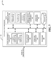
- the system 700 can include the path analysis component 102 , and the path analysis component 102 can include the path assessment component 104 , the path recommendation component 106 , the visualization component 108 , the medical device recommendation component 502 , the modeling component 602 and/or an archiving component 702 .
- the path assessment component 104 can include a lesion segmentation component 202 and a vessel segmentation component 204 .
- the path recommendation component 106 can include the medical device component 302 , the anatomic vessel component 304 and an artificial intelligence component 402 .
- the archiving component 702 can store data determined by the path recommendation component 106 , the visualization component 108 and/or the medical device recommendation component 502 and/or the modeling component 602 .
- data regarding a implantation path assessments, recommended implantation path and/or a ranking determined by the path recommendation component 106 can be archived and/or stored by the archiving component 702 .
- data for a multi-dimensional visualization associated with an implantation path, an anatomic vessel and/or a medical device can be archived and/or stored by the archiving component 702 .
- Data regarding medical device that is recommended by the medical device recommendation component 502 can additionally or alternatively be archived and/or stored by the archiving component 702 .
- data for a model generated by the modeling component 602 can be archived and/or stored by the archiving component 702 .
- Information stored and/or archived by the archiving component 702 can facilitate presentation of rendered data to a user and/or recommendation of implantation paths and/or medical devices to a user.
- Information stored and/or archived by the archiving component 702 can also facilitate a medical procedure associated with the medical device and/or the anatomic vessel, clinician reporting, a 3D printing process associated with the medical device, etc.
- the system 800 includes an anatomic vessel 802 of a patient body.
- Medical imaging data associated with the anatomic vessel 802 can be generated by employing one or more medical imaging devices.
- the medical imaging data associated with the anatomic vessel 802 can be multi-dimensional medical imaging data (e.g., 3D medical imaging data).
- the path analysis component 102 can receive the medical imaging data associated with the anatomic vessel 802 .
- the path assessment component 104 can analyze the medical imaging data associated with the anatomic vessel 802 . For example, the path assessment component 104 can perform segmentation associated with the anatomic vessel 802 .
- the path assessment component 104 can perform segmentation associated with a lesion area 804 of the anatomic vessel 802 .
- the lesion area 804 can be, for example, a disease area of the anatomic vessel 802 .
- the path analysis component 102 can determine an implantation path 806 for a medical device 808 to reach the lesion area 804 .
- the path assessment component 104 can generate a set of candidate implantation paths for the medical device 808 to reach the lesion area 804 .
- the medical device 808 can be, for example, a stent (e.g., a vascular stent).
- the path recommendation component 106 can rank the set of candidate implantation paths based on at least properties associated with the anatomic vessel 802 and/or properties associated with the medical device 808 . Based on the ranking of the set of candidate implantation paths, the path recommendation component 106 can recommend the implantation path 806 from the set of candidate implantation paths for deployment of the medical device 808 to the lesion area 804 .
- the path recommendation component 106 ensures that the medical device 808 is flexible enough to fit vessel curvature of the anatomic vessel 802 and/or ensures that a diameter of the anatomic vessel 802 is large enough to allow introduction of the medical device 808 .
- the system 900 includes an anatomic vessel 902 of a patient body.
- a medical professional interprets 2D medical data associated with the anatomic vessel to guess an implantation path 904 for a medical device 906 .
- a medical professional physically inserts the medical device 906 into the anatomic vessel 902 .
- the medical device 906 would physically come into contact with a vessel wall of the anatomic vessel 902 at a location 908 associated with the anatomic vessel 902 .
- multi-dimensional medical imaging data e.g., 3D medical imaging data
- the path analysis component 102 can determine that an implantation path 904 is not suitable for a medical device 906 .
- the path analysis component 102 it can be determined that the medical device 906 comes into contact with a vessel wall of the anatomic vessel 902 at the location 908 .
- the path analysis component 102 can determine that properties of the medical device 906 are not adequate to fit vessel curvature associated with the anatomic vessel 902 .
- the path analysis component 102 can select a different implantation path and/or a different medical device for the implantation path 904 .
- a medical procedure for implanting the medical device 906 into the anatomic vessel 902 can be more efficient and/or more accurate.
- invasive procedures to the anatomic vessel 902 can be minimized by replacing human trial and error for implanting the medical device 906 in the anatomic vessel 902 .
- FIGS. 10-13 illustrate methodologies and/or flow diagrams in accordance with the disclosed subject matter.
- the methodologies are depicted and described as a series of acts. It is to be understood and appreciated that the subject innovation is not limited by the acts illustrated and/or by the order of acts, for example acts can occur in various orders and/or concurrently, and with other acts not presented and described herein. Furthermore, not all illustrated acts may be required to implement the methodologies in accordance with the disclosed subject matter. In addition, those skilled in the art will understand and appreciate that the methodologies could alternatively be represented as a series of interrelated states via a state diagram or events. Additionally, it should be further appreciated that the methodologies disclosed hereinafter and throughout this specification are capable of being stored on an article of manufacture to facilitate transporting and transferring such methodologies to computers. The term article of manufacture, as used herein, is intended to encompass a computer program accessible from any computer-readable device or storage media.
- a medical device selection and/or an implantation path selection is assessed (e.g., by the path analysis component 102 ).
- a medical device and/or an implantation path for the medical device can be selected by segmenting and/or analyzing medical imaging data.
- Mechanical properties and/or physical properties of the medical device and/or an anatomic vessel associated with the implantation path can be employed to select the medical device and/or the implantation path.
- Assessment of the medical device selection and/or the implantation path selection can include confirming that the medical device is suitable for employment via the implantation path.
- the medical device is adequately flexible to fit curvature of the anatomic vessel associated with the implantation path, it can be confirmed that an opening of the anatomic vessel is large enough to allow the medical device to be inserted and traversed via the implantation path.
- Assessment of the medical device selection and/or the implantation path selection can also include virtually simulating an implantation process associated with the implantation path and the medical device.
- an interactive 3D visual sequence is provided to a user device.
- a display of a user device can present an interactive 3D visual sequence to allow a user to view and/or analyze the selected medical device and/or the selected implantation path via the interactive 3D visual sequence.
- the 3D visual sequence can present a series of frames that depict different portions of the implantation path with respect to the medical device and the anatomic vessel.
- a first frame of the 3D visual sequence can depict a first portion of the implantation path associated with a first portion of the anatomic vessel and the medical device
- a second frame of the 3D visual sequence can depict a second portion of the implantation path associated with a second portion of the anatomic vessel and the medical device, etc.
- the anatomic vessel associated with the 3D visual sequence can be presented as a 3D rendering of the anatomic vessel and/or the medical device associated with the 3D visual sequence can be presented as a 3D rendering of the medical device.
- an output result is recorded.
- information regarding an implantation path that is a most advantageous implantation path to reach a lesion area of the anatomic vessel with the medical device can be archived and/or stored.
- the information regarding the implantation path can be stored to facilitate use of the information regarding the implantation path prior to or during a medical procedure associated with the medical device and the anatomic vessel.
- the information regarding the implantation path can be stored to facilitate clinician reporting.
- the information regarding the implantation path can be stored to facilitate a 3D printing process associated with the medical device and/or the anatomic vessel.
- lesion segmentation is performed. For example, segmentation is performed for a portion of medical imaging data associated with a lesion area of an anatomic vessel. The lesion segmentation can define the lesion area from the medical imaging data.
- a medical device is selected. For example, a medical device can be chosen and a model representing mechanical properties of the medical device can be generated.
- candidate implantation paths are defined. For example, all possible implantation paths for the medical device to reach the lesion can be defined.
- a model representing mechanical properties of the anatomic vessel can also be generated.
- a model of the anatomic vessel can be generated.
- virtual simulation implantation is performed along each candidate implantation path.
- the medical device can be virtually traversed through each candidate implantation path associated with the anatomic vessel.
- a candidate implantation path is automatically selected. For example, a most advantageous implantation path for the medical device to reach the lesion area associated with the anatomic vessel can be determined.
- an interactive 3D visual sequence is provided to a user device.
- a display of a user device can present an interactive 3D visual sequence to allow a user to view and/or analyze the selected medical device and/or the selected implantation path via the interactive 3D visual sequence.
- the 3D visual sequence can present a series of frames that depict different portions of the implantation path with respect to the medical device and the anatomic vessel.
- a first frame of the 3D visual sequence can depict a first portion of the implantation path associated with a first portion of the anatomic vessel and the medical device
- a second frame of the 3D visual sequence can depict a second portion of the implantation path associated with a second portion of the anatomic vessel and the medical device, etc.
- the anatomic vessel associated with the 3D visual sequence can be presented as a 3D rendering of the anatomic vessel and/or the medical device associated with the 3D visual sequence can be presented as a 3D rendering of the medical device.
- an output result is recorded.
- information regarding an implantation path that is a most advantageous implantation path to reach a lesion area of the anatomic vessel with the medical device can be archived and/or stored.
- the information regarding the implantation path can be stored to facilitate use of the information regarding the implantation path prior to or during a medical procedure associated with the medical device and the anatomic vessel.
- the information regarding the implantation path can be stored to facilitate clinician reporting.
- the information regarding the implantation path can be stored to facilitate a 3D printing process associated with the medical device and/or the anatomic vessel.
- segmentation is performed with respect to medical imaging data (e.g., multi-dimensional medical imaging data). Segmentation can include a step 1204 where lesion segmentation is performed. For example, a lesion area associated with an anatomic vessel can be defined from the medical imaging data. Segmentation can also include a step 1206 where vessel segmentation is performed. For instance, the anatomic vessel can also be defined from the medical imaging data.
- iterative medical device assessment and path assessment is performed. For example, instead of validating a medical device into different implantation path, the methodology 1200 can iterate on a medical device and an implantation path until a medical device/path pair suitable for a medical procedure is determined.
- the recommendation analysis can include a step 1212 where a medical device is selected.
- the recommendation analysis can also include a step 1214 where virtual simulation implantation along each candidate path is performed.
- the recommendation analysis can include a step 1216 where an automatic best fit result is selected. If no valid path is found after step 1216 , step 1212 can be performed again and the recommendation analysis process can be repeated. If a valid path is found for the medical device after step 1216 , at step 1218 it is determined to proceed to recommend the valid path and the medical device. For example, once an implantation path and a medical device are determined by the methodology 1200 , a user device can be provided with an interactive 3D visual sequence.
- multi-dimensional visualization is performed.
- the multi-dimensional visualization can include a step 1222 where an interactive 3D visual sequence is provided to a user.
- the multi-dimensional visualization can include a step 1224 where output is recorded. For example, information associated with the selected implantation path and/or the selected medical device can be recorded for later user (e.g., for use during a 3D printing process, for use during a manufacturing process, for use during clinician reporting, etc.).
- a methodology 1300 for recommending an implantation path for a medical device can be utilized in various applications, such as, but not limited to, medical device systems, medical imaging systems, medical modeling systems, simulation systems, medical device navigation systems, stent navigation systems, enterprise imaging solution systems, advanced diagnostic tool systems, image management platform systems, care delivery management systems, artificial intelligence systems, machine learning systems, neural network systems, and the like.
- multi-dimensional medical imaging data associated with a patient body and a lesion area associated with an anatomic vessel is analyzed. For example, a portion of 3D medical imaging data associated with an anatomic vessel of a patient body can be analyzed.
- the multi-dimensional medical imaging data can be analyzed based on a segmentation process. For instance, the multi-dimensional medical imaging data can be analyzed by employing one or more segmentation algorithms and/or one or more statistical algorithms.
- geometric data, texture data and/or mesh data associated with the multi-dimensional medical imaging data can be analyzed.
- volumetric features, surface features, density features, geometric features, polygons, vectors, vertices, edges, faces and/or other features identified in the multi-dimensional medical imaging data can be analyzed.
- a set of implantation paths for a medical device to reach the lesion area is generated. For example, a set of candidate implantation paths for a medical device to traverse an anatomic vessel in order to reach the lesion area can be determined. The set of implantation paths can be generated based on properties and/or characteristics associated with the medical device. Additionally, the set of implantation paths can be generated based on properties and/or characteristics associated with the anatomic vessel. In a non-limiting example, the set of implantation paths can be generated based at least on deformation properties associated with the medical device and/or the anatomic vessel.
- the implantation paths for the medical device to reach the lesion area are ranked.
- implantation paths for the medical device can be ranked based on properties associated with the medical device, properties associated with the anatomic vessel and/or a score generated for each candidate implantation path.
- a higher ranked implantation path can correspond to an implantation path with a higher probability of fitting the anatomic vessel and/or limiting damage to the anatomic vessel.
- the implantation paths can be ranked by virtually simulating an implantation process associated with each of the implantation paths.
- a subset of the implantation paths for deployment of the medical device is recommended. For example, at least one implantation path can be recommended for the medical device based on properties associated with the medical device, properties associated with the anatomic vessel and/or a score generated for each candidate implantation path. Furthermore, at least one implantation path can be recommended for the medical device by performing learning to weigh costs and benefits associated with each of the implantation paths to reach the lesion area.
- a display associated with a multi-dimensional visualization of the recommended subset of implantation paths for the deployment of the medical device is generated.
- a 3D visualization of the at least one implantation path for the medical device can be presented on a display of a user device.
- a 3D rendering of the anatomic vessel along with the at least one implantation path and the medical device can be presented on a display of a user device.
- the system 1400 can be associated with a user device such as, for example, a computing device, a computer, a desktop computer, a laptop computer, a monitor device, a smart device, a smart phone, a mobile device, a handheld device, a tablet, a portable computing device or another type of user device associated with a display.
- the system 1400 illustrates an example user interface 1402 .
- the user interface 1402 can be a graphical user interface that presents a multi-dimensional visualization 1404 section associated with a recommended set of implantation paths for deployment of a medical device.
- the multi-dimensional visualization 1404 section of the user interface 1402 can present a multi-dimensional visualization (e.g., a 3D visualization) of a recommended set of implantation paths determined by the path recommendation component 106 .
- the multi-dimensional visualization 1404 section of the user interface 1402 can present an interactive multi-dimensional visual sequence associated with an implantation path from the recommended subset of implantation paths.
- the multi-dimensional visualization 1404 section of the user interface 1402 can present a multi-dimensional sequence of frames that depict different portions of the implantation path with respect to a medical device and an anatomic vessel.
- a medical device presented via the multi-dimensional visualization 1404 section of the user interface 1402 can be a 3D rendering of the medical device.
- an anatomic vessel presented via the multi-dimensional visualization 1404 section of the user interface 1402 can be a 3D rendering of the anatomic vessel.
- a user can view, analyze and/or interact with a 3D rendering of the medical device as the medical device traverses through a 3D rendering of the anatomic vessel via the recommended subset of implantation paths.
- the user interface 1402 can also include a medical device section 1406 associated with information for the medical device.
- the medical device section 1406 can be presented as a graphical element such as, but not limited to, a notification, a message, an icon, a thumbnail, a dialog box, an interactive tool, a widget or another type of graphical element.
- the medical device associated with the medical device section 1406 can be selected based on user input or alternatively by the medical device recommendation component 502 .
- the user interface 1402 can include a path recommendation section 1408 that includes a set of path recommendations 1408 1-N .
- the path recommendation section 1408 can be presented as a graphical element such as, but not limited to, a notification, a message, an icon, a thumbnail, a dialog box, an interactive tool, a widget or another type of graphical element. Furthermore, the set of path recommendations 1408 1-N can be determined by the path recommendation component 106 . It is to be appreciated that the user interface 1402 is merely an example. Therefore, the location and/or content of the multi-dimensional visualization 1404 , the medical device section 1406 and/or the path recommendation section 1408 can be varied. Furthermore, the user interface 1402 can include other features, content and/or functionalities not shown in FIG. 14 .
- FIGS. 15 and 16 are intended to provide a brief, general description of a suitable environment in which the various aspects of the disclosed subject matter may be implemented.
- a suitable environment 1500 for implementing various aspects of this disclosure includes a computer 1512 .
- the computer 1512 includes a processing unit 1514 , a system memory 1516 , and a system bus 1518 .
- the system bus 1518 couples system components including, but not limited to, the system memory 1516 to the processing unit 1514 .
- the processing unit 1514 can be any of various available processors. Dual microprocessors and other multiprocessor architectures also can be employed as the processing unit 1514 .
- the system bus 1518 can be any of several types of bus structure(s) including the memory bus or memory controller, a peripheral bus or external bus, and/or a local bus using any variety of available bus architectures including, but not limited to, Industrial Standard Architecture (ISA), Micro-Channel Architecture (MSA), Extended ISA (EISA), Intelligent Drive Electronics (IDE), VESA Local Bus (VLB), Peripheral Component Interconnect (PCI), Card Bus, Universal Serial Bus (USB), Advanced Graphics Port (AGP), Personal Computer Memory Card International Association bus (PCMCIA), Firewire (IEEE 1394), and Small Computer Systems Interface (SCSI).
- ISA Industrial Standard Architecture
- MSA Micro-Channel Architecture
- EISA Extended ISA
- IDE Intelligent Drive Electronics
- VLB VESA Local Bus
- PCI Peripheral Component Interconnect
- Card Bus Universal Serial Bus
- USB Universal Serial Bus
- AGP Advanced Graphics Port
- PCMCIA Personal Computer Memory Card International Association bus
- Firewire IEEE 1394
- SCSI Small Computer Systems Interface
- the system memory 1516 includes volatile memory 1520 and nonvolatile memory 1522 .
- the basic input/output system (BIOS) containing the basic routines to transfer information between elements within the computer 1512 , such as during start-up, is stored in nonvolatile memory 1522 .
- nonvolatile memory 1522 can include read only memory (ROM), programmable ROM (PROM), electrically programmable ROM (EPROM), electrically erasable programmable ROM (EEPROM), flash memory, or nonvolatile random access memory (RAM) (e.g., ferroelectric RAM (FeRAM).
- Volatile memory 1520 includes random access memory (RAM), which acts as external cache memory.
- RAM is available in many forms such as static RAM (SRAM), dynamic RAM (DRAM), synchronous DRAM (SDRAM), double data rate SDRAM (DDR SDRAM), enhanced SDRAM (ESDRAM), Synchlink DRAM (SLDRAM), direct Rambus RAM (DRRAM), direct Rambus dynamic RAM (DRDRAM), and Rambus dynamic RAM.
- SRAM static RAM
- DRAM dynamic RAM
- SDRAM synchronous DRAM
- DDR SDRAM double data rate SDRAM
- ESDRAM enhanced SDRAM
- SLDRAM Synchlink DRAM
- DRRAM direct Rambus RAM
- DRAM direct Rambus dynamic RAM
- Rambus dynamic RAM Rambus dynamic RAM
- Disk storage 1524 includes, but is not limited to, devices like a magnetic disk drive, floppy disk drive, tape drive, Jaz drive, Zip drive, LS-100 drive, flash memory card, or memory stick.
- the disk storage 1524 also can include storage media separately or in combination with other storage media including, but not limited to, an optical disk drive such as a compact disk ROM device (CD-ROM), CD recordable drive (CD-R Drive), CD rewritable drive (CD-RW Drive) or a digital versatile disk ROM drive (DVD-ROM).
- an optical disk drive such as a compact disk ROM device (CD-ROM), CD recordable drive (CD-R Drive), CD rewritable drive (CD-RW Drive) or a digital versatile disk ROM drive (DVD-ROM).
- a removable or non-removable interface is typically used, such as interface 1526 .
- FIG. 15 also depicts software that acts as an intermediary between users and the basic computer resources described in the suitable operating environment 1500 .
- Such software includes, for example, an operating system 1528 .
- Operating system 1528 which can be stored on disk storage 1524 , acts to control and allocate resources of the computer system 1512 .
- System applications 1530 take advantage of the management of resources by operating system 1528 through program modules 1532 and program data 1534 , e.g., stored either in system memory 1516 or on disk storage 1524 . It is to be appreciated that this disclosure can be implemented with various operating systems or combinations of operating systems.
- Input devices 1536 include, but are not limited to, a pointing device such as a mouse, trackball, stylus, touch pad, keyboard, microphone, joystick, game pad, satellite dish, scanner, TV tuner card, digital camera, digital video camera, web camera, and the like. These and other input devices connect to the processing unit 1514 through the system bus 1518 via interface port(s) 1538 .
- Interface port(s) 1538 include, for example, a serial port, a parallel port, a game port, and a universal serial bus (USB).
- Output device(s) 1540 use some of the same type of ports as input device(s) 1536 .
- a USB port may be used to provide input to computer 1512 , and to output information from computer 1512 to an output device 1540 .
- Output adapter 1542 is provided to illustrate that there are some output devices 1540 like monitors, speakers, and printers, among other output devices 1540 , which require special adapters.
- the output adapters 1542 include, by way of illustration and not limitation, video and sound cards that provide a means of connection between the output device 1540 and the system bus 1518 . It should be noted that other devices and/or systems of devices provide both input and output capabilities such as remote computer(s) 1544 .
- Computer 1512 can operate in a networked environment using logical connections to one or more remote computers, such as remote computer(s) 1544 .
- the remote computer(s) 1544 can be a personal computer, a server, a router, a network PC, a workstation, a microprocessor based appliance, a peer device or other common network node and the like, and typically includes many or all of the elements described relative to computer 1512 .
- only a memory storage device 1546 is illustrated with remote computer(s) 1544 .
- Remote computer(s) 1544 is logically connected to computer 1512 through a network interface 1548 and then physically connected via communication connection 1550 .
- Network interface 1548 encompasses wire and/or wireless communication networks such as local-area networks (LAN), wide-area networks (WAN), cellular networks, etc.
- LAN technologies include Fiber Distributed Data Interface (FDDI), Copper Distributed Data Interface (CDDI), Ethernet, Token Ring and the like.
- WAN technologies include, but are not limited to, point-to-point links, circuit switching networks like Integrated Services Digital Networks (ISDN) and variations thereon, packet switching networks, and Digital Subscriber Lines (DSL).
- ISDN Integrated Services Digital Networks
- DSL Digital Subscriber Lines
- Communication connection(s) 1550 refers to the hardware/software employed to connect the network interface 1548 to the bus 1518 . While communication connection 1550 is shown for illustrative clarity inside computer 1512 , it can also be external to computer 1512 .
- the hardware/software necessary for connection to the network interface 1548 includes, for exemplary purposes only, internal and external technologies such as, modems including regular telephone grade modems, cable modems and DSL modems, ISDN adapters, and Ethernet cards.
- FIG. 16 is a schematic block diagram of a sample-computing environment 1600 with which the subject matter of this disclosure can interact.
- the system 1600 includes one or more client(s) 1610 .
- the client(s) 1610 can be hardware and/or software (e.g., threads, processes, computing devices).
- the system 1600 also includes one or more server(s) 1630 .
- system 1600 can correspond to a two-tier client server model or a multi-tier model (e.g., client, middle tier server, data server), amongst other models.
- the server(s) 1630 can also be hardware and/or software (e.g., threads, processes, computing devices).
- the servers 1630 can house threads to perform transformations by employing this disclosure, for example.
- One possible communication between a client 1610 and a server 1630 may be in the form of a data packet transmitted between two or more computer processes.
- the system 1600 includes a communication framework 1650 that can be employed to facilitate communications between the client(s) 1610 and the server(s) 1630 .
- the client(s) 1610 are operatively connected to one or more client data store(s) 1620 that can be employed to store information local to the client(s) 1610 .
- the server(s) 1630 are operatively connected to one or more server data store(s) 1640 that can be employed to store information local to the servers 1630 .
- wireless telecommunication or radio technology e.g., Wi-Fi; Bluetooth; Worldwide Interoperability for Microwave Access (WiMAX); Enhanced General Packet Radio Service (Enhanced GPRS); Third Generation Partnership Project (3GPP) Long Term Evolution (LTE); Third Generation Partnership Project2 (3GPP2) Ultra Mobile Broadband (UMB); 3GPP Universal Mobile Telecommunication System (UMTS); High Speed Packet Access (HSPA); High Speed Downlink Packet Access (HSDPA); High Speed Uplink Packet Access (HSUPA); GSM (Global System for Mobile Communications) EDGE (Enhanced Data Rates for GSM Evolution) Radio Access Network (GERAN); UMTS Terrestrial Radio Access Network (UTRAN); LTE Advanced (LTE-A); etc.
- Wi-Fi Wireless Fidelity
- Bluetooth Worldwide Interoperability for Microwave Access
- WiMAX Enhanced General Packet Radio Service
- Enhanced GPRS Enhanced General Packet Radio Service
- 3GPP Third Generation Partnership Project
- LTE Long Term Evolution
- legacy telecommunication technologies e.g., GSM.
- mobile as well non-mobile networks e.g., the Internet, data service network such as internet protocol television (IPTV), etc.
- IPTV internet protocol television
- program modules include routines, programs, components, data structures, etc. that perform particular tasks and/or implement particular abstract data types.
- inventive methods may be practiced with other computer system configurations, including single-processor or multiprocessor computer systems, mini-computing devices, mainframe computers, as well as personal computers, hand-held computing devices (e.g., PDA, phone), microprocessor-based or programmable consumer or industrial electronics, and the like.
- the illustrated aspects may also be practiced in distributed computing environments where tasks are performed by remote processing devices that are linked through a communications network. However, some, if not all aspects of this disclosure can be practiced on stand-alone computers. In a distributed computing environment, program modules may be located in both local and remote memory storage devices.
- a component can refer to and/or can include a computer-related entity or an entity related to an operational machine with one or more specific functionalities.
- the entities disclosed herein can be either hardware, a combination of hardware and software, software, or software in execution.
- a component may be, but is not limited to being, a process running on a processor, a processor, an object, an executable, a thread of execution, a program, and/or a computer.
- an application running on a server and the server can be a component.
- One or more components may reside within a process and/or thread of execution and a component may be localized on one computer and/or distributed between two or more computers.
- respective components can execute from various computer readable media having various data structures stored thereon.
- the components may communicate via local and/or remote processes such as in accordance with a signal having one or more data packets (e.g., data from one component interacting with another component in a local system, distributed system, and/or across a network such as the Internet with other systems via the signal).
- a component can be an apparatus with specific functionality provided by mechanical parts operated by electric or electronic circuitry, which is operated by a software or firmware application executed by a processor.
- the processor can be internal or external to the apparatus and can execute at least a part of the software or firmware application.
- a component can be an apparatus that provides specific functionality through electronic components without mechanical parts, wherein the electronic components can include a processor or other means to execute software or firmware that confers at least in part the functionality of the electronic components.
- a component can emulate an electronic component via a virtual machine, e.g., within a cloud computing system.
- example and/or “exemplary” are utilized to mean serving as an example, instance, or illustration. For the avoidance of doubt, the subject matter disclosed herein is not limited by such examples.
- any aspect or design described herein as an “example” and/or “exemplary” is not necessarily to be construed as preferred or advantageous over other aspects or designs, nor is it meant to preclude equivalent exemplary structures and techniques known to those of ordinary skill in the art.
- aspects or features described herein can be implemented as a method, apparatus, system, or article of manufacture using standard programming or engineering techniques.
- various aspects or features disclosed in this disclosure can be realized through program modules that implement at least one or more of the methods disclosed herein, the program modules being stored in a memory and executed by at least a processor.
- Other combinations of hardware and software or hardware and firmware can enable or implement aspects described herein, including a disclosed method(s).
- the term “article of manufacture” as used herein can encompass a computer program accessible from any computer-readable device, carrier, or storage media.
- computer readable storage media can include but are not limited to magnetic storage devices (e.g., hard disk, floppy disk, magnetic strips . . .
- optical discs e.g., compact disc (CD), digital versatile disc (DVD), blu-ray disc (BD) . . .
- smart cards e.g., card, stick, key drive . . . ), or the like.
- processor can refer to substantially any computing processing unit or device comprising, but not limited to, single-core processors; single-processors with software multithread execution capability; multi-core processors; multi-core processors with software multithread execution capability; multi-core processors with hardware multithread technology; parallel platforms; and parallel platforms with distributed shared memory.
- a processor can refer to an integrated circuit, an application specific integrated circuit (ASIC), a digital signal processor (DSP), a field programmable gate array (FPGA), a programmable logic controller (PLC), a complex programmable logic device (CPLD), a discrete gate or transistor logic, discrete hardware components, or any combination thereof designed to perform the functions described herein.
- ASIC application specific integrated circuit
- DSP digital signal processor
- FPGA field programmable gate array
- PLC programmable logic controller
- CPLD complex programmable logic device
- processors can exploit nano-scale architectures such as, but not limited to, molecular and quantum-dot based transistors, switches and gates, in order to optimize space usage or enhance performance of user equipment.
- a processor may also be implemented as a combination of computing processing units.
- memory components entities embodied in a “memory,” or components comprising a memory. It is to be appreciated that memory and/or memory components described herein can be either volatile memory or nonvolatile memory, or can include both volatile and nonvolatile memory.
- nonvolatile memory can include read only memory (ROM), programmable ROM (PROM), electrically programmable ROM (EPROM), electrically erasable ROM (EEPROM), flash memory, or nonvolatile random access memory (RAM) (e.g., ferroelectric RAM (FeRAM).
- Volatile memory can include RAM, which can act as external cache memory, for example.
- RAM is available in many forms such as synchronous RAM (SRAM), dynamic RAM (DRAM), synchronous DRAM (SDRAM), double data rate SDRAM (DDR SDRAM), enhanced SDRAM (ESDRAM), Synchlink DRAM (SLDRAM), direct Rambus RAM (DRRAM), direct Rambus dynamic RAM (DRDRAM), and Rambus dynamic RAM (RDRAM).
- SRAM synchronous RAM
- DRAM dynamic RAM
- SDRAM synchronous DRAM
- DDR SDRAM double data rate SDRAM
- ESDRAM enhanced SDRAM
- SLDRAM Synchlink DRAM
- DRRAM direct Rambus RAM
- DRAM direct Rambus dynamic RAM
- RDRAM Rambus dynamic RAM
- components as described with regard to a particular system or method, can include the same or similar functionality as respective components (e.g., respectively named components or similarly named components) as described with regard to other systems or methods disclosed herein.
Landscapes
- Health & Medical Sciences (AREA)
- Engineering & Computer Science (AREA)
- Medical Informatics (AREA)
- Public Health (AREA)
- Biomedical Technology (AREA)
- General Health & Medical Sciences (AREA)
- Surgery (AREA)
- Life Sciences & Earth Sciences (AREA)
- Databases & Information Systems (AREA)
- Data Mining & Analysis (AREA)
- Pathology (AREA)
- Epidemiology (AREA)
- Primary Health Care (AREA)
- Molecular Biology (AREA)
- Animal Behavior & Ethology (AREA)
- Heart & Thoracic Surgery (AREA)
- Robotics (AREA)
- Veterinary Medicine (AREA)
- Nuclear Medicine, Radiotherapy & Molecular Imaging (AREA)
- Measuring And Recording Apparatus For Diagnosis (AREA)
- Magnetic Resonance Imaging Apparatus (AREA)
Abstract
Description
Claims (20)
Priority Applications (5)
| Application Number | Priority Date | Filing Date | Title |
|---|---|---|---|
| US15/186,951 US10806516B2 (en) | 2016-06-20 | 2016-06-20 | Virtual 4D stent implantation path assessment |
| EP17733233.5A EP3472740B1 (en) | 2016-06-20 | 2017-06-12 | Virtual assessment of an implantation path for a medical device |
| CN201780038293.4A CN109313940B (en) | 2016-06-20 | 2017-06-12 | Virtual assessment of medical device implantation path |
| PCT/US2017/036952 WO2017222849A1 (en) | 2016-06-20 | 2017-06-12 | Virtual assessment of an implantation path for a medical device |
| EP21190731.6A EP3937187A1 (en) | 2016-06-20 | 2017-06-12 | Virtual assessment of an implantation path for a medical device |
Applications Claiming Priority (1)
| Application Number | Priority Date | Filing Date | Title |
|---|---|---|---|
| US15/186,951 US10806516B2 (en) | 2016-06-20 | 2016-06-20 | Virtual 4D stent implantation path assessment |
Publications (2)
| Publication Number | Publication Date |
|---|---|
| US20170360508A1 US20170360508A1 (en) | 2017-12-21 |
| US10806516B2 true US10806516B2 (en) | 2020-10-20 |
Family
ID=59216006
Family Applications (1)
| Application Number | Title | Priority Date | Filing Date |
|---|---|---|---|
| US15/186,951 Active 2038-08-03 US10806516B2 (en) | 2016-06-20 | 2016-06-20 | Virtual 4D stent implantation path assessment |
Country Status (4)
| Country | Link |
|---|---|
| US (1) | US10806516B2 (en) |
| EP (2) | EP3472740B1 (en) |
| CN (1) | CN109313940B (en) |
| WO (1) | WO2017222849A1 (en) |
Cited By (1)
| Publication number | Priority date | Publication date | Assignee | Title |
|---|---|---|---|---|
| US12150810B2 (en) | 2021-08-17 | 2024-11-26 | Boston Scientific Scimed, Inc. | Intravascular imaging system with automated calcium analysis and treatment guidance |
Families Citing this family (38)
| Publication number | Priority date | Publication date | Assignee | Title |
|---|---|---|---|---|
| US8672837B2 (en) | 2010-06-24 | 2014-03-18 | Hansen Medical, Inc. | Methods and devices for controlling a shapeable medical device |
| US9057600B2 (en) | 2013-03-13 | 2015-06-16 | Hansen Medical, Inc. | Reducing incremental measurement sensor error |
| US9014851B2 (en) | 2013-03-15 | 2015-04-21 | Hansen Medical, Inc. | Systems and methods for tracking robotically controlled medical instruments |
| US9629595B2 (en) | 2013-03-15 | 2017-04-25 | Hansen Medical, Inc. | Systems and methods for localizing, tracking and/or controlling medical instruments |
| US9271663B2 (en) | 2013-03-15 | 2016-03-01 | Hansen Medical, Inc. | Flexible instrument localization from both remote and elongation sensors |
| US11020016B2 (en) | 2013-05-30 | 2021-06-01 | Auris Health, Inc. | System and method for displaying anatomy and devices on a movable display |
| CN105939647B (en) | 2013-10-24 | 2020-01-21 | 奥瑞斯健康公司 | Robot-assisted endovascular surgery system and related methods |
| EP3349649B1 (en) | 2015-09-18 | 2022-03-09 | Auris Health, Inc. | Navigation of tubular networks |
| US10143526B2 (en) | 2015-11-30 | 2018-12-04 | Auris Health, Inc. | Robot-assisted driving systems and methods |
| US10244926B2 (en) | 2016-12-28 | 2019-04-02 | Auris Health, Inc. | Detecting endolumenal buckling of flexible instruments |
| EP3600031A4 (en) | 2017-03-31 | 2021-01-20 | Auris Health, Inc. | ROBOTIC SYSTEMS FOR NAVIGATION OF LUMINAL NETWORKS THAT COMPENSATE FOR PHYSIOLOGICAL NOISE |
| US11647983B2 (en) * | 2017-05-05 | 2023-05-16 | International Business Machines Corporation | Automating ultrasound examination of a vascular system |
| US10022192B1 (en) | 2017-06-23 | 2018-07-17 | Auris Health, Inc. | Automatically-initialized robotic systems for navigation of luminal networks |
| US11058493B2 (en) | 2017-10-13 | 2021-07-13 | Auris Health, Inc. | Robotic system configured for navigation path tracing |
| US10555778B2 (en) | 2017-10-13 | 2020-02-11 | Auris Health, Inc. | Image-based branch detection and mapping for navigation |
| WO2019106884A1 (en) * | 2017-11-30 | 2019-06-06 | テルモ株式会社 | Assist system, assist method, and assist program |
| EP3684562B1 (en) | 2017-12-14 | 2025-03-12 | Auris Health, Inc. | System and program for estimating instrument location |
| CN110809453B (en) | 2017-12-18 | 2023-06-06 | 奥瑞斯健康公司 | Methods and systems for instrument tracking and navigation within a luminal network |
| WO2019191144A1 (en) | 2018-03-28 | 2019-10-03 | Auris Health, Inc. | Systems and methods for registration of location sensors |
| CN110913791B (en) | 2018-03-28 | 2021-10-08 | 奥瑞斯健康公司 | System and method for displaying estimated instrument location |
| US10905499B2 (en) | 2018-05-30 | 2021-02-02 | Auris Health, Inc. | Systems and methods for location sensor-based branch prediction |
| KR102455671B1 (en) | 2018-05-31 | 2022-10-20 | 아우리스 헬스, 인코포레이티드 | Image-Based Airway Analysis and Mapping |
| EP3801280B1 (en) | 2018-05-31 | 2024-10-02 | Auris Health, Inc. | Robotic systems for navigation of luminal network that detect physiological noise |
| WO2019231891A1 (en) | 2018-05-31 | 2019-12-05 | Auris Health, Inc. | Path-based navigation of tubular networks |
| WO2020069404A1 (en) | 2018-09-28 | 2020-04-02 | Auris Health, Inc. | Robotic systems and methods for concomitant endoscopic and percutaneous medical procedures |
| US10963757B2 (en) | 2018-12-14 | 2021-03-30 | Industrial Technology Research Institute | Neural network model fusion method and electronic device using the same |
| US12478444B2 (en) | 2019-03-21 | 2025-11-25 | The Board Of Trustees Of The Leland Stanford Junior University | Systems and methods for localization based on machine learning |
| JP7451686B2 (en) | 2019-08-30 | 2024-03-18 | オーリス ヘルス インコーポレイテッド | Instrument image reliability system and method |
| WO2021038469A1 (en) | 2019-08-30 | 2021-03-04 | Auris Health, Inc. | Systems and methods for weight-based registration of location sensors |
| US11456063B2 (en) | 2019-09-08 | 2022-09-27 | Novadontics Llc | Dental patient management system |
| EP4084722A4 (en) | 2019-12-31 | 2024-01-10 | Auris Health, Inc. | ALIGNMENT INTERFACES FOR PERCUTANEOUS ACCESS |
| KR20220123076A (en) | 2019-12-31 | 2022-09-05 | 아우리스 헬스, 인코포레이티드 | Alignment Techniques for Transdermal Access |
| CN114901194B (en) | 2019-12-31 | 2024-08-30 | 奥瑞斯健康公司 | Anatomical feature identification and targeting |
| US11737663B2 (en) | 2020-03-30 | 2023-08-29 | Auris Health, Inc. | Target anatomical feature localization |
| EP4248296A2 (en) * | 2020-11-23 | 2023-09-27 | Koninklijke Philips N.V. | Automatic generation of educational content from usage of optimal/poorest user |
| JP7769496B2 (en) * | 2021-09-09 | 2025-11-13 | キヤノンメディカルシステムズ株式会社 | Medical information processing device, X-ray diagnostic device, and medical information processing program |
| CN116127147B (en) * | 2023-04-04 | 2023-06-16 | 吉林大学 | Medical data storage method, system, computer device and storage medium |
| US20250124513A1 (en) * | 2023-10-16 | 2025-04-17 | nearmap australia pty ltd. | Assessment tool, graphical user interface, and associated functionality |
Citations (13)
| Publication number | Priority date | Publication date | Assignee | Title |
|---|---|---|---|---|
| US20090088830A1 (en) * | 2007-02-15 | 2009-04-02 | Siemens Corporate Research, Inc. | System and method for intraoperative guidance of stent placement during endovascular interventions |
| EP2712553A2 (en) | 2005-01-11 | 2014-04-02 | Volcano Corporation | Vascular image co-registration |
| US20140254906A1 (en) | 2013-03-11 | 2014-09-11 | Toshiba Medical Systems Corporation | Vascular tree from anatomical landmarks and a clinical ontology |
| US20140257763A1 (en) * | 2013-03-07 | 2014-09-11 | CHOICE Biotech Inc, | Method and system for dental implant path planning |
| US8922552B2 (en) * | 2003-01-15 | 2014-12-30 | Koninklijke Philips N.V. | Image processing method for automatic adaptation of 3-D deformable model onto a substantially tubular surface of a 3-D object |
| US20150049081A1 (en) * | 2013-08-13 | 2015-02-19 | Dane Coffey | Computer Visualization of Anatomical Items |
| WO2015089013A1 (en) | 2013-12-09 | 2015-06-18 | Intuitive Surgical Operations, Inc. | Systems and methods for device-aware flexible tool registration |
| US20150320324A1 (en) | 2014-05-09 | 2015-11-12 | Samsung Electronics Co., Ltd. | Method, apparatus, and system for providing medical image |
| US20150366628A1 (en) * | 2014-06-18 | 2015-12-24 | Covidien Lp | Augmented surgical reality environment system |
| US20160022208A1 (en) * | 2014-07-24 | 2016-01-28 | Lightlab Imaging, Inc. | Stent and Vessel Visualization and Diagnostic Systems, Devices, and Methods |
| US20160217375A1 (en) * | 2015-01-28 | 2016-07-28 | Infinitt Healthcare Co., Ltd. | Method and system for stent recommendation |
| US20160256610A1 (en) * | 2015-03-03 | 2016-09-08 | Yujie Zhou | Method of producing personalized biomimetic drug-eluting coronary stents by 3d-printing |
| US20170065349A1 (en) * | 2014-05-14 | 2017-03-09 | Ucl Business Plc | System and method for computer-assisted planning of a trajectory for a surgical insertion into a skull |
Family Cites Families (5)
| Publication number | Priority date | Publication date | Assignee | Title |
|---|---|---|---|---|
| WO1998020927A1 (en) * | 1996-11-12 | 1998-05-22 | Valerian Voinov | The optimum expandable stent mechanical model and its application |
| US7048962B2 (en) * | 2002-05-02 | 2006-05-23 | Labcoat, Ltd. | Stent coating device |
| CA2457171A1 (en) * | 2004-02-09 | 2005-08-09 | Centre Hospitalier De L'universite De Montreal - Chum | Imaging apparatus and methods |
| AU2008351907A1 (en) * | 2008-02-25 | 2009-09-03 | Inventive Medical Limited | Medical training method and apparatus |
| CN102451036B (en) * | 2010-10-22 | 2013-10-23 | 刘永安 | Medical device for performing heart intracavity operation in vitro |
-
2016
- 2016-06-20 US US15/186,951 patent/US10806516B2/en active Active
-
2017
- 2017-06-12 EP EP17733233.5A patent/EP3472740B1/en active Active
- 2017-06-12 CN CN201780038293.4A patent/CN109313940B/en active Active
- 2017-06-12 WO PCT/US2017/036952 patent/WO2017222849A1/en not_active Ceased
- 2017-06-12 EP EP21190731.6A patent/EP3937187A1/en active Pending
Patent Citations (13)
| Publication number | Priority date | Publication date | Assignee | Title |
|---|---|---|---|---|
| US8922552B2 (en) * | 2003-01-15 | 2014-12-30 | Koninklijke Philips N.V. | Image processing method for automatic adaptation of 3-D deformable model onto a substantially tubular surface of a 3-D object |
| EP2712553A2 (en) | 2005-01-11 | 2014-04-02 | Volcano Corporation | Vascular image co-registration |
| US20090088830A1 (en) * | 2007-02-15 | 2009-04-02 | Siemens Corporate Research, Inc. | System and method for intraoperative guidance of stent placement during endovascular interventions |
| US20140257763A1 (en) * | 2013-03-07 | 2014-09-11 | CHOICE Biotech Inc, | Method and system for dental implant path planning |
| US20140254906A1 (en) | 2013-03-11 | 2014-09-11 | Toshiba Medical Systems Corporation | Vascular tree from anatomical landmarks and a clinical ontology |
| US20150049081A1 (en) * | 2013-08-13 | 2015-02-19 | Dane Coffey | Computer Visualization of Anatomical Items |
| WO2015089013A1 (en) | 2013-12-09 | 2015-06-18 | Intuitive Surgical Operations, Inc. | Systems and methods for device-aware flexible tool registration |
| US20150320324A1 (en) | 2014-05-09 | 2015-11-12 | Samsung Electronics Co., Ltd. | Method, apparatus, and system for providing medical image |
| US20170065349A1 (en) * | 2014-05-14 | 2017-03-09 | Ucl Business Plc | System and method for computer-assisted planning of a trajectory for a surgical insertion into a skull |
| US20150366628A1 (en) * | 2014-06-18 | 2015-12-24 | Covidien Lp | Augmented surgical reality environment system |
| US20160022208A1 (en) * | 2014-07-24 | 2016-01-28 | Lightlab Imaging, Inc. | Stent and Vessel Visualization and Diagnostic Systems, Devices, and Methods |
| US20160217375A1 (en) * | 2015-01-28 | 2016-07-28 | Infinitt Healthcare Co., Ltd. | Method and system for stent recommendation |
| US20160256610A1 (en) * | 2015-03-03 | 2016-09-08 | Yujie Zhou | Method of producing personalized biomimetic drug-eluting coronary stents by 3d-printing |
Non-Patent Citations (1)
| Title |
|---|
| International Search Report and Written Opinion for corresponding PCT Application PCT/US2017/036952 dated Aug. 9, 2017; 13 pages. |
Cited By (1)
| Publication number | Priority date | Publication date | Assignee | Title |
|---|---|---|---|---|
| US12150810B2 (en) | 2021-08-17 | 2024-11-26 | Boston Scientific Scimed, Inc. | Intravascular imaging system with automated calcium analysis and treatment guidance |
Also Published As
| Publication number | Publication date |
|---|---|
| EP3937187A1 (en) | 2022-01-12 |
| CN109313940A (en) | 2019-02-05 |
| CN109313940B (en) | 2024-07-09 |
| US20170360508A1 (en) | 2017-12-21 |
| EP3472740B1 (en) | 2021-08-25 |
| EP3472740A1 (en) | 2019-04-24 |
| WO2017222849A1 (en) | 2017-12-28 |
Similar Documents
| Publication | Publication Date | Title |
|---|---|---|
| US10806516B2 (en) | Virtual 4D stent implantation path assessment | |
| US11074482B2 (en) | Classification and localization based on annotation information | |
| US10885400B2 (en) | Classification based on annotation information | |
| US11074687B2 (en) | Deep convolutional neural network with self-transfer learning | |
| US11954610B2 (en) | Active surveillance and learning for machine learning model authoring and deployment | |
| US11688518B2 (en) | Deep neural network based identification of realistic synthetic images generated using a generative adversarial network | |
| US11967434B2 (en) | Systems and methods for multi-dimensional fluid modeling of an organism or organ | |
| US10755140B2 (en) | Classification based on annotation information | |
| US20190122104A1 (en) | Building a binary neural network architecture | |
| US20180005083A1 (en) | Intelligent multi-scale medical image landmark detection | |
| US12106510B2 (en) | Method and system for detecting landmarks in medical images | |
| EP3716149A1 (en) | Generative model generating imaging data and corresponding multi-channel data | |
| US12333716B2 (en) | Generating high quality training data collections for training artificial intelligence models | |
| US20250173874A1 (en) | Method for detecting white matter lesions based on medical image | |
| CN119181473A (en) | Continuous model refinement based on non-image feedback to generate composite images | |
| Guo et al. | Evaluation of stroke sequelae and rehabilitation effect on brain tumor by neuroimaging technique: A comparative study | |
| US20250104451A1 (en) | Iterative framework for learning multimodal mappings tailored to medical image inferencing tasks | |
| Gilal | AI-based end-to-end solutions for fine-grained classification, detection and segmentation in real-world scenarios | |
| Moreno et al. | Kinematic motion representation in Cine-MRI to support cardiac disease classification | |
| Abadpour | Incorporating spatial context into fuzzy-possibilistic clustering using Bayesian inference | |
| Cui | Weakly Supervised Point Cloud Semantic Segmentation with Graph Convolutional Networks | |
| CHRISTOPHER et al. | Topology-Informed Brain Tumour Segmentation for Oncological Diagnosis |
Legal Events
| Date | Code | Title | Description |
|---|---|---|---|
| AS | Assignment |
Owner name: GENERAL ELECTRIC COMPANY, NEW YORK Free format text: ASSIGNMENT OF ASSIGNORS INTEREST;ASSIGNORS:GERMAIN, SYLVAIN;BAUSSE, JEROME;REEL/FRAME:038958/0662 Effective date: 20160524 |
|
| STPP | Information on status: patent application and granting procedure in general |
Free format text: NON FINAL ACTION MAILED |
|
| STPP | Information on status: patent application and granting procedure in general |
Free format text: RESPONSE TO NON-FINAL OFFICE ACTION ENTERED AND FORWARDED TO EXAMINER |
|
| STPP | Information on status: patent application and granting procedure in general |
Free format text: FINAL REJECTION MAILED |
|
| STPP | Information on status: patent application and granting procedure in general |
Free format text: DOCKETED NEW CASE - READY FOR EXAMINATION |
|
| STPP | Information on status: patent application and granting procedure in general |
Free format text: NON FINAL ACTION MAILED |
|
| STPP | Information on status: patent application and granting procedure in general |
Free format text: NOTICE OF ALLOWANCE MAILED -- APPLICATION RECEIVED IN OFFICE OF PUBLICATIONS |
|
| STPP | Information on status: patent application and granting procedure in general |
Free format text: PUBLICATIONS -- ISSUE FEE PAYMENT RECEIVED |
|
| STCF | Information on status: patent grant |
Free format text: PATENTED CASE |
|
| MAFP | Maintenance fee payment |
Free format text: PAYMENT OF MAINTENANCE FEE, 4TH YEAR, LARGE ENTITY (ORIGINAL EVENT CODE: M1551); ENTITY STATUS OF PATENT OWNER: LARGE ENTITY Year of fee payment: 4 |
|
| AS | Assignment |
Owner name: GE PRECISION HEALTHCARE LLC, WISCONSIN Free format text: NUNC PRO TUNC ASSIGNMENT;ASSIGNOR:GENERAL ELECTRIC COMPANY;REEL/FRAME:071225/0218 Effective date: 20250505 |