KR20140023378A - Eye covering and refractive correction methods and apparatus having improved tear flow, comfort, and/or applicability - Google Patents
Eye covering and refractive correction methods and apparatus having improved tear flow, comfort, and/or applicability Download PDFInfo
- Publication number
- KR20140023378A KR20140023378A KR1020137031721A KR20137031721A KR20140023378A KR 20140023378 A KR20140023378 A KR 20140023378A KR 1020137031721 A KR1020137031721 A KR 1020137031721A KR 20137031721 A KR20137031721 A KR 20137031721A KR 20140023378 A KR20140023378 A KR 20140023378A
- Authority
- KR
- South Korea
- Prior art keywords
- protective film
- eye
- cornea
- optical
- component
- Prior art date
- Legal status (The legal status is an assumption and is not a legal conclusion. Google has not performed a legal analysis and makes no representation as to the accuracy of the status listed.)
- Ceased
Links
- 238000000034 method Methods 0.000 title claims description 110
- 238000012937 correction Methods 0.000 title claims description 51
- 230000001681 protective effect Effects 0.000 claims abstract description 533
- 239000000463 material Substances 0.000 claims abstract description 340
- 210000004087 cornea Anatomy 0.000 claims abstract description 269
- 210000000744 eyelid Anatomy 0.000 claims abstract description 125
- 239000000017 hydrogel Substances 0.000 claims abstract description 117
- 210000000795 conjunctiva Anatomy 0.000 claims abstract description 101
- 239000007788 liquid Substances 0.000 claims abstract description 92
- 230000004438 eyesight Effects 0.000 claims abstract description 59
- 239000012530 fluid Substances 0.000 claims abstract description 49
- 238000005086 pumping Methods 0.000 claims abstract description 37
- 230000015572 biosynthetic process Effects 0.000 claims abstract description 13
- 210000001508 eye Anatomy 0.000 claims description 310
- 230000003287 optical effect Effects 0.000 claims description 241
- 230000008878 coupling Effects 0.000 claims description 196
- 238000010168 coupling process Methods 0.000 claims description 196
- 238000005859 coupling reaction Methods 0.000 claims description 196
- 210000000981 epithelium Anatomy 0.000 claims description 94
- 230000002093 peripheral effect Effects 0.000 claims description 77
- 229920001296 polysiloxane Polymers 0.000 claims description 44
- 230000033001 locomotion Effects 0.000 claims description 41
- 238000000576 coating method Methods 0.000 claims description 35
- 208000014733 refractive error Diseases 0.000 claims description 35
- 239000011248 coating agent Substances 0.000 claims description 32
- 230000004075 alteration Effects 0.000 claims description 29
- 210000001519 tissue Anatomy 0.000 claims description 29
- 201000009310 astigmatism Diseases 0.000 claims description 28
- 230000009969 flowable effect Effects 0.000 claims description 24
- 239000010703 silicon Substances 0.000 claims description 24
- 229910052710 silicon Inorganic materials 0.000 claims description 24
- 210000001747 pupil Anatomy 0.000 claims description 23
- 230000000193 eyeblink Effects 0.000 claims description 22
- 230000004397 blinking Effects 0.000 claims description 20
- 201000010041 presbyopia Diseases 0.000 claims description 20
- 230000002829 reductive effect Effects 0.000 claims description 16
- 239000003814 drug Substances 0.000 claims description 13
- 238000005452 bending Methods 0.000 claims description 10
- 238000005229 chemical vapour deposition Methods 0.000 claims description 9
- 230000007423 decrease Effects 0.000 claims description 8
- 229940079593 drug Drugs 0.000 claims description 7
- 238000004519 manufacturing process Methods 0.000 claims description 7
- KPUWHANPEXNPJT-UHFFFAOYSA-N disiloxane Chemical class [SiH3]O[SiH3] KPUWHANPEXNPJT-UHFFFAOYSA-N 0.000 claims description 5
- 230000002401 inhibitory effect Effects 0.000 claims description 4
- 238000007789 sealing Methods 0.000 claims description 4
- 230000000007 visual effect Effects 0.000 claims description 4
- 239000013013 elastic material Substances 0.000 claims description 3
- 230000004379 myopia Effects 0.000 claims description 3
- 208000001491 myopia Diseases 0.000 claims description 3
- 238000007493 shaping process Methods 0.000 claims description 2
- 239000010408 film Substances 0.000 claims 70
- 241000255789 Bombyx mori Species 0.000 claims 4
- 230000001939 inductive effect Effects 0.000 claims 2
- 239000012788 optical film Substances 0.000 claims 2
- 230000004515 progressive myopia Effects 0.000 claims 2
- 239000011253 protective coating Substances 0.000 claims 2
- 230000002265 prevention Effects 0.000 claims 1
- 210000003786 sclera Anatomy 0.000 abstract description 56
- 230000000717 retained effect Effects 0.000 abstract description 2
- 239000010410 layer Substances 0.000 description 128
- 238000002679 ablation Methods 0.000 description 34
- 230000007547 defect Effects 0.000 description 24
- XUIMIQQOPSSXEZ-UHFFFAOYSA-N Silicon Chemical compound [Si] XUIMIQQOPSSXEZ-UHFFFAOYSA-N 0.000 description 22
- 238000002161 passivation Methods 0.000 description 22
- XLYOFNOQVPJJNP-UHFFFAOYSA-N water Substances O XLYOFNOQVPJJNP-UHFFFAOYSA-N 0.000 description 21
- 206010015037 epilepsy Diseases 0.000 description 18
- 239000007779 soft material Substances 0.000 description 14
- 229920002379 silicone rubber Polymers 0.000 description 11
- 210000004045 bowman membrane Anatomy 0.000 description 10
- 230000001225 therapeutic effect Effects 0.000 description 10
- WOBHKFSMXKNTIM-UHFFFAOYSA-N Hydroxyethyl methacrylate Chemical compound CC(=C)C(=O)OCCO WOBHKFSMXKNTIM-UHFFFAOYSA-N 0.000 description 9
- 229920002818 (Hydroxyethyl)methacrylate Polymers 0.000 description 8
- 238000005259 measurement Methods 0.000 description 8
- 210000001525 retina Anatomy 0.000 description 8
- 238000001356 surgical procedure Methods 0.000 description 8
- WHNWPMSKXPGLAX-UHFFFAOYSA-N N-Vinyl-2-pyrrolidone Chemical compound C=CN1CCCC1=O WHNWPMSKXPGLAX-UHFFFAOYSA-N 0.000 description 7
- 238000009499 grossing Methods 0.000 description 7
- 239000007787 solid Substances 0.000 description 6
- 229940124597 therapeutic agent Drugs 0.000 description 6
- 102000008186 Collagen Human genes 0.000 description 5
- 108010035532 Collagen Proteins 0.000 description 5
- 239000002202 Polyethylene glycol Substances 0.000 description 5
- 230000008901 benefit Effects 0.000 description 5
- 229920001436 collagen Polymers 0.000 description 5
- 230000035876 healing Effects 0.000 description 5
- 230000010220 ion permeability Effects 0.000 description 5
- 238000005498 polishing Methods 0.000 description 5
- 229920001223 polyethylene glycol Polymers 0.000 description 5
- 230000008929 regeneration Effects 0.000 description 5
- 238000011069 regeneration method Methods 0.000 description 5
- 230000004044 response Effects 0.000 description 5
- 238000003325 tomography Methods 0.000 description 5
- 230000004304 visual acuity Effects 0.000 description 5
- 230000003511 endothelial effect Effects 0.000 description 4
- 230000036571 hydration Effects 0.000 description 4
- 238000006703 hydration reaction Methods 0.000 description 4
- 239000012528 membrane Substances 0.000 description 4
- 230000004048 modification Effects 0.000 description 4
- 238000012986 modification Methods 0.000 description 4
- 238000012876 topography Methods 0.000 description 4
- 208000003556 Dry Eye Syndromes Diseases 0.000 description 3
- BPQQTUXANYXVAA-UHFFFAOYSA-N Orthosilicate Chemical compound [O-][Si]([O-])([O-])[O-] BPQQTUXANYXVAA-UHFFFAOYSA-N 0.000 description 3
- QVGXLLKOCUKJST-UHFFFAOYSA-N atomic oxygen Chemical compound [O] QVGXLLKOCUKJST-UHFFFAOYSA-N 0.000 description 3
- 239000002131 composite material Substances 0.000 description 3
- 230000005489 elastic deformation Effects 0.000 description 3
- 239000007789 gas Substances 0.000 description 3
- 238000002347 injection Methods 0.000 description 3
- 239000007924 injection Substances 0.000 description 3
- 230000007774 longterm Effects 0.000 description 3
- 238000007726 management method Methods 0.000 description 3
- 210000004126 nerve fiber Anatomy 0.000 description 3
- 239000001301 oxygen Substances 0.000 description 3
- 229910052760 oxygen Inorganic materials 0.000 description 3
- 229920000052 poly(p-xylylene) Polymers 0.000 description 3
- 230000001737 promoting effect Effects 0.000 description 3
- 239000011241 protective layer Substances 0.000 description 3
- 238000012360 testing method Methods 0.000 description 3
- 239000012780 transparent material Substances 0.000 description 3
- NIXOWILDQLNWCW-UHFFFAOYSA-M Acrylate Chemical compound [O-]C(=O)C=C NIXOWILDQLNWCW-UHFFFAOYSA-M 0.000 description 2
- 206010013774 Dry eye Diseases 0.000 description 2
- 206010020675 Hypermetropia Diseases 0.000 description 2
- 230000005856 abnormality Effects 0.000 description 2
- 210000004556 brain Anatomy 0.000 description 2
- 210000004027 cell Anatomy 0.000 description 2
- 230000008859 change Effects 0.000 description 2
- 210000003161 choroid Anatomy 0.000 description 2
- 150000001875 compounds Chemical class 0.000 description 2
- 238000001804 debridement Methods 0.000 description 2
- 238000013461 design Methods 0.000 description 2
- 229920001971 elastomer Polymers 0.000 description 2
- 239000000806 elastomer Substances 0.000 description 2
- 230000001037 epileptic effect Effects 0.000 description 2
- 238000011049 filling Methods 0.000 description 2
- 229930195733 hydrocarbon Natural products 0.000 description 2
- 150000002430 hydrocarbons Chemical class 0.000 description 2
- 230000004305 hyperopia Effects 0.000 description 2
- 201000006318 hyperopia Diseases 0.000 description 2
- 238000010348 incorporation Methods 0.000 description 2
- 208000014674 injury Diseases 0.000 description 2
- 210000001232 limbus corneae Anatomy 0.000 description 2
- 210000005036 nerve Anatomy 0.000 description 2
- 230000035699 permeability Effects 0.000 description 2
- 230000001172 regenerating effect Effects 0.000 description 2
- 238000002271 resection Methods 0.000 description 2
- 229920005989 resin Polymers 0.000 description 2
- 239000011347 resin Substances 0.000 description 2
- 238000004528 spin coating Methods 0.000 description 2
- 239000000126 substance Substances 0.000 description 2
- 230000001629 suppression Effects 0.000 description 2
- 230000008733 trauma Effects 0.000 description 2
- -1 CH4 Chemical class 0.000 description 1
- 206010010071 Coma Diseases 0.000 description 1
- 241000195493 Cryptophyta Species 0.000 description 1
- 235000004035 Cryptotaenia japonica Nutrition 0.000 description 1
- YCKRFDGAMUMZLT-UHFFFAOYSA-N Fluorine atom Chemical compound [F] YCKRFDGAMUMZLT-UHFFFAOYSA-N 0.000 description 1
- 241000306688 Meridion Species 0.000 description 1
- 241001465754 Metazoa Species 0.000 description 1
- 241000288906 Primates Species 0.000 description 1
- 229910002808 Si–O–Si Inorganic materials 0.000 description 1
- FAPWRFPIFSIZLT-UHFFFAOYSA-M Sodium chloride Chemical compound [Na+].[Cl-] FAPWRFPIFSIZLT-UHFFFAOYSA-M 0.000 description 1
- 102000007641 Trefoil Factors Human genes 0.000 description 1
- 235000015724 Trifolium pratense Nutrition 0.000 description 1
- 208000027418 Wounds and injury Diseases 0.000 description 1
- 230000002159 abnormal effect Effects 0.000 description 1
- 238000010521 absorption reaction Methods 0.000 description 1
- 238000009825 accumulation Methods 0.000 description 1
- 230000006978 adaptation Effects 0.000 description 1
- 229940035676 analgesics Drugs 0.000 description 1
- 238000004458 analytical method Methods 0.000 description 1
- 229940035674 anesthetics Drugs 0.000 description 1
- 239000000730 antalgic agent Substances 0.000 description 1
- 210000002159 anterior chamber Anatomy 0.000 description 1
- 239000007864 aqueous solution Substances 0.000 description 1
- 230000004888 barrier function Effects 0.000 description 1
- 230000009286 beneficial effect Effects 0.000 description 1
- 235000010290 biphenyl Nutrition 0.000 description 1
- 239000004305 biphenyl Substances 0.000 description 1
- 125000006267 biphenyl group Chemical group 0.000 description 1
- 238000004364 calculation method Methods 0.000 description 1
- 229940112822 chewing gum Drugs 0.000 description 1
- 235000015218 chewing gum Nutrition 0.000 description 1
- 238000004140 cleaning Methods 0.000 description 1
- 210000003683 corneal stroma Anatomy 0.000 description 1
- 230000006378 damage Effects 0.000 description 1
- 230000018044 dehydration Effects 0.000 description 1
- 238000006297 dehydration reaction Methods 0.000 description 1
- 238000009792 diffusion process Methods 0.000 description 1
- 125000000118 dimethyl group Chemical group [H]C([H])([H])* 0.000 description 1
- 239000004205 dimethyl polysiloxane Substances 0.000 description 1
- 235000013870 dimethyl polysiloxane Nutrition 0.000 description 1
- 238000005553 drilling Methods 0.000 description 1
- 230000000694 effects Effects 0.000 description 1
- 210000003038 endothelium Anatomy 0.000 description 1
- 230000010393 epithelial cell migration Effects 0.000 description 1
- 230000008472 epithelial growth Effects 0.000 description 1
- 210000003560 epithelium corneal Anatomy 0.000 description 1
- 230000008020 evaporation Effects 0.000 description 1
- 238000001704 evaporation Methods 0.000 description 1
- 230000004418 eye rotation Effects 0.000 description 1
- 229910052731 fluorine Inorganic materials 0.000 description 1
- 239000011737 fluorine Substances 0.000 description 1
- 230000006870 function Effects 0.000 description 1
- 239000003193 general anesthetic agent Substances 0.000 description 1
- VOZRXNHHFUQHIL-UHFFFAOYSA-N glycidyl methacrylate Chemical compound CC(=C)C(=O)OCC1CO1 VOZRXNHHFUQHIL-UHFFFAOYSA-N 0.000 description 1
- 230000012010 growth Effects 0.000 description 1
- 230000004371 high visual acuity Effects 0.000 description 1
- 230000002209 hydrophobic effect Effects 0.000 description 1
- 230000006698 induction Effects 0.000 description 1
- 230000008595 infiltration Effects 0.000 description 1
- 238000001764 infiltration Methods 0.000 description 1
- 150000002500 ions Chemical class 0.000 description 1
- 230000001788 irregular Effects 0.000 description 1
- 201000000766 irregular astigmatism Diseases 0.000 description 1
- 206010023332 keratitis Diseases 0.000 description 1
- 150000002632 lipids Chemical class 0.000 description 1
- 238000011068 loading method Methods 0.000 description 1
- 239000000314 lubricant Substances 0.000 description 1
- 238000005461 lubrication Methods 0.000 description 1
- 230000005012 migration Effects 0.000 description 1
- 238000013508 migration Methods 0.000 description 1
- 210000003097 mucus Anatomy 0.000 description 1
- 239000005445 natural material Substances 0.000 description 1
- 239000010702 perfluoropolyether Substances 0.000 description 1
- ZUOUZKKEUPVFJK-UHFFFAOYSA-N phenylbenzene Natural products C1=CC=CC=C1C1=CC=CC=C1 ZUOUZKKEUPVFJK-UHFFFAOYSA-N 0.000 description 1
- 229920000435 poly(dimethylsiloxane) Polymers 0.000 description 1
- 230000002035 prolonged effect Effects 0.000 description 1
- 238000007634 remodeling Methods 0.000 description 1
- 230000003252 repetitive effect Effects 0.000 description 1
- 230000035807 sensation Effects 0.000 description 1
- 229910052709 silver Inorganic materials 0.000 description 1
- 239000004332 silver Substances 0.000 description 1
- 238000004088 simulation Methods 0.000 description 1
- 239000011780 sodium chloride Substances 0.000 description 1
- 238000009987 spinning Methods 0.000 description 1
- 150000003431 steroids Chemical class 0.000 description 1
- 210000002536 stromal cell Anatomy 0.000 description 1
- 230000008961 swelling Effects 0.000 description 1
- 229920002994 synthetic fiber Polymers 0.000 description 1
- 230000007704 transition Effects 0.000 description 1
- 238000007740 vapor deposition Methods 0.000 description 1
- 238000009736 wetting Methods 0.000 description 1
Images


Classifications
-
- A—HUMAN NECESSITIES
- A61—MEDICAL OR VETERINARY SCIENCE; HYGIENE
- A61F—FILTERS IMPLANTABLE INTO BLOOD VESSELS; PROSTHESES; DEVICES PROVIDING PATENCY TO, OR PREVENTING COLLAPSING OF, TUBULAR STRUCTURES OF THE BODY, e.g. STENTS; ORTHOPAEDIC, NURSING OR CONTRACEPTIVE DEVICES; FOMENTATION; TREATMENT OR PROTECTION OF EYES OR EARS; BANDAGES, DRESSINGS OR ABSORBENT PADS; FIRST-AID KITS
- A61F9/00—Methods or devices for treatment of the eyes; Devices for putting in contact-lenses; Devices to correct squinting; Apparatus to guide the blind; Protective devices for the eyes, carried on the body or in the hand
-
- G—PHYSICS
- G02—OPTICS
- G02C—SPECTACLES; SUNGLASSES OR GOGGLES INSOFAR AS THEY HAVE THE SAME FEATURES AS SPECTACLES; CONTACT LENSES
- G02C7/00—Optical parts
- G02C7/02—Lenses; Lens systems ; Methods of designing lenses
- G02C7/04—Contact lenses for the eyes
- G02C7/049—Contact lenses having special fitting or structural features achieved by special materials or material structures
-
- G—PHYSICS
- G02—OPTICS
- G02C—SPECTACLES; SUNGLASSES OR GOGGLES INSOFAR AS THEY HAVE THE SAME FEATURES AS SPECTACLES; CONTACT LENSES
- G02C7/00—Optical parts
- G02C7/02—Lenses; Lens systems ; Methods of designing lenses
- G02C7/022—Ophthalmic lenses having special refractive features achieved by special materials or material structures
-
- G—PHYSICS
- G02—OPTICS
- G02C—SPECTACLES; SUNGLASSES OR GOGGLES INSOFAR AS THEY HAVE THE SAME FEATURES AS SPECTACLES; CONTACT LENSES
- G02C7/00—Optical parts
- G02C7/02—Lenses; Lens systems ; Methods of designing lenses
- G02C7/024—Methods of designing ophthalmic lenses
-
- G—PHYSICS
- G02—OPTICS
- G02C—SPECTACLES; SUNGLASSES OR GOGGLES INSOFAR AS THEY HAVE THE SAME FEATURES AS SPECTACLES; CONTACT LENSES
- G02C7/00—Optical parts
- G02C7/02—Lenses; Lens systems ; Methods of designing lenses
- G02C7/04—Contact lenses for the eyes
-
- G—PHYSICS
- G02—OPTICS
- G02C—SPECTACLES; SUNGLASSES OR GOGGLES INSOFAR AS THEY HAVE THE SAME FEATURES AS SPECTACLES; CONTACT LENSES
- G02C7/00—Optical parts
- G02C7/02—Lenses; Lens systems ; Methods of designing lenses
- G02C7/04—Contact lenses for the eyes
- G02C7/047—Contact lens fitting; Contact lenses for orthokeratology; Contact lenses for specially shaped corneae
Landscapes
- Health & Medical Sciences (AREA)
- Ophthalmology & Optometry (AREA)
- Physics & Mathematics (AREA)
- General Health & Medical Sciences (AREA)
- General Physics & Mathematics (AREA)
- Optics & Photonics (AREA)
- Heart & Thoracic Surgery (AREA)
- Biomedical Technology (AREA)
- Engineering & Computer Science (AREA)
- Vascular Medicine (AREA)
- Life Sciences & Earth Sciences (AREA)
- Animal Behavior & Ethology (AREA)
- Public Health (AREA)
- Veterinary Medicine (AREA)
- Prostheses (AREA)
- Eyeglasses (AREA)
Abstract
콘택트 렌즈와 같은 눈 보호막은 그 보호막 아래에 있는 눈물 액체를 펌핑하기 위한 하나 이상의 구조물을 포함하여 그러한 보호막이 눈에 유지될 수 있고 연장된 시간 동안 시력을 교정할 수 있게 한다. 많은 구체예에서, 보호막은 보호막 아래의 눈물 액체를 유도하는 천공을 지닌 물질 및 공막 상의 결막과 접촉하는 모양의 외부 부분을 포함하여, 눈이 닫힐 때에 하나 이상의 눈꺼풀의 압력이 하나 이상의 천공을 통해서 그리고 결막과 접촉하는 모양의 외부 부분 아래에서 눈물 액체를 강제하게 한다. 보호막의 하부 표면을 따라서 연장되는 하이드로겔 층이 천공과 커플링될 수 있다. 많은 구체예에서, 눈꺼풀의 개방은 보호막 상의 압력을 감소시켜서 보호막의 일부가 각막으로부터 분리되어 보호막 아래의 액체를 천공을 통해서 유도하게 하고, 보호막의 외부 부분이 결막과 접촉하여, 예를 들어, 보호막이 결막과 접촉되는 곳에서 밀봉부의 형성에 의해서, 눈물 액체의 흐름을 억제하게 한다. 눈이 후속적으로 깜박이는 때에, 하나 이상의 눈꺼풀의 압력은 보호막을 각막을 향해서 강제하여 눈물 액체가 천공을 통해서 통과할 수 있게 하고, 결막과 접촉되는 외부 부분이 결막으로부터 약간 분리되어 외부 부분 아래의 눈물 액체를 통과시키고 각막을 세정하고 외부 부분이 결막과 접촉하는 곳을 덮는다. Eye shields, such as contact lenses, include one or more structures for pumping tear fluid under the shield so that such shields can be retained in the eye and correct vision for an extended period of time. In many embodiments, the protective film comprises a material having a perforation that induces tear fluid below the protective film and an external portion shaped to contact the conjunctiva on the sclera so that when the eye is closed, the pressure of the one or more eyelids is through the one or more perforations and Force tear fluid under the outer portion of the shape that contacts the conjunctiva. A hydrogel layer extending along the lower surface of the protective film can be coupled with the perforations. In many embodiments, the opening of the eyelids reduces the pressure on the protective film such that a portion of the protective film separates from the cornea to guide the liquid under the protective film through the perforation and the outer portion of the protective film contacts the conjunctiva, for example, the protective film. The formation of a seal at the place where it comes into contact with the conjunctiva allows the flow of tear liquid to be suppressed. When the eye subsequently flashes, the pressure of the one or more eyelids forces the protective film towards the cornea, allowing tear fluid to pass through the perforation, and the outer part that contacts the conjunctiva is slightly separated from the conjunctiva and below the outer part. Pass the tear fluid and clean the cornea and cover where the outer part comes into contact with the conjunctiva.

Description
본 원은 2012년 4월 20일자 출원된 미국 가출원 제61/636,404호, 2011년 7월 14일자 출원된 미국 가출원 제61/507,971호, 및 2011년 4월 28일자 출원된 미국 가출원 제61/480,222호의 우선권을 주장하고, 이들 각각의 전체 내용이 본원에서 참조로 포함된다.This application is directed to US Provisional Application No. 61 / 636,404, filed April 20, 2012, US Provisional Application No. 61 / 507,971, filed July 14, 2011, and US Provisional Application No. 61 / 480,222, filed April 28, 2011. The priority of the call is claimed, the entire contents of each of which are incorporated herein by reference.
본 발명은 일반적으로 눈의 시력 및 개선된 시력을 제공하기 위한 눈의 치료에 관한 것이다. 굴절 이상의 보정과 같은 시력 보정용 보호막(covering) 및 또한 굴절교정레이저각막절제술(photorefractive keratectomy) 후의 상피 결함을 갖는 눈의 치료에 대해 구체적으로 언급되었지만, 본 발명의 구체예는, 예를 들어, 수차 보정(aberration correction), 다초점 보정, 노안 보정 및 난시 교정 중 하나 이상에 의한 바와 같이 많은 방식으로 시력을 보정하기 위해 사용될 수 있는 연장된 착용의 콘택트 렌즈를 포함할 수 있다.The present invention relates generally to the treatment of the eye to provide vision of the eye and improved vision. Although specifically mentioned for the treatment of eyes with epithelial defects after vision correcting coverings such as correction of refractive errors and also photorefractive keratectomy, embodiments of the present invention include, for example, aberration correction. extended wear contact lenses that can be used to correct vision in many ways, such as by one or more of aberration correction, multifocal correction, presbyopia correction, and astigmatism correction.
눈은 환자들의 시력을 허용하는 몇 가지 조직들을 포함한다. 눈의 각막은 건강한 눈에서 투명하고 빛을 굴절시켜 상이 망막상에 형성되도록 하는 눈의 전방 조직이다. 상기 망막은 이의 위에 형성된 상으로부터 빛을 감지하고 상기 시그날을 상으로부터 뇌에 전달하는 눈의 후방 조직이다. 상기 각막은 보우만(Bowman) 막, 간질, 및 상기 간질 및 보우만 막으로 연장된 신경 섬유와 같은 각막의 기저 조직을 보호하는 조직의 외부층인 상피를 포함한다. 건강한 눈은 상피 상에 분포된 눈물막을 포함한다. 상기 눈물막은 광학적으로 평활 표면을 제공하도록 상피의 작은 불규칙성을 평활시킬 수 있다. 상기 눈물막은 경우에 따라 실질적으로 기저 상피, 간질 및 보우만 막의 형태에 의해 모양이 결정된다. 상기 눈물막은 대부분이 물이고 추가의 성분, 예를 들어, 유점액 및 지질을 포함하는 액체를 포함한다. 상기 각막의 많은 신경 섬유는 각막을 눈물막으로 보호할 수 있는 눈의 깜박거림을 촉진시키는 감각을 제공한다. 상기 신경섬유는 또한 통증을 감지하여 정상적으로 각막에 대한 외상을 회피하도록 하고 또한 사물이 각막에 직접 접촉하는 것을 회피하도록 하여 상기 중요한 조직을 보호하게 한다.The eye contains several tissues that allow the vision of the patient. The cornea of the eye is an anterior tissue of the eye that is transparent in healthy eyes and refracts light so that an image is formed on the retina. The retina is the posterior tissue of the eye that senses light from the image formed thereon and transmits the signal from the image to the brain. The cornea comprises the Bowman's membrane, epilepsy, and epithelium, the outer layer of tissue that protects the underlying tissue of the cornea, such as nerve fibers extending into the epilepsy and Bowman's membrane. Healthy eyes include a tear film distributed over the epithelium. The tear film can smooth small irregularities of the epithelium to provide an optically smooth surface. The tear film is optionally shaped substantially by the shape of the basal epithelium, epilepsy and Bowman's membrane. The tear film comprises a liquid, most of which is water and comprises additional components such as mucus and lipids. Many nerve fibers of the cornea provide a sensation that promotes blinking of the eye that can protect the cornea with the tear film. The nerve fibers also sense pain to avoid normal trauma to the cornea and also to avoid direct contact of the object with the cornea, thereby protecting the vital tissue.
본 발명의 구체예와 관련된 연구는 선행 기술의 콘택트 렌즈 및 치료학적 보호막의 적어도 일부가 적어도 몇몇 경우에 이상적이지 못할 수 있음을 시사한다. 환자에게 콘택트 렌즈 또는 치료학적 보호막을 제거하고 대체시키는 것이 다소 성가실 수 있기 때문에, 많은 콘택트 렌즈 및 치료학적 보호막은 이상적인 시간 보다 적은 시간 동안 착용될 수 있고, 적어도 일부의 경우에는, 환자는 이상적일 수 있는 시간 보다 더 길 수 있는 시간 동안 콘택트 렌즈 또는 치료학적 보호막을 눈에 착용한 채로 있을 수 있다. 장기간 착용 렌즈가 어느 정도 더 긴 시간 동안 눈에 유지될 수 있지만, 상기 렌즈가 눈에 착용될 수 있는 시간 범위는 이상적인 시간 미만일 수 있다. 본 발명의 구체예와 관련된 연구는 또한 선행 기술의 콘택트 렌즈의 눈물 흐름이 이상적이지 못할 수 있고 이상적이지 못한 눈물 흐름은 잠재적인 합병증과 관련되거나 상기 렌즈가 눈에 유지될 수 있는 시간 범위를 제한할 수 있음을 시사한다.Studies in connection with embodiments of the present invention suggest that at least some of the prior art contact lenses and therapeutic shields may not be ideal in at least some cases. Many contact lenses and therapeutic shields may be worn for less than an ideal time, and at least in some cases, patients may be ideal, as removing and replacing a contact lens or therapeutic shield may be somewhat cumbersome for a patient. Contact lenses or therapeutic shields may remain on the eye for a period of time that may be longer than can be. Long term wearing lenses may remain in the eye for some longer time, but the time range over which the lens can be worn on the eye may be less than the ideal time. Studies in connection with embodiments of the present invention may also indicate that tear flow of prior art contact lenses may not be ideal and that abnormal flow of tears may be associated with potential complications or limit the time span within which the lens may remain in the eye. Suggests that you can.
건강한 각막에서, 때때로, 각막의 탈수로서 언급되는 각막의 적당한 양의 수화가 유지되어 각막이 투명한 채로 유지되게 한다. 상기 각막은 각막으로부터 인접한 전방 챔버(anterior chamber)로 물을 펌핑하는 후방 내피층을 포함한다. 상피는 눈물 액체로부터 각막으로의 물의 흐름을 억제하여 각막 간질이 내피의 펌핑과 함께 적당한 양의 수화를 유지할 수 있다. 각막으로부터 물의 내피 펌핑은 눈의 적당한 수화 및 두께를 유지시키고 이는 흔히 각막건조도(deturgescence)로서 언급된다. 각막 상피가 치유되는 경우, 결함 상에 형성된 세포 층은 적어도 어느 정도는 적어도 몇몇 경우에 불규칙하여 환자의 시력은 이상적이지 못할 수 있다.In healthy corneas, an appropriate amount of hydration of the cornea, sometimes referred to as dehydration of the cornea, is maintained to keep the cornea transparent. The cornea includes a posterior endothelial layer that pumps water from the cornea to the adjacent anterior chamber. The epithelium inhibits the flow of water from the tear fluid to the cornea so that the corneal stroma can maintain proper amount of hydration with pumping of the endothelium. Endothelial pumping of water from the cornea maintains proper hydration and thickness of the eye, which is often referred to as corneal deturgescence. When the corneal epithelium is healed, the cell layer formed on the defect may be at least somewhat irregular at least in some cases so that the patient's vision may not be ideal.
절제술 후 각막은 복잡한 형태를 가질 수 있기 때문에, 선행 기술의 시판되는 많은 렌즈는 상기 절제된 각막에는 맞지 않아 이상적이지 않을 수 있고 적어도 일부 경우에 렌즈의 끼워 맞추기(fitting)가 시간 소모적이고 곤란할 수 있다. 강성 중심 RGP 부분 및 연성 주변 스커트를 지니는 시판되는 콘택트 렌즈는 절제된 각막에 맞추기에 어려울 수 있고/거나 시간 소모적일 수 있으며, 적어도 일부의 경우에는 매우 잘 맞지 않을 수 있다. 상기 절제된 각막은 절제 가장자리 근처의 곡선에서 급작스런 변화를 포함할 수 있고, 적어도 일부 경우에, 절제 가장자리 근처에서 상기 렌즈를 끼워 맞추기가 어려울 수 있다. 또한, 시판되는 콘택트 렌즈의 적어도 일부는 장기간 착용에 적합하지 않을 수 있고 매일 제거될 수 있어, 이는 어느 정도 환자에게는 곤란할 수 있고 순응도 부재를 유발하고 렌즈가 적어도 일부 경우에 이상적인 것 보다 오랫동안 눈에 유지되게 된다.Since the cornea may have a complex shape after resection, many commercially available lenses of the prior art may not be ideal because they do not fit the resected cornea and at least in some cases fitting of the lens may be time consuming and difficult. Commercially available contact lenses with a rigid centered RGP portion and a soft perimeter skirt can be difficult and / or time consuming to fit into ablated cornea, and at least in some cases may not fit very well. The ablated cornea may include a sudden change in the curve near the ablation edge, and at least in some cases, it may be difficult to fit the lens near the ablation edge. In addition, at least some of the commercially available contact lenses may not be suitable for long term wear and may be removed daily, which may be difficult for some patients and cause a lack of compliance and keep the lens in the eye for longer than ideal in at least some cases. Will be.
상기 관점에서, 시력 보정용의 개선된 콘택트 렌즈 및 각막의 상피 결함, 예를 들어, 굴절교정레이저각막절제술(이후부터 "PRK") 후 상피 결함과 관련된 치료를 위한 보호막을 제공하는 것이 요구될 수 있다. 이상적으로, 상기 콘택트 렌즈 및 보호막은 눈물 흐름을 개선시키고 공지된 기술의 결점의 적어도 일부를 회피하는 치료를 제공하고 환자의 편안함 및/또는 시력을 개선시킬 것이다.In view of the above, it may be desirable to provide an improved contact lens for vision correction and a protective film for the treatment of epithelial defects in the cornea, for example, epithelial defects after refractive laser keratectomy (hereinafter “PRK”). . Ideally, the contact lens and protective film will provide treatment that improves tear flow and avoids at least some of the drawbacks of known techniques and will improve patient comfort and / or vision.
발명의 간단한 요약A brief summary of the invention
본 발명의 구체예는 장시간 동안 개선된 시력을 제공하고 정상의 눈 또는 상피 결함, 예컨대, 굴절교정수술(refractive surgery), 예컨대, PRK후의 상피 결함을 지니는 눈을 치료하기 위해서 사용될 수 있는 개선된 보호막을 제공한다. 보호막은 콘택트 렌즈를 포함할 수 있고, 개선된 눈물 흐름을 제공하여 그러한 보호막이 장시간 동안 시력을 교정하도록 눈에 유지될 수 있게 한다. 보호막은 물 억제층 및 보호막의 물 억제 층 아래의 눈물 액체를 펌핑하는 하나 이상의 구조물을 포함하여 보호막이 눈에 유지되고 장시간 동안 시력을 교정할 수 있게 한다. 대안적으로 또는 조합적으로, 보호막은 수화 및 환자의 편리를 제공하도록 천공(fenestration)과 연결된 보호막의 후방 표면을 따라서 연장되는 하이드로겔 층을 포함할 수 있다. 하이드로겔 층은 각막을 천공과 유체 연결시켜서 눈물 액체와 치료제가 천공과 하이도로겔을 통해서 보호막의 전방 표면으로부터 각막으로 통과하게 할 수 있다. 많은 구체예에서, 보호막은 눈을 깜빡일 때 결막과 접촉하여 눈물 액체를 펌핑하도록 형성된 천공 및 외부 부분을 갖는 물질을 포함할 수 있다. 보호막은 굴절 내성을 갖는 편향 가능한 외부 부분을 포함하여 상기 보호막이 눈에 위치되고 눈이 눈꺼풀 분리와 함께 개방되는 경우 챔버가 형성되도록 할 수 있다. 천공과 연결된 하이드로겔 층은 챔버의 적어도 일부를 보호하는 하부 표면을 따라서 연장될 수 있다. 편향 가능한 외부 부분의 편향 내성은 눈물 액체를 펌핑하도록 눈꺼풀이 닫히는 때에 각막에 대해서 외부 부분이 안쪽으로 편향되도록 형성될 수 있다. 천공은 눈이 개방되는 때에 보호막 아래에 위치한 챔버 내로 눈물 액체를 유도할 수 있고, 챔버는 확장될 수 있다. 천공은 펌핑을 제공하도록 하이드로겔 층을 통해서 연장될 수 있다. 대안적으로 또는 조합적으로, 하이드로겔 층은 천공의 전단부를 덮을 수 있고, 외부 부분의 편향이 하이드로겔을 따라서 액체 및 약물이 이동되게 할 수 있다. 보호막의 외부 부분은 보호막이 눈에 위치되는 때에 결막과 접촉하여 챔버를 한정하도록 하는 모양의 공막 커플링 부분(sclera coupling portion)을 포함한다. 천공 및 보호막의 공막 커플링 부분은 눈이 감기고 하나 이상의 눈꺼풀의 압력이 보호막을 각막에 압박하는 때에 눈물 액체가 챔버 밖으로 통과하게 하여 챔버 용적이 감소되게 한다. 많은 구체예에서, 눈꺼풀이 분리되게 하는 눈의 개방은 보호막의 외부 부분상의 압력을 감소시켜서 각막의 외부 부분 상의 보호막의 외부 부분이 각막의 외부 부분으로부터 분리되어서 액체가 천공으로 유도되고 보호막 아래에 위치한 챔버 내로 유도되게 할 수 있다. 보호막의 공막 커플링 부분은, 눈이 개방되는 때에, 예를 들어, 상기 보호막이 결막과 접촉하는 위치에서 밀봉부의 형성과 함께, 공막 커플링 부분 아래 눈물 액체의 흐름을 억제하도록 결막과 접촉할 수 있고, 눈물 액체가 천공을 통해 유도된다.Embodiments of the present invention provide an improved protective film that can be used to provide improved vision for an extended period of time and to treat normal or epithelial defects, such as eyes with refractive surgery, such as epithelial defects after PRK. To provide. The protective film may include a contact lens and provide improved tear flow so that the protective film can be retained in the eye to correct vision for a long time. The protective film includes a water suppression layer and one or more structures for pumping tear liquid under the water suppression layer of the protective film to allow the protective film to remain in the eye and to correct vision for a long time. Alternatively or in combination, the protective film may comprise a layer of hydrogel extending along the posterior surface of the protective film in connection with fenestration to provide hydration and patient convenience. The hydrogel layer fluidly connects the cornea with the perforation to allow tear fluid and therapeutic agent to pass from the anterior surface of the protective film through the perforation and the hydogel to the cornea. In many embodiments, the protective film may comprise a material having a perforated and outer portion formed to pump tear liquid in contact with the conjunctiva when blinking. The protective film may comprise a deflectable outer portion having refractive resistance such that the chamber is formed when the protective film is positioned in the eye and the eye is opened with eyelid detachment. The hydrogel layer associated with the perforation may extend along the bottom surface protecting at least a portion of the chamber. The deflection resistance of the deflectable outer portion may be formed such that the outer portion deflects inward with respect to the cornea when the eyelid is closed to pump tear fluid. The perforation can induce tear liquid into the chamber located under the protective film when the eye is open, and the chamber can be expanded. Perforation may extend through the hydrogel layer to provide pumping. Alternatively or in combination, the hydrogel layer can cover the front end of the perforation and the deflection of the outer portion can cause the liquid and drug to move along the hydrogel. The outer portion of the protective film includes a sclera coupling portion shaped to contact the conjunctiva and define the chamber when the protective film is positioned in the eye. The scleral coupling portion of the perforation and protective membranes causes tear liquid to pass out of the chamber when the eye is closed and the pressure of the one or more eyelids squeezes the protective membrane against the cornea so that the chamber volume is reduced. In many embodiments, the opening of the eye causing the eyelid to separate reduces the pressure on the outer portion of the protective film such that the outer portion of the protective film on the outer portion of the cornea is separated from the outer portion of the cornea so that the liquid is perforated and positioned below the protective film. To be guided into the chamber. The sclera coupling portion of the protective film can contact the conjunct so as to suppress the flow of tear liquid under the sclera coupling portion, for example, with the formation of a seal at the position where the protective film contacts the conjunctiva when the eye is opened. Tear fluid is induced through the perforation.
후속적으로 눈을 깜박하는 경우, 하나 이상의 눈꺼풀의 압력은 상기 보호막이 각막을 향하도록 재촉하여 눈물 액체가 천공을 통과하도록 할 수 있고 공막 커플링 부분은 결막으로부터 약간 분리되어 상기 공막 커플링 부분 아래 눈물 액체가 통과하도록 하여 펌핑된 눈물 액체로 각막, 윤부(limbus), 결막 및 상기 보호막의 아래 측면을 세정할 수 있게 한다. 상기 보호막은 실리콘과 같은 높은 산소 투과성을 갖는 물질을 포함하여 상기 보호막이 개선된 눈물 흐름 및 높은 산소 투과성을 제공할 수 있도록 한다. 눈물 액체의 상기 개선된 흐름은 콘택트 렌즈와 같은 보호막이 적어도 약 1주, 예를 들어, 30일 또는 60일 이상의 연장된 시간 동안 착용될 수 있도록 한다. 상기 개선된 눈물 흐름은 상피 결함, 예를 들어, PRK 후 상피 결함을 갖는 눈의 치유 및 시력을 개선시킬 수 있다.Subsequently, when the eye blinks, the pressure of the one or more eyelids can urge the protective film towards the cornea, allowing tear liquid to pass through the perforation and the sclera coupling portion is slightly separated from the conjunctiva and below the sclera coupling portion. The tear liquid is allowed to pass through, allowing the pumped tear liquid to clean the cornea, limbus, conjunctiva and the lower side of the protective film. The protective film includes a material having a high oxygen permeability such as silicon so that the protective film can provide improved tear flow and high oxygen permeability. The improved flow of tear liquid allows a protective film, such as a contact lens, to be worn for an extended time of at least about 1 week, eg, 30 days or 60 days or more. The improved tear flow can improve the healing and visual acuity of eyes having epithelial defects, such as epithelial defects after PRK.
많은 구체예에서, 보호막은 렌즈와 같은 시력을 위한 내부 광학 성분, 및 동공과 관련하여 상기 내부 성분을 유지시켜 시력을 개선시키는 외부 커플링 구성요소을 포함한다. 상기 커플링 구성요소은 물질을 통한 눈물 액체의 통과를 억제할 수 있는 편향 가능한 물질을 포함하여 눈을 깜박하고 눈꺼풀이 상기 광학 성분을 가압하는 경우 상기 눈물 액체가 천공을 통과하도록 할 수 있다. 상기 외부 커플링 구성요소은 눈물 액체를 통과시키는 천공 및 결막과 접촉하도록 하는 외부 공막 커플링 부분을 포함할 수 있다. 상기 광학 성분은 제1 경도에 상응하는 제1 물질 및 제1 두께를 포함할 수 있다. 상기 커플링 구성요소은 제2 경도에 상응하는 제2 물질 및 제2 두께를 포함할 수 있다. 상기 제2 물질은 제1 물질보다 연성일 수 있고 제2 두께는 제1 두께보다 얇아서 상기 커플링 구성요소은 눈꺼풀과 함께 휘어질 수 있고 상기 커플링 구성요소은 눈꺼풀이 제1 성분과 제2 성분을 보호하도록 닫히는 경우 광학 성분 보다 크게 휘어질 수 있다. 상기 광학 성분은 커플링 구성요소 보다 큰 경도를 가져서 상기 커플링 구성요소은 외부 부분이 하나 이상의 눈꺼풀과 함께 휘어지는 경우 시력을 제공할 수 있다.In many embodiments, the protective film includes an internal optical component for vision, such as a lens, and an external coupling component that maintains the internal component in relation to the pupil to improve vision. The coupling component can include a deflectable material that can inhibit the passage of tear liquid through the material and allow the tear liquid to pass through the aperture when the eye blinks and the eyelid presses the optical component. The outer coupling component can include an outer scleral coupling portion that allows for contact with the perforation and conjunctiva through the tear liquid. The optical component may comprise a first material and a first thickness corresponding to the first hardness. The coupling component may comprise a second material and a second thickness corresponding to the second hardness. The second material may be softer than the first material and the second thickness is thinner than the first thickness such that the coupling component may bend with the eyelid and the coupling component protects the first component and the second component. When closed, it may bend more than the optical component. The optical component has a greater hardness than the coupling component such that the coupling component can provide vision when the outer portion is bent with one or more eyelids.
결막 및 하부 공막에 커플링하도록 제공된 동공에 대한 광학 성분의 배열이 시력을 위해 이로울 수 있다. 상기 광학 성분은 노안 보정 및 측정된 파면 수차, 구면 수차, 코마수차(coma) 및 세조각수차(trefoil)와 같은 동공의 위치에 의존할 수 있는 수차 시력 보정과 같은 개선된 시력을 제공하도록 동공과 상대적으로 실질적으로 고정된 위치에 유지될 수 있다.Arrangement of optical components relative to the pupil provided for coupling to the conjunctiva and lower sclera may be beneficial for vision. The optical component can be used to provide improved vision, such as presbyopia correction and measured aberration visual acuity correction, which may depend on the pupil's position, such as measured wavefront aberration, spherical aberration, coma, and trefoil. It may be maintained in a relatively substantially fixed position.
광학 성분 및 커플링 구성요소이 상피 결함을 갖는 눈에서 상피의 시력 및 소생을 개선시키기 위해 도움이 될 수 있다. 상기 광학 성분은 각막을 평활화시킬 수 있고 상기 상피 및 절제된 간질의 불규칙성을 평활화시킬 수 있다. 상기 커플링 구성요소은 광학 성분을 지지하여 광학 성분이 미끄러져 이동하지 않도록 하고 상피의 소생을 촉진시키는 환경을 제공할 수 있다. 눈물 액체의 펌핑은 상피 결함 근처에서 소생하는 상피로의 눈물 흐름을 개선시켜 상기 결함 상에서 상피의 소생을 촉진시킬 수 있다. 눈물 액체의 펌핑은 또한 약물, 예를 들어, 스테로이드의 절제된 영역으로의 전달을 촉진시켜 각막의 침윤 및 혼탁을 억제할 수 있다.Optical components and coupling components can help to improve vision and resuscitation of the epithelium in eyes with epithelial defects. The optical component can smooth the cornea and smooth the irregularities of the epithelium and excised epilepsy. The coupling component can provide an environment that supports the optical component to prevent the optical component from slipping and to promote resuscitation of the epithelium. The pumping of tear fluid can improve tear flow to the resuscitation epithelium near the epithelial defect, thereby promoting resuscitation of the epithelium on the defect. Pumping tear fluid may also facilitate delivery of drugs, eg, steroids, to the excised areas, thereby inhibiting corneal infiltration and clouding.
첫 번째 양태로, 본 발명의 구체예는 환자의 눈을 치료하기 위한 보호막을 제공한다. 눈은 눈물 액체, 동공, 각막 및 결막을 지닌다. 보호막은 눈의 시력을 교정하기 위한 광학 성분과 커플링 구성요소을 포함 포함한다. 광학 성분은 눈 상에 위치되는 때에 변형에 견디기에 충분한 제1 강성을 포함한다. 커플링 구성요소은 각막 및 결막과 접촉하고 동공과 관련하여 광학 성분을 지지한다. 커플링 구성요소은 결막과 접촉하도록 하는 크기의 외부 부분, 광학성분에 결합되는 내부 부분, 및 내부 부분과 외부 부분 사이에 연장되는 중간 부분을 포함한다. 광학 성분 또는 커플링 구성요소 중 하나 이상이 복수의 천공을 지녀서 눈을 깜박거릴 때에 눈물 액체를 펌핑한다. In a first aspect, embodiments of the present invention provide a protective film for treating an eye of a patient. The eyes have tear fluid, pupils, corneas and conjunctiva. The protective film includes an optical component and a coupling component for correcting the vision of the eye. The optical component includes a first stiffness sufficient to withstand deformation when placed on the eye. The coupling component contacts the cornea and conjunctiva and supports the optical component with respect to the pupil. The coupling component includes an outer portion sized to contact the conjunctiva, an inner portion coupled to the optical component, and an intermediate portion extending between the inner portion and the outer portion. At least one of the optical component or coupling component has a plurality of perforations to pump tear liquid when blinking.
많은 구체예에서, 보호막은 광학 성분을 포함하는 내부 부분과 커플링 구성요소의 내부 부분을 포함한다. 보호막의 외부 부분은 커플링 구성요소의 중간 부분과 커플링 구성요소의 외부 부분을 포함할 수 있다.In many embodiments, the protective film includes an inner portion that includes an optical component and an inner portion of the coupling component. The outer portion of the passivation layer may include an intermediate portion of the coupling component and an outer portion of the coupling component.
도 1aa는 본 발명의 구체예에 따라, 본원에 기재된 바와 같은 보호막의 사용에 적합한 눈을 도시한다.
도 1ab는 본 발명의 구체예에 따른 혼입을 위해 적합한, 상피 결함을 유발하는 굴절교정수술 직후의 절제된 눈을 도시한다.
도 1ac는 본 발명의 구체예에 따른, 눈 상에 정위된 보호막 및 눈의 껌벅거림을 도시하고 있다.
도 1ad는 본 발명의 구체예에 따른, 보호막 아래에서 눈물 액체를 펌핑할 수 있는 도 1ac의 보호막을 도시하고 있다.
도 1ae는 본 발명의 구체예에 따른, 눈을 감을 때에 눈물 액체를 펌핑하는 도 도 1ac 및 도 1ad의 보호막의 개략적인 예시를 도시하고 있다.
도 1af는 본 발명의 구체예에 따른, 눈을 뜰 때에 눈물 액체를 펌핑하는 도 도 1ac 및 도 1ad의 보호막의 개략적인 예시를 도시하고 있다.
도 1ag는 본 발명의 구체예에 따른, 공막과 맞도록 트리커브 특징(tricurve profile)을 지니는 보호막으로서, 절제된 각막과 맞도록 사용될 수 있는 보호막을 도시하고 있다.
도 1ah는 본 발명의 구체예에 따른, 곡선 부분의 경계들에서 릿지를 억제하도록 정렬된 곡선 특성의 슬로프를 지닌 공막과 맞는 트리커브 특징을 지니는 보호막을 도시하고 있다.
도 1ai는 본 발명의 구체예에 따른, 각막윤부에 대한 압력이 실질적으로 감소되도록 하는, 공막 커플링 부분의 하부 표면의 슬로프와의 각막 접촉 부분의 하부 표면의 슬로프의 정렬을 도시하고 있다.
도 1aj는 본 발명의 구체예에 따른, 도 1ag의 보호막의 테이퍼링된 에지를 도시하고 있다.
도 1ak는 본 발명의 구체예에 따른, 곡선 부분의 경계에서 릿지를 억제하도록 정렬된 곡선 프로파일의 슬로프로 각막, 각막윤부 및 공막과 맞는 트리커브 특징을 지니는 보호막의 평면도를 도시하고 있다.
도 1al은 본 발명의 구체예에 따른, 도 1ak의 보호막 및 각막, 각막윤부 및 공막에 커플링되도록 상응하는 곡선 부분의 측단면도를 도시하고 있다.
도 1am은 본 발명의 구체예에 따른, 도 1ak의 보호막 및 상부 표면의 상응하는 곡선 부분의 측단면도를 도시하고 있다.
도 1an는 본 발명의 구체예에 따른, 도 1ak의 보호막의 테이퍼링된 에지를 도시하고 있다.
도 1ao는 본 발명의 구체예에 따른, 외부 두께보다 더 두꺼운 내부 두께를 지니는 단일 물질 조각을 포함하는 보호막을 도시하고 있다.
도 1ap는 본 발명의 구체예에 따른, 내부 두께 및 내부 물질을 포함하는 내부 부분과 외부 두께 및 외부 물질을 포함하는 외부 부분을 지닌 도 1ac 내지 도 1ah에서와 같은 보호막으로서, 내부 두께가 외부 두께보다 큰 보호막을 도시하고 있다.
도 1aq는 본 발명의 구체예에 따른, 내부 두께 및 내부 물질을 포함하는 내부 부분과 외부 두께 및 외부 물질을 포함하는 외부 부분을 지닌 도 1ac 내지 도 1ah에서와 같은 보호막으로서, 내부 두께가 외부 두께보다 크며 외부 물질이 내부물질 둘레로 연장되는 보호막을 도시하고 있다.
도 1ar은 본 발명의 구체예에 따른, 보호막의 전방 표면상의 하이드로겔 물질의 층을 지니는 도 1ac 내지 도 1an 중 하나에서와 같은 보호막을 도시하고 있다.
도 1as는 본 발명의 구체예에 따른, 보호막을 가로지른 최대 거리보다 작게 연장되는 보호막의 전방 표면상의 하이드로겔 물질의 층을 지녀서 눈에 위치되는 때에 보호막의 단부가 하이드로겔 층으로부터 떨어져 눈의 외피와 맞물리고 보호막의 이동을 억제하도록 형성되게 하는 도 1ac 내지 도 1an 중 하나에서와 같은 보호막을 도시하고 있다.
도 1at는 본 발명의 구체예에 따른, 보호막의 후방 표면상에 하이드로겔 물질의 환형 층을 지녀서 눈에 위치되는 때에 보호막의 내부 부분이 하이드로겔 층으로부터 떨어져 각막과 접촉하고, 보호막의 외부 부분이 보호막으로부터 떨어져 각막과 접촉되게 하는 도 1ac 내지 도 1an 중 하나에서와 같은 보호막을 도시하고 있다.
도 1au는 본 발명의 구체예에 따른, 공막을 도 1ah에서와 같이 곡선 부분의 경계에서 릿지를 억제하도록 정렬된 곡선 프로파일의 슬로프와 맞도록 하는 트리커브 특징을 지니며 하부 표면 상에 하이드로겔 물질의 층을 지니는 보호막을 도시하고 있다.
도 1av는 본 발명의 구체예에 따른, 각막, 각막윤부 및 공막을 도 1ak에서와 같이 곡선 부분의 경계에서 릿지를 억제하도록 정렬된 곡선 프로파일의 슬로프와 맞도록 하는 트리커브 특징을 지니며, 결막을 하이드로겔 물질과 떨어져서 보호막과 맞물리도록 보호막을 가로지른 최대 거리 미만으로 연장되는 하부 표면 상의 하이드로겔 물질의 층을 지니는 보호막을 도시하고 있다.
도 1aw는 본 발명의 구체예에 따른, 보호막의 후방 표면을 따라서 연장되는 하이드로겔의 층으로 덮힌 후방 단부를 지니는 천공을 도시하고 있다.
도 1ax는 본 발명의 구체예에 따른, 보호막의 후방 표면을 따라서 연장되는 하이드로겔의 층을 통해서 연장되는 천공을 도시하고 있다.
도 1ay는 본 발명의 구체예에 따른, 보호막의 하부 표면을 따라서 후방 표면을 따라서 방사상으로 외향으로 연장되는 채널을 포함하는 보호막을 도시하고 있다.
도 1az는 본 발명의 구체예에 따른, 보호막의 하부 표면을 따라서 방사상으로 내향으로 연장되는 채널을 포함하는 보호막을 도시하고 있다.
도 1ba는 본 발명의 구체예에 따른, 부하에 대한 반응으로 렌즈의 일부의 굴절을 측정하기 위한 시험 장치를 도시하고 있다.
도 2aa는 본 발명의 구체예에 따른, 눈꺼풀이 분리된 상태에서 눈에 위치된 콘택트 렌즈를 포함하는 보호막을 도시하고 있다.
도 2ab는 본 발명의 구체예에 따른, 눈꺼풀이 감겨진 상태에서 도 2aa의 보호막의 측단면도를 도시하고 있다.
도 2ac는 본 발명의 구체예에 따른, 눈꺼풀이 감겨진 상태에서 도 2aa의 보호막의 정면도를 도시하고 있다.
도 2ad는 본 발명의 구체예에 따른, 눈꺼풀이 개방된 상태에서 도 2aa의 보호막의 측면 특징을 도시하고 있다.
도 2ae는 본 발명의 구체예에 따른, 보호막이 각막 및 결막의 내부 부분에 의해서 지지되고, 그러한 보호막이 눈꺼풀이 분리된 때에 챔버를 한정하도록 각막의 외부 부분으로부터 분리되게 하는 눈상에 위치된 콘택트 렌즈를 포함하는 보호막을 도시하고 있다.
도 2af는 본 발명의 구체예에 따른, 눈꺼풀이 감겨진 상태에서의 도 2ae의 보호막의 측단면도를 도시하고 있다.
도 2ag는 본 발명의 구체예에 따른, 눈물 액체가 펌핑되는 때에 상피를 따른 보호막의 슬라이딩이 억제되도록 눈꺼풀이 감기는 때에 눈의 회전과 함게 도 2af의 보호막의 측단면도를 도시하고 있다.
도 2ah는 본 발명의 구체예에 따른, 눈꺼풀이 개방된 상태에서에서 도 2ae의 보호막의 측단면도를 도시하고 있다.
도 2ai는 본 발명의 구체예에 따른, 챔버가 중간 용적을 포함하도록 눈꺼풀이 중간 위치에서 위치되는 상태에서 도 2ae의 보호막의 측단면도를 도시하고 있다.
도 2aj는 본 발명의 구체예에 따른, 하이드로겔이 눈과 접촉되어 있으면서 눈상에 위치된 도 1av의 보호막의 측단면도를 도시하고 있다.
도 3a는 본 발명의 구체예에 따른, 상피 결함을 지니는 눈의 각막상에 정위된 보호막을 도시하고 있다.
도 3b는 본 발명의 구체예에 따른, 상피 결함을 지니는 눈의 각막상의 위치 전에 제 1 형태의 보호막을 도시하고 있다.
도 3c는 본 발명의 구체예에 따른, 제 2 형태를 지니는 눈상에 위치된 도 3b의 보호막을 도시하고 있다.
도 4a는 보호막의 광학 성분을 형성시키기 위한 모울드를 도시하고 있다.
도 4b는 도 4a의 광학 성분을 포함하는 보호막을 형성시키기 위한 모울드를 도시하고 있다.
도 4c는 도 4a의 광학 성분 및 보호막의 연질 물질의 층을 포함하는 보호막을 형성시키기 위한 모울드를 도시하고 있다.
도 4d는 본 발명의 구체예에 따른, 유동성 물질의 주입 전에 내부에 위치된 강성 물질을 포함하는 고형의 내부 성분을 지니는 보호막을 형성시키기 위한 모울드를 도시하고 있다.
도 4e는 본 발명의 구체예에 따른, 에너지에 의한 보호막 내의 천공의 형성을 도시하고 있다.
도 4f는 본 발명의 구체예에 따른, 보호막의 후방 표면 상의 하이드로겔 물질의 스핀 코팅을 도시하고 있다.
도 4g는 본 발명의 구체예에 따른, 위에 형성된 하이드로겔 물질을 지니는 보호막 상의 화학 기상 증착을 도시하고 있다.
도 4h는 본 발명의 구체예에 따른, 컨테이너내에 패킹된 하이드로겔을 포함하는 보호막을 도시하고 있다.
도 5는 본 발명에 구체예에 따른 보호막을 도시하고 있다.
도 6a는 난시 눈상에 정위된 하드 렌즈의 예에 대한 방사상 도면을 도시하고 있다.
도 6b는 난시 눈상에 정위된 소프트 렌즈의 에에 대한 방사상 도면을 도시하고 있다.
도 6c는 난시 눈 상에 정위된 본 발명의 특정 구체예에 따른 보호막의 예에 대한 방사상 도면을 도시하고 있다.1A-1 illustrates an eye suitable for use of the protective film as described herein, in accordance with an embodiment of the present invention.
1ab shows ablated eye immediately after refractive surgery that results in an epithelial defect, suitable for incorporation in accordance with an embodiment of the present invention.
1ac depicts a protective film positioned on the eye and chewing gum of the eye, in accordance with an embodiment of the present invention.
FIG. 1ad illustrates the protective film of FIG. 1ac capable of pumping tear liquid under the protective film, in accordance with an embodiment of the present invention.
FIG. 1ae shows a schematic illustration of the protective film of FIGS. 1ac and 1ad pumping tear liquid upon closing the eye, according to an embodiment of the invention.
FIG. 1AF shows a schematic illustration of the protective film of FIGS. 1A-1A and 1AD pumping tear liquid when opening eyes, according to an embodiment of the invention.
Figure 1ag illustrates a protective film that has a tricurve profile to fit the sclera, in accordance with an embodiment of the present invention, which can be used to fit ablated cornea.
FIG. 1ah illustrates a protective film having tricurve features that match the sclera with a curved characteristic slope aligned to suppress ridges at the boundaries of the curved portion, according to embodiments of the present invention.
1 ai shows the alignment of the slope of the lower surface of the corneal contact portion with the slope of the lower surface of the sclera coupling portion such that the pressure on the corneal limbus is substantially reduced, according to an embodiment of the invention.
Figure 1aj illustrates the tapered edge of the protective film of Figure 1ag, in accordance with an embodiment of the present invention.
1A shows a plan view of a protective film having tricurve features that fit the cornea, limbal portion and sclera with slopes of the curved profile aligned to suppress ridges at the boundary of the curved portion, according to embodiments of the present invention.
1A shows a cross-sectional side view of the curved portion of FIG. 1AK and corresponding curve to couple to the cornea, corneal limbal and sclera, in accordance with an embodiment of the present invention.
FIG. 1A-M shows a cross-sectional side view of the protective film and corresponding curved portion of the top surface of FIG. 1A, in accordance with an embodiment of the invention.
1A illustrates the tapered edge of the protective film of FIG. 1A, in accordance with an embodiment of the invention.
1 a shows a protective film comprising a single piece of material having an inner thickness that is thicker than the outer thickness, according to embodiments of the present invention.
Figure 1ap is a protective film as in Figures 1ac-1ah with an inner thickness and an inner portion comprising an inner thickness and an outer material and an outer portion comprising an outer material, according to an embodiment of the invention, wherein the inner thickness is an outer thickness; A larger protective film is shown.
Fig. 1aq is a protective film as in Figs. 1ac-1ah with an inner thickness and an inner portion comprising an inner thickness and an outer material and an outer portion comprising an outer material, according to an embodiment of the invention, wherein the inner thickness is an outer thickness; A larger and larger protective film extends around the inner material.
Figure 1AR illustrates a protective film as in one of Figures 1ac-1an with a layer of hydrogel material on the front surface of the protective film, in accordance with an embodiment of the present invention.
FIG. 1As shows the end of the protective film away from the hydrogel layer when positioned in the eye with a layer of hydrogel material on the front surface of the protective film extending less than the maximum distance across the protective film, according to embodiments of the present invention. A protective film as in one of FIGS. 1A-1A is shown to engage the sheath and be formed to inhibit movement of the protective film.
FIG. 1AT illustrates an inner portion of the protective film away from the hydrogel layer and in contact with the cornea when positioned in the eye with an annular layer of hydrogel material on the posterior surface of the protective film, in accordance with an embodiment of the present invention. A protective film as in one of FIGS. 1A-1A is shown to be in contact with the cornea away from this protective film.
FIG. 1au is a hydrogel material on the bottom surface with tricurve features to fit the sclera with the slopes of the curved profile aligned to suppress the ridge at the boundary of the curved portion as in FIG. 1ah, according to an embodiment of the invention. A protective film with a layer of is shown.
FIG. 1av features a tricurve feature to align the cornea, limbal portion and sclera with a slope of a curved profile aligned to suppress ridges at the boundary of the curved portion, as in FIG. 1ak, according to an embodiment of the present invention. Shows a protective film having a layer of hydrogel material on the bottom surface extending below the maximum distance across the protective film to engage the protective film away from the hydrogel material.
1aw illustrates a perforation having a back end covered with a layer of hydrogel extending along the back surface of the protective film, in accordance with an embodiment of the present invention.
Figure 1ax illustrates a perforation extending through a layer of hydrogel extending along the back surface of the protective film, in accordance with an embodiment of the present invention.
1ay illustrates a passivation film including a channel extending radially outwardly along a rear surface along a bottom surface of the passivation film according to embodiments of the present invention.
1az illustrates a passivation film including a channel extending radially inwardly along a bottom surface of the passivation film, in accordance with an embodiment of the present invention.
1B illustrates a test apparatus for measuring the deflection of a portion of a lens in response to a load, in accordance with an embodiment of the invention.
2A depicts a protective film comprising a contact lens positioned in the eye with the eyelids detached in accordance with an embodiment of the present invention.
FIG. 2ab illustrates a side cross-sectional view of the protective film of FIG. 2aa in a closed eyelid, in accordance with an embodiment of the present invention.
2ac illustrates a front view of the protective film of FIG. 2aa in a closed eyelid, in accordance with an embodiment of the present invention.
FIG. 2ad illustrates side features of the protective film of FIG. 2aa with the eyelid open, in accordance with an embodiment of the present invention.
FIG. 2ae is a contact lens positioned on the eye where the protective film is supported by the inner portion of the cornea and conjunctiva and in such a way that the protective film is separated from the outer portion of the cornea to define the chamber when the eyelid is detached. A protective film including a is illustrated.
FIG. 2af illustrates a cross-sectional side view of the protective film of FIG. 2ae in a closed eyelid, in accordance with an embodiment of the present invention.
FIG. 2Ag shows a cross-sectional side view of the protective film of FIG. 2AF with eye rotation as the eyelids are wound so that sliding of the protective film along the epithelium is inhibited when the tear liquid is pumped, according to an embodiment of the present invention.
FIG. 2ah illustrates a cross-sectional side view of the protective film of FIG. 2ae with the eyelid open in accordance with an embodiment of the invention.
2A depicts a side cross-sectional view of the protective film of FIG. 2AE with the eyelid positioned in an intermediate position such that the chamber contains an intermediate volume, in accordance with an embodiment of the present invention.
FIG. 2aj illustrates a side cross-sectional view of the protective film of FIG. 1av positioned on the eye while the hydrogel is in contact with the eye, in accordance with an embodiment of the present invention.
3A illustrates a protective film positioned on the cornea of an eye with epithelial defect, in accordance with an embodiment of the present invention.
FIG. 3B illustrates a protective film of the first form prior to the location on the cornea of the eye with epithelial defect, according to an embodiment of the invention.
FIG. 3C illustrates the protective film of FIG. 3B positioned on an eye having a second form, in accordance with an embodiment of the invention.
4A shows a mold for forming the optical component of the protective film.
4B illustrates a mold for forming a protective film comprising the optical component of FIG. 4A.
4C illustrates a mold for forming a protective film comprising the optical component of FIG. 4A and a layer of soft material of the protective film.
4D illustrates a mold for forming a protective film having a solid internal component comprising a rigid material positioned therein prior to injection of a flowable material, in accordance with embodiments of the present invention.
4E illustrates the formation of perforations in the protective film by energy, in accordance with an embodiment of the present invention.
4F illustrates spin coating of hydrogel material on the back surface of a protective film, in accordance with an embodiment of the present invention.
4G illustrates chemical vapor deposition on a protective film with a hydrogel material formed thereon, according to an embodiment of the invention.
4H illustrates a protective film comprising a hydrogel packed into a container, according to an embodiment of the invention.
5 shows a protective film according to an embodiment of the present invention.
6A shows a radial view of an example of a hard lens positioned on astigmatism eyes.
6B shows a radial view of the e of the soft lens positioned on the astigmatism eye.
6C shows a radial view of an example of a protective film in accordance with certain embodiments of the present invention positioned on astigmatism eyes.
본원에 기재된 본 발명의 구체예는 발명의 명칭 "Therapeutic Device for Pain Management and Vision"의 2009년 4월 6일자로 출원된 미국 특허원 제12/384,659호에 기재된 통증 관리 및 시력을 위한 치료학적 보호막 장치와 조합될 수 있으며, 이의 전체 내용은 본원에서 참조로 통합되고 본원에 기재된 본 발명의 일부 구체예에 따른 조합에 적합하다.Embodiments of the invention described herein include a therapeutic barrier for pain management and vision described in US Patent Application No. 12 / 384,659, filed April 6, 2009, entitled "Therapeutic Device for Pain Management and Vision". It can be combined with a device, the entire contents of which are incorporated herein by reference and are suitable for combination according to some embodiments of the invention described herein.
본원에서 사용되는 바와 같이, 보호막은 환자의 눈을 덮고 그 자체로는 굴절시력 교정을 제공하지 않는 안과용 장치를 나타내기 위해서 사용된다. 굴절 교정을 제공하는 안과용 장치는 본원에서 콘택트 렌즈 또는 안과용 렌즈로 일컬어진다. As used herein, a protective film is used to represent an ophthalmic device that covers the patient's eyes and does not provide refractive vision correction on its own. Ophthalmic devices that provide refractive correction are referred to herein as contact lenses or ophthalmic lenses.
본원에서 기재된 구체예는 콘택트 렌즈와 같은 보호막과 함께 많은 방식으로 눈을 치료하기 위해 사용될 수 있다. 본원에서 기재된 보호막은 보호막이 장시간 동안 눈 상에 정위되는 경우에 각막의 팽윤을 억제하는 장시간 착용 콘택트 렌즈에 의한 장기간 시력 교정을 위해서 사용될 수 있으며, 또한, 많은 형태의 안구 수술, 예컨대, 굴절교정레이저각막절제술과 조합될 수 있다.Embodiments described herein can be used to treat the eye in many ways with a protective film such as a contact lens. The protective film described herein can be used for long-term vision correction by long-wearing contact lenses that suppress swelling of the cornea when the protective film is placed on the eye for a long time, and can also be used for many forms of eye surgery, such as refractive laser. It can be combined with corneal resection.
본원에서 사용된 수학 방정식 및 과학적 표기법을 당업자에 의해 이해될 수 있는 많은 방식의 수치로 환산하여 예를 들어, 제조사(Microsoft)에서 시판되는 ExcelTM과 같은 많은 시판되는 스프레드쉬트에 사용되는 표기법에 따라 데이타를 산출할 수 있다. 본원에 사용된 바와 같이 기호 "E"를 사용하여 1E1이 약 10과 균등하고 2E1은 약 20과 균등하며 4E2는 약 400과 균등하도록 기저 10의 지수를 표현할 수 있다. 본원에 사용된 바와 같은 기호 "∧"를 사용하여 A∧B는 AB와 균등하도록 지수를 표현하기 위해서 사용될 수 있다. 단위는 당업자에게 이해되는 많은 방식으로 표현될 수 있고, 예를 들어, "m"은 미터를 "Pa"는 압력에 대한 파스칼 단위를 "MPa"는 메가 파스칼을 나타낼 수 있다.The mathematical equations and scientific notations used herein are converted into numerical values in a number of ways that can be understood by one skilled in the art and according to the notation used in many commercially available spreadsheets such as, for example, Excel ™ available from the manufacturer (Microsoft). Data can be calculated. As used herein, the symbol “E” can be used to express an index of
본원에서 사용된 용어, 실록산 결합은, 예를 들어, 실리콘 엘라스토머의 Si-O-Si 공유결합을 포함한다. As used herein, the term siloxane bonds includes, for example, Si—O—Si covalent bonds of silicone elastomers.
본원에서 사용된 용어, 보호막, 예컨대, 콘택트 렌즈의 온 K 피트(on K fit)는 콘택트 렌즈를 각막의 가장 평탄한 경선(meridian)에 피팅함을 포함하고 온 K 피트는 약 1.5D내의 평탄한 경선보다 더 평탄할 수 있다. 예를 들어, 약 44D axis 90 및 43D axis 180의 각막만곡계 값(이하 "K")을 지니는 각막의 경우에, 온 K 피트는 측정된 눈의 영역에 대해서 약 43D 내지 약 41.5D 범위 내의 광출력(optical power)에 상응하는 곡률을 지니는 보호막을 제공할 수 있다. 본원에 기재된 온 K 피트는 눈물 액체가 보호막 아래에서 형성되어 그러한 눈물 액체가 본원에 기재된 구체예에 따라서 펌핑될 수 있게 할 수 있다.As used herein, the term "on K fit" of a protective film, such as a contact lens, includes fitting the contact lens to the flatst meridian of the cornea, and the on K fit is less than a flat meridion within about 1.5D. May be flatter. For example, in the case of a cornea having corneal curvature values of about 44D axis 90 and 43D axis 180 (hereinafter “K”), the on-k foot is in the range of about 43D to about 41.5D for the measured eye area. It is possible to provide a protective film having a curvature corresponding to the optical power. The on-k pit described herein can allow tear liquid to form under the protective film so that such tear liquid can be pumped according to the embodiments described herein.
이옵터(Diopter: "D")로의 각막의 광출력은 식 D = (1.3375-1)/R의 곡률반경 R과 관련될 수 있으며, 여기서, 1.3375은 수양액의 굴절지수에 상응하며, R은 각막의 곡률반경에 상응한다. 각막의 곡률은 곡률반경 R과 역으로 관련되어 곡률반경이 증가함에 따라서 각막의 골률은 감소하고 곡률반경이 감소함에 따라서 각막의 골률은 증가하게 한다.The light output of the cornea to the diopter ("D") may be related to the radius of curvature R of the formula D = (1.3375-1) / R, where 1.3375 corresponds to the refractive index of the aqueous solution and R is the cornea. Corresponds to the radius of curvature of. The curvature of the cornea is inversely related to the radius of curvature R so that as the radius of curvature increases, the corneal curvature decreases and as the radius of curvature decreases, the corneal curvature increases.
도 1aa는 본원에 기재된 바와 같은 보호막(100)과 함께 사용하기에 적합한 눈(2)를 도시한다. 많은 구체예에서, 보호막(100)은 콘택트 렌즈를 포함한다. 눈은 망막(5)상에 이미지를 형성하도록 구성된 각막(10)과 수정체(4)를 지니며, 이미지는 고시력에 상응하는 안와 5F(fovea 5F)상에 형성될 수 있다. 상기 각막은 눈의 윤부(6)으로 연장될 수 있고, 상기 윤부는 눈의 공막(S)에 연결될 수 있다. 상기 눈(2)은 윤부(6) 근처에 위치한 편평부(pars plana: PP)를 갖는다. 눈의 결막 (C)은 공막 상에 배치될 수 있다. 수정체는 상기 렌즈는 환자에게 보이는 물체에 초점을 맞추도록 수용될 수 있다. 상기 눈은 빛에 응답하여 확장하고 수축할 수 있는 동공(9)을 한정하는 홍채(8)를 갖는다. 상기 눈은 또한 공막(7)과 망막(5) 사이에 배치된 맥락막(CH)을 포함한다. 상기 눈은 수정체와 망막 사이에서 연장하는 유리체액(VH)를 갖는다. 상기 망막(5)은 이미지의 빛을 감지하고 상기 빛 이미지를, 시각 신경(ON)을 따라 환자의 뇌로 프로세싱되고 전송되는 신경 펄스로 전환시킨다.1AA shows an
도 1ab는 예를 들어, 상피 결함을 유도하는 PRK 수술인 굴절교정수술 직후 절제된 눈을 도시한다. 본원에 기재된 콘택트 렌즈를 포함하는 보호막은 절제된 각막 상에 위치될 수 있고 결막에 결합하여 개선된 시력을 제공한다. 상기 눈(2)은 동공(9)을 한정하는 홍체(8)를 포함하며, 그러한 동공을 통해서 빛이 통과되어 환자가 볼 수 있다. 각막(10)은 간질(16)상에 배치된 상피(12)를 포함한다. 상기 상피(12)는 약 50㎛일 수 있는 두께(12T)를 포함한다. 눈물 액체는 상피(12)의 전방 표면을 보호한다. 적어도 사람, 영장류 및 일부 조류에서, 보우만 막(14)이 상피(12)와 간질(16) 사이에 배치된다. 보우만 막(14)은 약 5 내지 10㎛의 두께를 갖는 비세포성의 실질적으로 콜라겐성 조직을 포함한다. 간질(16)은 그 내에 배치된 각막간질세포를 갖는 실질적으로 콜라겐성 조직을 포함한다. 일부 동물에서, 보우만 막은 부재일 수 있고 상피는 간질 층에 인접하게 배치될 수 있다. 내피(18)는 간질(16) 아래에 배치된다. 내피(18)는 홍채(8)를 향해 각막(10)으로부터 물을 펌핑하는 세포 층을 포함한다. 눈물 액체는 또한 상피 결함에 의해 노출된 각막의 표면, 예를 들어, 보우만 막의 노출된 표면 및 노출된 간질 표면을 보호한다.FIG. 1ab shows ablated eye immediately after refractive surgery, eg, a PRK surgery that induces epithelial defects. A protective film comprising the contact lenses described herein can be placed on the excised cornea and binds to the conjunctiva to provide improved vision. The
굴절교정수술, 예를 들어, PRK에 의해서, 상피가 제거되어 보우만 막(14) 및/또는 간질(16)로의 굴절 보정을 제거할 수 있다. 간질 및/또는 보우만 막의 전방 표면의 초기 프로필은 환자의 시력을 보정하기 위해 절제된 프로필(20)로 절제된다. 시력을 보정하기 위해 제거된 조직의 프로필은 미국 특허 제5,163,934호(제목: Photorefractive Keratectomy)에 기재되어 있고, 이의 내용은 본원에 기재된 본 발명의 몇몇 구체예에 따른 조합을 위해 적합할 수 있다. 절제된 프로필(20)은 일반적으로 눈의 굴절 이상를 보정하기 위해 각막을 따라 연장하는 시각 영역을 포함하고 눈의 수차, 예를 들어, 파면 수차를 보정할 수 있다. 절제된 프로필(20)은 예를 들어, 절제된 프로필을 억제할 수 있는 경계(20B)에 의해 한정된다. 상기 제거 프로필(20)은 직경 20D를 거치는 최대 차원을 포함한다. Refractive surgery, eg, PRK, may remove the epithelium to remove refractive correction to Bowman's
외피는 화살표(30)에 의해서 표시된 바와 같이 구심적으로 안쪽으로 이동하는 내부 경계를 포함할 수 있다. The sheath may include an inner boundary that is centripetally moved inward as indicated by
본원에 기재된 많은 구체예에서, 각막의 불규칙성은 외피가 개선된 시력 또는 편안함 중 하나 이상을 제공하도록 재생되는 때에 감소된다. 본원에 기재된 보호막은 각막 불규칙성의 시력에 대한 효과를 감소시키도록 구성될 수 있다.In many embodiments described herein, corneal irregularities are reduced when the outer shell is regenerated to provide one or more of improved vision or comfort. The protective film described herein can be configured to reduce the effect on corneal irregularities of the cornea.
도 1ac는 깜박이는 눈상에 정위된 보호막(100)을 도시하고 있다. 상부 눈꺼풀 및 하부 눈꺼풀이 눈 상에서 깜박일 수 있다. 구체예와 관련된 연구는 상부 눈꺼풀이 하향 운동(22A)을 발휘할 수 있고 하부 눈꺼풀이 눈상에서 상향 운동(22B)을 발휘할 수 있음을 시사한다. 하향 운동(22A)은 상향 운동(22B)보다 더 클 수 있다. 본원에 기재된 습윤 가능한 코팅 물질은 보호막의 운동을 억제하도록 눈꺼풀로부터 보호막으로 전달되는 힘 및 운동을 감소시킬 수 있다.1 a shows a
도 1ad는 보호막 아래에서 눈물 액체를 펌핑할 수 있는 도 1ac의 보호막을 도시하고 있다. 보호막(100)은 내부 부분(110) 및 외부 부분(120),약물을 포함할 수 있는 눈물 액체(TL)를 흐르도록 하는 외부 부분 상의 보호막 두께를 통해서 연장되는 천공(100F)을 지닌다. 약물은, 예를 들어, 마취제, 진통제 또는 그 밖의 약물을 포함할 수 있다.FIG. 1ad shows the protective film of FIG. 1ac capable of pumping tear liquid under the protective film. The
보호막(100)은 광학 성분(100A) 및 커플링 구성요소(100B)을 포함한다. 광학 성분(100A)은 보호막(100)의 내부 부분(110)을 포함할 수 있고, 커플링 구성요소(100B)은 보호막(100)의 외부 부분(120)을 포함할 수 있다. 광학 성분(100A)은 변형에 견디기에 충분한 강성이어서 광학 성분(100A)이 눈의 시력을 교정할 수 있게 한다. 광학 성분(100A)은 단일 물질 층, 또는 복수의 물질 층을 포함할 수 있다. 커플링 구성요소(100B)은 광학 성분(100A) 미만의 강성이어서, 눈꺼풀로 덮히는 때에 커플링 구성요소이 각막에 순응되도록 굽혀지거나 탄성적으로 변형되는 것 중 하나를 가능하게 할 수 있다. 커플링 구성요소(100B)은 광학 성분에 커플링되는 내부 성분(100B1), 공막에 커플링되는 외부 부분(100B3) 및 중간 부분(100B2)을 포함할 수 있다. 중간 부분(100B2)은 눈상에 위치되는 때에 챔버를 한정하도록 내부 성분(100B1)과 외부 부분(100B3)에 연장될 수 있다.The
광학 성분(100A) 및 커플링 구성요소(100B)은 눈을 감고 뜨는 때에, 예를 들어, 눈을 깜박일 때에, 각막 아래에서 눈물 액체를 펌핑할 수 있다. 외부 부분(120)을 포함하는 외부 성분(100B)은 천공(100F)을 포함할 수 있다. 예를 들어, 중간 부분(100B2)은 천공(100F)을 포함할 수 있다. 외부 부분(120)은 공막 커플링 부분(130)을 포함하는 외부 부분(100B3)을 포함하여 공막 및 주변 부분(120P) 상의 결막과 접촉할 수 있다. 공막 커플링 부분(130)은 주변 부분(120P)로 연장되는 얇은 플랜지 부분을 포함할 수 있다. 공막 커플링 부분은 눈이 깜박일 때에 탄성 변형이 가능한 얇은 탄성 부분을 포함하여 광학 성분이 하향으로 운동하게 할 수 있다. 대안적으로 또는 조합적으로, 외부 부분(120)은 눈이 깜박일 때에 굽혀지기에 충분한 강성을 포함할 수 있다.The
도 1ae는 본 발명의 구체예에 따라서 눈이 감길 때에 눈물 액체를 펌핑하는 도 1ac 및 도 1ad의 보호막의 개략적인 예시를 도시하고 있다.FIG. 1ae shows a schematic illustration of the protective film of FIGS. 1ac and 1ad pumping tear liquid when the eyes are closed in accordance with an embodiment of the present invention.
눈 상에 위치되는 때에, 보호막(100)은 챔버를 한정할 수 있으며, 보호막의 하부 표면은 공막 상의 각막, 각막윤부 및 결막을 따라서 연장된다. 눈꺼풀이 분리되는 때에, 보호막(100)은 보호막의 외부 부분 아래에서 연장되는 눈꺼풀로부터의 약간의 압력에 의해서 눈 상에 느슨하게 고정된다. 눈이 깜박일 때에, 눈꺼풀은 보호막에 압력을 가하도록 보호막의 외부 부분(120)과 내부 부분(110) 상에서 연장되어서, 보호막이 각막을 향해서 하향으로 강제되고 보호막 아래의 챔버의 용적이 감소되게 한다. 보호막(100)의 내부부분(110)의 광학 성분(100A)의 하향 운동은 펌핑된 눈물 액체(100TL)를 천공을 통해서 통과하도록 보호막을 하량으로 이동시킬 수 있으며, 많은 구체예에서, 펌핑된 눈물 액체(100TL)는 주변 부분(120P) 아래로 통과할 수 있다. When placed on the eye, the
도 1af는 본 발명의 구체예에 따라서 눈을 뜰 때에 눈물 액체를 펌핑하는 도 1ac 및 도 1ad의 보호막의 개략적인 예시를 도시하고 있다.1 af shows a schematic illustration of the protective film of FIGS. 1 ac and 1 ad pumping tear liquid upon opening the eye in accordance with an embodiment of the invention.
눈꺼풀이 개방되는 때에, 보호막 상의 압력은 감소되어서, 보호막은 각막으로부터 떨어져서 이동하고 챔버의 용적을 증가시킨다. 각막으로부터 떨어진 광학 부분(100A)의 운동은 펌핑된 눈물 액체(100TL)를 천공을 통해서 보호막내로 유도하고, 결막과의 주변 부분(120P) 및 공막 커플링 부분(130)의 접촉은 주변 부분(120P) 아래에서의 눈물 액체의 흐름을 억제할 수 있다. 많은 구체예에서, 주변 부분(120P)과 공막 커플링 부분(130)은 눈꺼풀이 개방되는 때에 밀봉을 형성하도록 결막과 접촉할 수 있으며, 광학 부분(100A)은 각막과 떨어져서 운동한다.When the eyelids open, the pressure on the protective film is reduced so that the protective film moves away from the cornea and increases the volume of the chamber. Movement of the
천공(100F)은 광학 성분으로부터 이격되어, 예를 들어, 광학 성분 중심으로부터 약 3.5 내지 약 4.5mm에 위치하여 천공(100F)의 광학적 인공물을 감소시킬 수 있다. 그러나, 천공은 감지할만한 가시적 인공물을 생성하지 않도록 충분히 작은 직경 및 충분히 적은 때에는 광학 성분 내에 위치할 수 있다. 상기 천공은 각막상의 보호막(100)의 배향을 나타내기 위한 패터(patter)를 포함할 수 있다. 예를 들어, 상부 천공 및 하부 천공은 환자 상의 90도 축을 나타낼 수 있고 수평 천공은 환자에 대해 180도 축의 위치를 나타내도록 제공될 수 있다. 상기 천공은 보호막이 환자에 대해 180도로 뒤집어지지(예를 들어, 업사이드 다운) 않음을 나타내기 위해 하위에 위치해야만 하는 추가의 천공을 포함할 수 있다. 추가의 하위 천공은 또한 하부 눈꺼풀 근처에서 형성되는 눈물 액체를 포함하는 리불렛에 커플링되어 눈물 액체의 펌핑을 촉진시킬 수 있다. 예를 들어, 눈이 깜박이는 경우, 하부 눈꺼풀은 하위 천공 상으로 연장할 수 있고 상부 눈꺼풀은 하부 리불렛에 커플링되도록 하향으로 연장할 수 있다. 눈이 개방되고 눈꺼풀이 분리되는 경우, 상부 눈꺼풀은 상부 천공 상으로 리불렛의 눈물 액체를 유도할 수 있고 하부 눈꺼풀은 아래로 움직여서 하위 리불렛상으로 상기 리불렛을 통과시킬 수 있다.
보호막(100)은, 2010년 2월 11일자로 공개된 미국 특허공보 제2010-0036488A1호인 2009년 4월 6일자로 출원된 발명의 명칭 "Therapeutic Device for Pain Management and Vision"의 미국 특허출원 제12/384,659호에 기재된 바와 같은, 많은 광학적으로 투명한 물질, 예를 들어, 합성 물질 또는 콜라겐 기반 물질과 같은 천연 물질 및 이의 조합 중 하나 이상을 포함할 수 있다. 예를 들어, 상기 렌즈 물질은 천연 물질, 예를 들어, 콜라겐 기반 물질을 포함할 수 있다. 대안적으로 또는 조합적으로, 상기 렌즈 물질은 공지된 합성 물질, 예를 들어, 하이드록시에틸 메타크릴레이트(HEMA) 하이드로겔, 하이드로겔, 실리콘, 예를 들어, 수화된 실리콘 및 이의 유도체를 포함할 수 있다. 예를 들어, 광학적으로 투명한 물질은 실리콘, 실리콘 하이드로겔, 수지를 포함하는 실리콘, 실리케이트를 포함하는 실리콘, 아크릴레이트 또는 콜라겐을 포함할 수 있다. 경화된 실리콘은 2부(two-part) 열 경화되고 RTV(실온 가황된) 실리콘을 포함할 수 있다. 예를 들어, 폴리디메틸 실록산, 예를 들어, NuSil, 또는 폴리(디메틸)(디페닐)실록산을 사용하여 상기 보호막, 예를 들어, 상기 보호막을 통한 산소 확산을 증가시키기 위해 물 함량이 10% 미만인 보호막을 성형할 수 있다. 상기 보호막(100)은 퍼플루오로폴리에테르 또는 플루오로포칼을 포함할 수 있다. 상기 렌즈 물질은 탄성, 예를 들어, 연신 가능한 탄성 물질, 예를 들어, 실리콘일 수 있어서, 상기 렌즈가 각막을 밀봉할 수 있다. 상기 렌즈 물질은 상기 보호막이 약 4 내지 약 40MPa 범위의 모듈러스를 포함하도록 하는 강도 및 크기 및 형태로 경화될 수 있다. 상기 물질은, 예를 들어, 내부에 배치된 광학적으로 투명한 실리케이트 및 약 10% 이하, 예를 들어, 약 5% 이하의 물 함량를 갖는 실리콘 탄성체를 포함하여, 상기 렌즈 보호막이 150을 초과하는 매우 높은 Dk을 가질 수 있게 하고 실리케이트를 포함하는 상기 실리콘 렌즈는 습윤성 표면을 제공하도록 처리될 수 있게 한다. 상기 렌즈는 하이드로겔, 예를 들어, 실리콘 하이드로겔을 포함할 수 있고 약 5% 내지 약 35% 범위의 물 함량 및 약 4 내지 약 40MPa 범위내 모듈러스로 형성되어 상기 보호막이 적어도 부분적으로 절제된 간질에 순응하게 할 수 있다.The
상기 보호막은 낮은 이온천공을 갖는 실리콘 또는 실리콘 하이드로겔을 포함하여, 상기 보호막이 상기 각막에 밀봉되게 할 수 있다. 예를 들어, 보호막은 낮은 이온 투과성을 포함하는 실리콘 하이드로겔을 포함할 수 있고, 물의 범위는 약 5% 내지 약 35%여서, Dk가 100 이상이게 할 수 있다. 상기 낮은 이온 투과성은, 각막을 밀봉시키도록, 약 0.25 x 10⁻³ cm2/초 이하, 예를 들어, 약 0.08 x 10⁻³ cm2/초의 이오노톤(Ionoton) 이온 투과성 계수를 포함할 수 있다. 상기 낮은 이온 투과성은 각막을 밀봉하기 위해 약 2.6 x 10⁻⁶ mm2/분 이하, 예를 들어, 약 1.5 x 10⁻⁶ mm2/분 이하의 이오노톤 이온 투과성 계수를 포함한다.The protective film may include silicon or silicone hydrogel with low ion perforation, such that the protective film is sealed to the cornea. For example, the protective film may comprise a silicone hydrogel that includes low ion permeability, and the water ranges from about 5% to about 35%, allowing Dk to be at least 100. The low ion permeability may include an Ionoton ion permeability coefficient of about 0.25 × 10 −3
상기 보호막(100)은 적어도 상기 보호막의 상부 측면상에 배치된 습윤성 표면 코팅(134)을 포함하여 상기 환자의 눈물막이 상기 보호막 상에서 평활화되고 환자는 볼 수 있게 한다. 상기 습윤성 표면 코팅은, 예를 들어, 환자가 눈을 깜박하는 경우 눈을 윤활시키기 위한, 환자 편안함을 위한 매끄러운 코팅을 포함할 수 있다. 상기 습윤성 코팅은 약 80도 이하의 접촉각을 포함할 수 있다. 예를 들어, 상기 코팅은 약 70도 이하의 접촉각을 포함할 수 있고, 상기 접촉각은 시력을 위한 평활 눈물 층을 갖는 표면을 제공하기 위해 약 55 내지 65도 범위 내일 수 있다. 예를 들어, 상기 습윤성 코팅은 보호막의 상부 표면 및 하부 표면 둘 다에 배치될 수 있다. 상기 상부 표면은 적어도 내부 부분(110) 상에서 연장하는 습윤성 코팅을 포함할 수 있다.The
상기 습윤성 코팅(134)은 많은 물질 중 하나 이상을 포함할 수 있다. 예를 들어, 상기 습윤성 코팅(134)은 폴리에틸렌 글리콜(PEG)을 포함할 수 있고, 상기 PEG 코팅은 ParyleneTM상에 배치될 수 있다. 대안적으로, 상기 습윤성 코팅은 플라스마 코팅을 포함할 수 있고 상기 플라스마 코팅은 발광 화학 기상 증착(LCVD) 막을 포함한다. 예를 들어, 상기 플라즈마 코팅은 하나 이상의 탄화수소, 예를 들어, CH4, O2 또는 불소 함유 탄화수소, 예를 들어, CF4 코팅 중 하나 이상을 포함한다. 대안적으로 또는 조합적으로, 상기 습윤성 코팅은 폴리에틸렌 글리콜(PEG) 코팅 또는 2-하이드록시에틸메타크릴레이트(HEMA)를 포함할 수 있다. 예를 들어, 상기 습윤성 코팅은 ParyleneTM 코팅 상에 배치된 HEMA를 포함할 수 있거나, 상기 습윤성 코팅은 ParyleneTM 코팅상에 배치된 N-비닐피롤리돈(NVP)를 포함할 수 있다.The
상기 보호막(100)은 각막의 중심 부분의 곡률에 상응하는 기저부 곡률반경 R1을 포함할 할 수 있다. 보호막(100)은 각막 상에 위치되고 눈꺼풀이 떨어져 이격되는 때의 제 1 형태(100C1) 및 각막 상에 위치되고 눈꺼풀을 깜박이는 때의 제 2 형태(100C2)를 포함한다. 제 1 형태(100C1) 및 제 2 형태(100C2)는 보호막(100) 아래의 눈물 액체를 펌핑한다.The
보호막(100)은 많은 적합한 모양 중 하나 이상에 상응하는 하부 표면을 포함하여 보호막을 각막, 예컨대, 천연 비절제된 각막 또는 굴절교정수술, 예컨대, PRK 후의 절제된 각막에 피팅할 수 있다. 보호막(100)의 내부 부분(110)의 하부 표면은 기저부 곡률반경에 상응할 수 있다. 절제 후 각막의 경우에, 보호막은 변형에 견디고 약 3mm에 걸친 외피를 평활화시킬 수 있으며, 더 큰 치수, 예컨대, 6mm에 걸쳐서 절제된 각막에 실질적으로 순응되도록 굽혀질 수 있다. 보호막은 첫 번째 커브와 함께 두 번째 커브를 포함하여 하부 표면이 이중 커브(bicurve) 표면을 포함하게 할 수 있다. 대안적으로, 하부 표면은 비구면 표면에 상응할 수 있다. 예를 들어, 비구면 표면은 PRK 후 눈에 피팅되도록 편원 형상 및 원추 상수(conic constant)를 포함할 수 있다. 본원에 기재된 바와 같은 곡선 비구면 표면은 비-절제된 눈에 피팅될 수 있으며, 보호막은 각막의 비-절제된 중심 영역의 곡률을 기반으로 하여 선택될 수 있다. 또한, 각막에 피팅되는 보호막을, 예를 들어, 복수의 크기로부터 한 보호막을 선택하여 확인하는 것이 도움이 될 수 있다.The
보호막(100)은 광학 성분(100A)을 지닌 내부 부분(110)을 포함할 수 있다. 광학 성분(100A)은 보호막(100)의 내부 부분(110)을 포함할 수 있다. 광학 성분은 약 5MPa 내지 약 40MPa 범위내의 모듈러스 및 약 100㎛ 내지 약 300㎛ 범위의 두께를 지녀서, 중심 부분이 변형을 견디고 불규칙성을 평활화시키며 시력을 교정하기에 충분한 강도를 지닐 수 있게 한다. 보호막은 탄성의 신장 가능한 물질을 포함하여, 예를 들어, 보호막이 각막에 피팅되도록 신장할 수 있게 할 수 있다. 약 4MPa 내지 약 40MPa 범위 내의 모듈러스를 지니는 보호막이 본원에 기재된 많은 방법으로 형성될 수 있다. 예를 들어, 보호막은 각막을 가로질러 연장되는 비-균일 두께를 지닌 한 조각의 물질을 포함할 수 있다. 보호막은 많은 방법으로 성형도리 수 있으며, 한 가지 물질의 한 조각을 포함할 수 있거나, 두 가지의 유사한 물질로 구성된 단일 조각을 포함할 수 있거나 함께 혼합된 복수의 물질을 포함할 수 있다.The
도 1ag는 공막 및 각막에 피팅되도록 3중 곡선 프로파일을 갖는 보호막(100)을 도시한다. 3중 곡선 프로파일은 비절제된 자연적인 눈에 피팅되도록 사용될 수 있으며, 여기서, 기저부 곡률(R1)은 각막의 광학적으로 사용된 중심 부분에 상응한다. 절제된 각막의 경우에, 기저부 곡률(R1)은 절제된 각막에 상응할 수 있다. 3중 곡선 보호막은 곡률반경 R1을 지닌 내부 하부 표면을 지니는 내부 부분과 곡률반경 (R1B)를 지닌 외부 하부 표면을 포함하는 외부 부분을 포함할 수 있다. 외부 부분(130)은 내부 부분(110)의 슬라이딩 운동을 억제하도록 공막 상에 위치된 결막에 피팅되고 결막과 접촉되는 크기를 지닌 제 3 곡률반경(R1C)을 지니는 공막 커플링 부분(130)을 포함할 수 있다. 본 발명의 구체예와 관련된 연구는 공막으로의 커플링이 각막 상의 렌즈의 정렬을 개선시킬 수 있음을 시사한다.1 ag shows a
3중 곡선 프로파일을 갖는 상기 보호막(100)은 눈(2)의 각막 및 공막에 피팅되는 크기를 지닌 치수를 포함할 수 있다. 3중 곡선 프로파일을 갖는 보호막(100)은 본원에 기재된 바와 같은 내부 부분(110) 및 외부 부분(120)을 포함할 수 있다. 상기 외부 부분(120)은 눈의 공막에 피팅되는 형태를 갖는, 예를 들어, 눈의 결막에 접촉되어 상기 결막이 공막과 공막 커플링 부분(130) 사이에 배치되도록 하는 형태를 갖는, 곡률(R1C)을 갖는 제3 공막 커플링 부분(130)을 포함할 수 있다. 본원에 기재된 바와 같이 내부 부분(110)은 치수(102)를 포함할 수 있고 외부 부분(120)은 치수(104)를 포함할 수 있다. 상기 보호막(100)은 내부 부분(110)의 상부 위치와 각막에 피팅되는 형태의 외부 부분(120)의 외부 경계 사이로 연장되는 새그 높이(sag height: 105)를 포함할 수 있다. 공막 커플링 부분(130)은 치수(103)를 가로지른 크기를 포함할 수 있다.The
치수(102), 치수(104), 치수(103), 치수(105) 및 치수 (105S)는 눈의 측정치를 기반으로 하여 눈에 크기 조절될 수 있다. 치수(103)는 약 1 내지 4 mm 범위내, 예를 들어, 약 1.5 내지 2mm 범위내의 거리를 가로질러 윤부로부터 공막 커플링 부분의 외부 경계로 연장되는 공막의 환상 영역에 상응할 수 있다. 눈의 윤부의 크기는 예를 들어 치수(104)에 상응하도록 측정될 수 있고, 약 11 내지 13mm 범위내에 있을 수 있다. 치수(105)는 각막의 정점으로부터 윤부까지의 눈의 높이에 상응할 수 있고, 치수(150S)는 보호 커플의 외부 위치로부터 공막을 덮고 있는 결막까지의 새그 높이에 상응할 수 있다.
치수(102)는 자연적인 각막의 내부 부위 또는 절제부를 가로지른 치수에 상응할 수 있다. 치수(102)는 보다 더 강성인 내부 부분(110)에 상응할 수 있으며, 절제 영역을 가로지른 치수보다 약 0.5 내지 약 2mm 작은 크기일 수 있어서, 연질의 덜 강성 외부 부분(120)이 절제부 및 상피 죽은조직제거부(epithelial debridement)의 가장자리 근처의 눈과 접촉하게 할 수 있다.The
부분(130)의 곡률반경(R1C)은 눈에 피팅되도록 결정될 수 있고, 약 12mm +/- 3mm 범위 내 일 수 있다. 외부 부분의 반경(R1B)은 약 +/- 0.5mm 범위내, 예를 들어, 약 +/- 0.25mm 범위 내에서 피팅될 수 있다.The radius of curvature R1C of the
보호막(100)의 치수는 많은 방식으로, 예를 들어, 각막 및 공막의 지형 측정으로 결정될 수 있다. 상기 각막 및 공막 지형은 많은 장치, 예를 들어, 제조원[ Bausch and Lomb]으로부터 시판되는 OrbscanTM 지형 시스템, 및 제조원(Oculus)으로부터 시판되는 PentacamTM 쉐임프플러그(Scheimpflug) 카메라 시스템으로 측정될 수 있다. 절제 특징은 지형과 조합하여 눈의 형태를 결정할 수 있다.The dimensions of the
보호막(100)의 치수는 임상적으로 측정될 수 있는 관용성을 기반으로 하여 각막 및 공막 중 하나 이상에 대해서 크기 측정될 수 있다.The dimensions of the
외부 부분(120) 및 공막 커플링 부분(130)은 하이드로겔 물질, 예를 들어, 실리콘 하이드로겔 물질을 포함할 수 있고, 내부 부분(110)은 본원에 기재된 바와 같은 강성 물질(110M), 예를 들어, 제 1 물질(110M1)의 제 1 층(100L1)과 제 3 물질(110M3)의 제 3 층(100L3) 사이의 제 2 층(100L2) 및 제2 물질(110M2)을 포함할 수 있다.The
본원에 기재된 바와 같은 보호막의 부분들, 예를 들어, 내부 부분 및 외부 부분은 제 1 부분을 제 2 부분과 연결시키는 연결부를 포함할 수 있고, 그러한 연결부는 본원에서 기재된 바와 같은 모듈러스를 지닐 수 있다. 상기 보호막은 적어도 약 5psi*mm2의 렌즈 연결 강도를 갖는 외부 렌즈 연결부에 커플링된 적어도 약 2 psi*mm2의 중심 강도를 갖는 중심 렌즈부를 갖는 콘택트 렌즈를 포함할 수 있다.Portions of the protective film as described herein, such as the inner and outer portions, may comprise a connecting portion connecting the first portion with the second portion, and such connecting portion may have a modulus as described herein. . The protective film may include a contact lens having a central lens portion having a center strength of at least about 2 psi *
도 1ah는 본 발명의 구체예에 따라서 곡선 부분의 경계에서 릿지를 억제하도록 정렬된 곡선 프로파일의 슬로프로 공막에 피팅되도록 하는 3중 곡선 프로파일을 지니는 보호막(100)을 도시하고 있다. 내부 부분(110)은 광학 성분((100A)을 포함하고, 외부 부분(120)은 커플링 구성요소(100B)을 포함한다. 커플링 구성요소(100B)은 광학 성분의 개선된 편리 및 지지를 위해서 광학 성분(100A) 아래에서 연장되는 얇은 물질 층(120M)을 포함할 수 있다. 커플링 구성요소(100B)을 포함하는 외부 부분(120)은 본원에 기재된 바와 같은 천공(100F)을 포함할 수 있다. 내부 부분(120)은 하부 표면을 따른 제 1 반경(R1) 및 상부 표면을 따른 제 1 전방 반경(R1A)을 포함한다. 외부 부분(120)은 치수(102)에 상응하는 경계에서 제 1 반경(R1A)으로 정렬된 제 2 반경(R1B)을 지니는 내부 부분에 커플링된다. 외부 부분(120)은 전방 표면을 따라서 연장되는 제 2 전방 반경(R1BA)을 지닌다. 각막과 접촉되도록 하부 표면을 따른 제 2 반경(R1B)을 포함하는 외부 부분(120)은, 예를 들어, 치수(104)에 상응하는 경계를 따라서, 눈의 윤부에 상응하는 위치에서 공막 커플링 부분(130)에 커플링될 수 있다. 본 발명의 구체예와 관련된 연구는 각막 접촉 부분의 경계와 공막 커플링 부분 근처에서의 릿지의 형성이 이상적일 수 있는 것 이상으로 다소 상피 세포 이동을 감소시킬 수 있으며, 릿지 형성을 억제하기 위한 곡선 프로파일의 정렬이 윤부 상의 평활한 전이를 제공할 수 있고, 윤부에 대한 기계적인 압력을 감소시킬 수 있음을 시사한다. 공막 접촉 부분(130)은 전방 곡률반경(R1CA)을 지니는 상부 표면을 포함한다.1 a depicts a
내부 부분(110)은 절제된 눈 또는 비-절제된 눈에 피팅되도록 굴곡될 수 있다. 공막 커플링 부분의 모듈러스 및 두께는 편리하게 눈에 피팅되도록 그리고 내부 부분(120)의 운동에 견디도록 많은 방식으로 구성될 수 있다. 공막의 커플링 부분(130)의 모듈러스는 약 5MPa 이하일 수 있고, 두께는 약 200㎛ 이하, 예를 들어, 100㎛ 이하이어서, 편안하게 신장되며 공막 상에 위치되는 때에 내부 부분의 운동에 견일 수 있다.The
공막 커플링 부분(130)의 치수(103)는 약 1 내지 4mm 범위내 거리를 가로질러 윤부로부터 공막 커플링 부분의 외부 경계까지 연장되는 공막의 환상 부위에 상응하여, 치수(103)가 약 12mm 내지 약 16mm, 예를 들어, 약 14mm 내지 약 16일 수 있게 한다.
부분(130)의 곡률반경(R1C), 두께 및 모듈러스는 편안함과 함께 내부 부분(110)의 이동에 견디도록 눈에 피팅되게 구성될 수 있다. 곡률반경(R1C)은 공막 및 결막의 곡률반경 미만의 크기일 수 있다. 예를 들어, 곡률반경(R1C)은 약 10mm 이하, 예를 들어, 눈의 공막 부분의 곡률이 예를 들어, 적어도 약 12mm인 경우 약 9mm 이하일 수 있다. 제3의 상대적 강직도는 약 4E-5 Pa*m∧3 이하이어서 편안함을 위해 실질적으로 신장하고 외부 부분이 공막 상에 위치되는 경우 내부 부분의 이동에 저항할 수 있다.The radius of curvature R1C, thickness, and modulus of the
곡률반경(R1C)을 지니는 공막 커플링 부분의 두께는 예를 들어, 약 100㎛로부터 테이퍼링된 엣지의 두께에 이르기까지 다양할 수 있다.The thickness of the scleral coupling portion having the radius of curvature R1C may vary, for example, from about 100 μm to the thickness of the tapered edge.
도 1ai는, 윤부에 대한 압력이 실질적으로 감소하도록 하는, 반경(R1C)을 포함하는 공막 커플링 부분(130)의 하부 표면의 슬로프와의 제 2 반경(R1B)을 포함하는 각막 접촉 부분의 하부 표면의 슬로프의 정렬을 도시하고 있다. 제 2 반경(R1B)에 상응하는 제 제 슬로프가 높이(R1BY)와 길이(R1BX)에 의해서 주어져 있고, 제 3 반경(R1C)에 상응하는 제 3 슬로프가 높이(R1CY)와 길이(R1CX)에 의해서 주어져 있FIG. 1AI is a lower portion of the corneal contact portion including a second radius R1B with a slope of the lower surface of the
도 1je에서와 같은 공막 및 연마된 각막에 조립되는 3중 곡선 프로필을 갖는 보호막의 테이퍼링된 엣지를 도시한다. 제3 부분 130은 챔퍼 120FE까지 거리 120FW 거리를 연장하는 협소해지는 테이퍼를 갖는 플랜지 120F를 포함할 수 있다. 챔퍼 120FE는 제1의 볼록한 곡선 하부 표면이 제2의 볼록한 곡선 상부 표면과 연결되는 외부 테두리를 따라 한정될 수 있다. 상기 외부 테두리를 따르는 상기 볼록 표면은 상기 보호막이 결막을 따라 슬라이딩되도록 하고 협소해지는 테이퍼는 보호막의 제3 부분이 실질적으로 신장하여 편안함에 대해 감소된 내성을 갖는 공막 및 결막에 커플링되는 것을 가능하게 한다. 제 2 슬로프는 제 3 슬로프와 정렬되어 실질적인 릿지가 윤부에 상응하는 위치에서 형성되지 않게 한다. 예를 들어, 제 1 슬로프는 제 2 슬로프와 실질적으로 동일할 수 있다. 내부 부분(110)의 슬로프는 유사한 방식으로 치수(102)에 상응하는 위치에서 제 2 부분(120)의 슬로프와 정렬될 수 있다.The tapered edge of the protective film having a triple curved profile assembled to the sclera and the polished cornea as in FIG. 1je is shown. The
도 1aj는 공막 및 각막에 피팅되도록 3중 곡선 프로파일을 지니는 도 1ag의 보호막의 테이퍼링된 에지를 도시하고 있다. 공막 커플링 부분(130)은 거리(120FW)로 챔퍼(chamfer: 120FE)로 연장되는 좁아지는 테이퍼(narrowing taper)를 지니는 플랜지(flange: 120F)를 포함할 수 있다. 챔퍼(120FE)는 외부 림을 따라서 한정될 수 있으며, 그러한 림에서, 제 1 볼록 곡선 하부 표면이 제 2 볼록 곡선 상부 표면과 연결된다. 외부 립을 따른 볼록 표면은 보호막이 결막을 따라서 활주하게 하고, 좁아지는 테이퍼는 보호막의 공막 커플링 부분이 실질적으로 신장되고 편안함을 위한 감소된 저항으로 공막 및 결막에 커플링되게 한다.FIG. 1 aJ shows the tapered edge of the protective film of FIG. 1 ag having a triple curve profile to fit to the sclera and cornea. The
보호막(100)의 치수는 많은 방식으로, 예를 들어, 각막 및 공막의 하나 이상의 형상 측정(topography measurement) 또는 단층촬영 측정(tomography measurement)으로 측정될 수 있다. 상기 각막 및 공막 형상(topography)은, 예를 들어, 제조원(Bausch and Lomb)으로부터 시판되는 OrbscanTM 단층촬영 시스템 및 제조원(Oculus)로부터 시판되는 PentacamTM 쉐임플러그 카메라 시스템과 같은 많은 장치로 측정될 수 있다. 단층촬영은, 예를 들어, 제이스/험프레이(Zeiss/Humphrey)로부터 시판되는 광학적 밀착 단층촬영(이하, OCT)으로, 윤부와 결막의 새그 높이를 측정하도록 OCT로 측정될 수 있다. 절제 특징은 단층촬영과 조합하여 눈의 모양을 결정할 수 있다.The dimensions of the
도 1ak는 본 발명의 구체예에 따라 곡선 부분의 경계에서 융기부를 억제하기 위해 정렬된 곡선 프로파일의 슬로프와 함께 각막, 윤부 및 공막에 피팅되도록 다중 곡선 프로파일을 지니는 보호막(100)의 평면도를 도시한다. 보호막(100)은 시력 보정을 위해 천공(100F) 및 광학 성분(100A), 및 본원에 기재된 바와 같이 눈물 액체를 펌핑할 수 있는 외부 커플링 구성요소(100B)을 포함한다.1A shows a plan view of a
도 1al은 본 발명의 실시예에 따라 각막, 윤부 및 공막에 커플링되는 상응하는 곡선 부분을 갖는 도 2ak의 보호막의 측단면도를 도시한다.1A shows a side cross-sectional view of the protective film of FIG. 2AK with corresponding curved portions coupled to the cornea, limbus and sclera in accordance with an embodiment of the present invention.
내부 부분(110)은 물질(110M)을 포함할 수 있는 광학 성분(100A)을 포함한다. 외부 부분(120)은 외부 물질(120M)을 포함할 수 있는 커플링 구성요소(100B)을 포함한다. 내부 부분(110)은 치수(102)에 상응하는 경계를 따라 외부 부분에 커플링된다. 내부 부분(110)의 하부 표면은 제 1 반경(R1)에 상응하는 모양 특징을 갖는다. 외부 부분(120)은 제 1 외부 곡률반경(R1B1)을 지니는 내부 부분에 커플링되어, 슬로프가 본원에 기재된 바와 같이 치수(102)에 상응하는 위치에서 정렬되게 한다. 외부 부분(120)은 제 1 외부 곡률반경(R1B1)에 커플링되는 제 2 외부 곡률반경(R1B2)을 포함한다. 제 1 외부 곡률반경(R1B1)은 치수(104A)에 상응하는 위치에서 본원에 기재된 바와 같이 정렬된 슬로프로 제 2 외부 곡률반경(R1B2)에 커플링된다. 외부 부분(120)은 제 2 외부 곡률반경(R1B2)에 커플링되는 제 3 외부 곡률반경(R1B3)을 포함한다. 제 2 외부 곡률반경(R1B2)은 치수(104B)에 상응하는 위치에서 본원에 기재된 바와 같이 정렬된 슬로프로 제 3 외부 곡률반경(R1B3)에 커플링된다.
제 1 외부 곡률반경(R1B1), 제 2 외부 곡률반경(R1B2) 및 제 3 외부 곡률반경(R1B3)은 환자 집단으로부터 결정된 값을 포함할 수 있다. 제 1 곡률반경(R1)은 환자 집단을 기준으로 결정된 값을 포함할 수 있다. 대안적으로 또는 조합적으로, 제 1 곡률반경(R1)은 절제 후 특징에 상응할 수 있다.The first outer radius of curvature R1B1, the second outer radius of curvature R1B2 and the third outer radius of curvature R1B3 may include values determined from a patient population. The first radius of curvature R1 may include a value determined based on the patient population. Alternatively or in combination, the first radius of curvature R1 may correspond to the feature after ablation.
제 1 외부 곡률반경(R1B1), 제 2 외부 곡률반경(R1B2) 및 제 3 외부 곡률반경(R1B3)은 원뿔 표면과 같은 비구형 표면과 조합되거나 이로 대체될 수 있다. 원뿔 표면은 제 1 외부 곡률반경(R1B1), 제 2 외부 곡률반경(R1B2) 및 제 3 외부 곡률반경(R1B3)에 따라 결정되어 상기 원뿔 표면은 환자 집단으로부터 결정된 값에 상응하게 할 수 있다.The first outer radius of curvature R1B1, the second outer radius of curvature R1B2 and the third outer radius of curvature R1B3 may be combined with or replaced with a non-spherical surface, such as a conical surface. The cone surface may be determined according to the first outer radius of curvature R1B1, the second outer radius of curvature R1B2 and the third outer radius of curvature R1B3 such that the cone surface may correspond to a value determined from the patient population.
상기 공막 커플링 부분(130)은 제 1 공막 커플링 곡률반경(R1C1)을 포함하는 하부 부분 및 제 2 공막 커플링 곡률반경(R1C2)를 갖는 제 2 공막 커플링 부분을 가질 수 있다. 반경(R1C1)을 포함하는 제 1 공막 커플링 부분은 치수(104)에 상응하는 위치에서 제 3 반경(R1B3)에 정렬될 수 있다. 반경(R1C2)를 포함하는 제2 공막 커플링 부분은 테이퍼링 플랜지(120F)의 내부 경계에 상응하는 치수(120FW)에 상응하는 위치에서 반경(R1C1)을 갖는 제 1 공막 커플링 부분에 정렬될 수 있다.The
도 1am은 본 발명의 실시예에 따라 상부 표면의 상응하는 곡선 부분을 갖는 도 2ak의 보호막의 측단면도를 도시한다. 상부 표면은 내부 전방 곡률반경( R1A), 제 1 외부 전방 곡률반경(R1B1A), 제 2 외부 전방 곡률반경(R1B2A)을 포함할 수 있다. 공막 커플링 부분(130)은 제 1 전방 곡률반경(R1C1A) 및 제 2 전방 커플링 곡률반경(R1C2A)를 포함할 수 있다.1A shows a cross-sectional side view of the protective film of FIG. 2AK with the corresponding curved portion of the top surface in accordance with an embodiment of the present invention. The upper surface may include an inner front curvature radius R1A, a first outer front curvature radius R1B1A, and a second outer front curvature radius R1B2A. The
도 1an은 본 발명의 실시예에 따라, 도 1ak의 보호막의 테이퍼링된 엣지를 도시한다.1A illustrates the tapered edge of the protective film of FIG. 1A, in accordance with an embodiment of the invention.
도 1ao는 균질 물질로 성형된 보호막을 포함하는 치료용 보호막(100)으로서, 외부 부분이 각막에 순응하도록 구성된 두께를 포함하고, 내부 부분(110)이 상피 및 각막을 평활화시키도록 구성된 두께를 포함하는 보호막을 도시하고 있다. 내부부분(110)은 광학 성분(100A)을 포함하고, 외부 부분(120)은 커플링 구성요소(100B)을 포함한다. 내부 부분(110)은 약 300 마이크론 이하, 예를 들어, 약 200 마이크론 이하의 두께를 포함할 수 있다. 많은 물질이 본원에 기재된 바와 같이 사용될 수 있으며, 보호막은 하나 이상의 물질을 포함할 수 있다. 예를 들어, 보호막은 약 0.1% 내지 약 10% 범위, 예를 들어, 약 1% 이하의 물 함량 및 약 5 내지 약 90의 범위, 예를 들어, 약 40 내지 약 85의 범위내에서 경도 쇼어 A 경도계 파라미터를 갖는 실리콘과 같은 물질의 단일 조각을 포함할 수 있다.1ao is a therapeutic
도 1ap는 내부 두께 및 내부 물질(110M)을 포함하는 내부 부분(110)을 갖고 외부 두께 및 외부 물질(120M)을 포함하는 외부 부분(120)을 갖는 보호막(100)으로서, 내부 두께가 외부 두께보다 큰 보호막을 도시하고 있다. 상기 내부 물질(110M)은 많은 물질을 포함할 수 있고 광학적으로 투명한 실리콘, 예를 들어, 수지를 갖는 실리콘을 포함할 수 있다. 상기 내부 물질은 내부 부분 주변에 형성된 외부 부분(120)과 함께 주형에 위치한 실리콘을 포함할 수 있다. 상기 내부 부분은 외부 부분과 유사한 경도를 포함할 수 있다. 외부 부분(120)의 외부 물질(120M)은 내부 부분과 유사한 물질을 포함할 수 있다. 예를 들어, 상기 외부 물질(120M)은 실리콘을 포함할 수 있고 내부 물질(110M)도 실리콘을 포함할 수 있다. 내부 및 외부 부분상에서 유사 물질의 이러한 사용은 외부 부분에 대한 내부 부분의 접착을 개선시킬 수 있다. 외부 물질(120M)은 내부 부분(110)을 따라, 예를 들어, 내부 부분 110의 하측면을 따라 연장되어 상기 내부 물질(110M)이 외부 물질(120M)의 포켓에 유지되게 할 수 있다. 대안적으로, 상기 내부 물질(110M)은 실질적으로 내부 부분(110)의 두께를 가로질러 연장될 수 있어 외부 물질(120M)이 실질적으로 환상 모양을 포함하고, 내부 물질(110M)이 환상 모양 내에 배치되고, 존재하는 경우, 실질적으로 상부 표면 코팅으로부터 하부 표면 코팅까지 연장되는 디스크 모양 부분을 포함한다.1 ap is a
도 1aq는 내부 두께 및 내부 물질(110M)을 포함하는 내부 부분(110)을 갖고 외부 두께 및 외부 물질(120M)을 포함하는 외부 부분(120)을 갖는 보호막(100)으로서, 내부 두께가 외부 두께보다 크고 외부 물질(120M)이 내부 물질(110M) 둘레에 연장되어 있는 보호막을 도시하고 있다. 내부 부분(110)은 광학 성분(100A)를 포함하고 외부 부분(120)은 커플링 구성요소(100B)를 포함한다. 보호막(100)은 적어도 제 2 반경(R1B)을 지니는 적어도 이중 곡선 보호막을 포함할 수 있다. 내부 부분(110M)은 세 개의 물질층, 즉, 제 1 물질(110M1)의 제 1 층(110L1), 제 2 물질(110M2)의 제 2 층(110L2) 및 제 3 물질(110M3)의 제 3 층(110L3)을 포함할 수 있다. 제 2 물질(110M2)은 강성 물질, 예를 들어, 강성 가스 투과성 물질, 강성 실리콘, 또는 강성 실리콘 아크릴레이트 중 하나 이상을 포함할 수 있다. 제 1 물질(110M1) 및 제 3 물질(110M3)은 연성 물질, 예를 들어, 연성 탄성체 또는 연성 하이드로겔, 예를 들어, 연성의 광학적으로 투명한 실리콘 또는 연성 실리콘 하이드로겔 중 하나 이상을 포함할 수 있다. 제 1 물질, 제 3 물질 및 외부 물질(120M)은 유사한 물질을 포함하여 강성 물질(110M2)의 제 2 층이 제 1 연성 물질(110M1), 제 3 연성 물질(110M3)로 그리고 주변 상에서는 연성 외부 물질(120M)로 캡슐화될 수 있다. 많은 구체예에서, 상기 제 2 강성 물질(110M2)은 제 1 물질(110M1), 제 3 물질(110M3) 및 외부 물질(120M)의 각각과 유사한 물질을 포함하여(예를 들어, 이들 각각은 실리콘을 포함할 수 있다), 보호막(100)의 상응하는 부분이 예를 들어 실리콘 탄성체 물질과 유사한 실리콘과 함께 결합될 수 있도록 한다. 많은 구체예에서, 보호막(100)은 모울드 내에 위치되어 제 1 물질(110M1), 제 3 물질(110M3) 및 외부 물질(120M)을 포함하는 물질의 단일 조각내에 캡슐화되는 강성 제 2 물질(110M2)과 함께 모울드 내에서 형성되어서, 제 1 물질(110M1), 제 3 물질(110M3) 및 외부 물질(120M)이 실질적으로 동일한 물질, 예를 들어, 실리콘 탄성체를 포함하게 할 수 있다. 강성 제 2 물질(110M2)은 제 1 물질(110M1), 제 3 물질(110M3) 및 외부 물질(120M)의 각각에, 예를 들어, 경화에 의해서, 결합된 실리콘을 포함하여, 제 1 물질(110M1), 제 3 물질(110M3) 및 외부 물질(120M)이 강성 실리콘을 포함하는 제 2 물질(110M2)에 결합된 동일한 연성 실리콘 물질을 포함하게 할 수 있다. 1aq is a
연성 물질(120M)로 구성된 연성 외부 부분(120), 연성 물질(110M1)로 구성된 제 1 층(100L1) 및 연성 물질(120M3)로 구성된 제 3 층(100L3)을 포함하는 연성 물질이 환자에 개선된 편안함 및 치유를 제공할 수 있고, 본원에 기재된 바와 같이 천공(100F) 및 공막 커플링 구성요소(130) 및 주변 부분(120P) 및 플랜지(120F)와 조합되는 때에 보호막이 눈에 착용될 수 있는 시간의 양을 연장시킬 수 있다. 연성 물질은 강성 물질(110M2)을 포함하는 강성 부분이 환자의 시력을 보정하는 하는 때에 눈의 조직에 적어도 부분적으로 순응하도록 굴절되거나 휘어지거나 요면화될 수 있다. 내부 부분(110)을 가로지르는 치수(102)는 실질적으로 눈의 입사 동공 또는 절제 영역 중 하나 이상을 실질적으로 덮는 크기일 수 있다. 절제된 눈의 경우에, 치수(102)는 절제 치수, 예컨대, 절제 직경 20D 보다 약간 작은 크기일 수 있어서, 내부 부분(110M)이 강성 물질(110M2)의 층에 의해서 시력을 보정하는 경우에 상피가 안쪽으로 성장할 수 있고 강성 물질(120M2)로부터 실질적인 붕괴 없이 연성 제 1 물질(110M1)의 층(100L1)과 접촉할 수 있게 한다. 눈꺼풀이 또한 개선된 편안함을 위해 제 3 층(100L3) 상에서 이동할 수 있다. 연성 제1 물질(110M1) 및 연성 제 3 물질(110M3)은, 예를 들어, 연성 탄성체 또는 연성 하이드로겔을 포함할 수 있고 각각은 동일한 물질을 포함하여 강성 제 2 물질(110M2)의 제 2 층(100L2)를 캡슐화할 수 있다.A flexible material comprising a soft
연성 물질(120M)로 구성된 연성 외부 부분(120), 연성 물질(110M1)로 구성된 제 1 층(100L1) 및 연성 물질(110M3)로 구성된 제 3 층(100L3)을 포함하는 연성 물질은 약 1 내지 20MPa 범위, 예를 들어, 약 1 내지 5MPa의 범위의 모듈러스를 가질 수 있다.The flexible material comprising the flexible
제 2 층(100L2)의 물질 내부 물질(120M 및 110M2)는 예를 들어, 하기 표 A에 제시된 바와 같이 약 5 내지 약 35 이상의 범위의 모듈러스를 가질 수 있다. 예를 들어, 물질(120M)이 실리콘 탄성체를 포함하거나 물질(110M2)의 층(100L2)이 실리콘 탄성체를 포함하는 경우, 상기 모듈러스는 약 5 내지 약 35MPa의 범위, 예를 들어, 약 20 내지 약 35MPa 범위 내에 있을 수 있다.The material
보호막(100)의 층은 눈(2) 위에 위치하는 경우 치료학적 이득을 제공하도록 하는 치수를 포함할 수 있다. 층(100L1)의 두께는 예를 들어 약 5 내지 50㎛ 범위, 예를 들어, 약 10㎛ 내지 30㎛ 범위 내에 있어서, 상기 층(100L1)이 렌즈를 수용하기 위한 연성의 적어도 부분적으로 순응성인 물질을 제공할 수 있게 한다. 중간 층(100L2)은, 예를 들어, 약 20㎛ 내지 약 150㎛일 수 있고 물질(110M2)은 제1 층(100L1)의 제 1 물질(110M1)보다 큰 모듈러스를 가질 수 있어 중간층이 굴절되는 경우 눈의 상피를 굴절시킬 수 있다. 제 3 층(100L3)은 약 5㎛ 내지 50㎛ 범위내, 예를 들어, 약 10㎛ 내지 약 30㎛ 범위 내일 수 있고 보호막(100)의 내부 부분(110) 내에 제 2 층을 보유하도록 제 2 층(100L2)을 덮을 수 있다. The layer of
치료학적 보호막(100)은 제 1 내부 물질(110M) 및 제 2 외부 물질(120M)을 포함하고, 여기서, 상기 외부 부분(120)은 탄성적으로 신장되고 각막의 상피 또는 결막 중 하나 이상에 순응하도록 구성된 경도를 포함하고, 상기 내부 부분(110)은 각막을 평활화시켜서 광학적 이익을 제공하도록 구성된 제 2 경도를 포함한다. 상기 외부 물질(120M)은 본원에 기재된 바와 같은 많은 물질을 포함할 수 있다. 내부 부분 및 외부 부분 각각의 쇼어 A 경도는 약 5 내지 약 90의 범위 내에 있을 수 있다. 예를 들어, 외부 물질(120M)은 약 20 내지 약 50, 예를 들어, 약 20 내지 약 40의 경도 쇼어 A 경도계 파라미터를 갖는 실리콘을 포함할 수 있고, 상기 내부 물질(110M)은 약 40 내지 약 90, 예를 들어, 약 50 내지 약 90의 경도 경도계 파라미터를 갖는 실리콘을 포함할 수 있다. 외부 부분은 주변부(120P)를 포함하고 상기 주변부는, 예를 들어, 보호막의 기저 반경이 각막보다 작은 경우에, 상피와의 밀봉을 형성하도록 상피에 근접하도록 하는 주변 및 원주 엣지 구조를 포함할 수 있다. 상기 주변 및 원주 엣지 구조는 많은 방식으로, 예를 들어, 주변부로 연장하는 엣지 부분의 테이퍼, 주변부로 연장하는 엣지 부분의 베벨 또는 주변부로 연장하는 엣지 부분의 챔퍼와 함께, 상피에 근접하도록 주변부 둘레로 연장하는 엣지를 한정하도록 성형될 수 있다. 내부 부분(110)은 내부 두께 및 내부 물질(110M)을 포함할 수 있고 외부 부분(120)은 외부 두께 및 외부 물질(120M)을 포함할 수 있고, 여기서, 상기 내부 두께는 실질적으로 외부 두께와 유사하다.Therapeutic
상피에 근접해 있는 주변 엣지 구조는 본원에 기재된 바와 같은 많은 형태의 내부 부분과 함께 사용될 수 있다. 예를 들어, 상기 내부 부분은 각막 및 상부 경성 광학 표면과 접촉하고 이를 평활화하도록 하기 위해 하부 경성 표면을 갖는 RGP 렌즈 물질을 포함할 수 있다. 대안적으로, 상기 내부 부분은 본원에 기재된 바와 같이 각막에 순응할 수 있다. 상기 외부 부분은 스커트를 포함할 수 있고 상기 스커트는 각막에 근접하여 이를 밀봉시키는 주변 엣지 구조, 예를 들어, 챔퍼를 포함할 수 있다. 엣지 구조를 포함하는 외부 부분의 강직도는 본원에 기재된 바와 같은 경도 및 두께 중 하나 이상으로 각막을 밀봉시키도록 결정될 수 있다.Peripheral edge structures proximal to the epithelium can be used with many types of internal parts as described herein. For example, the inner portion may comprise an RGP lens material having a lower hard surface to contact and smooth the cornea and the upper hard optical surface. Alternatively, the inner portion may conform to the cornea as described herein. The outer portion may comprise a skirt and the skirt may comprise a peripheral edge structure, for example a chamfer, which closes and seals the cornea. The stiffness of the outer portion including the edge structure can be determined to seal the cornea with one or more of hardness and thickness as described herein.
도 1ar은 보호막의 후방 표면상에 하이드로겔 물질의 층을 갖는 도 1ac 내지 도 1an 중 하나 이상에서와 같은 보호막을 도시한다. 상기 보호막 100은 본원에 기재된 바와 같은 보호막의 적어도 상부 측면 상에 배치된 습윤성 표면 코팅(134)을 포함할 수 있다. 하이드로겔 물질의 층은 하이드로겔 물질(110MHG) 층의 내부 부분 및 하이드로겔 물질(120MHG) 층의 외부 부분을 포함할 수 있다. 하이드로겔 물질의 층은 천공으로 연장되어 상기 하이드로겔 물질이 상기 천공에 커플링될 수 있다. 상기 하이드로겔 물질은 많은 방식으로 천공에 커플링될 수 있다. 예를 들어, 하이드로겔 물질 층은 천공을 덮을 수 있거나 천공(100F)이 하이드로겔 물질을 통해 연장될 수 있다. 하이드로겔 물질 층을 통해 연장하는 천공(1OOF)은 본원에 기재된 바와 같이 액체 눈물의 펌핑을 촉진시킬 수 있다. 대안적으로 또는 조합적으로, 천공(100F)을 하이드로겔 층에 커플링시키기 위한 천공(100F)의 후방 표면을 덮는 하이드로겔 물질 층이, 예를 들어, 각막의 중심부를 향해 하이드로겔 층을 따른 치료학적 제제의 이동을 촉진시킬 수 있다. 상기 하이드로겔은 보호막의 굴절가능한 부분을 따라 연장하여 하이드로겔 층상에 적어도 일부 압력을 가하여 예를 들어, 환자가 깜박거리는 경우, 하이드로겔 층을 따라 눈물 액체 또는 치료학적 제제중 하나 이상의 이동을 촉진시킬 수 있다.1A shows a protective film as in one or more of FIGS. 1A-1A with a layer of hydrogel material on the back surface of the protective film. The
본원에 기재된 바와 같은 하이드로겔 층은 상피의 재생을 촉진시킬 수 있고 본원에 기재된 바와 같은 광학 성분 아래에서 상피 재생을 촉진시키도록 절제부 상에서 재생하는 상피와 접촉하는 연성 표면을 제공할 수 있고, 상기 광학 성분은 변형에 저항할 수 있어 상피를 보호하고 상피의 재생을 촉진시키기 위한 환경을 제공할 수 있다. The hydrogel layer as described herein can promote epithelial regeneration and provide a soft surface in contact with the regenerating epithelium on the ablation to promote epithelial regeneration under the optical component as described herein, The optical component can resist deformation and provide an environment for protecting the epithelium and promoting regeneration of the epithelium.
상기 하이드로겔 물질은 본원에 기재된 바와 같은 하이드로겔 물질 중 하나 이상을 포함할 수 있다. 하부 표면을 따라 연장하는 하이드로겔 물질은 눈위에 위치하는 경우 보호막의 편안함을 증가시킬 수 있다. 하이드로겔 물질은 약 1㎛ 내지 약 100㎛, 예를 들어, 약 2㎛ 내지 약 50㎛의 범위 및 많은 구체예에서 약 5㎛ 내지 약 20㎛의 범위의 실질적으로 균일한 두께를 포함할 수 있다. 후방 표면을 따라 연장하는 하이드로겔 물질은 본원에 기재된 바와 같은 물질들(110M, 110M1, 110M2, 110M3 또는 120M) 중 하나 이상과 조합된 본원에 기재된 바와 같은 하이드로겔 물질들 중 하나 이상을 포함할 수 있다. 예를 들어, 물질들(110M, 110M1, 110M2, 110M3 또는 120M) 중 하나 이상은 실록산을 포함하는 실리콘 탄성체와 같은 실리콘을 포함할 수 있고, 상기 하이드로겔은 본원에 기재된 바와 같은 실리콘 하이드로겔 물질과 같은 하이드로겔을 포함할 수 있다.The hydrogel material may comprise one or more of the hydrogel materials as described herein. Hydrogel material extending along the bottom surface can increase the comfort of the protective film when placed over the eye. The hydrogel material may comprise a substantially uniform thickness in the range of about 1 μm to about 100 μm, for example in the range of about 2 μm to about 50 μm and in many embodiments in the range of about 5 μm to about 20 μm. . The hydrogel material extending along the posterior surface may comprise one or more of the hydrogel materials as described herein in combination with one or more of the
도 1as은 보호막을 가로질러 최대 거리보다 짧게 연장되는 보호막의 후방 표면 상에 하이드로겔 물질의 층을 지녀서 눈에 위치되는 때에 보호막의 단부가 하이드로겔 층과 이격되어 눈의 상피와 맞물리고 보호막의 운동을 억제하도록 구성되는 도 1ac 내지 도 1an 중 하나 이상에서와 같은 보호막을 도시한다. 많은 구체예에서, 물질(120M)이 눈의 표면, 예를 들어 상피에 커플링되어 보호막의 운동을 억제할 수 있다. 물질(120M)은 실리콘과 같은 점성의 소수성 물질을 포함하여 상피와 맞물려 운동을 억제시킬 수 있고, 상기 물질(120M)은 본원에 기재된 바와 같은 하나 이상의 코팅, 예를 들어, 증착에 의한 코팅으로 코팅될 수 있다. 하이드로겔 물질은 많은 방식으로 천공에 커플링될 수 있다. 예를 들어, 하이드로겔 물질 층은 천공을 덮을 수 있거나 상기 천공(100F)이 하이드로겔 물질을 통해 연장될 수 있다.Figure 1as shows a layer of hydrogel material on the posterior surface of the protective film that extends shorter than the maximum distance across the protective film and when placed in the eye, the ends of the protective film are spaced apart from the hydrogel layer to engage the epithelium of the eye and A protective film as in one or more of FIGS. 1A-1A is configured to inhibit movement. In many embodiments,
도 1at는 보호막을 후방 표면 상에 하이드로겔 물질(120MHG)의 환상 층을 지녀서 눈에 위치되는 때에 보호막의 내부 부분이 하이드로겔 층으로부터 이격된 각막과 접촉되고 보호막의 외부 부분이 보호막으로부터 이격된 각막과 접촉되는 도 1ac 내지 도 1an 중 하나 이상에서와 같은 보호막(100)을 도시한다. 본 발명의 구체예와 관련된 연구는 환상 하이드로겔 층이 본원에 기재된 바와 같은 내부 물질(110M1)의 후방 표면을 따라 상피의 성장을 촉진하는 환경을 제공할 수 있고, 물질(110M1)의 하부 표면은 예를 들어, 하이드로겔 보다 작은 두께를 갖는 물질로 코팅될 수 있음을 시사한다. FIG. 1AT shows that when the protective film is placed in the eye with an annular layer of hydrogel material (120MHG) on the posterior surface, the inner portion of the protective layer is in contact with the cornea spaced apart from the hydrogel layer and the outer portion of the protective layer is spaced from the protective layer. A
도 1au는 도 1ah에서와 같은 곡선 부분의 경계에서 융기부를 억제하도록 정렬된 곡선 프로파일의 슬로프를 갖는 공막에 피팅되기 위한 3중 곡선 프로파일을 갖고 하부 표면상에 하이드로겔 물질(120MHG)의 층을 갖는 보호막을 도시한다. 하이드로겔 물질(120M)은 실질적으로 보호막의 후방 표면을 거쳐 연장할 수 있다. 보호막은 눈, 예를 들어, 각막 상피 또는 결막 상피 중 하나 이상을 포함할 수 있는 눈의 상피와 맞물리게 하기 위해 하이드로겔이 없는 보호막의 일부를 제공하도록 보호막을 가로지르는 거리 보다 짧은 거리로 하부 표면을 따라 연장될 수 있다. 대안적으로, 보호막은 실질적으로 보호막을 가로지르는 거리에 상응하는 보호막의 후방 표면을 따라 실질적으로 연장하여 눈과 맞물리는 보호막의 외부 부분 상에 하이드로겔 피막을 제공할 수 있다.FIG. 1au has a triple curve profile for fitting to the sclera with slopes of the curved profile aligned to suppress ridges at the boundary of the curved portion as in FIG. 1ah and with a layer of hydrogel material (120MHG) on the bottom surface. A protective film is shown.
도 1av는 도 1ak에서와 같이 곡선 부분의 경계에서 융기부를 억제하도록 정렬된 곡선 프로필의 슬로프로 각막, 윤부 및 공막에 피팅되기 위한 3중 곡선 프로필을 갖고 하이드로겔 물질로부터 이격되어 결막을 보호막과 맞물리게 하기 위해서 보호막을 가로지르는 최대 거리 미만으로 연장하는 하부 표면상의 하이드로겔 물질을 갖는 보호막의 평면도를 도시한다. 대안적으로, 보호막은 실질적으로 보호막을 가로지르는 거리에 상응하는 보호막의 후방 표면을 따라 연장되어 눈과 맞물리는 보호막의 외부 부분 상에 하이드로겔 피막을 제공할 수 있다. 하이드로겔 피막은 본원에 기재된 바와 같이 하부 표면을 따라 연장하는 환형 형태를 포함할 수 있다.FIG. 1av is a slope of a curved profile aligned to inhibit ridges at the boundary of the curved portion as shown in FIG. To show a plan view of the protective film with hydrogel material on the lower surface extending below the maximum distance across the protective film. Alternatively, the protective film may extend along the posterior surface of the protective film corresponding to the distance substantially transverse to the protective film to provide a hydrogel coating on the outer portion of the protective film that engages the eye. The hydrogel coating may comprise an annular form extending along the bottom surface as described herein.
도 1aw는 본 발명의 구체예에 따라 보호막(100)의 후방 표면을 따라서 연장하는 하이드로겔 물질(29MHG)의 층으로 덮힌 후방 말단(100FPE)를 갖는 천공(100F)을 도시한다. 1aw shows a
도 1ax는 본 발명의 구체예에 따라 보호막(100)의 후방 표면을 따라서 연장하는 하이드로겔 물질(120MHG)의 층을 통해 연장하는 천공(100F)를 도시한다.1ax illustrates a
도 1ay는 본 발명의 구체예에 따라, 보호막의 하부 표면을 따라서 천공(100F)로부터 방사상 외향으로 연장하는 채널(100FC)을 포함하는 보호막을 도시한다.FIG. 1ay illustrates a passivation film including a channel 100FC extending radially outward from the
도 1az는 본 발명의 구체예에 따라, 보호막의 하부 표면을 따라서 천공(100F)로부터 방사상 내향으로 연장하는 채널(100FC)을 포함하는 보호막을 도시한다.1A shows a passivation film including a channel 100FC extending radially inward from the
도 1ba는 부하량에 응답하여 렌즈의 일부의 휘어짐을 측정하기 위한 시험 장치(190)을 도시한다. 본원에 기재된 바와 같는 보호막 및 복합 층의 부하 휘어짐은 보호막 및 상응하는 펌핑의 휘어짐을 측정하기 위해서 이용될 수 있다. 본 발명의 구체예와 관련된 연구는 상피와 접촉하는 내부 보호막 또는 외부 보호막 중 하나 이상이 눈의 깜박거림이 본원에 기재된 바와 같은 보호막 아래로부터 눈물 액체를 강제하도록 하는 탄성 변형에 의해서 보호막을 충분히 휘어지게 하는 강성을 포함할 수 있음을 시사한다. 예를 들어, 절제된 각막을 덮고 본원에 기재된 바와 같이 펌핑을 제공하기에 적합한 보호막의 내부 부분(120)이 또한 눈물 액체를 펌핑하면서 시력 교정을 제공하도록 자연적인 비절제된 각막을 덮기에 매우 적합하다. 외부 부분(120)은 눈이 깜박일 때 휘어지고 콘택트 렌즈와 같은 보호막 아래의 눈물 액체를 펌핑할 수 있는 탄성 변형을 제공하기에 충분한 본원에 기재된 바와 같은 강성을 포함할 수 있다.1B illustrates a
시험 장치(190)는 구멍(192)을 갖는 강성 지지부를 포함하여 구멍(192)을 통한 보호막(100)의 휘어짐이 측정될 수 있게 할 수 있다. 구멍(192)은 부하(196)에 응답하여 내부 부분(110)의 휘어짐(110D)을 측정하기 위해 내부 부분(110)을 가로지른 치수보다 작은 크기일 수 있는 횡단 치수(194)를 갖는다. 휘어짐(110D)은 최대 휘어짐, 예를 들어, 거리를 포함할 수 있다. 부하(196)는 점 부하 또는 직경(104)에 상응하는 면적에 대해 분포된 부하, 예를 들어, 보호막의 하부 측면 상의 가스 또는 액체 기원의 압력을 포함할 수 있다. 보호막은 눈에 위치하기 전의 보호막의 형태에 상응하는 제 1 형태(C1)을 포함할 수 있고 상기 보호막은 눈 위에 위치되는 경우 제 2 형태(C2)를 포함할 수 있으며, 보호막(100)이 실질적으로 시력을 파괴하는 것 없이 상피를 평활시키기 위해 휘어질 수 있도록 하는 보호막(100)을 휘게하기 위한 힘 및/또는 압력의 양을 측정할 수 있다. 예를 들어, 보호막은 약간 휘어져 약 1 또는 2 라인의 시력 이하로 시력을 저하시킬 수 있게 하고 보호막이 상피를 평활화시킬 수 있고 본원에 기재된 바와 같은 환경(100E)을 제공할 수 있게 한다.The
보호막의 모듈러스 및 두께를 사용하여 보호막(100)의 상대적 경도의 양, 일정 거리를 따라 보호막(100)을 휘게하는 상응하는 힘의 양 및 본원에 기재된 바와 같은 휘어진 보호막으로 상피를 평활화시키기 위한 상응하는 압력의 양을 결정할 수 있다.Using the modulus and thickness of the protective film, the amount of relative hardness of the
상대적 경도의 양은 두께의 세제곱을 곱한 모듈러스를 기준으로 측정할 수 있다. 휘어짐의 양은 보호막을 거쳐 휘어진 범위의 6 제곱, 모듈러스 및 두께의 세제곱에 상응한다. 휘어짐에 대한 범위의 대체적인 4차 관계는 본원에 기재된 바와 같은 보호막이 약 4 내지 6mm 범위내 연마 프로필에 적어도 부분적으로 순응하여, 예를 들어, 약 3mm 이하의 직경을 갖는 불규칙성을 실질적으로 억제할 수 있게 한다.The amount of relative hardness can be measured based on the modulus multiplied by the cube of the thickness. The amount of warpage corresponds to the cube of 6 squares, modulus and thickness of the warping range through the protective film. An alternative quaternary relationship of the range for warpage is that the protective film as described herein will at least partially conform to the polishing profile in the range of about 4-6 mm, thereby substantially suppressing irregularities having a diameter of about 3 mm or less, for example. To be able.
상기 휘어짐은 하기의 방정식으로 대략적으로 계산될 수 있다:The warpage can be roughly calculated by the following equation:
휘어짐 ![]()
![]()
상기 어림셈은, 예를 들어, 실질적으로 균일한 두께의 내부 부분을 갖는 보호막(100)의 성질을 이해하기 위해 유용할 수 있다. 실질적으로 균일한 두께는 약 +/- 25% 범위내, 예를 들어, 약 +/- 10% 범위내로 균일한 두께를 포함하여 상기 보호막이 실질적으로 적어도 절제 영역이 표면적의 대부분에 순응할 수 있고 많아야 절제 영역의 표면적의 소수 부분에 상응하는 작은 부분의 절제 영역 상에서 불규칙성을 억제할 수 있게 한다. 많은 구체예에서, 보호막은 적어도 약 4mm의 직경을 갖는 영역 상에서 순응하고 약 4mm 이하의 직경을 갖는 영역상에서의 불규칙성을 억제하고, 예를 들어, 약 3mm 이하의 영역에 대해 불규칙성을 덜 억제할 수 있다. 예를 들어, 상기 방정식을 기준으로, 상기 휘어짐은 범위의 4 제곱과 관련되어, 상응하는 부하의 경우에, 2mm의 범위는 4mm 범위의 휘어짐의 약 16분의 1을 가질 것이다. 유사하게, 3mm 범위는 6mm 범위의 휘어짐의 약 16분의 1인 휘어짐을 가질 것이다. 휘어짐은 두께의 세제곱과 관련되기 때문에, 두께를 2배 하면 약 8인자로 휘어짐을 감소시킬 수 있다. 상기 어림셈은 임상 시험과 조합하여 본원에 기재된 구체예에 따른 혼입에 적합한 두께 및 모듈을 결정할 수 있다.The approximation may be useful, for example, to understand the nature of the
실질적으로 균일한 두께를 갖는 물질의 지지되지 않는 환형 범위의 휘어짐에 대한 방정식은 다음과 같다:The equation for the bending of the unsupported annular range of a material with a substantially uniform thickness is:
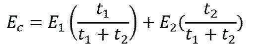

상기 식에서,Where
W는 표면상에서 고르게 분포된 부하량, 압력(Pa)이고,W is the load, pressure (Pa), distributed evenly on the surface,
R은 지지되지 않은 물질의 범위(m)이고,R is the range (m) of unsupported material,
E는 영 모듈러스(Young's Modulus) (Pa)이고,E is Young's Modulus (Pa),
t는 두께(m)이고,t is the thickness (m),
v는 포이손 비율(단위 없음, 물질 중에 일정한 것으로 추정됨)이고,v is the poisson ratio (unitless, estimated to be constant in the substance),
y는 휘어짐(m)이다.y is the deflection (m).
휘어짐에 대한 방정식은 문헌[참조: Theory and Analysis of Elastic Plates, Junuthula Narasimha Reddy, p.201 equation 5.3.43(1999)]에 기재되어 있다.Equations for bending are described in Theory and Analysis of Elastic Plates, Junuthula Narasimha Reddy, p.201 equation 5.3.43 (1999).
상기 방정식은 실질적으로 편평한 표면에 대한 상대적인 경도를 기재하고 있지만, 상기 방정식은 곡선 표면에 근접할 수 있고 당업자는 본원에 기재된 교시를 기준으로, 예를 들어, 유한 요소 모델링을 사용하여 실험적으로 휘어짐 부하 및 상대적 경도를 결정할 수 있다. Although the above equation describes a relative hardness for a substantially flat surface, the equation can be close to a curved surface and one skilled in the art can experimentally flex load using, for example, finite element modeling, based on the teachings described herein. And relative hardness.
표 A1. 본원에 기재된 바와 같은 보호막의 내부 부분의 물질, 모듈러스, 두께, 상대적 경도 Dk 및/또는 휘어짐 부하량. Table A1. Material, modulus, thickness, relative hardness Dk, and / or flexural loading of the inner portion of the protective film as described herein.
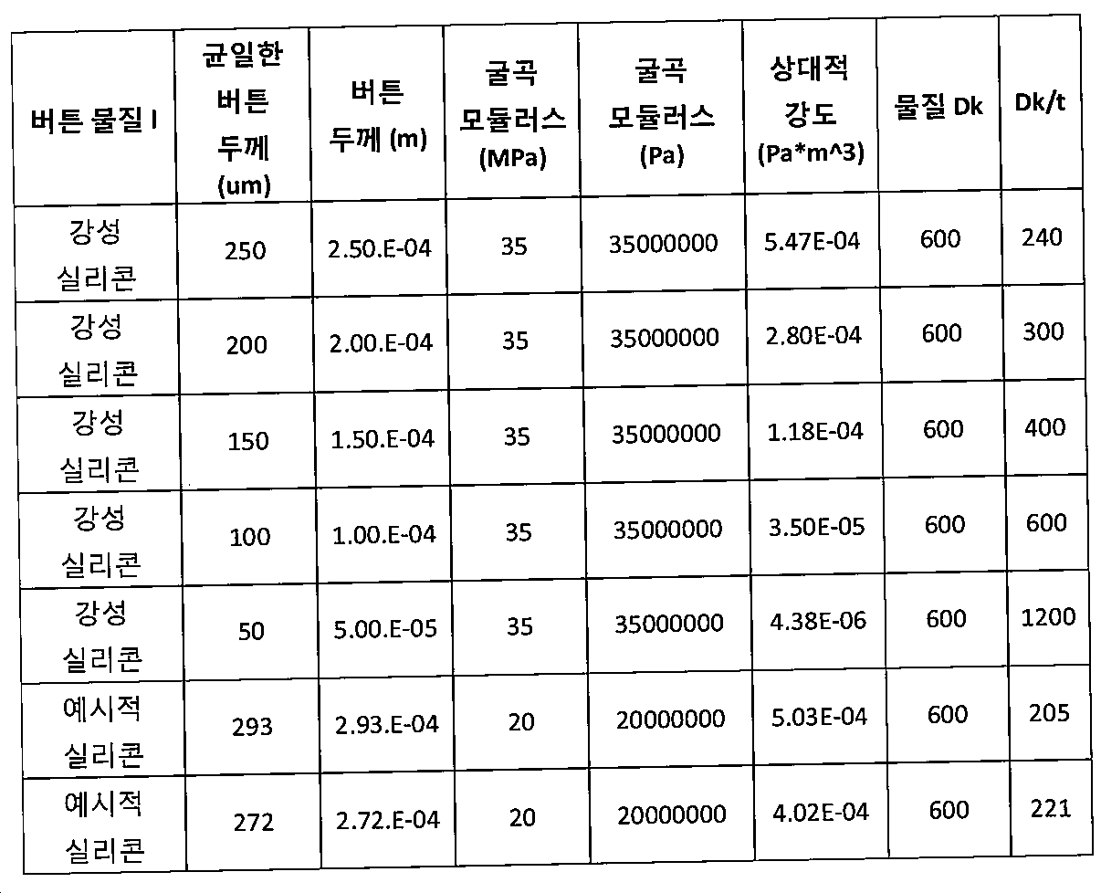
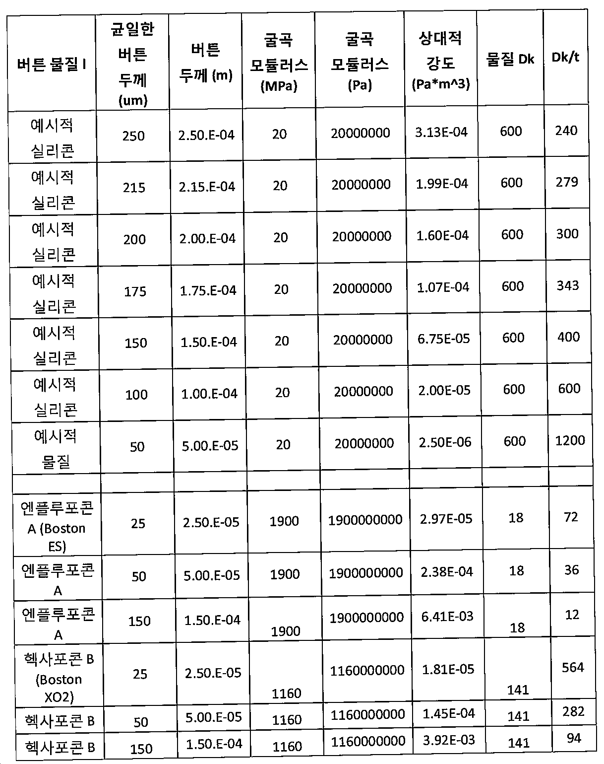
표 A1에 나타낸 바와 같이, 두께가 약 50㎛인 엔플루포콘 또는 헥사포콘과 같은 RGP 물질은 상피 평활화에 적합한 상대적 경도를 가질 수 있고 적어도 부분적으로 절제된 간질에 순응할 수 있다. 약 20MPa의 모듈러스 및 약 250㎛의 두께를 갖는 강성 실리콘은 약 2.4E-4의 상대적 경도를 제공하도록 약 50㎛의 두께 및 약 1900MPa의 모듈러스를 갖는 RGP 물질과 유사한 부하 하에 상대적 경도 3E-4 및 휘어짐을 제공할 것이다. 표 A1에 나타낸 바와 같은 시판되는 RGP 렌즈 물질은 보호막(100)을 제공하기 위해 본원에 기재된 바와 같은 구체예에 따라 조합될 수 있다. 본원에 기재된 교시를 기준으로, 당업자는 모듈러스 및 의도된 상대적 경도를 기준으로 보호막의 두께를 결정할 수 있다.As shown in Table A1, RGP materials, such as enflufocon or hexapocon, having a thickness of about 50 μm, may have a relative hardness suitable for epithelial smoothing and may be at least partially compliant with ablation epilepsy. Rigid silicon having a modulus of about 20 MPa and a thickness of about 250 μm has a relative hardness of 3E-4 and under similar loads as RGP materials having a thickness of about 50 μm and a modulus of about 1900 MPa to provide a relative hardness of about 2.4E-4 and Will provide warpage. Commercially available RGP lens materials as shown in Table A1 may be combined in accordance with embodiments as described herein to provide a
본원에 기재된 바와 같은 임상 연구에 따른 구체예와 관련된 연구는 약 3E-4 (3xlO-4 Pa*m∧3)의 상대적 경도를 갖는 보호막(100)의 내부 부분(110)이 시력을 개선시키고 적어도 부분적으로 눈에 순응하는데 효과적이어서 적어도 일부의 편안함을 제공하고 피팅을 개선시킬 수 있음을 보여 주었다. 많은 눈들은 많은 보호막들로 측정되었고 본 발명의 구체예와 관련된 연구는 약 1E-4 내지 약 5E-4(Pa*m∧A3) 범위내 상대적 경도를 갖는 내부 부분(110)이, 보호막이 본원에 기재된 바와 같이 절제부에 순응하고 상피를 평활화시킬 수 있도록 함을 지적한다. 예를 들어, 내부 부분(110)은 약 2E-4 내지 약 4E-4의 범위내 상대적 경도를 가질 수 있고 상기 눈은 보호막(100)의 휘어짐을 기준으로 상응하게 피팅될 수 있다.Studies relating to embodiments according to clinical studies as described herein indicate that the
상대적 경도는 눈 상의 보호막(100)의 휘어지는 양과 관련될 수 있다. 본 발명의 구체예와 관련된 연구는 내부 부분(110)의 상대적 경도, 약 3E-4가 눈위에 위치하는 경우 약 +/- 2D로 휘어지게 하여 약 2 또는 3mm의 내부 직경이 평활화되는 경우 대략 5 또는 6mm의 절제 직경에 걸쳐 약 +/- 2D 내까지 절제부에 순응할 수 있도록 함을 지적한다. 약 1.5 E-4의 상대적 경도를 갖는 보호막(100)은 눈 상에 위치하는 경우 약 +/- 4D로 휘어지게 할 수 있어 약 2 또는 3mm의 내부 직경이 평활화되는 경우 대략 5 또는 6mm 직경에 걸쳐 약 +/- 4D 이내의 절제부에 순응할 수 있다.The relative hardness may be related to the amount of warpage of the
보호막의 외부 부분은 각막의 외부 부분과 같은 눈의 외부 부분에 피팅되도록 하거나 결막 상에 위치하는 경우 공막에 피팅되도록 하기 위해 내부 부분 보다 적은 상대적 경도를 포함할 수 있다. The outer portion of the protective film may comprise less relative hardness than the inner portion to be fitted to an outer portion of the eye, such as the outer portion of the cornea, or to fit to the sclera when placed on the conjunctiva.
본원에 기재된 바와 같은 보호막은 표 A1의 많은 보호막 중 2개 이상의 값내의 범위에 상응하는 상대적 경도, 예를 들어, 약 2.50E-06 내지 약 6.41E-03(Pa*m∧3) 범위내 상대적 경도, 및 예를 들어, 6.75E-05 내지 약 5.47E-04(Pa*m∧3) 범위내 2개 이상의 중간 값을 포함할 수 있다. 본원에 기재된 교시를 기준으로, 보호막은 약 0.5E-3 내지 약 10E-3 (Pa*m∧3)의 범위, 예를 들어, 약 1E-3 내지 약 6 E-3의 범위내와 같은 많은 범위 중 하나 이상의 범위내에서 상대적 경도를 가질 수 있다. 본원에 기재된 교시를 기준으로, 당업자는 불규칙성을 평활화시키고 절제 영역에 실질적으로 순응하도록 보호막(100)에 대한 내부 부분(110)의 상대적 경도에 상응하는 두께 및 모듈러스를 측정하기 위한 임상적 연구를 수행할 수 있다.A protective film as described herein is a table corresponding to the relative hardness in the range of two or more values of a number of protective film of A1, for example, about 2.50E-06 to about 6.41E-03 (Pa * m ∧ 3) within the range of the relative Hardness and, for example, two or more intermediate values within the range of 6.75E-05 to about 5.47E-04 (Pa * m ∧ 3). Based on the teachings described herein, the protective film may be in a range of about 0.5E-3 to about 10E-3 (Pa * m ∧ 3), such as in the range of about 1E-3 to about 6 E-3. It may have a relative hardness within one or more of the ranges. Based on the teachings described herein, those skilled in the art conduct clinical studies to measure the thickness and modulus corresponding to the relative hardness of the
표 A2. 표 1의 보호막에 대한 3, 4, 5 및 6 mm의 직경에서의 5㎛ 휘어짐에 대한 압력Table A2. Pressure for 5 μm warpage at diameters of 3, 4, 5 and 6 mm for the protective membrane of Table 1
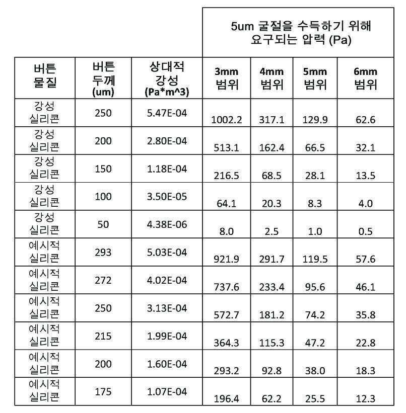

표 A1 및 A2의 데이타는 5㎛ 거리의 3mm 영역을 휘어지게 하기 위한 압력이 5㎛ 거리에서 4mm 영역을 휘어지게 하기 위한 압력의 약 3배 일 수 있고 5㎛ 거리에서 6mm 영역을 휘어지게 하기 위한 압력의 약 15배일 수 있음을 도시한다. 예를 들어, 약 3.13E-4 (Pa*m∧3)의 상대적 경도의 경우에, 5㎛ 휘어짐 압력은 각각 3, 4, 5 및 6mm의 직경에 대해 572.7, 181.2, 74.2, 35.8 (Pa)이고, 내부 부분(110)의 중심 3mm은 내부 부분(110)이, 예를 들어, 약 35Pa의 압력으로 6mm 범위를 가로지르는 절제부에 순응하는 경우 약 570Pa의 불규칙성에 대한 압착력을 제공할 수 있다. Hg의 mm로의 안내압(10P) 척도와 비교하여 보면, 12mm의 Hg는 약 1,600Pa이어서, 보호막은, 눈이 깜박이는 때에, 예를 들어, 6mm영역에 걸쳐서 각막에 순응할 수 있다. 눈이 깜박이는 때의 각막에 대한 보호막의 이러한 순응은 본원에 기재된 구체예에 따른 펌핑을 제공할 수 있다.The data in Tables A1 and A2 show that the pressure for deflecting the 3 mm area at 5 μm distance may be about three times the pressure for deflecting the 4 mm area at 5 μm distance and to deflect the 6 mm area at 5 μm distance. It can be about 15 times the pressure. For example, for a relative hardness of about 3.13E-4 (Pa * m ∧ 3), the 5 μm bending pressure is 572.7, 181.2, 74.2, 35.8 (Pa) for diameters of 3, 4, 5 and 6 mm, respectively. And the
상대적 경도 및 휘어짐 압력은 본원에 기재된 교시를 기반으로 하는 많은 보호막에 대해, 예를 들어, 복수의 물질을 갖는 복수의 층을 갖는 보호막에 대해 결정될 수 있다.Relative hardness and flexural pressure can be determined for many protective films based on the teachings described herein, for example for protective films having a plurality of layers having a plurality of materials.
표 A3. 층상 보호막의 상대적 경도Table A3. Relative Hardness of Layered Protective Film
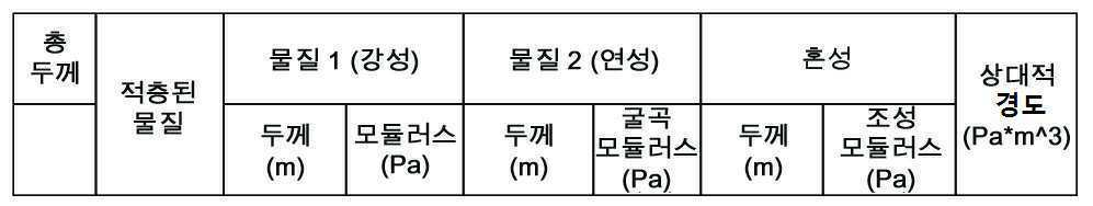

2개 이상의 물질이 2개 이상의 층을 제공하도록 조합되는 경우, 각각의 층의 상대적 경도는 총 복합 경도를 측정하기 위해 조합될 수 있다. 예를 들어, 상기 조합된 경도는 제 1 물질의 제1 층(100L1), 제 2 물질(M2)의 제 2 층(100L2) 및 제 3 물질(110M3)의 제 3 층(110L3)을 갖는 보호막에 대해 측정될 수 있고, 여기서, 상기 제 1 및 제 3 물질은 동일한 물질일 수 있다.If two or more materials are combined to provide two or more layers, the relative hardness of each layer can be combined to determine the total composite hardness. For example, the combined hardness is a protective film having a first layer 100L1 of a first material, a second layer 100L2 of a second material M2 and a third layer 110L3 of a third material 110M3. Can be measured, wherein the first and third materials can be the same material.
중량 평균 시스템을 사용하여 2개의 층을 하나의 물질로서 취급할 수 있다. 각각의 물질의 상대적 양 및 상기 2개의 물질의 모듈러스를 조합하여 각각의 층의 두께의 중량 평균을 기준으로 복합 모듈러스를 결정할 수 있다. 예를 들어, 90㎛의 20MPa 물질 층 및 10㎛의 5MPa 물질 층을 조합하여 다음과 같이 혼성 모듈러스를 결정할 수 있다:The weight average system can be used to treat the two layers as one material. The composite modulus can be determined based on the weighted average of the thickness of each layer by combining the relative amounts of each material and the modulus of the two materials. For example, the hybrid modulus can be determined as follows by combining a 20 MPa material layer of 90 μm and a 5 MPa material layer of 10 μm:
20MPa*0.9 + 5MPa*0.1 = 18.5MPa20MPa * 0.9 + 5MPa * 0.1 = 18.5MPa
본원에 기재된 방정식은 상이한 물질 및 두께의 많은 층들을 수용한다.The equations described herein accommodate many layers of different materials and thicknesses.
복합 모듈러스를 기준으로, 본 예에서 복합 모듈러스에 세제곱의 전체 두께를 곱할 수 있다: 18.5MPa * 100∧3. 이들 계산을 어림셈을 기준으로 할 수 있지만, 당업자는 시뮬레이션, 예를 들어, 무한 요소 모델링 시뮬레이션을 수행하여 본원에 기재된 바와 같은 상대적 경도의 양, 압력 및 휘어지는 힘 및 압력을 결정할 수 있다.Based on the compound modulus, in this example, the compound modulus can be multiplied by the total thickness of the cube: 18.5 MPa * 100 ∧ 3. These calculations can be based on approximation, but those skilled in the art can simulate, eg, infinite element modeling. Simulations can be performed to determine the amount of relative hardness, pressure, and bending forces and pressures as described herein.
보호막(100)의 하나 이상의 층의 굴절 지수는 실질적으로 각막의 굴절 지수에 상응할 수 있다. The refractive index of one or more layers of the
물질들(110M1, 11OM2 또는 110M3)의 하나 이상은 약 +/- 0.05 내로 각막의 굴절 지수와 일치하도록 약 1.38 내지 약 1.43 범위의 굴절 지수를 포함할 수 있다. 예를 들어, 상기 물질들(110M1 및 110M3)은 약 1.41의 굴절 지수를 갖는 광학적으로 투명한 연성 실리콘 탄성체를 포함할 수 있고 상기 물질(M2)은, 예를 들어, NuSil로부터 구입 가능한, 약 1.43의 굴절 지수를 갖는 광학적으로 투명한 강성 실리콘 탄성체를 포함할 수 있다. 대안적으로, 물질(110M1) 및 물질(110M3)은 실리콘 하이드로겔을 포함할 수 있고, 상기 물질(110M2)은, 예를 들어, 실리콘을 포함할 수 있다.One or more of the materials 110M1, 11OM2 or 110M3 may include a refractive index in the range of about 1.38 to about 1.43 to match the refractive index of the cornea within about +/− 0.05. For example, the materials 110M1 and 110M3 may comprise an optically transparent soft silicone elastomer having a refractive index of about 1.41 and the material M2 may be, for example, available from NuSil of about 1.43. Optically transparent rigid silicone elastomers having a refractive index. Alternatively, material 110M1 and material 110M3 may comprise a silicone hydrogel, and material 110M2 may, for example, comprise silicone.
상기 보호막은 보다 연성의 실리콘과 조합된 보다 큰 경도의 실리콘과 같은 유사한 물질을 포함할 수 있지만, 상기 보호막은 유사하지 않은 물질을 포함할 수 있다. 예를 들어, RGP 물질은 본원에 기재된 바와 같은 2중 곡선 또는 3중 곡선 실시예와 같은 하이드로겔과 조합할 수 있다. 상기 보호막은 안정성을 위해 적어도 윤부까지 연장될 수 있다. 상기 RGP 물질은 예를 들어, 표 A에 따라, 제 2 물질 (110M2)의 제 2 층(10012)를 포함할 수 있고 상기 하이드로겔은 제1 물질 110M1의 제1 층 10011을 포함할 수 있고 제 3 물질(110M3)의 제3 층(10013)을 포함할 수 있다. 상기 하이드로겔은 약 1.377 내지 약 0.05내의 각막의 굴절 지수와 일치하는 약 1.38 내지 약 1.42의 굴절 지수를 가질 수 있고, 예를 들어, 제조원(Vista Optics, UK)으로부터 시판되는 HEMA, NVP, GMA, MMA, SiH, TRS, HEMA/NVP, MMA/NVP, HEMA GMA, 또는 SiH/TRS 중 하나 이상을 포함할 수 있다. HEMA/NVP, MMA/NVP, 또는 HEMA/GMA를 포함하는 하이드로겔은 약 1.38 내지 약 1.43의 범위내 굴절 지수를 포함하도록 약 40% 내지 약 70% 범위의 물 함량을 가질 수 있다. 약 40%의 물 함량은 약 1.43의 굴절 지수에 상응하고 약 70%의 물 함량은 약 1.38의 굴절 지수에 상응한다. SiH/TRS를 포함하는 하이드로겔은 약 1.38 내지 약 1.43의 범위내에 굴절 지수를 포함하도록 약 20% 내지 약 70% 범위내 물 함량을 포함할 수 있다. 이들 SiH 하이드로겔과 함께, 약 20%의 물 함량은 약 1.43의 굴절 지수에 상응하고 약 70%의 물함량은 약 1.38의 굴절 지수에 상응한다.The protective film may include similar materials, such as silicon of greater hardness, combined with softer silicone, but the protective film may include dissimilar materials. For example, the RGP material can be combined with hydrogels such as the double curve or triple curve examples as described herein. The protective film may extend to at least the limbed portion for stability. The RGP material may comprise, for example, a second layer 10012 of a second material 110M2 and the hydrogel may comprise a first layer 10011 of a first material 110M1 and according to Table A. Third material 110M3 may include a third layer 10013. The hydrogel may have a refractive index of about 1.38 to about 1.42 that matches the refractive index of the cornea within about 1.377 to about 0.05, for example, HEMA, NVP, GMA, commercially available from Vista Optics, UK. One or more of MMA, SiH, TRS, HEMA / NVP, MMA / NVP, HEMA GMA, or SiH / TRS. Hydrogels comprising HEMA / NVP, MMA / NVP, or HEMA / GMA may have a water content in the range of about 40% to about 70% to include a refractive index in the range of about 1.38 to about 1.43. The water content of about 40% corresponds to the refractive index of about 1.43 and the water content of about 70% corresponds to the refractive index of about 1.38. Hydrogels comprising SiH / TRS may comprise a water content in the range of about 20% to about 70% to include a refractive index in the range of about 1.38 to about 1.43. With these SiH hydrogels, the water content of about 20% corresponds to the refractive index of about 1.43 and the water content of about 70% corresponds to the refractive index of about 1.38.
도 2aa는 본 발명의 구체예에 따른 눈꺼풀이 분리된 상태에서 눈에 위치된 콘택트 렌즈를 포함하는 보호막(100)을 도시한다. 보호막(100)은 눈물 액체(TL)가 보호막과 각막 사이의 보호막의 적어도 일부 아래에서 연장되어 챔퍼(100C)를 제공하도록 눈 상에 위치된다. 보호막(100)은 각막에 온 K(on K)로 또는 그보다 약간 편평하게 피팅되어 챔버(100C)를 제공할 수 있다. 대안적으로 또는 조합적으로, 외부 부분(120)의 플랜지(120F) 및 공막 커플링 부분(120S)은 결막보다 가파른 각도를 포함할 수 있어 상기 보호막은 내부 부분(110) 근처에서 챔버(100C)를 제공하도록 각막으로부터 이격되도록 촉진된다. 보호막(100)은 보호막의 중심에서 공막 커플링 부분(130)의 외부 주변(120P)으로의 융기부 거리에 상응하는 새그 높이 (105S1)을 포함한다. 눈꺼풀은 환자가 사물을 보이기 위해 분리될 수 있다.2A illustrates a
도 2ab는 눈꺼풀이 닫혀진 상태의 도 2aa의 보호막의 측단면도를 도시한다.FIG. 2ab shows a side cross-sectional view of the protective film of FIG. 2aa with the eyelid closed. FIG.
도 2ac는 본 발명의 구체예에 따라 닫히는 눈꺼풀과 함께 도 2aa의 보호막의 정면도를 도시한다. 눈꺼풀은 상부 눈꺼풀의 하향 이동(22A) 및 하부 눈꺼풀의 상향 이동(22B)로 닫힐 수 있다. 눈꺼풀의 닫힘은 보호막(100)에 압력을 나타내어 보호막(100)이 제 2 형태(100C2)를 포함하게 한다. 제 2 형태(100C2)는 제 2 새그 높이(105S2)로 감소되는 새그 높이(105)를 포함하여 챔버(100C)의 용적이 감소되고 보호막 아래로부터 펌핑된 눈물 액체(100TL)를 강제하게 한다. 펌핑된 눈물 액체 (100TL)는 외부 부분(120P) 아래에서 방사상으로 외부로 흘러 눈꺼풀에 덮히지 않는 않은 천공과 같은 천공(100F)을 통과한다. 눈꺼풀의 압력은 각막(100)을 향하도록 보호막(100)을 강제하여 챔버(100C)의 용적을 감소시킬 수 있다. 챔버(100C)의 용적은 플랜지(120F)를 포함하는 외부 부분(120)이 탄성 변형으로 휘어지는 경우 실질적으로 감소할 수 있다. 대안적으로 또는 조합적으로, 각막에 상응하는 외부 부분(120)은 휘어서 챔버(100C)의 용적을 감소시킬 수 있다. 많은 구체예에서, 광학 성분(100A)을 포함하는 내부 부분(110)은 눈꺼풀의 압력으로 휘어서 챔버 (100C)의 용적을 감소시킬 수 있다.2ac shows a front view of the protective film of FIG. 2aa with the eyelids closed in accordance with an embodiment of the present invention. The eyelid may be closed by
도 2ad는 본 발명의 실시예에 따라, 눈꺼풀 개방과 함께 도 2aa의 보호막의 측면 프로필을 도시한다. 눈꺼풀이 상부 눈꺼풀의 상향 이동(22C) 및 하부 눈꺼풀의 하향 이동(22D)으로 후퇴하는 경우, 보호막(100)은 제 1 새그 높이(105S1)를 갖는 제 1 형태(100C1)로 복귀할 수 있어 챔버의 용적이 증가한다. 공막 커플링 부분 (130)의 플랜지(120F) 및 주변부(120P)를 포함하는 외부 부분(120)은 결막과 접촉하여 결막과 접촉 밀봉을 형성할 수 있다. 결막과의 접촉 밀봉은 천공(100F)을 통한 챔버(100C)로의 눈물 액체(TL)의 흐름을 촉진시켜 펌핑된 눈물 액체(100TL)가 각막과 보호막(100) 사이에 위치할 수 있다. FIG. 2ad shows the side profile of the protective film of FIG. 2aa with the eyelid open, in accordance with an embodiment of the present invention. FIG. When the eyelid retracts with an upward movement 22C of the upper eyelid and a
하부 눈꺼풀의 눈물 리불렛은 눈의 표면상에 눈물 액체를 제공하도록 눈이 닫히는 때에 상향 이동할 수 있고, 상기 리불렛의 적어도 일부는 눈꺼풀이 서로 접촉하는 때에 상부 눈꺼풀에 커플링될 수 있다. 상부 눈꺼풀이 이동(22C)로 상향 이동하고 하부 눈꺼풀이 이동(22D)로 하향 이동하는 경우, 상부 눈꺼풀은 상부 천공 근처에 눈물 액체(TL)를 제공하여 상부 천공으로 통과시키고 하부 눈꺼풀은 하부 천공을 통해 이동하도록 하부 천공 근처에 눈물 액체(TL)을 제공할 수 있다.The tear eyelet of the lower eyelid may move upwards when the eye is closed to provide tear liquid on the surface of the eye, and at least a portion of the recaller may be coupled to the upper eyelid when the eyelids contact each other. When the upper eyelid moves upward with movement 22C and the lower eyelid movement with
눈의 반복적인 깜박거림은 자연적으로 발생하여 보호막 아래 눈물 액체를 펌핑하여 보호막 아래의 각막 및 결막을 세정한다. 보호막에 의해 제공되는 상기 펌핑 및 세정은 정상의 비연마된 눈을 갖는 환자가 보호막을 착용할 수 있는 시간의 양을 연장시키고, 예를 들어, PRK 후 눈에서의 상피 재생을 촉진할 수 있다.Repetitive blinking of the eye occurs naturally and pumps tear fluid under the protective film to clean the cornea and conjunctiva under the protective film. The pumping and cleaning provided by the protective film can prolong the amount of time a patient with normal unpolished eyes can wear the protective film and, for example, promote epithelial regeneration in the eye after PRK.
도 2ae는 본 발명의 실시예에 따라, 눈꺼풀이 분리되는 때에 보호막이 챔버를 한정하도록 각막의 외부 부분으로부터 분리된 보호막과 함께 각막 및 결막의 내부 부분으로 지지되도록 하여 눈 위에 위치한 콘택트 렌즈를 포함하는 보호막을 도시한다. 보호막(100)은 각막의 내부 부분에서, 예를 들어, 중앙 위치에서 각막과 접촉할 수 있다. 내부 부분(110)은 본원에 기재된 바와 같이 구심적으로 각막에 피팅되는, 예를 들어, 온 K 피팅으로 피팅되는 크기일 수 있다. 플랜지(120F) 및 공막 커플링 부분(130)을 포함하는 보호막(120)의 외부 부분은 내부 부분(110)이 구심적으로 공막과 접촉하는 때에 결막과 접촉하는 크기여서, 챔버(100C)가 각막의 외부 부분과 보호막 사이로 연장된 갭으로 각막의 외부 부분 상에 형성되게 한다. 각막의 외부 부분 상으로 연장하는 보호막의 외부 부분(120)은 각막보다 작은 곡률을 가져서, 각막의 외부 부분 상의 외부 부분(120)은 내부 부분(110)이 각막에 의해 지지되고 플랜지(120F)를 포함하는 외부 부분(120)이 결막과 커플링되는 때에 챔버(100C)를 형성할 수 있다. 천공(100F)은 눈꺼풀이 개방되는 경우 챔버(100C)와 갭의 위치에 상응하도록 보호막 상에 위치할 수 있다. 외부 부분(120)은 눈꺼풀이 개방되는 경우 챔버(100C)를 형성하기에 충분히 휘어짐에 대한 내성을 포함하고, 눈꺼풀이 닫히는 때에 외부 부분이 각막을 향하여 이동하고 갭 거리를 감소시키도록 눈꺼풀이 외부 부분 상으로 이동하는 때에 휘어짐에 저항하기에는 불충분한 내성을 포함한다.FIG. 2AE includes a contact lens positioned over the eye such that the protective film is supported by the inner portion of the cornea and conjunctiva with a protective film separated from the outer portion of the cornea to define the chamber when the eyelid is detached, in accordance with an embodiment of the present invention. A protective film is shown. The
보호막(100)은 각막에 피팅되어 챔버(100C)의 형성을 촉진시킬 수 있고 보호막(100)은 아래 형성된 챔버(100C)를 지닌 초기 형태(100C1)를 포함한다. 각막은 각막의 정점으로부터 윤부로 연장되는 융기부 거리에 상응하는 윤부 새그 높이(105L)를 포함할 수 있다. 윤부는 눈의 측정 축으로부터 방사상 거리(105RL)에 위치할 수 있다. 눈은 눈의 축으로부터 방사상 거리(105RC)에서 결막 새그 높이(105C)를 포함할 수 있다. 보호막은 윤부로의 방사상 거리(105RL)에 상응하는 위치에서 윤부 새그 높이(105LC)를 포함할 수 있다. 보호막은, 예를 들어, 플랜지(120F)를 따라 결막의 방사상 거리(105RC)에 상응하는 결막 접촉 위치에서 결막 새그 높이(105CC)를 포함할 수 있다. 많은 구체예에서, 윤부에 상응하는 위치에서 보호막의 새그 높이(105LC)는 윤부 새그 높이(105L) 이하이고 결막에 상응하는 위치에서 보호막의 새그 높이(105CC)는 결막 새그 높이(105C) 이하여서, 윤부에 대한 압력이 감소한다. 보호막이 눈 위에 위치하는 경우, 플랜지 부분(120F)을 포함하는 결막 커플링 부분(130)은 휘어져서 결막 접촉 부분의 새그 높이는 결막의 새그 높이(105CC)로부터 결막의 새그 높이(105C)로 감소하여 보호막의 새그 높이가 새그 휘어진 새그 높이(105S2)를 포함하게 한다.The
도 2af는 보호막(100)이 감소된 용적을 갖는 챔버(100C)를 지닌 형태(100C2)를 포함하도록, 닫히는 눈꺼풀과 함께 도 2ae의 보호막의 측단면도를 도시한다. 눈꺼풀이 닫히는 경우, 상부 및 하부 눈꺼풀은 보호막에 압력을 가하여 상기 보호막이 각막 및 결막의 외부 부분을 향하도록 강제된다. 각막의 외부 부분 상의 보호막의 외부 부분은 휘어짐에 대해 충분한 내성을 갖지 못하여 상기 보호막의 외부 부분이 각막의 외부 부분을 향하여 하향으로 휘어지게 한다. 각막의 외부 부분 상의 보호막의 외부 부분 사이에서 연장하는 갭 거리는 감소하여 챔버(100C)의 용적이 감소하고 펌핑된 눈물 액체(100TL)가 챔버(100C)로부터 천공(100F)을 통해 플랜지 부분(120F)을 포함하는 결막 접촉 부분(130) 아래로 흐르게 한다. 상부 눈꺼풀은 동공을 거쳐 연장하여 하위 및 상위 천공(100F)을 덮을 수 있다. 상부 눈꺼풀은 하부 눈꺼풀과 접촉하여 눈이 개방되는 때에 상위의 리블렛의 눈물 액체를 유도하여 리불렛의 눈물 액체가 하위 및 상위 천공을 통해 챔버 내로 유도될 수 있게 한다. FIG. 2af shows a side cross-sectional view of the protective film of FIG. 2ae with the closed eyelids such that the
각막의 외부 부분 상에서 보호막의 외부 부분의 휘어짐은 약 1.0E-6 Pa*m∧3 내지 약 6E-4Pa*m∧3, 예를 들어, 약 2.5 E-6 Pa*m∧3 내지 약 5E-4 Pa*m∧3의 범위내 상대적 경도를 갖는 보호막과 함께 제공될 수 있다. 표 A2는 상대적 경도 및 각막의 외부 부분에 상응하는 외부 부분(120)의 상응하는 범위의 적합한 값을 보여주고, 이는 본원에 기재된 교시를 토대로 결정될 수 있고 이로써 보호막의 외부 부분의 상대적 경도를 결정하여 휘어짐에 대한 내성을 제공하고 눈꺼풀이 보호막의 부분으로 이격되는 경우 갭과 함께 챔버를 형성하며 각막을 향해 휘어지도록 하고 눈꺼풀이 보호막의 부분을 덮는 경우 갭 및 상응하는 챔버 용적을 감소시킨다.The warpage of the outer portion of the protective film on the outer portion of the cornea is between about 1.0E-6 Pa *
결막에 커플링시키기 위한 공막 접촉 부분(130)의 휘어짐은 약 2 E-4 Pa*m∧3 이하, 예를 들어, 약 1E-4 Pa*m∧3 이하, 및 많은 구체예에서, 약 2E-5Pa*m∧3 이하의 상대적 경도를 포함하는 공막 접촉 부분(130)과 함께 제공될 수 있다. 표 A2는 본원에 기재된 바와 같은 교시를 기준으로 결정될 수 있는 공막 커플링 부분 (130)의 상대적 경도 및 상응하는 범위에 적합한 값을 보여주고, 이로써 보호막의 공막 커플링 부분의 상대적 경도를 결정하여 휘어짐에 대한 내성을 제공하고 눈꺼풀이 보호막 부분으로 이격되는 경우 갭과 함께 챔버를 형성하여 각막을 향하여 휘어지도록 하고 눈꺼풀이 각막의 외부 부분상의 보호막의 외부 부분을 포괄하는 경우 갭 및 상응하는 챔버 용적을 감소시킨다..The warpage of the
결막에 커플링시키기 위한 플랜지 부분(120F)의 휘어짐은 약 1E-4 Pa*m∧3 이하, 예를 들어, 약 2E-5 Pa*m∧3 이하, 및 많은 실시예에서 약 2.5E-6 Pa*m∧3 이하의 상대적 경도를 포함하는 플랜지 부분(130)과 함께 제공될 수 있다. 표 A2는 본원에 기재된 바와 같은 교시를 기준으로 결정될 수 있는 외부 플랜지 부분(120F)의 상대적 경도 및 상응하는 범위에 적합한 값을 보여주고, 이로써 보호막의 플랜지 부분(120F)의 상대적 경도를 결정하므로써 휘어짐에 대한 내성을 제공하고 눈꺼풀이 보호막으로부터 이격되는 경우 갭과 함께 챔버를 형성하여 각막을 향해 휘어지고 눈꺼풀이 각막의 외부 부분상에 보호막의 외부 부분을 포괄하는 경우 갭과 상응하는 챔버 용적을 감소시킨다.The bending of the
도 2ag는 본 발명의 실시예에 따라, 눈꺼풀이 닫히는 경우, 눈물 액체가 펌핑되는 때에 상피를 따른 보호막의 슬라이딩이 억제되도록, 눈꺼풀이 닫히는 때의 눈의 회전과 함께 도 2af의 보호막의 측단면도를 도시한다. 눈의 축은 상위로 회전하여 보호막이 상부 눈꺼풀과 하부 눈꺼풀을 따라 슬라이딩하도록 할 수 있다. 눈의 축은 눈의 하나 이상의 공지된 축을 포함할 수 있고 당업자에 의해 많은 방식으로 측정될 수 있다.FIG. 2AG is a side cross-sectional view of the protective film of FIG. 2AF with rotation of the eye when the eyelid is closed, such that when the eyelid is closed, sliding of the protective film along the epithelium when the tear liquid is pumped is suppressed, in accordance with an embodiment of the present invention. Illustrated. The axis of the eye can rotate upward to allow the protective film to slide along the upper and lower eyelids. The axis of the eye can include one or more known axes of the eye and can be measured in many ways by those skilled in the art.
도 2ah는 본 발명의 구체예에 따라, 눈꺼풀의 개방과 함께 도 2ae의 보호막의 측단면도를 도시한다. 눈꺼풀의 개방이 압력을 감소시키고 각막의 외부 부분 위에 있는 보호막의 외부 부분이 각막으로부터 이격되어 이동하도록 한다. 눈물 액체(TL)는 천공(100F)을 통해 챔버(100C)로 통과할 수 있다. 부분(130) 및 플랜지(120F)를 포함하는 보호막의 외부 부분은 결막과 접촉하여 눈물 흐름을 억제할 수 있고 보호막을 밀봉시킬 수 있다.FIG. 2ah shows a cross-sectional side view of the protective film of FIG. 2ae with the opening of the eyelid, according to an embodiment of the invention. The opening of the eyelids reduces the pressure and causes the outer portion of the protective film over the outer portion of the cornea to move away from the cornea. Tear liquid TL may pass through
도 2ai는 본 발명의 구체예에 따라, 챔버가 중간 형태(100C12) 용적을 포함하도록 중간 위치에서 위치되는 눈꺼풀과 함께 도 2ae의 보호막의 측단면도를 도시한다. 내부 부분(110)을 포함하는 광학 성분(100A)은 보호막이 챔버(100C)의 용적을 감소시키기도록 휘어지는 외부 부분(120)을 갖는 중간 부분(100C12)을 포함하는 때에 환자에 대한 시력을 제공하도록 휘어짐에 대해 충분한 경도 및 내성을 포함할 수 있다. 예를 들어, 환자는 외부 부분을 휘어지게 하기 위해 동공 가장자리에 대해 눈꺼풀을 닫을 수 있고, 광학 성분(100A) 및 내부 부분(110)은 실질적으로 휘지않게 유지될 수 있어 환자는 하나 이상의 눈꺼풀의 일부가 내부 부분(110)과 접촉하는 상태에서 20/20 이상(미터법 6/6 이상)의 시력을 가질 수 있다. 눈꺼풀의 개방은 챔버 용적을 증가시키고 눈물 액체를 펌핑할 수 있고 눈꺼풀의 닫힘은 챔버 용적을 감소시켜 눈물 액체를 펌핑할 수 있다.FIG. 2A-I shows a cross-sectional side view of the protective film of FIG. 2AE with the eyelid positioned in an intermediate position such that the chamber contains an intermediate form 100C12 volume, in accordance with an embodiment of the present invention. The
도 2aj는 하이드로겔이 눈과 접촉하면서 눈에 위치하는 도 1av의 보호막의 측단면도를 도시한다. 보호막(100)은 눈을 하이드로겔 층의 적어도 일부와 접촉시키기 위한 보호막의 후방 표면을 따라 연장하는 하이드로겔 물질(120MHG)의 층을 포함한다. 보호막(100)은 적어도 부분적으로 하이드로겔 물질의 층에 의해서 한정되는 챔버(100C)를 형성하도록 하는 치수일 수 있다. 천공은 하이드로겔 층을 통해 연장하여 본원에 기재된 바와 같은 펌핑을 제공할 수 있다. 대안적으로 또는 조합적으로, 천공의 후방 말단은 하이드로겔 물질 층으로 덮혀서 각막을 하이드로겔 물질의 층을 지닌 천공에 커플링시킬 수 있다. 하이드로겔 물질(120MHG)의 층으로 덮힌 천공은 보호막의 휠 수 있는 부분을 따라 위치하여, 예를 들어, 눈을 깜박이는 때에, 하이드로겔 물질을 따라 물과 치료학적 제제의 이동을 촉진시킬 수 있다. 하이드로겔 층은, 예를 들어, 액체 및 치료학적 제제를 각막의 중심 위치로 위킹(wicking)함에 의해서, 액체 및 치료학적 제제를 천공으로부터 각막의 요망되는 위치로 통과시키기 위한 매질을 포함할 수 있다. 본원에 기재된 바와 같이 하부 표면을 따라 연장하는 하이드로겔 층을 포함하는 보호막은 비절제된 눈에 피팅되어 굴절 보정을 제공할 수 있거나 본원에 기재된 바와 같이 절제된 눈에 피팅될 수 있다.FIG. 2aj shows a cross-sectional side view of the protective film of FIG. 1av with hydrogel in contact with the eye and positioned in the eye. FIG.
본 발명의 구체예에 따른 임상 시험은 보호막의 곡선 부분이 환자 집단의 각막 곡률 및 새그 높이 및 윤부 새그 높이 및 결막 새그 높이에 따라 온 K-값으로 피팅될 수 있음을 도시한다.Clinical trials according to embodiments of the present invention show that the curved portion of the protective film can be fitted to an on-K-value depending on corneal curvature and sag height and limbal sag height and conjunctival sag height of the patient population.
하기 본원에 나타낸 첨부 I는 본원에 기재된 구체예 및 교시에 따른 보호막(100)에 대한 치수 및 피팅 파라미터를 제공한다. 보호막은, 예를 들어, 본원에 나타낸 A 계열 표내의 물질 중 하나 이상을 포함할 수 있다. 보호막의 치수 및 피팅 파라미터는 본원에 기재된 구체예에 따라 각막 상에 위치하는 때에 눈물 액체의 펌핑을 제공할 수 있다. 첨부 I의 표는, 예를 들어, 가파른 K 각막, 중간 K 각막 및 편평한 K 각막과 함께 사용하기 위한 보호막을 확인하고 있다. 열거된 K 값은 집단 표준을 기초로 할 수 있어서, 눈 위에 위치하는 때에 보호막이 본원에 기재된 바와 같은 펌핑을 제공하게 한다. 보호막은 비-절제된 눈 또는 절제된 눈에 사용될 수 있고, 보호막은 제 1 내부 곡률(R1)을 기준으로 적어도 부분적으로 확인될 수 있다 .Appendix I, set forth herein below, provides the dimensions and fitting parameters for the
표 B1은 대략 14mm의 횡단 직경을 갖는 보호막 100을 보여주고 예를 들어, 본원에 기재된 바와 같이 K 또는 편평하게 조립될 수 있다. 상기 표는 각막의 중심 연마된 부분에 상응하는 R1을 열거한다. 광학 성분 100A 및 내부 커플링 구성요소 100B1을 포함하는 내부 부분 110은 약 5mm를 가로질러 연장하는 차원 R1을 갖고 연마 영역은 보다 크고 예를 들어 약 6mm일 수 있다. 반경 R1B1에 상응하는 부분은 약 5 내지 7mm의 횡단 차원을 갖고 곡률은 디옵터(D)의 눈의 광출력에 상응하는 각막곡률측정 값(K-값)으로 표현될 수 있다. 반경 R1B2에 상응하는 부분은 약 7 내지 9mm의 횡단 차원을 갖는다. 반경 R1B3에 상응하는 부분은 약 9 내지 11mm 횡단 차원을 갖는다. R1C1에 상응하는 부분은 약 11 내지 13.5mm를 가로질러 연장할 수 있고 부분 R1B3 및 부분 R1C2 사이의 하나 이상의 값을 갖는 곡률, 예를 들어, 약 10mm와 같은 약 8mm 내지 약 12mm의 곡률반경을 포함할 수 있다. R1C2에 상응하는 부분은 약 13.5 내지 14mm를 가로질러 연장될 수 있다. 부분 R1C2의 새그 높이는 예를 들어, 약 3.1 내지 약 3.4mm일 수 있다. R1C1에 상응하는 부분은 본원에 기재된 바와 같이 내부 경계상에 R1B3으로 배열된 부분 R1C1 및 외부 경계를 따라 위치하는 R1C2의 접선과 함께 본원에 기재된 바와 같은 융기부 형성을 억제하도록 많은 방식으로 각막에 조립될 수 있다.Table B1 shows a
표 B2는 예를 들어, 본원에 기재된 바와 같이 대략 14mm를 가로지르는 직경을 갖는 보호막 100을 보여주고 K 또는 편평하게 조립될 수 있다. 상기 표는 각막의 중심 연마된 부분에 상응하는 R1을 열거한다. 광학 성분 100A 및 내부 커플링 구성요소 100B1을 포함하는 내부 부분 110은 약 5mm를 가로질러 연장하는 차원 R1을 갖고 연마 영역은 보다 크고 예를 들어, 약 6mm일 수 있다. 반경 R1B1에 상응하는 부분은 약 5 내지 7mm의 횡단 차원을 갖고 곡률은 디옵터(D)의 눈의 광출력에 상응하는 각막곡률측정 값(K-값)으로 표현될 수 있다. 반경 R1B2에 상응하는 부분은 약 7 내지 9mm의 횡단 차원을 갖는다. 반경 R1B3에 상응하는 부분은 약 9 내지 11mm 횡단 차원을 갖고, 이들 값의 범위는 약 35.75 내지 약 40이고 각각의 값은 어느정도 표 B1의 상응하는 값 보다 주변부에서 더 편평하다. 예를 들어, 표 B1은 약 36.75 내지 약 41D의 범위를 가즌 것으로서 R1B3의 값을 열거한다. R1C1에 상응하는 부분은 약 11 내지 13.5mm을 가로질러 연장할 수 있다. R1C2에 상응하는 부분은 약 13.5 내지 14mm 가로질러 연장될 수 있다. 부분 R1C2의 새그 높이는 예를 들어, 약 3.1 내지 약 3.4mm일 수 있다. R1C1에 상응하는 부분은 예를 들어, 본원에 기재된 바와 같이 내부 경계상에 R1B3로 배열된 부분 R1C1 및 외부 경계를 따라 위치하는 R1C2의 접선과 함께 본원에 기재된 바와 같은 융기부 형성을 억제하도록 많은 방식으로 각막에 조립될 수 있다.Table B2 shows, for example, the
표 B3은 예를 들어, 본원에 기재된 바와 같이 대략 16mm를 가로지르는 직경을 갖는 보호막 100을 보여주고 K 또는 편평하게 조립될 수 있다. 상기 표는 각막의 중심 연마된 부분에 상응하는 R1을 열거한다. 광학 성분 100A 및 내부 커플링 구성요소 100B1을 포함하는 내부 부분 110은 약 5mm를 가로질러 연장하는 차원 R1을 갖고 연마 영역은 보다 크고 예를 들어, 약 6mm일 수 있다. 반경 R1B1에 상응하는 부분은 약 5 내지 7mm의 횡단 차원을 갖고 곡률은 디옵터(D)의 눈의 광출력에 상응하는 각막곡률측정 값(K-값)으로 표현될 수 있다. 반경 R1B2에 상응하는 부분은 약 7 내지 9mm의 횡단 차원을 갖는다. 반경 R1B3에 상응하는 부분은 약 9 내지 10.5mm 횡단 차원을 갖고 이들 수치는 약 36.75 내지 약 41의 범위이다. R1C에 상응하는 부분은 약 13 내지 약 16mm를 가로질러 연장할 수 있다. 부분 R1C2의 새그 높이는 예를 들어, 약 3.6mm 미만이어서 부분 R1C2는 눈에 위치하는 경우 휘어질 수 있다. R1C1에 상응하는 부분은 본원에 기재된 바와 같이 많은 방식으로 각막에 조립될 수 있다.Table B3 shows, for example,
표 B4는 예를 들어, 연장된 착용 콘택트 렌즈와 함께 본원에 기재된 바와 같은 눈물 액체를 펌핑하기 위해 비-연마된 눈에 사용하기 위한 곡률을 갖는 보호막 100을 도시한다. 보호막 100은 예를 들어, 본원에 기재된 바와 같이, 대략 14mm을 횡단하는 직경을 갖고 K 또는 편평하게 조립될 수 있다. 상기 표는 각막의 중심 연마된 부분에 상응하는 R1을 열거한다. 광학 성분 100A 및 내부 커플링 구성요소 100B1을 포함하는 내부 부분 110은 약 5mm를 가로질러 연장하는 차원 R1을 갖는다. R1에 상응하는 내부 부분의 곡률은 약 39D 내지 약 48D의 광출력에 상응하는 곡률값을 갖고, 상기 광출력은 비연마된 눈에 대한 집단 데이타를 기준으로 할 수 있고 예를 들어, 부분 R1B1 내지 R1B3, 및 R1C1 및 R1C2에 대한 곡률과 조합될 수 있다. 반경 R1B1에 상응하는 부분은 약 5 내지 7mm 횡단하는 차원을 갖고 상기 곡률은 디옵터(D)의 눈의 광출력에 상응하는 각막곡률측정 값(K-값)으로 표현될 수 있다. 반경 R1B2에 상응하는 부분은 약 7 내지 9mm의 횡단 차원을 갖는다. 반경 R1B3에 상응하는 부분은 약 9 내지 11mm 횡단 차원을 갖는다. R1C1에 상응하는 부분은 약 11 내지 약 13.5mm를 가로질러 연장할 수 있다. R1C2에 상응하는 부분은 약 13.5 내지 14mm 가로질러 연장될 수 있다. 부분 R1C2의 새그 높이는 예를 들어, 약 3.1 내지 약 3.4mm일 수 있다. R1C1에 상응하는 부분은 본원에 기재된 바와 같이 내부 경계상에 R1B3으로 배열된 부분 R1C1 및 외부 경계를 따라 위치하는 R1C2의 접선과 함께 본원에 기재된 바와 같은 융기부 형성을 억제하도록 많은 방식으로 각막에 조립될 수 있다.Table B4 shows a
표 B1 내지 B4가 예를 들어, 특이적 곡률 값을 나타내지만 당업자는 본원에 기재된 교시 및 실시예를 기준으로 많은 곡률값을 결정할 수 있고 하나 이상의 곡률은 비구형 표면, 예를 들어, 원뿔 상수를 갖는 비구형 표면과 조합될 수 있다.Although Tables B1 to B4 show specific curvature values, for example, those skilled in the art can determine many curvature values based on the teachings and examples described herein and one or more curvatures may be used to determine non-spherical surfaces such as cone constants. It can be combined with a non-spherical surface having.
도 3a는 상피 결함(11)을 지닌 눈(2)의 각막(10) 상에 위치된 보호막(100)을 도시하고 있다. 보호막은 곡선 바디(curved body), 예를 들어, 각막에 피팅되는 모양의 곡선 콘택트 렌즈 바디를 포함할 수 있다.FIG. 3A shows a
보호막(100)은 절제된 프로필과 상피 결함을 덮는 크기일 수 있다. 내부 부분(110)은 주요 절제부를 가로질러 연장되는 크기일 수 있는 횡단 치수(102)를 포함하고, 외부 부분(120)은 적어도 상피 결함을 가로질러 연장되고 결함부의 반대측상의 상피와 접촉되는 크기의 횡단 치수(104)를 포함한다. The
주요 절제부를 가로지러 연장되는 치수(102)는 예를 들어, 약 6 내지 8mm로 연장될 수 있고, 절제부보다 크기가 더 클 수 있다. 치수(104)는 예를 들어, 약 12 내지 약 14mm의 횡단 치수를 포함하여, 예를 들어, 윤부로 연장될 수 있고, 예를 들어, 환자의 윤부에 이르는 크기일 수 있다. 본 발명의 구체예와 관련된 연구는 윤부 둘레에서 윤부로 연장되는 크기의 보호막은 각막 상에 중심이 있을 수 있다. 보호막은 보호막의 외부 림이, 예를 들어, 윤부 주변의 공막 위에 배치된 결막과 접촉하도록 그리고, 예를 들어, 그러한 형태가 각막상의 렌즈를 중심에 오게 조정할 수 있도록 연장될 수 있다.The
보호막의 두께는 많은 방식으로 크기 조절되고 성형될 수 있다. 보호막의 내부 부분(110)은 두께(106)를 포함하고, 보호막의 외부 부분(120)은 두께(108)를 포함한다. 내부 부분의 두께(106)는 눈에 위치시키기 전에, 예를 들어, 눈의 전면에 유지되고 일정한 거리로 각막으로부터 분리되는 때에, 내부 부분이 약 +/- 1D 이하의 광출력을 포함하도록 하는, 실질적으로 균일한 두께를 포함할 수 있다. 대안적으로, 내부 부분의 두께는 옵티컬 파워, 예를 들어, 환자의 시력을 교정하는 옵티컬 파워를 포함하도록 다양할 수 있다.The thickness of the protective film can be sized and molded in many ways. The
재생된 상피(12R)의 평활 층(12S)은 절제된 프로필을 실질적으로 덮을 수 있다. 환경(100E)은 상피 재생을 유도하고 재생된 상피를 평활시키도록 구성된다. 재생 상피는 두께 프로필(12RP)을 포함한다.Smoothing layer 12S of regenerated
상피는 화살표(30)로 나타낸 바와 같이 주변 경계(12E)로부터 절제된 프로필(20)의 중심을 향해서 구심적으로 성장하여 노출된 간질을 덮는다.The epithelium grows centripetally toward the center of the resected
보호막(100)은 내부 부분(110)과 외부 부분(120)을 포함할 수 있다. 외부 부분(110)은 상피 결함 및 절제부의 가장자리 근처에서 각막과, 예를 들어, 연질의 순응성 물질, 예컨대, 실리콘 탄성체 또는 실리콘 하이드로겔과의 밀봉부(100S)를 형성하도록 구성될 수 있다. 내부 부분(120)은 동공 상에 위치되고, 환자가 볼 수 있도록 구성되며, 각막이 치유되는 때에 상피의 불규칙성을 평활화시키도록 외부 부분보다 더 큰 강성을 포함할 수 있다. 대안적으로, 내부 부분은 또한 외부 부분의 강성과 동일하거나 그보다 작은 강성을 포함할 수 있다. 예를 들어, 내부 부분은 실리콘을 포함할 수 있고, 외부 부분도 실리콘을 포함할 수 있으며, 내부 부분이 외부 부분보다 더 강성이어서 상피를 평활화시킬 수 있도록, 내부 부분은 더욱 강성인 실리콘 또는 더 큰 두께 중 하나 이상을 포함할 수 있다. 내부 부분이 외부 부분보다 더 강성일 수 있지만, 내부 부분은 간질내의 절제된 프로필(20)에 적어도 부분적으로 순응되도록 충분하게 연성이며, 가요성이고 순응성이어서, 환자가 내부 부분을 통해서 보고 내부 부분이 상피를 평활화시키는 때에 환자가 절제 프로필(20)에 의한 시력 교정의 이익을 얻게 할 수 있다. 본 발명의 구체예와 관련된 연구는 재생 상피가 절제 프로필(20)의 하부 간질보다 더 연성이어서, 내부 부분이, 예를 들어, 본원에 기재된 바와 같은 휘어짐 압력으로, 내부 부분 아래에 배치된 상피를 평활화시키는 때에, 내부 부분이 절제 프로필(20)의 모양에 순응하도록 구성될 수 있게 한다.The
도 3b는 상피 결함을 지니는 눈, 예컨대, PRK 절제를 지니는 눈의 각막 상에 위치되기 전의 제 1 형태의 보호막(100)을 도시하고 있다. 보호막(100)은 천공(100F)을 포함한다. 천공(100F)은 천공이 상피 결함부로부터 이격되어 위치하여 본원에 기재된 바와 같이 보호막 아래에서 눈물 액체를 펌핑하도록 보호막 상에 위치된다. 보호막(100)은 기저부 곡률반경(R1)을 지니는 내부 부분(110)을 포함할 수 있고, 기저부 곡률반경은 절제된 각막보다 약간 더 길어서, 보호막이 각막 상에 위치되기 전에 각막보다 더 편평할 수 있다. 공막 커플링 부분(130)을 포함하는 외부 부분(120)은 윤부에 대한 압력을 감소시키도록 각막보다 더 가파른 부분을 포함할 수 있다. 예를 들어, 플랜지 부분(120F)이 윤부 상의 보호막의 압력을 감소시키도록 결막과 공막의 상응하는 부분보다 더 가파를 수 있다.3B shows a
기저부 반경(R1)은 많은 방식으로 각막에 이르는 크기일 수 있다. 예를 들어, 기저부 반경(R1)은 절제 후의 눈에 상응하는 반응을 지닐 수 있다.The base radius R1 may be sized to the cornea in many ways. For example, the base radius R1 can have a response corresponding to the eye after ablation.
보호막(100)은 약 4MPa 내지 약 35MPa 범위내 모듈러스를 포함하여 중심부가 적어도 부분적으로 절제된 간질에 순응할 수 있고 상기 보호막이 절제된 각막의 각막 불규칙성 및 간질 불규칙성을 평활화시킬 수 있다. 보호막은 탄성 연신 가능한 물질을 포함하여 보호막이, 예를 들어, 각막에 피팅되도록 연신될 수 있다. 약 4MPa 내지 약 35MPa 범위내 모듈러스를 갖는 보호막은 본원에 기재된 바와 같은 많은 방식으로 형성될 수 있다. 예를 들어, 보호막은 절제된 각막 및 비절제된 각막의 적어도 일부를 가로질러 연장되는 실질적으로 균일한 두께를 갖는 단일 조각의 물질을 포함할 수 있고, 단일 조각의 물질은 실리콘 탄성체 또는 하이드로겔과 같은 탄성 물질을 포함할 수 있다. 대안적으로, 보호막은 절제된 각막 및 비절제된 각막의 적어도 일부를 가로질러 연장되는 비-균일한 두께를 갖는 단일 조각의 물질을 포함할 수 있다. 보호막은 많은 방식으로 성형될 수 있고 단일 조각의 하나의 물질을 포함하거나 2개의 유사한 물질로 구성된 단일 조각을 포함할 수 있거나, 함께 연결된 복수의 물질을 포함할 수 있다.The
보호막(100)은 본원에 기재된 내부 부분 밖에서 연장되는 하나 이상의 외부 부분을 포함할 수 있다.
도 3c는 절제된 간질 조직에 순응하고 절제된 간질 상의 상피를 평활화시키는 제 2 형태(100C2)를 지녀서, 보호막이 본원에 기재된 바와 같이 눈물 액체를 펌핑하도록 하는 눈에 위치되는 도 3b의 보호막을 도시하고 있다. 각막은 상응하는 곡률반경, 예를 들어, 반경(R2)을 가질 수 있는 시력을 보정하기 위한 절제된 표면(20)을 포함한다. 절제된 프로필(20)은 눈의 수차 및 각막으로 절제된 난시를 보정하기 위해 각막 내로 절제된 수차와 같은 반경(R2)에 상응하는 것들을 갖는 추가의, 또 다른 또는 조합적 모양을 포함할 수 있고, 보호막(100)의 내부 부분(110)은 각막의 상기 절제된 프로필에 순응할 수 있어서 보호막이 각막 상에 위치하는 경우 환자가 절제 시력 보정의 이득을 수용할 수 있게 한다. 예를 들어, 상기 각막 절제 프로필(20)은 곡률반경(R2)에 상응할 수 있고, 내부 부분(110)은 연마된 프로필(20)에 실질적으로 상응하는 제 2 형태(100C2)로 위치하기 전에 곡률반경(R1)에 상응하는 형태(100C1)로부터 편평해질 수 있어서, 환자가 절제 프로필(20)의 이득과 함께 볼 수 있게 한다. 예를 들어, 제 2 형태(100C2)는 곡률반경(R2)에 실질적으로 상응하는 순응 곡률반경(R12)을 포함할 수 있다. 보호막(100)의 제 1 형태(100C1)에 상응하는 프로필은 각막 상에 위치된 대로 도시되어, 각막 상에 위치되는 때에 보호막(100)의 순응하는 형태(100C2)로 위치시키기 전에 형태(100C1)로부터 보호막의 프로필에서의 변화를 예시하고 있다.FIG. 3C shows the protective film of FIG. 3B having a second form 100C2 that conforms to the excised epileptic tissue and smoothes the epithelium on the excised epilepsy so that the protective film is pumped into the eye as described herein. have. The cornea includes an
순응성 보호막(100)은 보호막(100)이 연마 프로필(20) 상의 각막 상에 위치되는 경우 상피를 평활화시키기에 충분한 경도를 포함한다. 상피는 각막을 절제하기 위한 상피의 죽은조직제거술(debridement) 전에 상피의 두께에 실질적으로 상응할 수 있는 주변 두께(12T)를 포함한다. 상피는 또한 절제 프로필(20) 상에 배치된 재생 상피(12R)를 포함한다. 보호막(100)은 제 2 형태(12C2)로 각막에 순응하는 경우 상피(12R)를 평활시킬 수 있다. 예를 들어, 절제부 상에 배치된 재생 상피(12R)의 불규칙성(12I)은 상피가 보호막(100)의 내부 부분을 따라 재생하는 때에 평활화시켜서, 재생 상피(12R)의 불규칙성(12I)는 주변 상피의 두께(12T) 보다 얇게 할 수 있다.The compliant
본원에 기재된 구체예와 관련된 연구는 약 4MPa 내지 약 35Mpa 범위의 모듈러스를 갖는 적어도 부분적으로 순응하는 보호막이 적어도 부분적으로 절제된 간질에 순응하고 상피 및 간질의 불칙성을 평활화시켜 본원에 기재된 바와 같이 시력을 개선시킬 수 있음을 나타낸다. 약 4MPa 내지 약 35MPa 범위내 모듈러스를 갖는 보호막은 본원에 기재된 많은 방식으로 형성될 수 있다.Studies in connection with embodiments described herein have shown that at least partially compliant protective membranes having a modulus in the range of about 4 MPa to about 35 Mpa conform to at least partially excised epilepsy and smooth epithelial and epileptic irregularities to improve vision as described herein. It can be improved. Protective films having a modulus in the range of about 4 MPa to about 35 MPa can be formed in many ways as described herein.
도 4a 내지 도 4h는 보호막(100)을 제조하는 방법(400) 및 본원에 기재된 바와 같이 보호말을 제조하는 장치를 도시하고 있다.4A-4H illustrate a
도 4a는 본원에 기재된 바와 같이 물질(110M)을 포함하는 보호막(100)의 광학 성분(100A)을 형성시키기 위한 모울드(600A)를 도시하고 있다. 광학 성분(100A)은, 예를 들어, 광학적으로 투명한 물질, 예컨대, 실리콘을 포함한다. 광학 성분은 본원에 기재된 바와 같은 모듈러스 및 두께 및 상응하는 강성도를 지녀서 시력 및 각막의 평활화를 제공할 수 있다. 모울드(600A)는, 예를 들어, 한 표면상의 광학적 교정 및 반대 표면상의 기저 곡률을 포함할 수 있다. 단계(410)에 의해서, 커플링 구성요소(100A)가 모울드(600A)에 형성될 수 있다.4A shows
도 4b는 도 4a의 커플링 구성요소과 커플링 구성요소(100B)을 포함하는 보호막을 형성시키기 위한 모울드(600B)를 도시하고 있다. 커플링 구성요소(100A)이 모울드에 위치되고 커플링 구성요소의 유동성 물질(120M)이 모울드에 주입되어 보호막을 형성시킬 수 있다. 고형의 내부 성분은 유동성 물질의 주입 전에 그에 위치된 강성 물질을 포함한다. 모울드(600B)는 물질의 고형 조각으로서 모울드에 위치된 내부 물질(110M) 및 모울드(600B) 내로 주입되고 내부 물질(120M)을 포함한 사전 형성된 조각 둘레에서 경화되는 유동성 물질을 포함하는 외부 물질(120M)을 포함한다. 유동성 물질은 많은 방식으로 내부 물질(110M) 둘레에 주입될 수 있다. 예를 들어, 내부 물질(110M)은 본원에 기재된 바와 같이 내부 부분(110)의 강성 물질(110M2)의 제 2 층(110L2)을 포함하고, 유동성 물질은 제 2 물질(110M2)의 상부 및 하부 표면 둘레에 주입되어 제 1 물질(110M1)의 제 1 층(110L1) 및 유동성 물질을 지닌 제 3 물질(110M3)의 제 3 층(110L3)을 형성시켜서, 제 1 물질(110M1), 제 3 물질(110M3) 및 외부 물질(120M)이 각각 경화되는 때에 실질적으로 동일한 연질 물질을 포함하게 할 수 있다. 단계(420)에 의해서, 커플링 구성요소(100A) 및 커플링 구성요소(100B)을 포함하는 보호막이 형성될 수 있다.FIG. 4B shows a
도 4c는 도 4a의 커플링 구성요소 및 보호막의 연질 물질의 층을 포함하는 보호막을 형성시켜서, 커플링 구성요소이 컬플링 성분의 두 층 사이에 위치될 수 있게 하는 모울드(600C)를 도시하고 있다. 커플링 구성요소(100M)은 도 4a에 도시된 바와 같은 모울드로부터 제거되고 모울드(600C)에 위치될 수 있다. 층(110L3)에 상응하는 유동성 물질(M3)이 모울드에 주입되고 경화될 수 있다. 층(110L2) 및 층(110L3)을 포함하는 부분적으로 형성된 내부 성분이 모울드(600C)로부터 제거될 수 있다. 단계(430)에 의해서, 두 층을 포함하는 보호막의 일부가 형성될 수 있다.FIG. 4C illustrates a
도 4d는 본 발명의 구체예에 따라서, 유동성 물질의 주입을 위해서 위치된 강성 물질을 포함하는 고형 내부 성분을 지니는 보호막을 형성시키기 위한 모울드(600D)를 도시하고 있다. 모울드(600)는 물질의 고형 조각으로서 모울드 내에 위치된 내부 물질(110M) 및 모울드(600) 내로 주입되고 내부 물질(600)을 포함하는 사전 형성된 조각 둘레에서 경화되는 유동성 물질을 포함하는 외부 물질(120M)을 포함할 수 있다. 모울드는 상부 부분 및 하부 부분을 포함할 수 있다. 많은 구체예에서, 보호막(100)은 모울드 내에 위치되어 제 1 물질(110M1), 제 3 물질(110M3) 및 외부 물질(120M)을 포함하는 물질의 단일 조각내에 캡슐화되는 강성 제 2 물질(110M2)과 함께 모울드 내에서 형성되어서, 제 1 물질(110M1), 제 3 물질(110M3) 및 외부 물질(120M)이 동일한 물질, 예를 들어, 실리콘을 포함하게 할 수 있다. 강성 제 2 물질(110M2)은 제 1 물질(110M1), 제 3 물질(110M3) 및 외부 물질(120M)의 각각에, 예를 들어, 경화에 의해서, 결합된 실리콘을 포함하여, 제 1 물질(110M1), 제 3 물질(110M3) 및 외부 물질(120M)이 강성 실리콘을 포함하는 제 2 물질(110M2)에 결합된 동일한 연성 실리콘 물질을 포함하게 할 수 있다. 단계(440)에 의해서, 제 1 물질(110M1)과 제 3 물질(110M3) 사이에 고형 내부 성분을 포함하는 보호막이 형성될 수 있다. 4D shows a
도 4e는 에너지에 의한 보호막에서의 천공의 형성을 도시하고 있다. 단계(450)에 의해서, 도 4b 또는 도 4d에 도시된 보호막이 에너지(650), 예를 들어, 기계적인 에너지 또는 전자기 에너지, 예컨대, 빛 에너지로 처리되어 보호막을 통해서 연장되는 천공을 형성시킬 수 있다. 예를 들어, 천공은 모울드로부터 제거되고 기계적으로 펀칭되거나 레이저 빛 에너지에 의해서 절삭되어 천공을 형성시킬 수 있다.4E shows the formation of perforations in the protective film by energy. By
도 4f는 보호막의 후방 표면상의 하이드로겔 물질의 스핀 코팅을 도시하고 있다. 본원에 기재된 바와 같은 일정량의 경화 가능한 하이드로겔 형성 물질(660)이 보호막의 후방 표면상에 증착되고, 코팅이 보호막의 중심으로부터 하이드로겔 물질의 외부 경계를 향해서 멀어지게 이동하도록 하는 속도의 회전(662)으로 스피닝될 수 있다. 하이드로겔 물질의 외부 경계는 경화 가능한 물질(660)의 양 및 스피닝 속도를 기준으로 하여 결정될 수 있으며, 경화 가능한 하이드로겔 물질은 본원에 기재된 바와 같은 요망되는 두께, 예를 들어, 완전히 수화되는 때에 약 1㎛ 내지 약 100㎛ 범위내의 실질적으로 균일한 두께를 제공하도록 포뮬레이션될 수 있다. 단계(460)에 의해서, 경화 가능한 하이드로겔 형성 물질(660)이 경화되어 보호막(100)의 하부 표면 상에 하이드로겔 물질의 층을 제공할 수 있다.4F illustrates spin coating of hydrogel material on the back surface of the protective film. An amount of curable hydrogel forming material 660, as described herein, is deposited on the back surface of the protective film, and at a rate of
도 4g는 위에 형성된 하이드로겔 물질을 지니는 보호막 상의 화학적 기상 증착을 도시하고 있다. 보호막(100)은 화학적 기상 증착 챔버(670) 내에 위치되고 본원에 기재된 화학적 기상 증착 중 하나 이상의 형태로 처리될 수 있다. 단계(460)에 의해서, 보호막(100)이 CVD에 의해 코팅되어 보호막의 표면상에 습윤성 물질을 제공할 수 있다.4G shows chemical vapor deposition on a protective film with a hydrogel material formed thereon. The
도 4h는 용기(680)내에 포장된 하이드로겔 물질(120HG)을 포함하는 보호막(100)을 도시하고 있다. 보호막은 살균처리될 수 있으며, 용기(580)내에 습식 또는 건식, 또는 이들의 조합으로 포장될 수 있다. 예를 들어, 보호막은 용기 내에 염수를 포함하는 유체와 함께 위치될 수 있다. 대안적으로, 보호막(100)은 예를 들어, 용기(680) 내에 건식 포장될 수 있다. 단계(480)에 의해서, 보호막(100)이 용기(680) 상에 위치되고 용기가 밀봉될 수 있다.4H shows a
방법(400)에서 예시되는 특정의 단계들은 본 발명의 구체예에 따른 보호막을 제조하는 특정의 방법을 제공함을 인지해야 한다. 단계들의 다른 순서가 또한 대안적인 구체예에 따라서 수행될 수 있다. 예를 들어, 본 발명의 대안적인 구체예는 상이한 순서로 상기 개괄된 단계들을 수행할 수 있다. 또한, 예시된 개별적인 단계들은 그 개별적인 단계에 적절하게 다양한 시퀀스로 수행될 수 있는 다중 서브 단계를 포함할 수 있다. 또한, 추가의 단계들은 특정의 적용에 따라서 추가되거나 제거될 수 있다. 당업자는 많은 변화, 변형 및 대안을 인식할 것이다.It should be appreciated that the specific steps illustrated in
눈물 액체를 펌핑하기 위한 콘택트 렌즈를 포함하는 보호막(100)을 제조하는 방법(500)은 하기 단계들 중 하나 이상을 포함할 수 있다:The
505- 커플링 구성요소을 위한 제 1 모울드를 제공하는 단계505- providing a first mold for the coupling component
510- 제 1 모울드내로 제 1 유동성 물질을 주입하는 단계510- Injecting the first flowable material into the first mold
515- 제 1 커플링 구성요소을 형성시키기 위해서 제 1 유동성 물질을 경화시키는 단계515-curing the first flowable material to form a first coupling component
520- 제 1 모울드로부터 제 1 커플링 구성요소을 제거하는 단계520-removing the first coupling component from the first mold
525- 제 2 모울드에 제 1 커플링 구성요소을 넣는 단계525- placing the first coupling component in the second mold
530- 제 2 모울드 내로 제 2 경화 가능한 물질을 주입하는 단계530-injecting a second curable material into the second mold
535- 제 2 성분을 형성시키기 위해서 제 2 유동성 물질을 경화시키는 단계535- curing the second flowable material to form a second component
540- 제 2 모울도로부터 제 2 성분을 제거하는 단계540- removing the second component from the second mold degree
545- 제 3 모울드에 제 2 성분을 넣는 단계545- adding the second component to the third mold
550- 제 3 모울드 내로 제 3 유동성 물질을 주입하는 단계550-Injecting a third flowable material into the third mold
555- 보호막을 형성시키기 위해서 제 3 유동성 물질을 경화시키는 단계555-curing the third flowable material to form a protective film
560- 보호막을 제거하는 단계560- step of removing the shield
565- 천공을 드릴링하는 단계565- Steps for drilling perforation
570- 습윤성 물질로 코팅하는 단계Coating with a 570-wetting material
성형된 보호막의 강성도 및 경도는 물질 경도, 모듈러스 또는 두께 중 하나 이상에 의해서 측정될 수 있다. 성형된 보호막은, 예를 들어, 외부 주변보다 더 강성인 내부 중심을 지니는 보호막을 포함하고, 중심이 가장자리보다 더 두꺼울 수 있다. 예를 들어, 보호막은 외부 부분보다 더 두꺼운 내부 부분을 지니는 단일 조각 보호막을 포함하여 내부 부분이 외부 부분보다 더 강성이게 할 수 있다. 대안적으로 또는 조합적으로, 광학적으로 투명한 내부 부분이 성형될 수 있고; 내부 부분이 모울드에 위치되며, 보호막이 성형되어 내부 부분 둘레에 외부 부분을 형성시킬 수 있다. 예를 들어, 성형된 내부 부분은 본원에 기재된 바와 같은 물질(110M2)의 층(110L2), 및 층(110L2) 둘레에 성형된 층(110L1) 또는 층(110L3) 중 하나 이상을 포함한다.The stiffness and hardness of the molded protective film can be measured by one or more of material hardness, modulus or thickness. The molded protective film includes, for example, a protective film having an inner center that is more rigid than the outer periphery, and the center may be thicker than the edges. For example, the protective film may comprise a single piece of protective film having an inner portion thicker than the outer portion to make the inner portion more rigid than the outer portion. Alternatively or in combination, an optically transparent inner portion can be molded; An inner portion is located in the mold and a protective film can be molded to form an outer portion around the inner portion. For example, the molded inner portion includes layer 110L2 of material 110M2 as described herein, and one or more of layer 110L1 or layer 110L3 molded around layer 110L2.
방법(500)에서 예시되는 특정의 단계들은 본 발명의 구체예에 따른 보호막을 제조하는 특정의 방법을 제공함을 인지해야 한다. 단계들의 다른 순서가 또한 대안적인 구체예에 따라서 수행될 수 있다. 예를 들어, 본 발명의 대안적인 구체예는 상이한 순서로 상기 개괄된 단계들을 수행할 수 있다. 또한, 예시된 개별적인 단계들은 그 개별적인 단계에 적절하게 다양한 시퀀스로 수행될 수 있는 다중 서브 단계를 포함할 수 있다. 또한, 추가의 단계들은 특정의 적용에 따라서 추가되거나 제거될 수 있다. 당업자는 많은 변화, 변형 및 대안을 인식할 것이다.It should be appreciated that the particular steps illustrated in
임상 연구를 수행하였고, 본원에 기재된 구체예에 따라 눈이 깜박이면서 렌즈 아래의 눈물의 펌핑을 나타내는 것으로 사료된다. 당업자는 보호막의 아래에서 눈물 액체를 펌핑하여 PRK 후에 각막 상에 위치시키기 위한 연장된 착용 콘택트 렌즈 또는 보호막 중 하나 이상을 제공하여 시력을 개선시키고 재상피화를 촉진시키도록 본원에 기재된 바와 같이 보호막(100)의 성질을 실험적으로 결정할 수 있다. Clinical studies have been conducted and are believed to indicate the pumping of tears under the lens with blinking eyes in accordance with embodiments described herein. Those skilled in the art will provide one or more of an extended wearing contact lens or protective film for pumping tear liquid under the protective film to place on the cornea after PRK to improve vision and promote re-epithelialization as described herein. Can be determined experimentally.
본원에서 사용된 바와 같이, 유사한 참조 문자는 본 발명에 기재된 기술 및 구체예에 따라서 조합될 수 있는 유사한 구조를 나타낸다.As used herein, similar reference characters indicate similar structures that may be combined in accordance with the techniques and embodiments described herein.
특정의 구체예에서, 안과 렌즈를 선택하는 방법이 제공된다. 그러한 방법은 환자의 눈, 즉, 굴절 모양을 제공하는 상피를 지닌 각막을 지니는 눈의 굴절 이상를 교정하기 위해서 사용될 수 있다. 특정의 구체예에서, 안과 렌즈를 선택하는 방법은 환자의 눈의 굴절 이상의 어떠한 구면 성분을 완화시키도록 요망되는 구면 굴절력(spherical power)을 측정하고; 상이한 구면 굴절력을 지니는 복수의 대안적인 안과 렌즈 중에서 요망되는 구면 굴절력에 상응하는 안과 렌즈를 확인함을 포함한다. 이어서, 확인된 안과 렌즈가 선택되고 환자의 눈에 적용되어 구면굴절이상(spherical refractive error)을 교정할 수 있다. 확인된 안과 렌즈는 요망되는 옵티컬 파워에 상응하는 전방 표면을 지니며, 전방 표면은 안과 렌즈의 내부 부분을 따라서 연장된다.In certain embodiments, a method of selecting an ophthalmic lens is provided. Such a method can be used to correct the refractive error of the patient's eye, ie the eye with the cornea with the epithelium providing the refractive shape. In certain embodiments, the method of selecting an ophthalmic lens measures the spherical power desired to mitigate any spherical component above the refractive index of the patient's eye; Identifying an ophthalmic lens corresponding to the desired spherical refractive power among a plurality of alternative ophthalmic lenses having different spherical refractive powers. The identified ophthalmic lens can then be selected and applied to the patient's eye to correct the spherical refractive error. The identified ophthalmic lens has an anterior surface corresponding to the desired optical power, with the anterior surface extending along the inner portion of the ophthalmic lens.
안과 렌즈는 구면굴절이상을 교정하는 내부 부분과 시각 조직(optical tissue)과 접촉하는 주변 부분을 지닌다. 안과 렌즈의 내부 부분은 변형 가능하며, 안과 렌즈의 주변 부분도 변형 가능하다. 안과 렌즈의 내부 부분은 주변 부분의 모듈러스 및 강성도보다 더 큰 모듈러스 및 강성도를 지닌다. 안과 렌즈의 주변 부분은 눈의 광학적 부위와 정렬되어 내부 부분을 지지하도록 광학적 부위 외부의 눈과 맞물리기에 적합한 모양을 지닌다. 특정의 구체예에서, 주변 부분은 상피와 같은 눈의 조직과 맞물리고 눈의 광학적 부위와 관련하여 안과 장치의 운동을 억제하거나 최소화하도록 구성된다. 특정의 구체예에서, 내부 부분, 주변 부분, 또는 내부 부분과 주변 부분 둘 모두가 눈이 깜박일 때 변형될 수 있거나 휘어질 수 있다.The ophthalmic lens has an internal portion that corrects spherical refractive errors and a peripheral portion that contacts the optical tissue. The inner part of the ophthalmic lens is deformable, and the peripheral part of the ophthalmic lens is also deformable. The inner portion of the ophthalmic lens has a higher modulus and stiffness than the modulus and stiffness of the peripheral portion. The peripheral portion of the ophthalmic lens is shaped to fit with the eye outside the optical portion to align with the optical portion of the eye to support the inner portion. In certain embodiments, the peripheral portion is configured to engage tissue of the eye, such as the epithelium, and to inhibit or minimize the movement of the ophthalmic device with respect to the optical portion of the eye. In certain embodiments, the inner portion, the peripheral portion, or both the inner portion and the peripheral portion may be deformed or bent when the eye blinks.
특정의 구체예에서, 상피의 굴절 모양은 눈의 광학적 부위를 가로질러 연장되어서 굴절이상이 난시 및/또는 고위 광학적 수차를 포함하게 한다. 그러한 구체예에서, 눈에 인접한 광학적 부위를 가로질러 연장되는 후방 표면은 난시 및/또는 고위 수차를 완화시키도록 굴절 모양을 포함하거나 포함하지 않을 수 있다. 요망되는 안과 렌즈의 선택은 안과 렌즈의 주변 부분이 안과 장치의 후방 표면과 은의 표면, 예컨대, 상피 사이의 렌즈 모양 용적을 유지시키기에 적합한 모양을 지닌다. 눈 상의 안과 장치의 정위 전에, 그 동안에 및/또는 그 후에, 렌즈 모양 용적은 눈물 유체로 충전되어서, 안과 렌즈의 전방 모양이 굴절이상을 교정하게 한다. 따라서, 특정의 방법으로, 안과 렌즈를 선택하는 것은 눈물 유체가 눈의 후방 표면과 굴절 모양 사이의 렌즈 모양 용적을 충전하여 난시 및/또는 고위 수차를 완화시키도록 주변 부분이 적합한 모양을 지니게 수행된다. 눈물 유체가 콘택트 렌즈와 눈 사이에 위치되고, 렌즈가 눈물 유체의 굴절률과 충분히 가까운 굴절률을 지니는 경우에, 눈의 굴절은, 적어도 후방 표면이 렌즈에 먼저 접촉되고/거나 콘택트 렌즈가 눈 상에 배치되어 유지되는 때에는, 후방 표면의 모양 및/또는 렌즈 모양 용적과는 대체로 무관할 수 있다. 특정의 방법으로, 안과 렌즈를 확인하는 것은 난시의 굴절력; 및 눈의 광학적 축에 대한 난시의 배향, 및/또는 고위 수차의 강도 및/또는 고위 수차의 유형의 그룹 중 적어도 일원과는 무관하다. 눈의 후방 표면에 의해서 한정되는 렌즈 모양 용적 및 눈물 유체로 충전되는 굴절 모양의 결과로, 안과 장치의 축 또는 위치를 눈과 배향시키는 것이 필요 없다.In certain embodiments, the refractive shape of the epithelium extends across the optical portion of the eye such that the refractive error includes astigmatism and / or high optical aberrations. In such embodiments, the posterior surface extending across the optical region adjacent to the eye may or may not include a refractive shape to mitigate astigmatism and / or high order aberrations. The selection of the desired ophthalmic lens has a shape in which the peripheral portion of the ophthalmic lens is suitable to maintain the lenticular volume between the posterior surface of the ophthalmic device and the surface of the silver, such as the epithelium. Before, during and / or after the positioning of the ophthalmic device on the eye, the lenticular volume is filled with tear fluid so that the anterior shape of the ophthalmic lens corrects the refractive error. Thus, in a particular way, selecting an ophthalmic lens is performed such that the tear portion fills the lenticular volume between the posterior surface of the eye and the refractive shape to mitigate astigmatism and / or high aberrations so that the peripheral portion has a shape that is suitable. . If the tear fluid is located between the contact lens and the eye, and the lens has a refractive index sufficiently close to the refractive index of the tear fluid, the refraction of the eye is such that at least the posterior surface contacts the lens first and / or the contact lens is placed on the eye. When maintained, they may be largely independent of the shape and / or lenticular volume of the posterior surface. In a particular way, identifying the ophthalmic lens includes the refractive power of the astigmatism; And the orientation of the astigmatism with respect to the optical axis of the eye, and / or at least a member of the group of types of intensity and / or high order aberrations. As a result of the lenticular volume defined by the posterior surface of the eye and the refractive shape filled with tear fluid, it is not necessary to orient the axis or position of the ophthalmic device with the eye.
본 발명에 의해서 제공되는 안과 렌즈는 또한 노안을 치료하기 위해서 사용될 수 있다. 노안을 치료하는 방법은, 예를 들어, 안과 렌즈의 내부 부분이 눈의 각막의 광학적 부위 상에 배치되도록 눈 상에 안과 렌즈를 정위시키고, 안과 렌즈의 주변 부분과 광학적 부위 외부의 눈의 조직 사이에 맞물림에 의해서 안과 렌즈의 내부 부분을 지지시킴을 포함한다. 안과 렌즈의 내부 부분과 안과 렌즈의 주변 부분은 변형 가능하여 내부 부분이 주변 부분의 모듈러스 및 강성도보다 더 큰 모듈러스 및 강성도를 지니게 할 수 있다. 노안을 교정하기 위해서, 내부 부분은 노안-완화 굴절 모양을 포함한다. 특정의 구체예에서, 노안-완화 모양은 애드 부위(add region), 다초점 모양, 구면 모양 및 이들 중 어느 것의 조합으로부터 선택된다. 특정의 구체예에서, 주변 부분은 눈의 조직, 예컨대, 상피와 맞물리도록 구성된 하나 이상의 곡률반경을 포함하여 각막의 광학적 부위에 관한 내부 부분의 운동을 억제하거나 최소화시킨다. 안과 렌즈의 전방 부분과 눈의 후방 표면은 눈물 유체로 충전되도록 구성되는 렌즈 모양 용적을 한정한다. 눈물 유체의 충전 및/또는 흐름을 촉진시키기 위해서, 주변 부위의 두께를 통해서 연장되는 복수의 천공이 주변 부위에 배치될 수 있다. 천공은, 안과 렌즈의 운동과 결부되어, 렌즈 모양 용적을 통한 눈물 유체의 이동을 촉진하도록 배치된다. 본 발명에 의해서 제공된 안과 렌즈를 사용하여 노안을 치료하는 그러한 방법은 눈에 대한 안과 렌즈의 정밀한 정렬을 필요로 하지 않을 수 있다. Ophthalmic lenses provided by the present invention can also be used to treat presbyopia. Methods of treating presbyopia include, for example, positioning an ophthalmic lens on the eye such that an inner portion of the ophthalmic lens is disposed on an optical portion of the cornea of the eye, and between the peripheral portion of the ophthalmic lens and the tissue of the eye outside the optical portion. Supporting the inner part of the ophthalmic lens by engaging it. The inner portion of the ophthalmic lens and the peripheral portion of the ophthalmic lens are deformable such that the inner portion can have a modulus and stiffness that is greater than the modulus and stiffness of the peripheral portion. To correct presbyopia, the inner portion includes presbyopia-relaxed refraction shapes. In certain embodiments, the presbyopia-relaxing shape is selected from add regions, multifocal shapes, spherical shapes, and combinations thereof. In certain embodiments, the peripheral portion includes one or more radius of curvature configured to engage the tissue of the eye, such as the epithelium, to inhibit or minimize the movement of the internal portion relative to the optical portion of the cornea. The anterior portion of the ophthalmic lens and the posterior surface of the eye define a lenticular volume configured to be filled with tear fluid. In order to facilitate the filling and / or flow of the tear fluid, a plurality of perforations extending through the thickness of the peripheral site may be placed at the peripheral site. The perforation, in conjunction with the movement of the ophthalmic lens, is arranged to facilitate the movement of tear fluid through the lenticular volume. Such a method of treating presbyopia using the ophthalmic lens provided by the present invention may not require precise alignment of the ophthalmic lens to the eye.
유사하게, 눈이 눈의 광학적 부위를 가로질러 연장되는 굴절 모양을 제공하는 상피를 지닌 각막을 지니는 경우에서, 눈의 굴절이상, 예컨대, 난시 및/또는 구면 수차를 교정하는 방법이 또한 제공된다. 굴절이상을 교정하는 방법은 안과 렌즈의 내부 부분이 각막의 광학적 부위 상에 배치되게 안과 렌즈를 정위시키는데, 정위된 안과 렌즈의 부방 표면이 눈과 인접되어 연장되고 상치의 굴절 모양으로부터 벗어나는 모양을 지녀서, 렌즈 모양 용적이 후방 표면과 상피 사이에 배치되게 하여 안과 렌즈를 정위시킴을 포함한다. 안과 렌즈의 주변 부분은 주변 부분의 두께를 통해서 연장되며 렌즈 모양 용적과 안과 렌즈의 후방(외부) 표면 사이에서 눈물 유체가 통과하게 하는 복수의 천공을 포함할 수 있다. 그러한 구체예에서, 정위된 안과 렌즈의 내부 부분은 안과 렌즈의 주변 부분과 눈의 조직, 예컨대, 광학적 부위 외부의 상피의 맞물림에 의해서 지지된다. 주변 부분은 안과 렌즈의 내부 부분을 지지하고, 눈의 광학적 부위와 관련하여 내부 부분의 운동을 억제하거나 최소화하고, 눈물 유체로 렌즈 모양 용적을 충전시키는 것을 촉진하도록 구성된다.Similarly, in cases where the eye has a cornea with an epithelium that provides a refractive shape extending across the optical area of the eye, a method is also provided for correcting the refractive error of the eye, such as astigmatism and / or spherical aberration. The method of correcting refractive error is to position the ophthalmic lens so that the inner part of the ophthalmic lens is placed on the optical part of the cornea, with the minor surface of the positioned ophthalmic lens extending adjacent to the eye and deviating from the refractive shape of the upper teeth. And positioning the ophthalmic lens such that the lenticular volume is disposed between the posterior surface and the epithelium. The peripheral portion of the ophthalmic lens may include a plurality of perforations that extend through the thickness of the peripheral portion and allow tear fluid to pass between the lenticular volume and the posterior (outer) surface of the ophthalmic lens. In such embodiments, the inner portion of the positioned ophthalmic lens is supported by the engagement of the peripheral portion of the ophthalmic lens with the tissue of the eye, such as the epithelium outside the optical region. The peripheral portion is configured to support the inner portion of the ophthalmic lens, to suppress or minimize the movement of the inner portion with respect to the optical portion of the eye, and to facilitate the filling of the lenticular volume with tear fluid.
천공은 안과 렌즈의 광학적 부위 외부에 그리고 안과 렌즈의 주변 부분과 눈의 조직 사이의 맞물림 부위의 안쪽으로 배치될 수 있다. 안과 렌즈의 내부 부분과 주변 부분은 변형 가능하고, 예를 들어, 눈꺼풀의 운동시에 및/또는 상피 부위의 국소적인 돌출시에 통증을 억제하도록 변형 가능하여, 내부 부분이 주변 부분의 모듈러스 및 강성도보다 큰 모듈러스 및 강성도를 지니게 한다. 특정의 구체예에서, 안과 렌즈의 내부 부분과 외부 부분의 변형성은 눈의 깜박거림이 천공을 통한 렌즈모양 용적 내외로의 눈물 유체의 흐름을 유도하도록, 그리고, 눈이 깜박거리지 않을 때는 내부 부분이 눈의 굴절이상을 교정하는 모양을 유지하도록 구성된다.The perforation may be placed outside the optical portion of the ophthalmic lens and inside the engagement portion between the peripheral portion of the ophthalmic lens and the tissue of the eye. The inner and peripheral portions of the ophthalmic lens are deformable and, for example, deformable to suppress pain during movement of the eyelids and / or local protrusion of the epithelial region, such that the inner portion is modulus and stiffness of the peripheral portion. Have greater modulus and stiffness. In certain embodiments, the deformability of the inner and outer portions of the ophthalmic lens is such that blinking of the eye induces the flow of tear fluid into and out of the lenticular volume through the puncture, and when the eye is not blinking, It is configured to maintain the shape to correct the refractive error of the eye.
특정의 구체예에서, 주변 부분은 눈의 표면과 맞물리고, 그에 의해서 눈의 광학적 부위와 관련하여 내부 부분의 운동을 견디도록 구성된 하나 이상의 곡률반경을 포함한다. 예를 들어, 특정의 구체예에서, 주변 부분이 복수의 곡률반경을 포함하고, 여기서, 곡률반경은 안과 렌즈의 중심으로부터 주변을 향해서 더 작아지게 한다. 특정의 구체예에서, 주변 부분과 맞물림 부위를 따른 눈의 조직 표면 사이의 맞물림이 깜박이는 동안 각막에 대해 내부 부분의 측면 운동을 억제한다.In certain embodiments, the peripheral portion includes one or more radius of curvature configured to engage the surface of the eye and thereby to withstand the movement of the inner portion relative to the optical portion of the eye. For example, in certain embodiments, the peripheral portion includes a plurality of radius of curvature, where the radius of curvature is made smaller towards the periphery from the center of the ophthalmic lens. In certain embodiments, the lateral movement of the inner portion relative to the cornea is inhibited while the engagement between the peripheral portion and the tissue surface of the eye along the engagement portion is blinking.
특정의 구체예에서, 본 발명에 의해서 제공되는 굴절이상을 교정하는 방법은, 예를 들어, 전방 표면을 통해서 눈으로 보는 때에, 약 0.5D 이상, 약 1.0D 이상, 및 특정의 구체예에서는, 약 1.5D 이상의 난시이상의 범위 전체에 걸쳐서 렌즈모양 용적의 모양에 실질적으로 무관하게, 굴절이상을 완화시킬 수 있고, 눈의 가시 축에 대한 안과 렌즈의 회전 배향과 무관하다.In certain embodiments, a method for correcting refractive error provided by the present invention is, for example, at least about 0.5D, at least about 1.0D, and in certain embodiments, when viewed visually through an anterior surface. Refractive anomalies can be alleviated substantially independent of the shape of the lenticular volume over the range of astigmatism over about 1.5D or greater and independent of the rotational orientation of the ophthalmic lens with respect to the visual axis of the eye.
본 발명에 의해서 제공되는 방법은 추가로 눈의 상피의 모양을 재형성시키는 방법을 포함한다. 특정의 구체예에서, 상피의 상대적인 모양을 광학적으로 재형성시키는 방법은 안과 렌즈의 내부 부분이 각막의 광학적 부위 상에 배치되도록 눈 상에 안과 렌즈를 정위시키는데, 정위된 안과 렌즈의 후방 표면이 눈에 인접하여 연장되고 상피의 굴절 모양으로부터 분기되는 모양을 지녀서 렌즈모양 용적이 그 사이에 배치되게 하여 안과 렌즈를 정위시키고; 유체가 렌즈모양 용적을 충전시키고 안과 렌즈의 전방 표면을 통해서 눈으로 볼 때에 굴절이상을 완화시키도록 안과 렌즈의 주변 부분과 광학적 부위 밖의 눈 사이의 맞물림에 의해서 안과 렌즈의 내부 부분을 지지시킴을 포함한다. 눈의 굴절이상을 교정하기 위해서 상피의 모양을 재형성시키는 방법에서, 안과 렌즈는 흔히(항상은 아니지만) 천공을 포함하지 않는다. 안과 렌즈의 후방 표면은 구면 굴절력을 교정하기 위한 굴절 모양을 한정하고, 눈 상에 정위되는 때에 눈의 표면에 의해서 렌즈모양 용적을 한정한다. 때로는, 상피 및/또는 눈의 하부 조직은 광학적 부위 상에 배치된 렌즈모양 용적을 충전시킬 수 있거나, 달리, 그의 일부, 대부분, 또는 모두를 점유할 수 있다. 다른 구체예에 의한 바와 같이, 상피의 모양을 재형성시키는데 사용하기 위한 안과 렌즈는 변형 가능한 내부 부분 및 변형 가능한 주변 부분을 포함하여 내부 부분이 주변 부분의 모듈러스 및 강성도보다 큰 모듈러스 및 강성도를 지니며 주변 부분이 눈의 조직 표면과 맞물리고 각막의 광학적 부위에 관한 내부 부분의 측면 운동을 억제하도록 구성되게 한다.The method provided by the present invention further includes a method of reshaping the epithelium of the eye. In certain embodiments, the method of optically reshaping the relative shape of the epithelium places the ophthalmic lens on the eye such that an interior portion of the ophthalmic lens is disposed on an optical site of the cornea, wherein the posterior surface of the stereotactic ophthalmic lens is Positioning the ophthalmic lens with a shape extending between and divergent from the refractive shape of the epithelium so that the lenticular volume is disposed therebetween; Including fluid supporting the inner portion of the ophthalmic lens by engagement between the peripheral portion of the ophthalmic lens and the eye outside the optical region to fill the lens volume and mitigate refractive errors when viewed through the anterior surface of the ophthalmic lens. do. In methods of reshaping the epithelium to correct for refractive errors in the eye, ophthalmic lenses often (but not always) include perforations. The posterior surface of the ophthalmic lens defines the refractive shape for correcting the spherical refractive power, and defines the lenticular volume by the surface of the eye when positioned on the eye. Occasionally, the epithelium and / or underlying tissue of the eye may fill a lenticular volume disposed on the optical site, or, alternatively, may occupy some, most, or all of it. As by another embodiment, the ophthalmic lens for use in reshaping the epithelium has a modulus and stiffness in which the inner portion is greater than the modulus and stiffness of the peripheral portion, including a deformable inner portion and a deformable peripheral portion. Peripheral portions engage with the tissue surface of the eye and are configured to inhibit lateral movement of the inner portion with respect to the optical portion of the cornea.
특정의 구체에에서, 상피의 굴절 모양을 재형성시키는 방법은, 약 0.5D 이상, 약 1.0D 이상, 및 특정의 구체예에서는, 약 1.5D 이상의 난시이상의 범위 전체에 걸쳐서 렌즈모양 용적의 모양에 실질적으로 무관하게, 전방 표면을 통해서 눈으로 보는 때에 굴절이상을 완화시키고, 눈의 가시 축에 대한 안과 렌즈의 회전 배향과 무관하다.In certain spheres, the method of reshaping the refractive shape of the epithelium is in the shape of a lenticular volume over the range of at least about 0.5D, at least about 1.0D, and in certain embodiments, at least about 1.5D. Substantially irrelevant, it mitigates refractive errors when viewed through the anterior surface and is independent of the rotational orientation of the ophthalmic lens with respect to the eye's visible axis.
추가로, 안과 렌즈가 눈으로부터 제거되는 때에, 상피의 광학적 재형성은 눈으로부터의 안과 렌즈의 제거 후 약 8 시간 이상, 약 24 시간 이상, 및 특정의 구체예에서는 약 48 시간 이상에서 약 1½D 이상만큼 눈의 굴절이상을 완화시킨다.In addition, when the ophthalmic lens is removed from the eye, the optical remodeling of the epithelium is at least about 8 hours, at least about 24 hours, and in certain embodiments at least about 48 hours after removal of the ophthalmic lens from the eye. Relieve eye refraction as much as possible.
본 발명에 의해서 제공되는 특정의 구체예는 환자들의 집단의 눈의 굴절이상을 교정하기 위한 대안적으로 선택 가능한 안과 렌즈의 세트를 포함한다. 그러한 안과 렌즈의 세트가 본원에서 개시된 방법에서 사용될 수 있다. 복수의 대안적인 안과 렌즈는 상이한 굴절 교정을 나타내는 상이한 구면 굴절력을 지닌다. 복수의 대안적인 안과 렌즈의 각각은 관련된 요망되는 구면 굴절력에 상응하는 전방 표면과 안과 렌즈의 주변 부분을 포함하며, 상기 전방 표면은 안과 렌즈의 내부 부분을 따라서 연장되고, 안과 렌즈의 내부 부분은 변형가능하며, 상기 안과 렌즈의 주변 부분은 내부 부분으로부터 방사상 외향으로 연장되고, 상기 주변 부분은 내부 부분의 강성도보다 작은 강성도를 지니고 광학적 부위 외부의 조직과 맞물리도록 구성되어 광학적 부위와 정렬된 상태로 내부 부분을 지지한다.Certain embodiments provided by the present invention include a set of alternatively selectable ophthalmic lenses for correcting refractive errors in the eyes of a population of patients. Such a set of ophthalmic lenses can be used in the methods disclosed herein. A plurality of alternative ophthalmic lenses have different spherical refractive powers representing different refractive corrections. Each of the plurality of alternative ophthalmic lenses comprises a front surface corresponding to the desired desired spherical refractive power and a peripheral portion of the ophthalmic lens, wherein the front surface extends along an inner portion of the ophthalmic lens and the inner portion of the ophthalmic lens is deformed. And the peripheral portion of the ophthalmic lens extends radially outward from the inner portion, the peripheral portion having a stiffness less than the stiffness of the inner portion and configured to engage with the tissue outside the optical portion and in alignment with the optical portion. Support the part.
특정의 구체에에서, 본 발명에 의해서 제공된 방법에서 사용하기에 적합한 안과 렌즈는 눈의 각막의 광학적 부위 상에 배치되도록 구성된 내부 부분, 및 광학적 부위의 외부에 배치된 눈의 조직, 예컨대, 상피의 주변 부분 사이에서 맞물림에 의해서 안과 렌즈의 내부 부분을 지지하도록 구성된 주변 부분을 포함한다. 내부 부분과 주변 부분은 변형 가능하여 내부 부분의 모듈러스 및 강성도가 주변 부분의 모듈러스 및 강성도보다 더 높다. 특정의 구체예에서, 주변 부분은 하나 이상의 곡률반경을 포함하고, 그에 의해서, 깜박이는 동안에 주변 부분이 각막의 광학적 부위에 관해서 내부 부분의 운동을 억제하거나 완화시키기 위해서 눈의 표면 조직과 맞물린다.In certain embodiments, ophthalmic lenses suitable for use in the methods provided by the present invention include an inner portion configured to be disposed on an optical portion of the cornea of the eye, and tissues of the eye, such as epithelium, disposed outside of the optical portion. A peripheral portion configured to support the inner portion of the ophthalmic lens by engagement between the peripheral portions. The inner and peripheral portions are deformable such that the modulus and stiffness of the inner portion is higher than the modulus and stiffness of the peripheral portion. In certain embodiments, the peripheral portion comprises one or more radius of curvature, whereby the peripheral portion engages with the surface tissue of the eye while blinking to inhibit or alleviate the movement of the internal portion with respect to the optical portion of the cornea.
노안 치료의 경우에, 안과 렌즈의 내부 부분은 노안-완화 굴절 모양을 포함하는 내부 부분을 따라 연장되는 표면을 포함한다. In the case of presbyopia treatment, the inner portion of the ophthalmic lens includes a surface extending along the inner portion that includes the presbyopia-relaxed refractive shape.
구면 굴정이상의 치료의 경우에, 안과 렌즈의 내부 부분을 따라 연장되는 표면은 구면 굴정이상을 교정하도록 구성된 모양을 포함한다.In the case of treatment of spherical flexor abnormalities, the surface extending along the inner portion of the ophthalmic lens includes a shape configured to correct spherical flexor abnormalities.
특정의 구체예에서, 내부 부분은 비-구면 굴절이상, 예컨대, 난시이상, 다초점 이상, 고위 수차, 및 통상의 광학적 교정 함수, 예컨대, 핀홀(pin hole)을 교정하도록 구성될 수 있다.In certain embodiments, the inner portion can be configured to correct for non-spherical refractive errors, such as astigmatism, multifocal anomalies, high order aberrations, and conventional optical correction functions, such as pin holes.
본 발명에 의해서 제공되는 특정의 구체예는 커플링 구성요소과 커플링 구성요소을 포함하는 보호막을 포함하고, 커플링 구성요소은 제 1 모듈러스를 지니는 제 1 물질을 포함하고, 커플링 구성요소은 제 2 모듈러스를 지닌 제 2 물질을 포함하고, 여기서, 제 1 모듈러스는 제 2 모듈러스보다 크다. 도 5는 커플링 구성요소(501) 및 커프링 성분(502)을 포함하는 보호막(500)을 도시하고 있다. Certain embodiments provided by the present invention include a coupling component and a protective film comprising the coupling component, the coupling component comprising a first material having a first modulus, wherein the coupling component comprises a second modulus. And a second material having a first modulus greater than the second modulus. FIG. 5 shows a
특정의 구체예에서, 보호막(500)은 약 9mm 내지 약 16mm, 특정의 구체예에서, 약 10mm 내지 약 15mm, 및 특정의 구체예에서, 약 12mm 내지 약 14mm의 직경(510)을 지닌다. In certain embodiments,
특정의 구체예에서, 커플링 구성요소(501)은 약 150㎛ 내지 약 500㎛, 약 200㎛ 내지 약 400㎛, 및 특정의 구체예에서, 약 250㎛ 내지 약 350㎛의 중심 두께를 포함한다.In certain embodiments,
특정의 구체예에서, 광학적 구성요소(501)는 제 1 두께(505)를 지닌 제 1 물질 및 제 2 두께(503)를 지닌 제 2 물질을 포함한다. 그러한 구체예에서, 제 2 물질은 광학적 구성요소(501)의 내부 표면, 예를 들어, 각막과 대면하는 표면상에 배치될 수 있고, 커플링 구성요소(502)를 형성시키는 물질과 동일한 물질일 수 있다. 제 2 물질은 약 5㎛ 내지 약 60㎛, 약 10㎛ 내지 약 50㎛, 및 특정의 구체예에서, 약 20㎛ 내지 약 40㎛의 두께(503)를 지닐 수 있다. 그러한 구체예에서, 광학적 구성요소(501)가 두 가지 물질을 포함하는 경우에, 광학적 구성요소의 전체 두께는 약 100㎛ 내지 약 550㎛, 약 200㎛ 내지 약 450㎛, 및 특정의 구체예에서, 약 250㎛ 내지 약 350㎛일 수 있다. In certain embodiments,
특정의 구체예에서, 광학적 구성요소(501)는 약 10 MPa 내지 약 70 MPa, 약 20 MPa 내지 약 60 MPa, 약 20 MPa 내지 약 50 MPa, 및 특정의 구체예에서, 약 30 MPa 내지 40 MPa의 모듈러스를 지니는 광학적으로 투명한 물질을 포함한다.In certain embodiments,
광학적 구성요소(501)는 시력을 교정하도록 구성될 수 있거나 시력을 교정하도록 구성되지 않을 수 있다.
특정의 구체예에서, 광학적 구성요소(501)는 실리콘, 실리콘 하이드로겔, 및 이들의 조합물로부터 선택된 물질을 포함한다. 특정의 구체예에서, 광학적 구성요소(501)는 실리콘, 특정의 구체예에서, 실리콘 하이드로겔, 및 특정의 구체예에서, 실리콘과 실리콘 하이드로겔의 조합물을 포함한다.In certain embodiments,
특정의 구체예에서, 광학적 구성요소(501)는 약 150㎛ 내지 약 500㎛의 중심 두께, 약 3mm 내지 약 9mm의 직경, 약 7mm 내지 약 12mm의 곡률반경, 및 약 20MPa 내지 약 50MPa의 모듈러스를 포함한다.In certain embodiments,
특정의 구체예에서, 커플링 구성요소(502)는 광학적 구성요소(501)로부터 외부 주변(504)으로 연장되며, 여기서, 광학적 구성요소(501)와의 접합부에서의 두께는 광학적 구성요소(502)의 두께와 동일하거나 유사하고, 외부 주변(504)을 향해서 점진적으로 테이퍼링되어 있으며, 여기서, 주변부에서 커플링 구성요소의 두께는 약 5㎛ 내지 약 60㎛, 약 10㎛ 내지 약 50㎛, 및 특정의 구체예에서는 약 20㎛ 내지 약 40㎛이다. In certain embodiments, the
특정의 구체예에서, 커플링 구성요소(502)는 하나 이상의 곡률반경(512)을 포함한다. 예를 들어, 특정의 구체예에서, 커플링 구성요소(502)는 단일의 곡률반경을 포함하고, 특정의 구체예에서는, 커플링 구성요소(502)는 하나 이상의 곡률반경, 예컨대, 둘, 셋, 넷, 다섯, 여섯 또는 일곱 이상의 곡률반경을 포함한다. 하나 이상의 곡률반경은, 예를 들어, 약 5mm 내지 약 15mm, 약 6mm 내지 약 13mm, 약 7mm 내지 약 12mm, 및 특정의 구체예에서는, 약 6mm 내지 약 10mm일 수 있다. 커플링 구성요소(502)를 특징짓는 하나 이상의 곡률반경은 광학적 구성요소(501)의 곡률반경보다 작다.In certain embodiments,
특정의 구체예에서, 커플링 구성요소(502)는 약 0.05 MPa 내지 약 4 MPa, 약 0.1 MPa 내지 약 3 MPa, 약 0.1 MPa 내지 약 2 MPa, 및 특정의 구체예에서는 약 0.2 MPa 내지 약 1.5 MPa의 모듈러스를 지니는 물질을 포함한다.In certain embodiments,
특정의 구체예에서, 커플링 구성요소(502)는 실리콘, 실리콘 하이드로겔 및 이들의 조합물로부터 선택된 물질을 포함한다, 특정의 구체예에서, 커플링 구성요소는 실리콘, 특정의 구체예에서는, 실리콘 하이드로겔, 및 특정의 구체예에서는, 실리콘과 실리콘 하이드로겔의 조합물을 포함한다.In certain embodiments,
특정의 구체예에서, 커플링 구성요소(502)는 커플링 구성요소의 두께를 통해서 연장되는 복수의 천공(509)을 포함한다. 커플링 구성요소(502)는, 예를 들어, 1 내지 약 30개의 천공, 1 내지 약 20개의 천공, 및 특정의 구체예에서는, 약 1 내지 약 10개의 천공을 포함할 수 있다. 천공(509)은 눈물 유체의 배출을 제공하도록 하는 어떠한 적합한 모양을 지닐 수 있다. 적합한 모양은, 예를 들어, 원형, 타원형, 계란형, 직사각형, 정사각형, 슬롯 또는 이들 중 어떠한 것의 조합을 포함한다. 복수의 천공(509)의 각각은 동일한 모양을 지닐 수 있거나 천공들 중 적어도 일부가 상이한 모양을 지닐 수 있다. 특정의 구체예에서, 천공은 약 50 ㎛ 내지 약 700 ㎛, 약 100 ㎛ 내지 약 500 ㎛, 및 특정의 구체예에서는, 약 200 ㎛ 내지 약 400 ㎛의 최대 치수(구멍 크기)를 지닌다. 천공의 각각은 동일한 최대 치수를 지닐 수 있거나, 천공들 중 적어도 일부는 상이한 치수를 지닐 수 있다.In certain embodiments,
특정의 구체예에서, 커플링 구성요소(502)는 천공을 포함하지 않는다.In certain embodiments,
특정의 구체예에서, 커플링 구성요소(502)는 광학적 구성요소(501)의 두께로부터 커플링 구성요소의 주변부(504)에서의 약 30㎛의 두께까지 테이퍼링되는 두께; 약 7 mm 내지 약 12 mm의 복수의 곡률반경을 포함하고; 약 0.1 MPa 내지 약 2 MPa의 모듈러스를 지니는 물질을 포함한다. 커플링 구성요소(502)가 복수의 곡률반경(512)을 포함하는 구체예에서, 곡률반경은 광학적 구성요소로부터 주변부를 향해서 감소한다.In certain embodiments,
광학적 구성요소(501)와 커플링 구성요소(502)를 포함하는 보호막은 눈의 조직, 예컨대, 상피에 밀봉부를 제공하도록 구성되어서 눈 상의 광학적 구성요소의 이동을 억제한다. The protective film comprising
도 6a 내지 도 6c는 난시 눈 상에 정위된 다양한 렌즈를 도시하고 있다. 도 6a 내지 도 6c의 각각에 대해서, 왼쪽 이미지는 제 1 방사부의 형태를 도시하고 있으며, 오른쪽 이미지는 비구면 돌출부(608)에 상응하는 제 2 방사부의 형태를 도시하고 있다. 도 6a에서, 제 1 방사부에 상응하는 형태는 눈(601)의 광학적 표면 및 망막(605) 상에 초점을 제공하는 연질 굴절 렌즈(603)를 포함한다. 도 6a의 오른쪽 이미지에서, 제 2 방사 방향은 망막 상에 초점이 형성되지 않는 상이한 굴절 모양(602)에 상응한다. 연질의 순응성 안과 렌즈(604)는 모양(602)에 순응하고, 그에 의해서, 비-구면 수차를 교정하는 것을 실패한다. 도 6b는 경질의 비-순응성 안과 렌즈(606)을 사용한 비구면 교정을 도시하고 있다. 또한, 제 1 방사부 및 제 2 방사부는 각각 상이한 광학적 모양(601) 및 모양(602)에 상응한다. 경질의 안과 렌즈(606)가 시력을 교정하고 있지만, 렌즈는 눈의 비대칭 프로필을 교정하도록 배향되어야 한다. 도 6c는 본 발명에 의해서 제공되는 안과 렌즈 및 방법을 사용한 비-구면 수차의 교정을 개략적으로 도시하고 있다(간단히 하기 위해서 눈의 주변 부분과 광학적 부위 외부의 렌즈가 생략된다). 본 발명에 의해서 제공되는 안과 렌즈는 눈(602)의 광학적 표면과 안과 렌즈(607) 사이에 렌조모양 용적을 제공하도록 구성되는 모듈러스 및 강성도를 지닌다. 노안의 교정의 경우에, 안과 렌즈는 렌즈모양 공간이 눈물 유체로 충전되도록 구성된다. 인지할 수 있는 바와 같이, 비-구면 광학적 수차를 교정하도록 안과 렌즈(607)를 배향시킬 필요가 없다.6A-6C show various lenses positioned on the astigmatism eye. For each of FIGS. 6A-6C, the left image shows the shape of the first radiator, and the right image shows the shape of the second radiator corresponding to the
본 발명에 의해서 제공되는 보호막은, 예를 들어, 상피 치유, 난시의 구면 교정, 노안의 해결, 상피 재성형, 및 건조한 눈를 포함한 많은 안과 적용에서 플랫폼으로서 사용될 수 있다.The protective film provided by the present invention can be used as a platform in many ophthalmic applications, including, for example, epithelial healing, spherical correction of astigmatism, resolution of presbyopia, epithelial reshaping, and dry eyes.
특정의 구체예에서, 보호막은 상피 치유를 촉진시키기 위해서 사용될 수 있다. 상피 결함은, 예를 들어, PRK, 각막염(filamentary keratitis), 증발성 눈 건조, 또는 눈의 물리적인 손상의 결과로서 발생할 수 있다. 이들 및 다른 적용에서 시력이 교정된다.In certain embodiments, a protective film can be used to promote epithelial healing. Epithelial defects can occur as a result of, for example, PRK, fibmentary keratitis, evaporative dry eye, or physical damage to the eye. In these and other applications the vision is corrected.
환자의 눈 상에 정위되는 때에, 보호막의 내부 표면과, 예를 들어, 각막, 보우만 막, 및/또는 상피를 포함할 수 있는 눈의 외부 표면이 상피의 치유 및/또는 성장을 촉진시키기 위한 챔버를 한정할 수 있다. 그러한 적용에서, 보호막은 수분 함량을 조절하고 높은 Dk를 나타내어 연장된 착용을 촉진하는 것이 바람직하다. 본 발명에 의해서 제공되는 보호막 및 방법을 사용하면, PRK 수술후의 완전한 상피 재성장이 약 48 시간, 약 72 시간, 96 시간 이내에 및 특정의 환자의 경우에서는, PRK 후 1주일 이내에 발생할 수 있다. When positioned on the patient's eye, the inner surface of the protective film and the outer surface of the eye, which may include, for example, the cornea, the Bowman's membrane, and / or the epithelium, are chambers for promoting healing and / or growth of the epithelium. It can be defined. In such applications, it is desirable that the protective film controls the moisture content and exhibits high Dk to promote prolonged wearing. Using the shields and methods provided by the present invention, complete epithelial regrowth after PRK surgery can occur within about 48 hours, about 72 hours, 96 hours, and in certain patients, within 1 week after PRK.
각막 난시의 구면 교정에 사용되는 때에, 본 발명에 의해서 제공되는 보호막 및 방법은 가스 투과성 렌즈에 비해서 개선된 편안함, 연질 콘택트 렌즈에 비해서 향상된 시력, 및 원환체 및 GP 렌즈에 비해서 감소된 피팅 시간(fitting time)의 이점을 나타낸다. 보호막 및 방법은, 특정의 구체예에서, 95% 초과의 난시이상, 불규칙한 난시, 예컨대, 외상 또는 PK에 의해서 유도된 난시 및 초기 원추각막(early kerotoconus)를 교정할 수 있다.When used for spherical correction of corneal astigmatism, the protective films and methods provided by the present invention provide improved comfort compared to gas permeable lenses, improved vision compared to soft contact lenses, and reduced fitting time compared to toric and GP lenses. fitting time). Protective films and methods may, in certain embodiments, correct for more than 95% of astigmatism, irregular astigmatism such as astigmatism and early kerotoconus induced by trauma or PK.
특정의 구체예에서, 보호막은 시력을 교정하는 광학적 구성요소를 포함한다. 따라서, 구면 교정에 추가로, 광학적 구성요소는 다초점, 고위 수차 또는 통상의 광학적 설계, 예컨대, 핀홀을 지지하도록 구성될 수 있다.In certain embodiments, the protective film comprises an optical component that corrects vision. Thus, in addition to spherical correction, optical components can be configured to support multifocal, high aberration or conventional optical designs, such as pinholes.
상피 재성형 적용에서, 본 발명의 보호막 및 방법은 착용 동안 상피를 재성형시키고, 보호막이 눈으로부터 제거된 후 장시간 동안 시력을 교정하기 위해서 사용될 수 있다. 예를 들어, 근시를 교정하기 위해서, 보호막이 사용되어 눈의 주변부를 향해서 상피를 유도하고 더 편평한 중심 곡선을 생성시킬 수 있다. 원시를 교정하기 위해서, 보호막이 사용되어 눈의 중심을 향해서 상피를 유도하고 더 가파른 중심 곡선을 생성시킬 수 있다. 특정의 구체예에서, 보호막이 사용되어 비구면 광학으로 성형함으로써 각막상의 요망되는 위치 또는 위치들을 향해서 상피를 유도함으로써 시력 교정을 위한 멀티포컬방식(mulitfocality)을 유도할 수 있다. 상피 재성형을 통한 멀티포컬방식의 유도는 노안 및 근시의 시력을 교정하기에 유용할 수 있다.In epithelial reshaping applications, the protective films and methods of the present invention can be used to reshape the epithelium during wear and to correct vision for a long time after the protective film is removed from the eye. For example, to correct myopia, a protective film can be used to guide the epithelium towards the periphery of the eye and produce a flatter central curve. To correct hyperopia, a shield may be used to guide the epithelium towards the center of the eye and produce a steeper central curve. In certain embodiments, a protective film can be used to induce multifocals for vision correction by directing the epithelium towards the desired or desired location on the cornea by shaping with aspherical optics. Multifocal induction through epithelial reshaping may be useful for correcting vision in presbyopia and myopia.
본 발명에서 제공되는 보호막 및 방법은 또한 건조한 눈을 처리하기에 유용할 수 있다. 그러한 적용에서, 보호막 물질은 낮은 수분 함량 및 낮은 수분 흡수율을 지니는 실리콘과 같은 물질을 포함하며, 눈으로부터의 수분 증발이 조절될 수 있고, 눈물 또는 윤활제 축적이 유지될 수 있다.The protective films and methods provided herein can also be useful for treating dry eyes. In such applications, the protective film material includes a material such as silicon having a low water content and a low water absorption rate, moisture evaporation from the eye can be controlled, and tear or lubricant accumulation can be maintained.
실시예Example
본 발명에 의해서 제공되는 구체예가 하기 실시예를 참조로 추가로 예시되며, 그러한 실시예는 본 발명에 의해서 제공되는 특정의 안과 렌즈의 사용을 기재하고 있다. 당업자에게는 물질 및 방법 둘 모두에 대한 많은 변경이 본 발명의 범위를 벗어나지 않으면서 실행될 수 있음이 자명할 것이다. Embodiments provided by the present invention are further illustrated with reference to the following examples, which examples describe the use of certain ophthalmic lenses provided by the present invention. It will be apparent to those skilled in the art that many modifications to both materials and methods can be made without departing from the scope of the present invention.
실시예 1Example 1
근시를 지닌 대상자에 대해서 특징적인 -2.63 디옵터(OD) 및 -2.13 디옵터(OS)의 광학적 교정을 필요로 하는 대상자가 안과 렌즈를 양 눈에 (매우 대체적으로) 약 40 시간 동안 착용하였다. 안과 장치에 대한 내부 및 주변 곡률반경이 표 1에 제공되어 있다. 약 40 시간 후에, 안과 렌즈를 제거하고, 시력을 교정하기에 필요한 광학적 교정 (디옵터)의 양을 다양한 시점에서 측정하였다. 안과 렌즈가 대상자로부터 제거된 후에 요구되는 광학적 교정(디옵터)의 양이 표 1에 나타내져 있다.Subjects requiring optical correction of -2.63 diopters (OD) and -2.13 diopters (OS) characteristic for myopic subjects wore ophthalmic lenses in both eyes (very largely) for about 40 hours. The internal and peripheral radius of curvature for the ophthalmic device is provided in Table 1. After about 40 hours, the ophthalmic lens was removed and the amount of optical correction (diopter) needed to correct vision was measured at various time points. The amount of optical correction (diopter) required after the ophthalmic lens is removed from the subject is shown in Table 1.
표 1. 안과 렌즈 착용 후에 요구된 광학적 교정 (디옵터)의 양Table 1. Amount of optical correction (diopter) required after wearing ophthalmic lenses
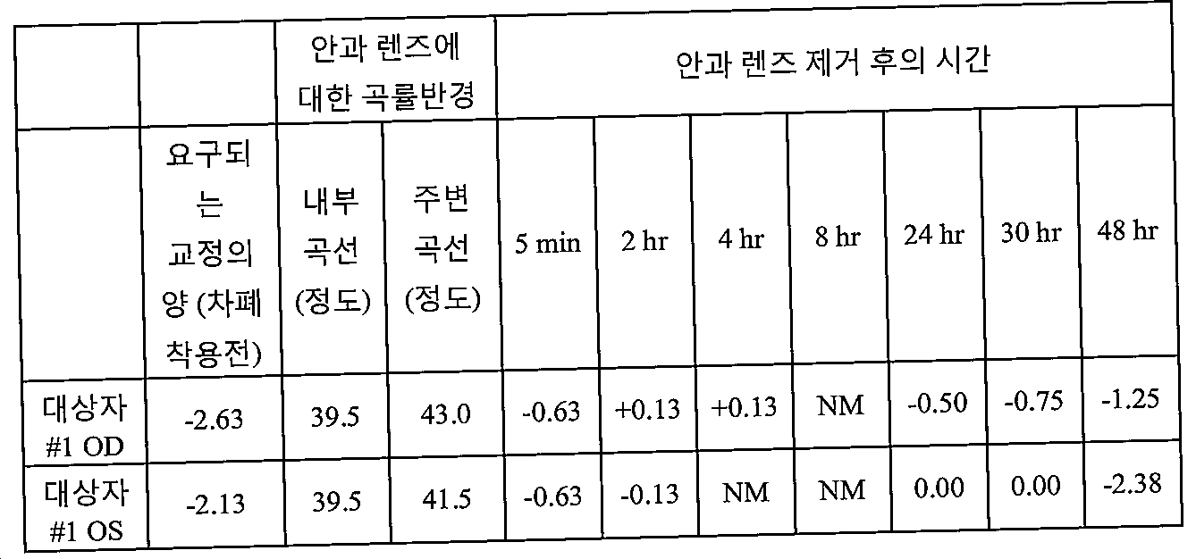
*NM = 측정하지 않음* NM = not measured
실시예 2Example 2
원시를 지닌 대상자에 대해서 특징적인 +0.13 디옵터(OD) 및 +0.25 디옵터(OS)의 광학적 교정을 필요로 하는 대상자가 안과 렌즈를 오른쪽 눈에 (매우 대체적으로) 약 35 시간 동안 착용하였고 왼쪽 눈에는 (매우 대체적으로) 약 17 시간 동안 착용하였다. 안과 장치에 대한 내부 및 주변 곡률반경이 표 2에 제공되어 있다. 대체로 상기 특정된 시간 후에, 안과 렌즈를 제거하고, 시력을 교정하기에 필요한 광학적 교정 (디옵터)의 양을 다양한 시점에서 측정하였다. 안과 렌즈가 대상자로부터 제거된 후에 요구되는 광학적 교정 (디옵터)의 양이 표 2에 나타내져 있다.Subjects requiring optical correction of +0.13 diopters (OD) and +0.25 diopters (OS) characteristic for subjects with hyperopia wore an ophthalmic lens in the right eye for about 35 hours (very largely). (Very generally) worn for about 17 hours. The internal and peripheral radius of curvature for the ophthalmic device is provided in Table 2. As a rule, after the time specified above, the amount of optical correction (diopter) needed to remove the ophthalmic lens and correct vision was measured at various time points. The amount of optical correction (diopter) required after the ophthalmic lens is removed from the subject is shown in Table 2.
표 2. 안과 렌즈 착용 후에 요구된 광학적 교정 (디옵터)의 양Table 2. Amount of optical correction (diopter) required after wearing ophthalmic lenses

*NM = 측정하지 않음* NM = not measured
예시적인 구체예가 예시에 의해서 그리고 명확한 이해를 위해서 약간 상세하게 기재되어 있지만, 당업자는 다양한 변형, 개조 및 변화가 이용될 수 있음을 인식할 것이다. 따라서, 본 발명의 범위는 전적으로 첨부된 특허청구범위에 한정되어야 한다.Although exemplary embodiments have been described in some detail by way of illustration and for clarity of understanding, those skilled in the art will recognize that various modifications, adaptations, and variations may be utilized. Accordingly, the scope of the invention should be limited solely to the appended claims.
첨부 1Attachment 1
표 B1Table B1
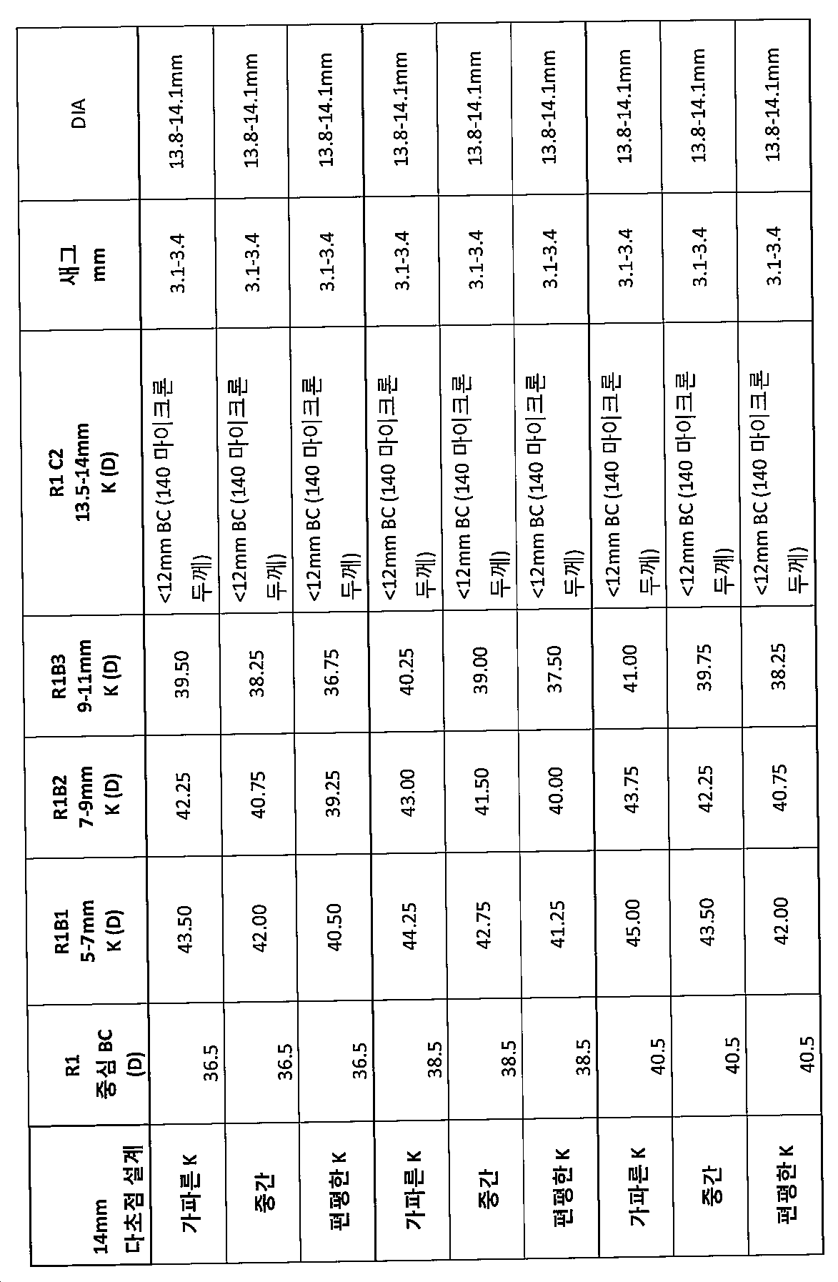
표 B2Table B2
더 편평한 주변 설계Planar Perimeter Design
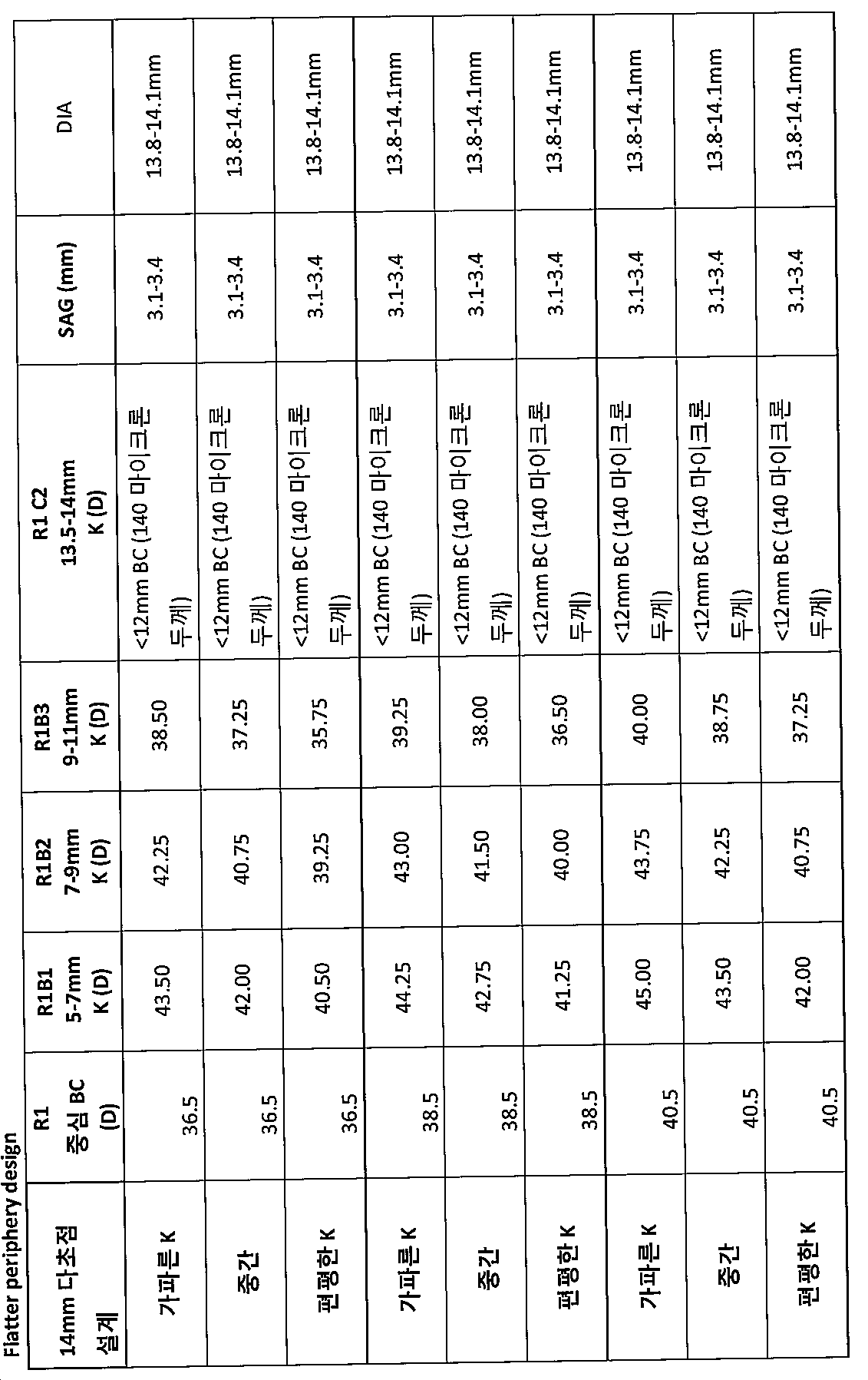
표 B3Table B3
*이전 곡선과 탄젠트될 수 없다(외부 곡선을 삽입하여 이를 치솟게 할 수 있다)It cannot be tangent with the previous curve (you can insert an outer curve to soar it)
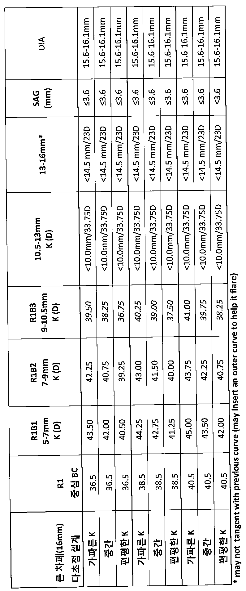
표 B4Table B4
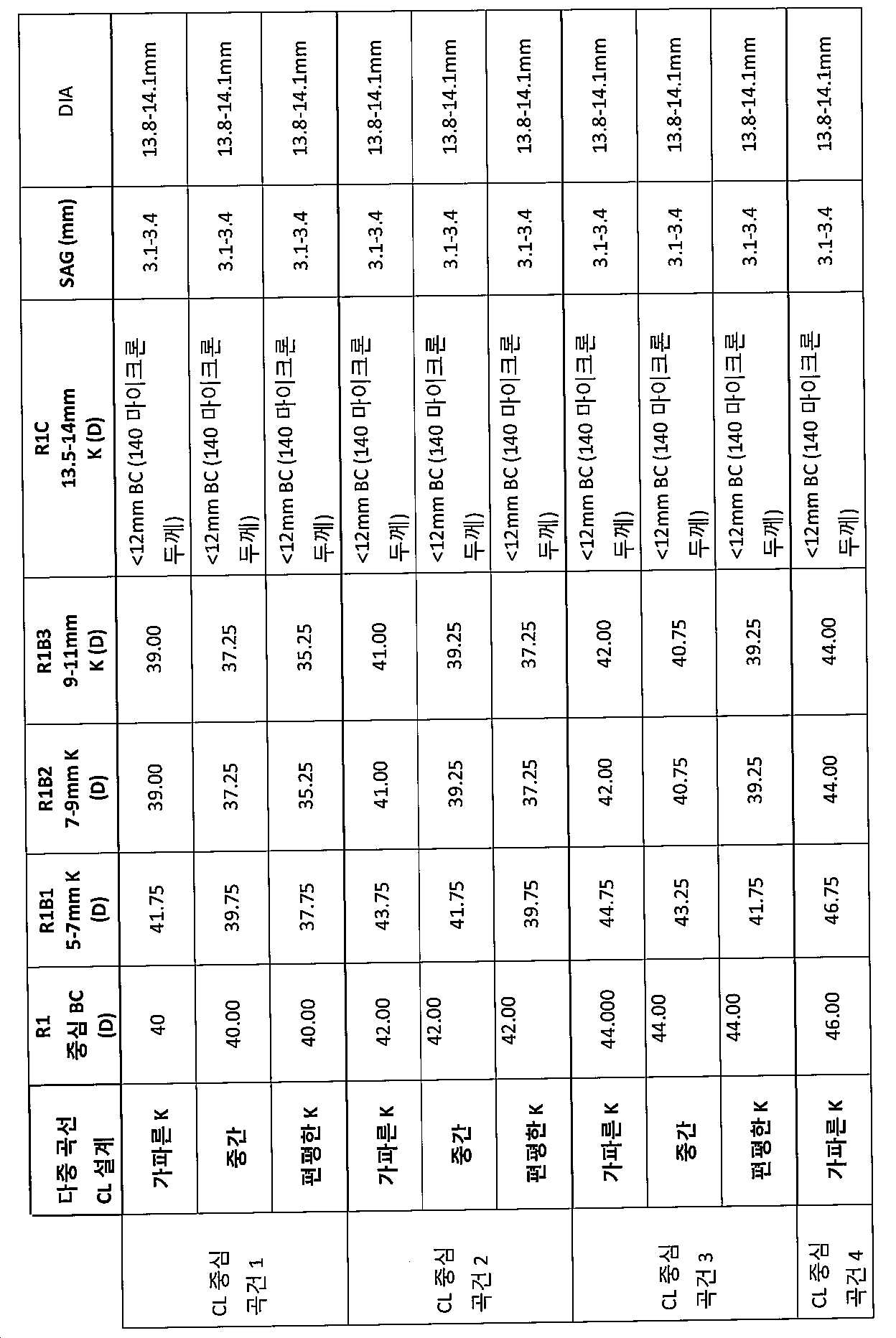
Claims (97)
상기 커플링 구성요소는 결막과 접촉하도록 하는 크기의 외부 부분, 광학적 구성요소에 커플링되는 내부 부분, 및 내부 부분과 외부 부분 사이에서 연장되는 중간 부분을 포함하고, 광학적 구성요소 또는 커플링 구성요소 중 하나 이상이 눈이 깜박거릴 때에 눈물 액체를 펌핑시키기 위한 복수의 천공을 포함하는, 환자의 눈을 치료하기 위한 보호막.An optical component for correcting vision of the eye comprising a first stiffness sufficient to withstand deformation when placed on the eye; And a coupling component for contacting the cornea and conjunctiva and supporting optical components with respect to the pupil, as a protective film for treating the eye of a patient with tear fluid, pupil, cornea, and conjunctiva. ,
The coupling component includes an outer portion sized to contact the conjunctiva, an inner portion coupled to the optical component, and an intermediate portion extending between the inner portion and the outer portion, and the optical component or coupling component. A protective film for treating an eye of a patient, wherein at least one of the plurality of holes comprises a plurality of perforations for pumping tear liquid when the eye blinks.
광학적 구성요소에 부착되는 커플링 구성요소를 형성시키기 위해서 유동성 물질을 경화시키는 단계;
모울드로부터 보호막을 제거하는 단계; 및
보호막에 복수의 천공을 형성시키는 단계를 포함하여 보호막을 제조하는 방법으로서,
광학적 구성요소에 부착되는 커플링 구성요소가 눈에 위치되는 대에 눈물 액체를 펌핑할 수 있는 방법.Injecting a fluid material into the mold;
Curing the flowable material to form a coupling component attached to the optical component;
Removing the protective film from the mold; And
A method of manufacturing a protective film, the method comprising forming a plurality of perforations in the protective film,
A method of pumping tear liquid when a coupling component attached to an optical component is located in the eye.
내부 구성요소를 형성시키기 위해서 내부 유동성 물질을 경화시키는 단계;
내부 구성요소 모울드로부터 내부 구성요소를 제거하는 단계;
내부 구성요소를 보호막 모울드에 넣어서, 내부 구성요소의 제 1 표면이 모울드의 내부 하부 표면으로 지지되게 하고, 그리고 갭이 내부 구성요소의 제 2 표면과 보호막 모울드의 상부 표면 사이에서 연장되게 하는 단계;
외부 유동성 물질을 보호막 모울드내로 주입시켜 적어도 내부 구성요소의 주변부을 따라서 내부 구성요소와 접촉시키고 갭을 충전시켜서, 내부 구성요소의 제 2 표면이 유동성 물질과 접촉되게 하는 단계;
보호막의 적어도 외부 부분을 형성시키기 위해서 외부 유동성 물질을 경화시키는 단계;
보호막 모울드로부터 보호막을 제거하는 단계; 및
보호막 내에 복수의 천공을 형성시키는 단계를 포함하여, 보호막을 제조하는 방법.Injecting an inner fluid material into the inner component module;
Curing the internal flowable material to form internal components;
Removing the internal component from the internal component mold;
Placing the inner component into the protective mold, such that the first surface of the inner component is supported by the inner lower surface of the mold, and the gap extends between the second surface of the inner component and the upper surface of the protective mold;
Injecting an outer flowable material into the protective mold mold to contact the inner component and fill the gap at least along the periphery of the inner component such that the second surface of the inner component is in contact with the flowable material;
Curing the external flowable material to form at least an outer portion of the protective film;
Removing the protective film from the protective film mold; And
Forming a plurality of perforations in the protective film.
광학적 구성요소에 부착된 커플링 구성요소를 형성시키기 위해서 유동성 물질을 경화시키는 단계;
하이드로겔 층을 형성시키는 단계; 및
하이드로겔 층으로 연장되는 보호막내의 복수의 천공을 형성시키는 단계를 포함하여 보호막을 제조하는 방법으로서,
광학적 구성요소에 부착된 커플링 구성요소가 눈 상에 위치되는 때에 눈물 액체를 펌핑할 수 있는 방법.Injecting a fluid material into the mold;
Curing the flowable material to form a coupling component attached to the optical component;
Forming a hydrogel layer; And
A method of making a protective film comprising forming a plurality of perforations in the protective film extending into a hydrogel layer, the method comprising:
A method of pumping tear liquid when a coupling component attached to an optical component is positioned on the eye.
각막 및 결막과 접촉하고 동공과 관련하여 광학적 구성요소를 지지하기 위한 커플링 구성요소를 포함하는 환자의 눈을 치료하기 위한 보호막으로서,
커플링 구성요소가 보호막의 전방 표면으로부터 하이드로겔 층까지 연장되는 복수의 천공을 지니는 수분 억제층을 포함하는 보호막.An optical component for correcting a patient's vision comprising a first intensity sufficient to withstand deformation when placed in the eye; And
A protective film for treating an eye of a patient comprising a coupling component for contacting the cornea and conjunctiva and supporting optical components with respect to the pupil,
A protective film comprising a moisture inhibiting layer having a plurality of perforations extending from the front surface of the protective film to the hydrogel layer.
상기 커플링 구성요소가 보호막의 후방 표면을 따라서 연장되는 하이드로겔의 층을 포함하고, 커플링 구성요소가 결막과 접촉하도록 하는 크기의 외부 부분, 광학적 구성요소에 커플링되는 내부 부분, 및 내부 부분과 외부 부분 사이에서 연장되는 중간 부분을 포함하고, 광학적 구성요소 또는 커플링 구성요소 중 하나 이상이 눈이 깜박거릴 때에 눈물 액체를 펌핑시키기 위한 복수의 천공을 포함하는, 환자의 눈을 치료하기 위한 보호막.An optical component for correcting vision of the eye comprising a first stiffness sufficient to withstand deformation when placed on the eye; And a coupling component for contacting the cornea and conjunctiva with and supporting the optical component with respect to the pupil, the protective film for treating the eye of a patient with tear fluid, pupil, cornea, and conjunctiva,
The coupling component comprises a layer of hydrogel extending along the posterior surface of the protective film, the outer portion sized to bring the coupling component into contact with the conjunctiva, the inner portion coupled to the optical component, and the inner portion A middle portion extending between the outer portion and the outer portion, wherein at least one of the optical component or the coupling component comprises a plurality of perforations for pumping tear fluid when the eye blinks. Shield.
상이한 구면 굴절력을 지닌 복수의 택일적 안과 렌즈 중에서, 요망되는 구면 굴절력에 상응하는 전방 표면을 제공하는 안과 렌즈를 확인하는 단계를 포함하여, 굴절 모양을 제공하는 상피를 지닌 각막을 지니는 환자의 눈의 굴절이상을 교정하기 위한 안관 렌즈를 선택하는 방법으로서,
전방 표면이 안과 렌즈의 내부 부분을 따라서 연장되고, 안과 렌즈의 내부 부분이 변형 가능하고, 안과 렌즈의 주변 부분이 내부 부분의 강도보다 낮은 강도를 지니고;
주변 부분이 광학적 부위 외부의 눈과 맞물리기에 적합한 모양을 지녀 눈의 광학적 부위와 정렬되어 내부 부분을 지지하는, 안관 렌즈를 선택하는 방법.Measuring the desired spherical component to mitigate any spherical component above the refractive error; And
Of a plurality of alternative ophthalmic lenses with different spherical refractive powers, comprising identifying an ophthalmic lens that provides an anterior surface corresponding to the desired spherical refractive power, the eye of the patient having a cornea with an epithelium that provides a refractive shape. As a method of selecting an ophthalmic lens for correcting refractive error,
The anterior surface extends along an inner portion of the ophthalmic lens, the inner portion of the ophthalmic lens is deformable, and the peripheral portion of the ophthalmic lens has an intensity lower than that of the inner portion;
A method of selecting an ophthalmic lens wherein the peripheral portion is shaped to fit the eye outside the optical portion and is aligned with the optical portion of the eye to support the inner portion.
눈물 유체가 후방 표면과 굴절 모양 사이의 렌즈모양 용적을 충전하여 난시 및/또는 고위 수차를 완화시키도록 주변 부분이 적합한 모양을 지니게 안과 렌즈의 선택이 수행되고, 안과 렌즈의 확인이 난시의 굴절력, 눈의 광학적 축에 대한 난시의 배향 및/또는 고위 수차의 유형 및/또는 고위 수차의 세기의 그룹 중 일원 이상과 무관한 방법.88. The method of claim 87, wherein the refractive shape of the epithelium extends across the optical portion of the eye, the refractive error includes astigmatism and / or high aberrations, and the posterior surface of the ophthalmic lens extends across the optical region adjacent to the eye. Configured;
Selection of the ophthalmic lens is performed such that the tear fluid fills the lenticular volume between the posterior surface and the refractive shape to mitigate astigmatism and / or high order aberrations so that the peripheral portion has a suitable shape, and the identification of the ophthalmic lens determines the refractive power of the astigmatism, The orientation of astigmatism with respect to the optical axis of the eye and / or the type of high order aberration and / or method independent of one or more of the groups of high intensity aberrations.
안과 렌즈의 내부 부분이 변형 가능하고 주변 부분이 내부 부분의 강도보다 더 작은 강도를 지니고, 안과 렌즈의 표면이 노안-완화 굴절 모양을 지니는 방법.Positioning the ophthalmic lens on the eye such that the inner portion of the ophthalmic lens is disposed on the optical portion of the cornea and supporting the inner portion of the ophthalmic lens by engagement between the peripheral portion of the ophthalmic lens and the eye outside the optical portion. Including, as a method for treating presbyopia of the eye of a patient with a cornea that provides a refractive shape,
A method in which the inner part of the ophthalmic lens is deformable, the peripheral part having a smaller intensity than the strength of the inner part, and the surface of the ophthalmic lens has a presbyopia-relaxing refractive shape.
정위된 안과 렌즈의 후방 표면이 눈에 인접하여 연장되고 상피의 굴절 모양으로부터 분기되는 모양을 지녀서 렌즈모양 용적이 그 사이에 배치되는 방법.Positioning the ophthalmic lens on the eye such that the inner portion of the ophthalmic lens is placed on the optical portion of the cornea and supporting the inner portion of the ophthalmic lens by engagement between the peripheral portion of the ophthalmic lens and the eye outside the optical portion. Having a cornea with an epithelium that fills the lenticular volume and provides a refractive shape that extends across the optical region of the eye, including relieving refractive errors when viewed visually through the anterior surface of the ophthalmic lens. As a method to correct the refractive error of the eyes,
Wherein the posterior surface of the positioned ophthalmic lens extends adjacent to the eye and diverges from the refractive shape of the epithelium so that the lenticular volume is disposed therebetween.
복수의 천공이 전방 표면과 후방 표면 사이에서 연장되고, 천공이 광학적 부위의 외부에 그리고 주변 부분과 눈의 조직 표면 사이의 맞물림 영역의 안쪽으로 배치되며;
안과 렌즈의 내부 부분이 변형 가능하고, 안과 렌즈의 주변 부분이 내부 부분의 강도보다 낮은 강도를 지녀서, 눈의 깜박임이 렌즈모양 용적의 내외로 천공을 통한 눈물 유체의 흐름을 유도하게 하며;
맞물림 부위를 따른 눈의 조직 표면과 주변 부분 사이의 맞물림이 깜박이는 동안의 각막에 대한 내부 부분의 측면 운동을 억제하고;
전방 표면을 통해서 눈으로 볼 때의 굴절이상의 완화가 약 1½D 이상의 난시성 이상의 범위 전체에 걸쳐서 렌즈모양 용적의 모양에 실질적으로 무관하고, 눈의 시각 축에 대한 안과 렌즈의 회전 배향과 무관한 방법.92. The method of claim 91, wherein the refractive error of the eye comprises astigmatism and / or spherical aberration,
A plurality of perforations extend between the anterior and posterior surfaces, the perforations being disposed outside of the optical area and inwardly of the engagement area between the peripheral portion and the tissue surface of the eye;
The inner portion of the ophthalmic lens is deformable and the peripheral portion of the ophthalmic lens has an intensity lower than that of the inner portion, such that blinking of the eye induces the flow of tear fluid through the perforation into and out of the lenticular volume;
Inhibits lateral movement of the inner portion relative to the cornea while the engagement between the peripheral surface and the tissue surface of the eye along the engagement site is blinking;
The method of alleviation of refractive errors when viewed through the anterior surface is substantially independent of the shape of the lens-like volume throughout the range of at least about 1½D or more of astigmatism and independent of the rotational orientation of the ophthalmic lens with respect to the eye's visual axis.
정위된 안과 렌즈의 후방 표면이 눈에 인접하여 연장되고 상피의 굴절 모양으로부터 분기되는 모양을 지녀서 렌즈모양 용적이 그 사이에 배치되는 방법.Positioning the ophthalmic lens on the eye so that the inner portion of the ophthalmic lens is placed on the optical portion of the cornea and supporting the inner portion of the ophthalmic lens by engagement between the peripheral portion of the ophthalmic lens and the eye outside the optical portion. Eye with cornea with epithelium that fills the lenticular volume and provides a refractive shape extending across the optical area of the eye, including relieving refractive errors when viewed through the eye's anterior surface through the ophthalmic lens. As a method of correcting the refractive error of
Wherein the posterior surface of the positioned ophthalmic lens extends adjacent to the eye and diverges from the refractive shape of the epithelium so that the lenticular volume is disposed therebetween.
전방 표면을 통해서 눈으로 볼 때의 굴절이상의 완화가 약 1½D 이상의 난시성 이상의 범위 전체에 걸쳐서 렌즈모양 용적의 모양에 실질적으로 무관하고, 눈의 시각 축에 대한 안과 렌즈의 회전 배향과 무관하며;
광학적 성형이 눈으로부터 안과 렌즈의 제거 후 약 8 시간 후에 약 1½D 이상까지 눈의 굴절이상을 완화시키는 방법.94. The method of claim 93, wherein the inner portion of the ophthalmic lens is deformable, the peripheral portion having an intensity lower than that of the inner portion, and the engagement between the tissue surface of the eye and the peripheral portion is at the side of the inner portion from the optical portion of the cornea. Inhibit exercise;
Alleviation of refractive errors when viewed through the anterior surface visually is substantially independent of the shape of the lens-like volume throughout the range of at least about 1½D or more of astigmatism and independent of the rotational orientation of the ophthalmic lens with respect to the eye's visual axis;
Optical shaping alleviates refractive error in the eye to about 1½D or more after about 8 hours after removal of the ophthalmic lens from the eye.
각각의 안과 렌즈가 회합적으로 요망되는 구면 굴절력에 상응하는 전방 표면과 내부 부분으로부터 방사상 외향으로 연장하는 안과 렌즈의 주변 부분을 지니며; 전방 표면은 안과 렌즈의 내부 부분을 따라서 연장되고, 안과 렌즈의 내부 부분은 변형 가능하며;
전방 표면은 안과 렌즈의 내부 부분을 따라서 연장되고, 안과 렌즈의 내부 부분은 변형가능하고;
주변 부분이 내부 부분의 강도보다 더 작은 강도를 지니며, 광학적 부위 외부의 조직과 맞물리도록 구성되어 광학적 부위와 정렬되어 내부부분을 지지하는, 택일적으로 선택 가능한 안과 렌즈의 세트.A set of alternatively selectable ophthalmic lenses for correcting the refractive error of an eye of a patient population having a cornea with an epithelium providing a refractive shape, comprising a plurality of alternative ophthalmic lenses with different spherical refractive powers,
Each ophthalmic lens has a peripheral portion of the ophthalmic lens extending radially outward from the anterior portion and the inner portion corresponding to the spherically desired spherical refractive power; The anterior surface extends along an inner portion of the ophthalmic lens, and the inner portion of the ophthalmic lens is deformable;
The anterior surface extends along an inner portion of the ophthalmic lens and the inner portion of the ophthalmic lens is deformable;
An alternatively selectable set of ophthalmic lenses wherein the peripheral portion has an intensity less than that of the inner portion and is configured to engage with tissue outside the optical portion to align with the optical portion to support the inner portion.
내부 부분의 강도보다 더 작은 강도를 지니며 주변 부분과 광학적 부위 외부에 배치된 눈 조직 사이의 맞물림에 의해서 안과 렌즈의 내부 부분을 지지하도록 구성된 주변 부분; 및
내부 부분을 따라서 연장되고 노안-완화 굴절 모양을 지니는 표면을 포함하는, 굴절 모양을 제공하는 상피를 지닌 각막을 가지는 환자의 눈의 노안을 치료하기 위한 안과 렌즈.An internal portion that is configured and deformable to be disposed on an optical site of the cornea;
A peripheral portion having a smaller intensity than that of the inner portion and configured to support the inner portion of the ophthalmic lens by engagement between the peripheral portion and eye tissue disposed outside the optical portion; And
An ophthalmic lens for treating presbyopia of a patient's eye having a cornea with an epithelium providing a refractive shape, the surface extending along an interior portion and having a presbyopia-refractive shape.
내부 부분이 광학적 부위 상에 배치되는 때에 눈에 인접한 내부 부분을 따라서 연장되는 후방 표면;
내부 부분의 방사상 외향으로 배치된 안과 렌즈의 주변 부위; 및
후방 표면 반대의 내부 부분을 따라서 연장되어서 유체 충전된 렌즈모양 용적 및 내부 부분을 통해서 눈으로 볼 때 굴절이상을 환화시키는 안과 렌즈의 전방 표면을 포함하는, 눈의 광학적 부위를 가로질러 연장되는 굴절 모양을 제공하는 상피를 지닌 각막을 가지는 눈의 굴절 이상을 교정하기 위한 안과 렌즈로서,
후방 표면이 상피의 굴절 모양으로부터 분기되는 모양을 지녀서 렌즈모양 용적이 그 사이에 배치되고;
주변 부분이 광학적 부위 외부의 눈과 맞물리도록 구성되어 렌즈모양 용적을 유체 충전시켜 유지하는, 눈의 굴절 이상을 교정하기 위한 안과 렌즈.An inner portion configured to be disposed on an optical site of the cornea;
A posterior surface extending along the inner portion adjacent the eye when the inner portion is disposed on the optical portion;
Peripheral portions of the ophthalmic lens disposed radially outward of the inner portion; And
Refractive shapes extending across the optical portion of the eye, including along the interior portion opposite the posterior surface and including a fluid-filled lens-like volume and an anterior surface of the ophthalmic lens that cyclizes the refractive error when viewed visually through the interior portion. An ophthalmic lens for correcting the refractive error of an eye having a cornea with an epithelium that provides
The posterior surface is shaped to diverge from the refractive shape of the epithelium so that the lenticular volume is disposed therebetween;
An ophthalmic lens for correcting refractive errors in the eye, wherein the peripheral portion is configured to engage the eye outside the optical area to maintain fluid-filled lens-like volume.
Applications Claiming Priority (7)
| Application Number | Priority Date | Filing Date | Title |
|---|---|---|---|
| US201161480222P | 2011-04-28 | 2011-04-28 | |
| US61/480,222 | 2011-04-28 | ||
| US201161507971P | 2011-07-14 | 2011-07-14 | |
| US61/507,971 | 2011-07-14 | ||
| US201261636404P | 2012-04-20 | 2012-04-20 | |
| US61/636,404 | 2012-04-20 | ||
| PCT/US2012/035050 WO2012149056A1 (en) | 2011-04-28 | 2012-04-25 | Eye covering and refractive correction methods and apparatus having improved tear flow, comfort, and/or applicability |
Publications (1)
| Publication Number | Publication Date |
|---|---|
| KR20140023378A true KR20140023378A (en) | 2014-02-26 |
Family
ID=47072730
Family Applications (1)
| Application Number | Title | Priority Date | Filing Date |
|---|---|---|---|
| KR1020137031721A Ceased KR20140023378A (en) | 2011-04-28 | 2012-04-25 | Eye covering and refractive correction methods and apparatus having improved tear flow, comfort, and/or applicability |
Country Status (8)
| Country | Link |
|---|---|
| US (4) | US8864306B2 (en) |
| EP (1) | EP2701644B1 (en) |
| JP (1) | JP2014514613A (en) |
| KR (1) | KR20140023378A (en) |
| CN (1) | CN103648448B (en) |
| AU (2) | AU2012249773A1 (en) |
| CA (1) | CA2834295A1 (en) |
| WO (1) | WO2012149056A1 (en) |
Cited By (2)
| Publication number | Priority date | Publication date | Assignee | Title |
|---|---|---|---|---|
| KR200477407Y1 (en) * | 2014-05-19 | 2015-06-05 | (주)리치아일랜드 | Contact lens |
| KR20230014388A (en) * | 2021-07-21 | 2023-01-30 | 가톨릭대학교 산학협력단 | Structure for eye treatment |
Families Citing this family (44)
| Publication number | Priority date | Publication date | Assignee | Title |
|---|---|---|---|---|
| WO2009145842A2 (en) | 2008-04-04 | 2009-12-03 | Forsight Labs, Llc | Therapeutic device for pain management and vision |
| WO2011050327A1 (en) | 2009-10-23 | 2011-04-28 | Forsight Labs Llc | Corneal denervation for treatment of ocular pain |
| US9498385B2 (en) | 2009-10-23 | 2016-11-22 | Nexisvision, Inc. | Conformable therapeutic shield for vision and pain |
| US8591025B1 (en) | 2012-09-11 | 2013-11-26 | Nexisvision, Inc. | Eye covering and refractive correction methods for LASIK and other applications |
| EP2632405A4 (en) | 2010-10-25 | 2016-02-24 | Nexisvision Inc | Methods and apparatus to identify eye coverings for vision |
| US12044905B2 (en) | 2011-04-28 | 2024-07-23 | Journey1 Inc | Contact lenses for refractive correction |
| US8678584B2 (en) | 2012-04-20 | 2014-03-25 | Nexisvision, Inc. | Contact lenses for refractive correction |
| KR20140023378A (en) | 2011-04-28 | 2014-02-26 | 넥시스비젼, 인코포레이티드 | Eye covering and refractive correction methods and apparatus having improved tear flow, comfort, and/or applicability |
| TWI588560B (en) | 2012-04-05 | 2017-06-21 | 布萊恩荷登視覺協會 | Lens, device, method and system for refractive error |
| HK1208268A1 (en) * | 2012-04-20 | 2016-02-26 | 内希斯视觉股份有限公司 | Contact lenses for refractive correction |
| US9465233B2 (en) | 2012-04-20 | 2016-10-11 | Nexisvision, Inc. | Bimodular contact lenses |
| US9201250B2 (en) | 2012-10-17 | 2015-12-01 | Brien Holden Vision Institute | Lenses, devices, methods and systems for refractive error |
| TWI600418B (en) | 2012-10-17 | 2017-10-01 | 布萊恩荷登視覺協會 | Lens, device, method and system for refractive error |
| US9097915B2 (en) * | 2013-03-05 | 2015-08-04 | EyeYon Medical Ltd. | Hyper-osmotic eye contact lens |
| US11079613B2 (en) * | 2013-03-05 | 2021-08-03 | EyeYon Medical Ltd. | Contact lens drug depot |
| US10025114B2 (en) * | 2013-03-13 | 2018-07-17 | Johnson & Johnson Vision Care, Inc. | Hydrogel lens having raised portions for improved oxygen transmission and tear flow |
| US8974055B2 (en) * | 2013-03-15 | 2015-03-10 | Johnson & Johnson Vision Care, Inc. | Method and apparatus for encapsulating a rigid insert in a contact lens for correcting vision in astigmatic patients |
| JP6310072B2 (en) * | 2013-06-26 | 2018-04-11 | ネクシスビジョン, インコーポレイテッド | Contact lenses for refractive correction |
| US9668916B2 (en) | 2013-11-04 | 2017-06-06 | Vance M. Thompson | Conjunctival cover and methods therefor |
| WO2015069927A1 (en) * | 2013-11-06 | 2015-05-14 | Nexisvision, Inc. | Bimodular contact lenses |
| TWI654440B (en) * | 2013-11-15 | 2019-03-21 | 美商實體科學公司 | Contact lens with hydrophilic layer |
| US9341864B2 (en) | 2013-11-15 | 2016-05-17 | Nexisvision, Inc. | Contact lenses having a reinforcing scaffold |
| SG11201605735SA (en) * | 2014-01-16 | 2016-09-29 | Kowa Co | Toric intraocular lens |
| WO2015116559A1 (en) | 2014-01-29 | 2015-08-06 | Nexisvision, Inc. | Multifocal bimodulus contact lenses |
| US9395557B2 (en) | 2014-11-12 | 2016-07-19 | Vance M. Thompson | Partial corneal conjunctival contact lens |
| US9869883B2 (en) | 2015-03-11 | 2018-01-16 | Vance M. Thompson | Tear shaping for refractive correction |
| US9977258B2 (en) * | 2016-04-14 | 2018-05-22 | Johnson & Johnson Vision Care, Inc. | Methods and apparatus to enhance oxygen concentrations for ophthalmic devices |
| US9964780B2 (en) * | 2016-04-14 | 2018-05-08 | Johnson & Johnson Vision Care, Inc. | Methods and apparatus to enhance oxygen concentrations for advanced ophthalmic devices |
| EP3446159B1 (en) * | 2016-04-20 | 2023-06-14 | CooperVision International Limited | Silicone elastomer-silicone hydrogel hybrid contact lenses |
| WO2018013757A2 (en) | 2016-07-14 | 2018-01-18 | Corning Incorporated | Methods of reducing surface roughness of reflectance coatings for duv mirrors |
| US10353220B2 (en) | 2016-10-17 | 2019-07-16 | Vance M. Thompson | Tear shaping for refractive correction |
| US11460719B2 (en) * | 2017-05-01 | 2022-10-04 | Pres-By Vision Ltd. | Dynamic tear lenses |
| JP2020525842A (en) * | 2017-06-27 | 2020-08-27 | アイディール スキャニング リミテッド ライアビリティ カンパニー | Ophthalmic lens design based on topology |
| EP3675711A4 (en) * | 2017-11-07 | 2021-06-30 | Notal Vision Ltd. | RETINAL IMAGING DEVICE AND ASSOCIATED PROCESSES |
| US10678067B2 (en) | 2018-04-06 | 2020-06-09 | Vance M. Thompson | Tear shaping for refractive correction |
| WO2020049356A1 (en) * | 2018-09-04 | 2020-03-12 | Pres-By Vision Ltd. | Mechanisms for inducing transitions in dynamic contact lenses |
| AR115981A1 (en) * | 2019-03-19 | 2021-03-17 | Ohio State Innovation Foundation | CONTACT LENS |
| US10725321B1 (en) * | 2019-05-20 | 2020-07-28 | Liberty Commercial Imports & Exports Services, LLC | Contact lens |
| US11229549B2 (en) | 2019-09-18 | 2022-01-25 | Vance M. Thompson | Tear transplantation and multi-part contact lens with absorbent portion |
| US12257184B2 (en) * | 2019-10-24 | 2025-03-25 | Denis LaBombard | Ocular device and drug delivery system, with case |
| USD1075870S1 (en) | 2020-03-26 | 2025-05-20 | Ohio State Innovation Foundation | Contact lens |
| US11841557B2 (en) | 2021-05-04 | 2023-12-12 | International Business Machines Corporation | Smart contacts that respond to fluid changes on the surface of the eyes |
| US12440385B2 (en) | 2021-12-10 | 2025-10-14 | Ocular Surface Innovations Inc. | Ophthalmic shield and therapeutic apparatus |
| US20230181376A1 (en) * | 2021-12-10 | 2023-06-15 | Ocular Surface Innovations Inc. | Ophthalmic shield and therapeutic method |
Citations (4)
| Publication number | Priority date | Publication date | Assignee | Title |
|---|---|---|---|---|
| US4640594A (en) * | 1982-01-07 | 1987-02-03 | Richard Berger | Tear pump contact lens |
| US6010219A (en) * | 1996-06-28 | 2000-01-04 | Contex, Inc. | Fenestrated contact lens for treating myopia |
| US20060250576A1 (en) * | 2005-05-06 | 2006-11-09 | Legerton Jerome A | Hybrid contact lens system and method of fitting |
| US20060285071A1 (en) * | 2005-06-21 | 2006-12-21 | Bausch & Lomb Incorporated | Femtosecond laser micromachining of a contact lens and a contact lens manufactured thereby |
Family Cites Families (266)
| Publication number | Priority date | Publication date | Assignee | Title |
|---|---|---|---|---|
| US2641161A (en) | 1950-12-13 | 1953-06-09 | Samuel W Silverstein | Contact lens |
| US3246941A (en) | 1962-02-28 | 1966-04-19 | Herbert L Moss | Contact lens with upper crescent shaped scleral flange and having vent channels and apertures |
| US3431046A (en) * | 1964-02-14 | 1969-03-04 | Studies Inc | Flexible polyethylene corneal contact lens |
| US3489491A (en) | 1966-12-01 | 1970-01-13 | Charles Patrick Creighton | Corneal contact lens with hydrophilic marginal rim portion |
| FR1510013A (en) | 1966-12-05 | 1968-01-19 | Freres Lissac Opticiens S A | Scleral lens enhancements |
| US3488111A (en) | 1967-03-16 | 1970-01-06 | Nat Patent Dev Corp | Hydrophilic hydrogel corneal contact lens with hard central insert |
| US3594074A (en) | 1969-06-16 | 1971-07-20 | Mencher Alexander | Contact lens with flexible central portion |
| US3619044A (en) | 1970-02-18 | 1971-11-09 | American Optical Corp | Rigid gas permeable contact lens with softer edge portion |
| US3688386A (en) | 1971-03-15 | 1972-09-05 | Adolf J Pereira | Method for fenestration of contact lenses |
| US3973837A (en) | 1971-05-04 | 1976-08-10 | Page Louis J | Contact lenses |
| US3973838A (en) | 1971-05-04 | 1976-08-10 | Page Louis J | Contact lenses |
| US3944347A (en) | 1971-06-11 | 1976-03-16 | E. I. Du Pont De Nemours & Co. | Contact lens having a hard center and soft, tough periphery |
| CA993401A (en) | 1972-12-04 | 1976-07-20 | Edward W. Merrill | Contact lens and method of preparation |
| US3833786A (en) | 1973-06-08 | 1974-09-03 | Continuous Curve Contact Lense | Laser apparatus for fenestration of contact lenses |
| US3915609A (en) | 1974-03-18 | 1975-10-28 | American Optical Corp | Molds for casting silicone rubber contact lenses |
| US4121885A (en) | 1974-04-29 | 1978-10-24 | Precision Cosmet Co., Inc. | Method to produce a composite contact lens |
| JPS51341A (en) | 1974-06-19 | 1976-01-06 | Canon Kk | DENSHISHA SHINHO |
| JPS518943A (en) | 1974-07-11 | 1976-01-24 | Fujitsu Ltd | KIROKUSOCHI |
| CS175082B1 (en) | 1974-08-28 | 1977-04-29 | ||
| SU558248A1 (en) | 1974-12-17 | 1977-05-15 | Московский научно-исследовательский институт глазных болезней им. Гельмгольца | Combined contact lens and method of its manufacture |
| US4068933A (en) | 1975-03-18 | 1978-01-17 | Maurice Seiderman | Fenestrated hydrogels for contact lens use and method for making same |
| US4208362A (en) | 1975-04-21 | 1980-06-17 | Bausch & Lomb Incorporated | Shaped body of at least two polymerized materials and method to make same |
| US4166255A (en) | 1976-06-14 | 1979-08-28 | Minnesota Mining And Manufacturing Company | Hybrid corneal contact lens |
| US4037866A (en) | 1976-07-26 | 1977-07-26 | Price Edward E | Contact lens applicator |
| US4071272A (en) | 1976-09-27 | 1978-01-31 | Drdlik Frank J | Contact lens applicator |
| US4200320A (en) | 1978-06-12 | 1980-04-29 | Durham Henry B | Contact lens applicator |
| US4194815A (en) | 1978-06-28 | 1980-03-25 | Dow Corning Corporation | Semi-scleral contact lens |
| US4268133A (en) | 1978-07-14 | 1981-05-19 | Bausch & Lomb Incorporated | Preferential orientation of contact lenses |
| US4211476A (en) | 1978-12-29 | 1980-07-08 | Allan J. Brummel | Contact lenses with angular orientation and stabilization |
| JPS55101125A (en) | 1979-01-20 | 1980-08-01 | Matsushita Electric Ind Co Ltd | Magnetic head |
| US4312575A (en) | 1979-09-18 | 1982-01-26 | Peyman Gholam A | Soft corneal contact lens with tightly cross-linked polymer coating and method of making same |
| JPS5727457Y2 (en) | 1980-04-18 | 1982-06-15 | ||
| ES501922A0 (en) | 1980-06-02 | 1982-07-01 | Corning Glass Works | IMPROVEMENTS INTRODUCED IN THE MANUFACTURE OF CONTACT LENSES OF COMPOSITE MATERIAL |
| US4381007A (en) | 1981-04-30 | 1983-04-26 | The United States Of America As Represented By The United States Department Of Energy | Multipolar corneal-shaping electrode with flexible removable skirt |
| GB2107895A (en) | 1981-10-19 | 1983-05-05 | Gholam A Peyman | Soft corneal contact lens with electrical glow discharge polymer coating |
| DE3143839A1 (en) | 1981-11-05 | 1983-05-19 | Jeffrey E. Palatine Ill. Koziol | Contact lens |
| US4487905A (en) | 1983-03-14 | 1984-12-11 | Dow Corning Corporation | Wettable silicone resin optical devices and curable compositions therefor |
| CS245064B1 (en) | 1983-12-30 | 1986-08-14 | Otto Wichterle | Perforated soft contact lens and method of its production |
| US4621912A (en) | 1985-02-14 | 1986-11-11 | Meyer Donald R | Foraminated optical contact lens |
| US4701288A (en) | 1985-06-05 | 1987-10-20 | Bausch & Lomb Incorporated | Method of making articles of dissimilar polymer compositions |
| US4666249A (en) | 1985-08-14 | 1987-05-19 | Sola U.S.A. Inc. | Surface-treated contact lens and method of producing |
| US4981841A (en) | 1986-04-04 | 1991-01-01 | Allergan, Inc. | Methods and materials for use in corneal wound healing |
| ES2044856T3 (en) | 1986-04-04 | 1994-01-16 | Allergan Inc | MORTAR COMPOSITION TO USE CORNEAL WOUNDS. |
| US4772283A (en) | 1986-05-16 | 1988-09-20 | White Thomas C | Corneal implant |
| US5114627A (en) | 1986-10-16 | 1992-05-19 | Cbs Lens | Method for producing a collagen hydrogel |
| US4806382A (en) | 1987-04-10 | 1989-02-21 | University Of Florida | Ocular implants and methods for their manufacture |
| US4810082A (en) | 1987-07-01 | 1989-03-07 | Abel Robert Jr | Corneal onlay lens |
| FR2622201B1 (en) | 1987-10-23 | 1990-03-23 | Essilor Int | WETABLE SILICON ELASTOMER SUITABLE FOR THE MANUFACTURE OF CONTACT LENSES |
| US4909896A (en) | 1988-02-29 | 1990-03-20 | Hoya Corporation | Process for producing contact lens |
| US4943150A (en) | 1988-03-22 | 1990-07-24 | Bausch & Lomb Incorporated | Method of making variable modulus lenses |
| GB2217869A (en) | 1988-04-22 | 1989-11-01 | Ceskoslovenska Akademie Ved | Contact lenses |
| IL86913A0 (en) | 1988-06-29 | 1988-11-30 | Bromine Compounds Ltd | Method for protecting carbon steel from corrosion in heavy brines |
| DE68927648T2 (en) | 1988-11-02 | 1997-04-24 | British Tech Group | Pouring and packaging contact lenses |
| US5008289A (en) | 1988-12-02 | 1991-04-16 | Galenpharma, Inc. | Composition for treating nasal disorders and headaches |
| US4978481A (en) | 1989-01-13 | 1990-12-18 | Ciba-Geigy Corporation | Process for the encapsulation of preformed substrates by graft copolymerization |
| US5104213A (en) | 1989-01-17 | 1992-04-14 | Wolfson Leonard G | Polymer buttons having holes therein and contact lenses manufactured therefrom and method of manufacture |
| US5073021A (en) | 1989-03-17 | 1991-12-17 | Environmental Research Institute Of Michigan | Bifocal ophthalmic lens constructed from birefringent material |
| AU5744490A (en) | 1989-05-15 | 1990-12-18 | Afferon Corporation | Composition and method for neural desensitization |
| US4952045B1 (en) | 1989-05-26 | 2000-08-08 | Contex Inc | Corneal contact lens and method for treating myopea |
| FR2648702A1 (en) | 1989-06-23 | 1990-12-28 | Hanna Khalil | LENS FOR EPIKERATOPHAKIE AND KERATOTOME, PARTICULARLY FOR REALIZING AN INCISION OF RECEIVING SUCH A LENS |
| GB8925246D0 (en) | 1989-11-08 | 1989-12-28 | Nat Res Dev | Gas sensor |
| US5019097A (en) | 1989-11-22 | 1991-05-28 | Allergan, Inc. | Corneal onlay lenses and methods for attaching same |
| AU629725B2 (en) | 1990-01-24 | 1992-10-08 | Novartis Ag | Contact lens and process for the manufacture thereof |
| AU642538B2 (en) | 1990-10-03 | 1993-10-21 | Toray Industries, Inc. | Optical sheet material |
| US5804263A (en) | 1990-10-24 | 1998-09-08 | University Of Florida Research Foundation, Inc. | Combined plasma and gamma radiation polymerization method for modifying surfaces |
| AU663681B2 (en) | 1990-11-05 | 1995-10-19 | Physiodynamics, Inc. | Apparatus and method for electrotherapeutic eye treatment |
| US5346491A (en) | 1991-03-28 | 1994-09-13 | Sony Corporation | Feed device for bipolar electrodes for capsulotomy |
| US5397848A (en) | 1991-04-25 | 1995-03-14 | Allergan, Inc. | Enhancing the hydrophilicity of silicone polymers |
| US5349395A (en) | 1991-08-23 | 1994-09-20 | Nick Stoyan | Multiple focus corneal contact lens and method for treating myopia |
| US5191365B1 (en) | 1991-08-23 | 2000-08-15 | Contex Inc | Corneal contact lens and method for treating myopia |
| US5428412B1 (en) | 1991-08-23 | 2000-08-08 | Contex Inc | Method for treating myopia with an aspheric corneal contact lens |
| US5270051A (en) | 1991-10-15 | 1993-12-14 | Harris Donald H | Enzyme-orthokeratology |
| US5236236A (en) | 1991-10-25 | 1993-08-17 | Girimont John V | Process and implements for cleaning, rinsing, storing and inserting a contact lens |
| CA2100092A1 (en) | 1991-11-08 | 1993-05-09 | Gunilla Lofgren Nisser | Auxiliary means provided for treatment of visual and/or brain disorder |
| US5245367A (en) | 1991-11-12 | 1993-09-14 | David Miller | Annular mask contact lenses |
| US5786883A (en) | 1991-11-12 | 1998-07-28 | Pilkington Barnes Hind, Inc. | Annular mask contact lenses |
| US5246259A (en) | 1992-02-21 | 1993-09-21 | Johann Hellenkamp | Applicator device for positioning a contact lens on the human eye |
| US5178879A (en) | 1992-04-17 | 1993-01-12 | Michael Adekunle | Capsaicin gel |
| JPH0649251A (en) | 1992-06-09 | 1994-02-22 | Ciba Geigy Ag | Graft polymerization method on the surface of preformed substrate to modify surface properties |
| EP0686015A1 (en) | 1992-09-02 | 1995-12-13 | EPSTEIN, Robert L. | Instrument for ophthalmological surgery |
| US5433898A (en) | 1992-09-11 | 1995-07-18 | Pilkington Barnes Hind, Inc. | Method of manufacturing a contact lens |
| ES2058022B1 (en) | 1992-09-17 | 1995-10-01 | Medan Eliseo Miralles | APPLICATOR-EXTRACTOR OF LENSES. |
| US5347326A (en) | 1992-10-05 | 1994-09-13 | Volk Donald A | Diagnostic or therapeutic contact lens |
| US5433714A (en) | 1993-04-06 | 1995-07-18 | Bloomberg; Leroy | Topical anesthesia method for eye surgery, and applicator therefor |
| EP0918248A3 (en) | 1993-04-07 | 1999-06-30 | Ttp Group Plc | Switchable lens |
| US5723541A (en) | 1993-06-16 | 1998-03-03 | Rasor Associates, Inc. | Ocular lens composition and method of formation |
| US5596912A (en) | 1993-08-12 | 1997-01-28 | Formica Technology, Inc. | Press plate having textured surface formed by simultaneous shot peening |
| US5434630A (en) | 1993-09-27 | 1995-07-18 | Bransome; Robert | Corrective contact lens system |
| TW257671B (en) | 1993-11-19 | 1995-09-21 | Ciba Geigy | |
| CA2174967C (en) | 1993-11-19 | 2006-03-14 | John Gerard Steele | Corneal onlays |
| JPH09506521A (en) | 1993-12-02 | 1997-06-30 | レイザー バイオテック インコーポレイテッド | Irradiation system for reshaping of human body surface |
| EP0701704B1 (en) | 1994-03-14 | 1999-12-15 | Seikagaku Corporation | Material to be worn on the eyeball |
| US5517260A (en) | 1994-03-28 | 1996-05-14 | Vari-Site, Inc. | Ophthalmic lens having a progressive multifocal zone and method of manufacturing same |
| US5910512A (en) | 1994-04-18 | 1999-06-08 | Healthline Laboratories, Inc. | Topical analgesic using water soluble capsaicin |
| US5632773A (en) | 1994-05-03 | 1997-05-27 | Allergan, Inc. | Biostable corneal implants |
| US5472436A (en) | 1994-07-26 | 1995-12-05 | Fremstad; Daria A. | Ocular appliance for delivering medication |
| US5598233A (en) | 1994-08-18 | 1997-01-28 | Harold A. Gell | Soft contact lens with contamination indicator |
| IL110740A (en) | 1994-08-22 | 1997-03-18 | Hanita Lenses | Multifocal contact lens |
| US5760100B1 (en) | 1994-09-06 | 2000-11-14 | Ciba Vision Corp | Extended wear ophthalmic lens |
| EP0871919A1 (en) | 1995-03-06 | 1998-10-21 | CORNUEJOLS, Georges | Optical devices for accelerating and amplifying eye reactions, woven optical devices, and eyewear incorporating such devices |
| TW275112B (en) | 1995-03-15 | 1996-05-01 | Ciba Geigy Ag | Rotationally stabilized contact lens and methods of lens stabilization |
| US5538301A (en) | 1995-06-06 | 1996-07-23 | Yavitz; Edward Q. | Contact lens applicator |
| US5820624A (en) | 1995-07-17 | 1998-10-13 | Quadrivium, L.L.C. | System for altering corneal tissue |
| US5649922A (en) | 1995-07-17 | 1997-07-22 | Yavitz; Edward Q. | Apparatus and method for altering corneal tissue |
| US6551307B2 (en) | 2001-03-23 | 2003-04-22 | Gholam A. Peyman | Vision correction using intrastromal pocket and flap |
| US5929968A (en) | 1995-11-01 | 1999-07-27 | Cotie; Robert L. | Scleral-corneal contact lens |
| FR2740682B1 (en) | 1995-11-06 | 1997-12-05 | Oreal | TOPICAL COMPOSITION CONTAINING CAPSAZEPINE |
| US6239180B1 (en) | 1995-11-08 | 2001-05-29 | The Regents Of The University Of California | Transdermal therapeutic device and method with capsaicin and capsaicin analogs |
| US6248788B1 (en) | 1996-11-06 | 2001-06-19 | The Regents Of The University Of California | Therapeutic method with capsaicin and capasicin analogs |
| FR2741727B1 (en) | 1995-11-23 | 1998-03-06 | Medilens Sarl | OPTICAL CONTACT LENS |
| US5628794A (en) | 1996-03-08 | 1997-05-13 | Lindstrom; Richard L. | Multifocal corneal implant lens having a hydrogelo coating |
| US5880809A (en) | 1996-12-30 | 1999-03-09 | Scientific Optics, Inc. | Contact lens |
| US5923397A (en) | 1996-03-25 | 1999-07-13 | Bausch & Lomb Incorporated | Bimodulus contact lens article |
| US5854291A (en) | 1996-04-23 | 1998-12-29 | Medical Merchandising, Inc. | Pain reliever and method of use |
| US5869533A (en) | 1996-04-23 | 1999-02-09 | Holt; Stephen D. | Non-irritating capsaicin formulations and applicators therefor |
| US5971541A (en) | 1996-05-29 | 1999-10-26 | Danker; Frederick J. | Correction of astigmatism using rotationally symmetric contact lenses |
| US5732990A (en) | 1996-06-06 | 1998-03-31 | Yavitz; Edward Q. | Contact lens applicator |
| US6099121A (en) | 1996-06-07 | 2000-08-08 | Bausch & Lomb Incorporated | Contact lens design |
| US5662706A (en) | 1996-06-14 | 1997-09-02 | Pbh, Inc. | Variable transmissivity annular mask lens for the treatment of optical aberrations |
| US5905561A (en) | 1996-06-14 | 1999-05-18 | Pbh, Inc. | Annular mask lens having diffraction reducing edges |
| US6726684B1 (en) | 1996-07-16 | 2004-04-27 | Arthrocare Corporation | Methods for electrosurgical spine surgery |
| US7193124B2 (en) | 1997-07-22 | 2007-03-20 | Battelle Memorial Institute | Method for forming material |
| ATE263629T1 (en) | 1996-07-23 | 2004-04-15 | Battelle Memorial Institute | DEVICE FOR DISPENSING AND METHOD FOR SHAPING A MATERIAL |
| US5957921A (en) | 1996-11-07 | 1999-09-28 | Optex Ophthalmologics, Inc. | Devices and methods useable for forming small openings in the lens capsules of mammalian eyes |
| AU6242298A (en) | 1997-01-17 | 1998-08-07 | Celadon Science, Llc | Methods for promoting healing of corneal resurfacing wounds |
| OA10602A (en) | 1997-03-12 | 2002-08-28 | Chantal Dossiaho | Artificial pupil artificial pupil lens |
| CA2285203C (en) | 1997-03-13 | 2005-07-26 | James N. Campbell | Compositions containing capsaicin or capsaicin analogues and a local anesthetic |
| WO1998043619A2 (en) | 1997-04-02 | 1998-10-08 | The Regents Of The University Of California | Method of local anesthesia |
| DE69826857T2 (en) | 1997-05-27 | 2006-02-23 | Novartis Ag | OPHTHALMIC COMPOSITE LENSES |
| US5980040A (en) | 1997-06-30 | 1999-11-09 | Wesley Jessen Corporation | Pinhole lens and contact lens |
| US5885597A (en) | 1997-10-01 | 1999-03-23 | Medical Research Industries,Inc. | Topical composition for the relief of pain |
| JPH11151263A (en) | 1997-11-20 | 1999-06-08 | Menicon Co Ltd | Bandage for eye surgery |
| JPH11249048A (en) | 1998-02-27 | 1999-09-17 | Ricoh Co Ltd | Optical scanning device |
| NZ506596A (en) | 1998-02-27 | 2003-10-31 | Synchroneuron Llc | Compositions comprising an alpha1-adrenergic antagonist, sucralfate and a local anaesthetic and further comprising nitroglycerin |
| US6849671B2 (en) | 1998-03-02 | 2005-02-01 | Johnson & Johnson Vision Care, Inc. | Contact lenses |
| US7461937B2 (en) | 2001-09-10 | 2008-12-09 | Johnson & Johnson Vision Care, Inc. | Soft contact lenses displaying superior on-eye comfort |
| US6217171B1 (en) | 1998-05-26 | 2001-04-17 | Novartis Ag | Composite ophthamic lens |
| US6036688A (en) | 1998-06-17 | 2000-03-14 | Edwards; Stuart D. | Radio frequency refractive keratectomy apparatus and method |
| US6092898A (en) | 1998-07-06 | 2000-07-25 | Johns Hopkins University | Surgical contact lens, methods for viewing the interior of an eye and methods for manufacturing such lenses |
| WO2000009042A1 (en) | 1998-08-12 | 2000-02-24 | Novartis Ag | Corneal onlay |
| WO2000014593A1 (en) | 1998-09-08 | 2000-03-16 | Scientific Optics, Inc. | Contact lens |
| US7137980B2 (en) | 1998-10-23 | 2006-11-21 | Sherwood Services Ag | Method and system for controlling output of RF medical generator |
| JP3211027B2 (en) | 1998-11-13 | 2001-09-25 | 丸石製薬株式会社 | Topical containing capsaicin |
| US6036314A (en) | 1999-01-26 | 2000-03-14 | Wolfson; Leonard G. | Apertured contact lenses |
| WO2000052516A2 (en) | 1999-03-01 | 2000-09-08 | Boston Innovative Optics, Inc. | System and method for increasing the depth of focus of the human eye |
| AU3739100A (en) | 1999-03-12 | 2000-09-28 | Bausch & Lomb Incorporated | Multifocal lens article |
| FR2793038B1 (en) | 1999-04-29 | 2002-01-25 | Essilor Int | COMPOSITE OPHTHALMIC LENS AND METHOD FOR OBTAINING SUCH A LENS |
| US6325509B1 (en) | 1999-08-20 | 2001-12-04 | Art Optical Contact Lens, Inc. | Low-mass ophthalmic lens |
| US6364482B1 (en) | 1999-11-03 | 2002-04-02 | Johnson & Johnson Vision Care, Inc. | Contact lens useful for avoiding dry eye |
| AUPQ622600A0 (en) | 2000-03-15 | 2000-04-06 | Unisearch Limited | Treatment and/or prevention of ocular pain |
| US6609793B2 (en) | 2000-05-23 | 2003-08-26 | Pharmacia Groningen Bv | Methods of obtaining ophthalmic lenses providing the eye with reduced aberrations |
| US6361169B1 (en) | 2000-06-09 | 2002-03-26 | Hsiao-Ching Tung | System and method for orthokeratology |
| CN1177243C (en) | 2000-06-27 | 2004-11-24 | 佳视科学公司 | Contact lens, its mfg. and prepn. method and computer programmed products |
| US6544286B1 (en) | 2000-07-18 | 2003-04-08 | Tissue Engineering Refraction, Inc. | Pre-fabricated corneal tissue lens method of corneal overlay to correct vision |
| JP2004504105A (en) | 2000-07-21 | 2004-02-12 | オハイオ ステイト ユニバーシティ | Method and apparatus for refractive eye surgery |
| US6886936B2 (en) | 2000-07-28 | 2005-05-03 | Ocular Sciences, Inc. | Contact lenses with blended microchannels |
| US6779888B2 (en) | 2000-07-28 | 2004-08-24 | Ocular Sciences, Inc. | Contact lenses with microchannels |
| US6474814B1 (en) | 2000-09-08 | 2002-11-05 | Florida Optical Engineering, Inc | Multifocal ophthalmic lens with induced aperture |
| WO2002027388A1 (en) | 2000-09-28 | 2002-04-04 | Novartis Ag | Fenestrated lens for increased tear flow and method for making the same |
| US7004584B1 (en) | 2000-10-05 | 2006-02-28 | Crt Technology, Inc. | Contact lens and methods of manufacture |
| US6652095B2 (en) | 2000-11-17 | 2003-11-25 | Hsiao-Ching Tung | Orthokeratology and bi-focal contact lens |
| US6406145B1 (en) | 2000-12-20 | 2002-06-18 | Johnson & Johnson Vision Care, Inc. | Contact lenses with improved centering and orienting |
| US6618626B2 (en) | 2001-01-16 | 2003-09-09 | Hs West Investments, Llc | Apparatus and methods for protecting the axillary nerve during thermal capsullorhaphy |
| JP2002303831A (en) | 2001-01-30 | 2002-10-18 | Menicon Co Ltd | contact lens |
| US7060288B2 (en) | 2001-02-28 | 2006-06-13 | Timothy Charles Hughes | Biomedical devices |
| GB0111721D0 (en) | 2001-05-14 | 2001-07-04 | Electrosols Ltd | Compositions |
| US6678513B2 (en) | 2001-05-31 | 2004-01-13 | Skyworks Solutions, Inc. | Non-linear transistor circuits with thermal stability |
| EP1449029A4 (en) | 2001-11-28 | 2007-05-16 | 3M Innovative Properties Co | Tir prism for dmd projector |
| AU2003219975A1 (en) | 2002-03-04 | 2003-09-22 | The Cleveland Clinic Foundation | Method and apparatus for controlling ablation in refractive surgery |
| US8347891B2 (en) | 2002-04-08 | 2013-01-08 | Medtronic Ardian Luxembourg S.A.R.L. | Methods and apparatus for performing a non-continuous circumferential treatment of a body lumen |
| US8043286B2 (en) | 2002-05-03 | 2011-10-25 | The Board Of Trustees Of The Leland Stanford Junior University | Method and apparatus for plasma-mediated thermo-electrical ablation |
| EP1364663A1 (en) | 2002-05-21 | 2003-11-26 | Commonwealth Scientific And Industrial Research Organisation | Ocular devices with functionalized surface with adhesive properties |
| US6843563B2 (en) | 2002-07-31 | 2005-01-18 | Bausch And Lomb, Inc. | Smoothly blended optical surfaces |
| US6997553B2 (en) | 2002-08-07 | 2006-02-14 | Global-Ok Vision, Inc. | Contact lens for reshaping the altered corneas of post refractive surgery, previous ortho-K of keratoconus |
| US7104648B2 (en) | 2002-09-06 | 2006-09-12 | Synergeyes, Inc. | Hybrid contact lens system and method |
| US7097301B2 (en) | 2002-09-06 | 2006-08-29 | Synergeyes, Inc. | Hybrid contact lens system and method |
| US7322694B2 (en) | 2002-09-06 | 2008-01-29 | Synergeyes, Inc. | Hybrid contact lens system and method |
| US7163292B2 (en) | 2002-09-06 | 2007-01-16 | Synergeyes, Inc. | Hybrid contact lens system and method |
| CA2498717A1 (en) | 2002-09-13 | 2004-03-25 | Ocular Sciences, Inc. | Devices and methods for improving vision |
| US7434936B2 (en) | 2002-12-06 | 2008-10-14 | Amo Manufacturing Usa, Llc | Residual accommodation threshold for correction of presbyopia and other presbyopia correction using patient data |
| US20040141150A1 (en) | 2003-01-21 | 2004-07-22 | Roffman Jeffrey H. | Hybrid multifocal contact lenses |
| JP2006523130A (en) | 2003-03-06 | 2006-10-12 | ジョン エイチ. シャダック, | Compatible optical lens and manufacturing method |
| AU2003902102A0 (en) | 2003-05-02 | 2003-05-22 | The Institute For Eye Research | Contact lens |
| US7628810B2 (en) | 2003-05-28 | 2009-12-08 | Acufocus, Inc. | Mask configured to maintain nutrient transport without producing visible diffraction patterns |
| JP2006526809A (en) | 2003-05-30 | 2006-11-24 | サイエンティフィック・オプティクス,インコーポレイテッド | Contact lens with adapted rim |
| US7018039B2 (en) | 2003-11-14 | 2006-03-28 | Synergeyes,Inc. | Contact lens |
| US7025455B2 (en) | 2003-12-19 | 2006-04-11 | J&J Vision Care, Inc. | Multifocal contact lenses having a pinhole |
| US7537338B2 (en) * | 2004-01-29 | 2009-05-26 | Simon Rodney Evans | Bifocal contact lens |
| US20050213030A1 (en) | 2004-02-25 | 2005-09-29 | Meyers William E | Corrective lens for corneal reshaping and method of determining the design of the corrective lens |
| BRPI0511339A (en) | 2004-05-20 | 2007-12-04 | Coopervision Inc | horny coatings and wavefront aberration correction to improve vision |
| JP4402691B2 (en) | 2004-05-28 | 2010-01-20 | 株式会社メニコン | contact lens |
| EP1763336A1 (en) | 2004-06-08 | 2007-03-21 | Ocularis Pharma, Inc. | Hydrophobic ophthalmic compositions and methods of use |
| US7365917B2 (en) | 2004-08-16 | 2008-04-29 | Xceed Imaging Ltd. | Optical method and system for extended depth of focus |
| WO2006026666A2 (en) | 2004-08-31 | 2006-03-09 | Synergeyes, Inc. | Multipurpose optical imaging devices |
| US20070135915A1 (en) | 2004-09-17 | 2007-06-14 | Klima William L | Implantable lens device |
| US7857849B2 (en) | 2004-10-05 | 2010-12-28 | The Board Of Trustees Of The Leland Stanford Junior Iniversity | Artificial corneal implant |
| US20060100617A1 (en) | 2004-11-09 | 2006-05-11 | Alcon, Inc. | Capsularhexis device |
| US7682020B2 (en) | 2004-11-17 | 2010-03-23 | Natural Focus Llc | Orthokeratological contact lenses and design methods therefor |
| US7530689B2 (en) | 2004-11-17 | 2009-05-12 | Natural Focus Llp | Orthokeratological contact lenses and design methods therefor |
| US7491350B2 (en) | 2004-12-01 | 2009-02-17 | Acufocus, Inc. | Method of making an ocular implant |
| US7559649B2 (en) | 2005-01-12 | 2009-07-14 | Dakota Sciences, LLC | Corneal-scleral orthokeratology contact lens |
| US8029515B2 (en) | 2005-01-31 | 2011-10-04 | Yichieh Shiuey | Corneal implants and methods and systems for placement |
| TWI410696B (en) | 2005-02-15 | 2013-10-01 | Univ Queensland | Lens control of myopla |
| US7401922B2 (en) | 2005-04-13 | 2008-07-22 | Synergeyes, Inc. | Method and apparatus for reducing or eliminating the progression of myopia |
| US7976577B2 (en) | 2005-04-14 | 2011-07-12 | Acufocus, Inc. | Corneal optic formed of degradation resistant polymer |
| WO2006134649A1 (en) | 2005-06-15 | 2006-12-21 | Menicon Co., Ltd. | Contact lens of various materials |
| US20060290882A1 (en) | 2005-06-27 | 2006-12-28 | Paragon Vision Sciences, Inc. | Laminated contact lens |
| US20070014760A1 (en) | 2005-07-18 | 2007-01-18 | Peyman Gholam A | Enhanced recovery following ocular surgery |
| US7443132B2 (en) * | 2005-07-28 | 2008-10-28 | Seiko Epson Corporation | Motor control device and method |
| WO2007017243A1 (en) | 2005-08-10 | 2007-02-15 | Novartis Ag | Silicone hydrogels |
| CN101238403A (en) | 2005-08-11 | 2008-08-06 | 库柏维景公司 | Contact lenses and method of reducing conjunctival pressure in contact lens wearers |
| EP1757975B1 (en) | 2005-08-24 | 2014-10-08 | Hecht Contactlinsen GmbH | Rigid contact lens |
| US7377637B2 (en) | 2005-10-11 | 2008-05-27 | Synergeyes, Inc. | Hybrid contact lens system and method of fitting |
| US7329001B2 (en) | 2005-11-01 | 2008-02-12 | Synergeyes, Inc. | Methods for improving the hydrophilicity of contact lenses and contact lenses having the same |
| US8158141B2 (en) | 2005-11-09 | 2012-04-17 | Paul Hong-Dze Chen | Cell transplant device for treatment of corneal wound |
| WO2007092550A2 (en) | 2006-02-08 | 2007-08-16 | Coopervision Inc. | Corneal onlays and related methods |
| US9052529B2 (en) | 2006-02-10 | 2015-06-09 | Johnson & Johnson Vision Care, Inc. | Comfortable ophthalmic device and methods of its production |
| CA2587097A1 (en) * | 2006-04-12 | 2007-10-12 | Rikke Dootjes | Lens |
| US7537339B2 (en) | 2006-05-25 | 2009-05-26 | Synergeyes, Inc. | Hybrid contact lens system and method of fitting |
| EP2033044B1 (en) | 2006-06-15 | 2014-07-23 | Shui T. Lai | Method of manufacturing high visual acuity contact lenses |
| NZ598028A (en) | 2006-07-31 | 2013-09-27 | Holden Brien Vision Inst | Corneal and epithelial remodelling |
| US20080074611A1 (en) | 2006-09-22 | 2008-03-27 | Meyers William E | Hybrid contact lens with improved resistance to flexure and method for designing the same |
| JP5350258B2 (en) | 2006-10-30 | 2013-11-27 | ノバルティス アーゲー | Method for coating silicone hydrogel lenses |
| US8038294B2 (en) | 2006-11-20 | 2011-10-18 | Menicon Co., Ltd. | Contact lens and method of manufacturing the same |
| US20080243156A1 (en) | 2007-03-30 | 2008-10-02 | Thomas John | Ophthalmic surgical instrument & surgical methods |
| US7401992B1 (en) | 2007-05-09 | 2008-07-22 | Wei-Jong Lin | Writing instrument |
| US20080287915A1 (en) | 2007-05-15 | 2008-11-20 | Boston Foundation For Sight | Tissue engineering system with scleral lens |
| US7828432B2 (en) | 2007-05-25 | 2010-11-09 | Synergeyes, Inc. | Hybrid contact lenses prepared with expansion controlled polymeric materials |
| US20090033864A1 (en) | 2007-07-30 | 2009-02-05 | Shone Thomas R | Multifocal contact lenses and methods for improving vision and for producing multifocal contact lenses |
| JP4976254B2 (en) * | 2007-10-17 | 2012-07-18 | 株式会社メニコン | Contact lens manufacturing method |
| US8298216B2 (en) | 2007-11-14 | 2012-10-30 | Myoscience, Inc. | Pain management using cryogenic remodeling |
| US20110034854A1 (en) | 2007-11-20 | 2011-02-10 | Ceramoptec Industries, Inc. | Pdt assisted vision correction and scar prevention |
| WO2009073213A1 (en) | 2007-12-05 | 2009-06-11 | Avedro, Inc. | Eye therapy system |
| US7931832B2 (en) | 2008-03-31 | 2011-04-26 | Johnson & Johnson Vision Care, Inc. | Ophthalmic lens media insert |
| WO2009145842A2 (en) | 2008-04-04 | 2009-12-03 | Forsight Labs, Llc | Therapeutic device for pain management and vision |
| US9125735B2 (en) * | 2008-04-04 | 2015-09-08 | Forsight Labs, Llc | Method of correcting vision using corneal onlays |
| AU2009256266B2 (en) * | 2008-06-06 | 2015-04-09 | Global-Ok Vision, Inc. | Soft contact lenses for treating ametropia |
| US8388130B2 (en) | 2008-11-03 | 2013-03-05 | Vicoh, Llc | Non-deforming contact lens |
| US7695135B1 (en) | 2008-11-11 | 2010-04-13 | Boston Foundation For Sight | Scleral lens with scalloped channels or circumferential fenestrated channels |
| US8083346B2 (en) | 2008-11-26 | 2011-12-27 | Liguori Management | Contact lens for keratoconus |
| US8137344B2 (en) | 2008-12-10 | 2012-03-20 | Alcon Research, Ltd. | Flexible, automated capsulorhexis device |
| US8163358B2 (en) | 2009-02-18 | 2012-04-24 | Synergeyes, Inc. | Surface modification of contact lenses |
| US8113653B2 (en) | 2009-04-22 | 2012-02-14 | Crt Technology, Inc. | Scleral contact lens and methods for making and using the same |
| CA2756799C (en) | 2009-05-04 | 2013-03-05 | Coopervision International Holding Company, Lp | Small optic zone contact lenses and methods |
| US20100312336A1 (en) | 2009-06-09 | 2010-12-09 | Xin Hong | Zonal diffractive multifocal intraocular lens with central monofocal diffractive region |
| WO2011004800A1 (en) * | 2009-07-08 | 2011-01-13 | 株式会社メニコン | Soft hybrid contact lens, method for producing same, and hydration method |
| US20110071630A1 (en) * | 2009-09-10 | 2011-03-24 | Boston Foundation For Sight | Sodium channel blocker delivery system with scleral lens |
| US9498385B2 (en) | 2009-10-23 | 2016-11-22 | Nexisvision, Inc. | Conformable therapeutic shield for vision and pain |
| WO2011050327A1 (en) | 2009-10-23 | 2011-04-28 | Forsight Labs Llc | Corneal denervation for treatment of ocular pain |
| US8591025B1 (en) | 2012-09-11 | 2013-11-26 | Nexisvision, Inc. | Eye covering and refractive correction methods for LASIK and other applications |
| US20140155800A1 (en) | 2009-10-23 | 2014-06-05 | Nexisvision, Inc. | Eye coverings for corneal healing and methods of use |
| EP2632405A4 (en) | 2010-10-25 | 2016-02-24 | Nexisvision Inc | Methods and apparatus to identify eye coverings for vision |
| KR20140023378A (en) | 2011-04-28 | 2014-02-26 | 넥시스비젼, 인코포레이티드 | Eye covering and refractive correction methods and apparatus having improved tear flow, comfort, and/or applicability |
| US8678584B2 (en) | 2012-04-20 | 2014-03-25 | Nexisvision, Inc. | Contact lenses for refractive correction |
| US8852274B2 (en) | 2011-06-24 | 2014-10-07 | Advanced Vision Science, Inc. | Composite ophthalmic devices and methods with incident light modifying properties |
| MY189707A (en) | 2012-02-03 | 2022-02-28 | Coopervision Int Ltd | Multifocal contact lenses and related methods and uses to improve vision of presbyopic subjects |
| MY168060A (en) | 2012-02-03 | 2018-10-11 | Coopervision Int Holding Co Lp | Multifocal contact lenses and related methods and uses to improve vision of presbyopic subjects |
| CN104204911B (en) | 2012-02-03 | 2016-03-09 | 库柏维景国际控股公司 | Multifocal contact lens for improving vision of presbyopia and related methods and uses |
| US8814350B2 (en) | 2012-02-13 | 2014-08-26 | Johnson & Johnson Vision Care, Inc. | Dynamic stabilization zones for contact lenses |
| US9046698B2 (en) | 2012-02-28 | 2015-06-02 | Johnson & Johnson Vision Care, Inc. | Multi-axis lens design for astigmatism |
| US9046699B2 (en) | 2012-03-13 | 2015-06-02 | Johnson & Johnson Vision Care, Inc. | Dynamic fluid zones in contact lenses |
| US20130258276A1 (en) | 2012-03-27 | 2013-10-03 | Jonathan Hansen | Increased stiffness center optic in soft contact lenses for astigmatism correction |
| HK1208268A1 (en) | 2012-04-20 | 2016-02-26 | 内希斯视觉股份有限公司 | Contact lenses for refractive correction |
| US9465233B2 (en) | 2012-04-20 | 2016-10-11 | Nexisvision, Inc. | Bimodular contact lenses |
| JP6310072B2 (en) | 2013-06-26 | 2018-04-11 | ネクシスビジョン, インコーポレイテッド | Contact lenses for refractive correction |
| US9341864B2 (en) | 2013-11-15 | 2016-05-17 | Nexisvision, Inc. | Contact lenses having a reinforcing scaffold |
-
2012
- 2012-04-25 KR KR1020137031721A patent/KR20140023378A/en not_active Ceased
- 2012-04-25 JP JP2014508526A patent/JP2014514613A/en active Pending
- 2012-04-25 WO PCT/US2012/035050 patent/WO2012149056A1/en not_active Ceased
- 2012-04-25 EP EP12777600.3A patent/EP2701644B1/en not_active Not-in-force
- 2012-04-25 CA CA2834295A patent/CA2834295A1/en not_active Abandoned
- 2012-04-25 CN CN201280028093.8A patent/CN103648448B/en active Active
- 2012-04-25 US US13/456,168 patent/US8864306B2/en active Active
- 2012-04-25 AU AU2012249773A patent/AU2012249773A1/en not_active Abandoned
-
2014
- 2014-08-25 US US14/468,075 patent/US9740025B2/en active Active
-
2017
- 2017-06-23 AU AU2017204286A patent/AU2017204286A1/en not_active Abandoned
- 2017-07-18 US US15/652,855 patent/US20170315380A1/en not_active Abandoned
-
2019
- 2019-09-03 US US16/559,479 patent/US20200004047A1/en not_active Abandoned
Patent Citations (4)
| Publication number | Priority date | Publication date | Assignee | Title |
|---|---|---|---|---|
| US4640594A (en) * | 1982-01-07 | 1987-02-03 | Richard Berger | Tear pump contact lens |
| US6010219A (en) * | 1996-06-28 | 2000-01-04 | Contex, Inc. | Fenestrated contact lens for treating myopia |
| US20060250576A1 (en) * | 2005-05-06 | 2006-11-09 | Legerton Jerome A | Hybrid contact lens system and method of fitting |
| US20060285071A1 (en) * | 2005-06-21 | 2006-12-21 | Bausch & Lomb Incorporated | Femtosecond laser micromachining of a contact lens and a contact lens manufactured thereby |
Non-Patent Citations (1)
| Title |
|---|
| 일본 공표특허공보 특표2004-510199호(2004.04.02.) 1부. * |
Cited By (2)
| Publication number | Priority date | Publication date | Assignee | Title |
|---|---|---|---|---|
| KR200477407Y1 (en) * | 2014-05-19 | 2015-06-05 | (주)리치아일랜드 | Contact lens |
| KR20230014388A (en) * | 2021-07-21 | 2023-01-30 | 가톨릭대학교 산학협력단 | Structure for eye treatment |
Also Published As
| Publication number | Publication date |
|---|---|
| CN103648448B (en) | 2016-11-09 |
| US20130077044A1 (en) | 2013-03-28 |
| WO2012149056A1 (en) | 2012-11-01 |
| AU2012249773A1 (en) | 2013-11-07 |
| CA2834295A1 (en) | 2012-11-01 |
| US9740025B2 (en) | 2017-08-22 |
| US20140362338A1 (en) | 2014-12-11 |
| AU2017204286A1 (en) | 2017-07-13 |
| CN103648448A (en) | 2014-03-19 |
| US8864306B2 (en) | 2014-10-21 |
| US20170315380A1 (en) | 2017-11-02 |
| JP2014514613A (en) | 2014-06-19 |
| EP2701644A4 (en) | 2014-12-03 |
| US20200004047A1 (en) | 2020-01-02 |
| EP2701644B1 (en) | 2018-05-30 |
| EP2701644A1 (en) | 2014-03-05 |
Similar Documents
| Publication | Publication Date | Title |
|---|---|---|
| KR20140023378A (en) | Eye covering and refractive correction methods and apparatus having improved tear flow, comfort, and/or applicability | |
| US11126011B2 (en) | Contact lenses for refractive correction | |
| JP6298810B2 (en) | Contact lenses for refractive correction | |
| US10036900B2 (en) | Bimodular contact lenses | |
| US8678584B2 (en) | Contact lenses for refractive correction | |
| KR20130107321A (en) | Methods and apparatus to identify eye coverings for vision | |
| WO2015069927A1 (en) | Bimodular contact lenses | |
| US20220390763A1 (en) | Contact lenses for refractive correction | |
| HK1195867B (en) | Eye covering and refractive correction methods and apparatus having improved tear flow, comfort, and/or applicability | |
| HK1195867A (en) | Eye covering and refractive correction methods and apparatus having improved tear flow, comfort, and/or applicability |
Legal Events
| Date | Code | Title | Description |
|---|---|---|---|
| PA0105 | International application |
Patent event date: 20131128 Patent event code: PA01051R01D Comment text: International Patent Application |
|
| PG1501 | Laying open of application | ||
| A201 | Request for examination | ||
| PA0201 | Request for examination |
Patent event code: PA02012R01D Patent event date: 20170425 Comment text: Request for Examination of Application |
|
| E902 | Notification of reason for refusal | ||
| PE0902 | Notice of grounds for rejection |
Comment text: Notification of reason for refusal Patent event date: 20171221 Patent event code: PE09021S01D |
|
| E601 | Decision to refuse application | ||
| PE0601 | Decision on rejection of patent |
Patent event date: 20180831 Comment text: Decision to Refuse Application Patent event code: PE06012S01D Patent event date: 20171221 Comment text: Notification of reason for refusal Patent event code: PE06011S01I |