KR100961485B1 - Prevention and Treatment of Amyloidosis Disease - Google Patents
Prevention and Treatment of Amyloidosis Disease Download PDFInfo
- Publication number
- KR100961485B1 KR100961485B1 KR1020097011415A KR20097011415A KR100961485B1 KR 100961485 B1 KR100961485 B1 KR 100961485B1 KR 1020097011415 A KR1020097011415 A KR 1020097011415A KR 20097011415 A KR20097011415 A KR 20097011415A KR 100961485 B1 KR100961485 B1 KR 100961485B1
- Authority
- KR
- South Korea
- Prior art keywords
- fragment
- carrier peptide
- antibody
- antibodies
- linked
- Prior art date
- Legal status (The legal status is an assumption and is not a legal conclusion. Google has not performed a legal analysis and makes no representation as to the accuracy of the status listed.)
- Expired - Lifetime
Links
- 208000037265 diseases, disorders, signs and symptoms Diseases 0.000 title claims abstract description 47
- 238000011282 treatment Methods 0.000 title abstract description 133
- 230000002265 prevention Effects 0.000 title abstract description 8
- 206010002022 amyloidosis Diseases 0.000 title description 11
- 210000004556 brain Anatomy 0.000 claims abstract description 92
- 208000037259 Amyloid Plaque Diseases 0.000 claims abstract description 50
- 201000010099 disease Diseases 0.000 claims abstract description 39
- 108090000765 processed proteins & peptides Proteins 0.000 claims description 263
- 239000002671 adjuvant Substances 0.000 claims description 115
- 239000012634 fragment Substances 0.000 claims description 108
- 241000282414 Homo sapiens Species 0.000 claims description 95
- 239000000427 antigen Substances 0.000 claims description 67
- 108091007433 antigens Proteins 0.000 claims description 67
- 102000036639 antigens Human genes 0.000 claims description 67
- 230000028993 immune response Effects 0.000 claims description 57
- 108020001507 fusion proteins Proteins 0.000 claims description 41
- 102000037865 fusion proteins Human genes 0.000 claims description 41
- 210000001744 T-lymphocyte Anatomy 0.000 claims description 37
- 108060003951 Immunoglobulin Proteins 0.000 claims description 22
- 102000018358 immunoglobulin Human genes 0.000 claims description 22
- 239000008194 pharmaceutical composition Substances 0.000 claims description 19
- 229960000814 tetanus toxoid Drugs 0.000 claims description 18
- 241000700605 Viruses Species 0.000 claims description 13
- 125000003275 alpha amino acid group Chemical group 0.000 claims description 12
- 241000894006 Bacteria Species 0.000 claims description 7
- 102000004127 Cytokines Human genes 0.000 claims description 7
- 108090000695 Cytokines Proteins 0.000 claims description 7
- 125000000151 cysteine group Chemical group N[C@@H](CS)C(=O)* 0.000 claims description 6
- 241000588724 Escherichia coli Species 0.000 claims description 5
- 108010090849 Amyloid beta-Peptides Proteins 0.000 claims description 4
- 102000013455 Amyloid beta-Peptides Human genes 0.000 claims description 4
- 239000003937 drug carrier Substances 0.000 claims description 4
- 101710098119 Chaperonin GroEL 2 Proteins 0.000 claims description 3
- 210000003743 erythrocyte Anatomy 0.000 claims description 3
- 208000002672 hepatitis B Diseases 0.000 claims description 3
- 206010022000 influenza Diseases 0.000 claims description 3
- 201000004792 malaria Diseases 0.000 claims description 3
- 206010008631 Cholera Diseases 0.000 claims description 2
- 108010058846 Ovalbumin Proteins 0.000 claims description 2
- 108010071390 Serum Albumin Proteins 0.000 claims description 2
- 102000007562 Serum Albumin Human genes 0.000 claims description 2
- 230000002238 attenuated effect Effects 0.000 claims description 2
- 244000052616 bacterial pathogen Species 0.000 claims description 2
- 108010045069 keyhole-limpet hemocyanin Proteins 0.000 claims description 2
- 210000000265 leukocyte Anatomy 0.000 claims description 2
- 239000003053 toxin Substances 0.000 claims description 2
- 231100000765 toxin Toxicity 0.000 claims description 2
- 241000725303 Human immunodeficiency virus Species 0.000 claims 2
- 102000019034 Chemokines Human genes 0.000 claims 1
- 108010012236 Chemokines Proteins 0.000 claims 1
- 229960003983 diphtheria toxoid Drugs 0.000 claims 1
- 238000000034 method Methods 0.000 abstract description 154
- 208000024827 Alzheimer disease Diseases 0.000 abstract description 68
- 230000027455 binding Effects 0.000 abstract description 48
- 239000003795 chemical substances by application Substances 0.000 abstract description 39
- 239000003814 drug Substances 0.000 abstract description 22
- 230000009851 immunogenic response Effects 0.000 abstract description 15
- 210000004898 n-terminal fragment Anatomy 0.000 abstract description 5
- 108010021625 Immunoglobulin Fragments Proteins 0.000 abstract description 2
- 102000008394 Immunoglobulin Fragments Human genes 0.000 abstract description 2
- 230000001976 improved effect Effects 0.000 abstract description 2
- 239000002953 phosphate buffered saline Substances 0.000 description 174
- 241000699670 Mus sp. Species 0.000 description 145
- 102000004196 processed proteins & peptides Human genes 0.000 description 116
- 241001465754 Metazoa Species 0.000 description 105
- 108010064539 amyloid beta-protein (1-42) Proteins 0.000 description 96
- DZHSAHHDTRWUTF-SIQRNXPUSA-N amyloid-beta polypeptide 42 Chemical compound C([C@@H](C(=O)N[C@@H](C)C(=O)N[C@@H](CCC(O)=O)C(=O)N[C@@H](CC(O)=O)C(=O)N[C@H](C(=O)NCC(=O)N[C@@H](CO)C(=O)N[C@@H](CC(N)=O)C(=O)N[C@@H](CCCCN)C(=O)NCC(=O)N[C@@H](C)C(=O)N[C@H](C(=O)N[C@@H]([C@@H](C)CC)C(=O)NCC(=O)N[C@@H](CC(C)C)C(=O)N[C@@H](CCSC)C(=O)N[C@@H](C(C)C)C(=O)NCC(=O)NCC(=O)N[C@@H](C(C)C)C(=O)N[C@@H](C(C)C)C(=O)N[C@@H]([C@@H](C)CC)C(=O)N[C@@H](C)C(O)=O)[C@@H](C)CC)C(C)C)NC(=O)[C@H](CC=1C=CC=CC=1)NC(=O)[C@@H](NC(=O)[C@H](CC(C)C)NC(=O)[C@H](CCCCN)NC(=O)[C@H](CCC(N)=O)NC(=O)[C@H](CC=1N=CNC=1)NC(=O)[C@H](CC=1N=CNC=1)NC(=O)[C@@H](NC(=O)[C@H](CCC(O)=O)NC(=O)[C@H](CC=1C=CC(O)=CC=1)NC(=O)CNC(=O)[C@H](CO)NC(=O)[C@H](CC(O)=O)NC(=O)[C@H](CC=1N=CNC=1)NC(=O)[C@H](CCCNC(N)=N)NC(=O)[C@H](CC=1C=CC=CC=1)NC(=O)[C@H](CCC(O)=O)NC(=O)[C@H](C)NC(=O)[C@@H](N)CC(O)=O)C(C)C)C(C)C)C1=CC=CC=C1 DZHSAHHDTRWUTF-SIQRNXPUSA-N 0.000 description 87
- 238000011081 inoculation Methods 0.000 description 86
- 235000001014 amino acid Nutrition 0.000 description 81
- 101710137189 Amyloid-beta A4 protein Proteins 0.000 description 80
- 101710151993 Amyloid-beta precursor protein Proteins 0.000 description 80
- 102100022704 Amyloid-beta precursor protein Human genes 0.000 description 80
- 229940024606 amino acid Drugs 0.000 description 78
- 150000001413 amino acids Chemical class 0.000 description 76
- 239000000203 mixture Substances 0.000 description 76
- 230000002163 immunogen Effects 0.000 description 70
- 210000004027 cell Anatomy 0.000 description 64
- 241000699666 Mus <mouse, genus> Species 0.000 description 56
- 210000001519 tissue Anatomy 0.000 description 53
- DRHZYJAUECRAJM-DWSYSWFDSA-N (2s,3s,4s,5r,6r)-6-[[(3s,4s,4ar,6ar,6bs,8r,8ar,12as,14ar,14br)-8a-[(2s,3r,4s,5r,6r)-3-[(2s,3r,4s,5r,6s)-5-[(2s,3r,4s,5r)-4-[(2s,3r,4r)-3,4-dihydroxy-4-(hydroxymethyl)oxolan-2-yl]oxy-3,5-dihydroxyoxan-2-yl]oxy-3,4-dihydroxy-6-methyloxan-2-yl]oxy-5-[(3s,5s, Chemical compound O([C@H]1[C@H](O)[C@H](O[C@H]([C@@H]1O[C@H]1[C@@H]([C@@H](O)[C@@H](O)[C@@H](CO)O1)O)O[C@H]1CC[C@]2(C)[C@H]3CC=C4[C@@H]5CC(C)(C)CC[C@@]5([C@@H](C[C@@]4(C)[C@]3(C)CC[C@H]2[C@@]1(C=O)C)O)C(=O)O[C@@H]1O[C@H](C)[C@@H]([C@@H]([C@H]1O[C@H]1[C@@H]([C@H](O)[C@@H](O[C@H]2[C@@H]([C@@H](O[C@H]3[C@@H]([C@@](O)(CO)CO3)O)[C@H](O)CO2)O)[C@H](C)O1)O)O)OC(=O)C[C@@H](O)C[C@H](OC(=O)C[C@@H](O)C[C@@H]([C@@H](C)CC)O[C@H]1[C@@H]([C@@H](O)[C@H](CO)O1)O)[C@@H](C)CC)C(O)=O)[C@@H]1OC[C@@H](O)[C@H](O)[C@H]1O DRHZYJAUECRAJM-DWSYSWFDSA-N 0.000 description 48
- 230000003053 immunization Effects 0.000 description 47
- 230000000694 effects Effects 0.000 description 45
- 238000002649 immunization Methods 0.000 description 45
- 230000004044 response Effects 0.000 description 44
- 238000009472 formulation Methods 0.000 description 42
- 210000002966 serum Anatomy 0.000 description 42
- 238000002474 experimental method Methods 0.000 description 41
- 239000007924 injection Substances 0.000 description 41
- 238000002347 injection Methods 0.000 description 41
- 229940035032 monophosphoryl lipid a Drugs 0.000 description 41
- 108090000623 proteins and genes Proteins 0.000 description 41
- 238000002965 ELISA Methods 0.000 description 39
- 210000001320 hippocampus Anatomy 0.000 description 38
- 210000004369 blood Anatomy 0.000 description 35
- 239000008280 blood Substances 0.000 description 35
- 230000003941 amyloidogenesis Effects 0.000 description 34
- 235000018102 proteins Nutrition 0.000 description 34
- 102000004169 proteins and genes Human genes 0.000 description 34
- 238000004458 analytical method Methods 0.000 description 33
- 239000000306 component Substances 0.000 description 33
- 238000003556 assay Methods 0.000 description 32
- 230000001225 therapeutic effect Effects 0.000 description 32
- 229920001184 polypeptide Polymers 0.000 description 31
- 230000002829 reductive effect Effects 0.000 description 31
- 229940037003 alum Drugs 0.000 description 27
- 230000009467 reduction Effects 0.000 description 26
- 238000002360 preparation method Methods 0.000 description 24
- FAPWRFPIFSIZLT-UHFFFAOYSA-M Sodium chloride Chemical compound [Na+].[Cl-] FAPWRFPIFSIZLT-UHFFFAOYSA-M 0.000 description 23
- 238000012360 testing method Methods 0.000 description 23
- 239000013598 vector Substances 0.000 description 23
- 239000000243 solution Substances 0.000 description 22
- 239000000562 conjugate Substances 0.000 description 21
- 230000008021 deposition Effects 0.000 description 21
- MQWISMJKHOUEMW-ULQDDVLXSA-N Phe-Arg-His Chemical compound C([C@H](N)C(=O)N[C@@H](CCCN=C(N)N)C(=O)N[C@@H](CC=1NC=NC=1)C(O)=O)C1=CC=CC=C1 MQWISMJKHOUEMW-ULQDDVLXSA-N 0.000 description 20
- 210000003719 b-lymphocyte Anatomy 0.000 description 20
- 238000006243 chemical reaction Methods 0.000 description 20
- 238000010191 image analysis Methods 0.000 description 20
- 108010064397 amyloid beta-protein (1-40) Proteins 0.000 description 18
- 238000005259 measurement Methods 0.000 description 18
- 238000012216 screening Methods 0.000 description 18
- XEDQMTWEYFBOIK-ACZMJKKPSA-N Asp-Ala-Glu Chemical compound [H]N[C@@H](CC(O)=O)C(=O)N[C@@H](C)C(=O)N[C@@H](CCC(O)=O)C(O)=O XEDQMTWEYFBOIK-ACZMJKKPSA-N 0.000 description 17
- XMBSYZWANAQXEV-UHFFFAOYSA-N N-alpha-L-glutamyl-L-phenylalanine Natural products OC(=O)CCC(N)C(=O)NC(C(O)=O)CC1=CC=CC=C1 XMBSYZWANAQXEV-UHFFFAOYSA-N 0.000 description 17
- 230000001965 increasing effect Effects 0.000 description 17
- XLYOFNOQVPJJNP-UHFFFAOYSA-N water Substances O XLYOFNOQVPJJNP-UHFFFAOYSA-N 0.000 description 17
- ZRALSGWEFCBTJO-UHFFFAOYSA-N Guanidine Chemical compound NC(N)=N ZRALSGWEFCBTJO-UHFFFAOYSA-N 0.000 description 16
- 230000001054 cortical effect Effects 0.000 description 16
- 239000000463 material Substances 0.000 description 16
- 210000000274 microglia Anatomy 0.000 description 16
- 241000283973 Oryctolagus cuniculus Species 0.000 description 15
- 230000005875 antibody response Effects 0.000 description 14
- 239000000872 buffer Substances 0.000 description 14
- 210000001175 cerebrospinal fluid Anatomy 0.000 description 14
- 230000007423 decrease Effects 0.000 description 14
- 238000001727 in vivo Methods 0.000 description 14
- 230000035772 mutation Effects 0.000 description 14
- 239000000725 suspension Substances 0.000 description 14
- 238000011830 transgenic mouse model Methods 0.000 description 14
- 241000699660 Mus musculus Species 0.000 description 13
- 206010057249 Phagocytosis Diseases 0.000 description 13
- 210000001638 cerebellum Anatomy 0.000 description 13
- 230000008782 phagocytosis Effects 0.000 description 13
- 229940124597 therapeutic agent Drugs 0.000 description 13
- KJBGAZSLZAQDPV-KKUMJFAQSA-N Glu-Phe-Arg Chemical compound C1=CC=C(C=C1)C[C@@H](C(=O)N[C@@H](CCCN=C(N)N)C(=O)O)NC(=O)[C@H](CCC(=O)O)N KJBGAZSLZAQDPV-KKUMJFAQSA-N 0.000 description 12
- 108010005233 alanylglutamic acid Proteins 0.000 description 12
- 239000003153 chemical reaction reagent Substances 0.000 description 12
- 230000003247 decreasing effect Effects 0.000 description 12
- 230000001404 mediated effect Effects 0.000 description 12
- 102000005962 receptors Human genes 0.000 description 12
- 108020003175 receptors Proteins 0.000 description 12
- 239000011780 sodium chloride Substances 0.000 description 12
- 230000009261 transgenic effect Effects 0.000 description 12
- 239000000969 carrier Substances 0.000 description 11
- 150000001875 compounds Chemical class 0.000 description 11
- 238000010790 dilution Methods 0.000 description 11
- 239000012895 dilution Substances 0.000 description 11
- 230000000971 hippocampal effect Effects 0.000 description 11
- 238000000338 in vitro Methods 0.000 description 11
- 230000009257 reactivity Effects 0.000 description 11
- 238000006467 substitution reaction Methods 0.000 description 11
- YYGNTYWPHWGJRM-UHFFFAOYSA-N (6E,10E,14E,18E)-2,6,10,15,19,23-hexamethyltetracosa-2,6,10,14,18,22-hexaene Chemical compound CC(C)=CCCC(C)=CCCC(C)=CCCC=C(C)CCC=C(C)CCC=C(C)C YYGNTYWPHWGJRM-UHFFFAOYSA-N 0.000 description 10
- 241000282693 Cercopithecidae Species 0.000 description 10
- 108020004414 DNA Proteins 0.000 description 10
- 101001046686 Homo sapiens Integrin alpha-M Proteins 0.000 description 10
- 241000124008 Mammalia Species 0.000 description 10
- 206010029240 Neuritis Diseases 0.000 description 10
- 241000283984 Rodentia Species 0.000 description 10
- BHEOSNUKNHRBNM-UHFFFAOYSA-N Tetramethylsqualene Natural products CC(=C)C(C)CCC(=C)C(C)CCC(C)=CCCC=C(C)CCC(C)C(=C)CCC(C)C(C)=C BHEOSNUKNHRBNM-UHFFFAOYSA-N 0.000 description 10
- 230000008859 change Effects 0.000 description 10
- PRAKJMSDJKAYCZ-UHFFFAOYSA-N dodecahydrosqualene Natural products CC(C)CCCC(C)CCCC(C)CCCCC(C)CCCC(C)CCCC(C)C PRAKJMSDJKAYCZ-UHFFFAOYSA-N 0.000 description 10
- 238000003379 elimination reaction Methods 0.000 description 10
- 239000000839 emulsion Substances 0.000 description 10
- 229940072221 immunoglobulins Drugs 0.000 description 10
- 239000002609 medium Substances 0.000 description 10
- 238000012544 monitoring process Methods 0.000 description 10
- 230000007170 pathology Effects 0.000 description 10
- 239000000523 sample Substances 0.000 description 10
- 229940031439 squalene Drugs 0.000 description 10
- TUHBEKDERLKLEC-UHFFFAOYSA-N squalene Natural products CC(=CCCC(=CCCC(=CCCC=C(/C)CCC=C(/C)CC=C(C)C)C)C)C TUHBEKDERLKLEC-UHFFFAOYSA-N 0.000 description 10
- 239000003656 tris buffered saline Substances 0.000 description 10
- IAZDPXIOMUYVGZ-UHFFFAOYSA-N Dimethylsulphoxide Chemical compound CS(C)=O IAZDPXIOMUYVGZ-UHFFFAOYSA-N 0.000 description 9
- 102100039289 Glial fibrillary acidic protein Human genes 0.000 description 9
- 101710193519 Glial fibrillary acidic protein Proteins 0.000 description 9
- DHMQDGOQFOQNFH-UHFFFAOYSA-N Glycine Chemical compound NCC(O)=O DHMQDGOQFOQNFH-UHFFFAOYSA-N 0.000 description 9
- -1 MPL TM Chemical compound 0.000 description 9
- 125000000729 N-terminal amino-acid group Chemical group 0.000 description 9
- 241000700159 Rattus Species 0.000 description 9
- ZMANZCXQSJIPKH-UHFFFAOYSA-N Triethylamine Chemical compound CCN(CC)CC ZMANZCXQSJIPKH-UHFFFAOYSA-N 0.000 description 9
- 210000005046 glial fibrillary acidic protein Anatomy 0.000 description 9
- 229960004198 guanidine Drugs 0.000 description 9
- 230000000069 prophylactic effect Effects 0.000 description 9
- 239000007790 solid phase Substances 0.000 description 9
- 208000024891 symptom Diseases 0.000 description 9
- 102000009109 Fc receptors Human genes 0.000 description 8
- 108010087819 Fc receptors Proteins 0.000 description 8
- 101000935040 Homo sapiens Integrin beta-2 Proteins 0.000 description 8
- MHAJPDPJQMAIIY-UHFFFAOYSA-N Hydrogen peroxide Chemical compound OO MHAJPDPJQMAIIY-UHFFFAOYSA-N 0.000 description 8
- 102100025390 Integrin beta-2 Human genes 0.000 description 8
- 241001529936 Murinae Species 0.000 description 8
- CHJJGSNFBQVOTG-UHFFFAOYSA-N N-methyl-guanidine Natural products CNC(N)=N CHJJGSNFBQVOTG-UHFFFAOYSA-N 0.000 description 8
- 230000002159 abnormal effect Effects 0.000 description 8
- 210000003169 central nervous system Anatomy 0.000 description 8
- 208000010877 cognitive disease Diseases 0.000 description 8
- 230000001419 dependent effect Effects 0.000 description 8
- SWSQBOPZIKWTGO-UHFFFAOYSA-N dimethylaminoamidine Natural products CN(C)C(N)=N SWSQBOPZIKWTGO-UHFFFAOYSA-N 0.000 description 8
- 230000001939 inductive effect Effects 0.000 description 8
- 210000004698 lymphocyte Anatomy 0.000 description 8
- 239000013642 negative control Substances 0.000 description 8
- 108020004707 nucleic acids Proteins 0.000 description 8
- 102000039446 nucleic acids Human genes 0.000 description 8
- 150000007523 nucleic acids Chemical class 0.000 description 8
- 238000002823 phage display Methods 0.000 description 8
- 210000001539 phagocyte Anatomy 0.000 description 8
- 239000000546 pharmaceutical excipient Substances 0.000 description 8
- 108091033319 polynucleotide Proteins 0.000 description 8
- 102000040430 polynucleotide Human genes 0.000 description 8
- 239000002157 polynucleotide Substances 0.000 description 8
- 239000000126 substance Substances 0.000 description 8
- RTKIYNMVFMVABJ-UHFFFAOYSA-L thimerosal Chemical compound [Na+].CC[Hg]SC1=CC=CC=C1C([O-])=O RTKIYNMVFMVABJ-UHFFFAOYSA-L 0.000 description 8
- 229940033663 thimerosal Drugs 0.000 description 8
- UGXYFDQFLVCDFC-CIUDSAMLSA-N Asn-Ser-Lys Chemical compound NCCCC[C@@H](C(O)=O)NC(=O)[C@H](CO)NC(=O)[C@@H](N)CC(N)=O UGXYFDQFLVCDFC-CIUDSAMLSA-N 0.000 description 7
- 206010018341 Gliosis Diseases 0.000 description 7
- 241000282412 Homo Species 0.000 description 7
- UIEZQYNXCYHMQS-BJDJZHNGSA-N Ile-Lys-Ala Chemical compound CC[C@H](C)[C@@H](C(=O)N[C@@H](CCCCN)C(=O)N[C@@H](C)C(=O)O)N UIEZQYNXCYHMQS-BJDJZHNGSA-N 0.000 description 7
- CKLJMWTZIZZHCS-REOHCLBHSA-N L-aspartic acid Chemical compound OC(=O)[C@@H](N)CC(O)=O CKLJMWTZIZZHCS-REOHCLBHSA-N 0.000 description 7
- KDXKERNSBIXSRK-UHFFFAOYSA-N Lysine Natural products NCCCCC(N)C(O)=O KDXKERNSBIXSRK-UHFFFAOYSA-N 0.000 description 7
- LHEZGZQRLDBSRR-WDCWCFNPSA-N Thr-Glu-Leu Chemical compound [H]N[C@@H]([C@@H](C)O)C(=O)N[C@@H](CCC(O)=O)C(=O)N[C@@H](CC(C)C)C(O)=O LHEZGZQRLDBSRR-WDCWCFNPSA-N 0.000 description 7
- 238000013459 approach Methods 0.000 description 7
- 210000005013 brain tissue Anatomy 0.000 description 7
- 210000004899 c-terminal region Anatomy 0.000 description 7
- 230000002490 cerebral effect Effects 0.000 description 7
- 239000012091 fetal bovine serum Substances 0.000 description 7
- 230000004927 fusion Effects 0.000 description 7
- 239000008176 lyophilized powder Substances 0.000 description 7
- 108010038320 lysylphenylalanine Proteins 0.000 description 7
- 238000004519 manufacturing process Methods 0.000 description 7
- 210000001616 monocyte Anatomy 0.000 description 7
- 239000000863 peptide conjugate Substances 0.000 description 7
- 239000013641 positive control Substances 0.000 description 7
- 230000009870 specific binding Effects 0.000 description 7
- HONKEGXLWUDTCF-YFKPBYRVSA-N (2s)-2-amino-2-methyl-4-phosphonobutanoic acid Chemical compound OC(=O)[C@](N)(C)CCP(O)(O)=O HONKEGXLWUDTCF-YFKPBYRVSA-N 0.000 description 6
- NHWYNIZWLJYZAG-XVYDVKMFSA-N Ala-Ser-His Chemical compound C[C@@H](C(=O)N[C@@H](CO)C(=O)N[C@@H](CC1=CN=CN1)C(=O)O)N NHWYNIZWLJYZAG-XVYDVKMFSA-N 0.000 description 6
- 102000009091 Amyloidogenic Proteins Human genes 0.000 description 6
- 108010048112 Amyloidogenic Proteins Proteins 0.000 description 6
- SUMJNGAMIQSNGX-TUAOUCFPSA-N Arg-Val-Pro Chemical compound CC(C)[C@H](NC(=O)[C@@H](N)CCCNC(N)=N)C(=O)N1CCC[C@@H]1C(O)=O SUMJNGAMIQSNGX-TUAOUCFPSA-N 0.000 description 6
- NAPNAGZWHQHZLG-ZLUOBGJFSA-N Asp-Asp-Ala Chemical compound C[C@@H](C(=O)O)NC(=O)[C@H](CC(=O)O)NC(=O)[C@H](CC(=O)O)N NAPNAGZWHQHZLG-ZLUOBGJFSA-N 0.000 description 6
- 102000011632 Caseins Human genes 0.000 description 6
- 108010076119 Caseins Proteins 0.000 description 6
- 241000700198 Cavia Species 0.000 description 6
- 201000010374 Down Syndrome Diseases 0.000 description 6
- PEDCQBHIVMGVHV-UHFFFAOYSA-N Glycerine Chemical compound OCC(O)CO PEDCQBHIVMGVHV-UHFFFAOYSA-N 0.000 description 6
- 101000823051 Homo sapiens Amyloid-beta precursor protein Proteins 0.000 description 6
- MQFGXJNSUJTXDT-QSFUFRPTSA-N Ile-Gly-Ile Chemical compound N[C@@H]([C@@H](C)CC)C(=O)NCC(=O)N[C@@H]([C@@H](C)CC)C(=O)O MQFGXJNSUJTXDT-QSFUFRPTSA-N 0.000 description 6
- 108700005091 Immunoglobulin Genes Proteins 0.000 description 6
- GILLQRYAWOMHED-DCAQKATOSA-N Lys-Val-Ser Chemical compound OC[C@@H](C(O)=O)NC(=O)[C@H](C(C)C)NC(=O)[C@@H](N)CCCCN GILLQRYAWOMHED-DCAQKATOSA-N 0.000 description 6
- 101000962498 Macropis fulvipes Macropin Proteins 0.000 description 6
- XSQUKJJJFZCRTK-UHFFFAOYSA-N Urea Chemical compound NC(N)=O XSQUKJJJFZCRTK-UHFFFAOYSA-N 0.000 description 6
- 230000015572 biosynthetic process Effects 0.000 description 6
- 125000003178 carboxy group Chemical group [H]OC(*)=O 0.000 description 6
- 239000005018 casein Substances 0.000 description 6
- BECPQYXYKAMYBN-UHFFFAOYSA-N casein, tech. Chemical compound NCCCCC(C(O)=O)N=C(O)C(CC(O)=O)N=C(O)C(CCC(O)=N)N=C(O)C(CC(C)C)N=C(O)C(CCC(O)=O)N=C(O)C(CC(O)=O)N=C(O)C(CCC(O)=O)N=C(O)C(C(C)O)N=C(O)C(CCC(O)=N)N=C(O)C(CCC(O)=N)N=C(O)C(CCC(O)=N)N=C(O)C(CCC(O)=O)N=C(O)C(CCC(O)=O)N=C(O)C(COP(O)(O)=O)N=C(O)C(CCC(O)=N)N=C(O)C(N)CC1=CC=CC=C1 BECPQYXYKAMYBN-UHFFFAOYSA-N 0.000 description 6
- 235000021240 caseins Nutrition 0.000 description 6
- 238000001514 detection method Methods 0.000 description 6
- 238000011161 development Methods 0.000 description 6
- 230000018109 developmental process Effects 0.000 description 6
- 230000008030 elimination Effects 0.000 description 6
- 230000002068 genetic effect Effects 0.000 description 6
- 210000002241 neurite Anatomy 0.000 description 6
- 239000008188 pellet Substances 0.000 description 6
- 239000012071 phase Substances 0.000 description 6
- 239000001488 sodium phosphate Substances 0.000 description 6
- 229910000162 sodium phosphate Inorganic materials 0.000 description 6
- 239000007858 starting material Substances 0.000 description 6
- RYFMWSXOAZQYPI-UHFFFAOYSA-K trisodium phosphate Chemical compound [Na+].[Na+].[Na+].[O-]P([O-])([O-])=O RYFMWSXOAZQYPI-UHFFFAOYSA-K 0.000 description 6
- QKNYBSVHEMOAJP-UHFFFAOYSA-N 2-amino-2-(hydroxymethyl)propane-1,3-diol;hydron;chloride Chemical compound Cl.OCC(N)(CO)CO QKNYBSVHEMOAJP-UHFFFAOYSA-N 0.000 description 5
- KQFRUSHJPKXBMB-BHDSKKPTSA-N Ala-Ala-Trp Chemical compound C1=CC=C2C(C[C@H](NC(=O)[C@H](C)NC(=O)[C@@H](N)C)C(O)=O)=CNC2=C1 KQFRUSHJPKXBMB-BHDSKKPTSA-N 0.000 description 5
- UFAQGGZUXVLONR-AVGNSLFASA-N Asp-Gln-Tyr Chemical compound C1=CC(=CC=C1C[C@@H](C(=O)O)NC(=O)[C@H](CCC(=O)N)NC(=O)[C@H](CC(=O)O)N)O UFAQGGZUXVLONR-AVGNSLFASA-N 0.000 description 5
- 108091003079 Bovine Serum Albumin Proteins 0.000 description 5
- 102000004190 Enzymes Human genes 0.000 description 5
- 108090000790 Enzymes Proteins 0.000 description 5
- 241000283073 Equus caballus Species 0.000 description 5
- MVADCDSCFTXCBT-CIUDSAMLSA-N His-Asp-Asp Chemical compound [H]N[C@@H](CC1=CNC=N1)C(=O)N[C@@H](CC(O)=O)C(=O)N[C@@H](CC(O)=O)C(O)=O MVADCDSCFTXCBT-CIUDSAMLSA-N 0.000 description 5
- 239000004472 Lysine Substances 0.000 description 5
- 208000026139 Memory disease Diseases 0.000 description 5
- 102100021794 Microtubule-associated protein 4 Human genes 0.000 description 5
- 101100342977 Neurospora crassa (strain ATCC 24698 / 74-OR23-1A / CBS 708.71 / DSM 1257 / FGSC 987) leu-1 gene Proteins 0.000 description 5
- 229920001213 Polysorbate 20 Polymers 0.000 description 5
- 230000005867 T cell response Effects 0.000 description 5
- MECLEFZMPPOEAC-VOAKCMCISA-N Thr-Leu-Lys Chemical compound C[C@H]([C@@H](C(=O)N[C@@H](CC(C)C)C(=O)N[C@@H](CCCCN)C(=O)O)N)O MECLEFZMPPOEAC-VOAKCMCISA-N 0.000 description 5
- 238000010171 animal model Methods 0.000 description 5
- 208000037875 astrocytosis Diseases 0.000 description 5
- 230000007341 astrogliosis Effects 0.000 description 5
- 239000003085 diluting agent Substances 0.000 description 5
- 239000002158 endotoxin Substances 0.000 description 5
- 239000003623 enhancer Substances 0.000 description 5
- 210000002443 helper t lymphocyte Anatomy 0.000 description 5
- 210000004408 hybridoma Anatomy 0.000 description 5
- 230000005847 immunogenicity Effects 0.000 description 5
- 239000007928 intraperitoneal injection Substances 0.000 description 5
- 238000002372 labelling Methods 0.000 description 5
- 210000002569 neuron Anatomy 0.000 description 5
- 239000000902 placebo Substances 0.000 description 5
- 229940068196 placebo Drugs 0.000 description 5
- 210000002381 plasma Anatomy 0.000 description 5
- 239000000256 polyoxyethylene sorbitan monolaurate Substances 0.000 description 5
- 235000010486 polyoxyethylene sorbitan monolaurate Nutrition 0.000 description 5
- 235000010482 polyoxyethylene sorbitan monooleate Nutrition 0.000 description 5
- 229920000053 polysorbate 80 Polymers 0.000 description 5
- 238000012545 processing Methods 0.000 description 5
- 238000003127 radioimmunoassay Methods 0.000 description 5
- 238000003259 recombinant expression Methods 0.000 description 5
- 239000012898 sample dilution Substances 0.000 description 5
- 230000035945 sensitivity Effects 0.000 description 5
- 241000894007 species Species 0.000 description 5
- 210000004988 splenocyte Anatomy 0.000 description 5
- 238000007920 subcutaneous administration Methods 0.000 description 5
- 238000002560 therapeutic procedure Methods 0.000 description 5
- FBHOPGDGELNWRH-DRZSPHRISA-N Ala-Glu-Phe Chemical compound [H]N[C@@H](C)C(=O)N[C@@H](CCC(O)=O)C(=O)N[C@@H](CC1=CC=CC=C1)C(O)=O FBHOPGDGELNWRH-DRZSPHRISA-N 0.000 description 4
- BKFXFUPYETWGGA-XVSYOHENSA-N Asn-Phe-Thr Chemical compound [H]N[C@@H](CC(N)=O)C(=O)N[C@@H](CC1=CC=CC=C1)C(=O)N[C@@H]([C@@H](C)O)C(O)=O BKFXFUPYETWGGA-XVSYOHENSA-N 0.000 description 4
- 241000283707 Capra Species 0.000 description 4
- 102000014914 Carrier Proteins Human genes 0.000 description 4
- 108010078791 Carrier Proteins Proteins 0.000 description 4
- 208000028698 Cognitive impairment Diseases 0.000 description 4
- 238000009007 Diagnostic Kit Methods 0.000 description 4
- 239000004471 Glycine Substances 0.000 description 4
- 101000616438 Homo sapiens Microtubule-associated protein 4 Proteins 0.000 description 4
- 102100022338 Integrin alpha-M Human genes 0.000 description 4
- MYZMQWHPDAYKIE-SRVKXCTJSA-N Lys-Leu-Ala Chemical compound [H]N[C@@H](CCCCN)C(=O)N[C@@H](CC(C)C)C(=O)N[C@@H](C)C(O)=O MYZMQWHPDAYKIE-SRVKXCTJSA-N 0.000 description 4
- SEQKRHFRPICQDD-UHFFFAOYSA-N N-tris(hydroxymethyl)methylglycine Chemical compound OCC(CO)(CO)[NH2+]CC([O-])=O SEQKRHFRPICQDD-UHFFFAOYSA-N 0.000 description 4
- 240000004808 Saccharomyces cerevisiae Species 0.000 description 4
- PXIPVTKHYLBLMZ-UHFFFAOYSA-N Sodium azide Chemical compound [Na+].[N-]=[N+]=[N-] PXIPVTKHYLBLMZ-UHFFFAOYSA-N 0.000 description 4
- PRXRUNOAOLTIEF-ADSICKODSA-N Sorbitan trioleate Chemical compound CCCCCCCC\C=C/CCCCCCCC(=O)OC[C@@H](OC(=O)CCCCCCC\C=C/CCCCCCCC)[C@H]1OC[C@H](O)[C@H]1OC(=O)CCCCCCC\C=C/CCCCCCCC PRXRUNOAOLTIEF-ADSICKODSA-N 0.000 description 4
- 102000019355 Synuclein Human genes 0.000 description 4
- 108050006783 Synuclein Proteins 0.000 description 4
- 239000013504 Triton X-100 Substances 0.000 description 4
- 229920004890 Triton X-100 Polymers 0.000 description 4
- QZKVWWIUSQGWMY-IHRRRGAJSA-N Val-Ser-Phe Chemical compound CC(C)[C@H](N)C(=O)N[C@@H](CO)C(=O)N[C@H](C(O)=O)CC1=CC=CC=C1 QZKVWWIUSQGWMY-IHRRRGAJSA-N 0.000 description 4
- KZSNJWFQEVHDMF-UHFFFAOYSA-N Valine Natural products CC(C)C(N)C(O)=O KZSNJWFQEVHDMF-UHFFFAOYSA-N 0.000 description 4
- 238000002835 absorbance Methods 0.000 description 4
- 230000004913 activation Effects 0.000 description 4
- 239000004480 active ingredient Substances 0.000 description 4
- 230000006933 amyloid-beta aggregation Effects 0.000 description 4
- 230000003942 amyloidogenic effect Effects 0.000 description 4
- 235000003704 aspartic acid Nutrition 0.000 description 4
- 210000001130 astrocyte Anatomy 0.000 description 4
- 230000001580 bacterial effect Effects 0.000 description 4
- 230000009286 beneficial effect Effects 0.000 description 4
- OQFSQFPPLPISGP-UHFFFAOYSA-N beta-carboxyaspartic acid Natural products OC(=O)C(N)C(C(O)=O)C(O)=O OQFSQFPPLPISGP-UHFFFAOYSA-N 0.000 description 4
- 230000008499 blood brain barrier function Effects 0.000 description 4
- 210000001218 blood-brain barrier Anatomy 0.000 description 4
- 229940098773 bovine serum albumin Drugs 0.000 description 4
- 210000003710 cerebral cortex Anatomy 0.000 description 4
- 238000003776 cleavage reaction Methods 0.000 description 4
- 210000001151 cytotoxic T lymphocyte Anatomy 0.000 description 4
- 239000008367 deionised water Substances 0.000 description 4
- 229910021641 deionized water Inorganic materials 0.000 description 4
- 238000012217 deletion Methods 0.000 description 4
- 230000037430 deletion Effects 0.000 description 4
- 238000013401 experimental design Methods 0.000 description 4
- 239000013604 expression vector Substances 0.000 description 4
- 102000046783 human APP Human genes 0.000 description 4
- 238000003018 immunoassay Methods 0.000 description 4
- 238000002991 immunohistochemical analysis Methods 0.000 description 4
- 239000003022 immunostimulating agent Substances 0.000 description 4
- 230000003308 immunostimulating effect Effects 0.000 description 4
- 230000006872 improvement Effects 0.000 description 4
- 238000011503 in vivo imaging Methods 0.000 description 4
- 230000003902 lesion Effects 0.000 description 4
- 239000002502 liposome Substances 0.000 description 4
- 210000002540 macrophage Anatomy 0.000 description 4
- 238000013507 mapping Methods 0.000 description 4
- 208000027061 mild cognitive impairment Diseases 0.000 description 4
- 230000001537 neural effect Effects 0.000 description 4
- 239000007764 o/w emulsion Substances 0.000 description 4
- 239000003921 oil Substances 0.000 description 4
- 235000019198 oils Nutrition 0.000 description 4
- 230000001575 pathological effect Effects 0.000 description 4
- 108010083476 phenylalanyltryptophan Proteins 0.000 description 4
- 230000003389 potentiating effect Effects 0.000 description 4
- 125000002924 primary amino group Chemical group [H]N([H])* 0.000 description 4
- 238000011321 prophylaxis Methods 0.000 description 4
- 238000011160 research Methods 0.000 description 4
- 230000002441 reversible effect Effects 0.000 description 4
- 238000003118 sandwich ELISA Methods 0.000 description 4
- 230000003248 secreting effect Effects 0.000 description 4
- 238000013207 serial dilution Methods 0.000 description 4
- 230000000638 stimulation Effects 0.000 description 4
- UCSJYZPVAKXKNQ-HZYVHMACSA-N streptomycin Chemical compound CN[C@H]1[C@H](O)[C@@H](O)[C@H](CO)O[C@H]1O[C@@H]1[C@](C=O)(O)[C@H](C)O[C@H]1O[C@@H]1[C@@H](NC(N)=N)[C@H](O)[C@@H](NC(N)=N)[C@H](O)[C@H]1O UCSJYZPVAKXKNQ-HZYVHMACSA-N 0.000 description 4
- KZNICNPSHKQLFF-UHFFFAOYSA-N succinimide Chemical compound O=C1CCC(=O)N1 KZNICNPSHKQLFF-UHFFFAOYSA-N 0.000 description 4
- 239000013589 supplement Substances 0.000 description 4
- 230000001988 toxicity Effects 0.000 description 4
- 231100000419 toxicity Toxicity 0.000 description 4
- 239000013603 viral vector Substances 0.000 description 4
- 238000001262 western blot Methods 0.000 description 4
- GGNHBHYDMUDXQB-KBIXCLLPSA-N Ala-Glu-Ile Chemical compound CC[C@H](C)[C@@H](C(O)=O)NC(=O)[C@H](CCC(O)=O)NC(=O)[C@H](C)N GGNHBHYDMUDXQB-KBIXCLLPSA-N 0.000 description 3
- QXRNAOYBCYVZCD-BQBZGAKWSA-N Ala-Lys Chemical compound C[C@H](N)C(=O)N[C@H](C(O)=O)CCCCN QXRNAOYBCYVZCD-BQBZGAKWSA-N 0.000 description 3
- 208000000044 Amnesia Diseases 0.000 description 3
- ULRPXVNMIIYDDJ-ACZMJKKPSA-N Asn-Glu-Ala Chemical compound C[C@@H](C(=O)O)NC(=O)[C@H](CCC(=O)O)NC(=O)[C@H](CC(=O)N)N ULRPXVNMIIYDDJ-ACZMJKKPSA-N 0.000 description 3
- KGHLGJAXYSVNJP-WHFBIAKZSA-N Asp-Ser-Gly Chemical compound OC(=O)C[C@H](N)C(=O)N[C@@H](CO)C(=O)NCC(O)=O KGHLGJAXYSVNJP-WHFBIAKZSA-N 0.000 description 3
- 230000007082 Aβ accumulation Effects 0.000 description 3
- 125000001433 C-terminal amino-acid group Chemical group 0.000 description 3
- 101100512078 Caenorhabditis elegans lys-1 gene Proteins 0.000 description 3
- KRKNYBCHXYNGOX-UHFFFAOYSA-K Citrate Chemical compound [O-]C(=O)CC(O)(CC([O-])=O)C([O-])=O KRKNYBCHXYNGOX-UHFFFAOYSA-K 0.000 description 3
- 108010062580 Concanavalin A Proteins 0.000 description 3
- 206010061818 Disease progression Diseases 0.000 description 3
- LFQSCWFLJHTTHZ-UHFFFAOYSA-N Ethanol Chemical compound CCO LFQSCWFLJHTTHZ-UHFFFAOYSA-N 0.000 description 3
- LYCAIKOWRPUZTN-UHFFFAOYSA-N Ethylene glycol Chemical compound OCCO LYCAIKOWRPUZTN-UHFFFAOYSA-N 0.000 description 3
- JSYULGSPLTZDHM-NRPADANISA-N Gln-Ala-Val Chemical compound [H]N[C@@H](CCC(N)=O)C(=O)N[C@@H](C)C(=O)N[C@@H](C(C)C)C(O)=O JSYULGSPLTZDHM-NRPADANISA-N 0.000 description 3
- GMGKDVVBSVVKCT-NUMRIWBASA-N Gln-Asn-Thr Chemical compound [H]N[C@@H](CCC(N)=O)C(=O)N[C@@H](CC(N)=O)C(=O)N[C@@H]([C@@H](C)O)C(O)=O GMGKDVVBSVVKCT-NUMRIWBASA-N 0.000 description 3
- WQZGKKKJIJFFOK-GASJEMHNSA-N Glucose Chemical compound OC[C@H]1OC(O)[C@H](O)[C@@H](O)[C@@H]1O WQZGKKKJIJFFOK-GASJEMHNSA-N 0.000 description 3
- BIAKMWKJMQLZOJ-ZKWXMUAHSA-N His-Ala-Ala Chemical compound C[C@H](NC(=O)[C@H](C)NC(=O)[C@@H](N)Cc1cnc[nH]1)C(O)=O BIAKMWKJMQLZOJ-ZKWXMUAHSA-N 0.000 description 3
- ZDXPYRJPNDTMRX-VKHMYHEASA-N L-glutamine Chemical compound OC(=O)[C@@H](N)CCC(N)=O ZDXPYRJPNDTMRX-VKHMYHEASA-N 0.000 description 3
- KZSNJWFQEVHDMF-BYPYZUCNSA-N L-valine Chemical compound CC(C)[C@H](N)C(O)=O KZSNJWFQEVHDMF-BYPYZUCNSA-N 0.000 description 3
- ZMXDDKWLCZADIW-UHFFFAOYSA-N N,N-Dimethylformamide Chemical compound CN(C)C=O ZMXDDKWLCZADIW-UHFFFAOYSA-N 0.000 description 3
- 241001460678 Napo <wasp> Species 0.000 description 3
- 229930040373 Paraformaldehyde Natural products 0.000 description 3
- HTTYNOXBBOWZTB-SRVKXCTJSA-N Phe-Asn-Asn Chemical compound C1=CC=C(C=C1)C[C@@H](C(=O)N[C@@H](CC(=O)N)C(=O)N[C@@H](CC(=O)N)C(=O)O)N HTTYNOXBBOWZTB-SRVKXCTJSA-N 0.000 description 3
- MSSXKZBDKZAHCX-UNQGMJICSA-N Phe-Thr-Val Chemical compound [H]N[C@@H](CC1=CC=CC=C1)C(=O)N[C@@H]([C@@H](C)O)C(=O)N[C@@H](C(C)C)C(O)=O MSSXKZBDKZAHCX-UNQGMJICSA-N 0.000 description 3
- 206010035226 Plasma cell myeloma Diseases 0.000 description 3
- 241000288906 Primates Species 0.000 description 3
- 102100037422 Receptor-type tyrosine-protein phosphatase C Human genes 0.000 description 3
- MHVXPTAMDHLTHB-IHPCNDPISA-N Ser-Phe-Trp Chemical compound C([C@H](NC(=O)[C@H](CO)N)C(=O)N[C@@H](CC=1C2=CC=CC=C2NC=1)C(O)=O)C1=CC=CC=C1 MHVXPTAMDHLTHB-IHPCNDPISA-N 0.000 description 3
- 229930006000 Sucrose Natural products 0.000 description 3
- CZMRCDWAGMRECN-UGDNZRGBSA-N Sucrose Chemical compound O[C@H]1[C@H](O)[C@@H](CO)O[C@@]1(CO)O[C@@H]1[C@H](O)[C@@H](O)[C@H](O)[C@@H](CO)O1 CZMRCDWAGMRECN-UGDNZRGBSA-N 0.000 description 3
- 239000006180 TBST buffer Substances 0.000 description 3
- 108700019146 Transgenes Proteins 0.000 description 3
- GOPQNCQSXBJAII-ULQDDVLXSA-N Tyr-Val-Lys Chemical compound CC(C)[C@@H](C(=O)N[C@@H](CCCCN)C(=O)O)NC(=O)[C@H](CC1=CC=C(C=C1)O)N GOPQNCQSXBJAII-ULQDDVLXSA-N 0.000 description 3
- 108010070944 alanylhistidine Proteins 0.000 description 3
- WNROFYMDJYEPJX-UHFFFAOYSA-K aluminium hydroxide Chemical compound [OH-].[OH-].[OH-].[Al+3] WNROFYMDJYEPJX-UHFFFAOYSA-K 0.000 description 3
- ILRRQNADMUWWFW-UHFFFAOYSA-K aluminium phosphate Chemical compound O1[Al]2OP1(=O)O2 ILRRQNADMUWWFW-UHFFFAOYSA-K 0.000 description 3
- 229940024545 aluminum hydroxide Drugs 0.000 description 3
- 125000000539 amino acid group Chemical group 0.000 description 3
- 230000000890 antigenic effect Effects 0.000 description 3
- 239000012911 assay medium Substances 0.000 description 3
- 230000003140 astrocytic effect Effects 0.000 description 3
- 230000008901 benefit Effects 0.000 description 3
- 230000001588 bifunctional effect Effects 0.000 description 3
- 230000037396 body weight Effects 0.000 description 3
- 239000004202 carbamide Substances 0.000 description 3
- 238000001516 cell proliferation assay Methods 0.000 description 3
- 210000002421 cell wall Anatomy 0.000 description 3
- 238000012875 competitive assay Methods 0.000 description 3
- 229920001577 copolymer Polymers 0.000 description 3
- 238000005859 coupling reaction Methods 0.000 description 3
- XUJNEKJLAYXESH-UHFFFAOYSA-N cysteine Natural products SCC(N)C(O)=O XUJNEKJLAYXESH-UHFFFAOYSA-N 0.000 description 3
- 235000018417 cysteine Nutrition 0.000 description 3
- 230000007812 deficiency Effects 0.000 description 3
- 230000001934 delay Effects 0.000 description 3
- 238000002405 diagnostic procedure Methods 0.000 description 3
- LOKCTEFSRHRXRJ-UHFFFAOYSA-I dipotassium trisodium dihydrogen phosphate hydrogen phosphate dichloride Chemical compound P(=O)(O)(O)[O-].[K+].P(=O)(O)([O-])[O-].[Na+].[Na+].[Cl-].[K+].[Cl-].[Na+] LOKCTEFSRHRXRJ-UHFFFAOYSA-I 0.000 description 3
- 230000005750 disease progression Effects 0.000 description 3
- 238000009826 distribution Methods 0.000 description 3
- 231100000673 dose–response relationship Toxicity 0.000 description 3
- 239000012636 effector Substances 0.000 description 3
- 230000006870 function Effects 0.000 description 3
- 230000002518 glial effect Effects 0.000 description 3
- XBGGUPMXALFZOT-UHFFFAOYSA-N glycyl-L-tyrosine hemihydrate Natural products NCC(=O)NC(C(O)=O)CC1=CC=C(O)C=C1 XBGGUPMXALFZOT-UHFFFAOYSA-N 0.000 description 3
- 108010087823 glycyltyrosine Proteins 0.000 description 3
- 239000001963 growth medium Substances 0.000 description 3
- 229960001438 immunostimulant agent Drugs 0.000 description 3
- 238000000099 in vitro assay Methods 0.000 description 3
- 230000000977 initiatory effect Effects 0.000 description 3
- 238000007917 intracranial administration Methods 0.000 description 3
- 238000007912 intraperitoneal administration Methods 0.000 description 3
- 238000001990 intravenous administration Methods 0.000 description 3
- 150000002632 lipids Chemical class 0.000 description 3
- 210000004962 mammalian cell Anatomy 0.000 description 3
- 230000006984 memory degeneration Effects 0.000 description 3
- 208000023060 memory loss Diseases 0.000 description 3
- 239000003226 mitogen Substances 0.000 description 3
- 239000000178 monomer Substances 0.000 description 3
- 201000000050 myeloid neoplasm Diseases 0.000 description 3
- 230000007171 neuropathology Effects 0.000 description 3
- 229920002866 paraformaldehyde Polymers 0.000 description 3
- 239000002245 particle Substances 0.000 description 3
- 238000010647 peptide synthesis reaction Methods 0.000 description 3
- 102000013415 peroxidase activity proteins Human genes 0.000 description 3
- 108040007629 peroxidase activity proteins Proteins 0.000 description 3
- 229920003023 plastic Polymers 0.000 description 3
- 239000004033 plastic Substances 0.000 description 3
- 229920000747 poly(lactic acid) Polymers 0.000 description 3
- 229920000642 polymer Polymers 0.000 description 3
- 230000002062 proliferating effect Effects 0.000 description 3
- 238000011002 quantification Methods 0.000 description 3
- 239000011347 resin Substances 0.000 description 3
- 229920005989 resin Polymers 0.000 description 3
- 230000000717 retained effect Effects 0.000 description 3
- 239000012723 sample buffer Substances 0.000 description 3
- 150000007949 saponins Chemical class 0.000 description 3
- 230000007017 scission Effects 0.000 description 3
- 238000002415 sodium dodecyl sulfate polyacrylamide gel electrophoresis Methods 0.000 description 3
- MIDXXTLMKGZDPV-UHFFFAOYSA-M sodium;1-[6-(2,5-dioxopyrrol-1-yl)hexanoyloxy]-2,5-dioxopyrrolidine-3-sulfonate Chemical compound [Na+].O=C1C(S(=O)(=O)[O-])CC(=O)N1OC(=O)CCCCCN1C(=O)C=CC1=O MIDXXTLMKGZDPV-UHFFFAOYSA-M 0.000 description 3
- 239000007787 solid Substances 0.000 description 3
- 239000002904 solvent Substances 0.000 description 3
- 210000000952 spleen Anatomy 0.000 description 3
- 238000010186 staining Methods 0.000 description 3
- 210000004500 stellate cell Anatomy 0.000 description 3
- 239000005720 sucrose Substances 0.000 description 3
- 239000000829 suppository Substances 0.000 description 3
- 238000013268 sustained release Methods 0.000 description 3
- 239000012730 sustained-release form Substances 0.000 description 3
- 108010026424 tau Proteins Proteins 0.000 description 3
- 102000013498 tau Proteins Human genes 0.000 description 3
- 241000701161 unidentified adenovirus Species 0.000 description 3
- 239000004474 valine Substances 0.000 description 3
- QMXCRMQIVATQMR-UHFFFAOYSA-N (2,5-dioxopyrrolidin-1-yl) 3-pyridin-2-ylsulfanylpropanoate Chemical compound O=C1CCC(=O)N1OC(=O)CCSC1=CC=CC=N1 QMXCRMQIVATQMR-UHFFFAOYSA-N 0.000 description 2
- DKJPOZOEBONHFS-ZLUOBGJFSA-N Ala-Ala-Asp Chemical compound C[C@H](N)C(=O)N[C@@H](C)C(=O)N[C@H](C(O)=O)CC(O)=O DKJPOZOEBONHFS-ZLUOBGJFSA-N 0.000 description 2
- 102100034112 Alkyldihydroxyacetonephosphate synthase, peroxisomal Human genes 0.000 description 2
- 108700028369 Alleles Proteins 0.000 description 2
- 241000710929 Alphavirus Species 0.000 description 2
- 102000002659 Amyloid Precursor Protein Secretases Human genes 0.000 description 2
- 108010043324 Amyloid Precursor Protein Secretases Proteins 0.000 description 2
- BMNVSPMWMICFRV-DCAQKATOSA-N Arg-His-Asp Chemical compound NC(N)=NCCC[C@H](N)C(=O)N[C@H](C(=O)N[C@@H](CC(O)=O)C(O)=O)CC1=CN=CN1 BMNVSPMWMICFRV-DCAQKATOSA-N 0.000 description 2
- 240000003291 Armoracia rusticana Species 0.000 description 2
- JBDLMLZNDRLDIX-HJGDQZAQSA-N Asn-Thr-Leu Chemical compound [H]N[C@@H](CC(N)=O)C(=O)N[C@@H]([C@@H](C)O)C(=O)N[C@@H](CC(C)C)C(O)=O JBDLMLZNDRLDIX-HJGDQZAQSA-N 0.000 description 2
- UAXIKORUDGGIGA-DCAQKATOSA-N Asp-Pro-Lys Chemical compound C1C[C@H](N(C1)C(=O)[C@H](CC(=O)O)N)C(=O)N[C@@H](CCCCN)C(=O)O UAXIKORUDGGIGA-DCAQKATOSA-N 0.000 description 2
- 241000700199 Cavia porcellus Species 0.000 description 2
- 108010078015 Complement C3b Proteins 0.000 description 2
- WTEACWBAULENKE-SRVKXCTJSA-N Cys-Phe-Asn Chemical compound C1=CC=C(C=C1)C[C@@H](C(=O)N[C@@H](CC(=O)N)C(=O)O)NC(=O)[C@H](CS)N WTEACWBAULENKE-SRVKXCTJSA-N 0.000 description 2
- 206010012289 Dementia Diseases 0.000 description 2
- KCXVZYZYPLLWCC-UHFFFAOYSA-N EDTA Chemical compound OC(=O)CN(CC(O)=O)CCN(CC(O)=O)CC(O)=O KCXVZYZYPLLWCC-UHFFFAOYSA-N 0.000 description 2
- KRHYYFGTRYWZRS-UHFFFAOYSA-N Fluorane Chemical compound F KRHYYFGTRYWZRS-UHFFFAOYSA-N 0.000 description 2
- YSDLIYZLOTZZNP-UWVGGRQHSA-N Gly-Leu-Met Chemical compound CSCC[C@@H](C(O)=O)NC(=O)[C@H](CC(C)C)NC(=O)CN YSDLIYZLOTZZNP-UWVGGRQHSA-N 0.000 description 2
- 239000012981 Hank's balanced salt solution Substances 0.000 description 2
- MDCTVRUPVLZSPG-BQBZGAKWSA-N His-Asp Chemical compound OC(=O)C[C@@H](C(O)=O)NC(=O)[C@@H](N)CC1=CNC=N1 MDCTVRUPVLZSPG-BQBZGAKWSA-N 0.000 description 2
- LMMPTUVWHCFTOT-GARJFASQSA-N His-Asp-Pro Chemical compound C1C[C@@H](N(C1)C(=O)[C@H](CC(=O)O)NC(=O)[C@H](CC2=CN=CN2)N)C(=O)O LMMPTUVWHCFTOT-GARJFASQSA-N 0.000 description 2
- ZZLWLWSUIBSMNP-CIUDSAMLSA-N His-Asp-Ser Chemical compound [H]N[C@@H](CC1=CNC=N1)C(=O)N[C@@H](CC(O)=O)C(=O)N[C@@H](CO)C(O)=O ZZLWLWSUIBSMNP-CIUDSAMLSA-N 0.000 description 2
- 101000799143 Homo sapiens Alkyldihydroxyacetonephosphate synthase, peroxisomal Proteins 0.000 description 2
- 101000608935 Homo sapiens Leukosialin Proteins 0.000 description 2
- 108010054477 Immunoglobulin Fab Fragments Proteins 0.000 description 2
- 102000001706 Immunoglobulin Fab Fragments Human genes 0.000 description 2
- 108010067060 Immunoglobulin Variable Region Proteins 0.000 description 2
- 102000017727 Immunoglobulin Variable Region Human genes 0.000 description 2
- 102000000589 Interleukin-1 Human genes 0.000 description 2
- 108010002352 Interleukin-1 Proteins 0.000 description 2
- 108010002350 Interleukin-2 Proteins 0.000 description 2
- 125000000998 L-alanino group Chemical group [H]N([*])[C@](C([H])([H])[H])([H])C(=O)O[H] 0.000 description 2
- DCXYFEDJOCDNAF-REOHCLBHSA-N L-asparagine Chemical compound OC(=O)[C@@H](N)CC(N)=O DCXYFEDJOCDNAF-REOHCLBHSA-N 0.000 description 2
- HNDVDQJCIGZPNO-YFKPBYRVSA-N L-histidine Chemical compound OC(=O)[C@@H](N)CC1=CN=CN1 HNDVDQJCIGZPNO-YFKPBYRVSA-N 0.000 description 2
- AGPKZVBTJJNPAG-WHFBIAKZSA-N L-isoleucine Chemical compound CC[C@H](C)[C@H](N)C(O)=O AGPKZVBTJJNPAG-WHFBIAKZSA-N 0.000 description 2
- COLNVLDHVKWLRT-QMMMGPOBSA-N L-phenylalanine Chemical compound OC(=O)[C@@H](N)CC1=CC=CC=C1 COLNVLDHVKWLRT-QMMMGPOBSA-N 0.000 description 2
- HFBCHNRFRYLZNV-GUBZILKMSA-N Leu-Glu-Asp Chemical compound [H]N[C@@H](CC(C)C)C(=O)N[C@@H](CCC(O)=O)C(=O)N[C@@H](CC(O)=O)C(O)=O HFBCHNRFRYLZNV-GUBZILKMSA-N 0.000 description 2
- 102100039564 Leukosialin Human genes 0.000 description 2
- 241000282567 Macaca fascicularis Species 0.000 description 2
- 102000007651 Macrophage Colony-Stimulating Factor Human genes 0.000 description 2
- 108010046938 Macrophage Colony-Stimulating Factor Proteins 0.000 description 2
- 102000018697 Membrane Proteins Human genes 0.000 description 2
- 108010052285 Membrane Proteins Proteins 0.000 description 2
- 108700020354 N-acetylmuramyl-threonyl-isoglutamine Proteins 0.000 description 2
- 238000005481 NMR spectroscopy Methods 0.000 description 2
- 108091061960 Naked DNA Proteins 0.000 description 2
- 206010028980 Neoplasm Diseases 0.000 description 2
- 108010065395 Neuropep-1 Proteins 0.000 description 2
- 108091028043 Nucleic acid sequence Proteins 0.000 description 2
- 229930182555 Penicillin Natural products 0.000 description 2
- JGSARLDLIJGVTE-MBNYWOFBSA-N Penicillin G Chemical compound N([C@H]1[C@H]2SC([C@@H](N2C1=O)C(O)=O)(C)C)C(=O)CC1=CC=CC=C1 JGSARLDLIJGVTE-MBNYWOFBSA-N 0.000 description 2
- 239000002202 Polyethylene glycol Substances 0.000 description 2
- 229920000954 Polyglycolide Polymers 0.000 description 2
- 108010039918 Polylysine Proteins 0.000 description 2
- 108010050254 Presenilins Proteins 0.000 description 2
- WCNVGGZRTNHOOS-ULQDDVLXSA-N Pro-Lys-Tyr Chemical compound C1C[C@H](NC1)C(=O)N[C@@H](CCCCN)C(=O)N[C@@H](CC2=CC=C(C=C2)O)C(=O)O WCNVGGZRTNHOOS-ULQDDVLXSA-N 0.000 description 2
- 239000012980 RPMI-1640 medium Substances 0.000 description 2
- 229920002684 Sepharose Polymers 0.000 description 2
- 108010090804 Streptavidin Proteins 0.000 description 2
- UZMAPBJVXOGOFT-UHFFFAOYSA-N Syringetin Natural products COC1=C(O)C(OC)=CC(C2=C(C(=O)C3=C(O)C=C(O)C=C3O2)O)=C1 UZMAPBJVXOGOFT-UHFFFAOYSA-N 0.000 description 2
- LMMDEZPNUTZJAY-GCJQMDKQSA-N Thr-Asp-Ala Chemical compound [H]N[C@@H]([C@@H](C)O)C(=O)N[C@@H](CC(O)=O)C(=O)N[C@@H](C)C(O)=O LMMDEZPNUTZJAY-GCJQMDKQSA-N 0.000 description 2
- 101000980463 Treponema pallidum (strain Nichols) Chaperonin GroEL Proteins 0.000 description 2
- 239000007997 Tricine buffer Substances 0.000 description 2
- 206010044688 Trisomy 21 Diseases 0.000 description 2
- 108060008682 Tumor Necrosis Factor Proteins 0.000 description 2
- UNUZEBFXGWVAOP-DZKIICNBSA-N Tyr-Glu-Val Chemical compound [H]N[C@@H](CC1=CC=C(O)C=C1)C(=O)N[C@@H](CCC(O)=O)C(=O)N[C@@H](C(C)C)C(O)=O UNUZEBFXGWVAOP-DZKIICNBSA-N 0.000 description 2
- PIFJAFRUVWZRKR-QMMMGPOBSA-N Val-Gly-Gly Chemical compound CC(C)[C@H]([NH3+])C(=O)NCC(=O)NCC([O-])=O PIFJAFRUVWZRKR-QMMMGPOBSA-N 0.000 description 2
- WBAJDGWKRIHOAC-GVXVVHGQSA-N Val-Lys-Gln Chemical compound [H]N[C@@H](C(C)C)C(=O)N[C@@H](CCCCN)C(=O)N[C@@H](CCC(N)=O)C(O)=O WBAJDGWKRIHOAC-GVXVVHGQSA-N 0.000 description 2
- NZGOVKLVQNOEKP-YDHLFZDLSA-N Val-Phe-Asn Chemical compound CC(C)[C@@H](C(=O)N[C@@H](CC1=CC=CC=C1)C(=O)N[C@@H](CC(=O)N)C(=O)O)N NZGOVKLVQNOEKP-YDHLFZDLSA-N 0.000 description 2
- WBPFYNYTYASCQP-CYDGBPFRSA-N Val-Val-Ile Chemical compound CC[C@H](C)[C@@H](C(=O)O)NC(=O)[C@H](C(C)C)NC(=O)[C@H](C(C)C)N WBPFYNYTYASCQP-CYDGBPFRSA-N 0.000 description 2
- 239000002253 acid Substances 0.000 description 2
- 210000001642 activated microglia Anatomy 0.000 description 2
- 238000012382 advanced drug delivery Methods 0.000 description 2
- 230000002411 adverse Effects 0.000 description 2
- 108010008685 alanyl-glutamyl-aspartic acid Proteins 0.000 description 2
- 229940024546 aluminum hydroxide gel Drugs 0.000 description 2
- SMYKVLBUSSNXMV-UHFFFAOYSA-K aluminum;trihydroxide;hydrate Chemical compound O.[OH-].[OH-].[OH-].[Al+3] SMYKVLBUSSNXMV-UHFFFAOYSA-K 0.000 description 2
- BFNBIHQBYMNNAN-UHFFFAOYSA-N ammonium sulfate Chemical class N.N.OS(O)(=O)=O BFNBIHQBYMNNAN-UHFFFAOYSA-N 0.000 description 2
- 238000000848 angular dependent Auger electron spectroscopy Methods 0.000 description 2
- 238000013528 artificial neural network Methods 0.000 description 2
- 239000012131 assay buffer Substances 0.000 description 2
- 150000001540 azides Chemical class 0.000 description 2
- 239000011324 bead Substances 0.000 description 2
- 230000006399 behavior Effects 0.000 description 2
- 230000000903 blocking effect Effects 0.000 description 2
- 210000001185 bone marrow Anatomy 0.000 description 2
- 230000001413 cellular effect Effects 0.000 description 2
- 239000001913 cellulose Substances 0.000 description 2
- 229920002678 cellulose Polymers 0.000 description 2
- 238000010382 chemical cross-linking Methods 0.000 description 2
- 238000010367 cloning Methods 0.000 description 2
- 238000011260 co-administration Methods 0.000 description 2
- 230000019771 cognition Effects 0.000 description 2
- 230000000295 complement effect Effects 0.000 description 2
- 238000004624 confocal microscopy Methods 0.000 description 2
- 239000000356 contaminant Substances 0.000 description 2
- 230000008878 coupling Effects 0.000 description 2
- 238000010168 coupling process Methods 0.000 description 2
- 230000005786 degenerative changes Effects 0.000 description 2
- 230000003111 delayed effect Effects 0.000 description 2
- 210000001947 dentate gyrus Anatomy 0.000 description 2
- 238000013461 design Methods 0.000 description 2
- 238000003745 diagnosis Methods 0.000 description 2
- KCFYHBSOLOXZIF-UHFFFAOYSA-N dihydrochrysin Natural products COC1=C(O)C(OC)=CC(C2OC3=CC(O)=CC(O)=C3C(=O)C2)=C1 KCFYHBSOLOXZIF-UHFFFAOYSA-N 0.000 description 2
- 239000013024 dilution buffer Substances 0.000 description 2
- 229940079593 drug Drugs 0.000 description 2
- 238000004520 electroporation Methods 0.000 description 2
- 238000004945 emulsification Methods 0.000 description 2
- 238000005516 engineering process Methods 0.000 description 2
- 238000010228 ex vivo assay Methods 0.000 description 2
- 239000000284 extract Substances 0.000 description 2
- 210000005153 frontal cortex Anatomy 0.000 description 2
- 238000001415 gene therapy Methods 0.000 description 2
- 239000011521 glass Substances 0.000 description 2
- 230000007387 gliosis Effects 0.000 description 2
- ZDXPYRJPNDTMRX-UHFFFAOYSA-N glutamine Natural products OC(=O)C(N)CCC(N)=O ZDXPYRJPNDTMRX-UHFFFAOYSA-N 0.000 description 2
- 108010080575 glutamyl-aspartyl-alanine Proteins 0.000 description 2
- 150000004676 glycans Chemical class 0.000 description 2
- 125000003630 glycyl group Chemical group [H]N([H])C([H])([H])C(*)=O 0.000 description 2
- 108010037850 glycylvaline Proteins 0.000 description 2
- 244000144993 groups of animals Species 0.000 description 2
- 230000002489 hematologic effect Effects 0.000 description 2
- 210000004754 hybrid cell Anatomy 0.000 description 2
- 230000001900 immune effect Effects 0.000 description 2
- 230000001771 impaired effect Effects 0.000 description 2
- 238000011534 incubation Methods 0.000 description 2
- 230000006698 induction Effects 0.000 description 2
- 210000004969 inflammatory cell Anatomy 0.000 description 2
- 230000028709 inflammatory response Effects 0.000 description 2
- 238000007918 intramuscular administration Methods 0.000 description 2
- 238000010255 intramuscular injection Methods 0.000 description 2
- 239000007927 intramuscular injection Substances 0.000 description 2
- JVTAAEKCZFNVCJ-UHFFFAOYSA-N lactic acid Chemical compound CC(O)C(O)=O JVTAAEKCZFNVCJ-UHFFFAOYSA-N 0.000 description 2
- 239000004816 latex Substances 0.000 description 2
- 229920000126 latex Polymers 0.000 description 2
- 108010034529 leucyl-lysine Proteins 0.000 description 2
- 239000003446 ligand Substances 0.000 description 2
- 239000007788 liquid Substances 0.000 description 2
- 238000011068 loading method Methods 0.000 description 2
- 230000000527 lymphocytic effect Effects 0.000 description 2
- 229920002521 macromolecule Polymers 0.000 description 2
- HQKMJHAJHXVSDF-UHFFFAOYSA-L magnesium stearate Chemical compound [Mg+2].CCCCCCCCCCCCCCCCCC([O-])=O.CCCCCCCCCCCCCCCCCC([O-])=O HQKMJHAJHXVSDF-UHFFFAOYSA-L 0.000 description 2
- 230000007246 mechanism Effects 0.000 description 2
- 239000000693 micelle Substances 0.000 description 2
- 244000000010 microbial pathogen Species 0.000 description 2
- JMUHBNWAORSSBD-WKYWBUFDSA-N mifamurtide Chemical compound CCCCCCCCCCCCCCCC(=O)OC[C@@H](OC(=O)CCCCCCCCCCCCCCC)COP(O)(=O)OCCNC(=O)[C@H](C)NC(=O)CC[C@H](C(N)=O)NC(=O)[C@H](C)NC(=O)[C@@H](C)O[C@H]1[C@H](O)[C@@H](CO)OC(O)[C@@H]1NC(C)=O JMUHBNWAORSSBD-WKYWBUFDSA-N 0.000 description 2
- 229960005225 mifamurtide Drugs 0.000 description 2
- 230000004048 modification Effects 0.000 description 2
- 238000012986 modification Methods 0.000 description 2
- 239000002052 molecular layer Substances 0.000 description 2
- 238000010172 mouse model Methods 0.000 description 2
- 210000000822 natural killer cell Anatomy 0.000 description 2
- 230000004770 neurodegeneration Effects 0.000 description 2
- 210000004498 neuroglial cell Anatomy 0.000 description 2
- 231100000252 nontoxic Toxicity 0.000 description 2
- 230000003000 nontoxic effect Effects 0.000 description 2
- 239000002773 nucleotide Substances 0.000 description 2
- 125000003729 nucleotide group Chemical group 0.000 description 2
- 210000000287 oocyte Anatomy 0.000 description 2
- 230000000242 pagocytic effect Effects 0.000 description 2
- 230000005298 paramagnetic effect Effects 0.000 description 2
- 230000036961 partial effect Effects 0.000 description 2
- 229940049954 penicillin Drugs 0.000 description 2
- 206010034674 peritonitis Diseases 0.000 description 2
- 230000007505 plaque formation Effects 0.000 description 2
- 229920001223 polyethylene glycol Polymers 0.000 description 2
- 229920000656 polylysine Polymers 0.000 description 2
- 229920001282 polysaccharide Polymers 0.000 description 2
- 239000005017 polysaccharide Substances 0.000 description 2
- 239000002243 precursor Substances 0.000 description 2
- 239000003755 preservative agent Substances 0.000 description 2
- 230000002335 preservative effect Effects 0.000 description 2
- 230000003449 preventive effect Effects 0.000 description 2
- 230000000750 progressive effect Effects 0.000 description 2
- 230000035755 proliferation Effects 0.000 description 2
- 230000009696 proliferative response Effects 0.000 description 2
- 238000001742 protein purification Methods 0.000 description 2
- 238000000746 purification Methods 0.000 description 2
- 239000001397 quillaja saponaria molina bark Substances 0.000 description 2
- 230000007342 reactive astrogliosis Effects 0.000 description 2
- 230000006798 recombination Effects 0.000 description 2
- 238000005215 recombination Methods 0.000 description 2
- 230000001105 regulatory effect Effects 0.000 description 2
- 230000010076 replication Effects 0.000 description 2
- 238000012552 review Methods 0.000 description 2
- 229930182490 saponin Natural products 0.000 description 2
- 230000028327 secretion Effects 0.000 description 2
- 210000003625 skull Anatomy 0.000 description 2
- 239000011734 sodium Substances 0.000 description 2
- 210000004989 spleen cell Anatomy 0.000 description 2
- 208000003265 stomatitis Diseases 0.000 description 2
- 229960005322 streptomycin Drugs 0.000 description 2
- 229960002317 succinimide Drugs 0.000 description 2
- 239000006228 supernatant Substances 0.000 description 2
- 238000003786 synthesis reaction Methods 0.000 description 2
- 238000007910 systemic administration Methods 0.000 description 2
- 230000000699 topical effect Effects 0.000 description 2
- 238000001890 transfection Methods 0.000 description 2
- 238000011820 transgenic animal model Methods 0.000 description 2
- 230000007704 transition Effects 0.000 description 2
- 230000032258 transport Effects 0.000 description 2
- 102000003390 tumor necrosis factor Human genes 0.000 description 2
- 210000003462 vein Anatomy 0.000 description 2
- 230000003612 virological effect Effects 0.000 description 2
- 239000011534 wash buffer Substances 0.000 description 2
- HDTRYLNUVZCQOY-UHFFFAOYSA-N α-D-glucopyranosyl-α-D-glucopyranoside Natural products OC1C(O)C(O)C(CO)OC1OC1C(O)C(O)C(O)C(CO)O1 HDTRYLNUVZCQOY-UHFFFAOYSA-N 0.000 description 1
- TVBVLFBAWBXUER-CMGQGHTMSA-N (2s,3r)-2-[[(2s)-2-[[(2s)-2-[[(2s)-6-amino-2-[[(2s)-2-[[(2s,3r)-2-[[(2s)-4-amino-2-[[(2s)-5-amino-2-[[(2s)-6-amino-2-[[(2s)-2-[[(2s)-2-[[(2s)-6-amino-2-[[(2s)-pyrrolidine-2-carbonyl]amino]hexanoyl]amino]-3-(4-hydroxyphenyl)propanoyl]amino]-3-methylbutanoy Chemical compound C([C@@H](C(=O)N[C@H](C(=O)N[C@@H](CCCCN)C(=O)N[C@@H](CCC(N)=O)C(=O)N[C@@H](CC(N)=O)C(=O)N[C@H](C(=O)N[C@@H](CC(C)C)C(=O)N[C@@H](CCCCN)C(=O)N[C@@H](CC(C)C)C(=O)N[C@@H](C)C(=O)N[C@@H]([C@@H](C)O)C(O)=O)[C@@H](C)O)C(C)C)NC(=O)[C@H](CCCCN)NC(=O)[C@H]1NCCC1)C1=CC=C(O)C=C1 TVBVLFBAWBXUER-CMGQGHTMSA-N 0.000 description 1
- NGEZPLCPKXKLQQ-VOTSOKGWSA-N (e)-4-(3-methoxyphenyl)but-3-en-2-one Chemical compound COC1=CC=CC(\C=C\C(C)=O)=C1 NGEZPLCPKXKLQQ-VOTSOKGWSA-N 0.000 description 1
- 108091032973 (ribonucleotides)n+m Proteins 0.000 description 1
- NFGXHKASABOEEW-UHFFFAOYSA-N 1-methylethyl 11-methoxy-3,7,11-trimethyl-2,4-dodecadienoate Chemical compound COC(C)(C)CCCC(C)CC=CC(C)=CC(=O)OC(C)C NFGXHKASABOEEW-UHFFFAOYSA-N 0.000 description 1
- OWEGMIWEEQEYGQ-UHFFFAOYSA-N 100676-05-9 Natural products OC1C(O)C(O)C(CO)OC1OCC1C(O)C(O)C(O)C(OC2C(OC(O)C(O)C2O)CO)O1 OWEGMIWEEQEYGQ-UHFFFAOYSA-N 0.000 description 1
- RDFMDVXONNIGBC-UHFFFAOYSA-N 2-aminoheptanoic acid Chemical compound CCCCCC(N)C(O)=O RDFMDVXONNIGBC-UHFFFAOYSA-N 0.000 description 1
- UAIUNKRWKOVEES-UHFFFAOYSA-N 3,3',5,5'-tetramethylbenzidine Chemical compound CC1=C(N)C(C)=CC(C=2C=C(C)C(N)=C(C)C=2)=C1 UAIUNKRWKOVEES-UHFFFAOYSA-N 0.000 description 1
- HSTOKWSFWGCZMH-UHFFFAOYSA-N 3,3'-diaminobenzidine Chemical compound C1=C(N)C(N)=CC=C1C1=CC=C(N)C(N)=C1 HSTOKWSFWGCZMH-UHFFFAOYSA-N 0.000 description 1
- BRMWTNUJHUMWMS-UHFFFAOYSA-N 3-Methylhistidine Natural products CN1C=NC(CC(N)C(O)=O)=C1 BRMWTNUJHUMWMS-UHFFFAOYSA-N 0.000 description 1
- OSJPPGNTCRNQQC-UWTATZPHSA-N 3-phospho-D-glyceric acid Chemical compound OC(=O)[C@H](O)COP(O)(O)=O OSJPPGNTCRNQQC-UWTATZPHSA-N 0.000 description 1
- 238000010600 3H thymidine incorporation assay Methods 0.000 description 1
- FWBHETKCLVMNFS-UHFFFAOYSA-N 4',6-Diamino-2-phenylindol Chemical compound C1=CC(C(=N)N)=CC=C1C1=CC2=CC=C(C(N)=N)C=C2N1 FWBHETKCLVMNFS-UHFFFAOYSA-N 0.000 description 1
- QFVHZQCOUORWEI-UHFFFAOYSA-N 4-[(4-anilino-5-sulfonaphthalen-1-yl)diazenyl]-5-hydroxynaphthalene-2,7-disulfonic acid Chemical compound C=12C(O)=CC(S(O)(=O)=O)=CC2=CC(S(O)(=O)=O)=CC=1N=NC(C1=CC=CC(=C11)S(O)(=O)=O)=CC=C1NC1=CC=CC=C1 QFVHZQCOUORWEI-UHFFFAOYSA-N 0.000 description 1
- 229940117976 5-hydroxylysine Drugs 0.000 description 1
- WOJKKJKETHYEAC-UHFFFAOYSA-N 6-Maleimidocaproic acid Chemical compound OC(=O)CCCCCN1C(=O)C=CC1=O WOJKKJKETHYEAC-UHFFFAOYSA-N 0.000 description 1
- TVEXGJYMHHTVKP-UHFFFAOYSA-N 6-oxabicyclo[3.2.1]oct-3-en-7-one Chemical compound C1C2C(=O)OC1C=CC2 TVEXGJYMHHTVKP-UHFFFAOYSA-N 0.000 description 1
- 229920000936 Agarose Polymers 0.000 description 1
- FXKNPWNXPQZLES-ZLUOBGJFSA-N Ala-Asn-Ser Chemical compound [H]N[C@@H](C)C(=O)N[C@@H](CC(N)=O)C(=O)N[C@@H](CO)C(O)=O FXKNPWNXPQZLES-ZLUOBGJFSA-N 0.000 description 1
- JDIQCVUDDFENPU-ZKWXMUAHSA-N Ala-His-Ala Chemical compound OC(=O)[C@H](C)NC(=O)[C@@H](NC(=O)[C@@H](N)C)CC1=CNC=N1 JDIQCVUDDFENPU-ZKWXMUAHSA-N 0.000 description 1
- ZSOICJZJSRWNHX-ACZMJKKPSA-N Ala-Ile Chemical compound CC[C@H](C)[C@@H](C([O-])=O)NC(=O)[C@H](C)[NH3+] ZSOICJZJSRWNHX-ACZMJKKPSA-N 0.000 description 1
- LNNSWWRRYJLGNI-NAKRPEOUSA-N Ala-Ile-Val Chemical compound C[C@H](N)C(=O)N[C@@H]([C@@H](C)CC)C(=O)N[C@@H](C(C)C)C(O)=O LNNSWWRRYJLGNI-NAKRPEOUSA-N 0.000 description 1
- FVNAUOZKIPAYNA-BPNCWPANSA-N Ala-Met-Tyr Chemical compound CSCC[C@H](NC(=O)[C@H](C)N)C(=O)N[C@H](C(O)=O)CC1=CC=C(O)C=C1 FVNAUOZKIPAYNA-BPNCWPANSA-N 0.000 description 1
- NCQMBSJGJMYKCK-ZLUOBGJFSA-N Ala-Ser-Ser Chemical compound [H]N[C@@H](C)C(=O)N[C@@H](CO)C(=O)N[C@@H](CO)C(O)=O NCQMBSJGJMYKCK-ZLUOBGJFSA-N 0.000 description 1
- BUQICHWNXBIBOG-LMVFSUKVSA-N Ala-Thr Chemical compound C[C@@H](O)[C@@H](C(O)=O)NC(=O)[C@H](C)N BUQICHWNXBIBOG-LMVFSUKVSA-N 0.000 description 1
- CLOMBHBBUKAUBP-LSJOCFKGSA-N Ala-Val-His Chemical compound C[C@@H](C(=O)N[C@@H](C(C)C)C(=O)N[C@@H](CC1=CN=CN1)C(=O)O)N CLOMBHBBUKAUBP-LSJOCFKGSA-N 0.000 description 1
- 102000007698 Alcohol dehydrogenase Human genes 0.000 description 1
- 108010021809 Alcohol dehydrogenase Proteins 0.000 description 1
- GUBGYTABKSRVRQ-XLOQQCSPSA-N Alpha-Lactose Chemical compound O[C@@H]1[C@@H](O)[C@@H](O)[C@@H](CO)O[C@H]1O[C@@H]1[C@@H](CO)O[C@H](O)[C@H](O)[C@H]1O GUBGYTABKSRVRQ-XLOQQCSPSA-N 0.000 description 1
- 206010002091 Anaesthesia Diseases 0.000 description 1
- 208000031295 Animal disease Diseases 0.000 description 1
- 108010032595 Antibody Binding Sites Proteins 0.000 description 1
- 101710145634 Antigen 1 Proteins 0.000 description 1
- 102000013918 Apolipoproteins E Human genes 0.000 description 1
- 108010025628 Apolipoproteins E Proteins 0.000 description 1
- 108010039627 Aprotinin Proteins 0.000 description 1
- 241000272478 Aquila Species 0.000 description 1
- 206010003445 Ascites Diseases 0.000 description 1
- GNKVBRYFXYWXAB-WDSKDSINSA-N Asn-Glu-Gly Chemical compound [H]N[C@@H](CC(N)=O)C(=O)N[C@@H](CCC(O)=O)C(=O)NCC(O)=O GNKVBRYFXYWXAB-WDSKDSINSA-N 0.000 description 1
- VPPXTHJNTYDNFJ-CIUDSAMLSA-N Asp-Ala-Lys Chemical compound C[C@@H](C(=O)N[C@@H](CCCCN)C(=O)O)NC(=O)[C@H](CC(=O)O)N VPPXTHJNTYDNFJ-CIUDSAMLSA-N 0.000 description 1
- DXQOQMCLWWADMU-ACZMJKKPSA-N Asp-Gln-Ser Chemical compound OC(=O)C[C@H](N)C(=O)N[C@@H](CCC(N)=O)C(=O)N[C@@H](CO)C(O)=O DXQOQMCLWWADMU-ACZMJKKPSA-N 0.000 description 1
- KLYPOCBLKMPBIQ-GHCJXIJMSA-N Asp-Ile-Ser Chemical compound CC[C@H](C)[C@@H](C(=O)N[C@@H](CO)C(=O)O)NC(=O)[C@H](CC(=O)O)N KLYPOCBLKMPBIQ-GHCJXIJMSA-N 0.000 description 1
- MYLZFUMPZCPJCJ-NHCYSSNCSA-N Asp-Lys-Val Chemical compound [H]N[C@@H](CC(O)=O)C(=O)N[C@@H](CCCCN)C(=O)N[C@@H](C(C)C)C(O)=O MYLZFUMPZCPJCJ-NHCYSSNCSA-N 0.000 description 1
- IDDMGSKZQDEDGA-SRVKXCTJSA-N Asp-Phe-Asn Chemical compound OC(=O)C[C@H](N)C(=O)N[C@H](C(=O)N[C@@H](CC(N)=O)C(O)=O)CC1=CC=CC=C1 IDDMGSKZQDEDGA-SRVKXCTJSA-N 0.000 description 1
- XWKPSMRPIKKDDU-RCOVLWMOSA-N Asp-Val-Gly Chemical compound [H]N[C@@H](CC(O)=O)C(=O)N[C@@H](C(C)C)C(=O)NCC(O)=O XWKPSMRPIKKDDU-RCOVLWMOSA-N 0.000 description 1
- DCXYFEDJOCDNAF-UHFFFAOYSA-N Asparagine Natural products OC(=O)C(N)CC(N)=O DCXYFEDJOCDNAF-UHFFFAOYSA-N 0.000 description 1
- 201000001320 Atherosclerosis Diseases 0.000 description 1
- 241000271566 Aves Species 0.000 description 1
- 231100000699 Bacterial toxin Toxicity 0.000 description 1
- 241000701822 Bovine papillomavirus Species 0.000 description 1
- 108700012434 CCL3 Proteins 0.000 description 1
- 101100505161 Caenorhabditis elegans mel-32 gene Proteins 0.000 description 1
- UXVMQQNJUSDDNG-UHFFFAOYSA-L Calcium chloride Chemical compound [Cl-].[Cl-].[Ca+2] UXVMQQNJUSDDNG-UHFFFAOYSA-L 0.000 description 1
- 102100033620 Calponin-1 Human genes 0.000 description 1
- 102000000013 Chemokine CCL3 Human genes 0.000 description 1
- 102000001327 Chemokine CCL5 Human genes 0.000 description 1
- 108010055166 Chemokine CCL5 Proteins 0.000 description 1
- 201000006082 Chickenpox Diseases 0.000 description 1
- 241001227713 Chiron Species 0.000 description 1
- 229920001661 Chitosan Polymers 0.000 description 1
- 102000009016 Cholera Toxin Human genes 0.000 description 1
- 108010049048 Cholera Toxin Proteins 0.000 description 1
- 108091026890 Coding region Proteins 0.000 description 1
- 241000759568 Corixa Species 0.000 description 1
- 241000557626 Corvus corax Species 0.000 description 1
- 241000699800 Cricetinae Species 0.000 description 1
- 241000699802 Cricetulus griseus Species 0.000 description 1
- 239000004971 Cross linker Substances 0.000 description 1
- 241000701022 Cytomegalovirus Species 0.000 description 1
- FBPFZTCFMRRESA-KVTDHHQDSA-N D-Mannitol Chemical compound OC[C@@H](O)[C@@H](O)[C@H](O)[C@H](O)CO FBPFZTCFMRRESA-KVTDHHQDSA-N 0.000 description 1
- 150000008574 D-amino acids Chemical class 0.000 description 1
- YVGGHNCTFXOJCH-UHFFFAOYSA-N DDT Chemical compound C1=CC(Cl)=CC=C1C(C(Cl)(Cl)Cl)C1=CC=C(Cl)C=C1 YVGGHNCTFXOJCH-UHFFFAOYSA-N 0.000 description 1
- BWGNESOTFCXPMA-UHFFFAOYSA-N Dihydrogen disulfide Chemical compound SS BWGNESOTFCXPMA-UHFFFAOYSA-N 0.000 description 1
- 239000006144 Dulbecco’s modified Eagle's medium Substances 0.000 description 1
- 241001269524 Dura Species 0.000 description 1
- 241000196324 Embryophyta Species 0.000 description 1
- 241000709744 Enterobacterio phage MS2 Species 0.000 description 1
- YQYJSBFKSSDGFO-UHFFFAOYSA-N Epihygromycin Natural products OC1C(O)C(C(=O)C)OC1OC(C(=C1)O)=CC=C1C=C(C)C(=O)NC1C(O)C(O)C2OCOC2C1O YQYJSBFKSSDGFO-UHFFFAOYSA-N 0.000 description 1
- 241000283086 Equidae Species 0.000 description 1
- 102000010834 Extracellular Matrix Proteins Human genes 0.000 description 1
- 108010037362 Extracellular Matrix Proteins Proteins 0.000 description 1
- 102000009123 Fibrin Human genes 0.000 description 1
- 108010073385 Fibrin Proteins 0.000 description 1
- BWGVNKXGVNDBDI-UHFFFAOYSA-N Fibrin monomer Chemical compound CNC(=O)CNC(=O)CN BWGVNKXGVNDBDI-UHFFFAOYSA-N 0.000 description 1
- 229910001111 Fine metal Inorganic materials 0.000 description 1
- OYRVWOGRRQDEQH-MLVLNPCWSA-N Gln-Tyr-Ile-Lys-Ala-Asn-Ser-Lys-Phe-Ile-Gly-Ile-Thr-Glu-Leu Chemical compound C([C@@H](C(=O)N[C@@H](C(C)CC)C(=O)NCC(=O)N[C@@H](C(C)CC)C(=O)N[C@@H](C(C)O)C(=O)N[C@@H](CCC(O)=O)C(=O)N[C@@H](CC(C)C)C(O)=O)NC(=O)[C@H](CCCCN)NC(=O)[C@H](CO)NC(=O)[C@H](CC(N)=O)NC(=O)[C@H](C)NC(=O)[C@H](CCCCN)NC(=O)[C@@H](NC(=O)[C@H](CC=1C=CC(O)=CC=1)NC(=O)[C@@H](N)CCC(N)=O)C(C)CC)C1=CC=CC=C1 OYRVWOGRRQDEQH-MLVLNPCWSA-N 0.000 description 1
- QZQYITIKPAUDGN-GVXVVHGQSA-N Gln-Val-His Chemical compound CC(C)[C@@H](C(=O)N[C@@H](CC1=CN=CN1)C(=O)O)NC(=O)[C@H](CCC(=O)N)N QZQYITIKPAUDGN-GVXVVHGQSA-N 0.000 description 1
- UTKUTMJSWKKHEM-WDSKDSINSA-N Glu-Ala-Gly Chemical compound OC(=O)CNC(=O)[C@H](C)NC(=O)[C@@H](N)CCC(O)=O UTKUTMJSWKKHEM-WDSKDSINSA-N 0.000 description 1
- HUWSBFYAGXCXKC-CIUDSAMLSA-N Glu-Ala-Met Chemical compound [H]N[C@@H](CCC(O)=O)C(=O)N[C@@H](C)C(=O)N[C@@H](CCSC)C(O)=O HUWSBFYAGXCXKC-CIUDSAMLSA-N 0.000 description 1
- WATXSTJXNBOHKD-LAEOZQHASA-N Glu-Asp-Val Chemical compound [H]N[C@@H](CCC(O)=O)C(=O)N[C@@H](CC(O)=O)C(=O)N[C@@H](C(C)C)C(O)=O WATXSTJXNBOHKD-LAEOZQHASA-N 0.000 description 1
- HTTSBEBKVNEDFE-AUTRQRHGSA-N Glu-Gln-Val Chemical compound CC(C)[C@@H](C(=O)O)NC(=O)[C@H](CCC(=O)N)NC(=O)[C@H](CCC(=O)O)N HTTSBEBKVNEDFE-AUTRQRHGSA-N 0.000 description 1
- CXRWMMRLEMVSEH-PEFMBERDSA-N Glu-Ile-Asn Chemical compound [H]N[C@@H](CCC(O)=O)C(=O)N[C@@H]([C@@H](C)CC)C(=O)N[C@@H](CC(N)=O)C(O)=O CXRWMMRLEMVSEH-PEFMBERDSA-N 0.000 description 1
- HRBYTAIBKPNZKQ-AVGNSLFASA-N Glu-Lys-Lys Chemical compound NCCCC[C@@H](C(O)=O)NC(=O)[C@H](CCCCN)NC(=O)[C@@H](N)CCC(O)=O HRBYTAIBKPNZKQ-AVGNSLFASA-N 0.000 description 1
- YMUFWNJHVPQNQD-ZKWXMUAHSA-N Gly-Ala-Ile Chemical compound CC[C@H](C)[C@@H](C(O)=O)NC(=O)[C@H](C)NC(=O)CN YMUFWNJHVPQNQD-ZKWXMUAHSA-N 0.000 description 1
- JLJLBWDKDRYOPA-RYUDHWBXSA-N Gly-Gln-Tyr Chemical compound NCC(=O)N[C@@H](CCC(N)=O)C(=O)N[C@H](C(O)=O)CC1=CC=C(O)C=C1 JLJLBWDKDRYOPA-RYUDHWBXSA-N 0.000 description 1
- CCQOOWAONKGYKQ-BYPYZUCNSA-N Gly-Gly-Ala Chemical compound OC(=O)[C@H](C)NC(=O)CNC(=O)CN CCQOOWAONKGYKQ-BYPYZUCNSA-N 0.000 description 1
- IDOGEHIWMJMAHT-BYPYZUCNSA-N Gly-Gly-Cys Chemical compound NCC(=O)NCC(=O)N[C@@H](CS)C(O)=O IDOGEHIWMJMAHT-BYPYZUCNSA-N 0.000 description 1
- HAXARWKYFIIHKD-ZKWXMUAHSA-N Gly-Ile-Ser Chemical compound NCC(=O)N[C@@H]([C@@H](C)CC)C(=O)N[C@@H](CO)C(O)=O HAXARWKYFIIHKD-ZKWXMUAHSA-N 0.000 description 1
- IALQAMYQJBZNSK-WHFBIAKZSA-N Gly-Ser-Asn Chemical compound [H]NCC(=O)N[C@@H](CO)C(=O)N[C@@H](CC(N)=O)C(O)=O IALQAMYQJBZNSK-WHFBIAKZSA-N 0.000 description 1
- AEMRFAOFKBGASW-UHFFFAOYSA-N Glycolic acid Polymers OCC(O)=O AEMRFAOFKBGASW-UHFFFAOYSA-N 0.000 description 1
- 108010017213 Granulocyte-Macrophage Colony-Stimulating Factor Proteins 0.000 description 1
- 102100039620 Granulocyte-macrophage colony-stimulating factor Human genes 0.000 description 1
- 206010019233 Headaches Diseases 0.000 description 1
- 101710154606 Hemagglutinin Proteins 0.000 description 1
- 229920000209 Hexadimethrine bromide Polymers 0.000 description 1
- 241000238631 Hexapoda Species 0.000 description 1
- 102100026122 High affinity immunoglobulin gamma Fc receptor I Human genes 0.000 description 1
- 101710182312 High affinity immunoglobulin gamma Fc receptor I Proteins 0.000 description 1
- VOEGKUNRHYKYSU-XVYDVKMFSA-N His-Asp-Ala Chemical compound [H]N[C@@H](CC1=CNC=N1)C(=O)N[C@@H](CC(O)=O)C(=O)N[C@@H](C)C(O)=O VOEGKUNRHYKYSU-XVYDVKMFSA-N 0.000 description 1
- AAXMRLWFJFDYQO-GUBZILKMSA-N His-Asp-Gln Chemical compound [H]N[C@@H](CC1=CNC=N1)C(=O)N[C@@H](CC(O)=O)C(=O)N[C@@H](CCC(N)=O)C(O)=O AAXMRLWFJFDYQO-GUBZILKMSA-N 0.000 description 1
- NWGXCPUKPVISSJ-AVGNSLFASA-N His-Gln-Lys Chemical compound C1=C(NC=N1)C[C@@H](C(=O)N[C@@H](CCC(=O)N)C(=O)N[C@@H](CCCCN)C(=O)O)N NWGXCPUKPVISSJ-AVGNSLFASA-N 0.000 description 1
- IDQNVIWPPWAFSY-AVGNSLFASA-N His-His-Gln Chemical compound [H]N[C@@H](CC1=CNC=N1)C(=O)N[C@@H](CC1=CNC=N1)C(=O)N[C@@H](CCC(N)=O)C(O)=O IDQNVIWPPWAFSY-AVGNSLFASA-N 0.000 description 1
- OQDLKDUVMTUPPG-AVGNSLFASA-N His-Leu-Glu Chemical compound [H]N[C@@H](CC1=CNC=N1)C(=O)N[C@@H](CC(C)C)C(=O)N[C@@H](CCC(O)=O)C(O)=O OQDLKDUVMTUPPG-AVGNSLFASA-N 0.000 description 1
- 101100268553 Homo sapiens APP gene Proteins 0.000 description 1
- 101500026582 Homo sapiens Amyloid-beta protein 42 Proteins 0.000 description 1
- 101000945318 Homo sapiens Calponin-1 Proteins 0.000 description 1
- 101000652736 Homo sapiens Transgelin Proteins 0.000 description 1
- 108010001336 Horseradish Peroxidase Proteins 0.000 description 1
- 108091006905 Human Serum Albumin Proteins 0.000 description 1
- 102000008100 Human Serum Albumin Human genes 0.000 description 1
- PMMYEEVYMWASQN-DMTCNVIQSA-N Hydroxyproline Chemical compound O[C@H]1CN[C@H](C(O)=O)C1 PMMYEEVYMWASQN-DMTCNVIQSA-N 0.000 description 1
- 208000035150 Hypercholesterolemia Diseases 0.000 description 1
- RWIKBYVJQAJYDP-BJDJZHNGSA-N Ile-Ala-Lys Chemical compound CC[C@H](C)[C@H](N)C(=O)N[C@@H](C)C(=O)N[C@H](C(O)=O)CCCCN RWIKBYVJQAJYDP-BJDJZHNGSA-N 0.000 description 1
- IPYVXYDYLHVWHU-GMOBBJLQSA-N Ile-Asn-Met Chemical compound CC[C@H](C)[C@@H](C(=O)N[C@@H](CC(=O)N)C(=O)N[C@@H](CCSC)C(=O)O)N IPYVXYDYLHVWHU-GMOBBJLQSA-N 0.000 description 1
- KFVUBLZRFSVDGO-BYULHYEWSA-N Ile-Gly-Asp Chemical compound CC[C@H](C)[C@H](N)C(=O)NCC(=O)N[C@H](C(O)=O)CC(O)=O KFVUBLZRFSVDGO-BYULHYEWSA-N 0.000 description 1
- PHRWFSFCNJPWRO-PPCPHDFISA-N Ile-Leu-Thr Chemical compound CC[C@H](C)[C@@H](C(=O)N[C@@H](CC(C)C)C(=O)N[C@@H]([C@@H](C)O)C(=O)O)N PHRWFSFCNJPWRO-PPCPHDFISA-N 0.000 description 1
- XMYURPUVJSKTMC-KBIXCLLPSA-N Ile-Ser-Gln Chemical compound CC[C@H](C)[C@@H](C(=O)N[C@@H](CO)C(=O)N[C@@H](CCC(=O)N)C(=O)O)N XMYURPUVJSKTMC-KBIXCLLPSA-N 0.000 description 1
- COWHUQXTSYTKQC-RWRJDSDZSA-N Ile-Thr-Glu Chemical compound CC[C@H](C)[C@@H](C(=O)N[C@@H]([C@@H](C)O)C(=O)N[C@@H](CCC(=O)O)C(=O)O)N COWHUQXTSYTKQC-RWRJDSDZSA-N 0.000 description 1
- DGAQECJNVWCQMB-PUAWFVPOSA-M Ilexoside XXIX Chemical compound C[C@@H]1CC[C@@]2(CC[C@@]3(C(=CC[C@H]4[C@]3(CC[C@@H]5[C@@]4(CC[C@@H](C5(C)C)OS(=O)(=O)[O-])C)C)[C@@H]2[C@]1(C)O)C)C(=O)O[C@H]6[C@@H]([C@H]([C@@H]([C@H](O6)CO)O)O)O.[Na+] DGAQECJNVWCQMB-PUAWFVPOSA-M 0.000 description 1
- 102000012745 Immunoglobulin Subunits Human genes 0.000 description 1
- 108010079585 Immunoglobulin Subunits Proteins 0.000 description 1
- 206010061218 Inflammation Diseases 0.000 description 1
- 102100020881 Interleukin-1 alpha Human genes 0.000 description 1
- 102000003777 Interleukin-1 beta Human genes 0.000 description 1
- 108090000193 Interleukin-1 beta Proteins 0.000 description 1
- 108010065805 Interleukin-12 Proteins 0.000 description 1
- 108010082786 Interleukin-1alpha Proteins 0.000 description 1
- 108010063738 Interleukins Proteins 0.000 description 1
- 102000015696 Interleukins Human genes 0.000 description 1
- PIWKPBJCKXDKJR-UHFFFAOYSA-N Isoflurane Chemical compound FC(F)OC(Cl)C(F)(F)F PIWKPBJCKXDKJR-UHFFFAOYSA-N 0.000 description 1
- ROHFNLRQFUQHCH-YFKPBYRVSA-N L-leucine Chemical compound CC(C)C[C@H](N)C(O)=O ROHFNLRQFUQHCH-YFKPBYRVSA-N 0.000 description 1
- 125000000393 L-methionino group Chemical group [H]OC(=O)[C@@]([H])(N([H])[*])C([H])([H])C(SC([H])([H])[H])([H])[H] 0.000 description 1
- LRQKBLKVPFOOQJ-YFKPBYRVSA-N L-norleucine Chemical compound CCCC[C@H]([NH3+])C([O-])=O LRQKBLKVPFOOQJ-YFKPBYRVSA-N 0.000 description 1
- GUBGYTABKSRVRQ-QKKXKWKRSA-N Lactose Natural products OC[C@H]1O[C@@H](O[C@H]2[C@H](O)[C@@H](O)C(O)O[C@@H]2CO)[C@H](O)[C@@H](O)[C@H]1O GUBGYTABKSRVRQ-QKKXKWKRSA-N 0.000 description 1
- 244000237786 Lathyrus tuberosus Species 0.000 description 1
- 241000880493 Leptailurus serval Species 0.000 description 1
- DBSLVQBXKVKDKJ-BJDJZHNGSA-N Leu-Ile-Ala Chemical compound [H]N[C@@H](CC(C)C)C(=O)N[C@@H]([C@@H](C)CC)C(=O)N[C@@H](C)C(O)=O DBSLVQBXKVKDKJ-BJDJZHNGSA-N 0.000 description 1
- BGZCJDGBBUUBHA-KKUMJFAQSA-N Leu-Lys-Leu Chemical compound CC(C)C[C@H](N)C(=O)N[C@@H](CCCCN)C(=O)N[C@@H](CC(C)C)C(O)=O BGZCJDGBBUUBHA-KKUMJFAQSA-N 0.000 description 1
- BIZNDKMFQHDOIE-KKUMJFAQSA-N Leu-Phe-Asn Chemical compound CC(C)C[C@H](N)C(=O)N[C@H](C(=O)N[C@@H](CC(N)=O)C(O)=O)CC1=CC=CC=C1 BIZNDKMFQHDOIE-KKUMJFAQSA-N 0.000 description 1
- DPURXCQCHSQPAN-AVGNSLFASA-N Leu-Pro-Pro Chemical compound CC(C)C[C@H](N)C(=O)N1CCC[C@H]1C(=O)N1[C@H](C(O)=O)CCC1 DPURXCQCHSQPAN-AVGNSLFASA-N 0.000 description 1
- ZDJQVSIPFLMNOX-RHYQMDGZSA-N Leu-Thr-Arg Chemical compound CC(C)C[C@H](N)C(=O)N[C@@H]([C@@H](C)O)C(=O)N[C@H](C(O)=O)CCCN=C(N)N ZDJQVSIPFLMNOX-RHYQMDGZSA-N 0.000 description 1
- XOEDPXDZJHBQIX-ULQDDVLXSA-N Leu-Val-Phe Chemical compound CC(C)C[C@H](N)C(=O)N[C@@H](C(C)C)C(=O)N[C@H](C(O)=O)CC1=CC=CC=C1 XOEDPXDZJHBQIX-ULQDDVLXSA-N 0.000 description 1
- 108010013709 Leukocyte Common Antigens Proteins 0.000 description 1
- FZIJIFCXUCZHOL-CIUDSAMLSA-N Lys-Ala-Ala Chemical compound OC(=O)[C@H](C)NC(=O)[C@H](C)NC(=O)[C@@H](N)CCCCN FZIJIFCXUCZHOL-CIUDSAMLSA-N 0.000 description 1
- UWKNTTJNVSYXPC-CIUDSAMLSA-N Lys-Ala-Ser Chemical compound OC[C@@H](C(O)=O)NC(=O)[C@H](C)NC(=O)[C@@H](N)CCCCN UWKNTTJNVSYXPC-CIUDSAMLSA-N 0.000 description 1
- YFGWNAROEYWGNL-GUBZILKMSA-N Lys-Gln-Asn Chemical compound NCCCC[C@H](N)C(=O)N[C@@H](CCC(N)=O)C(=O)N[C@@H](CC(N)=O)C(O)=O YFGWNAROEYWGNL-GUBZILKMSA-N 0.000 description 1
- YVMQJGWLHRWMDF-MNXVOIDGSA-N Lys-Gln-Ile Chemical compound CC[C@H](C)[C@@H](C(=O)O)NC(=O)[C@H](CCC(=O)N)NC(=O)[C@H](CCCCN)N YVMQJGWLHRWMDF-MNXVOIDGSA-N 0.000 description 1
- SLQJJFAVWSZLBL-BJDJZHNGSA-N Lys-Ile-Ala Chemical compound OC(=O)[C@H](C)NC(=O)[C@H]([C@@H](C)CC)NC(=O)[C@@H](N)CCCCN SLQJJFAVWSZLBL-BJDJZHNGSA-N 0.000 description 1
- ZCWWVXAXWUAEPZ-SRVKXCTJSA-N Lys-Met-Glu Chemical compound [H]N[C@@H](CCCCN)C(=O)N[C@@H](CCSC)C(=O)N[C@@H](CCC(O)=O)C(O)=O ZCWWVXAXWUAEPZ-SRVKXCTJSA-N 0.000 description 1
- LMGNWHDWJDIOPK-DKIMLUQUSA-N Lys-Phe-Ile Chemical compound [H]N[C@@H](CCCCN)C(=O)N[C@@H](CC1=CC=CC=C1)C(=O)N[C@@H]([C@@H](C)CC)C(O)=O LMGNWHDWJDIOPK-DKIMLUQUSA-N 0.000 description 1
- VVURYEVJJTXWNE-ULQDDVLXSA-N Lys-Tyr-Val Chemical compound [H]N[C@@H](CCCCN)C(=O)N[C@@H](CC1=CC=C(O)C=C1)C(=O)N[C@@H](C(C)C)C(O)=O VVURYEVJJTXWNE-ULQDDVLXSA-N 0.000 description 1
- 108010048188 MS2 polymerase Proteins 0.000 description 1
- 241000829100 Macaca mulatta polyomavirus 1 Species 0.000 description 1
- 108010030317 Macrophage-1 Antigen Proteins 0.000 description 1
- GUBGYTABKSRVRQ-PICCSMPSSA-N Maltose Natural products O[C@@H]1[C@@H](O)[C@H](O)[C@@H](CO)O[C@@H]1O[C@@H]1[C@@H](CO)OC(O)[C@H](O)[C@H]1O GUBGYTABKSRVRQ-PICCSMPSSA-N 0.000 description 1
- 229930195725 Mannitol Natural products 0.000 description 1
- VZBXCMCHIHEPBL-SRVKXCTJSA-N Met-Glu-Lys Chemical compound CSCC[C@H](N)C(=O)N[C@@H](CCC(O)=O)C(=O)N[C@H](C(O)=O)CCCCN VZBXCMCHIHEPBL-SRVKXCTJSA-N 0.000 description 1
- 108091022875 Microtubule Proteins 0.000 description 1
- 102000029749 Microtubule Human genes 0.000 description 1
- 101710093519 Microtubule-associated protein 4 Proteins 0.000 description 1
- 241001092142 Molina Species 0.000 description 1
- 240000002769 Morchella esculenta Species 0.000 description 1
- 235000002779 Morchella esculenta Nutrition 0.000 description 1
- DTERQYGMUDWYAZ-ZETCQYMHSA-N N(6)-acetyl-L-lysine Chemical compound CC(=O)NCCCC[C@H]([NH3+])C([O-])=O DTERQYGMUDWYAZ-ZETCQYMHSA-N 0.000 description 1
- NTNWOCRCBQPEKQ-YFKPBYRVSA-N N(omega)-methyl-L-arginine Chemical compound CN=C(N)NCCC[C@H](N)C(O)=O NTNWOCRCBQPEKQ-YFKPBYRVSA-N 0.000 description 1
- JDHILDINMRGULE-LURJTMIESA-N N(pros)-methyl-L-histidine Chemical compound CN1C=NC=C1C[C@H](N)C(O)=O JDHILDINMRGULE-LURJTMIESA-N 0.000 description 1
- YBAFDPFAUTYYRW-UHFFFAOYSA-N N-L-alpha-glutamyl-L-leucine Natural products CC(C)CC(C(O)=O)NC(=O)C(N)CCC(O)=O YBAFDPFAUTYYRW-UHFFFAOYSA-N 0.000 description 1
- 125000003047 N-acetyl group Chemical group 0.000 description 1
- JJIHLJJYMXLCOY-BYPYZUCNSA-N N-acetyl-L-serine Chemical compound CC(=O)N[C@@H](CO)C(O)=O JJIHLJJYMXLCOY-BYPYZUCNSA-N 0.000 description 1
- PYUSHNKNPOHWEZ-YFKPBYRVSA-N N-formyl-L-methionine Chemical compound CSCC[C@@H](C(O)=O)NC=O PYUSHNKNPOHWEZ-YFKPBYRVSA-N 0.000 description 1
- 108010049175 N-substituted Glycines Proteins 0.000 description 1
- 125000001429 N-terminal alpha-amino-acid group Chemical group 0.000 description 1
- 208000028389 Nerve injury Diseases 0.000 description 1
- 229910021586 Nickel(II) chloride Inorganic materials 0.000 description 1
- SNIOPGDIGTZGOP-UHFFFAOYSA-N Nitroglycerin Chemical compound [O-][N+](=O)OCC(O[N+]([O-])=O)CO[N+]([O-])=O SNIOPGDIGTZGOP-UHFFFAOYSA-N 0.000 description 1
- 239000004677 Nylon Substances 0.000 description 1
- 101710093908 Outer capsid protein VP4 Proteins 0.000 description 1
- 101710135467 Outer capsid protein sigma-1 Proteins 0.000 description 1
- 108010055531 P30 tetanus toxin peptide Proteins 0.000 description 1
- 229910019142 PO4 Inorganic materials 0.000 description 1
- 241001631646 Papillomaviridae Species 0.000 description 1
- 235000019483 Peanut oil Nutrition 0.000 description 1
- 108010067902 Peptide Library Proteins 0.000 description 1
- CYZBFPYMSJGBRL-DRZSPHRISA-N Phe-Ala-Glu Chemical compound [H]N[C@@H](CC1=CC=CC=C1)C(=O)N[C@@H](C)C(=O)N[C@@H](CCC(O)=O)C(O)=O CYZBFPYMSJGBRL-DRZSPHRISA-N 0.000 description 1
- YMORXCKTSSGYIG-IHRRRGAJSA-N Phe-Arg-Cys Chemical compound C1=CC=C(C=C1)C[C@@H](C(=O)N[C@@H](CCCN=C(N)N)C(=O)N[C@@H](CS)C(=O)O)N YMORXCKTSSGYIG-IHRRRGAJSA-N 0.000 description 1
- IDUCUXTUHHIQIP-SOUVJXGZSA-N Phe-Gln-Pro Chemical compound C1C[C@@H](N(C1)C(=O)[C@H](CCC(=O)N)NC(=O)[C@H](CC2=CC=CC=C2)N)C(=O)O IDUCUXTUHHIQIP-SOUVJXGZSA-N 0.000 description 1
- MJQFZGOIVBDIMZ-WHOFXGATSA-N Phe-Ile-Gly Chemical compound N[C@@H](CC1=CC=CC=C1)C(=O)N[C@@H]([C@@H](C)CC)C(=O)NCC(=O)O MJQFZGOIVBDIMZ-WHOFXGATSA-N 0.000 description 1
- JKJSIYKSGIDHPM-WBAXXEDZSA-N Phe-Phe-Ala Chemical compound C[C@H](NC(=O)[C@H](Cc1ccccc1)NC(=O)[C@@H](N)Cc1ccccc1)C(O)=O JKJSIYKSGIDHPM-WBAXXEDZSA-N 0.000 description 1
- AXIOGMQCDYVTNY-ACRUOGEOSA-N Phe-Phe-Leu Chemical compound C([C@@H](C(=O)N[C@@H](CC(C)C)C(O)=O)NC(=O)[C@@H](N)CC=1C=CC=CC=1)C1=CC=CC=C1 AXIOGMQCDYVTNY-ACRUOGEOSA-N 0.000 description 1
- 102000004160 Phosphoric Monoester Hydrolases Human genes 0.000 description 1
- 108090000608 Phosphoric Monoester Hydrolases Proteins 0.000 description 1
- 108091000080 Phosphotransferase Proteins 0.000 description 1
- 108010020346 Polyglutamic Acid Proteins 0.000 description 1
- WOIFYRZPIORBRY-AVGNSLFASA-N Pro-Lys-Val Chemical compound [H]N1CCC[C@H]1C(=O)N[C@@H](CCCCN)C(=O)N[C@@H](C(C)C)C(O)=O WOIFYRZPIORBRY-AVGNSLFASA-N 0.000 description 1
- 101710176177 Protein A56 Proteins 0.000 description 1
- 102000016611 Proteoglycans Human genes 0.000 description 1
- 108010067787 Proteoglycans Proteins 0.000 description 1
- 241001454523 Quillaja saponaria Species 0.000 description 1
- 235000009001 Quillaja saponaria Nutrition 0.000 description 1
- 108020005067 RNA Splice Sites Proteins 0.000 description 1
- 108020004511 Recombinant DNA Proteins 0.000 description 1
- WINXNKPZLFISPD-UHFFFAOYSA-M Saccharin sodium Chemical compound [Na+].C1=CC=C2C(=O)[N-]S(=O)(=O)C2=C1 WINXNKPZLFISPD-UHFFFAOYSA-M 0.000 description 1
- 241000235070 Saccharomyces Species 0.000 description 1
- 241000607142 Salmonella Species 0.000 description 1
- 206010039966 Senile dementia Diseases 0.000 description 1
- YQHZVYJAGWMHES-ZLUOBGJFSA-N Ser-Ala-Ser Chemical compound OC[C@H](N)C(=O)N[C@@H](C)C(=O)N[C@@H](CO)C(O)=O YQHZVYJAGWMHES-ZLUOBGJFSA-N 0.000 description 1
- VGNYHOBZJKWRGI-CIUDSAMLSA-N Ser-Asn-Lys Chemical compound NCCCC[C@@H](C(O)=O)NC(=O)[C@H](CC(N)=O)NC(=O)[C@@H](N)CO VGNYHOBZJKWRGI-CIUDSAMLSA-N 0.000 description 1
- 241000607768 Shigella Species 0.000 description 1
- 229920002472 Starch Polymers 0.000 description 1
- 230000024932 T cell mediated immunity Effects 0.000 description 1
- PZVGOVRNGKEFCB-KKHAAJSZSA-N Thr-Asn-Val Chemical compound C[C@H]([C@@H](C(=O)N[C@@H](CC(=O)N)C(=O)N[C@@H](C(C)C)C(=O)O)N)O PZVGOVRNGKEFCB-KKHAAJSZSA-N 0.000 description 1
- HJOSVGCWOTYJFG-WDCWCFNPSA-N Thr-Glu-Lys Chemical compound C[C@H]([C@@H](C(=O)N[C@@H](CCC(=O)O)C(=O)N[C@@H](CCCCN)C(=O)O)N)O HJOSVGCWOTYJFG-WDCWCFNPSA-N 0.000 description 1
- IQFYYKKMVGJFEH-XLPZGREQSA-N Thymidine Chemical compound O=C1NC(=O)C(C)=CN1[C@@H]1O[C@H](CO)[C@@H](O)C1 IQFYYKKMVGJFEH-XLPZGREQSA-N 0.000 description 1
- HDTRYLNUVZCQOY-WSWWMNSNSA-N Trehalose Natural products O[C@@H]1[C@@H](O)[C@@H](O)[C@@H](CO)O[C@@H]1O[C@@H]1[C@H](O)[C@@H](O)[C@@H](O)[C@@H](CO)O1 HDTRYLNUVZCQOY-WSWWMNSNSA-N 0.000 description 1
- VTHNLRXALGUDBS-BPUTZDHNSA-N Trp-Gln-Glu Chemical compound C1=CC=C2C(=C1)C(=CN2)C[C@@H](C(=O)N[C@@H](CCC(=O)N)C(=O)N[C@@H](CCC(=O)O)C(=O)O)N VTHNLRXALGUDBS-BPUTZDHNSA-N 0.000 description 1
- AIISTODACBDQLW-WDSOQIARSA-N Trp-Leu-Arg Chemical compound C1=CC=C2C(C[C@H](N)C(=O)N[C@@H](CC(C)C)C(=O)N[C@@H](CCCN=C(N)N)C(O)=O)=CNC2=C1 AIISTODACBDQLW-WDSOQIARSA-N 0.000 description 1
- LVTKHGUGBGNBPL-UHFFFAOYSA-N Trp-P-1 Chemical compound N1C2=CC=CC=C2C2=C1C(C)=C(N)N=C2C LVTKHGUGBGNBPL-UHFFFAOYSA-N 0.000 description 1
- WSFXJLFSJSXGMQ-MGHWNKPDSA-N Tyr-Ile-Lys Chemical compound CC[C@H](C)[C@@H](C(=O)N[C@@H](CCCCN)C(=O)O)NC(=O)[C@H](CC1=CC=C(C=C1)O)N WSFXJLFSJSXGMQ-MGHWNKPDSA-N 0.000 description 1
- QARCDOCCDOLJSF-HJPIBITLSA-N Tyr-Ile-Ser Chemical compound CC[C@H](C)[C@@H](C(=O)N[C@@H](CO)C(=O)O)NC(=O)[C@H](CC1=CC=C(C=C1)O)N QARCDOCCDOLJSF-HJPIBITLSA-N 0.000 description 1
- 241000700618 Vaccinia virus Species 0.000 description 1
- AAOPYWQQBXHINJ-DZKIICNBSA-N Val-Gln-Tyr Chemical compound CC(C)[C@@H](C(=O)N[C@@H](CCC(=O)N)C(=O)N[C@@H](CC1=CC=C(C=C1)O)C(=O)O)N AAOPYWQQBXHINJ-DZKIICNBSA-N 0.000 description 1
- SYOMXKPPFZRELL-ONGXEEELSA-N Val-Gly-Lys Chemical compound CC(C)[C@@H](C(=O)NCC(=O)N[C@@H](CCCCN)C(=O)O)N SYOMXKPPFZRELL-ONGXEEELSA-N 0.000 description 1
- AOILQMZPNLUXCM-AVGNSLFASA-N Val-Val-Lys Chemical compound CC(C)[C@H](N)C(=O)N[C@@H](C(C)C)C(=O)N[C@H](C(O)=O)CCCCN AOILQMZPNLUXCM-AVGNSLFASA-N 0.000 description 1
- 206010046980 Varicella Diseases 0.000 description 1
- 241000710959 Venezuelan equine encephalitis virus Species 0.000 description 1
- 230000005856 abnormality Effects 0.000 description 1
- 238000010521 absorption reaction Methods 0.000 description 1
- QPMSXSBEVQLBIL-CZRHPSIPSA-N ac1mix0p Chemical compound C1=CC=C2N(C[C@H](C)CN(C)C)C3=CC(OC)=CC=C3SC2=C1.O([C@H]1[C@]2(OC)C=CC34C[C@@H]2[C@](C)(O)CCC)C2=C5[C@]41CCN(C)[C@@H]3CC5=CC=C2O QPMSXSBEVQLBIL-CZRHPSIPSA-N 0.000 description 1
- 238000009825 accumulation Methods 0.000 description 1
- 230000002378 acidificating effect Effects 0.000 description 1
- 230000009471 action Effects 0.000 description 1
- 239000013543 active substance Substances 0.000 description 1
- 238000009098 adjuvant therapy Methods 0.000 description 1
- 210000004100 adrenal gland Anatomy 0.000 description 1
- 238000001042 affinity chromatography Methods 0.000 description 1
- 238000001261 affinity purification Methods 0.000 description 1
- 230000032683 aging Effects 0.000 description 1
- HDTRYLNUVZCQOY-LIZSDCNHSA-N alpha,alpha-trehalose Chemical compound O[C@@H]1[C@@H](O)[C@H](O)[C@@H](CO)O[C@@H]1O[C@@H]1[C@H](O)[C@@H](O)[C@H](O)[C@@H](CO)O1 HDTRYLNUVZCQOY-LIZSDCNHSA-N 0.000 description 1
- WQZGKKKJIJFFOK-PHYPRBDBSA-N alpha-D-galactose Chemical compound OC[C@H]1O[C@H](O)[C@H](O)[C@@H](O)[C@H]1O WQZGKKKJIJFFOK-PHYPRBDBSA-N 0.000 description 1
- VREFGVBLTWBCJP-UHFFFAOYSA-N alprazolam Chemical compound C12=CC(Cl)=CC=C2N2C(C)=NN=C2CN=C1C1=CC=CC=C1 VREFGVBLTWBCJP-UHFFFAOYSA-N 0.000 description 1
- AZDRQVAHHNSJOQ-UHFFFAOYSA-N alumane Chemical class [AlH3] AZDRQVAHHNSJOQ-UHFFFAOYSA-N 0.000 description 1
- DIZPMCHEQGEION-UHFFFAOYSA-H aluminium sulfate (anhydrous) Chemical compound [Al+3].[Al+3].[O-]S([O-])(=O)=O.[O-]S([O-])(=O)=O.[O-]S([O-])(=O)=O DIZPMCHEQGEION-UHFFFAOYSA-H 0.000 description 1
- 229940009859 aluminum phosphate Drugs 0.000 description 1
- 230000007792 alzheimer disease pathology Effects 0.000 description 1
- 150000001408 amides Chemical class 0.000 description 1
- 150000001412 amines Chemical class 0.000 description 1
- 125000003277 amino group Chemical group 0.000 description 1
- AVKUERGKIZMTKX-NJBDSQKTSA-N ampicillin Chemical compound C1([C@@H](N)C(=O)N[C@H]2[C@H]3SC([C@@H](N3C2=O)C(O)=O)(C)C)=CC=CC=C1 AVKUERGKIZMTKX-NJBDSQKTSA-N 0.000 description 1
- 229960000723 ampicillin Drugs 0.000 description 1
- 230000037005 anaesthesia Effects 0.000 description 1
- 230000003302 anti-idiotype Effects 0.000 description 1
- 210000000628 antibody-producing cell Anatomy 0.000 description 1
- 101150031224 app gene Proteins 0.000 description 1
- 229960004405 aprotinin Drugs 0.000 description 1
- 239000012062 aqueous buffer Substances 0.000 description 1
- 239000007864 aqueous solution Substances 0.000 description 1
- 239000007900 aqueous suspension Substances 0.000 description 1
- 150000001491 aromatic compounds Chemical class 0.000 description 1
- 125000003118 aryl group Chemical group 0.000 description 1
- 229960001230 asparagine Drugs 0.000 description 1
- 235000009582 asparagine Nutrition 0.000 description 1
- 108010030694 avidin-horseradish peroxidase complex Proteins 0.000 description 1
- 210000003050 axon Anatomy 0.000 description 1
- 239000000688 bacterial toxin Substances 0.000 description 1
- 230000004888 barrier function Effects 0.000 description 1
- 210000003651 basophil Anatomy 0.000 description 1
- 208000013404 behavioral symptom Diseases 0.000 description 1
- 229940049706 benzodiazepine Drugs 0.000 description 1
- 150000001557 benzodiazepines Chemical class 0.000 description 1
- GUBGYTABKSRVRQ-QUYVBRFLSA-N beta-maltose Chemical compound OC[C@H]1O[C@H](O[C@H]2[C@H](O)[C@@H](O)[C@H](O)O[C@@H]2CO)[C@H](O)[C@@H](O)[C@@H]1O GUBGYTABKSRVRQ-QUYVBRFLSA-N 0.000 description 1
- 230000002902 bimodal effect Effects 0.000 description 1
- 239000011230 binding agent Substances 0.000 description 1
- 230000004071 biological effect Effects 0.000 description 1
- 238000001574 biopsy Methods 0.000 description 1
- 210000000601 blood cell Anatomy 0.000 description 1
- 239000012503 blood component Substances 0.000 description 1
- 230000036765 blood level Effects 0.000 description 1
- 210000004204 blood vessel Anatomy 0.000 description 1
- 229910021538 borax Inorganic materials 0.000 description 1
- KDPAWGWELVVRCH-UHFFFAOYSA-N bromoacetic acid Chemical compound OC(=O)CBr KDPAWGWELVVRCH-UHFFFAOYSA-N 0.000 description 1
- 239000001110 calcium chloride Substances 0.000 description 1
- 229910001628 calcium chloride Inorganic materials 0.000 description 1
- 239000001506 calcium phosphate Substances 0.000 description 1
- 229910000389 calcium phosphate Inorganic materials 0.000 description 1
- 235000011010 calcium phosphates Nutrition 0.000 description 1
- 201000011510 cancer Diseases 0.000 description 1
- 239000002775 capsule Substances 0.000 description 1
- 229910002091 carbon monoxide Inorganic materials 0.000 description 1
- UHBYWPGGCSDKFX-UHFFFAOYSA-N carboxyglutamic acid Chemical compound OC(=O)C(N)CC(C(O)=O)C(O)=O UHBYWPGGCSDKFX-UHFFFAOYSA-N 0.000 description 1
- 150000001735 carboxylic acids Chemical class 0.000 description 1
- 230000015556 catabolic process Effects 0.000 description 1
- 210000004970 cd4 cell Anatomy 0.000 description 1
- 210000005056 cell body Anatomy 0.000 description 1
- 230000007910 cell fusion Effects 0.000 description 1
- 230000010261 cell growth Effects 0.000 description 1
- 239000006285 cell suspension Substances 0.000 description 1
- 230000007969 cellular immunity Effects 0.000 description 1
- 230000036755 cellular response Effects 0.000 description 1
- 238000005119 centrifugation Methods 0.000 description 1
- 238000012512 characterization method Methods 0.000 description 1
- 239000007795 chemical reaction product Substances 0.000 description 1
- 239000003593 chromogenic compound Substances 0.000 description 1
- 239000013611 chromosomal DNA Substances 0.000 description 1
- 210000000349 chromosome Anatomy 0.000 description 1
- 238000005345 coagulation Methods 0.000 description 1
- 230000015271 coagulation Effects 0.000 description 1
- 239000011248 coating agent Substances 0.000 description 1
- 238000000576 coating method Methods 0.000 description 1
- 230000001149 cognitive effect Effects 0.000 description 1
- 239000003086 colorant Substances 0.000 description 1
- 238000004440 column chromatography Methods 0.000 description 1
- 230000000052 comparative effect Effects 0.000 description 1
- 230000009137 competitive binding Effects 0.000 description 1
- 230000002860 competitive effect Effects 0.000 description 1
- 230000006957 competitive inhibition Effects 0.000 description 1
- IQFVPQOLBLOTPF-HKXUKFGYSA-L congo red Chemical compound [Na+].[Na+].C1=CC=CC2=C(N)C(/N=N/C3=CC=C(C=C3)C3=CC=C(C=C3)/N=N/C3=C(C4=CC=CC=C4C(=C3)S([O-])(=O)=O)N)=CC(S([O-])(=O)=O)=C21 IQFVPQOLBLOTPF-HKXUKFGYSA-L 0.000 description 1
- 230000021615 conjugation Effects 0.000 description 1
- 238000010276 construction Methods 0.000 description 1
- 238000007796 conventional method Methods 0.000 description 1
- 239000007822 coupling agent Substances 0.000 description 1
- 230000009260 cross reactivity Effects 0.000 description 1
- 210000005220 cytoplasmic tail Anatomy 0.000 description 1
- 210000004292 cytoskeleton Anatomy 0.000 description 1
- 239000000824 cytostatic agent Substances 0.000 description 1
- 230000001085 cytostatic effect Effects 0.000 description 1
- 230000006735 deficit Effects 0.000 description 1
- 230000003412 degenerative effect Effects 0.000 description 1
- 238000006731 degradation reaction Methods 0.000 description 1
- YSMODUONRAFBET-UHFFFAOYSA-N delta-DL-hydroxylysine Natural products NCC(O)CCC(N)C(O)=O YSMODUONRAFBET-UHFFFAOYSA-N 0.000 description 1
- 210000001787 dendrite Anatomy 0.000 description 1
- 210000004443 dendritic cell Anatomy 0.000 description 1
- 239000007933 dermal patch Substances 0.000 description 1
- 239000002274 desiccant Substances 0.000 description 1
- 239000003599 detergent Substances 0.000 description 1
- 229960000633 dextran sulfate Drugs 0.000 description 1
- 239000008121 dextrose Substances 0.000 description 1
- AFZSMODLJJCVPP-UHFFFAOYSA-N dibenzothiazol-2-yl disulfide Chemical compound C1=CC=C2SC(SSC=3SC4=CC=CC=C4N=3)=NC2=C1 AFZSMODLJJCVPP-UHFFFAOYSA-N 0.000 description 1
- 239000000539 dimer Substances 0.000 description 1
- 206010013023 diphtheria Diseases 0.000 description 1
- 229940042399 direct acting antivirals protease inhibitors Drugs 0.000 description 1
- 230000008034 disappearance Effects 0.000 description 1
- 231100000676 disease causative agent Toxicity 0.000 description 1
- 208000035475 disorder Diseases 0.000 description 1
- 239000012153 distilled water Substances 0.000 description 1
- PMMYEEVYMWASQN-UHFFFAOYSA-N dl-hydroxyproline Natural products OC1C[NH2+]C(C([O-])=O)C1 PMMYEEVYMWASQN-UHFFFAOYSA-N 0.000 description 1
- 230000009977 dual effect Effects 0.000 description 1
- 238000004043 dyeing Methods 0.000 description 1
- 239000003995 emulsifying agent Substances 0.000 description 1
- 206010014599 encephalitis Diseases 0.000 description 1
- 230000002255 enzymatic effect Effects 0.000 description 1
- 238000006911 enzymatic reaction Methods 0.000 description 1
- 210000003979 eosinophil Anatomy 0.000 description 1
- 206010015037 epilepsy Diseases 0.000 description 1
- YSMODUONRAFBET-UHNVWZDZSA-N erythro-5-hydroxy-L-lysine Chemical compound NC[C@H](O)CC[C@H](N)C(O)=O YSMODUONRAFBET-UHNVWZDZSA-N 0.000 description 1
- 150000002148 esters Chemical class 0.000 description 1
- 238000011156 evaluation Methods 0.000 description 1
- 230000005284 excitation Effects 0.000 description 1
- 230000001747 exhibiting effect Effects 0.000 description 1
- 239000013613 expression plasmid Substances 0.000 description 1
- 210000002744 extracellular matrix Anatomy 0.000 description 1
- 229950003499 fibrin Drugs 0.000 description 1
- 239000012530 fluid Substances 0.000 description 1
- 229930182830 galactose Natural products 0.000 description 1
- 239000007789 gas Substances 0.000 description 1
- 210000001035 gastrointestinal tract Anatomy 0.000 description 1
- 238000001502 gel electrophoresis Methods 0.000 description 1
- 238000002523 gelfiltration Methods 0.000 description 1
- 238000001476 gene delivery Methods 0.000 description 1
- 238000010353 genetic engineering Methods 0.000 description 1
- 239000008103 glucose Substances 0.000 description 1
- 108010008237 glutamyl-valyl-glycine Proteins 0.000 description 1
- 150000002334 glycols Chemical class 0.000 description 1
- 230000002414 glycolytic effect Effects 0.000 description 1
- 108010090037 glycyl-alanyl-isoleucine Proteins 0.000 description 1
- 108010084264 glycyl-glycyl-cysteine Proteins 0.000 description 1
- 210000002149 gonad Anatomy 0.000 description 1
- PJJJBBJSCAKJQF-UHFFFAOYSA-N guanidinium chloride Chemical compound [Cl-].NC(N)=[NH2+] PJJJBBJSCAKJQF-UHFFFAOYSA-N 0.000 description 1
- 210000004326 gyrus cinguli Anatomy 0.000 description 1
- 231100000869 headache Toxicity 0.000 description 1
- 230000036541 health Effects 0.000 description 1
- 239000001307 helium Substances 0.000 description 1
- 229910052734 helium Inorganic materials 0.000 description 1
- SWQJXJOGLNCZEY-UHFFFAOYSA-N helium atom Chemical compound [He] SWQJXJOGLNCZEY-UHFFFAOYSA-N 0.000 description 1
- 239000000185 hemagglutinin Substances 0.000 description 1
- 210000003958 hematopoietic stem cell Anatomy 0.000 description 1
- 210000003494 hepatocyte Anatomy 0.000 description 1
- 150000002391 heterocyclic compounds Chemical class 0.000 description 1
- 238000004128 high performance liquid chromatography Methods 0.000 description 1
- 229960002885 histidine Drugs 0.000 description 1
- 108010040030 histidinoalanine Proteins 0.000 description 1
- 108010092114 histidylphenylalanine Proteins 0.000 description 1
- 230000001744 histochemical effect Effects 0.000 description 1
- 230000001632 homeopathic effect Effects 0.000 description 1
- 230000006801 homologous recombination Effects 0.000 description 1
- 238000002744 homologous recombination Methods 0.000 description 1
- 229940088597 hormone Drugs 0.000 description 1
- 239000005556 hormone Substances 0.000 description 1
- 210000005260 human cell Anatomy 0.000 description 1
- 230000008348 humoral response Effects 0.000 description 1
- 230000002209 hydrophobic effect Effects 0.000 description 1
- 229960002591 hydroxyproline Drugs 0.000 description 1
- 238000003384 imaging method Methods 0.000 description 1
- 229940124452 immunizing agent Drugs 0.000 description 1
- 230000000984 immunochemical effect Effects 0.000 description 1
- 230000002779 inactivation Effects 0.000 description 1
- 208000015181 infectious disease Diseases 0.000 description 1
- 230000002458 infectious effect Effects 0.000 description 1
- 230000002757 inflammatory effect Effects 0.000 description 1
- 230000004054 inflammatory process Effects 0.000 description 1
- 238000001802 infusion Methods 0.000 description 1
- 239000004615 ingredient Substances 0.000 description 1
- ZPNFWUPYTFPOJU-LPYSRVMUSA-N iniprol Chemical compound C([C@H]1C(=O)NCC(=O)NCC(=O)N[C@H]2CSSC[C@H]3C(=O)N[C@@H](CCCCN)C(=O)N[C@@H](C)C(=O)N[C@@H](CCCNC(N)=N)C(=O)N[C@H](C(N[C@H](C(=O)N[C@@H](CCCNC(N)=N)C(=O)N[C@@H](CC=4C=CC(O)=CC=4)C(=O)N[C@@H](CC=4C=CC=CC=4)C(=O)N[C@@H](CC=4C=CC(O)=CC=4)C(=O)N[C@@H](CC(N)=O)C(=O)N[C@@H](C)C(=O)N[C@@H](CCCCN)C(=O)N[C@@H](C)C(=O)NCC(=O)N[C@@H](CC(C)C)C(=O)N[C@@H](CSSC[C@H](NC(=O)[C@H](CC(O)=O)NC(=O)[C@H](CCC(O)=O)NC(=O)[C@H](C)NC(=O)[C@H](CO)NC(=O)[C@H](CCCCN)NC(=O)[C@H](CC=4C=CC=CC=4)NC(=O)[C@H](CC(N)=O)NC(=O)[C@H](CC(N)=O)NC(=O)[C@H](CCCNC(N)=N)NC(=O)[C@H](CCCCN)NC(=O)[C@H](C)NC(=O)[C@H](CCCNC(N)=N)NC2=O)C(=O)N[C@@H](CCSC)C(=O)N[C@@H](CCCNC(N)=N)C(=O)N[C@@H]([C@@H](C)O)C(=O)N[C@@H](CSSC[C@H](NC(=O)[C@H](CC=2C=CC=CC=2)NC(=O)[C@H](CC(O)=O)NC(=O)[C@H]2N(CCC2)C(=O)[C@@H](N)CCCNC(N)=N)C(=O)N[C@@H](CC(C)C)C(=O)N[C@@H](CCC(O)=O)C(=O)N2[C@@H](CCC2)C(=O)N2[C@@H](CCC2)C(=O)N[C@@H](CC=2C=CC(O)=CC=2)C(=O)N[C@@H]([C@@H](C)O)C(=O)NCC(=O)N2[C@@H](CCC2)C(=O)N3)C(=O)NCC(=O)NCC(=O)N[C@@H](C)C(O)=O)C(=O)N[C@@H](CCC(N)=O)C(=O)N[C@H](C(=O)N[C@@H](CC=2C=CC=CC=2)C(=O)N[C@H](C(=O)N1)C(C)C)[C@@H](C)O)[C@@H](C)CC)=O)[C@@H](C)CC)C1=CC=C(O)C=C1 ZPNFWUPYTFPOJU-LPYSRVMUSA-N 0.000 description 1
- 102000006495 integrins Human genes 0.000 description 1
- 108010044426 integrins Proteins 0.000 description 1
- 229940047122 interleukins Drugs 0.000 description 1
- 238000001361 intraarterial administration Methods 0.000 description 1
- 230000003834 intracellular effect Effects 0.000 description 1
- 238000010253 intravenous injection Methods 0.000 description 1
- 238000011835 investigation Methods 0.000 description 1
- JDNTWHVOXJZDSN-UHFFFAOYSA-N iodoacetic acid Chemical compound OC(=O)CI JDNTWHVOXJZDSN-UHFFFAOYSA-N 0.000 description 1
- 230000001788 irregular Effects 0.000 description 1
- 229960002725 isoflurane Drugs 0.000 description 1
- 229960000310 isoleucine Drugs 0.000 description 1
- AGPKZVBTJJNPAG-UHFFFAOYSA-N isoleucine Natural products CCC(C)C(N)C(O)=O AGPKZVBTJJNPAG-UHFFFAOYSA-N 0.000 description 1
- 210000003734 kidney Anatomy 0.000 description 1
- 230000002147 killing effect Effects 0.000 description 1
- 235000014655 lactic acid Nutrition 0.000 description 1
- 239000004310 lactic acid Substances 0.000 description 1
- 229960000448 lactic acid Drugs 0.000 description 1
- 239000008101 lactose Substances 0.000 description 1
- 230000021633 leukocyte mediated immunity Effects 0.000 description 1
- 230000000670 limiting effect Effects 0.000 description 1
- 238000001638 lipofection Methods 0.000 description 1
- 239000006193 liquid solution Substances 0.000 description 1
- 239000006194 liquid suspension Substances 0.000 description 1
- 210000004185 liver Anatomy 0.000 description 1
- 230000004807 localization Effects 0.000 description 1
- 210000003141 lower extremity Anatomy 0.000 description 1
- 210000004072 lung Anatomy 0.000 description 1
- 210000002751 lymph Anatomy 0.000 description 1
- 210000001165 lymph node Anatomy 0.000 description 1
- 238000002794 lymphocyte assay Methods 0.000 description 1
- 230000001589 lymphoproliferative effect Effects 0.000 description 1
- 108010025153 lysyl-alanyl-alanine Proteins 0.000 description 1
- 108010064235 lysylglycine Proteins 0.000 description 1
- 108010054155 lysyllysine Proteins 0.000 description 1
- ZLNQQNXFFQJAID-UHFFFAOYSA-L magnesium carbonate Chemical compound [Mg+2].[O-]C([O-])=O ZLNQQNXFFQJAID-UHFFFAOYSA-L 0.000 description 1
- 239000001095 magnesium carbonate Substances 0.000 description 1
- 229910000021 magnesium carbonate Inorganic materials 0.000 description 1
- 235000019359 magnesium stearate Nutrition 0.000 description 1
- 230000003211 malignant effect Effects 0.000 description 1
- 210000005075 mammary gland Anatomy 0.000 description 1
- 239000000594 mannitol Substances 0.000 description 1
- 235000010355 mannitol Nutrition 0.000 description 1
- 239000011159 matrix material Substances 0.000 description 1
- 238000002483 medication Methods 0.000 description 1
- 210000001872 metatarsal bone Anatomy 0.000 description 1
- 108010034507 methionyltryptophan Proteins 0.000 description 1
- 244000005700 microbiome Species 0.000 description 1
- 238000001000 micrograph Methods 0.000 description 1
- 238000000520 microinjection Methods 0.000 description 1
- 239000011859 microparticle Substances 0.000 description 1
- 238000007431 microscopic evaluation Methods 0.000 description 1
- 210000004688 microtubule Anatomy 0.000 description 1
- 235000013336 milk Nutrition 0.000 description 1
- 239000008267 milk Substances 0.000 description 1
- 210000004080 milk Anatomy 0.000 description 1
- 230000003278 mimic effect Effects 0.000 description 1
- 239000002480 mineral oil Substances 0.000 description 1
- 235000010446 mineral oil Nutrition 0.000 description 1
- 238000002156 mixing Methods 0.000 description 1
- 238000010369 molecular cloning Methods 0.000 description 1
- 230000004660 morphological change Effects 0.000 description 1
- 239000012120 mounting media Substances 0.000 description 1
- 229940126619 mouse monoclonal antibody Drugs 0.000 description 1
- 125000001446 muramyl group Chemical group N[C@@H](C=O)[C@@H](O[C@@H](C(=O)*)C)[C@H](O)[C@H](O)CO 0.000 description 1
- 210000003205 muscle Anatomy 0.000 description 1
- 210000003098 myoblast Anatomy 0.000 description 1
- 230000008764 nerve damage Effects 0.000 description 1
- 210000002682 neurofibrillary tangle Anatomy 0.000 description 1
- 230000001272 neurogenic effect Effects 0.000 description 1
- 230000007390 neurogliosis Effects 0.000 description 1
- 230000002314 neuroinflammatory effect Effects 0.000 description 1
- 230000002981 neuropathic effect Effects 0.000 description 1
- 230000007121 neuropathological change Effects 0.000 description 1
- 230000007935 neutral effect Effects 0.000 description 1
- 210000000440 neutrophil Anatomy 0.000 description 1
- QMMRZOWCJAIUJA-UHFFFAOYSA-L nickel dichloride Chemical compound Cl[Ni]Cl QMMRZOWCJAIUJA-UHFFFAOYSA-L 0.000 description 1
- 210000004940 nucleus Anatomy 0.000 description 1
- 229920001778 nylon Polymers 0.000 description 1
- 229920002113 octoxynol Polymers 0.000 description 1
- 230000003287 optical effect Effects 0.000 description 1
- 210000000056 organ Anatomy 0.000 description 1
- 230000008520 organization Effects 0.000 description 1
- 229940094443 oxytocics prostaglandins Drugs 0.000 description 1
- 239000006174 pH buffer Substances 0.000 description 1
- 239000005022 packaging material Substances 0.000 description 1
- 125000001312 palmitoyl group Chemical group O=C([*])C([H])([H])C([H])([H])C([H])([H])C([H])([H])C([H])([H])C([H])([H])C([H])([H])C([H])([H])C([H])([H])C([H])([H])C([H])([H])C([H])([H])C([H])([H])C([H])([H])C([H])([H])[H] 0.000 description 1
- 238000007911 parenteral administration Methods 0.000 description 1
- 230000001936 parietal effect Effects 0.000 description 1
- 230000008506 pathogenesis Effects 0.000 description 1
- 239000000312 peanut oil Substances 0.000 description 1
- 230000000149 penetrating effect Effects 0.000 description 1
- 230000035515 penetration Effects 0.000 description 1
- 239000000137 peptide hydrolase inhibitor Substances 0.000 description 1
- 230000010412 perfusion Effects 0.000 description 1
- 230000002093 peripheral effect Effects 0.000 description 1
- 239000003208 petroleum Substances 0.000 description 1
- COLNVLDHVKWLRT-UHFFFAOYSA-N phenylalanine Natural products OC(=O)C(N)CC1=CC=CC=C1 COLNVLDHVKWLRT-UHFFFAOYSA-N 0.000 description 1
- 108010073025 phenylalanylphenylalanine Proteins 0.000 description 1
- 239000010452 phosphate Substances 0.000 description 1
- 239000008363 phosphate buffer Substances 0.000 description 1
- 229940067631 phospholipid Drugs 0.000 description 1
- 150000003904 phospholipids Chemical class 0.000 description 1
- BZQFBWGGLXLEPQ-REOHCLBHSA-N phosphoserine Chemical compound OC(=O)[C@@H](N)COP(O)(O)=O BZQFBWGGLXLEPQ-REOHCLBHSA-N 0.000 description 1
- 102000020233 phosphotransferase Human genes 0.000 description 1
- 230000035790 physiological processes and functions Effects 0.000 description 1
- 239000006187 pill Substances 0.000 description 1
- 239000013612 plasmid Substances 0.000 description 1
- 239000013600 plasmid vector Substances 0.000 description 1
- 229920001983 poloxamer Polymers 0.000 description 1
- 229920003229 poly(methyl methacrylate) Polymers 0.000 description 1
- 230000008488 polyadenylation Effects 0.000 description 1
- 229920001515 polyalkylene glycol Polymers 0.000 description 1
- 229920002643 polyglutamic acid Polymers 0.000 description 1
- 239000004633 polyglycolic acid Substances 0.000 description 1
- 239000004626 polylactic acid Substances 0.000 description 1
- 229920001451 polypropylene glycol Polymers 0.000 description 1
- 239000000843 powder Substances 0.000 description 1
- 239000002244 precipitate Substances 0.000 description 1
- 238000001556 precipitation Methods 0.000 description 1
- 230000008569 process Effects 0.000 description 1
- 239000000047 product Substances 0.000 description 1
- 208000037821 progressive disease Diseases 0.000 description 1
- 210000001236 prokaryotic cell Anatomy 0.000 description 1
- 108010093296 prolyl-prolyl-alanine Proteins 0.000 description 1
- 108010090894 prolylleucine Proteins 0.000 description 1
- 230000000644 propagated effect Effects 0.000 description 1
- 150000003180 prostaglandins Chemical class 0.000 description 1
- 230000001681 protective effect Effects 0.000 description 1
- 210000001938 protoplast Anatomy 0.000 description 1
- 230000002685 pulmonary effect Effects 0.000 description 1
- 230000000541 pulsatile effect Effects 0.000 description 1
- 238000011158 quantitative evaluation Methods 0.000 description 1
- 239000011541 reaction mixture Substances 0.000 description 1
- 230000007115 recruitment Effects 0.000 description 1
- 239000001044 red dye Substances 0.000 description 1
- 230000002787 reinforcement Effects 0.000 description 1
- 230000004043 responsiveness Effects 0.000 description 1
- 230000001177 retroviral effect Effects 0.000 description 1
- 210000003705 ribosome Anatomy 0.000 description 1
- 231100000241 scar Toxicity 0.000 description 1
- 238000007423 screening assay Methods 0.000 description 1
- 238000000926 separation method Methods 0.000 description 1
- 239000012679 serum free medium Substances 0.000 description 1
- 108010048818 seryl-histidine Proteins 0.000 description 1
- 230000035939 shock Effects 0.000 description 1
- 238000002603 single-photon emission computed tomography Methods 0.000 description 1
- 229910052708 sodium Inorganic materials 0.000 description 1
- 159000000000 sodium salts Chemical class 0.000 description 1
- 235000010339 sodium tetraborate Nutrition 0.000 description 1
- 239000003549 soybean oil Substances 0.000 description 1
- 235000012424 soybean oil Nutrition 0.000 description 1
- 125000006850 spacer group Chemical group 0.000 description 1
- 239000003381 stabilizer Substances 0.000 description 1
- 238000010561 standard procedure Methods 0.000 description 1
- 239000008107 starch Substances 0.000 description 1
- 235000019698 starch Nutrition 0.000 description 1
- 238000007619 statistical method Methods 0.000 description 1
- 239000008174 sterile solution Substances 0.000 description 1
- 230000001954 sterilising effect Effects 0.000 description 1
- 238000004659 sterilization and disinfection Methods 0.000 description 1
- 150000003431 steroids Chemical class 0.000 description 1
- 230000004936 stimulating effect Effects 0.000 description 1
- 238000003756 stirring Methods 0.000 description 1
- 210000002784 stomach Anatomy 0.000 description 1
- 238000003860 storage Methods 0.000 description 1
- 239000000758 substrate Substances 0.000 description 1
- JJAHTWIKCUJRDK-UHFFFAOYSA-N succinimidyl 4-(N-maleimidomethyl)cyclohexane-1-carboxylate Chemical compound C1CC(CN2C(C=CC2=O)=O)CCC1C(=O)ON1C(=O)CCC1=O JJAHTWIKCUJRDK-UHFFFAOYSA-N 0.000 description 1
- 230000000153 supplemental effect Effects 0.000 description 1
- 239000004094 surface-active agent Substances 0.000 description 1
- 238000001356 surgical procedure Methods 0.000 description 1
- 230000002459 sustained effect Effects 0.000 description 1
- 239000003826 tablet Substances 0.000 description 1
- 238000011287 therapeutic dose Methods 0.000 description 1
- 150000003568 thioethers Chemical class 0.000 description 1
- 125000003396 thiol group Chemical group [H]S* 0.000 description 1
- 125000000341 threoninyl group Chemical group [H]OC([H])(C([H])([H])[H])C([H])(N([H])[H])C(*)=O 0.000 description 1
- 238000003325 tomography Methods 0.000 description 1
- 238000011200 topical administration Methods 0.000 description 1
- 238000002723 toxicity assay Methods 0.000 description 1
- 231100000820 toxicity test Toxicity 0.000 description 1
- 230000005030 transcription termination Effects 0.000 description 1
- 230000037317 transdermal delivery Effects 0.000 description 1
- 238000012546 transfer Methods 0.000 description 1
- 230000001131 transforming effect Effects 0.000 description 1
- 150000003626 triacylglycerols Chemical class 0.000 description 1
- QORWJWZARLRLPR-UHFFFAOYSA-H tricalcium bis(phosphate) Chemical compound [Ca+2].[Ca+2].[Ca+2].[O-]P([O-])([O-])=O.[O-]P([O-])([O-])=O QORWJWZARLRLPR-UHFFFAOYSA-H 0.000 description 1
- BSVBQGMMJUBVOD-UHFFFAOYSA-N trisodium borate Chemical compound [Na+].[Na+].[Na+].[O-]B([O-])[O-] BSVBQGMMJUBVOD-UHFFFAOYSA-N 0.000 description 1
- GPRLSGONYQIRFK-MNYXATJNSA-N triton Chemical compound [3H+] GPRLSGONYQIRFK-MNYXATJNSA-N 0.000 description 1
- 238000002604 ultrasonography Methods 0.000 description 1
- 238000011144 upstream manufacturing Methods 0.000 description 1
- 238000002255 vaccination Methods 0.000 description 1
- 229960005486 vaccine Drugs 0.000 description 1
- 108010073969 valyllysine Proteins 0.000 description 1
- 230000008728 vascular permeability Effects 0.000 description 1
- 210000002845 virion Anatomy 0.000 description 1
- 238000011179 visual inspection Methods 0.000 description 1
- 230000003442 weekly effect Effects 0.000 description 1
- 238000009736 wetting Methods 0.000 description 1
- 239000000080 wetting agent Substances 0.000 description 1
- 238000002424 x-ray crystallography Methods 0.000 description 1
- 210000005253 yeast cell Anatomy 0.000 description 1
Images


Classifications
-
- C—CHEMISTRY; METALLURGY
- C07—ORGANIC CHEMISTRY
- C07K—PEPTIDES
- C07K14/00—Peptides having more than 20 amino acids; Gastrins; Somatostatins; Melanotropins; Derivatives thereof
- C07K14/435—Peptides having more than 20 amino acids; Gastrins; Somatostatins; Melanotropins; Derivatives thereof from animals; from humans
- C07K14/46—Peptides having more than 20 amino acids; Gastrins; Somatostatins; Melanotropins; Derivatives thereof from animals; from humans from vertebrates
- C07K14/47—Peptides having more than 20 amino acids; Gastrins; Somatostatins; Melanotropins; Derivatives thereof from animals; from humans from vertebrates from mammals
- C07K14/4701—Peptides having more than 20 amino acids; Gastrins; Somatostatins; Melanotropins; Derivatives thereof from animals; from humans from vertebrates from mammals not used
- C07K14/4711—Alzheimer's disease; Amyloid plaque core protein
-
- A—HUMAN NECESSITIES
- A61—MEDICAL OR VETERINARY SCIENCE; HYGIENE
- A61K—PREPARATIONS FOR MEDICAL, DENTAL OR TOILETRY PURPOSES
- A61K39/00—Medicinal preparations containing antigens or antibodies
- A61K39/395—Antibodies; Immunoglobulins; Immune serum, e.g. antilymphocytic serum
-
- A—HUMAN NECESSITIES
- A61—MEDICAL OR VETERINARY SCIENCE; HYGIENE
- A61K—PREPARATIONS FOR MEDICAL, DENTAL OR TOILETRY PURPOSES
- A61K38/00—Medicinal preparations containing peptides
- A61K38/16—Peptides having more than 20 amino acids; Gastrins; Somatostatins; Melanotropins; Derivatives thereof
- A61K38/17—Peptides having more than 20 amino acids; Gastrins; Somatostatins; Melanotropins; Derivatives thereof from animals; from humans
- A61K38/1703—Peptides having more than 20 amino acids; Gastrins; Somatostatins; Melanotropins; Derivatives thereof from animals; from humans from vertebrates
- A61K38/1709—Peptides having more than 20 amino acids; Gastrins; Somatostatins; Melanotropins; Derivatives thereof from animals; from humans from vertebrates from mammals
-
- A—HUMAN NECESSITIES
- A61—MEDICAL OR VETERINARY SCIENCE; HYGIENE
- A61K—PREPARATIONS FOR MEDICAL, DENTAL OR TOILETRY PURPOSES
- A61K38/00—Medicinal preparations containing peptides
- A61K38/16—Peptides having more than 20 amino acids; Gastrins; Somatostatins; Melanotropins; Derivatives thereof
- A61K38/17—Peptides having more than 20 amino acids; Gastrins; Somatostatins; Melanotropins; Derivatives thereof from animals; from humans
- A61K38/19—Cytokines; Lymphokines; Interferons
- A61K38/193—Colony stimulating factors [CSF]
-
- A—HUMAN NECESSITIES
- A61—MEDICAL OR VETERINARY SCIENCE; HYGIENE
- A61K—PREPARATIONS FOR MEDICAL, DENTAL OR TOILETRY PURPOSES
- A61K39/00—Medicinal preparations containing antigens or antibodies
- A61K39/0005—Vertebrate antigens
-
- A—HUMAN NECESSITIES
- A61—MEDICAL OR VETERINARY SCIENCE; HYGIENE
- A61K—PREPARATIONS FOR MEDICAL, DENTAL OR TOILETRY PURPOSES
- A61K39/00—Medicinal preparations containing antigens or antibodies
- A61K39/0005—Vertebrate antigens
- A61K39/0007—Nervous system antigens; Prions
-
- A—HUMAN NECESSITIES
- A61—MEDICAL OR VETERINARY SCIENCE; HYGIENE
- A61P—SPECIFIC THERAPEUTIC ACTIVITY OF CHEMICAL COMPOUNDS OR MEDICINAL PREPARATIONS
- A61P25/00—Drugs for disorders of the nervous system
-
- A—HUMAN NECESSITIES
- A61—MEDICAL OR VETERINARY SCIENCE; HYGIENE
- A61P—SPECIFIC THERAPEUTIC ACTIVITY OF CHEMICAL COMPOUNDS OR MEDICINAL PREPARATIONS
- A61P25/00—Drugs for disorders of the nervous system
- A61P25/28—Drugs for disorders of the nervous system for treating neurodegenerative disorders of the central nervous system, e.g. nootropic agents, cognition enhancers, drugs for treating Alzheimer's disease or other forms of dementia
-
- C—CHEMISTRY; METALLURGY
- C07—ORGANIC CHEMISTRY
- C07K—PEPTIDES
- C07K16/00—Immunoglobulins [IGs], e.g. monoclonal or polyclonal antibodies
- C07K16/18—Immunoglobulins [IGs], e.g. monoclonal or polyclonal antibodies against material from animals or humans
-
- A—HUMAN NECESSITIES
- A61—MEDICAL OR VETERINARY SCIENCE; HYGIENE
- A61K—PREPARATIONS FOR MEDICAL, DENTAL OR TOILETRY PURPOSES
- A61K39/00—Medicinal preparations containing antigens or antibodies
- A61K2039/505—Medicinal preparations containing antigens or antibodies comprising antibodies
-
- A—HUMAN NECESSITIES
- A61—MEDICAL OR VETERINARY SCIENCE; HYGIENE
- A61K—PREPARATIONS FOR MEDICAL, DENTAL OR TOILETRY PURPOSES
- A61K39/00—Medicinal preparations containing antigens or antibodies
- A61K2039/51—Medicinal preparations containing antigens or antibodies comprising whole cells, viruses or DNA/RNA
- A61K2039/53—DNA (RNA) vaccination
-
- A—HUMAN NECESSITIES
- A61—MEDICAL OR VETERINARY SCIENCE; HYGIENE
- A61K—PREPARATIONS FOR MEDICAL, DENTAL OR TOILETRY PURPOSES
- A61K39/00—Medicinal preparations containing antigens or antibodies
- A61K2039/555—Medicinal preparations containing antigens or antibodies characterised by a specific combination antigen/adjuvant
- A61K2039/55505—Inorganic adjuvants
-
- A—HUMAN NECESSITIES
- A61—MEDICAL OR VETERINARY SCIENCE; HYGIENE
- A61K—PREPARATIONS FOR MEDICAL, DENTAL OR TOILETRY PURPOSES
- A61K39/00—Medicinal preparations containing antigens or antibodies
- A61K2039/555—Medicinal preparations containing antigens or antibodies characterised by a specific combination antigen/adjuvant
- A61K2039/55511—Organic adjuvants
- A61K2039/55555—Liposomes; Vesicles, e.g. nanoparticles; Spheres, e.g. nanospheres; Polymers
-
- A—HUMAN NECESSITIES
- A61—MEDICAL OR VETERINARY SCIENCE; HYGIENE
- A61K—PREPARATIONS FOR MEDICAL, DENTAL OR TOILETRY PURPOSES
- A61K39/00—Medicinal preparations containing antigens or antibodies
- A61K2039/555—Medicinal preparations containing antigens or antibodies characterised by a specific combination antigen/adjuvant
- A61K2039/55511—Organic adjuvants
- A61K2039/55566—Emulsions, e.g. Freund's adjuvant, MF59
-
- A—HUMAN NECESSITIES
- A61—MEDICAL OR VETERINARY SCIENCE; HYGIENE
- A61K—PREPARATIONS FOR MEDICAL, DENTAL OR TOILETRY PURPOSES
- A61K39/00—Medicinal preparations containing antigens or antibodies
- A61K2039/555—Medicinal preparations containing antigens or antibodies characterised by a specific combination antigen/adjuvant
- A61K2039/55511—Organic adjuvants
- A61K2039/55572—Lipopolysaccharides; Lipid A; Monophosphoryl lipid A
-
- A—HUMAN NECESSITIES
- A61—MEDICAL OR VETERINARY SCIENCE; HYGIENE
- A61K—PREPARATIONS FOR MEDICAL, DENTAL OR TOILETRY PURPOSES
- A61K39/00—Medicinal preparations containing antigens or antibodies
- A61K2039/555—Medicinal preparations containing antigens or antibodies characterised by a specific combination antigen/adjuvant
- A61K2039/55511—Organic adjuvants
- A61K2039/55577—Saponins; Quil A; QS21; ISCOMS
-
- A—HUMAN NECESSITIES
- A61—MEDICAL OR VETERINARY SCIENCE; HYGIENE
- A61K—PREPARATIONS FOR MEDICAL, DENTAL OR TOILETRY PURPOSES
- A61K39/00—Medicinal preparations containing antigens or antibodies
- A61K2039/60—Medicinal preparations containing antigens or antibodies characteristics by the carrier linked to the antigen
- A61K2039/6031—Proteins
- A61K2039/6037—Bacterial toxins, e.g. diphteria toxoid [DT], tetanus toxoid [TT]
-
- A—HUMAN NECESSITIES
- A61—MEDICAL OR VETERINARY SCIENCE; HYGIENE
- A61K—PREPARATIONS FOR MEDICAL, DENTAL OR TOILETRY PURPOSES
- A61K39/00—Medicinal preparations containing antigens or antibodies
- A61K2039/60—Medicinal preparations containing antigens or antibodies characteristics by the carrier linked to the antigen
- A61K2039/6031—Proteins
- A61K2039/605—MHC molecules or ligands thereof
-
- A—HUMAN NECESSITIES
- A61—MEDICAL OR VETERINARY SCIENCE; HYGIENE
- A61K—PREPARATIONS FOR MEDICAL, DENTAL OR TOILETRY PURPOSES
- A61K39/00—Medicinal preparations containing antigens or antibodies
-
- C—CHEMISTRY; METALLURGY
- C07—ORGANIC CHEMISTRY
- C07K—PEPTIDES
- C07K2317/00—Immunoglobulins specific features
- C07K2317/30—Immunoglobulins specific features characterized by aspects of specificity or valency
- C07K2317/34—Identification of a linear epitope shorter than 20 amino acid residues or of a conformational epitope defined by amino acid residues
-
- C—CHEMISTRY; METALLURGY
- C07—ORGANIC CHEMISTRY
- C07K—PEPTIDES
- C07K2317/00—Immunoglobulins specific features
- C07K2317/70—Immunoglobulins specific features characterized by effect upon binding to a cell or to an antigen
-
- C—CHEMISTRY; METALLURGY
- C07—ORGANIC CHEMISTRY
- C07K—PEPTIDES
- C07K2317/00—Immunoglobulins specific features
- C07K2317/70—Immunoglobulins specific features characterized by effect upon binding to a cell or to an antigen
- C07K2317/77—Internalization into the cell
-
- C—CHEMISTRY; METALLURGY
- C07—ORGANIC CHEMISTRY
- C07K—PEPTIDES
- C07K2319/00—Fusion polypeptide
-
- G—PHYSICS
- G01—MEASURING; TESTING
- G01N—INVESTIGATING OR ANALYSING MATERIALS BY DETERMINING THEIR CHEMICAL OR PHYSICAL PROPERTIES
- G01N2800/00—Detection or diagnosis of diseases
- G01N2800/28—Neurological disorders
- G01N2800/2814—Dementia; Cognitive disorders
- G01N2800/2821—Alzheimer
-
- Y—GENERAL TAGGING OF NEW TECHNOLOGICAL DEVELOPMENTS; GENERAL TAGGING OF CROSS-SECTIONAL TECHNOLOGIES SPANNING OVER SEVERAL SECTIONS OF THE IPC; TECHNICAL SUBJECTS COVERED BY FORMER USPC CROSS-REFERENCE ART COLLECTIONS [XRACs] AND DIGESTS
- Y02—TECHNOLOGIES OR APPLICATIONS FOR MITIGATION OR ADAPTATION AGAINST CLIMATE CHANGE
- Y02A—TECHNOLOGIES FOR ADAPTATION TO CLIMATE CHANGE
- Y02A50/00—TECHNOLOGIES FOR ADAPTATION TO CLIMATE CHANGE in human health protection, e.g. against extreme weather
- Y02A50/30—Against vector-borne diseases, e.g. mosquito-borne, fly-borne, tick-borne or waterborne diseases whose impact is exacerbated by climate change
Landscapes
- Health & Medical Sciences (AREA)
- Life Sciences & Earth Sciences (AREA)
- Chemical & Material Sciences (AREA)
- Medicinal Chemistry (AREA)
- General Health & Medical Sciences (AREA)
- Engineering & Computer Science (AREA)
- Immunology (AREA)
- Veterinary Medicine (AREA)
- Public Health (AREA)
- Animal Behavior & Ethology (AREA)
- Pharmacology & Pharmacy (AREA)
- Organic Chemistry (AREA)
- Epidemiology (AREA)
- Proteomics, Peptides & Aminoacids (AREA)
- Bioinformatics & Cheminformatics (AREA)
- Biomedical Technology (AREA)
- Neurology (AREA)
- Zoology (AREA)
- Gastroenterology & Hepatology (AREA)
- Mycology (AREA)
- Neurosurgery (AREA)
- Microbiology (AREA)
- Molecular Biology (AREA)
- Biochemistry (AREA)
- Biophysics (AREA)
- Genetics & Genomics (AREA)
- Toxicology (AREA)
- Nuclear Medicine, Radiotherapy & Molecular Imaging (AREA)
- Chemical Kinetics & Catalysis (AREA)
- General Chemical & Material Sciences (AREA)
- Marine Sciences & Fisheries (AREA)
- Psychiatry (AREA)
- Hospice & Palliative Care (AREA)
- Medicines Containing Antibodies Or Antigens For Use As Internal Diagnostic Agents (AREA)
- Peptides Or Proteins (AREA)
- Medicines That Contain Protein Lipid Enzymes And Other Medicines (AREA)
- Investigating Or Analysing Biological Materials (AREA)
- Acyclic And Carbocyclic Compounds In Medicinal Compositions (AREA)
- Medicinal Preparation (AREA)
Abstract
본 발명은 환자의 뇌에 있는 Aβ의 아밀로이드 침착물과 관련된 질환의 치료를 위한 개선된 치료제 및 치료 방법을 제공한다. 상기 방법은 아밀로이드 침착물침착물 유리한 면역원성 반응을 유도하는 제제를 투여하는 것을 포함한다. 상기 방법은 알츠하이머 질환의 예방 및 치료에 유용하다. Aβ의 N-말단 단편 및 이에 결합하는 항체를 포함하는 제제가 바람직하다.The present invention provides improved therapeutics and methods for the treatment of diseases associated with amyloid deposits of A [beta] in the brain of a patient. The method comprises administering an amyloid deposit deposit that has an agent that induces an advantageous immunogenic response. The method is useful for the prevention and treatment of Alzheimer's disease. Preferred are agents comprising an N-terminal fragment of Aβ and an antibody binding thereto.
Aβ, 아밀로이드 침착물, N-말단 단편, 항체, 알츠하이머 질환 Aβ, amyloid deposits, N-terminal fragments, antibodies, Alzheimer's disease
Description
관련 출원의 참조Reference to Related Application
본 출원은 본 명세서에서 참고로 인용하는 1999년 5월 28일자로 출원된 미국 특허 출원 제09/322,289호의 일부계속출원이다.This application is partly filed in US Patent Application No. 09 / 322,289, filed May 28, 1999, which is incorporated herein by reference.
본 발명은 면역학 및 의약 기술 분야에 관한 것이다.The present invention relates to the field of immunology and medicinal technology.
알츠하이머 질환(AD)은 노인성 치매로 진행되는 진행성 질환이다. 참고 문헌[Selkoe, TINS 16, 403-409(1993); Hardy 등, WO92/13069; Selkoe, J. Neuropathol. Exp. Neurol. 53, 438-447(1994); Duff 등, Nature 373, 476-477(1995); Games 등, Nature 373, 523(1995)]. 보다 광범위하게는, 이러한 질환은 2개의 범주, 즉 고령(65세 이상)에서 발생하는 후기 발병 및, 노인기 이전, 즉 35세 에서 60세 사이에 발생하기 쉬운 초기 발병으로 나눌 수 있다. 이러한 2가지 유형의 질환에 있어서, 병리학적인 면에서는 동일하나, 이상 정도가 초년기에 발생하는 경우에는 더욱 심각하고 또한 더 널리 퍼지는 경향이 있다. 이러한 질환은 뇌에서 2종 이상의 병소 유형, 즉 노인성 플라크 및 신경섬유 농축을 특징으로 한다. 노인성 플라크는 뇌조직 단면의 현미경 분석에 의해 나타나는, 중앙에 세포외 아밀로이드 침착을 갖는 150 ㎛ 이하의 파괴된 신경망 부위를 말한다. 신경섬유 농축은 2개의 필라멘트가 한쌍을 이루어 서로에 대하여 꼬여있는 것으로 이루어진 타우 단백질과 관련된 미소관의 세포 내 침착물을 말한다.Alzheimer's disease (AD) is a progressive disease that progresses to senile dementia. References Selkoe, TINS 16, 403-409 (1993); Hardy et al., WO 92/13069; Selkoe, J. Neuropathol. Exp. Neurol. 53, 438-447 (1994); Duff et al ., Nature 373, 476-477 (1995); Games et al ., Nature 373, 523 (1995). More broadly, these diseases can be divided into two categories: late onset that occurs in older ages (65 and older) and early onset that are likely to occur prior to senile age, ie between 35 and 60 years. For these two types of diseases, they are the same in terms of pathology, but tend to be more severe and more prevalent when the degree of abnormality occurs in early years. This disease is characterized by two or more types of lesions in the brain, namely senile plaques and neurofibrillary tangles. An senile plaque refers to a disrupted neural network site of 150 μm or less with extracellular amyloid deposition in the middle, as indicated by microscopic analysis of brain tissue cross sections. Neurofibrous enrichment refers to intracellular deposits of microtubules associated with tau protein, which consists of two filaments being twisted against each other in pairs.
이러한 플라크의 주성분은 Aβ또는 β-아밀로이드 펩티드로 불리우는 펩티드이다. Aβ펩티드는 아밀로이드 전구체 단백질(APP)로 불리우는 전구체 단백질의 39-43 아미노산의 내부 단편이다. APP 단백질 내에서의 수회의 변이는 알츠하이머 질환의 존재와 연관성이 있었다. 참고 문헌[Goate 등, Nature 349, 704(1991) (발린717 내지 이소루신); Chartier Harlan 등, Nature 353, 844(1991)(발린717 내지 글리신); Murrell 등, Science 254, 97(1991) (발린717 내지 페닐알라닌); Mullan 등, Nature Genet., 1, 345(1992)(리신595-메티오닌596에서 아스파라긴595-루신596으로 변화되는 이중 변이)]. 이러한 변이는 APP를 Aβ로의 증가되거나 또는 변형된 처리에 의해, 특히 APP에서 Aβ의 장쇄형(즉, Aβ1-42 및 Aβ1-43) 증분으로의 처리에 의해 알츠하이머 질환이 유발되는 것으로 알려졌었다. 기타의 유전자, 예컨대 프레세닐린 유전자 PS1 및 PS2에서의 변이는 Aβ의 장쇄형의 증분을 생성하기 위해 APP 처리에 영향을 간접적으로 미치는 것으로 알려졌다. 참고 문헌[Hardy, TINS 20, 154(1997)]. 이러한 고찰은 Aβ및, 특히 이의 장쇄형이 알츠하이머 질환의 원인이 되는 성분이 되는 것으로 나타났다.The main component of such plaques is a peptide called Aβ or β-amyloid peptide. Aβ peptides are internal fragments of the 39-43 amino acids of the precursor protein called amyloid precursor protein (APP). Several variations in the APP protein were associated with the presence of Alzheimer's disease. See Goate et al ., Nature 349, 704 (1991) (valine 717 to isoleucine); Chartier Harlan et al ., Nature 353, 844 (1991) (valine 717 to glycine); Murrell et al., Science 254, 97 (1991) (valine 717 to phenylalanine); Mullan et al. , Nature Genet. , 1, 345 (1992) (dual variation from lysine 595 -methionine 596 to asparagine 595 -leucine 596 ). Such mutations have been known to cause Alzheimer's disease by increased or modified treatment of APP with Aβ, in particular by treatment of APP with long chain (ie, Aβ1-42 and Aβ1-43) increments in APP. Variations in other genes, such as the presenilin genes PS1 and PS2, are known to indirectly affect APP processing to produce long chain increments of Aβ. Hardy, TINS 20, 154 (1997). These considerations have shown that Aβ and, in particular, its long chain form, are the causative agents of Alzheimer's disease.
McMichael의 EP 526,511호에는 사전설정된 AD를 앓고 있는 환자에게 동종요 법의 투여량(10-2 ㎎/일)의 Aβ를 투여하는 것이 제안되어 있다. 약 5 ℓ정도의 혈장을 지닌 통상의 사람에게서, 이러한 투여량의 상한치는 2 pg/㎖ 이하의 농도를 생성하는 것으로 예상되기도 한다. 사람 혈장에 있어서의 Aβ의 정상적인 농도는 통상적으로 50∼200 pg/㎖ 범위 내가 된다[문헌: Seubert 등, Nature 359, 325-327(1992)]. EP 제526,511호에는 이러한 투여량이 내인성 순환 Aβ의 농도를 거의 변화시키지 않는 것으로 제안되어 있으며, EP 제526,511호에는 면역자극제로서 보조제의 사용이 권장되지 않았기 때문에, 어떠한 치료적 잇점을 창출할 지에 대하여서는 의문점이 있다.McMichael's EP 526,511 proposes to administer homeopathic doses (10 −2 mg / day) of Aβ to patients suffering from pre-set AD. In a typical human with about 5 L of plasma, the upper limit of this dosage is also expected to produce concentrations of 2 pg / ml or less. Normal concentrations of Αβ in human plasma are typically in the range of 50-200 pg / ml (Seubert et al ., Nature 359, 325-327 (1992)). EP 526,511 suggests that such doses rarely alter the concentration of endogenous circulating Αβ, while EP 526,511 does not recommend the use of adjuvant as an immunostimulant. I have a question.
반대로, 본 발명은 특히 환자에게서 이로운 면역 반응을 생성하는 조건하에서 환자에게 Aβ내의 특정한 에피토프에 항체 또는 Aβ의 단편을 투여함으로써 알츠하이머 질환 및 기타의 아밀로이드증 질환을 치료하는 것에 관한 것이다. 그래서, 본 발명은 신경병리학 및, 특정의 환자에게서는 알츠하이머 질환과 관련된 인식 손상을 예방 또는 완화시키기 위한 치료법에 대한 숙원을 이루고자 하는 것이다.In contrast, the present invention relates to the treatment of Alzheimer's disease and other amyloidosis diseases by administering antibodies or fragments of Αβ to specific epitopes in Αβια, especially under conditions that produce a beneficial immune response in the subject. Thus, the present invention seeks to establish a desire for neuropathology and therapies to prevent or alleviate cognitive impairment associated with Alzheimer's disease in certain patients.
본 출원은 본 명세서에서 참고로 인용하는 1998년 11월 30일 출원된 WO99/27944, 1997년 12월 2일자로 출원된 미국 특허 출원 제60/067,740호, 1998년 4월 7일자로 출원된 미국 특허 출원 제60/080,970호 및 1998년 11월 30일자로 출원된 미국 특허 출원 제09/201,430호에 관한 것이다.This application is incorporated by reference in WO99 / 27944, filed Nov. 30, 1998, US Patent Application No. 60 / 067,740, filed Dec. 2, 1997, US, filed April 7, 1998.
청구된 발명의 개요Summary of claimed invention
본 발명의 한 구체예에서, 본 발명은 환자의 뇌에서 Aβ의 아밀로이드 침착물과 관련된 질환을 예방 또는 치료하는 방법을 제공하고자 한다. 이러한 질환은 알츠하이머 질환, 다운증후군 및 인식 손상 등이 있다. 인식 손상의 경우에는 아밀로이드증 질환의 기타의 특징의 존재하에 또는 이러한 특징의 부재하에서도 발생할 수 있다. 본 발명의 특정의 방법은 환자에게 아밀로이드 침착물 성분에 특이적으로 결합되는 유효량의 항체를 투여하여야 한다. 이 방법은 사람인 환자에게서 알츠하이머 질환을 예방 또는 치료하기에 매우 유용하다. 이 방법은 Aβ에 결합되는 유효량의 항체를 투여하여야만 한다. 이 방법은 Aβ의 잔기 1∼10에 에피토프를 특이적으로 결합시키는 유효량의 항체를 투여하여야만 한다. 이 방법에서, 항체는 Aβ의 잔기 1∼6 내의 에피토프에 특이적으로 결합된다. 이 방법에서, 항체는 Aβ의 잔기 1∼5 내의 에피토프에 특이적으로 결합된다. 이 방법에서, 항체는 Aβ의 잔기 1∼7 내의 에피토프에 특이적으로 결합된다. 이 방법에서, 항체는 Aβ의 잔기 3∼7 내의 에피토프에 특이적으로 결합된다. 이 방법에서, 항체는 Aβ의 잔기 1∼3 내의 에피토프에 특이적으로 결합된다. 이 방법에서, 항체는 Aβ의 잔기 1∼4 내의 에피토프에 특이적으로 결합된다. 이 방법에서, 항체는 Aβ의 자유 N-말단 잔기를 포함하는 에피토프에 특이적으로 결합된다. 이 방법에서, 항체는 Aβ의 1∼10의 잔기 내의 에피토프에 결합되며, 여기서 Aβ의 잔기 1 및/또는 잔기 7은 아스파르트산이다. 이 방법에서 항체는 전장 아밀로이드 전구체 단백질(APP)에 결합되지 않고 Aβ펩티 드에 특이적으로 결합된다. 이 방법에서, 항체의 이소타입은 사람 IgG1이다.In one embodiment of the invention, the invention is intended to provide a method of preventing or treating a disease associated with amyloid deposits of A [beta] in the brain of a patient. Such diseases include Alzheimer's disease, Down syndrome and impaired cognition. Cognitive impairment may occur in the presence or other absence of other features of amyloidosis disease. Certain methods of the invention require the patient to be administered an effective amount of an antibody that specifically binds to an amyloid deposit component. This method is very useful for preventing or treating Alzheimer's disease in human patients. This method requires the administration of an effective amount of antibody that binds to Aβ. This method requires the administration of an effective amount of antibody that specifically binds an epitope to residues 1-10 of Aβ. In this method, the antibody specifically binds to an epitope within residues 1-6 of Aβ. In this method, the antibody specifically binds to an epitope within residues 1-5 of Aβ. In this method, the antibody specifically binds to an epitope within residues 1-7 of Aβ. In this method, the antibody specifically binds to an epitope within residues 3-7 of Aβ. In this method, the antibody specifically binds to an epitope within residues 1-3 of Aβ. In this method, the antibody specifically binds to an epitope within residues 1-4 of Aβ. In this method, the antibody specifically binds to an epitope comprising the free N-terminal residue of Aβ. In this method, the antibody binds to an epitope within residues 1-10 of Aβ, wherein
이 방법에서, 항체는 환자의 아밀로이드 침착물에 결합하며, 아밀로이드 침착물에 대한 제거 반응을 유발하게 된다. 예를 들면, 이러한 제거 반응은 Fc 수용체 매개된 식균작용에 의해 수행될 수 있다.In this method, the antibody binds to the amyloid deposits of the patient and causes an elimination response to the amyloid deposits. For example, this elimination reaction can be performed by Fc receptor mediated phagocytosis.
이러한 방법은 무증후성 환자 및 질환의 증후를 현재 나타내고 있는 환자 모두에 사용될 수 있다. 이러한 방법에 사용되는 항체는 사람, 사람화, 키메릭 또는 비-사람 항체가 될 수 있으며, 이는 모노클로널 또는 폴리클로널이 될 수 있다. 이러한 방법에서, 항체는 Aβ펩티드로 면역화된 사람으로부터 생성되며, 사람은 항체로 치료하고자 하는 환자가 될 수 있다.This method can be used both in asymptomatic patients and in patients currently presenting symptoms of the disease. Antibodies used in such methods may be human, humanized, chimeric or non-human antibodies, which may be monoclonal or polyclonal. In this method, the antibody is generated from a person immunized with an Αβ peptide, and the person can be a patient to be treated with the antibody.
이 방법에서, 항체는 약학적 조성물로서 약학적 담체와 함께 투여된다. 이 방법에서, 항체는 체중 1 ㎏당 0.0001∼100 ㎎, 바람직하게는 1 ㎎ 이상의 항체의 투여량으로 투여한다. 이 방법에서, 항체는 장시간에 걸쳐, 예를 들면 6개월 이상 동안 다수의 투여량으로 투여한다. 이 방법에서, 항체는 지연 방출형 조성물로서 투여된다. 항체는 예를 들면 복강내, 경구, 피하, 골내, 근육내, 국소, 비강내 또는 정맥내 투여될 수 있다.In this method, the antibody is administered with a pharmaceutical carrier as a pharmaceutical composition. In this method, the antibody is administered at a dosage of 0.0001 to 100 mg, preferably 1 mg or more of antibody per kg of body weight. In this method, the antibody is administered in multiple doses over a long period of time, for example for at least six months. In this method, the antibody is administered as a delayed release composition. Antibodies can be administered, for example, intraperitoneally, orally, subcutaneously, intraosseously, intramuscularly, topically, intranasally or intravenously.
이 방법에서, 항체는 환자에게 1 이상의 항체 연쇄를 암호화하는 폴리뉴클레오티드를 투여함으로써 투여된다. 상기 폴리뉴클레오티드는 환자에서 발현되어 항체 연쇄를 생성한다. 임의로 폴리뉴클레오티드는 항체의 중쇄 및 경쇄를 암호화한다. 이 폴리뉴클레오티드는 환자에서 발현되어 중쇄 및 경쇄를 생성한다. 이 방법에서, 환자의 혈액 중의 투여된 항체의 농도를 알기 위해 환자를 모니터링한다.In this method, the antibody is administered by administering to the patient a polynucleotide encoding at least one antibody chain. The polynucleotide is expressed in a patient to produce an antibody chain. Optionally, the polynucleotides encode the heavy and light chains of the antibody. This polynucleotide is expressed in the patient to produce heavy and light chains. In this method, the patient is monitored to know the concentration of administered antibody in the patient's blood.
본 발명의 한 구체예에서, 본 발명은 환자의 뇌에서 Aβ의 아밀로이드 침착물과 관련된 질환의 예방 또는 치료 방법을 제공하고자 한다. 예를 들면, 이러한 방법은 알츠하이머 질환 또는 다운증후군 또는 인식 손상을 치료하는 데 이용될 수 있다. 이러한 방법은 Aβ내의 특정 에피토프에 대한 면역원성 반응을 규명하는 Aβ분절 또는 이의 유사체를 투여하여야 한다. 이 방법은 Aβ의 제1 잔기는 폴리펩티드의 N-말단 잔기이고, 이 폴리펩티드는 Aβ의 C-말단 분절이 없는 것인 Aβ의 적어도 잔기 1∼5의 N-말단 잔기를 포함하는 유효량의 폴리펩티드를 환자에게 투여하여야 한다. 이 방법은, 분절이 Aβ의 잔기 1∼3에서 시작하여 Aβ의 잔기 7∼11에서 종결되는 Aβ의 N-말단 분절을 포함하는 유효량의 폴리펩티드를 환자에게 투여하여야 한다. 이 방법은, Aβ43의 잔기 12∼43 내의 에피토프에 대한 면역원성 반응을 유발하지 않으면서 분절이 Aβ의 잔기 1∼3에서 시작하여 Aβ의 잔기 7∼11에서 종결되는 Aβ의 N-말단 분절을 포함하는 유효량의 폴리펩티드를 환자에게 투여하여야 한다.In one embodiment of the present invention, the present invention seeks to provide a method of preventing or treating a disease associated with amyloid deposits of Αβ in the brain of a patient. For example, such methods can be used to treat Alzheimer's disease or Down syndrome or cognitive impairment. This method requires administration of an Aβ segment or analog thereof that identifies an immunogenic response to a specific epitope in Aβ. This method comprises a patient having an effective amount of a polypeptide comprising at least residues 1-5 of N-terminal residues of Aβ, wherein the first residue of Aβ is the N-terminal residue of the polypeptide, and the polypeptide lacks the C-terminal segment of Aβ. Should be administered. This method requires that the patient be administered an effective amount of polypeptide comprising an N-terminal segment of Aβ, wherein the segment begins at residues 1-3 of the β and ends at residues 7-11 of the β. This method includes an N-terminal segment of Aβ, wherein the segment starts at residues 1-3 of Aβ and ends at residues 7-11 of Aβ, without inducing an immunogenic response to epitopes within residues 12-43 of Aβ 43. An effective amount of the polypeptide should be administered to the patient.
상기의 방법에서, Aβ의 N-말단 분절은 C-말단에서 이종구조의 폴리펩티드에 연결된다. 상기의 방법에서, Aβ의 N-말단 분절은 이것의 N-말단에서 이종구조의 폴리펩티드에 연결된다. 상기 방법에서, Aβ의 N-말단 분절은 이것의 N 및 C-말단에서 제1 및 제2의 이종구조의 폴리펩티드에 연결된다. 상기 방법에서, Aβ의 N-말단 분절은 이것의 N-말단에서 이종구조의 폴리펩티드에 연결되고, 이것의 C-말단에서 N-말단 분절의 1 이상의 추가의 카피에 연결된다. 상기 방법에서 이종구조의 폴리펩티드 및 그로 인한 B-세포는 N-말단 분절에 반응한다. 상기의 방법에서, 폴리 펩티드는 추가로 N-말단 분절의 1 이상의 추가의 카피를 포함한다. 상기의 방법에서, 폴리펩티드는 N-말단으로부터 C-말단까지 Aβ의 N-말단 분절, N-말단 분절의 다수의 추가의 카피 및 이종구조의 아미노산 분절을 포함한다. 상기의 방법에서, N-말단 분절은 Aβ1-7로 이루어진다. 상기의 방법에서 N-말단 분절은 Aβ3-7로 이루어진다.In this method, the N-terminal segment of Aβ is linked to the heterologous polypeptide at the C-terminus. In this method, the N-terminal segment of Aβ is linked to the heterologous polypeptide at its N-terminus. In this method, the N-terminal segment of Aβ is linked to the polypeptides of the first and second heterostructures at its N and C-terminus. In this method, the N-terminal segment of Aβ is linked to the heterologous polypeptide at its N-terminus and at least one additional copy of the N-terminal segment at its C-terminus. In this method, the heterologous polypeptide and the resulting B-cells respond to N-terminal segments. In the above method, the polypeptide further comprises one or more additional copies of the N-terminal segment. In the above method, the polypeptide comprises an N-terminal segment of Aβ, a number of further copies of the N-terminal segment and a heterologous amino acid segment from the N-terminus to the C-terminus. In the above method, the N-terminal segment consists of Aβ1-7. In the above method, the N-terminal segment consists of Aβ3-7.
이 방법에서, 단편은 Aβ43에서 적어도 5개의 C-말단 아미노산이 없다. 이 방법에서, 분절은 Aβ로부터 10 이하의 인접한 아미노산을 포함한다. 분절은 통상적으로 환자당 매투여시 10 ㎍ 이상으로 투여한다.In this method, the fragment is free of at least five C-terminal amino acids in Aβ 43. In this method, the segment comprises up to 10 contiguous amino acids from Aβ. Segments are typically administered at 10 μg or more at each dose per patient.
이 방법에서, 단편은 Aβ펩티드에 대한 면역 반응을 개선시키는 보조제와 함께 투여된다. 보조제 및 단편은 차례대로 또는 함께 조성물로서 투여될 수 있다. 보조제는, 예컨대 수산화알루미늄, 인산알루미늄, MPLTM, QS-21(StimulonTM) 또는 불완전 프로인트 보조제가 될 수 있다.In this method, the fragment is administered with an adjuvant that improves the immune response to the Αβ peptide. Adjuvants and fragments may be administered as a composition in turn or together. Adjuvants can be, for example, aluminum hydroxide, aluminum phosphate, MPL ™ , QS-21 (Stimulon ™ ) or incomplete Freund's adjuvant.
본 발명은 또한, 전술한 바와 같은 Aβ의 동일한 에피토프 및 보조제에 대한 면역원성 반응을 규명하는 Aβ의 단편 및 기타의 제제를 포함하는 약학 조성물을 제공하고자 한다. 또한, 본 발명은 전술한 임의의 항체 및 약학적 허용 담체를 포함하는 약학 조성물을 제공하고자 한다.The present invention also provides a pharmaceutical composition comprising a fragment of Aβ and other agents that characterize an immunogenic response to the same epitope and adjuvant of Aβ as described above. The present invention also provides a pharmaceutical composition comprising any of the antibodies and pharmaceutically acceptable carriers described above.
본 발명의 한 구체예에서, 본 발명은 환자(예, 알츠하이머 질환)의 뇌에서 Aβ의 침착과 관련된 질환을 치료하는 데 있어서 작용하는 항체를 스크리닝하는 방법을 제공하고자 한다. 이러한 방법은, 폴리펩티드가 Aβ의 C-말단 분절이 없는, A β의 1 및 3 사이의 잔기에서 시작하는 Aβ의 N-말단 분절의 5 이상의 인접 아미노산을 포함하는 폴리펩티드를 항체와 접촉시켜야 한다. 그후, 항체가 폴리펩티드에 특이적으로 결합되었는지를 측정하는데, 이러한 특이적 결합은 질환의 치료에 있어서 항체가 활성을 지닌다는 것을 의미한다.In one embodiment of the present invention, the present invention seeks to provide a method for screening antibodies which act in treating a disease associated with the deposition of Aβ in the brain of a patient (eg Alzheimer's disease). This method requires contacting the antibody with a polypeptide comprising at least 5 contiguous amino acids of the N-terminal segment of Aβ starting at a residue between 1 and 3 of A β, wherein the polypeptide is free of the C-terminal segment of Aβ. It is then determined whether the antibody is specifically bound to the polypeptide, which means that the antibody is active in the treatment of the disease.
본 발명의 한 구체예에서, 본 발명은 항원 관련 생물학적 부분을 제거하는 데 있어서의 활성에 대한 항체를 스크리닝하는 방법을 제공하고자 한다. 이러한 방법은 배지 중에서 항원 관련 생물학적 부분을 항원 및 식균작용 세포 함유 Fc 수용체와 합하여야 한다. 배지 내에 보유된 항원 관련 생물학적 부분의 양을 모니터링한다. 항원 관련 생물학적 부분의 함량 감소는 항체가 항원 관련 생물학적 부분에 대한 활성을 제거하였다는 것을 의미한다. 항원은 조직 시료로서 또는 분리된 형태로 제공될 수 있다. 예를 들면, 항원은 알츠하이머 질환 환자 또는 알츠하이머 병리를 갖는 포유 동물의 뇌로부터 조직 시료로서 제공될 수 있다. 제거 활성에 대하여 항체를 테스트할 수 있는 기타의 조직 시료는 암 조직 시료, 바이러스 감염된 조직 시료, 염증 세포를 포함하는 조직 시료, 비-악성 비정상 세포 성장, 또는 비정상 세포외 기질을 포함하는 조직 시료 등이 있다.In one embodiment of the invention, the invention is intended to provide a method for screening antibodies for activity in removing antigen-related biological moieties. This method should combine the antigen-associated biological portion of the medium with the antigen and phagocytic cell containing Fc receptor. The amount of antigen related biological moiety retained in the medium is monitored. Decreasing the content of the antigen-associated biological moiety means that the antibody has removed activity against the antigen-associated biological moiety. The antigen can be provided as a tissue sample or in isolated form. For example, the antigen can be provided as a tissue sample from the brain of a mammal with Alzheimer's disease or a mammal with Alzheimer's pathology. Other tissue samples that can test antibodies for ablative activity include cancer tissue samples, virus infected tissue samples, tissue samples containing inflammatory cells, non-malignant abnormal cell growth, or tissue samples containing abnormal extracellular matrix, and the like. There is this.
본 발명의 한 구체예에서, 본 발명은 환자의 아밀로이드 침착을 검출하는 방법을 제공한다. 이러한 방법은 Aβ의 아미노산 1∼10 내의 에피토프에 특이적으로 결합하는 항체를 환자에게 투여하고, 환자의 뇌에서 항체의 존재를 검출하여야 한다. 이 방법에서, 항체는 Aβ의 잔기 4∼10 내의 에피토프에 결합한다. 이 방법에서, 항체는 상자성 표지물에 의해 표지되고, 핵자기 공명 단층촬영술에 의해 검출 된다.In one embodiment of the invention, the invention provides a method for detecting amyloid deposition in a patient. This method requires administering to the patient an antibody that specifically binds to an epitope within amino acids 1-10 of Aβ and detecting the presence of the antibody in the brain of the patient. In this method, the antibody binds to an epitope within residues 4-10 of Aβ. In this method, antibodies are labeled with paramagnetic labels and detected by nuclear magnetic resonance tomography.
또한, 본 발명은 전술한 방법에 사용하기에 적절한 진단 키트를 제공하고자 한다. 이러한 키트는 Aβ의 잔기 1∼10를 갖는 에피토프에 특이적으로 결합하는 항체를 포함한다. 키트는 알츠하이머 질환을 생체내 진단 또는 모니터링하기 위한 항체의 용도를 기재하는 라벨을 포함한다.In addition, the present invention seeks to provide a diagnostic kit suitable for use in the methods described above. Such kits include antibodies that specifically bind to epitopes having residues 1-10 of Aβ. The kit includes a label describing the use of the antibody to diagnose or monitor Alzheimer's disease in vivo.
용어 정의Term Definition
용어 "실질적인 동일성"이라는 것은 디폴트 갭 중량을 이용한 프로그램 GAP 또는 BESTFIT에 의한 것과 같이 최적으로 정렬되는 경우, 2개의 펩티드 서열은 65% 이상의 서열 동일성, 바람직하게는 적어도 80 또는 90%의 서열 동일성, 더욱 바람직하게는 95% 이상의 서열 동일성(예, 99% 이상의 서열 동일성)을 공유한다. 동일하지 않은 잔기 위치는 보존성 아미노산 치환에 의해 상이하게 되는 것이 바람직하다.The term "substantial identity" means that when optimally aligned, such as by program GAP or BESTFIT with a default gap weight, the two peptide sequences are at least 65% sequence identity, preferably at least 80 or 90% sequence identity, more Preferably at least 95% sequence identity (eg, at least 99% sequence identity). Preferably, residue positions that are not identical differ by conservative amino acid substitutions.
서열 비교를 위해, 통상적으로 하나의 서열은 테스트 서열을 비교하게 되는 기준 서열로서 작용하게 된다. 서열 비교 알고리즘을 사용하는 경우, 테스트 및 기준 서열을 컴퓨터에 입력하고, 필요할 경우 부분서열을 지정하고, 서열 알고리즘 프로그램 변수를 지정한다. 그후, 서열 비교 알고리즘으로 지정된 프로그램 변수를 기준으로 하여 기준 서열에 대한 테스트 서열(들)에 대한 서열 동일성 비율(%)을 계산한다.For sequence comparison, typically one sequence acts as a reference sequence to which test sequences are compared. When using a sequence comparison algorithm, test and reference sequences are entered into a computer, subsequences are specified if necessary, and sequence algorithm program variables are specified. The percentage sequence identity for the test sequence (s) relative to the reference sequence is then calculated based on the program variables designated by the sequence comparison algorithm.
비교용 서열의 최적의 배열은, 예컨대 문헌[Smith & Waterman, Adv. Appl. Math. 2:482(1981)]의 국소 상동성 알고리즘에 의해, 문헌[Needleman & Wunsch, J. Mol. Biol. 48:443(1970)]의 상동성 배열 알고리즘에 의해, 문헌[Pearson & Lipman, Proc. Nat'l. Acad. Sci. USA 85:2444(1988)]의 유사성 방법에 대한 조사에 의해, 이들 알고리즘의 컴퓨터 실행(미국 위스컨신주 매더슨 사이언스 드라이브 575에 소재하는 지네틱스 컴퓨터 그룹의 위스컨신 지네틱스 소프트웨어 팩키지의 TFASTA, FASTA, BESTFIT 및 GAP)에 의해 또는, 시각 조사(일반적으로 Ausubel 등 상동을 참조함)에 의해 수행될 수 있다. 서열 동일성 비율 및 서열 유사성 비율을 측정하는 데 적절한 알고리즘의 일례로는 BLAST 알고리즘이 있는데, 이는 문헌[Altschul 등, J. Mol. Biol. 215:403-410(1990)]에 기재되어 있다. BLAST 분석을 수행하기 위한 소프트웨어는 국립생명공학 정보센터(http://www.ncbi.nlm. nih.gov/)에서 공개적으로 입수 가능하다. 통상적으로, 주문형 변수를 사용할 수도 있으나, 폴트 프로그램 변수를 사용하여 서열 비교를 수행할 수 있다. 아미노산 서열의 경우, BLASTP 프로그램은 디폴트로서 글자 길이(W) 3, 기대값(E) 10 및 BLOSUM62 작성 매트릭스를 사용한다[문헌: Henikoff & Henikoff, Proc. Natl. Acad. Sci. USA 89, 10915(1989)].Optimal arrangements of comparative sequences are described, for example, in Smith & Waterman, Adv. Appl. Math. 2: 482 (1981), by the homology algorithm of Needleman & Wunsch, J. Mol. Biol. 48: 443 (1970) by the homology alignment algorithm of Pearson & Lipman, Proc. Nat'l. Acad. Sci. USA 85: 2444 (1988)] investigated the similarity method of computer implementation of these algorithms (TFASTA, FASTA, BESTFIT and the Wisconsin Genetics Software Package of the Genetics Computers Group, Mathison Science Drive 575, Wisconsin, USA). GAP) or by visual inspection (see generally homology to Ausubel et al.). One example of a suitable algorithm for determining the sequence identity ratio and sequence similarity ratio is the BLAST algorithm, which is described in Altschul et al., J. Mol. Biol. 215: 403-410 (1990). Software for performing BLAST analyzes is publicly available from the National Biotechnology Information Center (http://www.ncbi.nlm.nih.gov/). Typically, custom variables can also be used, but sequence comparisons can be performed using fault program variables. For amino acid sequences, the BLASTP program uses letter length (W) 3, expected value (E) 10 and BLOSUM62 creation matrix as defaults . Henikoff & Henikoff, Proc. Natl.
아미노산을 분류하기 위한 목적으로, 보존적 및 비보존적 아미노산으로서의 치환의 예로는 I군(소수성 측쇄): 노르루신, met, ala, val, leu, ile; II군(중성 친수성 측쇄): cys, ser, thr; III군(산성 측쇄): asp, glu; IV군(염기성 측쇄): asn, gln, his, lys, arg: V군(쇄 배향에 영향을 미치는 잔기): gly, pro; 및 VI군(방향족 측쇄): trp, tyr, phe 등으로 나눌 수 있다. 보존적 치환은 동일한 부류 에서의 아미노산 사이에서의 치환을 포함한다. 비보존적 치환은 기타원에 대한 이들 부류 중 하나의 원을 교환하는 것으로 이루어진다.For purposes of classifying amino acids, examples of substitutions as conservative and non-conservative amino acids include Group I (hydrophobic side chains): norleucine, met, ala, val, leu, ile; Group II (neutral hydrophilic side chain): cys, ser, thr; Group III (acidic side chains): asp, glu; Group IV (basic side chain): asn, gln, his, lys, arg: group V (residues affecting chain orientation): gly, pro; And group VI (aromatic side chains): trp, tyr, phe and the like. Conservative substitutions include substitutions between amino acids of the same class. Non-conservative substitutions consist of exchanging a member of one of these classes for the other member.
본 발명의 치료제는 통상적으로 원치 않는 오염물로부터 실질적으로 순수하게 된다. 이는 제제가 통상적으로 단백질 및 오염원을 실질적으로 간섭하지 않도록 할 뿐 아니라, 순도가 약 50% w/w(중량/중량) 이상인 것을 의미한다. 종종 이러한 제제는 순도가 약 80% w/w 이상, 바람직하게는 90% 이상 또는 약 95% w/w이다. 그러나, 통상의 단백질 정제 기법을 사용하여 99% w/w 이상의 균질한 펩티드를 얻을 수 있다.The therapeutic agents of the present invention typically become substantially pure from unwanted contaminants. This means that the formulation typically does not substantially interfere with proteins and contaminants, but also means that the purity is at least about 50% w / w (weight / weight). Often such formulations have a purity of at least about 80% w / w, preferably at least 90% or about 95% w / w. However, homogeneous peptides of 99% w / w or more can be obtained using conventional protein purification techniques.
2개의 단위 사이의 특이성 결합은 친화도가 106, 107, 108, 109 M-1 또는 1010 M-1 이상인 것을 의미한다. 친화도는 108 M-1 보다 큰 것이 바람직하다.Specific binding between two units means that the affinity is at least 10 6 , 10 7 , 10 8 , 10 9 M −1 or 10 10 M −1 . It is preferable that affinity is larger than 10 <8> M <-1> .
용어 "항체" 또는 "면역글로불린"은 무손상 항체 및 이의 결합 단편을 포함하는 것으로 사용한다. 통상적으로, 단편은 별도의 중쇄, 경쇄 Fab, Fab'F(ab')2, Fabc 및 Fv를 포함한 항원 단편에 대한 특이성 결합에 대해 유래하는 무손상 항체와 경쟁하게 된다. 단편은 재조합 DNA 기법에 의해 또는 무손상 면역글로불린의 효소적 또는 화학적 분리에 의해 생성된다. 또한, 용어 "항체"는 다른 단백질에 대한 화학적 접합체를 생성하거나 또는 다른 단백질과의 융합 단백질로서 발현되는 1종 이상의 면역글로불린 연쇄를 포함한다. 또한, 용어 "항체"라는 것은 이중특이성 항체를 포함한다. 이중특이성 또는 이중작용성 항체는 2개의 상이한 중쇄/경쇄쌍, 및 2개의 상이한 결합 부위를 갖는 인공 혼성 항체이다. 이중특이성 항체는 하이브리 도마의 융합 또는 Fab 단편의 결합을 비롯한 각종 방법에 의해 생성될 수 있다[문헌: Songsivilai & Lachmann, Clin. Exp. Immunol. 79:315-321(1990); Kostelny 등, J. Immunol. 148, 1547-1553(1992)].The term "antibody" or "immunoglobulin" is used to include intact antibodies and binding fragments thereof. Typically, fragments will compete with intact antibodies derived for specific binding to antigen fragments, including separate heavy chain, light chain Fab, Fab'F (ab ') 2, Fabc and Fv. Fragments are generated by recombinant DNA techniques or by enzymatic or chemical separation of intact immunoglobulins. In addition, the term “antibody” includes one or more immunoglobulin chains that produce chemical conjugates to other proteins or are expressed as fusion proteins with other proteins. The term "antibody" also includes bispecific antibodies. Bispecific or bifunctional antibodies are artificial hybrid antibodies having two different heavy / light chain pairs, and two different binding sites. Bispecific antibodies can be generated by a variety of methods, including fusion of hybridomas or binding of Fab fragments . Songsivilai & Lachmann, Clin. Exp. Immunol. 79: 315-321 (1990); Kostelny et al. , J. Immunol. 148, 1547-1553 (1992).
APP695, APP751 및 APP770는 각각 사람 APP 유전자에 의해 암호화된 695, 751 및 770 아미노산 잔기 장쇄 폴리펩티드를 지칭한다[문헌: Kang 등, Nature 325, 773(1987); Ponte 등, Nature 331, 525(1988); 및 Kitaguchi 등, Nature 331, 530(1988)]. 사람 아밀로이드 전구체 단백질(APP) 내의 아미노산은 APP770 이소형의 서열에 의해 번호가 할당된다. 용어 Aβ39, Aβ40, Aβ41, Aβ42 및 Aβ43은 아미노산 잔기 1-39, 1-40, 1-41, 1-42 및 1-43을 포함하는 Aβ펩티드를 지칭한다.APP 695 , APP 751 and APP 770 refer to 695, 751 and 770 amino acid residue long chain polypeptides encoded by the human APP gene, respectively, Kang et al ., Nature 325, 773 (1987); Ponte et al ., Nature 331, 525 (1988); And Kitaguchi et al ., Nature 331, 530 (1988). Amino acids in the human amyloid precursor protein (APP) are numbered by sequences of the APP770 isotype. The terms Aβ39, Aβ40, Aβ41, Aβ42 and Aβ43 refer to Aβ peptides comprising amino acid residues 1-39, 1-40, 1-41, 1-42 and 1-43.
"항원"이라는 것은 항체가 특이적으로 결합되는 단위를 의미한다."Antigen" means a unit to which an antibody specifically binds.
용어 "에피토프" 또는 "항원성 결정인자"는 B 및/또는 T 세포가 반응하게 되는 항원 상에서의 부위를 지칭한다. B-세포 에피토프는 단백질의 3차 폴딩에 병렬배치된 인접 아미노산 또는 비인접 아미노산으로부터 모두 형성될 수 있다. 인접 아미노산으로 형성된 에피토프는 통상적으로 변성 용매에 노출시 유지되나, 3차 폴딩에 의해 형성된 에피토프는 변성 용제를 사용한 처리시 상실된다. 통상적으로 에피토프는 3 이상, 바람직하게는 5 이상 또는 8∼10의 아미노산을 특유의 입체형태에 포함한다. 에피토프의 공간 형태를 결정하는 방법의 예로는 X선 결정학 또는 2차원적 핵자기 공명 등이 있다. 문헌[Epitope Mapping Protocols in Methods in Molecular Biology, Vol. 66, Glenn E. Morris 편저(1996)]. 동일한 에피토프를 인 식하는 항체는 표적 항원에 다른 항체가 결합되는 것을 방지하는 한 항체의 능력을 나타내는 단순 면역분석으로 식별될 수 있다. T-세포는 CD8에 대해 약 9개의 아미노산 또는 CD4 세포에 대한 약 13∼15 아미노산의 연속 에피토프를 인식한다. 에피토프를 인식하는 T 세포는, 에피토프에 반응하는 초회항원자극된 T 세포에 의한 3H-티미딘 혼입에 의해 측정되는 바와 같이 항원 의존성 증식을 측정하는 시험관내 분석[문헌: Burke 등, J. Inf. Dis. 170, 1110-19(1994)]에 의해, 항원 의존성 사멸[세포독성 T 림프 세포 분석, Tigges 등, J. Immunol. 156, 3901-3910]에 의해 또는 사이토카인 분비에 의해 식별될 수 있다.The term “epitope” or “antigenic determinant” refers to the site on an antigen to which B and / or T cells will respond. B-cell epitopes can be formed from both contiguous or non-contiguous amino acids parallel to the tertiary folding of the protein. Epitopes formed from contiguous amino acids are typically retained upon exposure to denaturing solvents, but epitopes formed by tertiary folding are lost upon treatment with denaturing solvents. Typically epitopes contain 3 or more, preferably 5 or more, or 8 to 10 amino acids in a unique conformation. Examples of methods for determining the spatial shape of epitopes include X-ray crystallography or two-dimensional nuclear magnetic resonance. Epitope Mapping Protocols in Methods in Molecular Biology , Vol. 66, edited by Glenn E. Morris (1996). Antibodies that recognize the same epitope can be identified by a simple immunoassay that shows the ability of the antibody to prevent other antibodies from binding to the target antigen. T-cells recognize a contiguous epitope of about 9 amino acids for CD8 or about 13-15 amino acids for CD4 cells. T cells that recognize epitopes are assayed in vitro to measure antigen dependent proliferation, as measured by 3 H-thymidine incorporation by first antigen-stimulated T cells in response to epitopes. Burke et al., J. Inf. . Dis. 170, 1110-19 (1994)], antigen dependent killing [cytotoxic T lymphocyte assay, Tigges et al. , J. Immunol. 156, 3901-3910 or by cytokine secretion.
용어 "면역학적" 또는 "면역" 반응은 수용체 환자에게서의 아밀로이드 펩티드에 대한 이로운 체액성(항체 매개) 및/또는 세포성(항원 특이성 T 세포 또는 이들의 분비 산물에 의해 매개) 반응의 전개를 의미한다. 이러한 반응은 면역원의 투여에 의해 유발되는 활성 반응 또는, 항체 또는 초회항원자극된 T 세포의 투여에 의해 유발되는 수동 반응 등이 될 수 있다. 세포성 면역 반응은 항원 특이성 CD4+ T 헬퍼 세포 및/또는 CD8+ 세포독성 T 세포를 활성화시키는 클래스 I 또는 클래스 II MHC 분자와 관련된 폴리펩티드 에피토프의 제시에 의하여 규명된다. 또한, 반응은 선천면역의 단핵구, 대식세포, NK 세포, 호염기 세포, 수지상 세포, 성상세포, 미세아교세포, 호산구 또는 기타의 성분의 활성화를 포함할 수 있다. 세포 매개 면역학적 반응의 존재는 증식 분석(CD4+ T 세포) 또는 CTL(세포독성 T 림프구) 분석[문 헌: Burke, 상동; Tigges, 상동]에 의해 측정할 수 있다. 면역원의 예방 또는 치료 효과에 대한 체액성 및 세포성 반응의 상대적 기여는 면역화 유전적 동계 동물로부터의 항체 및 T 세포를 별도로 분리하고, 제2의 개체에서의 보호 또는 치료적 효능을 측정함으로써 식별할 수 있다.The term "immunological" or "immune" response refers to the development of beneficial humoral (antibody mediated) and / or cellular (antigen specific T cells or their secretion products) responses to amyloid peptides in receptor patients. do. Such a response may be an active response induced by administration of an immunogen or a passive response induced by administration of an antibody or first antigen-stimulated T cells. The cellular immune response is characterized by the presentation of polypeptide epitopes associated with class I or class II MHC molecules that activate antigen specific CD4 + T helper cells and / or CD8 + cytotoxic T cells. The reaction may also include the activation of innate immune monocytes, macrophages, NK cells, basophils, dendritic cells, astrocytes, microglia, eosinophils or other components. The presence of cell mediated immunological responses can be determined by proliferation assay (CD4 + T cells) or CTL (cytotoxic T lymphocyte) assays. Tigges, homology]. Relative contributions of humoral and cellular responses to the prophylactic or therapeutic effects of immunogens can be identified by separately isolating antibodies and T cells from an immunized genetic syngeneic animal and measuring protective or therapeutic efficacy in a second individual. Can be.
용어 "면역원성 제제" 또는 "면역원"은 임의로 보조제와 함께 포유동물에게 투여시 그 자체에 대한 면역학적 반응을 유발할 수 있는 것을 의미한다.The term "immunogenic agent" or "immunogen" is meant to be capable of eliciting an immunological response to itself when administered to a mammal, optionally with an adjuvant.
용어 "나출 폴리뉴클레오티드"라는 것은 콜로이드 물질과 착체를 형성하지 않는 폴리뉴클레오티드를 의미한다. 나출 폴리뉴클레오티드는 종종 플라스미드 벡터 내에서 클로닝된다.The term "extracted polynucleotide" means a polynucleotide that does not complex with a colloidal material. Naked polynucleotides are often cloned in plasmid vectors.
용어 "보조제"라는 것은 항원과 함께 투여시 항원에 대한 면역 반응을 증강시키나, 단독으로 투여시에는 항원에 대한 면역 반응을 생성시키지 않는 화합물을 의미한다. 보조제는 림프구 보충, B 및/또는 T 세포의 자극 및 대식세포의 자극을 비롯한 수종의 메카니즘에 의한 면역 반응을 증강시킬 수 있다.The term "adjuvant" means a compound that enhances an immune response to an antigen when administered with an antigen, but does not produce an immune response to the antigen when administered alone. Adjuvants may enhance immune responses by several mechanisms, including lymphocyte recruitment, stimulation of B and / or T cells, and stimulation of macrophages.
용어 "환자"는 예방 또는 치료적 처치를 받는 사람 및 기타의 포유동물을 포함한다.The term “patient” includes persons and other mammals undergoing prophylactic or therapeutic treatment.
비응집 또는 단량체 Aβ라는 것은 Aβ의 가용성 단량체 펩티드 단위를 의미한다. 단량체 Aβ를 생성하는 한 방법은 초음파를 사용한 니트 DMSO 중에 동결건조 펩티드를 용해시키는 것이다, 그리하여 생성된 용액은 임의의 불용성 미립자를 제거하기 위해 원심분리한다. 응집된 Aβ는 단량체 단위가 비공유 결합에 의해 함께 유지되는 소중합체의 혼합물이다.By non-aggregated or monomeric Aβ is meant a soluble monomeric peptide unit of Aβ. One way to produce monomer Aβ is to dissolve the lyophilized peptide in neat DMSO using ultrasound, so that the resulting solution is centrifuged to remove any insoluble particulates. Aggregated Aβ is a mixture of oligomers in which monomeric units are held together by non-covalent bonds.
항체간의 경쟁은, 테스트 중인 면역글로불린이 Aβ와 같은 공동의 항원으로의 기준 항체의 특이적 결합을 방지하는 분석에 의해 측정된다. 다수의 유형의 경쟁적 결합 분석은 예를 들면 고상 직접 또는 간접 방사선면역분석(RIA), 고상 직접 또는 간접 효소 면역 분석(EIA), 샌드위치 경쟁 분석[문헌: Stahli 등, Methods in Enzymology 9:242-253(1983)]; 고상 직접 비오틴-아비딘 EIA[문헌: Kirkland 등, J. Immunol. 137:3614-3619(1986)]; 고상 직접 표지된 분석, 고상 직접 표지된 샌드위치 분석[문헌: Harlow and Lane, "Antibodies, A Laboratory Manual", 콜드 스프링 하버 프레스(1988)]; I-125 표지를 사용한 고상 직접 표지 RIA[문헌: Morel 등, Molec. Immunol. 25(1): 7-15(1988)]; 고상 직접 비오틴-아비딘 EIA[문헌: Cheung 등, Virology 176:546-552(1990)]; 및 직접 표지된 RIA[문헌: Moldenhauer 등, Scand. J. Immunol. 32:77-82(1990)] 등이 있다. 통상적으로, 이러한 분석은 고형 표면에 결합된 정제된 항원 또는, 비표지된 테스트 면역글로불린 및 표지된 기준 면역글로불린 모두를 포함하는 세포의 사용을 포함한다. 경쟁적 억제는 테스트 면역글로불린의 존재하에 고형 표면 또는 세포에 결합된 표지량을 측정함으로써 결정된다. 일반적으로 테스트 면역글로불린은 과량으로 존재한다. 경쟁적 분석(경쟁하는 항체)에 의해 식별되는 항체는 기준 항체와 동일한 에피토프에 결합되는 항체 및, 발생되는 입체 장애에 대한 기준 항체에 의해 결합된 에피토프에 충분히 인접하는 이웃하는 에피토프에 결합된 항체를 포함한다. 일반적으로, 경쟁하는 항체가 과량으로 존재하는 경우, 공동의 항원에 대한 기준 항체의 특이적 결합을 50% 또는 75% 정도로 억제하게 된다.Competition between antibodies is measured by assays in which the immunoglobulin under test prevents specific binding of the reference antibody to a common antigen, such as Aβ. Many types of competitive binding assays include, for example, solid phase direct or indirect radioimmunoassay (RIA), solid phase direct or indirect enzyme immunoassay (EIA), sandwich competition assay [Stahli et al., Methods in Enzymology 9: 242-253]. (1983); Solid phase direct biotin-avidin EIA [Kirkland et al. , J. Immunol. 137: 3614-3619 (1986); Solid phase direct labeled assay, solid phase direct labeled sandwich assay (Harlow and Lane, " Antibodies, A Laboratory Manual ", Cold Spring Harbor Press (1988)); Solid phase direct labeling using I-125 labeling RIA [Morel et al . , Molec. Immunol. 25 (1): 7-15 (1988); Solid phase direct biotin-avidin EIA (Cheung et al., Virology 176: 546-552 (1990)); And directly labeled RIA [Moldenhauer et al . , Scand. J. Immunol. 32: 77-82 (1990). Typically, such assays involve the use of purified antigen bound to a solid surface or cells comprising both an unlabeled test immunoglobulin and a labeled reference immunoglobulin. Competitive inhibition is determined by measuring the amount of label bound to a solid surface or cell in the presence of test immunoglobulins. Test immunoglobulins are generally present in excess. Antibodies identified by competitive assays (competitive antibodies) include antibodies that bind to the same epitope as the reference antibody, and antibodies bound to neighboring epitopes sufficiently adjacent to the epitope bound by the reference antibody for the steric hindrance that occurs. do. In general, when an excess of competing antibodies is present, the specific binding of the reference antibody to the common antigen is inhibited by 50% or 75%.
1종 이상의 인용된 원소를 "포함하는" 조성물 또는 방법은 특정하여 인용하지 않은 기타의 원소를 포함할 수 있다. 예를 들면, Aβ펩티드를 포함하는 조성물은 거대 폴리펩티드 서열의 성분으로서의 Aβ펩티드 및 분리된 Aβ펩티드 모두를 포함한다.Compositions or methods that “comprise” one or more of the cited elements may include other elements that are not specifically cited. For example, a composition comprising Aβ peptides includes both Aβ peptides and isolated Aβ peptides as components of a large polypeptide sequence.
1. 개론Introduction
몇몇 아밀로이드증 질환 및 상태는 환자의 뇌에서 불용성 덩어리로 응집된 Aβ펩티드의 침착물의 존재를 특징으로 한다. 이러한 질환은 알츠하이머 질환, 다운증후군 및 인식 손상 등이 있다. 인식 손상은 알츠하이머 질환 및 다운증후군의 징후이나, 이 또한 이들 질환의 기타의 특징을 갖지 않을 수도 있다. 예를 들면, 경증 인식 손상 또는 노화 관련 기억 상실은 아직 이러한 증상이 진전되지는 않았거나 또는 알츠하이머 질환이 완전히 전개되지 않을 수도 있는 환자에게서 발생한다. 경증 인식 손상은 협약에 의한 Mini-Mental State Exam에서의 점수로 정의할 수 있다. 이러한 질환은 콩고 레드 염료를 사용한 염색 및 β-플리트 시이트 구조를 갖는 Aβ의 응집물을 특징으로 한다. 환자에서의 아밀로이드 침착물의 성분에 대한 면역원성 반응을 생성함으로써 알츠하이머 질환 또는 기타의 아밀로이드증 질환을 예방 또는 처치하는 기본적인 접근법은 본 명세서에서 참고로 인용하는 WO99/27944에 기재되어 있다. 본 출원은 이러한 기본적인 접근법의 효과를 재연 및 확인하고자 한다. 그러나, 본 출원은 기본적으로는 개선된 제제 및 방법에 관한 것이다. 이러한 개선은 부분적으로는 면역원성 반응이 지향하여야만 하는 것에 대한 Aβ내에 바람직한 에피토프를 국소화시키는 것을 전제로 한다. Aβ내에 바람직한 에피토프를 식별하는 것으로 인하여 효능이 증가되고, 부작용이 발생할 가능성이 감소되고 및/또는 제조, 배합 및 투여의 용이성이 큰 제제 및 방법을 얻게 된다.Some amyloidosis diseases and conditions are characterized by the presence of deposits of Aβ peptides aggregated into insoluble masses in the brain of the patient. Such diseases include Alzheimer's disease, Down syndrome and impaired cognition. Cognitive impairment is a sign of Alzheimer's disease and Down's syndrome, but may also not have other characteristics of these diseases. For example, mild cognitive impairment or aging-related memory loss occurs in patients who have not yet developed these symptoms or who may not fully develop Alzheimer's disease. Mild cognitive impairment can be defined as scores from the Convention's Mini-Mental State Exam. This disease is characterized by staining with Congo red dye and aggregates of Aβ with β-pleat sheet structure. A basic approach to preventing or treating Alzheimer's disease or other amyloidosis diseases by generating an immunogenic response to the components of amyloid deposits in a patient is described in WO99 / 27944, which is incorporated herein by reference. The present application attempts to reproduce and confirm the effectiveness of this basic approach. However, the present application is basically directed to improved formulations and methods. This improvement is based, in part, on the localization of the desired epitope within Aβ against which the immunogenic response should be directed. Identifying preferred epitopes in Aβ results in formulations and methods that increase efficacy, reduce the likelihood of side effects, and / or provide greater ease of preparation, formulation, and administration.
II. 치료제II. remedy
면역원성 반응은 면역원을 투여시 환자에 있어서 항체가 Aβ와 반응성을 갖는 경우 활성이 될 수 있거나 또는, 항체 투여시 그 자체가 환자의 Aβ에 결합되는 경우 수동이 될 수 있다.The immunogenic response may be active if the antibody is reactive with Aβ in the patient upon administration of the immunogen, or may be passive if the antibody itself binds to the patient's Aβ upon administration.
1. 활성 면역 반응을 유발하는 제제1. Agents that induce an active immune response
치료제는 Aβ펩티드 내에서 특정의 에피토프를 특이적으로 지향하는 면역원성 반응을 유발한다. 바람직한 제제는 그 자체의 Aβ펩티드 및 이의 분절이다. 이러한 Aβ펩티드의 바람직한 에피토프에 대한 항체를 유발하거나 또는 교차 반응하는 천연 Aβ펩티드의 분절의 변이체, 유사체 및 모방체를 사용할 수도 있다.The therapeutic agent elicits an immunogenic response that specifically targets a particular epitope within the Αβ peptide. Preferred agents are their own Aβ peptides and segments thereof. Variants, analogs and mimetics of segments of native Aβ peptides that cause or cross-react antibodies to preferred epitopes of such Aβ peptides may also be used.
또한, β-아밀로이드 펩티드 또는 A4 펩티드(미국 특허 제4,666,829호; Glenner & Wong, Biochem. Biophys. Res. Commun. 120, 1131(1984)]로 공지되어 있는 Aβ는 39-43 아미노산의 펩티드이고, 이는 알츠하이머 질환의 특징적인 플라크의 주요 성분이 된다. Aβ는 β및 γ 분비효소로 칭하는 2개의 효소에 의해 거대 단백질 APP의 처리에 의해 생성된다[문헌: Hardy, TINS 20, 154(1997)]. 알츠하이머 질환과 관련된 APP에서의 기지의 변이는 β또는 γ분비효소 부위에 인접한 곳에서 발생하거나 또는 Aβ내에서 발생한다. 예를 들면 위치 717은 Aβ의 처리에서 APP의 γ-분비효소 분열 부위에 인접하며, 위치 670/671은 β-분비효소 분열의 부 위에 인접한다. 변이는 생성된 Aβ의 42/43 아미노산 형태의 양을 증가시키도록 Aβ를 형성함으로써 분열 반응과 상호작용하여 AD가 유발되는 것으로 알려졌다.Aβ, also known as β-amyloid peptide or A4 peptide (US Pat. No. 4,666,829; Glenner & Wong, Biochem. Biophys. Res. Commun. 120, 1131 (1984)) is a peptide of 39-43 amino acids, which It is a major component of plaques characteristic of Alzheimer's disease Aβ is produced by the treatment of the macroprotein APP by two enzymes called β and γ secretory enzymes (Hardy,
Aβ는 전통적인 그리고 대체의 보충 다단계 모두를 정착 및 활성화시킬 수 있는 특유의 특성을 갖는다. 특히, 이는 Clq 및 최종적으로는 C3bi에 결합된다. 이러한 관련성은 B 세포의 활성화를 일으키는 대식세포로의 결합을 촉진시킨다. 또한, C3bi는 추가로 분해되며, 이들 세포의 활성화에서 10,000 증가를 유발하는 T 세포 의존성 방식에서 B 세포상에서 CR2에 결합된다. 이러한 메카니즘은 Aβ가 과량의 기타의 항원의 것에서 면역 반응을 생성하는 것을 야기한다.Aβ has unique properties that can establish and activate both traditional and alternative supplemental multistages. In particular, it is bound to Clq and finally C3bi. This association promotes binding to macrophages causing activation of B cells. In addition, C3bi is further degraded and bound to CR2 on B cells in a T cell dependent manner that causes a 10,000 increase in activation of these cells. This mechanism causes A [beta] to generate an immune response in the excess of other antigens.
Aβ는 수개의 천연 발생 형태를 갖는다. Aβ의 사람 형태를 Aβ39, Aβ40, Aβ41, Aβ42 및 Aβ43으로 지칭한다. 이러한 펩티드의 서열 및 APP 전구체에 관한 관계는 문헌[Hardy 등, TINS 20, 155-158(1997)]의 도 1에 예시되어 있다. 예를 들면, Aβ42는 서열 H2N-Asp-Ala-Glu-Phe-Arg-His-Asp-Ser-Gly-Tyr-Glu-Val-His-His-Gln-Lys-Leu-Val-Phe-Phe-Ala-Glu-Asp-Val-Gly-Ser-Asn-Lys-Gly-Ala-Ile-Ile-Gly-Leu-Met-Val-Gly-Gly-Val-Val-Ile-Ala-OH(서열 번호 42)을 갖는다.Aβ has several naturally occurring forms. Human forms of Aβ are referred to as Aβ39, Aβ40, Aβ41, Aβ42 and Aβ43. The relationship regarding the sequence and APP precursor of such peptides is illustrated in FIG. 1 of Hardy et al.,
Aβ41, Aβ40 및 Aβ39는 C-말단으로부터 Ala, Ala-Ile 및 Ala-Ile-Val을 각각 제거하여 Aβ42와 상이하였다. Aβ43은 C-말단에서 트레오닌 잔기의 존재로 인하여 Aβ42와 상이하였다.Aβ41, Aβ40 and Aβ39 differed from Aβ42 by removing Ala, Ala-Ile and Ala-Ile-Val from the C-terminus, respectively. Aβ43 differed from Aβ42 due to the presence of threonine residues at the C-terminus.
Aβ의 면역원성 단편은 여러 이유로 인해서 본 발명의 방법에서의 무손상 분자에 비하여 이롭다. 우선, Aβ내의 특정 에피토프가 알츠하이머 질환의 처치에 대한 유용한 면역원성 반응을 유도하기 때문에, 에피토프를 함유하는 단편의 동일 투 여량의 매스는 무손상 Aβ투여량보다 높은 몰 농도의 유용한 면역원성 에피토프를 제공한다. 두번째로, 특정의 Aβ의 면역원성 단편은 Aβ가 유도하는 APP 단백질에 대한 중요한 면역원성 반응을 생성하지 않고 아밀로이드 침착물에 대한 면역원성 반응을 생성한다. 세번째로, Aβ의 단편은 이의 더 짧은 크기로 인하여 무손상 Aβ보다 더 간단히 제조된다. 네번째로, Aβ의 단편은 무손상 Aβ와 동일한 방법으로 응집되지 않는데, 이로 인해서 약학 조성물의 제조 및 이의 투여가 간단해진다.Immunogenic fragments of Αβ are beneficial over intact molecules in the methods of the present invention for several reasons. First, because certain epitopes in Αβ induce a useful immunogenic response to the treatment of Alzheimer's disease, the same dose of the fragment containing the epitope provides a useful immunogenic epitope at a higher molar concentration than the intact Αβ dose. do. Secondly, immunogenic fragments of certain Aβ produce an immunogenic response to amyloid deposits without generating an important immunogenic response to the Aβ-induced APP protein. Third, fragments of A [beta] are made simpler than intact A [beta] due to their shorter size. Fourth, fragments of Aβ do not aggregate in the same way as intact Aβ, which simplifies the preparation and administration of pharmaceutical compositions.
Aβ의 특정의 면역원성 단편은 천연 펩티드로부터의 적어도 2, 3, 5, 6, 10 또는 20의 인접 아미노산의 서열을 갖는다. 특정의 면역원성 단편은 Aβ로부터의 10, 9, 8, 7, 5 또는 3 이하의 인접 잔기를 갖는다. Aβ의 N-말단 절반으로부터의 분절이 바람직하다. 면역원성 단편의 예로는 Aβ1-5, 1-6, 1-7, 1-10, 3-7, 1-3 및 1-4가 바람직하다. Aβ1-5의 표시는 예를 들면 Aβ의 잔기 1-5를 포함하고 Aβ의 기타의 잔기는 결실되어 있는 단편을 나타낸다. Aβ의 잔기 1-3에서 개시되고, Aβ의 잔기 7-11에서 종료되는 단편이 특히 바람직하다. 또한, 단편 Aβ1-12를 사용할 수도 있으나, 이는 덜 바람직하다. 몇몇의 방법에서, 단편은 Aβ1-10 이외의 N-말단 잔기가 된다. 기타의 덜 바람직한 단편에는 Aβ13-28, 17-28, 1-28, 25-35, 35-40 및 35-42 등이 있다. 이러한 단편은 사용 전에 실시예에 기재된 바와 같이 아밀로이드 침착물을 제거 또는 방지하는 활성에 대하여 스크리닝하여야만 한다. Aβ의 천연 발생 형태에 존재하는 1 이상, 5 또는 10 이상의 C-말단 아미노산이 결실된 단편을 특정의 방법에 사용한다. 예를 들면, Aβ43의 C-말단 단부로부터의 5개의 아미노산이 결실된 단편은 Aβ의 N-말단 단부로부터의 처음 38개의 아미노산을 포 함한다. 아밀로이드 플라크의 기타의 성분, 예를 들면 사이누클레인 및 이의 에피토프 단편을 사용하여 면역원성 반응을 유발시킬 수 있다.Certain immunogenic fragments of Aβ have a sequence of at least 2, 3, 5, 6, 10 or 20 contiguous amino acids from the natural peptide. Certain immunogenic fragments have up to 10, 9, 8, 7, 5 or 3 contiguous residues from Aβ. Segments from the N-terminal half of Aβ are preferred. Examples of immunogenic fragments are Aβ1-5, 1-6, 1-7, 1-10, 3-7, 1-3 and 1-4. The designation of Aβ1-5 indicates, for example, fragments comprising residues 1-5 of Aβ and other residues of Aβ. Particularly preferred are fragments beginning at residues 1-3 of Aβ and ending at residues 7-11 of Aβ. In addition, fragment Aβ1-12 may also be used, but this is less preferred. In some methods, the fragment is an N-terminal residue other than Aβ1-10. Other less preferred fragments include Aβ 13-28, 17-28, 1-28, 25-35, 35-40, and 35-42. Such fragments must be screened for activity to remove or prevent amyloid deposits as described in the Examples before use. Fragments in which one or more, five or ten or more C-terminal amino acids present in naturally occurring forms of Aβ are deleted are used in certain methods. For example, a fragment missing five amino acids from the C-terminal end of Aβ 43 includes the first 38 amino acids from the N-terminal end of Aβ. Other components of amyloid plaques such as cynuclein and epitope fragments thereof can be used to elicit an immunogenic response.
특별한 언급이 없는 한, Aβ에 대한 기준은 전술한 바와 같은 천연의 사람 아미노산 서열 뿐 아니라, 대립, 종 및 유도된 변이체 등을 비롯한 유사체를 포함한다. 유사체는 통상적으로 주로 보존적 치환에 의해 1, 2 또는 수개의 위치에서 천연 발생 펩티드와는 상이하다. 유사체는 통상적으로 천연 펩티드와 80% 또는 90% 이상의 서열 동일성을 갖는다. 또한, 특정의 유사체는 비천연 아미노산 또는, 1, 2 또는 수개의 부위에서의 N 또는 C-말단 아미노산의 변형을 포함한다. 예를 들면 Aβ의 1, 및/또는 7 위치에서의 천연 아스파르트산 잔기는 이소아스파르트산으로 대체될 수 있다. 비천연 아미노산의 예로는 D-아미노산, α,α-이중치환된 아미노산, N-알킬 아미노산, 락트산, 4-히드록시프롤린, γ-카르복시글루타메이트, ε-N,N,N-트리메틸리신, ε-N-아세틸리신, O-포스포세린, N-아세틸세린, N-포르밀메티오닌, 3-메틸히스티딘, 5-히드록시리신, ω-N-메틸아르기닌 및 이소아스파르트산 등이 있다. 단편 및 유사체는 전술한 바와 같은 미처리 또는 위약 대조군과 비교하여 형질전환 동물 모델에서의 예방 또는 치료 효능에 대하여 스크리닝할 수 있다.Unless otherwise noted, references to Aβ include analogs, including allelic, species, and derived variants as well as natural human amino acid sequences as described above. Analogs are usually different from naturally occurring peptides at one, two or several positions, mainly by conservative substitutions. Analogs typically have at least 80% or 90% sequence identity with the native peptide. Certain analogs also include modifications of non-natural amino acids or N or C-terminal amino acids at one, two or several sites. For example, natural aspartic acid residues at the 1, and / or 7 positions of Αβ may be replaced with isoaspartic acid. Examples of non-natural amino acids include D-amino acids, α, α-disubstituted amino acids, N-alkyl amino acids, lactic acid, 4-hydroxyproline, γ-carboxyglutamate, ε-N, N, N-trimethyllysine, ε- N-acetyllysine, O-phosphoserine, N-acetylserine, N-formylmethionine, 3-methylhistidine, 5-hydroxylysine, ω-N-methylarginine and isoaspartic acid. Fragments and analogs can be screened for efficacy of prophylaxis or treatment in a transgenic animal model compared to untreated or placebo controls as described above.
Aβ, 이의 단편 및 유사체는 고상 펩티드 합성 또는 재조합 발현에 의해 합성될 수 있거나 또는, 천연 공급원으로부터 얻을 수 있다. 자동 펩티드 합성기는, 예컨대 미국 캘리포니아주 포스터 시티에 소재하는 애플라이드 바이오시스템즈와 같은 여러 제조업자로부터 통상적으로 입수 가능하다. 재조합 발현은 박테리아, 예컨대 이. 콜리, 효모, 곤충세포 또는 포유동물 세포중에서 발생할 수도 있다. 재조 합 발현에 대한 절차는 문헌[Sambrook 등, Molecular Cloning: A Laboratory Manual(C.S.H.P. 프레스, 뉴욕 2판, 1989]에 기재되어 있다. 또한, Aβ펩티드의 특정의 형태는 예를 들면 미국 캘리포니아주 써니베일에 소재하는 어메리칸 펩티즈 컴패니, 인코포레이티드 및 미국 캘리포니아주 나파에 소재하는 캘리포니아 펩티드 리서치, 인코포레이티드로부터 통상적으로 입수 가능하다.Aβ, fragments and analogs thereof can be synthesized by solid phase peptide synthesis or recombinant expression or can be obtained from natural sources. Automated peptide synthesizers are commonly available from a variety of manufacturers, such as, for example, Applied Biosystems, Foster City, CA. Recombinant expression is bacterial, such as E. coli. It may also occur in coli, yeast, insect cells or mammalian cells. Procedures for recombination expression are described in Sambrook et al., Molecular Cloning: A Laboratory Manual (CSHP Press, New York 2nd Edition, 1989. In addition, certain forms of Aβ peptides are described, for example, in Sunnyvale, California, USA. American Peptides Company, Inc., Inc. and California Peptide Research, Inc., Napa, CA, USA, are commonly available.
또한, 치료제는 장쇄 폴리펩티드, 예컨대 기타의 아미노산과 함께 Aβ펩티드의 활성 단편 등을 들 수 있다. 예를 들면, 이러한 제제는 이종 아미노산 서열에 대한 헬퍼 T-세포 반응을 유발하고, 그리하여 Aβ단편에 대한 B-세포 반응을 유발하는 이종 아미노산 서열에 융합된 Aβ의 단편을 포함하는 융합 단백질이 바람직하다. 이러한 폴리펩티드는 후술하는 바와 같이 미처리 또는 위약 대조군과 비교하여 동물 모델에서의 예방 또는 치료 효능에 대하여 스크리닝할 수 있다. Aβ펩티드, 유사체, 활성 단편 또는 기타의 폴리펩티드는 결합 또는 중합체 형태로 또는 분리된 형태로 투여될 수 있다. 또한, 치료제는 단량체 면역원성 제제의 다량체도 포함한다.Therapeutic agents also include long chain polypeptides such as active fragments of Aβ peptides along with other amino acids. For example, such a fusion protein comprising a fragment of Aβ fused to a heterologous amino acid sequence that induces a helper T-cell response to a heterologous amino acid sequence and thus a B-cell response to an Aβ fragment is preferred. . Such polypeptides can be screened for their prophylactic or therapeutic efficacy in animal models compared to untreated or placebo controls as described below. Aβ peptides, analogs, active fragments or other polypeptides can be administered in bound or polymeric form or in isolated form. Therapeutic agents also include multimers of monomeric immunogenic agents.
추가의 변형예에서, Aβ의 단편과 같은 면역원성 펩티드는 면역원성 조성물의 일부로서 바이러스 또는 박테리아에 의해 나타날 수도 있다. 면역원성 펩티드를 암호화하는 핵산을 바이러스 또는 박테리아의 에피좀 또는 게놈에 첨가한다. 임의로, 펩티드가 나타나도록 박테리아의 경막 단백질 또는 바이러스의 외표면 단백질과의 융합 단백질로서 또는 분비된 단백질로서 발현되도록 핵산을 첨가한다. 이러한 방법에 사용되는 바이러스 또는 박테리아는 비병원성이거나 또는 약화되어야 한 다. 바이러스로는 아데노바이러스, HSV, 베네주엘란 마뇌염 바이러스 및 기타의 알파 바이러스, 수포성 구내염 및 기타의 랍도스 바이러스, 종두증 및 수두 등이 적절하다. 박테리아로는 살모넬라 및 시겔라 등이 적절하다. HBV의 HBsAg로의 면역원성 펩티드의 융합이 특이 적절하다. 또한, 치료제로는 Aβ와의 아미노산 서열 유사성이 반드시 크지는 않으나, 그럼에도 불구하고 Aβ의 모방체로서 작용하며 유사한 면역 반응을 유발하는 펩티드 및 기타의 화합물 등을 포함한다. 예를 들면, β-플리트 시이트를 형성하는 임의의 펩티드 및 단백질을 적합성에 대하여 스크리닝할 수 있다. Aβ에 대한 모노클로널 항체에 대한 항개별특이형 항체 또는 기타의 아밀로이드증 펩티드를 사용할 수 있다. 이러한 항-Id 항체는 항원을 모방하며, 이에 대한 면역 반응을 생성한다[문헌: Essential Immunology, Roit 편저, 블랙웰 사이언티픽 퍼블리케이션즈, 팔로 알토, 6판), p, 181]. Aβ펩티드 이외의 제제는 전술한 Aβ의 바람직한 분절(예, 1-10, 1-7, 1-3 및 3-7) 1 이상에 대한 면역원성 반응을 유발하여야 한다. 이러한 제제는 Aβ의 기타의 단편에 대해 지향하지 않고 이들 분절 중 하나에 특이적으로 지향된 면역원성 반응을 유발하는 것이 바람직하다.In further variations, immunogenic peptides, such as fragments of Αβ, may be represented by a virus or bacteria as part of an immunogenic composition. Nucleic acid encoding an immunogenic peptide is added to the episome or genome of a virus or bacterium. Optionally, nucleic acids are added to express the peptide as a dura protein of the bacteria or a fusion protein with the outer surface protein of the virus or as a secreted protein. Viruses or bacteria used in these methods must be nonpathogenic or attenuated. Suitable viruses include adenoviruses, HSV, Venezuelan encephalitis virus and other alpha viruses, bullous stomatitis and other lobose viruses, headaches and chickenpox. Suitable bacteria include Salmonella and Shigella. Fusion of immunogenic peptides to HBsAg of HBV is particularly appropriate. Therapeutic agents also include peptides and other compounds that do not necessarily have high amino acid sequence similarity with Aβ but nonetheless act as mimics of Aβ and elicit a similar immune response. For example, any peptides and proteins that form β-pleat sheets can be screened for suitability. Anti-idiotypic antibodies or other amyloidosis peptides against monoclonal antibodies against Αβ may be used. Such anti-Id antibodies mimic antigens and produce immune responses to them ( Essential Immunology , Roit Edit, Blackwell Scientific Publishing, Palo Alto, 6th Edition), p, 181. Agents other than Aβ peptides should elicit an immunogenic response to one or more of the preferred segments of Aβ described above (eg, 1-10, 1-7, 1-3, and 3-7). Such agents are preferably not directed against other fragments of Aβ and elicit an immunogenic response specifically directed to one of these segments.
또한, 펩티드 또는 기타의 화합물의 램덤 라이브러리를 적합성에 대하여 모니터할 수 있다. 조합 라이브러리는 단계별 유형으로 합성될 수 있는 각종 유형의 화합물에 대하여 생성될 수 있다. 이러한 화합물로는 폴리펩티드, 베타-턴 모방체, 다당류, 인지질, 호르몬, 프로스타글란딘, 스테로이드, 방향족 화합물, 복소환 화합물, 벤조디아제핀, 소중합체 N-치환된 글리신 및 올리고카르바메이트 등이 있다. 화합물의 거대 조합 라이브러리는 본 명세서에서 참고로 인용하는 Affymax의 WO95/12608, Affymax의 WO93/06121, 컬럼비아 유니버시티의 WO94/08051, 파마코피이아의 WO95/35503 및 Scripps의 WO95/30642호에 기재된 암호화된 합성 라이브러리(ESL)에 의해 구성될 수 있다. 또한, 펩티드 라이브러리는 파지 디스플레이법에 의해 생성될 수도 있다. 예를 들면 Devlin의 WO91/18980을 참조한다.In addition, random libraries of peptides or other compounds can be monitored for suitability. Combination libraries can be generated for various types of compounds that can be synthesized in stepwise types. Such compounds include polypeptides, beta-turn mimetics, polysaccharides, phospholipids, hormones, prostaglandins, steroids, aromatic compounds, heterocyclic compounds, benzodiazepines, oligomer N-substituted glycines and oligocarbamates. Large combinatorial libraries of compounds are encoded in the following: It can be constructed by a synthetic library (ESL). Peptide libraries can also be generated by phage display. See, for example, Devlin's WO91 / 18980.
조합 라이브러리 및 기타의 화합물은 Aβ또는 기타의 아밀로이드증 펩티드에 대해 특이성을 갖는 것으로 알려진 항체 또는 림프세포(B 또는 T)에 결합되는 능력을 측정함으로써 적합성에 대해 초기에 스크리닝하였다. 예를 들면, 초기 스크리닝은 Aβ에 대한 임의의 폴리클로널 혈청 또는 모노클로널 항체 또는 이의 단편을 사용하여 수행될 수 있다. 그후, 화합물을 Aβ내의 특정 에피토프(예, 1-10, 1-7, 1-3, 1-4, 1-5 및 3-7)로의 결합에 대하여 스크리닝할 수 있다. 화합물은 항체 에피토프 특이성에 대한 지도 작성에 대해 기재된 것과 동일한 절차로 테스트할 수 있다. 이러한 스크리닝에 의해 식별된 화합물을 Aβ또는 이의 단편에 대한 항체 또는 반응성 림프 세포를 유발하는 능력에 대하여 추가로 분석하였다. 예를 들면, 혈청의 다수 희석물을 Aβ또는 이의 단편으로 코팅된 미량역가 평판상에서 테스트할 수 있거나, 또는 Aβ또는 단편에 대한 반응성 항체에 대하여 표준의 ELISA를 수행할 수 있다. 그후, 화합물을 실시예에서 후술하는 바와 같은 아밀로이드증 질환에 대해 조치된 형질전환 동물에서의 예방 및 치료 효능에 대하여 테스트할 수 있다. 이러한 동물의 예로는 참고 문헌[Games 등, 상동]에 기재된 바와 같은 APP의 717 변이를 갖는 마우스 및, 문헌[McConlogue 등, 미국 특허 제5,612,486호 및 Hsiao 등, Science 274, 99(1996); Staufenbiel 등, Proc. Natl. Acad. Sci. USA 94, 13287- 13292(1997); Sturchler-Pierrat 등, Proc. Natl. Acad. Sci. USA 94, 13287-13292(1997); Borchelt 등, Neuron 19, 939-945(1997)]에 기재된 바와 같은 APP의 670/671 스웨덴 변이를 갖는 마우스 등이 있다. 동일한 스트리닝 접근법은 전술한 바와 같은 Aβ의 기타의 잠재적 제제 유사체 및 Aβ의 단편을 포함하는 더 긴 펩티드 등에 사용할 수 있다.Combination libraries and other compounds were initially screened for compatibility by measuring their ability to bind to antibodies or lymphoid cells (B or T) known to have specificity for Aβ or other amyloidosis peptides. For example, initial screening can be performed using any polyclonal serum or monoclonal antibody or fragment thereof against Aβ. Compounds can then be screened for binding to specific epitopes in Aβ (eg, 1-10, 1-7, 1-3, 1-4, 1-5, and 3-7). Compounds can be tested by the same procedure as described for mapping for antibody epitope specificity. Compounds identified by this screening were further analyzed for their ability to elicit antibodies or reactive lymphocytes against Aβ or fragments thereof. For example, multiple dilutions of serum can be tested on microtiter plates coated with Aβ or fragments thereof, or standard ELISAs can be performed on reactive antibodies to Aβ or fragments. The compounds can then be tested for the prophylactic and therapeutic efficacy in transgenic animals that have been addressed for amyloidosis disease as described below in the Examples. Examples of such animals include mice with 717 mutations of APP as described in References (Games et al., Homology) and McCConlogue et al., US Pat. Nos. 5,612,486 and Hsiao et al., Science 274, 99 (1996); Staufenbiel et al . , Proc. Natl. Acad. Sci. USA 94, 13287-13292 (1997); Sturchler-Pierrat et al . , Proc. Natl. Acad. Sci. USA 94, 13287-13292 (1997); Borchelt et al., Neuron 19, 939-945 (1997)], and mice with 670/671 Swedish variation of APP as described. The same screening approach can be used for longer peptides including other potential agent analogs of Aβ and fragments of Aβ as described above.
2. 수동 면역 반응을 유발하는 제제2. Agents That Induce a Passive Immune Response
또한, 본 발명의 치료제는 Aβ및 아밀로이드 플라크의 기타 성분에 특이적으로 결합되는 항체를 포함한다. 이러한 항체는 모노클로널 또는 폴리클로널이 될 수 있다. 이러한 특정의 항체는 분리형에 결합하지 않으면서 Aβ의 합체형에 특이적으로 결합한다. 몇몇은 합체형에 결합하지 않고, 분리형에 특이적으로 결합한다. 몇몇은 합체형 및 분리형 모두에 결합된다. 이러한 특정의 항체는 Aβ의 천연 발생 장쇄형(즉, Aβ42 및 Aβ43)에 결합하지 않고, Aβ의 천연 단쇄형(즉, Aβ39, 40 또는 41)에 결합한다. 이러한 항체는 단쇄형에 결합하지 않고, 장쇄형에 결합된다. 몇몇의 항체는 전장 아밀로이드 전구체 단백질에 결합하지 않고, Aβ에 결합된다. 통상적으로 치료법에 사용되는 항체는 무손상 불변 부위를 갖거나 또는 적어도 Fc 수용체와 반응하기에 적어도 충분한 불변 범위를 갖는다. 사람 이소타입 IgG1은 식균작용 세포상에 FcRI 수용체에 대한 사람 이소타입의 친화도가 가장 높기 때문에 바람직하다. 또한, 이중특이성 Fab 분절을 사용할 수도 있는데, 항체의 하나의 아암은 Aβ에 대하여 특이성을 갖고, 다른 아암은 Fc 수용체에 대하여 특이성을 갖는 다. 몇몇의 항체는 약 106, 107, 108, 109 또는 1010 M-1 이상의 결합 친화도로 Aβ에 결합한다.The therapeutic agents of the invention also include antibodies that specifically bind to Aβ and other components of amyloid plaques. Such antibodies can be monoclonal or polyclonal. This particular antibody specifically binds to the conjugated form of Aβ without binding to the isolated form. Some do not bind to coalesced forms, but specifically bind to discrete forms. Some are combined in both coalesced and discrete forms. This particular antibody does not bind to naturally occurring long chains of Aβ (ie Aβ42 and Aβ43) but binds to naturally short chains of Aβ (ie Aβ39, 40 or 41). Such antibodies do not bind to the short chain but to the long chain. Some antibodies do not bind to the full length amyloid precursor protein, but rather to Aβ. Antibodies typically used in therapy have an intact constant region or at least a constant range sufficient to react with the Fc receptor. Human isotype IgG1 is preferred because it has the highest affinity of human isotypes for FcRI receptors on phagocytic cells. It is also possible to use bispecific Fab segments in which one arm of the antibody has specificity for Aβ and the other arm has specificity for the Fc receptor. Some antibodies bind to Aβ with a binding affinity of at least about 10 6 , 10 7 , 10 8 , 10 9 or 10 10 M −1 .
폴리클로널 혈청은 통상적으로 Aβ의 길이를 따라 수개의 에피토프에 결합하는 항체의 혼합 모집단을 포함한다. 그러나, 폴리클로널 혈청은 Aβ1-10과 같은 Aβ의 특정 단편에 특이성을 지닐 수 있다. 모노클로널 항체는 입체형태적 또는 비입체형태적 에피토프가 될 수 있는 Aβ내에서 특이성 에피토프에 결합된다. 항체의 예방 및 치료 효능은 실시예에서 후술되는 형질전환 동물 모델 절차를 사용하여 테스트할 수 있다. 바람직한 모노클로널 항체는 Aβ의 잔기 1-10 내에서 에피토프에 결합된다(천연 Aβ의 제1의 N-말단 잔기를 1로 지칭함). 몇몇의 바람직한 모노클로널 항체는 아미노산 1-5 내에서의 에피토프에 결합되며, 몇몇은 아미노산 5-10 내의 에피토프에 결합된다. 몇몇의 바람직한 항체는 아미노산 1-3, 1-4, 1-5, 1-6, 1-7 또는 3-7 내의 에피토프에 결합된다. 몇몇의 바람직한 항체는 Aβ의 잔기 1-3에서 개시되고, Aβ의 잔기 7-11에서 종료되는 에피토프에 결합된다. 항체는 Aβ의 10-15, 15-20, 25-30, 10-20, 20, 30 또는 10-25를 갖는 에피토프에 결합되는 것이 덜 바람직하다. 항체는 사용 전에 실시예에 기재된 마우스 모델에서의 활성에 대하여 스크리닝할 것을 권장한다. 예를 들면, 특정의 항체는 잔기 10-18, 16-24, 18-21 및 33-42 내에서의 에피토프에 특정한 항체가 활성이 결여되어 있는 것으로 밝혀졌다. 특정의 방법에서, 각종의 에피토프에 대해 특이적으로 결합되는 다수의 모노클로널 항체를 사용한다. 이러한 항체는 순차적으로 또는 동시에 투여할 수 있 다. Aβ이외의 아밀로이드 성분에 대한 항체를 사용할 수도 있다. 예를 들면, 항체는 아밀로이드 관련 단백질 시뉴클린을 지향할 수도 있다.Polyclonal sera typically comprise a mixed population of antibodies that bind to several epitopes along the length of Aβ. However, polyclonal serum may have specificity for certain fragments of Aβ such as Aβ1-10. Monoclonal antibodies bind specific epitopes within Aβ, which can be conformational or nonstereoscopic epitopes. The prophylactic and therapeutic efficacy of the antibodies can be tested using the transgenic animal model procedures described below in the Examples. Preferred monoclonal antibodies bind to the epitope within residues 1-10 of Αβ (referring to the first N-terminal residue of native Αβ). Some preferred monoclonal antibodies bind to epitopes within amino acids 1-5, and some bind to epitopes within amino acids 5-10. Some preferred antibodies bind to epitopes within amino acids 1-3, 1-4, 1-5, 1-6, 1-7 or 3-7. Some preferred antibodies bind to epitopes that are initiated at residues 1-3 of Aβ and terminate at residues 7-11 of Aβ. The antibody is less preferably bound to an epitope having 10-15, 15-20, 25-30, 10-20, 20, 30 or 10-25 of Aβ. Antibodies are recommended for screening for activity in the mouse models described in the Examples before use. For example, certain antibodies have been found to lack activity specific for epitopes within residues 10-18, 16-24, 18-21 and 33-42. In certain methods, a number of monoclonal antibodies are used that specifically bind to various epitopes. Such antibodies can be administered sequentially or simultaneously. Antibodies against amyloid components other than Aβ can also be used. For example, the antibody may be directed to amyloid related protein synuclein.
항체는 예를 들면 Aβ1-5와 같은 특정의 잔기 내의 에피토프에 결합되는 것으로 할 경우, 이는 항체가 특정의 잔기를 포함하는 폴리펩티드(즉, 예로서 여기에서는 Aβ1-5)에 특이적으로 결합된다는 것을 의미한다. 이러한 항체는 Aβ1-5에서 모든 잔기와 반드시 접촉하는 것은 아니다. Aβ1-5에서의 모든 단일의 아미노산 치환 또는 결실이 결합 친화도에 상당한 영향을 반드시 미치는 것도 아니다. 항체의 에피토프 특이성은, 각종의 성분이 예를 들면 Aβ의 각종 하부서열을 디스플레이하는 파지 디스플레이 라이브러리를 형성함으로써 측정될 수도 있다. 파지 디스플레이 라이브러리는 테스트시에 항체에 특이적으로 결합되는 성분에 대해 선택된다. 서열군을 분리한다. 통상적으로 이러한 군은 통상의 코어 서열을 함유할 수 있으며, 각종 성분에서의 측부 서열의 길이가 변화된다. 항체에 특이적으로 결합되는 최단 코어 서열은 항체에 의해 결합되는 에피토프로 정의된다. 또한, 항체는 에피토프 특이성을 이미 설정한 항체와의 경쟁적 분석에서의 에피토프 특이성에 대하여 테스트할 수도 있다. 예를 들면, Aβ에 결합하기 위한 3D6 항체와 경쟁하는 항체는 3D6, 즉 잔기 Aβ1-5와 같이 동일하거나 또는 유사한 에피토프에 결합된다. 마찬가지로, 10D5 항체와 경쟁하는 항체는 잔기 Aβ3-6 내에서 동일하거나 또는 유사한 에피토프에 결합된다. 에피토프 특이성에 대한 항체 스크리닝은 치료 효능의 유용한 표시가 된다. 예를 들면, Aβ의 잔기 1-7 내의 에피토프에 결합되는 것을 측정하는 항체는 알츠하이머 질환의 예방 및 치료에 있어서 유효할 것 같다.When an antibody is bound to an epitope within a particular residue, such as, for example, Aβ1-5, it means that the antibody specifically binds to a polypeptide comprising a particular residue (ie, eg, Aβ1-5 here). it means. Such antibodies do not necessarily contact all residues at Aβ1-5. Not every single amino acid substitution or deletion in Aβ1-5 necessarily affects significantly the binding affinity. Epitope specificity of an antibody may be measured by forming a phage display library in which various components display, for example, various subsequences of Aβ. Phage display libraries are selected for components that specifically bind to antibodies upon testing. Isolate sequence groups. Typically this group may contain conventional core sequences, and the length of flanking sequences in various components is varied. The shortest core sequence that specifically binds to an antibody is defined as the epitope bound by the antibody. The antibody may also be tested for epitope specificity in a competitive assay with antibodies that have already established epitope specificity. For example, an antibody competing with a 3D6 antibody to bind Aβ binds to the same or similar epitope as 3D6, ie residues Aβ1-5. Likewise, antibodies that compete with 10D5 antibodies bind to the same or similar epitopes within residues Aβ3-6. Antibody screening for epitope specificity is a useful indication of therapeutic efficacy. For example, antibodies that measure binding to epitopes within residues 1-7 of Aβ are likely to be effective in the prevention and treatment of Alzheimer's disease.
Aβ의 기타의 부위에 결합하지 않은채 Aβ의 바람직한 단편에 특이적으로 결합하는 모노클로널 또는 폴리클로널 항체는 기타 부위에 결합하는 모노클로널 항체 또는 무손상 Aβ에 결합하는 폴리클로널 혈청에 비하여 다수의 잇점을 갖는다. 우선, 동일한 매스 투여량에 대하여, 바람직한 단편에 특이적으로 결합하는 항체의 투여량은 아밀로이드 플라크를 제거하는 데 유효한 항체의 높은 몰 투여량을 함유한다. 두번째로, 바람직한 단편에 특이적으로 결합하는 항체는 무손상 APP 폴리펩티드에 대한 제거 반응을 유발하지 않으면서 아밀로이드 침착에 대한 제거 반응을 유발함으로써 부작용에 대한 가능성을 감소시킬 수 있다.Monoclonal or polyclonal antibodies that specifically bind to the desired fragment of A [beta] without binding to other sites of A [beta] can be applied to monoclonal antibodies that bind other sites or to polyclonal serum that binds to intact A [beta]. It has a number of advantages. First, for the same mass dose, the dose of antibody that specifically binds to the desired fragment contains a high molar dose of antibody effective to remove amyloid plaques. Secondly, an antibody that specifically binds to a desired fragment can reduce the likelihood of side effects by causing an elimination response to amyloid deposition without causing an elimination response to intact APP polypeptides.
i. 면역글로불린의 일반적인 특징i. General features of immunoglobulins
기본적인 항체 구조 단위는 서브유닛의 사량체를 포함하는 것으로 알려졌다. 각각의 사량체는 각 쌍이 하나의 "경"쇄(약 25 kDa) 및 하나의 "중"쇄(약 50∼70 kDa)를 갖는 폴리펩티드쇄의 2개의 동일한 쌍으로 이루어진다. 각 쇄의 아미노 말단 부위는 항원 인식에 대해 주로 관련되는 약 100∼110 이상의 아미노산의 가변 부위를 포함한다. 각 쇄의 카르복시 말단 부분은 효과기 작용에 주로 관여하는 불변 부위를 정의한다.Basic antibody structural units are known to contain tetramers of subunits. Each tetramer consists of two identical pairs of polypeptide chains, each pair having one "light" chain (about 25 kDa) and one "heavy" chain (about 50-70 kDa). The amino terminal portion of each chain comprises variable regions of at least about 100-110 amino acids that are primarily involved in antigen recognition. The carboxy terminal portion of each chain defines constant regions that are primarily involved in effector function.
경쇄는 카파 또는 람다로 분류된다. 중쇄는 감마, 뮤, 알파, 델타 또는 엡실론으로서 분류되며, 항체의 이소타입은 각각 IgG, IgM, IgA, IgD 및 IgE로서 정의된다. 경쇄 및 중쇄에서, 가변 및 불변 부위는 약 12개 이상의 아미노산의 "J" 부위에 의해 연결되며, 중쇄는 또한 약 10 이상의 아미노산의 "D" 부위를 포함한다. 문헌[Fundamental Immunology, Paul W. 편저, 래번 프레스 뉴욕, 1989, Ch. 7(이는 본 명세서에서 참고로 인용함].Light chains are classified as either kappa or lambda. Heavy chains are classified as gamma, mu, alpha, delta or epsilon, and the isotypes of the antibodies are defined as IgG, IgM, IgA, IgD and IgE, respectively. In the light and heavy chains, the variable and constant regions are linked by the "J" sites of about 12 or more amino acids, and the heavy chain also includes the "D" sites of about 10 or more amino acids. Fundamental Immunology, edited by Paul W., Raven Press, New York, 1989, Ch. 7, which is incorporated herein by reference.
각각의 경/중 쇄 쌍의 가변 부위는 항체 결합 부위를 형성한다. 그리하여, 무손상 항체는 2개의 결합 부위를 갖는다. 이중작용성 또는 이중특이성 항체는 제외하고, 2개의 결합 부위는 동일하다. 쇄는 상보성 결정 부위 또는 CDR로 칭하는 3개의 과가변 부위에 의해 연결되는 상대적 보존 골격 부위(FR)의 동일한 일반 구조를 나타낸다. 각 쌍의 2개의 쇄로부터의 CDRs은 골격 부위에 의해 정렬되며, 이는 특이성 에피토프에 결합 가능하다. N-말단으로부터 C-말단까지, 경쇄 및 중쇄 모두는 도메인 FR1, CDR1, FR2, CDR2, FR3, CDR3 및 FR4를 포함한다. 각 도메인으로의 아미노산의 배열은 문헌[Kabat, Sequences of Proteins of Immunological Interest(National Institutes of Health Bethesda, MD, 1987 및 1991, 또는 Chothia & Lesk, J. Mol. Biol. 196:901-917(1987); Chothia 등, Nature 342:878-883(1989)]의 정의에 의한다.The variable region of each light / heavy chain pair forms the antibody binding site. Thus, an intact antibody has two binding sites. The two binding sites are identical except for bifunctional or bispecific antibodies. The chains represent the same general structure of relative conserved backbone sites (FRs) linked by three hypervariable sites called complementarity determining sites or CDRs. CDRs from each pair of two chains are aligned by framework regions, which are capable of binding specific epitopes. From the N-terminus to the C-terminus, both the light and heavy chains comprise domains FR1, CDR1, FR2, CDR2, FR3, CDR3 and FR4. The arrangement of amino acids into each domain is described by Kabat, Sequences of Proteins of Immunological Interest (National Institutes of Health Bethesda, MD, 1987 and 1991, or Chothia & Lesk, J. Mol. Biol. 196: 901-917 (1987) Chothia et al ., Nature 342: 878-883 (1989).
ii. 비-사람 항체의 생성ii. Generation of non-human antibodies
쥐, 기니 피그, 영장류, 토끼 또는 래트와 같은 비-사람 모노클로널 항체의 생성은 예를 들면 동물을 Aβ로 면역화시킴으로써 수행될 수 있다. Aβ를 포함하는 더 긴 폴리펩티드 또는 Aβ의 면역원성 단편 또는 Aβ에 대한 항체에 대한 항개별특이형 항체를 사용할 수도 있다. 문헌[Harlow & Lane, Antibodies, A Laboratory Manual(CSHP NY, 1988)](본 명세서에서 참고로 인용함). 이러한 면역원은 펩티드 합성에 의해 또는 재조합 발현에 의해 천연 공급원으로부터 얻을 수 있다. 임의로 면역원은 투여되어 후술하는 바와 같이 융합되거나 또는 담체 단백질과 착체를 형 성할 수 있다. 임의로, 면역원은 보조제와 함께 투여될 수 있다. 수종의 보조제를 후술하는 바와 같이 사용할 수 있다. 완전 프로인트 보조제에 이어서 불완전 보조제를 사용하는 것이 실험실 동물의 면역화에 바람직하다. 토끼 또는 기니 피그가 폴리클로널 항체를 생성하는 데 사용된다. 마우스는 통상적으로 모노클로널 항체를 생성하는 데 사용된다. 항체는 Aβ에 대한 특이성 결합에 대해 스크리닝한다. 임의로 항체는 Aβ의 특이성 부위에 대한 결합에 대해 스크리닝한다. 이 스크리닝은 Aβ펩티드의 결실 돌연변이의 수집에 대한 항체의 결합을 측정하고, 결실 돌연변이가 항체에 결합하는지를 결정함으로써 이루어질 수 있다. 결합은 예를 들면 웨스턴 블롯 또는 ELISA에 의해 평가될 수 있다. 항체에 대한 특이적 결합을 나타내는 최소의 단편은 항체의 에피토프를 형성한다. 또는, 에피토프 특이성은 테스트 및 기준 항체가 Aβ에 대한 결합에 대해 경쟁하는 것인 경쟁 분석에 의해 결정될 수 있다. 테스트 및 기준 항체가 완료될 경우, 이들은 한 항체의 결합이 다른 항체의 결합을 방해하는 충분히 근접하는 동일한 에피토프(들)에 결합한다. 이러한 항체에 대한 바람직한 이소타입은 기타의 종에서의 마우스 이소타입 IgG2a 또는 균등 이소타입이 된다. 마우스 이소타입 IgG2a는 사람 이소타입 IgG1의 균등물이다.The production of non-human monoclonal antibodies such as rats, guinea pigs, primates, rabbits or rats can be performed, for example, by immunizing animals with Aβ. Longer polypeptides comprising A [beta] or immunogenic fragments of A [beta] or anti-idiotypic antibodies directed against antibodies to A [beta] can also be used. Harlow & Lane, Antibodies, A Laboratory Manual (CSHP NY, 1988), incorporated herein by reference. Such immunogens can be obtained from natural sources by peptide synthesis or by recombinant expression. Optionally, the immunogen can be administered and fused or complexed with a carrier protein as described below. Optionally, the immunogen can be administered with an adjuvant. Several auxiliaries can be used as described below. Use of complete Freund's adjuvant followed by incomplete adjuvant is preferred for immunization of laboratory animals. Rabbit or guinea pigs are used to generate polyclonal antibodies. Mice are commonly used to generate monoclonal antibodies. Antibodies are screened for specific binding to Aβ. Optionally, antibodies are screened for binding to specific sites of Aβ. This screening can be done by measuring the binding of the antibody to the collection of deletion mutations of the Αβ peptide and determining whether the deletion mutation binds to the antibody. Binding can be assessed by, for example, Western blot or ELISA. Minimal fragments showing specific binding to the antibody form the epitope of the antibody. Alternatively, epitope specificity can be determined by competition assays where test and reference antibodies compete for binding to Aβ. When the test and reference antibodies are complete, they bind to the same epitope (s) that are close enough that binding of one antibody interferes with binding of the other. Preferred isotypes for such antibodies are mouse isotype IgG2a or equivalent isotypes in other species. Mouse isotype IgG2a is an equivalent of human isotype IgG1.
iii. 키메라 및 사람화된 항체iii. Chimeric and Humanized Antibodies
키메라 및 사람화된 항체는 키메라 또는 사람화된 항체의 구성에 대한 출발 물질을 제공하는 마우스 또는 기타의 비-사람 항체로서 동일하거나 또는 유사한 결합 특이성 및 친화성을 갖는다. 키메라 항체는 경쇄 및 중쇄가, 통상적으로 상이한 종에 속하는 면역글로불린 유전자 분절로부터 유전공학 기법으로 처리하여 구성하 였다. 예를 들면, 마우스 모노클로널 항체로부터 유전자의 가변(V) 분절은 IgG1 및 IgG4와 같은 사람 불변(C) 분절에 결합될 수 있다. 사람 이소타입 IgG1이 바람직하다. 통상의 키메라 항체는 마우스 항체로부터의 V 또는 항원 결합 도메인 또는 사람 항체로부터의 C 또는 효과기 도메인으로 이루어진 혼성 단백질이 된다.Chimeric and humanized antibodies have the same or similar binding specificities and affinity as mouse or other non-human antibodies that provide starting materials for the construction of chimeric or humanized antibodies. Chimeric antibodies were constructed by processing light and heavy chains by genetic engineering techniques, typically from immunoglobulin gene segments belonging to different species. For example, variable (V) segments of genes from mouse monoclonal antibodies can be bound to human constant (C) segments such as IgG1 and IgG4. Human isotype IgG1 is preferred. Common chimeric antibodies are hybrid proteins consisting of a V or antigen binding domain from a mouse antibody or a C or effector domain from a human antibody.
사람화 항체는 실질적으로 사람 항체로부터의 가변 부위 골격 잔기(이를 수용체 항체로 칭함) 및, 실질적으로 마우스 항체로부터의 부위를 결정하는 상보성(이를 공여체 면역글로불린으로 칭함)을 갖는다. 문헌[Queen 등, Proc. Natl. Acad. Sci. USA 86:10029-10033(1989) 및 WO90/07861, 미국 특허 제5,693,762호, 동제5,693,761호, 동제5,585,089호, 동제5,530,101호 및 Winter의 미국 특허 제5,225,539호(모두 본 명세서에서 참고로 인용함)]. 불변 부위(들)가 존재할 경우, 이는 또한 실질적으로 또는 거의 사람 면역글로불린으로부터 유래한다. 사람 가변 도메인은 일반적으로 CDRs가 유도되는 쥐 가변 부위 도메인과 서열 동일성이 높은 것으로 나타난다. 중쇄 및 경쇄 가변 부위 골격 잔기는 동일하거나 또는 상이한 사람 항체 서열로부터 유도될 수 있다. 사람 항체 서열은 천연의 사람 항체의 서열이 될 수 있거나 또는 수개의 사람 항체의 일치 서열이 될 수 있다. 문헌[Carter 등, WO92/22653]. 사람 가변 부위 골격 잔기로부터의 특정의 아미노산은 CDR 입체형태상에서의 가능한 영향 및/또는 항원에 대한 결합을 기준으로 하여 치환에 대해 선택한다. 이러한 가능한 효과에 대한 조사는 특정 위치에서의 아미노산의 특징의 조사 또는, 특정 아미노산의 치환 또는 돌연변이의 효과의 실험적 관찰을 모델링함으로써 이루어진다.Humanized antibodies have substantially variable region backbone residues from human antibodies (called receptor antibodies) and complementarity (called donor immunoglobulins) that substantially determines sites from mouse antibodies. Queen et al . , Proc. Natl. Acad. Sci. USA 86: 10029-10033 (1989) and WO90 / 07861, US Pat. Nos. 5,693,762, 5,693,761, 5,585,089, 5,530,101 and Winter, US 5,225,539 (all incorporated herein by reference)] . If the constant site (s) is present, it is also derived from substantially or almost human immunoglobulins. Human variable domains generally appear to have high sequence identity with the murine variable region domains from which CDRs are derived. Heavy and light chain variable region backbone residues can be derived from the same or different human antibody sequences. The human antibody sequence can be the sequence of a natural human antibody or can be the matched sequence of several human antibodies. Carter et al., WO 92/22653. Certain amino acids from human variable region framework residues are selected for substitution based on possible effects on the CDR conformation and / or binding to the antigen. Investigation of such possible effects is made by investigating the characterization of amino acids at specific positions or by modeling experimental observation of the effect of substitution or mutation of specific amino acids.
예를 들면, 아미노산이 쥐 가변 부위 골격 잔기와, 선택된 사람의 가변 부위 골격 잔기 사이에서 상이할 경우, 아미노산이For example, if an amino acid differs between a murine variable region backbone residue and a variable region backbone residue of the selected person, the amino acid
(1) 직접 항원에 비공유 결합을 형성하며,(1) form a non-covalent bond directly to the antigen,
(2) CDR 부위에 이웃하고,(2) neighboring the CDR sites,
(3) CDR 부위와 상호작용하거나(즉, CDR 부위의 약 6 A 이내이고) 또는,(3) interact with the CDR site (ie within about 6 A of the CDR site), or
(4) VL-VH 계면에서 참여하는 경우, 마우스 항체로부터 균등 골격 아미노산에 의해 치환되어야만 한다.(4) When participating at the VL-VH interface, it must be substituted by an equivalent backbone amino acid from the mouse antibody.
치환에 대한 기타의 것에는 그 위치에서 사람 면역글로불린에 대하여 비정상적인 수용체 사람 골격 아미노산이 있다. 이러한 아미노산은 마우스 공여체 항체의 균등 위치로부터 또는, 보다 통상적인 사람 면역글로불린의 균등 위치로부터 아미노산으로 치환될 수 있다. 치환에 대한 기타의 예로는 그 위치에서 사람 면역글로불린에 대해 비정상적인 수용체 사람 골격 아미노산 등이 있다. 사람화된 면역글로불린 가변 부위 골격은 통상적으로 사람 가변 부위 골격 서열 또는 이러한 서열의 일치에 대해 서열 동일성 85% 이상을 나타낸다.Others for substitution include receptor human backbone amino acids that are abnormal for human immunoglobulins at that position. Such amino acids may be substituted with amino acids from the equivalent positions of the mouse donor antibody or from the equivalent positions of the more common human immunoglobulin. Other examples of substitutions include receptor human skeletal amino acids that are abnormal for human immunoglobulins at that location. Humanized immunoglobulin variable region backbones typically exhibit at least 85% sequence identity to human variable region backbone sequences or to concordance of such sequences.
iv. 사람 항체iv. Human antibodies
Aβ에 대한 사람 항체는 후술하는 각종의 기법에 의해 제공된다. 몇몇의 사람 항체는 경쟁적 결합 실험에 의해 선택되거나 또는, 실시예 XI에 기재된 마우스 모노클로널 중 하나와 같이 특정의 마우스 항체와 동일한 에피토프 특이성을 갖도록 선택된다. 사람 항체는 또한 면역원으로서 Aβ의 분절만을 사용하거나 및/또는, Aβ의 결실 돌연변이의 수집에 대한 항체를 스크리닝함으로써 특정의 에피토프 특 이성에 대해 스크리닝할 수 있다. 사람 항체는 이소타입 특이성 사람 IgG1을 갖는 것이 바람직하다.Human antibodies against Αβ are provided by various techniques described below. Some human antibodies are selected by competitive binding experiments or selected to have the same epitope specificity as certain mouse antibodies, such as one of the mouse monoclonals described in Example XI. Human antibodies can also be screened for specific epitope specificities by using only segments of Aβ as an immunogen and / or by screening antibodies for collection of deletion mutations of Aβ. Human antibodies preferably have isotype specific human IgG1.
(1) 트리오마(trioma) 방법론(1) trioma methodology
이 접근법에 사용하기 위한 기본적인 접근법 및 예시적인 세포 융합 파트너 SPAZ-4는 문헌[Oestberg 등, Hybridoma 2:361-367(1983); Oestberg, 미국 특허 제4,634,664호 및 Engleman 등, 미국 특허 제4,634,666호; 모두 본 명세서에 참고로 인용함]에 기재되어 있다. 이러한 방법에 의해 얻은 항체 생성 세포주를 트리오마로 지칭하는데, 이는 3개의 세포, 즉 2개의 사람 및 1개의 마우스로부터 유래한 것이기 때문이다. 초기에, 마우스 골수종주는 문헌[Oestberg, 상동]에 기재된 SPAZ-4 세포주와 같은 비-항체 생성 xenogeneic 혼성 세포를 얻기 위해 사람 B-림프세포와 융합된다. 트리오마는 사람 세포로 생성된 통상의 하이브리도마보다 더 안정한 항체를 생성하는 것으로 밝혀졌다.Basic approaches and exemplary cell fusion partners SPAZ-4 for use in this approach are described in Oestberg et al., Hybridoma 2: 361-367 (1983); Oestberg, US Pat. No. 4,634,664 and Engleman et al., US Pat. No. 4,634,666; All of which are incorporated herein by reference. The antibody producing cell line obtained by this method is called trioma because it is derived from three cells, ie two humans and one mouse. Initially, mouse myeloma strains are fused with human B-lymph cells to obtain non-antibody producing xenogeneic hybrid cells, such as the SPAZ-4 cell line described in Oestberg, homology. Triomas have been found to produce more stable antibodies than conventional hybridomas produced from human cells.
면역화된 B-림프세포는 사람 공여체의 혈액, 비장, 림프절 또는 골수로부터 얻는다. 특이성 항원 또는 에피토프에 대한 항체를 원할 경우, 면역화를 위해 항원 또는 이의 에피토프를 사용하는 것이 바람직하다. 면역화는 생체내 또는 시험관내에서 수행할 수 있다. 생체내 면역화의 경우, B 세포는 통상적으로 Aβ, 이의 단편, 또는 Aβ또는 단편을 함유하는 커다란 폴리펩티드 또는, Aβ에 대한 항체에 대한 항개별특이형 항체로 면역화된 사람으로부터 분리한다. 몇몇의 방법에서, B 세포는 최종적으로 항체 치료를 투여하고자 하는 동일한 환자로부터 분리한다. 시험관내 면역화의 경우, B-림프세포는 통상적으로 10% 사람 혈장이 보충된 RPMI- 1640(문헌: Engleman, 상동)와 같은 배지 내에서 7∼14 일간 항원에 노출시킨다.Immunized B-lymphocytes are obtained from the blood, spleen, lymph nodes or bone marrow of human donors. If an antibody against a specific antigen or epitope is desired, it is preferred to use the antigen or epitope thereof for immunization. Immunization can be performed in vivo or in vitro. For in vivo immunization, B cells are typically isolated from humans immunized with Aβ, fragments thereof, or large polypeptides containing Aβ or fragments, or anti-specific antibodies to antibodies against Aβ. In some methods, B cells are isolated from the same patient who is ultimately going to administer the antibody treatment. For in vitro immunization, B-lymphocytes are typically exposed to antigen for 7-14 days in medium such as RPMI-1640 (Engleman, homolog) supplemented with 10% human plasma.
면역화된 B-림프세포는, 주지의 방법에 의해 SPAZ-4와 같은 xenogeneic 혼성 세포에 융합된다. 예를 들면, 이들 세포는 분자량 1000∼4000, 약 37℃, 약 5∼10 분간 40-50%의 폴리에틸렌 글리콜로 처리한다. 세포를 융합 혼합물로부터 분리하고, 소정의 하이브리드(예, HAT 또는 AH)에 대해 선택성을 갖는 배지 내에서 증식시킨다. 필수 결합 특이성을 갖는 클론 분비 항체는 Aβ또는 이의 단편에 결합하는 능력에 대한 트리오마 배양 배지를 분석함으로써 식별된다. 소정의 특이성을 갖는 트리오마 생성 사람 항체는 제한 희석 기법에 의해 서브클로닝시키고, 배양 배지 내에서 시험관내에서 성장시킨다. 얻은 트리오마 세포주를 Aβ또는 이의 단편을 결합시키는 능력에 대하여 테스트한다.Immunized B-lymphocytes are fused to xenogeneic hybrid cells such as SPAZ-4 by known methods. For example, these cells are treated with 40-50% polyethylene glycol at a molecular weight of 1000-4000, about 37 ° C., for about 5-10 minutes. The cells are separated from the fusion mixture and propagated in a medium having selectivity for a given hybrid (eg HAT or AH). Clonal secreting antibodies with the essential binding specificities are identified by analyzing trioma culture media for their ability to bind Aβ or fragments thereof. Trioma producing human antibodies with certain specificities are subcloned by restriction dilution techniques and grown in vitro in culture medium. The resulting trioma cell line is tested for the ability to bind Aβ or a fragment thereof.
트리오마가 유전적으로 안정하기는 하나, 이는 고농도에서는 항체를 생성하지 않는다. 발현도는, 트리오마로부터의 항체 유전자를 1 이상의 발현 벡터로 클로닝시키고, 벡터를 표준 포유동물, 박테리아 또는 효모 세포주로 변형시킴으로써 증가될 수도 있다.Although trioma is genetically stable, it does not produce antibodies at high concentrations. Expression can also be increased by cloning the antibody gene from the trioma into one or more expression vectors and modifying the vector into a standard mammalian, bacterial or yeast cell line.
(2) 형질전환 비-사람 포유동물(2) transgenic non-human mammals
또한, Aβ에 대한 사람 항체는 사람 면역글로불린 위치의 적어도 하나의 분절을 암호화하는 돌연변이를 갖는 비-사람 형질전환 포유동물로부터 생성될 수 있다. 일반적으로, 이러한 형질전환 포유동물의 내인성 면역글로불린 위치는 작용이 불활성된다. 사람 면역글로불린 위치의 분절은 중쇄 및 경쇄 성분의 비정렬 서열을 포함하는 것이 바람직하다. 내인성 면역글로불린 유전자의 불활성화 및 외인성 면 역글로불린 유전자의 도입은 표적화된 상동성 재조합에 의해 또는 YAC 염색체 도입에 의해 이루어질 수 있다. 이러한 방법으로부터 생성된 형질전환 포유동물은 면역글로불린 성분 서열을 작용적으로 정렬시킬 수 있으며, 내인성 면역글로불린 유전자를 발현시키지 않으면서 사람 면역글로불린 유전자에 의해 암호화된 각종 이소타입 항체의 반복부를 발현시킬 수 있다. 이러한 특징을 갖는 포유동물의 생성 및 특성은 참고 문헌[Lonberg 등, WO93/12227(1993), 미국 특허 제5,877,397호, 동제5,874,299호, 동제5,814,318호, 동제5,789,650호, 동제5,770,429호, 동제5,661,016호, 동제5,633,425호, 동제5,625,126호, 동제5,569,825호, 동제5,545,806호, Nature 148, 1547-1553(1994), Nature Biotechnology 14, 826(1996), Kucherlapati, WO91/10741(1991)(모두 본 명세서에서 참고로 인용함)]에 상세히 기재되어 있다. 형질전환 마우스가 특히 적절하다. 항-Aβ항체는 문헌[kucherlapati, 상동]에 기재된 바와 같이 형질전환 비-사람 포유동물을 Aβ또는 이의 단편으로 면역화함으로써 얻는다. 모노클로널 항체는, 예컨대 B-세포를, 통상의 Kohler-Milstein 기법을 사용하여 포유동물로부터 적절한 골수종 세포주로 융합시킴으로써 생성된다. 또한, 사람 폴리클로널 항체는 면역화제로 면역화된 사람으로부터의 혈청의 형태로 제공될 수 있다. 임의로, 이러한 폴리클로널 항체는 친화성 제제로서 Aβ또는 기타의 아밀로이드 펩티드를 사용하여 친화도 정제에 의해 농축될 수 있다.In addition, human antibodies against Αβ may be generated from non-human transgenic mammals having mutations encoding at least one segment of human immunoglobulin positions. In general, endogenous immunoglobulin sites in such transgenic mammals are inactive. The segment of human immunoglobulin position preferably comprises the unaligned sequences of the heavy and light chain components. Inactivation of endogenous immunoglobulin genes and introduction of exogenous immunoglobulin genes can be by targeted homologous recombination or by YAC chromosome introduction. Transgenic mammals generated from this method can operatively align immunoglobulin component sequences and express repeats of various isotype antibodies encoded by human immunoglobulin genes without expressing endogenous immunoglobulin genes. have. The production and properties of mammals having these characteristics are described in Lonberg et al., WO93 / 12227 (1993), US Pat. Nos. 5,877,397, 5,874,299, 5,814,318, 5,789,650, 5,770,429, 5,661,016, 5,633,425, 5,625,126, 5,569,825, 5,545,806, Nature 148, 1547-1553 (1994),
(3) 파지 디스플레이 방법(3) phage display method
사람 항-Aβ항체를 얻기 위한 추가의 방법은 문헌[Huse 등, Science 246: 1275-1281(1989)]에 개략된 일반적인 프로토콜에 의해 사람 B 세포로부터 DNA 라이브러리를 스크리닝하고자 한다. 트리오마 기법에서 상술된 바와 같이, 이러한 B 세포는 Aβ, 단편, Aβ또는 단편을 포함하는 더 긴 폴리펩티드 또는 항개별특이형 항체로 면역화된 사람으로부터 얻을 수 있다. 임의로 이러한 B 세포는 최종적으로 항체 처리를 수용하고자 하는 환자로부터 얻는다. Aβ또는 이의 단편에 결합된 항체를 선택한다. 이러한 항체를 암호화하는 서열(또는 결합 단편)을 클로닝하고 증폭시킨다. Huse의 문헌에 기재된 프로토콜은 파지 디스플레이 기법과 조합하면 더욱 효과적이다. 문헌[Dower 등, WO91/17271 및 McCafferty 등등/01047, 미국 특허 제5,877,218호, 동제5,871,907호, 동제5,858,657호, 동제5,837,242호, 동제5,733,743호 및 동제5,565,332호(모두 본 명세서에서 참고로 인용함)]. 이러한 방법에서, 성분이 이의 외부면에서 각종 항체를 디스플레이하는 파지 라이브러리를 생성한다. 항체는 일반적으로 Fv 또는 Fab 단편으로서 디스플레이된다. 소정의 특이성을 갖는 파지 디스플레이 항체는 Aβ펩티드 또는 이의 단편에 대한 친화도 농축에 의해 선택된다.Further methods for obtaining human anti-Aβ antibodies are intended to screen DNA libraries from human B cells by the general protocol outlined in Huse et al., Science 246: 1275-1281 (1989). As detailed above in the trioma technique, such B cells can be obtained from humans immunized with longer polypeptides or anti-idiotype antibodies comprising Aβ, fragment, Aβ or fragment. Optionally such B cells are obtained from a patient who finally wants to receive antibody treatment. The antibody bound to Aβ or a fragment thereof is selected. The sequences (or binding fragments) encoding such antibodies are cloned and amplified. The protocol described in Huse's literature is more effective in combination with phage display techniques. Dower et al., WO91 / 17271 and McCafferty et al./01047, US Pat. Nos. 5,877,218, 5,871,907, 5,858,657, 5,837,242, 5,733,743 and 5,565,332, all of which are incorporated herein by reference. . In this method, a phage library is generated in which the component displays various antibodies on its outer surface. Antibodies are generally displayed as Fv or Fab fragments. Phage display antibodies with certain specificities are selected by affinity enrichment for Aβ peptides or fragments thereof.
파지 디스플레이 방법의 변형예에서, 선택된 쥐 항체의 결합 특이성을 갖는 사람 항체가 생성될 수 있다. 문헌[Winter, WO92/20791]. 이러한 방법에서, 선택된 쥐 항체의 경쇄 또는 중쇄 가변 부위는 출발 물질로서 사용된다. 예를 들면, 경쇄 가변 부위를 출발 물질로서 사용할 경우, 성분이 동일한 경쇄 가변 부위(즉, 쥐의 출발 물질) 및 각종 중쇄 가변 부위를 디스플레이하는 파지 라이브러리를 구성한다. 중쇄 가변 부위는 정렬된 사람 중쇄 가변 부위의 라이브러리로부터 얻는다. A β에 대한 강한 특이성 결합성을 나타내는(즉, 108 M-1 이상, 바람직하게는 109 M-1 이상) 파지를 선택한다. 이러한 파지로부터의 사람 중쇄 가변 부위는 추가의 파지 라이브러리를 구성하기 위한 출발 물질로서 작용한다. 이러한 라이브러리에서, 각각의 파지는 동일한 중쇄 가변 부위(즉, 제1 디스플레이 라이브러리로부터 식별되는 부위) 및 상이한 경쇄 가변 부위를 디스플레이한다. 경쇄 가변 부위는 정렬된 사람 가변 경쇄 부위의 라이브러리로부터 얻는다. 다시, Aβ에 대한 강한 특이성 결합을 나타내는 파지를 선택한다. 이러한 파지는 완전히 사람 항-Aβ항체의 가변 부위를 디스플레이한다. 이러한 항체는 일반적으로 쥐 출발 물질과 동일하거나 또는 유사한 에피토프 특이성을 갖는다.In a variation of the phage display method, human antibodies can be generated having the binding specificity of the selected murine antibody. Winter, WO 92/20791. In this method, the light or heavy chain variable region of the selected murine antibody is used as starting material. For example, when light chain variable regions are used as starting materials, a phage library is constructed that displays the same light chain variable regions (ie, mouse starting materials) and various heavy chain variable regions with the same components. The heavy chain variable region is obtained from a library of aligned human heavy chain variable regions. Phage showing strong specificity for A β (ie, at least 10 8 M −1 , preferably at least 10 9 M −1 ) are selected. Human heavy chain variable regions from these phages serve as starting material for constructing additional phage libraries. In such libraries, each phage displays the same heavy chain variable region (ie, the region identified from the first display library) and different light chain variable regions. Light chain variable regions are obtained from a library of aligned human variable light chain regions. Again, phages showing strong specific binding to Aβ are selected. Such phage fully display the variable region of the human anti-Aβ antibody. Such antibodies generally have the same or similar epitope specificity as the murine starting material.
v. 불변 부위의 선택v. Invariant site selection
키메라, 사람화 또는 사람 항체의 중쇄 및 경쇄 가변 부위는 사람 불변 부위의 적어도 일부에 결합될 수 있다. 불변 부위의 선택은 부분적으로는 항체 의존성 보체 및/또는 세포 매개된 독성을 원하는지에 따라 결정된다. 예를 들면 이소타입 IgG1 및 IgG3는 상보성 활성을 지니나, 이소타입 IgG2 및 IgG4는 이러한 활성을 갖지 않는다. 이소타입의 선택은 또한 항체가 뇌로 통과되는 것에 영향을 미친다. 사람 이소타입 IgG1이 바람직하다. 경쇄 불변 부위는 람다 또는 카파가 될 수 있다. 항체는 2개의 경쇄 및 2개의 중쇄를 포함하는 사량체로서, 별도의 중쇄, 경쇄로서, Fab, Fab' F(ab')2 및 Fv로서, 중쇄와 경쇄 가변 도메인이 스페이서를 통해 결합되는 단일쇄 항체로서 발현될 수 있다.The heavy and light chain variable regions of the chimeric, humanized or human antibody can be bound to at least a portion of the human constant region. The choice of constant regions is determined in part by whether antibody dependent complement and / or cell mediated toxicity is desired. For example, isotype IgG1 and IgG3 have complementary activities, while isotype IgG2 and IgG4 do not have this activity. The choice of isotypes also affects the passage of antibodies into the brain. Human isotype IgG1 is preferred. The light chain constant region can be lambda or kappa. Antibodies are tetramers comprising two light chains and two heavy chains, as separate heavy chains, light chains, as Fab, Fab 'F (ab') 2 and Fv, with a single chain in which the heavy and light chain variable domains are bound through a spacer It can be expressed as an antibody.
vi. 재조합 항체의 발현vi. Expression of Recombinant Antibodies
키메라, 사람화 및 사람 항체는 재조합 발현에 의해 통상적으로 생성된다. 재조합 폴리뉴클레오티드 구성물은 통상적으로 천연 결합 또는 이종형 프로모터 부위를 비롯하여 항체 연쇄의 암호 서열에 작용 가능하게 결합된 발현 제어 서열을 포함한다. 발현 제어 서열은 진핵 숙주 세포를 형질변형시키거나 또는 형질감염시킬 수 있는 벡터 내에서의 진핵 프로모터 시스템이 되는 것이 바람직하다. 일단 벡터가 적절한 숙주로 혼입되는 경우, 숙주는 뉴클레오티드 서열의 높은 발현, 교차 반응되는 항체의 수집 및 정제에 적절한 조건하에서 유지된다.Chimeric, humanized and human antibodies are commonly produced by recombinant expression. Recombinant polynucleotide constructs typically include expression control sequences that are operably linked to the coding sequence of the antibody chain, including natural binding or heterologous promoter sites. Expression control sequences are preferably eukaryotic promoter systems in vectors capable of transforming or transfecting eukaryotic host cells. Once the vector is incorporated into the appropriate host, the host is maintained under conditions suitable for high expression of the nucleotide sequence, collection and purification of the antibody to be cross reacted.
이들 발현 벡터는 통상적으로 에피좀으로서 또는 숙주 염색체 DNA의 내재부로서 숙주 유기체에서 복제가능하다. 일반적으로, 발현 벡터는 선별 마커, 예컨대 앰피실린 내성 또는 히그로마이신 내성을 포함하여 소정의 DNA 서열로 형질전환된 이들 세포를 검출할 수 있다. These expression vectors are typically replicable in the host organism as episomes or as an endogenous part of the host chromosomal DNA. In general, expression vectors can detect these cells transformed with a given DNA sequence, including selection markers such as ampicillin resistance or hygromycin resistance.
E.콜리는 본 발명의 DNA 서열을 클로닝하는 데 특히 유용한 하나의 원핵세포 숙주이다. 효모 등의 미생물도 발현에 유용하다. 사카로마이세스는 바람직한 효모 숙주로서, 적절한 벡터는 필요에 따라 발현 제어 서열, 복제 기점, 종결 서열 등을 포함한다. 통상적인 프로모터로는 3-포스포글리세레이트 키나아제 및 기타 해당성 효소 등이 있다. 유도성 효모 프로모터로는, 알코올 디히드로게나제, 이소시토크롬 C 및 말토스와 갈락토스 이용에 관여하는 효소 등이 있다. E. coli is one prokaryotic host that is particularly useful for cloning the DNA sequences of the present invention. Microorganisms, such as yeast, are also useful for expression. Saccharomyces are preferred yeast hosts, and suitable vectors include, as necessary, expression control sequences, origins of replication, termination sequences, and the like. Typical promoters include 3-phosphoglycerate kinase and other glycolytic enzymes. Inducible yeast promoters include alcohol dehydrogenase, isocytochrome C and enzymes involved in the use of maltose and galactose.
포유류 세포는 면역글로불린을 암호화하는 뉴클레오티드 분절 또는 이의 단편을 발현하는 바람직한 숙주이다. Winnacker, From Genes to Clones,(VCH Publishers, NY, 1987) 참조. 온전한 이종구조의 단백질을 분비할 수 있는 다수의 적절한 숙주 세포주가 해당 분야에서 개발되어 왔으며, 그 예로는 CHO 세포주, 각종 COS 세포주, HeLa 세포, L 세포 및 골수종 세포주 등이 있다. 세포는 인간이 아닌 것이 바람직하다. 이들 세포용 발현 벡터는 발현 제어 서열, 예컨대 복제 기점, 프로모터, 인헨서(Queen 등, Immunol. Rev. 89:49(1986))와, 필요한 처리 정보 부위, 예컨대 리보좀 결합 부위, RNA 스플라이스 부위, 폴리아데닐화 부위 및 전사 종결 서열을 포함할 수 있다. 바람직한 발현 제어 서열은 내인성 유전자, 시토메갈로바이러스, SV40, 아데노바이러스, 소 파필로마바이러스 등으로부터 유도된 프로모터이다. Co 등, J. Immunol. 148:1149(1992) 참조. Mammalian cells are preferred hosts that express nucleotide segments or fragments thereof that encode immunoglobulins. See Winnacker, From Genes to Clones, (VCH Publishers, NY, 1987). Many suitable host cell lines capable of secreting intact heterologous proteins have been developed in the art, such as CHO cell lines, various COS cell lines, HeLa cells, L cells and myeloma cell lines. Preferably the cell is not human. These expression vectors for cells include expression control sequences such as origins of replication, promoters, enhancers (Queen et al., Immunol. Rev. 89:49 (1986)), necessary processing information sites such as ribosomal binding sites, RNA splice sites, Polyadenylation sites and transcription termination sequences. Preferred expression control sequences are promoters derived from endogenous genes, cytomegalovirus, SV40, adenovirus, bovine papillomavirus and the like. Co et al., J. Immunol. See 148: 1149 (1992).
대안적으로, 항체를 암호화하는 서열을 유전자이식 동물의 게놈에 도입하기 위한 이식유전자(transgene)로 통합시킨 다음 유전자이식 동물의 젖에서 이 서열을 발현시킬 수 있다(예컨대 미국 특허 제5,741,957호, 미국 특허 제5,304,489호 및 미국 특허 제5,849,992호 참조). 적절한 이식유전자는, 카제인 또는 베타 갈락토글로불린 등의 유선 특이적 유전자에서 유래한 프로모터 및 인헨서와 작동가능하게 연결한 경쇄 및/또는 중쇄용 암호화 서열을 포함한다.Alternatively, the sequence encoding the antibody can be integrated into a transgene for introduction into the genome of the transgenic animal and then expressed in the milk of the transgenic animal (eg US Pat. No. 5,741,957, US). Patent 5,304,489 and U.S. patent 5,849,992). Suitable transgenes include light and / or heavy chain coding sequences operably linked with promoters and enhancers derived from mammary gland specific genes such as casein or beta galactoglobulin.
해당 DNA 분절을 함유하는 벡터는, 세포 숙주의 유형에 따라서 공지된 방법으로 숙주 세포 내로 전달할 수 있다. 예를 들어, 염화칼슘 형질감염은 통상 원핵세포에 이용되는 반면, 인산칼슘 처리, 일렉트로포레이션, 리포펙션, 바이오리스틱스(biolistics) 또는 바이러스계 형질감염은 기타 숙주 세포에 사용할 수 있다. 포유류 세포를 형질전환시키는데 사용되는 기타 방법은 폴리브렌, 원형질 융합, 리포 좀, 일렉트로포레이션 및 미세주입법(일반적으로, Sambrook 등의 상기 문헌 참조) 등의 사용을 포함한다. 유전자이식 동물의 생성을 위해서, 이식유전자를 수정된 난모세포에 미세주입하거나, 또는 배아 간세포의 게놈 내로 통합시킬 수 있으며, 이러한 세포의 핵은 핵을 제거한 난모세포에 전달된다.The vector containing the DNA segment can be delivered into the host cell by a known method depending on the type of cell host. For example, calcium chloride transfection is commonly used for prokaryotic cells, while calcium phosphate treatment, electroporation, lipofection, biolistics or viral based transfection can be used for other host cells. Other methods used to transform mammalian cells include the use of polybrene, protoplast fusion, liposomes, electroporation and microinjection methods (see, generally, Sambrook et al., Supra). For the generation of transgenic animals, the transgene can be microinjected into fertilized oocytes, or integrated into the genome of embryonic hepatocytes, the nucleus of which is delivered to the nucleated oocytes.
일단 발현되면, HPLC 정제, 컬럼 크로마토그래피, 겔 전기영동법 등을 비롯하여 당업계의 표준 절차에 따라 항체를 정제할 수 있다(일반적으로, Scopes, Protein Purification(Springer-Verlag, NY, 1982)).Once expressed, antibodies can be purified according to standard procedures in the art, including HPLC purification, column chromatography, gel electrophoresis and the like (generally, Scopes, Protein Purification (Springer-Verlag, NY, 1982)).
3. 캐리어 단백질3. Carrier Protein
면역 반응을 유도하기 위한 일부 제제는 아밀로이드 침착에 대한 면역 반응을 유도하는 적절한 에피토프를 포함하지만, 너무 작아 면역원성을 나타내지 못한다. 이러한 상황하에서, 펩티드 면역원을 적절한 캐리어에 연결하여 면역 반응의 유도를 보조할 수 있다. 적절한 캐리어로는 혈청 알부민, 키홀 림펫 헤모시아닌, 면역글로불린 분자, 티로글로불린, 난알부민, 파상풍 톡소이드 또는 다른 병원성 박테리아(예, 디프테리아, E. 콜리, 콜레라 또는 H.파이로리)로부터 유래한 톡소이드, 또는 감독형 독소 유도체 등이 있다. 기타 캐리어로는 복수개의 MHC 대립유전자, 예컨대 모든 인간 MHC 대립 유전자의 75% 이상에 결합하는 T-세포 에피토프를 포함한다. 이러한 캐리어는 종종 당업계에 "보편적 T-세포 에피토프"로서 알려져 있다. 보편적 T-세포 에피토프의 예는 다음과 같다.Some agents for inducing an immune response include a suitable epitope that induces an immune response to amyloid deposition, but is too small to show immunogenicity. Under these circumstances, peptide immunogens can be linked to appropriate carriers to aid in induction of immune responses. Suitable carriers include toxoids from serum albumin, keyhole limpet hemocyanin, immunoglobulin molecule, tyroglobulin, egg albumin, tetanus toxoid or other pathogenic bacteria (e.g. diphtheria, E. coli, cholera or H. pylori), or Directed toxin derivatives; Other carriers include T-cell epitopes that bind to a plurality of MHC alleles, such as at least 75% of all human MHC alleles. Such carriers are often known in the art as "universal T-cell epitopes." Examples of universal T-cell epitopes are as follows.
인플루엔자 적혈구 응집소: ![]()
![]()
PADRE(공통 잔기는 굵은 글씨체로): ![]()
![]()
말라리아 CS: T3 에피토프 EKKIAKMEKASSVFNV (서열 번호 45)Malaria CS: T3 epitope EKKIAKMEKASSVFNV (SEQ ID NO 45)
B형 간염 표면 항원: ![]()
![]()
열 충격 단백질 65: ![]()
![]()
BCG(bacille Calmette-Guerin): ![]()
![]()
파상풍 톡소이드: ![]()
![]()
파상풍 톡소이드: ![]()
![]()
HIV gp120 T1: ![]()
![]()
면역 반응을 자극 또는 증강시키는 다른 캐리어는 IL-1, IL-1 α 및 β펩티드, IL-2, γINF, IL-10, GM-CSF 등의 시토킨과 MIP1α 및 β와 RANTES 등의 케모틴 등을 포함한다. 면역원성 제제는, O'Mahony 등의 WO 97/17613호 및 WO97/17614호에 개시된 바와 같이 조직을 통한 수송을 증가시키는 펩티드에 연결할 수 있다. Other carriers that stimulate or enhance the immune response include cytokines such as IL-1, IL-1 α and β peptides, IL-2, γINF, IL-10 and GM-CSF, and chemotin such as MIP1α and β and RANTES. It includes. Immunogenic agents can be linked to peptides that increase transport through tissue, as disclosed in WO 97/17613 and WO97 / 17614 to O'Mahony et al.
면역원성 제제는 화학적 가교에 의해 캐리어에 연결할 수 있다. 캐리어에 면역원을 연결시키는 기법은 N-숙신이미딜-3-(2-피리딜-티오)프로피오네이트(SPDP) 및 숙신이미딜 4-(말레이미도메틸)시클로헥산-1-카르복실레이트(SMCC)를 이용한 이황화 결합의 형성을 포함한다(만약 펩티드에 설피히드릴 기가 부족하다면 시스테인 잔기를 첨가하여 이를 제공할 수 있음). 이들 시약은 그 자체와, 하나의 단백질 상의 펩티드 시스테인 잔기 사이의 이황화 결합 및 리신 상의 ε-아미노 또는 기타 아미노산에서의 다른 자유 아미노기를 통한 아미드 결합을 형성한다. 각종 이황화/ 아미드 형성 시약은 Immun.Rev. 62, 185(1982)에 개시되어 있다. 기타 이작용성 커플링제는 이황화 결합보다는 티오에테르를 형성한다. 이들 티오-에테르-형성제 중 다수는 시판되고 있으며, 이의 예로는 6-말레이미도카프로산, 2-브로모아세트산 및 2-요오도아세트산, 4-(N-말레이미도-메틸)시클로헥산-1-카르복실산의 반응성 에스테르를 포함한다. 카르복실기는, 이들을 숙신이미드 또는 1-히드록실-2-니트로-4-술폰산, 나트륨염과 결합시켜 활성시킬 수 있다. Immunogenic agents may be linked to the carrier by chemical crosslinking. Techniques for linking immunogens to carriers include N-succinimidyl-3- (2-pyridyl-thio) propionate (SPDP) and succinimidyl 4- (maleimidomethyl) cyclohexane-1-carboxylate ( Formation of disulfide bonds (SMCC) (if the peptide lacks sulfhydryl groups, cysteine residues can be added to provide this). These reagents themselves form disulfide bonds between peptide cysteine residues on one protein and amide bonds through ε-amino or other free amino groups on other amino acids on lysine. Various disulfide / amide forming reagents are described in Immun. 62, 185 (1982). Other bifunctional coupling agents form thioethers rather than disulfide bonds. Many of these thio-ether-forming agents are commercially available, examples of which include 6-maleimidocaproic acid, 2-bromoacetic acid and 2-iodoacetic acid, 4- (N-maleimido-methyl) cyclohexane-1 -Reactive esters of carboxylic acids. A carboxyl group can be activated by combining these with succinimide, 1-hydroxyl 2-nitro-4-sulfonic acid, and a sodium salt.
면역원성 펩티드는 캐리어(즉, 이종구조의 펩티드)를 보유한 융합 단백질로서 발현시킬 수 있다. 면역원성 펩티드는 아미노 말단, 카르복시 말단 또는 양 말단에서 캐리어에 결합할 수 있다. 경우에 따라, 면역원성 펩티드의 여러 반복부가 융합 단백질 내에 존재할 수 있다. 경우에 따라, 면역원성 펩티드는, 예컨대 펩티드의 N 및 C의 양 말단에서 이종구조의 펩티드의 여러 카피에 결합시킬 수 있다. 일부 캐리어 펩티드는 캐리어 펩티드에 대한 헬퍼 T-세포 반응을 유도하는 작용을 한다. 유도된 헬퍼 T-세포는 차례로 캐리어 펩티드에 결합된 면역원성 펩티드에 대해 B-세포 반응을 유도한다. The immunogenic peptide can be expressed as a fusion protein with a carrier (ie, a heterologous peptide). The immunogenic peptide may bind to the carrier at the amino terminus, at the carboxy terminus, or at both termini. If desired, several repeats of an immunogenic peptide may be present in the fusion protein. If desired, immunogenic peptides may bind to multiple copies of the heterologous peptide, eg, at both ends of the N and C of the peptide. Some carrier peptides act to induce a helper T-cell response to the carrier peptide. Induced helper T-cells induce a B-cell response against an immunogenic peptide that in turn is bound to a carrier peptide.
본 발명의 일부 제제는 Aβ의 N-말단 단편이 캐리어 펩티드에 C-말단에서 연결되는 융합 단백질을 포함한다. 이러한 제제에서, Aβ의 단편의 N-말단 잔기는 융합 단백질의 N-말단 잔기를 구성한다. 따라서, 그러한 융합 단백질은 Aβ의 N-말단 잔기를 자유 형태로 요하는 에피토프에 결합하는 항체를 유도하는 데 효과적이다. 본 발명의 일부 제제는 C-말단에서 캐리어 펩티드의 하나 이상의 카피에 연결된 Aβ의 N-말단 분절의 다수의 반복부를 포함한다. 그러한 융합 단백질로 통합된 Aβ 의 N-말단 단편은 종종 Aβ1-3에서 개시하여 Aβ7-11에서 끝난다. Aβ1-7, Aβ1-3, 1-4, 1-5 및 3-7은 Aβ의 바람직한 N-말단 단편이다. 일부 융합 단백질은 직렬로 Aβ의 상이한 N-말단 분절을 포함한다. 예를 들어, 융합 단백질은 Aβ1-7, Aβ1-3, 이종구조의 펩티드를 차례로 포함할 수 있다. Some formulations of the present invention comprise a fusion protein wherein the N-terminal fragment of Aβ is linked at the C-terminus to a carrier peptide. In such formulations, the N-terminal residue of the fragment of Aβ constitutes the N-terminal residue of the fusion protein. Thus, such fusion proteins are effective in inducing antibodies that bind the N-terminal residues of Aβ to epitopes that require free form. Some formulations of the invention comprise multiple repeats of the N-terminal segment of Aβ linked to one or more copies of the carrier peptide at the C-terminus. N-terminal fragments of Aβ integrated into such fusion proteins often start at Aβ1-3 and end at Aβ7-11. Aβ1-7, Aβ1-3, 1-4, 1-5 and 3-7 are preferred N-terminal fragments of Aβ. Some fusion proteins comprise different N-terminal segments of Aβ in series. For example, the fusion protein may comprise Aβ1-7, Aβ1-3, heterologous peptides in sequence.
일부 융합 단백질에서, Aβ의 N-말단 분절을 N-말단에서 이종구조의 캐리어 펩티드에 융합시킨다. C-말단 융합에서와 마찬가지로 Aβ의 동일한 종류의 N-말단 분절을 사용할 수 있다. 일부 융합 단백질은 Aβ의 N-말단 분절의 N-말단에 연결된 이종구조의 펩티드를 포함하며, 이는 직렬로 Aβ의 하나 이상의 부가 N-말단 분절에 연결되어 있다.In some fusion proteins, the N-terminal segment of Aβ is fused to a heterologous carrier peptide at the N-terminus. As in C-terminal fusion, the same kind of N-terminal segment of Aβ can be used. Some fusion proteins include peptides of heterostructures linked to the N-terminus of the N-terminal segment of Aβ, which are connected in series to one or more additional N-terminal segments of Aβ.
본 발명에 사용하기 적절한 융합 단백질의 일부 예는 하기에 제시되어 있다. 이들 융합 단백질 중 일부는 US 5,196,512호, EP 378,881호 및 EP 427,347호에 개시된 바와 같이 파상풍 톡소이드 에피토프에 연결된 Aβ의 분절을 포함한다. 일부 융합 단백질은 US 5,736,142호에 개시된 바와 같이 캐리어 펩티드에 연결된 Aβ의 분절을 포함한다. 일부 이종구조의 펩티드는 보편적 T-세포 에피토프이다. 일부 방법에서, 투여용 제제는 단순히 선형 구조로 이종구조의 분절에 연결된 Aβ분절을 갖는 단일 융합 단백질이다. 일부 방법에서, 제제는 수학식 2x[여기서 x는 1∼5의 정수임]로 표시되는 융합 단백질의 다량체이다. x는 1, 2 또는 3인 것이 바람직하고, 2인 것이 가장 바람직하다. x가 2인 경우, 그러한 다량체는 MAP4로서 언급되는 바람직한 구조로 연결된 4개의 융합 단백질을 보유한다(US 5,229,490호 참조). Aβ 의 에피토프는 밑줄로 표시되어 있다.Some examples of fusion proteins suitable for use in the present invention are set forth below. Some of these fusion proteins include segments of Aβ linked to tetanus toxoid epitopes as disclosed in US Pat. No. 5,196,512, EP 378,881 and EP 427,347. Some fusion proteins comprise segments of Aβ linked to a carrier peptide as disclosed in US Pat. No. 5,736,142. Some heterologous peptides are universal T-cell epitopes. In some methods, the formulation for administration is simply a single fusion protein having an Aβ segment linked to a segment of heterostructure in a linear structure. In some methods, the agent is a multimer of fusion proteins represented by the
MAP4 구조는 이하 제시되어 있다. 분지 구조는 리신의 측쇄 아민과 N-말단 두 곳에서 펩티드 합성을 개시하여 제조한다. 리신을 서열 내로 통합시켜 분지시킨 횟수에 따라서, 생성 구조는 복수개의 N-말단을 제공한다. 이 예에서, 4개의 동일한 N-말단을 분지된 리신 함유 코어에서 생성하였다. 그러한 다중도는 동족 B 세포의 반응도를 상당히 증가시킨다. The MAP4 structure is shown below. Branched structures are prepared by initiating peptide synthesis at both the branched amine and N-terminus of lysine. Depending on the number of times lysine has been incorporated into the sequence and branched, the resulting structure provides a plurality of N-terminus. In this example, four identical N-terminus were generated in the branched lysine containing core. Such multiplicity significantly increases the responsiveness of cognate B cells.
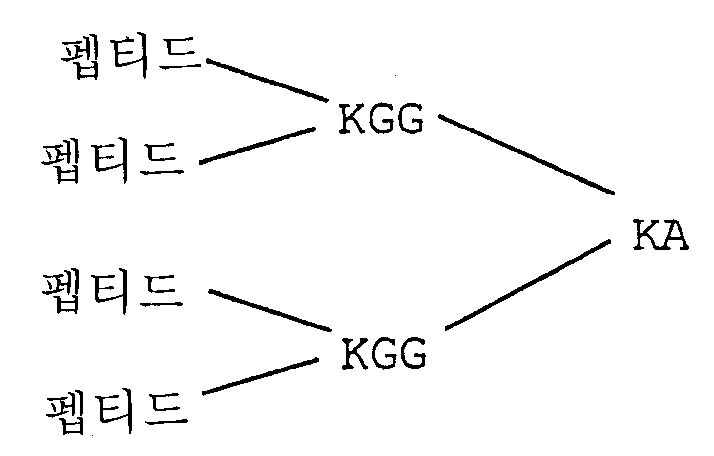
AN90549(MAP4구조의 Aβ1-7/파상풍 톡소이드 830-844):AN90549 (Aβ1-7 / Tetanus Toxoid 830-844 with MAP4 structure):
![]()
![]()
AN90550(MAP4구조의 Aβ1-7/파상풍 톡소이드 947-967):AN90550 (Aβ1-7 / Tetanus Toxoid 947-967 with MAP4 structure):
![]()
![]()
AN90542(선형 구조의 Aβ1-7/파상풍 톡소이드 830-844 + 947-967)AN90542 (Linear Aβ1-7 / Tetus Toxoid 830-844 + 947-967)
![]()
![]()
AN90576(MAP4 구조의 Aβ3-9/파상풍 톡소이드 830-844)AN90576 (Aβ3-9 / Tetanus Toxoid 830-844 with MAP4 Structure)
![]()
![]()
미국 특허 제5,736,142호에 개시된 펩티드(모두 선형 구조임)Peptides disclosed in US Pat. No. 5,736,142, all of which are linear structures
![]()
![]()
![]()
![]()
융합 단백질(Aβ의 면역원성 에피토프는 굵은 글씨로 표시되어 있음)의 또 다른 예는 다음과 같다. Another example of a fusion protein (Aβ immunogenic epitopes are shown in bold) is as follows.
AKXVAAWTLKAAA-DAEFRHD-DAEFRHD-DAEFRHD (서열 번호 58)AKXVAAWTLKAAA- DAEFRHD-DAEFRHD-DAEFRHD (SEQ ID NO: 58)
DAEFRHD-AKXVAAWTLKAAA (서열 번호 59) DAEFRHD -AKXVAAWTLKAAA (SEQ ID NO: 59)
DAEFRHD-ISQAVHAAHAEINEAGR (서열 번호 60) DAEFRHD -ISQAVHAAHAEINEAGR (SEQ ID NO: 60)
FRHDSGY-ISQAVHAAHAEINEAGR (서열 번호 61) FRHDSGY -ISQAVHAAHAEINEAGR (SEQ ID NO: 61)
EFRHDSG-ISQAVHAAHAEINEAGR (서열 번호 62) EFRHDSG -ISQAVHAAHAEINEAGR (SEQ ID NO: 62)
PKYVKQNTLKLAT-DAEFRHD-DAEFRHD-DAEFRHD (서열 번호 63)PKYVKQNTLKLAT- DAEFRHD-DAEFRHD-DAEFRHD (SEQ ID NO: 63)
DAEFRHD-PKYVKQNTLKLAT-DAEFRHD (서열 번호 64) DAEFRHD -PKYVKQNTLKLAT- DAEFRHD (SEQ ID NO: 64)
DAEFRHD-DAEFRHD-DAEFRHD-PKYVKQNTLKLAT (서열 번호 65) DAEFRHD-DAEFRHD-DAEFRHD -PKYVKQNTLKLAT (SEQ ID NO: 65)
DAEFRHD-DAEFRHD-PKYVKQNTLKLAT (서열 번호 66) DAEFRHD-DAEFRHD- PKYVKQNTLKLAT (SEQ ID NO: 66)
DAEFRHD-PKYVKQNTLKLAT-EKKIAKMEKASSVFNV-QYIKANSKFIGITEL-FNNFTVSFWLRVPKVSASHLE-DAEFRHD (서열 번호 67) DAEFRHD- PKYVKQNTLKLAT-EKKIAKMEKASSVFNV-QYIKANSKFIGITEL-FNNFTVSFWLRVPKVSASHLE- DAEFRHD (SEQ ID NO: 67)
DAEFRHD-DAEFRHD-DAEFRHD-QYIKANSKFIGITEL-FNNFTVSFWLRVPKVSASHLE (서열 번호 68) DAEFRHD-DAEFRHD-DAEFRHD- QYIKANSKFIGITEL-FNNFTVSFWLRVPKVSASHLE (SEQ ID NO: 68)
DAEFRHD-QYIKANSKFIGITELCFNNFTVSFWLRVPKVSASHLE (서열 번호 69) DAEFRHD -QYIKANSKFIGITEL C FNNFTVSFWLRVPKVSASHLE (SEQ ID NO: 69)
DAEFRHD-QYIKANSKFIGITELCFNNFTVSFWLRVPKVSASHLE-DAEFRHD (서열 번호 70) DAEFRHD -QYIKANSKFIGITEL C FNNFTVSFWLRVPKVSASHLE- DAEFRHD (SEQ ID NO: 70)
2개의 분지 수지 상에 DAEFRHD- QYIKANSKFIGITEL (서열 번호 52) DAEFRHD -QYIKANSKFIGITEL (SEQ ID NO: 52) on 2 branch resins
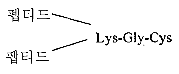
EQVTNVGGAISQAVHAAHAEINEAGR(서열 번호 71)(MAP-4 구조의 시누클레인 융합 단백질) EQVTNVGGA ISQAVHAAHAEINEAGR (SEQ ID NO: 71) (synuclein fusion protein of MAP-4 structure)
수동 면역화에 사용하기 위한 Aβ에 대한 항체의 형성에 사용되는 면역원을 형성하기 위해서 동일 또는 유사한 캐리어 단백질 및 연결 방법을 사용할 수 있다. 예를 들어, 캐리어에 연결된 Aβ또는 단편을 Aβ에 대한 모노클론 항체 제조시 실험실 동물에 투여할 수 있다. The same or similar carrier proteins and linking methods can be used to form immunogens used in the formation of antibodies against Aβ for use in passive immunization. For example, Aβ or fragment linked to a carrier can be administered to a laboratory animal in the preparation of a monoclonal antibody against Aβ.
4. 치료제를 암호화하는 핵산4. Nucleic Acids Encoding Therapeutics
아밀로이드 침착에 대한 면역 반응은 Aβ펩티드의 분절 및 이의 단편, 기타 펩티드 면역원, 또는 수동 면역화용으로 사용되는 항체 및 이의 성분을 암호화하는 핵산의 투여로 유도할 수 있다. 이러한 핵산은 DNA 또는 RNA일 수 있다. 면역원을 암호화하는 핵산 분절은 통상적으로 환자의 의도하는 표적 세포 중 DNA 분절을 발현시키는 프로모터 및 인헨서 등의 조절 서열에 연결된다. 면역 반응의 유도에 바람직한 혈액 세포에서의 발현의 경우, 경쇄 또는 중쇄 면역글로불린 유전자로부터 유래한 프로모터 및 인헨서 성분 또는 CMV 주요 직초기 프로모터 및 인헨서는 직접 발현에 적절하다. 연결된 조절 성분 및 암호화 서열은 종종 벡터 내로 클로닝된다. 이본쇄 항체의 투여를 위해, 2개의 사슬을 동일 또는 별도 벡터에서 클로닝시킬 수 있다. The immune response to amyloid deposition can be induced by administration of segments of Aβ peptides and fragments thereof, other peptide immunogens, or nucleic acids encoding antibodies and components thereof used for passive immunization. Such nucleic acid may be DNA or RNA. Nucleic acid segments encoding immunogens are typically linked to regulatory sequences such as promoters and enhancers that express DNA segments in the patient's intended target cells. For expression in blood cells preferred for induction of an immune response, promoter and enhancer components derived from the light or heavy chain immunoglobulin genes or CMV major immediate primitive promoters and enhancers are suitable for direct expression. Linked regulatory components and coding sequences are often cloned into the vector. For administration of double stranded antibodies, the two chains can be cloned in the same or separate vectors.
다수의 바이러스 벡터계, 예컨대 레트로바이러스계(예컨대 Lawrie 및 Tumin, Cur.Opin.Genet.Develop. 3, 102-109(1993) 참조); 아데노바이러스 벡터(예, Bett 등, J.Virol. 67, 5911(1993) 참조); 아데노 연관 바이러스 벡터(예, Zhou 등, J.Exp.Med. 179, 1867(1994) 참조), 백시니아 바이러스 및 조류 폭스 바이러스를 비롯한 폭스 계열 유래의 바이러스 벡터, 신드비스(Sindbis) 및 SFV(Semliki Forest Virus)(예, Dubensky 등, J.Virol. 70, 508-519(1996) 참조)로부터 유도된 것과 같은 알파 바이러스 종, 베네수엘라 말 뇌염 바이러스(미국 특허 제5,643,576호 참조) 및 랍도바이러스(rhabdovirus), 예컨대 수포성 구내염 바이러스(WO 96/34625호) 및 파필로마바이러스(Ohe 등, Human Gene Therapy 6, 325-333(1995); Woo 등, WO 94/12629 및 Xiao & Brandsma, Nucleic Acids. Res. 24, 2630-2622(1996)) 유래의 바이러스 벡터 등을 이용할 수 있다. Many viral vector systems, such as retroviral systems (see, eg, Lawrie and Tumin, Cur. Opin. Genet. Development. 3, 102-109 (1993)); Adenovirus vectors (see, eg, Bett et al., J. Virol. 67, 5911 (1993)); Adeno-associated viral vectors (see, for example, Zhou et al., J. Exp. Med. 179, 1867 (1994)), viral vectors from the Fox family, including vaccinia virus and avian fox virus, Sindbis and SFV (Semliki). Alpha virus species, such as those derived from Forest Virus (see, eg, Dubensky et al., J. Virol. 70, 508-519 (1996)), Venezuelan equine encephalitis virus (see US Pat. No. 5,643,576) and rhabdovirus ) Such as bullous stomatitis virus (WO 96/34625) and papillomavirus (Ohe et al., Human Gene Therapy 6, 325-333 (1995); Woo et al., WO 94/12629 and Xiao & Brandsma, Nucleic Acids.Res) 24, 2630-2622 (1996)), and the like can be used.
면역원을 암호화하는 DNA, 또는 이를 함유하는 벡터를 리포좀 내로 패키징할 수 있다. 적절한 지질 및 관련 유사체는 미국 특허 제5,208,036호, 제5,264,618호, 제5,279,833호 및 제5,283,185호에 개시되어 있다. 면역원을 암호화하는 DNA 및 벡터를 미립자 캐리어에 흡착시키거나 또는 이와 회합시킬 수 있으며, 미립자 캐리어의 예로는 폴리메틸 메타크릴레이트 폴리머와 폴리락티드 및 폴리(락티드-코-글리콜리드) 등이 있다. 예컨대 McGee 등, J. Micro Encap.(1996) 참조.DNA encoding an immunogen, or a vector containing the same, can be packaged into liposomes. Suitable lipids and related analogs are disclosed in US Pat. Nos. 5,208,036, 5,264,618, 5,279,833 and 5,283,185. DNA and vectors encoding immunogens can be adsorbed to or associated with particulate carriers, examples of which include polymethyl methacrylate polymers and polylactides and poly (lactide-co-glycolides). . See, eg, McGee et al., J. Micro Encap. (1996).
유전자 요법 벡터 또는 나출 DNA는, 통상적으로 전신 투여(예, 정맥내, 복강내, 비강, 위, 피내, 근육내, 피하 또는 두개내 주입) 또는 국소 적용(예컨대 미국 특허 제5,399,346호 참조)으로 개개 환자에게 투여하여 생체내 전달할 수 있다. 이 러한 벡터는 부피백신(bupivacine)(미국 특허 제5,593,970호)와 같은 촉진제를 추가로 포함한다. 또한, 유전자 총을 사용하여 DNA를 투여할 수 있다. Xiao & Brandsma, 상기 문헌 참조. 면역원을 암호화하는 DNA는 미세한 금속 비드의 표면에 침착시킨다. 미세발사체를 충격파 또는 팽창 헬륨 기체로 가속화시켜 일부 세포층의 심부에 조직을 침투시킨다. 예를 들어, 위스콘신주 미들레톤에 소재하는 아가세투스 인코포레이티드가 제조한 AccelTM 유전자 전달 장치가 적절하다. 대안적으로, 화학적 또는 기계적 자극으로 피부 상에 DNA를 단순히 스포팅함으로써 나출 DNA를 피부를 통해 혈류로 통과시킬 수 있다(WO 95/05853호 참조).Gene therapy vectors or naked DNA are typically individually administered by systemic administration (eg, intravenous, intraperitoneal, nasal, stomach, intradermal, intramuscular, subcutaneous or intracranial injection) or topical application (see, eg, US Pat. No. 5,399,346). It can be administered to a patient and delivered in vivo. Such vectors further include promoters such as bupivacine (US Pat. No. 5,593,970). Gene guns can also be used to administer DNA. See Xiao & Brandsma, supra. DNA encoding the immunogen is deposited on the surface of the fine metal beads. Microprojectiles are accelerated with shock waves or expanded helium gas to infiltrate tissue into the core of some cell layers. For example, the Accel ™ gene delivery device manufactured by Agacetus Incorporated, Middleleton, Wisconsin is suitable. Alternatively, naked DNA can be passed through the skin into the bloodstream by simply spotting the DNA on the skin with chemical or mechanical stimuli (see WO 95/05853).
또 다른 변형예에서, 면역원을 암호화하는 벡터를 개개 환자로부터 체외 배양된 세포(예, 림프구, 골수 흡입, 조직 생검) 또는 보편 공여자 조혈 간세포를 생체외에서 세포에 전달한 다음, 일반적으로 벡터에 통합된 세포를 선별한 후 환자에게 세포를 재이식할 수 있다. In another variation, a vector encoding an immunogen is delivered from an individual patient to cells cultured in vitro (eg, lymphocytes, bone marrow inhalation, tissue biopsy) or universal donor hematopoietic stem cells in vitro and then generally integrated into the vector. After selection, the cells can be transplanted to the patient.
III. 제거 활성에 대한 항체 스크리닝III. Antibody Screening for Removal Activity
본 발명은 제거 활성이 바람직한 아밀로이드 침착 또는 임의의 다른 항원, 또는 관련 생물학적 독립체를 제거하는 활성에 대해 항체를 스크리닝하는 방법을 제공한다. 아밀로이드 침착에 대한 활성을 스크리닝하기 위해서, 알츠하이머 질환 환자의 뇌 또는 특징적 알츠하이머 질환리를 보유한 동물 모델에서 얻은 조직 샘플을, 시험관내 배지에서 Fc 수용체를 보유하는 식세포(예컨대 소신경교세포) 및 테스트 하고자하는 항체와 접촉시킨다. 식세포는 BV-2, C8-B4 또는 THP-1 등의 세포 주 또는 1차 배양물일 수 있다. 일부 방법에서, 성분을 현미경 슬라이드에 결합시켜 현미경 모니터링을 용이하게 한다. 일부 방법에서, 미량 역가 접시의 웰에서 유사하게 수회 반응을 실시한다. 이러한 형식에서, 별도의 소형 현미경 슬라이드를 별도의 웰에 설치할 수 있거나, 또는 Aβ의 ELISA 검출과 같은 비현미경 검출 형식을 사용할 수 있다. 반응을 진행하기 전에 기준 값으로부터 시작하여 시험관내 반응 혼합물에서의 아밀로이드 침착량과 반응 중의 하나 이상의 테스트 값의 일련의 측정을 실시하는 것이 좋다. 항원은, 예컨대 Aβ또는 아밀로이드 플라크의 기타 성분에 대한 형광 표지된 항체로 염색하여 검출할 수 있다. 염색에 사용된 항체는 제거 활성에 대해 테스트 중인 항체와 동일할 수도, 동일하지 않을 수도 있다. 아밀로이드 침착 반응 과정에서 기준선과 비교하여 나타난 감소는, 테스트 중인 항체가 제거 활성을 보유함을 제시하는 것이다. 그러한 항체는 알츠하이머 및 기타 아밀로이드 형성 질병을 예방 또는 치료하는 데 유용할 것 같다.The present invention provides a method for screening antibodies for amyloid deposition or any other antigen, or activity for removing related biological entities, wherein removal activity is desired. To screen for activity against amyloid deposition, tissue samples obtained from brains of Alzheimer's disease patients or animal models with characteristic Alzheimer's disease pathologies are tested for phagocytic cells (eg small neuroglial cells) bearing Fc receptors in in vitro media and to be tested. Contact with antibody. The phagocytes may be cell lines or primary cultures such as BV-2, C8-B4 or THP-1. In some methods, components are coupled to microscope slides to facilitate microscope monitoring. In some methods, the reactions are similarly run several times in the wells of the microtiter dish. In this format, separate small microscope slides can be installed in separate wells, or non-microscopic detection formats such as ELISA detection of Aβ can be used. Before proceeding with the reaction, it is advisable to make a series of measurements of amyloid deposition in the in vitro reaction mixture and one or more test values of the reaction, starting from the reference value. Antigens can be detected, for example, by staining with fluorescently labeled antibodies against Aβ or other components of amyloid plaques. The antibody used for staining may or may not be the same as the antibody under test for clearance activity. The reduction shown compared to baseline in the course of the amyloid deposition reaction suggests that the antibody under test retains elimination activity. Such antibodies are likely to be useful for preventing or treating Alzheimer's and other amyloidogenic diseases.
유사 방법을 사용하여 기타 유형의 생물학적 독립체의 제거 활성에 대해 항체를 스크리닝할 수 있다. 이 분석을 이용하여 결국 어떤 종류의 생물학적 독립체에 대한 제거 활성도 검출할 수 있다. 통상적으로, 생물학적 독립체는 인간 또는 동물 질병에서 약간의 역할을 한다. 생물학적 독립체는 조직 샘플로서 또는 분리형으로서 제공할 수 있다. 조직 샘플로서 제공되는 경우, 조직 샘플의 성분에 용이하게 접근하고 고정에 수반되는 성분 구조의 교란을 피하기 위해서 조직 샘플을 고정하지 않는 것이 바람직하다. 이 분석법에서 테스트할 수 잇는 조직 샘플의 예로는 암조직, 전암(precancerous) 조직, 혹 또는 사마귀 같은 양성 증식을 포함하는 조 직, 병원성 미생물로 감염된 조직, 염증성 세포가 침투된 조직, 세포 간에 병리학적 간질을 보유하는 조직(예, 섬유소 심막염), 변종 항원을 보유하는 조직, 및 반흔 조직 등이 있다. 사용할 수 있는 분리형 생물학적 독립체의 예로는 Aβ, 바이러스 항원 또는 바이러스, 프로테오글리칸, 기타 병원성 미생물의 항원, 종양 항원 및 유착 분자 등이 있다. 이들 항원은 여러 수단 중 천연원, 재조합 발현 또는 화학적 합성으로부터 얻을 수 있다. 조직 샘플 또는 분리형 생물학적 독립체를 및 배지에서 Fc 수용체 보유 식세포(예, 단핵구 또는 소신경세포) 및 시험하고자 하는 항체와 접촉시킨다. 항체는 테스트 중인 생물학적 독립체에 대해 유도하거나, 또는 독립체와 관련된 항체에 대해 유도할 수 있다. 후자의 경우, 생물학적 독립체가 항원을 이용하여 대상성으로 식균 작용을 하는지를 시험하는 것이 목적이다. 일반적으로, 반드시 필요한 것은 아니지만, 항체 및 생물학적 독립체(종종 관련 항원을 보유)를 서로 접촉시킨 다음 식세포를 첨가한다. 배지 내에 남아 있는 생물학적 독립체 및/또는 관련 항원이 존재하는 경우, 이의 농도를 모니터링한다. 배지 중 항원 또는 관련 생물학적 독립체의 양 또는 농도의 감소는, 항체가 식세포와 함께 항원 및/또는 관련 생물학적 독립체에 대한 제거 반응을 보유함을 제시한다(예컨대 실시예 14 참조).Similar methods can be used to screen antibodies for the removal activity of other types of biological entities. This assay can eventually be used to detect clearance activity for any type of biological entity. Typically, biological entities play some role in human or animal disease. The biological entity may serve as a tissue sample or as a separate. When provided as a tissue sample, it is desirable not to immobilize the tissue sample in order to easily access the components of the tissue sample and to avoid perturbation of the component structures involved in immobilization. Examples of tissue samples that can be tested in this assay include tissues containing benign proliferation such as cancerous tissues, precancerous tissues, lumps or warts, tissues infected with pathogenic microorganisms, tissues infiltrated with inflammatory cells, and pathologies between cells. Tissues with epilepsy (eg fibrin peritonitis), tissues with variant antigens, and scar tissue. Examples of isolated biological entities that can be used include Aβ, viral antigens or viruses, proteoglycans, antigens of other pathogenic microorganisms, tumor antigens and adhesion molecules. These antigens can be obtained from natural sources, recombinant expression or chemical synthesis, among other means. Tissue samples or isolated biological entities are contacted with Fc receptor bearing phagocytes (eg monocytes or microneurons) in the medium and with the antibody to be tested. The antibody may be directed against the biological entity under test, or against an antibody associated with the entity. In the latter case, the aim is to test whether the biological entity is phagocytically targeted using the antigen. Generally, but not necessarily, the antibody and the biological entity (often bearing associated antigens) are brought into contact with each other followed by the addition of phagocytes. If there is a biological entity and / or related antigen remaining in the medium, its concentration is monitored. Reduction in the amount or concentration of antigen or related biological entity in the medium suggests that the antibody has a clearance reaction against the antigen and / or related biological entity with the phagocytes (see, eg, Example 14).
IV 치료 가능한 환자IV Treatable Patients
치료 가능한 환자는 질병 위험이 있으나 증상을 나타내지 않는 개체와, 현재 증상을 나타내고 있는 환자를 포함한다. 알츠하이머 질병의 경우, 남녀가 충분히 오래 산다면 결국 어떤 사람도 알츠하이머 질병에 걸릴 위험이 있다. 따라서, 본 발명의 방법은 대상 환자의 위험을 평가할 필요 없이 일반적인 개체군에 예방학적으로 시행될 수 있다. 본 발명의 방법은 공지된 알츠하이머 질환의 유전적 위험을 보유한 개체에 특히 유용하다. 그러한 개체는 이 질병을 경험한 친척이 있는 개체들 및 유전적 또는 생화학적 마커의 분석으로 위험이 측정된 개체들을 포함한다. 알츠하이머 질병에 대한 위험의 유전적 마커는 APP 유전자의 돌연변이, 특히 각각 하디 및 스웨디시 돌연변이라고 불리우는 위치 717과 위치 670 및 671에서의 돌연변이를 포함한다(Hardy, TINS, 상기 참조). 기타 위험 마커는 AD, 고콜레스테롤혈증 또는 아테롬성 경화증의 가족력인 프리세닐린(presenilin) 유전자, PS1 및 PS2와 ApoE의 돌연변이가 있다. 현재 알츠하이머 질환을 앓고 있는 개체를, 특징적 치매와 전술한 위험 인자의 존재로부터 알아낼 수 있다. 또한, AD에 걸린 개체를 확인하는 여러 진단 테스트가 이용가능하다. 이들은 CSF tau 및 Aβ42 수준의 측정을 포함한다. 상승된 tau 및 감소된 Aβ42의 수준은 AD의 존재를 알려준다. 알츠하이머 질환을 앓고 있는 개체는 실시예 부분에 논의된 바와 같은 ADRDA 기준으로 진단할 수 있다. Treatable patients include individuals who are at risk of disease but do not exhibit symptoms and patients who are currently presenting symptoms. In the case of Alzheimer's disease, if a man and a woman live long enough, there is a risk that anyone will eventually develop Alzheimer's disease. Thus, the methods of the present invention can be performed prophylactically in a general population without having to assess the risk of the subject patient. The methods of the present invention are particularly useful for individuals who carry the genetic risk of known Alzheimer's disease. Such individuals include those with relatives who have experienced the disease and those whose risk has been determined by analysis of genetic or biochemical markers. Genetic markers of risk for Alzheimer's disease include mutations in the APP gene, in particular mutations at positions 717 and
무증상 환자에서, 치료는 임의의 연령(예, 10, 20, 30)에 개시할 수 있다. 그러나, 일반적으로 환자가 40, 50, 60 또는 70세에 도달할때, 치료를 개시할 필요는 없다. 치료는 통상적으로 일정 기간 동안 수회 용량을 수반한다. 항체, 또는 일정 기간 동안 치료제(예, Aβ펩티드)에 반응하는 활성화된 T-세포 또는 B-세포를 분석하여 치료를 모니터링할 수 있다. 반응이 저하되면, 추가 항원 투여한다. 잠재적 다운증후군 환자의 경우, 모체에게 또는 출생 직후 치료제를 투여하여 출생전에 치료를 개시할 수 있다.In asymptomatic patients, treatment may commence at any age (eg, 10, 20, 30). Generally, however, when a patient reaches 40, 50, 60 or 70 years old, there is no need to initiate treatment. Treatment typically involves several doses over a period of time. Treatment can be monitored by analyzing antibodies or activated T-cells or B-cells in response to a therapeutic agent (eg, Aβ peptide) for a period of time. If the response is lowered, an additional antigen is administered. In patients with potential Down syndrome, treatment may be initiated before birth by administering the treatment to the mother or immediately after birth.
V 치료법V treatment
예방용으로, 알츠하이머 질환에 감수성인 환자 또는 위험이 있는 환자에게, 질병의 생화학, 조직학 및/또는 행동 증상, 질병의 형성 과정에서 제공되는 이의 합병증 및 중간 병리학적 표현형을 비롯한 질병의 위험을 제거 또는 감소시키거나, 병의 심각도를 줄이거나, 또는 질병의 개시를 지연시키는 유효량으로 약학 조성물 및 약제를 투여한다. 치료용으로, 질병에 걸렸을 것으로 의심되거나 또는 이미 병에 걸린 환자에게, 질병의 형성 과정에서 제공되는 이의 합병증 및 중간 병리학적 표현형을 비롯한 질병의(생화학, 조직학 및/또는 행동의) 증상을 치료, 또는 적어도 부분적으로 병의 진행을 정지시키는 유효량으로 약학 조성물 및 약제를 투여한다. 일부 방법에서, 제제의 투여는 특징적 알츠하이머 질환리학이 아직 발병하지 않은 환자에서 근인식(myocognitive) 손상을 감소 또는 제거한다. 처치 또는 예방 치료를 달성하기에 적당한 양은 치료 또는 예방 유효량으로서 정의한다. 예방 및 치료법에서, 제제는 일반적으로 충분한 면역 반응이 얻어질 때까지 수회 용량으로 투여한다. 통상적으로, 면역반응을 모니터링하고, 면역 반응이 약해지기 시작하면 반복 용량을 제공한다. For prevention, to patients who are susceptible to or at risk for Alzheimer's disease, the risk of the disease, including the biochemistry, histology and / or behavioral symptoms of the disease, its complications provided in the course of the disease and its intermediate pathological phenotype, is eliminated or The pharmaceutical compositions and medicaments are administered in an effective amount that reduces, reduces the severity of the disease, or delays the onset of the disease. For the treatment, to a patient suspected of having a disease or to already have a disease, treating a symptom of the disease (biochemistry, histology and / or behavior), including its complications and intermediate pathological phenotypes, which are presented during the formation of the disease, Or administering the pharmaceutical composition and medicament in an effective amount that at least partially arrests disease progression. In some methods, administration of the agent reduces or eliminates myocognitive impairment in patients who have not yet developed characteristic Alzheimer's pathology. Amounts suitable to achieve a therapeutic or prophylactic treatment are defined as therapeutically or prophylactically effective amounts. In prophylaxis and treatment, the formulations are generally administered in several doses until a sufficient immune response is obtained. Typically, the immune response is monitored and repeated doses are given as the immune response begins to weaken.
전술한 증상을 치료하기 위한 본 발명의 조성물의 유효량은 여러 상이한 인자, 예컨대 투여 수단, 표적 부위, 환자의 생리 상태, 환자가 인간인지 또는 동물인지의 여부, 투여되는 기타 약물, 및 치료가 예방용인지 또는 치료용인지 여부 등에 따라서 달라진다. 일반적으로, 환자는 인간이지만 유전자이식 포유류를 비롯한 인간이 아닌 포유류도 치료할 수 있다. 치료 용량은 안전성 및 효능을 최적화하도록 적정해야 한다. 면역원의 양은 보조제의 투여 여부에 따라 달라지며, 보조제의 부재하에서의 용량은 더 높아야 한다. 투여용 면역원의 양은 때로는 환자당 1∼500 ㎍, 더욱 일반적으로는 인간 투여의 경우 주사 1회당 5∼500 ㎍이다. 경우에 따라서, 주사 1회당 1∼2 ㎎의 고용량을 사용한다. 통상적으로 인간의 경우 주사 1회당 약 10, 20, 50 또는 100 ㎍이 사용된다. 면역원의 질량은 전체로서 면역원의 질량에 대한 면역원 내의 면역원성 에피토프의 질량비에 따라서 다르다. 통상, 면역원 1 ㎍ 당 면역원성 에피토프 10-3∼10-5 μmol이 사용된다. 주사 시기는 1일 1회 내지 1년 1회, 내지는 10년에 1회로 상당히 다양할 수 있다. 면역원을 투여한 당일에, 용량은 보조제의 존재하에 환자당 1 ㎍ 이상, 일반적으로 10 ㎍이상이고, 보조제의 부재하에 10 ㎍ 이상, 일반적으로 100 ㎍ 이상이다. 통상적인 방법은 면역화 후에, 시간 간격(예, 6주 간격)을 두고 추가 항원 주사를 실시하는 것으로 구성된다. 또 다른 방법은 면역화 후 1, 2 및 12개월이 경과한 다음 추가 항원 주사하는 것으로 구성된다. 또 다른 방법은 평생 매2개월 마다 1회씩 주사한다. 또는, 추가 항원 주사는 면역 반응을 모니터링하여 제시되는 대로 불규칙하게 실시할 수 있다. An effective amount of a composition of the present invention for treating the above-mentioned symptoms may be determined by several different factors such as means of administration, target site, physiological state of the patient, whether the patient is human or animal, other drugs administered, and therapies for preventing It depends on whether it is cognitive or therapeutic. Generally, patients are human but can also treat non-human mammals, including transgenic mammals. The therapeutic dose should be titrated to optimize safety and efficacy. The amount of immunogen depends on the administration of the adjuvant and the dose in the absence of the adjuvant should be higher. The amount of immunogen for administration is sometimes from 1 to 500 μg per patient, more generally from 5 to 500 μg per injection for human administration. In some cases, a high dose of 1-2 mg per injection is used. Typically, in humans, about 10, 20, 50 or 100 μg is used per injection. The mass of an immunogen depends on the mass ratio of immunogenic epitopes in the immunogen to the mass of the immunogen as a whole. Usually, 10 -3 to 10 -5 mol of immunogenic epitopes are used per µg of immunogen. The timing of injection can vary considerably from once a day to once a year, or even once a decade. On the day of administration of the immunogen, the dose is at least 1 μg per patient, generally at least 10 μg per patient in the presence of an adjuvant and at least 10 μg, generally at least 100 μg in the absence of the adjuvant. Conventional methods consist of performing additional antigen injections at time intervals (eg, 6 week intervals) after immunization. Another method consists of injection of
항체를 이용한 수동 면역화의 경우, 용량은 숙주 체중 1 kg당 0.0001∼100 ㎎, 더욱 일반적으로는 0.01∼5 ㎎ 범위이다. 예를 들어, 용량은 체중 1 kg당 1 ㎎ 또는 10 ㎎, 또는 1∼10 ㎎의 범위 내에 있을 수 있다. 예시적 치료법에서는 2주에 1회 또는 매월 1회 또는 3∼6개월마다 1회 투여한다. 일부 방법에서는 상이한 결합 특이성을 보유한 2 이상의 단일클론 항체를 동시에 투여하며, 이 경우 투여되는 각 항체의 용량은 지정된 범위내에 속한다. 항체는 일반적으로 수회 투여한다. 단일 용량 사이의 간격은 주 단위, 월 단위 또는 년 단위일 수 있다. 간격은 환자의 Aβ에 대한 항체의 혈중 농도를 측정하여 제시된 대로 불규칙할 수 있다. 일부 방법에서, 혈장 항체 농도 1∼1000 ㎍/㎖을 달성할 수 있도록 용량을 조정하며, 일부 방법에서는 25∼300 ㎍/㎖이다. 대안적으로, 서방형 제제로서 항체를 투여할 수 있으며, 이 경우 투여 빈도수를 줄인다. 용량 및 빈도수는 환자내 항체의 반감기에 따라 달라진다. 일반적으로, 인간 항체가 최장 반감기를 나타내고, 그 다음은 인간화된 항체, 키메라 항체 및 비인간 항체의 순이다. 투여 용량 및 빈도는 치료가 예방용인지 치료용인지 여부에 따라 달라질 수 있다. 예방용으로, 장기간에 걸쳐 비교적 간격을 크게 하여 비교적 낮은 용량을 투여한다. 일부 환자는 남은 일생 동안 계속 치료받는다. 치료용에서는, 질병의 진행이 감소되거나 또는 종결될 때까지, 바람직하게는 환자가 질병 증상의 부분적 또는 완전한 호전을 나타낼 때까지, 비교적 짧은 간격으로 비교적 높은 용량을 필요로 한다. 이후, 환자에게 예방법을 실시할 수 있다.For passive immunization with antibodies, doses range from 0.0001 to 100 mg, more usually 0.01 to 5 mg per kg body weight of the host. For example, the dose may be in the range of 1 mg or 10 mg, or 1-10 mg per kg body weight. Exemplary therapies are administered once every two weeks or once a month or once every three to six months. In some methods, two or more monoclonal antibodies with different binding specificities are administered simultaneously, in which case the dose of each antibody administered falls within the specified range. Antibodies are generally administered several times. The interval between single doses can be weekly, monthly or yearly. The interval may be irregular as shown by measuring blood levels of the antibody against the patient's Aβ. In some methods, the dose is adjusted to achieve a plasma antibody concentration of 1-1000 μg / ml, and in some methods 25-300 μg / ml. Alternatively, the antibody can be administered as a sustained release formulation, in which case the frequency of administration is reduced. Dosage and frequency vary depending on the half-life of the antibody in the patient. In general, human antibodies have the longest half-life, followed by humanized antibodies, chimeric antibodies, and nonhuman antibodies. Dosage and frequency may vary depending on whether the treatment is prophylactic or therapeutic. For prophylaxis, relatively low doses are administered over relatively long periods of time. Some patients continue to be treated for the rest of their lives. For therapeutic use, relatively high doses are required at relatively short intervals until the progression of the disease is reduced or terminated, preferably until the patient exhibits partial or complete improvement of the disease symptoms. The patient may then be prevented.
면역원을 암호화하는 핵산에 대한 용량은 환자당 약 10 ng∼1 g, 100 ng∼100 ㎎, 1 ㎍∼10 ㎎, 또는 30∼300 ㎍ DNA 범위이다. 감염성 바이러스 벡터에 대한 용량은 1회 용량당 10∼100 이상의 비리온으로 다양하다.Doses for nucleic acids encoding immunogens range from about 10 ng to 1 g, 100 ng to 100 mg, 1 μg to 10 mg, or 30 to 300 μg DNA per patient. Doses for infectious viral vectors vary from 10 to 100 or more virions per dose.
면역 반응을 유도하는 제제는 예방 및/또는 처치 치료를 위해 비경구, 국소, 정맥내, 경구, 피하, 동맥내, 두개내, 복강내, 비강내 또는 근육내 수단으로 투여 할 수 있다. 기타 투여 경로도 유사하게 유효할 수 있지만, 면역원성 제제의 가장 통상적인 투여 경로는 피하 경로이다. 그 다음 가장 일반적인 경로는 근육내 주사이다. 이러한 종류의 주사는 가장 통상적으로는 팔 또는 다리 근육에서 실시된다. 일부 방법에서, 침착물이 축적되는 특정 조직에 제제를 직접 주사, 예컨대 두개내 주사한다. 정맥내 주입시 근육내 주사는 항체 투여용으로 바람직하다. 일부 방법에서, 특정 치료 항체를 두개 내로 직접 주사한다. 일부 방법에서, 항체는 서방형 조성물 또는 장치(예, MedipadTM 장치)로서 투여한다.Agents that induce an immune response can be administered by parenteral, topical, intravenous, oral, subcutaneous, intraarterial, intracranial, intraperitoneal, intranasal or intramuscular means for prophylactic and / or therapeutic treatment. Other routes of administration may similarly be effective, but the most common route of administration of immunogenic agents is the subcutaneous route. The next most common route is intramuscular injection. Injections of this kind are most commonly performed in the arm or leg muscles. In some methods, the agent is injected directly, such as intracranially, into a particular tissue where deposits accumulate. Intramuscular injection upon intravenous infusion is preferred for antibody administration. In some methods, certain therapeutic antibodies are injected directly into the skull. In some methods, the antibody is administered as a sustained release composition or device (eg, a Medipad ™ device).
본 발명의 제제는 임의로 아밀로이드 형성 질병의 치료에 적어도 부분적으로 효과적인 기타 제제와 함께 투여할 수 있다. 아밀로이드 침착이 뇌에서 발생하는 알츠하이머 및 다운증후군의 경우, 혈액뇌 장벽을 통과하는 본 발명의 제제의 통과를 증가시키는 기타 제제와 함께 본 발명의 제제를 투여할 수 있다.The formulations of the present invention may optionally be administered with other agents that are at least partially effective for the treatment of amyloidogenic diseases. In the case of Alzheimer's and Down's syndrome, where amyloid deposition occurs in the brain, the formulations of the invention may be administered in combination with other formulations that increase the passage of the formulations of the invention across the blood brain barrier.
펩티드와 같은 본 발명의 면역원성 제제는 때로는 보조제와 함께 투여한다. Aβ와 같은 펩티드와 함께 각종 보조제를 사용하여 면역 반응을 유발할 수 있다. 바람직한 보조제는 반응의 성질 형태에 영향을 주는 면역원의 구조 변화를 유발하지 않고 면역원에 대한 내재 반응을 증대시킨다. 바람직한 보조제의 예로는 수산화알루미늄 및 인산알루미늄, 3-데-O-아실화된 모노포스포릴 리피드 A(MPLTM)(GB 2220211(RIBI ImmunoChem Research Inc., Hamilton, Montana, 지금은 Corixa의 일부) 등이 있다. StimulonTM QS-21은 사우스 아메리카에서 발견되는 Quillaja Saponaria Molina 나무의 수피로부터 분리된 트리터펜 글리코시드 또는 사포닌이 다(Vaccine Design; The Subunit and Adjuvant Approach(eds. Powell & Newman, Plenum Press, NY, 1995); 미국 특허 제5,057,540호 참조)(Aquila BioPharmaceuticals, Framingham, MA). 다른 보조제는, 경우에 따라서 모노포스포릴 리피드 A와 같은 면역자극제와 함께 수중유 에멀젼(예, 스쿠알렌 또는 땅콩유)이다(Stoute 등, N.Engl.J.Med. 336, 86-91(1997)). 또 다른 보조제는 CpG이다(WO 98/40100). 대안적으로, Aβ를 보조제에 커플링시킬 수 있다. 그러나, 그러한 커플링은 면역 반응의 특성에 영향을 주도록 Aβ의 구조를 실질적으로 변화시켜서는 안된다. 보조제는 활성제와 함께 치료 조성물의 성분으로서 투여할 수 있다. 또는 치료제의 투여와는 별도로 투여하거나, 또는 치료제의 투여 전, 동시에 또는 후에 투여할 수 있다. Immunogenic agents of the invention, such as peptides, are sometimes administered with adjuvant. Various adjuvants can be used with peptides such as Aβ to elicit an immune response. Preferred adjuvants augment the intrinsic response to the immunogen without causing structural changes in the immunogen that affect the nature of the response. Examples of preferred auxiliaries include aluminum hydroxide and aluminum phosphate, 3-de-O-acylated monophosphoryl lipid A (MPL ™ ) (GB 2220211 (RIBI ImmunoChem Research Inc., Hamilton, Montana, now part of Corixa), and the like. Stimulon ™ QS-21 is a triterpene glycoside or saponin isolated from the bark of the Quillaja Saponaria Molina tree found in South America (Vaccine Design; The Subunit and Adjuvant Approach (eds. Powell & Newman, Plenum Press, NY, 1995); see U.S. Pat. (Stoute et al., N. Engl. J. Med. 336, 86-91 (1997)) Another adjuvant is CpG (WO 98/40100) Alternatively, Aβ can be coupled to the adjuvant. However, such coupling affects the nature of the immune response To give must not substantially alter the structure of Aβ. Adjuvant may be administered as a component of a therapeutic composition with the active agent or can be administered as a therapeutic agent is administered separately, or be administered prior to administration of the therapeutic agent, at the same time or later.
바람직한 부류의 보조제는 알루미늄염(명반), 예컨대 수산화알루미늄, 인산알루미늄, 황산알루미늄이다. 그러한 보조제는 다른 특이적 면역자극제, 예컨대 MPL 또는 3-DMP, QS-21 또는 폴리글루탐산이나 폴리리신 등의 중합체 또는 단량체 아미노산의 유무 하에 사용할 수 있다. 또 다른 부류의 보조제는 수중유 에멀젼 제제이다. 그러한 보조제는 다른 특정 면역자극제, 예컨대 무라밀 펩티드(예, N-아세틸무라밀-L-트레오닐-D-이소글루타민(thr-MDP), N-아세틸-노르무라밀-L-알라닌-D-이소글루타민(nor-MDP), N-아세틸무라밀-L-알라닐-D-이소글루타밀-L-알라닌-2-(1'-2'-디팔미토일-sn-글리세롤-3-히드록시포스포릴옥시)-에틸아민(MTP-PE), N-아세틸글루코사미닐-N-아세틸무라밀-L-Al-D-이소글루-L-Ala-디팔미톡시프로필아미드(DTP-DPP) 테라미드 TM), 또는 기타 박테리아 세포벽 성분의 유무 하에 사용할 수 있다. 수중유 에멀젼은 (a) 모델 110Y 미세유동화제(미국 매사츄세츠주 뉴톤에 소재하는 마이크로플루이딕스) 등의 미세유동화제를 사용하여 미크론 이하 크기의 입자로 제형화한 5% 스쿠알렌, 0.5% 트윈 80 및 0.5% Span 85(임의로 다양한 양의 MTP-PE를 함유함)를 함유하는 MF59(WO 90/14837); (b) 미크론 이하 크기의 에멀젼으로 미세 유동화되거나 또는 와동되어 더 큰 입자 크기 에멀젼을 형성하는 10% 스쿠알렌, 0.4% 트윈 80, 5% 플루로닉 블록형 중합체 L121 및 thr-MDP를 함유하는 SAF, 및 (c) 2% 스쿠알렌, 0.2% 트윈 80과, 모노포스포릴 리피드 A(MPL), 트레할로스 디미콜레이트(TDM) 및 세포벽 골격(CWS), 바람직하게는 MPL+CSW(DetoxTM)로 구성된 군에서 선택된 하나 이상의 박테리아 세포벽 성분을 함유하는 RibiTM 보강계(RAS)(미국 몬타나주 해밀톤에 소재하는 리비 이뮤노켐)을 포함한다. 또 다른 부류의 바람직한 보조제는 사포닌 보조제, 예컨대 StimulonTM(QS-21, 미국 매사츄세츠주 프라밍험 소재의 아퀼라) 또는 ISCOM(면역자극 복합체) 및 ISCOMATRIX 등의 이로부터 형성된 입자이다. 다른 보조제는 완전 프로인트 보조제(CFA) 및 불완전 프로인트 보조제(IFA)를 포함한다. 다른 보조제로는 시토킨, 예컨대 인터루킨(IL-1, IL-2 및 IL-12), 대식 세포 콜로니 자극 인자(M-CSF), 종양 괴사 인자(TNF) 등이 있다.Preferred classes of auxiliaries are aluminum salts (alum) such as aluminum hydroxide, aluminum phosphate, aluminum sulfate. Such adjuvants may be used with or without other specific immunostimulants such as MPL or 3-DMP, QS-21 or polymer or monomeric amino acids such as polyglutamic acid or polylysine. Another class of adjuvants is oil-in-water emulsion formulations. Such adjuvants include other specific immunostimulating agents, such as Muramyl peptides (eg, N-acetylmuramil-L-threonyl-D-isoglutamine (thr-MDP), N-acetyl-normurayl-L-alanine-D- Isoglutamine (nor-MDP), N-acetylmurayl-L-alanyl-D-isoglutamyl-L-alanine-2- (1'-2'-dipalmitoyl-sn-glycerol-3-hydroxy Phosphoryloxy) -ethylamine (MTP-PE), N-acetylglucosaminyl-N-acetylmuramil-L-Al-D-isoglu-L-Ala-dipalmitoxypropylamide (DTP-DPP) terra Mead TM), or other bacterial cell wall components. Oil-in-water emulsions are (a) 5% squalene, 0.5% twin formulated into particles of sub-micron size using microfluidizing agents such as Model 110Y microfluidizing agent (Microfluidics, Newton, Mass.). MF59 (WO 90/14837) containing 80 and 0.5% Span 85 (optionally containing varying amounts of MTP-PE); (b) SAF containing 10% squalene, 0.4
보조제는 단일 조성물로서 면역원과 함께 투여할 수 있다. 또는 보조제는 면역원의 투여 전, 동시 또는 후에 투여할 수 있다. 면역원 및 보조제를 포장하여 동일한 병에 공급하거나, 또는 별도의 병에 포장하여 사용전에 혼합할 수 있다. 면역원 및 보조제는 통상적으로 목적하는 치료 용도를 제시하는 표지와 함께 포장한다. 면역원 및 보조제가 별도로 포장되는 경우, 포장은 통상적으로 사용전에 혼합에 대한 설명서를 포함한다. 보조제 및/또는 캐리어의 선택은 보조제를 함유하는 면역원성 제제의 안정성, 투여 경로, 투여 계획, 예방 접종하고자 하는 종에 대한 보조제의 효능에 따라 달라지며, 인간에서 약학적 허용 보조제는 적절한 조절체에 의한 인간 투여용으로 허가되거나 허가될 수 있는 것이다. 예를 들어, 완전 프로인트 보조제는 인간 투여용으로 적절하지 않다. 명반, MPL 및 QS-21이 바람직하다. 임의로, 2 이상의 상이한 보조제를 동시에 사용할 수 있다. 바람직한 조합은 명반과 MPL, 명반과 QS-21, MPL과 QS-21, 명반, QS-21 및 MPL을 함께 포함한다. 또한, 불완전 프로인트 보조제를, 경우에 따라 명반, QS-21과 MPL 및 이의 모든 조합과 함께 사용할 수 있다(Chang 등, Advanced Drug Delivery Reviews 32, 173-186(1998)).Adjuvants may be administered with the immunogen as a single composition. Alternatively, the adjuvant may be administered before, concurrently or after administration of the immunogen. Immunogens and adjuvants may be packaged and supplied in the same bottle or packaged in separate bottles and mixed before use. Immunogens and adjuvants are usually packaged with a label indicating the desired therapeutic use. If the immunogen and adjuvant are packaged separately, the package usually includes instructions for mixing prior to use. The choice of adjuvant and / or carrier depends on the stability of the immunogenic agent containing the adjuvant, the route of administration, the dosing regimen, and the efficacy of the adjuvant for the species to be vaccinated. Licensed or may be licensed for human administration. For example, complete Freund's adjuvant is not suitable for human administration. Alum, MPL and QS-21 are preferred. Optionally, two or more different adjuvants may be used simultaneously. Preferred combinations include alum and MPL, alum and QS-21, MPL and QS-21, alum, QS-21 and MPL together. Incomplete Freund's adjuvant may also be used, optionally with alum, QS-21 and MPL and all combinations thereof (Chang et al., Advanced
본 발명의 제제는 종종 활성 치료제와 각종 기타 약학적 허용 성분을 포함하는 약학 조성물로서 투여된다. Remington's Pharmaceutical Science(15th ed., Mack Publishing Company, Easton, Pennsylvania, 1980). 바람직한 형태는 목적하는 투여 방식 및 치료 용도에 따라 다르다. 조성물은, 목적하는 제형에 따라서, 약학적으로 허용가능한 비독성 캐리어 또는 희석제(이는 동물 또는 인간 투여용 약학 조성물을 배합하는 데 통상 사용되는 부형제로서 정의되어 있음)를 포함할 수 있다. 희석제는 조합시 생물학적 활성에 영향을 주지 않도록 선택한다. 이 희석제의 예로는 증류수, 생리학적 인산염 완충 염수, 링거액, 덱스트로스 용액 및 행크스 용액 등이 있다. 또한 약학 조성물 또는 배합물은 기타 캐리어, 보조제, 또는 비독 성, 비치료용, 비면역원성 안정화제 등을 포함할 수 있다.Formulations of the present invention are often administered as pharmaceutical compositions comprising the active therapeutic agent and various other pharmaceutically acceptable ingredients. Remington's Pharmaceutical Science (15th ed., Mack Publishing Company, Easton, Pennsylvania, 1980). Preferred forms depend on the mode of administration desired and the therapeutic use. The composition may comprise, depending on the desired formulation, a pharmaceutically acceptable non-toxic carrier or diluent, which is defined as an excipient commonly used in formulating pharmaceutical compositions for animal or human administration. Diluents are chosen so as not to affect biological activity in combination. Examples of this diluent include distilled water, physiological phosphate buffered saline, Ringer's solution, dextrose solution and Hanks' solution. Pharmaceutical compositions or combinations may also include other carriers, adjuvants, or non-toxic, non-treatment, non-immunogenic stabilizers, and the like.
약학 조성물은 크고 서서히 대사되는 거대 분자, 예컨대 단백질, 키토산 등의 다당류, 폴리락트산, 폴리글리콜산 및 공중합체(예, 라텍스 작용화된 세파로스 (TM), 아가로스, 셀룰로스 등), 아미노산 중합체, 아미노산 공중합체 및 지질 응집체(예, 오일 점적제 또는 리포좀) 등을 포함할 수 있다. 또한, 이들 캐리어는 면역자극제(즉, 보조제)로서 작용할 수 있다. Pharmaceutical compositions include large and slowly metabolized macromolecules such as polysaccharides such as proteins, chitosan, polylactic acid, polyglycolic acid and copolymers (e.g., latex functionalized sepharose (TM), agarose, cellulose, etc.), amino acid polymers, Amino acid copolymers and lipid aggregates (eg, oil drops or liposomes) and the like. In addition, these carriers can act as immunostimulants (ie, adjuvants).
비경구 투여용으로서, 본 발명의 제제는 물, 오일, 염수, 글리세롤 또는 에탄올 등의 멸균액일 수 있는 약학적 캐리어와 생리적 허용 희석제에서 물질의 용액 또는 현탁액의 주사가능 용량으로 투여할 수 있다. 또한, 보조 물질, 예컨대 습윤제 또는 유화제, 계면 활성제, pH 완충 물질 등이 조성물 내에 존재할 수 있다. 약학 조성물의 기타 성분은 석유, 동물, 식물 또는 합성 기원 오일, 예컨대 땅콩유, 대두유 및 미네랄유의 것들이 있다. 일반적으로, 폴리프로필렌 글리콜 또는 폴리에틸렌 글리콜 등의 글리콜이 특히 주사용 용액으로 바람직한 액체 캐리어이다. 활성 성분의 방출을 지속하는 방식으로 제형화할 수 있는 데포우 주사 또는 이식 제제의 형태로 항체를 투여할 수 있다. 예시적 조성물은 50 mM L-히스티딘, 150 mM NaCl로 구성되고 HCl로 pH6.0으로 조정한 수성 완충액에서 배합한 모노클론 항체 5 ㎎/㎖를 포함한다. For parenteral administration, the formulations of the present invention may be administered in injectable doses of solutions or suspensions of substances in pharmaceutical carriers and physiologically acceptable diluents, which may be sterile solutions such as water, oil, saline, glycerol or ethanol. In addition, auxiliary materials such as wetting or emulsifying agents, surfactants, pH buffer materials and the like may be present in the composition. Other components of the pharmaceutical composition include those of petroleum, animal, plant or synthetic origin oils such as peanut oil, soybean oil and mineral oil. In general, glycols such as polypropylene glycol or polyethylene glycol are particularly preferred liquid carriers for injectable solutions. The antibody can be administered in the form of a depot injection or implantable formulation that can be formulated in a sustained release of the active ingredient. Exemplary compositions comprise 5 mg / ml of monoclonal antibody composed of 50 mM L-histidine, 150 mM NaCl and formulated in aqueous buffer adjusted to pH 6.0 with HCl.
통상적으로, 조성물은 액체 용액 또는 현탁액으로 주사가능하게 제조한다. 주사 전에 액체 부형제 중 용액 또는 현탁액으로 적절한 고체 헝태로 제조할 수 있다. 제제는, 전술한 바와 같이, 에멀젼화하거나, 또는 리포좀 또는 폴리락티드, 폴 리글리콜리드 또는 보조제 효과 증강용 공중합체 등의 미세입자에 캡슐화할 수 있다(Langer, Science 249, 1527(1990) 및 Hanes, Advanced Drug Delivery Reviews 28, 97-119(1997) 참조). 본 발명의 제제는 활성 성분의 지속적 또는 맥박 방출(pulsatile release)을 허용하는 방식으로 제형화할 수 있는 데포우 주사 또는 이식 제제의 형태로 투여할 수 있다.Typically, the composition is prepared injectable as a liquid solution or suspension. Solutions or suspensions in liquid excipients may be prepared in suitable solid form prior to injection. The formulation can be emulsified or encapsulated in microparticles, such as liposomes or polylactide, polyglycolide or adjuvant copolymers as described above (Langer, Science 249, 1527 (1990) and Hanes, Advanced
기타 투여 방식에 적절한 부가의 제제는 경구, 비강내, 폐용 제제, 좌약 및 경피용 제제가 있다. Additional formulations suitable for other modes of administration include oral, intranasal, pulmonary formulations, suppositories, and transdermal formulations.
좌약의 경우, 결합제 및 캐리어는, 예컨대 폴리알킬렌 글리콜 또는 트리글리세라이드를 포함한다. 이러한 좌약은 0.5∼10%, 바람직하게는 1∼2% 범위의 활성 성분을 함유하는 혼합물로부터 형성할 수 있다. 경구용 제제는 부형제, 에컨대 약학 등급의 만니톨, 락토스, 전분, 스테아르산마그네슘, 나트륨 사카린, 셀룰로스 및 탄산마그네슘을 포함한다. 이들 조성물은 용제, 현탁액, 정제, 환약, 캡슐, 지연 방출형 제제 또는 분말의 형태를 취할 수 있으며, 활성 성분 10∼95%, 바람직하게는 25∼70%를 포함한다. For suppositories, binders and carriers include, for example, polyalkylene glycols or triglycerides. Such suppositories may be formed from mixtures containing the active ingredient in the range of 0.5-10%, preferably 1-2%. Oral formulations include excipients such as pharmaceutical grade mannitol, lactose, starch, magnesium stearate, sodium saccharin, cellulose and magnesium carbonate. These compositions may take the form of solvents, suspensions, tablets, pills, capsules, delayed release formulations or powders and comprise 10 to 95%, preferably 25 to 70%, of the active ingredient.
국소 투여로 경피 또는 피내 전달할 수 있다. 콜레라 독소 또는 독을 제거한 유도체 또는 이의 서브 유닛이나 기타 유사한 박테리아 독소와 상기 제제를 동시투여하여 용이하게 국소 투여할 수 있다(Glenn 등, Nature 391, 851(1998)). 화학적 가교 또는 융합 단백질로서의 발현에 의해 얻은 혼합물 또는 결합형 분자로서 성분을 사용하여 동시 투여할 수 있다.Topical administration may result in transdermal or intradermal delivery. The cholera toxin or detoxified derivative or subunit or other similar bacterial toxin can be easily administered by co-administration (Glenn et al., Nature 391, 851 (1998)). Co-administration may be carried out using the components as a mixture or binding molecule obtained by chemical crosslinking or expression as a fusion protein.
대안적으로, 경피 전달은 피부 패치 또는 트랜스퍼로좀(transferosomes)을 사용하여 달성할 수 있다(Paul 등, Eur. J. Immunol. 25, 3521-24(1995); Cevc 등, Biochem. Biophys. Acta 1368, 201-15(1998)).Alternatively, transdermal delivery can be accomplished using skin patches or transferosomes (Paul et al., Eur. J. Immunol. 25, 3521-24 (1995); Cevc et al., Biochem. Biophys. Acta 1368, 201-15 (1998).
VI. 진단 방법VI. Diagnostic method
본 발명은 알츠 하이머병을 앓고 있거나 또는 이에 감수성인 환자에서 Aβ펩티드에 대한 면역 반응을 검출하는 방법을 제공한다. 본 방법은 환자에게 투여하는 치료 과정을 모니터링하는 데 특히 유용하다. 이 방법을 사용하여 증상이 있는 환자에서의 처치 치료와 무증상 환자에서의 예방 치료를 모두 모니터링할 수 있다. 이 방법은 능동 면역화(예, 면역원의 투여에 반응하여 생성된 항체) 및 수동 면역화(예, 투여된 항체의 수준 측정)를 모니터링하는 데 유용하다. The present invention provides a method for detecting an immune response to an Αβ peptide in a patient suffering from or susceptible to Alzheimer's disease. The method is particularly useful for monitoring the course of treatment administered to a patient. This method can be used to monitor both treatment treatment in asymptomatic patients and preventive treatment in asymptomatic patients. This method is useful for monitoring active immunization (eg, antibodies produced in response to administration of an immunogen) and passive immunization (eg, measuring the level of antibody administered).
1. 능동 면역화1. Active Immunization
일부 방법은 제제의 용량을 투여하기 전에 환자의 면역 반응의 기준값을 측정하고, 치료 후에 면역 반응 값을 상기 기준값과 비교하는 것을 포함한다. 면역 반응값의 유의적인 증가(즉, 동일한 샘플의 반복 측정에서의 실험 오차의 통상적인 한계보다 큼, 이러한 측정의 평균으로부터 구한 하나의 표준 편차로 표현함)는 양성 치료 결과(즉, 제제의 투여가 면역 반응을 달성 또는 증대시킴)를 나타낸다. 면역 반응에 대한 값이 유의적으로 변화하지 않거나 감소한 경우, 음성 치료 대조군이다. 일반적으로, 면역원성 제제로 초기 치료 과정을 진행한 환자는 연속적인 투여에 대해 면역 반응의 증가를 나타낼 것으로 예상되며, 이는 결국 편평기에 도달한다. 제제의 투여는 일반적으로 면역 반응이 증가하는 동안 계속된다. 편평기 도달은 실시된 치료가 용량 및 빈도 측면에서 중단되거나 감소될 수 있음을 나타내는 지표이다. Some methods include measuring a baseline of the patient's immune response prior to administering a dose of the agent, and comparing the immune response value to the baseline after treatment. Significant increases in the immune response (ie greater than the usual limit of experimental error in repeated measurements of the same sample, expressed as one standard deviation from the mean of these measurements) resulted in a positive treatment result (i.e. Achieve or augment an immune response). If the value for the immune response does not change or decrease significantly, it is a negative therapeutic control. In general, patients undergoing an initial course of treatment with immunogenic agents are expected to show an increase in immune response to successive administrations, which eventually reach a flat phase. Administration of the agent generally continues while the immune response increases. Squamous arrival is an indicator that the treatment performed can be stopped or reduced in terms of dose and frequency.
다른 방법에서, 면역 반응의 대조값(즉, 평균 및 표준 편차)을 대조군에 대해 측정한다. 통상적으로 대조 개체군에서의 개체는 미리 치료를 받지 않았다. 치료제를 투여한 후에 환자의 면역 반응의 측정값을 대조값과 비교한다. 대조값에 비하여 상당한 증가(예컨대, 평균에서 얻은 표준 편차를 능가하는 증가)는 양성 치료 결과를 나타낸다. 상당한 증가의 결여 또는 감소는 음성 치료 결과를 나타낸다. 제제의 투여는 일반적으로 면역 반응이 대조값에 비하여 증가하는 동안 계속된다. 전술한 바와 같이, 대조값에 대한 편평기 도달은 치료 실시가 용량 및 빈도 측면에서 중단되거나 감소될 수 있음을 나타내는 지표이다. In another method, the control values (ie mean and standard deviation) of the immune response are measured against the control. Typically, individuals in the control population did not receive prior treatment. After administration of the therapeutic agent, the measurement of the patient's immune response is compared with the control. Significant increases relative to the control (eg, an increase over the standard deviation obtained from the mean) indicate a positive treatment result. Lack of or a significant increase in negative treatment results. Administration of the agent generally continues while the immune response increases relative to the control. As noted above, squamous arrival to the control is an indicator that treatment practice may be stopped or reduced in terms of dose and frequency.
다른 방법에서, 면역 반응의 대조값(예, 평균 및 평균 편차)은 치료제로 치료하고 면역 반응이 치료에 반응하여 편평기에 도달한 개체의 대조군으로부터 결정한다. 환자에서 측정된 면역 반응의 값을 대조값과 비교한다. 환자의 측정 수치가 대조값과 상당히 다르지 않으면(예컨대 하나의 표준 편차를 능가함) 치료는 중단된다. 환자의 수치가 대조값보다 유의적으로 낮으면, 제제의 연속 투여가 허용된다. 환자의 수치가 대조값 이하의 값으로 지속되면, 예컨대 다른 보조제의 사용과 같이 치료법에 변화를 줄 수 있다. In another method, the control values (eg, mean and mean deviation) of the immune response are determined from a control group of individuals treated with the therapeutic agent and whose immune response has reached a flat phase in response to the treatment. The value of the immune response measured in the patient is compared with the control. Treatment is discontinued if the patient's readings do not differ significantly from the control (eg, beyond one standard deviation). If the patient's level is significantly lower than the control, continuous administration of the formulation is allowed. If the patient's value persists below the control, the therapy can be altered, such as with the use of other adjuvants.
다른 방법에서, 현재 치료를 받지 않지만 이전에 치료 과정을 진행한 환자를, 치료 재개가 필요한지 여부를 결정하기 위해 면역 반응에 대해 모니터링한다. 환자의 면역 반응의 측정 값은 이전의 치료 과정 후에 환자에게서 전에 얻은 면역 반응의 값과 비교할 수 있다. 이전의 측정과 비교하여 상당한 감소(즉, 동일한 샘 플의 반복 측정시 오차의 통상적인 한계를 능가하는 감소량)는 치료를 재개할 수 있다는 지표이다. 대안적으로, 환자에게서 측정한 값은 치료 과정을 경험한 후에 환자의 개체군에서 측정된 대조값(평균 + 표준편차)과 비교할 수 있다. 대안적으로, 환자의 측정된 값은, 질병의 증상이 남아있지 않은 예방학적으로 치료된 환자의 개체군에서의 대조값, 또는 질병 특성의 개선을 나타내는 치료학적으로 치료된 환자의 개체군의 대조값과 비교할 수 있다. 이들 모든 경우에, 대조 수치와 비교하여 상당한 감소(즉, 표준 편차 이상)는 환자의 치료를 재개해야 함을 나타내는 지표이다.In another method, patients who are not currently undergoing treatment but have previously undergone a course of treatment are monitored for immune responses to determine if resumption of treatment is necessary. The measured value of the patient's immune response can be compared with the value of the previously obtained immune response from the patient after the previous course of treatment. A significant reduction compared to previous measurements (ie, the amount of reduction beyond the usual limits of error in repeated measurements of the same sample) is an indication that treatment can be resumed. Alternatively, the value measured in the patient can be compared with the control value (mean + standard deviation) measured in the patient's population after experiencing the course of treatment. Alternatively, the measured value of the patient can be compared with a control in a population of prophylactically treated patients with no symptoms of the disease, or with a control of a population of therapeutically treated patients indicating an improvement in disease characteristics. Can be compared. In all these cases, a significant reduction (ie, above the standard deviation) compared to the control value is an indication that the treatment of the patient should be resumed.
분석용 조직 샘플은 통상적으로 환자에게서 얻은 혈액, 혈장, 혈청, 점막 또는 뇌척수액이다. 이 샘플은 Aβ펩티드의 임의의 형태, 통상적으로 Aβ42에 대한 면역 반응의 지표에 대해 분석한다. 면역 반응은, 예컨대 Aβ펩티드에 특이적으로 결합하는 항체 또는 T 세포의 존재로부터 결정할 수 있다. Aβ에 특이적인 항체를 검출하는 ELISA 방법은 실시예 부분에 개시되어 있다. 반응성 T-세포를 검출하는 방법은 전술되어 있다(정의 부분 참조). 일부 방법에서, 상기 섹션 III에 개시된 바와 같이 제거 분석을 이용하여 면역 반응을 결정한다. 이러한 방법에서, 시험될 환자 유래의 조직 샘플을 아밀로이드 침착물(예, PDAPP 마우스 유래) 및 Fc 수용체 보유 식세포와 접촉시킨다. 그 다음 아밀로이드 침착물의 제거를 모니터링한다. 제거 반응의 존재 및 범위는 테스트 중인 환자의 조직 샘플의 Aβ를 제거하는 데 효과적인 항체의 존재 및 수준의 지표를 제공한다. Analytical tissue samples are typically blood, plasma, serum, mucosal or cerebrospinal fluid from a patient. This sample is analyzed for any form of Aβ peptide, typically an indicator of an immune response to
2. 수동 면역화2. Passive Immunization
일반적으로 수동 면역화를 모니터링하는 절차는 전술한 능동 면역화를 모니터링하는 데 선택된 것과 유사하다. 그러나, 수동 면역화 후에 항체 프로필은 통상적으로 항체 농도의 즉각적인 피크와 그 뒤의 지수형 감쇠를 나타낸다. 추가의 투여량 없이, 감쇠는 투여된 항체의 반감기에 따라서 수일 내지 수개월 내에 전처리 수준에 근접한다. 예를 들어, 일부 인간 항체의 반감기는 20일 범위내이다.In general, the procedure for monitoring passive immunization is similar to that selected for monitoring active immunization described above. However, antibody profiles after passive immunization typically show an immediate peak of antibody concentration followed by exponential attenuation. Without further dosage, the attenuation approaches the pretreatment level within days to months, depending on the half-life of the antibody administered. For example, the half-life of some human antibodies is in the 20-day range.
일부 방법에서, 환자의 Aβ에 대한 항체의 기준선 측정을 투여 전에 실시하고, 제2측정은 피크 항체 수준을 측정한 직후에 실시하며, 1회 이상의 추가의 측정은 항체 수준의 감쇠를 모니터링하는 간격으로 실시한다. 항체의 수준은 기준선 또는 기준선 아래의 피크의 소정 비율(예, 50%, 25% 또는 10%)로 낮아지는 경우, 추가 용량의 항체를 투여한다. 일부 방법에서, 피크 또는 후속 측정된 배경 미만의 수준을 이미 측정된 기준 수준과 비교하여 다른 환자의 유리한 예방 또는 처치 치료법을 구성한다. 측정된 항체 수준이 기준 수준보다 상당히 낮은 경우(예컨대 치료가 유익했던 환자의 개체군의 기준값의 평균에서 표준편차를 제한 값보다 낮은 경우), 항체의 추가 투여가 지시된다.In some methods, a baseline measurement of the antibody against the patient's Aβ is performed prior to administration, a second measurement is made immediately after measuring the peak antibody level, and one or more additional measurements are made at intervals monitoring the attenuation of the antibody level. Conduct. Additional levels of antibody are administered when the level of the antibody is lowered to a predetermined rate (eg 50%, 25% or 10%) of the baseline or below baseline. In some methods, levels below the peak or subsequent measured background are compared to reference levels already measured to constitute an advantageous prophylactic or therapeutic treatment for other patients. If the measured antibody level is significantly lower than the reference level (e.g., when the standard deviation is lower than the limit value in the mean of the reference value of the population of patients for whom treatment was beneficial), further administration of the antibody is indicated.
3. 진단 키트3. Diagnostic Kit
본 발명은 전술한 진단 방법을 수행하는 진단 키트를 추가로 제공한다. 통상적으로, 이러한 키트는 Aβ에 대한 항체에 특이적으로 결합하는 제제를 함유한다. 키트는 표지를 포함할 수도 있다. Aβ에 대한 항체의 검출을 위해서, 표지는 통상적으로 표지된 항-아이오도타입 항체의 형태로 존재한다. 항체의 검출을 위해서, 제제는 미량역가 접시의 웰과 같은 고상에 미리 결합된 상태로 공급할 수 있다. 또 한 키트는 통상적으로 키트의 사용 설명서를 제공하는 표지를 포함한다. 표지는 Aβ에 대한 항체의 수준과 측정된 표지 수준의 상관 관계를 나타내는 차트나 다른 해당 방법을 포함할 수 있다. 표지란 제조, 수송, 판매 또는 사용 과정의 임의의 시점에서 키트에 부착되어 있거나, 또는 그렇지 않으면 키트에 포함되는 임의의 쓰여지거나 또는 기록된 재료를 의미한다. 예를 들어, 표지는 광고용 전단 및 팜플렛, 포장 재료, 설명서, 오디오나 비디오 카세트, 컴퓨터 디스크 뿐 아니라 키트에 직접 각인된 문서를 포함한다.The present invention further provides a diagnostic kit for performing the aforementioned diagnostic method. Typically, such kits contain an agent that specifically binds to an antibody against Aβ. The kit may comprise a label. For detection of antibodies to Αβ, the labels are usually in the form of labeled anti-iodotype antibodies. For detection of the antibody, the preparation can be supplied prebound with a solid phase, such as a well of a microtiter dish. The kit also typically includes a label that provides instructions for use of the kit. The label may include a chart or other corresponding method that correlates the level of the antibody to Aβ and the measured label level. By label is meant any written or recorded material attached to or otherwise included in a kit at any point in the process of manufacture, transport, sale or use. For example, covers include advertising leaflets and pamphlets, packaging materials, instructions, audio or video cassettes, computer disks, as well as documents imprinted directly on the kit.
본 발명은 생체내 영상화를 실시하기 위한 진단 키트도 제공한다. 이러한 키트는 통상적으로 Aβ의 에피토프, 바람직하게는 잔기 1-10에 결합하는 항체를 포함한다. 바람직하게는, 항체는 표지되거나, 또는 2차 표지 시약이 키트 내에 포함되어 있다. 키트는 생체내 영상화 분석을 실시하기 위한 설명서로 표지되어 있는 것이 좋다. The present invention also provides a diagnostic kit for performing in vivo imaging. Such kits typically comprise an antibody that binds to an epitope of Aβ, preferably residues 1-10. Preferably, the antibody is labeled or a secondary labeling reagent is included in the kit. The kit is preferably labeled with instructions for conducting in vivo imaging analysis.
VII. 생체내 영상화VII. In vivo Imaging
본 발명은 환자의 아밀로이드 침착물을 생체내 영상화하는 방법을 제공한다. 이러한 방법은 알츠하이머 질환 또는 이에 대한 감수성을 진단하거나 또는 진단을 확인하는 데 유용하다. 예를 들어, 이 방법은 치매 증상을 나타내는 환자에게 사용할 수 있다. 환자가 비정상적인 아밀로이드 침착을 나타내면, 환자는 알츠하이머 질환을 앓고 있을 확률이 높다. 이 방법은 무증상 환자에게도 사용할 수 있다. 아밀로이드의 비정상 침착물의 존재는 추후 증상을 나타내는 질병에 대한 감수성을 제시하는 것이다. 또한 이 방법은 알츠하이머 질환으로 이미 진단된 환자의 치료에 대한 질병 진행 및/또는 반응을 모니터링하는 데 유용하다. The present invention provides a method for in vivo imaging of amyloid deposits in a patient. Such methods are useful for diagnosing or confirming Alzheimer's disease or susceptibility thereto. For example, this method can be used in patients with symptoms of dementia. If the patient exhibits abnormal amyloid deposition, the patient is most likely suffering from Alzheimer's disease. This method can also be used in asymptomatic patients. The presence of abnormal deposits of amyloid suggests susceptibility to later symptomatic diseases. This method is also useful for monitoring disease progression and / or response to treatment of patients already diagnosed with Alzheimer's disease.
본 방법은 Aβ에 결합하는 항체 등의 시약을 환자에게 투여한 다음 그 시약이 결합한 후에 제제를 검출하는 단계로 실시된다. 바람직한 항체는 전길이 APP 폴리펩티드에 결합하지 않고 환자의 Aβ침착물에 결합한다. 아미노산 1-10 내의 Aβ의 에피토프에 결합하는 항체가 특히 바람직하다. 일부 방법에서, 항체는 Aβ의 아미노산 7-10 내의 에피토프에 결합한다. 그러한 항체는 통상적으로 실질적인 제거 반응을 유도하지 않고 결합한다. 또 다른 방법에서, 항체는 Aβ의 아미노산 1-7 내의 에피토프에 결합한다. 그러한 항체는 통상적으로 Aβ에 결합하여 제거 반응을 유도한다. 그러나, Fab와 같은 전길이 불변부가 결여된 항체 단편을 이용하여 제거 반응을 피할 수 있다. 일부 방법에서, 동일한 항체는 치료 및 진단 시약으로서 작용할 수 있다. 일반적으로, Aβ의 잔기 10의 에피토프 C-말단에 결합하는 항체는 잔기 1-10 내의 에피토프에 결합하는 항체와 같은 강한 신호를 나타내지 않는데, 아마도 그 이유는 C-말단 에피토프가 아밀로이드 침착물에 접근하기 어렵기 때문인 것 같다. 따라서, 그러한 항체는 덜 바람직하다. The method is performed by administering a reagent such as an antibody that binds to Aβ to a patient, and then detecting the agent after the reagent binds. Preferred antibodies bind to Aβ deposits in the patient without binding to the full length APP polypeptide. Particularly preferred are antibodies that bind to the epitope of Aβ in amino acids 1-10. In some methods, the antibody binds to an epitope within amino acids 7-10 of Aβ. Such antibodies typically bind without inducing a substantial elimination reaction. In another method, the antibody binds to an epitope within amino acids 1-7 of Aβ. Such antibodies typically bind to Aβ and induce a clearance reaction. However, antibody fragments lacking full-length constants such as Fabs can be used to avoid elimination reactions. In some methods, the same antibody may act as a therapeutic and diagnostic reagent. In general, antibodies that bind to the epitope C-terminus of
진단 시약은 환자의 체내로 정맥내 주사하거나, 또는 두개내 주사로 또는 두개골에 구멍을 뚫어 뇌에 직접 투여할 수 있다. 시약의 용량은 치료 방법에서와 동일한 범위내여야 한다. 일부 방법에서 Aβ에 대한 친화도를 갖는 1차 시약이 표지되어 있지 않고 2차 표지제를 사용하여 1차 시약에 결합시킬 수 있지만, 통상적으로 시약은 표지되어 있다. 표지의 선택은 검출 수단에 따라 좌우된다. 예를 들어, 형광 표지는 광학 검출에 적절하다. 상자성 표지의 사용은 수술이 개재되지 않은 단층 X선 사진 검출에 적절하다. 방사성 표지는 PET 또는 SPECT를 사용하여 검출할 수 있다. Diagnostic reagents may be administered intravenously into the patient's body, or by intracranial injection or directly to the brain by puncture the skull. The dose of reagent should be in the same range as in the method of treatment. In some methods, primary reagents having affinity for Αβ are unlabeled and can be bound to the primary reagent using a secondary labeling agent, but reagents are typically labeled. The choice of label depends on the detection means. For example, fluorescent labels are suitable for optical detection. The use of paramagnetic markers is suitable for detecting tomographic X-rays without surgery. Radiolabels can be detected using PET or SPECT.
해당 기준선 값에 대한 표지된 위치의 수, 크기 및/또는 강도를 비교하여 진단을 실시한다. 기준선 값은 질병이 없는 개체군에서의 평균 수치를 나타낼 수 있다. 또한 기준선 값은 동일한 환자에서 이전에 측정된 수치일 수 있다. 예를 들어, 기준선 값은 치료 개시 전에 환자에서 측정하고, 이후에 측정된 값을 기준선 값과 비교할 수 있다. 기준선과 비교하여 값의 감소는 치료에 대한 양성 반응을 나타낸다. Diagnosis is made by comparing the number, size, and / or intensity of labeled locations to corresponding baseline values. Baseline values may represent mean values in a population without disease. Baseline values may also be values previously measured in the same patient. For example, the baseline value can be measured in the patient before initiation of treatment, and then the measured value can be compared with the baseline value. Reduction in value compared to baseline indicates a positive response to treatment.
전술한 설명으로부터 본 발명이 수많은 용도를 제공한다는 것을 알 수 있을 것이다. 예를 들면, 본 발명은 전술한 Aβ에 대한 항체 중 어느 것의 아밀로이드형성성 질병의 치료, 예방 및 진단의 용도, 또는 상기 용도를 위한 약제 또는 진단 조성물의 제조에 있어서의 용도를 제공한다. 이와 유사하게, 본 발명은 전술한 Aβ의 에피토프 단편 중 임의의 것의 아밀로이드증 질병의 치료 또는 예방을 위한 용도, 또는 상기 용도를 위한 약제의 제조에 있어서의 용도를 제공한다. It will be appreciated from the foregoing description that the present invention provides numerous uses. For example, the present invention provides the use of any of the antibodies to Aβ described above for the treatment, prevention and diagnosis of amyloidogenic diseases, or for the manufacture of a medicament or diagnostic composition for such use. Similarly, the present invention provides a use for the treatment or prevention of amyloidosis disease of any of the epitope fragments of Aβ described above, or in the manufacture of a medicament for such use.
[실시예]EXAMPLE
I. AD에 대한 Aβ의 예방 효과I. Preventive Effect of Aβ on AD
이들 실시예는 알츠하이머 유사 신경병리학을 형성하기 쉽게 하는 위치 717에서의 돌연변이를 갖는 APP(APP717V→F)를 과발현하는 유전자이식 마우스로의 Aβ42 펩티드의 투여를 개시하고 있다. 이들 마우스(PDAPP 마우스)의 제조 및 특성은 Games 등의 문헌[Natrue, 상기 문헌 참조]에 개시되어 있다. 이형접합형의 이들 동물은 이후 6개월령에서 Aβ를 침착시키기 시작한다. 15개월령까지 알츠하이머 질환에서 관찰되는 것과 동일한 Aβ침착 수준을 나타낸다. PDAPP 마우스에게 응집형 Aβ42(응집형 Aβ42) 또는 인산 완충 염수를 주사하였다. 응집형 Aβ42는 Aβ의 복수 에피토프에 대한 항체를 유도하는 능력 때문에 선택하였다. These examples disclose the administration of Aβ42 peptides to transgenic mice overexpressing APP (APP 717V → F ) with a mutation at position 717 that facilitates the formation of Alzheimer's-like neuropathology. The production and properties of these mice (PDAPP mice) are disclosed in Games et al., Natrue, supra. These animals of the heterozygous type begin to deposit Aβ at 6 months of age. By 15 months of age, they exhibit the same level of Aβ deposition as observed in Alzheimer's disease. Aggregated Aβ 42 (aggregated type) in PDAPP mice Aβ 42 ) or phosphate buffered saline. Aggregated Aβ 42 was chosen because of its ability to induce antibodies against multiple epitopes of Aβ.
A. 방법A. Method
1. 마우스 공급원1. Mouse Source
30 마리의 PDAPP 이종구조의 암컷 마우스를 무작위적으로 다음 군으로 나누었다: 응집형 Aβ42를 주사할 10 마리의 마우스(한마리는 운송 중 죽었음), PBS/보조제 또는 PBS를 주사할 5 마리의 마우스, 및 10 마리의 주사하지 않을 대조군. 5마리의 마우스에게 혈청 아밀로이드 단백질(SAP)의 서열로부터 유래한 펩티드를 주사하였다. Thirty PDAPP heterologous female mice were randomly divided into the following groups: 10 mice to be injected with aggregated Aβ42 (one died in transit), 5 mice to be injected with PBS / adjuvant or PBS, And 10 non-injected controls. Five mice were injected with peptides derived from the sequence of serum amyloid protein (SAP).
2. 면역원 제조2. Immunogen Preparation
응집형 Aβ42의 제조: Aβ42(유에스 펩티드 인코포레이티드, 로트 K42-12) 2 ㎎을 0.9 ㎖의 물에 용해시키고, 0.1 ㎖ 10×PBS를 첨가하여 1 ㎖로 만들었다. 이를 와동시키고 37℃에서 밤새 항온배양하였다. 이 조건하에서 펩티드가 응집되었다. 사용하지 않은 임의의 Aβ는 다음 주사시까지 -20℃에서 동결 건조 분말로서 저장하였다. Preparation of Aggregated Aβ 42: 2 mg of Aβ 42 (US Peptide, Lot K42-12) was dissolved in 0.9 mL of water and made 1 mL by adding 0.1
3. 주사제 제조3. Injection Preparation
각 주사를 위해서, 마우스당 응집형 Aβ42 100 ㎍을 1차 면역화를 위해서 최종 부피 400 ㎕ 에멀젼으로 완전 프로인트 보조제(CFA)를 이용하여 1:1 에멀젼화한 다음, 2주째에 불완전 프로인트 보조제(IFA)에서 동량의 면역원으로 추가항원을 제공하였다. IFA의 2회의 추가 용량을 매월 간격으로 제공하였다. 후속의 면역화는 PBS 500 ㎕에서 1개월 간격으로 실시하였다. 주사는 복강내(i.p.) 전달하였다. For each injection, 100 μg of aggregated Aβ42 per mouse was emulsified 1: 1 with complete Freund's adjuvant (CFA) in a final volume of 400 μl emulsion for first immunization, followed by incomplete Freund's adjuvant (2 weeks). IFA) provided additional antigens with the same immunogen. Two additional doses of IFA were given at monthly intervals. Subsequent immunizations were performed at 500 μl PBS at 1 month intervals. Injections were delivered intraperitoneally (i.p.).
PBS 주사는 동일한 계획을 따르고, 마우스 1 마리당 PBS/보조제 1:1 혼합물 400 ㎕, 또는 PBS 500 ㎕를 마우스에게 투여하였다. 유사하게 SAP 주사도 주사 1회당 100 ㎕의 용량을 사용하여 동일한 계획에 따라 실시하였다.PBS injection followed the same scheme and mice were dosed with 400 μl of PBS / adjuvant 1: 1 mixture, or 500 μl of PBS. Similarly, SAP injections were performed according to the same scheme using a dose of 100 μl per injection.
4. 마우스 혈액, 조직 제제 및 면역화학 역가 측정4. Determination of Mouse Blood, Tissue Formulation, and Immunochemical Titers
상기 방법은 하기 일반적인 재료 및 방법에 개시되어 있다. The method is disclosed in the following general materials and methods.
B. 결과B. Results
PDAPP 마우스에게 응집형 Aβ42(응집형 Aβ42), SAP 펩티드 또는 인산염 완충 염수를 주사하였다. PDAPP 마우스 한 군을 주사하지 않은 양성 대조군으로서 남겨두었다. 응집형 Aβ42에 대한 마우스의 역가는, 마우스가 1년령이 될때까지 제4회 추가 항원 투여 이후 격월로 모니터링하였다. 마우스를 13개월령에 죽였다. 검사한 모든 시점에서, 9 마리중 8마리의 응집형 Aβ42 마우스가 고 항체 역가를 나타내었는데, 이는 일련의 주사를 통해 높게 유지되었다(1/10000 이상의 역가). 9번째 마우스의 역가는 낮았지만, 대략 1/1000의 측정가능한 역가이다(도 1, 표 1). SAPP를 주사한 마우스의 면역원에 대한 역가는 1:1,000 내지 1:30,000이었으며, 하나의 마우스 만이 1:10,000을 초과하였다.PDAPP mice were injected with aggregated Aβ42 (aggregated Aβ42), SAP peptide or phosphate buffered saline. One group of PDAPP mice was left as a positive control without injection. Titers of mice against aggregated Aβ42 were monitored every other month after the fourth additional antigen administration until the mice were 1 year old. Mice were killed at 13 months of age. At all time points examined, 8 out of 9 aggregated Aβ42 mice showed high antibody titers, which remained high through a series of injections (titers over 1/10000). The titer of the ninth mouse was low, but was a measurable titer of approximately 1/1000 (FIG. 1, Table 1). Titers for immunogens of mice injected with SAPP ranged from 1: 1,000 to 1: 30,000, with only one mouse exceeding 1: 10,000.
PBS-처리 마우스에서 6개월, 10개월, 12개월째에 응집형 Aβ42에 대한 역가를 측정하였다. 1/100 희석에서, 응집형 Aβ42에 대해 역가를 측정한 경우 PBS 마우스는 한 데이타점에서 배경보다 4배만을 초과하였으며, 그 밖에는 모든 데이타 점에서 배경보다 4배 이하였다(표 1). 모든 역가가 300 미만인 이들 점들에서 SAP 특이적 반응은 무시할 만하였다. Titers for aggregated Aβ42 were measured at 6, 10 and 12 months in PBS-treated mice. At 1/100 dilution, titers for aggregated Aβ42 were only four times greater than background at one data point and four times less than background at all data points (Table 1). SAP specific responses were negligible at these points where all titers were less than 300.
Aβ1-42 처리군의 9마리 마우스 중 7마리는 뇌에서 검출가능한 아밀로이드가 없었다. 대조적으로, SAP 및 PBS 군의 마우스 유래의 뇌 조직은 해마와 전측(frontal) 및 대상(cingulate) 피질에서 다수의 아밀로이드 침착물을 함유하였 다. 해마 치상 뇌회의 외부 분자층과 같이 손상되기 쉬운 서브영역이 특징적으로 연루되는 침착 패턴은 미처리 대조군의 것과 유사하였다. Aβ1-42를 주사한 군으로부터 한 마리 마우스는 해마에 한정되는, 상당히 감소된 아밀로이드 적재량을 보유하였다. 분리된 플라크는 또 다른 Aβ1-42 처리 마우스에서 확인하였다. Seven of the nine mice in the Aβ1-42 treated group were free of detectable amyloid in the brain. In contrast, brain tissue from mice of the SAP and PBS groups contained a number of amyloid deposits in the hippocampus and frontal and cingulate cortex. The deposition pattern characteristically of fragile subregions, such as the outer molecular layer of the hippocampal dentate gyrus, was similar to that of the untreated control. One mouse from the group injected with Aβ1-42 had significantly reduced amyloid loading, limited to the hippocampus. Isolated plaques were identified in another Aβ1-42 treated mouse.
해마에서의 아밀로이드 적재량의 정량적 화상 분석은 Aβ42(AN1792)-처리 동물에서 달성된 급격한 감소를 입증하였다(도 2). PBS 군(2.22%) 및 미처리 대조군(2.65%)에 대한 아밀로이드 적재량의 중간값은 AN1792로 면역화시킨 것보다 상당히 컸다(0.00%, p=0.0005). 대조적으로, SAP 펩티드(SAPP)로 면역화시킨 군에 대한 중간값은 5.74%였다. 미처리 대조군 마우스 유래의 뇌조직은, 팽대후부(retrosplenial) 피질에서 뿐 아니라 해마에서 Aβ특이적 단일 클론 항체(mAb) 3D6으로 가시화된 다수의 Aβ아밀로이드 침착물을 포함하였다. 유사한 패턴의 아밀로이드 침착이 SAPP 또는 PBS로 면역화시킨 마우스에서 관찰되었다(도 2). 또한, 이들 나중의 3개 군의 경우, 해마 치상 뇌회의 외부 분자층과 같이 AD에서 고전적으로 관찰되는 뇌의 손상되기 쉬운 서브 영역이 관련되었다는 특징이 이들 3개의 군 모두에서 존재하였다. Quantitative image analysis of amyloid load in the hippocampus demonstrated the rapid reduction achieved in Aβ42 (AN1792) -treated animals (FIG. 2). The median amyloid loading for the PBS group (2.22%) and untreated control (2.65%) was significantly greater than that immunized with AN1792 (0.00%, p = 0.0005). In contrast, the median for the group immunized with SAP peptide (SAPP) was 5.74%. Brain tissue from untreated control mice contained a number of Aβ amyloid deposits visualized with Aβ specific monoclonal antibody (mAb) 3D6 in the hippocampus as well as in the retrosplenial cortex. Similar patterns of amyloid deposition were observed in mice immunized with SAPP or PBS (FIG. 2). In addition, for these later three groups, there was a feature in all three of these groups that involved fragile subregions of the brain that were classically observed in AD, such as the outer molecular layers of hippocampal cerebral gyrus.
또한, Aβ침착물을 함유하지 않는 뇌에는 인간 APP 항체 8E5를 이용하여 PDAPP 마우스에서 통상적으로 가시화한 신경반이 없다. 남은 군(SAP 주사 마우스, PBS 마우스 및 미처리 마우스) 유래의 모든 뇌는 미처리 PDAPP 마우스에서 통상적인 다수의 신경반을 보유하였다. 소수의 신경반은 AN1792로 치료한 하나의 마우스에서 존재하였으며, 이영양증 신경돌기의 단일 클러스터는 AN1792로 치료한 제2 마 우스에서 발견하였다. 해마의 화상 분석과 도 3의 도면은 PBS 수용체(중간값 0.28%, p=0.0005)와 비교하여 AN1792 처리 마우스(중간값 0.00%)에서의 이영양증 신경돌기의 실질상의 제거를 입증하였다. In addition, brains that do not contain Aβ deposits do not have neural plaques commonly visualized in PDAPP mice using human APP antibody 8E5. All brains from the remaining group (SAP injection mice, PBS mice and untreated mice) had a large number of neural plaques common in untreated PDAPP mice. A few neural plaques were present in one mouse treated with AN1792, and a single cluster of dystrophy neurites was found in a second mouse treated with AN1792. Image analysis of the hippocampus and the figure of FIG. 3 demonstrated parenchymal removal of dystrophy neurites in AN1792 treated mice (median 0.00%) compared to PBS receptor (median 0.28%, p = 0.0005).
플라크 관련 염증의 성상세포증 특성은 Aβ1-42 주사 군의 뇌에는 없었다. 다른 군의 마우스 유래의 뇌는 Aβ플라크 관련 신경교증의 전형적인 풍부하고 클러스터를 형성한 GFAP 양성 성상 세포를 포함하였다. GFAP-관련 슬라이드의 서브세트은 티오플라빈 S로 대조 염색하여 Aβ침착물을 배치하였다. GFAP 양성 성상 세포는 SAP, PBS 및 미처리 대조군과 연관이 있었다. 플라크 음성 Aβ1-42 처리 마우스에서는 이러한 연관이 관찰되지 않았지만, AN1792로 치료한 한 마리의 마우스에서는 최소 플라크 관련 신경교증이 확인되었다. The astrocytic character of plaque-associated inflammation was not present in the brains of the Aβ1-42 injection group. Brains from other groups of mice contained GFAP positive stellate cells that were abundant and clustered typical of Aβ plaque associated gliosis. Subsets of GFAP-related slides were control stained with thioflavin S to place Aβ deposits. GFAP positive stellate cells were associated with SAP, PBS and untreated controls. This association was not observed in plaque negative Aβ1-42 treated mice, but minimal plaque-associated neurogliosis was identified in one mouse treated with AN1792.
팽대후부피질에 대한 도 4에 도시된 화상 분석은, 성상세포증에서의 감소는 유의적인 것으로서, SAS 펩티드, PBS로 면역화한 군 또는 또는 미처리 군의 경우 6% 이상의 중간값에 대하여 AN1792로 처리한 경우 1.56%의 중간값을 나타내었다 (p=0.0017).The image analysis shown in FIG. 4 for bulging posterior cortex showed that the reduction in astrocytosis was significant, when treated with AN1792 for median value of 6% or more in the group immunized with SAS peptide, PBS or untreated group. A median of 1.56% was shown (p = 0.0017).
Aβ1-42를 주사한 마우스 및 PBS를 주사한 마우스의 서브세트 유래의 증거는, 플라크 관련 MHC II 면역반응성이 Aβ1-42 주사한 마우스에서는 없었음을 제시하는데, 이는 Aβ관련 염증 반응의 결핍과 일치하는 결과이다.Evidence from a subset of mice injected with Aβ1-42 and a mouse injected with PBS suggests that plaque-related MHC II immunoreactivity was absent in Aβ1-42 injected mice, consistent with a deficiency of Aβ-related inflammatory response. Is the result.
마우스 뇌의 절편을, MAC-1(세포 표면 단백질)에 대해 특이적인 모노클론 항체(mAb)와 반응시켰다. MAC-1(CD11b)은 인테그린군의 구성원이며, CD18과 이량체로서 존재한다. CD11b/CD18 복합체는 단백구, 대식세포, 호중구 및 천연 킬러 세포에 존재한다(Mak 및 Simard). 뇌의 내재성 MAC-1 반응성 세포 유형은 MAC-1 면역반응된 절편에서 유사한 표현형 행태를 기초로 한 소신경교세포가 될 것 같다. 플라크 관련 MAC-1 표지화는 PBS 대조군과 비교하여 AN1792로 처리된 마우스의 뇌에서 더 낮았으며, 이는 Aβ유도된 염증성 반응의 결여와 일치하는 발견이다.Sections of the mouse brain were reacted with monoclonal antibodies (mAb) specific for MAC-1 (cell surface protein). MAC-1 (CD11b) is a member of the integrin family and exists as a dimer with CD18. CD11b / CD18 complexes are present in protein, macrophages, neutrophils and natural killer cells (Mak and Simard). The brain's intrinsic MAC-1 reactive cell type is likely to be small neuroglial cells based on similar phenotypic behavior in MAC-1 immunoreacted sections. Plaque-related MAC-1 labeling was lower in the brain of mice treated with AN1792 compared to the PBS control, a finding consistent with the lack of Aβ-induced inflammatory response.
C. 결론C. Conclusion
Aβ1-42 주사한 마우스의 뇌에서 Aβ플라크의 결핍 및 반응성 뉴런 및 신경교 변화는, 뇌에서 아밀로이드가 전혀 또는 거의 침착되지 않으며, 신경교증 및 신경돌기 병리학 등의 병리학적 결과가 부재함을 제시한다. Aβ1-42로 처리한 PDAPP 마우스는 유전자이식하지 않은 대조 마우스와 실질적으로 동일한 병리학 결핍을 나타낸다. 따라서, Aβ1-42 주사는 뇌 조직 유래의 인간 Aβ의 침착 또는 제거의 예방과, 후속 뉴런 및 염증성 퇴행성 변화의 제거에 매우 효과적이다. 따라서, Aβ펩티드의 투여는 AD 방지에 있어서 예방적 및 치료적 이익을 보유할 수 있다.Deficiency of Aβ plaques and reactive neurons and glial changes in the brain of Aβ1-42 injected mice suggests no or little deposition of amyloid in the brain and the absence of pathological consequences such as gliosis and neurite pathology. PDAPP mice treated with Aβ1-42 exhibit substantially the same pathology deficiency as control mice not transgenic. Thus, Aβ1-42 injections are very effective in preventing the deposition or removal of human Aβ from brain tissue and in eliminating subsequent neurons and inflammatory degenerative changes. Thus, administration of Αβ peptides may have prophylactic and therapeutic benefits in preventing AD.
II. 용량 반응 실험II. Dose response experiment
5주령의 암컷 스위스 웹스터 마우스 군(군당 6마리)에게 CFA/IFA 내에 제제화된 Aβ300, 100, 33, 11, 3.7, 1.2, 0.4 또는 0.13 ㎍를 복강 주사하여 면역화시켰다. 3회 투여량을 2주 간격으로 접종하고, 한달 후에 4차 접종을 하였다. 1차 투여량은 CFA로 유화시켰고, 나머지 투여량은 IFA로 유화시켰다. 항체 역가를 측정하기 위해 2차 접종 후를 시작으로 매 접종 후 4 ∼ 7일에 채혈하였고, 2차 접종 후 항체 역가를 측정하였다. 항원 11, 33 또는 300 ㎍을 접종한 동물인 3군의 서브세트의 동물에서 4차 접종 후 4달 동안 거의 매달 간격으로 추가 채혈을 하여 면역원 성 제제의 용량 범위에 걸친 항체 반응의 감쇠를 모니터링하였다. 이 동물들에게 실험 개시 후 7개월에 최종 5차 접종을 하였다. 1주 후 이 동물들을 죽이고 AN1792에 대한 항체 반응을 측정하고 독성 분석을 수행하였다.A group of 5 week old female Swiss Webster mice (6 per group) were immunized by intraperitoneal injection of Aβ300, 100, 33, 11, 3.7, 1.2, 0.4 or 0.13 μg formulated in CFA / IFA. Three doses were inoculated at two week intervals, followed by a fourth dose one month later. The first dose was emulsified with CFA and the remaining doses were emulsified with IFA. To determine antibody titers, blood was collected 4-7 days after each inoculation, starting after the second inoculation, and antibody titers were measured after the second inoculation. Attenuation of the antibody response over the dose range of the immunogenic agent was monitored by additional blood collection at nearly monthly intervals for four months after the fourth inoculation in a subset of three groups of animals inoculated with
감쇠하는 용량 반응은 두개의 가장 적은 용량에서는 반응을 나타내지 않으면서 300 ∼ 3.7 ㎍에서 관찰되었다. 평균 항체 역가는 항원 11 ∼ 300 ㎍을 3회 접종 후 약 1:1000이었고, 4회 접종 후 1:10,000이었다(도 5 참조).Attenuation dose response was observed at 300-3.7 μg with no response at the two lowest doses. The average antibody titer was about 1: 1000 after three inoculations with antigens 11 to 300 μg and 1: 10,000 after four inoculations (see FIG. 5).
항체 역가는 3차 접종 후 가장 낮은 용량 군을 제외하고 급격히 증가하였고, GMT의 증가 범위는 5 ∼ 25배였다. 그 후, 0.4 ㎍ 수용체에서도 낮은 항체 반응을 검출할 수 있었다. 1.2 및 3.7 ㎍ 군은 GMT가 약 1000으로 유사한 역가를 나타내었고, 가장 높은 용량의 4개의 투여군은 GMT가 약 25,000이었으며, 단 33 ㎍ 용량 군은 더 낮은 GMT 3000이었다. 4차 접종 후, 역가는 대부분의 군에서 보다 완만하게 증가하였다. 더 적은 항원 투여량 군인 0.14 ∼ 11 ㎍ 군에 걸쳐 분명한 용량 반응이 나타났는데, 0.14 ㎍ 수용체의 경우 항체가 검출되지 않았고, 11 ㎍의 수용체의 경우 GMT가 36,000이었다. 또한, 11 ∼ 300 ㎍의 4개의 가장 높은 용량 군에 대한 역가는 밀집되었다. 따라서, 2회의 접종 후, 항체 역가는 0.4 ∼ 300 ㎍의 넓은 범위에 걸쳐 항원 용량에 의존적이었다. 3차 접종에 의해, 가장 높은 4개의 용량 군의 역가는 모두 유사하였고, 이 역가는 추가 접종 후 일정하게 유지되었다. Antibody titers increased rapidly after the 3rd inoculation except for the lowest dose group, and the range of increase of GMT was 5-25 fold. Thereafter, a low antibody response could be detected even at 0.4 µg receptor. The 1.2 and 3.7 μg groups showed similar titers with a GMT of about 1000, with the highest dose of four dose groups having a GMT of about 25,000, with the lower 33 μg group having a lower GMT 3000. After the fourth inoculation, titers increased more slowly in most groups. A clear dose response was seen across the lesser doses of the 0.14-11 μg group, with no antibody detected for the 0.14 μg receptor and 36,000 GMT for the 11 μg receptor. In addition, titers for the four highest dose groups of 11 to 300 μg were concentrated. Thus, after two inoculations, antibody titers were dependent on antigen dose over a wide range of 0.4-300 μg. By tertiary inoculation, the titers of the highest four dose groups were all similar and this titer remained constant after booster vaccination.
4차 접종한지 1개월 후, 역가는 접종한지 5일 후에 채혈한 혈액에서 측정한 것보다 300 ㎍ 군에서 2 ∼ 3배 더 높았다(도 6). 이러한 관찰은 피크 병력 항체 반응이 접종 후 5일이 지나서 발생한다는 것을 제시한다. 33 ㎍ 군에서는 이 시점 에 보다 완만한(50%) 증가가 관찰되었다. 300 ㎍ 투여 군에서는 마지막 투여한지 2개월 후에 GMT가 약 70% 급격히 감소하였다. 또 한달 후, 감소는 45%(100 ㎍)로 덜 급격하였고, 33 ㎍ 및 11 ㎍ 투여의 경우 약 14%였다. 따라서, 접종을 중단한 후 순환 항체 역가의 감소율은 피크 반응 후 처음 한달은 급격히 감소하고 그 후로는 보다 완만한 속도로 감소하는 이중양상인 것으로 나타난다.One month after the fourth inoculation, titers were 2-3 times higher in the 300 μg group than measured in blood drawn 5 days after inoculation (FIG. 6). This observation suggests that the peak history antibody response occurs 5 days after inoculation. In the 33 μg group, a more gentle (50%) increase was observed at this time. In the 300 μg group, GMT decreased rapidly by about 70% two months after the last dose. After another month, the reduction was less abrupt, 45% (100 μg), about 14% for 33 μg and 11 μg administration. Thus, the rate of decrease in circulating antibody titers after stopping the inoculation appears to be bimodal, which decreases rapidly in the first month after the peak reaction and then at a slower rate.
이러한 스위스 웹스터 마우스의 항체 역가 및 반응 역학은 동일한 방식으로 접종한 어린 이형접합성 PDAPP 형질전환 마우스의 것과 유사하다. 사람에서 면역 반응을 유도하는 데 효과적인 용량은 일반적으로 마우스에서 효과적인 용량과 유사하다.The antibody titers and reaction kinetics of these Swiss Webster mice are similar to those of young heterozygous PDAPP transgenic mice inoculated in the same manner. The dose effective for inducing an immune response in humans is generally similar to the dose effective in mice.
III. 확립된 AD에 대한 치료적 효능의 스크리닝III. Screening of Therapeutic Efficacy on Established AD
이 분석은 늙은 동물에서 AD의 신경병리적 특성을 지연 또는 역행시키는 활성에 대해 면역원성 제제를 테스트하도록 고안된 것이다. 아밀로이드 플라크가 PDAPP 마우스의 뇌에 이미 존재하는 임의의 시점에서 42 아미노산 길이의 Aβ(AN1792)의 접종을 시작하였다.This assay is designed to test immunogenic agents for activity that delays or reverses the neuropathological properties of AD in older animals. At any point where amyloid plaques were already present in the brains of PDAPP mice, inoculation of 42 amino acids long β (AN1792) was started.
이 실험에 이용된 시간의 경과에 걸쳐, 미처리 PDPAA 마우스는 AD에서 관찰되는 것과 유사한 많은 신경퇴화성 변화를 나타내었다(Games 등의 문헌, 상동 및 Johnson-Wood 등의 문헌[Proc.Natl.Acad.Sci.USA 94, 1550-1555(1997)]). Aβ의 아밀로이드 플라크로의 침착물은 비정상적인 축삭 및 수상돌기 성분으로 이루어진, 이영양증 신경돌기라 불리는 퇴행성 신경 반응과 관련이 있다. 이영양증 신경돌기에 의해 둘러싸여지고 이를 포함하는 아밀로이드 침착물은 신경염성 플라크라 불린 다. AD 및 PDAPP 마우스 모두에서 이영양증 신경돌기는 독특한 구형 구조를 지니고, APP 및 세포골격 성분을 인식하는 항체의 패널과 면역반응성이며, 초미세구조 레벨에서 복잡한 세포억제 퇴행성 변화를 나타낸다. 이러한 특성으로 인해 PDAPP 뇌에서 신경염 플라크의 형성의 질병 관련적 측정, 선별적 측증 및 재현적 측정이 가능하다. PDAPP 신경염 플라크의 이영양증 신경 성분은 사람 APP에 특이적인 항체(모노클로널 항체 8E5)로 쉽게 시각화할 수 있고, 컴퓨터 보조 화상 분석으로 쉽게 측정할 수 있다. 그러므로, AN1792가 아밀로이드 플라크 형성에 미치는 영향을 측정하는 것 외에도, 본 발명자들은 이 치료가 신경염성 이영양증의 발달에 미치는 영향을 모니터링하였다.Over time used in this experiment, untreated PDPAA mice showed many neurodegenerative changes similar to those observed in AD (Games et al., Homology and Johnson-Wood et al., Proc. Natl. Acad. Sci. USA 94, 1550-1555 (1997)]. Deposits of amyloid into amyloid plaques are associated with a degenerative nervous response called dystrophy neurites, consisting of abnormal axon and dendrites. Amyloid deposits surrounded by and containing dystrophic neurites are called neurogenic plaques. Dystrophic neurites in both AD and PDAPP mice have a unique spherical structure, are immunoreactive with a panel of antibodies that recognize APP and cytoskeleton components, and exhibit complex cytostatic degenerative changes at the ultrastructure level. This property allows for disease-related, selective scaffolding and reproducible measurements of neuritis plaque formation in the PDAPP brain. The dystrophic neuronal components of PDAPP neuritis plaques can be easily visualized with antibodies specific to human APP (monoclonal antibody 8E5) and easily measured by computer aided image analysis. Therefore, in addition to measuring the effect of AN1792 on amyloid plaque formation, we monitored the effect of this treatment on the development of neuroinflammatory dystrophy.
성상세포 및 소교세포는 신경 손상 정도에 반응하고 이를 반영하는 비신경 세포이다. CFAP-양성 성상세포 및 MHC II-양성 소교세포는 AD에서 흔히 관찰되며, 이들의 활성화는 질병의 심각도에 따라 증가한다. 따라서, 본 발명자들은 AN1792-처리 마우스에서의 반응성 성상세포증 및 소교세포증의 발달 역시 모니터링하였다.Astrocytes and microglia are non-neuronal cells that respond to and reflect the degree of nerve damage. CFAP-positive astrocytes and MHC II-positive microglia are commonly observed in AD, and their activation increases with disease severity. Therefore, we also monitored the development of reactive astrocytosis and microglia in AN1792-treated mice.
A. 재료 및 방법A. Materials and Methods
찰스 리버에서 입수한 11 ∼ 11.5 월령의 이형접합성 암컷 PDAPP 마우스 48마리를 무작위적으로 2개의 군으로 나누었다: 24마리는 프로인트 보조제와 배합한 AN1792 100 ㎍을 접종하였고, 나머지 24마리에는 프로인트 보조제와 배합한 PBS를 접종하였다. 마우스가 ∼15월령이 되었을 때, AN1792군 및 PBS군을 다시 나누었다. 15월령에 AN1792 및 PBS 처리 동물들의 각 군의 약 반수를 안락사시키고(각각 n=10 및 9), 나머지는 ∼18주의 종료 시까지 계속 접종을 하였다(각각 n=9 및 12). 총 8 마리의 동물(AN1792 5마리, PBS 3마리)이 실험 도중에 죽었다. 면역화한 동물 외에도, 1연령(n=10), 15월령(n=10) 및 18월령(n=10)된 미처리 PDPAA 마우스를 뇌의 Aβ및 APP 레벨을 측정하기 위한 ELISA를 이용한 비교에 포함시켰고, 1연령 동물은 면역조직학 분석에도 포함시켰다.48 heterozygous female PDAPP mice, aged 11 to 11.5 months old, obtained from the Charles River were randomly divided into two groups: 24 were inoculated with 100 μg of AN1792 in combination with Freund's adjuvant, and the remaining 24 were Freund's adjuvant. And inoculated with PBS. When mice reached ˜15 months of age, AN1792 group and PBS group were subdivided. At 15 months of age, approximately half of each group of AN1792 and PBS treated animals were euthanized (n = 10 and 9, respectively) and the remainder was continued inoculation until the end of ˜18 weeks (n = 9 and 12, respectively). A total of eight animals (5 AN1792, 3 PBS) died during the experiment. In addition to immunized animals, untreated PDPAA mice aged 1 (n = 10), 15 months (n = 10) and 18 months (n = 10) were included in the comparison using ELISA to measure Aβ and APP levels in the brain. In addition, 1-year-old animals were included in the immunohistochemical analysis.
다른 언급이 없다면 방법은 실시예 1에서와 동일하였다. AN1792의 UA 펩티드 로트 12 및 캘리포니아 펩티드 로트 ME0339를 15개월 시점 전의 6회 접종을 위한 항원을 제조하는 데 사용하였다. 캘리포니아 펩티드 로트 ME0339 및 ME0439는 15 ∼ 18주 사이의 3회의 추가 접종에 사용하였다.The method was the same as in Example 1 unless otherwise noted.
면역화를 위해, 200 ㎕ PBS 중의 AN1792 100 ㎍ 또는 PBS만을 완전 프로인트 보조제(CFA) 또는 불완전 프로인트 보조제(IFA) 또는 PBS에 1:1(부피:부피)로 유화시켜서 최종 부피가 400 ㎕가 되게 하였다. 1차 접종에서는 보조제로 CFA를 사용하였고, 그 후의 4회의 접종은 IFA를 사용하였으며, 마지막 4회의 접종은 보조제를 첨가하지 않고 PBS만을 사용하였다. 총 9회의 접종을 7개월에 걸쳐 수행하였는데, 처음 3회의 접종은 2주 간격으로, 나머지 접종은 4주 간격을 하였다. 처음 6회 접종만을 한 4개월 처리군은 15월령에 안락사시켰다.For immunization, only 100 μg of AN1792 or PBS in 200 μl PBS was emulsified in complete Freund's adjuvant (CFA) or incomplete Freund's adjuvant (IFA) or PBS 1: 1 (volume: volume) to a final volume of 400 μl. It was. In the first inoculation, CFA was used as an adjuvant, followed by 4 inoculations using IFA, and the last 4 inoculations using only PBS without adding adjuvant. A total of nine inoculations were performed over seven months, with the first three inoculations being two weeks apart and the remaining inoculations being four weeks apart. The four-month treatment group, which received the first six doses only, was euthanized at 15 months of age.
B. 결과B. Results
1. AN1792 처리가 아밀로이드 침착물에 미치는 영향1. Effect of AN1792 Treatment on Amyloid Deposits
정량적 화상 분석으로 측정한 AN1792 처리가 피질 아밀로이드 침착물에 미치는 영향의 결과는 도 7에 도시한다. 피질 아밀로이드 침착의 중간값은 미처리 12월령 PDAPP 마우스의 군에서 0.28%였고, 이 값은 실험 시작시 마우스의 플라크 침착 을 나타내는 값이었다. 18개월에, 아밀로이드 침착이 PBS 처리 마우스에서 4.87%로 17배 이상 증가한 반면, AN1792 처리 마우스에서는 12월령 미처리군과 15 및 18월령 PBS 처리군보다 현저히 적은 단지 0.01%의 현저히 감소된 아밀로이드 침착을 나타내었다. 아밀로이드 침착은 15개월(96% 감소; p=0.003) 및 18개월(>99% 감소; p=0.0002) 모두에서 AN1792 수용체에서 현저히 감소되었다.The results of the effect of AN1792 treatment on cortical amyloid deposits as determined by quantitative image analysis are shown in FIG. 7. The median cortical amyloid deposition was 0.28% in the group of untreated 12-month-old PDAPP mice, indicating the plaque deposition of the mice at the start of the experiment. At 18 months, amyloid deposition increased more than 17-fold to 4.87% in PBS treated mice, while AN1792 treated mice showed only 0.01% significantly reduced amyloid deposition, significantly less than 12-month-old and 15- and 18-month-old PBS-treated groups. It was. Amyloid deposition was significantly reduced at AN1792 receptor at both 15 months (96% decrease; p = 0.003) and 18 months (> 99% decrease; p = 0.0002).
일반적으로, PDAPP 마우스에서의 피질 아밀로이드 침착은 전두 및 역판상 (retrosplenial) 피질(RSC)에서 시작되어 복측-외측 방향으로 진행되어 관자놀이 엔토히널(entorhinal) 피질(EC)에 영향을 미친게 된다. AN1792가 처음 투여된 시기와 거의 유사한 나이인 12월령 마우스의 EC에서는 아밀로이드가 거의 또는 전혀 발견되지 않았다. AN1792를 4개월 처리한 후, RSC에서의 아밀로이드 침착은 현저히 감소되었으며, EC의 진행성 연루도 AN1792 치료에 의해 완전히 제거되었다. 이후의 관찰은 AN1792가 통상 관자놀이 및 복측 피질을 침투하는 아밀로이드의 진행을 완전히 중단시킴은 물론, RSC 내의 침착을 지연 또는 역행시킬 수 있다는 것을 보여주었다.In general, cortical amyloid deposition in PDAPP mice begins in the frontal and retrosplenial cortex (RSC) and progresses in the ventral-outward direction, affecting the temple enterorhinal cortex (EC). Little or no amyloid was found in EC of 12-month-old mice, about the same age as when AN1792 was first administered. After 4 months of AN1792 treatment, amyloid deposition in RSC was markedly reduced, and the progressive involvement of EC was also completely eliminated by AN1792 treatment. Subsequent observations showed that AN1792 can normally stop the progression of amyloid penetrating the temples and ventral cortex, as well as delay or reverse deposition in RSC.
PDAPP 마우스에서의 진행중인 피질 아밀로이드 침착에 미치는 AN1792의 지대한 영향은 7개월간 처리한 18월령 군에서 추가로 증명되었다. AN1792 처리 마우스에서는 피질 아밀로이드가 거의 발견되지 않았는데, 확산 플라크가 완전히 사라졌음은 물론, 밀집된 침착도 감소되었다.The profound effect of AN1792 on ongoing cortical amyloid deposition in PDAPP mice was further demonstrated in the 18-month-old group treated for 7 months. Almost no cortical amyloid was found in AN1792 treated mice, as well as the complete disappearance of diffuse plaques, as well as a dense deposition.
2. AN1792 처리와 관련된 세포 변화 및 형태 변화2. Cellular Changes and Morphological Changes Associated with AN1792 Treatment
Aβ-양성 세포의 군집은 일반적으로 아밀로이드 침착을 포함하는 뇌 영역에 서 관찰되었다. 놀랍게도, AN1792 수용체로부터의 몇몇 뇌에서, 세포외 피질 아밀로이드 플라크가 거의 또는 전혀 발견되지 않았다. Aβ면역반응성의 대부분은 대형 소엽 세포체 또는 응집 세포체를 갖는 세포 내에 포함된 것으로 나타났다. 표현형질을 보면 이들 세포는 활성화된 소교세포 또는 단핵구와 유사하였다. 이들은 활성화된 단핵구 및 소교세포에 의해 발현된 리간드(MHC II 및 CD11b)를 인식하는 항체와 면역반응성이었고, 때로는 혈관의 벽 또는 루멘에 결합되었다. Aβ및 MHC II 특이적 항체로 표지된 인접한 절편들의 비교는 이러한 세포들의 유사한 패턴이 두 항체류에 의해 인식됨음을 보여주었다. AN1792 처리된 뇌의 상세한 관찰을 통해 MHC II-양성 세포가 이러한 동물들에 잔류하는 제한된 아밀로이드의 근처로 한정되었음을 알게 되었다. 이용된 고정 조건 하에서, 세포들은 T 세포(CD3, CD3e) 또는 B 세포(CD45RA, CD45RB) 리간드 또는 백혈구 공통 항원(CD45)을 인식하는 항체와 면역반응성을 보이지 않았으나, 단핵구와 교차 반응하는 루코시알린(CD43)을 인식하는 항체와 반응성이었다. 이러한 세포는 PBS 처리 마우스 중에서는 발견되지 않았다.Populations of Αβ-positive cells were generally observed in areas of the brain that included amyloid deposition. Surprisingly, in some brains from AN1792 receptor, little or no extracellular cortical amyloid plaques were found. Most of the Αβ immunoreactivity was shown to be contained in cells with large lobules or aggregated cell bodies. The phenotype showed that these cells were similar to activated microglia or monocytes. They were immunoreactive with antibodies recognizing ligands expressed by activated monocytes and microglia (MHC II and CD11b) and sometimes bound to the walls or lumens of blood vessels. Comparison of adjacent fragments labeled with Aβ and MHC II specific antibodies showed that a similar pattern of these cells was recognized by both antibodies. Detailed observations of the AN1792 treated brain revealed that MHC II-positive cells were confined to the vicinity of the restricted amyloid remaining in these animals. Under the fixed conditions employed, the cells did not show immunoreactivity with antibodies recognizing T cell (CD3, CD3e) or B cell (CD45RA, CD45RB) ligands or leukocyte common antigen (CD45), but did not react with leucosialin to monocytes. It was reactive with an antibody that recognizes (CD43). These cells were not found in PBS treated mice.
PDAPP 마우스는 항상 해마 치상 회(dentate gyrus)의 외부 분자 층 내에 많은 아밀로이드 침착을 발생시킨다. 침착은 관통 경로 내에 독특한 줄무늬, 즉 일반적으로 AD 내에 아밀로이드 플라크를 포함하는 하부영역을 형성한다. PBS 처리 마우스에서의 이러한 침착의 특징적인 양상은 미처리 PDAPP 마우스에서 이전에 규명된 것과 유사하였다. 아밀로이드 침착은 연속 밴드 형태로 확산 및 밀집 플라크 둘다로 이루어졌다. 대조적으로, AN1792 처리 마우스 유래의 많은 뇌에서 이러한 패턴은 급격히 변화되었다. 해마 아밀로이드 침착은 더 이상 확산 아밀로이드롤 포함 하지 않았고, 밴드 패턴은 완전히 붕괴되었다. 대신에, 항-Aβ항체와 반응성인 다수의 이상한 천공 구조가 존재하였고, 이들 중 몇몇은 아밀로이드 포함 헤포인 것으로 나타났다.PDAPP mice always generate many amyloid deposits in the outer molecular layer of the hippocampus dentate gyrus. Deposition forms a distinctive stripe in the passageway, i.e., a subregion comprising amyloid plaques generally in AD. Characteristic aspects of this deposition in PBS treated mice were similar to those previously identified in untreated PDAPP mice. Amyloid deposition consisted of both diffuse and dense plaques in the form of continuous bands. In contrast, in many brains derived from AN1792 treated mice, this pattern changed dramatically. Hippocampal amyloid deposition no longer contained diffuse amyloidol and the band pattern completely collapsed. Instead, there are a number of strange puncture structures that are reactive with anti-Aβ antibodies, some of which have been shown to be amyloid containing hepotes.
MHC-II 양성 세포는 AN1792 처리 동물의 세포외 아밀로이드 부근에서 종종 관찰되었다. Aβ-양성 세포와 아밀로이드의 관련성 패턴은 AN1792 처리 마우스로부터의 몇몇 뇌에서 매우 유사하였다. 이러한 단핵구의 분포는 침착된 아밀로이드의 부근으로 한정되었고, Aβ플라크가 없는 다른 뇌 영역에는 전혀 존재하지 않았다. MHCII- 및 Aβ-표지된 절편의 공초점 현미경 관찰은 플라크 물질이 많은 단핵구 내에 포함되어 있다는 것을 보여주었다.MHC-II positive cells were often observed in the vicinity of extracellular amyloid in AN1792 treated animals. The pattern of association of Αβ-positive cells with amyloid was very similar in some brains from AN1792 treated mice. This monocyte distribution was confined to the vicinity of the deposited amyloid and was not present at all in other brain regions without Aβ plaques. Confocal microscopy of MHCII- and Aβ-labeled sections showed that plaque material was contained in many monocytes.
MHC II 및 MAC I 표지된 절편의 정량적 화상 분석은 PBS 군과 비교하여 AN1792 처리 마우스의 RSC 및 해마에서 증가된 면역반응성을 향한 경향이 있음을 보여주었으며, 해마에서의 MAC I 반응성의 크기에서는 유의성이 있었다. Quantitative image analysis of MHC II and MAC I labeled sections showed a trend towards increased immunoreactivity in RSC and hippocampus of AN1792 treated mice compared to the PBS group, with no significant difference in the magnitude of MAC I reactivity in the hippocampus. there was.
이러한 결과는 플라크 보유 뇌 영역에 아밀로이드의 활성, 세포 매개 제거가 있음을 나타내는 것이다.These results indicate that there is an active, cell-mediated clearance of amyloid in plaque-bearing brain regions.
3. AN1792가 Aβ수준에 미치는 영향: ELISA 측정3. Effect of AN1792 on Aβ Level: ELISA Measurement
(a) 피질 수준(a) cortical levels
미처리 PDAPP 마우스에서, 12개월에 피질 내의 총 Aβ의 중간 수준은 1,600 ng/g이었고, 이는 15개월까지 8,700 ng/g으로 증가하였다(표 2). 18개월에, 이 값은 22,000 ng/g이었으며, 실험 진행 동안 10배 이상 증가한 것이었다. PBS 처리 동물들은 15개월에 총 Aβ가 8,600 ng/g이었고, 이 값은 18개월에 19,000 ng/g으로 증가하였다. 대조적으로, AN1792 처리 동물들은 15개월에 총 Aβ가 PBS 접종 군보다 81% 적었다(1,600 ng/g). AN1792와 PBS 군을 비교할 때 18개월에 총 Aβ(5,200 ng/g)는 현저히 적었으며(p=0.0001), 이는 처리하지 않을 경우 존재하는 Aβ의 72% 감소를 나타낸다. Aβ42의 피질 수준을 비교할 때, 유사한 결과를 얻었는데, 즉 AN1792 처리군은 훨씬 적은 Aβ42를 포함하였으나, 이 경우 AN1792와 PBS 군 사이의 차이는 15개월(p=0.04)과 18개월(p=0.0001, 표2) 둘다에서 현저하였다.In untreated PDAPP mice, the median level of total Aβ in the cortex at 12 months was 1,600 ng / g, which increased to 8,700 ng / g by 15 months (Table 2). At 18 months, this value was 22,000 ng / g, an increase of 10 times or more during the course of the experiment. PBS treated animals had a total Aβ of 8,600 ng / g at 15 months and this value increased to 19,000 ng / g at 18 months. In contrast, AN1792 treated animals had 81% less total Aβ (1,600 ng / g) than the PBS inoculated group at 15 months. The total Aβ (5,200 ng / g) was significantly less (p = 0.0001) at 18 months when comparing AN1792 and PBS groups, indicating a 72% reduction in Aβ present without treatment. When comparing cortical levels of
(b) 해마 수준(b) hippocampus levels
미처리 PDAPP 마우스에서, 12월령에서 총 Aβ의 중간 해마 수준은 15,000 ng/g이었고, 이는 15개월에 51,000 ng/g으로, 18개월에 81,000 ng/g으로 증가하였다(표 3). 유사하게, PBS 접종한 마우스는 15개월에 40,000 ng/g, 18개월에 65,000 ng/g이었다. AN1792 접종한 동물들은 더 적은 총 Aβ42를 나타내었는데, 구체적으로 15개월에 25,000 ng/g, 18개월에 51,000 ng/g이었다. 18개월 AN1792 처리군의 값은 PBS 처리군 값보다 훨씬 더 적었다(p=0.0105; 표 3). 18개월 평가시 Aβ42의 측정은 동일한 양상의 결과를 나타내었는데, 즉 AN1792 처리군의 수준이 PBS 군에서보다 훨씬 적었다(각각 39,000 ng/g 대 57,000 ng/g; p=0.002)(표 3).In untreated PDAPP mice, the median hippocampus level of total Aβ at 12 months of age was 15,000 ng / g, which increased to 51,000 ng / g at 15 months and 81,000 ng / g at 18 months (Table 3). Similarly, mice inoculated with PBS were 40,000 ng / g at 15 months and 65,000 ng / g at 18 months. Animals inoculated with AN1792 showed less total Aβ42, specifically 25,000 ng / g at 15 months and 51,000 ng / g at 18 months. The value of the 18 months AN1792 treatment group was much less than the PBS treatment group value (p = 0.0105; Table 3). Measurements of Aβ42 at 18 months of evaluation showed the same pattern of results: the levels of the AN1792 treated group were much less than in the PBS group (39,000 ng / g vs 57,000 ng / g, respectively; p = 0.002) (Table 3).
(c) 소뇌 수준(c) cerebellar level
12월령 미처리 PDAPP 마우스에서, 총 Aβ의 중간 뇌 수준은 15 ng/g이었다(표 4). 15개월에, 이 중간값은 28 ng/g으로 증가하였고, 18개월까지 35 ng/g으로 증가하였다. PBS 처리 동물들은 중간 총 Aβ수준이 15개월에 21 ng/g, 18개월에 43 ng/g이었다. AN1792 처리 동물들은 15개월에 총 Aβ수준이 22 ng/g인 것으로 확인되었고, 이는 대응하는 PBS 군보다 18개월에 총 Aβ가 현저히 적은 것이었다(25 ng/g)(p=0.002)(표 4).In 12-month-old untreated PDAPP mice, the median brain level of total Aβ was 15 ng / g (Table 4). At 15 months, this median increased to 28 ng / g and to 18 ng by 35 ng / g. PBS treated animals had a median total Aβ level of 21 ng / g at 15 months and 43 ng / g at 18 months. AN1792 treated animals were found to have a total Aβ level of 22 ng / g at 15 months, with significantly lower total Aβ at 18 months than the corresponding PBS group (25 ng / g) (p = 0.002) (Table 4). .
4. AN1792 처리가 APP 레벨에 미치는 영향4. AN1792 Effect of Processing on APP Level
APP-α및 전장 APP 분자 모두 Aβ서열의 전부 또는 일부를 포함하기 때문에, AN1792 지령 면역 반응의 형성에 의해 잠재적으로 영향을 받을 수 있다. 지금까지의 연구에서, APP 레벨이 약간 증가하는 것은 PDAPP 마우스에서 신경병리가 증가하는 것이라고 확인된 바 있다. 피질에서, APP-α/FL(전장) 또는 APP-α레벨은 APP-α가 PBS 처리군과 비교하였을 때 AN1792 처리군의 경우 18개월 시점에서 19% 감소되었다는 것을 제외하고는 처리에 의해 실질적으로 변화되지 않았다. 18개월 AN1792 처리 APP 값은 12개월 및 15개월 미처리, 및 15개월 PBS 군의 값과 크게 다르지 않았다. 모든 경우에서, APP 값은 PDAPP 마우스에서 일반적으로 관찰되는 범위 내에서 변화되지 않았다.Since both APP-α and full-length APP molecules contain all or part of the Aβ sequence, they may be potentially affected by the formation of the AN1792 commanded immune response. In previous studies, a slight increase in APP levels has been shown to increase neuropathology in PDAPP mice. In the cortex, APP-α / FL (full length) or APP-α levels were substantially decreased by treatment, except that APP-α was 19% reduced at 18 months for AN1792 treated group compared to PBS treated group. Unchanged. The 18 month AN1792 treated APP values were not significantly different from those of the 12 and 15 month untreated, and 15 month PBS groups. In all cases, APP values did not change within the range generally observed in PDAPP mice.
5. AN1792 처리가 신경변성 및 신경교 병리에 미치는 영향5. Effect of AN1792 Treatment on Neurodegeneration and Glial Pathology
신경염 플라크 침착은 PBS 군과 비교하였을 때 AN1792 처리 마우스의 전두 피질의 경우 15월령(84%; p=0.03) 및 18월령(55%; p=0.01)에서 현저히 감소되었다(도 8). 신경염 플라크 침착의 중간값은 15월령 및 18월령 사이에서 PBS 군의 경우 0.32%에서 0.49%로 증가하였다. 이는 AN1792 군에서의 신경염 플라크의 발달이 현저히 감소된 것과는 대조적이었으며, 중간 신경염 플라크 침착값은 15월령 및 18월령 군에서 각각 0.05% 및 0.02%였다.Neuritis plaque deposition was significantly reduced at 15 months of age (84%; p = 0.03) and 18 months of age (55%; p = 0.01) for the frontal cortex of AN1792 treated mice compared to the PBS group (FIG. 8). The median neuritis plaque deposition increased from 0.32% to 0.49% in the PBS group between 15 and 18 months of age. This is in contrast to the markedly reduced development of neuritis plaques in the AN1792 group, with median neuritis plaque deposition values of 0.05% and 0.02% in the 15- and 18-month-old groups, respectively.
AN1792로의 면역화는 내성이 우수한 것으로 나타났고, 반응성 성상세포증 역시 PBS 군과 비교하였을 때 AN1792 처리 마우스의 RSC에서 15월령(56%; p=0.011) 및 18월령(39%; p=0.028) 모두에서 현저히 감소되었다(도 9). PBS 군에서의 성상세포증의 비율의 중간값은 15월령과 18월령 사이에서 4.26%에서 5.21%로 증가하였다. AN1792 처리는 두 시점에서 성상세포증을 각각 1.89% 및 3.2%로 억제하였다. 이는 신경망이 제거 과정에 의해 손상되지 않았음을 제시한다.Immunization with AN1792 was shown to be resistant and reactive astrocytosis was also seen in both RSC in AN1792 treated mice at 15 months of age (56%; p = 0.011) and 18 months of age (39%; p = 0.028) compared to the PBS group. It was significantly reduced (FIG. 9). The median rate of astrocytosis in the PBS group increased from 4.26% to 5.21% between 15 and 18 months of age. AN1792 treatment inhibited astrocytosis at 1.89% and 3.2%, respectively. This suggests that the neural network was not damaged by the elimination process.
6. 항체 반응6. Antibody Reactions
전술한 바와 같이, 11월령의 이형접합성 PDAPP 마우스(N=24)에게 프로인트 보조제와 유화시킨 AN1792 100 ㎍을 5회 연속, 0, 2, 4, 8 및 12주에 복강 주사하였고, 6차 접종은 16주에 PBS만으로(프로인트 보조제 사용하지 않음) 수행하였다. 음성 대조군으로서, 24마리의 월령이 같은 동일한 세트의 형질전환 마우스에게 동일한 보조제와 유화시킨 PBS를 동일한 스케줄에 따라 접종하였다. 2차 접종 후를 시작으로 하여 매 접종 후 3 ∼ 7일 이내에 채혈을 하였다. AN1792에 대한 항체는 ELISA로 측정하였다. AN1792를 접종한 동물들의 경우, 각각 2차, 3차 및 최종(6차) 접종을 수행한 후의 기하 평균 역가(GMT)는 약 1,900, 7,600 및 45,000이었다. 6차 접종 후 대조군 동물에서는 Aβ-특이적 항체가 검출되지 않았다. As described above, heterozygous PDAPP mice (N = 24) at 11 months of age were intraperitoneally injected with 100 μg of AN1792 emulsified with Freund's adjuvant at 0, 2, 4, 8 and 12 weeks and inoculated 6 times. Was performed at 16 weeks with PBS only (no Freund's adjuvant). As a negative control, the same set of transgenic mice with the same age of 24 were inoculated with the same adjuvant and PBS emulsified according to the same schedule. Blood was collected within 3 to 7 days after each inoculation, beginning after the second inoculation. Antibodies to AN1792 were measured by ELISA. For animals inoculated with AN1792, the geometric mean titers (GMT) after the second, third and final (sixth) inoculations were about 1,900, 7,600 and 45,000, respectively. No Αβ-specific antibodies were detected in control animals after the sixth inoculation.
동물들 중 약 반수에게 3달 더 처리하였는데, 약 10, 24 및 27주에 접종하였다. 이들 용량 각각은 프로인트 보조제를 첨가하지 않고 PBS 부형제만으로 투여하였다. 평균 항체 역가는 이 기간에 걸쳐 변화되지 않았다. 사실상, 항체 역가는 5차에서 9차 접종에 걸친 기간에 해당하는 4차에서 8차 채혈 때부터 안정하게 유지되는 것으로 나타났다.About half of the animals were treated for three more months, at about 10, 24 and 27 weeks of inoculation. Each of these doses was administered with only PBS excipients without the addition of Freund's adjuvant. Average antibody titers did not change over this period. In fact, antibody titers have been shown to remain stable from 4 th to 8 th blood collection, corresponding to the period from 5 th to 9 th inoculations.
AN1792 처리 마우스의 혈청에서 검출되는 면역화에 의해 생성된 Aβ-특이적 항체가 침착된 뇌 아밀로이드와도 연관이 있는지를 결정하기 위해, AN1792- 및 PBS 처리 마우스로부터의 절편들의 1 서브세트를 마우스 IgG에 특이적인 항체와 반응시켰다. PBS 군과는 대조적으로, AN1792 처리된 뇌의 Aβ플라크는 내인성 IgG로 코팅되었다. 이 두군에서의 이러한 차이는 15월령 및 18월령 군에서 모두 관찰되었다. 특히 놀라운 것은 이들 마우스에 많은 아밀로이드 침착이 존재함에도 불구하고 PBS 군에서는 표지화되지 않았다는 것이었다. 이러한 결과는 합성 Aβ단백질로의 면역화는 아밀로이드 플라크 내의 Aβ를 생체내에서 인식하고 이에 결합하는 항체를 생성한다는 것을 보여준다.To determine if Aβ-specific antibodies produced by immunization detected in the serum of AN1792 treated mice were also associated with deposited brain amyloid, one subset of sections from AN1792- and PBS treated mice was directed to mouse IgG. React with specific antibodies. In contrast to the PBS group, Aβ plaques of AN1792 treated brain were coated with endogenous IgG. This difference between the two groups was observed in both 15 and 18 month-old groups. What was particularly surprising was that despite the presence of many amyloid deposits in these mice, they were not labeled in the PBS group. These results show that immunization with synthetic Αβ protein produces antibodies that recognize and bind Αβ in amyloid plaques in vivo.
7. 세포 매개 면역 반응7. Cell Mediated Immune Response
18월령의 AN1792를 접종한 PDAPP 마우스 9마리와 PBS를 접종한 PDAPP 마우스 12마리로부터 9차 접종 후 비장을 제거하였다. 비장세포를 분리하고 Aβ40, Aβ42, 또는 Aβ40-1(역 순서 단백질)의 존재하에 72시간 동안 배양하였다. 미토젠 Con A를 양성 대조군으로 사용하였다. > 1.7 μM 단백질에서 최적 반응이 얻어졌다. AN1792 처리 마우스 9마리 모두에서 얻은 세포를 Aβ1-40 또는 Aβ1-42 단백질을 동일한 레벨로 삽입시켜 반응하도록 하여 증식시켰다(도 10, 위쪽 패널). Aβ40-1 역행 단백질에 대해서는 반응하지 않았다. 대조군 동물로부터 얻은 세포들은 Aβ단백질 중 어떤 것에도 반응하지 않았다(도 10, 아랫쪽 패널).Spleens were removed after the ninth inoculation from nine PDAPP mice inoculated with AN1792 of 18 months of age and 12 PDAPP mice inoculated with PBS. Splenocytes were isolated and incubated for 72 hours in the presence of Aβ40, Aβ42, or Aβ40-1 (reverse sequence protein). Mitogen Con A was used as a positive control. Optimal response was obtained at> 1.7 μM protein. Cells obtained from all nine AN1792 treated mice were grown by inserting Aβ1-40 or Aβ1-42 protein at the same level to react (FIG. 10, top panel). It did not respond to Aβ40-1 retrograde protein. Cells from control animals did not respond to any of the Aβ protein (FIG. 10, bottom panel).
C. 결론C. Conclusion
이 실험의 결과는 기존의 아밀로이드 침착물을 가공하는 PDAPP 마우스의 AN1792 면역화가 진행성 아밀로이드 침착을 완화 및 방지하고, 늙은 PDAPP 마우스 뇌에서 결과적인 신경병리적 변화를 지연시킨다는 것을 보여준다. AN1792로의 면역화는 일반적으로 아밀로이드증에 굴복하는 구조에서의 아밀로이드 발달을 실질적으로 중단시켰다. 따라서, Aβ펩티드의 접종은 AD의 치료에 치료적 이점을 제공한다.The results of this experiment show that AN1792 immunization of PDAPP mice processing existing amyloid deposits mitigates and prevents progressive amyloid deposition and delays the resulting neuropathological changes in aged PDAPP mouse brains. Immunization with AN1792 substantially stopped amyloid development in structures succumbing to amyloidosis. Thus, inoculation of Aβ peptides provides a therapeutic advantage in the treatment of AD.
IV. Aβ단편들의 스크리닝IV. Screening of Aβ Fragments
유효 반응을 부여하는 에피토프를 결정하기 위해, 9 ∼ 11월령의 PDAPP 마우스 100마리에게 9종의 상이한 APP 및 Aβ영역을 접종하였다. 9종의 상이한 면역원과 1종의 대조군을 전술한 바와 같이 복강주사하였다. 면역원은 4개의 사람 Aβ펩티드 접합체 1-12, 13-28, 32-42, 1-5(이들 모두는 시스테인 링크를 매개로 양 항-마우스 IgG에 결합된 것임), APP 폴리펩티드 아미노산 592-695, 응집된 사람 Aβ1-40 및 응집된 사람 Aβ25-35 및 응집된 설치류 Aβ42였다. 응집된 Aβ42 및 PBS는 각각 양성 및 음성 대조군으로 사용하였다. 처리군 당 10마리의 마우스를 사용하였다. 역가는 전술한 바와 같이 모니터링하였고, 접종 4개월 후반부에 마우스를 안락사시켰다. 조직화학적 분석, Aβ레벨 분석 및 독성 분석은 사후에 측정하였다.To determine the epitope conferring an effective response, 100 PDAPP mice aged 9-11 months were inoculated with nine different APP and Aβ regions. Nine different immunogens and one control group were intraperitoneally injected as described above. Immunogens include four human Aβ peptide conjugates 1-12, 13-28, 32-42, 1-5 (all of which are bound to both anti-mouse IgG via a cysteine link), APP polypeptide amino acids 592-695, Aggregated human Aβ1-40 and aggregated human Aβ25-35 and aggregated rodent Aβ42. Aggregated
A. 재료 및 방법A. Materials and Methods
1. 면역원의 제조1. Preparation of Immunogen
커플링된 Aβ펩티드의 제조: 4개의 사람 Aβ펩티드 접합체(아미노산 잔기 1-5, 1-12, 13-28 및 33-42, 각각 양 항-마우스 IgG에 접합됨)를 가교제 설포-EMCS를 이용하여 Aβ펩티드에 부가된 인공 시스테인을 통해 커플링시켜서 제조하였다. Aβ펩티드 유도체는 아래의 최종 아미노산 서열로 합성하였다. 각 경우, 삽입된 시스테인 잔기의 위치는 밑줄로 표시하였다. Aβ13-28 펩티드 유도체 역시 표시한 바와 같이 카복실 말단 시스테인 앞에 두개의 글리신 잔기가 첨가되었다. Preparation of Coupled Aβ Peptides: Four human Aβ peptide conjugates (amino acid residues 1-5, 1-12, 13-28 and 33-42, respectively conjugated to both anti-mouse IgGs) using crosslinker sulfo-EMCS By coupling through artificial cysteine added to the Aβ peptide. Aβ peptide derivatives were synthesized with the following final amino acid sequences. In each case, the position of the inserted cysteine residues is underlined. As indicated for the Aβ 13-28 peptide derivative, two glycine residues were added before the carboxyl terminal cysteine.
Aβ1-12 펩티드 NH2-DAEFRHDSGYEVC-COOH (서열 번호 72)Aβ1-12 Peptide NH2-DAEFRHDSGYEV C -COOH (SEQ ID NO: 72)
Aβ1-5 펩티드 NH2-DAEFRC-COOH (서열 번호 73)Aβ1-5 Peptide NH2-DAEFR C- COOH (SEQ ID NO: 73)
Aβ33-42 펩티드 NH2-C-아미노-헵타논산-GLMVGGVVIA-COOH (서열 번호 74)Aβ 33-42 Peptide NH2-C-amino-heptanoic acid-GLMVGGVVIA-COOH (SEQ ID NO: 74)
Aβ13-28 펩티드 Ac-NH-HHQKLVFFAEDVGSNKGGC-COOH (서열 번호 75)Aβ13-28 Peptide Ac-NH-HHQKLVFFAEDVGSNKGG C -COOH (SEQ ID NO: 75)
커플링 반응을 준비하기 위해, 양 항-마우스 IgG(잭슨 이뮤노리서치 래보러토리즈) 10 ㎎을 10 mM 붕산나트륨 완충액(pH 8.5)에 대해 밤새 투석시켰다. 그 후, 투석된 항체를 아미콘 센트리프펩 튜브를 이용하여 2 ㎖의 부피로 농축시켰다. 10 ㎎의 설포-EMCS[N(ε-말레이미도큐프로일옥시)숙신이미드](몰리큘러 사이언시즈 컴퍼니)를 1 ㎖의 탈이온수에 용해시켰다. 40배 몰 과량의 설포-EMCS를 양 항-마우스 IgG에 교반하면서 적가한 다음, 이 용액을 10분간 추가 교반하였다. 활성화된 양 항-마우스 IgG를 0.1 M NaPO4, 5 mM EDTA, pH 6.5으로 평형화시킨 10 ㎖의 겔 여과 컬럼(피어스 프레스토 컬럼, 피어스 케미칼스 제품)을 통과시키면서 정제하고 완충액 교환을 하였다. 280 nm에서의 흡광도로 확인한 항체를 포함하는 분획을 모아서 OD당 1.4 ㎎을 흡광 계수로 이용하여 약 1 ㎎/㎖의 농도로 희석시켰다. 40배 몰 과량의 Aβ펩티드를 10 mM NaPO4(pH 8.0) 20 ㎖에 용해시켰으며, 단 Aβ33-42 펩티드의 경우는 10 ㎎을 DMSO 0.5 ㎖에 먼저 용해시킨 다음, 10 mM NaPO4 완충액으로 20 ㎖이 되게 희석시켰다. 펩티드 용액은 각각 활성화된 양 항-마우스 IgG 10 ㎖에 첨가하고, 실온에서 4시간 동안 진탕시켰다. 이렇게 얻은 접합체를 아미콘 센트리프렙 튜브를 이용하여 10 ㎖ 미만의 최종 부피로 농축시킨 다음, 완충액을 교환하고 자유 펩티드를 제거하기 위해 PBS에 대해 투석시켰다. 멸균을 위해 접합체를 0.22 ㎛ 구경의 필터에 통과시킨 다음, 1 ㎎ 분획으로 분배하고 -20℃에서 동결시켜 보관하였다. 접합체의 농도는 BCA 단백질 분석(피어스 케미칼스)을 이용하고, 표준 곡선용으로 말 IgG를 이용하여 측정하였다. 접합은 활성화된 양 항-마우스 IgG의 분자량에 비해 접합된 펩티드의 분자량 증가로 기록하였다. Aβ1-5 양 항-마우스 접합체는 두가지의 접합체 풀이었고, 나머지는 단일 제제에 유래된 것이었다.To prepare the coupling reaction, 10 mg of both anti-mouse IgG (Jackson Immunore Laboratories) were dialyzed overnight against 10 mM sodium borate buffer (pH 8.5). The dialyzed antibody was then concentrated to a volume of 2 ml using Amicon Centripepep tube. 10 mg of sulfo-EMCS [N (ε-maleimidocuproyloxy) succinimide] (Molecular Sciences Company) was dissolved in 1 ml of deionized water. A 40-fold molar excess of sulfo-EMCS was added dropwise to both anti-mouse IgGs with stirring, then the solution was further stirred for 10 minutes. Both activated anti-mouse IgGs were purified while passing through a 10 ml gel filtration column (Pierce Presto column, Pierce Chemicals) equilibrated with 0.1 M NaPO 4 , 5 mM EDTA, pH 6.5 and buffer exchanged. Fractions containing the antibody identified by absorbance at 280 nm were collected and diluted to a concentration of about 1 mg / ml using 1.4 mg per OD as an absorption coefficient. A 40-fold molar excess of Αβ peptide was dissolved in 20 mL of 10 mM NaPO 4 (pH 8.0), except that for Aβ33-42 peptide, 10 mg was dissolved in 0.5 mL of DMSO first, followed by 20 with 10 mM NaPO 4 buffer. Dilute to mL. Peptide solutions were each added to 10 ml of both activated anti-mouse IgG and shaken for 4 hours at room temperature. The conjugate thus obtained was concentrated to a final volume of less than 10 ml using Amicon Centriprep tube and then dialyzed against PBS to exchange buffer and remove free peptide. The conjugate was passed through a 0.22 μm diameter filter for sterilization, then partitioned into 1 mg fractions and stored frozen at −20 ° C. Concentrations of the conjugates were measured using BCA protein analysis (Pierce Chemicals) and equine IgG for standard curves. Conjugation was recorded as the molecular weight increase of the conjugated peptide relative to the molecular weight of both activated anti-mouse IgG. Aβ1-5 both anti-mouse conjugates were two pools of conjugates, the rest from a single agent.
2. 응집된 Aβ펩티드의 제조2. Preparation of Aggregated Aβ Peptides
-20℃에서 방습 보관하였던 동결건조 분말로부터 각 주사제 세트를 제조하기 위해 사람 1-40(AN1528; 캘리포니아 펩티드즈 인코포레이티드, 로트 ME0541), 사람 1-42(AN1792; 캘리포니아 펩티드즈 인코포레이티드, 로트 ME0339 및 ME0439), 사람 25-35 및 설치류 1-42(캘리포니아 펩티드즈 인코포레이티드, 로트 ME0218) 펩티드를 새로이 가용화시켰다. 이러한 목적을 위해, 펩티드 2 ㎎을 탈이온수 0.9 ㎖에 첨가하고, 이 혼합물을 진탕시켜서 비교적 균일한 용액 또는 현탁액을 형성하였다. 4개 중에서, AN1528이 이 단계에서 유일한 가용성 펩티드였다. 그 다음, 10 x PBS(1 x PBS: 0.15 N NaCl, 0.01 M 인산나트륨, pH 7.5) 100 ㎕를 AN1528이 침전되기 시작하는 시점에 첨가하였다. 이 현탁액을 다시 진탕시켜서 그 다음날 사용을 위해 37℃에서 밤새 항온처리하였다.Human 1-40 (AN1528; California Peptides Incorporated, Lot ME0541), Human 1-42 (AN1792; California Peptides Incorporated) to prepare each injection set from lyophilized powder stored moistureproof at −20 ° C. Tide, Lot ME0339 and ME0439), Human 25-35 and Rodent 1-42 (California Peptides, Lot ME0218) peptides were newly solubilized. For this purpose, 2 mg of peptide was added to 0.9 ml of deionized water and the mixture was shaken to form a relatively uniform solution or suspension. Of the four, AN1528 was the only soluble peptide at this stage. Next, 100 μl of 10 × PBS (1 × PBS: 0.15 N NaCl, 0.01 M sodium phosphate, pH 7.5) was added at the time when AN1528 began to precipitate. This suspension was shaken again and incubated overnight at 37 ° C. for next day use.
pBx6 단백질의 제조: 100개의 아미노산 박테리오파아지 MS-2 폴리머라아제 N-말단 리더 서열과 그 뒤의 APP의 아미노산 592-695(βAPP)로 구성된 융합 단백질 pBx6을 암호화하는 발현 플라스미드를 Oltersdorf 등의 문헌[J.Biol.Chem. 265, 4492-4497(1990)]에 기술된 대로 구성하였다. 이 플라스미드를 E.콜리로 형질감염시키고, 프로모터를 유도시킨 후 단백질을 발현시켰다. 박테리아를 8M 우레아에서 용해시키고, pBx6를 예비 SDS PAGE로 부분 정제하였다. pBx6을 포함하는 분획은 토끼 항-pBx 폴리클로널 항체를 이용한 웨스턴 블롯으로 확인한 다음, 이를 모아서 아미콘 센트리프렙 튜브를 이용하여 농축시키고, PBS에 대해 투석시켰다. 코마시 블루로 염색한 SDS PAGE로 평가한 제제의 순도는 약 5 ∼ 10%였다. Preparation of pBx6 Protein: Expression plasmids encoding the fusion protein pBx6 consisting of the 100 amino acid bacteriophage MS-2 polymerase N-terminal leader sequence followed by amino acids 592-695 (βAPP) of APP are described in Oltersdorf et al. J. Biol. Chem. 265, 4492-4497 (1990). This plasmid was transfected with E. coli, the promoter was induced and the protein expressed. The bacteria were dissolved in 8M urea and pBx6 was partially purified by preparative SDS PAGE. Fractions containing pBx6 were identified by Western blot using rabbit anti-pBx polyclonal antibodies, then collected and concentrated using Amicon centriprep tubes and dialyzed against PBS. The purity of the formulation evaluated by SDS PAGE stained with Coomassie blue was about 5-10%.
B. 결과 및 고찰B. Results and Discussion
1. 실험 디자인1. Experimental design
9 ∼ 11월령의 이형접합성 PDAPP 형질전환 마우스 수컷 및 암컷 100마리를 찰스 리버 래보러토리 및 타코닉 래보러토리에서 입수하였다. 마우스를 10 군으로 나누어서 프로인트 보조제와 배합한 상이한 영역의 Aβ또는 APP를 접종하였다. 군 내의 동물들이 성별, 월령, 혈통 및 입수원이 가능한 한 가깝게 일치하도록 분포시켰다. 면역원은 각각 양 항-마우스 IgG에 접합시킨 사람 서열 1-5, 1-12, 13-28 및 33-42 유래의 4가지 Aβ펩티드; 4종의 응집된 Aβ펩티드, 사람 1-40(AN1528), 사람 1-42(AN1792), 사람 25-35 및 설치류 1-42; 및 APP 아미노산 잔기 592-695를 포함하는 pBx6으로 표시되는 융합 폴리펩티드를 포함하였다. 10번째 군은 대조군으로서 보조제와 배합된 PBS를 접종하였다.Male and female heterozygous PDAPP transgenic mice of 9-11 months of age were obtained from Charles River Laboratories and Taconic Laboratories. Mice were divided into 10 groups to inoculate different regions of Aβ or APP combined with Freund's adjuvant. Animals in the group were distributed so that sex, age, lineage, and source match as closely as possible. Immunogens were four Αβ peptides from human sequences 1-5, 1-12, 13-28 and 33-42 conjugated to both anti-mouse IgGs, respectively; Four aggregated Αβ peptides, human 1-40 (AN1528), human 1-42 (AN1792), human 25-35 and rodent 1-42; And a fusion polypeptide represented by pBx6 comprising an APP amino acid residues 592-695. The tenth group was inoculated with PBS in combination with adjuvant as a control.
매회 접종에서, 200 ㎕ PBS 중의 각 Aβ펩티드 100 ㎍ 또는 동일 부피의 PBS 중의 APP 유도체 pBx6 200 ㎍ 또는 PBS 단독을 최종 부피 400 ㎕로 하여 완전 프로인트 보조제(CFA)와 1:1(부피:부피)로 유화시켜서 1차 접종한 다음, 불완전 프로인트 보조제(IFA) 중의 동일한 양의 면역원으로 4회 추가 접종하고, 마지막에는 PBS를 접종하였다. 처음 3회 동안은 2주 간격으로, 그 후로는 매달 복강 주사하였다. 2차 접종 후를 시작으로 하여 매 접종 후 4 ∼ 7일 경에 채혈을 하여 항체 역가를 측정하였다. 마지막 접종한지 약 1주 후에 동물들을 안락사시켰다.At each inoculation, 100 μg of each Aβ peptide in 200 μl PBS or 200 μg of APP derivative pBx6 in the same volume of PBS alone or 400 μl of PBS alone was used to complete 1: 1 Freund's adjuvant (CFA) and 1: 1 (volume: volume). First inoculation by emulsification, followed by four additional inoculations with the same amount of immunogen in incomplete Freund's adjuvant (IFA), and finally inoculated with PBS. Intraperitoneal injections were made every two weeks for the first three times thereafter. Beginning after the second inoculation, blood collection was performed 4 to 7 days after each inoculation to measure antibody titers. Animals were euthanized about one week after the last inoculation.
2. 뇌에서의 Aβ및 APP 레벨2. Aβ and APP Levels in the Brain
다양한 Aβ펩티드 또는 APP 유도체로 약 4개월간 면역화한 후, 염수 관류된 동물로부터 뇌를 꺼내었다. 한쪽 반구는 면역조직학 분석을 위해 준비하였고, 나머지 반쪽은 Aβ및 APP 레벨의 정량을 위해 사용하였다. 다양한 형태의 베타 아밀로이드 펩티드 및 아밀로이드 전구체 단백질의 농도를 측정하기 위해, 반구를 절단하고, 5 M 구아니딘에서 해마, 피질 및 소뇌 영역의 균질물을 제조하였다. 이들을 희석시키고, ELISA 포맷에서 알려진 농도의 Aβ펩티드 또는 APP의 표준물의 연속 희석물과 비교하여 아밀로이드 또는 APP 레벨을 정량하였다.After about four months of immunization with various Aβ peptides or APP derivatives, brains were removed from saline-perfused animals. One hemisphere was prepared for immunohistologic analysis and the other half was used for quantification of Aβ and APP levels. To measure the concentration of various forms of beta amyloid peptide and amyloid precursor protein, hemispheres were cut and homogenates of hippocampus, cortex and cerebellar regions were prepared in 5 M guanidine. These were diluted and quantified amyloid or APP levels compared to serial dilutions of known concentrations of Aβ peptide or APP in ELISA format.
PBS를 접종한 대조군의 경우 총 Aβ의 중간 농도는 해마에서 피질보다 5.8배 더 높았다(해마 조직의 중간값 24,318 ng/g, 피질의 중간값 4,221 ng/g). 대조군의 소뇌의 중간 레벨(23.4 ng/g)은 해마에서보다 약 1,000배 낮았다. 이러한 레벨은 이 월령의 이형접합성 PDAPP 형질전환 마우스에 대해 이전에 본 발명자들이 보고한 것과 유사한 것이다(상기 Johnson-Woods 등의 문헌(1997)).The median concentration of total Aβ was 5.8 times higher in the hippocampus than in the control group inoculated with PBS (median 24,318 ng / g of hippocampal tissue, median of cortex 4,221 ng / g). The median level of cerebellum in the control group (23.4 ng / g) was about 1,000 times lower than in the hippocampus. This level is similar to what we previously reported for heterozygous PDAPP transgenic mice of this age (see Johnson-Woods et al., 1997, supra).
피질의 경우, 처리군의 한 서브세트는 대조군의 것과 상당히 다른 평균 총 Aβ및 Aβ1-42 레벨을 나타냈는데(p<0.05), 처리군 동물에게는 도 11에서 도시하는 바와 같이 AN1792, 설치류 Aβ1-42 또는 Aβ1-펩티드 접합체를 투여하였다. 이러한 처리군의 경우 대조군과 비교하여 총 Aβ의 중간 레벨이 각각 75%, 79% 및 61% 감소되었다. 임의의 군에 대한 Aβ-특이적 항체 역가와 뇌의 피질 영역 내의 Aβ레벨 사이의 특징적인 연관성이 없었다.For the cortex, one subset of the treatment groups showed significantly different mean total Aβ and Aβ1-42 levels than those of the control group (p <0.05). For treatment animals, AN1792, rodent Aβ1-42, as shown in FIG. Or Aβ1-peptide conjugates. For these treatment groups the median levels of total Aβ were reduced by 75%, 79% and 61%, respectively, compared to the control. There was no characteristic association between Aβ-specific antibody titers for any group and Aβ levels in cortical regions of the brain.
해마에서, AN1792 처리와 관련된 총 Aβ의 중간 감소(46%, p=0.0543)는 피질에서 관찰되는 것(75%, p=0.0021)만큼 크지 않았다. 그러나, 감소의 정도는 피질에서보다 해마에서 훨씬 더 컸으며, 해마에서의 순수한 감소는 조직(g)당 11,186 ng이고, 피질에서는 조직(g)당 3,171 ng이었다. 설치류 Aβ1-42 또는 Aβ1-5를 투여받은 동물군의 경우, 총 Aβ레벨의 중간값은 각각 36% 및 26% 감소되었다. 그러나, 작은 규모의 군 크기 및 두 군내의 동물들 사이의 아밀로이드 펩티드 레벨의 높은 변화성을 감안할 때, 이러한 감소는 유의적인 것이 아니었다. 해마에서 Aβ1-42의 레벨을 측정할 때, 처리에 의해 발생된 감소 중 어떠한 것도 유의적인 수준이 아니었다. 따라서, 피질 내의 더 적은 Aβ침착으로 인해, 이 영역 내의 변화는 치료 효과의 보다 민감한 척도가 된다. ELISA로 측정한 피질 내의 Aβ레벨의 변화는 면역조직학 분석 결과와 유사하였으나 동일하지는 않았다(하기 참조). In the hippocampus, the median decrease in total Aβ associated with AN1792 treatment (46%, p = 0.0543) was not as great as that observed in the cortex (75%, p = 0.0021). However, the degree of reduction was much greater in the hippocampus than in the cortex, with a net decrease in the hippocampus of 11,186 ng per g tissue (3) and 3,171 ng per g tissue (g) in the cortex. For animals receiving rodent Aβ1-42 or Aβ1-5, median total Aβ levels were reduced by 36% and 26%, respectively. However, given the small group size and high variability of amyloid peptide levels between the animals in the two groups, this reduction was not significant. When measuring the levels of Aβ1-42 in the hippocampus, none of the decreases caused by treatment was significant. Thus, due to less Aβ deposition in the cortex, changes in this area become a more sensitive measure of therapeutic effect. Changes in Aβ levels in the cortex measured by ELISA were similar but not identical to the results of immunohistologic analysis (see below).
총 Aβ는 AD 병리에 의해 일반적으로 가장 적게 영향을 받는 영역인 소뇌에서도 측정하였다. 다양한 Aβ펩티드 또는 APP 유도체를 접종한 임의의 군의 중간 Aβ농도 중 어떤 것도 뇌의 이 영역에서의 대조군의 농도와는 상이하였다. 이러한 결과는 Aβ의 비병리성 레벨이 치료에 의해 영향을 받지 않는다는 것을 제시한다.Total Αβ was also measured in the cerebellum, the region generally least affected by AD pathology. None of the median Aβ concentrations of any group inoculated with the various Aβ peptides or APP derivatives differed from the concentrations of the control in this region of the brain. These results suggest that non-pathological levels of Αβ are not affected by treatment.
처리군 및 대조군 마우스의 피질 및 소뇌에서도 APP 농도를 ELISA를 이용하여 측정하였다. 2종의 상이한 APP 분석을 이용하였다. APP-α/FL이라고 불리는 제1 분석은 APP-알파(α, Aβ서열 내에서 절단되는 APP의 분비형)와 APP의 전장형(FL) 둘다를 인식하는 반면, 제2 분석은 APP-α만을 인식한다. 처리군의 서브세트에서의 처리와 관련된 Aβ감소와는 대조적으로, APP 레벨은 대조군 동물과 비교하였을 때 모든 처리군에서 변화되지 않았다. 이러한 결과는 Aβ펩티드로의 면역화가 APP를 고갈시키지 않고, 오히려 치료 효과가 Aβ에 특이적임을 나타낸다. APP concentrations were also measured using ELISA in the cortex and cerebellum of treated and control mice. Two different APP assays were used. The first assay, called APP-α / FL, recognizes both APP-alpha (α, the secreted form of APP cleaved within the Aβ sequence) and the full-length form of APP (FL), while the second assay only identifies APP-α. Recognize. In contrast to Aβ reduction associated with treatment in a subset of treatment groups, APP levels did not change in all treatment groups as compared to control animals. These results indicate that immunization with Αβ peptide does not deplete APP, but rather that the therapeutic effect is specific for Αβ.
요약하면, 총 Aβ및 Aβ1-42 레벨은 AN1792, 설치류 Aβ1-42 또는 Aβ1-5 접합체를 이용한 처리에 의해 현저히 감소되었다. 해마에서, 총 Aβ는 AN1792 처리에 의해서만 감소되었다. 해마, 피질 또는 소뇌 영역에서는 Aβ또는 APP 레벨의 다른 어떠한 처리 관련 변화도 유의적이지 않았다.In summary, total Aβ and Aβ1-42 levels were significantly reduced by treatment with AN1792, rodent Aβ1-42 or Aβ1-5 conjugates. In the hippocampus, total Aβ was reduced only by AN1792 treatment. No other treatment related changes in Aβ or APP levels were significant in the hippocampus, cortex or cerebellum area.
2. 면역조직학 분석2. Immunohistologic Analysis
6개의 군의 한 서브세트로부터 면역조직학 분석을 위해 뇌를 준비하였는데, 이 군들은 Aβ펩티드 접합체 Aβ1-5, Aβ1-12 및 Aβ13-28을 접종한 3개의 군; 전장 Aβ응집물 AN1792 및 AN1528을 접종한 2개의 군 및 PBS 처리 대조군이었다. 이러한 군들에서 얻은 뇌 절편의 아밀로이드 침착의 화상 분석 결과는 도 12에 도시되어 있다. 대조군 동물과 비교하였을 때 3개의 처리군 동물의 피질 영역에는 아밀로이드 침착의 감소가 유의적이었다. 아밀로이드 침착의 가장 큰 감소는 AN1792를 투여받은 군에서 관찰되었고, 이때 중간값은 97% 감소되었다(p=0.001). AN1528 (95%, p=0.005)과 Aβ1-5 펩티드 접합체(67%, p=0.02)를 처리한 동물의 경우에도 유의적인 감소가 나타났다. Brains were prepared for immunohistologic analysis from one subset of six groups, three groups inoculated with Aβ peptide conjugates Aβ1-5, Aβ1-12 and Aβ13-28; Two groups inoculated with full length Aβ aggregates AN1792 and AN1528 and PBS treated controls. The image analysis results of amyloid deposition of brain sections obtained in these groups are shown in FIG. 12. Reduction of amyloid deposition was significant in the cortical areas of the three treated animals compared to the control animals. The greatest reduction in amyloid deposition was observed in the AN1792 group, with a median of 97% reduction (p = 0.001). Significant decreases were also seen in animals treated with AN1528 (95%, p = 0.005) and Aβ1-5 peptide conjugates (67%, p = 0.02).
ELISA로 총 Aβ또는 Aβ1-42를 정량하여 얻은 결과와 화상 분석에 의한 아밀로이드 침착은 어느 정도 상이하였다. AN1528을 처리한 것은 정량적 화상 분석으로 측정하였을 때 피질 아밀로이드 침착 레벨에 심각한 영향을 미쳤지만, ELISA로 측정하였을 때 같은 영역에서 총 Aβ의 농도에 영향을 주지 않았다. 이러한 두 결과간의 차이는 분석의 특수성에 기인한 것으로 생각된다. 화상 분석은 플라크로 응집된 불용성 Aβ만을 측정한다. 대조적으로, ELISA는 Aβ의 모든 형태, 즉 불용성 및 가용성, 단량체 및 응집체 모두를 측정한다. 질병 병리가 Aβ의 불용성 플라크 관련형과 연관이 있는 것으로 생각되기 때문에, 화상 분석 기술은 치료 효과를 나타내는 데 보다 큰 민감도를 가질 수 있다. ELISA는 보다 빠르고 보다 쉬운 분석이므로, 스크리닝 목적용으로 아주 유용하다. 뿐만 아니라, Aβ의 처리 관련 감소는 총 Aβ보다 플라크 관련형의 경우 더 크다.The results obtained by quantifying total Aβ or Aβ1-42 by ELISA and amyloid deposition by image analysis were somewhat different. Treatment of AN1528 had a significant effect on cortical amyloid deposition levels as determined by quantitative image analysis, but did not affect total Aβ concentrations in the same area as measured by ELISA. The difference between these two results is thought to be due to the specificity of the analysis. Image analysis measures only insoluble Aβ aggregated into plaques. In contrast, ELISA measures all forms of Aβ, both insoluble and soluble, monomers and aggregates. Because disease pathology is thought to be associated with an insoluble plaque associated type of Αβ, image analysis techniques may have greater sensitivity to exhibit therapeutic effects. ELISA is faster and easier to analyze, which is very useful for screening purposes. In addition, the treatment related reduction of Aβ is greater for plaque related types than for total Aβ.
처리된 동물들에서 면역화에 의해 생성된 Aβ-특이적 항체가 침착된 뇌 아밀로이드와 반응하는 지를 측정하기 위해, 처리된 동물과 대조군 마우스로부터의 절편들의 한 서브세트와 마우스 IgG에 특이적인 항체를 반응시켰다. PBS 군과는 대조적으로, Aβ함유 플라크는 Aβ펩티드 접합체 Aβ1-5, Aβ1-12 및 Aβ13-28; 및 전장 Aβ응집체 AN1792 및 AN1528로 접종한 동물의 경우 내인성 IgG로 코팅되었다. 다른 Aβ펩티드 또는 APP 펩티드 pBx6로 면역화시킨 동물로부터 얻은 뇌로는 이 분석을 수행하지 않았다.To determine if Αβ-specific antibodies produced by immunization in treated animals react with deposited brain amyloid, react one antibody of mouse IgG with a subset of sections from treated animals and control mice. I was. In contrast to the PBS group, A [beta] -containing plaques include A [beta] peptide conjugates A [beta] 1-5, A [beta] 1-12 and A [beta] 13-28; And animals inoculated with full length Aβ aggregates AN1792 and AN1528 were coated with endogenous IgG. This assay was not performed with brains from animals immunized with other Aβ peptides or APP peptide pBx6.
3. 항체 역가의 측정3. Determination of Antibody Titers
2차 접종 후를 시작으로 하여 매 접종 후 4 ∼ 7일 경에 마우스로부터 총 5회 채혈을 하였다. 항체 역가는 Aβ1-42로 코팅된 플라스틱 멀티 웰 플레이트를 이용한 샌드위치 ELISA를 이용하여 Aβ1-42 결합 항체로서 측정하였다. 도 13에 도시된 바와 같이, 피크 항체 역가는 가장 높은 역가의 AN1792 특이적 항체를 생성시킨 4개의 면역원성 조성물의 경우 4차 접종 후에 생성되었다: AN1792(피크 GMT: 94,647), AN1528(피크 GMT: 88,231), Aβ1-12 접합체(피크 GMT: 47,216) 및 설치류 Aβ1-42(피크 GMT: 10,766). 이러한 군들의 경우 역가는 5차 및 6차 접종 후에 다소 감소하였다. 남아있는 5회 용량의 면역원의 경우, 5차 또는 6차 접종 후에 피크 역가에 도달하였고, 이들은 4개의 가장 높은 역가 군의 것보다 훨씬 적은 정도였다: Aβ1-5 접합체(피크 GMT: 2,356), pBx6(피크 GMT: 1,986), Aβ13-28 접합체(피크 GMT: 1,183), Aβ33-42 접합체(피크 GMT: 658), Aβ25-35(피크 GMT: 125). 항체 역가는 면역원의 한 서브세트에 대해 동일한 ELISA 샌드위치 포맷을 이용하여 상동성 펩티드에 대해서도 측정하였으며, 이들 군은 Aβ1-5, Aβ13-28, Aβ25-35, Aβ33-42 또는 설치류 Aβ1-42를 접종한 것이었다. 이러한 역가는 설치류 Aβ1-42 면역원의 경우 상동성 면역원에 대한 항체 역가가 약 2배 이상인 것을 제외하고는 Aβ1-42에 대해 측정한 것과 동일하였다. 개개의 동물들의 AN1792 특이적 항체 역가의 크기 또는 처리군의 평균값은 피질내의 Aβ의 감소로서 측정된 효능과는 관련성이 없었다.Starting from the second inoculation, a total of five blood samples were collected from the
4. 림프구증식성 반응4. Lymphocytic reaction
Aβ의존성 림프구증식성 반응은 최종 6차 접종한지 약 1주일 후에 회수한 비장 세포를 이용하여 측정하였다. 새로이 회수한 세포(웰당 105)를 자극을 위해 5 μM 농도의 Aβ1-40의 존재하에 5일간 배양하였다. 10개의 군 중 7개의 군의 서브세트로부터 얻은 세포는 또한 역행 펩티드 Aβ40-1의 존재하에 배양하였다. 양성 대조군으로서, 또 다른 세포를 T 세포 미토젠인 PHA와 배양하였고, 음성 대조군으로서 펩티드를 추가하지 않은 세포를 배양하였다.Αβ-dependent lymphocyte proliferative responses were measured using splenocytes recovered about one week after the last sixth inoculation. Freshly recovered cells (105 per well) were incubated for 5 days in the presence of Aβ1-40 at a concentration of 5 μM for stimulation. Cells obtained from a subset of 7 of 10 groups were also cultured in the presence of retrograde peptide Aβ40-1. As a positive control, another cell was incubated with PHA, a T cell mitogen, and as a negative control, cells without addition of peptides were cultured.
대부분의 동물로부터 얻은 림프는 PHA에 반응하여 증식하였다. Aβ40-1 역행 펩티드에 대한 유의적인 반응은 없었다. 더 큰 응집형 Aβ펩티드, AN1792, 설치류 Aβ1-42 및 AN1528을 접종한 동물로부터 얻은 세포는 AN1792의 수용체에서 가장 큰 cpm으로 Aβ1-40으로 자극할 때, 강력히 증식하였다. Aβ1-12 접합체, Aβ13-28 접합체 및 Aβ25-35를 접종한 각 군 중 1마리는 Aβ1-40에 반응하여 증식하였다. Aβ1-5 접합체, Aβ33-42 접합체, pBx6 또는 PBS를 투여받은 나머지 군들에는 Aβ자극 반응을 나타내는 동물이 없었다. 이러한 결과는 하기 표 5에 요약한다.Lymph from most animals proliferated in response to PHA. There was no significant response to the Aβ40-1 retrograde peptide. Cells from animals inoculated with the larger aggregated Aβ peptide, AN1792, rodent Aβ1-42 and AN1528, proliferated strongly when stimulated with Aβ1-40 with the largest cpm at the receptor of AN1792. One of each group inoculated with Aβ1-12 conjugate, Aβ13-28 conjugate, and Aβ25-35 grew in response to Aβ1-40. The remaining groups receiving Aβ1-5 conjugates, Aβ33-42 conjugates, pBx6 or PBS had no animals exhibiting Aβ stimulatory responses. These results are summarized in Table 5 below.
이러한 결과는 AN1792 및 AN1528이 CD4+ 표현형과 가장 유사한 강력한 T 세포 반응을 자극한다는 것을 나타낸다. Aβ1-5를 접종한 동물에서의 Aβ-특이적 T 세포의 부재는 더 짧은 펩티드가 때로는 더 적은 효율로 작용한다고 할지라도 CD4+ T 세포에 의해 인식되는 펩티드 에피토프가 통상적으로 약 15 아미노산 길이이므로 놀라운 것이 아니다. 따라서, 4개의 접합체 펩티드의 경우 헬퍼 T 세포 에피토프의 대부분은 Aβ영역이 아니라 IgG 접합체 파트너에 위치하는 것으로 보인다. 이러한 가정은 이러한 처리군 각각의 동물들에 대한 매우 낮은 증식성 반응 발생율에 의해 지지된다. Aβ1-5 접합체는 Aβ특이적 T 세포가 분명히 없어도 뇌의 Aβ레벨을 상당히 감소시키면서 효과적이기 때문에, 이러한 펩티드를 이용한 면역화에 의해 유도된 주요 이펙터 면역 반응은 항체인 것으로 보인다.These results indicate that AN1792 and AN1528 stimulate potent T cell responses most similar to the CD4 + phenotype. The absence of Aβ-specific T cells in animals inoculated with Aβ1-5 is surprising because peptide epitopes recognized by CD4 + T cells are typically about 15 amino acids long, although shorter peptides sometimes function with less efficiency. no. Thus, for the four conjugate peptides, most of the helper T cell epitopes appear to be located in the IgG conjugate partner, not the Aβ region. This assumption is supported by the very low incidence of proliferative responses for animals in each of these treatment groups. Since A [beta] 1-5 conjugates are effective while significantly reducing A [beta] levels in the brain even in the absence of A [beta] -specific T cells, the main effector immune response induced by immunization with these peptides appears to be antibodies.
T-세포의 부족 및 Aβ잔기의 모든 부분을 포함하는 APP 아미노산 592-695를 포함하는 융합 단백질 pBx6으로부터의 낮은 항체 반응은 이러한 특수한 제제의 낮은 면역원성에 의한 것일 수 있다. Aβ25-35 응집물의 낮은 면역원성은 펩티드가 너무 작아서 항체 반응의 유도를 돕기 위한 우수한 T 세포 에피토프를 포함할 수 없기 때문인 것으로 생각된다. 이 펩티드가 캐리어 단백질에 접합될 경우, 이것의 면역원성이 더 커질 수 있을 것이다.The lack of T-cells and the low antibody response from the fusion protein pBx6 comprising the APP amino acids 592-695 including all parts of the Aβ residues may be due to the low immunogenicity of this particular agent. The low immunogenicity of the Aβ25-35 aggregates is believed to be due to the peptide being so small that it cannot contain good T cell epitopes to help induce the antibody response. If this peptide is conjugated to a carrier protein, its immunogenicity may be greater.
V. 수동적 보호를 위한 폴리클로널 항체의 제조V. Preparation of Polyclonal Antibodies for Passive Protection
비형질전환 마우스 125마리에게 Aβ1-42 100 ㎍과 CFA/IFA 보조제를 접종하고 4 ∼ 5개월 경과 후 안락사시켰다. 면역화된 마우스에서 혈액을 채취하였다. 다른 혈액 성분으로부터 IgG를 분리하였다. 면역원에 특이적인 항체는 친화성 크로마토그래피를 통해 부분적으로 정제하였다. 마우스당 평균 약 0.5 ∼ 1 ㎎의 면역원 특이적 항체를 얻었고, 이는 총 60 ∼ 120 ㎎이었다.125 non-transformed mice were inoculated with 100 μg of Aβ1-42 and a CFA / IFA adjuvant and euthanized 4 to 5 months later. Blood was collected from the immunized mice. IgG was isolated from other blood components. Antibodies specific for immunogens were partially purified via affinity chromatography. An average of about 0.5-1 mg of immunogen specific antibody was obtained per mouse, totaling 60-120 mg.
VI. Aβ에 대한 항체를 이용한 수동 면역화VI. Passive Immunization with Antibodies to Aβ
7 ∼ 9월령의 PDAPP 마우스 군 각각에 후술하는 바와 같이 PBS 중의 폴리클로널 항 Aβ또는 특이적 항 Aβ모노클로널 0.5 ㎎을 주사하였다. 모든 항체 제제는 내독소 레벨이 낮도록 정제하였다. Aβ의 단편 또는 더 긴 형태를 마우스에 주사하고, 하이브리도마를 제조하고, Aβ의 다른 중첩 단편에 결합하지 않고 Aβ의 원하는 단편에 특이적으로 결합하는 항체를 위한 하이브리도마를 스크리닝함으로써 일정 단편에 대한 모노클로널 항체를 제조할 수 있다. Each group of PDAPP mice at 7 to 9 months of age was injected with 0.5 mg of polyclonal anti-Aβ or specific anti-Aβ monoclonal in PBS as described below. All antibody preparations were purified to have low endotoxin levels. Certain fragments by injecting mice with fragments or longer forms of Aβ, preparing hybridomas, and screening hybridomas for antibodies that specifically bind to the desired fragment of Aβ without binding to other overlapping fragments of Aβ. Monoclonal antibodies against can be prepared.
필요에 따라 4개월에 걸쳐 마우스에게 복강 주사하여 ELISA에 의해 측정된 순환 항체 농도가 Aβ42 또는 다른 면역원에 대해 ELISA에 의해 정의된 1/1000보다 큰 역가를 유지하게 하였다. 역가는 전술한 바와 같이 모니터링하였고, 접종 6개월 후반부에 안락사시켰다. 면역조직학 분석, Aβ레벨 분석 및 독성 테스트는 사후에 수행하였다. 군 당 10마리를 이용하였다. 수동 면역화의 추가 실험은 하기 실시예 XI 및 XII에서 기술한다.Mice were intraperitoneally injected over 4 months as needed to ensure that circulating antibody concentrations measured by ELISA maintained titers greater than 1/1000 defined by ELISA for Aβ42 or other immunogens. Titers were monitored as described above and euthanized at the end of 6 months of inoculation. Immunohistologic analysis, Aβ level analysis and toxicity test were performed post mortem. Ten per group were used. Further experiments of passive immunization are described in Examples XI and XII below.
VII. 상이한 보조제의 비교VII. Comparison of Different Supplements
이 실시예는 면역 반응을 자극하는 CFA, 명반, 수중유 유화액 및 MPL의 능력을 비교한 것이다.This example compares the ability of CFA, alum, oil-in-water emulsions, and MPL to stimulate an immune response.
A. 재료 및 방법A. Materials and Methods
1. 실험 디자인1. Experimental design
엘름 힐에서 입수한 6주령의 하틀리 종 기니 피그 암컷 100마리를 10군으로 나누어 다양한 보조제와 배합한 AN1792 또는 이의 팔미토일화된 유도체로 면역화시켰다. 7개의 군에는 a) PBS, b) 프로인트 보조제, c) MPL, d) 스쿠알렌, e) MPL/스쿠알렌, f) 저용량의 명반, 또는 g) 고용량의 명반과 배합된 AN1792(다른 언급이 없다면 33 ㎍)를 주사하였다. 2개의 군에는 a) PBS 또는 b) 스쿠알렌과 배합된 AN1792의 팔미토일화된 유도체(33 ㎍)을 주사하였다. 마지막 10번째 군에는 항원이나 부가적인 보조제없이 PBS만을 주사하였다. 프로인트 보조제를 주사한 군의 경우, 1차 투여량은 CFA로 유화시키고, 나머지 4회 투여량은 IFA로 유화시켰다. 항원은 모든 군에 대해 33 ㎍의 용량으로 투여하였으며, 단 고용량 명반 군은 AN1792 300 ㎍을 투여하였다. 주사는 CFA/IFA의 경우 복강 주사하였고, 다른 모든 군의 경우 좌우 측면의 뒷다리 사두근에 근육 주사하였다. 처음 3회 투여는 2주 간격으로 하였고, 그 후 한달 간격으로 2회 접종하였다. 2차 접종 후를 시작으로 하여 매 접종 후 6 ∼ 7일에 채혈하여 항체 역가를 측정하였다. One hundred six-week-old Hartley species guinea pig females from Elm Hill were divided into ten groups and immunized with AN1792 or palmitoylated derivatives thereof in combination with various adjuvants. Seven groups included a) PBS, b) Freund's adjuvant, c) MPL, d) squalene, e) MPL / squalene, f) low-dose alum, or g) high-dose alum (unless otherwise noted 33 Μg) was injected. Two groups were injected with a) PBS or b) palmitoylated derivatives of AN1792 (33 μg) combined with squalene. The last ten groups were injected with only PBS without antigen or additional adjuvant. For the group injected with Freund's adjuvant, the first dose was emulsified with CFA and the remaining four doses were emulsified with IFA. Antigen was administered at a dose of 33 μg for all groups, except for 300 μg of AN1792 in the high dose alum group. The injections were intraperitoneally injected for CFA / IFA and intramuscularly injected to the lateral quadriceps of the left and right sides for all other groups. The first three doses were given at two week intervals, followed by two doses at one month intervals. Beginning after the second inoculation, blood collection was performed 6 to 7 days after each inoculation to measure antibody titers.
2. 면역원의 제조2. Preparation of Immunogen
2 ㎎의 Aβ42(캘리포니아 펩티드, 로트 ME0339)를 0.9 ㎖의 탈이온수에 첨가하고, 이 혼합물이 비교적 균일한 현탁액을 형성하도록 진탕하였다. 여기에 10 x PBS(1 x PBS, 0.15 M NaCl, 0.01 M 인산나트륨, pH 7.5)의 분취량 100 ㎕를 첨가하였다. 이 현탁액을 다시 진탕하고 그 다음날 사용을 위해 37℃에서 밤새 항온처리하였다. 사용하지 않은 Aβ1-42는 동결건조 분말 형태로 -20℃에 방습제와 함께 보관하였다.2 mg of Aβ 42 (California peptide, lot ME0339) was added to 0.9 mL of deionized water and shaken to form a relatively uniform suspension. To this was added 100 μl of an aliquot of 10 × PBS (1 × PBS, 0.15 M NaCl, 0.01 M sodium phosphate, pH 7.5). The suspension was shaken again and incubated overnight at 37 ° C. for use the next day. Unused Aβ1-42 was stored in the form of lyophilized powder with a desiccant at -20 ° C.
AN1792의 팔미토일화된 유도체는 디메틸 포름아미드에 용해된 팔미틴산무수물을 AN1792의 아미노 말단 잔기에 결합시킨 다음, 불화수소산을 처리하여 수지로부터 초기 펩티드를 제거하여 제조하였다.Palmitoylated derivatives of AN1792 were prepared by binding palmitic anhydride dissolved in dimethyl formamide to the amino terminal residues of AN1792, followed by hydrofluoric acid to remove the initial peptide from the resin.
완전 프로인트 보조제(CFA)를 포함하는 투여 제제(2군)를 제조하기 위해, 1차 접종을 위해서는 PBS 200 ㎕중의 AN1792 33 ㎍을 최종 부피 400 ㎕가 되게 CFA와 1:1(부피:부피)로 유화시켰다. 후속 접종의 경우, 불완전 프로인트 보조제(IFA)로 유사하게 유화시켰다.To prepare a dosage formulation (group 2) comprising complete Freund's adjuvant (CFA), 33 μg of AN1792 in 200 μl of PBS for the first inoculation and 1: 1 (volume: volume) with CFA to a final volume of 400 μl. Emulsified. For subsequent inoculations, they were similarly emulsified with incomplete Freund's adjuvant (IFA).
5군 및 8군의 경우 MPL을 포함하는 투여 제제를 제조하기 위해, 동결건조된 분말(리비 이뮤노켐 리서치, 인코포레이티드, 해밀톤, MT)을 0.2% 수성 트리에틸아민에 첨가하여 최종 농도가 1 ㎎/㎖이 되게 하고 진탕하였다. 이 혼합물을 65 ∼ 70℃에서 30초간 가열하여 약간 불투명한 균일한 마이셀 현탁액이 되게 하였다. 이 용액은 각 주사제 세트마다 새로 제조하였다. 5군의 각 주사제의 경우, 16.5 ㎕ PBS 중의 AN1792 33 ㎍, MPL(50 ㎕) 50 ㎍ 및 PBS 162 ㎕를 사용하기 바로 직전에 붕규산 튜브에서 혼합하였다.For
적은 수중유 현탁액을 포함하는 투여 제제를 제조하기 위해, PBS 중의 AN1792를 PBS 중의 5% 스쿠알렌, 0.5% Tween 80, 0.5% Span 85에 첨가하여 최종 단일 투여량 농도가 250 ㎕ 중에 33 ㎍ AN1792가 되게 하였다(6군). 이 혼합물을 두개의 챔버로 된 핸드-헬드 고정 장치에 통과시켜서 현미경으로 관찰할 때 유화액 소적의 직경이 1.0 ㎛ 직경의 표준 라텍스 비드와 거의 같게 보일 때까지 15 ∼ 20회 유화시켰다. 이렇게 얻은 현탁액은 유백광을 내는 우유빛 백색이었다. 유화액은 매 일련의 주사시마다 새로 제조하였다. 8군의 경우, 0.2% 트리에틸아민 중의 MPL을 전술한 바와 같이 유화를 위해 스쿠알렌 및 세제 혼합물에 용량당 50 ㎍의 농도로 첨가하였다. 팔미토일 유도체(7군)의 경우, 팔미토일-NH-Aβ1-42 용량당 33 ㎍을 스쿠알렌에 첨가하고 진탕하였다. 그 후 Tween 80 및 Span 85를 진탕하면서 첨가하였다. 이 혼합물을 PBS에 첨가하여 최종 농도가 5% 스쿠알렌, 0.5% Tween 80, 0.5% Span 85가 되게 하였고, 이 혼합물을 전술한 바와 같이 유화시켰다. To prepare a dosage formulation comprising a small oil-in-water suspension, AN1792 in PBS was added to 5% squalene, 0.5% Tween 80, 0.5% Span 85 in PBS to give a final single dose concentration of 33 μg AN1792 in 250 μl. (Group 6). This mixture was passed through a two-chamber hand-held fixture to emulsify 15-20 times until the diameter of the emulsion droplets looked almost the same as a standard latex bead of 1.0 μm diameter when viewed under a microscope. The suspension thus obtained was milky white, milky white. Emulsions were freshly prepared with each series of injections. For
명반을 포함하는 투여 제제(9군 및 10군)를 제조하기 위해, PBS 중의 AN1792를 알히드로겔(수산화알루미늄 겔, 애큐리트, 웨스트베리, NY)에 첨가하여 최종 투여량 부피 250 ㎕ 중의 명반 5 ㎎ 당 AN1792의 농도가 33 ㎍(저용량, 9군) 또는 300 ㎍(고용량, 10군)이 되게 하였다. 이 현탁액은 실온에서 4시간 동안 가볍게 혼합하였다.To prepare a dosage formulation (groups 9 and 10) comprising alum, AN1792 in PBS was added to an alhydrogel (aluminum hydroxide gel, Accurate, Westberry, NY) to
3. 항체 역가의 측정3. Determination of Antibody Titers
2차 접종 후를 시작으로 하여 접종한지 6 ∼ 7일 경에 기니 피그로부터 총 4회 채혈하였다. Aβ42에 대한 항체 역가는 전반적인 재료 및 방법에서 기술한 대로 ELISA에 의해 측정하였다.A total of four blood samples were collected from guinea pigs about 6 to 7 days after the second inoculation. Antibody titers against
4. 조직 준비4. Prepare the organization
약 14주 후, 모든 기니 피그에 CO2를 투여하여 안락사시켰다. 뇌척수액을 수집하고 뇌를 꺼내어 3개의 뇌 영역(해마, 피질 및 소뇌)을 절개하고 이를 ELISA를 이용하여 총 Aβ단백질의 농도를 측정하는 데 사용하였다.After about 14 weeks, all guinea pigs were euthanized by administering CO 2 . Cerebrospinal fluid was collected and brains were removed to dissect three brain regions (the hippocampus, cortex and cerebellum) and used to measure the concentration of total Aβ protein using ELISA.
B. 결과B. Results
1. 항체 반응1. Antibody Reactions
접종 후 AN1792에 대한 항체 반응으로서 측정하였을 때 여러 보조제의 효능의 범위가 다양하였다. 도 14에서 도시하는 바와 같이, AN1792를 PBS 중에서 접종하였을 때, 2차 또는 3차 접종 후에 항체가 검출되지 않았고, 4차 및 5차 접종시 기하 평균 역가(GMT)가 약 45에 불과한 무시할 수 있을 정도의 반응이 검출되었다. 수중유 유화액은 3차 접종 후에 적당한 역가를 유도하였고(GMT 255), 이는 4차 접종 후에 유지되었으며(GMT 301), 5차 접종에서는 감소하였다(GMT 54). 명반에 결합된 AN1792 300 ㎍에 대해서는 분명한 항원 투여 반응이 나타났으며, 이는 모든 시점에서 33 ㎍보다 더욱 면역원성이 컸다. 4차 접종 후 항체 반응의 피크에서, 2가지 투여량에서의 차이는 GMT가 약 1940(33 ㎍)과 3400(300 ㎍)으로 43%였다. 33 ㎍ AN1792 + MPL에 대한 항체 반응은 명반에 결합된 거의 10배 높은 용량의 항원(300 ㎍)에 의해 생성된 정도와 유사하였다. MPL을 수중유 유화액에 첨가하는 것은 단독 보조제로서 MPL을 사용한 것과 비교하여 75% 정도 제제 효능을 감소시켰다. AN1792의 팔미토일화된 유도체는 PBS 중에서 투여될 때 완전히 비면역원성이었고, 수중유 유화액 중에서 투여되었을 때 3차 및 4차 채혈의 경우 GMT 340 및 105로 적당한 역가를 나타내었다. 가장 높은 항체 역가는 프로인트 보조제와 함께 투여하였을 때 나타났으며, 피크 GMT가 약 87,000으로, 이 값은 그 다음의 두개의 가장 강력한 제제인 MPL과 고용량 AN1792/명반의 GMT보다 거의 30배 높은 값이었다.The range of efficacy of various adjuvants varied as measured as antibody response to AN1792 after inoculation. As shown in FIG. 14, when AN1792 was inoculated in PBS, no antibody was detected after the second or third inoculation, and the geometric mean titer (GMT) at the fourth and fifth inoculations was only about 45. A degree of response was detected. Oil-in-water emulsions induced moderate titers after the 3rd inoculation (GMT 255), which remained after the 4th inoculation (GMT 301) and decreased at the 5th inoculation (GMT 54). There was a clear antigenic response to 300 μg of AN1792 bound to alum, which was more immunogenic than 33 μg at all time points. At the peak of the antibody response after the fourth inoculation, the difference in the two doses was 43% with GMT of about 1940 (33 μg) and 3400 (300 μg). The antibody response to 33 μg AN1792 + MPL was similar to the extent produced by the nearly 10-fold higher dose of antigen (300 μg) bound to alum. Adding MPL to an oil-in-water emulsion reduced formulation efficacy by 75% compared to using MPL as a sole adjuvant. Palmitoylated derivatives of AN1792 were completely non-immunogenic when administered in PBS and exhibited moderate titers of
이 실험에서 확인된 가장 유력한 보조제는 MPL과 명반이었다. 이들 두개 중에서, MPL이 보다 바람직한데, MPL의 경우 명반에 의해 얻어지는 동일한 항체 반응을 생성하기 위해 요구되는 항체의 용량이 10배 더 적기 때문이다. 반응은 항원의 용량 및/또는 보조제의 용량을 증가시키고, 접종 스케줄을 최적화시킴으로써 증가시킬 수 있다. 수중유 유화액은 AN1792에 대해 매우 약한 보조제였고, 수중유 유화액을 MPL 보조제에 첨가하면 MPL 단독의 고유의 보조제 활성을 감소시켰다.The most potent aids identified in this experiment were MPL and alum. Of these two, MPL is more preferred because, in the case of MPL, the dose of antibody required to produce the same antibody response obtained by alum is 10 times less. Response may be increased by increasing the dose of antigen and / or adjuvant and optimizing the inoculation schedule. The oil-in-water emulsion was a very weak adjuvant for AN1792, and adding the oil-in-water emulsion to the MPL adjuvant reduced the inherent adjuvant activity of MPL alone.
2. 뇌에서의 Aβ레벨2. Aβ levels in the brain
약 14주에 기니 피그를 깊이 마취시키고, 뇌척수액(CSF)를 꺼내고, 군들의 한 서브세트의 동물로부터 뇌를 절개하였는데, 이 군들은 프로인트 보조제 접종군 (2군), MPL 접종군(5군), 고용량 명반 접종군, AN1792 300 ㎍ 접종군(10군) 및 PBS 접종 대조군(3군)이었다. Aβ펩티드 레벨을 측정하기 위해, 한개의 반구를 절개하고, 5 M 구아니딘 중의 해마, 피질 및 소뇌 영역의 균질물을 제조하였다. 이들을 희석하고, ELISA 포맷에서 알려진 농도의 Aβ표준 단백질의 연속 희석물과 비교하여 정량하였다. 이러한 제제에 의해 유도되는 Aβ에 대한 항체 반응은 광범위하였지만, 해마, 피질 및 소뇌의 Aβ단백질 레벨은 4군 모두에서 매우 유사하였다. 평균 Aβ레벨은 해마에서 조직(g)당 약 25 ng, 피질에서 21 ng/g, 그리고 소뇌에서 12 ng/g이었다. 따라서, 이러한 동물들 중 일부에서 거의 3개월 동안의 Aβ에 대한 높은 순환 항체 역가의 존재는 이들의뇌에서의 총 Aβ레벨을 변화시키지 않았다. CSF에서의 Aβ의 레벨은 군들 사이에서 역시 매우 유사하였다. 내인성 Aβ에 AN1792 면역화가 큰 효과를 미치지 못하는 것은 면역 반응이 Aβ의 병리 형성에 집중된다는 것을 나타낸다.At about 14 weeks, guinea pigs were deeply anesthetized, cerebrospinal fluid (CSF) was taken out, and brains were excised from a subset of the groups, which were grouped with Freund's adjuvant group (group 2), MPL group (group 5). ), High dose alum inoculation group, AN1792 300 μg inoculation group (group 10) and PBS inoculation control group (group 3). To measure Αβ peptide levels, one hemisphere was dissected and homogenates of hippocampus, cortex and cerebellar regions in 5 M guanidine were prepared. They were diluted and quantified in comparison to serial dilutions of Aβ standard proteins of known concentration in ELISA format. The antibody response to Aβ induced by these agents was extensive, but Aβ protein levels in the hippocampus, cortex and cerebellum were very similar in all four groups. Mean Aβ levels were about 25 ng per g tissue in the hippocampus, 21 ng / g in the cortex and 12 ng / g in the cerebellum. Thus, the presence of high circulating antibody titers against Αβ for almost three months in some of these animals did not change their total Αβ levels in their brains. The levels of Αβ in CSF were also very similar among the groups. The lack of significant effect of AN1792 immunization on endogenous Αβ indicates that the immune response is concentrated on the pathogenesis of Αβ.
VIII. 마우스에서의 상이한 보조제에 대한 면역 반응VIII. Immune Responses to Different Adjuvant in Mice
이 실험에서는 6주령 암컷 스위스 웹스터 마우스를 군당 10 ∼ 13마리로 하여 사용하였다. 0, 14, 28, 60, 90 및 20일째 투여 부피를 200 ㎕로 하여 피하 주사하였다. 2차 접종 후를 시작으로 하여 매 접종 7일 후에 이들 동물로부터 채혈하여 ELISA를 이용하여 항체 역가를 분석하였다. 각 군의 처리법은 표 7에 요약한다.In this experiment, 6-week-old female Swiss Webster mice were used with 10 to 13 mice per group. At 0, 14, 28, 60, 90 and 20 days, the injection volume was subcutaneously injected at 200 μl. Seven days after each inoculation, starting after the second inoculation, blood was collected from these animals and antibody titers were analyzed using ELISA. The treatment of each group is summarized in Table 7.

주:week:
a 실험 시작시의 각 군의 마우스의 수 a The number of mice in each group at the start of the experiment
b 보조제를 표시한 것이다. 이러한 모든 제제에 사용된 완충액은 PBS였다. 8군의 경우, 보조제와 항원을 사용하지 않았다. b supplements. The buffer used for all these formulations was PBS. In
각 군의 Aβ42에 대한 항체의 ELISA 역가는 하기 표 8에 제시하였다.ELISA titers of antibodies against Aβ42 in each group are shown in Table 8 below.

상기 표는 4군, 5군 및 18군의 경우 가장 높은 역가가 얻어졌고, 이 경우 보조제는 125 ㎍ MPL, 50 ㎍ MPL 및 QS-21 + MPL이었다.The table showed the highest titers were obtained for
IX. 상이한 보조제의 치료 효능IX. Therapeutic efficacy of different adjuvants
치료 효능 실험은 사람에 사용하기에 적합한 1 세트의 보조제를 이용하여 PDAPP 형질전환 마우스에서 수행하여, 이들이 Aβ에 대한 면역 반응을 강화시키는지와 뇌에서의 아밀로이드 침착의 면역 매개 제거를 유도하는지를 측정하였다. Therapeutic efficacy experiments were performed in PDAPP transgenic mice using a set of adjuvants suitable for use in humans to determine whether they intensify the immune response to Aβ and induce immune mediated elimination of amyloid deposition in the brain. .
7.5 ∼ 8.5 월령의 이형접합성 PDAPP 형질전환 마우스 수컷 및 암컷 180마리를 찰스 리버 래보러토리즈에서 입수하였다. 마우스를 군당 15 ∼ 23마리가 되게 하여 9군으로 나누어서, 다양한 보조제와 배합된 AN1792 또는 AN1528을 접종하였다. 동물들을 군 내에서 성별, 월령, 가계가 가능한 가깝게 되도록 분류하였다. 보조제는 명반, MPL 및 QS-21을 포함하였고, 각각 두 항원과 배합하였으며, AN1792만은 프로인트 보조제(FA)와 배합하였다. 또 다른 군은 PBS 완충액 중에 제조된 AN1792 + 방부제 티메로살을 보조제 없이 접종하였다. 9번째 군에는 음성 대조군으로서 PBS만을 접종하였다.The male and female heterozygous PDAPP transgenic mice of 7.5 to 8.5 months of age were obtained from Charles River Laboratories. Mice were divided into 9 groups with 15-23 mice per group, inoculated with AN1792 or AN1528 combined with various adjuvants. Animals were grouped to be as close as possible to gender, age, and ancestry within the group. Adjuvants included alum, MPL and QS-21, each combined with two antigens, and AN1792 only with Freund's adjuvant (FA). Another group was inoculated with no adjuvant AN1792 + preservative thimerosal prepared in PBS buffer. The ninth group was inoculated with only PBS as a negative control.
응집된 Aβ펩티드의 제조: 사람 Aβ1-40(AN1528; 캘리포니아 펩티드즈 인코포레이티드, 나파, CA; 로트 ME0541) 및 사람 Aβ1-42(AN1792; 캘리포니아 펩티드즈 인코포레이티드, 로트 ME0439) 펩티드를 동결건조된 분말로부터 각 주사제 세트의 제조를 위해 새로 가용화시키고 이를 -20℃에 방습 보관하였다. 이 목적을 위해, 펩티드 2 ㎎을 탈이온수 0.9 ㎖에 첨가하고, 이 혼합물을 진탕하여 비교적 균일한 용액 또는 현탁액을 형성하였다. AN1528은 AN1792와는 달리 이 단계에서 가용성이었다. 그 다음, 10 x PBS(1 x PBS: 0.15 M NaCl, 0.01 M 인산나트륨, pH 7.5)의 100 ㎕ 분취량을 첨가하였고, 이때 AN1528은 침전되기 시작하였다. 현탁액을 다시 진탕하여 그 다음날 사용하기 위해 37℃에서 밤새 항온처리하였다. Preparation of Aggregated Aβ Peptides: Human Aβ1-40 (AN1528; California Peptides Incorporated, Napa, CA; Lot ME0541) and Human Aβ1-42 (AN1792; California Peptides Incorporated, Lot ME0439) peptides Freshly solubilized for preparation of each injection set from lyophilized powder and stored moistureproof at -20 ° C. For this purpose, 2 mg of peptide was added to 0.9 ml of deionized water and the mixture was shaken to form a relatively uniform solution or suspension. Unlike AN1728, AN1528 was soluble at this stage. Then 100 μl aliquots of 10 × PBS (1 × PBS: 0.15 M NaCl, 0.01 M sodium phosphate, pH 7.5) were added, at which time AN1528 began to precipitate. The suspension was shaken again and incubated overnight at 37 ° C. for use the next day.
명반을 포함하는 투여 제제(1군 및 5군)를 제조하기 위해, PBS 중의 A 펩티드를 알히드로겔(2% 수산화알루미늄 겔, 사지언트 인코포레이티드, 클리프톤, NJ)에 첨가하여 농도가 명반 1 ㎎당 A 펩티드 100 ㎍이 되게 하였다. 10 x PBS를 최종 용량 부피가 1 x PBS 중의 200 ㎕이 되도록 첨가하였다. 10 x PBS를 첨가하여 최종 투여량 부피가 1 x PBS 중의 200 ㎕가 되게 하였다. 그 다음, 이 현탁액을 주사하기 전에 실온에서 약 4시간 동안 부드럽게 혼합하였다.To prepare a dosage formulation comprising groups of alum (
MPL을 포함하는 투여 제제(2군 및 6군)를 제조하기 위해, 동결건조된 분말(리비 이뮤노켐 리서치, 인코포레이티드, 해밀톤, MT; 로트 67039-E0896B)을 0.2% 수성 트리에틸아민에 첨가하여 최종 농도가 1 ㎎/㎖이 되게 하고 진탕하였다. 이 혼합물을 65 ∼ 70℃에서 30초간 가열하여 약간 불투명한 균일한 마이셀 현탁액이 되게 하였다. 이 용액은 4℃에서 보관하였다. 각 주사제 세트의 경우, 50 ㎕ PBS 중의 용량당 펩티드 100 ㎍, 용량(50 ㎕)당 MPL 50 ㎍ 및 용량당 PBS 100 ㎕를 사용하기 바로 직전에 붕규산 튜브에서 혼합하였다.To prepare a dosage formulation (
QS-21을 포함하는 투여 제제(3군 및 7군)를 제조하기 위해, 동결건조 분말(아퀼리아, 프라밍엄, MA; 로트 A7018R)을 PBS(pH 6.6 ∼ 6.7)에 첨가하여 최종 농도가 1 ㎎/㎖이 되게 하고 진탕하였다. 이 용액을 -20℃에서 보관하였다. 각 주사액 세트의 경우, 용량당 PBS 50 ㎕ 중의 펩티드 100 ㎍, PBS 25 ㎕ 중의 용량당 QS-21 25 ㎍ 및 용량당 PBS 125 ㎕를 사용하기 바로 직전에 붕규산 튜브에서 혼합하였다. To prepare dosage formulations (
프로인트 보조제를 포함하는 투여 제제(4군)를 제조하기 위해, 200 ㎕ PBS 중의 AN1792 100 ㎍을 완전 프로인트 보조제(CFA)와 1:1(부피:부피)로 유화시켜서 1차 접종용으로 최종 부피가 400 ㎕가 되게 하였다. 후속 접종의 경우, 불완전 프로인트 보조제(IFA)로 유사하게 유화시켰다. 보조제 명반, MPL 또는 QS-21을 포함하는 제제의 경우, AN1792 또는 AN1528의 용량당 100 ㎍을 최종 부피 200 ㎕ PBS 중의 명반(용량당 1 ㎎), MPL(용량당 50 ㎍) 또는 QS-21(용량당 25 ㎍)와 배합하여 어깨선 사이의 등에 피하 주사하였다. FA를 투여한 군의 경우, AN1792 100 ㎍을 최종 부피 400 ㎕가 되도록 완전 프로인트 보조제(CFA)와 1:1(부피:부피)로 유화시켜서 1차 접종의 경우 복강 주사하였고, 이어서 후속 5회 접종의 경우 불완전 프로인트 보조제(IFA) 중의 동일한 양의 면역원으로 추가 접종하였다. 보조제 없이 AN1792를 접종한 군의 경우, AN1792 10 ㎍을 최종 부피 50 ㎕ PBS 중의 5 ㎍ 티메로살과 배합하여 피하 주사하였다. 9번째 대조군에는 PBS 200 ㎕만을 피하 주사하였다. 처음 3회 투여는 2주 간격으로 하였고, 그 후 한달 간격으로 0, 16, 28, 56, 85 및 112일에 접종하였다. 2차 접종 후를 시작으로 하여 매 접종 후 6 ∼ 7일에 채혈하여 항체 역가를 측정하였다. 마지막 접종하고 약 1주일 후 동물들을 안락사시켰다. 결과는 뇌에서의 Aβ및 APP 레벨의 ELISA 분석 및 뇌 절편에서의 아밀로이드 플라크의 존재의 조직면역학적 평가로 측정하였다. 또한, Aβ특이적 항체 역가 및 및 Aβ의존적 증식성 및 시토킨 반응을 측정하였다.To prepare a dosage formulation comprising Freund's adjuvant (group 4), 100 μg of AN1792 in 200 μl PBS was emulsified with complete Freund's adjuvant (CFA) and 1: 1 (volume: volume) for final inoculation. The volume was brought to 400 μl. For subsequent inoculations, they were similarly emulsified with incomplete Freund's adjuvant (IFA). For preparations with adjuvant alum, MPL or QS-21, 100 μg per dose of AN1792 or AN1528 is administered in a final volume of 200 μl PBS alum (1 mg per dose), MPL (50 μg per dose) or QS-21 ( 25 μg per dose) and subcutaneously injected between the shoulder line and the back. For the FA-administered group, 100 μg of AN1792 was emulsified with complete Freund's adjuvant (CFA) and 1: 1 (volume: volume) to a final volume of 400 μl followed by intraperitoneal injection for the first inoculation followed by five subsequent doses. Inoculation was boosted with the same amount of immunogen in incomplete Freund's adjuvant (IFA). For groups inoculated with AN1792 without adjuvant, 10 μg of AN1792 was combined subcutaneously with 5 μg thimerosal in a final volume of 50 μl PBS. In the ninth control group, only 200 μl of PBS was injected subcutaneously. The first three doses were given at 2 week intervals and then at 0, 16, 28, 56, 85 and 112 days at monthly intervals. Beginning after the second inoculation, blood collection was performed 6 to 7 days after each inoculation to measure antibody titers. Animals were euthanized about one week after the last inoculation. Results were determined by ELISA analysis of Aβ and APP levels in the brain and histological immunoassay of the presence of amyloid plaques in brain sections. In addition, Αβ specific antibody titers and Αβ dependent proliferative and cytokine responses were measured.
표 9는 Aβ1-42에 대한 가장 높은 항체 역가가 FA 및 AN1792에 의해 유발되었고, 이 역가는 4차 접종 후에 절정(피크 GMT: 75,386))에 도달한 다음, 마지막 6차 접종 후에 59% 감소되었음을 보여준다. AN1792와 MPL에 의해 유발된 피크 평균은 FA로 생성된 것(피크 GMT: 28,867)보다 62% 더 낮았으며, 역시 접종 계획에서 초기에, 즉 3회 접종 후 도달하였고, 이어서 6차 접종 후에 피크 값의 28%로 감소되었다. AN1792와 배합된 QS-21에 의해 생성된 피크 평균 역가는 MPL에서 얻은 것보다 약 5배 낮았다(GMT: 1,511). 또한, 반응의 속도는 더 느렸는데, 이는 추가 접종이 피크 반응에 도달하는 데 요구되기 때문이다. 명반 결합된 AN1792에 의해 형성된 역가는 QS-21에 의해 얻어진 역가보다 조금 더 높았으며, 반응 속도는 보다 빨랐다. 티메로살과 함께 PBS 중에서 전달된 AN1792의 경우, 빈도 및 역가의 크기가 PBS만을 사용한 경우보다 약간 더 컸다. MPL 및 AN1528에 듸해 생성된 피크 역가는 AN1792에 의해 생성된 역가보다 약 9배 더 낮았다. 명반 결합된 AN1528은 동물들의 일부에서만 생성된 낮은 역가를 나타내면서 면역원성이 매우 낮았다. PBS만을 접종한 대조군 둥물에서는 항체 반응이 관찰되지 않았다.Table 9 shows that the highest antibody titers against Aβ1-42 were induced by FA and AN1792, which titers peaked after the fourth inoculation (peak GMT: 75,386) and then decreased 59% after the last six inoculations. Shows. Peak averages induced by AN1792 and MPL were 62% lower than those produced with FA (peak GMT: 28,867), again reaching early in the inoculation plan, ie after three inoculations, and then peak values after the sixth inoculation Reduced to 28%. The peak mean titer produced by QS-21 in combination with AN1792 was about 5 times lower than that obtained in MPL (GMT: 1,511). In addition, the rate of reaction was slower, since further inoculation was required to reach the peak response. The titer formed by alum bound AN1792 was slightly higher than the titer obtained by QS-21 and the reaction rate was faster. For AN1792 delivered in PBS with thimerosal, the frequency and titer size were slightly larger than with PBS alone. The peak titers generated for MPL and AN1528 were about 9 times lower than those produced by AN1792. Alum-coupled AN1528 showed very low immunogenicity, showing low titers produced only in some of the animals. No antibody response was observed in control rounds inoculated with PBS only.

주:week:
a Aβ1-42에 대해 측정한 기하 평균 항체 역가geometric mean antibody titers measured against a Αβ1-42
b 군당 반응하는 동물의 수 b Number of animals responding per group
다양한 보조제, 또는 티메로살과 함께 AN1792 또는 AN1528을 처리하는 것이 12월령 마우스의 피질 아밀로이드 침착에 미치는 영향을 ELISA로 측정한 결과는 도 15에 도시한다. PBS 대조군 PDAPP 마우스에서는, 12개월에 피질 내의 총 Aβ의 중간 레벨은 1,817 ng/g이었다. AN1792 + CFA/IFA, AN1792 + 명반, AN1792 + MPL 및 QS-21 + AN1792를 처리한 마우스에서 현저히 감소된 레벨의 Aβ가 관찰되었다. AN1792 + CFA/IFA의 경우에만 감소가 통계적으로 유의적인 수준에 도달하였다 (p<0.05). 그러나, 실시예 I 및 III에 나타낸 바와 같이, Aβ레벨을 감소시키는 데 있어서의 면역화의 효과는 15월령 및 18월령의 마우스에서 실질적으로 더 증가되었다. 따라서, 적어도 AN1792 + 명반, AN1792 + MPL 및 AN1792 + QS-21 조성물은 더 늙은 마우스의 처리에 있어서 통계적으로 유의적인 수준에 도달할 것으로 추측된다. 대조적으로, AN1792 + 방부제 티메로살은 PBS 처리 마우스에서와 거의 동일한 수준의 Aβ평균 레벨을 나타내었다. Aβ42의 피질 레벨을 비교하였을 때 유사한 결과를 얻었다. PBS 대조군에서의 Aβ42의 중간 레벨은 1624 ng/g이었다. 현저히 감소된 중간 레벨인 403, 1149, 620 및 714는 각각 AN1792 + CFA/IFA, AN1792 + 명반, AN1792 + MPL 및 AN1792 + QS-21로 처리한 마우스에서 관찰되었고, AN1792 CFA/IFA 처리군의 경우 감소가 통계적으로 유의적인 수준(p=0.05)에 도달하였다. AN1792 티메로살 처리 마우스에서의 중간 레벨은 1619 ng/g Aβ42이었다.The results measured by ELISA of treatment of AN1792 or AN1528 with various adjuvants, or thimerosal, on cortical amyloid deposition in 12-month-old mice are shown in FIG. 15. In PBS control PDAPP mice, the median level of total Aβ in the cortex at 12 months was 1,817 ng / g. Significantly decreased levels of Aβ were observed in mice treated with AN1792 + CFA / IFA, AN1792 + alum, AN1792 + MPL, and QS-21 + AN1792. Only in the case of AN1792 + CFA / IFA, the reduction reached a statistically significant level (p <0.05). However, as shown in Examples I and III, the effect of immunization in reducing Aβ levels was substantially further increased in mice at 15 months and 18 months of age. Thus, at least AN1792 + alum, AN1792 + MPL and AN1792 + QS-21 compositions are expected to reach statistically significant levels in the treatment of older mice. In contrast, AN1792 + preservative thimerosal showed almost the same levels of Aβ mean as in PBS treated mice. Similar results were obtained when comparing the cortical levels of
또 다른 치료 보조제/면역원 효능 실험은 9 ∼ 10.5 월령의 암컷 및 수컷의 이형접합성 PDAPP 형질전환 마우스에서 수행하였다. 실험 기간은 처리군당 29 ∼ 40마리로 하여 25주였으므로, 종료시에는 동물들이 15 ∼ 16.5 월령이 되었다. 처리군은 하기 표 10에서 확인할 수 있다.Another therapeutic adjuvant / immunogen potency experiment was performed in heterozygous PDAPP transgenic mice of 9-10.5 months of age in females and males. The experimental period was 25 weeks, with 29-40 animals per treatment group, so that animals were 15-15 months old at the end. Treatment groups can be found in Table 10 below.
표 10 약어: MAP - 다중 항원성 펩티드; TT - 파상풍 톡소이드 t-세포 에피토프(830-844); SQ - 피하; IP - 복강; PBS - 포스페이트 완충 염수; ISA-51은 IFA와 유사한 시판되는 보조제이다; GCS는 글리신/시트레이트/수크로오스 제제, MPL-SE는 안정화된 물과 오일의 유화액 중의 MPL이다.TABLE 10 Abbreviations: MAP-Multiple Antigen Peptides; TT-tetanus toxoid t-cell epitope (830-844); SQ-subcutaneous; IP-abdominal cavity; PBS-phosphate buffered saline; ISA-51 is a commercially available adjuvant similar to IFA; GCS is a glycine / citrate / sucrose preparation, MPL-SE is MPL in an emulsion of stabilized water and oil.
4군, QS21/AN1792 단축 스케줄 군을 제외한 모든 처리군의 면역화 스케줄은 동일하였다. 마우스에 0, 2, 4, 8, 12, 16, 20, 24주에 주사하고, 3, 5, 9, 13, 17, 21 및 25주에 채혈하였다. 25주의 실험 기간 동안 1군 및 2군에는 8회 주사하였고, 3군에는 4회 주사하였다. 4군인 QS21/AN1792 단축 스케줄 군에는 0, 2, 4 및 8주에만 주사하였다. 이 군은 실험 나머지 기간 동안 주사하지 않았지만, 역가 감쇠를 추적하기 위해 남은 실험 기간에 동일한 채혈 스케줄에 따라 채혈을 하였다. 각각 3군 및 5군인 QS21/AN1792 및 PBS 군은 이 실험을 위한 양성 및 음성 대조군으로 사용하였다.The immunization schedule was the same for all treatment groups except
역가는 항 Aβ항체 역가 분석으로 측정하였다. Titers were determined by anti Aβ antibody titer analysis.
1군: MPL-SE/AN1792 군, 9주에 피크 기하 평균 역가(GMT)가 17,100으로 상승하였고, 25주에 GMT가 10,000으로 떨어졌다. 초기에는, MPL-SE 역가가 QS21/AN1792 대조군(4군)보다 다소 더 높은 속도로 증가하였다. Group 1: MPL-SE / AN1792 group, peak geometric mean titers (GMT) rose to 17,100 at week 9, and GMT dropped to 10,000 at
2군: ISA 51/AN1792 군, 실험 마지막 9주 동안 GMT가 100,000 이상에 도달하면서 실험 전반에 걸쳐 높은 역가를 생성하였다. Group 2:
3군: QS21/AN1792 대조군, 17주에 피크 역가인 GMT 16,000에 도달하였다. 그 후 역가는 다음 8주에 걸쳐 감소하여 GMT 8,700으로 종결되었다. 이 군 내의 1마리는 실험 전체 기간에 걸쳐 역가가 상승되지 않았다. Group 3: QS21 / AN1792 control, peak titer GMT 16,000 reached week 17. The titer then decreased over the next eight weeks, ending with GMT 8,700. One in this group did not have an elevated titer over the entire duration of the experiment.
4군: QS21/AN1792 단축 주사 스케줄 군, 최종 접종 후 13주, 5주에 피크 역가 7,300에 도달하였다. 그 후 마지막 채혈(25주)에 GMT는 2,100이었다. 대조군에서와 마찬가지로, 1마리는 검출가능한 역가로 상승시키지 못하였고, 또 다른 동물은 감쇠 기간 후반부에 이르러 모든 역가를 상실하였다. Group 4: the QS21 / AN1792 abbreviated injection schedule group, peak titers 7,300 reached 13 and 5 weeks after the last inoculation. At the last blood collection (25 weeks), GMT was 2,100. As in the control group, one did not elevate to detectable titers and another animal lost all titers later in the attenuation period.
5군: PBS 단독 군, 역가를 나타내지 않았다. Group 5: PBS alone group, no titer.
피질 Aβ레벨을 평가하기 위해, 총 Aβ및 Aβ1-42를 ELISA로 측정하였다. 요약하면, 1개의 뇌 반구를 피질, 해마 및 소뇌 조직으로 절개한 다음, 5 M 구아니딘 완충액에서 균질화하여 뇌 Aβ에 대해 분석하였다. 피질 총 Aβ및 Aβ42 결과는 유사하다. 맨-휘트니(Mann-Whitney) 통계 분석을 수행하여 Aβ의 유의적인 변화를 나타내는 p 값 0.05로 하여 군 사이의 유의성을 측정하였다.To assess cortical Aβ levels, total Aβ and Aβ1-42 were measured by ELISA. In summary, one brain hemisphere was dissected into cortex, hippocampus, and cerebellar tissue and then homogenized in 5 M guanidine buffer to analyze for brain Aβ. Cortical total Aβ and Aβ42 results are similar. Mann-Whitney statistical analysis was performed to determine the significance between the groups with a p value of 0.05 representing a significant change in Aβ.
모든 처리군은 PBS 대조군과 비교하여 총 Aβ레벨이 현저히 감소되었다(표 11 참조). MPL-SE/AN1792 군은 Aβ에 가장 큰 변화를 나타내었으며, 이는 다른 처리군보다 현저히 향상된 것이다. QS21/AN1792 단축 군은 모두 8회의 접종을 한 QS21 대조군과 Aβ의 전체적인 변화가 유사하였다. ISA 51/AN1792 군에서의 Aβ레벨은 CFA/IFA:MAP(Aβ1-7) 군과 비교하였을 때 유사하게 감소되었다.All treatment groups had a significant decrease in total Aβ levels compared to the PBS control (see Table 11). The MPL-SE / AN1792 group showed the largest change in Aβ, which is a marked improvement over the other treatment groups. The QS21 / AN1792 shortened group had similar overall changes in Aβ with the 8-dose QS21 control group. Aβ levels in the
(ng/조직(g))Average
(ng / tissue (g))
(ng/조직(g))range
(ng / tissue (g))
결과적으로, AN1792와 배합된 MPL-SE, ISA-51 및 QS21 보조제는 피질 내의 Aβ침착을 지연시키는 충분한 면역 반응을 유의적으로 유도하는 데 효과적이다.As a result, MPL-SE, ISA-51 and QS21 adjuvants in combination with AN1792 are effective at significantly inducing sufficient immune responses to delay Aβ deposition in the cortex.
X. 독성 분석X. Toxicity Assay
실시예 2, 3 및 7에 기술된 실험의 종반부에 조직병리 검사를 위해 조직을 회수하였다. 또한, 실시예 3 및 7의 최종 혈액 시료에 대해 혈액학 분석 및 임상 화학 분석을 수행하였다. 뇌, 폐, 임파구, 위장관, 간, 신장, 부신 및 생식선을 비롯한 주요 기관의 대부분을 평가하였다. 실험 동물에서 산발성 병변이 관찰되었지만, AN1792 처리 동물과 미처리 동물 사이에서 손상된 조직이나 병변 심각성에서 분명한 차이가 없었다. PBS 처리 또는 미처리 동물과 비교하여 AN-1528 면역화 동물에서 특이한 조직병리적 병변이 관찰되지 않았다. 실시예 7에서 보조제 군과 PBS 처리 동물 사이의 임상적 화학 프로파일에서도 차이가 없었다. 실시예 7에서 PBS 처리 동물에 비해 AN1792와 프로인트 보조제를 처리한 동물들 사이에서 혈액학 매개변수 중 몇가지에서 현저한 증가가 있었지만, 이러한 종류의 효과는 프로인트 보조제 처리와 수반되는 복막염으로부터 예상할 수 있는 것이며, AN1792 처리로부터 발생한 어떠한 불리한 영향도 나타내지 않는다. 독성 평가의 일부가 아니라도, PDAPP 마우스 뇌 병리는 효능 종점의 일부로서 널리 관찰되었다. 치료와 관련된 뇌 형태에 미치는 불리한 영향의 신호는 실험 중 어디에서 나타나지 않았다. 이러한 결과는 AN1792 처리가 내성이 우수하며, 적어도 실질적으로 부작용이 없음을 나타낸다.Tissues were harvested for histopathology at the end of the experiments described in Examples 2, 3 and 7. In addition, hematological and clinical chemistry analyzes were performed on the final blood samples of Examples 3 and 7. Most of the major organs were evaluated, including the brain, lungs, lymphocytes, gastrointestinal tract, liver, kidneys, adrenal glands and gonads. Sporadic lesions were observed in the experimental animals, but there were no obvious differences in the damaged tissue or lesion severity between AN1792 treated and untreated animals. No specific histopathological lesions were observed in AN-1528 immunized animals compared to PBS treated or untreated animals. There was no difference in the clinical chemistry profile between the adjuvant group and the PBS treated animals in Example 7. Although there was a significant increase in some of the hematological parameters between AN1792 and Freund's adjuvant animals in Example 7, compared to PBS treated animals, this kind of effect is predictable from Freund's adjuvant treatment and associated peritonitis. It does not show any adverse effects resulting from AN1792 treatment. Although not part of the toxicity assessment, PDAPP mouse brain pathology has been widely observed as part of the efficacy endpoint. No signs of adverse effects on treatment-related brain morphology emerged during the experiment. These results indicate that AN1792 treatment is well tolerated and at least substantially free of side effects.
XI. 항 Aβ항체를 이용한 치료적 처리XI. Therapeutic Treatment with Anti-Aβ Antibodies
이 실시예는 이형접합성 형질전환 마우스의 뇌에서의 Aβ의 축적을 억제하는 다양한 모노클로널 및 폴리클로널 항체의 능력을 테스트한 것이다.This example tests the ability of various monoclonal and polyclonal antibodies to inhibit the accumulation of Aβ in the brain of heterozygous transgenic mice.
1. 실험 디자인1. Experimental design
8.5 ∼ 10.5 월령의 60마리의 수컷 및 암컷 이형접합성 PDAPP 형질전환 마우스를 찰스 리버 래보러토리에서 입수하였다. 마우스를 6군으로 분류하여 Aβ에 대한 다양한 항체로 처리하였다. 동물들을 군 내에서 성별, 월령, 가계 및 입수원이 가능한 가깝게 되도록 분포시켰다. 표 10에 나타나 있듯이, 항체는 4개의 쥐과 Aβ-특이적 모노클로널 항체, 2H3(Aβ잔기 1-12에 대한 것), 10D5(Aβ잔기 1-16에 대한 것), 266(Aβ잔기 13-28에 대한 것으로, 응집형 AN1792가 아니라 단량체 AN1792에 결합함), 21F12(Aβ잔기 33-42에 대한 것)를 포함하였다. 5번째 군은 Aβ-특이적 폴리클로널 항체 분획(응집형 AN1792를 접종하여 얻은 것)로 처리하였다. 음성 대조군에는 항체 없이 희석제 PBS만을 접종하였다.Sixty male and female heterozygous PDAPP transgenic mice, aged 8.5-10.5 months, were obtained from the Charles River Laboratories. Mice were divided into 6 groups and treated with various antibodies to Aβ. Animals were distributed in groups as close as possible to sex, age, household and source. As shown in Table 10, the antibodies are four murine Aβ-specific monoclonal antibodies, 2H3 (for Aβ residues 1-12), 10D5 (for Aβ residues 1-16), 266 (Aβ residues 13- 28, binding to monomer AN1792, not aggregated AN1792), 21F12 (for Aβ residues 33-42). The fifth group was treated with Aβ-specific polyclonal antibody fractions (obtained by inoculation with aggregated AN1792). Negative controls were inoculated only with diluent PBS without antibody.
모노클로널 항체는 약 10 ㎎/㎏의 용량으로 주사하였다(마우스의 체중은 50 g으로 가정). 평균 매 7일마다 복강 주사로 접종하여 항 Aβ역가가 1000 이상을 유지하도록 하였다. mAb266의 경우 분석에서 캡쳐 항원으로 사용된 응집형 AN1792에 잘 결합하지 못하기 때문에 더 낮은 역가가 측정되었지만, 동일한 접종 스케줄을 이 군에 유지하였다. 모노클로널 항체 2H3을 투여받은 군은 항체가 생체내에서 너무 빨리 제거되므로 처음 3주 내에 중단하였다. 항체 역가의 측정을 위해 매 접종 전에 동물들로부터 채혈을 하였다. 처리는 총 196일 동안 6달에 걸쳐 계속하였다. 마지막 접종한지 1주일 후에 동물들을 안락사시켰다.Monoclonal antibodies were injected at a dose of about 10 mg / kg (assume 50 g of mouse weight). Inoculation by intraperitoneal injection every 7 days averaged to maintain anti-Aβ titers of 1000 or more. Lower titers were measured for mAb266 because they did not bind well to aggregated AN1792 used as capture antigen in the assay, but the same inoculation schedule was maintained in this group. The group receiving monoclonal antibody 2H3 was discontinued within the first three weeks because the antibody was removed too quickly in vivo. Blood was drawn from animals before each inoculation for determination of antibody titers. Treatment continued over six months for a total of 196 days. Animals were euthanized one week after the last inoculation.
주week
a. 실험 종료시의 군 내의 마우스 수. 모든 군은 군당 10마리로 시작하였다.a. Number of mice in group at end of experiment. All groups started with 10 per group.
b. NA: 적용되지 않음.b. NA: Not applicable.
c. mAb: 모노클로널 항체c. mAb: monoclonal antibody
2. 재료 및 방법2. Materials and Methods
a. 항체의 제조a. Preparation of Antibodies
두 군의 동물들로부터 채취한 혈액으로부터 항 Aβ폴리클로널 항체를 준비하였다. 제1군은 6 ∼ 8주령의 암컷 스위스 웹스터 마우스 100마리로 구성되었다. 이 동물들을 0, 15 및 29일에 CFA/IFA와 배합된 AN1792 100 ㎍을 접종하였다. 4차 접종은 1/2 용량의 AN1792를 36일 째 접종하였다. 42일에 동물들을 희생시킨 후 방혈하여 혈청을 얻었고, 이 혈청을 모아서 총 64 ㎖이 되게 하였다. 제2군은 6 ∼ 9주령의 사람 APP 유전자에 대한 비형질전환 마우스를 제외하면 PDAPP 마우스와 동유전형질의 암컷 마우스 24마리로 구성되었다. 이 동물들에게 0, 14, 28 및 56일 째 CFA/IFA와 배합된 AN1792 100 ㎍으로 접종하였다. 이 동물들 역시 63일 째 희생시키고 방혈하여 혈청을 준비하고 총 14 ㎖이 되게 모았다. 2 로트의 혈청을 모았다. 항체 분획은 50% 포화 황산암모늄을 이용한 2종의 연속 침전으로 정제하였다. 최종 침전물은 PBS에 대해 투석하고 내독소에 대해 테스트하였다. 내독소의 레벨은 1 EU/㎎ 미만이었다.Anti Aβ polyclonal antibodies were prepared from blood taken from two groups of animals. The first group consisted of 100 female Swiss Webster mice, 6-8 weeks old. These animals were inoculated with 100 μg of AN1792 in combination with CFA / IFA on
복수액으로부터 항 Aβ모노클로널 항체를 준비하였다. 빙냉 복수액에 먼저 농축 나트륨 덱스트란 설페이트를 첨가하여 얼음 위에서 교반함으로써 지질을 제거하여 최종 농도가 0.238%가 되게 하였다. 농축된 CaCl2를 10,000 x g에서 원심분리하고, 펠렛을 버렸다. 이 용액을 10,000 x g에서 원심분리하고 펠렛을 제거하였다. 동부피의 포화 황산암모늄을 적가하면서 상청액을 얼음 위에서 교반하였다. 이 용액을 다시 10,000 x g에서 원심분리하고 상청액을 제거하였다. 펠렛을 재현탁시켜서 20 mM Tris-HCl, 0.4 M NaCl, pH 7.5에 대해 투석하였다. 이 분획을 파마시아 FPLC 세파로오스 Q 컬럼에 적용하여 20 mM Tris-HCl(pH 7.5) 중의 0.4 M에서 0.275M NaCl의 역 농도 구배로 용출시켰다. Anti-Aβ monoclonal antibodies were prepared from ascites fluid. To the ice-cold aqueous solution, concentrated sodium dextran sulfate was first added and stirred on ice to remove lipids to a final concentration of 0.238%. The concentrated CaCl 2 was centrifuged at 10,000 × g and the pellet was discarded. This solution was centrifuged at 10,000 xg and the pellet removed. The supernatant was stirred on ice with the addition of saturated ammonium sulfate in the eastern blood. This solution was again centrifuged at 10,000 xg and the supernatant was removed. The pellet was resuspended and dialyzed against 20 mM Tris-HCl, 0.4 M NaCl, pH 7.5. This fraction was applied to a Pharmacia FPLC Sepharose Q column to elute with a reverse concentration gradient of 0.275 M NaCl at 0.4 M in 20 mM Tris-HCl, pH 7.5.
280 nm에서의 흡광도로 항체 피크를 확인하고 적절한 분획을 모았다. 정제된 항체 제제를 BCA 방법을 이용하여 단백질 농도를 측정하고 SDS-PAGE를 이용하여 순도를 측정하여 특징을 규명하였다. 이 풀 역시 내독소에 대해 테스트하였다. 내독소의 레벨은 1 EU/㎎ 미만이었다. 100 미만의 역가는 임의로 역가 값 25를 할당하였다.Absorbance at 280 nm confirmed antibody peaks and the appropriate fractions were collected. Purified antibody preparations were characterized by measuring protein concentration using the BCA method and purity using SDS-PAGE. This pool was also tested for endotoxin. The level of endotoxin was less than 1 EU / mg. Titers below 100 randomly assigned a titer value of 25.
3. 뇌에서의 Aβ및 APP 레벨3. Aβ and APP levels in the brain
다양한 항 Aβ항체 제제를 이용한 약 6개월의 처리 후에 염수 관류를 하고 동물로부터 뇌를 제거하였다. 한쪽 반구는 면역조직학 분석을 위해 준비하였고, 다른쪽은 Aβ및 APP 레벨의 정량을 위해 사용하였다. 다양한 형태의 베타 아밀로이드 펩티드 및 아밀로이드 전구체 단백질(APP)의 농도를 측정하기 위해, 반구를 전개하고 해마, 피질 및 소뇌 영역의 균질물을 5 M 구아니딘 중에서 제조하였다. 이들을 연속 희석시키고, ELISA 포맷에서 알려진 농도의 Aβ펩티드 또는 APP의 표준물의 연속 희석물과 비교하여 아밀로이드 펩티드 또는 APP 레벨을 정량하였다.After about 6 months of treatment with various anti-Αβ antibody preparations, saline perfusion was performed and brains were removed from the animals. One hemisphere was prepared for immunohistologic analysis and the other was used for quantification of Aβ and APP levels. To measure the concentration of various forms of beta amyloid peptide and amyloid precursor protein (APP), hemispheres were developed and homogenates of hippocampus, cortex and cerebellar regions were prepared in 5 M guanidine. These were serially diluted and quantified amyloid peptide or APP levels compared to serial dilutions of known concentrations of Aβ peptides or APP standards in ELISA format.
ELISA에 의해 측정한 피질 및 해마의 균질물 내의 총 Aβ및 Aβ1-12의 레벨, 소뇌에서의 총 Aβ의 레벨은 표 11, 12 및 13에 각각 제시되어 있다. PBS를 접종한 대조군의 경우 총 Aβ의 중간 농도는 해마에서 피질보다 3.6배 더 높았다(해마 조직의 중간값 63,389 ng/g, 피질의 중간값 17,818 ng/g). 대조군의 소뇌의 중간 레벨(30.6 ng/조직(g))은 해마에서보다 약 2,000배 낮았다. 이러한 레벨은 이 월령의 이형접합성 PDAPP 형질전환 마우47The levels of total Aβ and Aβ1-12 and total Aβ in the cerebellum, as measured by ELISA, are shown in Tables 11, 12 and 13, respectively. The median concentration of total Aβ was 3.6-fold higher in the hippocampus than in the hippocampal control group (median 63,389 ng / g of hippocampal tissue, median of cortex 17,818 ng / g). The median level of cerebellum in the control group (30.6 ng / tissue g) was about 2,000 times lower than in the hippocampus. These levels are heterozygous PDAPP transgenic mice of this age.
스에 대해 이전에 본 발명자들이 보고한 것과 유사한 것이다(상기 Johnson-Woods 등의 문헌(1997)).This is similar to what has been previously reported by the inventors for the present invention (see Johnson-Woods et al., 1997, supra).
피질의 경우, 한 처리군은 Aβ1-42로 측정하였을 때 대조군의 것과 상당히 다른(p<0.05), 중간 Aβ레벨을 나타냈는데, 이러한 동물들은 도 13에서 도시하는 바와 같이 폴리클로널 항 Aβ항체를 투여받은 것이었다. Aβ1-42의 중간 레벨은 이 처리군의 경우 대조군과 비교하여 65% 감소되었다. Aβ1-42의 중간 레벨 역시 한 또 다른 처리군(mAb 10D5를 투여한 동물들)에서 대조군과 비교하여 현저히 55% 감소되었다(p=0.0433). For the cortex, one treatment group showed significantly different (p <0.05) median Aβ levels from those of the control group, as measured by Aβ1-42. These animals showed polyclonal anti-Aβ antibodies as shown in FIG. Was administered. The median level of Aβ1-42 was reduced by 65% compared to the control for this treatment group. The median level of Aβ1-42 was also significantly reduced by 55% compared to the control group in another treatment group (animals receiving mAb 10D5) (p = 0.0433).

주:week:
a. 실험 종반부에서의 군당 동물의 수a. Number of animals per group at the end of the experiment
b. ng/조직(g)b. ng / tissue (g)
c. 맨 휘트니 분석c. Man Whitney Analysis
d. NA: 적용되지 않음d. NA: Not applicable
e. 표준 편차e. Standard Deviation
해마에서, 폴리클로널 항 Aβ항체 처리와 관련된 총 Aβ의 중간 비율 감소(50%, p=0.0055)는 피질에서 관찰되는 것(65%)만큼 크지 않았다. 그러나, 감소의 절대적 크기는 피질에서보다 해마에서 거의 3배 더 컸으며, 해마에서의 순수한 감소는 조직(g)당 31,683 ng이었고, 피질에서는 조직(g) 당 11,658 ng이었다. 총 Aβ가 아니라 Aβ, 아밀로이드증이 더 큰 Aβ형태인 Aβ1-42의 레벨로서 측정할 경우, 폴리클로널 항체에 의해 이루어지는 감소는 유의적이었다(p=0.0025). mAbs 10D5 및 266을 처리한 군의 중간 레벨은 각각 33% 및 21% 감소되었다.In the hippocampus, the median percentage reduction (50%, p = 0.0055) associated with polyclonal anti Aβ antibody treatment was not as large as that observed in the cortex (65%). However, the absolute magnitude of the reduction was almost three times greater in the hippocampus than in the cortex, with a net reduction in the hippocampus of 31,683 ng per g tissue and 11,658 ng per g tissue in the cortex. When measured as the level of Aβ, Aβ1-42, which is the larger Aβ form of amyloidosis rather than total Aβ, the reduction made by polyclonal antibodies was significant (p = 0.0025). Median levels of the groups treated with mAbs 10D5 and 266 were reduced by 33% and 21%, respectively.
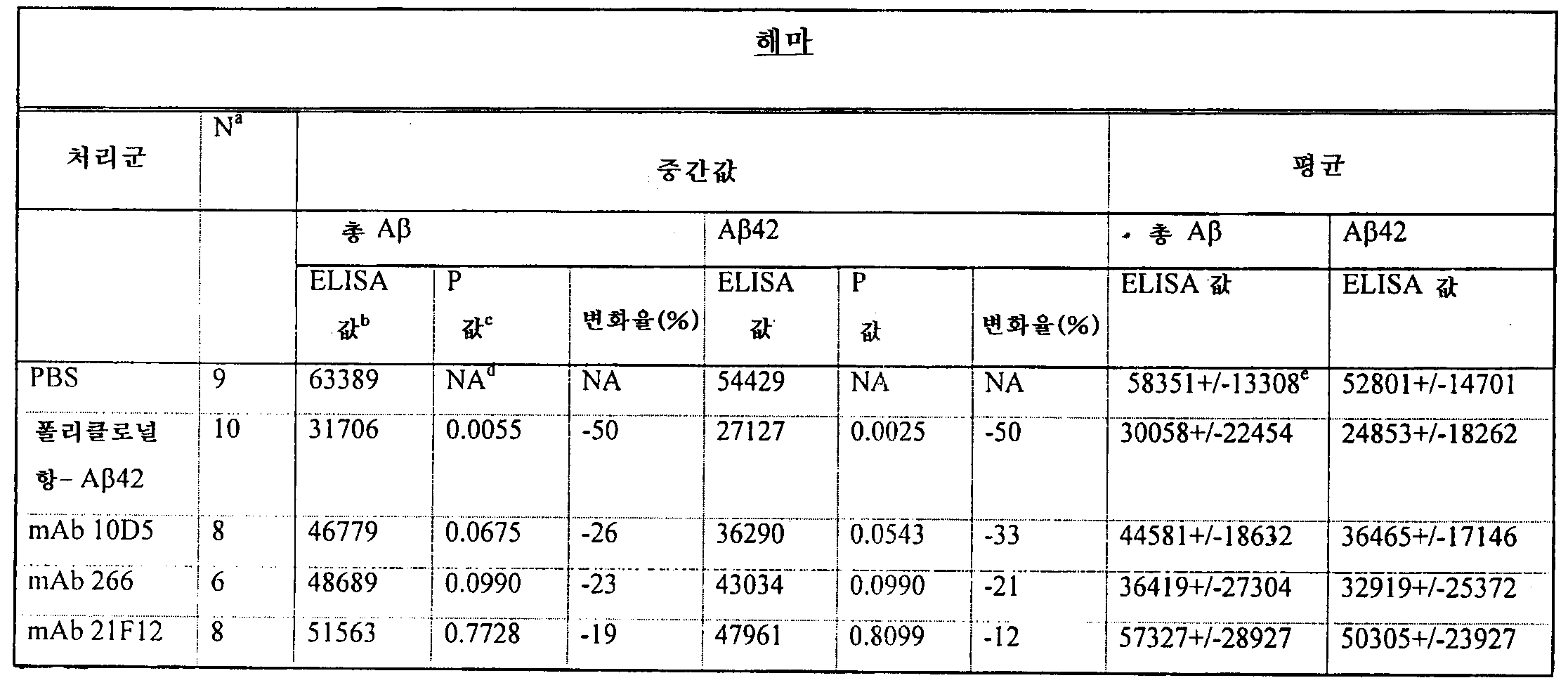
주:week:
a. 실험 종반부에서의 군당 동물의 수a. Number of animals per group at the end of the experiment
b. ng/조직(g)b. ng / tissue (g)
c. 맨 휘트니 분석c. Man Whitney Analysis
d. NA: 적용되지 않음d. NA: Not applicable
e. 표준 편차e. Standard Deviation
총 Aβ는 소뇌에서도 측정하였다(표 15). 폴리클로널 항 Aβ및 266 항체를 투여한 군에서는 총 Aβ의 레벨에 유의적인 감소를 나타내었고(43% 및 46%, 각각 p=0.0033및 p=0.0184), 10D5를 처리한 군은 유의적인 수준에 가까운 감소를 나타내었다(29%, p=0.0675).Total Aβ was also measured in the cerebellum (Table 15). The group receiving polyclonal anti Aβ and 266 antibodies showed a significant decrease in the level of total Aβ (43% and 46%, respectively, p = 0.0033 and p = 0.0184), and the group treated with 10D5 had a significant level. A decrease close to (29%, p = 0.0675).

주:week:
a. 실험 종반부에서의 군당 동물의 수a. Number of animals per group at the end of the experiment
b. ng/조직(g)b. ng / tissue (g)
c. 맨 휘트니 분석c. Man Whitney Analysis
d. NA: 적용되지 않음d. NA: Not applicable
e. 표준 편차e. Standard Deviation
항체 처리군, 대조군, PBS 처리군 마우스에서 얻은 피질 및 소뇌에서도 ELISA를 이용하여 APP 농도를 측정하였다. 2종의 상이한 APP 분석을 이용하였다. APP-α/FL이라고 지칭되는 제1 분석은 APP-알파(α, Aβ서열 내에서 절단되는 APP의 분비형)과 APP의 전장형(FL) 둘다를 인식하는 반면, 제2 분석은 APP-α만을 인식한다. 처리군의 서브세트에서의 처리와 관련된 Aβ감소와는 대조적으로, APP 레벨은 대조군 동물과 비교하였을 때 모든 처리군에서 변화되지 않았다. 이러한 결과는 Aβ항체로의 면역화가 APP를 고갈시키지 않고, 오히려 Aβ를 고갈시켰음을 나타낸다. APP concentrations were also measured by ELISA in the cortex and cerebellum obtained from antibody-treated, control, and PBS-treated mice. Two different APP assays were used. The first assay, called APP-α / FL, recognizes both APP-alpha (α, the secreted form of APP cleaved within the Aβ sequence) and the full-length form of APP (FL), while the second assay recognizes APP-α. Recognize only. In contrast to Aβ reduction associated with treatment in a subset of treatment groups, APP levels did not change in all treatment groups as compared to control animals. These results indicate that immunization with Αβ antibodies did not deplete APP, but rather depleted Αβ.
요약하면, AN1792에 대한 폴리클로널 항체로 처리한 동물에서의 피질, 해마 및 소뇌에서 총 Aβ는 현저히 감소되었다. Aβ1-42의 아미노 말단 영역, 특히 아미노산 1-16 및 13-28에 대한 모노클로널 항체 역시 더 적은 정도로 유의적인 치료 효과를 나타내었다.In summary, total Aβ was significantly reduced in the cortex, hippocampus and cerebellum in animals treated with polyclonal antibodies against AN1792. Monoclonal antibodies against the amino terminal regions of Aβ1-42, particularly amino acids 1-16 and 13-28, also showed a less significant therapeutic effect.
4. 면역조직학 분석4. Immunohistologic Analysis
PBS, 폴리클로널 Aβ42, 21F12, 266 및 10D5 처리군의 마우스로부터 얻은 뇌의 서브세트에서의 Aβ면역반응성 플라크의 형태를 Aβ42를 이용한 표준 면역화 절차를 수행한 이전 실험의 것과 정량적으로 비교하였다.The morphology of Aβ immunoreactive plaques in a subset of brains from mice in PBS, polyclonal Aβ42, 21F12, 266 and 10D5 treated groups was quantitatively compared to that of previous experiments that performed standard immunization procedures with Aβ42.
아밀로이드 플라크의 정도와 외견 둘다에서의 가장 큰 변화는 폴리클로널 Aβ42 항체를 접종한 동물에서 발생하였다. 아밀로이드 침착, 부식된 플라크 형태 및 세포 매개 Aβ면역반응성은 표준 면역 절차에 의해 생성된 효과와 아주 유사하였다. 이러한 관찰은 총 Aβ및 Aβ42 모두의 현저한 감소가 폴리클로널 A42 항체의 접종에 의해 이루어진다는 ELISA 결과를 지지한다.The largest change in both the degree and appearance of amyloid plaques occurred in animals inoculated with polyclonal Aβ42 antibody. Amyloid deposition, corroded plaque morphology and cell mediated Αβ immunoreactivity were very similar to the effects produced by standard immune procedures. This observation supports the ELISA results that a significant reduction in both total Aβ and Aβ42 is achieved by inoculation of polyclonal A42 antibodies.
유사한 정량 평가에서, 10D5 군의 아밀로이드 플라크 역시 세포 매개 Aβ면역반응성을 일부 증명하면서 수와 외견에서 감소하였다. 대조군 처리 동물들에 비해, Aβ에 대한 폴리클로널 Ig 분획 및 모노클로널 항체(10D5)의 하나는 플라크 침착을 각각 93% 및 81% 감소시켰다(p<0.005). 21F12는 플라크 침착에 비교적 약한 영향을 미치는 것으로 나타났다. pabAβ1-42를 처리한 후 뇌의 현미경 사진은 확산성 침착을 보여주었고, 대조군 처리 동물에 비해 pabAβ1-4 처리군에서 보다 큰 밀집형 플라크의 많은 부분이 없다는 것을 보여주었다.In a similar quantitative evaluation, amyloid plaques of the 10D5 group also decreased in number and appearance, demonstrating some cell-mediated Aβ immunoreactivity. Compared to the control treated animals, one of the polyclonal Ig fraction and monoclonal antibody (10D5) against Aβ reduced the plaque deposition by 93% and 81%, respectively (p <0.005). 21F12 has been shown to have a relatively weak effect on plaque deposition. Micrographs of the brain after treatment with pabAβ 1-42 showed diffuse deposition and showed no large portion of larger dense plaques in the pabAβ 1-4 treated group compared to control treated animals.
5. 항체 역가의 측정5. Determination of Antibody Titers
각 군에서 무작위적으로 선택한 마우스 3마리의 서브세트를 매 복강 주사 직전에 총 30회로 하여 채혈하였다. 항체 역가는 전반적인 재료 및 방법에서 기술하는 바와 같이 Aβ1-42 로 코팅된 플라스틱 멀티-웰로 샌드위치 ELISA를 이용하여 Aβ1-42 결합 항체로서 측정하였다. 매 채혈에 대한 평균 역가는 각각 폴리클로널 항체 및 모노클로널 10D5 및 21F12에 대해 도 16 ∼ 18에 도시한다. 역가는 폴리클로널 항체 제제의 경우 이 기간 동안에 걸쳐 평균이 약 1000 이상이었고, 10D15 및 21F12 처리 동물들의 경우 이 레벨보다 약간 더 높았다.A subset of three randomly selected mice from each group was bled 30 times prior to each intraperitoneal injection. Antibody titers were measured as Aβ1-42 binding antibodies using a sandwich ELISA in plastic multi-wells coated with Aβ1-42 as described in the overall materials and methods. Average titers for each blood collection are shown in FIGS. 16-18 for polyclonal antibodies and monoclonal 10D5 and 21F12, respectively. Titer averaged over 1000 over this period for polyclonal antibody preparations and slightly higher than this level for 10D15 and 21F12 treated animals.
6. 림프구증식성 반응6. Lymphocytic reaction
Aβ의존성 림프구증식성 반응은 최종 항체 접종 8일 후 회수한 비장 세포를 이용하여 측정하였다. 새로이 회수한 세포(웰당 105)를 자극을 위해 5 μM 농도의 Aβ1-40의 존재하에 5일간 배양하였다. 양성 대조군으로서, 또 다른 세포를 T 세포 미토젠, PHA와 배양하였고, 음성 대조군으로서 펩티드를 추가하지 않고 배양하였다.Ap-dependent lymphoproliferative responses were measured using splenocytes recovered 8 days after the last antibody inoculation. Freshly harvested cells (10 5 per well) were incubated for 5 days in the presence of 5 μM concentration of Aβ1-40 for stimulation. As a positive control, another cell was incubated with T cell mitogen, PHA, and as a negative control it was incubated without adding peptide.
다양한 Aβ항체로 수동적으로 면역화시킨 늙은 PDAPP 마우스로부터 얻은 비장세포를 AN1792로 시험관내에서 자극하고, 증식 반응 및 시토킨 반응을 측정하였다. 이들 분석의 목적은 수동 면역화가 항체 제시를 용이하게 하여, 이로써 AN1792에 특이적인 T 세포 반응을 개시시키는 지를 결정하는 것이었다. 항 Aβ항체로 수동적으로 면역화시킨 어떠한 마우스에서도 AN1792 특이적 증식 반응 또는 시토킨 반응이 관찰되지 않았다.Splenocytes from old PDAPP mice passively immunized with various Αβ antibodies were stimulated in vitro with AN1792 and proliferative and cytokine responses were measured. The purpose of these assays was to determine whether passive immunization facilitated antibody presentation, thereby initiating T cell responses specific for AN1792. No AN1792 specific proliferative or cytokine responses were observed in any mice passively immunized with anti-Αβ antibody.
XII. 수동 면역화의 추가 실험XII. Additional Experiments of Passive Immunization
제2 실험에서, 10D5의 처리를 반복하고, 2종의 또다른 항 Aβ항체인 모노클로널 3D6(Aβ1-5) 및 16C1(Aβ1-5)를 테스트하였다. 대조군에는 PBS 또는 무관한 이소타입 쌍의 항체(TM2a)를 투여하였다. 마우스는 이전 실험에서보다 더 늙었으며(11.5 ∼ 12 월령 이형접합성), 그 외의 나머지 실험 디자인은 동일하였다. 다시 한번, 처리 6주 후, 10D5는 PBS 또는 이소타입 매치 항체 대조군에 비해 80% 이상 플라크 침착이 감소되었다(p=0.003). Aβ, 3D6에 대한 다른 항체 중 하나는 동일하게 효력을 나타내어서 86% 감소시켰다(p=0.003). 대조적으로, 펩티드, 16C11에 대한 제3 항체는 플라크 침착에 어떠한 영향도 미치지 않았다. 유사한 결과가 Aβ42 ELISA 측정에서도 얻어졌다. 이러한 결과들은 Aβ펩티드에 대한 항체 반응이 T 세포 면역의 부재하에 PDAPP 마우스에서의 아밀로이드 침착을 감소시키기에 충분하지만, 모든 항 Aβ항체가 효능이 있는 것은 아니라는 것을 나타낸다. Aβ의 아미노산 1-5 또는 3-7을 포함하는 에피토프에 대한 항체가 특히 효과적이었다.In the second experiment, the treatment of 10D5 was repeated and two other anti-Αβ antibodies, monoclonal 3D6 (Aβ 1-5 ) and 16C1 (Aβ 1-5 ), were tested. The control group was administered with PBS or an unrelated isotype pair of antibodies (TM2a). Mice were older than in the previous experiment (11.5-12 months of age heterozygosity) and the rest of the experimental design was the same. Once again, after 6 weeks of treatment, 10D5 had reduced plaque deposition by at least 80% compared to the PBS or isotype match antibody controls (p = 0.003). One of the other antibodies against Aβ, 3D6 was equally effective, reducing 86% (p = 0.003). In contrast, the third antibody against peptide, 16C11, had no effect on plaque deposition. Similar results were obtained for Aβ42 ELISA measurements. These results indicate that although the antibody response to Αβ peptide is sufficient to reduce amyloid deposition in PDAPP mice in the absence of T cell immunity, not all anti Αβ antibodies are potent. Antibodies to epitopes comprising amino acids 1-5 or 3-7 of Aβ were particularly effective.
요약하면, 본 발명자들은 수동적으로 투여한 Aβ에 대한 항체가 알츠하이머 질환의 마우스 모델에서 플라크 침착 정도를 감소시켰음을 보여주었다. 적당한 혈정 농도(25 ∼ 70 ㎍/㎖)로 유지할 경우, 항체는 아밀로이드 플라크를 장식하기에 충분한 정도로 CNS에 접근할 수 있었다. CNS로의 항체의 접근은 PDAPP 마우스에서 에반스 블루에 의해 측정하였을 때 혈관 침투성이 증가되지 않았으므로, 혈류-뇌 장벽의 비정상적인 유출에 의한 것이 아니었다. 또한, 고령의 PDAPP 마우스의 뇌 실질에서의 항체 농도는 비형질전환 마우스에서와 동일하였고, 혈청에서의 항체의 농도가 0.1%였다(이소타입에 관계없음).In summary, the inventors have shown that antibodies against manually administered Αβ reduced the degree of plaque deposition in mouse models of Alzheimer's disease. When maintained at an appropriate blood serum concentration (25-70 μg / ml), the antibody could access the CNS to a degree sufficient to adorn amyloid plaques. The access of the antibody to the CNS was not due to abnormal outflow of the blood flow-brain barrier, as the vascular permeability did not increase as measured by Evans Blue in PDAPP mice. In addition, the antibody concentration in the brain parenchyma of old PDAPP mice was the same as in the nontransgenic mice, and the concentration of antibody in serum was 0.1% (regardless of isotype).
XIII: 항체 결합의 모니터링XIII: Monitoring of Antibody Binding
Aβ에 대한 항체가 CNS 내에서 직접적으로 작용할 수 있는 지를 측정하기 위해, 실시예 XII 후반부에 염수 관류된 마우스로부터 꺼낸 뇌로 전신 투여된 항체의 존재에 대해 관찰하였다. 비고정 저온장치 뇌 절편을 마우스 면역글로불린(염소 항-마우스 IgG-Cy3)에 대한 형광 물질에 노출시켰다. 10D5 및 3D6 군의 뇌 내에서의 플라크는 항체로 심하게 장식되었지만, 16C11 군에서는 염색되지 않았다. 플라크 침착의 전체적인 정도를 알기 위해, 각 뇌의 연속 절편을 먼저 항 Aβ항체와 면역반응시키고, 그 후 2차 물질과 면역반응시켰다. 전신 투여 후 10D5 및 3D6는 CNS 내의 대부분의 플라크에 접근할 수 있었다. 플라크 침착은 16C11 군과 비교하여 이들 처리군에서 상당히 감소되었다. 이러한 데이타는 전신 투여된 항체가 이들이 아밀로이드 제거를 직접적으로 개시시킬 수 있는 CNS에 침입할 수 있다는 것을 나타낸다. 16C11 역시 플라크에 접근할 수 있으나 결합할 수 없는 것으로 보인다.To determine whether antibodies against Αβ can act directly in the CNS, the presence of antibodies administered systemically to the brain taken from saline-perfused mice later in Example XII was observed. Unfixed cryogen brain sections were exposed to fluorescent material for mouse immunoglobulin (goat anti-mouse IgG-Cy3). Plaques in the brain of the 10D5 and 3D6 groups were heavily decorated with antibodies but were not stained in the 16C11 group. To determine the overall extent of plaque deposition, serial sections of each brain were first immunized with anti-Αβ antibody and then with secondary material. After systemic administration, 10D5 and 3D6 were able to access most plaques in the CNS. Plaque deposition was significantly reduced in these treatment groups compared to the 16C11 group. These data indicate that systemically administered antibodies can invade the CNS where they can directly initiate amyloid clearance. 16C11 also has access to plaques but appears to be unable to bind.
XIV: 아밀로이드 침착에 대한 항체의 활성을 대한 생체외 스크리닝 분석XIV: In Vitro Screening Assay for the Activity of Antibodies on Amyloid Deposition
항체가 플라크 제거에 미치는 영향을 조사하기 위해, 본 발명자들은 근원 소교세포 세포를 PDAPP 마우스 또는 사람 AD 뇌의 비고정 저온장치 절편과 배양하는 생체외 분석을 수행하였다. 소교세포 세포는 신생 DBA/2N 마우스(1 ∼ 3 일)의 대뇌 피질로부터 얻었다. 피질을 HBSS--(Hanks' Balanced Salt Solution, 시그마)에서 50 ㎍/㎖의 DNaseI(시그마)을 이용하여 기계적으로 분리하였다. 분리된 세포들을 100 ㎛ 세포 스트레이너(팰콘)로 여과하고 1000 rpm에서 5분간 원심분리하였다. 펠렛을 성장 배지(고농도 포도당 DMEM, 10% FBS, 25 ng/㎖ rmGH-CSF)에 재현탁시키고, 이 세포들을 T-75 플라스틱 배양 플라스크 당 2 뇌의 밀도로 플레이팅하였다. 7 ∼ 9일 후, 플라스크를 오비탈 진탕기에서 200 rpm으로 37℃에서 2시간 동안 진탕하였다. 세포 현탁액을 1000 rpm에서 원심분리하고 분석 배지에 재현탁시켰다.To investigate the effect of antibodies on plaque removal, the inventors performed an in vitro assay in which myoblast microglia cells were cultured with unfixed cryocol sections of PDAPP mice or human AD brains. Microglia cells were obtained from the cerebral cortex of newborn DBA / 2N mice (1-3 days). The cortex HBSS - using (Hanks' Balanced Salt Solution, Sigma) in 50 ㎍ / ㎖ in DNaseI (Sigma) was mechanically separated. The isolated cells were filtered with a 100 μm cell strainer (Falcon) and centrifuged at 1000 rpm for 5 minutes. The pellet was resuspended in growth medium (high glucose DMEM, 10% FBS, 25 ng / ml rmGH-CSF) and the cells were plated at a density of 2 brains per T-75 plastic culture flask. After 7-9 days, the flask was shaken at 200 rpm for 2 hours at 200 rpm on an orbital shaker. The cell suspension was centrifuged at 1000 rpm and resuspended in assay medium.
PDAPP 마우스 또는 사람 AD 뇌(사후 간격 < 3시간)의 10 ㎛ 저온장치 절편을 폴리 리신 코팅된 둥근 유리 커버슬립 위에 녹여서 마운팅하고, 24 웰 조직 배양 플레이트의 웰에 배치하였다. 커버슬립을 1% FBS, 글루타민, 페니실린/스트렙토마이신 및 5 ng/㎖ rmGM-CSF(R&D)를 포함하는 H-SFM(히브리도마-혈청이 없는 배지, 깁코 BRL)로 구성된 분석 배지로 2회 세정하였다. 대조군 또는 항 Aβ항체를 1시간 동안 2 x 농도(최종 5 ㎍/㎖)로 첨가하였다. 그 후, 소교 세포를 분석 배지 ㎖당 0.8 x 10⁶ 세포의 밀도로 접종하였다. 배양물을 함습 항온기(37℃, 5% CO2)에서 24시간 이상 동안 유지시켰다. 항온처리 후반부에, 배양물을 4% 파라포름알데히드로 고정하고, 0.1% Triton-X 100으로 침투시켰다. 절편들을 비오티닐화된 3D6으로 염색한 다음, 스트렙타비딘/Cy3 접합체(잭슨 이뮤노리서치)로 염색하였다. 외인성 소교세포 세포는 핵 착색제(DAPI)로 시각화하였다. 배양물은 역상 형광 현미경(니콘, TE300)으로 관찰하였고, SPOT 소프트웨어(다이애그노스틱 인스트러먼츠)를 이용하여 SPOT 디지탈 카메라로 현미경 사진을 찍었다. 웨스턴 블롯 분석을 위해서, 배양물을 8M 우레아에서 추출하고, 환원성 트리신 시료 완충액에 1:1로 희석시켜서 16% 트리신 겔(노벡스)에 로딩하였다. 임모빌론에 이전한 후, 블롯을 5 ㎍/㎖의 pabAβ42에 노출시킨 다음, HRP-접합 항-마우스 항체에 노출시키고 ECL(아머샴)로 발색시켰다.10 μm cryogen sections of PDAPP mice or human AD brains (post interval <3 hours) were thawed and mounted on polylysine coated round glass coverslips and placed in wells of a 24 well tissue culture plate. Coverslips were washed twice with assay medium consisting of H-SFM (Hybridoma-serum-free medium, Gibco BRL) containing 1% FBS, glutamine, penicillin / streptomycin and 5 ng / ml rmGM-CSF (R & D) It was. Control or anti Aβ antibody was added at 2 × concentration (final 5 μg / ml) for 1 hour. Microglia were then seeded at a density of 0.8 × 10 6 cells per ml of assay medium. Cultures were maintained for at least 24 hours in a humidified thermostat (37 ° C., 5% CO 2 ). Later incubation, the cultures were fixed with 4% paraformaldehyde and infiltrated with 0.1% Triton-
16C11(생체내에서 효능이 없었던 Aβ에 대한 항체 중 하나)의 존재하에 PDAPP 뇌 절편으로 분석을 수행하였을 때, β-아밀로이드 플라크는 손상되지 않은 상태로 보존되었고, 식균작용도 관찰되지 않았다. 이와는 대조적으로, 인접한 절편을 10D5의 존재하에 배양할 경우, 아밀로이드 침착물은 대개 사라졌고, 소교세포 세포는 Aβ를 포함하는 많은 식균작용성 소낭을 보여주었다. AD 뇌 절편에서도 동일한 결과를 얻었다; 10D5는 AD 플라크의 식균작용을 유도한 반면, 16C11은 비효과적이었다. 또한, Aβ에 대한 마우스나 사람 소교세포 세포, 및 마우스, 토끼, 또는 영장류 항체로 분석을 수행하였을 때 유사한 결과를 나타내었다.When analysis was performed with PDAPP brain sections in the presence of 16C11 (one of the antibodies against Aβ that was in vivo ineffective), β-amyloid plaques were preserved intact and no phagocytosis was observed. In contrast, when adjacent sections were cultured in the presence of 10D5, amyloid deposits usually disappeared and microglia cells showed many phagocytic vesicles containing Aβ. The same result was obtained with AD brain sections; 10D5 induced phagocytosis of AD plaque, whereas 16C11 was ineffective. In addition, similar results were obtained when analysis was performed with mouse or human microglia cells against Aβ and mouse, rabbit, or primate antibodies.
표 16은 몇가지 상이한 항체 결합 특이성에 대해 결합 및/또는 식균작용 중 어떤 것이 나타나지는 지에 대해 보여준다. aa 1-7내의 에피토프에 결합하는 항체는 아밀로이드 침착물에 결합하고 이를 제거하는 반면, 아미노산 4-10 내의 에피토프에 결합하는 항체는 아밀로이드 침착물을 제거하지 않고 결합하는 것을 알 수 있다. 잔기 10에 대한 에피토프 C-말단에 결합하는 항체는 아밀로이드 침착물에 결합하지도 않고 이를 제거하지도 않는다.Table 16 shows which of the binding and / or phagocytosis appears for several different antibody binding specificities. It can be seen that antibodies that bind epitopes within aa 1-7 bind to and remove amyloid deposits, whereas antibodies that bind epitopes within amino acids 4-10 bind without removing amyloid deposits. Antibodies that bind to the epitope C-terminus to
표 17은 생체외 분석에서의 식균작용을 유도하는 능력과 수동적 전달 실험에서의 생체내 플라크 침착을 감소시키는 능력을 비교하면서 Aβ에 대한 여러 항체에 대해 얻은 결과를 보여준다. 16C11 및 21F12가 큰 결합력으로 응집형 합성 Aβ펩티드에 결합함에도 불구하고, 이들 항체는 비고정 뇌 절편 내의 β-아밀로이드 플라크와 반응할 수 없었고, 생체외 분석에서 식균작용을 일으키지 않았으며, 생체내에서 효능이 없었다. 10D5, 3D6, 및 Aβ에 대한 폴리클로널 항체는 모든 3종의 측정에서 활성을 나타내었다. 22C8 항체는 위치 1 및 7의 아스파르트산이 이소 아스파르트산으로 대체된 천연 Aβ의 유사 형태에 보다 강력하게 결합한다. 이러한 결과들은 생체내 효능이 CNS 내의 플라크의 직접적인 항체 매개 제거에 의한 것이고, 생체외 분석이 생체내 효능을 예측할 수 있는 것임을 나타낸다.Table 17 shows the results obtained for the various antibodies against Aβ comparing the ability to induce phagocytosis in ex vivo assays and the ability to reduce in vivo plaque deposition in passive delivery experiments. Although 16C11 and 21F12 bind to aggregated synthetic Aβ peptides with great binding power, these antibodies were unable to react with β-amyloid plaques in unfixed brain sections, and did not cause phagocytosis in in vitro assays, and in vivo There was no efficacy. Polyclonal antibodies against 10D5, 3D6, and Aβ showed activity in all three measurements. The 22C8 antibody binds more strongly to the analogous form of native Αβ with aspartic acid at
NAC라고 불리는 시뉴클린의 단편에 대한 항체의 제거를 테스트하기 위해 동일한 분석을 이용하였다. 시뉴클린은 아밀로이드 플라크 관련 단백질인 것으로 알려져 있다. NAC에 대한 항체는 전술한 바와 같이 아밀로이드 플라크를 포함하는 뇌 조직 시료, 소교세포와 접촉하였다. 토끼 혈청을 대조군으로 사용하였다. 후속 모니터링은 항체의 제거 활성을 나타내는 플라크의 수와 크기가 현저히 감소되었음을 보여주었다.The same assay was used to test the removal of antibodies to fragments of synuclein called NAC. Synuclin is known to be an amyloid plaque related protein. Antibodies against NAC were contacted with brain tissue samples, microglia, containing amyloid plaques as described above. Rabbit serum was used as a control. Subsequent monitoring showed a significant reduction in the number and size of plaques indicating antibody removal activity.
공초점 현미경을 이용하여 생체외 분석을 수행하는 동안 Aβ가 흡수되었는 지를 확인하였다. 대조군 항체의 존재하에, 외인성 소교세포는 상기 조직의 공초점 플레인에 잔류하였고, Aβ를 포함하는 식균작용성 소낭은 없었으며, 플라크는 절편 내에 무결한 상태로 보존되었다. 10D5의 존재하에, 거의 모든 플라크 물질이 외인성 소교 세포 내에 있는 소낭에 포함되었다. 내부화된 펩티드의 운명을 결정하기 위해, 10D5 처리된 배양물을 다양한 시점에서 8 M 우레아로 추출하였고, 웨스턴 블롯 분석으로 관찰하였다. 1시간 시점에, 아직 식균작용은 발생하지 않았을 때, Aβ에 대한 폴리클로널 항체의 반응은 진한 4 kD 밴드(Aβ펩티드에 해당함)를 나타내었다. Aβ면역반응성은 1일째 감소되었고, 3일에 이르러서는 사라졌다. 따라서, Aβ의 항체 매개 식균작용은 Aβ의 분해를 유발시킨다.Confocal microscopy was used to confirm that Aβ was absorbed during the ex vivo analysis. In the presence of control antibodies, exogenous microglia remained in the confocal plane of the tissue, there were no phagocytic vesicles containing Aβ and plaques were preserved intact in sections. In the presence of 10D5 almost all plaque material was contained in the vesicles within the exogenous microglia. To determine the fate of the internalized peptides, 10D5 treated cultures were extracted with 8 M urea at various time points and observed by Western blot analysis. At 1 hour, when no phagocytosis had occurred yet, the response of the polyclonal antibody to Aβ showed a dark 4 kD band (corresponding to Aβ peptide). Αβ immunoreactivity decreased on
생체외 분석에서의 식균작용이 Fc-매개된 것인지를 결정하기 위해, 항-Aβ항체 3D6의 F(ab')2 단편들을 제조하였다. F(ab')2 단편들이 플라크와 반응하기 위한 완전한 능력을 보유할지라도, 이들은 소교세포에 의한 식균작용을 유발시킬 수 없었다. 또한, 전체 항체를 이용하는 식균작용은 쥐과 Fc 수용체(항-CD16/32)에 대한 제제에 의해 차단될 수 있었다. 이러한 데이타는 Aβ의 생체내 제거가 Fc-매개 식균작용을 통해 일어난다는 것을 나타낸다.To determine whether phagocytosis in in vitro analysis was Fc-mediated, F (ab ′) 2 fragments of anti-Aβ antibody 3D6 were prepared. Although F (ab ') 2 fragments retained full capacity to react with plaques, they could not induce phagocytosis by microglia. In addition, phagocytosis using whole antibodies could be blocked by preparations for murine Fc receptors (anti-CD16 / 32). These data indicate that in vivo clearance of Αβ occurs via Fc-mediated phagocytosis.
XV: 항체의 혈액 뇌 장벽의 통과XV: Penetration of the Blood Brain Barrier of Antibodies
이 실시예에서는 정상 마우스 또는 PDAPP 마우스의 말초 조직에 정맥 주사한 후 뇌에 전달된 항체의 농도를 측정한다. PDAPP 또는 대조군 정상 마우스를 0.9% NaCl로 관류하였다. 뇌 영역(해마 또는 피질)을 절개하고 급속 동결시켰다. 뇌를 0.1% 트리톤 + 프로테아제 억제제 중에서 균질화하였다. ELISA를 이용하여 추출물 중의 면역글로불린을 검출하였다. Fab'2 염소 항-마우스 IgG를 캡쳐 시약으로서 RIA 플레이트 위에 코팅하였다. 혈청 또는 뇌 추출물을 1시간 동안 항온처리하였다. 이소타입을 항-마우스 IgG-HRP 또는 IgG2a-HRP 또는 IgG2b-HRP(칼테그)를 이용하여 검출하였다. 이소타입에 관계없이, 항체는 혈액에서 발견되었을 때의 농도가 1:1000으로 CNS에 존재하였다. 예를 들면, IgG1의 농도가 혈액에서 IgG2a의 농도의 3배일 경우, 뇌에서 역시 3배의 IgG2a가 존재하였고, 이들 둘다 혈액에서의 각각의 레벨이 0.1%였다. 이 결과는 형질전환 및 비형질전환 마우스 둘다에서 관찰되었으므로, PDAPP는 특이한 유출 혈액 뇌 장벽을 지니지 않는다.In this example, the concentration of antibody delivered to the brain after intravenous injection into peripheral tissues of normal or PDAPP mice is measured. PDAPP or control normal mice were perfused with 0.9% NaCl. Brain regions (hippocampus or cortex) were dissected and snap frozen. Brains were homogenized in 0.1% Triton + protease inhibitors. Immunoglobulins in the extracts were detected using ELISA. Fab'2 goat anti-mouse IgG was coated onto RIA plates as capture reagent. Serum or brain extracts were incubated for 1 hour. Isotypes were detected using anti-mouse IgG-HRP or IgG2a-HRP or IgG2b-HRP (Carlteg). Regardless of the isotype, the antibody was present in the CNS with a concentration of 1: 1000 when found in the blood. For example, when the concentration of IgG1 was three times the concentration of IgG2a in the blood, there was also three times the IgG2a in the brain, both with 0.1% of each level in the blood. This result was observed in both transgenic and nontransgenic mice, so PDAPP does not have a specific shed blood brain barrier.
XVI: MAP 형태의 Aβ펩티드의 치료 효능XVI: Therapeutic Efficacy of Aβ Peptides in MAP Form
치료적 보조제/면역원 효능 실험은 9 ∼ 10.5 월령의 이형접합성 PDAPP 형질전환 마우스 암컷 및 수컷으로 수행하여 전술한 바와 같이 삼량체 MAP 형태의 Aβ1-7을 포함하는 융합 단백질의 효능을 테스트하였다. 처리군당 29 ∼ 40마리로 하여 실험 기간은 25주로 하였으므로, 동물들은 실험 종료시에 15 ∼ 16.5월령이 되었다. 이 실험에서 이용된 방법은 전술한 실시예 VIII에서의 상이한 보조제의 치료 실험에서와 동일한 것이다. 치료군은 하기 표 18에 기재하였다.Therapeutic adjuvant / immunogen efficacy experiments were performed with heterozygous PDAPP transgenic mouse females and males between 9 and 10.5 months of age to test the efficacy of fusion proteins comprising Aβ1-7 in the trimeric MAP form as described above. Since the experiment period was 25 weeks with 29-40 animals per treatment group, the animals were 15-16.5 months old at the end of the experiment. The method used in this experiment is the same as in the treatment trial of the different adjuvant in Example VIII described above. Treatment groups are listed in Table 18 below.
표 약어: MAP - 다중 항원성 펩티드; TT - 파상풍 톡소이드 t-세포 에피토프 (830-844); SC - 피하; IP - 복강; PBS - 포스페이트 완충 염수; GCS는 글리신/시트레이트/수크로오스 조성물이다.Table abbreviations: MAP-multiple antigenic peptides; TT-tetanus toxoid t-cell epitope (830-844); SC-subcutaneous; IP-abdominal cavity; PBS-phosphate buffered saline; GCS is a glycine / citrate / sucrose composition.
면역화 스케줄은 모든 처리군에서 동일하였다. 마우스에 0, 2, 4, 8, 12, 16, 20, 24주에 주사하였고, 3, 5, 9, 13, 17, 21 및 25주에 채혈하였다. 1, 2, 3, 4 및 6군에는 8회의 접종을 하였으며, 2군 및 3군에는 각각 QS21/AN1792와 PBS를 주사하여 본 실험을 위한 양성 및 음성 대조군으로 사용하였다.Immunization schedule was the same for all treatment groups. Mice were injected at 0, 2, 4, 8, 12, 16, 20, 24 weeks and blood was collected at 3, 5, 9, 13, 17, 21 and 25 weeks.
역가는 항 Aβ항체 역가 분석으로 측정하였다.Titers were determined by anti Aβ antibody titer analysis.
1군: CFA/IFA:MAP(Aβ1-7:TT) 군, 역가 레벨이 낮았다. 피크 GMT는 13주에 1,200에 불과하였고, 25주에 이르러 600으로 떨어졌다. 30마리의 마우스 중 3마리에서는 역가가 증가되지 않았고, 또 다른 7마리 마우스는 실험 후반부에 이르러 역가가 400을 초과하지 않았다. Group 1: CFA / IFA: MAP (Aβ1-7: TT) group with low titer level. The peak GMT was only 1,200 at 13 weeks and dropped to 600 in 25 weeks. The titer did not increase in 3 of 30 mice, and another 7 mice did not exceed 400 by the end of the experiment.
2군: QS21/AN1792 대조군, 17주에 GMT가 16,000이 되면서 피크 역가에 도달하였다. 그 후, 다음 8주에 걸쳐 역가가 감소되어 GMT가 8,700으로 종료되었다. 이 군 중 1마리는 실험 전반에 걸쳐서 역가를 증가시키지 못했다. Group 2: the QS21 / AN1792 control group, peak titer was reached with GMT of 16,000 at 17 weeks. The titer then decreased over the next eight weeks, ending the GMT at 8,700. One of these groups did not increase titer throughout the experiment.
3군: PBS 단독 군, 역가를 나타내지 않았다. Group 3: PBS alone group, no titer.
처리군 둘다는 PBS 대조군에 비해 피질 Aβ레벨이 유의적으로 감소되었음을 보여주었다(표 9 참조). CFA/IFA:MAP(Aβ1-7) 군은 비교적 낮은 항 Aβ항체 역가에도 불구하고 PBS 대조군과 비교하여 Aβ를 현저히 감소시켰다.Both treatment groups showed a significant decrease in cortical Aβ levels compared to the PBS control (see Table 9). The CFA / IFA: MAP (Aβ1-7) group significantly reduced Aβ compared to the PBS control group despite the relatively low anti Aβ antibody titers.
결론적으로, Aβ1-7 MAP 면역원은 피질 내의 Aβ침착을 지연시키기에 충분한 면역 반응을 유의적으로 유도하는 데 효과적이다.In conclusion, the Aβ1-7 MAP immunogen is effective at significantly inducing an immune response sufficient to delay Aβ deposition in the cortex.
XVII. 원숭이에서의 Aβ에 대한 면역원성 반응의 에피토프 지도화XVII. Epitope Mapping of Immunogenic Responses to Aβ in Monkeys
이 실시예에서는 AN1792(즉, Aβ1-42)를 이용한 면역화에 대한 영장류의 반응을 분석한다. 원숭이 11군(4마리/성별/군)을 QS-21 보조제(50 또는 100 ㎍/용량) 또는 물 중의 5% 멸균 덱스트로스(D5W, 대조군)와 배합한 AN1792(75 또는 300 ㎍/용량)로 면역화시켰다. 표 20에 기재한 바와 같이, 모든 동물들에게 3가지 주사 스케줄 중 하나로 IM 주사하였고, 총 4, 5 또는 8회 접종하였다. 실험 175일째 회수한 혈청 시료(4마리 원숭이/성별/군으로부터) 및 176일째 회수한 CSF 시료(3마리 원숭이/성별/군으로부터부) Aβ1-40 및 APP에 결합하는 능력을 평가하였다.In this example, the response of primates to immunization with AN1792 (ie Aβ1-42) is analyzed. Monkey 11 group (4 / sex / group) with AN1792 (75 or 300 μg / dose) in combination with QS-21 adjuvant (50 or 100 μg / dose) or 5% sterile dextrose in water (D5W, control) Immunized. As described in Table 20, all animals were IM injected with one of three injection schedules and inoculated a total of 4, 5 or 8 times. The ability to bind serum samples (from 4 monkeys / sex / group) recovered on day 175 and CSF samples (from 3 monkeys / sex / group) recovered on day 176 and Aβ1-40 and APP was evaluated.
(㎍/투여량)AN1792 Dosage
(Μg / dose)
(㎍/투여량)QS-21 Dosage
(Μg / dose)
a. 스케줄 1, 투여 일 1, 15, 29, 57, 85, 113, 141, 169; 스케줄 2, 투여 일 1, 29, 57, 113, 169; 스케줄 3, 투여 일 1, 43, 85, 169a.
b. D5W 주사 대조군b. D5W injection control
c. 부형제는 AN1792에 대한 부형제인 글리신/시트레이트/수크로오스 완충액으로 구성된다.c. Excipients consist of glycine / citrate / sucrose buffer, an excipient for AN1792.
AN1792를 접종한 동물로부터 얻은 혈청 시료의 항체에 의해 인식되는 선형 펩티드의 정확한 배열은 전체 Aβ1-42 서열을 포함하는 중첩 펩티드에 대한 항체의 결합을 측정하는 ELISA를 이용하여 측정하였다. AN1792의 부분 서열을 포함하는 비오티닐화된 펩티드는 키론 테크놀로지스에서 입수하였으며, 펩티드당 9 잔기의 중첩과 1 잔기의 스텝이 있는 10 아미노산 펩티드였다(합성 번호: 5366, 5311 및 5814). 제1의 32 펩티드(AN1792의 N-말단의 상류 8번 아미노산 위치에서 하류 방향으로 AN1792의 24번째 아미노산까지)를 GGK의 링커로 C-말단 상에 비오티닐화시켰다. 마지막 10개의 펩티드(이전 시리즈로부터 32번째 펩티드를 반복함)는 EGEG(서열 번호 76)로 구성된 링커에 의해 N-말단 상에 비오티닐화되어 있다. 동결건조된 비오티닐화된 펩티드를 DMSO 중에 5 mM의 농도로 용해시켰다. 이들 펩티드 스톡을 TTBS(0.05% Tween 20, 25 mM Tris HCl, 137 mM NaCl, 5.1 mM KCl, pH=7.5) 중에 5 μM으로 희석시켰다. 이 5 μM 용액의 100 ㎕ 분취량을 이중으로 스트렙타비딘 예비 코팅된 96 웰 플레이트(피어스)에 첨가하였다. 플레이트를 실온에서 1시간 동안 항온처리한 다음, TTBS로 4회 세척하였다. 혈청 시료를 아지드 없이 표본 희석액에서 희석하여 역가를 표준화시키고, 웰당 100 ㎕를 첨가하였다. 상기 플레이트를 실온에서 1시간 동안 항온처리한 다음, TTBS로 4회 세척하였다. HRP-접합 염소 항-사람 항체(잭슨 이뮤노리서치)를 아지드 없이 표본 희석액에서 희석하여 역가를 표준화시키고, 웰당 100 ㎕를 첨가하였다. 이 플레이트를 다시 항온처리하고 세척하였다. 발색 반응을 전개시키기 위해, TMB(피어스)를 웰당 100 ㎕씩 첨가하고 15분간 항온처리한 다음, 2 N H2SO4 30 ㎕를 첨가하여 반응을 중단시켰다. Vmax 또는 Spectramax 색측정 플레이트 판독기 상에서 450 nm에서 흡광도를 측정하였다.The exact arrangement of linear peptides recognized by antibodies in serum samples from animals inoculated with AN1792 was determined using an ELISA measuring the binding of the antibody to overlapping peptides comprising the entire Aβ1-42 sequence. Biotinylated peptides comprising the partial sequence of AN1792 were obtained from Chiron Technologies, a 10 amino acid peptide with overlap of 9 residues per peptide and 1 residue step (synthesis no .: 5366, 5311 and 5814). The first 32 peptides (from the
AN1792를 이용한 면역화는 175일까지 모든 투여 군에서 동물들의 100%에서 항체의 생성을 유도하였다. 상기 군들에서의 평균 역가의 범위는 14596 ∼ 56084였다. 더 많은 항원 및/또는 더 높은 농도의 보조제의 존재하에 면역 스케줄내에서 역가는 더 높아지는 경향을 보였으나, 면역화에 대한 개개 동물들의 반응이 매우 다양하기 때문에 통계적으로 유의적인 차이는 확인할 수 없었다.Immunization with AN1792 induced the production of antibodies in 100% of animals in all dose groups up to 175 days. The average titer in the groups ranged from 14596 to 56084. Titers tended to be higher in the immune schedule in the presence of more antigens and / or higher concentrations of adjuvant, but no statistically significant difference could be identified as the individual animals' response to immunization varied widely.
AN1792에 대한 항체에 대해서 양성인 혈청 역시 Aβ1-40에 대한 항체에 양성 반응을 보였다. 상기 군들에서의 평균 역가의 범위는 36867 ∼ 165991이었고, AN1792 역가에 대해서는, 175일에 군들 사이에서 통계적으로 유의적인 어떠한 차이도 보이지 않았다. AN1792에의 결합은 Aβ1-40에 결합하면서 높은 양의 관련성을 나타내었다(Spearman r = 0.8671).Serum positive for the antibody against AN1792 also tested positive for the antibody to Aβ1-40. The mean titers in the groups ranged from 36867 to 165991, and for AN1792 titers, there were no statistically significant differences between the groups at 175 days. Binding to AN1792 showed a high amount of association with binding to Aβ1-40 (Spearman r = 0.8671).
AN1792를 이용한 다양한 스케줄로 면역화시킨 48마리의 원숭이 중에서, 33마리가 분석을 위해 적절한 부피 및 특질의 CSF 시료를 생성하였다. 이 원숭이들 중 32마리(97%)는 AN1792에 대해 양의 역가를 나타내었다. 역가의 범위는 2 ∼ 246이었고, 평균은 49.44 ±21.34였다. CSF 항-AN1792 레벨은 혈청에서 측정한 값의 0.18 ± 0.11%였고, 혈청 역가와 매우 양의 관계(Spearman r = 0.7840)에 있었다. 군이나 성별 사이에서는 CSF에서의 항체의 비율에 어떠한 차이도 관찰되지 않았다. CSF에서의 항체 레벨은 전신에서 생성된 항체가 혈액 뇌 장벽을 통과하여 중추신경계로 수동적으로 전달된 것과 일치한다.Of the 48 monkeys immunized with various schedules using AN1792, 33 produced appropriate volume and characteristic CSF samples for analysis. 32 of these monkeys (97%) had a positive titer for AN1792. The titer ranged from 2 to 246, with an average of 49.44 ± 21.34. CSF anti-AN1792 levels were 0.18 ± 0.11% of the values measured in serum and were in a very positive relationship with serum titer (Spearman r = 0.840). No differences were observed in the proportion of antibodies in CSF between groups or genders. The antibody level in CSF is consistent with the antibody produced systemically across the blood brain barrier and passively delivered to the central nervous system.
항 AN1792 양성 CSF 시료의 한 서브세트의 테스트는 혈청 시료의 항체와 마찬가지로 CSF의 항체가 Aβ1-40과 교차반응한다는 것을 증명한다. Aβ1-40에 대한 역가는 이들의 개개의 AN1792 역가에 대해 높은 양의 관계(Spearman r = 0.9634)에 있다는 것을 보여주었다. AN1792에 대한 가장 높은 역가를 갖는 CSF 시료의 서브세트를 테스트한 것은 혈청 항체의 경우 APP에 결합하지 않는다는 것을 보여주었다.Testing of a subset of anti AN1792 positive CSF samples demonstrates that the antibodies of CSF crossreact with Aβ1-40 as well as the antibodies in serum samples. Titers for Aβ1-40 showed a high positive relationship (Spearman r = 0.9634) for their individual AN1792 titers. Testing a subset of CSF samples with the highest titers for AN1792 showed that for serum antibodies it did not bind APP.
175일째의 항체로 일련의 중첩 10-mer 펩티드에 대해 테스트할 경우, 모든 원숭이에서 얻은 항체는 AN1792 펩티드(APP의 아미노산 653-672)의 아미노산 1-10을 포함하는 서열을 갖는 펩티드에 결합하였다. 일부 동물에서는 이것이 측정가능하게 결합하는 유일한 펩티드였다(도 19 참조).When tested against a series of overlapping 10-mer peptides with antibody at day 175, antibodies from all monkeys bound to peptides with sequences comprising amino acids 1-10 of AN1792 peptide (amino acids 653-672 of APP). In some animals this was the only peptide that measurably binds (see FIG. 19).
다른 동물에서는 다른 반응성이 측정될 수 있으나, 모든 경우에 N-말단 펩티드 서열에 대한 반응성은 우세한 것이었다. 또 다른 반응성을 2군으로 나누었다. 제1군은 가장 일반적인 것으로, N-말단 1-10 AN1792 펩티드 둘레를 중심으로 하는 펩티드에 결합하는 것이었다(도 20). 이러한 유형의 결합은 AN1792 펩티드의 아미노산 -1-8, -1-9 및 2-11을 포함하는 펩티드에 대한 것이었다. 1-10 펩티드에 대한 것과 결합하였을 때 이러한 반응성은 모든 동물에서 주도적인 주요 반응성을 대표한다.시간에 따른 개개 동물들의 에피토프 지도화는 1-10 펩티드에 대한 항체 반응성이 인접한 펩티드에 확산된다는 것을 나타낸다. 이는 면역 반응의 강력한 바이어싱이 자유 말단 아스파르트산 잔기를 갖는 AN1792 펩티드의 N-말단에 반응한다는 것을 증명한다. 일부 동물에서의 제2의 소수의 검출가능한 활성은 주요 영역 및 AN1792 펩티드의 아미노산 7-16, 11-20 및 16-25을 포함하는 펩티드를 중심으로 하는 영역에 C-말단에 위치된 펩티드에 결합하였다. 이러한 반응성은 원숭이의 단 10 ∼ 30%만 관찰되었다. Different reactivity can be measured in other animals, but in all cases reactivity to the N-terminal peptide sequence was predominant. Another reactivity was divided into two groups. The first group was the most common, binding to peptides centered around the N-terminal 1-10 AN1792 peptide (FIG. 20). This type of binding was to peptides comprising amino acids -1-8, -1-9 and 2-11 of AN1792 peptide. This reactivity, when combined with that for 1-10 peptides, represents a major dominant reactivity in all animals. Epitope mapping of individual animals over time indicates that antibody reactivity against 1-10 peptides diffuses into adjacent peptides. . This demonstrates that strong biasing of the immune response responds to the N-terminus of the AN1792 peptide with free terminal aspartic acid residues. The second minority of detectable activity in some animals binds to a peptide located at the C-terminus in the main region and a region centered on a peptide comprising amino acids 7-16, 11-20 and 16-25 of AN1792 peptide. It was. This reactivity was observed only 10-30% of monkeys.
상이한 동물들 사이의 반응의 변화도(예컨대, 아미노산 1-10이 독점적 또는 우세한 반응성 에피토프인지)는 항원/보조제 용량, 투여 스케줄, 또는 항체 역가와는 관련이 없었으며, 아마도 개개 동물의 유전적 구성을 반영하는 것 같다.The degree of change in response between different animals (eg, whether amino acids 1-10 are the exclusive or predominant reactive epitopes) was not related to antigen / adjuvant dose, dosing schedule, or antibody titer, presumably the genetic makeup of the individual animal. Seems to reflect.
XVIII. 사람 피험체의 예방 및 치료XVIII. Prevention and Treatment of Human Subjects
단일 투여 단계 I 시도는 사람에서의 안정성을 측정하기 위해 수행한다. 치료제는 예상 효능 레벨인 약 0.01에서 시작하여 마우스 유효량에 도달하는 약 10배의 레벨까지 3의 비율로 증가시키면서 상이한 환자에게 용량을 증가시키면서 투여한다.Single dose Phase I trials are performed to determine stability in humans. The therapeutic agent is administered to different patients with increasing dose at a rate of 3 starting at the expected efficacy level of about 0.01 to a level of about 10 times reaching the mouse effective amount.
단계 II 시도는 치료 효능을 측정하기 위해 수행한다. 가능한 AD에 대한 알츠하이머 질환 및 관련 장애 조합(ADRDA) 기준을 이용하여 정의된 초기 내지 중기의 알츠하이머 질환 환자를 선별한다. 적절한 환자에게 Mini-Mental State Exam(MMSE) 상의 12 ∼ 26 범위로 점수를 매겼다. 다른 선별 기준은 환자가 실험 기간 동안 생존하고, 간섭할 수 있는 병용 약제의 사용과 같은 복합적인 문제가 없는 것이다. 환자 기능의 기저선 평가는 MMSE 및 ADAS와 같은 고전적인 심리측정법을 이용하여 수행하였고, 이는 알츠하이머 상태 및 작용을 갖는 환자를 평가하기 위한 전반적인 스케일이다. 이러한 심리측정 척도는 알츠하이머 상태의 진행의 척도이다. 적절한 정량적 수명 스케일은 치료를 모니터링하는 데 이용될 수도 있다. 질병 진행은 MRI에 의해 모니터링할 수도 있다. 면역원 특이적 항체 및 T-세포 반응의 분석을 비롯한 환자의 혈액 프로필 역시 모니터링할 수 있다.Phase II trials are performed to measure the efficacy of treatment. Alzheimer's disease and related disorder combination (ADRDA) criteria for possible AD are used to select patients from early to intermediate Alzheimer's disease. Appropriate patients were scored in the 12-26 range on the Mini-Mental State Exam (MMSE). Another screening criterion is that there are no complex problems such as the use of concomitant medications that may survive and interfere with the duration of the trial. Baseline assessment of patient function was performed using classical psychometric methods such as MMSE and ADAS, which is an overall scale for assessing patients with Alzheimer's status and action. This psychometric measure is a measure of progression of Alzheimer's status. Appropriate quantitative life scale may be used to monitor the treatment. Disease progression can also be monitored by MRI. Blood profiles of patients can also be monitored, including analysis of immunogen specific antibodies and T-cell responses.
기저선 측정 후에 환자에게 치료를 시작한다. 환자들을 무작위적으로 나누어 치료제 또는 위약을 맹검 방식으로 처치한다. 환자들은 최소한 매 6주마다 모니터링한다. 효능은 위약 군에 비해 치료군의 진행이 현저히 감소된 것으로 결정한다.After baseline measurement, the patient begins treatment. Patients are randomly divided to treat the treatment or placebo blindly. Patients are monitored at least every six weeks. Efficacy is determined to be that the progress of the treatment group is significantly reduced compared to the placebo group.
제2 단계 II 시도는 종종 노화 관련 기억 장애(AAMI) 또는 중기 경증 인식 장애(MCI)로도 불리는 비알츠하이머 질환 초기 기억 상실을 앓는 환자에서, ADRDA 기준에 의해 정의되는 가능한 알츠하이머 질환으로의 전환을 평가하기 위해 수행된다. 기억 상실 초기 징후 또는 사전 알츠하이머 증후군과 관련된 다른 어려움, 알츠하이머 질환의 가족력, 유전적 위험 인자, 나이, 성별 및 알츠하이머 질환의 높은 위험율을 예측하는 데 발견되는 기타 특징들에 대해 대조군을 스크리닝함으로써 알츠하이머 질환으로의 전환에 대해 위험률이 큰 환자를 선별한다. 보다 정상적인 집단을 평가하기 위해 고안된 기타의 매트릭스와 함께 MMSE 및 ADAS를 비롯한 적적한 매트릭스 상의 기저선 점수를 수집한다. 이러한 환자 집단을 제제의 투여 대안에 대한 위약 비교군을 갖도록 적절한 군으로 나눈다. 이러한 환자 집단으로 약 6개월 간격으로 수행하여, 각 환자의 종점에서 이들이 ADRDA 기준에 의해 정의되는 가능한 알츠하이머 질환을 관찰 말기에 전환시키는 지를 결정한다.The second phase II trial assesses the transition to possible Alzheimer's disease, as defined by the ADRDA criteria, in patients with non-Alzheimer's disease early memory loss, often referred to as age-related memory disorder (AAMI) or mid-term mild cognitive impairment (MCI). To be performed. Alzheimer's disease by screening controls for early signs of memory loss or other difficulties associated with pre-Alzheimer's syndrome, family history of Alzheimer's disease, genetic risk factors, age, sex and other features found to predict high risk of Alzheimer's disease Patients at high risk for screening are selected. Baseline scores on appropriate matrices, including MMSE and ADAS, are collected along with other matrices designed to assess more normal populations. This patient population is divided into appropriate groups to have a placebo comparison group for alternative administrations of the formulation. Performed in this patient population at about 6 month intervals to determine if at each patient's endpoint they transition to the end of observation a possible Alzheimer's disease as defined by the ADRDA criteria.
XIX. 전반적인 재료 및 방법XIX. Overall materials and methods
1. 항체 역가의 측정1. Measurement of Antibody Titer
마우스의 꼬리 정맥에 작은 흠집을 내어 채혈하여 약 200 ㎕의 혈액을 마이크로퓨지 튜브에 모았다. 먼저 뒷다리 비절 영역의 털을 깎은 다음, 18 게이지 바늘을 이용하여 중족 정맥에 찔러 기니 피그의 혈액을 마이크로퓨지 튜브에 모았다. 혈액을 실온에 1시간 동안 두어 응고하게 하고 진탕한 후 14,000 x g에서 10분간 원심분리하여 혈청으로부터 응고물을 분리하였다. 그 후, 혈청을 깨끗한 마이크로퓨지 튜브에 옮기고 역가를 측정할 때까지 4℃에서 보관하였다.A small scratch was taken on the tail vein of the mouse to collect blood and about 200 μl of blood was collected in a microfuge tube. First, the hair of the hind limb segment was shaved, and then, using an 18 gauge needle, the guinea pig blood was collected in a microfuge tube by puncturing into the metatarsal vein. The blood was left at room temperature for 1 hour to allow coagulation, shaking and centrifugation at 14,000 x g for 10 minutes to separate coagulum from serum. The serum was then transferred to a clean microfuge tube and stored at 4 ° C. until titer was measured.
항체 역가는 ELISA로 측정하였다. 96웰 마이크로역가 플레이트(코스타 EIA 플레이트)를 웰 코팅 완충액(0.1 M 인산나트륨, pH 8.5, 0.1% 나트륨 아지드) 중에서 Aβ42 또는 SAPP 10 ㎍/㎖ 또는 다른 항원(개개의 보고서 각각에서 언급한 바와 같은 양)을 포함하는 100 ㎕의 용액으로 코팅하고 실온에서 밤새 두었다. 웰을 흡입하고 혈청을 표본 희석액(0.014 M 인산나트륨, pH 7.4, 0.15 NaCl, 0.6% 소 혈청 알부민, 0.05% 티메로살) 중의 1/100 희석을 시작으로 하여 웰에 첨가하였다. 7가지의 연속 희석 시료를 3 단계로 플레이트에서 직접 희석시켜서 최종적으로 1/218,700으로 희석되게 하였다. 희석액을 실온에서 1시간 동안 코팅된 플레이트 웰에서 항온처리하였다. 그 다음, 플레이트를 0.05% Tween 20을 포함하는 PBS로 4회 세척하였다. 2차 항체인 호오스래디쉬 퍼옥시다아제에 접합된 염소 항-마우스 Ig(뵈링거 만하임 제품)를 표본 완충액 중의 1/3000 희석의 100 ㎕로 웰에 첨가하여 실온에서 1시간 동안 항온처리하였다. 플레이트를 다시 PBS, Tween 20으로 4회 세정하였다. 색소원을 발색시키기 위해, 슬로우 TMB(피어스 케미칼스 제품인 3,3',5,5'-테트라메틸 벤지딘)를 각 웰이 첨가하고 실온에서 15분간 항온처리하였다. 반응은 2 M H2SO4 25 ㎕를 첨가하여 중단시켰다. 그 후, 색 농도를 몰리큘러 디바이스 Vmax에서 450 ∼ 650 nm로 판독하였다. Antibody titers were measured by ELISA. 96-well microtiter plates (Costa EIA plates) were plated in well coating buffer (0.1 M sodium phosphate, pH 8.5, 0.1% sodium azide) with 10 μg / ml of Aβ42 or SAPP or as described in each individual report. Coated with 100 μl of solution) and left overnight at room temperature. Wells were aspirated and serum was added to the wells beginning with a 1/100 dilution in sample dilutions (0.014 M sodium phosphate, pH 7.4, 0.15 NaCl, 0.6% bovine serum albumin, 0.05% thimerosal). Seven serial dilution samples were diluted directly on the plate in three steps to finally dilute to 1 / 218,700. Dilutions were incubated in coated plate wells for 1 hour at room temperature. The plates were then washed four times with PBS containing 0.05
역가는 최대 OD의 1/2을 나타내는 혈청의 희석의 역수로서 정의하였다. 최대 OD는 아주 높은 역가를 갖는 경우를 제외하고는 일반적으로 최초 1/100 희석액에서 취하였는데, 아주 높은 역가를 갖는 경우에는 최대 OD를 확립하기 위해 보다 높은 초기 희석액이 요구되었다. 50% 지점이 두개의 희석액 사이에 위치한다면, 직선 추정선을 그어 최종 역가를 계산하였다. 기하 평균 항체 역가를 계산하기 위해, 100 미만의 역가를 역가값 25로 임의로 설정하였다.Titer was defined as the inverse of the dilution of serum representing half of the maximum OD. Maximum OD was generally taken at the first 1/100 dilution except for very high titers, where higher initial dilutions were required to establish the maximum OD. If the 50% point is located between the two dilutions, a straight line estimate is drawn to calculate the final titer. To calculate the geometric mean antibody titers, titers less than 100 were arbitrarily set to titers of 25.
2. 임파구 증식 분석2. Lymphocyte proliferation assay
마우스를 이소플루란으로 마취시켰다. 비장을 제거하고, 10% 열 불활성 태아 소 혈청을 함유하는 PBS(PBS-FBS) 5 ㎖로 2회 세정한 다음, 이를 메디머신(다코) 중의 50°센트리콘 유닛(다코 A/S, 덴마크)에서 PBS-FBS 1.5 ㎖ 중에서 100 rpm으로 10초간 균질화한 다음, 100 미크론 구경의 나일론 메쉬를 통과시켜서 여과하였다. 비장세포를 15 ㎖의 PBS-FBS로 1회 세척한 후, 200 x g에서 5분간 원심분리하여 펠렛을 만들었다. 적혈구를 0.15 M NH4Cl, 1M KHCO3, 0.1 M NaEDTA, pH 7.4를 포함하는 완충액 5 ㎖ 중에서 실온에서 5분간 펠렛을 재현탁시켜 용해시켰다. 그 후, 전술한 대로 백혈구를 세척하였다. 새로이 분리한 비장 세포(웰당 105 세포)를 2.05 mM L 글루타민, 1% 페니실린/스트렙토마이신 및 10% 열 불활성 FBS가 보충된 RPMI 1640 배지(JHR 바이오사이언시즈, 레넥사, KS) 중에서 3중으로 하여 96웰 U 바닥 조직 배양 처리 마이크로타이터 플레이트(코닝, 캠브리지, MA)에서 37℃에서 96시간 동안 배양하였다. 다양한 Aβ펩티드, Aβ1-16, Aβ1-40, Aβ1-42 또는 Aβ40-1 역행 서열 단백질 역시 4 단계로 5 ∼ 0.18 마이크로몰의 용량 범위로 첨가하였다. 대조군 웰의 세포를 콘카나발린 A(ConA)(시그마, 카탈로그 번호 C-5275, 1 ㎍/㎖)와 함께 단백질을 첨가하지 않고 배양하였다. 세포를 3H-티미딘(1 μCi/웰, 아머샴 코포레이티드, 알링톤 하이츠, IL)과 함께 마지막 24시간 동안 펄싱하였다. 그 후, 세포를 유니필터 플레이트에 회수하고, 탑 카운트 마이크로플레이트 신틸레이션 카운터(패커드 인스트러먼츠, 다우너스 그로브, IL)에서 계측하였다. 결과는 불용성 거대분자로 혼입된 방사능의 카운트/분(cpm)으로 나타내었다.Mice were anesthetized with isoflurane. The spleen is removed and washed twice with 5 ml of PBS (PBS-FBS) containing 10% heat inactive fetal bovine serum, which is then 50 ° centricon unit (Dako A / S, Denmark) in medicine (Dako). Homogenized for 10 seconds at 100 rpm in 1.5 ml of PBS-FBS at and filtered through a 100 micron diameter nylon mesh. The splenocytes were washed once with 15 ml of PBS-FBS and then centrifuged at 200 × g for 5 minutes to make pellets. Erythrocytes were lysed by resuspending the pellet for 5 minutes at room temperature in 5 ml of buffer containing 0.15 M NH 4 Cl, 1 M KHCO 3 , 0.1 M NaEDTA, pH 7.4. Thereafter, the white blood cells were washed as described above. Freshly isolated spleen cells (10 5 cells per well) were tripled in RPMI 1640 medium (JHR Biosciences, Renex, KS) supplemented with 2.05 mM L glutamine, 1% penicillin / streptomycin and 10% heat inactive FBS. 96 well U bottom tissue culture treated microtiter plates (Corning, Cambridge, MA) were incubated at 37 ° C. for 96 hours. Various A [beta] peptides, A [beta] 1-16, A [beta] 1-40, A [beta] 1-42 or A [beta] 40-1 retrograde sequence proteins were also added in dose steps ranging from 5 to 0.18 micromolar in four steps. Cells from control wells were incubated with Concanavalin A (ConA) (Sigma, Cat. No. C-5275, 1 μg / ml) without the addition of protein. Cells were pulsed for the last 24 hours with 3H-thymidine (1 μCi / well, Amersham Corporation, Arlington Heights, IL). Cells were then harvested into unifilter plates and counted on a top count microplate scintillation counter (Packard Instruments, Downers Grove, IL). The results are expressed in counts per minute (cpm) of radioactivity incorporated into insoluble macromolecules.
4. 뇌 조직 준비4. Brain Tissue Preparation
마취 후, 뇌를 꺼내어 한개의 반구는 면역조직학 분석을 위해 준비하였고, 다른 반구로부터 3개의 뇌 영역(해마, 피질 및 소뇌 영역)을 절개하고, 특수한 ELISA(상기 Johnson-Wood 등의 문헌)를 이용하여 다양한 Aβ단백질 및 APP 형태의 농도를 측정하는 데 이용하였다.After anesthesia, the brain was taken out and one hemisphere was prepared for immunohistologic analysis, three brain regions (hippocampal, cortex and cerebellar regions) were cut from the other hemisphere and a special ELISA (see Johnson-Wood et al.). It was used to measure the concentration of various Aβ protein and APP form.
ELISA를 위한 조직은 10배 부피의 빙냉 구아니딘 완충액(5.0 M 구아니딘-HCl, 50 mM Tris-HCl, pH 8.0)에서 균질화하였다. 균질물은 아담스 뉴테이터(피셔)를 이용하여 실온에서 3 ∼ 4시간 동안 부드럽게 진탕하면서 혼합한 다음, Aβ및 APP를 정량하기 전에 -20℃에서 보관하였다. 예비 실험은 분석물이 이 보관 조건하에서 안정하였고, 합성 Aβ단백질(Bachem)이 한배에서 난 마우스로부터의 대조군 뇌 조직의 균질물로 스파이킹 할 때 정량적으로 회수될 수 있다는 것을 보여주었다(상기 Johnson-Wood 등의 문헌).Tissues for ELISA were homogenized in 10-fold volume of ice cold guanidine buffer (5.0 M guanidine-HCl, 50 mM Tris-HCl, pH 8.0). Homogenates were mixed with gentle shaking using Adams Neuter (Fischer) at room temperature for 3-4 hours and then stored at -20 ° C prior to quantification of Aβ and APP. Preliminary experiments showed that the analytes were stable under these storage conditions and that synthetic Aβ protein (Bachem) could be recovered quantitatively when spiked into homogenates of control brain tissue from single-row mice (Johnson-S., Supra). Wood et al.).
5. Aβ레벨의 측정5. Measurement of Aβ Level
뇌 균질물을 빙냉 카세인 희석액(0.25% 카세인, PBS, 0.05% 나트륨 아지드, 20 ㎍/㎖ 아프로티닌, 5 mM EDTA pH 8.0, 10 ㎍/㎖ 루펩틴)으로 1:10으로 희석시킨 다음, 16,000 x g로 4℃에서 20분간 원심분리하였다. 합성 Aβ단백질 표준시료(1-42 아미노산) 및 APP 표준시료가 최종 조성물에서 0.5 M 구아니딘 및 0.1% 소 혈청 알부민(BSA)을 포함하도록 제조하였다. "총" Aβ샌드위치 ELISA는 Aβ의 아미노산 13-28에 특이적인 모노클로널 항체 모노클로널 항체 266(Seubert 등)를 캡쳐 항체로서 사용하고, Aβ의 아미노산 1-5에 특이적인 비오티닐화 모노클로널 항체 3D6을 리포터 항체로서 사용하였다. 3D6 모노클로널 항체는 분비형 APP 또는 전장 APP를 인식하지 않지만, 아미노 말단 아스파르트산을 보유하는 Aβ종만 탐지한다. 이 분석의 민감도 하한선은 ∼50 ng/㎖(11 nM)이고, 1 ng/㎖ 이하의 농도에서 내인성 쥐과 Aβ단백질에 교차 반응성을 나타내지 않는다(Johnson-Wood 등의 문헌). Brain homogenate was diluted 1:10 with ice cold casein dilution (0.25% casein, PBS, 0.05% sodium azide, 20 μg / ml aprotinin, 5 mM EDTA pH 8.0, 10 μg / ml lupetin), and then 16,000 Centrifuged at 4 ° C. for 20 minutes at xg. Synthetic Aβ protein standards (1-42 amino acids) and APP standards were prepared to include 0.5 M guanidine and 0.1% bovine serum albumin (BSA) in the final composition. A "total" Aβ sandwich ELISA uses a monoclonal antibody monoclonal antibody 266 (Seubert et al.) Specific for amino acids 13-28 of Aβ as a capture antibody and a biotinylated monoclonal specific for amino acids 1-5 of Aβ Null antibody 3D6 was used as reporter antibody. The 3D6 monoclonal antibody does not recognize secreted APP or full-length APP, but detects only Aβ species bearing amino terminal aspartic acid. The lower sensitivity limit of this assay is ˜50 ng / ml (11 nM) and does not show cross-reactivity with endogenous murine Aβ protein at concentrations of 1 ng / ml or less (Johnson-Wood et al.).
Aβ1-42 특이적 샌드위치 ELISA는 Aβ의 아미노산 33-42에 특이적인 mAβ21F12를 캡쳐 항체로서 이용한다(상기 Johnson-Wood 등의 문헌). 비오티닐화된 mAβ3D6 역시 이 분석에서 리포터 항체이며, 이 분석의 민감도 하한선은 약 125 ㎍/㎖(28 μM, Johnson-Wood 등의 문헌)이다. AβELISA의 경우, mAβ266(10 ㎍/㎖) 또는 mAβ21F12(5 ㎍/㎖) 100 ㎕를 96웰 면역분석 플레이트(코스타)에 코팅하여 실온에서 밤새 항온처리하였다. 이 용액을 흡입으로 제거하고, 웰에 PBS 완충액 중의 0.25% 사람 혈청 알부민 200 ㎕을 첨가하여 실온에서 1시간 동안 두어 블로킹시켰다. 블로킹 용액을 제거하고, 플레이트를 사용할 때까지 4℃에서 방습 보관하였다. 플레이트를 사용하기 전에 세척 완충액[Tris 완충 염수(0.15 M NaCl, 0.01 M Tris-HCl, pH 7.5) + 0.05% Tween 20]으로 재수화시켰다. 시료 및 표준 시료를 웰당 100㎕의 3중 분취량으로 첨가한 다음, 4℃에서 밤새 항온처리하였다. 플레이트는 매 분석 단계마다 세척 완충액으로 3회 이상 세척하였다. 카세인 분석 완충액(0.25% 카세인, PBS, 0.05% Tween 20, pH 7.4) 중에서 0.5 ㎍/㎖로 희석시킨 비오티닐화된 mA 3D6를 웰에 첨가하고 실온에서 1시간 동안 항온처리하였다. 카세인 분석 완충액에 1:4000으로 희석시킨 아비딘-호오스래디쉬 퍼옥시다아제 접합체(아비딘-HRP, 벡터, 버링게임, CA)를 웰에 첨가하여 실온에서 1시간 동안 두었다. 발색 기질인 슬로우 TMB-ELISA(피어스)를 첨가하고 실온에서 15분간 반응하도록 둔 다음, 2 N H2SO4 25 ㎕를 첨가하여 효소 반응을 중단시켰다. 반응 생성물을 몰리큘러 디바이시즈 Vmax를 이용하여 450 nm와 650 nm에서의 흡광도 차이를 측정하여 정량하였다.Aβ1-42 specific sandwich ELISA uses mAβ21F12 specific for amino acids 33-42 of Aβ as a capture antibody (Johnson-Wood et al., Supra). Biotinylated mAβ3D6 is also a reporter antibody in this assay, with a lower sensitivity limit of about 125 μg / ml (28 μM, Johnson-Wood et al.). For AβELISA, 100 μl of mAβ266 (10 μg / ml) or mAβ21F12 (5 μg / ml) was coated on 96 well immunoassay plates (Costa) and incubated overnight at room temperature. This solution was removed by inhalation and added to the wells with 200 μl of 0.25% human serum albumin in PBS buffer and left for 1 hour at room temperature for blocking. The blocking solution was removed and stored moistureproof at 4 ° C. until the plate was used. The plates were rehydrated with wash buffer [Tris buffered saline (0.15 M NaCl, 0.01 M Tris-HCl, pH 7.5) + 0.05% Tween 20). Samples and standard samples were added in triplicate aliquots of 100 μl per well and then incubated at 4 ° C. overnight. Plates were washed three more times with wash buffer at each assay step. Biotinylated mA 3D6 diluted to 0.5 μg / ml in casein assay buffer (0.25% casein, PBS, 0.05
6. APP 레벨의 측정6. Measurement of APP level
2종의 상이한 APP 분석을 이용하였다. APP-α/FL이라고 지칭되는 제1 분석은 APP-알파(α)와 APP의 전장(FL)형 둘다를 인식한다. 제2 분석은 APP-α에만 특이적이다. APP-α/FL 분석은 Aβ의 처음 12개의 아미노산을 비롯한 분비형 APP를 인식한다. 리포터 항체(2H3)는 APP695의 아미노산 612-613 사이에서 발생하는 α-클립-부위에 특이적이지 않으므로(Esch 등의 문헌[Science 248, 1122-1124(1990)]), 이 분석은 전장 APP(APP-FL) 역시 인식한다. APP-FL의 뇌 균질물을 고갈시키기 위한 APP-FL의 세포질 테일에 대한 고정화된 APP 항체를 이용한 예비 실험은 APP-α/FL APP의 약 30 ∼ 40%가 FL임을 나타낸다(데이타는 제시하지 않음). APP-α/FL 및 APP-α분석 둘다를 위한 캡쳐 항체는 APP695 형태의 444-592에 대해 생성된 mAb 8E5이다(상기 Games 등의 문헌). APP-α/FL 분석을 위한 리포터 mAb는 APP695의 아미노산 597-608에 특이적인 mAb 2H3이고(상기 Johnson-Wood 등의 문헌), APP- α분석에 대한 리포터 항체는 APP의 아미노산 605-611에 대해 생성된 비오티닐화된 mAb 16H9의 유도체이다. APP-αFL 분석의 민감도의 하한선은 약 11 ng/㎖(150 ρM)이고(Johnson-Wood 등의 문헌), APP-α특이적 분석의 민감도의 하한선은 22 ng/㎖(0.3 nM)이다. 두 APP 분석에서, mAb 8E5를 mAb 266에 대해 전술한 바와 같이 96 웰 EIA 플레이트의 웰에 코팅하였다. 정제된 재조합의 분비된 APP-α를 APP-α분석 및 APP-α/FL 분석의 기준으로 사용하였다(상기 Esch 등의 문헌). 5 M 구아니딘 중의 뇌 균질물 시료를 ELISA 표본 희석액(0.014 M 인산나트륨, pH 7.4, 0.6% 소 혈청 알부민, 0.05% 티메로살, 0.5 M NaCl, 0.1% NP40)에 1:10으로 희석시켰다. 그 후, 이들을 0.5 M 구아니딘을 포함하는 표본 완충액에 1:4로 희석시켰다. 그 후, 희석된 균질물을 실온에서 16,000 x g로 15초간 원심분리하였다. APP 표준물 및 시료를 2중 분취량으로 플레이트에 첨가하고 실온에서 1.5시간 동안 항온처리하였다. 비오티닐화된 리포터 항체 2H3 또는 16H9를 실온에서 1시간 동안 항온처리하였다. 표본 희석액에 1:1000으로 희석시킨 스트렙타비딘-알칼라인 포스파타아제(뵈링거 만하임)를 웰에서 실온에서 1시간 동안 항온처리하였다. 형광 기질 4-메틸-움벨리페릴-포스페이트를 실온에서 30분간 항온처리하고, 이 플레이트를 사이토플루오르 tm 2350 형광계측기(밀리포어) 상에서 365 nm 여기 및 450 nm 방출로 판독하였다.Two different APP assays were used. The first analysis, called APP-α / FL, recognizes both APP-alpha (α) and the full length (FL) type of APP. The second assay is specific only to APP-α. The APP-α / FL assay recognizes secreted APP including the first 12 amino acids of Aβ. Since the reporter antibody (2H3) is not specific for the α-clip-site that occurs between amino acids 612-613 of APP695 (Esch et al., Science 248, 1122-1124 (1990)), this assay is a full length APP ( APP-FL) is also recognized. Preliminary experiments with immobilized APP antibodies against the cytoplasmic tail of APP-FL to deplete brain homogenates of APP-FL indicate that about 30-40% of APP-α / FL APP is FL (data not shown) ). The capture antibody for both APP-α / FL and APP-α assays is mAb 8E5 generated against 444-592 in APP695 form (Games et al., Supra). The reporter mAb for APP-α / FL analysis is mAb 2H3 specific for amino acids 597-608 of APP695 (Johnson-Wood et al., Supra), and the reporter antibody for APP-α analysis is directed to amino acids 605-611 of APP. The resulting biotinylated mAb 16H9 derivative. The lower limit of the sensitivity of the APP-αFL assay is about 11 ng / ml (150 pM) (Johnson-Wood et al.) And the lower limit of the sensitivity of the APP-α specific assay is 22 ng / ml (0.3 nM). In both APP assays, mAb 8E5 was coated in wells of 96 well EIA plates as described above for mAb 266. Secreted APP-α of purified recombination was used as a reference for APP-α analysis and APP-α / FL analysis (Esch et al., Supra). Brain homogenate samples in 5 M guanidine were diluted 1:10 in ELISA sample dilutions (0.014 M sodium phosphate, pH 7.4, 0.6% bovine serum albumin, 0.05% thimerosal, 0.5 M NaCl, 0.1% NP40). Then they were diluted 1: 4 in sample buffer containing 0.5 M guanidine. The diluted homogenates were then centrifuged at 16,000 x g for 15 seconds at room temperature. APP standards and samples were added to the plates in duplicate aliquots and incubated for 1.5 hours at room temperature. Biotinylated reporter antibody 2H3 or 16H9 was incubated for 1 hour at room temperature. Streptavidin-alkaline phosphatase (Charlinger Mannheim) diluted 1: 1000 in sample dilutions was incubated in the wells for 1 hour at room temperature. Fluorescent substrate 4-methyl-umbeliferyl-phosphate was incubated for 30 minutes at room temperature and the plate was read with 365 nm excitation and 450 nm emission on a cytofluor tm 2350 fluorometer (Millipore).
7. 면역조직화학 분석7. Immunohistochemical Analysis
뇌를 PBS 중의 4% 파라포름알데히드에서 40℃에서 3일간 고정한 다음, 1% 파라포름알데히드, PBS에서 절단할 때까지 4℃에서 1 ∼ 7일간 보관하였다. 40 ㎛ 두께의 두정 부분을 실온에서 비브라톰에서 절단하고, 면역조직화학 분석 전에 -20℃에서 저온유지장치(포스페이트 완충액 중의 30% 글리세롤, 30% 에틸렌 글리콜)에서 보관하였다. 각 뇌의 경우, 각각 연속적인 240 ㎛ 간격으로 분리되어 있는 등 해마 레벨에 있는 6개의 절편을 다음의 항체 중 하나와 함께 밤새 항온처리하였다: (1) PBS와 1% 말 혈청 중의 2 ㎍/㎖ 농도로 희석된 비오티닐화된 항 Aβ(mAb, 3D6, 사람 Aβ에 특이적임); 또는 (2) PBS 및 1.0% 말 혈청 중에 3 ㎍/㎖ 농도로 희석된 사람 APP, 8E5에 특이적인 비오티닐화된 mAb; 또는 (3) Tris 완충 염수(pH 7.4)(TBS) 중의 0.25% Triton X-100 및 1% 말 혈청으로 1:500으로 희석시킨 신경교 원섬유산단백질에 특이적인 mAb(GFAP; 시그마 케미칼 컴퍼니); (4) TBS 중의 0.25% Triton X-100 및 1% 토끼 혈청으로 1:100으로 희석시킨 CD11b, MAC-1 항원에 특이적인 mAb(케미콘 인터내셔널); 또는 (5) TBS 중의 0.25% Triton X-100 및 1% 토끼 혈청에 1:100으로 희석시킨 MHC II 항원에 특이적인 mAb(파밍엔); 또는 (6) PBS 중의 1% 토끼 혈청으로 1:100으로 희석시킨 CD43에 특이적인 래트 mAb(파밍엔); 또는 (7) PBS 중의 1% 토끼 혈청으로 1:100으로 희석시킨 CD45RA에 특이적인 래트 mAb(파밍엔); 또는 (8) PBS 중의 1% 토끼 혈청으로 1:100으로 희석시킨 CD45RB에 특이적인 래트 모노클로널 Aβ(파밍엔); 또는 (9) PBS 중의 1% 토끼 혈청으로 1:100으로 희석시킨 CD45에 특이적인 래트 모노클로널 Aβ(파밍엔); 또는 (10) PBS 중의 1% 토끼 혈청으로 1:100으로 희석시킨 CD3e에 특이적인 비오티닐화된 폴리클로널 햄스터 Aβ(파밍엔); 또는 (11) PBS 중의 1% 토끼 혈청으로 1:200으로 희석시킨 CD3에 특이적인 래트 mAb(세로텍); 또는 1% 정상 말 혈청을 포함하는 1차 항체가 없는 PBS 용액.Brains were fixed for 3 days at 40 ° C. in 4% paraformaldehyde in PBS, then stored at 4 ° C. for 1-7 days until cleavage in 1% paraformaldehyde, PBS. 40 μm thick parietal sections were cut in vibratom at room temperature and stored in a cryostat (30% glycerol in phosphate buffer, 30% ethylene glycol) at −20 ° C. prior to immunohistochemical analysis. For each brain, six sections at hippocampus levels, each separated at consecutive 240 μm intervals, were incubated overnight with one of the following antibodies: (1) 2 μg / ml in PBS and 1% horse serum. Biotinylated anti Aβ diluted at concentrations (mAb, 3D6, specific for human Aβ); Or (2) biotinylated mAb specific for human APP, 8E5, diluted to 3 μg / ml concentration in PBS and 1.0% horse serum; Or (3) mAb (GFAP; Sigma Chemical Company) specific for glial fibrillar protein diluted 1: 500 with 0.25% Triton X-100 and 1% horse serum in Tris buffered saline (pH 7.4) (TBS); (4) CD11b, a mAb specific for MAC-1 antigen (Chemicon International) diluted 1: 100 with 0.25% Triton X-100 and 1% rabbit serum in TBS; Or (5) mAbs specific for MHC II antigen (Pharmingen) diluted 1: 100 in 0.25% Triton X-100 and 1% rabbit serum in TBS; Or (6) rat mAbs specific for CD43 (pharmingen) diluted 1: 100 with 1% rabbit serum in PBS; Or (7) rat mAbs specific for CD45RA diluted 1: 100 with 1% rabbit serum in PBS (Pharmingen); Or (8) rat monoclonal Αβ (pharming) specific for CD45RB diluted 1: 100 with 1% rabbit serum in PBS; Or (9) rat monoclonal Αβ (pharming) specific for CD45 diluted 1: 100 with 1% rabbit serum in PBS; Or (10) biotinylated polyclonal hamster Aβ (pharmingen) specific for CD3e diluted 1: 100 with 1% rabbit serum in PBS; Or (11) rat mAbs specific for CD3 (Serotec) diluted 1: 200 with 1% rabbit serum in PBS; Or PBS solution without primary antibody comprising 1% normal horse serum.
상기 1,2 및 6-12에 기재한 항체 용액으로 반응시킨 절편들을 PBS 중의 1.0% Triton X-100, 0.4% 과산화수소로 실온에서 20분간 예비처리하여 내인성 퍼옥시다아제를 블로킹하였다. 이후, 이들을 4℃에서 1차 항체와 밤새 항온처리하였다. 그 다음, 3D6 또는 8E5 또는 CD3e mAb와 반응시킨 절편을 PBS 중에 1:75로 희석시킨 키트 성분 "A" 및 "B"와 호오스래디쉬 퍼옥시다아제-아비딘-비오틴-복합체(벡터 엘리트 스탠다드 키트, 벡터 랩스, 버링게임, CA)와 실온에서 1시간 동안 반응시켰다. CD 45RA, CD 45RB, CD 45, CD3에 특이적인 항체와 반응시킨 절편들 및 1차 항체가 없는 PBS 용액을 각각 PBS 중에 1:75로 희석시킨 항-래트 IgG(벡터) 또는 PBS 중에 1:75로 희석시킨 비오티닐화된 항-마우스 IgG(벡터)와 실온에서 1시간 동안 희석시켰다. 그 다음, 절편들을 PBS 중에 1:75로 희석시킨 키트 성분 "A" 및 "B"와 호오스래디쉬 퍼옥시다아제-아비딘-비오틴-복합체(벡터 엘리트 스탠다드 키트, 벡터 랩스, 버링게임, CA)와 실온에서 1시간 동안 반응시켰다. The sections reacted with the antibody solutions described in 1,2 and 6-12 were pretreated for 20 minutes at room temperature with 1.0% Triton X-100, 0.4% hydrogen peroxide in PBS to block endogenous peroxidase. They were then incubated with primary antibody overnight at 4 ° C. Then, the components reacted with 3D6 or 8E5 or CD3e mAb were diluted to 1:75 in PBS with kit components "A" and "B" and horseradish peroxidase-avidin-biotin-complex (vector elite standard kit, vector Rabs, Burlingame, CA) were reacted at room temperature for 1 hour. Sections reacted with antibodies specific for CD 45RA, CD 45RB, CD 45, CD3 and PBS solution without primary antibody were diluted 1:75 in PBS, respectively 1:75 in anti-rat IgG (vector) or PBS Dilute with biotinylated anti-mouse IgG (vector) for 1 hour at room temperature. Next, the kit components "A" and "B" diluted sections at 1:75 in PBS and the horseradish peroxidase-avidin-biotin-complex (Vector Elite Standard Kit, Vector Labs, Burlingame, CA) and room temperature The reaction was carried out for 1 hour at.
절편들을 0.01% 과산화수소, 0.05% 3,3'-디아미노벤지딘(DAB) 중에서 실온에서 발색시켰다. GFAP-, MAC-1- 및 HMC II-특이적 항체와 항온처리하기 위한 절편들을 실온에서 0.6% 과산화수소로 예비처리하여 내인성 퍼옥시다아제를 블로킹 한 다음, 4℃에서 1차 항체와 밤새 항온처리하였다. GFAP 항체와 반응시킨 절편들을 TBS에 1:200으로 희석시킨 말에서 생성된 비오티닐화된 항-마우스 IgG(벡터 래보러토리즈; 백타스타인 엘리트 ABC 키트)와 실온에서 1시간 동안 항온처리하였다. 그 다음, 절편들을 TBS에 1:1000으로 희석시킨 아비딘-비오틴-퍼옥시다아제 복합체(벡터 래보러토리즈; 백타스타인 엘리트 ABC 키트)와 1시간 동안 반응시켰다. 1차 항체로서의 MAC-1 또는 MHC II-특이적 모노클로널 항체와 항온처리한 절편들을 TBS 중에 1:200으로 희석된 토끼에서 생성한 비오티닐화된 항-래트 IgG와 실온에서 1시간 동안 반응시킨 다음, TBS로 1:1000으로 희석시킨 아비딘-비오틴-퍼옥시다아제 복합체와 1시간 동안 항온처리하였다. 그 다음, GFAP-, MAC-1- 및 MHC II-특이적 항체와 항온처리된 절편들을 각각 실온에서 0.05% DAB, 0.01% 과산화수소, 0.04% 염화니켈로 처리한 다음, TBS와 4℃에서 11분간 처리하여 시각화시켰다.Sections were developed at room temperature in 0.01% hydrogen peroxide, 0.05% 3,3'-diaminobenzidine (DAB). Sections for incubation with GFAP-, MAC-1- and HMC II-specific antibodies were pretreated with 0.6% hydrogen peroxide at room temperature to block endogenous peroxidase and then incubated with primary antibody overnight at 4 ° C. Sections reacted with GFAP antibody were incubated for 1 hour at room temperature with biotinylated anti-mouse IgG (Vector Laboratories; Baxtastein Elite ABC Kit) produced in horses diluted 1: 200 in TBS. . The sections were then reacted with the avidin-biotin-peroxidase complex (Vector Laboratories; Vitastein Elite ABC Kit) diluted 1: 1000 in TBS for 1 hour. Sections incubated with MAC-1 or MHC II-specific monoclonal antibodies as primary antibodies reacted with biotinylated anti-rat IgG produced in rabbits diluted 1: 200 in TBS for 1 hour at room temperature It was then incubated with avidin-biotin-peroxidase complex diluted 1: 1000 with TBS for 1 hour. The incubated sections with GFAP-, MAC-1- and MHC II-specific antibodies were then treated with 0.05% DAB, 0.01% hydrogen peroxide, 0.04% nickel chloride at room temperature, respectively, and then for 11 minutes at 4 ° C. with TBS. Treated and visualized.
면역표지된 절편들을 유리 슬라이드(VWR, 수퍼프로스트 슬라이드) 위에 마운팅하고, 밤새 공기 중에서 건조시키고, 프로파르(아나텍)에 침지시켜서 마운팅 매질로서 퍼마운트(피셔)를 이용하여 커버슬립을 덮었다.Immunolabeled sections were mounted on glass slides (VWR, superfrost slides), dried in air overnight, and immersed in propar (anatec) to cover coverslips with permount (Fisher) as mounting medium.
Aβ플라크를 역염색하기 위해, GFAP-양성 절편의 서브세트를 수퍼프로스트 슬라이드 위에 마운팅하고 수성 1% 티오플라빈 S(시그마)에서 7분간 항온처리한 다음, 면역조직화학 분석을 수행하였다. 그 다음, 절편들을 탈수화시키고 프로파르를 제거한 다음, 퍼마운트로 마운팅된 커버슬립으로 덮었다.To reverse stain Aβ plaques, a subset of GFAP-positive sections were mounted on superfrost slides and incubated for 7 minutes in aqueous 1% thioflavin S (Sigma) followed by immunohistochemical analysis. The sections were then dehydrated and the profar removed, and then covered with coverslips mounted with permount.
8. 화상 분석8. Image Analysis
CCD 비디오 카메라와 소니 트리니트론 모니터를 통해 니콘 마이프로포트-FX 현미경에 연결된 비디오메트릭 150 화상 분석 시스템(온코 인코포레이티드, 게이터스버그, MD)을 면역반응성 슬라이드의 정량에 이용하였다. 절편의 영상을 비디오 버퍼에 저장하고, 색상 및 포화도를 기준으로 한 한계치를 측정하여 면역표지된 구조에 의해 차지되는 총 픽셀을 선별 및 계산하였다. 각 절편의 경우, 해마는 수작업으로 윤곽을 그려서, 해마에 의해 차지되는 총 픽셀 영역을 계산하였다. 아밀로이드 침착 비율(%)은 (mAb 3D6와 면역반응성인 Aβ침착물을 포함하는 해마 영역의 부분) x 100으로서 측정하였다. 유사하게, 신경염 침착물 비율(%)은 (모노클로널 항체 8E5와 반응성인 이영양증 신경을 포함하는 해마 영역의 부분) x 100으로 측정하였다. 심플 32 소프트웨어 애플리케이션 시스템 프로그램을 작동시키는 C-영상화 시스템(컴픽스 인코포레이티드, 캔버리 타운쉽, PA)을 옵트로닉스 카메라를 통해 니콘 마이크로포트-FX 현미경에 연결시켜서, GFAP-양성 성상세포, 및 MAC-1- 및 MHC II-양성 소교세포에 의해 차지되는 역판상 피질의 비율을 정량하는 데 이용하였다. 면역반응된 절편의 영상을 비디오 버퍼에 저장하고, 면역표지된 세포에 의해 차지되는 총 픽셀 영역을 선별 및 계산하기 위해 단색계 한계치를 결정하였다. 각 절편에 대해서, 역판상 피질(RSC)을 수작업으로 윤곽을 그리고, RSC에 의해 차지되는 총 픽셀 영역을 계산하였다. 성상세포증 비율(%)은 (GFAP 반응성 성상세포에 의해 차지되는 RSC의 부분) x 100으로 정의하였다. 유사하게, 소교세포증 비율(%)은 MAC-1- 또는 MHC II 반응성 소교세포에 의해 차지되는 RSC의 부분) x 100으로 정의하였다. 모든 화상 분석의 경우, 각각 연속적인 240 ㎛ 간격으로 분리되어 있는 등 해마의 레벨에 있는 6개의 절편을 각 동물에 대해 정량하였다. 모든 경우에서, 동물들의 처리 상태는 관찰자에게 알려주지 않았다.A Videometric 150 image analysis system (Onco Inc., Gatorsburg, MD), connected to a Nikon MyProport-FX microscope via a CCD video camera and a Sony Trinitron monitor, was used to quantify immunoreactive slides. Images of the sections were stored in a video buffer and thresholds based on color and saturation were measured to select and calculate total pixels occupied by immunolabeled structures. For each section, the hippocampus was manually outlined to calculate the total pixel area occupied by the hippocampus. % Amyloid deposition was determined as x100 (part of the hippocampal region comprising Aβ deposits immunoreactive with mAb 3D6). Similarly, the percentage of neuritis deposits was determined as x100 (part of the hippocampal region including dystrophic neurons reactive with monoclonal antibody 8E5). C-imaging systems (Complex Incorporated, Canbury Township, PA), which run the Simple 32 Software Application System program, are connected to a Nikon Microport-FX microscope via an Optronics camera, GFAP-positive astrocytes, and It was used to quantify the proportion of inverted cortex occupied by MAC-1- and MHC II-positive microglia. Images of the immunoreacted sections were stored in a video buffer and monochromatic thresholds were determined to screen and calculate the total pixel area occupied by immunolabeled cells. For each section, the inverted plate cortex (RSC) was manually outlined and the total pixel area occupied by the RSC was calculated. The percentage of astrocytosis was defined as (part of RSC occupied by GFAP reactive stellate cells) x 100. Similarly, the percentage of microglia was defined as x) (part of RSC occupied by MAC-1- or MHC II reactive microglia). For all image analyzes, six sections at the hippocampus level, each separated at successive 240 μm intervals, were quantified for each animal. In all cases, the treatment status of the animals was not informed to the observer.
전술한 발명이 분명한 이해를 목적으로 상세히 설명되었지만, 첨부되는 청구범위의 범위 내에서 특정 변형이 실시될 수 있음이 명백할 것이다. 본원에서 인용하는 모든 공보 및 특허 문헌은 마치 각각이 개별적으로 표시되는 것과 같은 정도로 모든 목적을 위해 그 전체를 참고문헌으로서 본원에서 포함하고 있다.While the foregoing invention has been described in detail for purposes of clarity of understanding, it will be apparent that certain modifications may be made within the scope of the appended claims. All publications and patent documents cited herein are hereby incorporated by reference in their entirety for all purposes to the extent that each is individually indicated.
도 1은 형질전환 마우스에게 Aβ1-42를 주사한 후의 항체 역가를 나타낸다.1 shows antibody titers after injection of Aβ1-42 into transgenic mice.
도 2는 해마에서의 아밀로이드 축적량을 나타낸다. Aβ-특이성 모노클로널 항체 3D6와의 반응도로서 정의되는, 아밀로이드 플라크가 차지하는 해마 영역의 면적율은 면역반응된 뇌 부위의 정량적 화상 분석을 컴퓨터로 수행하여 측정하였다. 각각의 마우스에 대한 값을 처리군으로 분류하여 도시하였다. 각각의 군에 대한 수평선은 분포의 중간값을 나타낸다.2 shows amyloid accumulation in the hippocampus. The area ratio of hippocampal regions occupied by amyloid plaques, defined as reactivity with Aβ-specific monoclonal antibody 3D6, was determined by computerized quantitative image analysis of immune-reacted brain regions. Values for each mouse are shown, divided into treatment groups. The horizontal line for each group represents the median of the distribution.
도 3은 해마에서의 신경염 이영양증을 나타낸다. 사람 APP 특이성 모노클로널 8E5와의 반응도로서 정의되는 이영양성 신경염이 차지하는 해마 부위의 면적율은 면역반응된 뇌 부위의 정량적 화상 분석을 컴퓨터로 수행하여 측정하였다. 각각의 마우스에 대한 값은 AN1792 처리군과 PBS 처리 대조군에 대해 나타낸다. 각각의 군에 대한 수평선은 분포의 중간값을 나타낸다.3 shows neuritis dystrophy in the hippocampus. The area ratio of hippocampal sites occupied by dystrophic neuritis, defined as reactivity with human APP specific monoclonal 8E5, was determined by computerized quantitative image analysis of immune-reacted brain areas. Values for each mouse are shown for AN1792 treated group and PBS treated control group. The horizontal line for each group represents the median of the distribution.
도 4는 역판상 피질에서의 성상세포증을 나타낸다. 신경교원섬유산단백질 (GFAP) 양성 성상세포가 차지하는 피질 부위의 면적율은 면역반응된 뇌 부위의 정량적 화상 분석을 컴퓨터로 수행하여 측정하였다. 각각의 마우스에 대한 값을 처리군으로 분류하여 도시하고, 중간군 값은 수평선으로 나타내었다.4 shows astrocytosis in the plaque cortex. The area ratio of cortical areas occupied by GFAP-positive astrocytic cells was measured by computerized quantitative image analysis of immune-reacted brain areas. The values for each mouse are shown broken down into treatment groups and the middle group values are indicated by horizontal lines.
도 5는 0.14, 0.4, 1.2, 3.7, 11, 33, 100 또는 300 ㎍을 함유하는 AN1792의 8종의 투여량 범위로 면역화시킨 후의 Aβ1-42에 대한 기하 평균 항체 역가를 나타낸다.5 shows geometric mean antibody titers for Aβ1-42 after immunization with eight dose ranges of AN1792 containing 0.14, 0.4, 1.2, 3.7, 11, 33, 100 or 300 μg.
도 6은 AN1792 면역화에 대한 항체 반응의 역학을 나타낸다. 역가는 각 군에 서 동물 6 마리에 대한 값의 기하 평균값으로 나타낸다.6 shows the kinetics of antibody response to AN1792 immunization. Titer is expressed as the geometric mean of the values for six animals in each group.
도 7은 PBS 및 AN1792 처리된 마우스에서의 피질 아밀로이드 축적량의 정량적 화상 분석을 나타낸다.7 shows quantitative image analysis of cortical amyloid accumulation in PBS and AN1792 treated mice.
도 8은 PBS 및 AN1792 처리된 마우스에서의 신경염 플라크의 정량적 화상 분석을 나타낸다.8 shows quantitative image analysis of neuritis plaques in PBS and AN1792 treated mice.
도 9는 PBS 및 AN1792 처리된 마우스에서의 성상세포가 차지하는 역판상 피질 비율의 정량적 화상 분석을 나타낸다.9 shows quantitative image analysis of the ratio of inverted cortex to astrocytic cells in PBS and AN1792 treated mice.
도 10은 AN1792 처리된(상부 패널) 또는 PBS 처리된(하부 패널)로부터의 비장 세포에서의 림프세포 증식 분석을 나타낸다.10 shows lymphocyte proliferation assays in spleen cells from AN1792 treated (top panel) or PBS treated (bottom panel).
도 11은 피질에서의 Aβ총 농도를 나타낸다. 프로인트 보조제와 혼합된 Aβ또는 APP 유도체로 면역화된 마우스에서의 각각의 Aβ프로파일의 스캐터플롯이다.11 shows the total Aβ concentration in the cortex. Scatter plot of each Aβ profile in mice immunized with Aβ or APP derivative mixed with Freund's adjuvant.
도 12는 피질에서의 아밀로이드 축적을 Aβ펩티드 접합체 Aβ1-5, Aβ1-12 및 Aβ13-28로 면역화된 마우스에 대한 면역반응된 뇌 부위의 정량적 화상 분석으로 측정하였으며, 전장 Aβ는 AN1792(Aβ1-42) 및 AN1528(Aβ1-40) 및 PBS 처리 대조군을 모은다.FIG. 12 shows that amyloid accumulation in the cortex was measured by quantitative image analysis of brain regions that were immunized against mice immunized with Aβ peptide conjugates Aβ1-5, Aβ1-12 and Aβ13-28, and full-length Aβ was determined by AN1792 (Aβ1-42). ) And AN1528 (Aβ1-40) and PBS treated controls.
도 13은 프로인트 보조제와 혼합된 Aβ또는 APP 유도체로 면역화된 마우스의 군에 대한 Aβ특이성 항체의 기하 평균 역가를 나타낸다.Figure 13 shows the geometric mean titers of Αβ-specific antibodies against a group of mice immunized with Αβ or APP derivatives mixed with Freund's adjuvant.
도 14는 각종 보조제와 혼합된 AN1792 또는 이의 팔미토일화 유도체로 면역화된 기니 피그의 군에 대한 Aβ특이성 항체의 기하 평균 역가를 나타낸다.FIG. 14 shows the geometric mean titers of Αβ-specific antibodies against a group of guinea pigs immunized with AN1792 or palmitoylated derivatives thereof mixed with various adjuvants.
도 15(A-E)는 각종 보조제를 사용한 AN1792 또는 AN1528로 처리한 12 월령의 PDAPP 마우스의 피질에서의 Aβ농도를 나타낸다.Figure 15 (A-E) shows the concentration of Aβ in the cortex of 12 month-old PDAPP mice treated with AN1792 or AN1528 using various adjuvants.
도 16은 Aβ에 대한 폴리클로널 항체로 처리된 마우스의 평균 역가를 나타낸다.16 shows average titers of mice treated with polyclonal antibodies against Aβ.
도 17은 Aβ에 대한 모노클로널 항체 10D5로 처리된 마우스의 평균 역가를 나타낸다.17 shows the average titers of mice treated with monoclonal antibody 10D5 against Aβ.
도 18은 Aβ에 대한 모노클로널 항체 2F12로 처리된 마우스의 평균 역가를 나타낸다.18 shows average titers of mice treated with monoclonal antibody 2F12 against Aβ.
도 19는 에피토프 지도: 제한 N-말단 반응을 나타낸다. 사이노몰구스 원숭이로부터 얻은 175일째 혈청을 완전 AN1792 서열을 커버하는 일련의 10-mer 중첩 펩티드(서열 번호 1-41)에 대하여 ELISA로 테스트하였다. 동물 번호 F10920M은 면역화 항원으로서 사용되는 AN1792 펩티드의 아미노산 1∼10을 커버하는 펩티드 DAEFRHDSGY(서열 번호 9)에 대한 대표적인 N-말단 제한된 반응을 나타낸다. 19 shows epitope map: restriction N-terminal responses. Day 175 serum from cynomolgus monkeys was tested by ELISA against a series of 10-mer overlapping peptides (SEQ ID NOs: 1-41) covering the complete AN1792 sequence. Animal number F10920M shows a representative N-terminal limited response to peptide DAEFRHDSGY (SEQ ID NO: 9) covering amino acids 1-10 of AN1792 peptide used as immunization antigen.
도 20은 에피토프 지도: 비제한 N-말단 반응을 나타낸다. 사이노몰구스 원숭이로부터 얻은 175일째 혈청을 완전 AN1792 서열을 커버하는 일련의 10-mer 중첩 펩티드(서열 번호 1-41)에 대하여 ELISA로 테스트하였다. 동물 번호 F10975F는 AN1792 펩티드의 아미노산 1∼10을 커버하는 펩티드 DAEFRHDSGY(서열 번호 9)에 대한 2개의 펩티드 N-말단 및 하나의 펩티드 C-말단에 대한 반응도를 나타낸다.20 shows epitope map: non-limiting N-terminal reaction. Day 175 serum from cynomolgus monkeys was tested by ELISA against a series of 10-mer overlapping peptides (SEQ ID NOs: 1-41) covering the complete AN1792 sequence. Animal number F10975F shows the reactivity to two peptide N-terminus and one peptide C-terminus to peptide DAEFRHDSGY (SEQ ID NO: 9) covering amino acids 1-10 of peptide AN1792.
SEQUENCE LISTING <110> Schenk, Dale B. Bard, Frederique Vasquez, Nicki Yednock, Ted Neuralab Limited <120> Prevention and Treatment of Amyloidogenic Disease <130> 15270J-004750KR <140> KR 10-2001-7015232 <141> 2000-05-26 <150> US 09/322,289 <151> 1999-05-28 <160> 35 <170> PatentIn Ver. 2.1 <210> 1 <211> 42 <212> PRT <213> Homo sapiens <220> <223> human Abeta42 beta-amyloid peptide <400> 1 Asp Ala Glu Phe Arg His Asp Ser Gly Tyr Glu Val His His Gln Lys 1 5 10 15 Leu Val Phe Phe Ala Glu Asp Val Gly Ser Asn Lys Gly Ala Ile Ile 20 25 30 Gly Leu Met Val Gly Gly Val Val Ile Ala 35 40 <210> 2 <211> 13 <212> PRT <213> Artificial Sequence <220> <223> Description of Artificial Sequence:influenza hemagglutinin HA-307-319 universal T-cell epitope <400> 2 Pro Lys Tyr Val Lys Gln Asn Thr Leu Lys Leu Ala Thr 1 5 10 <210> 3 <211> 13 <212> PRT <213> Artificial Sequence <220> <223> Description of Artificial Sequence:PADRE universal T-cell epitope <220> <221> MOD_RES <222> (3) <223> Xaa = any amino acid <400> 3 Ala Lys Xaa Val Ala Ala Trp Thr Leu Lys Ala Ala Ala 1 5 10 <210> 4 <211> 16 <212> PRT <213> Artificial Sequence <220> <223> Description of Artificial Sequence:malaria CS, T3 epitope universal T-cell epitope <400> 4 Glu Lys Lys Ile Ala Lys Met Glu Lys Ala Ser Ser Val Phe Asn Val 1 5 10 15 <210> 5 <211> 10 <212> PRT <213> Artificial Sequence <220> <223> Description of Artificial Sequence:hepatitis B surface antigen HBsAg-19-28 universal T-cell epitope <400> 5 Phe Phe Leu Leu Thr Arg Ile Leu Thr Ile 1 5 10 <210> 6 <211> 19 <212> PRT <213> Artificial Sequence <220> <223> Description of Artificial Sequence:heat shock protein 65 hsp65-153-171 universal T-cell epitope <400> 6 Asp Gln Ser Ile Gly Asp Leu Ile Ala Glu Ala Met Asp Lys Val Gly 1 5 10 15 Asn Glu Gly <210> 7 <211> 14 <212> PRT <213> Artificial Sequence <220> <223> Description of Artificial Sequence:bacille Calmette-Guerin universal T-cell epitope <400> 7 Gln Val His Phe Gln Pro Leu Pro Pro Ala Val Val Lys Leu 1 5 10 <210> 8 <211> 15 <212> PRT <213> Artificial Sequence <220> <223> Description of Artificial Sequence:tetanus toxoid TT-830-844 universal T-cell epitope <400> 8 Gln Tyr Ile Lys Ala Asn Ser Lys Phe Ile Gly Ile Thr Glu Leu 1 5 10 15 <210> 9 <211> 21 <212> PRT <213> Artificial Sequence <220> <223> Description of Artificial Sequence:tetanus toxoid TT-947-967 universal T-cell epitope <400> 9 Phe Asn Asn Phe Thr Val Ser Phe Trp Leu Arg Val Pro Lys Val Ser 1 5 10 15 Ala Ser His Leu Glu 20 <210> 10 <211> 16 <212> PRT <213> Artificial Sequence <220> <223> Description of Artificial Sequence:HIV gp120 T1 universal T-cell epitope <400> 10 Lys Gln Ile Ile Asn Met Trp Gln Glu Val Gly Lys Ala Met Tyr Ala 1 5 10 15 <210> 11 <211> 22 <212> PRT <213> Artificial Sequence <220> <223> Description of Artificial Sequence:AN 90549 Abeta 1-7/tetanus toxoid 830-844 <400> 11 Asp Ala Glu Phe Arg His Asp Gln Tyr Ile Lys Ala Asn Ser Lys Phe 1 5 10 15 Ile Gly Ile Thr Glu Leu 20 <210> 12 <211> 28 <212> PRT <213> Artificial Sequence <220> <223> Description of Artificial Sequence:AN 90550 Abeta 1-7/tetanus toxoid 947-967 <400> 12 Asp Ala Glu Phe Arg His Asp Phe Asn Asn Phe Thr Val Ser Phe Trp 1 5 10 15 Leu Arg Val Pro Lys Val Ser Ala Ser His Leu Glu 20 25 <210> 13 <211> 43 <212> PRT <213> Artificial Sequence <220> <223> Description of Artificial Sequence:AN90542 Abeta 1-7/tetanus toxoid 830-844 + 947-967 <400> 13 Asp Ala Glu Phe Arg His Asp Gln Tyr Ile Lys Ala Asn Ser Lys Phe 1 5 10 15 Ile Gly Ile Thr Glu Leu Phe Asn Asn Phe Thr Val Ser Phe Trp Leu 20 25 30 Arg Val Pro Lys Val Ser Ala Ser His Leu Glu 35 40 <210> 14 <211> 22 <212> PRT <213> Artificial Sequence <220> <223> Description of Artificial Sequence:AN 90576 Abeta 3-9/tetanus toxoid 830-844 <400> 14 Glu Phe Arg His Asp Ser Gly Gln Tyr Ile Lys Ala Asn Ser Lys Phe 1 5 10 15 Ile Gly Ile Thr Glu Leu 20 <210> 15 <211> 20 <212> PRT <213> Artificial Sequence <220> <223> Description of Artificial Sequence:AN90562 Abeta 1-7/peptide <220> <221> MOD_RES <222> (1)..(20) <223> Xaa = any amino acid <400> 15 Ala Lys Xaa Val Ala Ala Trp Thr Leu Lys Ala Ala Ala Asp Ala Glu 1 5 10 15 Phe Arg His Asp 20 <210> 16 <211> 34 <212> PRT <213> Artificial Sequence <220> <223> Description of Artificial Sequence:AN90543 Abeta 1-7 x 3/peptide <220> <221> MOD_RES <222> (1)..(34) <223> Xaa = any amino acid <400> 16 Asp Ala Glu Phe Arg His Asp Asp Ala Glu Phe Arg His Asp Asp Ala 1 5 10 15 Glu Phe Arg His Asp Ala Lys Xaa Val Ala Ala Trp Thr Leu Lys Ala 20 25 30 Ala Ala <210> 17 <211> 34 <212> PRT <213> Artificial Sequence <220> <223> Description of Artificial Sequence:fusion protein with Abeta epitope <220> <221> MOD_RES <222> (3) <223> Xaa = any amino acid <400> 17 Ala Lys Xaa Val Ala Ala Trp Thr Leu Lys Ala Ala Ala Asp Ala Glu 1 5 10 15 Phe Arg His Asp Asp Ala Glu Phe Arg His Asp Asp Ala Glu Phe Arg 20 25 30 His Asp <210> 18 <211> 20 <212> PRT <213> Artificial Sequence <220> <223> Description of Artificial Sequence:fusion protein with Abeta epitope <220> <221> MOD_RES <222> (10) <223> Xaa = any amino acid <400> 18 Asp Ala Glu Phe Arg His Asp Ala Lys Xaa Val Ala Ala Trp Thr Leu 1 5 10 15 Lys Ala Ala Ala 20 <210> 19 <211> 24 <212> PRT <213> Artificial Sequence <220> <223> Description of Artificial Sequence:fusion protein with Abeta epitope <400> 19 Asp Ala Glu Phe Arg His Asp Ile Ser Gln Ala Val His Ala Ala His 1 5 10 15 Ala Glu Ile Asn Glu Ala Gly Arg 20 <210> 20 <211> 24 <212> PRT <213> Artificial Sequence <220> <223> Description of Artificial Sequence:fusion protein with Abeta epitope <400> 20 Phe Arg His Asp Ser Gly Tyr Ile Ser Gln Ala Val His Ala Ala His 1 5 10 15 Ala Glu Ile Asn Glu Ala Gly Arg 20 <210> 21 <211> 24 <212> PRT <213> Artificial Sequence <220> <223> Description of Artificial Sequence:fusion protein with Abeta epitope <400> 21 Glu Phe Arg His Asp Ser Gly Ile Ser Gln Ala Val His Ala Ala His 1 5 10 15 Ala Glu Ile Asn Glu Ala Gly Arg 20 <210> 22 <211> 34 <212> PRT <213> Artificial Sequence <220> <223> Description of Artificial Sequence:fusion protein with Abeta epitope <400> 22 Pro Lys Tyr Val Lys Gln Asn Thr Leu Lys Leu Ala Thr Asp Ala Glu 1 5 10 15 Phe Arg His Asp Asp Ala Glu Phe Arg His Asp Asp Ala Glu Phe Arg 20 25 30 His Asp <210> 23 <211> 27 <212> PRT <213> Artificial Sequence <220> <223> Description of Artificial Sequence:fusion protein with Abeta epitope <400> 23 Asp Ala Glu Phe Arg His Asp Pro Lys Tyr Val Lys Gln Asn Thr Leu 1 5 10 15 Lys Leu Ala Thr Asp Ala Glu Phe Arg His Asp 20 25 <210> 24 <211> 34 <212> PRT <213> Artificial Sequence <220> <223> Description of Artificial Sequence:fusion protein with Abeta epitope <400> 24 Asp Ala Glu Phe Arg His Asp Asp Ala Glu Phe Arg His Asp Asp Ala 1 5 10 15 Glu Phe Arg His Asp Pro Lys Tyr Val Lys Gln Asn Thr Leu Lys Leu 20 25 30 Ala Thr <210> 25 <211> 27 <212> PRT <213> Artificial Sequence <220> <223> Description of Artificial Sequence:fusion protein with Abeta epitope <400> 25 Asp Ala Glu Phe Arg His Asp Asp Ala Glu Phe Arg His Asp Pro Lys 1 5 10 15 Tyr Val Lys Gln Asn Thr Leu Lys Leu Ala Thr 20 25 <210> 26 <211> 79 <212> PRT <213> Artificial Sequence <220> <223> Description of Artificial Sequence:fusion protein with Abeta epitope <400> 26 Asp Ala Glu Phe Arg His Asp Pro Lys Tyr Val Lys Gln Asn Thr Leu 1 5 10 15 Lys Leu Ala Thr Glu Lys Lys Ile Ala Lys Met Glu Lys Ala Ser Ser 20 25 30 Val Phe Asn Val Gln Tyr Ile Lys Ala Asn Ser Lys Phe Ile Gly Ile 35 40 45 Thr Glu Leu Phe Asn Asn Phe Thr Val Ser Phe Trp Leu Arg Val Pro 50 55 60 Lys Val Ser Ala Ser His Leu Glu Asp Ala Glu Phe Arg His Asp 65 70 75 <210> 27 <211> 57 <212> PRT <213> Artificial Sequence <220> <223> Description of Artificial Sequence:fusion protein with Abeta epitope <400> 27 Asp Ala Glu Phe Arg His Asp Asp Ala Glu Phe Arg His Asp Asp Ala 1 5 10 15 Glu Phe Arg His Asp Gln Tyr Ile Lys Ala Asn Ser Lys Phe Ile Gly 20 25 30 Ile Thr Glu Leu Phe Asn Asn Phe Thr Val Ser Phe Trp Leu Arg Val 35 40 45 Pro Lys Val Ser Ala Ser His Leu Glu 50 55 <210> 28 <211> 44 <212> PRT <213> Artificial Sequence <220> <223> Description of Artificial Sequence:fusion protein with Abeta epitope <400> 28 Asp Ala Glu Phe Arg His Asp Gln Tyr Ile Lys Ala Asn Ser Lys Phe 1 5 10 15 Ile Gly Ile Thr Glu Leu Cys Phe Asn Asn Phe Thr Val Ser Phe Trp 20 25 30 Leu Arg Val Pro Lys Val Ser Ala Ser His Leu Glu 35 40 <210> 29 <211> 51 <212> PRT <213> Artificial Sequence <220> <223> Description of Artificial Sequence:fusion protein with Abeta epitope <400> 29 Asp Ala Glu Phe Arg His Asp Gln Tyr Ile Lys Ala Asn Ser Lys Phe 1 5 10 15 Ile Gly Ile Thr Glu Leu Cys Phe Asn Asn Phe Thr Val Ser Phe Trp 20 25 30 Leu Arg Val Pro Lys Val Ser Ala Ser His Leu Glu Asp Ala Glu Phe 35 40 45 Arg His Asp 50 <210> 30 <211> 22 <212> PRT <213> Artificial Sequence <220> <223> Description of Artificial Sequence:fusion protein with Abeta epitope on a branched resin <400> 30 Asp Ala Glu Phe Arg His Asp Gln Tyr Ile Lys Ala Asn Ser Lys Phe 1 5 10 15 Ile Gly Ile Thr Glu Leu 20 <210> 31 <211> 26 <212> PRT <213> Artificial Sequence <220> <223> Description of Artificial Sequence:synuclein fusion protein <400> 31 Glu Gln Val Thr Asn Val Gly Gly Ala Ile Ser Gln Ala Val His Ala 1 5 10 15 Ala His Ala Glu Ile Asn Glu Ala Gly Arg 20 25 <210> 32 <211> 13 <212> PRT <213> Artificial Sequence <220> <223> Description of Artificial Sequence:Abeta 1-12 peptide with inserted Cys residue <400> 32 Asp Ala Glu Phe Arg His Asp Ser Gly Tyr Glu Val Cys 1 5 10 <210> 33 <211> 6 <212> PRT <213> Artificial Sequence <220> <223> Description of Artificial Sequence:Abeta 1-5 peptide with inserted Cys residue <400> 33 Asp Ala Glu Phe Arg Cys 1 5 <210> 34 <211> 12 <212> PRT <213> Artificial Sequence <220> <223> Description of Artificial Sequence:Abeta 33-42 peptide with inserted Cys residue <220> <221> MOD_RES <222> (2) <223> Xaa = amino-heptanoic acid <400> 34 Cys Xaa Gly Leu Met Val Gly Gly Val Val Ile Ala 1 5 10 <210> 35 <211> 19 <212> PRT <213> Artificial Sequence <220> <223> Description of Artificial Sequence:Abeta 13-28 peptide with two Gly residues added and inserted Cys residue <220> <221> MOD_RES <222> (1) <223> Xaa = N-acetyl His <400> 35 Xaa His Gln Lys Leu Val Phe Phe Ala Glu Asp Val Gly Ser Asn Lys 1 5 10 15 Gly Gly Cys SEQUENCE LISTING <110> Schenk, Dale B. Bard, frederique Vasquez, Nicki Yednock, Ted Neuralab limited <120> Prevention and Treatment of Amyloidogenic Disease <130> 15270J-004750KR <140> KR 10-2001-7015232 <141> 2000-05-26 <150> US 09 / 322,289 <151> 1999-05-28 <160> 35 <170> Patent In Ver. 2.1 <210> 1 <211> 42 <212> PRT <213> Homo sapiens <220> <223> human Abeta42 beta-amyloid peptide <400> 1 Asp Ala Glu Phe Arg His Asp Ser Gly Tyr Glu Val His His Gln Lys 1 5 10 15 Leu Val Phe Phe Ala Glu Asp Val Gly Ser Asn Lys Gly Ala Ile Ile 20 25 30 Gly Leu Met Val Gly Gly Val Val Ile Ala 35 40 <210> 2 <211> 13 <212> PRT <213> Artificial Sequence <220> <223> Description of Artificial Sequence: influenza hemagglutinin HA-307-319 universal T-cell epitope <400> 2 Pro Lys Tyr Val Lys Gln Asn Thr Leu Lys Leu Ala Thr 1 5 10 <210> 3 <211> 13 <212> PRT <213> Artificial Sequence <220> <223> Description of Artificial Sequence: PADRE universal T-cell epitope <220> <221> MOD_RES <222> (3) X223 = any amino acid <400> 3 Ala Lys Xaa Val Ala Ala Trp Thr Leu Lys Ala Ala Ala 1 5 10 <210> 4 <211> 16 <212> PRT <213> Artificial Sequence <220> <223> Description of Artificial Sequence: malaria CS, T3 epitope universal T-cell epitope <400> 4 Glu Lys Lys Ile Ala Lys Met Glu Lys Ala Ser Ser Val Phe Asn Val 1 5 10 15 <210> 5 <211> 10 <212> PRT <213> Artificial Sequence <220> <223> Description of Artificial Sequence: hepatitis B surface antigen HBsAg-19-28 universal T-cell epitope <400> 5 Phe Phe Leu Leu Thr Arg Ile Leu Thr Ile 1 5 10 <210> 6 <211> 19 <212> PRT <213> Artificial Sequence <220> <223> Description of Artificial Sequence: heat shock protein 65 hsp65-153-171 universal T-cell epitope <400> 6 Asp Gln Ser Ile Gly Asp Leu Ile Ala Glu Ala Met Asp Lys Val Gly 1 5 10 15 Asn Glu Gly <210> 7 <211> 14 <212> PRT <213> Artificial Sequence <220> <223> Description of Artificial Sequence: bacille Calmette-Guerin universal T-cell epitope <400> 7 Gln Val His Phe Gln Pro Leu Pro Pro Ala Val Val Lys Leu 1 5 10 <210> 8 <211> 15 <212> PRT <213> Artificial Sequence <220> <223> Description of Artificial Sequence: tetanus toxoid TT-830-844 universal T-cell epitope <400> 8 Gln Tyr Ile Lys Ala Asn Ser Lys Phe Ile Gly Ile Thr Glu Leu 1 5 10 15 <210> 9 <211> 21 <212> PRT <213> Artificial Sequence <220> <223> Description of Artificial Sequence: tetanus toxoid TT-947-967 universal T-cell epitope <400> 9 Phe Asn Asn Phe Thr Val Ser Phe Trp Leu Arg Val Pro Lys Val Ser 1 5 10 15 Ala Ser His Leu Glu 20 <210> 10 <211> 16 <212> PRT <213> Artificial Sequence <220> <223> Description of Artificial Sequence: HIV gp120 T1 universal T-cell epitope <400> 10 Lys Gln Ile Ile Asn Met Trp Gln Glu Val Gly Lys Ala Met Tyr Ala 1 5 10 15 <210> 11 <211> 22 <212> PRT <213> Artificial Sequence <220> <223> Description of Artificial Sequence: AN 90549 Abeta 1-7 / tetanus toxoid 830-844 <400> 11 Asp Ala Glu Phe Arg His Asp Gln Tyr Ile Lys Ala Asn Ser Lys Phe 1 5 10 15 Ile Gly Ile Thr Glu Leu 20 <210> 12 <211> 28 <212> PRT <213> Artificial Sequence <220> <223> Description of Artificial Sequence: AN 90550 Abeta 1-7 / tetanus toxoid 947-967 <400> 12 Asp Ala Glu Phe Arg His Asp Phe Asn Asn Phe Thr Val Ser Phe Trp 1 5 10 15 Leu Arg Val Pro Lys Val Ser Ala Ser His Leu Glu 20 25 <210> 13 <211> 43 <212> PRT <213> Artificial Sequence <220> <223> Description of Artificial Sequence: AN90542 Abeta 1-7 / tetanus toxoid 830-844 + 947-967 <400> 13 Asp Ala Glu Phe Arg His Asp Gln Tyr Ile Lys Ala Asn Ser Lys Phe 1 5 10 15 Ile Gly Ile Thr Glu Leu Phe Asn Asn Phe Thr Val Ser Phe Trp Leu 20 25 30 Arg Val Pro Lys Val Ser Ala Ser His Leu Glu 35 40 <210> 14 <211> 22 <212> PRT <213> Artificial Sequence <220> <223> Description of Artificial Sequence: AN 90576 Abeta 3-9 / tetanus toxoid 830-844 <400> 14 Glu Phe Arg His Asp Ser Gly Gln Tyr Ile Lys Ala Asn Ser Lys Phe 1 5 10 15 Ile Gly Ile Thr Glu Leu 20 <210> 15 <211> 20 <212> PRT <213> Artificial Sequence <220> <223> Description of Artificial Sequence: AN90562 Abeta 1-7 / peptide <220> <221> MOD_RES (222) (1) .. (20) X223 = any amino acid <400> 15 Ala Lys Xaa Val Ala Ala Trp Thr Leu Lys Ala Ala Ala Asp Ala Glu 1 5 10 15 Phe Arg His Asp 20 <210> 16 <211> 34 <212> PRT <213> Artificial Sequence <220> <223> Description of Artificial Sequence: AN90543 Abeta 1-7 x 3 / peptide <220> <221> MOD_RES (222) (1) .. (34) X223 = any amino acid <400> 16 Asp Ala Glu Phe Arg His Asp Asp Ala Glu Phe Arg His Asp Asp Ala 1 5 10 15 Glu Phe Arg His Asp Ala Lys Xaa Val Ala Ala Trp Thr Leu Lys Ala 20 25 30 Ala Ala <210> 17 <211> 34 <212> PRT <213> Artificial Sequence <220> <223> Description of Artificial Sequence: fusion protein with Abeta epitope <220> <221> MOD_RES <222> (3) X223 = any amino acid <400> 17 Ala Lys Xaa Val Ala Ala Trp Thr Leu Lys Ala Ala Ala Asp Ala Glu 1 5 10 15 Phe Arg His Asp Asp Ala Glu Phe Arg His Asp Asp Ala Glu Phe Arg 20 25 30 His Asp <210> 18 <211> 20 <212> PRT <213> Artificial Sequence <220> <223> Description of Artificial Sequence: fusion protein with Abeta epitope <220> <221> MOD_RES <222> (10) X223 = any amino acid <400> 18 Asp Ala Glu Phe Arg His Asp Ala Lys Xaa Val Ala Ala Trp Thr Leu 1 5 10 15 Lys Ala Ala Ala 20 <210> 19 <211> 24 <212> PRT <213> Artificial Sequence <220> <223> Description of Artificial Sequence: fusion protein with Abeta epitope <400> 19 Asp Ala Glu Phe Arg His Asp Ile Ser Gln Ala Val His Ala Ala His 1 5 10 15 Ala Glu Ile Asn Glu Ala Gly Arg 20 <210> 20 <211> 24 <212> PRT <213> Artificial Sequence <220> <223> Description of Artificial Sequence: fusion protein with Abeta epitope <400> 20 Phe Arg His Asp Ser Gly Tyr Ile Ser Gln Ala Val His Ala Ala His 1 5 10 15 Ala Glu Ile Asn Glu Ala Gly Arg 20 <210> 21 <211> 24 <212> PRT <213> Artificial Sequence <220> <223> Description of Artificial Sequence: fusion protein with Abeta epitope <400> 21 Glu Phe Arg His Asp Ser Gly Ile Ser Gln Ala Val His Ala Ala His 1 5 10 15 Ala Glu Ile Asn Glu Ala Gly Arg 20 <210> 22 <211> 34 <212> PRT <213> Artificial Sequence <220> <223> Description of Artificial Sequence: fusion protein with Abeta epitope <400> 22 Pro Lys Tyr Val Lys Gln Asn Thr Leu Lys Leu Ala Thr Asp Ala Glu 1 5 10 15 Phe Arg His Asp Asp Ala Glu Phe Arg His Asp Asp Ala Glu Phe Arg 20 25 30 His Asp <210> 23 <211> 27 <212> PRT <213> Artificial Sequence <220> <223> Description of Artificial Sequence: fusion protein with Abeta epitope <400> 23 Asp Ala Glu Phe Arg His Asp Pro Lys Tyr Val Lys Gln Asn Thr Leu 1 5 10 15 Lys Leu Ala Thr Asp Ala Glu Phe Arg His Asp 20 25 <210> 24 <211> 34 <212> PRT <213> Artificial Sequence <220> <223> Description of Artificial Sequence: fusion protein with Abeta epitope <400> 24 Asp Ala Glu Phe Arg His Asp Asp Ala Glu Phe Arg His Asp Asp Ala 1 5 10 15 Glu Phe Arg His Asp Pro Lys Tyr Val Lys Gln Asn Thr Leu Lys Leu 20 25 30 Ala thr <210> 25 <211> 27 <212> PRT <213> Artificial Sequence <220> <223> Description of Artificial Sequence: fusion protein with Abeta epitope <400> 25 Asp Ala Glu Phe Arg His Asp Asp Ala Glu Phe Arg His Asp Pro Lys 1 5 10 15 Tyr Val Lys Gln Asn Thr Leu Lys Leu Ala Thr 20 25 <210> 26 <211> 79 <212> PRT <213> Artificial Sequence <220> <223> Description of Artificial Sequence: fusion protein with Abeta epitope <400> 26 Asp Ala Glu Phe Arg His Asp Pro Lys Tyr Val Lys Gln Asn Thr Leu 1 5 10 15 Lys Leu Ala Thr Glu Lys Lys Ile Ala Lys Met Glu Lys Ala Ser Ser 20 25 30 Val Phe Asn Val Gln Tyr Ile Lys Ala Asn Ser Lys Phe Ile Gly Ile 35 40 45 Thr Glu Leu Phe Asn Asn Phe Thr Val Ser Phe Trp Leu Arg Val Pro 50 55 60 Lys Val Ser Ala Ser His Leu Glu Asp Ala Glu Phe Arg His Asp 65 70 75 <210> 27 <211> 57 <212> PRT <213> Artificial Sequence <220> <223> Description of Artificial Sequence: fusion protein with Abeta epitope <400> 27 Asp Ala Glu Phe Arg His Asp Asp Ala Glu Phe Arg His Asp Asp Ala 1 5 10 15 Glu Phe Arg His Asp Gln Tyr Ile Lys Ala Asn Ser Lys Phe Ile Gly 20 25 30 Ile Thr Glu Leu Phe Asn Asn Phe Thr Val Ser Phe Trp Leu Arg Val 35 40 45 Pro Lys Val Ser Ala Ser His Leu Glu 50 55 <210> 28 <211> 44 <212> PRT <213> Artificial Sequence <220> <223> Description of Artificial Sequence: fusion protein with Abeta epitope <400> 28 Asp Ala Glu Phe Arg His Asp Gln Tyr Ile Lys Ala Asn Ser Lys Phe 1 5 10 15 Ile Gly Ile Thr Glu Leu Cys Phe Asn Asn Phe Thr Val Ser Phe Trp 20 25 30 Leu Arg Val Pro Lys Val Ser Ala Ser His Leu Glu 35 40 <210> 29 <211> 51 <212> PRT <213> Artificial Sequence <220> <223> Description of Artificial Sequence: fusion protein with Abeta epitope <400> 29 Asp Ala Glu Phe Arg His Asp Gln Tyr Ile Lys Ala Asn Ser Lys Phe 1 5 10 15 Ile Gly Ile Thr Glu Leu Cys Phe Asn Asn Phe Thr Val Ser Phe Trp 20 25 30 Leu Arg Val Pro Lys Val Ser Ala Ser His Leu Glu Asp Ala Glu Phe 35 40 45 Arg His Asp 50 <210> 30 <211> 22 <212> PRT <213> Artificial Sequence <220> <223> Description of Artificial Sequence: fusion protein with Abeta epitope on a branched resin <400> 30 Asp Ala Glu Phe Arg His Asp Gln Tyr Ile Lys Ala Asn Ser Lys Phe 1 5 10 15 Ile Gly Ile Thr Glu Leu 20 <210> 31 <211> 26 <212> PRT <213> Artificial Sequence <220> <223> Description of Artificial Sequence: synuclein fusion protein <400> 31 Glu Gln Val Thr Asn Val Gly Gly Ala Ile Ser Gln Ala Val His Ala 1 5 10 15 Ala His Ala Glu Ile Asn Glu Ala Gly Arg 20 25 <210> 32 <211> 13 <212> PRT <213> Artificial Sequence <220> <223> Description of Artificial Sequence: Abeta 1-12 peptide with inserted Cys residue <400> 32 Asp Ala Glu Phe Arg His Asp Ser Gly Tyr Glu Val Cys 1 5 10 <210> 33 <211> 6 <212> PRT <213> Artificial Sequence <220> <223> Description of Artificial Sequence: Abeta 1-5 peptide with inserted Cys residue <400> 33 Asp Ala Glu Phe Arg Cys 1 5 <210> 34 <211> 12 <212> PRT <213> Artificial Sequence <220> <223> Description of Artificial Sequence: Abeta 33-42 peptide with inserted Cys residue <220> <221> MOD_RES <222> (2) Xaa = amino-heptanoic acid <400> 34 Cys Xaa Gly Leu Met Val Gly Gly Val Val Ile Ala 1 5 10 <210> 35 <211> 19 <212> PRT <213> Artificial Sequence <220> <223> Description of Artificial Sequence: Abeta 13-28 peptide with two Gly residues added and inserted Cys residue <220> <221> MOD_RES <222> (1) Xaa = N-acetyl His <400> 35 Xaa His Gln Lys Leu Val Phe Phe Ala Glu Asp Val Gly Ser Asn Lys 1 5 10 15 Gly Gly Cys
Claims (27)
Applications Claiming Priority (2)
| Application Number | Priority Date | Filing Date | Title |
|---|---|---|---|
| US09/322,289 US7964192B1 (en) | 1997-12-02 | 1999-05-28 | Prevention and treatment of amyloidgenic disease |
| US09/322,289 | 1999-05-28 |
Related Parent Applications (1)
| Application Number | Title | Priority Date | Filing Date |
|---|---|---|---|
| KR1020017015232A Division KR101058482B1 (en) | 1999-05-28 | 2000-05-26 | Prevention and Treatment of Amyloidosis Disease |
Publications (2)
| Publication Number | Publication Date |
|---|---|
| KR20090068386A KR20090068386A (en) | 2009-06-26 |
| KR100961485B1 true KR100961485B1 (en) | 2010-06-08 |
Family
ID=23254226
Family Applications (2)
| Application Number | Title | Priority Date | Filing Date |
|---|---|---|---|
| KR1020017015232A Expired - Lifetime KR101058482B1 (en) | 1999-05-28 | 2000-05-26 | Prevention and Treatment of Amyloidosis Disease |
| KR1020097011415A Expired - Lifetime KR100961485B1 (en) | 1999-05-28 | 2000-05-26 | Prevention and Treatment of Amyloidosis Disease |
Family Applications Before (1)
| Application Number | Title | Priority Date | Filing Date |
|---|---|---|---|
| KR1020017015232A Expired - Lifetime KR101058482B1 (en) | 1999-05-28 | 2000-05-26 | Prevention and Treatment of Amyloidosis Disease |
Country Status (38)
| Country | Link |
|---|---|
| US (6) | US7964192B1 (en) |
| EP (3) | EP2108376B1 (en) |
| JP (2) | JP5580504B2 (en) |
| KR (2) | KR101058482B1 (en) |
| CN (2) | CN1814287A (en) |
| AR (1) | AR024074A1 (en) |
| AT (1) | ATE434444T1 (en) |
| AU (1) | AU5303100A (en) |
| BG (2) | BG65882B1 (en) |
| BR (1) | BR0011000A (en) |
| CA (1) | CA2370311C (en) |
| CL (1) | CL2010000018A1 (en) |
| CO (1) | CO5170495A1 (en) |
| CZ (1) | CZ304876B6 (en) |
| DE (2) | DE10084643T1 (en) |
| EA (2) | EA006630B1 (en) |
| EE (1) | EE05671B1 (en) |
| ES (1) | ES2624908T3 (en) |
| GB (2) | GB2368794B (en) |
| HK (1) | HK1045116B (en) |
| HR (2) | HRP20010877B1 (en) |
| HU (1) | HU230550B1 (en) |
| IL (2) | IL146435A0 (en) |
| IS (1) | IS6154A (en) |
| MX (1) | MXPA01011621A (en) |
| MY (1) | MY132784A (en) |
| NO (3) | NO337301B1 (en) |
| NZ (1) | NZ515403A (en) |
| PE (1) | PE20010191A1 (en) |
| PL (1) | PL200458B1 (en) |
| SG (3) | SG147273A1 (en) |
| SK (1) | SK287513B6 (en) |
| TR (2) | TR200103447T2 (en) |
| TW (1) | TWI263503B (en) |
| UA (1) | UA81215C2 (en) |
| UY (1) | UY26182A1 (en) |
| WO (1) | WO2000072880A2 (en) |
| ZA (1) | ZA200109487B (en) |
Families Citing this family (186)
| Publication number | Priority date | Publication date | Assignee | Title |
|---|---|---|---|---|
| WO1997021728A1 (en) | 1995-12-12 | 1997-06-19 | Karolinska Innovations Ab | PEPTIDE BINDING THE KLVFF-SEQUENCE OF AMYLOID $g(b) |
| EP2006303A1 (en) * | 1997-04-09 | 2008-12-24 | Intellect Neurosciences, Inc. | Recombinant antibodies specific for Beta-Amyloid ends, DNA encoding and methods of use thereof |
| US6913745B1 (en) | 1997-12-02 | 2005-07-05 | Neuralab Limited | Passive immunization of Alzheimer's disease |
| US6905686B1 (en) | 1997-12-02 | 2005-06-14 | Neuralab Limited | Active immunization for treatment of alzheimer's disease |
| US7790856B2 (en) | 1998-04-07 | 2010-09-07 | Janssen Alzheimer Immunotherapy | Humanized antibodies that recognize beta amyloid peptide |
| US7588766B1 (en) | 2000-05-26 | 2009-09-15 | Elan Pharma International Limited | Treatment of amyloidogenic disease |
| TWI239847B (en) | 1997-12-02 | 2005-09-21 | Elan Pharm Inc | N-terminal fragment of Abeta peptide and an adjuvant for preventing and treating amyloidogenic disease |
| US7179892B2 (en) | 2000-12-06 | 2007-02-20 | Neuralab Limited | Humanized antibodies that recognize beta amyloid peptide |
| US6761888B1 (en) | 2000-05-26 | 2004-07-13 | Neuralab Limited | Passive immunization treatment of Alzheimer's disease |
| US20080050367A1 (en) | 1998-04-07 | 2008-02-28 | Guriq Basi | Humanized antibodies that recognize beta amyloid peptide |
| US6787523B1 (en) | 1997-12-02 | 2004-09-07 | Neuralab Limited | Prevention and treatment of amyloidogenic disease |
| US7964192B1 (en) | 1997-12-02 | 2011-06-21 | Janssen Alzheimer Immunotherapy | Prevention and treatment of amyloidgenic disease |
| US20030147882A1 (en) | 1998-05-21 | 2003-08-07 | Alan Solomon | Methods for amyloid removal using anti-amyloid antibodies |
| US6787637B1 (en) | 1999-05-28 | 2004-09-07 | Neuralab Limited | N-Terminal amyloid-β antibodies |
| UA81216C2 (en) | 1999-06-01 | 2007-12-25 | Prevention and treatment of amyloid disease | |
| US6919075B1 (en) * | 1999-09-03 | 2005-07-19 | Ramot At Tel Aviv University Ltd. | Bacteriophage displaying aβ epitopes and method of use |
| US20020094335A1 (en) * | 1999-11-29 | 2002-07-18 | Robert Chalifour | Vaccine for the prevention and treatment of alzheimer's and amyloid related diseases |
| EP1752472A3 (en) * | 1999-12-08 | 2007-04-25 | Intellect Neurosciences, Inc. | Chimeric amyloid beta peptides |
| IL149976A0 (en) * | 1999-12-08 | 2002-12-01 | Mindset Biopharmaceuticals Usa | Chimeric peptides as immunogens, antibodies thereto, and methods for immunization using chimeric peptides or antibodies |
| US20030086938A1 (en) * | 2000-02-21 | 2003-05-08 | Jensen Martin Roland | Novel methods for down-regulation of amyloid |
| CA2400838C (en) | 2000-02-21 | 2013-04-23 | Pharmexa A/S | Novel method for down-regulation of amyloid |
| DE1257584T1 (en) | 2000-02-24 | 2003-05-28 | Lilly Co Eli | HUMANIZED ANTIBODIES THAT DETECT AMYLOID BETA PEPTID |
| PT1284998E (en) | 2000-05-22 | 2005-06-30 | Univ New York | SYNTHETIC, NON-AMYLOOGOGENIC IMMUNOGENIC EPIDEMICS BETA-AMYLOID HOMOLOGISTS, INTENDED TO INDUCE AN IMMUNITY REACTION AGAINST BETA-AMYLOIDS AND AMYLOID DEPOSITS |
| EP2082749A3 (en) | 2000-07-07 | 2010-06-30 | Bioarctic Neuroscience AB | Prevention and treatment of Alzheimer's disease |
| US20040038302A1 (en) * | 2000-09-19 | 2004-02-26 | Roger Nitsch | Methods and compounds for treating brain amyloidosis |
| US7700751B2 (en) | 2000-12-06 | 2010-04-20 | Janssen Alzheimer Immunotherapy | Humanized antibodies that recognize β-amyloid peptide |
| PE20020574A1 (en) * | 2000-12-06 | 2002-07-02 | Wyeth Corp | HUMANIZED ANTIBODIES THAT RECOGNIZE THE AMYLOID PEPTIDE BETA |
| EP1434988A4 (en) | 2001-02-23 | 2005-12-14 | Elan Pharm Inc | Transgenic knockouts of bace-1 |
| AU2002257162A1 (en) | 2001-04-30 | 2002-11-11 | Eli Lilly And Company | Humanized antibodies |
| AU2002258808A1 (en) | 2001-04-30 | 2002-11-11 | Eli Lilly And Company | Humanized antibodies |
| US6906169B2 (en) | 2001-05-25 | 2005-06-14 | United Biomedical, Inc. | Immunogenic peptide composition comprising measles virus Fprotein Thelper cell epitope (MUFThl-16) and N-terminus of β-amyloid peptide |
| EP1519740A4 (en) * | 2001-08-17 | 2005-11-09 | Lilly Co Eli | Rapid improvement of cognition in conditions related to a-beta |
| DK1416965T3 (en) * | 2001-08-17 | 2008-05-05 | Univ Washington | Assay Procedure for Alzheimer's Disease |
| CA2452104A1 (en) * | 2001-08-17 | 2003-02-27 | Eli Lilly And Company | Use of antibodies having high affinity for soluble ass to treat conditions and diseases related to ass |
| DK1944040T3 (en) | 2001-08-17 | 2012-10-29 | Univ Washington | Method of analysis for Alzheimer's disease |
| US20040192898A1 (en) * | 2001-08-17 | 2004-09-30 | Jia Audrey Yunhua | Anti-abeta antibodies |
| MY144532A (en) * | 2001-08-20 | 2011-09-30 | Lundbeck & Co As H | Novel method for down-regulation of amyloid |
| AU2002357007A1 (en) | 2001-11-21 | 2003-06-10 | New York University | SYNTHETIC IMMUNOGENIC BUT NON-DEPOSIT-FORMING POLYPEPTIDES AND PEPTIDES HOMOLOGOUS TO AMYLOID Beta, PRION PROTEIN, AMYLIN, Alpha-SYNUCLEIN, OR POLYGLUTAMINE REPEATS FOR INDUCTION OF AN IMMUNE RESPONSE THERETO |
| CA2500462A1 (en) * | 2001-12-26 | 2004-03-25 | Universidad De Zaragoza | Polyclonal antibodies, preparation method thereof and use of same |
| AR038568A1 (en) | 2002-02-20 | 2005-01-19 | Hoffmann La Roche | ANTI-A BETA ANTIBODIES AND ITS USE |
| JP2005532276A (en) | 2002-03-05 | 2005-10-27 | ラモト アト テル−アヴィヴ ユニバーシティ リミテッド | Immunization composition and method of inducing an immune response against the β-secretase cleavage site of amyloid precursor protein |
| MY139983A (en) * | 2002-03-12 | 2009-11-30 | Janssen Alzheimer Immunotherap | Humanized antibodies that recognize beta amyloid peptide |
| US20070134247A9 (en) * | 2002-04-12 | 2007-06-14 | Ramot At Tel Aviv University Ltd. | Prevention of brain inflammation as a result of induced autoimmune response |
| EP2330113B1 (en) * | 2002-04-19 | 2016-06-29 | The Governing Council Of The University Of Toronto | Immunological methods and compositions for the treatment of Alzheimer's disease |
| AU2003223474B2 (en) * | 2002-04-25 | 2008-09-04 | Eli Lilly And Company | Method for treating anxiety and mood disorders in older subjects |
| WO2004013172A2 (en) * | 2002-07-24 | 2004-02-12 | Innogenetics N.V. | Fragments of beta-amyloid as targets for vaccination against alzheimer disease |
| US9535076B2 (en) * | 2002-09-12 | 2017-01-03 | The Regents Of The University Of California | Methods and compositions for eliciting an amyloid-selective immune response |
| PL377769A1 (en) * | 2002-10-09 | 2006-02-20 | Rinat Neuroscience Corp. | Methods of treating alzheimer's disease using antibodies directed against amyloid beta peptide and compositions thereof |
| US8506959B2 (en) | 2002-11-01 | 2013-08-13 | Neotope Biosciences Limited | Prevention and treatment of synucleinopathic and amyloidogenic disease |
| US8697082B2 (en) | 2002-11-01 | 2014-04-15 | Neotope Biosciences Limited | Prevention and treatment of synucleinopathic and amyloidogenic disease |
| TW200509968A (en) | 2002-11-01 | 2005-03-16 | Elan Pharm Inc | Prevention and treatment of synucleinopathic disease |
| US20080014194A1 (en) | 2003-10-31 | 2008-01-17 | Elan Pharmaceuticals, Inc. | Prevention and Treatment of Synucleinopathic and Amyloidogenic Disease |
| US9034337B2 (en) * | 2003-10-31 | 2015-05-19 | Prothena Biosciences Limited | Treatment and delay of outset of synucleinopathic and amyloidogenic disease |
| FR2846667B1 (en) * | 2002-11-06 | 2004-12-31 | Pasteur Institut | VARIABLE FRAGMENTS OF SINGLE-CHAIN CAMELIDE ANTIBODIES DIRECTED AGAINST BETA-AMYLOID PEPTIDE 1-42 AND THEIR APPLICATIONS FOR DIAGNOSIS AND TREATMENT OF NEUROAGREGATIVE DISEASES |
| AT413945B (en) | 2003-01-14 | 2006-07-15 | Mattner Frank Dr | Use of a compound having a binding capacity to an antibody specific for the natural N-terminal AB42 sequence, for preparing a vaccine for preventing and treating Alzheimer's disease |
| DE10303974A1 (en) | 2003-01-31 | 2004-08-05 | Abbott Gmbh & Co. Kg | Amyloid β (1-42) oligomers, process for their preparation and their use |
| PL378571A1 (en) | 2003-02-01 | 2006-05-02 | Neuralab Limited | Active immunization to generate antibodies to soluble A-ß |
| ATE468886T1 (en) | 2003-02-10 | 2010-06-15 | Applied Molecular Evolution | ABETA-BINDING MOLECULES |
| US8663650B2 (en) | 2003-02-21 | 2014-03-04 | Ac Immune Sa | Methods and compositions comprising supramolecular constructs |
| CA2519511A1 (en) * | 2003-03-17 | 2004-09-30 | Wyeth Holdings Corporation | Mutant cholera holotoxin as an adjuvant and an antigen carrier protein |
| US7732162B2 (en) | 2003-05-05 | 2010-06-08 | Probiodrug Ag | Inhibitors of glutaminyl cyclase for treating neurodegenerative diseases |
| ES2246177B1 (en) * | 2003-05-08 | 2007-03-01 | Universidad De Zaragoza. | USE OF ANTIBODIES FOR THE TREATMENT OF AMYLOID DISEASES. |
| US7358331B2 (en) | 2003-05-19 | 2008-04-15 | Elan Pharmaceuticals, Inc. | Truncated fragments of alpha-synuclein in Lewy body disease |
| EP1480041A1 (en) * | 2003-05-22 | 2004-11-24 | Innogenetics N.V. | Method for the prediction, diagnosis and differential diagnosis of Alzheimer's disease |
| PE20050627A1 (en) | 2003-05-30 | 2005-08-10 | Wyeth Corp | HUMANIZED ANTIBODIES THAT RECOGNIZE THE BETA AMYLOID PEPTIDE |
| JP4888876B2 (en) | 2003-06-13 | 2012-02-29 | 田平 武 | Recombinant adeno-associated virus vector for the treatment of Alzheimer's disease |
| US7807171B2 (en) | 2003-07-25 | 2010-10-05 | Ac Immune Sa | Therapeutic vaccine targeted against P-glycoprotein 170 for inhibiting multidrug resistance in the treatment of cancers |
| US7674599B2 (en) | 2003-11-08 | 2010-03-09 | Elan Pharmaceuticals, Inc. | Methods of using antibodies to detect alpha-synuclein in fluid samples |
| EA012984B1 (en) | 2003-12-17 | 2010-02-26 | Вайет | Immunogenic peptide carrier conjugates and method of producing same |
| PH12011502391A1 (en) * | 2003-12-17 | 2014-04-28 | Janssen Alzheimer Immunotherap | Aã immunogenic peptide carrier conjugates and methods of producing same |
| GB0410220D0 (en) | 2004-05-07 | 2004-06-09 | Kirkham Lea Ann | Mutant pneumolysin proteins |
| SE0401601D0 (en) | 2004-06-21 | 2004-06-21 | Bioarctic Neuroscience Ab | Protofibril specific antibodies and uses thereof |
| AT500483B1 (en) | 2004-07-13 | 2006-01-15 | Mattner Frank Dr | Kit for prevention or treatment of Alzheimer's disease comprises means for inducing sequestration of amyloid beta in plasma and apheresis apparatus which exhibits an amyloid beta precursor protein receptor |
| US20060024667A1 (en) * | 2004-07-29 | 2006-02-02 | Karen Manucharyan | Compositions and methods for Alzheimer's disease |
| AU2005290250A1 (en) | 2004-07-30 | 2006-04-06 | Rinat Neuroscience Corp. | Antibodies directed against amyloid-beta peptide and methods using same |
| MX2007001679A (en) | 2004-08-09 | 2007-05-23 | Elan Pharm Inc | Prevention and treatment of synucleinopathic and amyloidogenic disease. |
| US20060122120A1 (en) * | 2004-08-11 | 2006-06-08 | Johnson Jeffrey A | Method of reducing the effects of Abeta and compositions therefore |
| KR20070073743A (en) * | 2004-08-11 | 2007-07-10 | 미쓰비시 가가꾸 가부시키가이샤 | Antibodies and Their Use |
| TWI374935B (en) | 2004-08-27 | 2012-10-21 | Pfizer Ireland Pharmaceuticals | Production of α-abeta |
| TWI364458B (en) | 2004-08-27 | 2012-05-21 | Wyeth Res Ireland Ltd | Production of tnfr-lg |
| TWI384069B (en) | 2004-08-27 | 2013-02-01 | Pfizer Ireland Pharmaceuticals | Production of polypeptides |
| AU2005294131A1 (en) * | 2004-10-05 | 2006-04-20 | Wyeth | Methods and compositions for improving recombinant protein production |
| PE20061329A1 (en) | 2004-12-15 | 2006-12-08 | Neuralab Ltd | HUMANIZED AB ANTIBODIES TO IMPROVE COGNITION |
| TW200635608A (en) * | 2004-12-15 | 2006-10-16 | Neuralab Ltd | Aβ antibodies for use in improving cognition |
| US7625560B2 (en) | 2004-12-15 | 2009-12-01 | Janssen Alzheimer Immunotherapy | Humanized antibodies that recognize beta amyloid peptide |
| GT200600031A (en) | 2005-01-28 | 2006-08-29 | ANTI-BETA ANTIBODY FORMULATION | |
| CA2604675A1 (en) * | 2005-04-13 | 2006-10-26 | The University Of Tennessee Research Foundation | Diagnostic and therapeutic potential of immune globulin intravenous (igiv) products |
| MY148086A (en) | 2005-04-29 | 2013-02-28 | Rinat Neuroscience Corp | Antibodies directed against amyloid-beta peptide and methods using same |
| RU2406529C2 (en) * | 2005-05-05 | 2010-12-20 | Мерк Шарп Энд Домэ Корп. | Compositions of peptide conjugate and methods for prevention and treatment of alzheimer's disease |
| NZ565044A (en) * | 2005-06-17 | 2010-05-28 | Elan Pharma Int Ltd | Methods of purifying anti A beta antibodies with calcium chloride |
| KR101439828B1 (en) | 2005-11-30 | 2014-09-17 | 애브비 인코포레이티드 | Monoclonal antibodies against amyloid beta protein and uses thereof |
| JP5475994B2 (en) | 2005-11-30 | 2014-04-16 | アッヴィ・インコーポレイテッド | Anti-Aβ globulomer antibody, antigen-binding portion thereof, corresponding hybridoma, nucleic acid, vector, host cell, method for producing said antibody, composition comprising said antibody, use of said antibody and method of using said antibody. |
| ME01826B (en) * | 2005-11-30 | 2014-12-20 | Abbvie Inc | Monoclonal antibodies against amyloid beta protein and uses thereof |
| CA2632828C (en) | 2005-12-12 | 2012-07-10 | F. Hoffmann-La Roche Ag | Antibodies against amyloid beta with glycosylation in the variable region |
| SG10201404801YA (en) | 2005-12-12 | 2014-09-26 | Ac Immune Sa | Monoclonal antibody |
| WO2007090126A2 (en) * | 2006-01-30 | 2007-08-09 | Invitrogen Corporation | Compositions and methods for detecting and quantifying toxic substances in disease states |
| EP1996227A2 (en) * | 2006-02-24 | 2008-12-03 | CHIESI FARMACEUTICI S.p.A. | Anti-amyloid immunogenic compositions, methods and uses |
| NO347079B1 (en) | 2006-03-23 | 2023-05-08 | Bioarctic Neuroscience Ab | Improved protofibril-selective antibodies and use thereof |
| US8784810B2 (en) | 2006-04-18 | 2014-07-22 | Janssen Alzheimer Immunotherapy | Treatment of amyloidogenic diseases |
| EP2035448A4 (en) * | 2006-06-01 | 2010-11-03 | Elan Pharm Inc | NEUROACTIVE FRAGMENTS OF APP |
| TR201802814T4 (en) * | 2006-07-14 | 2018-03-21 | Ac Immune Sa | Humanized antibody against amyloid beta. |
| AR062065A1 (en) * | 2006-07-14 | 2008-10-15 | Ac Immune Sa | HUMANIZED ANTIBODY |
| EP3988566A1 (en) * | 2006-07-14 | 2022-04-27 | AC Immune SA | Humanized antibody against amyloid beta |
| HUE033466T2 (en) * | 2006-10-02 | 2017-12-28 | Ac Immune Sa | Humanized antibody against amyloid beta |
| US8455626B2 (en) | 2006-11-30 | 2013-06-04 | Abbott Laboratories | Aβ conformer selective anti-aβ globulomer monoclonal antibodies |
| HRP20140049T1 (en) | 2007-01-05 | 2014-02-28 | University Of Zürich | Anti-beta-amyloid antibody and uses thereof |
| EP3239174A1 (en) * | 2007-01-05 | 2017-11-01 | University of Zurich | Anti-beta-amyloid antibody and uses thereof |
| AU2008206555B2 (en) | 2007-01-18 | 2013-07-04 | Eli Lilly And Company | Pegylated Abeta Fab |
| US8147833B2 (en) * | 2007-02-23 | 2012-04-03 | Neotope Biosciences Limited | Prevention and treatment of synucleinopathic and amyloidogenic disease |
| CA2678963C (en) | 2007-02-23 | 2018-05-01 | Elan Pharmaceuticals, Inc. | Prevention and treatment of synucleinopathic and amyloidogenic disease |
| ES2570182T3 (en) | 2007-02-23 | 2016-05-17 | Prothena Biosciences Ltd | Prevention and treatment of synucleinopathic and amyloidogenic disease |
| WO2008104386A2 (en) | 2007-02-27 | 2008-09-04 | Abbott Gmbh & Co. Kg | Method for the treatment of amyloidoses |
| EA200901140A1 (en) | 2007-03-01 | 2010-04-30 | Пробиодруг Аг | NEW USE OF GLUTAMINYL CYCLLASE INHIBITORS |
| JP5667440B2 (en) | 2007-04-18 | 2015-02-12 | プロビオドルグ エージー | Thiourea derivatives as glutaminyl cyclase inhibitors |
| US8003097B2 (en) | 2007-04-18 | 2011-08-23 | Janssen Alzheimer Immunotherapy | Treatment of cerebral amyloid angiopathy |
| MX2009011408A (en) | 2007-05-02 | 2009-11-05 | Hoffmann La Roche | Method for stabilizing a protein. |
| US8048420B2 (en) | 2007-06-12 | 2011-11-01 | Ac Immune S.A. | Monoclonal antibody |
| US8613923B2 (en) | 2007-06-12 | 2013-12-24 | Ac Immune S.A. | Monoclonal antibody |
| SI2182983T1 (en) | 2007-07-27 | 2014-09-30 | Janssen Alzheimer Immunotherapy | Treatment of amyloidogenic diseases with humanised anti-abeta antibodies |
| EP2020602A1 (en) * | 2007-08-02 | 2009-02-04 | Araclon Biotech | High sensitivty immunoassays and kits for the determination of peptides and proteins of biological interest |
| PT2238166E (en) | 2007-10-05 | 2014-02-11 | Genentech Inc | Use of anti-amyloid beta antibody in ocular diseases |
| JO3076B1 (en) * | 2007-10-17 | 2017-03-15 | Janssen Alzheimer Immunotherap | Immunotherapy regimes dependent on apoe status |
| CN102016059B (en) | 2007-12-28 | 2014-10-22 | 依兰制药公司 | Treatment and prophylaxis of amyloidosis |
| EP2106802A1 (en) * | 2008-04-02 | 2009-10-07 | SIGMA-TAU Industrie Farmaceutiche Riunite S.p.A. | Modified peptides as synthetic vaccines in amyloid-associated disease |
| AT506820B1 (en) * | 2008-06-12 | 2011-07-15 | Affiris Forschungs Und Entwicklungs Gmbh | VZZINE AGAINST ALZHEIMER DISEASE |
| AT506819B1 (en) * | 2008-06-12 | 2011-06-15 | Affiris Forschungs Und Entwicklungs Gmbh | VACCINE FOR THE TREATMENT OF ALZHEIMER DISEASE |
| EP2145898A1 (en) * | 2008-07-15 | 2010-01-20 | CHIESI FARMACEUTICI S.p.A. | Anti-amyloid immunogenic compositions, methods and uses |
| US9067981B1 (en) | 2008-10-30 | 2015-06-30 | Janssen Sciences Ireland Uc | Hybrid amyloid-beta antibodies |
| JP5810413B2 (en) | 2008-12-19 | 2015-11-11 | バイオジェン インターナショナル ニューロサイエンス ゲーエムベーハー | Human anti-alpha synuclein autoantibodies |
| US8614297B2 (en) | 2008-12-22 | 2013-12-24 | Hoffmann-La Roche Inc. | Anti-idiotype antibody against an antibody against the amyloid β peptide |
| US9463458B2 (en) | 2009-03-02 | 2016-10-11 | Dignity Health | Diagnostic devices and methods of use |
| CN102695546B (en) | 2009-09-11 | 2014-09-10 | 前体生物药物股份公司 | Heterocylcic derivatives as inhibitors of glutaminyl cyclase |
| CA2788275A1 (en) | 2010-03-03 | 2011-09-09 | Boehringer Ingelheim International Gmbh | Biparatopic abeta binding polypeptides |
| US9181233B2 (en) | 2010-03-03 | 2015-11-10 | Probiodrug Ag | Inhibitors of glutaminyl cyclase |
| US8269019B2 (en) | 2010-03-10 | 2012-09-18 | Probiodrug Ag | Inhibitors |
| ES2684475T3 (en) | 2010-04-15 | 2018-10-03 | Abbvie Inc. | Proteins that bind to beta amyloid |
| JP5945532B2 (en) | 2010-04-21 | 2016-07-05 | プロビオドルグ エージー | Benzimidazole derivatives as inhibitors of glutaminyl cyclase |
| MX341369B (en) | 2010-07-30 | 2016-08-17 | Genentech Inc * | Safe and functional humanized anti beta-amyloid antibody. |
| EP3527220A1 (en) | 2010-08-12 | 2019-08-21 | AC Immune S.A. | Vaccine engineering |
| EP3533803B1 (en) | 2010-08-14 | 2021-10-27 | AbbVie Inc. | Anti-amyloid-beta antibodies |
| CA2813833C (en) | 2010-10-26 | 2020-09-22 | Ac Immune S.A. | Liposome-based construct comprising a peptide modified through hydrophobic moieties |
| DK2646462T3 (en) | 2010-11-29 | 2017-07-03 | Akershus Univ | METHODS AND COMPOSITIONS FOR MONITORING PHAGOCYTIC ACTIVITY |
| US8530670B2 (en) | 2011-03-16 | 2013-09-10 | Probiodrug Ag | Inhibitors |
| HUE041391T2 (en) | 2011-06-23 | 2019-05-28 | Biogen Int Neuroscience Gmbh | Anti-alpha synuclein binding molecules |
| RU2639509C2 (en) | 2011-06-27 | 2017-12-21 | Эйсай Ар Энд Ди Менеджмент Ко., Лтд. | Micro-rna-biomarkers indicating alzheimer's disease |
| US9926353B2 (en) | 2011-07-19 | 2018-03-27 | New York University | Immunotherapeutic modulation of amyloidogenic disease using non-fibrillogenic, non-amyloidogenic polymerized proteins and peptides |
| US8906382B2 (en) | 2011-07-19 | 2014-12-09 | New York University | Method for treating amyloid disease |
| GB201113570D0 (en) | 2011-08-05 | 2011-09-21 | Glaxosmithkline Biolog Sa | Vaccine |
| KR101600733B1 (en) * | 2011-10-28 | 2016-03-09 | 프로테나 바이오사이언시즈 리미티드 | Humanized antibodies that recognize alpha-synuclein |
| CA2862999A1 (en) | 2012-01-11 | 2013-07-18 | Arizona Board Of Regents, A Body Corporate Of The State Of Arizona Acting For And On Behalf Of Arizona State University | Bispecific antibody fragments for neurological disease proteins and methods of use |
| ES2855165T3 (en) * | 2012-01-20 | 2021-09-23 | Ananth Annapragada | Methods and compositions to objectively characterize medical images |
| KR20150036163A (en) * | 2012-07-03 | 2015-04-07 | 얀센 알츠하이머 이뮤노테라피 | C-terminal and central epitope a-beta antibodies |
| WO2014037952A1 (en) * | 2012-09-10 | 2014-03-13 | Yeda Research And Development Co. Ltd At The Weizmann Institute Of Science | Individualized immunomodulation therapy for neurodegenerative disorders, cns injury and age-related dementia |
| CU24446B1 (en) | 2013-03-13 | 2019-10-04 | Prothena Biosciences Ltd | A HUMANIZED MONOCLONAL ANTIBODY JOINING TAU |
| US9102752B2 (en) | 2013-03-15 | 2015-08-11 | United Biomedical, Inc. | Peptide vaccine for prevention and immunotherapy of dementia of the Alzheimer's type |
| EP2873680A1 (en) | 2013-11-13 | 2015-05-20 | F.Hoffmann-La Roche Ag | Oligopeptide and methods for producing conjugates thereof |
| EP2873679A1 (en) | 2013-11-13 | 2015-05-20 | F.Hoffmann-La Roche Ag | Camelid single-domain antibody directed against amyloid bêta and methods for producing conjugates thereof |
| WO2015165964A1 (en) * | 2014-04-29 | 2015-11-05 | Affiris Ag | Treatment and prevention of alzheimer's disease (ad) |
| WO2015165961A1 (en) | 2014-04-29 | 2015-11-05 | Affiris Ag | Treatment and prevention of alzheimer's disease (ad) |
| CN104122400B (en) * | 2014-07-01 | 2016-03-02 | 上海依科赛生物制品有限公司 | A kind of human beta-amyloid detection kit and uses thereof |
| EP3166969A2 (en) * | 2014-07-07 | 2017-05-17 | AbbVie Deutschland GmbH & Co. KG | IMMUNOGENIC PRODUCTS BASED ON MUTEIN AMYLOID ß (Aß) AMINO ACID SEQUENCES AND USES THEREOF |
| JP6628786B2 (en) | 2014-07-10 | 2020-01-15 | バイオアークティック アーベー | Improved Aβ protofibril binding antibody |
| MA41115A (en) | 2014-12-02 | 2017-10-10 | Biogen Int Neuroscience Gmbh | ALZHEIMER'S DISEASE TREATMENT PROCESS |
| DK3313879T3 (en) | 2015-06-24 | 2022-03-14 | Hoffmann La Roche | Anti-transferrin receptor antibodies with adapted affinity |
| AR106189A1 (en) | 2015-10-02 | 2017-12-20 | Hoffmann La Roche | BIESPECTIFIC ANTIBODIES AGAINST HUMAN A-b AND THE HUMAN TRANSFERRINE RECEIVER AND METHODS OF USE |
| EP3356406A1 (en) | 2015-10-02 | 2018-08-08 | H. Hoffnabb-La Roche Ag | Bispecific anti-human cd20/human transferrin receptor antibodies and methods of use |
| KR20180094876A (en) | 2015-11-09 | 2018-08-24 | 더 유니버시티 오브 브리티쉬 콜롬비아 | Amyloid beta epitopes and antibodies thereto |
| CA3004494A1 (en) | 2015-11-09 | 2017-05-18 | The University Of British Columiba | Epitopes in amyloid beta mid-region and conformationally-selective antibodies thereto |
| US10772969B2 (en) | 2015-11-09 | 2020-09-15 | The University Of British Columbia | N-terminal epitopes in amyloid beta and conformationally-selective antibodies thereto |
| US10188749B2 (en) | 2016-04-14 | 2019-01-29 | Fred Hutchinson Cancer Research Center | Compositions and methods to program therapeutic cells using targeted nucleic acid nanocarriers |
| PL3452507T3 (en) | 2016-05-02 | 2023-01-09 | Prothena Biosciences Limited | Tau immunotherapy |
| MY197413A (en) | 2016-05-02 | 2023-06-16 | Prothena Biosciences Ltd | Antibodies recognizing tau |
| BR112018072389A2 (en) | 2016-05-02 | 2019-02-19 | Prothena Biosciences Limited | antibodies that recognize tau |
| AU2017299858B2 (en) | 2016-07-18 | 2024-07-25 | Promis Neurosciences, Inc. | Antibodies to Amyloid beta |
| CN110352252B (en) * | 2016-09-22 | 2024-06-25 | 威廉马歇莱思大学 | Molecular hybridization probes for complex sequence capture and analysis |
| US20180125920A1 (en) | 2016-11-09 | 2018-05-10 | The University Of British Columbia | Methods for preventing and treating A-beta oligomer-associated and/or -induced diseases and conditions |
| BR112019022906A2 (en) | 2017-05-02 | 2020-05-26 | Prothena Biosciences Limited | ANTIBODIES THAT RECOGNIZE TAU |
| US12286469B2 (en) | 2017-07-18 | 2025-04-29 | The University Of British Columbia | Humanized antibodies binding to amyloid-beta (A-beta) |
| WO2019040612A1 (en) | 2017-08-22 | 2019-02-28 | Biogen Ma Inc. | Pharmaceutical compositions containing anti-beta amyloid antibodies |
| PL3461819T3 (en) | 2017-09-29 | 2020-11-30 | Probiodrug Ag | Inhibitors of glutaminyl cyclase |
| US12018069B2 (en) | 2018-06-28 | 2024-06-25 | The Trustees Of Columbia University In The City Of New York | Methods and compositions for imaging amyloid deposits |
| WO2020097561A1 (en) | 2018-11-08 | 2020-05-14 | Prothena Biosciences Limited | Antibodies recognizing tau |
| CU20210073A7 (en) | 2019-03-03 | 2022-04-07 | Prothena Biosciences Ltd | ANTIBODIES THAT BIND WITHIN THE CDRS-DEFINED MICROTUBULE-BINDING REGION OF TAU |
| US12352719B2 (en) | 2019-09-20 | 2025-07-08 | KYCERA AVX Components Corporation | Somatic cell-based electrical biosensor |
| JP2023535024A (en) | 2020-07-23 | 2023-08-15 | オター プロシーナ リミテッド | anti-Abeta antibody |
| AU2024322991A1 (en) | 2023-08-09 | 2026-01-08 | F. Hoffmann-La Roche Ag | Anti-a-beta protein antibodies, methods and uses thereof |
Family Cites Families (364)
| Publication number | Priority date | Publication date | Assignee | Title |
|---|---|---|---|---|
| BE878999A (en) * | 1979-09-26 | 1980-03-26 | Herstal Sa | COMPOSITE GUN AND METHOD FOR THE PRODUCTION THEREOF |
| US4902506A (en) | 1983-07-05 | 1990-02-20 | The University Of Rochester | Immunogenic conjugates |
| CH652145A5 (en) | 1982-01-22 | 1985-10-31 | Sandoz Ag | METHOD FOR IN VITRO PRODUCTION OF HYBRID OMEN WHAT human monoclonal antibodies GENERATE. |
| GB8308235D0 (en) | 1983-03-25 | 1983-05-05 | Celltech Ltd | Polypeptides |
| US4816567A (en) | 1983-04-08 | 1989-03-28 | Genentech, Inc. | Recombinant immunoglobin preparations |
| US4634666A (en) | 1984-01-06 | 1987-01-06 | The Board Of Trustees Of The Leland Stanford Junior University | Human-murine hybridoma fusion partner |
| US5158769A (en) | 1984-03-07 | 1992-10-27 | New York Blood Center, Inc. | Pre-S gene coded peptide hepatitis B immunogens, vaccines, diagnostics, and synthetic lipid vesicle carriers |
| US5417986A (en) | 1984-03-16 | 1995-05-23 | The United States Of America As Represented By The Secretary Of The Army | Vaccines against diseases caused by enteropathogenic organisms using antigens encapsulated within biodegradable-biocompatible microspheres |
| US5208036A (en) | 1985-01-07 | 1993-05-04 | Syntex (U.S.A.) Inc. | N-(ω, (ω-1)-dialkyloxy)- and N-(ω, (ω-1)-dialkenyloxy)-alk-1-yl-N,N,N-tetrasubstituted ammonium lipids and uses therefor |
| US4666829A (en) | 1985-05-15 | 1987-05-19 | University Of California | Polypeptide marker for Alzheimer's disease and its use for diagnosis |
| US5618920A (en) | 1985-11-01 | 1997-04-08 | Xoma Corporation | Modular assembly of antibody genes, antibodies prepared thereby and use |
| US4713366A (en) | 1985-12-04 | 1987-12-15 | The Ohio State University Research Foundation | Antigenic modification of polypeptides |
| US5096706A (en) | 1986-03-25 | 1992-03-17 | National Research Development Corporation | Antigen-based treatment for adiposity |
| US6548640B1 (en) | 1986-03-27 | 2003-04-15 | Btg International Limited | Altered antibodies |
| US5225539A (en) | 1986-03-27 | 1993-07-06 | Medical Research Council | Recombinant altered antibodies and methods of making altered antibodies |
| US5278049A (en) | 1986-06-03 | 1994-01-11 | Incyte Pharmaceuticals, Inc. | Recombinant molecule encoding human protease nexin |
| US5231170A (en) | 1986-08-27 | 1993-07-27 | Paul Averback | Antibodies to dense microspheres |
| US5220013A (en) | 1986-11-17 | 1993-06-15 | Scios Nova Inc. | DNA sequence useful for the detection of Alzheimer's disease |
| US5223482A (en) | 1986-11-17 | 1993-06-29 | Scios Nova Inc. | Recombinant Alzheimer's protease inhibitory amyloid protein and method of use |
| US5187153A (en) | 1986-11-17 | 1993-02-16 | Scios Nova Inc. | Methods of treatment using Alzheimer's amyloid polypeptide derivatives |
| US4879213A (en) | 1986-12-05 | 1989-11-07 | Scripps Clinic And Research Foundation | Synthetic polypeptides and antibodies related to Epstein-Barr virus early antigen-diffuse |
| EP0279582A3 (en) | 1987-02-17 | 1989-10-18 | Pharming B.V. | Dna sequences to target proteins to the mammary gland for efficient secretion |
| US4912206A (en) | 1987-02-26 | 1990-03-27 | The United States Of America As Represented By The Department Of Health And Human Services | CDNA clone encoding brain amyloid of alzheimer's disease |
| WO1988007089A1 (en) | 1987-03-18 | 1988-09-22 | Medical Research Council | Altered antibodies |
| JPS63245689A (en) * | 1987-03-31 | 1988-10-12 | Suntory Ltd | Human amyloid-related protein monoclonal antibody |
| US4883666A (en) * | 1987-04-29 | 1989-11-28 | Massachusetts Institute Of Technology | Controlled drug delivery system for treatment of neural disorders |
| US5229490A (en) | 1987-05-06 | 1993-07-20 | The Rockefeller University | Multiple antigen peptide system |
| US5258498A (en) | 1987-05-21 | 1993-11-02 | Creative Biomolecules, Inc. | Polypeptide linkers for production of biosynthetic proteins |
| US5583112A (en) | 1987-05-29 | 1996-12-10 | Cambridge Biotech Corporation | Saponin-antigen conjugates and the use thereof |
| US5245015A (en) | 1991-04-26 | 1993-09-14 | Tanox Biosystems, Inc. | Monoclonal antibodies which neutralize HIV-1 through reaction with a conformational epitope in vitro |
| US5057540A (en) | 1987-05-29 | 1991-10-15 | Cambridge Biotech Corporation | Saponin adjuvant |
| US5849298A (en) | 1987-06-24 | 1998-12-15 | Autoimmune Inc. | Treatment of multiple sclerosis by oral administration of bovine myelin |
| US5645820A (en) | 1987-06-24 | 1997-07-08 | Autoimmune, Inc. | Treatment of autoimmune diseases by aerosol administration of autoantigens |
| US5641474A (en) | 1987-06-24 | 1997-06-24 | Autoimmune, Inc. | Prevention of autoimmune diseases by aerosol administration of autoantigens |
| US5869054A (en) | 1987-06-24 | 1999-02-09 | Autoimmune Inc. | Treatment of multiple sclerosis by oral administration of autoantigens |
| US5571499A (en) | 1987-06-24 | 1996-11-05 | Autoimmune, Inc. | Treatment of autoimmune diseases by aerosol administration of autoantigens |
| US5571500A (en) | 1987-06-24 | 1996-11-05 | Autoimmune, Inc. | Treatment of autoimmune diseases through administration by inhalation of autoantigens |
| US5004697A (en) | 1987-08-17 | 1991-04-02 | Univ. Of Ca | Cationized antibodies for delivery through the blood-brain barrier |
| US5677425A (en) | 1987-09-04 | 1997-10-14 | Celltech Therapeutics Limited | Recombinant antibody |
| US5231000A (en) * | 1987-10-08 | 1993-07-27 | The Mclean Hospital | Antibodies to A4 amyloid peptide |
| US5089603A (en) | 1989-06-21 | 1992-02-18 | Tanox Biosystems, Inc. | Antigenic epitopes present on membrane-bound but not secreted iga |
| WO1994029459A1 (en) | 1993-06-04 | 1994-12-22 | Whitehead Institute For Biomedical Research | Stress proteins and uses therefor |
| US4912094B1 (en) | 1988-06-29 | 1994-02-15 | Ribi Immunochem Research Inc. | Modified lipopolysaccharides and process of preparation |
| US5576184A (en) * | 1988-09-06 | 1996-11-19 | Xoma Corporation | Production of chimeric mouse-human antibodies with specificity to human tumor antigens |
| JPH04501719A (en) | 1988-11-10 | 1992-03-26 | インペリアル・キヤンサー・リサーチ・テクノロジー・リミテツド | polypeptide |
| IL162181A (en) | 1988-12-28 | 2006-04-10 | Pdl Biopharma Inc | A method of producing humanized immunoglubulin, and polynucleotides encoding the same |
| US5530101A (en) | 1988-12-28 | 1996-06-25 | Protein Design Labs, Inc. | Humanized immunoglobulins |
| CA2006700A1 (en) | 1989-01-17 | 1990-07-17 | Antonello Pessi | Synthetic peptides and their use as universal carriers for the preparation of immunogenic conjugates suitable for the development of synthetic vaccines |
| US5227159A (en) * | 1989-01-31 | 1993-07-13 | Miller Richard A | Anti-idiotype antibodies reactive with shared idiotopes expressed by B cell lymphomas and autoantibodies |
| US5262332A (en) | 1989-04-05 | 1993-11-16 | Brigham And Women's Hospital | Diagnostic method for Alzheimer's disease: examination of non-neural tissue |
| HU212924B (en) | 1989-05-25 | 1996-12-30 | Chiron Corp | Adjuvant formulation comprising a submicron oil droplet emulsion |
| US5399346A (en) | 1989-06-14 | 1995-03-21 | The United States Of America As Represented By The Department Of Health And Human Services | Gene therapy |
| IT1237764B (en) | 1989-11-10 | 1993-06-17 | Eniricerche Spa | SYNTHETIC PEPTIDES USEFUL AS UNIVERSAL CARRIERS FOR THE PREPARATION OF IMMUNOGENIC CONJUGATES AND THEIR USE FOR THE DEVELOPMENT OF SYNTHETIC VACCINES. |
| US5633076A (en) | 1989-12-01 | 1997-05-27 | Pharming Bv | Method of producing a transgenic bovine or transgenic bovine embryo |
| US5859205A (en) | 1989-12-21 | 1999-01-12 | Celltech Limited | Humanised antibodies |
| GB8928874D0 (en) | 1989-12-21 | 1990-02-28 | Celltech Ltd | Humanised antibodies |
| DK0463151T3 (en) | 1990-01-12 | 1996-07-01 | Cell Genesys Inc | Generation of xenogenic antibodies |
| US5279833A (en) | 1990-04-04 | 1994-01-18 | Yale University | Liposomal transfection of nucleic acids into animal cells |
| US5264618A (en) | 1990-04-19 | 1993-11-23 | Vical, Inc. | Cationic lipids for intracellular delivery of biologically active molecules |
| WO1991016628A1 (en) | 1990-04-24 | 1991-10-31 | The Regents Of The University Of California | Purification, detection and methods of use of protease nexin-2 |
| GB9009548D0 (en) * | 1990-04-27 | 1990-06-20 | Celltech Ltd | Chimeric antibody and method |
| US5753624A (en) | 1990-04-27 | 1998-05-19 | Milkhaus Laboratory, Inc. | Materials and methods for treatment of plaquing disease |
| CA2081482C (en) | 1990-04-27 | 2000-11-21 | Ellis L. Kline | Method and composition for treatment of central nervous system disease states associated with abnormal amyloid beta protein molecular organization |
| US5427908A (en) | 1990-05-01 | 1995-06-27 | Affymax Technologies N.V. | Recombinant library screening methods |
| AU8081491A (en) | 1990-06-01 | 1991-12-31 | Cetus Corporation | Compositions and methods for identifying biologically active molecules |
| US5593970A (en) | 1990-06-11 | 1997-01-14 | Biochem Pharma Inc. | Heterocyclic anthracycline analogs |
| ATE260974T1 (en) | 1990-06-15 | 2004-03-15 | Scios Inc | TRANSGENIC NON-HUMAN MAMMAL EXHIBITING THE AMYLOID-FORMING PATHOLOGY OF ALZHEIMER'S DISEASE |
| CA2085897A1 (en) | 1990-06-19 | 1991-12-20 | George Pieczenik | Nonpathogenic variant virus |
| GB9014932D0 (en) | 1990-07-05 | 1990-08-22 | Celltech Ltd | Recombinant dna product and method |
| GB9015198D0 (en) | 1990-07-10 | 1990-08-29 | Brien Caroline J O | Binding substance |
| ATE185601T1 (en) | 1990-07-10 | 1999-10-15 | Cambridge Antibody Tech | METHOD FOR PRODUCING SPECIFIC BONDING PAIRS |
| US5780587A (en) | 1990-08-24 | 1998-07-14 | President And Fellows Of Harvard College | Compounds and methods for inhibiting β-protein filament formation and neurotoxicity |
| WO1993012227A1 (en) | 1991-12-17 | 1993-06-24 | Genpharm International, Inc. | Transgenic non-human animals capable of producing heterologous antibodies |
| US5789650A (en) | 1990-08-29 | 1998-08-04 | Genpharm International, Inc. | Transgenic non-human animals for producing heterologous antibodies |
| US5874299A (en) | 1990-08-29 | 1999-02-23 | Genpharm International, Inc. | Transgenic non-human animals capable of producing heterologous antibodies |
| US5545806A (en) | 1990-08-29 | 1996-08-13 | Genpharm International, Inc. | Ransgenic non-human animals for producing heterologous antibodies |
| US5814318A (en) | 1990-08-29 | 1998-09-29 | Genpharm International Inc. | Transgenic non-human animals for producing heterologous antibodies |
| US5625126A (en) | 1990-08-29 | 1997-04-29 | Genpharm International, Inc. | Transgenic non-human animals for producing heterologous antibodies |
| US5770429A (en) | 1990-08-29 | 1998-06-23 | Genpharm International, Inc. | Transgenic non-human animals capable of producing heterologous antibodies |
| US5877397A (en) | 1990-08-29 | 1999-03-02 | Genpharm International Inc. | Transgenic non-human animals capable of producing heterologous antibodies of various isotypes |
| US5661016A (en) | 1990-08-29 | 1997-08-26 | Genpharm International Inc. | Transgenic non-human animals capable of producing heterologous antibodies of various isotypes |
| JP2938569B2 (en) | 1990-08-29 | 1999-08-23 | ジェンファーム インターナショナル,インコーポレイティド | Method for producing xenogeneic immunoglobulin and transgenic mouse |
| US5633425A (en) | 1990-08-29 | 1997-05-27 | Genpharm International, Inc. | Transgenic non-human animals capable of producing heterologous antibodies |
| NZ239643A (en) | 1990-09-17 | 1996-05-28 | North American Vaccine Inc | Vaccine containing bacterial polysaccharide protein conjugate and adjuvant (c-nd-che-a-co-b-r) with a long chain alkyl group. |
| US6506728B2 (en) | 1990-09-25 | 2003-01-14 | Genentech, Inc. | Methods using a novel neurotrophic factor, NT-4 |
| CA2093022C (en) | 1990-10-05 | 2005-02-22 | Michael W. Fanger | Targeted immunostimulation with bispecific reagents |
| GB9023352D0 (en) | 1990-10-26 | 1990-12-05 | Lynxvale Ltd | Vaccinia vectors,vaccinia genes and expression products thereof |
| US5877015A (en) | 1991-01-21 | 1999-03-02 | Imperial College Of Science, Technology Of Medicine | APP770 mutant in alzheimer's disease |
| JP3177246B2 (en) | 1991-03-01 | 2001-06-18 | メリアル | Anti-LHRH immunization method for uncastrated male livestock and peptide therefor |
| US5192753A (en) | 1991-04-23 | 1993-03-09 | Mcgeer Patrick L | Anti-rheumatoid arthritic drugs in the treatment of dementia |
| AU655823B2 (en) | 1991-05-08 | 1995-01-12 | Crucell Switzerland Ag | Immunostimulating and immunopotentiating reconstituted influenza virosomes and vaccines containing them |
| US5858657A (en) | 1992-05-15 | 1999-01-12 | Medical Research Council | Methods for producing members of specific binding pairs |
| US5871907A (en) | 1991-05-15 | 1999-02-16 | Medical Research Council | Methods for producing members of specific binding pairs |
| WO1994004679A1 (en) | 1991-06-14 | 1994-03-03 | Genentech, Inc. | Method for making humanized antibodies |
| JP4124480B2 (en) | 1991-06-14 | 2008-07-23 | ジェネンテック・インコーポレーテッド | Immunoglobulin variants |
| US5434050A (en) | 1991-08-13 | 1995-07-18 | Regents Of The University Of Minnesota | Labelled β-amyloid peptide and methods of screening for Alzheimer's disease |
| US5283185A (en) | 1991-08-28 | 1994-02-01 | University Of Tennessee Research Corporation | Method for delivering nucleic acids into cells |
| AU669489B2 (en) | 1991-09-18 | 1996-06-13 | Affymax Technologies N.V. | Method of synthesizing diverse collections of oligomers |
| ES2136092T3 (en) | 1991-09-23 | 1999-11-16 | Medical Res Council | PROCEDURES FOR THE PRODUCTION OF HUMANIZED ANTIBODIES. |
| US5837268A (en) | 1991-10-16 | 1998-11-17 | University Of Saskatchewan | GnRH-leukotoxin chimeras |
| US5679348A (en) | 1992-02-03 | 1997-10-21 | Cedars-Sinai Medical Center | Immunotherapy for recurrent HSV infections |
| ATE245446T1 (en) | 1992-02-11 | 2003-08-15 | Jackson H M Found Military Med | DUAL CARRIER FOR IMMUNOGENIC CONSTRUCTS |
| US5714350A (en) | 1992-03-09 | 1998-02-03 | Protein Design Labs, Inc. | Increasing antibody affinity by altering glycosylation in the immunoglobulin variable region |
| US5733743A (en) | 1992-03-24 | 1998-03-31 | Cambridge Antibody Technology Limited | Methods for producing members of specific binding pairs |
| US5604102A (en) | 1992-04-15 | 1997-02-18 | Athena Neurosciences, Inc. | Methods of screening for β-amyloid peptide production inhibitors |
| US5441870A (en) | 1992-04-15 | 1995-08-15 | Athena Neurosciences, Inc. | Methods for monitoring cellular processing of β-amyloid precursor protein |
| US5851787A (en) | 1992-04-20 | 1998-12-22 | The General Hospital Corporation | Nucleic acid encoding amyloid precursor-like protein and uses thereof |
| AU4116793A (en) | 1992-04-24 | 1993-11-29 | Board Of Regents, The University Of Texas System | Recombinant production of immunoglobulin-like domains in prokaryotic cells |
| DE69327534T2 (en) | 1992-06-18 | 2000-06-08 | The President And Fellows Of Harvard College, Cambridge | VACCINE AGAINST DIPHTHERIETOXIN |
| NZ253137A (en) | 1992-06-25 | 1996-08-27 | Smithkline Beecham Biolog | Vaccine comprising antigen and/or antigenic composition, qs21 (quillaja saponaria molina extract) and 3 de-o-acylated monophosphoryl lipid a. |
| US5837672A (en) | 1992-07-10 | 1998-11-17 | Athena Neurosciences, Inc. | Methods and compositions for the detection of soluble β-amyloid peptide |
| US5766846A (en) | 1992-07-10 | 1998-06-16 | Athena Neurosciences | Methods of screening for compounds which inhibit soluble β-amyloid peptide production |
| US6610493B1 (en) | 1993-06-17 | 2003-08-26 | Brigham And Women's Hospital | Screening compounds for the ability to alter the production of amyloid-β peptide |
| IL102687A (en) | 1992-07-30 | 1997-06-10 | Yeda Res & Dev | Conjugates of poorly immunogenic antigens and synthetic pepide carriers and vaccines comprising them |
| NZ255256A (en) | 1992-08-27 | 1997-02-24 | Deakin Res Ltd | Synthetic peptide antigen analogues with retro, inverso or retro-inverso modification, their use and antibodies against them |
| US5958883A (en) | 1992-09-23 | 1999-09-28 | Board Of Regents Of The University Of Washington Office Of Technology | Animal models of human amyloidoses |
| KR100310939B1 (en) | 1992-10-01 | 2002-06-20 | 추후제출 | Complex Combination Chemistry Library Encrypted with Tags |
| AU5358494A (en) | 1992-10-13 | 1994-05-09 | Duke University | Method of inhibiting binding of amyloid precursor protein to beta-amyloid protein |
| US5605811A (en) | 1992-10-26 | 1997-02-25 | Athena Neurosciences, Inc. | Methods and compositions for monitoring cellular processing of beta-amyloid precursor protein |
| JP3553592B2 (en) | 1992-10-26 | 2004-08-11 | ビー. シェンク,デイル | Methods and compositions for the detection of soluble β-amyloid peptide |
| US5972336A (en) | 1992-11-03 | 1999-10-26 | Oravax Merieux Co. | Urease-based vaccine against helicobacter infection |
| US6210671B1 (en) | 1992-12-01 | 2001-04-03 | Protein Design Labs, Inc. | Humanized antibodies reactive with L-selectin |
| WO1994012629A1 (en) | 1992-12-02 | 1994-06-09 | Baylor College Of Medicine | Episomal vectors for gene therapy |
| DK0672142T3 (en) | 1992-12-04 | 2001-06-18 | Medical Res Council | Multivalent and multi-specific binding proteins as well as their preparation and use |
| KR100307289B1 (en) | 1993-01-22 | 2001-12-28 | 제임스 에스. 쿼크 | Ganglioside-KLH Conjugate Vaccine Containing QS-21 |
| US5955317A (en) | 1993-01-25 | 1999-09-21 | Takeda Chemical Industries, Ltd. | Antibodies to β-amyloids or their derivatives and use thereof |
| EP1308461A3 (en) | 1993-01-25 | 2004-02-11 | Takeda Chemical Industries, Ltd. | Antibodies to beta-amyloids or their derivatives and use thereof |
| US5358708A (en) | 1993-01-29 | 1994-10-25 | Schering Corporation | Stabilization of protein formulations |
| US5472693A (en) | 1993-02-16 | 1995-12-05 | The Dow Chemical Company | Family of anti-carcinoembryonic antigen chimeric antibodies |
| CA2115811A1 (en) | 1993-02-17 | 1994-08-18 | Claus Krebber | A method for in vivo selection of ligand-binding proteins |
| CA2115900A1 (en) * | 1993-02-22 | 1994-08-23 | Gerald W. Becker | Pharmaceutical screens and antibodies |
| CA2158455C (en) | 1993-03-17 | 2003-05-27 | Nicholas P. Restifo | Immunogenic chimeras comprising nucleic acid sequences encoding endoplasmic reticulum signal sequence peptides and at least one other peptide and their uses in vaccines and disease treatments |
| ATE270111T1 (en) | 1993-03-18 | 2004-07-15 | Cytimmune Sciences Inc | COMPOSITION AND METHOD FOR REDUCING THE TOXICITY OF BIOLOGICALLY ACTIVE FACTORS |
| US5776468A (en) | 1993-03-23 | 1998-07-07 | Smithkline Beecham Biologicals (S.A.) | Vaccine compositions containing 3-0 deacylated monophosphoryl lipid A |
| IT1270939B (en) | 1993-05-11 | 1997-05-26 | Angeletti P Ist Richerche Bio | PROCEDURE FOR THE PREPARATION OF IMMUNOGEN AND DIAGNOSTIC REAGENTS, AND IMMUNOGEN AND DIAGNOSTIC REAGENTS SO OBTAINABLE. |
| EP0705109B2 (en) | 1993-05-25 | 2004-01-02 | American Cyanamid Company | Adjuvants for vaccines against respiratory syncytial virus |
| WO1994028412A1 (en) | 1993-05-28 | 1994-12-08 | The Miriam Hospital | Composition and method for in vivo imaging of amyloid deposits |
| US5464823A (en) | 1993-07-20 | 1995-11-07 | The Regents Of The University Of California | Mammalian antibiotic peptides |
| CA2146667A1 (en) | 1993-08-18 | 1995-02-23 | Adolf Hoess | Lipopolysaccharide-binding and neutralizing peptides |
| WO1995005853A1 (en) | 1993-08-26 | 1995-03-02 | The Regents Of The University Of California | Method, compositions and devices for administration of naked polynucleotides which encode biologically active peptides |
| DK96493D0 (en) | 1993-08-26 | 1993-08-26 | Mouritsen Og Elsner Aps | PROCEDURE FOR INDUCING ANTIBODY RESPONSE TO SELF-PROTEINS AND AUTOVACCINE PROCESSED BY THE PROCEDURE |
| WO1995006407A1 (en) | 1993-08-30 | 1995-03-09 | The Regents Of The University Of California | Novel component of amyloid in alzheimer's disease and methods for use of same |
| EP0730609B1 (en) | 1993-09-07 | 2005-01-05 | Smithkline Beecham Corporation | Recombinant il4 antibodies useful in treatment of il4 mediated disorders |
| US5652334A (en) | 1993-09-08 | 1997-07-29 | City Of Hope | Method for design of substances that enhance memory and improve the quality of life |
| US5385887A (en) | 1993-09-10 | 1995-01-31 | Genetics Institute, Inc. | Formulations for delivery of osteogenic proteins |
| PT735893E (en) | 1993-09-14 | 2009-03-06 | Pharmexa Inc | Pan dr-binding peptides for enhancement of the immune response |
| US5470951A (en) | 1993-09-29 | 1995-11-28 | City Of Hope | Peptides for antagonizing the effects of amyloid βprotein |
| US5858981A (en) | 1993-09-30 | 1999-01-12 | University Of Pennsylvania | Method of inhibiting phagocytosis |
| ATE223702T1 (en) | 1993-10-22 | 2002-09-15 | Genentech Inc | METHOD AND COMPOSITIONS FOR MICROCAPSULATING ADJUVANTS |
| CA2175587A1 (en) | 1993-11-02 | 1995-05-11 | Jeffrey H. Sugarman | Synthesizing and screening molecular diversity |
| US5744368A (en) | 1993-11-04 | 1998-04-28 | Research Foundation Of State University Of New York | Methods for the detection of soluble amyloid β-protein (βAP) or soluble transthyretin (TTR) |
| US5827690A (en) | 1993-12-20 | 1998-10-27 | Genzyme Transgenics Corporatiion | Transgenic production of antibodies in milk |
| GB9326253D0 (en) | 1993-12-23 | 1994-02-23 | Smithkline Beecham Biolog | Vaccines |
| US5434170A (en) | 1993-12-23 | 1995-07-18 | Andrulis Pharmaceuticals Corp. | Method for treating neurocognitive disorders |
| US5877218A (en) | 1994-01-10 | 1999-03-02 | Teva Pharmaceutical Industries, Ltd. | Compositions containing and methods of using 1-aminoindan and derivatives thereof and process for preparing optically active 1-aminoindan derivatives |
| CA2182311A1 (en) | 1994-01-27 | 1995-08-03 | Karen Hsiao | Transgenic non-human mammals with progressive neurologic disease |
| US5877399A (en) | 1994-01-27 | 1999-03-02 | Johns Hopkins University | Transgenic mice expressing APP-Swedish mutation develop progressive neurologic disease |
| WO1995020979A1 (en) | 1994-02-03 | 1995-08-10 | The Picower Institute For Medical Research | Compositions and methods for advanced glycosylation endproduct-mediated modulation of amyloidosis |
| AUPM411994A0 (en) * | 1994-02-25 | 1994-03-24 | Deakin Research Limited | Epitopes |
| US5795954A (en) | 1994-03-04 | 1998-08-18 | Genentech, Inc. | Factor VIIa inhibitors from Kunitz domain proteins |
| US6270757B1 (en) | 1994-04-21 | 2001-08-07 | Genetics Institute, Inc. | Formulations for IL-11 |
| US6372716B1 (en) | 1994-04-26 | 2002-04-16 | Genetics Institute, Inc. | Formulations for factor IX |
| CA2189634A1 (en) | 1994-05-06 | 1995-11-16 | John J. Baldwin | Combinatorial dihydrobenzopyran library |
| AU686818B2 (en) * | 1994-05-25 | 1998-02-12 | John Mcmichael | Materials and methods for treatment of plaquing diseases |
| US5505947A (en) | 1994-05-27 | 1996-04-09 | The University Of North Carolina At Chapel Hill | Attenuating mutations in Venezuelan Equine Encephalitis virus |
| US5622701A (en) | 1994-06-14 | 1997-04-22 | Protein Design Labs, Inc. | Cross-reacting monoclonal antibodies specific for E- and P-selectin |
| US5663046A (en) | 1994-06-22 | 1997-09-02 | Pharmacopeia, Inc. | Synthesis of combinatorial libraries |
| US5798100A (en) | 1994-07-06 | 1998-08-25 | Immunomedics, Inc. | Multi-stage cascade boosting vaccine |
| US6417178B1 (en) | 1994-07-19 | 2002-07-09 | University Of Pittsburgh | Amyloid binding nitrogen-linked compounds for the antemortem diagnosis of alzheimer's disease, in vivo imaging and prevention of amyloid deposits |
| JPH10506004A (en) | 1994-07-27 | 1998-06-16 | ザ カウンシル オブ ザ クィーンズランド インスティテュート オブ メディカル リサーチ | Polyepitope vaccine |
| DK0784684T3 (en) | 1994-09-16 | 2007-12-17 | Cancer Res Inst Of Contra Cost | Recombinant peptides derived from the Mc3 anti BA46 antibody, methods for its use, and methods for humanizing antibody peptides |
| US5872005A (en) | 1994-11-03 | 1999-02-16 | Cell Genesys Inc. | Packaging cell lines for adeno-associated viral vectors |
| US6114133A (en) | 1994-11-14 | 2000-09-05 | Elan Pharmaceuticals, Inc. | Methods for aiding in the diagnosis of Alzheimer's disease by measuring amyloid-β peptide (x-≧41) |
| US5589154A (en) * | 1994-11-22 | 1996-12-31 | Rutgers, The State University Of New Jersey | Methods for the prevention or treatment of vascular hemorrhaging and Alzheimer's disease |
| US5688651A (en) | 1994-12-16 | 1997-11-18 | Ramot University Authority For Applied Research And Development Ltd. | Prevention of protein aggregation |
| DE69620877T2 (en) | 1995-02-06 | 2002-12-12 | Genetics Institute, Inc. | DRUG FORMULATIONS FOR IL-12 |
| US5786180A (en) | 1995-02-14 | 1998-07-28 | Bayer Corporation | Monoclonal antibody 369.2B specific for β A4 peptide |
| US5624937A (en) | 1995-03-02 | 1997-04-29 | Eli Lilly And Company | Chemical compounds as inhibitors of amyloid beta protein production |
| US5854215A (en) | 1995-03-14 | 1998-12-29 | Praecis Pharmaceuticals Incorporated | Modulators of β-amyloid peptide aggregation |
| US5817626A (en) | 1995-03-14 | 1998-10-06 | Praecis Pharmaceuticals Incorporated | Modulators of beta-amyloid peptide aggregation |
| US6303567B1 (en) | 1995-03-14 | 2001-10-16 | Praecis Pharmaceuticals, Inc . | Modulators of β-amyloid peptide aggregation comprising D-amino acids |
| JPH11514333A (en) | 1995-03-14 | 1999-12-07 | プレーシス ファーマスーティカルズ インコーポレイテッド | Modulators of amyloid aggregation |
| AU711702B2 (en) | 1995-03-23 | 1999-10-21 | Cambridge University Technical Services Limited | Vectors for gene delivery |
| US5869046A (en) | 1995-04-14 | 1999-02-09 | Genentech, Inc. | Altered polypeptides with increased half-life |
| US6121022A (en) | 1995-04-14 | 2000-09-19 | Genentech, Inc. | Altered polypeptides with increased half-life |
| UA56132C2 (en) | 1995-04-25 | 2003-05-15 | Смітклайн Бічем Байолоджікалс С.А. | Vaccine composition (variants), method for stabilizing qs21 providing resistance against hydrolysis (variants), method for manufacturing vaccine |
| US7153510B1 (en) | 1995-05-04 | 2006-12-26 | Yale University | Recombinant vesiculoviruses and their uses |
| EP0827544B1 (en) | 1995-05-23 | 2004-08-18 | MorphoSys AG | Multimeric proteins |
| US5948763A (en) | 1995-06-07 | 1999-09-07 | New York University | Peptides and pharmaceutical compositions thereof for treatment of disorders or diseases associated with abnormal protein folding into amyloid or amyloid-like deposits |
| US5910427A (en) | 1995-06-22 | 1999-06-08 | La Jolla Institute For Allergy And Immunology | Antigen non-specific glycosylation inhibiting factor derivatives |
| ATE214603T1 (en) | 1995-06-30 | 2002-04-15 | American Cyanamid Co | STABLE MACROLIDES AND MACROLIDE VACCINE COMPOSITIONS |
| JP4073955B2 (en) | 1995-07-07 | 2008-04-09 | ダーウィン モレキュラー コーポレイション | Chromosome 1 gene and gene product associated with Alzheimer's disease |
| AUPN443995A0 (en) | 1995-07-27 | 1995-08-17 | Csl Limited | Papillomavirus polyprotein |
| US6267958B1 (en) | 1995-07-27 | 2001-07-31 | Genentech, Inc. | Protein formulation |
| US6685940B2 (en) | 1995-07-27 | 2004-02-03 | Genentech, Inc. | Protein formulation |
| DK1143006T3 (en) | 1995-08-18 | 2008-07-14 | Morphosys Ip Gmbh | Vectors / DNA sequences from human combinatorial antibody libraries |
| US5824322A (en) | 1995-08-21 | 1998-10-20 | Cytrx Corporation | Compositions and methods for growth promotion |
| JP3495738B2 (en) | 1995-09-14 | 2004-02-09 | ザ レジェンツ オブ ザ ユニバーシティー オブ カリフォルニア | Natural PrPsc-specific antibody |
| US5731284A (en) | 1995-09-28 | 1998-03-24 | Amgen Inc. | Method for treating Alzheimer's disease using glial line-derived neurotrophic factor (GDNF) protein product |
| US5985242A (en) | 1995-10-27 | 1999-11-16 | Praecis Pharmaceuticals, Inc. | Modulators of β-amyloid peptide aggregation comprising D-amino acids |
| US5750361A (en) | 1995-11-02 | 1998-05-12 | The Regents Of The University Of California | Formation and use of prion protein (PRP) complexes |
| CA2235226A1 (en) | 1995-11-10 | 1997-05-15 | Elan Corporation, Plc | Peptides which enhance transport across tissues and methods of identifying and using the same |
| WO1997018855A1 (en) | 1995-11-21 | 1997-05-29 | Eduard Naumovich Lerner | Device for enhanced delivery of biologically active substances and compounds in an organism |
| WO1997021728A1 (en) | 1995-12-12 | 1997-06-19 | Karolinska Innovations Ab | PEPTIDE BINDING THE KLVFF-SEQUENCE OF AMYLOID $g(b) |
| JPH09178743A (en) * | 1995-12-27 | 1997-07-11 | Oriental Yeast Co Ltd | Method for quantifying soluble APP |
| US6015662A (en) | 1996-01-23 | 2000-01-18 | Abbott Laboratories | Reagents for use as calibrators and controls |
| US5770700A (en) | 1996-01-25 | 1998-06-23 | Genetics Institute, Inc. | Liquid factor IX formulations |
| US6096313A (en) | 1996-02-09 | 2000-08-01 | Ludwig Institute For Cancer Research | Compositions containing immunogenic molecules and granulocyte-macrophage colony stimulating factor, as an adjuvant |
| JP2000505306A (en) | 1996-02-26 | 2000-05-09 | モルフォシス・アクチェンゲゼルシャフト | Novel method for identifying nucleic acid sequences encoding two or more interacting (poly) peptides |
| US6150091A (en) | 1996-03-06 | 2000-11-21 | Baylor College Of Medicine | Direct molecular diagnosis of Friedreich ataxia |
| CN1217751A (en) | 1996-03-29 | 1999-05-26 | 奥塔戈大学 | Parapoxvirus vectors |
| AU713067B2 (en) | 1996-04-03 | 1999-11-25 | Anergen, Inc. | Cyclic peptide vaccines for treatment and prevention of diabetes |
| CA2251977A1 (en) | 1996-04-19 | 1997-10-30 | The Government Of The United States Of America, As Represented By The Se Cretary Of The Department Of Health And Human Services | Antigenically reactive regions of the hepatitis a virus polyprotein |
| US6284533B1 (en) | 1996-05-01 | 2001-09-04 | Avant Immunotherapeutics, Inc. | Plasmid-based vaccine for treating atherosclerosis |
| DE69726003T2 (en) | 1996-07-16 | 2004-08-26 | Andreas Plückthun | IMMUNGLOBULIN SUPERFAMILY DOMAINS AND FRAGMENTS WITH INCREASED SOLUBILITY |
| US6294378B1 (en) | 1996-07-26 | 2001-09-25 | Sloan-Kettering Institute For Cancer Research | Method and reagents for genetic immunization |
| US7147851B1 (en) | 1996-08-15 | 2006-12-12 | Millennium Pharmaceuticals, Inc. | Humanized immunoglobulin reactive with α4β7 integrin |
| EP0929574B1 (en) | 1996-08-27 | 2005-06-29 | Praecis Pharmaceuticals Incorporated | MODULATORS OF beta-AMYLOID PEPTIDE AGGREGATION COMPRISING D-AMINO ACIDS |
| US6057367A (en) | 1996-08-30 | 2000-05-02 | Duke University | Manipulating nitrosative stress to kill pathologic microbes, pathologic helminths and pathologically proliferating cells or to upregulate nitrosative stress defenses |
| US6797495B2 (en) | 1996-11-05 | 2004-09-28 | The Regents Of The University Of California | Somatic cells with ablated PrP gene and methods of use |
| US6022859A (en) | 1996-11-15 | 2000-02-08 | Wisconsin Alumni Research Foundation | Inhibitors of β-amyloid toxicity |
| AU5508798A (en) | 1996-11-19 | 1998-06-10 | Trustees Of The University Of Pennsylvania, The | Diagnostic and therapeutic reagents for alzheimer's disease |
| US6962984B2 (en) | 1996-12-05 | 2005-11-08 | Nihon University | IgA nephropathy-related DNA |
| US6218506B1 (en) | 1997-02-05 | 2001-04-17 | Northwestern University | Amyloid β protein (globular assembly and uses thereof) |
| US20030068316A1 (en) | 1997-02-05 | 2003-04-10 | Klein William L. | Anti-ADDL antibodies and uses thereof |
| TR199902124T2 (en) | 1997-03-03 | 2000-06-21 | Boehringer Ingelheim Pharmaceuticals, Inc. | Small molecules useful in the treatment of inflammatory diseases. |
| US6277375B1 (en) | 1997-03-03 | 2001-08-21 | Board Of Regents, The University Of Texas System | Immunoglobulin-like domains with increased half-lives |
| US5798102A (en) | 1997-03-04 | 1998-08-25 | Milkhaus Laboratory, Inc. | Treatment of cardiomyopathy |
| DK1005368T3 (en) | 1997-03-10 | 2010-01-04 | Ottawa Hospital Res Inst | Use of nucleic acids containing non-methylated CpG dinucleotide in combination with alum as adjuvants |
| US8173127B2 (en) | 1997-04-09 | 2012-05-08 | Intellect Neurosciences, Inc. | Specific antibodies to amyloid beta peptide, pharmaceutical compositions and methods of use thereof |
| US20020086847A1 (en) | 1997-04-09 | 2002-07-04 | Mindset Biopharmaceuticals (Usa) | Recombinant antibodies specific for beta-amyloid ends, DNA encoding and methods of use thereof |
| EP2006303A1 (en) * | 1997-04-09 | 2008-12-24 | Intellect Neurosciences, Inc. | Recombinant antibodies specific for Beta-Amyloid ends, DNA encoding and methods of use thereof |
| US6787319B2 (en) | 1997-04-16 | 2004-09-07 | American Home Products Corp. | β-amyloid peptide-binding proteins and polynucleotides encoding the same |
| DE69838294T2 (en) | 1997-05-20 | 2009-08-13 | Ottawa Health Research Institute, Ottawa | Process for the preparation of nucleic acid constructs |
| WO1998056418A1 (en) | 1997-06-13 | 1998-12-17 | Genentech, Inc. | Stabilized antibody formulation |
| WO1999000150A2 (en) | 1997-06-27 | 1999-01-07 | Regents Of The University Of California | Drug targeting of a peptide radiopharmaceutical through the primate blood-brain barrier in vivo with a monoclonal antibody to the human insulin receptor |
| DE69842048D1 (en) | 1997-08-01 | 2011-01-27 | Max Planck Gesellschaft | COMPOSITION AND METHOD FOR DETECTING DISEASES ASSOCIATED WITH THE FORMATION OF AMYLOID-SIMILAR FIBRILLES OR PROTEIN AGGREGATES |
| JP2002501721A (en) | 1997-08-01 | 2002-01-22 | モルフォシス・アクチェンゲゼルシャフト | Novel method and phage for identifying nucleic acid sequences encoding members of multimeric (poly) peptide complexes |
| ES2500490T3 (en) | 1997-08-29 | 2014-09-30 | Antigenics Inc. | Compositions comprising adjuvant QS-21 and polysorbate or cyclodextrin as excipient |
| US6175057B1 (en) | 1997-10-08 | 2001-01-16 | The Regents Of The University Of California | Transgenic mouse model of alzheimer's disease and cerebral amyloid angiopathy |
| US20010016053A1 (en) * | 1997-10-10 | 2001-08-23 | Monte A. Dickson | Multi-spectral imaging sensor |
| US6750324B1 (en) | 1997-12-02 | 2004-06-15 | Neuralab Limited | Humanized and chimeric N-terminal amyloid beta-antibodies |
| US7790856B2 (en) | 1998-04-07 | 2010-09-07 | Janssen Alzheimer Immunotherapy | Humanized antibodies that recognize beta amyloid peptide |
| US6787523B1 (en) | 1997-12-02 | 2004-09-07 | Neuralab Limited | Prevention and treatment of amyloidogenic disease |
| US6905686B1 (en) | 1997-12-02 | 2005-06-14 | Neuralab Limited | Active immunization for treatment of alzheimer's disease |
| US20080050367A1 (en) | 1998-04-07 | 2008-02-28 | Guriq Basi | Humanized antibodies that recognize beta amyloid peptide |
| US7179892B2 (en) | 2000-12-06 | 2007-02-20 | Neuralab Limited | Humanized antibodies that recognize beta amyloid peptide |
| US6913745B1 (en) | 1997-12-02 | 2005-07-05 | Neuralab Limited | Passive immunization of Alzheimer's disease |
| US7964192B1 (en) | 1997-12-02 | 2011-06-21 | Janssen Alzheimer Immunotherapy | Prevention and treatment of amyloidgenic disease |
| US6710226B1 (en) * | 1997-12-02 | 2004-03-23 | Neuralab Limited | Transgenic mouse assay to determine the effect of Aβ antibodies and Aβ Fragments on alzheimer's disease characteristics |
| TWI239847B (en) | 1997-12-02 | 2005-09-21 | Elan Pharm Inc | N-terminal fragment of Abeta peptide and an adjuvant for preventing and treating amyloidogenic disease |
| US6923964B1 (en) | 1997-12-02 | 2005-08-02 | Neuralab Limited | Active immunization of AScr for prion disorders |
| US6761888B1 (en) | 2000-05-26 | 2004-07-13 | Neuralab Limited | Passive immunization treatment of Alzheimer's disease |
| DE1033998T1 (en) * | 1997-12-03 | 2001-05-23 | Brigham And Women's Hospital, Boston | METHOD FOR SUPPRESSING BETA AMYLOID-RELATED CHANGES IN ALZHEIMER |
| FR2777015B3 (en) | 1998-02-23 | 2000-09-15 | Financ De Biotechnologie | METHOD AND MEANS FOR OBTAINING CELLULAR AND ANIMAL MODELS OF NEURODEGENERATIVE DISEASES |
| US6194551B1 (en) | 1998-04-02 | 2001-02-27 | Genentech, Inc. | Polypeptide variants |
| US6528624B1 (en) | 1998-04-02 | 2003-03-04 | Genentech, Inc. | Polypeptide variants |
| US20050059802A1 (en) | 1998-04-07 | 2005-03-17 | Neuralab Ltd | Prevention and treatment of amyloidogenic disease |
| US20050059591A1 (en) | 1998-04-07 | 2005-03-17 | Neuralab Limited | Prevention and treatment of amyloidogenic disease |
| ES2230848T3 (en) | 1998-04-28 | 2005-05-01 | Smithkline Beecham Corporation | MONOCLONAL ANTIBODIES WITH REDUCED IMMUNOGENICITY. |
| US20030147882A1 (en) | 1998-05-21 | 2003-08-07 | Alan Solomon | Methods for amyloid removal using anti-amyloid antibodies |
| US6432710B1 (en) | 1998-05-22 | 2002-08-13 | Isolagen Technologies, Inc. | Compositions for regenerating tissue that has deteriorated, and methods for using such compositions |
| US6727349B1 (en) | 1998-07-23 | 2004-04-27 | Millennium Pharmaceuticals, Inc. | Recombinant anti-CCR2 antibodies and methods of use therefor |
| US6126967A (en) * | 1998-09-03 | 2000-10-03 | Ascent Pediatrics | Extended release acetaminophen particles |
| ES2222728T3 (en) | 1998-10-05 | 2005-02-01 | Pharmexa A/S | THERAPEUTIC VACCINATION PROCEDURE. |
| EP1150688A4 (en) | 1998-10-19 | 2004-06-16 | Yeda Res & Dev | TREATING SYSTEMIC LUPUS ERYTHEMATODES BY REGULATING THE AUTOIMMUNE RESPONSE TO AUTOANTIGENS |
| US7112661B1 (en) | 1998-10-30 | 2006-09-26 | The Research Foundation Of State University Of New York | Variable heavy chain and variable light chain regions of antibodies to human platelet glycoprotein Ib alpha |
| GB2348203B (en) | 1998-11-04 | 2002-06-19 | Imp College Innovations Ltd | Solube beta-forms of prion proteins, methods of preparation and use |
| ES2216509T3 (en) | 1999-01-19 | 2004-10-16 | PHARMACIA & UPJOHN COMPANY | PACKING PROCEDURE FOR MEDICINAL SUBSTANCES SENSITIVE TO OXIDATION. |
| AU775525B2 (en) | 1999-01-22 | 2004-08-05 | Auckland Technology Enabling Corporation Limited | Vaccine-mediated treatment of neurological disorders |
| US7629311B2 (en) | 1999-02-24 | 2009-12-08 | Edward Lewis Tobinick | Methods to facilitate transmission of large molecules across the blood-brain, blood-eye, and blood-nerve barriers |
| US7282570B2 (en) | 1999-04-20 | 2007-10-16 | Genentech, Inc. | Compositions and methods for the treatment of immune related diseases |
| AU4529800A (en) | 1999-05-05 | 2000-11-21 | Neurochem, Inc. | Stereoselective antifibrillogenic peptides and peptidomimetics thereof |
| US6787637B1 (en) | 1999-05-28 | 2004-09-07 | Neuralab Limited | N-Terminal amyloid-β antibodies |
| PE20010212A1 (en) | 1999-06-01 | 2001-02-22 | Neuralab Ltd | COMPOSITIONS OF THE A-BETA PEPTIDE AND PROCESSES TO PRODUCE THEM |
| UA81216C2 (en) | 1999-06-01 | 2007-12-25 | Prevention and treatment of amyloid disease | |
| DK1409654T3 (en) | 1999-06-16 | 2008-12-08 | Boston Biomedical Res Inst | Immunological control of beta-amyloid levels in vivo |
| PL360914A1 (en) | 1999-07-15 | 2004-09-20 | Genetics Institute Inc. | Formulations for il-11 |
| DE60043165D1 (en) | 1999-08-04 | 2009-11-26 | Univ Southern California | Globular structure of amyloid beta-protein and its uses |
| US6919075B1 (en) | 1999-09-03 | 2005-07-19 | Ramot At Tel Aviv University Ltd. | Bacteriophage displaying aβ epitopes and method of use |
| US6294171B2 (en) | 1999-09-14 | 2001-09-25 | Milkhaus Laboratory, Inc. | Methods for treating disease states comprising administration of low levels of antibodies |
| US6824780B1 (en) | 1999-10-29 | 2004-11-30 | Genentech, Inc. | Anti-tumor antibody compositions and methods of use |
| US20020094335A1 (en) | 1999-11-29 | 2002-07-18 | Robert Chalifour | Vaccine for the prevention and treatment of alzheimer's and amyloid related diseases |
| WO2001039796A2 (en) | 1999-11-29 | 2001-06-07 | Neurochem Inc. | Vaccine for the prevention and treatment of alzheimer's and amyloid related diseases |
| IL149976A0 (en) | 1999-12-08 | 2002-12-01 | Mindset Biopharmaceuticals Usa | Chimeric peptides as immunogens, antibodies thereto, and methods for immunization using chimeric peptides or antibodies |
| US6399314B1 (en) | 1999-12-29 | 2002-06-04 | American Cyanamid Company | Methods of detection of amyloidogenic proteins |
| US20030086938A1 (en) | 2000-02-21 | 2003-05-08 | Jensen Martin Roland | Novel methods for down-regulation of amyloid |
| CA2400838C (en) | 2000-02-21 | 2013-04-23 | Pharmexa A/S | Novel method for down-regulation of amyloid |
| DE1257584T1 (en) | 2000-02-24 | 2003-05-28 | Lilly Co Eli | HUMANIZED ANTIBODIES THAT DETECT AMYLOID BETA PEPTID |
| CA2404237C (en) | 2000-04-05 | 2010-01-26 | University Of Tennessee Research Corporation | Methods of investigating, diagnosing, and treating amyloidosis |
| ATE398463T1 (en) | 2000-04-13 | 2008-07-15 | Corixa Corp | IMMUNO-STIMULATING COMPOSITIONS CONTAINING AMINOALKYL GLUCOSAMINE IDEPHOSPHATE AND QS-21 |
| KR100360985B1 (en) | 2000-04-26 | 2002-11-18 | 주식회사 동진쎄미켐 | Resist stripper composition |
| PT1284998E (en) | 2000-05-22 | 2005-06-30 | Univ New York | SYNTHETIC, NON-AMYLOOGOGENIC IMMUNOGENIC EPIDEMICS BETA-AMYLOID HOMOLOGISTS, INTENDED TO INDUCE AN IMMUNITY REACTION AGAINST BETA-AMYLOIDS AND AMYLOID DEPOSITS |
| EP2082749A3 (en) | 2000-07-07 | 2010-06-30 | Bioarctic Neuroscience AB | Prevention and treatment of Alzheimer's disease |
| EP1172378A1 (en) | 2000-07-12 | 2002-01-16 | Richard Dr. Dodel | Human beta-amyloid antibody and use thereof for treatment of alzheimer's disease |
| US20020009445A1 (en) | 2000-07-12 | 2002-01-24 | Yansheng Du | Human beta-amyloid antibody and use thereof for treatment of alzheimer's disease |
| US20030092145A1 (en) | 2000-08-24 | 2003-05-15 | Vic Jira | Viral vaccine composition, process, and methods of use |
| DE60139788D1 (en) | 2000-09-06 | 2009-10-15 | Aventis Pharma Sa | METHODS AND COMPOSITIONS FOR AMYLOIDOSIS-ASSOCIATED DISEASES |
| IT1319277B1 (en) | 2000-10-24 | 2003-09-26 | Chiesi Farma Spa | MELTING PROTEINS USEFUL FOR ALZHEIMER'S MILK IMMUNIZATION TREATMENT. |
| IL139308A0 (en) | 2000-10-26 | 2001-11-25 | Marikovsky Moshe | Peptides from amyloid precursor protein which inhibit tumor growth and metastasis |
| AU2002251682A1 (en) | 2000-11-02 | 2002-08-28 | Cornell Research Foundation, Inc. | In vivo multiphoton diagnostic detection and imaging of a neurodegenerative disease |
| US20020133001A1 (en) | 2000-11-27 | 2002-09-19 | Praecis Pharmaceuticals Inc. | Therapeutic agents and methods of use thereof for treating an amyloidogenic disease |
| US7700751B2 (en) | 2000-12-06 | 2010-04-20 | Janssen Alzheimer Immunotherapy | Humanized antibodies that recognize β-amyloid peptide |
| PE20020574A1 (en) | 2000-12-06 | 2002-07-02 | Wyeth Corp | HUMANIZED ANTIBODIES THAT RECOGNIZE THE AMYLOID PEPTIDE BETA |
| US6900036B2 (en) | 2000-12-27 | 2005-05-31 | University Of Texas Health Science Center Houston | Prion isomers, methods of making, methods of using, and compositions and products comprising prion isomers |
| FI20002897L (en) * | 2000-12-29 | 2002-06-30 | Nokia Corp | Electronic device and means for securing a removable unit in an operating position |
| US20020160394A1 (en) | 2001-01-24 | 2002-10-31 | Bayer Corporation | Regulation of transthyretin to treat obesity |
| DE60121729T2 (en) | 2001-04-19 | 2007-11-29 | Dr. Hermann Schätzl | Prion protein dimers for vaccinations |
| AU2002258808A1 (en) | 2001-04-30 | 2002-11-11 | Eli Lilly And Company | Humanized antibodies |
| AU2002257162A1 (en) | 2001-04-30 | 2002-11-11 | Eli Lilly And Company | Humanized antibodies |
| US6906169B2 (en) | 2001-05-25 | 2005-06-14 | United Biomedical, Inc. | Immunogenic peptide composition comprising measles virus Fprotein Thelper cell epitope (MUFThl-16) and N-terminus of β-amyloid peptide |
| GB0113179D0 (en) | 2001-05-31 | 2001-07-25 | Novartis Ag | Organic compounds |
| AU2002345843A1 (en) | 2001-06-22 | 2003-01-08 | Panacea Pharmaceuticals, Inc. | Compositions and methods for preventing protein aggregation in neurodegenerative diseases |
| CA2454587C (en) | 2001-07-25 | 2012-11-13 | Protein Design Labs, Inc. | Stable lyophilized pharmaceutical formulation of igg antibodies |
| US20030135035A1 (en) | 2001-08-09 | 2003-07-17 | Mark Shannon | Human ZZAP1 protein |
| CA2452104A1 (en) | 2001-08-17 | 2003-02-27 | Eli Lilly And Company | Use of antibodies having high affinity for soluble ass to treat conditions and diseases related to ass |
| US20040192898A1 (en) | 2001-08-17 | 2004-09-30 | Jia Audrey Yunhua | Anti-abeta antibodies |
| EP1519740A4 (en) | 2001-08-17 | 2005-11-09 | Lilly Co Eli | Rapid improvement of cognition in conditions related to a-beta |
| US20030082191A1 (en) | 2001-08-29 | 2003-05-01 | Poduslo Joseph F. | Treatment for central nervous system disorders |
| US7002711B2 (en) * | 2001-08-31 | 2006-02-21 | Kabushiki Kaisha Toshiba | Image reading device and method |
| US7781413B2 (en) | 2001-10-31 | 2010-08-24 | Board Of Regents, The University Of Texas System | SEMA3B inhibits tumor growth and induces apoptosis in cancer cells |
| NZ532896A (en) | 2001-11-08 | 2007-08-31 | Pdl Biopharma Inc | Stable liquid pharmaceutical formulation of IGG antibodies including daclizumab and fontolizumab |
| AU2002357007A1 (en) | 2001-11-21 | 2003-06-10 | New York University | SYNTHETIC IMMUNOGENIC BUT NON-DEPOSIT-FORMING POLYPEPTIDES AND PEPTIDES HOMOLOGOUS TO AMYLOID Beta, PRION PROTEIN, AMYLIN, Alpha-SYNUCLEIN, OR POLYGLUTAMINE REPEATS FOR INDUCTION OF AN IMMUNE RESPONSE THERETO |
| US20050227941A1 (en) | 2001-12-17 | 2005-10-13 | Karen Duff | Sequestration of ass in the periphery in the absence of immunomodulating agent as a therapeutic approach for the treatment or prevention of beta-amyloid related diseases |
| AR038568A1 (en) | 2002-02-20 | 2005-01-19 | Hoffmann La Roche | ANTI-A BETA ANTIBODIES AND ITS USE |
| CN1646161A (en) | 2002-02-21 | 2005-07-27 | 杜克大学 | Agents and methods of treatment for autoimmune diseases |
| MY139983A (en) | 2002-03-12 | 2009-11-30 | Janssen Alzheimer Immunotherap | Humanized antibodies that recognize beta amyloid peptide |
| US7132100B2 (en) | 2002-06-14 | 2006-11-07 | Medimmune, Inc. | Stabilized liquid anti-RSV antibody formulations |
| EP2338510A1 (en) | 2002-07-19 | 2011-06-29 | Novartis Pharma AG | Vaccine compositions containing amyloid beta1-6 antigen arrays |
| WO2004013172A2 (en) | 2002-07-24 | 2004-02-12 | Innogenetics N.V. | Fragments of beta-amyloid as targets for vaccination against alzheimer disease |
| EP1545582A4 (en) | 2002-10-01 | 2008-09-17 | Univ Northwestern | DIFFUSABLE LIGANDS (ADDLS) DERIVED FROM AMYOLIDE BETA, ADDL REAGENTS, ADDL-BINDING MOLECULES AND USES THEREOF |
| US20060019850A1 (en) | 2002-10-31 | 2006-01-26 | Korzenski Michael B | Removal of particle contamination on a patterned silicon/silicon dioxide using dense fluid/chemical formulations |
| FR2846667B1 (en) | 2002-11-06 | 2004-12-31 | Pasteur Institut | VARIABLE FRAGMENTS OF SINGLE-CHAIN CAMELIDE ANTIBODIES DIRECTED AGAINST BETA-AMYLOID PEPTIDE 1-42 AND THEIR APPLICATIONS FOR DIAGNOSIS AND TREATMENT OF NEUROAGREGATIVE DISEASES |
| AU2003293543A1 (en) | 2002-12-13 | 2004-07-09 | Abgenix, Inc. | System and method for stabilizing antibodies with histidine |
| US6787129B1 (en) | 2003-01-13 | 2004-09-07 | Zenitech Llc | Castor polyester as gloss agents in anionic systems |
| PL378571A1 (en) | 2003-02-01 | 2006-05-02 | Neuralab Limited | Active immunization to generate antibodies to soluble A-ß |
| ATE468886T1 (en) | 2003-02-10 | 2010-06-15 | Applied Molecular Evolution | ABETA-BINDING MOLECULES |
| PL1610820T5 (en) | 2003-04-04 | 2014-01-31 | Genentech Inc | High concentration antibody and protein formulations |
| EP1480041A1 (en) | 2003-05-22 | 2004-11-24 | Innogenetics N.V. | Method for the prediction, diagnosis and differential diagnosis of Alzheimer's disease |
| PE20050627A1 (en) | 2003-05-30 | 2005-08-10 | Wyeth Corp | HUMANIZED ANTIBODIES THAT RECOGNIZE THE BETA AMYLOID PEPTIDE |
| WO2005002444A1 (en) | 2003-07-07 | 2005-01-13 | Agency For Science, Technology And Research | Method and apparatus for extracting third ventricle information |
| WO2005014041A2 (en) | 2003-07-24 | 2005-02-17 | Novartis Ag | Use of an amyloid beta dna vaccine for the treatment and/or prevention of amyloid diseases |
| WO2005026211A2 (en) | 2003-09-05 | 2005-03-24 | Eli Lilly And Company | Anti-ghrelin antibodies |
| CA2445743A1 (en) | 2003-10-08 | 2005-04-08 | The University Of British Columbia | Methods for modulating neuronal responses |
| PH12011502391A1 (en) | 2003-12-17 | 2014-04-28 | Janssen Alzheimer Immunotherap | Aã immunogenic peptide carrier conjugates and methods of producing same |
| EA012984B1 (en) | 2003-12-17 | 2010-02-26 | Вайет | Immunogenic peptide carrier conjugates and method of producing same |
| AU2005290250A1 (en) | 2004-07-30 | 2006-04-06 | Rinat Neuroscience Corp. | Antibodies directed against amyloid-beta peptide and methods using same |
| AU2005294131A1 (en) | 2004-10-05 | 2006-04-20 | Wyeth | Methods and compositions for improving recombinant protein production |
| US20060160161A1 (en) | 2004-10-26 | 2006-07-20 | Elan Pharmaceuticals, Inc. | Methods for assessing antibodies to neurodegenerative disease-associated antigens |
| US7625560B2 (en) | 2004-12-15 | 2009-12-01 | Janssen Alzheimer Immunotherapy | Humanized antibodies that recognize beta amyloid peptide |
| PE20061329A1 (en) | 2004-12-15 | 2006-12-08 | Neuralab Ltd | HUMANIZED AB ANTIBODIES TO IMPROVE COGNITION |
| TW200635608A (en) | 2004-12-15 | 2006-10-16 | Neuralab Ltd | Aβ antibodies for use in improving cognition |
| WO2006066233A1 (en) | 2004-12-15 | 2006-06-22 | Neuralab Limited | An immunoprecipitation-based assay for predicting in vivo efficacy of beta-amyloid antibodies |
| WO2006066118A2 (en) | 2004-12-15 | 2006-06-22 | Neuralab Limited | Contextual fear test for predicting efficacy of alzheimer immunotherapeutic treatment |
| GT200600031A (en) | 2005-01-28 | 2006-08-29 | ANTI-BETA ANTIBODY FORMULATION | |
| CN101111264A (en) | 2005-01-28 | 2008-01-23 | 惠氏公司 | Stabilized Liquid Peptide Formulations |
| NZ565044A (en) | 2005-06-17 | 2010-05-28 | Elan Pharma Int Ltd | Methods of purifying anti A beta antibodies with calcium chloride |
| FR2887558B1 (en) | 2005-06-28 | 2007-08-17 | Aubert & Duval Soc Par Actions | MARTENSITIC STAINLESS STEEL COMPOSITION, PROCESS FOR MANUFACTURING A MECHANICAL PART THEREFROM, AND PIECE THUS OBTAINED |
| WO2007011810A1 (en) | 2005-07-18 | 2007-01-25 | Merck & Co., Inc. | Spiropiperidine beta-secretase inhibitors for the treatment of alzheimer's disease |
| WO2010044803A1 (en) | 2008-10-17 | 2010-04-22 | Elan Pharma International Limited | Treatment of amyloidogenic diseases |
| US8003097B2 (en) | 2007-04-18 | 2011-08-23 | Janssen Alzheimer Immunotherapy | Treatment of cerebral amyloid angiopathy |
| CN101970000A (en) | 2007-04-18 | 2011-02-09 | 杨森阿尔茨海默氏症免疫治疗公司 | Prevention and treatment of cerebral amyloid angiopathy |
| JO3076B1 (en) | 2007-10-17 | 2017-03-15 | Janssen Alzheimer Immunotherap | Immunotherapy regimes dependent on apoe status |
| CN102016059B (en) * | 2007-12-28 | 2014-10-22 | 依兰制药公司 | Treatment and prophylaxis of amyloidosis |
-
1999
- 1999-05-28 US US09/322,289 patent/US7964192B1/en not_active Expired - Fee Related
-
2000
- 2000-05-23 PE PE2000000488A patent/PE20010191A1/en not_active Application Discontinuation
- 2000-05-23 AR ARP000102514A patent/AR024074A1/en active IP Right Grant
- 2000-05-24 TW TW089110074A patent/TWI263503B/en not_active IP Right Cessation
- 2000-05-26 CO CO00039177A patent/CO5170495A1/en active IP Right Grant
- 2000-05-26 CA CA2370311A patent/CA2370311C/en not_active Expired - Lifetime
- 2000-05-26 EE EEP200100626A patent/EE05671B1/en unknown
- 2000-05-26 CZ CZ2001-3824A patent/CZ304876B6/en not_active IP Right Cessation
- 2000-05-26 HR HR20010877A patent/HRP20010877B1/en not_active IP Right Cessation
- 2000-05-26 KR KR1020017015232A patent/KR101058482B1/en not_active Expired - Lifetime
- 2000-05-26 EP EP09075267.6A patent/EP2108376B1/en not_active Expired - Lifetime
- 2000-05-26 JP JP2001511319A patent/JP5580504B2/en not_active Expired - Fee Related
- 2000-05-26 MX MXPA01011621A patent/MXPA01011621A/en active IP Right Grant
- 2000-05-26 SG SG200401381-9A patent/SG147273A1/en unknown
- 2000-05-26 EA EA200101251A patent/EA006630B1/en not_active IP Right Cessation
- 2000-05-26 UA UA2001118146A patent/UA81215C2/en unknown
- 2000-05-26 DE DE10084643T patent/DE10084643T1/en not_active Ceased
- 2000-05-26 MY MYPI20002355A patent/MY132784A/en unknown
- 2000-05-26 CN CNA2005101269459A patent/CN1814287A/en active Pending
- 2000-05-26 EA EA200501560A patent/EA021614B1/en not_active IP Right Cessation
- 2000-05-26 TR TR2001/03447T patent/TR200103447T2/en unknown
- 2000-05-26 NZ NZ515403A patent/NZ515403A/en not_active IP Right Cessation
- 2000-05-26 GB GB0130969A patent/GB2368794B/en not_active Expired - Lifetime
- 2000-05-26 CN CN008081174A patent/CN1359301B/en not_active Expired - Lifetime
- 2000-05-26 KR KR1020097011415A patent/KR100961485B1/en not_active Expired - Lifetime
- 2000-05-26 AT AT00937919T patent/ATE434444T1/en not_active IP Right Cessation
- 2000-05-26 HR HR20090510A patent/HRP20090510A2/en not_active Application Discontinuation
- 2000-05-26 SK SK1698-2001A patent/SK287513B6/en not_active IP Right Cessation
- 2000-05-26 PL PL357182A patent/PL200458B1/en unknown
- 2000-05-26 EP EP10183051.1A patent/EP2305302B1/en not_active Expired - Lifetime
- 2000-05-26 HU HU0201250A patent/HU230550B1/en unknown
- 2000-05-26 DE DE60042451T patent/DE60042451D1/en not_active Expired - Lifetime
- 2000-05-26 AU AU53031/00A patent/AU5303100A/en not_active Abandoned
- 2000-05-26 HK HK02106696.2A patent/HK1045116B/en not_active IP Right Cessation
- 2000-05-26 TR TR2002/02231T patent/TR200202231T2/en unknown
- 2000-05-26 WO PCT/US2000/014810 patent/WO2000072880A2/en not_active Ceased
- 2000-05-26 IL IL14643500A patent/IL146435A0/en not_active IP Right Cessation
- 2000-05-26 BR BR0011000-0A patent/BR0011000A/en not_active Application Discontinuation
- 2000-05-26 ES ES09075267.6T patent/ES2624908T3/en not_active Expired - Lifetime
- 2000-05-26 SG SG200401384-3A patent/SG140456A1/en unknown
- 2000-05-26 SG SG200401382-7A patent/SG143032A1/en unknown
- 2000-05-26 EP EP00937919A patent/EP1185298B1/en not_active Revoked
- 2000-05-29 UY UY26182A patent/UY26182A1/en not_active Application Discontinuation
-
2001
- 2001-11-09 IS IS6154A patent/IS6154A/en unknown
- 2001-11-16 ZA ZA200109487A patent/ZA200109487B/en unknown
- 2001-11-27 NO NO20015773A patent/NO337301B1/en not_active IP Right Cessation
- 2001-12-19 BG BG106241A patent/BG65882B1/en unknown
-
2004
- 2004-02-27 US US10/788,666 patent/US20040265308A1/en not_active Abandoned
- 2004-04-19 US US10/828,548 patent/US20040219146A1/en not_active Abandoned
- 2004-07-30 GB GBGB0417096.5A patent/GB0417096D0/en not_active Ceased
-
2007
- 2007-05-25 JP JP2007139180A patent/JP5039432B2/en not_active Expired - Lifetime
- 2007-11-20 IL IL187526A patent/IL187526A/en not_active IP Right Cessation
-
2008
- 2008-12-04 US US12/328,740 patent/US20090297511A1/en not_active Abandoned
-
2009
- 2009-06-17 BG BG10110407A patent/BG66083B1/en unknown
-
2010
- 2010-01-11 CL CL2010000018A patent/CL2010000018A1/en unknown
- 2010-02-23 NO NO20100264A patent/NO20100264L/en not_active Application Discontinuation
- 2010-12-22 US US12/977,013 patent/US20110306756A1/en not_active Abandoned
-
2014
- 2014-01-02 US US14/146,700 patent/US20140341911A1/en not_active Abandoned
-
2016
- 2016-01-08 NO NO20160043A patent/NO340768B1/en not_active IP Right Cessation
Also Published As
Similar Documents
| Publication | Publication Date | Title |
|---|---|---|
| KR100961485B1 (en) | Prevention and Treatment of Amyloidosis Disease | |
| KR100930559B1 (en) | Prevention and treatment of amyloidogenic diseases | |
| JP4677333B2 (en) | A preventive and therapeutic agent for amyloidogenic diseases | |
| US6743427B1 (en) | Prevention and treatment of amyloidogenic disease | |
| US6787637B1 (en) | N-Terminal amyloid-β antibodies | |
| US6750324B1 (en) | Humanized and chimeric N-terminal amyloid beta-antibodies | |
| US20050059591A1 (en) | Prevention and treatment of amyloidogenic disease | |
| US7588766B1 (en) | Treatment of amyloidogenic disease | |
| HK1117401A (en) | Prevention and treatment of amyloidogenic disease | |
| MXPA00005426A (en) | Prevention and treatment of amyloidogenic disease | |
| HK1137655B (en) | Prevention and treatment of amyloidogenic disease | |
| HK1137655A (en) | Prevention and treatment of amyloidogenic disease | |
| HK1155366A (en) | Prevention and treatment of amyloidogenic disease |
Legal Events
| Date | Code | Title | Description |
|---|---|---|---|
| A107 | Divisional application of patent | ||
| PA0104 | Divisional application for international application |
Comment text: Divisional Application for International Patent Patent event code: PA01041R01D Patent event date: 20090602 |
|
| A201 | Request for examination | ||
| PA0201 | Request for examination |
Patent event code: PA02012R01D Patent event date: 20090623 Comment text: Request for Examination of Application |
|
| PG1501 | Laying open of application | ||
| E902 | Notification of reason for refusal | ||
| PE0902 | Notice of grounds for rejection |
Comment text: Notification of reason for refusal Patent event date: 20090904 Patent event code: PE09021S01D |
|
| N231 | Notification of change of applicant | ||
| PN2301 | Change of applicant |
Patent event date: 20100319 Comment text: Notification of Change of Applicant Patent event code: PN23011R01D |
|
| N231 | Notification of change of applicant | ||
| PN2301 | Change of applicant |
Patent event date: 20100331 Comment text: Notification of Change of Applicant Patent event code: PN23011R01D |
|
| E701 | Decision to grant or registration of patent right | ||
| PE0701 | Decision of registration |
Patent event code: PE07011S01D Comment text: Decision to Grant Registration Patent event date: 20100504 |
|
| GRNT | Written decision to grant | ||
| PR0701 | Registration of establishment |
Comment text: Registration of Establishment Patent event date: 20100527 Patent event code: PR07011E01D |
|
| PR1002 | Payment of registration fee |
Payment date: 20100528 End annual number: 3 Start annual number: 1 |
|
| PG1601 | Publication of registration | ||
| FPAY | Annual fee payment |
Payment date: 20130430 Year of fee payment: 4 |
|
| PR1001 | Payment of annual fee |
Payment date: 20130430 Start annual number: 4 End annual number: 4 |
|
| FPAY | Annual fee payment |
Payment date: 20140421 Year of fee payment: 5 |
|
| PR1001 | Payment of annual fee |
Payment date: 20140421 Start annual number: 5 End annual number: 5 |
|
| FPAY | Annual fee payment |
Payment date: 20150417 Year of fee payment: 6 |
|
| PR1001 | Payment of annual fee |
Payment date: 20150417 Start annual number: 6 End annual number: 6 |
|
| FPAY | Annual fee payment |
Payment date: 20160427 Year of fee payment: 7 |
|
| PR1001 | Payment of annual fee |
Payment date: 20160427 Start annual number: 7 End annual number: 7 |
|
| FPAY | Annual fee payment |
Payment date: 20170504 Year of fee payment: 8 |
|
| PR1001 | Payment of annual fee |
Payment date: 20170504 Start annual number: 8 End annual number: 8 |
|
| FPAY | Annual fee payment |
Payment date: 20180427 Year of fee payment: 9 |
|
| PR1001 | Payment of annual fee |
Payment date: 20180427 Start annual number: 9 End annual number: 9 |
|
| FPAY | Annual fee payment |
Payment date: 20190429 Year of fee payment: 10 |
|
| PR1001 | Payment of annual fee |
Payment date: 20190429 Start annual number: 10 End annual number: 10 |
|
| PC1801 | Expiration of term |
Termination date: 20201126 Termination category: Expiration of duration |