JP7530299B2 - Anti-CD3 antibodies and uses thereof - Google Patents
Anti-CD3 antibodies and uses thereof Download PDFInfo
- Publication number
- JP7530299B2 JP7530299B2 JP2020565282A JP2020565282A JP7530299B2 JP 7530299 B2 JP7530299 B2 JP 7530299B2 JP 2020565282 A JP2020565282 A JP 2020565282A JP 2020565282 A JP2020565282 A JP 2020565282A JP 7530299 B2 JP7530299 B2 JP 7530299B2
- Authority
- JP
- Japan
- Prior art keywords
- antibody
- antigen
- seq
- binding fragment
- antibodies
- Prior art date
- Legal status (The legal status is an assumption and is not a legal conclusion. Google has not performed a legal analysis and makes no representation as to the accuracy of the status listed.)
- Active
Links
Images


Classifications
-
- C—CHEMISTRY; METALLURGY
- C07—ORGANIC CHEMISTRY
- C07K—PEPTIDES
- C07K16/00—Immunoglobulins [IGs], e.g. monoclonal or polyclonal antibodies
- C07K16/18—Immunoglobulins [IGs], e.g. monoclonal or polyclonal antibodies against material from animals or humans
- C07K16/28—Immunoglobulins [IGs], e.g. monoclonal or polyclonal antibodies against material from animals or humans against receptors, cell surface antigens or cell surface determinants
- C07K16/2803—Immunoglobulins [IGs], e.g. monoclonal or polyclonal antibodies against material from animals or humans against receptors, cell surface antigens or cell surface determinants against the immunoglobulin superfamily
- C07K16/2809—Immunoglobulins [IGs], e.g. monoclonal or polyclonal antibodies against material from animals or humans against receptors, cell surface antigens or cell surface determinants against the immunoglobulin superfamily against the T-cell receptor (TcR)-CD3 complex
-
- A—HUMAN NECESSITIES
- A61—MEDICAL OR VETERINARY SCIENCE; HYGIENE
- A61K—PREPARATIONS FOR MEDICAL, DENTAL OR TOILETRY PURPOSES
- A61K39/00—Medicinal preparations containing antigens or antibodies
- A61K39/395—Antibodies; Immunoglobulins; Immune serum, e.g. antilymphocytic serum
- A61K39/39533—Antibodies; Immunoglobulins; Immune serum, e.g. antilymphocytic serum against materials from animals
- A61K39/39541—Antibodies; Immunoglobulins; Immune serum, e.g. antilymphocytic serum against materials from animals against normal tissues, cells
-
- A—HUMAN NECESSITIES
- A61—MEDICAL OR VETERINARY SCIENCE; HYGIENE
- A61P—SPECIFIC THERAPEUTIC ACTIVITY OF CHEMICAL COMPOUNDS OR MEDICINAL PREPARATIONS
- A61P35/00—Antineoplastic agents
-
- A—HUMAN NECESSITIES
- A61—MEDICAL OR VETERINARY SCIENCE; HYGIENE
- A61P—SPECIFIC THERAPEUTIC ACTIVITY OF CHEMICAL COMPOUNDS OR MEDICINAL PREPARATIONS
- A61P35/00—Antineoplastic agents
- A61P35/02—Antineoplastic agents specific for leukemia
-
- C—CHEMISTRY; METALLURGY
- C07—ORGANIC CHEMISTRY
- C07K—PEPTIDES
- C07K16/00—Immunoglobulins [IGs], e.g. monoclonal or polyclonal antibodies
- C07K16/18—Immunoglobulins [IGs], e.g. monoclonal or polyclonal antibodies against material from animals or humans
- C07K16/28—Immunoglobulins [IGs], e.g. monoclonal or polyclonal antibodies against material from animals or humans against receptors, cell surface antigens or cell surface determinants
- C07K16/2803—Immunoglobulins [IGs], e.g. monoclonal or polyclonal antibodies against material from animals or humans against receptors, cell surface antigens or cell surface determinants against the immunoglobulin superfamily
-
- C—CHEMISTRY; METALLURGY
- C07—ORGANIC CHEMISTRY
- C07K—PEPTIDES
- C07K16/00—Immunoglobulins [IGs], e.g. monoclonal or polyclonal antibodies
- C07K16/18—Immunoglobulins [IGs], e.g. monoclonal or polyclonal antibodies against material from animals or humans
- C07K16/28—Immunoglobulins [IGs], e.g. monoclonal or polyclonal antibodies against material from animals or humans against receptors, cell surface antigens or cell surface determinants
- C07K16/2866—Immunoglobulins [IGs], e.g. monoclonal or polyclonal antibodies against material from animals or humans against receptors, cell surface antigens or cell surface determinants against receptors for cytokines, lymphokines, interferons
-
- C—CHEMISTRY; METALLURGY
- C07—ORGANIC CHEMISTRY
- C07K—PEPTIDES
- C07K16/00—Immunoglobulins [IGs], e.g. monoclonal or polyclonal antibodies
- C07K16/18—Immunoglobulins [IGs], e.g. monoclonal or polyclonal antibodies against material from animals or humans
- C07K16/28—Immunoglobulins [IGs], e.g. monoclonal or polyclonal antibodies against material from animals or humans against receptors, cell surface antigens or cell surface determinants
- C07K16/30—Immunoglobulins [IGs], e.g. monoclonal or polyclonal antibodies against material from animals or humans against receptors, cell surface antigens or cell surface determinants from tumour cells
- C07K16/3069—Reproductive system, e.g. ovaria, uterus, testes, prostate
-
- A—HUMAN NECESSITIES
- A61—MEDICAL OR VETERINARY SCIENCE; HYGIENE
- A61K—PREPARATIONS FOR MEDICAL, DENTAL OR TOILETRY PURPOSES
- A61K39/00—Medicinal preparations containing antigens or antibodies
- A61K2039/505—Medicinal preparations containing antigens or antibodies comprising antibodies
-
- C—CHEMISTRY; METALLURGY
- C07—ORGANIC CHEMISTRY
- C07K—PEPTIDES
- C07K2317/00—Immunoglobulins specific features
- C07K2317/20—Immunoglobulins specific features characterized by taxonomic origin
- C07K2317/21—Immunoglobulins specific features characterized by taxonomic origin from primates, e.g. man
-
- C—CHEMISTRY; METALLURGY
- C07—ORGANIC CHEMISTRY
- C07K—PEPTIDES
- C07K2317/00—Immunoglobulins specific features
- C07K2317/20—Immunoglobulins specific features characterized by taxonomic origin
- C07K2317/24—Immunoglobulins specific features characterized by taxonomic origin containing regions, domains or residues from different species, e.g. chimeric, humanized or veneered
-
- C—CHEMISTRY; METALLURGY
- C07—ORGANIC CHEMISTRY
- C07K—PEPTIDES
- C07K2317/00—Immunoglobulins specific features
- C07K2317/30—Immunoglobulins specific features characterized by aspects of specificity or valency
- C07K2317/31—Immunoglobulins specific features characterized by aspects of specificity or valency multispecific
-
- C—CHEMISTRY; METALLURGY
- C07—ORGANIC CHEMISTRY
- C07K—PEPTIDES
- C07K2317/00—Immunoglobulins specific features
- C07K2317/30—Immunoglobulins specific features characterized by aspects of specificity or valency
- C07K2317/33—Crossreactivity, e.g. for species or epitope, or lack of said crossreactivity
-
- C—CHEMISTRY; METALLURGY
- C07—ORGANIC CHEMISTRY
- C07K—PEPTIDES
- C07K2317/00—Immunoglobulins specific features
- C07K2317/50—Immunoglobulins specific features characterized by immunoglobulin fragments
- C07K2317/51—Complete heavy chain or Fd fragment, i.e. VH + CH1
-
- C—CHEMISTRY; METALLURGY
- C07—ORGANIC CHEMISTRY
- C07K—PEPTIDES
- C07K2317/00—Immunoglobulins specific features
- C07K2317/50—Immunoglobulins specific features characterized by immunoglobulin fragments
- C07K2317/515—Complete light chain, i.e. VL + CL
-
- C—CHEMISTRY; METALLURGY
- C07—ORGANIC CHEMISTRY
- C07K—PEPTIDES
- C07K2317/00—Immunoglobulins specific features
- C07K2317/50—Immunoglobulins specific features characterized by immunoglobulin fragments
- C07K2317/55—Fab or Fab'
-
- C—CHEMISTRY; METALLURGY
- C07—ORGANIC CHEMISTRY
- C07K—PEPTIDES
- C07K2317/00—Immunoglobulins specific features
- C07K2317/50—Immunoglobulins specific features characterized by immunoglobulin fragments
- C07K2317/56—Immunoglobulins specific features characterized by immunoglobulin fragments variable (Fv) region, i.e. VH and/or VL
- C07K2317/565—Complementarity determining region [CDR]
-
- C—CHEMISTRY; METALLURGY
- C07—ORGANIC CHEMISTRY
- C07K—PEPTIDES
- C07K2317/00—Immunoglobulins specific features
- C07K2317/70—Immunoglobulins specific features characterized by effect upon binding to a cell or to an antigen
- C07K2317/71—Decreased effector function due to an Fc-modification
-
- C—CHEMISTRY; METALLURGY
- C07—ORGANIC CHEMISTRY
- C07K—PEPTIDES
- C07K2317/00—Immunoglobulins specific features
- C07K2317/70—Immunoglobulins specific features characterized by effect upon binding to a cell or to an antigen
- C07K2317/75—Agonist effect on antigen
-
- C—CHEMISTRY; METALLURGY
- C07—ORGANIC CHEMISTRY
- C07K—PEPTIDES
- C07K2317/00—Immunoglobulins specific features
- C07K2317/90—Immunoglobulins specific features characterized by (pharmaco)kinetic aspects or by stability of the immunoglobulin
- C07K2317/92—Affinity (KD), association rate (Ka), dissociation rate (Kd) or EC50 value
-
- C—CHEMISTRY; METALLURGY
- C07—ORGANIC CHEMISTRY
- C07K—PEPTIDES
- C07K2317/00—Immunoglobulins specific features
- C07K2317/90—Immunoglobulins specific features characterized by (pharmaco)kinetic aspects or by stability of the immunoglobulin
- C07K2317/94—Stability, e.g. half-life, pH, temperature or enzyme-resistance
Landscapes
- Health & Medical Sciences (AREA)
- Chemical & Material Sciences (AREA)
- Immunology (AREA)
- Organic Chemistry (AREA)
- Life Sciences & Earth Sciences (AREA)
- Medicinal Chemistry (AREA)
- General Health & Medical Sciences (AREA)
- Biochemistry (AREA)
- Biophysics (AREA)
- Genetics & Genomics (AREA)
- Molecular Biology (AREA)
- Proteomics, Peptides & Aminoacids (AREA)
- Animal Behavior & Ethology (AREA)
- Public Health (AREA)
- Pharmacology & Pharmacy (AREA)
- Veterinary Medicine (AREA)
- General Chemical & Material Sciences (AREA)
- Nuclear Medicine, Radiotherapy & Molecular Imaging (AREA)
- Chemical Kinetics & Catalysis (AREA)
- Engineering & Computer Science (AREA)
- Pregnancy & Childbirth (AREA)
- Gynecology & Obstetrics (AREA)
- Reproductive Health (AREA)
- Cell Biology (AREA)
- Hematology (AREA)
- Oncology (AREA)
- Epidemiology (AREA)
- Biomedical Technology (AREA)
- Microbiology (AREA)
- Mycology (AREA)
- Bioinformatics & Cheminformatics (AREA)
- Peptides Or Proteins (AREA)
- Medicines Containing Antibodies Or Antigens For Use As Internal Diagnostic Agents (AREA)
- Micro-Organisms Or Cultivation Processes Thereof (AREA)
- Preparation Of Compounds By Using Micro-Organisms (AREA)
- Medicines That Contain Protein Lipid Enzymes And Other Medicines (AREA)
Description
(配列表)
本出願は、ASCIIフォーマットで電子的に提出済みであり、その全体が参照により本明細書に組み込まれている配列表を含む。2019年5月3日に作成された上記ASCIIコピーは、JBI5135WOPCT1_Sequence_listing.txtというファイル名で、サイズは588KBである。
(Sequence Listing)
This application has been submitted electronically in ASCII format and contains a Sequence Listing, which is incorporated herein by reference in its entirety. The ASCII copy, created on May 3, 2019, has the file name JBI5135WOPCT1_Sequence_listing.txt and is 588KB in size.
(発明の分野)
本明細書に提供される開示は、ヒト及び非ヒトCD3に特異的に結合することができる抗表面抗原分類3(CD3)抗体及びその抗原結合断片、具体的には、非ヒト哺乳類(例えば、カニクイザル)のCD3と交差反応性である抗CD3抗体及び抗原結合断片;ヒト及び非ヒト前立腺特異的膜抗原(PSMA)に特異的に結合することができるPSMA抗体及びその抗原結合断片;ヒト及び非ヒトIL1RAPに特異的に結合することができるIL1RAP抗体及びその抗原結合断片;ヒト及び非ヒトCD33に特異的に結合することができるCD33抗体及びその抗原結合断片;並びにCD3、PSMA、IL1RAP、CD33、CD3及びPSMA、CD3及びIL1RAP、又はCD3及びCD33に特異的に結合することができる二重特異性抗体に関する。本開示はまた、癌、自己免疫性及び/又は炎症性疾患、並びに他の病態の治療における、このような抗体及び抗原結合断片の使用にも関する。
FIELD OF THEINVENTION
The disclosure provided herein relates to anti-cluster of differentiation 3 (CD3) antibodies and antigen-binding fragments thereof capable of specifically binding to human and non-human CD3, in particular anti-CD3 antibodies and antigen-binding fragments thereof that are cross-reactive with CD3 of non-human mammals (e.g., cynomolgus monkeys); PSMA antibodies and antigen-binding fragments thereof capable of specifically binding to human and non-human prostate-specific membrane antigen (PSMA); IL1RAP antibodies and antigen-binding fragments thereof capable of specifically binding to human and non-human IL1RAP; CD33 antibodies and antigen-binding fragments thereof capable of specifically binding to human and non-human CD33; and bispecific antibodies capable of specifically binding to CD3, PSMA, IL1RAP, CD33, CD3 and PSMA, CD3 and IL1RAP, or CD3 and CD33. The disclosure also relates to the use of such antibodies and antigen-binding fragments in the treatment of cancer, autoimmune and/or inflammatory diseases, and other conditions.
二重特異性抗体及び抗体断片は、腫瘍細胞を殺傷するために細胞溶解性T細胞を動員する手段として検討されてきた。しかしながら、多くのT細胞動員二重特異性抗体の臨床的使用は、好ましくない薬物動態、潜在的な免疫原性、及び製造上の問題を含む課題によって制限されている。したがって、低い毒性及び好ましい製造プロファイルを示す、腫瘍細胞を殺傷するために細胞溶解性T細胞を動員する二重特異性抗体が非常に必要とされている。 Bispecific antibodies and antibody fragments have been explored as a means to recruit cytolytic T cells to kill tumor cells. However, the clinical use of many T cell recruiting bispecific antibodies is limited by challenges including unfavorable pharmacokinetics, potential immunogenicity, and manufacturing issues. Thus, there is a great need for bispecific antibodies that recruit cytolytic T cells to kill tumor cells that exhibit low toxicity and favorable manufacturing profiles.
ヒトCD3 T細胞抗原受容体タンパク質複合体は、6本の異なる鎖:CD3γ鎖(SwissProt P09693)、CD3δ鎖(SwissProt P04234)、2本のCD3ε鎖(SwissProt P07766)、及び1本のCD3ζ鎖ホモダイマー(SwissProt P20963)(ε γ:ε δ:ζζ)で構成され、これは、T細胞受容体のα及びβ鎖に会合する。この複合体は、いくつかの細胞内シグナル伝達経路への抗原認識の連関において重要な役割を果たす。CD3複合体は、シグナル伝達を媒介し、その結果、T細胞が活性化され、増殖する。CD3は、免疫応答に必要である。 The human CD3 T cell antigen receptor protein complex is composed of six distinct chains: the CD3 gamma chain (SwissProt P09693), the CD3 delta chain (SwissProt P04234), two CD3 epsilon chains (SwissProt P07766), and one CD3 zeta chain homodimer (SwissProt P20963) (ε gamma:ε delta:ζζ), which associates with the α and β chains of the T cell receptor. This complex plays a key role in coupling antigen recognition to several intracellular signaling pathways. The CD3 complex mediates signaling that results in T cell activation and proliferation. CD3 is required for immune responses.
配列番号662を含む重鎖相補性決定領域(HCDR)1、配列番号663を含むHCDR2、及び配列番号664を含むHCDR3を含む重鎖、並びに配列番号671を含む軽鎖相補性決定領域(LCDR)1、配列番号673を含むLCDR2、及び配列番号690を含むLCDR3を含む軽鎖;配列番号652を含む重鎖可変領域及び配列番号661を含む軽鎖可変領域;配列番号640を含む重鎖及び配列番号676を含む軽鎖を含むか;又は配列番号662を含むHCDR1、配列番号663を含むHCDR2、及び配列番号664を含むHCDR3を含む重鎖、並びに配列番号773を含むLCDR1、配列番号673を含むLCDR2、及び配列番号690を含むLCDR3を含む軽鎖;配列番号657を含む重鎖可変領域及び配列番号678を含む軽鎖可変領域;若しくは配列番号675を含む重鎖及び配列番号678を含む軽鎖を含む、単離組換え抗CD3抗体又はその抗原結合断片が、本明細書に提供される。 a heavy chain comprising a heavy chain complementarity determining region (HCDR) 1 comprising SEQ ID NO: 662, a HCDR2 comprising SEQ ID NO: 663, and a HCDR3 comprising SEQ ID NO: 664, and a light chain comprising a light chain complementarity determining region (LCDR) 1 comprising SEQ ID NO: 671, a LCDR2 comprising SEQ ID NO: 673, and a LCDR3 comprising SEQ ID NO: 690; a heavy chain variable region comprising SEQ ID NO: 652 and a light chain variable region comprising SEQ ID NO: 661; a heavy chain comprising SEQ ID NO: 640 and a light chain comprising SEQ ID NO: 676; or a heavy chain comprising SEQ ID NO: 662 Provided herein is an isolated recombinant anti-CD3 antibody or antigen-binding fragment thereof, comprising a heavy chain comprising HCDR1 comprising SEQ ID NO: 663, HCDR2 comprising SEQ ID NO: 664, and a light chain comprising LCDR1 comprising SEQ ID NO: 773, LCDR2 comprising SEQ ID NO: 673, and LCDR3 comprising SEQ ID NO: 690; a heavy chain variable region comprising SEQ ID NO: 657 and a light chain variable region comprising SEQ ID NO: 678; or a heavy chain comprising SEQ ID NO: 675 and a light chain comprising SEQ ID NO: 678.
また、約300nM以下の結合親和性でカニクイザル(Macaca fascicularis)又はヒトのCD3d、又はCD3e、又はCD3e及びCD3dに特異的に結合する単離組換え抗CD3抗体又はその抗原結合断片も本明細書に提供される。 Also provided herein is an isolated recombinant anti-CD3 antibody or antigen-binding fragment thereof that specifically binds to CD3d, or CD3e, or CD3e and CD3d of cynomolgus monkeys (Macaca fascicularis) or humans with a binding affinity of about 300 nM or less.
いくつかの実施形態では、単離組換え抗CD3抗体又はその抗原結合断片は、以下の特性のうちの1つ、2つ、3つ、又は4つを有する:
・20nM以下の計算EC50でヒト及びカニクイザルのCD3+Tリンパ球に結合し、40nM以下の計算EC50でカニクイザルCD3発現HEK細胞に結合し、CD3+Tリンパ球への結合とカニクイザルCD3発現HEK細胞への結合との間の計算EC50の差は5倍未満であり、計算EC50は、フローサイトメトリーを使用して0℃で全細胞結合アッセイで測定される;
・12nM以下の平衡解離定数(KD)でヒト由来の組換えCD3d(配列番号691)に結合、又はヒト由来の組換えCD3e(配列番号636)若しくはカニクイザル由来の組換えCD3d(配列番号692)に結合し、KDは、+25℃でProteon表面プラズモン共鳴アッセイProteOn XPR36システムを使用して測定される;
・X線結晶解析により決定したとき、CD3eの残基1~6に結合する;あるいは
・蛍光活性化細胞選別アッセイによって決定したとき、cOKT3又はSP34-2と同様の程度まで、T細胞を活性化するか又はCD69発現を誘導する。
In some embodiments, the isolated recombinant anti-CD3 antibody or antigen-binding fragment thereof has one, two, three, or four of the following properties:
binds to human and cynomolgus CD3+ T lymphocytes with a calculated EC50 of 20 nM or less and binds to cynomolgus CD3-expressing HEK cells with a calculated EC50 of 40 nM or less, with a calculated EC50 difference between binding to CD3+ T lymphocytes and binding to cynomolgus CD3-expressing HEK cells of less than 5-fold, the calculated EC50 being measured in a whole cell binding assay at 0° C. using flow cytometry;
binds to recombinant CD3d (SEQ ID NO: 691) of human origin with an equilibrium dissociation constant (KD) of 12 nM or less, or binds to recombinant CD3e (SEQ ID NO: 636) of human origin or recombinant CD3d (SEQ ID NO: 692) of cynomolgus monkey origin, the KD being measured using a Proteon surface plasmon resonance assay ProteOn XPR36 system at +25°C;
binds to residues 1-6 of CD3e as determined by X-ray crystallography; or activates T cells or induces CD69 expression to a similar extent as cOKT3 or SP34-2 as determined by fluorescence-activated cell sorting assays.
いくつかの実施形態では、本明細書に記載の抗体又はその抗原結合断片は、それぞれ配列番号662、663、664、671、673、及び690のHCDR1、HCDR2、HCDR3、LCDR1、LCDR2、及びLCDR3;それぞれ配列番号652及び661のVH及びVL;又はそれぞれ配列番号640及び676のHC及びLCを含む。 In some embodiments, the antibodies or antigen-binding fragments thereof described herein comprise HCDR1, HCDR2, HCDR3, LCDR1, LCDR2, and LCDR3 of SEQ ID NOs: 662, 663, 664, 671, 673, and 690, respectively; VH and VL of SEQ ID NOs: 652 and 661, respectively; or HC and LC of SEQ ID NOs: 640 and 676, respectively.
いくつかの実施形態では、本明細書に記載の抗体又はその抗原結合断片は、それぞれ配列番号662、663、664、773、673、及び690のHCDR1、HCDR2、HCDR3、LCDR1、LCDR2、及びLCDR3;それぞれ配列番号657及び678のVH及びVL;又はそれぞれ配列番号675及び677のHC及びLCを含む。 In some embodiments, the antibodies or antigen-binding fragments thereof described herein comprise HCDR1, HCDR2, HCDR3, LCDR1, LCDR2, and LCDR3 of SEQ ID NOs: 662, 663, 664, 773, 673, and 690, respectively; VH and VL of SEQ ID NOs: 657 and 678, respectively; or HC and LC of SEQ ID NOs: 675 and 677, respectively.
更に、CD3に特異的に結合する第1のドメイン及び第2の抗原に特異的に結合する第2のドメインを含む二重特異性抗体であって、当該第1のドメインが本明細書に記載の抗体又はその抗原結合断片を含む、二重特異性抗体が本明細書に提供される。 Further provided herein is a bispecific antibody comprising a first domain that specifically binds to CD3 and a second domain that specifically binds to a second antigen, the first domain comprising an antibody or antigen-binding fragment thereof described herein.
本明細書に記載の二重特異性抗体は、それぞれ配列番号662、663、664、671、673、及び670のHCDR1、HCDR2、HCDR3、LCDR1、LCDR2、及びLCDR3を含む第1のドメインと、それぞれ配列番号697、683、698、699、792、及び686のHCDR1、HCDR2、HCDR3、LCDR1、LCDR2、及びLCDR3を含む第2のドメインと、を含み得る; The bispecific antibodies described herein may comprise a first domain comprising HCDR1, HCDR2, HCDR3, LCDR1, LCDR2, and LCDR3 of SEQ ID NOs: 662, 663, 664, 671, 673, and 670, respectively, and a second domain comprising HCDR1, HCDR2, HCDR3, LCDR1, LCDR2, and LCDR3 of SEQ ID NOs: 697, 683, 698, 699, 792, and 686, respectively;
本明細書に記載の二重特異性抗体は、それぞれ配列番号652及び661のVH及びVLを含む第1のドメインと、それぞれ配列番号681及び682のVH及びVLを含む第2のドメインと、を含み得る。 The bispecific antibodies described herein may comprise a first domain comprising the VH and VL of SEQ ID NOs: 652 and 661, respectively, and a second domain comprising the VH and VL of SEQ ID NOs: 681 and 682, respectively.
本明細書に記載の二重特異性抗体は、それぞれ配列番号640及び676のHC及びLCを含む第1のドメインと、それぞれ配列番号679及び680のHC及びLCを含む第2のドメインと、を含み得る。 The bispecific antibodies described herein may comprise a first domain comprising the HC and LC of SEQ ID NOs: 640 and 676, respectively, and a second domain comprising the HC and LC of SEQ ID NOs: 679 and 680, respectively.
本明細書に記載の二重特異性抗体は、それぞれ配列番号662、663、664、671、673、及び670のHCDR1、HCDR2、HCDR3、LCDR1、LCDR2、及びLCDR3を含む第1のドメインと、それぞれ配列番号704、705、706、707、708、及び709のHCDR1、HCDR2、HCDR3、LCDR1、LCDR2、及びLCDR3を含む第2のドメインと、を含み得る。 The bispecific antibodies described herein may comprise a first domain comprising HCDR1, HCDR2, HCDR3, LCDR1, LCDR2, and LCDR3 of SEQ ID NOs: 662, 663, 664, 671, 673, and 670, respectively, and a second domain comprising HCDR1, HCDR2, HCDR3, LCDR1, LCDR2, and LCDR3 of SEQ ID NOs: 704, 705, 706, 707, 708, and 709, respectively.
本明細書に記載の二重特異性抗体は、それぞれ配列番号652及び661のVH及びVLを含む第1のドメインと、それぞれ配列番号702及び703のVH及びVLを含む第2のドメインと、を含み得る。 The bispecific antibodies described herein may comprise a first domain comprising the VH and VL of SEQ ID NOs: 652 and 661, respectively, and a second domain comprising the VH and VL of SEQ ID NOs: 702 and 703, respectively.
本明細書に記載の二重特異性抗体は、それぞれ配列番号640及び676のHC及びLCを含む第1のドメインと、それぞれ配列番号700及び701のHC及びLCを含む第2のドメインと、を含み得る。 The bispecific antibodies described herein may comprise a first domain comprising the HC and LC of SEQ ID NOs: 640 and 676, respectively, and a second domain comprising the HC and LC of SEQ ID NOs: 700 and 701, respectively.
本開示は、更に、300nM以下の親和性で組換えカニクイザル又はヒトCD3d又はCD3eを発現する細胞に結合する第1のドメインであって、細胞への結合がフローサイトメトリーによって測定される第1のドメインと、PSMAに特異的に結合する第2のドメインと、を含む単離二重特異性CD3×PSMA抗体を提供する。 The present disclosure further provides an isolated bispecific CD3xPSMA antibody comprising a first domain that binds to cells expressing recombinant cynomolgus or human CD3d or CD3e with an affinity of 300 nM or less, where binding to the cells is measured by flow cytometry, and a second domain that specifically binds to PSMA.
いくつかの実施形態では、二重特異性CD3×PSMA抗体は、以下の第1のドメインを含む:
・20nM以下の計算EC50でヒト及びカニクイザルのCD3+Tリンパ球に結合し、40nM以下の計算EC50でカニクイザルCD3発現HEK細胞に結合し、CD3+Tリンパ球への結合とカニクイザルCD3発現HEK細胞への結合との間の計算EC50の差は5倍未満であり、計算EC50は、フローサイトメトリーを使用して0℃で全細胞結合アッセイで測定される;
・12nM以下の平衡解離定数(KD)でヒト由来の組換えCD3d(配列番号691)に結合、又はヒト由来の組換えCD3e(配列番号636)に結合、又はカニクイザル由来の組換えCD3d(配列番号692)に結合、又はカニクイザル由来の組換えCD3e(配列番号693)に結合し、KDは、+25℃でProteon表面プラズモン共鳴アッセイProteOn XPR36システムを使用して測定される;
・ペプチドマッピング分析により検出したとき、メチオニンの酸化もトリプトファンの酸化も示さない、又はアスパラギンの脱アミドを示さない、又はアスパラギンの異性化を示さない;
・X線結晶解析により決定したとき、CD3eの残基1~6に結合する;あるいは
・蛍光活性化細胞選別アッセイによって決定したとき、cOKT3又はSP34-2と同様の程度まで、T細胞を活性化するか又はCD69発現を誘導する。
In some embodiments, the bispecific CD3xPSMA antibody comprises the following first domain:
binds to human and cynomolgus CD3+ T lymphocytes with a calculated EC50 of 20 nM or less and binds to cynomolgus CD3-expressing HEK cells with a calculated EC50 of 40 nM or less, with a calculated EC50 difference between binding to CD3+ T lymphocytes and binding to cynomolgus CD3-expressing HEK cells of less than 5-fold, the calculated EC50 being measured in a whole cell binding assay at 0° C. using flow cytometry;
binds to recombinant CD3d (SEQ ID NO: 691) of human origin, or binds to recombinant CD3e (SEQ ID NO: 636) of human origin, or binds to recombinant CD3d (SEQ ID NO: 692) of cynomolgus monkey origin, or binds to recombinant CD3e (SEQ ID NO: 693) of cynomolgus monkey origin with an equilibrium dissociation constant (KD) of 12 nM or less, the KD being measured using a Proteon surface plasmon resonance assay ProteOn XPR36 system at +25°C;
- does not exhibit oxidation of methionine or tryptophan, or deamidation of asparagine, or isomerization of asparagine as detected by peptide mapping analysis;
binds to residues 1-6 of CD3e as determined by X-ray crystallography; or activates T cells or induces CD69 expression to a similar extent as cOKT3 or SP34-2 as determined by fluorescence-activated cell sorting assays.
また、本明細書に記載の抗体と、薬学的に許容できる担体と、を含む医薬組成物も記載される。 Also described is a pharmaceutical composition comprising an antibody described herein and a pharma- ceutical acceptable carrier.
本開示は、更に、抗体が発現する条件で本明細書に記載の抗体又はその抗原結合断片をコードしているポリヌクレオチドを含むベクターを含む宿主細胞を培養することと、当該宿主細胞によって産生された抗体を回収することと、を含む、本明細書に記載の抗体を産生する方法を提供する。二重特異性CD3×PSMA抗体を産生する方法は、2つの同一のHC1及び2つの同一のLC1を有する単一特異性二価CD3抗体を、2つの同一のHC2及び2つの同一のLC2を有する単一特異性二価PSMA抗体と、約1:1のモル比の混合物で合わせることと、当該混合物に還元剤を導入することと、当該混合物を約90分間~約6時間インキュベートすることと、当該還元剤を除去することと、当該HC1、当該LC1、当該HC2、及び当該LC2を含む二重特異性CD3×PSMA抗体を精製することと、を含み得る。 The disclosure further provides a method of producing the antibodies described herein, comprising culturing a host cell comprising a vector comprising a polynucleotide encoding an antibody or antigen-binding fragment thereof described herein under conditions in which the antibody is expressed, and recovering the antibody produced by the host cell. A method of producing a bispecific CD3xPSMA antibody may comprise combining a monospecific bivalent CD3 antibody having two identical HC1s and two identical LC1s with a monospecific bivalent PSMA antibody having two identical HC2s and two identical LC2s in a mixture at about a 1:1 molar ratio, introducing a reducing agent to the mixture, incubating the mixture for about 90 minutes to about 6 hours, removing the reducing agent, and purifying the bispecific CD3xPSMA antibody comprising the HC1, the LC1, the HC2, and the LC2.
対象における癌を治療する方法であって、当該癌を治療するのに十分な時間にわたって、治療有効量の単離抗体を、それを必要としている当該対象に投与することを含む、方法が更に記載される。 Further described is a method of treating cancer in a subject, comprising administering to a subject in need thereof a therapeutically effective amount of the isolated antibody for a time sufficient to treat the cancer.
本開示は、また、本明細書に記載の抗体を含む、キットを提供する。キットは、抗体を検出するための試薬及び使用説明書を更に含み得る。 The present disclosure also provides kits that include the antibodies described herein. The kits may further include reagents and instructions for use in detecting the antibodies.
発明の概要、並びに以下の発明を実施するための形態は、添付の図面と併せて読むことで、更に理解される。開示される抗体及び方法を説明する目的で、当該抗体及び方法の例示的な実施形態を図面中に示す。但し、当該抗体及び方法は、開示される特定の実施形態に限定されるものではない。図面は、以下の通りである。
本明細書に引用されている特許及び特許出願を含む(但しそれらに限定されない)全ての刊行物は、完全に記載されているかのように参照により、本明細書に組み込まれる。 All publications, including but not limited to patents and patent applications, cited in this specification are hereby incorporated by reference as if fully set forth.
本明細書で使用される用語は、特定の実施形態を記載する目的でのみ使用され、限定を意図するものではないと理解すべきである。特に断らない限り、本明細書において使用される全ての技術用語及び科学用語は、本発明が属する技術分野の当業者によって一般的に理解されているものと同じ意味を有する。 It should be understood that the terms used herein are used only for the purpose of describing particular embodiments and are not intended to be limiting. Unless otherwise specified, all technical and scientific terms used herein have the same meaning as commonly understood by one of ordinary skill in the art to which this invention belongs.
本明細書に記載されているものと同様又は同等の任意の方法及び材料を、本発明の試験を実施するために使用することができるが、例示的な材料及び方法を本明細書に記載する。本発明を説明及び特許請求する上で以下の用語が使用される。 Although any methods and materials similar or equivalent to those described herein can be used to carry out testing of the present invention, exemplary materials and methods are described herein. In describing and claiming the present invention, the following terminology is used:
本明細書及び添付の「特許請求の範囲」において使用されるとき、「a」、「an」、及び「the」という単数形は、その内容について別段の明確な指示がない限り、複数の指示対象を包含する。したがって、例えば、「細胞(a cell)」という言及には、2つ以上の細胞の組み合わせ、及びこれに類するものなどが含まれる。 As used in this specification and the appended claims, the singular forms "a," "an," and "the" include plural referents unless the content clearly dictates otherwise. Thus, for example, reference to "a cell" includes a combination of two or more cells, and the like.
「特異的結合」又は「特異的に結合する」又は「結合する」とは、抗体が抗原又はかかる抗原内部のエピトープに、他の抗原に対しより高い親和性で結合することを指す。典型的には、抗体は、約5×10⁻⁸M以下、例えば約1×10⁻⁹M以下、約1×10⁻¹⁰M以下、約1×10-11M以下、又は約1×10-12M以下の平衡解離定数(KD)で抗原又は抗原内のエピトープに結合し、非特異性抗原(例えば、BSA、カゼイン)への結合に関しては、典型的には、そのKDより少なくとも100倍小さいKDで結合する。解離定数は標準的手法を用いて測定することができる。しかし、抗原又は抗原内のエピトープに特異的に結合する抗体は、他の関連抗原、例えば、ヒト又はサル、例えば、Macaca fascicularis(カニクイザル、cyno)又はPan troglodytes(チンパンジー、chimp)などの他の種由来の同じ抗原(ホモログ)に対して交差反応性を有する場合がある。単一特異性抗体は、1つの抗原又は1つのエピトープに特異的に結合するのに対し、二重特異性抗体は、2つの異なる抗原又は2つの異なるエピトープに特異的に結合する。 "Specific binding" or "specifically binds" or "binds" refers to an antibody binding to an antigen, or an epitope within such antigen, with higher affinity than to other antigens. Typically, an antibody binds to an antigen or an epitope within an antigen with an equilibrium dissociation constant (K D ) of about 5×10 −8 M or less, e.g., about 1×10 −9 M or less, about 1×10 −10 M or less, about 1×10 −11 M or less, or about 1×10 −12 M or less, and typically binds with a K D at least 100-fold less than the K D for binding to non-specific antigens (e.g., BSA, casein). Dissociation constants can be measured using standard techniques. However, an antibody that specifically binds to an antigen or an epitope within an antigen may have cross-reactivity to other related antigens, e.g., the same antigen (homologs) from other species such as humans or monkeys, e.g., Macaca fascicularis (cynomolgus monkey, cyno) or Pan troglodytes (chimp). A monospecific antibody specifically binds to one antigen or one epitope, whereas a bispecific antibody specifically binds to two different antigens or two different epitopes.
「抗体」は、広義の意味を有し、マウス、ヒト、ヒト化及びキメラモノクローナル抗体を含むモノクローナル抗体、抗原結合断片、二重特異性又は多重特異性抗体、ダイマー、テトラマー又は多量体(マルチマー)抗体、単鎖抗体、ドメイン抗体、並びに必要とされる特異性の抗原結合部位を含む任意の他の改変された立体配置の免疫グロブリン分子を含む、免疫グロブリン分子を含む。「完全長抗体分子」は、ジスルフィド結合によって相互に接続されている2本の重鎖(heavy chain、HC)及び2本の軽鎖(light chain、LC)、並びにこれらの多量体(例えば、IgM)から構成される。各重鎖は、重鎖可変領域(heavy chain variable region、VH)、並びに重鎖定常領域(ドメインCH1、ヒンジ、CH2、及びCH3からなる)から構成される。各軽鎖は、軽鎖可変領域(light chain variable region、VL)及び軽鎖定常領域(constant region、CL)から構成される。VH領域及びVL領域は、フレームワーク領域(FR)が散在している、相補性決定領域(CDR)と呼ばれる超可変性の領域に更に細分類され得る。各VH及びVLは、アミノ末端からカルボキシ末端に向かって以下の順序:FR1、CDR1、FR2、CDR2、FR3、CDR3、及びFR4で配置された、3つのCDR及び4つのFRセグメントで構成される。 "Antibody" has a broad meaning and includes immunoglobulin molecules, including monoclonal antibodies, including murine, human, humanized and chimeric monoclonal antibodies, antigen-binding fragments, bispecific or multispecific antibodies, dimeric, tetrameric or multimeric antibodies, single chain antibodies, domain antibodies, and any other modified configuration of immunoglobulin molecule that contains an antigen-binding site of the required specificity. A "full-length antibody molecule" is composed of two heavy chains (HC) and two light chains (LC) interconnected by disulfide bonds, and multimers thereof (e.g., IgM). Each heavy chain is composed of a heavy chain variable region (VH) and a heavy chain constant region (consisting of domains CH1, hinge, CH2, and CH3). Each light chain is composed of a light chain variable region (VL) and a light chain constant region (CL). The VH and VL regions can be further subdivided into regions of hypervariability called complementarity determining regions (CDRs), interspersed with framework regions (FRs). Each VH and VL is composed of three CDR and four FR segments, arranged from the amino terminus to the carboxy terminus in the following order: FR1, CDR1, FR2, CDR2, FR3, CDR3, and FR4.
「相補性決定領域(CDR)」は、抗原に結合する抗体の領域である。CDRは、Kabat(Wu et al.(1970)J Exp Med 132:211-50)(Kabat et al.,Sequences of Proteins of Immunological Interest,5th Ed.Public Health Service,National Institutes of Health,Bethesda,Md.,1991)、Chothia(Chothia et al.(1987)J Mol Biol 196:901-17)、IMGT(Lefranc et al.(2003)Dev Comp Immunol 27:55-77)、及びAbM(Martin and Thornton(1996)J Bmol Biol 263:800-15)などの、様々な記述を使用して定義することができる。様々な記述と、可変領域の付番との対応が記載されている(例えば、Lefranc et al.(2003)Dev Comp Immunol 27:55-77;Honegger and Pluckthun,(2001)J Mol Biol 309:657-70;International ImMunoGeneTics(IMGT)データベース;ウェブソース、http://www_imgt_orgを参照のこと)。UCL Business PLCによるabYsisなどの利用可能なプログラムを使用して、CDRを描写することができる。本明細書で使用する場合、用語「CDR」、「HCDR1」、「HCDR2」、「HCDR3」、「LCDR1」、「LCDR2」、及び「LCDR3」は、明細書で別途明示的に記載のない限り、上述したKabat、Chothia、IMGT、又はAbMの方法のいずれかにより定義されるCDRを含む。 A "complementarity determining region (CDR)" is the region of an antibody that binds to an antigen. CDRは、Kabat(Wu et al.(1970)J Exp Med 132:211-50)(Kabat et al.,Sequences of Proteins of Immunological Interest,5th Ed.Public Health Service,National Institutes of Health,Bethesda,Md.,1991)、Chothia(Chothia et al.(1987)J Mol Biol 196:901-17)、IMGT(Lefranc et al.(2003)Dev Comp Immunol Various descriptions can be used to define variable regions, such as AbM (Martin and Thornton (1996) J Mol Biol 27:55-77), and AbM (Martin and Thornton (1996) J Mol Biol 263:800-15). Correspondence between the various descriptions and the numbering of variable regions has been described (see, e.g., Lefranc et al. (2003) Dev Comp Immunol 27:55-77; Honegger and Pluckthun, (2001) J Mol Biol 309:657-70; International ImMunoGeneTics (IMGT) database; web source, http://www_imgt_org). CDRs can be delineated using available programs such as abYsis by UCL Business PLC. As used herein, the terms "CDR", "HCDR1", "HCDR2", "HCDR3", "LCDR1", "LCDR2", and "LCDR3" include CDRs defined by any of the Kabat, Chothia, IMGT, or AbM methods described above, unless otherwise expressly stated in the specification.
免疫グロブリンは、重鎖定常ドメインのアミノ酸配列に応じて、5つの主要なクラス、すなわちIgA、IgD、IgE、IgG、及びIgMに割り当てられ得る。IgA及びIgGは、アイソタイプのIgA1、IgA2、IgG1、IgG2、IgG3及びIgG4として更に細分類される。どのような脊椎動物種の抗体軽鎖も、その定常ドメインのアミノ酸配列に基づいて、2つの明確に異なるタイプ、すなわち、カッパ(κ)及びラムダ(λ)のうちの一方に割り当てることができる。 Immunoglobulins can be assigned to five major classes, IgA, IgD, IgE, IgG, and IgM, depending on the amino acid sequence of the heavy chain constant domain. IgA and IgG are further subdivided into isotypes IgA1, IgA2, IgG1, IgG2, IgG3, and IgG4. The antibody light chain of any vertebrate species can be assigned to one of two clearly distinct types, kappa (κ) and lambda (λ), based on the amino acid sequence of its constant domain.
「抗原結合断片」とは、抗原に結合する免疫グロブリン分子の一部を意味する。抗原結合断片は合成ポリペプチド、酵素により入手可能なポリペプチド、又は遺伝子組換えされたポリペプチドであってよく、VH、VL、VH及びVL、Fab、F(ab’)2、Fd及びFv断片、1つのVHドメイン又は1つのVLドメインからなるドメイン抗体(dAb)、サメ可変性IgNARドメイン、ラクダ化VHドメイン、FR3-CDR3-FR4部分などの、抗体のCDRを再現したアミノ酸残基からなる最小の認識単位、HCDR1、HCDR2、及び/又はHCDR3、並びにLCDR1、LCDR2、及び/又はLCDR3が挙げられる。VH及びVLドメインは互いに、合成リンカーを介して連結し、様々なタイプの一本鎖抗体設計を形成することができ、VH及びVLドメインが、別々の一本鎖抗体コンストラクトにより発現される場合では、VH/VLドメインが分子内、又は分子間で対形成し、一価の抗原結合部位、例えば一本鎖Fv(single chain Fv、scFv)又はディアボディを形成することができ、これらは、例えば国際公開第1998/44001号、同第1988/01649号、同第1994/13804号、及び同第1992/01047号に記載されている。 "Antigen-binding fragment" refers to a portion of an immunoglobulin molecule that binds to an antigen. Antigen-binding fragments may be synthetic, enzymatically obtainable, or recombinant polypeptides, and include VH, VL, VH and VL, Fab, F(ab')2, Fd, and Fv fragments, domain antibodies (dAbs) consisting of one VH domain or one VL domain, shark variable IgNAR domain, camelized VH domain, and the minimum recognition unit consisting of amino acid residues reproducing the CDRs of an antibody, such as HCDR1, HCDR2, and/or HCDR3, and LCDR1, LCDR2, and/or LCDR3. The VH and VL domains can be linked to each other via synthetic linkers to form various types of single chain antibody designs, and when the VH and VL domains are expressed as separate single chain antibody constructs, the VH/VL domains can pair intramolecularly or intermolecularly to form monovalent antigen binding sites, such as single chain Fvs (scFvs) or diabodies, as described, for example, in WO 1998/44001, WO 1988/01649, WO 1994/13804, and WO 1992/01047.
「モノクローナル抗体」とは、抗体分子の実質的に均質な母集団(即ち、母集団を含む個別の抗体が、抗体重鎖からC末端リジンを除去する、又は、アミノ酸異性化若しくはアミド分解、メチオニン酸化若しくはアスパラギン若しくはグルタミンアミド分解などの翻訳後修飾といった、可能な周知の変更を除いて同一である)から入手される抗体を意味する。モノクローナル抗体は典型的には、1つの抗原性エピトープに結合する。二重特異性モノクローナル抗体は、2つの異なる抗原性エピトープに結合する。モノクローナル抗体は、抗体集団内で不均一なグリコシル化を有し得る。モノクローナル抗体は、単一特異性であってよく、又は、二重特異性などの多重特異性であってよく、一価、二価、又は多価であってよい。 "Monoclonal antibody" refers to an antibody obtained from a substantially homogenous population of antibody molecules (i.e., the individual antibodies comprising the population are identical except for possible known modifications such as removal of the C-terminal lysine from the antibody heavy chain, or post-translational modifications such as amino acid isomerization or deamidation, methionine oxidation, or asparagine or glutamine deamidation). Monoclonal antibodies typically bind to one antigenic epitope. Bispecific monoclonal antibodies bind to two different antigenic epitopes. Monoclonal antibodies may have heterogeneous glycosylation within the antibody population. Monoclonal antibodies may be monospecific or multispecific, such as bispecific, and may be monovalent, bivalent, or multivalent.
「単離抗体」は、異なる抗原特異性を有する他の抗体を実質的に含まない抗体又は抗体断片を指す(例えば、ある抗原に特異的に結合する単離抗体は、当該抗原以外の抗原に特異的に結合する抗体を実質的に含まない)。二重特異性CD3抗体の場合、二重特異性抗体は、CD3及び第2の抗原の両方に特異的に結合する。「単離抗体」は、純度が80%、81%、82%、83%、84%、85%、86%、87%、88%、89%、90%、91%、92%、93%、94%、95%、96%、97%、98%、99%、又は100%の抗体など、高純度に単離された抗体を包含する。 "Isolated antibody" refers to an antibody or antibody fragment that is substantially free of other antibodies with different antigen specificities (e.g., an isolated antibody that specifically binds to an antigen is substantially free of antibodies that specifically bind to antigens other than the antigen in question). In the case of a bispecific CD3 antibody, the bispecific antibody specifically binds to both CD3 and a second antigen. "Isolated antibody" encompasses highly purified, isolated antibodies, such as antibodies that are 80%, 81%, 82%, 83%, 84%, 85%, 86%, 87%, 88%, 89%, 90%, 91%, 92%, 93%, 94%, 95%, 96%, 97%, 98%, 99%, or 100% pure.
「ヒト化抗体」とは、少なくとも1つのCDRが非ヒト種に由来し、少なくとも1つのフレームワークがヒト免疫グロブリン配列に由来する抗体を意味する。ヒト化抗体は、フレームワークに置換を含むことができるため、フレームワークは、発現したヒト免疫グロブリン又はヒト免疫グロブリン生殖細胞系列遺伝子配列の厳密なコピーではない場合がある。 "Humanized antibody" means an antibody in which at least one CDR is derived from a non-human species and at least one framework is derived from a human immunoglobulin sequence. Humanized antibodies can contain substitutions in the framework, so that the framework may not be an exact copy of an expressed human immunoglobulin or human immunoglobulin germline gene sequence.
「ヒト抗体」とは、ヒト対象に投与されるときに、最小の免疫応答を有するように最適化された抗体を意味する。ヒト抗体の可変領域は、ヒト免疫グロブリン配列に由来する。ヒト抗体が定常領域又は定常領域の一部を含む場合、当該定常領域もヒト免疫グロブリン配列に由来する。ヒト抗体は、ヒト抗体の可変領域がヒト生殖系列免疫グロブリン又は再編成された免疫グロブリン遺伝子を使用する系から得られた場合、ヒト起源の配列に「由来する」重鎖可変領域及び軽鎖可変領域を含む。このような例示的な系は、ファージにディスプレイされたヒト免疫グロブリン遺伝子ライブラリ、及びヒト免疫グロブリン遺伝子座を保有するトランスジェニック非ヒト動物、例えばマウス又はラットを含む。「ヒト抗体」は、典型的には、ヒト抗体及びヒト免疫グロブリン遺伝子座を得るために使用した系の違い、フレームワーク若しくはCDRへの体細胞変異の導入若しくは置換の意図的な導入、又はこれらの両方により、ヒトで発現した免疫グロブリンと比較したときにアミノ酸の違いを含有する。典型的には、「ヒト抗体」は、ヒト生殖系列免疫グロブリン又は再編成された免疫グロブリン遺伝子によってコードされているアミノ酸配列に対して、アミノ酸配列が少なくとも約80%、81%、82%、83%、84%、85%、86%、87%、88%、89%、90%、91%、92%、93%、94%、95%、96%、97%、98%又は99%同一である。場合によっては、「ヒト抗体」は、例えば、Knappik et al.(2000)J Mol Biol 296:57-86に記載されているヒトフレームワーク配列分析から導かれたコンセンサスフレームワーク配列、又は例えば、Shi et al.(2010)J Mol Biol 397:385-96及び国際公開第2009/085462号に記載されている、ファージにディスプレイされたヒト免疫グロブリン遺伝子ライブラリに組み込まれた合成HCDR3を含有していてもよい。 By "human antibody" is meant an antibody that is optimized to have a minimal immune response when administered to a human subject. The variable regions of a human antibody are derived from human immunoglobulin sequences. If the human antibody contains a constant region or a portion of a constant region, the constant region is also derived from a human immunoglobulin sequence. A human antibody contains heavy and light chain variable regions that are "derived" from sequences of human origin when the variable regions of the human antibody are derived from a system that uses human germline immunoglobulin or rearranged immunoglobulin genes. Exemplary such systems include phage-displayed human immunoglobulin gene libraries and transgenic non-human animals, such as mice or rats, that carry human immunoglobulin loci. A "human antibody" typically contains amino acid differences when compared to immunoglobulins expressed in humans due to differences in the system used to obtain the human antibodies and human immunoglobulin loci, the intentional introduction of somatic mutations or substitutions in the framework or CDRs, or both. Typically, a "human antibody" is at least about 80%, 81%, 82%, 83%, 84%, 85%, 86%, 87%, 88%, 89%, 90%, 91%, 92%, 93%, 94%, 95%, 96%, 97%, 98% or 99% identical in amino acid sequence to the amino acid sequence encoded by a human germline immunoglobulin or rearranged immunoglobulin gene. Optionally, a "human antibody" will comprise a consensus framework sequence derived from analysis of human framework sequences as described, for example, in Knappik et al. (2000) J Mol Biol 296:57-86, or a consensus framework sequence as described, for example, in Shi et al. (2010) J Mol Biol 397:385-96 and WO 2009/085462, and may contain a synthetic HCDR3 incorporated into a phage-displayed human immunoglobulin gene library.
参照ポリペプチド配列に対する「アミノ酸配列同一性パーセント(%)」は、配列同一性パーセントが最大になるように、配列同一性の一部として保存的置換を全く考慮することなく、配列をアラインメントし、必要に応じてギャップを導入した後の、参照ポリペプチド配列中のアミノ酸残基と同一である候補配列中のアミノ酸残基の百分率として定義される。アミノ酸配列同一性パーセントを求める目的のためのアライメントは、当該技術分野における技能の範囲内で様々な方法、例えば、BLAST、BLAST-2、ALIGN、又はMegalign(DNASTAR)ソフトウェアなどの公的に入手可能なコンピュータソフトウェアを使用して達成することができる。当業者は、比較される配列の全長にわたって最大のアラインメントを達成するために必要な任意のアルゴリズムを含む、配列をアラインメントするための適切なパラメータを決定することができる。しかしながら、本明細書の目的のために、配列比較コンピュータプログラムALIGN-2を使用してアミノ酸配列同一性%の値が生成される。ALIGN-2配列比較コンピュータプログラムは、Genentech.Inc.により作成され、ソースコードは、米国著作権局(Washington D.C.,20559)にユーザ文書と共に提出されており、米国著作権登録番号TXU510087として登録されている。ALIGN-2プログラムは、Genentech.Inc.(South San Francisco,Calif.)から公的に入手可能であるか、又はソースコードからコンパイルしてもよい。ALIGN-2プログラムは、デジタルUNIX V4.0Dを含むUNIXオペレーティングシステムで使用するためにコンパイルする必要がある。全ての配列比較パラメータは、ALIGN-2プログラムによって設定され、変化しない。アミノ酸配列比較のためにALIGN-2を使用する場合、所与のアミノ酸配列Bへの、との、に対する所与のアミノ酸配列Aのアミノ酸配列同一性%(あるいは、所与のアミノ酸配列Bに、と、又はに対して同一である特定のアミノ酸配列%を有する又は含む所与のアミノ酸配列Aとも言うことができる)は、以下のように計算される:
100×割合X/Y、
(式中、Xは、A及びBの配列アライメントプログラムALIGN-2によるアラインメントにおいてそのプログラムによって完全一致としてスコア化されたアミノ酸残基の数であり、Yは、B中のアミノ酸残基の総数である)。アミノ酸配列Aの長さがアミノ酸配列Bの長さと等しくない場合、AのBに対するアミノ酸配列同一性%は、BのAに対するアミノ酸配列同一性%と等しくはないことが理解されるであろう。特に明記しない限り、本明細書で使用される全てのアミノ酸配列同一性%の値は、ALIGN-2コンピュータプログラムを使用して直前の段落に記載の通り得られる。
"Percent (%) amino acid sequence identity" to a reference polypeptide sequence is defined as the percentage of amino acid residues in a candidate sequence that are identical to the amino acid residues in the reference polypeptide sequence after aligning the sequences and introducing gaps, if necessary, to maximize the percent sequence identity, without considering any conservative substitutions as part of the sequence identity. Alignment for purposes of determining percent amino acid sequence identity can be accomplished in a variety of ways within the skill of the art, for example, using publicly available computer software such as BLAST, BLAST-2, ALIGN, or Megalign (DNASTAR) software. Those skilled in the art can determine appropriate parameters for aligning sequences, including any algorithms needed to achieve maximum alignment over the full length of the sequences being compared. However, for purposes herein, percent amino acid sequence identity values are generated using the sequence comparison computer program ALIGN-2. The ALIGN-2 sequence comparison computer program is available from Genentech. Inc. and the source code has been submitted with user documentation to the U.S. Copyright Office, Washington D.C., 20559, where it is registered under U.S. Copyright Registration No. TXU510087. The ALIGN-2 program is publicly available from Genentech. Inc., South San Francisco, Calif., or it may be compiled from the source code. The ALIGN-2 program must be compiled for use on UNIX operating systems, including Digital UNIX V4.0D. All sequence comparison parameters are set by the ALIGN-2 program and do not vary. When using ALIGN-2 for amino acid sequence comparison, the % amino acid sequence identity of a given amino acid sequence A to, with, or against a given amino acid sequence B (alternatively, it can be said that a given amino acid sequence A has or contains a particular amino acid sequence % identical to, with, or against a given amino acid sequence B) is calculated as follows:
100 x ratio X/Y,
(where X is the number of amino acid residues in an alignment of A and B by the sequence alignment program ALIGN-2 that are scored as perfect matches by that program, and Y is the total number of amino acid residues in B.) It will be understood that if the length of amino acid sequence A is not equal to the length of amino acid sequence B, then the % amino acid sequence identity of A to B will not be equal to the % amino acid sequence identity of B to A. Unless otherwise stated, all % amino acid sequence identity values used herein are obtained as described in the immediately preceding paragraph using the ALIGN-2 computer program.
抗原結合部位が非ヒト種に由来する抗体は、「ヒト抗体」の定義には含まれない。 Antibodies whose antigen-binding site is derived from a non-human species are not included in the definition of "human antibody."
「組換え物」は、異なる供給源由来のセグメントが結合されて組換えDNA、抗体、又はタンパク質を産生するときに、組換え手段によって調製、発現、作製、又は単離されるDNA、抗体、及び他のタンパク質を指す。 "Recombinant" refers to DNA, antibodies, and other proteins that are prepared, expressed, produced, or isolated by recombinant means, when segments from different sources are joined to produce the recombinant DNA, antibody, or protein.
「エピトープ」とは、抗体が特異的に結合する抗原の一部分を指す。エピトープは、典型的には、アミノ酸又は多糖側鎖などの部分の化学的に活性な(極性、非極性又は疎水性など)表面基からなり、特定の三次元構造特性に加えて特定の電荷特性を有し得る。エピトープは、配座上の空間単位を形成する連続的な及び/又は不連続なアミノ酸によって構成され得る。不連続なエピトープでは、抗原の直鎖配列の異なる部分にあるアミノ酸が、タンパク質分子の折り畳みにより三次元空間でごく近接するようになる。抗体「エピトープ」は、エピトープを識別するために使用する方法論によって異なる。 "Epitope" refers to the portion of an antigen to which an antibody specifically binds. Epitopes typically consist of chemically active (e.g. polar, non-polar or hydrophobic) surface groups of moieties such as amino acids or polysaccharide side chains and may have specific charge characteristics in addition to specific three-dimensional structural properties. Epitopes may be composed of contiguous and/or discontinuous amino acids that form a conformational spatial unit. In discontinuous epitopes, amino acids in different parts of the linear sequence of the antigen are brought into close proximity in three-dimensional space due to folding of the protein molecule. Antibody "epitopes" vary according to the methodology used to identify the epitope.
「パラトープ」は、抗原に特異的に結合する抗体の部分を指す。パラトープは、事実上線形であってもよく、又は線形の一連のアミノ酸ではなく、抗体の隣接していないアミノ酸同士の空間的な関係することによって形成される不連続なものであってもよい。「軽鎖パラトープ」及び「重鎖パラトープ」又は「軽鎖パラトープのアミノ酸残基」及び「重鎖パラトープのアミノ酸残基」はそれぞれ、抗原と接触する抗体の軽鎖残基及び重鎖残基を指し、あるいは概して「抗体パラトープ残基」は、抗原と接触するこれらの抗体アミノ酸を指す。 "Paratope" refers to the portion of an antibody that specifically binds to an antigen. The paratope may be linear in nature, or may be discontinuous, formed by the spatial relationships of non-adjacent amino acids of the antibody, rather than a linear series of amino acids. "Light chain paratope" and "heavy chain paratope" or "amino acid residues of the light chain paratope" and "amino acid residues of the heavy chain paratope" refer to the light chain and heavy chain residues of an antibody, respectively, that contact the antigen, or generally "antibody paratope residues" refer to those antibody amino acids that contact the antigen.
「二重特異性」は、2つの異なる抗原、又は同じ抗原中の2つの異なるエピトープと特異的に結合する抗体を指す。二重特異性抗体は、他の関連抗原、例えば、ヒト又はサル、例えば、カニクイザル(cyno)又はチンパンジーなどの他の種由来の同じ抗原(ホモログ)に対して交差反応性を有し得る、あるいは、2つ以上の異なる抗原間で共有されているエピトープに結合し得る。 "Bispecific" refers to an antibody that specifically binds to two different antigens or to two different epitopes on the same antigen. Bispecific antibodies may be cross-reactive to other related antigens, e.g., the same antigen (homologs) from other species such as humans or monkeys, e.g., cynomolgus monkeys (cyno) or chimpanzees, or may bind to an epitope shared between two or more different antigens.
「多重特異性」とは、同じ抗原内の2つ以上の異なる抗原、又は2つ以上の異なるエピトープに特異的に結合する抗体を意味する。多重特異性抗体は、他の関連抗原、例えば、ヒト又はサル、例えば、カニクイザル(cyno)又はチンパンジーなどの他の種由来の同じ抗原(ホモログ)に対して交差反応性を有し得る、あるいは、2つ以上の異なる抗原間で共有されているエピトープに結合し得る。 "Multispecific" means an antibody that specifically binds to two or more different antigens, or two or more different epitopes within the same antigen. Multispecific antibodies may have cross-reactivity to other related antigens, e.g., the same antigen (homologs) from other species, such as humans or monkeys, e.g., cynomolgus monkeys (cyno) or chimpanzees, or may bind to an epitope shared between two or more different antigens.
「変異体」は、1つ以上の改変、例えば、1つ以上の置換、挿入、又は欠失によって参照ポリペプチド又は参照ポリヌクレオチドとは異なるポリペプチド又はポリヌクレオチドを指す。 "Variant" refers to a polypeptide or polynucleotide that differs from a reference polypeptide or polynucleotide by one or more modifications, e.g., one or more substitutions, insertions, or deletions.
「ベクター」は、生体系内で複製可能な、又はそのような系間を移動することができる、ポリヌクレオチドを指す。ベクターポリヌクレオチドは、典型的には、ベクターを複製することができる生物学的要素を利用して細胞、ウイルス、動物、植物などの生体系及び再構成された生体系におけるこれらポリヌクレオチドの複製又は維持を促進する機能を有する、複製起点、プロモータ、ポリアデニル化シグナル、及び選択マーカーなどの要素を含有する。ベクターポリヌクレオチドは、一本鎖又は二本鎖のDNA若しくはRNA分子、又はこれらのハイブリッドであり得る。 "Vector" refers to a polynucleotide that can replicate in a biological system or can be moved between such systems. Vector polynucleotides typically contain elements such as origins of replication, promoters, polyadenylation signals, and selection markers that function to facilitate the replication or maintenance of these polynucleotides in biological systems such as cells, viruses, animals, plants, and reconstituted biological systems using biological elements that allow the vector to replicate. Vector polynucleotides can be single- or double-stranded DNA or RNA molecules, or hybrids thereof.
「発現ベクター」は、発現ベクター中に存在するポリヌクレオチド配列によってコードされるポリペプチドの翻訳を指令するために、生体系又は再構成された生体系において利用できるベクターを指す。 "Expression vector" refers to a vector that can be utilized in a biological system or a reconstituted biological system to direct the translation of a polypeptide encoded by a polynucleotide sequence present in the expression vector.
「ポリヌクレオチド」とは、糖-リン酸骨格又は他の等価な共有結合化学によって共有結合しているヌクレオチド鎖を含む分子を指す。二本鎖及び一本鎖のDNA及びRNAが、ポリヌクレオチドの典型例である。「ポリヌクレオチド」とは、糖-リン酸骨格により共有結合した、又は他の等価な共有結合性化学反応により結合したヌクレオチド鎖を含む合成分子を指し得、cDNAは、例示的な合成ポリヌクレオチドである。 "Polynucleotide" refers to a molecule that includes a chain of nucleotides covalently linked by a sugar-phosphate backbone or other equivalent covalent chemistry. Double-stranded and single-stranded DNA and RNA are typical examples of polynucleotides. "Polynucleotide" can refer to a synthetic molecule that includes a chain of nucleotides covalently linked by a sugar-phosphate backbone or other equivalent covalent chemistry, with cDNA being an exemplary synthetic polynucleotide.
「ポリペプチド」又は「タンパク質」は、ペプチド結合によって連結されてポリペプチドを形成する少なくとも2つのアミノ酸残基を含む分子を指す。50個未満のアミノ酸からなる小分子ポリペプチドは、「ペプチド」と称され得る。 "Polypeptide" or "protein" refers to a molecule that contains at least two amino acid residues linked by a peptide bond to form a polypeptide. Small polypeptides consisting of less than 50 amino acids may be referred to as "peptides."
「フローサイトメトリー」は、流体が少なくとも1つのレーザーを通過する際に当該流体中の粒子の物理的及び化学的特性を分析するために使用される技術である。細胞成分を蛍光標識し、次いで、レーザーによって励起して、様々な波長で発光させる(Adan,et al,Critical Reviews in Biotechnology(2016)1549-7801)。 "Flow cytometry" is a technique used to analyze the physical and chemical properties of particles in a fluid as the fluid passes through at least one laser. Cellular components are fluorescently labeled and then excited by the laser to emit light at different wavelengths (Adan, et al, Critical Reviews in Biotechnology (2016) 1549-7801).
「抗イディオタイプ(抗Id)抗体」は、抗体の抗原決定基(例えば、パラトープ又はCDR)を認識する抗体である。抗イディオタイプ抗体を産生又は調製するプロセスは、当該技術分野において概ね知られている。(Lathey,J.et al Immunology 1986 57(1):29-35)。抗Id抗体は、抗原をブロッキングするものであっても、しないものであってもよい。抗原をブロッキングする抗Id抗体は、サンプル中の遊離抗体、例えばCD3を検出するために使用することができる。ブロッキングしない抗Id抗体は、サンプル中の全抗体(遊離しているもの、抗原に一部結合しているもの、又は抗原に完全に結合しているもの)を検出するために使用することができる。抗Id抗体は、抗Id抗体が調製されている抗体で動物を免疫化することによって調製することができる。本明細書に記載されるいくつかの実施形態では、抗イディオタイプ抗体は、サンプル中の治療抗体のレベルを検出するために使用される。 An "anti-idiotypic (anti-Id) antibody" is an antibody that recognizes an antigenic determinant (e.g., paratope or CDR) of an antibody. The process of producing or preparing anti-idiotypic antibodies is generally known in the art. (Lathey, J. et al Immunology 1986 57(1):29-35). Anti-Id antibodies may or may not block the antigen. Anti-Id antibodies that block the antigen can be used to detect free antibodies, e.g., CD3, in a sample. Anti-Id antibodies that do not block can be used to detect total antibodies (free, partially bound to antigen, or fully bound to antigen) in a sample. Anti-Id antibodies can be prepared by immunizing an animal with the antibody against which the anti-Id antibody is prepared. In some embodiments described herein, anti-idiotypic antibodies are used to detect levels of therapeutic antibodies in a sample.
また、更に別の動物で免疫応答を誘導する免疫原として抗Id抗体を使用して、いわゆる抗-抗Id抗体を産生することもできる。抗抗Id抗体は、抗Id抗体を誘導した元のmAbとエピトープが同一であり得る。したがって、mAbのイディオタイプ決定基に対する抗体を使用することによって、同一の特異性の抗体を発現する他のクローンを同定することが可能である。抗Id抗体は様々であってよく(それにより、抗Id抗体変異体が産生される)、及び/又は抗CD3抗体に関して本明細書の他の箇所に記載されているものなどの任意の好適な技術によって誘導体化してもよい。 Anti-Id antibodies can also be used as immunogens to induce an immune response in yet another animal, producing so-called anti-anti-Id antibodies. Anti-anti-Id antibodies can be epitopically identical to the original mAb that induced the anti-Id antibody. Thus, by using antibodies against the idiotypic determinants of the mAb, it is possible to identify other clones expressing antibodies of the same specificity. Anti-Id antibodies can be varied (thereby producing anti-Id antibody variants) and/or derivatized by any suitable technique, such as those described elsewhere herein for anti-CD3 antibodies.
PSMAは、前立腺特異性膜抗原を指す。Pan troglodytes(チンパンジー又はchimpとも称される)PSMAのアミノ酸配列を配列番号49に示す。細胞外ドメインは、配列番号49の残基44~750にまたがり、膜貫通ドメインは、残基20~43にまたがり、細胞質ドメインは、残基1~19にまたがる。Macaca fascicularis(カニクイザル、マカク、又はcynoとも称される)PSMAのアミノ酸配列を配列番号50に示す。細胞外ドメインは、配列番号50の残基44~750にまたがり、膜貫通ドメインは、残基20~43にまたがり、細胞質ドメインは、残基1~19にまたがる。ヒトPSMAのアミノ酸配列を配列番号51に示す。細胞外ドメインは、配列番号51の残基44~750にまたがり、膜貫通ドメインは、残基20~43にまたがり、細胞質ドメインは、残基1~19にまたがる。 PSMA refers to prostate-specific membrane antigen. The amino acid sequence of Pan troglodytes (also called chimpanzee or chimp) PSMA is set forth in SEQ ID NO:49. The extracellular domain spans residues 44-750 of SEQ ID NO:49, the transmembrane domain spans residues 20-43, and the cytoplasmic domain spans residues 1-19. The amino acid sequence of Macaca fascicularis (also called cynomolgus monkey, macaque, or cyno) PSMA is set forth in SEQ ID NO:50. The extracellular domain spans residues 44-750 of SEQ ID NO:50, the transmembrane domain spans residues 20-43, and the cytoplasmic domain spans residues 1-19. The amino acid sequence of human PSMA is set forth in SEQ ID NO:51. The extracellular domain spans residues 44-750 of SEQ ID NO:51, the transmembrane domain spans residues 20-43, and the cytoplasmic domain spans residues 1-19.
本明細書全体を通して、「CD3特異的」又は「CD3に特異的に結合する」又は「抗CD3抗体」は、CD3-ε細胞外ドメイン(ECD)(配列番号636)に特異的に結合する抗体を含む、CD3-εポリペプチド(配列番号635)に特異的に結合する抗体を指す。CD3-εは、CD3-γ、-δ、及び-ζ、並びにT細胞受容体α/β及びγ/δヘテロダイマーと一緒に、T細胞受容体-CD3複合体を形成する。この複合体は、いくつかの細胞内シグナル伝達経路への抗原認識の連関において重要な役割を果たす。CD3複合体は、シグナル伝達を媒介し、その結果、T細胞が活性化され、増殖する。CD3は、免疫応答に必要である。 Throughout this specification, "CD3-specific" or "specifically binds to CD3" or "anti-CD3 antibody" refers to an antibody that specifically binds to the CD3-ε polypeptide (SEQ ID NO:635), including an antibody that specifically binds to the CD3-ε extracellular domain (ECD) (SEQ ID NO:636). CD3-ε, together with CD3-γ, -δ, and -ζ, and the T cell receptor α/β and γ/δ heterodimers, forms the T cell receptor-CD3 complex. This complex plays an important role in linking antigen recognition to several intracellular signaling pathways. The CD3 complex mediates signaling that results in T cell activation and proliferation. CD3 is required for the immune response.
配列番号635(ヒトCD3ε)
MQSGTHWRVLGLCLLSVGVWGQDGNEEMGGITQTPYKVSISGTTVILTCPQYPGSEILWQHNDKNIGGDEDDKNIGSDEDHLSLKEFSELEQSGYYVCYPRGSKPEDANFYLYLRARVCENCMEMDVMSVATIVIVDICITGGLLLLVYYWSKNRKAKAKPVTRGAGAGGRQRGQNKERPPPVPNPDYEPIRKGQRDLYSGLNQRRI
SEQ ID NO: 635 (human CD3ε)
MQSGTHWRVLGLCLLSVGVWGQDGNEEMGGITQTPYKVSISGTTVILTCPQYPGSEILWQHNDKNIGGDEDDKNIGSDEDHLSLKEFSELEQSGYYVCYPRGSKPEDANFYLYLRARVCEN CMEMDVMSVATIVIVDICITGGLLLLVYYWSKNRKAKAKPVTRGAGAGGRQRGQNKERPPPVPNPDYEPIRKGQRDLYSGLNQRRI
配列番号636(ヒトCD3ε細胞外ドメイン)
DGNEEMGGITQTPYKVSISGTTVILTCPQYPGSEILWQHNDKNIGGDEDDKNIGSDEDHLSLKEFSELEQSGYYVCYPRGSKPEDANFYLYLRARVCENCMEMD
SEQ ID NO:636 (human CD3 epsilon extracellular domain)
DGNEEMGGITQTPYKVSISGTTVILTCPQYPGSEILWQHNDKNIGGDEDDKNIGSDEDHLSLKEFSELEQSGYYVCYPRGSKPEDANFYLYLRARVCENCMEMD
本明細書で使用するとき、用語「インターロイキン-1受容体アクセサリータンパク質」、「IL1RAP」、及び「IL1-RAP」は、具体的には、例えば、GenBank Accession No.AAB84059、NCBI Reference Sequence:NP_002173.1、及びUniProtKB/Swiss-Prot Accession No.Q9NPH3-1に記載されているような、ヒトIL1RAPタンパク質を含む(Huang et al.,1997,Proc.Natl.Acad.Sci.USA.94(24),12829-12832も参照)。IL1RAPはまた、科学文献においてIL1 R3、C3orf13、FLJ37788、IL-1 RAcP及びEG3556としても知られている。 As used herein, the terms "interleukin-1 receptor accessory protein," "IL1RAP," and "IL1-RAP" specifically include the human IL1RAP protein, as described, for example, in GenBank Accession No. AAB84059, NCBI Reference Sequence: NP_002173.1, and UniProtKB/Swiss-Prot Accession No. Q9NPH3-1 (see also Huang et al., 1997, Proc. Natl. Acad. Sci. USA. 94(24), 12829-12832). IL1RAP is also known in the scientific literature as IL1 R3, C3orf13, FLJ37788, IL-1 RAcP and EG3556.
「CD33」は、表面抗原分類33タンパク質を指す。CD33は、67キロダルトン(kD)の1回膜貫通糖タンパク質であり、シアル酸結合免疫グロブリン様レクチン(シグレック)ファミリーのメンバーである。CD33は、主に骨髄分化抗原であると考えられ、骨髄前駆細胞、好中球、及びマクロファージでは低発現し、循環単球及び樹状細胞では高発現する。ヒトCD33細胞外ドメイン(Uniprot P20138)(配列番号636)及びカニクイザルCD33(XP_005590138.1)は、本開示によるCD33特異性抗体の生成に使用するためのタンパク質の例である。 "CD33" refers to cluster of differentiation 33 protein. CD33 is a 67 kilodalton (kD) single-pass transmembrane glycoprotein and a member of the sialic acid-binding immunoglobulin-like lectin (Siglec) family. CD33 is considered to be primarily a myeloid differentiation antigen, with low expression on myeloid progenitor cells, neutrophils, and macrophages and high expression on circulating monocytes and dendritic cells. Human CD33 extracellular domain (Uniprot P20138) (SEQ ID NO: 636) and cynomolgus monkey CD33 (XP_005590138.1) are examples of proteins for use in generating CD33-specific antibodies according to the present disclosure.
「TMEFF2」は、EGF様ドメイン及びトモレグリン2とも呼ばれる2つのフォリスタチン様ドメイン2を含むヒト膜貫通タンパク質を指す。完全長ヒトTMEFF2のアミノ酸配列を配列番号77に示す。TMEFF2の細胞外ドメインを配列番号575に示し、完全長TMEFF2の残基40~374にまたがる。TMEFF2細胞外ドメインは、3つの異なるサブドメイン、Kazal様1(残基85~137)、Kazal様2(残基176~229)、及びEGFドメイン(残基261~301)を保有する。TMEFF2 EGFドメインを配列番号577に示す。TMEFF2「膜近位領域」は、EGFドメイン及びN-C末端リンカー領域(例えば、配列番号77の完全長ヒトTMEFF2の残基230~320)を包含する、配列番号629のTMEFF2領域を指す。本明細書におけるタンパク質、ポリペプチド、及びタンパク質断片に対する全ての言及は、非ヒト種由来のものであると明示的に指定されない限り、それぞれのタンパク質、ポリペプチド、又はタンパク質断片のヒトバージョンを指すことを意図する。したがって、「TMEFF2」は、非ヒト種、例えば、「マウスTMEFF2」又は「サルTMEFF2」などの非ヒト種由来のものであると指定されない限り、ヒトTMEFF2を意味する。
"TMEFF2" refers to a human transmembrane protein that contains an EGF-like domain and two follistatin-
配列番号77(完全長ヒトTMEFF2)
MVLWESPRQCSSWTLCEGFCWLLLLPVMLLIVARPVKLAAFPTSLSDCQTPTGWNCSGYDDRENDLFLCDTNTCKFDGECLRIGDTVTCVCQFKCNNDYVPVCGSNGESYQNECYLRQAACKQQSEILVVSEGSCATDAGSGSGDGVHEGSGETSQKETSTCDICQFGAECDEDAEDVWCVCNIDCSQTNFNPLCASDGKSYDNACQIKEASCQKQEKIEVMSLGRCQDNTTTTTKSEDGHYARTDYAENANKLEESAREHHIPCPEHYNGFCMHGKCEHSINMQEPSCRCDAGYTGQHCEKKDYSVLYVVPGPVRFQYVLIAAVIGTIQIAVICVVVLCITRKCPRSNRIHRQKQNTGHYSSDNTTRASTRLI
SEQ ID NO:77 (Full length human TMEFF2)
MVLWESPRQCSSWTLCEGFCWLLLLPVMLLIVARPVKLAAFPTSL SDCQTPTGWNCSGYDDRENDLFLCDTNTCKFDGECLRIGDTVTCVCQFKCNNDYVPVCGSNGESYQNECYLRQAAC KQQSEILVVSEGSCATDAGSGSGDGVHEGSGETSQKETSTCDICQFGAECDEDAEDVWCVCNIDCS QTNFNPLCASDGKSYDNACQIKEASCQKQEKIEVMSLGRCQDNTTTTTKSEDGHYARTDYAENANKLEESAREHHIPCPEHYNGFCMHGKCEHSINMQEPSCRCDAGYTGQHCEKKDYSVL YVVPGPPVRFQYVLIAAVIGTIQIAVICVVVLCITRKCPRSNRIHRQKQNTGHYSSDNTTRASTRLI
配列番号575(ヒトTMEFF2の細胞外ドメイン)
FPTSLSDCQTPTGWNCSGYDDRENDLFLCDTNTCKFDGECLRIGDTVTCVCQFKCNNDYVPVCGSNGESYQNECYLRQAACKQQSEILVVSEGSCATDAGSGSGDGVHEGSGETSQKETSTCDICQFGAECDEDAEDVWCVCNIDCSQTNFNPLCASDGKSYDNACQIKEASCQKQEKIEVMSLGRCQDNTTTTTKSEDGHYARTDYAENANKLEESAREHHIPCPEHYNGFCMHGKCEHSINMQEPSCRCDAGYTGQHCEKKDYSVLYVVPGPVRFQYVLIAAVIGTIQIAVICVVVLCITRKCPRSNRIHRQKQNTGHYSSDNTTRASTRLI
SEQ ID NO:575 (Extracellular domain of human TMEFF2)
FPTSLSDCQTPTGWNCSGYDDRENDLFLCDTNTCKFDGECLRIGDTVTCVCQFKCNNDYVPVCGSNGESYQNECYLRQAACKQQSEILVVSEGSCATDAGSGSGGVHEGSGETSQKETST CDICQFGAECDEDAEDVWCVCNIDCSQTNFNPLCASDGKSYDNACQ IKEASCQKQEKIEVMSLGRCQDNTTTTTKSEDGHYARTDYAENANKLEESAREHHIPCPEHYNGFCMHGKCEHSINMQEPSCRCDAGYTGQHCEKKDYSVLYVVPGPVRFQYVLIAAVIGT IQIAVICVVVLCITRKCPRSNRIHRQKQNTGHYSSDNTTRASTRLI
TMEFF2 EGFドメイン配列番号577
HHIPCPEHYNGFCMHGKCEHSINMQEPSCRCDAGYTGQHCE
TMEFF2 EGF domain SEQ ID NO:577
HHIPCPEHYNGFCMHGKCEHSINMQEPSCRCDAGYTGQHCE
TMEFF2膜近位領域配列番号629
NTTTTTKSEDGHYARTDYAENANKLEESAREHHIPCPEHYNGFCMHGKCEHSINMQEPSCRCDAGYTGQHCEKKDYSVLYVVPGPVRFQYV
TMEFF2 membrane proximal region SEQ ID NO: 629
NTTTTTTKSEDGHYARTDYAENANKLEESAREHHIPCPEHYNGFCMHGKCEHSINMQEPSCRCDAGYTGQHCEKKDYSVLYVVPGPVRFQYV
「TMEFF2陽性癌」は、測定可能なレベルのTMEFF2タンパク質を示す癌組織又は癌細胞を指す。TMEFF2タンパク質のレベルは、生細胞又は溶解細胞に対して周知のアッセイ、例えば、ELISA、免疫蛍光、フローサイトメトリー、又はラジオイムノアッセイを使用して測定することができる。「過剰発現」、「過剰発現した」、及び「過剰発現している」は、互換的に、参照サンプルと比較したときに測定可能に高いレベルの腫瘍抗原を有する癌細胞、悪性細胞、又は癌組織などのサンプルを指す。過剰発現は、遺伝子増幅によって、又は転写若しくは翻訳の増加によって引き起こされ得る。サンプル中のタンパク質の発現及び過剰発現は、例えば、生細胞又は溶解細胞に対して周知のアッセイ、例えば、ELISA、免疫蛍光、フローサイトメトリー、又はラジオイムノアッセイを使用して測定することができる。サンプル中のポリヌクレオチドの発現及び過剰発現は、例えば、蛍光インサイチュハイブリダイゼーション、サザンブロッティング、又はPCR技術を使用して測定することができる。サンプル中のタンパク質又はポリヌクレオチドのレベルが参照サンプルと比較したときに少なくとも1.5倍高いとき、タンパク質又はポリヌクレオチドは過剰発現している。参照サンプルの選択は周知である。 "TMEFF2 positive cancer" refers to cancer tissue or cancer cells that exhibit measurable levels of TMEFF2 protein. The level of TMEFF2 protein can be measured using well-known assays on live or lysed cells, such as ELISA, immunofluorescence, flow cytometry, or radioimmunoassay. "Overexpression", "overexpressed", and "overexpressing" interchangeably refer to a sample, such as a cancer cell, malignant cell, or cancer tissue, that has measurably higher levels of a tumor antigen when compared to a reference sample. Overexpression can be caused by gene amplification or by increased transcription or translation. Expression and overexpression of proteins in a sample can be measured, for example, using well-known assays on live or lysed cells, such as ELISA, immunofluorescence, flow cytometry, or radioimmunoassay. Expression and overexpression of polynucleotides in a sample can be measured, for example, using fluorescent in situ hybridization, Southern blotting, or PCR techniques. A protein or polynucleotide is overexpressed when the level of the protein or polynucleotide in a sample is at least 1.5-fold higher compared to a reference sample. Selection of the reference sample is well known.
「サンプル」は、対象から単離された類似の流体、細胞、又は組織に加えて、対象の体内に存在する流体、細胞、又は組織の収集物を指す。例示的なサンプルは、血液、血清及び漿膜液、血漿、リンパ液、尿、唾液、嚢胞液、涙液、糞便、喀痰などの体液、分泌組織及び器官の粘膜からの分泌液、膣分泌液、非固形腫瘍に関連するものなどの腹水、胸膜腔、心膜腔、腹膜腔、腹腔、及び他の体腔の流体、気管支洗浄によって回収された流体、対象又は生物供給源、例えば、細胞及び器官の培養培地(細胞又は器官の馴化培地を含む)、洗浄液などと接触した液体溶液、組織生検、穿刺吸引、又は外科的に切除された腫瘍組織である。 "Sample" refers to a collection of fluids, cells, or tissues present within a subject, in addition to similar fluids, cells, or tissues isolated from a subject. Exemplary samples are bodily fluids such as blood, serum and serous fluid, plasma, lymphatic fluid, urine, saliva, cyst fluid, tears, feces, sputum, secretions from mucous membranes of secretory tissues and organs, vaginal secretions, ascites, pleural, pericardial, peritoneal, abdominal, and other body cavities, such as those associated with non-solid tumors, fluids collected by bronchial lavage, liquid solutions in contact with a subject or biological source, such as cell and organ culture media (including cell or organ conditioned media), lavage fluids, tissue biopsies, fine needle aspirations, or surgically resected tumor tissue.
本明細書で使用するとき、「癌細胞」又は「腫瘍細胞」は、インビボ、エクスビボ、又は組織培養のいずれかにおいて、自然発生的な又は導入された表現型の変化を有する癌性、前癌性、又は形質転換細胞を指す。これらの変化は、必ずしも新たな遺伝物質の取り込みを伴うものではない。形質転換は、形質転換ウイルスの感染及び新たなゲノム核酸の組み込み、又は外因性の核酸の取り込みにより生じ得るが、自然発生的に又は発癌物質に対する曝露後にも生じる場合があり、それによって内因性遺伝子が変異する。形質転換/癌は、インビトロ、インビボ、及びエクスビボにおける、形態学的変化、細胞の不死化、異常な増殖の制御、病巣の形成、増殖、悪性病変、腫瘍特異的マーカーレベルの調節、侵襲性、ヌードマウスなどの好適な動物宿主における腫瘍増殖などによって例示される(Freshney,Culture of Animal Cells:A Manual of Basic Technique(第3版、1994年))。 As used herein, "cancer cell" or "tumor cell" refers to a cancerous, precancerous, or transformed cell that has phenotypic changes that are either spontaneous or induced, either in vivo, ex vivo, or in tissue culture. These changes do not necessarily involve the incorporation of new genetic material. Transformation can occur by infection with a transforming virus and incorporation of new genomic nucleic acid, or by incorporation of exogenous nucleic acid, but can also occur spontaneously or following exposure to carcinogens, which mutate endogenous genes. Transformation/cancer is exemplified by in vitro, in vivo, and ex vivo morphological changes, cell immortalization, control of abnormal proliferation, formation of lesions, proliferation, malignant lesions, modulation of tumor-specific marker levels, invasiveness, tumor growth in suitable animal hosts such as nude mice, etc. (Freshney, Culture of Animal Cells: A Manual of Basic Technique (3rd ed., 1994)).
特に明記しない限り、本明細書に記載される濃度又は濃度範囲などのあらゆる数値は、全ての場合において、「約」という用語によって修飾されているものとして理解されるべきである。したがって、数値は、典型的には、記載される値の±10%を含む。例えば、1mg/mLの濃度は0.9mg/mL~1.1mg/mLを含む。同様に、1%~10%(w/v)の濃度範囲は0.9%(w/v)~11%(w/v)を含む。本明細書で使用する場合、数値範囲の使用は、文脈上そうでない旨が明確に示されない限り、その範囲内の整数及び値の分数を含む、全ての可能な部分範囲、その範囲内の全ての個々の数値を明示的に含む。 Unless otherwise indicated, all numerical values, such as concentrations or concentration ranges, described herein should be understood in all instances as being modified by the term "about." Thus, numerical values typically include ±10% of the stated value. For example, a concentration of 1 mg/mL includes 0.9 mg/mL to 1.1 mg/mL. Similarly, a concentration range of 1% to 10% (w/v) includes 0.9% (w/v) to 11% (w/v). As used herein, the use of numerical ranges expressly includes all possible subranges, all individual numerical values within the range, including integers and fractions of values within the range, unless the context clearly indicates otherwise.
「エフェクター抗原」は、細胞傷害、貪食作用、抗原提示、及び/又はサイトカイン放出を刺激又は誘発することができる免疫系の細胞からの抗原である。このようなエフェクター抗原は、例えば、限定するものではないが、T細胞及びナチュラルキラー(NK)細胞である。エフェクター抗原の好適な特異性の例としては、T細胞についてはCD3又はCD3εなどのCD3サブユニット、及びNK細胞についてはCD16が挙げられるが、これらに限定されない。エフェクター細胞のこのような細胞表面分子は、細胞殺傷を媒介するのに好適である。エフェクター細胞は、細胞傷害、貪食作用、抗原提示、及び/又はサイトカイン放出を刺激又は誘発することができる免疫系の細胞である。このようなエフェクター細胞は、例えば、T細胞、ナチュラルキラー(NK)細胞、顆粒球、単球、マクロファージ、樹状細胞、及び抗原提示細胞であるが、これらに限定されない。エフェクター細胞の好適な特異性の例としては、以下が挙げられるが、これらに限定されない:T細胞についてはCD2、CD3及びCD3eなどのCD3サブユニット、CD5、CD28、及びT細胞受容体(TCR)の他の構成要素;NK細胞についてはCD16、CD16A、CD25、CD38、CD44、CD56、CD69、CD94、CD335(NKp46)、CD336、(NKp44)、CD337(NKp30)、NKp80、NKG2C及びNKG2D、DNAM、NCR;顆粒球についてはCD18、CD64、及びCD89;単球及びマクロファージについてはCD18、CD32、CD64、CD89、及びマンノース受容体;樹状細胞についてはCD64及びマンノース受容体;並びにCD35。本発明の特定の実施形態では、エフェクター細胞の特異性、すなわち細胞表面分子は、二重特異性又は多重特異性分子のこのような細胞表面分子への結合時に細胞殺傷を媒介し、それによって、細胞溶解又はアポトーシスを誘導するのに好適である。 An "effector antigen" is an antigen from a cell of the immune system that can stimulate or induce cytotoxicity, phagocytosis, antigen presentation, and/or cytokine release. Such effector antigens are, for example, but not limited to, T cells and natural killer (NK) cells. Examples of suitable specificities of effector antigens include, but are not limited to, CD3 subunits such as CD3 or CD3ε for T cells, and CD16 for NK cells. Such cell surface molecules of effector cells are suitable for mediating cell killing. Effector cells are cells of the immune system that can stimulate or induce cytotoxicity, phagocytosis, antigen presentation, and/or cytokine release. Such effector cells are, for example, but not limited to, T cells, natural killer (NK) cells, granulocytes, monocytes, macrophages, dendritic cells, and antigen presenting cells. Examples of suitable specificities of effector cells include, but are not limited to, the following: CD3 subunits such as CD2, CD3 and CD3e, CD5, CD28, and other components of the T cell receptor (TCR) for T cells; CD16, CD16A, CD25, CD38, CD44, CD56, CD69, CD94, CD335 (NKp46), CD336, (NKp44), CD337 (NKp30), NKp80, NKG2C and NKG2D, DNAM, NCR for NK cells; CD18, CD64, and CD89 for granulocytes; CD18, CD32, CD64, CD89, and mannose receptor for monocytes and macrophages; CD64 and mannose receptor for dendritic cells; and CD35. In certain embodiments of the invention, the specificity, i.e., cell surface molecule, of the effector cell is suitable to mediate cell killing upon binding of the bispecific or multispecific molecule to such cell surface molecule, thereby inducing cell lysis or apoptosis.
「二重特異性CD3抗体」は、CD3に特異的に結合する少なくとも1つの結合ドメイン及び第2の抗原に特異的に結合する少なくとも1つの結合ドメインを含む分子、例えば、CD3に特異的に結合する第1のドメイン及び第2の抗原に特異的に結合する第2のドメインを含む二重特異性抗体を指す。CD3及び第2の抗原に特異的に結合するドメインは、典型的にはVH/VL対である。二重特異性CD3抗体は、CD3又は第2の抗原のいずれかへの結合に関して一価であってもよい。いくつかの実施形態では、第2の又は標的抗原は、免疫エフェクター細胞以外の標的細胞上で発現する細胞表面抗原である。いくつかの実施形態では、第2の抗原は、腫瘍関連抗原(TAA)である。例示的なTAAは、PSMA、CD33、IL1RAP、及びTMEFF2である。 A "bispecific CD3 antibody" refers to a molecule comprising at least one binding domain that specifically binds CD3 and at least one binding domain that specifically binds a second antigen, e.g., a bispecific antibody comprising a first domain that specifically binds CD3 and a second domain that specifically binds a second antigen. The domains that specifically bind CD3 and the second antigen are typically VH / VL pairs. A bispecific CD3 antibody may be monovalent with respect to binding to either CD3 or the second antigen. In some embodiments, the second or target antigen is a cell surface antigen expressed on a target cell other than an immune effector cell. In some embodiments, the second antigen is a tumor associated antigen (TAA). Exemplary TAAs are PSMA, CD33, IL1RAP, and TMEFF2.
「二重特異性PSMA×CD3抗体」、「PSMA/CD3抗体」、「二重特異性抗PSMA×CD3抗体」、又は「抗PSMA/CD3抗体」などは、PSMAに特異的に結合する少なくとも1つの結合ドメインと、CD3に特異的に結合する少なくとも1つの結合ドメインとを含む分子を指す。PSMA及びCD3に特異的に結合するドメインは、典型的にはVH/VL対である。二重特異性抗PSMA×CD3抗体は、PSMA又はCD3のいずれかへの結合に関して一価であってもよい。 "Bispecific PSMAxCD3 antibody", "PSMA/CD3 antibody", "bispecific anti-PSMAxCD3 antibody", or "anti-PSMA/CD3 antibody" and the like refer to a molecule that comprises at least one binding domain that specifically binds PSMA and at least one binding domain that specifically binds CD3. The domains that specifically bind PSMA and CD3 are typically VH / VL paired. Bispecific anti-PSMAxCD3 antibodies may be monovalent with respect to binding to either PSMA or CD3.
「二重特異性CD33×CD3抗体」、「CD33/CD3抗体」、「二重特異性抗CD33×CD3抗体」、又は「抗CD33/CD3抗体」などは、CD33に特異的に結合する少なくとも1つの結合ドメインと、CD3に特異的に結合する少なくとも1つの結合ドメインとを含む分子を指す。CD33及びCD3に特異的に結合するドメインは、典型的にはVH/VL対である。二重特異性抗CD33×CD3抗体は、CD33又はCD3のいずれかへの結合に関して一価であってもよい。 A "bispecific CD33xCD3 antibody", "CD33/CD3 antibody", "bispecific anti-CD33xCD3 antibody", or "anti-CD33/CD3 antibody" or the like refers to a molecule comprising at least one binding domain that specifically binds to CD33 and at least one binding domain that specifically binds to CD3. The domains that specifically bind to CD33 and CD3 are typically VH / VL paired. A bispecific anti-CD33xCD3 antibody may be monovalent with respect to binding to either CD33 or CD3.
「二重特異性IL1RAP×CD3抗体」、「IL1RAP/CD3抗体」、「二重特異性抗IL1RAP×CD3抗体」、又は「抗IL1RAP/CD3抗体」などは、IL1RAPに特異的に結合する少なくとも1つの結合ドメインと、CD3に特異的に結合する少なくとも1つの結合ドメインとを含む分子を指す。IL1RAP及びCD3に特異的に結合するドメインは、典型的にはVH/VL対である。二重特異性抗IL1RAP×CD3抗体は、IL1RAP又はCD3のいずれかへの結合に関して一価であってもよい。 "Bispecific IL1RAPxCD3 antibody", "IL1RAP/CD3 antibody", "bispecific anti-IL1RAPxCD3 antibody" or "anti-IL1RAP/CD3 antibody" and the like refer to a molecule comprising at least one binding domain that specifically binds IL1RAP and at least one binding domain that specifically binds CD3. The domains that specifically bind IL1RAP and CD3 are typically VH / VL paired. Bispecific anti-IL1RAPxCD3 antibodies may be monovalent with respect to binding to either IL1RAP or CD3.
「二重特異性抗TMEFF2/抗CD3抗体」、TMEFF2/CD3抗体、TMEFF2×CD3抗体などは、TMEFF2及びCD3に結合する抗体を指す。 "Bispecific anti-TMEFF2/anti-CD3 antibody", TMEFF2/CD3 antibody, TMEFF2xCD3 antibody, etc. refer to an antibody that binds to TMEFF2 and CD3.
「価数」とは、分子中の抗原に対して特異的な結合部位が明記された数で存在することを意味する。そのため、用語「1価」、「2価」、「4価」、及び「6価」はそれぞれ、抗原に対して特異的な結合部位が分子中に1個、2個、4個、及び6個存在することを指す。「多価」とは、分子中に抗原に対して特異的な結合部位が2つ以上存在することを指す。 "Valency" means that there is a specified number of antigen-specific binding sites in a molecule. Thus, the terms "monovalent," "bivalent," "tetravalent," and "hexavalent" refer to the presence of one, two, four, and six antigen-specific binding sites in a molecule, respectively. "Multivalent" refers to the presence of more than one antigen-specific binding site in a molecule.
「抗原特異的CD4+又はCD8+T細胞」は、特定の抗原によって活性化されるCD4+若しくはCD8+T細胞、又はその免疫刺激性エピトープを指す。 "Antigen-specific CD4+ or CD8+ T cells" refers to CD4+ or CD8+ T cells activated by a specific antigen or its immunostimulatory epitope.
「対象」は、任意のヒト又は非ヒト動物を含む。「非ヒト動物」としては、例えば、哺乳類及び非哺乳類、例えば、非ヒト霊長類、ヒツジ、イヌ、ネコ、ウマ、ウシ、ニワトリ、両生類、爬虫類などの全ての脊椎動物が挙げられる。注記されている場合を除き、「患者」及び「対象」という用語は、互換的に使用される。 "Subject" includes any human or non-human animal. "Non-human animals" include all vertebrates, including, for example, mammals and non-mammals, such as non-human primates, sheep, dogs, cats, horses, cows, chickens, amphibians, reptiles, etc. Except where noted, the terms "patient" and "subject" are used interchangeably.
本明細書全体を通して、抗体の定常領域におけるアミノ酸残基の付番は、特に明示的に指定しない限り、Kabat et al.,Sequences of Proteins of Immunological Interest,5th Ed.Public Health Service,National Institutes of Health,Bethesda,MD.(1991)に記載されているEUインデックスに準拠する。 Throughout this specification, the numbering of amino acid residues in the constant region of an antibody is based on the EU index as set forth in Kabat et al., Sequences of Proteins of Immunological Interest, 5th Ed. Public Health Service, National Institutes of Health, Bethesda, MD. (1991), unless otherwise expressly specified.
物質の組成
本発明は、抗CD3抗体及びその抗原結合断片、PSMAに特異的に結合する抗体及びその抗原結合断片、CD33に特異的に結合する抗体及びその抗原結合断片、IL1RAPに特異的に結合する抗体及びその抗原結合断片、CD3に特異的に結合する第1のドメイン及び第2の抗原に特異的に結合する第2のドメインを含む多重特異性抗体、並びにCD3とPSMA、CD33、IL1RAP、及びTMEFF2のうちの1つ以上とに特異的に結合する多重特異性抗体を提供する。本発明は、本発明の抗体又はその相補的核酸をコードしているポリペプチド及びポリヌクレオチド、ベクター、宿主細胞、並びにこれらを作製する方法及び使用する方法を提供する。
Compositions of Matter The present invention provides anti-CD3 antibodies and antigen-binding fragments thereof, antibodies and antigen-binding fragments thereof that specifically bind to PSMA, antibodies and antigen-binding fragments thereof that specifically bind to CD33, antibodies and antigen-binding fragments thereof that specifically bind to IL1RAP, multispecific antibodies comprising a first domain that specifically binds to CD3 and a second domain that specifically binds to a second antigen, and multispecific antibodies that specifically bind to CD3 and one or more of PSMA, CD33, IL1RAP, and TMEFF2. The present invention provides polypeptides and polynucleotides, vectors, host cells encoding the antibodies of the invention or their complementary nucleic acids, and methods of making and using the same.
本明細書に記載される抗体の全般的な態様
本明細書に記載の抗CD3特異性抗体又は抗原結合断片は、記載される抗CD3抗体又は抗原結合断片の生物学的特性(例えば、結合親和性又は免疫エフェクター活性)を保持している、1つ又は複数のアミノ酸の置換、欠失、又は付加を有する変異体を含む。本発明の文脈において、下記注釈は、別途記載のない限り、変異を説明するのに使用される。i)所与の位置におけるアミノ酸の置換は、例えば、K409Rと記載される。K409Rは、409位におけるリジンのアルギニンによる置換を意味する。ii)特定の変異体について、特定の三文字又は一文字コードは、コードXaa及びXを含めて、アミノ酸残基を示すのに使用される。このため、409位におけるリジンについてのアルギニンの置換は、K409Rと指定され、又は409位におけるリジンについての任意のアミノ酸残基の置換は、K409Xと指定される。409位におけるリジンの欠失の場合には、この欠失は、K409*により示される。当業者であれば、1つ又は複数のアミノ酸置換、欠失、又は付加を有する変異体を産生することができる。
General aspects of the antibodies described herein The anti-CD3 specific antibodies or antigen-binding fragments described herein include variants with one or more amino acid substitutions, deletions or additions that retain the biological properties (e.g. binding affinity or immune effector activity) of the described anti-CD3 antibodies or antigen-binding fragments. In the context of the present invention, the following notations are used to describe the mutations, unless otherwise stated: i) Substitution of an amino acid at a given position is written, for example, as K409R. K409R means substitution of lysine at
これらの変異体としては、(a)1つ又は2つ以上のアミノ酸残基が保存的又は非保存的アミノ酸により置換されている変異体、(b)1つ又は2つ以上のアミノ酸がポリペプチドに付加され、又は同ポリペプチドから欠失している変異体、(c)1つ又は2つ以上のアミノ酸が置換基を含む変異体、及び(d)ポリペプチドが別のペプチド又はポリペプチド、例えば、融合パートナー、タンパク質タグ、又は他の化学部分と融合した変異体、を挙げることができる。これらは、ポリペプチドに有用な特性、例えば、抗体に対するエピトープ、ポリヒスチジン配列、ビオチン部分等を付与してもよい。本明細書に記載された抗体又は抗原結合フラグメントは、保存的又は非保存的位置のいずれかにおいてある種からのアミノ酸残基が別の種における対応する残基に置換されている変異体を含んでもよい。他の実施形態では、非保存的位置におけるアミノ酸残基は、保存的又は非保存的残基により置換される。これらの変異体を得るための手法は、遺伝的(欠失、変異等)、化学的、及び酵素的手法を含めて、当業者に既知である。 These variants may include (a) variants in which one or more amino acid residues are replaced by conservative or non-conservative amino acids, (b) variants in which one or more amino acids are added to or deleted from the polypeptide, (c) variants in which one or more amino acids include a substituent, and (d) variants in which the polypeptide is fused to another peptide or polypeptide, such as a fusion partner, protein tag, or other chemical moiety. These may confer useful properties to the polypeptide, such as an epitope for an antibody, a polyhistidine sequence, a biotin moiety, etc. The antibodies or antigen-binding fragments described herein may include variants in which an amino acid residue from one species is replaced by the corresponding residue in another species, either at a conserved or non-conserved position. In other embodiments, an amino acid residue at a non-conserved position is replaced by a conservative or non-conserved residue. Techniques for obtaining these variants are known to those of skill in the art, including genetic (deletion, mutation, etc.), chemical, and enzymatic techniques.
本明細書に記載の抗体又は抗原結合断片は、IgM、IgD、IgG、IgA、又はIgEアイソタイプであってよい。いくつかの実施形態では、抗体アイソタイプは、IgG1、IgG2、IgG3、又はIgG4アイソタイプである。いくつかの実施形態では、抗体は、IgG1アイソタイプである。いくつかの実施形態では、抗体は、IgG2アイソタイプである。いくつかの実施形態では、抗体は、IgG3アイソタイプである。いくつかの実施形態では、抗体は、IgG4アイソタイプである。抗体又はその抗原結合断片の特異性は、CDRのアミノ酸配列及び配置によって部分的に決定される。したがって、あるアイソタイプのCDRは、抗原特異性を変化させることなく、別のアイソタイプに導入することができる。したがって、このような抗体アイソタイプは、記載された抗体又は抗原結合フラグメントの範囲内にある。 The antibodies or antigen-binding fragments described herein may be of an IgM, IgD, IgG, IgA, or IgE isotype. In some embodiments, the antibody isotype is an IgG1, IgG2, IgG3, or IgG4 isotype. In some embodiments, the antibody is an IgG1 isotype. In some embodiments, the antibody is an IgG2 isotype. In some embodiments, the antibody is an IgG3 isotype. In some embodiments, the antibody is an IgG4 isotype. The specificity of an antibody or antigen-binding fragment thereof is determined in part by the amino acid sequence and configuration of the CDRs. Thus, the CDRs of one isotype can be introduced into another isotype without changing the antigen specificity. Thus, such antibody isotypes are within the scope of the described antibodies or antigen-binding fragments.
IgGクラスは、ヒトにおいて、4つのアイソタイプ:IgG1、IgG2、IgG3、及びIgG4に分割される。これらのアイソタイプは、CH1、CH2、及びCH3領域のアミノ酸配列において95%超の相同性を共有するが、ヒンジ領域のアミノ酸組成及び構造においては大きな差異を示す。Fc領域は、エフェクター機能、例えば、抗体依存性細胞傷害(ADCC)、抗体依存性細胞貪食作用(ADCP)、及び補体依存性細胞傷害(CDC)を媒介する。ADCCでは、Fc領域が、免疫エフェクター細胞、例えば、ナチュラルキラー細胞及びマクロファージの表面上のFc受容体(FcγR)に結合して、標的細胞の溶解を引き起こす。CDCでは、抗体が、細胞表面における補体カスケードを誘発することにより標的細胞の殺傷を媒介する。ADCPでは、抗体が、マクロファージ又は樹状細胞などの貪食細胞による内部移行により抗体被膜標的細胞の排除を媒介する。本明細書に記載の抗体としては、Fc領域が様々なエフェクター機能を調節するように改変されている改変バージョンを含む、IgGアイソタイプのいずれかと組み合わされて可変ドメインの記載された特徴を有する抗体が挙げられる。 The IgG class is divided into four isotypes in humans: IgG1, IgG2, IgG3, and IgG4. These isotypes share over 95% homology in the amino acid sequences of the CH1, CH2, and CH3 regions, but show significant differences in the amino acid composition and structure of the hinge region. The Fc region mediates effector functions such as antibody-dependent cellular cytotoxicity (ADCC), antibody-dependent cellular phagocytosis (ADCP), and complement-dependent cytotoxicity (CDC). In ADCC, the Fc region binds to Fc receptors (FcγR) on the surface of immune effector cells, such as natural killer cells and macrophages, causing lysis of the target cell. In CDC, the antibody mediates killing of the target cell by triggering the complement cascade at the cell surface. In ADCP, the antibody mediates elimination of antibody-coated target cells by internalization by phagocytes such as macrophages or dendritic cells. The antibodies described herein include antibodies having the described characteristics of the variable domains in combination with any of the IgG isotypes, including modified versions in which the Fc region has been modified to modulate various effector functions.
治療抗体の多くの用途では、細胞枯渇による安全上のリスクを生じる可能性が潜在的にあるため、Fc媒介エフェクター機能は望ましくない。エフェクター機能の改変は、Fc領域を遺伝子操作してFcγR又は補体因子への結合を低減することにより達成され得る。活性化(FcγRI、FcγRIIa、FcγRIIIa、及びFcγRIIIb)及び阻害性(FcγRIIb)FcγR又は補体第1成分(C1q)へのIgGの結合は、ヒンジ領域及びCH2ドメインに位置する残基に依存する。IgG1、IgG2、及びIgG4に変異を導入して、Fc媒介エフェクター機能を低下又はサイレンシングさせることができる。本明細書に記載された抗体は、これらの改変を含んでもよい。 In many applications of therapeutic antibodies, Fc-mediated effector functions are undesirable due to potential safety risks from cell depletion. Modification of effector functions can be achieved by genetically engineering the Fc region to reduce binding to FcγRs or complement factors. Binding of IgG to activating (FcγRI, FcγRIIa, FcγRIIIa, and FcγRIIIb) and inhibitory (FcγRIIb) FcγRs or the first component of complement (C1q) depends on residues located in the hinge region and CH2 domain. Mutations can be introduced into IgG1, IgG2, and IgG4 to reduce or silence Fc-mediated effector functions. The antibodies described herein may include these modifications.
いくつかの実施形態では、抗CD3、抗PSMA、抗CD33、及び/又は抗IL1RAP抗体は、以下の特性のうちの1つ以上を有する遺伝子操作されたFc領域を含む:(a)親Fcと比較してエフェクター機能が低下している;(b)FcγRI、FcγRIIa、FcγRIIb、FcγRIIIb、及び/若しくはFcγRIIIaに対する親和性が低下している、(c)FcγRIに対する親和性が低下している、(d)FcγRIIaに対する親和性が低下している、(e)FcγRIIIbに対する親和性が低下している、又は(f)FcγRIIIaに対する親和性が低下している。 In some embodiments, the anti-CD3, anti-PSMA, anti-CD33, and/or anti-IL1RAP antibodies comprise an engineered Fc region having one or more of the following properties: (a) reduced effector function compared to the parent Fc; (b) reduced affinity for FcγRI, FcγRIIa, FcγRIIb, FcγRIIIb, and/or FcγRIIIa, (c) reduced affinity for FcγRI, (d) reduced affinity for FcγRIIa, (e) reduced affinity for FcγRIIIb, or (f) reduced affinity for FcγRIIIa.
いくつかの実施形態では、抗CD3、抗PSMA、抗CD33、及び/又は抗IL1RAP抗体は、例えば、IgG1、IgG2、IgG3、又はIgG4アイソタイプである。抗体がIgG4アイソタイプを有するいくつかの実施形態では、抗体は、野生型IgG4アイソタイプと比較して、そのFc領域中にS228P、F234A、及びL235A置換を含有する。抗体がIgG1アイソタイプを有するいくつかの実施形態では、抗体は、そのFc領域中にL234A及びL235A置換を含有する。本明細書に記載された抗体は、これらの改変を含んでもよい。 In some embodiments, the anti-CD3, anti-PSMA, anti-CD33, and/or anti-IL1RAP antibodies are, for example, an IgG1, IgG2, IgG3, or IgG4 isotype. In some embodiments, where the antibody has an IgG4 isotype, the antibody contains S228P, F234A, and L235A substitutions in its Fc region compared to the wild-type IgG4 isotype. In some embodiments, where the antibody has an IgG1 isotype, the antibody contains L234A and L235A substitutions in its Fc region. The antibodies described herein may include these modifications.
いくつかの実施形態では、抗CD3、抗PSMA、抗CD33、及び/又は抗IL1RAP抗体は、任意選択で重鎖置換S228Pを含むIgG4アイソタイプである。 In some embodiments, the anti-CD3, anti-PSMA, anti-CD33, and/or anti-IL1RAP antibodies are of the IgG4 isotype, optionally containing the heavy chain substitution S228P.
いくつかの実施形態では、抗CD3、抗PSMA、抗CD33、及び/又は抗IL1RAP抗体は、野生型IgG1と比較して、任意選択で重鎖置換L234A、G237A、P238S、H268A、A330S、及びP331Sを含むIgG1アイソタイプである。 In some embodiments, the anti-CD3, anti-PSMA, anti-CD33, and/or anti-IL1RAP antibodies are of the IgG1 isotype, optionally including heavy chain substitutions L234A, G237A, P238S, H268A, A330S, and P331S compared to wild-type IgG1.
いくつかの実施形態では、抗CD3、抗PSMA、抗CD33、及び/又は抗IL1RAP抗体は、野生型IgG2と比較して、任意選択で重鎖置換L234A、G237A、P238S、H268A、V309L、A330S、及びP331Sを含むIgG2アイソタイプである。 In some embodiments, the anti-CD3, anti-PSMA, anti-CD33, and/or anti-IL1RAP antibodies are of the IgG2 isotype, optionally including heavy chain substitutions L234A, G237A, P238S, H268A, V309L, A330S, and P331S compared to wild-type IgG2.
野生型IgG4
ASTKGPSVFPLAPCSRSTSESTAALGCLVKDYFPEPVTVSWNSGALTSGVHTFPAVLQSSGLYSLSSVVTVPSSSLGTKTYTCNVDHKPSNTKVDKRVESKYGPPCPSCPAPEFLGGPSVFLFPPKPKDTLMISRTPEVTCVVVDVSQEDPEVQFNWYVDGVEVHNAKTKPREEQFNSTYRVVSVLTVLHQDWLNGKEYKCKVSNKGLPSSIEKTISKAKGQPREPQVYTLPPSQEEMTKNQVSLTCLVKGFYPSDIAVEWESNGQPENNYKTTPPVLDSDGSFFLYSRLTVDKSRWQEGNVFSCSVMHEALHNHYTQKSLSLSLGK(配列番号602)
Wild-type IgG4
ASTKGPSVFPLAPCSRSTSESTAALGCLVKDYFPEPVTVSWNSGALTSGVHTFPAVLQSSGLYSLSSVVTVPSSSLGTKTYTCNVDHKPSNTKVDKRVESKYGPPCPSCPAPEFLGGPSVF LFPPKPKDTLMISRTPEVTCVVVDVSQEDPEVQFNWYVDGVEVHNAK TKPREEQFNSTYRVVSVLTVLHQDWLNGKEYKCKVSNKGLPSSIEKTISKAKGQPREPQVYTLPPSQEEMTKNQVSLTCLVKGFYPSDIAVEWESNGQPENNYKTTPPVLDSDGSFFLYSRLTVDKSRWQEGNVFSCSVMHEALHNHYTQKSLSLSLGK (SEQ ID NO: 602)
野生型IgG1
ASTKGPSVFPLAPSSKSTSGGTAALGCLVKDYFPEPVTVSWNSGALTSGVHTFPAVLQSSGLYSLSSVVTVPSSSLGTQTYICNVNHKPSNTKVDKKVEPKSCDKTHTCPPCPAPELLGGPSVFLFPPKPKDTLMISRTPEVTCVVVDVSHEDPEVKFNWYVDGVEVHNAKTKPREEQYNSTYRVVSVLTVLHQDWLNGKEYKCKVSNKALPAPIEKTISKAKGQPREPQVYTLPPSRDELTKNQVSLTCLVKGFYPSDIAVEWESNGQPENNYKTTPPVLDSDGSFFLYSKLTVDKSRWQQGNVFSCSVMHEALHNHYTQKSLSLSPGK(配列番号601)
Wild-type IgG1
ASTKGPSVFPLAPSSKSTSGGTAALGCLVKDYFPEPVTVSWNSGALTSGVHTFPAVLQSSGLYSLSSVVTVPSSSLGTQTYICNVNHKPSNTKVDKKVEPKSCDKTHTCPPCPAPELLGGP SVFLFPPKPKDTLMISRTPEVTCVVDVSHEDPEVKFNWYVDGVEVHN AKTKPREEQYNSTYRVVSVLTVLHQDWLNGKEYKCKVSNKALPAPIEKTISKAKGQPREPQVYTLPPSRDELTKNQVSLTCLVKGFYPSDIAVEWESNGQPENNYKTTPPVLDSDGSFFLYSKLTVDKSRWQQGNVFSCSVMHEALHNHYTQKSLSLSPGK (SEQ ID NO: 601)
野生型IgG2
ASTKGPSVFPLAPCSRSTSESTAALGCLVKDYFPEPVTVSWNSGALTSGVHTFPAVLQSSGLYSLSSVVTVPSSNFGTQTYTCNVDHKPSNTKVDKTVERKCCVECPPCPAPPVAGPSVFLFPPKPKDTLMISRTPEVTCVVVDVSHEDPEVQFNWYVDGVEVHNAKTKPREEQFNSTFRVVSVLTVVHQDWLNGKEYKCKVSNKGLPAPIEKTISKTKGQPREPQVYTLPPSREEMTKNQVSLTCLVKGFYPSDISVEWESNGQPENNYKTTPPMLDSDGSFFLYSKLTVDKSRWQQGNVFSCSVMHEALHNHYTQKSLSLSPGK(配列番号711)
Wild-type IgG2
ASTKGPSVFPLAPCSRSTSESTAALGCLVKDYFPEPVTVSWNSGALTSGVHTFPAVLQSSGLYSLSSSVVTVPSSNFGTQTYTCNVDHKPSNTKVDKTVERKCCVECPPCPAPPPVAGPSVFL FPPKPKDTLMISRTPEVTCVVDVSHEDPEVQFNWYVDGVEVHNAK TKPREEQFNSTFRVVSVLTVVHQDWLNGKEYKCKVSNKGLPAPIEKTISKTKGQPREPQVYTLPPSREEMTKNQVSLTCLVKGFYPSDISVEWESNGQPENNYKTTPPMLDSDGSFFLYSKLTVDKSRWQQGNVFSCSVMHEALHNHYTQKSLSLSPGK (SEQ ID NO: 711)
特定の実施形態では、標識された抗CD3、抗PSMA、抗CD33、及び/又は抗IL1RAP抗体が提供される。例示的な直接検出される標識又は部分(例えば、蛍光、発色団、高電子密度、化学発光、及び放射性標識)並びに(例えば、酵素反応又は分子間相互作用を通して)間接的に検出される標識及び部分(例えば、酵素又はリガンド)。例示的な標識としては、放射性標識(例えば、32P、14C、111I、125I、3H、131I)、蛍光標識(DyLight(登録商標)649など)、エピトープタグ、ビオチン、発色団標識、ECL標識、又は酵素が挙げられる。より詳細には、記載された標識としては、ルテニウム、111In-DOTA、111In-ジエチレントリアミン五酢酸(DTPA)、西洋わさびペルオキシダーゼ、アルカリホスファターゼ、及びベータガラクトシダーゼ、ポリヒスチジン(HISタグ)、アクリジン染料、シアニン染料、フルオロン染料、オキサジン染料、フェナントリジン染料、ローダミン染料、Alexafluor(登録商標)染料などが挙げられる。 In certain embodiments, labeled anti-CD3, anti-PSMA, anti-CD33, and/or anti-IL1RAP antibodies are provided. Exemplary directly detected labels or moieties (e.g., fluorescent, chromophore, electron dense, chemiluminescent, and radioactive labels) and indirectly detected labels and moieties (e.g., enzymes or ligands) (e.g., through enzymatic reactions or intermolecular interactions). Exemplary labels include radioactive labels (e.g., 32 P, 14 C, 111 I, 125 I, 3 H, 131 I), fluorescent labels (such as DyLight® 649), epitope tags, biotin, chromophore labels, ECL labels, or enzymes. More specifically, the labels described include ruthenium, 111 In-DOTA, 111 In-diethylenetriaminepentaacetic acid (DTPA), horseradish peroxidase, alkaline phosphatase, and beta-galactosidase, polyhistidine (HIS tag), acridine dyes, cyanine dyes, fluorone dyes, oxazine dyes, phenanthridine dyes, rhodamine dyes, Alexafluor® dyes, and the like.
記載される抗CD3、抗PSMA、抗CD33、及び/又は抗IL1RAP抗体、並びに抗原結合断片に加えて、記載される抗体及び抗原結合断片をコード可能なポリヌクレオチド配列も提供される。記載されるポリヌクレオチドを含むベクターも提供され、同様に、本明細書に提供される抗CD3、抗PSMA、抗CD33、及び/若しくは抗IL1RAP抗体、又は抗原結合断片を発現している細胞も提供される。また、開示されたベクターを発現可能な細胞も記載される。これらの細胞は、哺乳類細胞(例えば、293F細胞、CHO細胞)、昆虫細胞(例えば、Sf7細胞)、酵母細胞、植物細胞、又は細菌細胞(例えば、大腸菌)であってもよい。記載された抗体はまた、ハイブリドーマ細胞により産生することができる。 In addition to the described anti-CD3, anti-PSMA, anti-CD33, and/or anti-IL1RAP antibodies and antigen-binding fragments, polynucleotide sequences capable of encoding the described antibodies and antigen-binding fragments are also provided. Vectors comprising the described polynucleotides are also provided, as are cells expressing the anti-CD3, anti-PSMA, anti-CD33, and/or anti-IL1RAP antibodies or antigen-binding fragments provided herein. Also described are cells capable of expressing the disclosed vectors. These cells may be mammalian cells (e.g., 293F cells, CHO cells), insect cells (e.g., Sf7 cells), yeast cells, plant cells, or bacterial cells (e.g., E. coli). The described antibodies may also be produced by hybridoma cells.
単一特異性抗体の生成
いくつかの実施形態では、本発明の抗CD3抗体、抗PSMA、抗CD33、抗TMEFF2、及び/又は抗IL1RAP抗体はヒトである。
Generation of Monospecific Antibodies In some embodiments, the anti-CD3, anti-PSMA, anti-CD33, anti-TMEFF2, and/or anti-IL1RAP antibodies of the invention are human.
いくつかの実施形態では、本発明の抗CD3抗体、抗PSMA、抗CD33、抗TMEFF2、及び/又は抗IL1RAP抗体はヒト化されている。 In some embodiments, the anti-CD3, anti-PSMA, anti-CD33, anti-TMEFF2, and/or anti-IL1RAP antibodies of the invention are humanized.
本明細書に記載の本発明の単一特異性抗体(例えば、抗CD3、抗PSMA、抗CD33、抗TMEFF2、及び/又は抗IL1RAP抗体)は、様々な技術を使用して生成され得る。例えば、Kohler and Milstein,Nature 256:495,1975のハイブリドーマ法を使用してモノクローナル抗体を生成することができる。ハイブリドーマ法では、マウス又は他の宿主動物、例えばハムスター、ラット、又はニワトリを、ヒト、チンパンジー、又はマカクのPSMA、CD33、IL1RAP、TMEFF2、若しくはCD3、又はPSMA、CD33、IL1RAP、TMEFF2、若しくはCD3の断片、例えば、PSMA、CD33、IL1RAP、TMEFF2、若しくはCD3の細胞外ドメインで免疫化し、続いて、標準的な方法を使用して、免疫化した動物由来の脾臓細胞を骨髄腫細胞と融合させてハイブリドーマ細胞を形成する(Goding,Monoclonal Antibodies:Principles and Practice,pp.59-103(Academic Press,1986))。1個の不死化したハイブリドーマ細胞から発生したコロニーを、結合特異性、交差反応性又はその欠如、及び抗原親和性などの所望の特性を有する抗体の産生についてスクリーニングする。 The monospecific antibodies of the invention described herein (e.g., anti-CD3, anti-PSMA, anti-CD33, anti-TMEFF2, and/or anti-IL1RAP antibodies) can be produced using a variety of techniques. For example, monoclonal antibodies can be produced using the hybridoma method of Kohler and Milstein, Nature 256:495, 1975. In the hybridoma method, mice or other host animals, such as hamsters, rats, or chickens, are immunized with human, chimpanzee, or macaque PSMA, CD33, IL1RAP, TMEFF2, or CD3, or a fragment of PSMA, CD33, IL1RAP, TMEFF2, or CD3, such as the extracellular domain of PSMA, CD33, IL1RAP, TMEFF2, or CD3, and then spleen cells from the immunized animal are fused with myeloma cells to form hybridoma cells using standard methods (Goding, Monoclonal Antibodies: Principles and Practice, pp. 59-103 (Academic Press, 1986)). Colonies that arise from single immortalized hybridoma cells are screened for production of antibodies with the desired properties, such as binding specificity, cross-reactivity or lack thereof, and antigen affinity.
様々な宿主動物を使用して、本明細書に記載の本発明の抗CD3、抗PSMA、抗CD33、TMEFF2、及び/又は抗IL1RAP抗体を産生することができる。例えば、Balb/cマウスを使用して、マウス抗ヒトPSMA抗体を生成することができる。Balb/cマウス及び他のヒト以外の動物において作製された抗体を、よりヒトに近い配列を生成するための様々な技術を使用してヒト化することもできる。 A variety of host animals can be used to produce the anti-CD3, anti-PSMA, anti-CD33, TMEFF2, and/or anti-IL1RAP antibodies of the invention described herein. For example, Balb/c mice can be used to generate mouse anti-human PSMA antibodies. Antibodies made in Balb/c mice and other non-human animals can also be humanized using a variety of techniques to generate more human-like sequences.
ヒトアクセプターフレームワークの選択を含む例示的なヒト化技術は公知であり、CDRグラフト化(米国特許第5,225,539号)、SDRグラフト化(米国特許第6,818,749号)、リサーフェシング(Padlan(1991)Mol Immunol 28:489-499)、特異性決定残基のリサーフェシング(米国特許出願公開第2010/0261620号)、ヒトフレームワーク適応(米国特許第8,748,356)、又は超ヒト化(米国特許第7,709,226号)を含む。これらの方法では、CDRの長さの類似性若しくはカノニカル構造の同一性、又はこれらの組み合わせに基づいて、親フレームワークに対する全体的な相同性に基づき選択され得るヒトフレームワークに親抗体のCDRを導入する。 Exemplary humanization techniques involving the selection of a human acceptor framework are known and include CDR grafting (U.S. Pat. No. 5,225,539), SDR grafting (U.S. Pat. No. 6,818,749), resurfacing (Padlan (1991) Mol Immunol 28:489-499), resurfacing of specificity determining residues (U.S. Patent Application Publication No. 2010/0261620), human framework adaptation (U.S. Pat. No. 8,748,356), or hyperhumanization (U.S. Pat. No. 7,709,226). These methods introduce the CDRs of a parent antibody into a human framework that may be selected based on overall homology to the parent framework, based on similarity in CDR length or identity of canonical structure, or a combination thereof.
ヒト化抗体は、国際公開第1090/007861号及び同第1992/22653号に記載されているものなどの技術によって、結合親和性を保存するように変化させたフレームワーク支持残基を組み込むことによって(復帰変異)、又はCDRのいずれかに変動を導入して、例えば抗体の親和性を改善することによって、所望の抗原に対するその選択性又は親和性を改善するように更に最適化させてもよい。 Humanized antibodies may be further optimized to improve their selectivity or affinity for the desired antigen by incorporating framework support residues that have been altered to preserve binding affinity (backmutations) or by introducing variations in any of the CDRs, for example to improve the affinity of the antibody, by techniques such as those described in WO 1090/007861 and WO 1992/22653.
自身のゲノムにヒト免疫グロブリン(Ig)遺伝子座を有する、マウス又はラットなどのトランスジェニック動物を使用して、標的タンパク質に対するヒト抗体を生成することができ、これらは例えば、以下に記載されている:米国特許第6,150,584号、国際公開第99/45962号、同第2002/066630号、同第2002/43478号、同第2002/043478号、及び同第1990/04036号、Lonberg et al(1994)Nature 368:856-9;Green et al(1994)Nature Genet.7:13-21;Green & Jakobovits(1998)Exp.Med.188:483-95;Lonberg and Huszar(1995)Int Rev Immunol 13:65-93;Bruggemann et al.,(1991)Eur J Immunol 21:1323- 1326;Fishwild et al.,(1996)Nat Biotechnol 14:845-851;Mendez et al.,(1997)Nat Genet 15:146-156;Green(1999)J Immunol Methods 231:11-23;Yang et al.,(1999)Cancer Res 59:1236-1243;Bruggemann and Taussig(1997)Curr Opin Biotechnol 8:455-458。このような動物の内在的な免疫グロブリン遺伝子座を破壊又は欠失させてもよく、相同又は非相同組換えを使用して、導入染色体(transchromosome)を使用して、又はミニ遺伝子を使用して、少なくとも1つの完全な又は部分的なヒト免疫グロブリン遺伝子座を動物のゲノムに挿入してもよい。Regeneron(http://_www_regeneron_com)、Harbour Antibodies(http://_www_harbourantibodies_com)、Open Monoclonal Technology,Inc.(OMT)(http://_www_omtinc_net)、KyMab(http://_www_kymab_com)、Trianni(http://_www.trianni_com)、及びAblexis(http://_www_ablexis_com)などの企業は、上記の技術を使用して、選択抗原を標的としたヒト抗体を提供するべく取り組んでいる場合がある。 Transgenic animals, such as mice or rats, that have human immunoglobulin (Ig) loci in their genome can be used to generate human antibodies against target proteins, as described, for example, in U.S. Pat. No. 6,150,584; WO 99/45962, WO 2002/066630, WO 2002/43478, WO 2002/043478, and WO 1990/04036; Lonberg et al (1994) Nature 368:856-9; Green et al (1994) Nature Genet. 7:13-21; Green & Jakobovits (1998) Exp. Med. 188:483-95; Lonberg and Huszar (1995) Int Rev Immunol 13:65-93; Bruggemann et al. , (1991) Eur J Immunol 21:1323-1326; Fishwild et al. , (1996) Nat Biotechnol 14:845-851; Mendez et al. , (1997) Nat Genet 15:146-156; Green (1999) J Immunol Methods 231:11-23; Yang et al. , (1999) Cancer Res 59:1236-1243; Bruggemann and Taussig (1997) Curr Opin Biotechnol 8:455-458. The endogenous immunoglobulin loci of such animals may be disrupted or deleted, and at least one complete or partial human immunoglobulin locus may be inserted into the animal's genome using homologous or non-homologous recombination, using a transchromosome, or using a minigene. Regeneron (http://www_regeneron_com), Harbour Antibodies (http://www_harbourantibodies_com), Open Monoclonal Technology, Inc. Companies such as (OMT) (http://_www_omtinc_net), KyMab (http://_www_kymab_com), Trianni (http://_www.trianni_com), and Ablexis (http://_www_ablexis_com) may be working to provide human antibodies targeted to selected antigens using the above technology.
ヒト抗体は、ファージディスプレイライブラリから選択することができ、この場合、ファージは、ヒト免疫グロブリン又はその一部、例えば、Fab、単鎖抗体(scFv)、又は不対若しくは対合抗体の可変領域を発現するように遺伝子操作されている(Knappik et al.,(2000)J Mol Biol 296:57-86;Krebs et al.,(2001)J Immunol Meth 254:67-84;Vaughan et al.,(1996)Nature Biotechnology 14:309-314;Sheets et al.,(1998)PITAS(USA)95:6157-6162;Hoogenboom and Winter(1991)J Mol Biol 227:381;Marks et al.,(1991)J Mol Biol 222:581)。本発明の抗体は、例えば、Shi et al.,(2010)J Mol Biol 397:385-96及び国際公開第09/085462号)に記載の通り、バクテリオファージpIXコートタンパク質との融合タンパク質として抗体の重鎖及び軽鎖可変領域を発現しているファージディスプレイライブラリから単離され得る。ライブラリを、ヒト及び/又はカニクイザルのPSMA、CD33、IL1RAP、TMEFF2、又はCD3のファージ結合についてスクリーニングし、得られた陽性クローンを更に特性評価し、Fabをクローンの溶解物から単離し、完全長のIgGとして発現させることができる。ヒト抗体を単離するためのこのようなファージディスプレイ法は、例えば、米国特許第5,223,409号、同第5,403,484号、同第5,571,698号、同第5,427,908号、同第5,580,717号、同第5,969,108号、同第6,172,197号、同第5,885,793号、同第6,521,404号、同第6,544,731号、同第6,555,313号、同第6,582,915号、及び同第6,593,081号に記載されている。 Human antibodies can be selected from phage display libraries, where phage have been engineered to express the variable regions of human immunoglobulins or portions thereof, such as Fab, single chain antibodies (scFv), or unpaired or paired antibodies (Knappik et al., (2000) J Mol Biol 296:57-86; Krebs et al., (2001) J Immunol Meth 254:67-84; Vaughan et al., (1996) Nature Biotechnology 14:309-314; Sheets et al., (1998) PITAS (USA) 95:6157-6162; Hoogenboom and Winter (1991) J Mol Biol 227:381; Marks et al., (1991) J Mol Biol 222:581). Antibodies of the invention can be isolated from phage display libraries expressing antibody heavy and light chain variable regions as fusion proteins with bacteriophage pIX coat protein, e.g., as described in Shi et al., (2010) J Mol Biol 397:385-96 and WO 09/085462). Libraries are screened for phage binding to human and/or cynomolgus PSMA, CD33, IL1RAP, TMEFF2, or CD3, and resulting positive clones can be further characterized and Fabs isolated from clonal lysates and expressed as full-length IgG. Such phage display methods for isolating human antibodies are described, for example, in U.S. Patent Nos. 5,223,409, 5,403,484, 5,571,698, 5,427,908, 5,580,717, 5,969,108, 6,172,197, 5,885,793, 6,521,404, 6,544,731, 6,555,313, 6,582,915, and 6,593,081.
免疫原性抗原の調製及びモノクローナル抗体の産生は、組換えタンパク質産生などの任意の好適な技術を使用して実施することができる。免疫原性抗原は、精製タンパク質、あるいは全細胞又は細胞若しくは組織抽出物を含むタンパク質混合物の形態で動物に投与されてもよく、抗原は、当該抗原又はその一部をコードしている核酸から動物の体内でデノボ形成されてもよい。 Preparation of immunogenic antigens and production of monoclonal antibodies can be carried out using any suitable technique, such as recombinant protein production. Immunogenic antigens may be administered to an animal in the form of purified proteins or protein mixtures including whole cells or cell or tissue extracts, or antigens may be formed de novo in the animal from nucleic acids encoding the antigen or a portion thereof.
二重特異性及び多重特異性CD3抗体の生成及び使用
本発明は、CD3に特異的に結合する第1のドメインと、第2の抗原に特異的に結合する第2のドメインとを含む二重特異性及び多重特異性抗体を提供する。第2の抗原は、腫瘍関連抗原(TAA)又は病原性細胞上の抗原であってよい。
Generation and Uses of Bispecific and Multispecific CD3 Antibodies The present invention provides bispecific and multispecific antibodies comprising a first domain that specifically binds to CD3 and a second domain that specifically binds to a second antigen, which may be a tumor-associated antigen (TAA) or an antigen on a pathogenic cell.
CD3に特異的に結合する第1のドメインと第2の抗原に特異的に結合する第2のドメインとを含む二重特異性抗体及び多重特異性抗体を遺伝子操作するために使用することができる例示的な抗CD3抗体としては、それぞれ表7A及び7Bに示されるVH/VL配列及び重鎖/軽鎖CDRを含むCD3抗体、並びに表10及び表11及び添付のテキストに記載されているその遺伝子操作された変異体が挙げられる。例えば、本明細書に記載のCD3抗体CD3B312、CD3B313、CD3B314、CD3B315、CD3B316、CD3B317、CD3B337、CD3B373、CD3B376、CD3B389、CD3B450、及びCD3B467のCDR及び/又はVH/VLドメインを使用して、CD3に特異的に結合する第1のドメインと第2の抗原に特異的に結合する第2のドメインとを含む二重特異性及び多重特異性抗体を生成することができる。 Exemplary anti-CD3 antibodies that can be used to engineer bispecific and multispecific antibodies comprising a first domain that specifically binds CD3 and a second domain that specifically binds a second antigen include CD3 antibodies comprising the VH / VL sequences and heavy/light chain CDRs shown in Tables 7A and 7B, respectively, and engineered variants thereof described in Tables 10 and 11 and the accompanying text. For example, the CDRs and/or VH/VL domains of the CD3 antibodies CD3B312, CD3B313, CD3B314, CD3B315, CD3B316, CD3B317, CD3B337, CD3B373, CD3B376, CD3B389, CD3B450, and CD3B467 described herein can be used to generate bispecific and multispecific antibodies comprising a first domain that specifically binds CD3 and a second domain that specifically binds a second antigen.
本明細書に記載の抗CD3抗体のCDR及び/又はVH/VLドメインを、CD3に特異的に結合する第1のドメインと第2の抗原に特異的に結合する第2のドメインとを含む二重特異性抗体に組み込むことができる。 The CDRs and/or VH/VL domains of the anti-CD3 antibodies described herein can be incorporated into a bispecific antibody comprising a first domain that specifically binds to CD3 and a second domain that specifically binds to a second antigen.
本明細書に記載の抗CD3抗体のCDR及び/又はVH/VLドメインを、本明細書及び表23に記載のPSMA結合VH/VLドメインを含む二重特異性抗体に組み込むことができる。本明細書に記載の抗CD3抗体及び/又はVH/VLドメインを、本明細書及び表30に記載のIL1RAP結合VH/VLドメインを含む二重特異性抗体に組み込むことができる。本明細書に記載の抗CD3抗体のCDR及び/又はVH/VLドメインを、本明細書及び表38に記載のCD33結合VH/VLドメインを含む二重特異性抗体に組み込むことができる。例えば、本明細書に記載のPSMA抗体PSMB119、PSMB120、PSMB121、PSMB122、PSMB123、PSMB87、PSMB126、PSMB127、PSMB128、PSMB129、PSMB130、PSMB120、PSMB121、PSMB122、PSMB123、PSMB127、PSMB128、PSMB130、PSMB344、PSMB345、PSMB346、PSMB347、PSMB349、PSMB358、PSMB359、PSMB360、PSMB361、PSMB362、PSMB363、及びPSMB365のVH/VLドメインを使用して、二重特異性PSMA×CD3抗体を生成することができる。 The CDRs and/or VH/VL domains of the anti-CD3 antibodies described herein can be incorporated into bispecific antibodies comprising the PSMA-binding VH/VL domains described herein and in Table 23. The anti-CD3 antibodies and/or VH/VL domains described herein can be incorporated into bispecific antibodies comprising the IL1RAP-binding VH/VL domains described herein and in Table 30. The CDRs and/or VH/VL domains of the anti-CD3 antibodies described herein can be incorporated into bispecific antibodies comprising the CD33-binding VH/VL domains described herein and in Table 38. For example, the VH/VL domains of the PSMA antibodies PSMB119, PSMB120, PSMB121, PSMB122, PSMB123, PSMB87, PSMB126, PSMB127, PSMB128, PSMB129, PSMB130, PSMB120, PSMB121, PSMB122, PSMB123, PSMB127, PSMB128, PSMB130, PSMB344, PSMB345, PSMB346, PSMB347, PSMB349, PSMB358, PSMB359, PSMB360, PSMB361, PSMB362, PSMB363, and PSMB365 described herein can be used to generate bispecific PSMAxCD3 antibodies.
例示的なTAAは、PSMA、CD33、TMEFF2、及びIL1RAPである。本明細書に提供される例示的な多重特異性PSMA×CD3、CD33×CD3、TMEFF2×CD3、及びIL1RAP×CD3抗体は、CD3に特異的に結合する第1のドメインと、PSMA、CD33、TMEFF2、又はIL1RAPに特異的に結合する第2のドメインとを有する。二重特異性PSMA×CD3分子を遺伝子操作するために使用することができる例示的な抗PSMA抗体は、本明細書に記載されているものであり、これは、表23に列挙する重鎖及び軽鎖配列を含むがこれらに限定されない重鎖及び軽鎖配列を含み得る。二重特異性IL1RAP×CD3分子を遺伝子操作するために使用することができる例示的な抗IL1RAP抗体は、本明細書に記載されているものであり、これは、表35に提供される重鎖及び軽鎖可変領域配列を含むがこれらに限定されない重鎖及び軽鎖可変領域配列を含み得る。二重特異性CD33×CD3分子を遺伝子操作するために使用することができる例示的な抗CD33抗体は、本明細書に記載されているものであり、これは、表43に提供される重鎖及び軽鎖可変領域配列を含むがこれらに限定されない重鎖及び軽鎖可変領域配列を含み得る。二重特異性TMEFF2×CD3分子を遺伝子操作するために使用することができる例示的な抗TMEFF2抗体は、本明細書に記載されているものであり、これは、表59、表66~68に提供される重鎖及び軽鎖可変領域配列を含むがこれらに限定されない重鎖及び軽鎖可変領域配列を含み得る。 Exemplary TAAs are PSMA, CD33, TMEFF2, and IL1RAP. Exemplary multispecific PSMAxCD3, CD33xCD3, TMEFF2xCD3, and IL1RAPxCD3 antibodies provided herein have a first domain that specifically binds CD3 and a second domain that specifically binds PSMA, CD33, TMEFF2, or IL1RAP. Exemplary anti-PSMA antibodies that can be used to engineer bispecific PSMAxCD3 molecules are those described herein, which may include heavy and light chain sequences, including but not limited to, the heavy and light chain sequences listed in Table 23. Exemplary anti-IL1RAP antibodies that can be used to engineer bispecific IL1RAPxCD3 molecules are those described herein, which may include heavy and light chain variable region sequences, including but not limited to, the heavy and light chain variable region sequences provided in Table 35. Exemplary anti-CD33 antibodies that can be used to engineer bispecific CD33xCD3 molecules are those described herein and may include heavy and light chain variable region sequences, including but not limited to, the heavy and light chain variable region sequences provided in Table 43. Exemplary anti-TMEFF2 antibodies that can be used to engineer bispecific TMEFF2xCD3 molecules are those described herein and may include heavy and light chain variable region sequences, including but not limited to, the heavy and light chain variable region sequences provided in Table 59, Tables 66-68.
生成された二重特異性抗体は、CD3及び/若しくは第2の抗原への結合について、並びに/又は第2の抗原を発現する細胞のT細胞媒介性殺傷などの所望の機能特性について試験することができる。 The bispecific antibodies generated can be tested for binding to CD3 and/or a second antigen and/or for a desired functional property, such as T cell-mediated killing of cells expressing the second antigen.
本明細書に提供される二重特異性抗体は、完全長抗体構造を有する抗体を含む。 The bispecific antibodies provided herein include antibodies having a full-length antibody structure.
「Fabアーム」又は「半分子」とは、抗原に特異的に結合する1つの重鎖-軽鎖対を意味する。 "Fab arm" or "half molecule" means one heavy-light chain pair that specifically binds to an antigen.
本明細書に記載の完全長二重特異性抗体は、例えば、インビトロにおける無細胞環境において又は共発現を使用してNのいずれかで異なる特異性を有する2つの抗体半分子のヘテロダイマーを形成するのに好都合になるように、各半分子における重鎖CH3界面に置換を導入することによって、2つの単一特異性二価抗体間でのFabアーム交換(又は半分子交換)を使用して生成され得る。Fabアーム交換反応は、ジスルフィド結合異性化反応及びCH3ドメインの解離-会合の結果である。親単一特異性抗体のヒンジ領域における重鎖ジスルフィド結合は減少する。親単一特異性抗体のうちの1つの得られた遊離システインは、第2の親単一特異性抗体分子のシステイン残基と重鎖内ジスルフィド結合を形成し、同時に、親抗体のCH3ドメインは、解離-会合により解放及び再形成する。FabアームのCH3ドメインは、ホモダイマー化よりヘテロダイマー化に好都合になるように改変されてもよい。得られる生成物は、各々が異なるエピトープ、すなわち、CD3上のエピトープ及び第2の抗原上のエピトープに結合する、2つのFabアーム又は半分子を有する二重特異性抗体である。 The full-length bispecific antibodies described herein can be generated using Fab arm exchange (or half molecule exchange) between two monospecific bivalent antibodies, for example, by introducing substitutions in the heavy chain CH3 interface in each half molecule to favor the formation of heterodimers of two antibody half molecules with different specificities at either N in a cell-free environment in vitro or using co-expression. The Fab arm exchange reaction is the result of a disulfide bond isomerization reaction and dissociation-association of the CH3 domains. The heavy chain disulfide bonds in the hinge region of the parent monospecific antibodies are reduced. The resulting free cysteine of one of the parent monospecific antibodies forms an intra-heavy chain disulfide bond with a cysteine residue of the second parent monospecific antibody molecule, while the CH3 domain of the parent antibody is released and reformed by dissociation-association. The CH3 domain of the Fab arm may be modified to favor heterodimerization over homodimerization. The resulting product is a bispecific antibody with two Fab arms or half molecules, each binding to a different epitope, i.e., an epitope on CD3 and an epitope on a second antigen.
「ホモダイマー化」とは、同一のCH3アミノ酸配列を有する2本の重鎖の相互作用を意味する。「ホモダイマー」とは、同一のCH3アミノ酸配列を有する2本の重鎖を有する抗体を意味する。 "Homodimerization" refers to the interaction of two heavy chains with identical CH3 amino acid sequences. "Homodimer" refers to an antibody having two heavy chains with identical CH3 amino acid sequences.
「ヘテロダイマー化」とは、同一でないCH3アミノ酸配列を有する2本の重鎖の相互作用を意味する。「ヘテロダイマー」とは、同一でないCH3アミノ酸配列を有する2本の重鎖を有する抗体を意味する。 "Heterodimerization" refers to the interaction of two heavy chains that have non-identical CH3 amino acid sequences. "Heterodimer" refers to an antibody having two heavy chains that have non-identical CH3 amino acid sequences.
いくつかの実施形態では、二重特異性抗体は、Triomab/Quadroma(Trion Pharma/Fresenius Biotech)、Knob-in-Hole(Genentech)、CrossMAb(Roche)、及び静電的一致(Chugai,Amgen,NovoNordisk,Oncomed)、LUZ-Y(Genentech)、Strand Exchange Engineered Domain body(SEEDbody)(EMD Serono)、Biclonic(Merus)、並びにDuoBody(登録商標)Technology(Genmab A/S)などの設計を含む。 In some embodiments, bispecific antibodies include designs such as Triomab/Quadroma (Trion Pharma/Fresenius Biotech), Knob-in-Hole (Genentech), CrossMAb (Roche), and electrostatic match (Chugai, Amgen, NovoNordisk, Oncomed), LUZ-Y (Genentech), Strand Exchange Engineered Domain body (SEEDbody) (EMD Serono), Biclonic (Merus), and DuoBody® Technology (Genmab A/S).
Triomab quadroma技術を使用して、本発明の抗CD3抗体のVH及びVLが組み込まれた完全長二重特異性抗体を生成することができる。Triomab技術は、2つの親キメラ抗体、IgG2aを有する1つの親mAbと、ラットIgG2b定常領域を有する第2の親mAbとの間のFabアーム交換を促進し、キメラ二重特異性抗体をもたらす。 Triomab quadroma technology can be used to generate full-length bispecific antibodies incorporating the VH and VL of the anti-CD3 antibodies of the present invention. Triomab technology promotes Fab arm exchange between two parent chimeric antibodies, one parent mAb with IgG2a and a second parent mAb with rat IgG2b constant regions, resulting in a chimeric bispecific antibody.
「ノブ-イン-ホール」戦略(例えば、国際公開第2006/028936号を参照されたい)を使用して、完全長二重特異性抗体を生成することができる。簡潔に述べると、ヒトIgGにおけるCH3ドメインの界面を形成する選択されたアミノ酸は、ヘテロダイマー形成を促進するために、CH3ドメイン相互作用に影響を及ぼす位置において変異され得る。小さな側鎖を有するアミノ酸(ホール)が、第1の抗原に特異的に結合する抗体の重鎖内に導入され、大きな側鎖を有するアミノ酸(ノブ)が、第2の抗原に特異的に結合する抗体の重鎖内に導入される。2つの抗体の共発現後に、ヘテロダイマーが、「ホール」を有する重鎖と「ノブ」を有する重鎖との優先的な相互作用の結果として形成される。ノブ及びホールを形成する例示的なCH3置換ペアは、T366Y/F405A、T366W/F405W、F405W/Y407A、T394W/Y407T、T394S/Y407A、T366W/T394S、F405W/T394S、及びT366W/T366S_L368A_Y407Vである(第1の重鎖の第1のCH3ドメインにおける修飾位置/第2の重鎖の第2のCH3ドメインにおける修飾位置として表現)。 A "knobs-in-holes" strategy (see, e.g., WO 2006/028936) can be used to generate full-length bispecific antibodies. Briefly, selected amino acids that form the interface of the CH3 domain in human IgG can be mutated at positions that affect CH3 domain interactions to promote heterodimer formation. An amino acid with a small side chain (hole) is introduced into the heavy chain of an antibody that specifically binds to a first antigen, and an amino acid with a large side chain (knob) is introduced into the heavy chain of an antibody that specifically binds to a second antigen. After co-expression of the two antibodies, a heterodimer is formed as a result of the preferential interaction of the heavy chain with the "hole" with the heavy chain with the "knob". Exemplary CH3 substitution pairs forming knobs and holes are T366Y/F405A, T366W/F405W, F405W/Y407A, T394W/Y407T, T394S/Y407A, T366W/T394S, F405W/T394S, and T366W/T366S_L368A_Y407V (expressed as modification position in the first CH3 domain of the first heavy chain/modification position in the second CH3 domain of the second heavy chain).
CrossMAb技術を使用して、完全長二重特異性抗体を生成することができる。CrossMAbは、「ノブ-イン-ホール」戦略を利用してFabアーム交換を促進することに加えて、半アームのうちの1つに、得られる二重特異性抗体の正しい軽鎖対形成を保証するために交換されたCH1及びCLドメインを有する(例えば、米国特許第8,242,247号を参照されたい)。 CrossMAb technology can be used to generate full-length bispecific antibodies. In addition to utilizing a "knobs-in-holes" strategy to facilitate Fab arm exchange, CrossMAbs have CH1 and CL domains in one of the half arms exchanged to ensure correct light chain pairing in the resulting bispecific antibody (see, e.g., U.S. Pat. No. 8,242,247).
他の交差戦略を使用して、一方のアーム又は両方のアームのいずれかにおける、重鎖と軽鎖との間若しくは二重特異性抗体の重鎖内の、可変領域若しくは定常領域、又は両方の領域を交換することによって、完全長二重特異性抗体を生成することができる。これらの交換としては、例えば、国際公開第2009/080254号、同第2009/080251号、同第2009/018386号、及び同第2009/080252号に記載されるVH-CH1とVL-CL、VHとVL、CH3とCL、及びCH3とCH1が挙げられる。 Other crossover strategies can be used to generate full-length bispecific antibodies by swapping variable or constant regions, or both regions, between the heavy and light chains or within the heavy chains of the bispecific antibody, in either one or both arms. These swaps include, for example, VH-CH1 and VL-CL, VH and VL, CH3 and CL, and CH3 and CH1, as described in WO 2009/080254, WO 2009/080251, WO 2009/018386, and WO 2009/080252.
他の戦略、例えば、あるCH3表面における正に荷電した残基及び第2のCH3表面における負に荷電した残基を置換することによる静電的相互作用を使用する重鎖ヘテロダイマー化の促進は、米国特許出願公開第2010/0015133号、米国特許出願公開第2009/0182127号、米国特許出願公開第2010/028637号、又は米国特許出願公開第2011/0123532号に記載されるように使用することができる。他の戦略では、ヘテロダイマー化は、米国特許出願公開第2012/0149876号又は米国特許出願公開第2013/0195849号に記載されるように、下記置換:L351Y_F405A Y407V/T394W、T366I_K392M_T394W/F405A_Y407V、T366L_K392M_T394W/F405A_Y407V、L351Y_Y407A/T366A_K409F、L351Y_Y407A/T366V_K409F、Y407A/T366A_K409F、又はT350V_L351Y_F405A_Y407V/T350V_T366L_K392L_T394W(第1の重鎖の第1のCH3ドメインにおける修飾位置/第2の重鎖の第2のCH3ドメインにおける修飾位置として表現)により促進されてもよい。 Other strategies, such as promoting heavy chain heterodimerization using electrostatic interactions by substituting a positively charged residue on one CH3 surface and a negatively charged residue on the second CH3 surface, can be used as described in U.S. Patent Application Publication No. 2010/0015133, U.S. Patent Application Publication No. 2009/0182127, U.S. Patent Application Publication No. 2010/028637, or U.S. Patent Application Publication No. 2011/0123532. In other strategies, heterodimerization can be achieved using the following substitutions: L351Y_F405A, L351Y_F405A, L351Y_F405B, L351Y_F405C, L351Y_F405D, L351Y_F405E, L351Y_F405F ... This may be promoted by Y407V/T394W, T366I_K392M_T394W/F405A_Y407V, T366L_K392M_T394W/F405A_Y407V, L351Y_Y407A/T366A_K409F, L351Y_Y407A/T366V_K409F, Y407A/T366A_K409F, or T350V_L351Y_F405A_Y407V/T350V_T366L_K392L_T394W (expressed as the modification position in the first CH3 domain of the first heavy chain/the modification position in the second CH3 domain of the second heavy chain).
LUZ-Y技術を利用して、二重特異性抗体を生成することができる。この技術では、Wranik et al.,(2012)J Biol Chem 287(52):42221-9に記載の通り、ロイシンジッパーをCH3ドメインのC末端に付加して、精製後に除去される親mAbからのヘテロダイマーアセンブリを駆動する。 Bispecific antibodies can be generated using LUZ-Y technology, in which a leucine zipper is added to the C-terminus of the CH3 domain to drive heterodimer assembly from the parent mAb that is removed after purification, as described in Wranik et al., (2012) J Biol Chem 287(52):42221-9.
SEEDbody技術を利用して、二重特異性抗体を生成することができる。SEEDbodyは、それらの定常ドメインにおいて、米国特許出願公開第2007/0287170号に記載されるように、ヘテロダイマー化を促進するためにIgA残基で置換された選択IgG残基を有する。 SEEDbody technology can be used to generate bispecific antibodies. SEEDbodies have selected IgG residues in their constant domains replaced with IgA residues to promote heterodimerization, as described in U.S. Patent Application Publication No. 2007/0287170.
本明細書に記載の二重特異性抗体は、国際公開第2011/131746号)に記載の方法に従って、2つの単一特異性ホモダイマー抗体のCH3領域に非対称な変異を導入し、ジスルフィド結合を異性化させる還元条件下で、2つの親単一特異性ホモダイマー抗体から二重特異性ヘテロダイマー抗体を形成することによって、無細胞環境においてインビトロで生成され得る。この方法では、第1の単一特異性二価抗体(すなわち、第2の抗原に特異的に結合する抗体、例えば、抗PSMA、抗CD33、抗TMEFF2、又は抗IL1RAP抗体)及び第2の単一特異性二価抗体(例えば、抗CD3抗体)を、ヘテロダイマーの安定性を促進するCH3ドメインにおける特定の置換を有するように遺伝子操作し;これらの抗体は、ヒンジ領域におけるシステインがジスルフィド結合を異性化させるのに十分な還元条件下において共にインキュベートされ、それにより、Fabアーム交換により二重特異性抗体が生成される。インキュベート条件は、非還元条件に戻される。使用され得る例示的な還元剤は、2-メルカプトエチルアミン(2-MEA)、ジチオスレイトール(DTT)、ジチオエリスリトール(DTE)、グルタチオン、トリス(2-カルボキシエチル)ホスフィン(TCEP)、L-システイン、及びβ-メルカプトエタノールである。例えば、少なくとも20℃の温度で、少なくとも25mMの2-MEAの存在下又は少なくとも0.5mMのジチオスレイトールの存在下で、pH5~8、例えばpH7.0又はpH7.4で、少なくとも90分間のインキュベートを用いることができる。 The bispecific antibodies described herein can be generated in vitro in a cell-free environment by introducing asymmetric mutations in the CH3 regions of two monospecific homodimeric antibodies and forming a bispecific heterodimeric antibody from two parent monospecific homodimeric antibodies under reducing conditions that cause disulfide bonds to isomerize, according to the method described in WO 2011/131746. In this method, a first monospecific bivalent antibody (i.e., an antibody that specifically binds to a second antigen, e.g., an anti-PSMA, anti-CD33, anti-TMEFF2, or anti-IL1RAP antibody) and a second monospecific bivalent antibody (e.g., an anti-CD3 antibody) are engineered to have specific substitutions in the CH3 domain that promote heterodimer stability; these antibodies are incubated together under reducing conditions sufficient to cause the cysteines in the hinge region to isomerize the disulfide bonds, thereby generating the bispecific antibody by Fab arm exchange. The incubation conditions are returned to non-reducing conditions. Exemplary reducing agents that may be used are 2-mercaptoethylamine (2-MEA), dithiothreitol (DTT), dithioerythritol (DTE), glutathione, tris(2-carboxyethyl)phosphine (TCEP), L-cysteine, and β-mercaptoethanol. For example, incubation at a temperature of at least 20° C., in the presence of at least 25 mM 2-MEA or in the presence of at least 0.5 mM dithiothreitol, at a pH of 5-8, e.g., pH 7.0 or pH 7.4, for at least 90 minutes may be used.
本明細書に記載のいくつかの実施形態では、CD3に特異的に結合する第1のドメインと第2の抗原に特異的に結合する第2のドメインとを含む二重特異性抗体は、抗体のCH3定常ドメインに少なくとも1つの置換を含む。 In some embodiments described herein, a bispecific antibody comprising a first domain that specifically binds to CD3 and a second domain that specifically binds to a second antigen comprises at least one substitution in the CH3 constant domain of the antibody.
本明細書に記載のいくつかの実施形態では、抗体のCH3定常ドメインにおける少なくとも1つの置換は、K409R、F405L、又はF405L及びR409K置換であり、残基の付番はEUインデックスに準拠する。 In some embodiments described herein, at least one substitution in the CH3 constant domain of the antibody is a K409R, F405L, or F405L and R409K substitution, where residue numbering is according to the EU index.
抗体ドメイン及び付番は、周知である。「非対称」とは、抗体内の2本の別の重鎖の2つのCH3ドメインにおける、同一でない置換を意味する。IgG1 CH3領域は通常、IgG1上の残基341~446(残基付番はEUインデックスに従う)からなる。 Antibody domains and numbering are well known. "Asymmetric" refers to non-identical substitutions in the two CH3 domains of two separate heavy chains within an antibody. The IgG1 CH3 region typically consists of residues 341-446 on IgG1 (residue numbering according to the EU index).
本明細書に記載のいくつかの実施形態では、二重特異性抗体は、IgG1アイソタイプであり、野生型IgG1と比較して、抗体の第1の重鎖(HC1)におけるF405L置換及び抗体の第2の重鎖(HC2)におけるK409R置換を含む。 In some embodiments described herein, the bispecific antibody is of the IgG1 isotype and comprises an F405L substitution in the first heavy chain (HC1) of the antibody and a K409R substitution in the second heavy chain (HC2) of the antibody compared to wild-type IgG1.
本明細書に記載のいくつかの実施形態では、二重特異性抗体は、IgG1アイソタイプであり、野生型IgG1と比較して、抗体の第1の重鎖(HC1)におけるK409R置換及び抗体の第2の重鎖(HC2)におけるF405L置換を含む。 In some embodiments described herein, the bispecific antibody is of the IgG1 isotype and comprises a K409R substitution in the first heavy chain (HC1) of the antibody and a F405L substitution in the second heavy chain (HC2) of the antibody compared to wild-type IgG1.
本明細書に記載のいくつかの実施形態では、二重特異性抗体は、IgG4アイソタイプであり、野生型IgG4と比較して、HC1におけるS228P置換並びにHC2におけるS228P、F405L、及びR409K置換を含む。 In some embodiments described herein, the bispecific antibody is of the IgG4 isotype and contains an S228P substitution in HC1 and an S228P, F405L, and R409K substitution in HC2 compared to wild-type IgG4.
本明細書に記載のいくつかの実施形態では、二重特異性抗体は、IgG4アイソタイプであり、野生型IgG4と比較して、HC1におけるS228P、F405L、及びR409K置換、並びにHC2におけるS228P置換を含む。 In some embodiments described herein, the bispecific antibody is of the IgG4 isotype and contains S228P, F405L, and R409K substitutions in HC1 and an S228P substitution in HC2 compared to wild-type IgG4.
本明細書に記載のいくつかの実施形態では、二重特異性抗体は、IgG4アイソタイプであり、野生型IgG4と比較して、HC1におけるS228P、F234A、及びL235A置換、並びにHC2におけるS228P、F234A、L235A、F405L、及びR409K置換を含む。 In some embodiments described herein, the bispecific antibody is of the IgG4 isotype and contains S228P, F234A, and L235A substitutions in HC1 and S228P, F234A, L235A, F405L, and R409K substitutions in HC2 compared to wild-type IgG4.
本明細書に記載のいくつかの実施形態では、二重特異性抗体は、IgG4アイソタイプであり、野生型IgG4と比較して、HC1におけるS228P、F234A、L235A、F405L、及びR409K置換、並びにHC2におけるS228P、F234A、及びL235A置換を含む。 In some embodiments described herein, the bispecific antibody is of the IgG4 isotype and contains S228P, F234A, L235A, F405L, and R409K substitutions in HC1 and S228P, F234A, and L235A substitutions in HC2 compared to wild-type IgG4.
本明細書に記載のいくつかの実施形態では、本発明の二重特異性抗体は、残基位置350、366、368、370、399、405、407、又は409においてHC1及びHC2における少なくとも1つ、2つ、3つ、4つ、5つ、6つ、7つ、又は8つの非対称置換を含み、残基の付番はEUインデックスに準拠する。
In some embodiments described herein, the bispecific antibodies of the invention comprise at least one, two, three, four, five, six, seven, or eight asymmetric substitutions in HC1 and HC2 at
本明細書に記載のいくつかの実施形態では、本発明の二重特異性抗体は、残基位置350、370、405、又は409においてHC1及びHC2における少なくとも1つ、2つ、3つ、又は4つの非対称置換を含み、残基の付番はEUインデックスに準拠する。
In some embodiments described herein, the bispecific antibodies of the invention comprise at least one, two, three, or four asymmetric substitutions in HC1 and HC2 at
本明細書に記載のいくつかの実施形態では、本発明の二重特異性抗体は、残基位置405又は409においてHC1及びHC2における少なくとも1つの非対称置換を含み、残基の付番はEUインデックスに準拠する。
In some embodiments described herein, the bispecific antibodies of the invention comprise at least one asymmetric substitution in HC1 and HC2 at
置換は、典型的には、標準方法を使用して抗体の定常ドメインなどの分子に対してDNAレベルで行われる。 Substitutions are typically made at the DNA level for molecules such as antibody constant domains using standard methods.
本発明の抗体は、様々な周知の抗体形態に改変されてもよい。 The antibodies of the present invention may be modified into various well-known antibody forms.
いくつかの実施形態では、本発明の二重特異性抗体は、クロスボディである。 In some embodiments, the bispecific antibodies of the invention are crossbodies.
いくつかの実施形態では、本発明の二重特異性抗体としては、組換えIgG様二重標的分子(分子の2つの側が各々、少なくとも2つの異なる抗体のFab断片又はFab断片の一部を含有する)、IgG融合分子(完全長IgG抗体が、余分のFab断片又はFab断片の一部に融合される)、Fc融合分子(一本鎖Fv分子又は安定化された二重特異性抗体が、重鎖定常ドメイン、Fc領域、又はその一部に融合される)、Fab融合分子(異なるFab断片が1つに融合される)、ScFv及びディアボディに基づく抗体及び重鎖抗体(例えば、ドメイン抗体、ナノボディ)(異なる一本鎖Fv分子又は異なる二重特異性抗体又は異なる重鎖抗体(例えば、ドメイン抗体、ナノボディ)が、互いに融合するか、又は別のタンパク質若しくは担体分子に融合する)が挙げられる。 In some embodiments, bispecific antibodies of the invention include recombinant IgG-like dual targeting molecules (where each of the two sides of the molecule contains a Fab fragment or a portion of a Fab fragment of at least two different antibodies), IgG fusion molecules (where a full-length IgG antibody is fused to an extra Fab fragment or a portion of a Fab fragment), Fc fusion molecules (where a single-chain Fv molecule or a stabilized bispecific antibody is fused to a heavy chain constant domain, Fc region, or a portion thereof), Fab fusion molecules (where different Fab fragments are fused together), ScFv and diabody-based antibodies and heavy chain antibodies (e.g., domain antibodies, nanobodies) (where different single-chain Fv molecules or different bispecific antibodies or different heavy chain antibodies (e.g., domain antibodies, nanobodies) are fused to each other or to another protein or carrier molecule).
いくつかの実施形態では、組換えIgG様二重標的化分子としては、Dual Targeting(DT)-Ig(GSK/Domantis)、Two-in-one Antibody(Genentech)、及びmAb2(F-Star)が挙げられる。 In some embodiments, recombinant IgG-like dual targeting molecules include Dual Targeting (DT)-Ig (GSK/Domantis), Two-in-one Antibody (Genentech), and mAb2 (F-Star).
いくつかの実施形態では、IgG融合分子としては、Dual Variable Domain(DVD)-Ig(Abbott)、Ts2Ab(MedImmune/AZ)、及びBsAb(Zymogenetics)、HERCULES(Biogen Idec)、並びにTvAb(Roche)が挙げられる。 In some embodiments, IgG fusion molecules include Dual Variable Domain (DVD)-Ig (Abbott), Ts2Ab (MedImmune/AZ), and BsAb (Zymogenetics), HERCULES (Biogen Idec), and TvAb (Roche).
いくつかの実施形態では、Fc融合分子としては、ScFv/Fc Fusions(Academic Institution)、SCORPION(Emergent BioSolutions/Trubion,Zymogenetics/BMS)、及びDual Affinity Retargeting Technology(Fc-DART)(MacroGenics)が挙げられる。 In some embodiments, Fc fusion molecules include ScFv/Fc Fusions (Academic Institution), SCORPION (Emergent BioSolutions/Trubion, Zymogenetics/BMS), and Dual Affinity Retargeting Technology (Fc-DART) (MacroGenics).
いくつかの実施形態では、Fab融合二重特異性抗体としては、F(ab)2(Medarex/AMGEN)、Dual-Action or Bis-Fab(Genentech)、Dock-and-Lock(DNL)(ImmunoMedics)、Bivalent Bispecific(Biotecnol)、及びFab-Fv(UCB-Celltech)が挙げられる。ScFv-、ディアボディに基づく抗体及びドメイン抗体としては、Bispecific T Cell Engager(BITE)(Micromet)、Tandem Diabody(Tandab)(Affimed)、Dual Affinity Retargeting Technology(DART)(MacroGenics)、Single-chain Diabody(Academic)、TCR-like Antibodies(AIT,ReceptorLogics)、Human Serum Albumin ScFv Fusion(Merrimack)、及びCOMBODY(Epigen Biotech)、二重標的ナノボディ(dual targeting nanobody)(Ablynx)、二重標的重鎖のみのドメイン抗体(dual targeting heavy chain only domain antibody)が挙げられる。様々なフォーマットの二重特異性抗体が、例えば、Chames and Baty(2009)Curr Opin Drug Disc Dev 12:276及びNunez-Prado et al.,(2015)Drug Discovery Today 20(5):588-594に記載されている。 In some embodiments, Fab fusion bispecific antibodies include F(ab)2 (Medarex/AMGEN), Dual-Action or Bis-Fab (Genentech), Dock-and-Lock (DNL) (ImmunoMedics), Bivalent Bispecific (Biotechnol), and Fab-Fv (UCB-Celltech). ScFv-, diabody-based antibodies and domain antibodies include Bispecific T Cell Engager (BITE) (Micromet), Tandem Diabody (Tandab) (Affimed), Dual Affinity Retargeting Technology (DART) (MacroGenics), Single-chain Diabody (Academic), TCR-like Antibodies (AIT, ReceptorLogics), Human Serum Albumin ScFv Fusion (Merimack), and COMBODY (Epigen). Biotech), dual targeting nanobody (Ablynx), and dual targeting heavy chain only domain antibody. Bispecific antibodies in various formats are described, for example, in Chames and Baty (2009) Curr Opin Drug Disc Dev 12:276 and Nunez-Prado et al., (2015) Drug Discovery Today 20(5):588-594.
表2は、本発明の二重特異性抗体を生成するために使用することができる、本明細書に記載の例示的なモノクローナル抗体をまとめたものである。 Table 2 summarizes exemplary monoclonal antibodies described herein that can be used to generate bispecific antibodies of the invention.

二重特異性TMEFF2×CD3分子を遺伝子操作するために使用することができる例示的な抗TMEFF2抗体は、本明細書に記載されているものであり、これは、表59、表66~68に提供される重鎖及び軽鎖可変領域配列を含むがこれらに限定されない重鎖及び軽鎖可変領域配列を含み得る。 Exemplary anti-TMEFF2 antibodies that can be used to engineer bispecific TMEFF2xCD3 molecules are those described herein, which may include heavy and light chain variable region sequences, including but not limited to, the heavy and light chain variable region sequences provided in Table 59, Tables 66-68.
いくつかの実施形態では、抗CD3抗体は、多重特異性抗体、例えば、CD3に特異的に結合する第1のドメインと第2の抗原に特異的に結合する第2のドメインとを含む二重特異性抗体である。いくつかの実施形態では、第2の又は標的抗原は、免疫エフェクター細胞以外の標的細胞上で発現する細胞表面抗原である。いくつかの実施形態では、第2の抗原は、TAAである。例示的なTAAは、PSMA、CD33、TMEFF2、及びIL1RAPである。 In some embodiments, the anti-CD3 antibody is a multispecific antibody, e.g., a bispecific antibody, comprising a first domain that specifically binds to CD3 and a second domain that specifically binds to a second antigen. In some embodiments, the second or target antigen is a cell surface antigen expressed on a target cell other than an immune effector cell. In some embodiments, the second antigen is a TAA. Exemplary TAAs are PSMA, CD33, TMEFF2, and IL1RAP.
本発明は、CD3に特異的に結合する第1のドメインと、第2の抗原に特異的に結合する第2のドメインとを含む二重特異性抗体を提供する。 The present invention provides a bispecific antibody comprising a first domain that specifically binds to CD3 and a second domain that specifically binds to a second antigen.
いくつかの実施形態では、第1のドメインは、それぞれ配列番号662、663、664、671、673、及び690のHCDR1、HCDR2、HCDR3、LCDR1、LCDR2、及びLCDR3を含む。いくつかの実施形態では、第1のドメインは、それぞれ配列番号652及び661のVH及びVLを含む。いくつかの実施形態では、第1のドメインは、それぞれ配列番号640及び676のHC及びLCを含む。いくつかの実施形態では、第1のドメインは、それぞれ配列番号657及び678のVH及びVLを含む。いくつかの実施形態では、第1のドメインは、それぞれ配列番号675及び677のHC及びLCを含む。 In some embodiments, the first domain comprises HCDR1, HCDR2, HCDR3, LCDR1, LCDR2, and LCDR3 of SEQ ID NOs: 662, 663, 664, 671, 673, and 690, respectively. In some embodiments, the first domain comprises VH and VL of SEQ ID NOs: 652 and 661, respectively. In some embodiments, the first domain comprises HC and LC of SEQ ID NOs: 640 and 676, respectively. In some embodiments, the first domain comprises VH and VL of SEQ ID NOs: 657 and 678, respectively. In some embodiments, the first domain comprises HC and LC of SEQ ID NOs: 675 and 677, respectively.
いくつかの実施形態では、第1のドメインは、それぞれ配列番号662、663、664、773、673、及び690のHCDR1、HCDR2、HCDR3、LCDR1、LCDR2、及びLCDR3を含む。いくつかの実施形態では、第1のドメインは、それぞれ配列番号657及び678のVH及びVLを含む。いくつかの実施形態では、第1のドメインは、それぞれ配列番号675及び677のHC及びLCを含む。 In some embodiments, the first domain comprises HCDR1, HCDR2, HCDR3, LCDR1, LCDR2, and LCDR3 of SEQ ID NOs: 662, 663, 664, 773, 673, and 690, respectively. In some embodiments, the first domain comprises VH and VL of SEQ ID NOs: 657 and 678, respectively. In some embodiments, the first domain comprises HC and LC of SEQ ID NOs: 675 and 677, respectively.
本発明は、また、治療において使用するための、本発明のCD3に特異的に結合する第1のドメインと第2の抗原に特異的に結合する第2のドメインとを含む、二重特異性抗体を提供する。 The present invention also provides a bispecific antibody comprising a first domain that specifically binds to CD3 of the present invention and a second domain that specifically binds to a second antigen for use in therapy.
本発明は、また、細胞増殖性障害の治療において使用するための、本発明のCD3に特異的に結合する第1のドメインと第2の抗原に特異的に結合する第2のドメインとを含む、二重特異性抗体を提供する。 The present invention also provides a bispecific antibody comprising a first domain that specifically binds to CD3 of the present invention and a second domain that specifically binds to a second antigen for use in treating a cell proliferative disorder.
本発明は、また、癌の治療において使用するための、本発明のCD3に特異的に結合する第1のドメインと第2の抗原に特異的に結合する第2のドメインとを含む、二重特異性抗体を提供する。 The present invention also provides a bispecific antibody comprising a first domain that specifically binds to CD3 of the present invention and a second domain that specifically binds to a second antigen for use in the treatment of cancer.
本発明は、また、自己免疫疾患の治療において使用するための、本発明のCD3に特異的に結合する第1のドメインと第2の抗原に特異的に結合する第2のドメインとを含む、二重特異性抗体を提供する。 The present invention also provides a bispecific antibody comprising a first domain that specifically binds to CD3 of the present invention and a second domain that specifically binds to a second antigen for use in treating an autoimmune disease.
本発明は、また、癌を治療するための医薬の製造において使用するための、本発明のCD3に特異的に結合する第1のドメインと第2の抗原に特異的に結合する第2のドメインとを含む、二重特異性抗体を提供する。 The present invention also provides a bispecific antibody comprising a first domain that specifically binds to CD3 of the present invention and a second domain that specifically binds to a second antigen, for use in the manufacture of a medicament for treating cancer.
本発明は、また、自己免疫障害を治療するための医薬の製造において使用するための、本発明のCD3に特異的に結合する第1のドメインと第2の抗原に特異的に結合する第2のドメインとを含む、二重特異性抗体を提供する。 The present invention also provides a bispecific antibody comprising a first domain that specifically binds to CD3 of the present invention and a second domain that specifically binds to a second antigen, for use in the manufacture of a medicament for treating an autoimmune disorder.
本発明の更なる態様は、それを必要としている対象における細胞増殖性疾患又は自己免疫疾患を治療する方法であって、治療有効量の本発明の抗CD3抗体を当該対象に投与することを含む、方法である。いくつかの実施形態では、抗CD3抗体、又は本発明のCD3に特異的に結合する第1のドメインと第2の抗原に特異的に結合する第2のドメインとを含む二重特異性抗体は、約0.01mg/kg~約10mg/kgの投与量で対象に投与される。いくつかの実施形態では、抗CD3抗体、又は本発明のCD3に特異的に結合する第1のドメインと第2の抗原に特異的に結合する第2のドメインとを含む二重特異性抗体は、約0.1mg/kg~約10mg/kgの投与量で対象に投与される。いくつかの実施形態では、抗CD3抗体、又は本発明のCD3に特異的に結合する第1のドメインと第2の抗原に特異的に結合する第2のドメインとを含む二重特異性抗体は、約1mg/kgの投与量で対象に投与される。いくつかの実施形態では、抗CD3抗体、又は本発明のCD3に特異的に結合する第1のドメインと第2の抗原に特異的に結合する第2のドメインとを含む二重特異性抗体は、皮下に、静脈内に、筋肉内に、局所的に、経口的に、経皮的に、腹腔内に、眼窩内に、移植によって、吸入によって、髄腔内に、脳室内に、又は鼻腔内に投与される。いくつかの実施形態では、抗CD3抗体、又は本発明のCD3に特異的に結合する第1のドメインと第2の抗原に特異的に結合する第2のドメインとを含む二重特異性抗体は、皮下投与される。いくつかの実施形態では、抗CD3抗体、又は本発明のCD3に特異的に結合する第1のドメインと第2の抗原に特異的に結合する第2のドメインとを含む二重特異性抗体は、静脈内投与される。 A further aspect of the invention is a method of treating a cell proliferative disease or an autoimmune disease in a subject in need thereof, comprising administering to the subject a therapeutically effective amount of an anti-CD3 antibody of the invention. In some embodiments, the anti-CD3 antibody or bispecific antibody of the invention comprising a first domain that specifically binds to CD3 and a second domain that specifically binds to a second antigen is administered to the subject at a dosage of about 0.01 mg/kg to about 10 mg/kg. In some embodiments, the anti-CD3 antibody or bispecific antibody of the invention comprising a first domain that specifically binds to CD3 and a second domain that specifically binds to a second antigen is administered to the subject at a dosage of about 0.1 mg/kg to about 10 mg/kg. In some embodiments, the anti-CD3 antibody or bispecific antibody of the invention comprising a first domain that specifically binds to CD3 and a second domain that specifically binds to a second antigen is administered to the subject at a dosage of about 1 mg/kg. In some embodiments, the anti-CD3 antibody or bispecific antibody of the present invention comprising a first domain that specifically binds to CD3 and a second domain that specifically binds to a second antigen is administered subcutaneously, intravenously, intramuscularly, topically, orally, transdermally, intraperitoneally, intraorbitally, by implantation, by inhalation, intrathecally, intracerebroventricularly, or intranasally. In some embodiments, the anti-CD3 antibody or bispecific antibody of the present invention comprising a first domain that specifically binds to CD3 and a second domain that specifically binds to a second antigen is administered subcutaneously. In some embodiments, the anti-CD3 antibody or bispecific antibody of the present invention comprising a first domain that specifically binds to CD3 and a second domain that specifically binds to a second antigen is administered intravenously.
前述の使用又は方法のいずれかにおいて、細胞増殖性疾患は癌である。いくつかの実施形態では、癌は、食道癌、胃癌、小腸癌、大腸癌、結腸直腸癌、乳癌、非小細胞肺癌、非ホジキンリンパ腫(NHL)、B細胞リンパ腫、B細胞白血病、多発性骨髄腫、腎癌、前立腺癌、肝臓癌、頭頸部癌、黒色腫、卵巣癌、中皮腫、神経膠芽腫、胚中心B細胞様(GCB)DLBCL、活性化B細胞様(ABC)DLBCL、濾胞性リンパ腫(FL)、マントル細胞リンパ腫(MCL)、急性骨髄性白血病(AML)、慢性リンパ性白血病(CLL)、辺縁帯リンパ腫(MZL)、小リンパ性白血病(SLL)、リンパ形質細胞性リンパ腫(LL)、ワルデンシュトレームマクログロブリン血症(WM)、中枢神経系リンパ腫(CNSL)、バーキットリンパ腫(BL)、B細胞性リンパ性白血病、脾臓辺縁帯リンパ腫、ヘアリー細胞白血病、脾臓リンパ腫/白血病・分類不能型、脾臓びまん性赤色髄小細胞型B細胞リンパ腫、ヘアリー細胞白血病変異体、ワルデンシュトレームマクログロブリン血症、重鎖疾患、形質細胞骨髄腫、骨の孤立性形質細胞腫、骨外性形質細胞腫、粘膜関連リンパ組織の節外性辺縁帯リンパ腫(MALTリンパ腫)、節性辺縁帯リンパ腫、小児節性辺縁帯リンパ腫、小児濾胞性リンパ腫、原発性皮膚濾胞中心リンパ腫、T細胞/組織球豊富型大細胞型B細胞リンパ腫、CNSの原発性DLBCL、原発性皮膚DLBCL・下肢型、高齢者のEBV陽性DLBCL、慢性炎症に関連するDLBCL、リンパ腫様肉芽腫症、縦隔(胸腺)原発大細胞型B細胞リンパ腫、血管内大細胞型B細胞リンパ腫、ALK陽性大細胞型B細胞リンパ腫、形質芽球性リンパ腫、HHV8関連多中心性キャッスルマン病において生じる大細胞型B細胞リンパ腫、原発性滲出性リンパ腫:びまん性大細胞型B細胞リンパ腫とバーキットリンパ腫との中間の特徴を有する分類不能B細胞リンパ腫、古典的ホジキンリンパ腫、及び軽鎖アミロイドーシスからなる群から選択される。 In any of the foregoing uses or methods, the cell proliferative disorder is cancer. In some embodiments, the cancer is selected from the group consisting of esophageal cancer, gastric cancer, small intestine cancer, colon cancer, colorectal cancer, breast cancer, non-small cell lung cancer, non-Hodgkin's lymphoma (NHL), B-cell lymphoma, B-cell leukemia, multiple myeloma, renal cancer, prostate cancer, liver cancer, head and neck cancer, melanoma, ovarian cancer, mesothelioma, glioblastoma, germinal center B-cell-like (GCB) DLBCL, activated B-cell-like (ABC) DLBCL, follicular lymphoma (FL), mantle cell lymphoma (MCL), acute myeloid leukemia (AML), chronic lymphocytic leukemia (CL), chronic myeloma (CR), chronic myeloma (MM), chronic myeloma (CH), chronic myeloma (MM), chronic myeloma (CR), chronic myeloma (MM), chronic myeloma (CR), chronic myeloma (MM), chronic myeloma (CM ... Leukemia (CLL), marginal zone lymphoma (MZL), small lymphocytic leukemia (SLL), lymphoplasmacytic lymphoma (LL), Waldenstrom's macroglobulinemia (WM), central nervous system lymphoma (CNSL), Burkitt's lymphoma (BL), B-cell lymphocytic leukemia, splenic marginal zone lymphoma, hairy cell leukemia, splenic lymphoma/leukemia unclassifiable, splenic diffuse small red pulp B-cell lymphoma, hairy cell leukemia variant, Waldenstrom's macroglobulinemia (WM), central nervous system lymphoma (CNSL), Burkitt's lymphoma (BL), B-cell lymphocytic leukemia, splenic marginal zone lymphoma, hairy cell leukemia variant, splenic lymphoma/leukemia unclassifiable, splenic diffuse small red pulp B-cell lymphoma, hairy cell leukemia variant, Waldenstrom's macroglobulinemia (WM), Lymphoglobulinemia, Heavy chain disease, Plasma cell myeloma, Isolated plasmacytoma of bone, Extraosseous plasmacytoma, Extranodal marginal zone lymphoma of mucosa-associated lymphoid tissue (MALT lymphoma), Nodal marginal zone lymphoma, Pediatric nodal marginal zone lymphoma, Pediatric follicular lymphoma, Primary cutaneous follicle center lymphoma, T-cell/histiocyte-rich large B-cell lymphoma, Primary DLBCL of the CNS, Primary cutaneous DLBCL - limb type, EBV-positive DLBCL in the elderly, DLBCL associated with chronic inflammation, Lymphoma Selected from the group consisting of granulomatous tumors, primary mediastinal (thymic) large B-cell lymphoma, intravascular large B-cell lymphoma, ALK-positive large B-cell lymphoma, plasmablastic lymphoma, large B-cell lymphoma arising in HHV8-associated multicentric Castleman disease, primary effusion lymphoma: unclassifiable B-cell lymphoma with characteristics intermediate between diffuse large B-cell lymphoma and Burkitt lymphoma, classical Hodgkin lymphoma, and light chain amyloidosis.
いくつかの実施形態では、癌は、食道癌である。いくつかの実施形態では、癌は、腺癌、例えば、転移性腺癌(例えば、結腸直腸腺癌、胃腺癌、又は膵臓腺癌)である。 In some embodiments, the cancer is esophageal cancer. In some embodiments, the cancer is an adenocarcinoma, e.g., a metastatic adenocarcinoma (e.g., colorectal adenocarcinoma, gastric adenocarcinoma, or pancreatic adenocarcinoma).
前述の使用又は方法のいずれかにおいて、自己免疫障害は、関節リウマチ、若年性関節リウマチ、全身性エリテマトーデス(SLE)、ウェゲナー病、炎症性腸疾患、特発性血小板減少性紫班症(ITP)、血栓性血小板減少紫斑症(TTP)、自己免疫性血小板減少症、多発性硬化症、乾癬、IgA腎症、IgM多発ニューロパシー、重症筋無力症、血管炎、真性糖尿病、レイノー症候群、シェーグレン症候群、糸球体腎炎、視神経脊髄炎(NMO)、及びIgGニューロパシーからなる群から選択され得る。 In any of the foregoing uses or methods, the autoimmune disorder may be selected from the group consisting of rheumatoid arthritis, juvenile rheumatoid arthritis, systemic lupus erythematosus (SLE), Wegener's disease, inflammatory bowel disease, idiopathic thrombocytopenic purpura (ITP), thrombotic thrombocytopenic purpura (TTP), autoimmune thrombocytopenia, multiple sclerosis, psoriasis, IgA nephropathy, IgM polyneuropathy, myasthenia gravis, vasculitis, diabetes mellitus, Raynaud's syndrome, Sjogren's syndrome, glomerulonephritis, neuromyelitis optica (NMO), and IgG neuropathy.
別の態様では、本発明は、(a)本発明のCD3に特異的に結合する第1のドメイン及び第2の抗原に特異的に結合する第2のドメインを含む、前述の抗CD3抗体又は二重特異性抗体のいずれか1つを含む組成物と、(b)細胞増殖性障害を治療又は進行を遅延させるために対象に当該組成物を投与するための指示書を含む添付文書と、を含むキットを特徴とする。用語「添付文書」は、このような治療用製品の適応症、用法、投与量、投与、併用療法、禁忌、及び/又は使用に関する警告に関する情報を含む、治療用製品の市販のパッケージに通常的に含まれている指示書を指すために使用される。 In another aspect, the invention features a kit that includes (a) a composition comprising any one of the aforementioned anti-CD3 antibodies or bispecific antibodies comprising a first domain that specifically binds to CD3 of the invention and a second domain that specifically binds to a second antigen, and (b) a package insert that includes instructions for administering the composition to a subject to treat or delay progression of a cell proliferative disorder. The term "package insert" is used to refer to instructions that are typically included in commercially available packages of therapeutic products that include information regarding the indications, usage, dosage, administration, combination therapy, contraindications, and/or warnings regarding the use of such therapeutic products.
前述の使用又は方法のいずれかにおいて、対象はヒトであってよい。 In any of the above uses or methods, the subject may be a human.
ポリヌクレオチド、ベクター、及び宿主細胞
また、本発明の抗CD3抗体、又は本発明のCD3に特異的に結合する第1のドメインと第2の抗原に特異的に結合する第2のドメインとを含む二重特異性抗体をコードしている、単離ポリヌクレオチドも開示される。本明細書で提供される可変ドメインをコード可能な単離ポリヌクレオチドは、本発明の抗体又は抗原結合断片を産生するために同一の又は異なるベクターに含まれ得る。
Polynucleotides, Vectors, and Host Cells Also disclosed are isolated polynucleotides encoding the anti-CD3 antibodies of the invention, or bispecific antibodies comprising a first domain that specifically binds to CD3 and a second domain that specifically binds to a second antigen. The isolated polynucleotides capable of encoding the variable domains provided herein can be included in the same or different vectors to produce the antibodies or antigen-binding fragments of the invention.
いくつかの実施形態では、本発明のポリヌクレオチドは、リーダー配列をコードしているポリヌクレオチドを含む。当技術分野において既知の任意のリーダー配列を利用することができる。リーダー配列をコードしているポリヌクレオチドは、制限エンドヌクレアーゼ切断部位又は翻訳開始部位を含み得る。 In some embodiments, the polynucleotides of the invention include a polynucleotide encoding a leader sequence. Any leader sequence known in the art can be utilized. The polynucleotide encoding the leader sequence can include a restriction endonuclease cleavage site or a translation initiation site.
本発明のポリヌクレオチドを含むベクターも提供される。ベクターは、発現ベクターであり得る。発現ベクターは、1つ以上の追加配列を含有してもよい。この追加配列には、例えば、制御配列(例えば、プロモータ、エンハンサー)、選択マーカー、及びポリアデニル化シグナルが挙げられるが、これらに限定されない。広い各種の宿主細胞を形質転換するためのベクターは公知であり、プラスミド、ファージミド、コスミド、バキュロウイルス、バクミド、細菌人工染色体(BAC)、酵母人工染色体(YAC)、並びに、他の細菌、酵母、及びウイルスのベクターが挙げられるが、これらに限定されない。 Also provided is a vector comprising a polynucleotide of the invention. The vector can be an expression vector. The expression vector can contain one or more additional sequences, including, but not limited to, control sequences (e.g., promoters, enhancers), selection markers, and polyadenylation signals. Vectors for transforming a wide variety of host cells are known, including, but not limited to, plasmids, phagemids, cosmids, baculoviruses, bacmids, bacterial artificial chromosomes (BACs), yeast artificial chromosomes (YACs), and other bacterial, yeast, and viral vectors.
本明細書の範囲内にある組換え発現ベクターは、好適な制御エレメントに操作可能に連結することができる少なくとも1つの組換えタンパク質をコードしている合成又はcDNA由来の核酸断片を含む。このような調節エレメントとしては、転写プロモータ、好適なmRNAリボソーム結合部位をコードする配列、並びに転写及び翻訳の終了を制御する配列を挙げることができる。発現ベクター、特に、哺乳類の発現ベクターはまた、1つ以上の非転写エレメント、例えば、複製の起点、発現される遺伝子に連結された好適なプロモータ及びエンハンサー、他の5’又は3’フランキング非転写配列、5’又は3’非翻訳配列(例えば、必須リボソーム結合部位)、ポリアデニル化部位、スプライスドナー及びアクセプター部位、又は転写終了配列を含んでもよい。宿主において複製する能力を付与する複製の起点もまた包含されてもよい。 Recombinant expression vectors within the scope of this specification include synthetic or cDNA-derived nucleic acid fragments encoding at least one recombinant protein that can be operably linked to suitable control elements. Such regulatory elements can include a transcriptional promoter, a sequence encoding a suitable mRNA ribosomal binding site, and sequences that control the termination of transcription and translation. Expression vectors, particularly mammalian expression vectors, can also include one or more non-transcribed elements, such as an origin of replication, a suitable promoter and enhancer linked to the gene to be expressed, other 5' or 3' flanking non-transcribed sequences, 5' or 3' non-translated sequences (e.g., essential ribosomal binding sites), polyadenylation sites, splice donor and acceptor sites, or transcription termination sequences. An origin of replication that confers the ability to replicate in a host can also be included.
脊椎動物細胞を形質転換するのに使用される発現ベクター中の転写及び翻訳制御配列は、ウイルス資源により提供することができる。例示的なベクターは、Okayama and Berg,3 Mol.Cell.Biol.280(1983)に記載されたように構築されてもよい。Biol.280(1983)。 Transcriptional and translational control sequences in expression vectors used to transform vertebrate cells can be provided by viral sources. Exemplary vectors may be constructed as described in Okayama and Berg, 3 Mol. Cell. Biol. 280 (1983). Biol. 280 (1983).
いくつかの実施形態では、抗体又は抗原結合フラグメントのコード配列は、強力な構成的プロモータ、例えば、下記遺伝子:ヒポキサンチンホスホリボシルトランスフェラーゼ(HPRT)、アデノシンデアミナーゼ、ピルビン酸キナーゼ、ベータアクチン、ヒトミオシン、ヒトヘモグロビン、ヒト筋クレアチン等のためのプロモータの制御下に置かれる。加えて、多くのウイルスプロモータが、真核細胞中で恒常的に機能し、記載された実施形態での使用に適している。このようなウイルスプロモータとしては、サイトメガロウイルス(CMV)中間初期プロモータ、SV40の初期及び後期プロモータ、マウス乳癌ウイルス(MMTV)プロモータ、モロニー白血病ウイルス、ヒト免疫不全ウイルス(HIV)、エプスタインバーウイルス(EBV)、ラウス肉腫ウイルス(RSV)、及び他のレトロウイルスの長末端反復配列(LTR)、並びに単純ヘルペスウイルスのチミジンキナーゼプロモータが挙げられるが、これらに限定されない。一実施形態では、PSMA特異性抗体又はその抗原結合断片のコード配列は、誘導性プロモータ、例えば、メタロチオネインプロモータ、テトラサイクリン誘導性プロモータ、ドキシサイクリン誘導性プロモータ、1つ以上のインターフェロン刺激応答エレメント(ISRE)、例えば、プロテインキナーゼR2’,5’-オリゴアデニレート合成酵素、Mx遺伝子、ADAR1などを含有するプロモータの制御下に置かれる。 In some embodiments, the coding sequence of the antibody or antigen-binding fragment is placed under the control of a strong constitutive promoter, such as the promoter for the following genes: hypoxanthine phosphoribosyltransferase (HPRT), adenosine deaminase, pyruvate kinase, beta-actin, human myosin, human hemoglobin, human muscle creatine, etc. In addition, many viral promoters function constitutively in eukaryotic cells and are suitable for use in the described embodiments. Such viral promoters include, but are not limited to, the cytomegalovirus (CMV) intermediate-early promoter, the early and late promoters of SV40, the mouse mammary tumor virus (MMTV) promoter, the long terminal repeats (LTRs) of Moloney leukemia virus, human immunodeficiency virus (HIV), Epstein-Barr virus (EBV), Rous sarcoma virus (RSV), and other retroviruses, and the thymidine kinase promoter of herpes simplex virus. In one embodiment, the coding sequence of the PSMA-specific antibody or antigen-binding fragment thereof is placed under the control of an inducible promoter, e.g., a metallothionein promoter, a tetracycline-inducible promoter, a doxycycline-inducible promoter, a promoter containing one or more interferon-stimulated response elements (ISREs), e.g., protein kinase R 2',5'-oligoadenylate synthase, Mx gene, ADAR1, etc.
本明細書に記載されたベクターは、1つ以上の配列内リボソーム進入部位(IRES)を含有してもよい。IRES配列の融合ベクター内への包含は、一部のタンパク質の発現を増強させるのに有益であり得る。いくつかの実施形態では、ベクター系は、1つ又は2つ以上のポリアデニル化部位(例えば、SV40)を含むこととなる。この部位は、前述の核酸配列のうちのいずれかの上流又は下流にあってもよい。ベクターの成分は、近接して連結されてもよく、又は遺伝子産物を発現させるのに最適な間隔を提供するように(すなわち、ORF間に「スペーサー」ヌクレオチドを導入することにより)配置されてもよく、若しくは別の方法で位置付けられてもよい。調節エレメント、例えば、IRESモチーフはまた、発現に最適な間隔を提供するように配置されてもよい。 The vectors described herein may contain one or more internal ribosome entry sites (IRES). Inclusion of an IRES sequence in a fusion vector may be beneficial to enhance expression of some proteins. In some embodiments, the vector system will include one or more polyadenylation sites (e.g., SV40). This site may be upstream or downstream of any of the aforementioned nucleic acid sequences. The components of the vector may be closely linked, or positioned to provide optimal spacing for expression of the gene product (i.e., by introducing "spacer" nucleotides between the ORFs), or otherwise positioned. Regulatory elements, such as IRES motifs, may also be positioned to provide optimal spacing for expression.
ベクターは、当技術分野において既知の選択マーカーを含んでもよい。選択マーカーとしては、陽性及び陰性選択マーカー、例えば、抗生物質抵抗性遺伝子(例えば、ネオマイシン抵抗性遺伝子、ヒグロマイシン抵抗性遺伝子、カナマイシン抵抗性遺伝子、テトラサイクリン抵抗性遺伝子、ペニシリン抵抗性遺伝子)、グルタミン酸合成酵素遺伝子、ガンシクロビル選択用のHSV-TK、HSV-TK誘導体、又は6-メチルプリン選択用の細菌のプリンヌクレオシドホスホリラーゼ遺伝子(Gadi et al.,7 Gene Ther.1738-1743(2000))が挙げられる。(余白)選択マーカーをコードする核酸配列又はクローニング部位は、対象となるポリペプチドをコードする核酸配列又はクローニング部位の上流又は下流にあってもよい。 The vector may contain a selection marker known in the art. Selection markers include positive and negative selection markers, such as antibiotic resistance genes (e.g., neomycin resistance gene, hygromycin resistance gene, kanamycin resistance gene, tetracycline resistance gene, penicillin resistance gene), glutamate synthase gene, HSV-TK, HSV-TK derivatives for ganciclovir selection, or bacterial purine nucleoside phosphorylase gene for 6-methylpurine selection (Gadi et al., 7 Gene Ther. 1738-1743 (2000)). (space) The nucleic acid sequence or cloning site encoding the selection marker may be upstream or downstream of the nucleic acid sequence or cloning site encoding the polypeptide of interest.
本明細書に記載されたベクターを使用して、記載された抗体又は抗原結合フラグメントをコードする遺伝子により種々の細胞を形質転換することができる。例えば、ベクターは、抗CD3、抗PSMA、抗CD33、抗TMEFF2、若しくは抗IL1RAP抗体、又は抗原結合断片を産生する細胞を生成するために使用され得る。したがって、本発明は、本発明のベクターを含む、宿主細胞も提供する。 The vectors described herein can be used to transform various cells with genes encoding the described antibodies or antigen-binding fragments. For example, the vectors can be used to generate cells that produce anti-CD3, anti-PSMA, anti-CD33, anti-TMEFF2, or anti-IL1RAP antibodies, or antigen-binding fragments. Thus, the invention also provides host cells that include the vectors of the invention.
外来遺伝子を細胞内に導入するための数多くの手法が、当技術分野において既知であり、本明細書に記載され例示された種々の実施形態に従って、記載された方法を行う目的で組換え細胞を作成するのに使用することができる。使用される手法は、異種遺伝子配列が細胞の子孫により遺伝及び発現可能であり、かつレシピエント細胞に必須の成育及び生理学的機能を損なうことのないよう、異種遺伝子配列を宿主細胞に安定して導入するものである必要がある。使用することができる手法としては、染色体導入法(例えば、細胞融合、染色体媒介性遺伝子導入、マイクロ細胞媒介性遺伝子導入)、物理的方法(例えば、トランスフェクション、スフェロプラスト融合、マイクロインジェクション、エレクトロポレーション、リポソーム担体)、ウイルスベクター導入(例えば、組換えDNAウイルス、組換えRNAウイルス)等が挙げられる(Cline,29 Pharmac.Ther.69-92(1985)に記載)が、これらに限定されない。Ther.69-92(1985))。哺乳類細胞による細菌プロトプラストのリン酸カルシウム沈殿及びポリエチレングリコール(PEG)誘導融合を使用してもまた、細胞を形質転換することができる。 Numerous techniques for introducing foreign genes into cells are known in the art and can be used to generate recombinant cells for carrying out the described methods according to various embodiments described and exemplified herein. The technique used must stably introduce the heterologous gene sequence into the host cell so that the heterologous gene sequence is inheritable and expressible by the cell's progeny and does not impair essential growth and physiological functions of the recipient cell. Techniques that can be used include, but are not limited to, chromosomal transfer methods (e.g., cell fusion, chromosome-mediated gene transfer, microcell-mediated gene transfer), physical methods (e.g., transfection, spheroplast fusion, microinjection, electroporation, liposome carriers), viral vector transfer (e.g., recombinant DNA viruses, recombinant RNA viruses), and the like (described in Cline, 29 Pharmac. Ther. 69-92 (1985)). Ther. 69-92 (1985)). Cells can also be transformed using calcium phosphate precipitation of bacterial protoplasts and polyethylene glycol (PEG)-induced fusion with mammalian cells.
本明細書に記載の抗体又は抗原結合断片の発現に使用するのに好適な細胞は、好ましくは、真核細胞、より好ましくは、植物、げっ歯類、又はヒト起源の細胞であり、例えば、特にNSO、CHO、CHOK1、perC.6、Tk-ts13、BHK、HEK293細胞、COS-7、T98G、CV-1/EBNA、L細胞、C127、3T3、HeLa、NS1、Sp2/0骨髄腫細胞、及びBHK細胞株であるが、これらに限定されない。加えて、抗体の発現は、ハイブリドーマ細胞を使用して達成されてもよい。ハイブリドーマを生成するための方法は、当該技術分野において十分確立されている。 Cells suitable for use in expressing the antibodies or antigen-binding fragments described herein are preferably eukaryotic cells, more preferably cells of plant, rodent, or human origin, such as, but not limited to, NSO, CHO, CHOK1, perC.6, Tk-ts13, BHK, HEK293 cells, COS-7, T98G, CV-1/EBNA, L cells, C127, 3T3, HeLa, NS1, Sp2/0 myeloma cells, and BHK cell lines, among others. In addition, expression of antibodies may be achieved using hybridoma cells. Methods for generating hybridomas are well established in the art.
本発明の発現ベクターで形質転換された細胞を、本発明の抗体又は抗原結合断片の組換え発現について選択又はスクリーニングしてよい。組換え陽性細胞は、タンパク質改変又は変化させた翻訳後修飾により、所望の表現型、例えば、高レベルの発現、向上した増殖特性、又は所望の生化学的特性を有するタンパク質を生成する能力を示すサブクローンについて、増殖され、スクリーニングされる。これらの表現型は、所与のサブクローンの本来の特性又は変異によるものであり得る。変異は、化学薬品、UV波長光、照射、ウイルス、挿入変異源、DNAミスマッチ修復の阻害、又はこのような方法の組み合わせにより生じ得る。 Cells transformed with an expression vector of the invention may be selected or screened for recombinant expression of an antibody or antigen-binding fragment of the invention. Recombinant positive cells are expanded and screened for subclones that exhibit the desired phenotype, e.g., high levels of expression, improved growth characteristics, or the ability to generate a protein with desired biochemical properties due to protein modification or altered post-translational modifications. These phenotypes may be due to the inherent properties or mutations of a given subclone. Mutations may be caused by chemicals, UV light, irradiation, viruses, insertional mutagens, inhibition of DNA mismatch repair, or a combination of such methods.
医薬組成物/投与
本発明は、本発明の抗体と、薬学的に許容できる担体と、を含む、医薬組成物も提供する。治療用途では、本発明の抗体は、薬学的に許容できる担体中に活性成分として有効量の抗体を含有する医薬組成物として調製することができる。「担体」は、本発明の抗体と共に投与される希釈剤、補助剤、賦形剤、又はビヒクルを指す。このようなビヒクルは、水、及び石油、動物、植物、又は合成物由来のものを含む油、例えば、落花生油、大豆油、鉱油、ゴマ油などの液体であってよい。例えば、0.4%生理食塩水及び0.3%グリシンを用いてよい。これらの溶液は滅菌され、概して粒子状物質を含まない。これらは、従来周知の滅菌技術(例えば、濾過)によって滅菌することができる。組成物は、生理学的条件に近づけるために必要とされる薬学的に許容できる補助物質、例えば、pH調整剤及び緩衝剤、安定剤、増粘剤、潤滑剤及び着色剤などを含有し得る。このような医薬製剤中の本発明の抗体の濃度は、約0.5重量%未満から通常少なくとも約1重量%まで、最大で15又は20重量%まで変動し得、また、選択される具体的な投与方法に従って、必要とされる用量、流体体積、粘度などに主に基づいて選択され得る。好適なビヒクル及び製剤(他のヒトタンパク質、例えばヒト血清アルブミンを含む)は、例えば、Remington:The Science and Practice of Pharmacy,21stEdition,Troy,D.B.ed.,Lipincott Williams and Wilkins,Philadelphia,PA 2006,Part 5,Pharmaceutical Manufacturing pp691-1092に記載されており、特にpp.958-989を参照されたい。
Pharmaceutical Compositions/Administration The present invention also provides pharmaceutical compositions comprising the antibody of the present invention and a pharma- ceutically acceptable carrier. For therapeutic use, the antibody of the present invention can be prepared as a pharmaceutical composition containing an effective amount of the antibody as an active ingredient in a pharma- ceutically acceptable carrier. "Carrier" refers to a diluent, adjuvant, excipient, or vehicle with which the antibody of the present invention is administered. Such vehicles can be liquids such as water and oils, including those of petroleum, animal, vegetable, or synthetic origin, for example, peanut oil, soybean oil, mineral oil, sesame oil, and the like. For example, 0.4% saline and 0.3% glycine can be used. These solutions are sterile and generally free of particulate matter. They can be sterilized by conventional, well-known sterilization techniques (e.g., filtration). The compositions can contain pharma- ceutical acceptable auxiliary substances required to approximate physiological conditions, such as pH adjusting and buffering agents, stabilizers, thickeners, lubricants, and coloring agents. The concentration of the antibody of the invention in such pharmaceutical formulations can vary from less than about 0.5% by weight, typically to at least about 1% by weight, up to 15 or 20% by weight, and can be selected primarily based on the required dose, fluid volumes, viscosities, etc., according to the particular method of administration selected. Suitable vehicles and formulations (including other human proteins, such as human serum albumin) are described, for example, in Remington: The Science and Practice of Pharmacy, 21st Edition, Troy, D. B. ed., Lipincott Williams and Wilkins, Philadelphia, PA 2006,
本発明の抗体を治療的に使用するための投与方法は、例えば、錠剤、カプセル剤、液剤、粉剤、ゲル、粒子の、注射器、埋め込み式装置、浸透圧ポンプ、カートリッジ、マイクロポンプに入れられた製剤を使用する非経口投与、例えば、皮内、筋肉内、腹腔内、静脈内、又は皮下、肺内、経粘膜(経口、鼻腔内、膣内、直腸)投与;又は当該技術分野において周知の、当業者によって認識される他の手段であり得る。部位特異的投与は、例えば、腫瘍内、関節内、気管支内、腹内、関節包内、軟骨内、洞内、腔内、小脳内、脳室内、結腸内、頸管内、胃内、肝内、心筋内、骨内、骨盤内、心膜内、腹腔内、胸膜内、前立腺内、肺内、直腸内、腎臓内、網膜内、脊髄内、滑液嚢内、胸郭内、子宮内、血管内、膀胱内、病巣内、腟内、直腸内、口腔内、舌下、鼻腔内、又は経皮送達によって達成され得る。 Methods of administration for therapeutic use of the antibodies of the present invention may be parenteral, e.g., intradermal, intramuscular, intraperitoneal, intravenous, or subcutaneous, pulmonary, transmucosal (oral, intranasal, intravaginal, rectal) administration using, for example, tablet, capsule, liquid, powder, gel, particle formulations contained in syringes, implantable devices, osmotic pumps, cartridges, micropumps; or other means well known in the art and recognized by the skilled artisan. Site-specific administration can be achieved, for example, by intratumoral, intraarticular, intrabronchial, intraabdominal, intracapsular, intracartilaginous, intrasinus, intracavity, intracerebellar, intraventricular, intracolonic, intracervical, intragastric, intrahepatic, intramyocardial, intraosseous, intrapelvic, intrapericardial, intraperitoneal, intrapleural, intraprostatic, intrapulmonary, intrarectal, intrarenal, intraretinal, intraspinal, intrasynovial, intrathoracic, intrauterine, intravascular, intravesical, intralesional, intravaginal, intrarectal, buccal, sublingual, intranasal, or transdermal delivery.
本発明の抗体は、任意の好適な経路によって、例えば、静脈内(i.v.)注入若しくはボーラス注射によって非経口的に、筋肉内に、又は皮下に、又は腹腔内に、対象に投与してよい。i.v.注入は、例えば、15、30、60、90、120、180若しくは240分間、又は1、2、3、4、5、6、7、8、9、10、11若しくは12時間にわたって投与され得る。 The antibodies of the invention may be administered to a subject by any suitable route, for example, parenterally, by intravenous (i.v.) infusion or bolus injection, intramuscularly, or subcutaneously, or intraperitoneally. The i.v. infusion may be administered, for example, for 15, 30, 60, 90, 120, 180, or 240 minutes, or for 1, 2, 3, 4, 5, 6, 7, 8, 9, 10, 11, or 12 hours.
対象に与えられる用量は、治療される疾患を緩和するか又は少なくとも部分的に停止させるのに十分(「治療有効量」)であり、時には、0.005mg~約100mg/kg、例えば、約0.05mg~約30mg/kg、若しくは約5mg~約25mg/kg、又は約4mg/kg、約8mg/kg、約16mg/kg、若しくは約24mg/kg、又は、例えば1、2、3、4、5、6、7、8、9若しくは10mg/kgであってよいが、更により高い量、例えば、約15、16、17、18、19、20、21、22、23、24、25、30、40、50、60、70、80、90、又は100mg/kgであってもよい。 The dose given to the subject is sufficient to alleviate or at least partially halt the disease being treated (a "therapeutically effective amount"), and sometimes may be from 0.005 mg to about 100 mg/kg, e.g., from about 0.05 mg to about 30 mg/kg, or from about 5 mg to about 25 mg/kg, or about 4 mg/kg, about 8 mg/kg, about 16 mg/kg, or about 24 mg/kg, or, e.g., 1, 2, 3, 4, 5, 6, 7, 8, 9, or 10 mg/kg, but may also be even higher amounts, e.g., about 15, 16, 17, 18, 19, 20, 21, 22, 23, 24, 25, 30, 40, 50, 60, 70, 80, 90, or 100 mg/kg.
例えば、50、100、200、500、若しくは1000mgの一定の単位用量を与えてもよく、又は用量は、患者の表面積に基づいて、例えば、500、400、300、250、200、若しくは100mg/m2であってもよい。通常、1~8用量(例えば、1、2、3、4、5、6、7、又は8)が患者を治療するために投与されるが、9、10、11、12、13、14、15、16、17、18、19、20、又はそれよりも多い用量を与えてもよい。 For example, fixed unit doses of 50, 100, 200, 500, or 1000 mg may be given, or doses may be based on the surface area of the patient, for example, 500, 400, 300, 250, 200, or 100 mg/m 2. Typically, 1 to 8 doses (e.g., 1, 2, 3, 4, 5, 6, 7, or 8) are administered to treat a patient, although 9, 10, 11, 12, 13, 14, 15, 16, 17, 18, 19, 20, or more doses may be given.
本発明の抗体の投与は、1日、2日、3日、4日、5日、6日、1週間、2週間、3週間、1ヶ月、5週間、6週間、7週間、2ヶ月、3ヶ月、4ヶ月、5ヶ月、6ヶ月後に、又はそれ以上後に繰り返すことができる。治療過程を繰り返してもよく、長期にわたる投与も同様に可能である。繰り返し投与は、同一用量であっても異なる用量であってもよい。例えば、本明細書に記載される本発明の抗体は、静脈内注入によって、8週間にわたって1週間間隔で8mg/kg又は16mg/kgで投与し、続いて、更に16週間にわたって2週間ごとに8mg/kg又は16mg/kgで投与し、続いて、4週間ごとに8mg/kg又は16mg/kgで投与してよい。 Administration of the antibodies of the invention can be repeated after 1 day, 2 days, 3 days, 4 days, 5 days, 6 days, 1 week, 2 weeks, 3 weeks, 1 month, 5 weeks, 6 weeks, 7 weeks, 2 months, 3 months, 4 months, 5 months, 6 months, or more. Repeated courses of treatment may be administered over time as well. Repeated administrations may be at the same dose or at different doses. For example, the antibodies of the invention described herein may be administered by intravenous infusion at 8 mg/kg or 16 mg/kg at weekly intervals for 8 weeks, followed by 8 mg/kg or 16 mg/kg every 2 weeks for an additional 16 weeks, followed by 8 mg/kg or 16 mg/kg every 4 weeks.
例えば、本明細書に記載の方法における抗体は、単回投与、又は24、12、8、6、4若しくは2時間ごとの分割投与、又はこれらの任意の組み合わせを使用して、約0.1~100mg/kg、例えば、1日あたり0.5、0.9、1.0、1.1、1.5、2、3、4、5、6、7、8、9、10、11、12、13、14、15、16、17、18、19、20、21、22、23、24、25、26、27、28、29、30、40、45、50、60、70、80、90、又は100mg/kgの量の1日投薬量として、治療開始後1、2、3、4、5、6、7、8、9、10、11、12、13、14、15、16、17、18、19、20、21、22、23、24、25、26、27、28、29、30、31、32、33、34、35、36、37、38、39又は40日目のうちの少なくとも1日に、あるいは1、2、3、4、5、6、7、8、9、10、11、12、13、14、15、16、17、18、19又は20週目のうちの少なくとも1週に、あるいはこれらの任意の組み合わせで提供され得る。 For example, the antibodies in the methods described herein can be administered in an amount of about 0.1 to 100 mg/kg, e.g., 0.5, 0.9, 1.0, 1.1, 1.5, 2, 3, 4, 5, 6, 7, 8, 9, 10, 11, 12, 13, 14, 15, 16, 17, 18, 19, 20, 21, 22, 23, 24, 25, 26, 27, 28, 29, 30, 40, 45, 50, 60, 70, 80, 90, or 100 mg/kg per day, using a single dose or divided doses every 24, 12, 8, 6, 4, or 2 hours, or any combination thereof. The dosage may be provided on at least one of the following days: 1, 2, 3, 4, 5, 6, 7, 8, 9, 10, 11, 12, 13, 14, 15, 16, 17, 18, 19, 20, 21, 22, 23, 24, 25, 26, 27, 28, 29, 30, 31, 32, 33, 34, 35, 36, 37, 38, 39, or 40; or on at least one of the following weeks: 1, 2, 3, 4, 5, 6, 7, 8, 9, 10, 11, 12, 13, 14, 15, 16, 17, 18, 19, or 20; or any combination thereof.
また、本明細書に記載の方法における抗体は、癌の発現リスクを低下させる、癌の進行における事象の発生の開始を遅延させる、及び/又は癌が寛解したときの再発リスクを低下させるために予防的に投与されてもよい。 The antibodies in the methods described herein may also be administered prophylactically to reduce the risk of cancer onset, delay the onset of events in the progression of the cancer, and/or reduce the risk of recurrence when the cancer goes into remission.
本明細書に提供される抗体を、保存のために凍結乾燥し、使用前に好適な担体で再構成してもよい。この技術は、従来のタンパク質調製物に関して効果的であることが示されており、周知の凍結乾燥及び再構成技術を用いることができる。 The antibodies provided herein may be lyophilized for storage and reconstituted in a suitable carrier prior to use. This technique has been shown to be effective with conventional protein preparations, and well-known lyophilization and reconstitution techniques can be used.
CD3、PSMA、CD33、IL1RAP、TMEFF2、又は標的抗原及びCD3を検出する方法
サンプル中のCD3、PSMA、CD33、TMEFF2、又はIL1RAPを検出する方法であって、サンプルを得ることと、当該サンプルを本発明の抗CD3、抗PSMA、抗CD33、抗TMEFF2、又は抗IL1RAP抗体と接触させることと、当該サンプル中のCD3、PSMA、CD33、TMEFF2、又はIL1RAPに結合した抗体を検出することと、を含む、方法が本明細書に提供される。
Methods for detecting CD3, PSMA, CD33, IL1RAP, TMEFF2, or a target antigen and CD3 Provided herein are methods for detecting CD3, PSMA, CD33, TMEFF2, or IL1RAP in a sample, comprising obtaining a sample, contacting the sample with an anti-CD3, anti-PSMA, anti-CD33, anti-TMEFF2, or anti-IL1RAP antibody of the invention, and detecting antibody bound to CD3, PSMA, CD33, TMEFF2, or IL1RAP in the sample.
更に、サンプル中のCD3及び第2の抗原(例えば、PSMA、CD33、TMEFF2、又はIL1RAP)を検出する方法であって、サンプルを得ることと、当該サンプルを、CD3に特異的に結合する第1のドメイン及び第2の抗原に特異的に結合する第2のドメインを含む二重特異性抗体と接触させることと、当該サンプル中のCD3及び当該第2の抗原に結合した抗体を検出することと、を含む、方法が提供される。 Further provided is a method for detecting CD3 and a second antigen (e.g., PSMA, CD33, TMEFF2, or IL1RAP) in a sample, the method comprising obtaining a sample, contacting the sample with a bispecific antibody comprising a first domain that specifically binds to CD3 and a second domain that specifically binds to a second antigen, and detecting CD3 and the antibody bound to the second antigen in the sample.
本明細書に記載のいくつかの実施形態では、サンプルは、尿、血液、血清、血漿、唾液、腹水、循環細胞、循環腫瘍細胞、組織会合していない細胞(すなわち、遊離細胞)、組織(例えば、外科的に切除された腫瘍組織、穿刺吸引を含む生検)、組織学的調製物などに由来していてよい。 In some embodiments described herein, samples may be derived from urine, blood, serum, plasma, saliva, ascites, circulating cells, circulating tumor cells, non-tissue-associated cells (i.e., free cells), tissue (e.g., surgically resected tumor tissue, biopsies including fine needle aspiration), histological preparations, and the like.
本発明の抗体は、既知の方法を使用して検出することができる。例示的な方法としては、蛍光若しくは化学発光標識、又は放射線標識を使用して抗体を直接標識すること、あるいは本発明の抗体に、ビオチン、酵素、又はエピトープタグなどの容易に検出可能な部分を結合させることが挙げられる。例示的な標識及び部分は、ルテニウム、111In-DOTA、111In-ジエチレントリアミン五酢酸(DTPA)、セイヨウワサビペルオキシダーゼ、アルカリホスファターゼ及びベータガラクトシダーゼ、ポリ-ヒスチジン(HISタグ)、アクリジン染料、シアニン染料、フルオロン染料、オキサジン染料、フェナントリジン染料、ローダミン染料、及びAlexafluor(登録商標)染料である。 The antibodies of the invention can be detected using known methods. Exemplary methods include directly labeling the antibody using a fluorescent or chemiluminescent label, or a radioactive label, or coupling the antibody of the invention to a readily detectable moiety such as biotin, an enzyme, or an epitope tag. Exemplary labels and moieties are ruthenium, 111 In-DOTA, 111 In-diethylenetriaminepentaacetic acid (DTPA), horseradish peroxidase, alkaline phosphatase and beta-galactosidase, poly-histidine (HIS tag), acridine dyes, cyanine dyes, fluorone dyes, oxazine dyes, phenanthridine dyes, rhodamine dyes, and Alexafluor® dyes.
本明細書で提供される抗体は、サンプル中のCD3、PSMA、CD33、IL1RAP、TMEFF2、又はCD3、及び第2の抗原を検出するために、様々なアッセイで使用することができる。例示的なアッセイは、ウェスタンブロット分析、ラジオイムノアッセイ、表面プラズモン共鳴、免疫沈降、平衡透析、免疫拡散、電気化学発光(ECL)イムノアッセイ、免疫組織化学的検査、蛍光活性化細胞選別(FACS)、又はELISAアッセイである。 The antibodies provided herein can be used in a variety of assays to detect CD3, PSMA, CD33, IL1RAP, TMEFF2, or CD3 and a second antigen in a sample. Exemplary assays are Western blot analysis, radioimmunoassay, surface plasmon resonance, immunoprecipitation, equilibrium dialysis, immunodiffusion, electrochemiluminescence (ECL) immunoassay, immunohistochemistry, fluorescence-activated cell sorting (FACS), or ELISA assays.
抗体キット
本発明はまた、抗CD3、抗PSMA、抗CD33、抗TMEFF2、若しくは抗IL1RAP抗体、又は本発明のCD3に特異的に結合する第1のドメイン及び第2の抗原若しくはその抗原結合断片に特異的に結合する第2のドメインを含む二重特異性抗体、又はこれらの抗原結合断片のうちの1つ以上を含む、キットも提供する。記載されるキットは、抗CD3、抗PSMA、抗CD33、抗TMEFF2、若しくは抗IL1RAP抗体、又は本発明のCD3に特異的に結合する第1のドメイン及び第2の抗原に特異的に結合する第2のドメインを含む二重特異性抗体を使用する方法、あるいは当業者に公知の他の方法を実行するために使用することができる。いくつかの実施形態では、記載されるキットは、本明細書に記載の抗体又は抗原結合断片と、生体サンプル中のCD3、又はPSMA、CD33、IL1RAP、TMEFF2などの第2の抗原の存在の検出において使用するための試薬とを含んでいてよい。したがって、記載されるキットは、本明細書に記載の抗体若しくはその抗原結合断片、及び使用していないときに抗体若しくは断片を収容しておくための容器、抗体若しくは断片の使用説明書、固体支持体に固定された抗体若しくは断片、及び/又は本明細書に記載の抗体若しくは断片の検出可能に標識された形態のうちの1つ以上を含んでいてよい。
Antibody Kits The present invention also provides kits comprising one or more of the anti-CD3, anti-PSMA, anti-CD33, anti-TMEFF2, or anti-IL1RAP antibodies, or bispecific antibodies comprising a first domain that specifically binds to CD3 of the present invention and a second domain that specifically binds to a second antigen or antigen-binding fragment thereof, or antigen-binding fragments thereof. The described kits can be used to carry out methods of using the anti-CD3, anti-PSMA, anti-CD33, anti-TMEFF2, or anti-IL1RAP antibodies, or bispecific antibodies comprising a first domain that specifically binds to CD3 of the present invention and a second domain that specifically binds to a second antigen, or other methods known to those of skill in the art. In some embodiments, the described kits may comprise the antibodies or antigen-binding fragments described herein and reagents for use in detecting the presence of CD3 or a second antigen, such as PSMA, CD33, IL1RAP, TMEFF2, in a biological sample. Thus, the described kits may include one or more of the antibodies or antigen-binding fragments thereof described herein, and a container for housing the antibody or fragment when not in use, instructions for use of the antibody or fragment, the antibody or fragment immobilized on a solid support, and/or a detectably labeled form of the antibody or fragment described herein.
本発明は、また、PSMAに特異的に結合する本明細書に記載の抗体を含む、キットも提供する。本発明は、また、CD33に特異的に結合する本明細書に記載の抗体を含む、キットも提供する。本発明は、また、IL1RAPに特異的に結合する本明細書に記載の抗体を含む、キットも提供する。本発明は、また、TMEFF2に特異的に結合する本明細書に記載の抗体を含む、キットも提供する。 The present invention also provides kits that include an antibody described herein that specifically binds to PSMA. The present invention also provides kits that include an antibody described herein that specifically binds to CD33. The present invention also provides kits that include an antibody described herein that specifically binds to IL1RAP. The present invention also provides kits that include an antibody described herein that specifically binds to TMEFF2.
キットは、治療的使用のために、及び診断キットとして使用することができる。 The kits can be used for therapeutic use and as diagnostic kits.
キットは、生体サンプル中のCD3、PSMA、CD33、IL1RAP、TMEFF2、及び/又は第2の抗原の存在を検出するために使用され得る。 The kit can be used to detect the presence of CD3, PSMA, CD33, IL1RAP, TMEFF2, and/or a second antigen in a biological sample.
いくつかの実施形態では、キットは、本明細書に記載の本発明の抗体と、当該抗体を検出するための試薬とを含む。キットは、使用説明書;他の試薬、例えば、標識、治療薬、又はキレート化若しくは別の方法のカップリングに有用な剤、標識若しくは治療薬に対する抗体、又は放射線防護組成物;投与するために当該抗体を調製するための装置又は他の材料;薬学的に許容できる担体、及び対象に投与するためのデバイス又は他の材料を含む、1つ以上の他の構成要素を含み得る。 In some embodiments, a kit includes an antibody of the invention as described herein and a reagent for detecting the antibody. The kit may include one or more other components, including instructions for use; other reagents, such as a label, a therapeutic agent, or an agent useful for chelating or otherwise coupling, an antibody to a label or therapeutic agent, or a radioprotective composition; equipment or other materials for preparing the antibody for administration; a pharma- ceutically acceptable carrier, and a device or other materials for administration to a subject.
いくつかの実施形態では、キットは、容器内の本発明の抗体と、キットの使用説明書とを含む。 In some embodiments, the kit comprises an antibody of the invention in a container and instructions for using the kit.
いくつかの実施形態では、キットにおける抗体は標識されている。 In some embodiments, the antibody in the kit is labeled.
CD3特異性抗体
単離抗CD3抗体が本明細書に記載される。本発明の抗CD3抗体は、ヒトCD3及び任意選択でカニクイザルCD3に結合する。いくつかの実施形態では、本発明の抗CD3抗体及びその断片は、互いの5倍以内の親和性でヒト及びカニクイザルCD3に結合する。換言すれば、抗体結合の差は、5倍未満である。この場合、本発明の抗CD3抗体は、霊長類におけるCD3の安全性、活性、及び/又は薬物動態プロファイルの前臨床評価のため、並びに、ヒトにおいて薬物として使用することができる。換言すれば、同じCD3特異性分子を、前臨床動物試験並びにヒトにおける臨床試験において使用することができる。このヒト/カニクイザル交差反応性は、種特異的な代替分子と比較して高度に同等の結果及び動物試験の予測力の大きな増大をもたらす。いくつかの実施形態では、本発明の抗CD3抗体及びその断片は、CD3e/dサブユニットによって形成されるエピトープに結合する。CD3特異性抗体は、ヒトであっても、ヒト化されていても、又はキメラであってもよい。また、OmniRat(Open Monoclonal Technologies(「OMT」)、Palo Alto,California,USA,omniab.com)で生成されたヒト抗体も本明細書に例示される。
CD3-specific antibodies Isolated anti-CD3 antibodies are described herein. The anti-CD3 antibodies of the present invention bind to human CD3 and optionally cynomolgus CD3. In some embodiments, the anti-CD3 antibodies and fragments thereof of the present invention bind to human and cynomolgus CD3 with affinities within 5-fold of each other. In other words, the difference in antibody binding is less than 5-fold. In this case, the anti-CD3 antibodies of the present invention can be used for preclinical evaluation of the safety, activity, and/or pharmacokinetic profile of CD3 in primates and as a drug in humans. In other words, the same CD3-specific molecule can be used in preclinical animal studies as well as clinical trials in humans. This human/cynomolgus cross-reactivity results in highly comparable results and a large increase in the predictive power of animal studies compared to species-specific surrogate molecules. In some embodiments, the anti-CD3 antibodies and fragments thereof of the present invention bind to an epitope formed by the CD3e/d subunits. The CD3-specific antibodies can be human, humanized, or chimeric. Also exemplified herein are human antibodies generated at OmniRat (Open Monoclonal Technologies ("OMT"), Palo Alto, California, USA, omniab.com).
本明細書に記載のいくつかの実施形態では、抗CD3抗体又はその断片は、以下の特性のうちの1つ、2つ、3つ、4つ、又は5つを有する:
a)20nM以下の計算EC50でヒト及びカニクイザルのCD3+Tリンパ球に結合し、40nM以下の計算EC50でカニクイザルCD3発現HEK細胞に結合し、CD3+Tリンパ球への結合とカニクイザルCD3発現HEK細胞への結合との間の計算EC50の差は5倍未満であり、計算EC50は、フローサイトメトリーを使用して0℃で全細胞結合アッセイで測定される;
b)12nM以下の平衡解離定数(KD)でヒト由来の組換えCD3d(配列番号691)に結合、又はヒト由来の組換えCD3e(配列番号636)に結合、又はカニクイザル由来の組換えCD3e(配列番号693)に結合し、KDは、+25℃でProteon表面プラズモン共鳴アッセイProteOn XPR36システムを使用して測定される;
c)ペプチドマッピング分析により検出したとき、メチオニンの酸化もトリプトファンの酸化も示さない、又はアスパラギンの脱アミドを示さない、又はアスパラギンの異性化を示さない;
d)X線結晶解析により決定したとき、CD3eの残基1~6に結合する;又は
e)蛍光活性化細胞選別アッセイによって決定したとき、cOKT3又はSP34-2と同様の程度まで、T細胞を活性化するか又はCD69発現を誘導する。
In some embodiments described herein, the anti-CD3 antibody or fragment thereof has one, two, three, four, or five of the following properties:
a) binds to human and cynomolgus CD3+ T lymphocytes with a calculated EC50 of 20 nM or less and binds to cynomolgus CD3-expressing HEK cells with a calculated EC50 of 40 nM or less, with less than a 5-fold difference in the calculated EC50 between binding to CD3+ T lymphocytes and binding to cynomolgus CD3-expressing HEK cells, the calculated EC50 being measured in a whole cell binding assay at 0° C. using flow cytometry;
b) binds to recombinant CD3d (SEQ ID NO: 691) of human origin, or binds to recombinant CD3e (SEQ ID NO: 636) of human origin, or binds to recombinant CD3e (SEQ ID NO: 693) of cynomolgus monkey origin with an equilibrium dissociation constant (K D ) of 12 nM or less, the K D being measured using a Proteon surface plasmon resonance assay ProteOn XPR36 system at +25°C;
c) does not exhibit oxidation of methionine or tryptophan, or deamidation of asparagine, or isomerization of asparagine, as detected by peptide mapping analysis;
d) binds to residues 1-6 of CD3e as determined by x-ray crystallography; or e) activates T cells or induces CD69 expression to a similar extent as cOKT3 or SP34-2 as determined by fluorescence-activated cell sorting assays.
本発明の抗CD3抗体及びその断片は、フローサイトメトリーによって決定したとき、約5nM~約1000nM、好ましくは約5nM~約50nM、約50nM~約100nM、約100nM~約200nM、約200nM~約300nM、約300nM~約400nM、約400nM~約500nM、約500nM~約600nM、約600nM~約700nM、約700nM~約800nM、約800nM~約900nM、及び約900nM~約1000nM、より好ましくは約5nM~約300nMである、ヒトCD3を発現しているヒトT細胞に対するインビトロ結合親和性(Kd)を有する。 The anti-CD3 antibodies and fragments thereof of the present invention have an in vitro binding affinity (K d ) for human T cells expressing human CD3, as determined by flow cytometry, of about 5 nM to about 1000 nM, preferably about 5 nM to about 50 nM, about 50 nM to about 100 nM, about 100 nM to about 200 nM, about 200 nM to about 300 nM, about 300 nM to about 400 nM, about 400 nM to about 500 nM, about 500 nM to about 600 nM, about 600 nM to about 700 nM, about 700 nM to about 800 nM, about 800 nM to about 900 nM, and about 900 nM to about 1000 nM, more preferably about 5 nM to about 300 nM.
いくつかの態様では、本発明の抗CD3抗体及びその断片は、フローサイトメトリーによって決定したとき、約5nM~約1000nM、好ましくは約5nM~約50nM、約50nM~約100nM、約100nM~約200nM、約200nM~約300nM、約300nM~約400nM、約400nM~約500nM、約500nM~約600nM、約600nM~約700nM、約700nM~約800nM、約800nM~約900nM、及び約900nM~約1000nM、より好ましくは約5nM~約300nM、最も好ましくは約100nMである、ヒトCD3を発現しているヒトT細胞に対するインビトロ結合親和性(Kd)を有する二価抗体である。 In some aspects, the anti-CD3 antibodies and fragments thereof of the present invention are bivalent antibodies having an in vitro binding affinity (K d ) for human T cells expressing human CD3 of about 5 nM to about 1000 nM, preferably about 5 nM to about 50 nM, about 50 nM to about 100 nM, about 100 nM to about 200 nM, about 200 nM to about 300 nM, about 300 nM to about 400 nM, about 400 nM to about 500 nM, about 500 nM to about 600 nM, about 600 nM to about 700 nM, about 700 nM to about 800 nM, about 800 nM to about 900 nM, and about 900 nM to about 1000 nM, more preferably about 5 nM to about 300 nM, and most preferably about 100 nM, as determined by flow cytometry.
いくつかの態様では、本発明の抗CD3抗体及びその断片は、フローサイトメトリーによって決定したとき、約5nM~約1000nM、好ましくは約5nM~約50nM、約50nM~約100nM、約100nM~約200nM、約200nM~約300nM、約300nM~約400nM、約400nM~約500nM、約500nM~約600nM、約600nM~約700nM、約700nM~約800nM、約800nM~約900nM、及び約900nM~約1000nM、より好ましくは約100nM~約250nM、最も好ましくは約250nMである、ヒトCD3を発現しているヒトT細胞に対するインビトロ結合親和性(Kd)を有する一価コンストラクトである。 In some aspects, the anti-CD3 antibodies and fragments thereof of the present invention are monovalent constructs having an in vitro binding affinity (K d ) for human T cells expressing human CD3 of about 5 nM to about 1000 nM, preferably about 5 nM to about 50 nM, about 50 nM to about 100 nM, about 100 nM to about 200 nM, about 200 nM to about 300 nM, about 300 nM to about 400 nM, about 400 nM to about 500 nM, about 500 nM to about 600 nM, about 600 nM to about 700 nM, about 700 nM to about 800 nM, about 800 nM to about 900 nM, and about 900 nM to about 1000 nM, more preferably about 100 nM to about 250 nM, and most preferably about 250 nM , as determined by flow cytometry.
一態様では、本明細書に記載の抗CD3抗体及びその断片は、フローサイトメトリーを使用して測定された初代ヒトT細胞に対してAlexaFluor488コンジュゲートSP34-2抗体を使用する競合結合アッセイによって決定したとき、CD3の結合について市販のCD3抗体SP34-2(BD Biosciences 551916)と競合する。 In one aspect, the anti-CD3 antibodies and fragments thereof described herein compete with commercially available CD3 antibody SP34-2 (BD Biosciences 551916) for binding of CD3 as determined by a competitive binding assay using AlexaFluor488-conjugated SP34-2 antibody on primary human T cells measured using flow cytometry.
一態様では、抗CD3抗体及びその断片は、ペプチドマッピングによって決定したとき、翻訳後修飾を示さず、例えば、酸化を示さず、脱アミドを示さず、及びアスパラギン酸の異性化を示さない。 In one aspect, the anti-CD3 antibodies and fragments thereof do not exhibit post-translational modifications, e.g., no oxidation, no deamidation, and no isomerization of aspartic acid, as determined by peptide mapping.
一態様では、抗CD3抗体及びその断片は、フローサイトメトリーを用いたT細胞ベースのアッセイによって決定したとき、ヒト及びカニクイザルT細胞におけるSP34-2並びにヒトT細胞におけるcOKT3と同様の程度までT細胞を活性化し、CD69発現を誘導するのに効果的である。 In one aspect, the anti-CD3 antibodies and fragments thereof are effective in activating T cells and inducing CD69 expression to a similar extent as SP34-2 in human and cynomolgus T cells and cOKT3 in human T cells, as determined by a T cell-based assay using flow cytometry.
一態様では、本明細書に記載の抗CD3抗体及びその断片は、約400kcal/mol以上、約410kcal/mol以上、約420kcal/mol以上、約430kcal/mol以上、約440kcal/mol以上、約45kcal/mol以上、約460kcal/mol以上、約470kcal/mol以上、約480kcal/mol以上、約490kcal/mol以上、約500kcal/mol以上、約510kcal/mol以上、約520kcal/mol以上、約530kcal/mol以上、約540kcal/mol以上、又は約550kcal/mol以上のアンフォールディングの全エンタルピーを有する。特定の態様では、本発明の抗CD3抗体及びその断片は、約418kcal/mol、545kcal/mol、約402kcal/mol、又は約406kcal/molのアンフォールディングの全エンタルピーを有し、抗CD3抗体は、それぞれ、CD3B376(IgG4 PAA)、CD3B450(IgG4 PAA)、CD3B389(IgG1σ)、及びCD3B467(IgG1σ)分子である。 In one aspect, the anti-CD3 antibodies and fragments thereof described herein have a total enthalpy of unfolding of about 400 kcal/mol or more, about 410 kcal/mol or more, about 420 kcal/mol or more, about 430 kcal/mol or more, about 440 kcal/mol or more, about 45 kcal/mol or more, about 460 kcal/mol or more, about 470 kcal/mol or more, about 480 kcal/mol or more, about 490 kcal/mol or more, about 500 kcal/mol or more, about 510 kcal/mol or more, about 520 kcal/mol or more, about 530 kcal/mol or more, about 540 kcal/mol or more, or about 550 kcal/mol or more. In certain aspects, the anti-CD3 antibodies and fragments thereof of the present invention have a total enthalpy of unfolding of about 418 kcal/mol, 545 kcal/mol, about 402 kcal/mol, or about 406 kcal/mol, and the anti-CD3 antibodies are CD3B376 (IgG4 PAA), CD3B450 (IgG4 PAA), CD3B389 (IgG1σ), and CD3B467 (IgG1σ) molecules, respectively.
例示的なこのような抗体としては、CD3B311、CD3B312、CD3B313、CD3B314、CD3B315、CD3B316、CD3B317、CD3B334、CD3B376、CD3B389、CD3B450、並びに一価フォーマットに遺伝子操作されたCD3B467及びCD3B376及びCD3B450が挙げられる。 Exemplary such antibodies include CD3B311, CD3B312, CD3B313, CD3B314, CD3B315, CD3B316, CD3B317, CD3B334, CD3B376, CD3B389, CD3B450, and CD3B467 and CD3B376 and CD3B450 engineered into a monovalent format.
本発明の抗CD3抗体又は抗原結合断片は、様々な形態で存在することができるが、表7Aに示されている抗体可変ドメインセグメント又はCDR、及びその遺伝子操作された変異体、例えば、表9及び表10、並びにそれに付随する説明に示されているか又は記載されているもののうちの1つ以上を含む。 The anti-CD3 antibodies or antigen-binding fragments of the present invention can exist in a variety of forms, including one or more of the antibody variable domain segments or CDRs shown in Table 7A, and engineered variants thereof, such as those shown or described in Tables 9 and 10, and the accompanying description.
本発明は、また、表7Bに記載の抗体のいずれか1つのHCDR1、HCDR2、及びHCDR3を含む重鎖を含む、抗CD3抗体又はその抗原結合断片を提供する。本発明は、また、表7Bに記載の抗体のいずれか1つのHCDR1、HCDR2、及びHCDR3を含む重鎖と、表7Bに記載の抗体のいずれか1つのLCDR1、LCDR2、及びLCDR3を含む軽鎖と、を含む、抗CD3抗体又はその抗原結合断片を提供する。いくつかの実施形態では、本発明の抗CD3抗体又はその抗原結合断片は、表7Bに記載の抗体のいずれか1つのHCDR1、HCDR2、及びHCDR3を含む重鎖と、表7Bに記載の抗体のいずれか1つのLCDR1、LCDR2、及びLCDR3を含む軽鎖と、を含む抗体又はその抗原結合断片と、CD3に対する結合について競合する。 The present invention also provides an anti-CD3 antibody or antigen-binding fragment thereof comprising a heavy chain comprising HCDR1, HCDR2, and HCDR3 of any one of the antibodies listed in Table 7B. The present invention also provides an anti-CD3 antibody or antigen-binding fragment thereof comprising a heavy chain comprising HCDR1, HCDR2, and HCDR3 of any one of the antibodies listed in Table 7B, and a light chain comprising LCDR1, LCDR2, and LCDR3 of any one of the antibodies listed in Table 7B. In some embodiments, the anti-CD3 antibody or antigen-binding fragment thereof of the present invention competes for binding to CD3 with an antibody or antigen-binding fragment thereof comprising a heavy chain comprising HCDR1, HCDR2, and HCDR3 of any one of the antibodies listed in Table 7B, and a light chain comprising LCDR1, LCDR2, and LCDR3 of any one of the antibodies listed in Table 7B.
いくつかの実施形態では、本発明の抗CD3抗体又はその抗原結合断片は、配列番号651、652、653、654、655、687、又は656の重鎖可変領域(VH)に含有されているHCDR1、HCDR2、及びHCDR3を含み、当該HCDR1、HCDR2、及びHCDR3は、Chothia、Kabat、又はIMGTによって定義される。 In some embodiments, the anti-CD3 antibodies or antigen-binding fragments thereof of the present invention comprise HCDR1, HCDR2, and HCDR3 contained in the heavy chain variable region (VH) of SEQ ID NO: 651, 652, 653, 654, 655, 687, or 656, where the HCDR1, HCDR2, and HCDR3 are as defined by Chothia, Kabat, or IMGT.
いくつかの実施形態では、本発明の抗CD3抗体又はその抗原結合断片は、配列番号658、659、694、660、688、又は661の軽鎖可変領域(VL)に含有されているLCDR1、LCDR2、及びLCDR3を含み、当該LCDR1、LCDR2、及びLCDR3は、Chothia、Kabat、又はIMGTによって定義される。 In some embodiments, the anti-CD3 antibody or antigen-binding fragment thereof of the present invention comprises LCDR1, LCDR2, and LCDR3 contained in the light chain variable region (VL) of SEQ ID NO: 658, 659, 694, 660, 688, or 661, where the LCDR1, LCDR2, and LCDR3 are as defined by Chothia, Kabat, or IMGT.
いくつかの実施形態では、本発明の抗CD3抗体又はその抗原結合断片は、以下を含む:
配列番号662、665、又は666のHCDR1;
配列番号663、689、又は695のHCDR2;
配列番号664のHCDR3。
In some embodiments, the anti-CD3 antibody, or antigen-binding fragment thereof, of the invention comprises:
HCDR1 of SEQ ID NO: 662, 665, or 666;
HCDR2 of SEQ ID NO: 663, 689, or 695;
HCDR3 of sequence number 664.
いくつかの実施形態では、本発明の抗CD3抗体又はその抗原結合断片は、以下を含む:
配列番号773、710、674又は671のLCDR1;
配列番号669又は673のLCDR2;
配列番号670のLCDR3。
In some embodiments, the anti-CD3 antibody, or antigen-binding fragment thereof, of the invention comprises:
LCDR1 of SEQ ID NO: 773, 710, 674 or 671;
LCDR2 of SEQ ID NO: 669 or 673;
LCDR3 of sequence number 670.
いくつかの実施形態では、本発明の抗CD3抗体又はその抗原結合断片は、以下を含む:
配列番号662、665、又は666のHCDR1;
配列番号663、689、又は695のHCDR2;
配列番号664のHCDR3;
配列番号773、710、674又は671のLCDR1;
配列番号669又は673のLCDR2;
配列番号670のLCDR3。
In some embodiments, the anti-CD3 antibody, or antigen-binding fragment thereof, of the invention comprises:
HCDR1 of SEQ ID NO: 662, 665, or 666;
HCDR2 of SEQ ID NO: 663, 689, or 695;
HCDR3 of SEQ ID NO:664;
LCDR1 of SEQ ID NO: 773, 710, 674 or 671;
LCDR2 of SEQ ID NO: 669 or 673;
LCDR3 of sequence number 670.
いくつかの実施形態では、本発明の抗CD3抗体又はその抗原結合断片は、以下のHCDR1、HCDR2、及びHCDR3を含む:
それぞれ、配列番号662、663、及び664;
それぞれ、配列番号662、695、及び664;
それぞれ、配列番号665、663、及び664;
それぞれ、配列番号665、695、及び664;
それぞれ、配列番号662、689、及び664;
それぞれ、配列番号666、663、及び664;
それぞれ、配列番号666、695、及び664;
それぞれ、配列番号665、689、及び664;又は
それぞれ、配列番号666、689、及び664。
In some embodiments, the anti-CD3 antibodies, or antigen-binding fragments thereof, of the invention comprise the following HCDR1, HCDR2, and HCDR3:
SEQ ID NOs: 662, 663, and 664, respectively;
SEQ ID NOs: 662, 695, and 664, respectively;
SEQ ID NOs: 665, 663, and 664, respectively;
SEQ ID NOs: 665, 695, and 664, respectively;
SEQ ID NOs: 662, 689, and 664, respectively;
SEQ ID NOs: 666, 663, and 664, respectively;
SEQ ID NOs: 666, 695, and 664, respectively;
SEQ ID NOs: 665, 689, and 664, respectively; or SEQ ID NOs: 666, 689, and 664, respectively.
いくつかの実施形態では、本発明の抗CD3抗体又はその抗原結合断片は、以下のLCDR1、LCDR2、及びLCDR3を含む:
それぞれ、配列番号773、669、及び670;
それぞれ、配列番号773、673、及び670;
それぞれ、配列番号710、673、及び670;
それぞれ、配列番号674、673、及び670;
それぞれ、配列番号671、673、及び690;
それぞれ、配列番号773、673、及び690;
それぞれ、配列番号671、669、及び670;又は
それぞれ、配列番号776、673、及び670。
In some embodiments, the anti-CD3 antibodies, or antigen-binding fragments thereof, of the invention comprise the following LCDR1, LCDR2, and LCDR3:
SEQ ID NOs: 773, 669, and 670, respectively;
SEQ ID NOs: 773, 673, and 670, respectively;
SEQ ID NOs: 710, 673, and 670, respectively;
SEQ ID NOs: 674, 673, and 670, respectively;
SEQ ID NOs: 671, 673, and 690, respectively;
SEQ ID NOs: 773, 673, and 690, respectively;
SEQ ID NOs: 671, 669, and 670, respectively; or SEQ ID NOs: 776, 673, and 670, respectively.
いくつかの実施形態では、本発明の抗CD3抗体又はその抗原結合断片は、それぞれ配列番号662、663、664、773、669、及び670のHCDR1、HCDR2、HCDR3、LCDR1、LCDR2、及びLCDR3を含む。 In some embodiments, the anti-CD3 antibodies or antigen-binding fragments thereof of the present invention comprise HCDR1, HCDR2, HCDR3, LCDR1, LCDR2, and LCDR3 of SEQ ID NOs: 662, 663, 664, 773, 669, and 670, respectively.
いくつかの実施形態では、本発明の抗CD3抗体又はその抗原結合断片は、それぞれ配列番号662、663、664、773、673、及び670のHCDR1、HCDR2、HCDR3、LCDR1、LCDR2、及びLCDR3を含む。 In some embodiments, the anti-CD3 antibodies or antigen-binding fragments thereof of the present invention comprise HCDR1, HCDR2, HCDR3, LCDR1, LCDR2, and LCDR3 of SEQ ID NOs: 662, 663, 664, 773, 673, and 670, respectively.
いくつかの実施形態では、本発明の抗CD3抗体又はその抗原結合断片は、それぞれ配列番号662、663、664、671、669、及び670のHCDR1、HCDR2、HCDR3、LCDR1、LCDR2、及びLCDR3を含む。 In some embodiments, the anti-CD3 antibodies or antigen-binding fragments thereof of the present invention comprise HCDR1, HCDR2, HCDR3, LCDR1, LCDR2, and LCDR3 of SEQ ID NOs: 662, 663, 664, 671, 669, and 670, respectively.
いくつかの実施形態では、本発明の抗CD3抗体又はその抗原結合断片は、それぞれ配列番号662、663、664、671、673、及び690のHCDR1、HCDR2、HCDR3、LCDR1、LCDR2、及びLCDR3を含む。 In some embodiments, the anti-CD3 antibodies or antigen-binding fragments thereof of the present invention comprise HCDR1, HCDR2, HCDR3, LCDR1, LCDR2, and LCDR3 of SEQ ID NOs: 662, 663, 664, 671, 673, and 690, respectively.
いくつかの実施形態では、本発明の抗CD3抗体又はその抗原結合断片は、それぞれ配列番号662、695、664、773、669、及び670のHCDR1、HCDR2、HCDR3、LCDR1、LCDR2、及びLCDR3を含む。 In some embodiments, the anti-CD3 antibodies or antigen-binding fragments thereof of the present invention comprise HCDR1, HCDR2, HCDR3, LCDR1, LCDR2, and LCDR3 of SEQ ID NOs: 662, 695, 664, 773, 669, and 670, respectively.
いくつかの実施形態では、本発明の抗CD3抗体又はその抗原結合断片は、それぞれ配列番号662、695、664、773、673、及び670のHCDR1、HCDR2、HCDR3、LCDR1、LCDR2、及びLCDR3を含む。 In some embodiments, the anti-CD3 antibodies or antigen-binding fragments thereof of the present invention comprise HCDR1, HCDR2, HCDR3, LCDR1, LCDR2, and LCDR3 of SEQ ID NOs: 662, 695, 664, 773, 673, and 670, respectively.
いくつかの実施形態では、本発明の抗CD3抗体又はその抗原結合断片は、それぞれ配列番号662、695、664、671、669、及び670のHCDR1、HCDR2、HCDR3、LCDR1、LCDR2、及びLCDR3を含む。 In some embodiments, the anti-CD3 antibodies or antigen-binding fragments thereof of the present invention comprise HCDR1, HCDR2, HCDR3, LCDR1, LCDR2, and LCDR3 of SEQ ID NOs: 662, 695, 664, 671, 669, and 670, respectively.
いくつかの実施形態では、本発明の抗CD3抗体又はその抗原結合断片は、それぞれ配列番号662、695、664、671、673、及び670のHCDR1、HCDR2、HCDR3、LCDR1、LCDR2、及びLCDR3を含む。 In some embodiments, the anti-CD3 antibodies or antigen-binding fragments thereof of the present invention comprise HCDR1, HCDR2, HCDR3, LCDR1, LCDR2, and LCDR3 of SEQ ID NOs: 662, 695, 664, 671, 673, and 670, respectively.
いくつかの実施形態では、本発明の抗CD3抗体又はその抗原結合断片は、それぞれ配列番号665、663、664、773、669、及び670のHCDR1、HCDR2、HCDR3、LCDR1、LCDR2、及びLCDR3を含む。 In some embodiments, the anti-CD3 antibodies or antigen-binding fragments thereof of the present invention comprise HCDR1, HCDR2, HCDR3, LCDR1, LCDR2, and LCDR3 of SEQ ID NOs: 665, 663, 664, 773, 669, and 670, respectively.
いくつかの実施形態では、本発明の抗CD3抗体又はその抗原結合断片は、それぞれ配列番号665、663、664、773、673、及び670のHCDR1、HCDR2、HCDR3、LCDR1、LCDR2、及びLCDR3を含む。 In some embodiments, the anti-CD3 antibodies or antigen-binding fragments thereof of the present invention comprise HCDR1, HCDR2, HCDR3, LCDR1, LCDR2, and LCDR3 of SEQ ID NOs: 665, 663, 664, 773, 673, and 670, respectively.
いくつかの実施形態では、本発明の抗CD3抗体又はその抗原結合断片は、それぞれ配列番号665、663、664、671、669、及び670のHCDR1、HCDR2、HCDR3、LCDR1、LCDR2、及びLCDR3を含む。 In some embodiments, the anti-CD3 antibodies or antigen-binding fragments thereof of the present invention comprise HCDR1, HCDR2, HCDR3, LCDR1, LCDR2, and LCDR3 of SEQ ID NOs: 665, 663, 664, 671, 669, and 670, respectively.
いくつかの実施形態では、本発明の抗CD3抗体又はその抗原結合断片は、それぞれ配列番号665、663、664、671、673、及び670のHCDR1、HCDR2、HCDR3、LCDR1、LCDR2、及びLCDR3を含む。 In some embodiments, the anti-CD3 antibodies or antigen-binding fragments thereof of the present invention comprise HCDR1, HCDR2, HCDR3, LCDR1, LCDR2, and LCDR3 of SEQ ID NOs: 665, 663, 664, 671, 673, and 670, respectively.
いくつかの実施形態では、本発明の抗CD3抗体又はその抗原結合断片は、それぞれ配列番号665、695、664、773、669、及び670のHCDR1、HCDR2、HCDR3、LCDR1、LCDR2、及びLCDR3を含む。 In some embodiments, the anti-CD3 antibodies or antigen-binding fragments thereof of the present invention comprise HCDR1, HCDR2, HCDR3, LCDR1, LCDR2, and LCDR3 of SEQ ID NOs: 665, 695, 664, 773, 669, and 670, respectively.
いくつかの実施形態では、本発明の抗CD3抗体又はその抗原結合断片は、それぞれ配列番号665、695、664、773、673、及び670のHCDR1、HCDR2、HCDR3、LCDR1、LCDR2、及びLCDR3を含む。 In some embodiments, the anti-CD3 antibodies or antigen-binding fragments thereof of the present invention comprise HCDR1, HCDR2, HCDR3, LCDR1, LCDR2, and LCDR3 of SEQ ID NOs: 665, 695, 664, 773, 673, and 670, respectively.
いくつかの実施形態では、本発明の抗CD3抗体又はその抗原結合断片は、それぞれ配列番号665、695、664、776、669、及び670のHCDR1、HCDR2、HCDR3、LCDR1、LCDR2、及びLCDR3を含む。 In some embodiments, the anti-CD3 antibodies or antigen-binding fragments thereof of the present invention comprise HCDR1, HCDR2, HCDR3, LCDR1, LCDR2, and LCDR3 of SEQ ID NOs: 665, 695, 664, 776, 669, and 670, respectively.
いくつかの実施形態では、本発明の抗CD3抗体又はその抗原結合断片は、それぞれ配列番号665、695、664、776、673、及び670のHCDR1、HCDR2、HCDR3、LCDR1、LCDR2、及びLCDR3を含む。 In some embodiments, the anti-CD3 antibodies or antigen-binding fragments thereof of the present invention comprise HCDR1, HCDR2, HCDR3, LCDR1, LCDR2, and LCDR3 of SEQ ID NOs: 665, 695, 664, 776, 673, and 670, respectively.
いくつかの実施形態では、本発明の抗CD3抗体又はその抗原結合断片は、それぞれ配列番号666、663、664、773、669、及び670のHCDR1、HCDR2、HCDR3、LCDR1、LCDR2、及びLCDR3を含む。 In some embodiments, the anti-CD3 antibodies or antigen-binding fragments thereof of the present invention comprise HCDR1, HCDR2, HCDR3, LCDR1, LCDR2, and LCDR3 of SEQ ID NOs: 666, 663, 664, 773, 669, and 670, respectively.
いくつかの実施形態では、本発明の抗CD3抗体又はその抗原結合断片は、それぞれ配列番号666、663、664、773、673、及び670のHCDR1、HCDR2、HCDR3、LCDR1、LCDR2、及びLCDR3を含む。 In some embodiments, the anti-CD3 antibodies or antigen-binding fragments thereof of the present invention comprise HCDR1, HCDR2, HCDR3, LCDR1, LCDR2, and LCDR3 of SEQ ID NOs: 666, 663, 664, 773, 673, and 670, respectively.
いくつかの実施形態では、本発明の抗CD3抗体又はその抗原結合断片は、それぞれ配列番号666、663、664、776、669、及び670のHCDR1、HCDR2、HCDR3、LCDR1、LCDR2、及びLCDR3を含む。 In some embodiments, the anti-CD3 antibodies or antigen-binding fragments thereof of the present invention comprise HCDR1, HCDR2, HCDR3, LCDR1, LCDR2, and LCDR3 of SEQ ID NOs: 666, 663, 664, 776, 669, and 670, respectively.
いくつかの実施形態では、本発明の抗CD3抗体又はその抗原結合断片は、それぞれ配列番号666、663、664、671、673、及び670のHCDR1、HCDR2、HCDR3、LCDR1、LCDR2、及びLCDR3を含む。 In some embodiments, the anti-CD3 antibodies or antigen-binding fragments thereof of the present invention comprise HCDR1, HCDR2, HCDR3, LCDR1, LCDR2, and LCDR3 of SEQ ID NOs: 666, 663, 664, 671, 673, and 670, respectively.
いくつかの実施形態では、本発明の抗CD3抗体又はその抗原結合断片は、それぞれ配列番号666、695、664、773、669、及び670のHCDR1、HCDR2、HCDR3、LCDR1、LCDR2、及びLCDR3を含む。 In some embodiments, the anti-CD3 antibodies or antigen-binding fragments thereof of the present invention comprise HCDR1, HCDR2, HCDR3, LCDR1, LCDR2, and LCDR3 of SEQ ID NOs: 666, 695, 664, 773, 669, and 670, respectively.
いくつかの実施形態では、本発明の抗CD3抗体又はその抗原結合断片は、それぞれ配列番号666、695、664、773、673、及び670のHCDR1、HCDR2、HCDR3、LCDR1、LCDR2、及びLCDR3を含む。 In some embodiments, the anti-CD3 antibodies or antigen-binding fragments thereof of the present invention comprise HCDR1, HCDR2, HCDR3, LCDR1, LCDR2, and LCDR3 of SEQ ID NOs: 666, 695, 664, 773, 673, and 670, respectively.
いくつかの実施形態では、本発明の抗CD3抗体又はその抗原結合断片は、それぞれ配列番号666、695、664、671、669、及び670のHCDR1、HCDR2、HCDR3、LCDR1、LCDR2、及びLCDR3を含む。 In some embodiments, the anti-CD3 antibodies or antigen-binding fragments thereof of the present invention comprise HCDR1, HCDR2, HCDR3, LCDR1, LCDR2, and LCDR3 of SEQ ID NOs: 666, 695, 664, 671, 669, and 670, respectively.
いくつかの実施形態では、本発明の抗CD3抗体又はその抗原結合断片は、それぞれ配列番号666、695、664、671、673、及び670のHCDR1、HCDR2、HCDR3、LCDR1、LCDR2、及びLCDR3を含む。 In some embodiments, the anti-CD3 antibodies or antigen-binding fragments thereof of the present invention comprise HCDR1, HCDR2, HCDR3, LCDR1, LCDR2, and LCDR3 of SEQ ID NOs: 666, 695, 664, 671, 673, and 670, respectively.
いくつかの実施形態では、本発明の抗CD3抗体又はその抗原結合断片は、それぞれ配列番号662、689、664、671、673、及び670のHCDR1、HCDR2、HCDR3、LCDR1、LCDR2、及びLCDR3を含む。 In some embodiments, the anti-CD3 antibodies or antigen-binding fragments thereof of the present invention comprise HCDR1, HCDR2, HCDR3, LCDR1, LCDR2, and LCDR3 of SEQ ID NOs: 662, 689, 664, 671, 673, and 670, respectively.
いくつかの実施形態では、本発明の抗CD3抗体又はその抗原結合断片は、それぞれ配列番号662、663、664、710、673、及び670のHCDR1、HCDR2、HCDR3、LCDR1、LCDR2、及びLCDR3を含む。 In some embodiments, the anti-CD3 antibodies or antigen-binding fragments thereof of the present invention comprise HCDR1, HCDR2, HCDR3, LCDR1, LCDR2, and LCDR3 of SEQ ID NOs: 662, 663, 664, 710, 673, and 670, respectively.
いくつかの実施形態では、本発明の抗CD3抗体又はその抗原結合断片は、それぞれ配列番号662、663、664、671、673、及び690のHCDR1、HCDR2、HCDR3、LCDR1、LCDR2、及びLCDR3を含む。 In some embodiments, the anti-CD3 antibodies or antigen-binding fragments thereof of the present invention comprise HCDR1, HCDR2, HCDR3, LCDR1, LCDR2, and LCDR3 of SEQ ID NOs: 662, 663, 664, 671, 673, and 690, respectively.
いくつかの実施形態では、本発明の抗CD3抗体又はその抗原結合断片は、それぞれ配列番号662、663、664、773、673、及び690のHCDR1、HCDR2、HCDR3、LCDR1、LCDR2、及びLCDR3を含む。 In some embodiments, the anti-CD3 antibodies or antigen-binding fragments thereof of the present invention comprise HCDR1, HCDR2, HCDR3, LCDR1, LCDR2, and LCDR3 of SEQ ID NOs: 662, 663, 664, 773, 673, and 690, respectively.
いくつかの実施形態では、本発明の抗原結合断片の抗CD3抗体又はその抗原結合断片は、配列番号709、640、641、642、643、675、若しくは644の重鎖(HC)配列、及び/又は配列番号645、716、649、676、677、若しくは650の軽鎖(LC)配列を含む。いくつかの実施形態では、本発明の抗原結合断片の抗CD3抗体又はその抗原結合断片は、配列番号709、640、641、642、643、675、又は644と少なくとも85%、好ましくは90%、より好ましくは95%以上、例えば95%、96%、97%、98%、又は99%同一のポリペプチド配列を有するHC、及び配列番号645、716、649、676、677、又は650と少なくとも85%、好ましくは90%、より好ましくは95%以上、例えば95%、96%、97%、98%、又は99%同一のポリペプチド配列を有するLCを含む。いくつかの実施形態では、本発明の抗原結合断片の抗CD3抗体又はその抗原結合断片は、配列番号709、640、641、642、643、675、又は644と少なくとも85%、好ましくは90%、より好ましくは95%以上、例えば95%、96%、97%、98%、又は99%同一のポリペプチド配列を有するHC、及び配列番号645、716、649、676、677、又は650と少なくとも85%、好ましくは90%、より好ましくは95%以上、例えば95%、96%、97%、98%、又は99%同一のポリペプチド配列を有するLCを含み、CDR領域に配列の変動は存在しない。 In some embodiments, the anti-CD3 antibody or antigen-binding fragment thereof of the present invention comprises a heavy chain (HC) sequence of SEQ ID NO: 709, 640, 641, 642, 643, 675, or 644, and/or a light chain (LC) sequence of SEQ ID NO: 645, 716, 649, 676, 677, or 650. In some embodiments, an anti-CD3 antibody or antigen-binding fragment thereof of the invention comprises a HC having a polypeptide sequence at least 85%, preferably 90%, more preferably 95% or more, such as 95%, 96%, 97%, 98%, or 99% identical to SEQ ID NO: 709, 640, 641, 642, 643, 675, or 644, and a LC having a polypeptide sequence at least 85%, preferably 90%, more preferably 95% or more, such as 95%, 96%, 97%, 98%, or 99% identical to SEQ ID NO: 645, 716, 649, 676, 677, or 650. In some embodiments, the anti-CD3 antibody or antigen-binding fragment thereof of the present invention comprises a HC having a polypeptide sequence at least 85%, preferably 90%, more preferably 95% or more, e.g., 95%, 96%, 97%, 98%, or 99% identical to SEQ ID NO: 709, 640, 641, 642, 643, 675, or 644, and a LC having a polypeptide sequence at least 85%, preferably 90%, more preferably 95% or more, e.g., 95%, 96%, 97%, 98%, or 99% identical to SEQ ID NO: 645, 716, 649, 676, 677, or 650, with no sequence variation in the CDR regions.
いくつかの実施形態では、本発明の抗原結合断片の抗CD3抗体又はその抗原結合断片は、配列番号651、652、657、653、654、655、687、若しくは656の重鎖可変領域(VH)配列、及び/又は配列番号658、659、694、660、688、678、若しくは661の軽鎖可変領域(VL)配列を含む。いくつかの実施形態では、本発明の抗原結合断片の抗CD3抗体又はその抗原結合断片は、配列番号651、652、657、653、654、655、687、又は656と少なくとも85%、好ましくは90%、より好ましくは95%以上、例えば95%、96%、97%、98%、又は99%同一のポリペプチド配列を有するVH、及び配列番号658、659、694、660、688、678、又は661と少なくとも85%、好ましくは90%、より好ましくは95%以上、例えば95%、96%、97%、98%、又は99%同一のポリペプチド配列を有するVLを含む。いくつかの実施形態では、本発明の抗原結合断片の抗CD3抗体又はその抗原結合断片は、配列番号651、652、657、653、654、655、687、又は656と少なくとも85%、好ましくは90%、より好ましくは95%以上、例えば95%、96%、97%、98%、又は99%同一のポリペプチド配列を有するVH、及び配列番号658、659、694、660、688、678、又は661と少なくとも85%、好ましくは90%、より好ましくは95%以上、例えば95%、96%、97%、98%、又は99%同一のポリペプチド配列を有するVLを含み、CDR領域に配列の変動は存在しない。 In some embodiments, the anti-CD3 antibody or antigen-binding fragment thereof of the present invention comprises a heavy chain variable region (VH) sequence of SEQ ID NO: 651, 652, 657, 653, 654, 655, 687, or 656, and/or a light chain variable region (VL) sequence of SEQ ID NO: 658, 659, 694, 660, 688, 678, or 661. In some embodiments, an anti-CD3 antibody or antigen-binding fragment thereof of the invention comprises a VH having a polypeptide sequence at least 85%, preferably 90%, more preferably 95% or more, such as 95%, 96%, 97%, 98%, or 99% identical to SEQ ID NO: 651, 652, 657, 653, 654, 655, 687, or 656, and a VL having a polypeptide sequence at least 85%, preferably 90%, more preferably 95% or more, such as 95%, 96%, 97%, 98%, or 99% identical to SEQ ID NO: 658, 659, 694, 660, 688, 678, or 661. In some embodiments, the anti-CD3 antibody or antigen-binding fragment thereof of the present invention comprises a VH having a polypeptide sequence at least 85%, preferably 90%, more preferably 95% or more, e.g., 95%, 96%, 97%, 98%, or 99% identical to SEQ ID NO: 651, 652, 657, 653, 654, 655, 687, or 656, and a VL having a polypeptide sequence at least 85%, preferably 90%, more preferably 95% or more, e.g., 95%, 96%, 97%, 98%, or 99% identical to SEQ ID NO: 658, 659, 694, 660, 688, 678, or 661, with no sequence variation in the CDR regions.
PSMA特異性抗体
PSMAに結合する抗体及びその断片は、チンパンジー標的抗原に結合する。一実施形態では、抗体及びその断片は、互いの5倍以内の親和性でヒト及びマカクPSMA標的抗原に結合する。換言すれば、抗体結合の差は、5倍未満である。この場合、同一抗体分子は、霊長類におけるPSMAの安全性、活性、及び/又は薬物動態プロファイルの前臨床評価のため、並びに、ヒトにおいて薬物として使用することができる。換言すれば、同じPSMA特異性分子を、前臨床動物試験並びにヒトにおける臨床試験において使用することができる。これは、種特異的な代替分子と比較して高度に同等の結果及び動物試験の予測力の大きな増大をもたらす。PSMAドメインは、種間特異的、すなわち、ヒト及びマカク抗原と反応性であるため、本発明の抗体又はその断片は、霊長類におけるこれらの結合ドメインの安全性、活性、及び/又は薬物動態プロファイルの前臨床評価のため、そして、同一形態で、ヒトにおいて薬物として使用することができる。
PSMA-specific antibodies Antibodies and fragments thereof that bind to PSMA bind to chimpanzee target antigens. In one embodiment, the antibodies and fragments thereof bind to human and macaque PSMA target antigens with affinities within 5-fold of each other. In other words, the difference in antibody binding is less than 5-fold. In this case, the same antibody molecule can be used for preclinical evaluation of the safety, activity, and/or pharmacokinetic profile of PSMA in primates and as a drug in humans. In other words, the same PSMA-specific molecule can be used in preclinical animal tests and clinical trials in humans. This results in highly comparable results and a large increase in the predictive power of animal tests compared to species-specific alternative molecules. Because the PSMA domain is species-specific, i.e. reactive with human and macaque antigens, the antibodies or fragments thereof of the present invention can be used for preclinical evaluation of the safety, activity, and/or pharmacokinetic profile of these binding domains in primates and, in the same form, as a drug in humans.
本発明は、また、PSMAに特異的に結合する多重特異性抗体を提供する。本発明によれば、二重特異性、すなわち二機能性抗体を使用して、2つの異なる治療標的を会合させるか、又は2つの異なる機能を発揮させることができる。このような抗体を使用して、免疫エフェクター細胞、例えばT-又はNK-細胞を特定の標的細胞に向かって動員することができる。様々な抗体断片に基づく分子は、公知であり、例えば癌治療のために研究中である。 The present invention also provides multispecific antibodies that specifically bind to PSMA. In accordance with the present invention, bispecific, or bifunctional, antibodies can be used to engage two different therapeutic targets or to exert two different functions. Such antibodies can be used to recruit immune effector cells, such as T- or NK-cells, toward specific target cells. Various antibody fragment-based molecules are known and under investigation, for example, for cancer treatment.
本発明は、また、PSMA×「エフェクター抗原」二重特異性抗体も提供する。一実施形態では、PSMA×「エフェクター抗原」二重特異性抗体のエフェクター抗原は、CD3である。同一分子を、前臨床動物試験、並びに臨床試験、更にはヒトにおける治療において使用することができる、PSMA×CD3二重特異性抗体を生成することが可能であることが本発明で見出された。これは、それぞれヒトPSMA及びヒトCD3への結合に加えて、チンパンジー及びマカクの抗原のホモログにも結合する、PSMA×CD3二重特異性抗体の同定による。本発明のPSMA×CD3二重特異性抗体は、癌を含むがこれらに限定されない様々な疾患に対する治療剤として使用することができる。上記に鑑みて、系統学的に(ヒトから)遠い種において試験するための代替標的PSMA×CD3二重特異性抗体を構築する必要がなくなる。結果として、同一分子を、臨床試験においてヒトに投与することを意図しているような動物の前臨床試験において使用することができることに加えて、続いての市場承認及び治療薬の投与で使用することもできる。 The present invention also provides a PSMA x "effector antigen" bispecific antibody. In one embodiment, the effector antigen of the PSMA x "effector antigen" bispecific antibody is CD3. It has been found in the present invention that it is possible to generate a PSMA x CD3 bispecific antibody that can be used in preclinical animal studies, as well as in clinical trials and even in treatment in humans. This is due to the identification of a PSMA x CD3 bispecific antibody that, in addition to binding to human PSMA and human CD3, respectively, also binds to homologs of the antigen in chimpanzees and macaques. The PSMA x CD3 bispecific antibody of the present invention can be used as a therapeutic agent for various diseases, including but not limited to cancer. In view of the above, there is no need to construct an alternative target PSMA x CD3 bispecific antibody for testing in phylogenetically distant (from humans) species. As a result, the same molecule can be used in preclinical animal studies, such as those intended for administration to humans in clinical trials, as well as for subsequent market approval and administration of the therapeutic agent.
本明細書に記載のいくつかの実施形態では、PSMAに特異的に結合する単離抗体又はその抗体断片は、以下の特性のうちの1つ、2つ、3つ、4つ、又は5つを有する:
a.25nM以下の平衡解離定数(KD)でチンパンジーPSMA細胞外ドメイン(ECD)に結合し、KDは、+25℃でProteOn XPR36システムを使用して測定される、
b.20nM以下の計算EC50でLNCaP細胞に結合し、40nM以下の計算EC50でカニクイザルPSMA発現HEK細胞に結合し、LNCaP細胞への結合とカニクイザルPSMA発現HEK細胞への結合との間の計算EC50の差は5倍未満であり、計算EC50は、フローサイトメトリーを使用して0℃で全細胞結合アッセイにおいて測定される、
c.12nM以下の平衡解離定数(KD)でヒト(配列番号55)、チンパンジー(配列番号52)、及びカニクイザル(配列番号53)由来の組換えPSMA ECDに結合し、KDは、+25℃でProteon表面プラズモン共鳴アッセイProteOn XPR36システムを使用して測定される、
d.抗CD3抗体と二重特異性抗体において対になったときに、LNCaP細胞、C42細胞、ヒトPSMA発現HEK細胞、若しくはカニクイザルPSMA発現HEK細胞のT細胞媒介性殺傷を示し、当該T細胞媒介性殺傷は、クロム-51若しくはカスパーゼ3/7活性化アッセイによって測定される、又は
e.配座エピトープを認識し、当該エピトープは、ヒトPSMA(配列番号51)の残基I138、F235、P237、G238、D244、Y299、Y300、Q303、K304、E307、及びK324-P326で構成される
In some embodiments described herein, the isolated antibody or antibody fragment thereof that specifically binds to PSMA has one, two, three, four, or five of the following properties:
a. binds to chimpanzee PSMA extracellular domain (ECD) with an equilibrium dissociation constant ( KD ) of 25 nM or less, as measured using a ProteOn XPR36 system at +25° C ;
b. binds to LNCaP cells with a calculated EC50 of 20 nM or less and binds to cyno PSMA-expressing HEK cells with a calculated EC50 of 40 nM or less, the difference in calculated EC50 between binding to LNCaP cells and binding to cyno PSMA-expressing HEK cells is less than 5-fold, the calculated EC50 being measured in a whole cell binding assay at 0° C. using flow cytometry;
c. binds to recombinant PSMA ECD from human (SEQ ID NO:55), chimpanzee (SEQ ID NO:52), and cynomolgus monkey (SEQ ID NO:53) with an equilibrium dissociation constant ( KD ) of 12 nM or less , as measured using a Proteon surface plasmon resonance assay ProteOn XPR36 system at +25°C;
d. when paired in a bispecific antibody with an anti-CD3 antibody, exhibits T cell-mediated killing of LNCaP cells, C42 cells, human PSMA-expressing HEK cells, or cynomolgus PSMA-expressing HEK cells, as measured by Chromium-51 or
例示的なこのような抗体又はその断片は、本明細書に記載のPSMA抗体PSMB119、PSMB120、PSMB121、PSMB122、PSMB123、PSMB87、PSMB126、PSMB127、PSMB128、PSMB129、PSMB130、PSMB120、PSMB121、PSMB122、PSMB123、PSMB127、PSMB128、PSMB130、PSMB344、PSMB345、PSMB346、PSMB347、PSMB349、PSMB358、PSMB359、PSMB360、PSMB361、PSMB362、PSMB363、及びPSMB365である。 Exemplary such antibodies or fragments thereof are the PSMA antibodies PSMB119, PSMB120, PSMB121, PSMB122, PSMB123, PSMB87, PSMB126, PSMB127, PSMB128, PSMB129, PSMB130, PSMB120, PSMB121, PSMB122, PSMB123, PSMB127, PSMB128, PSMB130, PSMB344, PSMB345, PSMB346, PSMB347, PSMB349, PSMB358, PSMB359, PSMB360, PSMB361, PSMB362, PSMB363, and PSMB365 described herein.
本明細書に記載の本発明のいくつかの実施形態では、本発明のPSMAに特異的に結合する抗体は、それぞれ配列番号56、57、58、59、60、及び61のHCDR1、HCDR2、HCDR3、LCDR1、LCDR2、及びLCDR3を含む。 In some embodiments of the invention described herein, the antibodies of the invention that specifically bind to PSMA comprise HCDR1, HCDR2, HCDR3, LCDR1, LCDR2, and LCDR3 of SEQ ID NOs: 56, 57, 58, 59, 60, and 61, respectively.
本明細書に記載の本発明のいくつかの実施形態では、本発明のPSMAに特異的に結合する抗体は、それぞれ配列番号62、63、64、65、60、及び66のHCDR1、HCDR2、HCDR3、LCDR1、LCDR2、及びLCDR3を含む。 In some embodiments of the invention described herein, the antibodies of the invention that specifically bind to PSMA comprise HCDR1, HCDR2, HCDR3, LCDR1, LCDR2, and LCDR3 of SEQ ID NOs: 62, 63, 64, 65, 60, and 66, respectively.
本明細書に記載の本発明のいくつかの実施形態では、本発明のPSMAに特異的に結合する抗体は、それぞれ配列番号67、68、69、70、71、及び72のHCDR1、HCDR2、HCDR3、LCDR1、LCDR2、及びLCDR3を含む。 In some embodiments of the invention described herein, the antibodies of the invention that specifically bind to PSMA comprise HCDR1, HCDR2, HCDR3, LCDR1, LCDR2, and LCDR3 of SEQ ID NOs: 67, 68, 69, 70, 71, and 72, respectively.
本明細書に記載の本発明のいくつかの実施形態では、本発明のPSMAに特異的に結合する抗体は、それぞれ配列番号73、74、75、76、60、及び61のHCDR1、HCDR2、HCDR3、LCDR1、LCDR2、及びLCDR3を含む。 In some embodiments of the invention described herein, the antibodies of the invention that specifically bind to PSMA comprise HCDR1, HCDR2, HCDR3, LCDR1, LCDR2, and LCDR3 of SEQ ID NOs: 73, 74, 75, 76, 60, and 61, respectively.
本明細書に記載の本発明のいくつかの実施形態では、本発明のPSMAに特異的に結合する抗体は、それぞれ配列番号78、79、80、81、82、及び83のHCDR1、HCDR2、HCDR3、LCDR1、LCDR2、及びLCDR3を含む。 In some embodiments of the invention described herein, the antibodies of the invention that specifically bind to PSMA comprise HCDR1, HCDR2, HCDR3, LCDR1, LCDR2, and LCDR3 of SEQ ID NOs: 78, 79, 80, 81, 82, and 83, respectively.
本明細書に記載の本発明のいくつかの実施形態では、本発明のPSMAに特異的に結合する抗体は、それぞれ配列番号84、85、86、87、60、及び88のHCDR1、HCDR2、HCDR3、LCDR1、LCDR2、及びLCDR3を含む。 In some embodiments of the invention described herein, the antibodies of the invention that specifically bind to PSMA comprise HCDR1, HCDR2, HCDR3, LCDR1, LCDR2, and LCDR3 of SEQ ID NOs: 84, 85, 86, 87, 60, and 88, respectively.
本明細書に記載の本発明のいくつかの実施形態では、本発明のPSMAに特異的に結合する抗体は、それぞれ配列番号89、90、91、92、93、及び94のHCDR1、HCDR2、HCDR3、LCDR1、LCDR2、及びLCDR3を含む。 In some embodiments of the invention described herein, the antibodies of the invention that specifically bind to PSMA comprise HCDR1, HCDR2, HCDR3, LCDR1, LCDR2, and LCDR3 of SEQ ID NOs: 89, 90, 91, 92, 93, and 94, respectively.
本明細書に記載の本発明のいくつかの実施形態では、本発明のPSMAに特異的に結合する抗体は、それぞれ配列番号95、96、97、65、60、及び66のHCDR1、HCDR2、HCDR3、LCDR1、LCDR2、及びLCDR3を含む。 In some embodiments of the invention described herein, the antibodies of the invention that specifically bind to PSMA comprise HCDR1, HCDR2, HCDR3, LCDR1, LCDR2, and LCDR3 of SEQ ID NOs: 95, 96, 97, 65, 60, and 66, respectively.
本明細書に記載の本発明のいくつかの実施形態では、本発明のPSMAに特異的に結合する抗体は、それぞれ配列番号84、98、99、100、82、及び101のHCDR1、HCDR2、HCDR3、LCDR1、LCDR2、及びLCDR3を含む。 In some embodiments of the invention described herein, the antibodies of the invention that specifically bind to PSMA comprise HCDR1, HCDR2, HCDR3, LCDR1, LCDR2, and LCDR3 of SEQ ID NOs: 84, 98, 99, 100, 82, and 101, respectively.
本明細書に記載の本発明のいくつかの実施形態では、本発明のPSMAに特異的に結合する抗体は、それぞれ配列番号89、90、102、103、104、及び105のHCDR1、HCDR2、HCDR3、LCDR1、LCDR2、及びLCDR3を含む。 In some embodiments of the invention described herein, the antibodies of the invention that specifically bind to PSMA comprise HCDR1, HCDR2, HCDR3, LCDR1, LCDR2, and LCDR3 of SEQ ID NOs: 89, 90, 102, 103, 104, and 105, respectively.
本明細書に記載の本発明のいくつかの実施形態では、本発明のPSMAに特異的に結合する抗体は、それぞれ配列番号89、90、106、103、104、及び105のHCDR1、HCDR2、HCDR3、LCDR1、LCDR2、及びLCDR3を含む。 In some embodiments of the invention described herein, the antibodies of the invention that specifically bind to PSMA comprise HCDR1, HCDR2, HCDR3, LCDR1, LCDR2, and LCDR3 of SEQ ID NOs: 89, 90, 106, 103, 104, and 105, respectively.
本明細書に記載の本発明のいくつかの実施形態では、本発明のPSMAに特異的に結合する抗体は、それぞれ配列番号107、108、109、76、60、及び88のHCDR1、HCDR2、HCDR3、LCDR1、LCDR2、及びLCDR3を含む。 In some embodiments of the invention described herein, the antibodies of the invention that specifically bind to PSMA comprise HCDR1, HCDR2, HCDR3, LCDR1, LCDR2, and LCDR3 of SEQ ID NOs: 107, 108, 109, 76, 60, and 88, respectively.
本明細書に記載の本発明のいくつかの実施形態では、本発明のPSMAに特異的に結合する抗体は、それぞれ配列番号78、1、80、81、82、及び83のHCDR1、HCDR2、HCDR3、LCDR1、LCDR2、及びLCDR3を含む。 In some embodiments of the invention described herein, the antibodies of the invention that specifically bind to PSMA comprise HCDR1, HCDR2, HCDR3, LCDR1, LCDR2, and LCDR3 of SEQ ID NOs: 78, 1, 80, 81, 82, and 83, respectively.
本明細書に記載の本発明のいくつかの実施形態では、本発明のPSMAに特異的に結合する抗体は、それぞれ配列番号78、1、80、81、82、及び83のHCDR1、HCDR2、HCDR3、LCDR1、LCDR2、及びLCDR3を含む。 In some embodiments of the invention described herein, the antibodies of the invention that specifically bind to PSMA comprise HCDR1, HCDR2, HCDR3, LCDR1, LCDR2, and LCDR3 of SEQ ID NOs: 78, 1, 80, 81, 82, and 83, respectively.
本明細書に記載の本発明のいくつかの実施形態では、本発明のPSMAに特異的に結合する抗体は、それぞれ配列番号78、1、80、4、82、及び686のHCDR1、HCDR2、HCDR3、LCDR1、LCDR2、及びLCDR3を含む。 In some embodiments of the invention described herein, the antibodies of the invention that specifically bind to PSMA comprise HCDR1, HCDR2, HCDR3, LCDR1, LCDR2, and LCDR3 of SEQ ID NOs: 78, 1, 80, 4, 82, and 686, respectively.
本明細書に記載の本発明のいくつかの実施形態では、本発明のPSMAに特異的に結合する抗体は、それぞれ配列番号78、1、80、81、792、及び686のHCDR1、HCDR2、HCDR3、LCDR1、LCDR2、及びLCDR3を含む。 In some embodiments of the invention described herein, the antibodies of the invention that specifically bind to PSMA comprise HCDR1, HCDR2, HCDR3, LCDR1, LCDR2, and LCDR3 of SEQ ID NOs: 78, 1, 80, 81, 792, and 686, respectively.
本明細書に記載の本発明のいくつかの実施形態では、本発明のPSMAに特異的に結合する抗体は、それぞれ配列番号78、2、80、81、82、及び83のHCDR1、HCDR2、HCDR3、LCDR1、LCDR2、及びLCDR3を含む。 In some embodiments of the invention described herein, the antibodies of the invention that specifically bind to PSMA comprise HCDR1, HCDR2, HCDR3, LCDR1, LCDR2, and LCDR3 of SEQ ID NOs: 78, 2, 80, 81, 82, and 83, respectively.
本明細書に記載の本発明のいくつかの実施形態では、本発明のPSMAに特異的に結合する抗体は、それぞれ配列番号78、3、80、81、82、及び5のHCDR1、HCDR2、HCDR3、LCDR1、LCDR2、及びLCDR3を含む。 In some embodiments of the invention described herein, the antibodies of the invention that specifically bind to PSMA comprise HCDR1, HCDR2, HCDR3, LCDR1, LCDR2, and LCDR3 of SEQ ID NOs: 78, 3, 80, 81, 82, and 5, respectively.
本明細書に記載の本発明のいくつかの実施形態では、本発明のPSMAに特異的に結合する抗体は、それぞれ配列番号78、3、80、81、82、及び83のHCDR1、HCDR2、HCDR3、LCDR1、LCDR2、及びLCDR3を含む。 In some embodiments of the invention described herein, the antibodies of the invention that specifically bind to PSMA comprise HCDR1, HCDR2, HCDR3, LCDR1, LCDR2, and LCDR3 of SEQ ID NOs: 78, 3, 80, 81, 82, and 83, respectively.
本明細書に記載の本発明のいくつかの実施形態では、本発明のPSMAに特異的に結合する抗体は、それぞれ配列番号78、3、80、4、82、及び686のHCDR1、HCDR2、HCDR3、LCDR1、LCDR2、及びLCDR3を含む。 In some embodiments of the invention described herein, the antibodies of the invention that specifically bind to PSMA comprise HCDR1, HCDR2, HCDR3, LCDR1, LCDR2, and LCDR3 of SEQ ID NOs: 78, 3, 80, 4, 82, and 686, respectively.
本明細書に記載の本発明のいくつかの実施形態では、本発明のPSMAに特異的に結合する抗体は、それぞれ配列番号78、3、80、81、792、及び686のHCDR1、HCDR2、HCDR3、LCDR1、LCDR2、及びLCDR3を含む。 In some embodiments of the invention described herein, the antibodies of the invention that specifically bind to PSMA comprise HCDR1, HCDR2, HCDR3, LCDR1, LCDR2, and LCDR3 of SEQ ID NOs: 78, 3, 80, 81, 792, and 686, respectively.
本明細書に記載の本発明のいくつかの実施形態では、本発明のPSMAに特異的に結合する抗体は、それぞれ配列番号78、2、81、81、82、及び5のHCDR1、HCDR2、HCDR3、LCDR1、LCDR2、及びLCDR3を含む。 In some embodiments of the invention described herein, the antibodies of the invention that specifically bind to PSMA comprise HCDR1, HCDR2, HCDR3, LCDR1, LCDR2, and LCDR3 of SEQ ID NOs: 78, 2, 81, 81, 82, and 5, respectively.
本明細書に記載の本発明のいくつかの実施形態では、本発明のPSMAに特異的に結合する抗体は、それぞれ配列番号78、2、80、4、792、及び686のHCDR1、HCDR2、HCDR3、LCDR1、LCDR2、及びLCDR3を含む。 In some embodiments of the invention described herein, the antibodies of the invention that specifically bind to PSMA comprise HCDR1, HCDR2, HCDR3, LCDR1, LCDR2, and LCDR3 of SEQ ID NOs: 78, 2, 80, 4, 792, and 686, respectively.
本明細書に記載の本発明のいくつかの実施形態では、本発明のPSMAに特異的に結合する抗体は、それぞれ配列番号78、2、80、4、792、及び686のHCDR1、HCDR2、HCDR3、LCDR1、LCDR2、及びLCDR3を含む。 In some embodiments of the invention described herein, the antibodies of the invention that specifically bind to PSMA comprise HCDR1, HCDR2, HCDR3, LCDR1, LCDR2, and LCDR3 of SEQ ID NOs: 78, 2, 80, 4, 792, and 686, respectively.
本明細書に記載の本発明のいくつかの実施形態では、本発明のPSMAに特異的に結合する抗体は、それぞれ配列番号78、683、80、81、792、及び686のHCDR1、HCDR2、HCDR3、LCDR1、LCDR2、及びLCDR3を含む。 In some embodiments of the invention described herein, the antibodies of the invention that specifically bind to PSMA comprise HCDR1, HCDR2, HCDR3, LCDR1, LCDR2, and LCDR3 of SEQ ID NOs: 78, 683, 80, 81, 792, and 686, respectively.
本明細書に記載の本発明のいくつかの実施形態では、本発明のPSMAに特異的に結合する抗体は、配列番号6、7、8、110、112、114、116、118、120、121、123、125、126、128、130、又は681の重鎖可変領域(VH)を含む。本明細書に記載の本発明のいくつかの実施形態では、本発明のPSMAに特異的に結合する抗体は、配列番号9、111、113、115、117、119、122、124、127、129、131、又は682の軽鎖可変領域(VL)を含む。 In some embodiments of the invention described herein, an antibody that specifically binds to PSMA of the invention comprises a heavy chain variable region (VH) of SEQ ID NO: 6, 7, 8, 110, 112, 114, 116, 118, 120, 121, 123, 125, 126, 128, 130, or 681. In some embodiments of the invention described herein, an antibody that specifically binds to PSMA of the invention comprises a light chain variable region (VL) of SEQ ID NO: 9, 111, 113, 115, 117, 119, 122, 124, 127, 129, 131, or 682.
本明細書に記載の本発明のいくつかの実施形態では、本発明のPSMAに特異的に結合する抗体は、配列番号12、13、132、134、136、138、140、141、143、145、146、148、150、151、又は679の重鎖配列を含む。 In some embodiments of the invention described herein, an antibody of the invention that specifically binds to PSMA comprises a heavy chain sequence of SEQ ID NO: 12, 13, 132, 134, 136, 138, 140, 141, 143, 145, 146, 148, 150, 151, or 679.
本明細書に記載の本発明のいくつかの実施形態では、本発明のPSMAに特異的に結合する抗体は、配列場号14、15、75、133、135、137、139、142、144、147、149、又は680の軽鎖配列を含む。
In some embodiments of the invention described herein, an antibody of the invention that specifically binds to PSMA comprises a light chain sequence of
CD33特異性抗体
本発明のCD33特異性抗体は、CD33及び/又はCD3に対する高親和性結合、CD33及び/又はCD3に対する高い特異性、並びに単独で又は他の抗癌療法と組み合わせて投与されたときに癌を治療又は予防する能力を含むがこれらに限定されない、1つ以上の望ましい機能特性を有する。
CD33-Specific Antibodies The CD33-specific antibodies of the present invention have one or more desirable functional properties, including, but not limited to, high affinity binding to CD33 and/or CD3, high specificity for CD33 and/or CD3, and the ability to treat or prevent cancer when administered alone or in combination with other anti-cancer therapies.
特定の実施形態では、単離モノクローナル抗体又はその抗原結合断片は、CD33のC2ドメインに結合する。特定の実施形態では、単離モノクローナル抗体又はその抗原結合断片は、CD33のVドメインに結合する。完全長ヒトCD33は、Uniprot P20138(配列番号244)によって提供される。 In certain embodiments, the isolated monoclonal antibody or antigen-binding fragment thereof binds to the C2 domain of CD33. In certain embodiments, the isolated monoclonal antibody or antigen-binding fragment thereof binds to the V domain of CD33. Full-length human CD33 is provided by Uniprot P20138 (SEQ ID NO:244).
本明細書で使用するとき、「CD33に特異的に結合する」抗体とは、CD33、好ましくはヒトCD33、好ましくはCD33のC2ドメインに対し、1×10⁻⁷M以下、好ましくは1×10⁻⁸M以下、より好ましくは5×10⁻⁹M以下、1×10⁻⁹M以下、5×10⁻¹⁰M以下、又は1×10⁻¹⁰M以下のKDで結合する抗体を指す。 As used herein, an antibody that "specifically binds to CD33" refers to an antibody that binds to CD33, preferably human CD33, preferably the C2 domain of CD33, with a KD of 1x10⁻⁷ M or less, preferably 1x10⁻⁸ M or less, more preferably 5x10⁻⁹ M or less, 1x10⁻⁹ M or less, 5x10⁻¹⁰ M or less, or 1x10⁻¹⁰ M or less.
本明細書に記載の抗体又は抗原結合断片は、様々な形態で存在し得るが、表39及び40に示される抗体CDRのうちの1つ以上を含む。 The antibodies or antigen-binding fragments described herein may exist in various forms, but contain one or more of the antibody CDRs shown in Tables 39 and 40.
CD33に特異的に結合する組換え抗体及び抗原結合断片が本明細書に記載される。いくつかの実施形態では、CD33特異性抗体又は抗原結合断片は、ヒトIgG又はその誘導体である。本明細書に例示されるCD33特異性抗体又は抗原結合断片は、ヒトであるが、例示される抗体又は抗原結合断片は、キメラ化されていてもよい。 Described herein are recombinant antibodies and antigen-binding fragments that specifically bind to CD33. In some embodiments, the CD33-specific antibodies or antigen-binding fragments are human IgG or derivatives thereof. The CD33-specific antibodies or antigen-binding fragments exemplified herein are human, although the exemplified antibodies or antigen-binding fragments may be chimerized.
いくつかの実施形態では、表39に記載の抗体のいずれか1つのCDR1、CDR2、及びCDR3を含む重鎖を含む、CD33特異性抗体又はその抗原結合断片が提供される。いくつかの実施形態では、表39に記載の抗体のいずれか1つのCDR1、CDR2、及びCDR3を含む重鎖と、表40に記載の抗体のいずれか1つのCDR1、CDR2、及びCDR3を含む軽鎖と、を含む、CD33特異性抗体又はその抗原結合断片が提供される。 In some embodiments, a CD33-specific antibody or antigen-binding fragment thereof is provided, comprising a heavy chain comprising the CDR1, CDR2, and CDR3 of any one of the antibodies listed in Table 39. In some embodiments, a CD33-specific antibody or antigen-binding fragment thereof is provided, comprising a heavy chain comprising the CDR1, CDR2, and CDR3 of any one of the antibodies listed in Table 39, and a light chain comprising the CDR1, CDR2, and CDR3 of any one of the antibodies listed in Table 40.
いくつかの実施形態では、表38に示される重鎖可変領域を含むCD33特異性抗体又はその抗原結合断片が提供される。いくつかの実施形態では、表38に示される軽鎖可変領域を含むCD33特異性抗体又はその抗原結合断片が提供される。 In some embodiments, a CD33-specific antibody or antigen-binding fragment thereof is provided that comprises a heavy chain variable region shown in Table 38. In some embodiments, a CD33-specific antibody or antigen-binding fragment thereof is provided that comprises a light chain variable region shown in Table 38.
この項で論じられ、表38に示される抗体の重鎖可変ドメイン及び軽鎖可変ドメインは、二重特異性コンストラクトに含めるのに好適である。例えば、CD33二重特異性抗体のいくつかの実施形態では、エフェクターアームはCD3アームである。CD33×CD3二重特異性抗体のいくつかの実施形態では、CD3アームは、それぞれ配列番号662、663、664、671、673、及び670のHCDR1、HCDR2、HCDR3、LCDR1、LCDR2、及びLCDR3を含む。CD33×CD3二重特異性抗体のいくつかの実施形態では、CD3アームは、それぞれ配列番号652及び661のVH及びVLを含む。CD33×CD3二重特異性抗体のいくつかの実施形態では、CD3アームは、それぞれ配列番号640及び676のHC及びLCを含む。 The heavy and light chain variable domains of the antibodies discussed in this section and shown in Table 38 are suitable for inclusion in a bispecific construct. For example, in some embodiments of a CD33 bispecific antibody, the effector arm is a CD3 arm. In some embodiments of a CD33xCD3 bispecific antibody, the CD3 arm comprises HCDR1, HCDR2, HCDR3, LCDR1, LCDR2, and LCDR3 of SEQ ID NOs: 662, 663, 664, 671, 673, and 670, respectively. In some embodiments of a CD33xCD3 bispecific antibody, the CD3 arm comprises a VH and VL of SEQ ID NOs: 652 and 661, respectively. In some embodiments of a CD33xCD3 bispecific antibody, the CD3 arm comprises a HC and LC of SEQ ID NOs: 640 and 676, respectively.
CD33×CD3二重特異性抗体のいくつかの実施形態では、CD3アームは、それぞれ配列番号662、663、664、773、673、及び670のHCDR1、HCDR2、HCDR3、LCDR1、LCDR2、及びLCDR3を含む。CD33×CD3二重特異性抗体のいくつかの実施形態では、CD3アームは、それぞれ配列番号657及び678のVH及びVLを含む。CD33×CD3二重特異性抗体のいくつかの実施形態では、CD3アームは、それぞれ配列番号675及び677のHC及びLCを含む含む。 In some embodiments of the CD33xCD3 bispecific antibody, the CD3 arm comprises HCDR1, HCDR2, HCDR3, LCDR1, LCDR2, and LCDR3 of SEQ ID NOs: 662, 663, 664, 773, 673, and 670, respectively. In some embodiments of the CD33xCD3 bispecific antibody, the CD3 arm comprises VH and VL of SEQ ID NOs: 657 and 678, respectively. In some embodiments of the CD33xCD3 bispecific antibody, the CD3 arm comprises HC and LC of SEQ ID NOs: 675 and 677, respectively.
特定の実施形態では、抗CD33抗体又はその抗原結合断片は、配列番号267、260、275、270、262、258、257、281、292、291、261、269、280、259、263、264、265、266、272、277、279、284、若しくは285と少なくとも95%同一のポリペプチド配列を有する重鎖可変領域、又は配列番号287、314、309、301、298、297、290、332、331、302、310、320、300、304、305、306、307、317、319、324、若しくは325と少なくとも95%同一のポリペプチド配列を有する軽鎖可変領域を含み、当該抗CD3抗体又はその抗原結合断片が、配列番号257若しくは258と少なくとも95%同一のポリペプチド配列を有する重鎖可変領域、又は配列番号298若しくは299と少なくとも95%同一のポリペプチド配列を有する軽鎖可変領域を含む。 In certain embodiments, the anti-CD33 antibody or antigen-binding fragment thereof comprises a heavy chain variable region having a polypeptide sequence at least 95% identical to SEQ ID NO:267, 260, 275, 270, 262, 258, 257, 281, 292, 291, 261, 269, 280, 259, 263, 264, 265, 266, 272, 277, 279, 284, or 285, or a heavy chain variable region having a polypeptide sequence at least 95% identical to SEQ ID NO:287, 314, 309, 301, 298, 297, 290, 332, 331, 302, 310, 320, 300, 304, 305, 306, 307, 317, 319, 324, or 325, and the anti-CD3 antibody or antigen-binding fragment thereof comprises a heavy chain variable region having a polypeptide sequence at least 95% identical to SEQ ID NO: 257 or 258, or a light chain variable region having a polypeptide sequence at least 95% identical to SEQ ID NO: 298 or 299.
IL1RAP特異性抗体
本明細書で使用するとき、用語「インターロイキン-1受容体アクセサリータンパク質」、「IL1RAP」、及び「IL1-RAP」は、具体的には、例えば、GenBank Accession No.AAB84059、NCBI Reference Sequence:NP_002173.1、及びUniProtKB/Swiss-Prot Accession No.Q9NPH3-1に記載されているような、ヒトIL1RAPタンパク質(配列番号576)を含む(Huang et al.,1997,Proc.Natl.Acad.Sci.USA.94(24),12829-12832も参照)。IL1RAPはまた、科学文献においてIL1 R3、C3orf13、FLJ37788、IL-1 RAcP及びEG3556としても知られている。
IL1RAP-SPECIFIC ANTIBODIES As used herein, the terms "interleukin-1 receptor accessory protein,""IL1RAP," and "IL1-RAP" specifically include the human IL1RAP protein (SEQ ID NO:576), as described, for example, in GenBank Accession No. AAB84059, NCBI Reference Sequence: NP_002173.1, and UniProtKB/Swiss-Prot Accession No. Q9NPH3-1 (see also Huang et al., 1997, Proc. Natl. Acad. Sci. USA. 94(24), 12829-12832). IL1RAP is also known in the scientific literature as IL1 R3, C3orf13, FLJ37788, IL-1 RAcP and EG3556.
本明細書に記載された抗体又は抗原結合断片は、様々な形態で存在し得るが、表29に示される抗体CDRのうちの1つ以上を含む。 The antibodies or antigen-binding fragments described herein may exist in various forms, but contain one or more of the antibody CDRs shown in Table 29.
本明細書において、IL1RAPに特異的に結合する、組換え抗体及び抗原結合フラグメントが記載されている。いくつかの実施形態では、IL1RAP特異性抗体又は抗原結合断片は、ヒトIgG又はその誘導体である。本明細書に例示されるIL1RAP特異性抗体又は抗原結合断片は、ヒトであるが、例示される抗体又は抗原結合断片は、キメラ化されていてもよい。 Described herein are recombinant antibodies and antigen-binding fragments that specifically bind to IL1RAP. In some embodiments, the IL1RAP-specific antibodies or antigen-binding fragments are human IgG or derivatives thereof. The IL1RAP-specific antibodies or antigen-binding fragments exemplified herein are human, although the exemplified antibodies or antigen-binding fragments may be chimerized.
いくつかの実施形態では、表29に記載の抗体のいずれか1つのCDR1、CDR2、及びCDR3を含む重鎖を含む、IL1RAP特異性抗体又はその抗原結合断片が提供される。いくつかの実施形態では、表29に記載の抗体のいずれか1つのCDR1、CDR2、及びCDR3を含む重鎖と、表29に記載の抗体のいずれか1つのCDR1、CDR2、及びCDR3を含む軽鎖と、を含む、IL1RAP特異性抗体又はその抗原結合断片が提供される。 In some embodiments, an IL1RAP-specific antibody or antigen-binding fragment thereof is provided, comprising a heavy chain comprising the CDR1, CDR2, and CDR3 of any one of the antibodies listed in Table 29. In some embodiments, an IL1RAP-specific antibody or antigen-binding fragment thereof is provided, comprising a heavy chain comprising the CDR1, CDR2, and CDR3 of any one of the antibodies listed in Table 29, and a light chain comprising the CDR1, CDR2, and CDR3 of any one of the antibodies listed in Table 29.
いくつかの実施形態では、表30に示される抗体のいずれか1つの重鎖可変領域を含む、IL1RAP特異性抗体又はその抗原結合断片が提供される。いくつかの実施形態では、表30に示される抗体のいずれか1つの軽鎖可変領域を含む、IL1RAP特異性抗体又はその抗原結合断片が提供される。いくつかの実施形態では、表30に示される抗体のいずれか1つの重鎖可変領域及び軽鎖可変領域を含む、IL1RAP特異性抗体又はその抗原結合断片が提供される。 In some embodiments, an IL1RAP-specific antibody or antigen-binding fragment thereof is provided that comprises a heavy chain variable region of any one of the antibodies shown in Table 30. In some embodiments, an IL1RAP-specific antibody or antigen-binding fragment thereof is provided that comprises a light chain variable region of any one of the antibodies shown in Table 30. In some embodiments, an IL1RAP-specific antibody or antigen-binding fragment thereof is provided that comprises a heavy chain variable region and a light chain variable region of any one of the antibodies shown in Table 30.
この項で論じられ、表30に示される抗体の重鎖可変ドメイン及び軽鎖可変ドメインは、標的アームが抗IL1RAPアームである二重特異性コンストラクトに含めるのに好適である。例えば、IL1RAP二重特異性抗体のいくつかの実施形態では、エフェクターアームはCD3アームである。IL1RAP×CD3二重特異性抗体のいくつかの実施形態では、CD3アームは、それぞれ配列番号662、663、664、671、673、及び670のHCDR1、HCDR2、HCDR3、LCDR1、LCDR2、及びLCDR3を含む。IL1RAP×CD3二重特異性抗体のいくつかの実施形態では、CD3アームは、それぞれ配列番号652及び661のVH及びVLを含む。IL1RAP×CD3二重特異性抗体のいくつかの実施形態では、CD3アームは、それぞれ配列番号640及び676のHC及びLCを含む。 The heavy and light chain variable domains of the antibodies discussed in this section and shown in Table 30 are suitable for inclusion in a bispecific construct in which the targeting arm is an anti-IL1RAP arm. For example, in some embodiments of an IL1RAP bispecific antibody, the effector arm is a CD3 arm. In some embodiments of an IL1RAPxCD3 bispecific antibody, the CD3 arm comprises HCDR1, HCDR2, HCDR3, LCDR1, LCDR2, and LCDR3 of SEQ ID NOs: 662, 663, 664, 671, 673, and 670, respectively. In some embodiments of an IL1RAPxCD3 bispecific antibody, the CD3 arm comprises a VH and VL of SEQ ID NOs: 652 and 661, respectively. In some embodiments of an IL1RAPxCD3 bispecific antibody, the CD3 arm comprises a HC and LC of SEQ ID NOs: 640 and 676, respectively.
IL1RAP×CD3二重特異性抗体のいくつかの実施形態では、CD3アームは、それぞれ配列番号662、663、664、773、673、及び670のHCDR1、HCDR2、HCDR3、LCDR1、LCDR2、及びLCDR3を含む。IL1RAP×CD3二重特異性抗体のいくつかの実施形態では、CD3アームは、それぞれ配列番号657及び678のVH及びVLを含む。IL1RAP×CD3二重特異性抗体のいくつかの実施形態では、CD3アームは、それぞれ配列番号675及び677のHC及びLCを含む。 In some embodiments of the IL1RAPxCD3 bispecific antibody, the CD3 arm comprises HCDR1, HCDR2, HCDR3, LCDR1, LCDR2, and LCDR3 of SEQ ID NOs: 662, 663, 664, 773, 673, and 670, respectively. In some embodiments of the IL1RAPxCD3 bispecific antibody, the CD3 arm comprises VH and VL of SEQ ID NOs: 657 and 678, respectively. In some embodiments of the IL1RAPxCD3 bispecific antibody, the CD3 arm comprises HC and LC of SEQ ID NOs: 675 and 677, respectively.
TMEFF2特異性抗体
本発明は、TMEFF2の配列番号629の膜近位領域に結合する、単離抗TMEFF2抗体又はその抗原結合断片を提供する。TMEFF2の膜近位領域に結合する本発明の抗TMEFF2抗体は、細胞によって内部移行しない。いかなる特定の理論にも束縛されるものではないが、内部移行しない抗TMEFF2抗体は、内部移行する抗TMEFF2抗体と比較したとき、TMEFF2の内部移行及び分解の欠如から生じる抗体エフェクター機能によって媒介される発癌作用が改善されていると予想することができる。
TMEFF2-specific antibodies The present invention provides isolated anti-TMEFF2 antibodies, or antigen-binding fragments thereof, that bind to the membrane proximal region of TMEFF2 SEQ ID NO: 629. Anti-TMEFF2 antibodies of the present invention that bind to the membrane proximal region of TMEFF2 are not internalized by cells. Without being bound to any particular theory, it can be expected that non-internalizing anti-TMEFF2 antibodies have improved oncogenic effects mediated by antibody effector functions resulting from the lack of internalization and degradation of TMEFF2 when compared to internalizing anti-TMEFF2 antibodies.
「膜近位領域に結合する」とは、水素/重水素交換(H/D交換)を用いて同定された抗体エピトープ残基の90%が、TMEFF2の膜近位領域内に存在することを意味する。エピトープ残基は、H/D交換による重水素化レベルの少なくとも5%の差によって、試験抗体により保護されるものである。例示的なこのような抗体は、本明細書に記載のTMEB675、TMEB570、TMEB674、TMEB565、TMEB762、及びTMEB757である。 "Binds to the membrane proximal region" means that 90% of the antibody epitope residues identified using hydrogen/deuterium exchange (H/D exchange) are present within the membrane proximal region of TMEFF2. Epitope residues are those that are protected by the test antibody by at least a 5% difference in deuteration levels by H/D exchange. Exemplary such antibodies are TMEB675, TMEB570, TMEB674, TMEB565, TMEB762, and TMEB757 described herein.
いくつかの実施形態では、単離抗TMEFF2抗体又はその抗原結合断片は、残基HGKCEHSINMQEPSC(配列番号592)又はDAGYTGQHCEKKDYSVL(配列番号600)内でTMEFF2の膜近位領域に結合する。残基HGKCEHSINMQEPSC(配列番号592)内で結合する例示的な抗TMEFF2抗体は、TMEB570である。残基DAGYTGQHCEKKDYSVL(配列番号600)内で結合する例示的な抗TMEFF2抗体は、TMEB675である。TNEB675変異体TMEB762及びTMEB757も、残基DAGYTGQHCEKKDYSVL(配列番号600)内でTMEFF2の膜近位領域に結合すると予想される。 In some embodiments, the isolated anti-TMEFF2 antibody or antigen-binding fragment thereof binds to the membrane proximal region of TMEFF2 within residues HGKCEHSINMQEPSC (SEQ ID NO:592) or DAGYTGQHCEKKDYSVL (SEQ ID NO:600). An exemplary anti-TMEFF2 antibody that binds within residues HGKCEHSINMQEPSC (SEQ ID NO:592) is TMEB570. An exemplary anti-TMEFF2 antibody that binds within residues DAGYTGQHCEKKDYSVL (SEQ ID NO:600) is TMEB675. TNEB675 variants TMEB762 and TMEB757 are also predicted to bind to the membrane proximal region of TMEFF2 within residues DAGYTGQHCEKKDYSVL (SEQ ID NO:600).
H/D交換アッセイでは、組換え的に発現させたTMEFF2 ECDを、抗体の存在又は非存在下、重水素化水中で所定の時間にわたってインキュベートし、その結果、抗体によって保護されていない交換可能な水素原子での重水素が組み込まれ、続いて、タンパク質のプロテアーゼ消化及びLC-MSを使用するペプチド断片の分析を行う。H/D交換アッセイは、公知のプロトコルを使用して行うことができる。例示的なプロトコルを実施例5に記載する。 In the H/D exchange assay, recombinantly expressed TMEFF2 ECD is incubated in deuterated water in the presence or absence of an antibody for a given period of time, resulting in the incorporation of deuterium at exchangeable hydrogen atoms not protected by the antibody, followed by protease digestion of the protein and analysis of peptide fragments using LC-MS. The H/D exchange assay can be performed using known protocols. An exemplary protocol is described in Example 5.
本発明は、また、配列番号25の重鎖可変領域(VH)及び配列番号28の軽鎖可変領域(VL)、配列番号589のVH及び配列番号29のVL、配列番号27のVH及び配列番号30のVL、配列番号589のVH及び配列番号31のVL、配列番号604のVH及び配列番号607のVL、又は配列番号612のVH及び配列番号613のVLを含む参照抗体と、TMEFF2の膜近位領域への結合について競合する、単離抗TMEFF2抗体又はその抗原結合断片を提供する。 The present invention also provides an isolated anti-TMEFF2 antibody or antigen-binding fragment thereof that competes for binding to the membrane proximal region of TMEFF2 with a reference antibody comprising a heavy chain variable region (VH) of SEQ ID NO:25 and a light chain variable region (VL) of SEQ ID NO:28, a VH of SEQ ID NO:589 and a VL of SEQ ID NO:29, a VH of SEQ ID NO:27 and a VL of SEQ ID NO:30, a VH of SEQ ID NO:589 and a VL of SEQ ID NO:31, a VH of SEQ ID NO:604 and a VL of SEQ ID NO:607, or a VH of SEQ ID NO:612 and a VL of SEQ ID NO:613.
TMEFF2の膜近位領域への試験抗体の結合についての参照抗体との競合は、周知の方法を用いてインビトロでアッセイすることができる。例えば、非標識抗体の存在下におけるMSD Sulfoタグ(商標)NHS-エステル標識試験抗体の、TMEFF2の膜近位領域への結合は、ELISA法によって評価することができるか、又はBiacore分析法若しくはフローサイトメトリーを用いて競合を実証することもできる。試験抗体がTMEFF2の膜近位領域への参照抗体の結合を85%以上、例えば、90%以上、又は95%以上阻害するとき、試験抗体はTMEFF2への結合に関して参照抗体と競合している。 Competition of a test antibody with a reference antibody for binding to the membrane proximal region of TMEFF2 can be assayed in vitro using well-known methods. For example, binding of an MSD Sulfo-tag™ NHS-ester labeled test antibody to the membrane proximal region of TMEFF2 in the presence of unlabeled antibody can be assessed by ELISA, or competition can be demonstrated using Biacore analysis or flow cytometry. A test antibody competes with a reference antibody for binding to TMEFF2 when the test antibody inhibits binding of the reference antibody to the membrane proximal region of TMEFF2 by 85% or more, e.g., 90% or more, or 95% or more.
本発明は、また、以下の重鎖相補性決定領域1(HCDR1)、HCDR2、HCDR3、軽鎖相補性決定領域1(LCDR1)、LCDR2、及びLCDR3を含む単離抗TMEFF2抗体又はその抗原結合断片を提供する:
それぞれ、配列番号582、584、587、18、588、及び22、
それぞれ、配列番号583、585、16、19、21、及び23、
それぞれ、配列番号582、586、17、18、588、及び24、
それぞれ、配列番号583、585、16、18、588、及び22、又は
それぞれ、配列番号582、584、587、18、588、及び603。
The present invention also provides an isolated anti-TMEFF2 antibody or antigen-binding fragment thereof comprising the following heavy chain complementarity determining region 1 (HCDR1), HCDR2, HCDR3, light chain complementarity determining region 1 (LCDR1), LCDR2, and LCDR3:
SEQ ID NOs: 582, 584, 587, 18, 588, and 22, respectively;
SEQ ID NOs: 583, 585, 16, 19, 21, and 23, respectively;
SEQ ID NOs: 582, 586, 17, 18, 588, and 24, respectively;
SEQ ID NOs: 583, 585, 16, 18, 588, and 22, respectively, or SEQ ID NOs: 582, 584, 587, 18, 588, and 603, respectively.
いくつかの実施形態では、本発明の単離抗TMEFF2抗体又はその抗原結合断片は、約0.4×10⁻⁹M以下の平衡解離定数(KD)でTMEFF2の膜近位領域に結合し、KDは、室温においてpH4.5~5.0の酢酸バッファ中で表面プラズモン共鳴を使用して測定される。 In some embodiments, an isolated anti-TMEFF2 antibody or antigen-binding fragment thereof of the invention binds to the membrane proximal region of TMEFF2 with an equilibrium dissociation constant (K D ) of about 0.4×10 −9 M or less, where the K D is measured using surface plasmon resonance in acetate buffer at pH 4.5-5.0 at room temperature.
いくつかの実施形態では、単離抗TMEFF2抗体又はその抗原結合断片は、約0.1×10⁻¹⁰M~約0.4×10⁻⁹MのKDでTMEFF2の膜近位領域に結合する。 In some embodiments, the isolated anti-TMEFF2 antibody, or antigen-binding fragment thereof, binds to the membrane proximal region of TMEFF2 with a K D of about 0.1×10 −10 M to about 0.4×10 −9 M.
TMEFF2の膜近位領域に対する抗体の親和性は、任意の好適な方法を用いて実験により決定することができる。例示的な方法は、ProteOn XPR36、Biacore3000又はKinExA装置、ELISA、若しくは当業者に既知の競合的結合アッセイを利用する。抗体のTMEFF2に対する親和性の測定値は、異なる条件(例えば、オスモル濃度、pH)下で測定した場合に異なり得る。したがって、親和性及び他の結合パラメータ(例えば、KD、Kon、Koff)の測定は、典型的には、標準化された条件及び本明細書に記載のバッファなどの標準化されたバッファを用いて行われる。例えばBiacore 3000又はProteOnを使用した親和性測定での内部エラー(標準偏差(SD)として測定されるもの)は典型的に、典型的な検出範囲内で測定した場合、5~33%の範囲内であり得ることが当業者には分かるであろう。したがって、「約」という用語は、KD値に言及する場合、アッセイにおける典型的な標準偏差を反映する。例えば、KDが1×10⁻⁹Mの場合の典型的なSDは、最大±0.33×10⁻⁹Mである。
The affinity of an antibody for the membrane proximal region of TMEFF2 can be determined experimentally using any suitable method. Exemplary methods utilize ProteOn XPR36, Biacore3000 or KinExA instruments, ELISA, or competitive binding assays known to those of skill in the art. Measurements of the affinity of an antibody for TMEFF2 may vary when measured under different conditions (e.g., osmolality, pH). Thus, measurements of affinity and other binding parameters (e.g., K D , K on , K off ) are typically performed using standardized conditions and standardized buffers, such as those described herein. Those of skill in the art will appreciate that the internal error (measured as standard deviation (SD)) in affinity measurements, e.g., using
本発明は、また、VH3_3-23(配列番号53)又はVH1_1-69(配列番号54)に由来する重鎖可変領域(VH)フレームワークを含む、TMEFF2の膜近位領域に結合する単離抗TMEFF2抗体又はその抗原結合断片を提供する。 The present invention also provides an isolated anti-TMEFF2 antibody or antigen-binding fragment thereof that binds to the membrane proximal region of TMEFF2, comprising a heavy chain variable region (VH) framework derived from VH3_3-23 (SEQ ID NO:53) or VH1_1-69 (SEQ ID NO:54).
本発明は、また、VKI_L11(配列番号55)又はVKIIII_A27(配列番号591)に由来する軽鎖可変領域(VL)フレームワークを含む、TMEFF2の膜近位領域に結合する単離抗TMEFF2抗体又はその抗原結合断片を提供する。 The present invention also provides an isolated anti-TMEFF2 antibody or antigen-binding fragment thereof that binds to the membrane proximal region of TMEFF2, comprising a light chain variable region (VL) framework derived from VKI_L11 (SEQ ID NO: 55) or VKIIII_A27 (SEQ ID NO: 591).
本発明は、また、それぞれ配列番号53のVH3_3-23又は配列番号55のVKI_L11に由来するVHフレームワーク及びVLフレームワークを含む、TMEFF2の膜近位領域に結合する単離抗TMEFF2抗体又はその抗原結合断片を提供する。 The present invention also provides an isolated anti-TMEFF2 antibody or antigen-binding fragment thereof that binds to the membrane proximal region of TMEFF2, comprising a VH framework and a VL framework derived from VH3_3-23 of SEQ ID NO: 53 or VKI_L11 of SEQ ID NO: 55, respectively.
本発明は、また、それぞれ配列番号54のVH1_1-69及び配列番号591のVKIII_A27に由来するVHフレームワーク及びVLフレームワークを含む、TMEFF2の膜近位領域に結合する単離抗TMEFF2抗体又はその抗原結合断片を提供する。 The present invention also provides an isolated anti-TMEFF2 antibody or antigen-binding fragment thereof that binds to the membrane proximal region of TMEFF2, comprising a VH framework and a VL framework derived from VH1_1-69 of SEQ ID NO:54 and VKIII_A27 of SEQ ID NO:591, respectively.
本発明は、また、それぞれ配列番号54のVH1_1-69及び配列番号55のVKI_L11に由来するVHフレームワーク及びVLフレームワークを含む、TMEFF2の膜近位領域に結合する単離抗TMEFF2抗体又はその抗原結合断片を提供する。 The present invention also provides an isolated anti-TMEFF2 antibody or antigen-binding fragment thereof that binds to the membrane proximal region of TMEFF2, comprising a VH framework and a VL framework derived from VH1_1-69 of SEQ ID NO:54 and VKI_L11 of SEQ ID NO:55, respectively.
特定のフレームワーク又は生殖系列配列「に由来する」重鎖又は軽鎖可変領域を含む抗体は、ヒト生殖系列免疫グロブリン遺伝子を使用する系から、例えば、本明細書で論じるトランスジェニックマウス、ラット、若しくはニワトリ、又はファージディスプレイライブラリなどから得られる抗体を指す。生殖細胞系列配列に由来する特定のフレームワークを含有する抗体は、例えば自然発生する体細胞変異又は意図的な置換が原因で、その由来となった配列と比較したときにアミノ酸の違いを含み得る。 An antibody that includes a heavy or light chain variable region "derived from" a particular framework or germline sequence refers to an antibody obtained from a system that uses human germline immunoglobulin genes, such as from a transgenic mouse, rat, or chicken as discussed herein, or from a phage display library. An antibody that includes a particular framework derived from a germline sequence may contain amino acid differences when compared to the sequence from which it is derived, due to, for example, naturally occurring somatic mutations or deliberate substitutions.
本発明は、また、それぞれ配列番号582、584、587、18、588、及び22のHCDR1、HCDR2、HCDR3、LCDR1、LCDR2、及びLCDR3を含む、抗TMEFF2抗体又はその抗原結合断片を提供する。 The present invention also provides an anti-TMEFF2 antibody or antigen-binding fragment thereof comprising HCDR1, HCDR2, HCDR3, LCDR1, LCDR2, and LCDR3 of SEQ ID NOs: 582, 584, 587, 18, 588, and 22, respectively.
いくつかの実施形態では、単離抗TMEFF2抗体又はその抗原結合断片は、配列番号25のVH及び配列番号28のVLを含む。 In some embodiments, the isolated anti-TMEFF2 antibody or antigen-binding fragment thereof comprises a VH of SEQ ID NO:25 and a VL of SEQ ID NO:28.
いくつかの実施形態では、VHは、配列番号39のポリヌクレオチドによってコードされており、VLは、配列番号42のポリヌクレオチドによってコードされている。 In some embodiments, the VH is encoded by a polynucleotide of SEQ ID NO:39 and the VL is encoded by a polynucleotide of SEQ ID NO:42.
いくつかの実施形態では、単離抗TMEFF2抗体又はその抗原結合断片は、配列番号32のHC及び配列番号35のLCを含む。 In some embodiments, the isolated anti-TMEFF2 antibody or antigen-binding fragment thereof comprises a HC of SEQ ID NO:32 and a LC of SEQ ID NO:35.
いくつかの実施形態では、HCは、配列番号46のポリヌクレオチドによってコードされており、VLは、配列番号49のポリヌクレオチドによってコードされている。 In some embodiments, the HC is encoded by a polynucleotide of SEQ ID NO:46 and the VL is encoded by a polynucleotide of SEQ ID NO:49.
本発明は、また、それぞれ配列番号583、585、16、19、21、及び23のHCDR1、HCDR2、HCDR3、LCDR1、LCDR2、及びLCDR3を含む、単離抗TMEFF2抗体又はその抗原結合断片を提供する。 The present invention also provides an isolated anti-TMEFF2 antibody or antigen-binding fragment thereof, comprising HCDR1, HCDR2, HCDR3, LCDR1, LCDR2, and LCDR3 of SEQ ID NOs: 583, 585, 16, 19, 21, and 23, respectively.
いくつかの実施形態では、単離抗TMEFF2抗体又はその抗原結合断片は、配列番号589のVH及び配列番号29のVLを含む。 In some embodiments, the isolated anti-TMEFF2 antibody or antigen-binding fragment thereof comprises a VH of SEQ ID NO:589 and a VL of SEQ ID NO:29.
いくつかの実施形態では、VHは、配列番号40のポリヌクレオチドによってコードされており、VLは、配列番号43のポリヌクレオチドによってコードされている。 In some embodiments, the VH is encoded by a polynucleotide of SEQ ID NO:40 and the VL is encoded by a polynucleotide of SEQ ID NO:43.
いくつかの実施形態では、単離抗TMEFF2抗体又はその抗原結合断片は、配列番号33のHC及び配列番号36のLCを含む。 In some embodiments, the isolated anti-TMEFF2 antibody or antigen-binding fragment thereof comprises a HC of SEQ ID NO:33 and a LC of SEQ ID NO:36.
いくつかの実施形態では、HCは、配列番号47のポリヌクレオチドによってコードされており、LCは、配列番号50のポリヌクレオチドによってコードされている。 In some embodiments, the HC is encoded by a polynucleotide of SEQ ID NO:47 and the LC is encoded by a polynucleotide of SEQ ID NO:50.
本発明は、また、それぞれ配列番号582、586、17、18、588、及び24のHCDR1、HCDR2、HCDR3、LCDR1、LCDR2、及びLCDR3を含む、単離抗TMEFF2抗体又はその抗原結合断片を提供する。 The present invention also provides an isolated anti-TMEFF2 antibody or antigen-binding fragment thereof, comprising HCDR1, HCDR2, HCDR3, LCDR1, LCDR2, and LCDR3 of SEQ ID NOs: 582, 586, 17, 18, 588, and 24, respectively.
いくつかの実施形態では、単離抗TMEFF2抗体又はその抗原結合断片は、配列番号27のVH及び配列番号30のVLを含む。 In some embodiments, the isolated anti-TMEFF2 antibody or antigen-binding fragment thereof comprises a VH of SEQ ID NO:27 and a VL of SEQ ID NO:30.
いくつかの実施形態では、VHは、配列番号41のポリヌクレオチドによってコードされており、VLは、配列番号44のポリヌクレオチドによってコードされている。 In some embodiments, the VH is encoded by a polynucleotide of SEQ ID NO:41 and the VL is encoded by a polynucleotide of SEQ ID NO:44.
いくつかの実施形態では、単離抗TMEFF2抗体又はその抗原結合断片は、配列番号34のHC及び配列番号37のLCを含む。 In some embodiments, the isolated anti-TMEFF2 antibody or antigen-binding fragment thereof comprises a HC of SEQ ID NO:34 and a LC of SEQ ID NO:37.
いくつかの実施形態では、HCは、配列番号48のポリヌクレオチドによってコードされており、LCは、配列番号51のポリヌクレオチドによってコードされている。 In some embodiments, the HC is encoded by a polynucleotide of SEQ ID NO:48 and the LC is encoded by a polynucleotide of SEQ ID NO:51.
本発明は、また、それぞれ配列番号583、585、16、18、588、及び22のHCDR1、HCDR2、HCDR3、LCDR1、LCDR2、及びLCDR3を含む、単離抗TMEFF2抗体又はその抗原結合断片を提供する。 The present invention also provides an isolated anti-TMEFF2 antibody or antigen-binding fragment thereof, comprising HCDR1, HCDR2, HCDR3, LCDR1, LCDR2, and LCDR3 of SEQ ID NOs: 583, 585, 16, 18, 588, and 22, respectively.
いくつかの実施形態では、単離抗TMEFF2抗体又はその抗原結合断片は、配列番号589のVH及び配列番号31のVLを含む。 In some embodiments, the isolated anti-TMEFF2 antibody or antigen-binding fragment thereof comprises a VH of SEQ ID NO:589 and a VL of SEQ ID NO:31.
いくつかの実施形態では、VHは、配列番号40のポリヌクレオチドによってコードされており、VLは、配列番号45のポリヌクレオチドによってコードされている。 In some embodiments, the VH is encoded by a polynucleotide of SEQ ID NO:40 and the VL is encoded by a polynucleotide of SEQ ID NO:45.
いくつかの実施形態では、単離抗TMEFF2抗体又はその抗原結合断片は、配列番号33のHC及び配列番号38のLCを含む。 In some embodiments, the isolated anti-TMEFF2 antibody or antigen-binding fragment thereof comprises a HC of SEQ ID NO:33 and a LC of SEQ ID NO:38.
いくつかの実施形態では、HCは、配列番号47のポリヌクレオチドによってコードされており、LCは、配列番号590のポリヌクレオチドによってコードされている。 In some embodiments, the HC is encoded by a polynucleotide of SEQ ID NO:47 and the LC is encoded by a polynucleotide of SEQ ID NO:590.
本発明は、また、それぞれ配列番号582、584、587、18、588、及び603のHCDR1、HCDR2、HCDR3、LCDR1、LCDR2、及びLCDR3を含む、単離抗TMEFF2抗体又はその抗原結合断片を提供する。 The present invention also provides an isolated anti-TMEFF2 antibody or antigen-binding fragment thereof, comprising HCDR1, HCDR2, HCDR3, LCDR1, LCDR2, and LCDR3 of SEQ ID NOs: 582, 584, 587, 18, 588, and 603, respectively.
いくつかの実施形態では、単離抗TMEFF2抗体又はその抗原結合断片は、配列番号604のVH及び配列番号607のVLを含む。 In some embodiments, the isolated anti-TMEFF2 antibody or antigen-binding fragment thereof comprises a VH of SEQ ID NO:604 and a VL of SEQ ID NO:607.
いくつかの実施形態では、VHは、配列番号618のポリヌクレオチドによってコードされており、VLは、配列番号619のポリヌクレオチドによってコードされている。 In some embodiments, the VH is encoded by a polynucleotide of SEQ ID NO:618 and the VL is encoded by a polynucleotide of SEQ ID NO:619.
いくつかの実施形態では、単離抗TMEFF2抗体又はその抗原結合断片は、配列番号614のHC及び配列番号615のLCを含む。 In some embodiments, the isolated anti-TMEFF2 antibody or antigen-binding fragment thereof comprises a HC of SEQ ID NO:614 and a LC of SEQ ID NO:615.
いくつかの実施形態では、HCは、配列番号620のポリヌクレオチドによってコードされており、LCは、配列番号621のポリヌクレオチドによってコードされている。 In some embodiments, the HC is encoded by a polynucleotide of SEQ ID NO:620 and the LC is encoded by a polynucleotide of SEQ ID NO:621.
本発明は、また、それぞれ配列番号582、584、587、18、588、及び603のHCDR1、HCDR2、HCDR3、LCDR1、LCDR2、及びLCDR3を含む、単離抗TMEFF2抗体又はその抗原結合断片を提供する。 The present invention also provides an isolated anti-TMEFF2 antibody or antigen-binding fragment thereof, comprising HCDR1, HCDR2, HCDR3, LCDR1, LCDR2, and LCDR3 of SEQ ID NOs: 582, 584, 587, 18, 588, and 603, respectively.
いくつかの実施形態では、単離抗TMEFF2抗体又はその抗原結合断片は、配列番号612のVH及び配列番号613のVLを含む。 In some embodiments, the isolated anti-TMEFF2 antibody or antigen-binding fragment thereof comprises a VH of SEQ ID NO:612 and a VL of SEQ ID NO:613.
いくつかの実施形態では、VHは、配列番号622のポリヌクレオチドによってコードされており、VLは、配列番号623のポリヌクレオチドによってコードされている。 In some embodiments, the VH is encoded by a polynucleotide of SEQ ID NO:622 and the VL is encoded by a polynucleotide of SEQ ID NO:623.
いくつかの実施形態では、単離抗TMEFF2抗体又はその抗原結合断片は、配列番号616のHC及び配列番号617のLCを含む。 In some embodiments, the isolated anti-TMEFF2 antibody or antigen-binding fragment thereof comprises a HC of SEQ ID NO:616 and a LC of SEQ ID NO:617.
いくつかの実施形態では、HCは、配列番号624のポリヌクレオチドによってコードされており、LCは、配列番号625のポリヌクレオチドによってコードされている。 In some embodiments, the HC is encoded by a polynucleotide of SEQ ID NO:624 and the LC is encoded by a polynucleotide of SEQ ID NO:625.
いくつかの実施形態では、単離抗TMEFF2抗体又はその抗原結合断片は、多重特異性抗体である。 In some embodiments, the isolated anti-TMEFF2 antibody or antigen-binding fragment thereof is a multispecific antibody.
いくつかの実施形態では、単離抗TMEFF2抗体又はその抗原結合断片は、二重特異性抗体である。 In some embodiments, the isolated anti-TMEFF2 antibody or antigen-binding fragment thereof is a bispecific antibody.
いくつかの実施形態では、単離抗TMEFF2二重特異性抗体又はその抗原結合断片は、T細胞抗原に結合する。 In some embodiments, the isolated anti-TMEFF2 bispecific antibody or antigen-binding fragment thereof binds to a T cell antigen.
いくつかの実施形態では、単離抗TMEFF2二重特異性抗体又はその抗原結合断片は、CD3に結合する。 In some embodiments, the isolated anti-TMEFF2 bispecific antibody or antigen-binding fragment thereof binds to CD3.
いくつかの実施形態では、単離抗TMEFF2二重特異性抗体又はその抗原-結合断片は、CD3εに結合する。 In some embodiments, the isolated anti-TMEFF2 bispecific antibody or antigen-binding fragment thereof binds to CD3ε.
本発明の例示的な抗TMEFF2抗体のVH、VL、HCDR、LCDR、HC、及びLC配列を、表60~67に示す。 The VH, VL, HCDR, LCDR, HC, and LC sequences of exemplary anti-TMEFF2 antibodies of the present invention are shown in Tables 60-67.
実施例で説明する実施形態は、一方が重鎖に由来し、一方が軽鎖に由来する可変ドメインの対を含むが、当業者であれば、代替的な実施形態が単一の重鎖又は軽鎖可変ドメインを含み得ることを認識するであろう。単一の可変ドメインを用いて、TMEFF2に結合することができる2ドメイン特異性抗原結合断片を形成することができる可変ドメインについてスクリーニングすることができる。スクリーニングは、例えば、国際公開第1992/01047号に開示されている階層的二重コンビナトリアルアプローチ(hierarchical dual combinatorial approach)を用いたファージディスプレイスクリーニング法によって行うことができる。このアプローチでは、VH鎖又はVL鎖のいずれかのクローンを含む個別のコロニーを用いて、他方の鎖(VL又はVH)をコードしているクローンの完全なライブラリに感染させ、得られた二本鎖特異性抗原結合ドメインを公知の方法及び本明細書に記載されるものを用いてファージディスプレイ技術に従って選択する。したがって、個別のVH及びVLポリペプチド鎖は、国際公開第1992/01047号に開示されている方法を用いて、追加の抗TMEFF2抗体を同定するのに有用である。 Although the embodiments described in the examples include a pair of variable domains, one from a heavy chain and one from a light chain, one of skill in the art will recognize that alternative embodiments may include a single heavy or light chain variable domain. A single variable domain can be used to screen for variable domains capable of forming a two-domain specific antigen-binding fragment capable of binding to TMEFF2. Screening can be performed, for example, by phage display screening methods using the hierarchical dual combinatorial approach disclosed in WO 1992/01047. In this approach, individual colonies containing clones of either the VH or VL chain are used to infect a complete library of clones encoding the other chain (VL or VH), and the resulting two-chain specific antigen-binding domains are selected according to phage display techniques using known methods and those described herein. Thus, individual VH and VL polypeptide chains are useful for identifying additional anti-TMEFF2 antibodies using the methods disclosed in WO 1992/01047.
二重特異性抗TMEFF2/抗CD3抗体
本発明は、また、TMEFF2に結合する第1のドメイン及びCD3に結合する第2のドメインを含む、単離二重特異性抗TMEFF2/抗CD3抗体又はその抗原結合断片であって、抗体が、TMEFF2の膜近位領域に結合する、単離二重特異性抗体又はその抗原結合断片を提供する。いずれの特定の理論にも束縛されるものではないが、TMEFF2の膜近位領域に結合する二重特異性抗体は、腫瘍細胞のT細胞媒介性殺傷の媒介においてより効率的であり得る。
Bispecific Anti-TMEFF2/Anti-CD3 Antibodies The present invention also provides an isolated bispecific anti-TMEFF2/anti-CD3 antibody, or antigen-binding fragment thereof, comprising a first domain that binds to TMEFF2 and a second domain that binds to CD3, wherein the antibody binds to the membrane proximal region of TMEFF2. Without being bound to any particular theory, bispecific antibodies that bind to the membrane proximal region of TMEFF2 may be more efficient in mediating T cell-mediated killing of tumor cells.
本発明は、また、TMEFF2に結合する第1のドメイン及びCD3に結合する第2のドメインを含む単離二重特異性抗TMEFF2/抗CD3抗体又はその抗原結合断片であって、抗体が、配列番号25の重鎖可変領域(VH)及び配列番号28の軽鎖可変領域(VL)、配列番号589のVH及び配列番号29のVL、配列番号27のVH及び配列番号30のVL、配列番号589のVH及び配列番号31のVL、配列番号604のVH及び配列番号607のVL、又は配列番号612のVH及び配列番号613のVLを含む参照抗体と、TMEFF2の膜近位領域への結合について競合する、単離二重特異性抗体又はその抗原結合断片を提供する。 The present invention also provides an isolated bispecific anti-TMEFF2/anti-CD3 antibody or antigen-binding fragment thereof comprising a first domain that binds to TMEFF2 and a second domain that binds to CD3, wherein the antibody competes for binding to the membrane proximal region of TMEFF2 with a reference antibody comprising a heavy chain variable region (VH) of SEQ ID NO:25 and a light chain variable region (VL) of SEQ ID NO:28, a VH of SEQ ID NO:589 and a VL of SEQ ID NO:29, a VH of SEQ ID NO:27 and a VL of SEQ ID NO:30, a VH of SEQ ID NO:589 and a VL of SEQ ID NO:31, a VH of SEQ ID NO:604 and a VL of SEQ ID NO:607, or a VH of SEQ ID NO:612 and a VL of SEQ ID NO:613.
いくつかの実施形態では、単離二重特異性抗TMEFF2/抗CD3抗体又はその抗原結合断片は、約0.4×10-9M以下の解離定数(KD)でTMEFF2の膜近位領域に結合し、KDは、室温においてpH4.5~5.0の酢酸バッファ中で表面プラズモン共鳴を使用して測定される。 In some embodiments, the isolated bispecific anti-TMEFF2/anti-CD3 antibody or antigen-binding fragment thereof binds to the membrane proximal region of TMEFF2 with a dissociation constant (K D ) of about 0.4×10 −9 M or less, where the K D is measured using surface plasmon resonance in acetate buffer at pH 4.5-5.0 at room temperature.
いくつかの実施形態では、単離二重特異性抗TMEFF2/抗CD3抗体又はその抗原結合断片は、約0.1×10-10M~約0.4×10-9MのKDでTMEFF2の膜近位領域に結合する。 In some embodiments, the isolated bispecific anti-TMEFF2/anti-CD3 antibody or antigen-binding fragment thereof binds to the membrane proximal region of TMEFF2 with a K D of about 0.1×10 −10 M to about 0.4×10 −9 M.
本発明は、また、第1のドメインが、以下のHCDR1、HCDR2、HCDR3、LCDR1、LCDR2、及びLCDR3を含む、単離二重特異性抗TMEFF2/抗CD3抗体又はその抗原結合断片を提供する:
それぞれ、配列番号582、584、587、18、588、及び22、
それぞれ、配列番号583、585、16、19、21、及び23、
それぞれ、配列番号582、586、17、18、588、及び24、o
それぞれ、配列番号583、585、16、18、588、及び22、又は
それぞれ、配列番号582、584、587、18、588、及び603。
The present invention also provides an isolated bispecific anti-TMEFF2/anti-CD3 antibody, or antigen-binding fragment thereof, wherein the first domain comprises the following HCDR1, HCDR2, HCDR3, LCDR1, LCDR2, and LCDR3:
SEQ ID NOs: 582, 584, 587, 18, 588, and 22, respectively;
SEQ ID NOs: 583, 585, 16, 19, 21, and 23, respectively;
SEQ ID NOs: 582, 586, 17, 18, 588, and 24, respectively.
SEQ ID NOs: 583, 585, 16, 18, 588, and 22, respectively, or SEQ ID NOs: 582, 584, 587, 18, 588, and 603, respectively.
本発明は、また、第2のドメインが、それぞれ配列番号662、663、664、671、673、及び690のHCDR1、HCDR2、HCDR3、LCDR1、LCDR2、及びLCDR3を含む、単離二重特異性抗TMEFF2/抗CD3抗体又はその抗原結合断片を提供する。 The present invention also provides an isolated bispecific anti-TMEFF2/anti-CD3 antibody or antigen-binding fragment thereof, wherein the second domain comprises HCDR1, HCDR2, HCDR3, LCDR1, LCDR2, and LCDR3 of SEQ ID NOs: 662, 663, 664, 671, 673, and 690, respectively.
本発明は、また、第1のドメインが、以下を含む、単離二重特異性抗TMEFF2/抗CD3抗体又はその抗原結合断片を提供する:
配列番号25のVH及び配列番号28のVL、
配列番号589のVH及び配列番号29のVL、
配列番号27のVH及び配列番号30のVL、
配列番号589のVH及び配列番号31のVL、
配列番号604のVH及び配列番号607のVL、又は
配列番号612のVH及び配列番号613のVL。
The present invention also provides an isolated bispecific anti-TMEFF2/anti-CD3 antibody, or antigen-binding fragment thereof, wherein the first domain comprises:
VH of SEQ ID NO: 25 and VL of SEQ ID NO: 28;
VH of SEQ ID NO: 589 and VL of SEQ ID NO: 29;
VH of SEQ ID NO: 27 and VL of SEQ ID NO: 30;
VH of SEQ ID NO: 589 and VL of SEQ ID NO: 31;
VH of SEQ ID NO:604 and VL of SEQ ID NO:607, or VH of SEQ ID NO:612 and VL of SEQ ID NO:613.
本発明は、また、第2のドメインが、以下を含む、単離二重特異性抗TMEFF2/抗CD3抗体又はその抗原結合断片を提供する:
配列番号652のVH及び配列番号661のVL。
The present invention also provides an isolated bispecific anti-TMEFF2/anti-CD3 antibody, or antigen-binding fragment thereof, wherein the second domain comprises:
VH of sequence number 652 and VL of sequence number 661.
いくつかの実施形態では、第2のドメインは、配列番号657のVH及び配列番号658のVLを含む。 In some embodiments, the second domain comprises a VH of SEQ ID NO:657 and a VL of SEQ ID NO:658.
本発明は、また、TMEFF2に結合する第1のドメイン及びCD3に結合する第2のドメインを含む、単離二重特異性抗TMEFF2/抗CD3抗体又はその抗原結合断片であって、
当該第1のドメインが、それぞれ配列番号582、584、587、18、588、及び22のHCDR1、HCDR2、HCDR3、LCDR1、LCDR2、及びLCDR3を含み、当該第2のドメインが、それぞれ配列番号662、663、664、671、673、及び690のHCDR1、HCDR2、HCDR3、LCDR1、LCDR2、及びLCDR3を含む;
当該第1のドメインが、配列番号25のVH及び配列番号28のVLを含み、当該第2のドメインが、配列番号652のVH及び配列番号661のVLを含む;
当該第1のドメインが、配列番号25のVH及び配列番号28のVLを含み、当該第2のドメインが、配列番号657のVH及び配列番号678のVLを含む;
当該二重特異性抗TMEFF2/抗CD3抗体又はその抗原結合断片が、配列番号32のHC1、配列番号35のLC1、配列番号640のHC2、及び配列番号676のLC2を含む;並びに/又は
当該二重特異性抗TMEFF2/抗CD3抗体又はその抗原結合断片が、配列番号32のHC1、配列番号35のLC1、配列番号675のHC2、及び配列番号677のLC2を含む、単離二重特異性抗体又はその抗原結合断片を提供する。
The present invention also provides an isolated bispecific anti-TMEFF2/anti-CD3 antibody, or antigen-binding fragment thereof, comprising a first domain that binds to TMEFF2 and a second domain that binds to CD3,
the first domain comprises HCDR1, HCDR2, HCDR3, LCDR1, LCDR2, and LCDR3 of SEQ ID NOs: 582, 584, 587, 18, 588, and 22, respectively, and the second domain comprises HCDR1, HCDR2, HCDR3, LCDR1, LCDR2, and LCDR3 of SEQ ID NOs: 662, 663, 664, 671, 673, and 690, respectively;
the first domain comprises a VH of SEQ ID NO:25 and a VL of SEQ ID NO:28, and the second domain comprises a VH of SEQ ID NO:652 and a VL of SEQ ID NO:661;
the first domain comprises a VH of SEQ ID NO:25 and a VL of SEQ ID NO:28, and the second domain comprises a VH of SEQ ID NO:657 and a VL of SEQ ID NO:678;
the bispecific anti-TMEFF2/anti-CD3 antibody or antigen-binding fragment thereof comprises an HC1 of SEQ ID NO:32, an LC1 of SEQ ID NO:35, an HC2 of SEQ ID NO:640, and an LC2 of SEQ ID NO:676; and/or the bispecific anti-TMEFF2/anti-CD3 antibody or antigen-binding fragment thereof comprises an HC1 of SEQ ID NO:32, an LC1 of SEQ ID NO:35, an HC2 of SEQ ID NO:675, and an LC2 of SEQ ID NO:677.
本発明は、また、TMEFF2に結合する第1のドメイン及びCD3に結合する第2のドメインを含む、単離二重特異性抗TMEFF2/抗CD3抗体又はその抗原結合断片であって、
当該第1のドメインが、それぞれ配列番号583、585、16、19、21、及び23のHCDR1、HCDR2、HCDR3、LCDR1、LCDR2、及びLCDR3を含み、当該第2のドメインが、それぞれ配列番号662、663、664、671、673、及び690のHCDR1、HCDR2、HCDR3、LCDR1、LCDR2、及びLCDR3を含む;
当該第1のドメインが、配列番号589のVH及び配列番号29のVLを含み、当該第2のドメインが、配列番号652のVH及び配列番号661のVLを含む;
当該第1のドメインが、配列番号589のVH及び配列番号29のVLを含み、当該第2のドメインが、配列番号657のVH及び配列番号678のVLを含む;並びに/又は
当該二重特異性抗TMEFF2/抗CD3抗体又はその抗原結合断片が、配列番号33のHC1、配列番号36のLC1、配列番号640のHC2、及び配列番号676のLC2を含む;
当該二重特異性抗TMEFF2/抗CD3抗体又はその抗原結合断片が、配列番号33のHC1、配列番号36のLC1、配列番号675のHC2、及び配列番号677のLC2を含む、単離二重特異性抗体又はその抗原結合断片を提供する。
The present invention also provides an isolated bispecific anti-TMEFF2/anti-CD3 antibody, or antigen-binding fragment thereof, comprising a first domain that binds to TMEFF2 and a second domain that binds to CD3,
the first domain comprises HCDR1, HCDR2, HCDR3, LCDR1, LCDR2, and LCDR3 of SEQ ID NOs: 583, 585, 16, 19, 21, and 23, respectively, and the second domain comprises HCDR1, HCDR2, HCDR3, LCDR1, LCDR2, and LCDR3 of SEQ ID NOs: 662, 663, 664, 671, 673, and 690, respectively;
the first domain comprises a VH of SEQ ID NO:589 and a VL of SEQ ID NO:29, and the second domain comprises a VH of SEQ ID NO:652 and a VL of SEQ ID NO:661;
the first domain comprises a VH of SEQ ID NO:589 and a VL of SEQ ID NO:29, and the second domain comprises a VH of SEQ ID NO:657 and a VL of SEQ ID NO:678; and/or the bispecific anti-TMEFF2/anti-CD3 antibody or antigen-binding fragment thereof comprises an HC1 of SEQ ID NO:33, an LC1 of SEQ ID NO:36, an HC2 of SEQ ID NO:640, and an LC2 of SEQ ID NO:676;
The bispecific anti-TMEFF2/anti-CD3 antibody or antigen-binding fragment thereof comprises an HC1 of SEQ ID NO:33, an LC1 of SEQ ID NO:36, an HC2 of SEQ ID NO:675, and an LC2 of SEQ ID NO:677.
本発明は、また、TMEFF2に結合する第1のドメイン及びCD3に結合する第2のドメインを含む、単離二重特異性抗TMEFF2/抗CD3抗体又はその抗原結合断片であって、
当該第1のドメインが、それぞれ配列番号582、586、17、18、588、及び24のHCDR1、HCDR2、HCDR3、LCDR1、LCDR2、及びLCDR3を含み、当該第2のドメインが、それぞれ配列番号662、663、664、671、673、及び690のHCDR1、HCDR2、HCDR3、LCDR1、LCDR2、及びLCDR3を含む;
当該第1のドメインが、配列番号27のVH及び配列番号30のVLを含み、当該第2のドメインが、配列番号652のVH及び配列番号661のVLを含む;
当該第1のドメインが、配列番号27のVH及び配列番号30のVLを含み、当該第2のドメインが、配列番号657のVH及び配列番号678のVLを含む;並びに/又は
当該二重特異性抗TMEFF2/抗CD3抗体又はその抗原結合断片が、配列番号34のHC1、配列番号37のLC1、配列番号640のHC2、及び配列番号676のLC2を含む;
当該二重特異性抗TMEFF2/抗CD3抗体又はその抗原結合断片が、配列番号34のHC1、配列番号37のLC1、配列番号675のHC2、及び配列番号677のLC2を含む、単離二重特異性抗体又はその抗原結合断片を提供する。
The present invention also provides an isolated bispecific anti-TMEFF2/anti-CD3 antibody, or antigen-binding fragment thereof, comprising a first domain that binds to TMEFF2 and a second domain that binds to CD3,
the first domain comprises HCDR1, HCDR2, HCDR3, LCDR1, LCDR2, and LCDR3 of SEQ ID NOs: 582, 586, 17, 18, 588, and 24, respectively, and the second domain comprises HCDR1, HCDR2, HCDR3, LCDR1, LCDR2, and LCDR3 of SEQ ID NOs: 662, 663, 664, 671, 673, and 690, respectively;
the first domain comprises a VH of SEQ ID NO:27 and a VL of SEQ ID NO:30, and the second domain comprises a VH of SEQ ID NO:652 and a VL of SEQ ID NO:661;
the first domain comprises a VH of SEQ ID NO:27 and a VL of SEQ ID NO:30, and the second domain comprises a VH of SEQ ID NO:657 and a VL of SEQ ID NO:678; and/or the bispecific anti-TMEFF2/anti-CD3 antibody or antigen-binding fragment thereof comprises an HC1 of SEQ ID NO:34, an LC1 of SEQ ID NO:37, an HC2 of SEQ ID NO:640, and an LC2 of SEQ ID NO:676;
The bispecific anti-TMEFF2/anti-CD3 antibody or antigen-binding fragment thereof comprises an HC1 of SEQ ID NO:34, an LC1 of SEQ ID NO:37, an HC2 of SEQ ID NO:675, and an LC2 of SEQ ID NO:677.
本発明は、また、TMEFF2に結合する第1のドメイン及びCD3に結合する第2のドメインを含む、単離二重特異性抗TMEFF2/抗CD3抗体又はその抗原結合断片であって、
当該第1のドメインが、それぞれ配列番号583、585、16、18、588、及び22のHCDR1、HCDR2、HCDR3、LCDR1、LCDR2、及びLCDR3を含み、当該第2のドメインが、それぞれ配列番号662、663、664、671、673、及び690のHCDR1、HCDR2、HCDR3、LCDR1、LCDR2、及びLCDR3を含む;
当該第1のドメインが、配列番号589のVH及び配列番号31のVLを含み、当該第2のドメインが、配列番号652のVH及び配列番号661のVLを含む;
当該第1のドメインが、配列番号589のVH及び配列番号31のVLを含み、当該第2のドメインが、配列番号657のVH及び配列番号678のVLを含む;並びに/又は
当該二重特異性抗TMEFF2/抗CD3抗体又はその抗原結合断片が、配列番号33のHC1、配列番号38のLC1、配列番号640のHC2、及び配列番号676のLC2を含む;
当該二重特異性抗TMEFF2/抗CD3抗体又はその抗原結合断片が、配列番号33のHC1、配列番号38のLC1、配列番号675のHC2、及び配列番号677のLC2を含む、単離二重特異性抗体又はその抗原結合断片を提供する。
The present invention also provides an isolated bispecific anti-TMEFF2/anti-CD3 antibody, or antigen-binding fragment thereof, comprising a first domain that binds to TMEFF2 and a second domain that binds to CD3,
the first domain comprises HCDR1, HCDR2, HCDR3, LCDR1, LCDR2, and LCDR3 of SEQ ID NOs: 583, 585, 16, 18, 588, and 22, respectively, and the second domain comprises HCDR1, HCDR2, HCDR3, LCDR1, LCDR2, and LCDR3 of SEQ ID NOs: 662, 663, 664, 671, 673, and 690, respectively;
the first domain comprises a VH of SEQ ID NO:589 and a VL of SEQ ID NO:31, and the second domain comprises a VH of SEQ ID NO:652 and a VL of SEQ ID NO:661;
the first domain comprises a VH of SEQ ID NO:589 and a VL of SEQ ID NO:31, and the second domain comprises a VH of SEQ ID NO:657 and a VL of SEQ ID NO:678; and/or the bispecific anti-TMEFF2/anti-CD3 antibody or antigen-binding fragment thereof comprises an HC1 of SEQ ID NO:33, an LC1 of SEQ ID NO:38, an HC2 of SEQ ID NO:640, and an LC2 of SEQ ID NO:676;
The bispecific anti-TMEFF2/anti-CD3 antibody or antigen-binding fragment thereof comprises an HC1 of SEQ ID NO:33, an LC1 of SEQ ID NO:38, an HC2 of SEQ ID NO:675, and an LC2 of SEQ ID NO:677.
本発明は、また、TMEFF2に結合する第1のドメイン及びCD3に結合する第2のドメインを含む、単離二重特異性抗TMEFF2/抗CD3抗体又はその抗原結合断片であって、
当該第1のドメインが、それぞれ配列番号582、584、587、18、588、及び603のHCDR1、HCDR2、HCDR3、LCDR1、LCDR2、及びLCDR3を含み、当該第2のドメインが、それぞれ配列番号662、663、664、671、673、及び690のHCDR1、HCDR2、HCDR3、LCDR1、LCDR2、及びLCDR3を含む;
当該第1のドメインが、配列番号604のVH及び配列番号607のVLを含み、当該第2のドメインが、配列番号652のVH及び配列番号661のVLを含む;
当該第1のドメインが、配列番号604のVH及び配列番号607のVLを含み、当該第2のドメインが、配列番号657のVH及び配列番号678のVLを含む;並びに/又は
当該二重特異性抗TMEFF2/抗CD3抗体又はその抗原結合断片が、配列番号614のHC1、配列番号615のLC1、配列番号640のHC2、及び配列番号676のLC2を含む;
当該二重特異性抗TMEFF2/抗CD3抗体又はその抗原結合断片が、配列番号614のHC1、配列番号615のLC1、配列番号675のHC2、及び配列番号677のLC2を含む、単離二重特異性抗体又はその抗原結合断片を提供する。
The present invention also provides an isolated bispecific anti-TMEFF2/anti-CD3 antibody, or antigen-binding fragment thereof, comprising a first domain that binds to TMEFF2 and a second domain that binds to CD3,
the first domain comprises HCDR1, HCDR2, HCDR3, LCDR1, LCDR2, and LCDR3 of SEQ ID NOs: 582, 584, 587, 18, 588, and 603, respectively, and the second domain comprises HCDR1, HCDR2, HCDR3, LCDR1, LCDR2, and LCDR3 of SEQ ID NOs: 662, 663, 664, 671, 673, and 690, respectively;
the first domain comprises a VH of SEQ ID NO:604 and a VL of SEQ ID NO:607, and the second domain comprises a VH of SEQ ID NO:652 and a VL of SEQ ID NO:661;
the first domain comprises a VH of SEQ ID NO:604 and a VL of SEQ ID NO:607, and the second domain comprises a VH of SEQ ID NO:657 and a VL of SEQ ID NO:678; and/or the bispecific anti-TMEFF2/anti-CD3 antibody or antigen-binding fragment thereof comprises an HC1 of SEQ ID NO:614, an LC1 of SEQ ID NO:615, an HC2 of SEQ ID NO:640, and an LC2 of SEQ ID NO:676;
The bispecific anti-TMEFF2/anti-CD3 antibody or antigen-binding fragment thereof comprises an HC1 of SEQ ID NO:614, an LC1 of SEQ ID NO:615, an HC2 of SEQ ID NO:675, and an LC2 of SEQ ID NO:677.
本発明は、また、TMEFF2に結合する第1のドメイン及びCD3に結合する第2のドメインを含む、単離二重特異性抗TMEFF2/抗CD3抗体又はその抗原結合断片であって、
当該第1のドメインが、それぞれ配列番号582、584、587、18、588、及び603のHCDR1、HCDR2、HCDR3、LCDR1、LCDR2、及びLCDR3を含み、当該第2のドメインが、それぞれ配列番号662、663、664、671、673、及び690のHCDR1、HCDR2、HCDR3、LCDR1、LCDR2、及びLCDR3を含む;
当該第1のドメインが、配列番号612のVH及び配列番号613のVLを含み、当該第2のドメインが、配列番号652のVH及び配列番号661のVLを含む;
当該第1のドメインが、配列番号612のVH及び配列番号613のVLを含み、当該第2のドメインが、配列番号657のVH及び配列番号678のVLを含む;並びに/又は
当該二重特異性抗TMEFF2/抗CD3抗体又はその抗原結合断片が、配列番号616のHC1、配列番号617のLC1、配列番号640のHC2、及び配列番号676のLC2を含む;
当該二重特異性抗TMEFF2/抗CD3抗体又はその抗原結合断片が、配列番号616のHC1、配列番号617のLC1、配列番号675のHC2、及び配列番号677のLC2を含む、単離二重特異性抗体又はその抗原結合断片を提供する。
The present invention also provides an isolated bispecific anti-TMEFF2/anti-CD3 antibody, or antigen-binding fragment thereof, comprising a first domain that binds to TMEFF2 and a second domain that binds to CD3,
the first domain comprises HCDR1, HCDR2, HCDR3, LCDR1, LCDR2, and LCDR3 of SEQ ID NOs: 582, 584, 587, 18, 588, and 603, respectively, and the second domain comprises HCDR1, HCDR2, HCDR3, LCDR1, LCDR2, and LCDR3 of SEQ ID NOs: 662, 663, 664, 671, 673, and 690, respectively;
the first domain comprises a VH of SEQ ID NO:612 and a VL of SEQ ID NO:613, and the second domain comprises a VH of SEQ ID NO:652 and a VL of SEQ ID NO:661;
the first domain comprises a VH of SEQ ID NO:612 and a VL of SEQ ID NO:613, and the second domain comprises a VH of SEQ ID NO:657 and a VL of SEQ ID NO:678; and/or the bispecific anti-TMEFF2/anti-CD3 antibody or antigen-binding fragment thereof comprises an HC1 of SEQ ID NO:616, an LC1 of SEQ ID NO:617, an HC2 of SEQ ID NO:640, and an LC2 of SEQ ID NO:676;
The bispecific anti-TMEFF2/anti-CD3 antibody or antigen-binding fragment thereof comprises an HC1 of SEQ ID NO:616, an LC1 of SEQ ID NO:617, an HC2 of SEQ ID NO:675, and an LC2 of SEQ ID NO:677.
実施形態:
本発明は、以下の非限定的な実施形態を提供する。
1.単離組換え抗CD3抗体又はその抗原結合断片であって、
a)配列番号662を含む重鎖相補性決定領域(HCDR)1、配列番号663を含むHCDR2、及び配列番号664を含むHCDR3を含む重鎖、並びに配列番号671を含む軽鎖相補性決定領域(LCDR)1、配列番号673を含むLCDR2、及び配列番号690を含むLCDR3を含む軽鎖;
b)配列番号652を含む重鎖可変領域及び配列番号661を含む軽鎖可変領域;
c)配列番号640を含む重鎖、及び配列番号676を含む軽鎖;
d)配列番号662を含むHCDR1、配列番号663を含むHCDR2、及び配列番号664を含むHCDR3を含む重鎖、並びに配列番号773を含むLCDR1、配列番号673を含むLCDR2、及び配列番号690を含むLCDR3を含む軽鎖;
e)配列番号657を含む重鎖可変領域及び配列番号678を含む軽鎖可変領域;あるいは
f)配列番号675を含む重鎖及び配列番号678を含む軽鎖を含む、単離組換え抗CD3抗体又はその抗原結合断片。
2.約300nM以下の結合親和性でカニクイザル(Macaca fascicularis)又はヒトのCD3d、又はCD3e、又はCD3e及びCD3dに特異的に結合する、単離組換え抗CD3抗体又はその抗原結合断片。
3.当該結合親和性が約100nM以下である、実施形態2に記載の単離組換え抗CD3抗体又はその抗原結合断片。
4.当該結合親和性が、フローサイトメトリー又は+25℃におけるProteon表面プラズモン共鳴アッセイProteOn XPR36システムによって測定される、実施形態2又は3に記載の単離組換え抗CD3抗体又はその抗原結合断片。
5.以下の特性のうちの1つ、2つ、3つ、又は4つを有する、実施形態1~4のいずれか1つに記載の単離組換え抗CD3抗体又はその抗原結合断片:
a)300nM以下の計算EC50でヒト及びカニクイザルのCD3+Tリンパ球に結合し、300nM以下の計算EC50でカニクイザルCD3発現HEK細胞に結合し、CD3+Tリンパ球への結合とカニクイザルCD3発現HEK細胞への結合との間の計算EC50の差は5倍未満であり、計算EC50は、フローサイトメトリーを使用して0℃で全細胞結合アッセイで測定される;
b)300nM以下の平衡解離定数(KD)でヒト由来の組換えCD3d(配列番号691)に結合、又はヒト由来の組換えCD3e(配列番号636)に結合、又はカニクイザル由来の組換えCD3d(配列番号692)に結合、又はカニクイザル由来の組換えCD3e(配列番号693)に結合し、KDは、+25℃でProteon表面プラズモン共鳴アッセイProteOn XPR36システムを使用して測定される;
c)X線結晶解析により決定したとき、CD3eの残基1~6に結合する;又は
d)蛍光活性化細胞選別アッセイによって決定したとき、cOKT3又はSP34-2と同様の程度まで、T細胞を活性化するか又はCD69発現を誘導する。
6.抗体定常ドメインに少なくとも1つの置換を含む、実施形態1~5のいずれか1つに記載の抗体又はその抗原結合断片であって、当該少なくとも1つの置換が、
a)重鎖置換K409R、F405L、又はF405L及びR409Kを含む、
b)重鎖置換S228P、F234A、及びL235Aを含む、
c)重鎖置換L234A、G237A、P238S、H268A、A330S、及びP331Sを含み、当該抗体が、IgG1アイソタイプである、又は
d)重鎖置換S228Pを含み、当該抗体が、IgG4アイソタイプであり、
残基の付番がEUインデックスに準拠する、抗体又はその抗原結合断片。
7.それぞれ配列番号662、663、664、671、673、及び690のHCDR1、HCDR2、HCDR3、LCDR1、LCDR2、及びLCDR3を含む、実施形態1~6のいずれか1つに記載の抗体又はその抗原結合断片。
8.それぞれ配列番号652及び661の重鎖可変領域(VH)及び軽鎖可変領域(VL)を含む、実施形態1~7のいずれか1つに記載の抗体又はその抗原結合断片。
9.それぞれ配列番号640及び676の重鎖配列(HC)及び軽鎖配列(LC)を含む、実施形態1~8のいずれか1つに記載の抗体又はその抗原結合断片。
10.それぞれ配列番号662、663、664、773、673、及び690のHCDR1、HCDR2、HCDR3、LCDR1、LCDR2、及びLCDR3を含む、実施形態1~5のいずれか1つに記載の抗体又はその抗原結合断片。
11.それぞれ配列番号657及び678のVH及びVLを含む、実施形態1~5のいずれか1つに記載の抗体又はその抗原結合断片。
12.それぞれ配列番号675及び677のHC及びLCを含む、実施形態1~5のいずれか1つに記載の抗体又はその抗原結合断片。
13.それぞれ配列番号662、663、664、671、673、及び690のHCDR1、HCDR2、HCDR3、LCDR1、LCDR2、及びLCDR3を含む、抗体又はその抗原結合断片。
14.それぞれ配列番号652及び661のVH及びVLを含む、抗体又はその抗原結合断片。
15.それぞれ配列番号640及び676のHC及びLCを含む、抗体又はその抗原結合断片。
16.それぞれ配列番号662、663、664、773、673、及び690のHCDR1、HCDR2、HCDR3、LCDR1、LCDR2、及びLCDR3を含む、抗体又はその抗原結合断片。
17.それぞれ配列番号657及び678のVH及びVLを含む、抗体又はその抗原結合断片。
18.それぞれ配列番号675及び677のHC及びLCを含む、抗体又はその抗原結合断片。
19.当該抗体が、ヒトである又はヒト化されている、実施形態1~18のいずれか1つに記載の抗体又はその抗原結合断片。
20.当該抗体が、IgG4又はIgG1アイソタイプである、実施形態19に記載の抗体。
21.当該抗体のFcに1、2、3、4、5、6、7、8、9、又は10個の置換を含む、実施形態20に記載の抗体。
22.実施形態18に記載の抗体であって、
a)D43G、L49M、L50I、S62N、Q85Eの軽鎖置換、
b)D43G、V48L、L49M、L50I、S62N、Q85E、H89Yの軽鎖置換、
c)R10G、R13K、V73I、R70K、T83S、L96Vの重鎖置換、
d)軽鎖置換D43G、V48L、L49M、L50I、S62N、Q85E、又はH89Yのいずれか1つ、又は
e)重鎖置換R10G、R13K、V73I、R79K、T83S、又はL96Vのいずれか1つ、
を含み、
軽鎖置換の残基付番は、配列番号661に準拠し、重鎖置換については、配列番号652に準拠する、抗体。
23.当該抗体が、二重特異性又は多重特異性である、実施形態1~22のいずれか1つに記載の抗体。
24.CD3に特異的に結合する第1のドメイン及び第2の抗原に特異的に結合する第2のドメインを含む二重特異性抗体であって、当該第1のドメインが、それぞれ配列番号662、663、664、671、673、及び690のHCDR1、HCDR2、HCDR3、LCDR1、LCDR2、及びLCDR3を含む、二重特異性抗体。
25.当該第1のドメイン及び第2のドメインがIgG4アイソタイプであり、当該第1又は第2のドメインが、S228P、F234A、L235A、F405L、及びR409Kの重鎖置換を含み、当該第1又は第2のドメインの他方のドメインが、S228P、F234A、及びL235Aの重鎖置換を含み、残基の付番はEUインデックスに従う、実施形態24に記載の二重特異性抗体。
26.当該第1及び/又は第2のドメインが、F405L、又はF405L及びR409K置換を含む、CH3定常ドメインにおける少なくとも1つの置換を含み、残基の付番はEUインデックスに準拠する、実施形態24に記載の二重特異性抗体。
27.当該第1又は第2のドメインの一方が、F405Lの重鎖置換を含み、当該第1又は第2のドメインの他方が、K409Rの重鎖置換を含み、残基の付番がEUインデックスに準拠する、実施形態24に記載の二重特異性抗体。
28.当該第1のドメイン及び第2のドメインが、IgG4アイソタイプであり、当該第1又は第2のドメインの一方が、S228Pの重鎖置換を含み、当該第1又は第2のドメインの他方が、S228P、F405L、及びR409Kの重鎖置換を含み、残基の付番がEUインデックスに準拠する、実施形態24に記載の二重特異性抗体。
29.当該第1のドメインが、それぞれ配列番号652及び661のVH及びVLを含む、請求項24に記載の二重特異性抗体。
30.当該第1のドメインが、それぞれ配列番号640及び676のHC及びLCを含む、請求項24に記載の二重特異性抗体。
31.当該第1のドメインが、それぞれ配列番号657及び678のVH及びVLを含む、請求項24に記載の二重特異性抗体。
32.当該第1のドメインが、それぞれ配列番号675及び677のHC及びLCを含む、請求項24に記載の二重特異性抗体。
33.当該第2の抗原が、免疫エフェクター細胞以外の標的細胞上で発現する細胞表面抗原である、請求項24に記載の二重特異性抗体。
34.当該細胞表面抗原が、腫瘍関連抗原である、請求項33に記載の二重特異性抗体。
35.当該第2の抗原が、CD33、IL1RAP、PSMA、又はTMEFF2である、請求項24~34のいずれか一項に記載の二重特異性抗体。
36.当該第1のドメインが、それぞれ配列番号662、663、664、671、673、及び690のHCDR1、HCDR2、HCDR3、LCDR1、LCDR2、及びLCDR3を含み、当該第2のドメインが、それぞれ配列番号78、683、80、81、792、及び686のHCDR1、HCDR2、HCDR3、LCDR1、LCDR2、及びLCDR3を含む、実施形態35に記載の二重特異性抗体。
37.当該第1のドメインが、それぞれ配列番号652及び661のVH及びVLを含み、当該第2のドメインが、それぞれ配列番号681及び682のVH及びVLを含む、実施形態33に記載の二重特異性抗体。
38.当該第1のドメインが、それぞれ配列番号640及び676のHC及びLCを含み、当該第2のドメインが、それぞれ配列番号679及び680のHC及びLCを含む、実施形態33に記載の二重特異性抗体。
39.当該第1のドメインが、それぞれ配列番号662、663、664、671、673、及び690のHCDR1、HCDR2、HCDR3、LCDR1、LCDR2、及びLCDR3を含み、当該第2のドメインが、それぞれ配列番号78、3、80、81、792、及び686のHCDR1、HCDR2、HCDR3、LCDR1、LCDR2、及びLCDR3を含む、実施形態33に記載の二重特異性抗体。
40.当該第1のドメインが、それぞれ配列番号652及び661のVH及びVLを含み、当該第2のドメインが、それぞれ配列番号8及び682のVH及びVLを含む、実施形態33に記載の二重特異性抗体。(余白)。
41.当該第1のドメインが、それぞれ配列番号640及び676のHC及びLCを含み、当該第2のドメインが、それぞれ配列番号13及び680のHC及びLCを含む、実施形態33に記載の二重特異性抗体。
42.当該第1のドメインが、それぞれ配列番号662、663、664、671、673、及び690のHCDR1、HCDR2、HCDR3、LCDR1、LCDR2、及びLCDR3を含み、当該第2のドメインが、それぞれ配列番号78、79、80、81、82、及び83のHCDR1、HCDR2、HCDR3、LCDR1、LCDR2、及びLCDR3を含む、実施形態33に記載の二重特異性抗体。
43.当該第1のドメインが、それぞれ配列番号652及び661のVH及びVLを含み、当該第2のドメインが、それぞれ配列番号116及び117のVH及びVLを含む、実施形態33に記載の二重特異性抗体。
44.当該第1のドメインが、それぞれ配列番号640及び676のHC及びLCを含み、当該第2のドメインが、それぞれ配列番号138及び139のHC及びLCを含む、実施形態33に記載の二重特異性抗体。
45.当該第1のドメインが、それぞれ配列番号662、663、664、671、673、及び670のHCDR1、HCDR2、HCDR3、LCDR1、LCDR2、及びLCDR3を含み、当該第2のドメインが、それぞれ配列番号164、165、166、167、168、及び169のHCDR1、HCDR2、HCDR3、LCDR1、LCDR2、及びLCDR3を含む、実施形態31に記載の二重特異性抗体。
46.当該第1のドメインが、それぞれ配列番号652及び661のVH及びVLを含み、当該第2のドメインが、それぞれ配列番号215及び216のVH及びVLを含む、実施形態31に記載の二重特異性抗体。
47.当該第1のドメインが、それぞれ配列番号662、663、664、671、673、及び670のHCDR1、HCDR2、HCDR3、LCDR1、LCDR2、及びLCDR3を含み、当該第2のドメインが、それぞれ配列番号349、390、341、471、513、及び555のHCDR1、HCDR2、HCDR3、LCDR1、LCDR2、及びLCDR3を含む、実施形態31に記載の二重特異性抗体。
48.当該第1のドメインが、それぞれ配列番号652及び661のVH及びVLを含み、当該第2のドメインが、それぞれ配列番号267及び306のVH及びVLを含む、実施形態31に記載の二重特異性抗体。
49.当該第1のドメインが、それぞれ配列番号662、663、664、671、673、及び670のHCDR1、HCDR2、HCDR3、LCDR1、LCDR2、及びLCDR3を含み、当該第2のドメインが、それぞれ配列番号363、404、445、485、527、及び569のHCDR1、HCDR2、HCDR3、LCDR1、LCDR2、及びLCDR3を含む、実施形態31に記載の二重特異性抗体。
50.当該第1のドメインが、それぞれ配列番号652及び661のVH及びVLを含み、当該第2のドメインが、それぞれ配列番号281及び320のVH及びVLを含む、実施形態31に記載の二重特異性抗体。
51.実施形態1~50のいずれか1つに記載の抗体と、薬学的に許容できる担体とを含む、医薬組成物。
52.実施形態1~50のいずれか1つに記載の抗体をコードしている、ポリヌクレオチド。
53.実施形態52に記載のポリヌクレオチドを含む、ベクター。
54.実施形態53に記載のベクターを含む、宿主細胞。
55.実施形態1~50のいずれか1つに記載の抗体を産生する方法であって、当該抗体が発現する条件で実施形態52に記載の宿主細胞を培養することと、当該宿主細胞によって産生された当該抗体を回収することと、を含む、方法。
56.対象における癌を治療する方法であって、当該癌を治療するのに十分な時間にわたって、それを必要としている当該対象に、治療有効量の実施形態1~50のいずれか1つに記載の単離抗体を投与することを含む、方法。
57.当該癌が、固形腫瘍又は血液悪性腫瘍である、実施形態56に記載の方法。
58.当該固形腫瘍が、前立腺癌、結腸直腸癌、胃癌、腎明細胞癌、膀胱癌、肺癌、扁平上皮癌、神経膠腫、乳癌、腎臓癌、血管新生障害、腎明細胞癌(CCRCC)、膵臓癌、腎癌、尿路上皮癌、又は肝臓腺癌である、実施形態57に記載の方法。
59.当該前立腺癌が、難治性前立腺癌、前立腺上皮内腫瘍、アンドロゲン非依存性前立腺癌、又は悪性前立腺癌である、実施形態58に記載の方法。
60.当該血液悪性腫瘍が、急性骨髄性白血病(AML)、骨髄異形成症候群(MDS)、急性リンパ性白血病(ALL)、びまん性大細胞型B細胞リンパ腫(DLBCL)、慢性骨髄性白血病(CML)、又は芽球性形質細胞様樹状細胞腫瘍(DPDCN)である、実施形態57に記載の方法。
61.当該抗体が、第2の治療剤と組み合わせて投与される、実施形態56~60のいずれか1つに記載の方法。
62.治療において使用するための、実施形態1~50のいずれか1つに記載の抗体。
63.実施形態1~50のいずれか1つに記載の抗体に結合する抗イディオタイプ抗体。
Embodiments:
The present invention provides the following non-limiting embodiments.
1. An isolated recombinant anti-CD3 antibody or antigen-binding fragment thereof, comprising:
a) a heavy chain comprising a heavy chain complementarity determining region (HCDR) 1 comprising SEQ ID NO:662, a HCDR2 comprising SEQ ID NO:663, and a HCDR3 comprising SEQ ID NO:664, and a light chain comprising a light chain complementarity determining region (LCDR) 1 comprising SEQ ID NO:671, a LCDR2 comprising SEQ ID NO:673, and a LCDR3 comprising SEQ ID NO:690;
b) a heavy chain variable region comprising SEQ ID NO:652 and a light chain variable region comprising SEQ ID NO:661;
c) a heavy chain comprising SEQ ID NO:640, and a light chain comprising SEQ ID NO:676;
d) a heavy chain comprising an HCDR1 comprising SEQ ID NO:662, an HCDR2 comprising SEQ ID NO:663, and an HCDR3 comprising SEQ ID NO:664, and a light chain comprising an LCDR1 comprising SEQ ID NO:773, an LCDR2 comprising SEQ ID NO:673, and an LCDR3 comprising SEQ ID NO:690;
e) a heavy chain variable region comprising SEQ ID NO:657 and a light chain variable region comprising SEQ ID NO:678; or f) an isolated recombinant anti-CD3 antibody or antigen-binding fragment thereof comprising a heavy chain comprising SEQ ID NO:675 and a light chain comprising SEQ ID NO:678.
2. An isolated recombinant anti-CD3 antibody or antigen-binding fragment thereof that specifically binds to CD3d, or CD3e, or CD3e and CD3d of cynomolgus monkey (Macaca fascicularis) or human with a binding affinity of about 300 nM or less.
3. The isolated recombinant anti-CD3 antibody or antigen-binding fragment thereof of
4. The isolated recombinant anti-CD3 antibody or antigen-binding fragment thereof according to
5. The isolated recombinant anti-CD3 antibody or antigen-binding fragment thereof of any one of
a) binds to human and cynomolgus CD3+ T lymphocytes with a calculated EC50 of 300 nM or less and binds to cynomolgus CD3-expressing HEK cells with a calculated EC50 of 300 nM or less, the difference in calculated EC50 between binding to CD3+ T lymphocytes and binding to cynomolgus CD3-expressing HEK cells is less than 5-fold, the calculated EC50 being measured in a whole cell binding assay at 0° C. using flow cytometry;
b) binds to recombinant CD3d (SEQ ID NO: 691) of human origin, or binds to recombinant CD3e (SEQ ID NO: 636) of human origin, or binds to recombinant CD3d (SEQ ID NO: 692) of cynomolgus monkey origin, or binds to recombinant CD3e (SEQ ID NO: 693) of cynomolgus monkey origin with an equilibrium dissociation constant (K D ) of 300 nM or less, the K D being measured using a Proteon surface plasmon resonance assay ProteOn XPR36 system at +25°C;
c) binds to residues 1-6 of CD3e as determined by x-ray crystallography; or d) activates T cells or induces CD69 expression to a similar extent as cOKT3 or SP34-2 as determined by fluorescence-activated cell sorting assays.
6. The antibody or antigen-binding fragment thereof according to any one of
a) comprising the heavy chain substitutions K409R, F405L, or F405L and R409K;
b) comprising the heavy chain substitutions S228P, F234A, and L235A;
c) comprising the heavy chain substitutions L234A, G237A, P238S, H268A, A330S, and P331S, wherein the antibody is of the IgG1 isotype; or d) comprising the heavy chain substitution S228P, wherein the antibody is of the IgG4 isotype;
An antibody or antigen-binding fragment thereof, wherein residue numbering is according to the EU index.
7. The antibody or antigen-binding fragment thereof of any one of
8. The antibody or antigen-binding fragment thereof of any one of
9. The antibody or antigen-binding fragment thereof of any one of
10. The antibody or antigen-binding fragment thereof of any one of
11. The antibody or antigen-binding fragment thereof of any one of
12. The antibody or antigen-binding fragment thereof of any one of
13. An antibody or antigen-binding fragment thereof comprising HCDR1, HCDR2, HCDR3, LCDR1, LCDR2, and LCDR3 of SEQ ID NOs: 662, 663, 664, 671, 673, and 690, respectively.
14. An antibody or antigen-binding fragment thereof comprising the VH and VL of SEQ ID NOs: 652 and 661, respectively.
15. An antibody or antigen-binding fragment thereof comprising the HC and LC of SEQ ID NOs: 640 and 676, respectively.
16. An antibody or antigen-binding fragment thereof comprising HCDR1, HCDR2, HCDR3, LCDR1, LCDR2, and LCDR3 of SEQ ID NOs: 662, 663, 664, 773, 673, and 690, respectively.
17. An antibody or antigen-binding fragment thereof comprising the VH and VL of SEQ ID NOs: 657 and 678, respectively.
18. An antibody or antigen-binding fragment thereof comprising the HC and LC of SEQ ID NOs: 675 and 677, respectively.
19. The antibody or antigen-binding fragment thereof of any one of
20. The antibody of
21. The antibody of
22. The antibody of
a) light chain substitutions of D43G, L49M, L50I, S62N, Q85E;
b) light chain substitutions D43G, V48L, L49M, L50I, S62N, Q85E, H89Y;
c) heavy chain substitutions R10G, R13K, V73I, R70K, T83S, L96V;
d) any one of the light chain substitutions D43G, V48L, L49M, L50I, S62N, Q85E, or H89Y; or e) any one of the heavy chain substitutions R10G, R13K, V73I, R79K, T83S, or L96V;
Including,
An antibody in which residue numbering for the light chain substitutions is in accordance with SEQ ID NO:661 and for the heavy chain substitutions is in accordance with SEQ ID NO:652.
23. The antibody of any one of
24. A bispecific antibody comprising a first domain that specifically binds to CD3 and a second domain that specifically binds to a second antigen, wherein the first domain comprises HCDR1, HCDR2, HCDR3, LCDR1, LCDR2, and LCDR3 of SEQ ID NOs: 662, 663, 664, 671, 673, and 690, respectively.
25. The bispecific antibody of
26. The bispecific antibody according to
27. The bispecific antibody of
28. The bispecific antibody of
29. The bispecific antibody of
30. The bispecific antibody of
31. The bispecific antibody of
32. The bispecific antibody of
33. The bispecific antibody of
34. The bispecific antibody of claim 33, wherein said cell surface antigen is a tumor-associated antigen.
35. The bispecific antibody of any one of
36. The bispecific antibody of embodiment 35, wherein said first domain comprises HCDR1, HCDR2, HCDR3, LCDR1, LCDR2, and LCDR3 of SEQ ID NOs: 662, 663, 664, 671, 673, and 690, respectively, and said second domain comprises HCDR1, HCDR2, HCDR3, LCDR1, LCDR2, and LCDR3 of SEQ ID NOs: 78, 683, 80, 81, 792, and 686, respectively.
37. The bispecific antibody of embodiment 33, wherein said first domain comprises the VH and VL of SEQ ID NOs: 652 and 661, respectively, and said second domain comprises the VH and VL of SEQ ID NOs: 681 and 682, respectively.
38. The bispecific antibody of embodiment 33, wherein said first domain comprises the HC and LC of SEQ ID NOs: 640 and 676, respectively, and said second domain comprises the HC and LC of SEQ ID NOs: 679 and 680, respectively.
39. The bispecific antibody of embodiment 33, wherein said first domain comprises HCDR1, HCDR2, HCDR3, LCDR1, LCDR2, and LCDR3 of SEQ ID NOs: 662, 663, 664, 671, 673, and 690, respectively, and said second domain comprises HCDR1, HCDR2, HCDR3, LCDR1, LCDR2, and LCDR3 of SEQ ID NOs: 78, 3, 80, 81, 792, and 686, respectively.
40. The bispecific antibody of embodiment 33, wherein said first domain comprises the VH and VL of SEQ ID NOs: 652 and 661, respectively, and said second domain comprises the VH and VL of SEQ ID NOs: 8 and 682, respectively. (Marginal space).
41. The bispecific antibody of embodiment 33, wherein said first domain comprises the HC and LC of SEQ ID NOs: 640 and 676, respectively, and said second domain comprises the HC and LC of SEQ ID NOs: 13 and 680, respectively.
42. The bispecific antibody of embodiment 33, wherein said first domain comprises HCDR1, HCDR2, HCDR3, LCDR1, LCDR2, and LCDR3 of SEQ ID NOs: 662, 663, 664, 671, 673, and 690, respectively, and said second domain comprises HCDR1, HCDR2, HCDR3, LCDR1, LCDR2, and LCDR3 of SEQ ID NOs: 78, 79, 80, 81, 82, and 83, respectively.
43. The bispecific antibody of embodiment 33, wherein said first domain comprises the VH and VL of SEQ ID NOs: 652 and 661, respectively, and said second domain comprises the VH and VL of SEQ ID NOs: 116 and 117, respectively.
44. The bispecific antibody of embodiment 33, wherein said first domain comprises the HC and LC of SEQ ID NOs: 640 and 676, respectively, and said second domain comprises the HC and LC of SEQ ID NOs: 138 and 139, respectively.
45. The bispecific antibody of
46. The bispecific antibody of
47. The bispecific antibody of
48. The bispecific antibody of
49. The bispecific antibody of
50. The bispecific antibody of
51. A pharmaceutical composition comprising the antibody of any one of
52. A polynucleotide encoding the antibody of any one of
53. A vector comprising the polynucleotide of embodiment 52.
54. A host cell comprising the vector of embodiment 53.
55. A method of producing an antibody according to any one of
56. A method of treating cancer in a subject, comprising administering to said subject in need thereof a therapeutically effective amount of the isolated antibody of any one of embodiments 1-50 for a time sufficient to treat said cancer.
57. The method of embodiment 56, wherein the cancer is a solid tumor or a hematological malignancy.
58. The method of
59. The method of embodiment 58, wherein the prostate cancer is refractory prostate cancer, prostatic intraepithelial neoplasia, androgen-independent prostate cancer, or malignant prostate cancer.
60. The method of
61. The method of any one of embodiments 56-60, wherein the antibody is administered in combination with a second therapeutic agent.
62. The antibody of any one of
63. An anti-idiotypic antibody that binds to the antibody of any one of
1 抗CD3mAbのデノボ生成及び機能特性評価
1-1 CD3モノクローナル抗体を生成するためのCD3抗原によるOmniRats(登録商標)の免疫化
OmniRats(登録商標)を、ヒトCD3e及びヒトCD3d;カニクイザルCD3e及びカニクイザルCD3dをコードしている特許品ベクター(Aldevron,Fargo,North Dakota,USA)で免疫化した。動物を、ヒト及びカニクイザルDNAで交互にブーストした。6回目の適用から、動物に同じインサートを有する最適化されたベクターを投与した。リンパ節由来の細胞を、Ag8骨髄腫細胞株と融合させた。融合BLW由来の細胞4000万個を、IgM枯渇後、3枚の96ウェルプレートに置いた。融合BLX由来の細胞13300万個を、IgMを枯渇させていない、9枚の96ウェルプレートに置いた。
1 De novo generation and functional characterization of anti-CD3 mAbs 1-1 Immunization of OmniRats® with CD3 antigen to generate CD3 monoclonal antibodies OmniRats® were immunized with proprietary vectors (Aldevron, Fargo, North Dakota, USA) encoding human CD3e and human CD3d; cynomolgus CD3e and cynomolgus CD3d. Animals were boosted alternately with human and cynomolgus DNA. From the sixth application onwards, animals received the optimized vector with the same insert. Cells from lymph nodes were fused with Ag8 myeloma cell line. 40 million cells from the fusion BLW were plated in three 96-well plates after IgM depletion. 133 million cells from the fusion BLX were plated into nine 96-well plates that were not IgM depleted.
a)スクリーニングベクター:pOPT-CD3e-hum-epsilon-TCE.OMT+pOPT-CD3d-hum-delta.OMT、1:1(pOPT-CD3e/d-hum-mix)、及びpcDNA3.1-CD3e-cyn-delta+pcDNA3.1-CD3d-cyn-delta、1:1(pcDNA3.1-CD3e/d-cyn-mix)にクローニングしたヒト及びカニクイザルcDNAを一過的にトランスフェクトした細胞で、細胞ベースのELISA(CELISA)により、融合BLW(リンパ球の磁気ビーズ枯渇あり)及びBLX(リンパ球の磁気ビーズ枯渇なし)からのハイブリドーマ上清を分析した。ヒト及びカニクイザルcDNA配列(及び対応するアミノ酸配列)を表4に提供する。CELISA陰性対照については、トランスフェクトされていない哺乳類細胞をハイブリドーマ上清と共にインキュベートし、二次Bethyl抗体で検出した。CELISAトランスフェクション対照については、上記のコンストラクトでトランスフェクトした哺乳類細胞を、抗タグ抗体で検出した。 a) Screening vector: pOPT-CD3e-hum-epsilon-TCE. Hybridoma supernatants from fusions BLW (with magnetic bead depletion of lymphocytes) and BLX (without magnetic bead depletion of lymphocytes) were analyzed by cell-based ELISA (CELISA) on cells transiently transfected with human and cynomolgus cDNA cloned into OMT + pOPT-CD3d-hum-delta. OMT, 1:1 (pOPT-CD3e/d-hum-mix), and pcDNA3.1-CD3e-cyn-delta + pcDNA3.1-CD3d-cyn-delta, 1:1 (pcDNA3.1-CD3e/d-cyn-mix). Human and cynomolgus cDNA sequences (and corresponding amino acid sequences) are provided in Table 4. For the CELISA negative control, non-transfected mammalian cells were incubated with hybridoma supernatants and detected with a secondary Bethyl antibody. For the CELISA transfection control, mammalian cells transfected with the above constructs were detected with an anti-tag antibody.
ハイブリドーマ上清を、CD3陽性及びCD3陰性Jurkat細胞:Jurkat CD3+(E6-1)及びJurkat CD3-(J.RT3-T3.5)においてフローサイトメトリー(FACS)により更に分析した。FACS陰性対照については、CD3陰性Jurkat細胞(J.RT3-T3.5)を希釈バッファと共にインキュベートし、Southern抗ラットIgHRP及びBethyl抗ラットIgG1、2a、2b、2c-HRP二次抗体で検出した。 Hybridoma supernatants were further analyzed by flow cytometry (FACS) on CD3 positive and CD3 negative Jurkat cells: Jurkat CD3+ (E6-1) and Jurkat CD3- (J.RT3-T3.5). For the FACS negative control, CD3 negative Jurkat cells (J.RT3-T3.5) were incubated with dilution buffer and detected with Southern anti-rat Ig-HRP and Bethyl anti-rat IgG1, 2a, 2b, 2c-HRP secondary antibodies.
一過的にトランスフェクトされた細胞に提示されたヒト及びカニクイザルCD3e/d複合体について、又はヒトCD3e/d複合体の場合、Jurkat CD3+(E6-1)細胞において、融合BLW及び融合BLX由来のハイブリドーマのハイブリドーマ上清中の抗体の特異性は、一過的にトランスフェクトされた細胞に対するCELISAによって、並びにJurkat細胞株に対してFACSで試験した場合に証明された(表3)。陰性対照として機能する実験サンプル中のいずれのサンプルについても有意なシグナルは検出されなかった。 For human and cynomolgus CD3e/d complexes presented on transiently transfected cells, or in the case of human CD3e/d complexes on Jurkat CD3+ (E6-1) cells, the specificity of the antibodies in the hybridoma supernatants of hybridomas derived from fusion BLW and fusion BLX was demonstrated when tested by CELISA on transiently transfected cells and by FACS on the Jurkat cell line (Table 3). No significant signal was detected for any of the experimental samples, which served as negative controls.


1-2 クローニング抗CD3抗体
OmniRats(OMT,Palo Alto,California,USA)で抗ヒトCD3抗体を生成した。これらのクローンの可変領域(「V領域」)配列をゲノム配列から抽出し、解析した。得られた全ての配列は、ヒトIgG重鎖又はλ軽鎖のいずれかであり、配列、特にLCが高い相同性を示した。配列を生殖細胞系列にアラインメントすると、フレームワークにおいていくつかの変異が示された(図1)。V領域DNA配列を合成し、哺乳類発現ベクターに、重鎖配列をヒトIgG1ベクターに、軽鎖配列をヒトλベクターにクローニングした。配列を表6及び7に示す。7つのmAbにタンパク質識別子を割り当てた(表5A)。
1-2 Cloning anti-CD3 antibodies Anti-human CD3 antibodies were generated in OmniRats (OMT, Palo Alto, California, USA). The variable region ("V region") sequences of these clones were extracted from genomic sequences and analyzed. All sequences obtained were either human IgG heavy or λ light chains and showed high sequence homology, especially the LC. Alignment of the sequences to the germline showed some mutations in the framework (Figure 1). V region DNA sequences were synthesized and cloned into mammalian expression vectors, heavy chain sequences into human IgG1 vectors and light chain sequences into human λ vectors. The sequences are shown in Tables 6 and 7. Protein identifiers were assigned to the seven mAbs (Table 5A).
CD3B312を最も代表的なクローンとして選択し、S228P、F234A、L235A変異を有し、表5Bに示すようにタンパク質識別子を割り当てた、ヒトIgG1σ及びIgG4PAAに、重鎖配列をクローニングした。これらを使用して二重特異性抗体を生成し、細胞傷害を通してT細胞リダイレクション機能を実証した。 CD3B312 was selected as the most representative clone, and heavy chain sequences were cloned into human IgG1σ and IgG4PAA with S228P, F234A, and L235A mutations and assigned protein identifiers as shown in Table 5B. These were used to generate bispecific antibodies and demonstrate T cell redirection function through cytotoxicity.


1-3 ヒト及びカニクイザル精製T細胞への結合についてのハイブリドーマのスクリーニング
ヒト(図2)及びカニクイザル(図3)の精製CD3+Tリンパ球に対する個々のラットハイブリドーマ上清の結合能力を評価するために、細胞ベースの結合アッセイを設計した。T細胞を計数し、1×10^6細胞/mLに希釈し、0.5μL/mLのLive/Dead Fixable Green Dead Cell Stain(Life Technologies、L-2301)と共にインキュベートした。次に、細胞を、100μL/ウェル(1×10^5細胞/ウェル)でU底(Falcon 353077)プレートに分注した。プレートを300gで5分間遠心分離して細胞をペレット化し、上清を除去した。プレートを短時間ボルテックスして、細胞を再懸濁させた。ハイブリドーマ上清をFACS染色バッファ(BSA、BD Biosciences 554657)で4.5μg/mLに希釈し、次いで、最低濃度が0.006μg/mLになるように1:3希釈度で6回連続希釈した。マウス抗ヒトCD3の陽性対照(SP34-2、BD Biosciences 551916)及び陰性アイソタイプ対照(マウスIgG1 BD Biosciences 556648)も4.5μg/mLに希釈した。50μLの各サンプルをT細胞に添加し、4℃で1時間インキュベートした。細胞を、染色バッファで1回洗浄し、二次AF647ヤギ抗マウスIgG(Life Technologies、A21235)又はAF647ヤギ抗ラットIgG(Life Technologies、A21247)を、適切な種(ハイブリドーマサンプルについては抗ラット、対照抗体については抗マウス)に従って、50μL中10μg/mLで添加した。プレートを4℃で45分間インキュベートし、染色バッファで2回洗浄した。細胞を、25μLのランニングバッファ(染色バッファ+1mM EDTA(Life technologies、AM9260G)+0.1%プルロニックF-68(Life Technologies 24040-032))に再懸濁させ、Intellicytシステム(Intellicyt Corp.)で読み取った。結果を図2及び3に示す。
1-3 Screening of Hybridomas for Binding to Human and Cynomolgus Monkey Purified T Cells A cell-based binding assay was designed to evaluate the binding ability of individual rat hybridoma supernatants to purified CD3+ T lymphocytes from humans (Figure 2) and cynomolgus monkeys (Figure 3). T cells were counted, diluted to 1x10^6 cells/mL, and incubated with 0.5 μL/mL Live/Dead Fixable Green Dead Cell Stain (Life Technologies, L-2301). Cells were then dispensed into U-bottom (Falcon 353077) plates at 100 μL/well (1x10^5 cells/well). Plates were centrifuged at 300g for 5 minutes to pellet the cells, and the supernatant was removed. Plates were vortexed briefly to resuspend the cells. Hybridoma supernatants were diluted to 4.5 μg/mL in FACS staining buffer (BSA, BD Biosciences 554657) and then serially diluted six times at 1:3 dilutions to give a lowest concentration of 0.006 μg/mL. A mouse anti-human CD3 positive control (SP34-2, BD Biosciences 551916) and a negative isotype control (mouse IgG1 BD Biosciences 556648) were also diluted to 4.5 μg/mL. 50 μL of each sample was added to T cells and incubated for 1 hour at 4° C. Cells were washed once with staining buffer and secondary AF647 goat anti-mouse IgG (Life Technologies, A21235) or AF647 goat anti-rat IgG (Life Technologies, A21247) was added at 10 μg/mL in 50 μL according to the appropriate species (anti-rat for hybridoma samples, anti-mouse for control antibodies). Plates were incubated for 45 minutes at 4° C. and washed twice with staining buffer. Cells were resuspended in 25 μL of running buffer (staining buffer + 1 mM EDTA (Life technologies, AM9260G) + 0.1% Pluronic F-68 (Life Technologies 24040-032)) and read on an Intellicyt system (Intellicyt Corp.). The results are shown in Figures 2 and 3.
他の実験では、精製ヒトT細胞を、U底プレートに1.1×10^5細胞/ウェルでプレーティングした。プレートを300gで5分間遠心分離して細胞をペレット化し、上清を除去した。プレートを短時間ボルテックスして、細胞を再懸濁させた。ハイブリドーマ上清をFACS染色バッファ(BSA、BD Biosciences 554657)で30μg/mLに希釈し、次いで、最低濃度が0.00017μg/mLになるように1:3希釈度で11回連続希釈した。50μLの各サンプルをT細胞に添加し、4Cで1時間インキュベートした。細胞を染色バッファで1回洗浄し、二次Dylight 650ヤギ抗ラットIgG(Bethyl、A110-239D5)を50μL中10μg/mLで添加した。プレートを4Cで1時間インキュベートし、染色バッファで2回洗浄した。細胞を30μLのFACSバッファに再懸濁させ、Hypercyteフローサイトメータ(Intellicyt Corp.)で読み取った。初代ヒトT細胞に結合する抗CD3クローンの代表的な用量応答曲線を図5に示す。 In other experiments, purified human T cells were plated at 1.1x10^5 cells/well in U-bottom plates. Plates were centrifuged at 300g for 5 minutes to pellet the cells and the supernatant was removed. Plates were vortexed briefly to resuspend the cells. Hybridoma supernatants were diluted to 30μg/mL in FACS staining buffer (BSA, BD Biosciences 554657) and then serially diluted 11 times at 1:3 dilutions to a lowest concentration of 0.00017μg/mL. 50μL of each sample was added to T cells and incubated at 4C for 1 hour. Cells were washed once with staining buffer and secondary Dylight 650 goat anti-rat IgG (Bethyl, A110-239D5) was added at 10μg/mL in 50μL. Plates were incubated for 1 h at 4C and washed twice with staining buffer. Cells were resuspended in 30 μL of FACS buffer and read on a Hypercyte flow cytometer (Intellicyt Corp.). Representative dose-response curves of anti-CD3 clones binding to primary human T cells are shown in Figure 5.
SP34-2(既知のエピトープを有し、カニクイザルCD3と交差反応性である市販の抗ヒトCD3抗体)との代表的な競合結合曲線を図6に示す。初期スクリーニング結果を表8にまとめる。クローンのうちの6つは陽性結合を示し、また、初代ヒトT細胞への結合についてSP34-2と競合する。 Representative competitive binding curves with SP34-2 (a commercially available anti-human CD3 antibody with a known epitope and cross-reactive with cynomolgus CD3) are shown in Figure 6. Initial screening results are summarized in Table 8. Six of the clones show positive binding and also compete with SP34-2 for binding to primary human T cells.
1-4 SP34-2市販CD3抗体との競合アッセイ
また、既知のエピトープを有し、カニクイザルCD3と交差反応性である、市販の抗ヒトCD3抗体SP34-2と競合する能力について、ハイブリドーマ上清を評価した。最初に、AF488蛍光標識SP34-2(BD、557705)の濃度滴定曲線で実施して、以下の競合アッセイのためにSP34-2の固定濃度を決定した。簡潔に述べると、ヒト精製T細胞を、PBSで1×10^6細胞/mLに希釈した。Fcブロック(Human TruStain Fc Block,Biolegend,422302)を5μL/100μL細胞で添加し、100μL/ウェルでU底プレートにプレーティングした。AF488 SP34-2及びAF488標識されたアイソタイプ対照(AF488マウスIgG1、BD、400129)を、1:2希釈スキームで50μg/mLから0.049μg/mLに連続希釈した。プレートを300×gで5分間遠心分離して細胞をペレット化し、上清を除去した。プレートを穏やかにボルテックスして、細胞を再懸濁させた。50μLの各AF488 SP34-2希釈液を細胞に添加し、4Cで1時間インキュベートした。プレートを染色バッファで2回、ランニングバッファ(染色バッファ+1mM EDTA(Life technologies、AM9260G)+0.1%プルロニックF-68(Life Technologies 24040-032))で1回洗浄した。細胞を、25μLのランニングバッファに再懸濁させ、HTFCスクリーニングシステム(IntelliCyt Corporation)で読み取った。用量応答曲線に基づいて、競合アッセイのために、2μg/mLのSP34-2の固定濃度を選択した。
1-4 SP34-2 Competition Assay with Commercial CD3 Antibody Hybridoma supernatants were also evaluated for their ability to compete with the commercially available anti-human CD3 antibody SP34-2, which has a known epitope and is cross-reactive with cynomolgus monkey CD3. First, a concentration titration curve of AF488 fluorescently labeled SP34-2 (BD, 557705) was performed to determine a fixed concentration of SP34-2 for the following competition assay. Briefly, human purified T cells were diluted to 1x10^6 cells/mL in PBS. Fc block (Human TruStain Fc Block, Biolegend, 422302) was added at 5μL/100μL cells and plated at 100μL/well in a U-bottom plate. AF488 SP34-2 and AF488 labeled isotype control (AF488 mouse IgG1, BD, 400129) were serially diluted from 50 μg/mL to 0.049 μg/mL in a 1:2 dilution scheme. The plate was centrifuged at 300×g for 5 minutes to pellet the cells and the supernatant was removed. The plate was gently vortexed to resuspend the cells. 50 μL of each AF488 SP34-2 dilution was added to the cells and incubated at 4C for 1 hour. The plate was washed twice with staining buffer and once with running buffer (staining buffer + 1 mM EDTA (Life technologies, AM9260G) + 0.1% Pluronic F-68 (Life Technologies 24040-032)). Cells were resuspended in 25 μL of running buffer and read on a HTFC screening system (IntelliCyt Corporation). Based on the dose-response curve, a fixed concentration of 2 μg/mL of SP34-2 was selected for the competition assay.
精製T細胞への結合が実証された7つのハイブリドーマを、ヒトT細胞に結合するSP34-2との競合についてアッセイした(図4)。対照抗体を競合アッセイに含めた。非標識マウス抗ヒトCD3、SP34-2抗体及びマウス抗ヒトCD3、UCHT1抗体を陽性対照として使用し、ラットIgG及びマウスアイソタイプを、AF488標識SP34-2の陰性対照として使用した。精製ヒトT細胞を、PBSで1×10^6細胞/mLの濃度に希釈した。細胞100μLあたり5μLのFcブロック(Human TruStain Fc Block、Biolegend、422302)及び細胞1mLあたり0.5μLのLive/Dead Fixable Far Red Dead Cell Stain(Life Technologies L10120)を細胞に添加し、4℃で15分間インキュベートした。次に、1ウェルあたり105個の細胞(1×10^5細胞/ウェル)を96ウェルU底プレート(Falcon 353077)に分注した。プレートを300×gで5分間遠心分離して細胞をペレット化し、上清を除去した。プレートを穏やかにボルテックスして、細胞を再懸濁させた。ハイブリドーマ上清及び対照抗体を、FACS染色バッファ(BSA、BD Biosciences 554657)で所望の最終濃度の2倍に希釈した。35μLの2×ハイブリドーマ上清及び対照抗体を35μLの2×AF488 SP34-2(4μg/mL)と混合して、所望の1×濃度のハイブリドーマ上清、1×濃度の対照抗体、及び2μg/mLのAF488 SP34-2を得た。ハイブリドーマ上清及び対照抗体を、ある濃度範囲で7点滴定を用いてアッセイした。ハイブリドーマ上清は200μg/mLから0.08μg/mLまでアッセイし、対照抗体は100μg/mLから0.04μg/mLまでアッセイした。2μg/mLのAF488 SP34-2を含む1×ハイブリドーマ上清又は1×対照抗体50μLをT細胞に添加し、4℃で2時間インキュベートした。プレートを染色バッファで2回、ランニングバッファ(染色バッファ+1mM EDTA(Life technologies、AM9260G)+0.1%プルロニックF-68(Life Technologies 24040-032))で1回洗浄した。細胞を、25μLのランニングバッファに再懸濁させ、HTFCスクリーニングシステム(IntelliCyt Corporation)で読み取った。SP34-2(既知のエピトープを有し、カニクイザルCD3と交差反応性である、市販の抗ヒトCD3抗体)との代表的な競合結合曲線を図4及び6に示す。初期スクリーニング結果を表8にまとめる。クローンのうちの6つは陽性結合を示し、また、初代ヒトT細胞への結合についてSP34-2と競合した。 Seven hybridomas with demonstrated binding to purified T cells were assayed for competition with SP34-2 binding to human T cells (Figure 4). Control antibodies were included in the competition assay. Unlabeled mouse anti-human CD3, SP34-2 and mouse anti-human CD3, UCHT1 antibodies were used as positive controls, and rat IgG and mouse isotype were used as negative controls for AF488-labeled SP34-2. Purified human T cells were diluted in PBS to a concentration of 1x10^6 cells/mL. 5 μL of Fc block (Human TruStain Fc Block, Biolegend, 422302) per 100 μL of cells and 0.5 μL of Live/Dead Fixable Far Red Dead Cell Stain (Life Technologies L10120) per mL of cells were added to the cells and incubated at 4° C. for 15 minutes. 10 cells per well (1×10^5 cells/well) were then dispensed into a 96-well U-bottom plate (Falcon 353077). The plate was centrifuged at 300×g for 5 minutes to pellet the cells and the supernatant was removed. The plate was gently vortexed to resuspend the cells. Hybridoma supernatants and control antibodies were diluted to 2x the desired final concentration in FACS staining buffer (BSA, BD Biosciences 554657). 35 μL of 2× hybridoma supernatant and control antibody were mixed with 35 μL of 2× AF488 SP34-2 (4 μg/mL) to give the desired 1× concentration of hybridoma supernatant, 1× concentration of control antibody, and 2 μg/mL of AF488 SP34-2. Hybridoma supernatants and control antibodies were assayed using a 7-point titration over a range of concentrations. Hybridoma supernatants were assayed from 200 μg/mL to 0.08 μg/mL and control antibodies were assayed from 100 μg/mL to 0.04 μg/mL. 50 μL of 1× hybridoma supernatant or 1× control antibody containing 2 μg/mL AF488 SP34-2 was added to the T cells and incubated for 2 hours at 4° C. Plates were washed twice with staining buffer and once with running buffer (staining buffer + 1 mM EDTA (Life technologies, AM9260G) + 0.1% Pluronic F-68 (Life Technologies 24040-032)). Cells were resuspended in 25 μL running buffer and read on the HTFC screening system (IntelliCyt Corporation). Representative competitive binding curves with SP34-2, a commercially available anti-human CD3 antibody with a known epitope and cross-reactive with cynomolgus CD3, are shown in Figures 4 and 6. Initial screening results are summarized in Table 8. Six of the clones showed positive binding and also competed with SP34-2 for binding to primary human T cells.
図4に示すように、7つの抗体は、同様の曲線でSP34-2と競合した。対照SP34-2に対する曲線の右側シフトは、弱くなる結合親和性を示す。アイソタイプ対照ラットIgGは、予想どおり、SP34-2と競合しなかった。 As shown in Figure 4, the seven antibodies competed with SP34-2 with similar curves. A rightward shift in the curve relative to the control SP34-2 indicates weaker binding affinity. The isotype control rat IgG did not compete with SP34-2, as expected.

1-5 CD69のアップレギュレーションにより測定したときのT細胞活性化についてのハイブリドーマヒットのスクリーニング
初代ヒト及びカニクイザルT細胞ベースのアッセイを使用して、ハイブリドーマヒットのT細胞を活性化する能力を判定した。これは、TCR活性化の交差連結効果を再現するために、抗体をプレートにコーティングすることによって達成された。活性化の際、T細胞は、タンパク質、CD69の表面発現をアップレギュレートすることが知られている。未知のサンプル又は対照(陽性対照:インハウス、Okt-3 BISB264.002、BD Bioscience SP-34-2 #551916;陰性対照:抗CD20、インハウス、BISB266.004)を含む10μg/mLの抗体調製物50μLを96ウェルプレート(Costar#3361)にコーティングすることによって、実験を実施した。プレートを4℃で一晩インキュベートした。翌日、プレートをPBSで2回洗浄した。冷凍した初代T細胞(ヒトはBiological Specialities又はHemacareから供給され;カニクイザルはWorldWide Primatesから供給された)を解凍し、生存数について計数し、RPMI 1640培地(10% HI FBS(Gibco#10062)を含むGibco #11875)中に2×10⁶細胞/mLで再懸濁させた。100μLの細胞をプレートに添加し、37C、5%CO2で一晩(約16時間)インキュベートした。翌日、プレートを1300rpmで3分間回転させて細胞をペレット化し、上清を廃棄した。細胞をPBSで1回洗浄し、既に述べた通り回転させた。PBS中Live/Dead Green Fixable Dye(Life Technologies#L23101)の2.5%溶液10μLを各ウェルに添加し、室温及び暗所で10分間インキュベートした。次に、FACSバッファ(BD Biosciences#554657)中抗CD69 AF488(Biolegend #310916ロット#B125271)の1%溶液50μLを添加し、プレートを4℃で45分間インキュベートした。既に述べた通り細胞をペレット化し、上清を廃棄し150μLのFACSバッファに再懸濁させることによって、プレートを2回洗浄した。最後の洗浄後、細胞を150μLのFACSバッファに再懸濁させ、FACS Cantoで読み取った。図20にみられるように、抗CD69染色の測定された平均蛍光強度によって示されるように、陽性対照cOkt3及びSP34-2は、ヒトT細胞におけるCD69のアップレギュレーションを誘導した。SP34-2のみがカニクイザルT細胞でCD69発現を誘導したが、これは、サルからヒトまで保存されているCD3配列の領域に結合するためである。OKT3抗CD3クローンは、カニクイザルCD3に結合せず、CD69のアップレギュレーションを誘導しなかった。ヒト及びカニクイザルT細胞の両方における陰性対照は、T細胞で発現しない抗CD20であった。T細胞活性化について試験したハイブリドーマクローンのうちのいくつかは、陽性対照と同様の程度まで、すなわち2B4、2E6、3B4、1F8、2E9、3F4、3G8、4E5、5H7、8B4、8G8、及び1F1と同様の程度までCD69発現を誘導した。また、5H7、8B4、及び8G8を除いて、ほとんどは、カニクイザルT細胞に結合し、活性化した。
1-5 Screening of Hybridoma Hits for T Cell Activation as Measured by CD69 Upregulation Primary human and cynomolgus monkey T cell based assays were used to determine the ability of hybridoma hits to activate T cells. This was accomplished by coating the antibodies onto plates to mimic the cross-linking effect of TCR activation. Upon activation, T cells are known to upregulate the surface expression of the protein, CD69. Experiments were performed by coating 96-well plates (Costar #3361) with 50 μL of 10 μg/mL antibody preparations containing unknown samples or controls (positive control: in-house, Okt-3 BISB264.002, BD Bioscience SP-34-2 #551916; negative control: anti-CD20, in-house, BISB266.004). Plates were incubated overnight at 4°C. The next day, plates were washed twice with PBS. Frozen primary T cells (humans were provided by Biological Specialities or Hemacare; cynomolgus monkeys were provided by WorldWide Primates) were thawed, counted for viability, and resuspended at 2x10⁶ cells/mL in RPMI 1640 medium (Gibco #11875 with 10% HI FBS (Gibco #10062)). 100 μL of cells were added to the plate and incubated overnight (approximately 16 hours) at 37C, 5% CO2 . The next day, plates were spun at 1300 rpm for 3 minutes to pellet the cells and the supernatant was discarded. Cells were washed once with PBS and spun as previously described. 10 μL of a 2.5% solution of Live/Dead Green Fixable Dye (Life Technologies #L23101) in PBS was added to each well and incubated for 10 minutes at room temperature and in the dark. Next, 50 μL of a 1% solution of anti-CD69 AF488 (Biolegend #310916 Lot #B125271) in FACS buffer (BD Biosciences #554657) was added and the plate was incubated for 45 minutes at 4° C. The plate was washed twice by pelleting the cells as before, discarding the supernatant and resuspending in 150 μL FACS buffer. After the final wash, the cells were resuspended in 150 μL FACS buffer and read on a FACS Canto. As seen in Figure 20, the positive controls cOkt3 and SP34-2 induced upregulation of CD69 in human T cells as indicated by the measured mean fluorescence intensity of anti-CD69 staining. Only SP34-2 induced CD69 expression in cynomolgus T cells because it binds to a region of the CD3 sequence that is conserved from monkeys to humans. The OKT3 anti-CD3 clone does not bind to cynomolgus CD3 and did not induce upregulation of CD69. The negative control for both human and cynomolgus T cells was anti-CD20, which is not expressed on T cells. Several of the hybridoma clones tested for T cell activation induced CD69 expression to a similar extent as the positive controls, namely 2B4, 2E6, 3B4, 1F8, 2E9, 3F4, 3G8, 4E5, 5H7, 8B4, 8G8, and 1F1. Most also bound to and activated cynomolgus T cells, with the exception of 5H7, 8B4, and 8G8.
1-6 BLW-2E6のフレームワーク遺伝子操作
クローンは、ヒト免疫グロブリン生殖細胞系列の配列と比較して高い相同性を示し、フレームワーク変異を有していた(図1A及び1B)。クローン2E6を選択して、標準的なフレームワーク配列を適合させた。HCにおける6つの変異及びLCにおける7つの変異全てを個々に又は組み合わせてヒト生殖系列配列に復帰変異させた(表9)。変異したV領域DNA配列を合成し、その親コンストラクトと同じ哺乳類発現ベクターにクローニングした。HC及びLCコンストラクトをマトリックスフォーマットを介して対にして、個々の又は組み合わせの変異を有するタンパク質を生成し、タンパク質活性を試験した。LCにおけるV48は、生殖細胞系列に変更し戻すことができなかった。他の全ての復帰変異は、重大なものではなかったが、ある程度まで活性を低下させた。
1-6 Framework Engineering of BLW-2E6 Clones showed high homology and had framework mutations compared to the human immunoglobulin germline sequences (Figures 1A and 1B). Clone 2E6 was selected to fit the standard framework sequence. All six mutations in the HC and seven mutations in the LC were individually or in combination reverted to the human germline sequence (Table 9). Mutated V-region DNA sequences were synthesized and cloned into the same mammalian expression vector as the parent construct. HC and LC constructs were paired via a matrix format to generate proteins with individual or combined mutations and test protein activity. V48 in the LC could not be changed back to germline. All other reversions reduced activity to some extent, although not critically.
1つのハイブリドーマクローン、BLW-2E6に対してフレームワーク変異で遺伝子操作した結果、80個の変異体クローンが得られ、そのうちのいくつかを表10に示す。80個の変異体クローンを、初代ヒトT細胞への結合についてアッセイした(図7及び8)。T細胞を計数し、1×10^6細胞/mLに希釈し、細胞100μLあたり5μLのFcブロック(Human TruStain Fc Block、Biolegend、422302)及び細胞100μLあたり0.5μ/mLのLive/Dead Fixable Green Dead Cell Stain(Life Technologies、L-2301)と共にインキュベートした。次に、細胞を、100μL/ウェル(1×10^5細胞/ウェル)で96ウェルU底プレート(Falcon 353077)に分注した。プレートを300×gで5分間遠心分離して細胞をペレット化し、上清を除去した。プレートを穏やかに短時間ボルテックスして、細胞を再懸濁させた。ハイブリドーマ上清をFACS染色バッファ(BSA、BD Biosciences 554657)で7.5μg/mL、1.5ug/mL、0.3μg/mL、及び0.06μg/mLに希釈した。50μLの各サンプルをT細胞に添加し、4℃で1時間インキュベートした。細胞を染色バッファで1回洗浄し、5μg/mLの二次AF647ヤギ抗ヒトIgG F(ab’)2(Jackson ImmunoResearchカタログ109-605-097)50μLを細胞に添加した。プレートを4℃で45分間インキュベートし、染色バッファで2回、ランニングバッファ(染色バッファ+1mM EDTA(Life technologies、AM9260G)+0.1%プルロニックF-68(Life Technologies 24040-032))で1回洗浄した。細胞を、25μLのランニングバッファに再懸濁させ、HTFCスクリーニングシステム(IntelliCyt Corporation)で読み取った。結果は、位置48におけるLCの変化が結合を無効にし、その結果、変異が持ち越されなかったことを示す。全ての位置が生殖系列に戻ったHC(CD3H231)では、結合のわずかな減少がみられた。 One hybridoma clone, BLW-2E6, was engineered with framework mutations resulting in 80 mutant clones, some of which are shown in Table 10. The 80 mutant clones were assayed for binding to primary human T cells (Figures 7 and 8). T cells were counted, diluted to 1x10^6 cells/mL, and incubated with 5 μL of Fc block (Human TruStain Fc Block, Biolegend, 422302) per 100 μL of cells and 0.5 μL/mL of Live/Dead Fixable Green Dead Cell Stain (Life Technologies, L-2301) per 100 μL of cells. Cells were then dispensed into 96-well U-bottom plates (Falcon 353077) at 100 μL/well (1×10^5 cells/well). Plates were centrifuged at 300×g for 5 minutes to pellet the cells and the supernatant was removed. Plates were gently vortexed briefly to resuspend the cells. Hybridoma supernatants were diluted in FACS staining buffer (BSA, BD Biosciences 554657) to 7.5 μg/mL, 1.5 ug/mL, 0.3 μg/mL, and 0.06 μg/mL. 50 μL of each sample was added to T cells and incubated at 4° C. for 1 hour. The cells were washed once with staining buffer and 50 μL of 5 μg/mL secondary AF647 goat anti-human IgG F(ab')2 (Jackson ImmunoResearch catalog 109-605-097) was added to the cells. The plates were incubated for 45 minutes at 4°C and washed twice with staining buffer and once with running buffer (staining buffer + 1 mM EDTA (Life technologies, AM9260G) + 0.1% Pluronic F-68 (Life Technologies 24040-032)). The cells were resuspended in 25 μL running buffer and read on the HTFC screening system (IntelliCyt Corporation). The results show that the LC change at position 48 abolished binding and therefore the mutation was not carried over. In HC (CD3H231) where all positions were reverted to germline, there was a slight decrease in binding.
1-7 BLW-2E6 LCのC91スキャニング
クローン2E6及びその誘導体は、発現が乏しく、タンパク質の凝集が観察された。軽鎖の1つの残基、C91は、翻訳後修飾(PTM)リスクを有すると予測され、タンパク質の安定性を改善するために他の全ての可能なアミノ酸を変異させた(表10)。変異したV領域DNA配列を合成し、その親コンストラクトと同じヒトλ発現ベクターにクローニングした。SPR結果は、位置91におけるバリン又はロイシンに対する変化が、結合親和性を劇的には変化させないことを示した。この変化は野生型配列にも組み込まれ、上記フレームワーク適応の結果、抗体CD3B376(CD3H219/CD3L150)及びCD3B450(CD3H231/CD3L197)が得られた。CD3B376及びCD3B450をIgG4PAAとしてクローニングした(S228P、F234A、L235A変異を有するIgG4)。
1-7 C91 Scanning of BLW-2E6 LC Clone 2E6 and its derivatives expressed poorly and protein aggregation was observed. One residue in the light chain, C91, was predicted to have a post-translational modification (PTM) risk, and all other possible amino acids were mutated to improve protein stability (Table 10). The mutated V-region DNA sequences were synthesized and cloned into the same human lambda expression vector as its parent construct. SPR results showed that the change to valine or leucine at position 91 did not dramatically change the binding affinity. This change was also incorporated into the wild-type sequence, and the above framework adaptation resulted in the antibodies CD3B376 (CD3H219/CD3L150) and CD3B450 (CD3H231/CD3L197). CD3B376 and CD3B450 were cloned as IgG4PAA (IgG4 with S228P, F234A, L235A mutations).
CD3B376の配列情報を以下に提供する:
CD3H219 HCアミノ酸配列(配列番号640):
QVQLQQSGPRLVRPSQTLSLTCAISGDSVFNNNAAWSWIRQSPSRGLEWLGRTYYRSKWLYDYAVSVKSRITVNPDTSRNQFTLQLNSVTPEDTALYYCARGYSSSFDYWGQGTLVTVSSASTKGPSVFPLAPCSRSTSESTAALGCLVKDYFPEPVTVSWNSGALTSGVHTFPAVLQSSGLYSLSSVVTVPSSSLGTKTYTCNVDHKPSNTKVDKRVESKYGPPCPPCPAPEAAGGPSVFLFPPKPKDTLMISRTPEVTCVVVDVSQEDPEVQFNWYVDGVEVHNAKTKPREEQFNSTYRVVSVLTVLHQDWLNGKEYKCKVSNKGLPSSIEKTISKAKGQPREPQVYTLPPSQEEMTKNQVSLTCLVKGFYPSDIAVEWESNGQPENNYKTTPPVLDSDGSFLLYSKLTVDKSRWQEGNVFSCSVMHEALHNHYTQKSLSLSLGK
The sequence information for CD3B376 is provided below:
CD3H219 HC amino acid sequence (SEQ ID NO:640):
QVQLQQSGPRLVRPSQTLSLTCAISGDSVFNNNAAWSWIRQSPSRGLEWLGRTYYRSKWLYDYAVSVKSRITVNPDTTSRNQFTLQLNSVTPEDTALYYCARGYSSSFDYWGQGTLVTVSSA STKGPSVFPLAPCSRSTSESTAALGCLVKDYFPEPVTVSWNSGALTSGVHTFPAVLQSSGLYSLSSVVTVPSSSLGTKTYTCNVDHKPSNTKVDKRVESKYG PPCPCPAPEAAGGPSVFLFPPKPKDTLMISRTPEVTCVVVDVSQEDPEVQFNWYVDGVEVHNAKTKPREEQFNSTYRVVSVLTVLHQDWLNGKEYKCKVSNKGLPSSIEKTISKAKGQPR EPQVYTLPPSQEEMTKNQVSLTCLVKGFYPSDIAVEWESNGQPENNYKTTPPVLDSDGSFLLYSKLTVDKSRWQEGNVFSCSVMHEALHNHYTQKSLSLSLGK
CD3H219 HC核酸配列(配列番号712)
caggtgcagctgcagcagtctggccctagactcgtgcggccttcccagaccctgtctctgacctgtgccatctccggcgactccgtgttcaacaacaacgccgcctggtcctggatccggcagagcccttctagaggcctggaatggctgggccggacctactaccggtccaagtggctgtacgactacgccgtgtccgtgaagtcccggatcaccgtgaaccctgacacctcccggaaccagttcaccctgcagctgaactccgtgacccctgaggacaccgccctgtactactgcgccagaggctactcctcctccttcgactattggggccagggcaccctcgtgaccgtgtcctct
CD3H219 HC nucleic acid sequence (SEQ ID NO:712)
caggtgcagctgcagcagtctggccctagactcgtgcggccttcccagacctgtctctgacctgtgccatctccggcgactccgtgttcaacaacaacgccgcctggtcctggatccggc agagcccttctagaggcctggaatggctgggccggacctactacggtccaagtggctg tacgactacgccgtgtccgtgaagtcccggatcacgtgaaccctgaacctcccgggaaccagttcaccctgcagctgaactccgtgacccctgaggacacgccctgtactactgcgcca gaggctactcctcctccttcgactattgggggccagggcaccctcgtgaccgtgtcctct
CD3L150 LCアミノ酸配列(配列番号676):
QSALTQPASVSGSPGQSITISCTGTSSNIGTYKFVSWYQQHPDKAPKVLLYEVSKRPSGVSSRFSGSKSGNTASLTISGLQAEDQADYHCVSYAGSGTLLFGGGTKLTVLGQPKAAPSVTLFPPSSEELQANKATLVCLISDFYPGAVTVAWKADSSPVKAGVETTTPSKQSNNKYAASSYLSLTPEQWKSHRSYSCQVTHEGSTVEKTVAPTECS
CD3L150 LC amino acid sequence (SEQ ID NO:676):
QSALTQPASVSGSPGQSITISCTGTSSNIGTYKFVSWYQQHPDKAPKVLLYEVSKRPSGVSSRFSGSKSGNTASLTISGLQAEDQADYHCVSYAGSGTLLFGGGTKLTVLGQPKAAPSVTL FPPSSEELQANKATLVCLISDFYPGAVTVAWKADSSPVKAGVETTTPSKQSNNKYAASSYLSLTPEQWKSHRSYSCQVTHEGSTVEKTVAPTECS
CD3L150 LC核酸配列(配列番号713)
cagtctgctctgacccagcctgcctccgtgtctggctctcccggccagtccatcaccatcagctgtaccggcacctcctccaacatcggcacctacaagttcgtgtcctggtatcagcagcaccccgacaaggcccccaaagtgctgctgtacgaggtgtccaagcggccctctggcgtgtcctccagattctccggctccaagtctggcaacaccgcctccctgaccatcagcggactgcaggctgaggaccaggccgactaccactgtgtgtcctacgctggctctggcaccctgctgtttggcggaggcaccaagctgaccgtgctg
CD3L150 LC nucleic acid sequence (SEQ ID NO:713)
cagtctgctctgacccagcctgcctccgtgtctggctctcccggccagtccatcaccatcagctgtaccggcacctcctccaacatcggcacctacaagttcgtgtcctggtatcagcagc accccgacaaggcccccaaagtgctgctgtacgaggtgtccaag cggccctctggcgtgtcctccagattctccggctccaagtctggcaacacgcctccctgaccatcagcggactgcaggctgaggaccaggccgactactgtgtgtcctacgctggct ctggcaccctgctgtttggcggaggcaccaagctgaccgtgctg
CD3H219のVHアミノ酸配列(配列番号652):
qvqlqqsgprlvrpsqtlsltcaisgdsvfnnnaawswirqspsrglewlgrtyyrskwlydyavsvksritvnpdtsrnqftlqlnsvtpedtalyycargysssfdywgqgtlvtvss
VH amino acid sequence of CD3H219 (SEQ ID NO:652):
qvqlqqsgprlvrpsqtlsltcaisgdsvfnnnaawswirqspsrglewlgrtyyrskwlydyavsvksritvnpdtsrnqftlqlnsvtpedtalyycargyss sfdywgqgtlvtvss
CD3L150のVLアミノ酸配列(配列番号661):
qsaltqpasvsgspgqsitisctgtssnigtykfvswyqqhpdkapkvllyevskrpsgvssrfsgsksgntasltisglqaedqadyhcVsyagsgtllfgggtkltvl
VL amino acid sequence of CD3L150 (SEQ ID NO:661):
qsaltqpasvsgspgqsitisctgtssnigtykfvswyqqhpdkapkvllyevskrpsgvssrfsgsksgntasltisglqaedqadyhcVsyagsgtllfgggtkl tvl
CD3H219のHCDR1(配列番号662):NNNAAWS HCDR1 of CD3H219 (sequence number 662): NNNAAWS
CD3H219のHCDR2(配列番号663):RTYYRSKWLYDYAVSVKS HCDR2 of CD3H219 (sequence number 663): RTYYRSKWLYDYAVSVKS
CD3H219のHCDR3(配列番号664):GYSSSFDY HCDR3 of CD3H219 (sequence number 664): GYSSSFDY
CD3L150のLCDR1(配列番号671):TGTSSNIGTYKFVS LCDR1 of CD3L150 (sequence number 671): TGTSSNIGTYKFVS
CD3L150のLCDR2(配列番号673):EVSKRPS LCDR2 of CD3L150 (sequence number 673): EVSKRPS
CD3L150のLCDR3(配列番号690):VSYAGSGTLL LCDR3 of CD3L150 (sequence number 690): VSYAGSGTLL
CD3B450の配列情報を以下に提供する:
CD3H231 HCアミノ酸配列(配列番号675):
QVQLQQSGPGLVKPSQTLSLTCAISGDSVFNNNAAWSWIRQSPSRGLEWLGRTYYRSKWLYDYAVSVKSRITINPDTSKNQFSLQLNSVTPEDTAVYYCARGYSSSFDYWGQGTLVTVSSASTKGPSVFPLAPCSRSTSESTAALGCLVKDYFPEPVTVSWNSGALTSGVHTFPAVLQSSGLYSLSSVVTVPSSSLGTKTYTCNVDHKPSNTKVDKRVESKYGPPCPPCPAPEAAGGPSVFLFPPKPKDTLMISRTPEVTCVVVDVSQEDPEVQFNWYVDGVEVHNAKTKPREEQFNSTYRVVSVLTVLHQDWLNGKEYKCKVSNKGLPSSIEKTISKAKGQPREPQVYTLPPSQEEMTKNQVSLTCLVKGFYPSDIAVEWESNGQPENNYKTTPPVLDSDGSFLLYSKLTVDKSRWQEGNVFSCSVMHEALHNHYTQKSLSLSLGK
The sequence information for CD3B450 is provided below:
CD3H231 HC amino acid sequence (SEQ ID NO:675):
QVQLQQSGPGLVKPSQTLSLTCAISGDSVFNNNAAWSWIRQSPSRGLEWLGRTYYRSKWLYDYAVSVKSRITINPDTSKNQFSLQLNSVTPEDTAVYYCARGYSSSFDYWGQGTLVTVSSA STKGPSVFPLAPCSRSTSESTAALGCLVKDYFPEPVTVSWNSGALTSGVHTFPAVLQSSGLYSLSSVVTVPSSSLGTKTYTCNVDHKPSNTKVDKRVESKYG PPCPPCPAPEAAGGPSVFLFPPKPKDTLMISRTPEVTCVVVDVSQEDPEVQFNWYVDGVEVHNAKTKPREEQFNSTYRVVSVLTVLHQDWLNGKEYKCKVSNKGLPSSIEKTISKAKGQPR EPQVYTLPPSQEEMTKNQVSLTCLVKGFYPSDIAVEWESNGQPENNYKTTPPVLDSDGSFLLYSKLTVDKSRWQEGNVFSCSVMHEALHNHYTQKSLSLSLGK
CD3H231 HC核酸配列(配列番号714):
caggtgcagctgcagcagagcggccccggcctggtcaagcccagccagaccctgagcctgacctgcgccatcagcggcgacagcgtgttcaacaacaacgccgcctggtcctggatccgccagagccccagccgcggcctggagtggctgggccgcacctactaccgcagcaagtggctgtacgactacgccgtgtccgtgaagtcccgcatcaccatcaaccccgacaccagcaagaaccagttctccctgcagctgaacagcgtgacccccgaggacaccgccgtgtactactgcgcccgcggctacagcagcagcttcgactactggggccagggcaccctggtcaccgtgtccagc
CD3H231 HC nucleic acid sequence (SEQ ID NO:714):
caggtgcagctgcagcagagcggccccggcctggtcaagcccagccagacctgagcctgacctgcgccatcagcggcgacagcgtgttcaacaacaacgccgcctggtcctggatccgcc agagccccagccgcggcctggagtggctgggccgcacctactaccgcagcaagtggctg tacgactacgccgtgtccgtgaagtcccgcatcatcatcaacccgacaccagcaagaaccagttctccctgcagctgaacagcgtgacccccgaggacacgccgtgtactactgcgccc gcggctacagcagcagcttcgactactgggggccagggcaccctggtcaccgtgtccagc
CD3L197 LCアミノ酸配列(配列番号677):
QSALTQPASVSGSPGQSITISCTGTSSNIGTYKFVSWYQQHPGKAPKVMIYEVSKRPSGVSNRFSGSKSGNTASLTISGLQAEDEADYYCVSYAGSGTLLFGGGTKLTVLGQPKAAPSVTLFPPSSEELQANKATLVCLISDFYPGAVTVAWKADSSPVKAGVETTTPSKQSNNKYAASSYLSLTPEQWKSHRSYSCQVTHEGSTVEKTVAPTECS
CD3L197 LC amino acid sequence (SEQ ID NO:677):
QSALTQPASVGSPGQSITISCTGTSSNIGTYKFVSWYQQHPGKAPKVMIYEVSKRPSGVSNRFSGSKSGNTASLTISGLQAEDEADYYCVSYAGSGTLLFGGGTKLTVLGQPKAAPSVTL FPPSSEELQANKATLVCLISDFYPGAVTVAWKADSSPVKAGVETTTPSKQSNNKYAASSYLSLTPEQWKSHRSYSCQVTHEGSTVEKTVAPTECS
CD3L197 LC核酸配列(配列番号715):
Cagtctgctctgacccagcctgcctccgtgtctggctctcccggccagtccatcaccatcagctgtaccggcacctcctccaacatcggcacctacaagttcgtgtcctggtatcagcagcaccccggcaaggcccccaaagtgatgatctacgaggtgtccaagcggccctccggcgtgtccaacagattctccggctccaagtccggcaacaccgcctccctgacaatcagcggactgcaggccgaggacgaggccgactactactgtgtgtcctacgccggctctggcaccctgctgtttggcggcggaacaaagctgaccgtgctg
CD3L197 LC nucleic acid sequence (SEQ ID NO:715):
Cagtctgctctgacccagcctgcctccgtgtctggctctcccggggccagtccatcaccatcagctgtaccggcacctcctccaacatcggcacctacaagttcgtgtcctggtatcagcagc accccggcaaggcccccaaagtgatgatctacgaggtgtccaag cggccctccggcgtgtccaacagattctccggctccaagtccggcaacacgcctccctgacaatcagcggactgcaggccgaggacgaggccgactactgtgtgtcctacgccggct ctggcaccctgctgtttggcggcggaacaaagctgaccgtgctg
CD3H231のVHアミノ酸配列(配列番号657):qvqlqqsgpglvkpsqtlsltcaisgdsvfnnnaawswirqspsrglewlgrtyyrskwlydyavsvksritinpdtsknqfslqlnsvtpedtavyycargysssfdywgqgtlvtvss CD3H231 VH amino acid sequence (SEQ ID NO:657): qvqlqqsgpglvkpsqtlsltcaisgdsvfnnnaawswirqspsrglewlgrtyyrskwlydyavsvksritinpdtsknqfslqlnsvtpedtavyycargysssfdywgqgtlvtvss
CD3L197のVLアミノ酸配列(配列番号678):
QSALTQPASVSGSPGQSITISCTGTSSNIGTYKFVSWYQQHPGKAPKVMIYEVSKRPSGVSNRFSGSKSGNTASLTISGLQAEDEADYYCVSYAGSGTLLFGGGTKLTVL
VL amino acid sequence of CD3L197 (SEQ ID NO:678):
QSALTQPASVSGSPGQSITISCTGTSSNIGTYKFVSWYQQHPGKAPKVMIYEVSKRPSGVSNRFSGSKSGNTASLTISGLQAEDEADYYCVSYAGSGTLLFGGGTKLTVL
CD3H231のHCDR1(配列番号662):NNNAAWS HCDR1 of CD3H231 (sequence number 662): NNNAAWS
CD3H231のHCDR2(配列番号663):RTYYRSKWLYDYAVSVKS HCDR2 of CD3H231 (sequence number 663): RTYYRSKWLYDYAVSVKS
CD3H231のHCDR3(配列番号664):GYSSSFDY HCDR3 of CD3H231 (sequence number 664): GYSSSFDY
CD3L197のLCDR1(配列番号671):TGTSSNIGTYKFVS LCDR1 of CD3L197 (sequence number 671): TGTSSNIGTYKFVS
CD3L197のLCDR2(配列番号673):EVSKRPS LCDR2 of CD3L197 (sequence number 673): EVSKRPS
CD3L197のLCDR3(配列番号690):VSYAGSGTLL LCDR3 of CD3L197 (sequence number 690): VSYAGSGTLL

1-8 ProteOn SPRによるhCD3εコンストラクトに対するBLW-2E6 CD3mAbの結合
C末端Tencon25融合体を含む組換えヒトCD3ε(1-27)(Janssen production、hCD3ε(1-27)-Tn25と称される)に対する、軽鎖及び/又は重鎖に点変異を有するBLW-2E6抗CD3mAbの結合を、ProteOn SPR(Bio-Rad)により測定した。0.005% Tween-20を含有するPBS中流量30μL/分で、GLC Sensor Chip(Bio-Rad、カタログ番号176-5011)に垂直に配向された6つのリガンドチャネル全てにおいて、酢酸バッファ、pH5.0中30μg/mLでのアミンカップリングを介して、ヤギ抗ヒトFc IgG(Jackson Immunoresearch、カタログ番号109-005-098)を直接固定化した。固定化密度は、平均で約6000応答単位(RU)であり、異なるチャネル間の変動は5%未満であった。垂直リガンド配向で1.5μg/mL(1000~1250RU)において抗ヒトFc IgG表面に5つの異なるmAbを捕捉し、6番目のリガンドチャネルは、リガンドなし表面対照であった。5つの濃度の3倍希釈系列中1μM濃度のhCD3ε(1-27)-Tn25をアナライトとして流して、捕捉されたmAbに水平配向で結合させた。また、捕捉されたmAbの解離及びベースラインでの安定性をモニタリングするために、6番目のバッファサンプルを注入した。hCD3ε(1-27)-Tn25の全濃度について、100μL/分の流量で30分間解離相をモニタリングした。抗原及び結合mAbを除去するために18秒パルスの0.8%リン酸を用いて、次の相互作用サイクルのために結合表面を再生した。応答データから次の2セットの基準データを減算することによって生データを処理した:1)Agと空のチップ表面との間での非特異的相互作用を補正するためのスポット間シグナル;2)経時的な捕捉されたmAb表面の解離に起因するベースラインの揺れを補正するためのバッファチャネルシグナル。各mAbについて全濃度において処理したデータを1:1の単純なラングミュア結合モデルにグローバルフィットさせて、反応速度定数(kon、koff)及び親和性(KD)定数の推定値を抽出した。結果を表11に与える。
1-8 Binding of BLW-2E6 CD3mAb to hCD3ε Constructs by ProteOn SPR Binding of BLW-2E6 anti-CD3mAb carrying point mutations in the light and/or heavy chains to recombinant human CD3ε(1-27) containing a C-terminal Tencon25 fusion (Janssen production, designated hCD3ε(1-27)-Tn25) was measured by ProteOn SPR (Bio-Rad). Goat anti-human Fc IgG (Jackson Immunoresearch, Cat. No. 109-005-098) was directly immobilized via amine coupling at 30 μg/mL in acetate buffer, pH 5.0, in all six ligand channels oriented vertically on a GLC Sensor Chip (Bio-Rad, Cat. No. 176-5011) at a flow rate of 30 μL/min in PBS containing 0.005% Tween-20. The immobilization density averaged approximately 6000 response units (RU) with less than 5% variation between different channels. Five different mAbs were captured on the anti-human Fc IgG surface at 1.5 μg/mL (1000-1250 RU) in the vertical ligand orientation, and the sixth ligand channel was a no-ligand surface control. hCD3ε(1-27)-Tn25 at 1 μM concentration in a 3-fold dilution series of five concentrations was run as the analyte to bind to the captured mAb in a horizontal orientation. A sixth buffer sample was also injected to monitor the dissociation and baseline stability of the captured mAb. The dissociation phase was monitored for 30 min at a flow rate of 100 μL/min for all concentrations of hCD3ε(1-27)-Tn25. The binding surface was regenerated for the next interaction cycle with an 18 s pulse of 0.8% phosphoric acid to remove antigen and bound mAb. The raw data was processed by subtracting two sets of reference data from the response data: 1) spot-to-spot signals to correct for non-specific interactions between Ag and the empty chip surface; 2) buffer channel signals to correct for baseline drift due to dissociation of the captured mAb surface over time. The processed data at all concentrations for each mAb were globally fitted to a 1:1 simple Langmuir binding model to extract estimates of kinetic (k on , k off ) and affinity (K D ) constants. The results are given in Table 11.

1-9 ヒトT細胞に対する抗CD3モノクローナル抗体の結合
CD3B376及びCD3B450のヒトT細胞に対するインビトロ結合親和性を、抗原特異的標的アームとの交差後にフローサイトメトリーによって判定した。抗CD3トレーサー分子(KdT)の飽和結合定数を求めるために、ヒトT細胞に対して予備試験を実施した。次いで、滴定された濃度の試験mAbを用いる競合結合アッセイにおいて、固定濃度のトレーサー([T])を使用した。試験分子のIC50(50%阻害が達成される濃度)値を使用して、以下の式:Kd=IC50/(1+([T]/KdT))を使用して結合親和性(Kd)を求めた。5人のヒトドナーを使用して、トレーサー、市販のAlexaFluor488 SP34-2抗CD3(BioScience#557705)の飽和結合定数(KdT)を求めた(データは示さず)。
1-9 Binding of anti-CD3 monoclonal antibodies to human T cells The in vitro binding affinity of CD3B376 and CD3B450 to human T cells was determined by flow cytometry after crossing with antigen-specific target arms. Preliminary tests were performed on human T cells to determine the saturation binding constant of the anti-CD3 tracer molecule (KdT). A fixed concentration of the tracer ([T]) was then used in a competitive binding assay with titrated concentrations of the test mAbs. The IC50 (concentration at which 50% inhibition is achieved) value of the test molecule was used to determine the binding affinity ( Kd ) using the following formula: Kd = IC50/(1 + ([T]/ KdT )). The saturation binding constant ( KdT ) of the tracer, commercially available AlexaFluor488 SP34-2 anti-CD3 (BioScience #557705), was determined using five human donors (data not shown).
トレーサーの飽和結合定数(KdT)の決定
方法:ヒト汎T細胞を、使用するまで窒素タンク内で低温保存した。T細胞を解凍し、PBSで洗浄し、FACS染色バッファに再懸濁させ、計数し(生存率に留意して)、0.5×10^6細胞/mLで再懸濁させた。Far Red Live/Dead stain(Life Technologies、AKA Invitrogen #L34974)(バイアルに50μLのDMSO)を1×10^6細胞あたり1μLで添加し;FcRブロッカー(Miltenyi Biotec#130-059-901)(0.5×10^6あたり1:20希釈液1mL)をそれぞれ10分間にわたって細胞に添加した。細胞を50,000細胞/ウェルでプレーティングし、洗浄した。漸増濃度のAlexaFluor488 SP-34抗CD3を4℃で2時間にわたってT細胞に添加した。細胞を洗浄して非結合抗体を除去し、15分間固定し、洗浄し、1mM EDTAを含有するFACS染色バッファ中に再懸濁させた。
Determination of Saturation Binding Constants (K d T) of Tracers Method: Human pan-T cells were cryopreserved in nitrogen tank until use. T cells were thawed, washed with PBS, resuspended in FACS staining buffer, counted (noting viability) and resuspended at 0.5x10^6 cells/mL. Far Red Live/Dead stain (Life Technologies, AKA Invitrogen #L34974) (50 μL DMSO in vial) was added at 1 μL per 1x10^6 cells; FcR blocker (Miltenyi Biotec #130-059-901) (1 mL of 1:20 dilution per 0.5x10^6) was added to the cells for 10 minutes each. Cells were plated at 50,000 cells/well and washed. Increasing concentrations of AlexaFluor488 SP-34 anti-CD3 were added to the T cells for 2 hours at 4° C. Cells were washed to remove unbound antibody, fixed for 15 minutes, washed, and resuspended in FACS staining buffer containing 1 mM EDTA.
iQue Intellicyte Flow Cytometerを使用して結合を測定した。T細胞集団、続いて細胞個片、続いて生細胞(FL4)について細胞をゲーティングした。各ウェルについて、染色の幾何平均(FL1)を求めた。 Binding was measured using an iQue Intellicyte Flow Cytometer. Cells were gated on T cell populations, followed by cell individuals, followed by live cells (FL4). The geometric mean of staining (FL1) was determined for each well.
得られた平均蛍光強度値を抗体分子濃度の関数としてプロットし、一部位結合分析(全結合)においてPrismソフトウェアを使用して分析した(図9)。ソフトウェアは、質量作用の法則に従う受容体(ヒト汎T細胞上のCD3)への抗体分子の結合を説明する、対応するKd値を計算する。式は以下の通りである:Y=(Bmax×X)/(Kd+X);(式中、Bmaxは最大結合であり、Kdは、最大半量結合に達するのに必要なリガンドの濃度である)。 The obtained mean fluorescence intensity values were plotted as a function of antibody molecule concentration and analyzed using Prism software in a one-site binding analysis (total binding) (Figure 9). The software calculates the corresponding Kd value, which describes the binding of the antibody molecule to the receptor (CD3 on human pan-T cells) according to the law of mass action. The formula is as follows: Y = ( Bmax x X) / ( Kd + X); where Bmax is the maximum binding and Kd is the concentration of ligand required to reach half-maximal binding.
結果:各ドナーについてKd値を導出し、平均値を得た。ヒトT細胞についての飽和結合定数(KdT)は、5.6±1.0nM(n=4)であると導出され、Kd結合親和性を求めるために既に述べた式で使用した。 Results: Kd values were derived for each donor and averaged. The saturation binding constant ( KdT ) for human T cells was derived to be 5.6±1.0 nM (n=4) and used in the formula above to determine the Kd binding affinity.
競合アッセイによる抗CD3mAbの結合親和性の決定
方法:CD3に対する二価抗体を用いて競合結合試験を行った。
・抗CD3二価:CD3B376及びCD3B450(図10)
ヒト汎T細胞を使用して、試験mAbの結合親和性を求めた。使用したトレーサーは、市販のAlexaFluor488 SP-34抗CD3(BioScience#557705)であり、このトレーサーの飽和結合定数は上記の通りである。
Determination of Binding Affinities of Anti-CD3 mAbs by Competition Assay Methods: Competitive binding studies were performed using bivalent antibodies against CD3.
Anti-CD3 bivalent: CD3B376 and CD3B450 (Figure 10)
Human pan T cells were used to determine the binding affinity of the test mAbs. The tracer used was the commercially available AlexaFluor488 SP-34 anti-CD3 (BioScience #557705), whose saturation binding constant is as shown above.
T細胞を、使用するまで窒素タンク内で低温保存した。T細胞を解凍し、PBSで洗浄し、FACS染色バッファに再懸濁させ、生存率に留意して計数し、0.5×10⁶細胞/mLで再懸濁させた。Far Red Live/Dead stain(Life Technologies、AKA Invitrogen # L34974)(バイアルに50μLのDMSO)を1×10^6細胞あたり1μLで添加し;FcRブロッカー(Miltenyi Biotec#130-059-901)(0.5×10^6あたり1:20希釈液1mL)をそれぞれ10分間にわたって細胞に添加した。細胞を50,000細胞/ウェルでプレーティングし、洗浄した。 T cells were cryopreserved in a nitrogen tank until use. T cells were thawed, washed with PBS, resuspended in FACS staining buffer, counted noting viability, and resuspended at 0.5x10⁶ cells/mL. Far Red Live/Dead stain (Life Technologies, AKA Invitrogen # L34974) (50 μL DMSO in vial) was added at 1 μL per 1x10^ 6 cells; FcR blocker (Miltenyi Biotec # 130-059-901) (1 mL of 1:20 dilution per 0.5x10^6) was added to the cells for 10 minutes each. Cells were plated at 50,000 cells/well and washed.
mAb(及びアイソタイプ対照)を、出発濃度1000又は200μg/mL(2×)から1:2連続希釈し、固定濃度(5μg/mL;2×)のトレーサーを一緒に混合して、1×濃度を得た。したがって、トレーサーの最終濃度(1×)は、2.5μg/mL=16.6nMであった。混合物を4℃で2時間にわたってT細胞に添加した。次いで、細胞を洗浄して非結合抗体を除去し、15分間固定し、洗浄し、1mM EDTAを含有するFACS染色バッファ中に再懸濁させた。 mAbs (and isotype controls) were serially diluted 1:2 from starting concentrations of 1000 or 200 μg/mL (2x) and mixed together with a fixed concentration of tracer (5 μg/mL; 2x) to obtain a 1x concentration. Thus, the final concentration of tracer (1x) was 2.5 μg/mL = 16.6 nM. The mixture was added to T cells for 2 hours at 4°C. Cells were then washed to remove unbound antibody, fixed for 15 minutes, washed, and resuspended in FACS staining buffer containing 1 mM EDTA.
iQue Intellicyte Flow Cytometerを使用して結合を測定した。T細胞集団、続いて細胞個片、続いて生細胞(FL4)について細胞をゲーティングした。各ウェルについて、染色の幾何平均(FL1)を求めた。取得した平均蛍光強度値をlog抗体分子濃度(nMに変換)の関数としてプロットし、それからEC50/IC50値(nM)が導出されるシグモイド用量応答(可変勾配)においてPrismソフトウェアを使用して分析した。以下の式:Kd=IC50/(1+([T]/KdT))を用いて結合親和性(Kd)を導出した。式中、Kdは、競合相手(非標識分子)の親和性であり、試験化合物のIC50はnM単位であり、[T]は、トレーサーの濃度(16.6nM)であり、KdTは、飽和結合によって決定されるトレーサーのKdである(ヒトについては5.6nM)。 Binding was measured using an iQue Intellicyte Flow Cytometer. Cells were gated on T cell populations, followed by cell individual and then live cells (FL4). The geometric mean of staining (FL1) was determined for each well. The acquired mean fluorescence intensity values were plotted as a function of log antibody molecule concentration (converted to nM) and analyzed using Prism software in a sigmoidal dose response (variable slope) from which EC50/IC50 values (nM) were derived. Binding affinity ( Kd ) was derived using the following formula: Kd = IC50/(1 + ([T]/ KdT )). where K d is the affinity of the competitor (unlabeled molecule), IC50 of the test compound in nM, [T] is the concentration of the tracer (16.6 nM), and K d T is the K d of the tracer determined by saturation binding (5.6 nM for humans).
モノクローナル及び二重特異性抗体の産生
本発明の二重特異性CD3抗体は、Labrijn et al.,2013,PNAS,vol.110(13):5145-5150;国際公開第2011/131746号;又はLabrijn et al.,2014,Nat Protoc,9(10):2450-63に記載の通り、制御されたFabアーム交換(FAE)によって生成され得る。簡潔に述べると、このインビトロ法では、2つの完全長親二価抗体が提供され、各々がヘテロダイマー形成に好都合な抗体CH3領域における変異を含み、その結果、各々の親抗体由来の半アームを含有する二重特異性抗体が得られる。ヘテロダイマー形成に好都合になるように使用され得る変異は、IgG1抗体の場合、一方の親抗体ではF405L、他方の親抗体ではR409Kであるか、又はIgG4抗体の場合、野生型CH3を保持しながら、一方の親抗体でF405L及びK409Rである。
Production of Monoclonal and Bispecific Antibodies Bispecific CD3 antibodies of the invention can be generated by controlled Fab arm exchange (FAE) as described in Labrijn et al., 2013, PNAS, vol. 110(13):5145-5150; WO 2011/131746; or Labrijn et al., 2014, Nat Protoc, 9(10):2450-63. Briefly, in this in vitro method, two full-length parent bivalent antibodies are provided, each containing mutations in the antibody CH3 region that favor heterodimer formation, resulting in a bispecific antibody containing half-arms derived from each parent antibody. Mutations that can be used to favor heterodimer formation are, for IgG1 antibodies, F405L in one parent antibody and R409K in the other parent antibody, or, for IgG4 antibodies, F405L and K409R in one parent antibody while retaining the wild-type CH3.
単一特異性抗体を、CMVプロモータ下においてHEK細胞株中で発現させた。 The monospecific antibodies were expressed in HEK cell lines under the CMV promoter.
親抗体を、100mM NaAc pH3.5の溶出バッファ、並びに2M Tris pH7.5及び150mMのNaClの中和バッファを用いたプロテインAカラムを使用して精製した。PD10(Sephadex G25M)カラムを用いてmabを脱塩し、D-PBS、pH7.2バッファに透析した。 The parent antibody was purified using a Protein A column with an elution buffer of 100 mM NaAc pH 3.5 and a neutralization buffer of 2 M Tris pH 7.5 and 150 mM NaCl. The mab was desalted using a PD10 (Sephadex G25M) column and dialyzed into D-PBS, pH 7.2 buffer.
精製後、親抗体を、75mMシステアミン-HCl中の還元条件下で混合し、31℃で4時間インキュベートした。組換え反応はモル濃度比に基づいており、設定量の標的親(例えば、10mg又は約71.8ナノモル)をCD3抗体(例えば、約67.8ナノモル)と組み合わせ、標的親抗体をCD3抗体の6%過剰で添加した。その後、組換え物をPBSに対して透析して、還元剤を除去した。組換え後に残存する未反応のCD3親抗体の量を最小化するために、過剰な標的親抗体(比)を用いて二重特異性抗体反応を行った。親mAbの部分的な減少に続いて、PBS中に一晩透析することによって還元剤を除去した。 After purification, the parent antibodies were mixed under reducing conditions in 75 mM cysteamine-HCl and incubated at 31°C for 4 hours. The recombination reaction was based on a molar ratio, combining a set amount of target parent (e.g., 10 mg or approximately 71.8 nmoles) with CD3 antibody (e.g., approximately 67.8 nmoles), with the target parent antibody added at a 6% excess over the CD3 antibody. The recombination was then dialyzed against PBS to remove the reducing agent. To minimize the amount of unreacted CD3 parent antibody remaining after recombination, the bispecific antibody reaction was performed with an excess of target parent antibody (ratio). Following partial reduction of the parent mAb, the reducing agent was removed by dialysis overnight into PBS.
結果:図10は、AlexaFluor488 SP-34抗CD3トレーサー抗体に対する結合についての競合について、二価及び一価抗CD3コンストラクト、CD3B376及びCD3B450の阻害曲線を示す。漸増濃度の試験抗CD3抗体は、AlexaFluor488トレーサー抗体の結合を減少させ、したがって、平均蛍光強度(MFI)を減少させる。IC50値を生成し、前述の式を使用して、Kd親和性を計算し、表13にまとめた。 Results: Figure 10 shows inhibition curves of bivalent and monovalent anti-CD3 constructs, CD3B376 and CD3B450, for competition for binding to AlexaFluor488 SP-34 anti-CD3 tracer antibody. Increasing concentrations of the tested anti-CD3 antibodies decrease binding of the AlexaFluor488 tracer antibody and therefore decrease the mean fluorescence intensity (MFI). IC50 values were generated and Kd affinities were calculated using the formula above and are summarized in Table 13.
CD3B376結合部位は、二価及び一価の両方の形態でCD3B450よりも強固であった。 The CD3B376 binding site was tighter than CD3B450 in both bivalent and monovalent forms.

1-10 抗CD3モノクローナル抗体の配座安定性
抗CD3抗体CD3B376、CD3B389(CD3B376のIgG1σバージョン;重鎖は配列番号729であり;軽鎖は配列番号676である)、CD3B450、及びCD3B467(CD3B450のIgG1σバージョン;重鎖は配列番号728であり;軽鎖は配列番号677である)の配座安定性を示差走査熱量測定(DSC)によって判定した。熱転移の中点Tmを、Ab候補のそれぞれの熱変性プロファイルから決定した。図12A~図12Eは、PBS中の抗CD3抗体の熱変性プロファイルを示す。表14は、DSCによって決定したときの抗CD3抗体の熱的アンフォールディングについてのTm及びエンタルピー値(ΔH)のまとめを含む。
1-10 Conformational Stability of Anti-CD3 Monoclonal Antibodies The conformational stability of anti-CD3 antibodies CD3B376, CD3B389 (an IgG1 σ version of CD3B376; heavy chain is SEQ ID NO:729; light chain is SEQ ID NO:676), CD3B450, and CD3B467 (an IgG1 σ version of CD3B450; heavy chain is SEQ ID NO:728; light chain is SEQ ID NO:677) was determined by differential scanning calorimetry (DSC). The midpoint Tm of the thermal transition was determined from the thermal denaturation profile of each of the Ab candidates. Figures 12A-E show the thermal denaturation profiles of anti-CD3 antibodies in PBS. Table 14 contains a summary of the Tm and enthalpy values (ΔH) for thermal unfolding of anti-CD3 antibodies as determined by DSC.
DSC結果は、全ての抗CD3抗体CD3B376、CD3B389、CD3B450、及びCD3B467が折り畳まれたドメインを有することを示す。アンフォールディングの開始に基づいて、各抗体の相対的安定性は以下の通りであった:CD3B389<CD3B467<CD3B376<CD3B450。DSCによって試験した抗CD3分子は、その熱安定性においてある程度の差を示した。CD3B376(IgG4 PAA)分子は、59.7、62.4、及び69.2℃で3つの部分的に未分解転移を示し、アンフォールディングの全エンタルピーは417.6kcal/モルであったが、CD3B450(IgG4 PAA)分子は、62.5及び66.3℃で2つの未分解転移を示し、アンフォールディングの全エンタルピーは545.1kcal/molであった。CD3B389(IgG1σ)分子は、54.6、58.2、73.1、及び77.1℃で4つの転移を示し、アンフォールディングの全エンタルピーは401.7kcal/molであったが、CD3B467(IgG1σ)分子は、56.3、59.6、66.5、及び75.6℃で4つの転移を示し、アンフォールディングの全エンタルピーは406.2kcal/molであった。 DSC results show that all anti-CD3 antibodies CD3B376, CD3B389, CD3B450, and CD3B467 have folded domains. Based on the onset of unfolding, the relative stability of each antibody was as follows: CD3B389<CD3B467<CD3B376<CD3B450. The anti-CD3 molecules tested by DSC showed some differences in their thermal stability. The CD3B376 (IgG4 PAA) molecule showed three partially unresolved transitions at 59.7, 62.4, and 69.2°C with a total enthalpy of unfolding of 417.6 kcal/mol, while the CD3B450 (IgG4 PAA) molecule showed two unresolved transitions at 62.5 and 66.3°C with a total enthalpy of unfolding of 545.1 kcal/mol. The CD3B389 (IgG1σ) molecule showed four transitions at 54.6, 58.2, 73.1, and 77.1 °C with a total enthalpy of unfolding of 401.7 kcal/mol, while the CD3B467 (IgG1σ) molecule showed four transitions at 56.3, 59.6, 66.5, and 75.6 °C with a total enthalpy of unfolding of 406.2 kcal/mol.

2つのIgG1抗体は、対応するIgG4 PAA抗体と比較(同じ可変ドメインを有する分子を比較)してより低い安定性を示し、第1の転移のTmは5~6℃低い(図13A及び図13B及び表14)。 The two IgG1 antibodies showed lower stability compared to the corresponding IgG4 PAA antibodies (comparing molecules with the same variable domains), with Tms at the first transitions 5-6°C lower (Figures 13A and 13B and Table 14).
1-11 CD3eのN末端ペプチドとの複合体におけるCD3B334 Fabの結晶構造
抗CD3 mAb CD3B334(CD3H231/CD3L137)を改変して、抗体中の多数のフレームワーク残基をヒト生殖系列残基で置き換えることによって、「ヒト度」指数を増加させた。この手順により、以下の変異を有する抗体CD3B334が得られた:親VL CD3L124と比較したとき、VLにおいてD43G/L49M/L50I/S62N/Q85E/H89Y、親VH CD3H219と比較したとき、VHにおいてR10G/R13k/V73I/R79K/T83S/L96V。CD3B334のHisタグ付きFab断片を、HEK293 Expi細胞において発現させ、アフィニティクロマトグラフィー及びサイズ排除クロマトグラフィーを用いて精製した。ヒトCD3eのN末端の9merペプチドを、New England Peptide(ロットV1108-19/21)で合成し、モル比10:1(ペプチド過剰)でFabと混合した。0.1M Tris、pH8.5中4Mギ酸ナトリウムを含有する溶液から蒸気拡散法により複合体を結晶化させた。結晶は、66.5×69.4×100.4Åの単位格子寸法及び非対称ユニット内の1つの複合分子を有する斜方晶空間群P212121に属する。検索モデルとしてFabの結晶構造を使用して、分子置換法により1.8Åの分解能でこの複合体の構造を決定した。
1-11 Crystal structure of CD3B334 Fab in complex with N-terminal peptide of CD3e Anti-CD3 mAb CD3B334 (CD3H231/CD3L137) was modified to increase the "humanness" index by replacing a number of framework residues in the antibody with human germline residues. This procedure resulted in antibody CD3B334 with the following mutations: D43G/L49M/L50I/S62N/Q85E/H89Y in VL when compared to parent VL CD3L124, R10G/R13k/V73I/R79K/T83S/L96V in VH when compared to parent VH CD3H219. His-tagged Fab fragments of CD3B334 were expressed in HEK293 Expi cells and purified using affinity and size-exclusion chromatography. A 9-mer peptide of the N-terminus of human CD3e was synthesized by New England Peptide (Lot V1108-19/21) and mixed with Fab at a molar ratio of 10:1 (peptide excess). The complex was crystallized by vapor diffusion from a solution containing 4 M sodium formate in 0.1 M Tris, pH 8.5. The crystals belong to the orthorhombic space group P212121 with unit cell dimensions of 66.5 x 69.4 x 100.4 Å and one complex molecule in the asymmetric unit. The structure of this complex was determined at 1.8 Å resolution by molecular replacement using the crystal structure of Fab as a search model.
CD3B334は、CD3eの残基1~6に結合した。ペプチドのN末端Glnは、ピログルタミン酸塩形態であり、疎水性環境では、HCDR3のF107とLCDR3のL99との間にあった(図18)。HCDR2からの2つのアルギニン残基R52及びR56を、CD3の酸性残基と静電相互作用で会合させた。合計で、16残基がCD3B334パラトープを形成した。LCDR2を除く全てのCDRからの残基が、CD3ペプチドと直接接触していた(4Å以内の距離)(図19を参照)。 CD3B334 bound to residues 1-6 of CD3e. The N-terminal Gln of the peptide was in pyroglutamate form and was in a hydrophobic environment between F107 of HCDR3 and L99 of LCDR3 (Figure 18). Two arginine residues R52 and R56 from HCDR2 associated with acidic residues of CD3 through electrostatic interactions. In total, 16 residues formed the CD3B334 paratope. Residues from all CDRs except LCDR2 were in direct contact with the CD3 peptide (within a distance of 4 Å) (see Figure 19).
2 PSMA抗体
2-1 PSMA細胞株の生成
標準的な分子生物学技術を使用して、CMVプロモータを含むインハウス発現ベクターを使用して、抗PSMAリードを評価するためのスクリーニングツールとして使用するために、完全長チンパンジーPSMA(H2Q3K5_PANTR、配列番号49)又は完全長カニクイザルPSMA(EHH56646.1、配列番号50)を提示する発現ベクターを生成した。標準的な方法を使用して、ベクターを懸濁液中のHEK293F細胞に一過的にトランスフェクションした。トランスフェクトされた293F懸濁細胞を、増殖培地+血清中にプレーティングしたところ、接着性になり、安定したプラスミドの組み込みについて選択した。単一細胞集団を連続希釈により選択し、(アイソタイプ対照としてのR-PE抗ウサギ二次抗体(カタログ番号#111-116-144,Jackson ImmunoResearch Laboratories,Inc.)及びウサギポリクローナルIgG(カタログ番号SC-532、Santa Cruz Biotechnology)と共に、一次抗体としてPSMAL抗体(Center)親和性精製ウサギポリクローナル抗体(カタログ番号OAAB02483、Aviva Systems Biology)を使用して、PSMA表面受容体の発現をFACSにより定量した。
2 PSMA Antibodies 2-1 Generation of PSMA Cell Lines Using standard molecular biology techniques, an in-house expression vector containing a CMV promoter was used to generate expression vectors displaying full-length chimpanzee PSMA (H2Q3K5_PANTR, SEQ ID NO: 49) or full-length cynomolgus PSMA (EHH56646.1, SEQ ID NO: 50) for use as a screening tool to evaluate anti-PSMA leads. Vectors were transiently transfected into HEK293F cells in suspension using standard methods. Transfected 293F suspension cells were plated in growth medium + serum, became adherent, and selected for stable plasmid integration. Single cell populations were selected by serial dilution and expression of PSMA surface receptors was quantified by FACS using PSMAL antibody (Center) affinity purified rabbit polyclonal antibody (catalog no. OAAB02483, Aviva Systems Biology) as the primary antibody, with R-PE anti-rabbit secondary antibody (catalog no. #111-116-144, Jackson ImmunoResearch Laboratories, Inc.) and rabbit polyclonal IgG (catalog no. SC-532, Santa Cruz Biotechnology) as isotype control.
完全長ヒトPSMA(FOLH1_HUMAN、配列番号51)及びPSMA陽性細胞を選択するためのピューロマイシンを含有するレンチウイルス(Genecopoeia、カタログ番号EX-G0050-Lv105-10)を使用して、ヒトPSMA発現細胞株を生成した。PSMAについて陰性であるHEK293F細胞(ATCC)に、レンチウイルス粒子を形質導入して、ヒトPSMAを過剰発現させた。形質導入後、プールされた細胞を処理することによって、PSMA及び耐性マーカーを陽性発現している細胞を選択し、DMEM+10%HI FBS(Life Technologies)中で増殖させ、様々な濃度のピューロマイシン(Life Technologies)を補給した。 Human PSMA-expressing cell lines were generated using lentivirus (Genecopoeia, catalog number EX-G0050-Lv105-10) containing full-length human PSMA (FOLH1_HUMAN, SEQ ID NO:51) and puromycin to select PSMA-positive cells. HEK293F cells (ATCC), negative for PSMA, were transduced with lentiviral particles to overexpress human PSMA. After transduction, pooled cells were treated to select for cells positively expressing PSMA and resistance markers, grown in DMEM + 10% HI FBS (Life Technologies) and supplemented with various concentrations of puromycin (Life Technologies).
HEK生成細胞株に加えて、いくつかの市販の細胞株を、ファージパニング及び結合及び細胞傷害アッセイに使用した。LNCaPクローンFGC細胞(ATCCカタログ番号CRL-1740)は、市販のヒト前立腺癌細胞株である。C4-2B細胞は、元々MD Andersonで開発されたものであり、インビボで増殖させ、骨髄に転移したLNCaP FGCに由来する(Thalmann,et al 1994,Cancer Research 54,2577-81)。 In addition to the HEK-generated cell lines, several commercially available cell lines were used for phage panning and binding and cytotoxicity assays. LNCaP clone FGC cells (ATCC catalogue no. CRL-1740) are a commercially available human prostate cancer cell line. C4-2B cells were originally developed at MD Anderson and are derived from LNCaP FGCs that were expanded in vivo and metastasized to bone marrow (Thalmann, et al 1994, Cancer Research 54, 2577-81).
2-2 可溶性PSMA ECDタンパク質の生成
組換えチンパンジーPSMA細胞外ドメイン(ECD)タンパク質(Chimp PSMA ECD、配列番号52)をパニングのために生成し、標準的な分子生物学的技術を使用し、CMVプロモータを含むインハウス発現ベクターを使用して抗PSMAリードを評価した。N末端シグナル配列(配列番号594)、N末端Aviタグ(配列番号595)、及び6-Hisタグ(配列番号596)を含むチンパンジーPSMA ECD遺伝子断片(配列番号49のアミノ酸44~750)を、標準的な分子生物学的技術を使用し、CMVプロモータを含むインハウス発現ベクターを使用してクローニングし、293Expi細胞(Invitrogen)で一過的に発現させた。遺伝子合成技術を使用してcDNAを調製した(米国特許第6,670,127号、米国特許第6,521,427号)。上清を回収し、遠心分離して清澄化した。タンパク質を、1)HisTrap HPカラム(GE Healthcare)を用いたIMAC精製、及び2)サイズ排除精製(Superdex200、Ge Healthcare)の2段階精製プロセスを用いて精製し、ここでの溶出バッファは、PSMAのダイマー化を安定させるために0.5mM ZnCl2を含有するダルベッコリン酸緩衝生理食塩水、カルシウム、マグネシウム(Thermofisher、#14040)である。対象とするタンパク質を含む画分をプールし、タンパク質濃度をA280によって測定した。この材料を、結合及び親和性の測定のために使用し、PSMG8と称する。
2-2 Generation of Soluble PSMA ECD Protein Recombinant chimpanzee PSMA extracellular domain (ECD) protein (Chimp PSMA ECD, SEQ ID NO:52) was generated for panning and evaluation of anti-PSMA leads using standard molecular biology techniques and an in-house expression vector containing a CMV promoter. A chimpanzee PSMA ECD gene fragment (amino acids 44-750 of SEQ ID NO:49) containing an N-terminal signal sequence (SEQ ID NO:594), an N-terminal Avi tag (SEQ ID NO:595), and a 6-His tag (SEQ ID NO:596) was cloned using standard molecular biology techniques and an in-house expression vector containing a CMV promoter and transiently expressed in 293 Expi cells (Invitrogen). cDNA was prepared using gene synthesis techniques (U.S. Patent No. 6,670,127, U.S. Patent No. 6,521,427). The supernatant was collected and clarified by centrifugation. The protein was purified using a two-step purification process: 1) IMAC purification using a HisTrap HP column (GE Healthcare), and 2) size-exclusion purification (Superdex200, Ge Healthcare), where the elution buffer was Dulbecco's Phosphate Buffered Saline, Calcium, Magnesium (Thermofisher, #14040) containing 0.5 mM ZnCl2 to stabilize PSMA dimerization. Fractions containing the protein of interest were pooled and protein concentration was measured by A280. This material was used for binding and affinity measurements and is referred to as PSMG8.
チンパンジーPSMA ECDもパニングのためにビオチン化した。Aviタグを含有するタンパク質をビオチン化するために哺乳類細胞にコトランスフェクトするBirAプラスミドを、インハウスで作製した。BirAコード領域(配列番号597)を、マウスIgG重鎖(配列番号598)由来のシグナルペプチドに融合させ、ER保持シグナル(KDEL(配列番号716))をC末端に付加して、BirAプラスミド(配列番号599)を作製した。構築した遺伝子を、CMVプロモータの制御下にある発現ベクターにクローニングした。ビオチン化PSMA抗原を産生するために、PSMAプラスミドDNAを、BirAプラスミドに対して4倍過剰(w/w)でトランスフェクションミックスに添加した。 Chimpanzee PSMA ECD was also biotinylated for panning. The BirA plasmid was generated in-house to co-transfect mammalian cells to biotinylate proteins containing Avi tags. The BirA coding region (SEQ ID NO:597) was fused to a signal peptide from mouse IgG heavy chain (SEQ ID NO:598) and an ER retention signal (KDEL (SEQ ID NO:716)) was added to the C-terminus to generate the BirA plasmid (SEQ ID NO:599). The constructed gene was cloned into an expression vector under the control of a CMV promoter. To produce biotinylated PSMA antigen, PSMA plasmid DNA was added to the transfection mix in 4-fold excess (w/w) relative to the BirA plasmid.
BirA発現コンストラクトのコトランスフェクションによりAviタグを介してチンパンジーPSMA ECDタンパク質のビオチン化を実施し、得られた分泌タンパク質を、1)HisTrap HPカラム(GE Healthcare)を用いたIMAC精製、及び2)サイズ排除精製(Superdex200、Ge Healthcare)の2段階精製プロセスを用いて精製し、ここでの溶出バッファは、PSMAのダイマー化を安定させるために0.5mM ZnCl2を含有するダルベッコリン酸緩衝生理食塩水、カルシウム、マグネシウム(Thermofisher,#14040)である。このタンパク質を、ファージパニング試験で使用する前に内毒素について試験した。 Biotinylation of chimpanzee PSMA ECD protein was performed via the Avi tag by co-transfection of a BirA expression construct, and the resulting secreted protein was purified using a two-step purification process: 1) IMAC purification using a HisTrap HP column (GE Healthcare), and 2) size-exclusion purification (Superdex200, Ge Healthcare), where the elution buffer was Dulbecco's Phosphate Buffered Saline, Calcium, Magnesium (Thermofisher, #14040) containing 0.5 mM ZnCl2 to stabilize PSMA dimerization. The protein was tested for endotoxin before use in phage panning studies.
N末端シグナル(配列番号594)、N末端Avi-(配列番号595)及び6His-(配列番号596)タグを含む配列番号50のアミノ酸44~750に対応する組換えカニクイザルPSMA細胞外ドメイン(ECD)タンパク質(カニクイザルPSMA ECD、配列番号53)をクローニングし、チンパンジーPSMA ECDについて前述したように発現させた。BirA発現コンストラクトのコトランスフェクションによりAviタグを介してカニクイザルPSMA ECDタンパク質のビオチン化を実施し、得られた分泌タンパク質を、IMAC HisTrap HPカラム(GE Healthcare)及びMonoAvidinカラムを用いて2段階精製することによって精製した。このタンパク質を、ファージパニング試験で使用する前に内毒素について試験した。この材料を、結合及び親和性の測定のためにも使用し、PSMG1と称する。 Recombinant cyno PSMA extracellular domain (ECD) protein (cyno PSMA ECD, SEQ ID NO:53) corresponding to amino acids 44-750 of SEQ ID NO:50, including the N-terminal signal (SEQ ID NO:594), N-terminal Avi- (SEQ ID NO:595) and 6His- (SEQ ID NO:596) tags, was cloned and expressed as previously described for chimpanzee PSMA ECD. Biotinylation of the cyno PSMA ECD protein was performed via the Avi tag by co-transfection of a BirA expression construct, and the resulting secreted protein was purified by a two-step purification using an IMAC HisTrap HP column (GE Healthcare) and a MonoAvidin column. The protein was tested for endotoxin before use in phage panning studies. This material was also used for binding and affinity measurements and is referred to as PSMG1.
IgG1 Fc(配列番号593)を含む第2の組換えカニクイザルPSMA ECDタンパク質(カニクイザルPSMA Fc、配列番号54)をクローニングし、標準的な分子生物学的技術を用いてCMVプロモータを含むインハウス発現ベクターを用いて発現させた。Cyno PSMA Fcタンパク質を、293HEK-expi細胞で一過的に発現させた。HEK293 Expi細胞におけるPSMG3の一過的トランスフェクション物を、トランスフェクションの5日後に収集し、遠心分離(30分、6000rpm)により清澄化し、濾過した(0.2μPES膜、Corning)。標準曲線を作成するために使用済み培地(spent medium)にスパイクされた精製既知IgG(同じアイソタイプ)を使用し、Octet機器(ForteBio)でIgGの相対量を求めた。 A second recombinant cyno PSMA ECD protein (cyno PSMA Fc, SEQ ID NO:54) containing IgG1 Fc (SEQ ID NO:593) was cloned and expressed using an in-house expression vector containing a CMV promoter using standard molecular biology techniques. Cyno PSMA Fc protein was transiently expressed in 293HEK-expi cells. Transient transfections of PSMG3 in HEK293 Expi cells were harvested 5 days after transfection, clarified by centrifugation (30 min, 6000 rpm), and filtered (0.2 μPES membrane, Corning). Purified known IgG (same isotype) spiked into spent medium was used to generate a standard curve, and the relative amount of IgG was determined on an Octet instrument (ForteBio).
清澄化されたカニクイザルPSMA Fc上清を、平衡化された(PBS、pH7.2)HiTrap MabSelect Sure Protein Aカラム(GE Healthcare)に、レジン1mLあたり約30mgのタンパク質の相対濃度でロードした。ローディング後、カラムをdPBS、pH7.2で洗浄し、10カラム体積の0.1M酢酸ナトリウム、pH3.5で溶出した。ピーク画分をプールし、2M Tris、pH7で中和し、濾過した(0.2μ)。中和したタンパク質サンプルを、Ca2+、Mg2+、及び0.5mM ZnCl2、pH7.2を含有するdPBSの3つの改変物に対して、4℃で一晩透析した。翌日、サンプルを透析から取り出し、濾過し(0.2μ)、BioTek SynergyHTTM分光光度計で280nmの吸光度によってタンパク質濃度を求めた。精製したタンパク質の量を、SDS-PAGE、及び解析サイズ排除HPLC(Dionex HPLCシステム)によって評価した。LALアッセイ(Pyrotell-T、Associates of Cape Cod)を用いて内毒素レベルを測定した。精製したタンパク質を4℃で保存した。
Clarified cyno PSMA Fc supernatant was loaded onto an equilibrated (PBS, pH 7.2) HiTrap MabSelect Sure Protein A column (GE Healthcare) at a relative concentration of approximately 30 mg protein per mL of resin. After loading, the column was washed with dPBS, pH 7.2, and eluted with 10 column volumes of 0.1 M sodium acetate, pH 3.5. Peak fractions were pooled, neutralized with 2 M Tris,
N末端Avi-及び6Hisタグ(配列番号596)を含む配列番号51のアミノ酸44~750に対応する組換えヒトPSMA細胞外ドメイン(ECD)タンパク質(ヒトPSMA ECD、配列番号55)をクローニングし、チンパンジー及びカニクイザルのPSMA ECDタンパク質について前述したように、発現させ、精製した。 Recombinant human PSMA extracellular domain (ECD) protein (human PSMA ECD, SEQ ID NO:55) corresponding to amino acids 44-750 of SEQ ID NO:51, including an N-terminal Avi- and 6His tag (SEQ ID NO:596), was cloned, expressed, and purified as previously described for chimpanzee and cynomolgus monkey PSMA ECD proteins.
2-3 抗チンパンジー及び抗ヒトPSMA Fabの同定
組換えタンパク質を用いたパニング
4つのヒトVL生殖系列遺伝子(A27、B3、L6、O12)ライブラリと対になったVH1-69、3-23、及び5-51重鎖ライブラリからなるデノボヒトFab-pIXライブラリ[Shi,L.,et al J Mol Biol,2010.397(2):p.385-396.国際公開第2009/085462号]の第1の溶液パニングを、製造元のプロトコルに従ってビオチン化チンパンジーPSMA ECDでコーティングされたストレプトアビジンビーズ(Invitrogenカタログ番号112.05D、ロット番号62992920)にファージを捕捉し、続いて、製造元のプロトコルに従ってカニクイザルPSMA-FcでコーティングされたProtGビーズ(Invitrogen、カタログ番号10003D)にファージを捕捉し、続いて、製造元のプロトコルに従ってビオチン化チンパンジーPSMA ECDでコーティングされたSera-mag Double Speed magnetic Neutravidinビーズ(Thermo、カタログ番号7815-2104-011150)にファージを捕捉する1ラウンドを含む交互パニングアプローチを使用して実施した。このパニングから、2つのヒット:PSMM18及びPSMM25を得た。
2-3 Identification of anti-chimpanzee and anti-human PSMA Fabs Panning with recombinant proteins A de novo human Fab-pIX library consisting of VH1-69, 3-23, and 5-51 heavy chain libraries paired with four human VL germline gene (A27, B3, L6, O12) libraries [Shi, L., et al J Mol Biol, 2010.397(2): p. 385-396. A first solution panning of [WO 2009/085462] was performed using an alternating panning approach that included one round of phage capture on streptavidin beads coated with biotinylated chimpanzee PSMA ECD according to the manufacturer's protocol (Invitrogen Cat. No. 112.05D, Lot No. 62992920), followed by capture of phage on ProtG beads coated with cynomolgus PSMA-Fc according to the manufacturer's protocol (Invitrogen, Cat. No. 10003D), followed by capture of phage on Sera-mag Double Speed magnetic Neutravidin beads coated with biotinylated chimpanzee PSMA ECD according to the manufacturer's protocol (Thermo, Cat. No. 7815-2104-011150). This panning yielded two hits: PSMM18 and PSMM25.
抗PSMAFabの全細胞パニング
インプットとしての上記のチンパンジーECDパニング実験又は新たなデノボファージライブラリからの、ラウンド#1アウトプットを使用して、全細胞に対して追加のパニング実験を行った。簡潔に述べると、ヘルパーファージ感染によってファージを産生し、当該技術分野において公知の標準的なプロトコルに従ってPEG/NaCl沈殿により濃縮した。静かに揺動させながら、4℃で一晩、トランスフェクトされていない親HEK293F細胞上でファージライブラリを予め排除した(pre-cleared)。PEG/NaCl沈殿後、予め排除したライブラリを、チンパンジーPSMAを発現しているHEK293細胞又はLNCAP細胞と共に、4℃で2時間、静かに揺動させながらインキュベートした。未結合のファージの除去及びファージに結合している細胞の回収をFicoll勾配により行い、数回の洗浄段階後、結合ファージを保有する細胞を、揺動させることなく、37℃で30分間、1mLのTG-1大腸菌培養物と共にインキュベートした。得られた混合物をLB-カルベニシリン-1%グルコースプレート上にプレーティングし、37℃で一晩増殖させた。次いで、その後のパニングラウンドのためにプロセスを繰り返した。
Whole cell panning of anti-PSMA Fabs Additional panning experiments were performed on whole cells using
大腸菌上清を生成するためのファージFab-pIXのFab-Hisへの変換
得られたファージFab-pIXヒットを標準的な手順を用いてFab-Hisに変換した。プラスミドDNAをファージパニングした大腸菌(Plasmid Plus Maxi Kit、Qiagenカタログ番号12963)から単離し、NheI/SpeI制限酵素消化に供した。得られた5400及び100bpの断片を0.8%アガロースゲルで分離し、5400bpの断片をゲル精製した(MinElute PCR精製キット、Qiagenカタログ番号28006)。精製した5400bpのバンドを、T4リガーゼを使用してセルフライゲーションさせ、得られた生成物(Fab-his融合体をコードしている)を、TG-1大腸菌株に形質転換し戻し、クローン的に単離した。1mM IPTGを含む培養物を一晩誘導することによって、クローンからFab-His上清を生成した。一晩培養物を遠心分離した後、清澄化した上清は下流アッセイで使用する準備が整った。様々なFab-his上清の相対発現レベルを求めるために、連続希釈した上清に対する抗カッパ(Southern Biotechカタログ番号2061-05)ELISAを実施した。試験した全てのクローンが、同様のFab-his発現を示した(データは示さず)。
Conversion of phage Fab-pIX to Fab-His to generate E. coli supernatant The resulting phage Fab-pIX hits were converted to Fab-His using standard procedures. Plasmid DNA was isolated from phage panned E. coli (Plasmid Plus Maxi Kit, Qiagen Cat. No. 12963) and subjected to NheI/SpeI restriction enzyme digestion. The resulting 5400 and 100 bp fragments were separated on a 0.8% agarose gel and the 5400 bp fragment was gel purified (MinElute PCR Purification Kit, Qiagen Cat. No. 28006). The purified 5400 bp band was self-ligated using T4 ligase and the resulting product (encoding the Fab-his fusion) was transformed back into TG-1 E. coli strain and clonally isolated. Fab-His supernatants were generated from the clones by inducing the cultures with 1 mM IPTG overnight. After centrifugation of the overnight cultures, the clarified supernatants were ready for use in downstream assays. To determine the relative expression levels of the various Fab-his supernatants, anti-kappa (Southern Biotech catalogue no. 2061-05) ELISA was performed on serially diluted supernatants. All clones tested showed similar Fab-his expression (data not shown).
大腸菌由来のFab-his融合体の細胞結合
大腸菌上清由来の個々のFab-his融合体のPSMA発現細胞への結合能力を評価するために、細胞ベースの結合アッセイを設計した。pIX切除後の全てのパニング実験のラウンド3のアウトプットから個々のFabクローンを単離した。Fabクローンを、チンパンジー及びカニクイザルのPSMA発現HEK細胞、並びにLNCaP細胞におけるヒトPSMAへの結合について試験した。簡潔に述べると、PSMA発現細胞を、1ウェルあたり200,000の密度でV底プレート(CoStar 3357)に分注し、氷上で1時間、Fab断片を発現している(100μL)上清と共にインキュベートした。2%FBSを含有するPBSで細胞を2回洗浄し、氷上で1時間、マウス抗ヒトカッパ-RPE抗体(Life Technologiesカタログ番号MH10514)で染色した。2%FBSを含有するPBSで細胞を2回洗浄し、100Lμの同じ洗浄バッファ中に再懸濁させた。BD FACSアレイフローサイトメーターでプレートを読み取った。前方散乱及び側方散乱を使用して細胞の健常集団をライブゲーティングし、次いで、PE染色のためにこのゲート内の細胞を分析することによって、FACSデータをFlowJoソフトウェアで分析した。平均蛍光強度(MFI)を計算し、Microsoft Excelにエクスポートした。3種全てのPSMA(カニクイザル、チンパンジー、及びヒト)に対してバックグラウンドの≧3倍の結合を示し、HEK293細胞株への結合は示さなかったFabクローンを、「予備陽性」とラベル付けした。Fabを配列決定し、再スクリーニングのために哺乳類発現ベクターにクローニングするために持ち越した。哺乳類細胞で発現したFab上清のPSMA発現細胞株への結合から、真の陽性を選択した。
Cell Binding of E. coli-Derived Fab-his Fusions A cell-based binding assay was designed to evaluate the binding ability of individual Fab-his fusions from E. coli supernatants to PSMA-expressing cells. Individual Fab clones were isolated from the output of
哺乳類Fabの調製
大腸菌Fabを哺乳類発現Fabに変換するために、製造元のプロトコルに従って、In-Fusion HDクローニング(ClonTechカタログ番号638918)を使用した。簡潔に述べると、一次スクリーニングを通過し、哺乳類Fabフォーマットに移されるクローンのヌクレオチド配列を、「InFu Primer Finder v1.2.3」プログラム(インハウスで開発したソフトウェア)にロードし、これによって、huKappa_muIgGSP及びhuG1 Fab発現ベクターへのIn-FusionクローニングのためのPCR断片を生成するために使用されるアイソタイプ特異的PCRプライマーのリストを生成する。これらのベクターは、pcDNA3.1に基づかないCMVプロモータを有するインハウスベクターである。In-fusionプロセスに続いて、大腸菌クローンを単離し、配列を確認し、標準的なプロトコルを用いてHEK293細胞にトランスフェクトした。5日後にトランスフェクションから20mLの上清を収集することにより、PSMA発現細胞株への結合を確認するための哺乳類PSMA Fabを調製した。
Preparation of Mammalian Fabs To convert E. coli Fabs to mammalian expressed Fabs, In-Fusion HD Cloning (ClonTech Cat. No. 638918) was used according to the manufacturer's protocol. Briefly, the nucleotide sequences of clones that passed the primary screen and were to be transferred to mammalian Fab format were loaded into the "InFu Primer Finder v1.2.3" program (in-house developed software), which generates a list of isotype-specific PCR primers used to generate PCR fragments for In-Fusion cloning into the huKappa_muIgGSP and huG1 Fab expression vectors. These vectors are in-house vectors with a CMV promoter that are not based on pcDNA3.1. Following the In-fusion process, E. coli clones were isolated, sequence verified, and transfected into HEK293 cells using standard protocols. Mammalian PSMA Fab was prepared for confirmation of binding to PSMA-expressing cell lines by harvesting 20 mL of supernatant from the
哺乳類supフォーマットにおける全細胞パニングからのヒットの再スクリーニング
既に記載した全細胞結合アッセイを用いて、哺乳類で発現したFab上清の確認を行った。ヒトPSMA(LNCaP)、チンパンジー、及びカニクイザル細胞へのFabの結合を試験することに加えて、親HEK細胞株に結合しないことについてのカウンタースクリーニングを行った。表19は、PSMA発現細胞に結合する哺乳類Fab上清のヒットプロファイルを示す。大腸菌の上清からのヒットの多くは、哺乳類で発現したタンパク質では確認されなかった。PSMM48は、カニクイザルPSMA発現細胞への高い結合、及びチンパンジーPSMA発現細胞へのいくらかの結合を示したが、ヒトPSMAを発現するLNCaP細胞へは結合しなかった。PSMM56は同様のプロファイルを示したが、LNCaP細胞にいくらか結合した。PSMM69~80は、LNCaP細胞に結合したが、チンパンジーPSMA発現細胞にもカニクイザルPSMA発現細胞にも結合しなかった。哺乳類Fab sup PSMM52、M56、及びM57は、3つ全ての細胞株に結合した。PSMM50、M51、及びM54は、より多くのチンパンジー又はカニクイザルへの結合を示す。M58は、わずかなチンパンジー及びカニクイザルへの結合を示した。
Rescreening of hits from whole cell panning in mammalian sup format Confirmation of mammalian expressed Fab supernatants was performed using the whole cell binding assay previously described. In addition to testing Fab binding to human PSMA (LNCaP), chimpanzee, and cynomolgus cells, counterscreening was performed for lack of binding to the parental HEK cell line. Table 19 shows the hit profile of mammalian Fab supernatants binding to PSMA expressing cells. Many of the hits from E. coli supernatants were not confirmed with mammalian expressed protein. PSMM48 showed high binding to cynomolgus PSMA expressing cells and some binding to chimpanzee PSMA expressing cells, but no binding to LNCaP cells expressing human PSMA. PSMM56 showed a similar profile, but some binding to LNCaP cells. PSMM69-80 bound to LNCaP cells, but no binding to chimpanzee or cynomolgus PSMA expressing cells. Mammalian Fabs sup PSMM52, M56, and M57 bound to all three cell lines. PSMM50, M51, and M54 showed more binding to chimpanzee or cynomolgus monkey. M58 showed little binding to chimpanzee and cynomolgus monkey.

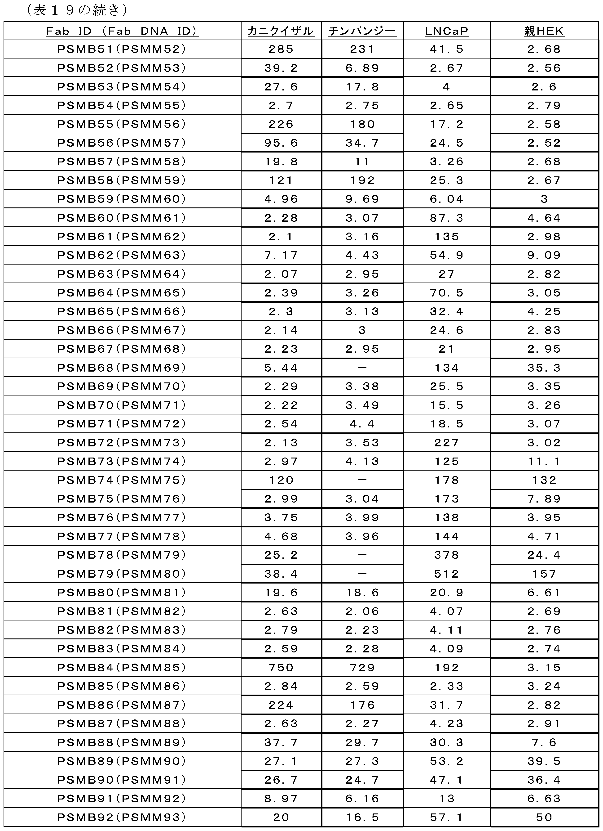

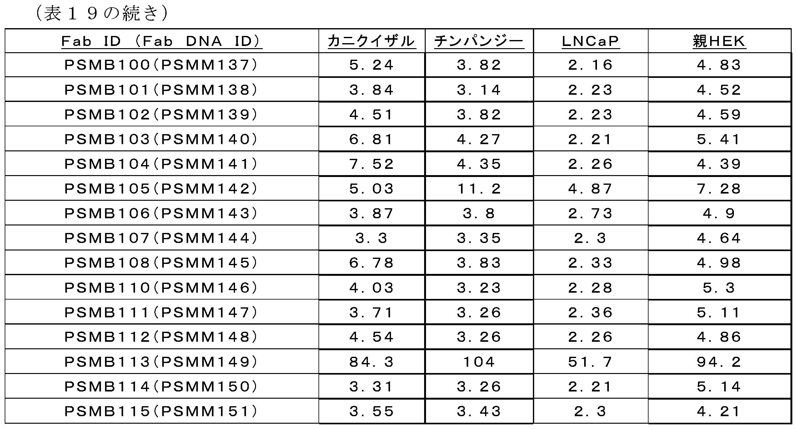
哺乳類で発現したFabの用量応答曲線
哺乳類で発現したFabクローンが、未希釈Fab上清としてPSMA発現細胞株への陽性結合について確認されてから、上清をOctet又はタンパク質ゲルによりタンパク質濃度に対して正規化し、用量応答曲線を完成させて、前述のプロトコルを用いてPSMA結合を確認した。図23~図25は、3つのPSMA発現細胞の全てに対する結合が実証されたヒットの滴定曲線を示す。図23A、23B、23C、及び23Dは、抗PSMAパニングヒット対LNCaP細胞の滴定曲線を示す。図24A、24B、24C、及び24Dは、抗PSMAパニングヒット対チンパンジーPSMA HEK細胞の滴定曲線を示す。図25A、25B、25C、及び25Dは、抗PSMAパニングヒット対カニクイザルPSMA HEK細胞の滴定曲線を示す。ヒット間の結合プロファイルを、異なる種のPSMAを発現する細胞株にわたって比較した。PSMM52上清を、実験にわたって陽性対照として使用した。CDRにおけるN-結合型グリコシル化部位、PSMA陰性親HEK細胞株への結合、又はPSMA陽性細胞株への結合の欠如から、いくつかのヒットを優先から外した。11個のFabヒットが残っており、10個のヒットをヒトIgG4-PAA重鎖コンストラクトにクローニングし、PSMA×CD3二重特異性抗体を生成するために使用した。これらのヒットは、互いの3倍以内の種間結合を示し、PSMA陽性標的のT細胞リダイレクション殺傷について試験される二重特異性抗体フォーマットに移した。各ヒットについてのパニング抗原を表20に示す。
Dose Response Curves of Mammalian-Expressed Fabs Mammalian-expressed Fab clones were confirmed for positive binding to PSMA-expressing cell lines as undiluted Fab supernatants, and then the supernatants were normalized to protein concentration by Octet or protein gel to complete dose response curves to confirm PSMA binding using the protocol described above. Figures 23-25 show titration curves of hits that demonstrated binding to all three PSMA-expressing cells. Figures 23A, 23B, 23C, and 23D show titration curves of anti-PSMA panning hits vs. LNCaP cells. Figures 24A, 24B, 24C, and 24D show titration curves of anti-PSMA panning hits vs. chimpanzee PSMA HEK cells. Figures 25A, 25B, 25C, and 25D show titration curves of anti-PSMA panning hits vs. cynomolgus PSMA HEK cells. Binding profiles between hits were compared across cell lines expressing different species of PSMA. PSMM52 supernatant was used as a positive control throughout the experiment. Several hits were deprioritized due to N-linked glycosylation sites in the CDRs, binding to the PSMA-negative parental HEK cell line, or lack of binding to PSMA-positive cell lines. Eleven Fab hits remained, and ten hits were cloned into a human IgG4-PAA heavy chain construct and used to generate a PSMAxCD3 bispecific antibody. These hits showed cross-species binding within 3-fold of each other and were transferred to a bispecific antibody format to be tested for T cell redirection killing of PSMA-positive targets. The panning antigens for each hit are shown in Table 20.

抗PSMAmAbの調製
3つ全てのPSMA発現細胞への結合が実証された合計12個のクローンを、最終的には、制限クローニングによってFc置換S228P、F234A、及びL235A(PAA)アイソタイプを有するmAb IgG4に変換した。簡潔に述べると、初期スクリーニングを通過したFabクローンに対応するコンストラクトをHindIII及びApaIで消化した。ゲル精製した断片を、完全mAb発現のためにCMV promotervDR000215、ヒトIgG4-PAA Fcを含有するCMV駆動発現ベクターを含む、発現ベクターにライゲーションした。これにより、二重特異性抗体の迅速生成が可能になった。既に記載した発現ベクターを使用して、各PSMA mabの重鎖及び軽鎖を発現させ、両方のベクターをmAbの発現のために293Expi又はCHO細胞株に一過的にコトランスフェクトした。ファージパニングから生成された種間陽性PSMA FabのCDR配列を以下の表21に示す。選択されたFabのVH及びVL配列を以下の表22に示す。Fabから生成されたmAbの重鎖及び軽鎖配列を表23に示す。
Preparation of anti-PSMA mAbs A total of 12 clones that demonstrated binding to all three PSMA-expressing cells were finally converted to mAb IgG4 with Fc substitutions S228P, F234A, and L235A (PAA) isotype by restriction cloning. Briefly, constructs corresponding to Fab clones that passed the initial screening were digested with HindIII and ApaI. Gel-purified fragments were ligated into expression vectors, including CMV promoter DR000215, a CMV-driven expression vector containing human IgG4-PAA Fc, for full mAb expression. This allowed for rapid generation of bispecific antibodies. The heavy and light chains of each PSMA mab were expressed using the expression vectors previously described, and both vectors were transiently co-transfected into 293Expi or CHO cell lines for expression of the mAbs. The CDR sequences of the cross-species positive PSMA Fabs generated from phage panning are shown below in Table 21. The VH and VL sequences of the selected Fabs are shown below in Table 22. The heavy and light chain sequences of the mAbs generated from the Fabs are shown in Table 23.


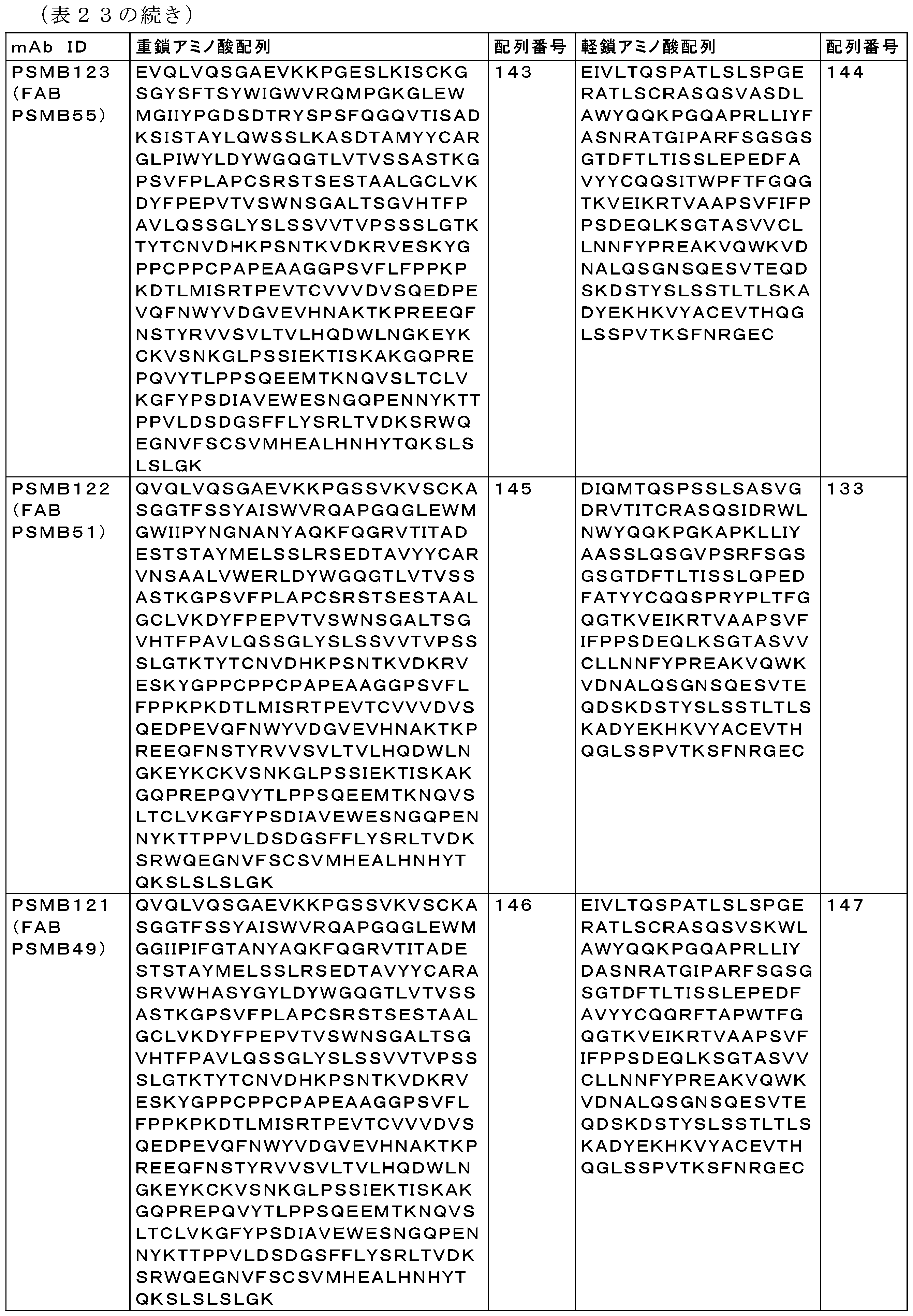

配列番号130のVH及び配列番号131のVLを有するVH及びVL領域と、S228P、F234A、及びL235A置換を有するIgG4定常領域とを含む単一特異性抗PSMA抗体PSMB119を生成した。配列番号128のVH及び配列番号129のVLを有するVH及びVL領域と、S228P、F234A、及びL235A置換を有するIgG4定常領域とを含む単一特異性抗PSMA抗体PSMB120を生成した。配列番号126のVH及び配列番号127のVLを有するVH及びVL領域と、S228P、F234A、及びL235A置換を有するIgG4定常領域とを含む単一特異性抗PSMA抗体PSMB121を生成した。配列番号125のVH及び配列番号111のVLを有するVH及びVL領域と、S228P、F234A及びL235A置換を有するIgG4定常領域とを含む単一特異性抗PSMA抗体PSMB122を生成した。配列番号123のVH及び配列番号124のVLを有するVH及びVL領域と、S228P、F234A、及びL235A置換を有するIgG4定常領域とを含む単一特異性抗PSMA抗体PSMB123を生成した。配列番号121のVH及び配列番号122のVLを有するVH及びVL領域と、S228P、F234A、及びL235A置換を有するIgG4定常領域とを含む単一特異性抗PSMA抗体PSMB124を生成した。配列番号118のVH及び配列番号119のVLを有するVH及びVL領域と、S228P、F234A、及びL235A置換を有するIgG4定常領域とを含む単一特異性抗PSMA抗体PSMB126を生成した。配列番号116のVH及び配列番号117のVLを有するVH及びVL領域と、S228P、F234A、及びL235A置換を有するIgG4定常領域とを含む単一特異性抗PSMA抗体PSMB127を生成した。配列番号114のVH及び配列番号115のVLを有するVH及びVL領域と、S228P、F234A、及びL235A置換を有するIgG4定常領域とを含む単一特異性抗PSMA抗体PSMB128を生成した。配列番号110のVH及び配列番号111のVLを有するVH及びVL領域と、S228P、F234A、及びL235A置換を有するIgG4定常領域とを含む単一特異性抗PSMA抗体PSMB129を生成した。配列番号112のVH及び配列番号113のVLを有するVH及びVL領域と、S228P、F234A、及びL235A置換を有するIgG4定常領域とを含む単一特異性抗PSMA抗体PSMB130を生成した。 A monospecific anti-PSMA antibody PSMB119 was generated, comprising a VH and VL region having a VH of SEQ ID NO: 130 and a VL of SEQ ID NO: 131, and an IgG4 constant region having S228P, F234A, and L235A substitutions. A monospecific anti-PSMA antibody PSMB120 was generated, comprising a VH and VL region having a VH of SEQ ID NO: 128 and a VL of SEQ ID NO: 129, and an IgG4 constant region having S228P, F234A, and L235A substitutions. A monospecific anti-PSMA antibody PSMB121 was generated, comprising a VH and VL region having a VH of SEQ ID NO: 126 and a VL of SEQ ID NO: 127, and an IgG4 constant region having S228P, F234A, and L235A substitutions. A monospecific anti-PSMA antibody PSMB122 was generated, comprising VH and VL regions with a VH of SEQ ID NO: 125 and a VL of SEQ ID NO: 111, and an IgG4 constant region with S228P, F234A, and L235A substitutions. A monospecific anti-PSMA antibody PSMB123 was generated, comprising VH and VL regions with a VH of SEQ ID NO: 123 and a VL of SEQ ID NO: 124, and an IgG4 constant region with S228P, F234A, and L235A substitutions. A monospecific anti-PSMA antibody PSMB124 was generated, comprising VH and VL regions with a VH of SEQ ID NO: 121 and a VL of SEQ ID NO: 122, and an IgG4 constant region with S228P, F234A, and L235A substitutions. A monospecific anti-PSMA antibody PSMB126 was generated, comprising VH and VL regions with a VH of SEQ ID NO: 118 and a VL of SEQ ID NO: 119, and an IgG4 constant region with S228P, F234A, and L235A substitutions. A monospecific anti-PSMA antibody PSMB127 was generated, comprising VH and VL regions with a VH of SEQ ID NO: 116 and a VL of SEQ ID NO: 117, and an IgG4 constant region with S228P, F234A, and L235A substitutions. A monospecific anti-PSMA antibody PSMB128 was generated, comprising VH and VL regions with a VH of SEQ ID NO: 114 and a VL of SEQ ID NO: 115, and an IgG4 constant region with S228P, F234A, and L235A substitutions. A monospecific anti-PSMA antibody PSMB129 was generated, comprising a VH and VL region with a VH of SEQ ID NO: 110 and a VL of SEQ ID NO: 111, and an IgG4 constant region with S228P, F234A, and L235A substitutions. A monospecific anti-PSMA antibody PSMB130 was generated, comprising a VH and VL region with a VH of SEQ ID NO: 112 and a VL of SEQ ID NO: 113, and an IgG4 constant region with S228P, F234A, and L235A substitutions.
2-5 抗PSMA Fab PSMB83(別名PSMM84)に結合したヒトPSMA ECDの結晶構造
PSMAは、細胞表面上で発現するホモダイマータンパク質である。PSMAは、ペプチダーゼ活性を有する大きなECDドメイン(705残基)、1回貫通TMドメイン、及び短い19残基の細胞内ドメインで構成される、1モノマーあたり750残基のII型内在性糖タンパク質である。PSMAと抗体との間の結合部位についてより深く理解するために、二重特異性抗体PS3B27の抗PSMA Fabアームに結合したヒトPSMAの細胞外領域(ECD)の結晶構造を3.15Åの分解能で決定した。
2-5 Crystal structure of human PSMA ECD bound to anti-PSMA Fab PSMB83 (also known as PSMM84) PSMA is a homodimeric protein expressed on the cell surface. PSMA is a type II integral glycoprotein of 750 residues per monomer, consisting of a large ECD domain (705 residues) with peptidase activity, a single-spanning TM domain, and a short 19-residue intracellular domain. To gain a deeper understanding of the binding site between PSMA and antibodies, the crystal structure of the extracellular domain (ECD) of human PSMA bound to the anti-PSMA Fab arm of the bispecific antibody PS3B27 was determined at 3.15 Å resolution.
ヒトPSMA(残基44~750)の細胞外領域を、N末端gp67シグナルペプチド、続いて、切断可能なヘキサヒスチジンタグ(配列番号596)を含むHigh Five(商標)昆虫細胞で発現させた。最初のNi2+-NTA親和性捕捉、TEVに媒介されるヒスチジンタグの切断、続いて、逆アフィニティクロマトグラフィー段階、及び最後のサイズ排除クロマトグラフィー段階を含む3段階手順により、上清から分泌タンパク質を精製した。精製されたPSMA-ECDを液体窒素中で急速冷凍し、10mM HEPES pH7.4、150mM NaCl、2mM CaCl2、0.1mM ZnCl2中において-80℃で保存した。 The extracellular region of human PSMA (residues 44-750) was expressed in High Five™ insect cells containing an N-terminal gp67 signal peptide followed by a cleavable hexahistidine tag (SEQ ID NO:596). Secreted protein was purified from the supernatant by a three-step procedure including initial Ni 2+ -NTA affinity capture, TEV-mediated cleavage of the histidine tag followed by a reverse affinity chromatography step, and a final size-exclusion chromatography step. Purified PSMA-ECD was flash frozen in liquid nitrogen and stored at -80°C in 10 mM HEPES pH 7.4, 150 mM NaCl, 2 mM CaCl 2 , 0.1 mM ZnCl 2 .
二重特異性抗体PS3B27における親抗PSMA FabアームであるPSMB83(別名PSMM84)のFabを、ヘキサヒスチジンタグ(配列番号596)を含むHEK293 Expi細胞で発現させ、アフィニティクロマトグラフィー(HisTrap、GE Healthcare)及びサイズ排除クロマトグラフィー(SEC-300、Phenomenex Yarra)を用いて精製した。Fabを、50mM NaCl、20mM Tris pH7.4中において4℃で保存した。 Fab of PSMB83 (also known as PSMM84), the parent anti-PSMA Fab arm of bispecific antibody PS3B27, was expressed in HEK293 Expi cells containing a hexahistidine tag (SEQ ID NO:596) and purified using affinity chromatography (HisTrap, GE Healthcare) and size exclusion chromatography (SEC-300, Phenomenex Yarra). Fab was stored at 4°C in 50 mM NaCl, 20 mM Tris pH 7.4.
3段階手順によってヒトPSMA ECD/PSMB83 Fab複合体を調製した。最初に、Fabを20mM MES pH6.0、150mM NaClにバッファ交換した。次いで、Fab及びPSMAを混合し(PSMAモノマーに対して1.5モル過剰のFab)、20mM MES pH6.0に透析しながら、4℃で一晩インキュベートした。最後に、複合体を20mM MES pH6.0中でmonoS 5/50カラムに結合させ、NaCl勾配で溶出した。
Human PSMA ECD/PSMB83 Fab complex was prepared by a three-step procedure. First, Fab was buffer exchanged into 20 mM MES pH 6.0, 150 mM NaCl. Then, Fab and PSMA were mixed (1.5 molar excess of Fab over PSMA monomer) and incubated overnight at 4°C while being dialyzed into 20 mM MES pH 6.0. Finally, the complex was bound to a
20℃におけるシッティングドロップ蒸気拡散法及びMosquito LCPロボット(TTP Labtech)を用いて、X線回折に好適な結晶を得た。マイクロシード及び最初は7.3mg/mLのPSMA/Fab複合体を用いて、18% PEG 3kDa、0.2M(NH4)2SO4、0.1M Tris pH8.5から、ヒトPSMA ECDに結合したPSMB83 Fabの結晶を成長させた。最初は8.8mg/mLのFabを用いて、25% PEG 3kDa、0.2M LiCl、0.1M酢酸、pH4.5から遊離PSMB83 Fabの結晶を得た。
Crystals suitable for X-ray diffraction were obtained using sitting drop vapor diffusion at 20° C. and a Mosquito LCP robot (TTP Labtech). Crystals of PSMB83 Fab bound to human PSMA ECD were grown from 18
Phaser(Phaser Crystallographic Software,University of Cambridge)による分子置換(MR)によって構造を解析した。PSMB83(別名PSMM84)Fab構造のMR検索モデルは、PDBコード4M6Oであった。MR検索モデルとしてPSMA(PDBコード:2C6G)及びPSMB83 Fab(1.93Å分解能の構造、データは示さない)の結晶構造を使用して、PSMA/Fab複合体構造を解析した。PHENIXにより構造を精密化し(Adams,et al,2004)、COOTを用いてモデル調整を行った(Emsley and Cowtan,2004)。他の全ての結晶学的計算をCCP4プログラムスイートで行った(Collaborative Computational Project Number 4,1994)。PyMol(PyMOL Molecular Graphics System,Version 1.4.1,Schrodinger,LLC)で全ての分子グラフィクスを作成し、相補性決定領域(CDR)をKabatの定義を用いて決定した。
The structure was solved by molecular replacement (MR) using Phaser (Phaser Crystallographic Software, University of Cambridge). The MR search model of the PSMB83 (also known as PSMM84) Fab structure was PDB code 4M6O. The PSMA/Fab complex structure was solved using the crystal structures of PSMA (PDB code: 2C6G) and PSMB83 Fab (structure at 1.93 Å resolution, data not shown) as MR search models. The structure was refined using PHENIX (Adams, et al, 2004), and the model was adjusted using COOT (Emsley and Cowtan, 2004). All other crystallographic calculations were performed with the CCP4 suite of programs (Collaborative
PSMA/Fab構造は、Fab軽鎖残基1~211、Fab重鎖残基1~224(無秩序である残基138~146を除く)、及びPSMA残基56~750を含み、これは、プロテアーゼ(残基56~116及び352~590)、アピカル(残基117~351)及びヘリカル(残基591~750)ドメイン、並びにPSMAダイマーサブユニットあたり10個の可能なN-結合型グリカンのうちの7個(Asn-76、-121、-140、-195、-459、-476、及び-638)に対応する。PSMA活性部位は、3つのドメイン間の界面に位置し、ヒスチジン(H377及びH553)及びグルタミン酸/アスパラギン酸(D387、触媒活性を有するE424、E425、及びD453)の残基、並びに水分子が配位している2個の亜鉛原子を含有している。結晶非対称単位は、PSMB83 Fabと同様に結合した各サブユニットを有する1つのPSMAダイマーを含有する。Fab/PSMAの結合部位は、結合残基の信頼できる位置決めを可能にする電子密度マップによってよく定義される。Fab及びPSMA分子は、図26~図31で逐次付番される。 The PSMA/Fab structure includes Fab light chain residues 1-211, Fab heavy chain residues 1-224 (excluding residues 138-146, which are disordered), and PSMA residues 56-750, which correspond to the protease (residues 56-116 and 352-590), apical (residues 117-351), and helical (residues 591-750) domains, and 7 of 10 possible N-linked glycans per PSMA dimer subunit (Asn-76, -121, -140, -195, -459, -476, and -638). The PSMA active site is located at the interface between the three domains and contains histidine (H377 and H553) and glutamic/aspartic acid (D387, catalytically active E424, E425, and D453) residues, as well as two zinc atoms coordinated by water molecules. The crystal asymmetric unit contains one PSMA dimer with each subunit bound similarly to the PSMB83 Fab. The Fab/PSMA binding site is well defined by electron density maps that allow reliable positioning of the binding residues. The Fab and PSMA molecules are numbered sequentially in Figures 26-31.
PSMB83エピトープ、パラトープ、及び相互作用。二重特異性抗体PS3B27における親抗PSMA FabアームであるPSMB83は、PSMAのアピカルドメインにおける配座エピトープ及び不連続エピトープを認識する(図26)。Fabに埋もれているPSMA表面積は、約700Å2である。具体的には、PSMB83エピトープ残基は、I138、F235、P237、G238、D244、Y299、Y300、Q303、K304、E307、及びK324~P326である。ヘリックスα7(残基Y299~E307)はエピトープの優勢領域であり、Fab重鎖及び軽鎖CDRにわたって結合する。ヘリックスの一端において、Y299及びY300は、Fab残基Y57H、W94L、並びにPSMA残基F235及びP237と芳香族クラスタを形成し、一方、他のヘリックス末端におけるE307は、R91Lとの塩架橋及び水素結合Y32Lを形成する。図27及び図28は、PSMB83軽鎖及び重鎖とのPSMAの主な相互作用を示す。PSMB83エピトープ残基は、ヒトとカニクイザルとの間で保存されており(図29)、二重特異性抗体PS3B27は、ヒト及びカニクイザルPSMAと同様の親和性で結合することが実証された。対照的に、ヒトの、マウスG238A及び特にY300Dのエピトープへの変異は、ヒトと比較してマウスPSMAに対するPSMB83結合親和性が低いと予想される。Y300D変異は、N59Hとの水素結合接触及びW94Lとのπスタッキング相互作用を妨害する。 PSMB83 epitope, paratope, and interactions. PSMB83, the parent anti-PSMA Fab arm of the bispecific antibody PS3B27, recognizes a conformational and discontinuous epitope in the apical domain of PSMA (FIG. 26). The surface area of PSMA buried in the Fab is approximately 700 Å2 . Specifically, the PSMB83 epitope residues are I138, F235, P237, G238, D244, Y299, Y300, Q303, K304, E307, and K324-P326. Helix α7 (residues Y299-E307) is the predominant region of the epitope, which binds across the Fab heavy and light chain CDRs. At one end of the helix, Y299 and Y300 form an aromatic cluster with Fab residues Y57H , W94L , and PSMA residues F235 and P237, while E307 at the other end of the helix forms a salt bridge with R91L and a hydrogen bond Y32L . Figures 27 and 28 show the main interactions of PSMA with PSMB83 light and heavy chains. PSMB83 epitope residues are conserved between humans and cynomolgus monkeys (Figure 29), and bispecific antibody PS3B27 was demonstrated to bind human and cynomolgus monkey PSMA with similar affinity. In contrast, human mutations to the epitope of mouse G238A and especially Y300D are predicted to reduce PSMB83 binding affinity to mouse PSMA compared to humans. The Y300D mutation disrupts hydrogen bond contacts with N59H and pi-stacking interactions with W94L .
PSMB83パラトープは、CDR-L2及び-CDR-H1を除く全てのCDR由来の残基で構成されている(図30)。具体的には、パラトープ残基は、軽鎖S30L、Y32L、R91L、S92L、W94L、並びに重鎖G56H~N59H、K65H、G66H、Y101H、V107H、及びD109Hである。図31は、PSMAとPSMB83との間の相互作用接触を示す。エピトープのアクセス可能な位置は、PS3B27二重特異性抗体におけるPSMB83 Fabアームの膜結合PSMAへの結合を促進し、一方、他のFabアームは、T細胞膜におけるCD3に依然として結合している。PSMB83は、抗体が活性部位から離れて結合し、酵素機能に影響を及ぼす可能性があるPSMAにおける何らかの大きな構造変化、例えば、活性部位を閉鎖するループ移動又は触媒残基の置換(Fabに結合している及び結合していない(Barinka Et al,2007)構造におけるPSMA分子のCα重ね合わせについては0.3ÅのRMSD)を引き起こさないので、PSMA酵素活性を阻害しないと予想される。 The PSMB83 paratope is composed of residues from all CDRs except CDR-L2 and -CDR-H1 (Figure 30). Specifically, the paratope residues are light chain S30L , Y32L , R91L , S92L , W94L , and heavy chain G56H - N59H , K65H , G66H , Y101H , V107H , and D109H . Figure 31 shows the interaction contacts between PSMA and PSMB83. The accessible location of the epitope facilitates binding of the PSMB83 Fab arm in the PS3B27 bispecific antibody to membrane-bound PSMA, while the other Fab arm is still bound to CD3 on the T cell membrane. PSMB83 is not expected to inhibit PSMA enzymatic activity because the antibody binds away from the active site and does not induce any major structural changes in PSMA that could affect enzyme function, such as loop movements that close the active site or displacement of catalytic residues (RMSD of 0.3 Å for Ca superposition of the PSMA molecule in the Fab-bound and unbound (Barinka Et al, 2007) structures).
2-6 抗PSMA親和性成熟
親PSMB127(fab ID=PSMB83、別名PSMM84)と比較して結合親和性が増加した抗体を同定するために、2つのPSMA親和性成熟ライブラリからの抗PSMA Fabファージクローンに対して親和性成熟を行った。PSMB127の親和性成熟のために2つのライブラリを生成した)。第1のライブラリにおいて、重鎖CDR1及びCDR2を表24(PH9H9L1)の設計に従ってランダム化した。H-CDR3断片をpDR000024032からPCRで増幅させ、SacII+XhoIで消化した。この断片をPH9H9L1/PH9L3ライブラリにクローニングした。これを大腸菌MC1061F’細胞に形質転換し、このFabライブラリをディスプレイするファージを生成した。第2のライブラリにおいて、軽鎖CDRを表25(PH9L3L3)の設計に従ってランダム化した。PSMB83(PSMH360)からの重鎖をPCRで増幅し、NcoI+XhoIで消化した。この断片をPH9L3L3ライブラリDNA(ELN:デノボ2010ファージライブラリSRI-021)にクローニングした。これを大腸菌MC1061F’細胞に形質転換し、このFabライブラリをディスプレイするファージを生成した。
2-6 Anti-PSMA Affinity Maturation To identify antibodies with increased binding affinity compared to the parent PSMB127 (fab ID=PSMB83, also known as PSMM84), affinity maturation was performed on anti-PSMA Fab phage clones from two PSMA affinity maturation libraries. Two libraries were generated for affinity maturation of PSMB127). In the first library, heavy chain CDR1 and CDR2 were randomized according to the design in Table 24 (PH9H9L1). The H-CDR3 fragment was amplified by PCR from pDR000024032 and digested with SacII+XhoI. This fragment was cloned into the PH9H9L1/PH9L3 library, which was transformed into E. coli MC1061F′ cells to generate phage displaying this Fab library. In the second library, light chain CDRs were randomized according to the design in Table 25 (PH9L3L3). The heavy chain from PSMB83 (PSMH360) was amplified by PCR and digested with NcoI + XhoI. This fragment was cloned into PH9L3L3 library DNA (ELN: de novo 2010 phage library SRI-021), which was transformed into E. coli MC1061F' cells to generate phage displaying this Fab library.


PSMA親和性成熟Fab-pIXライブラリの溶液パニングを、3ラウンドにわたってビオチン化ヒトPSMA ECDに対して行った。ファージに結合した抗原を、製造元のプロトコルに従ってニュートラアビジンビーズ(GE HealthCare Life Scienceカタログ番号78152104011150)に捕捉し、続いて、1×PBST(tween20中0.05%)で広範に洗浄し、500倍モル過剰のビオチン化抗原中非標識PSMA ECDと共に1時間インキュベートした。このパニングにより、クローン、PSMXP46R3_59H09、PSMXP46R3_59H06、PSMXP46R3_59E03、PSMXP46R3_59C09,PSMXP46R3_59H01、PSMXP46R3_59F11、及びPSMXP46R3_59F07が得られた。 Solution panning of the PSMA affinity matured Fab-pIX library was performed against biotinylated human PSMA ECD for three rounds. Phage-bound antigen was captured on Neutravidin beads (GE HealthCare Life Science Catalog No. 78152104011150) according to the manufacturer's protocol, followed by extensive washing with 1x PBST (0.05% in tween 20) and incubation for 1 hour with a 500-fold molar excess of unlabeled PSMA ECD in biotinylated antigen. This panning yielded the clones PSMXP46R3_59H09, PSMXP46R3_59H06, PSMXP46R3_59E03, PSMXP46R3_59C09, PSMXP46R3_59H01, PSMXP46R3_59F11, and PSMXP46R3_59F07.
抗PSMA fabクローンの発現レベルを決定するために、96ウェルMaxisorbプレートを、4℃で一晩抗ヒトFd IgGでコーティングし、洗浄し、3%ミルク-PBS-0.05%Tweenで1時間ブロッキングした。最後のウェルブランクを用いて、11個の希釈液についてファージ上清サンプルをブロッキングバッファで2倍連続希釈した。100μLのこれらの溶液を、コーティングされたプレート上に1時間にわたって捕捉した。プレートを洗浄し、100μLの抗F(ab’)2-HRP抗体を1時間にわたって添加した。プレートを洗浄し、100μLのペルオキシダーゼ試薬で発色させ、Envisionで発光を読み取った(図32)。 To determine the expression levels of anti-PSMA fab clones, 96-well Maxisorb plates were coated with anti-human Fd IgG overnight at 4°C, washed, and blocked with 3% milk-PBS-0.05% Tween for 1 hour. Phage supernatant samples were serially diluted 2-fold in blocking buffer for 11 dilutions with the last well blanked. 100 μL of these solutions were captured on the coated plates for 1 hour. Plates were washed and 100 μL of anti-F(ab')2-HRP antibody was added for 1 hour. Plates were washed and developed with 100 μL of peroxidase reagent and luminescence was read on an Envision (Figure 32).
抗PSMA fabクローンのヒト及びカニクイザル組換えタンパク質への結合を決定するために、96ウェルMaxisorbプレートを、4℃で一晩、5μg/mLのニュートラアビジン100μLでコーティングした。プレートを洗浄し、3%ミルク-PBS-0.05%Tweenで1時間ブロッキングした。組換えビオチン化ヒト及びカニクイザルPSMAタンパク質を、室温で1時間にわたって、2.5μg/mLで捕捉した。プレートを洗浄し、2倍連続希釈したfab上清100μLを、RTで1時間にわたって捕捉した。プレートを洗浄し、次いで、PBST中0.3%ミルク200μLと共に2.5時間インキュベートして、親和性の弱いfabの一部を洗い流した。次いで、新たなPBST中の0.3%ミルク200μLと共に更に30分間インキュベートして、親和性の弱いfabを更に除去した。プレートを洗浄し、100μLの抗F(ab’)2-HRP抗体を1時間にわたって添加した。プレートを洗浄し、100μLのペルオキシダーゼ試薬で発色させ、Envisionで発光を読み取った(図33及び34)。 To determine binding of anti-PSMA fab clones to human and cynomolgus recombinant proteins, 96-well Maxisorb plates were coated with 100 μL of neutravidin at 5 μg/mL overnight at 4°C. Plates were washed and blocked with 3% milk-PBS-0.05% Tween for 1 h. Recombinant biotinylated human and cynomolgus PSMA proteins were captured at 2.5 μg/mL for 1 h at room temperature. Plates were washed and 100 μL of 2-fold serially diluted fab supernatants were captured for 1 h at RT. Plates were washed and then incubated with 200 μL of 0.3% milk in PBST for 2.5 h to wash out some of the weak affinity fabs. They were then incubated with 200 μL of fresh 0.3% milk in PBST for an additional 30 min to further remove weak affinity fabs. The plate was washed and 100 μL of anti-F(ab')2-HRP antibody was added for 1 hour. The plate was washed and developed with 100 μL of peroxidase reagent and luminescence was read on an Envision (Figures 33 and 34).
図32は、親Fab及び親和性成熟Fabのタンパク質発現が類似していることを示す。y軸の値は、希釈曲線にわたってfabタンパク質の存在量に等しい検出試薬の発光を表し、発光の読み取り値が高いほど、連続的な2倍希釈で減少させたウェル内のタンパク質がより多い。親和性成熟fabを有するウェルにはより多くのタンパク質が存在していたが、EC50値(最大応答の半分を与えるタンパク質の濃度である)によって示されるように、親に対する増加は最大で5倍である。これらのデータは、図33におけるPSMA結合プロファイルの差を示しており、図34は、Fab濃度の差によるものではない。 Figure 32 shows that protein expression of parental and affinity matured Fabs is similar. The y-axis values represent the luminescence of the detection reagent equivalent to the amount of fab protein present across the dilution curve; the higher the luminescence reading, the more protein in the well that was reduced with successive 2-fold dilutions. Although there was more protein present in the wells with affinity matured fab, the increase over the parent is up to 5-fold as indicated by the EC50 values (which are the concentration of protein that gives half the maximum response). These data show that the differences in the PSMA binding profiles in Figure 33 and Figure 34 are not due to differences in Fab concentration.
図33は、親和性成熟Fabのヒト組換え抗原に対する結合が親抗PSMA Fab(PSMB83)よりも改善されたことを示す。ここでもまた、グラフのy軸は、発光値を表す。この場合、値が大きいほど、ヒトPSMAタンパク質に結合したFabがより多いことを意味する。これは、(x軸に沿って)Fabの濃度を増加させるにつれて高い発光値が生じる、結合の尺度である。高濃度であってもシグナルが存在しないことによって実証されるように、これらの条件下での親Fabの結合はごくわずかであった(x軸に沿った白丸)。親和性成熟fabの結合は、試験された濃度にわたって観察され、これは、ヒトPSMAタンパク質に対する結合能がより強いことと等しい。ヒトPSMAタンパク質に対する親Fab結合がゼロであることに鑑みて、EC50を生成することはできなかった。 Figure 33 shows that the binding of affinity matured Fabs to human recombinant antigen was improved over the parental anti-PSMA Fab (PSMB83). Again, the y-axis of the graph represents luminescence values. In this case, higher values mean more Fab bound to human PSMA protein. This is a measure of binding, with higher luminescence values occurring with increasing concentrations of Fab (along the x-axis). Binding of the parental Fab under these conditions was negligible, as demonstrated by the absence of signal even at high concentrations (open circles along the x-axis). Binding of the affinity matured Fab was observed across the concentrations tested, which equates to a stronger binding capacity to human PSMA protein. In light of the zero parental Fab binding to human PSMA protein, an EC50 could not be generated.
図34は、親和性成熟Fabのカニクイザル組換え抗原に対する結合が親抗PSMA Fab(PSMB83)よりも改善されたことを示す。ここでもまた、グラフのy軸は、発光値を表す。この場合、値が大きいほど、カニクイザルPSMAタンパク質に結合したFabがより多いことを意味する。これは、(x軸に沿って)Fabの濃度を増加させるにつれて高い発光値が生じる、結合の尺度である。高濃度であってもシグナルが存在しないことによって実証されるように、これらの条件下での親Fabの結合はごくわずかであった(x軸に沿った白丸)。親和性成熟Fabの結合は、試験された濃度にわたって観察され、これは、ヒトPSMAタンパク質に対する結合能がより強いことと等しい。ヒトPSMAタンパク質に対する親Fab結合がゼロであることに鑑みて、直接比較するためのEC50を生成することはできなかった。 Figure 34 shows that the binding of affinity matured Fabs to the cynomolgus recombinant antigen was improved over the parental anti-PSMA Fab (PSMB83). Again, the y-axis of the graph represents luminescence values. In this case, higher values mean more Fab bound to the cynomolgus PSMA protein. This is a measure of binding, with higher luminescence values occurring with increasing concentrations of Fab (along the x-axis). Binding of the parental Fab under these conditions was negligible, as demonstrated by the absence of signal even at high concentrations (open circles along the x-axis). Binding of the affinity matured Fab was observed across the concentrations tested, which equates to a stronger binding capacity to the human PSMA protein. Given the zero parental Fab binding to the human PSMA protein, it was not possible to generate an EC50 for direct comparison.
全体として、ファージのFab結合プロファイルは、ヒト及びカニクイザル組換え抗原に対する結合が親抗PSMA mAb(PSMB127)よりも改善されたことを示す。この改善は、Fab発現レベルに対して正規化された親和性成熟Fabの結合を示す図34及び図35によって実証されるように、Fab発現プロファイルの違いの結果ではない。ELISAスクリーニングから同定された上位5つのFab候補を、IgG4 PAAにおいてモノクローナル抗体フォーマットで産生した。表26は、後続のMab識別子を列挙し、表27及び表28は、それぞれ、その可変領域の配列並びに重鎖及び軽鎖の配列を示す。 Overall, the phages' Fab binding profiles show improved binding to human and cynomolgus recombinant antigens over the parent anti-PSMA mAb (PSMB127). This improvement is not the result of differences in Fab expression profiles, as demonstrated by Figures 34 and 35, which show the binding of affinity matured Fabs normalized to Fab expression levels. The top five Fab candidates identified from the ELISA screen were produced in monoclonal antibody format in IgG4 PAA. Table 26 lists the subsequent Mab identifiers, and Tables 27 and 28 show the sequences of their variable regions and the heavy and light chains, respectively.


3つの異なるHC及び4つの異なるLCをマトリックス形式で組み合わせて、ヒットの相違を拡大した(表29)。PSMH860のCDR2におけるメチオニンが翻訳後リスクであることに鑑みて、M64Lを有する新たな配列が生成され、PSMH865として同定した。PSM865はPSML160と対になって、Mab PSMB365を生成した。 Three different HCs and four different LCs were combined in a matrix format to expand the divergence of the hits (Table 29). Given that the methionine in CDR2 of PSMH860 is a post-translational risk, a new sequence with M64L was generated and identified as PSMH865. PSM865 was paired with PSML160 to generate Mab PSMB365.

表30及び31は、それぞれ、マトリックスで組換えた可変領域の配列並びに重鎖及び軽鎖の配列を示す。 Tables 30 and 31 show the sequences of the variable regions and the heavy and light chains recombined with the matrix, respectively.


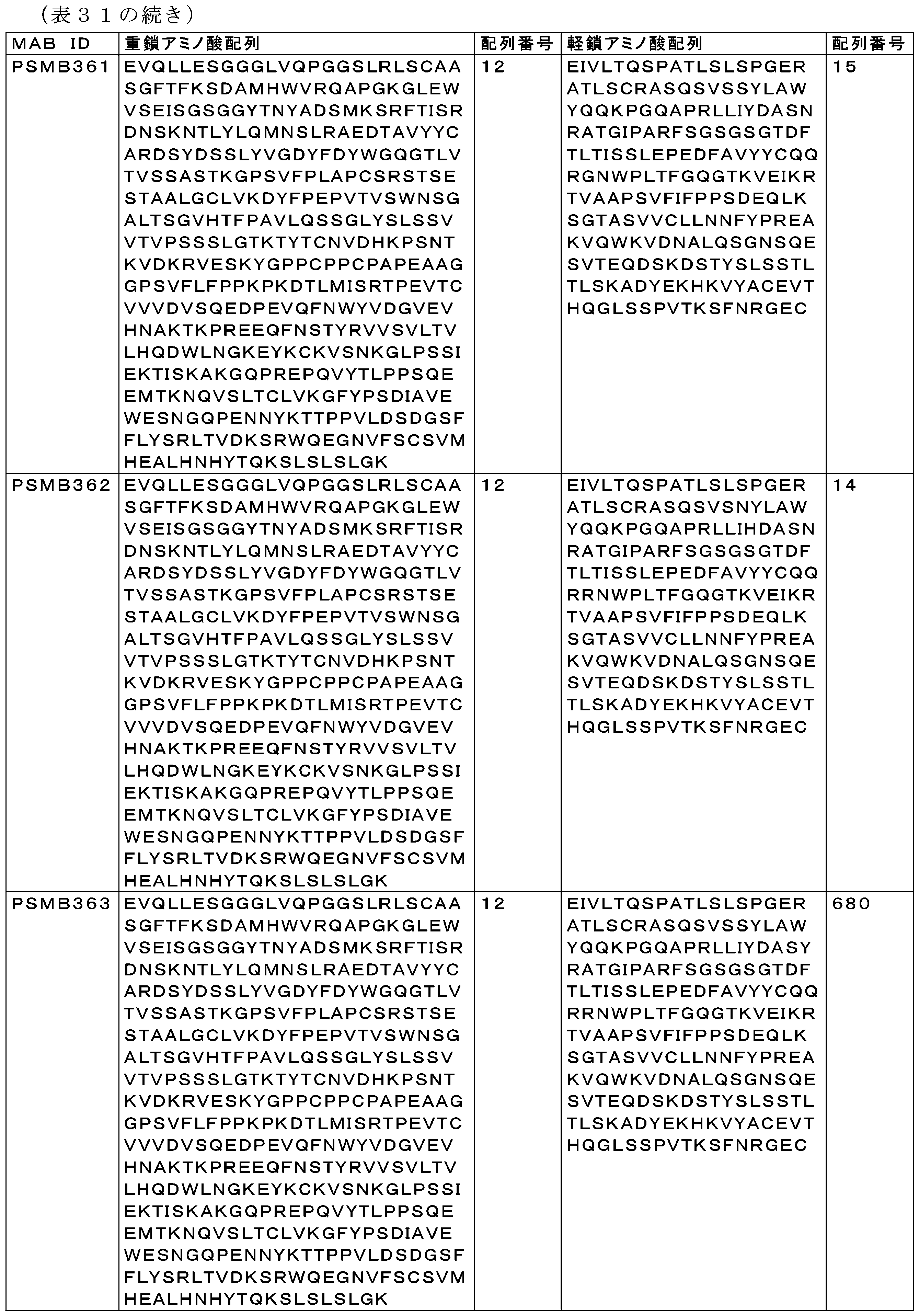

表32は、全ての親和性成熟PSMAヒットについてのCDR配列を示す。 Table 32 shows the CDR sequences for all affinity matured PSMA hits.

3 PSMA×CD3二重特異性抗体の調製及び機能評価
3-1 PSMA×CD3二重特異性抗体の生成
2種類の親和性成熟PSMA×CD3二重特異性抗体を生成した:高親和性CD3アームCD3B376(VH配列番号652、VL配列番号661、HC配列番号640、LC配列番号676)CD3アーム又は低親和性CD3B450(VH配列番号657、VL配列番号678、HC配列番号675、LC配列番号677)アームで組換えされた標的アーム(例えば、親和性成熟抗PSMA)に特異的なものである。
3 Preparation and Functional Evaluation of PSMAxCD3 Bispecific Antibodies 3-1 Generation of PSMAxCD3 Bispecific Antibodies Two types of affinity matured PSMAxCD3 bispecific antibodies were generated: those specific for a targeting arm (e.g., affinity matured anti-PSMA) engineered with a high affinity CD3 arm CD3B376 (VH SEQ ID NO:652, VL SEQ ID NO:661, HC SEQ ID NO:640, LC SEQ ID NO:676) CD3 arm or a low affinity CD3B450 (VH SEQ ID NO:657, VL SEQ ID NO:678, HC SEQ ID NO:675, LC SEQ ID NO:677) arm.
これらの親mAbは、ターゲティング親(PSMA)が409RGenMab変異(IgG4のネイティブアミノ酸)を含有し、一方、殺傷親(CD3)はF405LGenMab変異及びR409K変異を含有する、GenMabフォーマット(Labrijn et al,2013)である。単一特異性抗CD3抗体を、そのFc領域にFc置換S228P、F234A、L235A、F405L及びR409K(CD3アーム)(EUインデックスに従って付番)を有するIgG4として発現させた。ターゲティング親(PSMA)は、Fc置換S228P、F234A、L235Aを有するヒトIgG4上にある。単一特異性抗体を、CMVプロモータ下においてHEK細胞株中で発現させた。 These parental mAbs are in GenMab format (Labrijn et al, 2013) where the targeting parent (PSMA) contains the 409R GenMab mutation (native amino acid of IgG4) while the killing parent (CD3) contains the F405L GenMab and R409K mutations. The monospecific anti-CD3 antibody was expressed as IgG4 with Fc substitutions S228P, F234A, L235A, F405L and R409K (CD3 arm) (numbered according to the EU index) in its Fc region. The targeting parent (PSMA) is on human IgG4 with Fc substitutions S228P, F234A, L235A. The monospecific antibody was expressed in HEK cell lines under the CMV promoter.
親PSMA及びCD3抗体を、100mM NaAc pH3.5の溶出バッファ及び2.5M Tris pH7.2の中和バッファを用いたプロテインAカラムを使用して精製した。中和された親mAを使用して、PSMA×CD3二重特異性抗体を作製した。親mabの一部を、更に、分析測定及びアッセイのために、D-PBS、pH7.2バッファにバッファ交換した。 Parent PSMA and CD3 antibodies were purified using a Protein A column with an elution buffer of 100 mM NaAc pH 3.5 and a neutralization buffer of 2.5 M Tris pH 7.2. The neutralized parent mabs were used to generate PSMAxCD3 bispecific antibodies. A portion of the parent mabs was buffer exchanged into D-PBS, pH 7.2 buffer for further analytical measurements and assays.
精製後、制御されたFabアーム交換を行い、二重特異性抗体を作製した。親PSMA抗体を、75mM -2-MEA(2-メルカプトエチルアミン)中の還元条件下で所望の親CD3抗体と混合し、31℃で4時間又は室温で一晩インキュベートした。組換え反応は、モル比に基づくものであり、ここでは、組換え後に残存するCD3親mAbを最小化するために6%過剰のPSMA親mAを使用した。その後、組換え物を1×DPBS、pH7.2に対して透析して、還元剤を除去した。 After purification, controlled Fab arm exchange was performed to generate bispecific antibodies. Parent PSMA antibodies were mixed with the desired parent CD3 antibodies under reducing conditions in 75 mM 2-MEA (2-mercaptoethylamine) and incubated at 31°C for 4 hours or at room temperature overnight. Recombination reactions were based on molar ratios, where a 6% excess of PSMA parent mAb was used to minimize CD3 parent mAb remaining after recombination. The recombination was then dialyzed against 1x DPBS, pH 7.2 to remove the reducing agent.
組換え反応で使用した親mAb(すなわち、PSMA、CD3、又はヌル)と共に、産生された最終二重特異性抗体を表33に列挙する。 The final bispecific antibodies produced are listed in Table 33, along with the parent mAbs (i.e., PSMA, CD3, or null) used in the recombination reactions.
選択されたPSMAヒットを非殺傷アーム(ヌル)と対にして、試験目的のための陰性対照を作製した。対照二重特異性抗体の場合、B2M1、IgG4 PAAフォーマット(VH配列番号610、VL配列番号611)におけるRSV抗体を生成し、精製し、それぞれ、CD3アームCD3B219-F405L、R409Kのいずれかと組み合わせてCD3B288(CD3×ヌル)を、又はPSMAアームPSMB122、PSMB126、PSMB130のいずれかと組み合わせてPS3B37、PS3B39及びPS3B40(PSMA×ヌル)を生成した。これらのPSMA特異的親和性成熟Mabを(上記の方法のように)CD3B219及びCD3B376と交差させて、表32に示す二重特異性抗体を生成した。 Selected PSMA hits were paired with a non-killing arm (null) to generate negative controls for testing purposes. For control bispecific antibodies, B2M1, an RSV antibody in IgG4 PAA format (VH SEQ ID NO: 610, VL SEQ ID NO: 611), was generated and purified and combined with either CD3 arms CD3B219-F405L, R409K to generate CD3B288 (CD3 x null), or with either PSMA arms PSMB122, PSMB126, PSMB130 to generate PS3B37, PS3B39 and PS3B40 (PSMA x null), respectively. These PSMA-specific affinity matured Mabs were cross-linked with CD3B219 and CD3B376 (as described above) to generate the bispecific antibodies shown in Table 32.

3-2 LNCAP細胞結合におけるPSMA×CD3親和性成熟二重特異性Abの評価
PSMA×CD3二重特異性抗体を、PSMA陽性細胞株、LNCAP、PSMA陰性細胞株、PC3への結合について試験した。PSMA二重特異性抗体の結合能を評価するために、(既に記載した通りの)細胞結合アッセイを利用した。二重特異性抗体をタンパク質濃度について正規化した後、ヒト又はカニクイザルPSMAのいずれかを発現している同じ数の細胞と共にインキュベートした。各濃度のMFIをフローサイトメトリーによって収集し、濃度の関数としてプロットした。データをlog10を介して変換し、次いでプロットした。結合曲線の非線形回帰を行って、EC50を求めた。
3-2 Evaluation of PSMAxCD3 affinity matured bispecific Abs on LNCAP cell binding The PSMAxCD3 bispecific antibodies were tested for binding to the PSMA positive cell line, LNCAP, and the PSMA negative cell line, PC3. To evaluate the binding capacity of the PSMA bispecific antibodies, a cell binding assay (as previously described) was utilized. The bispecific antibodies were normalized for protein concentration and then incubated with the same number of cells expressing either human or cynomolgus PSMA. The MFI of each concentration was collected by flow cytometry and plotted as a function of concentration. The data were transformed via log10 and then plotted. Nonlinear regression of the binding curves was performed to determine the EC50.
これらの相対値を、標的細胞に対するPSMA結合をランク付けするために使用した。図14~16は、調製した全ての二重特異性抗体のLNCAP結合を示す。図16では、コンストラクトのいずれもPSMA陰性細胞株への結合を示さなかった。図14及び図15では、親和性成熟ヒットは全て、親Mab、PS2B27と比較して、曲線の左側へのシフト及びcMaxの増加を通して結合親和性の増加を示した。 These relative values were used to rank PSMA binding to target cells. Figures 14-16 show LNCAP binding of all prepared bispecific antibodies. In Figure 16, none of the constructs showed binding to PSMA-negative cell lines. In Figures 14 and 15, all affinity matured hits showed increased binding affinity through a shift of the curve to the left and an increase in cMax compared to the parent Mab, PS2B27.
組換えカニクイザルPSMA ECDとヒトPSMA ECDとの親和性成熟二重特異性抗体の相互作用を、組換えチンパンジーPSMA ECDについて既に記載した通り、ProteOn XPR36システム(BioRad)を使用して表面プラズモン共鳴(SPR)によって試験した。二重特異性抗体の全てが、実質的に同じ親和性で両標的に結合し、KDは、ヒトPSMA ECDについては0.05nM~0.27nMの範囲であり、カニクイザルPSMA ECDについては0.05nM~0.23nMの範囲である。 The interaction of affinity-matured bispecific antibodies with recombinant cyno PSMA ECD and human PSMA ECD was tested by surface plasmon resonance (SPR) using the ProteOn XPR36 system (BioRad) as previously described for recombinant chimpanzee PSMA ECD. All of the bispecific antibodies bound both targets with essentially the same affinity, with KDs ranging from 0.05 nM to 0.27 nM for human PSMA ECD and 0.05 nM to 0.23 nM for cyno PSMA ECD.
3-3 機能性細胞殺傷アッセイにおけるPSMA×CD3親和性成熟二重特異性Abの評価
上記データ、親和性測定値、及び配列同一性に基づいて、CD3B219又はCD3B376のいずれかとの二重特異性物として、3つのPSMA抗体、PSMB347、PSMB360、及びPSMB365を、PSMA特異的なリダイレクトされたT細胞の細胞傷害を媒介する能力について更に特性評価した。活性カスパーゼ3/7による蛍光基質の切断を介して細胞殺傷を間接的に測定するカスパーゼ細胞傷害アッセイを用いて、T細胞媒介性殺傷を測定した。基質の切断により蛍光DNA色素が得られ、蛍光は細胞核に制限される。同じ座標でウェルを正確に撮像することができる電動式10倍対物レンズを使用して、アッセイの全過程を通して各ウェルにおいて繰り返し蛍光測定を行う。規定のサイズ制限に基づいて及び/又は二次標識の使用を通して、標的細胞集団を特定する。冷凍した汎CD3+T細胞(Biological Specialty Corporation,Colmar,PAから購入)を、正常健常ドナーから陰性選択によって単離した。PSMAを発現している前立腺癌細胞(LNCaP、C42)を、10% HI FBS+補給物(Life Technologiesから購入)を含むRPMI 1640中で培養した。
3-3 Evaluation of PSMAxCD3 affinity matured bispecific Abs in functional cell killing assays Based on the above data, affinity measurements, and sequence identity, the three PSMA antibodies, PSMB347, PSMB360, and PSMB365, were further characterized for their ability to mediate cytotoxicity of PSMA-specific redirected T cells as bispecifics with either CD3B219 or CD3B376. T cell-mediated killing was measured using a caspase cytotoxicity assay, which indirectly measures cell killing via cleavage of a fluorescent substrate by
T細胞及び標的細胞を、選択試薬なしで、フェノールレッド不含RPMI+10% FBS及び補給物(Life Technologies)中、3:1のエフェクター対標的比(E:T)で合わせ、製造元ガイドラインに従って細胞各1mLに対して0.6μLのNucViewカスパーゼ試薬(Essen Bioscience)を添加した。総体積0.1mLの細胞を、透明な96ウェル平底プレート(BD Falcon)の適切なウェルに添加した。PS3B27(CD3×PSMA)、CD3B288(CD3×ヌル)、又はPS3B46(PSMA×ヌル)二重特異性抗体を、上記のように調製したフェノールレッド不含RPMI中2×最終濃度で調製し、0.1mLの化合物を各ウェルに添加した。ウェルの縁部における細胞凝集を最小化するために室温で30分間インキュベートした後、プレートをZoom Incucyte機器(Essen Bioscience)に移した。Incucyte機器は、37℃、5%CO2に設定された加湿インキュベータ内に存在する。 T cells and target cells were combined at a 3:1 effector to target ratio (E:T) in phenol red-free RPMI + 10% FBS and supplements (Life Technologies) without selection reagent, and 0.6 μL of NucView Caspase Reagent (Essen Bioscience) was added for every mL of cells according to the manufacturer's guidelines. A total volume of 0.1 mL of cells was added to the appropriate wells of a clear 96-well flat-bottom plate (BD Falcon). PS3B27 (CD3 x PSMA), CD3B288 (CD3 x Null), or PS3B46 (PSMA x Null) bispecific antibodies were prepared at 2x final concentration in phenol red-free RPMI prepared as above, and 0.1 mL of compound was added to each well. After incubating at room temperature for 30 minutes to minimize cell clumping at the edges of the wells, the plate was transferred to a Zoom Incucyte instrument (Essen Bioscience). The Incucyte instrument is in a humidified incubator set at 37°C and 5% CO2.
Incucyteにおける処理の解像度は、製造ガイドラインに従って、試験される各細胞株に対して設計した。カスパーゼシグナルにおいてプラトーが観察され、続いて、最高濃度の試験化合物を含有するウェルにおける最大シグナルから3回以上の連続的な減少が観察されるまで、6時間ごとに測定を行った。図17のデータが示すように、PS3B80、PS3B79、PS3B89、PS3B90、PS3B63、及びPS3B72の曲線は左にシフトし、これはPS3B27に対する効力の増加を示す。ヌルアーム対照は、予想どおり細胞死を誘導しなかった。 Treatment resolution in Incucyte was designed for each cell line tested according to the manufacturing guidelines. Measurements were taken every 6 hours until a plateau was observed in the caspase signal followed by 3 or more consecutive declines from the maximum signal in the wells containing the highest concentration of test compound. As the data in Figure 17 show, the curves for PS3B80, PS3B79, PS3B89, PS3B90, PS3B63, and PS3B72 shifted to the left, indicating increased potency relative to PS3B27. The null arm control did not induce cell death as expected.
3-4 ヒト化NSGマウスにおけるLnCaP異種移植片の腫瘍形成予防における抗腫瘍有効性
ヒトT細胞で腹腔内(ip)ヒト化された雄NOD.Cg-Prkdcscid Il2rgtm1Wjl/SzJ(NSG)マウスにおける、樹立された3D LnCaP AR.TBヒト前立腺癌異種移植片において、PS3B79及びPS3B90の有効性を評価した。2.5若しくは5mg/kgのPS3B79及びPS3B90、又はヌル×CD3B376抗体対照を、合計8回、36、39、43、47、50、53、56、60、及び63日目にq3d~q4dで投与した。1群あたり9頭の動物が残っていた試験の最後の日である腫瘍移植後53日目に、腫瘍増殖抑制(%TGI)を計算した。ヌル×CD3対照と比較して統計的に有意な腫瘍増殖阻害は、PS3B79については5mg/kgで42% TGI(Bonferroni検定を伴う二元配置分散分析、*p≦0.0001、図21)、並びにPS3B90については2.5及び5mg/kgでそれぞれ53%及び33% TGI(Bonferroni検定を伴う二元配置分散分析、*p≦0.001、図22)が観察された。したがって、CD3B376は、インビボでT細胞活性化及び細胞傷害を誘導することができ、その結果、高親和性PSMA結合アーム、PSMB360及びPSMB365を有する二重特異性フォーマットにおける腫瘍増殖阻害をもたらすことができる。
3-4 Antitumor Efficacy in Preventing Tumor Formation of LnCaP Xenografts in Humanized NSG Mice The efficacy of PS3B79 and PS3B90 was evaluated in established 3D LnCaP AR.TB human prostate cancer xenografts in male NOD.Cg-Prkdc scid Il2rg tm1Wjl /SzJ (NSG) mice humanized intraperitoneally (ip) with human T cells. PS3B79 and PS3B90 at 2.5 or 5 mg/kg, or a null x CD3B376 antibody control, were administered q3d-q4d on
4 IL1RAP×CD3二重特異性抗体の調製及び機能評価
本開示の二重特異性抗体に使用され、使用するのに好適なIL1RAPモノクローナル抗体は、その開示全体が参照により本明細書に組み込まれる米国特許出願公開第20170121420(A1)号に記載されている。15個の単一特異性IL1RAP抗体(米国特許出願公開第20170121420(A1)号の表6を参照されたい)は、Fc置換S228P、L234A、及びL235Aを有するIgG4として発現した。CD3親mAbは、S228P、L234A、L235A、F405L、及びR409Kを有するIgG4として発現した(EUインデックスに従って付番)。配列番号92のVH及び配列番号93のVLを有するVH及びVL領域と、S228P、L234A、L235A、F405L、及びR409K置換を有するIgG4定常領域とを含む単特異性抗CD3抗体CD3B220も生成した。
4 Preparation and Functional Evaluation of IL1RAP x CD3 Bispecific Antibodies IL1RAP monoclonal antibodies suitable for use in the bispecific antibodies of the present disclosure are described in US Patent Publication No. 20170121420(A1), the disclosure of which is incorporated herein by reference in its entirety. Fifteen monospecific IL1RAP antibodies (see Table 6 in US Patent Publication No. 20170121420(A1)) were expressed as IgG4 with Fc substitutions S228P, L234A, and L235A. The CD3 parent mAb was expressed as IgG4 with S228P, L234A, L235A, F405L, and R409K (numbered according to the EU index). A monospecific anti-CD3 antibody, CD3B220, was also generated that contains the VH and VL regions with the VH of SEQ ID NO: 92 and the VL of SEQ ID NO: 93, and an IgG4 constant region with S228P, L234A, L235A, F405L, and R409K substitutions.
プロテインAカラムを用い、標準的な方法を用いて単一特異性抗体を精製した。溶出後、プールをpH7に中和し、1×D-PBS、pH7.2に透析した。
Monospecific antibodies were purified using standard methods on a Protein A column. After elution, the pool was neutralized to
(国際公開第2011/131746号に記載の通り)制御されたFabアーム交換を通して単一特異性CD3 mAb(CD3B376:配列番号652のVH及び配列番号661のVL又はCD3B450:配列番号657のVH及び配列番号678のVL)及び単一特異性IL1RAP mAbと組み合わせることにより、二重特異性IL1RAP×CD3抗体を生成した。簡潔に述べると、DPBS、pH7~7.4中約1~20mg/mLの、モル比1.08:1の抗IL1RAP/抗CD3抗体を75mMの2-メルカプトエタノールアミン(2-MEA)と混合し、25~37℃において2~6時間インキュベートし、続いて、透析、ダイアフィルトレーション、接線流濾過により2-MEAを除去して、還元剤を除去し、二重特異性抗体の形成を可能にした。 Bispecific IL1RAPxCD3 antibodies were generated by combining monospecific CD3 mAbs (CD3B376: VH of SEQ ID NO:652 and VL of SEQ ID NO:661 or CD3B450: VH of SEQ ID NO:657 and VL of SEQ ID NO:678) with monospecific IL1RAP mAbs through controlled Fab arm exchange (as described in WO2011/131746). Briefly, anti-IL1RAP/anti-CD3 antibodies at a molar ratio of 1.08:1, approximately 1-20 mg/mL in DPBS, pH 7-7.4, were mixed with 75 mM 2-mercaptoethanolamine (2-MEA) and incubated at 25-37°C for 2-6 hours, followed by removal of the 2-MEA by dialysis, diafiltration, and tangential flow filtration to remove the reducing agent and allow for the formation of the bispecific antibodies.
IL1RAP×CD3二重特異性抗体の重鎖及び軽鎖を以下の表34に示す。 The heavy and light chains of the IL1RAP x CD3 bispecific antibody are shown in Table 34 below.
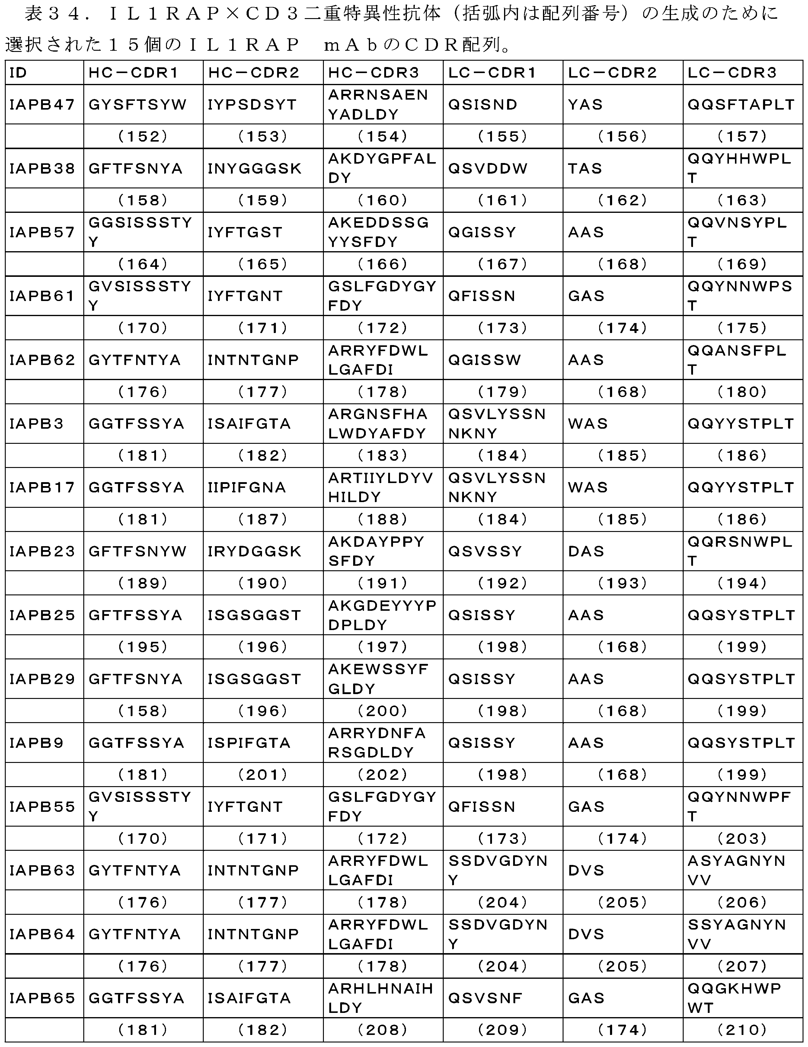
15個のIL1RAP mAbのVH及びVL配列を以下の表35に示す。 The VH and VL sequences of the 15 IL1RAP mAbs are shown in Table 35 below.
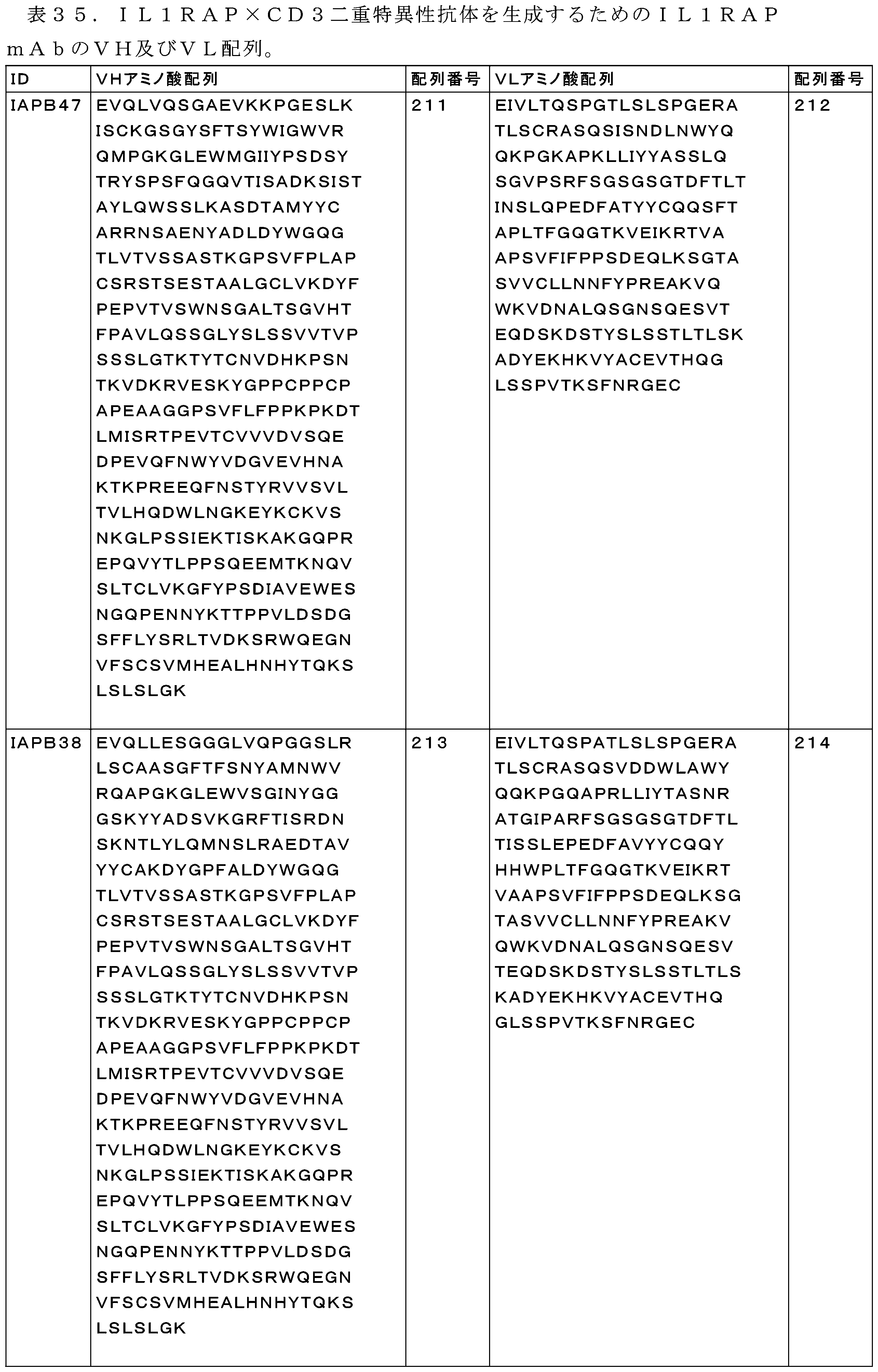


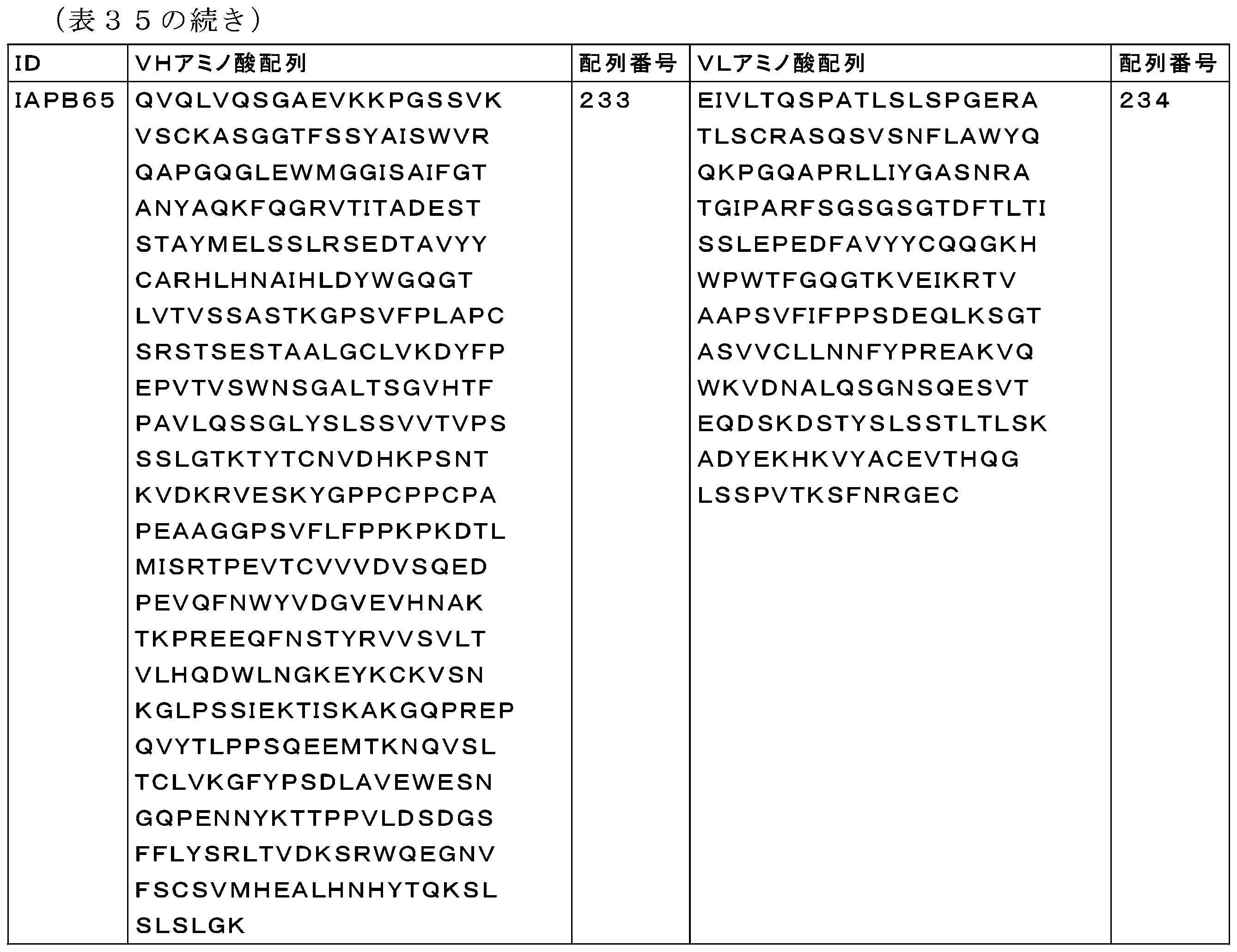
4-1 全血T細胞傷害アッセイにおけるIL1RAP二重特異性抗体の評価
方法:腫瘍細胞株LAMA-84をDPBSで洗浄した後、10×106細胞/mL(CFSE)で、室温で8分間、CFSE(150μL DMSOに再懸濁し、1:10,000希釈)と共にインキュベートした。染色をHI FBSでクエンチし、培地で洗浄した後、2×10⁵細胞/mLで完全培地に再懸濁させた。等体積の全血及びCFSE染色腫瘍細胞を96ウェルプレートの各ウェルにおいて合わせた。それぞれのウェルに抗体(10μLのDPBS中に再懸濁)を添加した後、全てのプレートを37℃、5%CO2で48時間インキュベートした。未希釈のCD25-PEを全血の各ウェルに直接添加し、室温で30分間インキュベートし、光から保護した。次いで、細胞を1500rpmで5分間遠心分離し、200μLの複数種赤血球溶解バッファで溶解し、更に4回繰り返した。細胞をDPBSで1回洗浄し、Live/Dead Near-IR(DPBSで希釈)で15分間染色した(表36)。各ウェルを洗浄し、FACSバッファ中に再懸濁させた後、BD FACS CANTO IIで細胞を取得した。
4-1 Evaluation of IL1RAP Bispecific Antibody in Whole Blood T-Cytotoxicity Assay Method: The tumor cell line LAMA-84 was washed with DPBS and then incubated with CFSE (resuspended in 150 μL DMSO, diluted 1:10,000) at 10×10 6 cells/mL (CFSE) for 8 min at room temperature. Staining was quenched with HI FBS, washed with medium and then resuspended in complete medium at 2×10 5 cells/mL. Equal volumes of whole blood and CFSE stained tumor cells were combined in each well of a 96-well plate. After adding the antibody (resuspended in 10 μL DPBS) to each well, all plates were incubated at 37° C., 5% CO 2 for 48 h. Undiluted CD25-PE was added directly to each well of whole blood and incubated at room temperature for 30 min, protected from light. The cells were then centrifuged at 1500 rpm for 5 minutes and lysed with 200 μL of multi-species red blood cell lysis buffer, which was repeated four more times. The cells were washed once with DPBS and stained with Live/Dead Near-IR (diluted in DPBS) for 15 minutes (Table 36). After washing each well and resuspending in FACS buffer, the cells were acquired on a BD FACS CANTO II.
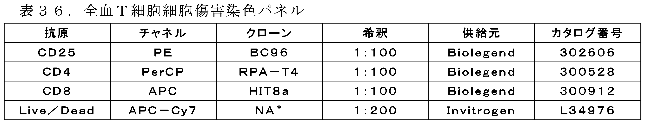
FACS CANTO II(Becton,Dickinson and Company,Franklin Lakes,NJ)での細胞取得は、1×10⁴腫瘍細胞に限定された。細胞集団を特定するためのFSC及びSSC、次いで、CFSE陽性腫瘍事象、最後に、腫瘍細胞傷害を評価するためのLive/Dead Near-IRでゲーティングすることによって、腫瘍細胞死を評価した。細胞集団を特定するためのFSC及びSSC、CFSE陰性事象、生細胞を評価するためのNear-IR陰性事象、次いで、CD25陽性細胞でゲーティングすることによって、T細胞活性化を評価した。死腫瘍細胞又はCD25陽性細胞のいずれかの百分率を、GraphPad Prism 6を用いてグラフ化し、非線形曲線フィットで分析した。非線形混合効果モデルを使用して集団EC50値を求めるために、プログラム「R」において追加の統計分析を評価した。
Cell acquisition on a FACS CANTO II (Becton, Dickinson and Company, Franklin Lakes, NJ) was limited to 1x10⁴ tumor cells. Tumor cell death was assessed by gating on FSC and SSC to identify cell populations, then CFSE positive tumor events, and finally Live/Dead Near-IR to assess tumor cytotoxicity. T cell activation was assessed by gating on FSC and SSC to identify cell populations, CFSE negative events, Near-IR negative events to assess live cells, and then CD25 positive cells. The percentage of either dead tumor cells or CD25 positive cells was graphed using
結果:sIL1RAPを含有する生理学的末梢全血状態を再現するために、IC3B19(IAPB57×CD3B219)及びIC3B34(IAPB57×CD3B376)を、全血を利用するエクスビボT細胞リダイレクションアッセイにおいて評価した。外因的に添加したIL1RAP+LAMA-84細胞株(2×10⁴細胞/ウェル)を、48時間かけてIC3B19、IC3B34、及びヌルアーム対照二重特異性抗体と共に健常対照ヒト末梢全血(n=15ドナーサンプル)に添加した。図53に示す通り、IC3B19及びIC3B34は、48時間後にエクスビボにおいてIL1RAP+LAMA-84細胞株の同様のT細胞媒介細胞傷害を誘導した。図53に提示されるIC3B19及びIC3B34細胞傷害のEC50値は、それぞれ2.097及び2.762であった(表37)。同様に、図54によって表されるIC3B19及びIC3B34によるT細胞活性化(CD25)のEC50値は、それぞれ4.237及び7.825nMであった(表38)。ヌルアーム対照二重特異性抗体は全て、図53及び図54に示すように、検出可能なレベルの細胞傷害もT細胞活性化も誘導しなかった。細胞傷害及びT細胞活性化の概ねのレベルは、IC3B19及びIC3B34の両方の間で類似していた。 Results: To mimic physiological peripheral whole blood conditions containing sIL1RAP, IC3B19 (IAPB57xCD3B219) and IC3B34 (IAPB57xCD3B376) were evaluated in an ex vivo T cell redirection assay utilizing whole blood. Exogenously added IL1RAP + LAMA-84 cell line ( 2x10⁴ cells/well) was added to healthy control human peripheral whole blood (n=15 donor samples) along with IC3B19, IC3B34, and null arm control bispecific antibody for 48 hours. As shown in Figure 53, IC3B19 and IC3B34 induced similar T cell mediated cytotoxicity of the IL1RAP + LAMA-84 cell line ex vivo after 48 hours. The EC50 values for IC3B19 and IC3B34 cytotoxicity presented in Figure 53 were 2.097 and 2.762, respectively (Table 37). Similarly, the EC50 values for T cell activation (CD25) by IC3B19 and IC3B34 presented in Figure 54 were 4.237 and 7.825 nM, respectively (Table 38). All null arm control bispecific antibodies induced no detectable levels of cytotoxicity or T cell activation as shown in Figures 53 and 54. The overall levels of cytotoxicity and T cell activation were similar between both IC3B19 and IC3B34.


4-2 24及び48時間における全血T細胞細胞傷害アッセイにおけるIL1RAP×CD3二重特異性Abによるサイトカイン放出の評価
方法:実施例4-1の24時間及び48時間のサンプルからの凍結上清を、氷上で解凍した。使用前に、プレートを4℃で5分間500gで遠心分離し、次いで、氷上に戻した。ELISAのために1:2、1:25、及び1:1000の希釈比のMSD Diluent 2を使用して、U底96ウェルプレート(Falcon)において連続希釈液を調製した。
4-2 Evaluation of cytokine release by IL1RAP x CD3 bispecific Ab in a whole blood T cell cytotoxicity assay at 24 and 48 hours Methods: Frozen supernatants from the 24 and 48 hour samples of Example 4-1 were thawed on ice. Prior to use, plates were centrifuged at 500 g for 5 minutes at 4°C and then returned to ice. Serial dilutions were prepared in U-bottom 96-well plates (Falcon) using
上清を遠心分離している間、MSDアッセイプレート(ProInflammatory Panel I V- Plex、カタログ番号K15049G-4、ロット番号K008572)を製造元のプロトコルに従って予め洗浄した。提供された較正物質を1.0mLのMSD Diluent 2で再構成することにより、標準曲線を作成した。50マイクロリットルの各サンプル又は標準を、予め洗浄したMSDプレートに直接添加した。その後のインキュベート及び洗浄は、全て製造元のプロトコルに従って実施した。MSD Sector Imagerでアッセイプレートを読み取った。
While the supernatant was being centrifuged, MSD assay plates (ProInflammatory Panel I V-Plex, Catalog No. K15049G-4, Lot No. K008572) were pre-washed according to the manufacturer's protocol. Standard curves were generated by reconstituting the provided calibrators with 1.0 mL of
IC3B19とIC3B34との間で応答を比較するために、2つの方法を使用した。 Two methods were used to compare responses between IC3B19 and IC3B34.
混合効果を有する線形回帰
この方法では、対になったドナーを考慮した用量相互作用及びランダム効果による化合物、用量、及び化合物について固定効果を有する線形回帰モデルを構築した。次いで、各濃度でIC3B19とIC3B34との間の事後比較を行い、多重比較のためにp値を適切に調整した。
Linear regression with mixed effects In this method, linear regression models with fixed effects for compound, dose, and compound by dose interactions and random effects accounting for paired donors were constructed. Post-hoc comparisons between IC3B19 and IC3B34 at each concentration were then performed and p-values were appropriately adjusted for multiple comparisons.
混合効果を有する非線形(4パラメータ)回帰
それぞれの化合物の最小、最大、logEC50、及び傾きについての固定効果パラメータと、対にしたドナー効果を考慮するランダム効果項とを有する非線形回帰モデルを用いて、IC3B19及びIC3B34を一緒にモデル化した。点推定値及びその信頼区間を抽出し、これらの別個のモデルから目視比較のためにプロットした。対象となるパラメータの合同推定値を使用したモデルを構築し、次いで、そのモデルを対数尤度検定を使用して元のモデルと比較することによって、各パラメータについて有意差を検定した。精巧にするために、IC3B19の最小、IC3B19の最大、IC3B19のlogEC50、IC3B19の傾き、IC3B34の最小、IC3B34の最大、IC3B34のlogEC50、及びIC3B34の傾きのパラメータを用いたモデルが存在する。最小値が有意に異なるかどうかをチェックするために、最小について単一の共有パラメータが存在することを除いて、全て同じパラメータを有する第2のモデルを構築する。第1のモデルの対数尤度(モデルがどのくらい良好にデータにフィッティングするかの全体的な尺度)を、第2のモデルの対数尤度と比較する。それらが互いに有意には異なっていない場合、最小が互いに有意には異なっていないと結論付けることができる。同様に、対数尤度が互いに有意に異なる場合、最小が有意に異なると結論付けるであろう。
Nonlinear (4-parameter) regression with mixed effects IC3B19 and IC3B34 were modeled together using a nonlinear regression model with fixed effect parameters for the min, max, logEC50, and slope of each compound, and a random effect term to account for paired donor effects. Point estimates and their confidence intervals were extracted and plotted for visual comparison from these separate models. Each parameter was tested for significant differences by building a model with joint estimates of the parameters of interest, and then comparing the model to the original model using a log-likelihood test. For refinement, there is a model with the following parameters: IC3B19 min, IC3B19 max, IC3B19 logEC50, IC3B19 slope, IC3B34 min, IC3B34 max, IC3B34 logEC50, and IC3B34 slope. To check whether the minima are significantly different, a second model is built with all the same parameters, except for the single shared parameter for the minima. The log-likelihood of the first model (an overall measure of how well the model fits the data) is compared to the log-likelihood of the second model. If they are not significantly different from each other, one can conclude that the minima are not significantly different from each other. Similarly, if the log-likelihoods are significantly different from each other, one would conclude that the minima are significantly different.
多くの場合、2つの理由のうちの1つから、モデル又はパラメータ比較を計算できなかった:1.4パラメータロジスティック回帰自体を計算するにはデータが扱いにくかった、又は2.パラメータ推定間の差が大きすぎて、合同モデルが失敗した。第1の場合、結果は報告せず、まとめの表には「NA」と示す。第2の場合も結果は報告しないが、有意の省略表現として「Sig」とまとめの表に示す。これらのパラメータの周囲の95%信頼区間は個別に推定可能であり、重なり合わないため、これらは有意に異なるが、正確なp値は未知であると考えることができる。 In many cases, it was not possible to compute a model or parameter comparison for one of two reasons: 1. The data were too intractable to compute a four-parameter logistic regression by itself, or 2. The differences between parameter estimates were so large that the joint model failed. In the first case, the results are not reported and are indicated in the summary tables as "NA." In the second case, the results are also not reported but are indicated in the summary tables as "Sig" as shorthand for significance. Because the 95% confidence intervals around these parameters can be estimated separately and do not overlap, they can be considered significantly different but with the exact p-value unknown.
差は、0.05以下のp値で統計的に有意であると考えられる。全ての分析をR、バージョン3.3.2.で実施した。 Differences were considered statistically significant at a p-value of 0.05 or less. All analyses were performed in R, version 3.3.2.
結果:T細胞及びIL1RAP+標的細胞株LAMA-84(内因的及び外因的に添加された腫瘍細胞)のIC3B19及びIC3B34媒介会合の結果、T細胞サイトカイン放出が生じた。IC3B19とIC3B34との間のサイトカイン放出の差を求めるために、実施例1のセクションで論じた通り、外因性LAMA-84 IL1RAP+腫瘍細胞株を添加した、全血(n=15ドナー)細胞傷害及びT細胞活性化アッセイから10個の炎症促進性サイトカインについて上清を評価した。評価した10個のサイトカインのうち、IL-8及びIL-12p70を除いて、ほとんどのサイトカインは、IC3B19及びIC3B34の両方に応答して測定可能な用量依存性放出をもたらした。表39に示すように、IL-10、IL-2、TNF-α、及びINF-γが、最も強力なEC50(nM)放出をもたらした。加えて、サイトカインの大部分及び両時点で、IC3B34分子はEC50(nM)の効力を低下させ、これは統計的に有意であった。例外は、IL-4(24時間)、IL-6(24及び48時間)、IL-8(24及び48時間)、IL-12p70(24時間及び48時間)、IL-13(24時間)、INF-γ(24時間及び48時間)、及びTNF-α(24時間)であり、IC3B19とIC3B34との間でEC50(nM)値には統計的に有意な差がなかった。 Results: IC3B19 and IC3B34 mediated association of T cells and the IL1RAP+ target cell line LAMA-84 (endogenously and exogenously added tumor cells) resulted in T cell cytokine release. To determine the difference in cytokine release between IC3B19 and IC3B34, supernatants were evaluated for 10 pro-inflammatory cytokines from whole blood (n=15 donors) cytotoxicity and T cell activation assays spiked with exogenous LAMA-84 IL1RAP+ tumor cell lines as discussed in the Example 1 section. Of the 10 cytokines evaluated, most cytokines produced measurable dose-dependent release in response to both IC3B19 and IC3B34, with the exception of IL-8 and IL-12p70. As shown in Table 39, IL-10, IL-2, TNF-α, and INF-γ produced the most potent EC50 (nM) release. In addition, for the majority of cytokines and at both time points, the IC3B34 molecule reduced the potency of the EC50 (nM) which was statistically significant. The exceptions were IL-4 (24 hours), IL-6 (24 and 48 hours), IL-8 (24 and 48 hours), IL-12p70 (24 and 48 hours), IL-13 (24 hours), IFN-γ (24 and 48 hours), and TNF-α (24 hours), where there was no statistically significant difference in the EC50 (nM) values between IC3B19 and IC3B34.

4-3 カスパーゼ細胞傷害アッセイにおけるIL1RAP二重特異性抗体の評価
IC3B19及びIC3B34のT細胞媒介性殺傷を、第2の種類の細胞傷害アッセイを用いて測定した。カスパーゼ細胞傷害アッセイは、活性カスパーゼ3/7による蛍光基質の切断を介して細胞殺傷を間接的に測定する。基質の切断により蛍光DNA色素が得られ、蛍光は細胞核に制限される。同じ座標でウェルを正確に撮像することができる電動式10倍対物レンズを使用して、アッセイの全過程を通して各ウェルにおいて繰り返し蛍光測定を行う。規定のサイズ制限に基づいて、標的細胞集団を特定する。
4-3 Evaluation of IL1RAP Bispecific Antibody in Caspase Cytotoxicity Assay T cell-mediated killing of IC3B19 and IC3B34 was measured using a second type of cytotoxicity assay. Caspase cytotoxicity assays indirectly measure cell killing via cleavage of a fluorescent substrate by
冷凍した汎CD3+T細胞(Biological Specialty Corporation,Colmar,PAから購入)を、正常健常ドナーから陰性選択によって単離した。標的細胞(NCI-H1975細胞、IL1RAPを発現しているヒト肺腺癌細胞株)を、10%熱不活化FBS、1mMピルビン酸ナトリウム、及び0.1mM非必須アミノ酸(Life Technologies)を補給したRPMI1640/Glutamax(25mM HEPESバッファ)培地中で培養した。 Frozen pan-CD3+ T cells (purchased from Biological Specialty Corporation, Colmar, PA) were isolated by negative selection from normal healthy donors. Target cells (NCI-H1975 cells, a human lung adenocarcinoma cell line expressing IL1RAP) were cultured in RPMI1640/Glutamax (25 mM HEPES buffer) medium supplemented with 10% heat-inactivated FBS, 1 mM sodium pyruvate, and 0.1 mM non-essential amino acids (Life Technologies).
標的細胞を、実験開始の16時間前に、補給物を含有するフェノールレッド不含RPMI培地50μL中20,000細胞/ウェルで、組織培養処理された透明な平底プレート(Costar)にプレーティングした。細胞を室温(RT)で20分間インキュベートして、ウェル内で細胞を均一に分布させ、次いで、細胞を37℃及び5%CO2で一晩インキュベートした。
Target cells were plated in tissue culture-treated, clear, flat-bottom plates (Costar) at 20,000 cells/well in 50 μL of phenol red-free RPMI
実験日に、T細胞を計数し、補給物を含有するフェノールレッド不含RPMI培地中1.0×10^6細胞/mLに希釈し、10μMの作用濃度についてはNuc-View TM488カスパーゼ-3/7基質(Essen Biosciences)と組み合わせた。組み合わせたT細胞及びカスパーゼ-37基質100μLを、カスパーゼ-3/7基質の最終濃度5μMで各ウェルに添加した。 On the day of the experiment, T cells were counted and diluted to 1.0x10^6 cells/mL in phenol red-free RPMI medium containing supplements and combined with Nuc-View TM488 caspase-3/7 substrate (Essen Biosciences) for a working concentration of 10 μM. 100 μL of combined T cells and caspase-37 substrate was added to each well for a final concentration of caspase-3/7 substrate of 5 μM.
IC3B19(IAPB57×CD3B219)、IC3B34(IAPB57×CD3B376)、CD3B288(ヌル×CD3B219)、及びIAPB57×ヌル(IAPB101)二重特異性抗体を、補給物を含有するフェノールレッド不含RPMI培地中4×最終濃度で調製し、各ウェルに50μLの抗体を添加した。室温で20分間インキュベートしてウェル内の分布を均一にした後、プレートをZoom Incucyte機器(Essen Bioscience)に移した。Incucyte機器は、37℃、5%CO2に設定された加湿インキュベータ内に存在する。 IC3B19 (IAPB57 x CD3B219), IC3B34 (IAPB57 x CD3B376), CD3B288 (Null x CD3B219), and IAPB57 x Null (IAPB101) bispecific antibodies were prepared at 4x final concentration in phenol red-free RPMI medium containing supplements, and 50 μL of antibody was added to each well. After 20 minutes of incubation at room temperature to ensure even distribution within the wells, the plate was transferred to a Zoom Incucyte instrument (Essen Bioscience). The Incucyte instrument resides in a humidified incubator set at 37°C and 5% CO2.
NCI-H1975細胞について、Incucyteにおける処理の解像度を設計した。この処理の解像度は、T細胞をサイズによって除外しながら、標的細胞のカスパーゼ活性を明確に特定する。測定は、T0及び120時間まで6時間ごとに行った。最大シグナルは、処理後72時間であると判定され、その時点でデータを分析した。 For NCI-H1975 cells, a treatment resolution in Incucyte was designed that clearly identifies caspase activity in target cells while excluding T cells by size. Measurements were taken at T0 and every 6 hours up to 120 hours. Maximum signal was determined to be 72 hours post-treatment, at which point data was analyzed.
アッセイが完了した後、NCCI H1975の処理解像度を用いて各プレートを分析した。蛍光生データをIncucyte Zoomソフトウェアからエクスポートし、GraphPad Prism(GraphPad Software,Inc.、La Jolla,CA)にペーストした。GraphPadにおいて各ウェルの曲線下面積(AUC)を計算することによって、カスパーゼ3/7活性を求めた。Log10nM抗体濃度の関数としてAUC値をプロットした。各濃度応答曲線のEC50(ナノモル(nM))を、非線形回帰(シグモイド用量応答、-可変の傾き)に従って報告した。各アッセイは、2つの技術的複製物を含んでおり、各技術的複製物を4回測定した。
After the assay was completed, each plate was analyzed using NCCI H1975 processing resolution. Raw fluorescence data was exported from Incucyte Zoom software and pasted into GraphPad Prism (GraphPad Software, Inc., La Jolla, CA).
IC3B19及びIC3B34はいずれも、NCI-H1975細胞において標的特異的細胞傷害を誘導したが、ヌルアームを有する二重特異性抗体(IAPB57×B23B49又はB23B39×CD3B219)は誘導しなかった(図57)。このアッセイでは、細胞傷害EC50は、IC3B19とIC3B34との間で3倍異なり、それぞれ0.018及び0.057nMの値である。 Both IC3B19 and IC3B34, but not null-arm bispecific antibodies (IAPB57xB23B49 or B23B39xCD3B219), induced target-specific cytotoxicity in NCI-H1975 cells (Figure 57). In this assay, the cytotoxicity EC50 differed 3-fold between IC3B19 and IC3B34, with values of 0.018 and 0.057 nM, respectively.
4-4 T細胞ヒト化NSGマウスのH1975ヒトNSCLC異種移植片におけるIAPB57×CD3B376の有効性
IAPB57×CD3B376二重特異性抗体IC3B34の有効性を、ヒトT細胞でヒト化された雌NOD.Cg-Prkdcscid Il2rgtm1Wjl/SzJ(NSG)マウスにおける樹立されたH1975ヒト非小細胞肺癌(NSCLC)異種移植片において評価した。0.1、0.35、若しくは1mg/kgのIAPB57×CD3B376、又はヌル×CD3抗体対照を、合計4回14、18、21、及び25日目に投与した。1群あたり10頭の動物が残っていた試験の最後の日である腫瘍移植後29日目に、腫瘍増殖抑制(%TGI)を計算した。ヌル×CD3対照と比較して統計的に有意な腫瘍増殖阻害は、0.35及び1mg/kgのIAPB57×CD3B376で観察され、それぞれ63%及び89% TGIであった(Bonferroni検定を伴う二元配置分散分析、p<0.0001、図58)。
4-4 Efficacy of IAPB57xCD3B376 in H1975 human NSCLC xenografts in T cell humanized NSG mice The efficacy of the IAPB57xCD3B376 bispecific antibody IC3B34 was evaluated in established H1975 human non-small cell lung cancer (NSCLC) xenografts in female NOD.Cg-Prkdcscid Il2rgtm1Wjl/SzJ (NSG) mice humanized with human T cells. 0.1, 0.35, or 1 mg/kg of IAPB57xCD3B376 or a null xCD3 antibody control was administered on
5 CD33抗体
5-1 抗原の生成
免疫化及びアッセイのためにC末端で融合させたC58S変異を有するヒト血清アルブミン(HSA)の変異型モノマー形態、Uniprot P02768を有する又は有しない、ヒト及びカニクイザルのCD33タンパク質を産生した。6-ヒスチジンタグ(配列番号596)を含むCD33タンパク質抗原をコードしているcDNAを合成的に合成し、標準的な分子生物学的技術を用いて、アクチンプロモーターの下にある哺乳類分泌発現ベクターにクローニングした。
5. CD33 Antibody 5-1. Antigen Production Human and cynomolgus CD33 proteins were produced with or without a mutant monomeric form of human serum albumin (HSA) with the C58S mutation fused at the C-terminus, Uniprot P02768, for immunization and assays. cDNA encoding the CD33 protein antigen containing a 6-histidine tag (SEQ ID NO:596) was synthetically synthesized and cloned into a mammalian secretory expression vector under the actin promoter using standard molecular biology techniques.
Uniprot P20138(配列番号235)由来の完全長ヒトCD33細胞外ドメイン(ECD)(ヒトCD33 ECD)を、N末端でHSAの有り無しでシグナル配列と融合させ、続いて、C末端で6ヒスチジンタグ(配列番号596)と融合させた(HSAを含むhCD33 ECD及びhCD33 ECDのみ)。製造元のプロトコルに従ってExpifectamineを使用して、HEK293由来細胞、Expi293(Gibco/Thermo Fisher Scientific;Waltham,MA)にヒトCD33 ECD発現コンストラクトを一過的にトランスフェクトした。軌道振盪器において8%CO2で37℃において5日間細胞をインキュベートした後、収集した。発現した細胞を遠心分離により除去し、Ni Sepharose 6 Fast Flow resin(GE Healthcare;Little Chalfont,United Kingdom)を使用する固定化金属アフィニティクロマトグラフィー、続いて、ダルッベッコリン酸生理食塩水バッファpH7.2(1×DPBS)中におけるSuperdex 200分取サイズ排除クロマトグラフィー(SEC)(GE Healthcare)を使用して、可溶性CD33を培地から精製した。ジスルフィド凝集体を全て除くSEC溶出画分を合わせ、滅菌濾過して、免疫化及びCD33アッセイのための最終タンパク質を得た。A280によってタンパク質濃度を求め、SDS-PAGE及び分析SEC(Phenomenex;Torrance,CA)によって精製タンパク質の品質を評価した。EndoSafe-PTS Cartridges、発色性LALアッセイ(Charles River;Wilmington,MA)を使用して、内毒素測定を実施した。
The full-length human CD33 extracellular domain (ECD) (human CD33 ECD) from Uniprot P20138 (SEQ ID NO: 235) was fused to a signal sequence with or without HSA at the N-terminus, followed by a 6-histidine tag (SEQ ID NO: 596) at the C-terminus (hCD33 ECD with HSA and hCD33 ECD only). Human CD33 ECD expression constructs were transiently transfected into HEK293-derived cells, Expi293 (Gibco/Thermo Fisher Scientific; Waltham, MA), using Expifectamine according to the manufacturer's protocol. Cells were incubated at 37°C with 8% CO2 in an orbital shaker for 5 days before harvesting. Expressed cells were removed by centrifugation and soluble CD33 was purified from the media using immobilized metal affinity chromatography using
ヒトCD33 ECDサブドメインタンパク質、hCD33 V-ドメイン-HSA、hCD33 V-ドメイン-his、hCD33 C2ドメイン-HSA、及びhCD33 C2ドメイン-Hisを同様に構築し、発現させ、完全長ヒトCD33 ECDとして精製した。 Human CD33 ECD subdomain proteins, hCD33 V-domain-HSA, hCD33 V-domain-his, hCD33 C2 domain-HSA, and hCD33 C2 domain-His, were similarly constructed, expressed, and purified as full-length human CD33 ECD.
Genbank配列XP_005590138.1に基づいて、免疫化及び交差選択性アッセイのためのカニクイザルCD33コンストラクト、カニクイザルCD33 ECD-HSA、カニクイザルCD33-Hisも生成した。カニクイザルCD33タンパク質の発現及び精製は、ヒトCD33タンパク質と同様であった。 Based on the Genbank sequence XP_005590138.1, we also generated cynomolgus CD33 constructs, cynomolgus CD33 ECD-HSA, cynomolgus CD33-His for immunization and cross-selectivity assays. Expression and purification of cynomolgus CD33 protein was similar to human CD33 protein.
製造元の条件に従ってSureLink Chromagenic Biotin Labelingキット(SeraCare KPL)を使用して、50mMのリン酸ナトリウムpH7.2中でスクリーニングのためのCD33抗原をビオチン化した。簡潔に述べると、ビオチンのタンパク質に対するモル比4:1で25mMのビオチン原液をCD33タンパク質に添加し、穏やかに回転させながら室温で30分間インキュベートし、次いで、4℃に切り替えて更に2時間インキュベートした。1×DPBSへのバッファ交換によって、組み込まれていないビオチンを除去した。NanoDropを使用してA280nm及びA354nmで測定することによって、タンパク質濃度及びビオチンの組み込みを求めた。上記抗原のそれぞれの配列については表40を参照されたい。 CD33 antigen for screening was biotinylated in 50 mM sodium phosphate pH 7.2 using SureLink Chromagenic Biotin Labeling kit (SeraCare KPL) according to manufacturer's conditions. Briefly, 25 mM biotin stock solution was added to CD33 protein at a 4:1 molar ratio of biotin to protein and incubated at room temperature for 30 minutes with gentle rotation, then switched to 4°C for an additional 2 hours. Unincorporated biotin was removed by buffer exchange to 1x DPBS. Protein concentration and biotin incorporation were determined by measuring A280nm and A354nm using a NanoDrop. See Table 40 for the sequences of each of the above antigens.

5-2 CD33発現同質遺伝子細胞株の生成
完全長のヒトCD33又はカニクイザルCD33とCD33陽性細胞を選択するためのピューロマイシンとを含有するレンチウイルス(Genecopoeia;Rockville,MD)を使用して、ヒト及びカニクイザルのCD33発現細胞株を生成した。CD33について陰性であるHEK293F細胞(ATCC)に、レンチウイルス粒子を形質導入して、ヒトCD33及びカニクイザルCD33を過剰発現させた。形質導入後、プールされた細胞を処理することによって、CD33及び耐性マーカーを陽性発現している細胞を選択し、DMEM+10%HI FBS(Life Technologies;Carlsbad,CA)中で増殖させ、様々な濃度のピューロマイシン(Life Technologies)を補給した。
5-2 Generation of CD33-expressing isogenic cell lines Human and cynomolgus CD33-expressing cell lines were generated using lentivirus (Genecopoeia; Rockville, MD) containing full-length human CD33 or cynomolgus CD33 and puromycin to select CD33-positive cells. HEK293F cells (ATCC), which are negative for CD33, were transduced with lentiviral particles to overexpress human CD33 and cynomolgus CD33. After transduction, pooled cells were treated to select cells positively expressing CD33 and resistance markers, and grown in DMEM + 10% HI FBS (Life Technologies; Carlsbad, CA) and supplemented with various concentrations of puromycin (Life Technologies).
HEK生成細胞株に加えて、いくつかの市販の細胞株を、結合及び細胞傷害アッセイに使用した。これらはMOLM13、KG1、SH2、OCIAML3、及びMV411を含んでおり、American Type Culture Collection又はDeutsche Sammlung von Mikrooranismen und Zellkulturenのいずれかから入手し、10% FBSを含む完全RPMI培養培地中、37℃、5%CO2で培養した。 In addition to the HEK-generated cell lines, several commercially available cell lines were used for binding and cytotoxicity assays, including MOLM13, KG1, SH2, OCIAML3, and MV411, obtained from either the American Type Culture Collection or Deutsche Sammlung von Mikrooranismen und Zellkulturen, and cultured at 37° C., 5% CO2 in complete RPMI culture medium containing 10% FBS.
5-3 免疫化キャンペーン
OmniRat
ヒト免疫グロブリントランスジェニックラット系統(OmniRat(登録商標);Ligand Pharmaceuticals;San Diego,CA)を使用して、ヒトCD33モノクローナル抗体発現ハイブリドーマ細胞を開発した。OmniRat(登録商標)は、キメラヒト/ラットIgH遺伝子座(ラットCH遺伝子座に連結された天然立体構造の22のヒトVH、全てのヒトD及びJHセグメントを含む)を完全にヒトのIgL遺伝子座(Jκ-Cκに連結された12のVκ及びJλ-Cλに連結された16のVλ)と共に含有する(例えば、Osborn,et al.(2013)J Immunol 190(4):1481-1490を参照)。したがって、このラットは、ラット免疫グロブリンの発現減少を示し、免疫化に応答して、導入されたヒト重鎖及び軽鎖の導入遺伝子がクラススイッチ及び体細胞変異を受けて、完全にヒトの可変領域を有する高親和性キメラヒト/ラットIgGモノクローナル抗体が生成される。OmniRat(登録商標)の調製及び使用、並びにこのようなラットが有するゲノム改変は、Bruggemannらの国際公開第2014/093908号に記載されている。
5-3 Immunization campaign OmniRat
A human immunoglobulin transgenic rat line (OmniRat®; Ligand Pharmaceuticals; San Diego, Calif.) was used to develop human CD33 monoclonal antibody-expressing hybridoma cells. OmniRat® contains a chimeric human/rat IgH locus (containing 22 human VH , all human D and JH segments in the native conformation linked to the rat C H locus) along with a fully human IgL locus (12 Vκ linked to Jκ-Cκ and 16 Vλ linked to Jλ-Cλ) (see, e.g., Osborn, et al. (2013) J Immunol 190(4):1481-1490). Thus, the rats exhibit reduced expression of rat immunoglobulins, and in response to immunization, the introduced human heavy and light chain transgenes undergo class switching and somatic mutation to generate high affinity chimeric human/rat IgG monoclonal antibodies with fully human variable regions. The preparation and use of OmniRat®, as well as the genomic modifications harbored by such rats, are described in WO 2014/093908 to Bruggemann et al.
組換えヒト及びカニクイザルCD33(それぞれ、huCD33 ECD-HSA及びカニクイザルCD33 ECD-HSA)で免疫化したとき、このトランスジェニックラットは、ヒトCD33に対するキメラヒト-ラットIgG抗体を産生し、そのうちのいくつかはカニクイザルCD33にも結合する。 When immunized with recombinant human and cynomolgus CD33 (huCD33 ECD-HSA and cynomolgus CD33 ECD-HSA, respectively), the transgenic rats produced chimeric human-rat IgG antibodies against human CD33, some of which also bound to cynomolgus CD33.
8頭のOmniRatを、huCD33 ECD-HSA及びカニクイザルCD33 ECD-HSAのどちらかで免疫化した。46日間の免疫化レジメン後、8頭のOmniRat全てからリンパ節を摘出し、ハイブリドーマを生成するために使用した。81枚の96ウェルプレートのハイブリドーマ上清を、標準的な技術を用いて結合ELISA及びAlphaLISAを介してスクリーニングし、そのうち128個のハイブリドーマ上清を、huCD33 ECD-HSA及びカニクイザルCD33 ECD-HSAへの特異的結合について選択した。128個の上清の大部分が、huCD33又はcyCD33を過剰発現している細胞への結合についても陽性であった。 Eight OmniRats were immunized with either huCD33 ECD-HSA or cynomolgus CD33 ECD-HSA. After a 46-day immunization regimen, lymph nodes were removed from all eight OmniRats and used to generate hybridomas. Hybridoma supernatants from 81 96-well plates were screened via binding ELISA and AlphaLISA using standard techniques, of which 128 hybridoma supernatants were selected for specific binding to huCD33 ECD-HSA and cynomolgus CD33 ECD-HSA. The majority of the 128 supernatants were also positive for binding to cells overexpressing huCD33 or cyCD33.
6頭の追加のOmniRatをrhuCD33のみで免疫化した。31日間の免疫化レジメン後、6頭のOmniRat全てからリンパ節を摘出し、ハイブリドーマを生成するために使用した。30枚の96ウェルプレートのハイブリドーマ上清を、標準的な技術を用いて結合ELISAを介してスクリーニングし、そのうち94個のハイブリドーマ上清を、huCD33 ECD-HSA及びカニクイザルCD33 ECD-HSAへの特異的結合について選択した。陽性クローンからハイブリドーマ溶解物を調製し、下記v領域クローニングに進めた。 Six additional OmniRats were immunized with rhuCD33 alone. After the 31-day immunization regimen, lymph nodes were removed from all six OmniRats and used to generate hybridomas. Hybridoma supernatants from 30 96-well plates were screened via binding ELISA using standard techniques, of which 94 hybridoma supernatants were selected for specific binding to huCD33 ECD-HSA and cynomolgus CD33 ECD-HSA. Hybridoma lysates were prepared from positive clones and proceeded to v-region cloning as described below.
OmniMouse
ヒト免疫グロブリントランスジェニックマウス系統(OmniMouse(登録商標);Ligand Pharmaceuticals)を使用して、ヒトCD33モノクローナル抗体発現ハイブリドーマ細胞を開発した。OmniMouse(登録商標)は、完全ヒトIgL遺伝子座と一緒にキメラヒト/ラットIgH遺伝子座を含有する。このマウスは、マウス免疫グロブリンの発現減少を示し、免疫化に応答して、導入されたヒト重鎖及び軽鎖の導入遺伝子がクラススイッチ及び体細胞変異を受けて、完全にヒトの可変領域を有する高親和性キメラヒト/ラットIgGモノクローナル抗体が生成される。
OmniMouse
A human immunoglobulin transgenic mouse strain (OmniMouse®; Ligand Pharmaceuticals) was used to develop human CD33 monoclonal antibody-expressing hybridoma cells. The OmniMouse® contains a chimeric human/rat IgH locus together with a fully human IgL locus. The mouse exhibits reduced expression of mouse immunoglobulins, and in response to immunization, the introduced human heavy and light chain transgenes undergo class switching and somatic mutation to generate high affinity chimeric human/rat IgG monoclonal antibodies with fully human variable regions.
組換えヒト及びカニクイザルCD33(それぞれ、huCD33 ECD-HSA及びカニクイザルCD33 ECD-HAS)で免疫化したとき、このトランスジェニックマウスは、ヒトCD33に対するキメラヒト/ラットIgG抗体を産生し、そのうちのいくつかはカニクイザルCD33にも結合する。 When immunized with recombinant human and cynomolgus CD33 (huCD33 ECD-HSA and cynomolgus CD33 ECD-HAS, respectively), the transgenic mice produced chimeric human/rat IgG antibodies against human CD33, some of which also bound to cynomolgus CD33.
4頭のOmniMouseを、huCD33 ECD-HSA及びカニクイザルCD33 ECD-HSAのどちらかで免疫化した。53日間の免疫化レジメン後、4頭のOmniMouse全てから脾臓及びリンパ節を摘出し、ハイブリドーマを生成するために使用した。48枚の96ウェルプレートのハイブリドーマ上清を、結合ELISA及びAlphaLISAを介してスクリーニングし、そのうち8個のハイブリドーマ上清を、huCD33 ECD-HSA及びカニクイザルCD33 ECD-HSAへの特異的結合について選択した。陽性クローンからハイブリドーマ溶解物を調製し、下記v領域クローニングに進めた。 Four OmniMice were immunized with either huCD33 ECD-HSA or cynomolgus CD33 ECD-HSA. After a 53-day immunization regimen, spleens and lymph nodes were harvested from all four OmniMice and used to generate hybridomas. Hybridoma supernatants from 48 96-well plates were screened via binding ELISA and AlphaLISA, of which eight hybridoma supernatants were selected for specific binding to huCD33 ECD-HSA and cynomolgus CD33 ECD-HSA. Hybridoma lysates were prepared from positive clones and proceeded to v-region cloning as described below.
V領域クローニング
ハイブリドーマ細胞溶解物からの全RNAを、製造元のプロトコルに従ってRNeasy 96キット(Qiagen;Hilden,Germany)を使用して精製し、得られたRNAをDrop Senseを使用して定量化し、-80℃で保存した、又はInvitrogen SuperScript III First-Strand Synthesis System for RT-PCR(Invitrogen;Carlsbad,CA)を使用してcDNAを合成した。それぞれ重鎖、κ鎖、及びλ鎖の定常領域にアニーリングされた遺伝子特異的プライマーを使用して、第1の鎖のcDNA合成を実施した。RT-PCR反応混合物は、最高3μgの精製RNA、遺伝子特異的プライマー、dNTPミックス、反応バッファ、25mM MgCl2、DTT、RNaseOUT(商標)(40U/μL、Invitrogen)、及びSuperScript(商標)III RT(200U/μL、Invitrogenカタログ番号18080-051)で構成されており、50℃で50分間及び85℃で5分間インキュベートされる。得られた一本鎖cDNAを-20℃で保存した、又は一本鎖DNAをPCR増幅させた。PLATINUM Pfxポリメラーゼ(Invitrogen)を使用してPCR反応を行った。最適化されたPCR条件を使用して、それぞれ、リーダー配列並びに重鎖、κ及びλ鎖の定常領域に、フォワード及びリバースプライマーをアニーリングさせることによって、v領域断片を増幅させた。得られたPCR断片をゲルに流し、予め設計しておいたプライマーを使用して配列決定して、v領域配列を得た。得られたv領域配列の.abiファイルを収集し、Janssen Biologics Discoveryで作成されたSanger v領域配列解析プログラムによって分析した。回収されたv領域のAA配列を内部データベースに登録し、コドンを最適化し、所望のヒト抗体アイソタイプの適切な定常領域:IgG1 F405L及びIgG4 PAAを保有するpUnderベースの発現ベクターにクローニングした。合計76個のOMNIRat抗体及び8個のOMNIMouse抗体のクローニングに成功し、更なる特性評価に進めた。以下の表は、OMNIRatキャンペーンで同定された上位32(表41を参照されたい)及びOMNIMouseキャンペーンで同定された8(表42を参照されたい)由来の配列をまとめたものであり、OMNIRat抗体のうちのいくつかをIgG1及びIgG4 PAAにクローニングし、OMNIMouseキャンペーンからは全てをIgG1及びIgG4 PAAの両方にクローニングした。
V-region cloning Total RNA from hybridoma cell lysates was purified using the RNeasy 96 kit (Qiagen; Hilden, Germany) according to the manufacturer's protocol, and the resulting RNA was quantified using Drop Sense and stored at -80°C, or cDNA was synthesized using the Invitrogen SuperScript III First-Strand Synthesis System for RT-PCR (Invitrogen; Carlsbad, CA). First-strand cDNA synthesis was performed using gene-specific primers annealed to the heavy, kappa, and lambda chain constant regions, respectively. The RT-PCR reaction mixture consisted of up to 3 μg purified RNA, gene-specific primers, dNTP mix, reaction buffer, 25 mM MgCl 2 , DTT, RNaseOUT™ (40 U/μL, Invitrogen), and SuperScript™ III RT (200 U/μL, Invitrogen catalogue no. 18080-051) and was incubated at 50° C. for 50 min and 85° C. for 5 min. The resulting single-stranded cDNA was stored at −20° C. or single-stranded DNA was PCR amplified. PCR reactions were performed using PLATINUM Pfx polymerase (Invitrogen). Optimized PCR conditions were used to amplify the v-region fragments by annealing forward and reverse primers to the leader sequences and constant regions of the heavy, kappa and lambda chains, respectively. The resulting PCR fragments were run on a gel and sequenced using pre-designed primers to obtain the v-region sequences. The .abi files of the resulting v-region sequences were collected and analyzed by the Sanger v-region sequence analysis program written at Janssen Biologics Discovery. The recovered v-region AA sequences were entered into an internal database, codon optimized and cloned into pUnder-based expression vectors carrying the appropriate constant regions of the desired human antibody isotypes: IgG1 F405L and IgG4 PAA. A total of 76 OMNIRat and 8 OMNIMouse antibodies were successfully cloned and progressed for further characterization. The table below summarizes sequences from the top 32 identified in the OMNIRat campaign (see Table 41) and 8 identified in the OMNIMose campaign (see Table 42); several of the OMNIRat antibodies were cloned into IgG1 and IgG4 PAAs, and all from the OMNIMose campaign were cloned into both IgG1 and IgG4 PAAs.


5-4 Expi293小規模トランスフェクション及び精製
免疫化キャンペーン及びその後の(IgG1 F405L及びIgG4 PAAへの)v領域クローニングで同定された抗体を発現させ、小さな2mLスケールを介して精製した。Expi293(商標)細胞(ThermoFisher Scientific)を、Expi293(商標)発現培地に1.25×10⁵~2.25×10⁵生存細胞/mLの密度で播種し、37℃、7%CO2のシェーカーインキュベータ(INFORS HT Multitron Pro)においてポリカーボネート製、使い捨て、滅菌、排気、バッフルなしの三角振盪フラスコ内で培養した。125mL~2Lの振盪フラスコにおける通常的な細胞増殖の場合、振盪直径19mmの振盪器については振盪速度を130rpmに設定した。細胞を継代培養し、この場合、生存率98~99%で、密度が3×10⁶~5×10⁶生存細胞/mLで対数増殖に達した。
5-4 Expi293 Small-Scale Transfection and Purification Antibodies identified in the immunization campaign and subsequent v-region cloning (into IgG1 F405L and IgG4 PAA) were expressed and purified via a small 2 mL scale. Expi293™ cells (ThermoFisher Scientific) were seeded at a density of 1.25×10 5 -2.25×10 5 viable cells/mL in Expi293™ Expression Medium and cultured in polycarbonate, disposable, sterile, vented, non-baffled Erlenmeyer shake flasks in a shaker incubator (INFORS HT Multitron Pro) at 37°C, 7% CO 2 . For routine cell growth in 125 mL to 2 L shake flasks, the shaking speed was set at 130 rpm for a shaker with a shaking diameter of 19 mm. Cells were subcultured until they reached logarithmic growth at a density of 3×10 6 to 5×10 6 viable cells/mL with a viability of 98-99%.
トランスフェクションの日に、生存細胞密度及び生存率を求めた。細胞を、3×10⁶生存細胞/mLの密度でトランスフェクトした。最適なトランスフェクションのために、TEバッファ(10mM Tris-HCl、1mM EDTA、pH8.0)中0.1mg/mLの濃度の滅菌重鎖及び軽鎖プラスミドDNAを使用する。 On the day of transfection, viable cell density and viability were determined. Cells were transfected at a density of 3x10⁶ viable cells/mL. For optimal transfection, use sterile heavy and light chain plasmid DNA at a concentration of 0.1 mg/mL in TE buffer (10 mM Tris-HCl, 1 mM EDTA, pH 8.0).
製造元のトランスフェクションプロトコル(ThermoFisher Publication Number MAN0007814)に従って、Expi293(商標)細胞をトランスフェクトした。24ウェルディープウェルプレート(GE Healthcare)においてトランスフェクションを行った。簡潔に述べると、プラスミドDNAを、以下の比で0.1mLのOptiMEM(商標)培地(ThermoFisher Scientific)で希釈した:0.250μgの重鎖DNA:0.750μgの軽鎖DNA:0.5μgのpAdvantage。5μLのExpiFectamine(商標)293トランスフェクション試薬を希釈し、95μLのOptiMEM(商標)培地と穏やかに混合し、1分間インキュベートした。希釈したExpiFectamine(商標)293試薬を希釈したDNAに添加し、穏やかに混合し、ExpiFectamine(商標)293/プラスミドDNA複合体を室温で40分間インキュベートした。インキュベーション後、37℃、7%CO2のシェーカーインキュベータにおいて一晩インキュベートした複合体に、1.8mLのExpi293(商標)細胞を添加した。 Expi293™ cells were transfected according to the manufacturer's transfection protocol (ThermoFisher Publication Number MAN0007814). Transfections were performed in 24-well deep-well plates (GE Healthcare). Briefly, plasmid DNA was diluted in 0.1 mL of OptiMEM™ medium (ThermoFisher Scientific) in the following ratio: 0.250 μg heavy chain DNA: 0.750 μg light chain DNA: 0.5 μg pAdvantage. 5 μL of ExpiFectamine™ 293 transfection reagent was diluted and gently mixed with 95 μL of OptiMEM™ medium and incubated for 1 min. The diluted ExpiFectamine™ 293 reagent was added to the diluted DNA, mixed gently, and the ExpiFectamine™ 293/plasmid DNA complex was incubated for 40 minutes at room temperature. After incubation, 1.8 mL of Expi293™ cells was added to the complex, which was incubated overnight in a shaker incubator at 37° C., 7% CO2 .
トランスフェクション後1日目に、10μLのExpiFectamine(商標)293 Enhancer1及び100μLのExpifectamine 293(商標)Enhancer 2を添加し、プレートを更に5日間インキュベータに戻した。トランスフェクション後6日目に850×Gで15分間遠心分離することによって培養物を収集した後、精製した。
One day after transfection, 10 μL of Expifectamine™ 293
上記で調製した1.7mLの清澄化した発現上清を、新たな2mL96ディープウェルプレートに移した。MabSelect SuRe(GE Healthcare)及びDPBS-/-スラリーの1:4混合物800μLを、96ウェルAcroprep Advance 1μmガラスフィルタープレート(Pall)の全てのウェルにピペッティングすることによって、精製プレートを調製した。200mbarの減圧をプレートに加えて過剰なPBSを除去し、その後、800μLの新鮮PBSで洗浄した。200mbarの減圧を加えて、洗浄バッファを除去した。次いで、PBSで洗浄したレジンに清澄化した上清を移し、穏やかに混合し、15分間インキュベートした。インキュベーション後、200mbarの減圧を加えて上清を除去した。洗浄と洗浄との間に200mbarの真空圧力を加えながら、MabSelect SuReをPBSで3回洗浄し、25mM酢酸ナトリウム、pH5(TEKNOVA;Hollister,CA)で1回洗浄して、過剰のバッファを除去した。0.1M酢酸ナトリウム、pH3.5を使用してレジンに結合しているmAbを溶出し、効果的に解離させるために10分間インキュベートした。フィルタープレートを96ディープウェルプレートの上に置き、1000gで2分間遠心分離することを介して溶出したmAbをボトムプレートに回収した。80μLの2.5Mトリス酢酸塩、pH7.2を添加してmAbを中和した。mAbを、96ウェルDispoDIALYZERプレート(Harvard Apparatus;Holliston,MA)においてPBSに一晩透析し、96ディープウェルプレートの上に配置された96ウェルAcroprep Advance 0.2μm Suporフィルタープレート(Pall;Port Washington,NY)に移し、デスクトップ遠心分離において1,500gで15分間遠心分離することを介してタンパク質溶液を濾過した。DropSense Instrument(Trinean)を使用して、濾液に対するA280測定により、タンパク質濃度を求めた。
1.7 mL of the clarified expression supernatant prepared above was transferred to a new 2 mL 96 deep-well plate. The purification plate was prepared by pipetting 800 μL of a 1:4 mixture of MabSelect SuRe (GE Healthcare) and DPBS-/- slurry into all wells of a 96-


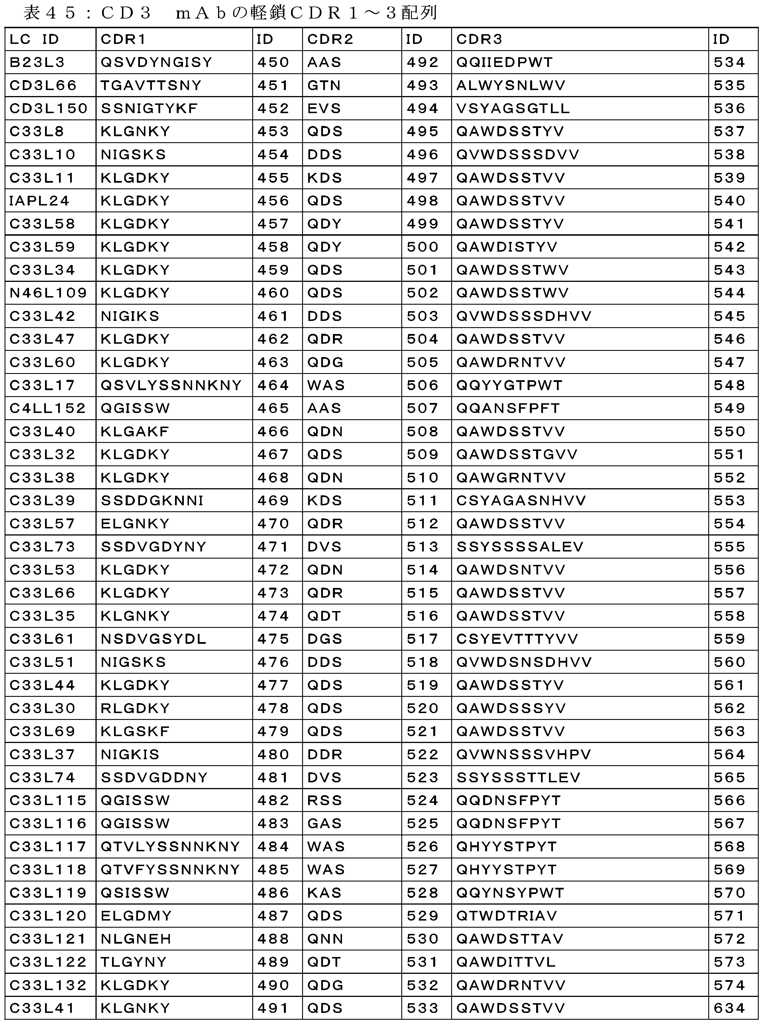
5-5 抗CD33 mAbの特性評価
免疫化を介して同定し、v領域クローニングし、続いて、発現及び精製したOMNIRat抗体を、CD33発現細胞への結合及び組換え抗原への結合について更に特性評価した。精製抗体を、ヒトCD33又はカニクイザルCD33(上記の通り生成)を発現している安定的にトランスフェクトされたHEK293F細胞への結合について、陰性対照としての親HEK293Fと共に評価した。非酵素的解離バッファ(Thermo Scientific)を使用して、細胞を組織培養フラスコから収集した。フラスコをPBSで2回すすぎ、解離バッファをフラスコに添加し、細胞が非接着性になるまでフラスコを37℃で10分間インキュベートした。細胞を300gで5分間遠心分離し、染色バッファ(Becton Dickinson;Franklin Lakes,NJ)に1.0×10⁶細胞/mLで再懸濁させた。50,000細胞/ウェルの各細胞型を、丸底プレート(Becton Dickinson)における染色バッファ50μLにプレーティングした。2×濃度の試験mAb又はアイソタイプ対照50μLを3つの希釈濃度及びゼロ(120nM、12nM、及び1.2nM、並びに0nM)で添加し、得られた溶液を4℃で30分インキュベートした。各プレートの全てのウェルに100μLの染色バッファを添加し、プレートを300gで5分間回転させ、バッファを除去し、各プレートの全てのウェルに200μLの染色バッファを添加し、プレートを300gで5分間回転させ、バッファを除去した。50μLの2μg/mLヤギ抗ヒトFc AF647二次抗体(Jackson Immunoresearch;West Grove,PA)をプレートの全てのウェルに添加し、プレートを4℃で30分間インキュベートした。プレートの全てのウェルに100μLの染色バッファを添加し、プレートを300gで5分間回転させ、バッファを除去した。200μLのランニングバッファ(ランニングバッファは、染色バッファ、1mM EDTA、0.1%プルロニック酸である)をプレートの全てのウェルに添加し、プレートを300gで5分間回転させ、バッファを除去した。Sytox Green live/dead色素(ThermoFisher)を含有するランニングバッファ30μLを細胞の入った全てのウェルに添加し、プレートをiQue IntelliCytフローサイトメータで読み取った。細胞を前方対側方散乱でゲーティングして細胞片を除去し、次いで、個片、次いで、Sytox染色を除外した生細胞でゲーティングした。AF647チャネルにおける平均蛍光強度によって抗体結合を評価した。
5-5 Characterization of anti-CD33 mAb The OMNIRat antibody identified through immunization, v-region cloned, and subsequently expressed and purified was further characterized for binding to CD33-expressing cells and for binding to recombinant antigen. The purified antibody was evaluated for binding to stably transfected HEK293F cells expressing human CD33 or cynomolgus CD33 (generated as described above), with parental HEK293F as a negative control. Cells were harvested from tissue culture flasks using non-enzymatic dissociation buffer (Thermo Scientific). The flasks were rinsed twice with PBS, dissociation buffer was added to the flasks, and the flasks were incubated at 37°C for 10 minutes until the cells were non-adherent. Cells were centrifuged at 300g for 5 minutes and resuspended at 1.0x10⁶ cells/mL in staining buffer (Becton Dickinson; Franklin Lakes, NJ). 50,000 cells/well of each cell type were plated in 50 μL of staining buffer in round bottom plates (Becton Dickinson). 50 μL of 2x concentration of test mAb or isotype control was added at three dilution concentrations and zero (120 nM, 12 nM, and 1.2 nM, and 0 nM) and the resulting solution was incubated at 4°C for 30 minutes. 100 μL of staining buffer was added to all wells of each plate, plates were spun at 300g for 5 minutes, buffer was removed, and 200 μL of staining buffer was added to all wells of each plate, plates were spun at 300g for 5 minutes, and buffer was removed. 50 μL of 2 μg/mL goat anti-human Fc AF647 secondary antibody (Jackson Immunoresearch; West Grove, PA) was added to all wells of the plate and the plate was incubated at 4° C. for 30 minutes. 100 μL of staining buffer was added to all wells of the plate, the plate was spun at 300 g for 5 minutes and the buffer was removed. 200 μL of running buffer (running buffer is staining buffer, 1 mM EDTA, 0.1% pluronic acid) was added to all wells of the plate, the plate was spun at 300 g for 5 minutes and the buffer was removed. 30 μL of running buffer containing Sytox Green live/dead dye (ThermoFisher) was added to all wells with cells and the plate was read on an iQue IntelliCyt flow cytometer. Cells were gated on forward vs. side scatter to exclude cell debris, then gated on individual cells, then live cells excluding Sytox staining. Antibody binding was assessed by mean fluorescence intensity in the AF647 channel.
精製mAbの生物物理的結合特性の評価を開始するために、オフレートスクリーニングを実施した。76個のOMNIRat抗CD33mAbを、組換えヒトCD33 ECD-HSA(C33W2)及びカニクイザルCD33 ECD-HSA(C33W1)タンパク質(Janssen production)への結合について試験し、IBIS MX96 SPRiアレイプラットフォーム(Carterra;Newton,PA)によってオフレートを測定した。IBIS機器において10分間の会合時間で、CMD50mセンサチップ(Xantec、ロットCMD50m0415.a)を使用し、酢酸バッファpH4.5中100μg/mLのアミンカップリングを介して、ヤギ抗ヒトFc IgG(Jackson Immunoresearch、カタログ番号109-005-098)を直接固定化した。約9000Rusの平均GAH-Fc固定化レベルが達成された。センサチップをContinuous Flow Microspotter(CFM)ユニットに移して、10分間10μg/mLで各抗CD33mAbを捕捉した。再生なしの単一サイクルの反応速度論により、結合をIBIS SPRiで測定した。各抗原濃度系列(3倍希釈系列で3μM)を低濃度(0.46nM)から高濃度(3μM)まで逐次注入して、ランニングバッファとしてPBST(0.005%Tweenを含むPBS)を使用して、5分間の会合時間及び15分間の解離時間で、捕捉されたmAbに結合させた。生の結合データ(.trixファイルフォーマット)を参照し、SprintXソフトウェア(Wasatch、バージョン1.9.3.2)を使用して整列させた後、1:1結合反応速度分析(Wasatch、バージョン2.0.0.33)のためにScrubberソフトウェア(バージョン2.0)にエクスポート(.ibmxファイルフォーマット)して、koff結果を抽出した。 To begin evaluating the biophysical binding properties of the purified mAbs, an off-rate screen was performed. Seventy-six OMNIRat anti-CD33 mAbs were tested for binding to recombinant human CD33 ECD-HSA (C33W2) and cynomolgus monkey CD33 ECD-HSA (C33W1) proteins (Janssen production) and off-rates were measured by an IBIS MX96 SPRi array platform (Carterra; Newton, PA). Goat anti-human Fc IgG (Jackson Immunoresearch, catalog number 109-005-098) was directly immobilized via amine coupling at 100 μg/mL in acetate buffer pH 4.5 using a CMD50m sensor chip (Xantec, lot CMD50m0415.a) with an association time of 10 minutes in an IBIS instrument. An average GAH-Fc immobilization level of approximately 9000 Rus was achieved. The sensor chip was transferred to a Continuous Flow Microspotter (CFM) unit to capture each anti-CD33 mAb at 10 μg/mL for 10 minutes. Binding was measured on an IBIS SPRi by single cycle kinetics without regeneration. Each antigen concentration series (3 μM in 3-fold dilutions) was injected sequentially from low (0.46 nM) to high (3 μM) and allowed to bind to the captured mAb with 5 min association and 15 min dissociation times using PBST (PBS with 0.005% Tween) as the running buffer. Raw binding data (.trix file format) were referenced and aligned using SprintX software (Wasatch, version 1.9.3.2) and then exported (.ibmx file format) to Scrubber software (version 2.0) for 1:1 binding kinetics analysis (Wasatch, version 2.0.0.33) to extract koff results.
以下の表46は、ヒト及びカニクイザルCD33発現細胞株への結合、並びに組換え抗原に対する結合(抗原のうちの1つについて少なくとも>10e-3のオフレート)によって評価したときの上位32のクローンをまとめたものである。これらの32個のうち、4個を除いた全てが、ヒト又はカニクイザル発現細胞のいずれかへの認識可能な結合を示した。32個全てを、エピトープビニング及び全反応速度分析を介した更なる特性評価に進めた。 Table 46 below summarizes the top 32 clones as assessed by binding to human and cynomolgus CD33 expressing cell lines, as well as binding to recombinant antigens (off-rates of at least >10e-3 for one of the antigens). Of these 32, all but four showed appreciable binding to either human or cynomolgus CD33 expressing cells. All 32 were taken forward for further characterization via epitope binning and overall kinetic analysis.


次いで、mAbのパネルを、完全親和性分析及びエピトープビニングについて更に特性評価した。抗CD33mAbの組換えヒトCD33 ECD-HSA(C33W2)及びカニクイザルCD33 ECD-HSA(C33W1)への結合を、ProteOn SPR(Bio-Rad)によって測定した。0.005% Tween-20を含有するPBS中流量30μL/分で、GLC Sensor Chip(Bio-Rad、カタログ番号176-5011)に垂直に配向された6つのリガンドチャネル全てにおける酢酸バッファpH5.0中30μg/mLでのアミンカップリングを介して、ヤギ抗ヒトFc IgG(Jackson Immunoresearch、カタログ番号109-005-098)を直接固定化した。固定化密度は、平均で約5000応答単位(RU)であり、異なるチャネル間の変動は5%未満であった。垂直リガンド配向で0.25又は0.5μg/mL(160~300RU)において抗ヒトFc IgG表面に異なるmAbを捕捉し、6番目のリガンドチャネルは、リガンドなし表面対照であった。5つの濃度の3倍希釈系列中0.3μM濃度のヒト及びカニクイザルCD33-HSAタンパク質をアナライトとし流して、捕捉されたmAbに水平配向で結合させた。また、捕捉されたmAbの解離及びベースラインでの安定性をモニタリングするために、6番目のチャネルにバッファサンプルを注入した。ヒト及びカニクイザルCD33-HSAの全ての濃度の解離相を、C33B782への結合については15分間、C33B912(hIgG4を有するC33B904と同一)への結合については60分間、流量100μL/分でモニタリングした後、0.85%リン酸の18秒間パルスを使用して再生して、抗原及び結合したmAbを除去した。以下から応答データを減じた後に二重参照することによって生データを処理した:1)Agと空のチップ表面との間で非特異的相互作用を補正するためのインタースポット;2)経時的な捕捉されたmAb表面の解離に起因するベースラインの揺れを補正するためのバッファチャネル。各mAbについて全抗原濃度において処理したデータを1:1の単純なラングミュア結合モデルにグローバルフィットさせて、反応速度(kon、koff)及び親和性(Kd)定数の推定値を抽出した。 The panel of mAbs was then further characterized for complete affinity analysis and epitope binning. Binding of anti-CD33 mAbs to recombinant human CD33 ECD-HSA (C33W2) and cynomolgus monkey CD33 ECD-HSA (C33W1) was measured by ProteOn SPR (Bio-Rad). Goat anti-human Fc IgG (Jackson Immunoresearch, Cat. No. 109-005-098) was directly immobilized via amine coupling at 30 μg/mL in acetate buffer pH 5.0 in all six ligand channels oriented perpendicularly to a GLC Sensor Chip (Bio-Rad, Cat. No. 176-5011) at a flow rate of 30 μL/min in PBS containing 0.005% Tween-20. The immobilization density was about 5000 response units (RU) on average, with less than 5% variation between different channels. Different mAbs were captured on anti-human Fc IgG surfaces at 0.25 or 0.5 μg/mL (160-300 RU) in a vertical ligand orientation, and the sixth ligand channel was a no-ligand surface control. Human and cynomolgus CD33-HSA proteins at 0.3 μM concentration in a 3-fold dilution series of five concentrations were run as analytes to bind to the captured mAbs in a horizontal orientation. Buffer samples were also injected into the sixth channel to monitor the dissociation and baseline stability of the captured mAbs. The dissociation phase of all concentrations of human and cynomolgus CD33-HSA was monitored for 15 min for binding to C33B782 and 60 min for binding to C33B912 (identical to C33B904 with hIgG4) at a flow rate of 100 μL/min, followed by regeneration using an 18 s pulse of 0.85% phosphoric acid to remove antigen and bound mAb. Raw data was processed by double referencing after subtracting response data from: 1) interspots to correct for non-specific interactions between Ag and the empty chip surface; 2) buffer channels to correct for baseline drift due to dissociation of captured mAb surface over time. Processed data at all antigen concentrations for each mAb were globally fitted to a 1:1 simple Langmuir binding model to extract estimates of kinetic (k on , k off ) and affinity (K d ) constants.
mAbのパネルが全て1つの個別のエピトープに結合するか、又は広いエピトープをカバーするかを判定するために、エピトープビニング実験を行った。CMD-200Mセンサプリズムチップを使用して、IBIS SPRi機器(Carterra)において、CD33mAbの競合的エピトープビニングを行った。別々のContinuous Flow Microspotter(CFM)を使用して、酢酸バッファ(pH4.5)中10μg/mLで、アミンカップリングを介してチップに各抗CD33抗体を直接固定化した。次いで、プリントされたセンサチップを、Classical又は「Sandwich」ビニングフォーマットを使用してビニング分析のためにIBIS機器に移した。50nMのヒトCD33 ECD-HSA(C33W2)を逐次注入し、続いて、133nMの溶液で競合アナライトとして抗CD33 mAbを1回注入して、抗原及び抗体の各逐次注入サイクル後に表面再生を伴って固定化された抗CD33mAに結合させることによって、ビニングを行った。 Epitope binning experiments were performed to determine whether the panel of mAbs all bind one individual epitope or cover a broad epitope. Competitive epitope binning of CD33 mAbs was performed on an IBIS SPRi instrument (Carterra) using a CMD-200M sensor prism chip. Each anti-CD33 antibody was directly immobilized on the chip via amine coupling at 10 μg/mL in acetate buffer (pH 4.5) using a separate Continuous Flow Microspotter (CFM). The printed sensor chips were then transferred to the IBIS instrument for binning analysis using either the Classical or "Sandwich" binning format. Binning was performed by sequential injections of 50 nM human CD33 ECD-HSA (C33W2), followed by one injection of anti-CD33 mAb as a competing analyte in a 133 nM solution to bind to the immobilized anti-CD33 mA with surface regeneration after each sequential injection cycle of antigen and antibody.
再生前及び再生後の固定化されたmAbの活性をモニタリングするために、実験の開始時及び終了時に、競合mAbを全く含まないバッファ注入を行って、抗原のみの結合活性を測定した。バッファ(抗原のみ)結合に対する競合mAb結合の応答は、溶液中の抗体が固定化されたmAbへの抗原結合をブロック又はサンドイッチするかどうかの指標である。生のビニングデータ(.trixファイルフォーマット)を参照し、SprintXソフトウェア(Wasatch、バージョン1.9.3.2)を使用してゼロにした後、分析のためにビニングソフトウェアHtTools.exe(Wasatch、バージョン2.0.0.33)にエクスポート(.ibmxファイルフォーマット)した。20RU未満の抗原応答を有する抗体及び自己ブロックしなかった抗体を除去することにより、データを精選した。競合mAb応答を、抗原のみの結合応答に対して正規化した。正規化された応答が<0.25である抗体はブロッカーを意味し、正規化された応答が≧0.25である抗体は、非ブロッカー/サンドイッチャーを意味した。組み合わされたデンドログラムプロットにおいて高さ2.5におけるカットを使用して、異なるビンを予測した。 To monitor the activity of the immobilized mAb before and after regeneration, buffer injections without any competing mAb were performed at the beginning and end of the experiment to measure antigen-only binding activity. The response of competing mAb binding to buffer (antigen-only) binding is an indication of whether the antibody in solution blocks or sandwiches antigen binding to the immobilized mAb. Raw binned data (.trix file format) were referenced and zeroed using SprintX software (Wasatch, version 1.9.3.2) and then exported (.ibmx file format) to the binning software HtTools.exe (Wasatch, version 2.0.0.33) for analysis. Data were curated by removing antibodies with antigen responses below 20 RU and antibodies that did not self-block. Competitive mAb responses were normalized to antigen-only binding responses. Antibodies with a normalized response <0.25 were designated blockers and antibodies with a normalized response ≥0.25 were designated non-blockers/sandwichers. A cut at height 2.5 in the combined dendrogram plot was used to predict distinct bins.
以下の表は、32個の選択mAbの全反応速度分析及びエピトープビニングをまとめたものである。ヒト及びカニクイザルCD33の両方に対してナノモル濃度以下の親和性を有する抗CD33mAbは合計8個存在し、これらのmAbは3つの別個のエピトープビンに対応し、一方、より大きなパネルは、様々な親和性及び7個の別個のエピトープビンを有する。 The following table summarizes the full kinetic analysis and epitope binning of the 32 selected mAbs. There are a total of eight anti-CD33 mAbs with subnanomolar affinity for both human and cynomolgus CD33, which correspond to three distinct epitope bins, while the larger panel has a range of affinities and seven distinct epitope bins.


OmniMouseパネル(合計8個のmAb)を別々に生成し、細胞への結合について更に特性評価した。細胞結合を上記のように行い、以下の表にまとめた。試験した8個のmAbのうち、6個はCD33発現細胞に直接結合したが、2個はしなかった。 The OmniMouse panel (8 mAbs total) was generated separately and further characterized for binding to cells. Cell binding was performed as described above and is summarized in the table below. Of the 8 mAbs tested, 6 bound directly to CD33 expressing cells, whereas 2 did not.


細胞上でCD33に結合している6つのmAbを、上記の方法を使用して組換え抗原に対する全反応速度分析を介して生物物理学的に更に特性評価し、以下の表にまとめた。試験した6つのmAbのうち、1つは、ピコモル濃度の親和性でヒトCD33に(C33B912)、及び、カニクイザルCD33に対してはナノモル濃度以下で結合し、一方、1つは、ヒトCD33に対しては非常に強い親和性を有していたが、カニクイザルCD33(C33B911)に対してはわずかナノモル濃度の親和性しか有していなかった。更に2つのクローンは、ヒト及びカニクイザルCD33(C33B913及びC33B916)の両方に対してナノモル濃度以下であったが、いずれも親和性はC33B912の範囲ではなかった。 Six mAbs binding to CD33 on cells were further biophysically characterized via total kinetic analysis against recombinant antigen using the methods described above and are summarized in the table below. Of the six mAbs tested, one bound with picomolar affinity to human CD33 (C33B912) and subnanomolar affinity to cynomolgus CD33, while one had very strong affinity to human CD33 but only nanomolar affinity to cynomolgus CD33 (C33B911). Two additional clones were subnanomolar to both human and cynomolgus CD33 (C33B913 and C33B916), but neither had affinities in the range of C33B912.

先のOMNIRatキャンペーンで既に同定されたいくつかの対照mAに加えて、OMNIMouse由来の6つの細胞結合mAbについてもエピトープビニング実験を実施した。ヒトCD33に対するそのナノモル濃度以下の親和性及び異なるエピトープビンの数に基づいて、対照mAbを選択した。ビニングソフトウェアHtToolsは、実験ごとにエピトープビン数を割り当てるので、既に定義されたエピトープビンに対するいくつかの対照を有することが、交差比較のために重要であった。2つのOMNIMouse由来のヒトCD33高親和性クローン(C33B911及びC33B912)は両方とも、上記のビン4(この実験におけるビン4)からのクローンにビニングしたが、ナノモル濃度以下のクローン(C33B916)は、C33B836(上記実験におけるビン1)と共にこの実験における2にビニングした。
In addition to some control mAs already identified in the previous OMNIRat campaign, epitope binning experiments were also performed for six cell-binding mAbs from OMNIMouse. Control mAbs were selected based on their subnanomolar affinity for human CD33 and the number of different epitope bins. Since the binning software HtTools assigns epitope bin numbers per experiment, it was important for cross-comparison to have some controls for the epitope bins already defined. Both of the two OMNIMouse-derived human CD33 high affinity clones (C33B911 and C33B912) were binned into clones from

CD33は、2つのIgGドメイン、膜遠位Vドメイン、及び膜近位C2ドメインで構成される。SNPrs12459419は、CD33プレmRNA転写物の選択的オルタナティブスプライシングを引き起こして、C2のみの形態を細胞上で発現させ得るので、このドメインを標的とすることにより、臨床的有益性を与えることができる。2つのドメインのうちのいずれにmAbが結合可能であったかを確認するために、結合抗原としてヒトCD33 ECD-HSA、ヒトCD33 V-HSA、及びヒトCD33 C2-HSAを使用して、4つの異なるエピトープビンをカバーする、最高結合能力を有する6つのmAbに対して上記プロトコルに従ってオフレートスクリーニングを行った。以下の表に示すように、ビン4に予め分類された2つのクローンは、両方ともhuCD33 C2ドメインに結合したが、huCD33 Vドメインには結合せず、一方、ビン2及び3のクローンは、Vドメインには結合したが、C2ドメインには結合しなかった。ビン5に分類された2つのクローンは、いずれのドメインにも結合しなかったため、その正確な結合位置は、2つのドメインにまたがっている可能性がある。3つの市販のmAbをこの実験に含めたところ(WM53(EMD Millipore;Darmstadt,Germany)、P67.7(Biolegend,San Diego,CA)、及びLSBioクローン906(LifeSpan Biosciences,Seattle,WA)))、全てVドメインへの結合は示したが、C2ドメインへの結合は示さなかった。表51のC2ドメイン結合データに関連して表42及び45のエピトープビンを見ると、ヒト完全長タンパク質に対して約25nMから約30pMの親和性の範囲でC2ドメインに潜在的に結合し得るmAbが合計15個存在する。
CD33 is composed of two IgG domains, a membrane-distal V domain, and a membrane-proximal C2 domain. Targeting this domain may confer clinical benefit, as SNP rs12459419 may cause alternative splicing of the CD33 pre-mRNA transcript to express a C2-only form on cells. To confirm which of the two domains the mAbs were able to bind, off-rate screening was performed according to the above protocol for six mAbs with the highest binding capacity, covering four different epitope bins, using human CD33 ECD-HSA, human CD33 V-HSA, and human CD33 C2-HSA as binding antigens. As shown in the table below, two clones presorted in

更にインビボ及びインビトロ試験を裏付けるために、スケールアップ及びfabアーム交換のために選択クローン(C33B836、C33B782、C33B778、C33B904、C33B806、C33B830、C33B937、C33B792、C33B760、及びC33B777)を選択して、抗CD3抗体を含む二重特異性分子を生成した。ExpiCHO-S(商標)細胞(ThermoFisher Scientific)を、1.25×10⁵~2.25×10⁵生存細胞/mLでExpiCHO(商標)発現培地に播種し、37℃、7%CO2のシェーカーインキュベータ(INFORS HT Multitron Pro)においてポリカーボネート製、使い捨て、滅菌、排気、バッフルなし三角振盪フラスコ内で培養した。125mL~2Lの振盪フラスコにおけるルーチンな細胞増殖の場合、振盪直径19mmの振盪器については振盪速度を130rpmに設定した。細胞を継代培養し、この場合、生存率98~99%で、密度が4×10⁶~6×10⁶生存細胞/mLで対数増殖に達した。 To further support in vivo and in vitro testing, selected clones (C33B836, C33B782, C33B778, C33B904, C33B806, C33B830, C33B937, C33B792, C33B760, and C33B777) were chosen for scale-up and fab arm exchange to generate bispecific molecules containing anti-CD3 antibodies. ExpiCHO-S™ cells (ThermoFisher Scientific) were seeded in ExpiCHO™ Expression Medium at 1.25×10 5 -2.25×10 5 viable cells/mL and cultured in polycarbonate, disposable, sterile, vented, non-baffled Erlenmeyer shake flasks in a shaker incubator (INFORS HT Multitron Pro) at 37°C and 7% CO 2. For routine cell growth in 125 mL-2 L shake flasks, the shaking speed was set at 130 rpm for a shaker with a shaking diameter of 19 mm. Cells were subcultured to reach logarithmic growth at densities of 4×10 6 -6×10 6 viable cells/mL with 98-99% viability.
トランスフェクションの2日前に、必要とされる培養体積に対して1.5×10⁶生存細胞/mLでExpiCHO-S(商標)細胞を播種した。トランスフェクションの日に、生存細胞密度及び生存率を求めた。細胞を、6×106生存細胞/mLの密度でトランスフェクトした。最適なトランスフェクションのために、TEバッファ(10mM Tris-HCl、1mM EDTA、pH8.0)中≧1mg/mL濃度の滅菌重鎖及び軽鎖プラスミドDNAを使用した。
ExpiCHO-S™ cells were seeded at 1.5x10⁶ viable cells/mL into the required
製造元のMax Titerトランスフェクションプロトコル(ThermoFisher Publication Number MAN0014337)に従って、ExpiCHO-S(商標)細胞をトランスフェクトした。以下に示す全ての量及び体積は、最終的なトランスフェクトされた培養体積1mLあたりであった。簡潔に述べると、プラスミドDNAを、以下の比で0.04mLの冷OptiPRO(商標)培地(ThermoFisher Scientific)で希釈した:0.125gの重鎖DNA:0.375gの軽鎖DNA:0.5gのpAdvantage。6.4μLのExpiFectamine(商標)CHOトランスフェクション試薬を希釈し、0.04mLの冷OptiPRO(商標)培地と穏やかに混合し、1分間インキュベートした。希釈したExpiFectamine(商標)CHO試薬を希釈したDNAに添加し、穏やかに混合し、ExpiFectamine(商標)CHO/プラスミドDNA複合体を室温で5分間インキュベートした。インキュベーション後、複合体をシェーカーフラスコ内のExpiCHO-S(商標)細胞に添加し、37℃、7%CO2のシェーカーインキュベータ内で一晩インキュベートした。 ExpiCHO-S™ cells were transfected according to the manufacturer's Max Titer transfection protocol (ThermoFisher Publication Number MAN0014337). All amounts and volumes shown below were per mL of final transfected culture volume. Briefly, plasmid DNA was diluted in 0.04 mL of cold OptiPRO™ medium (ThermoFisher Scientific) in the following ratio: 0.125 g heavy chain DNA: 0.375 g light chain DNA: 0.5 g pAdvantage. 6.4 μL of ExpiFectamine™ CHO transfection reagent was diluted and gently mixed with 0.04 mL of cold OptiPRO™ medium and incubated for 1 min. The diluted ExpiFectamine™ CHO Reagent was added to the diluted DNA, mixed gently, and the ExpiFectamine™ CHO/plasmid DNA complexes were incubated for 5 minutes at room temperature. After incubation, the complexes were added to ExpiCHO-S™ cells in a shaker flask and incubated overnight in a shaker incubator at 37° C., 7% CO2 .
Max Titerプロトコルのために、トランスフェクション後1日目に、6μLのExpiFectamine(商標)CHO Enhancer及び160μLのExpiCHO(商標)Feedを添加し、フラスコを32℃、7%CO2のシェーカーインキュベータに移した。トランスフェクション後5日目に、2回目の160μLのExpiCHO(商標)Feedをフラスコに添加し、振盪しながら32℃のインキュベータに戻した。トランスフェクション後12日目に培養物を収集し、5000rpmで15分間遠心分離し、0.2μm Acropak 1500フィルターカプセル(Pall)を通して清澄化した。
For the Max Titer protocol, one day post-transfection, 6 μL of ExpiFectamine™ CHO Enhancer and 160 μL of ExpiCHO™ Feed were added and the flask was transferred to a 32° C., 7% CO2 shaker incubator. Five days post-transfection, a second 160 μL of ExpiCHO™ Feed was added to the flask and returned to the 32° C. incubator with shaking. Twelve days post-transfection, the culture was harvested, centrifuged at 5000 rpm for 15 minutes, and clarified through a 0.2
MabSelect SuRe(GE Healthcare)を使用して、発現した抗体を清澄化した上清から精製した。MabSelect SuRe Protein Aカラムを、1×D-PBS、pH7.2で平衡化した後、個々の培養上清をロードした。未結合タンパク質を、1×D-PBS、pH7.2で広範囲にわたって洗浄することによって除去した。結合タンパク質を0.1Mの酢酸ナトリウム、pH3.5で溶出した。ピーク画分を2.5M Tris(pH7.2)で中和し、プールした。中和された画分プールを、アッセイ及び生物物理的特性評価のために1×dPBSに透析した、又は二重特異性アセンブリに利用した。 Expressed antibodies were purified from clarified supernatants using MabSelect SuRe (GE Healthcare). A MabSelect SuRe Protein A column was equilibrated with 1x D-PBS, pH 7.2, before loading with the individual culture supernatants. Unbound proteins were removed by extensive washing with 1x D-PBS, pH 7.2. Bound proteins were eluted with 0.1 M sodium acetate, pH 3.5. Peak fractions were neutralized with 2.5 M Tris, pH 7.2, and pooled. The neutralized fraction pool was dialyzed into 1x dPBS for assay and biophysical characterization, or utilized for bispecific assembly.
各溶出プールのタンパク質濃度をOD280nmでの吸光度を測定することによって求め、アミノ酸配列に基づいて吸光度、吸光係数を用いて計算した。 The protein concentration of each elution pool was determined by measuring the absorbance at OD 280 nm and calculated using the absorbance and extinction coefficient based on the amino acid sequence.
6 CD33×CD3二重特異性抗体の調製及び機能評価
6-1 精製された親mAbを使用したFabアーム交換
CD33×CD3二重特異性抗体の形成は、2つの親mAb、ターゲティングアームに特異的なもの(例えば、CD33)及びエフェクターアームに特異的なもの(例えばCD3)を必要とする。CD33mAbを高親和性(CD3B376:配列番号652のVH及び配列番号661のVL)又は低親和性CD3アーム(CD3B450:配列番号657のVH及び配列番号678のVL)アームで組換えた。これらの親mAb(CD33及びCD3アーム)は、ターゲティング親(CD33)が409R Genmab変異(IgG4のネイティブアミノ酸)を含有し、一方、殺傷親(CD3)はF405L変異及びR409Kを含有する、IgG4 PAAフォーマット(Labrijn et al,2013)である。単一特異性抗CD3抗体を、そのFc領域にFc置換S228P、F234A、L235A、F405L及びR409K(CD3アーム)(EUインデックスに従って付番)を有するIgG4として発現させた。単一特異性抗体を、上記のように発現させ、精製した。精製後、親CD33抗体を、75mM 2-MEA(2-メルカプトエチルアミン)中の還元条件下で所望の親CD3抗体と混合し、31℃で5時間又は室温で一晩インキュベートした。組換え反応は、モル濃度比に基づいており、設定量のCD33抗体(例えば、10mg又は約74.6ナノモル)をCD3抗体(例えば、約67.8ナノモル)と組み合わせ、CD33抗体をCD3抗体の6%過剰量で添加した。CD33抗体原液の濃度は、0.8~6mg/mLで変化し、組換え反応の体積は、各対形成で変化した。その後、組換え反応物をPBSに対して一晩透析して、還元剤を除去した。組換え後に残存する未反応のCD3親抗体の量を最小化するために、過剰のCD33抗体(比)を用いてCD33×CD3二重特異性抗体反応を行った。
6 Preparation and Functional Evaluation of CD33xCD3 Bispecific Antibodies 6-1 Fab Arm Exchange Using Purified Parental mAbs The formation of CD33xCD3 bispecific antibodies requires two parental mAbs, one specific for the targeting arm (e.g., CD33) and one specific for the effector arm (e.g., CD3). CD33 mAb was recombined with either a high affinity (CD3B376: VH of SEQ ID NO:652 and VL of SEQ ID NO:661) or low affinity CD3 arm (CD3B450: VH of SEQ ID NO:657 and VL of SEQ ID NO:678) arm. These parental mAbs (CD33 and CD3 arm) are in IgG4 PAA format (Labrijn et al, 2013) where the targeting parent (CD33) contains the 409R Genmab mutation (native amino acid of IgG4) while the killing parent (CD3) contains the F405L mutation and R409K. Monospecific anti-CD3 antibodies were expressed as IgG4 with Fc substitutions S228P, F234A, L235A, F405L and R409K (CD3 arm) (numbered according to the EU index) in their Fc region. Monospecific antibodies were expressed and purified as described above. After purification, the parental CD33 antibodies were mixed with the desired parental CD3 antibody under reducing conditions in 75 mM 2-MEA (2-mercaptoethylamine) and incubated at 31° C. for 5 hours or at room temperature overnight. The recombination reactions were based on a molar ratio, combining a set amount of CD33 antibody (e.g., 10 mg or about 74.6 nmoles) with CD3 antibody (e.g., about 67.8 nmoles), with the CD33 antibody added at a 6% excess of CD3 antibody. The concentration of the CD33 antibody stock solution varied from 0.8 to 6 mg/mL, and the volume of the recombination reaction was varied for each pairing. The recombination reactions were then dialyzed overnight against PBS to remove the reducing agent. To minimize the amount of unreacted CD3 parent antibody remaining after recombination, the CD33xCD3 bispecific antibody reaction was performed with an excess of CD33 antibody (ratio).
組換え反応で使用した親mAb(すなわち、CD33、CD3、又はヌル)と共に、産生された最終的なCD33×CD3二重特異性抗体を表47に列挙する。 The final CD33xCD3 bispecific antibodies produced are listed in Table 47, along with the parental mAbs (i.e., CD33, CD3, or null) used in the recombination reactions.
選択されたCD33ヒットはまた、非殺傷アーム(ヌル)と対形成して、試験目的のための陰性対照が生じた。対照二重特異性抗体については、B2M1、IgG4 PAAフォーマットにおけるRSV抗体を生成し、精製し、CD3アームCD3B219又はCD3B376-F405L、R409Kのいずれかと組み合わせて、それぞれ、CD3B288(CD3×ヌル)及びCD3B510(CD3B376×ヌル)を生成した。CD33アームをB23B49と組み合わせて、表52のようにCD33×ヌルを生成した。 Selected CD33 hits were also paired with a non-killing arm (null) to generate negative controls for testing purposes. For control bispecific antibodies, B2M1, an RSV antibody in IgG4 PAA format, was generated, purified, and combined with either CD3 arms CD3B219 or CD3B376-F405L, R409K to generate CD3B288 (CD3x null) and CD3B510 (CD3B376x null), respectively. The CD33 arm was combined with B23B49 to generate CD33x null as per Table 52.

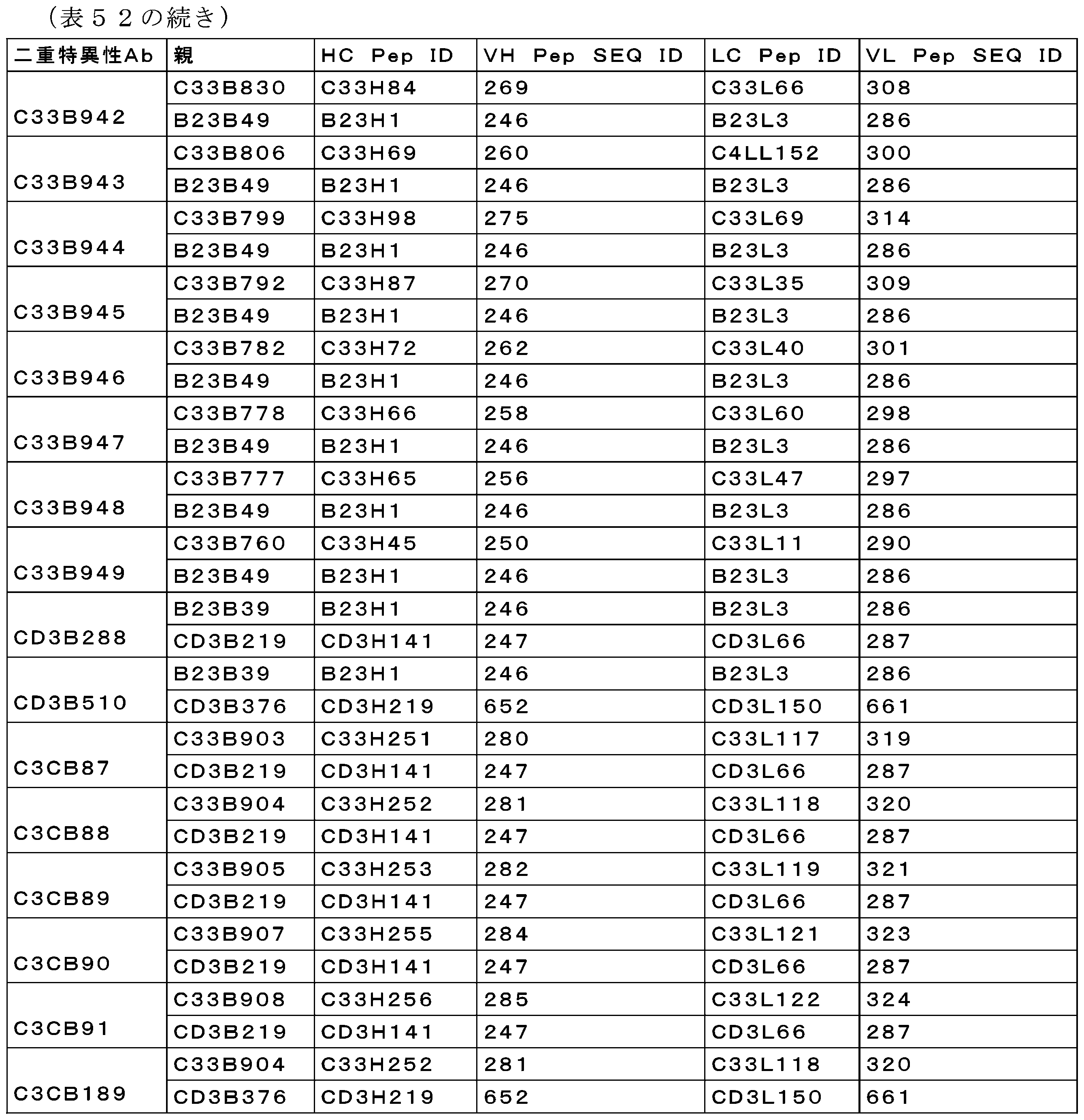
6-2 AML一次サンプルにおけるAML芽球及びT細胞の活性化の、エクスビボにおけるCD33×CD3によって媒介される減少
CD33×CD3二重特異性抗体の細胞傷害能を更に評価するために、上位4個の抗体を使用して、AML患者全血を使用するエクスビボ細胞傷害アッセイを行った(図62)。このアッセイは患者の血液中の自家T細胞の存在に依存するので、このアッセイでは、追加のT細胞を供給することなく48時間にわたってAML患者由来の希釈された全血に様々な二重特異性抗体(CD3アームCD3B219及びCD3B376のいずれかと対形成したCD33抗体)を添加した。48時間目に、サンプルをCD3 PerCPCy5.5、CD25 PE、CD33 FITC、及びCD38 APCで染色した(全ての抗体はBiolegend;San Diego,CAから購入した)。次いで、サンプルを1×Lyse RBC Lysis Buffer(eBioscience)で少なくとも3回洗浄した。次いで、サンプルを、LIVE/DEAD(登録商標)Fixable Near-IR Dead Cell Stainバッファ(Life Technologies)で染色した。まず、二重特異性抗体の存在下で、AML患者癌細胞の画分(CD3-CD38+細胞として定義)中の生CD33+細胞を定量することによって、腫瘍細胞傷害の程度を求めた。以下の等式を使用して、PBS/未処理対照に対するパーセントとして細胞傷害を計算した:(PBS/未処理対照中のCD33+%-処理サンプル中のCD33+%)/(PBS/未処理対照中のCD33+%)。CD3+画分中のCD25+事象のパーセントとしてT細胞活性化を計算した。
6-2 Ex vivo CD33xCD3-mediated reduction of AML blasts and T cell activation in AML primary samples To further evaluate the cytotoxic potential of CD33xCD3 bispecific antibodies, an ex vivo cytotoxicity assay using AML patient whole blood was performed using the top four antibodies (Figure 62). Since this assay relies on the presence of autologous T cells in the patient's blood, in this assay, various bispecific antibodies (CD33 antibodies paired with either of the CD3 arms CD3B219 and CD3B376) were added to diluted whole blood from AML patients for 48 hours without providing additional T cells. At 48 hours, samples were stained with CD3 PerCPCy5.5, CD25 PE, CD33 FITC, and CD38 APC (all antibodies were purchased from Biolegend; San Diego, CA). Samples were then washed at least three times with 1x Lyse RBC Lysis Buffer (eBioscience). Samples were then stained with LIVE/DEAD® Fixable Near-IR Dead Cell Stain buffer (Life Technologies). The extent of tumor cytotoxicity was determined by first quantifying live CD33 + cells in a fraction of AML patient cancer cells (defined as CD3 − CD38 + cells) in the presence of the bispecific antibody. Cytotoxicity was calculated as a percent of PBS/untreated control using the following equation: (% CD33 + in PBS/untreated control−% CD33 + in treated sample)/(% CD33 + in PBS/untreated control). T cell activation was calculated as a percent of CD25 + events in the CD3 + fraction.
図62に示すように、CD3アーム(CD3B376及びCD3B219)のいずれかと対形成した全てのCD33リード抗体は、48時間後、T細胞の活性化と相関する総細胞傷害の用量依存的な減少を促進した。ヌルアーム対照抗体(ヌル×CD3B219及びヌル×CD3B376)は、腫瘍細胞傷害もT細胞活性化も示さなかった。この結果も、CD33×CD3二重特異性抗体が自家状況において機能することを証明する。これらの結果は、4つの他のAMLドナーサンプルの代表的なものである(データは示さない)。表53は、CD33×CD3多重特異性抗体で生成されたEC50値をまとめたものである。EC50値から分かるように、C3アーム(C3CB88、C3CB189)のいずれかと対形成したC33B904、及びCD3アーム(C3CB7、C3CB97)のいずれかと対形成したC33B836が、最も強力かつ有効な抗体であった。したがって、更なる特性評価ではこれらの4つの抗体に焦点をあてた。 As shown in FIG. 62, all CD33 lead antibodies paired with either CD3 arm (CD3B376 and CD3B219) promoted a dose-dependent decrease in total cytotoxicity after 48 hours that correlated with T cell activation. Null arm control antibodies (Null×CD3B219 and Null×CD3B376) showed no tumor cell cytotoxicity or T cell activation. This result again demonstrates that CD33×CD3 bispecific antibodies work in an autologous setting. These results are representative of four other AML donor samples (data not shown). Table 53 summarizes the EC 50 values generated with CD33×CD3 multispecific antibodies. As can be seen from the EC 50 values, C33B904 paired with either C3 arm (C3CB88, C3CB189) and C33B836 paired with either CD3 arm (C3CB7, C3CB97) were the most potent and effective antibodies. Therefore, further characterization was focused on these four antibodies.

6-3 CD33×CD3二重特異性抗体がCD33のC2ドメインに結合し、CD33一塩基多型(SNP)発現細胞株の細胞傷害を誘導することの証明。
CD33×CD3二重特異性抗体を用いたインビトロT細胞媒介細胞傷害アッセイ
最近の研究によって、AML集団の約50%に一塩基多型型(SNP)rs12459419が存在し、CD33のエクソン2がスキップされ、その結果、CD33のVドメインが欠失することが示された。この研究はまた、CD33のVドメインに結合するMylotargがSNPを発現する患者において有効性を有さなかったので、再発リスクが低下し、AML集団の約50%において生存率が改善されたことも示した(Lamba et al 2017,JCO,CD33 Splicing Polymorphism Determines Gemtuzumab Ozogamicin Response in De Novo Acute Myeloid Leukemia:Report From Randomized Phase III Children’s Oncology Group Trial AAML0531)。上述の研究におけるMylotargに関するデータに鑑みて、インビトロT細胞媒介性細胞傷害アッセイを行って、CD3アーム(CD3B219又はCD3B376)と対形成したCD33ヒット(V結合C33B836対C2結合C33B904)が、SNPrs12459419発現細胞株の殺傷を媒介するかどうかを評価した。簡潔に述べると、エフェクター細胞(Biological Specialityから購入した汎T細胞)を収集し、計数し、洗浄し、10%FBS(Invitrogen)を含むRPMI(Invitrogen)細胞培地中1×10⁶細胞/mLになるように再懸濁させた。標的細胞(KG1、SH2、及びOCIAML3)をCFSE(Invitrogen)で標識し、10%FBSを含むRPMI中2×105細胞/mLになるように再懸濁させた。KG1、SH2及びOCIAML3は、それぞれ、CD33 SNPrs12459419変異の野生型、ヘテロ接合型、及びホモ接合型を表すように選択した。エフェクター細胞及びCFSE標識標的細胞を、滅菌96ウェル丸底プレートにおいてエフェクター:標的比(E:T)=5:1で混合した。10μLのFcブロック(ReoPro Fc断片)を、5μLの二重特異性抗体のアリコートと共に、様々な濃度を含有する各ウェルに添加した。培養物を、5%CO2下、37℃で48時間インキュベートした。48時間後、LIVE/DEAD(登録商標)Fixable Near-IR Dead Cell Stainバッファ(Life technologies)をサンプルに添加し、培養物を、RTで暗所において20分間インキュベートし、洗浄し、100~200μLのFACバッファに再懸濁させた。CANTOIIフローサイトメータ(BD Biosciences)を使用して、薬剤誘導細胞傷害を決定し、FlowJoソフトウェア又はDiveソフトウェア(BD Biosciences)により分析した。対象となる集団は、二重陽性CFSE+/live/dead+細胞である。図63に示すように、ヌルアーム対照(ヌル×CD3B219及びヌル×CD3B376)とは異なり、V結合及びC2結合CD33×CD3多重特異性抗体は、48時間でSNPrs12459419変異細胞株KG1についてCD33+野生型のT細胞リダイレクトによる細胞傷害を誘導した。対照的に、VバインダーC33B836(C3CB97、C3CB7)とは異なり、C2結合C33B904と対になった二重特異性抗体(C3CB189、C3CB88)のみが、それぞれrs12459419 SNP変異についてヘテロ接合型又はホモ接合性であったSH2及びOCIAML3細胞株の細胞傷害を媒介した。この理由から、C33B904と対形成した二重特異性抗体(C3CB189、C3CB88)を、更なる分析及び特性評価に進めた。まとめると、これらのデータは、C33B904と対形成した二重特異性抗体などのCD33 C2結合二重特異性抗体が、V結合競合因子抗CD33抗体よりも広範なAML患者群において有効性を示す能力を有することを示唆している。
6-3 Demonstration that CD33×CD3 bispecific antibody binds to the C2 domain of CD33 and induces cytotoxicity in CD33 single nucleotide polymorphism (SNP)-expressing cell lines.
In Vitro T Cell-Mediated Cytotoxicity Assay Using CD33xCD3 Bispecific Antibody Recent studies have demonstrated that approximately 50% of AML populations harbor the single nucleotide polymorphism (SNP) rs12459419, which results in skipping of
6-4 エクスビボでCD33×CD3は、エクスビボ全血MOLM-13細胞傷害アッセイにおいてMOLM-13及び単球におけるスパイクの低減を媒介した。
MOLM-13細胞及び正常ヒト単球におけるスパイクの排除におけるCD33×CD3二重特異性抗体の細胞傷害能を評価するために、外因的に添加されたCD33+AML細胞株MOLM-13と共に正常健常ヒト全血を使用するエクスビボ細胞傷害アッセイを利用した。このアッセイはドナーの血液中の自家T細胞の存在に依存するので、上記実験と同様に、追加のT細胞を供給することなく48時間にわたって6人の異なる正常ヒトドナー由来の希釈された全血に様々な二重特異性抗体(CD3アームCD3B219及びCD3B376のいずれかと対形成したCD33抗体)を添加した。希釈前に、各ドナーの血液中のT細胞の濃度を調べた。次いで、AML患者サンプルにおけるエフェクター:標的比を再現するためにエフェクター:標的比(E:T)が1:5になるように、血液をCFSE(Invitrogen)標識MOLM-13細胞で希釈した。48時間目に、サンプルをCD3 PerCPCy5.5、CD25 PE、CD33 FITC、及びCD14 Pacific Blueで染色した(全ての抗体はBiolegendから購入した)。次いで、サンプルを1×Lyse RBC Lysis Buffer(eBioscience)で少なくとも3回洗浄した。次いで、サンプルを、LIVE/DEAD(登録商標)Fixable Near-IR Dead Cell Stainバッファ(Life Technologies)で染色した。まず、二重特異性抗体の存在下で、CD14+単球の画分中の生CD33+細胞を定量することによって、腫瘍細胞傷害の程度を求めた。死CFSE+細胞のパーセントを調べることにより、MOLM-13細胞の細胞傷害を判定した。以下の等式を使用して、PBS/未処理対照に対するパーセントとして単球の細胞傷害を計算した:(PBS/未処理対照中のCD33+CD14+%-処理サンプル中のCD33+CD14+%)/(PBS/未処理対照中のCD33+CD14+%)。図64のデータは、両方のCD33×CD3二重特異性抗体(CD3アーム、CD3B376及びCD3B219のいずれかと対になった同じCD33リードC33B904)が、48時間でMOLM-13細胞及びCD33+単球の細胞傷害を特異的に誘導することを示す。ヌルアーム対照を、陰性二重特異性抗体対照として使用した。ヌルアーム対照は、MOLM-13及びCD33+単球の細胞傷害活性をほとんど乃至全く示さなかった。これらのデータは、6人の異なる正常ドナーの平均値を示す。MOLM-13及びCD14+単球の細胞傷害についての平均EC50値を表54に示す。
6-4 Ex vivo CD33xCD3 mediated a reduction in MOLM-13 and spikes in monocytes in an ex vivo whole blood MOLM-13 cytotoxicity assay.
To evaluate the cytotoxic potential of CD33xCD3 bispecific antibodies in eliminating spikes in MOLM-13 cells and normal human monocytes, an ex vivo cytotoxicity assay was utilized using normal healthy human whole blood with exogenously added CD33 + AML cell line MOLM-13. As this assay relies on the presence of autologous T cells in the donor's blood, similar to the above experiments, various bispecific antibodies (CD33 antibodies paired with either CD3 arm CD3B219 or CD3B376) were added to diluted whole blood from six different normal human donors for 48 hours without providing additional T cells. Prior to dilution, the concentration of T cells in each donor's blood was determined. Blood was then diluted with CFSE (Invitrogen)-labeled MOLM-13 cells to achieve an effector:target ratio (E:T) of 1:5 to reproduce the effector:target ratio in AML patient samples. At 48 hours, samples were stained with CD3 PerCPCy5.5, CD25 PE, CD33 FITC, and CD14 Pacific Blue (all antibodies were purchased from Biolegend). Samples were then washed at least three times with 1×Lyse RBC Lysis Buffer (eBioscience). Samples were then stained with LIVE/DEAD® Fixable Near-IR Dead Cell Stain buffer (Life Technologies). The extent of tumor cytotoxicity was determined by first quantifying live CD33 + cells in the fraction of CD14 + monocytes in the presence of bispecific antibodies. MOLM-13 cell cytotoxicity was determined by examining the percentage of dead CFSE + cells. The following equation was used to calculate monocyte cytotoxicity as a percentage of PBS/untreated control: (%CD33 + CD14 + in PBS/untreated control-%CD33 + CD14 + in treated sample)/(%CD33 + CD14 + in PBS/untreated control). The data in FIG. 64 show that both CD33xCD3 bispecific antibodies (same CD33 lead C33B904 paired with either CD3 arm, CD3B376 and CD3B219) specifically induce cytotoxicity of MOLM-13 cells and CD33 + monocytes at 48 hours. A null arm control was used as a negative bispecific antibody control. The null arm control showed little to no cytotoxic activity of MOLM-13 and CD33 + monocytes. These data represent the average of six different normal donors. The mean EC 50 values for MOLM-13 and CD14 + monocyte cytotoxicity are shown in Table 54.

6-5 カニクイザルに対するCD33×CD3二重特異性抗体の種交差反応性の証明
エクスビボにおいて、CD33×CD3は、カニクイザル全血を用いたエクスビボ細胞傷害アッセイにおいて単球の減少を媒介した
機能的交差反応性を証明し、正常カニクイザル単球の排除におけるCD33×CD3二重特異性抗体の細胞傷害能を評価するために、健常カニクイザル全血を使用したエクスビボ細胞傷害アッセイを利用した。このアッセイはドナーの血液中の自家T細胞の存在に依存するので、上記実験と同様に、追加のT細胞を供給することなく48時間にわたって6頭の異なる正常カニクイザルドナー由来の希釈された全血に様々な二重特異性抗体(CD3アームCD3B219及びCD3B376のいずれかと対形成したCD33抗体)を添加した。48時間目に、サンプルをCD3 PerCPCy5.5、CD25 PE、CD33 FITC、及びCD14 Pacific Blueで染色した(全ての抗体は、Miltenyi;Bergisch Gladbach,Germanyから購入したCD33抗体を除いてBiolegendから購入した)。次いで、1×Lyse RBC Lysis Buffer(eBioscience)でサンプルを少なくとも3回洗浄した後、LIVE/DEAD(登録商標)Fixable Near-IR Dead Cell Stainバッファ(Life Technologies)で染色した。まず、二重特異性抗体の存在下で、CD14+単球の画分中の生CD33+細胞を定量することによって、単球細胞傷害の程度を求めた。以下の等式を使用して、PBS/未処理対照に対するパーセントとして細胞傷害を計算した:(PBS/未処理対照中のCD33+CD14+%-処理サンプル中のCD33+CD14+%)/(PBS/未処理対照中のCD33+CD14+%)。CD3+画分中のCD25+事象のパーセントとしてT細胞活性化を計算した。図65のデータは、両方のCD33×CD3二重特異性抗体(CD3アーム、CD3B376及びCD3B219のいずれかと対になった同じCD33リードC33B904)が、48時間でCD33+単球の細胞傷害及びT細胞活性化を特異的に誘導したことを示す。ヌルアーム対照を、陰性二重特異性抗体対照として使用したところ、ほとんど乃至全く細胞傷害もT細胞活性も示さなかった。表55は、6頭の異なるカニクイザルドナーの平均値を示す。
6-5 Demonstration of species cross-reactivity of CD33xCD3 bispecific antibodies to cynomolgus monkeys Ex vivo, CD33xCD3 mediated monocyte depletion in an ex vivo cytotoxicity assay using cynomolgus monkey whole blood To demonstrate functional cross-reactivity and evaluate the cytotoxic potential of CD33xCD3 bispecific antibodies in eliminating normal cynomolgus monkey monocytes, an ex vivo cytotoxicity assay using healthy cynomolgus monkey whole blood was utilized. As this assay relies on the presence of autologous T cells in the donor's blood, various bispecific antibodies (CD33 antibodies paired with either of the CD3 arms CD3B219 and CD3B376) were added to diluted whole blood from six different normal cynomolgus donors for 48 hours without providing additional T cells, similar to the above experiment. At 48 hours, samples were stained with CD3 PerCPCy5.5, CD25 PE, CD33 FITC, and CD14 Pacific Blue (all antibodies were purchased from Biolegend except for the CD33 antibody, which was purchased from Miltenyi; Bergisch Gladbach, Germany). Samples were then washed at least three times with 1× Lyse RBC Lysis Buffer (eBioscience) before staining with LIVE/DEAD® Fixable Near-IR Dead Cell Stain buffer (Life Technologies). The extent of monocyte cytotoxicity was first determined by quantifying live CD33 + cells in the fraction of CD14 + monocytes in the presence of the bispecific antibodies. Cytotoxicity was calculated as a percent of PBS/untreated control using the following equation: (%CD33 + CD14 + in PBS/untreated control-%CD33 + CD14 + in treated sample)/(%CD33 + CD14 + in PBS/untreated control). T cell activation was calculated as a percent of CD25 + events in the CD3 + fraction. The data in Figure 65 show that both CD33xCD3 bispecific antibodies (the same CD33 lead C33B904 paired with either of the CD3 arms, CD3B376 and CD3B219) specifically induced CD33 + monocyte cytotoxicity and T cell activation at 48 hours. A null arm control was used as a negative bispecific antibody control and showed little to no cytotoxicity or T cell activity.Table 55 shows the average values from six different cynomolgus donors.
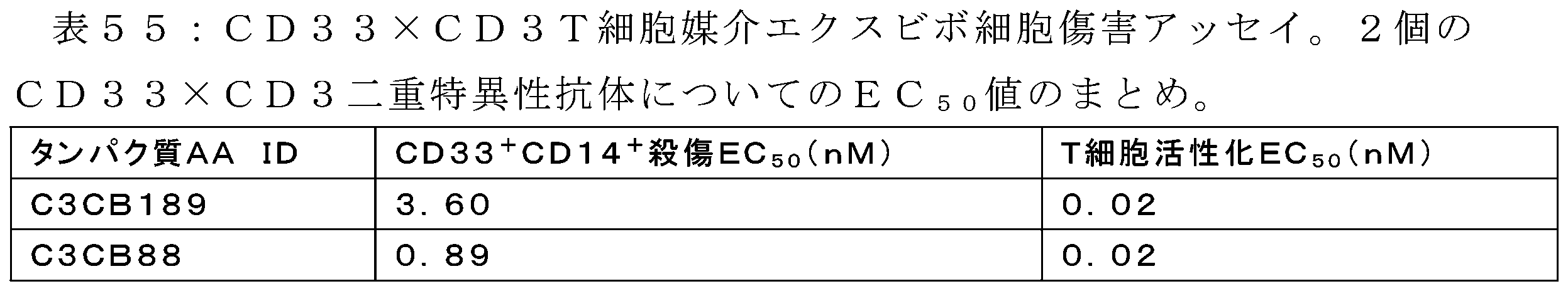
6-6 T細胞ヒト化NSGマウスにおけるMOLM-13ヒトAML異種移植片におけるC3CB189及びC3CB88の有効性
2000万個のT細胞でヒト化された雌NOD.Cg-Prkdcscid Il2rgtm1Wjl/SzJ(NSG)マウスにおける、樹立されたルシフェラーゼでトランスフェクトされた播種性MOLM-13ヒト急性骨髄性白血病(AML)異種移植片において、C3CB189及びC3CB88の有効性を評価した。静脈内腫瘍移植後5日目にライブバイオルミネッセンスイメージング(BLI)によって、動物をn=10/群に無作為化した。0.005、0.05、及び0.5mg/kgのC3CB189及びC3CB88、又は0.5mg/kgのヌル×CD3抗体対照を、6週間にわたって3~4日毎に腹腔内投与した。
6-6 Efficacy of C3CB189 and C3CB88 in MOLM-13 human AML xenografts in T cell-humanized NSG mice The efficacy of C3CB189 and C3CB88 was evaluated in established luciferase-transfected disseminated MOLM-13 human acute myeloid leukemia (AML) xenografts in female NOD.Cg-Prkdc scid Il2rg tm1Wjl /SzJ (NSG) mice humanized with 20 million T cells. Animals were randomized, n=10/group, by live bioluminescence imaging (BLI) 5 days after intravenous tumor implantation. C3CB189 and C3CB88 at 0.005, 0.05, and 0.5 mg/kg, or a null x CD3 antibody control at 0.5 mg/kg, were administered intraperitoneally every 3-4 days for 6 weeks.
腫瘍移植後13日目に、1群あたり少なくとも8頭の動物が残存していた場合、バイオルミネッセンスによって決定したときの腫瘍増殖阻害(%TGI)を計算した。ヌル×CD3対照と比較して、全ての濃度のC3CB189(図66)及びC3CB88(図68)で統計的に有意な腫瘍増殖阻害が観察された。ヌル×CD3処理対照と比較して、0.005、0.05、及び0.5mg/kgのC3CB189は、それぞれ76%、100%、及び82%の腫瘍増殖阻害を惹起し、0.005、0.05、及び0.5mg/kgのC3CB88は、それぞれ100%、100%、及び91%の腫瘍増殖阻害を惹起した。 Tumor growth inhibition (%TGI) as determined by bioluminescence was calculated 13 days after tumor implantation when at least 8 animals remained per group. Statistically significant tumor growth inhibition was observed at all concentrations of C3CB189 (Figure 66) and C3CB88 (Figure 68) compared to Null x CD3 controls. C3CB189 at 0.005, 0.05, and 0.5 mg/kg elicited 76%, 100%, and 82% tumor growth inhibition, respectively, and C3CB88 at 0.005, 0.05, and 0.5 mg/kg elicited 100%, 100%, and 91% tumor growth inhibition, respectively, compared to Null x CD3 treated controls.
C3CB189及びC3CB88による処理の結果、腫瘍量が低減し、ヌル×CD3対照群の16日中央生存期間よりも大きく寿命が延長(ILS)された。全用量にわたって、C3CB189で処理した動物は、19~27.5日間の中央生存期間を有しており(図67)、C3CB88で処理した動物は、26~28.5日間の中央生存期間を有していた(図69)。対照群と比較して、0.005、0.05、及び0.5mg/kgのC3CB189は、それぞれ19%、72%、及び50%寿命を延長させ、C3CB88は、それぞれ63%、78%及び72%の寿命延長をもたらした。 Treatment with C3CB189 and C3CB88 resulted in reduced tumor burden and increased life span (ILS) over the 16 day median survival of the null x CD3 control group. Across all doses, animals treated with C3CB189 had a median survival of 19-27.5 days (Figure 67) and animals treated with C3CB88 had a median survival of 26-28.5 days (Figure 69). Compared to the control group, 0.005, 0.05, and 0.5 mg/kg C3CB189 increased life span by 19%, 72%, and 50%, respectively, and C3CB88 provided a life span increase of 63%, 78%, and 72%, respectively.
7 TMEFF2抗体
7-1 抗原の生成
UniProtアクセッション番号Q9UIK5配列に基づいて、ヒト細胞外ドメイン(ECD)TMEFF2を産生した。C末端に6×Hisタグ(配列番号596)及びAviタグ配列を有するECDコンストラクトを設計した(コンストラクトTMEW1;配列番号578)。FS2及びEGFドメインを含有するコンストラクト(アミノ酸151~320は、6×Hisタグ(配列番号596)及びaviタグ配列とのヒト血清アルブミン(HSA)融合体として設計した(コンストラクトTMEW7;配列番号579)。6×Hisタグ(配列番号596)を有する(コンストラクトTMEW19;配列番号580)又はHisタグを有するラットIgG1 Fcに融合している(コンストラクトTMEW20;配列番号581)TMEFF2膜近位ドメイン(残基230~320)を含有するコンストラクトを設計した。TMEFF2の残基230~320は、TMEFF2の残基261~301にまたがるEGFドメインを含有する。製造元のプロトコルに従ってExpifectamineを使用して、HEK293由来細胞、Expi293(Gibco/Thermo Fisher Scientific)にヒトTMEFF2 ECD発現コンストラクトを一過的にトランスフェクトした。軌道振盪器において8%CO2で37℃において5日間細胞をインキュベートした後、収集した。発現した細胞を遠心分離により除去し、Ni Sepharose 6 Fast Flow resin(GE Healthcare)を使用する固定化金属アフィニティクロマトグラフィー、続いて、ダルッベッコリン酸生理食塩水バッファpH7.2(1×DPBS)中におけるSuperdex 200分取サイズ排除クロマトグラフィー(SEC)(GE Healthcare)を使用して、hisタグを有する可溶性TMEFF2タンパク質を培地から精製した。生成された抗原のアミノ酸列を表58に示す。
7 TMEFF2 Antibody 7-1 Generation of Antigen Human extracellular domain (ECD) TMEFF2 was produced based on the UniProt accession number Q9UIK5 sequence. An ECD construct was designed with a 6xHis tag (SEQ ID NO:596) and an Avi tag sequence at the C-terminus (construct TMEW1; SEQ ID NO:578). A construct containing the FS2 and EGF domains (amino acids 151-320) was designed as a human serum albumin (HSA) fusion with a 6xHis tag (SEQ ID NO:596) and an avi tag sequence (construct TMEW7; SEQ ID NO:579). A rat IgG1 with a 6xHis tag (SEQ ID NO:596) (construct TMEW19; SEQ ID NO:580) or a His tag was designed with a 6xHis tag (SEQ ID NO:596) (construct TMEW19; SEQ ID NO:581). A construct was designed containing the TMEFF2 membrane proximal domain (residues 230-320) fused to TMEFF2 Fc (construct TMEW20; SEQ ID NO:581). Residues 230-320 of TMEFF2 contain the EGF domain spanning residues 261-301 of TMEFF2. Human TMEFF2 ECD expression constructs were transiently transfected into HEK293-derived cells, Expi293 (Gibco/Thermo Fisher Scientific), using Expifectamine according to the manufacturer's protocol. Cells were incubated at 37°C with 8% CO2 in an orbital shaker for 5 days before harvesting. Expressed cells were removed by centrifugation and plated on

7-2 抗TMEFF2抗体の生成
ヒト免疫グロブリン遺伝子座を発現しているトランスジェニックラット(OmniRat(登録商標))を用いた抗体生成。OmniRat(登録商標)は、キメラヒト/ラットIgH遺伝子座(ラットCH遺伝子座に連結された天然立体構造の22のヒトVH、全てのヒトD及びJHセグメントを含む)を完全にヒトのIgL遺伝子座(Jκ-Cκに連結された12のVκ及びJλ-Cλに連結された16のVλ)と共に含有する(例えば、Osborn,et al.(2013)J Immunol 190(4):1481-1490を参照)。したがって、このラットは、ラット免疫グロブリンの発現減少を示し、免疫化に応答して、導入されたヒト重鎖及び軽鎖の導入遺伝子がクラススイッチ及び体細胞変異を受けて、完全にヒトの可変領域を有する高親和性キメラヒト/ラットIgGモノクローナル抗体が生成される。OmniRat(登録商標)の調製及び使用、並びにこのようなラットが有しているゲノム改変は、国際公開第14/093908号に記載されている。
7-2 Generation of Anti-TMEFF2 Antibodies Antibody generation using a transgenic rat expressing a human immunoglobulin locus (OmniRat®). OmniRat® contains a chimeric human/rat IgH locus (containing 22 human VH in the native conformation linked to the rat C H locus, all human D and JH segments) along with a fully human IgL locus (12 Vκ linked to Jκ-Cκ and 16 Vλ linked to Jλ-Cλ) (see, e.g., Osborn, et al. (2013) J Immunol 190(4):1481-1490). Thus, this rat exhibits reduced expression of rat immunoglobulins, and in response to immunization, the introduced human heavy and light chain transgenes undergo class switching and somatic mutation to generate high affinity chimeric human/rat IgG monoclonal antibodies with fully human variable regions. The preparation and use of OmniRats®, and the genomic modifications that such rats carry, are described in WO 14/093908.
OmniRatを、ヒトTMEFF2コンストラクトFS2-EGF-Tev-HSA(C34S)-His-Aviタグ(TMEW7、配列番号579)で免疫化し、コンストラクトspTMEFF2(230-320)G3S-RAIgG1Fc(TMEW20、配列番号581)でブーストした。89日間の免疫化レジメン後、ラットからリンパ節を摘出し、ハイブリドーマを生成するために使用し、ELISA及び/又はSPARCL(Spatial Proximity Analyte Reagent Capture Luminescence)によるヒトTMEFF2-FL-ECD-HIS-Aviタグ(TMEW1)タンパク質への結合についてハイブリドーマ上清をスクリーニングした。TMEFF ECD、FS2-EGF、又はEGFドメインのみのTMEFF2に対する結合の二次ELISA及びSPARCLスクリーニングのために、いくつかの上清を選択した。スクリーニング結果に基づいて、いくつかのハイブリドーマクローンを配列決定し、発現させ、機能性について特性評価した。 OmniRats were immunized with human TMEFF2 construct FS2-EGF-Tev-HSA(C34S)-His-Avi tag (TMEW7, SEQ ID NO: 579) and boosted with construct spTMEFF2(230-320)G3S-RAIgG1Fc (TMEW20, SEQ ID NO: 581). After an 89-day immunization regimen, lymph nodes were removed from the rats and used to generate hybridomas, and hybridoma supernatants were screened for binding to human TMEFF2-FL-ECD-HIS-Avi tag (TMEW1) protein by ELISA and/or SPARCL (Spatial Proximity Analyte Reagent Capture Luminescence). Several supernatants were selected for secondary ELISA and SPARCL screening of binding to TMEFF2, TMEFF ECD, FS2-EGF, or the EGF domain alone. Based on the screening results, several hybridoma clones were sequenced, expressed, and characterized for functionality.
ファージディスプレイライブラリからの抗体生成
Shi et al.,J Mol Biol 397:385-96,2010及び国際公開第2009/085462号)に記載の通り、2セットのデノボpIXファージディスプレイライブラリから標準的な方法を使用してTMEFF2結合Fabを選択した。簡潔に述べると、ヒトスカフォールドを多様化させることによってV3.0及びV5.0と呼ばれる2セットのライブラリを生成し、ここでは、生殖系列VH遺伝子IGHV1-69*01、IGHV3-23*01、及びIGHV5-51*01を、H3ループを介してヒトIGHJ-4ミニ遺伝子で組換え(V5.0ではIGHJ-6ミニ遺伝子も用いた)、並びに、ヒト生殖系列VLカッパ遺伝子O12(IGKV1-39*01)、L6(IGKV3-11*01)、A27(IGKV3-20*01)、及びB3(IGKV4-1*01)をIGKJ-1ミニ遺伝子で組換えて、完全なVH及びVLドメインを構築した。多様化のために、タンパク質及びペプチド抗原と高頻度で接触するH1、H2、L1、L2及びL3ループ周辺の重鎖及び軽鎖可変領域内の位置を選択した。選択した位置における配列多様性は、それぞれのIGHV又はIGLV遺伝子のIGHV又はIGLV生殖系列遺伝子ファミリーにおいて各々の位置に存在する残基に制限した。H3ループにおける多様性は、V3.0ライブラリについては7~14アミノ酸長、V5.0ライブラリについては6~19アミノ酸長の短鎖乃至中鎖の合成ループを使用して作製した。H3におけるアミノ酸分布は、ヒト抗体において観察されるアミノ酸の変動を再現するよう設計された。ライブラリを作製するために利用したスカフォールドは、そのヒトVH及びVL生殖系列遺伝子の由来に従って命名した。V3.0及びV5.0のセットの両方について、3つの重鎖ライブラリを各々、4個の生殖系列軽鎖又は生殖系列軽鎖ライブラリと組み合わせて、選択実験に使用されるライブラリの各セットについて12の固有のVH:VLの組み合わせを作製した。
Antibody generation from phage display libraries TMEFF2-binding Fabs were selected using standard methods from two sets of de novo pIX phage display libraries as described in Shi et al., J Mol Biol 397:385-96, 2010 and WO 2009/085462. Briefly, two sets of libraries, designated V3.0 and V5.0, were generated by diversifying human scaffolds, in which germline VH genes IGHV1-69 * 01, IGHV3-23 * 01, and IGHV5-51 * 01 were recombined with human IGHJ-4 minigene via the H3 loop (IGHJ-6 minigene was also used in V5.0), and human germline VL kappa genes O12 (IGKV1-39 * 01), L6 (IGKV3-11 * 01), A27 (IGKV3-20 * 01), and B3 (IGKV4-1 * 01) were recombined with IGKJ-1 minigene to construct complete VH and VL domains. For diversification, positions within the heavy and light chain variable regions around the H1, H2, L1, L2 and L3 loops that frequently contact protein and peptide antigens were selected. Sequence diversity at the selected positions was restricted to residues present at each position in the IGHV or IGLV germline gene family of the respective IGHV or IGLV gene. Diversity in the H3 loop was generated using short to medium length synthetic loops, 7-14 amino acids long for the V3.0 library and 6-19 amino acids long for the V5.0 library. The amino acid distribution in H3 was designed to reproduce the amino acid variability observed in human antibodies. The scaffolds utilized to generate the libraries were named according to their human VH and VL germline gene origin. For both the V3.0 and V5.0 sets, the three heavy chain libraries were each combined with four germline light chains or germline light chain libraries to generate 12 unique VH:VL combinations for each set of libraries used in the selection experiments.
V領域クローニング
ファージのハイブリドーマ細胞溶解物からの全RNAを、製造元のプロトコルに従ってRNeasy 96キット(Qiagen)を用いて精製した。得られたRNAを、Drop Senseを使用して定量し、-80℃で保存したか又はRT-PCR(Invitrogen)によりInvitrogen SuperScript III First-Strand Synthesis Systemを用いてcDNA合成に使用した。それぞれ重鎖、κ鎖、及びλ鎖の定常領域にアニーリングする遺伝子特異的プライマーを使用して、第1の鎖のcDNA合成を実施した。RT-PCR反応混合物は、最高3μgの精製RNA、遺伝子特異的プライマー、dNTPミックス、反応バッファ、25mM MgCl2、DTT、RNaseOUT(商標)(40U/μL、Invitrogen)、及びSuperScript(商標)III RT(200U/L、Invitrogenカタログ番号18080-051)で構成されており、50℃で50分間及び85℃で5分間インキュベートした。得られた一本鎖cDNAを-20℃で保存した、又はPCR増幅に直接使用した。PLATINUM Pfxポリメラーゼ(Invitrogen)を使用してPCR反応を行った。最適化されたPCR条件を使用して、それぞれ、リーダー配列並びに重鎖、κ及びλ鎖の定常領域に、フォワード及びリバースプライマーをアニーリングさせることによって、v領域断片を増幅させた。得られたPCR断片を配列決定し、回収したv-領域のアミノ酸配列のコドンを最適化し、S228P、F234A、及びL235A変異を有するIgG4定常領域を有するpUnderベースの発現ベクター(IgG4PAAアイソタイプ)にクローニングした。
V-region cloning Total RNA from phage hybridoma cell lysates was purified using RNeasy 96 kit (Qiagen) according to the manufacturer's protocol. The resulting RNA was quantified using Drop Sense and either stored at -80°C or used for cDNA synthesis using the Invitrogen SuperScript III First-Strand Synthesis System by RT-PCR (Invitrogen). First-strand cDNA synthesis was performed using gene-specific primers annealing to the heavy, kappa, and lambda chain constant regions, respectively. The RT-PCR reaction mixture consisted of up to 3 μg purified RNA, gene-specific primers, dNTP mix, reaction buffer, 25 mM MgCl 2 , DTT, RNaseOUT™ (40 U/μL, Invitrogen), and SuperScript™ III RT (200 U/L, Invitrogen catalogue no. 18080-051) and was incubated at 50° C. for 50 min and 85° C. for 5 min. The resulting single-stranded cDNA was stored at −20° C. or used directly for PCR amplification. PCR reactions were performed using PLATINUM Pfx polymerase (Invitrogen). Optimized PCR conditions were used to amplify the v-region fragments by annealing forward and reverse primers to the leader sequences and constant regions of the heavy, kappa and lambda chains, respectively. The resulting PCR fragment was sequenced and the amino acid sequence of the recovered v-region was codon optimized and cloned into a pUnder-based expression vector (IgG4PAA isotype) carrying the IgG4 constant region with the S228P, F234A, and L235A mutations.
Expi293小規模トランスフェクション及び精製
免疫化キャンペーン又はファージディスプレイから同定された選択抗体をクローニングし、IgG1PAAとして発現させ、小さい2mLスケールで精製した。Expi293(商標)細胞(ThermoFisher Scientific)を、Expi293(商標)発現培地に1.25×10⁵~2.25×10⁵生存細胞/mLの密度で播種し、37℃、7%CO2で125mL~2L振盪フラスコ内において培養した。細胞を継代培養し、この場合、生存率98~99%で、密度が3×106~5×106生存細胞/mLで対数増殖に達した。
Expi293 Small Scale Transfection and Purification Selected antibodies identified from the immunization campaign or phage display were cloned, expressed as IgG1 PAA, and purified at a small 2 mL scale. Expi293™ cells (ThermoFisher Scientific) were seeded in Expi293™ Expression Medium at a density of 1.25x10⁵-2.25x10⁵ viable cells/mL and cultured at 37°C, 7% CO2 in 125 mL-2 L shake flasks. Cells were subcultured until they reached logarithmic growth at a density of 3x10⁶-5x10⁶ viable cells/mL with 98-99% viability.
トランスフェクションの日に、生存細胞密度及び生存率を求めた。製造元のトランスフェクションプロトコル(ThermoFisher Publication Number MAN0007814)に従って、3×10⁶生存細胞/mLの密度で細胞をトランスフェクトした。トランスフェクション後6日目に850×Gで15分間遠心分離することによって培養物を収集した後、精製した。mAb Select Sure resin(GE Healthcare)を使用して清澄化した上清から抗体を精製し、PBSに透析した。DropSense Instrument(Trinean)を使用して、濾液に対するA280測定により、タンパク質濃度を求めた。 On the day of transfection, viable cell density and viability were determined. Cells were transfected at a density of 3 x 10⁶ viable cells/mL according to the manufacturer's transfection protocol (ThermoFisher Publication Number MAN0007814). Cultures were harvested 6 days post-transfection by centrifugation at 850 x G for 15 min and then purified. Antibodies were purified from the clarified supernatant using mAb Select Sure resin (GE Healthcare) and dialyzed into PBS. Protein concentrations were determined by A280 measurements on the filtrate using a DropSense Instrument (Trinean).
7-3 抗TMEFF2抗体の特性評価
抗TMEFF2抗体は高親和性でTMEFF2に結合する。
TMEFF2 ECD(TMEW1:TMEFF2-FL ECD-His-Aviタグ)及び/又は膜近位領域(TMEW19:spTMEFF2(230-320)G3S-H6)への選択IgG4PAA抗TMEFF2抗体の結合を、Proteon(TMEB674、TMEB675、TMEB 565及びTMEB570)又はBiacore SPR(TMEB762及びTMEB757)を用いて評価した。選択された抗体の結合の反応速度パラメータを表59に示す。抗TMEFF2抗体は、ピコモル濃度の親和性でTMEFF2 ECD及びTMEFF2膜近位領域の両方に結合することが見出された。
7-3 Characterization of anti-TMEFF2 antibodies Anti-TMEFF2 antibodies bind to TMEFF2 with high affinity.
Binding of selected IgG4 PAA anti-TMEFF2 antibodies to TMEFF2 ECD (TMEW1: TMEFF2-FL ECD-His-Avi tag) and/or membrane proximal region (TMEW19: spTMEFF2(230-320)G3S-H6) was assessed using Proteon (TMEB674, TMEB675, TMEB 565 and TMEB570) or Biacore SPR (TMEB762 and TMEB757). Kinetic parameters of binding of selected antibodies are shown in Table 59. Anti-TMEFF2 antibodies were found to bind to both TMEFF2 ECD and TMEFF2 membrane proximal region with picomolar affinity.

ProteOn SPR
抗TMEFF2 mAbのヒトTMEFF2のECD及び膜近位領域への結合を、ProteOn SPR(Bio-Rad)によって測定した。精製されたmAb(PBST中1μg/mLの最終濃度に希釈した)をアッセイにおいてリガンドとして使用し、Fc捕捉を介してヤギ抗ヒト(GAH)IgG Fcに固定化した。GAH IgG Fcのアミンカップリングのために、EDC(40mM)及びNHS(10mM)の1:1混合物を注入直前に混合してチップ表面を活性化し、垂直配向で注入した。次いで、酢酸バッファ(pH5.0)中GAH-Fc(30μg/mL)抗体を、垂直配向で30μL/分で300秒間表面上に流した。続いて、1Mエタノールアミン(pH8.5)を同じ配向で注入することによって、表面における残りの反応性カルボキシル基を全て不活化させた。固定化のために抗体を1μg/mLの濃度で使用した。抗体を水平方向に表面上に流した。5つの濃度の3倍希釈系列(最高濃度は100~600nMの範囲)のヒトTMEFF2 ECD又は膜近位領域を、捕捉された分子に結合させるために垂直配向でアナライトとして流した。また、ベースラインシグナルにおける任意のドリフトをモニタリングするために、垂直方向で6番目のチャネルにバッファサンプルを注入した。全ての濃度についての会合及び解離相を、100μL/分の流速でそれぞれ3分間及び30(又は15)分間モニタリングした。結合抗原を除去するために18秒パルスの0.8%リン酸を用いて、次の相互作用サイクルのために結合表面を再生した。応答データから2セットの参照データを減じることによって生データを処理した:1)抗原と空のチップ表面との間の非特異的相互作用を補正するためのインタースポットシグナル;2)非特異的ベースラインドリフトを補正するための空のチャネルシグナル(チップ上にPBSTのみを流した場合)。
ProteOn SPR
Binding of anti-TMEFF2 mAbs to the ECD and membrane proximal regions of human TMEFF2 was measured by ProteOn SPR (Bio-Rad). Purified mAbs (diluted to a final concentration of 1 μg/mL in PBST) were used in the assay. and immobilized goat anti-human (GAH) IgG Fc via Fc capture. For amine coupling of GAH IgG Fc, a 1:1 mixture of EDC (40 mM) and NHS (10 mM) was used immediately prior to injection. The chip surface was activated by mixing the antibody with GAH-Fc (30 μg/mL) in acetate buffer (pH 5.0) and then injected in a vertical orientation. GAH-Fc antibody was then flowed over the surface at 30 μL/min for 300 seconds in a vertical orientation. Subsequently, any remaining reactive carboxyl groups on the surface were deactivated by injecting 1 M ethanolamine (pH 8.5) in the same orientation. Antibodies were used at a concentration of 1 μg/mL for immobilization. Antibodies were flowed horizontally over the surface. Human TMEFF2 ECD or The membrane proximal region was run as analyte in a vertical orientation to bind to the captured molecules, and a buffer sample was run in the sixth vertical channel to monitor any drift in the baseline signal. The association and dissociation phases for all concentrations were monitored for 3 and 30 (or 15) minutes, respectively, at a flow rate of 100 μL/min. An 18-second pulse of 0.8% phosphoric acid was added to remove bound antigen. The binding surface was regenerated for the next interaction cycle using the ELISA kit. The raw data were processed by subtracting two sets of reference data from the response data: 1) non-specific interactions between the antigen and the empty chip surface; 2) the empty channel signal (when only PBST was flowed over the chip) to correct for non-specific baseline drift.
Biacore 8K SPR
抗TMEFF2 mAbのヒトTMEFF2 ECDへの結合を、Biacore 8K SPRによって測定した。アッセイのフォーマットは、高密度抗ヒトFc表面を使用してmAを捕捉した後、単一サイクルの反応速度論による方法を用いてヒトTMEFF2濃度滴定を注入するためのものであった。HBSP(GE)バッファ中流量30μL/分でCM5 Sensor Chip(GE)において、フローセル1及び2に10mM酢酸バッファ、pH4.5中30g/mLでのアミンカップリングを介してヤギ抗ヒトFc IgG(Jackson Immunoresearch、カタログ番号109-005-098)を直接固定化した。mAbを、フローセル2において0.5μg/mL(約200~300RU)で抗ヒトFc IgG表面上に捕捉した。次いで、ランニングバッファをHBSP+100μg/mL BSAに変更した。3倍希釈系列中の30nM濃度のTMEFF2 ECDを、単一サイクルの反応速度論による方法を用いて低濃度から高濃度まで注入した。最後、すなわち最高濃度の注入の30分後にオフレートをモニタリングし、次いで、0.8%リン酸(Bio-Rad)を用いて表面を再生した。同じmAbを捕捉し、同じサンプルランの条件を使用して、バッファブランクのランも完了した。応答データから2セットの参照データを減じることによって生データを処理した:1)サンプルフローセル2から減じた参照フローセル1、及び2)実験ランから減じたバッファブランクのラン。各mAbについて全濃度において処理したデータを1:1の単純なラングミュア結合モデルにグローバルフィットさせて、反応速度定数(kon、koff)及び親和性(KD)定数の推定値を抽出した。
Biacore 8K SPR
Binding of anti-TMEFF2 mAbs to human TMEFF2 ECD was measured by Biacore 8K SPR. The assay format consisted of capturing mAs using a high density anti-human Fc surface followed by a single cycle kinetic method. The human TMEFF2 concentration titration was injected using 10 mM acetate buffer, pH 4.5, 30 g/mL in HBSP (GE) buffer on a CM5 Sensor Chip (GE) at a flow rate of 30 μL/min. Goat anti-human Fc IgG (Jackson Immunoresearch, Cat. No. 109-005-098) was directly immobilized via amine coupling at 0.5 μg/mL (approximately 200-300 RU) on
抗TMEFF2抗体の熱安定性
TMEB675は、DSC(示差走査熱量測定)により通常よりも低い熱安定性プロファイルを示し、アンフォールディング開始Tm=52℃であり、第1の熱転移(Tm1)=60.4℃であった。TMEB675の配列のより厳密な検査(実施例4を参照)は、重鎖及び軽鎖のフレームワーク領域内の体細胞超変異(SHM)の存在を示した。いくつかの再遺伝子操作された変異体をサブクローニングし、発現させ、精製し、DSCによりプロファイリングした。得られたmAb TMEB762及びTMEFB757は、望ましい熱安定性プロファイルを示した(それぞれTm1=69.4℃及びTm1=69.7℃)。TMEB675と比較して、TMEB762は、重鎖において以下のアミノ酸改変:R14P、P20L、H81Qを有し、一方、TMEFB757は、重鎖において以下のアミノ酸改変:R14P及びP20Lを有していた。TMEB675と比較して、TMEB762は、軽鎖において以下のアミノ酸改変:A1D及びA91Pを有し、一方、TMEFB757は、軽鎖にA91Pを有していた。残基の付番はKabatに従う。TMEB675、TMEB762のTMEFF2 ECDへの結合の反応速度パラメータを表59に示す。
Thermal Stability of Anti-TMEFF2 Antibodies TMEB675 exhibited a subnormal thermal stability profile by DSC (differential scanning calorimetry) with an onset unfolding Tm=52° C. and a first thermal transition (Tm1)=60.4° C. Closer examination of the sequence of TMEB675 (see Example 4) indicated the presence of somatic hypermutation (SHM) within the framework regions of the heavy and light chains. Several re-engineered mutants were subcloned, expressed, purified, and profiled by DSC. The resulting mAbs TMEB762 and TMEFB757 exhibited desirable thermal stability profiles (Tm1=69.4° C. and Tm1=69.7° C., respectively). Compared to TMEB675, TMEB762 had the following amino acid modifications in the heavy chain: R14P, P20L, H81Q, while TMEFB757 had the following amino acid modifications in the heavy chain: R14P and P20L. Compared to TMEB675, TMEB762 had the following amino acid modifications in the light chain: A1D and A91P, while TMEFB757 had A91P in the light chain. Residue numbering is according to Kabat. Kinetic parameters for binding of TMEB675, TMEB762 to TMEFF2 ECD are shown in Table 59.
7-4 抗TMEFF2抗体の構造特定
標準的な技術を用いて、抗体のcDNA配列及びアミノ酸翻訳物を得た。ポリペプチド配列の決定後、可変領域又は完全長抗体をコードしているいくつかの抗体cDNAを、発現をスケールアップするための標準的な方法を用いてコドンを最適化した。
7-4 Structural Characterization of Anti-TMEFF2 Antibodies Antibody cDNA sequences and amino acid translations were obtained using standard techniques. After determining the polypeptide sequences, some antibody cDNAs encoding the variable regions or full-length antibodies were codon-optimized using standard methods to scale up expression.
表60は、選択抗TMEFF2抗体のHCDR1及びHCDR2アミノ酸配列を示す。 Table 60 shows the HCDR1 and HCDR2 amino acid sequences of selected anti-TMEFF2 antibodies.
表61は、選択抗TMEFF2抗体のHCDR3のアミノ酸配列を示す。 Table 61 shows the amino acid sequences of HCDR3 of selected anti-TMEFF2 antibodies.
表62は、選択抗TMEFF2抗体のLCDR1及びLCDR2アミノ酸配列を示す。 Table 62 shows the LCDR1 and LCDR2 amino acid sequences of selected anti-TMEFF2 antibodies.
表63は、選択抗TMEFF2のLCDR3のアミノ酸配列を示す。 Table 63 shows the amino acid sequences of LCDR3 of selected anti-TMEFF2 antibodies.
表64は、選択抗TMEFF2のVH及びVLアミノ酸配列を示す。 Table 64 shows the VH and VL amino acid sequences of selected anti-TMEFF2 antibodies.
表65は、選択抗TMEFF2抗体の重鎖及び軽鎖の配列番号を示す。 Table 65 shows the sequence numbers of the heavy and light chains of selected anti-TMEFF2 antibodies.
表66は、選択抗TMEFF2の重鎖アミノ酸配列を示す。 Table 66 shows the heavy chain amino acid sequences of selected anti-TMEFF2 antibodies.
表67は、選択抗TMEFF2の軽鎖アミノ酸配列を示す。 Table 67 shows the light chain amino acid sequences of selected anti-TMEFF2 antibodies.
表68は、様々な抗TMEFF2抗体鎖をコードしているポリヌクレオチドの配列番号を示す。 Table 68 shows the sequence numbers of polynucleotides encoding various anti-TMEFF2 antibody chains.


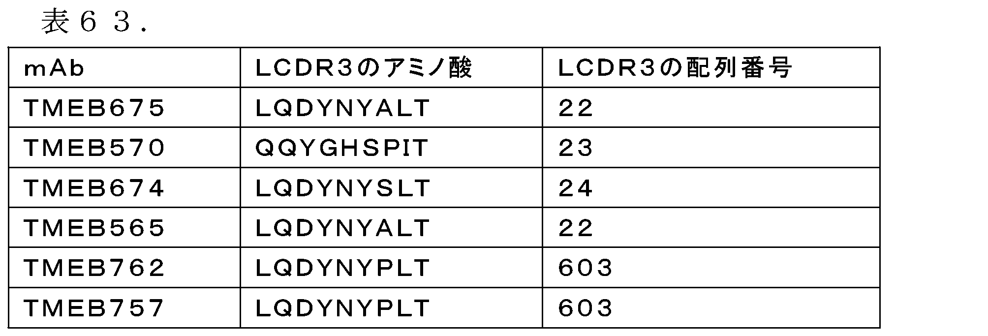
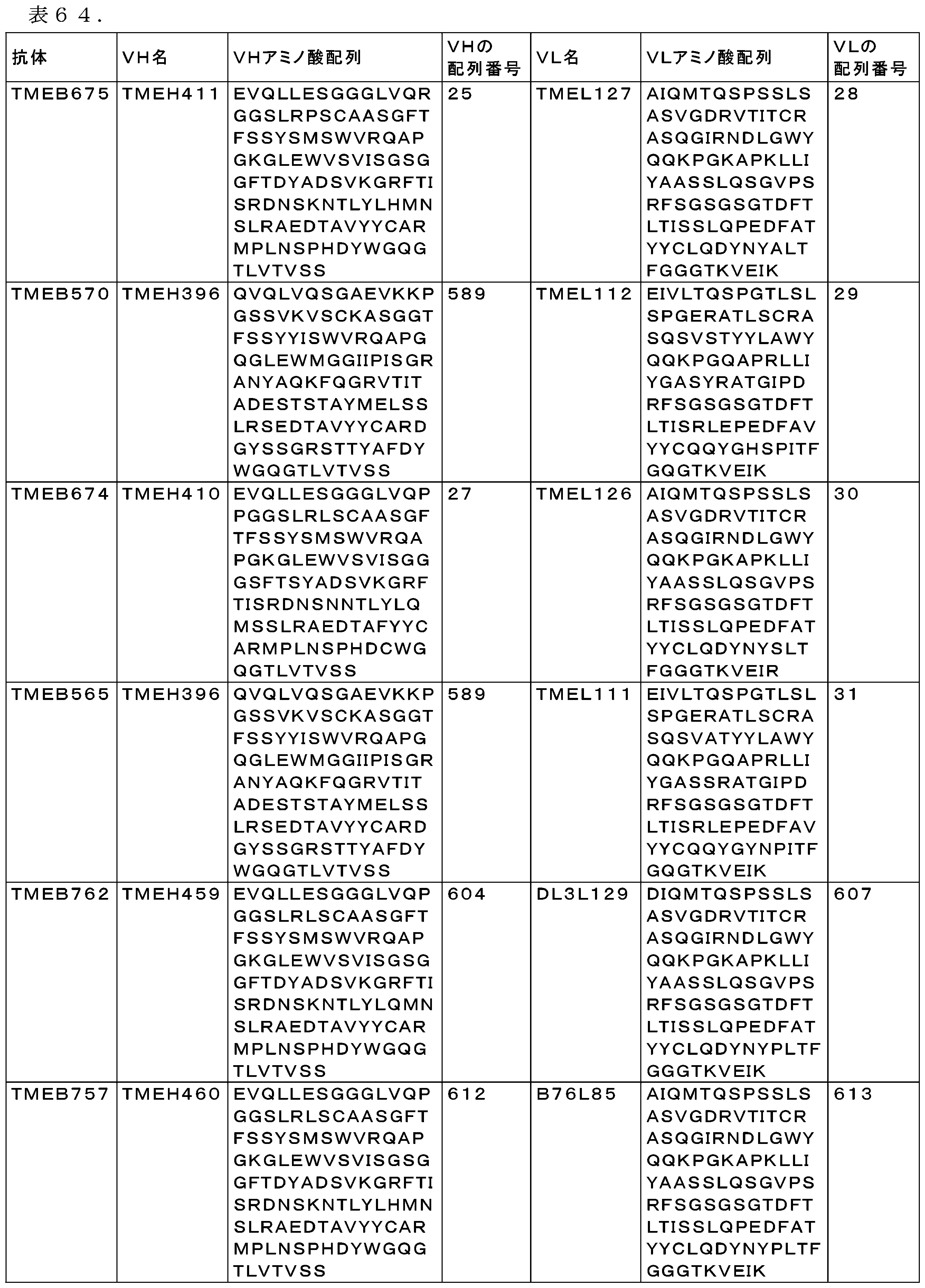


配列番号39(TMEB675 VH cDNA)
Gaggtgcagctgctggaaagcggcggaggcctggtgcagagaggaggaagcctgagacccagctgtgccgccagcggcttcaccttcagcagctacagcatgagctgggtcaggcaggcccctggcaaaggactggagtgggtgagcgtgattagcggcagcggcggcttcaccgattacgccgacagcgtgaagggcaggttcaccatcagcagggacaatagcaagaacaccctgtacctgcacatgaacagcctgagggccgaggacaccgccgtgtactactgcgccaggatgcccctgaacagccctcacgactactggggccagggaaccctggtgaccgtgtccagc
SEQ ID NO: 39 (TMEB675 VH cDNA)
Gaggtgcagctgctggaaagcggcggaggcctggtgcagagaggaggaagcctgagacccagctgtgccgccagcggcttcacctcagcagctacagcatgagctgggtcaggcaggccc ctggcaaaggactggagtgggtgagcgtgattagcggcagcggcggcttcaccgat tacgccgacagcgtgaaggcaggttcaccatcagcaggacaatagcaagaacaccctgtacctgcacatgaacagcctgaggggccgaggacacgccgtgtacttgcgccaggatgc ccctgaacagccctcacgactactgggggccaggaaccctggtgaccgtgtccagc
配列番号40(TMEB570,TMEB565 VH cDNA)
Caggtgcagctggtgcagagcggcgcggaagtgaaaaaaccgggcagcagcgtgaaagtgagctgcaaagcgagcggcggcaccttcagctcctattacattagctgggtgcgccaggcgccgggccagggcctggaatggatgggtggcattatcccaatcagtgggcgtgctaattatgcgcagaaatttcagggccgcgtgaccattaccgctgatgaaagcaccagcaccgcgtatatggaactgagcagcctgcgcagcgaagataccgcggtgtattattgcgcgcgcgacggctacagtagtggacgtagcacaacatacgcatttgactattggggccagggcaccctggtgaccgtgtcgagt
SEQ ID NO: 40 (TMEB570, TMEB565 VH cDNA)
Caggtgcagctggtgcagagcggcgcggaagtgaaaaaaccggggcagcagcgtgaaagtgagctgcaaagcgagcggcggcaccttcagctcctattacattagctgggtgcgccagcgc cgggccagggcctggaatggatgggtggcattatcccaatcagtgggcgtgctaattatgcgcag aaatttcaggccgcgtgacttaccgctgatgaaagcaccagcaccgcgtatatggaactgagcagcctgcgcagcgaagataccgcggtgtattatgcgcgcgcgacggctacagta gtggacgtagcacaacatacgcatttgactattggggccagggcaccctggtgaccgtgtcgagt
配列番号41(TMEB674 VH cDNA)
Gaagtgcagctgctggagagcggaggaggactggtgcagcctcctggcggaagcctgagactgagctgcgccgctagcggcttcaccttcagcagctacagcatgagctgggtgagacaggctcctggcaagggcctggagtgggtgagcgtgatcagcggcggaggcagctttaccagctacgccgacagcgtgaagggcaggttcaccatcagcagggacaacagcaacaacaccctgtacctgcagatgagcagcctgagggccgaggacaccgccttctactactgcgccaggatgcccctgaacagcccccatgactgctggggacagggcaccctggtgaccgtgagcagc
SEQ ID NO: 41 (TMEB674 VH cDNA)
Gaagtgcagctgctggagagcggaggactggtgcagcctcctggcggaagcctgagactgagctgcgccgctagcggcttcaccttcagcagctacagcatgagctgggtgagacagg ctcctggcaagggcctggagtgggtgagcgtgatcagcggcggaggcagctttacca gctacgccgacagcgtgaaggcaggttcaccatcagcaggacaacagcaacaacacctgtacctgcagatgagcagcctgagggccgaggacacgccttctactactgcgccaggat gcccctgaacagcccccatgactgctggggacagggcaccctggtgaccgtgagcagc
配列番号42(TMEB675 VL cDNA)
Gccatccagatgacccagagccctagcagcctgagcgctagcgtgggcgacagggtgaccatcacctgcagggccagccagggcatcagaaacgacctgggctggtaccagcagaagcccggcaaggcccccaagctgctgatctacgccgccagcagcctgcagagcggagtgcctagcaggttcagcggaagcggcagcggcaccgacttcaccctgaccatcagcagcctgcagcccgaggacttcgccacctactactgcctgcaggactacaactacgccctgacattcggcggcggcaccaaggtggagatcaag
SEQ ID NO: 42 (TMEB675 VL cDNA)
Gccatccagatgacccagagccctagcagcctgagcgctagcgtgggcgacaggtgaccatcacctgcaggccagccaggcatcagaaaacgacctgggctggtaccagcagaagcccg gcaaggcccccaagctgctgatctacgccgccagcagcc tgcagagcggagtgcctagcaggttcagcggaagcggcagcggcaccgacttcaccctgaccatcagcagcctgcagcccgaggacttcgccacctacttgcctgcaggactactacaacta cgccctgacattcggcggcggcaccaaggtggagatcaag
配列番号43(TMEB570 VL cDNA)
Gaaattgtgctgacccagagcccgggcaccctgagcctgagcccgggcgaacgcgcgaccctgagctgccgcgcgagccagagcgtttccacatactacctggcgtggtatcagcagaaaccgggccaggcgccgcgcctgctgatttacggtgcctcctatcgcgcgaccggcattccggatcgctttagcggcagcggttccggcaccgattttaccctgaccattagccgcctggaaccggaagattttgcggtgtattattgccagcagtacggtcacagcccgattacttttggccagggcaccaaagtggaaatcaaa
SEQ ID NO: 43 (TMEB570 VL cDNA)
Gaaattgtgctgacccagagcccggggcaccctgagcctgagcccgggcgaacgcgcgaccctgagctgccgcgcgagccagagcgtttccacatactacctggcgtggtatcagcagaaac cgggccaggcgccgcgcctgctgatttacggtgcctcctat cgcgcgaccggcattccggatcgctttagcggcagcggttccggcaccgatttaccctgaccattagccgcctggaaccggaagattttgcggtgtattatgccagcagtacggtcaca gcccgattacttttggccagggcaccaaagtggaaatcaaa
配列番号44(TMEB674 VL cDNA)
Gccatccagatgacccagagccctagcagcctgagcgctagcgtgggcgacagggtgaccatcacctgcagggccagccagggcatcaggaacgacctgggctggtaccagcagaagcccggcaaggcccccaagctgctgatctacgccgccagcagcctgcagagcggagtgcctagcaggttcagcggcagcggaagcggcaccgacttcaccctgaccatctccagcctgcagcccgaggacttcgccacctactactgcctgcaggactacaactacagcctgaccttcggcggcggcaccaaggtggagatcagg
SEQ ID NO: 44 (TMEB674 VL cDNA)
Gccatccagatgacccagagccctagcagcctgagcgctagcgtgggcgacaggtgaccatcacctgcaggccagccaggcatcaggaacgacctgggctggtaccagcagaagcccg gcaaggcccccaagctgctgatctacgccgccagcagcc tgcagagcggagtgcctagcaggttcagcggcagcggaagcggcaccgacttcaccctgaccatctccagcctgcagcccgaggacttcgccacctacttgcctgcaggactacaacta cagcctgaccttcggcggcggcaccaaggtggagatcagg
配列番号45(TMEB565 VL cDNA)
Gaaattgtgctgacccagagcccgggcaccctgagcctgagcccgggcgaacgcgcgaccctgagctgccgcgcgagccagagcgttgccacctattatcttgcgtggtatcagcagaaaccgggccaggcgccgcgcctgctgatttacggtgcatcctcccgtgcgaccggcattccggatcgctttagcggcagcggttccggcaccgattttaccctgaccattagccgcctggaaccggaagattttgcggtgtattattgccagcagtacggctataacccaattacctttggccagggcaccaaagtggaaatcaaa
SEQ ID NO: 45 (TMEB565 VL cDNA)
Gaaattgtgctgacccagagcccggggcaccctgagcctgagcccgggcgaacgcgcgaccctgagctgccgcgcgagccagagcgttgccacctattatcttgcgtggtatcagcagaaac cgggccaggcgccgcgcctgctgatttacggtgcatcctcc cgtgcgaccggcattccggatcgctttagcggcagcggttccggcaccgatttaccctgaccattagccgcctggaaccggaagattttgcggtgtattatgccagcagtacggctata acccaattacctttggccagggcaccaaagtggaaatcaaa
配列番号46(TMEB675 HC cDNA)
gaggtgcagctgctggaaagcggcggaggcctggtgcagagaggaggaagcctgagacccagctgtgccgccagcggcttcaccttcagcagctacagcatgagctgggtcaggcaggcccctggcaaaggactggagtgggtgagcgtgattagcggcagcggcggcttcaccgattacgccgacagcgtgaagggcaggttcaccatcagcagggacaatagcaagaacaccctgtacctgcacatgaacagcctgagggccgaggacaccgccgtgtactactgcgccaggatgcccctgaacagccctcacgactactggggccagggaaccctggtgaccgtgtccagcgcttccaccaagggcccatccgtcttccccctggcgccctgctccaggagcacctccgagagcacagccgccctgggctgcctggtcaaggactacttccccgaaccggtgacggtgtcgtggaactcaggcgccctgaccagcggcgtgcacaccttcccggctgtcctacagtcctcaggactctactccctcagcagcgtggtgaccgtgccctccagcagcttgggcacgaaaacctacacctgcaacgtagatcacaagcccagcaacaccaaggtggacaagagagttgagtccaaatatggtcccccatgcccaccatgcccagcacctgaggccgccgggggaccatcagtcttcctgttccccccaaaacccaaggacactctcatgatctcccggacccctgaggtcacgtgcgtggtggtggacgtgagccaggaagaccccgaggtccagttcaactggtacgtggatggcgtggaggtgcataatgccaagacaaagccgcgggaggagcagttcaacagcacgtaccgtgtggtcagcgtcctcaccgtcctgcaccaggactggctgaacggcaaggagtacaagtgcaaggtctccaacaaaggcctcccgtcctccatcgagaaaaccatctccaaagccaaagggcagccccgagagccacaggtgtacaccctgcccccatcccaggaggagatgaccaagaaccaggtcagcctgacctgcctggtcaaaggcttctaccccagcgacatcgccgtggagtgggagagcaatgggcagccggagaacaactacaagaccacgcctcccgtgctggactccgacggctccttcttcctctacagcaggctaaccgtggacaagagcaggtggcaggaggggaatgtcttctcatgctccgtgatgcatgaggctctgcacaaccactacacacagaagagcctctccctgtctctgggtaaa
SEQ ID NO: 46 (TMEB675 HC cDNA)
gaggtgcagctgctggaaagcggcggaggcctggtgcagagaggaggaagcctgagacccagctgtgccgccagcggcttcacctcagcagctacagcatgagctgggtcaggcaggccc ctggcaaaggactggagtgggtgagcgtgattagcggcagcggcg gcttcaccgattacgccgacagcgtgaaggcaggttcaccatcagcagggacaatagcaagaacaccctgtacctgcacatgaacagcctgaggccgaggacacgccgtgtactactg cgccaggatgcccctgaacagccctcacgactactgggggccagga accctggtgaccgtgtccagcgcttccaccaaggccccatccgtcttccccctggcgccctgctccaggagcacctccgagagcacagccgccctgggctgcctggtcaaggactttcc ccgaacggtgacggtgtcgtggaactcaggcgccctgaccagcgg cgtgcacaccttcccggctgtcctacgtcctcaggactctactccctcagcagcgtggtgacgtgccctccagcagcttgggcacgaaaacctacctgcaacgtagatcacaagccc agcaacaccaaggtggacaagagagttgagtccaaatatggtcccc catgcccaccatgcccagcacctgaggccgcggggaccatcagtcttcctgttcccccaaaacccaaggacactctcatgatctcccggacccctgaggtcacgtgcgtggtggtgga cgtgagccaggaagaccccgaggtccagttcaactggtacgtggat ggcgtggaggtgcataatgccaagacaaagccgcgggaggagcagttcaacagcacgtaccgtgtggtcagcgtcctcaccgtcctgcaccaggactggctgaacggcaaggagtacaagt gcaaggtctccaacaaaggcctcccgtcctccatcgagaaaaccat ctccaaagccaaaggcagccccgagagcccaggtgtacacctgcccccatcccaggaggagatgaccaagaaccaggtcagcctgacctgcctggtcaaaggcttctaccccagcgac atcgccgtggagtgggagagcaatgggcagccggagaacaactaca agaccacgcctcccgtgctggactccgacggctccttctttcctctacagcaggctaaccgtggacaagagcaggtggcaggaggggaatgtcttctcatgctccgtgatgcatgaggctct gcacaaccactacacacagaagaagcctctccctgtctctgggtaaa
配列番号47(TMEB570,TMEB565 HC cDNA)
caggtgcagctggtgcagagcggcgcggaagtgaaaaaaccgggcagcagcgtgaaagtgagctgcaaagcgagcggcggcaccttcagctcctattacattagctgggtgcgccaggcgccgggccagggcctggaatggatgggtggcattatcccaatcagtgggcgtgctaattatgcgcagaaatttcagggccgcgtgaccattaccgctgatgaaagcaccagcaccgcgtatatggaactgagcagcctgcgcagcgaagataccgcggtgtattattgcgcgcgcgacggctacagtagtggacgtagcacaacatacgcatttgactattggggccagggcaccctggtgaccgtgtcgagtgcttccaccaagggcccatccgtcttccccctggcgccctgctccaggagcacctccgagagcacagccgccctgggctgcctggtcaaggactacttccccgaaccggtgacggtgtcgtggaactcaggcgccctgaccagcggcgtgcacaccttcccggctgtcctacagtcctcaggactctactccctcagcagcgtggtgaccgtgccctccagcagcttgggcacgaaaacctacacctgcaacgtagatcacaagcccagcaacaccaaggtggacaagagagttgagtccaaatatggtcccccatgcccaccatgcccagcacctgaggccgccgggggaccatcagtcttcctgttccccccaaaacccaaggacactctcatgatctcccggacccctgaggtcacgtgcgtggtggtggacgtgagccaggaagaccccgaggtccagttcaactggtacgtggatggcgtggaggtgcataatgccaagacaaagccgcgggaggagcagttcaacagcacgtaccgtgtggtcagcgtcctcaccgtcctgcaccaggactggctgaacggcaaggagtacaagtgcaaggtctccaacaaaggcctcccgtcctccatcgagaaaaccatctccaaagccaaagggcagccccgagagccacaggtgtacaccctgcccccatcccaggaggagatgaccaagaaccaggtcagcctgacctgcctggtcaaaggcttctaccccagcgacatcgccgtggagtgggagagcaatgggcagccggagaacaactacaagaccacgcctcccgtgctggactccgacggctccttcttcctctacagcaggctaaccgtggacaagagcaggtggcaggaggggaatgtcttctcatgctccgtgatgcatgaggctctgcacaaccactacacacagaagagcctctccctgtctctgggtaaa
SEQ ID NO: 47 (TMEB570, TMEB565 HC cDNA)
caggtgcagctggtgcagagcggcgcggaagtgaaaaaaccggggcagcagcgtgaaagtgagctgcaaagcgagcggcggcaccttcagctcctattacattagctgggtgcgccagcgc cgggccagggcctggaatggatgggtggcattatcccaatcagtgggc gtgctaattatgcgcagaaatttcagggccgcgtgaccattccgctgatgaaagcaccagcaccgcgtatatggaactgagcagcctgcgcagcgaagataccgcggtgtattattgcgc gcgcgacggctacagtagtggacgtagcacaacatacgcatttgacta ttgggccagggcaccctggtgaccgtgtcgagtgcttccaccaaggccccatccgtcttccccctggcgccctgctcccaggagcacctccgagagcacagccgccctgggctgcctggtc aaggactacttccccgaaccggtgacggtgtcgtggaactcaggcgcc ctgaccagcggcgtgcacaccttcccggctgtcctacgtcctcaggactctactccctcagcagcgtggtgacgtgccctccagcagcttgggcacgaaaacctacctacctgcaacgtag atcacaagcccagcaacaccaaggtggacaagagagttgagtccaaat atggtcccccatgcccaccatgcccagcacctgaggccgccgggggaccatcagtcttcctgttcccccaaaaccccaaggacactctcatgatctcccggacccctgaggtcacgtgcgt ggtggtggacgtgagccaggaagaccccgaggtccagttcaactggta cgtggatggcgtggaggtgcataatgccaagacaaagccgcgggaggagcagttcaacagcacgtaccgtgtggtcagcgtcctcaccgtcctgcaccaggactggctgaacggcaagga tacaagtgcaaggtctccaacaaaggcctcccgtcctccatcgagaaa accatctccaaagccaaaggcagccccgagagcccaggtgtacacctgcccccatcccaggaggagatgaccaagaaccaggtcagcctgacctgcctggtcaaaggcttctacccca gcgacatcgccgtggagtgggagagcaatgggcagccggagaacaact acaaagaccacgcctcccgtgctggactccgacggctccttctttcctctacagcaggctaaccgtggacaagagcaggtggcaggagggaatgtcttctctgctccgtgatgcatgaggc tctgcacaaccactacacacagaagaagcctctccctgtctctgggtaaa
配列番号48(TMEB674 HC cDNA)
gaagtgcagctgctggagagcggaggaggactggtgcagcctcctggcggaagcctgagactgagctgcgccgctagcggcttcaccttcagcagctacagcatgagctgggtgagacaggctcctggcaagggcctggagtgggtgagcgtgatcagcggcggaggcagctttaccagctacgccgacagcgtgaagggcaggttcaccatcagcagggacaacagcaacaacaccctgtacctgcagatgagcagcctgagggccgaggacaccgccttctactactgcgccaggatgcccctgaacagcccccatgactgctggggacagggcaccctggtgaccgtgagcagcgcttccaccaagggcccatccgtcttccccctggcgccctgctccaggagcacctccgagagcacagccgccctgggctgcctggtcaaggactacttccccgaaccggtgacggtgtcgtggaactcaggcgccctgaccagcggcgtgcacaccttcccggctgtcctacagtcctcaggactctactccctcagcagcgtggtgaccgtgccctccagcagcttgggcacgaaaacctacacctgcaacgtagatcacaagcccagcaacaccaaggtggacaagagagttgagtccaaatatggtcccccatgcccaccatgcccagcacctgaggccgccgggggaccatcagtcttcctgttccccccaaaacccaaggacactctcatgatctcccggacccctgaggtcacgtgcgtggtggtggacgtgagccaggaagaccccgaggtccagttcaactggtacgtggatggcgtggaggtgcataatgccaagacaaagccgcgggaggagcagttcaacagcacgtaccgtgtggtcagcgtcctcaccgtcctgcaccaggactggctgaacggcaaggagtacaagtgcaaggtctccaacaaaggcctcccgtcctccatcgagaaaaccatctccaaagccaaagggcagccccgagagccacaggtgtacaccctgcccccatcccaggaggagatgaccaagaaccaggtcagcctgacctgcctggtcaaaggcttctaccccagcgacatcgccgtggagtgggagagcaatgggcagccggagaacaactacaagaccacgcctcccgtgctggactccgacggctccttcttcctctacagcaggctaaccgtggacaagagcaggtggcaggaggggaatgtcttctcatgctccgtgatgcatgaggctctgcacaaccactacacacagaagagcctctccctgtctctgggtaaa
SEQ ID NO: 48 (TMEB674 HC cDNA)
gaagtgcagctgctggagagcggaggactggtgcagcctcctggcggaagcctgagactgagctgcgccgctagcggcttcaccttcagcagctacagcatgagctgggtgagacagg ctcctggcaagggcctggagtgggtgagcgtgatcagcggcggagg cagctttaccagctacgccgacagcgtgaaggcaggttcaccatcagcaggacaacagcaacaacacctgtacctgcagatgagcagcctgaggccgaggacacgccttctactac tgcgccaggatgcccctgaacagcccccatgactgctggggacag gcaccctggtgaccgtgagcagcgcttccaccaaggggcccatccgtcttccccctggcgccctgctccaggagcacctccgagagcacagccgccctgggctgcctggtcaaggacttt ccccgaaccggtgacggtgtcgtggaactcaggcgccctgaccagc ggcgtgcacaccttcccggctgtcctacgtcctcaggactctctactccctcagcagcgtggtgacgtgccctccagcagcttgggcacgaaaacctacctgcaacgtagatcacaagc ccagcaacaccaaggtggacaagagagttgagtccaaatatggtccc ccatgccccaccatgcccagcacctgaggccgccggggaccatcagtcttcctgttcccccaaaacccaaggacactctcatgatctcccggacccctgaggtcacgtgcgtggtggtgg acgtgagccaggaagaccccgaggtccagttcaactggtacgtgga tggcgtggaggtgcataatgccaagacaaagccgcgggaggagcagttcaacagcacgtaccgtgtggtcagcgtcctcaccgtcctgcaccaggactggctgaacggcaaggagtacaag tgcaaggtctccaacaaaggcctcccgtcctccatcgagaaaacca tctccaaagccaaaggcagccccgagagcccaggtgtacacctgcccccatcccaggaggagatgaccaagaaccaggtcagcctgacctgcctggtcaaaggcttctaccccagcga catcgccgtggagtgggagagcaatgggcagccggagaacaactac aagaccacgcctcccgtgctggactccgacggctccttctttcctctacagcaggctaaccgtggacaagagcaggtggcaggagggaatgtcttctcatgctccgtgatgcatgaggctc tgcacaaccactacacacagaagaagcctctccctgtctctgggtaaa
配列番号49(TMEB675 LC cDNA)
Gccatccagatgacccagagccctagcagcctgagcgctagcgtgggcgacagggtgaccatcacctgcagggccagccagggcatcagaaacgacctgggctggtaccagcagaagcccggcaaggcccccaagctgctgatctacgccgccagcagcctgcagagcggagtgcctagcaggttcagcggaagcggcagcggcaccgacttcaccctgaccatcagcagcctgcagcccgaggacttcgccacctactactgcctgcaggactacaactacgccctgacattcggcggcggcaccaaggtggagatcaagcgtacggtggctgcaccatctgtcttcatcttcccgccatctgatgagcagttgaaatctggaactgcctctgttgtgtgcctgctgaataacttctatcccagagaggccaaagtacagtggaaggtggataacgccctccaatcgggtaactcccaggagagtgtcacagagcaggacagcaaggacagcacctacagcctcagcagcaccctgacgctgagcaaagcagactacgagaaacacaaagtctacgcctgcgaagtcacccatcagggcctgagctcgcccgtcacaaagagcttcaacaggggagagtgt
SEQ ID NO: 49 (TMEB675 LC cDNA)
Gccatccagatgacccagagccctagcagcctgagcgctagcgtgggcgacaggtgaccatcacctgcaggccagccaggcatcagaaaacgacctgggctggtaccagcagaagcccg gcaaggcccccaagctgctgatctacgccgccagcagcc tgcagagcggagtgcctagcaggttcagcggaagcggcagcggcaccgacttcaccctgaccatcagcagcctgcagcccgaggacttcgccacctacttgcctgcaggactactacaacta cgccctgacattcggcggcggcaccaaggtggagatcaag cgtacggtggctgcaccatctgtcttcatcttcccgccatctgatgagcagttgaaatctggaactgcctctgttgtgtgcctgctgaataacttctatcccagagaggccaaagtacagt ggaaggtggataacgccctccaatcgggtaactcccagg agagtgtcacagagcaggacagcaaggacagcacctacagcctcagcagcaccctgacgctgagcaaagcagactacgagaaacacaaagtctacgcctgcgaagtcacccatcagggcct gagctcgcccgtcacaaagagcttcaacaggggagagtgt
配列番号50(TMEB570 LC cDNA)
Gaaattgtgctgacccagagcccgggcaccctgagcctgagcccgggcgaacgcgcgaccctgagctgccgcgcgagccagagcgtttccacatactacctggcgtggtatcagcagaaaccgggccaggcgccgcgcctgctgatttacggtgcctcctatcgcgcgaccggcattccggatcgctttagcggcagcggttccggcaccgattttaccctgaccattagccgcctggaaccggaagattttgcggtgtattattgccagcagtacggtcacagcccgattacttttggccagggcaccaaagtggaaatcaaacgtacggtggctgcaccatctgtcttcatcttcccgccatctgatgagcagttgaaatctggaactgcctctgttgtgtgcctgctgaataacttctatcccagagaggccaaagtacagtggaaggtggataacgccctccaatcgggtaactcccaggagagtgtcacagagcaggacagcaaggacagcacctacagcctcagcagcaccctgacgctgagcaaagcagactacgagaaacacaaagtctacgcctgcgaagtcacccatcagggcctgagctcgcccgtcacaaagagcttcaacaggggagagtgt
SEQ ID NO:50 (TMEB570 LC cDNA)
Gaaattgtgctgacccagagcccggggcaccctgagcctgagcccgggcgaacgcgcgaccctgagctgccgcgcgagccagagcgtttccacatactacctggcgtggtatcagcagaaac cgggccaggcgccgcgcctgctgatttacggtgcctccta tcgcgcgacggcattccggatcgctttagcggcagcggttccggcaccgatttaccctgaccattagccgcctggaaccggaagattttgcggtgtattattgccagcagtacggtcac agccccgattacttttggccagggcaccaaagtggaaatca aacgtacggtggctgcaccatctgtcttcatcttcccgccatctgatgagcagttgaaatctggaactgcctctgttgtgcctgctgaataacttctatcccagagaggccaaagtaca gtggaaggtggataacgccctccaatcgggtaactcccag gagagtgtcacagagcaggacagcaaggacagcacctacagcctcagcagcaccctgacgctgagcaaagcagactacgagaaacacaaagtctacgcctgcgaagtcacccatcaggcc tgagctcgcccgtcacaaagagcttcaacaggggagtgt
配列番号51(TMEB674 LC cDNA)
ccatccagatgacccagagccctagcagcctgagcgctagcgtgggcgacagggtgaccatcacctgcagggccagccagggcatcaggaacgacctgggctggtaccagcagaagcccggcaaggcccccaagctgctgatctacgccgccagcagcctgcagagcggagtgcctagcaggttcagcggcagcggaagcggcaccgacttcaccctgaccatctccagcctgcagcccgaggacttcgccacctactactgcctgcaggactacaactacagcctgaccttcggcggcggcaccaaggtggagatcaggcgtacggtggctgcaccatctgtcttcatcttcccgccatctgatgagcagttgaaatctggaactgcctctgttgtgtgcctgctgaataacttctatcccagagaggccaaagtacagtggaaggtggataacgccctccaatcgggtaactcccaggagagtgtcacagagcaggacagcaaggacagcacctacagcctcagcagcaccctgacgctgagcaaagcagactacgagaaacacaaagtctacgcctgcgaagtcacccatcagggcctgagctcgcccgtcacaaagagcttcaacaggggagagtgt
SEQ ID NO:51 (TMEB674 LC cDNA)
ccatccagatgacccagagccctagcagcctgagcgctagcgtgggcgacaggtgaccatcacctgcaggccagccaggcatcaggaacgacctgggctggtaccagcagaagcccgg caaggcccccaagctgctgatctacgccgccagcagcct gcagagcggagtgcctagcaggttcagcggcagcggaagcggcaccgacttcaccctgaccatctccagcctgcagcccgaggacttcgccacctacttgcctgcaggactactacaactac agcctgaccttcggcggcggcaccaaggtggagatcagg cgtacggtggctgcaccatctgtcttcatcttcccgccatctgatgagcagttgaaatctggaactgcctctgttgtgtgcctgctgaataacttctatcccagagaggccaaagtacagt ggaaggtggataacgccctccaatcgggtaactcccagg agagtgtcacagagcaggacagcaaggacagcacctacagcctcagcagcaccctgacgctgagcaaagcagactacgagaaacacaaagtctacgcctgcgaagtcacccatcagggcct gagctcgcccgtcacaaagagcttcaacaggggagtgt
配列番号590(TMEB565 LC cDNA)
Gaaattgtgctgacccagagcccgggcaccctgagcctgagcccgggcgaacgcgcgaccctgagctgccgcgcgagccagagcgttgccacctattatcttgcgtggtatcagcagaaaccgggccaggcgccgcgcctgctgatttacggtgcatcctcccgtgcgaccggcattccggatcgctttagcggcagcggttccggcaccgattttaccctgaccattagccgcctggaaccggaagattttgcggtgtattattgccagcagtacggctataacccaattacctttggccagggcaccaaagtggaaatcaaacgtacggtggctgcaccatctgtcttcatcttcccgccatctgatgagcagttgaaatctggaactgcctctgttgtgtgcctgctgaataacttctatcccagagaggccaaagtacagtggaaggtggataacgccctccaatcgggtaactcccaggagagtgtcacagagcaggacagcaaggacagcacctacagcctcagcagcaccctgacgctgagcaaagcagactacgagaaacacaaagtctacgcctgcgaagtcacccatcagggcctgagctcgcccgtcacaaagagcttcaacaggggagagtgt
SEQ ID NO:590 (TMEB565 LC cDNA)
Gaaattgtgctgacccagagcccggggcaccctgagcctgagcccgggcgaacgcgcgaccctgagctgccgcgcgagccagagcgttgccacctattatttgcgtggtatcagcagaaac cgggccaggcgccgcgcctgctgatttacggtgcatcctc ccgtgcgaccggcattccggatcgctttagcggcagcggttccggcaccgatttaccctgaccattagccgcctggaaccggaagattttgcggtgtattatgccagcagtacggctat aacccaattacctttggccagggcaccaaagtggaaatca aacgtacggtggctgcaccatctgtcttcatcttcccgccatctgatgagcagttgaaatctggaactgcctctgttgtgcctgctgaataacttctatcccagagaggccaaagtaca gtggaaggtggataacgccctccaatcgggtaactcccag gagagtgtcacagagcaggacagcaaggacagcacctacagcctcagcagcaccctgacgctgagcaaagcagactacgagaaacacaaagtctacgcctgcgaagtcacccatcaggcc tgagctcgcccgtcacaaagagcttcaacaggggagtgt
配列番号618(TMEB762 VH cDNA)
gaggtgcagctgctggaaagcggcggaggcctggtgcagcccggaggaagcctgagactcagctgtgccgccagcggcttcaccttcagcagctacagcatgagctgggtcaggcaggcccctggcaaaggactggagtgggtgagcgtgattagcggcagcggcggcttcaccgattacgccgacagcgtgaagggcaggttcaccatcagcagggacaatagcaagaacaccctgtacctgcagatgaacagcctgagggccgaggacaccgccgtgtactactgcgccaggatgcccctgaacagccctcacgactactggggccagggaaccctggtgaccgtgtccagc
SEQ ID NO:618 (TMEB762 VH cDNA)
gaggtgcagctgctggaaagcggcggaggcctggtgcagcccggaggaagcctgagactcagctgtgccgccagcggcttcacctcagcagctacagcatgagctgggtcaggcaggccc ctggcaaaggactggagtgggtgagcgtgattagcggcagcggcggcttcaccgat tacgccgacagcgtgaaggcaggttcaccatcagcaggacaatagcaagaacaccctgtacctgcagatgaacagcctgaggccgaggacacgccgtgtacttgcgccaggatgc ccctgaacagccctcacgactactgggggccaggaaccctggtgaccgtgtccagc
配列番号619(TMEB762 VL cDNA)
gacatccagatgacccagagccctagcagcctgagcgctagcgtgggcgacagggtgaccatcacctgcagggccagccagggcatcagaaacgacctgggctggtaccagcagaagcccggcaaggcccccaagctgctgatctacgccgccagcagcctgcagagcggagtgcctagcaggttcagcggaagcggcagcggcaccgacttcaccctgaccatcagcagcctgcagcccgaggacttcgccacctactactgcctgcaggactacaactaccccctgacattcggcggcggcaccaaggtggagatcaag
SEQ ID NO:619 (TMEB762 VL cDNA)
gacatccagatgacccagagccctagcagcctgagcgctagcgtgggcgacaggtgaccatcacctgcaggccagccaggcatcagaaaacgacctgggctggtaccagcagaagcccg gcaaggcccccaagctgctgatctacgccgccagcagcc tgcagagcggagtgcctagcaggttcagcggaagcggcagcggcaccgacttcaccctgaccatcagcagcctgcagcccgaggacttcgccacctacttgcctgcaggactactacaacta ccccctgacattcggcggcggcaccaaggtggagatcaag
配列番号620(TMEB762 HC cDNA)
gaggtgcagctgctggaaagcggcggaggcctggtgcagcccggaggaagcctgagactcagctgtgccgccagcggcttcaccttcagcagctacagcatgagctgggtcaggcaggcccctggcaaaggactggagtgggtgagcgtgattagcggcagcggcggcttcaccgattacgccgacagcgtgaagggcaggttcaccatcagcagggacaatagcaagaacaccctgtacctgcagatgaacagcctgagggccgaggacaccgccgtgtactactgcgccaggatgcccctgaacagccctcacgactactggggccagggaaccctggtgaccgtgtccagcgcttccaccaagggcccatccgtcttccccctggcgccctgctccaggagcacctccgagagcacagccgccctgggctgcctggtcaaggactacttccccgaaccggtgacggtgtcgtggaactcaggcgccctgaccagcggcgtgcacaccttcccggctgtcctacagtcctcaggactctactccctcagcagcgtggtgaccgtgccctccagcagcttgggcacgaaaacctacacttgcaacgtagatcacaagcccagcaacaccaaggtggacaagagagttgagtccaaatatggtcccccatgcccaccatgcccagcacctgaggccgccgggggaccatcagtcttcctgttccccccaaaacccaaggacactctcatgatctcccggacccctgaggtcacgtgcgtggtggtggacgtgagccaggaagaccccgaggtccagttcaactggtacgtggatggcgtggaggtgcataatgccaagacaaagccgcgggaggagcagttcaacagcacgtaccgtgtggtcagcgtcctcaccgtcctgcaccaggactggctgaacggcaaggagtacaagtgcaaggtctccaacaaaggcctcccgtcctccatcgagaaaaccatctccaaagccaaagggcagccccgagagccacaggtgtacaccctgcccccatcccaggaggagatgaccaagaaccaggtcagcctgacctgcctggtcaaaggcttctaccccagcgacatcgccgtggagtgggagagcaatgggcagccggagaacaactacaagaccacgcctcccgtgctggactccgacggctccttcttcctctacagcaggctaaccgtggacaagagcagatggcaggaggggaatgtcttctcatgctccgtgatgcatgaggctctgcacaaccactacacacagaagagcctctccctgtctctgggtaaa
SEQ ID NO:620 (TMEB762 HC cDNA)
gaggtgcagctgctggaaagcggcggaggcctggtgcagcccggaggaagcctgagactcagctgtgccgccagcggcttcacctcagcagctacagcatgagctgggtcaggcaggccc ctggcaaaggactggagtgggtgagcgtgattagcggcagcggcg gcttcaccgattacgccgacagcgtgaaggcaggttcaccatcagcagggacaatagcaagaacaccctgtacctgcagatgaacagcctgaggccgaggacacgccgtgtactactg cgccaggatgcccctgaacagccctcacgactactgggggccagga accctggtgaccgtgtccagcgcttccaccaaggccccatccgtcttccccctggcgccctgctccaggagcacctccgagagcacagccgccctgggctgcctggtcaaggactttcc ccgaacggtgacggtgtcgtggaactcaggcgccctgaccagcgg cgtgcacaccttcccggctgtcctacgtcctcaggactctactccctcagcagcgtggtgacgtgccctccagcagcttgggcacgaaaacctacttgcaacgtagatcacaagccc agcaacaccaaggtggacaagagagttgagtccaaatatggtcccc catgccccaccatgcccagcacctgaggccgcgggggaccatcagtcttcctgttcccccaaaacccaaggacactctcatgatctcccggacccctgaggtcacgtgcgtggtggtgga cgtgagccaggaagaccccgaggtccagttcaactggtacgtggat ggcgtggaggtgcataatgccaagacaaagccgcgggaggagcagttcaacagcacgtaccgtgtggtcagcgtcctcaccgtcctgcaccaggactggctgaacggcaaggagtacaagt gcaaggtctccaacaaaggccctcccgtcctccatcgagaaaaccat ctccaaagccaaaggggcagccccgagagcccaggtgtacacctgcccccatcccaggaggagatgaccaagaaccaggtcagcctgacctgcctggtcaaaggcttcttaccccagcgac atcgccgtggagtgggagagcaatgggcagccggagaacaactaca agaccacgcctcccgtgctggactccgacggctccttctttcctctacagcaggctaaccgtggacaagagcagatggcaggagggaatgtcttctcatgctccgtgatgcatgaggctct gcacaaccactacacacagaagaagcctctccctgtctctgggtaaa
配列番号621(TMEB762 LC cDNA)
gacatccagatgacccagagccctagcagcctgagcgctagcgtgggcgacagggtgaccatcacctgcagggccagccagggcatcagaaacgacctgggctggtaccagcagaagcccggcaaggcccccaagctgctgatctacgccgccagcagcctgcagagcggagtgcctagcaggttcagcggaagcggcagcggcaccgacttcaccctgaccatcagcagcctgcagcccgaggacttcgccacctactactgcctgcaggactacaactaccccctgacattcggcggcggcaccaaggtggagatcaagcgtacggtggctgcaccatctgtcttcatcttcccgccatctgatgagcagttgaaatctggaactgcctctgttgtgtgcctgctgaataacttctatcccagagaggccaaagtacagtggaaggtggataacgccctccaatcgggtaactcccaggagagtgtcacagagcaggacagcaaggacagcacctacagcctcagcagcaccctgacgctgagcaaagcagactacgagaaacacaaagtctacgcctgcgaagtcacccatcagggcctgagctcgcccgtcacaaagagcttcaacaggggagagtgttag
SEQ ID NO:621 (TMEB762 LC cDNA)
gacatccagatgacccagagccctagcagcctgagcgctagcgtgggcgacaggtgaccatcacctgcaggccagccaggcatcagaaaacgacctgggctggtaccagcagaagcccg gcaaggcccccaagctgctgatctacgccgccagcagcct gcagagcggagtgcctagcaggttcagcggaagcggcagcggcaccgacttcaccctgaccatcagcagcctgcagcccgaggacttcgccacctacttgcctgcaggactactacaactac cccctgacattcggcggcggcaccaaggtggagatcaagc gtacggtggctgcaccatctgtcttcatcttcccgccatctgatgagcagttgaaatctggaactgcctctgttgtgcctgctgaataacttctatcccagagaggccaaagtacagtg gaaggtggataacgccctccaatcgggtaactcccaggag agtgtcacagagcaggacagcaaggacagcacctacagcctcagcagcaccctgacgctgagcaaagcagactacgagaaacacaaagtctacgcctgcgaagtcacccatcagggcctga gctcgcccgtcacaaagagcttcaacaggggagagtgttag
配列番号622(TMEB757 VH cDNA)
gaggtgcagctgctggaaagcggcggaggcctggtgcagcccggaggaagcctgagactcagctgtgccgccagcggcttcaccttcagcagctacagcatgagctgggtcaggcaggcccctggcaaaggactggagtgggtgagcgtgattagcggcagcggcggcttcaccgattacgccgacagcgtgaagggcaggttcaccatcagcagggacaatagcaagaacaccctgtacctgcacatgaacagcctgagggccgaggacaccgccgtgtactactgcgccaggatgcccctgaacagccctcacgactactggggccagggaaccctggtgaccgtgtccagc
SEQ ID NO:622 (TMEB757 VH cDNA)
gaggtgcagctgctggaaagcggcggaggcctggtgcagcccggaggaagcctgagactcagctgtgccgccagcggcttcacctcagcagctacagcatgagctgggtcaggcaggccc ctggcaaaggactggagtgggtgagcgtgattagcggcagcggcggcttcaccgat tacgccgacagcgtgaaggcaggttcaccatcagcaggacaatagcaagaacaccctgtacctgcacatgaacagcctgaggggccgaggacacgccgtgtacttgcgccaggatgc ccctgaacagccctcacgactactgggggccaggaaccctggtgaccgtgtccagc
配列番号623(TMEB757 VL cDNA)
gccatccagatgacccagagccctagcagcctgagcgctagcgtgggcgacagggtgaccatcacctgcagggccagccagggcatcagaaacgacctgggctggtaccagcagaagcccggcaaggcccccaagctgctgatctacgccgccagcagcctgcagagcggagtgcctagcaggttcagcggaagcggcagcggcaccgacttcaccctgaccatcagcagcctgcagcccgaggacttcgccacctactactgcctgcaggactacaactaccccctgacattcggcggcggcaccaaggtggagatcaag
SEQ ID NO:623 (TMEB757 VL cDNA)
gccatccagatgacccagagccctagcagcctgagcgctagcgtgggcgacaggtgaccatcacctgcaggccagccaggcatcagaaaacgacctgggctggtaccagcagaagcccg gcaaggcccccaagctgctgatctacgccgccagcagcc tgcagagcggagtgcctagcaggttcagcggaagcggcagcggcaccgacttcaccctgaccatcagcagcctgcagcccgaggacttcgccacctacttgcctgcaggactactacaacta ccccctgacattcggcggcggcaccaaggtggagatcaag
配列番号624(TMEB757 HC cDNA)
gaggtgcagctgctggaaagcggcggaggcctggtgcagcccggaggaagcctgagactcagctgtgccgccagcggcttcaccttcagcagctacagcatgagctgggtcaggcaggcccctggcaaaggactggagtgggtgagcgtgattagcggcagcggcggcttcaccgattacgccgacagcgtgaagggcaggttcaccatcagcagggacaatagcaagaacaccctgtacctgcacatgaacagcctgagggccgaggacaccgccgtgtactactgcgccaggatgcccctgaacagccctcacgactactggggccagggaaccctggtgaccgtgtccagcgcttccaccaagggcccatccgtcttccccctggcgccctgctccaggagcacctccgagagcacagccgccctgggctgcctggtcaaggactacttccccgaaccggtgacggtgtcgtggaactcaggcgccctgaccagcggcgtgcacaccttcccggctgtcctacagtcctcaggactctactccctcagcagcgtggtgaccgtgccctccagcagcttgggcacgaaaacctacacttgcaacgtagatcacaagcccagcaacaccaaggtggacaagagagttgagtccaaatatggtcccccatgcccaccatgcccagcacctgaggccgccgggggaccatcagtcttcctgttccccccaaaacccaaggacactctcatgatctcccggacccctgaggtcacgtgcgtggtggtggacgtgagccaggaagaccccgaggtccagttcaactggtacgtggatggcgtggaggtgcataatgccaagacaaagccgcgggaggagcagttcaacagcacgtaccgtgtggtcagcgtcctcaccgtcctgcaccaggactggctgaacggcaaggagtacaagtgcaaggtctccaacaaaggcctcccgtcctccatcgagaaaaccatctccaaagccaaagggcagccccgagagccacaggtgtacaccctgcccccatcccaggaggagatgaccaagaaccaggtcagcctgacctgcctggtcaaaggcttctaccccagcgacatcgccgtggagtgggagagcaatgggcagccggagaacaactacaagaccacgcctcccgtgctggactccgacggctccttcttcctctacagcaggctaaccgtggacaagagcagatggcaggaggggaatgtcttctcatgctccgtgatgcatgaggctctgcacaaccactacacacagaagagcctctccctgtctctgggtaaa
SEQ ID NO:624 (TMEB757 HC cDNA)
gaggtgcagctgctggaaagcggcggaggcctggtgcagcccggaggaagcctgagactcagctgtgccgccagcggcttcacctcagcagctacagcatgagctgggtcaggcaggccc ctggcaaaggactggagtgggtgagcgtgattagcggcagcggcg gcttcaccgattacgccgacagcgtgaaggcaggttcaccatcagcagggacaatagcaagaacaccctgtacctgcacatgaacagcctgaggccgaggacacgccgtgtactactg cgccaggatgcccctgaacagccctcacgactactgggggccagga accctggtgaccgtgtccagcgcttccaccaaggccccatccgtcttccccctggcgccctgctccaggagcacctccgagagcacagccgccctgggctgcctggtcaaggactttcc ccgaacggtgacggtgtcgtggaactcaggcgccctgaccagcgg cgtgcacaccttcccggctgtcctacgtcctcaggactctactccctcagcagcgtggtgacgtgccctccagcagcttgggcacgaaaacctacttgcaacgtagatcacaagccc agcaacaccaaggtggacaagagagttgagtccaaatatggtcccc catgccccaccatgcccagcacctgaggccgcgggggaccatcagtcttcctgttcccccaaaacccaaggacactctcatgatctcccggacccctgaggtcacgtgcgtggtggtgga cgtgagccaggaagaccccgaggtccagttcaactggtacgtggat ggcgtggaggtgcataatgccaagacaaagccgcgggaggagcagttcaacagcacgtaccgtgtggtcagcgtcctcaccgtcctgcaccaggactggctgaacggcaaggagtacaagt gcaaggtctccaacaaaggcctcccgtcctccatcgagaaaaccat ctccaaagccaaaggcagccccgagagcccaggtgtacacctgcccccatcccaggaggagatgaccaagaaccaggtcagcctgacctgcctggtcaaaggcttctaccccagcgac atcgccgtggagtgggagagcaatgggcagccggagaacaactaca agaccacgcctcccgtgctggactccgacggctccttctttcctctacagcaggctaaccgtggacaagagcagatggcaggagggaatgtcttctcatgctccgtgatgcatgaggctct gcacaaccactacacacagaagaagccctctccctgtctctgggtaaa
配列番号625(TMEB757 LC cDNA)
gccatccagatgacccagagccctagcagcctgagcgctagcgtgggcgacagggtgaccatcacctgcagggccagccagggcatcagaaacgacctgggctggtaccagcagaagcccggcaaggcccccaagctgctgatctacgccgccagcagcctgcagagcggagtgcctagcaggttcagcggaagcggcagcggcaccgacttcaccctgaccatcagcagcctgcagcccgaggacttcgccacctactactgcctgcaggactacaactaccccctgacattcggcggcggcaccaaggtggagatcaagcgtacggtggctgcaccatctgtcttcatcttcccgccatctgatgagcagttgaaatctggaactgcctctgttgtgtgcctgctgaataacttctatcccagagaggccaaagtacagtggaaggtggataacgccctccaatcgggtaactcccaggagagtgtcacagagcaggacagcaaggacagcacctacagcctcagcagcaccctgacgctgagcaaagcagactacgagaaacacaaagtctacgcctgcgaagtcacccatcagggcctgagctcgcccgtcacaaagagcttcaacaggggagagtgttag
SEQ ID NO:625 (TMEB757 LC cDNA)
gccatccagatgacccagagccctagcagcctgagcgctagcgtgggcgacaggtgaccatcacctgcaggccagccaggcatcagaaaacgacctgggctggtaccagcagaagcccg gcaaggcccccaagctgctgatctacgccgccagcagccct gcagagcggagtgcctagcaggttcagcggaagcggcagcggcaccgacttcaccctgaccatcagcagcctgcagcccgaggacttcgccacctacttgcctgcaggactacaactac cccctgacattcggcggcggcaccaaggtggagatcaagc gtacggtggctgcaccatctgtcttcatcttcccgccatctgatgagcagttgaaatctggaactgcctctgttgtgcctgctgaataacttctatcccagagaggccaaagtacagtg gaaggtggataacgccctccaatcgggtaactcccaggag agtgtcacagagcaggacagcaaggacagcacctacagcctcagcagcaccctgacgctgagcaaagcagactacgagaaacacaaagtctacgcctgcgaagtcacccatcagggcctga gctcgcccgtcacaaagagcttcaacaggggagagtgttag
選択抗TMEFF2抗体のフレームワークを、表69に示す。 The frameworks of selected anti-TMEFF2 antibodies are shown in Table 69.

配列番号53(VH3_3-23フレームワーク)
EVQLLESGGGLVQPGGSLRLSCAASGFTFSSYAMSWVRQAPGKGLEWVSAISGSGGSTYYADSVKGRFTISRDNSKNTLYLQMNSLRAEDTAVYYCAKWGQGTLVTVSS
SEQ ID NO:53 (VH3_3-23 framework)
EVQLLESGGGLVQPGGSLRLSCAASGFTFSSYAMSWVRQAPGKGLEWVSAISGSGGSTYADSVKGRFTISRDNSKNTLYLQMNSLRAEDTAVYYCAKWGQGTLVTVSS
配列番号54(VH1_1-69フレームワーク)
QVQLVQSGAEVKKPGSSVKVSCKASGGTFS SYAIS WVRQAPGQGLEWMG GIIPIFGTANYAQKFQG RVTITADESTSTAYMELSSLRSEDTAVYYCAR
SEQ ID NO:54 (VH1_1-69 framework)
QVQLVQSGAEVKKPGSSVKVSCKASGGTFS SYAIS WVRQAPGQGLEWMG GIIPIFGTANYAQKFQG RVTITADESTSTAYMELSSLRSEDTAVYYCAR
配列番号55(VKI_L11フレームワーク)
aiqmtqspsslsasvgdrvtitcrasqgirndlgwyqqkpgkapklliyaasslqsgvpsrfsgsgsg tdftltisslqpedfatyyc lqdynyp
SEQ ID NO:55 (VKI_L11 framework)
aiqmtqspsslsasvgdrvtitcrasqgirlndlgwyqqkpgkapklliyaasslqsgvpsrfsgsgsg tdftltisslqpedfatyyc lqdynyp
配列番号591(VKIII_A27フレームワーク)
EIVLTQSPGTLSLSPGERATLSCRASQSVSSSYLAWYQQKPGQAPRLLIY GASSRATGIPDRFSGSGSGTDFTLTISRLEPEDFAVYYCQQYGSSP
SEQ ID NO:591 (VKIII_A27 framework)
EIVLTQSPGTLSLSPGERATLSCRASQSVSSSYLAWYQQKPGQAPRLLIY GASSRATGIPDRFSGSGSGTDFTLTISRLEPEDFAVYYCQQYGSSP
7-5 抗TMEFF2抗体のエピトープマッピング
H/D交換を使用して、TMEB570及びTMEB675のエピトープマッピングを行った。TMEW1(配列番号578)を、これらのアッセイにおいてTMEFF2の供給源として使用した。
7-5 Epitope mapping of anti-TMEFF2 antibodies Epitope mapping of TMEB570 and TMEB675 was performed using H/D exchange. TMEW1 (SEQ ID NO:578) was used as the source of TMEFF2 in these assays.
130μLの対照バッファ(50mMリン酸、100mM塩化ナトリウム、pH7.4)中10μgのTMEW1を、130μLの4M塩酸グアニジン、0.85M TCEPバッファ(最終的なpHは2.5であった)を添加し、混合物を10℃で3分間インキュベートすることによって変性させた。次いで、混合物を、インハウスでパッキングしたペプシン/プロテアーゼXIII(w/w、1:1)カラム(2.1×30mm)を使用したオンカラムペプシン/プロテアーゼXIII消化に供した。得られたペプチドを、Q ExactiveTM Hybrid Quadrupole-Orbitrap Mass Spectrometer(Thermo)に接続されたWaters Acquity UPLCで構成されるUPLC-MSシステムを使用して分析した。ペプチドを、2%→34%の溶媒B(アセトニトリル中、0.2%ギ酸)の16.5分の勾配で50mm×1mmのC8カラム上で分離した。溶媒Aは、水中0.2%ギ酸であった。注入バルブ、及びペプシン/プロテアーゼXIIIカラム、及びそれらに関連する接続チューブは、10℃で維持された冷却ボックス内で維持した。第2の切換弁、C8カラム、及びそれらに関連するステンレス鋼の接続チューブは、-6℃で維持された別の冷却循環ボックス内にあった。TMEW1配列に対するMS/MSデータをMascotで検索することを通じて、ペプチド同定を行った。プリカーサー及びプロダクトイオンに対する質量許容差は、それぞれ7ppm及び0.02Daであった。 10 μg of TMEW1 in 130 μL of control buffer (50 mM phosphate, 100 mM sodium chloride, pH 7.4) was denatured by adding 130 μL of 4 M guanidine hydrochloride, 0.85 M TCEP buffer (final pH was 2.5) and incubating the mixture at 10°C for 3 min. The mixture was then subjected to on-column pepsin/protease XIII digestion using an in-house packed pepsin/protease XIII (w/w, 1:1) column (2.1 × 30 mm). The resulting peptides were analyzed using a UPLC-MS system consisting of a Waters Acquity UPLC connected to a Q Exactive™ Hybrid Quadrupole-Orbitrap Mass Spectrometer (Thermo). Peptides were separated on a 50 mm x 1 mm C8 column with a 16.5 min gradient of 2% -> 34% solvent B (0.2% formic acid in acetonitrile). Solvent A was 0.2% formic acid in water. The injection valve and the pepsin/protease XIII column and their associated connecting tubing were kept in a cooling box maintained at 10°C. The second switching valve, the C8 column and their associated stainless steel connecting tubing were in another cooling circulation box maintained at -6°C. Peptide identification was performed through Mascot searching of MS/MS data against the TMEW1 sequence. Mass tolerances for precursor and product ions were 7 ppm and 0.02 Da, respectively.
10μLのTMEW1(5μg)、又は10μLのTMEW1及びTMEB570又はTMEB675混合物(5μg:15μg)を、10℃で0秒、30秒、360秒、3600秒、又は14400秒、120μLの重水標識バッファ(50mMリン酸ナトリウム、100mM塩化ナトリウム、pH7.4)と共にインキュベートした。各時点での水素/重水素(H/D)交換を、2回で実施した。130μLの4M塩酸グアニジン、0.85M TCEPバッファ(最終的なpHは2.5であった)を添加することによって、水素/重水素交換をクエンチした。その後、クエンチしたサンプルを、上記に記載されるようにオンカラムペプシン/プロテアーゼXIII消化及びLC-MS分析に供した。質量スペクトルはMSのみのモードで記録した。 10 μL of TMEW1 (5 μg) or 10 μL of TMEW1 and TMEB570 or TMEB675 mixture (5 μg:15 μg) were incubated with 120 μL of deuterium labeling buffer (50 mM sodium phosphate, 100 mM sodium chloride, pH 7.4) for 0, 30, 360, 3600, or 14400 s at 10°C. Hydrogen/deuterium (H/D) exchange at each time point was performed in duplicate. Hydrogen/deuterium exchange was quenched by adding 130 μL of 4 M guanidine hydrochloride, 0.85 M TCEP buffer (final pH was 2.5). The quenched samples were then subjected to on-column pepsin/protease XIII digestion and LC-MS analysis as described above. Mass spectra were recorded in MS-only mode.
MSの生データを、HDX WorkBench、H/D交換MSデータを分析するためのソフトウェアを使用して処理した(Pascal et al.,J.Am.Soc.Mass Spectrom.2012,23(9),1512-1521)。重水素化ペプチドとその天然形態(t0)との平均質量差を使用して、重水素レベルを算出した。タンパク質の約97~99%を特定のペプチドにマッピングすることができた。重水素ビルドアップ曲線は、ペプチドの交換時間にわたって、傾きにおいて有意な差を示した。 Raw MS data were processed using HDX WorkBench, a software for analyzing H/D exchange MS data (Pascal et al., J. Am. Soc. Mass Spectrom. 2012, 23(9), 1512-1521). The average mass difference between the deuterated peptide and its native form (t0) was used to calculate the deuterium level. Approximately 97-99% of the protein could be mapped to a specific peptide. Deuterium build-up curves showed significant differences in slope over the peptide exchange time.
TMEB570エピトープ:TMEW1は、残基235~249(配列番号575に従って残基付番)で重水素取り込みのわずかな減少を示し、例えば、膜近位領域内の残基HGKCEHSINMQEPSC(配列番号592)は、TMEB570への結合時にH/D交換から保護されていた。 TMEB570 epitope: TMEW1 showed a slight decrease in deuterium uptake at residues 235-249 (residue numbering according to SEQ ID NO:575), e.g., residues HGKCEHSINMQEPSC (SEQ ID NO:592) in the membrane proximal region were protected from H/D exchange upon binding to TMEB570.
TMEB675エピトープ:TMEW1は、残基252~268(配列番号575に従って残基付番)で重水素取り込みのわずかな減少を示し、例えば、膜近位領域内の残基DAGYTGQHCEKKDYSVL(配列番号600)は、TMEB675への結合時にH/D/交換から保護されていた。 TMEB675 epitope: TMEW1 showed a slight decrease in deuterium uptake at residues 252-268 (residue numbering according to SEQ ID NO:575), e.g., residues DAGYTGQHCEKKDYSVL (SEQ ID NO:600) in the membrane proximal region were protected from H/D/exchange upon binding to TMEB675.
したがって、TMEB570 TMEFF2エピトープ残基は、HGKCEHSINMQEPSC(配列番号592)を包含し、TMEB675エピトープ残基は、DAGYTGQHCEKKDYSVL(配列番号600)を包含していた。両抗体は、膜近位領域内でTMEFF2に結合した。 Thus, the TMEB570 TMEFF2 epitope residues included HGKCEHSINMQEPSC (SEQ ID NO:592) and the TMEB675 epitope residues included DAGYTGQHCEKKDYSVL (SEQ ID NO:600). Both antibodies bound to TMEFF2 within the membrane proximal region.
7-6 二重特異性TMEFF2×CD3抗体の生成
選択単一特異性抗TMEFF2及び抗CD3抗体(実施例1~7を参照)を、IgG4/κとして発現させた。F405L及びR409K置換(EU付番)を抗CD3抗体に導入したが、抗TMEFF2抗体は野生型IgG4を有していた。位置405及び409における置換に加えて、IgG4mAbは、S228P、F234A、及びL235A置換を有するように遺伝子操作された。
7-6 Generation of bispecific TMEFF2xCD3 antibodies Selected monospecific anti-TMEFF2 and anti-CD3 antibodies (see Examples 1-7) were expressed as IgG4/kappa. F405L and R409K substitutions (EU numbering) were introduced into the anti-CD3 antibodies, while the anti-TMEFF2 antibodies had wild-type IgG4. In addition to the substitutions at
単一特異性抗体を発現させ、プロテインAカラム(HiTrap MabSelect SuReカラム)を使用する標準的な方法を使用して精製した。溶出後、プールを、D-PBS、pH7.2の中に透析した。 Monospecific antibodies were expressed and purified using standard methods using a Protein A column (HiTrap MabSelect SuRe column). After elution, the pool was dialyzed into D-PBS, pH 7.2.
国際公開第2011/131746号に記載の通り、インビトロFabアーム交換において単一特異的TMEFF2 mAbと単一特異的CD3 mAbとを組み合わせることにより、二重特異性TMEFF2×CD3抗体を生成した。簡潔に述べると、PBS、pH7~7.4中約1~20mg/の、モル比1:1の各抗体を75mMの2-メルカプトエタノールアミン(2-MEA)と一緒に混合し、25~37℃において2~6時間インキュベートし、続いて、標準的な方法を使用して透析、ダイアフィルトレーション、接線流濾過、及び/又はスピン細胞濾過により2-MEAを除去した。 Bispecific TMEFF2xCD3 antibodies were generated by combining monospecific TMEFF2 and CD3 mAbs in in vitro Fab arm exchange as described in WO 2011/131746. Briefly, approximately 1-20 mg/mL of each antibody in PBS, pH 7-7.4, at a molar ratio of 1:1, was mixed with 75 mM 2-mercaptoethanolamine (2-MEA) and incubated at 25-37°C for 2-6 hours, followed by removal of 2-MEA by dialysis, diafiltration, tangential flow filtration, and/or spin cell filtration using standard methods.
標準的な方法を用いて、残留親抗TMEFF2及び抗CD3抗体を最小化するために、疎水性相互作用クロマトグラフィーを用いてインビトロFabアーム交換を行った後、二重特異性抗体を更に精製した。 The bispecific antibodies were further purified using standard methods after in vitro Fab arm exchange using hydrophobic interaction chromatography to minimize residual parental anti-TMEFF2 and anti-CD3 antibodies.
表73及び表74は、生成された二重特異性抗体を示す。 Tables 73 and 74 show the bispecific antibodies that were produced.


7-7 二重特異性TMEFF2×CD3抗体の特性評価
分析超遠心分離(AUC)による純度分析
分析超遠心分離(AUC)によって、溶液中の巨大分子のサイズ、形状、凝集の状態、及び可逆的相互作用を決定することができる。沈降速度(SV)は、遠心分離スピンとして、サンプルホルダー(セル)の外半径に移動する巨大分子の濃度勾配を可能にするAUC技術である。これにより、分子のサイズ及び形状の要因である沈降係数の決定が可能になり、それは、各分子に固有である。Beckman Optima AUC機器をこの目的のために使用した。1.2cmのBeckmanセンターピース(50Krpmの速度に設定)及び石英窓を備える遠心セルにサンプルをロードした。細胞を集め、130lbまでトルクを与える。遠心セルを、An-50(8穴)又はAn-60(4穴)ローターに入れ、AUCチャンバ内に配置した。ランを開始する前に、チャンバ内のローターで少なくとも1時間、AUCの温度を20.5℃に平衡化した。スキャンカウント(250スキャン)、スキャン収集の頻度(90秒)、データ分解能(10μM)、280nmの波長で、mAbサンプルについて40Krpmでランを実行した。直接境界フィッティングソフトウェアSEDANALを使用してデータを分析した。二重特異性抗体TMCB150及びその親mAbの純度を測定した。TMCB150は、AUCによって決定したとき、97.1%モノマー、2.8%ダイマーモノマーを示し、凝集を示さず、更なる生物物理的特性評価のために一過的に発現させた研究材料について許容可能な基準を満たした。TMEB762は、95.5%モノマー、4.5%ダイマーを示し、凝集は示さなかったが、一方、CD3B376は、97.7%モノマー及び2.2%ダイマーを示し、凝集は示さなかった。最低2段階精製された分子の>5%の凝集体レベルは、生物活性、溶解性、安定性、及び貯蔵寿命に有意な影響を及ぼし得る。
7-7 Characterization of Bispecific TMEFF2xCD3 Antibody Purity Analysis by Analytical Ultracentrifugation (AUC) Analytical ultracentrifugation (AUC) allows the size, shape, state of aggregation, and reversible interactions of macromolecules in solution to be determined. Sedimentation velocity (SV) is an AUC technique that allows the concentration gradient of macromolecules to migrate to the outer radius of the sample holder (cell) as the centrifuge spins. This allows the determination of the sedimentation coefficient, which is a factor of the size and shape of the molecule, and is unique to each molecule. A Beckman Optima AUC instrument was used for this purpose. Samples were loaded into a centrifuge cell with a 1.2 cm Beckman centerpiece (set to a speed of 50K rpm) and quartz windows. Cells were collected and torqued to 130 lbs. The centrifuge cell was placed into an An-50 (8-hole) or An-60 (4-hole) rotor and placed in the AUC chamber. The temperature of the AUC was equilibrated to 20.5°C for at least 1 hour with the rotor in the chamber before starting the run. Runs were performed at 40K rpm for mAb samples with a scan count (250 scans), frequency of scan collection (90 seconds), data resolution (10 μM), and wavelength of 280 nm. Data was analyzed using direct boundary fitting software SEDANAL. Purity of bispecific antibody TMCB150 and its parent mAb was measured. TMCB150 showed 97.1% monomer, 2.8% dimer monomer, and no aggregation as determined by AUC, fulfilling the acceptable criteria for transiently expressed study material for further biophysical characterization. TMEB762 showed 95.5% monomer, 4.5% dimer, and no aggregation, while CD3B376 showed 97.7% monomer and 2.2% dimer, and no aggregation. Aggregate levels >5% in molecules purified at least two times can significantly affect biological activity, solubility, stability, and shelf life.
DSCによる抗TMEFF2/CD3二重特異性物の配座安定性
TMCB150熱アンフォールディングをDSC(示差走査熱量測定)によって決定し、これは、アンフォールディング開始Tm=52.6℃であり、61.8℃で第1の熱転移(Tm1)、67.6℃で第2の熱転移(Tm2)、及び75.5℃で第3の熱転移(Tm3)を示した。上記の通りDSCによって評価した親抗体(抗TMEFF2、TMEB762、及び抗CD3、CD3B376)の熱転移プロファイルに基づいて、TMCB150のTm1は、CD3B376 FABのアンフォールディングに対応し、TMCB150のTm3は、TMEB762 Fabのアンフォールディング転移に対応する。
Conformational Stability of Anti-TMEFF2/CD3 Bispecifics by DSC TMCB150 thermal unfolding was determined by DSC (differential scanning calorimetry) and showed an onset unfolding Tm=52.6° C. with a first thermal transition (Tm1) at 61.8° C., a second thermal transition (Tm2) at 67.6° C., and a third thermal transition (Tm3) at 75.5° C. Based on the thermal transition profiles of the parent antibodies (anti-TMEFF2, TMEB762, and anti-CD3, CD3B376) assessed by DSC as described above, Tm1 of TMCB150 corresponds to the unfolding of CD3B376 Fab and Tm3 of TMCB150 corresponds to the unfolding transition of TMEB762 Fab.
血清安定性
ヒト血清成分に対する非特異的又はオフターゲット結合についてのリード候補の特性を評価するために、血清安定性アッセイを展開する。これにより、不十分な薬物動態及び生物分布特性を予測することができる。蛍光ベースのクロマトグラフィー法を使用して、TMCB150の結合及び安定性をバッファ及びヒト血清の両方において評価する。二重特異性抗体を、Alexa Fluor488コンジュゲート(製造元の指示書に従ったInvitrogenキット)で標識し、4℃及び37℃で2日間、Hepesバッファ及びヒト血清(Sigma、カタログ番号H4522)中でインキュベートし、次いで、蛍光検出器を備えたAgilent HPLCシステムを用いてSEC-HPLCにより分析する。各ピークの曲線下面積の積分から凝集率を計算する。TMCB150は、時間ゼロにおいてHepesバッファ中で2.4%の凝集を示し、摂氏4度及び37度において2日後にそれぞれ2.0%及び1.3%の凝集を示した。ヒト血清において、TMCB150は、時間ゼロで1,7%の凝集、そして、摂氏4度及び37度の両方で2日後に1.1%の凝集を示した。
Serum Stability A serum stability assay is developed to characterize lead candidates for non-specific or off-target binding to human serum components. This can predict poor pharmacokinetic and biodistribution properties. Binding and stability of TMCB150 is evaluated in both buffer and human serum using a fluorescence-based chromatographic method. The bispecific antibody is labeled with Alexa Fluor488 conjugate (Invitrogen kit according to manufacturer's instructions) and incubated in Hepes buffer and human serum (Sigma, Cat. No. H4522) at 4°C and 37°C for 2 days, then analyzed by SEC-HPLC using an Agilent HPLC system equipped with a fluorescence detector. The percent aggregation is calculated from the integral of the area under the curve of each peak. TMCB150 showed 2.4% aggregation in Hepes buffer at time zero and 2.0% and 1.3% aggregation after 2 days at 4°C and 37°C, respectively. In human serum, TMCB150 showed 1.7% aggregation at time zero and 1.1% aggregation after 2 days at both 4 and 37 degrees Celsius.
非特異的結合
リード分子の、無関係な表面への非特異的結合を、バイオセンサー技術(Biacore 8K)によって決定する。無関係なタンパク質でコーティングされたSPR表面上に抗体を通過させる。抗体がこれらの無関係な表面に対して有意な結合を示す場合、その抗体は、インビボ特性が乏しく、製造上の問題を示すと予測される。リード分子を最終濃度1μMで試験する。無関係な表面としては、負に帯電しているタンパク質(ダイズトリプシン阻害剤)、正に帯電しているタンパク質(リゾチーム及びβ-デフェンシン)、疎水性(Rh-インテグリンa4b7)、ヒトIgG、粘着性タンパク質(Rh-CD19)が挙げられる。この実験では、適切な対照を使用する。リードを2つの表面上に流し、一方はブランクであり、他方は直接固定化された分子を有する。試験表面RUからブランクRUを減じることによって、応答単位(RU)レベルを求める。TMCB150及び親mAbのRUを以下の表に示す。応答単位≧100は、帯電/疎水性/IgG表面への非特異的結合のリスクが高いことを予測し、これは、製造中に問題を生じさせ、PK特性が不十分であると解釈される。抗体のいずれも、無関係な表面に対する非特異的結合を示さず、このことから、製造及びインビボ挙動についてのリスクが低い、乃至無いと予測される。
Non-specific binding Non-specific binding of the lead molecule to irrelevant surfaces is determined by biosensor technology (Biacore 8K). Antibodies are passed over SPR surfaces coated with irrelevant proteins. If the antibody shows significant binding to these irrelevant surfaces, it is predicted to have poor in vivo properties and indicate manufacturing problems. Lead molecules are tested at a final concentration of 1 μM. Irrelevant surfaces include negatively charged proteins (soybean trypsin inhibitor), positively charged proteins (lysozyme and β-defensin), hydrophobic (Rh-integrin a4b7), human IgG, and adhesive proteins (Rh-CD19). Appropriate controls are used in this experiment. The lead is run over two surfaces, one blank and the other with the molecule directly immobilized. Response unit (RU) levels are determined by subtracting the blank RU from the test surface RU. RU for TMCB150 and parent mAb are shown in the table below. Response units > 100 predict a high risk of non-specific binding to charged/hydrophobic/IgG surfaces, which would create problems during manufacturing and translate into poor PK properties. None of the antibodies showed non-specific binding to irrelevant surfaces, which predicts low to no risk for manufacturing and in vivo behavior.

高濃度安定性
注入体積を減少させて皮下投与を容易にするために、多くのモノクローナル抗体は高濃度(≧100mg/mL)で製剤化される。加えて、全てのモノクローナル抗体は、精製プロセス中に一時的に高濃度(≧50mg/mL)に曝される。したがって、高濃度安定性は、これらの分子の重要な品質属性である。30kDa MWCO膜を備えた遠心限外濾過装置を用いて濃縮を行う。最初に、5.1~5.3mgの各タンパク質を同じ出発濃度に希釈し、15分間隔で4000×gで遠心分離した。各15分間の遠心分離段階の終了時に、濃縮器を遠心分離器から取り出し、残りのサンプル体積の視覚的推定値を記録する。SoloVPE機器によって濃度を測定する。濃縮したサンプルを4C及び40Cで2週間インキュベートし、一定時間間隔でアリコートを引き出し、分析SECにより完全性を確認した。TMCB150及び親mAb(CD3B376及びTMEB762)は正常に濃縮された。TMCB150の最終濃度は99.4mg/mLであり、親mAbの濃度は52.2mg/mL(TMEB762)及び52.6mg/mL(CD3B376)であった。濃度は4C及び40Cで2週間同じままであり、これは、分子が、凝集の可能性もエッペンドルフチューブへの吸着の可能性もない、良好な内部特性を有することを示唆している。SECピークの積分から測定した種%(A:凝集体;M:モノマー;F:断片)を以下の表に提供する。高濃度では、40Cで2週間保管した後、分子はインタクトであり、凝集体は4~5%であり、断片は≦0.3%であり、このことから良好な貯蔵寿命が予測される。
High Concentration Stability To reduce injection volumes and facilitate subcutaneous administration, many monoclonal antibodies are formulated at high concentrations (>100 mg/mL). In addition, all monoclonal antibodies are exposed to high concentrations (>50 mg/mL) temporarily during the purification process. High concentration stability is therefore an important quality attribute for these molecules. Concentration is performed using a centrifugal ultrafiltration device with a 30 kDa MWCO membrane. First, 5.1-5.3 mg of each protein was diluted to the same starting concentration and centrifuged at 4000 x g for 15 min intervals. At the end of each 15 min centrifugation step, the concentrator is removed from the centrifuge and a visual estimate of the remaining sample volume is recorded. Concentration is measured by the SoloVPE instrument. Concentrated samples were incubated at 4C and 40C for 2 weeks, with aliquots withdrawn at regular time intervals and integrity confirmed by analytical SEC. TMCB150 and parent mAbs (CD3B376 and TMEB762) were successfully concentrated. The final concentration of TMCB150 was 99.4 mg/mL and the concentrations of the parent mAbs were 52.2 mg/mL (TMEB762) and 52.6 mg/mL (CD3B376). The concentrations remained the same for 2 weeks at 4C and 40C, suggesting that the molecule has good internal properties with no potential for aggregation or adsorption to Eppendorf tubes. The % species (A: aggregates; M: monomers; F: fragments) measured from integration of the SEC peaks are provided in the table below. At the high concentration, after 2 weeks of storage at 40C the molecule was intact with 4-5% aggregates and ≦0.3% fragments, predicting a good shelf life.

動態排除アッセイ(KinExA)による抗TMEFF2/CD3二重特異性抗体の親和性決定
KinExA 3200機器(Sapidyne Instruments,Inc.)を使用して、二重特異性mAb TMCB150及びその二価TMEFF2親TMEB762 mAbの、ヒトTMEFF2細胞外ドメイン(ECD)に対する溶液平衡親和性KDを測定した。10mM HEPES、150mM NaCL、0.05%界面活性剤P20、pH7.4、0.1% BSA、及び0.02% NaN3中における一定濃度の抗TMEFF2 mAbを用いて、ヒトTMEFF2細胞外ドメイン(ECD)の連続希釈物を調製した。結合相互作用が平衡に達するまで、反応混合物をRTでインキュベートした。KinExAソフトウェアシミュレーションを使用して、インキュベーションの継続時間を求めた。ビス-アクリルアミド/アズラクトンコポリマー(Pierce Biotechnology,Inc.)で構成されるプレ活性化ビーズにおけるアミンカップリングによるTMEFF2-ECDの直接共有結合性固定化によって、ビーズを調製した。インキュベーション後、混合物をTMEFF2修飾ビーズに通し、蛍光標識された二次抗体を使用して捕捉抗体を検出することによって、混合物中の遊離抗体を評価するために、サンプルをKinExA機器で実験した。KinExA Proソフトウェアを使用して、データを1:1結合モデルにフィッティングさせた。
Affinity Determination of Anti-TMEFF2/CD3 Bispecific Antibodies by Kinetic Exclusion Assay (KinExA) The solution equilibrium affinity KD of the bispecific mAb TMCB150 and its bivalent TMEFF2 parent TMEB762 mAb to human TMEFF2 extracellular domain (ECD) was measured using a KinExA 3200 instrument (Sapidyne Instruments, Inc.). Serial dilutions of human TMEFF2 extracellular domain (ECD) were prepared with a constant concentration of anti-TMEFF2 mAb in 10 mM HEPES, 150 mM NaCL, 0.05% surfactant P20, pH 7.4, 0.1% BSA, and 0.02% NaN3. The reaction mixture was incubated at RT until the binding interaction reached equilibrium. The duration of incubation was determined using KinExA software simulation. Beads were prepared by direct covalent immobilization of TMEFF2-ECD by amine coupling on pre-activated beads composed of bis-acrylamide/azlactone copolymer (Pierce Biotechnology, Inc.). After incubation, samples were run on the KinExA instrument to assess free antibody in the mixture by passing the mixture over the TMEFF2-modified beads and detecting the captured antibody using a fluorescently labeled secondary antibody. Data were fitted to a 1:1 binding model using KinExA Pro software.

抗TMEFF2/CD3二重特異性抗体のN末端CD3εペプチドに対する結合親和性の測定
Biacore 8K(GE Healthcare)及び抗ヒトFcバイオセンサー表面を使用して実施したSPRによって、反応速度定数を測定した。抗ヒト免疫グロブリン抗体を、CM4センサチップ(GE Healthcare)の表面に共有結合させた。対象となる抗体を抗ヒト免疫グロブリンセンサチップ上に捕捉し、続いて、0.05%界面活性剤P20(Tween(商標)20)及び100μg/mL BSAを含有するHEPES緩衝生理食塩水中に様々な濃度でN末端CD3εペプチドを注入した。100μL/分で0.8%リン酸30μLを2パルス注入して、表面を再生した。報告データは、捕捉抗体を含む流動細胞と、捕捉抗体を含まない参照細胞との間のSPRシグナルの差異である。基準-引算シグナル(reference-subtracted signal)からブランク注入のデータを引算することによって、シグナルに対する器具の追加の寄与を除去した。BIAevaluationソフトウェア(BIAcore,Inc.)を使用して全ての濃度(グローバルフィット)における会合及び解離相を1:1結合モデルにフィッティングさせることにより、データを分析した。95%CI×標準偏差/反復の数の平方根のt値によって計算される平均+95%CI(信頼間隔)として、データを報告する。
Measurement of binding affinity of anti-TMEFF2/CD3 bispecific antibodies to N-terminal CD3ε peptide Kinetic constants were measured by SPR performed using a Biacore 8K (GE Healthcare) and an anti-human Fc biosensor surface. Anti-human immunoglobulin antibodies were covalently attached to the surface of a CM4 sensor chip (GE Healthcare). Antibodies of interest were captured on the anti-human immunoglobulin sensor chip, followed by injection of N-terminal CD3ε peptide at various concentrations in HEPES-buffered saline containing 0.05% surfactant P20 (Tween™ 20) and 100 μg/mL BSA. The surface was regenerated by injecting two pulses of 30 μL of 0.8% phosphoric acid at 100 μL/min. Data reported are the difference in SPR signal between flow cells containing captured antibody and reference cells without captured antibody. Additional instrument contributions to the signal were removed by subtracting data from blank injections from the reference-subtracted signal. Data were analyzed by fitting the association and dissociation phases at all concentrations (global fit) to a 1:1 binding model using BIAevaluation software (BIAcore, Inc.). Data are reported as mean + 95% CI (confidence interval) calculated by t-value of 95% CI x standard deviation/square root of number of replicates.

7-8 二重特異性TMEFF2×CD3抗体は、前立腺癌細胞のT細胞媒介性殺傷に効果的である。
前立腺癌細胞のT細胞媒介性殺傷
選択二重特異性TMEFF2×CD3抗体を、前立腺癌細胞のT細胞媒介性殺傷を媒介する能力について評価した。
7-8 Bispecific TMEFF2xCD3 antibodies are effective in T cell-mediated killing of prostate cancer cells.
T Cell-Mediated Killing of Prostate Cancer Cells Selected bispecific TMEFF2xCD3 antibodies were evaluated for their ability to mediate T cell-mediated killing of prostate cancer cells.
活性カスパーゼ3/7による蛍光基質の切断を介して細胞殺傷を間接的に測定するカスパーゼ細胞傷害アッセイを用いて、TMEFF2×CD3二重特異性抗体のT細胞媒介性殺傷を測定した。基質の切断により蛍光DNA色素が得られ、蛍光は細胞核に制限される。同じ座標でウェルを正確に撮像することができる電動式10倍対物レンズを使用して、アッセイの全過程を通して各ウェルにおいて繰り返し蛍光測定を行う。規定のサイズ制限に基づいて及び/又は二次標識の使用を通して、標的細胞集団を特定する。
T cell-mediated killing of TMEFF2xCD3 bispecific antibodies was measured using a caspase cytotoxicity assay, which indirectly measures cell killing via cleavage of a fluorescent substrate by
冷凍した汎CD3+T細胞(Biological Specialty Corporation,Colmar,PA)を、正常健常ドナーから陰性選択によって単離した。TMEFF2を発現している前立腺癌細胞(LNCaP-AR)を、10% HI FBS+補給物(Life Technologies)を含むRPMI 1640中で培養した。T細胞及び標的細胞を、選択試薬なしで、フェノールレッド不含RPMI+10% FBS及び補給物(Life Technologies)中、3:1のエフェクター対標的比(E:T)で合わせ、製造元ガイドラインに従って細胞各1mLに対して0.6μLのNucViewカスパーゼ試薬(Essen Bioscience)を添加した。総体積0.1mLの細胞を、透明な96ウェル平底プレート(BD Falcon)の適切なウェルに添加した。TMEFF2×CD3二重特異性抗体を、上記のように調製したフェノールレッド不含RPMI中2×最終濃度で調製し、0.1mLの化合物を各ウェルに添加した。ウェルの縁部における細胞凝集を最小化するために室温で30分間インキュベートした後、37℃、5%CO2で保持したZoom Incucyte機器(Essen Bioscience)にプレートを移した。 Frozen pan-CD3 + T cells (Biological Specialty Corporation, Colmar, PA) were isolated by negative selection from normal healthy donors. Prostate cancer cells expressing TMEFF2 (LNCaP-AR) were cultured in RPMI 1640 with 10% HI FBS plus supplements (Life Technologies). T cells and target cells were combined at a 3:1 effector to target ratio (E:T) in phenol red-free RPMI + 10% FBS and supplements (Life Technologies) without selection reagent, and 0.6 μL of NucView Caspase Reagent (Essen Bioscience) was added for every mL of cells according to the manufacturer's guidelines. A total volume of 0.1 mL of cells was added to appropriate wells of a clear 96-well flat-bottom plate (BD Falcon). TMEFF2xCD3 bispecific antibody was prepared at 2x final concentration in phenol red-free RPMI prepared as above, and 0.1 mL of compound was added to each well. After a 30 minute incubation at room temperature to minimize cell clumping at the edges of the wells, the plate was transferred to a Zoom Incucyte instrument (Essen Bioscience) maintained at 37°C, 5% CO2 .
Incucyteにおける処理の解像度は、製造ガイドラインに従って、試験される各細胞株に対して設計した。カスパーゼシグナルにおいてプラトーが観察され、続いて、最高濃度の試験化合物を含有するウェルにおける最大シグナルから3回以上の連続的な減少が観察されるまで、6時間ごとに測定を行った。 Treatment resolution in Incucyte was designed for each cell line tested according to the manufacturer's guidelines. Measurements were taken every 6 hours until a plateau was observed in the caspase signal, followed by 3 or more consecutive declines from the maximum signal in the wells containing the highest concentration of test compound.
アッセイが完了した後、適切な処理解像度を用いて各プレートを分析した。蛍光生データをIncucyte Zoomソフトウェアからエクスポートし、GraphPad Prism(GraphPad Software,Inc.、La Jolla,CA)にペーストした。GraphPadにおいて各ウェルの曲線下面積(AUC)を計算することによって、カスパーゼ3/7活性を求めた。Log10nM化合物の関数としてAUC値をプロットした。各用量曲線のEC50(ナノモル(nM))を、非線形回帰分析(4パラメータフィット、最小二乗)に従って報告した。各アッセイは、最低3つの生物学的複製物を含有しており、5人の健常ドナー由来のT細胞を使用して各細胞株を試験した。非線形回帰モデルを使用して、非臨床統計によってデータを更に分析した。
After the assay was completed, each plate was analyzed using the appropriate processing resolution. Raw fluorescence data was exported from Incucyte Zoom software and pasted into GraphPad Prism (GraphPad Software, Inc., La Jolla, CA).
二重特異性TMEFF2×CD3抗体は、インビボ前立腺癌腫瘍モデルにおいて効果的である。 Bispecific TMEFF2xCD3 antibodies are effective in in vivo prostate cancer tumor models.
選択二重特異性抗体を、雄NSGマウスにおけるエクスビボLnCaP前立腺癌モデルで試験した。試験のために、0.2mL/動物の50%Cultrex中106個のLnCaP細胞を、0日目に皮下注射により右側腹部に投与した。腫瘍が約100~150mm3の体積に達したときに動物を無作為化し、15日目に206T細胞/マウスを腹腔内注入した。各群で10頭の動物を使用した。T細胞注射の1~3日後に、抗体による処理を開始した。抗体を週2回腹腔内投与した。投与前に、全ての動物にIVIG+Fcブロックを投与した。腫瘍が約1200mm3に達するまで、腫瘍体積及び体重を週2回評価した。処理群を表79に示す。これらのアッセイでは、CD3結合アームとHIV gp120に結合するヌルアームとを有するCD3×ヌル抗体を陰性対照として使用した。
Selected bispecific antibodies were tested in an ex vivo LnCaP prostate cancer model in male NSG mice. For testing, 106 LnCaP cells in 0.2 mL/

表80は、腫瘍移植後の各群あたりの38日目の腫瘍増殖阻害率を示す。 Table 80 shows the tumor growth inhibition rate for each group on day 38 after tumor implantation.
図72は、腫瘍移植後の経時的な平均腫瘍体積の減少を示す。図73は、0.05、0.1、又は0.5mg/kgのTMEB762×CD3B376で処理した各マウスの平均腫瘍体積の減少を示す。 Figure 72 shows the reduction in average tumor volume over time after tumor implantation. Figure 73 shows the reduction in average tumor volume for each mouse treated with 0.05, 0.1, or 0.5 mg/kg TMEB762xCD3B376.

20e6個のT細胞でヒト化された雄NOD.Cg-Prkdcscid Il2rgtm1Wjl/SzJ(NSG)マウスにおける、樹立されたLNCaP異種移植片において、TMEB762×CD3B376(TMCB150)の有効性も評価した。動物を、腫瘍移植後13日目に平均腫瘍体積によって各々10頭の動物群に無作為化した。0.5、0.1、及び0.05mg/kgのTMEB762×CD3B376又は0.5mg/kgのヌル×CD3B376抗体対照を、5週間にわたって週2回IP投与した。腫瘍移植後35日目に、1群あたり少なくとも8頭の動物が残っていた場合、腫瘍体積によって決定したときの腫瘍増殖阻害(%TGI)を計算した。ヌル×CD3対照と比較して統計的に有意な腫瘍増殖阻害は、0.5及び0.1mg/kgのTMEB762×CD3B376では観察されたが、0.05mg/kgでは観察されなかった(図73、p≦0.0001)。ヌル×CD3処理対照と比較して、0.5、0.1、及び0.05mg/kgのTMEB762×CDB376は、それぞれ110%、88%、及び25%の腫瘍増殖阻害を惹起した。TMEB762×CDB376処理の結果、それぞれ0.5及び0.1mg/kgで7及び3頭の完全奏功が得られた。皮下LNCaP腫瘍を週2回キャリパーで測定し、結果を平均腫瘍体積(mm3)±SEM(1群あたり≧8頭のマウスが残っていた)として提示した。
The efficacy of TMEB762xCD3B376 (TMCB150) was also evaluated in established LNCaP xenografts in male NOD.Cg-Prkdc scid Il2rg tm1Wjl /SzJ (NSG) mice humanized with 20e6 T cells. Animals were randomized into groups of 10 animals each by
TMCB132の投与に応答したT細胞活性化
LnCaP前立腺癌細胞におけるT細胞活性化を、フローサイトメトリーによって、具体的には、TMCB132による処理の72時間後に、6人の別個の正常な健常ドナー(9642、9672、9762、9772、9832、9852)におけるCD3+/CD8+T細胞のCD25陽性を評価することによって測定した(図74)。LnCaP前立腺癌細胞に対するTMCB132の投与に応答して、3:1エフェクター:標的比で添加されたCD3+汎T細胞の活性化が観察された。
T cell activation in response to administration of TMCB132 T cell activation in LnCaP prostate cancer cells was measured by flow cytometry, specifically by evaluating CD25 positivity of CD3+/CD8+ T cells in six separate normal healthy donors (9642, 9672, 9762, 9772, 9832, 9852) 72 hours after treatment with TMCB132 (Figure 74). In response to administration of TMCB132 to LnCaP prostate cancer cells, activation of CD3+ pan T cells added at a 3:1 effector:target ratio was observed.
インビトロにおけるTMCB132のT細胞媒介細胞傷害。
T細胞媒介性細胞傷害アッセイを使用して、Incucyteプラットフォームでライブタイムラプスイメージングを使用して、インビトロにおけるTMCB132の細胞傷害能を評価した。3:1の(エフェクター:標的)エフェクター:標的比(E:T比)で、健常ドナー由来の単離汎ヒトCD3+T細胞の存在下で、TMEFF2陽性細胞株LnCaPにおいてTMCB132を試験した。96時間の期間にわたって活性カスパーゼ3/7の蛍光シグナルを測定することによって、アポトーシスによる細胞死をモニタリングした。TMCB132は、時間の経過とともに、生存LnCaP細胞の減少を用量依存的に促進した。カスパーゼ3/7活性又は蛍光シグナルの用量依存的増加は、T細胞の存在下でLnCaP細胞における細胞死を示した(図75)。データは、TMCB132が、LnCaP腫瘍細胞においてT細胞媒介死を誘導するのに効果的であることを示唆している。
T cell-mediated cytotoxicity of TMCB132 in vitro.
A T cell-mediated cytotoxicity assay was used to evaluate the cytotoxic potential of TMCB132 in vitro using live time-lapse imaging on the Incucyte platform. TMCB132 was tested in the TMEFF2-positive cell line LnCaP in the presence of isolated pan-human CD3+ T cells from healthy donors at an effector:target ratio (E:T ratio) of 3:1. Apoptotic cell death was monitored by measuring the fluorescent signal of
T細胞ヒト化マウスにおける樹立されたSCヒト前立腺異種移植片におけるTMCB132の有効性。
TMCB132の抗腫瘍有効性を、20e6個のT細胞でヒト化された雄NOD.Cg-Prkdcscid Il2rgtm1Wjl/SzJ(NSG,The Jackson Laboratory,Bar Harbor,ME)マウスの樹立された皮下(SC)ヒト前立腺LNCaP異種移植片において評価した。動物を、腫瘍移植後13日目に平均腫瘍体積によってそれぞれ10頭の動物群に無作為化した。0.5、0.1、及び0.05mg/kgのTMEB132又は0.5mg/kgのヌル×CD3B219抗体対照を、4週間にわたって週2回IP投与した。腫瘍移植後45日目に、1群あたり少なくとも7頭の動物が残っていた場合、腫瘍体積によって決定したときの腫瘍増殖阻害(%TGI)を計算した。ヌル×CD3対照と比較して統計的に有意な腫瘍増殖阻害は、0.5及び0.1及び0.05mg/kgのTMEB132で観察された(図75、p≦0.0001)。ヌル×CD3処理対照と比較して、0.5、0.1、及び0.05mg/kgのTMEB132は、それぞれ102%、109%、及び47%の腫瘍増殖阻害を惹起した。TMEB132処理の結果、それぞれ0.5、0.1及び0.05mg/kgで3、2、及び1頭の完全奏功が得られた。
以下の態様を包含し得る。
[1] 単離組換え抗CD3抗体又はその抗原結合断片であって、
a)配列番号662を含む重鎖相補性決定領域(HCDR)1、配列番号663を含むHCDR2、及び配列番号664を含むHCDR3を含む重鎖、並びに配列番号671を含む軽鎖相補性決定領域(LCDR)1、配列番号673を含むLCDR2、及び配列番号690を含むLCDR3を含む軽鎖;
b)配列番号652を含む重鎖可変領域及び配列番号661を含む軽鎖可変領域;
c)配列番号640を含む重鎖、及び配列番号676を含む軽鎖;
d)配列番号657を含む重鎖可変領域及び配列番号678を含む軽鎖可変領域;又は
e)配列番号675を含む重鎖、及び配列番号677を含む軽鎖を含む、単離組換え抗体又はその抗原結合断片。
[2] 約300nM以下の結合親和性でカニクイザル(Macaca fascicularis)又はヒトのCD3d、又はCD3e、又はCD3e及びCD3dに特異的に結合する、単離組換え抗CD3抗体又はその抗原結合断片。
[3] 前記結合親和性が約100nM以下である、上記[2]に記載の単離組換え抗CD3抗体又はその抗原結合断片。
[4] 前記結合親和性が、フローサイトメトリー又は+25℃におけるProteon表面プラズモン共鳴アッセイProteOn XPR36システムによって測定される、上記[2]又は[3]に記載の単離組換え抗CD3抗体又はその抗原結合断片。
[5] 以下の特性のうちの1つ、2つ、3つ、又は4つを有する、上記[1]~[4]のいずれか一項に記載の単離組換え抗CD3抗体又はその抗原結合断片:
a)300nM以下の計算EC50でヒト及びカニクイザルのCD3+Tリンパ球に結合し、300nM以下の計算EC50でカニクイザルCD3発現HEK細胞に結合し、CD3+Tリンパ球への結合とカニクイザルCD3発現HEK細胞への結合との間の計算EC50の差は5倍未満であり、前記計算EC50は、フローサイトメトリーを使用して0℃で全細胞結合アッセイで測定される;
b)300nM以下の平衡解離定数(K
D
)でヒト由来の組換えCD3d(配列番号691)に結合、又はヒト由来の組換えCD3e(配列番号636)に結合、又はカニクイザル由来の組換えCD3d(配列番号692)に結合、又はカニクイザル由来の組換えCD3e(配列番号693)に結合し、前記K
D
は、+25℃でProteon表面プラズモン共鳴アッセイProteOn XPR36システムを使用して測定される;
c)X線結晶解析により決定したとき、CD3eの残基1~6に結合する;あるいは
d)蛍光活性化細胞選別アッセイによって決定したとき、cOKT3又はSP34-2と同様の程度まで、T細胞を活性化するか又はCD69発現を誘導する。
[6] 抗体定常ドメインに少なくとも1つの置換を含む、上記[1]~[5]のいずれか一項に記載の抗体又はその抗原結合断片であって、前記少なくとも1つの置換が、
a)重鎖置換K409R、F405L、又はF405L及びR409Kを含む、
b)重鎖置換S228P、F234A、及びL235Aを含む、
c)重鎖置換L234A、G237A、P238S、H268A、A330S、及びP331Sを含み、前記抗体が、IgG1アイソタイプである、又は
d)重鎖置換S228Pを含み、前記抗体が、IgG4アイソタイプであり、
残基の付番がEUインデックスに準拠する、抗体又はその抗原結合断片。
[7] それぞれ配列番号662、663、664、671、673、及び690の前記HCDR1、前記HCDR2、前記HCDR3、前記LCDR1、前記LCDR2、及び前記LCDR3を含む、上記[1]~[6]のいずれか一項に記載の抗体又はその抗原結合断片。
[8] それぞれ配列番号652及び661の重鎖可変領域(VH)及び軽鎖可変領域(VL)を含む、上記[1]~[7]のいずれか一項に記載の抗体又はその抗原結合断片。
[9] それぞれ配列番号640及び676の重鎖配列(HC)及び軽鎖配列(LC)を含む、上記[1]~[8]のいずれか一項に記載の抗体又はその抗原結合断片。
[10] それぞれ配列番号657及び678のVH及びVLを含む、上記[1]~[5]のいずれか一項に記載の抗体又はその抗原結合断片。
[11] それぞれ配列番号675及び677のHC及びLCを含む、上記[1]~[5]のいずれか一項に記載の抗体又はその抗原結合断片。
[12] それぞれ配列番号662、663、664、671、673、及び690のHCDR1、HCDR2、HCDR3、LCDR1、LCDR2、及びLCDR3を含む、抗体又はその抗原結合断片。
[13] それぞれ配列番号652及び661のVH及びVLを含む、抗体又はその抗原結合断片。
[14] それぞれ配列番号640及び676のHC及びLCを含む、抗体又はその抗原結合断片。
[15] それぞれ配列番号657及び678のVH及びVLを含む、抗体又はその抗原結合断片。
[16] それぞれ配列番号675及び677のHC及びLCを含む、抗体又はその抗原結合断片。
[17] 前記抗体が、ヒトである又はヒト化されている、上記[1]~[16]のいずれか一項に記載の抗体又はその抗原結合断片。
[18] 前記抗体が、IgG4又はIgG1アイソタイプである、上記[17]に記載の抗体。
[19] 前記抗体のFcに1、2、3、4、5、6、7、8、9、又は10個の置換を含む、上記[18]に記載の抗体。
[20] 配列番号639のHC及び配列番号646のLCを含む、抗体又はその抗原結合断片であって、
a)D43G、L49M、L50I、S62N、Q85Eの軽鎖置換、
b)D43G、V48L、L49M、L50I、S62N、Q85E、H89Yの軽鎖置換、
c)R10G、R13K、V73I、R70K、T83S、L96Vの重鎖置換、
d)軽鎖置換D43G、V48L、L49M、L50I、S62N、Q85E、又はH89Yのいずれか1つ、又は
e)重鎖置換R10G、R13K、V73I、R79K、T83S、又はL96Vのいずれか1つ、
を含む少なくとも1つの置換を含み、
残基の付番がEUインデックスに準拠する、抗体又はその抗原結合断片。
[21] 前記抗体が、二重特異性又は多重特異性である、上記[1]~[20]のいずれか一項に記載の抗体。
[22] CD3に特異的に結合する第1のドメイン及び第2の抗原に特異的に結合する第2のドメインを含む二重特異性抗体であって、前記第1のドメインが、それぞれ配列番号662、663、664、671、673、及び690のHCDR1、HCDR2、HCDR3、LCDR1、LCDR2、及びLCDR3を含む、二重特異性抗体。
[23] 前記第1のドメイン及び第2のドメインがIgG4アイソタイプであり、前記第1又は第2のドメインが、S228P、F234A、L235A、F405L、及びR409Kの重鎖置換を含み、前記第1又は第2のドメインの他方のドメインが、S228P、F234A、及びL235Aの重鎖置換を含み、残基の付番がEUインデックスに準拠する、上記[22]に記載の二重特異性抗体。
[24] 前記第1及び/又は第2のドメインが、F405L、又はF405L及びR409K置換を含む、CH3定常ドメインにおける少なくとも1つの置換を含み、残基の付番がEUインデックスに準拠する、上記[22]に記載の二重特異性抗体。
[25] 前記第1又は第2のドメインの一方が、F405Lの重鎖置換を含み、前記第1又は第2のドメインの他方が、K409Rの重鎖置換を含み、残基の付番がEUインデックスに準拠する、上記[22]に記載の二重特異性抗体。
[26] 前記第1のドメイン及び第2のドメインが、IgG4アイソタイプであり、前記第1又は第2のドメインの一方が、S228Pの重鎖置換を含み、前記第1又は第2のドメインの他方が、S228P、F405L、及びR409Kの重鎖置換を含み、残基の付番がEUインデックスに準拠する、上記[22]に記載の二重特異性抗体。
[27] 前記第1のドメインが、それぞれ配列番号652及び661のVH及びVLを含む、上記[22]に記載の二重特異性抗体。
[28] 前記第1のドメインが、それぞれ配列番号640及び676のHC及びLCを含む、上記[22]に記載の二重特異性抗体。
[29] 前記第1のドメインが、それぞれ配列番号657及び678のVH及びVLを含む、上記[22]に記載の二重特異性抗体。
[30] 前記第1のドメインが、それぞれ配列番号675及び677のHC及びLCを含む、上記[22]に記載の二重特異性抗体。
[31] 前記第2の抗原が、免疫エフェクター細胞以外の標的細胞上で発現する細胞表面抗原である、上記[22]に記載の二重特異性抗体。
[32] 前記細胞表面抗原が、腫瘍関連抗原である、上記[31]に記載の二重特異性抗体。
[33] 前記第2の抗原が、CD33、IL1RAP、PSMA、又はTMEFF2である、上記[24]~[34]のいずれか一項に記載の二重特異性抗体。
[34] 上記[1]~[33]のいずれか一項に記載の抗体と、薬学的に許容できる担体とを含む、医薬組成物。
[35] 上記[1]~[34]のいずれか一項に記載の抗体をコードしている、ポリヌクレオチド。
[36] 上記[35]に記載のポリヌクレオチドを含む、ベクター。
[37] 上記[36]に記載のベクターを含む、宿主細胞。
[38] 上記[1]~[34]のいずれか一項に記載の抗体を産生する方法であって、前記抗体が発現する条件で上記[37]に記載の宿主細胞を培養することと、前記宿主細胞によって産生された前記抗体を回収することと、を含む、方法。
[39] 対象における癌を治療する方法であって、前記癌を治療するのに十分な時間にわたって、それを必要としている前記対象に、治療有効量の上記[1]~[34]のいずれか一項に記載の単離抗体を投与することを含む、方法。
[40] 前記癌が、固形腫瘍又は血液悪性腫瘍である、上記[39]に記載の方法。
[41] 前記固形腫瘍が、前立腺癌、結腸直腸癌、胃癌、腎明細胞癌、膀胱癌、肺癌、扁平上皮癌、神経膠腫、乳癌、腎臓癌、血管新生障害、腎明細胞癌(CCRCC)、膵臓癌、腎癌、尿路上皮癌、又は肝臓腺癌である、上記[40]に記載の方法。
[42] 前記前立腺癌が、難治性前立腺癌、前立腺上皮内腫瘍、アンドロゲン非依存性前立腺癌、又は悪性前立腺癌である、上記[41]に記載の方法。
[43] 前記血液悪性腫瘍が、急性骨髄性白血病(AML)、骨髄異形成症候群(MDS)、急性リンパ性白血病(ALL)、びまん性大細胞型B細胞リンパ腫(DLBCL)、慢性骨髄性白血病(CML)、又は芽球性形質細胞様樹状細胞腫瘍(DPDCN)である、上記[40]に記載の方法。
[44] 前記抗体が、第2の治療剤と組み合わせて投与される、上記[38]~[43]のいずれか一項に記載の方法。
[45] 治療において使用するための、上記[1]~[34]のいずれか一項に記載の抗体。
[46] 上記[1]~[34]のいずれか一項に記載の抗体に結合する、抗イディオタイプ抗体。
Efficacy of TMCB132 in established SC human prostate xenografts in T cell humanized mice.
The antitumor efficacy of TMCB132 was evaluated in established subcutaneous (SC) human prostate LNCaP xenografts in male NOD.Cg-Prkdc scid Il2rg tm1Wjl /SzJ (NSG, The Jackson Laboratory, Bar Harbor, ME) mice humanized with 20e 6 T cells. Animals were randomized into groups of 10 animals each by
The following aspects may be included.
[1] An isolated recombinant anti-CD3 antibody or antigen-binding fragment thereof, comprising:
a) a heavy chain comprising a heavy chain complementarity determining region (HCDR) 1 comprising SEQ ID NO:662, a HCDR2 comprising SEQ ID NO:663, and a HCDR3 comprising SEQ ID NO:664, and a light chain comprising a light chain complementarity determining region (LCDR) 1 comprising SEQ ID NO:671, a LCDR2 comprising SEQ ID NO:673, and a LCDR3 comprising SEQ ID NO:690;
b) a heavy chain variable region comprising SEQ ID NO:652 and a light chain variable region comprising SEQ ID NO:661;
c) a heavy chain comprising SEQ ID NO:640, and a light chain comprising SEQ ID NO:676;
d) a heavy chain variable region comprising SEQ ID NO:657 and a light chain variable region comprising SEQ ID NO:678; or
e) An isolated recombinant antibody or antigen-binding fragment thereof comprising a heavy chain comprising SEQ ID NO:675 and a light chain comprising SEQ ID NO:677.
[2] An isolated recombinant anti-CD3 antibody or antigen-binding fragment thereof that specifically binds to CD3d, or CD3e, or CD3e and CD3d of cynomolgus monkeys (Macaca fascicularis) or humans with a binding affinity of about 300 nM or less.
[3] The isolated recombinant anti-CD3 antibody or antigen-binding fragment thereof according to [2] above, wherein the binding affinity is about 100 nM or less.
[4] The isolated recombinant anti-CD3 antibody or antigen-binding fragment thereof according to the above [2] or [3], wherein the binding affinity is measured by flow cytometry or a Proteon surface plasmon resonance assay ProteOn XPR36 system at +25°C.
[5] The isolated recombinant anti-CD3 antibody or antigen-binding fragment thereof according to any one of [1] to [4] above, having one, two, three, or four of the following characteristics:
a) binds to human and cynomolgus CD3+ T lymphocytes with a calculated EC50 of 300 nM or less and binds to cynomolgus CD3-expressing HEK cells with a calculated EC50 of 300 nM or less, the calculated EC50 difference between binding to CD3+ T lymphocytes and binding to cynomolgus CD3-expressing HEK cells is less than 5-fold, said calculated EC50 being measured in a whole cell binding assay at 0° C. using flow cytometry;
b) binds to recombinant CD3d (SEQ ID NO: 691) of human origin, or binds to recombinant CD3e (SEQ ID NO: 636) of human origin, or binds to recombinant CD3d (SEQ ID NO: 692) of cynomolgus monkey origin, or binds to recombinant CD3e (SEQ ID NO: 693) of cynomolgus monkey origin with an equilibrium dissociation constant (K D ) of 300 nM or less, said K D being measured using a Proteon surface plasmon resonance assay ProteOn XPR36 system at +25°C;
c) binds to residues 1-6 of CD3e as determined by X-ray crystallography; or
d) activates T cells or induces CD69 expression to a similar extent as cOKT3 or SP34-2, as determined by fluorescence-activated cell sorting assays.
[6] The antibody or antigen-binding fragment thereof according to any one of [1] to [5] above, comprising at least one substitution in an antibody constant domain, wherein the at least one substitution is
a) comprising the heavy chain substitutions K409R, F405L, or F405L and R409K;
b) comprising the heavy chain substitutions S228P, F234A, and L235A;
c) comprising the heavy chain substitutions L234A, G237A, P238S, H268A, A330S, and P331S, wherein the antibody is of the IgG1 isotype; or
d) comprising the heavy chain substitution S228P, wherein the antibody is of the IgG4 isotype;
An antibody or antigen-binding fragment thereof, in which residue numbering is according to the EU index.
[7] The antibody or antigen-binding fragment thereof according to any one of [1] to [6] above, comprising the HCDR1, the HCDR2, the HCDR3, the LCDR1, the LCDR2, and the LCDR3 of SEQ ID NOs: 662, 663, 664, 671, 673, and 690, respectively.
[8] The antibody or antigen-binding fragment thereof according to any one of [1] to [7] above, comprising a heavy chain variable region (VH) and a light chain variable region (VL) of SEQ ID NOs: 652 and 661, respectively.
[9] The antibody or antigen-binding fragment thereof according to any one of [1] to [8] above, comprising a heavy chain sequence (HC) and a light chain sequence (LC) of SEQ ID NOs: 640 and 676, respectively.
[10] The antibody or antigen-binding fragment thereof according to any one of [1] to [5] above, comprising VH and VL of SEQ ID NOs: 657 and 678, respectively.
[11] The antibody or antigen-binding fragment thereof according to any one of [1] to [5] above, comprising HC and LC of SEQ ID NOs: 675 and 677, respectively.
[12] An antibody or antigen-binding fragment thereof comprising HCDR1, HCDR2, HCDR3, LCDR1, LCDR2, and LCDR3 of SEQ ID NOs: 662, 663, 664, 671, 673, and 690, respectively.
[13] An antibody or antigen-binding fragment thereof comprising VH and VL of SEQ ID NOs: 652 and 661, respectively.
[14] An antibody or an antigen-binding fragment thereof comprising the HC and LC of SEQ ID NOs: 640 and 676, respectively.
[15] An antibody or antigen-binding fragment thereof comprising VH and VL of SEQ ID NOs: 657 and 678, respectively.
[16] An antibody or an antigen-binding fragment thereof comprising the HC and LC of SEQ ID NOs: 675 and 677, respectively.
[17] The antibody or antigen-binding fragment thereof according to any one of [1] to [16] above, wherein the antibody is human or humanized.
[18] The antibody according to [17] above, wherein the antibody is of IgG4 or IgG1 isotype.
[19] The antibody according to [18] above, comprising 1, 2, 3, 4, 5, 6, 7, 8, 9, or 10 substitutions in Fc of the antibody.
[20] An antibody or antigen-binding fragment thereof comprising a HC of SEQ ID NO: 639 and a LC of SEQ ID NO: 646,
a) light chain substitutions D43G, L49M, L50I, S62N, Q85E;
b) light chain substitutions D43G, V48L, L49M, L50I, S62N, Q85E, H89Y;
c) heavy chain substitutions R10G, R13K, V73I, R70K, T83S, L96V;
d) any one of the light chain substitutions D43G, V48L, L49M, L50I, S62N, Q85E, or H89Y; or
e) any one of the heavy chain substitutions R10G, R13K, V73I, R79K, T83S, or L96V;
and at least one substitution comprising
An antibody or antigen-binding fragment thereof, wherein residue numbering is according to the EU index.
[21] The antibody according to any one of [1] to [20] above, wherein the antibody is bispecific or multispecific.
[22] A bispecific antibody comprising a first domain that specifically binds to CD3 and a second domain that specifically binds to a second antigen, wherein the first domain comprises HCDR1, HCDR2, HCDR3, LCDR1, LCDR2, and LCDR3 of SEQ ID NOs: 662, 663, 664, 671, 673, and 690, respectively.
[23] The bispecific antibody of the above-mentioned [22], wherein the first domain and the second domain are of IgG4 isotype, the first or second domain comprises heavy chain substitutions of S228P, F234A, L235A, F405L, and R409K, and the other of the first or second domain comprises heavy chain substitutions of S228P, F234A, and L235A, and the numbering of residues is according to the EU index.
[24] The bispecific antibody of [22] above, wherein the first and/or second domain comprises at least one substitution in the CH3 constant domain, comprising a F405L, or a F405L and a R409K substitution, wherein the numbering of residues is according to the EU index.
[25] The bispecific antibody of [22] above, wherein one of the first or second domains comprises a heavy chain substitution of F405L and the other of the first or second domain comprises a heavy chain substitution of K409R, wherein residue numbering is according to the EU index.
[26] The bispecific antibody of [22] above, wherein the first domain and the second domain are of the IgG4 isotype, one of the first or second domain comprises a heavy chain substitution of S228P, and the other of the first or second domain comprises heavy chain substitutions of S228P, F405L, and R409K, and wherein residue numbering is according to the EU index.
[27] The bispecific antibody according to [22] above, wherein the first domain comprises the VH and VL of SEQ ID NOs: 652 and 661, respectively.
[28] The bispecific antibody according to [22] above, wherein the first domain comprises the HC and LC of SEQ ID NOs: 640 and 676, respectively.
[29] The bispecific antibody according to [22] above, wherein the first domain comprises the VH and VL of SEQ ID NOs: 657 and 678, respectively.
[30] The bispecific antibody according to [22] above, wherein the first domain comprises the HC and LC of SEQ ID NOs: 675 and 677, respectively.
[31] The bispecific antibody according to [22] above, wherein the second antigen is a cell surface antigen expressed on a target cell other than an immune effector cell.
[32] The bispecific antibody according to [31] above, wherein the cell surface antigen is a tumor-associated antigen.
[33] The bispecific antibody according to any one of [24] to [34] above, wherein the second antigen is CD33, IL1RAP, PSMA, or TMEFF2.
[34] A pharmaceutical composition comprising the antibody according to any one of [1] to [33] above and a pharma- ceutical acceptable carrier.
[35] A polynucleotide encoding the antibody according to any one of [1] to [34] above.
[36] A vector comprising the polynucleotide according to [35] above.
[37] A host cell comprising the vector according to [36] above.
[38] A method for producing the antibody according to any one of [1] to [34] above, comprising culturing the host cell according to [37] above under conditions in which the antibody is expressed, and recovering the antibody produced by the host cell.
[39] A method of treating cancer in a subject, comprising administering to the subject in need thereof a therapeutically effective amount of the isolated antibody of any one of [1] to [34] above for a time sufficient to treat the cancer.
[40] The method according to [39] above, wherein the cancer is a solid tumor or a hematological malignancy.
[41] The method according to the above-mentioned [40], wherein the solid tumor is prostate cancer, colorectal cancer, gastric cancer, renal clear cell carcinoma, bladder cancer, lung cancer, squamous cell carcinoma, glioma, breast cancer, kidney cancer, angiogenesis disorder, renal clear cell carcinoma (CCRCC), pancreatic cancer, renal cancer, urothelial carcinoma, or hepatic adenocarcinoma.
[42] The method according to the above-mentioned [41], wherein the prostate cancer is refractory prostate cancer, prostatic intraepithelial neoplasia, androgen-independent prostate cancer, or malignant prostate cancer.
[43] The method according to [40] above, wherein the hematological malignancy is acute myeloid leukemia (AML), myelodysplastic syndrome (MDS), acute lymphoblastic leukemia (ALL), diffuse large B-cell lymphoma (DLBCL), chronic myelogenous leukemia (CML), or blastic plasmacytoid dendritic cell neoplasm (DPDCN).
[44] The method according to any one of [38] to [43] above, wherein the antibody is administered in combination with a second therapeutic agent.
[45] The antibody according to any one of [1] to [34] above, for use in therapy.
[46] An anti-idiotype antibody that binds to the antibody according to any one of [1] to [34] above.
Claims (65)
a)配列番号662を含む重鎖相補性決定領域(HCDR)1、配列番号663を含むHCDR2、及び配列番号664を含むHCDR3を含む重鎖、並びに配列番号671を含む軽鎖相補性決定領域(LCDR)1、配列番号673を含むLCDR2、及び配列番号690を含むLCDR3を含む軽鎖;
b)配列番号652を含む重鎖可変領域及び配列番号661を含む軽鎖可変領域;
c)配列番号640を含む重鎖、及び配列番号676を含む軽鎖;
d)配列番号657を含む重鎖可変領域及び配列番号678を含む軽鎖可変領域;又は
e)配列番号675を含む重鎖、及び配列番号677を含む軽鎖を含む、単離組換え抗体又はその抗原結合断片。 1. An isolated recombinant anti-CD3 antibody, or antigen-binding fragment thereof, comprising:
a) a heavy chain comprising a heavy chain complementarity determining region (HCDR) 1 comprising SEQ ID NO:662, a HCDR2 comprising SEQ ID NO:663, and a HCDR3 comprising SEQ ID NO:664, and a light chain comprising a light chain complementarity determining region (LCDR) 1 comprising SEQ ID NO:671, a LCDR2 comprising SEQ ID NO:673, and a LCDR3 comprising SEQ ID NO:690;
b) a heavy chain variable region comprising SEQ ID NO:652 and a light chain variable region comprising SEQ ID NO:661;
c) a heavy chain comprising SEQ ID NO:640, and a light chain comprising SEQ ID NO:676;
d) a heavy chain variable region comprising SEQ ID NO:657 and a light chain variable region comprising SEQ ID NO:678; or e) an isolated recombinant antibody or antigen-binding fragment thereof comprising a heavy chain comprising SEQ ID NO:675 and a light chain comprising SEQ ID NO:677.
a)300nM以下の計算EC50でヒト及びカニクイザルのCD3+Tリンパ球に結合し、300nM以下の計算EC50でカニクイザルCD3発現HEK細胞に結合し、CD3+Tリンパ球への結合とカニクイザルCD3発現HEK細胞への結合との間の計算EC50の差は5倍未満であり、前記計算EC50は、フローサイトメトリーを使用して0℃で全細胞結合アッセイで測定される;
b)300nM以下の平衡解離定数(KD)でヒト由来の組換えCD3d(配列番号691)に結合、又はヒト由来の組換えCD3e(配列番号636)に結合、又はカニクイザル由来の組換えCD3d(配列番号692)に結合、又はカニクイザル由来の組換えCD3e(配列番号693)に結合し、前記KDは、+25℃でProteon表面プラズモン共鳴アッセイProteOn XPR36システムを使用して測定される;
c)X線結晶解析により決定したとき、CD3eの残基1~6に結合する;あるいは
d)蛍光活性化細胞選別アッセイによって決定したとき、cOKT3又はSP34-2と同様の程度まで、T細胞を活性化するか又はCD69発現を誘導する。 5. The isolated recombinant anti-CD3 antibody or antigen-binding fragment thereof of any one of claims 1 to 4, having one, two, three or four of the following characteristics:
a) binds to human and cynomolgus CD3+ T lymphocytes with a calculated EC50 of 300 nM or less and binds to cynomolgus CD3-expressing HEK cells with a calculated EC50 of 300 nM or less, the calculated EC50 difference between binding to CD3+ T lymphocytes and binding to cynomolgus CD3-expressing HEK cells is less than 5-fold, said calculated EC50 being measured in a whole cell binding assay at 0° C. using flow cytometry;
b) binds to recombinant CD3d (SEQ ID NO: 691) of human origin, or binds to recombinant CD3e (SEQ ID NO: 636) of human origin, or binds to recombinant CD3d (SEQ ID NO: 692) of cynomolgus monkey origin, or binds to recombinant CD3e (SEQ ID NO: 693) of cynomolgus monkey origin with an equilibrium dissociation constant (K D ) of 300 nM or less, said K D being measured using a Proteon surface plasmon resonance assay ProteOn XPR36 system at +25°C;
c) binds to residues 1-6 of CD3e as determined by x-ray crystallography; or d) activates T cells or induces CD69 expression to a similar extent as cOKT3 or SP34-2 as determined by fluorescence-activated cell sorting assays.
a)重鎖置換K409R、F405L、又はF405L及びR409Kを含む、
b)重鎖置換S228P、F234A、及びL235Aを含む、
c)重鎖置換L234A、G237A、P238S、H268A、A330S、及びP331Sを含み、前記抗体が、IgG1アイソタイプである、又は
d)重鎖置換S228Pを含み、前記抗体が、IgG4アイソタイプであり、
残基の付番がEUインデックスに準拠する、抗体又はその抗原結合断片。 The antibody or antigen-binding fragment thereof according to any one of claims 1 to 5, comprising at least one substitution in an antibody constant domain, said at least one substitution being
a) comprising the heavy chain substitutions K409R, F405L, or F405L and R409K;
b) comprising the heavy chain substitutions S228P, F234A, and L235A;
c) comprising the heavy chain substitutions L234A, G237A, P238S, H268A, A330S, and P331S, wherein the antibody is of an IgG1 isotype; or d) comprising the heavy chain substitution S228P, wherein the antibody is of an IgG4 isotype;
An antibody or antigen-binding fragment thereof, wherein residue numbering is according to the EU index.
前記抗体が、第1のドメインを含む組換え多重特異性抗体であり、それぞれ、配列番号652および661の重鎖可変領域(VH)及び軽鎖可変領域(VL)を含み、ここで、前記第1のドメインがCD3に特異的に結合し、
前記抗体が、IgG1アイソタイプであり、(i)L234A及びL235A、(ii)T366S、L368A及びY407V置換、及び(iii)T366Wを含む置換を含み、
残基の付番がEUインデックスに準拠する、抗体又はその抗原結合断片。 14. The antibody or antigen-binding fragment thereof of claim 13,
the antibody is a recombinant multispecific antibody comprising a first domain, comprising a heavy chain variable region (VH) and a light chain variable region (VL) of SEQ ID NOs: 652 and 661, respectively, wherein the first domain specifically binds to CD3;
the antibody is of the IgG1 isotype and contains substitutions including: (i) L234A and L235A, (ii) T366S, L368A and Y407V substitutions, and (iii) T366W;
An antibody or antigen-binding fragment thereof, wherein residue numbering is according to the EU index.
Priority Applications (1)
| Application Number | Priority Date | Filing Date | Title |
|---|---|---|---|
| JP2024087232A JP2024116190A (en) | 2018-05-24 | 2024-05-29 | Anti-CD3 antibodies and uses thereof |
Applications Claiming Priority (3)
| Application Number | Priority Date | Filing Date | Title |
|---|---|---|---|
| US201862676081P | 2018-05-24 | 2018-05-24 | |
| US62/676,081 | 2018-05-24 | ||
| PCT/IB2019/054188 WO2019224717A2 (en) | 2018-05-24 | 2019-05-21 | Anti-cd3 antibodies and uses thereof |
Related Child Applications (1)
| Application Number | Title | Priority Date | Filing Date |
|---|---|---|---|
| JP2024087232A Division JP2024116190A (en) | 2018-05-24 | 2024-05-29 | Anti-CD3 antibodies and uses thereof |
Publications (3)
| Publication Number | Publication Date |
|---|---|
| JP2021524249A JP2021524249A (en) | 2021-09-13 |
| JPWO2019224717A5 JPWO2019224717A5 (en) | 2022-05-25 |
| JP7530299B2 true JP7530299B2 (en) | 2024-08-07 |
Family
ID=67211773
Family Applications (2)
| Application Number | Title | Priority Date | Filing Date |
|---|---|---|---|
| JP2020565282A Active JP7530299B2 (en) | 2018-05-24 | 2019-05-21 | Anti-CD3 antibodies and uses thereof |
| JP2024087232A Pending JP2024116190A (en) | 2018-05-24 | 2024-05-29 | Anti-CD3 antibodies and uses thereof |
Family Applications After (1)
| Application Number | Title | Priority Date | Filing Date |
|---|---|---|---|
| JP2024087232A Pending JP2024116190A (en) | 2018-05-24 | 2024-05-29 | Anti-CD3 antibodies and uses thereof |
Country Status (38)
| Country | Link |
|---|---|
| US (2) | US11603405B2 (en) |
| EP (2) | EP4643951A3 (en) |
| JP (2) | JP7530299B2 (en) |
| KR (1) | KR20210012007A (en) |
| CN (2) | CN120058941A (en) |
| AR (1) | AR115422A1 (en) |
| AU (1) | AU2019274656A1 (en) |
| BR (1) | BR112020023508A2 (en) |
| CA (1) | CA3101271A1 (en) |
| CL (1) | CL2020003033A1 (en) |
| CO (1) | CO2020014506A2 (en) |
| CR (1) | CR20200568A (en) |
| DK (1) | DK3802608T3 (en) |
| EA (1) | EA202092847A1 (en) |
| EC (1) | ECSP20075215A (en) |
| ES (1) | ES3032856T3 (en) |
| FI (1) | FI3802608T3 (en) |
| HR (1) | HRP20250621T1 (en) |
| HU (1) | HUE072251T2 (en) |
| IL (1) | IL278846A (en) |
| JO (1) | JOP20200302A1 (en) |
| LT (1) | LT3802608T (en) |
| MA (1) | MA52773B1 (en) |
| MX (1) | MX420253B (en) |
| NI (1) | NI202000089A (en) |
| PE (1) | PE20210132A1 (en) |
| PH (1) | PH12020551920A1 (en) |
| PL (1) | PL3802608T3 (en) |
| PT (1) | PT3802608T (en) |
| RS (1) | RS66901B1 (en) |
| SA (1) | SA520420631B1 (en) |
| SG (1) | SG11202011270QA (en) |
| SI (1) | SI3802608T1 (en) |
| SM (1) | SMT202500221T1 (en) |
| TW (1) | TW202012443A (en) |
| UA (1) | UA129050C2 (en) |
| UY (1) | UY38245A (en) |
| WO (1) | WO2019224717A2 (en) |
Families Citing this family (34)
| Publication number | Priority date | Publication date | Assignee | Title |
|---|---|---|---|---|
| IL319047A (en) | 2015-08-28 | 2025-04-01 | Amunix Operating Inc | Chimeric polypeptide assembly and methods of making and using the same |
| EP3824906A1 (en) | 2016-12-21 | 2021-05-26 | Amgen Inc. | Anti-tnf alpha antibody formulations |
| CN110997725B (en) | 2017-06-12 | 2024-08-09 | 蓝鳍生物医药公司 | Anti-IL1RAP Antibodies and Antibody Drug Conjugates |
| CA3101272A1 (en) | 2018-05-24 | 2019-11-28 | Janssen Biotech, Inc. | Psma binding agents and uses thereof |
| JOP20190116A1 (en) * | 2018-05-24 | 2019-11-24 | Janssen Biotech Inc | CD33 antibody, and CD33 bis-specific antibody 33 (CD33) / CD3 and their uses |
| JP2021524255A (en) | 2018-05-24 | 2021-09-13 | ヤンセン バイオテツク,インコーポレーテツド | Unispecific and bispecific antibodies Anti-TMEFF2 antibodies and their use |
| TWI838389B (en) * | 2018-07-19 | 2024-04-11 | 美商再生元醫藥公司 | BISPECIFIC ANTI-BCMAxANTI-CD3 ANTIBODIES AND USES THEREOF |
| EP3823665B1 (en) | 2018-07-19 | 2024-01-24 | Regeneron Pharmaceuticals, Inc. | Chimeric antigen receptors with bcma specificity and uses thereof |
| EP3956022A1 (en) | 2019-04-19 | 2022-02-23 | Janssen Biotech, Inc. | Methods of treating prostate cancer with an anti- psma/cd3 antibody |
| JP2022541332A (en) * | 2019-07-26 | 2022-09-22 | ヤンセン バイオテツク,インコーポレーテツド | Proteins containing kallikrein-related peptidase 2 antigen-binding domains and uses thereof |
| WO2021127088A1 (en) * | 2019-12-18 | 2021-06-24 | Janssen Biotech, Inc. | Materials and methods for in vivo biological targeting |
| CA3173268A1 (en) * | 2020-02-27 | 2021-09-02 | Janssen Biotech, Inc. | Materials and methods for modulating an immune response |
| US20210284731A1 (en) * | 2020-03-13 | 2021-09-16 | Janssen Biotech, Inc. | Methods and materials for modulating an immune response |
| US12460001B2 (en) * | 2020-05-27 | 2025-11-04 | Janssen Biotech, Inc. | Proteins comprising CD3 antigen binding domains and uses thereof |
| UY39345A (en) | 2020-07-29 | 2022-01-31 | Janssen Biotech Inc | PROTEINS COMPRISING HLA-G ANTIGEN-BINDING DOMAINS AND THEIR USES |
| US20230310599A1 (en) * | 2020-09-02 | 2023-10-05 | Genmab A/S | Antibody therapy |
| WO2022060901A1 (en) | 2020-09-16 | 2022-03-24 | Amgen Inc. | Methods for administering therapeutic doses of bispecific t-cell engaging molecules for the treatment of cancer |
| EP4223777A4 (en) * | 2020-09-29 | 2025-01-08 | Fortvita Biologics (Singapore) Pte. Ltd. | ANTI-CD3 ANTIBODIES AND USES THEREOF |
| US20240025992A1 (en) | 2020-10-22 | 2024-01-25 | Janssen Biotech, Inc. | Proteins comprising delta-like ligand 3 (dll3) antigen binding domains and their uses |
| JP2024507180A (en) * | 2021-02-16 | 2024-02-16 | ヤンセン ファーマシューティカ エヌ.ベー. | Trispecific antibody targeting BCMA, GPRC5D, and CD3 |
| JP2024520338A (en) | 2021-05-21 | 2024-05-24 | レオ ファーマ アクティーゼルスカブ | Anti-IL-1 receptor accessory protein antibody |
| US20230073821A1 (en) * | 2021-08-27 | 2023-03-09 | International Business Machines Corporation | Antigen-binding proteins targeting coronavirus (cov) variants |
| MX2024002476A (en) * | 2021-08-27 | 2024-05-20 | Janssen Biotech Inc | ANTI-PSMA ANTIBODIES AND THEIR USES. |
| US20250346668A1 (en) * | 2022-03-25 | 2025-11-13 | Genor Biopharma Co., Ltd | Cd3-targeting antibody and use thereof |
| EP4590704A1 (en) * | 2022-09-21 | 2025-07-30 | Sanofi Biotechnology | Humanized anti-il-1r3 antibody and methods of use |
| EP4598958A1 (en) | 2022-10-05 | 2025-08-13 | Amgen Inc. | Combination therapies comprising t-cell redirecting therapies and agonistic anti-il-2r antibodies or fragments thereof |
| WO2024089551A1 (en) | 2022-10-25 | 2024-05-02 | Janssen Biotech, Inc. | Msln and cd3 binding agents and methods of use thereof |
| WO2024126833A1 (en) * | 2022-12-16 | 2024-06-20 | Ichnos Sciences SA | Cd47-il1rap bispecific antibodies |
| WO2025134050A1 (en) | 2023-12-21 | 2025-06-26 | Janssen Biotech, Inc | Trispecific antibody targeting bcma, gprc5d and cd3 for the treatment of multiple myeloma |
| WO2025134049A1 (en) | 2023-12-21 | 2025-06-26 | Janssen Biotech, Inc | Trispecific antibody targeting bcma, gprc5d and cd3 for the treatment of al amyloidosis |
| US20250313641A1 (en) | 2024-04-09 | 2025-10-09 | Amgen Inc. | Agonistic anti-il-2rbg heavy-chain antibodies |
| WO2025230233A1 (en) * | 2024-05-03 | 2025-11-06 | 국민대학교산학협력단 | Antibody specific to human cd3, and use thereof |
| WO2026006492A2 (en) | 2024-06-25 | 2026-01-02 | Ypsilon Therapeutics, Inc. | Anti-prame/hla-a2 antibodies and uses thereof |
| WO2026006495A1 (en) | 2024-06-25 | 2026-01-02 | Alloy Therapeutics, Inc. | Anti-wt1/hla-a2 antibody and uses thereof |
Citations (3)
| Publication number | Priority date | Publication date | Assignee | Title |
|---|---|---|---|---|
| JP2015535828A (en) | 2012-09-21 | 2015-12-17 | リジェネロン・ファーマシューティカルズ・インコーポレイテッドRegeneron Pharmaceuticals, Inc. | Anti-CD3 antibodies, bispecific antigen binding molecules that bind CD3 and CD20, and uses thereof |
| JP2017504314A (en) | 2013-12-17 | 2017-02-09 | ジェネンテック, インコーポレイテッド | Anti-CD3 antibodies and methods of use |
| JP2017505121A (en) | 2014-01-09 | 2017-02-16 | ゲンマブ エー/エス | Humanized or chimeric CD3 antibody |
Family Cites Families (49)
| Publication number | Priority date | Publication date | Assignee | Title |
|---|---|---|---|---|
| US5225539A (en) | 1986-03-27 | 1993-07-06 | Medical Research Council | Recombinant altered antibodies and methods of making altered antibodies |
| WO1988001649A1 (en) | 1986-09-02 | 1988-03-10 | Genex Corporation | Single polypeptide chain binding molecules |
| US5223409A (en) | 1988-09-02 | 1993-06-29 | Protein Engineering Corp. | Directed evolution of novel binding proteins |
| GB8823869D0 (en) | 1988-10-12 | 1988-11-16 | Medical Res Council | Production of antibodies |
| IL162181A (en) | 1988-12-28 | 2006-04-10 | Pdl Biopharma Inc | A method of producing humanized immunoglubulin, and polynucleotides encoding the same |
| US5530101A (en) * | 1988-12-28 | 1996-06-25 | Protein Design Labs, Inc. | Humanized immunoglobulins |
| US6150584A (en) | 1990-01-12 | 2000-11-21 | Abgenix, Inc. | Human antibodies derived from immunized xenomice |
| US5427908A (en) | 1990-05-01 | 1995-06-27 | Affymax Technologies N.V. | Recombinant library screening methods |
| US6172197B1 (en) | 1991-07-10 | 2001-01-09 | Medical Research Council | Methods for producing members of specific binding pairs |
| GB9015198D0 (en) | 1990-07-10 | 1990-08-29 | Brien Caroline J O | Binding substance |
| US6255458B1 (en) | 1990-08-29 | 2001-07-03 | Genpharm International | High affinity human antibodies and human antibodies against digoxin |
| JP4124480B2 (en) | 1991-06-14 | 2008-07-23 | ジェネンテック・インコーポレーテッド | Immunoglobulin variants |
| DE69233782D1 (en) | 1991-12-02 | 2010-05-20 | Medical Res Council | Preparation of Autoantibodies on Phage Surfaces Starting from Antibody Segment Libraries |
| DK0672142T3 (en) | 1992-12-04 | 2001-06-18 | Medical Res Council | Multivalent and multi-specific binding proteins as well as their preparation and use |
| AUPO591797A0 (en) | 1997-03-27 | 1997-04-24 | Commonwealth Scientific And Industrial Research Organisation | High avidity polyvalent and polyspecific reagents |
| US6670127B2 (en) | 1997-09-16 | 2003-12-30 | Egea Biosciences, Inc. | Method for assembly of a polynucleotide encoding a target polypeptide |
| ATE294860T1 (en) | 1997-09-16 | 2005-05-15 | Egea Biosciences Llc | METHODS FOR COMPLETE CHEMICAL SYNTHESIS AND ASSEMBLY OF GENES AND GENOMES |
| US6818749B1 (en) | 1998-10-31 | 2004-11-16 | The United States Of America As Represented By The Department Of Health And Human Services | Variants of humanized anti carcinoma monoclonal antibody cc49 |
| US6596541B2 (en) | 2000-10-31 | 2003-07-22 | Regeneron Pharmaceuticals, Inc. | Methods of modifying eukaryotic cells |
| EP1916303B1 (en) | 2000-11-30 | 2013-02-27 | Medarex, Inc. | Nucleic acids encoding rearranged human immunoglobulin sequences from transgenic transchromosomal mice |
| EP1539233B1 (en) | 2001-07-12 | 2011-04-27 | FOOTE, Jefferson | Super humanized antibodies |
| MX2007002856A (en) | 2004-09-02 | 2007-09-25 | Genentech Inc | Heteromultimeric molecules. |
| WO2006106905A1 (en) | 2005-03-31 | 2006-10-12 | Chugai Seiyaku Kabushiki Kaisha | Process for production of polypeptide by regulation of assembly |
| DE102005028778A1 (en) | 2005-06-22 | 2006-12-28 | SUNJÜT Deutschland GmbH | Multi-layer foil, useful for lining a flexible container, comprises a barrier layer, a stretch-poor plastic layer, an antistatic plastic layer and a layer containing a safe material for food |
| PT1999154E (en) | 2006-03-24 | 2013-01-24 | Merck Patent Gmbh | Engineered heterodimeric protein domains |
| US20090182127A1 (en) | 2006-06-22 | 2009-07-16 | Novo Nordisk A/S | Production of Bispecific Antibodies |
| KR20100058509A (en) | 2007-07-31 | 2010-06-03 | 메디뮨 엘엘씨 | Multispecific epitope binding proteins and uses thereof |
| US8748356B2 (en) | 2007-10-19 | 2014-06-10 | Janssen Biotech, Inc. | Methods for use in human-adapting monoclonal antibodies |
| CA2710373A1 (en) | 2007-12-19 | 2009-07-09 | Ping Tsui | Design and generation of human de novo pix phage display libraries via fusion to pix or pvii, vectors, antibodies and methods |
| US8227577B2 (en) | 2007-12-21 | 2012-07-24 | Hoffman-La Roche Inc. | Bivalent, bispecific antibodies |
| US9266967B2 (en) | 2007-12-21 | 2016-02-23 | Hoffmann-La Roche, Inc. | Bivalent, bispecific antibodies |
| US20090162359A1 (en) | 2007-12-21 | 2009-06-25 | Christian Klein | Bivalent, bispecific antibodies |
| US8242247B2 (en) | 2007-12-21 | 2012-08-14 | Hoffmann-La Roche Inc. | Bivalent, bispecific antibodies |
| WO2010045340A1 (en) | 2008-10-14 | 2010-04-22 | Centocor Ortho Biotech Inc. | Methods of humanizing and affinity-maturing antibodies |
| WO2010129304A2 (en) | 2009-04-27 | 2010-11-11 | Oncomed Pharmaceuticals, Inc. | Method for making heteromultimeric molecules |
| ES2989108T3 (en) | 2010-04-20 | 2024-11-25 | Genmab As | Proteins containing heterodimeric antibody FC and methods for producing the same |
| AU2011325833C1 (en) | 2010-11-05 | 2017-07-13 | Zymeworks Bc Inc. | Stable heterodimeric antibody design with mutations in the Fc domain |
| ES2732213T3 (en) | 2011-05-21 | 2019-11-21 | Macrogenics Inc | Molecules that bind to CD3 capable of binding to human and non-human CD3 |
| AU2012332021B8 (en) | 2011-11-04 | 2017-10-12 | Zymeworks Bc Inc. | Stable heterodimeric antibody design with mutations in the Fc domain |
| FI3653049T3 (en) | 2012-12-14 | 2023-11-02 | Omniab Inc | Polynucleotides encoding rodent antibodies with human idiotypes and animals comprising same |
| EP3149041A1 (en) * | 2014-05-28 | 2017-04-05 | F. Hoffmann-La Roche AG | Antibodies binding to human and cynomolgus cd3 epsilon |
| EP2982693A1 (en) * | 2014-08-07 | 2016-02-10 | Affimed Therapeutics AG | CD3 binding domain |
| CN114230667A (en) * | 2015-01-23 | 2022-03-25 | 赛诺菲 | anti-CD 3 antibodies, anti-CD 123 antibodies, and bispecific antibodies that specifically bind to CD3 and/or CD123 |
| AU2016350705A1 (en) | 2015-11-02 | 2018-05-17 | Janssen Pharmaceutica Nv | Anti-IL1RAP antibodies, bispecific antigen binding molecules that bind IL1RAP and CD3, and uses thereof |
| UA126384C2 (en) | 2016-09-14 | 2022-09-28 | Тенеобіо, Інк. | AN ANTIBODY THAT BINDS CD3 |
| JP7395508B2 (en) * | 2018-05-16 | 2023-12-11 | ヤンセン バイオテツク,インコーポレーテツド | Methods of treating cancer and improving the effectiveness of T cell redirection therapeutics |
| JOP20190116A1 (en) | 2018-05-24 | 2019-11-24 | Janssen Biotech Inc | CD33 antibody, and CD33 bis-specific antibody 33 (CD33) / CD3 and their uses |
| JP2021524255A (en) * | 2018-05-24 | 2021-09-13 | ヤンセン バイオテツク,インコーポレーテツド | Unispecific and bispecific antibodies Anti-TMEFF2 antibodies and their use |
| CA3101272A1 (en) | 2018-05-24 | 2019-11-28 | Janssen Biotech, Inc. | Psma binding agents and uses thereof |
-
2019
- 2019-05-21 JP JP2020565282A patent/JP7530299B2/en active Active
- 2019-05-21 JO JOP/2020/0302A patent/JOP20200302A1/en unknown
- 2019-05-21 ES ES19737219T patent/ES3032856T3/en active Active
- 2019-05-21 MX MX2020012587A patent/MX420253B/en unknown
- 2019-05-21 HR HRP20250621TT patent/HRP20250621T1/en unknown
- 2019-05-21 SG SG11202011270QA patent/SG11202011270QA/en unknown
- 2019-05-21 CN CN202510209510.8A patent/CN120058941A/en active Pending
- 2019-05-21 US US16/418,082 patent/US11603405B2/en active Active
- 2019-05-21 MA MA52773A patent/MA52773B1/en unknown
- 2019-05-21 CA CA3101271A patent/CA3101271A1/en active Pending
- 2019-05-21 RS RS20250579A patent/RS66901B1/en unknown
- 2019-05-21 PL PL19737219.6T patent/PL3802608T3/en unknown
- 2019-05-21 WO PCT/IB2019/054188 patent/WO2019224717A2/en not_active Ceased
- 2019-05-21 EA EA202092847A patent/EA202092847A1/en unknown
- 2019-05-21 EP EP25173398.6A patent/EP4643951A3/en active Pending
- 2019-05-21 CN CN201980049271.7A patent/CN112912397B/en active Active
- 2019-05-21 HU HUE19737219A patent/HUE072251T2/en unknown
- 2019-05-21 KR KR1020207037046A patent/KR20210012007A/en active Pending
- 2019-05-21 CR CR20200568A patent/CR20200568A/en unknown
- 2019-05-21 FI FIEP19737219.6T patent/FI3802608T3/en active
- 2019-05-21 SI SI201930938T patent/SI3802608T1/en unknown
- 2019-05-21 LT LTEPPCT/IB2019/054188T patent/LT3802608T/en unknown
- 2019-05-21 AU AU2019274656A patent/AU2019274656A1/en active Pending
- 2019-05-21 PE PE2020001886A patent/PE20210132A1/en unknown
- 2019-05-21 SM SM20250221T patent/SMT202500221T1/en unknown
- 2019-05-21 UA UAA202008271A patent/UA129050C2/en unknown
- 2019-05-21 EP EP19737219.6A patent/EP3802608B1/en active Active
- 2019-05-21 DK DK19737219.6T patent/DK3802608T3/en active
- 2019-05-21 PT PT197372196T patent/PT3802608T/en unknown
- 2019-05-21 BR BR112020023508-3A patent/BR112020023508A2/en unknown
- 2019-05-24 UY UY0001038245A patent/UY38245A/en unknown
- 2019-05-24 AR ARP190101400A patent/AR115422A1/en unknown
- 2019-05-24 TW TW108118086A patent/TW202012443A/en unknown
-
2020
- 2020-11-09 PH PH12020551920A patent/PH12020551920A1/en unknown
- 2020-11-19 IL IL278846A patent/IL278846A/en unknown
- 2020-11-23 CL CL2020003033A patent/CL2020003033A1/en unknown
- 2020-11-23 NI NI202000089A patent/NI202000089A/en unknown
- 2020-11-23 CO CONC2020/0014506A patent/CO2020014506A2/en unknown
- 2020-11-23 EC ECSENADI202075215A patent/ECSP20075215A/en unknown
- 2020-11-24 SA SA520420631A patent/SA520420631B1/en unknown
-
2023
- 2023-03-08 US US18/180,225 patent/US20230322924A1/en active Granted
-
2024
- 2024-05-29 JP JP2024087232A patent/JP2024116190A/en active Pending
Patent Citations (3)
| Publication number | Priority date | Publication date | Assignee | Title |
|---|---|---|---|---|
| JP2015535828A (en) | 2012-09-21 | 2015-12-17 | リジェネロン・ファーマシューティカルズ・インコーポレイテッドRegeneron Pharmaceuticals, Inc. | Anti-CD3 antibodies, bispecific antigen binding molecules that bind CD3 and CD20, and uses thereof |
| JP2017504314A (en) | 2013-12-17 | 2017-02-09 | ジェネンテック, インコーポレイテッド | Anti-CD3 antibodies and methods of use |
| JP2017505121A (en) | 2014-01-09 | 2017-02-16 | ゲンマブ エー/エス | Humanized or chimeric CD3 antibody |
Non-Patent Citations (2)
| Title |
|---|
| Arthritis Research and Therapy,2017年,Vol.19:176,pp.1-12 |
| Journal of the American chemical society,2012年,Vol.34,pp.9918-9921 |
Also Published As
Similar Documents
| Publication | Publication Date | Title |
|---|---|---|
| JP7530299B2 (en) | Anti-CD3 antibodies and uses thereof | |
| JP7292200B2 (en) | Anti-GPRC5D antibody, bispecific antigen-binding molecule that binds GPRC5D and CD3, and use thereof | |
| US9884921B2 (en) | Bispecific heterodimeric diabodies and uses thereof | |
| JP2023051968A (en) | Multispecific NKp46 binding protein | |
| JP2024161509A (en) | Multivalent antibodies | |
| TW202237661A (en) | TRISPECIFIC ANTIBODY AGAINST GPRC5DxBCMAxCD3 AND USE THEREOF | |
| JP7209008B2 (en) | antibody | |
| TWI865451B (en) | Monospecific and multispecifc anti-tmeff2 antibodies and their uses | |
| KR20240023653A (en) | Anti-CD3 antibodies and uses thereof | |
| US20250136688A1 (en) | Stabilized cd3 antigen binding agents and methods of use thereof | |
| US20220363758A1 (en) | Heterodimeric Antibodies That Bind to CD38 and CD3 | |
| HK40126767A (en) | Anti-cd3 antibodies and uses thereof | |
| US20250361317A1 (en) | Bispecific antibody targeting emr2 (cd312) and the t-cell receptor trbv19 | |
| EA045935B1 (en) | ANTIBODIES TO CD3 AND THEIR APPLICATION | |
| HK40052977A (en) | Anti-cd3 antibodies and uses thereof | |
| TW202428620A (en) | Msln and cd3 binding agents and methods of use thereof |
Legal Events
| Date | Code | Title | Description |
|---|---|---|---|
| A521 | Request for written amendment filed |
Free format text: JAPANESE INTERMEDIATE CODE: A523 Effective date: 20220517 |
|
| A621 | Written request for application examination |
Free format text: JAPANESE INTERMEDIATE CODE: A621 Effective date: 20220517 |
|
| A131 | Notification of reasons for refusal |
Free format text: JAPANESE INTERMEDIATE CODE: A131 Effective date: 20230530 |
|
| A601 | Written request for extension of time |
Free format text: JAPANESE INTERMEDIATE CODE: A601 Effective date: 20230830 |
|
| A601 | Written request for extension of time |
Free format text: JAPANESE INTERMEDIATE CODE: A601 Effective date: 20231030 |
|
| A521 | Request for written amendment filed |
Free format text: JAPANESE INTERMEDIATE CODE: A523 Effective date: 20231117 |
|
| A02 | Decision of refusal |
Free format text: JAPANESE INTERMEDIATE CODE: A02 Effective date: 20240130 |
|
| A521 | Request for written amendment filed |
Free format text: JAPANESE INTERMEDIATE CODE: A523 Effective date: 20240529 |
|
| A911 | Transfer to examiner for re-examination before appeal (zenchi) |
Free format text: JAPANESE INTERMEDIATE CODE: A911 Effective date: 20240607 |
|
| TRDD | Decision of grant or rejection written | ||
| A01 | Written decision to grant a patent or to grant a registration (utility model) |
Free format text: JAPANESE INTERMEDIATE CODE: A01 Effective date: 20240702 |
|
| A61 | First payment of annual fees (during grant procedure) |
Free format text: JAPANESE INTERMEDIATE CODE: A61 Effective date: 20240726 |
|
| R150 | Certificate of patent or registration of utility model |
Ref document number: 7530299 Country of ref document: JP Free format text: JAPANESE INTERMEDIATE CODE: R150 |