JP3877148B2 - Gene therapy for diabetic ischemic disease - Google Patents
Gene therapy for diabetic ischemic disease Download PDFInfo
- Publication number
- JP3877148B2 JP3877148B2 JP2001534424A JP2001534424A JP3877148B2 JP 3877148 B2 JP3877148 B2 JP 3877148B2 JP 2001534424 A JP2001534424 A JP 2001534424A JP 2001534424 A JP2001534424 A JP 2001534424A JP 3877148 B2 JP3877148 B2 JP 3877148B2
- Authority
- JP
- Japan
- Prior art keywords
- diabetic
- ischemic
- hgf
- gene
- ischemic disease
- Prior art date
- Legal status (The legal status is an assumption and is not a legal conclusion. Google has not performed a legal analysis and makes no representation as to the accuracy of the status listed.)
- Expired - Lifetime
Links
- 206010012601 diabetes mellitus Diseases 0.000 title claims abstract description 83
- 208000023589 ischemic disease Diseases 0.000 title claims abstract description 36
- 238000001415 gene therapy Methods 0.000 title description 18
- 230000000302 ischemic effect Effects 0.000 claims abstract description 58
- 108090000100 Hepatocyte Growth Factor Proteins 0.000 claims abstract description 43
- 102000003745 Hepatocyte Growth Factor Human genes 0.000 claims abstract description 42
- 108090000623 proteins and genes Proteins 0.000 claims abstract description 36
- 101150022655 HGF gene Proteins 0.000 claims description 46
- 210000003141 lower extremity Anatomy 0.000 claims description 45
- 239000003814 drug Substances 0.000 claims description 35
- 239000002502 liposome Substances 0.000 claims description 28
- 229940124597 therapeutic agent Drugs 0.000 claims description 28
- 201000002818 limb ischemia Diseases 0.000 claims description 25
- 210000003205 muscle Anatomy 0.000 claims description 11
- 241000711408 Murine respirovirus Species 0.000 claims description 10
- 208000010125 myocardial infarction Diseases 0.000 claims description 8
- 239000004480 active ingredient Substances 0.000 claims description 6
- 238000004519 manufacturing process Methods 0.000 claims description 5
- 208000032131 Diabetic Neuropathies Diseases 0.000 claims 2
- 230000033115 angiogenesis Effects 0.000 abstract description 15
- 238000000034 method Methods 0.000 description 57
- 241000700159 Rattus Species 0.000 description 41
- WQZGKKKJIJFFOK-GASJEMHNSA-N Glucose Natural products OC[C@H]1OC(O)[C@H](O)[C@@H](O)[C@@H]1O WQZGKKKJIJFFOK-GASJEMHNSA-N 0.000 description 12
- 210000003414 extremity Anatomy 0.000 description 12
- 239000008103 glucose Substances 0.000 description 12
- 210000004369 blood Anatomy 0.000 description 11
- 239000008280 blood Substances 0.000 description 11
- 210000004027 cell Anatomy 0.000 description 11
- 230000000694 effects Effects 0.000 description 11
- 238000002360 preparation method Methods 0.000 description 11
- 238000012546 transfer Methods 0.000 description 11
- 210000003556 vascular endothelial cell Anatomy 0.000 description 11
- 239000013603 viral vector Substances 0.000 description 10
- 239000003795 chemical substances by application Substances 0.000 description 9
- 238000010992 reflux Methods 0.000 description 9
- 108020004414 DNA Proteins 0.000 description 8
- 102000000380 Matrix Metalloproteinase 1 Human genes 0.000 description 7
- 108010016113 Matrix Metalloproteinase 1 Proteins 0.000 description 7
- 239000013604 expression vector Substances 0.000 description 7
- 102000004169 proteins and genes Human genes 0.000 description 7
- 101000876829 Homo sapiens Protein C-ets-1 Proteins 0.000 description 6
- 201000010099 disease Diseases 0.000 description 6
- 208000037265 diseases, disorders, signs and symptoms Diseases 0.000 description 6
- 230000014509 gene expression Effects 0.000 description 6
- 108020004999 messenger RNA Proteins 0.000 description 6
- 210000002027 skeletal muscle Anatomy 0.000 description 6
- 208000033463 Ischaemic neuropathy Diseases 0.000 description 5
- 230000017531 blood circulation Effects 0.000 description 5
- 210000004204 blood vessel Anatomy 0.000 description 5
- 238000002347 injection Methods 0.000 description 5
- 239000007924 injection Substances 0.000 description 5
- 102000002260 Alkaline Phosphatase Human genes 0.000 description 4
- 108020004774 Alkaline Phosphatase Proteins 0.000 description 4
- 241000700605 Viruses Species 0.000 description 4
- 239000002299 complementary DNA Substances 0.000 description 4
- 210000001105 femoral artery Anatomy 0.000 description 4
- 238000001727 in vivo Methods 0.000 description 4
- 238000002271 resection Methods 0.000 description 4
- 208000024891 symptom Diseases 0.000 description 4
- 241001465754 Metazoa Species 0.000 description 3
- 102100035251 Protein C-ets-1 Human genes 0.000 description 3
- ZSJLQEPLLKMAKR-UHFFFAOYSA-N Streptozotocin Natural products O=NN(C)C(=O)NC1C(O)OC(CO)C(O)C1O ZSJLQEPLLKMAKR-UHFFFAOYSA-N 0.000 description 3
- 230000009471 action Effects 0.000 description 3
- 150000001413 amino acids Chemical class 0.000 description 3
- 230000002491 angiogenic effect Effects 0.000 description 3
- 230000004087 circulation Effects 0.000 description 3
- 230000007423 decrease Effects 0.000 description 3
- 229940079593 drug Drugs 0.000 description 3
- 230000006870 function Effects 0.000 description 3
- 238000007912 intraperitoneal administration Methods 0.000 description 3
- 210000004185 liver Anatomy 0.000 description 3
- 239000000203 mixture Substances 0.000 description 3
- 238000011160 research Methods 0.000 description 3
- ZSJLQEPLLKMAKR-GKHCUFPYSA-N streptozocin Chemical compound O=NN(C)C(=O)N[C@H]1[C@@H](O)O[C@H](CO)[C@@H](O)[C@@H]1O ZSJLQEPLLKMAKR-GKHCUFPYSA-N 0.000 description 3
- 229960001052 streptozocin Drugs 0.000 description 3
- 238000002560 therapeutic procedure Methods 0.000 description 3
- 210000001519 tissue Anatomy 0.000 description 3
- 200000000007 Arterial disease Diseases 0.000 description 2
- 206010069729 Collateral circulation Diseases 0.000 description 2
- 241000699670 Mus sp. Species 0.000 description 2
- 108091061960 Naked DNA Proteins 0.000 description 2
- FAPWRFPIFSIZLT-UHFFFAOYSA-M Sodium chloride Chemical compound [Na+].[Cl-] FAPWRFPIFSIZLT-UHFFFAOYSA-M 0.000 description 2
- 239000002870 angiogenesis inducing agent Substances 0.000 description 2
- 238000010171 animal model Methods 0.000 description 2
- 230000008081 blood perfusion Effects 0.000 description 2
- HVYWMOMLDIMFJA-DPAQBDIFSA-N cholesterol Chemical compound C1C=C2C[C@@H](O)CC[C@]2(C)[C@@H]2[C@@H]1[C@@H]1CC[C@H]([C@H](C)CCCC(C)C)[C@@]1(C)CC2 HVYWMOMLDIMFJA-DPAQBDIFSA-N 0.000 description 2
- 239000012228 culture supernatant Substances 0.000 description 2
- 230000003247 decreasing effect Effects 0.000 description 2
- 238000011161 development Methods 0.000 description 2
- 230000018109 developmental process Effects 0.000 description 2
- 238000005516 engineering process Methods 0.000 description 2
- 238000002474 experimental method Methods 0.000 description 2
- 238000010195 expression analysis Methods 0.000 description 2
- 238000009472 formulation Methods 0.000 description 2
- 230000008014 freezing Effects 0.000 description 2
- 238000007710 freezing Methods 0.000 description 2
- 208000019622 heart disease Diseases 0.000 description 2
- 208000028867 ischemia Diseases 0.000 description 2
- 230000007654 ischemic lesion Effects 0.000 description 2
- 238000010369 molecular cloning Methods 0.000 description 2
- 210000004165 myocardium Anatomy 0.000 description 2
- 230000010412 perfusion Effects 0.000 description 2
- 238000004393 prognosis Methods 0.000 description 2
- 238000003259 recombinant expression Methods 0.000 description 2
- 238000011084 recovery Methods 0.000 description 2
- 230000008929 regeneration Effects 0.000 description 2
- 238000011069 regeneration method Methods 0.000 description 2
- 230000008439 repair process Effects 0.000 description 2
- 238000010186 staining Methods 0.000 description 2
- 239000000725 suspension Substances 0.000 description 2
- 241000701161 unidentified adenovirus Species 0.000 description 2
- 241001430294 unidentified retrovirus Species 0.000 description 2
- TZCPCKNHXULUIY-RGULYWFUSA-N 1,2-distearoyl-sn-glycero-3-phosphoserine Chemical compound CCCCCCCCCCCCCCCCCC(=O)OC[C@H](COP(O)(=O)OC[C@H](N)C(O)=O)OC(=O)CCCCCCCCCCCCCCCCC TZCPCKNHXULUIY-RGULYWFUSA-N 0.000 description 1
- QKNYBSVHEMOAJP-UHFFFAOYSA-N 2-amino-2-(hydroxymethyl)propane-1,3-diol;hydron;chloride Chemical compound Cl.OCC(N)(CO)CO QKNYBSVHEMOAJP-UHFFFAOYSA-N 0.000 description 1
- 208000010370 Adenoviridae Infections Diseases 0.000 description 1
- 206010060931 Adenovirus infection Diseases 0.000 description 1
- 241000702421 Dependoparvovirus Species 0.000 description 1
- 229920002307 Dextran Polymers 0.000 description 1
- 241000991587 Enterovirus C Species 0.000 description 1
- 102000004190 Enzymes Human genes 0.000 description 1
- 108090000790 Enzymes Proteins 0.000 description 1
- ZWZWYGMENQVNFU-UHFFFAOYSA-N Glycerophosphorylserin Natural products OC(=O)C(N)COP(O)(=O)OCC(O)CO ZWZWYGMENQVNFU-UHFFFAOYSA-N 0.000 description 1
- 206010061598 Immunodeficiency Diseases 0.000 description 1
- 208000029462 Immunodeficiency disease Diseases 0.000 description 1
- 239000000232 Lipid Bilayer Substances 0.000 description 1
- 241000829100 Macaca mulatta polyomavirus 1 Species 0.000 description 1
- 102000007999 Nuclear Proteins Human genes 0.000 description 1
- 108010089610 Nuclear Proteins Proteins 0.000 description 1
- 241000283973 Oryctolagus cuniculus Species 0.000 description 1
- 108091023040 Transcription factor Proteins 0.000 description 1
- 102000040945 Transcription factor Human genes 0.000 description 1
- 241000700618 Vaccinia virus Species 0.000 description 1
- 102000005789 Vascular Endothelial Growth Factors Human genes 0.000 description 1
- 108010019530 Vascular Endothelial Growth Factors Proteins 0.000 description 1
- 208000011589 adenoviridae infectious disease Diseases 0.000 description 1
- 238000004458 analytical method Methods 0.000 description 1
- 210000000709 aorta Anatomy 0.000 description 1
- 210000001367 artery Anatomy 0.000 description 1
- 230000015572 biosynthetic process Effects 0.000 description 1
- 230000036772 blood pressure Effects 0.000 description 1
- 239000007853 buffer solution Substances 0.000 description 1
- 230000005779 cell damage Effects 0.000 description 1
- 230000003915 cell function Effects 0.000 description 1
- 208000037887 cell injury Diseases 0.000 description 1
- 210000000170 cell membrane Anatomy 0.000 description 1
- 230000008859 change Effects 0.000 description 1
- 238000006243 chemical reaction Methods 0.000 description 1
- 235000012000 cholesterol Nutrition 0.000 description 1
- 238000010367 cloning Methods 0.000 description 1
- 238000000975 co-precipitation Methods 0.000 description 1
- 239000012050 conventional carrier Substances 0.000 description 1
- 238000007796 conventional method Methods 0.000 description 1
- 238000009792 diffusion process Methods 0.000 description 1
- 241001493065 dsRNA viruses Species 0.000 description 1
- 210000002889 endothelial cell Anatomy 0.000 description 1
- 102000036444 extracellular matrix enzymes Human genes 0.000 description 1
- 108091007167 extracellular matrix enzymes Proteins 0.000 description 1
- 238000011049 filling Methods 0.000 description 1
- 238000001914 filtration Methods 0.000 description 1
- 230000004927 fusion Effects 0.000 description 1
- 239000011521 glass Substances 0.000 description 1
- 239000003102 growth factor Substances 0.000 description 1
- 210000003494 hepatocyte Anatomy 0.000 description 1
- 238000009396 hybridization Methods 0.000 description 1
- 230000007813 immunodeficiency Effects 0.000 description 1
- 230000006872 improvement Effects 0.000 description 1
- 238000001361 intraarterial administration Methods 0.000 description 1
- 238000007918 intramuscular administration Methods 0.000 description 1
- 238000001990 intravenous administration Methods 0.000 description 1
- 238000007914 intraventricular administration Methods 0.000 description 1
- 238000011835 investigation Methods 0.000 description 1
- 210000000265 leukocyte Anatomy 0.000 description 1
- 150000002632 lipids Chemical class 0.000 description 1
- 238000001638 lipofection Methods 0.000 description 1
- 239000007788 liquid Substances 0.000 description 1
- 239000000463 material Substances 0.000 description 1
- 230000001404 mediated effect Effects 0.000 description 1
- 239000002923 metal particle Substances 0.000 description 1
- 108020004707 nucleic acids Proteins 0.000 description 1
- 102000039446 nucleic acids Human genes 0.000 description 1
- 150000007523 nucleic acids Chemical class 0.000 description 1
- 239000002245 particle Substances 0.000 description 1
- WTJKGGKOPKCXLL-RRHRGVEJSA-N phosphatidylcholine Chemical compound CCCCCCCCCCCCCCCC(=O)OC[C@H](COP([O-])(=O)OCC[N+](C)(C)C)OC(=O)CCCCCCCC=CCCCCCCCC WTJKGGKOPKCXLL-RRHRGVEJSA-N 0.000 description 1
- 239000002504 physiological saline solution Substances 0.000 description 1
- 229920000642 polymer Polymers 0.000 description 1
- 230000003389 potentiating effect Effects 0.000 description 1
- 230000002265 prevention Effects 0.000 description 1
- 230000001737 promoting effect Effects 0.000 description 1
- 102000005962 receptors Human genes 0.000 description 1
- 108020003175 receptors Proteins 0.000 description 1
- 238000003757 reverse transcription PCR Methods 0.000 description 1
- 238000002741 site-directed mutagenesis Methods 0.000 description 1
- 239000011780 sodium chloride Substances 0.000 description 1
- 239000000243 solution Substances 0.000 description 1
- 239000002904 solvent Substances 0.000 description 1
- 230000001954 sterilising effect Effects 0.000 description 1
- 239000006228 supernatant Substances 0.000 description 1
- 230000001256 tonic effect Effects 0.000 description 1
- QORWJWZARLRLPR-UHFFFAOYSA-H tricalcium bis(phosphate) Chemical compound [Ca+2].[Ca+2].[Ca+2].[O-]P([O-])([O-])=O.[O-]P([O-])([O-])=O QORWJWZARLRLPR-UHFFFAOYSA-H 0.000 description 1
- 241001529453 unidentified herpesvirus Species 0.000 description 1
- 239000013598 vector Substances 0.000 description 1
- 210000003462 vein Anatomy 0.000 description 1
- XLYOFNOQVPJJNP-UHFFFAOYSA-N water Substances O XLYOFNOQVPJJNP-UHFFFAOYSA-N 0.000 description 1
Images


Classifications
-
- A—HUMAN NECESSITIES
- A61—MEDICAL OR VETERINARY SCIENCE; HYGIENE
- A61K—PREPARATIONS FOR MEDICAL, DENTAL OR TOILETRY PURPOSES
- A61K38/00—Medicinal preparations containing peptides
- A61K38/16—Peptides having more than 20 amino acids; Gastrins; Somatostatins; Melanotropins; Derivatives thereof
- A61K38/17—Peptides having more than 20 amino acids; Gastrins; Somatostatins; Melanotropins; Derivatives thereof from animals; from humans
- A61K38/18—Growth factors; Growth regulators
- A61K38/1833—Hepatocyte growth factor; Scatter factor; Tumor cytotoxic factor II
-
- A—HUMAN NECESSITIES
- A61—MEDICAL OR VETERINARY SCIENCE; HYGIENE
- A61K—PREPARATIONS FOR MEDICAL, DENTAL OR TOILETRY PURPOSES
- A61K48/00—Medicinal preparations containing genetic material which is inserted into cells of the living body to treat genetic diseases; Gene therapy
- A61K48/0008—Medicinal preparations containing genetic material which is inserted into cells of the living body to treat genetic diseases; Gene therapy characterised by an aspect of the 'non-active' part of the composition delivered, e.g. wherein such 'non-active' part is not delivered simultaneously with the 'active' part of the composition
- A61K48/0016—Medicinal preparations containing genetic material which is inserted into cells of the living body to treat genetic diseases; Gene therapy characterised by an aspect of the 'non-active' part of the composition delivered, e.g. wherein such 'non-active' part is not delivered simultaneously with the 'active' part of the composition wherein the nucleic acid is delivered as a 'naked' nucleic acid, i.e. not combined with an entity such as a cationic lipid
-
- A—HUMAN NECESSITIES
- A61—MEDICAL OR VETERINARY SCIENCE; HYGIENE
- A61K—PREPARATIONS FOR MEDICAL, DENTAL OR TOILETRY PURPOSES
- A61K48/00—Medicinal preparations containing genetic material which is inserted into cells of the living body to treat genetic diseases; Gene therapy
- A61K48/0008—Medicinal preparations containing genetic material which is inserted into cells of the living body to treat genetic diseases; Gene therapy characterised by an aspect of the 'non-active' part of the composition delivered, e.g. wherein such 'non-active' part is not delivered simultaneously with the 'active' part of the composition
- A61K48/0025—Medicinal preparations containing genetic material which is inserted into cells of the living body to treat genetic diseases; Gene therapy characterised by an aspect of the 'non-active' part of the composition delivered, e.g. wherein such 'non-active' part is not delivered simultaneously with the 'active' part of the composition wherein the non-active part clearly interacts with the delivered nucleic acid
-
- A—HUMAN NECESSITIES
- A61—MEDICAL OR VETERINARY SCIENCE; HYGIENE
- A61K—PREPARATIONS FOR MEDICAL, DENTAL OR TOILETRY PURPOSES
- A61K48/00—Medicinal preparations containing genetic material which is inserted into cells of the living body to treat genetic diseases; Gene therapy
- A61K48/0075—Medicinal preparations containing genetic material which is inserted into cells of the living body to treat genetic diseases; Gene therapy characterised by an aspect of the delivery route, e.g. oral, subcutaneous
-
- A—HUMAN NECESSITIES
- A61—MEDICAL OR VETERINARY SCIENCE; HYGIENE
- A61K—PREPARATIONS FOR MEDICAL, DENTAL OR TOILETRY PURPOSES
- A61K9/00—Medicinal preparations characterised by special physical form
- A61K9/10—Dispersions; Emulsions
- A61K9/127—Synthetic bilayered vehicles, e.g. liposomes or liposomes with cholesterol as the only non-phosphatidyl surfactant
- A61K9/1271—Non-conventional liposomes, e.g. PEGylated liposomes or liposomes coated or grafted with polymers
- A61K9/1272—Non-conventional liposomes, e.g. PEGylated liposomes or liposomes coated or grafted with polymers comprising non-phosphatidyl surfactants as bilayer-forming substances, e.g. cationic lipids or non-phosphatidyl liposomes coated or grafted with polymers
-
- A—HUMAN NECESSITIES
- A61—MEDICAL OR VETERINARY SCIENCE; HYGIENE
- A61P—SPECIFIC THERAPEUTIC ACTIVITY OF CHEMICAL COMPOUNDS OR MEDICINAL PREPARATIONS
- A61P3/00—Drugs for disorders of the metabolism
- A61P3/08—Drugs for disorders of the metabolism for glucose homeostasis
- A61P3/10—Drugs for disorders of the metabolism for glucose homeostasis for hyperglycaemia, e.g. antidiabetics
-
- A—HUMAN NECESSITIES
- A61—MEDICAL OR VETERINARY SCIENCE; HYGIENE
- A61P—SPECIFIC THERAPEUTIC ACTIVITY OF CHEMICAL COMPOUNDS OR MEDICINAL PREPARATIONS
- A61P9/00—Drugs for disorders of the cardiovascular system
- A61P9/10—Drugs for disorders of the cardiovascular system for treating ischaemic or atherosclerotic diseases, e.g. antianginal drugs, coronary vasodilators, drugs for myocardial infarction, retinopathy, cerebrovascula insufficiency, renal arteriosclerosis
-
- C—CHEMISTRY; METALLURGY
- C07—ORGANIC CHEMISTRY
- C07K—PEPTIDES
- C07K14/00—Peptides having more than 20 amino acids; Gastrins; Somatostatins; Melanotropins; Derivatives thereof
- C07K14/435—Peptides having more than 20 amino acids; Gastrins; Somatostatins; Melanotropins; Derivatives thereof from animals; from humans
- C07K14/475—Growth factors; Growth regulators
- C07K14/4753—Hepatocyte growth factor; Scatter factor; Tumor cytotoxic factor II
-
- C—CHEMISTRY; METALLURGY
- C12—BIOCHEMISTRY; BEER; SPIRITS; WINE; VINEGAR; MICROBIOLOGY; ENZYMOLOGY; MUTATION OR GENETIC ENGINEERING
- C12N—MICROORGANISMS OR ENZYMES; COMPOSITIONS THEREOF; PROPAGATING, PRESERVING, OR MAINTAINING MICROORGANISMS; MUTATION OR GENETIC ENGINEERING; CULTURE MEDIA
- C12N7/00—Viruses; Bacteriophages; Compositions thereof; Preparation or purification thereof
-
- A—HUMAN NECESSITIES
- A61—MEDICAL OR VETERINARY SCIENCE; HYGIENE
- A61K—PREPARATIONS FOR MEDICAL, DENTAL OR TOILETRY PURPOSES
- A61K48/00—Medicinal preparations containing genetic material which is inserted into cells of the living body to treat genetic diseases; Gene therapy
-
- C—CHEMISTRY; METALLURGY
- C12—BIOCHEMISTRY; BEER; SPIRITS; WINE; VINEGAR; MICROBIOLOGY; ENZYMOLOGY; MUTATION OR GENETIC ENGINEERING
- C12N—MICROORGANISMS OR ENZYMES; COMPOSITIONS THEREOF; PROPAGATING, PRESERVING, OR MAINTAINING MICROORGANISMS; MUTATION OR GENETIC ENGINEERING; CULTURE MEDIA
- C12N2760/00—MICROORGANISMS OR ENZYMES; COMPOSITIONS THEREOF; PROPAGATING, PRESERVING, OR MAINTAINING MICROORGANISMS; MUTATION OR GENETIC ENGINEERING; CULTURE MEDIA ssRNA viruses negative-sense
- C12N2760/00011—Details
- C12N2760/18011—Paramyxoviridae
- C12N2760/18811—Sendai virus
- C12N2760/18841—Use of virus, viral particle or viral elements as a vector
- C12N2760/18843—Use of virus, viral particle or viral elements as a vector viral genome or elements thereof as genetic vector
Landscapes
- Health & Medical Sciences (AREA)
- Life Sciences & Earth Sciences (AREA)
- Chemical & Material Sciences (AREA)
- Medicinal Chemistry (AREA)
- General Health & Medical Sciences (AREA)
- Pharmacology & Pharmacy (AREA)
- Animal Behavior & Ethology (AREA)
- Public Health (AREA)
- Veterinary Medicine (AREA)
- Engineering & Computer Science (AREA)
- Epidemiology (AREA)
- Genetics & Genomics (AREA)
- Molecular Biology (AREA)
- Organic Chemistry (AREA)
- Gastroenterology & Hepatology (AREA)
- Bioinformatics & Cheminformatics (AREA)
- Zoology (AREA)
- Biotechnology (AREA)
- Biochemistry (AREA)
- Proteomics, Peptides & Aminoacids (AREA)
- Biophysics (AREA)
- Dispersion Chemistry (AREA)
- Immunology (AREA)
- Diabetes (AREA)
- Wood Science & Technology (AREA)
- Chemical Kinetics & Catalysis (AREA)
- Toxicology (AREA)
- Nuclear Medicine, Radiotherapy & Molecular Imaging (AREA)
- General Chemical & Material Sciences (AREA)
- Biomedical Technology (AREA)
- General Engineering & Computer Science (AREA)
- Microbiology (AREA)
- Virology (AREA)
- Vascular Medicine (AREA)
- Heart & Thoracic Surgery (AREA)
- Cardiology (AREA)
- Urology & Nephrology (AREA)
- Emergency Medicine (AREA)
- Endocrinology (AREA)
- Hematology (AREA)
Abstract
Description
技術分野
本発明は、肝実質細胞増殖因子(HGF)遺伝子を用いた糖尿病性虚血性疾患の治療剤及び治療法に関する。具体的には、HGF遺伝子を有効成分として含有する糖尿病性虚血性疾患治療剤やHGF遺伝子を虚血部位へ投与することを特徴とする糖尿病性虚血性疾患治療法などに関する。
背景技術
HGFは、成熟肝細胞に対する強力な増殖促進因子として発見され、その遺伝子クローニングがなされたタンパク質である(Biochem Biophys Res Commun,122,1450(1984)、Proc.Natl.Acad.Sci.USA,83,6489,(1986)、FEBS Letter,22,231(1987)、Nature,342,440(1989)、Proc.Natl.Acad.Sci.USA,87,3200(1991))。その後の研究により、HGFはin vivoにおいて肝再生因子として障害肝の修復再生に働くだけでなく、血管新生作用を有し、虚血性疾患や動脈疾患の治療または予防に大きな役割を果たしていることが明らかとなってきた(Symp.Soc.Exp.Biol.,47cell behavior,227−234(1993),Proc.Natl.Acad.Sci.,90,1937−1941(1993),Circulation,97,381−390(1998))。すなわち、ウサギの下肢虚血モデルにおいてHGFを投与すると、顕著な血管新生が見られ血流量の改善、血圧減少の抑制、虚血症状の改善が生じたとの報告がなされている。これらの報告により、今日では、血管新生因子の一つとしてHGFが発現、機能していると考えられるようになった。
このようにHGFは血管新生因子の機能を始めとして種々の機能を持っており、医薬品として活用するための色々な試みがなされてきた。しかし、ここで問題となってきたのがHGFの血中での半減期である。HGFの半減期は約10分と短く、血中濃度を維持することが困難であり、また、HGF有効量の患部への移行性が問題となった。
一般的にタンパク性製剤の場合は静脈内への投与の多いことが常識となっており、前記虚血性疾患モデルに対するHGFの投与に関しても、例えば静脈や動脈内への投与の例が示されている(Circulation,97,381−390(1998))。このような動物モデルに対する静脈あるいは動脈内投与により、虚血性疾患あるいは動脈疾患に対するHGFの有効性が明らかにされているものの、具体的なHGFの有効な投与方法あるいは投与量等については、未だ結論が出されていない。特にHGFタンパクの場合は、前記のような半減期の問題、患部への移行性の問題もあり、HGFタンパクの有効な投与法あるいは投与量等については、未だ結論が出されていない。
また一方で、最近の分子生物学の飛躍的向上により、遺伝子導入法による細胞機能の賦活化が可能となり、種々の取り組みがなされており、HGF遺伝子を用いて、心臓領域への遺伝子治療が試みられようとしている。遺伝子の導入法については、例えば冠動脈内拡散注入法等の幾つかの報告があるものの、虚血部位への遺伝子導入法など、特に骨格筋に対する筋肉内導入法を用いて、具体的な糖尿病性虚血性疾患に対して、効果を示した例は見出されていない。
さらに、糖尿病を併発する、または糖尿病性に惹起される虚血性疾患においては血管新生が起こりにくく、予後も不良であることが知られているが、このような糖尿病性虚血性疾患に対してHGF遺伝子の投与が有効であるか否かについては、何ら明らかにされていない。
発明の開示
本発明の目的は、HGF遺伝子を用いた糖尿病性虚血性疾患の治療剤及び治療法を提供することにある。
本発明者らは、HGF遺伝子が糖尿病性の虚血性疾患に適応できるか否かを明らかにするために鋭意検討を行った結果、当該疾患に対して、直接虚血患部にHGF遺伝子を投与することにより、極めて有効な効果が得られることを明らかにした。具体的には下肢虚血性疾患において、下肢骨格筋層にHGF遺伝子を投与することにより、有効な効果が得られることを見出した。前述のように糖尿病を併発する、または糖尿病性に惹起される虚血性疾患においては血管新生が起こりにくく、予後も不良であることが知られている。そのため単なる虚血性疾患と異なり、糖尿病性虚血性疾患に対してHGF遺伝子が有効であるか否かはこれまで明らかにされていなかったが、本発明により初めてその有効性が示された。
このようなHGF遺伝子による治療は非侵襲的な治療法であるため、病状に応じて何回でも当該遺伝子を投与することが可能であるという特徴を有する。
すなわち本発明の要旨は以下のとおりである。
(1)肝実質細胞増殖因子(HGF)遺伝子を有効成分として含有する、糖尿病性虚血性疾患治療剤、
(2)虚血部位へ投与するための、上記(1)の治療剤、
(3)糖尿病性虚血性疾患が、糖尿病性下肢虚血性疾患、糖尿病性虚血性神経障害又は糖尿病性心筋梗塞である、上記(1)又は(2)の治療剤、
(4)糖尿病性虚血性疾患が糖尿病性下肢虚血性疾患である、上記(3)の治療剤、
(5)虚血部位の筋肉内へ投与するための、上記(1)〜(4)のいずれかの治療剤、
(6)HGF遺伝子がセンダイ・ウイルス(HVJ)−リポソームの形態にある、上記(1)〜(5)のいずれかの治療剤、
(7)必要に応じて複数回投与するための、上記(1)〜(6)のいずれかの治療剤、
(8)HGF遺伝子として少なくとも50μgを用いる、上記(1)〜(7)のいずれかの治療剤、
(9)HGF遺伝子をヒトに導入することを含む、糖尿病性虚血性疾患の治療法、
(10)HGF遺伝子を虚血部位へ投与する、上記(9)の治療法、
(11)糖尿病性虚血性疾患が、糖尿病性下肢虚血性疾患、糖尿病性虚血性神経障害又は糖尿病性心筋梗塞である、上記(9)又は(10)の治療法、
(12)糖尿病性虚血性疾患が糖尿病性下肢虚血性疾患である、上記(11)の治療法、
(13)HGF遺伝子を虚血部位の筋肉内へ投与する、上記(9)〜(12)のいずれかの治療法、
(14)HGF遺伝子がセンダイ・ウイルス(HVJ)−リポソームの形態にある、上記(9)〜(13)のいずれかの治療法、
(15)HGF遺伝子を必要に応じて複数回投与する、上記(9)〜(14)のいずれかの治療法、
(16)HGF遺伝子として少なくとも50μgを投与する、上記(9)〜(15)のいずれかの治療法、
(17)糖尿病性虚血性疾患治療剤の製造のためのHGF遺伝子の使用、
(18)糖尿病性虚血性疾患が、糖尿病性下肢虚血性疾患、糖尿病性虚血性神経障害又は糖尿病性心筋梗塞である、上記(17)の使用、
(19)糖尿病性虚血性疾患が糖尿病性下肢虚血性疾患である、上記(18)の使用、
(20)HGF遺伝子がセンダイ・ウイルス(HVJ)−リポソームの形態にある、上記(17)〜(19)のいずれかの使用、
(21)HGF遺伝子として少なくとも50μgを用いる、上記(17)〜(20)のいずれかの使用。
発明の実施するための最良の形態
本発明において使用される「HGF遺伝子」とは、HGF(HGFタンパク)を発現可能な遺伝子を指す。具体的には、Nature,342,440(1989)、特許第2777678号公報、Biochem.Biophys.Res.Commun.,163,967(1989)、Biochem.Biophys.Res.Commun.,172,321(1990)などに記載のHGFのcDNAを後述の如き適当な発現ベクター(非ウイルスベクター、ウイルスベクター)に組み込んだものが挙げられる。ここでHGFをコードするcDNAの塩基配列は、前記文献に記載されている他、Genbank等のデータベースにも登録されている。従ってこれらの配列情報に基づき適当なDNA部分をPCRのプライマーとして用い、例えば肝臓や白血球由来のmRNAに対してRT−PCR反応を行うことなどにより、HGFのcDNAをクローニングすることができる。これらのクローニングは、例えばMolecular Cloning 2nd Edt.,Cold Spring Harbor Laboratory Press(1989)等の基本書に従い、当業者ならば容易に行うことができる。
さらに、本発明のHGF遺伝子は前述のものに限定されず、発現されるタンパク質がHGFと実質的に同じ作用を有する遺伝子である限り、本発明のHGF遺伝子として使用できる。すなわち、1)前記cDNAとストリンジェントな条件下でハイブリダイズするDNAや、2)前記cDNAによりコードされるタンパク質のアミノ酸配列に対して1若しくは複数(好ましくは数個)のアミノ酸が置換、欠失及び/又は付加されたアミノ酸配列からなるタンパク質をコードするDNA、などのうち、HGFとしての作用を有するタンパクをコードするものであれば、本発明のHGF遺伝子の範疇に含まれる。ここで前記1)及び2)のDNAは、例えば部位特異的突然変異誘発法、PCR法、又は通常のハイブリダイゼーション法などにより容易に得ることができ、具体的には前記Molecular Cloning等の基本書を参考にして行うことができる。
次に、本発明の遺伝子治療において用いられる遺伝子導入方法、導入形態および導入量等について記述する。
HGF遺伝子を有効成分とする遺伝子治療剤を患者に投与する場合、その投与形態としては非ウイルスベクターを用いた場合と、ウイルスベクターを用いた場合の二つに大別され、実験手引書などにその調製法、投与法が詳しく解説されている(別冊実験医学,遺伝子治療の基礎技術,羊土社,1996、別冊実験医学,遺伝子導入&発現解析実験法,羊土社,1997、日本遺伝子治療学会編遺伝子治療開発研究ハンドブック、エヌ・ティー・エス,1999)。以下、具体的に説明する。
A.非ウイルスベクターを用いる場合
慣用の遺伝子発現ベクターに目的とする遺伝子が組み込まれた組換え発現ベクターを用いて、以下のような手法により目的遺伝子を細胞や組織に導入することができる。
細胞への遺伝子導入法としては、リポフェクション法、リン酸−カルシウム共沈法、DEAE−デキストラン法、微小ガラス管を用いたDNAの直接注入法などが挙げられる。
また、組織への遺伝子導入法としては、内包型リポソーム(internal type liposome)による遺伝子導入法、静電気型リポソーム(electrostatic type liposome)による遺伝子導入法、HVJ−リポソーム法、改良型HVJ−リポソーム法(HVJ−AVEリポソーム法)、レセプター介在性遺伝子導入法、パーティクル銃で担体(金属粒子)とともにDNA分子を細胞に移入する方法、naked−DNAの直接導入法、正電荷ポリマーによる導入法等のいずれかの方法に供することにより、組換え発現ベクターを細胞内に取り込ませることが可能である。
このうちHVJ−リポソームは、脂質二重膜で作られたリポソーム中にDNAを封入し、さらにこのリポソームと不活化したセンダイウイルス(Hemagglutinating virus of Japan:HVJ)とを融合させたものである。当該HVJ−リポソーム法は従来のリポソーム法と比較して、細胞膜との融合活性が非常に高いことを特徴とするものであり、好ましい導入形態である。HVJ−リポソームの調製法については文献(実験医学別冊,遺伝子治療の基礎技術,羊土社,1996、遺伝子導入&発現解析実験法,羊土社,1997、J.Clin.Invest.93,1458−1464(1994)、Am.J.Physiol.271,R1212−1220(1996))などに詳しく述べられているため、それらを参照されたい。またHVJリポソーム法とは、例えばMolecular Medicine,30,1440−1448(1993)、実験医学,12,1822−1826(1994)、蛋白質・核酸・酵素,42,1806−1813(1997)等に記載の方法であり、好ましくはCirculation,92(Suppl.II),479−482(1995)に記載の方法が挙げられる。
なおHVJとしてはZ株(ATCCより入手可能)が好ましいが、基本的には他のHVJ株(例えばATCC VR−907やATCC VR−105など)も用いることができる。
さらに、naked−DNAの直接導入法は、上記手法のうち最も簡便な手法であり、この観点から好ましい導入法である。
ここで用いられる発現ベクターとしては、生体内で目的遺伝子を発現させることのできるベクターであれば如何なる発現ベクターであっても良いが、例えばpCAGGS(Gene 108,193−200(1991))や、pBK−CMV、pcDNA3.1、pZeoSV(インビトロゲン社、ストラタジーン社)などの発現ベクターが挙げられる。
B.ウイルスベクターを用いる場合
ウイルスベクターとしては、組換えアデノウイルス、レトロウイルス等のウイルスベクターを用いた方法が代表的なものである。より具体的には、例えば、無毒化したレトロウイルス、アデノウイルス、アデノ随伴ウイルス、ヘルペスウイルス、ワクシニアウイルス、ポックスウイルス、ポリオウイルス、シンビスウイルス、センダイウイルス、SV40、免疫不全症ウイルス(HIV)等のDNAウイルスまたはRNAウイルスに目的とする遺伝子を導入し、細胞に組換えウイルスを感染させることによって、細胞内に遺伝子を導入することが可能である。
前記ウイルスベクターのうち、アデノウイルスの感染効率が他のウイルスベクターを用いた場合よりもはるかに高いことが知られており、この観点からは、アデノウイルスベクター系を用いることが好ましい。
本発明の遺伝子治療剤の患者への導入法としては、遺伝子治療剤を直接体内に導入するin vivo法、及び、ヒトからある種の細胞を取り出して体外で遺伝子治療剤を該細胞に導入し、その細胞を体内に戻すex vivo法がある(日経サイエンス、1994年4月号、20−45頁、月刊薬事、36(1),23−48(1994)、実験医学増刊、12(15)、(1994)、日本遺伝子治療学会編 遺伝子治療開発研究ハンドブック、エヌ・ティー・エス、1999)。本発明では、in vivo法が好ましい。
製剤形態としては、上記の各投与形態に合った種々の製剤形態(例えば液剤など)をとり得る。例えば有効成分である遺伝子を含有する注射剤とされた場合、当該注射剤は常法により調製することができ、例えば適切な溶剤(PBS等の緩衝液、生理食塩水、滅菌水等)に溶解した後、必要に応じてフィルター等で濾過滅菌し、次いで無菌的な容器に充填することにより調製することができる。当該注射剤には必要に応じて慣用の担体等を加えても良い。また、HVJ−リポソーム等のリポソームにおいては、懸濁剤、凍結剤、遠心分離濃縮凍結剤などのリポソーム製剤の形態とすることができる。
本発明で用いられるHGF遺伝子以外に、血管新生作用を有する公知の因子を併用して、又は単独で用いることが可能である。例えば、VEGFやFGF等の因子は血管新生作用を有することが報告されており、これらの遺伝子を使用することが出来る。またEGF等の増殖因子は、組織の種々の細胞障害を修復することが報告されており、これらの遺伝子を用いることも可能である。
本発明でいう糖尿病性虚血性疾患とは、糖尿病性下肢虚血性疾患、糖尿病性虚血性神経障害または、糖尿病性心筋梗塞などであり、これらのいずれの疾患に対しても本発明の遺伝子治療剤を適用することができる。また本発明の遺伝子治療剤は、重症の糖尿病性虚血性疾患の患者だけでなく、進行中の軽度の患者にも使用することができる。
本発明の遺伝子治療剤は、治療目的の疾患、症状などに応じた適当な投与方法・投与部位が選択される。投与方法としては、非経口的に投与することが好ましい。また投与部位としては、虚血部位内に投与することが好ましい。ここで「虚血部位」とは、虚血患部及びその周辺を含む部位を指す。
虚血部位においては、具体的には血管内や筋肉内などへの投与が可能であるが、特に、虚血部位の筋肉内に投与することが好ましい。すなわち下肢虚血性疾患においては、下肢虚血部位の骨格筋内に投与することにより、虚血患部の血管新生を促進させ血流量の改善を図り、虚血患部の機能の回復正常化を行うことが出来る。また心筋梗塞等の心疾患においては、心筋内に投与することにより、同様の効果を上げることができる。
好ましい投与方法としては、例えば、非侵襲的なカテーテルあるいは非侵襲的な注射器等による投与方法を挙げることができる。更に、エコー使用下での非侵襲的なカテーテルあるいは注射器等による投与方法を挙げることができる。非侵襲的なカテーテルを用いる投与方法としては、例えば、心疾患においては、心室内腔より心筋内に直接HGF遺伝子を注入する方法が挙げられる。
本発明のHGF遺伝子を適用することにより、糖尿病性の虚血性疾患の患者に対して積極的な遺伝子導入による治療を行うことができ、例えば、糖尿病性下肢虚血性疾患の患者においては、従来であれば外科的な患部の切除以外には道のなかった重症の患者に対して、機能の回復が可能となる。
本発明の治療剤の投与量としては、患者の症状等によって異なるが、成人患者1人当たりHGF遺伝子として約1μg〜約50mgの範囲、好ましくは約10μg〜約5mg、より好ましくは約50μg〜約5mgの範囲から投与量が選択される。
本発明の治療剤は、数日ないし数週間に一回投与するのが好適であり、投与回数は適宜患者の症状に応じて選択される。本発明の治療剤は、非侵襲的な投与に供されるものであるため、病状に応じて何回でも投与できるという特徴を有する。
以下、実施例により本発明を具体的に説明するが、本発明はこれらの実施例によりなんら限定されるものではない。
実験材料及び方法
HVJリポソーム製剤の作製
10mgの乾燥脂質(ホスファチジルセリン、ホスファチジルコリン、コレステロールの1:4.8:2の混合物)と、HGF遺伝子(100μg)−HMG1(high mobility group 1 nuclear protein,25μg)を含有する等張液(137μM NaCl,5.4μM KCl,10μM Tris−HCl;pH7.6)200μlを混合し、激しく攪拌して超音波を掛けてリポソームを形成させた。精製センダイウイルス(Z株)を3分間UV照射(110erg/mm2/sec)した。リポソーム懸濁液とセンダイウイルス(HVJ)を混合して4℃10分間、続いて37℃30分間加温した。遊離のHVJを除去して、HVJリポソーム製剤を取得した。
実験動物
投与群:糖尿病ラット(DM−ラット)の下肢虚血ラット
コントロール・ラット:正常ラットの下肢虚血ラット
HVJリポソーム製剤の投与法
腹腔内にストレプトゾトシンを投与することにより誘発した16週齢の糖尿病ラット(1群6匹)に対して、片側大腿動脈の一部外科的切除を行って、下肢部に虚血状態を作製した。
HGF遺伝子含有HVJ−リポソーム製剤を、下肢虚血骨格筋に注入した。
検討内容
リポソーム製剤投与後、側副血行路形成および血流改善効果の指標としてレーザー散乱光解析によるレーザー・ドップラー・イメージャー(LDI)を用いて下肢血流を測定した。正常下肢に対する虚血下肢の色調ヒストグラム(colored histogram)の平均値を血液還流比率(perfusion ratio)とした。
下肢虚血部分の毛細血管密度をアルカリフォスファターゼ(ALP)染色により測定し、糖尿病下肢虚血ラット群とコントロールの下肢虚血ラット群とで比較した。あるいは、HGF遺伝子の投与群と未投与群とで比較した。
参考例1
糖尿病ラットの下肢虚血部位におけるHGF発現状況
腹腔内にストレプトゾトシンを投与して誘発した16週齢の糖尿病ラット(1群6匹)およびコントロール群として16週齢の正常のラット(1群6匹)に対して、片側大腿動脈の一部外科的切除を行って、下肢部に虚血状態をもたらした。
1週間後、レーザー・ドップラー・イメージャーにより虚血部位の血液還流比率を測定した。糖尿病下肢虚血ラットの虚血部位の血液還流比率は、コントロールの下肢虚血ラットの血液還流比率よりもはるかに低いものであった(図1参照)。
3週間後、5週間後にも、虚血部位の血液還流比率を測定したところ、同様に糖尿病下肢虚血ラットの虚血部位の血液還流比率は、コントロールの下肢虚血ラットの血液還流比率よりもはるかに低いものであった(図1参照)。
筋肉内の内在性HGFの濃度は、糖尿病下肢虚血ラットの虚血部位の筋肉内においてコントロールの下肢虚血ラットの筋肉内よりも顕著に低いものであった。この結果は、糖尿病において血管新生が不良である原因が筋肉内の内在性HGFの減少によるものであることを示している。このため、糖尿病下肢虚血ラットでは、血管新生が起こりにくくなり、側副血行路が発達しないと考えられる(図2参照)。
実施例1
糖尿病下肢虚血ラットに対するHGF遺伝子治療の効果(I)
腹腔内にストレプトゾトシンを投与することにより誘発した16週齢の糖尿病ラット(1群6匹)および16週齢の正常ラット(1群6匹)に対して、片側大腿動脈の一部の外科的切除を行って、下肢部に虚血状態をもたらした。大腿動脈の外科的切除後、HGF遺伝子(50μg)含有HVJ−リポソーム製剤を下肢虚血部位の筋肉内に導入した。
3週間後、レーザー・ドップラー・イメージャーにより虚血部位の血液還流比率を測定した。HGF遺伝子が投与された糖尿病下肢虚血ラットの虚血部位の血液還流比率は、コントロールの下肢虚血ラットや未投与の上記糖尿病下肢虚血ラットと比較し、顕著な増加を示した。
コントロールの下肢虚血ラットの血液還流比率を100%とすると、HGF遺伝子未投与の糖尿病下肢虚血ラットでは67%であり、HGF遺伝子を投与した糖尿病下肢虚血ラットでは129%であった。結果を図3に示す(図1参照)。
実施例2
糖尿病下肢虚血ラットに対するHGF遺伝子治療の効果(II)
上記と同じ様に処置をした糖尿病下肢虚血ラット、コントロールの下肢虚血ラットを作製し、HGF遺伝子治療を行った。5週間後に各ラットの下肢虚血部位の骨格筋を取り出し、ALP染色し単位面積当たりの血管数を比較した。HGF遺伝子未投与の糖尿病下肢虚血ラットでは、コントロールの下肢虚血ラットと比較して有意に血管数が少なく、HGF遺伝子投与の糖尿病下肢虚血ラットは血管数が有意に増加していた。結果を図4に示す。
参考例2
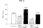
血管内皮細胞によるMMP−1の産生に対するグルコース濃度およびHGF添加の影響
血管内皮細胞(ヒト大動脈由来)を無血清下で、グルコース濃度が0、25mM、50mMとなるような3種の培地で培養し、24時間培養後の培養液上清のMMP−1濃度を測定した、
また各々、グルコース添付の30分前にHGFを100ng/mlで添加した場合と比較した。
上清中のMMP−1濃度が、グルコースの濃度依存的に有意に減少し、HGF処理によりMMP−1の減少が止まり増加することが示された。結果を図5に示す。
参考例3
血管内皮細胞に対するHGFの効果(血管新生に係わる転写因子ETS−1の変化)
参考例1と同様に血管内皮細胞を培養し、細胞中に発現する転写因子ETS−1のmRNA量を測定した。コントロールの内皮細胞の、ETS−1のmRNA量を100%とすると、HGF未添加の血管内皮細胞では高グルコースの条件により有意に減少していた。一方、HGF添加の血管内皮細胞ではコントロールの発現量と比較して同等以上になっていた(P<0.01)。結果を図6に示す。
以上のように、高グルコース条件下の血管内皮細胞では、血管新生のために必須のマトリックス分解酵素であるMMP−1の産生が減少し、血管新生時に発現、増加する転写因子であるETS−1のmRNA発現が低下していた。
上記のことから、高グルコース条件下では血管新生がおこりにくくなっていることが明らかにされた。一方、高グルコース条件下の血管内皮細胞にHGFを添加することにより、MMP−1の産生、ETS−1のmRNAの発現が顕著に増加し、HGFにより血管新生がおこりやすくなっていることが示された。
産業上の利用可能性
本発明のHGF遺伝子を有効成分とする糖尿病性虚血性疾患治療剤は、HGFの発現の減少した糖尿病性虚血患部に特有の血管新生不良を改善し、顕著な血管新生作用を示すことから、虚血患部の血流量を増大させ病状を改善することができる。しかも、本発明の治療剤は患者の病状に応じて1回以上の投与を行うことにより、その目的である血管新生を促進することができる。従ってこれらの効果により、本発明の治療剤は糖尿病性下肢虚血性疾患、糖尿病性虚血性神経障害または、糖尿病性心筋梗塞をより的確に治療することができる。
【図面の簡単な説明】
図1は、参考例1の糖尿病の下肢虚血ラット群および、正常ラットに下肢虚血を惹起したコントロール群における、血液還流比率(Perfusion ratio)の経時的変化を示したグラフである。
図2は、参考例1の糖尿病の下肢虚血ラット群および、正常ラットに下肢虚血を惹起したコントロール群における虚血筋肉内の内在性HGF濃度を示したグラフである。
図3は、実施例1の糖尿病の下肢虚血ラットへのHGF遺伝子投与群および未投与群と、正常ラットに下肢虚血を惹起したコントロール群における、血液還流比率を測定した結果を示したグラフである。
図4は、実施例2の糖尿病の下肢虚血ラットへのHGF遺伝子投与群および未投与群と、正常ラットに下肢虚血を惹起したコントロール群における、下肢虚血部の骨格筋をALP染色し、単位当たりの血管数を比較した結果を示したグラフである。
図5は、参考例2のグルコースを添加した血管内皮細胞のHGF添加群および未添加群と、グルコースを添加していないコントロール群における、培養上清のMMP−1濃度を測定した結果を示すグラフである。
図6は、参考例3のグルコースを添加した血管内皮細胞のHGF添加群および未添加群と、グルコースを添加していないコントロール群における、血管内皮細胞中に発現する転写因子ETS−1のmRNA量を測定した結果を示すグラフである。TECHNICAL FIELD The present invention relates to a therapeutic agent and a therapeutic method for diabetic ischemic disease using a hepatocyte growth factor (HGF) gene. Specifically, the present invention relates to a therapeutic agent for diabetic ischemic disease containing an HGF gene as an active ingredient, a therapeutic method for diabetic ischemic disease characterized by administering the HGF gene to an ischemic site, and the like.
BACKGROUND ART HGF is a protein that has been discovered as a potent growth promoting factor for mature hepatocytes and has been cloned (Biochem Biophys Res Commun, 122, 1450 (1984), Proc. Natl. Acad. Sci. USA, 83, 6489, (1986), FEBS Letter, 22, 231 (1987), Nature, 342, 440 (1989), Proc. Natl. Acad. Sci. USA, 87, 3200 (1991)). Subsequent research shows that HGF not only works in vivo for repair and regeneration of damaged liver as a liver regeneration factor, but also has an angiogenic action and plays a major role in the treatment or prevention of ischemic and arterial diseases. (Sym. Soc. Exp. Biol., 47 cell behavior, 227-234 (1993), Proc. Natl. Acad. Sci., 90, 1937-1941 (1993), Circulation, 97, 381-390. (1998)). That is, it has been reported that when HGF was administered in a rabbit lower limb ischemia model, remarkable angiogenesis was observed, blood flow was improved, blood pressure was suppressed, and ischemic symptoms were improved. Based on these reports, HGF is now considered to be expressed and functioning as one of the angiogenic factors.
Thus, HGF has various functions including the function of an angiogenic factor, and various attempts have been made to utilize it as a medicine. However, what has become a problem here is the half-life of HGF in the blood. The half-life of HGF is as short as about 10 minutes, it is difficult to maintain the blood concentration, and the transfer of an effective amount of HGF to the affected area has become a problem.
In general, in the case of protein preparations, it is common knowledge that many administrations are intravenously. Regarding the administration of HGF to the ischemic disease model, for example, examples of administration into veins and arteries are shown. (Circulation, 97, 381-390 (1998)). Although the effectiveness of HGF for ischemic disease or arterial disease has been clarified by intravenous or intraarterial administration to such an animal model, specific effective methods or doses of HGF have not yet been concluded. Has not been issued. In particular, in the case of HGF protein, there are also problems of half-life and transferability to the affected area as described above, and an effective administration method or dose of HGF protein has not yet been concluded.
On the other hand, recent advances in molecular biology have made it possible to activate cell functions by gene transfer methods, and various efforts have been made. Gene therapy for the heart region using HGF genes has been attempted. It is going to be done. Although there are some reports on gene transfer methods such as intracoronary diffusion injection, there are specific diabetic properties using intramuscular transfer methods for skeletal muscles, such as gene transfer methods to ischemic sites. No examples have been found to have any effect on ischemic disease.
Furthermore, it is known that angiogenesis is unlikely to occur in ischemic diseases accompanied by diabetes or caused by diabetics, and the prognosis is also poor. However, HGF is used for such diabetic ischemic diseases. It is not clear at all whether the gene administration is effective.
DISCLOSURE OF THE INVENTION An object of the present invention is to provide a therapeutic agent and a therapeutic method for diabetic ischemic disease using the HGF gene.
As a result of intensive investigations to clarify whether the HGF gene can be applied to diabetic ischemic diseases, the present inventors directly administer the HGF gene to the affected ischemic region for the disease. It was clarified that a very effective effect can be obtained. Specifically, it has been found that an effective effect can be obtained by administering the HGF gene to the limb skeletal muscle layer in the limb ischemic disease. As described above, it is known that angiogenesis hardly occurs and the prognosis is poor in an ischemic disease that is accompanied by diabetes or is caused by diabetes. Therefore, unlike a simple ischemic disease, whether or not the HGF gene is effective against diabetic ischemic disease has not been clarified so far, but the present invention has been demonstrated for the first time.
Since such treatment with the HGF gene is a non-invasive treatment method, the gene can be administered any number of times according to the disease state.
That is, the gist of the present invention is as follows.
(1) A therapeutic agent for diabetic ischemic disease, comprising a hepatocyte growth factor (HGF) gene as an active ingredient,
(2) The therapeutic agent according to (1) above for administration to an ischemic site,
(3) The therapeutic agent according to (1) or (2) above, wherein the diabetic ischemic disease is diabetic lower limb ischemic disease, diabetic ischemic neuropathy or diabetic myocardial infarction,
(4) The therapeutic agent according to (3) above, wherein the diabetic ischemic disease is diabetic lower limb ischemic disease,
(5) The therapeutic agent according to any one of (1) to (4) above, which is administered into the muscle at the ischemic site,
(6) The therapeutic agent according to any one of (1) to (5) above, wherein the HGF gene is in the form of Sendai virus (HVJ) -liposomes,
(7) The therapeutic agent according to any one of (1) to (6) above, wherein the therapeutic agent is administered a plurality of times as necessary.
(8) The therapeutic agent according to any one of (1) to (7) above, wherein at least 50 μg is used as the HGF gene,
(9) A method for treating diabetic ischemic disease, comprising introducing an HGF gene into a human,
(10) The method of (9) above, wherein the HGF gene is administered to the ischemic site,
(11) The method of (9) or (10) above, wherein the diabetic ischemic disease is diabetic lower limb ischemic disease, diabetic ischemic neuropathy or diabetic myocardial infarction,
(12) The method of (11) above, wherein the diabetic ischemic disease is a diabetic lower limb ischemic disease,
(13) The method according to any one of (9) to (12) above, wherein the HGF gene is administered into the muscle at the ischemic site,
(14) The treatment method according to any of (9) to (13) above, wherein the HGF gene is in the form of Sendai virus (HVJ) -liposomes,
(15) The method according to any one of (9) to (14) above, wherein the HGF gene is administered multiple times as necessary.
(16) The method according to any one of (9) to (15) above, wherein at least 50 μg is administered as the HGF gene,
(17) Use of the HGF gene for the manufacture of a therapeutic agent for diabetic ischemic disease,
(18) The use of (17) above, wherein the diabetic ischemic disease is diabetic lower limb ischemic disease, diabetic ischemic neuropathy or diabetic myocardial infarction,
(19) Use of (18) above, wherein the diabetic ischemic disease is diabetic lower limb ischemic disease,
(20) The use according to any one of (17) to (19) above, wherein the HGF gene is in the form of Sendai virus (HVJ) -liposomes,
(21) The use according to any one of (17) to (20) above, wherein at least 50 μg is used as the HGF gene.
BEST MODE FOR CARRYING OUT THE INVENTION “HGF gene” used in the present invention refers to a gene capable of expressing HGF (HGF protein). Specifically, Nature, 342, 440 (1989), Japanese Patent No. 2777678, Biochem. Biophys. Res. Commun. , 163,967 (1989), Biochem. Biophys. Res. Commun. , 172, 321 (1990), and the like, which are incorporated into an appropriate expression vector (non-viral vector, viral vector) as described below. Here, the base sequence of cDNA encoding HGF is described in the above-mentioned document, and is also registered in a database such as Genbank. Therefore, HGF cDNA can be cloned by using an appropriate DNA portion as a PCR primer based on these sequence information and performing, for example, RT-PCR reaction on mRNA derived from liver or leukocytes. These cloning methods are described in, for example, Molecular Cloning 2nd Edt. , Cold Spring Harbor Laboratory Press (1989), etc., can be easily performed by those skilled in the art.
Furthermore, the HGF gene of the present invention is not limited to those described above, and can be used as the HGF gene of the present invention as long as the expressed protein is a gene having substantially the same action as HGF. That is, 1) DNA that hybridizes with the cDNA under stringent conditions, or 2) one or more (preferably several) amino acids are substituted or deleted from the amino acid sequence of the protein encoded by the cDNA. In addition, DNA encoding a protein consisting of an added amino acid sequence, and the like encoding a protein having an action as HGF are included in the category of the HGF gene of the present invention. Here, the DNAs 1) and 2) can be easily obtained by, for example, site-directed mutagenesis, PCR, or ordinary hybridization, and more specifically, a basic document such as Molecular Cloning. Can be done with reference to.
Next, the gene introduction method, the introduction form, the introduction amount, etc. used in the gene therapy of the present invention will be described.
When a gene therapy agent containing the HGF gene as an active ingredient is administered to a patient, the administration mode is roughly divided into two cases, using a non-viral vector and using a viral vector. The preparation method and administration method are explained in detail (separate volume experimental medicine, basic technology of gene therapy, Yodosha, 1996, separate volume experimental medicine, gene transfer & expression analysis experiment method, Yodosha, 1997, Japanese gene therapy. Gene Therapy Development Research Handbook, NTS, 1999). This will be specifically described below.
A. When a non-viral vector is used The target gene can be introduced into cells and tissues by the following method using a recombinant expression vector in which the target gene is incorporated into a conventional gene expression vector. .
Examples of the method for introducing a gene into a cell include a lipofection method, a phosphate-calcium coprecipitation method, a DEAE-dextran method, and a direct DNA injection method using a micro glass tube.
In addition, gene transfer methods to tissues include gene transfer methods using internal type liposomes, gene transfer methods using electrostatic type liposomes, HVJ-liposome method, and improved HVJ-liposome method (HVJ). -AVE liposome method), receptor-mediated gene introduction method, method of transferring DNA molecules with cells (metal particles) with a particle gun, direct introduction of naked-DNA, introduction method using positively charged polymer, etc. By using the method, it is possible to incorporate the recombinant expression vector into cells.
Among them, HVJ-liposomes are obtained by encapsulating DNA in liposomes made of a lipid bilayer, and further fusing this liposome with inactivated Sendai virus (Hemagglutinating virus of Japan: HVJ). The HVJ-liposome method is characterized by a very high fusion activity with the cell membrane as compared with the conventional liposome method, and is a preferable introduction form. For the preparation method of HVJ-liposomes, refer to the literature (Experimental medicine separate volume, Basic technology of gene therapy, Yodosha, 1996, Gene transfer & expression analysis experiment method, Yodosha, 1997, J. Clin. Invest. 93, 1458- 1464 (1994), Am. J. Physiol.271, R1212-1220 (1996)) and the like. The HVJ liposome method is described in, for example, Molecular Medicine, 30, 1440-1448 (1993), Experimental Medicine, 12, 1822-1826 (1994), Protein / Nucleic Acid / Enzyme, 42, 1806-1813 (1997), etc. Preferably, the method described in Circulation, 92 (Suppl. II), 479-482 (1995) can be mentioned.
The HVJ is preferably a Z strain (available from ATCC), but basically other HVJ strains (for example, ATCC VR-907 and ATCC VR-105) can also be used.
Furthermore, the direct introduction method of naked-DNA is the simplest method among the above methods, and is a preferable introduction method from this viewpoint.
The expression vector used here may be any expression vector as long as the target gene can be expressed in vivo. For example, pCAGGS (Gene 108, 193-200 (1991)) or pBK -Expression vectors such as CMV, pcDNA3.1, pZeoSV (Invitrogen, Stratagene) can be mentioned.
B. When using a viral vector As a viral vector, a method using a viral vector such as a recombinant adenovirus or a retrovirus is typical. More specifically, for example, detoxified retrovirus, adenovirus, adeno-associated virus, herpes virus, vaccinia virus, poxvirus, poliovirus, shinbis virus, Sendai virus, SV40, immunodeficiency virus (HIV), etc. It is possible to introduce a gene into a cell by introducing the gene of interest into a DNA virus or RNA virus of the above and infecting the cell with a recombinant virus.
Among the viral vectors, it is known that adenovirus infection efficiency is much higher than when other viral vectors are used. From this viewpoint, it is preferable to use an adenoviral vector system.
As a method for introducing the gene therapy agent of the present invention into a patient, an in vivo method in which the gene therapy agent is directly introduced into the body, or a certain cell is taken out from a human and introduced into the cell outside the body. There is an ex vivo method for returning the cells to the body (Nikkei Science, April 1994, 20-45, Monthly Pharmaceutical Affairs, 36 (1), 23-48 (1994), Experimental Medicine Extra Number, 12 (15) (1994), Gene Therapy Society of Japan, Gene Therapy Development Research Handbook, NTS, 1999). In the present invention, the in vivo method is preferable.
As a preparation form, various preparation forms (for example, a liquid agent etc.) suitable for each of the above administration forms can be taken. For example, when an injection containing a gene which is an active ingredient is used, the injection can be prepared by a conventional method, for example, dissolved in an appropriate solvent (buffer solution such as PBS, physiological saline, sterilized water, etc.). Then, if necessary, it can be prepared by sterilizing by filtration with a filter or the like and then filling into an aseptic container. A conventional carrier or the like may be added to the injection as necessary. Liposomes such as HVJ-liposomes can be in the form of liposome preparations such as a suspension, a freezing agent, and a centrifugal concentrated freezing agent.
In addition to the HGF gene used in the present invention, a known factor having an angiogenic action can be used in combination or alone. For example, factors such as VEGF and FGF have been reported to have angiogenic effects, and these genes can be used. In addition, growth factors such as EGF have been reported to repair various cell damages in tissues, and these genes can also be used.
The diabetic ischemic disease referred to in the present invention is diabetic lower limb ischemic disease, diabetic ischemic neuropathy, diabetic myocardial infarction or the like, and the gene therapy agent of the present invention for any of these diseases Can be applied. Moreover, the gene therapy agent of the present invention can be used not only for patients with severe diabetic ischemic disease but also for mild patients in progress.
For the gene therapy agent of the present invention, an appropriate administration method and administration site are selected in accordance with the disease or symptom to be treated. As an administration method, it is preferable to administer parenterally. The administration site is preferably administered within the ischemic site. Here, the “ischemic site” refers to a site including an ischemic site and its periphery.
In the ischemic site, specifically, administration into a blood vessel or muscle is possible, but it is particularly preferable to administer into the muscle at the ischemic site. In other words, in lower limb ischemic disease, administration into the skeletal muscle at the lower limb ischemia site promotes angiogenesis of the ischemic lesion, improves blood flow, and normalizes the recovery of the function of the ischemic lesion. I can do it. In heart diseases such as myocardial infarction, the same effect can be achieved by administration into the myocardium.
As a preferable administration method, for example, a non-invasive catheter or a non-invasive syringe can be used. Furthermore, a non-invasive catheter or syringe administration method using an echo can be mentioned. As an administration method using a non-invasive catheter, for example, in heart disease, a method of directly injecting an HGF gene into the myocardium from the intraventricular cavity can be mentioned.
By applying the HGF gene of the present invention, it is possible to treat patients with diabetic ischemic disease by active gene transfer. For example, in patients with diabetic leg ischemic disease, If possible, functional recovery is possible for severely ill patients who have no other way than surgical resection.
The dosage of the therapeutic agent of the present invention varies depending on the patient's symptoms and the like, but is about 1 μg to about 50 mg, preferably about 10 μg to about 5 mg, more preferably about 50 μg to about 5 mg as an HGF gene per adult patient. The dose is selected from the range of
The therapeutic agent of the present invention is preferably administered once every several days to several weeks, and the number of administrations is appropriately selected according to the symptoms of the patient. Since the therapeutic agent of the present invention is provided for noninvasive administration, it has a feature that it can be administered any number of times according to the disease state.
EXAMPLES Hereinafter, although an Example demonstrates this invention concretely, this invention is not limited at all by these Examples.
Experimental materials and methods
Preparation of HVJ liposome preparations containing 10 mg of dry lipid (1: 4.8: 2 mixture of phosphatidylserine, phosphatidylcholine, cholesterol) and HGF gene (100 μg) -HMG1 (
Experimental animal Administration group: Diabetic rat (DM-rat) lower limb ischemic rat Control rat: Normal rat lower limb ischemic rat
Administration method of HVJ liposome preparation A 16-week-old diabetic rat (6 mice per group) induced by intraperitoneal administration of streptozotocin was subjected to partial surgical resection of the unilateral femoral artery, An ischemic state was created in the lower limb.
The HGF gene-containing HVJ-liposome formulation was injected into the lower limb ischemic skeletal muscle.
Contents of study After administration of the liposome preparation, lower limb blood flow was measured using a laser Doppler imager (LDI) by laser scattered light analysis as an index of collateral circulation formation and blood flow improvement effect. The average value of the color histogram of the ischemic lower limb with respect to the normal lower limb was defined as the blood reflux ratio (perfusion ratio).
Capillary density in the lower limb ischemia was measured by alkaline phosphatase (ALP) staining and compared between diabetic and limb ischemic rats. Or it compared with the administration group of the HGF gene, and the non-administration group.
Reference example 1
Status of HGF expression at lower limb ischemia in diabetic rats 16-week-old diabetic rats (6 mice per group) induced by intraperitoneal administration of streptozotocin and 16-week-old normal rats as control group (1 A group of 6 animals) underwent partial surgical resection of the unilateral femoral artery, resulting in an ischemic condition in the lower limbs.
One week later, the blood reflux ratio at the ischemic site was measured with a laser Doppler imager. The blood perfusion rate at the ischemic site in diabetic lower limb ischemic rats was much lower than the blood perfusion rate in control lower limb ischemic rats (see FIG. 1).
After 3 weeks and 5 weeks, the blood reflux ratio at the ischemic site was measured. Similarly, the blood reflux ratio at the ischemic site in diabetic lower limb ischemic rats was higher than that of the control lower limb ischemic rats. It was much lower (see Figure 1).
The concentration of endogenous HGF in the muscle was significantly lower in the muscle at the ischemic site of diabetic lower limb ischemic rats than in the muscle of control lower limb ischemic rats. This result indicates that the cause of poor angiogenesis in diabetes is due to a decrease in endogenous HGF in the muscle. For this reason, in diabetic limb ischemic rats, angiogenesis is unlikely to occur and collateral circulation is not developed (see FIG. 2).
Example 1
Effect of HGF gene therapy on diabetic limb ischemia rats (I)
Surgical excision of part of the unilateral femoral artery for 16-week-old diabetic rats (6 animals per group) and 16-week-old normal rats (6 animals per group) induced by intraperitoneal administration of streptozotocin To bring about an ischemic condition in the lower limbs. After surgical resection of the femoral artery, an HVJ-liposome preparation containing the HGF gene (50 μg) was introduced into the muscle at the lower limb ischemia site.
Three weeks later, the blood reflux ratio at the ischemic site was measured with a laser Doppler imager. The blood reflux ratio at the ischemic site in diabetic limb ischemic rats administered with the HGF gene showed a marked increase compared to the control limb ischemic rats and the untreated diabetic limb ischemic rats.
Assuming that the blood reflux ratio of control lower limb ischemic rats was 100%, it was 67% in diabetic limb ischemic rats not administered with HGF gene, and 129% in diabetic limb ischemic rats administered with HGF gene. The results are shown in FIG. 3 (see FIG. 1).
Example 2
Effect of HGF gene therapy on diabetic limb ischemic rats (II)
Diabetic lower limb ischemic rats and control lower limb ischemic rats treated in the same manner as described above were prepared and subjected to HGF gene therapy. Five weeks later, the skeletal muscle at the lower limb ischemia site of each rat was taken out and ALP-stained to compare the number of blood vessels per unit area. In diabetic limb ischemic rats not administered with the HGF gene, the number of blood vessels was significantly smaller than in the control limb ischemic rats, and in diabetic limb ischemic rats administered with the HGF gene, the number of blood vessels was significantly increased. The results are shown in FIG.
Reference example 2
Effect of glucose concentration and HGF addition on MMP-1 production by vascular endothelial cells Three types of vascular endothelial cells (derived from human aorta) under serum-free conditions with glucose concentrations of 0, 25 mM, 50 mM Cultured in a medium, the MMP-1 concentration of the culture supernatant after 24 hours of culture was measured,
Moreover, each compared with the case where HGF was added at 100 ng / ml 30 minutes before glucose attachment.
It was shown that the concentration of MMP-1 in the supernatant decreased significantly depending on the glucose concentration, and that the decrease in MMP-1 stopped and increased by HGF treatment. The results are shown in FIG.
Reference example 3
Effect of HGF on vascular endothelial cells (change in transcription factor ETS-1 involved in angiogenesis)
Vascular endothelial cells were cultured in the same manner as in Reference Example 1, and the mRNA level of the transcription factor ETS-1 expressed in the cells was measured. Assuming that the amount of ETS-1 mRNA in the control endothelial cells was 100%, vascular endothelial cells to which HGF had not been added were significantly decreased due to high glucose conditions. On the other hand, HGF-added vascular endothelial cells were equal to or greater than the control expression level (P <0.01). The results are shown in FIG.
As described above, in vascular endothelial cells under high glucose conditions, production of MMP-1, which is a matrix degrading enzyme essential for angiogenesis, decreases, and ETS-1, which is a transcription factor that is expressed and increased during angiogenesis. MRNA expression was reduced.
From the above, it was revealed that angiogenesis is less likely to occur under high glucose conditions. On the other hand, it has been shown that the addition of HGF to vascular endothelial cells under high glucose conditions markedly increases the production of MMP-1 and the expression of ETS-1 mRNA, which facilitates angiogenesis by HGF. It was done.
INDUSTRIAL APPLICABILITY The therapeutic agent for diabetic ischemic disease comprising the HGF gene of the present invention as an active ingredient improves the poor angiogenesis peculiar to the affected area of diabetic ischemia in which the expression of HGF is reduced, and remarkable angiogenesis. Since it exhibits an action, it is possible to increase the blood flow volume in the affected area of ischemia and improve the disease state. And the therapeutic agent of this invention can promote the angiogenesis which is the objective by administering 1 time or more according to a patient's medical condition. Therefore, due to these effects, the therapeutic agent of the present invention can more accurately treat diabetic lower limb ischemic disease, diabetic ischemic neuropathy or diabetic myocardial infarction.
[Brief description of the drawings]
FIG. 1 is a graph showing changes over time in blood reflux ratio (Perfusion ratio) in a diabetic lower limb ischemic rat group of Reference Example 1 and a control group in which normal rat is induced to undergo lower limb ischemia.
FIG. 2 is a graph showing the endogenous HGF concentration in ischemic muscles in the diabetic lower limb ischemic rat group of Reference Example 1 and the control group in which normal rat is induced limb ischemia.
FIG. 3 is a graph showing the results of measuring blood reflux ratios in the HGF gene-administered group and the non-administered group in diabetic lower limb ischemic rats of Example 1 and in the control group that induced lower limb ischemia in normal rats. It is.
FIG. 4 shows ALP staining of skeletal muscle in the ischemic part of the lower limb in the HGF gene-administered group and the non-administered group of diabetic lower limb ischemic rats of Example 2 and the control group in which normal rat induced lower limb ischemia It is the graph which showed the result of having compared the number of blood vessels per unit.
FIG. 5 is a graph showing the results of measuring the MMP-1 concentration of the culture supernatant in the HGF-added and non-added groups of vascular endothelial cells to which glucose of Reference Example 2 was added and the control group to which glucose was not added. It is.
FIG. 6 shows the amount of mRNA of the transcription factor ETS-1 expressed in vascular endothelial cells in the HGF added and non-added groups of vascular endothelial cells to which glucose of Reference Example 3 was added and in the control group to which glucose was not added. It is a graph which shows the result of having measured.
Claims (13)
Applications Claiming Priority (2)
| Application Number | Priority Date | Filing Date | Title |
|---|---|---|---|
| JP30998499 | 1999-10-29 | ||
| PCT/JP2000/007502 WO2001032220A1 (en) | 1999-10-29 | 2000-10-26 | Gene therapy for diabetic ischemic disease |
Publications (1)
| Publication Number | Publication Date |
|---|---|
| JP3877148B2 true JP3877148B2 (en) | 2007-02-07 |
Family
ID=17999746
Family Applications (1)
| Application Number | Title | Priority Date | Filing Date |
|---|---|---|---|
| JP2001534424A Expired - Lifetime JP3877148B2 (en) | 1999-10-29 | 2000-10-26 | Gene therapy for diabetic ischemic disease |
Country Status (15)
| Country | Link |
|---|---|
| US (3) | US20080268030A1 (en) |
| EP (1) | EP1142590B8 (en) |
| JP (1) | JP3877148B2 (en) |
| KR (1) | KR20010108053A (en) |
| CN (1) | CN1182874C (en) |
| AT (1) | ATE409494T1 (en) |
| AU (1) | AU778826B2 (en) |
| CA (1) | CA2356701C (en) |
| CY (1) | CY1110445T1 (en) |
| DE (1) | DE60040383D1 (en) |
| DK (1) | DK1142590T3 (en) |
| ES (1) | ES2313907T3 (en) |
| HK (1) | HK1041211A1 (en) |
| PT (1) | PT1142590E (en) |
| WO (1) | WO2001032220A1 (en) |
Families Citing this family (14)
| Publication number | Priority date | Publication date | Assignee | Title |
|---|---|---|---|---|
| WO2001026694A1 (en) | 1999-10-08 | 2001-04-19 | Medgene Bioscience, Inc. | Gene therapy for cardiomyopathy |
| EP1391214A4 (en) | 2001-05-09 | 2006-05-17 | Anges Mg Inc | Gene transfer of angiogenic factor for skin disease |
| AU2002349583B2 (en) * | 2001-11-28 | 2007-11-22 | Anges Mg, Inc. | Genetic remedies for neurodegenerative diseases |
| KR100562824B1 (en) * | 2002-03-20 | 2006-03-23 | 주식회사 바이로메드 | Hybrid hepatocyte growth factor gene that expresses two genes of hepatocyte growth factor with high gene expression efficiency |
| US20080213348A1 (en) * | 2002-06-06 | 2008-09-04 | Anges Mg, Inc. | Agents for gene therapy of cerebrovascular disorders |
| CA2500278A1 (en) * | 2002-10-02 | 2004-04-15 | Anges Mg, Inc. | Pharmaceutical preparation for hearing impairment |
| JP4775940B2 (en) * | 2004-06-29 | 2011-09-21 | アンジェスMg株式会社 | Allodynia treatment, improvement, prevention agent |
| US20090202606A1 (en) * | 2008-01-25 | 2009-08-13 | Viromed Co., Ltd. | Treatment and Prevention of Cardiac Conditions Using Two or More Isoforms of Hepatocyte Growth Factor |
| ES2556711T3 (en) * | 2008-04-09 | 2016-01-19 | Viromed Co., Ltd. | Lyophilized DNA formulations for increased expression of plasmid DNA |
| EP2840957B1 (en) * | 2012-04-27 | 2024-01-03 | Stryker European Operations Limited | Optical coherent imaging medical device and method |
| MX375916B (en) | 2013-10-22 | 2025-03-07 | Helixmith Co Ltd | COMPOSITION FOR PREVENTING OR TREATING AMYOTROPHIC LATERAL SCLEROSIS USING TWO OR MORE ISOFORMS OF THE HEPATOCYTE GROWTH FACTOR. |
| RU2691654C1 (en) * | 2018-06-05 | 2019-06-17 | Юрий Валентинович Червяков | Method of treating chronic ischemia of lower extremities of atherosclerotic genesis in femoral and popliteal arteries occlusion |
| JP7212230B2 (en) | 2018-07-19 | 2023-01-25 | ヘリックスミス カンパニー, リミテッド | Lyophilized pharmaceutical composition for naked DNA gene therapy |
| WO2024186828A1 (en) | 2023-03-06 | 2024-09-12 | Wisconsin Alumni Research Foundation | Methods of inducing cardiac cell proliferation and inducing heart regeneration |
Family Cites Families (29)
| Publication number | Priority date | Publication date | Assignee | Title |
|---|---|---|---|---|
| US4576177A (en) * | 1983-02-18 | 1986-03-18 | Webster Wilton W Jr | Catheter for removing arteriosclerotic plaque |
| US6088613A (en) * | 1989-12-22 | 2000-07-11 | Imarx Pharmaceutical Corp. | Method of magnetic resonance focused surgical and therapeutic ultrasound |
| US5298422A (en) * | 1991-11-06 | 1994-03-29 | Baylor College Of Medicine | Myogenic vector systems |
| US5631237A (en) * | 1992-12-22 | 1997-05-20 | Dzau; Victor J. | Method for producing in vivo delivery of therapeutic agents via liposomes |
| TW360548B (en) * | 1993-04-08 | 1999-06-11 | Powderject Res Ltd | Products for therapeutic use |
| US5652225A (en) * | 1994-10-04 | 1997-07-29 | St. Elizabeth's Medical Center Of Boston, Inc. | Methods and products for nucleic acid delivery |
| US5817022A (en) * | 1995-03-28 | 1998-10-06 | Sonometrics Corporation | System for displaying a 2-D ultrasound image within a 3-D viewing environment |
| US5756122A (en) * | 1995-06-07 | 1998-05-26 | Georgetown University | Liposomally encapsulated nucleic acids having high entrapment efficiencies, method of manufacturer and use thereof for transfection of targeted cells |
| ES2240999T3 (en) * | 1995-08-29 | 2005-10-16 | Anges Mg, Inc. | MEDICINAL CONTAINING A GENE OF HGF. |
| US6121246A (en) * | 1995-10-20 | 2000-09-19 | St. Elizabeth's Medical Center Of Boston, Inc. | Method for treating ischemic tissue |
| US5922685A (en) * | 1996-06-05 | 1999-07-13 | Powderject Vaccines, Inc. | IL-12 gene therapy of tumors |
| US6893664B1 (en) * | 1996-06-17 | 2005-05-17 | Powderject Research Limited | Particle delivery techniques |
| EP0922102B1 (en) * | 1996-07-03 | 2010-04-21 | Genentech, Inc. | Hepatocyte growth factor receptor agonists and uses thereof |
| US5980887A (en) * | 1996-11-08 | 1999-11-09 | St. Elizabeth's Medical Center Of Boston | Methods for enhancing angiogenesis with endothelial progenitor cells |
| US6024703A (en) * | 1997-05-07 | 2000-02-15 | Eclipse Surgical Technologies, Inc. | Ultrasound device for axial ranging |
| GB9709453D0 (en) * | 1997-05-10 | 1997-07-02 | Imp Cancer Res Tech | Polypedtides and their use in therapy |
| WO1999036103A1 (en) * | 1998-01-16 | 1999-07-22 | Mcgill University | Prevention and treatment of neuropathy by hepatocyte growth factor |
| CA2261255A1 (en) * | 1998-02-13 | 1999-08-13 | Ethicon, Inc. | Assessment of ischemic wound healing therapeutics |
| US6066123A (en) * | 1998-04-09 | 2000-05-23 | The Board Of Trustees Of The Leland Stanford Junior University | Enhancement of bioavailability by use of focused energy delivery to a target tissue |
| US6958147B1 (en) * | 1998-10-26 | 2005-10-25 | Licentia Ltd | Use of VEGF-C to prevent restenosis |
| US7125856B1 (en) * | 1999-04-15 | 2006-10-24 | St. Elizabeth's Medical Center Of Boston, Inc. | Angiogenic growth factors for treatment of peripheral neuropathy |
| CA2365434A1 (en) * | 1999-04-15 | 2000-10-26 | St. Elizabeth's Medical Center Of Boston | Angiogenic growth factors for treatment of peripheral neuropathy |
| US6440945B1 (en) * | 1999-05-27 | 2002-08-27 | Instituto Dermopatico Dell'immacolata | Method of inducing angiogenesis in nonis chemic skeletal muscle |
| WO2001026694A1 (en) * | 1999-10-08 | 2001-04-19 | Medgene Bioscience, Inc. | Gene therapy for cardiomyopathy |
| EP1300158B9 (en) * | 2000-06-27 | 2008-11-19 | AnGes MG, Inc. | Pharmaceutical compositions for angiogenic therapy |
| EP1391214A4 (en) * | 2001-05-09 | 2006-05-17 | Anges Mg Inc | Gene transfer of angiogenic factor for skin disease |
| US20060074036A1 (en) * | 2002-06-06 | 2006-04-06 | Ryuichi Morishita | Agents for gene therapy of cerebrovascular disorders |
| US20080213348A1 (en) * | 2002-06-06 | 2008-09-04 | Anges Mg, Inc. | Agents for gene therapy of cerebrovascular disorders |
| DE60333476D1 (en) * | 2002-12-02 | 2010-09-02 | Anges Mg Inc | COMPOSITIONS FOR TREATING OR PREVENTING ANGIOGENESIS-DEPENDENT SYMPTOMS |
-
2000
- 2000-10-26 PT PT00971697T patent/PT1142590E/en unknown
- 2000-10-26 WO PCT/JP2000/007502 patent/WO2001032220A1/en active IP Right Grant
- 2000-10-26 DK DK00971697T patent/DK1142590T3/en active
- 2000-10-26 AT AT00971697T patent/ATE409494T1/en active
- 2000-10-26 DE DE60040383T patent/DE60040383D1/en not_active Expired - Lifetime
- 2000-10-26 CA CA2356701A patent/CA2356701C/en not_active Expired - Lifetime
- 2000-10-26 ES ES00971697T patent/ES2313907T3/en not_active Expired - Lifetime
- 2000-10-26 KR KR1020017008242A patent/KR20010108053A/en not_active Application Discontinuation
- 2000-10-26 AU AU10523/01A patent/AU778826B2/en not_active Expired
- 2000-10-26 EP EP00971697A patent/EP1142590B8/en not_active Expired - Lifetime
- 2000-10-26 JP JP2001534424A patent/JP3877148B2/en not_active Expired - Lifetime
- 2000-10-26 CN CNB00803835XA patent/CN1182874C/en not_active Expired - Lifetime
-
2002
- 2002-04-09 HK HK02102637.3A patent/HK1041211A1/en unknown
-
2008
- 2008-04-04 US US12/080,715 patent/US20080268030A1/en not_active Abandoned
- 2008-12-22 CY CY20081101477T patent/CY1110445T1/en unknown
-
2010
- 2010-11-02 US US12/938,128 patent/US20110045061A1/en not_active Abandoned
-
2015
- 2015-09-28 US US14/867,866 patent/US20160250290A1/en not_active Abandoned
Also Published As
| Publication number | Publication date |
|---|---|
| KR20010108053A (en) | 2001-12-07 |
| EP1142590B1 (en) | 2008-10-01 |
| EP1142590A1 (en) | 2001-10-10 |
| CA2356701C (en) | 2011-02-15 |
| PT1142590E (en) | 2008-10-27 |
| CN1339973A (en) | 2002-03-13 |
| HK1041211A1 (en) | 2002-07-05 |
| EP1142590B8 (en) | 2008-11-26 |
| AU1052301A (en) | 2001-05-14 |
| DE60040383D1 (en) | 2008-11-13 |
| CN1182874C (en) | 2005-01-05 |
| US20110045061A1 (en) | 2011-02-24 |
| CY1110445T1 (en) | 2015-04-29 |
| WO2001032220A1 (en) | 2001-05-10 |
| DK1142590T3 (en) | 2009-01-26 |
| CA2356701A1 (en) | 2001-05-10 |
| EP1142590A4 (en) | 2005-01-26 |
| US20080268030A1 (en) | 2008-10-30 |
| ES2313907T3 (en) | 2009-03-16 |
| US20160250290A1 (en) | 2016-09-01 |
| AU778826B2 (en) | 2004-12-23 |
| ATE409494T1 (en) | 2008-10-15 |
Similar Documents
| Publication | Publication Date | Title |
|---|---|---|
| US20160250290A1 (en) | Gene therapy for diabetic ischemic disease | |
| JP3865632B2 (en) | Cardiomyopathy gene therapy | |
| JP4441263B2 (en) | Gene therapy for neurodegenerative diseases | |
| US20020172663A1 (en) | Localized myocardial injection method for treating ischemic myocardium | |
| JPH10501423A (en) | Gene transfer-mediated angiogenesis therapy | |
| CN1198675A (en) | Drugs containing the HGF gene | |
| AU2002320348B2 (en) | Enhancement of transfection of DNA into the liver | |
| US20020082204A1 (en) | Treatment of inflammation with p20 | |
| JPWO2003103721A1 (en) | Cerebrovascular disorder gene therapy agent | |
| TWI309570B (en) | ||
| JP2002507584A (en) | Use of scattering factors to enhance angiogenesis | |
| Naimark et al. | Adenovirus–catheter compatibility increases gene expression after delivery to porcine myocardium | |
| TWI287999B (en) | Gene therapy for cardiomyopathy | |
| JP2005097118A (en) | Cell transplant establishment promoter comprising HGF gene |
Legal Events
| Date | Code | Title | Description |
|---|---|---|---|
| A131 | Notification of reasons for refusal |
Free format text: JAPANESE INTERMEDIATE CODE: A131 Effective date: 20051115 |
|
| A521 | Request for written amendment filed |
Free format text: JAPANESE INTERMEDIATE CODE: A821 Effective date: 20051207 |
|
| RD02 | Notification of acceptance of power of attorney |
Free format text: JAPANESE INTERMEDIATE CODE: A7422 Effective date: 20051207 |
|
| A521 | Request for written amendment filed |
Free format text: JAPANESE INTERMEDIATE CODE: A821 Effective date: 20051207 |
|
| A521 | Request for written amendment filed |
Free format text: JAPANESE INTERMEDIATE CODE: A523 Effective date: 20060113 |
|
| A521 | Request for written amendment filed |
Free format text: JAPANESE INTERMEDIATE CODE: A523 Effective date: 20060131 |
|
| TRDD | Decision of grant or rejection written | ||
| A01 | Written decision to grant a patent or to grant a registration (utility model) |
Free format text: JAPANESE INTERMEDIATE CODE: A01 Effective date: 20061025 |
|
| A61 | First payment of annual fees (during grant procedure) |
Free format text: JAPANESE INTERMEDIATE CODE: A61 Effective date: 20061026 |
|
| R150 | Certificate of patent or registration of utility model |
Free format text: JAPANESE INTERMEDIATE CODE: R150 Ref document number: 3877148 Country of ref document: JP Free format text: JAPANESE INTERMEDIATE CODE: R150 |
|
| FPAY | Renewal fee payment (event date is renewal date of database) |
Free format text: PAYMENT UNTIL: 20091110 Year of fee payment: 3 |
|
| FPAY | Renewal fee payment (event date is renewal date of database) |
Free format text: PAYMENT UNTIL: 20201110 Year of fee payment: 14 |
|
| R250 | Receipt of annual fees |
Free format text: JAPANESE INTERMEDIATE CODE: R250 |
|
| S533 | Written request for registration of change of name |
Free format text: JAPANESE INTERMEDIATE CODE: R313533 |
|
| R350 | Written notification of registration of transfer |
Free format text: JAPANESE INTERMEDIATE CODE: R350 |
|
| R153 | Grant of patent term extension |
Free format text: JAPANESE INTERMEDIATE CODE: R153 |
|
| R250 | Receipt of annual fees |
Free format text: JAPANESE INTERMEDIATE CODE: R250 |