JP3586743B2 - Measurement method with corrected hematocrit value - Google Patents
Measurement method with corrected hematocrit value Download PDFInfo
- Publication number
- JP3586743B2 JP3586743B2 JP29792295A JP29792295A JP3586743B2 JP 3586743 B2 JP3586743 B2 JP 3586743B2 JP 29792295 A JP29792295 A JP 29792295A JP 29792295 A JP29792295 A JP 29792295A JP 3586743 B2 JP3586743 B2 JP 3586743B2
- Authority
- JP
- Japan
- Prior art keywords
- capillary chamber
- blood
- light
- reagent layer
- optical change
- Prior art date
- Legal status (The legal status is an assumption and is not a legal conclusion. Google has not performed a legal analysis and makes no representation as to the accuracy of the status listed.)
- Expired - Fee Related
Links
- 238000005534 hematocrit Methods 0.000 title claims description 17
- 238000000691 measurement method Methods 0.000 title claims description 4
- 239000003153 chemical reaction reagent Substances 0.000 claims description 56
- 210000004369 blood Anatomy 0.000 claims description 33
- 239000008280 blood Substances 0.000 claims description 33
- 230000003287 optical effect Effects 0.000 claims description 31
- 238000000034 method Methods 0.000 claims description 18
- 238000005259 measurement Methods 0.000 claims description 17
- 230000036770 blood supply Effects 0.000 claims description 4
- 238000002834 transmittance Methods 0.000 claims description 2
- 239000007788 liquid Substances 0.000 description 9
- 239000002245 particle Substances 0.000 description 8
- WQZGKKKJIJFFOK-GASJEMHNSA-N Glucose Natural products OC[C@H]1OC(O)[C@H](O)[C@@H](O)[C@@H]1O WQZGKKKJIJFFOK-GASJEMHNSA-N 0.000 description 6
- GWEVSGVZZGPLCZ-UHFFFAOYSA-N Titan oxide Chemical compound O=[Ti]=O GWEVSGVZZGPLCZ-UHFFFAOYSA-N 0.000 description 6
- HVYWMOMLDIMFJA-DPAQBDIFSA-N cholesterol Chemical compound C1C=C2C[C@@H](O)CC[C@]2(C)[C@@H]2[C@@H]1[C@@H]1CC[C@H]([C@H](C)CCCC(C)C)[C@@]1(C)CC2 HVYWMOMLDIMFJA-DPAQBDIFSA-N 0.000 description 6
- 239000008103 glucose Substances 0.000 description 6
- RLFWWDJHLFCNIJ-UHFFFAOYSA-N 4-aminoantipyrine Chemical compound CN1C(C)=C(N)C(=O)N1C1=CC=CC=C1 RLFWWDJHLFCNIJ-UHFFFAOYSA-N 0.000 description 4
- 239000004793 Polystyrene Substances 0.000 description 4
- 238000012937 correction Methods 0.000 description 4
- 210000003743 erythrocyte Anatomy 0.000 description 4
- -1 polyethylene terephthalate Polymers 0.000 description 4
- 229920002223 polystyrene Polymers 0.000 description 4
- 239000000243 solution Substances 0.000 description 4
- 239000000126 substance Substances 0.000 description 4
- 102000004316 Oxidoreductases Human genes 0.000 description 3
- 108090000854 Oxidoreductases Proteins 0.000 description 3
- 102000003992 Peroxidases Human genes 0.000 description 3
- LEHOTFFKMJEONL-UHFFFAOYSA-N Uric Acid Chemical compound N1C(=O)NC(=O)C2=C1NC(=O)N2 LEHOTFFKMJEONL-UHFFFAOYSA-N 0.000 description 3
- TVWHNULVHGKJHS-UHFFFAOYSA-N Uric acid Natural products N1C(=O)NC(=O)C2NC(=O)NC21 TVWHNULVHGKJHS-UHFFFAOYSA-N 0.000 description 3
- WQZGKKKJIJFFOK-VFUOTHLCSA-N beta-D-glucose Chemical compound OC[C@H]1O[C@@H](O)[C@H](O)[C@@H](O)[C@@H]1O WQZGKKKJIJFFOK-VFUOTHLCSA-N 0.000 description 3
- 230000017531 blood circulation Effects 0.000 description 3
- 238000006243 chemical reaction Methods 0.000 description 3
- 235000012000 cholesterol Nutrition 0.000 description 3
- 238000001514 detection method Methods 0.000 description 3
- 229920001477 hydrophilic polymer Polymers 0.000 description 3
- 239000004816 latex Substances 0.000 description 3
- 229920000126 latex Polymers 0.000 description 3
- 230000035699 permeability Effects 0.000 description 3
- 229920000139 polyethylene terephthalate Polymers 0.000 description 3
- 239000005020 polyethylene terephthalate Substances 0.000 description 3
- 239000002243 precursor Substances 0.000 description 3
- 238000012360 testing method Methods 0.000 description 3
- 239000004408 titanium dioxide Substances 0.000 description 3
- 229940116269 uric acid Drugs 0.000 description 3
- CDGBQMHYFARRCC-UHFFFAOYSA-N 3-(n-ethyl-3,5-dimethylanilino)-2-hydroxypropane-1-sulfonic acid Chemical compound OS(=O)(=O)CC(O)CN(CC)C1=CC(C)=CC(C)=C1 CDGBQMHYFARRCC-UHFFFAOYSA-N 0.000 description 2
- VTYYLEPIZMXCLO-UHFFFAOYSA-L Calcium carbonate Chemical compound [Ca+2].[O-]C([O-])=O VTYYLEPIZMXCLO-UHFFFAOYSA-L 0.000 description 2
- 229920013683 Celanese Polymers 0.000 description 2
- 102000004190 Enzymes Human genes 0.000 description 2
- 108090000790 Enzymes Proteins 0.000 description 2
- 108010010803 Gelatin Proteins 0.000 description 2
- 239000004366 Glucose oxidase Substances 0.000 description 2
- 108010015776 Glucose oxidase Proteins 0.000 description 2
- 229920000544 Gore-Tex Polymers 0.000 description 2
- XLOMVQKBTHCTTD-UHFFFAOYSA-N Zinc monoxide Chemical compound [Zn]=O XLOMVQKBTHCTTD-UHFFFAOYSA-N 0.000 description 2
- 238000004458 analytical method Methods 0.000 description 2
- QVGXLLKOCUKJST-UHFFFAOYSA-N atomic oxygen Chemical compound [O] QVGXLLKOCUKJST-UHFFFAOYSA-N 0.000 description 2
- TZCXTZWJZNENPQ-UHFFFAOYSA-L barium sulfate Chemical compound [Ba+2].[O-]S([O-])(=O)=O TZCXTZWJZNENPQ-UHFFFAOYSA-L 0.000 description 2
- 239000011248 coating agent Substances 0.000 description 2
- 238000000576 coating method Methods 0.000 description 2
- 239000006103 coloring component Substances 0.000 description 2
- 230000000694 effects Effects 0.000 description 2
- 239000000839 emulsion Substances 0.000 description 2
- 229940088598 enzyme Drugs 0.000 description 2
- 239000008273 gelatin Substances 0.000 description 2
- 229920000159 gelatin Polymers 0.000 description 2
- 235000019322 gelatine Nutrition 0.000 description 2
- 235000011852 gelatine desserts Nutrition 0.000 description 2
- 229940116332 glucose oxidase Drugs 0.000 description 2
- 235000019420 glucose oxidase Nutrition 0.000 description 2
- JVTAAEKCZFNVCJ-UHFFFAOYSA-N lactic acid Chemical compound CC(O)C(O)=O JVTAAEKCZFNVCJ-UHFFFAOYSA-N 0.000 description 2
- 239000000203 mixture Substances 0.000 description 2
- 229910052760 oxygen Inorganic materials 0.000 description 2
- 239000001301 oxygen Substances 0.000 description 2
- 108040007629 peroxidase activity proteins Proteins 0.000 description 2
- IXPNQXFRVYWDDI-UHFFFAOYSA-N 1-methyl-2,4-dioxo-1,3-diazinane-5-carboximidamide Chemical compound CN1CC(C(N)=N)C(=O)NC1=O IXPNQXFRVYWDDI-UHFFFAOYSA-N 0.000 description 1
- 229920001817 Agar Polymers 0.000 description 1
- 229920000742 Cotton Polymers 0.000 description 1
- 101710088194 Dehydrogenase Proteins 0.000 description 1
- 229920002307 Dextran Polymers 0.000 description 1
- 102000001554 Hemoglobins Human genes 0.000 description 1
- 108010054147 Hemoglobins Proteins 0.000 description 1
- 229920002153 Hydroxypropyl cellulose Polymers 0.000 description 1
- 239000004743 Polypropylene Substances 0.000 description 1
- 239000004372 Polyvinyl alcohol Substances 0.000 description 1
- 239000000853 adhesive Substances 0.000 description 1
- 230000001070 adhesive effect Effects 0.000 description 1
- 239000008272 agar Substances 0.000 description 1
- XAGFODPZIPBFFR-UHFFFAOYSA-N aluminium Chemical compound [Al] XAGFODPZIPBFFR-UHFFFAOYSA-N 0.000 description 1
- 239000007864 aqueous solution Substances 0.000 description 1
- 229910000019 calcium carbonate Inorganic materials 0.000 description 1
- 238000011088 calibration curve Methods 0.000 description 1
- 229920002301 cellulose acetate Polymers 0.000 description 1
- 210000001175 cerebrospinal fluid Anatomy 0.000 description 1
- 238000004040 coloring Methods 0.000 description 1
- 229920001577 copolymer Polymers 0.000 description 1
- 230000003111 delayed effect Effects 0.000 description 1
- 238000006911 enzymatic reaction Methods 0.000 description 1
- UIWXSTHGICQLQT-UHFFFAOYSA-N ethenyl propanoate Chemical compound CCC(=O)OC=C UIWXSTHGICQLQT-UHFFFAOYSA-N 0.000 description 1
- NBVXSUQYWXRMNV-UHFFFAOYSA-N fluoromethane Chemical compound FC NBVXSUQYWXRMNV-UHFFFAOYSA-N 0.000 description 1
- 108010046301 glucose peroxidase Proteins 0.000 description 1
- 125000002791 glucosyl group Chemical group C1([C@H](O)[C@@H](O)[C@H](O)[C@H](O1)CO)* 0.000 description 1
- 238000000703 high-speed centrifugation Methods 0.000 description 1
- 230000002209 hydrophobic effect Effects 0.000 description 1
- 239000001863 hydroxypropyl cellulose Substances 0.000 description 1
- 235000010977 hydroxypropyl cellulose Nutrition 0.000 description 1
- 230000001771 impaired effect Effects 0.000 description 1
- FZWBNHMXJMCXLU-BLAUPYHCSA-N isomaltotriose Chemical compound O[C@@H]1[C@@H](O)[C@H](O)[C@@H](CO)O[C@@H]1OC[C@@H]1[C@@H](O)[C@H](O)[C@@H](O)[C@@H](OC[C@@H](O)[C@@H](O)[C@H](O)[C@@H](O)C=O)O1 FZWBNHMXJMCXLU-BLAUPYHCSA-N 0.000 description 1
- 235000014655 lactic acid Nutrition 0.000 description 1
- 239000004310 lactic acid Substances 0.000 description 1
- 239000000395 magnesium oxide Substances 0.000 description 1
- CPLXHLVBOLITMK-UHFFFAOYSA-N magnesium oxide Inorganic materials [Mg]=O CPLXHLVBOLITMK-UHFFFAOYSA-N 0.000 description 1
- AXZKOIWUVFPNLO-UHFFFAOYSA-N magnesium;oxygen(2-) Chemical compound [O-2].[Mg+2] AXZKOIWUVFPNLO-UHFFFAOYSA-N 0.000 description 1
- 229920000609 methyl cellulose Polymers 0.000 description 1
- 239000001923 methylcellulose Substances 0.000 description 1
- 235000010981 methylcellulose Nutrition 0.000 description 1
- 238000007254 oxidation reaction Methods 0.000 description 1
- 239000008363 phosphate buffer Substances 0.000 description 1
- 239000000049 pigment Substances 0.000 description 1
- 239000004033 plastic Substances 0.000 description 1
- 229920003023 plastic Polymers 0.000 description 1
- 239000004014 plasticizer Substances 0.000 description 1
- 229920000058 polyacrylate Polymers 0.000 description 1
- 229920000642 polymer Polymers 0.000 description 1
- 229920001155 polypropylene Polymers 0.000 description 1
- 229920002451 polyvinyl alcohol Polymers 0.000 description 1
- 235000019422 polyvinyl alcohol Nutrition 0.000 description 1
- 229920000036 polyvinylpyrrolidone Polymers 0.000 description 1
- 239000001267 polyvinylpyrrolidone Substances 0.000 description 1
- 235000013855 polyvinylpyrrolidone Nutrition 0.000 description 1
- 238000002360 preparation method Methods 0.000 description 1
- 230000035484 reaction time Effects 0.000 description 1
- 230000009257 reactivity Effects 0.000 description 1
- 210000003296 saliva Anatomy 0.000 description 1
- 238000000926 separation method Methods 0.000 description 1
- 210000002966 serum Anatomy 0.000 description 1
- 238000007873 sieving Methods 0.000 description 1
- 239000000661 sodium alginate Substances 0.000 description 1
- 235000010413 sodium alginate Nutrition 0.000 description 1
- 229940005550 sodium alginate Drugs 0.000 description 1
- 239000003381 stabilizer Substances 0.000 description 1
- 239000000758 substrate Substances 0.000 description 1
- 239000004094 surface-active agent Substances 0.000 description 1
- 210000004243 sweat Anatomy 0.000 description 1
- 239000000454 talc Substances 0.000 description 1
- 229910052623 talc Inorganic materials 0.000 description 1
- 229920005992 thermoplastic resin Polymers 0.000 description 1
- 238000012546 transfer Methods 0.000 description 1
- 210000002700 urine Anatomy 0.000 description 1
- XLYOFNOQVPJJNP-UHFFFAOYSA-N water Substances O XLYOFNOQVPJJNP-UHFFFAOYSA-N 0.000 description 1
- 239000011787 zinc oxide Substances 0.000 description 1
Images

Landscapes
- Investigating Or Analysing Biological Materials (AREA)
Description
【0001】
【発明に属する技術分野】
本発明は、血液中の特定成分を測定するための方法に関する。
【0002】
【従来の技術】
これまで液体試料、例えば血液、尿、唾液、汗、髄液中の特定成分を迅速かつ簡易に測定するための手段として、多くの測定用具が開発されてきており、公知である。
特に、乾式の測定用具は、試薬の調製を必要とせず、測定対象試料を用具に滴下することなどの簡単な操作のみにより、液体試料中の特定成分を測定することを可能にしている。
【0003】
臨床検査の分野において、液体試料中の特定成分が、例えば、グルコース、乳酸、尿酸、コレステロールなどのような場合には、基質特異性の高い酵素反応を用いた測定法が従来より用いられている。
【0004】
これまでの乾式の測定用具としては、例えば、酸化酵素及び過酸化酵素を用いた検出系をポリマー中に分散させ、プラスチック部材に塗布した耐水性試験フィルムが特公昭49−33800号に記載されている。このフィルムは、試料を試薬層と一定時間接触させた後に脱脂綿等で拭き取り除去することにより、生成した呈色色素の測定をフィルムの試料供給側から行うことを可能とした。
【0005】
特公昭53−21677号には、液体不浸透性支持体上に、試薬層及び展開層を設けた多層試験フィルムが開示されている。このフィルムの展開層に試料を点着すると、試料は展開層で広がった後、試薬層に移行する。試薬層において生成した呈色色素を液体不浸透性支持体側より測定する。
【0006】
特開昭60−82859号には、展開層と試薬層との間に酸素透過性蛋白質不透過性光遮蔽層を有する一体型多層分析用具が開示されている。
【0007】
本出願人による特開昭60−205364号に開示されている測定用具は、試薬層と支持体の間に多孔質疎水性の酸素供給層を設けている。
【0008】
また、本出願人により、多孔性フィルムを試薬層の支持体に用いた測定用具が特開平2−150751号において開示されている。この測定用具は多孔性フィルム上に試薬層を設置している。
【0009】
また、本出願人により、支持体とカバーが毛細管現象を誘導する程度の間隔で設置されて毛細管を形成している測定用具が特開平4−188065号において開示されている。
【0010】
しかし、これまでの方法は全て、液体試料が血液の場合、その血液のヘマトクリット値、つまり赤血球の量の影響をかなり受けることがわかった。
これまでの乾式の測定用具は、試薬層表面の部分で赤血球と赤血球以外の液体成分(血清又は血漿)とを分けるというふるい的な役割を果たしている。
従って、赤血球が存在すれば、液体成分の浸透性を阻害する要因となる。
そして、結果として、液体成分の浸透性が阻害されれば、反応が遅延し、低い測定値が得られたりする。
【0011】
【発明が解決しようとする課題】
本発明の目的は、測定用具を用いて測定する際に、上記の様な従来技術の問題点を解決し、迅速かつ簡便に精度良く、血液中の特定成分を測定するための方法を提供することである。
【0012】
【課題を解決するための手段】
本発明は、血液中の特定成分を測定するための測定用具である、毛細管室5と、毛細管室5内に特定成分と反応することにより光学的変化を示す試薬層3とを有する測定用具を用いた血液中の特定成分を測定する方法であって、血液を毛細管室5に供給し、試薬層3におけるA点で試薬層3の光学的変化を測定し、毛細管室5のA点以外のB点において光学的変化を測定することにより血液のヘマトクリット値を求め、試薬層3のA点にて光学的変化により測定される特定成分の測定値を、B点にて測定されるヘマトクリット値により補正することを特徴とする測定方法である。なお、説明中の数字は、図1の数字と一致している。
【0013】
本発明は、特開平4−188065号の改良でもあり、特開平4−188065号に記載の用具が応用できる測定方法である。
【0014】
【発明の実施の形態】
本発明における測定用具としては、
光透過性支持体、
光透過性支持体上に固着して、特定成分と反応することにより光学的変化を示す試薬層、
少なくとも試薬層を覆い、試薬層を固着した光透過性支持体との間に毛細管室を形成するように光透過性支持体に固定されたカバーからなる測定用具などがあげられる。
【0015】
上記のカバーは、試料供給口及び空気抜き口を有することが好ましく、この場合、血液を毛細管室に供給する方法としては、血液をカバーの試料供給口から滴下するなどして、毛細管室に供給する。
試料供給口及び空気抜き口は、カバーに開けられた穴であってもよいが、カバーの一部を切り裂いて設けた隙間でも、カバーと光透過性支持体との間に設けた隙間でもよい。
また、カバーの形状は、光透過性支持体に固定されて毛細管室、試料供給口及び空気抜き口を有するようなものであれば、成形品でも構わないし、フィルムの両側にガイドを配置することによって毛細管室を形成するようにしても構わない。
【0016】
光透過性支持体は、種々のものを用いることができるが、ポリエチレンテレフタレートフィルム、ポリスチレンフィルム、酢酸セルロースフィルムが好ましい。
支持体が光透過性であることで、毛細管室の光学的変化を測定することが可能となる。
また、血液が試料供給口から供給された事を、確認することが可能となる。そして、血液の供給から測定までの時間を一定にして自動的に測定することが可能となる。
【0017】
また、光透過性支持体が直径1〜10mmの貫通孔を有し、試薬層を固着した光透過性フィルムが、該貫通孔を覆っているのが好ましい。
これは、例えば、血液の特定成分がグルコース、尿酸、コレステロールの場合、かつ酸化酵素を用いる場合である。
光透過性フィルムは、ポリエチレンテレフタレートフィルム、ポリスチレンフィルムなど種々のものを用いることができるが、多孔性フィルムであることが好ましい。
なぜなら、この様な多孔性フィルムは空気透過性であり、酸化反応に必要な酸素を空気中から試薬層へ容易に充分量供給することができるからである。
【0018】
一方、例えば、血液の特定成分がグルコース、尿酸、コレステロールの場合であっても、酸化酵素を用いない場合、例えば脱水素酵素を用いる場合は、光透過性支持体が直径1〜10mmの貫通孔を有し、試薬層を固着した光透過性フィルムが、該貫通孔を覆っている必要はない。
【0019】
上記の多孔性フィルムには、種々のものがあり、例えば、ニュークリポア(ニュークリポア製)、セルガード(ヘキストセラニーズ製)、サイクロポア(ワットマン製)、オムニポア(ミリポア製)、ゴアテックス(ゴアテックス製)、フィルタライト(メムテック製)などの商品名で市販されている。
【0020】
毛細管室における光学的変化を測定する位置における隙間高さは、好ましくは5〜200μm、より好ましくは10〜50μmである。従って、毛細管室全ての隙間高さが、一様に5〜200μmである必要はなく、図1のように毛細管室の一部がこのような隙間高さであればよい。
この点で、本発明の測定用具は、特開平4−188065号記載の用具とは異なる。
この毛細管の一部における光学的変化を測定することで、ヘマトクリット値を求め、試薬層における光学的変化又はこれより求まる測定値を補正することができる。
【0021】
毛細管室における光学的変化は、光透過性支持体側からの透過率でよく、この場合は、カバーが光透過性であることが必要である。
また、光透過性支持体側からの反射率でもよく、この場合は、カバーが光反射性であることが必要である。
【0022】
光学系の配置としては、例えば、光透過性支持体側に光源を配置し、45°反射の位置に検出器を配置したり、光透過性支持体側に光源を配置し、カバー側に検出器を配置したりすることができる。
【0023】
毛細管室における光学的変化を測定する位置は、どこでも構わない。
この部分では、図1の(B)点のように、毛細管室におけるカバーが厚くなって光学的変化を測定しやすい毛細管室になっている。
【0024】
毛細管室における光学的変化を測定し、これにより血液のヘマトクリット値を求めると同時に、血液が供給されたことをも毛細管室における光学的変化によって、検知することもできる。検知によって、オートスタート機能として時間的にスタートをかけることができ、試薬層における光学的変化を一定時間後、例えば1分後に正確に測定することができる。
この場合、毛細管室における光学的変化を測定する位置は、試薬層に距離的に近いことが好ましい。なぜなら、試薬層における反応のスタートと血液が供給されたことの検知は、同時であるべきだからである。
しかも、毛細管室における光学的変化を測定する位置は、図1の(B)点のように、血液の流れ方向において、試薬層の後部、つまり試薬層と空気抜き口の間にあるのが好ましい。なぜなら、毛細管室においては、偶然に血液の移送が停滞しても、もしも、図1の(B)点のように、該位置が血液の流れ方向において、試薬層の後部つまり試薬層と空気抜き口の間にあれば、試薬層に血液が移送されるまでスタートがかからないだけで済む。しかし、もしも該位置が血液の流れ方向において、試薬層の前部つまり試料供給口と試薬層の間にあれば、試料が供給されたとしてスタートされたものの、試薬層にまで血液が移送されずに、結果として低い測定値しか得られない場合が生ずる。
【0025】
試薬層は、特定成分を分析可能な酵素及び色素前駆体を含有する。
例えば、特定成分がグルコースの場合、グルコースオキシダーゼ、、ペルオキシダーゼおよび4−アミノアンチピリン、N−エチル−N−(2−ヒドロキシ−3−スルホプロピル)−3,5−ジメチルアニリンなど種々の色素前駆体を含有する。
【0026】
また、試薬層は、光を遮蔽する不溶性粒子を含有することが可能で、これにより、例えば、血液の様な着色成分含有試料の分析においても、多孔性フィルム側から測定することにより、ヘモグロビンといった着色成分を隠蔽し、正確な分析結果を得ることが可能となる。
【0027】
不溶性粒子としては、二酸化チタン、酸化亜鉛、硫酸バリュウム及び酸化マグネシウムなどの光反射性の白色粒子が好適であるが、液体試料に溶解せず光を遮蔽する物質であればどのようなものでも良い。
例えば、フルオロカーボン、ポリスチレンラテックス粒子、炭酸カルシウム、タルク、アルミニウム粉末、デキストラン、アクリル系ポリマー粒子、ポリスチレン系ラテックス粒子などがあげられる。
【0028】
試薬液の光透過性フィルムへの塗布性能を高めるために、試薬液に親水性高分子を添加してもよい。
親水性高分子としては、ヒドロキシプロピルセルロース、メチルセルロース、アルギン酸ナトリウム、ポリビニルアルコール、ポリビニルピロリドン、ゼラチン、修飾ゼラチン、寒天等があげられる。
更に、試薬層の形状を維持したり、浸透性を向上するために、これら親水性高分子に乳化エマルジョン系接着剤やラテックス粒子を添加することもある。例えば、ポリプロピオン酸ビニル共重合体が、プロピオファン(BASF製)の商品名で市販されている。
【0029】
また、試薬液に界面活性剤、可塑剤、安定化剤などを加えることによっても塗布性能及び反応性を向上させることができる。
【0030】
本発明における試薬層を、酵素や色素前駆体等を含む反応層と二酸化チタンのような不溶性粒子を含む分離層の2層に分けることも可能である。
【0031】
以下に実施例を示すが、これによって本発明が限定されるものではない。
【0032】
【実施例1】
血中グルコースの定量
光透過性フィルムとしてのポリプロピレン製多孔性フィルムである、厚み25μmのセルガード(ヘキストセラニーズ製フィルムの商標)に下記の組成の試薬液を、濡れ厚さ100μmで塗布し、40℃で30分間乾燥した。
(試薬液組成)
グルコースオキシダーゼ(東洋紡製) 5ku
ペルオキシダーゼ(東洋紡製) 10ku
4−アミノアンチピリン(キシダ化学製) 50mg
N−エチル−N−(2−ヒドロキシ−3−スルホプロピル)
−3,5−ジメチルアニリン(同仁化学製) 100mg
プロピオファン5D(BASF製) 10g
0.1Mリン酸緩衝液(pH7.0) 2ml
50wt%二酸化チタン水溶液 1g
得られた多孔性フィルムに固着した試薬層を多孔性フィルムごと7mm×7mmに裁断し、4mmの貫通孔を有する10mm×30mmに裁断した支持体としての光透過性ポリエチレンテレフタレートフィルム(厚み188μm)と、熱可塑性樹脂を用いて熱圧着する。ただし、多孔性フィルムと支持体が接着するようにし、かつ、多孔性フィルムが4mmの貫通孔を覆うようにした。
試薬層を上にして、図1に示すような形状に成形された、10mm×30mm×厚み2mmの白色のカバーを支持体を覆うように固定し、熱圧着した。
この時、毛細管室の隙間高さを50μmとした。
これにより、測定用具を得た。
【0033】
これを、図1を用いて説明する。
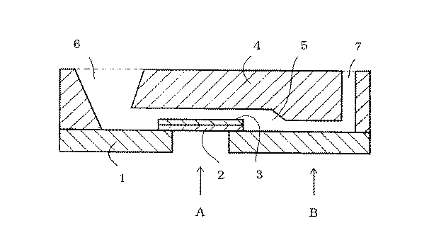
図1は、貫通孔を有する光透過性支持体1上に貫通孔を覆うように固定した光透過性フィルム2、光透過性フィルム2に固着した試薬層3、試薬層3を覆いながら、光透過性支持体1との間に、毛細管室5を形成するように、光透過性支持体1に固定されたカバー4から成り、カバーは試料供給口6と空気抜き口7を有する、本発明で使用される測定用具の断面図である。
試薬層の反射率測定は(A)点で行い、ヘマトクリット値を求めるための反射率測定及び血液が供給されたことを検知するための反射率測定は、(B)点で行う。
実施例の場合、(B)点における隙間高さは、50μmである。
なお、図1は、わかりやすくするために、上下方向を拡大してある。
【0034】
実施例1で作製した測定用具に、同一のグルコース濃度で、種々のヘマトクリット値の異なる血液試料を20μlずつ滴下し、1分後に支持体の貫通孔を通して、下方から、色差計(日本電色工業製、Σ−90)を用いて、測定波長λ=640nmにおける試薬層の反射率を、(A)点にて測定した。
この反射率から、
クベルカームンク(Kubelka−Munk)の式の簡略型:
K/S=(1−R)2/2R
K:定数
S:散乱係数
R:反射率/100
に従って、K/S値を求めた。
K/S値は、モル濃度に相関するので、検量線を作成して、濃度換算することができる。得られた結果を、測定値とした。
同時に、測定波長λ=600nmにおける毛細管室の反射率を(B)点にて測定した。
この測定結果から、予め作成した表1より、血液試料のヘマトクリット値による補正係数を決定し、測定値×補正係数を求めた。
なお、ヘマトクリット値を、毛細管高速遠沈法にて確認した。
【0035】
また、専用の測定器を用いれば、測定波長λ=600nmにおける毛細管室の反射率を(B)点にて測定し、この測定結果から、予め作成した表1より、血液試料のヘマトクリット値による補正係数を決定すると同時に、反応時間のスタートとし、オートスタート機能として、正確に1分後に、自動的に支持体の貫通孔を通して、測定波長λ=640nmにおける試薬層の反射率を、(A)点にて測定できる。
【0036】
【表1】
これらの結果を、表2及び表3にまとめた。
【0038】
【表2】
【表3】
実施例に示した測定値×補正係数と、単なる測定値とを比較すると、前者はヘマトクリット値による影響が小さくなった。
【0041】
【発明の効果】
以上、詳述したように、本発明の測定方法を用いると、血液中の特定成分を迅速かつ簡便に測定することができ、かつ、ヘマトクリット値の影響が軽減され、臨床検査等において絶大な効果をもたらす。
【図面の簡単な説明】
【図1】本発明で使用する測定用具の一例の断面図である。
【符号の説明】
1 光透過性支持体
2 光透過性フィルム
3 試薬層
4 カバー
5 毛細管室
6 試料供給口
7 空気抜き口
A 試薬層の光学的変化を測定する位置
B ヘマトクリット値を求めるために、毛細管室の光学的変化を測定する位置[0001]
TECHNICAL FIELD OF THE INVENTION
The present invention relates to a method for measuring a specific component in blood.
[0002]
[Prior art]
Many measuring instruments have been developed and known as means for quickly and easily measuring specific components in liquid samples such as blood, urine, saliva, sweat, and cerebrospinal fluid.
In particular, a dry-type measurement tool does not require preparation of a reagent, and enables measurement of a specific component in a liquid sample by only a simple operation such as dropping a sample to be measured onto the tool.
[0003]
In the field of clinical testing, when a specific component in a liquid sample is, for example, glucose, lactic acid, uric acid, cholesterol, etc., a measurement method using an enzyme reaction having high substrate specificity has been conventionally used. .
[0004]
As a conventional dry-type measuring tool, for example, a water-resistant test film in which a detection system using an oxidase and a peroxidase is dispersed in a polymer and applied to a plastic member is described in JP-B-49-33800. I have. After making the sample come into contact with the reagent layer for a certain period of time, the film was wiped off with absorbent cotton or the like, thereby making it possible to measure the generated coloring pigment from the sample supply side of the film.
[0005]
JP-B-53-21677 discloses a multilayer test film in which a reagent layer and a developing layer are provided on a liquid-impermeable support. When the sample is spotted on the spread layer of the film, the sample spreads on the spread layer and then moves to the reagent layer. The color dye formed in the reagent layer is measured from the liquid impermeable support side.
[0006]
Japanese Patent Application Laid-Open No. 60-82859 discloses an integrated multilayer analytical tool having an oxygen-permeable protein-impermeable light shielding layer between a developing layer and a reagent layer.
[0007]
The measuring device disclosed in Japanese Patent Application Laid-Open No. 60-205364 by the present applicant has a porous hydrophobic oxygen supply layer provided between a reagent layer and a support.
[0008]
In addition, the present applicant discloses a measuring device using a porous film as a support for a reagent layer in Japanese Patent Application Laid-Open No. 2-150751. This measuring instrument has a reagent layer provided on a porous film.
[0009]
Japanese Patent Application Laid-Open No. Hei 4-188065 discloses a measuring tool in which the support and the cover are arranged at such an interval as to induce a capillary phenomenon to form a capillary.
[0010]
However, all previous methods have shown that when the liquid sample is blood, it is heavily influenced by the hematocrit of the blood, the amount of red blood cells.
Conventional dry-type measuring instruments have a sieving role of separating red blood cells from liquid components (serum or plasma) other than red blood cells at the surface of the reagent layer.
Therefore, the presence of red blood cells is a factor that inhibits the permeability of liquid components.
As a result, if the permeability of the liquid component is impaired, the reaction is delayed, and a low measured value may be obtained.
[0011]
[Problems to be solved by the invention]
An object of the present invention is to solve the above-mentioned problems of the prior art when measuring using a measuring tool, and to provide a method for measuring a specific component in blood quickly, easily and accurately. That is.
[0012]
[Means for Solving the Problems]
The present invention is a measurement tool for measuring a specific component in blood, and the capillary chamber 5, the measurement device having a
[0013]
The present invention is also an improvement of Japanese Patent Application Laid-Open No. 4-188065 and is a measuring method to which the tool described in Japanese Patent Application Laid-Open No. 4-188065 can be applied.
[0014]
BEST MODE FOR CARRYING OUT THE INVENTION
As the measuring tool in the present invention,
Light-transmitting support,
A reagent layer that is fixed on a light-transmitting support and shows an optical change by reacting with a specific component,
Examples of the measuring tool include a cover that covers at least the reagent layer and is fixed to the light-transmitting support so as to form a capillary chamber between the light-transmitting support and the reagent layer.
[0015]
The cover preferably has a sample supply port and an air vent, and in this case, as a method of supplying blood to the capillary chamber, blood is supplied to the capillary chamber by dropping blood from the sample supply port of the cover. .
The sample supply port and the air vent may be holes formed in the cover, but may be gaps provided by cutting a part of the cover or gaps provided between the cover and the light-transmitting support.
The shape of the cover may be a molded product as long as it is fixed to the light-transmitting support and has a capillary chamber, a sample supply port, and an air vent, and by disposing guides on both sides of the film. A capillary chamber may be formed.
[0016]
Various types of light-transmitting supports can be used, but a polyethylene terephthalate film, a polystyrene film, and a cellulose acetate film are preferable.
The optical transparency of the support makes it possible to measure optical changes in the capillary chamber.
Further, it is possible to confirm that blood has been supplied from the sample supply port. Then, it is possible to automatically perform measurement while keeping the time from blood supply to measurement constant.
[0017]
Further, it is preferable that the light-transmitting support has a through-hole having a diameter of 1 to 10 mm, and the light-transmitting film to which the reagent layer is fixed covers the through-hole.
This is the case, for example, when the specific components of blood are glucose, uric acid, and cholesterol, and when an oxidase is used.
As the light transmitting film, various films such as a polyethylene terephthalate film and a polystyrene film can be used, but a porous film is preferable.
This is because such a porous film is air-permeable and can easily supply a sufficient amount of oxygen necessary for the oxidation reaction from the air to the reagent layer.
[0018]
On the other hand, for example, even when the specific component of blood is glucose, uric acid, and cholesterol, when the oxidase is not used, for example, when dehydrogenase is used, the light-transmitting support has a through hole having a diameter of 1 to 10 mm. It is not necessary that the light-transmitting film having the reagent layer fixed thereon covers the through-hole.
[0019]
There are various types of the porous film described above. For example, Nuclepore (made by Nuclepore), Celgard (made by Hoechst Celanese), Cyclopore (made by Whatman), Omnipore (made by Millipore), Gore-Tex (made by Gore-Tex) ), Filterlight (Memtec) and the like.
[0020]
The gap height at the position where the optical change is measured in the capillary chamber is preferably 5 to 200 μm, more preferably 10 to 50 μm. Therefore, the gap height of all the capillary chambers does not need to be uniformly 5 to 200 μm, and it is sufficient that a part of the capillary chamber has such a gap height as shown in FIG.
In this respect, the measuring tool of the present invention is different from the tool described in JP-A-4-18865.
By measuring the optical change in a part of the capillary, the hematocrit value can be obtained, and the optical change in the reagent layer or the measured value obtained therefrom can be corrected.
[0021]
The optical change in the capillary chamber may be the transmittance from the light-transmitting support side, in which case the cover needs to be light-transmitting.
The reflectance from the light-transmitting support side may be used. In this case, the cover needs to be light-reflective.
[0022]
As the arrangement of the optical system, for example, a light source is arranged on the light transmissive support side, a detector is arranged at a position of 45 ° reflection, or a light source is arranged on the light transmissive support side, and a detector is arranged on the cover side. And can be placed.
[0023]
The position for measuring the optical change in the capillary chamber may be anywhere.
In this portion, as shown in the point (B) of FIG. 1, the cover in the capillary chamber is thickened, and the capillary chamber is easy to measure an optical change.
[0024]
The optical change in the capillary chamber is measured, whereby the hematocrit value of the blood is determined, and at the same time, the blood supply can also be detected by the optical change in the capillary chamber. By the detection, an automatic start function can be started in time, and an optical change in the reagent layer can be accurately measured after a certain time, for example, one minute.
In this case, the position where the optical change is measured in the capillary chamber is preferably close in distance to the reagent layer. This is because the start of the reaction in the reagent layer and the detection of blood supply should be simultaneous.
Moreover, it is preferable that the position where the optical change is measured in the capillary chamber is located at the rear of the reagent layer, that is, between the reagent layer and the air vent in the blood flow direction, as shown at point (B) in FIG. Because, in the capillary chamber, even if the transfer of the blood stagnates by accident, as shown in the point (B) in FIG. 1, the position is in the blood flow direction at the rear of the reagent layer, that is, the reagent layer and the air vent. If it is between the two, it is not necessary to start until the blood is transferred to the reagent layer. However, if the position is in the front of the reagent layer in the direction of blood flow, that is, between the sample supply port and the reagent layer, although it is assumed that the sample has been supplied, the blood is not transferred to the reagent layer. In some cases, only low measured values are obtained as a result.
[0025]
The reagent layer contains an enzyme and a dye precursor capable of analyzing a specific component.
For example, when the specific component is glucose, various dye precursors such as glucose oxidase, peroxidase and 4-aminoantipyrine, N-ethyl-N- (2-hydroxy-3-sulfopropyl) -3,5-dimethylaniline are used. contains.
[0026]
In addition, the reagent layer can contain insoluble particles that shield light, whereby, for example, in the analysis of a coloring component-containing sample such as blood, by measuring from the porous film side, such as hemoglobin, It is possible to hide the coloring component and obtain an accurate analysis result.
[0027]
As the insoluble particles, light-reflective white particles such as titanium dioxide, zinc oxide, barium sulfate and magnesium oxide are suitable, but any substance may be used as long as it is a substance that does not dissolve in the liquid sample and blocks light. .
Examples include fluorocarbon, polystyrene latex particles, calcium carbonate, talc, aluminum powder, dextran, acrylic polymer particles, and polystyrene latex particles.
[0028]
A hydrophilic polymer may be added to the reagent solution in order to enhance the coating performance of the reagent solution on the light transmitting film.
Examples of the hydrophilic polymer include hydroxypropylcellulose, methylcellulose, sodium alginate, polyvinyl alcohol, polyvinylpyrrolidone, gelatin, modified gelatin, and agar.
Further, in order to maintain the shape of the reagent layer or to improve the permeability, an emulsion emulsion-based adhesive or latex particles may be added to these hydrophilic polymers. For example, a poly (vinyl propionate) copolymer is commercially available under the trade name of propiophan (manufactured by BASF).
[0029]
The coating performance and reactivity can also be improved by adding a surfactant, a plasticizer, a stabilizer and the like to the reagent solution.
[0030]
The reagent layer in the present invention can be divided into two layers, a reaction layer containing an enzyme or a dye precursor, and a separation layer containing insoluble particles such as titanium dioxide.
[0031]
Examples are shown below, but the present invention is not limited by these examples.
[0032]
Embodiment 1
A reagent solution having the following composition was applied to a 25 μm-thick Celgard (trademark of a film made by Hoechst Celanese), which was a porous film made of polypropylene as a light transmissive film for quantitatively measuring blood glucose, with a wet thickness of 100 μm. Dry at 30 ° C. for 30 minutes.
(Reagent composition)
Glucose oxidase (Toyobo) 5ku
Peroxidase (manufactured by Toyobo) 10ku
50 mg of 4-aminoantipyrine (manufactured by Kishida Chemical)
N-ethyl-N- (2-hydroxy-3-sulfopropyl)
-3,5-dimethylaniline (manufactured by Dojin Chemical) 100mg
Propiophan 5D (BASF) 10g
2 ml of 0.1 M phosphate buffer (pH 7.0)
50g% titanium dioxide aqueous solution 1g
A light-transmitting polyethylene terephthalate film (188 μm in thickness) as a support, which was obtained by cutting the reagent layer fixed to the obtained porous film together with the porous film into 7 mm × 7 mm, and cutting into a 10 mm × 30 mm having a 4 mm through-hole. Thermocompression bonding using a thermoplastic resin. However, the porous film and the support were adhered to each other, and the porous film covered a 4 mm through hole.
A white cover of 10 mm × 30 mm × 2 mm formed in a shape as shown in FIG. 1 with the reagent layer facing upward was fixed so as to cover the support, and was thermocompression-bonded.
At this time, the gap height of the capillary chamber was set to 50 μm.
Thereby, a measuring tool was obtained.
[0033]
This will be described with reference to FIG.
FIG. 1 shows a light-transmitting
The reflectance measurement of the reagent layer is performed at point (A), and the reflectance measurement for obtaining the hematocrit value and the reflectance measurement for detecting that blood has been supplied are performed at point (B).
In the case of the embodiment, the gap height at the point (B) is 50 μm.
Note that FIG. 1 is enlarged in the vertical direction for easy understanding.
[0034]
At the same glucose concentration, blood samples having various hematocrit values of 20 μl were dropped on the measuring device prepared in Example 1, and one minute later, the color difference meter (Nippon Denshoku Industries Co., Ltd.) was passed through the through hole of the support from below. Σ-90), the reflectance of the reagent layer at a measurement wavelength λ = 640 nm was measured at point (A).
From this reflectance,
A simplified form of the Kubelka-Munk equation:
K / S = (1-R) 2 / 2R
K: constant S: scattering coefficient R: reflectance / 100
The K / S value was determined according to
Since the K / S value correlates with the molar concentration, a calibration curve can be created and the concentration can be converted. The obtained result was used as a measured value.
At the same time, the reflectance of the capillary chamber at the measurement wavelength λ = 600 nm was measured at the point (B).
From this measurement result, the correction coefficient based on the hematocrit value of the blood sample was determined from Table 1 prepared in advance, and the measured value × correction coefficient was obtained.
In addition, the hematocrit value was confirmed by a capillary high-speed centrifugation method.
[0035]
If a dedicated measuring device is used, the reflectance of the capillary chamber at the measurement wavelength λ = 600 nm is measured at the point (B), and from this measurement result, the correction based on the hematocrit value of the blood sample is obtained from Table 1 prepared in advance. At the same time as the determination of the coefficient, the reaction time is started. As an auto-start function, the reflectance of the reagent layer at the measurement wavelength λ = 640 nm is automatically passed through the through hole of the support exactly one minute later, at the point (A). Can be measured.
[0036]
[Table 1]
Tables 2 and 3 summarize these results.
[0038]
[Table 2]
[Table 3]
When the measured value × correction coefficient shown in the example was compared with a mere measured value, the former was less affected by the hematocrit value.
[0041]
【The invention's effect】
As described in detail above, by using the measurement method of the present invention, a specific component in blood can be measured quickly and easily, and the effect of a hematocrit value is reduced. Bring.
[Brief description of the drawings]
FIG. 1 is a cross-sectional view of an example of a measuring tool used in the present invention.
[Explanation of symbols]
DESCRIPTION OF SYMBOLS 1 Light-transmitting
Claims (8)
Priority Applications (1)
| Application Number | Priority Date | Filing Date | Title |
|---|---|---|---|
| JP29792295A JP3586743B2 (en) | 1995-10-09 | 1995-10-09 | Measurement method with corrected hematocrit value |
Applications Claiming Priority (1)
| Application Number | Priority Date | Filing Date | Title |
|---|---|---|---|
| JP29792295A JP3586743B2 (en) | 1995-10-09 | 1995-10-09 | Measurement method with corrected hematocrit value |
Publications (2)
| Publication Number | Publication Date |
|---|---|
| JPH09105750A JPH09105750A (en) | 1997-04-22 |
| JP3586743B2 true JP3586743B2 (en) | 2004-11-10 |
Family
ID=17852831
Family Applications (1)
| Application Number | Title | Priority Date | Filing Date |
|---|---|---|---|
| JP29792295A Expired - Fee Related JP3586743B2 (en) | 1995-10-09 | 1995-10-09 | Measurement method with corrected hematocrit value |
Country Status (1)
| Country | Link |
|---|---|
| JP (1) | JP3586743B2 (en) |
Cited By (3)
| Publication number | Priority date | Publication date | Assignee | Title |
|---|---|---|---|---|
| WO2007007849A1 (en) | 2005-07-14 | 2007-01-18 | Matsushita Electric Industrial Co., Ltd. | Analyzer and analyzing method |
| WO2013147200A1 (en) * | 2012-03-29 | 2013-10-03 | 積水メディカル株式会社 | Method for measuring hematocrit value |
| EP3505913A4 (en) * | 2016-08-18 | 2019-07-03 | Konica Minolta, Inc. | Measurement method, measurement apparatus, and measurement system |
Families Citing this family (10)
| Publication number | Priority date | Publication date | Assignee | Title |
|---|---|---|---|---|
| AU3609900A (en) * | 1999-03-02 | 2000-09-21 | Qualigen, Inc. | Methods and apparatus for separation of biological fluids |
| EP1447665B1 (en) | 2003-02-11 | 2016-06-29 | Bayer HealthCare LLC | Method for reducing effect of hematocrit on measurement of an analyte in whole blood |
| DE102005048236A1 (en) * | 2005-10-07 | 2007-04-12 | Ministerium für Wissenschaft, Forschung und Kunst Baden-Württemberg | Apparatus and method for determining the volume fractions of the phases in a suspension |
| JP4785611B2 (en) * | 2006-05-11 | 2011-10-05 | アークレイ株式会社 | Method and apparatus for measuring the concentration of a specific component in a blood sample |
| JP2008216026A (en) * | 2007-03-05 | 2008-09-18 | Fujifilm Corp | Hematocrit measurement method |
| US9778200B2 (en) | 2012-12-18 | 2017-10-03 | Ixensor Co., Ltd. | Method and apparatus for analyte measurement |
| JP6322643B2 (en) * | 2013-01-07 | 2018-05-09 | アイセンサー・カンパニー・リミテッドIxensor Co., Ltd. | Test paper and test paper reading method |
| US10782306B2 (en) * | 2015-12-24 | 2020-09-22 | Koninklijke Philips N.V. | Method and a system for determinations of cell suspensions |
| JP6911318B2 (en) * | 2016-09-29 | 2021-07-28 | コニカミノルタ株式会社 | Blood test sensor chip, blood test device and blood test method |
| CN111868514B (en) * | 2018-03-19 | 2023-08-08 | 普和希控股公司 | Method for determining the amount of a blood component in blood |
-
1995
- 1995-10-09 JP JP29792295A patent/JP3586743B2/en not_active Expired - Fee Related
Cited By (5)
| Publication number | Priority date | Publication date | Assignee | Title |
|---|---|---|---|---|
| WO2007007849A1 (en) | 2005-07-14 | 2007-01-18 | Matsushita Electric Industrial Co., Ltd. | Analyzer and analyzing method |
| WO2013147200A1 (en) * | 2012-03-29 | 2013-10-03 | 積水メディカル株式会社 | Method for measuring hematocrit value |
| JPWO2013147200A1 (en) * | 2012-03-29 | 2015-12-14 | 積水メディカル株式会社 | Method for measuring hematocrit value |
| EP3505913A4 (en) * | 2016-08-18 | 2019-07-03 | Konica Minolta, Inc. | Measurement method, measurement apparatus, and measurement system |
| US11047798B2 (en) | 2016-08-18 | 2021-06-29 | Konica Minolta, Inc. | Measurement method, measurement apparatus, and measurement system |
Also Published As
| Publication number | Publication date |
|---|---|
| JPH09105750A (en) | 1997-04-22 |
Similar Documents
| Publication | Publication Date | Title |
|---|---|---|
| EP0654659B1 (en) | Defined volume test device | |
| JP3586743B2 (en) | Measurement method with corrected hematocrit value | |
| US6506575B1 (en) | Analytical element and method for the determination of an analyte in a liquid | |
| US5227310A (en) | Device and method for assay of liquid sample | |
| JP2618727B2 (en) | Method for quantifying test components in whole blood | |
| JP2864035B2 (en) | Whole blood analysis element | |
| JP2002540427A5 (en) | ||
| JPS61245057A (en) | Integral type multi-layered analyzing element | |
| JPS6348457A (en) | Dry-type multi-layered analytical element | |
| JP2611890B2 (en) | Measurement method using dry analytical element and dry analytical element | |
| EP0169055A2 (en) | Polymeric single layer analytical element | |
| JP3647000B2 (en) | Liquid sample analysis tool and analysis method | |
| US6299838B1 (en) | Test apparatus for assaying a component in a liquid sample | |
| EP1271147A1 (en) | Test paper | |
| US8202490B2 (en) | Membranes and methods for coating membranes | |
| JPH09133673A (en) | Instrument and method for measuring liquid sample | |
| JP2513509B2 (en) | Urea analysis reagent composition and integrated multilayer analytical element containing the same | |
| JPH10132748A (en) | Dry measuring and testing element | |
| EP1159613A1 (en) | Method and apparatus for controlling the adsorption of a liquid sample through an absorbent layer | |
| JP3639392B2 (en) | Dry measurement test equipment | |
| JPH0989888A (en) | Analyzing element conforming to whole blood | |
| JPH02290541A (en) | Analysis element | |
| JP2005351908A (en) | Test piece for quantitating component in liquid sample | |
| JPH02179450A (en) | Testing tool | |
| JPH05103699A (en) | Diagnostic drug for detecting cholesterin, preparation of reagent film for detecting cholesterin, and test strip for detecting cholesterin |
Legal Events
| Date | Code | Title | Description |
|---|---|---|---|
| TRDD | Decision of grant or rejection written | ||
| A01 | Written decision to grant a patent or to grant a registration (utility model) |
Free format text: JAPANESE INTERMEDIATE CODE: A01 Effective date: 20040714 |
|
| A61 | First payment of annual fees (during grant procedure) |
Free format text: JAPANESE INTERMEDIATE CODE: A61 Effective date: 20040723 |
|
| R150 | Certificate of patent or registration of utility model |
Free format text: JAPANESE INTERMEDIATE CODE: R150 |
|
| FPAY | Renewal fee payment (event date is renewal date of database) |
Free format text: PAYMENT UNTIL: 20080820 Year of fee payment: 4 |
|
| FPAY | Renewal fee payment (event date is renewal date of database) |
Free format text: PAYMENT UNTIL: 20080820 Year of fee payment: 4 |
|
| FPAY | Renewal fee payment (event date is renewal date of database) |
Free format text: PAYMENT UNTIL: 20090820 Year of fee payment: 5 |
|
| FPAY | Renewal fee payment (event date is renewal date of database) |
Free format text: PAYMENT UNTIL: 20090820 Year of fee payment: 5 |
|
| FPAY | Renewal fee payment (event date is renewal date of database) |
Free format text: PAYMENT UNTIL: 20100820 Year of fee payment: 6 |
|
| LAPS | Cancellation because of no payment of annual fees |