JP2022106847A - Microglia derived from pluripotent stem cells and how to prepare and use them - Google Patents
Microglia derived from pluripotent stem cells and how to prepare and use them Download PDFInfo
- Publication number
- JP2022106847A JP2022106847A JP2022072376A JP2022072376A JP2022106847A JP 2022106847 A JP2022106847 A JP 2022106847A JP 2022072376 A JP2022072376 A JP 2022072376A JP 2022072376 A JP2022072376 A JP 2022072376A JP 2022106847 A JP2022106847 A JP 2022106847A
- Authority
- JP
- Japan
- Prior art keywords
- cells
- microglial
- medium
- cell
- contacting
- Prior art date
- Legal status (The legal status is an assumption and is not a legal conclusion. Google has not performed a legal analysis and makes no representation as to the accuracy of the status listed.)
- Pending
Links
Images


Classifications
-
- C—CHEMISTRY; METALLURGY
- C12—BIOCHEMISTRY; BEER; SPIRITS; WINE; VINEGAR; MICROBIOLOGY; ENZYMOLOGY; MUTATION OR GENETIC ENGINEERING
- C12N—MICROORGANISMS OR ENZYMES; COMPOSITIONS THEREOF; PROPAGATING, PRESERVING, OR MAINTAINING MICROORGANISMS; MUTATION OR GENETIC ENGINEERING; CULTURE MEDIA
- C12N5/00—Undifferentiated human, animal or plant cells, e.g. cell lines; Tissues; Cultivation or maintenance thereof; Culture media therefor
- C12N5/06—Animal cells or tissues; Human cells or tissues
- C12N5/0602—Vertebrate cells
- C12N5/0618—Cells of the nervous system
- C12N5/0622—Glial cells, e.g. astrocytes, oligodendrocytes; Schwann cells
-
- A—HUMAN NECESSITIES
- A61—MEDICAL OR VETERINARY SCIENCE; HYGIENE
- A61K—PREPARATIONS FOR MEDICAL, DENTAL OR TOILETRY PURPOSES
- A61K35/00—Medicinal preparations containing materials or reaction products thereof with undetermined constitution
- A61K35/12—Materials from mammals; Compositions comprising non-specified tissues or cells; Compositions comprising non-embryonic stem cells; Genetically modified cells
- A61K35/30—Nerves; Brain; Eyes; Corneal cells; Cerebrospinal fluid; Neuronal stem cells; Neuronal precursor cells; Glial cells; Oligodendrocytes; Schwann cells; Astroglia; Astrocytes; Choroid plexus; Spinal cord tissue
-
- A—HUMAN NECESSITIES
- A61—MEDICAL OR VETERINARY SCIENCE; HYGIENE
- A61P—SPECIFIC THERAPEUTIC ACTIVITY OF CHEMICAL COMPOUNDS OR MEDICINAL PREPARATIONS
- A61P21/00—Drugs for disorders of the muscular or neuromuscular system
- A61P21/02—Muscle relaxants, e.g. for tetanus or cramps
-
- A—HUMAN NECESSITIES
- A61—MEDICAL OR VETERINARY SCIENCE; HYGIENE
- A61P—SPECIFIC THERAPEUTIC ACTIVITY OF CHEMICAL COMPOUNDS OR MEDICINAL PREPARATIONS
- A61P25/00—Drugs for disorders of the nervous system
-
- A—HUMAN NECESSITIES
- A61—MEDICAL OR VETERINARY SCIENCE; HYGIENE
- A61P—SPECIFIC THERAPEUTIC ACTIVITY OF CHEMICAL COMPOUNDS OR MEDICINAL PREPARATIONS
- A61P25/00—Drugs for disorders of the nervous system
- A61P25/14—Drugs for disorders of the nervous system for treating abnormal movements, e.g. chorea, dyskinesia
- A61P25/16—Anti-Parkinson drugs
-
- A—HUMAN NECESSITIES
- A61—MEDICAL OR VETERINARY SCIENCE; HYGIENE
- A61P—SPECIFIC THERAPEUTIC ACTIVITY OF CHEMICAL COMPOUNDS OR MEDICINAL PREPARATIONS
- A61P25/00—Drugs for disorders of the nervous system
- A61P25/18—Antipsychotics, i.e. neuroleptics; Drugs for mania or schizophrenia
-
- A—HUMAN NECESSITIES
- A61—MEDICAL OR VETERINARY SCIENCE; HYGIENE
- A61P—SPECIFIC THERAPEUTIC ACTIVITY OF CHEMICAL COMPOUNDS OR MEDICINAL PREPARATIONS
- A61P25/00—Drugs for disorders of the nervous system
- A61P25/28—Drugs for disorders of the nervous system for treating neurodegenerative disorders of the central nervous system, e.g. nootropic agents, cognition enhancers, drugs for treating Alzheimer's disease or other forms of dementia
-
- A—HUMAN NECESSITIES
- A61—MEDICAL OR VETERINARY SCIENCE; HYGIENE
- A61P—SPECIFIC THERAPEUTIC ACTIVITY OF CHEMICAL COMPOUNDS OR MEDICINAL PREPARATIONS
- A61P37/00—Drugs for immunological or allergic disorders
- A61P37/02—Immunomodulators
- A61P37/04—Immunostimulants
-
- C—CHEMISTRY; METALLURGY
- C12—BIOCHEMISTRY; BEER; SPIRITS; WINE; VINEGAR; MICROBIOLOGY; ENZYMOLOGY; MUTATION OR GENETIC ENGINEERING
- C12N—MICROORGANISMS OR ENZYMES; COMPOSITIONS THEREOF; PROPAGATING, PRESERVING, OR MAINTAINING MICROORGANISMS; MUTATION OR GENETIC ENGINEERING; CULTURE MEDIA
- C12N5/00—Undifferentiated human, animal or plant cells, e.g. cell lines; Tissues; Cultivation or maintenance thereof; Culture media therefor
- C12N5/06—Animal cells or tissues; Human cells or tissues
- C12N5/0602—Vertebrate cells
- C12N5/0603—Embryonic cells ; Embryoid bodies
- C12N5/0606—Pluripotent embryonic cells, e.g. embryonic stem cells [ES]
-
- C—CHEMISTRY; METALLURGY
- C12—BIOCHEMISTRY; BEER; SPIRITS; WINE; VINEGAR; MICROBIOLOGY; ENZYMOLOGY; MUTATION OR GENETIC ENGINEERING
- C12N—MICROORGANISMS OR ENZYMES; COMPOSITIONS THEREOF; PROPAGATING, PRESERVING, OR MAINTAINING MICROORGANISMS; MUTATION OR GENETIC ENGINEERING; CULTURE MEDIA
- C12N5/00—Undifferentiated human, animal or plant cells, e.g. cell lines; Tissues; Cultivation or maintenance thereof; Culture media therefor
- C12N5/06—Animal cells or tissues; Human cells or tissues
- C12N5/0602—Vertebrate cells
- C12N5/0618—Cells of the nervous system
-
- G—PHYSICS
- G01—MEASURING; TESTING
- G01N—INVESTIGATING OR ANALYSING MATERIALS BY DETERMINING THEIR CHEMICAL OR PHYSICAL PROPERTIES
- G01N33/00—Investigating or analysing materials by specific methods not covered by groups G01N1/00 - G01N31/00
- G01N33/48—Biological material, e.g. blood, urine; Haemocytometers
- G01N33/50—Chemical analysis of biological material, e.g. blood, urine; Testing involving biospecific ligand binding methods; Immunological testing
- G01N33/5005—Chemical analysis of biological material, e.g. blood, urine; Testing involving biospecific ligand binding methods; Immunological testing involving human or animal cells
- G01N33/5008—Chemical analysis of biological material, e.g. blood, urine; Testing involving biospecific ligand binding methods; Immunological testing involving human or animal cells for testing or evaluating the effect of chemical or biological compounds, e.g. drugs, cosmetics
- G01N33/5044—Chemical analysis of biological material, e.g. blood, urine; Testing involving biospecific ligand binding methods; Immunological testing involving human or animal cells for testing or evaluating the effect of chemical or biological compounds, e.g. drugs, cosmetics involving specific cell types
- G01N33/5058—Neurological cells
-
- C—CHEMISTRY; METALLURGY
- C12—BIOCHEMISTRY; BEER; SPIRITS; WINE; VINEGAR; MICROBIOLOGY; ENZYMOLOGY; MUTATION OR GENETIC ENGINEERING
- C12N—MICROORGANISMS OR ENZYMES; COMPOSITIONS THEREOF; PROPAGATING, PRESERVING, OR MAINTAINING MICROORGANISMS; MUTATION OR GENETIC ENGINEERING; CULTURE MEDIA
- C12N2500/00—Specific components of cell culture medium
- C12N2500/90—Serum-free medium, which may still contain naturally-sourced components
-
- C—CHEMISTRY; METALLURGY
- C12—BIOCHEMISTRY; BEER; SPIRITS; WINE; VINEGAR; MICROBIOLOGY; ENZYMOLOGY; MUTATION OR GENETIC ENGINEERING
- C12N—MICROORGANISMS OR ENZYMES; COMPOSITIONS THEREOF; PROPAGATING, PRESERVING, OR MAINTAINING MICROORGANISMS; MUTATION OR GENETIC ENGINEERING; CULTURE MEDIA
- C12N2501/00—Active agents used in cell culture processes, e.g. differentation
- C12N2501/10—Growth factors
- C12N2501/155—Bone morphogenic proteins [BMP]; Osteogenins; Osteogenic factor; Bone inducing factor
-
- C—CHEMISTRY; METALLURGY
- C12—BIOCHEMISTRY; BEER; SPIRITS; WINE; VINEGAR; MICROBIOLOGY; ENZYMOLOGY; MUTATION OR GENETIC ENGINEERING
- C12N—MICROORGANISMS OR ENZYMES; COMPOSITIONS THEREOF; PROPAGATING, PRESERVING, OR MAINTAINING MICROORGANISMS; MUTATION OR GENETIC ENGINEERING; CULTURE MEDIA
- C12N2501/00—Active agents used in cell culture processes, e.g. differentation
- C12N2501/20—Cytokines; Chemokines
- C12N2501/22—Colony stimulating factors (G-CSF, GM-CSF)
-
- C—CHEMISTRY; METALLURGY
- C12—BIOCHEMISTRY; BEER; SPIRITS; WINE; VINEGAR; MICROBIOLOGY; ENZYMOLOGY; MUTATION OR GENETIC ENGINEERING
- C12N—MICROORGANISMS OR ENZYMES; COMPOSITIONS THEREOF; PROPAGATING, PRESERVING, OR MAINTAINING MICROORGANISMS; MUTATION OR GENETIC ENGINEERING; CULTURE MEDIA
- C12N2501/00—Active agents used in cell culture processes, e.g. differentation
- C12N2501/20—Cytokines; Chemokines
- C12N2501/23—Interleukins [IL]
- C12N2501/2334—Interleukin-34 (IL-34)
-
- C—CHEMISTRY; METALLURGY
- C12—BIOCHEMISTRY; BEER; SPIRITS; WINE; VINEGAR; MICROBIOLOGY; ENZYMOLOGY; MUTATION OR GENETIC ENGINEERING
- C12N—MICROORGANISMS OR ENZYMES; COMPOSITIONS THEREOF; PROPAGATING, PRESERVING, OR MAINTAINING MICROORGANISMS; MUTATION OR GENETIC ENGINEERING; CULTURE MEDIA
- C12N2506/00—Differentiation of animal cells from one lineage to another; Differentiation of pluripotent cells
- C12N2506/45—Differentiation of animal cells from one lineage to another; Differentiation of pluripotent cells from artificially induced pluripotent stem cells
Landscapes
- Health & Medical Sciences (AREA)
- Engineering & Computer Science (AREA)
- Life Sciences & Earth Sciences (AREA)
- Biomedical Technology (AREA)
- Chemical & Material Sciences (AREA)
- Bioinformatics & Cheminformatics (AREA)
- Organic Chemistry (AREA)
- General Health & Medical Sciences (AREA)
- Neurology (AREA)
- Biotechnology (AREA)
- Neurosurgery (AREA)
- Cell Biology (AREA)
- Zoology (AREA)
- Immunology (AREA)
- Wood Science & Technology (AREA)
- Genetics & Genomics (AREA)
- Medicinal Chemistry (AREA)
- Animal Behavior & Ethology (AREA)
- Veterinary Medicine (AREA)
- Public Health (AREA)
- Pharmacology & Pharmacy (AREA)
- Microbiology (AREA)
- Biochemistry (AREA)
- Developmental Biology & Embryology (AREA)
- Chemical Kinetics & Catalysis (AREA)
- General Chemical & Material Sciences (AREA)
- Nuclear Medicine, Radiotherapy & Molecular Imaging (AREA)
- General Engineering & Computer Science (AREA)
- Molecular Biology (AREA)
- Hematology (AREA)
- Urology & Nephrology (AREA)
- Gynecology & Obstetrics (AREA)
- Reproductive Health (AREA)
- Tropical Medicine & Parasitology (AREA)
- Pathology (AREA)
- Toxicology (AREA)
- Epidemiology (AREA)
- Virology (AREA)
- Food Science & Technology (AREA)
- Physics & Mathematics (AREA)
Abstract
Description
関連出願の相互参照
本願は、2016年3月3日に出願された米国仮特許出願第62/303,301号および2016年10月20日に出願された米国仮特許出願第62/410,645号の優先権の恩典を主張し、前記仮特許出願の内容は参照により本明細書に組み入れられる。
Mutual reference of related applications This application is the priority of US Provisional Patent Application No. 62 / 303,301 filed on March 3, 2016 and US Provisional Patent Application No. 62 / 410,645 filed on October 20, 2016. The contents of the provisional patent application are incorporated herein by reference.
連邦政府の助成による研究に関する記載
本発明は、米国国立衛生研究所によって交付された助成金番号AG046170の下に、米国政府の支援を受けてなされた。米国政府は本発明に一定の権利を有する。
Federal Granted Research Description The invention was made with the support of the United States Government under grant number AG046170 issued by the National Institutes of Health. The US Government has certain rights to the invention.
参照による組み入れ
参照による組み入れを許す管轄区域に限り、この開示において引用される参考文献はすべて、参照により、その全体が本明細書に組み入れられる(この特許開示において本文に続く括弧内の数字または上付きの数字は、この特許明細書の「参考文献一覧」の項に掲載する番号付きの参考文献を指す)。加えて、本明細書において引用されまたは言及される製品すべてについて、製造元の使用説明書またはカタログが、いずれも参照により本明細書に組み入れられる。参照により本明細書に組み入れられる文書またはそこに含まれる任意の教示内容は、本発明の実施において使用することができる。
Incorporation by Reference All references cited in this disclosure are incorporated herein by reference in their entirety, only in jurisdictions that allow inclusion by reference (numbers in parentheses following the text in this patent disclosure or above. Numbers with numbers refer to the numbered references listed in the "List of References" section of this patent specification). In addition, for all products cited or referred to herein, the manufacturer's instructions or catalogs are all incorporated herein by reference. The documents incorporated herein by reference or any teachings contained therein may be used in the practice of the present invention.
発明の背景
ミクログリアは脳の常在性組織特異的マクロファージである。ミクログリアは中枢神経系(CNS)の発生および維持において決定的な役割をいくつか果たす。ミクログリアは、卵黄嚢において原始CD45+CX3CR1-骨髄系前駆体から生じ、それが、二次造血の出現前に、CD45+CX3CR1+ミクログリア前駆体に分化して、発生中の脳に入りこむ。無傷の血液脳関門を持つ健常な成人の脳において、ミクログリアは、循環骨髄由来細胞によって補充されることのない長寿命の自立的集団として存続する。高度に枝分かれしたミクログリア細胞は「静止型」と定義されるが、それらの突起は、恒常性の破綻がないかどうか脳を調べるために絶えず移動しているので、実際には極めて活動的である。
Background of the Invention Microglia are resident tissue-specific macrophages in the brain. Microglia play several crucial roles in the development and maintenance of the central nervous system (CNS). Microglia arise in the yolk sac from the primordial CD45 + CX3CR1-myeloid precursor, which differentiates into the CD45 + CX3CR1 + microglial precursor and enters the developing brain before the appearance of secondary hematopoiesis. In the brain of a healthy adult with an intact blood-brain barrier, microglia survive as a long-lived, self-sustaining population that is not replenished by circulating bone marrow-derived cells. Highly branched microglial cells are defined as "quiescent", but their projections are actually extremely active as they are constantly migrating to examine the brain for homeostatic disruptions. ..
ミクログリアは、貪食を使って病原体および/または損傷した細胞もしくは死んだ細胞を排除し、毒性分子、細胞残屑および/またはタンパク質沈着物を除去することで、炎症を減衰させ、組織の再生および修復を促進する。発生中は、ミクログリアが、神経系前駆体の遊走および分化、神経発生ならびにオリゴデンドロサイト発生を促進し、刈り込み(pruning)によってシナプス形成およびシナプス可塑性を調節する。ミクログリアは、サイトカインおよび神経毒性分子を放出することによって、病的な脳の炎症および血液脳関門の破壊の一因になる場合もある。ミクログリアの機能不全は、筋萎縮性側索硬化症(ALS)およびアルツハイマー病(AD)と関連づけられている。ミクログリア細胞の慢性的活性化は、多発性硬化症(MS)およびパーキンソン病の進行の引き金である可能性があり、貪食およびシナプス刈り込みの欠陥は、統合失調症および自閉症スペクトラム障害と関係づけられている。動物実験に基づいてミクログリアの関与があると決定された他の疾患としては、レット症候群、スフェロイド形成を伴うびまん性白質脳症(例えば軸索スフェロイド形成を伴う遺伝性びまん性白質脳症)、および前頭側頭葉変性症(FTLD)、例えば家族性FTLDが挙げられる。 Microglia use phagocytosis to eliminate pathogens and / or damaged or dead cells, and remove toxic molecules, cell debris and / or protein deposits to reduce inflammation and regenerate and repair tissue. To promote. During development, microglia promote the migration and differentiation of nervous system precursors, neurogenesis and oligodendrocyte development, and regulate synaptogenesis and synaptic plasticity by pruning. Microglia can also contribute to pathological brain inflammation and disruption of the blood-brain barrier by releasing cytokines and neurotoxic molecules. Microglial dysfunction has been associated with amyotrophic lateral sclerosis (ALS) and Alzheimer's disease (AD). Chronic activation of microglial cells can trigger the progression of multiple sclerosis (MS) and Parkinson's disease, and defects in phagocytosis and synaptic pruning are associated with schizophrenia and autism spectrum disorders Has been done. Other diseases that have been determined to involve microglia based on animal experiments include Rett Syndrome, diffuse leukoencephalopathy with spheroid formation (eg, hereditary diffuse leukoencephalopathy with axillary spheroid formation), and frontotemporal lobar. Frontotemporal degeneration (FTLD), such as familial FTLD.
ミクログリアに関する我々の知識の大半は齧歯類の研究に由来する。しかし、齧歯類とヒトのミクログリア細胞には、それらの増殖速度、付着性および重要な受容体の発現などに、大きな相違がある。これらの相違と一致して、齧歯類の多能性幹細胞をミクログリアに分化させるために以前に開発されたプロトコールは、ヒト多能性幹細胞では有効でなかった。 Most of our knowledge of microglia comes from rodent studies. However, rodents and human microglial cells differ significantly in their growth rate, adhesion and expression of important receptors. Consistent with these differences, previously developed protocols for differentiating rodent pluripotent stem cells into microglia have not been effective in human pluripotent stem cells.
初代ヒトミクログリア細胞の直接的な分析は、ヒト脳検体の入手可能性、とりわけ健常個体からの入手可能性に制約があるため、厳しく制限されてきた。ヒト多能性幹細胞(PSC)分野の発展は、ヒト多能性幹細胞から多くの異なる分化細胞タイプを生成することを可能にしたが、そのようなヒト幹細胞からミクログリア細胞を生成するための効率のよい再現性ある方法が、当技術分野では依然として必要とされている。本発明はこの必要性に対処する。 Direct analysis of primary human microglial cells has been severely restricted due to limited availability of human brain specimens, especially from healthy individuals. Developments in the field of human pluripotent stem cells (PSCs) have made it possible to generate many different differentiated cell types from human pluripotent stem cells, but with the efficiency of producing microglial cells from such human pluripotent stem cells. Good reproducible methods are still needed in the art. The present invention addresses this need.
脳の免疫細胞であるミクログリアは、中枢神経系の適正な発生および維持にとって極めて重要であり、数多くの神経学的疾患および障害に関わっている。本発明は、多能性幹細胞からヒトミクログリアを生成させるための、さまざまな新しい改良された方法を提供する。本発明の方法を使用することで、化学合成培地を使って、胚性幹細胞(ES細胞、すなわちESC)からも、誘導多能性幹細胞(iPS細胞、すなわちIPSC)からも、CD14および/またはCX3CR1を発現するミクログリア前駆体が生成する。そのようなミクログリア前駆体は、典型的には、ほぼ25~30日以内に現れ、ほぼ20~25日間にわたって、ほぼ50日目まで生産され続ける。本明細書に規定する方法は、そのようなミクログリア前駆体のさらなる分化も可能にし、このさらなる分化は、運動性の高い突起を有し、多くの典型的ミクログリアマーカーを発現し、サイトカインを放出し、貪食活性を有し、ADPに応答して細胞内Ca2+トランジェントを生じる分枝型ミクログリアの生成をもたらす。これらの方法は、異なる多能性幹細胞(PSC)株にわたって、高い再現性を示す。 Microglia, the immune cells of the brain, are crucial for the proper development and maintenance of the central nervous system and are involved in a number of neurological disorders and disorders. The present invention provides a variety of new and improved methods for producing human microglia from pluripotent stem cells. By using the methods of the invention, CD14 and / or CX3CR1 from embryonic stem cells (ES cells, i.e. ESC) as well as induced pluripotent stem cells (iPS cells, i.e. IPSC) using chemically synthesized media. A microglial precursor expressing the above is produced. Such microglial precursors typically appear within approximately 25-30 days and continue to be produced for approximately 20-25 days until approximately 50 days. The methods defined herein also allow for further differentiation of such microglial precursors, which have highly motile processes, express many typical microglial markers, and release cytokines. It has phagocytic activity and results in the production of branched microglia that produce intracellular Ca 2+ transients in response to ADP. These methods show high reproducibility across different pluripotent stem cell (PSC) strains.
本発明の主要な局面の一部を以下にまとめる。その他の局面は、この開示の発明の詳細な説明、実施例、図面および特許請求の範囲の各項に記載する。この開示の各項の記載は、他の項と合わせて読解されるものとする。さらにまた、この開示の各項に記載されるさまざまな態様は、多種多様に組み合わせることができ、そのような組合せはすべて、本発明の範囲内に包含されるものとする。 Some of the main aspects of the present invention are summarized below. Other aspects are described in the detailed description, examples, drawings and claims of the invention of this disclosure. The statements in each section of this disclosure shall be read in conjunction with the other sections. Furthermore, the various aspects described in each section of this disclosure can be combined in a wide variety of ways, all of which are within the scope of the present invention.
いくつかの態様において、本発明は、多能性幹細胞からミクログリア細胞を生成するための方法を提供する。いくつかのそのような態様において、多能性幹細胞は、任意の哺乳動物種から得られる。いくつかの態様において、多能性幹細胞はヒト多能性幹細胞であって、ヒトミクログリア細胞の生成をもたらす。いくつかの態様において、多能性幹細胞は、誘導多能性幹細胞(「iPS細胞」すなわち「iPSC」)または胚性幹細胞(「ES細胞」すなわち「ESC」)のどちらかである。そのような方法は、2つの主要な工程を伴う。第1の工程では、CD14+および/またはCX3CR1+ミクログリア前駆細胞の生成につながる骨髄系分化を誘導する条件下で多能性幹細胞を培養する。第2の工程では、CD14+および/またはCX3CR1+ミクログリア前駆細胞をミクログリア細胞に分化させる。いくつかの態様では、CD14+および/またはCX3CR1+ミクログリア前駆細胞を、(i)IL-34を含む第1のミクログリア分化培地、または(ii)M-CSFを含む第2のミクログリア分化培地のうちのどちらか一方において培養することにより、それらをミクログリア細胞に分化させる。いくつかの態様では、第1のミクログリア分化培地はGM-CSFも含む。いくつかの態様では、第2のミクログリア分化培地は、GM-CSF、NGF-βおよびCCL2からなる群より選択される1種または複数種の因子も含む。例えば、いくつかの態様では、第2のミクログリア分化培地は、GM-CSF、NGF-βおよびCCL2のそれぞれも含む。いくつかの態様では、CD14+および/またはCX3CR1+ミクログリア前駆細胞を、ほぼ15日間にわたって、第1または第2のミクログリア分化培地と接触させる。いくつかの態様では、第1の工程において生成したCD 14+および/またはCX3CR1+ミクログリア前駆細胞は、第2の工程に進む前に単離される。いくつかの態様において、ミクログリア前駆細胞はCD14発現が最大になる時点付近において単離される。 In some embodiments, the invention provides a method for producing microglial cells from pluripotent stem cells. In some such embodiments, pluripotent stem cells are obtained from any mammalian species. In some embodiments, the pluripotent stem cell is a human pluripotent stem cell, resulting in the production of human microglial cells. In some embodiments, pluripotent stem cells are either induced pluripotent stem cells (“iPS cells” or “iPSC”) or embryonic stem cells (“ES cells” or “ESC”). Such a method involves two main steps. In the first step, pluripotent stem cells are cultured under conditions that induce myeloid differentiation leading to the production of CD14 + and / or CX3CR1 + microglial progenitor cells. In the second step, CD14 + and / or CX3CR1 + microglial progenitor cells are differentiated into microglial cells. In some embodiments, the CD14 + and / or CX3CR1 + microglial progenitor cells are either (i) a first microglial differentiation medium containing IL-34 or (ii) a second microglial differentiation medium containing M-CSF. By culturing on one side, they are differentiated into microglial cells. In some embodiments, the first microglial differentiation medium also comprises GM-CSF. In some embodiments, the second microglial differentiation medium also comprises one or more factors selected from the group consisting of GM-CSF, NGF-β and CCL2. For example, in some embodiments, the second microglial differentiation medium also comprises GM-CSF, NGF-β and CCL2, respectively. In some embodiments, CD14 + and / or CX3CR1 + microglial progenitor cells are contacted with a first or second microglial differentiation medium for approximately 15 days. In some embodiments, the CD 14+ and / or CX3CR1 + microglial progenitor cells produced in the first step are isolated before proceeding to the second step. In some embodiments, microglial progenitor cells are isolated near the point of maximal CD14 expression.
いくつかの代替的態様において、本発明は、多能性幹細胞からミクログリア前駆細胞を生成するための方法を提供する。そのような方法は、上述した2つの主要工程のうちの最初の工程を行うことを伴い、任意で、結果として生じたCD14+および/またはCX3CR1+ミクログリア前駆細胞を単離することを伴う。 In some alternative embodiments, the present invention provides a method for producing microglial progenitor cells from pluripotent stem cells. Such a method involves performing the first of the two major steps described above, and optionally involves isolating the resulting CD14 + and / or CX3CR1 + microglial progenitor cells.
別の代替的態様において、本発明は、ミクログリア前駆細胞からミクログリア細胞を生成するための方法を提供する。そのような方法は、上述した2つの主要工程のうちの2番目の工程を行うことを伴う。 In another alternative embodiment, the invention provides a method for producing microglial cells from microglial progenitor cells. Such a method involves performing the second of the two main steps described above.
(CD14+および/またはCX3CR1+ミクログリア前駆細胞の生成につながる)骨髄系分化を誘導する条件下で多能性幹細胞を培養することを伴う本発明の態様の一部では、段階ごとにサイトカインおよび組織培養培地の組合せを変えて細胞を培養する多工程プロセスを使用する。これらの多工程プロセスにおける工程は、多能性幹細胞から原始血管芽細胞への分化の誘導、および/または原始血管芽細胞から骨髄系前駆体への分化の誘導、および/または骨髄系前駆体からミクログリア前駆体への分化の誘導をもたらしうる。それぞれの細胞表面マーカープロファイルによって定義すると、いくつかの態様において、そのような多工程プロセスは、まず多能性幹細胞からKDR+CD235a+細胞への分化の誘導、次にCD45+CX3CR1-細胞への分化の誘導、そして次にCD45+CX3CR1+CD14+細胞への分化の誘導をもたらしうる。 In some aspects of the invention involving culturing pluripotent stem cells under conditions that induce myeloid differentiation (leading to the production of CD14 + and / or CX3CR1 + microglial progenitor cells), step-by-step cytokine and tissue culture medium. A multi-step process is used in which cells are cultured by changing the combination of. Steps in these multi-step processes are the induction of pluripotent stem cell to primordial hemangioblasts and / or the differentiation of primordial hemangioblasts to myeloid precursors and / or from myeloid precursors. It can lead to the induction of differentiation into microglial precursors. In some embodiments, as defined by their respective cell surface marker profiles, such multi-step processes first induce differentiation of pluripotent stem cells into KDR + CD235a + cells, then CD45 + CX3CR1-cells, and then. It can then lead to the induction of differentiation into CD45 + CX3CR1 + CD14 + cells.
いくつかの態様において、本明細書に規定する、多能性幹細胞からCD14+および/またはCX3CR1+ミクログリア前駆細胞を生成するための方法は、以下の4工程のうちの1つまたは複数を行うことを含む:第1に、細胞培養物を、培養培地中で、BMP4を含む第1の組成物と接触させる工程であって、細胞培養物を第1の組成物と最初に接触させるときに、細胞培養物が多能性幹細胞を含む、工程;第2に、細胞培養物を、造血細胞培地中で、bFGF、SCFおよびVEGFAのうちの1つまたは複数(例えばbFGF、SCFおよびVEGFAのそれぞれ)を含む第2の組成物と接触させる工程;第3に、細胞培養物を、造血細胞培地中で、SCF、IL-3、TPO、MCSFおよびFLT3リガンドのうちの1つまたは複数(例えばSCF、IL-3、TPO、MCSFおよびFLT3リガンドのそれぞれ)を含む第3の組成物と接触させる工程;および第4に、細胞培養物を、造血細胞培地中で、MCSF、FLT3リガンドおよびGM-CSFのうちの1つまたは複数(例えばMCSF、FLT3リガンド、およびGM-CSFのそれぞれ)を含む第4の組成物と接触させる工程。いくつかの態様では、上記4工程のすべてが順に行われる。そのような態様の一部では、これら4工程のうちのどの工程に使用される培地も、無血清培地である。そのような態様の一部では、これら4工程のうちのどの工程に使用される培地も、化学合成培地である。これら4種の組成物において使用される作用物質の例示的な量は、この特許開示の詳細な説明および実施例の項に示す。 In some embodiments, the method for producing CD14 + and / or CX3CR1 + microglial progenitor cells from pluripotent stem cells, as defined herein, comprises performing one or more of the following four steps: : First, the step of contacting the cell culture with the first composition containing BMP4 in the culture medium, when the cell culture is first contacted with the first composition, the cell culture. The process comprises pluripotent stem cells; second, the cell culture comprises one or more of bFGF, SCF and VEGFA (eg, bFGF, SCF and VEGFA respectively) in a hematopoietic cell medium. Step of contacting with the second composition; third, the cell culture is placed in a hematopoietic cell medium in one or more of the SCF, IL-3, TPO, MCSF and FLT3 ligands (eg SCF, IL- 3. Contact with a third composition containing TPO, MCSF and FLT3 ligands, respectively; and Fourth, place the cell culture in a hematopoietic cell medium of MCSF, FLT3 ligand and GM-CSF. The step of contacting with a fourth composition comprising one or more (eg, MCSF, FLT3 ligand, and GM-CSF, respectively). In some embodiments, all of the above four steps are performed in sequence. In some of such embodiments, the medium used in any of these four steps is a serum-free medium. In some of such embodiments, the medium used in any of these four steps is a chemically synthesized medium. Illustrative amounts of agents used in these four compositions are given in the detailed description and examples section of this patent disclosure.
上記4工程のうちの最初の工程において、いくつかの態様では、幹細胞の維持に適した組織培養培地が使用され、一方、別の態様では、幹細胞の分化に適した組織培養培地が使用される。いくつかの態様では、多能性因子を含まない改変多能性幹細胞維持培地が使用される。いくつかの態様では、多能性因子を含まない培地が使用される。例えば一態様において、上記4工程のうちの最初の工程において使用される培地は、塩化リチウム、GABA、ピペコリン酸、bFGFまたはTGFβ1のいずれをも含まない。例えば一態様では、塩化リチウム、GABA、ピペコリン酸、bFGFまたはTGFβ1のいずれをも含有しない改変「mTeSR1」培地が使用される。上記4工程のうちの最後の3工程では、任意の適切な造血細胞培地を使用することができる。一態様において、造血細胞培地は「StemPro-34」である。 In the first step of the above four steps, in some embodiments, a tissue culture medium suitable for stem cell maintenance is used, while in another embodiment, a tissue culture medium suitable for stem cell differentiation is used. .. In some embodiments, a modified pluripotent stem cell maintenance medium that does not contain pluripotent factors is used. In some embodiments, a pluripotent-free medium is used. For example, in one embodiment, the medium used in the first step of the above four steps does not contain any of lithium chloride, GABA, pipecolic acid, bFGF or TGFβ1. For example, in one embodiment, a modified "mTeSR1" medium containing none of lithium chloride, GABA, pipecolic acid, bFGF or TGFβ1 is used. Any suitable hematopoietic cell medium can be used in the last three steps of the above four steps. In one embodiment, the hematopoietic cell medium is "Stem Pro-34".
本発明は、多能性幹細胞からCD14+および/またはCX3CR1+ミクログリア前駆細胞を生成させるために上述した4工程のそれぞれを行う、一定の例示的なタイミングも提供する。これらの例示的タイミングは、詳細な説明、実施例および図面に記載する。例えばいくつかの態様では、細胞培養物をおよそ4日間、第1の組成物と接触させる。いくつかの態様では、細胞培養物をおよそ2日間、第2の組成物と接触させる。いくつかの態様では、細胞培養物をおよそ8日間、第3の組成物と接触させる。いくつかの態様では、細胞培養物を、およそ11日~36日間またはそれ以上にわたって、第4の組成物と接触させる。 The present invention also provides certain exemplary timings in which each of the four steps described above is performed to generate CD14 + and / or CX3CR1 + microglial progenitor cells from pluripotent stem cells. These exemplary timings are described in the detailed description, examples and drawings. For example, in some embodiments, the cell culture is contacted with the first composition for approximately 4 days. In some embodiments, the cell culture is contacted with the second composition for approximately 2 days. In some embodiments, the cell culture is contacted with the third composition for approximately 8 days. In some embodiments, the cell culture is contacted with the fourth composition for approximately 11-36 days or longer.
いくつかの態様では、上述の、または本明細書の他の箇所に記載する、多能性幹細胞からCD14+および/またはCX3CR1+ミクログリア前駆細胞を生成するための方法を実行する場合、培地変更を行うときに組織培養上清を捨てるのではなく、上清を取っておき、上清中に存在する細胞を回収して、細胞培養物に戻す。これが有利である理由は、多能性幹細胞からミクログリア前駆体への転換中に誘導される重要な細胞タイプのうち一定のものは、細胞培養プレートに付着する細胞の層に存在するのではなく、主として細胞上清中に見いだされることを、本発明者らが発見したことにある。したがっていくつかの態様では、培地を替えるときに、培養上清中に存在する細胞を回収して細胞培養物に戻す。いくつかの態様では、約10日目より後に行われる培地交換に関して、培養上清中に存在する細胞を回収して、細胞培養物に戻す。いくつかの態様では、細胞が第3の組成物または第4の組成物と接触しているときに行われる培地交換に関して、培養上清中に存在する細胞を回収して細胞培養物に戻す。いくつかの態様では、CD45+CX3CR1-細胞集団の出現後に行われる培地交換に関して、培養上清中に存在する細胞を回収して細胞培養物に戻す。 In some embodiments, when performing medium changes when performing the methods for producing CD14 + and / or CX3CR1 + microglial progenitor cells from pluripotent stem cells, as described above or elsewhere herein. Instead of discarding the tissue culture supernatant, the supernatant is set aside, and the cells present in the supernatant are collected and returned to the cell culture. The reason this is advantageous is that certain of the key cell types induced during the conversion of pluripotent stem cells to microglial precursors are not present in the layer of cells attached to the cell culture plate. The present inventors have discovered that they are mainly found in cell supernatants. Therefore, in some embodiments, when the medium is changed, the cells present in the culture supernatant are recovered and returned to the cell culture. In some embodiments, for medium exchanges that occur after about 10 days, the cells present in the culture supernatant are harvested and returned to the cell culture. In some embodiments, for medium exchange performed when the cells are in contact with the third or fourth composition, the cells present in the culture supernatant are recovered and returned to the cell culture. In some embodiments, for media exchanges performed after the appearance of the CD45 + CX3CR1-cell population, the cells present in the culture supernatant are harvested and returned to the cell culture.
いくつかの態様において、本発明は、ミクログリア細胞またはミクログリア前駆細胞、例えば本明細書に記載の方法によって生産されるものを提供する。いくつかの態様において、本発明は、そのような細胞の「実質的に純粋な」集団を提供する。 In some embodiments, the invention provides microglial cells or microglial progenitor cells, such as those produced by the methods described herein. In some embodiments, the invention provides a "substantially pure" population of such cells.
いくつかの態様において、本発明は、CD14+ミクログリア前駆細胞、例えば本明細書に規定する方法によって生産されるものを提供する。いくつかの態様において、本発明は、CX3CR1+ミクログリア前駆細胞、例えば本明細書に規定する方法によって生産されるものを提供する。いくつかの態様において、本発明は、CD14+CX3CR1+ミクログリア前駆細胞、例えば本明細書に規定する方法によって生産されるものを提供する。いくつかの態様において、本発明は、CD14+CX3CR1+CD45+ミクログリア前駆細胞、例えば本明細書に規定する方法によって生産されるものを提供する。 In some embodiments, the invention provides CD14 + microglial progenitor cells, such as those produced by the methods specified herein. In some embodiments, the invention provides CX3CR1 + microglial progenitor cells, such as those produced by the methods specified herein. In some embodiments, the invention provides CD14 + CX3CR1 + microglial progenitor cells, eg, those produced by the methods specified herein. In some embodiments, the invention provides CD14 + CX3CR1 + CD45 + microglial progenitor cells, eg, those produced by the methods specified herein.
いくつかの態様において、本発明は、CD11b、CD11c、CX3CR1、P2RY12、IBA-1、TMEM119、およびCD45からなる群より選択される1種または複数種のマーカーを発現するミクログリア細胞、例えば本明細書に規定する方法によって生産されるものを提供する。 In some embodiments, the invention presents microglial cells expressing one or more markers selected from the group consisting of CD11b, CD11c, CX3CR1, P2RY12, IBA-1, TMEM119, and CD45, eg, herein. Provide what is produced by the method specified in.
いくつかの態様において、本発明は、CD11b+、CD11c+、CX3CR1+P2RY12+CD45+ミクログリア細胞、例えば本明細書に規定する方法によって生産されるものを提供する。 In some embodiments, the invention provides CD11b +, CD11c +, CX3CR1 + P2RY12 + CD45 + microglial cells, such as those produced by the methods specified herein.
いくつかの態様において、本発明は、CD11b+、CD11c+、CX3CR1+P2RY12+CD45+、IBA-1+、TMEM119+ミクログリア、例えば本明細書に規定する方法によって生産されるものを提供する。 In some embodiments, the present invention provides CD11b +, CD11c +, CX3CR1 + P2RY12 + CD45 +, IBA-1 +, TMEM119 + microglia, such as those produced by the methods specified herein.
いくつかの態様において、本発明は、初代ミクログリア細胞より低レベルのCD11b、OLFML3および/またはTMEM119を発現するミクログリア細胞、例えば本明細書に規定する方法によって生産されるものを提供する。 In some embodiments, the invention provides microglial cells that express lower levels of CD11b, OLFML3 and / or TMEM119 than primary microglial cells, such as those produced by the methods specified herein.
いくつかの態様において、本明細書に規定するミクログリア細胞は、分枝型の形態を有し、かつ/または食作用活性を有し、かつ/またはアデノシン二リン酸(ADP)曝露に応答して細胞内Ca2+トランジェントを生じ、かつ/またはサイトカインを放出し、かつ/または初代ミクログリア細胞の転写プロファイルに似た転写プロファイルを有する。 In some embodiments, the microglial cells defined herein have a branched morphology and / or phagocytic activity and / or in response to adenosine diphosphate (ADP) exposure. It produces intracellular Ca2 + transients and / or releases cytokines and / or has a transcriptional profile similar to that of primary microglial cells.
本発明の方法を使って生成したミクログリア細胞およびミクログリア前駆細胞は、所望する任意の目的に、例えば限定するわけではないが、研究、薬物スクリーニング、動物モデル(例えばヒト疾患のモデル)、処置方法、それらが放出するサイトカインの(例えばニューロンなどの他の細胞タイプに対する)効果の研究、他の細胞タイプ(例えばニューロン、アストロサイト、オリゴデンドロサイトおよび/または脳内皮細胞)との直接的または間接的共培養、他の細胞タイプ(例えばニューロン、アストロサイト、オリゴデンドロサイトおよび/または脳内皮細胞)を培養するための条件培地の生産、 器官培養および/または3次元組織培養(例えばミニ脳または脳オルガノイド)における生成などにおいて、使用することができる。本明細書に記載するミクログリア細胞およびミクログリア前駆細胞、ならびに/または本明細書に記載の方法を使って生産されるものが、他の任意のミクログリア細胞またはミクログリア前駆細胞を使用することが望ましいまたは望ましいであろう任意の目的に使用できることは、当業者には認識されるであろう。 Microglial cells and microglial precursor cells generated using the methods of the invention can be used for any desired purpose, eg, but not limited to, research, drug screening, animal models (eg, models of human disease), treatment methods, and the like. Studying the effects of the cytokines they release (eg on other cell types such as neurons), direct or indirect co-operation with other cell types (eg neurons, astrosites, oligodendrocytes and / or brain endothelial cells) Culture, production of conditioned media for culturing other cell types (eg neurons, astrosites, oligodendrocytes and / or brain endothelial cells), organ cultures and / or 3D tissue cultures (eg mini-brain or brain organoids) It can be used in the generation of. It is desirable or desirable to use any other microglial cell or microglial progenitor cell as described herein and microglial progenitor cells, and / or those produced using the methods described herein. It will be appreciated by those skilled in the art that it can be used for any purpose that may be.
例えばいくつかの態様において、本発明は、本明細書に記載するまたは本明細書に記載の方法を使って生産されるミクログリア細胞またはミクログリア前駆細胞を、それを必要とする対象に投与する工程を含む、処置方法および予防方法を提供する。いくつかの態様において、対象は、ミクログリア細胞またはミクログリア前駆細胞の欠陥または欠乏と関連する疾患または障害を有するか、それを有すると疑われるか、またはそれを発症するリスクを有しうる。そのような疾患および障害として、筋萎縮性側索硬化症(ALS)、アルツハイマー病(AD)、多発性硬化症(MS)、パーキンソン病、レット症候群、スフェロイド形成を伴うびまん性白質脳症(例えば軸索スフェロイド形成を伴う遺伝性びまん性白質脳症)、前頭側頭葉変性症(FTLD)、例えば家族性FTLD、統合失調症および自閉症スペクトラム障害が挙げられるが、それらに限定されるわけではない。いくつかのそのような方法では、処置に使用されるミクログリア細胞またはミクログリア前駆細胞を、同じ対象に由来する誘導多能性幹細胞から生成させる。すなわち、それは自家細胞である。別のそのような態様では、処置に使用されるミクログリア細胞またはミクログリア前駆細胞を、同じ種の異なる個体(ドナー)から生成させる。すなわち、それは同種異系細胞である。いくつかの態様において、同種異系/ドナー細胞が使用される場合、細胞は対象のMHC/HLAタイプと適合するMHC/HLAタイプを有するドナー個体に由来しうる。 For example, in some embodiments, the invention administers microglial cells or microglial progenitor cells described herein or produced using the methods described herein to a subject in need thereof. Provide treatment and prevention methods, including. In some embodiments, the subject may have, be suspected of having, or be at risk of developing a disease or disorder associated with a defect or deficiency of microglial cells or microglial progenitor cells. Such diseases and disorders include muscle atrophic lateral sclerosis (ALS), Alzheimer's disease (AD), multiple sclerosis (MS), Parkinson's disease, Let's syndrome, diffuse leukoencephalopathy with spheroid formation (eg, axis). Hereditary diffuse leukoencephalopathy with cord spheroid formation), frontotemporal lobar degeneration (FTLD), such as familial FTLD, schizophrenia and autism spectrum disorders, but is not limited to them. .. In some such methods, microglial cells or microglial progenitor cells used for treatment are generated from induced pluripotent stem cells derived from the same subject. That is, it is an autologous cell. In another such embodiment, microglial cells or microglial progenitor cells used for treatment are generated from different individuals (donors) of the same species. That is, it is an allogeneic cell. In some embodiments, when allogeneic / donor cells are used, the cells can be derived from donor individuals with an MHC / HLA type that is compatible with the MHC / HLA type of interest.
別の態様において、本発明は、本明細書に記載するまたは本明細書に記載の方法を使って生産されるミクログリア細胞またはミクログリア前駆細胞を使った薬物スクリーニングの方法、例えば限定するわけではないがハイスループットスクリーニング方法を提供する。例えば一態様において、本発明は、ミクログリア細胞またはミクログリア先駆細胞の欠陥または欠乏と関連する疾患または障害の処置または予防において有用な化合物を同定する方法であって、本明細書に記載するまたは本明細書に記載の方法を使って生成させたミクログリア細胞またはミクログリア前駆細胞を、1種または複数種の候補化合物と接触させる工程、および候補化合物のうちのいずれか1種または複数種がミクログリア細胞またはミクログリア先駆細胞の欠陥または欠乏を改善するかどうかを決定する工程を含む方法を提供する。いくつかの態様において、本発明は、本明細書に記載するミクログリア細胞が発現するまたは本明細書に記載の方法を使って生成させたミクログリア細胞が発現するP2RY12 Gタンパク質共役受容体の調節物質を同定することを目的とするスクリーニング方法を提供する。そのような方法は、そのようなミクログリア細胞を、1種または複数種の候補化合物と接触させる工程、および候補化合物のうちのいずれかがP2RY12 Gタンパク質共役受容体の活性を調節するかどうかを決定する工程を含みうる。 In another aspect, the invention is a method of drug screening using microglial cells or microglial progenitor cells described herein or produced using the methods described herein, eg, but not limited. A high-throughput screening method is provided. For example, in one aspect, the invention is a method of identifying a compound useful in the treatment or prevention of a disease or disorder associated with a defect or deficiency of microglial cells or microglial progenitor cells, as described herein or herein. The step of contacting microglial cells or microglial progenitor cells generated using the methods described in the book with one or more candidate compounds, and any one or more of the candidate compounds are microglial cells or microglia. Provided are methods that include the step of determining whether to ameliorate a defect or deficiency of progenitor cells. In some embodiments, the invention provides a regulator of a P2RY12 G protein-coupled receptor expressed by microglial cells described herein or generated using the methods described herein. A screening method for the purpose of identification is provided. Such methods determine the step of contacting such microglial cells with one or more candidate compounds, and whether any of the candidate compounds regulates the activity of the P2RY12 G protein-coupled receptor. May include steps to be performed.
別の態様において本発明は、この特許開示の詳細な説明の項においてさらに説明するとおり、本明細書に記載するさまざまな方法を行う際に有用な組織培養培地、組織培養培地添加物、およびさまざまなキットも提供する。
[本発明1001]
(a)CD14+および/またはCX3CR1+ミクログリア前駆細胞の生成につながる骨髄系分化を誘導する条件下でヒト多能性幹細胞を培養する工程、ならびに
(b)CD14+および/またはCX3CR1+ミクログリア前駆細胞を、(i)IL-34を含む第1のミクログリア分化培地、または(ii)M-CSFを含む第2のミクログリア分化培地のいずれかにおいて、培養する工程
を含み、それによってヒトミクログリア細胞を生成する、ミクログリア細胞を生成するための方法。
[本発明1002]
工程(b)においてCD14+および/またはCX3CR1+ミクログリア前駆細胞を培養する前に、工程(a)において産生されたCD14+および/またはCX3CR1+ミクログリア前駆細胞を単離する工程をさらに含む、本発明1001の方法。
[本発明1003]
工程(a)が、多能性幹細胞の造血前駆細胞への分化を誘導することを含む、本発明1001の方法。
[本発明1004]
造血前駆細胞の骨髄系前駆細胞への分化を誘導することをさらに含む、本発明1003の方法。
[本発明1005]
骨髄系前駆細胞のCD14+および/またはCX3CR1+ミクログリア前駆細胞への分化を誘導することをさらに含む、本発明1004の方法。
[本発明1006]
工程(a)が、細胞培養物を、多能性幹細胞の維持または分化に適した無血清培地中で、BMP4を含む第1の組成物と接触させることを含み、細胞培養物を第1の組成物と最初に接触させるときに、細胞培養物が多能性幹細胞を含む、本発明1001の方法。
[本発明1007]
細胞培養物を、無血清造血細胞培地中で、bFGF、SCF、VEGF-Aおよびそれらの組合せからなる群より選択される1種または複数種の因子を含む第2の組成物と接触させることをさらに含む、本発明1006の方法。
[本発明1008]
細胞培養物を、無血清造血細胞培地中で、bFGF、SCF、VEGF-Aおよびそれらの組合せを含む第2の組成物と接触させることをさらに含む、本発明1006の方法。
[本発明1009]
細胞培養物を、無血清造血細胞培地中で、SCF、IL-3、TPO、MCSF、FLT3リガンドおよびそれらの組合せからなる群より選択される1種または複数種の因子を含む第3の組成物と接触させることをさらに含む、本発明1007の方法。
[本発明1010]
細胞培養物を、無血清造血細胞培地中で、SCF、IL-3、TPO、MCSF、FLT3リガンドおよびそれらの組合せを含む第3の組成物と接触させることをさらに含む、本発明1007の方法。
[本発明1011]
細胞培養物を、無血清造血細胞培地中で、MCSF、FLT3リガンド、GM-CSFおよびそれらの組合せからなる群より選択される1種または複数種の因子を含む第4の組成物と接触させることをさらに含む、本発明1009の方法。
[本発明1012]
細胞培養物を、無血清造血細胞培地中で、MCSF、FLT3リガンド、GM-CSFおよびそれらの組合せを含む第4の組成物と接触させることをさらに含む、本発明1009の方法。
[本発明1013]
工程(a)が、
a)細胞培養物を、多能性幹細胞の維持または分化に適した無血清培地中で、BMP4を含む第1の組成物と接触させることであって、細胞培養物を第1の組成物と最初に接触させるときに、細胞培養物が多能性幹細胞を含む、接触させること、
b)細胞培養物を、無血清造血細胞培地中で、bFGF、SCF、およびVEGF-Aを含む第2の組成物と接触させること、
c)細胞培養物を、無血清造血細胞培地中で、SCF、IL-3、TPO、MCSF、およびFLT3リガンドを含む第3の組成物と接触させること、ならびに
d)細胞培養物を、無血清造血細胞培地中で、MCSF、FLT3リガンド、およびGM-CSFを含む第4の組成物と接触させること
を逐次的に含む、本発明1001の方法。
[本発明1014]
第1のミクログリア分化培地がGM-CSFをさらに含む、本発明1001の方法。
[本発明1015]
第2のミクログリア分化培地が、GM-CSF、NGF-β、CCL2およびそれらの組合せからなる群より選択される1種または複数種の因子をさらに含む、本発明1001の方法。
[本発明1016]
第2のミクログリア分化培地が、GM-CSF、NGF-βおよびCCL2をさらに含む、本発明1001の方法。
[本発明1017]
CD14+および/またはCX3CR1+ミクログリア前駆細胞が、第1または第2のミクログリア分化培地においておよそ15日間にわたって培養される、本発明1001の方法。
[本発明1018]
ヒト多能性幹細胞が、骨髄系分化を誘導する条件下でおよそ25~50日間培養される、本発明1001の方法。
[本発明1019]
細胞培養物を、およそ4日間、第1の組成物と接触させることを含む、本発明1006の方法。
[本発明1020]
細胞培養物を、およそ2日間、第2の組成物と接触させることを含む、本発明1007または本発明1008の方法。
[本発明1021]
細胞培養物を、およそ8日間、第3の組成物と接触させることを含む、本発明1009または本発明1010の方法。
[本発明1022]
細胞培養物を、およそ11~36日間、第4の組成物と接触させることを含む、本発明1011または本発明1012の方法。
[本発明1023]
多能性幹細胞の分化に適した培地が多能性因子を含まない、本発明1006の方法。
[本発明1024]
多能性幹細胞の分化に適した培地が、塩化リチウム、GABA、ピペコリン酸、bFGFまたはTGFβ1を含まない、本発明1006の方法。
[本発明1025]
多能性幹細胞の分化に適した培地が、塩化リチウム、GABA、ピペコリン酸、bFGFまたはTGFβ1を含まないmTeSR1培地である、本発明1006の方法。
[本発明1026]
前記造血細胞培地がStemPro-34である、本発明1007~1012のいずれかの方法。
[本発明1027]
CD14+および/またはCX3CR1+ミクログリア前駆細胞を単離する工程が、細胞を抗CD14+抗体および/またはCX3CR1+抗体と接触させることを含む、本発明1002の方法。
[本発明1028]
抗体が結合した細胞を、抗体が結合していない細胞から分離することを含む、本発明1027の方法。
[本発明1029]
蛍光活性化細胞選別(FACS)を行うことを含む、本発明1028の方法。
[本発明1030]
細胞を、抗体に直接的または間接的に結合する磁気ビーズと接触させること、および磁石を使って、抗体が結合した細胞を抗体が結合していない細胞から分離することを含む、本発明1028の方法。
[本発明1031]
培地を交換するときに培養上清中に存在する細胞を収集すること、および収集した細胞を細胞培養物に戻すことを含む、本発明1001の方法。
[本発明1032]
工程(a)中に行われる培地交換に関して、培養上清中に存在する細胞を収集すること、および収集した細胞を細胞培養物に戻すことを含む、本発明1001の方法。
[本発明1033]
工程(a)の約10日目より後に行われる培地交換に関して、培養上清中に存在する細胞を収集すること、および収集した細胞を細胞培養物に戻すことを含む、本発明1001の方法。
[本発明1034]
細胞が第3の組成物または第4の組成物と接触しているときに行われる培地交換に関して、培養上清中に存在する細胞を収集すること、および収集した細胞を細胞培養物に戻すことを含む、本発明1013の方法。
[本発明1035]
多能性幹細胞が誘導多能性幹(iPS)細胞である、本発明1001の方法。
[本発明1036]
多能性幹細胞が胚性幹(ES)細胞である、本発明1001の方法。
[本発明1037]
(a)骨髄系分化を誘導する条件下でヒト多能性幹細胞を培養する工程、
(b)工程(a)において生成したCD14+および/またはCX3CR1+細胞を単離する工程
を含み、該CD14+および/またはCX3CR1+細胞がミクログリア前駆細胞である、ミクログリア前駆細胞を生成するための方法。
[本発明1038]
工程(a)が、多能性幹細胞の造血前駆細胞への分化を誘導することを含む、本発明1037の方法。
[本発明1039]
造血前駆細胞の骨髄系前駆細胞への分化を誘導することをさらに含む、本発明1038の方法。
[本発明1040]
骨髄系前駆細胞のCD14+および/またはCX3CR1+ミクログリア前駆細胞への分化を誘導することをさらに含む、本発明1039の方法。
[本発明1041]
工程(a)が、細胞培養物を、多能性幹細胞の維持または分化に適した無血清培地中で、BMP4を含む第1の組成物と接触させることを含み、細胞培養物を第1の組成物と最初に接触させるときに、細胞培養物が多能性幹細胞を含む、本発明1037の方法。
[本発明1042]
細胞培養物を、無血清造血細胞培地中で、bFGF、SCF、VEGF-Aおよびそれらの組合せからなる群より選択される1種または複数種の因子を含む第2の組成物と接触させることをさらに含む、本発明1041の方法。
[本発明1043]
細胞培養物を、無血清造血細胞培地中で、bFGF、SCF、VEGF-Aおよびそれらの組合せを含む第2の組成物と接触させることをさらに含む、本発明1041の方法。
[本発明1044]
細胞培養物を、無血清造血細胞培地中で、SCF、IL-3、TPO、MCSF、FLT3リガンドおよびそれらの組合せからなる群より選択される1種または複数種の因子を含む第3の組成物と接触させることをさらに含む、本発明1042の方法。
[本発明1045]
細胞培養物を、無血清造血細胞培地中で、SCF、IL-3、TPO、MCSFおよびFLT3リガンドを含む第3の組成物と接触させることをさらに含む、本発明1042の方法。
[本発明1046]
細胞培養物を、無血清造血細胞培地中で、MCSF、FLT3リガンド、GM-CSFおよびそれらの組合せからなる群より選択される1種または複数種の因子を含む第4の組成物と接触させることをさらに含む、本発明1044の方法。
[本発明1047]
細胞培養物を、無血清造血細胞培地中で、MCSF、FLT3リガンド、およびGM-CSFを含む第4の組成物と接触させることをさらに含む、本発明1044の方法。
[本発明1048]
工程(a)が、
a)細胞培養物を、多能性幹細胞の維持または分化に適した無血清培地中で、BMP4を含む第1の組成物と接触させることであって、細胞培養物を第1の組成物と最初に接触させるときに、細胞培養物が多能性幹細胞を含む、接触させること、
b)細胞培養物を、無血清造血細胞培地中で、bFGF、SCF、およびVEGF-Aを含む第2の組成物と接触させること、
c)細胞培養物を、無血清造血細胞培地中で、SCF、IL-3、TPO、MCSF、およびFLT3リガンドを含む第3の組成物と接触させること、ならびに
d)細胞培養物を、無血清造血細胞培地中で、MCSF、FLT3リガンド、およびGM-CSFを含む第4の組成物と接触させること
を逐次的に含む、本発明1037の方法。
[本発明1049]
ヒト多能性幹細胞が、骨髄系分化を誘導する条件下でおよそ25~50日間培養される、本発明1037の方法。
[本発明1050]
細胞培養物を、およそ4日間、第1の組成物と接触させることを含む、本発明1041または本発明1047の方法。
[本発明1051]
細胞培養物を、およそ2日間、第2の組成物と接触させることを含む、本発明1043、本発明1043、または本発明1048の方法。
[本発明1052]
細胞培養物を、およそ8日間、第3の組成物と接触させることを含む、本発明1044、本発明1045、または本発明1048の方法。
[本発明1053]
細胞培養物を、およそ11~36日間、第4の組成物と接触させることを含む、本発明1046、本発明1047、または本発明1048の方法。
[本発明1054]
多能性幹細胞の分化に適した培地が多能性因子を含まない、本発明1041の方法。
[本発明1055]
多能性幹細胞の分化に適した培地が、塩化リチウム、GABA、ピペコリン酸、bFGFまたはTGFβ1を含まない、本発明1041の方法。
[本発明1056]
多能性幹細胞の分化に適した培地が、塩化リチウム、GABA、ピペコリン酸、bFGFまたはTGFβ1を含まないmTeSR1培地である、本発明1041の方法。
[本発明1057]
CD14+および/またはCX3CR1+ミクログリア前駆細胞を単離する工程が、細胞を抗CD14+抗体および/またはCX3CR1+抗体と接触させることを含む、本発明1037の方法。
[本発明1058]
抗体が結合した細胞を、抗体が結合していない細胞から分離することを含む、本発明1057の方法。
[本発明1059]
蛍光活性化細胞選別(FACS)を行うことを含む、本発明1058の方法。
[本発明1060]
細胞を、抗体に直接的または間接的に結合する磁気ビーズと接触させること、および磁石を使って、抗体が結合した細胞を抗体が結合していない細胞から分離することを含む、本発明1058の方法。
[本発明1061]
培地を交換するときに培養上清中に存在する細胞を収集すること、および収集した細胞を細胞培養物に戻すことを含む、本発明1037の方法。
[本発明1062]
工程(a)の約10日目より後に行われる培地交換に関して、培養上清中に存在する細胞を収集すること、および収集した細胞を細胞培養物に戻すことを含む、本発明1037の方法。
[本発明1063]
細胞が第3の組成物または第4の組成物と接触しているときに行われる培地交換に関して、培養上清中に存在する細胞を収集すること、および収集した細胞を細胞培養物に戻すことを含む、本発明1044~1048のいずれかの方法。
[本発明1064]
多能性幹細胞が誘導多能性幹(iPS)細胞である、本発明1037の方法。
[本発明1065]
多能性幹細胞が胚性幹(ES)細胞である、本発明1037の方法。
[本発明1066]
(i)IL-34を含む第1のミクログリア分化培地、または(ii)M-CSFを含む第2のミクログリア分化培地のいずれかにおいて、CD14+および/またはCX3CR1+ミクログリア前駆細胞を培養する工程を含み、それによってヒトミクログリア細胞を生成する、CD14+および/またはCX3CR1+ヒトミクログリア前駆細胞からミクログリア細胞を生成するための方法。
[本発明1067]
第1のミクログリア分化培地がGM-CSFをさらに含む、本発明1066の方法。
[本発明1068]
第2のミクログリア分化培地が、GM-CSF、NGF-β、CCL2およびそれらの組合せからなる群より選択される1種または複数種の因子をさらに含む、本発明1066の方法。
[本発明1069]
第2のミクログリア分化培地が、GM-CSF、NGF-βおよびCCL2をさらに含む、本発明1066の方法。
[本発明1070]
CD14+および/またはCX3CR1+ミクログリア前駆細胞が、第1または第2のミクログリア分化培地においておよそ15日間にわたって培養される、本発明1066の方法。
[本発明1071]
処置を必要とする対象に、本発明1001~1036または1066~1070のいずれかの方法を使って生成したミクログリア細胞または本発明1037~1065のいずれかの方法を使って生成したミクログリア前駆細胞を投与する工程を含み、それによって対象を処置する、処置の方法。
[本発明1072]
対象が、ミクログリア細胞またはミクログリア前駆細胞の欠陥または欠乏と関連する疾患または障害を有する、本発明1071の方法。
[本発明1073]
対象が、筋萎縮性側索硬化症(ALS)、アルツハイマー病(AD)、多発性硬化症(MS)、パーキンソン病、レット症候群、スフェロイド形成を伴うびまん性白質脳症、軸索スフェロイド形成を伴う遺伝性びまん性白質脳症、前頭側頭葉変性症(FTLD)、家族性FTLD、統合失調症および自閉症スペクトラム障害からなる群より選択される疾患または障害を有する、本発明1071の方法。
[本発明1074]
ミクログリア細胞またはミクログリア前駆細胞が、前記対象から得られる体細胞に由来する誘導多能性幹(iPS)細胞から生成される、本発明1071の方法。
[本発明1075]
a)対象から得られた体細胞から誘導多能性幹(iPS)細胞を生成する工程、
b)本発明1001~1036または1066~1070のいずれかの方法を使ってiPS細胞からミクログリア細胞を生成する工程、または本発明1037~1065のいずれかの方法を使ってiPS細胞からミクログリア前駆細胞を生成する工程、および
c)前記対象に前記ミクログリア細胞または前記ミクログリア前駆細胞を投与する工程、
d)それによって対象を処置する工程
を含む、処置を必要とする対象を処置する方法。
[本発明1076]
対象が、ミクログリア細胞またはミクログリア前駆細胞の欠陥または欠乏と関連する疾患または障害を有する、本発明1075の方法。
[本発明1077]
対象が、筋萎縮性側索硬化症(ALS)、アルツハイマー病(AD)、多発性硬化症(MS)、パーキンソン病、レット症候群、スフェロイド形成を伴うびまん性白質脳症、軸索スフェロイド形成を伴う遺伝性びまん性白質脳症、前頭側頭葉変性症(FTLD)、家族性FTLD、統合失調症および自閉症スペクトラム障害からなる群より選択される疾患または障害を有する、本発明1076の方法。
[本発明1078]
ミクログリア細胞またはミクログリア先駆細胞の欠陥または欠乏と関連する疾患または障害の処置または予防に有用な化合物を同定する方法であって、本発明1001~1036または1066~1070のいずれかの方法によって生成したミクログリア細胞、または本発明1037~1065のいずれかの方法によって生成したミクログリア前駆細胞を、候補化合物と接触させる工程、および該候補化合物がミクログリア細胞またはミクログリア前駆細胞の前記欠陥または欠乏を改善するかどうかを決定する工程を含む、方法。
[本発明1079]
ハイスループット法である、本発明1077の方法。
[本発明1080]
本発明1001~1036または1066~1070のいずれかの方法によって生成したミクログリア細胞を候補化合物と接触させる工程、および該候補化合物が、P2RY12 Gタンパク質共役受容体の活性を調節するかどうかを決定する工程を含む、P2RY12 Gタンパク質共役受容体の調節物質を同定する方法。
[本発明1081]
ハイスループット法である、本発明1079の方法。
In another aspect, the invention is a tissue culture medium, tissue culture medium additive, and various useful in performing the various methods described herein, as further described in the detailed description section of this patent disclosure. Kits are also provided.
[Invention 1001]
The steps of culturing human pluripotent stem cells under conditions that induce myeloid differentiation leading to the production of CD14 + and / or CX3CR1 + microglial progenitor cells, and (b) CD14 + and / or CX3CR1 + microglial progenitor cells, (i). Microglial cells comprising the step of culturing in either a first microglial differentiation medium containing IL-34 or (ii) a second microglial differentiation medium containing M-CSF, thereby producing human microglial cells. How to generate.
[Invention 1002]
The method of the invention 1001 further comprising the step of isolating the CD14 + and / or CX3CR1 + microglial progenitor cells produced in step (a) prior to culturing the CD14 + and / or CX3CR1 + microglial progenitor cells in step (b).
[Invention 1003]
The method of the present invention 1001 comprising step (a) inducing the differentiation of pluripotent stem cells into hematopoietic progenitor cells.
[Invention 1004]
The method of the present invention 1003, further comprising inducing the differentiation of hematopoietic progenitor cells into myeloid progenitor cells.
[Invention 1005]
The method of the invention 1004, further comprising inducing the differentiation of myeloid progenitor cells into CD14 + and / or CX3CR1 + microglial progenitor cells.
[Invention 1006]
Step (a) comprises contacting the cell culture with a first composition containing BMP4 in a serum-free medium suitable for the maintenance or differentiation of pluripotent stem cells, wherein the cell culture is first. The method of the present invention 1001 in which the cell culture comprises pluripotent stem cells upon initial contact with the composition.
[Invention 1007]
Contacting the cell culture in serum-free hematopoietic cell medium with a second composition comprising one or more factors selected from the group consisting of bFGF, SCF, VEGF-A and combinations thereof. The method of the present invention 1006, further comprising.
[Invention 1008]
The method of the invention 1006, further comprising contacting the cell culture in a serum-free hematopoietic cell medium with a second composition comprising bFGF, SCF, VEGF-A and combinations thereof.
[Invention 1009]
A third composition comprising a cell culture in a serum-free hematopoietic cell medium containing one or more factors selected from the group consisting of SCF, IL-3, TPO, MCSF, FLT3 ligands and combinations thereof. The method of the present invention 1007, further comprising contacting with.
[Invention 1010]
The method of the invention 1007, further comprising contacting the cell culture in a serum-free hematopoietic cell medium with a third composition comprising SCF, IL-3, TPO, MCSF, FLT3 ligands and combinations thereof.
[Invention 1011]
Contacting cell cultures in serum-free hematopoietic cell medium with a fourth composition comprising one or more factors selected from the group consisting of MCSF, FLT3 ligands, GM-CSF and combinations thereof. The method of the present invention 1009, further comprising.
[Invention 1012]
The method of the invention 1009, further comprising contacting the cell culture in a serum-free hematopoietic cell medium with a fourth composition comprising MCSF, FLT3 ligand, GM-CSF and combinations thereof.
[Invention 1013]
Process (a) is
a) Contacting the cell culture with a first composition containing BMP4 in a serum-free medium suitable for the maintenance or differentiation of pluripotent stem cells, the cell culture with the first composition. When first contacted, the cell culture contains pluripotent stem cells, contacting,
b) Contacting the cell culture with a second composition containing bFGF, SCF, and VEGF-A in serum-free hematopoietic cell medium,
c) Contacting the cell culture in a serum-free hematopoietic cell medium with a third composition containing SCF, IL-3, TPO, MCSF, and FLT3 ligands, and
d) The method of the invention 1001 comprising sequentially contacting a cell culture with a fourth composition comprising MCSF, FLT3 ligand, and GM-CSF in a serum-free hematopoietic cell medium.
[Invention 1014]
The method of 1001 of the present invention, wherein the first microglial differentiation medium further comprises GM-CSF.
[Invention 1015]
The method of 1001 of the present invention, wherein the second microglial differentiation medium further comprises one or more factors selected from the group consisting of GM-CSF, NGF-β, CCL2 and combinations thereof.
[Invention 1016]
The method of 1001 of the present invention, wherein the second microglial differentiation medium further comprises GM-CSF, NGF-β and CCL2.
[Invention 1017]
The method of the present invention 1001 in which CD14 + and / or CX3CR1 + microglial progenitor cells are cultured in a first or second microglial differentiation medium for approximately 15 days.
[Invention 1018]
The method of the present invention 1001 in which human pluripotent stem cells are cultured for approximately 25-50 days under conditions that induce myeloid differentiation.
[Invention 1019]
The method of the invention 1006, comprising contacting the cell culture with the first composition for approximately 4 days.
[Invention 1020]
The method of the present invention 1007 or the present invention 1008, which comprises contacting the cell culture with the second composition for approximately 2 days.
[Invention 1021]
The method of the present invention 1009 or the present invention 1010, which comprises contacting the cell culture with the third composition for approximately 8 days.
[Invention 1022]
The method of the present invention 1011 or the present invention 1012, which comprises contacting the cell culture with the fourth composition for approximately 11-36 days.
[Invention 1023]
The method of the present invention 1006, wherein the medium suitable for the differentiation of pluripotent stem cells does not contain pluripotent factors.
[1024 of the present invention]
The method of the present invention 1006, wherein the medium suitable for the differentiation of pluripotent stem cells does not contain lithium chloride, GABA, pipecolic acid, bFGF or TGFβ1.
[Invention 1025]
The method of the present invention 1006, wherein the medium suitable for the differentiation of pluripotent stem cells is mTeSR1 medium free of lithium chloride, GABA, pipecolic acid, bFGF or TGFβ1.
[Invention 1026]
The method of any of 1007 to 1012 of the present invention, wherein the hematopoietic cell medium is StemPro-34.
[Invention 1027]
The method of the invention 1002, wherein the step of isolating CD14 + and / or CX3CR1 + microglial progenitor cells comprises contacting the cells with an anti-CD14 + antibody and / or CX3CR1 + antibody.
[Invention 1028]
The method of the invention 1027 comprising separating antibody-bound cells from non-antibody-bound cells.
[Invention 1029]
The method of the present invention 1028 comprising performing fluorescence activated cell sorting (FACS).
[Invention 1030]
The invention 1028, which comprises contacting the cells with magnetic beads that bind directly or indirectly to the antibody, and using a magnet to separate the cells to which the antibody has bound from the cells to which the antibody has not bound. Method.
[Invention 1031]
The method of the present invention 1001 comprising collecting cells present in the culture supernatant when changing medium and returning the collected cells to a cell culture.
[Invention 1032]
The method of the present invention 1001 comprising collecting cells present in the culture supernatant and returning the collected cells to a cell culture with respect to the medium exchange performed during step (a).
[Invention 1033]
The method of the present invention 1001 comprising collecting cells present in the culture supernatant and returning the collected cells to a cell culture with respect to the medium exchange performed after about 10 days of step (a).
[Invention 1034]
Collecting the cells present in the culture supernatant and returning the collected cells to the cell culture for media exchanges performed when the cells are in contact with the third or fourth composition. The method of the present invention 1013, comprising.
[Invention 1035]
The method of the present invention 1001 in which pluripotent stem cells are induced pluripotent stem (iPS) cells.
[Invention 1036]
The method of the present invention 1001 in which pluripotent stem cells are embryonic stem (ES) cells.
[Invention 1037]
(A) A step of culturing human pluripotent stem cells under conditions that induce myeloid differentiation,
(B) A method for producing microglial progenitor cells, comprising the step of isolating the CD14 + and / or CX3CR1 + cells produced in step (a), wherein the CD14 + and / or CX3CR1 + cells are microglial progenitor cells.
[Invention 1038]
The method of the present invention 1037, wherein step (a) comprises inducing the differentiation of pluripotent stem cells into hematopoietic progenitor cells.
[Invention 1039]
The method of the present invention 1038, further comprising inducing the differentiation of hematopoietic progenitor cells into myeloid progenitor cells.
[Invention 1040]
The method of 1039 of the present invention further comprising inducing the differentiation of myeloid progenitor cells into CD14 + and / or CX3CR1 + microglial progenitor cells.
[Invention 1041]
Step (a) comprises contacting the cell culture with a first composition containing BMP4 in a serum-free medium suitable for the maintenance or differentiation of pluripotent stem cells, wherein the cell culture is first. The method of the invention 1037, wherein the cell culture comprises pluripotent stem cells upon initial contact with the composition.
[Invention 1042]
Contacting the cell culture in serum-free hematopoietic cell medium with a second composition comprising one or more factors selected from the group consisting of bFGF, SCF, VEGF-A and combinations thereof. The method of the present invention 1041, further comprising.
[Invention 1043]
The method of 1041 of the present invention further comprises contacting the cell culture in a serum-free hematopoietic cell medium with a second composition comprising bFGF, SCF, VEGF-A and combinations thereof.
[Invention 1044]
A third composition comprising a cell culture in a serum-free hematopoietic cell medium containing one or more factors selected from the group consisting of SCF, IL-3, TPO, MCSF, FLT3 ligands and combinations thereof. The method of the invention 1042, further comprising contacting with.
[Invention 1045]
The method of 1042 of the invention, further comprising contacting the cell culture in a serum-free hematopoietic cell medium with a third composition comprising SCF, IL-3, TPO, MCSF and FLT3 ligands.
[Invention 1046]
Contacting cell cultures in serum-free hematopoietic cell medium with a fourth composition comprising one or more factors selected from the group consisting of MCSF, FLT3 ligands, GM-CSF and combinations thereof. The method of the present invention 1044, further comprising.
[Invention 1047]
The method of 1044 of the present invention further comprising contacting the cell culture in a serum-free hematopoietic cell medium with a fourth composition comprising MCSF, FLT3 ligand, and GM-CSF.
[Invention 1048]
Process (a) is
a) Contacting the cell culture with a first composition containing BMP4 in a serum-free medium suitable for the maintenance or differentiation of pluripotent stem cells, the cell culture with the first composition. When first contacted, the cell culture contains pluripotent stem cells, contacting,
b) Contacting the cell culture with a second composition containing bFGF, SCF, and VEGF-A in serum-free hematopoietic cell medium,
c) Contacting the cell culture in a serum-free hematopoietic cell medium with a third composition containing SCF, IL-3, TPO, MCSF, and FLT3 ligands, and
d) The method of the invention 1037, which comprises sequentially contacting the cell culture in a serum-free hematopoietic cell medium with a fourth composition comprising MCSF, FLT3 ligand, and GM-CSF.
[Invention 1049]
The method of the present invention 1037, wherein human pluripotent stem cells are cultured for approximately 25-50 days under conditions that induce myeloid differentiation.
[Invention 1050]
The method of the invention 1041 or the invention 1047 comprising contacting the cell culture with the first composition for approximately 4 days.
[Invention 1051]
The method of the present invention 1043, the present invention 1043, or the present invention 1048, which comprises contacting the cell culture with the second composition for approximately 2 days.
[Invention 1052]
The method of the present invention 1044, the present invention 1045, or the present invention 1048, which comprises contacting the cell culture with the third composition for approximately 8 days.
[Invention 1053]
The method of the present invention 1046, the present invention 1047, or the present invention 1048, which comprises contacting the cell culture with the fourth composition for approximately 11-36 days.
[Invention 1054]
The method of 1041 of the present invention, wherein the medium suitable for the differentiation of pluripotent stem cells does not contain pluripotent factors.
[Invention 1055]
The method of 1041 of the present invention, wherein the medium suitable for pluripotent stem cell differentiation is free of lithium chloride, GABA, pipecolic acid, bFGF or TGFβ1.
[Invention 1056]
The method of 1041 of the present invention, wherein the medium suitable for the differentiation of pluripotent stem cells is mTeSR1 medium free of lithium chloride, GABA, pipecolic acid, bFGF or TGFβ1.
[Invention 1057]
The method of the invention 1037, wherein the step of isolating CD14 + and / or CX3CR1 + microglial progenitor cells comprises contacting the cells with an anti-CD14 + antibody and / or CX3CR1 + antibody.
[Invention 1058]
The method of the present invention 1057 comprising separating antibody-bound cells from non-antibody-bound cells.
[Invention 1059]
The method of the present invention 1058, comprising performing fluorescence activated cell sorting (FACS).
[Invention 1060]
The invention 1058, which comprises contacting the cells with magnetic beads that bind directly or indirectly to the antibody, and using a magnet to separate the cells to which the antibody has bound from the cells to which the antibody has not bound. Method.
[Invention 1061]
The method of the invention 1037 comprising collecting cells present in the culture supernatant when changing medium and returning the collected cells to the cell culture.
[Invention 1062]
The method of the invention 1037, comprising collecting cells present in the culture supernatant and returning the collected cells to the cell culture for media exchange performed after about 10 days of step (a).
[Invention 1063]
Collecting cells present in the culture supernatant and returning the collected cells to the cell culture for media exchanges performed when the cells are in contact with the third or fourth composition. The method of any of 1044-1048 of the present invention, comprising.
[Invention 1064]
The method of the present invention 1037, wherein the pluripotent stem cells are induced pluripotent stem (iPS) cells.
[Invention 1065]
The method of the present invention 1037, wherein the pluripotent stem cells are embryonic stem (ES) cells.
[Invention 1066]
Includes the step of culturing CD14 + and / or CX3CR1 + microglial progenitor cells in either (i) a first microglial differentiation medium containing IL-34 or (ii) a second microglial differentiation medium containing M-CSF. A method for producing microglial cells from CD14 + and / or CX3CR1 + human microglial progenitor cells, thereby producing human microglial cells.
[Invention 1067]
The method of the present invention 1066, wherein the first microglial differentiation medium further comprises GM-CSF.
[Invention 1068]
The method of 1066 of the present invention, wherein the second microglial differentiation medium further comprises one or more factors selected from the group consisting of GM-CSF, NGF-β, CCL2 and combinations thereof.
[Invention 1069]
The method of 1066 of the present invention, wherein the second microglial differentiation medium further comprises GM-CSF, NGF-β and CCL2.
[Invention 1070]
The method of 1066 of the present invention, wherein CD14 + and / or CX3CR1 + microglial progenitor cells are cultured in first or second microglial differentiation medium for approximately 15 days.
[Invention 1071]
Administer microglial cells generated using any of the methods 1001-1036 or 1066-1070 of the invention or microglial progenitor cells generated using any of the methods 1037-1065 of the invention to subjects in need of treatment. A method of treatment that comprises the steps of treating the subject thereby.
[Invention 1072]
The method of 1071 of the present invention, wherein the subject has a disease or disorder associated with a defect or deficiency of microglial cells or microglial progenitor cells.
[Invention 1073]
Subjects are muscle atrophic lateral sclerosis (ALS), Alzheimer's disease (AD), multiple sclerosis (MS), Parkinson's disease, Let's syndrome, diffuse leukoencephalopathy with spheroid formation, inheritance with axillary spheroid formation The method of the present invention 1071 having a disease or disorder selected from the group consisting of diffuse leukoencephalopathy, frontotemporal lobar degeneration (FTLD), familial FTLD, schizophrenia and autism spectrum disorder.
[Invention 1074]
The method of 1071 of the present invention, wherein microglial cells or microglial progenitor cells are generated from somatic cell-derived induced pluripotent stem (iPS) cells obtained from the subject.
[Invention 1075]
a) Steps to generate induced pluripotent stem (iPS) cells from somatic cells obtained from the subject,
b) Producing microglial cells from iPS cells using any of the methods 1001-1036 or 1066-1070 of the present invention, or producing microglial progenitor cells from iPS cells using any of the methods 1037-1065 of the present invention. The process of generation, and
c) The step of administering the microglial cell or the microglial progenitor cell to the subject,
d) A method of treating a subject in need of treatment, including the step of treating the subject thereby.
[Invention 1076]
The method of the invention 1075, wherein the subject has a disease or disorder associated with a defect or deficiency of microglial cells or microglial progenitor cells.
[Invention 1077]
Subjects are muscle atrophic lateral sclerosis (ALS), Alzheimer's disease (AD), multiple sclerosis (MS), Parkinson's disease, Let's syndrome, diffuse leukoencephalopathy with spheroid formation, inheritance with axillary spheroid formation The method of the invention 1076 having a disease or disorder selected from the group consisting of diffuse leukoencephalopathy, frontotemporal lobar degeneration (FTLD), familial FTLD, schizophrenia and autism spectrum disorder.
[Invention 1078]
A method for identifying compounds useful in the treatment or prevention of diseases or disorders associated with defects or deficiencies in microglial cells or microglial progenitor cells, the microglia produced by any of the methods 1001-1036 or 1066-1070 of the present invention. The step of contacting a cell or a microglial progenitor cell produced by any of the methods 1037-1065 of the present invention with a candidate compound, and whether the candidate compound ameliorate the defect or deficiency of the microglial cell or microglial progenitor cell. A method that includes a step to determine.
[Invention 1079]
The method of the present invention 1077, which is a high-throughput method.
[Invention 1080]
A step of contacting microglial cells generated by any of the methods 1001 to 1036 or 1066 to 1070 of the present invention with a candidate compound, and a step of determining whether the candidate compound regulates the activity of a P2RY12 G protein-coupled receptor. A method for identifying a regulator of a P2RY12 G protein-coupled receptor, including.
[Invention 1081]
The method of the present invention 1079, which is a high-throughput method.
図面は単に例示を目的としており、本教示の範囲を限定しようとするものではないことは、当業者には理解されるであろう。 It will be appreciated by those skilled in the art that the drawings are for illustration purposes only and are not intended to limit the scope of this teaching.
発明の詳細な説明
本発明の主要な局面の一部は、この特許開示の上記発明の概要の項にまとめた。さらなる局面を、この開示の実施例、図面および特許請求の範囲の項に記載する。発明の詳細な説明というこの項では一定の追加説明を加えるが、これは、この特許開示の他のすべての項の開示と一緒にそれらと合わせて読解されるものとする。さらにまた、この開示の各項に記載されるさまざまな態様は、多種多様に組み合わせることができ、そのような組合せはすべて、本発明の範囲内に包含されるものとする。この特許開示において使用される見出しおよび小見出しはいずれも、便宜上、参照/読解が容易になるように提供するに過ぎず、明細書全体を参照することによって理解されるべき本明細書に記載する発明のさまざまな局面または態様の限定を意味するものではない。
Detailed Description of the Invention Some of the main aspects of the present invention are summarized in the section of the above-mentioned outline of the invention of this patent disclosure. Further aspects are described in the Examples, Drawings and Claims section of this disclosure. Certain additional explanations are added in this section of detailed description of the invention, which shall be read in conjunction with the disclosures of all other sections of this patent disclosure. Furthermore, the various aspects described in each section of this disclosure can be combined in a wide variety of ways, all of which are within the scope of the present invention. The headings and subheadings used in this patent disclosure are provided for convenience only for ease of reference / reading, and the inventions described herein should be understood by reference to the entire specification. It does not imply a limitation of the various aspects or aspects of.
定義および略号
一定の用語を、すぐ下に定義するが、これらの用語のそれぞれは、それらが使用される文脈によって、また明細書全体を参照することによって、より完全に定義されうる。すぐ下に特には定義されない用語は、この特許開示のどこか他の項で定義されるか、またはそれらの意味はそれらの用語が使用される文脈から明白であるか、さもなければ、本発明が関係する技術分野の当業者に一般に理解されているとおり、それぞれの通常の意味に従って使用されている。当業者は、例えば「The Dictionary of Cell and Molecular Biology」(5th ed. J.M.Lackie ed., 2013)、「Oxford Dictionary of Biochemistry and Molecular Biology」(2d ed. R.Cammack et al. eds., 2008)、および「The Concise Dictionary of Biomedicine and Molecular Biology」P-S.Juo,(2d ed. 2002)に、本明細書において使用されるいくつかの用語の一般的定義を見いだすことができる。
Definitions and Abbreviations Certain terms are defined immediately below, but each of these terms can be more fully defined by the context in which they are used and by reference to the entire specification. Terms not specifically defined immediately below are defined elsewhere in this patent disclosure, or their meaning is clear from the context in which they are used, or else the invention. It is used according to its usual meaning, as is generally understood by those skilled in the art in which it relates. Those skilled in the art include, for example, "The Dictionary of Cell and Molecular Biology" (5th ed. JMLackie ed., 2013), "Oxford Dictionary of Biochemistry and Molecular Biology" (2d ed. R. Cammack et al. Eds., 2008), And "The Concise Dictionary of Biomedicine and Molecular Biology" PS. Juo, (2d ed. 2002), general definitions of some terms used herein can be found.
本明細書および添付の特許請求の範囲において使用される場合、単数形(a, an, the)は、文脈上そうでないことが明らかである場合を除き、複数の指示対象を包含する。用語「1つの(a, an)」は、用語「1つまたは複数の」および「少なくとも1つの」と相互可換的に使用することができる。 As used herein and in the appended claims, the singular form (a, an, the) includes multiple referents unless it is clear in the context. The term "one (a, an)" can be used interchangeably with the terms "one or more" and "at least one".
さらにまた、「および/または」は、特定された2つの特徴または構成要素のそれぞれの、他方を伴う、または他方なしでの、具体的開示と解釈されるものとする。したがって、「Aおよび/またはB」などの表現において使用される用語「および/または」は、AおよびB、AまたはB、A(のみ)、およびB(のみ)を包含するものとする。同様に、「A、Bおよび/またはC」などの表現において使用される用語「および/または」は、A、BおよびC; A、BまたはC; AまたはB; AまたはC; BまたはC; AおよびB; AおよびC; BおよびC; A(のみ); B(のみ); およびC(のみ)を包含するものとする。 Furthermore, "and / or" shall be construed as a specific disclosure of each of the two identified features or components, with or without the other. Therefore, the terms "and / or" used in expressions such as "A and / or B" shall include A and B, A or B, A (only), and B (only). Similarly, the terms "and / or" used in expressions such as "A, B and / or C" are A, B and C; A, B or C; A or B; A or C; B or C. A and B; A and C; B and C; A (only); B (only); and C (only) shall be included.
単位、接頭辞および記号は、国際単位系(SI)が許容するそれぞれの形式で表される。数値範囲はその範囲を画定する数字を含む。態様が「を含む」という言い回しで記載されている場合はいつでも、「からなる」および/または「から本質的になる」という用語で記載される態様であってその他の点では同様である態様が、包含される。 Units, prefixes and symbols are represented in their respective formats allowed by the International System of Units (SI). Numerical ranges include numbers that define the range. Whenever an embodiment is described with the phrase "contains," an embodiment described by the terms "consisting of" and / or "consisting essentially of" is otherwise similar. , Included.
数を表す用語の前に「約」または「ほぼ」または「およそ」とある場合、その用語は、言明された数および値±言明された数の10%を包含する。 When the term for a number is preceded by "about" or "almost" or "approximately", the term includes the stated number and value ± 10% of the stated number.
用語「多能性幹細胞」または「PSC」は、当技術分野におけるその通常の意味、すなわち内胚葉、外胚葉および中胚葉細胞へと発生する能力を有する自己複製細胞という意味を有する。いくつかの態様において、PSCはヒトPSCである。PSCは、胚性幹細胞(ESC)および誘導多能性幹細胞(「iPS細胞」または「iPSC」)を包含する。ES細胞およびiPS細胞という用語は、当技術分野におけるそれぞれの通常の意味を有する。 The term "pluripotent stem cell" or "PSC" has its usual meaning in the art, that is, a self-replicating cell capable of developing into endoderm, ectoderm and mesoderm cells. In some embodiments, the PSC is a human PSC. PSCs include embryonic stem cells (ESCs) and induced pluripotent stem cells (“iPS cells” or “iPSCs”). The terms ES cells and iPS cells have their usual meanings in the art.
本明細書において使用する「実質的に純粋」という表現は、少なくとも95%の細胞が、当該表現型を有する、細胞の集団を指す。「実質的に純粋な」細胞集団に言及するすべての態様では、細胞集団がそれより低レベルまたは高レベルの純度を有する代替態様も想定される。例えばいくつかの態様において、所与の細胞集団が「実質的に純粋」である代わりに、細胞集団は、少なくとも35%、40%、45%、50%、55%、60%、65%、70%、75%、80%、85%、90%、95%、96%、97%、98%、または99%の細胞、または100%の細胞が、当該表現型を有するものでありうる。 As used herein, the expression "substantially pure" refers to a population of cells in which at least 95% of the cells have that phenotype. In all embodiments referring to a "substantially pure" cell population, alternative embodiments are also envisioned in which the cell population has lower or higher levels of purity. For example, in some embodiments, instead of a given cell population being "substantially pure," the cell population is at least 35%, 40%, 45%, 50%, 55%, 60%, 65%, 70%, 75%, 80%, 85%, 90%, 95%, 96%, 97%, 98%, or 99% of cells, or 100% of cells, may have the phenotype.
「対象」または「個体」または「患者」とは、診断、予後、もしくは治療を受けることが望まれる、または本明細書に記載する方法を使ってそこからミクログリア細胞を生成させる、任意の対象、特に哺乳動物対象を意味する。哺乳動物対象は、ヒト、家畜、農用動物、スポーツ動物、および動物園動物、例えばヒト、非ヒト霊長類、イヌ、ネコ、モルモット、ウサギ、ラット、マウス、ウマ、ウシ、ブタなどを包含する。いくつかの態様において、対象はヒトである。 A "subject" or "individual" or "patient" is any subject whose diagnosis, prognosis, or treatment is desired, or from which microglial cells are generated using the methods described herein. In particular, it means a mammalian subject. Mammalian subjects include humans, livestock, agricultural animals, sports animals, and zoo animals such as humans, non-human primates, dogs, cats, guinea pigs, rabbits, rats, mice, horses, cows, pigs and the like. In some embodiments, the subject is a human.
「処置する」または「処置」または「処置すること」などの用語は、診断された病理学的疾患または障害を治癒させ、減速し、その症状を減らし、かつ/またはその進行を停止させる治療手段を指す。したがって処置を必要とする対象には、既に障害を持っている対象が包含される。一定の態様において、対象が、疾患または障害と関連する任意の症状の、例えば全面的、部分的、持続的または一過性の軽減または排除を示すのであれば、その対象は、上首尾に「処置され」ている。 Terms such as "treat" or "treat" or "treat" are therapeutic means that cure, slow down, reduce its symptoms, and / or stop its progression of a diagnosed pathological disease or disorder. Point to. Therefore, the subjects requiring treatment include those who already have a disability. In certain embodiments, if the subject exhibits, for example, total, partial, continuous or transient relief or elimination of any symptom associated with the disease or disorder, the subject is successfully "" Have been treated. "
「予防する」または「予防」とは、病理学的疾患または障害の発症を予防しかつ/または遅らせる防止手段または予防手段を指す。したがって、予防を必要とする対象には、前記疾患または障害を起こしやすいか、前記疾患または障害に対する感受性が高い対象が包含される。一定の態様において、対象が、疾患または障害と関連する症状を、本発明の方法に付されていない対象と比較して、一過性にまたは持続的に、少なく、または低い重症度で、発症するか、または疾患もしくは障害と関連する症状の開始が遅い場合、その疾患または障害は、本明細書に規定する方法によって上首尾に予防されている。 "Preventing" or "preventing" refers to preventive or prophylactic measures that prevent and / or delay the onset of a pathological disease or disorder. Therefore, subjects in need of prevention include those who are susceptible to or susceptible to the disease or disorder. In certain embodiments, the subject develops symptoms associated with the disease or disorder transiently or persistently, with less or less severity, as compared to a subject not subject to the methods of the invention. If the onset of symptoms associated with the disease or disorder is delayed, the disease or disorder is successfully prevented by the methods specified herein.
さらなる説明
ミクログリア細胞およびミクログリア前駆細胞を生成するためのさまざまな方法を、この特許開示の発明の概要、実施例および特許請求の範囲の項に記載する。そのような方法において使用される多能性幹細胞は、任意の適切なタイプの多能性幹細胞であることができる。一態様において、多能性幹細胞はESCまたはiPSCであり、それらはそれぞれ当技術分野において周知である。iPSCが使用される場合、そのような細胞は、当技術分野において公知の任意の適切な手段を使って、例えば限定するわけではないが、修飾RNAに基づく方法、センダイウイルスに基づく方法などを使って、非多能性状態から多能性状態へと「リプログラム」されていてもよい。さらにまた、そのような細胞は、当技術分野において公知のリプログラミング因子の任意の適切な混合物を使って多能性状態にリプログラムされていてもよい。
Further Description Various methods for producing microglial cells and microglial progenitor cells are described in the sections of the invention of this patent disclosure, examples and claims. The pluripotent stem cells used in such a method can be any suitable type of pluripotent stem cell. In one aspect, pluripotent stem cells are ESCs or iPSCs, each of which is well known in the art. When iPSCs are used, such cells use any suitable means known in the art, such as, but not limited to, modified RNA-based methods, Sendai virus-based methods, and the like. It may be "reprogrammed" from a non-pluripotent state to a pluripotent state. Furthermore, such cells may be reprogrammed into a pluripotent state using any suitable mixture of reprogramming factors known in the art.
細胞/組織培養培地の具体的なタイプが指定される場合を除き、当技術分野において公知の適切な細胞/組織培養培地をどれでも使用することができる。多くのそのようなタイプの培地は公知であり、市販されている。いくつかの態様では、一定の化学合成および/または無血清培地タイプが使用される。 Any suitable cell / tissue culture medium known in the art can be used unless a specific type of cell / tissue culture medium is specified. Many such types of media are known and commercially available. In some embodiments, certain chemical synthesis and / or serum-free medium types are used.
いくつかの態様では、多能性幹細胞の維持において使用するのに適した培地が使用される。いくつかのそのような態様において、そのような培地は、Stem Cell Technologies製のmTeSR1培地である。しかし、多能性幹細胞の維持における使用への適性に関してmTeSR培地と等価な他のタイプの培地はいくつかあり、それらをどれでも使用できることは、当業者には認識されるであろう。典型的には、そのような培地は、多能性状態における細胞の維持を容易にするために、1種または複数種の多能性因子を含有するであろう。一定の態様では、そのような培地の変形であって、これらの多能性因子を含まないものが使用される。例えば、mTeSR1培地が使用される一定の態様では、塩化リチウム、GABA、ピペコリン酸、bFGFまたはTGFβ1をいずれも含まないmTeSR1培地の変形(本明細書では「mTeSR1 Custom」培地という場合もある)が使用される。mTeSR1培地の組成は当技術分野で公知であり、例えばLudwig et al., 2006(Nat Methods.2006 Aug;3(8):637-46;「Feeder-Independent Culture of Human Embryonic Stem Cells」)に記載されており、その内容は参照により本明細書に組み入れられる。 In some embodiments, a medium suitable for use in the maintenance of pluripotent stem cells is used. In some such embodiments, such medium is mTeSR1 medium from Stem Cell Technologies. However, it will be appreciated by those skilled in the art that there are several other types of media equivalent to mTeSR medium in terms of suitability for use in the maintenance of pluripotent stem cells, and any of them can be used. Typically, such media will contain one or more pluripotent factors to facilitate the maintenance of cells in a pluripotent state. In certain embodiments, such media variants that do not contain these pluripotent factors are used. For example, in certain embodiments where the mTeSR1 medium is used, a variant of the mTeSR1 medium that does not contain any of lithium chloride, GABA, pipecolic acid, bFGF or TGFβ1 (sometimes referred to herein as "mTeSR1 Custom" medium) is used. Will be done. The composition of mTeSR1 medium is known in the art and is described, for example, in Ludwig et al., 2006 (Nat Methods. 2006 Aug; 3 (8): 637-46; "Feeder-Independent Culture of Human Embryonic Stem Cells"). And its contents are incorporated herein by reference.
いくつかの態様では、多能性幹細胞の分化において使用するのに適した培地が使用される。そのような培地は典型的には多能性因子を含まない。 In some embodiments, a medium suitable for use in the differentiation of pluripotent stem cells is used. Such media typically do not contain pluripotent factors.
いくつかの態様では、造血細胞を培養するのに適した培地が使用される。いくつかのそのような態様では、そのような培地はThermoFisher製のStemPro-34である。StemPro-34培地の組成は当技術分野において公知であり、例えば「Hematopoietic Culture Nutrient Supplement」と題するEP0891419A4およびWO1997033978A1に記載されており、それらの内容は参照により本明細書に組み入れられる。しかし、造血細胞の培養における使用への適性に関して、StemPro-34培地と等価な他のタイプの培地はいくつかあり、それらをどれでも使用できることは、当業者には認識されるであろう。 In some embodiments, a medium suitable for culturing hematopoietic cells is used. In some such embodiments, such medium is StemPro-34 from Thermo Fisher. The composition of StemPro-34 medium is known in the art and is described, for example, in EP0891419A4 and WO1997033978A1 entitled "Hematopoietic Culture Nutrient Supplement", the contents of which are incorporated herein by reference. However, one of ordinary skill in the art will recognize that there are several other types of media equivalent to StemPro-34 medium in terms of suitability for use in culturing hematopoietic cells, and any of them can be used.
本発明の態様の多くでは、本明細書に記載する組成物および方法において、例えば培地添加物として、一定の因子を使用する必要(または除外する必要)がある。これらには、bFGF、SCF、VEGFA、IL-3、TPO、MCSF、FLT3リガンド、GM-CSF、GABA、ピペコリン酸、bFGF、TGFβ1、IL-34、GM-CSF、M-CSF、NGF-βおよびCCL2などがあるが、それらに限定されるわけではない。これらの因子のそれぞれは、頭字語または他の略号が使用されている場合にはこれらの因子のそれぞれの完全な名称を含めて、当技術分野において周知である。さらにまた、これらの因子はいずれも、商業的供給源を含む複数の供給源から、公に入手することができる。本発明の方法および組成物におけるこれらの因子のそれぞれの使用に関して、例示的な量/濃度を、この特許開示の実施例の項に掲載する。指定量への言及があるすべての態様については、指定量前後の量も意図される。さらにまた、状況によっては、指定量からのさらなる逸脱を使用できることも、当業者には認識されるであろう。また当業者は、使用される量が依然として言明された効果(例えば言明された分化効果)を達成する限りにおいて、例えば指定量を増減するために、常用の試験、最適化、用量応答研究などを行うことによって、各因子をどのくらい使用すべきか決定することができるであろう。例えばいくつかの態様では、指定された作用物質の指定量を、言明された量の10%、または20%、または30%、または40%、または50%、または60%、または70%、または80%、または90%まで低減しうる。同様に、いくつかの態様では、指定された作用物質の指定量を、言明された量の10%、20%、30%、40%、50%、60%、70%、80%、90%、100%、150%、200%、300%、400%、または500%増加させることもできる。同様に、指定された因子への言及がある場合、そのような因子の類似体、変異体または誘導体も、その類似体、変異体または誘導体が指定された因子と同じ一般機能/活性を有する限り、使用できることは、当業者には認識されるであろう。 Many aspects of the invention require the use (or exclusion) of certain factors in the compositions and methods described herein, eg, as media additives. These include bFGF, SCF, VEGFA, IL-3, TPO, MCSF, FLT3 ligand, GM-CSF, GABA, pipecoric acid, bFGF, TGFβ1, IL-34, GM-CSF, M-CSF, NGF-β and There are CCL2, etc., but they are not limited to them. Each of these factors is well known in the art, including the full name of each of these factors when an acronym or other abbreviation is used. Furthermore, all of these factors are publicly available from multiple sources, including commercial sources. Illustrative amounts / concentrations for the use of each of these factors in the methods and compositions of the present invention are listed in the Examples section of this patent disclosure. For all embodiments with reference to the designated amount, quantities around the designated amount are also intended. Furthermore, it will be appreciated by those skilled in the art that further deviations from the specified amount may be used in some circumstances. Those skilled in the art will also perform routine tests, optimizations, dose response studies, etc., for example to increase or decrease the specified amount, as long as the amount used still achieves the stated effect (eg, the stated differentiation effect). By doing so, it will be possible to determine how much each factor should be used. For example, in some embodiments, the specified amount of the specified agent is 10%, or 20%, or 30%, or 40%, or 50%, or 60%, or 70%, or It can be reduced to 80% or 90%. Similarly, in some embodiments, the specified amount of the specified agent is 10%, 20%, 30%, 40%, 50%, 60%, 70%, 80%, 90% of the stated amount. , 100%, 150%, 200%, 300%, 400%, or 500% can also be increased. Similarly, where reference is made to a specified factor, as long as the analog, variant or derivative of such factor has the same general function / activity as the specified factor. , It will be recognized by those skilled in the art that it can be used.
同様に、これらの因子のそれぞれの使用に関して、さらには本明細書に規定する任意の方法の他の任意の工程の実施に関して、例示的な時間/タイミングが指定されている場合は、その指定時間の「前後」である時間/タイミングも意図されている。さらにまた、状況によっては、指定された時間/タイミングからのさらなる逸脱を使用できることも、当業者には認識されるであろう。また当業者は、指定された時間/タイミングが依然として言明された効果(例えば言明された分化効果)を達成する限りにおいて、例えば指定された時間/タイミングを増減するために、常用の試験、最適化、用量応答研究などを行うことによって、時間をどのように調整できるかを決定することができるであろう。例えばいくつかの態様では、指定された時間を、言明された時間の10%、または20%、または30%、または40%、または50%、または60%、または70%、または80%、または90%まで低減しうる。同様に、いくつかの態様では、指定された時間を、言明された時間の10%、20%、30%、40%、50%、60%、70%、80%、90%、100%、150%、200%、300%、400%、または500%増加させることもできる。 Similarly, for the use of each of these factors, and for the implementation of any other step of any method specified herein, if exemplary time / timing is specified, that specified time. The time / timing that is "before and after" is also intended. Furthermore, it will be appreciated by those skilled in the art that further deviations from the specified time / timing may be used in some circumstances. Those skilled in the art will also perform routine tests, optimizations, for example, to increase or decrease the specified time / timing, as long as the specified time / timing still achieves the stated effect (eg, the stated differentiation effect). , Dose response studies, etc. could determine how time can be adjusted. For example, in some embodiments, the specified time is 10%, or 20%, or 30%, or 40%, or 50%, or 60%, or 70%, or 80%, or Can be reduced to 90%. Similarly, in some embodiments, the specified time is 10%, 20%, 30%, 40%, 50%, 60%, 70%, 80%, 90%, 100% of the declared time. It can also be increased by 150%, 200%, 300%, 400%, or 500%.
本明細書に別段の言明がある場合を除き、本発明の方法の実行においては、常用の培地調製方法、培地交換方法、非付着細胞を回収するために上清を遠沈する方法など、細胞培養のための常用の周知の方法および組成物が使用されるものとする。いくつかの態様において、多能性幹細胞からミクログリアまたはミクログリア前駆体を生成させるために使用される本発明の培養方法は、単層培養を利用する。別の態様において、多能性幹細胞からミクログリアまたはミクログリア前駆体を生成させるために使用される本発明の培養方法は、フィーダー細胞/フィーダー層の使用を利用しない。さらなる態様において、多能性幹細胞からミクログリアまたはミクログリア前駆体を生成させるために使用される本発明の培養方法は、胚様体(EB)形成を利用せず、かつ/または胚様体(EB)形成を必要としない。 Unless otherwise stated herein, in the practice of the methods of the invention, cells such as a conventional medium preparation method, a medium exchange method, a method of centrifuging the supernatant to recover non-adherent cells, etc. Commonly known methods and compositions for culturing shall be used. In some embodiments, the culture method of the invention used to generate microglia or microglial precursors from pluripotent stem cells utilizes monolayer culture. In another embodiment, the culture method of the present invention used to generate microglia or microglial precursors from pluripotent stem cells does not utilize the use of feeder cells / feeder layers. In a further embodiment, the culture method of the present invention used to generate microglia or microglial precursors from pluripotent stem cells does not utilize embryoid body (EB) formation and / or embryoid body (EB). Does not require formation.
本発明の態様の多くは、細胞集団を、それらが発現する一定の細胞マーカーによって記載する。これらのマーカーには、CD14、CX3CR1、CD11b、CD11c、P2RY12、IBA-1、およびTMEM119などがあるが、それらに限定されるわけではない。これらのマーカーのそれぞれは、頭字語または他の略号が使用されている場合にはこれらのマーカーのそれぞれの完全な名称を含めて、当技術分野において周知である。これらのマーカーのいずれかに「+」記号を付けて言及する場合(例えばマーカー+、例えばCD14+)、その「+」記号は、その細胞が当該マーカーに関して陽性であること、すなわち当該マーカーを検出可能なレベルに発現することを意味する。細胞が検出可能なレベルのこれらのマーカーのいずれかを発現しているかどうかは、当技術分野において公知の標準的な常用の方法を使って、例えば抗体に基づく検出、mRNA検出などを伴うものなどを使って、決定することができる。 Many aspects of the invention describe cell populations by the constant cell markers they express. These markers include, but are not limited to, CD14, CX3CR1, CD11b, CD11c, P2RY12, IBA-1, and TMEM119. Each of these markers is well known in the art, including the full name of each of these markers if an acronym or other abbreviation is used. When any of these markers is referred to with a "+" symbol (eg marker +, eg CD14 +), the "+" symbol indicates that the cell is positive for the marker, i.e. the marker can be detected. It means that it is expressed at a high level. Whether cells express any of these markers at detectable levels can be determined using standard commonly used methods known in the art, such as those involving antibody-based detection, mRNA detection, etc. Can be used to determine.
本明細書に記載する方法のいくつかは、CD14+および/またはCX3CR1+ミクログリア前駆細胞を他の細胞タイプから単離/分離する工程を伴う。いくつかの態様において、これらの方法は、細胞(例えば混合細胞集団)を抗CD14抗体および/またはCX3CR1抗体と接触させることを伴う。次に、当技術分野において公知の任意の適切な方法を使って、抗体が結合した細胞を抗体が結合していない細胞から分離することができる。そのような方法の一つは蛍光活性化細胞選別法(FACS)である。他の適切な方法には、磁気ビーズを利用する方法がある。そのような方法は、典型的には、所望の抗体とも磁気ビーズとも接触させることを伴い、磁気ビーズは直接的または間接的に抗体に結合することができるように構成されている。この場合は、抗体が結合した細胞を、抗体が結合していない細胞から、磁石を使って分離することができる。当技術分野において公知の任意の適切な抗CD14抗体または抗CX3CR1抗体を使用することができる。例示的な抗体を、この特許出願の実施例の項に列挙する。さらにまた、磁気ビーズに基づくCD14+細胞単離のための例示的市販キットを、この特許出願の実施例の項において特定する。 Some of the methods described herein involve the step of isolating / isolating CD14 + and / or CX3CR1 + microglial progenitor cells from other cell types. In some embodiments, these methods involve contacting cells (eg, a mixed cell population) with anti-CD14 and / or CX3CR1 antibodies. The antibody-bound cells can then be separated from the antibody-bound cells using any suitable method known in the art. One such method is the fluorescence activated cell selection method (FACS). Another suitable method is to utilize magnetic beads. Such a method typically involves contacting both the desired antibody and the magnetic beads so that the magnetic beads can bind to the antibody directly or indirectly. In this case, the antibody-bound cells can be separated from the non-antibody-bound cells using a magnet. Any suitable anti-CD14 antibody or anti-CX3CR1 antibody known in the art can be used. Exemplary antibodies are listed in the Examples section of this patent application. Furthermore, exemplary commercial kits for CD14 + cell isolation based on magnetic beads are specified in the Examples section of this patent application.
いくつかの態様において、本発明は、本明細書に記載するさまざまな方法を行う際に有用な組織培養培地、組織培養培地添加物、およびさまざまなキットを提供する。 In some embodiments, the invention provides tissue culture medium, tissue culture medium additives, and various kits that are useful in performing the various methods described herein.
一態様において、本発明は、多能性幹細胞からミクログリア前駆細胞を生成させるためのキットを含み、本キットは、BMP4、bFGF、SCF、VEGFA、SCF、IL-3、TPO、MCSF、FLT3リガンド、M-CSF、およびGM-CSFからなる群より選択される2種以上の因子を含む。いくつかの態様において、キットはこれらの因子のそれぞれを含む。 In one aspect, the invention comprises a kit for generating microglial progenitor cells from pluripotent stem cells, the kit comprising BMP4, bFGF, SCF, VEGFA, SCF, IL-3, TPO, MCSF, FLT3 ligand, Contains two or more factors selected from the group consisting of M-CSF and GM-CSF. In some embodiments, the kit comprises each of these factors.
一態様において、本発明は、多能性幹細胞からミクログリア前駆細胞を生成させるためのキットを含み、本キットは、BMP4、bFGF、SCF、VEGFA、SCF、IL-3、TPO、MCSF、FLT3リガンド、M-CSF、GM-CSF、IL-34、NGF-βおよびCCL2からなる群より選択される2種以上の因子を含む。いくつかの態様において、キットはこれらの因子のそれぞれを含む。 In one aspect, the invention comprises a kit for producing microglial progenitor cells from pluripotent stem cells, the kit comprising BMP4, bFGF, SCF, VEGFA, SCF, IL-3, TPO, MCSF, FLT3 ligand, It contains two or more factors selected from the group consisting of M-CSF, GM-CSF, IL-34, NGF-β and CCL2. In some embodiments, the kit comprises each of these factors.
いくつかの態様において、そのようなキットは、(a)BMP4を含む第1の培地添加物/組成物、(b)bFGF、SCFおよびVEGFAのうちの1種または複数種(例えばそれぞれ)を含む第2の培地添加物/組成物、SCF、IL-3、TPO、MCSFおよびFLT3リガンドのうちの1種または複数種(例えばそれぞれ)を含む第3の培地添加物/組成物、MCSF、FLT3リガンドおよびGM-CSFのうちの1種または複数種(例えばそれぞれ)を含む第4の培地添加物/組成物、ならびに(i)GM-CSFおよびIL-34のうちの1種もしくは複数種(例えばそれぞれ)または(ii)M-CSF、GM-CSF、NGF-βおよびCCL2のうちの1種もしくは複数種(例えばそれぞれ)のどちらか一方を含む第5の培地添加物/組成物からなる群より選択される1種または複数種の培地添加物組成物であって、それぞれが個別の容器中に存在するものを含む。キットがミクログリア前駆体の調製に使用されるものである場合は、第1~第4の培地添加物/組成物のそれぞれが、キットに含まれうる。キットがミクログリア細胞の調製に使用されるものである場合は、5種の培地添加物/組成物のすべてが、キットに含まれうる。 In some embodiments, such a kit comprises (a) a first medium additive / composition containing BMP4, and (b) one or more of bFGF, SCF and VEGFA (eg, respectively). Second medium additive / composition, third medium additive / composition containing one or more (eg, each) of SCF, IL-3, TPO, MCSF and FLT3 ligand, MCSF, FLT3 ligand And a fourth medium additive / composition containing one or more of GM-CSF (eg, each), and (i) one or more of GM-CSF and IL-34 (eg, respectively). ) Or (ii) Select from the group consisting of a fifth medium additive / composition containing either one or more of M-CSF, GM-CSF, NGF-β and CCL2 (eg, each). Includes one or more media additive compositions to be produced, each present in a separate container. If the kit is to be used in the preparation of microglial precursors, each of the first to fourth media additives / compositions may be included in the kit. If the kit is used for the preparation of microglial cells, all five media additives / compositions may be included in the kit.
本明細書に記載するキットは、組織培養培地、例えば多能性幹細胞の維持または分化に適した培地、および/または造血細胞培地も含みうる。いくつかの態様では、培地は多能性因子を含まない。 The kits described herein may also include tissue culture media, such as media suitable for the maintenance or differentiation of pluripotent stem cells, and / or hematopoietic cell media. In some embodiments, the medium is free of pluripotent factors.
本明細書に記載するキットは、抗CD14抗体、抗CX3CR1抗体、または抗CD14抗体と抗CX3CR1抗体の両方も含みうる。 The kits described herein may also include anti-CD14 antibody, anti-CX3CR1 antibody, or both anti-CD14 antibody and anti-CX3CR1 antibody.
キットは、任意で、使用説明書、1つまたは複数の容器、1種または複数種の抗体、またはそれらの任意の組合せを含みうる。通例、キットにはラベルが付属する。ラベルは、任意の書き込み式または記録式の材料を含み、それらは、キット内容物の使用に関する説明または他の情報を提供する電子形式またはコンピュータ可読形式(例えばディスク、光学ディスク、メモリーチップまたはテープ)であってもよい。 The kit may optionally include instructions for use, one or more containers, one or more antibodies, or any combination thereof. Kits usually come with a label. Labels include any writable or recordable material, which is in electronic or computer readable form (eg, disc, optical disc, memory chip or tape) that provides instructions or other information regarding the use of the kit contents. It may be.
いくつかの態様において、本発明は組織培養培地も提供し、これは任意で、一定の培地添加物と共に提供されてもよい。例えば一態様において、本発明は、多能性幹細胞の維持において使用するのに適した培地であって、多能性因子を含まない培地を提供する。例えばいくつかのそのような態様において、培地は、塩化リチウム、GABA、ピペコリン酸、bFGFまたはTGFβ1をいずれも含まない。任意で、そのような培地は、培地添加物としてBMP4を含むか、培地添加物としてのBMP4と共に提供されうる。別の一態様において、本発明は、造血細胞の培養に適した培地を提供する。任意で、そのような培地は、bFGF、SCFおよびVEGFAのうちの1種または複数種(またはそれぞれ)を含む培地添加物/組成物;ならびに/またはSCF、IL-3、TPO、MCSFおよびFLT3リガンドのうちの1種または複数種(またはそれぞれ)を含む培地添加物/組成物;ならびに/またはMCSF、FLT3リガンドおよびGM-CSFのうちの1種または複数種(またはそれぞれ)を含む培地添加物/組成物を含むか、それらの培地添加物/組成物と共に提供されうる。いくつかのそのような態様では、培地は無血清培地である。いくつかのそのような態様では、培地は化学合成培地である。 In some embodiments, the invention also provides tissue culture medium, which may optionally be provided with certain media additives. For example, in one aspect, the invention provides a medium suitable for use in the maintenance of pluripotent stem cells, which is free of pluripotent factors. For example, in some such embodiments, the medium is free of lithium chloride, GABA, pipecolic acid, bFGF or TGFβ1. Optionally, such media may contain BMP4 as a medium additive or may be provided with BMP4 as a medium additive. In another aspect, the invention provides a medium suitable for culturing hematopoietic cells. Optionally, such media is a medium additive / composition containing one or more (or each) of bFGF, SCF and VEGFA; and / or SCF, IL-3, TPO, MCSF and FLT3 ligands. Medium Additives / Compositions Containing One or More Of (or Each); And / or Medium Additives / Composition Containing One or More Of MCSF, FLT3 Ligand and GM-CSF / The compositions may be included or provided with their media additives / compositions. In some such embodiments, the medium is serum-free medium. In some such embodiments, the medium is a chemically synthesized medium.
本発明の実施では、別段の表示がある場合を除き、当技術分野の技能の範囲内にある細胞生物学、分子生物学、細胞培養、免疫学などの従来の技法が利用されるであろう。これらの技法は最新の文献に詳しく開示されており、特にSambrook, Fritsch and Maniatis eds.「Molecular Cloning A Laboratory Manual」2nd Ed., Cold Springs Harbor Laboratory Press, 1989;「Methods of Enzymology」シリーズ(Academic Press, Inc.);および「Antibodies: A Laboratory Manual」Harlow et al., eds.(1987)が挙げられる。 In the practice of the present invention, conventional techniques such as cell biology, molecular biology, cell culture, immunology, etc., which are within the skill of the art, will be utilized unless otherwise indicated. .. These techniques are detailed in the latest literature, especially Sambrook, Fritsch and Maniatis eds. "Molecular Cloning A Laboratory Manual" 2nd Ed., Cold Springs Harbor Laboratory Press, 1989; "Methods of Enzymology" series (Academic Press). , Inc.); and "Antibodies: A Laboratory Manual" Harlow et al., Eds. (1987).
実施例1-ヒト多能性幹細胞からのミクログリアの生成
ミクログリア細胞は胚発生中に卵黄嚢において骨髄系前駆体から生じるので、本発明者らは、ヒトPSCを骨髄細胞系譜に分化させるための無血清および無フィーダープロトコールを確立しようと考えた。本発明者らが作成したこのプロトコールを、図1A)に模式的に示す。過去の研究(Yanagimachi et al., 2013)に基づいて、本発明者らは、BMP4シグナリングによって原条様細胞を誘導して、原始血管芽細胞のKDR+CD235a+集団を得た(Sturgeon et al., 2014)(図S1A)。CD45+CX3CR1-ミクログリア前駆体が16日目までに培養の上清画分に現れ、20日目と25日目の間にCX3CR1がアップレギュレートされた。対照的に、付着集団はCD45+CX3CR1+前駆体をわずかな割合しか含有しなかった。興味深いことに、CD45+CX3CR1-集団のサブセットは、CX3CR1のアップレギュレーション前に、16日目付近でCD14をアップレギュレートした(図1B)。25日目と50日目の間に、82±5%のCD14+細胞がCX3CR1を共発現した。このプロトコールのミクログリア前駆体生成効率は、CD14発現に基づいて、2種のESC株および15種のiPSC株を含む試験した17株全体で、68±4%であった(図5D)。ミクログリア前駆体は最大1ヶ月にわたって上清画分中に生成し続け、それらは週に1回、プレーティングした100×103個のPSCごとに、1回の単離につき、224±42×103細胞の平均収量で単離された。ミクログリア前駆体は、さらなる分化のために、または液体窒素中での長期保存のために、FACS分取(図5B、C)または磁気ビーズによる分離のいずれかによって単離された。融解させた前駆体はそれらの分化能を保っており、融解後生存率は57±5%であった。
Example 1-Generation of microglia from human pluripotent stem cells Since microglial cells arise from myeloid precursors in the yolk sac during embryogenesis, we have no ability to differentiate human PSCs into myeloid cell lineages. We sought to establish serum and feeder-free protocols. This protocol created by the present inventors is schematically shown in FIG. 1A). Based on previous studies (Yanagimachi et al., 2013), we induced primitive streak-like cells by BMP4 signaling to obtain a KDR + CD235a + population of primitive hemangioblasts (Sturgeon et al., 2014). ) (Fig. S1A). CD45 + CX3CR1-microglial precursors appeared in the supernatant fraction of the culture by day 16 and CX3CR1 was upregulated between
ミクログリアの生成
ミクログリアを生成させるために本発明者らが開発したプロトコールを図1A)に模式的に示す。実施例1で述べたようにiPSCからミクログリア前駆体を生産した。2週間にわたるIL-34およびGM-CSF刺激によって、単離ミクログリア前駆体からミクログリアへの分化を誘導した。iPSC由来ミクログリア細胞(iPSC-MG)は付着細胞として成長し、多くの突起を伸ばし、ミクログリア細胞に特有の形態を呈した(図2A)。さらにまた、iPSC-MGのタイムラプスビデオ顕微鏡観察により、それらの突起は、運動性が高く、インビボでのミクログリア細胞と同様に微小環境を常に走査していることが示された(Davalos et al., 2005; Nimmerjahn, 2012)。iPSC-MGは、IBA-1、CD11c、TMEM119、P2RY12、CD11bおよびCX3CR1を含む公知の抗原マーカーを発現した(図2A~C)。注目すべきことに、Thermanoxプラスチックカバースリップは、細胞の分枝が増えるように見え、最適な表面環境を与えるようであった。比較には市販のヒト初代ミクログリア細胞(hMG)を使用した(図6)。
Generation of microglia The protocol developed by the present inventors to generate microglia is schematically shown in Fig. 1A). Microglial precursors were produced from iPSCs as described in Example 1. Two weeks of IL-34 and GM-CSF stimulation induced the differentiation of isolated microglial precursors into microglia. iPSC-derived microglial cells (iPSC-MG) grew as adherent cells, extended many protrusions, and exhibited a morphology peculiar to microglial cells (Fig. 2A). Furthermore, time-lapse video microscopy of iPSC-MG showed that these protrusions were highly motile and constantly scanned the microenvironment similar to microglial cells in vivo (Davalos et al.,, 2005; Nimmerjahn, 2012). iPSC-MG expressed known antigen markers including IBA-1, CD11c, TMEM119, P2RY12, CD11b and CX3CR1 (FIGS. 2A-C). Notably, Thermanox plastic coverslips appeared to have more cell branches, providing an optimal surface environment. Commercially available human primary microglial cells (hMG) were used for comparison (Fig. 6).
iPSC由来ミクログリアはヒト初代ミクログリアに似ている
iPSC-MGのアイデンティティをさらに確認するために、次世代ディープRNAシークエンシング(RNAseq)によって、全トランスクリプトーム解析を行った。血縁でない6人の健常ドナーからのiPSC-MGを、末梢血由来マクロファージ(PB-M(-))、M(LPS,IFNγ)、M(IL4,IL13)およびM(IL10)に極性化したマクロファージ、初代ヒト肝マクロファージ(hhM)、ならびに業者によって供給された血清含有培地で培養された初代ヒトミクログリア(hMG)または本発明者らの無血清培地で培養された初代ヒトミクログリア(hMG-SF)と比較した。本発明者らは、高品質なシークエンシングリードを得て(平均Phred品質スコア>38.4)、そのうちの86.2%超がSTARアライナーによってヒトゲノムhg19にマッピングされた。全部で18,516の遺伝子が発現しているとみなされ、それらをさらなる解析に使用した。
iPSC-derived microglia resemble the first human microglia
To further confirm the identity of iPSC-MG, total transcriptome analysis was performed by next-generation deep RNA sequencing (RNAseq). Macrophages obtained by polarization iPSC-MG from 6 unrelated healthy donors to peripheral blood-derived macrophages (PB-M (-)), M (LPS, IFNγ), M (IL4, IL13) and M (IL10). , Primary human liver macrophages (hhM), and primary human microglia (hMG) cultured in serum-containing media supplied by vendors or primary human microglia (hMG-SF) cultured in our serum-free medium. Compared. We obtained high quality sequencing reads (mean Phred quality score> 38.4), of which more than 86.2% were mapped to the human genome hg19 by the STAR aligner. A total of 18,516 genes were considered to be expressed and were used for further analysis.
iPSC-MG、hMGおよびhMG-SFは、全発現遺伝子を使った階層的クラスター分析で、互いにクラスターを形成して網羅的遺伝子発現レベルで高い類似度を示し(スピアマンの相関係数0.901~0.997)、どのマクロファージサブタイプとも異なっていた(図3A)。次に本発明者らは、ヒト脳抽出物から初代CD45+細胞を単離した別個の研究(Zhang et al., 2016)から得られたデータ(図3Bでは「Myeloid」と名付けている)を含む階層的クラスター分析を行った。iPSC-MG試料は「Myeloid」試料と共にクラスターを形成したが、PB-Mは異なるクラスターを作り、hhMは別個にクラスターを形成して、試験した試料のいずれとも最も類似性が低いようである。 iPSC-MG, hMG and hMG-SF show high similarity at the comprehensive gene expression level by forming clusters with each other in a hierarchical cluster analysis using all expressed genes (Spearman's correlation coefficient 0.901 to 0.997). , Different from any macrophage subtype (Fig. 3A). We then include data from a separate study (Zhang et al., 2016) in which primary CD45 + cells were isolated from human brain extracts (named "Myeloid" in Figure 3B). Hierarchical cluster analysis was performed. The iPSC-MG sample clustered with the "Myeloid" sample, but PB-M clustered differently and hhM clustered separately, appearing to be the least similar to any of the tested samples.
最近行われた3つの研究(Bennett et al., 2016; Butovsky et al., 2014; Hickman et al., 2013)では、初代齧歯類細胞から、ミクログリアにおいて発現するユニークな遺伝子を含むデータセットが得られている。次に本発明者らは、これらの研究のうちの少なくとも2つにおいて同定された遺伝子を選択して、本発明者らの試料におけるそれらの発現を評価した(表1)。 Three recent studies (Bennett et al., 2016; Butovsky et al., 2014; Hickman et al., 2013) found a dataset containing unique genes expressed in microglia from primary rodent cells. Has been obtained. We then selected genes identified in at least two of these studies and evaluated their expression in our samples (Table 1).
(表1)
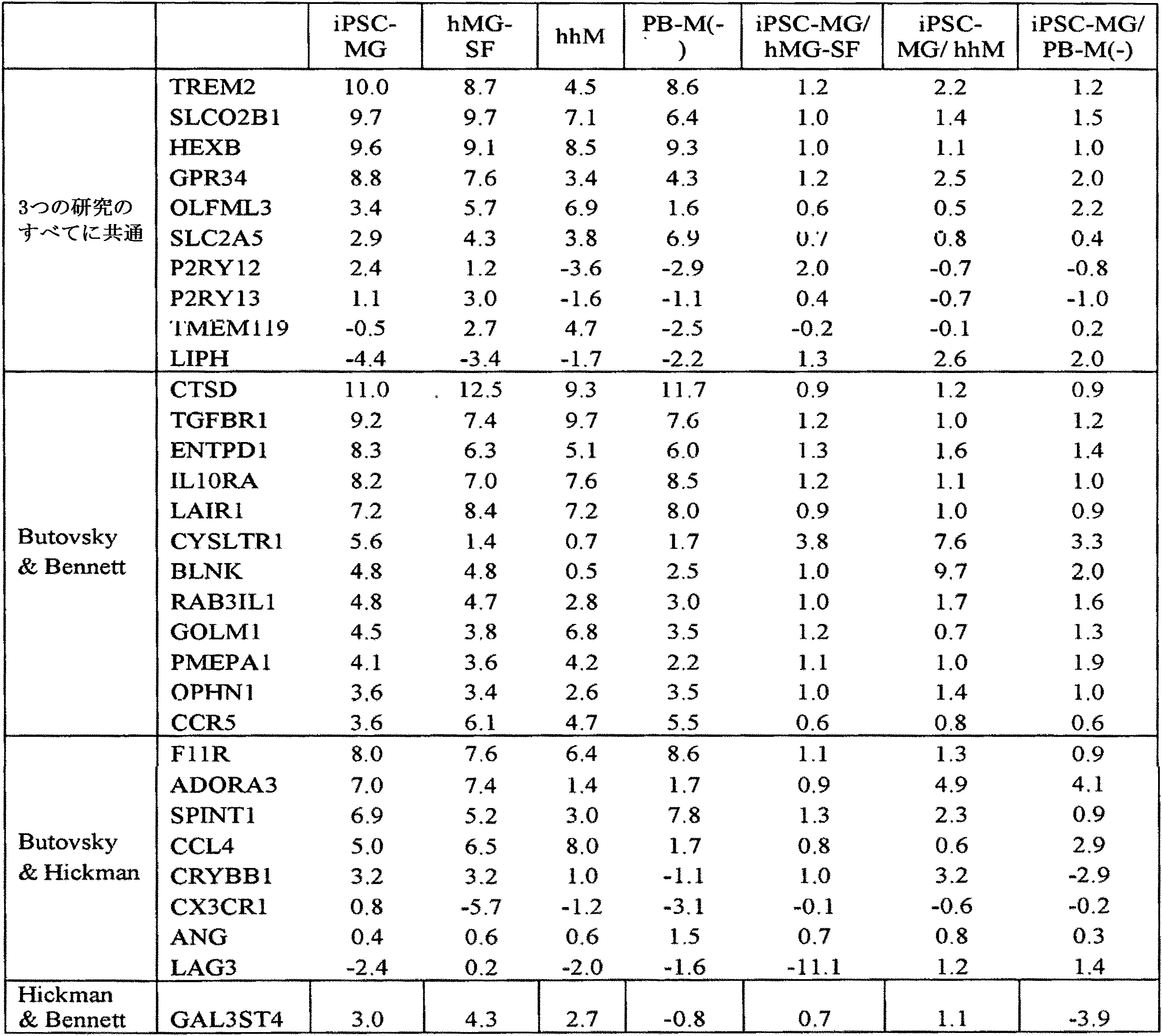
選択した31個の遺伝子のうち、hMGは29個を発現(少なくとも1CPMと定義)し、iPSC-MGは28個を発現していた。全体として、これらの遺伝子の発現レベルは同等であったが、例外として、P2RY13はiPSC-MGでは2.6分の1であり、CYSTR1はhMGでは3.8分の1であった。さらにまた、LIPHは試験したどのヒト細胞タイプでも非常に低く(<1CPM)、hMGは低いCX3CR1発現を、そしてiPSC-MGは低いTMEM119発現および低いLAG3発現を示した。ただし、CX3CR1タンパク質およびTMEM119タンパク質は、それぞれFACSおよび免疫蛍光染色によって検出された。 Of the 31 genes selected, hMG expressed 29 (defined as at least 1 CPM) and iPSC-MG expressed 28. Overall, the expression levels of these genes were comparable, with the exception of P2RY13 at 1 / 2.6 for iPSC-MG and CYSTR1 at 1 / 3.8 for hMG. Furthermore, LIPH was very low (<1CPM) in all human cell types tested, hMG showed low CX3CR1 expression, and iPSC-MG showed low TMEM119 and low LAG3 expression. However, the CX3CR1 and TMEM119 proteins were detected by FACS and immunofluorescent staining, respectively.
組織常在性の肝マクロファージおよび末梢血由来マクロファージとの比較により、iPSC由来ミクログリアでも初代ミクログリアでも一貫して高い11個の遺伝子(TREM2、SLC02B1、GPR34、P2RY12、P2RY13、ENTPD1、BLNK、RAB3IL1、ADORA3、CRYBB1、GAL3ST4)が明らかになった。 Eleven genes consistently high in both iPSC-derived microglia and primary microglia (TREM2, SLC02B1, GPR34, P2RY12, P2RY13, ENTPD1, BLNK, RAB3IL1, ADORA3) by comparison with tissue-resident hepatic macrophages and peripheral blood-derived macrophages , CRYBB1, GAL3ST4) have been revealed.
Butovskyらの研究では、胎児および成人のヒト初代ミクログリアにおけるユニークなシグネチャーとして、6つの遺伝子、すなわちC1QA、GAS6、GPR34、MERTK、P2RY12およびPROS1が提案されている。事実、それらの高い発現は、本発明者らのどのミクログリア試料(iPSC-MG、hMG、hMG-SF)でも、2つの別個の研究(Muffat et al., 2016; Zhang et al., 2016)からのミクログリア試料でも、一貫していたが(図3C)、マクロファージおよび神経前駆細胞(NPC)は、それらより低いレベルを示した(t検定:6つの遺伝子すべてにおいてミクログリアとマクロファージまたはNPCとの間でP<0.05)。図3Cに関係する表2は、RNAseqによって決定されたミクログリア試料におけるこれら6つの確証されたヒトミクログリアシグネチャー遺伝子の発現を示している。表2における値はlog2変換したCPMである。 The study by Butovsky et al. Has proposed six genes, C1QA, GAS6, GPR34, MERTK, P2RY12 and PROS1, as unique signatures in fetal and adult human primary microglia. In fact, their high expression is from two separate studies (Muffat et al., 2016; Zhang et al., 2016) in any of our microglial samples (iPSC-MG, hMG, hMG-SF). Although consistent with microglial samples from (Fig. 3C), macrophages and neural progenitor cells (NPCs) showed lower levels (t-test: between microglia and macrophages or NPCs in all six genes. P <0.05). Table 2, related to Figure 3C, shows the expression of these six confirmed human microglial signature genes in microglial samples determined by RNAseq. The values in Table 2 are log2 converted CPMs.
(表2)
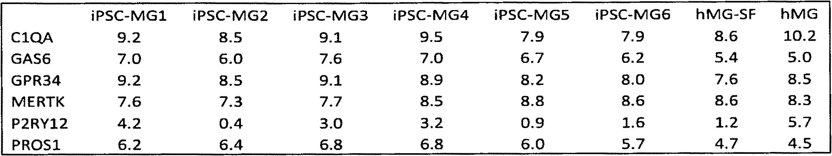
ヒトiPSC由来ミクログリアと初代ミクログリアのサイトカインプロファイルは似ているが、末梢血由来マクロファージのサイトカインプロファイルとは異なることがわかった。本発明者らは、iPSC-MG、hMG、hMG-SF、および異なる極性化を起こしたマクロファージを含むPB-Mによって放出されるタンパク質を分析した(図3D)。興味深いことに、iPSC-MGと初代ミクログリアとの間の類似度は、それらの培養培地(血清含有、hMG)を本発明者らの分化培地(hMG-SF)に置き換えると激増して、ピアソン相関係数はR=0.473からR=0.824へと増加した。注目すべきことには、hMGは、炎症時に典型的に放出され実際にM(LPS,IFNγ)マクロファージでも発現するRANTES、I-TAC、BAFF、GR0-aおよびMIP3aなどのサイトカインのアップレギュレーションを示した(図3Dの矢印)。それでもなおミクログリア試料は互いにクラスターを形成し、M(-)、M(LPS,IFNγ)、M(IL4,IL13)およびM(IL10)として極性化したPB-Mとは離れたクラスターを形成した。 It was found that the cytokine profiles of human iPSC-derived microglia and primary microglia are similar, but different from those of peripheral blood-derived macrophages. We analyzed proteins released by PB-M, including iPSC-MG, hMG, hMG-SF, and macrophages with different polarities (Fig. 3D). Interestingly, the similarity between iPSC-MG and primary microglia increased dramatically when their culture medium (serum-containing, hMG) was replaced with our differentiation medium (hMG-SF), and the Pearson phase. The number of relationships increased from R = 0.473 to R = 0.824. Notably, hMG exhibits upregulation of cytokines such as RANTES, I-TAC, BAFF, GR0-a and MIP3a, which are typically released during inflammation and are actually also expressed in M (LPS, IFNγ) macrophages. (Arrow in Figure 3D). Nevertheless, the microglial samples clustered with each other and clustered apart from PB-M polarized as M (-), M (LPS, IFNγ), M (IL4, IL13) and M (IL10).
本発明者らのiPSC-MGは、機能的食細胞であることもわかった。iPSC-MG、hMG、hMG-SF、PB-Mおよび未分化iPSCを1細胞あたり所与の量の蛍光標識カルボキシル化ラテックスマイクロスフェアに曝露した。フローサイトメトリー分析により、iPSC-MGの大部分は貪食できることが示された(90±6%)。これらの結果はhMGとhMG-SFの両方に匹敵した。予想どおり、M(-)マクロファージも、マイクロスフェアを取り込むことができたが、未分化iPSCはマイクロスフェアを取り込むことができなかった(図3E)。 We also found that the iPSC-MG of the present inventors is a functional phagocyte. iPSC-MG, hMG, hMG-SF, PB-M and undifferentiated iPSCs were exposed to a given amount of fluorescently labeled carboxylated latex microspheres per cell. Flow cytometric analysis showed that the majority of iPSC-MG could be phagocytosed (90 ± 6%). These results were comparable to both hMG and hMG-SF. As expected, M (-) macrophages were also able to take up microspheres, but undifferentiated iPSCs were not able to take up microspheres (Fig. 3E).
本発明者らのiPSC-MGは、ADPに応答して細胞内Ca2+を放出することもわかった。ミクログリアシグネチャー遺伝子P2RY12は、ADPに応答して細胞内Ca2+([Ca2+]i)トランジェントをもたらすGiタンパク質共役プリン作動性受容体をコードしているが(Haynes et al., 2006)、PB-Mはこの受容体を発現しないので、ADPに応答しない(Moore et al., 2015)。したがって、ADPが誘導する[Ca2+]iトランジェントは、ミクログリアとマクロファージとを識別するために使用される。iPSC-MG、hMG、hMG-SFおよびPB-Mを50μM ADPで60秒間刺激したところ、ミクログリア細胞だけがADPに応答した(図4A~C)。iPSC-MGにおけるADP応答の最大振幅(図4E)と応答性細胞の数(図4F)は、hMGまたはhMG-SFのいずれよりも高かった。異なる極性化を起こしたPB-MはいずれもADPに応答しなかったが、100μM ATPによる刺激時には[Ca2+]iトランジェントが確実に観察された(図4Dおよび図8)。 We also found that our iPSC-MG releases intracellular Ca2 + in response to ADP. Although the microglial signature gene P2RY12 encodes a Gi protein-conjugated purinergic receptor that provides intracellular Ca2 + ([Ca2 +] i) transients in response to ADP (Haynes et al., 2006), PB-M Since it does not express this receptor, it does not respond to ADP (Moore et al., 2015). Therefore, ADP-induced [Ca2 +] itransients are used to distinguish between microglia and macrophages. When iPSC-MG, hMG, hMG-SF and PB-M were stimulated with 50 μM ADP for 60 seconds, only microglial cells responded to ADP (Figs. 4A-C). The maximum amplitude of ADP response in iPSC-MG (Fig. 4E) and the number of responsive cells (Fig. 4F) were higher than in either hMG or hMG-SF. None of the PB-Ms with different polarities responded to ADP, but [Ca2 +] i transients were definitely observed when stimulated with 100 μM ATP (Fig. 4D and Fig. 8).
要約および考察
インビトロでのPSCの造血分化は、インビボでの二次造血ではなく原始造血に等価である。これは、PSC由来の造血前駆体が長期多系譜再構築を生じることができないことの説明になりうる(Vanhee et al., 2015)。本発明者らは、PSC由来骨髄系前駆体は原始卵黄嚢骨髄系前駆体に似ているがゆえに、胚発生中に起きるように、インビトロでミクログリアを生じるのかもしれないという仮説を立てた。本発明者らは、骨髄系誘導培地でPSCを刺激することにより、KDR+CD235a+原始血管芽細胞集団を生産し、インビトロでCD45+CX3CR1-ミクログリア前駆体からCD45+CX3CR1+ミクログリア前駆体への進行を再現した。
Summary and Discussion In vitro hematopoietic differentiation of PSCs is equivalent to primordial hematopoiesis rather than secondary hematopoiesis in vivo. This may explain the inability of PSC-derived hematopoietic precursors to undergo long-term polylineage reconstruction (Vanhee et al., 2015). We hypothesized that PSC-derived myeloid precursors may produce microglia in vitro, as they do during embryogenesis, because they resemble primordial yolk sac myeloid precursors. We produced a population of KDR + CD235a + primordial hemangioblasts by stimulating PSC with myeloid induction medium and reproduced the progression from CD45 + CX3CR1-microglial precursors to CD45 + CX3CR1 + microglial precursors in vitro.
このプロトコールのロバスト性および再現性を保証するために、本発明者らは、異なるリプログラミング戦略(例えばmRNA/miRNA、センダイウイルス)を使って作製された、MS患者、AD患者、PD患者または健常個体からのiPSCを含む17株のPSCのパネルを試験した。線維芽細胞は、年齢が25~68歳の範囲にある男女両方のドナーから得た。本発明者らは、すべての株から、未分化PSC1個あたり2~3個の前駆体という平均収量で、ミクログリア前駆体を得ることができた。このような多様なパネルから予想されるとおり、前駆体の収量は株間で変動したが、特定の疾患、リプログラミング方法またはドナーの性別および年齢への明らかな相関はなかった。 To ensure the robustness and reproducibility of this protocol, we have created MS patients, AD patients, PD patients or healthy patients using different reprogramming strategies (eg mRNA / miRNA, Sendai virus). Panels of 17 strains of PSCs, including iPSCs from individuals, were tested. Fibroblasts were obtained from both male and female donors in the age range of 25-68 years. We were able to obtain microglial precursors from all strains with an average yield of 2-3 precursors per undifferentiated PSC. As expected from such diverse panels, precursor yields varied between strains, but there was no clear correlation to specific diseases, reprogramming methods or donor gender and age.
本発明者らのミクログリア前駆体は、典型的なマーカーを発現し、微小環境を走査する能力を有する運動性の高い突起を持つ分枝型であって、正常ヒトミクログリアと等価な効率で貪食することができるミクログリアを生じた。 Our microglial precursors are branched, with highly motile protrusions that express typical markers and have the ability to scan the microenvironment, and phagocytose with efficiency equivalent to normal human microglia. Produced microglia that can.
ヒトミクログリアのアイデンティティの定義は十分には確立されていないが、マウスにおける最近のゲノムワイドな研究により、ミクログリアと他の骨髄系細胞タイプまたはCNS細胞タイプとの区別を容易にするデータセットが得られている(Bennett et al., 2016; Butovsky et al., 2014; Hickman et al., 2013)。そこで本発明者らは、提案されたヒトミクログリアにおける「シグネチャー遺伝子」を評価するために、iPSC-MGの網羅的mRNA発現を、初代ミクログリアならび末梢血由来マクロファージおよび肝マクロファージと比較した。本発明者らは、異なる遺伝的背景を持つ試料を含めた。本発明者らの解析は、iPSC-MGが循環マクロファージとも他の組織特異的マクロファージとも離れたクラスターを形成し、ヒト脳抽出物から単離された初代ミクログリア細胞およびCD45+細胞(「myeloid」と呼ばれる)(Zhang et al., 2016)と互いにクラスターを形成することを明らかに示した。さらにまた、本発明者らのiPSC-MGは、マウスミクログリアにおいて豊富な他の多くの遺伝子に加えて、ヒトミクログリアにユニークであると提唱されている6つの遺伝子(Butovsky et al., 2014)を発現した(表1)。 Although the definition of human microglial identity is not well established, recent genome-wide studies in mice have provided a dataset that facilitates the distinction between microglia and other myeloid or CNS cell types. (Bennett et al., 2016; Butovsky et al., 2014; Hickman et al., 2013). Therefore, in order to evaluate the proposed "signature gene" in human microglia, we compared the comprehensive mRNA expression of iPSC-MG with primary microglia as well as peripheral blood-derived macrophages and hepatic macrophages. We included samples with different genetic backgrounds. In our analysis, iPSC-MG formed distant clusters from circulating macrophages and other tissue-specific macrophages, and primary microglial cells and CD45 + cells (called "myeloid") isolated from human brain extracts. ) (Zhang et al., 2016) and clearly showed that they form clusters with each other. Furthermore, our iPSC-MG contains six genes (Butovsky et al., 2014) that have been proposed to be unique to human microglia, in addition to many other genes that are abundant in mouse microglia. It was expressed (Table 1).
iPSC-MGが放出するサイトカインは、hMGとは似たプロファイルを有するが、PB-Mとは、それらの極性化状態とは無関係に、異なることがわかった。注目すべきことに、hMGは、本発明者らの培地で培養した場合(hMG-SF)の方が、iPSC-MGと、より密なクラスターを形成した。理論に束縛されることは望まないが、その理由はhMG培養培地における血清の存在であるという仮説を、本発明者らは立てている。インビボでは、ミクログリアは血液脳関門の「後ろ」に存在しており、血清構成要素の存在はそれらの活性化の引き金を引く(Ransohoff and Perry, 2009)。事実、血清中で培養されたhMGは、M(LPS,IFNγ)炎症誘発性マクロファージと同様に、RANTES、GR0-A、I-TAC、BAFFおよびMIP3aなどといった炎症性分子のレベルの増加を示した。 Cytokines released by iPSC-MG were found to have a profile similar to that of hMG, but differ from PB-M, regardless of their polarity. Notably, hMG formed a denser cluster with iPSC-MG when cultured in our medium (hMG-SF). We do not want to be bound by theory, but we hypothesize that the reason is the presence of serum in the hMG culture medium. In vivo, microglia reside "behind" the blood-brain barrier, and the presence of serum components triggers their activation (Ransohoff and Perry, 2009). In fact, hMG cultured in serum showed increased levels of inflammatory molecules such as RANTES, GR0-A, I-TAC, BAFF and MIP3a, as well as M (LPS, IFNγ) pro-inflammatory macrophages. ..
本発明者らは、iPSC-MGが転写産物レベルでもタンパク質レベルでも機能的なP2RY12受容体を発現することをも明らかにした。齧歯類およびヒトのミクログリアは、この受容体によって他の骨髄性細胞と識別され(Butovsky et al., 2014)、ADPによるその活性化は[Ca2+]iトランジェントをもたらす(Moore et al., 2015)。PB-MはADPに対して不応性であったが、ATPに曝露されると[Ca2+]iトランジェントを示した一方で、ミクログリア細胞(iPSC-MG、hMGおよびhMG-SF)はいずれも、ADPによって惹起される[Ca2+]iトランジェントを示したことから、それらは健常であり機能的であることが示された。 We also found that iPSC-MG expresses a functional P2RY12 receptor at both transcript and protein levels. Rodent and human microglia are distinguished from other myeloid cells by this receptor (Butovsky et al., 2014), and their activation by ADP results in [Ca2 +] i transients (Moore et al., 2015). ). PB-M was refractory to ADP, but showed [Ca2 +] itransients when exposed to ATP, while all microglial cells (iPSC-MG, hMG and hMG-SF) showed ADP. They showed healthy and functional [Ca2 +] i-transients evoked by.
マウスESCをミクログリアに分化させるためのプロコールは公知であるが(例えばBeutner et al., 2010; Napoli et al., 2009; Tsuchiya et al., 2005参照)、本発明者らは、それらのプロトコールがヒトiPSCでは有効でないことを見いだし(データ省略)、齧歯類系とヒト系の違いが浮き彫りになった。ヒトミクログリア様細胞は、ミクログリアのシグネチャー遺伝子の詳細な特徴づけと機能研究は為されていないものの、以前に、末梢血(PB)単球から分化転換されている(Etemad et al., 2012; Ohgidani et al., 2014)。Muffat et al.(September 2016)は、ヒトミクログリア分化プロトコールを記載した。本発明者らは、RNAseqを使って、本発明者らの方法によって生成させたミクログリアを、Muffatらの方法を使って生成させたミクログリアと比較した。この比較データを本明細書に掲載する(図3Cおよび図7)。どちらのアプローチも、CD235a+と定義される卵黄嚢前駆体を経て誘導されるミクログリアの胚発生を模倣する。どちらのプロトコールも、化学合成培地におけるミクログリアの細胞系譜決定および成熟への主要ドライバーとしてIL34を使用し、どちらのプロトコールも、典型的マーカーを発現し貪食を行うことができる運動性突起を持つミクログリアを生成する。胚様体(EB)形成ではなく単層培養に基づく本発明者らの戦略は、効率では同等であるが、必要とする出発PSCの数が少ない。重要なことに、本発明者らのプロトコールは、特定のEB形態の手作業による選択を必要としない。本発明者らは、FACSまたは磁気ビーズによってミクログリア前駆体を単離するので、化合物スクリーニングおよび遺伝子スクリーニングなどのハイスループット応用が可能になる。 Although protocols for differentiating mouse ESCs into microglia are known (see, eg, Beutner et al., 2010; Napoli et al., 2009; Tsuchiya et al., 2005), we have these protocols. Was found to be ineffective in human iPSC (data omitted), highlighting the difference between rodent and human strains. Human microglial-like cells have previously been transdifferentiated from peripheral blood (PB) monocytes, although detailed characterization and functional studies of microglial signature genes have not been performed (Etemad et al., 2012; Ohgidani). et al., 2014). Muffat et al. (September 2016) described the human microglial differentiation protocol. We used RNAseq to compare the microglia produced by our method with those produced by Muffat et al. This comparative data is presented herein (Fig. 3C and Fig. 7). Both approaches mimic microglial embryogenesis induced via the yolk sac precursor defined as CD235a +. Both protocols use IL34 as the primary driver for cell lineage and maturation of microglia in synthetic media, and both protocols produce microglia with motility processes capable of expressing typical markers and phagocytosing. Generate. Our strategy, which is based on monolayer culture rather than embryoid body (EB) formation, is comparable in efficiency but requires fewer starting PSCs. Importantly, our protocol does not require manual selection of specific EB forms. We isolate microglial precursors by FACS or magnetic beads, enabling high-throughput applications such as compound screening and gene screening.
まとめると、本明細書に記載する新しいプロトコールは、ヒトミクログリア細胞の新しい供給源としてiPSC-MGを提供し、これは、健常および疾患におけるミクログリアの役割をより良く理解するために、マウスモデルにおける研究を補完するであろう。本明細書に記載するプロトコールは、健常対象からの株を使って行うか、罹患患者からの株を使って行うかに関わりなく、再現性が高いので、神経障害におけるミクログリア機能障害の発症機序を調べるための貴重なツールになる。さらにまた、共培養系または三次元系、例えばニューロンおよび他のグリア細胞タイプが関与するものなどに、ミクログリアを含めれば、インビボ環境の複雑さをより良く再現するためのインビトロ疾患モデリングが容易になるはずである。 Taken together, the new protocol described herein provides iPSC-MG as a new source of human microglial cells, a study in mouse models to better understand the role of microglia in health and disease. Will complement. The protocol described herein is highly reproducible regardless of whether it is performed using a strain from a healthy subject or a strain from an affected patient, and thus the pathogenic mechanism of microglial dysfunction in neuropathy. It will be a valuable tool for investigating. Furthermore, inclusion of microglia in co-culture or three-dimensional systems, such as those involving neurons and other glial cell types, facilitates in vitro disease modeling to better reproduce the complexity of the in vivo environment. Should be.
実施例2-実験手順
この実施例では、実施例1で概説した研究を行うために使用した材料および方法に関する詳細を記す。
Example 2-Experimental Procedure This example describes in detail the materials and methods used to perform the studies outlined in Example 1.
多能性幹細胞株
この研究では2種のヒトESC株(RUES1およびH9、どちらもNIHの承認を受けている)および15種のiPSC株を使用した。これらの株については補足情報に記載する。iPSC株の一つはRicardo Feldman博士から譲渡されたものであり、他のiPSC株はすべてNYSCF Research Instituteにおいてリプログラムされた。ヒト初代ミクログリアおよび肝マクロファージはScienCell Research Laboratoriesから購入した。
Pluripotent stem cell strains Two human ESC strains (RUES1 and H9, both NIH approved) and 15 iPSC strains were used in this study. These strains are described in the supplementary information. One of the iPSC strains was transferred from Dr. Ricardo Feldman, and all other iPSC strains were reprogrammed at the NYSCF Research Institute. Primary human microglia and hepatic macrophages were purchased from ScienCell Research Laboratories.
ミクログリア分化プロトコール
80ng/ml BMP4を含有するmTeSR Custom培地(StemCell Technologies)を使って、PSCの分化を誘導した。4日目に、StemPro-34 SFM(2mM GutaMAX含有、Life Technologies)中の25ng/ml bFGF、100ng/ml SCFおよび80ng/ml VEGFで、細胞を誘導した。2日後に、50ng/ml SCF、50ng/ml IL-3、5ng/ml TPO、50ng/ml M-CSFおよび50ng/ml Flt3を培地に添加し、14日目からは、50ng/ml M-CSF、50ng/ml Flt3および25ng/ml GM-CSFを添加した。25日目と50日目の間に、CD14+前駆体またはCD14+CX3CR1+前駆体を単離し、ミクログリア培地(RPMI-1640、Life Technologies、2mM GlutaMAX-I、10ng/ml GM-CSFおよび100ng/ml IL-34を含む)中、組織培養処理が施されたディッシュまたはThermanoxプラスチックカバースリップ(いずれもThermo Scientific製)上に、再プレーティングした。少なくとも2週間は3日~4日ごとに培地を補充した。
Microglial differentiation protocol
PSC differentiation was induced using mTeSR Custom medium (StemCell Technologies) containing 80 ng / ml BMP4. On
組織培養および細胞株
別段の言明がある場合を除き、培養はすべて、37℃の培養器中、5%CO2で行った。また、別段の言明がある場合を除き、成長因子はいずれもR&D Systemsから購入したヒト組換えタンパク質である。培地はすべて1×ペニシリン/ストレプトマイシン(P/S)または1×抗生物質-抗真菌剤(Antibiotic-Antimycotic)(Life Technologies)を含有する。RT:室温。
Tissue cultures and cell lines All cultures were performed at 5% CO2 in a 37 ° C. incubator, unless otherwise stated. Also, unless otherwise stated, all growth factors are human recombinant proteins purchased from R & D Systems. All media contains 1 x penicillin / streptomycin (P / S) or 1 x antibiotic-antifungal agent (Antibiotic-Antimycotic) (Life Technologies). RT: Room temperature.
RUES1およびH9はNIHによって承認されたヒトESC株である。iPSC株はいずれも、特別治験審査委員会(specific institutional review board)の承認とインフォームドコンセントを受けて、匿名化されたドナーの皮膚生検から誘導された。対照iPSC株050643-01-MR-28495(25歳男性)、050652-01-MR-28650(45歳女性)、050598-01-MR-49026(53歳男性)、050598-01-MR-025(53歳男性)、051024-01-MR-005(29歳男性)、050689-01-MR-012(27歳男性)、050675-01-MR-012(34歳男性)、050642-01-MR-001(69歳男性)、PD株050412-01-MR-49025(63歳男性)およびAD株050948-01-MR-028(40歳女性)は、New York Stem Cell Foundationのハイスループット自動システム(米国特許出願公開第2013/0345094号およびPaull et al., 2015参照)を使って、mRNA/miRNA法(StemGent)によってリプログラムされた(Paull et al., 2015)。iPSC102(56歳男性)は多発性硬化症患者からmRNA/miRNAを使って手作業でリプログラムされた(Douvaras et al., 2014)。10001.198.01SV4Mutおよび10001.198.01SV5Mutは、GBA N370S変異を持つPD患者(68歳男性)からセンダイウイルスを使ってリプログラムされた2つの異なるクローンであり(Woodard et al., 2014)、一方、10001.198.01SV5WTはCRISPR/Cas9で修正された株である。MJ2136は、Ricardo Feldman博士の厚意で譲り受けたものであり、健常対照からセンダイウイルスを使ってリプログラムされた(Panicker et al., 2014)。 RUES1 and H9 are NIH-approved human ESC strains. All iPSC strains were derived from anonymized donor skin biopsies with the approval of a specific institutional review board and informed consent. Control iPSC strain 050643-01-MR-28495 (25 year old male), 050652-01-MR-28650 (45 year old female), 050598-01-MR-49026 (53 year old male), 050598-01-MR-025 ( 53-year-old man), 051024-01-MR-005 (29-year-old man), 050689-01-MR-012 (27-year-old man), 050675-01-MR-012 (34-year-old man), 050642-01-MR- 001 (69-year-old male), PD strain 050412-01-MR-49025 (63-year-old male) and AD strain 050948-01-MR-028 (40-year-old female) are high-throughput automated systems of the New York Stem Cell Foundation (US). Reprogrammed by the mRNA / miRNA method (StemGent) using Patent Application Publication No. 2013/0345094 and Paul et al., 2015 (Paull et al., 2015). iPSC102, a 56-year-old man, was manually reprogrammed with mRNA / miRNA from a patient with multiple sclerosis (Douvaras et al., 2014). 10001.198.01SV4Mut and 10001.198.01SV5Mut are two different clones reprogrammed with Sendai virus from a PD patient (68-year-old man) with the GBA N370S mutation (Woodard et al., 2014), while 10001.198. 01SV5WT is a CRISPR / Cas9 modified strain. MJ2136, courtesy of Dr. Ricardo Feldman, was reprogrammed with Sendai virus from a healthy control (Panicker et al., 2014).
ヒト初代ミクログリアおよび肝マクロファージは、ミクログリア培地およびマクロファージ培地(ScienCell)と共に、ScienCellから購入した。 Primary human microglia and hepatic macrophages were purchased from ScienCell along with microglial medium and macrophage medium (ScienCell).
PSC株は、mTeSR1培地(StemCell Technologies)中、Matrigelコートディッシュ上で、培養、増殖させた。株は、Stempro Accutase(Life Technologies)による5分間の酵素的剥離を使って3~4日ごとに継代し、24時間、10μM Rock阻害剤(Y2732、Stemgent)を含むmTeSR1培地に再プレーティングした。 The PSC strain was cultured and grown on a Matrigel coated dish in mTeSR1 medium (StemCell Technologies). Strains were passaged every 3-4 days using 5-minute enzymatic exfoliation with Stempro Accutase (Life Technologies) and replated for 24 hours on mTeSR1 medium containing a 10 μM Rock inhibitor (Y2732, Stemgent). ..
骨髄系前駆体の単離
培養の上清画分からの細胞を、CX3CR1および/またはCD14コンジュゲート一次抗体(表3参照)またはそれらそれぞれのアイソタイプ対照と共に、氷上で40分インキュベートした。次に、細胞をFACS緩衝液(PBS、0.5%BSA、2mM EDTA、20mMグルコース)で洗浄し、300gで6分間、ペレット化し、死細胞を除外するためにDAPIを含有するFACS緩衝液に再懸濁した。CD14+細胞またはCD14+CX3CR1+細胞を、100μmセラミックノズルおよび20psiを用いて、ARIA IIu(商標)セルソーター(BD Biosciences)でのFACSにより単離した。
Isolation of Myeloid Precursors Cells from culture supernatants were incubated on ice for 40 minutes with CX3CR1 and / or CD14 conjugated primary antibodies (see Table 3) or their respective isotype controls. The cells are then washed with FACS buffer (PBS, 0.5% BSA, 2 mM EDTA, 20 mM glucose), pelleted at 300 g for 6 minutes, and re-sprayed in FACS buffer containing DAPI to eliminate dead cells. It became cloudy. CD14 + cells or CD14 + CX3CR1 + cells were isolated by FACS on an ARIA IIu ™ cell sorter (BD Biosciences) using a 100 μm ceramic nozzle and 20 psi.
骨髄系前駆体の凍結および融解
骨髄系前駆細胞を、単離後に、90%FBS(Life Technologies)と10%DMSO(Sigma-Aldrich)とからなる凍結培地中、低温貯蔵バイアル(Thermo Scientific)において凍結した。次に、細胞をMr.Frosty(Thermo Scientific)容器に移し、-80℃に一晩置いた。翌日、長期保存のために低温貯蔵バイアルを液体窒素に移した。
Freezing and thawing of myeloid precursors After isolation, myeloid progenitor cells are frozen in a cryopreservation vial (Thermo Scientific) in a frozen medium consisting of 90% FBS (Life Technologies) and 10% DMSO (Sigma-Aldrich). did. The cells were then transferred to a Mr. Frosty (Thermo Scientific) container and placed at -80 ° C overnight. The next day, cold storage vials were transferred to liquid nitrogen for long-term storage.
細胞を融解するために、低温貯蔵バイアルを、部分的に融解するまで1~2分間、37℃の水浴に移した。層流フード下で、RPMI-1640培地をバイアルの元の体積の5倍まで加えた。次に、細胞を300gで6分間遠心分離し、適当量の培地に再懸濁し、組織培養処理が施されたプラスチックにプレーティングした。 To thaw the cells, the cryopreservation vials were transferred to a 37 ° C. water bath for 1-2 minutes until partially thawed. Under a laminar flow hood, RPMI-1640 medium was added up to 5 times the original volume of the vial. The cells were then centrifuged at 300 g for 6 minutes, resuspended in an appropriate amount of medium and plated on tissue culture treated plastic.
詳細なミクログリア分化プロトコール
PSCを、24時間、10μM Rock阻害剤を含有するmTeSR1培地中、15×103細胞/cm2の密度で、Matrigel(BD Biosciences)上にプレーティングした。個々のコロニーが目に見えるようになったら(通常はプレーティングの2~4日後)、80ng/ml BMP4を含有するmTeSR Custom培地(StemCell Technologies)を与えて分化を誘導した。mTeSR Custom培地は、塩化リチウム、GABA、ピペコリン酸、bFGFおよびTGFβ1を含まないmTeSR1培地である(StemCell Technologies)。4日間、培地を毎日替えてから、25ng/ml bFGF、100ng/ml SCFおよび80ng/ml VEGFを添加したStemPro-34 SFM(2mM GutaMAX-I含有、Life Technologies)を使って細胞を誘導した。2日後に、50ng/ml SCF、50ng/ml IL-3、5ng/ml TPO、50ng/ml M-CSFおよび50ng/ml Flt3を含有するStemPro-34に、培地を切り替えた。10日目に、培養の上清画分をペレット化し、新鮮な培地(前と同じ)に再懸濁し、それぞれのディッシュに戻した。14日目に、浮遊細胞をペレット化し、50ng/ml M-CSF、50ng/ml Flt3および25ng/ml GM-CSFを含有するStemPro-34に再懸濁し、再プレーティングによってそれぞれのディッシュに戻した。この手順を4日ごとに繰り返した。24日目から52日目までは、CD14/CX3CR1二重陽性前駆体のピークを決定するために、少量の浮遊細胞をフローサイトメトリー分析用に加工した。CD14+前駆体またはCD14+CX3CR1+前駆体の単離後に、組織培養処理を施したディッシュまたはThermanoxプラスチックカバースリップ(いずれもThermo Scientific製)上に、SF-ミクログリア培地(2mM GlutaMAX-I、10ng/ml GM-CSFおよび100ng/ml IL-34を添加したLife Technologies製のRPMI-1640)中、40~50×103細胞/cm2でプレーティングした。少なくとも2週間は3日~4日ごとに培地を補充した。
Detailed microglial differentiation protocol
PSCs were plated on Matrigel (BD Biosciences) at a density of 15 x 103 cells / cm2 in mTeSR1 medium containing a 10 μM Rock inhibitor for 24 hours. When individual colonies became visible (usually 2-4 days after plating), differentiation was induced by feeding mTeSR Custom medium (StemCell Technologies) containing 80 ng / ml BMP4. The mTeSR Custom medium is an mTeSR1 medium free of lithium chloride, GABA, pipecolic acid, bFGF and TGFβ1 (StemCell Technologies). The medium was changed daily for 4 days, then cells were induced using StemPro-34 SFM (containing 2 mM GutaMAX-I, Life Technologies) supplemented with 25 ng / ml bFGF, 100 ng / ml SCF and 80 ng / ml VEGF. Two days later, the medium was switched to StemPro-34 containing 50 ng / ml SCF, 50 ng / ml IL-3, 5 ng / ml TPO, 50 ng / ml M-CSF and 50 ng / ml Flt3. On
末梢血由来マクロファージおよび極性化
New York Blood Centerにおいて、以前に記載されたように(Pallotta et al., 2015)、健常個体の末梢血から得られた単離ヒト単核球からマクロファージを分化させた。簡単に述べると、Ficoll勾配およびEasySep Human CD14 Positive Selection Kit(STEMCELL Technologies)を使った磁気ビーズに基づく分離を経て、CD14+細胞を単離した。次に、細胞を5×10⁵細胞/mlの密度で超低接着プレートに5日間播種し、2mM GlutaMAX-I、10%熱非働化ヒト血清(Sigma-Aldrich)および20ng/ml M-CSF(PeproTech)を添加したRPMI-1640を使って、マクロファージに分化させた。極性化のために、マクロファージを同じ培地に維持するか(M(-))、または100ng/ml LPS(Sigma-Aldrich)および100ng/ml IFNγ(M(LPS,IFNγ))、40ng/ml IL-4および20ng/ml IL-13(M(IL4,IL13))もしくは40ng/ml IL-10(M(IL10);いずれもPeproTech製)マクロファージで処理した。
Peripheral blood-derived macrophages and polarization
At the New York Blood Center, macrophages were differentiated from isolated human mononuclear cells obtained from the peripheral blood of healthy individuals, as previously described (Pallotta et al., 2015). Briefly, CD14 + cells were isolated via Ficoll gradient and magnetic bead-based isolation using the EasySep Human CD14 Positive Selection Kit (STEMCELL Technologies). The cells were then seeded on an ultra-low adhesion plate at a density of 5 x 10⁵ cells / ml for 5 days with 2 mM GlutaMAX-I, 10% heat-inactivated human serum (Sigma-Aldrich) and 20 ng / ml M-CSF (PeproTech). ) Was added to the cells to differentiate them into macrophages. For polarization, keep macrophages in the same medium (M (-)) or 100 ng / ml LPS (Sigma-Aldrich) and 100 ng / ml IFNγ (M (LPS, IFNγ)), 40 ng / ml IL- Treatment with 4 and 20 ng / ml IL-13 (M (IL4, IL13)) or 40 ng / ml IL-10 (M (IL10); both manufactured by PeproTech) macrophages.
免疫蛍光染色
細胞を、PBS-T(0.1%Triton-X100を含有するPBS)で10分ずつ3回洗浄し、ブロッキング血清(5%ロバ血清を含むPBS-T)中で2時間インキュベートし、一次抗体(表3参照)を4℃で一晩適用した。翌日、細胞をPBS-T中で15分ずつ3回洗浄し、二次抗体と共に室温(RT)で2時間インキュベートし、PBS-T中で10分ずつ3回洗浄し、DAPIを使って室温で15分間対比染色し、PBS中で2回洗浄した。二次抗体は1:500希釈で使用した。オリンパス(Olympus)DP30BW白黒デジタルカメラを装備したオリンパスIX71倒立顕微鏡を使って画像を取得した。蛍光色は、オリンパスソフトウェアDP ManagerまたはimageJを使って、デジタルに適用した。
Immunofluorescent stained cells were washed 3 times with PBS-T (PBS containing 0.1% Triton-X100) for 10 minutes each and incubated in blocking serum (PBS-T containing 5% donkey serum) for 2 hours for primary. The antibody (see Table 3) was applied overnight at 4 ° C. The next day, cells were washed 3 times in PBS-T for 15 minutes each, incubated with secondary antibody for 2 hours at room temperature (RT), washed 3 times in PBS-T for 10 minutes each, at room temperature using DAPI. It was counterstained for 15 minutes and washed twice in PBS. The secondary antibody was used at a 1: 500 dilution. Images were acquired using an Olympus IX71 inverted microscope equipped with an Olympus DP30BW black and white digital camera. Fluorescent colors were applied digitally using Olympus software DP Manager or imageJ.
フローサイトメトリー分析
細胞を37℃で5分間のAccutase処理によって酵素的に回収した後、セルリフター(Sigma-Aldrich)を使って掻き取った。次に、適当量の蛍光コンジュゲート抗体(表3参照)を含有するそれぞれの培地100μlに再懸濁し、遮光して氷上で40分間インキュベートした。アイソタイプ対照または二次抗体のみを使って、ベースライン背景シグナルを測定した。死細胞の除外にはDAPIまたはSytox Green(Thermofisher)を使用した。分析は、5レーザーBD Biosciences ARIA-IIu(商標)セルソーターまたは4レーザーAttune NxTフローサイトメーター(ThermoFisher)で行った。データはBD FACSDiva(商標)ソフトウェアまたはFlowJoバージョン9.9.4(Flow Jo LLC)を使って解析した。
Flow cytometry analysis Cells were enzymatically recovered by Accutase treatment at 37 ° C. for 5 minutes and then scraped off using a cell lifter (Sigma-Aldrich). It was then resuspended in 100 μl of each medium containing an appropriate amount of fluorescent conjugated antibody (see Table 3), shaded and incubated on ice for 40 minutes. Baseline background signals were measured using only isotype controls or secondary antibodies. DAPI or Sytox Green (Thermofisher) was used to exclude dead cells. Analysis was performed with a 5-laser BD Biosciences ARIA-IIu ™ cell sorter or a 4-laser Attune NxT flow cytometer (ThermoFisher). Data were analyzed using BD FACS Diva ™ software or Flow Jo version 9.9.4 (Flow Jo LLC).
(表3)フローサイトメトリーおよび免疫蛍光分析に使用した抗体
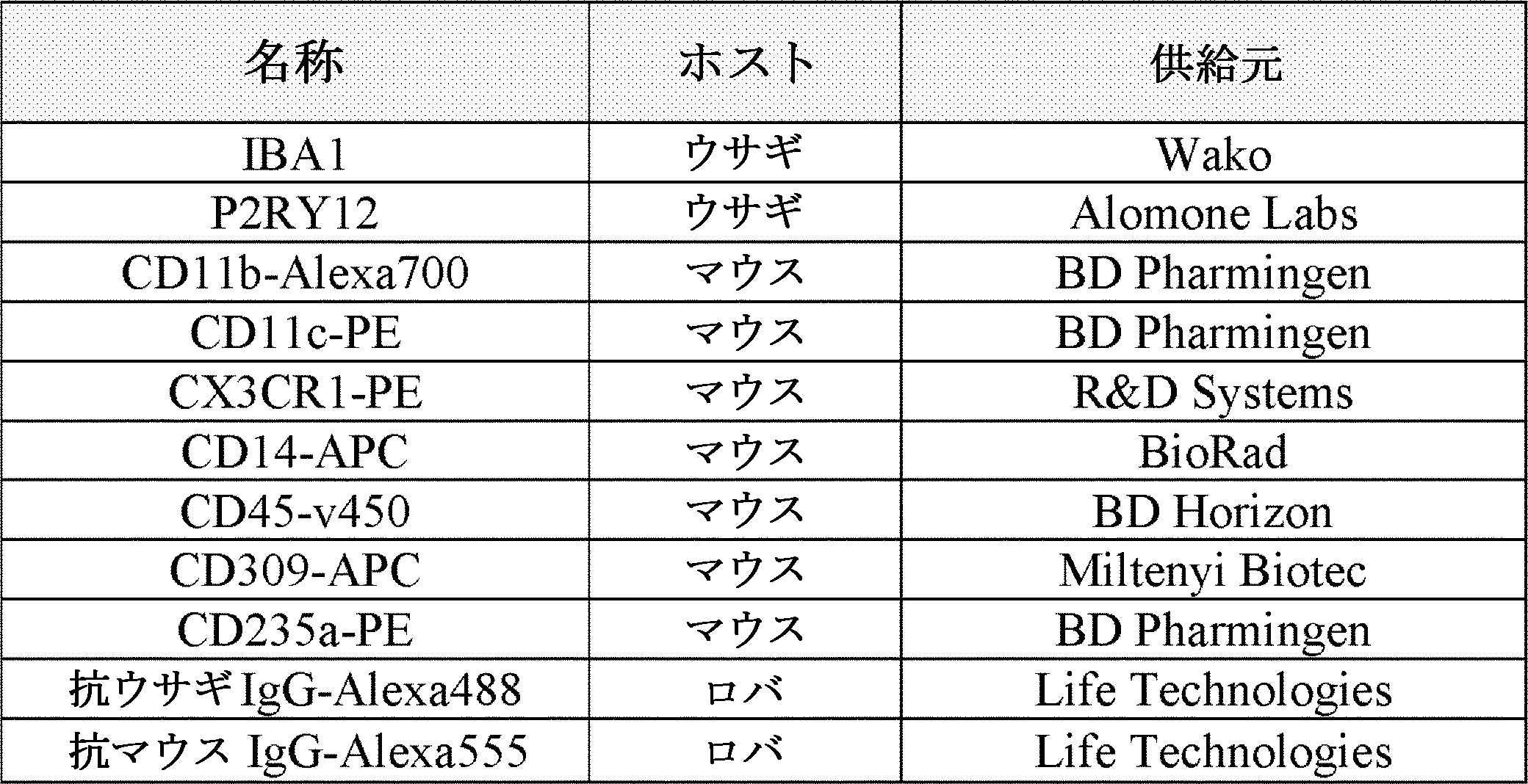
貪食アッセイ
貪食アッセイは、以前に記載されたように行った(Enomoto et al., 2013)。簡単に述べると、付着ミクログリア細胞が入っているディッシュに、Fluoresbrite YGカルボキシレートマイクロスフェア1.00μm(Polysciences)を200マイクロスフェア/細胞の比で加えた。その培養物を37℃で3時間インキュベートした後、オリンパスDP30BW白黒デジタルカメラを装備したオリンパスIX71倒立顕微鏡で、蛍光像を取得した。次に、細胞をPBSで3回洗浄し、Accutaseで5分間処理し、セルリフターで完全に剥離させた。遠心分離後に、DAPIを含有するFACS緩衝液に細胞を再懸濁し、BD Biosciences ARIA-IIu(商標)セルソーターで分析した。
Phagocytosis Assay The phagocytosis assay was performed as previously described (Enomoto et al., 2013). Briefly, Fluoresbrite YG carboxylate microspheres 1.00 μm (Polysciences) were added to a dish containing adherent microglial cells at a ratio of 200 microspheres / cell. After incubating the culture at 37 ° C. for 3 hours, fluorescence images were obtained with an Olympus IX71 inverted microscope equipped with an Olympus DP30BW black and white digital camera. The cells were then washed 3 times with PBS, treated with Accutase for 5 minutes and completely detached with a cell lifter. After centrifugation, cells were resuspended in FACS buffer containing DAPI and analyzed on a BD Biosciences ARIA-IIu ™ cell sorter.
サイトカインプロファイラーおよびクラスタリング
ミクログリア細胞の分泌サイトカインプロファイルを分析するために、プロテオームプロファイラー抗体アレイのヒトXLサイトカインアレイキット(R&D Systems)を、製造元の使用説明書に従って使用した。培養から上清を収集し、最長3ヶ月間、-80℃で保存した。メンブレンをKodak Image Station 4000MM PROで直接可視化し、Carestream分子イメージングソフトウェアを使って画像を取得した。
Cytokine Profiler and Clustering To analyze the secretory cytokine profile of microglial cells, the Human XL Cytokine Array Kit (R & D Systems) of the proteome profiler antibody array was used according to the manufacturer's instructions. The supernatant was collected from the culture and stored at -80 ° C for up to 3 months. The membrane was visualized directly with the Kodak Image Station 4000MM PRO and images were acquired using Carestream molecular imaging software.
シグナルを分析するために、画像をImage Jにインポートし、プロテインアレイアナライザー・プラグインを使用した。次に、2つの同一スポットの強度の読みを平均し、8つの陰性対照の平均値を各値から差し引いた。最後に、データを、6つの参照スポット(陽性対照)の平均強度と比較した強度比として表した。タンパク質プロファイラー分析のためのヒートマップおよびクラスタリングを、Rバージョン3.3.1のheatmap.2を使って生成させた。 To analyze the signal, we imported the images into Image J and used the Protein Array Analyzer plug-in. Next, the intensity readings of the two identical spots were averaged and the mean of the eight negative controls was subtracted from each value. Finally, the data were expressed as an intensity ratio compared to the average intensity of the 6 reference spots (positive controls). Heatmaps and clustering for protein profiler analysis were generated using heatmap.2 in R version 3.3.1.
細胞内Ca2+イメージング
細胞をThermanoxプラスチックカバースリップ(ThermoFisher)上で培養し、Pluronic-127試薬と1:1で混合した蛍光性Ca2+色素Fluo-4/AM(2□M)(どちらもInvitrogen製)を添加した培地と共に、37℃で30分間インキュベートした。次に、pHを7.5に調整した1%BSA(Life Technologies)、1×GlutaMAX-Iおよび10mM HEPES(Sigma-Aldrich)を含有するRPMI-1640培地で、細胞を2回洗浄した。色素のエステル化を保証するために、細胞をさらに30分間回復させた。次に、カバースリップを、正立型オリンパスBX61顕微鏡にマウントした記録チャンバに移した。蛍光を冷却CCDカメラ(Hamamatsu Orca R2)によって2Hzで記録した。薬物適用は室温で60秒間の全チャンバ灌流によって行った。[Ca2+]iトランジェントはΔF(t)/F0の形式で表され、ここで、F0は所与の関心領域のベースライン蛍光であり、ΔFは蛍光F(t)とF0の電流レベルの間の相違である。0.05未満であるΔF(t)/F0のゆらぎは、非応答とみなした。
Intracellular Ca2 + Imaging Cells were cultured on Thermo Fisher and fluorescent Ca2 + dye Fluo-4 / AM (2 □ M) (both manufactured by Invitrogen) mixed 1: 1 with Prulonic-127 reagent. The cells were incubated with the added medium at 37 ° C. for 30 minutes. The cells were then washed twice with RPMI-1640 medium containing 1% BSA (Life Technologies), 1 × GlutaMAX-I and 10 mM HEPES (Sigma-Aldrich) with pH adjusted to 7.5. Cells were allowed to recover for an additional 30 minutes to ensure dye esterification. The coverslip was then transferred to a recording chamber mounted on an upright Olympus BX61 microscope. Fluorescence was recorded at 2 Hz by a cooled CCD camera (Hamamatsu Orca R2). Drug application was performed by whole chamber perfusion for 60 seconds at room temperature. [Ca 2+ ] i Transients are expressed in the form ΔF (t) / F0, where F0 is the baseline fluorescence of a given region of interest and ΔF is between the fluorescence F (t) and the current level of F0. Is the difference. Fluctuations of ΔF (t) / F0 less than 0.05 were considered non-response.
RNAのシークエンシングおよび解析
RNA単離は、RNeasy Plus Mini Kit(Qiagen)をQIAshredder(Qiagen)と共に使用して行った。Accutaseによる5分間の処理後にセルリフターを使って、細胞を酵素的に剥離させた。遠心分離後に細胞をPBSで洗浄し、溶解緩衝液に再懸濁した。次に、製造元の使用説明書に従ってさらに加工するまで、試料を-80℃で保存した。RNAを30μlのRNaseフリーddH2Oに溶出させ、NanoDrop 8000分光測光器(Thermo Scientific)で定量した。
RNA sequencing and analysis
RNA isolation was performed using the RNeasy Plus Mini Kit (Qiagen) with QIAshredder (Qiagen). After 5 minutes of treatment with Accutase, cells were enzymatically detached using a cell lifter. After centrifugation, the cells were washed with PBS and resuspended in lysis buffer. The sample was then stored at -80 ° C until further processing according to the manufacturer's instructions. RNA was eluted in 30 μl RNase-free ddH2O and quantified with a NanoDrop 8000 spectrophotometer (Thermo Scientific).
Illumina HiSeq 2500プラットフォームでIlluminaプロトコールに従ってシングルエンドRNAseqデータを生成させた。スターアライナー(star aligner)(バージョン2.5.0b)を使って、生シークエンシングリードをヒトhg19ゲノムとアラインメントした。リードアラインメントに続いて、featureCounts(Liao et al., 2014)を使って、Ensembl遺伝子モデルGRCh37.70に基づいて遺伝子レベルで遺伝子発現を定量した。少なくとも1つの試料中で少なくとも1カウント・パー・ミリオン(CPM)である遺伝子を発現しているとみなして、その後の分析のためにとっておき、そうでないものは除去した。シークエンシングライブラリーサイズの相違を調整するために、M値のトリム平均正規化(trimmed mean of M-value normalization)(TMM)法(Robinson et al., 2010)を使って、遺伝子レベルリードカウントデータを正規化した。トランスクリプトームワイドの遺伝子発現に基づく階層的クラスター分析は、Rプログラミング言語を使って行った。 Single-ended RNAseq data was generated according to the Illumina protocol on the Illumina HiSeq 2500 platform. Raw sequencing reads were aligned with the human hg19 genome using a star aligner (version 2.5.0b). Following read alignment, featureCounts (Liao et al., 2014) were used to quantify gene expression at the gene level based on the Ensembl gene model GRCh37.70. Genes that were at least 1 count per million (CPM) were considered to be expressed in at least one sample and were set aside for subsequent analysis and those that were not were removed. Gene-level read count data using the trimmed mean of M-value normalization (TMM) method (Robinson et al., 2010) to adjust for differences in sequencing library sizes. Was normalized. Hierarchical cluster analysis based on transcriptome-wide gene expression was performed using the R programming language.
ZhangらのミクログリアRNAseqデータ(Zhang et al., 2016)を再解析するために、本発明者らは、遺伝子発現オムニバス(GEO)から「myeloid」細胞の生RNAseqデータをダウンロードした(アクセッションGSE73721)。上記と同じstar/featureCountsパイプラインを使ってRNAseqリードデータを加工してから、その遺伝子レベルリードカウントを、本発明者らの試料の遺伝子カウントデータと組み合わせた。統合したデータを、TMMアプローチで正規化してから、線形回帰を使って、バッチに関する補正を行った。階層的クラスター分析を使って、試料の非類似度を示した。 To reanalyze the microglial RNAseq data from Zhang et al. (Zhang et al., 2016), we downloaded live RNAseq data from the gene expression omnibus (GEO) of "myeloid" cells (Axon GSE73721). .. The RNAseq read data was processed using the same star / featureCounts pipeline as above, and then the gene level read counts were combined with the gene count data for our samples. The integrated data was normalized by the TMM approach and then batch-corrected using linear regression. Hierarchical cluster analysis was used to show the dissimilarity of the samples.
同様に、最近公表されたMuffat et al., 2016のヒトiPSC由来ミクログリアデータセットと比較するために、本発明者らは彼らのRNAseqリードデータをGEOからダウンロードし(アクセッションGSE85839)、次に同じRNAseq解析パイプラインを適用することで遺伝子レベルカウントデータを得て、それを本試料のリードカウントと統合した。統合したデータを正規化し、バッチ補正してから、階層的クラスター分析を行った。 Similarly, for comparison with the recently published Muffat et al., 2016 human iPSC-derived microglial dataset, we downloaded their RNAseq read data from GEO (Axon GSE85839) and then the same. Gene level count data was obtained by applying the RNAseq analysis pipeline and integrated with the read count of this sample. The integrated data was normalized, batch corrected, and then hierarchical cluster analysis was performed.
統計
頻度はエクセルで算出し、平均±平均の標準誤差(SEM)として表した。対応のないスチューデントのt検定を使って[Ca2+]iトランジェント振幅の統計的解析を行い、エクセルで平均値を比較した。6種のシグネチャー遺伝子に基づくデンドログラムに関するクラスターパーティションのP値有意性は、2つの中心を持つk平均クラスターから得られるクラスター中心距離が元のデータのそれよりも遠い1000重複順列(各遺伝子内で遺伝子発現値をシャフリング)の分率として推定した。サイトカイン放出データに関するiPSC-MGとhMGまたはhMG-SFとの間のピアソン相関係は、GraphPad Prism 6を使って計算した。
Statistical frequency was calculated in Excel and expressed as mean ± standard error (SEM). Statistical analysis of [Ca2 +] i transient amplitudes was performed using the unpaired Student's t-test, and the mean values were compared in Excel. The P-value significance of the cluster partition for the dendrogram based on the 6 signature genes is the 1000 permutations with which the cluster center distance obtained from the k-mean cluster with two centers is farther than that of the original data (within each gene). The gene expression value was estimated as a fraction of shuffling). The Pearson phase relationship between iPSC-MG and hMG or hMG-SF for cytokine release data was calculated using
実施例3
多能性幹細胞からミクログリアを生成させるための代替的プロトコール
この実施例は、多能性幹細胞からミクログリアを生成させるための代替的な例示的プロトコールを提供する。これらのプロトコールでは、CD14+/CX3CR1+ミクログリア前駆体がFACSによって単離されるか、CD14発現だけに基づいて磁気ビーズによる単離を使って単離され、単離されたCD14+前駆体は次に、組織培養処理が施されたプラスチックに、代替(「A」)ミクログリア分化培地(M-CSF、GM-CSF、NGF-βおよびCCL2を含む)または通常(「R」)ミクログリア分化培地(GM-CSFおよびIL-34を含む、実施例1および2において使用されたもの)に入れて、プレーティングされる。別段の言明がある場合を除き、この実施例での材料および方法は、実施例1および2について上述したものと同じである。
Example 3
Alternative Protocols for Producing Microglia from Pluripotent Stem Cells This example provides an alternative exemplary protocol for producing microglia from pluripotent stem cells. In these protocols, the CD14 + / CX3CR1 + microglial precursor was isolated by FACS or isolated using magnetic bead isolation based solely on CD14 expression, and the isolated CD14 + precursor was then tissue-cultured. Treated plastic with alternative (“A”) microglial differentiation medium (including M-CSF, GM-CSF, NGF-β and CCL2) or normal (“R”) microglial differentiation medium (GM-CSF and IL) (As used in Examples 1 and 2), including -34) and plated. Unless otherwise stated, the materials and methods in this example are the same as described above for Examples 1 and 2.
例示的プロトコールの一つでは、表示した日に以下の工程を行う。
0日目:Matrigel上で成長させた未分化iPSCコロニー(だいたい直径1mm)を、80ng/ml BMP4を添加したmTSeR Customに切り替えることによって誘導する。培地を毎日替える。
4日目:2mM Glutamax、25ng/ml bFGF、100ng/ml SCFおよび80ng/ml VEGFを含むStemPro-34に、培地を切り替える。
6日目:2mM Glutamax、50ng/ml SCF、50ng/ml IL-3、5ng/ml TPO、50ng/ml M-CSF、50ng/ml Flt-3を含むStemPro-34に、培地を切り替える。
10日目:上清から細胞を収集、遠心沈殿させ、2mM Glutamax、50ng/ml SCF、50ng/ml IL-3、5ng/ml TPO、50ng/ml M-CSF、50ng/ml Flt-3を含むStemPro-34に再懸濁する。細胞をそれぞれのウェルに戻す。
14日目:上清から細胞を収集、遠心沈殿させ、2mM Glutamax、50ng/ml M-CSF、50ng/ml Flt-3、25ng/ml GM-CSFを含むStemPro-34に再懸濁する。細胞をそれぞれのウェルに戻す。4日ごとに繰り返す。
24日目~52日目:CD14+細胞に関して、FACS分取するか、磁気ビーズ単離を使用する。ProFreezeまたは50%FBS/10%DMSOを使って、60%の生存率でCD14+細胞を凍結する。このCD14+前駆体を使用して、以下の分化工程でミクログリア細胞を生産する。
In one of the exemplary protocols, the following steps are performed on the indicated date.
Day 0: Undifferentiated iPSC colonies grown on Matrigel (approximately 1 mm in diameter) are induced by switching to mTSeR Custom supplemented with 80 ng / ml BMP4. Change the medium daily.
Day 4: Switch medium to StemPro-34 containing 2 mM Glutamax, 25 ng / ml bFGF, 100 ng / ml SCF and 80 ng / ml VEGF.
Day 6: Switch medium to StemPro-34 containing 2 mM Glutamax, 50 ng / ml SCF, 50 ng / ml IL-3, 5 ng / ml TPO, 50 ng / ml M-CSF, 50 ng / ml Flt-3.
Day 10: Collect cells from supernatant, centrifuge and contain 2 mM Glutamax, 50 ng / ml SCF, 50 ng / ml IL-3, 5 ng / ml TPO, 50 ng / ml M-CSF, 50 ng / ml Flt-3 Resuspend in StemPro-34. Return cells to their respective wells.
Day 14: Cells are collected from the supernatant, centrifuged and resuspended in StemPro-34 containing 2 mM Glutamax, 50 ng / ml M-CSF, 50 ng / ml Flt-3, 25 ng / ml GM-CSF. Return cells to their respective wells. Repeat every 4 days.
Days 24-52: For CD14 + cells, FACS fractionation or magnetic bead isolation is used. Freeze CD14 + cells with 60% viability using ProFreeze or 50% FBS / 10% DMSO. Using this CD14 + precursor, microglial cells are produced in the following differentiation steps.
上記の工程に続いて、CD14+前駆細胞を50K~100K細胞/cm2の密度で、組織培養処理を施したプラスチックにプレーティングする。代替「A」培地(すなわち2mM Glutamax、10ng/ml M-CSF、10ng/ml GM-CSF、10ng/ml NGF-βおよび100ng/ml CCL-2を添加したRPMI-1640)または通常「R」培地(すなわち2mM Glutamax、10ng/ml GM-CSFおよび100ng/ml IL-34を添加したRPMI-1640)のどちらかを細胞に供給する。培地「A」を使用する場合は、2週間にわたって毎日培地を替える。培地「R」を使用する場合は、2週間にわたって4日ごとに培地を替える。この工程によって、CD14+前駆細胞からミクログリア細胞が生産されることになる。 Following the above steps, CD14 + progenitor cells are plated with tissue culture treated plastic at a density of 50K-100K cells / cm2. Alternative "A" medium (ie RPMI-1640 supplemented with 2 mM Glutamax, 10 ng / ml M-CSF, 10 ng / ml GM-CSF, 10 ng / ml NGF-β and 100 ng / ml CCL-2) or usually "R" medium Either feed the cells (ie RPMI-1640 supplemented with 2 mM Glutamax, 10 ng / ml GM-CSF and 100 ng / ml IL-34). If medium "A" is used, change the medium daily for 2 weeks. When using medium "R", change the medium every 4 days for 2 weeks. By this step, microglial cells are produced from CD14 + progenitor cells.
ミクログリア分化工程を行う前に(FACSまたは磁気ビーズを使って)CD14+細胞を単離することは不可欠ではない。例えば、いくつかの態様では、CD14+細胞を単離する代わりに、上清中の全細胞が以降のミクログリア分化工程に使用される。加えて、これらの例示的プロトコールの一定の変形(例えば作用物質の濃度および培地変更のタイミングの違いなど)を使用することができ、それらが本発明の範囲内であることは、当業者には認識されるであろう。 Isolation of CD14 + cells (using FACS or magnetic beads) prior to the microglial differentiation step is not essential. For example, in some embodiments, instead of isolating CD14 + cells, all cells in the supernatant are used in subsequent microglial differentiation steps. In addition, certain variations of these exemplary protocols can be used, such as differences in the concentration of agents and the timing of medium changes, and it will be appreciated by those skilled in the art that they are within the scope of the present invention. Will be recognized.
図9は、最終ミクログリア分化工程に代替「A」培地が使用されるそのような例示的プロトコールの概略図である。最終分化工程に代替「A」培地を使って生産された細胞は、ミクログリアマーカーIBA1およびCD11c(免疫蛍光染色による決定;図10参照)ならびにCD11b、CD11c、CX3CR1、P2RY12、CD45(FACsによる決定)を発現する。 FIG. 9 is a schematic representation of such an exemplary protocol in which an alternative “A” medium is used for the final microglial differentiation step. Cells produced using alternative "A" medium for the final differentiation step were subjected to microglial markers IBA1 and CD11c (determined by immunofluorescence staining; see Figure 10) and CD11b, CD11c, CX3CR1, P2RY12, CD45 (determined by FACs). Express.
「A」ミクログリア分化培地と「R」ミクログリア分化培地の使用の効果を対比して調べ、またミクログリア前駆体のプレーティングに多種多様な基質を使用する効果も調べるために、研究を行った。ミクログリア前駆体を、表示した基質(すなわちプラスチック、ラミニン、フィブロネクチンまたはマトリゲル(matrigel))にプレーティングし、「A」培地または「R」培地のどちらかにおいて培養した場合に、表示したマーカーを発現する細胞が全細胞に占めるパーセンテージ(%)を、下記表4に示す。ミクログリア前駆体は、以下に示す試験した基質を含めて、任意の適切な基質にプレーティングすることができる。限定するわけではないが、(例えば免疫蛍光研究を行うために)付着細胞が望まれる場合を含むいくつかの態様では、プラスチックが使用される。限定するわけではないが、細胞付着が必要でない態様を含むいくつかの態様では、Matrigelが使用される。 Studies were conducted to compare the effects of using "A" microglial differentiation medium and "R" microglial differentiation medium, and to investigate the effects of using a wide variety of substrates on the plating of microglial precursors. Microglial precursors are plated on the labeled substrate (ie, plastic, laminin, fibronectin or matrigel) and cultured in either "A" or "R" medium to express the labeled markers. The percentage of cells in the total cells is shown in Table 4 below. Microglial precursors can be plated on any suitable substrate, including the substrates tested below. Plastics are used in some embodiments, including, but not limited to, where adherent cells are desired (eg, for performing immunofluorescence studies). In some embodiments, including but not limited to, cell attachment is not required, Matrigel is used.
(表4)ミクログリアマーカープロファイルに対する分化培地および基質の効果
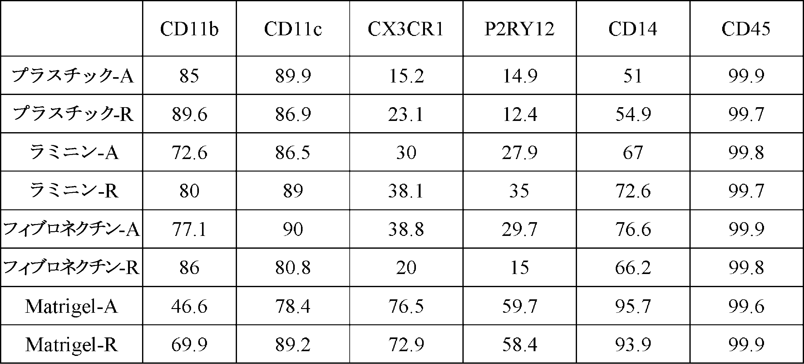
図11に示すように、「A」培地で培養されたミクログリアは(「R」培地で培養されたもののように)カルボキシル化ラテックスビーズを貪食することができた。図12に示すように、「A」培地または「R」培地のどちらかで培養されたミクログリアが放出するサイトカインは、極めて似ていることがわかった。プラスチック上または超低接着(ULA)プレート上で培養されたiPSC由来ミクログリアが示す発現プロファイルは互いに、そしてヒト初代ミクログリアと共に、クラスターを形成し、M(-)、M(LPS,IFNγ)、M(IL4,IL13)およびM(IL10)として極性化した末梢血由来マクロファージとは離れていることもわかった。 As shown in FIG. 11, microglia cultured in "A" medium were able to phagocytose carboxylated latex beads (like those cultured in "R" medium). As shown in FIG. 12, the cytokines released by microglia cultured in either "A" or "R" medium were found to be very similar. Expression profiles of iPSC-derived microglia cultured on plastic or ultra-low adhesion (ULA) plates cluster with each other and with primary human microglia, M (-), M (LPS, IFNγ), M ( It was also found to be distant from peripheral blood-derived macrophages polarized as IL4, IL13) and M (IL10).
本発明の具体的態様に関する上記の説明によって本発明の一般的性質は十分に明らかになるので、他者は、過度の実験を行わずに、当技術分野の通常の技能内である知識を応用して、本発明の一般的概念から逸脱することなく、さまざまな応用のために、容易にそれら具体的態様を変更しかつ/または適合させることができる。したがって、そのような適合および変更は、本明細書に提示した教示および指針に基づいて、開示した態様の均等物の意味および範囲内にあるものとする。本明細書における表現法または用語法は、説明を目的としており、限定を目的とするものではないので、当業者は、本明細書の用語法または表現法を、前記教示および指針に照らして解釈するものと、理解すべきである。 The above description of the specific embodiments of the invention will sufficiently reveal the general nature of the invention so that others can apply knowledge within the ordinary skills of the art without undue experimentation. Thus, their specific aspects can be easily modified and / or adapted for a variety of applications without departing from the general concept of the present invention. Therefore, such adaptations and modifications shall be within the meaning and scope of the equivalent of the disclosed aspects, based on the teachings and guidelines presented herein. As the terminology or terminology herein is for explanatory purposes and not intended to be limiting, one of ordinary skill in the art will interpret the terminology or terminology herein in the light of the teachings and guidelines. It should be understood as what to do.
参考文献一覧
この開示の他の項で引用された文書に加えて、以下の参考文献も、さらなる背景を提供しうる。この開示において引用された参考文献はすべて、参照によりその全体が本明細書に組み入れられる。加えて、本明細書において引用されまたは言及されたあらゆる製品に関する製造元の使用説明書またはカタログも、参照により本明細書に組み入れられる。参照により本明細書に組み入れられる文書またはそれに含まれる任意の教示内容は、本発明の実施において使用することができる。参照によりこの文章に組み入れられた文書を、先行技術であると認めるわけではない。
Claims (81)
(b)CD14+および/またはCX3CR1+ミクログリア前駆細胞を、(i)IL-34を含む第1のミクログリア分化培地、または(ii)M-CSFを含む第2のミクログリア分化培地のいずれかにおいて、培養する工程
を含み、それによってヒトミクログリア細胞を生成する、ミクログリア細胞を生成するための方法。 The steps of culturing human pluripotent stem cells under conditions that induce myeloid differentiation leading to the production of CD14 + and / or CX3CR1 + microglial progenitor cells, and (b) CD14 + and / or CX3CR1 + microglial progenitor cells, (i). Microglial cells comprising the step of culturing in either a first microglial differentiation medium containing IL-34 or (ii) a second microglial differentiation medium containing M-CSF, thereby producing human microglial cells. How to generate.
a)細胞培養物を、多能性幹細胞の維持または分化に適した無血清培地中で、BMP4を含む第1の組成物と接触させることであって、細胞培養物を第1の組成物と最初に接触させるときに、細胞培養物が多能性幹細胞を含む、接触させること、
b)細胞培養物を、無血清造血細胞培地中で、bFGF、SCF、およびVEGF-Aを含む第2の組成物と接触させること、
c)細胞培養物を、無血清造血細胞培地中で、SCF、IL-3、TPO、MCSF、およびFLT3リガンドを含む第3の組成物と接触させること、ならびに
d)細胞培養物を、無血清造血細胞培地中で、MCSF、FLT3リガンド、およびGM-CSFを含む第4の組成物と接触させること
を逐次的に含む、請求項1に記載の方法。 Process (a) is
a) Contacting the cell culture with a first composition containing BMP4 in a serum-free medium suitable for the maintenance or differentiation of pluripotent stem cells, the cell culture with the first composition. When first contacted, the cell culture contains pluripotent stem cells, contacting,
b) Contacting the cell culture with a second composition containing bFGF, SCF, and VEGF-A in serum-free hematopoietic cell medium,
c) Contacting the cell culture in a serum-free hematopoietic cell medium with a third composition containing SCF, IL-3, TPO, MCSF, and FLT3 ligands, and
d) The method of claim 1, wherein the cell culture is sequentially contacted with a fourth composition comprising MCSF, FLT3 ligand, and GM-CSF in a serum-free hematopoietic cell medium.
(b)工程(a)において生成したCD14+および/またはCX3CR1+細胞を単離する工程
を含み、該CD14+および/またはCX3CR1+細胞がミクログリア前駆細胞である、ミクログリア前駆細胞を生成するための方法。 (A) A step of culturing human pluripotent stem cells under conditions that induce myeloid differentiation,
(B) A method for producing microglial progenitor cells, comprising the step of isolating the CD14 + and / or CX3CR1 + cells produced in step (a), wherein the CD14 + and / or CX3CR1 + cells are microglial progenitor cells.
a)細胞培養物を、多能性幹細胞の維持または分化に適した無血清培地中で、BMP4を含む第1の組成物と接触させることであって、細胞培養物を第1の組成物と最初に接触させるときに、細胞培養物が多能性幹細胞を含む、接触させること、
b)細胞培養物を、無血清造血細胞培地中で、bFGF、SCF、およびVEGF-Aを含む第2の組成物と接触させること、
c)細胞培養物を、無血清造血細胞培地中で、SCF、IL-3、TPO、MCSF、およびFLT3リガンドを含む第3の組成物と接触させること、ならびに
d)細胞培養物を、無血清造血細胞培地中で、MCSF、FLT3リガンド、およびGM-CSFを含む第4の組成物と接触させること
を逐次的に含む、請求項37に記載の方法。 Process (a) is
a) Contacting the cell culture with a first composition containing BMP4 in a serum-free medium suitable for the maintenance or differentiation of pluripotent stem cells, the cell culture with the first composition. When first contacted, the cell culture contains pluripotent stem cells, contacting,
b) Contacting the cell culture with a second composition containing bFGF, SCF, and VEGF-A in serum-free hematopoietic cell medium,
c) Contacting the cell culture in a serum-free hematopoietic cell medium with a third composition containing SCF, IL-3, TPO, MCSF, and FLT3 ligands, and
d) The method of claim 37, which comprises sequentially contacting the cell culture with a fourth composition comprising MCSF, FLT3 ligand, and GM-CSF in a serum-free hematopoietic cell medium.
b)請求項1~36または66~70のいずれか一項に記載の方法を使ってiPS細胞からミクログリア細胞を生成する工程、または請求項37~65のいずれか一項に記載の方法を使ってiPS細胞からミクログリア前駆細胞を生成する工程、および
c)前記対象に前記ミクログリア細胞または前記ミクログリア前駆細胞を投与する工程、
d)それによって対象を処置する工程
を含む、処置を必要とする対象を処置する方法。 a) Steps to generate induced pluripotent stem (iPS) cells from somatic cells obtained from the subject,
b) The step of generating microglial cells from iPS cells using the method according to any one of claims 1-36 or 66-70, or using the method according to any one of claims 37-65. To generate microglial progenitor cells from iPS cells, and
c) The step of administering the microglial cell or the microglial progenitor cell to the subject,
d) A method of treating a subject in need of treatment, including the step of treating the subject thereby.
Applications Claiming Priority (6)
| Application Number | Priority Date | Filing Date | Title |
|---|---|---|---|
| US201662303301P | 2016-03-03 | 2016-03-03 | |
| US62/303,301 | 2016-03-03 | ||
| US201662410645P | 2016-10-20 | 2016-10-20 | |
| US62/410,645 | 2016-10-20 | ||
| PCT/US2017/020709 WO2017152081A1 (en) | 2016-03-03 | 2017-03-03 | Microglia derived from pluripotent stem cells and methods of making and using the same |
| JP2018546540A JP7235507B2 (en) | 2016-03-03 | 2017-03-03 | Pluripotent stem cell-derived microglia and methods of making and using the same |
Related Parent Applications (1)
| Application Number | Title | Priority Date | Filing Date |
|---|---|---|---|
| JP2018546540A Division JP7235507B2 (en) | 2016-03-03 | 2017-03-03 | Pluripotent stem cell-derived microglia and methods of making and using the same |
Publications (1)
| Publication Number | Publication Date |
|---|---|
| JP2022106847A true JP2022106847A (en) | 2022-07-20 |
Family
ID=59723991
Family Applications (2)
| Application Number | Title | Priority Date | Filing Date |
|---|---|---|---|
| JP2018546540A Active JP7235507B2 (en) | 2016-03-03 | 2017-03-03 | Pluripotent stem cell-derived microglia and methods of making and using the same |
| JP2022072376A Pending JP2022106847A (en) | 2016-03-03 | 2022-04-26 | Microglia derived from pluripotent stem cells and how to prepare and use them |
Family Applications Before (1)
| Application Number | Title | Priority Date | Filing Date |
|---|---|---|---|
| JP2018546540A Active JP7235507B2 (en) | 2016-03-03 | 2017-03-03 | Pluripotent stem cell-derived microglia and methods of making and using the same |
Country Status (6)
| Country | Link |
|---|---|
| US (2) | US11149250B2 (en) |
| EP (1) | EP3423564A4 (en) |
| JP (2) | JP7235507B2 (en) |
| AU (1) | AU2017228466B2 (en) |
| CA (1) | CA3016575A1 (en) |
| WO (1) | WO2017152081A1 (en) |
Cited By (1)
| Publication number | Priority date | Publication date | Assignee | Title |
|---|---|---|---|---|
| WO2023277199A1 (en) | 2021-07-02 | 2023-01-05 | 三菱マテリアル株式会社 | Copper strip for edgewise bending, and electronic/electrical device component and busbar |
Families Citing this family (35)
| Publication number | Priority date | Publication date | Assignee | Title |
|---|---|---|---|---|
| US20190018000A1 (en) | 2016-01-12 | 2019-01-17 | Cedars-Sinai Medical Center | A method of non destructive monitoring of biological processes in microfluidic tissue culture systems |
| EP3411472A4 (en) | 2016-02-01 | 2020-01-22 | Cedars-Sinai Medical Center | Systems and methods for growth of intestinal cells in microfluidic devices |
| US11913022B2 (en) | 2017-01-25 | 2024-02-27 | Cedars-Sinai Medical Center | In vitro induction of mammary-like differentiation from human pluripotent stem cells |
| EP3589293A4 (en) * | 2017-02-28 | 2020-12-16 | The Regents of the University of California | DIFFERENTIATION AND USE OF HUMAN MICROGLIA-LIKE CELLS FROM PLURIPOTENT STEM CELLS AND HEMATOPOIETIC ANALYZER CELLS |
| US11767513B2 (en) | 2017-03-14 | 2023-09-26 | Cedars-Sinai Medical Center | Neuromuscular junction |
| WO2018176001A2 (en) | 2017-03-24 | 2018-09-27 | Cedars-Sinai Medical Center | Methods and compositions for production of fallopian tube epithelium |
| WO2018209022A2 (en) * | 2017-05-10 | 2018-11-15 | University Of Rochester | Methods of treating neuropsychiatric disorders |
| EP3768823B1 (en) | 2018-03-23 | 2025-12-31 | Cedars-Sinai Medical Center | METHOD FOR THE USE OF ISLAND CELLS |
| JP7464532B2 (en) | 2018-04-06 | 2024-04-09 | シーダーズ-サイナイ メディカル センター | Neurodegenerative disease models derived from human pluripotent stem cells on a microfluidic chip |
| US11981918B2 (en) | 2018-04-06 | 2024-05-14 | Cedars-Sinai Medical Center | Differentiation technique to generate dopaminergic neurons from induced pluripotent stem cells |
| CA3114651A1 (en) * | 2018-09-28 | 2020-04-02 | Memorial Sloan-Kettering Cancer Center | Stem cell-derived human microglial cells, methods of making and methods of use |
| US12419915B2 (en) * | 2019-03-13 | 2025-09-23 | The Broad Institute, Inc. | Microglial progenitors for regeneration of functional microglia in the central nervous system and therapeutics uses thereof |
| KR20220013556A (en) * | 2019-05-27 | 2022-02-04 | 베스트푈리셰 빌헴스 우니베지태트 뮌스터 | Rapid and definitive generation of microglia from human pluripotent stem cells |
| GB2584664B (en) * | 2019-06-10 | 2023-05-24 | Newcells Biotech Ltd | Improved retinal organoids and methods of making the same |
| US20230025289A1 (en) | 2019-08-23 | 2023-01-26 | Sana Biotechnology, Inc. | Cd24 expressing cells and uses thereof |
| WO2021102347A1 (en) * | 2019-11-20 | 2021-05-27 | The Brigham And Women's Hospital, Inc. | Reprogramming cells into homeostatic microglia |
| JP7698309B2 (en) * | 2019-11-28 | 2025-06-25 | 国立大学法人京都大学 | Method for producing hematopoietic cells from pluripotent stem cells |
| KR20210077634A (en) * | 2019-12-17 | 2021-06-25 | 코아스템(주) | Differentiation method of securing large amount of microglia cells by using 3D organoids prepared from human pluripotent stem cells |
| KR20220158046A (en) | 2020-03-25 | 2022-11-29 | 사나 바이오테크놀로지, 인크. | Immunocompromised Neuronal Cells for Treatment of Nervous System Disorders and Conditions |
| MX2023001831A (en) | 2020-08-13 | 2023-06-29 | Sana Biotechnology Inc | METHODS OF TREATMENT OF SENSITIZED PATIENTS WITH HYPOIMMUNOGENIC CELLS AND ASSOCIATED METHODS AND COMPOSITIONS. |
| CN114426951B (en) * | 2020-10-29 | 2024-05-14 | 北京赛尔湃腾科技咨询合伙企业(有限合伙) | Method for preparing microglial cell-like cells by utilizing pluripotent stem cells |
| DE112022000079T5 (en) | 2021-04-01 | 2023-06-15 | Hayk Davtyan | Genetic modification of mammalian cells to confer resistance to CSF1R antagonists |
| JP2024521619A (en) | 2021-05-27 | 2024-06-04 | サナ バイオテクノロジー,インコーポレイテッド | Hypoimmunogenic cells containing engineered HLA-E or HLA-G |
| US20240316198A1 (en) | 2021-07-14 | 2024-09-26 | Sana Biotechnology, Inc. | Altered expression of y chromosome-linked antigens in hypoimmunogenic cells |
| WO2023019225A2 (en) | 2021-08-11 | 2023-02-16 | Sana Biotechnology, Inc. | Genetically modified cells for allogeneic cell therapy to reduce instant blood mediated inflammatory reactions |
| WO2023019227A1 (en) | 2021-08-11 | 2023-02-16 | Sana Biotechnology, Inc. | Genetically modified cells for allogeneic cell therapy to reduce complement-mediated inflammatory reactions |
| TW202321435A (en) | 2021-08-11 | 2023-06-01 | 美商薩那生物科技公司 | Inducible systems for altering gene expression in hypoimmunogenic cells |
| JP2024534772A (en) | 2021-08-11 | 2024-09-26 | サナ バイオテクノロジー,インコーポレイテッド | Genetically modified cells for allogeneic cell therapy |
| WO2023064570A1 (en) * | 2021-10-15 | 2023-04-20 | National Stem Cell Foundation | Methods and systems for culturing organoids |
| AU2023216659A1 (en) * | 2022-02-01 | 2024-08-15 | Bluerock Therapeutics Lp | Methods for treating inherited metabolic disorders |
| WO2023183313A1 (en) | 2022-03-22 | 2023-09-28 | Sana Biotechnology, Inc. | Engineering cells with a transgene in b2m or ciita locus and associated compositions and methods |
| WO2023205303A1 (en) * | 2022-04-20 | 2023-10-26 | The Regents Of The University Of California | Microglial cells as therapeutic targets in neurodevelopmental disorders |
| CN120641566A (en) | 2023-02-06 | 2025-09-12 | 蓝岩治疗有限公司 | Degron fusion protein and its production method and use |
| WO2025054202A1 (en) | 2023-09-05 | 2025-03-13 | Sana Biotechnology, Inc. | Method of screening a sample comprising a transgene with a unique barcode |
| WO2025151838A1 (en) | 2024-01-12 | 2025-07-17 | Sana Biotechnology, Inc. | Safety switches to control in vitro and in vivo proliferation of cell therapy products |
Family Cites Families (5)
| Publication number | Priority date | Publication date | Assignee | Title |
|---|---|---|---|---|
| CA2248142A1 (en) | 1996-03-12 | 1997-09-18 | Life Technologies, Inc. | Hematopoietic cell culture nutrient supplement |
| WO2010125110A1 (en) * | 2009-04-28 | 2010-11-04 | Life & Brain Gmbh | Method for obtaining human microglial precursor cells from pluripotent stem cells |
| BR112014013152B1 (en) | 2011-12-01 | 2021-08-31 | New York Stem Cell Foundation, Inc | AUTOMATED SYSTEM FOR INDUCED PLURIPOTENT STEM CELLS (IPSCS) |
| US9540612B2 (en) | 2012-01-30 | 2017-01-10 | Icahn School Of Medicine At Mount Sinai | Methods for programming differentiated cells into hematopoietic stem cells |
| US10081792B2 (en) | 2014-12-31 | 2018-09-25 | Wisconsin Alumni Research Foundation | Derivation of human microglia from pluripotent stem cells |
-
2017
- 2017-03-03 US US15/449,519 patent/US11149250B2/en active Active
- 2017-03-03 CA CA3016575A patent/CA3016575A1/en active Pending
- 2017-03-03 AU AU2017228466A patent/AU2017228466B2/en active Active
- 2017-03-03 JP JP2018546540A patent/JP7235507B2/en active Active
- 2017-03-03 EP EP17760917.9A patent/EP3423564A4/en active Pending
- 2017-03-03 WO PCT/US2017/020709 patent/WO2017152081A1/en not_active Ceased
-
2021
- 2021-09-13 US US17/473,257 patent/US20210403863A1/en active Pending
-
2022
- 2022-04-26 JP JP2022072376A patent/JP2022106847A/en active Pending
Non-Patent Citations (3)
| Title |
|---|
| JOURNAL OF NEUROSCIENCE METHODS, 2012, VOL.209, PP.79-89, JPN6021011256, ISSN: 0005220474 * |
| PLOS ONE, 2013, VOL.8, NO.4, E59243, PP,1-9, JPN6021011253, ISSN: 0005220473 * |
| SCIENTIFIC REPORTS, 2014, VOL.4, NO.4957, PP.1-7, JPN6021011259, ISSN: 0005220475 * |
Cited By (1)
| Publication number | Priority date | Publication date | Assignee | Title |
|---|---|---|---|---|
| WO2023277199A1 (en) | 2021-07-02 | 2023-01-05 | 三菱マテリアル株式会社 | Copper strip for edgewise bending, and electronic/electrical device component and busbar |
Also Published As
| Publication number | Publication date |
|---|---|
| AU2017228466A1 (en) | 2018-09-27 |
| CA3016575A1 (en) | 2017-09-08 |
| US11149250B2 (en) | 2021-10-19 |
| AU2017228466B2 (en) | 2023-05-25 |
| JP2019512221A (en) | 2019-05-16 |
| US20210403863A1 (en) | 2021-12-30 |
| JP7235507B2 (en) | 2023-03-08 |
| EP3423564A1 (en) | 2019-01-09 |
| US20170253856A1 (en) | 2017-09-07 |
| EP3423564A4 (en) | 2019-08-21 |
| WO2017152081A1 (en) | 2017-09-08 |
Similar Documents
| Publication | Publication Date | Title |
|---|---|---|
| JP2022106847A (en) | Microglia derived from pluripotent stem cells and how to prepare and use them | |
| Gasparini et al. | Transplanted human cones incorporate into the retina and function in a murine cone degeneration model | |
| JP7668551B2 (en) | Differentiation and use of human microglia-like cells from pluripotent stem cells and hematopoietic progenitor cells | |
| Wiley et al. | cGMP production of patient-specific iPSCs and photoreceptor precursor cells to treat retinal degenerative blindness | |
| Ferreira et al. | A magnetic three‐dimensional levitated primary cell culture system for the development of secretory salivary gland‐like organoids | |
| JP2022097752A (en) | Genetic markers for engraftment of human cardiac ventricular progenitor cells | |
| Arnhold et al. | Amniotic‐Fluid Stem Cells: Growth Dynamics and Differentiation Potential after a CD‐117‐Based Selection Procedure | |
| US11690877B2 (en) | Umbilical cord-derived adherent stem cells, preparation method therefor, and use thereof | |
| US20160320409A1 (en) | Method for identifying and selecting cardiomyocytes | |
| Pini et al. | ALX1‐related frontonasal dysplasia results from defective neural crest cell development and migration | |
| AU2019266240A1 (en) | Cell populations and gene expression associated with in vitro beta cell differentiation | |
| WO2014100806A1 (en) | Muse cells isolation and expansion | |
| JP4571387B2 (en) | Human amnion-derived side population cells and uses thereof | |
| Kochat et al. | JMJD3 aids in reprogramming of bone marrow progenitor cells to hepatic phenotype through epigenetic activation of hepatic transcription factors | |
| US20240240149A1 (en) | A method for producing blood progenitor and progenitor t cells, resulting cells and methods and uses thereof | |
| Kirsanov et al. | Modeling mammalian spermatogonial differentiation and meiotic initiation in vitro | |
| CN103540566A (en) | Methods and compositions for long term hematopoietic repopulation | |
| Yuan et al. | Primary culture of germ cells that portray stem cell characteristics and recipient preparation for autologous transplantation in the rhesus monkey | |
| KR102011634B1 (en) | Enhanced postnatal adherent cells and use thereof | |
| US12509659B2 (en) | Adult stem cell compositions and methods of identification and isolation | |
| Konttinen | Human stem cell-derived glial cells reveal metabolic dysfunctions in Alzheimer's disease | |
| US20230113221A1 (en) | Method for producing bankable and subculturable mature microglia | |
| Santana González | Resolving the complexity of oviduct biology and pathology by comprehensive single-cell sequencing across species | |
| EP4041305A1 (en) | Use of entpd3 for identification, isolation, and enhancing mature stem cell derived insulin-producing cells | |
| Kaur | Pharmacological treatment of adult stem cells expand their potential for cardiac repair and regeneration via epigenetic mechanisms |
Legal Events
| Date | Code | Title | Description |
|---|---|---|---|
| A521 | Request for written amendment filed |
Free format text: JAPANESE INTERMEDIATE CODE: A523 Effective date: 20220526 |
|
| A621 | Written request for application examination |
Free format text: JAPANESE INTERMEDIATE CODE: A621 Effective date: 20220526 |
|
| A131 | Notification of reasons for refusal |
Free format text: JAPANESE INTERMEDIATE CODE: A131 Effective date: 20230426 |
|
| A601 | Written request for extension of time |
Free format text: JAPANESE INTERMEDIATE CODE: A601 Effective date: 20230725 |
|
| A02 | Decision of refusal |
Free format text: JAPANESE INTERMEDIATE CODE: A02 Effective date: 20231218 |