WO2023064570A1 - Methods and systems for culturing organoids - Google Patents
Methods and systems for culturing organoids Download PDFInfo
- Publication number
- WO2023064570A1 WO2023064570A1 PCT/US2022/046728 US2022046728W WO2023064570A1 WO 2023064570 A1 WO2023064570 A1 WO 2023064570A1 US 2022046728 W US2022046728 W US 2022046728W WO 2023064570 A1 WO2023064570 A1 WO 2023064570A1
- Authority
- WO
- WIPO (PCT)
- Prior art keywords
- media
- concentration
- organoid
- cortical
- dopaminergic
- Prior art date
- Legal status (The legal status is an assumption and is not a legal conclusion. Google has not performed a legal analysis and makes no representation as to the accuracy of the status listed.)
- Ceased
Links
Classifications
-
- C—CHEMISTRY; METALLURGY
- C12—BIOCHEMISTRY; BEER; SPIRITS; WINE; VINEGAR; MICROBIOLOGY; ENZYMOLOGY; MUTATION OR GENETIC ENGINEERING
- C12N—MICROORGANISMS OR ENZYMES; COMPOSITIONS THEREOF; PROPAGATING, PRESERVING, OR MAINTAINING MICROORGANISMS; MUTATION OR GENETIC ENGINEERING; CULTURE MEDIA
- C12N5/00—Undifferentiated human, animal or plant cells, e.g. cell lines; Tissues; Cultivation or maintenance thereof; Culture media therefor
- C12N5/06—Animal cells or tissues; Human cells or tissues
- C12N5/0602—Vertebrate cells
- C12N5/0618—Cells of the nervous system
-
- C—CHEMISTRY; METALLURGY
- C12—BIOCHEMISTRY; BEER; SPIRITS; WINE; VINEGAR; MICROBIOLOGY; ENZYMOLOGY; MUTATION OR GENETIC ENGINEERING
- C12N—MICROORGANISMS OR ENZYMES; COMPOSITIONS THEREOF; PROPAGATING, PRESERVING, OR MAINTAINING MICROORGANISMS; MUTATION OR GENETIC ENGINEERING; CULTURE MEDIA
- C12N5/00—Undifferentiated human, animal or plant cells, e.g. cell lines; Tissues; Cultivation or maintenance thereof; Culture media therefor
- C12N5/06—Animal cells or tissues; Human cells or tissues
- C12N5/0697—Artificial constructs associating cells of different lineages, e.g. tissue equivalents
-
- C—CHEMISTRY; METALLURGY
- C12—BIOCHEMISTRY; BEER; SPIRITS; WINE; VINEGAR; MICROBIOLOGY; ENZYMOLOGY; MUTATION OR GENETIC ENGINEERING
- C12M—APPARATUS FOR ENZYMOLOGY OR MICROBIOLOGY; APPARATUS FOR CULTURING MICROORGANISMS FOR PRODUCING BIOMASS, FOR GROWING CELLS OR FOR OBTAINING FERMENTATION OR METABOLIC PRODUCTS, i.e. BIOREACTORS OR FERMENTERS
- C12M21/00—Bioreactors or fermenters specially adapted for specific uses
- C12M21/08—Bioreactors or fermenters specially adapted for specific uses for producing artificial tissue or for ex-vivo cultivation of tissue
-
- C—CHEMISTRY; METALLURGY
- C12—BIOCHEMISTRY; BEER; SPIRITS; WINE; VINEGAR; MICROBIOLOGY; ENZYMOLOGY; MUTATION OR GENETIC ENGINEERING
- C12M—APPARATUS FOR ENZYMOLOGY OR MICROBIOLOGY; APPARATUS FOR CULTURING MICROORGANISMS FOR PRODUCING BIOMASS, FOR GROWING CELLS OR FOR OBTAINING FERMENTATION OR METABOLIC PRODUCTS, i.e. BIOREACTORS OR FERMENTERS
- C12M29/00—Means for introduction, extraction or recirculation of materials, e.g. pumps
- C12M29/06—Nozzles; Sprayers; Spargers; Diffusers
-
- C—CHEMISTRY; METALLURGY
- C12—BIOCHEMISTRY; BEER; SPIRITS; WINE; VINEGAR; MICROBIOLOGY; ENZYMOLOGY; MUTATION OR GENETIC ENGINEERING
- C12N—MICROORGANISMS OR ENZYMES; COMPOSITIONS THEREOF; PROPAGATING, PRESERVING, OR MAINTAINING MICROORGANISMS; MUTATION OR GENETIC ENGINEERING; CULTURE MEDIA
- C12N5/00—Undifferentiated human, animal or plant cells, e.g. cell lines; Tissues; Cultivation or maintenance thereof; Culture media therefor
- C12N5/06—Animal cells or tissues; Human cells or tissues
- C12N5/0602—Vertebrate cells
- C12N5/0618—Cells of the nervous system
- C12N5/0622—Glial cells, e.g. astrocytes, oligodendrocytes; Schwann cells
-
- C—CHEMISTRY; METALLURGY
- C12—BIOCHEMISTRY; BEER; SPIRITS; WINE; VINEGAR; MICROBIOLOGY; ENZYMOLOGY; MUTATION OR GENETIC ENGINEERING
- C12N—MICROORGANISMS OR ENZYMES; COMPOSITIONS THEREOF; PROPAGATING, PRESERVING, OR MAINTAINING MICROORGANISMS; MUTATION OR GENETIC ENGINEERING; CULTURE MEDIA
- C12N5/00—Undifferentiated human, animal or plant cells, e.g. cell lines; Tissues; Cultivation or maintenance thereof; Culture media therefor
- C12N5/06—Animal cells or tissues; Human cells or tissues
- C12N5/0602—Vertebrate cells
- C12N5/0618—Cells of the nervous system
- C12N5/0623—Stem cells
-
- C—CHEMISTRY; METALLURGY
- C12—BIOCHEMISTRY; BEER; SPIRITS; WINE; VINEGAR; MICROBIOLOGY; ENZYMOLOGY; MUTATION OR GENETIC ENGINEERING
- C12N—MICROORGANISMS OR ENZYMES; COMPOSITIONS THEREOF; PROPAGATING, PRESERVING, OR MAINTAINING MICROORGANISMS; MUTATION OR GENETIC ENGINEERING; CULTURE MEDIA
- C12N2500/00—Specific components of cell culture medium
- C12N2500/02—Atmosphere, e.g. low oxygen conditions
-
- C—CHEMISTRY; METALLURGY
- C12—BIOCHEMISTRY; BEER; SPIRITS; WINE; VINEGAR; MICROBIOLOGY; ENZYMOLOGY; MUTATION OR GENETIC ENGINEERING
- C12N—MICROORGANISMS OR ENZYMES; COMPOSITIONS THEREOF; PROPAGATING, PRESERVING, OR MAINTAINING MICROORGANISMS; MUTATION OR GENETIC ENGINEERING; CULTURE MEDIA
- C12N2501/00—Active agents used in cell culture processes, e.g. differentation
- C12N2501/01—Modulators of cAMP or cGMP, e.g. non-hydrolysable analogs, phosphodiesterase inhibitors, cholera toxin
-
- C—CHEMISTRY; METALLURGY
- C12—BIOCHEMISTRY; BEER; SPIRITS; WINE; VINEGAR; MICROBIOLOGY; ENZYMOLOGY; MUTATION OR GENETIC ENGINEERING
- C12N—MICROORGANISMS OR ENZYMES; COMPOSITIONS THEREOF; PROPAGATING, PRESERVING, OR MAINTAINING MICROORGANISMS; MUTATION OR GENETIC ENGINEERING; CULTURE MEDIA
- C12N2501/00—Active agents used in cell culture processes, e.g. differentation
- C12N2501/10—Growth factors
- C12N2501/13—Nerve growth factor [NGF]; Brain-derived neurotrophic factor [BDNF]; Cilliary neurotrophic factor [CNTF]; Glial-derived neurotrophic factor [GDNF]; Neurotrophins [NT]; Neuregulins
-
- C—CHEMISTRY; METALLURGY
- C12—BIOCHEMISTRY; BEER; SPIRITS; WINE; VINEGAR; MICROBIOLOGY; ENZYMOLOGY; MUTATION OR GENETIC ENGINEERING
- C12N—MICROORGANISMS OR ENZYMES; COMPOSITIONS THEREOF; PROPAGATING, PRESERVING, OR MAINTAINING MICROORGANISMS; MUTATION OR GENETIC ENGINEERING; CULTURE MEDIA
- C12N2501/00—Active agents used in cell culture processes, e.g. differentation
- C12N2501/10—Growth factors
- C12N2501/15—Transforming growth factor beta (TGF-β)
-
- C—CHEMISTRY; METALLURGY
- C12—BIOCHEMISTRY; BEER; SPIRITS; WINE; VINEGAR; MICROBIOLOGY; ENZYMOLOGY; MUTATION OR GENETIC ENGINEERING
- C12N—MICROORGANISMS OR ENZYMES; COMPOSITIONS THEREOF; PROPAGATING, PRESERVING, OR MAINTAINING MICROORGANISMS; MUTATION OR GENETIC ENGINEERING; CULTURE MEDIA
- C12N2501/00—Active agents used in cell culture processes, e.g. differentation
- C12N2501/20—Cytokines; Chemokines
- C12N2501/22—Colony stimulating factors (G-CSF, GM-CSF)
-
- C—CHEMISTRY; METALLURGY
- C12—BIOCHEMISTRY; BEER; SPIRITS; WINE; VINEGAR; MICROBIOLOGY; ENZYMOLOGY; MUTATION OR GENETIC ENGINEERING
- C12N—MICROORGANISMS OR ENZYMES; COMPOSITIONS THEREOF; PROPAGATING, PRESERVING, OR MAINTAINING MICROORGANISMS; MUTATION OR GENETIC ENGINEERING; CULTURE MEDIA
- C12N2501/00—Active agents used in cell culture processes, e.g. differentation
- C12N2501/20—Cytokines; Chemokines
- C12N2501/23—Interleukins [IL]
- C12N2501/2334—Interleukin-34 (IL-34)
-
- C—CHEMISTRY; METALLURGY
- C12—BIOCHEMISTRY; BEER; SPIRITS; WINE; VINEGAR; MICROBIOLOGY; ENZYMOLOGY; MUTATION OR GENETIC ENGINEERING
- C12N—MICROORGANISMS OR ENZYMES; COMPOSITIONS THEREOF; PROPAGATING, PRESERVING, OR MAINTAINING MICROORGANISMS; MUTATION OR GENETIC ENGINEERING; CULTURE MEDIA
- C12N2503/00—Use of cells in diagnostics
- C12N2503/04—Screening or testing on artificial tissues
-
- C—CHEMISTRY; METALLURGY
- C12—BIOCHEMISTRY; BEER; SPIRITS; WINE; VINEGAR; MICROBIOLOGY; ENZYMOLOGY; MUTATION OR GENETIC ENGINEERING
- C12N—MICROORGANISMS OR ENZYMES; COMPOSITIONS THEREOF; PROPAGATING, PRESERVING, OR MAINTAINING MICROORGANISMS; MUTATION OR GENETIC ENGINEERING; CULTURE MEDIA
- C12N2513/00—3D culture
Definitions
- the presently-disclosed subject matter generally relates to methods and systems for culturing an organoid.
- certain embodiments of the presently-disclosed subject matter relate to methods and systems for culturing an organoid under closed conditions and/or microgravity conditions for an extended period of time.
- neurodegenerative diseases include Parkinson’s disease (PD) and progressive multiple sclerosis (MS), for which effective treatments are still lacking.
- PD Parkinson’s disease
- MS progressive multiple sclerosis
- Astrocytes, microglia, and oligodendrocytes are non-neuronal cells of the central nervous system (CNS) that are essential to maintain CNS homeostasis. Altered functions of astrocytes and microglia have been directly or indirectly correlated to neuronal degeneration. For example, microglia participate in defense mechanisms, migrating toward injury sites, releasing cytokines, and removing debris or dead cells through phagocytosis.
- PD symptoms are caused by loss of dopamine neurons in the CNS and the standard of care is administration of L-Dopa, the biochemical precursor of dopamine, which enables remaining neurons to release more dopamine; but it does not halt the loss of dopamine neurons.
- Immuno-modulatory therapies in MS are largely successful in blocking or preventing peripheral immune cell infiltration during the relapsing-remitting phase of MS (RRMS), but primary progressive (PP) and secondary progressive (SP) MS are more challenging to treat. Investigating the glia-induced pathogenic mechanisms in MS, PD, and other neurodegenerative diseases will open up new avenues for more effective treatments.
- iPSC induced pluripotent stem cell
- brain organoids contain a full complement of neurons and glia, mimicking features of developing brains and capturing cell-cell interactions.
- Brain organoids have been engineered to carry specific disease-associ ted mutations and used to model mental disorders such as schizophrenia , neurodevelopmental, and neurodegenerative disorders.
- the presently-disclosed subject matter includes methods and systems for culturing an organoid.
- certain embodiments of the presently-disclosed subject matter include methods and systems for culturing an organoid that allow the organoid to be cultured under closed conditions and/or microgravity conditions for an extended period of time.
- an exemplary method of culturing an organoid includes a first step of combining a neural precursor cell with a first volume of a cortical media or a dopaminergic media to form an organoid.
- a microglia is then added to the organoid, and the combined organoid and microglia is subsequently added to a cryovial with a second volume of the cortical media or the dopaminergic media, with the second volume of the cortical media or the dopaminergic media including an amount of interleukin (IL)-34 and granulocyte-macrophage colony-stimulating factor (GM-CSF), and an amount of a buffering solution.
- the cryovial is then sealed and the organoid is cultured for a pre-determined period of time under closed conditions.
- the neural precursor cell and/or the microglia is derived from a neural stem cell such as, in certain embodiments, an iPSC.
- a neural stem cell such as, in certain embodiments, an iPSC.
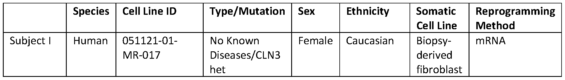
- the iPSC is obtained from a healthy subject or a subject having a neurodegenerative disease.
- Such neurodegenerative diseases can, in certain embodiments, be selected from Multiple Sclerosis and Parkinson’s disease.
- cortical media is utilized and includes an effective amount of cyclic adenosine monophosphate (cAMP), brain-derived neurotrophic factor (BDNF), glial cell-derived neurotrophic factor (GDNF), and neurotrophin (NT)-3.
- cAMP is included at a concentration of about lOOmM
- the BDNF is included at a concentration of aboutlO ng/ml
- the GDNF is included at a concentration of about 10 ng/ml
- the NT-3 is included at a concentration of about 10 ng/ml.
- the IL-34 is included at a concentration of about 100 ng/ml and the GM-CSF is included at a concentration of about 10 ng/ml.
- dopaminergic media is utilized and combined with a neural precursor cell, where the dopaminergic media includes an effective amount of BDNF, GDNF, TGF-P3, ascorbic acid, and cAMP.
- the BDNF is included at a concentration of about 20 ng/ml
- the GDNF is included at a concentration of about 20 ng/ml
- the TGF-P3 is included at a concentration of about 1 ng/ml
- the ascorbic acid is included at a concentration of about 200 pM
- the cAMP is included at a concentration of about 500 pM.
- the IL-34 included in the dopaminergic media is included at a concentration of about 100 ng/ml and the GM-CSF is included at a concentration of about 10 ng/ml.
- culturing the cells for a predetermined period of time comprises culturing the cells for the predetermined period of time under microgravity conditions.
- the pre-determined period of time for culturing the cells is at least 28 days.
- the organoid is removed from the cryovial and cultured in a tissue culture vessel.
- the organoid comprises a single organoid.
- a system for culturing an organoid comprises: an organoid formed from a neural precursor cell; a microglia; an effective amount of a cortical media or a dopaminergic media; a buffering solution; and a cryovial for housing the organoid, the microglia, the cortical media or the dopaminergic media, and the buffering solution under closed conditions.
- the neural precursor cell and/or the microglia is derived from a neural stem cell, such as an iPSC that can be obtained from a healthy subject or a subject having a neurodegenerative disease.
- the neurodegenerative disease is selected from Multiple Sclerosis and Parkinson’s disease.
- the cortical media included in an exemplary system also includes, in certain embodiments, an effective amount of cAMP, BDNF, GDNF, and NT-3.
- the cAMP is included in the cortical media at a concentration of about lOOmM
- the BDNF is included at a concentration of aboutlO ng/ml
- the GDNF is included at a concentration of about 10 ng/ml
- the NT-3 is included at a concentration of about 10 ng/ml.
- the dopaminergic media included in an exemplary system then similarly includes, in certain embodiments, an effective amount of BDNF, GDNF, TGF-P3, ascorbic acid, and cAMP.
- the BDNF is included at a concentration of about 20 ng/ml
- the GDNF is included at a concentration of about 20 ng/ml
- the TGF-P3 is included at a concentration of about 1 ng/ml
- the ascorbic acid is included at a concentration of about 200 pM
- the cAMP in included at a concentration of about 500 pM.
- the cortical media or the dopaminergic media comprises an amount of IL-34 and GM-CSF.
- the IL-34 is included at a concentration of about 100 ng/ml and the GM-CSF is included at a concentration of about 10 ng/ml.
- FIG. 1 is a schematic diagram showing an experimental strategy utilized during the course and development of the presently-disclosed subject matter, and depicting hiPSC lines and differentiation protocols.
- FIG. 2 is an image of a CubeLab used in accordance with the presently-disclosed subject matter, and showing the CubeLab floating onboard the International Space Station (IS S) while containing human three-dimensional (3D) brain organoids derived from Parkinson’s disease (PD) and Primary Progressive Multiple Sclerosis (PPMS) patients.
- FIG. 3 includes an image showing the CubeLab held the constant temperature of static culture systems in a 30-day mission onboard the ISS (top) along with images of vials containing organoids at different days and acquired from the CubeLab during the incubation on the ISS (bottom).
- FIG. 4 is an image showing the Power Ascent Utility Locker (PAUL) used in accordance with the presently-disclosed subject matter and which provides power and a data interface for monitoring the experiment during launch and on the ISS.
- PAUL Power Ascent Utility Locker
- FIGS. 5A-5F include images showing brain organoids cultured in static systems without media exchange are viable after 30 days, including: (FIG. 5A) an image showing the increase in the size of organoids cultured in closed static systems, from the pre-flight stage to the post-flight stage in low-Earth orbit (LEO) and ground; (FIG. 5B) an image showing plated organoids with outgrowth of neural projection and radial glia (arrows) after 30 days culture in low-Earth orbit (LEO) and ground, where the organoids maintained the typical cyto-architecture as shown in the cortical healthy control organoid, and where the neural rosettes are visible and diffused throughout the organoid; (FIGS.
- 5C-5F images showing MAP2 and Hoechst staining and tridimensional (3D) rendering displaying the healthy architecture of the neural rosettes (arrows) after 30 days culture in static systems.
- HC healthy control
- PPMS primary progressive multiple sclerosis
- PD Parkinson’s disease
- LEO low-Earth orbit).
- FIGS. 6A-6I include graphs and plots showing that microgravity alters gene expression in cortical and dopaminergic brain organoids, including: (FIG. 6A) a principal component analysis (PCA) of RNA-seq data showing the clustering of the samples, where PCI separates dopaminergic vs cortical, and PC2 separates control vs. patient; (FIGS.
- PCA principal component analysis
- FIGS. 6B-6E plots showing differentially expressed genes (DEGs) analyzed using DESeq2, where the volcano plots were created using the packages ggplot2 and ggrepel, where the log2 fold change indicates the mean expression for each gene, where genes with significant enrichment (padj ⁇ 0.05) are shown as red or blue (log2 fold change >1 or ⁇ -1, respectively) across replicates, where the plots shown in FIGS. 6B-6C show the differentially expressed genes (DEGs) - low-Earth orbit (LEO) versus ground - of the iPSC-derived cortical organoids from the healthy control and the iPSC-derived organoids from the multiple sclerosis patients, and where the volcano plots in FIGS.
- DEGs differentially expressed genes
- LEO low-Earth orbit
- GSEA gene enrichment analyses
- FIGS. 7A-7B includes graphs and plots showing that microgravity is the cause of the differential gene expression in dopaminergic and cortical organoids, where differentially expressed genes (DEGs) were analyzed using DESeq2 and the volcano plots are created using the packages ggplot2 and ggrepel, where FIG.
- DEGs differentially expressed genes
- LEO low-Earth orbit
- GSEA gene enrichment analyses
- FIG. 8G shows the total protein analyzed emphasizing the DSPs in cortical and dopaminergic samples alone and the DSPs commonly secreted, and where (FIGS. 8E-8F) the GO terms (BP, CP, MP) are shown for cortical and dopaminergic respectively and were performed using shinyGO.
- FIGS. 9A-9H are graphs and plots showing differentially expressed genes (DEGs, where the volcano plots of FIGS. 9A-9B show the DEGs - LEO vs ground - of the iPSC- derived cortical control organoids (-MG) and (+MG), where the plots shown in FIGS. 9C-9D show the iPSC-derived MS patient organoids (-MG) and (+MG), where the volcano plots shown in FIGS. 9E-9F show the DEGs of the iPSC-derived DA control organoids (-MG) and (+MG), where the DEGs of the iPSC-derived PD patient organoids are shown in the plots of FIGS.
- DEGs differentially expressed genes
- FIGS. 10A-10D includes graphs and plots, including: (FIG. 10A) a volcano plot showing the DEGs - LEO vs ground - of all (cortical and dopaminergic) iPSC-derived control organoids; (FIG. 10B) a plot showing the DEGs of all (MS and PD) iPSC-derived organoids; and FIGS. 10C-10D plots showing the gene enrichment analysis of the DEG shown in FIGS. 10A-10B, respectively, where the DEGs are identified using RNA-seq reads quantified with DESeq2.
- nucleotides and polypeptides disclosed herein are included in publicly-available databases, such as GENBANK® and SWISSPROT. Information including sequences and other information related to such nucleotides and polypeptides included in such publicly-available databases are expressly incorporated by reference. Unless otherwise indicated or apparent the references to such publicly-available databases are references to the most recent version of the database as of the filing date of this Application.
- the present application can “comprise” (open ended), “consist of’ (closed ended), or “consist essentially of’ the components of the present invention as well as other ingredients or elements described herein.
- “comprising” is open ended and means the elements recited, or their equivalent in structure or function, plus any other element or elements which are not recited.
- the terms “having” and “including” are also to be construed as open ended unless the context suggests otherwise.
- the term “about,” when referring to a value or to an amount of mass, weight, time, volume, concentration or percentage is meant to encompass variations of in some embodiments ⁇ 20%, in some embodiments ⁇ 10%, in some embodiments ⁇ 5%, in some embodiments ⁇ 1%, in some embodiments ⁇ 0.5%, and in some embodiments ⁇ 0.1% from the specified amount, as such variations are appropriate to perform the disclosed method.
- ranges can be expressed as from “about” one particular value, and/or to “about” another particular value. It is also understood that there are a number of values disclosed herein, and that each value is also herein disclosed as “about” that particular value in addition to the value itself. For example, if the value “10” is disclosed, then “about 10” is also disclosed. It is also understood that each unit between two particular units are also disclosed. For example, if 10 and 15 are disclosed, then 11, 12, 13, and 14 are also disclosed.
- an optionally variant portion means that the portion is variant or non-variant.
- the presently-disclosed subject matter is based, at least in part, on the discovery of a system and method that allows for the long-term culture of human iPSC-derived brain organoids (cortical and dopaminergic neural precursor cells) with microglia in closed systems.
- brain organoids are maintained in closed cryovials suspended in 1 mL of medium that ensures brain organoid growth and survival without media exchange during long-term culture in microgravity and on Earth.
- the systems and methods allow for the maintenance of live and metabolically active organoids in culture for at least 28 days in sealed tubes under close conditions or, in other words, without the addition of freshly reconstituted media or repeated equilibration of CO2 concentration.
- a neural precursor or progenitor cell is first formed.
- the phrase “neural precursor cell” and grammatical variations thereof is used herein to refer to the progenitor cells of the central nervous system (CNS) of a subject and which typically give rise the glial and neuronal cell types that populate the CNS, but generally do not generate the non- neural cells that are also present in the CNS, such as immune system cells.
- the neural precursor cell used to form an organoid is derived from a neural stem cell, such as, in some embodiments, an iPSC that, as would be recognized by those in the art, is derived from an adult cell but has been reprogrammed back into a pluripotent state (see, e.g., Pauli, et al. Nat Methods 12:885-892 (2015), which is incorporated herein by reference in its entirety).
- a neural stem cell such as, in some embodiments, an iPSC that, as would be recognized by those in the art, is derived from an adult cell but has been reprogrammed back into a pluripotent state (see, e.g., Pauli, et al. Nat Methods 12:885-892 (2015), which is incorporated herein by reference in its entirety).
- the iPSC or other stem cell is derived or obtained from the cells of a normal healthy subject.
- the term “subject” includes both human and animal subjects such that veterinary uses and applications are within the scope of the presently-disclosed subject matter.
- the presently-disclosed subject matter thus provides for the culturing of organoids produced or derived from mammals such as humans, as well as those mammals of importance due to being endangered, such as Siberian tigers; of economic importance, such as animals raised on farms for consumption by humans; and/or animals of social importance to humans, such as animals kept as pets or in zoos.
- Examples of such animals include but are not limited to: carnivores such as cats and dogs; swine, including pigs, hogs, and wild boars; ruminants and/or ungulates such as cattle, oxen, sheep, giraffes, deer, goats, bison, and camels; and horses.
- carnivores such as cats and dogs
- swine including pigs, hogs, and wild boars
- ruminants and/or ungulates such as cattle, oxen, sheep, giraffes, deer, goats, bison, and camels
- horses include but are not limited to: carnivores such as cats and dogs; swine, including pigs, hogs, and wild boars; ruminants and/or ungulates such as cattle, oxen, sheep, giraffes, deer, goats, bison, and camels; and horses.
- the iPSC is derived or obtained from the cells of a subject having a neurodegenerative disease, which is generally characterized by a progressive loss in the structure or function of the neurons in a subject or, in other words, neurodegeneration.
- a neurodegenerative disease which is generally characterized by a progressive loss in the structure or function of the neurons in a subject or, in other words, neurodegeneration.
- Such neurodegenerative diseases include, but are not limited to, disorders including Alzheimer's disease, Parkinson's disease, Huntington's disease, Multiple Sclerosis, Amyotrophic Lateral Sclerosis, Batten disease, and Creutzfeldt-Jakob disease.
- the iPSCs or other stem cells used in accordance with the presently-described systems and methods are derived or otherwise obtained from a subject having Multiple Sclerosis or a subject having Parkinson’s disease.
- the stem cells e.g., the iPSCs
- the cells are then differentiated into cortical or dopaminergic neural precursor cells by combining the stem cells with a first volume of a cortical media or a dopaminergic media to form an organoid.
- Organoids as would also be recognized by those skilled in the art, are small, self-organized three-dimensional tissue constructs that are typically derived from stem cells and which can be directed to assume a particular cellular identity such that the organoid, at least to a certain degree, mimics its corresponding in vivo organ or tissue.
- cortical media is used herein to refer to cell culture media capable of being used to differentiate stem cells, such as iPSCs, into a cortical neural precursor cell.
- dopaminergic media is used herein to refer to cell culture media capable of being used to differentiate stem cells into a dopaminergic neural precursor cell.
- the cortical media or dopaminergic media used in accordance with the systems and methods of the presently-disclosed subject matter typically will include an amount of nutrients, growth factors, and the like sufficient to differentiate the neural precursors cells into and maintain the cells as a desired phenotype.
- the term “effective amount” is used herein to refer to an amount of nutrients, growth factors, and the like that is included in a particular cell culture media and is sufficient to produce and/or maintain a desired phenotype, such as a desired type of neural precursor cell or neural cell or tissue.
- a cell culture media of the present invention can be varied so as to administer an amount of the active compound(s) that is effective to achieve the desired cell type or organoid for a particular system and/or method.
- the effective amount in any particular case will depend upon a variety of factors and differentiation into a desired cell type, but the determination and adjustment of an effective amount, as well as evaluation of when and how to make such adjustments, are known to those of ordinary skill in the art.
- methods and cell culture media useful for differentiation of stem cells into cortical and dopaminergic cells see, e.g., Yao, et al. Cell Stem Cell 20: 120-134 (2017), which is incorporated herein by reference in its entirety. See also, Kriks, et al. Nature 480: 547-551(2011), which is further incorporated herein by reference in its entirety.
- combining the neural precursor cell with the first volume of a cortical media or a dopaminergic media comprises combining the neural precursor cell with cortical media.
- the cortical media includes an effective amount of DMEM-F12 media, neurobasal media, cAMP, BDNF, GDNF, and NT-3.
- the cAMP is included at a concentration of about lOOmM
- the BDNF is included at a concentration of about 10 ng/ml
- the GDNF is included at a concentration of about 10 ng/ml
- the NT-3 is included at a concentration of about 10 ng/ml.
- combining the neural precursor cell with the first volume of a cortical media or a dopaminergic media comprises combining the neural precursor cell with dopaminergic media.
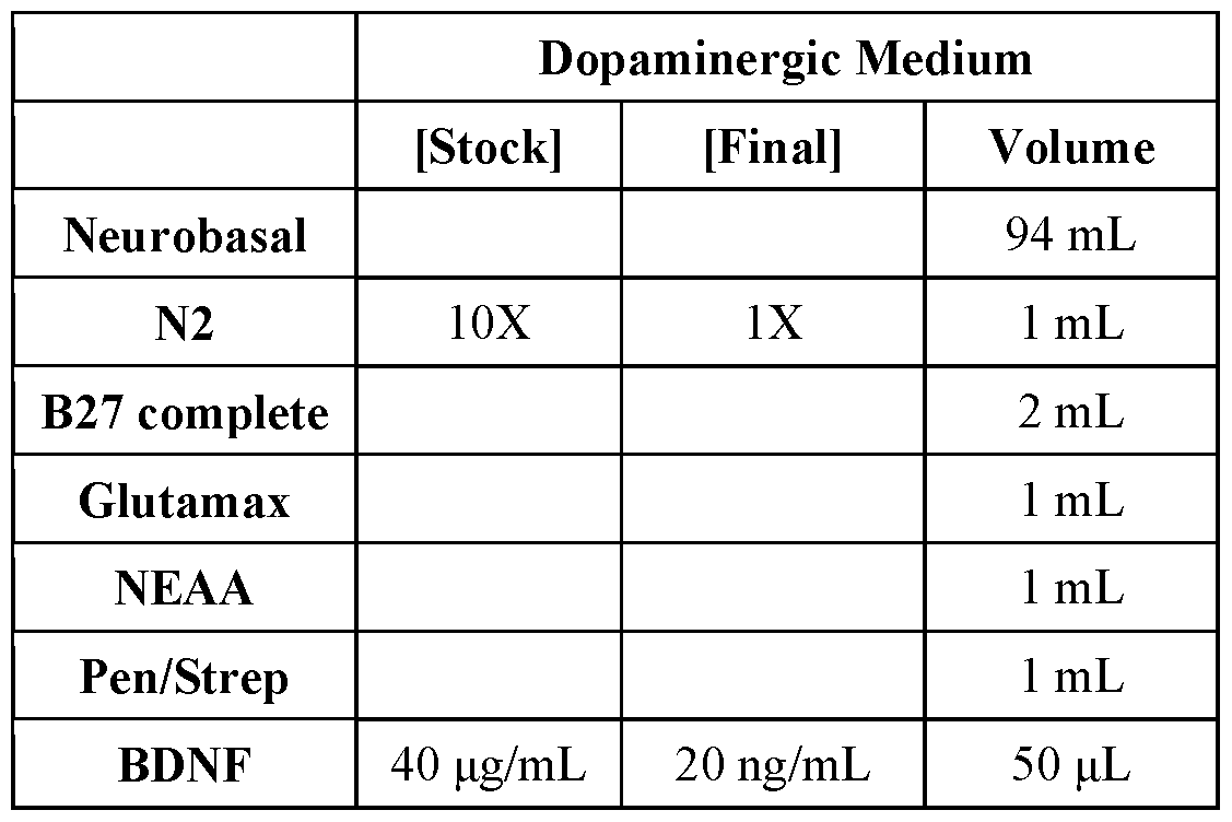
- the dopaminergic media includes an effective amount of neurobasal media, N2 supplemental media, B27 complete, Glutamax, NEAA, Pen/Strep, BDNF, GDNF, TGF-P3, ascorbic acid, and cAMP.
- the BDNF is included at a concentration of about 20 ng/ml
- the GDNF is included at a concentration of about 20 ng/ml
- the TGF-P3 is included at a concentration of about 1 ng/ml
- the ascorbic acid is included at a concentration of about 200 pM
- the cAMP is included at a concentration of about 500 pM.
- a microglia such as a microglia derived from a neural stem cell (e.g., an induced pluripotent stem cell derived (iPSC)), is then added to the organoid.
- a neural stem cell e.g., an induced pluripotent stem cell derived (iPSC)
- iPSC induced pluripotent stem cell derived
- the combined organoid and iPSC-derived microglia are subsequently added to a cryovial with a second volume of the cortical media or the dopaminergic media that then also includes an amount of IL-34 and GM-CSF, as well as an amount of a buffering solution.
- the media further includes IL-34 at a concentration of about 100 ng/ml and GM-CSF at a concentration of about 10 ng/ml.
- the IL-34 is included at a saturating concentration of at least about 100 ng/ml and the GM-CSF is included at a concentration of about 10 ng/ml.
- cryovials used in accordance with the presently-described systems and methods, although the term “cryovial” is often used in relation to containers capable of withstanding low temperatures (e.g., -196°C), the use of the term “cryovial” herein is not limited to such containers but is further inclusive of any sufficiently durable container capable of being sealed tightly and utilized for the long term storage of cell and tissues.
- cryovials capable of use in accordance with the presently-disclosed subject matter can be obtained from commercially-available sources such as, for example, the NUNCTM Coded Cryobank Vial from Thermo Fisher Scientific.
- a buffering agent can also be added to the cell culture media included in the cryovial to maintain the pH of the media during the culturing of the organoids under closed conditions.
- the buffering solution comprises HEPES (4-(2 -hydroxy ethyl)- 1 -piperazineethanesulfonic acid), as it was believed that the use of this zwitterionic sulfonic acid buffering agent could maintain physiological pH despite potential changes in carbon dioxide concentration during the course of the culturing of the organoid for a period of time under closed conditions in the cryovial.
- the HEPES is included in the cell culture media at a concentration of 10-15 mM.
- the addition of the HEPES and the relatively small size of the organoid relative to the larger volume of media provides an environment capable of supporting the organoid for a month or more. More specifically, it is contemplated that, in certain embodiments, seeding the organoids with a limited number of cells (e.g., 100K) allows the organoids to grow a limited amount within the one month time frame as the neural precursors proliferate.
- the organoid then remains relatively small (about 0.5 mm or about less than 0.75 mm) within the volume of media in the cryovials (1-2 ml) such that the ratio of the media volume to the size and/or number of cells assists in the maintenance of the survival of the organoid under the closed conditions.
- the cryovial Upon placing the combined organoid and microglia in the cryovial with the second volume of media including the buffering solution, the cryovial is then sealed, such as by capping the cryovial and sealing it with parafilm, to create a closed system in which the cryovial is completely sealed off from its external environment.
- the organoid with the microglia and culture media is then cultured for a pre-determined period of time under closed conditions whereby the cryovial and cultured cells included in the cryovial are not re-opened during the culture period.
- the culture period is at least 28 days.
- the culture period takes part, in total or in part, under microgravity conditions.
- the methods described herein further include the steps of removing the organoid from the cryovial after the predetermined time period, and culturing the organoid in another tissue culture vessel as it has been surprisingly determined that the presently-described systems and methods allow for the culturing of organoids under closed and/or microgravity conditions for an extended period of time and in a manner that maintains the viability of the cultured cells/organoids.
- the culturing of the cells following the closed conditions is accomplished using the above-described media with standard cell culture gas conditions (e.g., 5% CO2, 20% O2).
- the additional culturing is further performed under conditions sufficient to allow the cells to attach to cell culture surfaces coated with extracellular matrix (ECM) proteins such as laminin and/or fibronectin to promote outgrowth of cells and nerve projections onto the surfaces under standard culture conditions as would be appreciated by those skilled in the art.
- ECM extracellular matrix
- a system for culturing an organoid comprises: an organoid derived formed from a neural precursor cell; an induced pluripotent stem cell derived (iPSC)-derived microglia; an effective amount of a cortical media or a dopaminergic media; a buffering solution; and a cryovial for housing the organoid, the microglia, the cortical media or the dopaminergic media, and the buffering solution under closed conditions.
- iPSC induced pluripotent stem cell derived
- cell culture media is provided that includes the components and various combinations thereof described herein above for use with the presently-described systems and methods.
- a kit for culturing an organoid is provided that includes one or more of the components of the systems of the presently-disclosed subject matter along with instructions for using the kit.
- the instructions for using the kit include instructions for carrying out methods for culturing an organoid in accordance with the presently-disclosed subject matter.
- the presently-disclosed subject matter allows the survival of cortical and dopaminergic brain organoids in closed long-term culture systems for space mission(s) studies onboard of the international space station (ISS) without the intervention of the astronauts.
- the methods and systems allow for the culture in microgravity of human brain organoids as a disease model for neurodegenerative disorders - such as multiple sclerosis (MS) and Parkinson’s disease (PD).
- MS multiple sclerosis
- PD Parkinson’s disease
- the methods and systems also provide a long-term culture system for biological research during a lunar mission, and the methods and systems will further allow for long-term culture onboard of the lunar station - Gateway - orbiting the Moon in the near future.
- Cultured induced pluripotent stem cells derived from people with neurodegenerative disease can be used to produce human brain organoid models and it was believed that such models could be useful in addressing an urgent unmet need for understanding the mechanisms of neurodegenerative disease, and for studying the effects of microgravity on central nervous system (CNS) function in space travelers.
- Exposure to microgravity in low-Earth orbit (LEO) has been shown to impact the cardiovascular, musculoskeletal, and immune systems of the human body.
- Evidence from astronaut data and mouse models suggests that microgravity also causes intracranial fluid shifts and alters white and gray matter.
- iPSC Pluripotent Stem Cell
- GSEA Gene Set Enrichment Analysis
- NSCs Neural Stem Cells
- HC healthy control
- PPMS primary progressive multiple sclerosis
- PD Parkinson’s disease
- DMEM-F12 and Neurobasal media were purchased from ThermoFisher (Cat. Nos. #11330 and 21103049). N2 supplement (cat. no. 17502048), B27 (cat. no. 17504044), Glutamax (cat. no. 35050061), NEAA (cat. no.
- Pen/Strep (cat. no. 15070063) were each purchased from Gibco/ThermoFisher.
- BDNF catalog. no. 248-BD/CF
- GDNF cat. no. 212-GD-050
- NT3 cat. no. 267-N3-MTO
- TGF-p3 catalog. no. 243-B3-010
- Ascorbic acid catalog. no. A4403-100MG
- cAMP catalog. no. D0627-1G were purchased from Sigma Aldrich.
- RNA extraction and sequencing 22 organoids cultured in LEO and 22 from ground control were snap-frozen and stored at -80°C. RNA was extracted using the Qiagen Rneasy Micro kit (QIAGEN #74004). To maximize the yield, the RNA was eluted into 12pL of ultrapure DI water. The RNA was sequenced using an ultra-low input preparation of samples with a minimum of lOng of RNA and the Illumina NovaSeq Platform for high-throughput sequencing.
- GSEA Gene Set Enrichment Analysis
- Organoids were fixed with a 4% paraformaldehyde (PF A) solution in PBS for 30 minutes.
- PF A paraformaldehyde
- the organoids were processed following a previously published protocol for whole mounting staining and stained using an antibody to MAP2 (Ab5392, dilution 1 : 1000) to detect neurites and Hoechst dye for labeling nuclei.
- the labeled organoids were imaged using a ZEISS confocal microscope (LSM780), and images were processed and edited using Imaris Image Analysis Software.
- RADTriage50 JP Laboratories, Inc. passive dosimeters were included in the flight hardware both on station and on ground. RADTriage50 devices from both LEO and ground units were assessed in post-flight evaluation.
- Radiation Environmental Monitoring methods on ISS. Radiation detectors used on the ISS include Radiation Environment Monitoring (REM) and Hybrid Electronic
- HERA Radiation Assessor
- Neuronal progenitors were seeded into U-bottom wells to enable aggregation into organoids, and matched microglia progenitors, differentiated in parallel from the same iPSCs, were added to the organoids for integration. This process was intended to mimic the migration of microglial progenitors to the developing CNS during embryonic development.
- the 3D organoids cultures were shipped to the Space Station Processing Facility (SSPF) at Kennedy Space Center (KSC), where they were transferred to individual tubes in 1 mL of culture medium, sealed, and loaded onto flight hardware for parallel culture on the ground at KSC and in LEO on the ISS.
- SSPF Space Station Processing Facility
- KSC Kennedy Space Center
- Organoids survive for one month in static culture. Organoids were cultured for approximately a month in one ml of medium in cryovials without medium changes. The organoids increased in size both in LEO and on ground (FIGS. 5A-5B). After return to Earth, a small sample of the organoids were assessed for viability by plating onto laminin-coated dishes in fresh medium. Organoids attached to the plate showed robust process outgrowth, indicating that they thrived during the month-long culture in microgravity. The cortical organoids formed neural rosettes typical of this cell type (FIGS. 5B-5F).
- Principal component analysis of all organoids (FIG. 6A) showed a clear distinction between the dopaminergic and cortical organoids.
- FIG. 6A To determine the effects of LEO associated with each of the organoid types, differences in ground compared to LEO were examined in separate analyses for each of the organoid types (cortical or dopaminergic) for disease-associated cells (PD or MS) and matched controls (FIGS. 6B-6E).
- DESEq2 identified differentially expressed genes that had a padj ⁇ 0.05 and log2 fold change > 1 and ⁇ -1.
- FIGS. 9A-9E To gain insight about the biological processes affected by microgravity Gene Set Enrichment Analysis (GSEA) was performed on the ground vs LEO DEGs.
- GSEA microgravity Gene Set Enrichment Analysis
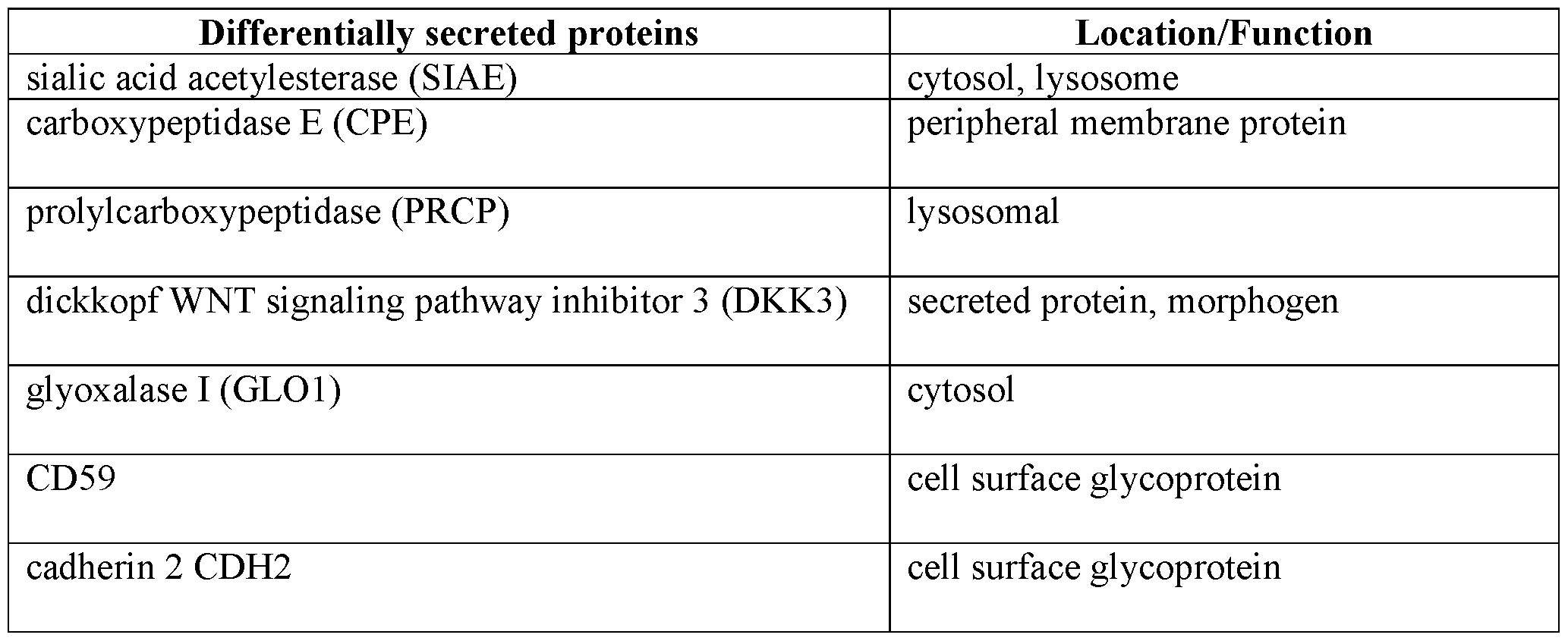
- culture medium was collected from vials containing single organoids and analyzed using Olink® Target 1536. All of the organoid types showed LEO vs ground differentially secreted proteins (DSPs) (padj ⁇ 0.05, Welch two-sample t-test) (FIGS. 8A-8G). 40 DSPs were identified in the medium from dopamine neuron organoids and 95 from cortical organoid medium. Seven proteins were found to be secreted at higher levels in LEO by both dopaminergic and cortical samples (Table 3).
- DSPs Differentially secreted proteins
- LEO Low Earth Orbit
- Fattorelli N Martinez-Muriana A, Wolfs L, et al (2021) Stem-cell-derived human microglia transplanted into mouse brain to study human disease. Nat Protoc. Kriks S, Shim J-W, Piao J, et al (2011) Dopamine neurons derived from human ES cells efficiently engraft in animal models of Parkinson’s disease. Nature 480:547-551. Yao Z, Mich JK, Ku S, et al (2017) A Single-Cell Roadmap of Lineage Bifurcation in Human ESC Models of Embryonic Brain Development. Cell Stem Cell 20:120-134.
Landscapes
- Health & Medical Sciences (AREA)
- Engineering & Computer Science (AREA)
- Biomedical Technology (AREA)
- Life Sciences & Earth Sciences (AREA)
- Organic Chemistry (AREA)
- Bioinformatics & Cheminformatics (AREA)
- Wood Science & Technology (AREA)
- Zoology (AREA)
- Chemical & Material Sciences (AREA)
- Genetics & Genomics (AREA)
- Biotechnology (AREA)
- General Health & Medical Sciences (AREA)
- General Engineering & Computer Science (AREA)
- Biochemistry (AREA)
- Microbiology (AREA)
- Cell Biology (AREA)
- Neurology (AREA)
- Neurosurgery (AREA)
- Sustainable Development (AREA)
- Molecular Biology (AREA)
- Developmental Biology & Embryology (AREA)
- Micro-Organisms Or Cultivation Processes Thereof (AREA)
- Apparatus Associated With Microorganisms And Enzymes (AREA)
Abstract
Description


Claims
Priority Applications (3)
| Application Number | Priority Date | Filing Date | Title |
|---|---|---|---|
| US18/700,180 US20240417682A1 (en) | 2021-10-15 | 2022-10-14 | Methods and systems for culturing organoids |
| JP2024523119A JP2024538165A (en) | 2021-10-15 | 2022-10-14 | Methods and systems for culturing organoids |
| EP22881840.7A EP4416272A4 (en) | 2021-10-15 | 2022-10-14 | METHODS AND SYSTEMS FOR CULTIVATING ORGANOIDS |
Applications Claiming Priority (4)
| Application Number | Priority Date | Filing Date | Title |
|---|---|---|---|
| US202163256279P | 2021-10-15 | 2021-10-15 | |
| US63/256,279 | 2021-10-15 | ||
| US202263344777P | 2022-05-23 | 2022-05-23 | |
| US63/344,777 | 2022-05-23 |
Publications (1)
| Publication Number | Publication Date |
|---|---|
| WO2023064570A1 true WO2023064570A1 (en) | 2023-04-20 |
Family
ID=85987890
Family Applications (1)
| Application Number | Title | Priority Date | Filing Date |
|---|---|---|---|
| PCT/US2022/046728 Ceased WO2023064570A1 (en) | 2021-10-15 | 2022-10-14 | Methods and systems for culturing organoids |
Country Status (4)
| Country | Link |
|---|---|
| US (1) | US20240417682A1 (en) |
| EP (1) | EP4416272A4 (en) |
| JP (1) | JP2024538165A (en) |
| WO (1) | WO2023064570A1 (en) |
Citations (4)
| Publication number | Priority date | Publication date | Assignee | Title |
|---|---|---|---|---|
| US20060211109A1 (en) * | 2003-03-12 | 2006-09-21 | Reliance Life Sciences Pvt. Ltd. | Derivation of terminally differentiated dopaminergic neurons from human embryonic stem cells |
| US20170253856A1 (en) * | 2016-03-03 | 2017-09-07 | Panagiotis Douvaras | Microglia derived from pluripotent stem cells and methods of making and using the same |
| WO2020243618A1 (en) * | 2019-05-29 | 2020-12-03 | New York Stem Cell Foundation, Inc. | Functional astrocytes derived from pluripotent stem cells and methods of making and using the same |
| US20210163888A1 (en) * | 2018-04-17 | 2021-06-03 | Case Western Reserve University | Induction of myelinating oligodendrocytes in human cortical spheroids |
Family Cites Families (1)
| Publication number | Priority date | Publication date | Assignee | Title |
|---|---|---|---|---|
| GB2584664B (en) * | 2019-06-10 | 2023-05-24 | Newcells Biotech Ltd | Improved retinal organoids and methods of making the same |
-
2022
- 2022-10-14 WO PCT/US2022/046728 patent/WO2023064570A1/en not_active Ceased
- 2022-10-14 JP JP2024523119A patent/JP2024538165A/en active Pending
- 2022-10-14 US US18/700,180 patent/US20240417682A1/en active Pending
- 2022-10-14 EP EP22881840.7A patent/EP4416272A4/en active Pending
Patent Citations (4)
| Publication number | Priority date | Publication date | Assignee | Title |
|---|---|---|---|---|
| US20060211109A1 (en) * | 2003-03-12 | 2006-09-21 | Reliance Life Sciences Pvt. Ltd. | Derivation of terminally differentiated dopaminergic neurons from human embryonic stem cells |
| US20170253856A1 (en) * | 2016-03-03 | 2017-09-07 | Panagiotis Douvaras | Microglia derived from pluripotent stem cells and methods of making and using the same |
| US20210163888A1 (en) * | 2018-04-17 | 2021-06-03 | Case Western Reserve University | Induction of myelinating oligodendrocytes in human cortical spheroids |
| WO2020243618A1 (en) * | 2019-05-29 | 2020-12-03 | New York Stem Cell Foundation, Inc. | Functional astrocytes derived from pluripotent stem cells and methods of making and using the same |
Non-Patent Citations (1)
| Title |
|---|
| See also references of EP4416272A4 * |
Also Published As
| Publication number | Publication date |
|---|---|
| EP4416272A4 (en) | 2025-10-22 |
| EP4416272A1 (en) | 2024-08-21 |
| US20240417682A1 (en) | 2024-12-19 |
| JP2024538165A (en) | 2024-10-18 |
Similar Documents
| Publication | Publication Date | Title |
|---|---|---|
| Leng et al. | CRISPRi screens in human iPSC-derived astrocytes elucidate regulators of distinct inflammatory reactive states | |
| Sloan et al. | Human astrocyte maturation captured in 3D cerebral cortical spheroids derived from pluripotent stem cells | |
| Schultz et al. | Skeletal muscle satellite cells | |
| Li et al. | Stem cells as therapy for hearing loss | |
| Salero et al. | Differentiation of ES cells into cerebellar neurons | |
| Wakeman et al. | Cryopreservation maintains functionality of human iPSC dopamine neurons and rescues parkinsonian phenotypes in vivo | |
| Zhang et al. | Generation of excitatory and inhibitory neurons from common progenitors via Notch signaling in the cerebellum | |
| Niclis et al. | Efficiently specified ventral midbrain dopamine neurons from human pluripotent stem cells under xeno-free conditions restore motor deficits in parkinsonian rodents | |
| Sun et al. | Mutations in the transcriptional regulator MeCP2 severely impact key cellular and molecular signatures of human astrocytes during maturation | |
| de Luzy et al. | Isolation of LMX1a ventral midbrain progenitors improves the safety and predictability of human pluripotent stem cell-derived neural transplants in Parkinsonian disease | |
| US20050031538A1 (en) | Neural cell assay | |
| US11850266B2 (en) | Cardiomyocytes and compositions and methods for producing the same | |
| Nam et al. | In vitro modeling for inherited neurological diseases using induced pluripotent stem cells: from 2D to organoid | |
| Marx et al. | An individual patient's “body” on chips—how organismoid theory can translate into your personal precision therapy approach | |
| WO2022221051A1 (en) | Cardiomyocytes and compositions and methods for producing the same | |
| Mazzini et al. | Current status and new avenues of stem cell-based preclinical and therapeutic approaches in amyotrophic lateral sclerosis | |
| US20240417682A1 (en) | Methods and systems for culturing organoids | |
| EP2253700A1 (en) | A method for producing test systems from donors suffering from adverse effects of medicaments and /or medical treatments, and uses of said systems | |
| Young et al. | Naturally occurring adult pluripotent stem cells | |
| Adalbert et al. | Cultured dissociated primary dorsal root ganglion neurons from adult horses enable study of axonal transport | |
| Marotta et al. | Space Renaissance and Neurodegeneration | |
| Rodnichenko et al. | The study of the remyelinating effect of leukemia inhibitory factor and melatonin on the toxic cuprizone model of demyelination of murine cerebellar cells culture in vitro | |
| Drouin-Ouellet | The potential of alternate sources of cells for neural grafting in Parkinson’s and Huntington’s disease | |
| Morita et al. | Impact of a simulated gravity load for atmospheric reentry, 10 g for 2 min, on conscious mice | |
| Brevig et al. | Proliferative response of human T lymphocytes to porcine fetal brain cells |
Legal Events
| Date | Code | Title | Description |
|---|---|---|---|
| 121 | Ep: the epo has been informed by wipo that ep was designated in this application |
Ref document number: 22881840 Country of ref document: EP Kind code of ref document: A1 |
|
| WWE | Wipo information: entry into national phase |
Ref document number: 18700180 Country of ref document: US |
|
| ENP | Entry into the national phase |
Ref document number: 2024523119 Country of ref document: JP Kind code of ref document: A |
|
| WWE | Wipo information: entry into national phase |
Ref document number: 2022881840 Country of ref document: EP |
|
| NENP | Non-entry into the national phase |
Ref country code: DE |
|
| ENP | Entry into the national phase |
Ref document number: 2022881840 Country of ref document: EP Effective date: 20240515 |