JP2004531259A - Compositions and methods for recombinant cloning of nucleic acid molecules - Google Patents
Compositions and methods for recombinant cloning of nucleic acid molecules Download PDFInfo
- Publication number
- JP2004531259A JP2004531259A JP2002583657A JP2002583657A JP2004531259A JP 2004531259 A JP2004531259 A JP 2004531259A JP 2002583657 A JP2002583657 A JP 2002583657A JP 2002583657 A JP2002583657 A JP 2002583657A JP 2004531259 A JP2004531259 A JP 2004531259A
- Authority
- JP
- Japan
- Prior art keywords
- protein
- amino acid
- acid sequence
- seq
- nucleic acid
- Prior art date
- Legal status (The legal status is an assumption and is not a legal conclusion. Google has not performed a legal analysis and makes no representation as to the accuracy of the status listed.)
- Pending
Links
- 150000007523 nucleic acids Chemical class 0.000 title claims abstract description 200
- 102000039446 nucleic acids Human genes 0.000 title claims abstract description 198
- 108020004707 nucleic acids Proteins 0.000 title claims abstract description 198
- 238000000034 method Methods 0.000 title claims abstract description 193
- 239000000203 mixture Substances 0.000 title claims abstract description 143
- 238000010367 cloning Methods 0.000 title claims abstract description 88
- 238000005215 recombination Methods 0.000 claims abstract description 283
- 230000006798 recombination Effects 0.000 claims abstract description 283
- 101150046810 fis gene Proteins 0.000 claims abstract description 258
- 239000013598 vector Substances 0.000 claims abstract description 148
- 102000007056 Recombinant Fusion Proteins Human genes 0.000 claims abstract description 70
- 108010008281 Recombinant Fusion Proteins Proteins 0.000 claims abstract description 70
- 108090000623 proteins and genes Proteins 0.000 claims description 216
- 108020004414 DNA Proteins 0.000 claims description 180
- 210000004027 cell Anatomy 0.000 claims description 161
- 102000004169 proteins and genes Human genes 0.000 claims description 152
- 108010000605 Ribosomal Proteins Proteins 0.000 claims description 150
- 102000002278 Ribosomal Proteins Human genes 0.000 claims description 150
- 238000006243 chemical reaction Methods 0.000 claims description 136
- 239000012634 fragment Substances 0.000 claims description 113
- 241000588724 Escherichia coli Species 0.000 claims description 99
- 239000000872 buffer Substances 0.000 claims description 92
- 230000000694 effects Effects 0.000 claims description 89
- 239000000047 product Substances 0.000 claims description 89
- 238000000338 in vitro Methods 0.000 claims description 39
- 238000001727 in vivo Methods 0.000 claims description 26
- 102000053602 DNA Human genes 0.000 claims description 21
- 102000004190 Enzymes Human genes 0.000 claims description 17
- 108090000790 Enzymes Proteins 0.000 claims description 17
- 241000894006 Bacteria Species 0.000 claims description 15
- 108091008146 restriction endonucleases Proteins 0.000 claims description 14
- 238000002156 mixing Methods 0.000 claims description 13
- FWMNVWWHGCHHJJ-SKKKGAJSSA-N 4-amino-1-[(2r)-6-amino-2-[[(2r)-2-[[(2r)-2-[[(2r)-2-amino-3-phenylpropanoyl]amino]-3-phenylpropanoyl]amino]-4-methylpentanoyl]amino]hexanoyl]piperidine-4-carboxylic acid Chemical compound C([C@H](C(=O)N[C@H](CC(C)C)C(=O)N[C@H](CCCCN)C(=O)N1CCC(N)(CC1)C(O)=O)NC(=O)[C@H](N)CC=1C=CC=CC=1)C1=CC=CC=C1 FWMNVWWHGCHHJJ-SKKKGAJSSA-N 0.000 claims description 11
- 125000000539 amino acid group Chemical group 0.000 claims description 11
- 230000003321 amplification Effects 0.000 claims description 10
- 238000003199 nucleic acid amplification method Methods 0.000 claims description 10
- 239000006227 byproduct Substances 0.000 claims description 9
- 239000002299 complementary DNA Substances 0.000 claims description 9
- 238000009472 formulation Methods 0.000 claims description 9
- 238000003786 synthesis reaction Methods 0.000 claims description 8
- 241000187747 Streptomyces Species 0.000 claims description 7
- 241000193830 Bacillus <bacterium> Species 0.000 claims description 6
- 241000588722 Escherichia Species 0.000 claims description 6
- 241000589516 Pseudomonas Species 0.000 claims description 6
- 241000589517 Pseudomonas aeruginosa Species 0.000 claims description 6
- 108010092799 RNA-directed DNA polymerase Proteins 0.000 claims description 6
- 241000607142 Salmonella Species 0.000 claims description 6
- 241000293869 Salmonella enterica subsp. enterica serovar Typhimurium Species 0.000 claims description 6
- 241000606768 Haemophilus influenzae Species 0.000 claims description 5
- 241000588747 Klebsiella pneumoniae Species 0.000 claims description 5
- 241000607626 Vibrio cholerae Species 0.000 claims description 5
- 210000004962 mammalian cell Anatomy 0.000 claims description 5
- 241000251468 Actinopterygii Species 0.000 claims description 4
- 102100031780 Endonuclease Human genes 0.000 claims description 4
- 241000238631 Hexapoda Species 0.000 claims description 4
- 102000003960 Ligases Human genes 0.000 claims description 4
- 108090000364 Ligases Proteins 0.000 claims description 4
- 210000003527 eukaryotic cell Anatomy 0.000 claims description 4
- 229940047650 haemophilus influenzae Drugs 0.000 claims description 4
- 229940118696 vibrio cholerae Drugs 0.000 claims description 4
- 241000206602 Eukaryota Species 0.000 claims description 3
- 230000002708 enhancing effect Effects 0.000 claims description 3
- 210000005253 yeast cell Anatomy 0.000 claims description 3
- 239000012096 transfection reagent Substances 0.000 claims description 2
- 125000003275 alpha amino acid group Chemical group 0.000 claims 66
- 238000005516 engineering process Methods 0.000 abstract description 16
- 108020004511 Recombinant DNA Proteins 0.000 abstract description 8
- 235000018102 proteins Nutrition 0.000 description 139
- 150000001413 amino acids Chemical class 0.000 description 115
- FAPWRFPIFSIZLT-UHFFFAOYSA-M Sodium chloride Chemical compound [Na+].[Cl-] FAPWRFPIFSIZLT-UHFFFAOYSA-M 0.000 description 112
- PEDCQBHIVMGVHV-UHFFFAOYSA-N Glycerine Chemical compound OCC(O)CO PEDCQBHIVMGVHV-UHFFFAOYSA-N 0.000 description 105
- 238000003556 assay Methods 0.000 description 62
- 239000011780 sodium chloride Substances 0.000 description 57
- 239000013612 plasmid Substances 0.000 description 50
- 238000000746 purification Methods 0.000 description 42
- QKNYBSVHEMOAJP-UHFFFAOYSA-N 2-amino-2-(hydroxymethyl)propane-1,3-diol;hydron;chloride Chemical compound Cl.OCC(N)(CO)CO QKNYBSVHEMOAJP-UHFFFAOYSA-N 0.000 description 39
- 108010015268 Integration Host Factors Proteins 0.000 description 39
- 239000000499 gel Substances 0.000 description 38
- 239000000463 material Substances 0.000 description 37
- 239000000758 substrate Substances 0.000 description 36
- 108010091086 Recombinases Proteins 0.000 description 35
- 230000004913 activation Effects 0.000 description 35
- 102000018120 Recombinases Human genes 0.000 description 33
- KCXVZYZYPLLWCC-UHFFFAOYSA-N EDTA Chemical compound OC(=O)CN(CC(O)=O)CCN(CC(O)=O)CC(O)=O KCXVZYZYPLLWCC-UHFFFAOYSA-N 0.000 description 30
- 102100034343 Integrase Human genes 0.000 description 30
- XLYOFNOQVPJJNP-UHFFFAOYSA-N water Chemical compound O XLYOFNOQVPJJNP-UHFFFAOYSA-N 0.000 description 30
- ATHGHQPFGPMSJY-UHFFFAOYSA-N spermidine Chemical compound NCCCCNCCCN ATHGHQPFGPMSJY-UHFFFAOYSA-N 0.000 description 26
- 108010061833 Integrases Proteins 0.000 description 25
- 238000003752 polymerase chain reaction Methods 0.000 description 25
- 238000002415 sodium dodecyl sulfate polyacrylamide gel electrophoresis Methods 0.000 description 25
- 239000003550 marker Substances 0.000 description 24
- 229940080469 phosphocellulose Drugs 0.000 description 23
- 101150031239 xis gene Proteins 0.000 description 23
- 230000015572 biosynthetic process Effects 0.000 description 22
- 238000002474 experimental method Methods 0.000 description 22
- 239000002773 nucleotide Substances 0.000 description 22
- 125000003729 nucleotide group Chemical group 0.000 description 22
- 229960000723 ampicillin Drugs 0.000 description 21
- AVKUERGKIZMTKX-NJBDSQKTSA-N ampicillin Chemical compound C1([C@@H](N)C(=O)N[C@H]2[C@H]3SC([C@@H](N3C2=O)C(O)=O)(C)C)=CC=CC=C1 AVKUERGKIZMTKX-NJBDSQKTSA-N 0.000 description 21
- 239000000284 extract Substances 0.000 description 21
- RAXXELZNTBOGNW-UHFFFAOYSA-N imidazole Natural products C1=CNC=N1 RAXXELZNTBOGNW-UHFFFAOYSA-N 0.000 description 21
- 108091028043 Nucleic acid sequence Proteins 0.000 description 20
- 101100049582 Enterobacteria phage HK620 hkaC gene Proteins 0.000 description 19
- 150000003839 salts Chemical class 0.000 description 19
- 230000027455 binding Effects 0.000 description 18
- 101150036876 cre gene Proteins 0.000 description 18
- 230000010354 integration Effects 0.000 description 18
- 230000010076 replication Effects 0.000 description 18
- 108091032973 (ribonucleotides)n+m Proteins 0.000 description 17
- 102100025601 60S ribosomal protein L27 Human genes 0.000 description 17
- 230000014509 gene expression Effects 0.000 description 17
- 238000003780 insertion Methods 0.000 description 17
- 230000037431 insertion Effects 0.000 description 17
- 239000006228 supernatant Substances 0.000 description 17
- 230000012010 growth Effects 0.000 description 15
- 240000004808 Saccharomyces cerevisiae Species 0.000 description 14
- 235000014680 Saccharomyces cerevisiae Nutrition 0.000 description 14
- 229910052588 hydroxylapatite Inorganic materials 0.000 description 14
- XYJRXVWERLGGKC-UHFFFAOYSA-D pentacalcium;hydroxide;triphosphate Chemical compound [OH-].[Ca+2].[Ca+2].[Ca+2].[Ca+2].[Ca+2].[O-]P([O-])([O-])=O.[O-]P([O-])([O-])=O.[O-]P([O-])([O-])=O XYJRXVWERLGGKC-UHFFFAOYSA-D 0.000 description 14
- 238000002360 preparation method Methods 0.000 description 14
- 229940088598 enzyme Drugs 0.000 description 13
- 229940063673 spermidine Drugs 0.000 description 13
- 231100000331 toxic Toxicity 0.000 description 13
- 230000002588 toxic effect Effects 0.000 description 13
- OKKJLVBELUTLKV-UHFFFAOYSA-N Methanol Chemical compound OC OKKJLVBELUTLKV-UHFFFAOYSA-N 0.000 description 12
- 108091034117 Oligonucleotide Proteins 0.000 description 12
- 239000002253 acid Substances 0.000 description 12
- 150000007513 acids Chemical class 0.000 description 12
- 239000013599 cloning vector Substances 0.000 description 12
- 108020001507 fusion proteins Proteins 0.000 description 12
- 239000011541 reaction mixture Substances 0.000 description 12
- 239000007320 rich medium Substances 0.000 description 12
- 238000012546 transfer Methods 0.000 description 12
- 108700026244 Open Reading Frames Proteins 0.000 description 11
- 238000005119 centrifugation Methods 0.000 description 11
- 102000037865 fusion proteins Human genes 0.000 description 11
- 108090000204 Dipeptidase 1 Proteins 0.000 description 10
- 101001059240 Saccharomyces cerevisiae (strain ATCC 204508 / S288c) Site-specific recombinase Flp Proteins 0.000 description 10
- 230000003213 activating effect Effects 0.000 description 10
- 239000002609 medium Substances 0.000 description 10
- 108090000765 processed proteins & peptides Proteins 0.000 description 10
- 241000701959 Escherichia virus Lambda Species 0.000 description 9
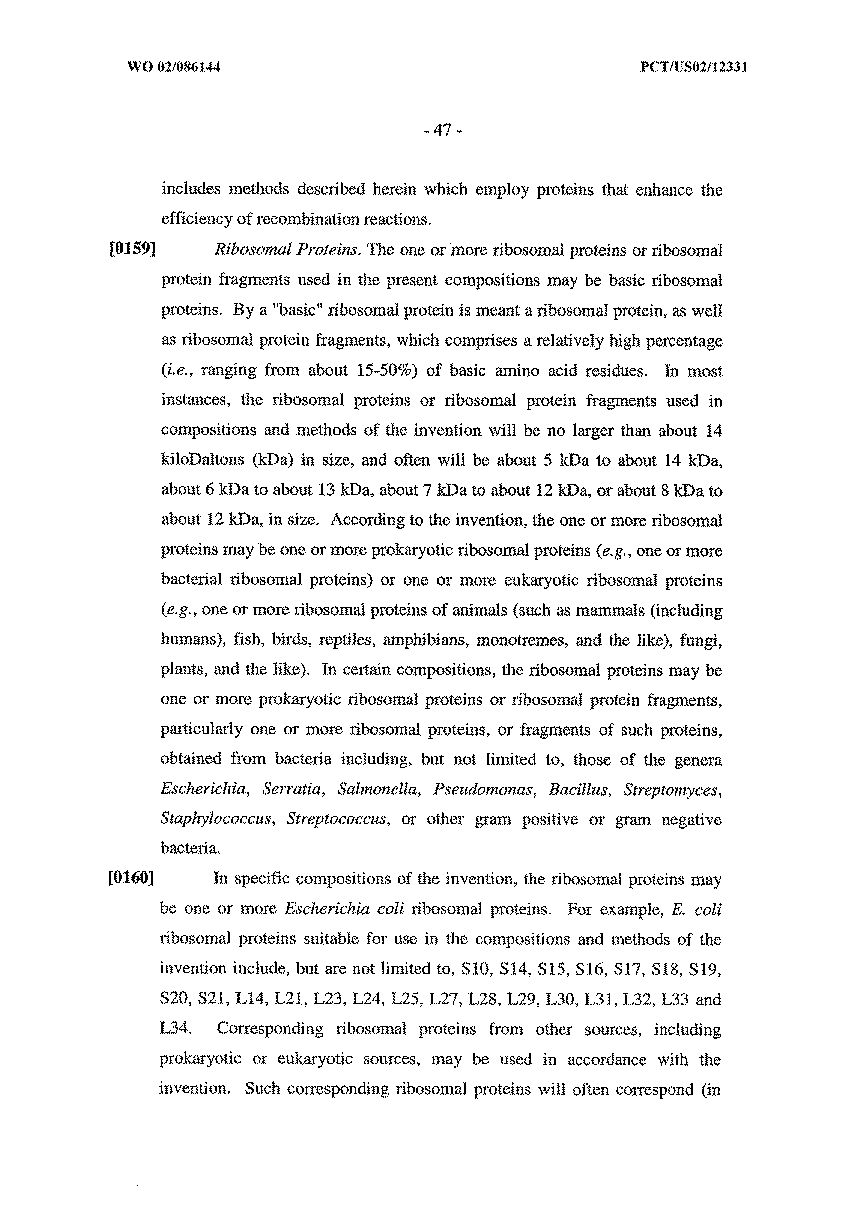
- 108010052160 Site-specific recombinase Proteins 0.000 description 9
- 238000004587 chromatography analysis Methods 0.000 description 9
- 238000000502 dialysis Methods 0.000 description 9
- 229930027917 kanamycin Natural products 0.000 description 9
- 229960000318 kanamycin Drugs 0.000 description 9
- SBUJHOSQTJFQJX-NOAMYHISSA-N kanamycin Chemical compound O[C@@H]1[C@@H](O)[C@H](O)[C@@H](CN)O[C@@H]1O[C@H]1[C@H](O)[C@@H](O[C@@H]2[C@@H]([C@@H](N)[C@H](O)[C@@H](CO)O2)O)[C@H](N)C[C@@H]1N SBUJHOSQTJFQJX-NOAMYHISSA-N 0.000 description 9
- 229930182823 kanamycin A Natural products 0.000 description 9
- 102000004196 processed proteins & peptides Human genes 0.000 description 9
- QTBSBXVTEAMEQO-UHFFFAOYSA-N Acetic acid Chemical compound CC(O)=O QTBSBXVTEAMEQO-UHFFFAOYSA-N 0.000 description 8
- JLCPHMBAVCMARE-UHFFFAOYSA-N [3-[[3-[[3-[[3-[[3-[[3-[[3-[[3-[[3-[[3-[[3-[[5-(2-amino-6-oxo-1H-purin-9-yl)-3-[[3-[[3-[[3-[[3-[[3-[[5-(2-amino-6-oxo-1H-purin-9-yl)-3-[[5-(2-amino-6-oxo-1H-purin-9-yl)-3-hydroxyoxolan-2-yl]methoxy-hydroxyphosphoryl]oxyoxolan-2-yl]methoxy-hydroxyphosphoryl]oxy-5-(5-methyl-2,4-dioxopyrimidin-1-yl)oxolan-2-yl]methoxy-hydroxyphosphoryl]oxy-5-(6-aminopurin-9-yl)oxolan-2-yl]methoxy-hydroxyphosphoryl]oxy-5-(6-aminopurin-9-yl)oxolan-2-yl]methoxy-hydroxyphosphoryl]oxy-5-(6-aminopurin-9-yl)oxolan-2-yl]methoxy-hydroxyphosphoryl]oxy-5-(6-aminopurin-9-yl)oxolan-2-yl]methoxy-hydroxyphosphoryl]oxyoxolan-2-yl]methoxy-hydroxyphosphoryl]oxy-5-(5-methyl-2,4-dioxopyrimidin-1-yl)oxolan-2-yl]methoxy-hydroxyphosphoryl]oxy-5-(4-amino-2-oxopyrimidin-1-yl)oxolan-2-yl]methoxy-hydroxyphosphoryl]oxy-5-(5-methyl-2,4-dioxopyrimidin-1-yl)oxolan-2-yl]methoxy-hydroxyphosphoryl]oxy-5-(5-methyl-2,4-dioxopyrimidin-1-yl)oxolan-2-yl]methoxy-hydroxyphosphoryl]oxy-5-(6-aminopurin-9-yl)oxolan-2-yl]methoxy-hydroxyphosphoryl]oxy-5-(6-aminopurin-9-yl)oxolan-2-yl]methoxy-hydroxyphosphoryl]oxy-5-(4-amino-2-oxopyrimidin-1-yl)oxolan-2-yl]methoxy-hydroxyphosphoryl]oxy-5-(4-amino-2-oxopyrimidin-1-yl)oxolan-2-yl]methoxy-hydroxyphosphoryl]oxy-5-(4-amino-2-oxopyrimidin-1-yl)oxolan-2-yl]methoxy-hydroxyphosphoryl]oxy-5-(6-aminopurin-9-yl)oxolan-2-yl]methoxy-hydroxyphosphoryl]oxy-5-(4-amino-2-oxopyrimidin-1-yl)oxolan-2-yl]methyl [5-(6-aminopurin-9-yl)-2-(hydroxymethyl)oxolan-3-yl] hydrogen phosphate Polymers Cc1cn(C2CC(OP(O)(=O)OCC3OC(CC3OP(O)(=O)OCC3OC(CC3O)n3cnc4c3nc(N)[nH]c4=O)n3cnc4c3nc(N)[nH]c4=O)C(COP(O)(=O)OC3CC(OC3COP(O)(=O)OC3CC(OC3COP(O)(=O)OC3CC(OC3COP(O)(=O)OC3CC(OC3COP(O)(=O)OC3CC(OC3COP(O)(=O)OC3CC(OC3COP(O)(=O)OC3CC(OC3COP(O)(=O)OC3CC(OC3COP(O)(=O)OC3CC(OC3COP(O)(=O)OC3CC(OC3COP(O)(=O)OC3CC(OC3COP(O)(=O)OC3CC(OC3COP(O)(=O)OC3CC(OC3COP(O)(=O)OC3CC(OC3COP(O)(=O)OC3CC(OC3COP(O)(=O)OC3CC(OC3COP(O)(=O)OC3CC(OC3CO)n3cnc4c(N)ncnc34)n3ccc(N)nc3=O)n3cnc4c(N)ncnc34)n3ccc(N)nc3=O)n3ccc(N)nc3=O)n3ccc(N)nc3=O)n3cnc4c(N)ncnc34)n3cnc4c(N)ncnc34)n3cc(C)c(=O)[nH]c3=O)n3cc(C)c(=O)[nH]c3=O)n3ccc(N)nc3=O)n3cc(C)c(=O)[nH]c3=O)n3cnc4c3nc(N)[nH]c4=O)n3cnc4c(N)ncnc34)n3cnc4c(N)ncnc34)n3cnc4c(N)ncnc34)n3cnc4c(N)ncnc34)O2)c(=O)[nH]c1=O JLCPHMBAVCMARE-UHFFFAOYSA-N 0.000 description 8
- 238000003277 amino acid sequence analysis Methods 0.000 description 8
- 238000004458 analytical method Methods 0.000 description 8
- 230000002441 reversible effect Effects 0.000 description 8
- 239000000523 sample Substances 0.000 description 8
- 108010067770 Endopeptidase K Proteins 0.000 description 7
- 239000012190 activator Substances 0.000 description 7
- 238000003776 cleavage reaction Methods 0.000 description 7
- 230000000295 complement effect Effects 0.000 description 7
- 238000012217 deletion Methods 0.000 description 7
- 230000037430 deletion Effects 0.000 description 7
- 230000006870 function Effects 0.000 description 7
- 230000001965 increasing effect Effects 0.000 description 7
- 230000006698 induction Effects 0.000 description 7
- 238000004519 manufacturing process Methods 0.000 description 7
- 230000035772 mutation Effects 0.000 description 7
- 239000008188 pellet Substances 0.000 description 7
- 230000007017 scission Effects 0.000 description 7
- 239000000243 solution Substances 0.000 description 7
- 238000013518 transcription Methods 0.000 description 7
- -1 7-deaza-dGTP Chemical compound 0.000 description 6
- 241000196324 Embryophyta Species 0.000 description 6
- 210000004899 c-terminal region Anatomy 0.000 description 6
- 238000011534 incubation Methods 0.000 description 6
- 239000000543 intermediate Substances 0.000 description 6
- 230000001404 mediated effect Effects 0.000 description 6
- 239000012528 membrane Substances 0.000 description 6
- 210000004379 membrane Anatomy 0.000 description 6
- 238000012986 modification Methods 0.000 description 6
- 230000004048 modification Effects 0.000 description 6
- 230000008569 process Effects 0.000 description 6
- 239000002002 slurry Substances 0.000 description 6
- UCSJYZPVAKXKNQ-HZYVHMACSA-N streptomycin Chemical compound CN[C@H]1[C@H](O)[C@@H](O)[C@H](CO)O[C@H]1O[C@@H]1[C@](C=O)(O)[C@H](C)O[C@H]1O[C@@H]1[C@@H](NC(N)=N)[C@H](O)[C@@H](NC(N)=N)[C@H](O)[C@H]1O UCSJYZPVAKXKNQ-HZYVHMACSA-N 0.000 description 6
- 230000035897 transcription Effects 0.000 description 6
- 230000009466 transformation Effects 0.000 description 6
- 108091027551 Cointegrate Proteins 0.000 description 5
- 241001302160 Escherichia coli str. K-12 substr. DH10B Species 0.000 description 5
- 241000702191 Escherichia virus P1 Species 0.000 description 5
- 108010043121 Green Fluorescent Proteins Proteins 0.000 description 5
- 108091029795 Intergenic region Proteins 0.000 description 5
- 108010054278 Lac Repressors Proteins 0.000 description 5
- 230000001580 bacterial effect Effects 0.000 description 5
- 230000003115 biocidal effect Effects 0.000 description 5
- 238000009835 boiling Methods 0.000 description 5
- 101150102092 ccdB gene Proteins 0.000 description 5
- ZMMJGEGLRURXTF-UHFFFAOYSA-N ethidium bromide Chemical compound [Br-].C12=CC(N)=CC=C2C2=CC=C(N)C=C2[N+](CC)=C1C1=CC=CC=C1 ZMMJGEGLRURXTF-UHFFFAOYSA-N 0.000 description 5
- 229960005542 ethidium bromide Drugs 0.000 description 5
- 238000010438 heat treatment Methods 0.000 description 5
- BPHPUYQFMNQIOC-NXRLNHOXSA-N isopropyl beta-D-thiogalactopyranoside Chemical compound CC(C)S[C@@H]1O[C@H](CO)[C@H](O)[C@H](O)[C@H]1O BPHPUYQFMNQIOC-NXRLNHOXSA-N 0.000 description 5
- 230000036961 partial effect Effects 0.000 description 5
- 230000001105 regulatory effect Effects 0.000 description 5
- 108010042407 Endonucleases Proteins 0.000 description 4
- 102000004144 Green Fluorescent Proteins Human genes 0.000 description 4
- 108020005210 Integrons Proteins 0.000 description 4
- 241001465754 Metazoa Species 0.000 description 4
- 101710188688 Non-structural protein 7a Proteins 0.000 description 4
- 238000012408 PCR amplification Methods 0.000 description 4
- 239000004098 Tetracycline Substances 0.000 description 4
- 238000000246 agarose gel electrophoresis Methods 0.000 description 4
- 230000004663 cell proliferation Effects 0.000 description 4
- 229960005091 chloramphenicol Drugs 0.000 description 4
- WIIZWVCIJKGZOK-RKDXNWHRSA-N chloramphenicol Chemical compound ClC(Cl)C(=O)N[C@H](CO)[C@H](O)C1=CC=C([N+]([O-])=O)C=C1 WIIZWVCIJKGZOK-RKDXNWHRSA-N 0.000 description 4
- 238000010293 colony formation assay Methods 0.000 description 4
- 150000001875 compounds Chemical class 0.000 description 4
- SUYVUBYJARFZHO-RRKCRQDMSA-N dATP Chemical compound C1=NC=2C(N)=NC=NC=2N1[C@H]1C[C@H](O)[C@@H](COP(O)(=O)OP(O)(=O)OP(O)(O)=O)O1 SUYVUBYJARFZHO-RRKCRQDMSA-N 0.000 description 4
- SUYVUBYJARFZHO-UHFFFAOYSA-N dATP Natural products C1=NC=2C(N)=NC=NC=2N1C1CC(O)C(COP(O)(=O)OP(O)(=O)OP(O)(O)=O)O1 SUYVUBYJARFZHO-UHFFFAOYSA-N 0.000 description 4
- 230000007423 decrease Effects 0.000 description 4
- 238000006731 degradation reaction Methods 0.000 description 4
- 230000002068 genetic effect Effects 0.000 description 4
- 239000005090 green fluorescent protein Substances 0.000 description 4
- 230000002427 irreversible effect Effects 0.000 description 4
- 238000002955 isolation Methods 0.000 description 4
- 238000012544 monitoring process Methods 0.000 description 4
- 230000003362 replicative effect Effects 0.000 description 4
- 238000011160 research Methods 0.000 description 4
- 238000000527 sonication Methods 0.000 description 4
- 238000012360 testing method Methods 0.000 description 4
- 229960002180 tetracycline Drugs 0.000 description 4
- 229930101283 tetracycline Natural products 0.000 description 4
- 235000019364 tetracycline Nutrition 0.000 description 4
- 150000003522 tetracyclines Chemical class 0.000 description 4
- 238000004448 titration Methods 0.000 description 4
- 239000001226 triphosphate Substances 0.000 description 4
- 235000011178 triphosphate Nutrition 0.000 description 4
- 239000004475 Arginine Substances 0.000 description 3
- 241000193388 Bacillus thuringiensis Species 0.000 description 3
- 108020004705 Codon Proteins 0.000 description 3
- 102000052510 DNA-Binding Proteins Human genes 0.000 description 3
- 108700020911 DNA-Binding Proteins Proteins 0.000 description 3
- LFQSCWFLJHTTHZ-UHFFFAOYSA-N Ethanol Chemical compound CCO LFQSCWFLJHTTHZ-UHFFFAOYSA-N 0.000 description 3
- ODKSFYDXXFIFQN-BYPYZUCNSA-P L-argininium(2+) Chemical compound NC(=[NH2+])NCCC[C@H]([NH3+])C(O)=O ODKSFYDXXFIFQN-BYPYZUCNSA-P 0.000 description 3
- KDXKERNSBIXSRK-YFKPBYRVSA-N L-lysine Chemical compound NCCCC[C@H](N)C(O)=O KDXKERNSBIXSRK-YFKPBYRVSA-N 0.000 description 3
- KDXKERNSBIXSRK-UHFFFAOYSA-N Lysine Natural products NCCCCC(N)C(O)=O KDXKERNSBIXSRK-UHFFFAOYSA-N 0.000 description 3
- 241000124008 Mammalia Species 0.000 description 3
- 108010014251 Muramidase Proteins 0.000 description 3
- 102000016943 Muramidase Human genes 0.000 description 3
- 108010062010 N-Acetylmuramoyl-L-alanine Amidase Proteins 0.000 description 3
- 108020002230 Pancreatic Ribonuclease Proteins 0.000 description 3
- 102000005891 Pancreatic ribonuclease Human genes 0.000 description 3
- 241000607720 Serratia Species 0.000 description 3
- HEMHJVSKTPXQMS-UHFFFAOYSA-M Sodium hydroxide Chemical compound [OH-].[Na+] HEMHJVSKTPXQMS-UHFFFAOYSA-M 0.000 description 3
- 229930006000 Sucrose Natural products 0.000 description 3
- CZMRCDWAGMRECN-UGDNZRGBSA-N Sucrose Chemical compound O[C@H]1[C@H](O)[C@@H](CO)O[C@@]1(CO)O[C@@H]1[C@H](O)[C@@H](O)[C@H](O)[C@@H](CO)O1 CZMRCDWAGMRECN-UGDNZRGBSA-N 0.000 description 3
- 108020004566 Transfer RNA Proteins 0.000 description 3
- 108010020764 Transposases Proteins 0.000 description 3
- 102000008579 Transposases Human genes 0.000 description 3
- 241000700605 Viruses Species 0.000 description 3
- 108091006088 activator proteins Proteins 0.000 description 3
- ODKSFYDXXFIFQN-UHFFFAOYSA-N arginine Natural products OC(=O)C(N)CCCNC(N)=N ODKSFYDXXFIFQN-UHFFFAOYSA-N 0.000 description 3
- 229920001222 biopolymer Polymers 0.000 description 3
- 229910052799 carbon Inorganic materials 0.000 description 3
- 230000015556 catabolic process Effects 0.000 description 3
- 239000007795 chemical reaction product Substances 0.000 description 3
- 210000000349 chromosome Anatomy 0.000 description 3
- 238000009833 condensation Methods 0.000 description 3
- 230000005494 condensation Effects 0.000 description 3
- RGWHQCVHVJXOKC-SHYZEUOFSA-J dCTP(4-) Chemical compound O=C1N=C(N)C=CN1[C@@H]1O[C@H](COP([O-])(=O)OP([O-])(=O)OP([O-])([O-])=O)[C@@H](O)C1 RGWHQCVHVJXOKC-SHYZEUOFSA-J 0.000 description 3
- HAAZLUGHYHWQIW-KVQBGUIXSA-N dGTP Chemical compound C1=NC=2C(=O)NC(N)=NC=2N1[C@H]1C[C@H](O)[C@@H](COP(O)(=O)OP(O)(=O)OP(O)(O)=O)O1 HAAZLUGHYHWQIW-KVQBGUIXSA-N 0.000 description 3
- NHVNXKFIZYSCEB-XLPZGREQSA-N dTTP Chemical compound O=C1NC(=O)C(C)=CN1[C@@H]1O[C@H](COP(O)(=O)OP(O)(=O)OP(O)(O)=O)[C@@H](O)C1 NHVNXKFIZYSCEB-XLPZGREQSA-N 0.000 description 3
- 238000001962 electrophoresis Methods 0.000 description 3
- 230000007717 exclusion Effects 0.000 description 3
- 239000013604 expression vector Substances 0.000 description 3
- 235000019688 fish Nutrition 0.000 description 3
- 230000004927 fusion Effects 0.000 description 3
- 239000001963 growth medium Substances 0.000 description 3
- HNDVDQJCIGZPNO-UHFFFAOYSA-N histidine Natural products OC(=O)C(N)CC1=CN=CN1 HNDVDQJCIGZPNO-UHFFFAOYSA-N 0.000 description 3
- 238000009396 hybridization Methods 0.000 description 3
- 238000010348 incorporation Methods 0.000 description 3
- 101150062334 int gene Proteins 0.000 description 3
- 230000003993 interaction Effects 0.000 description 3
- 239000004325 lysozyme Substances 0.000 description 3
- 229960000274 lysozyme Drugs 0.000 description 3
- 235000010335 lysozyme Nutrition 0.000 description 3
- 229920001184 polypeptide Polymers 0.000 description 3
- 239000002244 precipitate Substances 0.000 description 3
- 230000004044 response Effects 0.000 description 3
- 108010092942 ribosomal protein S20 Proteins 0.000 description 3
- 238000000926 separation method Methods 0.000 description 3
- 238000012163 sequencing technique Methods 0.000 description 3
- 230000000087 stabilizing effect Effects 0.000 description 3
- 229960005322 streptomycin Drugs 0.000 description 3
- 239000005720 sucrose Substances 0.000 description 3
- 230000004083 survival effect Effects 0.000 description 3
- 238000001890 transfection Methods 0.000 description 3
- 125000002264 triphosphate group Chemical class [H]OP(=O)(O[H])OP(=O)(O[H])OP(=O)(O[H])O* 0.000 description 3
- 241001515965 unidentified phage Species 0.000 description 3
- 108700026220 vif Genes Proteins 0.000 description 3
- CFBILACNYSPRPM-UHFFFAOYSA-N 2-amino-2-(hydroxymethyl)propane-1,3-diol;2-[[1,3-dihydroxy-2-(hydroxymethyl)propan-2-yl]amino]acetic acid Chemical compound OCC(N)(CO)CO.OCC(CO)(CO)NCC(O)=O CFBILACNYSPRPM-UHFFFAOYSA-N 0.000 description 2
- ASJSAQIRZKANQN-CRCLSJGQSA-N 2-deoxy-D-ribose Chemical group OC[C@@H](O)[C@@H](O)CC=O ASJSAQIRZKANQN-CRCLSJGQSA-N 0.000 description 2
- NKDFYOWSKOHCCO-YPVLXUMRSA-N 20-hydroxyecdysone Chemical compound C1[C@@H](O)[C@@H](O)C[C@]2(C)[C@@H](CC[C@@]3([C@@H]([C@@](C)(O)[C@H](O)CCC(C)(O)C)CC[C@]33O)C)C3=CC(=O)[C@@H]21 NKDFYOWSKOHCCO-YPVLXUMRSA-N 0.000 description 2
- 102100023415 40S ribosomal protein S20 Human genes 0.000 description 2
- 229920001817 Agar Polymers 0.000 description 2
- 229920000936 Agarose Polymers 0.000 description 2
- 241000271566 Aves Species 0.000 description 2
- 235000014469 Bacillus subtilis Nutrition 0.000 description 2
- KXDAEFPNCMNJSK-UHFFFAOYSA-N Benzamide Chemical compound NC(=O)C1=CC=CC=C1 KXDAEFPNCMNJSK-UHFFFAOYSA-N 0.000 description 2
- CURLTUGMZLYLDI-UHFFFAOYSA-N Carbon dioxide Chemical compound O=C=O CURLTUGMZLYLDI-UHFFFAOYSA-N 0.000 description 2
- 108090000994 Catalytic RNA Proteins 0.000 description 2
- 102000053642 Catalytic RNA Human genes 0.000 description 2
- 108010051219 Cre recombinase Proteins 0.000 description 2
- HMFHBZSHGGEWLO-SOOFDHNKSA-N D-ribofuranose Chemical group OC[C@H]1OC(O)[C@H](O)[C@@H]1O HMFHBZSHGGEWLO-SOOFDHNKSA-N 0.000 description 2
- 108010017826 DNA Polymerase I Proteins 0.000 description 2
- 102000004594 DNA Polymerase I Human genes 0.000 description 2
- 238000012270 DNA recombination Methods 0.000 description 2
- 108010046276 FLP recombinase Proteins 0.000 description 2
- 229920001917 Ficoll Polymers 0.000 description 2
- 241000233866 Fungi Species 0.000 description 2
- 210000000712 G cell Anatomy 0.000 description 2
- 108700039691 Genetic Promoter Regions Proteins 0.000 description 2
- 108010060309 Glucuronidase Proteins 0.000 description 2
- 102000053187 Glucuronidase Human genes 0.000 description 2
- 108010070675 Glutathione transferase Proteins 0.000 description 2
- 102000005720 Glutathione transferase Human genes 0.000 description 2
- 241000606790 Haemophilus Species 0.000 description 2
- HTTJABKRGRZYRN-UHFFFAOYSA-N Heparin Chemical compound OC1C(NC(=O)C)C(O)OC(COS(O)(=O)=O)C1OC1C(OS(O)(=O)=O)C(O)C(OC2C(C(OS(O)(=O)=O)C(OC3C(C(O)C(O)C(O3)C(O)=O)OS(O)(=O)=O)C(CO)O2)NS(O)(=O)=O)C(C(O)=O)O1 HTTJABKRGRZYRN-UHFFFAOYSA-N 0.000 description 2
- 241000282412 Homo Species 0.000 description 2
- 241000282414 Homo sapiens Species 0.000 description 2
- 241000725303 Human immunodeficiency virus Species 0.000 description 2
- KFZMGEQAYNKOFK-UHFFFAOYSA-N Isopropanol Chemical compound CC(C)O KFZMGEQAYNKOFK-UHFFFAOYSA-N 0.000 description 2
- HNDVDQJCIGZPNO-YFKPBYRVSA-N L-histidine Chemical compound OC(=O)[C@@H](N)CC1=CN=CN1 HNDVDQJCIGZPNO-YFKPBYRVSA-N 0.000 description 2
- GDBQQVLCIARPGH-UHFFFAOYSA-N Leupeptin Natural products CC(C)CC(NC(C)=O)C(=O)NC(CC(C)C)C(=O)NC(C=O)CCCN=C(N)N GDBQQVLCIARPGH-UHFFFAOYSA-N 0.000 description 2
- 239000004472 Lysine Substances 0.000 description 2
- ISWSIDIOOBJBQZ-UHFFFAOYSA-N Phenol Chemical compound OC1=CC=CC=C1 ISWSIDIOOBJBQZ-UHFFFAOYSA-N 0.000 description 2
- 239000004743 Polypropylene Substances 0.000 description 2
- PYMYPHUHKUWMLA-LMVFSUKVSA-N Ribose Chemical group OC[C@@H](O)[C@@H](O)[C@@H](O)C=O PYMYPHUHKUWMLA-LMVFSUKVSA-N 0.000 description 2
- 241000235343 Saccharomycetales Species 0.000 description 2
- 229920002684 Sepharose Polymers 0.000 description 2
- 238000012300 Sequence Analysis Methods 0.000 description 2
- 241000191940 Staphylococcus Species 0.000 description 2
- 108091081024 Start codon Proteins 0.000 description 2
- 241000194017 Streptococcus Species 0.000 description 2
- 239000008272 agar Substances 0.000 description 2
- 239000011543 agarose gel Substances 0.000 description 2
- HMFHBZSHGGEWLO-UHFFFAOYSA-N alpha-D-Furanose-Ribose Chemical group OCC1OC(O)C(O)C1O HMFHBZSHGGEWLO-UHFFFAOYSA-N 0.000 description 2
- 239000003242 anti bacterial agent Substances 0.000 description 2
- QVGXLLKOCUKJST-UHFFFAOYSA-N atomic oxygen Chemical compound [O] QVGXLLKOCUKJST-UHFFFAOYSA-N 0.000 description 2
- 229940097012 bacillus thuringiensis Drugs 0.000 description 2
- 238000005452 bending Methods 0.000 description 2
- 230000033228 biological regulation Effects 0.000 description 2
- UDSAIICHUKSCKT-UHFFFAOYSA-N bromophenol blue Chemical compound C1=C(Br)C(O)=C(Br)C=C1C1(C=2C=C(Br)C(O)=C(Br)C=2)C2=CC=CC=C2S(=O)(=O)O1 UDSAIICHUKSCKT-UHFFFAOYSA-N 0.000 description 2
- 238000004364 calculation method Methods 0.000 description 2
- 235000011089 carbon dioxide Nutrition 0.000 description 2
- 230000030833 cell death Effects 0.000 description 2
- 230000008859 change Effects 0.000 description 2
- 238000012512 characterization method Methods 0.000 description 2
- 239000003153 chemical reaction reagent Substances 0.000 description 2
- 239000013611 chromosomal DNA Substances 0.000 description 2
- NKLPQNGYXWVELD-UHFFFAOYSA-M coomassie brilliant blue Chemical compound [Na+].C1=CC(OCC)=CC=C1NC1=CC=C(C(=C2C=CC(C=C2)=[N+](CC)CC=2C=C(C=CC=2)S([O-])(=O)=O)C=2C=CC(=CC=2)N(CC)CC=2C=C(C=CC=2)S([O-])(=O)=O)C=C1 NKLPQNGYXWVELD-UHFFFAOYSA-M 0.000 description 2
- 239000000287 crude extract Substances 0.000 description 2
- 239000013078 crystal Substances 0.000 description 2
- 108010082025 cyan fluorescent protein Proteins 0.000 description 2
- 125000004122 cyclic group Chemical group 0.000 description 2
- 238000000354 decomposition reaction Methods 0.000 description 2
- 238000004925 denaturation Methods 0.000 description 2
- 230000036425 denaturation Effects 0.000 description 2
- 230000029087 digestion Effects 0.000 description 2
- 238000007865 diluting Methods 0.000 description 2
- 238000010790 dilution Methods 0.000 description 2
- 239000012895 dilution Substances 0.000 description 2
- 230000002255 enzymatic effect Effects 0.000 description 2
- 238000011049 filling Methods 0.000 description 2
- 230000001976 improved effect Effects 0.000 description 2
- 239000000411 inducer Substances 0.000 description 2
- 230000001939 inductive effect Effects 0.000 description 2
- 208000015181 infectious disease Diseases 0.000 description 2
- 238000011081 inoculation Methods 0.000 description 2
- 210000001739 intranuclear inclusion body Anatomy 0.000 description 2
- 101150109249 lacI gene Proteins 0.000 description 2
- GDBQQVLCIARPGH-ULQDDVLXSA-N leupeptin Chemical compound CC(C)C[C@H](NC(C)=O)C(=O)N[C@@H](CC(C)C)C(=O)N[C@H](C=O)CCCN=C(N)N GDBQQVLCIARPGH-ULQDDVLXSA-N 0.000 description 2
- 108010052968 leupeptin Proteins 0.000 description 2
- 238000013507 mapping Methods 0.000 description 2
- 108020004999 messenger RNA Proteins 0.000 description 2
- 238000013508 migration Methods 0.000 description 2
- 230000005012 migration Effects 0.000 description 2
- 238000010369 molecular cloning Methods 0.000 description 2
- 239000000178 monomer Substances 0.000 description 2
- 229910000363 nickel(II) sulfate Inorganic materials 0.000 description 2
- 235000015097 nutrients Nutrition 0.000 description 2
- 230000002018 overexpression Effects 0.000 description 2
- 229910052760 oxygen Inorganic materials 0.000 description 2
- 239000001301 oxygen Substances 0.000 description 2
- 230000037361 pathway Effects 0.000 description 2
- YBYRMVIVWMBXKQ-UHFFFAOYSA-N phenylmethanesulfonyl fluoride Chemical compound FS(=O)(=O)CC1=CC=CC=C1 YBYRMVIVWMBXKQ-UHFFFAOYSA-N 0.000 description 2
- BASFCYQUMIYNBI-UHFFFAOYSA-N platinum Chemical compound [Pt] BASFCYQUMIYNBI-UHFFFAOYSA-N 0.000 description 2
- 229920001155 polypropylene Polymers 0.000 description 2
- 125000002924 primary amino group Chemical group [H]N([H])* 0.000 description 2
- 210000001236 prokaryotic cell Anatomy 0.000 description 2
- 108020001580 protein domains Proteins 0.000 description 2
- 238000011084 recovery Methods 0.000 description 2
- 230000002829 reductive effect Effects 0.000 description 2
- 108010067528 ribosomal proteins L27 Proteins 0.000 description 2
- 210000004708 ribosome subunit Anatomy 0.000 description 2
- 108091092562 ribozyme Proteins 0.000 description 2
- 238000005185 salting out Methods 0.000 description 2
- 239000007858 starting material Substances 0.000 description 2
- 239000012536 storage buffer Substances 0.000 description 2
- 238000006467 substitution reaction Methods 0.000 description 2
- 230000000153 supplemental effect Effects 0.000 description 2
- 239000000725 suspension Substances 0.000 description 2
- 231100000419 toxicity Toxicity 0.000 description 2
- 230000001988 toxicity Effects 0.000 description 2
- 230000005030 transcription termination Effects 0.000 description 2
- 230000014621 translational initiation Effects 0.000 description 2
- 230000017105 transposition Effects 0.000 description 2
- 230000024540 transposon integration Effects 0.000 description 2
- 238000010396 two-hybrid screening Methods 0.000 description 2
- 108091005957 yellow fluorescent proteins Proteins 0.000 description 2
- DGVVWUTYPXICAM-UHFFFAOYSA-N β‐Mercaptoethanol Chemical compound OCCS DGVVWUTYPXICAM-UHFFFAOYSA-N 0.000 description 2
- OAKPWEUQDVLTCN-NKWVEPMBSA-N 2',3'-Dideoxyadenosine-5-triphosphate Chemical compound C1=NC=2C(N)=NC=NC=2N1[C@H]1CC[C@@H](CO[P@@](O)(=O)O[P@](O)(=O)OP(O)(O)=O)O1 OAKPWEUQDVLTCN-NKWVEPMBSA-N 0.000 description 1
- OSBLTNPMIGYQGY-UHFFFAOYSA-N 2-amino-2-(hydroxymethyl)propane-1,3-diol;2-[2-[bis(carboxymethyl)amino]ethyl-(carboxymethyl)amino]acetic acid;boric acid Chemical compound OB(O)O.OCC(N)(CO)CO.OC(=O)CN(CC(O)=O)CCN(CC(O)=O)CC(O)=O OSBLTNPMIGYQGY-UHFFFAOYSA-N 0.000 description 1
- 101710171204 30S ribosomal protein S20 Proteins 0.000 description 1
- QFVHZQCOUORWEI-UHFFFAOYSA-N 4-[(4-anilino-5-sulfonaphthalen-1-yl)diazenyl]-5-hydroxynaphthalene-2,7-disulfonic acid Chemical compound C=12C(O)=CC(S(O)(=O)=O)=CC2=CC(S(O)(=O)=O)=CC=1N=NC(C1=CC=CC(=C11)S(O)(=O)=O)=CC=C1NC1=CC=CC=C1 QFVHZQCOUORWEI-UHFFFAOYSA-N 0.000 description 1
- 101710134681 40 kDa protein Proteins 0.000 description 1
- 102100023216 40S ribosomal protein S15 Human genes 0.000 description 1
- 101710135913 50S ribosomal protein L27 Proteins 0.000 description 1
- 102000002260 Alkaline Phosphatase Human genes 0.000 description 1
- 108020004774 Alkaline Phosphatase Proteins 0.000 description 1
- USFZMSVCRYTOJT-UHFFFAOYSA-N Ammonium acetate Chemical compound N.CC(O)=O USFZMSVCRYTOJT-UHFFFAOYSA-N 0.000 description 1
- 239000005695 Ammonium acetate Substances 0.000 description 1
- 208000031295 Animal disease Diseases 0.000 description 1
- 102100021569 Apoptosis regulator Bcl-2 Human genes 0.000 description 1
- 241000972773 Aulopiformes Species 0.000 description 1
- 241000194107 Bacillus megaterium Species 0.000 description 1
- 102100026189 Beta-galactosidase Human genes 0.000 description 1
- 108091003079 Bovine Serum Albumin Proteins 0.000 description 1
- 238000009010 Bradford assay Methods 0.000 description 1
- OKTJSMMVPCPJKN-UHFFFAOYSA-N Carbon Chemical compound [C] OKTJSMMVPCPJKN-UHFFFAOYSA-N 0.000 description 1
- 108020004638 Circular DNA Proteins 0.000 description 1
- 241000193468 Clostridium perfringens Species 0.000 description 1
- 108091035707 Consensus sequence Proteins 0.000 description 1
- 241000938605 Crocodylia Species 0.000 description 1
- 102000012410 DNA Ligases Human genes 0.000 description 1
- 108010061982 DNA Ligases Proteins 0.000 description 1
- 102000003915 DNA Topoisomerases Human genes 0.000 description 1
- 108090000323 DNA Topoisomerases Proteins 0.000 description 1
- 230000004544 DNA amplification Effects 0.000 description 1
- 230000004543 DNA replication Effects 0.000 description 1
- 238000001712 DNA sequencing Methods 0.000 description 1
- 230000006820 DNA synthesis Effects 0.000 description 1
- 230000004568 DNA-binding Effects 0.000 description 1
- 108010014303 DNA-directed DNA polymerase Proteins 0.000 description 1
- 102000016928 DNA-directed DNA polymerase Human genes 0.000 description 1
- 101710150441 DNA-invertase Proteins 0.000 description 1
- 102000000541 Defensins Human genes 0.000 description 1
- 108010002069 Defensins Proteins 0.000 description 1
- 102000007260 Deoxyribonuclease I Human genes 0.000 description 1
- 108010008532 Deoxyribonuclease I Proteins 0.000 description 1
- AHCYMLUZIRLXAA-SHYZEUOFSA-N Deoxyuridine 5'-triphosphate Chemical compound O1[C@H](COP(O)(=O)OP(O)(=O)OP(O)(O)=O)[C@@H](O)C[C@@H]1N1C(=O)NC(=O)C=C1 AHCYMLUZIRLXAA-SHYZEUOFSA-N 0.000 description 1
- 101100311938 Dictyostelium discoideum phesA gene Proteins 0.000 description 1
- 101100423325 Dictyostelium discoideum phesB gene Proteins 0.000 description 1
- 101100364969 Dictyostelium discoideum scai gene Proteins 0.000 description 1
- 241000255601 Drosophila melanogaster Species 0.000 description 1
- 101100437104 Drosophila melanogaster AttB gene Proteins 0.000 description 1
- 206010059866 Drug resistance Diseases 0.000 description 1
- 102000004533 Endonucleases Human genes 0.000 description 1
- 241000702197 Enterobacteria phage P4 Species 0.000 description 1
- 241000701968 Enterobacteria phage phi80 Species 0.000 description 1
- 241000588698 Erwinia Species 0.000 description 1
- 102100031690 Erythroid transcription factor Human genes 0.000 description 1
- 101710100588 Erythroid transcription factor Proteins 0.000 description 1
- 241000672609 Escherichia coli BL21 Species 0.000 description 1
- 241000620209 Escherichia coli DH5[alpha] Species 0.000 description 1
- 241001147440 Escherichia virus 186 Species 0.000 description 1
- 241000702192 Escherichia virus P2 Species 0.000 description 1
- 241000701533 Escherichia virus T4 Species 0.000 description 1
- 108091029865 Exogenous DNA Proteins 0.000 description 1
- 241000192125 Firmicutes Species 0.000 description 1
- 230000005526 G1 to G0 transition Effects 0.000 description 1
- 101150066002 GFP gene Proteins 0.000 description 1
- 108010010803 Gelatin Proteins 0.000 description 1
- WQZGKKKJIJFFOK-GASJEMHNSA-N Glucose Natural products OC[C@H]1OC(O)[C@H](O)[C@@H](O)[C@@H]1O WQZGKKKJIJFFOK-GASJEMHNSA-N 0.000 description 1
- NYHBQMYGNKIUIF-UUOKFMHZSA-N Guanosine Chemical compound C1=NC=2C(=O)NC(N)=NC=2N1[C@@H]1O[C@H](CO)[C@@H](O)[C@H]1O NYHBQMYGNKIUIF-UUOKFMHZSA-N 0.000 description 1
- 101710113864 Heat shock protein 90 Proteins 0.000 description 1
- 229920002971 Heparan sulfate Polymers 0.000 description 1
- 101000971171 Homo sapiens Apoptosis regulator Bcl-2 Proteins 0.000 description 1
- 101001018196 Homo sapiens Mitogen-activated protein kinase kinase kinase 5 Proteins 0.000 description 1
- 241000976924 Inca Species 0.000 description 1
- 101710203526 Integrase Proteins 0.000 description 1
- 108091092195 Intron Proteins 0.000 description 1
- 239000007836 KH2PO4 Substances 0.000 description 1
- 241000588748 Klebsiella Species 0.000 description 1
- 241000235058 Komagataella pastoris Species 0.000 description 1
- OUYCCCASQSFEME-QMMMGPOBSA-N L-tyrosine Chemical compound OC(=O)[C@@H](N)CC1=CC=C(O)C=C1 OUYCCCASQSFEME-QMMMGPOBSA-N 0.000 description 1
- 241000270322 Lepidosauria Species 0.000 description 1
- FYYHWMGAXLPEAU-UHFFFAOYSA-N Magnesium Chemical compound [Mg] FYYHWMGAXLPEAU-UHFFFAOYSA-N 0.000 description 1
- 102000018697 Membrane Proteins Human genes 0.000 description 1
- 108010052285 Membrane Proteins Proteins 0.000 description 1
- 102100033127 Mitogen-activated protein kinase kinase kinase 5 Human genes 0.000 description 1
- 101100364971 Mus musculus Scai gene Proteins 0.000 description 1
- 125000001429 N-terminal alpha-amino-acid group Chemical group 0.000 description 1
- 241000244206 Nematoda Species 0.000 description 1
- 206010028980 Neoplasm Diseases 0.000 description 1
- 108091005461 Nucleic proteins Proteins 0.000 description 1
- 101150071716 PCSK1 gene Proteins 0.000 description 1
- 229910019142 PO4 Inorganic materials 0.000 description 1
- 239000002033 PVDF binder Substances 0.000 description 1
- 241000364057 Peoria Species 0.000 description 1
- BELBBZDIHDAJOR-UHFFFAOYSA-N Phenolsulfonephthalein Chemical compound C1=CC(O)=CC=C1C1(C=2C=CC(O)=CC=2)C2=CC=CC=C2S(=O)(=O)O1 BELBBZDIHDAJOR-UHFFFAOYSA-N 0.000 description 1
- 101710093543 Probable non-specific lipid-transfer protein Proteins 0.000 description 1
- 102000055027 Protein Methyltransferases Human genes 0.000 description 1
- 108700040121 Protein Methyltransferases Proteins 0.000 description 1
- 102000044126 RNA-Binding Proteins Human genes 0.000 description 1
- 108700020471 RNA-Binding Proteins Proteins 0.000 description 1
- 108700005075 Regulator Genes Proteins 0.000 description 1
- 108020005091 Replication Origin Proteins 0.000 description 1
- 108010046983 Ribonuclease T1 Proteins 0.000 description 1
- 108090000530 Ribosomal protein S15 Proteins 0.000 description 1
- 102000014400 SH2 domains Human genes 0.000 description 1
- 108050003452 SH2 domains Proteins 0.000 description 1
- 241000293871 Salmonella enterica subsp. enterica serovar Typhi Species 0.000 description 1
- 241000701835 Salmonella virus P22 Species 0.000 description 1
- 108010071390 Serum Albumin Proteins 0.000 description 1
- 102000007562 Serum Albumin Human genes 0.000 description 1
- 241000256251 Spodoptera frugiperda Species 0.000 description 1
- 239000008051 TBE buffer Substances 0.000 description 1
- 108010006785 Taq Polymerase Proteins 0.000 description 1
- 239000004809 Teflon Substances 0.000 description 1
- 229920006362 Teflon® Polymers 0.000 description 1
- 108010010574 Tn3 resolvase Proteins 0.000 description 1
- 239000007983 Tris buffer Substances 0.000 description 1
- 241000269370 Xenopus <genus> Species 0.000 description 1
- 241000607479 Yersinia pestis Species 0.000 description 1
- YVNQAIFQFWTPLQ-UHFFFAOYSA-O [4-[[4-(4-ethoxyanilino)phenyl]-[4-[ethyl-[(3-sulfophenyl)methyl]amino]-2-methylphenyl]methylidene]-3-methylcyclohexa-2,5-dien-1-ylidene]-ethyl-[(3-sulfophenyl)methyl]azanium Chemical compound C1=CC(OCC)=CC=C1NC1=CC=C(C(=C2C(=CC(C=C2)=[N+](CC)CC=2C=C(C=CC=2)S(O)(=O)=O)C)C=2C(=CC(=CC=2)N(CC)CC=2C=C(C=CC=2)S(O)(=O)=O)C)C=C1 YVNQAIFQFWTPLQ-UHFFFAOYSA-O 0.000 description 1
- AZJLCKAEZFNJDI-DJLDLDEBSA-N [[(2r,3s,5r)-5-(4-aminopyrrolo[2,3-d]pyrimidin-7-yl)-3-hydroxyoxolan-2-yl]methoxy-hydroxyphosphoryl] phosphono hydrogen phosphate Chemical compound C1=CC=2C(N)=NC=NC=2N1[C@H]1C[C@H](O)[C@@H](COP(O)(=O)OP(O)(=O)OP(O)(O)=O)O1 AZJLCKAEZFNJDI-DJLDLDEBSA-N 0.000 description 1
- HDRRAMINWIWTNU-NTSWFWBYSA-N [[(2s,5r)-5-(2-amino-6-oxo-3h-purin-9-yl)oxolan-2-yl]methoxy-hydroxyphosphoryl] phosphono hydrogen phosphate Chemical compound C1=2NC(N)=NC(=O)C=2N=CN1[C@H]1CC[C@@H](COP(O)(=O)OP(O)(=O)OP(O)(O)=O)O1 HDRRAMINWIWTNU-NTSWFWBYSA-N 0.000 description 1
- ARLKCWCREKRROD-POYBYMJQSA-N [[(2s,5r)-5-(4-amino-2-oxopyrimidin-1-yl)oxolan-2-yl]methoxy-hydroxyphosphoryl] phosphono hydrogen phosphate Chemical compound O=C1N=C(N)C=CN1[C@@H]1O[C@H](COP(O)(=O)OP(O)(=O)OP(O)(O)=O)CC1 ARLKCWCREKRROD-POYBYMJQSA-N 0.000 description 1
- ZXZIQGYRHQJWSY-NKWVEPMBSA-N [hydroxy-[[(2s,5r)-5-(6-oxo-3h-purin-9-yl)oxolan-2-yl]methoxy]phosphoryl] phosphono hydrogen phosphate Chemical compound O1[C@H](COP(O)(=O)OP(O)(=O)OP(O)(=O)O)CC[C@@H]1N1C(NC=NC2=O)=C2N=C1 ZXZIQGYRHQJWSY-NKWVEPMBSA-N 0.000 description 1
- QPMSXSBEVQLBIL-CZRHPSIPSA-N ac1mix0p Chemical compound C1=CC=C2N(C[C@H](C)CN(C)C)C3=CC(OC)=CC=C3SC2=C1.O([C@H]1[C@]2(OC)C=CC34C[C@@H]2[C@](C)(O)CCC)C2=C5[C@]41CCN(C)[C@@H]3CC5=CC=C2O QPMSXSBEVQLBIL-CZRHPSIPSA-N 0.000 description 1
- 230000006978 adaptation Effects 0.000 description 1
- 238000005273 aeration Methods 0.000 description 1
- 230000004075 alteration Effects 0.000 description 1
- 229940043376 ammonium acetate Drugs 0.000 description 1
- 235000019257 ammonium acetate Nutrition 0.000 description 1
- 239000003708 ampul Substances 0.000 description 1
- 230000003698 anagen phase Effects 0.000 description 1
- 210000004102 animal cell Anatomy 0.000 description 1
- 230000000845 anti-microbial effect Effects 0.000 description 1
- 239000002518 antifoaming agent Substances 0.000 description 1
- 239000000427 antigen Substances 0.000 description 1
- 108091007433 antigens Proteins 0.000 description 1
- 102000036639 antigens Human genes 0.000 description 1
- 239000004599 antimicrobial Substances 0.000 description 1
- 239000000074 antisense oligonucleotide Substances 0.000 description 1
- 238000012230 antisense oligonucleotides Methods 0.000 description 1
- 230000006907 apoptotic process Effects 0.000 description 1
- 108010005774 beta-Galactosidase Proteins 0.000 description 1
- 230000004071 biological effect Effects 0.000 description 1
- 230000008033 biological extinction Effects 0.000 description 1
- 230000008827 biological function Effects 0.000 description 1
- KGBXLFKZBHKPEV-UHFFFAOYSA-N boric acid Chemical compound OB(O)O KGBXLFKZBHKPEV-UHFFFAOYSA-N 0.000 description 1
- 239000004327 boric acid Substances 0.000 description 1
- 229940098773 bovine serum albumin Drugs 0.000 description 1
- 239000012152 bradford reagent Substances 0.000 description 1
- 239000000337 buffer salt Substances 0.000 description 1
- 239000001506 calcium phosphate Substances 0.000 description 1
- 229910000389 calcium phosphate Inorganic materials 0.000 description 1
- 235000011010 calcium phosphates Nutrition 0.000 description 1
- 229940041514 candida albicans extract Drugs 0.000 description 1
- 101150055766 cat gene Proteins 0.000 description 1
- 101150039936 ced-9 gene Proteins 0.000 description 1
- 230000022131 cell cycle Effects 0.000 description 1
- 230000032823 cell division Effects 0.000 description 1
- 210000002230 centromere Anatomy 0.000 description 1
- 239000000919 ceramic Substances 0.000 description 1
- 238000011210 chromatographic step Methods 0.000 description 1
- 230000002759 chromosomal effect Effects 0.000 description 1
- 230000024321 chromosome segregation Effects 0.000 description 1
- 230000001332 colony forming effect Effects 0.000 description 1
- 238000004440 column chromatography Methods 0.000 description 1
- 230000002860 competitive effect Effects 0.000 description 1
- 238000004590 computer program Methods 0.000 description 1
- 238000012790 confirmation Methods 0.000 description 1
- 238000010276 construction Methods 0.000 description 1
- 230000001276 controlling effect Effects 0.000 description 1
- 238000002425 crystallisation Methods 0.000 description 1
- 230000008025 crystallization Effects 0.000 description 1
- UFJPAQSLHAGEBL-RRKCRQDMSA-N dITP Chemical compound O1[C@H](COP(O)(=O)OP(O)(=O)OP(O)(O)=O)[C@@H](O)C[C@@H]1N1C(N=CNC2=O)=C2N=C1 UFJPAQSLHAGEBL-RRKCRQDMSA-N 0.000 description 1
- URGJWIFLBWJRMF-JGVFFNPUSA-N ddTTP Chemical compound O=C1NC(=O)C(C)=CN1[C@@H]1O[C@H](COP(O)(=O)OP(O)(=O)OP(O)(O)=O)CC1 URGJWIFLBWJRMF-JGVFFNPUSA-N 0.000 description 1
- 230000003247 decreasing effect Effects 0.000 description 1
- 239000007857 degradation product Substances 0.000 description 1
- 239000005549 deoxyribonucleoside Substances 0.000 description 1
- 230000001419 dependent effect Effects 0.000 description 1
- 238000001514 detection method Methods 0.000 description 1
- 239000003599 detergent Substances 0.000 description 1
- 230000001627 detrimental effect Effects 0.000 description 1
- 239000013024 dilution buffer Substances 0.000 description 1
- 239000000539 dimer Substances 0.000 description 1
- 229910000396 dipotassium phosphate Inorganic materials 0.000 description 1
- VHJLVAABSRFDPM-QWWZWVQMSA-N dithiothreitol Chemical compound SC[C@@H](O)[C@H](O)CS VHJLVAABSRFDPM-QWWZWVQMSA-N 0.000 description 1
- 238000004520 electroporation Methods 0.000 description 1
- 239000002158 endotoxin Substances 0.000 description 1
- 239000003623 enhancer Substances 0.000 description 1
- 230000000967 entomopathogenic effect Effects 0.000 description 1
- 238000012869 ethanol precipitation Methods 0.000 description 1
- 238000000605 extraction Methods 0.000 description 1
- 239000012467 final product Substances 0.000 description 1
- 238000005194 fractionation Methods 0.000 description 1
- 239000012737 fresh medium Substances 0.000 description 1
- 238000010230 functional analysis Methods 0.000 description 1
- 239000008273 gelatin Substances 0.000 description 1
- 229920000159 gelatin Polymers 0.000 description 1
- 235000019322 gelatine Nutrition 0.000 description 1
- 235000011852 gelatine desserts Nutrition 0.000 description 1
- 238000001415 gene therapy Methods 0.000 description 1
- 238000007429 general method Methods 0.000 description 1
- 238000012252 genetic analysis Methods 0.000 description 1
- 238000010353 genetic engineering Methods 0.000 description 1
- GVVPGTZRZFNKDS-JXMROGBWSA-N geranyl diphosphate Chemical compound CC(C)=CCC\C(C)=C\CO[P@](O)(=O)OP(O)(O)=O GVVPGTZRZFNKDS-JXMROGBWSA-N 0.000 description 1
- 239000008103 glucose Substances 0.000 description 1
- 229920000669 heparin Polymers 0.000 description 1
- 229960002897 heparin Drugs 0.000 description 1
- 239000012145 high-salt buffer Substances 0.000 description 1
- 238000000703 high-speed centrifugation Methods 0.000 description 1
- 238000002744 homologous recombination Methods 0.000 description 1
- 230000006801 homologous recombination Effects 0.000 description 1
- 210000005260 human cell Anatomy 0.000 description 1
- 239000012535 impurity Substances 0.000 description 1
- 239000004615 ingredient Substances 0.000 description 1
- 239000002054 inoculum Substances 0.000 description 1
- 150000002484 inorganic compounds Chemical class 0.000 description 1
- 230000003834 intracellular effect Effects 0.000 description 1
- 238000011835 investigation Methods 0.000 description 1
- 238000004255 ion exchange chromatography Methods 0.000 description 1
- 238000006317 isomerization reaction Methods 0.000 description 1
- 231100000518 lethal Toxicity 0.000 description 1
- 230000001665 lethal effect Effects 0.000 description 1
- 150000002632 lipids Chemical class 0.000 description 1
- 239000012160 loading buffer Substances 0.000 description 1
- 238000011068 loading method Methods 0.000 description 1
- 230000005923 long-lasting effect Effects 0.000 description 1
- 230000002101 lytic effect Effects 0.000 description 1
- 229910052749 magnesium Inorganic materials 0.000 description 1
- 239000011777 magnesium Substances 0.000 description 1
- TWRXJAOTZQYOKJ-UHFFFAOYSA-L magnesium chloride Substances [Mg+2].[Cl-].[Cl-] TWRXJAOTZQYOKJ-UHFFFAOYSA-L 0.000 description 1
- 229910001629 magnesium chloride Inorganic materials 0.000 description 1
- 229910052943 magnesium sulfate Inorganic materials 0.000 description 1
- CSNNHWWHGAXBCP-UHFFFAOYSA-L magnesium sulphate Substances [Mg+2].[O-][S+2]([O-])([O-])[O-] CSNNHWWHGAXBCP-UHFFFAOYSA-L 0.000 description 1
- 230000007246 mechanism Effects 0.000 description 1
- 230000002503 metabolic effect Effects 0.000 description 1
- 230000003278 mimic effect Effects 0.000 description 1
- 238000000329 molecular dynamics simulation Methods 0.000 description 1
- 229910000402 monopotassium phosphate Inorganic materials 0.000 description 1
- 101150039901 mukF gene Proteins 0.000 description 1
- 238000002703 mutagenesis Methods 0.000 description 1
- 231100000350 mutagenesis Toxicity 0.000 description 1
- 239000002547 new drug Substances 0.000 description 1
- 102000044158 nucleic acid binding protein Human genes 0.000 description 1
- 108700020942 nucleic acid binding protein Proteins 0.000 description 1
- 210000000056 organ Anatomy 0.000 description 1
- 150000002894 organic compounds Chemical class 0.000 description 1
- 238000012261 overproduction Methods 0.000 description 1
- 238000004806 packaging method and process Methods 0.000 description 1
- 239000002245 particle Substances 0.000 description 1
- 108010091212 pepstatin Proteins 0.000 description 1
- FAXGPCHRFPCXOO-LXTPJMTPSA-N pepstatin A Chemical compound OC(=O)C[C@H](O)[C@H](CC(C)C)NC(=O)[C@H](C)NC(=O)C[C@H](O)[C@H](CC(C)C)NC(=O)[C@H](C(C)C)NC(=O)[C@H](C(C)C)NC(=O)CC(C)C FAXGPCHRFPCXOO-LXTPJMTPSA-N 0.000 description 1
- 101150080777 pheS gene Proteins 0.000 description 1
- 229960003531 phenolsulfonphthalein Drugs 0.000 description 1
- 239000010452 phosphate Substances 0.000 description 1
- 229910052697 platinum Inorganic materials 0.000 description 1
- 238000006116 polymerization reaction Methods 0.000 description 1
- 229920002981 polyvinylidene fluoride Polymers 0.000 description 1
- 238000001556 precipitation Methods 0.000 description 1
- 230000000644 propagated effect Effects 0.000 description 1
- 235000004252 protein component Nutrition 0.000 description 1
- 238000001742 protein purification Methods 0.000 description 1
- 238000011002 quantification Methods 0.000 description 1
- 238000007420 radioactive assay Methods 0.000 description 1
- 230000007420 reactivation Effects 0.000 description 1
- 238000010188 recombinant method Methods 0.000 description 1
- 230000009467 reduction Effects 0.000 description 1
- 238000002407 reforming Methods 0.000 description 1
- 230000009711 regulatory function Effects 0.000 description 1
- 230000003716 rejuvenation Effects 0.000 description 1
- 230000001177 retroviral effect Effects 0.000 description 1
- 238000012552 review Methods 0.000 description 1
- 239000002342 ribonucleoside Substances 0.000 description 1
- 210000003705 ribosome Anatomy 0.000 description 1
- 101150098466 rpsL gene Proteins 0.000 description 1
- 235000019515 salmon Nutrition 0.000 description 1
- 230000035945 sensitivity Effects 0.000 description 1
- 239000013605 shuttle vector Substances 0.000 description 1
- 239000012192 staining solution Substances 0.000 description 1
- 238000010561 standard procedure Methods 0.000 description 1
- 230000004936 stimulating effect Effects 0.000 description 1
- 238000003756 stirring Methods 0.000 description 1
- 238000003860 storage Methods 0.000 description 1
- 238000012916 structural analysis Methods 0.000 description 1
- 230000001629 suppression Effects 0.000 description 1
- 230000002194 synthesizing effect Effects 0.000 description 1
- 230000008685 targeting Effects 0.000 description 1
- 210000001550 testis Anatomy 0.000 description 1
- 238000010257 thawing Methods 0.000 description 1
- 210000001519 tissue Anatomy 0.000 description 1
- 231100000167 toxic agent Toxicity 0.000 description 1
- 239000003440 toxic substance Substances 0.000 description 1
- 108700012359 toxins Proteins 0.000 description 1
- 230000005026 transcription initiation Effects 0.000 description 1
- 108091006106 transcriptional activators Proteins 0.000 description 1
- 238000010361 transduction Methods 0.000 description 1
- 230000026683 transduction Effects 0.000 description 1
- 230000007704 transition Effects 0.000 description 1
- 230000005945 translocation Effects 0.000 description 1
- QORWJWZARLRLPR-UHFFFAOYSA-H tricalcium bis(phosphate) Chemical compound [Ca+2].[Ca+2].[Ca+2].[O-]P([O-])([O-])=O.[O-]P([O-])([O-])=O QORWJWZARLRLPR-UHFFFAOYSA-H 0.000 description 1
- UNXRWKVEANCORM-UHFFFAOYSA-N triphosphoric acid Chemical compound OP(O)(=O)OP(O)(=O)OP(O)(O)=O UNXRWKVEANCORM-UHFFFAOYSA-N 0.000 description 1
- LENZDBCJOHFCAS-UHFFFAOYSA-N tris Chemical compound OCC(N)(CO)CO LENZDBCJOHFCAS-UHFFFAOYSA-N 0.000 description 1
- 239000012137 tryptone Substances 0.000 description 1
- OUYCCCASQSFEME-UHFFFAOYSA-N tyrosine Natural products OC(=O)C(N)CC1=CC=C(O)C=C1 OUYCCCASQSFEME-UHFFFAOYSA-N 0.000 description 1
- 238000002525 ultrasonication Methods 0.000 description 1
- 241001430294 unidentified retrovirus Species 0.000 description 1
- 238000005406 washing Methods 0.000 description 1
- 239000012138 yeast extract Substances 0.000 description 1
Images


Classifications
-
- C—CHEMISTRY; METALLURGY
- C12—BIOCHEMISTRY; BEER; SPIRITS; WINE; VINEGAR; MICROBIOLOGY; ENZYMOLOGY; MUTATION OR GENETIC ENGINEERING
- C12N—MICROORGANISMS OR ENZYMES; COMPOSITIONS THEREOF; PROPAGATING, PRESERVING, OR MAINTAINING MICROORGANISMS; MUTATION OR GENETIC ENGINEERING; CULTURE MEDIA
- C12N15/00—Mutation or genetic engineering; DNA or RNA concerning genetic engineering, vectors, e.g. plasmids, or their isolation, preparation or purification; Use of hosts therefor
- C12N15/09—Recombinant DNA-technology
- C12N15/63—Introduction of foreign genetic material using vectors; Vectors; Use of hosts therefor; Regulation of expression
- C12N15/66—General methods for inserting a gene into a vector to form a recombinant vector using cleavage and ligation; Use of non-functional linkers or adaptors, e.g. linkers containing the sequence for a restriction endonuclease
-
- C—CHEMISTRY; METALLURGY
- C07—ORGANIC CHEMISTRY
- C07K—PEPTIDES
- C07K14/00—Peptides having more than 20 amino acids; Gastrins; Somatostatins; Melanotropins; Derivatives thereof
- C07K14/195—Peptides having more than 20 amino acids; Gastrins; Somatostatins; Melanotropins; Derivatives thereof from bacteria
- C07K14/24—Peptides having more than 20 amino acids; Gastrins; Somatostatins; Melanotropins; Derivatives thereof from bacteria from Enterobacteriaceae (F), e.g. Citrobacter, Serratia, Proteus, Providencia, Morganella, Yersinia
- C07K14/245—Escherichia (G)
-
- C—CHEMISTRY; METALLURGY
- C12—BIOCHEMISTRY; BEER; SPIRITS; WINE; VINEGAR; MICROBIOLOGY; ENZYMOLOGY; MUTATION OR GENETIC ENGINEERING
- C12N—MICROORGANISMS OR ENZYMES; COMPOSITIONS THEREOF; PROPAGATING, PRESERVING, OR MAINTAINING MICROORGANISMS; MUTATION OR GENETIC ENGINEERING; CULTURE MEDIA
- C12N15/00—Mutation or genetic engineering; DNA or RNA concerning genetic engineering, vectors, e.g. plasmids, or their isolation, preparation or purification; Use of hosts therefor
- C12N15/09—Recombinant DNA-technology
- C12N15/63—Introduction of foreign genetic material using vectors; Vectors; Use of hosts therefor; Regulation of expression
Landscapes
- Health & Medical Sciences (AREA)
- Genetics & Genomics (AREA)
- Life Sciences & Earth Sciences (AREA)
- Chemical & Material Sciences (AREA)
- Engineering & Computer Science (AREA)
- Organic Chemistry (AREA)
- Wood Science & Technology (AREA)
- Bioinformatics & Cheminformatics (AREA)
- Zoology (AREA)
- General Engineering & Computer Science (AREA)
- Molecular Biology (AREA)
- Biotechnology (AREA)
- Biomedical Technology (AREA)
- General Health & Medical Sciences (AREA)
- Biophysics (AREA)
- Biochemistry (AREA)
- Physics & Mathematics (AREA)
- Plant Pathology (AREA)
- Microbiology (AREA)
- Proteomics, Peptides & Aminoacids (AREA)
- Medicinal Chemistry (AREA)
- Gastroenterology & Hepatology (AREA)
- Micro-Organisms Or Cultivation Processes Thereof (AREA)
- Peptides Or Proteins (AREA)
Abstract
本発明は一般に、組換えDNA技術に関する。本発明はより詳細には、組換え系を用いる核酸分子の組換えクローニングのための組成物および方法に関する。特に本発明は、1つまたは複数のFisタンパク質、および組換えクローニングのために用いられる1つまたは複数の補足的な成分(1つまたは複数の組換えタンパク質など)を含む組成物に関する。本発明はさらに、核酸分子の組換えクローニングの方法における上記の組成物の使用に関する。本発明はまた、本発明の方法によって作製される単離された核酸分子、このような核酸分子を含むベクター、ならびにこのような核酸分子およびベクターを含む宿主細胞にも関する。The present invention relates generally to recombinant DNA technology. The invention more particularly relates to compositions and methods for recombinant cloning of nucleic acid molecules using a recombination system. In particular, the present invention relates to compositions comprising one or more Fis proteins and one or more supplementary components (such as one or more recombinant proteins) used for recombinant cloning. The invention further relates to the use of the above composition in a method of recombinant cloning of a nucleic acid molecule. The present invention also relates to isolated nucleic acid molecules produced by the methods of the invention, vectors containing such nucleic acid molecules, and host cells containing such nucleic acid molecules and vectors.
Description
【技術分野】
【0001】
発明の背景
発明の分野
本発明は一般に、組換えDNA技術に関する。本発明はより詳細には、組換え系を用いる核酸分子の組換えクローニングのための組成物および方法に関する。特に本発明は、1つまたは複数のFisタンパク質、および組換えクローニングのために用いられる1つまたは複数の補足的な成分(1つまたは複数の組換えタンパク質など)を含む組成物に関する。本発明はさらに、核酸分子の組換えクローニングの方法における上記の組成物の使用に関する。本発明はまた、本発明の方法によって作製される単離された核酸分子、このような核酸分子を含むベクター、ならびにこのような核酸分子およびベクターを含む宿主細胞にも関する。
【0002】
関連技術
部位特異的リコンビナーゼ
部位特異的リコンビナーゼは、多くの生物(例えば、ウイルスおよび細菌)に存在するタンパク質であり、エンドヌクレアーゼ特性およびリガーゼ特性の両方を備えることが示されている。これらのリコンビナーゼは(場合によっては関連タンパク質とともに)、DNA中の塩基の特異的配列を認識し、そのようなセグメントに隣接するDNAセグメント同士を交換する。リコンビナーゼおよび関連タンパク質は「組換えタンパク質」と総称される(例えば、Landy, A., Current Opinion in Biotechnology 3: 699-707 (1993)を参照されたい)。
【0003】
種々の生物由来のさまざまな組換え系が記載されている。例えば、Hoessら、Nucleic Acids Research 14(6): 2287 (1986);Abremskiら, J. Biol. Chem. 261(1): 391 (1986);Campbell, J. Bacteriol. 174(23): 7495 (1992);Qianら, J. Biol. Chem. 267(11): 7794 (1992);Arakiら, J. Mol. Biol. 225(1): 25 (1992);MaeserおよびKahnmann, Mol. Gen. Genet. 230:170-176 (1991);Espositoら、Nucl. Acids Res. 25(18): 3605 (1997)を参照されたい。
【0004】
これらの多くはリコンビナーゼのインテグラーゼファミリーに属する(Argosら、EMBO J. 5: 433-440 (1986))。これらの中でおそらく最も研究されているものは、バクテリオファージλ由来のインテグラーゼ/att系(Landy, A. Current Opinions in Genetics and Devel. 3: 699-707 (1993))、バクテリオファージP1由来のCre/loxP系(HoessおよびAbremski (1990)、「Nucleic Acids and Molecular Biology」第4巻、EcksteinおよびLilley編、Berlin-Heidelberg:SpringerVerlag;pp. 90-109)および出芽酵母2μ環状プラスミド由来のFLP/FRT系(Broachら、Cell 29: 227-234 (1982))である。
【0005】
Backman(米国特許第4,673,640号)は、タンパク質を産生するDNAセグメントの組換えを、野生型組換え部位であるattBおよびattPを用いる酵素的な部位特異的組換えによって行うための、λリコンビナーゼのインビボでの使用を開示している。
【0006】
HasanおよびSzybalski(Gene 56: 145-151 (1987))は、プロモーターに隣接する野生型attP部位とattB部位との間の分子内組換えのための、インビボでのλIntリコンビナーゼの使用を開示している。これらの部位の向きは互いに対して逆であるため、これは目的の遺伝子に対するプロモーター領域の非可逆的なフリッピング(flipping)を引き起こす。
【0007】
Palazzoloら、Gene 88: 25-36 (1990)は、クローニングされたDNA配列の外側かつ野生型loxP部位の間に位置する制限部位を含むバクテリオファージλアームを有する、ファージλベクターを開示している。Creリコンビナーゼを発現する大腸菌(Escherichia coli)細胞の、これらのファージベクターへの感染は、loxP部位間の組換え、およびクローニングされたcDNAを含むプラスミドレプリコンのインビボでの切除をもたらす。
【0008】
Posfaiら(Nucl. Acids Res. 22: 2392-2398 (1994))は、2つの野生型FRT認識配列によって挟まれた選択マーカーを有する部分的発現ベクターをゲノムDNAに挿入するための方法を開示している。細胞内に存在するFLP部位特異的リコンビナーゼが、ベクターをゲノム中の所定の部位に組み込むために用いられる。レプリコンが機能的である条件下において、このクローニングされたゲノムDNAは増幅されうる。
【0009】
Bebeeら(米国特許第5,434,066号)は、2つのloxP部位を含むDNAに対して、Creなどの部位特異的リコンビナーゼを部位間のインビボ組換えのために用いることを開示している。
【0010】
Boyd(Nucl. Acids Res. 21: 817-821 (1993))は、大腸菌宿主細胞内に存在するCre部位特異的リコンビナーゼが作用する野生型loxP部位を含む脱リン酸化されたベクターとの分子間連結を促進する条件を用いて、平滑末端DNAのクローニングを容易にするための方法を開示している。
【0011】
Waterhouseら(PCT第93/19172号およびNucleic Acids Res. 21(9): 2265 (1993))は、特定の抗体の軽鎖および重鎖を種々のファージベクターのloxP部位とloxP 511部位との間にクローニングし、それを新たな大腸菌細胞へのトランスフェクションに用いるインビボでの方法を開示している。Creは宿主細胞において2つの親分子(1つはプラスミド、1つはファージ)に対して作用し、2つの異なるコインテグレート(cointegrate)(loxP部位またはloxP 511部位のいずれかにおける組換えにより生じる)および2つの娘分子という平衡状態にある4つの産物を生成し、その1つが所望の産物であった。
【0012】
他の関連技術とは対照的に、SchlakeおよびBode(Biochemistry 33: 12746-12751 (1994))は、それぞれが野生型およびスペーサー変異型のFRT組換え部位によって挟まれた、規定された染色体位置で発現カセットを交換するためのインビボでの方法を開示している。部位特異的組換えのためにこのFLP/FRT系を用いることにより、二重相互乗換え(double-reciprocal crossover)が培養哺乳動物細胞において媒介されている。
【0013】
トランスポゼース
トランスポゼースという酵素ファミリーもレプリコン間の遺伝子情報の移行に用いられている。トランスポゾンは構造的にさまざまであり、単純型または複合型と記載されるが、逆方向に構成されるDNA配列によって挟まれたリコンビナーゼ遺伝子をコードすることが一般的である。トランスポゾンの組込みは、ランダムなこともあれば高度に特異的なこともある。高度に部位特異的なTn7などの代表的なものは、レプリコン間のDNAセグメントのインビボ移動に利用されている(Lucklowら, J. Viol. 67: 4566-4579 (1993))。
【0014】
DevineおよびBoeke、Nucl. Acids Res. 22: 3765-3772 (1994)は、DNAセグメントのインビトロでのレシピエントDNA分子への挿入のための人工トランスポゾンの構築を開示している。この系は、酵母TY1ウイルス様粒子のインテグラーゼを利用する。標準的な方法を用いて、目的のDNAセグメントを、トランスポゾン様エレメントTY1の末端間にクローニングする。TY1インテグラーゼの存在下において、その結果生じるエレメントは第2の標的DNA分子中にランダムに組み込まれる。
【0015】
DNAクローニング
DNAセグメントのクローニングは、現在、多くの研究所における日常的な手法として、かつ多くの遺伝子解析における事前に必要な工程として行われている。これらのクローニングの目的はさまざまであるが、一般的な目的は以下の2つと考えられる: (1)大きなDNAまたはRNAセグメント(染色体、YAC、PCR断片、mRNAなど)からのDNAの最初のクローニング、これはpUC、pGem、pBlueScriptなどの比較的少数の既知のベクターにおいて行われる、および(2)これらのDNAセグメントの、機能的分析のために特殊化されたベクター中へのサブクローニング。最初のクローニングベクターからより特殊化されたベクターへのDNAセグメントの移行には、多大な時間および努力が費やされる。この移行はサブクローニングと呼ばれる。
【0016】
クローニングのための基本的な方法は長年知られており、その間ほとんど変化していない。代表的なクローニングプロトコールは以下の通りである:
(1)目的のDNAを1つまたは2つの制限酵素で消化する;
(2)目的のDNAセグメントが既知である場合は、それをゲル精製する;
(3)適宜、適切な制限酵素による切断、アルカリホスファターゼ処理、ゲル精製などによってベクターを調製する;
(4)切断されていないベクターおよび自己連結したベクターのバックグラウンドを排除するための適切な対照とともに、DNAセグメントをベクターに連結すること;
(5)その結果得られたベクターを大腸菌宿主細胞内に導入すること;
(6)選択されたコロニーを摘出し、少量の培養物を一晩増殖させること;
(7)DNAミニプレップを作製すること;および
(8)単離されたプラスミドを、アガロースゲル(しばしば診断的な制限酵素消化の後に)またはPCRによって分析すること。
【0017】
DNAセグメントをサブクローニングするために使用される特殊化されたベクターは、機能的に多様である。これらには以下のものが非制限的に含まれる:種々の生物において遺伝子を発現させるためのベクター;遺伝子発現を調節するためのベクター;タンパク質精製を補助するためのまたは細胞におけるタンパク質の追跡を可能にするための標識を提供するベクター;クローニングされたDNAセグメントの改変(例えば、欠失の作製)のためのベクター;プローブ(例えば、リボプローブ)の合成のためのベクター;DNAシークエンシング用のテンプレートの調製のためのベクター;タンパク質コード領域の同定のためのベクター;種々のタンパク質コード領域の融合のためのベクター;目的のDNAを大量に得るためのベクター。個々の研究には、目的のDNAセグメントの、いくつかの異なる特殊化されたベクター中へのサブクローニングが用いられることが一般的である。
【0018】
当技術分野で知られているように、単純なサブクローニングは1日で行うことができる(例えば、DNAセグメントが大きくなく、制限部位がサブクローニングベクターのものと適合する場合)。しかし、他の多くのサブクローニングは、特に未知の配列、長い断片、毒性遺伝子、制限部位の配置が不適切、高度のバックグラウンド、酵素の純度の低さなどがかかわる場合には、数週間かかることがある。このため、DNA断片のサブクローニングはしばしば、できるだけ少ない回数で実施されるべき面倒な仕事であるとみなされる。DNAセグメントのクローニングを容易にするいくつかの方法は、例えば以下の参考文献に記載されている。
【0019】
Ferguson, J.ら、Gene 16: 191 (1981)は、酵母DNAの断片のサブクローニング用のベクターのファミリーを開示している。これらのベクターはカナマイシン耐性をコードする。比較的長い酵母DNAセグメントのクローンを部分的に消化し、サブクローニングベクター中に連結することができる。元々のクローニングベクターがアンピシリンに対する耐性を有する場合には、選択はカナマイシンに対するものと考えられるため、形質転換の前に精製は必要でない。
【0020】
Hashimoto-Gotoh, T.ら、Gene 41: 125 (1986)は、ストレプトマイシン感受性遺伝子の内部に一意的なクローニング部位を有するサブクローニングベクターを開示している;ストレプトマイシン耐性のある宿主において、優性感受性遺伝子における挿入または欠失を有するプラスミドのみが、ストレプトマイシン選択に抗して生き残る。
【0021】
したがって、制限酵素およびリガーゼを用いる従来のサブクローニング法は、時間がかかる上に信頼性も比較的低い。かなりの労働が費やされる上、2日またはそれ以上の日数の後に所望のサブクローンが候補プラスミド中に見いだされなければ、次いで、条件の変更を加えた上で全工程を繰り返されなければならない。部位特異的リコンビナーゼはDNAのインビボ組換えに用いられているが、インビトロでこの種の酵素を成功裏に用いるためには、いくつかの問題が伴うと予想された。例えば、部位特異性および効率はインビトロでは異なると予想され;トポロジー的に連結された産物が予想され;DNA基質および組換えタンパク質のトポロジーがインビトロでは顕著に異なると予想された(例えば、Adamsら, J. Mol. Biol. 226: 661-73 (1992)を参照のされたい)。インビボで長時間続きうる反応は、インビトロでは酵素が不活性化されるまでの著しく短い時間でしか起こらないと予想された。使用する生物学的宿主には多数のDNA組換え産物が存在し、このためにサブクローニングの信頼性、特異性、または効率が不十分なものになると予想された。このため、インビトロ組換え反応は、所望のレベルの産物が得られる程度に十分に効率的であるとは予想されなかった。
【0022】
Fisタンパク質
Fisは、大腸菌およびネズミチフス菌(Salmonella typhimurium)のほか、多くの他の原核生物(例えば、肺炎桿菌(Klebsiella pneumoniae)、コレラ菌(Vibrio cholera)、インフルエンザ菌(Haemophilus influenza)、緑膿菌(Pseudomonas aeruginosa)など)に認められるホモ二量体タンパク質である。本タンパク質のサイズはさまざまであり、一般には約90〜110アミノ酸の範囲である。Fisは、DNAインベルターゼのファミリーによって行われるDNA組換え反応の調節に役割を果たすことから最初に同定された(Johnson, R.C.ら(1986) Cell 46: 531-9ならびにKoch, C.およびKahmann, R. (1986) J. Biol. Chem. 261: 15673-8)。Fisは、細胞内でさまざまな調節機能を担い、大腸菌核様体を形成するタンパク質-DNA塊の一部としてしばしば単離される、NAPSまたは核様体(nucleoid)関連タンパク質として知られるタンパク質のグループに属する(Pan, C.Q.ら (1996) J. Mol. Biol. 264: 675-95)。このファミリーの大半のメンバーは、DNA基質の屈曲、ループ形成、または縮合を含む、特異的または非特異的なDNA相互作用に関与すると思われる。Fisの他の役割は後になって同定されたが、これには、多数のプロモーターの転写活性化因子(Nilsson, L.ら (1990) EMBO J. 9: 727-34;Ross, W.ら (1990) EMBO J. 9: 3733-42;Xu, J.およびJohnson, R.C. (1995) J. Bacteriol. 177: 5222-31)、別のプロモーターのセットのリプレッサー(Ball, C.A.ら (1992) J. Bacteriol. 174: 8043-56;Koch, C.ら (1991) Nucl. Acids Res. 19: 5915-22;Xu, J.およびJohnson, R.C. (1995) J. Bacteriol. 177: 938-47)、DNA複製(Filutowicz, M.ら (1992) J. Bacteriol. 174: 398-407)および細胞分裂/染色体分離(Paull, T.T.およびJohnson, R.C. (1995) J. Biol. Chem. 270: 8744-54)のための補助因子、ならびにバクテリオファージλの部位特異的組換えに関与する因子(Thompson, J.F.ら (1987) Cell 50: 901-8;Ball, C.A.およびJohnson, R.C. (1991) J. Bacteriol. 173: 4027-31;Ball, C.A.およびJohnson, R.C. (1991) J. Bacteriol. 173: 4032-8)としての機能が含まれる。Fisの細胞レベルは大腸菌の細胞周期の間に、増殖段階および栄養分の入手可能性に依存して大きく変動する(Ball, C.A.ら (1992) J. Bacteriol. 174: 8043-56;Thompson, J.F.ら (1987) Cell 50: 901-8)。計算により、対数増殖期の間は、染色体に沿って500塩基対毎に結合するのに十分なFisが細胞内に存在すると予想されている。しかし、細胞が静止期に入るかまたは栄養分を除去すると、Fisのレベルはほとんど検出不能な量まで低下する(Ball, C.A.ら (1992) J. Bacteriol. 174: 8043-56)。
【0023】
FisはインビトロでDNAと非特異的に結合しうるが、逆方向反復配列と大まかに類似した15塩基対の縮重コンセンサス配列を有する一連の部位に対する親和性はそれよりもかなり高い(Pan, C.Q.ら (1996) J. Mol. Biol. 264: 675-95;Bruist, M.F.ら (1987) Genes Dev. 1: 762-72;Bokal, A.J.ら (1995) J. Mol. Biol. 245: 197-207)。
【0024】
DNAフットプリント法により、本タンパク質とこれらの15塩基対のFis結合部位内のDNAとが明らかに接触していることが示されている;しかし、DNA配列のみではFis結合親和性の予測因子としては不十分と考えられ、局所のDNA構造が任意のFis結合部位の活性に影響を及ぼす可能性がある。Fisは特異的に結合するとDNAを屈曲させ、この屈曲の程度は個々のFis結合部位に依存するように思われる(Thompson, J.F.およびLandy, A. (1988) Nucl. Acids Res. 16: 9687-9705.;Pan, C.Q.ら (1996) Biochemistry 35: 4326-33)。種々のDNA基質を用いたさまざまな実験により、屈曲角は45度〜90度の範囲であることが観察されている(Thompson, J.F.およびLandy, A. (1988) Nucl. Acids Res. 16: 9687-9705)。
【0025】
BallおよびJohnson(Ball, C.A.およびJohnson, R.C. (1991) J. Bacteriol. 173: 4027-31;Ball, C.A.およびJohnson, R.C. (1991) J. Bacteriol. 173: 4032-8)による遺伝学的証拠により、Fisはファージλの切除を誘発しうるだけでなく、溶原性もFisの存在によって増強されることが示されている。これらの実験は、F部位に変異のあるファージ、および/またはFisを欠失した大腸菌を用いてインビボで行われ、Fisが欠失すると溶原化の頻度が15分の1に低下することが示された(Ball, C.A.およびJohnson, R.C. (1991) J. Bacteriol. 173: 4032-8)。この低下は一部には、非組換え関連事象における調節因子としてのFisが存在しないためと考えられる。しかし、Xis結合には影響を及ぼさずにFis結合を失わせるF部位の変異であっても溶原化頻度の2〜3分の1への低下をもたらしたことから、Fisは切除に加えて組込みにも役割を果たすことが示唆される。組込みに関して検討するためにFisを用いてインビトロで行われた実験では、反応に対するFisの影響は同定されなかった(Thompson, J.F.ら (1987) Cell 50: 901-908)。
【0026】
リボソームタンパク質
特徴の記載
大腸菌リボソームにはほぼ53種の異なるタンパク質があり、うち21種は30Sサブユニットに関連し(S1〜S21と命名)、32種は50Sサブユニットに関連している(L1〜L34と命名)。一般に、数が小さいほど分子量が大きい。S1〜S4およびL1〜L4を例外として、それらが含むアミノ酸は200個未満である(分子量は20kDa未満である)。各タンパク質の一次アミノ酸配列は知られている。S5、S6、S8、S17、L1、L7、L9、L14、およびL30に関しては三次元構造も知られている。これらのタンパク質のほとんどでは、アルギニン(argまたはR)およびリジン(lysまたはK)という2種類の塩基性アミノ酸の頻度が相対的に高い。これはリボソームタンパク質のほとんどがRNA結合タンパク質であると仮定すれば直感的に意味をなす。リボソームタンパク質に関して知られていることの大部分は、Annual Reviews of Biochemistry:51:155 (1982);52:35 (1983);53:75 (1984);54:507 (1985);66:679 (1997)という一連の論文にまとめられている。
【0027】
酵母組換え系の強化
酵母FLP/FRT組換え系が組換えを行うために必要とするのは、FRT DNA結合部位およびFLPリコンビナーゼのみである。これに対して、λインテグラーゼ(Int)系で組換えを行うために最小限必要なものには、リコンビナーゼ(Tnt)およびDNA部位(att)のほかに、IHFタンパク質も含まれる。
【0028】
IHFは小型DNA結合タンパク質のHUファミリーに属する。これらは、DNAと結合してその構造を縮合する、100アミノ酸またはそれ未満の塩基性タンパク質である。HUはλ組換え系におけるIHFの代わりになると考えられる。IHFおよびHUは酵母FLP/FRT組換え系を活性化しないが、大腸菌リボソームタンパク質S3、S4、S5、およびL2は活性化する(BrucknerおよびCox、Nucl. Acids Res. 17:3145-3161 (1989))。酵母FLP/FRT組換え系を活性化することが示されている大腸菌リボソームタンパク質は大型であり、1つの例を除いてすべて、200個を上回るアミノ酸を有する(表1);比較的小型の大腸菌リボソームタンパク質は、FLP/FRT(または任意の他の)組換え系を活性化しないことが示されている。
【0029】
(表1)酵母Flp/Frtリコンビナーゼを賦活する大腸菌リボソームタンパク質
【0030】
発明の概要
本発明は、インビトロまたはインビボで、組換えクローニングを用いて、増幅されたキメラ性または組換え核酸分子を入手するための組成物および方法を提供する。これらの方法は、標準的なクローニング法またはサブクローニング法よりも特異性が高く、迅速で、必要な労働量が少ない。本発明の改良された特異性、速度、および収量により、任意の関連した目的に有用な、DNAまたはRNAのクローニングまたはサブクローニング、調節または交換が容易になる。
【0031】
ある種の面において、本発明は、少なくとも1つ(例えば、1つ、2つ、3つ、4つ、5つ、6つ、7つ、8つ、10など)の組換えタンパク質、ならびに少なくとも1つ(例えば、1つ、2つ、3つ、4つ、5つ、6つ、7つ、8つ、10など)のFisタンパク質および/またはFisタンパク質断片を含む組成物であって、組換えタンパク質が少なくとも1つ(例えば、1つ、2つ、3つ、4つ、5つ、6つ、7つ、8つ、10など)の核酸分子の組換えクローニングのために有効な量として存在し、Fisタンパク質および/またはFisタンパク質断片が組換えクローニングの効率を高めるのに有効な量として存在する組成物を提供する。
【0032】
特定の態様において、本発明の組成物に存在するFisタンパク質は、完全長Fisタンパク質ではない。関連した態様において、本発明の組成物は、大腸菌由来のFisタンパク質でない完全長Fisタンパク質(例えば、配列番号:1に示されたアミノ酸配列を有するFisタンパク質)を含んでもよい。
【0033】
関連した面において、本発明の組成物はさらに少なくとも1つ(例えば、1つ、2つ、3つ、4つ、5つ、6つ、7つ、8つ、10など)の核酸分子を含んでもよく、これは線状核酸分子、閉環状核酸分子、および/またはベクター(例えば、挿入ドナー分子、ベクタードナー分子、コインテグレート分子、産物分子、および/または副産物分子)であってよい。さらに、本発明の組成物中に存在する閉環状核酸分子は超らせん状であってもよい。
【0034】
さらなる関連した面において、本発明の組成物はさらに、少なくとも1つ(例えば、1つ、2つ、3つ、4つ、5つ、6つ、7つ、8つ、10など)のリボソームタンパク質(例えば、原核生物または真核生物リボソームタンパク質)および/またはリボソームタンパク質断片を含んでもよく、この際、リボソームタンパク質および/またはリボソームタンパク質断片は組換えクローニングの効率を高めるのに有効な量として存在する。さらに、リボソームタンパク質は、S10、S14、S15、S16、S17、S18、S19、S20、S21、L14、L21、L23、L24、L25、L27、L28、L29、L30、L31、L32、L33、およびL34からなる大腸菌リボソームタンパク質の群より選択される大腸菌リボソームタンパク質などの、大腸菌リボソームタンパク質であってもよい。加えて、本発明の組成物に含まれるリボソームタンパク質には、1つまたは複数の塩基性リボソームタンパク質が含まれうる。さらに、本発明の組成物に含まれるリボソームタンパク質には、分子量が約14キロダルトン(kDa)未満の1つまたは複数のリボソームタンパク質および/またはリボソームタンパク質断片が含まれうる。
【0035】
他の関連した面において、本発明の組成物は、大腸菌、ネズミチフス菌、肺炎桿菌、コレラ菌、インフルエンザ菌、および緑膿菌からなる群より選択される生物に由来する、1つまたは複数のFisタンパク質を含みうる。
【0036】
さらに他の関連した面において、本発明の組成物は、配列番号:1のアミノ酸配列、配列番号:2のアミノ酸配列、配列番号:3のアミノ酸配列、配列番号:4のアミノ酸配列、配列番号:5のアミノ酸配列、および配列番号:6のアミノ酸配列からなる群より選択されるアミノ酸配列と少なくとも90%同一なアミノ酸配列を含む、1つまたは複数のFisタンパク質を含みうる。
【0037】
さらに別の関連した面において、本発明の組成物は、配列番号:1のアミノ酸配列、配列番号:2のアミノ酸配列、配列番号:3のアミノ酸配列、配列番号:4のアミノ酸配列、配列番号:5のアミノ酸配列、および配列番号:6のアミノ酸配列からなる群より選択されるアミノ酸配列の少なくとも15個のアミノ酸残基を含む、1つまたは複数のFisタンパク質断片を含みうる。
【0038】
さらに関連した面において、本発明の組成物中の組換えタンパク質は、Int、Cre、FLP、Xis、IHF、およびHUに加えてそれらの組み合わせからなる群より選択される、原核生物組換えタンパク質または1つもしくは複数の組換えタンパク質である。
【0039】
もう1つの面において、本発明は、目的の核酸のインビトロでのクローニングのための方法であって、
(a)少なくとも第1の組換え部位を含む第1のベクターと、少なくとも第2の組換え部位を含む第2のベクターをインビトロで混合する段階、この際、第1および/または第2のベクターは目的の核酸をさらに含む;ならびに
(b)少なくとも1つの組換えタンパク質、および組換え効率を高める少なくとも1つのタンパク質またはタンパク質断片の存在下において、少なくとも第1および第2の組換え部位の組換えを引き起こすのに十分な条件下で、混合物をインキュベートし、それによって目的の核酸を含むキメラ核酸分子を作製する段階、を含む方法を提供する。選択的には、キメラ核酸分子を含む上記の段階(b)の混合物を、続いて1つまたは複数の宿主細胞と接触させ、その後にキメラ核酸分子を含む宿主細胞の選択、第1のベクターを含む細胞の除外選択、および第2のベクターを含む宿主細胞の除外選択を行い、それによって目的の核酸をクローニングしてもよい。
【0040】
特定の態様において、組換え効率を高める少なくとも1つのタンパク質またはタンパク質断片には、少なくとも1つのFisタンパク質またはFisタンパク質断片が含まれる。
【0041】
本発明はまた、発現シグナルとオープンリーディングフレームまたは部分的オープンリーディングフレームとを並置するためのインビトロでの方法であって
(a)発現シグナルおよび少なくとも第1の組換え部位を含む第1の核酸分子と、オープンリーディングフレームまたは部分的オープンリーディングフレームおよび少なくとも第2の組換え部位を含む第2の核酸分子をインビトロで混合する段階;ならびに
(b)少なくとも1つの組換えタンパク質、および組換え効率を高める少なくとも1つのタンパク質またはタンパク質断片の存在下において、少なくとも第1および第2の組換え部位の組換えを引き起こすのに十分な条件下で(a)の混合物をインビトロでインキュベートし、それによってオープンリーディングフレームまたは部分的オープンリーディングフレームの発現を発現シグナルによって制御しうるように発現シグナルとオープンリーディングフレームまたは部分的オープンリーディングフレームとを並置する段階を含む方法も提供する。
【0042】
関連した態様において、本発明は、発現シグナルと、発現可能ではあるがオープンリーディングフレームも部分的オープンリーディングフレームも含まない核酸セグメントとを並置するための、インビトロでの方法を提供する。このような核酸セグメントの例には、tRNA分子、rRNA分子およびリボザイムをコードするDNAが含まれる。
【0043】
特定の態様において、組換え効率を高める少なくとも1つのタンパク質またはタンパク質断片には、少なくとも1つのFisタンパク質またはFisタンパク質断片が含まれる。
【0044】
本発明はさらに、少なくとも1つ(例えば、1つ、2つ、3つ、4つ、5つ、6つ、7つ、8つ、10など)の第1の核酸分子の組換えクローニングのための方法であって、
(a)第1の核酸分子を、少なくとも1つ(例えば、1つ、2つ、3つ、4つ、5つ、6つ、7つ、8つ、10など)の第2の核酸分子および上記の本発明の組成物と混合することによって混合物を形成させる段階;ならびに
(b)(a)で形成された混合物を、第1の核酸分子と第2の核酸分子との組換えが起こるのに十分な条件下でインキュベートする段階、を含み、
第1の核酸分子および第2の核酸分子のそれぞれが少なくとも1つ(例えば、1つ、2つ、3つ、4つ、5つ、6つ、7つ、8つ、10など)の組換え部位を含むような方法を提供する。
【0045】
特定の態様において、これらの方法に用いられる第1の核酸分子は、ゲノムDNAまたはcDNAのいずれかでよい。さらに、第1の核酸分子を化学合成またはインビボもしくはインビトロでの増幅によって作製してもよい。
【0046】
さらなる特定の態様において、第2の核酸分子には1つまたは複数のベクターが含まれうる。このようなベクターの例には、原核細胞において(例えば、エシェリキア属、サルモネラ属、バシラス属、ストレプトミセス属、および/またはシュードモナス属の細菌において複製しうるベクター)、真核細胞において(例えば、酵母細胞、植物細胞、魚類細胞、哺乳動物細胞、および/または昆虫細胞において複製しうるベクター)、または原核生物および真核細胞の双方において複製が可能なベクターが含まれる。
【0047】
本発明はまた、組換えクローニング反応の効率を高めるための方法も提供する。これらの方法は、少なくとも1つ(例えば、1つ、2つ、3つ、4つ、5つ、6つ、7つ、8つ、10など)の組換え部位を含む核酸分子である、少なくとも2つ(例えば、2つ、3つ、4つ、5つ、6つ、7つ、8つ、10など)の核酸分子を、(1)少なくとも1つ(例えば、1つ、2つ、3つ、4つ、5つ、6つ、7つ、8つ、10など)のFisタンパク質および/またはFisタンパク質断片、ならびに(2)少なくとも1つ(例えば、1つ、2つ、3つ、4つ、5つ、6つ、7つ、8つ、10など)の組換えタンパク質と接触させることを含む。
【0048】
特定の態様において、本発明の方法はさらに、組換えクローニングの効率を高めるのに有効な量として存在するリボソームタンパク質および/またはリボソームタンパク質断片である、少なくとも1つ(例えば、1つ、2つ、3つ、4つ、5つ、6つ、7つ、8つ、10など)のリボソームタンパク質(例えば、原核生物または真核生物のリボソームタンパク質)および/またはリボソームタンパク質断片を含む組成物、ならびに上記および別のところに記載されたほかの組成物の使用を含む。
【0049】
本発明はさらに、相互には実質的に組換えを行わない組換え部位である、少なくとも2つ(例えば、2つ、3つ、4つ、5つ、6つ、7つ、8つ、10など)の組換え部位によって挟まれた核酸セグメントを含む、少なくとも1つ(例えば、1つ、2つ、3つ、4つ、5つ、6つ、7つ、8つ、10など)の核酸分子のクローニングのための方法を提供する。これらの方法は以下を含む:
(a)(i)核酸分子を含む少なくとも1つ(例えば、1つ、2つ、3つ、4つ、5つ、6つ、7つ、8つ、10など)の挿入ドナー分子;
(ii)相互には実質的に組換えを行わない組換え部位である、少なくとも2つ(例えば、2つ、3つ、4つ、5つ、6つ、7つ、8つ、10など)の組換え部位を含む少なくとも1つ(例えば、1つ、2つ、3つ、4つ、5つ、6つ、7つ、8つ、10など)の第1のベクタードナー分子;
(iii)少なくとも1つ(例えば、1つ、2つ、3つ、4つ、5つ、6つ、7つ、8つ、10など)の組換えタンパク質の有効量;および
(iv)少なくとも1つ(例えば、1つ、2つ、3つ、4つ、5つ、6つ、7つ、8つ、10など)のFisタンパク質および/またはFisタンパク質断片の有効量、
をインビトロまたはインビボで組み合わせて配合物(combination)を形成させる段階;ならびに
(b)核酸分子が第1のベクタードナー分子中に移行するのに十分な条件下で配合物をインキュベートし、それによって少なくとも1つ(例えば、1つ、2つ、3つ、4つ、5つ、6つ、7つ、8つ、10など)の第1の産物分子を作製する段階。
【0050】
直前に記載した方法はさらに、以下を含みうる:
(c)(i)核酸分子を含む第1の産物分子;
(ii)相互には実質的に組換えを行わない組換え部位である、2つまたはそれ以上(例えば、2つ、3つ、4つ、5つ、6つ、7つ、8つ、10など)の組換え部位を含む、少なくとも1つ(例えば、1つ、2つ、3つ、4つ、5つ、6つ、7つ、8つ、10など)の第2のベクタードナー分子;
(iii)少なくとも1つ(例えば、1つ、2つ、3つ、4つ、5つ、6つ、7つ、8つ、10など)の組換えタンパク質の有効量;および
(iv)少なくとも1つ(例えば、1つ、2つ、3つ、4つ、5つ、6つ、7つ、8つ、10など)のFisタンパク質および/またはFisタンパク質断片の有効量、
をインビトロまたはインビボで組み合わせることによって配合物を形成させること;ならびに
(d)核酸分子が第2のベクタードナー分子中に移行するのに十分な条件下で配合物をインキュベートし、それによって少なくとも1つ(例えば、1つ、2つ、3つ、4つ、5つ、6つ、7つ、8つ、10など)の第2の産物分子を作製すること。
【0051】
加えて、段階(a)で形成された配合物はさらに、少なくとも1つ(例えば、1つ、2つ、3つ、4つ、5つ、6つ、7つ、8つ、10など)のリボソームタンパク質および/またはリボソームタンパク質断片を含んでもよい。
【0052】
特定の態様において、本発明は、(1)attL部位およびattR部位を含む核酸分子、(2)attL部位を含む超らせん核酸分子およびattR部位を含む線状核酸分子、(3)それぞれが単一の組換え部位を含む核酸分子、ならびに/または(4)それぞれが単一の組換え部位を含み、単一の組換え部位がattL部位およびattR部位である核酸分子、の間の組換え反応を伴う組換えクローニングの方法を含まない。
【0053】
さらなる特定の態様において、本発明は、(1)attB部位およびattP部位を含む核酸分子、(2)それぞれが少なくとも1つの組換え部位を含み、線状核酸分子および超らせん核酸分子上の組換え部位が相互に組換えを行いうる超らせん核酸分子および線状核酸分子、(3)それぞれが少なくとも1つの組換え部位を含み、線状核酸分子および超らせん核酸分子上の組換え部位が違いに組換えを行いうる超らせん核酸分子および超らせん核酸分子、(4)それぞれが少なくとも1つの組換え部位を含み、線状核酸分子および超らせん核酸分子上の組換え部位が相互に組換えを行いうる線状核酸分子および線状核酸分子、(5)それぞれが少なくとも2つの組換え部位(例えば、少なくとも1つの他の核酸分子上の組換え部位とそれぞれが組換えを行いうる、少なくとも2つの組換え部位)を含む核酸分子、および/または(6)それぞれが単一の組換え部位を含み、単一の組換え部位がattB部位およびattP部位である核酸分子、の間の組換え反応を伴う組換えクローニングの方法を含まない。上記の組換え方法はいずれもインビボで行ってもインビトロで行ってもよい。
【0054】
本発明はまた、核酸分子の組換えクローニングに用いるためのキットも提供する。これらのキットは、少なくとも1つ(例えば、1つ、2つ、3つ、4つ、5つ、6つ、7つ、8つ、10など)のFisタンパク質および/またはFisタンパク質断片を含む。
【0055】
特定の態様において、上記のキットはさらに、(a)少なくとも1つ(例えば、1つ、2つ、3つ、4つ、5つ、6つ、7つ、8つ、10など)の核酸分子、(b)少なくとも1つ(例えば、1つ、2つ、3つ、4つ、5つ、6つ、7つ、8つ、10など)の組換えタンパク質および/または少なくとも1つの組換えタンパク質を含む組成物、(c)リガーゼ活性を有する少なくとも1つ(例えば、1つ、2つ、3つ、4つ、5つ、6つ、7つ、8つ、10など)の酵素、(d)ポリメラーゼ活性を有する少なくとも1つ(例えば、1つ、2つ、3つ、4つ、5つ、6つ、7つ、8つ、10など)の酵素、(e)逆転写酵素活性を有する少なくとも1つ(例えば、1つ、2つ、3つ、4つ、5つ、6つ、7つ、8つ、10など)の酵素、(f)制限エンドヌクレアーゼ活性を有する少なくとも1つ(例えば、1つ、2つ、3つ、4つ、5つ、6つ、7つ、8つ、10など)の酵素、(g)少なくとも1つ(例えば、1つ、2つ、3つ、4つ、5つ、6つ、7つ、8つ、10など)のリボソームタンパク質および/またはリボソームタンパク質断片、(h)少なくとも1つ(例えば、1つ、2つ、3つ、4つ、5つ、6つ、7つ、8つ、10など)のプライマー、(i)少なくとも1つ(例えば、1つ、2つ、3つ、4つ、5つ、6つ、7つ、8つ、10など)の緩衝液、(j)少なくとも1つ(例えば、1つ、2つ、3つ、4つ、5つ、6つ、7つ、8つ、10など)のトランスフェクション試薬、(k)少なくとも1つ(例えば、1つ、2つ、3つ、4つ、5つ、6つ、7つ、8つ、10など)の宿主細胞、(1)少なくとも1つ(例えば、1つ、2つ、3つ、4つ、5つ、6つ、7つ、8つ、10など)の組換えタンパク質、ならびに(m)キットの成分の使用に関する説明書からなる群より選択される、少なくとも1つ(例えば、1つ、2つ、3つ、4つ、5つ、6つ、7つ、8つ、10など)の成分を含む。
【0056】
さらなる特定の態様において、本発明のキット中の組換えタンパク質および/または組換えタンパク質を含む組成物は、att部位間の組換えを触媒しうる。
【0057】
他の特定の態様において、本発明のキット中の組換えタンパク質および/または組換えタンパク質を含む組成物は、attB部位とattP部位との間の反応(すなわち、BP反応)、attL部位とattR部位との間の反応(すなわち、LR反応)、またはBP反応およびLR反応の双方を触媒しうる。これらの種類の反応は、GATEWAY(商標)クローニング技術解説書(Cloning Technology Instruction Manual)(Invitrogen Corp., Life Technologies Division)(2001年3月29日にアクセスしたwww.lifetech.com/Content/Tech-Online/molecular_biology/manuals_pps/gatewayman.pdfも参照されたい)に記載されており、その全開示内容は参照として本明細書に組み入れられる。
【0058】
本発明の他の態様は、当技術分野で知られている事柄により、以下の本発明の図面および記載により、ならびに特許の請求の範囲により、当業者には明らかである。
【0059】
発明の詳細な説明
概要
Fisタンパク質が、インビトロおよびインビボで、λInt組換え系などの組換え系の組換え活性を賦活(stimulate)する可能性があることが見いだされた。したがって、本発明は、組換えタンパク質(例えば、λ Int)および組換え部位を用いてキメラ核酸を形成させる目的で核酸分子を操作するために可逆的および/または反復可能なクローニングおよびサブクローニング反応を行うのに有用な、このようなFisタンパク質を含む組成物、およびこのような組成物を用いる方法を提供する。すなわち、本発明による組換えクローニングは、核酸分子のセグメントをインビトロおよび/またはインビボで移動または交換させるために、1つまたは複数(例えば、1つ、2つ、3つ、4つ、5つ、6つ、8つ、10など)のFisタンパク質および1つまたは複数(例えば、1つ、2つ、3つ、4つ、5つ、6つ、8つ、10など)の組換えタンパク質(これは部位特異的な原核生物組換えタンパク質でよい)を含む組成物を、少なくとも1つ(例えば、1つ、2つ、3つ、4つ、5つ、6つ、8つ、10など)の選択された組換え部位を有する組換え核酸分子と組み合わせて用いる。
【0060】
本発明の方法は、所望の特性および/または核酸セグメントを有するキメラDNAまたはRNA分子を作製するために組換え反応を用いる。本発明の方法は、1つまたは複数の目的の核酸分子が、任意の数のベクター系の中に、またはそれらの間で移動または移行するように作用する。本発明によれば、種々のベクター系への、またはそれらの間でのこのような移行を、別個に、逐次的にまたは一括して(例えば、任意の数の異なるベクターに1回の段階で)行いうる。本発明の改良された特異性、速度、および/または収量により、あらゆる関連した目的に有用な、DNAまたはRNAのクローニング、サブクローニング、調節、または交換が容易になる。このような目的には、DNAまたはRNAセグメントのインビトロでの組換え、および転写された、複製された、単離されたゲノムのDNAまたはRNAのインビトロまたはインビボでの挿入または改変が含まれる。
【0061】
定義
以下の記載には、組換えDNA技術に用いられる多くの用語が広範囲に利用される。このような用語に与えられる範囲を含め、本明細書および特許請求の範囲に対して明確かつ一貫した理解が得られるように、以下の定義を提供する。
【0062】
アダプター:1つ以上の組換え部位(またはこのような組換え部位の部分)を含むオリゴヌクレオチドまたは核酸断片もしくはセグメント(例えば、DNA)であり、本発明によれば、これは、本明細書に記載の環状または線状挿入ドナー分子および他の核酸分子に付加することができる。組換え部位の部分を用いる場合、欠損部分は挿入ドナー分子によって提供されうる。このようなアダプターは線状分子の一方もしくは両方の末端またはその付近に付加されうるが、アダプターは環状または線状分子内の任意の位置に付加されうる。例えば、アダプターを、目的の特定の核酸分子の両側に(挟み込んで)位置するように配置してもよい。本発明によれば、アダプターを、標準的な組換え技術(例えば、制限消化および連結)によって目的の核酸分子に付加しうる。例えば、分子を適切な制限酵素でまず消化し、アダプターを切断部位に付加した上で、切断部位にアダプターを含む環状分子を再形成させることにより、アダプターを環状分子に付加することができる。あるいは、アダプターを、線状分子の1つもしくは複数の末端または分子の両端に直接連結させ、それにより、一方または両方の末端にアダプターを有する線状分子を生じさせることもできる。本発明の1つの面においては、アダプターを線状分子の集団(例えば、切断または消化されたcDNAライブラリーまたはゲノムDNA)に付加し、前記集団のすべてまたはかなりの部分が一方または両方の末端にアダプターを含む直線状分子の集団を形成させることができる。
【0063】
増幅:本明細書で用いる「増幅」という用語は、文脈に応じて、ポリメラーゼを用いてヌクレオチド配列のコピーの数を増加させるための任意のインビトロの方法のことを指す。核酸増幅は、DNA分子および/もしくはRNA分子またはプライマーへのヌクレオチドの取り込みをもたらし、それによってテンプレートに対して相補的な新たな分子を形成させる。形成された核酸分子およびそのテンプレートは、さらなる核酸分子を合成するためのテンプレートとして使用することができる。本明細書で用いる場合、1つの増幅反応は多数回の複製から構成されうる。DNA増幅反応には、例えば、ポリメラーゼ連鎖反応(PCR)が含まれる。1つのPCR反応は、DNA分子の5〜100「サイクル」の変性および合成からなりうる。
【0064】
「増幅」という用語は、インビボでの核酸の産生のことも指し、これはしばしば細胞内への導入後に起こる。このため、例えば、プラスミドが複製しうる細胞の形質転換によってプラスミドを増幅させることもできる。続いてこれらの細胞を培養し、「増幅された」プラスミドを続いて単離することができる。
【0065】
副産物:クローニングまたはサブクローニングされることが望まれるセグメントを欠く、娘分子(組換えクローニング過程の間に第2の組換え事象後に生成される新たなクローン)である。
【0066】
コインテグレート:両方の親(出発)分子を含む、少なくとも1つの本発明の組換え中間体核酸分子である。これは通常環状である。いくつかの態様において、これは線状でありうる。
【0067】
Fisタンパク質:1つまたは複数の組換え反応(例えば、λInt組換え系の組換え反応)の効率を高める、任意の数の生物に由来するFisタンパク質、ならびにFisタンパク質の変異体および誘導体のことを指す。Fisタンパク質の例は配列番号:1〜6に示されている。
【0068】
Fisタンパク質断片:1つまたは複数の組換え反応(例えば、λInt組換え系の組換え反応)の効率を高める、Fisタンパク質の断片、ならびにこのような断片の変異体および誘導体のことを指す。一般に、本発明の方法に用いるのに適したFisタンパク質断片は、少なくとも15アミノ酸を含むと考えられる。
【0069】
宿主:組換えクローニング産物のレシピエントとなりうる、任意の原核生物または真核生物のことである。本明細書で用いる「宿主」という用語には、遺伝子操作が可能な原核生物または真核生物が含まれる。このような宿主の例については、Maniatisら、「Molecular Cloning: A Laboratory Manual」、Cold Spring Harbor Laboratory、Cold Spring Harbor、New York (1982)を参照のこと。
【0070】
ハイブリダイゼーション:「ハイブリダイゼーション」および「ハイブリダイズする」という用語は、二本鎖分子を生じる2つの相補的な一本鎖核酸分子(DNAおよび/またはRNA)の塩基対合をいう。本明細書で用いる場合、2つの核酸分子は、塩基対が完全に相補的ではなくともハイブリダイズしうる。したがって、ミスマッチ塩基は、当技術分野で周知の適切な条件を用いる場合、2つの核酸分子のハイブリダイゼーションを妨げない。
【0071】
挿入物:これには、本発明の方法によって操作しうる所望の核酸セグメントまたは核酸セグメントの集団(図1のセグメントA)が含まれる。すなわち、挿入物という用語は、特定の核酸(例えば、DNA)セグメントまたはセグメントの集団を含むものとする。この種の挿入物は、1つまたは複数の遺伝子またはオープンリーディングフレーム(ORF)を含みうる。
【0072】
挿入ドナー:挿入物を有する、本発明の2つの親核酸分子(例えば、RNAまたはDNA)のうちの1つである。挿入ドナー分子は、両端を組換えシグナルに挟まれた挿入物を含む。挿入ドナーは線状または環状でありうる。本発明の1つの態様において、挿入ドナーは環状DNA分子であり、組換えシグナルの外側のクローニングベクター配列をさらに含む(図1を参照のこと)。挿入物の集団または核酸セグメントの集団を用いて挿入ドナーを作製する場合、挿入ドナーの集団が得られ、それを本発明に従って用いることができる。
【0073】
ライブラリー:ヌクレオチド配列の点で異なる核酸分子(環状または線状)の収集物(例えば、少なくとも500、1,000、2,000、3,000、5,000、10,000、15,000、20,000、30,000、50,000、70,000、80,000種などの個々の核酸分子が異なる配列を含むか、100ヌクレオチドを上回る配列同一性のある領域を共有しない、核酸分子の集団)のことを指す。1つの態様において、ライブラリーは、生物の核酸内容物の全体、一部分、もしくは有意な部分の代表物であるか(「ゲノム」ライブラリー)、または細胞、組織、器官、もしくは生物において発現される核酸分子の全体、一部分、もしくは有意な部分(例えば、約50%、約60%、約70%、約80%、約90%、約95%など)の核酸分子の代表物のセット(cDNAライブラリーまたはそれに由来するセグメント)である。また、ライブラリーが、デノボ合成、1つまたは複数の核酸分子の変異誘発などによって作製されるランダムな配列を有する核酸分子を含んでもよい。そのようなライブラリーは、1つのベクターまたは2つもしくはそれ以上(例えば、2つ、3つ、4つ、5つ、7つ、10、12、15、20、30、50種など)の異なるベクターに含まれていても含まれていなくてもよい。本発明の実施に用いられるベクターは標準化されたライブラリーであってもよい。さらに、これらのライブラリーは線状または環状の分子を含みうる。
【0074】
ヌクレオチド:塩基-糖-ホスフェートの結合物のことを指す。ヌクレオチドは、核酸分子(DNAおよびRNA)の単量体単位である。ヌクレオチドという用語には、リボヌクレオシド三リン酸であるATP、UTP、CTP、GTP、およびデオキシリボヌクレオシド三リン酸、例えばdATP、dCTP、dITP、dUTP、dGTP、dTTP、またはそれらの誘導体が含まれる。このような誘導体には、例えば[αS]dATP、7-デアザ-dGTP、および7-デアザ-dATPが含まれる。ヌクレオチドという用語は、本明細書で用いる場合、ジデオキシリボヌクレオシド三リン酸(ddNTP)およびその誘導体のことも指す。ジデオキシヌクレオシド三リン酸の例示的な例には、ddATP、ddCTP、ddGTP、ddITP、およびddTTPが非制限的に含まれる。本発明によれば、「ヌクレオチド」は標識されなくてもよく、周知の技術により検出可能なように標識されてもよい。検出可能な標識には、例えば、放射性同位体、蛍光標識、化学発光標識、生体発光標識、および酵素標識が含まれる。
【0075】
オリゴヌクレオチド:1つのヌクレオチドのデオキシリボースまたはリボースの3'位と、隣接するヌクレオチドのデオキシリボースまたはリボースの5'位との間のホスホジエステル結合によって結合されたヌクレオチドの共有結合配列を含む、合成または天然の分子のことを指す。
【0076】
プライマー:核酸分子(例えば、DNA分子)の増幅または重合化の際に、ヌクレオチドモノマーの共有結合により伸長する、一本鎖または二本鎖のオリゴヌクレオチドのことを指す。1つの面において、1つまたは複数の組換え部位またはそのような組換え部位の部分を含む。組換え部位の部分は、目的の組換え部位の、少なくとも2塩基、少なくとも5塩基、少なくとも10塩基、または少なくとも20塩基を含む。組換え部位の部分を用いる場合、組換え部位の欠損部分は、新規に合成された核酸分子によるテンプレートによって提供されうる。このような組換え部位は、プライマーの一方または両方の末端の内部および/またはそこに位置しうる。さらに、さらなる配列を、組換えを増強もしくは改良し、ならびに/または組換えの際に組換え部位を安定化するために、組換え部位に隣接するプライマーに付加してもよい。このような安定化配列は、任意の長さの任意の配列(好ましくは、G/Cに富む配列)でありうる。このような配列のサイズはさまざまであってよく、例えば、1〜約1000塩基、1〜約500塩基、1〜約100塩基、1〜約60塩基、1〜約25塩基、1〜約10塩基、2〜約10、または約4塩基である。ほとんどの場合、このような配列は、1または2塩基長よりも長い。
【0077】
産物:組換えクローニング過程の際に第2の組換え事象の後に生成される、AおよびDの配列を含む所望の娘分子の一方のことである(図1を参照のこと)。産物は、クローニングまたはサブクローニングしようとする核酸分子を含む。本発明によれば、挿入ドナーの集団を用いる場合には、その結果生じる産物分子の集団は挿入ドナーの挿入物の集団の全体または一部を含むと考えられ、多くの場合には、挿入ドナーの当初の配列の代表的な集団を含むと考えられる。
【0078】
プロモーター:遺伝子の5'領域と一般に記載される、開始コドンの近位に位置するDNA配列である。隣接するDNAセグメントの転写はプロモーター領域から開始される。抑制性プロモーターの転写速度は、抑制物質に応答して減少する。誘導性プロモーターの転写速度は、誘導物質に応答して増加する。構成性プロモーターの転写速度は、一般的な代謝条件の影響下で変化しうるが、特異的に調節されていない。
【0079】
組換え反応の効率を高めるタンパク質:(1)組換え反応の速度を高める、または(2)組換え反応によって生じる最終産物の量を増加させる、タンパク質またはペプチドのことを指す。このようなタンパク質の例には、Fisタンパク質および大腸菌リボソームタンパク質S10、S14、S15、S16、S17、S18、S19、S20、S21、L14、L21、L23、L24、L25、L27、L28、L29、L30、L31、L32、L33、およびL34が含まれる。そのほかの例には、組換え反応の効率を高めるタンパク質断片(例えば、Fisタンパク質断片)がある。さらなる例には、Fisが結合する核酸分子(例えば、配列番号:7または配列番号:8に示されたヌクレオチド配列を含む核酸分子)と結合し、1つまたは複数の組換え反応の効率を高めるタンパク質およびタンパク質断片がある。
【0080】
組換えクローニングの効率を高めるのに有効な量:組換え反応の効率を高めるタンパク質またはタンパク質断片の量のことを指す。このような量を決定するための方法は以下の実施例3に示されている。一般的には、組換え反応の効率を高めるタンパク質またはタンパク質断片を、タンパク質またはタンパク質断片の非存在下で行った組換え反応と比較して1つまたは複数の組換え反応の効率の測定可能な増加(例えば、少なくとも5%、少なくとも10%、少なくとも15%、少なくとも20%、少なくとも25%、少なくとも30%、少なくとも35%、少なくとも50%などの増加)をもたらす量として含める。Fis活性、ならびに組成物が組換え反応の効率を高めるか否かの評価に用いうるアッセイ法の一例は、以下の実施例3で説明する「組換えアッセイ法」である。
【0081】
認識配列:認識配列は、タンパク質、化合物、DNA、またはRNAの分子(例えば、制限エンドヌクレアーゼ、修飾メチラーゼ、またはリコンビナーゼ)が認識し結合する特定の配列である。本発明において、認識配列は通常、組換え部位のことを指す。例えば、Creリコンビナーゼに対する認識配列はloxPであり、これは、2つの13塩基対の逆方向反復配列(これはリコンビナーゼ結合部位として働く)が8塩基対のコア配列を挟み込んだものから構成される34塩基対の配列である。Sauer, B., Current Opinion in Biotechnology 5: 521-527 (1994)の図1を参照のこと。認識配列の他の例には、attB、attP、attL、およびattRの配列があり、これはリコンビナーゼ酵素λインテグラーゼによって認識される。attBは、2つの9塩基対のコア型Int結合部位および1つの7塩基対の重複領域を含む約25塩基対の配列である。attPは、コア型Int結合部位およびアーム型Int結合部位、ならびに補助タンパク質である組込み宿主因子(IHF)、FIS、および切出し酵素(Xis)に対する部位を含む約240塩基対の配列である。Landy, Current Opinion in Biotechnology 3: 699-707 (1993)を参照のこと。そのように操作された部位が組換え反応を非可逆的にするP1ドメインおよびH1ドメイン(例えば、attRまたはattP)を含まない場合には、何らかの方法でこれらの部位のドメインが改変されたことを示すために、このような部位はattR'またはattP'と呼ばれる。
【0082】
リコンビナーゼ:特異的な組換え部位におけるDNAセグメントの交換を触媒する組換えタンパク質の一種である。
【0083】
組換えクローニング:それによって核酸分子のセグメントまたはこのような分子の集団が、インビトロまたはインビボで、交換、挿入、置換、置き換え、または改変を受ける、本明細書に記載される方法である。
【0084】
組換えタンパク質:これには、1つ以上の組換え部位がかかわる組換え反応に関与する、切出しタンパク質もしくは組込みタンパク質、酵素、補助因子、または関連タンパク質が含まれる。Landy, A., Current Opinion in Biotechnology 3: 699-707 (1993)を参照のこと。
【0085】
抑制カセット:サブクローニングベクター中に存在する選択マーカーのリプレッサーを含む核酸セグメントのことである。
【0086】
リボソームタンパク質:リボソームのサブユニットの構成要素である、タンパク質またはその変異体もしくは誘導体のことである。本発明によれば、リボソームは原核生物リボソームでも真核生物リボソームでもよい。リボソームの一例は大腸菌リボソームであり、これは30Sおよび50Sサブユニットを含む。
【0087】
リボソームタンパク質断片:リボソームのサブユニットの構成要素であるタンパク質の断片である。一般に、本発明の実施に用いられるリボソームタンパク質断片は、機能的断片である。「機能的」断片とは、1つまたは複数の組換え反応(例えば、λInt組換え系の組換え反応)の賦活に関して、対応する天然のリボソームタンパク質と実質的に同じ生物活性を有する、天然のリボソームタンパク質の断片またはそのような断片の変異体もしくは誘導体のことを意味する。
【0088】
選択マーカー:これは、しばしば特定の条件下で、ある分子もしくはそれを含む細胞を選択すること、またはそれを除外選択することを可能にするDNAセグメントである。これらのマーカーは、RNA、ペプチドもしくはタンパク質の産生などの(これらに限定されない)活性をコードしうるか、またはRNA、ペプチド、タンパク質、無機および有機化合物もしくは組成物などのための結合部位を提供しうる。選択マーカーの例には、以下のものが非制限的に含まれる:(1)通常であれば毒性のある化合物(例えば、抗生物質)に対する耐性を付与する産物をコードするDNAセグメント;(2)通常であればレシピエント細胞に存在しない産物をコードするDNAセグメント(例えば、tRNA遺伝子、栄養要求性マーカー);(3)遺伝子産物の活性を抑制する産物をコードするDNAセグメント;(4)容易に同定可能な産物をコードするDNAセグメント(例えば、βガラクトシダーゼ、緑色蛍光タンパク質(GFP)、および細胞表面タンパク質などの表現型マーカー);(5)通常であれば細胞の生存および/もしくは機能にとって有害な産物と結合するDNAセグメント;(6)上記の番号1〜5に記載したDNAセグメントのいずれかの活性を別の様式で阻害するDNAセグメント(例えば、アンチセンスオリゴヌクレオチド);(7)基質を修飾する産物と結合するDNAセグメント(例えば、制限エンドヌクレアーゼ);(8)所望の分子(例えば、特異的タンパク質結合部位)の単離もしくは同定に用いうるDNAセグメント;(9)通常であれば非機能性でありうる特異的なヌクレオチド配列をコードするDNAセグメント(例えば、分子の亜集団のPCR増幅のための);(10)存在しない場合に、特定の化合物に対する耐性もしくは感受性を直接的もしくは間接的に付与するDNAセグメント;ならびに/または(11)レシピエント細胞において毒性のある産物をコードするDNAセグメント。
【0089】
選択スキーム:これは、挿入ドナー、ベクタードナー、任意の中間体(例えば、コインテグレート)、および/または副産物を含む混合物からの、所望の1つまたは複数の産物の選択、富化、または同定を可能にする任意の方法である。1つの態様の選択スキームは、組換えクローニングの際に結合しているか結合していない、少なくとも2つの構成要素を有する。一方の構成要素は選択マーカーである。もう一方の構成要素は、選択マーカーのインビトロもしくはインビボでの発現、または選択マーカーを保有するプラスミドを有する細胞の生存を制御する。一般に、この制御エレメントは選択マーカーのリプレッサーまたは誘導物質と考えられるが、選択マーカーの発現を制御するための他の手段を用いることもできる。リプレッサーまたは活性化物質のいずれを用いるかは、当業者に直ちに明らかであるように、マーカーが陽性選択用または陰性選択用のいずれであるか、および種々のDNAセグメントの厳密な配列に依存する。多くの場合、選択スキームは、1つまたは複数の所望の産物のみの選択または富化をもたらすと考えられる。本明細書に定義するように、DNA分子を選択することには、(a)所望のDNA分子の存在に関して選択または富化する段階、および(b)所望のDNA分子でないDNA分子の存在に関して除外選択または富化する段階が含まれる。
【0090】
1つの態様において、選択スキーム(これを逆に行うこともできる)は、3つの形態のうちの1つをとり、これについては図1で考察する。第1のものは、本明細書では選択マーカーおよびそのリプレッサーにより例示され、セグメントDを有しかつセグメントCを欠く分子を選択する。第2のものは、セグメントCを有する分子、およびセグメントDを有する分子を除外選択する。第2の形態の想定される態様は、インビトロ反応産物を導入しようとする細胞に対して毒性のある遺伝子を保有するDNAセグメントを有すると考えられる。毒性遺伝子は、毒性遺伝子産物(毒性タンパク質またはRNA)として発現されるDNAであってもよく、またはそれ自体もしくは単独で毒性があってもよい(後者の場合、毒性遺伝子はその古典的な定義である「遺伝性形質」を有すると解釈される)。
【0091】
このような毒性遺伝子産物の例は当技術分野で周知であり、これには、アポトーシス関連遺伝子(例えば、ASK1、またはbcl-2/ced-9ファミリーのメンバー)、ヒト免疫不全ウイルス(HIV)のものを含むレトロウイルス遺伝子、NP-1などのデフェンシン、逆方向反復配列または対合パリンドロームDNA配列、φX174またはバクテリオファージT4由来のものなどのバクテリオファージ溶解性遺伝子;rpsLなどの抗生物質感受性遺伝子、pheSなどの抗菌薬感受性遺伝子、プラスミドキラー遺伝子、細菌に対する毒性のある遺伝子産物を生じる真核生物転写ベクター遺伝子(例えばGATA-1)および抑制機能の非存在下で宿主を死滅させる遺伝子(例えば)kicBまたはccdBが非制限的に含まれる。または、毒性遺伝子が、制限部位などのようにインビトロで選択可能であってもよい。
【0092】
第2の形態において、セグメントDは選択マーカーを有する。毒性遺伝子は、ベクタードナー、コインテグレート、および副産物分子を有する形質転換体を排除すると考えられ、一方、選択マーカーは、産物を含む細胞を選択し、挿入ドナーのみを有する細胞を除外選択するために用いることができる。
【0093】
第3の形態は、同一の分子上にセグメントAおよびセグメントDの両方をシス的に有する細胞は選択するが、異なる分子上に両方のセグメントをトランス的に有する細胞は選択しない。この具体例としては、それぞれ1つがセグメントAおよびDの上にある2つの不活性な断片に分割される選択マーカーが考えられる。
【0094】
断片は、セグメントが組換え事象によって一緒になる時にそれらが機能的な選択マーカーを再構成するように、組換え部位に対して配列される。例えば、組換え事象は、プロモーターを構造遺伝子と連結すること、構造遺伝子の2つの断片を連結すること、生存のために必要なヘテロ二量体遺伝子産物をコードする遺伝子を連結すること、またはレプリコンの部分を連結することが可能である。
【0095】
部位特異的リコンビナーゼ:これは、一般的に少なくとも以下の4つの活性(またはそれらの組み合わせ)を有するリコンビナーゼの一型である:(1)1つまたは2つの特異的DNA配列の認識;(2)前記1つまたは複数のDNA配列の切断;(3)鎖交換に関与するDNAトポイソメラーゼ活性;および(4)DNAの切断された鎖を再封するDNAリガーゼ活性。Sauer, B., Current Opinions in Biotechnology 5: 521-527 (1994)を参照のこと。鎖交換の機構には、DNA合成の非存在下における特異的DNA配列の切断および再結合が含まれる(Landy, A.(1989)Ann. Rev. Biochem.58: 913-949)。
【0096】
サブクローニングベクター:適切なレプリコンをしばしば含む環状または線状DNA分子を含むクローニングベクターである。本発明において、サブクローニングベクター(図1中のセグメントD)は、最終産物中に組込まれて、クローニングされたDNA挿入物(図1中のセグメントA)に対してまたはそれとともに作用することが望まれる、機能性および/または調節性エレメントも含みうる。また、サブクローニングベクターは、選択マーカー(例えば、DNA)も含みうる。
【0097】
テンプレート:テンプレートは、増幅、合成、またはシークエンシングを行おうとする二本鎖または一本鎖の核酸分子のことを指す。二本鎖DNA分子の場合には、これらの分子の増幅、合成、またはシークエンシングを行う前に、その鎖を第1および第2の鎖へと変性させてもよく、または二本鎖分子を直接テンプレートとして用いてもよい。一本鎖テンプレートの場合には、テンプレートの少なくとも一部に対して相補的なプライマーを適切な条件下でハイブリダイズさせ、次いでポリメラーゼ活性を有する1つまたは複数のポリペプチド(例えば、DNAポリメラーゼおよび/または逆転写酵素)に、テンプレートの全体または一部に対して相補的な分子を合成させることができる。または、二本鎖テンプレートの場合、1つまたは複数のプロモーターを、1つまたは複数のポリメラーゼと組み合わせて用いて、そのテンプレートの全体または一部に対して相補的な核酸分子を作製することもできる。新たに合成される分子は、本発明によれば、元のテンプレートに比較して同じであるかまたは短いと考えられる。さらに、核酸テンプレートの集団を、一般的には元のテンプレート集団の代表である核酸分子の集団を作製するために、合成または増幅の際に用いてもよい。
【0098】
ベクター:1つまたは複数の有用な生物学的または生化学的性質を挿入物に付与する核酸分子(例えば、DNA)のことである。その例には、プラスミド、ファージ、自律複製配列(ARS)、動原体、およびインビトロまたは宿主細胞における複製もしくは複製が可能な、または所望の核酸セグメントを宿主細胞内の所望の位置に運搬しうる、他のDNA配列が含まれる。ベクターは、その場所でベクターの本質的な生物学的機能を損なわずに決定可能な様式でDNA配列を切断することができ、その中にDNA断片をその複製およびクローニングを生じさせるためにスプライス導入することができる、1つまたは複数の制限エンドヌクレアーゼ認識部位を有しうる。ベクターはさらに、プライマー部位(例えば、PCRのための)、転写および/または翻訳開始および/または調節部位、組換えシグナル、レプリコン、選択マーカーなどを提供しうる。明らかに、相同組換え、転位、または制限酵素の使用を必要としない所望のDNA断片の挿入方法(例えば、これらには限定されないが、PCR断片のUDGクローニング(米国特許第5,334,575号、その全体が参照として本明細書に組み入れられる)、T:Aクローニングなど)も、本発明に従って使用されるクローニングベクター中に断片をクローニングするために利用することができる。クローニングベクターはさらに、クローニングベクターにより形質転換がなされた細胞の同定に用いるのに適した1つまたは複数の選択マーカーを含みうる。
【0099】
ベクタードナー:所望の産物の部分となるDNAベクターを含むDNAセグメントを有する本発明の2つの親核酸分子(例えば、RNAまたはDNA)のうち1つである。ベクタードナーは、組換え部位によって挟まれた、サブクローニングベクターD(または、挿入ドナーが既にクローニングベクターを含まない場合、これをクローニングベクターと呼ぶこともできる)およびセグメントCを含む(図1参照)。セグメントCおよび/またはDは、選択スキームに関して上に述べたように、所望の産物娘分子の選択に貢献するエレメントを含みうる。これらの組換えシグナルは同じでも異なってもよく、同一のリコンビナーゼによる作用を受けても異なるリコンビナーゼによる作用を受けてもよい。さらに、ベクタードナーは線状でも環状でもよい。
【0100】
本明細書で用いられる組換えDNA技術ならびに分子生物学および細胞生物学の分野で用いられる他の用語は、適用される技術分野の当業者に一般に理解されると考えられる。
【0101】
組換えスキーム
本発明のインビトロおよび/またはインビボでの方法に関する1つの一般的なスキームを図1に示し、ここで、挿入ドナーおよびベクタードナーは環状DNAでも線状DNAでもありうるが、環状として示している。ベクターDは元のクローニングベクターBと交換される。挿入ドナーはベクターを含む必要はない。本発明の方法により、挿入物Aを任意の数のベクターに移行させることが可能になる。本発明によれば、挿入物を特定の1つのベクターに移行させることもでき、または一段階で多数のベクターに移行させることもできる。さらに、例えば産物DNA分子を挿入ドナーとして用いて異なるベクタードナーと併用することにより、挿入を任意の数のベクターに逐次的に移行させることもできる。目的の核酸分子を新たなベクターに移行させ、それによって新たな産物DNA分子を作製する。続いて、新たな産物DNA分子を、目的の核酸分子を新たなベクターに移行させるための出発材料として用いる。このような逐次的な移行を、任意の数の異なるベクターにおいて任意の回数にわたって行いうる。このように、本発明は核酸分子のクローニングまたはサブクローニングを可能にするが、これらの方法は容易かつ簡単であるため、ハイスループット用途に特に適している。本発明によれば、エレメントAおよびDを含む娘分子を選択し、1つまたは複数のコインテグレートを含む他の分子は除外選択することが望ましい。四角印および丸印は組換え部位の異なるセットである(例えば、lox部位またはatt部位)。セグメントAまたはDは、少なくとも1つの選択マーカー、発現シグナル、複製起点、またはDNAの欠失、選択、発現、マッピング、もしくはシークエンシングのための特殊化された機能を含むことができ、この例ではDを用いている。本明細書で述べるように、このスキームを本発明に従って逆転にすることも可能である。逆反応の結果得られた産物(例えば、挿入ドナー)を1つまたは複数のベクターと併用して、挿入物が任意の数のベクターに含まれている新たな産物分子を作製することができる。
【0102】
エレメントAまたはDの部分として可能な所望のDNAセグメントの例には、PCR産物、大きなDNAセグメント、ゲノムのクローンまたは断片、cDNAのクローンまたは断片、機能性エレメントなど、および有用な核酸またはタンパク質をコードする遺伝子または部分的遺伝子が非制限的に含まれる。さらに、本発明の組換えクローニングは、タンパク質発現(天然または融合タンパク質)および/または遺伝子治療のための、エクスビボおよびインビボでの遺伝子導入媒体を作製するために用いることができる。
【0103】
図1において、本スキームはベクターDおよびAを含む所望の産物を以下のように提供する。まず挿入ドナー(AおよびBを含む)を組換えタンパク質によって四角印の組換え部位でベクタードナー(CおよびDを含む)と組換え、A-D-C-Bのそれぞれを有するコインテグレートを形成させる。次に、丸印の組換え部位で組換えが起こり、産物DNA(AおよびD)および副産物DNA(CおよびB)が形成される。しかし、必要であれば、2つまたはそれ以上の異なるコインテグレートを形成させ、2つまたはそれ以上の産物を作製することができる。
【0104】
操作された組換え部位を含む核酸分子を用いる組換えクローニング、ならびにこの技法を遂行するための材料および方法は、以下のものに詳細に記載されている:1995年6月7日に提出された米国特許出願第08/486,139号;1996年6月7日に提出された米国特許出願第08/663,002号(現在は米国特許第5,888,732号);1998年1月12日に提出された米国特許出願第09/005,476号(現在は米国特許第6,171,861号);1997年10月24日に提出された米国特許出願第60/065,930号;1998年10月23日に提出された米国特許出願第09/177,387号;1999年3月2日に提出された米国特許出願第60/122,389号;1999年3月22日に提出された米国特許出願第60/122,392号;1999年3月23日に提出された米国特許出願第60/126,049号;米国特許出願第09/233,493号(現在は米国特許第6,143,557号);1999年11月12日に提出された米国特許出願09/438,358号;1999年5月28日に提出された米国特許出願第60/136,744号;1999年11月2日に提出された米国特許出願第09/432,085号;2000年2月4日に提出された米国特許出願第09/498,074号;1998年11月13日に提出された米国特許出願第60/108,324号;1999年11月12日に提出された米国特許出願第09/438,358号;2000年3月2日に提出された米国特許出願第09/517,466号;2000年12月11日に提出された米国特許出願第09/732,914号;およびPCT国際公開公報第00/52027号(これらのすべての開示内容はその全体が参照として本明細書に組み入れられる)。操作された組換え部位を含む核酸分子を用いる組換えクローニング、ならびにこの技法を遂行するための材料および方法は、以下のものにも記載されている:GATEWAY(商標)クローニング技術解説書(Invitrogen Corp., Life Technologies Division)(2001年3月29日にアクセスしたwww.lifetech.com/Content/TechOnline/molecular_biology/manuals_pps/gatewayman.pdfを参照のこと)および「FOCUS:An Introduction to GATEWAY Cloning Technology」vol. 23.1, p. 4(2001年3月29日にアクセスしたwww.lifetech.com/Content/Focus/231004.pdfを参照)(これらのすべての開示内容は参照として本明細書に組み入れられる)。
【0105】
組成物
本発明により、核酸分子またはそのセグメントの組換えクローニングに用いうる組成物が提供される。本発明の組成物は以下のものの混合物を含みうる:(1)少なくとも1つ(例えば、1つ、2つ、3つ、4つ、5つ、6つ、8つ、10など)のFisタンパク質またはFisタンパク質断片、(2)本明細書に記載の組換え反応に用いられる少なくとも1つの他の成分(例えば、本発明の組成物は少なくとも1つ(例えば、1つ、2つ、3つ、4つ、5つ、6つ、8つ、10など)の組換えタンパク質を含みうる)、(3)少なくとも1つの組換え部位(これはベクター、ベクタードナー、挿入物、挿入ドナー、産物分子、中間体など、またはそれらの組み合わせであってよい)を含む少なくとも1つの核酸分子。本発明の組成物はさらに、少なくとも1つ(例えば、1つ、2つ、3つ、4つ、5つ、6つ、8つ、10など)のリボソームタンパク質またはリボソームタンパク質断片も含みうる。一般に、これらの組成物は核酸分子の組換えクローニングに用いるのに適していると考えられる。さらに、多くの場合、これらの組成物は、(1)Fisタンパク質もしくはFisタンパク質断片、および/または(2)リボソームタンパク質もしくはリボソームタンパク質断片の非存在下で行われた組換え反応と比較して、組換え反応の効率の向上をもたらすと考えられる。
【0106】
関連した態様において、組成物はさらに、1つまたは複数の補足的な成分を含んでもよく、これには例えば、同じでも異なってもよい1つもしくは複数(例えば、1つ、2つ、3つ、4つ、5つ、6つ、8つ、10、20、30、50、100、1000、5000、20000、100000など)の核酸分子(1つまたは複数の挿入ドナー分子、1つまたは複数のベクタードナー分子、1つまたは複数のコインテグレート分子、1つまたは複数の産物分子、および1つまたは複数の副産物分子を非制限的に含む)、1つもしくは複数(例えば、1つ、2つ、3つ、4つ、5つ、6つ、8つ、10など)の緩衝塩、および/または核酸分子の組換えクローニングに用いうる他の試薬などがある。
【0107】
関連した面において、Fisタンパク質、Fisタンパク質断片、組換えタンパク質および/または本発明の組成物は、貯蔵用試薬溶液に従来より含められる、1つまたは複数(例えば、1つ、2つ、3つ、4つ、5つ、6つ、8つ、10など)の安定化化合物(例えば、グリセロール、血清アルブミン、またはゼラチン)を含みうる。このような安定化化合物の適した量は、当技術分野の当業者にはよく知られているか、日常的な実験のみを用いて容易に決定しうる。例えば、グリセロールは本発明の組成物中に約5%〜75%、約10%〜65%、約15%〜60%、約20%〜55%、約25%〜50%または約50%の濃度(vol/vol)で用いることができる。さらなる関連した面において、本発明は、これらの組成物を直ちに使用できる濃度で提供し、これまで入手可能であった溶液において必要であった時間のかかる希釈および予混合の段階を不要にする。
【0108】
Fisタンパク質
本発明の組成物および/または方法に用いられる1つまたは複数(例えば、1つ、2つ、3つ、4つ、5つ、6つ、8つ、10など)のFisタンパク質またはFisタンパク質断片は、非常にさまざまな生物(例えば、エシェリキア属、セラチア属、サルモネラ属、シュードモナス属、ヘモフィルス属、バシラス属、ストレプトミセス属、ブドウ球菌属、連鎖球菌属のもの、または他のグラム陽性菌もしくはグラム陰性菌を非制限的に含む細菌)から入手しうる。好ましいFisタンパク質(またはその部分もしくは断片)は原核生物に由来する、またはそれから得られる。
【0109】
一般に、本発明に用いられるFisタンパク質およびFisタンパク質断片の分子量は14キロダルトン(kDa)未満であると考えられる。さらに、多くの場合には、これらのタンパク質のアミノ酸残基の約2%〜約40%、約5%〜約35%、約10%〜約35%、約10%〜約30%、約15%〜約30%、または約15%〜約25%は塩基性アミノ酸残基であると考えられる。「塩基性アミノ酸残基」とは、pKasが7.0を上回るアミノ酸残基(例えば、アルギニン、リジン、ヒスチジンなど)を意味する。したがって、本発明は、上記のFisタンパク質およびFisタンパク質断片を含む組成物、ならびにこれらの組成物を本発明の方法に用いるための方法を含む。
【0110】
Fisタンパク質の一例は大腸菌の98アミノ酸のFisタンパク質であり、これは以下のアミノ酸配列を有する:
Fisタンパク質のもう1つの例は肺炎桿菌の93アミノ酸のFisタンパク質であり、これは以下のアミノ酸配列を有する:
Fisタンパク質のさらにもう1つの例はコレラ菌の98アミノ酸のFisタンパク質であり、これは以下のアミノ酸配列を有する:
Fisタンパク質のもう1つの例はインフルエンザ菌の99アミノ酸のFisタンパク質であり、これは以下のアミノ酸配列を有する:
Fisタンパク質のさらにもう1つの例は緑膿菌の107アミノ酸のFisタンパク質であり、これは以下のアミノ酸配列を有する:
Fisタンパク質のさらに別の例はネズミチフス菌の98アミノ酸のFisタンパク質であり、これは以下のアミノ酸配列を有する:
本発明の方法は、Fisタンパク質およびFisタンパク質断片のほか、組換え反応の効率を高める、Fisタンパク質およびFisタンパク質断片の変種、誘導体、および変異体を使用する。本発明に用いるのに適したFisタンパク質断片には、少なくとも10アミノ酸、少なくとも15アミノ酸、少なくとも20アミノ酸、少なくとも30アミノ酸、少なくとも35アミノ酸、少なくとも40アミノ酸、少なくとも45アミノ酸、少なくとも50アミノ酸、少なくとも55アミノ酸、少なくとも60アミノ酸、少なくとも70アミノ酸、少なくとも75アミノ酸、少なくとも80アミノ酸、少なくとも85アミノ酸などを含む断片が含まれる。本発明に用いるのに適したFisタンパク質断片には、約10〜20アミノ酸、約20〜30アミノ酸、約30〜40アミノ酸、約50〜60アミノ酸、約60〜70アミノ酸、約70〜80アミノ酸、約90〜100アミノ酸などを含む断片も含まれる。
【0117】
本発明に用いうるタンパク質には、基準用のFisタンパク質(例えば、上に示したアミノ酸配列を有するFisタンパク質)またはFisタンパク質断片に対して少なくとも65%、70%、75%、80%、85%、90%、95%、98%、または99%同一なアミノ酸配列を含む、変種、誘導体、および変異体が含まれる。
【0118】
基準アミノ酸配列に対して、少なくとも、例えば、65%「同一な」アミノ酸配列を有するタンパク質またはタンパク質断片とは、タンパク質配列が基準タンパク質のアミノ酸配列の100アミノ酸当たり35アミノ酸までの変化を含みうることを除き、タンパク質のアミノ酸配列が基準アミノ酸配列と同一であることを意図している。言い換えれば、基準アミノ酸配列と少なくとも65%同一なアミノ酸配列を有するタンパク質を得るためには、基準配列中の最大35%までのアミノ酸残基が欠失するか別のアミノ酸で置換されてもよく、または基準配列中の総アミノ酸残基の最大35%までの多数のアミノ酸が基準配列中に挿入されてもよい。基準配列のこれらの変化は、基準アミノ酸配列のアミノ(-N)末端もしくは炭素(C-)末端の位置またはそれらの末端位置の間のどこかに生じてもよく、基準配列中の残基間に個別に分散しても基準配列内部に1つまたは複数の連続した群として分散してもよい。実際の手段として、所定のアミノ酸配列が基準タンパク質のアミノ酸配列と例えば少なくとも65%同一であるか否かは、既知のコンピュータプログラム、例えば核酸配列同一性の決定のための上記のプログラムなどを用いて、またはCLUSTAL Wプログラム(Thompson, J.D.ら、Nucleic Acids Res. 22: 4673-4680 (1994))を用いて、従来通りに判定することができる。
【0119】
本発明の実施に用いうるFisタンパク質断片には、Fisタンパク質のN末端およびC末端の欠失変異体(例えば、配列番号:1〜6のいずれかに示されたあるアミノ酸配列を有するFisタンパク質)も含まれる。このようなFisタンパク質断片には、少なくとも5アミノ酸、少なくとも10アミノ酸、少なくとも15アミノ酸、少なくとも20アミノ酸、少なくとも25アミノ酸、少なくとも30アミノ酸、少なくとも35アメ`ノ酸、少なくとも40アミノ酸、少なくとも45アミノ酸、少なくとも50アミノ酸、少なくとも55アミノ酸、少なくとも60アミノ酸、少なくとも65アミノ酸、少なくとも70アミノ酸、または少なくとも75アミノ酸がN末端から欠失したものが含まれる。このようなFisタンパク質断片には、少なくとも1アミノ酸、少なくとも2アミノ酸、少なくとも3アミノ酸、少なくとも4アミノ酸、少なくとも5アミノ酸、少なくとも6アミノ酸、少なくとも7アミノ酸、少なくとも8アミノ酸、少なくとも9アミノ酸、または少なくとも10アミノ酸がC末端から欠失したものも含まれる。さらに、このようなFisタンパク質断片には、上記のN末端およびC末端の欠失の両方を含むタンパク質も含まれる。
【0120】
本発明の実施に用いうるFis欠失変異体の具体的な例には、配列番号:1のアミノ酸75〜98、配列番号:1のアミノ酸76〜97、配列番号:1のアミノ酸77〜96、配列番号:1のアミノ酸78〜95、配列番号:1のアミノ酸79〜93、または配列番号:1のアミノ酸80〜92、ならびに他のFisタンパク質の対応する領域を含むFisタンパク質断片が含まれる。
【0121】
本発明は、本明細書で言及するFisタンパク質をコードする核酸分子のほか、本発明の工程におけるこれらの核酸分子の使用も含む。
【0122】
本発明の組成物には、Fisが特異的に結合する核酸と結合して、組換え反応の効率を高めるタンパク質およびタンパク質断片も含まれうる。例えば、Fisは以下のヌクレオチド配列を有する核酸と結合することが示されている:
![]()
【0123】
Fisは以下のヌクレオチド配列を有する核酸とも結合する:
![]()
したがって、ある種の態様において、本発明は、(1)配列番号:7もしくは配列番号:8に示されたヌクレオチド配列を有する核酸と結合するタンパク質およびペプチド、または配列番号:7もしくは配列番号:8に示されたヌクレオチド配列(またはその部分)に1つ、2つ、3つ、もしくは4つの置換、欠失、もしくは挿入が生じたものを有する核酸と結合するタンパク質およびペプチド、ならびに(2)組換え反応の効率を高めるタンパク質およびペプチドを用いる、組換え反応を増強するための方法を含む。
【0125】
本発明のFisタンパク質およびFisタンパク質断片、ならびにFisが特異的に結合する核酸と結合するタンパク質およびペプチドを調製し、融合タンパク質として用いることができる。Fisは二量体を形成すると考えられている。このため、本発明に用いうる融合タンパク質の例には、(1)配列番号:7または配列番号:8に示されたヌクレオチド配列(またはその部分)を含む核酸と結合するFisタンパク質、Fisタンパク質断片、またはペプチド、および(2)多量体(例えば、ホモ二量体)の形成を促進するタンパク質またはタンパク質ドメインを含む融合タンパク質がある。このようなタンパク質およびタンパク質ドメインの例には、SH2ドメイン、ストレプトミセスのタンパク質DnaA、AraC、熱ショックタンパク質90などが含まれる。したがって、本発明は、上記の融合タンパク質、これらの融合タンパク質をコードする核酸分子、ならびにこれらの融合タンパク質および核酸分子を組換え反応の効率を高めるために用いるための方法を含む。
【0126】
本発明の融合タンパク質にはさらに、2つまたはそれ以上(例えば、2つ、3つ、4つなど)のFisタンパク質またはそのサブポーションが融合されて単一のポリペプチド鎖となった融合物が含まれる。これらのFisタンパク質は同一なアミノ酸配列を有してもよく、またはアミノ酸配列が異なっていてもよい。本発明はさらに、完全長Fisタンパク質がFisタンパク質断片と融合されたものを含む融合タンパク質も含む。本発明の融合タンパク質の調製に用いうるFisタンパク質断片の例には、配列番号:1のアミノ酸75〜98、配列番号:1のアミノ酸76〜97、配列番号:1のアミノ酸77〜96、配列番号:1のアミノ酸78〜95、配列番号:1のアミノ酸79〜93、または配列番号:1のアミノ酸80〜92、ならびに他のFisタンパク質の対応する領域を含むタンパク質が含まれる。
【0127】
本発明はさらに、1つまたは複数のFisタンパク質が1つまたは複数の非Fisタンパク質(例えば、グルタチオン S-トランスフェラーゼ(GST)、グルクロニダーゼ(GUS)、ヒスチジンタグ(HIS6)、緑色蛍光タンパク質(GEP)、黄色蛍光タンパク質(YFP)、シアン蛍光タンパク質(CFP)など)と融合したFisタンパク質を含む。上記のFis融合タンパク質をコードする核酸分子も本発明の範囲に含まれる。
【0128】
Fisの存在下で行われる組換え反応の最適化に関係する具体的なパラメーターおよび条件は以下の実施例3に示されており、既知のアッセイ法を用いて決定することもできる。例えば、力価アッセイ法を用いて、精製Fisタンパク質の適切な量または抽出物の適切な量を決定することもできる。このようなアッセイ法については以下の実施例の項で詳細に説明する。
【0129】
Fisタンパク質およびFisタンパク質断片、ならびに組換え反応の効率を高める他のタンパク質およびタンパク質断片は、組換え反応(例えば、BP CLONASE(商標)またはLR CLONASE(商標)により触媒される組換え反応)にさまざまな濃度で含めることができ、これには、約0.5ng/μl、約1.0ng/μl、約1.5ng/μl、約2.0ng/μl、約2.5ng/μl、約3.0ng/μl、約3.5ng/μl、約4.0ng/μl、約4.5ng/μl、約5.0ng/μl、約5.5ng/μl、約6.0ng/μl、約6.5ng/μl、約7.0ng/μl、約7.5ng/μl、約8.0ng/μl、約8.5ng/μl、約9.0ng/μl、約9.5ng/μl、約10.0ng/μl、約10.5ng/μl、約11.0ng/μl、約11.5ng/μl、約12.0ng/μl、約12.5ng/μl、約13.0ng/μl、約13.5ng/μl、約14.0ng/μl、約14.5ng/μl、約15.0ng/μl、約16.0ng/μl、約17.0ng/μl、約18.0ng/μl、約19.0ng/μl、約20.0ng/μl、約22.0ng/μl、約25.0ng/μl、約27.0ng/μl、約30.0ng/μl、約35.0ng/μl、または約40.0ng/μlが含まれる。同様に、Fisも組換え反応にさまざまな範囲で含めることができ、これには、約0.5ng/μl〜約40.0ng/μl、約0.5ng/μl〜約30.0ng/μl、約0.5ng/μl〜約15.0ng/μl、約1.0ng/μl〜約14.0ng/μl、約5.0ng/μl〜約10.0ng/μl、約7.0ng/μl〜約15.0ng/μl、約10.0ng/μl〜約15.0ng/μl、約5.0ng/μl〜約30.0ng/μl、約10.0ng/μl〜約30.0ng/μl、約20ng/μl〜約30.0ng/μl、約20ng/μl〜約35.0ng/μl、または約20ng/μl〜約40.0ng/μlが含まれる。当然ながら、本発明の方法に用いるのに適した他の濃度および範囲は、上記の通りの、以下の実施例に詳細に述べる力価アッセイ法を行うことにより、過度の実験を行うことなく、当業者によって決定されうる。したがって、本発明はさらに、組換え反応の効率を高めるタンパク質を用いる、本明細書に記載の方法を含む。
【0130】
リボソームタンパク質
本組成物に用いられる1つまたは複数のリボソームタンパク質またはリボソームタンパク質断片は塩基性リボソームタンパク質であってよい。「塩基性」リボソームタンパク質とは、塩基性アミノ酸残基を比較的高い比率(すなわち、約15〜50%の範囲)で含む、リボソームタンパク質ならびにリボソームタンパク質断片のことを意味する。ほとんどの場合、本発明の組成物および方法に用いられるリボソームタンパク質またはリボソームタンパク質断片のサイズは約14キロダルトン(kDa)以下であり、しばしばサイズは約5kDa〜約14kDa、約6kDa〜約13kDa、約7kDa〜約12kDa、または約8kDa〜約12kDaであると考えられる。本発明によれば、1つまたは複数のリボソームタンパク質は、1つもしくは複数の原核生物リボソームタンパク質(例えば、1つもしくは複数の細菌リボソームタンパク質)または1つもしくは複数の真核生物リボソームタンパク質(例えば、動物(哺乳動物(ヒトを含む)、魚類、鳥類、爬虫類、両生類、単孔類など)、菌類、植物などの1つもしくは複数のリボソームタンパク質であってよい。ある種の組成物において、リボソームタンパク質は、1つまたは複数の原核生物リボソームタンパク質またはリボソームタンパク質断片、特に、エシェリキア属、セラチア属、サルモネラ属、シュードモナス属、ヘモフィルス属、バシラス属、ストレプトミセス属、ブドウ球菌属、連鎖球菌属のもの、または他のグラム陽性菌もしくはグラム陰性菌を非制限的に含む細菌)から入手しうる、1つまたは複数のリボソームタンパク質またはこのようなタンパク質の断片であってよい。
【0131】
本発明の特定の組成物において、リボソームタンパク質は、1つまたは複数の大腸菌リボソームタンパク質であってよい。例えば、本発明の組成物および方法に用いるのに適した大腸菌リボソームタンパク質には、S10、S14、S15、S16、S17、S18、S19、S20、S21、L14、L21、L23、L24、L25、L27、L28、L29、L30、L31、L32、L33、およびL34が非制限的に含まれる。原核生物性または真核生物性の源を含む他の源に由来する対応するリボソームタンパク質を本発明に従って用いることもできる。このような対応するリボソームタンパク質はしばしば、(構造、サイズ、生化学、および/または機能の点で)本明細書に記載の大腸菌リボソームタンパク質と対応すると考えられる。
【0132】
リボソームタンパク質、特に原核生物リボソームタンパク質の生産および単離のための源および方法については、以下の実施例1で説明する。加えて、原核生物および真核生物リボソームタンパク質の源および単離に関する情報は、Ann. Rev. Biochem. 51:155 (1982);Ann. Rev. Biochem. 52:35 (1983);Ann. Rev. Biochem. 53:75 (1984);Ann. Rev. Biochem. 54:507 (1985);Ann. Rev. Biochem. 66:679 (1997);ならびにBrucknerおよびCox、Nucl. Acids Res. 17(8): 3145-3161 (1989)にも記載されている。
【0133】
組換え反応を増強するために本発明の組成物および方法に用いるのに最適な1つまたは複数のリボソームタンパク質の量は、公知のアッセイ法を用いて決定しうる。詳細には、力価アッセイ法を用いて、精製リボソームタンパク質の適切な量または抽出物の適切な量を決定することができる。このようなアッセイ法については以下の実施例の項で詳細に説明する。例えば、ある種の態様において、組成物は、大腸菌リボソームタンパク質S20もしくはS15またはそれらの断片の有効量を、例えば、約1ng/20μl〜約2500ng/20μl、約2ng/20μl〜約2000ng/20μl、約5ng/20μl〜約1500ng/20μl、約10ng/20μl〜約1500ng/20μl、約25ng/20μl〜約1500ng/20μl、約50ng/20μl〜約1500ng/20μl、約100ng/20μl〜約1500ng/20μl、約250ng/20μl〜約1500ng/20μl、約300ng/20μl〜約1500ng/20μl、約500ng/20μl〜約1500ng/20μl、約500ng/20μl〜約1250ng/20μl、または約625ng/20μl〜約1250ng/20μlの濃度範囲で含みうる。他の態様において、組成物は大腸菌リボソームタンパク質L27を、例えば、約1,000ng/20μl〜約50,000ng/20μl、約2,000ng/20μl〜約40,000ng/20μl、約5,000ng/20μl〜約30,000ng/20μl、約10,000ng/20μl〜約25,000ng/20μl、約10,000ng/20μl〜約20,000ng/20μl、または約10,000ng/20μlの濃度で含みうる。当然ながら、本組成物に用いうる、S20、S15、もしくはL27または他の適した原核生物もしくは真核生物リボソームタンパク質の他の濃度範囲は、上記の通りの、以下の実施例に詳細に述べる滴定アッセイ法を行うことにより、過度の実験を行うことなく当業者によって決定されうる。さらに他のリボソームタンパク質またはその断片が、本発明の組成物中に上記の量として存在してもよい。
【0134】
組換えタンパク質
本発明の組成物および方法において、DNAセグメントの交換は、リコンビナーゼならびに関連した補助因子およびタンパク質を含む組換えタンパク質によって遂行される。本発明の組成物に用いるための1つまたは複数の組換えタンパク質は、核酸分子の組換えクローニングに用いるのに適した、任意の原核生物または真核生物組換えタンパク質を含む、任意の組換えタンパク質であってよい。このような組換えタンパク質の例には以下のものが非制限的に含まれる。
【0135】
A.Cre:バクテリオファージP1由来の原核生物組換えタンパク質(AbremskiおよびHoess、J. Biol. Chem. 259(3): 1509-1514 (1984))であり、loxP(乗換えの遺伝子座)部位と呼ばれる34bpのDNA配列間の交換を触媒する(すなわち、組換えを引き起こす)(Hoessら、Nucl. Acids Res. 14: 2287 (1986)を参照のこと)。Creは市販されている(Novagen, Inc.、601 Science Drive, Madison, WI 53711, カタログ番号69247-1)。Creによって媒介される組換えは自由に可逆的である。熱力学的考察からは、Cre媒介性組込み(2つの分子間の組換えにより1つの分子を形成する)が、Cre媒介性切出し(同一分子中の2つのloxP部位間の組換えにより2つの娘分子を形成する)よりもはるかに効率が低いことは驚くには当たらない。Creは、当技術分野で周知のように、補助因子としてマグネシウムまたはスペルミジンのいずれかを含む単純な緩衝液中で働く。DNA基質は線状でも超らせん状でもよい。多くの変異型loxP部位が記載されている(Hoessら、Nucl. Acids Res. 14:2287 (1986))。これらのうちの1つであるloxP 511は、別のloxP 511部位との組換えを行うが、loxP部位とは組換えを行わない。
【0136】
B.インテグラーゼ:λゲノムの大腸菌染色体への組込みを媒介する、バクテリオファージλ由来の原核生物組換えタンパク質。バクテリオファージλInt組換えタンパク質は、溶原状態の形成または誘導の一部として、その基質であるatt部位間の非可逆的な組換えを促す。組換え反応の可逆性は、組込み型組換えおよび切出し型組換えのための2つの独立した経路によって生じる。各経路は、att部位のDNAを含む、特有であるが重複する15のタンパク質結合部位のセットを用いる。4つのタンパク質(Int、Xis、IHF、およびFis)が関与する協同的および競合的相互作用が組換えの方向を決定する。
【0137】
組込み型組換えには、Intタンパク質およびIHFタンパク質、ならびにattP部位(240bp)およびattB部位(25bp)が関与する。組換えにより、2つの新たな部位:attLおよびattRが形成される。切出し型組換えは、attPおよびattBを生成するために、Int、IHF、およびXis、ならびにattL部位およびattR部位を必要とする。特定の条件下で、Fisは切出し型組換えを賦活する。これらの正常な反応に加えて、attPおよびattBは、同一分子上に配置された場合、切出し型組換えを促進して2つの切出し産物(attLを有するものおよびattRを有するもの)を生成しうることを理解すべきである。同様に、attLを含む分子とattRを含む分子との間の分子間組換えにより、Int、IHF、およびXisの存在下で、組込み型組換えならびにattPおよびattBの生成が生じうる。このため、DNAセグメントを操作されたatt部位の適切な組合せによって挟み込むことにより、適切な組換えタンパク質の存在下で、互いに逆反応として、切出し型組換えまたは組込み型組換えを導くことができる。
【0138】
att部位のそれぞれは15bpのコア配列を含む。機能的に重要な個々の配列エレメントは、この共通コアの境界の内部に、外側に、およびその境界にまたがって存在する(Landy,A., Ann. Rev. Biochem. 58: 913 (1989))。種々のatt部位間の効率的な組換えには、中心の共通領域の配列が組換えパートナー間で同一であることが必要であるが、現在では正確な配列を改変しうることが明らかになっている。その結果、コア内に変化を有するatt部位の誘導体が、少なくとも天然のコア配列と同程度に効率的に組換えを行うことが現在では見出されている。
【0139】
インテグラーゼは、バクテリオファージλ上のattP部位(約240bp)と大腸菌ゲノム上のattB部位(約25bp)との組換えを行い(Weisberg, R.A.およびLandy, A. 「λII(Lambda II)」、p.211 (1983)、Cold Spring Harbor Laboratory)、attL部位(約100bp)およびattR部位(約160bp)によって挟まれた、組込まれたλゲノムを生成するように作用する。Xisの非存在下(下記参照)では、この反応は本質的に非可逆的である。インテグラーゼおよびIHFによって媒介される組込み反応は、インビトロで、スペルミジンを含む単純な緩衝液を用いて進行する。インテグラーゼは、Nash, H.A.、Methods of Enzymology 100: 210-216 (1983)による記載のようにして入手しうる。IHFは、Filutowicz, M.ら、Gene 147: 149-150 (1994)による記載のようににして入手しうる。
【0140】
種々の生物由来のさまざまな組換え系も、本明細書中に提供する教示および手引きに基づいて用いうる。例えば、Hoessら、Nucleic Acids Research 14(6): 2287 (1986);Abremskiら、J. Biol. Chem. 261(1): 391 (1986);Campbell, J. Bacteriol. 174(23): 7495 (1992);Qianら、J. Biol. Chem. 267(11): 7794 (1992);Arakiら、J. Mol. Biol. 225(1): 25 (1992)を参照されたい。これらの多くはリコンビナーゼのインテグラーゼファミリーに属する(Argosら、EMBO J. 5: 433-440 (1986))。これらの中でおそらく最も研究されているものは、バクテリオファージλ由来のインテグラーゼ/att系(Landy, A. (1993) Current Opinions in Genetics and Devel. 3: 699-707)、バクテリオファージP1由来のCre/loxP系(HoessおよびAbremski (1990)、「Nucleic Acids and Molecular Biology」第4巻、EcksteinおよびLilley編、Berlin-Heidelberg:Springer-Verlag;pp. 90-109)および出芽酵母2μ環状プラスミド由来のFLP/FRT系(Broachら、Cell 29: 227-234 (1982))である。
【0141】
部位特異的リコンビナーゼのリゾルベース(Res)ファミリーのメンバー(例えば、γδ、Tn3リゾルベース、Hin、Gin、およびCin)も知られており、本発明に従って用いうる。この関連性の高いリコンビナーゼのファミリーのメンバーは一般に分子内反応(例えば、反転および切出し)に制約されるほか、宿主がコードする因子を必要とすることがある。宿主因子の要求性のいくつか(MaeserおよびKahnmann (1991) Mol. Gen. Genet. 230: 170-176)ならびに分子内組換えの制約のいくつかを軽減する変異体が単離されている。
【0142】
λIntおよびP1 Creに類似した他の部位特異的リコンビナーゼは、それぞれIntおよびCreの代わりとなりうる。このようなリコンビナーゼは公知である。多くの場合、このような他のリコンビナーゼの精製は、当技術分野で記載されている。それらが知られていない場合には、細胞抽出物を用いること、またはCreおよびIntに関して記載された手順を用いて酵素を部分的に精製することができる。
【0143】
CreおよびIntについて例示のために詳細に記載しているが、関連性のある多くのリコンビナーゼ系が存在し、記載された発明に対するそれらの適用もまた、本発明に従って提供される。部位特異的リコンビナーゼのインテグラーゼファミリーは、例えばバクテリオファージλ、φ80、P22、P2、186、P4、およびP1によってコードされる部位特異的組換えタンパク質のような、本発明のための代替的な組換えタンパク質および組換え部位を提供するために用いうる。本発明の組成物および方法に用いうるこの群の組換えタンパク質は、配列に関して予想外に大きな多様性を示す。この多様性にもかかわらず、これらのリコンビナーゼはすべて、そのC末端側の半分はアライメントが可能である。C末端付近の40残基の領域はすべてのタンパク質で特によく保存されており、酵母2μプラスミドFLPタンパク質のC末端付近の領域と相同である。3つの位置がこのファミリーの多くのメンバー間で完全に保存されている:すなわち、ヒスチジン、アルギニン、およびチロシンが、よく保存されたC末端領域内のアライメント位置396、399、および433にそれぞれ見出される。これらの残基はこのファミリーのリコンビナーゼの活性部位に寄与しており、チロシン-433が鎖切断および再結合の際にDNAと一時的に共有結合を形成することを示唆する。例えば、Argos, P.ら、EMBO J. 5: 433-40 (1986)を参照されたい。
【0144】
接合性トランスポゾン(例えば、Tn916)などの、ある種のトランスポゾンのリコンビナーゼ(ScottおよびChurchward、Ann. Rev. Microbiol. 49:367 (1995);TaylorおよびChurchward, J. Bacteriol. 179:1837 (1997))も本発明の組成物および方法に用いうる。これらのトランスポゾン・リコンビナーゼはリコンビナーゼのインテグラーゼファミリーに属し、場合によっては特異的な組込み部位に対して強い選好性を示す(Ikeら、J. Bacteriol. 174:1801 (1992);Trieu-Cuotら、Mol. Microbiol. 8:179 (1993))。
【0145】
または、IS231および他のバチルス・チューリンゲンシス(Bacillus thuringiensis)転移因子を、組換えタンパク質および組換え部位として本発明に従って用いることも可能と考えられる。バチルス・チューリンゲンシスは、農業病害虫ならびにヒトおよび動物の疾患媒介生物に対して有効な胞子嚢中のデルタエンドトキシン結晶が存在することにその毒性が起因する、昆虫病原性細菌である。これらの毒素タンパク質をコードする遺伝子のほとんどは、プラスミドに保有されており、一般に挿入配列(IS231、IS232、IS240、ISBT1、およびISBT2)ならびにトランスポゾン(Tn4430およびTn5401)と構造的に関連性がある。これらの可動性エレメントのいくつかは活性があって結晶遺伝子の可動性に関与し、それによって細菌毒性の変動に寄与することが示されている。
【0146】
イソIS231エレメントの構造分析により、それらがウェルシュ菌(Clostridium perfringens)由来のIS1151と類縁関係にあり、大腸菌由来のIS4およびIS186とは遠縁に当たる関連性があることが示されている。他のIS4ファミリーのメンバーと同様に、それらは、他のISファミリーおよびレトロウイルスに見出される、保存されたトランスポゼース-インテグラーゼモチーフを含む。さらに、大腸菌のIS231Aから収集された機能的データからは、特異的な標的に対する選好性を有する非複製的な転位様式が示されている。同様な結果はまた、枯草菌およびB.チューリンゲンシスでも同様の結果が得られている。例えば、Mahillon, J.ら、Genetica 93: 13-26 (1994);Campbell, J. Bacteriol. 7495-7499 (1992)を参照を参照されたい。
【0147】
これらと関連性のないリコンビナーゼのファミリーであるトランスポゼースも、レプリコン間の遺伝情報の移行に用いられており、このため、本発明による組換えタンパク質として用いうる。トランスポゾンは構造的にさまざまであり、単純型または複合型と記載されるが、逆方向に構成されるDNA配列によって挟まれたリコンビナーゼ遺伝子をコードすることが一般的である。トランスポゾンの組込みは、ランダムなこともあれば高度に特異的なこともある。高度に部位特異的なTn7などの代表的なものは、レプリコン間のDNAセグメントのインビボ移動に利用されている(Lucklowら, J. Virol. 67: 4566-4579 (1993))。
【0148】
関連したエレメントであるインテグロンも転位可能であり、1つのレプリコンから別のレプリコンへの薬剤耐性カセットの移動を促進する。これらのエレメントはしばしば、欠損型のトランスポゾン誘導体である。トランスポゾンTn21は、In2と呼ばれるクラスIインテグロンを含む。In2由来のインテグラーゼ(IntI1)はこのクラスのすべてのインテグロンに共通しており、2つの59bpエレメントの間または59bpエレメントとattI部位との間の組換えを媒介し、レシピエントインテグロンへの挿入をもたたすことができる。インテグラーゼは切出し型組換えも触媒する(Hall, Ciba Found. Symp. 207:192 (1997);Franciaら、J. Bacteriol. 179:4419 (1997))。
【0149】
グループIIイントロンは、触媒性RNAおよびタンパク質をコードする可動性遺伝因子である。そのタンパク質成分は逆転写酵素、マチュラーゼ、およびエンドヌクレアーゼ活性を有し、一方、RNAはエンドヌクレアーゼ活性を有し、イントロンが組み込まれる標的部位の配列を決定する。RNA配列の部分を改変することにより、エレメントが組み込まれる組込み部位を規定することができる。外来性DNA配列をイントロンの両端の間に組み入れ、特定の部位へのターゲティングを可能にすることができる。「レトロホーミング(retrohoming)」と呼ばれるこの工程は、DNA:RNA中間体を介して起こり、それがcDNAへと、さらに最終的には二本鎖DNAへとコピーされる(Matsuuraら、Genes and Dev. 11:2910-2924 (1997);Guoら、EMBO J. 16: 6835-6848 (1997))。イントロンにコードされたホーミングエンドヌクレアーゼは数多く同定されている(BelfortおよびRoberts、Nucl. Acids. Res. 25:3379 (1997))。この種の系は、本明細書に記載のサブクローニング方法への応用に容易に採用することができる。
【0150】
加えて、他の適した組換えタンパク質が、以下のものに詳細に記載されている:米国特許第5,851,808号;1995年6月7日に提出された米国特許出願第08/486,139号;1996年6月7日に提出された米国特許出願第08/663,002号(現在は米国特許第5,888,732号);1998年1月12日に提出された米国特許出願第09/005,476号;1997年10月24日に提出された米国特許出願第60/065,930号;1998年10月23日に提出された米国特許出願第09/177,387号;1999年3月2日に提出された米国特許出願第60/122,389号;1999年3月22日に提出された米国特許出願第60/122,392号;1999年3月23日に提出された米国特許出願第60/126,049号;1999年11月12日に提出された米国特許出願09/438,358号;1999年5月28日に提出された米国特許出願第60/136,744号;2000年3月2日に提出された米国特許出願第09/517,466号;2000年12月11日に提出された米国特許出願第09/732,914号;およびPCT国際公開公報第00/52027号(これらのすべての開示内容はその全体が参照として本明細書に組み入れられる)。
【0151】
本発明のある種の組成物において、組換えタンパク質はInt、Cre、Res、Xis、FLP、IHF、およびHUからなる群より選択することができ、これは部位特異的組換えタンパク質であってもよい。
【0152】
組換え反応の効率を高めるための本発明の組成物および方法に用いるのに適した組換えタンパク質の量は、公知のアッセイ法を用いることによって決定しうる。詳細には、力価アッセイ法を用いて、精製リボソームタンパク質の適切な量または抽出物の適切な量を決定することができる。このようなアッセイ法については以下の実施例の項で詳細に説明する。
【0153】
本発明の例示的なある種の態様において、組成物はλIntの有効量を、例えば、約1ng/20μl〜約500ng/20μl、約2ng/20μl〜約250ng/20μl、約5ng/20μl〜約200ng/20μl、約10ng/20μl〜約200ng/20μl、約25ng/20μl〜約200ng/20μl、約50ng/20μl〜約200ng/20μl、または約100ng/20μl〜約200ng/20μlの濃度範囲で含む。
【0154】
加えて、本発明の組成物は1つまたは複数の補足的な組換えタンパク質を含んでもよく、例えば、本発明の組成物はλIntを上記の濃度範囲で含む上に、HUタンパク質および/またはIHFタンパク質を、約1ng/20μl、約5ng/20μl、約10ng/20μl、約20ng/20μl、約30ng/20μl、約40ng/20μl、約50ng/20μl、約60ng/20μl、約70ng/20μl、約80ng/20μl、約90ng/20μl、約100ng/20μl、約110ng/20μl、約120ng/20μl、約130ng/20μl、もしくは約140ng/20μlの濃度、または約1ng/20μl〜約50ng/20μl、約2ng/20μl〜約25ng/20μl、約5ng/20μl〜約20ng/20μl、約5ng/20μl〜約15ng/20μl、約5ng/20μl〜約10ng/20μl、約5ng/20μl〜約120ng/20μl、約5ng/20μl〜約100ng/20μl、約5ng/20μl〜約80ng/20μl、約5ng/20μl〜約70ng/20μl、約5ng/20μl〜約60ng/20μl、約10ng/20μl〜約20ng/20μl、約10ng/20μl〜約40ng/20μl、約10ng/20μl〜約50ng/20μl、約10ng/20μl〜約60ng/20μl、約10ng/20μl〜約70ng/20μl、約10ng/20μl〜約80ng/20μl、約10ng/20μl〜約90ng/20μl、もしくは約10ng/20μl〜約100ng/20μlの濃度範囲で含みうる。当然ながら、本組成物に用いうる、λIntまたは他の適した組換えタンパク質の他の濃度範囲は、上記の通りの、以下の実施例の項に詳細に述べる滴定アッセイ法を行うことにより、過度の実験を行うことなく当業者によって決定されうる。
【0155】
DNA分子、ベクター、および宿主細胞
上記の本発明の組成物は、本発明によって提供される組換えクローニング方法における使用に適している。操作された組換え部位を含む核酸分子を用いる組換えクローニング、ならびにこの技法を遂行するための材料および方法は、以下のものに詳細に記載されている:1995年6月7日に提出された米国特許出願第08/486,139号;1996年6月7日に提出された米国特許出願第08/663,002号(現在は米国特許第5,888,732号);1998年1月12日に提出された米国特許出願第09/005,476号;1997年10月24日に提出された米国特許出願第60/065,930号;1998年10月23日に提出された米国特許出願第09/177,387号;1999年3月2日に提出された米国特許出願第60/122,389号;1999年3月22日に提出された米国特許出願第60/122,392号;1999年3月23日に提出された米国特許出願第60/126,049号;1999年11月12日に提出された米国特許出願09/438,358号;1999年5月28日に提出された米国特許出願第60/136,744号;2000年3月2日に提出された米国特許出願第09/517,466号;2000年12月11日に提出された米国特許出願第09/732,914号;およびPCT国際公開公報第00/52027号(これらのすべての開示内容はその全体が参照として本明細書に組み入れられる)。
【0156】
本発明によれば、本発明の組成物および方法に用いるための挿入ドナー分子はゲノムDNAもしくはcDNAに由来してもよく、または化学合成法によって作製してもよい。関連した1つの面において、挿入ドナー分子は1つまたは複数のベクターを含みうる。
【0157】
本発明の組成物および方法に用いるためのベクタードナー分子ならびに他の核酸分子は、選択的には、組換え反応の際にベクタードナー分子によって付与される選択マーカーを含むコインテグレートまたは中間体分子および産物分子などの所望の分子を含む宿主細胞の選択を可能にする、少なくとも1つの選択マーカーを含みうる。本発明のこの面によれば、選択マーカーは、抗生物質耐性遺伝子、tRNA遺伝子、栄養要求性マーカー、毒性遺伝子、表現型マーカー、アンチセンスオリゴヌクレオチド、制限エンドヌクレアーゼ、制限エンドヌクレアーゼ切断部位、酵素切断部位、タンパク質結合部位、および、PCRプライマー配列に対して相補的な配列であってよい。1つの関連した面において、ベクタードナー分子ならびに他の核酸分子は、1つもしくは複数の真核生物ベクターまたは1つもしくは複数の原核生物ベクターを含みうる。本発明のこの面における使用に真核生物ベクターには、例えば、酵母細胞、植物細胞、魚類細胞、真核細胞、哺乳動物細胞、および/または昆虫細胞において増殖および/または複製するベクターが含まれ、適した原核生物ベクターには、例えば、エシェリキア属(最も詳細には大腸菌)、サルモネラ属、バシラス属、ストレプトミセス属、またはシュードモナス属の細菌において増殖および/または複製するベクターが含まれる。
【0158】
本明細書に記載の組成物および方法は、本発明による組換えクローニングにおける使用に適している。しかし、挿入ドナー分子および/またはベクタードナーDNA分子ならびに他の核酸分子に含まれる野生型組換え部位は、本発明の方法に適用された場合に、組換え反応の効率もしくは特異性、または産物分子の機能を低下させる配列を含む可能性がある。例えば、attB、attR、attP、attL、およびloxP組換え部位には複数の終止コドンが両方の鎖上の複数のリーディングフレーム中に存在するため、組換えの効率は、例えば、コード配列が組換え部位を越えなければならない場合(loxPおよびattB部位の各鎖上では1つのリーディングフレームのみが利用可能である)または不可能な場合(attP、attR、またはattLにおいて)には低下する。
【0159】
したがって、これらの問題を克服するために、1つまたは複数の操作された組換え部位を含む核酸分子を本発明の方法に用いうる。例えば、att部位を、組換え反応の特異性または効率および産物DNAの性質を強化するために(例えば、att1、att2、およびatt3部位);逆反応を減少させるために(例えば、attRからのP1およびH1の除去)、1つまたは複数の変異を有するように操作することができる。これらの変異体の試験により、どの変異体が、本発明による組換えサブクローニングに適する程度に十分な組換え活性を生じるかを決定する。このため、1つまたは複数のFisタンパク質またはFisタンパク質断片(ならびに選択的には、本発明に記載の、1つもしくは複数の組換えタンパク質および/または1つもしくは複数のリボソームタンパク質もしくはリボソームタンパク質断片)に加えて、本発明の組成物はさらに、1つまたは複数の挿入ドナー分子、1つまたは複数のベクタードナー分子、1つまたは複数のコインテグレート分子、1つまたは複数の産物分子、および1つまたは複数の副産物分子(それらの任意のものまたはすべてが、操作された組換え部位または変異した組換え部位を含みうる)を非制限的に含む、1つまたは複数の核酸分子を含みうる。
【0160】
部位特異的組換えを増強するために、組換え部位に変異を導入することができる。本発明の組換えクローニング方法において挿入ドナーまたはベクタードナー分子として用いうる分子である、変異し、操作された1つまたは複数の組換え部位を含むDNA分子の作製は、以下のものに詳細に記載されている:1995年6月7日に提出された米国特許出願第08/486,139号;1996年6月7日に提出された米国特許出願第08/663,002号(現在は米国特許第5,888,732号);1998年1月12日に提出された米国特許出願第09/005,476号;1997年10月24日に提出された米国特許出願第60/065,930号;1998年10月23日に提出された米国特許出願第09/177,387号;1999年3月2日に提出された米国特許出願第60/122,389号;1999年3月22日に提出された米国特許出願第60/122,392号;1999年3月23日に提出された米国特許出願第60/126,049号;1999年11月12日に提出された米国特許出願09/438,358号;1999年5月28日に提出された米国特許出願第60/136,744号;2000年3月2日に提出された米国特許出願第09/517,466号;およびPCT国際公開公報第00/52027号(これらのすべての開示内容はその全体が参照として本明細書に組み入れられる)。特定の態様において、本発明の組成物および方法は、組換え部位の少なくとも1つがインビトロでのコインテグレートDNAまたは産物DNAの形成における組換えを増強する少なくとも1つの操作された変異を有するコア領域を含む、選択マーカーおよび/または所望のDNAセグメントを挟み込んだ少なくとも2つの操作された組換え部位を有する少なくとも1つの核酸セグメントを含む核酸分子を含むか、または用いる。
【0161】
本発明によれば、本発明の方法に用いられるベクタードナーの構築には任意のベクターを用いうる。特に、当技術分野で知られたベクターおよび市販のもの(ならびにそれらの変種または誘導体)を、本発明の方法に用いるための1つまたは複数の組換え部位を含むように本発明に従って操作することができる。このようなベクターは、例えば、Vector Laboratories Inc.、Invitrogen、Promega、Novagen、NEB、Clontech、Boehringer Mannheim、Pharmacia、EpiCenter、OriGenes Technologies Inc.、Stratagene、Perkin Elmer、Pharmingen、およびResearch Geneticsから入手可能である。続いて、このようなベクターを、例えば、目的の核酸分子のクローニングまたはサブクローニングに用いることができる。特に関心が持たれる一般的な種類のベクターには、原核生物および/または真核生物クローニングベクター、発現ベクター、融合ベクター、ツーハイブリッドまたは逆ツーハイブリッドベクター、種々の宿主で用いるためのシャトルベクター、変異誘発ベクター、転写ベクター、大きな挿入物を収容するためのベクターなどが含まれる。本発明の方法に用いられるベクタードナーを構築するために用いうるベクター(およびそれらの変異体、誘導体、または変種)の例は、以下のものに詳細に記載されている:1995年6月7日に提出された米国特許出願第08/486,139号;1996年6月7日に提出された米国特許出願第08/663,002号(現在は米国特許第5,888,732号);1998年1月12日に提出された米国特許出願第09/005,476号;1997年10月24日に提出された米国特許出願第60/065,930号;1998年10月23日に提出された米国特許出願第09/177,387号;1999年3月2日に提出された米国特許出願第60/122,389号;1999年3月22日に提出された米国特許出願第60/122,392号;1999年3月23日に提出された米国特許出願第60/126,049号;1999年11月12日に提出された米国特許出願09/438,358号;1999年5月28日に提出された米国特許出願第60/136,744号;2000年3月2日に提出された米国特許出願第09/517,466号;およびPCT国際公開公報第00/52027号(これらのすべての開示内容はその全体が参照として本明細書に組み入れられる)。
【0162】
本発明はまた一般に、本発明の方法によって作製されたDNA分子、特に単離されたDNA分子であるDNA分子にも関する。本発明の方法によって作製されたDNA分子の単離のための方法は当業者によく知られており、これらは以下のものに一般的に記載されている:1995年6月7日に提出された米国特許出願第08/486,139号;1996年6月7日に提出された米国特許出願第08/663,002号(現在は米国特許第5,888,732号);1998年1月12日に提出された米国特許出願第09/005,476号;1997年10月24日に提出された米国特許出願第60/065,930号;1998年10月23日に提出された米国特許出願第09/177,387号;1999年3月2日に提出された米国特許出願第60/122,389号;1999年3月22日に提出された米国特許出願第60/122,392号;1999年3月23日に提出された米国特許出願第60/126,049号;1999年11月12日に提出された米国特許出願09/438,358号;1999年5月28日に提出された米国特許出願第60/136,744号;2000年3月2日に提出された米国特許出願第09/517,466号;およびPCT国際公開公報第00/52027号(これらのすべての開示内容はその全体が参照として本明細書に組み入れられる)。さらに、本発明の単離されたDNA分子を、種々の原核生物(細菌)または真核生物(酵母、植物、またはヒトおよび他の哺乳動物を含む動物)宿主細胞のトランスフェクションまたは形質転換のために適した標準的なヌクレオチドベクターに挿入することもできる。これらの目的に適したベクター、およびその中へのDNA断片の挿入のための方法は当業者に周知であると考えられる。したがって、本発明は、このようなDNA分子を含むベクター、ならびにこのようなDNA分子および/またはベクターを含む宿主細胞にも関する。
【0163】
本発明に用いうる代表的な宿主細胞には、細菌細胞、酵母細胞、植物細胞、および動物細胞が非制限的に含まれる。本発明に用いるのに適した細菌宿主細胞には、エシェリキア属細胞(特に大腸菌細胞、最も詳細には大腸菌株DH10B、Stbl2、DH5α、DB3、DB3.1(例えば、E.coli LIBRARY EFFICIENCY(登録商標)DB3.1(商標)コンピテント細胞;Invitrogen Corp., Life Technologies Division(Rockville, Maryland))、DB4およびDB5;(2000年3月2日に提出された米国特許出願第09/518,188号(その開示内容はその全体が参照として本明細書に組み入れられる)を参照されたい)、バシラス属細胞(特に枯草菌および巨大菌(B. megaterium)の細胞)、ストレプトミセス属細胞、エルウィニア属細胞、クレブシエラ属細胞、セラチア属細胞(特に霊菌(S. marcessans)細胞)、シュードモナス属細胞(特に緑膿菌細胞)、およびサルモネラ属細胞(特にネズミチフス菌およびチフス菌の細胞)が含まれる。本発明に用いるのに適した動物宿主細胞には、昆虫細胞(最も詳細には、キイロショウジョウバエ細胞、ヨトウガ(Spodoptera frugiperda)Sf9細胞およびSf21細胞、ならびにイラクサキンウワバ(Trichoplusa)ハイファイブ(High-Five)細胞)、センチュウ細胞(特にC.エレガンス細胞)、鳥類細胞、両生類細胞(特にアフリカツメガエル細胞)、爬虫類細胞、および哺乳動物細胞(最も詳細には、CHO、COS、VERO、BHK、およびヒト細胞)。本発明に用いるのに適した酵母宿主細胞には、出芽酵母(Saccharomyces cerevisiae)細胞およびピキア酵母(Pichia pastoris)細胞が含まれる。上記および他の適した宿主細胞は、例えば、Invitrogen Corp., Life Technologies Division(Rockville, Maryland)American Type Culture Collection(Manassas, Virginia);およびAgricultural Research Culture Collection(NRRL;Peoria, Illinois)から市販されている。
【0164】
本発明の1つまたは複数の核酸分子および/またはベクターを含む宿主細胞を作製するために、本発明の核酸分子および/またはベクターを本明細書に記載の宿主細胞に導入するための方法は、当業者にはよく知られていると考えられる。例えば、本発明の核酸分子および/またはベクターを、感染、形質導入、トランスフェクション、および形質転換という周知の技法を用いて宿主細胞に導入することができる。本発明の核酸分子および/またはベクターは単独で導入してもよく、または他の核酸分子および/またはベクターとともに導入してもよい、または、本発明の核酸分子および/またはベクターを、沈降物(例えば、リン酸カルシウム沈降物)または脂質との複合物として宿主細胞に導入することもできる。電気穿孔法を、本発明の核酸分子および/またはベクターを宿主に導入するために用いてもよい。同様に、そのような分子を、化学的にコンピテントな細胞に導入することもできる。ベクターがウイルスである場合には、それをインビトロでパッケージ化するかパッケージング細胞に導入し、パッケージングされたウイルスを細胞に形質導入してもよい。したがって、本発明のこの面に従って、本発明の核酸分子および/またはベクターを宿主に導入するために適した非常にさまざまな技法が当業者に周知であり、慣用されている。このような技術は、例えば以下のものに詳細に概説されている:Sambrook, J.ら、「Molecular Cloning, Laboratory Manual」第2版、Cold Spring Harbor, NY:Cold Spring Harbor Laboratory Press, pp. 16.30-16.55 (1989)、Watson, J.D.ら、「Recombinant DNA」第2版、New York:W.H. Freeman and Co., pp. 213-234 (1992)、およびWinnacker, E.、「From Genes to Clones」、New York:YCH Publishers (1987)(これらは、これらの技法を詳述している多くの実験マニュアルの例示であり、これらはその全体が関連する開示内容に関して参照として本明細書に組み入れられる)。
【0165】
キット
本発明は、核酸分子の組換えクローニングに用いるためのキットにも関する。本発明によるキットは、バイアル、チューブ、瓶、アンプルなどの1つまたは複数の容器を内部に密に納めて収容するように区画化された搬送手段を含みうる。このような容器のそれぞれは、核酸分子の組換えクローニングを、特に本発明の方法に従って行うために必要な成分または成分の混合物を含みうる。
【0166】
このような1つの面において、本発明のキットは、少なくとも1つのFisタンパク質またはその断片を含みうる。さらに、このようなキットは少なくとも1つの組換えタンパク質も含みうる。本発明のキットに用いるのに適したFisタンパク質および組換えタンパク質には、これに必ずしも限定されないが、本明細書中に詳細に記載したタンパク質が含まれる。当然ながら、キットが1つまたは複数の容器を含み、第1の容器が少なくとも1つのFisタンパク質および少なくとも1つの組換えタンパク質を含む、または第1の容器が本発明の上記の組成物もしくは成分の1つもしくは複数を含むように、これらの成分の1つまたは複数を単一の容器内に配合することも可能である。本発明のさらなるキットは、核酸分子の組換えクローニングを行う上で有用と思われる補足的な成分、例えば、1つまたは複数のポリメラーゼ(Taq、Tne、Tmaといった、1つまたは複数の耐熱性DNAポリメラーゼなど)、1つまたは複数のリボソームタンパク質(またはその断片)、逆転写酵素活性を有する1つまたは複数のポリペプチド(RSVまたはASLY逆転写酵素、特にRNアーゼH活性が大幅に低下したものなど)、1つまたは複数の制限エンドヌクレアーゼ、1つまたは複数の緩衝液、1つまたは複数の界面活性剤、キットの成分の使用に関する説明書などを含む、1つまたは複数の補足的な容器を含みうる。
【0167】
用途
本発明の組成物、方法、およびキットには数多くの用途がある。これらの用途には、ベクターの変更、遺伝子を調節性遺伝子配列(例えば、プロモーター、エンハンサーなど)と機能的に結合させること、融合タンパク質の遺伝子の構築、コピー数の変更、レプリコンの変更、ファージ中へのクローニング、ならびに例えばPCR産物(一方の端にattB部位を有し、もう一方の端にloxP部位を有するもの)、ゲノムDNAおよびcDNAのクローニングが非制限的に含まれる。このような用途は、例えば以下のものに詳細に記載されている:1995年6月7日に提出された米国特許出願第08/486,139号;1996年6月7日に提出された米国特許出願第08/663,002号(現在は米国特許第5,888,732号);1998年1月12日に提出された米国特許出願第09/005,476号;1997年10月24日に提出された米国特許出願第60/065,930号;1998年10月23日に提出された米国特許出願第09/177,387号;1999年3月2日に提出された米国特許出願第60/122,389号;1999年3月22日に提出された米国特許出願第60/122,392号;1999年3月23日に提出された米国特許出願第60/126,049号;1999年11月12日に提出された米国特許出願09/438,358号;1999年5月28日に提出された米国特許出願第60/136,744号;2000年3月2日に提出された米国特許出願第09/517,466号;およびPCT国際公開公報第00/52027号(これらのすべての開示内容はその全体が参照として本明細書に組み入れられる)。
【0168】
本明細書に記載した方法および用途に対する他の適切な改変および適合化が直ちに明らかであること、ならびにそれを本発明の発明およびその任意の態様から逸脱することなく行いうることは、関連技術分野の当業者には理解されると考えられる。本発明をここまで詳細に説明してきたが、これは以下の実施例を参照することによってより明確に理解されると考えられる。この実施例は、例示の目的のみで含まれるものであり、本発明を限定することは意図していない。
【0169】
実施例
本発明の組換えクローニング方法は、クローニングベクターの変化のように、あるものを使用者にとって有用にするために核酸セグメントの交換を遂行する。ほとんどの場合、これらのセグメントは、互いに対して適切な方向にある組換えシグナルによって両側を挟み込まれる必要がある。以下の実施例において、2つの親の核酸分子(例えば、プラスミド)は挿入ドナーおよびベクタードナーと呼ばれる。挿入ドナーは、ベクタードナーによって付与される新たなベクターと連結される予定のセグメントを含む。両方の開始分子を含む組換え中間体はコインテグレートと呼ばれる。第2の組換え事象は2つの娘分子を生成し、これらは産物(所望の新たなクローン)および副産物と呼ばれる。
【0170】
緩衝液
本発明の反応には種々の既知の緩衝液を用いることができる。制限酵素に関しては、製造者によって推奨される緩衝液を用いることが望ましい。代替的な緩衝液は、文献から容易に見出することができ、または当業者によって考案することもできる。λインテグラーゼのための1つの例示的な緩衝液は、50mM Tris-HCl(pH 7.5-7.8)、70 mM KCl、5mMスペルミジン、0.5 mM EDTA、および0.25mg/mlウシ血清アルブミンを含み、選択的には10%グリセロールも含む。λインテグラーゼのためのもう1つの緩衝液は、50mM Tris-HCl(pH 7.5)、50mM NaCl、4mMスペルミジン、1.0mM EDTA、および15%グリセロールである。λIntに類似した他の部位特異的リコンビナーゼのために適した緩衝液は当技術分野で知られているか、または特に上記の緩衝液に鑑みて当業者によって経験的に決定されうるかのいずれかである。
【0171】
実施例1:大腸菌リボソームタンパク質によるインテグラーゼの賦活
材料および方法
組換えアッセイ法のためのDNA
1つのattP部位を有するプラスミドpHN894(図2)および1つのattB部位を有するプラスミドpBB105(図3)が記載されている(Kitts, P.A.およびNash, H.A. J. Mol. Biol. 204: 95-107 (1988);Nash, H.A.Methods Enz. 100: 210-216 (1983))。pBB105は使用前にEcoRIで切断した。1つのattL部位を有するプラスミドpHN872(図4)、および1つのattR部位を有するプラスミドpBN868(図5)が記載されている(Kitts, P.A.およびNash, H.A. J. Mol. Biol. 204: 95-107 (1988))。pHN872は使用前にSalIで切断した。これらのプラスミドを大腸菌DH10B株において増殖させた。プラスミドDNAの調製用の細胞を増殖させるための増殖培地は1リットル中に以下を含む:トリプトン12g、酵母エキス24g、KH2PO42.3g、K2HPO412.5g、0.01%(v/v)PPG消泡剤、および適切な抗生物質。グリセロール保存物からの細胞を、100μg/mlアンピシリン(pBB105、pHN894、pHN868)または100μg/mlカナマイシン(pHN872)を含む25mlの培地中に入れ、37℃で一晩増殖させた。一晩おいた培養物の15mlを、10μg/mlの適切な抗生物質を含む1.5L培地に添加し、細胞をA600が2.0となるまで増殖させた。続いてクロラムフェニコールを最終濃度170μg/mlとなるように添加し、増殖を37℃で16時間続けた。細胞を遠心処理によって回収し、-70℃で保存した。プラスミドDNAは以下の通りに精製した。凍結細胞を氷上で解凍し、25mM Tris-HCl(pH 8.0)、10mM EDTA、および50mMグルコース(TEG)+100μg/mlのRNアーゼA+1mg/mlリゾチームの中に細胞7ml/gとして再懸濁した。続いて、細胞を可溶化するために1%(w/v)SDS-0.125N NaOHを細胞が14ml/gとなるように添加した。氷上に10分間置いた後に、7.5M酢酸アンモニウムを添加して細胞10.5ml/gとした。氷上に10分間置いた後、混合物を28,000×gで10分間遠心し、上清を採取した。0.6倍容量の冷イソプロパノールの添加によってDNAを沈殿させ、28,000×gでの10分間の遠心によってDNAをペレット化した。DNAペレットを10mM Tris-HCl(pH 7.5)-1mM EDTA(T10E1)+RNアーゼA(100μg/ml)+RNアーゼT1(1,200U/ml)中に溶解した。DNAのフェノール抽出およびエタノール沈殿の後に、それをT10E1中に溶解した。DNAを100倍容量の10mM Tris-HCl(pH 7.5)、1mM EDTA、および450mM NaCl(T10E1N450)に対して一晩透析した。透析したDNAを、T10E1N450で平衡化したNACS-37カラム(LTI)にかけた。カラムをカラム10倍量のT10E1N450で洗浄した上で、0.45M〜0.65M NaCl(T10E1中)によるカラム15倍量の直線的勾配によって溶出させた。画分をアガロースゲル電気泳動によって分析し、超らせんDNAを含むものをプールした。プールしたDNAをT10E1に対して透析し、-20℃で保存した。
【0172】
プラスミドpEZ13835(図6;attP)、pEZC7501(図7;attB)、pEZ11104(図8;attR)、およびpEZC8402(図9;attL)は示した通りである。pEZC7501は使用前にScaIで、pEZC8402はNcoIで切断した。pEZ13835およびpEZC8402は大腸菌DB2株において、他の2つは大腸菌DH5αにおいて増殖させた。グリセロール保存物からの細胞を、25mlのCirclegrow(BIO 101)+100μg/mlアンピシリン(pEZC7501およびpEZC8402)または+100μg/mlカナマイシン(pEZ13835およびpEZ11104)に入れ、37℃で一晩増殖させた。細胞を遠心処理によって回収し、-70℃で保存した。プラスミドDNAはQiagen社のMidi製品およびプロトコールを用いて精製した。
【0173】
SDS PAGE
Tris-Tricine SDS PAGE 16%プレキャストミニゲル(Novex)をタンパク質試料の分析に用いた。試料は、同容積の0.9M Tris-HCl(pH 8.45)、24%(v/v)グリセロール、8%(w/v)SDS、0.015%(w/v)クーマシーブルーG、0.005%(w/v)フェノールレッド、および0.05Mジチオスレイトールを混合して、3〜5分間煮沸することによって調製した。ゲルを0.1M Tris-Tricine(pH 8.3)-0.1%(w/v)SDS中にて125ボルトで90分間泳動させた。ゲルを50%(v/v)メタノール、10%(v/v)酢酸、および1mg/mlクーマシーブルーR-250溶液で染色し、続いて20%(v/v)メタノール、10%(v/v)酢酸溶液中で脱染した。
【0174】
タンパク質濃度の決定
S20、Int、およびXisはブラッドフォード試薬色素とはほとんど結合しないため、ブラッドフォード法はタンパク質濃度の決定に用いなかった。その代わりに、IntおよびXisについては、SDSゲル上をIntまたはXisと並べて泳動させた類似サイズの既知の量のBenchMarkタンパク質標準物質のクーマシーブルー染色バンドの強度との比較によってタンパク質濃度を推定した。S20については、0.140×10⁴M−1cm−1の278nmでの吸光係数を用いてタンパク質濃度を確定した(Eur. J. Biochem. 126: 299-309 (1982))。
【0175】
PCR
PCR反応混合物(50μl)には、22mM Tris-HCl(pH 8.4)、55mM KCl、1.65mM MgCl2、各200μMのdATP、dCTP、dTTP、およびdGTP、1μMの各プライマー、300ngのDNAテンプレート、ならびに1.1単位のTaq DNAポリメラーゼを含めた。最初のテンプレート変性は95℃、5分間とした。
【0176】
IHFの精製
IHFの過剰産生に対して用いた染色剤は記載されている(Nash, H.A.ら、J. Bacteriol. 169: 4121-4127 (1987))。IHFは記載されている通りに精製した(Rice, P.A.ら、Cell 87: 1295-1306 (1996))。
【0177】
天然Intの精製
天然のIntは、大腸菌HN695株(Lange-Gustafson, B.J.およびNash, H.A. J. Biol. Chem. 259:12724-12732 (1984))から、発表された手順(Nash, H.A. Methods Enz. 100: 210-216 (1983))の変法によって単離した。
【0178】
細胞の増殖
HN695株のグリセロール保存物からの細胞を、250mlフラスコに入れた25μg/mlアンピシリンを含むLB培地50mlに接種した。この培養物をエアシェーカー(air shaker)内で、A650が0.6〜1.4となるまで31℃で増殖させた。この播種培養物を、それぞれ500mlの増殖培地を含む6本の2.8Lフラスコへの接種に用い、細胞を上記の通りに増殖させた。これらの培養物を、500L発酵槽に入れた360Lの増殖培地への接種に用いた。通気(190rpm)および攪拌(200rpm)の下で、A650が0.65となるまで細胞を31℃で増殖させ、冷却した遠心分離器により回収した。細胞ぺースト(〜400g)を、10%(w/v)スクロースを含む氷冷した50mM Tris-HCl(pH 7.5)の添加によって600mlに増量し、ウェアリング混合機により低速でホモジネート化した。このスラリーを40mlずつのアリコートに分割し、ドライアイスで凍結した上で、-70℃で保存した。
【0179】
抽出物の調製
凍結細胞の3本のチューブ(60g)を室温で解凍し、氷上に置いた。各チューブに対して、10mg/mlリゾチーム溶液(250mM Tris-HCl(pH 7.5)中)2mlを添加し、チューブを十分に混合した。氷上に35分間置いた後、混合物を32,600×gで45分間遠心した。上清を保管した(57ml)。
【0180】
差次的塩析(Differential Salt Precipitation)
上清を50mM Tris-HCl(pH 7.5)で希釈して100mlとし、予冷却したSorval T865ローターにて4℃および41,000rpm(170,000×g)で200分間遠心した。上清をデカントして凍結し、-70℃で保存した。ペレットも-70℃で保存した。解凍したペレットを、テフロン製乳棒を利用して、バッファーX(50mM Tris-HCl(pH 7.5)、1mM EDTA、1mMβ-メルカプトエタノール、および10%(w/v)グリセロール)+0.6M KCl中に再懸濁した。同じ緩衝液を用いて容積を50mlに調整した後に、混合物を4℃で1時間攪拌し、Sorval T865ローターにより前と同じように遠心した。透明な淡黄色の上清を注意深く採取し、ドライアイスで凍結した上で、-70℃で保存した。
【0181】
ホスホセルロースクロマトグラフィー
解凍後に、第2の上清を、バッファーX+0.6M KClにより平衡化した4.5mlホスホセルロースカラム(Whatman P-11)に38cm/時でローディングし、カラムをカラム5倍量のバッファーX+0.6M KClで洗った。カラムを、バッファーX+0.6M KClからバッファーX+1.7M KClまでのカラム10倍量の直線的勾配によって19cm/時で展開した。0.7〜1.1M KClの間に溶出するInt含有画分をプールして、-70℃で保存した。
【0182】
ヒドロキシアパタイトクロマトグラフィー
ホスホセルロースによるプールを、バッファーX+0.6M KClにより平衡化した1.5mlヒドロキシアパタイトカラム(Bio-Rad、セラミック、II型)に38cm/時でローディングした。ローディングの前に、プールをバッファーXで希釈して、バッファーX+0.6M KClのイオン強度と一致させた。カラムをバッファーX+1M KClで洗った。Intは、バッファーX+0.6M KClからバッファーX+0.6M KCl+0.025M KPO4までのカラム10倍量の直線的勾配により、19cm/時で溶出された。Int含有画分をプールし、BSAを2mg/mlとなるように添加した上で、プールを-70℃で凍結させた。
【0183】
天然Int調製物の副画分としての賦活タンパク質(stimulatory protein)の精製
「材料および方法」の「天然Intの精製」の項に記載した通りに、細胞を増殖させて回収し、細胞抽出物を調製した。透明な細胞抽出物(〜60ml)をバッファーXで100mlに希釈し(天然Intの精製の項参照)、Sorval T865ローターにて4℃、41,000rpmで200分間遠心した。上清を25mlずつのアリコートに分割して50mlコニカルチューブに入れ、熱湯浴に30分間浸漬した。加熱した懸濁液を27,000×gで45分間遠心した。上清を採取し、バッファーX+1.7M KClで希釈してバッファーX+0.6M KClのイオン強度と一致させた上で、バッファーX+0.6M KClにより平衡化した18mlホスホセルロース(Whatman P-11)カラム(1.6×9cm)に15cm/時でローディングした。カラムをカラム10倍量のバッファーX+0.6M KClで洗い、バッファーX+0.6M KClからバッファーX+1.7M KClまでのカラム10倍量の直線的勾配により展開した。画分は-70℃で保存した。画分のアリコートのSDS PAGE分析によって見かけの分子量11kDaで移動する単一のタンパク質バンドが明らかになった。このタンパク質は1.2M KClで溶出した。この11KDaタンパク質を含む画分をプールし、バッファーXで希釈してバッファーX+0.2M KClのイオン強度と一致させた。希釈したプールを、バッファーX+0.2M KClにより平衡化した1ml Mono Sカラム(Pharmacia)に76cm/時でローディングした。タンパク質はバッファーX+1.0M KClで溶出した。11KDaタンパク質のピークを含む画分をプールし、-70℃で保存した。タンパク質を、「材料および方法」の「賦活タンパク質のアミノ末端アミノ酸配列分析」の項に記載したアミノ末端アミノ酸配列分析にかけたところ、リボソームタンパク質S20であることが判明した。
【0184】
天然Intを産生する細胞からの賦活タンパク質の精製
「材料および方法」の「天然Intの精製」の項に記載した通りに、細胞を増殖させて回収した。細胞スラリー(細胞60g)を室温で解凍し、氷上に置いた。20mg/mlリゾチーム溶液(250mM Tris-HCl(pH 7.5)中)を、細胞の容積の1/20の容積として添加した。時折混合しながら40分間氷上に置いた後、KClを最終濃度0.6Mとして添加した。スラリーを25mlずつのアリコートに分けて50mlコニカルチューブに入れ、72℃水浴中に25分間浸漬した。懸濁液を27,000×gで45分間遠心した。上清を、バッファーX+0.6M KClにより平衡化した10mlホスホセルロースカラム(Whatman P-11)(1.6×5cm)に15cm/時でローディングした。カラムをカラム10倍量のバッファーX+0.6M KClで洗い、バッファーX+0.6M KClから1.7M KClまでのカラム10倍量の直線的勾配によって展開した。画分をλインテグラーゼ活性の賦活能力に関してアッセイした(「材料および方法」の」組込み型組換えゲルアッセイ法」の項参照)。賦活活性の2つのピークが見いだされた。〜0.8M KClで溶出した画分(プール1)および〜1.2M KClで溶出した画分(プール2)から2つのプールを作成し、-70℃で保存した。
【0185】
これらのプールをMono Sにより別個に処理した。各プールをバッファーXで希釈してバッファーX+0.2M KClのイオン強度と一致させ、バッファーX+0.2M KClにより平衡化した1ml Mono Sカラム(Pharmacia)に76cm/時でローディングした。カラムをカラム10倍量のバッファーX+0.2M KClで洗い、バッファーX+0.2M KClからバッファーX+1.7M KClまでのカラム20倍量の直線的勾配によって展開した。画分は-70℃で保存した。
【0186】
各カラムからの画分をλインテグラーゼ活性の賦活能力に関してアッセイした。ホスホセルロースからのプール1は、Mono Sによって2つの活性ピークに分画された。第1のピークの主なタンパク質バンド(図18、レーンAおよびB)はN末端アミノ酸配列分析により、リボソームタンパク質L27と決定された(「材料および方法」の「賦活タンパク質のアミノ末端アミノ酸配列分析」の項参照)。勾配下で後に溶出する第2のピークは2つの主なタンパク質バンドから構成されることが、SDS PAGE分析によって見いだされた(図18、レーンCおよびD)。1つのタンパク質はL27とともに移動し、もう1つはL27およびS20よりも移動度が低かった(レーンE)。ホスホセルロースからのプール2はMono Sによって1つの活性ピークに分画され、これはMono Sのプール1の第2のピークよりも幾分高い塩濃度で溶出した。この活性ピーク中の主なタンパク質はSDS-PAGE分析の際にS20タンパク質とともに移動した(図18、レーンFおよびG)。
【0187】
賦活タンパク質のアミノ末端アミノ酸配列分析
「材料および方法」の「SDS PAGE」の項に記載した通りに、タンパク質試料に対してSDS PAGEを行った。ゲルはトランスファー緩衝液(0.05M Tris、0.04Mホウ酸、0.5mM EDTA、20%(v/v)メタノール(pH 8.4))により平衡化した。PVDF膜(Millipore社のImmobilon P)を製造者の説明書に従って調製し、トランスファーバッファーにより平衡化した。BioRadミニブロッティング装置を100ボルトで1時間用いて、タンパク質を膜に移行させた。膜をクーマシーブルーR-250染色溶液で染色し、100%(v/v)メタノール中で脱染した。膜を風乾させ、染色されたタンパク質バンドを膜から切り出して、1.5ml微量遠心管内に保存した。
【0188】
膜に結合したタンパク質試料に対するアミノ末端アミノ酸配列分析は、HHMI Biopolymer Laboratory, W.M. Keck Foundation, New Haven, CTによる自動エドマン配列分析によって行った。
【0189】
Int-His6のクローニング
Int遺伝子のクローニングには以下の2つのオリゴヌクレオチドを用いた:
【0190】
Int-His6の精製
プラスミドpTRCN2INT2を有する大腸菌DH10B細胞からInt-His6を精製した(「材料および方法」の「Int-His6のクローニング」の項参照)。
【0191】
細胞の増殖
播種用の保存株を調製するために、プラスミドpTRCN2INT2を有する大腸菌DH10B細胞を緩衝リッチメディウム培地+100μg/mlアンピシリン中にて、A590が〜2となるまで30℃で増殖させた。培養物を50%グリセロールと1:1で混合した。混合物は1mlずつに分割して氷上でクリオバイアルに入れた後に-80℃で保存した。
【0192】
小規模増殖のためには、凍結グリセロール保存物からの細胞を、2×250ml底面バッフル付き振盪フラスコに入れた2×50ml緩衝リッチメディウム+100μg/mlアンピシリン中に接種した。細胞を30℃および250rpmの下で16.5時間増殖させてA590を〜4.0とした。初回振盪フラスコ増殖物の25mlを、1Lの緩衝リッチメディウム+100μg/mlアンピシリンを含むそれぞれ4L、2.8Lの底面バッフル付きフェルンバッハフラスコに接種した(初期A590は〜0.1)。培養物をA590=1.0〜1.5となるまで30℃で増殖させた。培養物にIPTGを1mMとなるまで添加することによって誘導を加えた。増殖を30℃で2時間継続した。培養物を4×1L遠心ボトルに入れて氷冷することによって冷却し、4,500rpm(5,895×g)および4℃での12分間の遠心処理によって回収した。各ペレットを〜7mlの50mM Tris-HCl(pH 8.0)、100mM NaCl、4℃中への再懸濁によって洗い、再び遠心を行った。ペレットを凍結して-80℃で保存した。
【0193】
大規模増殖のためには、250ml底面バッフル付き振盪フラスコに入れた50mlの緩衝リッチメディウム+100μg/mlアンピシリンに、1mlの凍結保存株を接種した。細胞を30℃および250rpmの下で増殖させてA590を0.8〜1.2とした。この50mlすべてを、2.8L底面バッフル付きフェルンバッハフラスコに入れた500mlの緩衝リッチメディウム+100μg/mlに接種した。増殖を30℃および250rpmの下で継続し、A590=0.8〜1.2とした。
【0194】
14L容器に入れた10Lの緩衝リッチメディウム+100μg/mアンピシリンに対して、500mlの培養物をすべて接種した。温度は30℃に保った。溶存酸素レベルは>30%、pHは7+/-0.3に調節した。A590=1.5〜2.0となったところで、培養物にIPTGを1mMとなるまで添加することによって誘導を加えた。増殖を30℃で2時間継続した。容器を冷却し、Sharples遠心分離器による遠心処理によって回収した。細胞ペーストを凍結し、-80℃で保存した。
【0195】
精製
凍結細胞(20g)を氷上で解凍し、40mlのTris-HCl(pH 8.0)-10%(w/v)スクロース中に懸濁した。細胞を氷上で超音波処理によって破砕し(最大設定の70%で30秒間のバーストを4回)、抽出物を27,000×g、4℃で30分間遠心した。その上清を収集した。NiSO4で荷電させてバッファーA(50mM Tris-HCl(pH 8.0)、0.3M NaCl、10%(v/v)グリセロール)により平衡化した20ml(充填容積)のChelating Sepharose(Pharmacia)と上清を混合した。スラリーを50mlコニカルチューブに移し、4℃で30分間静かに振盪した。続いてスラリーを1.6cmカラムに充填し、FPLCシステム(Pharmacia)に接続した。カラムをカラム20倍量のバッファーA+20mMイミダゾールにより30cm/時で洗った。タンパク質を、バッファーA+20mMイミダゾールからバッファーA+500mMイミダゾールまでのカラム15倍量の直線的勾配によって溶出させた。画分をSDS PAGEにより分析した。Int-His6を含む画分をプールし、0.5M EDTAを最終濃度1mMとして添加した。続いてこのプールを分画分子量(MWCO)10,000の透析チューブに移し、50倍容量のバッファーB(50mM Tris-HCl(pH 7.5)、1mM EDTA、10%(v/v)グリセロール、および1mMβ-メルカプトエタノール)に対して透析した。透析したプールを、バッファーB+0.2M NaClにより平衡化した2ml(1×1cm)END-S04(EM Separations)カラムに38cm/時でローディングした。カラムをカラム10倍量のバッファーB+0.2M NaClによって76cm/時で洗い、バッファーB+0.2M NaClからバッファーB+1.6M NaClまでのカラム15倍量の直線的勾配によって展開した。SDS PAGEによる分析では、Int-His6は約1.1M NaClで溶出した。ピーク画分をプールし、プールを10,000 MWCOの透析チューブに移して、100倍容量のバッファーC(バッファーBからEDTAを除いたもの)に対して透析した。透析したプールを、バッファーCにより平衡化した1ml(0.5×1cm)ヒドロキシアパタイトカラム(Type II、BioRad)に38cm/時でローディングした。カラムをカラム10倍量のバッファーC+1M NaClで洗い、カラム10倍量のバッファーC+0.6M NaCl+25mM KPO4により19cm/時で展開した。画分をSDS PAGEによって分析し、Int-His6を含むピーク画分をプールした。このプールを10,000 MWCOの透析チューブに移して、200倍容量の50mM Tris-HCl(pH 7.5)、50mM NaCl、0.05mM EDTA、50%(v/v)グリセロール、および1mM DTTに対して4℃で一晩透析した。最終的な試料を-70℃で保存した。
【0196】
Xis-His6のクローニング
Xis遺伝子のクローニングには以下の2つのオリゴヌクレオチドを用いた:
これらのオリゴヌクレオチドを、λDNAをテンプレートとして用いて219bpのPCR増幅産物を生成するために用いた。以下の段階から構成される15サイクルによってDNAを増幅した(「材料および方法」の「PCR」の項):95℃ 15秒間、55℃ 15秒間、および72℃ 60秒間。219bpのPCR産物をNdeIおよびHindIIIで消化し、プラスミドpTRCN2のNdeI部位およびHindIII部位にクローニングした(図10)。この構築物をpTRCN2XIS2と命名した(図12)。このXis遺伝子はpTRCプロモーターの制御下にあり、His6タグをコードする配列をタンパク質のカルボキシ末端に含む。pTRCN2XIS1中のXis遺伝子のDNA配列を決定したところ、発表された配列と一致することが見いだされた。
【0198】
Xis-His6の精製
プラスミドpTRCN2XIS1を有する大腸菌Stbl 2細胞からXis-His6を精製した(「材料および方法」の「Xis-His6のクローニング」の項参照)。
【0199】
細胞の増殖
播種用の保存株を調製するために、プラスミドpTRCN2XIS1を有する大腸菌Stbl 2細胞を緩衝リッチメディウム培地+100μg/mlアンピシリン中で、A590が〜3となるまで37℃で増殖させた。培養物を50%グリセロールと1:1で混合した。混合物を1mlずつに分割して氷上でクリオバイアルに入れた後に-70℃で保存した。
【0200】
小規模増殖のためには、凍結グリセロール保存物からの細胞を、250ml底面バッフル付き振盪フラスコに入れた50mlの緩衝リッチメディウム+100μg/mlアンピシリン中に接種した。細胞を37℃および250rpmの下で17時間増殖させてA590を〜4.0とした。
【0201】
初回振盪フラスコ増殖物の12mlを、1Lの緩衝リッチメディウム+100μg/mlアンピシリンを含むそれぞれ4L、2.8Lの底面バッフル付きフェルンバッハフラスコに接種した(初期A590は〜0.05)。培養物をA590=1.5〜2.0となるまで37℃で増殖させた。培養物にIPTGを1mMとなるまで添加することによって誘導を加えた。増殖を37℃で2時間継続した。培養物を4×1L遠心ボトルに入れて氷冷することによって冷却し、4,500rpm(5,895×g)および4℃での15分間の遠心処理によって回収した。各ペレットを〜20mlの使用培地への再懸濁によって洗い、再び遠心を行った。ペレットを凍結して-70℃で保存した。
【0202】
大規模増殖のためには、250ml底面バッフル付き振盪フラスコに入れた50mlの緩衝リッチメディウム+100μg/mlアンピシリンに、1mlの凍結保存株を接種した。細胞を37℃および250rpmの下で増殖させてA590を0.6〜1.4とした。この50mlすべてを、2.8L底面バッフル付きフェルンバッハフラスコに入れた500mlの緩衝リッチメディウム+100μg/mlアンピシリンに接種した。増殖を37℃および250rpmの下で継続し、A590=0.6〜1.4とした。14L容器に入れた10Lの緩衝リッチメディウム+100μg/mアンピシリンに対して、500mlの培養物をすべて接種した。温度は37℃に保った。溶存酸素レベルは>30%、pHは7+/-0.3に調節した。A590=1.5〜2.0となったところで、培養物にIPTGを1mMとなるまで添加することによって誘導を加えた。増殖を37℃で2時間継続した。容器を冷却し、Sharples遠心分離器による遠心処理によって回収した。細胞ペーストを凍結し、-70℃で保存した。
【0203】
精製
凍結細胞(20g)を氷上で解凍し、50mM Tris-HCl(pH 8.0)、10%(w/v)スクロース、0.002mg/mlロイペプチン、0.002mg/mlペプスタチンA、0.8mg/mlベンズアミド、および0.05mg/ml Pefablockを含む20ml中に懸濁した。細胞を氷上で超音波処理によって破砕し(最大設定の80%での5秒間のバーストと5秒間の休止を交互に3分間)。抽出物を27,000×g、4℃で30分間遠心し、上清を収集した。この上清を、NiSO4で荷電させてバッファーD(50mM Tris-HCl(pH 7.5)、0.4M NaCl、および10%(v/v)グリセロール)+5mM イミダゾール)により平衡化したChelating Sepharose(Pharmacia)の20mlカラム(1.6×10cm)に30cm/時でローディングした。カラムをカラム20倍量のバッファーD+5mM イミダゾールにより30cm/時で洗い、バッファーD+5mM イミダゾールからバッファーD+450mMイミダゾールまでのカラム15倍量の直線的勾配により12cm/時で展開した。画分をSDS PAGEにより分析した。Xis-His6タンパク質を含むピーク画分をプールし、0.5M EDTAおよび1M DTTをそれぞれ最終濃度1mMおよび4mMとして添加した。続いてこのプールを、バッファーE(50mM Tris-HCl(pH 7.5)、1mM EDTA、10%(v/v)グリセロール、および4mM DTT)+0.4M NaClにより平衡化した5.5ml(1.0×7.0cm)のEMD-SO4(EM Separations)カラムに38cm/時でローディングした。カラムをカラム10倍量のバッファーE+0.4M NaClにより76cm/時で洗い、バッファーE+0.4M NaClからバッファーE+2M NaClまでのカラム10倍量の直線的勾配によって15cm/時で展開した。画分をSDS PAGEにより分析した。Xis-His6は約1.1〜1.8M NaClで幅広いピークとして溶出した。Xis-His6を含むピーク画分をプールした。このプールをバッファーBで希釈してバッファーE+0.2M NaClのイオン強度と一致させ、バッファーE+0.2M NaClにより平衡化した1ml(0.5×5.0cm)のMono S(Pharmacia)カラムに152cm/時でローディングした。カラムをカラム10倍量のバッファーE+0.2M NaClで洗った。Xis-His6をカラム10倍量のバッファーE+2.0M NaClにより61cm/時で溶出させた。画分をSDS PAGEによって分析し、Xis-His6を含むピーク画分をプールした。このプールを分画分子量2,000の透析カセット(Pierce)に移して、200倍容量の50mM Tris-HCl(pH 7.5)、50mM NaCl、0.05mM EDTA、50%(v/v)グリセロール、および1mM DTTに対して4℃で一晩透析した。最終的な試料を-70℃で保存した。
【0204】
S20のクローニング
S20遺伝子のクローニングには以下の2つのオリゴヌクレオチドを用いた:
これらのオリゴヌクレオチドを、大腸菌染色体DNAをテンプレートとして用いて267bpのPCR増幅産物を生成するために用いた。以下の段階から構成される15サイクルによってDNAを増幅した(「材料および方法」の「PCR」の項):95℃ 15秒間、55℃ 15秒間、および67℃ 30秒間。267bpのPCR産物をNdeIおよびBamHIで消化し、大腸菌DH10B内のプラスミドpTRCN2のNdeI部位およびBamHI部位(図10)にクローニングした。この構築物をpTRCN2S20AAと命名した(図13)。このS20遺伝子はpTRCプロモーターの制御下にある。pTRCN2S20AA中のS20遺伝子のDNA配列を決定したところ、下記の点を除き、発表された配列と一致することが見いだされた。すなわち、発現増強のためのクローニングの間に開始コドンがTTGからATGに変化していた。pTRCN2S20AAをNdeIおよびBamHIで消化して267bp断片を作製し、これを大腸菌BL21DE3株内のpET12A(Novagen)のNdeI部位およびBamHI部位にクローニングした。この結果得られた構築物をpET12AS20AAと命名した(図14)。S20遺伝子はT7プロモーターの制御下にある。
【0206】
組換えS20の精製
プラスミドpET12AS20AAを有する大腸菌BL21DE3からS20を精製した(「材料および方法」の「S20のクローニング」の項参照)。
【0207】
細胞の増殖
プラスミドpET12AS20AAを有するBL21DE3のグリセロール保存物からの細胞を、100μg/mlアンピシリンを含む3mlのLB培地に接種した。この接種物をLB培地+100μg/mlアンピシリンで1:100に希釈し、300mlの培養物を30℃で増殖させた。培養物のA650が1.0を超えないようにした。この培養物を用いて、それぞれ500mlのCirclegrow(BIO 101)+100g/mlアンピシリン+1mM MgSO4を含む10本のフラスコに接種した。A650が0.5となるまで細胞を37℃で増殖させ、IPTGを0.5mMとなるまで添加してS20の発現を誘導した。37℃で4時間増殖させた後に、細胞を4℃での遠心処理によって回収し、-70℃で保存した。。
【0208】
精製
凍結細胞(10g)を氷上で解凍し、50mM Tris-HCl(pH 7.5)、0.2mM EDTA、10%(v/v)グリセロール、0.2mM DTT、0.2μg/ml ロイペプチン、および1mM PMSFを含む25ml中に懸濁した。続いて細胞を超音波処理によって破砕した(最大設定の80%の5秒間のバーストと5秒間の休止を交互に1.5分間)。続いてNaCl(5.0M)を最終濃度0.67Mとして添加した。容器を転置することによってスラリーを混合し、続いて氷上に10分間置いた。この混合物を27,000×g、4℃で30分間遠心し、上清を収集した。上清をバッファーB(50mM Tris-HCl(pH 7.5)、1mM EDTA、10%(v/v)グリセロール、1mMβ-メルカプトエタノール)で希釈してバッファーB+0.3M NaClのイオン強度と一致させた後に、バッファーB+0.3M NaClにより平衡化した7.5ml(1.8×3.7cm)のEMD-SO4(EM Separations)カラムに30cm/時でローディングした。カラムをカラム10倍量のバッファーB+0.3M NaClにより30cm/時で洗い、バッファーE+0.3M NaClからバッファーE+1.8M NaClまでのカラム15倍量の直線的勾配によって30cm/時で展開した。画分をSDS PAGEにより分析した。S20は約0.9M NaClで溶出した。S20のピークを含む画分をプールした。このプールを分画分子量2,000の透析カセット(Pierce)に移して、200倍容量の50mM Tris-HCl(pH 7.5)、50mM NaCl、0.05mM EDTA、50%(v/v)グリセロール、および1mM DTTに対して4℃で一晩透析した。最終的な試料を-70℃で保存した。
【0209】
組込み型組換えゲルアッセイ法
アガロースゲル電気泳動によって組込み型組換え(線状化attBおよび超らせん状attP DNA基質を含むと定義される)を観測するための反応混合物(最終容積10μl)を、25℃で45分間インキュベートした。1μlのIntまたはInt-His6(50mM Tris-HCl(pH 7.5)、1mM EDTA、600mM KCl、2mg/ml BSA、および10%(v/v)グリセロール中に含まれる)を、賦活タンパク質の可能性がある物の存在下または非存在下において、20mM Tris-HCl(pH 8.0)、5mMスペルミジン、50μg/ml BSA、125ngの線状化pBB105、125ngの超らせん状pHN894、および12.5ngのIHFを含む混合物に加えることにより、反応を開始させた。温度を70℃に10分間上昇させることによってインキュベーションを停止し、続いて2.5μlの25%(w/v)Ficoll 400、0.5%(w/v)SDSおよび0.00625%(w/v)ブロモフェノールブルーを添加した。いくつかの場合には、反応混合物をプロテイナーゼKで処理した(10〜20μg、25℃で15分間)。1%アガロースミニゲルキャスト(40mM Tris-酢酸(pH 8.3)、1mM EDTA(TAE)、および1μg/ml臭化エチジウム中)によりTAE中にて105Vで30分間泳動させる電気泳動によって試料を分析した。組換え活性は、10,201bpとして移動するDNAバンドの出現によって示される。Int活性の1単位は、以前に記載された通りに定義した(Nash, H.A.Methods Enz. 100: 210-216 (1983))。
【0210】
除去型組換えゲルアッセイ法
アガロースゲル電気泳動によって切出し型組換え(線状化attLおよび超らせん状attR DNA基質を含むと定義される)を観測するための反応混合物(最終容積10μl)を、25℃で45分間インキュベートした。1μlのIntまたはInt-His6(50mM Tris-HCl(pH 7.5)、1mM EDTA、600mM KCl、2mg/ml BSA、および10%(v/v)グリセロール中に含まれる)を、賦活タンパク質の可能性がある物の存在下または非存在下において、20mM Tris-HCl(pH 8.0)、5mMスペルミジン、50μg/ml BSA、125ngの線状化pHN872、125ngの超らせん状pHN868、12.5ngのIHF、および28ngXisまたはXis-His6を含む混合物に加えることにより、反応を開始させた。温度を70℃に10分間上昇させることによってインキュベーションを停止し、続いて2.5μlの25%(w/v)Ficoll 400、0.5%(w/v)SDS、および0.00625%(w/v)ブロモフェノールブルーを添加した。いくつかの場合には、反応混合物をプロテイナーゼKで処理した(10〜20μg、25℃で15分間)。1%アガロースミニゲルキャスト(40mM Tris-酢酸(pH 8.3)、1mM EDTA(TAE)、および1μg/ml臭化エチジウム中)によりTAE中にて105Vで30分間泳動させる電気泳動によって試料を分析した。組換え活性は、9,991bpとして移動するDNAバンドの出現によって示される。
【0211】
組込み型組換えコロニー形成アッセイ法
大腸菌の形質転換によって組込み型組換え(線状化attBおよび超らせん状attP DNA基質を含むと定義される)を観測するための反応混合物(最終容積20μl)を、25℃で45分間インキュベートした。4μlのIntまたはInt-His6(50mM Tris-HCl(pH 7.5)、50mM Tris-HCl(pH 7.5)、50mM NaCl、1mM EDTA、200μg/ml BSA、および50%(v/v)グリセロール中に含まれる)を、S20の存在下または非存在下において、50mM Tris-HCl(pH 7.5)、50mM NaCl、2.5mMスペルミジン、0.25mM EDTA、200μg/ml BSA、100ngの線状化pEZC7501、100ngの超らせん状pEZ13835、および10ngのIHFを含む混合物に加えることにより、反応を開始させた。温度を70℃に10分間上昇させることによってインキュベーションを停止した。プロテイナーゼK(1μl中に4μg)を添加し、37℃で10分間おいた後に混合物を遠心した(14,000rpm、30秒間)。この混合物(1μl)を用いて、滅菌ポリプロピレンチューブに入れた100μlのME DH5α大腸菌コンピテント細胞(LTI)の形質転換を氷上にて行った。氷上に30分間置いた後、チューブを42℃水浴中に45秒間入れて熱ショックを与えた。続いてチューブを氷上に2分間置いた。S.O.C.培地(0.9ml)をチューブに添加し、チューブを振盪機に入れて37℃および225rpmの下に60分間置いた。形質転換細胞のアリコート(10μlおよび100μl)を、LB培地+100μg/mlカナマイシンにより調製した別個の寒天プレート上に播き、プレートを37℃で16〜24時間インキュベートした。カナマイシン耐性コロニーは、組込み型組換え事象の結果としてのみ生じる。
【0212】
除去型組換えコロニー形成アッセイ法
大腸菌の形質転換によって切出し型組換え(線状化attRおよび超らせん状attL DNA基質を含むと定義される)を観測するための反応混合物(最終容積20μl)を、25℃で45分間インキュベートした。4μlのIntまたはInt-His6(50mM Tris-HCl(pH 7.5)、50mM Tris-HCl(pH 7.5)、50mM NaCl、1mM EDTA、200μg/ml BSA、および50%(v/v)グリセロール中に含まれる)を、S20の存在下または非存在下において、50mM Tris-HCl(pH 7.5)、50mM NaCl、2.5mMスペルミジン、0.25mM EDTA、200μg/ml BSA、100ngの線状化pEZC8402、100ngの超らせん状pEZ11104、12.5ngのIHF、および28ngのXisまたはXis-His6を含む混合物に加えることにより、反応を開始させた。温度を70℃に10分間上昇させることによってインキュベーションを停止した。プロテイナーゼK(1μl中に4μg)を添加し、37℃で10分間おいた後に混合物を遠心した(14,000rpm、30秒間)。この反応混合物の一部(10.5μl)を89.5μlのT10E1で希釈した。希釈した混合物(1μl)を用いて、滅菌ポリプロピレンチューブに入れた100μlのME DH5α大腸菌コンピテント細胞(LTI)の形質転換を氷上にて行った。氷上に30分間置いた後、チューブを42℃水浴中に45秒間入れて熱ショックを与えた。続いてチューブを氷上に2分間置いた。S.O.C.培地(0.9ml)をチューブに添加し、チューブを振盪機に入れて37℃および225rpmの下に60分間置いた。形質転換細胞のアリコート(10μlおよび100μl)を、LB培地+100μg/mlアンピシリンにより調製した別個の寒天プレート上に播き、プレートを37℃で16〜24時間インキュベートした。アンピシリン耐性コロニーは、切出し型組換え事象の結果としてのみ生じる。
【0213】
結果
第I部:細胞抽出物成分との混合によるインテグラーゼ活性の回復
カラム分画によるInt活性の回復
大腸菌で過剰発現されたIntの精製には、差次的塩析ならびにその後にホスホセルロースおよびヒドロキシアパタイトによるクロマトグラフィーを用いた(「材料および方法」)。この手順によって天然Intを精製しようと試みたところ、Int組込み型組換え活性(「材料および方法」の「組込み型組換えゲルアッセイ法」の項に記載した通りに決定)は、ホスホセルロースクロマトグラフィーの段階を通じて維持されていたが、最後のヒドロキシアパタイトクロマトグラフィーの段階で失われることが明らかになった。ヒドロキシアパタイトカラム画分には全く活性が認められなかった。ヒドロキシアパタイト画分のSDS-PAGE分析から、結合したタンパク質がIntであることと整合する分子量40kDaの単一のタンパク質の存在が判明しているため、これはクロマトグラフィーの際のIntタンパク質の損失が原因ではない。この40KDaタンパク質のピークを含む画分をプールし、このプールを組込み型組換え活性に関してアッセイした。表2に示した結果から見てとれるように、活性は全く観察されなかった。
【0214】
(表2)天然Intの精製の概要
ホスホセルロースプール中のタンパク質のSDS PAGEによる検討により、Int(40kDa)および、より小型の5〜17kDaの範囲の多数のタンパク質(少なくとも6種)の存在が明らかになった。Int活性を賦活するHUなどの大腸菌DNA結合タンパク質は、この小型サイズの範囲に該当する(Segall, A.M.ら、EMBO J. 13: 4536-4548 (1994))。以上のことから、このInt調製物が組換え反応混合物中にすでに存在するIHFの活性を上回るには別の成分が必要であるとの仮説を立てた(「材料および方法」)。さらに、クロマトグラフィーの結果から、この成分がホスホセルロースからはIntと共溶出するが、ヒドロキシアパタイトによる結合はなされないという可能性が示唆された。この仮説を検証するために、ヒドロキシアパタイトと結合しなかった当初のホスホセルロースプールからの材料を、再びホスホセルロースカラムで分画した。このカラムの画分からの試料を、ヒドロキシアパタイトカラムからプールした不活性型Intに組込み型組換え活性を回復させる能力に関してアッセイした。ホスホセルロースカラムから1.0M KClの前後で溶出した画分は、不活性型Intに組換え活性を回復させる成分を含むことが明らかになった(図15)。最も賦活活性が高かった画分(図15の画分番号15〜18)を、その後の特徴分析に用いた。この賦活物質の至適量の存在下での、組込み型組換えアッセイ法におけるIntヒドロキシアパタイトプールのユニットアッセイ法により、賦活成分がユニットアッセイ法に存在すると、ホスホセルロースプール中に存在するInt活性の100%を上回る活性がヒドロキシアパタイトプール中に存在することが示された(表2)。
【0216】
賦活成分の特徴分析
第2のホスホセルロースカラムからの賦活成分のSDS PAGE分析によって複数のタンパク質バンドが認められ、そのうち2つはIHF基準物質のサブユニットとサイズが類似しているように思われた(図15)。組込み型組換えゲルアッセイ法に用いたIHFの濃度が最適ではなかった可能性を考慮して、IHFの厳密な力価測定を、ホスホセルロースカラム由来の賦活物質の存在下および非存在下で不活性型Intを用いて行った。IHF単独では12.5〜1,250ngの範囲のいずれの量も不活性型Intを賦活しないことが明らかになった。これに対して、12.5ngのIHFおよびホスホセルロースカラム由来の成分の共存下ではInt活性が回復した。
【0217】
賦活成分をDNアーゼIまたはRNアーゼAで処理してもIntを賦活する能力は低下しなかった。成分を熱湯浴に30分間入れても影響は全くなかった。しかし、プロテイナーゼKで処理すると賦活能力が消失し、この賦活成分が高温に耐えうるタンパク質であることが示された。
【0218】
第II部:賦活タンパク質の精製および同定
副画分からの精製
天然Intを発現する大腸菌の抽出物中の、そのリコンビナーゼ活性を賦活するタンパク質を同定するための手段を講じた。Int賦活タンパク質の存在を組込み型組換えゲルアッセイ法(「材料および方法」)および直前に記載した通りに精製した不活性型Intを用いて検出することにより、精製に関してモニタリングした(「材料および方法」の「天然Intの精製」および「結果」の「第I部:細胞抽出物成分との混合によるインテグラーゼ活性の回復」)。この抽出物を水の沸点まで加熱しても賦活活性に負の影響がないという事実を利用した。加熱はいくつかの目的に利用した。第1に、初期精製段階の際に存在する活性型Intは非可逆的に不活性化され、ゲル組換えアッセイ法への干渉をなくせる。第2に、粗抽出物中の多くの大腸菌タンパク質は高温では沈殿する;このため、加熱により、溶解性を保っているタンパク質の精製が容易になる。
【0219】
天然Int精製の初期に生じた副画分(「材料および方法」の「天然Intの精製」)を100℃に加熱し、遠心処理によって清澄化した上で、不活性型Intを賦活する能力に関してアッセイした。差次的塩析の工程における第1の高速遠心処理による上清にほとんどの賦活活性があることが明らかになった。この上清を出発材料として用いて、「材料および方法」の「天然Int調製物の副画分としての賦活タンパク質の精製」の項に記載した通りに賦活タンパク質を精製した。2回のカラムクロマトグラフィー工程の後にほぼ均質な11KDaタンパク質が精製され(図16)、これはゲル組換えアッセイ法において不活性型Intを賦活した(図17)。
【0220】
この11KDaタンパク質を、アミノ末端アミノ酸配列解析のためにHHMI Biopolymer Laboratory, W.M. Keck Foundationに送付した(「材料および方法」の「賦活タンパク質のアミノ末端アミノ酸配列解析」)。この配列は
![]()
【0221】
全細胞抽出物からの精製
Int活性を賦活する、小型で耐熱性のある核酸結合タンパク質が大腸菌の抽出物中に1つ同定されたことから、他のものを同定する取り組みを行った。Intの賦活に関してアッセイするための不活性型Intを用いたゲル組換えアッセイ法を用い、大腸菌細胞全抽出物から出発して、賦活活性の精製を再度行った(「材料および方法」の「天然Intを産生する細胞からの賦活タンパク質の精製」)。この場合も、加熱した大腸菌抽出物の分画にはホスホセルロースクロマトグラフィーに続いてMono Sクロマトグラフィーを用いた。SDS PAGE上でS20よりも幾分速く移動する、第2の賦活タンパク質が同定された(図18)。このタンパク質も、配列解析のためにHHMI Biopolymer Laboratory, W.M. Keck Foundationに送付した。この配列は
![]()
【0222】
第III部:S20のクローニングおよび活性の実証
rS20のクローニング、過剰発現、および精製
大腸菌DNA由来のS20の遺伝子を、PCRを用いてT7プロモーターの制御下にあるようにクローニングした(「材料および方法」の「S20のクローニング」を参照)。組換えS20は高度に過剰発現され、EMD-SO4クロマトグラフィーによって容易に精製された(「材料および方法」の「組換えS20の精製」を参照)。大腸菌9gからほぼ均質な組換えS20が約110mg精製された(図20)。
【0223】
rS20の特徴分析
組換えS20は、ゲルアッセイ法による評価で、天然Intにより触媒される組込み型および切出し型のλ組換えを賦活し(図19)、組換えS20はまた、ゲルアッセイ法(図21)およびコロニー形成アッセイ法(表3および4)の双方による評価で、組換えInt-His6により触媒される組込み型および切出し型のλ組換えをいずれも賦活した。これらの結果により、天然のS20を用いて得られた結果が裏付けられた;すなわち、組換えS20はIntのリコンビナーゼ活性を賦活する。
【0224】
(表3)組込み型組換えコロニー形成アッセイ法における、組換えS20によるINT-HIS6の賦活*
【0225】
(表4)切出し型組換えコロニー形成アッセイ法における、組換えS20によるINT-HIS6の賦活*
【0226】
反応物へのS20およびIntの添加の順序が重要であるように思われた。組込み型組換えの最大限の賦活を得るためには、IntをS20と混合し、これらのタンパク質を混合物としてIHFおよびDNAに添加する必要があった。S20をIntの前に添加した場合またはIntをS20の前に添加した場合には、観察された賦活の程度は弱かった。これらの結果は、S20がIntと結合して、そのリコンビナーゼ活性を強化する何らかの種類の物理的変化を生じる可能性を示唆する。ゲルシフトアッセイ法からは、S20が組換えアッセイ法においてDNA基質と結合することが示されている。したがって、アガロースゲル電気泳動の際にウェル内のDNAの捕捉を避けるには、大量のS20を含む組換えアッセイ法混合物をプロテイナーゼKで処理することが必要である。組込み型(表3)および切出し型(表4)のコロニー形成組換えアッセイ法の双方においてS20の量と組換え数との関係を調べた力価測定により、S20の量に関するかなり鮮鋭な最適条件が、特に切出し反応において示された。これらのアッセイ法におけるS20の至適量でのS20とDNAヌクレオチドとのモル比は、組込み反応ではS20 1分子当たり5〜10ヌクレオチドであり、切出し反応ではS20 1分子当たり25ヌクレオチドであった。単量体として作用するS20のサイズ(約10kDa)のタンパク質の結合領域(binding footprint)は、タンパク質1分子当たり5〜10ヌクレオチドと推測された。組込み型反応に関する最適条件はこの範囲に収まることから、組込み型組換えの最適な賦活のためには、DNAを覆うのに十分なS20が存在しなければならないことが示唆される。同じ仮定を行うと、切出し反応では、DNAを覆うのに十分なS20が存在すると反応が阻害されると考えられる。いずれの場合にも、S20のDNAとの結合は、他の小型の非特異的DNA結合タンパク質と同じように、組換え反応の効率に何らかの影響を及ぼす可能性が高い(Segall, A.M.ら、EMBO J. 13: 4536-4548 (1994))。
【0227】
第IV部.IntおよびInt-His6の組込み型組換え活性
発表された精製手順(Nash, H.A.、Methods Enz. 100: 210-216 (1983))の変法(「材料および方法」)に従って天然λIntの3通りの精製を遂行し、クローニングしたInt-His6のはるかに多数の精製をより簡単な手順によって行った(「材料および方法」)。ゲルアッセイ法を用いてこれらの調製物の組込み型リコンビナーゼ活性を調べた結果(「材料および方法」の「組込み型組換えゲルアッセイ法」)、S20の存在下および非存在下におけるIntの活性に関する一般的な結論をいくつか導くことが可能である。第1に、最終精製段階で塩濃度が高く(0.6M KCl)グリセロール濃度が低い(10%)緩衝液中に保ち(発表された精製手順で推奨されている通り)、その後にBSAの存在下または非存在下で-70℃で保存した、ほぼ均質なIntまたはInt-His6の調製物では、一般にIntリコンビナーゼ活性が低下している。しかし、被験調製物のすべてで、Intをアッセイ法に添加する前にS20と混合することによって活性を高めることができる。しかしながら、塩濃度の高い緩衝液中にある、低いように思われるIntの調製物の活性を、調製物をアッセイ法の前に塩濃度の低い緩衝液(0.05M KCl)で希釈することにより、またはより好ましくは低濃度の塩(0.05〜0.1M KCl)および50%(v/v)グリセロールを含む緩衝液中で調製物を透析することにより、ある程度高めうることが明らかになった。続いて、このような調製物は-20℃または-70℃で保存しうる。さらに、リコンビナーゼ活性のレベルにかかわらず、これらの調製物は単独で、透析の前または後に、その活性を賦活する適切な量のS20の添加を受ける。
【0228】
結論
以上を総合すると、これらの結果から、少なくとも2つの大腸菌リボソームタンパク質S20およびL27が、ならびにおそらくは第3の大腸菌リボソームタンパク質であるS15も、Int媒介性組換えをインビトロで賦活することが示される。さらに、λ組換え系では活性がないように思われるλIntの精製調製物に、S20の添加によって活性を回復させることもできる。
【0229】
実施例2:他の大腸菌リボソームタンパク質によるインテグラーゼ組換えの賦活
S20およびL27に加えて、他の大腸菌リボソームタンパク質も、組換え系、特にλInt系の活性を賦活する可能性がある。特に、塩基性でサイズが約14キロダルトンまたはそれ未満である大腸菌リボソームタンパク質は、原核生物組換え系の活性を賦活するために用いられる。用いられる可能性のある、このようなリボソームタンパク質を表5に示す:
【0230】
(表5)組換え活性の賦活に用いられるそのほかのリボソームタンパク質
これらのリボソームタンパク質は天然の源から単離され、これはS20およびL27に関して大まかに述べた通り、ならびにAnn. Rev. Biochem 51:155 (1982)、Ann. Rev. Biochem. 52:35 (1983)、Ann. Rev. Biochem 53:75 (1984)、およびAnn. Rev. Biochem 66:679 (1997)に考察されている通りである。または、リボソームタンパク質は、S20およびXisの産生に関して上記に概要を述べたように、組換えDNA法によっても調製される。単離されたリボソームタンパク質は、S20およびL27に関して上に述べたように、それらの1つまたは複数を本発明の組成物に含めることにより、組換え活性、特にIntのそれを賦活するために用いられ、これらの組成物は、実施例1にS20に関して概要を述べたように、組込み型および切出し型組換えアッセイ法、ならびに本発明の組換えクローニング方法に用いられる。さらに、本明細書に記載したものに対応するリボソームタンパク質を、本発明に従って用いることもできる。例えば、他の原核生物源および真核生物源(例えば、酵母、真菌、動物(ヒトなどの哺乳動物を含む)、植物など)由来のリボソームタンパク質を、本発明の方法および組成物に用いることもできる。
【0232】
実施例3:大腸菌Fisタンパク質はバクテリオファージλIntによる組込み型組換えを賦活する
組換えの賦活のためのFisの使用の例
標準的なBP CLONASE(商標)GATEWAY(商標)反応物への200〜500nM Fisの添加により、組換え産物の形成および生成コロニー数の最適な賦活が得られると考えられる。同じようなレベルのFisは、BP基質のトポロジーが逆になった反応、すなわち線状のP基質および超らせん状のB基質を用いる反応も賦活すると考えられる(ライブラリートランスファー)。いずれの場合にも、BP CLONASE(商標)反応のための標準的な反応条件を用いうる。同じ至適範囲のFisは、Fisの非存在下での反応と同じ反応条件下で、単一のP組換え部位およびB組換え部位を含む組換え反応も賦活すると考えられる。
【0233】
Fisによる組換えの賦活レベルの概要
A.一部位組換え反応
Fisによる最適な賦活は、200〜500nMの範囲のFisおよび5nM DNAで観察される。Fisは、基質のトポロジーにかかわらず、すべての一部位組込み反応を賦活する。超らせん状attPおよび線状attB部位を用いる標準的な反応は、比較的低レベルのIntの存在下で最大10倍に賦活される。超らせん状attBおよび線状attP部位を用いる逆トポロジーの反応は、種々の塩濃度で最大5倍に賦活される。線状attPと線状attB部位との間の反応はFisによって最大3倍に賦活される。
【0234】
B.二部位組換え反応(GATEWAY(商標))
Fisによる最適な賦活は、200〜500nMの範囲のFisおよび5nM DNAで観察される。FisはBP反応産物の形成を、条件に応じて最大3倍に賦活する。コインテグレートの形成は影響を受けないため、この賦活は全面的にコインテグレートの分解の賦活に起因するように思われる。標準的なGATEWAY(商標)反応は、通常用いられるのと同じ条件下で、単にFisを反応物に添加することによって賦活しうる。逆トポロジーのGATEWAY(商標)反応(線状のP、超らせん状のB)では、Fisは産物の生成を幾分賦活するが、コインテグレートへと変換される出発物質B基質の量を著しく増加させる。
【0235】
結果
Fisの産生
大腸菌fis遺伝子を、pLDE15中の、熱誘導性λcI857リプレッサーの制御下にあるλPLプロモーターの下流にクローニングした。この構築物は42℃で誘導するとFisを高レベルで発現したことから、精製プロトコールの検討のために一連の抽出物を作製した。
【0236】
1リットルの培養物から2〜3mgの精製(>90%)Fisが得られると考えられる最終的なプロトコールを開発した。本手順は、粗抽出物を形成させるための超音波処理に続いて、ヘパリン硫酸によるクロマトグラフィー、さらにその後にMono Sによるイオン交換クロマトグラフィーを含む。精製されたタンパク質は少量の副次的不純物を含んでおり、これはおそらくは、精製前に抽出物を加熱することにより(Fisは沸点でも最長10分間は完全に耐熱性であるため)、または塩の完全希釈によるFisの結晶化により、除去しうると考えられる。文献中にはこれらのいずれの方法も用いられている。最終的なFis試料を、50%グリセロールおよび0.5M NaClを含む緩衝液中で透析し、複数のチューブに分割して-20℃または-80℃で保存した。精製したFisを、文献中に発表されたものと類似のゲル遅延アッセイ法を用いて、活性に関してアッセイしたところ、得られた見かけのKd値は10〜30nMであることがわかった。
【0237】
切出し型組換えに対するFisの影響
インビトロでの切出しに対するFisの影響を、超らせん状pEZ11104(attL)および線状化pRCAT1(attR)を用いる二部位LRアッセイ法を用いて評価した。図22に示した通り、Xisが高レベルの場合には、Fisタンパク質の量の増加に伴って組換え産物の量は幾分賦活された。しかし、Xisレベルの低下に伴ってFisの賦活は増強し、Xisレベルが極めて低い場合、Fisによる最大限の賦活は10〜15倍に達した。Fisによる最大限の賦活は、20μlの反応物につきFisが30〜125ngの間で起こるように思われた。コインテグレートは産物へと急速に変換されるため、Fisがコインテグレートの形成および分解の両方に影響を及ぼすか否かを分析することは困難である;しかし、いずれの段階にも賦活が観察される可能性が高く、賦活レベルは同程度であるように思われる。
【0238】
組込み型コインテグレート分解に対するFisの影響
図23は、超らせん状pDONR201(attP)および線状化pBGFP1(attB)を用いる二部位BPアッセイ法に対するFis添加の影響を示している。至適レベルのFis(同じく30〜120ng/反応)の存在下では、組換え産物の比率は2〜4倍に高まる。また、Fisによる賦活の程度は塩濃度が高いほど大きいが、これは通常、コインテグレートの分解には不適な条件である。Fisの存在下では、いかなる塩濃度でもコインテグレート形成に対して観察しうる影響はない(非提示データ)。
【0239】
図24は、塩濃度の影響をより詳細に分析している。この場合もFisによる賦活がすべての塩濃度で認められるが、Fisの存在しない対照が塩濃度により著しい影響を受けたため、塩濃度がより高い場合のFisによる賦活の程度ははるかに大きい。25mM NaClではFisはほぼ2倍に賦活させるが、75mMおよび100mM NaClではFisによる賦活は7倍を上回る。しかし、より高い塩濃度で、組換え産物の量が、25mM NaClでFisにより賦活される最適な組換えを上回る場合はない。
【0240】
組込み型組換えに対するFisの影響
図25は、attP(pATTP2)が超らせん状でattB(pATTB2)が線状である標準的な条件下で、塩濃度が高くても低くても、Fisが一部位PxB組換えに影響を及ぼさないことを示している。しかし、Intレベルが最適濃度を下回れば(図26)、Fisはこの反応を最大10倍に賦活しうる。さらに、図27が示すように、両方の基質が線状化されると、Fisは組換えレベルに顕著な影響を及ぼす。線状化pATTP2および線状化pATTB2を用いると、Fisは種々の塩濃度で組換えを2〜3倍に賦活し、これはコインテグレート分解反応に関して認められた結果と非常に類似している。Fisによる最も顕著な影響は、超らせん状pATTB2と線状pATTP2との間の反応に対するものであるように思われる。この反応は通常の条件下では極めて乏しく、低濃度塩条件でも検出可能な量の産物はほとんど認められない。しかし、Fisの存在下では、図28に示されているように、組換えが強く賦活される。
【0241】
考察
本研究の結果から、組込みの際にインビボで観察される賦活の源と思われるものが特定された。attP基質が超らせん状でない場合はインビトロで2〜3倍の影響が明らかに認められる。超らせん形成エネルギーは、組換えの開始前に形成される必要のある、インタソームとして知られるタンパク質-DNA構造が正しく確立されるために不可欠であることが知られている。この議論は、超らせん状でないattP基質に関してインビトロで観察される組換え効率がはるかに低いことを説明するために用いられている。しかし、細胞内のDNAは、単離されたプラスミドDNAに認められる高レベルの超らせんほどには超らせん化していないことが広く示されている。
【0242】
Johnsonは、このような高い超らせんが存在しない条件下で組込みを増強するためにFisを細胞において用いうるという概念を最初に提唱した(Ball, C.A.およびJohnson, R.C. (1991) J. Bacteriol. 173: 4032-8)。多くの核封入体関連タンパク質が核封入体のDNA凝縮に関与するように思われるという事実からみて、FisがDNAと結合してそれを屈曲させる能力が、超らせん形成によるDNAの凝縮を高度に模倣することは可能であり、このような事象は、高度の超らせんの非存在下においてもインタソーム形成を可能にすると思われる。これは、最適条件を下回るInt濃度で観察される、Fisによる賦活の説明にもなると考えられる。細胞内では、Intレベルが、実験室でのインビトロ組換え反応に用いられた人工的な高濃度よりもはるかに低い可能性が高いため、Fisは「標準的な」組換え反応が進行するためにも必要である可能性がある。
【0243】
F部位変異体に、Fisによる組込みのより強い賦活を促す能力があることも、以上に提唱した役割に対するもう1つの証拠である。より強固なFis結合はより効率的なDNA凝縮、および組込み賦活の増大をもたらす可能性が高いと考えられる。これらの効果が反応速度論レベルで認められるか否か、すなわちFisの添加がインタソーム形成の速度を直接上昇させるかという点は現時点では不明である。初期の研究では、線状attP/超らせん状attB反応の初期速度がFisの存在下で上昇する方向性が示されており、Fisが実際にインタソーム形成レベルで反応速度論的に作用する可能性が示唆されている。
【0244】
Fisが、両方の基質が線状である反応と比べて、線状P/超らせん状B反応をより高度に賦活させるように思われる理由は全く明らかでない。組込み型インタソーム形成はattP上のみで起こり、attBの捕捉は対合過程における最終段階と考えられている。この場合に、attBの超らせん状態がインタソーム形成の結果にどれほど影響しうるかは不明である。そうではなくて、FisのattBとの相互作用が、attB部位を何らかの様式でインタソームに到達しやすくさせる、または対合後の下流の段階、例えば第一鎖切断後の異性化を補助するという可能性もある。
【0245】
実験方法
オリゴヌクレオチド.オリゴヌクレオチドはInvitrogen Corp., Life Technologies Division(Rockville, Maryland)から入手した。
組換えアッセイ法プラスミド
pATTP2は、λattP部位をpUC19中にクローニングすることによって構築した。pATTB2は、大腸菌attB部位をpUC19中にクローニングすることによって構築した。pDONR201(Invitrogen Corp., Life Technologies Division(Rockville, Maryland)、カタログ番号11798-014)はattP1部位およびattP2部位をccdB遺伝子に隣接した状態で含む。pEZ11104はattL1部位およびattL2部位をCAT遺伝子に隣接した状態で含む。pBGFP2は、GFP遺伝子に隣接するattB1部位およびattB2部位を含むPCR断片がpUC19に挿入されたものである。pRCAT1は、attR1部位およびattR2部位ならびにCAT/ccdBカセットを含むpEZC8402の断片がpUC19に挿入されたものである。
【0247】
大腸菌Fisのクローニング
fis遺伝子は大腸菌DH10B株の染色体DNAから、Platinum Taq Hi Fidelity、および遺伝子の5'末端および3'末端に対応するプライマー(DE46およびDE49)を用いてPCR増幅した。5'プライマーは、fis遺伝子の開始点の前に強力なシャイン・ダルガルノ開始配列が付与されるように構築した。PCR産物を消化して、熱誘導性λCI857遺伝子の制御下にあるλPLプロモーターを有する高コピー数発現ベクターであるpRAD19中にクローニングした。陽性クローン(pLDE15)を、変異が存在しないことを確認するための配列確認を行った上で、大腸菌BL21に発現のために導入した。
【0248】
大腸菌のFisタンパク質の誘導
pLDE15を含む細胞を、100μg/mlアンピシリンを加えた2mlのLB中にて30℃で一晩増殖させ、新たな培地2mlに入れて希釈した上で、OD600が0.7となるまで増殖させた。この培養物を2本のチューブに分け、一方は30℃に保ち、もう一方には42℃による誘導を2時間行った。2時間後に、培養物を遠心処理してローディングバッファー中に再懸濁し、SDS-PAGEにより分析した。誘導した細胞はすでに部分的に溶解した外観を示し、Fisの著しい過剰発現はこれらの条件下では大腸菌に対して致死的であることが示唆された。誘導した試料は、分子量12kDa前後に極めて明瞭な過剰発現タンパク質バンドを示した。
【0249】
大腸菌のFisタンパク質の精製
一晩培養したpLDE15の培養物5mlを、フェルンバッハフラスコに入った1リットルのLB+Amp中に希釈して、OD600が0.7になるまで30℃で増殖させ、42℃による誘導を2時間行い、遠心処理を行った。7.5gの湿細胞を入手し、これを-80℃で凍結した。細胞を解凍して、50mM Tris-HCl、pH 8.0、5mM EDTA、10%グリセロール、1M NaCl、および1mM DTTを含む15mlの緩衝液中に再懸濁した。細胞液に1/2インチのチップを用いる超音波処理を45秒間、4回行い、細胞片を30,000×g、40分間の遠心処理によって除去した。抽出物は-80℃で保存した。15mlの抽出物を35mlのバッファーA(20mM Tris-HCl、pH 8.0、1mM EDTA、10%グリセロール、1mM DTT)で希釈し、Pharmacia Hitrap Heparinカラム(2×1mlの連続カラム)に流速0.25ml/分でかけた。カラムを10CVの400mM NaCl(バッファーA中)で洗い、400mMから800mM NaCl(バッファーA中)までの15CVの勾配によって溶出させた。Fisの幅広いピークがSDS-PAGEによって検出され、Fisを含む画分をプールして、200mM NaClを含むバッファーAに対して透析した。この試料を、同じ緩衝液により平衡化した1ml Pharmacia Hitrap Mono Sカラムにかけた。カラムを15CVの200mM NaCl(バッファーA中)で洗い、200mMから1M NaCl(バッファーA中)までの20CVの勾配によって溶出させた。2つのピークがカラムから検出され、このうち第2の鋭いピークがFisタンパク質のほとんどを表していた。最も清澄な画分をプールし、クーマシー染色で>90%Fisを含む試料を得た。20mM Tns-HCl、pH 8.0、1mM EDTA、50%グリセロール、1mM DTT、0.5M NaClを含むFis貯蔵バッファー中での透析後に、1mg/ml濃度の精製Fisが得られた。Fisは-80℃または-20℃で保存した。
【0250】
Fis活性アッセイ法
Fis活性を検討するためのゲル遅延アッセイ法を開発した。λattP配列からなるPCR産物を、プライマーDE9およびDE10を用いて増幅した。400塩基対の産物をAvaIで切断し、大腸菌DNAポリメラーゼIのクレノウ断片を用いて、両端を32P-dCTPで標識した。反応は、20μlの反応物中に20mM Tris-HCl、pH 8.0、5%グリセロール、25mM NaCl、200μg/mlサケ精巣DNA、1.17ng(10,000cpm/fmol)PCR産物という最終的な条件下で行った。タンパク質を添加し、室温で10分間結合させて、試料をNovex 6%ゲル遅延ゲルにローディングし、0.5×TBEバッファー中にて100Vで60分間泳動させた。ゲルを乾燥させ、Phosphorimagerにより2〜3時間露光して可視化した。競合DNAの非存在下では複数のシフトがアッセイ中に観察された。しかし、競合物の存在下では単一の個別のシフトが観察され、これによって見かけのKd値の算出が可能になった。これらのPCR産物はやや不純で分解産物を含んでおり、このため、得られた値はある程度の誤差を含む傾向がある;しかし、見かけのKdは10〜30nMと考えられ、これはλF部位を用いて発表された値とよく合致する。このことは、この種のゲル遅延アッセイ法が精製Fisタンパク質の活性の有効な検査手段となることを示唆する。
【0251】
放射性アッセイ基質
組換えアッセイ法用の線状基質をクレノウ充填反応によって標識した。線状化基質(1μg)を0.5単位のクレノウポリメラーゼ、1mM dATP、1mM dGTP、1mM dTTP、および30μCiの32P-dCTPとともに14分間インキュベートし、1mM dCTPを添加してさらに1分間インキュベートした上で、Concert PCR精製カラムを用いてDNAを精製し、50μl TEにて溶出させた。
【0252】
組換えアッセイ法
一部位組換え反応物(20μl)は、25mM Tris-HCl、pH 8.0、1mM EDTA、6mMスペルミジン、15%グリセロール、75mM NaCl(他に指示する場合を除く)、100fmoleの各基質、および約30,000cpmの32P標識線状基質から構成した。標準的な組込み反応物は、80ng THFおよび150ng Intから構成した。切出し反応物は、35ng IHF、50ng Xis、および150ng Intから構成した。反応物を25℃で45分間インキュベートし、50μg/mlプロテイナーゼKの添加によって停止させ、65℃で15分間加熱し、0.7%アガロースゲルによる電気泳動を行った。ゲルを乾燥させ、Molecular Dynamics phosphorimagerにより可視化した。組換えレベルは、ImageQuantを用いた基質バンドおよび産物バンドの定量化によって判定した。GATEWAY(商標)(2部位)反応も、標準的なBP反応に4mMスペルミジンおよび25mM NaClを含め、標準的なLR反応に7.5mMスペルミジンおよび75mM NaClを含めた点を除き、同様に行った。
【0253】
実施例4:BP CLONASE(商標)反応におけるFisの使用
BP組換え反応を、50fmolの超らせん状pDONR201、75mM NaCl、7.5mMスペルミジン、2μlのBP貯蔵バッファー(5mM EDTA、1mg/ml BSA、22mM NaCl、5mMスペルミジン、25mM Tris-HCl、pH 7.5)および2μlのBP CLONASE(商標)(40ng/μl Int、20ng/μl IHF、pH 7.5)を含む20μlの反応混合物中において室温で60〜120分間行った。BP CLONASE(商標)により触媒される組換え反応の効率を高めるFisの至適濃度は約150nMであることが明らかになった。
【0254】
さらに、上記の反応条件により、標準的な反応物(すなわち、300ng pDONR DNA、100ng attB DNA、4μl BP CLONASE(商標)、4μl BPバッファー、20μlの反応物の場合)と類似したコロニー生成が得られており、必要な酵素およびベクターDNAの量は半分である。
【0255】
標準的なBP組換え反応において、Fisの添加は、標準的なBP反応物と比較してコロニー生成量の3倍の増加をもたらす。
【0256】
Fisは、線状組換え反応によると線形的である第2の組換え反応(コインテグレート分解)の速度を賦活することによって作用を発揮することが知られている。
【0257】
理論に拘束されることは望んでいないが、線状核酸分子および超らせん核酸分子を用いるBP組換え反応の全体的な効率は以下の通りである:
超らせん状P×線状B>線状P×超らせん状B>線状P×線状B>超らせん状P×超らせんB
【0258】
ここまで本発明をある程度詳細に、理解を明確にするための図および実施例によって十分に記載してきたが、本発明の範囲またはそのいずれの特定の態様にも影響を及ぼさずに、条件、配合および他のパラメーターの広範囲かつ等価な範囲内で本発明を改変または変更することにより本発明を実施しうること、ならびにこのような改変または変更が添付の特許請求の範囲内に含まれることが意図されることは、当業者に明らかであると考えられる。
【0259】
本明細書において言及されるすべての刊行物、特許および特許出願は、本発明が属する当該分野の当業者のレベルを示しており、これらは、それぞれの個々の刊行物、特許、または特許出願が参照として組み入れられるように特定的および個別に示されている場合と同程度に、参照として本明細書に組み入れられる。
【図面の簡単な説明】
【0260】
【図1】本発明の一般的な方法の1つを示しており、ここで開始(親)DNA分子は環状でも線状でもありうる。その目標は、新たなサブクローニングベクターDを、本来のクローニングベクターBと交換することである。1つの態様においては、ADを選択し、かつすべての他の分子(コインテグレートを含む)を除外選択することが望ましい。四角印と丸印は、組換え部位:例えば、loxP部位、att部位などである。例えば、セグメントDは、発現シグナル、新たな薬剤マーカー、新たな複製起点、またはDNAのマッピングもしくはシークエンシングのために特殊化された機能を含みうる。
【図2】プラスミドpHN894の制限地図を示している。AttP:attP付着部位;'tet:切断型テトラサイクリン耐性遺伝子;amp:β-ラクタマーゼ遺伝子。
【図3】プラスミドpBB105の制限地図を示している。AttB:attB付着部位;'tet:切断型テトラサイクリン耐性遺伝子;amp:β-ラクタマーゼ遺伝子;ori:colE1複製起点;ROP:複製制御部位。
【図4】プラスミドpHN872の制限地図を示している。AttL:attL付着部位;'tet:切断型テトラサイクリン耐性遺伝子;'amp:切断型β-ラクタマーゼ遺伝子;ori:colE1複製起点;KmR:カナマイシン耐性遺伝子。
【図5】プラスミドpHN868の制限地図を示している。AttR:attR付着部位;'tet:切断型テトラサイクリン耐性遺伝子;amp:β-ラクタマーゼ遺伝子;ori:colE1複製起点;ROP:複製制御部位。
【図6】プラスミドpEZ13835の制限地図を示している。WTattP1:改変されたattP付着部位;WTattP3:改変されたattP付着部位;T1T2:転写終結因子;KmR:カナマイシン耐性遺伝子;CmR:クロラムフェニコール耐性遺伝子;ccdB:細胞死遺伝子;ori:colE1複製起点。
【図7】プラスミドpEZC7501の制限地図を示している。AttB1:改変されたattB付着部位;attB3:改変されたattB付着部位;GPP:切断型の緑色蛍光タンパク質遺伝子;T7 P:T7プロモーター;SP6 P:SP6プロモーター;CMV P:CMVプロモーター;lacI':lacIプロモーター;lox p:cre組換え部位;small t & poly A:SV40小型腫瘍抗原イントロンおよびポリAシグナル;f1:f1遺伝子間領域;incA:ファージP1不適合性遺伝子座;Amp:β-ラクタマーゼ遺伝子;ori:colE1複製起点。
【図8】プラスミドpEZ11104の制限地図を示している。AttL1:改変されたattL付着部位;attL3:改変されたattL付着部位;CmR:クロラムフェニコール耐性遺伝子;KmR:カナマイシン耐性遺伝子;ori:colE1複製起点。
【図9】プラスミドpEZC8402の制限地図を示している。attR'1:改変されたattR付着部位;attR'3:改変されたattR付着部位;lac I:lacリプレッサー遺伝子;amp:Pラクタマーゼ遺伝子;ori:colE1複製起点;CmR:クロラムフェニコール耐性遺伝子;f1:f1遺伝子間領域;ccdB:細胞死遺伝子。
【図10】プラスミドpTRCN2の制限地図を示している。Ap:β-ラクタマーゼ遺伝子;ptrc:trcプロモーター;laqIQ:lacリプレッサー遺伝子;f1'ori:f1遺伝子間領域;ori:colE1複製起点。
【図11】プラスミドpTRCN2INT2の制限地図を示している。Ap:β-ラクタマーゼ遺伝子;ptrc:trcプロモーター;laqIQ:lacリプレッサー遺伝子;f1'ori:f1遺伝子間領域;ori:colE1複製起点;Int:λインテグラーゼ遺伝子。
【図12】プラスミドpTRCN2XIS1の制限地図を示している。Ap:β-ラクタマーゼ遺伝子;ptrc:trcプロモーター;laqIQ:lacリプレッサー遺伝子;f1'ori:f1遺伝子間領域;ori:colE1複製起点;xis:λxis遺伝子。
【図13】プラスミドpTRCN2S20AAの制限地図を示している。Ap:β-ラクタマーゼ遺伝子;ptrc:trcプロモーター;laqIQ:lacリプレッサー遺伝子;f1'ori:f1遺伝子間領域;ori:colE1複製起点;rpsT:S20遺伝子。
【図14】プラスミドpET12AS20AAの制限地図を示している。Ap:β-ラクタマーゼ遺伝子;ori:colE1複製起点;'rpsT:S20遺伝子;T7:T7プロモーター;T7term:T7転写終結配列。
【図15】ホスホセルロースカラム分画による、ヒドロキシアパタイトによる結合がなされなかったタンパク質の画分のSDS-PAGEゲルの写真である。ヒドロキシアパタイトによる結合がなされなかったホスホセルロースカラムのタンパク質の画分13〜20からのアリコート(7.5μl)をSDS PAGEによって分析した。IHF(「IHF A」:0.3μg;「IHF B」:0.5μg)およびBenchMarkタンパク質標準物質(「M」)を基準として泳動させた。図の下方には、画分からのアリコートが、組込み型組換え(integrative recombination)ゲルアッセイ法においてIntを賦活する相対的能力を示している(-、賦活なし;+、++、+++、後者ほど高いレベルでの賦活)。
【図16】天然Int精製物の副画分から精製したS20リボソームタンパク質のSDS-PAGEゲルの写真である。レーンM:BenchMarkタンパク質標準物質;レーンA〜E:S20のMono Sプールのそれぞれ5μl、2μl、2μl、1μl、および1μlのアリコート。
【図17】Mono Sプール中のS20タンパク質(図16参照)がInt活性を賦活する能力を示す、組込み型組換えゲルアッセイ法(材料および方法の項参照)における臭化エチジウム染色ゲルの写真である。レーンA:Int+S20;レーンB:Intのみ;レーンC:Int希釈バッファーのみ。最も移動度の低いバンドが組換えDNA産物である。
【図18】材料および方法の項の「天然Intを産生する細胞からの賦活タンパク質の精製」ならびに結果の項の「第II部:賦活タンパク質の精製および同定」に記載された、Mono Sカラムからの組込み型組換え賦活活性を含むピーク画分のSDS-PAGEゲルの写真である。ホスホセルロースプール#1にはMono Sカラムによる分画を行い、画分18(1μlおよび2μl、レーンAおよびB)および画分22(1μlおよび2μl、レーンCおよびD)の2つの活性ピークを得た。ホスホセルロースプール#2は同じMono Sカラムによる2回目の処理で分画し、画分24に1つの活性ピークを得た(1μlおよび2μl、レーンFおよびG)。S20はレーンE、BenchMarkタンパク質標準物質はレーンMを泳動させた。
【図19】900ngの組換えS20(図19)、900ngのS20(図16参照)、および10μgのL27(図18中の画分18)による37ngの天然Intの賦活を示す、組込み型組換えゲルアッセイ法(材料および方法)における臭化エチジウム染色ゲルの写真である。レーンA:組換えS20;レーンB:S20;レーンC:L27;レーンD:Intのみ;レーンE:Intも賦活タンパク質も添加せず。
【図20】2μgの精製組換えS20のSDS-PAGEゲルの写真である。
【図21】0ng(レーンBおよびE)および382ng(レーンCおよびF)の組換えS20の存在下における59ngのInt-His6のリコンビナーゼ活性を示す、組込み型(レーンA〜C)および切出し型(レーンD〜F)組換えゲルアッセイ法における臭化エチジウム染色ゲルの写真である。すべてのアッセイ法に12.5ngのIHFも含まれている。切出し型組換えアッセイ法には42ngのMS-His6を含めた。レーンAおよびDで分析したアッセイ法にはInt-His6もrS20も含まれていない。
【図22】Fisによる一部位LR組換え反応の賦活に関する実験を示している。反応(20μl)は100fmolのpATTL2および100fmolのpATTR2-BamHI基質を用いて行った(以下の実施例3における「実験方法」の項参照)。所定のFis濃度で観察された組換え産物の比率をXisの3つの異なる濃度に対してプロットした。産物の比率は、産物バンドにおける放射能の量を基質バンドおよび産物バンドにおける放射能の量の合計によって除算することによって決定した。
【図23】Fisによる二部位BP組換え反応の賦活に関する実験を示している。反応(20μl)は100fmolのpDONR201(Invitrogen Corp., Life Technologies Division(Rockville, Maryland), カタログ番号11798-014)および100fmolのpBGFP2-XhoI基質を用いて行った(以下の実施例3における「実験方法」の項参照)。所定のFis濃度で観察された組換え産物の比率を、NaClの3つの異なる濃度に対してプロットした。産物の比率は、産物バンドにおける放射能の量を基質、コインテグレートおよび産物のバンドにおける放射能の量の合計によって除算することによって決定した。
【図24】Fisによる二部位BP組換え反応の賦活に対する塩濃度の影響に関する実験を示している。反応(20μl)は100fmolのpDONR201および100fmolのpBGFP2-XhoI基質を用いて行った(以下の実施例3における「実験方法」の項参照)。所定のNaCl濃度で観察された組換え産物の比率を、Fisの3つの異なる濃度に対してプロットした。示したデータは3回の実験の平均であり、標準偏差をエラーバーとして示している。
【図25】標準条件下ではFisが一部位BP組換え反応を賦活しないことを示した実験を示している。反応(20μl)は100fmolのpATTP2および100fmolのpATTB2-HindIII基質を用いて行った(以下の実施例3における「実験方法」の項参照)。所定のFis濃度で観察された組換え産物の比率を、2つの異なる塩濃度に対してプロットした。示したデータは2回の別個の実験の平均であり、標準偏差をエラーバーとして示している。
【図26】Fisによる一部位BP組換え反応の賦活がより低いInt濃度でも認められることを示した実験を示している。反応(20μl)は100fmolのpATTP2および100fmolのpATTB2-HindIII基質を用いて行った(以下の実施例3における「実験方法」の項参照)。所定のInt濃度で観察された組換え産物の比率を、3つの異なるFis濃度に対してプロットした。
【図27】Fisによる、線状基質を用いた一部位BP組換え反応の賦活に関する実験を示している。反応(20μl)は100fmolのpATTP2-BamHIおよび100fmolのpATTB2-HindIII基質を用いて行った(以下の実施例3における「実験方法」の項参照)。所定のNaCl濃度で観察された組換え産物の比率を、Fisの存在下および非存在下においてプロットした。示したデータは3回の別個の実験の平均であり、標準偏差をエラーバーとして示している。
【図28】Fisによる、トポロジーが変化した基質を用いた一部位BP組換え反応の賦活に関する実験を示している。反応(20μl)は100fmolのpATTB2および100fmolのpATTP2-BamHI基質を用いて行った(以下の実施例3における「実験方法」の項参照)。所定のNaCl濃度で観察された組換え産物の比率を、Fisの存在下および非存在下においてプロットした。示したデータは3回の別個の実験の平均であり、標準偏差をエラーバーとして示している。【Technical field】
[0001]
Background of the Invention
Field of the invention
The present invention relates generally to recombinant DNA technology. The invention more particularly relates to compositions and methods for recombinant cloning of nucleic acid molecules using a recombination system. In particular, the present invention relates to compositions comprising one or more Fis proteins and one or more supplementary components (such as one or more recombinant proteins) used for recombinant cloning. The invention further relates to the use of the above composition in a method of recombinant cloning of a nucleic acid molecule. The present invention also relates to isolated nucleic acid molecules produced by the methods of the invention, vectors containing such nucleic acid molecules, and host cells containing such nucleic acid molecules and vectors.
[0002]
Related technology
Site-specific recombinase
Site-specific recombinases are proteins that are present in many organisms (eg, viruses and bacteria) and have been shown to possess both endonuclease and ligase properties. These recombinases (possibly with related proteins) recognize specific sequences of bases in DNA and exchange DNA segments adjacent to such segments. Recombinases and related proteins are collectively referred to as "recombinant proteins" (see, for example, Landy, A., Current Opinion in Biotechnology 3: 699-707 (1993)).
[0003]
Various recombination systems from various organisms have been described. For example, Hoess et al., Nucleic Acids Research 14 (6): 2287 (1986); Abremski et al., J. Biol. Chem. 261 (1): 391 (1986); Campbell, J. Bacteriol. 174 (23): 7495 ( 1992); Qian et al., J. Biol. Chem. 267 (11): 7794 (1992); Araki et al., J. Mol. Biol. 225 (1): 25 (1992); Maeser and Kahnmann, Mol. Gen. Genet. 230: 170-176 (1991); Esposito et al., Nucl. Acids Res. 25 (18): 3605 (1997).
[0004]
Many of these belong to the integrase family of recombinases (Argos et al., EMBO J. 5: 433-440 (1986)). Probably the most studied of these is the integrase / att system from bacteriophage λ (Landy, A. Current Opinions in Genetics and Devel. 3: 699-707 (1993)), from bacteriophage P1. FLP / derived from the Cre / loxP system (Hoess and Abremski (1990), "Nucleic Acids and Molecular Biology" Vol. 4, Ed. Eckstein and Lilley, Berlin-Heidelberg: SpringerVerlag; pp. 90-109) and budding yeast 2μ circular plasmid. FRT system (Broach et al., Cell 29: 227-234 (1982)).
[0005]
Backman (U.S. Pat. No. 4,673,640) describes in vivo λ recombinases for recombination of protein-producing DNA segments by enzymatic site-specific recombination using the wild-type recombination sites attB and attP. It discloses its use in
[0006]
Hasan and Szybalski (Gene 56: 145-151 (1987)) disclose the use of a λInt recombinase in vivo for intramolecular recombination between a wild-type attP site and an attB site adjacent to a promoter. I have. This causes irreversible flipping of the promoter region for the gene of interest, since the orientation of these sites is opposite to each other.
[0007]
Palazzolo et al., Gene 88: 25-36 (1990) disclose a phage lambda vector having a bacteriophage lambda arm containing a restriction site located outside the cloned DNA sequence and between the wild-type loxP sites. . Infection of Escherichia coli cells expressing Cre recombinase with these phage vectors results in recombination between loxP sites and excision of the plasmid replicon containing the cloned cDNA in vivo.
[0008]
Posfai et al. (Nucl. Acids Res. 22: 2392-2398 (1994)) disclose a method for inserting a partial expression vector having a selectable marker flanked by two wild-type FRT recognition sequences into genomic DNA. ing. The FLP site-specific recombinase present in the cell is used to integrate the vector at a predetermined site in the genome. Under conditions in which the replicon is functional, the cloned genomic DNA can be amplified.
[0009]
Bebee et al. (US Pat. No. 5,434,066) disclose the use of site-specific recombinases such as Cre for in vivo recombination between sites on DNA containing two loxP sites.
[0010]
Boyd (Nucl. Acids Res. 21: 817-821 (1993)) describes an intermolecular ligation with a dephosphorylated vector containing a wild-type loxP site on which a Cre site-specific recombinase acts in E. coli host cells. Discloses a method for facilitating the cloning of blunt-ended DNA using conditions that promote
[0011]
Waterhouse et al. (PCT 93/19172 and Nucleic Acids Res. 21 (9): 2265 (1993)) disclose the light and heavy chains of certain antibodies between the loxP and loxP 511 sites of various phage vectors. And uses it for transfection into new E. coli cells in vivo. Cre acts on two parent molecules (one plasmid and one phage) in the host cell and two different cointegrates (resulting from recombination at either loxP or loxP 511 sites). And four products in equilibrium with two daughter molecules, one of which was the desired product.
[0012]
In contrast to other related technologies, Schlake and Bode (Biochemistry 33: 12746-12751 (1994)) have defined chromosomal locations, each flanked by wild-type and spacer-mutated FRT recombination sites. Disclosed are in vivo methods for exchanging expression cassettes. By using this FLP / FRT system for site-specific recombination, double-reciprocal crossover is mediated in cultured mammalian cells.
[0013]
Transposase
An enzyme family called transposase has also been used to transfer genetic information between replicons. Transposons vary in structure and are described as simple or complex, but generally encode a recombinase gene flanked by inverted DNA sequences. Transposon integration can be random or highly specific. Representatives such as Tn7, which is highly site-specific, have been used for in vivo migration of DNA segments between replicons (Lucklow et al., J. Viol. 67: 4566-4579 (1993)).
[0014]
Devine and Boeke, Nucl. Acids Res. 22: 3765-3772 (1994) disclose the construction of artificial transposons for insertion of DNA segments into recipient DNA molecules in vitro. This system utilizes the integrase of the yeast TY1 virus-like particle. The DNA segment of interest is cloned between the ends of the transposon-like element TY1, using standard methods. In the presence of TY1 integrase, the resulting element is randomly incorporated into the second target DNA molecule.
[0015]
DNA cloning
Cloning of DNA segments is currently performed as a routine procedure in many laboratories and as a prerequisite step in many genetic analyses. Although the goals of these clonings vary, there are two general goals: (1) Initial cloning of DNA from large DNA or RNA segments (chromosomes, YACs, PCR fragments, mRNA, etc.), This is done in relatively few known vectors, such as pUC, pGem, pBlueScript, and (2) subcloning of these DNA segments into specialized vectors for functional analysis. Transferring the DNA segment from the initial cloning vector to a more specialized vector requires a great deal of time and effort. This transition is called subcloning.
[0016]
The basic method for cloning has been known for many years, during which time it has changed little. A typical cloning protocol is as follows:
(1) Digest the target DNA with one or two restriction enzymes;
(2) If the DNA segment of interest is known, gel-purify it;
(3) preparing a vector by cleavage with an appropriate restriction enzyme, treatment with alkaline phosphatase, gel purification or the like as appropriate;
(4) ligating the DNA segment to the vector, with appropriate controls to eliminate background of uncut vector and self-ligated vector;
(5) introducing the resulting vector into E. coli host cells;
(6) picking out selected colonies and growing a small amount of culture overnight;
(7) making a DNA miniprep; and
(8) Analyzing the isolated plasmid by agarose gel (often after diagnostic restriction digestion) or by PCR.
[0017]
The specialized vectors used to subclone DNA segments are functionally diverse. These include, but are not limited to: vectors for expressing genes in various organisms; vectors for regulating gene expression; to aid protein purification or to track proteins in cells Vectors for modification of cloned DNA segments (eg, creation of deletions); vectors for synthesis of probes (eg, riboprobes); Templates for DNA sequencing A vector for the identification of protein coding regions; a vector for fusion of various protein coding regions; a vector for obtaining a large amount of DNA of interest. It is common for individual studies to use subcloning of the DNA segment of interest into several different specialized vectors.
[0018]
As is known in the art, simple subcloning can be performed in one day (eg, if the DNA segment is not large and the restriction sites are compatible with those of the subcloning vector). However, many other subclonings can take weeks, especially when involving unknown sequences, long fragments, toxic genes, improper placement of restriction sites, high background, poor enzyme purity, etc. There is. For this reason, subcloning of DNA fragments is often considered a tedious task that should be performed as few times as possible. Some methods for facilitating the cloning of DNA segments are described, for example, in the following references:
[0019]
Ferguson, J. et al., Gene 16: 191 (1981) discloses a family of vectors for subcloning fragments of yeast DNA. These vectors encode kanamycin resistance. Clones of relatively long yeast DNA segments can be partially digested and ligated into a subcloning vector. If the original cloning vector has resistance to ampicillin, purification is not required prior to transformation because the selection is likely to be for kanamycin.
[0020]
Hashimoto-Gotoh, T. et al., Gene 41: 125 (1986) disclose a subcloning vector having a unique cloning site within a streptomycin susceptibility gene; insertion in a dominant susceptibility gene in a streptomycin resistant host. Or only plasmids with deletions survive streptomycin selection.
[0021]
Therefore, conventional subcloning methods using restriction enzymes and ligases are slow and relatively unreliable. Significant labor is expended, and if the desired subclone is not found in the candidate plasmid after two or more days, then the entire process must be repeated with modified conditions. Although site-specific recombinases have been used for in vivo recombination of DNA, the successful use of such enzymes in vitro would have entailed some problems. For example, site specificity and efficiency are expected to be different in vitro; topologically linked products are expected; DNA substrate and recombinant protein topologies are expected to be significantly different in vitro (eg, Adams et al., J. Mol. Biol. 226: 661-73 (1992)). It was expected that long-lasting reactions in vivo would only occur in vitro for a significantly shorter time before the enzyme was inactivated. There are a large number of DNA recombination products in the biological host used, and it was expected that subcloning would be inadequate in reliability, specificity, or efficiency. For this reason, in vitro recombination reactions were not expected to be efficient enough to obtain the desired level of product.
[0022]
Fis protein
Fis is E. coli and Salmonella typhimurium, as well as many other prokaryotes, such as Klebsiella pneumoniae, Vibrio cholera, Haemophilus influenza, Pseudomonas aeruginosa. ), Etc.). The size of the protein varies, and generally ranges from about 90 to 110 amino acids. Fis was first identified as playing a role in regulating the DNA recombination reactions performed by the family of DNA invertases (Johnson, RC et al. (1986) Cell 46: 531-9 and Koch, C. and Kahmann, R. (1986) J. Biol. Chem. 261: 15673-8). Fis is a group of proteins known as NAPS or nucleoid-related proteins that are responsible for a variety of regulatory functions in cells and are often isolated as part of the protein-DNA mass that forms E. coli nucleoids. (Pan, CQ et al. (1996) J. Mol. Biol. 264: 675-95). Most members of this family appear to be involved in specific or non-specific DNA interactions, including bending, looping, or condensation of the DNA substrate. Other roles for Fis were later identified, including the transcriptional activators of numerous promoters (Nilsson, L. et al. (1990) EMBO J. 9: 727-34; Ross, W. et al. 1990) EMBO J. 9: 3733-42; Xu, J. and Johnson, RC (1995) J. Bacteriol. 177: 5222-31), a repressor of another set of promoters (Ball, CA et al. (1992) J Bacteriol. 174: 8043-56; Koch, C. et al. (1991) Nucl. Acids Res. 19: 5915-22; Xu, J. and Johnson, RC (1995) J. Bacteriol. 177: 938-47), DNA replication (Filutowicz, M. et al. (1992) J. Bacteriol. 174: 398-407) and cell division / chromosome segregation (Paull, TT and Johnson, RC (1995) J. Biol. Chem. 270: 8744-54). 173, and factors involved in site-specific recombination of bacteriophage λ (Thompson, JF et al. (1987) Cell 50: 901-8; Ball, CA and Johnson, RC (1991) J. Bacteriol. 173 : 4027-31; Function as Ball, CA and Johnson, RC (1991) J. Bacteriol. 173: 4032-8) It is included. Cell levels of Fis vary widely during the E. coli cell cycle, depending on the growth stage and availability of nutrients (Ball, CA et al. (1992) J. Bacteriol. 174: 8043-56; Thompson, JF et al. (1987) Cell 50: 901-8). Calculations predict that enough Fis is present in the cell during the logarithmic growth phase to bind every 500 base pairs along the chromosome. However, as the cells enter the stationary phase or remove nutrients, the levels of Fis decrease to almost undetectable amounts (Ball, C.A., et al. (1992) J. Bacteriol. 174: 8043-56).
[0023]
Although Fis can bind nonspecifically to DNA in vitro, it has a much higher affinity for a series of sites with a 15 base pair degenerate consensus sequence roughly resembling inverted repeats (Pan, CQ (1996) J. Mol. Biol. 264: 675-95; Bruist, MF et al. (1987) Genes Dev. 1: 762-72; Bokal, AJ et al. (1995) J. Mol. Biol. 245: 197-207. ).
[0024]
DNA footprinting shows that the protein is in clear contact with the DNA in these 15-base pair Fis binding sites; however, DNA sequences alone are predictive of Fis binding affinity. Is considered insufficient, and local DNA structure may affect the activity of any Fis binding site. Fis binds specifically when it binds, and the degree of this bend appears to depend on the individual Fis binding site (Thompson, JF and Landy, A. (1988) Nucl. Acids Res. 16: 9687- 9705 .; Pan, CQ et al. (1996) Biochemistry 35: 4326-33). Various experiments with various DNA substrates have observed bending angles in the range of 45-90 degrees (Thompson, JF and Landy, A. (1988) Nucl. Acids Res. 16: 9687). -9705).
[0025]
Genetic evidence from Ball and Johnson (Ball, CA and Johnson, RC (1991) J. Bacteriol. 173: 4027-31; Ball, CA and Johnson, RC (1991) J. Bacteriol. 173: 4032-8) In addition, Fis has been shown not only to be able to induce excision of phage λ, but also to have a lysogenicity enhanced by the presence of Fis. These experiments were performed in vivo using phage with a mutation at the F site and / or Escherichia coli lacking Fis, and the lack of Fis can reduce the frequency of lysogenesis by a factor of 15. (Ball, CA and Johnson, RC (1991) J. Bacteriol. 173: 4032-8). This decrease may be due in part to the absence of Fis as a regulator in non-recombination-related events. However, because F-site mutations that abolish Fis binding without affecting Xis binding resulted in a two-thirds reduction in lysogenesis frequency, Fis It is suggested that it also plays a role in integration. Experiments performed in vitro with Fis to study for integration did not identify the effect of Fis on the response (Thompson, J.F. et al. (1987) Cell 50: 901-908).
[0026]
Ribosomal protein
Description of features
There are almost 53 different proteins in the E. coli ribosome, 21 of which are related to the 30S subunit (designated S1-S21) and 32 are related to the 50S subunit (designated L1-L34). Generally, the lower the number, the higher the molecular weight. With the exception of S1-S4 and L1-L4, they contain less than 200 amino acids (molecular weight less than 20 kDa). The primary amino acid sequence of each protein is known. Three-dimensional structures are also known for S5, S6, S8, S17, L1, L7, L9, L14, and L30. Most of these proteins have relatively high frequencies of two basic amino acids, arginine (arg or R) and lysine (lys or K). This makes sense intuitively given that most ribosomal proteins are RNA binding proteins. Most of what is known about ribosomal proteins is described in the Annual Reviews of Biochemistry: 51: 155 (1982); 52:35 (1983); 53:75 (1984); 54: 507 (1985); 66: 679 ( 1997).
[0027]
Enhancement of yeast recombination system
The yeast FLP / FRT recombination system requires only a FRT DNA binding site and a FLP recombinase to perform recombination. In contrast, the minimal requirements for recombination in the λ integrase (Int) system include the recombinase (Tnt) and the DNA site (att) as well as the IHF protein.
[0028]
IHF belongs to the HU family of small DNA binding proteins. These are basic proteins of 100 amino acids or less that bind to DNA and condense its structure. HU is thought to replace IHF in the lambda recombination system. IHF and HU do not activate the yeast FLP / FRT recombination system, but do activate E. coli ribosomal proteins S3, S4, S5, and L2 (Bruckner and Cox, Nucl. Acids Res. 17: 3145-3161 (1989)). ). E. coli ribosomal proteins that have been shown to activate the yeast FLP / FRT recombination system are large and, with one exception, all have more than 200 amino acids (Table 1); Ribosomal proteins have not been shown to activate the FLP / FRT (or any other) recombination system.
[0029]
(Table 1) Escherichia coli ribosomal proteins that activate yeast Flp / Frt recombinase
[0030]
Summary of the Invention
The present invention provides compositions and methods for obtaining amplified chimeric or recombinant nucleic acid molecules using recombinant cloning, in vitro or in vivo. These methods are more specific, faster, and require less labor than standard cloning or subcloning methods. The improved specificity, rate, and yield of the present invention facilitates the cloning or subcloning, regulation, or exchange of DNA or RNA, which is useful for any relevant purpose.
[0031]
In certain aspects, the invention includes at least one (e.g., one, two, three, four, five, six, seven, eight, ten, etc.) recombinant proteins, and at least one A composition comprising one (eg, one, two, three, four, five, six, seven, eight, ten, etc.) Fis protein and / or a Fis protein fragment, wherein the composition The amount of the recombinant protein is effective for recombinant cloning of at least one (eg, one, two, three, four, five, six, seven, eight, ten, etc.) nucleic acid molecules. A composition that is present and wherein the Fis protein and / or Fis protein fragment is present in an amount effective to increase the efficiency of recombinant cloning.
[0032]
In certain embodiments, the Fis protein present in a composition of the invention is not a full-length Fis protein. In a related embodiment, a composition of the invention may comprise a full-length Fis protein that is not a Fis protein from E. coli (eg, a Fis protein having the amino acid sequence set forth in SEQ ID NO: 1).
[0033]
In a related aspect, the composition of the invention further comprises at least one (eg, one, two, three, four, five, six, seven, eight, ten, etc.) nucleic acid molecules. This may be a linear nucleic acid molecule, a closed circular nucleic acid molecule, and / or a vector (eg, an insert donor molecule, a vector donor molecule, a cointegrate molecule, a product molecule, and / or a by-product molecule). Further, the closed circular nucleic acid molecule present in the composition of the present invention may be supercoiled.
[0034]
In a further related aspect, the composition of the present invention further comprises at least one (eg, one, two, three, four, five, six, seven, eight, ten, etc.) ribosomal proteins (Eg, prokaryotic or eukaryotic ribosomal proteins) and / or ribosomal protein fragments, wherein the ribosomal proteins and / or ribosomal protein fragments are present in an amount effective to increase the efficiency of recombinant cloning. . In addition, ribosomal proteins are S10, S14, S15, S16, S17, S18, S19, S20, S21, L14, L21, L23, L24, L25, L27, L28, L29, L30, L31, L32, L33, and L34. Escherichia coli ribosomal proteins, such as Escherichia coli ribosomal proteins selected from the group of Escherichia coli ribosomal proteins consisting of In addition, the ribosomal proteins included in the compositions of the present invention can include one or more basic ribosomal proteins. Further, the ribosomal proteins included in the compositions of the present invention can include one or more ribosomal proteins and / or ribosomal protein fragments having a molecular weight of less than about 14 kilodaltons (kDa).
[0035]
In another related aspect, the composition of the present invention comprises one or more Fis, derived from an organism selected from the group consisting of: May include proteins.
[0036]
In yet another related aspect, the composition of the present invention comprises the amino acid sequence of SEQ ID NO: 1, the amino acid sequence of SEQ ID NO: 2, the amino acid sequence of SEQ ID NO: 3, the amino acid sequence of SEQ ID NO: 4, SEQ ID NO: It may comprise one or more Fis proteins comprising an amino acid sequence at least 90% identical to an amino acid sequence selected from the group consisting of the amino acid sequence of SEQ ID NO: 5 and SEQ ID NO: 6.
[0037]
In yet another related aspect, the composition of the present invention comprises an amino acid sequence of SEQ ID NO: 1, an amino acid sequence of SEQ ID NO: 2, an amino acid sequence of SEQ ID NO: 3, an amino acid sequence of SEQ ID NO: 4, SEQ ID NO: It may comprise one or more Fis protein fragments comprising at least 15 amino acid residues of the amino acid sequence selected from the group consisting of the amino acid sequence of 5 and the amino acid sequence of SEQ ID NO: 6.
[0038]
In a further related aspect, the recombinant protein in the composition of the invention is a prokaryotic recombinant protein or a prokaryotic recombinant protein selected from the group consisting of Int, Cre, FLP, Xis, IHF, and HU, as well as combinations thereof. One or more recombinant proteins.
[0039]
In another aspect, the invention relates to a method for in vitro cloning of a nucleic acid of interest, comprising:
(A) mixing in vitro a first vector containing at least a first recombination site and a second vector containing at least a second recombination site, wherein the first and / or the second vector Further comprises the nucleic acid of interest; and
(B) under conditions sufficient to cause recombination of at least the first and second recombination sites in the presence of at least one recombinant protein, and at least one protein or protein fragment that enhances recombination efficiency. Incubating the mixture, thereby producing a chimeric nucleic acid molecule comprising the nucleic acid of interest. Alternatively, the mixture of step (b) above containing the chimeric nucleic acid molecule is subsequently contacted with one or more host cells, followed by selection of host cells containing the chimeric nucleic acid molecule, the first vector The exclusion of cells containing the second vector and the exclusion of host cells containing the second vector may be performed to clone the nucleic acid of interest.
[0040]
In certain embodiments, the at least one protein or protein fragment that enhances recombination efficiency includes at least one Fis protein or Fis protein fragment.
[0041]
The present invention also provides an in vitro method for juxtaposing an expression signal with an open or partially open reading frame.
(A) mixing in vitro a first nucleic acid molecule comprising an expression signal and at least a first recombination site and a second nucleic acid molecule comprising an open or partially open reading frame and at least a second recombination site; Doing; and
(B) under conditions sufficient to cause recombination of at least the first and second recombination sites in the presence of at least one recombinant protein, and at least one protein or protein fragment that enhances recombination efficiency. Incubating the mixture of (a) in vitro, thereby juxtaposing the expression signal and the open reading frame or the partial open reading frame such that the expression of the open reading frame or the partial open reading frame can be controlled by the expression signal. A method comprising:
[0042]
In a related aspect, the invention provides an in vitro method for juxtaposing an expression signal and a nucleic acid segment that is expressible but does not contain an open or partially open reading frame. Examples of such nucleic acid segments include tRNA molecules, rRNA molecules and DNA encoding ribozymes.
[0043]
In certain embodiments, the at least one protein or protein fragment that enhances recombination efficiency includes at least one Fis protein or Fis protein fragment.
[0044]
The invention further provides for the recombinant cloning of at least one (eg, one, two, three, four, five, six, seven, eight, ten, etc.) first nucleic acid molecule. The method of
(A) at least one (eg, one, two, three, four, five, six, seven, eight, ten, etc.) second nucleic acid molecule; Forming a mixture by mixing with the composition of the invention described above;
(B) incubating the mixture formed in (a) under conditions sufficient for recombination of the first nucleic acid molecule and the second nucleic acid molecule to occur,
Each of the first and second nucleic acid molecules has at least one (eg, one, two, three, four, five, six, seven, eight, ten, etc.) recombination Methods are provided that include a site.
[0045]
In certain embodiments, the first nucleic acid molecule used in these methods can be either genomic DNA or cDNA. Further, the first nucleic acid molecule may be made by chemical synthesis or in vivo or in vitro amplification.
[0046]
In further specific embodiments, the second nucleic acid molecule can include one or more vectors. Examples of such vectors include prokaryotic cells (eg, vectors that can replicate in bacteria of the genus Escherichia, Salmonella, Bacillus, Streptomyces, and / or Pseudomonas), and in eukaryotic cells (eg, yeast). Cells capable of replicating in cells, plant cells, fish cells, mammalian cells, and / or insect cells), or vectors capable of replicating in both prokaryotic and eukaryotic cells.
[0047]
The present invention also provides a method for increasing the efficiency of a recombination cloning reaction. These methods are nucleic acid molecules that include at least one (e.g., one, two, three, four, five, six, seven, eight, ten, etc.) recombination sites. Two (e.g., two, three, four, five, six, seven, eight, ten, etc.) nucleic acid molecules are (1) at least one (e.g., one, two, three, One, four, five, six, seven, eight, ten, etc.) Fis proteins and / or Fis protein fragments, and (2) at least one (eg, one, two, three, four, One, five, six, seven, eight, ten, etc.) of recombinant proteins.
[0048]
In certain embodiments, the method of the present invention further comprises at least one (e.g., one, two, or three) ribosomal proteins and / or ribosomal protein fragments present in an amount effective to increase the efficiency of recombinant cloning. Compositions comprising three, four, five, six, seven, eight, ten, etc.) ribosomal proteins (eg, prokaryotic or eukaryotic ribosomal proteins) and / or ribosomal protein fragments, and And the use of other compositions described elsewhere.
[0049]
The present invention further relates to at least two (e.g., two, three, four, five, six, seven, eight, ten) recombination sites that do not substantially recombine with each other. At least one (eg, one, two, three, four, five, six, seven, eight, ten, etc.) nucleic acid comprising a nucleic acid segment flanked by recombination sites Methods for cloning molecules are provided. These methods include:
(A) (i) at least one (eg, one, two, three, four, five, six, seven, eight, ten, etc.) inserted donor molecules comprising a nucleic acid molecule;
(Ii) at least two recombination sites that do not substantially recombine with each other (eg, two, three, four, five, six, seven, eight, ten, etc.) At least one (e.g., one, two, three, four, five, six, seven, eight, ten, etc.) first vector donor molecule comprising
(Iii) an effective amount of at least one (eg, one, two, three, four, five, six, seven, eight, ten, etc.) recombinant protein; and
(Iv) an effective amount of at least one (eg, one, two, three, four, five, six, seven, eight, ten, etc.) Fis protein and / or Fis protein fragment;
Combining in vitro or in vivo to form a combination; and
(B) incubating the formulation under conditions sufficient for the nucleic acid molecule to migrate into the first vector donor molecule, thereby causing at least one (eg, one, two, three, four, five, One, six, seven, eight, ten, etc.) first product molecules.
[0050]
The method just described may further include:
(C) (i) a first product molecule comprising a nucleic acid molecule;
(Ii) two or more (eg two, three, four, five, six, seven, eight, ten) recombination sites that do not substantially recombine with each other; At least one (e.g., one, two, three, four, five, six, seven, eight, ten, etc.) second vector donor molecule comprising a recombination site;
(Iii) an effective amount of at least one (eg, one, two, three, four, five, six, seven, eight, ten, etc.) recombinant protein; and
(Iv) an effective amount of at least one (eg, one, two, three, four, five, six, seven, eight, ten, etc.) Fis protein and / or Fis protein fragment;
Combining in vitro or in vivo to form a formulation;
(D) incubating the formulation under conditions sufficient for the nucleic acid molecule to migrate into the second vector donor molecule, thereby causing at least one (eg, one, two, three, four, five, One, six, seven, eight, ten, etc.) second product molecules.
[0051]
In addition, the formulation formed in step (a) may further comprise at least one (eg, one, two, three, four, five, six, seven, eight, ten, etc.) Ribosomal proteins and / or ribosomal protein fragments may be included.
[0052]
In certain embodiments, the present invention relates to (1) a nucleic acid molecule comprising an attL site and an attR site; (2) a supercoiled nucleic acid molecule comprising an attL site and a linear nucleic acid molecule comprising an attR site; And / or (4) a nucleic acid molecule comprising a single recombination site, each of which comprises a single recombination site, wherein the single recombination sites are attL and attR sites. It does not include the accompanying recombination cloning method.
[0053]
In a further particular embodiment, the invention relates to (1) a nucleic acid molecule comprising attB and attP sites, (2) recombination on linear and supercoiled nucleic acid molecules, each comprising at least one recombination site. Supercoiled nucleic acid molecules and linear nucleic acid molecules whose sites can recombine with each other, (3) each contains at least one recombination site, and the recombination sites on the linear nucleic acid molecule and supercoiled nucleic acid molecule are different A supercoiled nucleic acid molecule and a supercoiled nucleic acid molecule capable of recombination, (4) each containing at least one recombination site, wherein the recombination sites on the linear nucleic acid molecule and the supercoiled nucleic acid molecule recombine with each other; (5) each having at least two recombination sites (eg, each capable of recombination with a recombination site on at least one other nucleic acid molecule; At least two recombination sites) and / or (6) a nucleic acid molecule comprising a single recombination site, each of which comprises a single recombination site, wherein the single recombination sites are attB and attP sites. Does not include the method of recombination cloning involving a recombination reaction. Any of the above recombination methods may be performed in vivo or in vitro.
[0054]
The present invention also provides kits for use in recombinant cloning of nucleic acid molecules. These kits include at least one (eg, one, two, three, four, five, six, seven, eight, ten, etc.) Fis proteins and / or Fis protein fragments.
[0055]
In certain embodiments, the above-described kit further comprises (a) at least one (eg, one, two, three, four, five, six, seven, eight, ten, etc.) nucleic acid molecules , (B) at least one (eg, one, two, three, four, five, six, seven, eight, ten, etc.) and / or at least one recombinant protein A composition comprising (c) at least one (e.g., one, two, three, four, five, six, seven, eight, ten, etc.) enzyme having ligase activity, (d ) At least one enzyme having polymerase activity (eg, one, two, three, four, five, six, seven, eight, ten, etc.); (e) having reverse transcriptase activity At least one (e.g., one, two, three, four, five, six, seven, eight, ten, etc.) enzymes, (f) at least one having restriction endonuclease activity (e.g., , One, two, three, four, five, six (G) at least one (eg, one, two, three, four, five, six, seven, eight, ten, etc.) of the enzymes Ribosomal proteins and / or ribosomal protein fragments, (h) at least one (eg, one, two, three, four, five, six, seven, eight, ten, etc.) primers, (i ) At least one (e.g., one, two, three, four, five, six, seven, eight, ten, etc.) buffers; (j) at least one (e.g., one, Two, three, four, five, six, seven, eight, ten, etc.) transfection reagents, (k) at least one (eg, one, two, three, four, Five, six, seven, eight, ten, etc.) host cells, (1) at least one (eg, one, two, three, four, five, six, seven, eight) And 10) recombinant proteins and (m) instructions for using the components of the kit. At least one (e.g., one, two, three, four, five, six, seven, eight, ten, etc.) selected components.
[0056]
In a further particular embodiment, the recombinant protein and / or the composition comprising the recombinant protein in the kit of the invention can catalyze the recombination between att sites.
[0057]
In other specific embodiments, the recombinant protein and / or composition comprising the recombinant protein in the kit of the invention comprises a reaction between an attB site and an attP site (ie, a BP reaction), an attL site and an attR site. (Ie, the LR reaction), or both the BP and LR reactions. These types of reactions are described in GATEWAY ™ Cloning Technology Instruction Manual (Invitrogen Corp., Life Technologies Division) (www.lifetech.com/Content/Tech- Online / molecular_biology / manuals_pps / gatewayman.pdf), the entire disclosure of which is incorporated herein by reference.
[0058]
Other embodiments of the invention will be apparent to those skilled in the art from what is known in the art, from the following drawings and description of the invention, and from the claims.
[0059]
DETAILED DESCRIPTION OF THE INVENTION
Overview
It has been found that the Fis protein may stimulate the recombination activity of recombination systems, such as the λInt recombination system, in vitro and in vivo. Accordingly, the present invention provides for reversible and / or repeatable cloning and subcloning reactions to manipulate nucleic acid molecules for the purpose of forming chimeric nucleic acids using recombinant proteins (eg, λ Int) and recombination sites. Provided are compositions comprising such Fis proteins, and methods of using such compositions, which are useful for: That is, the recombinant cloning according to the present invention may involve one or more (eg, one, two, three, four, five, five, Six, eight, ten, etc.) Fis proteins and one or more (eg, one, two, three, four, five, six, eight, ten, etc.) recombinant proteins (which Can be a site-specific prokaryotic recombinant protein) at least one (eg, one, two, three, four, five, six, eight, ten, etc.) Used in combination with a recombinant nucleic acid molecule having a selected recombination site.
[0060]
The method of the present invention uses a recombination reaction to create a chimeric DNA or RNA molecule having the desired properties and / or nucleic acid segments. The methods of the present invention act to move or transfer one or more nucleic acid molecules of interest into or between any number of vector systems. According to the present invention, such transfer to or between various vector systems can be performed separately, sequentially or collectively (eg, in one step for any number of different vectors). ) Can do it. The improved specificity, rate, and / or yield of the present invention facilitates the cloning, subcloning, regulation, or exchange of DNA or RNA, which is useful for any related purpose. Such purposes include in vitro recombination of DNA or RNA segments and insertion or modification of transcribed, replicated, isolated genomic DNA or RNA in vitro or in vivo.
[0061]
Definition
In the description that follows, a number of terms used in recombinant DNA technology are utilized extensively. The following definitions are provided to provide a clear and consistent understanding of the specification and claims, including the scope given to such terms.
[0062]
Adapter: An oligonucleotide or nucleic acid fragment or segment (eg, DNA) containing one or more recombination sites (or portions of such recombination sites), which, according to the present invention, is referred to herein as The described circular or linear insertion donor molecules and other nucleic acid molecules can be added. If a portion of the recombination site is used, the missing portion can be provided by the inserted donor molecule. Such adapters can be added at or near one or both ends of the linear molecule, but the adapter can be added at any position within the circular or linear molecule. For example, the adapter may be arranged so as to be positioned (sandwiched) on both sides of the specific nucleic acid molecule of interest. According to the present invention, adapters can be added to nucleic acid molecules of interest by standard recombinant techniques (eg, restriction digestion and ligation). For example, an adapter can be added to a cyclic molecule by first digesting the molecule with an appropriate restriction enzyme, adding the adapter to the cleavage site, and then reforming the cyclic molecule containing the adapter at the cleavage site. Alternatively, the adapter can be linked directly to one or more termini of the linear molecule or to both ends of the molecule, resulting in a linear molecule having an adapter at one or both termini. In one aspect of the invention, an adapter is added to a population of linear molecules (eg, a truncated or digested cDNA library or genomic DNA), such that all or a substantial portion of the population is at one or both ends. A population of linear molecules containing the adapter can be formed.
[0063]
Amplification: As used herein, the term "amplification" refers to any in vitro method for increasing the number of copies of a nucleotide sequence using a polymerase, depending on the context. Nucleic acid amplification results in incorporation of nucleotides into DNA and / or RNA molecules or primers, thereby forming new molecules that are complementary to the template. The formed nucleic acid molecule and its template can be used as a template for synthesizing further nucleic acid molecules. As used herein, one amplification reaction may consist of multiple replications. The DNA amplification reaction includes, for example, a polymerase chain reaction (PCR). One PCR reaction may consist of 5-100 "cycles" of denaturation and synthesis of the DNA molecule.
[0064]
The term "amplification" also refers to the production of nucleic acids in vivo, which often occurs after introduction into a cell. For this reason, the plasmid can be amplified, for example, by transformation of a cell in which the plasmid can replicate. These cells can then be cultured and the "amplified" plasmid can be subsequently isolated.
[0065]
By-product: a daughter molecule (a new clone generated after a second recombination event during the recombination cloning process), lacking the segment desired to be cloned or subcloned.
[0066]
Cointegrate: at least one recombinant intermediate nucleic acid molecule of the invention, including both parent (starting) molecules. It is usually annular. In some embodiments, this can be linear.
[0067]
Fis protein: A Fis protein from any number of organisms and variants and derivatives of the Fis protein that enhance the efficiency of one or more recombination reactions (eg, the recombination reaction of the λInt recombination system). Point. Examples of Fis proteins are shown in SEQ ID NOs: 1-6.
[0068]
Fis protein fragment: Refers to fragments of the Fis protein, as well as variants and derivatives of such fragments, that increase the efficiency of one or more recombination reactions (eg, the recombination reaction of the λInt recombination system). Generally, a Fis protein fragment suitable for use in the method of the invention will comprise at least 15 amino acids.
[0069]
Host: Any prokaryotic or eukaryotic organism that can be the recipient of a recombinant cloning product. The term "host" as used herein includes prokaryotes or eukaryotes capable of genetic manipulation. See Maniatis et al., "Molecular Cloning: A Laboratory Manual," Cold Spring Harbor Laboratory, Cold Spring Harbor, New York (1982) for examples of such hosts.
[0070]
Hybridization: The terms "hybridization" and "hybridize" refer to the base-pairing of two complementary single-stranded nucleic acid molecules (DNA and / or RNA) to yield a double-stranded molecule. As used herein, two nucleic acid molecules can hybridize even though their base pairs are not perfectly complementary. Thus, mismatched bases do not interfere with the hybridization of two nucleic acid molecules when using appropriate conditions known in the art.
[0071]
Insert: This includes the desired nucleic acid segment or population of nucleic acid segments (segment A in FIG. 1) that can be manipulated by the methods of the present invention. That is, the term insert is intended to include a particular nucleic acid (eg, DNA) segment or population of segments. Such an insert may include one or more genes or an open reading frame (ORF).
[0072]
Insert Donor: One of the two parent nucleic acid molecules of the invention (eg, RNA or DNA) having an insert. Insertion donor molecules include an insert flanked by recombination signals. Insertion donors can be linear or circular. In one embodiment of the invention, the insertion donor is a circular DNA molecule, further comprising a cloning vector sequence outside the recombination signal (see FIG. 1). When a population of inserts or a population of nucleic acid segments is used to generate an insertion donor, a population of insertion donors is obtained and can be used in accordance with the present invention.
[0073]
Library: a collection of nucleic acid molecules (circular or linear) that differ in nucleotide sequence (eg, at least 500, 1,000, 2,000, 3,000, 5,000, 10,000, 15,000, 20,000, 30,000, 50,000, 70,000, 80,000, etc. (A population of nucleic acid molecules) wherein the individual nucleic acid molecules comprise different sequences or do not share regions of sequence identity greater than 100 nucleotides). In one embodiment, the library is representative of all, a portion, or a significant portion of the nucleic acid content of the organism (a "genomic" library) or is expressed in a cell, tissue, organ, or organism A set of nucleic acid molecule representatives (cDNA live) of all, a portion, or a significant portion (eg, about 50%, about 60%, about 70%, about 80%, about 90%, about 95%, etc.) of the nucleic acid molecule Rally or a segment derived therefrom). The library may also include nucleic acid molecules having random sequences created by de novo synthesis, mutagenesis of one or more nucleic acid molecules, and the like. Such libraries may contain one vector or two or more (eg, two, three, four, five, seven, 10, 12, 15, 20, 30, 50, etc.) It may or may not be included in the vector. Vectors used in the practice of the present invention may be standardized libraries. In addition, these libraries may contain linear or circular molecules.
[0074]
Nucleotide: refers to a base-sugar-phosphate conjugate. Nucleotides are the monomeric units of nucleic acid molecules (DNA and RNA). The term nucleotide includes the ribonucleoside triphosphates ATP, UTP, CTP, GTP, and deoxyribonucleoside triphosphates, such as dATP, dCTP, dITP, dUTP, dGTP, dTTP, or derivatives thereof. Such derivatives include, for example, [αS] dATP, 7-deaza-dGTP, and 7-deaza-dATP. The term nucleotide, as used herein, also refers to dideoxyribonucleoside triphosphate (ddNTP) and derivatives thereof. Illustrative examples of dideoxynucleoside triphosphates include, but are not limited to, ddATP, ddCTP, ddGTP, ddITP, and ddTTP. According to the present invention, "nucleotides" need not be labeled, and may be detectably labeled by well-known techniques. Detectable labels include, for example, radioisotopes, fluorescent labels, chemiluminescent labels, bioluminescent labels, and enzyme labels.
[0075]
Oligonucleotide: synthetic or comprising a covalent sequence of nucleotides linked by a phosphodiester bond between the 3 'position of deoxyribose or ribose of one nucleotide and the 5' position of deoxyribose or ribose of an adjacent nucleotide. Refers to a natural molecule.
[0076]
Primer: refers to a single-stranded or double-stranded oligonucleotide that is extended by the covalent attachment of nucleotide monomers during amplification or polymerization of a nucleic acid molecule (eg, a DNA molecule). In one aspect, it includes one or more recombination sites or portions of such recombination sites. The portion of the recombination site comprises at least 2, at least 5, at least 10 or at least 20 bases of the recombination site of interest. If a portion of the recombination site is used, the missing portion of the recombination site may be provided by a template with the newly synthesized nucleic acid molecule. Such recombination sites may be located within and / or at one or both ends of the primer. In addition, additional sequences may be added to primers adjacent to the recombination site to enhance or improve recombination and / or stabilize the recombination site upon recombination. Such a stabilizing sequence can be any sequence of any length, preferably a G / C rich sequence. The size of such sequences can vary, e.g., from 1 to about 1000 bases, 1 to about 500 bases, 1 to about 100 bases, 1 to about 60 bases, 1 to about 25 bases, 1 to about 10 bases. , 2 to about 10, or about 4 bases. In most cases, such sequences are longer than one or two bases in length.
[0077]
Product: One of the desired daughter molecules, including the A and D sequences, generated after the second recombination event during the recombination cloning process (see FIG. 1). The product contains the nucleic acid molecule to be cloned or subcloned. According to the present invention, if a population of inserted donors is used, the resulting population of product molecules will comprise all or a portion of the population of inserted insertion donors, and often will It is believed to include a representative population of the original sequence of
[0078]
Promoter: A DNA sequence located proximal to the start codon, commonly described as the 5 'region of the gene. Transcription of the adjacent DNA segment is initiated from the promoter region. The transcription rate of a repressible promoter decreases in response to a repressor. The rate of transcription of the inducible promoter increases in response to the inducer. The transcription rate of a constitutive promoter can vary under the influence of general metabolic conditions, but is not specifically regulated.
[0079]
Protein that enhances the efficiency of a recombination reaction: Refers to a protein or peptide that (1) speeds up the recombination reaction or (2) increases the amount of end product produced by the recombination reaction. Examples of such proteins include Fis protein and E. coli ribosomal proteins S10, S14, S15, S16, S17, S18, S19, S20, S21, L14, L21, L23, L24, L25, L27, L28, L29, L30. , L31, L32, L33, and L34. Other examples include protein fragments that increase the efficiency of the recombination reaction (eg, Fis protein fragments). Further examples include Fis binding to a nucleic acid molecule to which it binds (eg, a nucleic acid molecule comprising the nucleotide sequence set forth in SEQ ID NO: 7 or SEQ ID NO: 8) to increase the efficiency of one or more recombination reactions There are proteins and protein fragments.
[0080]
An amount effective to increase the efficiency of recombination cloning: refers to the amount of protein or protein fragment that increases the efficiency of the recombination reaction. A method for determining such amounts is shown in Example 3 below. Generally, a protein or protein fragment that increases the efficiency of a recombination reaction can be measured for efficiency of one or more recombination reactions as compared to a recombination reaction performed in the absence of the protein or protein fragment. Include as an amount that provides an increase (eg, an increase of at least 5%, at least 10%, at least 15%, at least 20%, at least 25%, at least 30%, at least 35%, at least 50%, etc.). One example of an assay that can be used to evaluate Fis activity, as well as whether the composition enhances the efficiency of the recombination reaction, is the "Recombinant Assay" described in Example 3 below.
[0081]
Recognition sequence: A recognition sequence is a specific sequence that a protein, compound, DNA, or RNA molecule (eg, a restriction endonuclease, a modified methylase, or a recombinase) recognizes and binds. In the present invention, the recognition sequence usually refers to a recombination site. For example, the recognition sequence for Cre recombinase is loxP, which consists of two 13 base pair inverted repeats (which serve as recombinase binding sites) flanking an 8 base pair core sequence34. It is a base pair sequence. See FIG. 1 of Sauer, B., Current Opinion in Biotechnology 5: 521-527 (1994). Other examples of recognition sequences include the sequences attB, attP, attL, and attR, which are recognized by the recombinase enzyme λ integrase. attB is an approximately 25 base pair sequence containing two 9 base pair core-type Int binding sites and one 7 base pair overlapping region. attP is a sequence of about 240 base pairs including a core-type Int binding site and an arm-type Int binding site, and sites for accessory proteins, integration host factor (IHF), FIS, and excision enzyme (Xis). See Landy, Current Opinion in Biotechnology 3: 699-707 (1993). If the so engineered sites do not contain the P1 and H1 domains (eg, attR or attP) that make the recombination reaction irreversible, it may be that the domain of these sites has been modified in some way. To indicate, such sites are called attR 'or attP'.
[0082]
Recombinase: A type of recombinant protein that catalyzes the exchange of DNA segments at specific recombination sites.
[0083]
Recombinant cloning: A method described herein in which a segment of a nucleic acid molecule or population of such molecules undergoes exchange, insertion, substitution, replacement, or modification in vitro or in vivo.
[0084]
Recombinant protein: This includes excision or integration proteins, enzymes, cofactors, or related proteins involved in recombination reactions involving one or more recombination sites. See Landy, A., Current Opinion in Biotechnology 3: 699-707 (1993).
[0085]
Suppression cassette: A nucleic acid segment containing a repressor for a selectable marker present in a subcloning vector.
[0086]
Ribosomal protein: A protein or a variant or derivative thereof that is a component of a ribosomal subunit. According to the invention, the ribosome may be a prokaryotic or eukaryotic ribosome. One example of a ribosome is the E. coli ribosome, which contains 30S and 50S subunits.
[0087]
Ribosomal protein fragment: A fragment of a protein that is a component of a ribosomal subunit. Generally, ribosomal protein fragments used in the practice of the present invention are functional fragments. A “functional” fragment is a naturally occurring fragment that has substantially the same biological activity as the corresponding native ribosomal protein in activating one or more recombination reactions (eg, the recombination reaction of a λInt recombination system). A fragment of a ribosomal protein or a variant or derivative of such a fragment is meant.
[0088]
Selectable marker: This is a DNA segment that often allows, under certain conditions, the selection of a molecule or a cell containing it, or the exclusion of it. These markers may encode activities such as, but not limited to, the production of RNA, peptides or proteins, or may provide binding sites for RNA, peptides, proteins, inorganic and organic compounds or compositions, etc. . Examples of selectable markers include, but are not limited to: (1) a DNA segment encoding a product that confers resistance to an otherwise toxic compound (eg, an antibiotic); (2) A DNA segment encoding a product that is not normally present in the recipient cell (eg, a tRNA gene, an auxotrophic marker); (3) a DNA segment encoding a product that inhibits the activity of the gene product; (4) easily DNA segments encoding identifiable products (eg, phenotypic markers such as β-galactosidase, green fluorescent protein (GFP), and cell surface proteins); (5) normally detrimental to cell survival and / or function A DNA segment that binds to the product; (6) a DNA segment that inhibits the activity of any of the DNA segments described in Nos. 1-5 above in another manner ( (7) DNA segments (eg, restriction endonucleases) that bind to a product that modifies a substrate; (8) isolation or identification of a desired molecule (eg, a specific protein binding site). DNA segments that can be used; (9) DNA segments encoding specific nucleotide sequences that may be otherwise non-functional (eg, for PCR amplification of a subpopulation of molecules); A DNA segment that confers, directly or indirectly, resistance or sensitivity to a particular compound; and / or (11) a DNA segment that encodes a product that is toxic in recipient cells.
[0089]
Selection scheme: This involves the selection, enrichment, or identification of one or more desired products from a mixture comprising an insert donor, a vector donor, any intermediates (eg, co-integrates), and / or by-products. Any way to make it possible. One embodiment of the selection scheme has at least two components that are linked or unlinked during recombination cloning. One component is a selectable marker. The other component controls the expression of the selectable marker in vitro or in vivo, or the survival of cells having a plasmid carrying the selectable marker. Generally, the control element will be considered a repressor or inducer of the selectable marker, although other means for controlling the expression of the selectable marker can be used. Whether a repressor or activator is used depends on whether the marker is for positive or negative selection and the exact sequence of the various DNA segments, as will be readily apparent to those skilled in the art. . In many cases, the selection scheme will result in the selection or enrichment of only one or more desired products. As defined herein, selecting a DNA molecule includes (a) selecting or enriching for the presence of the desired DNA molecule, and (b) excluding for the presence of a DNA molecule that is not the desired DNA molecule. The step of selecting or enriching is included.
[0090]
In one embodiment, the selection scheme (and vice versa) takes one of three forms, which is discussed in FIG. The first selects a molecule having segment D and lacking segment C, exemplified herein by a selectable marker and its repressor. The second excludes molecules with segment C and molecules with segment D. A contemplated embodiment of the second form would have a DNA segment carrying a gene that is toxic to the cell into which the in vitro reaction product is to be introduced. The toxic gene may be DNA expressed as a toxic gene product (toxic protein or RNA), or may be toxic by itself or alone (in the latter case, the toxic gene is defined by its classical definition). To be interpreted as having a certain "hereditary trait").
[0091]
Examples of such toxic gene products are well known in the art and include apoptosis-related genes (eg, ASK1, or members of the bcl-2 / ced-9 family), human immunodeficiency virus (HIV) Retroviral genes, including defensins such as NP-1, inverted repeats or paired palindromic DNA sequences, bacteriophage lytic genes such as those from φX174 or bacteriophage T4; antibiotic susceptibility genes such as rpsL; An antimicrobial susceptibility gene such as pheS, a plasmid killer gene, a eukaryotic transcription vector gene (eg, GATA-1) that produces a gene product that is toxic to bacteria, and a gene (eg, kicB) that kills the host in the absence of repressor function Or ccdB is included without limitation. Alternatively, the toxic gene may be selectable in vitro, such as a restriction site.
[0092]
In a second form, segment D has a selectable marker. The toxic gene is thought to eliminate transformants with vector donors, co-integrates, and by-product molecules, while the selectable marker is used to select cells containing the product and exclude cells with only the inserted donor. Can be used.
[0093]
The third form selects cells that have both segment A and segment D in cis on the same molecule, but not cells that have both segments trans in different molecules. An example of this would be a selectable marker that is split into two inactive fragments, one on each of segments A and D.
[0094]
The fragments are sequenced for recombination sites such that when the segments are brought together by a recombination event, they reconstitute a functional selectable marker. For example, a recombination event can involve linking a promoter to a structural gene, linking two fragments of a structural gene, linking a gene encoding a heterodimeric gene product required for survival, or a replicon. Can be connected.
[0095]
Site-specific recombinase: This is a type of recombinase that generally has at least four activities (or combinations thereof): (1) recognition of one or two specific DNA sequences; (2) Cleavage of said one or more DNA sequences; (3) DNA topoisomerase activity involved in strand exchange; and (4) DNA ligase activity for resealing the broken strand of DNA. See Sauer, B., Current Opinions in Biotechnology 5: 521-527 (1994). The mechanism of strand exchange involves the cleavage and recombination of specific DNA sequences in the absence of DNA synthesis (Landy, A. (1989) Ann. Rev. Biochem. 58: 913-949).
[0096]
Subcloning vector: A cloning vector that contains a circular or linear DNA molecule that often contains a suitable replicon. In the present invention, it is desired that the subcloning vector (segment D in FIG. 1) be integrated into the final product and act on or with the cloned DNA insert (segment A in FIG. 1). , Functional and / or regulatory elements. The subcloning vector can also include a selectable marker (eg, DNA).
[0097]
Template: A template refers to a double-stranded or single-stranded nucleic acid molecule to be amplified, synthesized, or sequenced. In the case of double-stranded DNA molecules, the strands may be denatured into first and second strands prior to amplification, synthesis, or sequencing of these molecules, or It may be used directly as a template. In the case of a single-stranded template, a primer complementary to at least a portion of the template is hybridized under appropriate conditions, and then one or more polypeptides having polymerase activity (eg, DNA polymerase and / or Or reverse transcriptase) can synthesize molecules that are complementary to all or part of the template. Alternatively, in the case of a double-stranded template, one or more promoters can be used in combination with one or more polymerases to create a nucleic acid molecule complementary to all or a portion of the template. . The newly synthesized molecule is considered according to the invention to be the same or shorter compared to the original template. In addition, a population of nucleic acid templates may be used during synthesis or amplification, generally to generate a population of nucleic acid molecules that are representative of the original template population.
[0098]
Vector: A nucleic acid molecule (eg, DNA) that confers one or more useful biological or biochemical properties to an insert. Examples include plasmids, phage, autonomously replicating sequences (ARS), centromeres, and capable of replication or replication in vitro or in a host cell, or carrying a desired nucleic acid segment to a desired location in a host cell. And other DNA sequences. A vector can cut a DNA sequence in a determinable manner without compromising the intrinsic biological function of the vector at that location, into which the DNA fragment is spliced to effect its replication and cloning. Can have one or more restriction endonuclease recognition sites. The vector may further provide primer sites (eg, for PCR), transcription and / or translation initiation and / or regulatory sites, recombination signals, replicons, selectable markers, and the like. Obviously, methods for inserting a desired DNA fragment that do not require homologous recombination, transposition, or the use of restriction enzymes (such as, but not limited to, UDG cloning of PCR fragments (US Pat. No. 5,334,575, (Incorporated herein by reference), T: A cloning, etc.) can also be utilized to clone the fragment into the cloning vector used in accordance with the present invention. The cloning vector can further include one or more selectable markers suitable for use in identifying cells that have been transformed with the cloning vector.
[0099]
Vector Donor: One of the two parent nucleic acid molecules (eg, RNA or DNA) of the present invention having a DNA segment containing a DNA vector that will be part of the desired product. The vector donor comprises a subcloning vector D (or, if the inserted donor does not already contain a cloning vector, this may be referred to as a cloning vector) and a segment C, flanked by recombination sites (see Figure 1). Segments C and / or D may contain elements that contribute to the selection of the desired product daughter molecule, as described above with respect to the selection scheme. These recombination signals may be the same or different and may be affected by the same or different recombinases. Further, the vector donor may be linear or circular.
[0100]
As used herein, recombinant DNA technology and other terms used in the fields of molecular and cell biology will be generally understood by those of ordinary skill in the applicable arts.
[0101]
Recombination scheme
One general scheme for the in vitro and / or in vivo methods of the invention is shown in FIG. 1, where the insert and vector donors can be either circular or linear DNA, but are shown as circular. Vector D is exchanged for the original cloning vector B. The insert donor need not include the vector. The method of the invention allows the transfer of Insert A to any number of vectors. According to the invention, the insert can be transferred to one particular vector, or it can be transferred to multiple vectors in one step. Furthermore, the insertion can be transferred sequentially to any number of vectors, for example, by using the product DNA molecule as an insertion donor and using it in conjunction with a different vector donor. The nucleic acid molecule of interest is transferred to a new vector, thereby creating a new product DNA molecule. Subsequently, the new product DNA molecule is used as a starting material for transferring the target nucleic acid molecule to a new vector. Such a sequential transfer may be performed any number of times in any number of different vectors. Thus, while the invention allows for the cloning or subcloning of nucleic acid molecules, these methods are particularly suitable for high-throughput applications because of their ease and simplicity. According to the present invention, it is desirable to select daughter molecules that include elements A and D, and exclude other molecules that include one or more co-integrates. Squares and circles are different sets of recombination sites (eg, lox sites or att sites). Segment A or D can include at least one selectable marker, expression signal, origin of replication, or specialized functions for DNA deletion, selection, expression, mapping, or sequencing, in this example D is used. As described herein, this scheme can be reversed according to the present invention. The product of the reverse reaction (eg, an insert donor) can be used in combination with one or more vectors to create new product molecules in which the insert is contained in any number of vectors.
[0102]
Examples of possible desired DNA segments as part of element A or D include PCR products, large DNA segments, genomic clones or fragments, cDNA clones or fragments, functional elements, etc., and encode useful nucleic acids or proteins. Genes or partial genes which are not limited. In addition, the recombinant cloning of the present invention can be used to create ex vivo and in vivo gene transfer vehicles for protein expression (natural or fusion proteins) and / or gene therapy.
[0103]
In FIG. 1, the scheme provides the desired products containing vectors D and A as follows. First, the insertion donor (including A and B) is recombined with the vector donor (including C and D) at the recombination site marked with a recombination protein to form a co-integrate having each of A-D-C-B. Next, recombination occurs at the rounded recombination sites to form product DNA (A and D) and by-product DNA (C and B). However, if desired, two or more different co-integrates can be formed to produce two or more products.
[0104]
Recombinant cloning using a nucleic acid molecule containing an engineered recombination site, and materials and methods for performing this technique are described in detail in: U.S. Patent Application No. 08 / 486,139; U.S. Patent Application No. 08 / 663,002 filed June 7, 1996 (now U.S. Patent No. 5,888,732); U.S. Patent Application filed Jan. 12, 1998 No. 09 / 005,476 (now U.S. Patent No. 6,171,861); U.S. Patent Application No. 60 / 065,930, filed October 24, 1997; U.S. Patent Application No. 177,387; U.S. Patent Application No. 60 / 122,389 filed March 2, 1999; U.S. Patent Application No. 60 / 122,392 filed March 22, 1999; filed March 23, 1999 U.S. Patent Application No. 60 / 126,049; U.S. Patent Application No. 09 / 233,493 (now U.S. Patent No. 6,143,557); filed November 12, 1999. U.S. Patent Application No. 09 / 438,358; U.S. Patent Application No. 60 / 136,744 filed May 28, 1999; U.S. Patent Application No. 09 / 432,085 filed November 2, 1999; February 2000 U.S. patent application Ser. No. 09 / 498,074 filed on Nov. 4, U.S. Patent Application Ser. No. 60 / 108,324 filed on Nov. 13, 1998; U.S. Patent Application Ser. No. 438,358; U.S. patent application Ser. No. 09 / 517,466 filed Mar. 2, 2000; U.S. patent application Ser. No. 09 / 732,914 filed Dec. 11, 2000; and PCT Publication No. WO 00/52027. (The disclosures of all of which are incorporated herein by reference in their entirety). Recombinant cloning using nucleic acid molecules containing engineered recombination sites, and materials and methods for performing this technique are also described in: GATEWAY ™ Cloning Technical Description (Invitrogen Corp.) ., Life Technologies Division) (see www.lifetech.com/Content/TechOnline/molecular_biology/manuals_pps/gatewayman.pdf, accessed March 29, 2001) and "FOCUS: An Introduction to GATEWAY Cloning Technology" vol. 23.1, p. 4 (see www.lifetech.com/Content/Focus/231004.pdf, accessed March 29, 2001) (all of these disclosures are incorporated herein by reference).
[0105]
Composition
The present invention provides compositions that can be used for recombinant cloning of nucleic acid molecules or segments thereof. The composition of the invention may comprise a mixture of: (1) at least one (eg, one, two, three, four, five, six, eight, ten, etc.) Fis protein Or a Fis protein fragment, (2) at least one other component used in the recombination reaction described herein (eg, the composition of the present invention comprises at least one (eg, one, two, three, (Including four, five, six, eight, ten, etc.) recombinant proteins), (3) at least one recombination site, which is a vector, vector donor, insert, insert donor, product molecule, At least one nucleic acid molecule, which may be an intermediate or the like, or a combination thereof). The compositions of the present invention may further comprise at least one (eg, one, two, three, four, five, six, eight, ten, etc.) ribosomal proteins or ribosomal protein fragments. Generally, these compositions will be suitable for use in recombinant cloning of nucleic acid molecules. In addition, in many cases, these compositions may be used in comparison to a recombination reaction performed in the absence of (1) a Fis protein or a Fis protein fragment, and / or (2) a ribosomal protein or a ribosomal protein fragment. It is believed that this results in an increase in the efficiency of the recombination reaction.
[0106]
In a related embodiment, the composition may further comprise one or more supplemental components, including, for example, one or more (eg, one, two, three, , Four, five, six, eight, 10, 20, 30, 50, 100, 1000, 5000, 20,000, 100,000, etc.) nucleic acid molecules (one or more inserted donor molecules, one or more Vector donor molecule, one or more co-integrate molecules, one or more product molecules, and one or more by-product molecules, including but not limited to, one or more (e.g., one, two, 3, 4, 5, 6, 8, 10, 10) buffer salts and / or other reagents that can be used for recombinant cloning of nucleic acid molecules.
[0107]
In a related aspect, the Fis protein, Fis protein fragment, recombinant protein, and / or composition of the present invention may comprise one or more (eg, one, two, three, or more) conventionally contained in a storage reagent solution. , 4, 5, 6, 8, 10, etc.) of stabilizing compounds (eg, glycerol, serum albumin, or gelatin). Suitable amounts of such stabilizing compounds are well known to those skilled in the art or can be readily determined using only routine experimentation. For example, glycerol can be present in the compositions of the present invention at about 5% to 75%, about 10% to 65%, about 15% to 60%, about 20% to 55%, about 25% to 50% or about 50%. It can be used at a concentration (vol / vol). In a further related aspect, the present invention provides these compositions in ready-to-use concentrations, obviating the time-consuming dilution and pre-mixing steps required in previously available solutions.
[0108]
Fis protein
One or more (eg, one, two, three, four, five, six, eight, ten, etc.) Fis proteins or Fis protein fragments used in the compositions and / or methods of the invention Can be from a wide variety of organisms, such as those of the genus Escherichia, Serratia, Salmonella, Pseudomonas, Haemophilus, Bacillus, Streptomyces, Staphylococcus, Streptococcus, or other Gram-positive bacteria or Grams Bacteria, including but not limited to negative bacteria). Preferred Fis proteins (or portions or fragments thereof) are derived from or obtained from prokaryotes.
[0109]
Generally, the molecular weight of Fis proteins and Fis protein fragments used in the present invention will be less than 14 kilodaltons (kDa). Further, in many cases, about 2% to about 40%, about 5% to about 35%, about 10% to about 35%, about 10% to about 30%, about 15% of the amino acid residues of these proteins. % To about 30%, or about 15% to about 25%, are considered to be basic amino acid residues. "Basic amino acid residue" refers to pKaAn amino acid residue in which s is greater than 7.0 (eg, arginine, lysine, histidine, etc.). Accordingly, the present invention includes compositions comprising the above-described Fis proteins and Fis protein fragments, and methods for using these compositions in the methods of the present invention.
[0110]
One example of a Fis protein is the 98 amino acid Fis protein of E. coli, which has the following amino acid sequence:
Another example of a Fis protein is the 93 amino acid Fis protein of Klebsiella pneumoniae, which has the following amino acid sequence:
Yet another example of a Fis protein is the 98 amino acid Fis protein of Vibrio cholerae, which has the following amino acid sequence:
Another example of a Fis protein is the 99 amino acid Fis protein of Haemophilus influenzae, which has the following amino acid sequence:
Yet another example of a Fis protein is the 107 amino acid Fis protein of Pseudomonas aeruginosa, which has the following amino acid sequence:
Yet another example of a Fis protein is the 98 amino acid Fis protein of Salmonella typhimurium, which has the following amino acid sequence:
The methods of the present invention use Fis proteins and Fis protein fragments, as well as variants, derivatives and variants of Fis proteins and Fis protein fragments that enhance the efficiency of the recombination reaction. Fis protein fragments suitable for use in the present invention include at least 10 amino acids, at least 15 amino acids, at least 20 amino acids, at least 30 amino acids, at least 35 amino acids, at least 40 amino acids, at least 45 amino acids, at least 50 amino acids, at least 55 amino acids, Fragments comprising at least 60 amino acids, at least 70 amino acids, at least 75 amino acids, at least 80 amino acids, at least 85 amino acids and the like are included. Fis protein fragments suitable for use in the present invention include about 10-20 amino acids, about 20-30 amino acids, about 30-40 amino acids, about 50-60 amino acids, about 60-70 amino acids, about 70-80 amino acids, Fragments containing about 90-100 amino acids and the like are also included.
[0117]
Proteins that can be used in the present invention include at least 65%, 70%, 75%, 80%, 85% relative to a reference Fis protein (eg, a Fis protein having the amino acid sequence shown above) or a Fis protein fragment. Variants, derivatives, and variants that contain amino acid sequences that are 90%, 95%, 98%, or 99% identical.
[0118]
A protein or protein fragment having an amino acid sequence that is at least 65% "identical" to a reference amino acid sequence means that the protein sequence can include up to 35 amino acid changes per 100 amino acids in the amino acid sequence of the reference protein. Except that the amino acid sequence of the protein is intended to be identical to the reference amino acid sequence. In other words, to obtain a protein having an amino acid sequence at least 65% identical to the reference amino acid sequence, up to 35% of the amino acid residues in the reference sequence may be deleted or replaced with another amino acid, Alternatively, a number of amino acids up to 35% of the total amino acid residues in the reference sequence may be inserted into the reference sequence. These changes in the reference sequence may occur at the amino (-N) or carbon (C-) terminus of the reference amino acid sequence or anywhere between those terminal positions, and may occur between residues in the reference sequence. Or may be dispersed within the reference sequence as one or more continuous groups. As a practical measure, whether a given amino acid sequence is at least 65% identical to the amino acid sequence of a reference protein can be determined using known computer programs, such as those described above for determining nucleic acid sequence identity. Or CLUSTAL W program (Thompson, JD et al., Nucleic Acids Res. 22: 4673-4680 (1994)).
[0119]
Fis protein fragments that can be used in the practice of the present invention include N-terminal and C-terminal deletion mutants of the Fis protein (for example, a Fis protein having a certain amino acid sequence shown in any of SEQ ID NOs: 1 to 6). Is also included. Such Fis protein fragments include at least 5 amino acids, at least 10 amino acids, at least 15 amino acids, at least 20 amino acids, at least 25 amino acids, at least 30 amino acids, at least 35 amino acids, at least 40 amino acids, at least 45 amino acids, at least 50 amino acids. Include amino acids, at least 55 amino acids, at least 60 amino acids, at least 65 amino acids, at least 70 amino acids, or at least 75 amino acids deleted from the N-terminus. Such a Fis protein fragment has at least one amino acid, at least two amino acids, at least three amino acids, at least four amino acids, at least five amino acids, at least six amino acids, at least seven amino acids, at least eight amino acids, at least nine amino acids, or at least ten amino acids. Also includes those deleted from the C-terminus. Furthermore, such Fis protein fragments also include proteins containing both the N-terminal and C-terminal deletions described above.
[0120]
Specific examples of Fis deletion mutants that can be used in the practice of the present invention include
[0121]
The present invention includes the nucleic acid molecules encoding the Fis proteins referred to herein, as well as the use of these nucleic acid molecules in the processes of the present invention.
[0122]
The compositions of the present invention may also include proteins and protein fragments that bind to nucleic acids to which Fis specifically binds and increase the efficiency of the recombination reaction. For example, Fis has been shown to bind to nucleic acids having the following nucleotide sequences:
![]()
[0123]
Fis also binds to nucleic acids having the following nucleotide sequences:
![]()
Thus, in certain embodiments, the present invention relates to (1) proteins and peptides that bind to a nucleic acid having the nucleotide sequence set forth in SEQ ID NO: 7 or SEQ ID NO: 8, or SEQ ID NO: 7 or SEQ ID NO: 8 Proteins and peptides that bind to nucleic acids having one, two, three, or four substitutions, deletions, or insertions in the nucleotide sequence shown in (or portions thereof), and sets (2) Includes methods for enhancing the recombination reaction using proteins and peptides that increase the efficiency of the recombination reaction.
[0125]
The Fis protein and Fis protein fragment of the present invention, and proteins and peptides that bind to nucleic acids to which Fis specifically binds can be prepared and used as fusion proteins. Fis is believed to form a dimer. Therefore, examples of the fusion protein that can be used in the present invention include (1) a Fis protein and a Fis protein fragment that bind to a nucleic acid containing the nucleotide sequence shown in SEQ ID NO: 7 or SEQ ID NO: 8 (or a part thereof). Or a fusion protein comprising a protein or protein domain that promotes the formation of multimers (eg, homodimers). Examples of such proteins and protein domains include the SH2 domain, Streptomyces proteins DnaA, AraC,
[0126]
The fusion protein of the present invention further includes a fusion in which two or more (eg, two, three, four, etc.) Fis proteins or subportions thereof are fused into a single polypeptide chain. included. These Fis proteins may have identical amino acid sequences or may differ in amino acid sequence. The invention further includes fusion proteins, including those in which the full-length Fis protein is fused to a Fis protein fragment. Examples of Fis protein fragments that can be used for preparing the fusion protein of the present invention include
[0127]
The present invention further provides that the one or more Fis proteins comprise one or more non-Fis proteins (eg, glutathione S-transferase (GST), glucuronidase (GUS), histidine tag (HIS6), green fluorescent protein (GEP), Includes Fis protein fused to yellow fluorescent protein (YFP), cyan fluorescent protein (CFP), etc. Nucleic acid molecules encoding the above-mentioned Fis fusion proteins are also included in the scope of the present invention.
[0128]
Specific parameters and conditions involved in optimizing a recombination reaction performed in the presence of Fis are set forth in Example 3 below and can also be determined using known assays. For example, a titer assay can be used to determine the appropriate amount of purified Fis protein or extract. Such assays are described in detail in the Examples section below.
[0129]
Fis proteins and Fis protein fragments, as well as other proteins and protein fragments that enhance the efficiency of the recombination reaction, vary in recombination reactions (eg, BP CLONASE ™ or LR CLONASE ™ catalyzed recombination reactions). At about 0.5 ng / μl, about 1.0 ng / μl, about 1.5 ng / μl, about 2.0 ng / μl, about 2.5 ng / μl, about 3.0 ng / μl, about 3.5 ng / μl, ng / μl, about 4.0 ng / μl, about 4.5 ng / μl, about 5.0 ng / μl, about 5.5 ng / μl, about 6.0 ng / μl, about 6.5 ng / μl, about 7.0 ng / μl, about 7.5 ng / μl. μl, about 8.0 ng / μl, about 8.5 ng / μl, about 9.0 ng / μl, about 9.5 ng / μl, about 10.0 ng / μl, about 10.5 ng / μl, about 11.0 ng / μl, about 11.5 ng / μl, About 12.0ng / μl, about 12.5ng / μl, about 13.0ng / μl, about 13.5ng / μl, about 14.0ng / μl, about 14.5ng / μl, about 15.0ng / μl, about 16.0ng / μl, about 17.0ng / μl ng / μl, about 18.0 ng / μl, about 19.0 ng / μl, about 20.0 ng / μl, about 22.0 ng / μl, about 25.0 ng / μl, About 27.0 ng / μl, about 30.0 ng / μl, about 35.0 ng / μl, or about 40.0 ng / μl. Similarly, Fis can be included in the recombination reaction in various ranges, including from about 0.5 ng / μl to about 40.0 ng / μl, from about 0.5 ng / μl to about 30.0 ng / μl, about 0.5 ng / μl. μl to about 15.0 ng / μl, about 1.0 ng / μl to about 14.0 ng / μl, about 5.0 ng / μl to about 10.0 ng / μl, about 7.0 ng / μl to about 15.0 ng / μl, about 10.0 ng / μl to About 15.0ng / μl, about 5.0ng / μl to about 30.0ng / μl, about 10.0ng / μl to about 30.0ng / μl, about 20ng / μl to about 30.0ng / μl, about 20ng / μl to about 35.0ng / μl μl, or about 20 ng / μl to about 40.0 ng / μl. Of course, other concentrations and ranges suitable for use in the methods of the invention may be obtained without undue experimentation by performing the titer assays described in detail in the Examples below, as described above. It can be determined by one skilled in the art. Accordingly, the present invention further includes the methods described herein using proteins that enhance the efficiency of the recombination reaction.
[0130]
Ribosomal protein
One or more ribosomal proteins or ribosomal protein fragments used in the present composition may be basic ribosomal proteins. By “basic” ribosomal protein is meant ribosomal proteins as well as ribosomal protein fragments that contain a relatively high proportion of basic amino acid residues (ie, in the range of about 15-50%). In most cases, the size of the ribosomal protein or ribosomal protein fragment used in the compositions and methods of the present invention will be less than about 14 kilodaltons (kDa), and often will be about 5 kDa to about 14 kDa, about 6 kDa to about 13 kDa, about It is believed to be from 7 kDa to about 12 kDa, or from about 8 kDa to about 12 kDa. According to the present invention, the one or more ribosomal proteins are one or more prokaryotic ribosomal proteins (eg, one or more bacterial ribosomal proteins) or one or more eukaryotic ribosomal proteins (eg, It can be one or more ribosomal proteins such as animals (including mammals (including humans), fish, birds, reptiles, amphibians, monopores, etc.), fungi, plants, etc. In certain compositions, ribosomal proteins Is one or more prokaryotic ribosomal proteins or ribosomal protein fragments, especially those of the genus Escherichia, Serratia, Salmonella, Pseudomonas, Haemophilus, Bacillus, Streptomyces, Staphylococcus, Streptococcus, Or other gram-positive or gram-negative bacteria Available from bacteria) comprising a limited manner, and may be a fragment of one or more ribosomal proteins or such proteins.
[0131]
In certain compositions of the invention, the ribosomal protein may be one or more E. coli ribosomal proteins. For example, E. coli ribosomal proteins suitable for use in the compositions and methods of the invention include S10, S14, S15, S16, S17, S18, S19, S20, S21, L14, L21, L23, L24, L25, L27. , L28, L29, L30, L31, L32, L33, and L34 are included without limitation. Corresponding ribosomal proteins from other sources, including prokaryotic or eukaryotic sources, can also be used in accordance with the present invention. Such corresponding ribosomal proteins are often considered to correspond (in terms of structure, size, biochemistry, and / or function) to the E. coli ribosomal proteins described herein.
[0132]
Sources and methods for the production and isolation of ribosomal proteins, particularly prokaryotic ribosomal proteins, are described in Example 1 below. In addition, information on the source and isolation of prokaryotic and eukaryotic ribosomal proteins can be found in Ann. Rev. Biochem. 51: 155 (1982); Ann. Rev. Biochem. 52:35 (1983); Ann. Rev. Biochem. 53:75 (1984); Ann. Rev. Biochem. 54: 507 (1985); Ann. Rev. Biochem. 66: 679 (1997); and Bruckner and Cox, Nucl. Acids Res. 17 (8): 3145-3161 (1989).
[0133]
The optimal amount of one or more ribosomal proteins to use in the compositions and methods of the invention to enhance the recombination reaction can be determined using known assays. In particular, titer assays can be used to determine the appropriate amount of purified ribosomal protein or extract. Such assays are described in detail in the Examples section below. For example, in certain embodiments, the composition comprises an effective amount of E. coli ribosomal protein S20 or S15 or a fragment thereof, for example, from about 1 ng / 20 μl to about 2500 ng / 20 μl, from about 2 ng / 20 μl to about 2000 ng / 20 μl, about 5 ng / 20 μl to about 1500 ng / 20 μl, about 10 ng / 20 μl to about 1500 ng / 20 μl, about 25 ng / 20 μl to about 1500 ng / 20 μl, about 50 ng / 20 μl to about 1500 ng / 20 μl, about 100 ng / 20 μl to about 1500 ng / 20 μl, 250 ng / 20 μl to about 1500 ng / 20 μl, about 300 ng / 20 μl to about 1500 ng / 20 μl, about 500 ng / 20 μl to about 1500 ng / 20 μl, about 500 ng / 20 μl to about 1250 ng / 20 μl, or about 625 ng / 20 μl to about 1250 ng / 20 μl It can be included in the concentration range. In other embodiments, the composition comprises E. coli ribosomal protein L27, for example, from about 1,000 ng / 20 μl to about 50,000 ng / 20 μl, from about 2,000 ng / 20 μl to about 40,000 ng / 20 μl, from about 5,000 ng / 20 μl to about 30,000 ng / l. 20 μl, about 10,000 ng / 20 μl to about 25,000 ng / 20 μl, about 10,000 ng / 20 μl to about 20,000 ng / 20 μl, or about 10,000 ng / 20 μl. Of course, other concentration ranges of S20, S15, or L27 or other suitable prokaryotic or eukaryotic ribosomal proteins that may be used in the present compositions are described above, and titrations detailed in the Examples below. By performing the assay, it can be determined by one of skill in the art without undue experimentation. Still other ribosomal proteins or fragments thereof may be present in the compositions of the invention in the amounts described above.
[0134]
Recombinant protein
In the compositions and methods of the invention, the exchange of DNA segments is effected by a recombinant protein comprising a recombinase and related cofactors and proteins. The one or more recombinant proteins for use in the compositions of the present invention can be any recombinant, including any prokaryotic or eukaryotic recombinant protein, suitable for use in recombinant cloning of nucleic acid molecules. It can be a protein. Examples of such recombinant proteins include, but are not limited to:
[0135]
A. Cre: Prokaryotic recombinant protein from bacteriophage P1 (Abremski and Hoess, J. Biol. Chem. 259 (3): 1509-1514 (1984)), a 34 bp locus called the loxP (translocation locus) site. Catalyze exchanges between DNA sequences (ie, cause recombination) (see Hoess et al., Nucl. Acids Res. 14: 2287 (1986)). Cre is commercially available (Novagen, Inc., 601 Science Drive, Madison, WI 53711, catalog number 69247-1). Cre-mediated recombination is freely reversible. From thermodynamic considerations, Cre-mediated integration (recombination between the two molecules to form one molecule) was confirmed by Cre-mediated excision (recombination between two loxP sites in the same molecule by two daughters). It is not surprising that the efficiency is much lower than that of forming molecules. Cre works in a simple buffer containing either magnesium or spermidine as a cofactor, as is well known in the art. The DNA substrate may be linear or supercoiled. A number of mutant loxP sites have been described (Hoess et al., Nucl. Acids Res. 14: 2287 (1986)). One of these, loxP 511, recombines with another loxP 511 site but does not recombine with a loxP site.
[0136]
B. Integrase: A prokaryotic recombinant protein from bacteriophage λ that mediates the integration of the λ genome into the E. coli chromosome. The bacteriophage λInt recombinant protein promotes irreversible recombination between its substrate att sites as part of the formation or induction of a lysogen state. The reversibility of the recombination reaction occurs by two independent pathways for integrated recombination and excision recombination. Each pathway uses a set of 15 unique but overlapping protein binding sites, including the DNA of the att site. Cooperative and competitive interactions involving four proteins (Int, Xis, IHF, and Fis) determine the direction of recombination.
[0137]
Integrative recombination involves Int and IHF proteins, as well as attP sites (240 bp) and attB sites (25 bp). Recombination creates two new sites: attL and attR. Excisive recombination requires Int, IHF, and Xis, and attL and attR sites to generate attP and attB. Under certain conditions, Fis activates excisive recombination. In addition to these normal reactions, attP and attB, when placed on the same molecule, can facilitate excision recombination to produce two excision products (one with attL and one with attR) You should understand that. Similarly, intermolecular recombination between a molecule containing attL and a molecule containing attR can result in integrative recombination and the production of attP and attB in the presence of Int, IHF, and Xis. Thus, by flanking the DNA segment with an appropriate combination of engineered att sites, excision recombination or integration recombination can be induced in the presence of the appropriate recombinant protein, as a reverse reaction to each other.
[0138]
Each of the att sites contains a 15 bp core sequence. The functionally important individual sequence elements are inside, outside and across this common core boundary (Landy, A., Ann. Rev. Biochem. 58: 913 (1989)). . Efficient recombination between the various att sites requires that the sequence of the central common region be identical between recombination partners, but it has now been shown that the correct sequence can be altered. ing. As a result, it has now been found that derivatives of the att site with changes in the core recombine at least as efficiently as the native core sequence.
[0139]
Integrase recombines the attP site (about 240 bp) on bacteriophage λ with the attB site (about 25 bp) on the E. coli genome (Weisberg, RA and Landy, A. “λII (Lambda II)”, p. .211 (1983), Cold Spring Harbor Laboratory), which acts to produce an integrated λ genome flanked by attL sites (about 100 bp) and attR sites (about 160 bp). In the absence of Xis (see below), the reaction is essentially irreversible. The integration reaction mediated by integrase and IHF proceeds in vitro using a simple buffer containing spermidine. Integrase can be obtained as described by Nash, H.A., Methods of Enzymology 100: 210-216 (1983). IHF can be obtained as described by Filutowicz, M. et al., Gene 147: 149-150 (1994).
[0140]
Various recombinant systems from various organisms can also be used based on the teachings and guidance provided herein. For example, Hoess et al., Nucleic Acids Research 14 (6): 2287 (1986); Abremski et al., J. Biol. Chem. 261 (1): 391 (1986); Campbell, J. Bacteriol. 174 (23): 7495 ( 1992); Qian et al., J. Biol. Chem. 267 (11): 7794 (1992); Araki et al., J. Mol. Biol. 225 (1): 25 (1992). Many of these belong to the integrase family of recombinases (Argos et al., EMBO J. 5: 433-440 (1986)). Probably the most studied of these is the integrase / att system from bacteriophage λ (Landy, A. (1993) Current Opinions in Genetics and Devel. 3: 699-707), from bacteriophage P1. Cre / loxP system (Hoess and Abremski (1990), "Nucleic Acids and Molecular Biology" Vol. 4, Eckstein and Lilley, Ed., Berlin-Heidelberg: Springer-Verlag; pp. 90-109) and budding yeast 2μ circular plasmid-derived plasmids The FLP / FRT system (Broach et al., Cell 29: 227-234 (1982)).
[0141]
Members of the resolvase (Res) family of site-specific recombinases (eg, γδ, Tn3 resolvase, Hin, Gin, and Cin) are also known and can be used in accordance with the present invention. Members of this closely related recombinase family are generally restricted to intramolecular reactions (eg, inversion and excision) and may require host-encoded factors. Some of the host factor requirements (Maeser and Kahnmann (1991) Mol. Gen. Genet. 230: 170-176) as well as variants that reduce some of the constraints of intramolecular recombination have been isolated.
[0142]
Other site-specific recombinases similar to λ Int and P1 Cre can replace Int and Cre, respectively. Such recombinases are known. In many cases, purification of such other recombinases has been described in the art. If they are not known, the enzyme can be partially purified using cell extracts or using the procedures described for Cre and Int.
[0143]
Although Cre and Int have been described in detail for purposes of illustration, there are many relevant recombinase systems, and their application to the described invention is also provided according to the invention. The integrase family of site-specific recombinases is an alternative set for the present invention, such as, for example, site-specific recombinant proteins encoded by bacteriophage λ, φ80, P22, P2, 186, P4, and P1. It can be used to provide recombinant proteins and recombination sites. This group of recombinant proteins that can be used in the compositions and methods of the present invention exhibit unexpectedly large diversity in sequence. Despite this diversity, all of these recombinases are alignable in their C-terminal half. The region of 40 residues near the C-terminus is particularly well conserved in all proteins and is homologous to the region near the C-terminus of the yeast 2μ plasmid FLP protein. Three positions are completely conserved among many members of this family: histidine, arginine, and tyrosine are found at alignment positions 396, 399, and 433, respectively, within the well-conserved C-terminal region. . These residues contribute to the active site of this family of recombinases, suggesting that tyrosine-433 forms a temporary covalent bond with DNA upon strand breaks and reassociation. See, for example, Argos, P. et al., EMBO J. 5: 433-40 (1986).
[0144]
Recombinases of certain transposons, such as conjugative transposons (eg, Tn916) (Scott and Churchward, Ann. Rev. Microbiol. 49: 367 (1995); Taylor and Churchward, J. Bacteriol. 179: 1837 (1997)). Can also be used in the compositions and methods of the present invention. These transposon recombinases belong to the integrase family of recombinases and in some cases show strong preference for specific integration sites (Ike et al., J. Bacteriol. 174: 1801 (1992); Trieu-Cuot et al. Mol. Microbiol. 8: 179 (1993)).
[0145]
Alternatively, it is contemplated that IS231 and other Bacillus thuringiensis transposable elements can be used in accordance with the present invention as recombination proteins and recombination sites. Bacillus thuringiensis is an entomopathogenic bacterium due to its toxicity due to the presence of delta endotoxin crystals in sporangia that are effective against agricultural pests and human and animal disease vectors. Most of the genes encoding these toxin proteins are carried on plasmids and are generally structurally related to insertion sequences (IS231, IS232, IS240, ISBT1, and ISBT2) and transposons (Tn4430 and Tn5401). Some of these mobile elements have been shown to be active and involved in the mobilization of crystal genes, thereby contributing to altered bacterial toxicity.
[0146]
Structural analysis of the iso-IS231 elements indicates that they are closely related to IS1151 from Clostridium perfringens and are closely related to IS4 and IS186 from E. coli. Like other IS4 family members, they contain a conserved transposase-integrase motif found in other IS families and retroviruses. In addition, functional data collected from Escherichia coli IS231A indicates a non-replicative transposition mode with preference for specific targets. Similar results have also been obtained with B. subtilis and B. thuringiensis. See, for example, Mahillon, J. et al., Genetica 93: 13-26 (1994); Campbell, J. Bacteriol. 7495-7499 (1992).
[0147]
Transposase, a family of recombinases unrelated to these, has also been used to transfer genetic information between replicons and can therefore be used as a recombinant protein according to the present invention. Transposons vary in structure and are described as simple or complex, but generally encode a recombinase gene flanked by inverted DNA sequences. Transposon integration can be random or highly specific. Representatives such as Tn7, which is highly site-specific, have been used for the in vivo migration of DNA segments between replicons (Lucklow et al., J. Virol. 67: 4566-4579 (1993)).
[0148]
A related element, the integron, is also transposable, facilitating the transfer of drug resistance cassettes from one replicon to another. These elements are often truncated transposon derivatives. Transposon Tn21 contains a class I integron called In2. The In2 derived integrase (IntI1) is common to all integrons of this class and mediates recombination between two 59 bp elements or between a 59 bp element and an attI site and inserts into the recipient integron Can be provided. Integrase also catalyzes excision recombination (Hall, Ciba Found. Symp. 207: 192 (1997); Francia et al., J. Bacteriol. 179: 4419 (1997)).
[0149]
Group II introns are mobile genetic elements that encode catalytic RNAs and proteins. The protein component has reverse transcriptase, maturase, and endonuclease activity, while RNA has endonuclease activity and determines the sequence of the target site where the intron is integrated. By modifying a portion of the RNA sequence, an integration site into which the element is incorporated can be defined. Exogenous DNA sequences can be incorporated between the ends of the intron to allow targeting to specific sites. This process, called "retrohoming", occurs via a DNA: RNA intermediate, which is copied into cDNA and ultimately into double-stranded DNA (Matsuura et al., Genes and Dev 11: 2910-2924 (1997); Guo et al., EMBO J. 16: 6835-6848 (1997)). A number of intron-encoded homing endonucleases have been identified (Belfort and Roberts, Nucl. Acids. Res. 25: 3379 (1997)). This type of system can be readily adapted for application to the subcloning methods described herein.
[0150]
In addition, other suitable recombinant proteins are described in detail in: US Patent No. 5,851,808; US Patent Application No. 08 / 486,139 filed June 7, 1995; 1996 U.S. Patent Application No. 08 / 663,002 filed on June 7 (now U.S. Patent No. 5,888,732); U.S. Patent Application No. 09 / 005,476 filed on January 12, 1998; October 24, 1997 U.S. Patent Application No. 60 / 065,930, filed on Oct. 23, 1998; U.S. Patent Application No. 09 / 177,387, filed on Oct. 23, 1998; U.S. Patent Application No. 60 / 122,389, filed on Mar. 2, 1999 U.S. Patent Application No. 60 / 122,392 filed March 22, 1999; U.S. Patent Application No. 60 / 126,049 filed March 23, 1999; filed November 12, 1999 U.S. Patent Application No. 09 / 438,358; U.S. Patent Application No. 60 / 136,744 filed May 28, 1999; U.S. Patent Application No. 09 / 517,466 filed March 2, 2000; December 2000 On the 11th Issued U.S. Patent Application Serial No. 09 / 732,914; and PCT International Publication No. WO 00/52027 (the disclosure of all of these entirely incorporated herein by reference).
[0151]
In certain compositions of the invention, the recombinant protein can be selected from the group consisting of Int, Cre, Res, Xis, FLP, IHF, and HU, even if it is a site-specific recombinant protein. Good.
[0152]
The amount of recombinant protein suitable for use in the compositions and methods of the invention to increase the efficiency of the recombination reaction can be determined by using known assays. In particular, titer assays can be used to determine the appropriate amount of purified ribosomal protein or extract. Such assays are described in detail in the Examples section below.
[0153]
In certain exemplary embodiments of the invention, the composition comprises an effective amount of λInt, for example, from about 1 ng / 20 μl to about 500 ng / 20 μl, from about 2 ng / 20 μl to about 250 ng / 20 μl, from about 5 ng / 20 μl to about 200 ng. / 20 μl, about 10 ng / 20 μl to about 200 ng / 20 μl, about 25 ng / 20 μl to about 200 ng / 20 μl, about 50 ng / 20 μl to about 200 ng / 20 μl, or about 100 ng / 20 μl to about 200 ng / 20 μl.
[0154]
In addition, the compositions of the present invention may include one or more supplemental recombinant proteins, for example, the compositions of the present invention include λInt in the above concentration range, as well as HU protein and / or IHF. About 1 ng / 20 μl, about 5 ng / 20 μl, about 10 ng / 20 μl, about 20 ng / 20 μl, about 30 ng / 20 μl, about 40 ng / 20 μl, about 50 ng / 20 μl, about 60 ng / 20 μl, about 70 ng / 20 μl, about 80 ng / 20 μl, about 90 ng / 20 μl, about 100 ng / 20 μl, about 110 ng / 20 μl, about 120 ng / 20 μl, about 130 ng / 20 μl, or about 140 ng / 20 μl, or about 1 ng / 20 μl to about 50 ng / 20 μl, about 2 ng / 20 μl to about 25 ng / 20 μl, about 5 ng / 20 μl to about 20 ng / 20 μl, about 5 ng / 20 μl to about 15 ng / 20 μl, about 5 ng / 20 μl to about 10 ng / 20 μl, about 5 ng / 20 μl to about 120 ng / 20 μl, about 5 ng / 20 μl 20 μl to about 100 ng / 20 μl, about 5 ng / 20 μl to about 80 ng / 20 μl, about 5 ng / 20 μl to about 70 ng / 20 μl, about 5 ng / 20 μl to about 60 ng / 20 μl, about 10 ng / 20 μl to about 20 ng / 20 μl, about 10 ng / 20 μl to about 40 ng / 20 μl, about 10 ng / 20 μl to about 50 ng / 20 μl, about 10 ng / 20 μl to about 60 ng / 20 μl, about 10 ng / 20 μl to about 70 ng / 20 μl, about 10 ng / 20 μl to about 80 ng / 20 μl, about 10 ng / 20 μl to about 90 ng / 20 μl, or a concentration range of about 10 ng / 20 μl to about 100 ng / 20 μl. Of course, other concentration ranges for λInt or other suitable recombinant proteins that may be used in the present compositions may be determined by performing a titration assay as described above and in detail in the Examples section below. Can be determined by one skilled in the art without undue experimentation.
[0155]
DNA molecules, vectors, and host cells
The compositions of the invention described above are suitable for use in the recombinant cloning methods provided by the invention. Recombinant cloning using a nucleic acid molecule containing an engineered recombination site, and materials and methods for performing this technique are described in detail in: U.S. Patent Application No. 08 / 486,139; U.S. Patent Application No. 08 / 663,002 filed June 7, 1996 (now U.S. Patent No. 5,888,732); U.S. Patent Application filed Jan. 12, 1998 No. 09 / 005,476; U.S. Patent Application No. 60 / 065,930, filed October 24, 1997; U.S. Patent Application No. 09 / 177,387, filed October 23, 1998; March 2, 1999 U.S. Patent Application No. 60 / 122,389 filed March 22, 1999; U.S. Patent Application No. 60 / 122,392 filed March 22, 1999; U.S. Patent Application No. 60 / 126,049 filed March 23, 1999 US patent application Ser. No. 09 / 438,358 filed Nov. 12, 1999; US patent application Ser. No. 60 / 136,744 filed May 28, 1999; Mar. 2, 2000 US application Ser. No. 09 / 517,466 filed on Dec. 11, 2000; US patent application Ser. No. 09 / 732,914 filed on Dec. 11, 2000; and PCT Publication No. WO 00/52027, all of which are incorporated herein by reference. Is incorporated herein by reference in its entirety).
[0156]
According to the present invention, an inserted donor molecule for use in the compositions and methods of the present invention may be derived from genomic DNA or cDNA, or may be made by chemical synthesis. In one related aspect, the inserted donor molecule can include one or more vectors.
[0157]
Vector donor molecules and other nucleic acid molecules for use in the compositions and methods of the invention may optionally include co-integrates or intermediate molecules comprising a selectable marker conferred by the vector donor molecule during a recombination reaction. At least one selectable marker may be included that allows for the selection of host cells that contain the desired molecule, such as a product molecule. According to this aspect of the invention, the selectable marker is an antibiotic resistance gene, a tRNA gene, an auxotrophic marker, a toxic gene, a phenotypic marker, an antisense oligonucleotide, a restriction endonuclease, a restriction endonuclease cleavage site, an enzymatic cleavage. It may be a site, a protein binding site, and a sequence complementary to the PCR primer sequence. In one related aspect, the vector donor molecule as well as the other nucleic acid molecule can include one or more eukaryotic vectors or one or more prokaryotic vectors. Eukaryotic vectors for use in this aspect of the invention include, for example, vectors that propagate and / or replicate in yeast cells, plant cells, fish cells, eukaryotic cells, mammalian cells, and / or insect cells. Suitable prokaryotic vectors include, for example, vectors that grow and / or replicate in bacteria of the genus Escherichia (most particularly E. coli), Salmonella, Bacillus, Streptomyces, or Pseudomonas.
[0158]
The compositions and methods described herein are suitable for use in recombinant cloning according to the present invention. However, wild-type recombination sites contained in the insert and / or vector donor DNA molecules as well as other nucleic acid molecules, when applied to the methods of the invention, may reduce the efficiency or specificity of the recombination reaction, or the product molecule. May contain sequences that reduce the function of For example, since multiple stop codons are present in multiple reading frames on both strands at the attB, attR, attP, attL, and loxP recombination sites, the efficiency of the recombination can be Decreases if the site must be crossed (only one reading frame is available on each strand of loxP and attB sites) or impossible (at attP, attR, or attL).
[0159]
Thus, to overcome these problems, nucleic acid molecules containing one or more engineered recombination sites can be used in the methods of the invention. For example, att sites are used to enhance the specificity or efficiency of the recombination reaction and the nature of the product DNA (eg, att1, att2, and att3 sites); to reduce reverse reactions (eg, P1 from attR). And removal of H1) can be manipulated to have one or more mutations. Testing of these mutants will determine which mutants produce sufficient recombination activity to be suitable for recombinant subcloning according to the present invention. For this, one or more Fis proteins or Fis protein fragments (and optionally, one or more recombinant proteins and / or one or more ribosomal proteins or ribosomal protein fragments according to the invention) In addition, the composition of the present invention may further comprise one or more insert donor molecules, one or more vector donor molecules, one or more co-integrate molecules, one or more product molecules, and one or more Or one or more nucleic acid molecules, including but not limited to a plurality of by-product molecules, any or all of which may include engineered or mutated recombination sites.
[0160]
Mutations can be introduced at the recombination site to enhance site-specific recombination. The generation of a mutated, engineered DNA molecule containing one or more recombination sites, which can be used as an insertion donor or vector donor molecule in the recombination cloning method of the present invention, is described in detail below. No. 08 / 486,139 filed on June 7, 1995; US Patent Application No. 08 / 663,002 filed on June 7, 1996 (now US Patent No. 5,888,732) US patent application Ser. No. 09 / 005,476 filed on Jan. 12, 1998; US patent application Ser. No. 60 / 065,930 filed on Oct. 24, 1997; US filed on Oct. 23, 1998 Patent Application No. 09 / 177,387; US Patent Application No. 60 / 122,389 filed March 2, 1999; US Patent Application No. 60 / 122,392 filed March 22, 1999; March 1999 U.S. Patent Application No. 60 / 126,049 filed on Nov. 23; U.S. Patent Application No. 09 / 438,358 filed on Nov. 12, 1999; May 1999 U.S. Patent Application No. 60 / 136,744 filed on March 28; U.S. Patent Application No. 09 / 517,466 filed March 2, 2000; and PCT Publication No. WO 00/52027, the disclosures of all of which are incorporated herein by reference. The contents are incorporated herein by reference in their entirety). In certain embodiments, the compositions and methods of the present invention provide for a core region wherein at least one of the recombination sites has at least one engineered mutation that enhances recombination in the formation of co-integrated DNA or product DNA in vitro. Include or use a nucleic acid molecule comprising at least one nucleic acid segment having at least two engineered recombination sites flanking a selectable marker and / or a desired DNA segment.
[0161]
According to the present invention, any vector may be used to construct the vector donor used in the method of the present invention. In particular, engineering vectors and commercially available vectors (and variants or derivatives thereof) known in the art according to the present invention to include one or more recombination sites for use in the methods of the present invention. Can be. Such vectors are available, for example, from Vector Laboratories Inc., Invitrogen, Promega, Novagen, NEB, Clontech, Boehringer Mannheim, Pharmacia, EpiCenter, OriGenes Technologies Inc., Stratagene, Perkin Elmer, Pharmingen, and Research Genetics. . Subsequently, such vectors can be used, for example, for cloning or subcloning of the nucleic acid molecule of interest. Common types of vectors of particular interest include prokaryotic and / or eukaryotic cloning vectors, expression vectors, fusion vectors, two-hybrid or reverse two-hybrid vectors, shuttle vectors for use in various hosts, mutations, Inducible vectors, transcription vectors, vectors for housing large inserts, and the like are included. Examples of vectors (and their variants, derivatives or variants) that can be used to construct the vector donor used in the methods of the present invention are described in detail below: June 7, 1995 U.S. patent application Ser. No. 08 / 486,139 filed on Jun. 7, 1996; U.S. patent application Ser. No. 08 / 663,002 (now U.S. Pat. No. 5,888,732); U.S. Patent Application No. 09 / 005,476; U.S. Patent Application No. 60 / 065,930, filed October 24, 1997; U.S. Patent Application No. 09 / 177,387, filed October 23, 1998; 1999 U.S. Patent Application No. 60 / 122,389, filed March 2, 1999; U.S. Patent Application No. 60 / 122,392, filed March 22, 1999; U.S. Patent Application No. 60 / 122,392, filed March 23, 1999. 60 / 126,049; U.S. patent application Ser. No. 09 / 438,358, filed Nov. 12, 1999; U.S. Patent Application No. 60 / 136,744, filed May 28, 1999. US patent application Ser. No. 09 / 517,466, filed Mar. 2, 2000; and PCT Publication No. WO 00/52027, the disclosures of all of which are incorporated herein by reference in their entirety. .
[0162]
The present invention also generally relates to DNA molecules made by the methods of the present invention, particularly DNA molecules that are isolated DNA molecules. Methods for the isolation of DNA molecules produced by the methods of the present invention are well known to those of skill in the art and are generally described in the following: filed on June 7, 1995 U.S. Patent Application No. 08 / 486,139; U.S. Patent Application No. 08 / 663,002, filed June 7, 1996 (now U.S. Patent No. 5,888,732); U.S. Patent Filed January 12, 1998 Application No. 09 / 005,476; U.S. Patent Application No. 60 / 065,930, filed October 24, 1997; U.S. Patent Application No. 09 / 177,387, filed October 23, 1998; March 2, 1999 U.S. Patent Application No. 60 / 122,389 filed on March 22, 1999; U.S. Patent Application No. 60 / 122,392 filed on March 22, 1999; U.S. Patent Application No. 60 / 126,049 filed on March 23, 1999 No. 09 / 438,358 filed on Nov. 12, 1999; U.S. Patent Application No. 60 / 136,744 filed on May 28, 1999; U.S. application filed on Mar. 2, 2000 Special No. 09 / 517,466; and PCT Publication No. WO 00/52027, the disclosures of all of which are incorporated herein by reference in their entirety. In addition, the isolated DNA molecules of the present invention can be used for transfection or transformation of various prokaryotic (bacterial) or eukaryotic (yeast, plant, or animal, including human and other mammals) host cells. Can also be inserted into a standard nucleotide vector suitable for Vectors suitable for these purposes, and methods for inserting DNA fragments therein, will be known to those of skill in the art. Thus, the present invention also relates to vectors containing such DNA molecules and host cells containing such DNA molecules and / or vectors.
[0163]
Representative host cells that can be used in the present invention include, but are not limited to, bacterial cells, yeast cells, plant cells, and animal cells. Bacterial host cells suitable for use in the present invention include Escherichia cells (especially E. coli cells, most particularly E. coli strains DH10B, Stbl2, DH5α, DB3, DB3.1 (eg, E. coli LIBRARY EFFICIENCY®). ) DB3.1 ™ competent cells; Invitrogen Corp., Life Technologies Division (Rockville, Maryland), DB4 and DB5; (US Patent Application Serial No. 09 / 518,188, filed March 2, 2000, The disclosures are incorporated herein by reference in their entireties), Bacillus cells (especially B. subtilis and B. megaterium cells), Streptomyces cells, Erwinia cells, Klebsiella. Genus cells, Serratia cells (especially S. marcessans cells), Pseudomonas cells (especially Pseudomonas aeruginosa cells), and Salmonella cells (especially cells of Salmonella typhimurium and Salmonella typhi). Animal host cells suitable for use in the invention include insect cells (most particularly, Drosophila melanogaster cells, Spodoptera frugiperda Sf9 and Sf21 cells, and Trichoplusa High-Five). Cells), nematode cells (especially C. elegans cells), avian cells, amphibian cells (especially Xenopus cells), reptile cells, and mammalian cells (most particularly CHO, COS, VERO, BHK, and human cells) Suitable yeast host cells for use in the present invention include Saccharomyces cerevisiae cells and Pichia pastoris cells.The above and other suitable host cells are described, for example, in Invitrogen Corp., Life Technologies Division (Rockville, Maryland) American Type Culture Collection (Manassas, Virginia); and Agricultural Research Culture Collection (NRRL; Peoria , Illinois).
[0164]
To create a host cell comprising one or more nucleic acid molecules and / or vectors of the invention, a method for introducing a nucleic acid molecule and / or vector of the invention into a host cell described herein comprises: It is believed well known to those skilled in the art. For example, the nucleic acid molecules and / or vectors of the invention can be introduced into host cells using well-known techniques of infection, transduction, transfection, and transformation. The nucleic acid molecule and / or vector of the present invention may be introduced alone, or may be introduced together with another nucleic acid molecule and / or vector, or the nucleic acid molecule and / or vector of the present invention may be introduced into a precipitate ( For example, calcium phosphate precipitate) or a complex with lipids can be introduced into host cells. Electroporation may be used to introduce the nucleic acid molecules and / or vectors of the invention into a host. Similarly, such molecules can be introduced into chemically competent cells. If the vector is a virus, it may be packaged in vitro or introduced into a packaging cell, and the packaged virus transduced into the cell. Thus, in accordance with this aspect of the invention, a wide variety of techniques suitable for introducing a nucleic acid molecule and / or vector of the invention into a host are well known and commonly used by those skilled in the art. Such techniques are outlined in detail, for example, in Sambrook, J. et al., "Molecular Cloning, Laboratory Manual", 2nd edition, Cold Spring Harbor, NY: Cold Spring Harbor Laboratory Press, pp. 16.30. -16.55 (1989), Watson, JD et al., "Recombinant DNA" 2nd edition, New York: WH Freeman and Co., pp. 213-234 (1992), and Winnacker, E., "From Genes to Clones", New York: YCH Publishers (1987) (these are examples of a number of experimental manuals detailing these techniques, which are incorporated herein by reference in their entirety for relevant disclosures).
[0165]
kit
The present invention also relates to kits for use in recombinant cloning of nucleic acid molecules. A kit according to the present invention may include a carrier means compartmented to house one or more containers, such as vials, tubes, bottles, ampules, etc., tightly packed therein. Each such container may contain the necessary components or mixture of components for performing recombinant cloning of the nucleic acid molecule, particularly in accordance with the methods of the present invention.
[0166]
In one such aspect, a kit of the invention may include at least one Fis protein or a fragment thereof. Further, such a kit may also include at least one recombinant protein. Fis proteins and recombinant proteins suitable for use in the kits of the invention include, but are not necessarily limited to, the proteins described in detail herein. Of course, the kit comprises one or more containers, wherein the first container comprises at least one Fis protein and at least one recombinant protein, or wherein the first container comprises the above composition or component of the invention. One or more of these components can be formulated in a single container to include one or more. Additional kits of the invention include additional components that may be useful in performing recombinant cloning of nucleic acid molecules, such as one or more polymerases (eg, one or more thermostable DNAs such as Taq, Tne, Tma). Polymerases, etc.), one or more ribosomal proteins (or fragments thereof), one or more polypeptides with reverse transcriptase activity (eg, RSV or ASLY reverse transcriptase, especially those with greatly reduced RNase H activity) ), One or more supplementary containers, including one or more restriction endonucleases, one or more buffers, one or more detergents, instructions for using the components of the kit, etc. May be included.
[0167]
Use
The compositions, methods, and kits of the present invention have numerous uses. These uses include changing vectors, functionally linking genes to regulatory gene sequences (eg, promoters, enhancers, etc.), constructing fusion protein genes, changing copy numbers, changing replicons, Cloning, as well as, for example, PCR products (having an attB site at one end and a loxP site at the other end), genomic DNA and cDNA. Such uses are described in detail, for example, in: US Patent Application No. 08 / 486,139 filed June 7, 1995; US Patent Application filed June 7, 1996 No. 08 / 663,002 (now U.S. Pat. No. 5,888,732); U.S. Patent Application No. 09 / 005,476 filed on Jan. 12, 1998; U.S. Patent Application No. 60 / filed on Oct. 24, 1997. U.S. Patent Application No. 09 / 177,387 filed October 23, 1998; U.S. Patent Application No. 60 / 122,389 filed March 2, 1999; filed March 22, 1999 U.S. Patent Application No. 60 / 122,392; U.S. Patent Application No. 60 / 126,049 filed March 23, 1999; U.S. Patent Application No. 09 / 438,358 filed November 12, 1999; May 1999 U.S. Patent Application No. 60 / 136,744 filed March 28, 2000; U.S. Patent Application No. 09 / 517,466 filed March 2, 2000; and PCT Publication No. WO 00/52027, all of which are incorporated herein by reference. Disclosure entirely incorporated herein by reference).
[0168]
Other suitable modifications and adaptations to the methods and uses described herein are immediately evident, and what can be done without departing from the invention and any aspects thereof, Will be understood by those skilled in the art. Although the present invention has been described in detail, it is believed that it will be more clearly understood by reference to the following examples. This example is included for illustrative purposes only and is not intended to limit the invention.
[0169]
Example
The recombinant cloning methods of the present invention perform exchange of nucleic acid segments to make one useful to the user, such as a change in a cloning vector. In most cases, these segments need to be flanked on both sides by recombination signals in the proper orientation with respect to each other. In the examples below, the two parent nucleic acid molecules (eg, plasmids) are called the insert donor and the vector donor. An insert donor contains a segment to be ligated with a new vector provided by a vector donor. A recombinant intermediate containing both starting molecules is called a co-integrate. The second recombination event produces two daughter molecules, called products (the desired new clone) and by-products.
[0170]
Buffer
Various known buffers can be used in the reaction of the present invention. For restriction enzymes, it is desirable to use buffers recommended by the manufacturer. Alternative buffers can be readily found in the literature or can be devised by those skilled in the art. One exemplary buffer for lambda integrase includes 50 mM Tris-HCl (pH 7.5-7.8), 70 mM KCl, 5 mM spermidine, 0.5 mM EDTA, and 0.25 mg / ml bovine serum albumin, Also contains 10% glycerol. Another buffer for lambda integrase is 50 mM Tris-HCl (pH 7.5), 50 mM NaCl, 4 mM spermidine, 1.0 mM EDTA, and 15% glycerol. Suitable buffers for other site-specific recombinases similar to λInt are either known in the art or can be determined empirically by one of skill in the art, especially in view of the buffers described above. .
[0171]
Example 1: Activation of integrase by Escherichia coli ribosomal protein
Materials and methods
DNA for recombination assays
Plasmid pHN894 with one attP site (Figure 2) and plasmid pBB105 with one attB site (Figure 3) have been described (Kitts, PA and Nash, HAJ Mol. Biol. 204: 95-107 (1988)). Nash, HAMethods Enz. 100: 210-216 (1983)). pBB105 was cut with EcoRI before use. Plasmid pHN872 with one attL site (FIG. 4) and plasmid pBN868 with one attR site (FIG. 5) have been described (Kitts, PA and Nash, HAJ Mol. Biol. 204: 95-107 (1988). )). pHN872 was cut with SalI before use. These plasmids were propagated in E. coli strain DH10B. The growth medium for growing cells for the preparation of plasmid DNA contains in one liter: 12 g of tryptone, 24 g of yeast extract, KH2PO42.3g, K2HPO412.5 g, 0.01% (v / v) PPG antifoam, and appropriate antibiotic. Cells from the glycerol stock were placed in 25 ml of medium containing 100 μg / ml ampicillin (pBB105, pHN894, pHN868) or 100 μg / ml kanamycin (pHN872) and grown overnight at 37 °
[0172]
Plasmids pEZ13835 (FIG. 6; attP), pEZC7501 (FIG. 7; attB), pEZ11104 (FIG. 8; attR), and pEZC8402 (FIG. 9; attL) are as indicated. pEZC7501 was cut with ScaI before use and pEZC8402 was cut with NcoI. pEZ13835 and pEZC8402 were grown in E. coli strain DB2 and the other two in E. coli DH5α. Cells from the glycerol stock were placed in 25 ml Circlegrow (BIO 101) +100 μg / ml ampicillin (pEZC7501 and pEZC8402) or +100 μg / ml kanamycin (pEZ13835 and pEZ11104) and grown overnight at 37 ° C. Cells were harvested by centrifugation and stored at -70 ° C. Plasmid DNA was purified using Qiagen's Midi product and protocol.
[0173]
SDS-PAGE
Tris-
[0174]
Determination of protein concentration
The Bradford method was not used to determine protein concentration because S20, Int, and Xis hardly bind to the Bradford reagent dye. Instead, for Int and Xis, the protein concentration was estimated by comparison to the intensity of the Coomassie blue stained band of a known amount of a similarly sized BenchMark protein standard run on the SDS gel alongside the Int or Xis. . 0.140 × 10 for S204M-1cm-1The protein concentration was determined using the extinction coefficient at 278 nm (Eur. J. Biochem. 126: 299-309 (1982)).
[0175]
PCR
The PCR reaction mixture (50 μl) contains 22 mM Tris-HCl (pH 8.4), 55 mM KCl, 1.65
[0176]
IHF purification
The stain used for overproduction of IHF has been described (Nash, H.A. et al., J. Bacteriol. 169: 4121-4127 (1987)). IHF was purified as described (Rice, P.A. et al., Cell 87: 1295-1306 (1996)).
[0177]
Purification of natural Int
Natural Int was obtained from the E. coli strain HN695 (Lange-Gustafson, BJ and Nash, HAJ Biol. Chem. 259: 12724-12732 (1984)) and published procedures (Nash, HA Methods Enz. 100: 210-216 (1984)). 1983)).
[0178]
Cell proliferation
Cells from the glycerol stock of HN695 were inoculated into 50 ml of LB medium containing 25 μg / ml ampicillin in a 250 ml flask. Place this culture in an air shaker650Were grown at 31 ° C. until the pH was 0.6-1.4. The inoculated culture was used to inoculate six 2.8 L flasks, each containing 500 ml of growth medium, and cells were grown as described above. These cultures were used to inoculate 360 L of growth medium in a 500 L fermentor. Under aeration (190 rpm) and stirring (200 rpm), A650Cells were grown at 31 ° C. until the pH was 0.65 and harvested in a chilled centrifuge. Cell paste ((400 g) was made up to 600 ml by the addition of ice-cold 50 mM Tris-HCl (pH 7.5) containing 10% (w / v) sucrose and homogenized at low speed with a Waring mixer. The slurry was divided into 40 ml aliquots, frozen on dry ice, and stored at -70 ° C.
[0179]
Preparation of extract
Three tubes (60 g) of frozen cells were thawed at room temperature and placed on ice. To each tube, 2 ml of a 10 mg / ml lysozyme solution (in 250 mM Tris-HCl (pH 7.5)) was added and the tubes were mixed well. After 35 minutes on ice, the mixture was centrifuged at 32,600 × g for 45 minutes. The supernatant was saved (57 ml).
[0180]
Differential Salt Precipitation
The supernatant was diluted to 100 ml with 50 mM Tris-HCl (pH 7.5) and centrifuged at 4 ° C. and 41,000 rpm (170,000 × g) for 200 minutes in a pre-cooled Sorval T865 rotor. The supernatant was decanted and frozen and stored at -70 ° C. The pellet was also stored at -70 ° C. Using a Teflon pestle, reconstitute the thawed pellet in Buffer X (50 mM Tris-HCl (pH 7.5), 1 mM EDTA, 1 mM β-mercaptoethanol, and 10% (w / v) glycerol) +0.6 M KCl. Suspended. After adjusting the volume to 50 ml with the same buffer, the mixture was stirred at 4 ° C. for 1 hour and centrifuged as before with a Sorval T865 rotor. The clear pale yellow supernatant was carefully collected, frozen on dry ice and stored at -70 ° C.
[0181]
Phosphocellulose chromatography
After thawing, the second supernatant was loaded at 38 cm / hr onto a 4.5 ml phosphocellulose column (Whatman P-11) equilibrated with buffer X + 0.6 M KCl, and the column was loaded with 5 column volumes of buffer X + 0.6 M KCl. Washed with. The column was developed at 19 cm / hr with a 10 column linear gradient from buffer X + 0.6 M KCl to buffer X + 1.7 M KCl. Int containing fractions eluting between 0.7-1.1M KCl were pooled and stored at -70 ° C.
[0182]
Hydroxyapatite chromatography
The phosphocellulose pool was loaded at 38 cm / h onto a 1.5 ml hydroxyapatite column (Bio-Rad, ceramic, type II) equilibrated with buffer X + 0.6 M KCl. Prior to loading, the pool was diluted with Buffer X to match the ionic strength of Buffer X + 0.6M KCl. The column was washed with buffer X + 1M KCl. Int is from Buffer X + 0.6M KCl to Buffer X + 0.6M KCl + 0.025M KPOFourThe column was eluted at 19 cm / hr with a linear gradient of 10 column volumes. Int containing fractions were pooled, BSA was added to 2 mg / ml, and the pool was frozen at -70 ° C.
[0183]
Purification of stimulatory protein as a sub-fraction of natural Int preparation
Cells were grown and harvested and cell extracts were prepared as described in "Purification of Natural Int" in Materials and Methods. The clear cell extract (〜60 ml) was diluted to 100 ml with buffer X (see Purification of Native Int) and centrifuged in a Sorval T865 rotor at 4 ° C., 41,000 rpm for 200 minutes. The supernatant was divided into 25 ml aliquots, placed in 50 ml conical tubes, and immersed in a boiling water bath for 30 minutes. The heated suspension was centrifuged at 27,000 × g for 45 minutes. The supernatant was collected, diluted with buffer X + 1.7 M KCl to match the ionic strength of buffer X + 0.6 M KCl, and then equilibrated with buffer X + 0.6 M KCl, and then 18 ml phosphocellulose (Whatman P-11) column (Whatman P-11) 1.6 × 9 cm) at 15 cm / hour. The column was washed with 10 column volumes of buffer X + 0.6M KCl and developed with a 10 column linear gradient from buffer X + 0.6M KCl to buffer X + 1.7M KCl. Fractions were stored at -70 ° C. SDS PAGE analysis of an aliquot of the fraction revealed a single protein band migrating at an apparent molecular weight of 11 kDa. This protein eluted at 1.2M KCl. The fractions containing the 11KDa protein were pooled and diluted with buffer X to match the ionic strength of buffer X + 0.2M KCl. The diluted pool was loaded at 76 cm / hr onto a 1 ml Mono S column (Pharmacia) equilibrated with buffer X + 0.2M KCl. The protein was eluted with buffer X + 1.0 M KCl. Fractions containing the 11KDa protein peak were pooled and stored at -70 ° C. The protein was subjected to amino-terminal amino acid sequence analysis described in the section "Amino-terminal amino acid sequence analysis of activated protein" in "Materials and Methods", and was found to be ribosomal protein S20.
[0184]
Purification of activated proteins from cells producing natural Int
Cells were grown and harvested as described in "Purification of Native Int" in Materials and Methods. The cell slurry (60 g cells) was thawed at room temperature and placed on ice. A 20 mg / ml lysozyme solution (in 250 mM Tris-HCl, pH 7.5) was added as 1/20 of the cell volume. After 40 minutes on ice with occasional mixing, KCl was added to a final concentration of 0.6M. The slurry was divided into 25 ml aliquots, placed in 50 ml conical tubes, and immersed in a 72 ° C. water bath for 25 minutes. The suspension was centrifuged at 27,000 × g for 45 minutes. The supernatant was loaded at 15 cm / hr onto a 10 ml phosphocellulose column (Whatman P-11) (1.6 × 5 cm) equilibrated with buffer X + 0.6 M KCl. The column was washed with 10 column volumes of buffer X + 0.6M KCl and developed with a 10 column linear gradient from buffer X + 0.6M KCl to 1.7M KCl. Fractions were assayed for their ability to activate lambda integrase activity (see "Embedded Recombinant Gel Assay" in Materials and Methods). Two peaks of activation activity were found. Two pools were made from the fraction eluted with 0.80.8 M KCl (Pool 1) and the fraction eluted with 1.21.2 M KCl (Pool 2) and stored at −70 ° C.
[0185]
These pools were treated separately with Mono S. Each pool was diluted with Buffer X to match the ionic strength of Buffer X + 0.2 M KCl and loaded onto a 1 ml Mono S column (Pharmacia) equilibrated with Buffer X + 0.2 M KCl at 76 cm / hr. The column was washed with 10 column volumes of buffer X + 0.2M KCl and developed with a 20 column linear gradient from buffer X + 0.2M KCl to buffer X + 1.7M KCl. Fractions were stored at -70 ° C.
[0186]
Fractions from each column were assayed for their ability to activate lambda integrase activity.
[0187]
Amino-terminal amino acid sequence analysis of activated proteins
SDS PAGE was performed on protein samples as described in the "SDS PAGE" section of "Materials and Methods". The gel was equilibrated with transfer buffer (0.05 M Tris, 0.04 M boric acid, 0.5 mM EDTA, 20% (v / v) methanol (pH 8.4)). PVDF membranes (Immobilon P from Millipore) were prepared according to the manufacturer's instructions and equilibrated with transfer buffer. The protein was transferred to the membrane using a BioRad mini blotting device at 100 volts for 1 hour. The membrane was stained with Coomassie Blue R-250 staining solution and destained in 100% (v / v) methanol. The membrane was allowed to air dry and the stained protein band was excised from the membrane and stored in a 1.5 ml microcentrifuge tube.
[0188]
Amino-terminal amino acid sequence analysis on membrane-bound protein samples was performed by automated Edman sequence analysis with the HHMI Biopolymer Laboratory, W.M. Keck Foundation, New Haven, CT.
[0189]
Int-His6Cloning
The following two oligonucleotides were used for cloning the Int gene:
[0190]
Int-His6Purification
Int-His from E. coli DH10B cells with plasmid pTRCN2INT26Was purified (“Int-His” in “Materials and Methods”).6Section).
[0191]
Cell proliferation
To prepare a stock for inoculation, E. coli DH10B cells harboring plasmid pTRCN2INT2 were transformed into buffer rich medium + 100 μg / ml ampicillin in A590Grown at 30 ° C. until 2. The culture was mixed 1: 1 with 50% glycerol. The mixture was divided into 1 ml portions, placed in cryovials on ice, and then stored at -80 ° C.
[0192]
For small-scale growth, cells from the frozen glycerol stock were inoculated into 2 x 50 ml buffered rich medium + 100 μg / ml ampicillin in shake flasks with 2 x 250 ml bottom baffles. Cells are grown at 30 ° C. and 250 rpm for 16.5 hours590Was set to ~ 4.0. 25 ml of the initial shake flask growth were inoculated into 4 L and 2.8 L bottom-baffled Fernbach flasks each containing 1 L of buffer rich medium + 100 μg / ml ampicillin (initial A590Is ~ 0.1). Culture A590Grow at 30 ° C. until = 1.0-1.5. Induction was added by adding IPTG to the culture to 1 mM. Growth was continued at 30 ° C. for 2 hours. Cultures were chilled by placing on a 4 × 1 L centrifuge bottle and chilled on ice and harvested by centrifugation at 4,500 rpm (5,895 × g) and 4 ° C. for 12 minutes. Each pellet was washed by resuspension in 77 ml of 50 mM Tris-HCl (pH 8.0), 100 mM NaCl, 4 ° C., and centrifuged again. The pellet was frozen and stored at -80 ° C.
[0193]
For large scale growth, 1 ml of the cryopreserved strain was inoculated into 50 ml of buffer rich medium + 100 μg / ml ampicillin in a 250 ml bottom-baffled shake flask. Cells are grown at 30 ° C. and 250 rpm590Was set to 0.8 to 1.2. All of this 50 ml was inoculated into 500 ml buffered rich medium + 100 μg / ml in a 2.8 L baffled Fernbach flask. Growth is continued at 30 ° C. and 250 rpm, A590= 0.8 to 1.2.
[0194]
All cultures of 500 ml were inoculated against 10 L of buffer rich medium + 100 μg / m ampicillin in a 14 L container. The temperature was kept at 30 ° C. The dissolved oxygen level was adjusted to> 30% and the pH was adjusted to 7 +/- 0.3. A590When = 1.5-2.0, induction was added by adding IPTG to the culture to 1 mM. Growth was continued at 30 ° C. for 2 hours. The vessel was cooled and collected by centrifugation in a Sharples centrifuge. Cell paste was frozen and stored at -80 ° C.
[0195]
Purification
Frozen cells (20 g) were thawed on ice and suspended in 40 ml of Tris-HCl (pH 8.0) -10% (w / v) sucrose. Cells were disrupted by sonication on ice (4 bursts of 30 seconds at 70% of maximum setting) and the extract was centrifuged at 27,000 xg at 4 ° C for 30 minutes. The supernatant was collected. NiSO4The supernatant was mixed with 20 ml (filling volume) of Chelating Sepharose (Pharmacia), which was charged with and equilibrated with buffer A (50 mM Tris-HCl (pH 8.0), 0.3 M NaCl, 10% (v / v) glycerol). . The slurry was transferred to a 50 ml conical tube and gently shaken at 4 ° C for 30 minutes. Subsequently, the slurry was packed in a 1.6 cm column and connected to an FPLC system (Pharmacia). The column was washed with 20 column volumes of buffer A + 20 mM imidazole at 30 cm / hour. Protein was eluted with a 15 column linear gradient from buffer A + 20 mM imidazole to buffer A + 500 mM imidazole. Fractions were analyzed by SDS PAGE. Int-His6Were pooled and 0.5 M EDTA was added to a final concentration of 1 mM. Subsequently, the pool was transferred to a dialysis tube having a molecular weight cut-off (MWCO) of 10,000, and 50 volumes of buffer B (50 mM Tris-HCl (pH 7.5), 1 mM EDTA, 10% (v / v) glycerol, and 1 mM β-mercapto were used. (Ethanol). The dialyzed pool was loaded at 38 cm / hr onto a 2 ml (1 × 1 cm) END-S04 (EM Separations) column equilibrated with buffer B + 0.2 M NaCl. The column was washed with 10 column volumes of buffer B + 0.2M NaCl at 76 cm / hr and developed with a 15 column linear gradient from buffer B + 0.2M NaCl to buffer B + 1.6M NaCl. For analysis by SDS PAGE, Int-His6Eluted at about 1.1M NaCl. The peak fractions were pooled and the pool was transferred to a 10,000 MWCO dialysis tube and dialyzed against 100 volumes of buffer C (buffer B minus EDTA). The dialyzed pool was loaded onto a 1 ml (0.5 × 1 cm) hydroxyapatite column (Type II, BioRad) equilibrated with buffer C at 38 cm / hr. Wash the column with 10 column volumes of buffer C + 1 M NaCl and 10 column volume of buffer C + 0.6 M NaCl + 25 mM KPOFourDeployed at 19 cm / hour. Fractions were analyzed by SDS PAGE and Int-His6The peak fractions containing were pooled. The pool was transferred to a 10,000 MWCO dialysis tube and 200 volumes of 50 mM Tris-HCl (pH 7.5), 50 mM NaCl, 0.05 mM EDTA, 50% (v / v) glycerol, and 1 mM DTT at 4 ° C. Dialysis overnight. The final sample was stored at -70 ° C.
[0196]
Xis-His6Cloning
The following two oligonucleotides were used for cloning the Xis gene:
These oligonucleotides were used to generate a 219 bp PCR amplification product using λ DNA as a template. The DNA was amplified by 15 cycles consisting of the following steps ("PCR" section of Materials and Methods): 95 ° C for 15 seconds, 55 ° C for 15 seconds, and 72 ° C for 60 seconds. The 219 bp PCR product was digested with NdeI and HindIII and cloned into the NdeI and HindIII sites of plasmid pTRCN2 (FIG. 10). This construct was named pTRCN2XIS2 (FIG. 12). This Xis gene is under the control of the pTRC promoter,6The tag encoding sequence is included at the carboxy terminus of the protein. The DNA sequence of the Xis gene in pTRCN2XIS1 was determined and found to be consistent with the published sequence.
[0198]
Xis-His6Purification
Xis-His from
[0199]
Cell proliferation
To prepare a stock for inoculation,
[0200]
For small scale growth, cells from the frozen glycerol stock were inoculated into 50 ml of buffered rich medium + 100 μg / ml ampicillin in a 250 ml bottom-baffled shake flask. The cells are grown for 17 hours at 37 ° C. and 250 rpm for A590Was set to ~ 4.0.
[0201]
12 ml of the initial shake flask growth were inoculated into 4 L and 2.8 L bottom-baffled Fernbach flasks each containing 1 L of buffer rich medium + 100 μg / ml ampicillin (initial A590Is ~ 0.05). Culture A590= 37. C. until 1.5 = 2.0. Induction was added by adding IPTG to the culture to 1 mM. Growth was continued at 37 ° C. for 2 hours. Cultures were chilled by placing in a 4 × 1 L centrifuge bottle and chilled on ice and harvested by centrifugation at 4,500 rpm (5,895 × g) and 4 ° C. for 15 minutes. Each pellet was washed by resuspension in 2020 ml of working medium and centrifuged again. The pellet was frozen and stored at -70 ° C.
[0202]
For large scale growth, 1 ml of the cryopreserved strain was inoculated into 50 ml of buffer rich medium + 100 μg / ml ampicillin in a 250 ml bottom-baffled shake flask. Cells are grown at 37 ° C. and 250 rpm590Was set to 0.6 to 1.4. All of this 50 ml was inoculated into 500 ml buffered rich medium + 100 μg / ml ampicillin in a 2.8 L Fernbach flask with bottom baffles. Growth is continued at 37 ° C. and 250 rpm and A590= 0.6 to 1.4. All cultures of 500 ml were inoculated against 10 L of buffer rich medium + 100 μg / m ampicillin in a 14 L container. The temperature was kept at 37 ° C. The dissolved oxygen level was adjusted to> 30% and the pH was adjusted to 7 +/- 0.3. A590When = 1.5-2.0, induction was added by adding IPTG to the culture to 1 mM. Growth was continued at 37 ° C. for 2 hours. The vessel was cooled and collected by centrifugation in a Sharples centrifuge. Cell paste was frozen and stored at -70 ° C.
[0203]
Purification
Frozen cells (20 g) are thawed on ice and 50 mM Tris-HCl (pH 8.0), 10% (w / v) sucrose, 0.002 mg / ml leupeptin, 0.002 mg / ml pepstatin A, 0.8 mg / ml benzamide, and 0.05 mg Suspended in 20 ml containing mg / ml Pefablock. The cells were disrupted by sonication on ice (alternatively 5 minutes burst and 80 seconds rest at 80% of max setting for 3 minutes). The extract was centrifuged at 27,000 × g at 4 ° C. for 30 minutes, and the supernatant was collected. This supernatant is washed with
[0204]
Cloning of S20
The following two oligonucleotides were used for cloning the S20 gene:
These oligonucleotides were used to generate a 267 bp PCR amplification product using E. coli chromosomal DNA as a template. DNA was amplified by 15 cycles consisting of the following steps (PCR section of Materials and Methods): 95 ° C for 15 seconds, 55 ° C for 15 seconds, and 67 ° C for 30 seconds. The 267 bp PCR product was digested with NdeI and BamHI and cloned into the NdeI and BamHI sites of plasmid pTRCN2 in E. coli DH10B (FIG. 10). This construct was named pTRCN2S20AA (FIG. 13). This S20 gene is under the control of the pTRC promoter. The DNA sequence of the S20 gene in pTRCN2S20AA was determined and found to be consistent with the published sequence, with the following exceptions. That is, the initiation codon was changed from TTG to ATG during cloning for enhancing expression. pTRCN2S20AA was digested with NdeI and BamHI to generate a 267 bp fragment, which was cloned into the NdeI and BamHI sites of pET12A (Novagen) in E. coli BL21DE3 strain. The resulting construct was named pET12AS20AA (FIG. 14). The S20 gene is under the control of the T7 promoter.
[0206]
Purification of recombinant S20
S20 was purified from E. coli BL21DE3 harboring plasmid pET12AS20AA (see "Cloning of S20" in Materials and Methods).
[0207]
Cell proliferation
Cells from a glycerol stock of BL21DE3 with plasmid pET12AS20AA were inoculated into 3 ml of LB medium containing 100 μg / ml ampicillin. This inoculum was diluted 1: 100 with LB medium + 100 μg / ml ampicillin, and 300 ml of the culture was grown at 30 ° C. Culture A650Did not exceed 1.0. Using this culture, 500 ml of Circlegrow (BIO 101) +100 g / ml ampicillin + 1 mM MgSO4Were inoculated into 10 flasks containing A650The cells were grown at 37 ° C. until the pH of the cells reached 0.5, and IPTG was added to 0.5 mM to induce the expression of S20. After 4 hours of growth at 37 ° C, cells were harvested by centrifugation at 4 ° C and stored at -70 ° C. .
[0208]
Purification
Frozen cells (10 g) are thawed on ice and in 25 ml containing 50 mM Tris-HCl (pH 7.5), 0.2 mM EDTA, 10% (v / v) glycerol, 0.2 mM DTT, 0.2 μg / ml leupeptin, and 1 mM PMSF. Suspended in water. The cells were then disrupted by sonication (1.5 min alternating between a 5 sec burst of 80% of max setting and a 5 sec pause). Subsequently, NaCl (5.0M) was added to a final concentration of 0.67M. The slurry was mixed by inverting the container, followed by placing on ice for 10 minutes. This mixture was centrifuged at 27,000 × g at 4 ° C. for 30 minutes, and the supernatant was collected. After diluting the supernatant with buffer B (50 mM Tris-HCl (pH 7.5), 1 mM EDTA, 10% (v / v) glycerol, 1 mM β-mercaptoethanol) to match the ionic strength of buffer B + 0.3 M NaCl, 7.5 ml (1.8 x 3.7 cm) EMD-SO equilibrated with buffer B + 0.3 M NaCl4(EM Separations) The column was loaded at 30 cm / hour. The column was washed with 10 column volumes of buffer B + 0.3 M NaCl at 30 cm / hr and developed at 30 cm / hr with a 15 column linear gradient from buffer E + 0.3 M NaCl to buffer E + 1.8 M NaCl. Fractions were analyzed by SDS PAGE. S20 eluted at about 0.9M NaCl. Fractions containing the S20 peak were pooled. This pool was transferred to a dialysis cassette (Pierce) with a molecular weight cut-off of 2,000, and 200 volumes of 50 mM Tris-HCl (pH 7.5), 50 mM NaCl, 0.05 mM EDTA, 50% (v / v) glycerol, and 1 mM DTT were added. It was dialyzed overnight at 4 ° C. The final sample was stored at -70 ° C.
[0209]
Built-in recombinant gel assay
The reaction mixture (10 μl final volume) for monitoring integrated recombination (defined as containing linearized attB and supercoiled attP DNA substrate) by agarose gel electrophoresis was incubated at 25 ° C. for 45 minutes. 1 μl Int or Int-His6(Contained in 50 mM Tris-HCl (pH 7.5), 1 mM EDTA, 600 mM KCl, 2 mg / ml BSA, and 10% (v / v) glycerol) in the presence or absence of potential activator proteins. The reaction was added in the presence to a mixture containing 20 mM Tris-HCl (pH 8.0), 5 mM spermidine, 50 μg / ml BSA, 125 ng of linearized pBB105, 125 ng of supercoiled pHN894, and 12.5 ng of IHF. Started. The incubation is stopped by raising the temperature to 70 ° C. for 10 minutes, followed by 2.5 μl of 25% (w / v) Ficoll 400, 0.5% (w / v) SDS and 0.00625% (w / v) bromophenol blue Was added. In some cases, the reaction mixtures were treated with proteinase K (10-20 μg, 15 minutes at 25 ° C.). Samples were analyzed by electrophoresis on a 1% agarose minigel cast (in 40 mM Tris-acetic acid (pH 8.3), 1 mM EDTA (TAE), and 1 μg / ml ethidium bromide) at 105 V for 30 minutes in TAE. Recombination activity is indicated by the appearance of a DNA band migrating as 10,201 bp. One unit of Int activity was defined as previously described (Nash, H.A. Methods Enz. 100: 210-216 (1983)).
[0210]
Removal type recombinant gel assay
The reaction mixture (10 μl final volume) for monitoring excisional recombination (defined as containing linearized attL and supercoiled attR DNA substrates) by agarose gel electrophoresis was incubated at 25 ° C. for 45 minutes. 1 μl Int or Int-His6(Contained in 50 mM Tris-HCl (pH 7.5), 1 mM EDTA, 600 mM KCl, 2 mg / ml BSA, and 10% (v / v) glycerol) in the presence or absence of potential activator proteins. In the presence, 20 mM Tris-HCl (pH 8.0), 5 mM spermidine, 50 μg / ml BSA, 125 ng linearized pHN872, 125 ng supercoiled pHN868, 12.5 ng IHF, and 28 ng Xis or Xis-His6The reaction was initiated by adding to a mixture containing The incubation was stopped by raising the temperature to 70 ° C. for 10 minutes, followed by 2.5 μl of 25% (w / v) Ficoll 400, 0.5% (w / v) SDS, and 0.00625% (w / v) bromophenol Blue was added. In some cases, the reaction mixtures were treated with proteinase K (10-20 μg, 15 minutes at 25 ° C.). Samples were analyzed by electrophoresis on a 1% agarose minigel cast (in 40 mM Tris-acetic acid (pH 8.3), 1 mM EDTA (TAE), and 1 μg / ml ethidium bromide) at 105 V for 30 minutes in TAE. Recombination activity is indicated by the appearance of a DNA band that migrates as 9,991 bp.
[0211]
Integrated recombinant colony formation assay
The reaction mixture (20 μl final volume) for monitoring integrated recombination (defined as containing linearized attB and supercoiled attP DNA substrate) by transformation of E. coli was incubated at 25 ° C. for 45 minutes. 4 μl Int or Int-His6(Contained in 50 mM Tris-HCl (pH 7.5), 50 mM Tris-HCl (pH 7.5), 50 mM NaCl, 1 mM EDTA, 200 μg / ml BSA, and 50% (v / v) glycerol) in the presence of S20 Alternatively, in the absence of 50 mM Tris-HCl (pH 7.5), 50 mM NaCl, 2.5 mM spermidine, 0.25 mM EDTA, 200 μg / ml BSA, 100 ng of linearized pEZC7501, 100 ng of supercoiled pEZ13835, and 10 ng of IHF. The reaction was initiated by adding to the containing mixture. The incubation was stopped by raising the temperature to 70 ° C. for 10 minutes. Proteinase K (4 μg in 1 μl) was added and the mixture was centrifuged (14,000 rpm, 30 seconds) after standing at 37 ° C. for 10 minutes. Using this mixture (1 μl), 100 μl of MEDH5α E. coli competent cells (LTI) in a sterile polypropylene tube were transformed on ice. After 30 minutes on ice, the tubes were heat shocked by placing them in a 42 ° C. water bath for 45 seconds. The tubes were then placed on ice for 2 minutes. S.O.C. medium (0.9 ml) was added to the tube and the tube was placed on a shaker at 37 ° C. and 225 rpm for 60 minutes. Aliquots (10 μl and 100 μl) of the transformed cells were plated on separate agar plates prepared with LB medium + 100 μg / ml kanamycin, and the plates were incubated at 37 ° C. for 16-24 hours. Kanamycin resistant colonies arise only as a result of an integrated recombination event.
[0212]
Eliminating recombinant colony formation assay
The reaction mixture (
[0213]
result
Part I: Recovery of integrase activity by mixing with cell extract components
Recovery of Int activity by column fractionation
Int overexpressed in E. coli was purified using differential salting out followed by chromatography on phosphocellulose and hydroxyapatite ("Materials and Methods"). Attempts to purify native Int by this procedure showed that Int-integrated recombination activity (determined as described in the “Integrated Recombinant Gel Assay” section of Materials and Methods) was determined by phosphocellulose chromatography. , But was lost during the last hydroxyapatite chromatography step. No activity was observed in the hydroxyapatite column fraction. SDS-PAGE analysis of the hydroxyapatite fraction revealed the presence of a single protein with a molecular weight of 40 kDa, consistent with the fact that the bound protein was Int, which led to a loss of Int protein during chromatography. Not the cause. Fractions containing the 40KDa protein peak were pooled and the pool was assayed for integrative recombination activity. As can be seen from the results shown in Table 2, no activity was observed.
[0214]
(Table 2) Outline of purification of natural Int
Examination of the proteins in the phosphocellulose pool by SDS PAGE revealed the presence of Int (40 kDa) and a number of smaller proteins (at least 6) ranging from 5-17 kDa. E. coli DNA binding proteins such as HU that activate Int activity fall into this small size range (Segall, A.M. et al., EMBO J. 13: 4536-4548 (1994)). From the above, it was hypothesized that another component would be required for this Int preparation to exceed the activity of IHF already present in the recombination reaction mixture ("Materials and Methods"). Furthermore, the chromatographic results suggested that this component could co-elute with Int from phosphocellulose but not be bound by hydroxyapatite. To test this hypothesis, material from the original phosphocellulose pool that did not bind to hydroxyapatite was fractionated again on a phosphocellulose column. Samples from fractions of this column were assayed for their ability to restore integrative recombination activity to inactive Int pooled from the hydroxyapatite column. The fraction eluted around 1.0 M KCl from the phosphocellulose column was found to contain components that restore the recombination activity to inactive Int (FIG. 15). The fraction with the highest activating activity (fraction numbers 15-18 in FIG. 15) was used for subsequent characterization. A unit assay of the Int hydroxyapatite pool in an integrative recombination assay in the presence of an optimal amount of this activator shows that when the activator is present in the unit assay, the 100% of the Int activity present in the phosphocellulose pool. More than% activity was shown to be present in the hydroxyapatite pool (Table 2).
[0216]
Characterization of activator
SDS PAGE analysis of the activator from the second phosphocellulose column revealed multiple protein bands, two of which appeared to be similar in size to the IHF reference subunit (FIG. 15). Taking into account the possibility that the concentration of IHF used in the embedded recombinant gel assay was not optimal, exact titration of IHF was not possible in the presence and absence of activators from phosphocellulose columns. Performed using activated Int. It was found that IHF alone did not activate inactive Int in any amount in the range of 12.5 to 1,250 ng. In contrast, Int activity was restored in the presence of 12.5 ng of IHF and a component derived from a phosphocellulose column.
[0217]
Treatment of the activator with DNase I or RNase A did not reduce the ability to activate Int. Placing the ingredients in a boiling water bath for 30 minutes had no effect. However, when treated with proteinase K, the activating ability was lost, indicating that the activating component was a protein that could withstand high temperatures.
[0218]
Part II: Purification and identification of activated proteins
Purification from subfractions
Measures were taken to identify proteins that activate their recombinase activity in extracts of E. coli expressing native Int. Purification was monitored by detection of the presence of the Int activating protein using an integrated recombinant gel assay ("Materials and Methods") and inactive Int purified as described immediately above ("Materials and Methods"). "Purification of native Int" and "Results" "Part I: Restoring integrase activity by mixing with cell extract components"). Utilizing the fact that heating this extract to the boiling point of water does not negatively affect the activation activity. Heating was used for several purposes. First, active Int present during the initial purification step is irreversibly inactivated, eliminating interference with the gel recombination assay. Second, many E. coli proteins in the crude extract precipitate at elevated temperatures; thus, heating facilitates purification of proteins that remain soluble.
[0219]
The ability to activate inactive Int after heating the sub-fraction ("Materials and Methods", "Purification of Natural Int") generated at the beginning of natural Int purification to 100 ° C, clarifying by centrifugation Assayed. It was found that the supernatant obtained by the first high-speed centrifugation in the step of differential salting-out had most of the activating activity. Using this supernatant as the starting material, the activated protein was purified as described in the section "Purification of the activated protein as a subfraction of the natural Int preparation" in "Materials and Methods". After two column chromatography steps, a nearly homogeneous 11 KDa protein was purified (FIG. 16), which activated inactive Int in the gel recombination assay (FIG. 17).
[0220]
This 11KDa protein was sent to the HHMI Biopolymer Laboratory, W.M.Keck Foundation for amino-terminal amino acid sequence analysis ("Amino-terminal amino acid sequence analysis of activated protein" in "Materials and Methods"). This array is
![]()
[0221]
Purification from whole cell extracts
One small, heat-resistant, nucleic acid binding protein that activates Int activity was identified in an E. coli extract, and efforts were made to identify others. Purification of the rejuvenating activity was performed again using a gel recombination assay with inactive Int to assay for the reactivation of Int, starting from the whole extract of E. coli cells (see Materials and Methods Purification of Activated Protein from Int-producing Cells "). Again, phosphocellulose chromatography followed by Mono S chromatography was used to fractionate the heated E. coli extract. A second activator protein was identified that migrated somewhat faster on SDS PAGE than S20 (FIG. 18). This protein was also sent to the HHMI Biopolymer Laboratory, W.M. Keck Foundation for sequence analysis. This array is
![]()
[0222]
Part III: S20 cloning and demonstration of activity
Cloning, overexpression, and purification of rS20
The gene for S20 from E. coli DNA was cloned using PCR under the control of the T7 promoter (see "Cloning of S20" in Materials and Methods). Recombinant S20 is highly overexpressed and EMD-SO4It was easily purified by chromatography (see "Purification of recombinant S20" in Materials and Methods). Approximately 110 mg of nearly homogeneous recombinant S20 was purified from 9 g of E. coli (FIG. 20).
[0223]
rS20 feature analysis
Recombinant S20 activated native and Int catalyzed λ recombination, as assessed by gel assay (FIG. 19), and recombinant S20 also induced gel assay (FIG. 21) and colony Recombinant Int-His was assessed by both the formation assay (Tables 3 and 4).6Activated both the integrated and the excision type λ recombination catalyzed by. These results confirmed the results obtained with native S20; that is, recombinant S20 activates the recombinase activity of Int.
[0224]
(Table 3) INT-HIS with recombinant S20 in an integrated recombinant colony formation assay6Activation*
[0225]
(Table 4) INT-HIS by recombinant S20 in excision type recombinant colony formation assay6Activation*
[0226]
The order of addition of S20 and Int to the reaction appeared to be important. In order to obtain maximum activation of integrated recombination, Int had to be mixed with S20 and these proteins added as a mixture to IHF and DNA. When S20 was added before Int or when Int was added before S20, the degree of activation observed was weak. These results suggest that S20 may bind to Int and produce some kind of physical change that enhances its recombinase activity. Gel shift assays have shown that S20 binds to DNA substrates in recombinant assays. Therefore, it is necessary to treat the recombinant assay mixture containing large amounts of S20 with proteinase K to avoid capturing DNA in the wells during agarose gel electrophoresis. Titration of the relationship between the amount of S20 and the number of recombination in both the integrated (Table 3) and excision (Table 4) colony forming recombination assays yielded fairly sharp optimal conditions for the amount of S20. Was especially shown in the excision reaction. The molar ratio of S20 to DNA nucleotides in the optimal amount of S20 in these assays was 5-10 nucleotides per S20 molecule in the integration reaction and 25 nucleotides per S20 molecule in the excision reaction. The binding footprint of the S20 protein (approximately 10 kDa) acting as a monomer was estimated to be 5-10 nucleotides per protein molecule. Optimal conditions for the integration reaction fall within this range, suggesting that there must be enough S20 to cover the DNA for optimal activation of the integration recombination. Under the same assumption, the excision reaction would be inhibited by the presence of sufficient S20 to cover the DNA. In each case, the binding of S20 to DNA, like other small non-specific DNA binding proteins, is likely to have some effect on the efficiency of the recombination reaction (Segall, AM et al., EMBO J. 13: 4536-4548 (1994)).
[0227]
Part IV. Int and Int-His6Built-in recombination activity
Three purifications of native λInt were performed according to a modification (“Materials and Methods”) of a published purification procedure (Nash, H.A., Methods Enz. 100: 210-216 (1983)) and cloned Int-His6Was performed by a simpler procedure ("Materials and Methods"). Investigation of the incorporation of recombinase activity of these preparations using the gel assay ("Embedded Recombinant Gel Assay" in "Materials and Methods") revealed the activity of Int in the presence and absence of S20. It is possible to draw some general conclusions. First, keep in a high salt (0.6M KCl), low glycerol (10%) buffer at the final purification step (as recommended in the published purification procedure) and then in the presence of BSA. Or almost homogenous Int or Int-His stored at -70 ° C in the absence6Preparations generally have reduced Int recombinase activity. However, in all of the test preparations, activity can be increased by mixing Int with S20 before adding it to the assay. However, the activity of the seemingly low preparation of Int in a high salt buffer was determined by diluting the preparation with a low salt buffer (0.05 M KCl) prior to the assay. Or, more preferably, dialysis of the preparation in a buffer containing a low concentration of salt (0.05-0.1 M KCl) and 50% (v / v) glycerol has shown that it can be increased to some extent. Subsequently, such preparations may be stored at -20 ° C or -70 ° C. Furthermore, regardless of the level of recombinase activity, these preparations alone, prior to or after dialysis, receive the addition of an appropriate amount of S20 to activate the activity.
[0228]
Conclusion
Taken together, these results indicate that at least two E. coli ribosomal proteins S20 and L27, and possibly a third E. coli ribosomal protein, S15, also activate Int-mediated recombination in vitro. In addition, activity can be restored by the addition of S20 to purified preparations of λInt, which appear to be inactive in the λ recombination system.
[0229]
Example 2: Activation of integrase recombination by other Escherichia coli ribosomal proteins
In addition to S20 and L27, other E. coli ribosomal proteins may also activate recombinant, especially λInt, activities. In particular, E. coli ribosomal proteins that are basic and are about 14 kilodaltons or less in size are used to enhance the activity of prokaryotic recombination systems. Such ribosomal proteins that may be used are shown in Table 5:
[0230]
(Table 5) Other ribosomal proteins used to activate recombination activity
These ribosomal proteins were isolated from natural sources, which were described generally for S20 and L27, and Ann. Rev. Biochem 51: 155 (1982), Ann. Rev. Biochem. 52:35 (1983). Rev. Biochem 53:75 (1984), and Ann. Rev. Biochem 66: 679 (1997). Alternatively, ribosomal proteins are also prepared by recombinant DNA methods, as outlined above for the production of S20 and Xis. The isolated ribosomal proteins are used to activate recombinant activity, particularly that of Int, by including one or more of them in the compositions of the invention, as described above for S20 and L27. These compositions are used in integrated and excision recombination assays, and the recombinant cloning methods of the invention, as outlined for S20 in Example 1. In addition, ribosomal proteins corresponding to those described herein can also be used according to the present invention. For example, ribosomal proteins from other prokaryotic and eukaryotic sources (eg, yeast, fungi, animals (including mammals such as humans), plants, etc.) may be used in the methods and compositions of the invention. it can.
[0232]
Example 3 Escherichia coli Fis Protein Activates Integrative Recombination by Bacteriophage λInt
Examples of using Fis to activate recombination
It is believed that the addition of 200-500 nM Fis to a standard BP CLONASE ™ GATEWAY ™ reaction provides optimal activation of recombinant product formation and the number of resulting colonies. Similar levels of Fis are also believed to activate reactions in which the topology of the BP substrate is reversed, ie, using linear P and supercoiled B substrates (library transfer). In each case, standard reaction conditions for the BP CLONASE ™ reaction may be used. It is believed that the same optimal range of Fis also activates a recombination reaction involving a single P and B recombination site under the same reaction conditions as the reaction in the absence of Fis.
[0233]
Overview of activation level of recombination by Fis
A. Partial recombination reaction
Optimal activation by Fis is observed with Fis in the range of 200-500 nM and 5 nM DNA. Fis activates all one-site integration reactions, regardless of the topology of the substrate. Standard reactions using supercoiled attP and linear attB sites are activated up to 10-fold in the presence of relatively low levels of Int. Reverse topology reactions using supercoiled attB and linear attP sites are activated up to 5-fold at various salt concentrations. The reaction between linear attP and linear attB sites is up to three times activated by Fis.
[0234]
B. Two-site recombination reaction (GATEWAY ™)
Optimal activation by Fis is observed with Fis in the range of 200-500 nM and 5 nM DNA. Fis activates the formation of BP reaction products up to 3-fold depending on the conditions. This activation appears to be due entirely to the activation of co-integrate degradation, as the formation of co-integrates is unaffected. The standard GATEWAY ™ reaction can be activated by simply adding Fis to the reaction under the same conditions that are commonly used. In the reverse topology GATEWAY ™ reaction (linear P, supercoiled B), Fis somewhat stimulates product formation but significantly increases the amount of starting B substrate that is converted to cointegrate Let it.
[0235]
result
Fis production
Escherichia coli fis gene, heat-inducible λcI in pLDE15857ΛP under repressor controlLCloned downstream of the promoter. This construct expressed a high level of Fis when induced at 42 ° C., so a series of extracts was made for purification protocol studies.
[0236]
A final protocol was developed that would yield 2-3 mg of purified (> 90%) Fis from 1 liter of culture. The procedure involves sonication to form a crude extract, followed by chromatography on heparin sulfate, followed by ion exchange chromatography on Mono S. The purified protein contains small amounts of side impurities, probably by heating the extract prior to purification (since Fis is fully thermostable at boiling point for up to 10 minutes) or salt Could be removed by crystallization of Fis by complete dilution of Both of these methods are used in the literature. The final Fis sample was dialyzed in a buffer containing 50% glycerol and 0.5 M NaCl, divided into multiple tubes and stored at -20 ° C or -80 ° C. Purified Fis was assayed for activity using a gel retardation assay similar to that published in the literature, resulting in an apparent KdValues were found to be 10-30 nM.
[0237]
Effect of Fis on excision recombination
The effect of Fis on excision in vitro was assessed using a two-site LR assay using supercoiled pEZ11104 (attL) and linearized pRCAT1 (attR). As shown in FIG. 22, when Xis was at a high level, the amount of the recombinant product was somewhat activated with an increase in the amount of Fis protein. However, the activation of Fis increased with decreasing Xis levels, and at very low Xis levels, the maximum activation by Fis reached 10 to 15 times. Maximal activation by Fis appeared to occur between 30-125 ng Fis per 20 μl reaction. Because co-integrates are rapidly converted to products, it is difficult to analyze whether Fis affects both co-integrate formation and degradation; however, activation was observed at both stages. And activation levels appear to be comparable.
[0238]
Influence of Fis on embedded co-integration decomposition
FIG. 23 shows the effect of Fis addition on a two-site BP assay using supercoiled pDONR201 (attP) and linearized pBGFP1 (attB). In the presence of optimal levels of Fis (also 30-120 ng / reaction), the ratio of recombinant products increases 2-4 fold. In addition, the degree of activation by Fis is greater as the salt concentration is higher, which is generally unsuitable for the decomposition of co-integrates. In the presence of Fis, any salt concentration has no observable effect on co-integrate formation (data not shown).
[0239]
FIG. 24 analyzes the effect of salt concentration in more detail. Again, Fis activation is observed at all salt concentrations, but the control without Fis was significantly affected by salt concentration, so the extent of Fis activation at higher salt concentrations is much greater. At 25 mM NaCl, Fis is almost doubled, but at 75 mM and 100 mM NaCl, Fis is more than 7 times activated. However, at higher salt concentrations, the amount of recombinant product does not exceed the optimal recombination activated by Fis with 25 mM NaCl.
[0240]
Effect of Fis on integrated recombination
FIG. 25 shows that under standard conditions where attP (pATTP2) is supercoiled and attB (pATTB2) is linear, Fis affects site PxB recombination at high and low salt concentrations. It is not shown. However, if Int levels are below the optimal concentration (FIG. 26), Fis can activate this reaction up to 10-fold. In addition, as FIG. 27 shows, Fis has a significant effect on recombination levels when both substrates are linearized. With linearized pATTP2 and linearized pATTB2, Fis activated recombination 2-3 fold at various salt concentrations, very similar to the results observed for the cointegrate degradation reaction. The most prominent effect of Fis appears to be on the reaction between supercoiled pATTB2 and linear pATTP2. This reaction is very poor under normal conditions, with little detectable amount of product even at low salt conditions. However, in the presence of Fis, recombination is strongly activated, as shown in FIG.
[0241]
Consideration
The results of this study identified possible sources of activation observed in vivo during integration. If the attP substrate is not supercoiled, a 2-3 fold effect is clearly seen in vitro. The supercoiling energy is known to be essential for the correct establishment of a protein-DNA structure, known as an intersome, which needs to be formed before the start of recombination. This argument has been used to explain the much lower recombination efficiency observed in vitro for non-coiled attP substrates. However, it has been widely shown that intracellular DNA is not as supercoiled as the high levels of supercoil found in isolated plasmid DNA.
[0242]
Johnson first proposed the concept that Fis could be used in cells to enhance integration in the absence of such a high supercoil (Ball, CA and Johnson, RC (1991) J. Bacteriol. 173 : 4032-8). In view of the fact that many nuclear inclusion body-related proteins appear to be involved in nuclear inclusion DNA condensation, the ability of Fis to bind and bend DNA is highly dependent on the formation of DNA by supercoiling. It is possible to mimic, and such an event would enable intersome formation even in the absence of a high degree of supercoiling. This may also explain the activation by Fis, which is observed at Int concentrations below optimal conditions. In cells, Int levels are likely to be much lower than the artificially high concentrations used for in vitro recombination in the laboratory, and Fis May also be necessary.
[0243]
The ability of F-site mutants to promote stronger activation of integration by Fis is another evidence for the role proposed above. It is likely that stronger Fis binding will result in more efficient DNA condensation and increased integration activation. It is unknown at this time whether these effects are observed at the kinetic level, ie, whether the addition of Fis directly increases the rate of intersome formation. Earlier studies have shown that the initial rate of the linear attP / supercoiled attB reaction increases in the presence of Fis, and that Fis may actually be kinetic at the level of intersome formation. Is suggested.
[0244]
It is completely unclear why Fis seems to enhance the linear P / supercoiled B reaction to a higher degree than the reaction where both substrates are linear. Integrated intersome formation occurs only on attP, and capture of attB is considered the final step in the pairing process. In this case, it is unclear how the supercoiling state of attB can affect the outcome of intersome formation. Instead, the interaction of Fis with attB could make the attB site accessible in some way to the intersome, or help downstream steps after pairing, e.g., isomerization after first strand breaks. There is also.
[0245]
experimental method
Oligonucleotides. Oligonucleotides were obtained from Invitrogen Corp., Life Technologies Division (Rockville, Maryland).
Recombination assay plasmid
pATTP2 was constructed by cloning the λattP site into pUC19. pATTB2 was constructed by cloning the E. coli attB site into pUC19. pDONR201 (Invitrogen Corp., Life Technologies Division (Rockville, Maryland), catalog number 11798-014) contains attP1 and attP2 sites adjacent to the ccdB gene. pEZ11104 contains attL1 and attL2 sites adjacent to the CAT gene. pBGFP2 is obtained by inserting a PCR fragment containing attB1 site and attB2 site adjacent to the GFP gene into pUC19. pRCAT1 is a fragment of pEZC8402 containing the attR1 and attR2 sites and the CAT / ccdB cassette inserted into pUC19.
[0247]
Cloning of Escherichia coli Fis
The fis gene was PCR-amplified from chromosomal DNA of Escherichia coli DH10B using Platinum Taq Hi Fidelity and primers (DE46 and DE49) corresponding to the 5 ′ and 3 ′ ends of the gene. The 5 'primer was constructed such that a strong Shine-Dalgarno start sequence was added before the start of the fis gene. Digest PCR product and heat-inducible λCI857It was cloned into pRAD19, a high copy number expression vector with the λPL promoter under gene control. The positive clone (pLDE15) was introduced into Escherichia coli BL21 for expression after sequence confirmation was performed to confirm that no mutation was present.
[0248]
Induction of Escherichia coli Fis protein
Cells containing pLDE15 were grown overnight at 30 ° C. in 2 ml of LB supplemented with 100 μg / ml ampicillin, diluted in 2 ml of fresh medium and OD600Was grown to 0.7. The culture was split into two tubes, one kept at 30 ° C. and the other at 42 ° C. for 2 hours. After 2 hours, the culture was centrifuged and resuspended in loading buffer and analyzed by SDS-PAGE. The induced cells already showed a partially lysed appearance, suggesting that marked overexpression of Fis was lethal to E. coli under these conditions. The induced sample showed a very clear overexpressed protein band around 12 kDa molecular weight.
[0249]
Purification of Escherichia coli Fis protein
Dilute 5 ml of the overnight culture of pLDE15 into 1 liter of LB + Amp in a Fernbach flask to obtain an OD600Was grown at 30 ° C. until the value became 0.7, induction at 42 ° C. was performed for 2 hours, and centrifugation was performed. 7.5 g of wet cells were obtained and frozen at -80 ° C. Cells were thawed and resuspended in 15 ml of a buffer containing 50 mM Tris-HCl, pH 8.0, 5 mM EDTA, 10% glycerol, 1 M NaCl, and 1 mM DTT. Ultrasonication was performed four times for 45 seconds using a イ ン チ inch tip in the cell solution, and cell debris was removed by centrifugation at 30,000 × g for 40 minutes. The extract was stored at -80 ° C. 15 ml of the extract was diluted with 35 ml of buffer A (20 mM Tris-HCl, pH 8.0, 1 mM EDTA, 10% glycerol, 1 mM DTT) and applied to a Pharmacia Hitrap Heparin column (2 × 1 ml continuous column) at a flow rate of 0.25 ml / min. went out. The column was washed with 10 CV of 400 mM NaCl (in buffer A) and eluted with a 15 CV gradient from 400 mM to 800 mM NaCl (in buffer A). A broad peak of Fis was detected by SDS-PAGE, and fractions containing Fis were pooled and dialyzed against buffer A containing 200 mM NaCl. This sample was applied to a 1 ml Pharmacia Hitrap Mono S column equilibrated with the same buffer. The column was washed with 15 CV of 200 mM NaCl (in buffer A) and eluted with a 20 CV gradient from 200 mM to 1 M NaCl (in buffer A). Two peaks were detected from the column, of which the second sharp peak represented most of the Fis protein. The clearest fractions were pooled and Coomassie stained samples containing> 90% Fis. After dialysis in Fis storage buffer containing 20 mM Tns-HCl, pH 8.0, 1 mM EDTA, 50% glycerol, 1 mM DTT, 0.5 M NaCl, purified Fis at a concentration of 1 mg / ml was obtained. Fis was stored at -80 ° C or -20 ° C.
[0250]
Fis activity assay
A gel retardation assay was developed to study Fis activity. The PCR product consisting of the λattP sequence was amplified using primers DE9 and DE10. The 400 bp product is cut with AvaI and the ends are digested with the Klenow fragment of E. coli DNA polymerase I.32Labeled with P-dCTP. The reaction was performed under the final conditions of 20 mM Tris-HCl, pH 8.0, 5% glycerol, 25 mM NaCl, 200 μg / ml salmon testis DNA, 1.17 ng (10,000 cpm / fmol) PCR product in a 20 μl reaction. . The protein was added, allowed to bind for 10 minutes at room temperature, and the sample was loaded on a Novex 6% gel delay gel and run in 0.5 × TBE buffer at 100 V for 60 minutes. Gels were dried and visualized by exposure to a Phosphorimager for 2-3 hours. Multiple shifts were observed during the assay in the absence of competing DNA. However, a single discrete shift was observed in the presence of the competitor, which allowed the calculation of the apparent Kd value. These PCR products are slightly impure and contain degradation products, so the values obtained tend to contain some errors; however, the apparent Kd is considered to be 10-30 nM, Good agreement with published values. This suggests that this type of gel retardation assay is an effective test for the activity of purified Fis protein.
[0251]
Radioactive assay substrate
The linear substrate for the recombination assay was labeled by the Klenow filling reaction. The linearized substrate (1 μg) was combined with 0.5 units of Klenow polymerase, 1 mM dATP, 1 mM dGTP, 1 mM dTTP, and 30 μCi.32After incubation with P-dCTP for 14 minutes, addition of 1 mM dCTP and incubation for another 1 minute, the DNA was purified using a Concert PCR purification column and eluted with 50 μl TE.
[0252]
Recombination assay
The single site recombination reaction (20 μl) contains 25 mM Tris-HCl, pH 8.0, 1 mM EDTA, 6 mM spermidine, 15% glycerol, 75 mM NaCl (unless otherwise indicated), 100 fmole of each substrate, and about 30,000 cpm of32Consists of a P-labeled linear substrate. Standard integration reactions consisted of 80 ng THF and 150 ng Int. Excision reactions consisted of 35 ng IHF, 50 ng Xis, and 150 ng Int. The reaction was incubated at 25 ° C. for 45 minutes, stopped by the addition of 50 μg / ml proteinase K, heated at 65 ° C. for 15 minutes, and electrophoresed on a 0.7% agarose gel. The gel was dried and visualized with a Molecular Dynamics phosphorimager. Recombination levels were determined by quantification of substrate and product bands using ImageQuant. The GATEWAY ™ (2-site) reaction was performed similarly, except that the standard BP reaction included 4 mM spermidine and 25 mM NaCl, and the standard LR reaction included 7.5 mM spermidine and 75 mM NaCl.
[0253]
Example 4: Use of Fis in BP CLONASE ™ reaction
The BP recombination reaction was performed with 50 fmol supercoiled pDONR201, 75 mM NaCl, 7.5 mM spermidine, 2 μl BP storage buffer (5 mM EDTA, 1 mg / ml BSA, 22 mM NaCl, 5 mM spermidine, 25 mM Tris-HCl, pH 7.5) and 2 μl Of BP CLONASE ™ (40 ng / μl Int, 20 ng / μl IHF, pH 7.5) in a 20 μl reaction mixture at room temperature for 60-120 minutes. The optimal concentration of Fis that increased the efficiency of the recombination reaction catalyzed by BP CLONASE ™ was found to be about 150 nM.
[0254]
In addition, the above reaction conditions yield colonies similar to the standard reaction (ie, 300 ng pDONR DNA, 100 ng attB DNA, 4 μl BP CLONASE ™, 4 μl BP buffer, 20 μl reaction). And the amount of enzyme and vector DNA required is half.
[0255]
In a standard BP recombination reaction, the addition of Fis results in a 3-fold increase in colony production compared to the standard BP reaction.
[0256]
Fis is known to exert its effect by activating the rate of a second recombination reaction (cointegrate degradation), which is linear according to the linear recombination reaction.
[0257]
Without wishing to be bound by theory, the overall efficiency of a BP recombination reaction using linear and supercoiled nucleic acid molecules is as follows:
Super spiral P x Linear B> Linear P x Super spiral B> Linear P x Linear B> Super spiral P x Super spiral B
[0258]
Although the invention has been described in some detail, by way of illustration and example, for purposes of clarity of understanding, it has been found that the invention does not affect the scope of the invention or any particular embodiment thereof. And that the invention may be practiced by altering or changing the invention within the broad and equivalent scope of the other parameters, and that such alterations or changes are intended to be included within the scope of the appended claims. What is done will be apparent to those skilled in the art.
[0259]
All publications, patents, and patent applications mentioned in this specification are indicative of the level of those skilled in the art to which this invention pertains, and each individual publication, patent, or patent application is filed with its own. It is incorporated herein by reference to the same extent as if it were specifically and individually indicated to be incorporated by reference.
[Brief description of the drawings]
[0260]
FIG. 1 illustrates one general method of the invention, wherein the starting (parent) DNA molecule can be circular or linear. The goal is to replace the new subcloning vector D with the original cloning vector B. In one embodiment, it is desirable to select AD and exclude all other molecules (including co-integrates). Squares and circles indicate recombination sites: for example, loxP sites, att sites, and the like. For example, segment D can include an expression signal, a new drug marker, a new origin of replication, or a specialized function for mapping or sequencing DNA.
FIG. 2 shows a restriction map of plasmid pHN894. AttP: attP attachment site; 'tet: truncated tetracycline resistance gene; amp: β-lactamase gene.
FIG. 3 shows a restriction map of plasmid pBB105. AttB: attB attachment site; 'tet: truncated tetracycline resistance gene; amp: β-lactamase gene; ori: colE1 origin of replication; ROP: replication control site.
FIG. 4 shows a restriction map of plasmid pHN872. AttL: attL attachment site; 'tet: truncated tetracycline resistance gene;' amp: truncated β-lactamase gene; ori: colE1 origin of replication; KmR: kanamycin resistance gene.
FIG. 5 shows a restriction map of plasmid pHN868. AttR: attR attachment site; 'tet: truncated tetracycline resistance gene; amp: β-lactamase gene; ori: colE1 origin of replication; ROP: replication control site.
FIG. 6 shows a restriction map of plasmid pEZ13835. WTattP1: modified attP attachment site; WTattP3: modified attP attachment site; T1T2: transcription termination factor; KmR: kanamycin resistance gene; CmR: chloramphenicol resistance gene; ccdB: cell death gene; .
FIG. 7 shows a restriction map of plasmid pEZC7501. AttB1: modified attB attachment site; attB3: modified attB attachment site; GPP: truncated green fluorescent protein gene; T7 P: T7 promoter; SP6 P: SP6 promoter; CMV P: CMV promoter; lacI ′: lacI Promoter; lox p: cre recombination site; small & poly A: SV40 small tumor antigen intron and poly A signal; f1: f1 intergenic region; incA: phage P1 incompatible locus; Amp: β-lactamase gene; ori : ColE1 replication origin.
FIG. 8 shows a restriction map of plasmid pEZ11104. AttL1: modified attL attachment site; attL3: modified attL attachment site; CmR: chloramphenicol resistance gene; KmR: kanamycin resistance gene; ori: colE1 origin of replication.
FIG. 9 shows a restriction map of plasmid pEZC8402. attR'1: modified attR attachment site; attR'3: modified attR attachment site; lac I: lac repressor gene; amp: P-lactamase gene; ori: colE1 origin of replication; CmR: chloramphenicol resistance gene F1: intergenic f1 region; ccdB: cell death gene.
FIG. 10 shows a restriction map of plasmid pTRCN2. Ap: β-lactamase gene; ptrc: trc promoter; laqIQ: Lac repressor gene; f1'ori: f1 intergenic region; ori: colE1 origin of replication.
FIG. 11 shows a restriction map of plasmid pTRCN2INT2. Ap: β-lactamase gene; ptrc: trc promoter; laqIQ: Lac repressor gene; f1'ori: f1 intergenic region; ori: colE1 origin of replication; Int: λ integrase gene.
FIG. 12 shows a restriction map of plasmid pTRCN2XIS1. Ap: β-lactamase gene; ptrc: trc promoter; laqIQ: Lac repressor gene; f1'ori: f1 intergenic region; ori: colE1 origin of replication; xis: λxis gene.
FIG. 13 shows a restriction map of plasmid pTRCN2S20AA. Ap: β-lactamase gene; ptrc: trc promoter; laqIQ: Lac repressor gene; f1'ori: f1 intergenic region; ori: colE1 origin of replication; rpsT: S20 gene.
FIG. 14 shows a restriction map of plasmid pET12AS20AA. Ap: β-lactamase gene; ori: colE1 origin of replication; 'rpsT: S20 gene; T7: T7 promoter; T7term: T7 transcription termination sequence.
FIG. 15 is a photograph of an SDS-PAGE gel of a fraction of a protein not bound by hydroxyapatite, which was fractionated by a phosphocellulose column. Aliquots (7.5 μl) from fractions 13-20 of the protein on the phosphocellulose column that were not bound by hydroxyapatite were analyzed by SDS PAGE. Electrophoresis was performed with reference to IHF (“IHF A”: 0.3 μg; “IHF B”: 0.5 μg) and BenchMark protein standard (“M”). At the bottom of the figure, aliquots from fractions show the relative ability to activate Int in an integrative recombination gel assay (-, no activation; +, ++, +++, Activation at a higher level as the latter).
FIG. 16 is a photograph of an SDS-PAGE gel of S20 ribosomal protein purified from a subfraction of a purified natural Int product. Lane M: BenchMark protein standard; Lanes AE: 5 μl, 2 μl, 2 μl, 1 μl, and 1 μl aliquots of S20 Mono S pool, respectively.
FIG. 17 is a photograph of an ethidium bromide stained gel in an integrated recombinant gel assay (see Materials and Methods) showing the ability of the S20 protein in the Mono S pool (see FIG. 16) to activate Int activity. is there. Lane A: Int + S20; Lane B: Int only; Lane C: Int dilution buffer only. The band with the lowest mobility is the recombinant DNA product.
FIG. 18: From the Mono S column described in “Purification of Activating Protein from Cells Producing Natural Int” in Materials and Methods and “Part II: Purification and Identification of Activating Proteins” in Results. 5 is a photograph of an SDS-PAGE gel of a peak fraction containing the incorporation-type recombinant activation activity.
FIG. 19. Integrative recombination showing activation of 37 ng of native Int by 900 ng of recombinant S20 (FIG. 19), 900 ng of S20 (see FIG. 16), and 10 μg of L27 (
FIG. 20 is a photograph of an SDS-PAGE gel of 2 μg of purified recombinant S20.
FIG. 21: 59 ng of Int-His in the presence of 0 ng (lanes B and E) and 382 ng (lanes C and F) of
FIG. 22 shows an experiment on activation of a one-site LR recombination reaction by Fis. Reactions (20 μl) were performed using 100 fmol of pATTL2 and 100 fmol of pATTR2-BamHI substrate (see “Experimental Methods” in Example 3 below). The proportion of recombinant product observed at a given Fis concentration was plotted against three different concentrations of Xis. The product ratio was determined by dividing the amount of radioactivity in the product band by the sum of the amounts of radioactivity in the substrate and product bands.
FIG. 23 shows an experiment on activation of a two-site BP recombination reaction by Fis. Reactions (20 μl) were performed using 100 fmol of pDONR201 (Invitrogen Corp., Life Technologies Division (Rockville, Maryland), catalog number 11798-014) and 100 fmol of pBGFP2-XhoI substrate (see “Experimental methods in Example 3 below”). Section "). The percentage of recombinant products observed at a given Fis concentration was plotted against three different concentrations of NaCl. Product ratio was determined by dividing the amount of radioactivity in the product band by the sum of the amounts of radioactivity in the substrate, co-integrate and product bands.
FIG. 24 shows experiments on the effect of salt concentration on activation of a two-site BP recombination reaction by Fis. Reactions (20 μl) were performed using 100 fmol of pDONR201 and 100 fmol of pBGFP2-XhoI substrate (see “Experimental Methods” in Example 3 below). The proportion of recombinant product observed at a given NaCl concentration was plotted against three different concentrations of Fis. The data shown is the average of three experiments and the standard deviation is shown as error bars.
FIG. 25 shows an experiment that showed that Fis did not activate a one-site BP recombination reaction under standard conditions. Reactions (20 μl) were performed using 100 fmol of pATTP2 and 100 fmol of pATTB2-HindIII substrate (see “Experimental Methods” in Example 3 below). The proportion of recombinant product observed at a given Fis concentration was plotted against two different salt concentrations. The data shown is the average of two separate experiments and the standard deviation is shown as error bars.
FIG. 26 shows experiments showing that Fis activation of the one-site BP recombination reaction is observed even at lower Int concentrations. Reactions (20 μl) were performed using 100 fmol of pATTP2 and 100 fmol of pATTB2-HindIII substrate (see “Experimental Methods” in Example 3 below). The proportion of recombinant product observed at a given Int concentration was plotted against three different Fis concentrations.
FIG. 27 shows an experiment by Fis regarding activation of a one-site BP recombination reaction using a linear substrate. Reactions (20 μl) were performed using 100 fmol pATTP2-BamHI and 100 fmol pATTB2-HindIII substrate (see “Experimental Methods” in Example 3 below). The percentage of recombinant products observed at a given NaCl concentration was plotted in the presence and absence of Fis. The data shown is the average of three separate experiments and the standard deviation is shown as error bars.
FIG. 28 shows an experiment by Fis on the activation of a one-site BP recombination reaction using a substrate with altered topology. Reactions (20 μl) were performed using 100 fmol of pATTB2 and 100 fmol of pATTP2-BamHI substrate (see “Experimental Methods” in Example 3 below). The proportion of recombinant products observed at a given NaCl concentration was plotted in the presence and absence of Fis. The data shown is the average of three separate experiments and the standard deviation is shown as error bars.
Claims (80)
(a)大腸菌;
(b)ネズミチフス菌;
(c)肺炎桿菌;
(d)コレラ菌;
(e)インフルエンザ菌;および
(f)緑膿菌。2. The composition of claim 1, wherein the Fis protein is an organism's Fis protein selected from the group consisting of:
(A) E. coli;
(B) Salmonella typhimurium;
(C) Klebsiella pneumoniae;
(D) Vibrio cholerae;
(E) Haemophilus influenzae; and (f) Pseudomonas aeruginosa.
(a)配列番号:1のアミノ酸配列;
(b)配列番号:2のアミノ酸配列;
(c)配列番号:3のアミノ酸配列;
(d)配列番号:4のアミノ酸配列;
(e)配列番号:5のアミノ酸配列;および
(f)配列番号:6のアミノ酸配列。2. The composition of claim 1, wherein the Fis protein comprises an amino acid sequence at least 90% identical to an amino acid sequence selected from the group consisting of:
(A) the amino acid sequence of SEQ ID NO: 1;
(B) the amino acid sequence of SEQ ID NO: 2;
(C) the amino acid sequence of SEQ ID NO: 3;
(D) the amino acid sequence of SEQ ID NO: 4;
(E) the amino acid sequence of SEQ ID NO: 5; and (f) the amino acid sequence of SEQ ID NO: 6.
(a)配列番号:1のアミノ酸配列;
(b)配列番号:2のアミノ酸配列;
(c)配列番号:3のアミノ酸配列;
(d)配列番号:4のアミノ酸配列;
(e)配列番号:5のアミノ酸配列;および
(f)配列番号:6のアミノ酸配列。13. The composition of claim 12, wherein the Fis protein comprises an amino acid sequence selected from the group consisting of:
(A) the amino acid sequence of SEQ ID NO: 1;
(B) the amino acid sequence of SEQ ID NO: 2;
(C) the amino acid sequence of SEQ ID NO: 3;
(D) the amino acid sequence of SEQ ID NO: 4;
(E) the amino acid sequence of SEQ ID NO: 5; and (f) the amino acid sequence of SEQ ID NO: 6.
(a)配列番号:1のアミノ酸配列;
(b)配列番号:2のアミノ酸配列;
(c)配列番号:3のアミノ酸配列;
(d)配列番号:4のアミノ酸配列;
(e)配列番号:5のアミノ酸配列;および
(f)配列番号:6のアミノ酸配列。2. The composition of claim 1, wherein the Fis protein fragment comprises at least 15 amino acid residues of an amino acid sequence selected from the group consisting of:
(A) the amino acid sequence of SEQ ID NO: 1;
(B) the amino acid sequence of SEQ ID NO: 2;
(C) the amino acid sequence of SEQ ID NO: 3;
(D) the amino acid sequence of SEQ ID NO: 4;
(E) the amino acid sequence of SEQ ID NO: 5; and (f) the amino acid sequence of SEQ ID NO: 6.
(a)挿入ドナー分子;
(b)ベクタードナー分子;
(c)コインテグレート分子;
(d)産物分子;および
(e)副産物分子。20. The composition of claim 18, wherein the nucleic acid molecule comprises a molecule selected from the group consisting of:
(A) an inserted donor molecule;
(B) a vector donor molecule;
(C) co-integrate molecules;
(D) product molecules; and (e) by-product molecules.
(a)第1の核酸分子を少なくとも1つの第2の核酸分子および請求項1記載の組成物と混合することによって混合物を形成させる段階;ならびに
(b)(a)で形成された混合物を、第1の核酸分子と第2の核酸分子との組換えが行われるのに十分な条件下でインキュベートする段階、を含み、
第1の核酸分子および第2の核酸分子がそれぞれ少なくとも1つの組換え部位を含む方法。A method for recombinant cloning of at least one first nucleic acid molecule, comprising:
(A) mixing the first nucleic acid molecule with at least one second nucleic acid molecule and the composition of claim 1 to form a mixture; and (b) mixing the mixture formed in (a) with: Incubating under conditions sufficient for recombination of the first nucleic acid molecule and the second nucleic acid molecule to take place;
The method wherein the first nucleic acid molecule and the second nucleic acid molecule each comprise at least one recombination site.
(a)第1の核酸分子を少なくとも1つの第2の核酸分子および請求項2記載の組成物と混合することによって混合物を形成させる段階ならびに
(b)(a)で形成された混合物を、第1の核酸分子と第2の核酸分子との組換えが行われるのに十分な条件下でインキュベートする段階、を含み、
第1の核酸分子および第2の核酸分子がそれぞれ少なくとも1つの組換え部位を含んでいる方法。A method for recombinant cloning of at least one first nucleic acid molecule, comprising:
(A) mixing the first nucleic acid molecule with at least one second nucleic acid molecule and the composition of claim 2 to form a mixture; and (b) mixing the mixture formed in (a) with Incubating under conditions sufficient for recombination of the one nucleic acid molecule and the second nucleic acid molecule to take place;
The method wherein the first nucleic acid molecule and the second nucleic acid molecule each include at least one recombination site.
(a)大腸菌;
(b)ネズミチフス菌;
(c)肺炎桿菌;
(d)コレラ菌;
(e)インフルエンザ菌;および
(f)緑膿菌。35. The method of claim 34, wherein the Fis protein is a Fis protein of an organism selected from the group consisting of:
(A) E. coli;
(B) Salmonella typhimurium;
(C) Klebsiella pneumoniae;
(D) Vibrio cholerae;
(E) Haemophilus influenzae; and (f) Pseudomonas aeruginosa.
(a)配列番号:1のアミノ酸配列;
(b)配列番号:2のアミノ酸配列;
(c)配列番号:3のアミノ酸配列;
(d)配列番号:4のアミノ酸配列;
(e)配列番号:5のアミノ酸配列;および
(f)配列番号:6のアミノ酸配列。35. The method of claim 34, wherein the Fis protein comprises an amino acid sequence at least 90% identical to an amino acid sequence selected from the group consisting of:
(A) the amino acid sequence of SEQ ID NO: 1;
(B) the amino acid sequence of SEQ ID NO: 2;
(C) the amino acid sequence of SEQ ID NO: 3;
(D) the amino acid sequence of SEQ ID NO: 4;
(E) the amino acid sequence of SEQ ID NO: 5; and (f) the amino acid sequence of SEQ ID NO: 6.
(a)配列番号:1のアミノ酸配列;
(b)配列番号:2のアミノ酸配列;
(c)配列番号:3のアミノ酸配列;
(d)配列番号:4のアミノ酸配列;
(e)配列番号:5のアミノ酸配列;および
(f)配列番号:6のアミノ酸配列。46. The method of claim 45, wherein the Fis protein comprises an amino acid sequence selected from the group consisting of:
(A) the amino acid sequence of SEQ ID NO: 1;
(B) the amino acid sequence of SEQ ID NO: 2;
(C) the amino acid sequence of SEQ ID NO: 3;
(D) the amino acid sequence of SEQ ID NO: 4;
(E) the amino acid sequence of SEQ ID NO: 5; and (f) the amino acid sequence of SEQ ID NO: 6.
(a)配列番号:1のアミノ酸配列;
(b)配列番号:2のアミノ酸配列;
(c)配列番号:3のアミノ酸配列;
(d)配列番号:4のアミノ酸配列;
(e)配列番号:5のアミノ酸配列;および
(f)配列番号:6のアミノ酸配列。35. The method of claim 34, wherein the Fis protein fragment comprises at least 15 amino acid residues of an amino acid sequence selected from the group consisting of:
(A) the amino acid sequence of SEQ ID NO: 1;
(B) the amino acid sequence of SEQ ID NO: 2;
(C) the amino acid sequence of SEQ ID NO: 3;
(D) the amino acid sequence of SEQ ID NO: 4;
(E) the amino acid sequence of SEQ ID NO: 5; and (f) the amino acid sequence of SEQ ID NO: 6.
(a)(i)核酸分子を含む少なくとも1つの挿入ドナー分子;
(ii)相互には実質的に組換えを行わない組換え部位である、少なくとも2つの組換え部位を含む少なくとも1つの第1のベクタードナー分子;
(iii)少なくとも1つの組換えタンパク質の有効量;および
(iv)少なくとも1つのFisタンパク質またはFisタンパク質断片の有効量、
をインビトロまたはインビボで組み合わせて配合物を形成させる段階;ならびに
(b)核酸分子が第1のベクタードナー分子中に移行するのに十分な条件下で配合物をインキュベートし、それによって少なくとも1つの第1の産物分子を作製する段階、
を含む方法。A method for cloning at least one nucleic acid molecule, comprising a nucleic acid segment flanked by at least two recombination sites that are recombination sites that do not substantially recombine with each other,
(A) (i) at least one inserted donor molecule comprising a nucleic acid molecule;
(Ii) at least one first vector donor molecule comprising at least two recombination sites that are recombination sites that do not substantially recombine with each other;
(Iii) an effective amount of at least one recombinant protein; and (iv) an effective amount of at least one Fis protein or Fis protein fragment;
Combining in vitro or in vivo to form a formulation; and (b) incubating the formulation under conditions sufficient for the nucleic acid molecule to translocate into the first vector donor molecule, thereby producing at least one first Producing a product molecule of 1,
A method that includes
(ii)相互には実質的に組換えを行わない組換え部位である、2つまたはそれ以上の組換え部位を含む、少なくとも1つの第2のベクタードナー分子;
(iii)少なくとも1つの組換えタンパク質の有効量;および
(iv)少なくとも1つのFisタンパク質またはFisタンパク質断片の有効量、
をインビトロまたはインビボで組み合わせることによって配合物を形成させる段階;ならびに
(d)核酸分子が第2のベクタードナー分子中に移行するのに十分な条件下で配合物をインキュベートし、それによって少なくとも1つの第2の産物分子を作製する段階、
をさらに含む、請求項54記載の方法。(C) (i) a first product molecule comprising a nucleic acid molecule;
(Ii) at least one second vector donor molecule comprising two or more recombination sites that are recombination sites that do not substantially recombine with each other;
(Iii) an effective amount of at least one recombinant protein; and (iv) an effective amount of at least one Fis protein or Fis protein fragment;
In vitro or in vivo to form a formulation; and (d) incubating the formulation under conditions sufficient for the nucleic acid molecule to migrate into the second vector donor molecule, thereby producing at least one Creating a second product molecule,
55. The method of claim 54, further comprising:
(a)大腸菌;
(b)ネズミチフス菌;
(c)肺炎桿菌;
(d)コレラ菌;
(e)インフルエンザ菌;および
(f)緑膿菌。55. The method of claim 54, wherein the Fis protein is a Fis protein of an organism selected from the group consisting of:
(A) E. coli;
(B) Salmonella typhimurium;
(C) Klebsiella pneumoniae;
(D) Vibrio cholerae;
(E) Haemophilus influenzae; and (f) Pseudomonas aeruginosa.
(a)配列番号:1のアミノ酸配列;
(b)配列番号:2のアミノ酸配列;
(c)配列番号:3のアミノ酸配列;
(d)配列番号:4のアミノ酸配列;
(e)配列番号:5のアミノ酸配列;および
(f)配列番号:6のアミノ酸配列。55. The method of claim 54, wherein the Fis protein comprises an amino acid sequence at least 90% identical to an amino acid sequence selected from the group consisting of:
(A) the amino acid sequence of SEQ ID NO: 1;
(B) the amino acid sequence of SEQ ID NO: 2;
(C) the amino acid sequence of SEQ ID NO: 3;
(D) the amino acid sequence of SEQ ID NO: 4;
(E) the amino acid sequence of SEQ ID NO: 5; and (f) the amino acid sequence of SEQ ID NO: 6.
(a)配列番号:1のアミノ酸配列;
(b)配列番号:2のアミノ酸配列;
(c)配列番号:3のアミノ酸配列;
(d)配列番号:4のアミノ酸配列;
(e)配列番号:5のアミノ酸配列;および
(f)配列番号:6のアミノ酸配列。67. The method of claim 66, wherein the Fis protein comprises an amino acid sequence selected from the group consisting of:
(A) the amino acid sequence of SEQ ID NO: 1;
(B) the amino acid sequence of SEQ ID NO: 2;
(C) the amino acid sequence of SEQ ID NO: 3;
(D) the amino acid sequence of SEQ ID NO: 4;
(E) the amino acid sequence of SEQ ID NO: 5; and (f) the amino acid sequence of SEQ ID NO: 6.
(a)配列番号:1のアミノ酸配列;
(b)配列番号:2のアミノ酸配列;
(c)配列番号:3のアミノ酸配列;
(d)配列番号:4のアミノ酸配列;
(e)配列番号:5のアミノ酸配列;および
(f)配列番号:6のアミノ酸配列。55. The method of claim 54, wherein the Fis protein fragment comprises at least 15 amino acid residues of an amino acid sequence selected from the group consisting of:
(A) the amino acid sequence of SEQ ID NO: 1;
(B) the amino acid sequence of SEQ ID NO: 2;
(C) the amino acid sequence of SEQ ID NO: 3;
(D) the amino acid sequence of SEQ ID NO: 4;
(E) the amino acid sequence of SEQ ID NO: 5; and (f) the amino acid sequence of SEQ ID NO: 6.
(a)少なくとも1つの核酸分子;
(b)少なくとも1つの組換えタンパク質を含む少なくとも1つの組換えタンパク質または組成物;
(c)リガーゼ活性を有する少なくとも1つの酵素;
(d)ポリメラーゼ活性を有する少なくとも1つの酵素;
(e)逆転写酵素活性を有する少なくとも1つの酵素;
(f)制限エンドヌクレアーゼ活性を有する少なくとも1つの酵素;
(g)少なくとも1つのリボソームタンパク質またはリボソームタンパク質断片;
(h)少なくとも1つのプライマー;
(i)少なくとも1つの緩衝液;
(j)少なくとも1つのトランスフェクション試薬;
(k)少なくとも1つの宿主細胞;
(l)少なくとも1つの組換えタンパク質;および
(m)キットの成分の使用に関する説明書。70. The kit of claim 69, further comprising at least one component selected from the group consisting of:
(A) at least one nucleic acid molecule;
(B) at least one recombinant protein or composition comprising at least one recombinant protein;
(C) at least one enzyme having ligase activity;
(D) at least one enzyme having polymerase activity;
(E) at least one enzyme having reverse transcriptase activity;
(F) at least one enzyme having restriction endonuclease activity;
(G) at least one ribosomal protein or ribosomal protein fragment;
(H) at least one primer;
(I) at least one buffer;
(J) at least one transfection reagent;
(K) at least one host cell;
(L) at least one recombinant protein; and (m) instructions for using the components of the kit.
Applications Claiming Priority (2)
| Application Number | Priority Date | Filing Date | Title |
|---|---|---|---|
| US28452801P | 2001-04-19 | 2001-04-19 | |
| PCT/US2002/012331 WO2002086144A2 (en) | 2001-04-19 | 2002-04-19 | Compositions and methods for recombinational cloning of nucleic acid molecules |
Publications (2)
| Publication Number | Publication Date |
|---|---|
| JP2004531259A true JP2004531259A (en) | 2004-10-14 |
| JP2004531259A5 JP2004531259A5 (en) | 2005-08-04 |
Family
ID=23090535
Family Applications (1)
| Application Number | Title | Priority Date | Filing Date |
|---|---|---|---|
| JP2002583657A Pending JP2004531259A (en) | 2001-04-19 | 2002-04-19 | Compositions and methods for recombinant cloning of nucleic acid molecules |
Country Status (5)
| Country | Link |
|---|---|
| US (1) | US20030077804A1 (en) |
| EP (1) | EP1390394A4 (en) |
| JP (1) | JP2004531259A (en) |
| CA (1) | CA2444195A1 (en) |
| WO (1) | WO2002086144A2 (en) |
Cited By (1)
| Publication number | Priority date | Publication date | Assignee | Title |
|---|---|---|---|---|
| JP2006141320A (en) * | 2004-11-22 | 2006-06-08 | Invitrogen Japan Kk | Method for cloning plural nucleic acid fragments |
Families Citing this family (28)
| Publication number | Priority date | Publication date | Assignee | Title |
|---|---|---|---|---|
| US6143557A (en) | 1995-06-07 | 2000-11-07 | Life Technologies, Inc. | Recombination cloning using engineered recombination sites |
| US6964861B1 (en) | 1998-11-13 | 2005-11-15 | Invitrogen Corporation | Enhanced in vitro recombinational cloning of using ribosomal proteins |
| US6720140B1 (en) | 1995-06-07 | 2004-04-13 | Invitrogen Corporation | Recombinational cloning using engineered recombination sites |
| US7119250B2 (en) | 1997-06-03 | 2006-10-10 | The University Of Chicago | Plant centromere compositions |
| US7235716B2 (en) | 1997-06-03 | 2007-06-26 | Chromatin, Inc. | Plant centromere compositions |
| US7193128B2 (en) | 1997-06-03 | 2007-03-20 | Chromatin, Inc. | Methods for generating or increasing revenues from crops |
| US7227057B2 (en) | 1997-06-03 | 2007-06-05 | Chromatin, Inc. | Plant centromere compositions |
| CN101125873A (en) * | 1997-10-24 | 2008-02-20 | 茵维特罗根公司 | Recombinational cloning using nucleic acids having recombination sites |
| US7351578B2 (en) * | 1999-12-10 | 2008-04-01 | Invitrogen Corp. | Use of multiple recombination sites with unique specificity in recombinational cloning |
| DE69836092T2 (en) | 1997-10-24 | 2007-05-10 | Invitrogen Corp., Carlsbad | RECOMBINATORY CLONING USING NUCLEAR FACILITIES WITH RECOMBINATION CENTERS |
| DE60042969D1 (en) | 1999-03-02 | 2009-10-29 | Life Technologies Corp | PREPARATIONS AND METHODS FOR USE IN RECOMBINATORY CLONING OF NUCLEIC ACIDS |
| US7989202B1 (en) | 1999-03-18 | 2011-08-02 | The University Of Chicago | Plant centromere compositions |
| EP1250453B1 (en) | 1999-12-10 | 2008-04-09 | Invitrogen Corporation | Use of multiple recombination sites with unique specificity in recombinational cloning |
| US7244560B2 (en) | 2000-05-21 | 2007-07-17 | Invitrogen Corporation | Methods and compositions for synthesis of nucleic acid molecules using multiple recognition sites |
| US7198924B2 (en) * | 2000-12-11 | 2007-04-03 | Invitrogen Corporation | Methods and compositions for synthesis of nucleic acid molecules using multiple recognition sites |
| EP1572914B1 (en) | 2002-03-29 | 2012-06-06 | Syngenta Participations AG | Lambda integrase mediated recombination in plants |
| EP1543128A4 (en) * | 2002-07-18 | 2008-02-20 | Invitrogen Corp | Viral vectors containing recombination sites |
| BR0304860A (en) * | 2002-11-11 | 2004-08-31 | Ajinomoto Kk | Method for producing a target substance by use of a bacterium belonging to the genus Escherichia |
| WO2005028615A2 (en) * | 2003-06-26 | 2005-03-31 | Invitrogen Corporation | Methods and compositions for detecting promoter activity and expressing fusion proteins |
| WO2005038003A2 (en) * | 2003-10-16 | 2005-04-28 | Invitrogen Corporation | Compositions and methods for generation of positive and negative selection vectors |
| EP2287341B1 (en) | 2003-12-01 | 2013-02-13 | Life Technologies Corporation | Nucleic acid molecules containing recombination sites and methods of using the same |
| US7935862B2 (en) | 2003-12-02 | 2011-05-03 | Syngenta Participations Ag | Targeted integration and stacking of DNA through homologous recombination |
| US8729341B2 (en) * | 2004-02-23 | 2014-05-20 | University Of Chicago | Plants modified with mini-chromosomes |
| WO2005108568A1 (en) * | 2004-05-10 | 2005-11-17 | Basf Plant Science Gmbh | Methods for assembling multiple expression constructs |
| EP1929019A2 (en) | 2005-09-08 | 2008-06-11 | Chromatin, Inc. | Plants modified with mini-chromosomes |
| WO2008112972A2 (en) * | 2007-03-15 | 2008-09-18 | Chromatin, Inc. | Centromere sequences and minichromosomes |
| EP2456894A4 (en) | 2009-07-23 | 2013-09-25 | Chromatin Inc | Sorghum centromere sequences and minichromosomes |
| CN105950772A (en) * | 2016-07-11 | 2016-09-21 | 宁波基内生物技术有限公司 | Primer, probe, method and kit for detecting KPC (Klebsiella Pneumoniae Carbapenemases) antibiotic gene |
Family Cites Families (72)
| Publication number | Priority date | Publication date | Assignee | Title |
|---|---|---|---|---|
| US591609A (en) * | 1897-10-12 | Automatic harpoon hay-fork | ||
| US4626505A (en) * | 1984-02-24 | 1986-12-02 | E. I. Du Pont De Nemours And Company | Selectable markers for yeast transformation |
| US4673640A (en) * | 1984-04-30 | 1987-06-16 | Biotechnica International, Inc. | Regulated protein production using site-specific recombination |
| US4743546A (en) * | 1985-02-13 | 1988-05-10 | Biotechnica International, Inc. | Controlled gene excision |
| US4965188A (en) * | 1986-08-22 | 1990-10-23 | Cetus Corporation | Process for amplifying, detecting, and/or cloning nucleic acid sequences using a thermostable enzyme |
| US4683195A (en) * | 1986-01-30 | 1987-07-28 | Cetus Corporation | Process for amplifying, detecting, and/or-cloning nucleic acid sequences |
| US4683202A (en) * | 1985-03-28 | 1987-07-28 | Cetus Corporation | Process for amplifying nucleic acid sequences |
| EP0208491B1 (en) * | 1985-07-03 | 1993-08-25 | Genencor International, Inc. | Hybrid polypeptides and process for their preparation |
| CA1293460C (en) * | 1985-10-07 | 1991-12-24 | Brian Lee Sauer | Site-specific recombination of dna in yeast |
| US5378618A (en) * | 1988-04-15 | 1995-01-03 | E. I. Du Pont De Nemours And Company | Vitro headful packaging system for cloning DNA fragments as large as 95kb |
| CH675260A5 (en) * | 1988-07-19 | 1990-09-14 | Sulzer Ag | |
| US5334375A (en) * | 1988-12-29 | 1994-08-02 | Colgate Palmolive Company | Antibacterial antiplaque oral composition |
| DE3901681A1 (en) * | 1989-01-21 | 1990-07-26 | Behringwerke Ag | SIGNAL PEPTIDE FOR THE SECRETION OF PEPTIDES IN ESCHERICHIA COLI, METHOD FOR ITS EFFECT AND ITS USE |
| US5102797A (en) * | 1989-05-26 | 1992-04-07 | Dna Plant Technology Corporation | Introduction of heterologous genes into bacteria using transposon flanked expression cassette and a binary vector system |
| US5658772A (en) * | 1989-12-22 | 1997-08-19 | E. I. Du Pont De Nemours And Company | Site-specific recombination of DNA in plant cells |
| US5227288A (en) * | 1990-10-01 | 1993-07-13 | Blattner Frederick R | DNA sequencing vector with reversible insert |
| US5286632A (en) * | 1991-01-09 | 1994-02-15 | Jones Douglas H | Method for in vivo recombination and mutagenesis |
| WO1992015694A1 (en) * | 1991-03-08 | 1992-09-17 | The Salk Institute For Biological Studies | Flp-mediated gene modification in mammalian cells, and compositions and cells useful therefor |
| US5962255A (en) * | 1992-03-24 | 1999-10-05 | Cambridge Antibody Technology Limited | Methods for producing recombinant vectors |
| US5871907A (en) * | 1991-05-15 | 1999-02-16 | Medical Research Council | Methods for producing members of specific binding pairs |
| US5858657A (en) * | 1992-05-15 | 1999-01-12 | Medical Research Council | Methods for producing members of specific binding pairs |
| WO1993015191A1 (en) * | 1992-01-24 | 1993-08-05 | Life Technologies, Inc. | Modulation of enzyme activities in the in vivo cloning of dna |
| US5635381A (en) * | 1992-02-26 | 1997-06-03 | Mogen International Nv | Agrobacterium bacteria capable of site-specific recombination |
| US5733743A (en) * | 1992-03-24 | 1998-03-31 | Cambridge Antibody Technology Limited | Methods for producing members of specific binding pairs |
| US5733733A (en) * | 1992-08-04 | 1998-03-31 | Replicon, Inc. | Methods for the isothermal amplification of nucleic acid molecules |
| WO1994003624A1 (en) * | 1992-08-04 | 1994-02-17 | Auerbach Jeffrey I | Methods for the isothermal amplification of nucleic acid molecules |
| US5614389A (en) * | 1992-08-04 | 1997-03-25 | Replicon, Inc. | Methods for the isothermal amplification of nucleic acid molecules |
| US5348886A (en) * | 1992-09-04 | 1994-09-20 | Monsanto Company | Method of producing recombinant eukaryotic viruses in bacteria |
| US6225121B1 (en) * | 1992-09-14 | 2001-05-01 | Institute Of Molecular Biology And Biotechnology/Forth | Eukaryotic transposable element |
| DK0672142T3 (en) * | 1992-12-04 | 2001-06-18 | Medical Res Council | Multivalent and multi-specific binding proteins as well as their preparation and use |
| US5334575A (en) * | 1992-12-17 | 1994-08-02 | Eastman Kodak Company | Dye-containing beads for laser-induced thermal dye transfer |
| US5527695A (en) * | 1993-01-29 | 1996-06-18 | Purdue Research Foundation | Controlled modification of eukaryotic genomes |
| US5919676A (en) * | 1993-06-24 | 1999-07-06 | Advec, Inc. | Adenoviral vector system comprising Cre-loxP recombination |
| EP0632054A1 (en) * | 1993-06-28 | 1995-01-04 | European Molecular Biology Laboratory | Regulation of site-specific recombination by site-specific recombinase/nuclear receptor fusion proteins |
| US5843744A (en) * | 1993-07-08 | 1998-12-01 | Ecogen Inc. | Bacillus thuringiensis Tn5401 proteins |
| US5441884A (en) * | 1993-07-08 | 1995-08-15 | Ecogen Inc. | Bacillus thuringiensis transposon TN5401 |
| US5470727A (en) * | 1993-12-21 | 1995-11-28 | Celtrix Pharmaceuticals, Inc. | Chromosomal expression of non-bacterial genes in bacterial cells |
| US5605793A (en) * | 1994-02-17 | 1997-02-25 | Affymax Technologies N.V. | Methods for in vitro recombination |
| US5837458A (en) * | 1994-02-17 | 1998-11-17 | Maxygen, Inc. | Methods and compositions for cellular and metabolic engineering |
| US5677170A (en) * | 1994-03-02 | 1997-10-14 | The Johns Hopkins University | In vitro transposition of artificial transposons |
| WO1995032225A1 (en) * | 1994-05-23 | 1995-11-30 | The Salk Institute For Biological Studies | Method for site-specific integration of nucleic acids and related products |
| US5723765A (en) * | 1994-08-01 | 1998-03-03 | Delta And Pine Land Co. | Control of plant gene expression |
| US5589618A (en) * | 1994-09-01 | 1996-12-31 | University Of Florida | Materials and methods for increasing corn seed weight |
| US5766891A (en) * | 1994-12-19 | 1998-06-16 | Sloan-Kettering Institute For Cancer Research | Method for molecular cloning and polynucleotide synthesis using vaccinia DNA topoisomerase |
| US5981177A (en) * | 1995-01-25 | 1999-11-09 | Demirjian; David C. | Protein fusion method and constructs |
| US5814300A (en) * | 1995-03-03 | 1998-09-29 | Cephalon, Inc. | Gene-targeted non-human mammals deficient in the SOD-1 gene |
| US6143557A (en) * | 1995-06-07 | 2000-11-07 | Life Technologies, Inc. | Recombination cloning using engineered recombination sites |
| US6964861B1 (en) * | 1998-11-13 | 2005-11-15 | Invitrogen Corporation | Enhanced in vitro recombinational cloning of using ribosomal proteins |
| JP4020429B2 (en) * | 1995-06-07 | 2007-12-12 | インヴィトロジェン コーポレーション | Recombination cloning using engineered recombination sites |
| US5801030A (en) * | 1995-09-01 | 1998-09-01 | Genvec, Inc. | Methods and vectors for site-specific recombination |
| US5677175A (en) * | 1995-10-13 | 1997-10-14 | Purdue Research Foundation | Plant pathogen induced proteins |
| US5928914A (en) * | 1996-06-14 | 1999-07-27 | Albert Einstein College Of Medicine Of Yeshiva University, A Division Of Yeshiva University | Methods and compositions for transforming cells |
| US5710248A (en) * | 1996-07-29 | 1998-01-20 | University Of Iowa Research Foundation | Peptide tag for immunodetection and immunopurification |
| US6271436B1 (en) * | 1996-10-11 | 2001-08-07 | The Texas A & M University System | Cells and methods for the generation of transgenic pigs |
| US6066778A (en) * | 1996-11-06 | 2000-05-23 | The Regents Of The University Of Michigan | Transgenic mice expressing APC resistant factor V |
| US5851808A (en) * | 1997-02-28 | 1998-12-22 | Baylor College Of Medicine | Rapid subcloning using site-specific recombination |
| US5989872A (en) * | 1997-08-12 | 1999-11-23 | Clontech Laboratories, Inc. | Methods and compositions for transferring DNA sequence information among vectors |
| US6897066B1 (en) * | 1997-09-26 | 2005-05-24 | Athersys, Inc. | Compositions and methods for non-targeted activation of endogenous genes |
| DE69836092T2 (en) * | 1997-10-24 | 2007-05-10 | Invitrogen Corp., Carlsbad | RECOMBINATORY CLONING USING NUCLEAR FACILITIES WITH RECOMBINATION CENTERS |
| CN101125873A (en) * | 1997-10-24 | 2008-02-20 | 茵维特罗根公司 | Recombinational cloning using nucleic acids having recombination sites |
| US6262341B1 (en) * | 1997-11-18 | 2001-07-17 | Pioneer Hi-Bred International, Inc. | Method for the integration of foreign DNA into eukaryotic genomes |
| US5874259A (en) * | 1997-11-21 | 1999-02-23 | Wisconsin Alumni Research Foundation | Conditionally amplifiable BAC vector |
| US6436707B1 (en) * | 1998-03-27 | 2002-08-20 | Lexicon Genetics Incorporated | Vectors for gene mutagenesis and gene discovery |
| US6080576A (en) * | 1998-03-27 | 2000-06-27 | Lexicon Genetics Incorporated | Vectors for gene trapping and gene activation |
| AU1721600A (en) * | 1998-11-13 | 2000-06-05 | Life Technologies, Inc. | Compositions and methods for recombinational cloning of nucleic acid molecules |
| AU3391900A (en) * | 1999-03-05 | 2000-09-21 | Maxygen, Inc. | Encryption of traits using split gene sequences |
| EP1194540A1 (en) * | 1999-07-14 | 2002-04-10 | Clontech Laboratories Inc. | Recombinase-based methods for producing expression vectors and compositions for use in practicing the same |
| DE19941186A1 (en) * | 1999-08-30 | 2001-03-01 | Peter Droege | Sequence-specific DNA recombination in eukaryotic cells |
| EP1250453B1 (en) * | 1999-12-10 | 2008-04-09 | Invitrogen Corporation | Use of multiple recombination sites with unique specificity in recombinational cloning |
| US7244560B2 (en) * | 2000-05-21 | 2007-07-17 | Invitrogen Corporation | Methods and compositions for synthesis of nucleic acid molecules using multiple recognition sites |
| US20020068290A1 (en) * | 2000-05-31 | 2002-06-06 | Timur Yarovinsky | Topoisomerase activated oligonucleotide adaptors and uses therefor |
| WO2002095055A2 (en) * | 2001-05-21 | 2002-11-28 | Invitrogen Corporation | Compositions and methods for use in isolation of nucleic acid molecules |
-
2002
- 2002-04-19 WO PCT/US2002/012331 patent/WO2002086144A2/en not_active Ceased
- 2002-04-19 EP EP02728842A patent/EP1390394A4/en not_active Withdrawn
- 2002-04-19 US US10/125,648 patent/US20030077804A1/en not_active Abandoned
- 2002-04-19 JP JP2002583657A patent/JP2004531259A/en active Pending
- 2002-04-19 CA CA002444195A patent/CA2444195A1/en not_active Abandoned
Cited By (1)
| Publication number | Priority date | Publication date | Assignee | Title |
|---|---|---|---|---|
| JP2006141320A (en) * | 2004-11-22 | 2006-06-08 | Invitrogen Japan Kk | Method for cloning plural nucleic acid fragments |
Also Published As
| Publication number | Publication date |
|---|---|
| EP1390394A2 (en) | 2004-02-25 |
| WO2002086144A2 (en) | 2002-10-31 |
| US20030077804A1 (en) | 2003-04-24 |
| WO2002086144A3 (en) | 2003-12-04 |
| EP1390394A4 (en) | 2004-05-26 |
| CA2444195A1 (en) | 2002-10-31 |
Similar Documents
| Publication | Publication Date | Title |
|---|---|---|
| JP2004531259A (en) | Compositions and methods for recombinant cloning of nucleic acid molecules | |
| JP4020429B2 (en) | Recombination cloning using engineered recombination sites | |
| US7282326B2 (en) | Recombinational cloning using engineered recombination sites | |
| US6720140B1 (en) | Recombinational cloning using engineered recombination sites | |
| CN100587068C (en) | Use of multiple recombination sites with unique specificity in recombinant cloning | |
| US6964861B1 (en) | Enhanced in vitro recombinational cloning of using ribosomal proteins | |
| WO1996040724A9 (en) | Recombinational cloning using engineered recombination sites | |
| WO2000029000A1 (en) | Compositions and methods for recombinational cloning of nucleic acid molecules | |
| AU2002258868A1 (en) | Compositions and methods for recombinational cloning of nucleic acid molecules | |
| AU2007242911A1 (en) | Recombinational cloning using engineered recombination sites | |
| NZ516384A (en) | Composition comprising a nucleic acid molecule | |
| AU2004201501A1 (en) | Recombinational cloning using engineered recombination sites | |
| HK1019068B (en) | Recombinational cloning in vitro using engineered recombination sites |