EP2285832B1 - Antibodies and processes for preparing the same - Google Patents
Antibodies and processes for preparing the same Download PDFInfo
- Publication number
- EP2285832B1 EP2285832B1 EP09747016.5A EP09747016A EP2285832B1 EP 2285832 B1 EP2285832 B1 EP 2285832B1 EP 09747016 A EP09747016 A EP 09747016A EP 2285832 B1 EP2285832 B1 EP 2285832B1
- Authority
- EP
- European Patent Office
- Prior art keywords
- descriptions
- myc
- cell
- peptide
- antibody
- Prior art date
- Legal status (The legal status is an assumption and is not a legal conclusion. Google has not performed a legal analysis and makes no representation as to the accuracy of the status listed.)
- Active
Links
Images


Classifications
-
- A—HUMAN NECESSITIES
- A61—MEDICAL OR VETERINARY SCIENCE; HYGIENE
- A61P—SPECIFIC THERAPEUTIC ACTIVITY OF CHEMICAL COMPOUNDS OR MEDICINAL PREPARATIONS
- A61P37/00—Drugs for immunological or allergic disorders
- A61P37/02—Immunomodulators
-
- C—CHEMISTRY; METALLURGY
- C07—ORGANIC CHEMISTRY
- C07K—PEPTIDES
- C07K14/00—Peptides having more than 20 amino acids; Gastrins; Somatostatins; Melanotropins; Derivatives thereof
- C07K14/82—Translation products from oncogenes
-
- C—CHEMISTRY; METALLURGY
- C12—BIOCHEMISTRY; BEER; SPIRITS; WINE; VINEGAR; MICROBIOLOGY; ENZYMOLOGY; MUTATION OR GENETIC ENGINEERING
- C12N—MICROORGANISMS OR ENZYMES; COMPOSITIONS THEREOF; PROPAGATING, PRESERVING, OR MAINTAINING MICROORGANISMS; MUTATION OR GENETIC ENGINEERING; CULTURE MEDIA
- C12N15/00—Mutation or genetic engineering; DNA or RNA concerning genetic engineering, vectors, e.g. plasmids, or their isolation, preparation or purification; Use of hosts therefor
- C12N15/09—Recombinant DNA-technology
- C12N15/63—Introduction of foreign genetic material using vectors; Vectors; Use of hosts therefor; Regulation of expression
-
- A—HUMAN NECESSITIES
- A61—MEDICAL OR VETERINARY SCIENCE; HYGIENE
- A61K—PREPARATIONS FOR MEDICAL, DENTAL OR TOILETRY PURPOSES
- A61K39/00—Medicinal preparations containing antigens or antibodies
-
- C—CHEMISTRY; METALLURGY
- C07—ORGANIC CHEMISTRY
- C07K—PEPTIDES
- C07K16/00—Immunoglobulins [IGs], e.g. monoclonal or polyclonal antibodies
-
- C07K16/108—
-
- C—CHEMISTRY; METALLURGY
- C07—ORGANIC CHEMISTRY
- C07K—PEPTIDES
- C07K16/00—Immunoglobulins [IGs], e.g. monoclonal or polyclonal antibodies
- C07K16/18—Immunoglobulins [IGs], e.g. monoclonal or polyclonal antibodies against material from animals or humans
-
- C—CHEMISTRY; METALLURGY
- C07—ORGANIC CHEMISTRY
- C07K—PEPTIDES
- C07K16/00—Immunoglobulins [IGs], e.g. monoclonal or polyclonal antibodies
- C07K16/40—Immunoglobulins [IGs], e.g. monoclonal or polyclonal antibodies against enzymes
-
- C—CHEMISTRY; METALLURGY
- C12—BIOCHEMISTRY; BEER; SPIRITS; WINE; VINEGAR; MICROBIOLOGY; ENZYMOLOGY; MUTATION OR GENETIC ENGINEERING
- C12N—MICROORGANISMS OR ENZYMES; COMPOSITIONS THEREOF; PROPAGATING, PRESERVING, OR MAINTAINING MICROORGANISMS; MUTATION OR GENETIC ENGINEERING; CULTURE MEDIA
- C12N15/00—Mutation or genetic engineering; DNA or RNA concerning genetic engineering, vectors, e.g. plasmids, or their isolation, preparation or purification; Use of hosts therefor
- C12N15/09—Recombinant DNA-technology
- C12N15/11—DNA or RNA fragments; Modified forms thereof; Non-coding nucleic acids having a biological activity
- C12N15/113—Non-coding nucleic acids modulating the expression of genes, e.g. antisense oligonucleotides; Antisense DNA or RNA; Triplex- forming oligonucleotides; Catalytic nucleic acids, e.g. ribozymes; Nucleic acids used in co-suppression or gene silencing
-
- C—CHEMISTRY; METALLURGY
- C12—BIOCHEMISTRY; BEER; SPIRITS; WINE; VINEGAR; MICROBIOLOGY; ENZYMOLOGY; MUTATION OR GENETIC ENGINEERING
- C12N—MICROORGANISMS OR ENZYMES; COMPOSITIONS THEREOF; PROPAGATING, PRESERVING, OR MAINTAINING MICROORGANISMS; MUTATION OR GENETIC ENGINEERING; CULTURE MEDIA
- C12N5/00—Undifferentiated human, animal or plant cells, e.g. cell lines; Tissues; Cultivation or maintenance thereof; Culture media therefor
- C12N5/10—Cells modified by introduction of foreign genetic material
-
- A—HUMAN NECESSITIES
- A61—MEDICAL OR VETERINARY SCIENCE; HYGIENE
- A61K—PREPARATIONS FOR MEDICAL, DENTAL OR TOILETRY PURPOSES
- A61K39/00—Medicinal preparations containing antigens or antibodies
- A61K2039/505—Medicinal preparations containing antigens or antibodies comprising antibodies
-
- A—HUMAN NECESSITIES
- A61—MEDICAL OR VETERINARY SCIENCE; HYGIENE
- A61K—PREPARATIONS FOR MEDICAL, DENTAL OR TOILETRY PURPOSES
- A61K39/00—Medicinal preparations containing antigens or antibodies
- A61K2039/51—Medicinal preparations containing antigens or antibodies comprising whole cells, viruses or DNA/RNA
- A61K2039/53—DNA (RNA) vaccination
-
- A—HUMAN NECESSITIES
- A61—MEDICAL OR VETERINARY SCIENCE; HYGIENE
- A61K—PREPARATIONS FOR MEDICAL, DENTAL OR TOILETRY PURPOSES
- A61K39/00—Medicinal preparations containing antigens or antibodies
- A61K2039/555—Medicinal preparations containing antigens or antibodies characterised by a specific combination antigen/adjuvant
- A61K2039/55511—Organic adjuvants
- A61K2039/55516—Proteins; Peptides
-
- C—CHEMISTRY; METALLURGY
- C07—ORGANIC CHEMISTRY
- C07K—PEPTIDES
- C07K2319/00—Fusion polypeptide
- C07K2319/01—Fusion polypeptide containing a localisation/targetting motif
- C07K2319/10—Fusion polypeptide containing a localisation/targetting motif containing a tag for extracellular membrane crossing, e.g. TAT or VP22
-
- C—CHEMISTRY; METALLURGY
- C07—ORGANIC CHEMISTRY
- C07K—PEPTIDES
- C07K2319/00—Fusion polypeptide
- C07K2319/20—Fusion polypeptide containing a tag with affinity for a non-protein ligand
-
- C—CHEMISTRY; METALLURGY
- C07—ORGANIC CHEMISTRY
- C07K—PEPTIDES
- C07K2319/00—Fusion polypeptide
- C07K2319/20—Fusion polypeptide containing a tag with affinity for a non-protein ligand
- C07K2319/21—Fusion polypeptide containing a tag with affinity for a non-protein ligand containing a His-tag
-
- C—CHEMISTRY; METALLURGY
- C12—BIOCHEMISTRY; BEER; SPIRITS; WINE; VINEGAR; MICROBIOLOGY; ENZYMOLOGY; MUTATION OR GENETIC ENGINEERING
- C12N—MICROORGANISMS OR ENZYMES; COMPOSITIONS THEREOF; PROPAGATING, PRESERVING, OR MAINTAINING MICROORGANISMS; MUTATION OR GENETIC ENGINEERING; CULTURE MEDIA
- C12N2310/00—Structure or type of the nucleic acid
- C12N2310/10—Type of nucleic acid
- C12N2310/14—Type of nucleic acid interfering nucleic acids [NA]
Definitions
- antigen specific monoclonal antibodies are limited. Typically only a small representation of the highly diverse antigen-specific B-cell population is obtained. In some cases antigen specific B-cells fail to proliferate due to self-tolerance. In other cases, antigen specific B-cells fail to proliferate after cell fusion with a myeloma fusion partner.
- Described herein are various methods for producing (a) antibody producing cells and (b) antibodies that overcome many of the problems associated with conventional antibody production. Described herein are methods for producing (a) antibody producing cells and (b) antibodies derived from cells (e.g., mammalian cells) that are that over-express MYC.
- over-expression of MYC is induced by contacting the cell with a small molecule, a biologic, a peptide, an antibody, or a combination thereof.
- the small molecule is an antagonist of Max-1, Mxi-1, MAD, or a combination thereof.
- the small molecule is an antagonist of Max-1.
- the small molecule is an antagonist of Mxi-1.
- the small molecule is an antagonist of MAD.
- over-expression of MYC is induced by contacting the cell with a siRNA molecule for Max-1, Mxi-1, MAD, or a combination thereof. Described herein is a method for (a) producing antibody producing cells and (b) antibodies derived from cells wherein MYC is over-expressed.
- over-expression of MYC is induced by contacting the cell with a small molecule, a biologic, a peptide, an antibody, or a combination thereof.
- the small molecule is an antagonist of Max-1, Mxi-1, MAD, or a combination thereof.
- the small molecule is an antagonist of Max-1.
- the small molecule is an antagonist of Mxi-1. In some descriptions, the small molecule is an antagonist of MAD. In some descriptions, over-expression of MYC is induced by contacting the cell with a siRNA molecule for Max-1, Mxi-1, MAD, or a combination thereof. In some descriptions, over-expression of MYC is induced by contacting the cell with a shRNA molecule for Max-1, Mxi-1, MAD, or a combination thereof.
- Described herein is a method for producing an antibody from an immuno-deficient organism (e.g., a mammal) wherein the organism's immune system is reconstituted with a plurality of hematopoietic stem cells that comprise an exogenous MYC gene (i.e., a MYC nucleic acid sequence (e.g., a transgenic sequence) that encodes a Myc peptide; i.e., transgenic MYC).
- MYC nucleic acid sequence e.g., a transgenic sequence
- a method of producing a human or humanized antibody by introducing (or otherwise providing to) a human gene that encodes the human antibody into a cell (e.g., a mammalian cell) that comprises transgenic MYC.
- an isolated B-cell for producing a human or humanized antibody, wherein the B-cell over-expresses a MYC gene.
- an isolated antibody prepared according to the any of the methods disclosed herein. Described herein is also a cell that has been engineered to produce an antibody prepared according any of the methods disclosed herein.
- Described herein are methods for producing antibodies (e.g., monoclonal antibodies) without the need for cell fusion.
- the methods disclosed herein produce monoclonal antibody without the need to fuse antibody producing B cells with a fusion partner, thereby decreasing the time required to produce the antibody.
- Certain descriptions, described herein provide for the production of antibodies specific for antigens that are normally subject to immunological constraints (e.g., self tolerance).
- the methods are used to produce monoclonal antibodies (i.e., mAB) to self antigens.
- Described herein is a method for producing an antibody specific for an antigen, comprising contacting a cell comprising transgenic MYC with a selected antigen.
- the cell e.g., a mammalian cell
- the cell expresses CD79 on its cell surface.
- the cell e.g., a mammalian cell
- the cell is a mammalian cell.
- the cell e.g., a mammalian cell
- the transgenic MYC gene comprises an inducible promoter or a B-cell-selective promoter operably linked to an open reading frame (i.e., ORF) of the gene.
- the B-cell selective promoter is the E ⁇ promoter.
- the inducible promoter comprises one or more TREs.
- the cell e.g., a mammalian cell
- the selected antigen comprises administering (i.e., inoculating or introducing) the selected antigen to the organism (e.g., mammal).
- the genome of the organism further comprises a nucleic acid sequence encoding a tetracycline reverse transcriptional activator (i.e., rtTA), a tetracycline transcriptional activator (i.e., tTA), or both.
- a nucleic acid sequence encoding an rtTA comprises a B-cell-selective promoter operably linked to an open reading frame of the sequence.
- a nucleic acid sequence encoding an rtTA comprises an MMTV promoter operably linked to an open reading frame of the sequence.
- a nucleic acid sequence encoding a tTA comprises a B-cell-selective promoter operably linked to an open reading frame of the sequence. In some descriptions, a nucleic acid sequence encoding a tTA comprises an MMTV promoter operably linked to an open reading frame of the sequence.
- a method disclosed herein further comprises providing doxycycline, tetracycline, or an analog thereof to the organism (e.g., a mammal) for a period to sufficient to suppress tTA-dependent expression or rtTA-dependent expression of the transgenic MYC gene.
- a method disclosed herein further comprises (a) providing doxycycline, tetracycline, or an analog thereof to the organism (e.g., a mammal) for a period to sufficient to suppress tTA-dependent expression or rtTA-dependent expression of the transgenic MYC gene, and (b) withdrawing the doxycycline, tetracycline, or analog thereof after the period a time sufficient induce tTA-dependent expression or rtTA-dependent expression of the transgenic MYC gene.
- the organism further comprises an exogenous nucleic acid sequence encoding the selected antigen.
- the method further comprises recovering from the organism one or more B-cells that express an antibody specific for the selected antigen.
- the transgenic MYC gene encodes a Myc-ER polypeptide. In some descriptions, the transgenic MYC gene encodes a Myc-GR polypeptide. As used herein, "GR" means glucocorticoid receptor.
- a Myc-ER polypeptide is translocated to the nucleus of a cell by contacting the cell (e.g., a mammalian cell) with an ER ligand.
- the selected antigen is a self-antigen.
- method comprises introducing (or otherwise providing to) an expression vector encoding the selected antigen.
- Described herein is a method for producing an antibody specific for an antigen, comprising administering (i.e., introducing or inoculating) a selected antigen to an organism (e.g., a mammal), wherein the organism (e.g., a mammal) comprises a transgenic MYC gene and an inducible promoter or a B-cell-selective promoter operably linked to an ORF of the MYC gene.
- Described herein is a method for producing an antibody specific for an antigen, comprising (a) providing a B-cell expressing an antibody that specifically binds to an antigen, wherein the B-cell comprises an transgenic MYC gene and/or an exogenous Myc peptide (e.g., a recombinant Myc peptide (e.g., a Myc fusion peptide as described herein)); and (b) inducing the expression of the MYC gene and/or the activity of the Myc peptide (e.g., a recombinant Myc peptide (e.g., a Myc fusion peptide as described herein)) in the B-cell.
- a transgenic MYC gene e.g., a recombinant Myc peptide (e.g., a Myc fusion peptide as described herein)
- Myc peptide e.g., a recombinant Myc peptide
- the B-cell is prepared by contacting a B-cell comprising transgenic MYC gene and/or an exogenous Myc peptide (e.g., a recombinant Myc peptide (e.g., a Myc fusion peptide as described herein)) with the selected antigen.
- a method disclosed herein further comprises expanding the B-cell to generate a monoclonal population of B-cells.
- the method further comprises recovering the antibody from the population of monoclonal B-cells.
- the B-cell is present in an organism (e.g., a mammal) and the induction of Myc activity occurs in vivo.
- a method disclosed herein further comprises introducing (or otherwise providing to) a transgenic MYC gene or an exogenous Myc peptide (e.g., a recombinant Myc peptide (e.g., a Myc fusion peptide as described herein)) into the B-cell ex vivo prior to the inducing step.
- a method disclosed herein further comprises introducing (or otherwise providing to) an exogenous Myc peptide (e.g., a recombinant Myc peptide (e.g., a Myc fusion peptide as described herein)) into the B-cell ex vivo.
- the exogenous Myc peptide (e.g., a recombinant Myc peptide (e.g., a Myc fusion peptide as described herein)) comprises a protein transduction domain (e.g., a TAT domain).
- the B-cell is an anergic B-cell.
- the selected antigen is a self-antigen.
- the transgenic MYC gene comprises a B-cell-selective promoter operably linked to an open reading frame of the gene.
- the nucleic acid sequence (e.g., a transgenic sequence) comprises an inducible promoter operably linked to the open reading frame of the gene.
- the inducible promoter comprises one or more TREs and the cell (e.g., a mammalian cell) further expresses a tTA peptide or an rtTA peptide.
- the recombinant cell expresses the tTA peptide.
- the exogenous Myc peptide e.g., a recombinant Myc peptide (e.g., a Myc fusion peptide as described herein)
- the inducing comprises contacting the B-cell with an ER ligand.
- the exogenous Myc peptide (e.g., a recombinant Myc peptide (e.g., a Myc fusion peptide as described herein)) is a Myc-GR polypeptide.
- the inducing comprises contacting the B-cell with a GR ligand.
- the B-cell is a mouse B-cell.
- the B-cell is from an organism (e.g., a mammal) that was administered (i.e., inoculated with or immunized with) the selected antigen.
- the immunized organism comprises a transgenic MYC gene.
- the transgenic MYC gene is inducible.
- the transgenic MYC gene comprises a promoter, and wherein the promoter comprises one or more TREs.
- the organism further carries and expresses a tTA nucleic acid sequence (e.g., a transgenic sequence) or an rtTA nucleic acid sequence (e.g., a transgenic sequence).
- the organism further carries and expresses a gene (e.g., an exogenous gene) encoding the selected antigen.
- Described herein is a method for producing an antibody that specifically binds to an antigen, comprising administering to an immuno-deficient mammal a plurality of hematopoietic stem cells that comprise an transgenic MYC gene; and (c) administering (i.e., introducing, or inoculating) a selected antigen to the immuno-deficient mammal.
- the hematopoietic stem cells further comprise an exogenous BCL-2 gene.
- a method disclosed herein further comprises inducing a plurality of the hematopoietic stem cells to differentiate into B-cells.
- a method disclosed herein further comprises recovering a plurality of B-cells that express the antibody from the immuno-deficient mammal. In some descriptions, a method disclosed herein further comprises recovering the antibody from the plurality of B-cells that express the antibody.
- the transgenic MYC gene comprises an inducible promoter or a B-cell-selective promoter. In some descriptions, the transgenic MYC gene comprises an inducible promoter comprising one or more TREs. In some descriptions, the hematopoietic stem cells express tTA or rtTA.
- a method disclosed herein further comprises (a) providing doxycycline, tetracycline, or an analog thereof to the immuno-deficient mammal for a period of time sufficient to suppress the tTA-dependent transactivation, and (b) withdrawing the doxycycline, tetracycline, or analog thereof after the period time in order to allow tTA-dependent transactivation.
- the B-cell selective promoter is the E ⁇ promoter.
- the transgenic MYC gene encodes a Myc-ER polypeptide.
- a method disclosed herein further comprises (a) recovering at least one B-cell that express the antibody specific antigen from the immune-deficient mammal; and (b) contacting the B-cell with an ER ligand.
- the exogenous Myc peptide e.g., a recombinant Myc peptide (e.g., a Myc fusion peptide as described herein)
- Myc-GR polypeptide e.g., a recombinant Myc peptide (e.g., a Myc fusion peptide as described herein)
- a method disclosed herein further comprises (a) recovering at least one B-cell that express the antibody specific antigen from the immuno-deficient mammal; and (b) contacting the B-cell with a GR ligand.
- the selected antigen is a self-antigen.
- immuno-deficient organism is obtained by irradiating the organism (e.g., mammal).
- the immuno-deficient organism e.g., mammal
- the immuno-deficient organism is a Rag-1ko, Rag-2, SCID, DNA-PK, Ku70, Ku80, XRCC4, or ⁇ MT mouse.
- the immuno-deficient organism expresses the selected antigen.
- the immuno-deficient organism e.g., mammal
- the selected antigen is introduced into the organism's genome by transfection with a nucleic acid expression vector or infection with a viral expression vector.
- the expression vector is a lentivirus.
- a method for producing a human or humanized antibody comprising: providing a cell (e.g., a mammalian cell) that expresses human antibodies and comprises an exogenous DNA sequence that encodes a Myc peptide (e.g., a recombinant Myc peptide (e.g., a Myc fusion peptide as described herein)); and (b) contacting the cell (e.g., a mammalian cell) with a selected antigen.
- the cell e.g., a mammalian cell
- the cell e.g., a mammalian cell
- the cell e.g., a mammalian cell
- the cell e.g., a mammalian cell
- the cell inducibly over-expresses Myc.
- over-expression of MYC is induced by contacting the cell with a small molecule, a biologic, a peptide, an antibody, or a combination thereof.
- the small molecule is an antagonist of Max-1, Mxi-1, MAD, or a combination thereof.
- the small molecule is an antagonist of Max-1.
- the small molecule is an antagonist of Mxi-1.
- the small molecule is an antagonist of MAD.
- over-expression of MYC is induced by contacting the cell with a siRNA molecule for Max-1, Mxi-1, MAD, or a combination thereof.
- over-expression of MYC is induced by contacting the cell with a shRNA molecule for Max-1, Mxi-1, MAD, or a combination thereof.
- the cell e.g., a mammalian cell
- the method further comprises recovering the cell (e.g., a mammalian cell) from the organism (e.g., mammal).
- the organism e.g., mammal
- the organism is a mouse.
- the organism e.g., mammal
- the organism is an MMTV-tTA/TRE- MYC mouse.
- the organism e.g., mammal
- the organism is obtained by: (a) presenting an immuno-deficient organism (e.g., mammal); and (b) administering to the organism (e.g., mammal) a plurality of hematopoietic stem cells that over-express MYC.
- over-expression of MYC is induced by contacting the cell with a small molecule, a biologic, a peptide, an antibody, or a combination thereof.
- the small molecule is an antagonist of Max-1, Mxi-1, MAD, or a combination thereof.
- the small molecule is an antagonist of Max-1.
- the small molecule is an antagonist of Mxi-1.
- the small molecule is an antagonist of MAD.
- over-expression of MYC is induced by contacting the cell with a siRNA molecule for Max-1, Mxi-1, MAD, or a combination thereof.
- over-expression of MYC is induced by contacting the cell with a shRNA molecule for Max-1, Mxi-1, MAD, or a combination thereof.
- the immuno-deficient organism e.g., mammal
- the immuno-deficient organism e.g., mammal
- the immuno-deficient organism is a Rag-Iko, Rag-2, SCID, DNA-PK, Ku70, Ku80, XRCC4, or ⁇ MT mouse.
- the organism e.g., mammal
- a method disclosed herein further comprises recovering an antibody.
- a method disclosed herein further comprises subjecting the cell (e.g., a mammalian cell) to conditions that induce over-expression of MYC.
- over-expression of MYC is induced by contacting the cell with a small molecule, a biologic, a peptide, an antibody, or a combination thereof.
- the small molecule is an antagonist of Max-1, Mxi-1, MAD, or a combination thereof. In some descriptions, the small molecule is an antagonist of Max-1. In some descriptions, the small molecule is an antagonist of Mxi-1. In some descriptions, the small molecule is an antagonist of MAD. In some descriptions, over-expression of MYC is induced by contacting the cell with a siRNA molecule for Max-1, Mxi-1, MAD, or a combination thereof. In some descriptions, over-expression of MYC is induced by contacting the cell with a shRNA molecule for Max-1, Mxi-1, MAD, or a combination thereof.
- the MYC DNA sequence comprises an inducible promoter or a B-cell-selective promoter.
- the cell prior to isolating the cell (e.g., a mammalian cell) from the organism (e.g., mammal) the cell (e.g., a mammalian cell) is subjected to conditions that induce over-expression of MYC.
- over-expression of MYC is induced by contacting the cell with a small molecule, a biologic, a peptide, an antibody, or a combination thereof.
- the small molecule is an antagonist of Max-1, Mxi-1, MAD, or a combination thereof.
- the small molecule is an antagonist of Max-1.
- the small molecule is an antagonist of Mxi-1. In some descriptions, the small molecule is an antagonist of MAD. In some descriptions, over-expression of MYC is induced by contacting the cell with a siRNA molecule for Max-1, Mxi-1, MAD, or a combination thereof. In some descriptions, over-expression of MYC is induced by contacting the cell with a shRNA molecule for Max-1, Mxi-1, MAD, or a combination thereof.
- the antibody producing cell subsequent to isolating the antibody producing cell from the mammal the antibody producing cell is subjected to conditions that induce over-expression of MYC.
- over-expression of MYC is induced by contacting the cell with a small molecule, a biologic, a peptide, an antibody, or a combination thereof.
- the small molecule is an antagonist of Max-1, Mxi-1, MAD, or a combination thereof.
- the small molecule is an antagonist of Max-1.
- the small molecule is an antagonist of Mxi-1.
- the small molecule is an antagonist of MAD.
- over-expression of MYC is induced by contacting the cell with a siRNA molecule for Max-1, Mxi-1, MAD, or a combination thereof. In some descriptions, over-expression of MYC is induced by contacting the cell with a shRNA molecule for Max-1, Mxi-1, MAD, or a combination thereof.
- the mammal is an MMTV-rtTA/TRE- MYC ; MMTV-rtTA/TRE- MYC /Rag-1-/-; MMTV-tTA/TRE- MYC ; or MMTV-tTA/TRE- MYC /Rag-1-/-mouse and the conditions that induce over-expression of MYC are exposure to doxycycline, tetracycline, or an analog thereof.
- the Myc peptide is a Myc-ER fusion peptide.
- a method disclosed herein further comprises contacting the mammalian cell with an estrogen receptor ligand.
- the mammalian cell is contacted with the selected antigen in the absence of an estrogen receptor ligand.
- the Myc peptide is a Myc-GR fusion peptide.
- a method disclosed herein further comprises contacting the mammalian cell with a glucocorticoid receptor ligand.
- the mammalian cell is contacted with the selected antigen in the absence of an glucocorticoid receptor ligand.
- Described herein is a method for producing a human or humanized antibody comprising: (a) providing an human cell; (b) isolating a human gene that encodes the antibody from the human cell; and (c) introducing (or otherwise providing to) the gene into an cell that comprises an transgenic MYC gene or a exogenous Myc peptide (e.g., a recombinant Myc peptide (e.g., a Myc fusion peptide as described herein)).
- the human gene encodes human IgH (immunoglobulin heavy chain) and IgL (immunoglobulin light chain), wherein the IgH and IgL together form an antibody that specifically binds the selected antigen.
- the cell e.g., a mammalian cell
- a method disclosed herein further comprises transplanting the cell (e.g., a mammalian cell) into an organism (e.g., a mouse).
- the human gene isolated encodes a first antibody and a second antibody.
- a method disclosed herein further comprises recovering the antibody from the cell (e.g., a mammalian cell).
- the transgenic MYC DNA sequence comprises an inducible promoter or a B-cell-selective promoter.
- the exogenous Myc peptide e.g., a recombinant Myc peptide (e.g., a Myc fusion peptide as described herein)
- Myc-ER fusion peptide e.g., a Myc-GR fusion peptide.
- Described herein is a method for producing a human or humanized antibody comprising: (a) introducing (or otherwise providing to) a gene encoding a human immunoglobulin into a cell that over-expresses Myc; and (b) isolating the encoded human immunoglobulin.
- over-expression of MYC is induced by contacting the cell with a small molecule, a biologic, a peptide, an antibody, or a combination thereof.
- the small molecule is an antagonist of Max-1, Mxi-1, MAD, or a combination thereof.
- the small molecule is an antagonist of Max-1.
- the small molecule is an antagonist of Mxi-1.
- the small molecule is an antagonist of MAD.
- over-expression of MYC is induced by contacting the cell with a siRNA molecule for Max-1, Mxi-1, MAD, or a combination thereof.
- over-expression of MYC is induced by contacting the cell with a shRNA molecule for Max-1, Mxi-1, MAD, or a combination thereof.
- Described herein are isolated B-cells for producing a human or humanized antibody, wherein the B-cells over-expresses Myc.
- over-expression of MYC is induced by contacting the cell with a small molecule, a biologic, a peptide, an antibody, or a combination thereof.
- the small molecule is an antagonist of Max-1, Mxi-1, MAD, or a combination thereof.
- the small molecule is an antagonist of Max-1.
- the small molecule is an antagonist of Mxi-1.
- the small molecule is an antagonist of MAD.
- over-expression of MYC is induced by contacting the cell with a siRNA molecule for Max-1, Mxi-1, MAD, or a combination thereof. In some descriptions, over-expression of MYC is induced by contacting the cell with a shRNA molecule for Max-1, Mxi-1, MAD, or a combination thereof. Also described herein are isolated antibodies prepared according to the process of any of the methods disclosed herein. And further described herein are mammalian cells that have been engineered to produce a recombinant form of any antibody prepared according the process of the methods disclosed herein.
- Described herein are systems and methods for generating antibodies with (a) improved specificity, (b) significantly increased potency, or combination thereof.
- the systems and methods are more efficient and rapid than current approaches.
- the absence of self-tolerance enables these systems to generate antibodies without significant (or any) immunological constraints.
- the systems and approaches allow the generation of antibody specificities that cannot be accomplished by current immunization procedures.
- the cognate antigen drives B-cell neoplasia in vivo, and allows for the selection of the antibody producing cells that will develop into lymphoma and the resulting monoclonal antibody producing cell lines.
- Such systems and approaches accelerate the time for antibody development, as such systems and approaches allow screening of the resulting monoclonal antibodies to be performed in a directed manner.
- the methods and systems described herein provide the ability to generate and clonally expand antibody producing cell lines from tumors, thus eliminating the need for cell fusion of the antibody producing splenic B-cells with a myeloma fusion partner.
- the cell fusion process, described in the art is fairly inefficient and only allows for the immortalization of a fraction of the B-cells that proliferate after immunization, and hence, limits the number and variety of antigen specific monoclonal antibodies obtained.
- leukocyte comprises, by way of non-limiting example, lymphocytes, monocytes, macrophages, eosinophils, neutrophils and basophils.
- leukocytes refer to hematopoietic stem cells and all myeloid and lymphoid lineages that arise from hematopoietic stem cells.
- leukocytes refer all immature, mature, undifferentiated and differentiated white blood cell populations including tissue specific and specialized varieties.
- lymphocyte encompasses, by way of non-limiting example, B-cells, T-cells, NKT cells, and NK cells.
- lymphocytes refers to all immature, mature, undifferentiated and differentiated white lymphocyte populations including tissue specific and specialized varieties.
- lymphocytes include all B-cell lineages including pre-B-cells, Progenitor B cells, Early Pro-B cells, Late Pro-B cells, Large Pre-B cells, Small Pre-B cells, Immature B cells, Mature B cells, plasma B-cells, memory B-cells, B-1 cells, B-2 cells and anergic AN1/T3 cell populations.
- B-cell refers to, by way of non-limiting example, a pre-B-cell, Progenitor B cell, Early Pro-B cell, Late Pro-B cell, Large Pre-B cell, Small Pre-B cell, Immature B cell, Mature B cell, plasma B-cell, memory B-cell, B-1 cell, B-2 cells and anergic AN1/T3 cell populations.
- B-cell includes a B-cell that expresses an immunoglobulin heavy chain and/or light chain on its cells surface.
- B-cell includes a B-cell that expresses and secretes an immunoglobulin heavy chain and/or light chain.
- B-cell includes a cell that binds an antigen on its cell-surface.
- B-cells or AN1/T3 cells are utilized in the processes described.
- such cells are optionally substituted with any animal cell suitable for expressing, capable of expressing (e.g., inducible expression), or capable of being differentiated into a cell suitable for expressing an antibody including, e.g., a hematopoietic stem cell, a B-cell, a pre-B-cell, a Progenitor B cell, a Early Pro-B cell, a Late Pro-B cell, a Large Pre-B cell, a Small Pre-B cell, an Immature B cell, a Mature B cell, a plasma B-cell, a memory B-cell, a B-1 cell, a B-2 cell, an anergic B-cell, or an anergic AN1/T3 cell.
- immunize refers to the introduction of an antigen into an organism by any suitable method.
- various routes are by way of intradermal injection, intravenous injection, intraocular administration, subcutaneous injection, intraperitoneal injection, oral administration, or topical administration.
- an antigen refers to a substance that is capable of inducing the production of an antibody.
- an antigen is a substance that binds to an antibody variable region.
- the selected antigen is a susbstance that is not native to the antibody-producing organism.
- the selected antigen is a susbstance that is native to the antibody-producing organism (e.g., a self antigen).
- AN1/T3 cell populations refers to an anergic population of B-cells; descriptions that describe AN1/T3 cell populations also include descriptions, in which the term AN1/T3 cell populations is replaced with the term "anergic population of B-cells.”
- anergic refers to a CD79 expressing cell that is not activated upon antigen binding.
- activation is defined by the up-regulation of cell-surface CD79.
- activation is defined by phosphorylation of CD79a and/or phosphorylation of Syk.
- activation is defined by calcium mobilization.
- CD79 refers to the cell surface protein comprising either CD79a (Ig alpha) or CD79b (Ig beta).
- fusion protein and "fusion peptide” are used interchangeably and refer to a contiguous polypeptide chain comprising at least two different proteins, parts of proteins or domains of proteins that are not normally found together in nature.
- Myc Myc
- cMyc Myc protein
- Myc polypeptide refer in certain instances to the NCBI Accession Number NP002458.2, functional homologs, analogs or fragments thereof. Synonyms of Myc include, but are not limited to c-Myc, v-Myc, Myc proto-oncogene protein & Transcription factor p64.
- a Myc polypeptide comprises an amino acid sequence that is at least 40% to 100% identical, e.g., at least 40%, 45%, 50%, 55%, 60%, 65%, 70%, 75%, 80%, 85%, 86%, 87%, 88%, 90%, 91%, 92%, 94%, 95%, 96%, 97%, 98%, or any other percent from about 40% to about 100% identical to the sequence of NCBI Accession Numbers NP002458.2.
- a Myc polypeptide comprises a polypeptide sequence of 40 amino acids or more in length that is at least 50% to 100% identical, e.g., at least 50%, 55%, 60%, 65%, 70%, 75%, 80%, 85%, 86%, 87%, 88%, 90%, 91%, 92%, 94%, 95%, 96%, 97%, 98%, or any other percent from about 50% to about 100% identical to the sequence of NCBI Accession Numbers NP002458.2.
- a Myc polypeptide comprises a polypeptide sequence of 40 amino acids or more in length that is at least 50% to 100% identical, e.g., at least 50%, 55%, 60%, 65%, 70%, 75%, 80%, 85%, 86%, 87%, 88%, 90%, 91%, 92%, 94%, 95%, 96%, 97%, 98%, or any other percent from about 50% to about 100% identical to the sequence of NCBI Accession Numbers NP002458.2 wherein the Myc polypeptide promotes cell viability, cell immortality, cell growth and/or cell proliferation.
- the function of an onco-peptide as used herein refers to one or more of the promotion of cell viability, cell immortality, cell growth and/or cell proliferation.
- Myc is utilized as an illustrative example of an onco-peptide with onco-peptide function. It is to be understood that in those descriptions, disclosed herein, the Myc is optionally substituted with any suitable onco-peptide, analog, homolog, or fragment thereof that promotes cell viability, cell immortality, cell growth and/or cell proliferation.
- MYC refers to a nucleic acid encoding a Myc peptide (e.g., a recombinant Myc peptide (e.g., a Myc fusion peptide as described herein)).
- a MYC gene comprises a nucleotide sequence of at least 120 nucleotides that is at least 60% to 100% identical or homologous, e.g., at least 60, 65%, 70%, 75%, 80%, 85%, 86%, 87%, 88%, 90%, 91%, 92%, 94%, 95%, 96%, 97%, 98%, or any other percent from about 70% to about 100% identical to sequences of NCBI Accession Number NM 002467.
- Myc-ER refers to a Myc peptide fused to a modified hormone binding domain of the estrogen receptor (ER). When exposed to 4-hydroxytamoxifen or other estrogen analogs, the Myc-ER polypeptide is triggered to translocate into the nucleus of the cell.
- Myc-ER refers to a transgene encoding a Myc-ER polypeptide.
- Myc-GR refers to a Myc peptide fused to a modified hormone binding domain of the glucocorticoid receptor (GR).
- MYC -GR refers to a transgene encoding a Myc-GR polypeptide.
- the sequences can be aligned for optimal comparison purposes (e.g., gaps are introduced in the sequence of a first amino acid or nucleic acid sequence for optimal alignment with a second amino or nucleic acid sequence).
- the amino acid residues or nucleotides at corresponding amino acid positions or nucleotide positions can then be compared. When a position in the first sequence is occupied by the same amino acid residue or nucleotide as the corresponding position in the second sequence, then the molecules are identical at that position.
- Gapped BLAST is utilized as described in Altschul et al. (1997) Nucleic Acids Res. 25:3389-3402 .
- the default parameters of the respective programs e.g., XBLAST and NBLAST. See the website of the National Center for Biotechnology Information for further details (on the World Wide Web at ncbi.nlm.nih.gov).
- Proteins suitable for use in the methods described herein also includes proteins having between 1 to 15 amino acid changes, e.g., 1, 2, 3, 4, 5, 6, 7, 8, 9, 10, 11, 12, 13, 14, or 15 amino acid substitutions, deletions, or additions, compared to the amino acid sequence of any protein described herein.
- the altered amino acid sequence is at least 75% identical, e.g., 77%, 80%, 82%, 85%, 88%, 90%, 92%,95%,97%, 98%, 99%, or 100% identical to the amino acid sequence of any protein inhibitor described herein.
- sequence-variant proteins are suitable for the methods described herein as long as the altered amino acid sequence retains sufficient biological activity to be functional in the compositions and methods described herein.
- substitutions should be conservative amino acid substitutions.
- a "conservative amino acid substitution” is illustrated by a substitution among amino acids within each of the following groups: (1) glycine, alanine, valine, leucine, and isoleucine, (2) phenylalanine, tyrosine, and tryptophan, (3) serine and threonine, (4) aspartate and glutamate, (5) glutamine and asparagine, and (6) lysine, arginine and histidine.
- the BLOSUM62 table is an amino acid substitution matrix derived from about 2,000 local multiple alignments of protein sequence segments, representing highly conserved regions of more than 500 groups of related proteins ( Henikoff et al (1992), Proc. Natl Acad Sci. USA, 89:10915-10919 ). Accordingly, the BLOSUM62 substitution frequencies are used to define conservative amino acid substitutions that, in some descriptions, are introduced into the amino acid sequences described or disclosed herein. Although it is possible to design amino acid substitutions based solely upon chemical properties (as discussed above), the language "conservative amino acid substitution” preferably refers to a substitution represented by a BLOSUM62 value of greater than -1.
- an amino acid substitution is conservative if the substitution is characterized by a BLOSUM62 value of 0, 1, 2, or 3.
- preferred conservative amino acid substitutions are characterized by a BLOSUM62 value of at least 1 (e.g., 1, 2 or 3), while more preferred conservative amino acid substitutions are characterized by a BLOSUM62 value of at least 2 (e.g., 2 or 3).
- RNA refers to one or more of the following events: (1) production of RNA from a DNA sequence (e.g., by transcription) within a cell; (2) processing of an RNA transcript (e.g., by splicing, editing, 5' cap formation, and/or 3' end formation) within a cell; (3) translation of RNA into a polypeptide or protein within a cell; (4) post-translational modification of a polypeptide or protein within a cell; (5) presentation of a polypeptide or protein on the cell surface; (6) secretion or release of a polypeptide or protein from a cell.
- over-expression refers to a higher level of expression when compared to the endogenous level of expression of an identical polypeptide or protein within the same cell. In certain instances, “over-expression” refers to recombinant expression of a polypeptide. In some descriptions, a higher level of expression comprises 2% to 200% higher. In some descriptions, a higher level of expression comprises 2-fold to 1000-fold higher. In some descriptions, a higher level of expression comprises 2-fold to 1000-fold higher. In some descriptions, a higher level of expression comprises 2-fold to 10,000-fold higher. In some descriptions, a higher level of expression comprises a detectable level of expression when compared to a previous undetectable level of expression. In some descriptions, “over-expression” refers to any detectable level of expression of an exogenous polypeptide or protein.
- over-expression of MYC refers to over-expression of a Myc peptide (e.g., a recombinant Myc peptide (e.g., a Myc fusion peptide as described herein)) or a Myc peptide (e.g., a recombinant Myc peptide (e.g., a Myc fusion peptide as described herein)) fused to another peptide.
- over-expression of MYC refers to over-expression of MYC-ER.
- over-expression of MYC refers to over-expression of MYC-GR.
- over-expression of MYC refers to over-expression of a fragment of a Myc-polypeptide that contains the DNA binding domain of Myc. In some descriptions, “over-expression of MYC” refers to over-expression of a polypeptide that comprises the DNA binding domain of Myc.
- antibody refers to monoclonal antibodies, polyclonal antibodies, bi-specific antibodies, multispecific antibodies, grafted antibodies, human antibodies, humanized antibodies, synthetic antibodies, chimeric antibodies, camelized antibodies, single-chain Fvs (scFv), single chain antibodies, Fab fragments, F(ab') fragments, disulfide-linked Fvs (sdFv), intrabodies, and anti-idiotypic (anti-Id) antibodies and antigen-binding fragments of any of the above.
- antibodies include immunoglobulin molecules and immunologically active fragments of immunoglobulin molecules, i.e., molecules that contain an antigen binding site.
- Immunoglobulin molecules are of any type (e.g., IgG, IgE, IgM, IgD, IgA and IgY), class (e.g., IgG 1 , IgG 2 , IgG 3 , IgG 4 , IgA 1 and IgA 2 ) or subclass.
- the terms "antibody” and immunoglobulin are used interchangeably in the broadest sense.
- the subunit structures and three-dimensional configurations of the different classes of immunoglobulins are well known in the art.
- an antibody is part of a larger molecule, formed by covalent or non-covalent association of the antibody with one or more other proteins or peptides.
- derivative in the context of a polypeptide or protein, e.g. an antibody, refers to a polypeptide or protein that comprises an amino acid sequence which has been altered by the introduction of amino acid residue substitutions, deletions or additions.
- derivative also refers to a polypeptide or protein which has been modified, i.e., by the covalent attachment of any type of molecule to the antibody.
- a polypeptide or protein is modified, e.g., by glycosylation, acetylation, pegylation, phosphorylation, amidation, derivatization by protecting/blocking groups, proteolytic cleavage, linkage to a cellular ligand or other protein, etc.
- derivatives, polypeptides or proteins are produced by chemical modifications using suitable techniques, including, but not limited to specific chemical cleavage, acetylation, formylation, metabolic synthesis of tunicamycin, etc.
- a derivative a polypeptide or protein possesses a similar or identical function as the polypeptide or protein from which it was derived.
- full length antibody “intact antibody” and “whole antibody” are used herein interchangeably, to refer to an antibody in its substantially intact form, and not antibody fragments as defined below. These terms particularly refer to an antibody with heavy chains contains Fc regions.
- an antibody variant described herein is a full length antibody. In some descriptions, the full length antibody is human, humanized, chimeric, and/or affinity matured.
- affinity matured antibody is one having one or more alteration in one or more CDRs thereof which result in an improvement in the affinity of the antibody for antigen, compared to a parent antibody which does not possess those alteration(s).
- Preferred affinity matured antibodies will have nanomolar or even picomolar affinities for the target antigen.
- Affinity matured antibodies are produced by suitable procedures. See, for example, Marks et al., (1992) Biotechnology 10:779-783 that describes affinity maturation by variable heavy chain (VH) and variable light chain (VL) domain shuffling. Random mutagenesis of CDR and/or framework residues is described in: Barbas, et al. (1994) Proc. Nat. Acad.
- binding fragment means a portion or fragment of an intact antibody molecule, preferably wherein the fragment retains antigen-binding function.
- antibody fragments include Fab, Fab', F(ab') 2 , Fd, Fd' and Fv fragments, diabodies, linear antibodies ( Zapata et al. (1995) Protein Eng. 10: 1057 ), single-chain antibody molecules, single-chain binding polypeptides, scFv, bivalent scFv, tetravalent scFv, and bispecific or multispecific antibodies formed from antibody fragments.
- Fab fragments are typically produced by papain digestion of antibodies resulting in the production of two identical antigen-binding fragments, each with a single antigen-binding site and a residual "Fc” fragment. Pepsin treatment yields a F(ab')2 fragment that has two antigen-combining sites capable of cross-linking antigen.
- An "Fv” is the minimum antibody fragment that contains a complete antigen recognition and binding site. In a two-chain Fv species, this region consists of a dimer of one heavy- and one light-chain variable domain in tight, non-covalent association.
- scFv single-chain Fv
- one heavy- and one light-chain variable domain are covalently linked by a flexible peptide linker such that the light and heavy chains associate in a "dimeric" structure analogous to that in a two-chain Fv species. It is in this configuration that the three CDRs of each variable domain interact to define an antigen-binding site on the surface of the VH VL dimer.
- the six CDRs confer antigen-binding specificity to the antibody.
- a single variable domain or half of an Fv comprising only three CDRs specific for an antigen
- the Fab fragment also contains the constant domain of the light chain and the first constant domain (C H 1) of the heavy chain.
- Fab fragments differ from Fab' fragments by the addition of a few residues at the carboxy terminus of the heavy-chain C H 1 domain including one or more cysteines from the antibody hinge region.
- Fab'-SH is the designation herein for Fab' in which the cysteine residue(s) of the constant domains bear a free thiol group.
- F(ab') 2 antibody fragments originally were produced as pairs of Fab' fragments that have hinge cysteines between them. Other chemical couplings of antibody fragments are also suitable. Methods for producing the various fragments from monoclonal Abs include, e.g., Plückthun, 1992, Immunol. Rev. 130:152-188 .
- monoclonal antibody refers to an antibody obtained from a population of substantially homogeneous antibodies, i.e., the individual antibodies comprising the population are identical except for possible naturally occurring mutations that are present in minor amounts.
- monoclonal antibodies are made, for example, by the hybridoma method first described by Köhler and Milstein (1975) Nature 256:495 , or are made by recombinant methods, e.g., as described in U.S. Pat. No. 4,816,567 .
- monoclonal antibodies are isolated from phage antibody libraries using the techniques described in Clackson et al., Nature 352:624-628 (1991 ), as well as in Marks et al., J. Mol. Biol. 222:581-597 (1991 ).
- the antibodies herein include monoclonal, polyclonal, recombinant, chimeric, humanized, bi-specific, grafted, human, and fragments thereof including antibodies altered by any means to be less immunogenic in humans.
- the monoclonal antibodies and fragments, etc., herein include “chimeric” antibodies and "humanized” antibodies.
- chimeric antibodies include a portion of the heavy and/or light chain that is identical with or homologous to corresponding sequences in antibodies derived from a particular species or belonging to a particular antibody class or subclass, while the remainder of the chain(s) is identical with or homologous to corresponding sequences in antibodies derived from another species or belonging to another antibody class or subclass, so long as they exhibit the desired biological activity ( U.S. Pat. No. 4,816,567 ); Morrison et al. Proc. Natl Acad. Sci. 81:6851-6855 (1984 ).
- a chimeric antibody contains variable regions derived from a mouse and constant regions derived from human in which the constant region contains sequences homologous to both human IgG2 and human IgG4.
- "Humanized” forms of non-human (e.g., murine) antibodies or fragments are chimeric immunoglobulins, immunoglobulin chains or fragments thereof (such as Fv, Fab, Fab', F(ab') 2 or other antigen-binding subsequences of antibodies) which contain minimal sequence derived from non-human immunoglobulin.
- Humanized antibodies include, grafted antibodies or CDR grafted antibodies wherein part or all of the amino acid sequence of one or more complementarity determining regions (CDRs) derived from a non-human animal antibody is grafted to an appropriate position of a human antibody while maintaining the desired binding specificity and/or affinity of the original non-human antibody.
- CDRs complementarity determining regions
- corresponding non-human residues replace Fv framework residues of the human immunoglobulin.
- humanized antibodies comprise residues that are found neither in the recipient antibody nor in the imported CDR or framework sequences. These modifications are made to further refine and optimize antibody performance.
- the humanized antibody comprises substantially all of at least one, and typically two, variable domains, in which all or substantially all of the CDR regions correspond to those of a non-human immunoglobulin and all or substantially all of the FR regions are those of a human immunoglobulin consensus sequence.
- CDR regions correspond to those of a non-human immunoglobulin
- FR regions are those of a human immunoglobulin consensus sequence.
- vector or "expression vector” is used herein to mean vectors used as suitable vehicles for introducing (or otherwise providing to) into and/or expressing a desired gene in a host cell.
- vectors include but are not limited to plasmids, phages, viruses and retroviruses.
- suitable vectors comprise a selection marker.
- suitable vectors comprise restriction sites to facilitate cloning of the desired gene and in some descriptions, suitable vectors comprise the ability to enter and/or replicate in eukaryotic or prokaryotic cells.
- one class of vector utilizes DNA elements that are derived from animal viruses such as bovine papilloma virus, polyoma virus, adenovirus, vaccinia virus, baculovirus, retroviruses (RSV, MMTV or MOMLV) or SV40 virus.
- animal viruses such as bovine papilloma virus, polyoma virus, adenovirus, vaccinia virus, baculovirus, retroviruses (RSV, MMTV or MOMLV) or SV40 virus.
- RSV retroviruses
- Others involve the use of polycistronic systems with internal ribosome binding sites.
- cells that have integrated exogenous DNA into their chromosomes are selected by introducing (or otherwise providing to) one or more selection markers into the transfected host cells.
- the selection markers provide for prototrophy to an auxotrophic host, biocide resistance (e.g., antibiotics) or resistance to heavy metals such as copper.
- the selectable marker gene is either directly linked to the DNA sequences to be expressed, or is introduced into the same cell by cotransformation.
- additional regulatory elements are incorporated in the vector for optimal transcription. Examples of regulatory elements include signal sequences, splice signals, as well as transcriptional promoters, enhancers, and termination signals.
- the nucleic acids described herein include a promoter comprising a tetracycline response element and a transgene that constitutively expresses either rtTA (reverse tetracycline transactivator) or tTA (tetracycline-controlled transactivator).
- the rtTA protein binds to a TRE and activates transcription only in the presence of tetracycline, doxycycline or an analogue thereof.
- the tTA protein in the presence of tetracycline, doxycycline or an analogue thereof, binds to a TRE thereby inhibiting transcription. In the absence of tetracycline, doxycycline or an analogue thereof tTA allows transcription.
- epitope refers to a fragment of a polypeptide or protein having antigenic or immunogenic activity in an organism, preferably in a mammal, and most preferably in a human.
- An epitope having immunogenic activity is a fragment of a polypeptide or protein that elicits an antibody response in an animal.
- An epitope having antigenic activity is a fragment of a polypeptide or protein to which an antibody immunospecifically binds as determined by any known method, for example by immunoassays. Antigenic epitopes need not necessarily be immunogenic.
- the specified antibodies or binding molecules bind to a particular polypeptide, protein or epitope yet does not bind in a significant or undesirable amount to other molecules present in a sample.
- the specified antibody or binding molecule does not undesirably cross-react with non-target antigens and/or epitopes.
- a variety of immunoassay formats are used to select antibodies or other binding molecule that are immunoreactive with a particular polypeptide and have a desired specificity.
- solid-phase ELISA immunoassays BIAcore, flow cytometry and radioimmunoassays are used to select monoclonal antibodies having a desired immunoreactivity and specificity.
- Harlow 1988, ANTIBODIES, A LABORATORY MANUAL, Cold Spring Harbor Publications, New York (hereinafter, "Harlow "), for a description of immunoassay formats and conditions that are used to determine or assess immunoreactivity and specificity.
- “Selective binding”, “selectivity”, and the like refer the preference of an antibody to interact with one molecule as compared to another. Preferably, interactions between antibodies, particularly modulators, and proteins are both specific and selective. Note that in some descriptions, an antibody is designed to "specifically bind” and “selectively bind” two distinct, yet similar targets without binding to other undesirable targets.
- endogenous in the context of a cellular protein refers to protein naturally occurring and/or expressed by the cell in the absence of recombinant manipulation; accordingly, the terms “endogenously expressed protein” or “endogenous protein” excludes cellular proteins expressed by means of recombinant technology.
- polypeptide peptide
- protein protein
- polypeptide peptide
- the terms apply to naturally occurring amino acid polymers as well as amino acid polymers in which one or more amino acid residues is a non-naturally occurring amino acid, e.g., an amino acid analog.
- the terms encompass amino acid chains of any length, including full length proteins (i.e., antigens), wherein the amino acid residues are linked by covalent peptide bonds.
- onco-peptide and “oncoprotein” are utilized interchangeably herein and refer to a polymer of amino acid residues, fragments or analogs thereof, that are encoded by oncogenes or proto-oncogenes. In certain instances, onco-peptides promote cell survival and/or cell proliferation.
- amino acid refers to naturally occurring and non-naturally occurring amino acids, as well as amino acid analogs and amino acid mimetics that function in a manner similar to the naturally occurring amino acids.
- Naturally encoded amino acids are the 20 common amino acids (alanine, arginine, asparagine, aspartic acid, cysteine, glutamine, glutamic acid, glycine, histidine, isoleucine, leucine, lysine, methionine, phenylalanine, proline, serine, threonine, tryptophan, tyrosine, and valine) and pyrolysine and selenocysteine.
- Amino acid analogs refers to agents that have the same basic chemical structure as a naturally occurring amino acid, i.e., an ⁇ carbon that is bound to a hydrogen, a carboxyl group, an amino group, and an R group, such as, homoserine, norleucine, methionine sulfoxide, methionine methyl sulfonium.
- Such analogs have modified R groups (such as, norleucine) or modified peptide backbones, but retain the same basic chemical structure as a naturally occurring amino acid.
- Amino acids are referred to herein by either their commonly known three letter symbols or by the one-letter symbols recommended by the IUPAC-IUB Biochemical Nomenclature Commission. Nucleotides, likewise, are referred to by their commonly accepted single-letter codes.
- nucleic acid refers to deoxyribonucleotides, deoxyribonucleosides, ribonucleosides, or ribonucleotides and polymers thereof in either single- or double-stranded form. Unless specifically limited, the term encompasses nucleic acids containing known analogues of natural nucleotides which have similar binding properties as the reference nucleic acid and are metabolized in a manner similar to naturally occurring nucleotides. Unless specifically limited otherwise, the term also refers to oligonucleotide analogs including PNA (peptidonucleic acid), analogs of DNA used in antisense technology (phosphorothioates, phosphoroamidates, and the like).
- PNA peptidonucleic acid
- analogs of DNA used in antisense technology phosphorothioates, phosphoroamidates, and the like.
- nucleic acid sequence also implicitly encompasses conservatively modified variants thereof (including but not limited to, degenerate codon substitutions) and complementary sequences as well as the sequence explicitly indicated.
- degenerate codon substitutions are achieved by generating sequences in which the third position of one or more selected (or all) codons is substituted with mixed-base and/or deoxyinosine residues ( Batzer et al., Nucleic Acid Res. 19:5081 (1991 ); Ohtsuka et al., J. Biol. Chem. 260:2605-2608 (1985 ); and Cassol et al. (1992); Rossolini et al., Mol. Cell. Probes 8:91-98 (1994 )).
- isolated and purified refer to a material that is substantially or essentially removed from or concentrated in its natural environment.
- an isolated nucleic acid is one that is separated from at least some of the nucleic acids that normally flank it or other nucleic acids or components (proteins, lipids, etc%) in a sample.
- a polypeptide is purified if it is substantially removed from or concentrated in its natural environment. Methods for purification and isolation of nucleic acids and proteins are documented methodologies. Descriptions, of "substantially” include at least 20%, at least 40%, at least 50%, at least 75%, at least 85%, at least 90%, at least 95%, or at least 99%.
- recombinant nucleic acid refers to a nucleic acid that is engineered through the combination or insertion of one or more nucleic acids, thereby combining sequences that would not normally occur together in nature.
- recombinant nucleic acids comprise promoters or enhances.
- recombinant nucleic acids comprise restriction enzyme sites.
- recombinant nucleic acids encode polypeptides.
- recombinant nucleic acids comprise mutations.
- recombinant polypeptide refers to a polypeptide that is produced from a recombinant nucleic acid.
- the recombinant polypeptide is a Myc fusion peptide.
- the recombinant polypeptide is a TAT-Myc fusion peptide as described herein.
- recombinant Myc polypeptide comprises a Myc polypeptide that is produced from a recombinant nucleic acid.
- the recombinant Myc polypeptide is a Myc fusion peptide.
- the recombinant Myc polypeptide is a TAT-Myc fusion peptide as described herein.
- recombinant Myc activity refers to the binding of a recombinant Myc polypeptide to DNA located in the nucleus of the cell wherein the recombinant Myc regulates the transcriptional activity of Myc responsive genes.
- transgene refers to an exogenous gene introduced into the genome of an organism.
- transgenic animal refers to an animal that carries a transgene.
- genetically altered refers to an animal, bacteria, virus or cell comprising a recombinant nucleic acid.
- self antigen refers to an antigen that originates from within an animal, tissue, or cell.
- a self antigen comprises an endogenous antigen.
- a self antigen comprises an endogenous antigen produced by an endogenous retrovirus.
- self antigens comprise neo-self antigens, microbially or parasite encoded neo-self antigens, or other neo-self antigens expressed as a result of genetic alteration to an animal or cell.
- a chimeric mouse expresses a neo-self antigen.
- auto antigen refers to an antigen that comprises an epitope of a self antigen or an immunologically reactive epitope that mimics that of a self antigen.
- auto antigen comprises antigens to which autoantibodies are produced.
- an auto antigen comprises an endogenous antigen wherein the animal from which the endogenous antigen originated is or was once immunologically tolerant to the selected antigen.
- non-self antigen refers to an antigen that is introduced into an organism via use of a retrovirus.
- a retrovirus is used to overexpress a protein in a stem cell and those cells are transplanted into an organism.
- the protein is viewed by the chimaeric's animal immunue system as a self antigen.
- Myc activity refers to binding of a Myc peptide (e.g., a recombinant Myc peptide (e.g., a Myc fusion peptide as described herein)) to DNA in the nucleus of a cell wherein Myc regulates the transcriptional activity of Myc responsive genes. In some descriptions, Myc activity induces cell proliferation and/or antibody production.
- a Myc peptide e.g., a recombinant Myc peptide (e.g., a Myc fusion peptide as described herein)
- Myc activity induces cell proliferation and/or antibody production.
- activation of Myc and “Myc activation” refers to the induction of Myc activity.
- activation of Myc is induced by over-expression of a Myc peptide (e.g., a recombinant Myc peptide (e.g., a Myc fusion peptide as described herein)).
- activation of Myc is induced by transport of a Myc peptide (e.g., a recombinant Myc peptide (e.g., a Myc fusion peptide as described herein)) into the nucleus of a cell.
- activation of Myc is induced by transport of a Myc peptide (e.g., a recombinant Myc peptide (e.g., a Myc fusion peptide as described herein)) into a cell.
- a Myc peptide e.g., a recombinant Myc peptide (e.g., a Myc fusion peptide as described herein)
- TRE refers to a tetracycline response element.
- immortal refers the ability of a cell to proliferate in culture over multiple generations with a minimal loss of viability of the overall population.
- immortal cells are considered transformed cells.
- immortal cells are considered malignant.
- immortal B-cell are referred to as lymphomas.
- native primary cells and primary B-cells are, in some descriptions, not immortal and will lose viability after a few passages in culture.
- xenomouse means a mouse that is genetically altered to express one or more non-native genes. In some descriptions, a xenomouse is genetically altered to produce human antibodies.
- Transporter peptide and “peptide transduction domain” (PTD) are interchangeable. As used herein, the terms mean a peptide sequence that promotes peptide penetration into cells and tissues.
- a transporter peptide is TAT.
- a transporter peptide is TAT [48-57] .
- a transporter peptide is TAT [57-48] .
- the transporter peptide is HIV-Vpr, HSV-Vp22, antennapedia, the chariot system, or a combination thereof. For examples of transporter peptides, see U.S. App. No 11/583,970 (Pub. No. 2007-0116691 ).
- a "MYC sequence” is a MYC amino acid peptide sequence.
- the MYC peptide is a complete MYC peptide sequence.
- the MYC peptide is a partial MYC peptide sequence.
- the MYC is c-MYC.
- the MYC peptide sequence comprises:
- Described herein is a method of producing an antibody that specifically binds to an antigen, comprising: contacting a cell comprising a nucleic acid sequence (e.g., a transgenic sequence) encoding an onco-peptide (e.g., MYC) or an onco-peptide (e.g., recombinant onco-peptide; e.g., TAT-Myc) with a selected antigen.
- the onco-peptide is a fusion peptide.
- the onco-peptide is a fusion peptide comprising a PTD.
- the onco-peptide is TAT-MYC.
- the onco-peptide is Myc-ER.
- the onco-peptide is Myc-GR.
- the onco-peptide is a peptide that promotes cell survival and/or proliferation.
- the cell is a mammalian cell.
- the cell is a human cell.
- the cell is a B-cell.
- the cell is a hematopoietic cell.
- the cell is a human B-cell.
- the cell is a human hematopoietic cell.
- the cell e.g., a mammalian cell
- the cell e.g., a mammalian cell
- the cell e.g., a mammalian cell
- the cell is a B-cell, a B-cell precursor or progenitor, or a hematopoietic stem cell.
- the antibody specifically binds to the selected antigen.
- the onco-peptide is a peptide that promotes cell survival and/or proliferation.
- the cell e.g., a mammalian cell
- the cell is contacted with the selected antigen by any suitable method.
- the cell e.g., a mammalian cell
- comprises a nucleic acid sequence e.g., a transgenic sequence
- the selected antigen is a self-antigen.
- a method described herein further comprises allowing or inducing differentiation of the cell (e.g., a mammalian cell) to a B-cell.
- a MYC sequence (e.g., a transgenic sequence) further comprises a B-cell selective promoter or an inducible promoter.
- a nucleic acid sequence (e.g., a transgenic sequence) encoding the selected antigen further comprises a B-cell selective promoter or an inducible promoter.
- a B-cell selective promoter is, by way of non-limiting example, the E ⁇ promoter.
- the inducible promoter comprises, by way of non-limiting example, one or more TREs.
- the cell e.g., a mammalian cell
- a nucleic acid sequence e.g., a transgenic sequence
- the nucleic acid sequence (e.g., a transgenic sequence) encoding the tTA peptide or the rtTA peptide comprises a B-cell-selective promoter operably linked to the open reading frame encoding the tTA peptide or the rtTA peptide.
- the nucleic acid sequence (e.g., transgenic nucleic acid sequence) encoding the tTA peptide or the rtTA peptide comprises the MMTV promoter operably linked to the open reading frame encoding the tTA peptide or the rtTA peptide.
- an inducible promoter comprises one or more TREs
- a method described herein further comprises contacting the cell (e.g., a mammalian cell) with doxycycline, tetracycline, or an analog thereof.
- the onco-peptide is a fusion peptide that comprises a receptor that activates the onco-peptide activity (e.g., cell survival and/or proliferation) when bound with a ligand.
- the receptor is an estrogen receptor (ER).
- the onco-peptide is a Myc-ER fusion peptide.
- the onco-peptide is a Myc-GR fusion peptide.
- a method described herein further comprises contacting a cell comprising a recombinant onco-peptide that is a fusion peptide comprising a receptor with a ligand that binds the receptor.
- a method described herein further comprises contacting the cell (e.g., a mammalian cell) comprising a Myc-ER fusion peptide with an estrogen receptor modulator (e.g., an ER agonist or antagonist).
- a method described herein further comprises contacting the cell (e.g., a mammalian cell) comprising a Myc-GR fusion peptide with a glucocorticoid receptor modulator (e.g., a GR agonist or antagonist).
- the onco-peptide is a fusion peptide that comprises a (a) a transporter peptide sequence (e.g., TAT); and (b) a MYC sequence (e.g., c-MYC).
- the fusion peptide is a peptide of Formula (I): transporter peptide sequence -MYC sequence.
- a fusion peptide disclosed herein comprises (a) a transporter peptide sequence; (b) a MYC sequence; and (c) one or more molecules that link the transporter peptide sequence and the MYC sequence (i.e., "X").
- the fusion peptide is a peptide of Formula (II): transporter peptide sequence-X-MYC sequence, wherein -X- is molecule that links the transporter peptide sequence and the MYC sequence.
- - X- is an amino acid.
- -X- is at least one amino acid.
- a method described herein further comprises inducing MYC expression, Myc activity, or a combination thereof, in the cell (e.g., a mammalian cell).
- a method described herein further comprises proliferating or inducing proliferation of the cell (e.g., a mammalian cell).
- the cells e.g., a mammalian cell
- the proliferating cells are lymphoma cells.
- the cells e.g., a mammalian cell
- the method further comprises recovering from the cell (e.g., a mammalian cell) the antibody that specifically binds the selected antigen.
- the antibody produced is soluble and is not membrane bound.
- the cell e.g., a mammalian cell
- an organism e.g., a mammal
- the organism is a xenomouse.
- the organism e.g., a mammal
- further comprises a nucleic acid sequence e.g., a transgenic sequence
- a transgenic onco-peptide sequence e.g., MYC
- the organism e.g., a mammal
- the organism further comprises a transgenic nucleic acid sequence encoding a tTA peptide or an rtTA peptide.
- the nucleic acid sequence (e.g., transgenic nucleic acid sequence) encoding the tTA peptide or the rtTA peptide comprises a B-cell-selective promoter operably linked to the open reading frame encoding the tTA peptide or the rtTA peptide.
- the nucleic acid sequence (e.g., transgenic nucleic acid sequence) encoding the tTA peptide or the rtTA peptide comprises the MMTV promoter operably linked to the open reading frame encoding the tTA peptide or the rtTA peptide.
- contacting a cell comprising a Myc peptide comprises introducing (or otherwise providing to) the selected antigen into the organism (e.g., a mammal) by any suitable manner.
- the onco-peptide is a fusion peptide.
- the onco-peptide is a fusion peptide comprising a PTD.
- the onco-peptide is TAT-MYC.
- the onco-peptide is Myc-ER.
- the onco-peptide is Myc-GR.
- the organism is a xenomouse.
- the organism e.g., a mammal
- the nucleic acid sequence (e.g., transgenic nucleic acid sequence) encoding the selected antigen comprises a B-cell-selective promoter operably linked to the open reading frame encoding the selected antigen.
- a method described herein further comprises providing doxycycline, tetracycline, or an analog thereof to the organism (e.g., a mammal) to suppress tTA-dependent expression of the Myc peptide.
- a method described herein further comprises providing doxycycline, tetracycline, or an analog thereof to the organism (e.g., a mammal) for a period of time sufficient to suppress tTA-dependent expression of the transgenic MYC gene, and withdrawing the doxycycline, tetracycline, or analog thereof after the period of time to induce tTA-dependent expression of the transgenic MYC.
- a method described herein further comprises recovering from the organism (e.g., a mammal; e.g., a xenomouse) a cell (e.g., B-cells) that express the antibody that specifically binds the selected antigen.
- a method described herein further comprises recovering from the cells (e.g., mammalian cells) (e.g., B-cells) the antibody that specifically binds the selected antigen.
- a method described herein further comprises recovering from the organism (e.g., a mammal) the antibody that specifically binds the selected antigen.
- Described herein is a method of producing an antibody that specifically binds to an antigen, comprising: contacting a cell with a selected antigen and an onco-peptide (e.g. Myc).
- an onco-peptide e.g. Myc
- the onco-peptide is a peptide that promotes cell survival and/or proliferation.
- the cell is a mammalian cell.
- the cell is a human cell.
- the cell is a B-cell.
- the cell is a hematopoietic cell.
- the cell is a human B-cell.
- the cell is a human hematopoietic cell.
- the cell e.g., a mammalian cell
- the cell e.g., a mammalian cell
- the cell e.g., a mammalian cell
- the cell is a B-cell, a B-cell precursor or progenitor, or a hematopoietic stem cell.
- the antibody specifically binds to the selected antigen.
- the onco-peptide is a peptide that promotes cell survival and/or proliferation.
- the cell e.g., a mammalian cell
- the cell is contacted with the selected antigen by any suitable method.
- the cell e.g., a mammalian cell
- comprises a nucleic acid sequence e.g., a transgenic sequence
- the selected antigen is a self-antigen.
- a method described herein further comprises allowing or inducing differentiation of the cell (e.g., a mammalian cell) to a B-cell.
- the onco-peptide is a fusion peptide that comprises a receptor that activates the onco-peptide activity (e.g., cell survival and/or proliferation) when bound with a ligand.
- the receptor is an estrogen receptor (ER).
- the receptor is a glucocorticoid receptor (GR).
- the onco-peptide is a Myc-ER fusion peptide.
- the onco-peptide is a Myc-GR fusion peptide.
- a method described herein further comprises contacting a cell comprising a recombinant onco-peptide that is a fusion peptide comprising a receptor with a ligand that binds the receptor.
- a method described herein further comprises contacting the cell (e.g., a mammalian cell) comprising a Myc-ER fusion peptide with an estrogen receptor modulator (e.g., an ER agonist or antagonist).
- a method described herein further comprises contacting the cell (e.g., a mammalian cell) comprising a Myc-GR fusion peptide with an glucocorticoid receptor modulator (e.g., a GR agonist or antagonist).
- the onco-peptide is a fusion peptide that comprises a (a) a transporter peptide sequence (e.g., TAT); and (b) a MYC sequence (e.g., c-MYC).
- the fusion peptide is a peptide of Formula (I): transporter peptide sequence -MYC sequence.
- a fusion peptide disclosed herein comprises (a) a transporter peptide sequence; (b) a MYC sequence; and (c) one or more molecules that link the transporter peptide sequence and the MYC sequence (i.e., "X").
- the fusion peptide is a peptide of Formula (II): transporter peptide sequence-X-MYC sequence, wherein -X- is molecule that links the transporter peptide sequence and the MYC sequence.
- - X- is an amino acid.
- -X- is at least one amino acid.
- the method further comprises recovering from the cell (e.g., a mammalian cell) the antibody that specifically binds the selected antigen.
- the antibody produced is soluble and is not membrane bound.
- contacting a cell comprising a Myc peptide comprises introducing (or otherwise providing to) the selected antigen into the organism (e.g., a mammal) by any suitable manner.
- the Myc peptide is a fusion peptide.
- the Myc peptide is a TAT-Myc peptide as described herein.
- the organism is a xenomouse.
- the organism e.g., a mammal
- the organism further comprises a nucleic acid sequence (e.g., transgenic nucleic acid sequence) encoding the selected antigen.
- the nucleic acid sequence (e.g., transgenic nucleic acid sequence) encoding the selected antigen comprises a B-cell-selective promoter operably linked to the open reading frame encoding the selected antigen.
- a method described herein further comprises recovering from the organism (e.g., a mammal) a cell (e.g., B-cells) that express the antibody that specifically binds the selected antigen.
- a method described herein further comprises recovering from the cells (e.g., mammalian cells), B-cells) the antibody that specifically binds the selected antigen.
- a method described herein further comprises recovering from the organism (e.g., a mammal) the antibody that specifically binds the selected antigen.
- Described herein is a method for producing an antibody that specifically binds to an antigen, comprising: contacting a cell with a selected antigen, wherein the cell (e.g., a mammalian cell) over-expresses an onco-peptide (e.g., recombinant onco-peptide; e.g., Myc).
- the cell e.g., a mammalian cell
- an onco-peptide e.g., recombinant onco-peptide; e.g., Myc
- the Myc peptide is a fusion peptide.
- the Myc peptide is a TAT-Myc peptide as described herein.
- the onco-peptide is a peptide that promotes cell survival and/or proliferation.
- the cell is a mammalian cell.
- the cell is a human cell.
- the cell is a B-cell.
- the cell is a hematopoietic cell.
- the cell is a human B-cell.
- the cell is a human hematopoietic cell.
- the cell (e.g., a mammalian cell) expresses CD79 on its cell surface.
- the cell e.g., a mammalian cell comprising a nucleic acid sequence (e.g., a transgenic sequence) comprises an intact salvage pathway for purine biosynthesis.
- the cell e.g., a mammalian cell comprising a nucleic acid sequence (e.g., a transgenic sequence) comprises an intact salvage pathway for purine biosynthesis and is tolerant and or anergic to the selected antigen.
- the cell e.g., a mammalian cell
- the cell is a B-cell, a B-cell progenitor or precursor, or a hematopoietic stem cell.
- the antibody specifically binds to the selected antigen.
- the onco-peptide is a peptide that promotes cell survival and or proliferation.
- over-expression of MYC is induced by contacting the cell with a small molecule, a biologic, a peptide, an antibody, or a combination thereof.
- the small molecule is an antagonist of Max-1, Mxi-1, MAD, or a combination thereof.
- the small molecule is an antagonist of Max-1.
- the down-regulation of an Max-1 gene and/or polypeptide upregulates the expression of a MYC proto-oncogene and/or a polypeptide encoded by a MYC proto-oncogene.
- the small molecule is an antagonist of Mxi-1.
- the down-regulation of an Mxi-1 gene and/or polypeptide upregulates the expression of a MYC proto-oncogene and/or a polypeptide encoded by a MYC proto-oncogene.
- the small molecule is an antagonist of MAD.
- the down-regulation of a MAD-1 gene and/or polypeptide upregulates the expression of a MYC proto-oncogene and/or a polypeptide encoded by a MYC proto-oncogene.
- over-expression of MYC is induced by contacting the cell with a siRNA molecule for Max-1, Mxi-1, MAD, or a combination thereof.
- over-expression of MYC is induced by contacting the cell with a shRNA molecule for Max-1, Mxi-1, MAD, or a combination thereof.
- the cell e.g., a mammalian cell
- the cell is contacted with the selected antigen by any suitable method.
- the cell e.g., a mammalian cell
- the selected antigen is a self-antigen.
- a method described herein further comprises allowing or inducing differentiation of the cell (e.g., a mammalian cell) to a B-cell.
- a MYC sequence (e.g., a transgenic sequence) further comprises a B-cell selective promoter or an inducible promoter.
- a nucleic acid sequence (e.g., a transgenic sequence) encoding the selected antigen further comprises a B-cell selective promoter or an inducible promoter.
- a B-cell selective promoter is, by way of non-limiting example, the E ⁇ promoter.
- the inducible promoter comprises, by way of non-limiting example, one or more TREs.
- the cell e.g., a mammalian cell
- a nucleic acid sequence e.g., a transgenic sequence
- the nucleic acid sequence (e.g., transgenic nucleic acid sequence) encoding the tTA peptide or the rtTA peptide comprises a B-cell-selective promoter operably linked to the open reading frame encoding the tTA peptide or the rtTA peptide.
- the nucleic acid sequence (e.g., transgenic nucleic acid sequence) encoding the tTA peptide or the rtTA peptide comprises the MMTV promoter operably linked to the open reading frame encoding the tTA peptide or the rtTA peptide.
- an inducible promoter comprises one or more TREs
- a method described herein further comprises contacting the cell (e.g., a mammalian cell) with doxycycline, tetracycline, or an analog thereof.
- the onco-peptide is a fusion peptide that comprises a receptor that activates the onco-peptide activity (e.g., cell survival and/or proliferation) when bound with a ligand.
- the receptor is an estrogen receptor (ER).
- the onco-peptide is a Myc-ER fusion peptide.
- the receptor is an Glucocorticoid receptor (GR).
- GR Glucocorticoid receptor
- the onco-peptide is a Myc-GR fusion peptide.
- a method described herein further comprises contacting a cell comprising a fusion peptide comprising a receptor with a ligand that specifically binds to the receptor.
- a method described herein further comprises contacting a cell comprising a Myc-ER peptide with an estrogen receptor modulator (e.g., an ER agonist or antagonist). In specific descriptions, a method described herein further comprises contacting a cell comprising a Myc-GR peptide with an glucocorticoid receptor modulator (e.g., a GR agonist or antagonist).
- an estrogen receptor modulator e.g., an ER agonist or antagonist
- a method described herein further comprises contacting a cell comprising a Myc-GR peptide with an glucocorticoid receptor modulator (e.g., a GR agonist or antagonist).
- the onco-peptide is a fusion peptide that comprises a (a) a transporter peptide sequence (e.g., TAT); and (b) a MYC sequence (e.g., c-MYC).
- the fusion peptide is a peptide of Formula (I): transporter peptide sequence -MYC sequence.
- a fusion peptide disclosed herein comprises (a) a transporter peptide sequence; (b) a MYC sequence; and (c) one or more molecules that link the transporter peptide sequence and the MYC sequence (i.e., "X").
- the fusion peptide is a peptide of Formula (II): transporter peptide sequence-X-MYC sequence, wherein -X- is molecule that links the transporter peptide sequence and the MYC sequence.
- - X- is an amino acid.
- -X- is at least one amino acid.
- a method described herein further comprises inducing MYC expression, Myc activity, or a combination thereof, in the cell (e.g., a mammalian cell).
- a method described herein further comprises proliferating or inducing proliferation of the cell (e.g., a mammalian cell).
- the cells e.g., mammalian cells
- the proliferating cells are lymphoma cells.
- the cells e.g., mammalian cells
- the method further comprises recovering the antibody that specifically binds the selected antigen.
- the antibody produced is soluble and is not membrane bound. In some descriptions, the antibody produced is membrane bound. In some descriptions, the antibody produced is intracellular.
- the cell e.g., a mammalian cell
- an organism e.g., a mammal
- the organism is a xenomouse.
- the organism e.g., a mammal
- the organism e.g., a mammal
- the nucleic acid sequence (e.g., transgenic nucleic acid sequence) encoding the tTA peptide or the rtTA peptide comprises a B-cell-selective promoter operably linked to the open reading frame encoding the tTA peptide or the rtTA peptide.
- the nucleic acid sequence (e.g., transgenic nucleic acid sequence) encoding the tTA peptide or the rtTA peptide comprises the MMTV promoter operably linked to the open reading frame encoding the tTA peptide or the rtTA peptide.
- contacting the cell (e.g., a mammalian cell) comprising MYC (e.g., a transgenic MYC) comprises introducing (or otherwise providing to) the selected antigen into the organism (e.g., a mammal) by any suitable manner.
- the organism e.g., a mammal
- the organism further comprises a nucleic acid sequence (e.g., a transgenic sequence) encoding the selected antigen.
- the nucleic acid sequence (e.g., transgenic nucleic acid sequence) encoding the selected antigen comprises a B-cell-selective promoter operably linked to the open reading frame encoding the selected antigen.
- a method described herein further comprises providing doxycycline, tetracycline, or an analog thereof to the organism (e.g., a mammal) to suppress tTA-dependent expression of the nucleic acid sequence (e.g., transgenic nucleic acid sequence) encoding a Myc peptide (e.g., a recombinant Myc peptide (e.g., a Myc fusion peptide as described herein)).
- the onco-peptide is a fusion peptide.
- the onco-peptide is a fusion peptide comprising a PTD.
- the onco-peptide is TAT-MYC.
- the onco-peptide is Myc-ER. In some descriptions, the onco-peptide is Myc-GR. In some descriptions, a method described herein further comprises (a) providing doxycycline, tetracycline, or an analog thereof to the organism (e.g., a mammal) for a period of time sufficient to suppress tTA-dependent expression of the nucleic acid sequence (e.g., transgenic nucleic acid sequence) encoding the Myc peptide, and (b) withdrawing the doxycycline, tetracycline, or analog thereof after the period of time to induce tTA-dependent expression of the nucleic acid sequence (e.g., transgenic nucleic acid sequence) encoding the Myc peptide.
- the organism e.g., a mammal
- withdrawing the doxycycline, tetracycline, or analog thereof after the period of time to induce tTA-dependent expression of the nucleic acid sequence (e.g., transgenic nu
- a method described herein further comprises recovering from the organism (e.g., a mammal) a cell (e.g., B-cell) that express the antibody that specifically binds the selected antigen.
- a method described herein further comprises recovering from the cell (e.g., a mammalian cell) (e.g., B-cell) the antibody that specifically binds the selected antigen.
- a method described herein further comprises recovering from the organism (e.g., a mammal) the antibody that specifically binds the selected antigen.
- Described herein is a method of producing an antibody that specifically binds to an antigen, comprising: introducing (or otherwise providing to) a selected antigen into an organism (e.g., a mammal), wherein the organism (e.g., a mammal) comprises a nucleic acid sequence (e.g., a transgenic sequence) that encodes an onco-peptide (e.g., recombinant onco-peptide; e.g., Myc), and comprises an inducible promoter or a B-cell-selective promoter operably linked to the open reading frame of the encoded onco-peptide.
- an organism e.g., a mammal
- the organism e.g., a mammal
- the organism e.g., a mammal
- an onco-peptide e.g., recombinant onco-peptide; e.g., Myc
- the onco-peptide is a fusion peptide. In some descriptions, the onco-peptide is a fusion peptide comprising a PTD. In some descriptions, the onco-peptide is TAT-MYC. In some descriptions, the onco-peptide is Myc-ER. In some descriptions, the onco-peptide is Myc-GR.
- the organism is a xenomouse.
- Described herein is a method of producing an antibody that specifically binds to an antigen, comprising:
- the onco-peptide is a fusion peptide. In some descriptions, the onco-peptide is a fusion peptide comprising a PTD. In some descriptions, the onco-peptide is TAT-MYC. In some descriptions, the onco-peptide is Myc-ER. In some descriptions, the onco-peptide is Myc-GR.
- the cell e.g., a mammalian cell
- the selected antigen is a self-antigen.
- the antibody specifically binds to the selected antigen.
- the cell is a human cell.
- the cell e.g., a mammalian cell
- the cell is a B-cell, a B-cell precursor or progenitor, or a hematopoietic stem cell.
- the B-cell is an anergic B-cell.
- the cell e.g., a mammalian cell
- the B-cell is a mouse B-cell.
- the cell e.g., a mammalian cell; e.g., B-cell
- the cell is from an organism (e.g., a mammal; e.g., a xenomouse) that was administered (i.e., inoculated with, or immunized against) the selected antigen.
- the organism is a xenomouse.
- the organism e.g., a mammal
- an onco-peptide e.g., a recombinant onco-peptide; e.g., Myc
- a nucleic acid sequence e.g., a transgenic sequence
- the onco-peptide is a fusion peptide.
- the onco-peptide is a fusion peptide comprising a PTD.
- the onco-peptide is TAT-MYC.
- the onco-peptide is Myc-ER.
- the onco-peptide is Myc-GR.
- the organism e.g., a mammal
- the organism further comprises a nucleic acid sequence (e.g., a transgenic sequence) that encodes the selected antigen.
- a MYC sequence e.g., a transgenic sequence
- a nucleic acid sequence (e.g., a transgenic sequence) encoding the selected antigen further comprises a B-cell selective promoter or an inducible promoter.
- a B-cell selective promoter is, by way of non-limiting example, the E ⁇ promoter.
- the inducible promoter comprises, by way of non-limiting example, one or more TREs.
- a method described herein further comprises allowing or inducing differentiation of the cell (e.g., a mammalian cell) into a B-cell.
- the method further comprises recovering the antibody.
- activity of the onco-peptide induces expansion of the cell (e.g., a mammalian cell) population.
- the cell e.g., a mammalian cell
- the cell e.g., B-cell
- an organism e.g., a mammal
- the onco-peptide e.g., recombinant onco-peptide; e.g., Myc
- the organism is a xenomouse.
- the method further comprises introducing (or otherwise providing to) an onco-peptide (e.g., a recombinant onco-peptide; e.g., Myc) or the nucleic acid sequence (e.g., transgenic nucleic acid sequence) encoding an onco-peptide (e.g., a recombinant onco-peptide; e.g., Myc) into the cell (e.g., a mammalian cell).
- an onco-peptide e.g., a recombinant onco-peptide; e.g., Myc
- the nucleic acid sequence e.g., transgenic nucleic acid sequence
- the onco-peptide or the nucleic acid sequence (e.g., transgenic nucleic acid sequence) encoding the onco-peptide is introduced into the cell (e.g., a mammalian cell) ex vivo and then the cell (e.g., a mammalian cell) is introduced into an organism (e.g., a mammal).
- the organism e.g., a mammal.
- the organism is a xenomouse.
- the process comprises introducing (or otherwise providing to) a onco-peptide (e.g., recombinant onco-peptide; e.g., Myc) into a cell (e.g., B-cell) ex vivo.
- the onco-peptide (e.g., recombinant onco-peptide; e.g., Myc) comprises a protein transduction domain, e.g., HIV-1 Tat or Vpr (e.g., Tat-Myc or Vpr-Myc).
- a protein transduction domain e.g., HIV-1 Tat or Vpr (e.g., Tat-Myc or Vpr-Myc).
- inducing the activity of an onco-peptide includes inducing the expression of a nucleic acid sequence encoding the onco-peptide, inducing the activity of the onco-peptide, or a combination thereof. In certain instances, inducing activity of the onco-peptide includes inducing over-expression of the onco-peptide. In some descriptions, inducible expression or over-expression of the onco-peptide is achieved by activating an inducible promoter of a nucleic acid sequence (e.g., a transgenic sequence) that induces expression of an onco-peptide.
- a nucleic acid sequence e.g., a transgenic sequence
- over-expression of MYC is induced by contacting the cell with a small molecule, a biologic, a peptide, an antibody, or a combination thereof.
- the small molecule is an antagonist of Max-1, Mxi-1, MAD, or a combination thereof.
- the small molecule is an antagonist of Max-1.
- the small molecule is an antagonist of Mxi-1.
- the small molecule is an antagonist of MAD.
- over-expression of MYC is induced by contacting the cell with a siRNA molecule for Max-1, Mxi-1, MAD, or a combination thereof.
- over-expression of MYC is induced by contacting the cell with a shRNA molecule for Max-1, Mxi-1, MAD, or a combination thereof.
- the nucleic acid sequence (e.g., transgenic nucleic acid sequence) encoding an onco-peptide (e.g., recombinant onco-peptide; e.g., Myc) comprises a promoter, e.g., a B-cell selective promoter or an inducible promoter, operably linked to the open reading frame encoding the onco-peptide (e.g., recombinant onco-peptide; e.g., Myc).
- a promoter e.g., a B-cell selective promoter or an inducible promoter
- the nucleic acid sequence (e.g., transgenic nucleic acid sequence) encoding an onco-peptide (e.g., recombinant onco-peptide; e.g., Myc) comprises a B-cell-selective promoter operably linked to an open reading frame.
- the nucleic acid sequence (e.g., transgenic nucleic acid sequence) encoding the onco-peptide comprises an inducible promoter operably linked to an open reading frame for the Myc peptide.
- the inducible promoter comprises one or more TREs and the cell (e.g., a mammalian cell) further expresses a tTA peptide or an rtTA peptide.
- the cell e.g., a mammalian cell expresses the tTA peptide.
- the onco-peptide is a fusion peptide.
- the onco-peptide is a fusion peptide comprising a PTD.
- the onco-peptide is TAT-MYC.
- the onco-peptide is Myc-ER.
- the onco-peptide is Myc-GR.
- the onco-peptide is a fusion peptide that is inducibly activated.
- the onco-peptide is a fusion peptide comprising a receptor that activates the survival and/or proliferative characteristic of the onco-peptide when modulated (i.e., bound with a ligand, such, as an agonist or antagonist).
- the onco-peptide e.g., a recombinant onco-peptide; e.g., Myc
- an estrogen receptor e.g., Myc-ER
- inducing activity of the onco-peptide in the cell comprises contacting the cell (e.g., a mammalian cell) with an ER ligand.
- the onco-peptide e.g., a recombinant onco-peptide; e.g., Myc
- the onco-peptide is a fusion peptide comprising an glucocorticoid receptor (e.g., Myc-GR).
- inducing activity of the onco-peptide in the cell comprises contacting the cell (e.g., a mammalian cell) with a GR ligand.
- the onco-peptide is a fusion peptide that comprises a (a) a transporter peptide sequence (e.g., TAT); and (b) a MYC sequence (e.g., c-MYC).
- the fusion peptide is a peptide of Formula (I): transporter peptide sequence -MYC sequence.
- a fusion peptide disclosed herein comprises (a) a transporter peptide sequence; (b) a MYC sequence; and (c) one or more molecules that link the transporter peptide sequence and the MYC sequence (i.e., "X").
- the fusion peptide is a peptide of Formula (II): transporter peptide sequence-X-MYC sequence, wherein -X- is molecule that links the transporter peptide sequence and the MYC sequence.
- - X- is an amino acid.
- -X- is at least one amino acid.
- the cell e.g., a mammalian cell; e.g., a B-cell
- an onco-peptide e.g., recombinant onco-peptide; e.g., Myc
- a nucleic acid sequence e.g., a transgenic sequence
- an onco-peptide e.g., a recombinant onco-peptide; e.g., Myc
- the Myc peptide is a fusion peptide.
- the Myc peptide is a TAT-Myc peptide as described herein.
- the organism is a xenomouse.
- the organism is administered (i.e., immunized against, inoculated with) an antigen and allowed to mount an immune response to the selected antigen.
- the organism is exposed to the selected antigen by any suitable manner.
- the selected antigen is administered with an adjuvant.
- the selected antigen is covalently linked to a carrier. Examples of carries include but are not limited to bovine serum albumin, keyhole limpet hemocyanin, ovalbumin and hyroglobulin.
- a cell or organism utilized in a method disclosed herein includes those that inducibly over-express an onco-peptide (e.g., recombinant onco-peptide; e.g., Myc) and are engineered to express an antigen.
- an onco-peptide e.g., recombinant onco-peptide; e.g., Myc
- Such cells and organisms are prepared by any suitable method including, by way of non-limiting example, retroviral mediated transduction of bone marrow hematopoietic stem cells, production of transgenic organisms (or crossing the onco-peptide over-expressing organisms with an organism that expresses the selected antigen), or any other method for gene delivery into the cell (e.g., a mammalian cell) or organism.
- over-expression of MYC is induced by contacting the cell with a small molecule, a biologic, a peptide, an antibody, or a combination thereof.
- the small molecule is an antagonist of Max-1, Mxi-1, MAD, or a combination thereof.
- the small molecule is an antagonist of Max-1.
- the small molecule is an antagonist of Mxi-1.
- the small molecule is an antagonist of MAD.
- over-expression of MYC is induced by contacting the cell with a siRNA molecule for Max-1, Mxi-1, MAD, or a combination thereof.
- over-expression of MYC is induced by contacting the cell with a shRNA molecule for Max-1, Mxi-1, MAD, or a combination thereof.
- the cell e.g., a mammalian cell
- an onco-peptide e.g., recombinant onco-peptide; e.g., Myc
- the cell e.g., a mammalian cell or organism is maintained under conditions in which the onco-peptide activity is not induced until the production of antibodies is desired.
- the cell e.g., a mammalian cell
- a cell e.g., a mammalian cell; e.g., a B-cell
- a cell e.g., a mammalian cell; e.g., a B-cell
- B cells specific for a heterologous antigen, a self antigen, or an auto antigen are tolerant to the heterologous antigen, self antigen, or auto antigen.
- induction of onco-peptide e.g., recombinant onco-peptide; e.g., Myc
- a tolerant B-cell is anergic to an antigen.
- a tolerant B-cell is tolerant to an antigen that is immunologically similar to an auto antigen or self antigen.
- a tolerant B-cell is tolerant to an antigen that is a heterologous antigen.
- the heterologous antigen is homologous to an auto antigen or self antigen.
- the selected antigen specific B-cells produced by over-expression of MYC are removed from the organism and are cloned to produce immortal antibody producing B-cells without the need for cell fusion to a myeloma partner.
- the Myc peptide is a fusion peptide.
- the Myc peptide is a TAT-Myc peptide as described herein.
- Described herein is a method for producing an antibody that specifically binds to an antigen, comprising:
- Described herein is a method of producing an antibody that specifically binds to an antigen, comprising: administering to an immuno-deficient organism (e.g., a mammal) a plurality of hematopoietic stem cells (e.g., recombinant hematopoietic stem cells) that comprise a nucleic acid sequence (e.g., a transgenic sequence) encoding an onco-peptide (e.g., a recombinant onco-peptide; e.g., Myc); and contacting a selected antigen with the immuno-deficient organism (e.g., a mammal).
- the onco-peptide is a fusion peptide.
- the onco-peptide is a fusion peptide comprising a PTD. In some descriptions, the onco-peptide is TAT-MYC. In some descriptions, the onco-peptide is Myc-ER. In some descriptions, the onco-peptide is Myc-GR.
- the organism is a xenomouse.
- the onco-peptide is a Myc peptide. In some descriptions, the onco-peptide is a fusion peptide. In some descriptions, the onco-peptide is a fusion peptide comprising a PTD. In some descriptions, the onco-peptide is TAT-MYC. In some descriptions, the onco-peptide is inducibly activated (e.g., inducibly expressed or the function is inducibly activated). In certain descriptions, the onco-peptide is a fusion peptide comprising a receptor, and the function of the onco-peptide is inducibly activated by modulating or binding the receptor (e.g., with a ligand, such as an agonist or antagonist).
- a ligand such as an agonist or antagonist
- the onco-peptide is Myc-ER. In specific descriptions, the onco-peptide is Myc-GR. In further descriptions, the method further comprises inducing onco-peptide (e.g., recombinant onco-peptide; e.g., Myc) activity in the organism (e.g., a mammal). In some descriptions, the organism is a xenomouse.
- onco-peptide e.g., recombinant onco-peptide; e.g., Myc
- the organism e.g., a mammal. In some descriptions, the organism is a xenomouse.
- Myc activity is optionally induced by administering an estrogen receptor ligand to the immuno-deficient organism (e.g., a mammal); in the case of a MYC nucleic acid sequence with an inducible promoter comprising one or more TREs, Myc activity is induced by providing doxycycline, tetracycline, or an analog thereof to the immuno-deficient organism (e.g., a mammal); or in the case of Myc-GR, Myc activity is induced by administering a glucocorticoid receptor ligand to the immune-deficient organism.
- an estrogen receptor ligand to the immuno-deficient organism
- MYC nucleic acid sequence with an inducible promoter comprising one or more TREs Myc activity is induced by providing doxycycline, tetracycline, or an analog thereof to the immuno-deficient organism (e.g., a mammal)
- Myc activity is induced by administering a glucocortico
- the hematopoietic stem cells further comprise a nucleic acid sequence (e.g., a transgenic sequence) encoding a polypeptide that inhibits apoptosis (e.g., Bcl-2, Bcl-x, Mcl-1).
- a nucleic acid sequence e.g., a transgenic sequence
- a polypeptide that inhibits apoptosis e.g., Bcl-2, Bcl-x, Mcl-1
- a method described herein further comprises inducing a plurality of the hematopoietic stem cells to differentiate into B-cells.
- a method disclosed herein further comprises recovering a plurality of the B-cells that express an antibody that specifically binds to the selected antigen from the immuno-deficient organism (e.g., a mammal).
- a method described herein further comprises recovering the antibody from the immuno-deficient organism (e.g., a mammal) or from a plurality of B-cells that express the antibody.
- the nucleic acid sequence (e.g., transgenic nucleic acid sequence) encoding an onco-peptide comprises an inducible promoter or a B-cell-selective promoter. In some descriptions, the nucleic acid sequence (e.g., transgenic nucleic acid sequence) encoding an onco-peptide comprises an inducible promoter comprising one or more TREs. In some descriptions, the hematopoietic stem cells express tTA or rtTA.
- a method described herein further comprises providing doxycycline, tetracycline, or an analog thereof to the immuno-deficient organism (e.g., a mammal) to suppress the tTA-dependent transactivation, and withdrawing the doxycycline, tetracycline, or analog thereof to induce tTA-dependent transactivation.
- the B-cell selective promoter is the E ⁇ promoter.
- an immuno-deficient organism e.g., a mammal
- the immuno-deficient organism is a Rag-1ko, Rag-2, SCID, DNA-PK, Ku70, Ku80, XRCC4, or ⁇ MT mouse.
- the organism is a xenomouse.
- the selected antigen is administered to the organism by any suitable method.
- the selected antigen is a self antigen.
- the organism is an immuno-deficient mammal.
- the organism is an immuno-deficient mouse.
- the organism is an immune-deficient xenomouse.
- the immuno-deficient organism e.g., a mammal
- the immuno-deficient organism e.g., a mammal
- that expresses the selected antigen comprises an exogenous nucleic acid sequence that encodes the selected antigen.
- the selected antigen is introduced by transfection with a nucleic acid expression vector or infection with a recombinant virus expression vector.
- the recombinant virus expression vector is a recombinant lentivirus.
- the antibody specifically binds to the selected antigen.
- the Myc peptide is a fusion peptide.
- the Myc peptide is a TAT-Myc peptide as described herein.
- hematopoietic stem cells that over-express MYC are used to reconstitute the peripheral lymphoid compartments of immuno-deficient organisms.
- over-expression of MYC is induced by contacting the cell with a small molecule, a biologic, a peptide, an antibody, or a combination thereof.
- the small molecule is an antagonist of Max-1, Mxi-1, MAD, or a combination thereof.
- the small molecule is an antagonist of Max-1.
- the small molecule is an antagonist of Mxi-1.
- the small molecule is an antagonist of MAD.
- over-expression of MYC is induced by contacting the cell with a siRNA molecule for Max-1, Mxi-1, MAD, or a combination thereof. In some descriptions, over-expression of MYC is induced by contacting the cell with a shRNA molecule for Max-1, Mxi-1, MAD, or a combination thereof.
- the immuno-deficient organisms are rendered immuno-deficient by lethal irradiation. In some descriptions, immuno-deficient organisms are lethally irradiated mice. In some descriptions, the immuno-deficient organism is any immuno-deficient or immuno-compromised mouse.
- the immuno-deficient animal is a Rag-1 knock out, Rag-2, SCID, DNA-PK, Ku70, Ku80, XRCC4, or ⁇ MT mouse.
- the peripheral lymphoid compartment of an immuno-deficient animal is typically reconstituted in 8-12 weeks after transplantation of hematopoietic stem cells.
- organisms that are reconstituted with hematopoietic stem cells that over-express MYC are further genetically altered to express the selected antigen.
- organisms that are reconstituted with hematopoietic stem cells that over-express MYC are administered (i.e., immunized with or inoculated against) the selected antigen.
- B-cell populations e.g., anergic B-cells, such as AN1/T3 are isolated from organisms reconstituted with hematopoietic stem cells and are used in any method described herein.
- hematopoietic stem cells that over-express MYC reconstitute the peripheral hematopoietic compartment of immuno-deficient organisms, are exposed to the selected antigen and develop into antigen specific AN1/T3 cell populations.
- induction of Myc activity in these cells generates immortal antigen specific B-cells.
- hematopoietic stem cells utilized herein are prepared by any suitable method.
- hematopoietic stem cells are isolated from an organism that over-expresses Myc.
- over-expression of MYC is induced by contacting the cell with a small molecule, a biologic, a peptide, an antibody, or a combination thereof.
- the small molecule is an antagonist of Max-1, Mxi-1, MAD, or a combination thereof.
- the small molecule is an antagonist of Max-1.
- the small molecule is an antagonist of Mxi-1.
- the small molecule is an antagonist of MAD.
- over-expression of MYC is induced by contacting the cell with a siRNA molecule for Max-1, Mxi-1, MAD, or a combination thereof.
- the organism is a xenomouse.
- hematopoietic stem cells are isolated from organisms that comprise antigen specific B-cell populations.
- hematopoietic stem cells are isolated from organisms that comprise antigen binding B-cell populations.
- hematopoietic stem cells are isolated from organisms that comprise antigen specific AN1/T3 cell populations.
- hematopoietic stem cells are isolated from organisms that comprise antigen specific anergic B-cell populations.
- hematopoietic stem cells are isolated from wild type organisms.
- the organisms are mice (e.g., transgenic mice or genetically altered mice).
- organisms are treated with 5FU, in order to enrich for long-term hematopoietic stem cells, and induce their proliferation in vivo.
- the organisms e.g., mice
- the organisms are treated with from 0.01 to 100 mg/mouse of 5FU.
- the organisms (e.g., mice) are treated with from 0.1 to 50 mg/mouse of 5FU.
- the organisms (e.g., mice) are treated with from 1 to 10 mg/mouse of 5FU.
- the organisms e.g., mice
- 5 mg/mouse of 5FU the organisms
- bone marrow cells containing 5FU enriched populations of hematopoietic stem cells are isolated, by any suitable method, from the femurs and tibia bones of organisms that over-express MYC.
- over-expression of MYC is induced by contacting the cell with a small molecule, a biologic, a peptide, an antibody, or a combination thereof.
- the small molecule is an antagonist of Max-1, Mxi-1, MAD, or a combination thereof.
- the small molecule is an antagonist of Max-1.
- the small molecule is an antagonist of Mxi-1. In some descriptions, the small molecule is an antagonist of MAD. In some descriptions, over-expression of MYC is induced by contacting the cell with a siRNA molecule for Max-1, Mxi-1, MAD, or a combination thereof.
- hematopoietic stem cells are isolated approximately 1-10 days after administration of 5FU. In some descriptions, hematopoietic stem cells are isolated approximately 5 days after administration of 5FU. In some descriptions, hematopoietic stem cells are cultured in vitro in a media comprising human IL-3, IL-6 and Stem Cell Factor (SCF). In certain descriptions, isolated hematopoietic stem cells over-express MYC.
- over-expression of MYC is induced by contacting the cell with a small molecule, a biologic, a peptide, an antibody, or a combination thereof.
- the small molecule is an antagonist of Max-1, Mxi-1, MAD, or a combination thereof.
- the small molecule is an antagonist of Max-1.
- the small molecule is an antagonist of Mxi-1.
- the small molecule is an antagonist of MAD.
- over-expression of MYC is induced by contacting the cell with a siRNA molecule for Max-1, Mxi-1, MAD, or a combination thereof.
- hematopoietic stem cells are genetically altered to over-express MYC in vitro by introducing (or otherwise providing to) a nucleic acid into the hematopoietic stem cells in which the nucleic acid directs the over-expression of MYC.
- the over-expression of MYC is inducible.
- the hematopoietic stem cells are genetically altered to over-express MYC by infecting the hematopoietic stem cells in vitro with a virus that directs the over-expression of MYC.
- the virus is a lentivirus, retrovirus or adenovirus.
- the hematopoietic stem cells are genetically altered to over-express MYC by transfection with a nucleic acid.
- the hematopoietic stem cells are also genetically altered to express a reporter gene.
- reporter genes are utilized and allow selection or isolation of cells that are genetically altered to express Myc.
- the reporter gene is GFP (green fluorescent protein).
- hematopoietic stem cells are transduced with a virus that directs the over-expression of MYC and also GFP.
- the hematopoietic stem cells are infected with a lentivirus that directs the over-expression of MYC and GFP. Described herein are hematopoietic stem cells that over-express MYC, as described herein, and are used to generate immortal antibody producing B-cells.
- polyclonal populations of antibodies that specifically binds to the selected antigen, as well as the cell (e.g., a mammalian cell) producing such antibodies are isolated directly from the tissues (spleen, lymph nodes, blood, etc.) or serum of organisms.
- monoclonal populations of antibody producing cells are isolated from organisms, and monoclonal antibodies are recovered from culture media.
- Described herein is any isolated B-cell prepared by a method described herein. Described herein is a cell (e.g., a mammalian cell) that has been engineered to produce a recombinant form of any antibody prepared according any process disclosed herein.
- a cell e.g., a mammalian cell
- a B-cell that expresses a human or humanized antibody, and an onco-peptide (e.g., recombinant onco-peptide; e.g., Myc), a nucleic acid sequence (e.g., a transgenic sequence) that encodes an onco-peptide (e.g., recombinant onco-peptide; e.g., Myc), and/or wherein the B-cell over-expresses Myc.
- an onco-peptide e.g., recombinant onco-peptide; e.g., Myc
- a nucleic acid sequence e.g., a transgenic sequence
- an onco-peptide e.g., recombinant onco-peptide; e.g., Myc
- over-expression of MYC is induced by contacting the cell with a small molecule, a biologic, a peptide, an antibody, or a combination thereof.
- the small molecule is an antagonist of Max-1, Mxi-1, MAD, or a combination thereof. In some descriptions, the small molecule is an antagonist of Max-1. In some descriptions, the small molecule is an antagonist of Mxi-1. In some descriptions, the small molecule is an antagonist of MAD. In some descriptions, over-expression of MYC is induced by contacting the cell with a siRNA molecule for Max-1, Mxi-1, MAD, or a combination thereof. In some descriptions, over-expression of MYC is induced by contacting the cell with a shRNA molecule for Max-1, Mxi-1, MAD, or a combination thereof.
- the onco-peptide is a fusion peptide comprising a transduction domain (e.g., Tat-Myc or Vpr-Myc), a receptor that activates function of the onco-peptide when modulated or bound (e.g., Myc-ER, Myc-GR), or a combination thereof (e.g., Tat-Myc-ER, Tat-Myc-GR, Vpr-Myc-ER, or Vpr-Myc-GR).
- a transduction domain e.g., Tat-Myc or Vpr-Myc
- a receptor that activates function of the onco-peptide when modulated or bound e.g., Myc-ER, Myc-GR
- a combination thereof e.g., Tat-Myc-ER, Tat-Myc-GR, Vpr-Myc-ER, or Vpr-Myc-GR.
- a method disclosed herein is utilized to prepare human or humanized antibodies.
- the cells e.g., mammalian cells
- organisms of any of a method disclosed herein express human antibodies.
- the organism is a xenomouse.
- a method of producing a human or humanized antibody comprising: providing a cell (e.g., a mammalian cell) that expresses human antibodies and comprises a nucleic acid sequence (e.g., a transgenic sequence) that encodes a Myc peptide, or a Myc peptide; and contacting the cell (e.g., a mammalian cell) with a selected antigen.
- the onco-peptide is a fusion peptide.
- the onco-peptide is a fusion peptide comprising a PTD.
- the onco-peptide is TAT-MYC.
- the onco-peptide is Myc-ER.
- the onco-peptide is Myc-GR.
- the cell e.g., a mammalian cell
- the cell that expresses human antibodies and comprises a nucleic acid sequence (e.g., a transgenic sequence) that encodes the Myc peptide, or the Myc is a cell that expresses cell-surface CD79.
- the cell e.g., a mammalian cell
- the cell expresses CD79 and comprises an intact salvage pathway for purine biosynthesis.
- the cell e.g., a mammalian cell
- expresses CD79 comprises an intact salvage pathway for purine biosynthesis and is tolerant and or anergic to the selected antigen.
- the cell e.g., a mammalian cell
- the nucleic acid sequence e.g., a transgenic sequence
- the B-cell over-expresses Myc.
- over-expression of MYC is induced by contacting the cell with a small molecule, a biologic, a peptide, an antibody, or a combination thereof.
- the small molecule is an antagonist of Max-1, Mxi-1, MAD, or a combination thereof.
- the small molecule is an antagonist of Max-1. In some descriptions, the small molecule is an antagonist of Mxi-1. In some descriptions, the small molecule is an antagonist of MAD. In some descriptions, over-expression of MYC is induced by contacting the cell with a siRNA molecule for Max-1, Mxi-1, MAD, or a combination thereof. In some descriptions, over-expression of MYC is induced by contacting the cell with a shRNA molecule for Max-1, Mxi-1, MAD, or a combination thereof.
- the cell e.g., a mammalian cell
- the cell that expresses human antibodies and comprises the nucleic acid sequence that encodes a Myc peptide or the Myc peptide is present in an organism (e.g., a mammal), and wherein the method further comprises recovering the antibody producing cell from the organism (e.g., a mammal).
- the Myc peptide is a fusion peptide.
- the Myc peptide is a TAT-Myc peptide as described herein.
- the organism is a xenomouse.
- the organism e.g., a mammal
- the organism e.g., a mammal
- the organism is a mouse.
- the organism e.g., a mammal
- the organism is an MMTV-tTA/TRE- MYC mouse.
- the organism is obtained by: (a) presenting an immuno-deficient organism (e.g., a mammal); and (b) administering to the organism (e.g., a mammal) a plurality of hematopoietic stem cells that over-express MYC.
- over-expression of MYC is induced by contacting the cell with a small molecule, a biologic, a peptide, an antibody, or a combination thereof.
- the small molecule is an antagonist of Max-1, Mxi-1, MAD, or a combination thereof. In some descriptions, the small molecule is an antagonist of Max-1. In some descriptions, the small molecule is an antagonist of Mxi-1. In some descriptions, the small molecule is an antagonist of MAD. In some descriptions, over-expression of MYC is induced by contacting the cell with a siRNA molecule for Max-1, Mxi-1, MAD, or a combination thereof. In some descriptions, over-expression of MYC is induced by contacting the cell with a shRNA molecule for Max-1, Mxi-1, MAD, or a combination thereof.
- the hematopoietic stem cells are human stem cells, including by way of example primary human cord blood.
- the human stem cells are transduced to produce conditionally-activated MYC, including by way of example only MYC-ER and Myc-GR.
- the aforementioned human stem cells are also transduced with the cDNA of an antigen: in such descriptions, the human stem cells will produce both the selected antigen and the conditionally-activated MYC.
- the resulting human stem cells are administered to an immuno-deficient organism (e.g., a mammal) - as one option, upon detection of B-cells in the periphery, the immuno-deficient organism (e.g., a mammal) is provided with the agent that induces conditional expression of MYC.
- an immuno-deficient organism e.g., a mammal
- the agent that induces conditional expression of MYC e.g., a mammal
- the human stem cells that have been transduced to produce conditionally-activated Myc are provided directly to the immuno-deficient organism (e.g., a mammal); upon detection of the B-cells in the periphery, the resulting organism (e.g., a mammal) is provided with lymphoma cells (including lymphoma cells from the same species or genera as the immuno-deficient organism (e.g., a mammal)) that express the selected antigen(s) of interest; the resulting organism (e.g., a mammal) is then provided with the agent that conditionally-activates MYC.
- the immuno-deficient organism e.g., a mammal
- the immuno-deficient organism e.g., a mammal
- the immuno-deficient organism is obtained by irradiating the organism (e.g., a mammal).
- the immuno-deficient organism e.g., a mammal
- the immuno-deficient organism is a Rag-1ko, Rag-2, SCID, DNA-PK, Ku70, Ku80, XRCC4, or ⁇ MT mouse.
- the organism e.g., a mammal
- a method described herein further comprises recovering the antibody produced by the antibody producing cell.
- a method described herein further comprises subjecting the antibody producing cell to conditions that induce over-expression of MYC.
- the nucleic acid sequence (e.g., a transgenic sequence) that encodes the Myc peptide comprises an inducible promoter or a B-cell-selective promoter.
- the antibody producing cell prior to isolating the antibody producing cell from the organism (e.g., a mammal) the antibody producing cell is subjected to conditions that induce over-expression of MYC.
- over-expression of MYC is induced by contacting the cell with a small molecule, a biologic, a peptide, an antibody, or a combination thereof.
- the small molecule is an antagonist of Max-1, Mxi-1, MAD, or a combination thereof.
- the small molecule is an antagonist of Max-1.
- the small molecule is an antagonist of Mxi-1. In some descriptions, the small molecule is an antagonist of MAD. In some descriptions, over-expression of MYC is induced by contacting the cell with a siRNA molecule for Max-1, Mxi-1, MAD, or a combination thereof. In some descriptions, over-expression of MYC is induced by contacting the cell with a shRNA molecule for Max-1, Mxi-1, MAD, or a combination thereof.
- the antibody producing cell subsequent to isolating the antibody producing cell from the organism (e.g., a mammal) the antibody producing cell is subjected to conditions that induce over-expression of MYC.
- over-expression of MYC is induced by contacting the cell with a small molecule, a biologic, a peptide, an antibody, or a combination thereof.
- the small molecule is an antagonist of Max-1, Mxi-1, MAD, or a combination thereof.
- the small molecule is an antagonist of Max-1.
- the small molecule is an antagonist of Mxi-1.
- the small molecule is an antagonist of MAD.
- over-expression of MYC is induced by contacting the cell with a siRNA molecule for Max-1, Mxi-1, MAD, or a combination thereof. In some descriptions, over-expression of MYC is induced by contacting the cell with a shRNA molecule for Max-1, Mxi-1, MAD, or a combination thereof.
- the organism e.g., a mammal
- the organism is an MMTV-rtTA/TRE- MYC ; MMTV-rtTA/TRE- MYC /Rag-1-/-; MMTV-tTA/TRE- MYC ; or MMTV-tTA/TRE- MYC /Rag-1-/- mouse and the conditions that induce over-expression of MYC are exposure to doxycycline, tetracycline, or an analog thereof.
- the Myc peptide is a Myc-ER fusion peptide.
- a method described herein further comprises contacting the cell (e.g., a mammalian cell) with an estrogen receptor ligand.
- the cell e.g., a mammalian cell
- the selected antigen in the absence of an estrogen receptor ligand.
- the Myc peptide is a Myc-GR fusion peptide.
- a method described herein further comprises contacting the cell (e.g., a mammalian cell) with a glucocorticoid receptor ligand.
- the cell e.g., a mammalian cell
- the selected antigen in the absence of an glucocorticoid receptor ligand.
- Described herein is a method of producing an antibody comprising: (a) contacting a cell comprising Myc (e.g., recombinant Myc) with a selected antigen; (b) recovering the cell (e.g., a mammalian cell) comprising Myc (e.g., recombinant Myc) wherein the cell (e.g., a mammalian cell) is now an immortal cell; and (c) isolating the antibody from the immortal cell wherein the antibody binds specifically to the selected antigen.
- the onco-peptide is a fusion peptide.
- the onco-peptide is a fusion peptide comprising a PTD.
- the onco-peptide is TAT-MYC. In some descriptions, the onco-peptide is Myc-ER. In some descriptions, the onco-peptide is Myc-GR. In some descriptions, the antibody is human or humanized. In some descriptions, the antibody is CDR engrafted. In some descriptions, the antibody is chimeric. In some descriptions, the antibody is a human IgG. In some descriptions, the antibody is or comprises one or more polypeptides derived from a human IgG1, IgG4, IgG2, IgD, IgA or IgM. In some descriptions, the cell (is a mammalian cell. In some descriptions, the cell (e.g., a mammalian cell) is a chicken B-cell.
- the cell e.g., a mammalian cell
- the cell is a mouse cell.
- the cell e.g., a mammalian cell
- the organism is a xenomouse.
- the cell e.g., a mammalian cell
- the cell is an AN1/T3 cell.
- the cell e.g., a mammalian cell
- the cell is an HSC.
- the cell e.g., a mammalian cell
- the cell is an immature B-cell.
- the cell (e.g., a mammalian cell) comprises a nucleic acid sequence (e.g., a transgenic sequence) encoding a Myc peptide (e.g., a recombinant Myc peptide (e.g., a Myc fusion peptide as described herein)).
- the Myc peptide is a fusion peptide.
- the Myc peptide is a TAT-Myc peptide as described herein.
- the cell (e.g., a mammalian cell) comprises one or more nucleic acid sequences (e.g., transgenic sequences) that encode a human or humanized antibody.
- the cell comprises cell surface expression of CD79.
- the cell comprises a cell surface B-cell receptor.
- the cell binds specifically to the selected antigen.
- the cell e.g., a mammalian cell
- the cell is anergic or tolerant to the selected antigen.
- the cell is anergic or tolerant to the selected antigen prior to contacting antigen in the presence of Myc (e.g., recombinant Myc) activity.
- the Myc peptide is a fusion peptide. In some descriptions, the Myc peptide is a TAT-Myc peptide as described herein.
- the cell e.g., a mammalian cell
- the cell is mortal (i.e., not immortalized) and/or non-malignant. In some descriptions, the cell (e.g., a mammalian cell) is mortal (i.e., not immortalized) and/or non-malignant prior to contacting antigen in the presence of Myc (e.g., recombinant Myc) activity. In some descriptions, the cell (e.g., a mammalian cell) comprises an intact, functional salvage pathway for purine biosynthesis.
- the cell (e.g., a mammalian cell) comprises a functional hypoxanthine-guanine phosphoribosyltransferase enzyme.
- the immortal cell is malignant.
- the immortal cell is a lymphoma.
- the immortal cell expresses CD79.
- the immortal cell comprises Myc (e.g., recombinant Myc) activity.
- the immortal cell comprises an intact, functional salvage pathway for purine biosynthesis.
- the immortal cell comprises a functional hypoxanthine-guanine phosphoribosyltransferase enzyme.
- the immortal cell comprises no more than one nucleus.
- the immortal cell is not a hybridoma. In some descriptions, the immortal cell is not a myeloma. In some descriptions, the immortal cell comprises the same number of chromosomes as were present in the cell (e.g., a mammalian cell) Myc (e.g., recombinant Myc) activity prior to contacting the selected antigen. In some descriptions, the immortal cell comprises no more than 5 additional chromosomes than were present in the cell (e.g., a mammalian cell) Myc (e.g., recombinant Myc) activity prior to contacting the selected antigen.
- Myc e.g., recombinant Myc
- the immortal cell comprises no more than 4 additional chromosomes than were present in the cell (e.g., a mammalian cell) Myc (e.g., recombinant Myc) activity prior to contacting the selected antigen. In some descriptions, the immortal cell comprises no more than 3 additional chromosomes than were present in the cell (e.g., a mammalian cell) Myc (e.g., recombinant Myc) activity prior to contacting the selected antigen. In some descriptions, the immortal cell comprises no more than 2 additional chromosomes than were present in the cell (e.g., a mammalian cell) Myc (e.g., recombinant Myc) activity prior to contacting the selected antigen.
- the immortal cell comprises no more than 1 additional chromosome than was present in the cell (e.g., a mammalian cell) Myc (e.g., recombinant Myc) activity prior to contacting the selected antigen. In some descriptions, the immortal cell comprises no additional chromosomes than were present in the cell (e.g., a mammalian cell) Myc (e.g., recombinant Myc) activity prior to contacting the selected antigen. In some descriptions, Myc (e.g., recombinant Myc) activity is inducible.
- Myc (e.g., recombinant Myc) activity is inducible and Myc (e.g., recombinant Myc) activity is not present in the cell (e.g., a mammalian cell) prior to contact with antigen.
- the cell e.g., a mammalian cell
- over-expression of MYC is induced by contacting the cell with a small molecule, a biologic, a peptide, an antibody, or a combination thereof.
- the small molecule is an antagonist of Max-1, Mxi-1, MAD, or a combination thereof.
- the small molecule is an antagonist of Max-1.
- the small molecule is an antagonist of Mxi-1. In some descriptions, the small molecule is an antagonist of MAD. In some descriptions, over-expression of MYC is induced by contacting the cell with a siRNA molecule for Max-1, Mxi-1, MAD, or a combination thereof. In some descriptions, over-expression of MYC is induced by contacting the cell with a shRNA molecule for Max-1, Mxi-1, MAD, or a combination thereof. In some descriptions, the selected antigen is an auto antigen, a self antigen or a tolerant antigen. In some descriptions, the selected antigen is an auto antigen or a self antigen that is native to the organism.
- the selected antigen is homologous to an auto antigen, self antigen or tolerant antigen. In some descriptions, the selected antigen is from 60-100% homologous. In some descriptions, the selected antigen is 60, 65, 70, 75, 80, 85, 90 or 95 % homologous to a self antigen, auto antigen or tolerant antigen. In some descriptions, the selected antigen is 91, 92, 93, 94, 95, 96, 97, 98 or 99% homologous to a self antigen, auto antigen or tolerant antigen. In some descriptions, the selected antigen binds to a BCR but fails to transduce a signal through the BCR in the absence of Myc (e.g., recombinant Myc) activity.
- Myc e.g., recombinant Myc
- the selected antigen fails to induce an immune response when immunized into an organism in the absence of Myc (e.g., recombinant Myc) activity.
- contacting a cell comprising Myc (e.g., recombinant Myc) activity with the selected antigen comprises over-expression of the selected antigen in an organism by a nucleic acid sequence (e.g., a transgenic sequence) encoding the selected antigen.
- contacting a cell comprising Myc (e.g., recombinant Myc) activity with the selected antigen comprises over-expression of the selected antigen in the cell (e.g., a mammalian cell) by a nucleic acid sequence (e.g., a transgenic sequence) encoding the selected antigen.
- contacting a cell comprising Myc (e.g., recombinant Myc) activity with the selected antigen comprises immunizing an organism with the selected antigen.
- a method described herein is used to produce immortal cell lines that produce human antibodies.
- organisms are produced that are genetically altered to produce human antibodies and over-express MYC.
- over-expression of MYC is induced by contacting the cell with a small molecule, a biologic, a peptide, an antibody, or a combination thereof.
- the small molecule is an antagonist of Max-1, Mxi-1, MAD, or a combination thereof.
- the small molecule is an antagonist of Max-1.
- the small molecule is an antagonist of Mxi-1.
- the small molecule is an antagonist of MAD.
- over-expression of MYC is induced by contacting the cell with a siRNA molecule for Max-1, Mxi-1, MAD, or a combination thereof.
- the organisms are mice (e.g., a xenomouse).
- the organisms are produced by cross breeding an organism that is genetically altered to produce a human antibody with an organism that over-expresses Myc.
- over-expression of MYC is induced by contacting the cell with a small molecule, a biologic, a peptide, an antibody, or a combination thereof.
- the small molecule is an antagonist of Max-1, Mxi-1, MAD, or a combination thereof.
- the small molecule is an antagonist of Max-1. In some descriptions, the small molecule is an antagonist of Mxi-1. In some descriptions, the small molecule is an antagonist of MAD. In some descriptions, over-expression of MYC is induced by contacting the cell with a siRNA molecule for Max-1, Mxi-1, MAD, or a combination thereof. In some descriptions, the organism is genetically altered to produce a human antibody, a CDR engrafted antibody or a chimeric antibody. In some descriptions, a mouse strain that carries a transgenic BAC construct encoding the human immunoglobulin locus (IgH and IgL) is crossed with a mouse that that over-expresses Myc.
- IgH and IgL human immunoglobulin locus
- over-expression of MYC is induced by contacting the cell with a small molecule, a biologic, a peptide, an antibody, or a combination thereof.
- the small molecule is an antagonist of Max-1, Mxi-1, MAD, or a combination thereof.
- the small molecule is an antagonist of Max-1.
- the small molecule is an antagonist of Mxi-1.
- the small molecule is an antagonist of MAD.
- over-expression of MYC is induced by contacting the cell with a siRNA molecule for Max-1, Mxi-1, MAD, or a combination thereof.
- the mouse that over-expresses Myc is an E ⁇ -MYC mouse or a MMTV-rtTA/TRE- MYC mouse. In some descriptions, the mouse that over-expresses Myc expresses Myc-ER. In some descriptions, the mouse that over-expresses Myc expresses Myc-GR. In some descriptions, organisms that are genetically altered to produce human antibodies and over-express MYC are exposed to an antigen. In some descriptions, organisms produced that are genetically altered to produce human antibodies and over-express MYC are immunized with an antigen.
- organisms produced that are genetically altered to produce human antibodies and over-express MYC also express a self antigen, neo-self antigen, an auto antigen or an antigen.
- hematopoietic stem cells are produced from the organisms described herein that are genetically altered to produce human antibodies and that over-express MYC.
- over-expression of MYC is induced by contacting the cell with a small molecule, a biologic, a peptide, an antibody, or a combination thereof.
- the small molecule is an antagonist of Max-1, Mxi-1, MAD, or a combination thereof.
- the small molecule is an antagonist of Max-1.
- the small molecule is an antagonist ofMxi-1. In some descriptions, the small molecule is an antagonist of MAD. In some descriptions, over-expression of MYC is induced by contacting the cell with a siRNA molecule for Max-1, Mxi-1, MAD, or a combination thereof. In some descriptions, hematopoietic stem cells produced from any animal described herein are used to generate immortal antibody producing B-cells. In some descriptions, immortal antibody producing B-cells are produced by the method described herein using organisms that are genetically altered to produce human antibodies and that over-express MYC.
- over-expression of MYC is induced by contacting the cell with a small molecule, a biologic, a peptide, an antibody, or a combination thereof.
- the small molecule is an antagonist of Max-1, Mxi-1, MAD, or a combination thereof.
- the small molecule is an antagonist of Max-1.
- the small molecule is an antagonist of Mxi-1.
- the small molecule is an antagonist of MAD.
- over-expression of MYC is induced by contacting the cell with a siRNA molecule for Max-1, Mxi-1, MAD, or a combination thereof.
- the antibodies produce by the methods described herein are humanized antibodies, CDR engrafted antibodies or chimeric antibodies.
- Described herein is a method of producing an antibody comprising: (a) contacting a cell comprising Myc (e.g., recombinant Myc) activity with a selected antigen, wherein the cell (e.g., a mammalian cell) is present in an organism (e.g., a mammal), (b) recovering the cell (e.g., a mammalian cell) wherein the cell (e.g., a mammalian cell) is now an immortal cell and (c) isolating the antibody from the immortal cell wherein the antibody binds specifically to the selected antigen.
- Myc is a fusion peptide.
- Myc is a fusion peptide comprising a PTD.
- Myc is TAT-MYC.
- Myc Myc-ER.
- Myc Myc-GR.
- the organism is a xenomouse.
- the organism e.g., a mammal
- the organism is obtained by: (a) presenting an immuno-deficient organism (e.g., a mammal); and (b) administering to the organism (e.g., a mammal) a plurality of hematopoietic stem cells that over-express MYC.
- MYC encodes a fusion peptide.
- MYC encodes a fusion peptide comprising a PTD.
- MYC encodes TAT-MYC.
- MYC encodes Myc-ER.
- MYC encodes Myc-GR.
- over-expression of MYC is induced by contacting the cell with a small molecule, a biologic, a peptide, an antibody, or a combination thereof.
- the small molecule is an antagonist of Max-1, Mxi-1, MAD, or a combination thereof.
- the small molecule is an antagonist of Max-1.
- the small molecule is an antagonist of Mxi-1.
- the small molecule is an antagonist of MAD.
- over-expression of MYC is induced by contacting the cell with a siRNA molecule for Max-1, Mxi-1, MAD, or a combination thereof.
- the cell e.g., a mammalian cell
- the cell is derived from the plurality of hematopoietic stem cells that over-express MYC.
- over-expression of MYC is induced by contacting the cell with a small molecule, a biologic, a peptide, an antibody, or a combination thereof.
- the small molecule is an antagonist of Max-1, Mxi-1, MAD, or a combination thereof.
- the small molecule is an antagonist of Max-1.
- the small molecule is an antagonist of Mxi-1.
- the small molecule is an antagonist of MAD.
- over-expression of MYC is induced by contacting the cell with a siRNA molecule for Max-1, Mxi-1, MAD, or a combination thereof.
- the immuno-deficient organism e.g., a mammal
- the immuno-deficient organism is obtained by irradiating the organism (e.g., a mammal).
- the immuno-deficient organism e.g., a mammal
- the immuno-deficient organism is a Rag-1ko, Rag-2, SCID, DNA-PK, Ku70, Ku80, XRCC4, or ⁇ MT mouse.
- the organism e.g., a mammal expresses the selected antigen.
- a method described herein further comprises recovering the antibody produced by the antibody producing cell. In some descriptions, a method described herein further comprises subjecting the antibody producing cell to conditions that induce over-expression of MYC. In some descriptions, over-expression of MYC is induced by contacting the cell with a small molecule, a biologic, a peptide, an antibody, or a combination thereof. In some descriptions, the small molecule is an antagonist of Max-1, Mxi-1, MAD, or a combination thereof. In some descriptions, the small molecule is an antagonist of Max-1. In some descriptions, the small molecule is an antagonist of Mxi-1. In some descriptions, the small molecule is an antagonist of MAD.
- over-expression of MYC is induced by contacting the cell with a siRNA molecule for Max-1, Mxi-1, MAD, or a combination thereof. In some descriptions, over-expression of MYC is induced by contacting the cell with a shRNA molecule for Max-1, Mxi-1, MAD, or a combination thereof.
- the nucleic acid sequence e.g., a transgenic sequence
- the antibody producing cell prior to isolating the antibody producing cell from the organism (e.g., a mammal) the antibody producing cell is subjected to conditions that induce over-expression of MYC.
- over-expression of MYC is induced by contacting the cell with a small molecule, a biologic, a peptide, an antibody, or a combination thereof.
- the small molecule is an antagonist of Max-1, Mxi-1, MAD, or a combination thereof.
- the small molecule is an antagonist of Max-1.
- the small molecule is an antagonist of Mxi-1.
- the small molecule is an antagonist of MAD.
- over-expression of MYC is induced by contacting the cell with a siRNA molecule for Max-1, Mxi-1, MAD, or a combination thereof.
- over-expression of MYC is induced by contacting the cell with a shRNA molecule for Max-1, Mxi-1, MAD, or a combination thereof.
- Described herein is a method of producing a human or humanized antibody comprising:
- the human gene encodes human IgH and IgL, wherein the IgH and IgL together form an antibody that specifically binds the selected antigen.
- the cell e.g., a mammalian cell
- a method described herein further comprises transplanting the cell (e.g., a mammalian cell) into a mouse.
- the human gene isolated encodes a first antibody and a second antibody.
- a method described herein further comprises recovering the antibody from the cell (e.g., a mammalian cell).
- the nucleic acid sequence e.g., a transgenic sequence
- the Myc peptide comprises an inducible promoter or a B-cell-selective promoter.
- the Myc peptide is a Myc-ER fusion peptide.
- the Myc peptide is a Myc-GR fusion peptide.
- the Myc peptide is a TAT-Myc fusion peptide disclosed herein.
- Described herein is a method of producing a human or humanized antibody comprising: (a) introducing (or otherwise providing to) at least one gene encoding a human immunoglobulin into a cell that over-expresses Myc; and (b) isolating the encoded human immunoglobulin.
- over-expression of MYC is induced by contacting the cell with a small molecule, a biologic, a peptide, an antibody, or a combination thereof.
- the small molecule is an antagonist of Max-1, Mxi-1 , MAD, or a combination thereof.
- the small molecule is an antagonist of Max-1.
- the small molecule is an antagonist of Mxi-1.
- the small molecule is an antagonist of MAD.
- over-expression of MYC is induced by contacting the cell with a siRNA molecule for Max-1, Mxi-1, MAD, or a combination thereof.
- over-expression of MYC is induced by contacting the cell with a shRNA molecule for Max-1, Mxi-1, MAD, or a combination thereof.
- peripheral blood B-cells are obtained from a human.
- the human has a serum antibody titer to a certain antigen.
- the B-cells are obtained using standard approaches.
- the B-cells are enriched or purified by any suitable method.
- purified B-cells are panned on plastic plates coated with the selected antigen in order to enrich for those B-cells with a specificity of interest.
- the selected antigen is conjugated to magnetic beads that are used to isolate the B-cells with the specificity of interest.
- nucleic acids, that encode for the heavy and light chain of an antibody are isolated from enriched populations of antigen specific B-cells.
- nucleic acids are isolated from purified antigen specific B-cells.
- nucleic acids encoding the heavy and light chain of an antibody are isolated by RT-PCR or PCR.
- enriched populations of antigen specific B cells are single-cell sorted into terasaki plates for single cell RT-PCR.
- nucleic acids encoding the variable regions for the heavy and light chain of an antibody are isolated.
- nucleic acids encoding human antibodies are transferred into vectors to enable the expression of the human antibodies in cells that over-express MYC.
- over-expression of MYC is induced by contacting the cell with a small molecule, a biologic, a peptide, an antibody, or a combination thereof.
- the small molecule is an antagonist of Max-1, Mxi-1, MAD, or a combination thereof.
- the small molecule is an antagonist of Max-1.
- the small molecule is an antagonist of Mxi-1.
- the small molecule is an antagonist of MAD.
- over-expression of MYC is induced by contacting the cell with a siRNA molecule for Max-1, Mxi-1, MAD, or a combination thereof.
- the vectors are plasmids or viral vectors.
- nucleic acids encoding the variable regions of a human antibody are transferred into a vector cassette comprising the constant region of a human antibody. Using this process or any other suitable process, a variety of human antibodies with defined antigen specificity are produced of the desired class, subclass or isotype.
- cDNA fragments encoding human variable regions are cloned into retroviral vectors that encode a human heavy and light chain constant region.
- the resulting vectors encoding human antibodies are introduced, by any suitable method, into hematopoietic stem cells that over-express MYC.
- over-expression of MYC is induced by contacting the cell with a small molecule, a biologic, a peptide, an antibody, or a combination thereof.
- the small molecule is an antagonist of Max-1, Mxi-1, MAD, or a combination thereof.
- the small molecule is an antagonist of Max-1.
- the small molecule is an antagonist of Mxi-1.
- the small molecule is an antagonist of MAD.
- over-expression of MYC is induced by contacting the cell with a siRNA molecule for Max-1, Mxi-1, MAD, or a combination thereof.
- Immortalized B-cells are then produced by methods described herein.
- hematopoietic stem cells are derived from MMTV-tTA/TRE- MYC /Rag-1 knock out mice, E ⁇ - MYC /Rag-1 knock out mice, or bone marrow from a Rag-1 knock out mouse that was transduced with a retrovirus directing the over-expression of MYC.
- cells expressing human antibodies and Myc are used to reconstitute immuno-deficient organisms and are subject to the protocols described herein to produce antibody or immortal antibody producing B-cells.
- the resulting cell lines obtained from this approach encode human immunoglobulin sequences specific for the protein of interest.
- this is an approach for the generation of cocktails of inhibitory antibodies for viral infections, tumors, bacteria and fungi, etc.
- MYC antigen-specific non-human (e.g., mouse) antibodies from hematopoietic stem cells or B-cells that over-express MYC.
- over-expression of MYC is induced by contacting the cell with a small molecule, a biologic, a peptide, an antibody, or a combination thereof.
- the small molecule is an antagonist of Max-1, Mxi-1, MAD, or a combination thereof.
- the small molecule is an antagonist of Max-1.
- the small molecule is an antagonist of Mxi-1.
- the small molecule is an antagonist of MAD.
- over-expression of MYC is induced by contacting the cell with a siRNA molecule for Max-1, Mxi-1, MAD, or a combination thereof. In some descriptions, over-expression of MYC is induced by contacting the cell with a shRNA molecule for Max-1, Mxi-1, MAD, or a combination thereof. Described herein, antigen specific non-human antibodies are humanized and the humanized antibodies are expressed in hematopoietic stem cells or B-cells that over-express MYC. In some descriptions, over-expression of MYC is induced by contacting the cell with a small molecule, a biologic, a peptide, an antibody, or a combination thereof.
- the small molecule is an antagonist of Max-1, Mxi-1, MAD, or a combination thereof. In some descriptions, the small molecule is an antagonist of Max-1. In some descriptions, the small molecule is an antagonist of Mxi-1. In some descriptions, the small molecule is an antagonist of MAD. In some descriptions, over-expression of MYC is induced by contacting the cell with a siRNA molecule for Max-1, Mxi-1, MAD, or a combination thereof. In some descriptions, nucleic acids encoding the selected antigen-specific mouse antibodies are isolated from any source by any suitable method. In some descriptions, the nucleic acids encoding the selected antigen-specific mouse antibodies are isolated from a hybridoma that expresses an antibody of interest.
- the nucleic acids encoding the selected antigen-specific non-human (e.g., mouse) antibodies are isolated from any antibody producing cell described herein.
- the nucleic acids encoding the selected antigen-specific mouse antibodies are isolated from hematopoietic stem cells that over-express MYC.
- over-expression of MYC is induced by contacting the cell with a small molecule, a biologic, a peptide, an antibody, or a combination thereof.
- the small molecule is an antagonist of Max-1, Mxi-1, MAD, or a combination thereof.
- the small molecule is an antagonist of Max-1.
- the small molecule is an antagonist of Mxi-1.
- the small molecule is an antagonist of MAD.
- over-expression of MYC is induced by contacting the cell with a siRNA molecule for Max-1, Mxi-1, MAD, or a combination thereof.
- the nucleic acids encoding the selected antigen-specific non-human (e.g., mouse) antibodies are isolated from immortalized B-cells that over-express MYC.
- over-expression of MYC is induced by contacting the cell with a small molecule, a biologic, a peptide, an antibody, or a combination thereof.
- the small molecule is an antagonist of Max-1, Mxi-1, MAD, or a combination thereof.
- the small molecule is an antagonist of Max-1. In some descriptions, the small molecule is an antagonist of Mxi-1. In some descriptions, the small molecule is an antagonist of MAD. In some descriptions, over-expression of MYC is induced by contacting the cell with a siRNA molecule for Max-1, Mxi-1, MAD, or a combination thereof. In some descriptions, isolated nucleic acids that encode antigen-specific antibodies are genetically altered to encode humanized, CDR engrafted, or chimeric antibodies. In some descriptions, PCR is used to amplify the rearranged VDJ joint sequence derived from the murine Ig heavy chain and VJ regions from the Ig light chain loci.
- the PCR-amplified fragments are cloned into a retroviral plasmid that encodes a human Ig heavy chain and human Ig light chain.
- the PCR-amplified fragments are cloned into any vector that directs the expression of the desired antibody.
- the Ig heavy chain and light chain sequences are spaced by an IRES element, such that both cDNAs are expressed from the same viral vector.
- IRES element such that both cDNAs are expressed from the same viral vector.
- a number of different viral vectors are known in the art that enable the generation of different antibody isotypes, classes, and subclasses.
- the sequences encoding the Fc regions are further modified to alter effector function, the ability to trigger autoimmune reactions or the ability to induce immune-complex deposition problems.
- the resulting vectors or retroviruses are introduced into hematopoietic stem cells that over-express MYC.
- over-expression of MYC is induced by contacting the cell with a small molecule, a biologic, a peptide, an antibody, or a combination thereof.
- the small molecule is an antagonist of Max-1, Mxi-1, MAD, or a combination thereof.
- the small molecule is an antagonist of Max-1.
- the small molecule is an antagonist of Mxi-1.
- the small molecule is an antagonist of MAD.
- over-expression of MYC is induced by contacting the cell with a siRNA molecule for Max-1, Mxi-1, MAD, or a combination thereof.
- hematopoietic stem cells that over-express MYC are obtained from an organism that inducibly expresses recombinant Myc (e.g., MMTV-tTA/TRE- MYC /Rag-1 knock out mice).
- the hematopoietic stem cells described herein are transferred into a lethally irradiated animal, e.g., lethally irradiated C57/BL6 wild type mice.
- the resulting mice generate immortal monoclonal antibody producing cells that are antigen specific with all of the added features of the humanized antibodies.
- nucleic acids encoding human antibodies are isolated from human B-cells obtained from either healthy donors, patients who suffer from antibody-mediated autoimmune diseases (e.g. Sjögren's syndrome, Hashimoto's thyroiditis, Systemic Lupus Erythematosus, Waldenström's macroglobulinemia, etc), or patients who suffer from Non-Hodgkin's lymphomas (e.g. Burkitt's lymphomas, Follicular Like Lymphomas, Diffuse Large B-cell lymphomas, MGUS and Multiple Myeloma).
- the nucleic acids encoding human antibodies are cloned, using any suitable manner, into retroviral vectors as described herein to produce retroviral libraries.
- two different retroviral vectors are used to expedite the generation of the retroviral libraries.
- the resulting vectors encoding human antibodies are introduced, using any suitable manner, into hematopoietic stem cells that over-express MYC.
- over-expression of MYC is induced by contacting the cell with a small molecule, a biologic, a peptide, an antibody, or a combination thereof.
- the small molecule is an antagonist of Max-1, Mxi-1, MAD, or a combination thereof.
- the small molecule is an antagonist of Max-1.
- the small molecule is an antagonist of Mxi-1.
- the small molecule is an antagonist of MAD.
- over-expression of MYC is induced by contacting the cell with a siRNA molecule for Max-1, Mxi-1, MAD, or a combination thereof.
- hematopoietic stem cells are then differentiated to produce immortalized B-cells (including conditionally immortalized) that express the selected antigen encoded by the nucleic acids cloned.
- hematopoietic stem cells are derived from an organism that conditionally expresses Myc (e.g., MMTV-tTA/TRE- MYC /Rag-1 knock out mice), an organism that expresses recombinant Myc in a B-cell (e.g., E ⁇ - MYC /Rag-1 knock out mice), or bone marrow from an immuno-deficient animal (e.g., a Rag-1 knock out mouse) that was transduced with a retrovirus directing the over-expression of MYC.
- cells expressing human antibodies and Myc are used to reconstitute immuno-deficient organisms and are subject to the protocols described herein to produce antibody or immortal antibody producing B-cells.
- the retroviral libraries are used to transduce hematopoietic stem cells obtained from an organism that conditionally expresses Myc (e.g., MMTV-tTA/TRE- MYC /Rag-1 knock out mice) in order to generate bone marrow chimeric mice.
- Myc e.g., MMTV-tTA/TRE- MYC /Rag-1 knock out mice
- the bone marrow chimeric mice only make B-cells that express human antibodies and are maintained on a doxycycline containing diet until they are ready for immunization (in order to suppress MYC expression).
- these mice are immunized in the absence of doxycycline to induce over-expression of MYC in their B-cells.
- the reactive, antigen-specific B-cells are isolated and enriched by panning against an antigen.
- reactive, antigen-specific B-cells are isolated and enriched by other selection methods known in the art. Examples of selections methods include but are not limited to panning, cell sorting and magnetic bead isolation techniques.
- antigen-specific populations are grown in the presence of Myc (e.g., recombinant Myc) activity in order to produce immortal monoclonal cell lines that generate human antibodies.
- antigen-specific populations are grown in the presence of Myc over-expression in order to produce immortal monoclonal cell lines that generate human antibodies.
- the approach described above is used to specifically isolate human IgA antibodies to specific antigens. Human IgA antibodies are highly sought after for prophylaxis.
- the Fc region of any antibody produced herein is easily exchanged or altered to produce different classes, subclasses, or isotypes or antibodies with modified effector functions.
- any onco-peptide is suitable for use herein.
- the onco-peptide promotes cell viability, cell immortality, cell growth and/or cell proliferation.
- an onco-peptide is, by way of non-limiting example, a Myc peptide (e.g., a recombinant Myc peptide (e.g., a Myc fusion peptide as described herein)), a Notch-1 peptide (e.g., a recombinant peptide), an Akt peptide (e.g., a recombinant peptide), an hTERT peptide (e.g., a recombinant peptide), and the like.
- a Myc peptide e.g., a recombinant Myc peptide (e.g., a Myc fusion peptide as described herein)
- a Notch-1 peptide e.g., a recombinant peptide
- onco-peptides e.g., peptides encoded by a protooncogene or an oncogene
- the Myc peptide is a fusion peptide.
- the Myc peptide comprises a PTD.
- the Myc peptide is a TAT-Myc peptide as described herein.
- the Myc peptide is Myc-ER.
- the Myc peptide is Myc-GR.
- Myc is the onco-peptide utilized, it is to be understood that any onco-peptide that promotes cell viability, cell immortality, cell growth and/or cell proliferation is optionally substituted for the Myc peptide. In some descriptions, over-expression of an onco-peptide results in onco-peptide activity.
- Myc-over-expressing BCR HEL transgenic B-cells mount a vigorous response to soluble HEL (hen egg-white lysozyme) and engender a polyclonal autoimmune lymphoprolifeative disease prior to the onset of a malignancy ( Figures 1-6 )
- the over-expression of MYC in auto reactive B-cells renders the B-cells independent of T-cell help, through Myc's abilities to provide proliferative and survival signals.
- the expanded population of Myc over-expressing, auto reactive B-cells develop into a B-cell lymphoma that remains dependent upon both continuous exposure to its cognate antigen and Myc (e.g., recombinant Myc) activity.
- B-cells are harvested from, e.g., the lymph nodes, spleens and/or bone marrow of tumor-bearing organisms. In certain descriptions, such B-cells are used to establish many cell lines that expressed the BCR HEL nucleic acid sequence (e.g., a transgenic sequence) and secrete anti-HEL antibody, without requiring cell fusion of the primary cells to a fusion partner.
- BCR HEL nucleic acid sequence e.g., a transgenic sequence
- the onco-peptide is a fusion peptide.
- fusion peptides include, e.g., a transduction domain and/or a receptor (e.g., Tat-Myc, Myc-ER, Myc-GR, Tat-Myc-ER, and Tat-Myc-GR).
- the onco-peptide is Bcl-2, Mcl-1, a Bcl-2 family member or a derivative thereof.
- the onco-peptide is Ras, H-Ras, K-Ras, N-Ras, ERK, c-erbB2, RET or TRK or a derivative thereof. Described herein is a method of producing an antibody specific for an antigen, comprising contacting a cell (e.g., a mammalian cell) comprising a nucleic acid sequence (e.g., a transgenic sequence) that encodes SV40 T-Ag with a selected antigen.
- a cell e.g., a mammalian cell
- a nucleic acid sequence e.g., a transgenic sequence
- an onco-peptide of a method disclosed herein is optionally substituted with a growth factor that promotes cell viability, cell immortality, cell growth and/or cell proliferation.
- a method of producing an antibody specific for an antigen comprising contacting a cell (e.g., a mammalian cell) comprising a nucleic acid sequence (e.g., a transgenic sequence) that encodes a growth factor receptor polypeptide with a selected antigen.
- growth factors receptors include but are not limited to epidermal growth factor receptor (EGFr), platelet derived growth factor receptor (PDGFr), erbB2, erbB4, vascular endothelial growth factor receptor (VEGFr), tyrosine kinase with immunoglobulin-like and epidermal growth factor homology domains (TIE-2), insulin growth factor-I (IGFI) receptor, macrophage colony stimulating factor (cfms), BTK, ckit, cmet, fibroblast growth factor (FGF) receptors, Trk receptors (TrkA, TrkB, and TrkC), ephrin (eph) receptors, and the RET protooncogene.
- EGFr epidermal growth factor receptor
- PDGFr platelet derived growth factor receptor
- erbB2 erbB4
- VEGFr vascular endothelial growth factor receptor
- TIE-2 vascular endothelial growth factor receptor
- IGFI insulin growth
- Described herein is a method of producing an antibody specific for an antigen, comprising contacting a cell (e.g., a mammalian cell) comprising a nucleic acid sequence (e.g., a transgenic sequence) that encodes a dominant negative tumor suppressor polypeptide with a selected antigen.
- a cell e.g., a mammalian cell
- a nucleic acid sequence e.g., a transgenic sequence
- the dominant negative suppressor polypeptide is a dominant negative of p53.
- Described herein is a method of producing an antibody specific for an antigen, comprising introducing (or otherwise providing to) a selected antigen into an organism (e.g., a mammal), wherein the organism (e.g., a mammal) comprises a nucleic acid sequence (e.g., a transgenic sequence)that encodes Bcl-2, Mcl-1, a Bcl-2 family member, Ras, H-Ras, K-Ras, N-Ras, ERK, c-erbB2, RET or TRK, SV40 T-Ag, EGFr, PDGFr, erbB2, erbB4, VEGFr, TIE-2, IGFI receptor, cfms, BTK, ckit, cmet, an FGF receptor, a Trk receptor, an eph receptor, RET and/or a dominant negative of p53.
- a nucleic acid sequence e.g., a transgenic sequence
- the method further comprises generating immortal antigen-specific B-cells from the organism (e.g., a mammal). In some descriptions, the organism is a xenomouse. In some descriptions, the method further comprises generating hematopoietic stem cells that are genetically altered to express at least one of the listed nucleic acid sequences (e.g., a transgenic sequence). In some descriptions, a method disclosed herein further comprises the preparation of human antibodies. In some descriptions, the method further comprises recovering the antibody specific for an antigen from a plurality of recombinant B-cells generated by the expansion of the B-cell.
- a method disclosed herein further comprises generating antibody producing immortal, antigen-specific B-cells from an anergic B-cell that over-expresses at least one of the listed nucleic acid sequences (e.g., a transgenic sequence).
- a method disclosed herein further comprises an antigen that is a self-antigen or an auto-antigen.
- a method disclosed herein further comprises contacting a B-cell with the selected antigen and over-expressing the nucleic acid sequence (e.g., a transgenic sequence).
- a method disclosed herein comprises immunizing the organism (e.g., a mammal) with the selected antigen.
- the organism e.g., a mammal
- a method disclosed herein further comprises a nucleic acid sequence (e.g., a transgenic sequence) encoding a Myc peptide (e.g., a recombinant Myc peptide (e.g., a Myc fusion peptide as described herein)).
- the Myc peptide is a fusion peptide.
- the Myc peptide comprises a PTD.
- the Myc peptide is a TAT-Myc peptide as described herein.
- the Myc peptide is Myc-ER.
- the Myc peptide is Myc-GR.
- Described herein is a method of producing an antibody selective for an antigen, comprising (a) administering to an immunodeficient organism (e.g., a mammal) a plurality of hematopoietic stem cells that comprise a nucleic acid sequence (e.g., a transgenic sequence) encoding Bcl-2, Mcl-1, a Bcl-2 family member, Ras, H-Ras, K-Ras, N-Ras, ERK, c-erbB2, RET or TRK, SV40 T-Ag, EGFr, PDGFr, erbB2, erbB4, VEGFr, TIE-2, IGFI receptor, cfms, BTK, ckit, cmet, an FGF receptor, a Trk receptor, an eph receptor, RET and/or a dominant negative of p53; and (b) introducing (or otherwise providing to) a selected antigen into the immuno-deficient organism (e.
- the organism is a xenomouse.
- the hematopoietic stem cells further comprise a nucleic acid sequence (e.g., a transgenic sequence) encoding a Myc peptide.
- the Myc peptide is a fusion peptide.
- the Myc peptide comprises a PTD.
- the Myc peptide is a TAT-Myc peptide as described herein.
- the Myc peptide is Myc-ER.
- the Myc peptide is Myc-GR.
- a method disclosed herein further comprises inducing a plurality of the hematopoietic stem cells to differentiate into B-cells. In some descriptions, a method described herein further comprises recovering a plurality of the B-cells that express the antibody selective for the selected antigen from the immuno-deficient organism (e.g., a mammal). In some descriptions, a method described herein further comprises recovering the antibody from the plurality of B-cells that express the antibody.
- the nucleic acid sequence e.g., a transgenic sequence
- the nucleic acid sequence (e.g., a transgenic sequence) comprises an inducible promoter comprising one or more TREs.
- the hematopoietic stem cells express tTA or rtTA.
- a method described herein providing doxycycline, tetracycline, or an analog thereof to the immuno-deficient organism (e.g., a mammal) for a period of time sufficient to suppress the tTA-dependent transactivation, and withdrawing the doxycycline, tetracycline, or analog thereof after the period to induce tTA-dependent transactivation.
- the nucleic acid sequence (e.g., a transgenic sequence) comprises a B-cell-selective promoter and the B-cell selective promoter is the E ⁇ promoter.
- the organism or cell further comprises a nucleic acid sequence (e.g., a transgenic sequence) encoding a fusion peptide.
- the selected antigen is a self-antigen or an auto antigen.
- the immuno-deficient organism e.g., a mammal
- the immuno-deficient organism e.g., a mammal
- the immuno-deficient organism is a Rag-1ko, Rag-2, SCID, DNA-PK, Ku70, Ku80, XRCC4, or ⁇ MT mouse.
- the immuno-deficient organism expresses the selected antigen.
- the immuno-deficient organism e.g., a mammal
- that expresses the selected antigen is transgenic for the selected antigen.
- the selected antigen is introduced by transfection with a nucleic acid expression vector or infection with a recombinant virus expression vector.
- the recombinant virus expression vector is a recombinant lentivirus.
- a method of producing a human or humanized antibody comprising: (a) providing a cell (e.g., a mammalian cell) that expresses human antibodies and comprises nucleic acid sequence (e.g., a transgenic sequence) that encodes a recombinant Bcl-2, Mcl-1, a Bcl-2 family member, Ras, H-Ras, K-Ras, N-Ras, ERK, c-erbB2, RET or TRK, SV40 T-Ag, EGFr, PDGFr, erbB2, erbB4, VEGFr, TIE-2, IGFI receptor, cfms, BTK, ckit, cmet, an FGF receptor, a Trk receptor, an eph receptor, RET and/or a dominant negative p53 polypeptide; and (b) contacting the cell (e.g., a mammalian cell) with a selected antigen.
- a cell e
- the cell e.g., a mammalian cell
- nucleic acid sequence e.g., a transgenic sequence
- Ras H-Ras, K-Ras, N-Ras, ERK, c-erbB2, RET or TRK
- SV40 T-Ag EGFr, PDGFr, erbB2, erbB4, VEGFr, TIE-2, IGFI receptor, cfms, BTK, ckit, cmet, an FGF receptor, a Trk receptor, an eph receptor, RET and/or a dominant negative p53 polypeptide is a B-cell.
- the cell inducibly over-expresses Myc.
- the cell inducibly over-expresses the recombinant Bcl-2, Mcl-1, a Bcl-2 family member, Ras, H-Ras, K-Ras, N-Ras, ERK, c-erbB2, RET or TRK, SV40 T-Ag, EGFr, PDGFr, erbB2, erbB4, VEGFr, TIE-2, IGFI receptor, cfms, BTK, ckit, cmet, an FGF receptor, a Trk receptor, an eph receptor, RET and/or dominant negative p53 polypeptide.
- the cell e.g., a mammalian cell
- the cell that expresses human antibodies and comprises nucleic acid sequence (e.g., a transgenic sequence)a that encodes Bcl 2, Mcl-1, a Bcl-2 family member, Ras, H-Ras, K-Ras, N-Ras, ERK, c-erbB2, RET or TRK, SV40 T-Ag, EGFr, PDGFr, erbB2, erbB4, VEGFr, TIE-2, IGFI receptor, cfms, BTK, ckit, cmet, an FGF receptor, a Trk receptor, an eph receptor, RET and/or dominant negative p53 polypeptide is present in an organism (e.g., a mammal), and wherein the method further comprises recovering the antibody producing cell from the organism (e.g., a mammal).
- nucleic acid sequence e.g., a transgenic sequence
- the organism is a xenomouse.
- the organism e.g., a mammal
- the organism is a mouse.
- the organism (e.g., a mammal) utilized in a method described herein is obtained by: (a) presenting an immuno-deficient organism (e.g., a mammal); and (b) administering to the organism (e.g., a mammal) a plurality of hematopoietic stem cells that over-express Bcl-2, Mcl-1, a Bcl-2 family member, Ras, H-Ras, K-Ras, N-Ras, ERK, c-erbB2, RET or TRK, SV40 T-Ag, EGFr, PDGFr, erbB2, erbB4, VEGFr, TIE-2, IGFI receptor, cfms, BTK, ckit, cmet, an FGF receptor, a Trk receptor
- the immuno-deficient organism e.g., a mammal
- the immuno-deficient organism is obtained by irradiating the organism (e.g., a mammal).
- the immuno-deficient organism e.g., a mammal
- the immuno-deficient organism is a Rag-1ko, Rag-2, SCID, DNA-PK, Ku70, Ku80, XRCC4, or ⁇ MT mouse.
- the organism e.g., a mammal
- a method described herein further comprises recovering the antibody produced by the antibody producing cell or animal.
- a method described herein further comprises subjecting the antibody producing cell to conditions that induce over-expression of Bcl-2, Mcl-1, a Bcl-2 family member, Ras, H-Ras, K-Ras, N-Ras, ERK, c-erbB2, RET or TRK, SV40 T-Ag, EGFr, PDGFr, erbB2, erbB4, VEGFr, TIE-2, IGFI receptor, cfms, BTK, ckit, cmet, an FGF receptor, a Trk receptor, an eph receptor, RET and/or a dominant negative p53 polypeptide.
- Described herein is a method of producing a human or humanized antibody comprising: (a) providing an antibody producing human cell; (b) isolating a human gene that encodes the antibody from the antibody producing human cell; and (c) introducing (or otherwise providing to) the human gene that encodes the antibody into an cell (e.g., a mammalian cell) that comprises nucleic acid sequence (e.g., a transgenic sequence)that encodes a Bcl-2, Mcl-1, a Bcl-2 family member, Ras, H-Ras, K-Ras, N-Ras, ERK, c-erbB2, RET or TRK, SV40 T-Ag, EGFr, PDGFr, erbB2, erbB4, VEGFr, TIE-2, IGFI receptor, cfms, BTK, ckit, cmet, an FGF receptor, a Trk receptor, an eph receptor, RET and/or a dominant
- the cell over-expresses a Myc peptide (e.g., a recombinant Myc peptide (e.g., a Myc fusion peptide as described herein)).
- a Myc peptide e.g., a recombinant Myc peptide (e.g., a Myc fusion peptide as described herein)
- the Myc peptide is a fusion peptide.
- the Myc peptide is a TAT-Myc peptide as described herein.
- over-expression of MYC is induced by contacting the cell with a small molecule, a biologic, a peptide, an antibody, or a combination thereof.
- the small molecule is an antagonist of Max-1, Mxi-1, MAD, or a combination thereof.
- the small molecule is an antagonist of Max-1. In some descriptions, the small molecule is an antagonist of Mxi-1. In some descriptions, the small molecule is an antagonist of MAD. In some descriptions, over-expression of MYC is induced by contacting the cell with a siRNA molecule for Max-1, Mxi-1, MAD, or a combination thereof. In some descriptions, over-expression of MYC is induced by contacting the cell with a shRNA molecule for Max-1, Mxi-1, MAD, or a combination thereof. In some descriptions, the human gene encodes human IgH and IgL, wherein the IgH and IgL together form an antibody that specifically binds the selected antigen.
- the cell e.g., a mammalian cell
- a method described herein further comprises transplanting the cell (e.g., a mammalian cell) into a mouse.
- the human gene isolated encodes a first antibody and a second antibody.
- a method described herein further comprises recovering the antibody from the cell (e.g., a mammalian cell).
- Described herein is a method of producing a human or humanized antibody comprising: (a) introducing (or otherwise providing to) at least one gene encoding a human immunoglobulin into a cell that over-expresses a Bcl-2, Mcl-1, a Bcl-2 family member, Ras, H-Ras, K-Ras, N-Ras, ERK, c-erbB2, RET or TRK, SV40 T-Ag, EGFr, PDGFr, erbB2, erbB4, VEGFr, TIE-2, IGFI receptor, cfms, BTK, ckit, cmet, an FGF receptor, a Trk receptor, an eph receptor, RET and/or a dominant negative p53 polypeptide; and (b) isolating the encoded human immunoglobulin.
- Described herein is an isolated B-cell for producing a human or humanized antibody, wherein the B-cell over-expresses a Bcl-2, Mcl-1, a Bcl-2 family member, Ras, H-Ras, K-Ras, N-Ras, ERK, c-erbB2, RET or TRK, SV40 T-Ag, EGFr, PDGFr, erbB2, erbB4, VEGFr, TIE-2, IGFI receptor, cfms, BTK, ckit, cmet, an FGF receptor, a Trk receptor, an eph receptor, RET and/or a dominant negative p53 polypeptide.
- the B-cell also over-expresses a Myc peptide (e.g., a recombinant Myc peptide (e.g., a Myc fusion peptide as described herein)).
- the Myc peptide is a fusion peptide.
- the Myc peptide is a TAT-Myc peptide as described herein.
- over-expression of MYC is induced by contacting the cell with a small molecule, a biologic, a peptide, an antibody, or a combination thereof.
- the small molecule is an antagonist of Max-1, Mxi-1, MAD, or a combination thereof.
- the small molecule is an antagonist of Max-1. In some descriptions, the small molecule is an antagonist of Mxi-1. In some descriptions, the small molecule is an antagonist of MAD. In some descriptions, over-expression of MYC is induced by contacting the cell with a siRNA molecule for Max-1, Mxi-1, MAD, or a combination thereof. In some descriptions, over-expression of MYC is induced by contacting the cell with a shRNA molecule for Max-1, Mxi-1, MAD, or a combination thereof.
- Onco-peptide e.g., recombinant onco-peptide; e.g., Myc
- an antigen e.g., a soluble antigen
- the cell e.g., a mammalian cell
- a nucleic acid sequence e.g., a transgenic sequence
- Described herein is a method of producing an antibody and/or cell that produces an antibody that specifically binds to an antigen, comprising: contacting a cell comprising a nucleic acid sequence (e.g., a transgenic sequence) that encodes an onco-peptide (e.g., recombinant onco-peptide; e.g., Myc) with a selected antigen, wherein in the absence of onco-peptide activity (expression and/or onco-peptide function), the cell (e.g., a mammalian cell) is tolerant and/or anergic to the selected antigen.
- a nucleic acid sequence e.g., a transgenic sequence
- an onco-peptide e.g., recombinant onco-peptide; e.g., Myc
- the onco-peptide is a fusion peptide. In some descriptions, the onco-peptide comprises a PTD. In some descriptions, the onco-peptide is a TAT-Myc peptide as described herein. In some descriptions, the onco-peptide is Myc-ER. In some descriptions, the onco-peptide is Myc-GR.
- the method further comprises recovering one or more B-cells that express the antibody that specifically binds to the selected antigen.
- the selected antigen is a self antigen.
- Described herein is a method of producing an antibody that specifically binds to an antigen comprising:
- the onco-peptide is a fusion peptide. In some descriptions, the onco-peptide comprises a PTD. In some descriptions, the onco-peptide is a TAT-Myc peptide as described herein. In some descriptions, the onco-peptide is Myc-ER. In some descriptions, the onco-peptide is Myc-GR.
- the organism is a xenomouse.
- the cell in the absence of onco-peptide (activity (expression and/or onco-peptide function), the cell (e.g., a mammalian cell) is tolerant and/or anergic to the selected antigen.
- the Myc peptide is a fusion peptide.
- the Myc peptide is a TAT-Myc peptide as described herein.
- induction of onco-peptide activity induces proliferation of the cell (e.g., a mammalian cell).
- the antibody is recovered from a plurality of cells (e.g., B-cells) generated by the expansion of the cell.
- the selected antigen is a self antigen.
- the process of recovering an antibody comprises recovering the nucleic acids that encode said antibody.
- Described herein is a method of producing an antibody that specifically binds to a self-antigen comprising:
- the onco-peptide is a fusion peptide. In some descriptions, the onco-peptide comprises a PTD. In some descriptions, the onco-peptide is a TAT-Myc peptide as described herein. In some descriptions, the onco-peptide is Myc-ER. In some descriptions, the onco-peptide is Myc-GR.
- the method comprises: contacting a cell that expresses the antibody with a TAT-Myc fusion peptide disclosed herein; and recovering the antibody expressed from cell (e.g., B-cell).
- the cell e.g., a mammalian cell
- the cell is tolerant and/or anergic to the self antigen.
- the onco-peptide is a fusion peptide. In some descriptions, the onco-peptide comprises a PTD. In some descriptions, the onco-peptide is a TAT-Myc peptide as described herein. In some descriptions, the onco-peptide is Myc-ER. In some descriptions, the onco-peptide is Myc-GR.
- induction of onco-peptide activity induces proliferation of the cell (e.g., a mammalian cell).
- the antibody is recovered from a plurality of cells (e.g., B-cells) generated by the expansion of the cell.
- Described herein is a method of producing an antibody selective for an antigen, comprising, (a) inducing Myc (e.g., recombinant Myc) activity in a tolerant B-cell that comprises a Myc peptide or a nucleic acid sequence (e.g., a transgenic sequence) encoding a Myc peptide; (b) expanding the B-cell after the induction; and (c) recovering the expressed antibody that is selective for the selected antigen from a plurality of recombinant B-cells generated by the expansion of the B-cell.
- Myc e.g., recombinant Myc
- the Myc peptide is a fusion peptide. In some descriptions, the Myc peptide comprises a PTD. In some descriptions, the Myc peptide is a TAT-Myc peptide as described herein. In some descriptions, the Myc peptide is Myc-ER. In some descriptions, the Myc peptide is Myc-GR.
- Described herein is a method of producing an antibody selective for an antigen, comprising, (a) inducing Myc (e.g., recombinant Myc) activity in a B-cell that expresses the antibody, and comprises a Myc peptide or a nucleic acid sequence (e.g., a transgenic sequence) encoding the Myc peptide; (b) expanding the B-cell after the induction; and (c) recovering the expressed antibody from a plurality of recombinant B-cells generated by the expansion of the B-cell.
- Myc e.g., recombinant Myc
- the onco-peptide is a fusion peptide. In some descriptions, the onco-peptide comprises a PTD. In some descriptions, the onco-peptide is a TAT-Myc peptide as described herein. In some descriptions, the onco-peptide is Myc-ER. In some descriptions, the onco-peptide is Myc-GR.
- expansion of a cell is caused by the induction of the onco-peptide (e.g., recombinant onco-peptide; e.g., Myc) activity in the cell (e.g., B-cell).
- onco-peptide e.g., recombinant onco-peptide; e.g., Myc
- Ars.A1 mice break tolerance and develop activated antigen-specific B-cells.
- Ars.A1 mice express a transgenic B-cell receptor (BCR) specific for the selected antigen arsenate (Ars). In these mice the transgenic BCR specifically binds to arsenate, but exhibits low affinity cross-reactivity with DNA. Immature B cells from these organisms are anergic as a consequence of Ag recognition in the bone marrow.
- Eu- MYC mice express Myc almost exclusively in B-cells under the control of the immunoglobulin heavy chain gene enhancer. Mice derived from a cross between the Ars.A1 mouse and the E ⁇ - MYC strain develop a Burkitt's like lymphoma.
- the tumors are composed of mature, activated B-cells that express arsenate-specific IgM on their surface.
- Myc e.g., recombinant Myc activity breaks tolerance for auto reactive B-cells in the context of a low-affinity, anti-DNA antibody.
- Myc e.g., recombinant Myc activity breaks tolerance of B-cells that bind self antigen or auto antigens.
- Described herein is a method of producing an antibody that specifically binds to an antigen, comprising, (a) inducing Myc (e.g., recombinant Myc) activity in a B-cell that binds the selected antigen, and comprises a nucleic acid sequence (e.g., a transgenic sequence) encoding the recombinant Myc peptide; (b) expanding the B-cell after the induction; and (c) recovering the expressed antibody from a plurality of recombinant B-cells generated by the expansion of the B-cell.
- Myc e.g., recombinant Myc
- the onco-peptide is a fusion peptide. In some descriptions, the onco-peptide comprises a PTD. In some descriptions, the onco-peptide is a TAT-Myc peptide as described herein. In some descriptions, the onco-peptide is Myc-ER. In some descriptions, the onco-peptide is Myc-GR.
- the nucleic acid sequence (e.g., a transgenic nucleic acid sequence) encoding the Myc peptide comprises a B-cell selective promoter.
- Described herein is a method of producing an antibody specific for an antigen, comprising contacting a cell comprising a nucleic acid sequence (e.g., a transgenic sequence) that encodes a Myc peptide with a selected antigen.
- the Myc peptide is a fusion peptide.
- the Myc peptide comprises a PTD.
- the Myc peptide is a TAT-Myc peptide as described herein.
- the Myc peptide is Myc-ER.
- the Myc peptide is Myc-GR.
- the cell e.g., a mammalian cell
- the cell is a B-cell or a cell that expresses a B cell receptor on its cell surface.
- the cell e.g., a mammalian cell
- the cell e.g., a mammalian cell
- the cell is a mammalian cell.
- the cell is a B-cell with an intact salvage pathway for purine biosynthesis.
- the nucleic acid sequence (e.g., a transgenic sequence) that encodes a Myc peptide comprises a B-cell-selective promoter operably linked to an open reading frame of the encoded Myc peptide.
- the B-cell selective promoter is the E ⁇ promoter.
- the organism e.g., a mammal
- Described herein is a method of producing an antibody specific for an antigen, comprising (a) contacting a CD79 expressing cell with a selected antigen wherein the CD79 expressing cell comprises Myc, and (b) recovering a CD79 expressing cell that produces an antibody that specifically binds to the selected antigen.
- the Myc peptide is a fusion peptide. In some descriptions, the Myc peptide comprises a PTD. In some descriptions, the Myc peptide is a TAT-Myc peptide as described herein. In some descriptions, the Myc peptide is Myc-ER. In some descriptions, the Myc peptide is Myc-GR.
- the recovered CD79 expressing cell that produces an antibody that specifically binds to the selected antigen maintains the same number of chromosomes throughout the process. In some descriptions, the CD79 expressing cell that produces an antibody that specifically binds to the selected antigen produces soluble antibody. In some descriptions, the recovered CD79 expressing cell that produces an antibody that specifically binds to the selected antigen is expanded in vitro and the antibody that specifically binds to the selected antigen is isolated. In some descriptions, the isolated antibody is purified.
- an organism provided in a method disclosed herein is an organism with a B-cell specific expression of an onco-peptide and/or a temporally regulated over-expression of a polypeptide.
- the organism is a xenomouse.
- a cell utilized in any process described herein is a cell from such an organism.
- MMTV-tTA/TRE- MYC mice enable B-cell specific, temporally regulated over-expression of MYC following the withdrawal of doxycycline from the diet.
- mice derived from a cross between the Ars.A1 mouse and the MMTV-tTA/TRE- MYC mouse generate activated peripheral B-cells and anti-nuclear antibodies. These mice also accumulate immune complex deposits in their kidneys and develop B-cell lymphomas. Generation of immortal antibody producing cell lines from the tumors of these mice does not require cell fusion with a myeloma partner cell.
- any Myc over-expressing organism or cell is utilized in a method described herein.
- the organism is a xenomouse.
- organisms that over-express MYC predominantly in the B cell population such as, for example, the E ⁇ -MYC mouse strain, are utilized.
- organisms or cells that over-express MYC in an inducible manner are utilized.
- the organism is a xenomouse.
- the Myc over-expression is regulated in a temporal manner and in some descriptions, Myc over-expression is suppressed until the production of the antibodies commences.
- Some descriptiondescriptions comprises the use of the MMTV-tTA/TRE- MYC mouse strain. In MMTV-tTA/TRE- MYC mice, Myc over-expression is commenced by removing the doxycycline or tetracycline from the diet of the mice or mouse cells.
- the MMTV-tTA/TRE- MYC mouse strain is administered doxycycline or tetracycline from birth until they are to be used to produce the antibody of interest, thereby minimizing the formation of spontaneous lymphoproliferative diseases.
- MMTV-rtTA/TRE- MYC mouse strains are utilized.
- Myc over-expression is commenced by the addition of doxycycline or tetracycline to the diet of the mice.
- Myc-ER mice or mouse cells are utilized. In Myc-ER mice and mouse cells, Myc is activated by the addition of estrogen or an analogue of estrogen. In some descriptiondescriptions, the analogue of estrogen is 40HT. In some descriptiondescriptions, alternative inducible gene expression/repression systems are utilized. In some descriptions, any animal that over-expresses Myc is used to produce immortalized antibody producing cells by methods described herein.
- Myc-GR mice or mouse cells are utilized.
- Myc-GR mice and mouse cells Myc is activated by the addition of glucocorticoid or an analogue of glucocorticoid.
- alternative inducible gene expression/repression systems are utilized.
- any animal that is genetically altered to over-express MYC is used to produce immortalized antibody producing cells by methods described herein.
- antigen responsive immortal B-cell lines are generated from anergic AN1/T3 cell populations found within an organism that over-expresses MYC.
- MYC encodes a fusion peptide.
- MYC encodes a fusion peptide comprising a PTD.
- MYC encodes a TAT-Myc peptide as described herein.
- MYC encodes Myc-ER.
- MYC encodes Myc-GR.
- over-expression of MYC is induced by contacting the cell with a small molecule, a biologic, a peptide, an antibody, or a combination thereof.
- the small molecule is an antagonist of Max-1, Mxi-1, MAD, or a combination thereof.
- the small molecule is an antagonist of Max-1.
- the small molecule is an antagonist of Mxi-1.
- the small molecule is an antagonist of MAD.
- over-expression of MYC is induced by contacting the cell with a siRNA molecule for Max-1, Mxi-1, MAD, or a combination thereof.
- these organisms express the selected antigen of interest.
- the selected antigen is expressed by retroviral-mediated transduction of bone marrow derived hematopoietic stem cells (hematopoietic stem cells) with a recombinant retrovirus directing the expression of the selected antigen, using a standard transgenic approach, or any other method for gene delivery into organisms.
- the organisms are exposed to the selected antigen by immunization.
- the organisms genetically altered to express Myc are MMTV-tTA/TRE- MYC organisms that are maintained on doxycycline from conception.
- the MMTV-tTA/TRE- MYC organisms are MMTV-tTA/TRE- MYC mice.
- lymphocytes comprising the AN1/T3 cell populations are isolated from the spleens of organisms genetically altered to express Myc and are further cultured in vitro. In some descriptions, the lymphocytes comprising the AN1/T3 populations are isolated from the blood, bone marrow, or lymph nodes. In some descriptions, lymphocytes comprising the AN1/T3 cell populations are cultured under conditions in which MYC is over-expressed. In some descriptions, lymphocytes comprising the AN1/T3 cell populations are cultured in the presence of antigen under conditions in which MYC is over-expressed. In some descriptions, lymphocytes comprising the AN1/T3 cell populations are cultured in the presence of antigen under conditions in which MYC is over-ecpressed.
- lymphocytes comprising the AN1/T3 cell populations are cultured in the presence of antigen and Myc (e.g., recombinant Myc) activity.
- lymphocytes comprising the AN1/T3 cell populations are cultured in the presence of doxycycline.
- the doxycycline concentration is from about 0.01 nM to about 300 nM.
- the doxycycline concentration is from about 1 nM to about 100 nM.
- the doxycycline concentration is from 40 nM to 60 nM.
- tetracycline, or an analogue of tetracycline is used in place of doxycycline.
- antigen-specific B-cells are selected and isolated by various suitable methods.
- a multitude of cell selection and cell isolation suitable techniques are available including, but are not limited to flow cytometry, panning, magnetic beads, affinity columns, immunoprecipitation and various means of cell sorting.
- the cultured cells are re-exposed to antigen under condition in which a Myc peptide (e.g., a recombinant Myc peptide) is activated.
- a Myc peptide e.g., a recombinant Myc peptide
- the Myc peptide is a fusion peptide.
- the Myc peptide comprises a PTD.
- the Myc peptide is a TAT-Myc peptide as described herein.
- the Myc peptide is a Myc-ER peptide as described herein.
- the Myc peptide is a Myc-GR peptide as described herein.
- the selected antigen-specific B-cells or cells that express an antigen-specific antibody are cloned and expanded without the need for cell fusion to a partner cell.
- Described herein is a method of producing an antibody specific for an antigen, comprising (a) providing an AN1/T3 B-cell wherein the AN1/T3 B-cell comprises a nucleic acid sequence (e.g., a transgenic sequence) encoding a Myc peptide, (b) contacting the AN1/T3 B-cell with an antigen wherein the AN1/T3 B-cell binds specifically to the selected antigen; and (c) inducing Myc (e.g., recombinant Myc) activity in the AN1/T3 B-cell.
- a nucleic acid sequence e.g., a transgenic sequence
- Myc e.g., recombinant Myc
- the Myc peptide is a fusion peptide. In some descriptions, the Myc peptide comprises a PTD. In some descriptions, the Myc peptide is a TAT-Myc peptide as described herein. In some descriptions, the Myc peptide is a Myc-ER peptide as described herein. In some descriptions, the Myc peptide is a Myc-GR peptide as described herein.
- a method described herein further comprises recovering the antibody that specifically binds to the selected antigen from a plurality of the AN1/T3 B-cells.
- the AN1/T3 B-cell is present in an organism (e.g., a mammal) and inducing of Myc (e.g., recombinant Myc) activity in the AN1/T3 B-cell occurs in vivo.
- the contacting the AN1/T3 B-cell with an antigen occurs in vitro or ex vivo.
- a method described herein further comprises introducing (or otherwise providing to) a Myc peptide or a nucleic acid sequence (e.g., a transgenic sequence) encoding the Myc peptide inot the AN1/T3 B-cell ex vivo prior to the inducing step.
- the Myc peptide is a fusion peptide.
- the Myc peptide comprises a PTD.
- the Myc peptide is a TAT-Myc peptide as described herein.
- the Myc peptide is a Myc-ER peptide as described herein.
- the Myc peptide is a Myc-GR peptide as described herein.
- a method described herein comprises introducing (or otherwise providing to) the Myc peptide into the AN1/T3 B-cell ex vivo.
- the Myc peptide is a fusion peptide.
- the Myc peptide comprises a PTD.
- the Myc peptide is a TAT-Myc peptide as described herein.
- the Myc peptide is a Myc-ER peptide as described herein.
- the Myc peptide is a Myc-GR peptide as described herein.
- the AN1/T3 B-cell is an anergic B-cell prior to induction of Myc (e.g., recombinant Myc) activity.
- the selected antigen is a self-antigen.
- the nucleic acid sequence (e.g., a transgenic nucleic acid sequence) encoding the Myc peptide comprises a B-cell specific promoter operably linked to the open reading frame encoding the Myc peptide.
- the nucleic acid sequence (e.g., a transgenic nucleic acid sequence) encoding the Myc peptide comprises an inducible promoter operably linked to the open reading frame for the Myc peptide.
- the inducible promoter comprises one or more TREs and the recombinant AN1/T3 B-cell further expresses a tTA peptide or an rtTA peptide. In some descriptions, the recombinant AN1/T3 B-cell expresses the tTA peptide.
- the Myc peptide is a Myc-ER fusion peptide.
- inducing Myc (e.g., recombinant Myc) activity comprises contacting the recombinant AN1/T3 B-cell with an ER ligand.
- the Myc peptide is a Myc-GR fusion peptide.
- inducing Myc (e.g., recombinant Myc) activity comprises contacting the recombinant AN1/T3 B-cell with a GR ligand.
- the AN1/T3 B-cell is a mouse cell.
- contacting the AN1/T3 B-cell with an antigen comprises immunizing an organism with the selected antigen.
- the immunized organism is an organism (e.g., a mammal) carrying the nucleic acid sequence (e.g., a transgenic nucleic acid sequence) encoding the Myc peptide.
- the nucleic acid sequence e.g., a transgenic sequence
- the inducible nucleic acid sequence comprises a promoter, and wherein the promoter comprises one or more TREs.
- the organism e.g., a mammal
- the organism further carries and expresses a tTA nucleic acid sequence (e.g., a transgenic sequence) or an rtTA nucleic acid sequence (e.g., a transgenic sequence).
- the organism e.g., a mammal
- the organism further carries and expresses a nucleic acid sequence (e.g., a transgenic sequence) encoding the selected antigen.
- Described herein is a method of producing an antibody specific for an antigen, comprising (a) providing an anergic or tolerant B-cell wherein the anergic or tolerant B-cell comprises a nucleic acid sequence (e.g., a transgenic sequence) encoding a Myc peptide, (b) contacting the anergic or tolerant B-cell with an antigen wherein the anergic or tolerant B-cell binds specifically to the selected antigen; and (c) inducing Myc (e.g., recombinant Myc) activity in the anergic or tolerant B-cell.
- a nucleic acid sequence e.g., a transgenic sequence
- Myc e.g., recombinant Myc
- the Myc peptide is a fusion peptide. In some descriptions, the Myc peptide comprises a PTD. In some descriptions, the Myc peptide is a TAT-Myc peptide as described herein. In some descriptions, the Myc peptide is a Myc-ER peptide as described herein. In some descriptions, the Myc peptide is a Myc-GR peptide as described herein.
- a method described herein further comprises recovering the antibody that specifically binds to the selected antigen from a plurality of the anergic or tolerant B-cells.
- the anergic or tolerant B-cell is present in an organism (e.g., a mammal) and inducing of Myc (e.g., recombinant Myc) activity in the anergic or tolerant B-cell occurs in vivo.
- the contacting the anergic or tolerant B-cell with an antigen occurs in vitro or ex vivo.
- a method described herein further comprises introducing (or otherwise providing to) a Myc peptide or a nucleic acid sequence (e.g., a transgenic sequence) encoding the Myc peptide inot the anergic or tolerant B-cell ex vivo prior to the inducing step.
- the Myc peptide is a fusion peptide.
- the Myc peptide is a TAT-Myc peptide as described herein.
- a method described herein comprises introducing (or otherwise providing to) the Myc peptide into the anergic or tolerant B-cell ex vivo.
- the Myc peptide is a fusion peptide.
- the Myc peptide comprises a PTD.
- the Myc peptide is a TAT-Myc peptide as described herein.
- the Myc peptide is a Myc-ER peptide as described herein.
- the Myc peptide is a Myc-GR peptide as described herein.
- the anergic or tolerant B-cell is anergic or tolerant to the selected antigen prior to induction of Myc (e.g., recombinant Myc) activity.
- the selected antigen is a self-antigen.
- the nucleic acid sequence (e.g., a transgenic nucleic acid sequence) encoding the Myc peptide comprises a B-cell specific promoter operably linked to the open reading frame encoding the Myc peptide.
- the nucleic acid sequence (e.g., a transgenic nucleic acid sequence) encoding the Myc peptide comprises an inducible promoter operably linked to the open reading frame for the Myc peptide.
- the inducible promoter comprises one or more TREs and the anergic or tolerant B-cell further expresses a tTA peptide or an rtTA peptide.
- the anergic or tolerant B-cell expresses the tTA peptide.
- the Myc peptide is a Myc-ER fusion peptide. In some descriptions, inducing Myc (e.g., recombinant Myc) activity comprises contacting the anergic or tolerant B-cell with an ER ligand. In some descriptions, the Myc peptide is a Myc-GR fusion peptide. In some descriptions, inducing Myc (e.g., recombinant Myc) activity comprises contacting the anergic or tolerant B-cell with a GR ligand.
- the anergic or tolerant B-cell is a mouse cell.
- contacting the anergic or tolerant B-cell with an antigen comprises administering the selected antigen to an organism (i.e., immunizing or inoculating the organism).
- the organism is a xenomouse.
- the immunized animal is an organism (e.g., a mammal) carrying the nucleic acid sequence (e.g., a transgenic nucleic acid sequence) encoding the Myc peptide.
- the nucleic acid sequence e.g., a transgenic sequence
- the nucleic acid sequence is an inducible nucleic acid sequence (e.g., a transgenic sequence).
- the inducible nucleic acid sequence (e.g., a transgenic sequence) comprises a promoter, and wherein the promoter comprises one or more TREs.
- the organism e.g., a mammal
- the organism further carries and expresses a tTA nucleic acid sequence (e.g., a transgenic sequence) or an rtTA nucleic acid sequence (e.g., a transgenic sequence).
- the organism e.g., a mammal
- the organism further carries and expresses a nucleic acid sequence (e.g., a transgenic sequence) encoding the selected antigen.
- a method described herein comprises the use of MMTV-tTA/TRE-Myc organisms.
- a method described herein comprises the use of Eu-Myc, Myc-GR, or Myc-ER organisms.
- any organism is used in which the organism over-expresses Myc.
- over-expression of MYC is induced by contacting the cell with a small molecule, a biologic, a peptide, an antibody, or a combination thereof.
- the small molecule is an antagonist of Max-1, Mxi-1, MAD, or a combination thereof.
- the small molecule is an antagonist of Max-1. In some descriptions, the small molecule is an antagonist of Mxi-1. In some descriptions, the small molecule is an antagonist of MAD. In some descriptions, over-expression of MYC is induced by contacting the cell with a siRNA molecule for Max-1, Mxi-1, MAD, or a combination thereof.
- a method described herein comprises the use of any organism in which Myc (e.g., recombinant Myc) activity is inducible. In some descriptions, a method described herein comprises the use of any mouse in which Myc (e.g., recombinant Myc) activity is present or is inducible. In some descriptions, the organisms that over-express MYC are mice.
- the organism is a xenomouse.
- the organisms are exposed to the selected antigen and the organisms are maintained under condition in which MYC is over-ecpressed.
- a method described herein comprises organisms that express the selected antigen by means of an integrated nucleic acid sequence (e.g., a transgenic sequence).
- tolerance to an antigen is broken by over-expression of MYC in vivo.
- B-cell tolerance to an antigen is broken by Myc (e.g., recombinant Myc) activity in an AN1-T3 cell that binds the selected antigen.
- tolerance is broken by inducing Myc (e.g., recombinant Myc) activity in an anergic B-cell in vivo.
- Myc e.g., recombinant Myc
- over-expression of MYC in vivo results in the expansion of populations of B-cells, once anergic, that produce antibodies to the selected antigen.
- MYC is over-ecpressed at 3 weeks of age or later to induce proliferation of antigen-specific B-cells.
- MYC is over-ecpressed from 3-30 weeks of age.
- MYC is over-ecpressed from 3-6, 3-12, or 3-24 weeks of age.
- MYC is over-ecpressed at 6 weeks of age.
- Myc is expressed at any point in the life cycle of the organism that provides for the generation of antigen-specific B-cells as described herein.
- MMTV-tTA/TRE- MYC mice exposed to antigen are maintained on a doxycycline containing diet since conception. These mice are later switched to normal mouse chow to induce the over-expression of MYC.
- doxycycline is withdrawn from the diet at 3-30 weeks of age. In some descriptions, doxycycline is withdrawn from the diet from 3-6, 3-12, or 3-24 weeks of age. In some descriptions, doxycycline is withdrawn from the diet at 6 weeks of age. In some descriptions, doxycycline is withdrawn from the diet at any point in the life cycle of the organism that provides for the generation of antigen-specific B-cells as described herein.
- mice that develop B-cell lymphomas present with scruffy fur, externally evident lymphadenopathy, dehydration, sluggishness, hind limb paralysis - ascending, etc.
- leukocytes are isolated from the resulting tumors or from lymph nodes, spleen, bone marrow, or blood.
- leukocytes are isolated prior to the development of tumors.
- antigen-specific B-cells that over-express MYC are recovered from the isolated leukocytes.
- the recovered antigen-specific B-cells are expanded in vitro.
- antigen specific antibody to antigens if recovered from the selected antigen-specific B-cells that are genetically altered to express Myc.
- monoclonal cell lines are generated from the recovered antigen-specific B-cells.
- monoclonal antibodies are recovered from the selected antigen-specific B-cells.
- the cells e.g., mammalian cells
- the cells are stored frozen for future access to viable, primary antibody producing cells.
- antibodies are isolated directly from organisms that present with B-cell lymphomas.
- Described herein is a method of producing an antibody specific for an antigen, comprising (a) providing an anergic or tolerant B-cell, wherein the anergic or tolerant B-cell comprises a nucleic acid sequence (e.g., a transgenic sequence) encoding a Myc peptide, (b) contacting the anergic or tolerant B-cell with an antigen in vivo wherein the anergic or tolerant B-cell binds specifically to the selected antigen; and (c) inducing Myc (e.g., recombinant Myc) activity in the anergic or tolerant B-cell.
- a nucleic acid sequence e.g., a transgenic sequence
- Myc e.g., recombinant Myc
- the Myc peptide is a fusion peptide. In some descriptions, the Myc peptide comprises a PTD. In some descriptions, the Myc peptide is a TAT-Myc peptide as described herein. In some descriptions, the Myc peptide is a Myc-ER peptide as described herein. In some descriptions, the Myc peptide is a Myc-GR peptide as described herein.
- the anergic or tolerant B-cells upon exposure to antigen and Myc (e.g., recombinant Myc) activity, are transformed into immortal B-cells that produce an antibody that specifically binds to the selected antigen.
- the anergic or tolerant B-cells, upon exposure to antigen and Myc (e.g., recombinant Myc) activity are transformed into immortal B-cells that produce an antibody that specifically binds to the selected antigen and cell fusion is not required.
- a method described herein further comprises recovering the antibody that specifically binds to the selected antigen from a plurality of the immortal B-cells that produce an antibody that specifically binds to the selected antigen.
- the anergic or tolerant B-cell is present in an organism (e.g., a mammal) and inducing of Myc (e.g., recombinant Myc) activity in the anergic or tolerant B-cell occurs in vivo.
- a method described herein further comprises introducing (or otherwise providing to) a Myc peptide or a nucleic acid sequence (e.g., a transgenic sequence) encoding the Myc peptide into the anergic or tolerant B-cell ex vivo prior to the inducing step.
- the Myc peptide is a fusion peptide.
- the Myc peptide comprises a PTD.
- the Myc peptide is a TAT-Myc peptide as described herein.
- the Myc peptide is a Myc-ER peptide as described herein.
- the Myc peptide is a Myc-GR peptide as described herein.
- a method described herein comprises introducing (or otherwise providing to) the Myc peptide (e.g., a recombinant Myc peptide (e.g., a Myc fusion peptide as described herein)) into the anergic or tolerant B-cell ex vivo.
- the Myc peptide is a fusion peptide.
- the Myc peptide comprises a PTD.
- the Myc peptide is a TAT-Myc peptide as described herein.
- the Myc peptide is a Myc-ER peptide as described herein.
- the Myc peptide is a Myc-GR peptide as described herein.
- the anergic or tolerant B-cell is anergic or tolerant to the selected antigen prior to induction of Myc (e.g., recombinant Myc) activity.
- the selected antigen is a self-antigen.
- the nucleic acid sequence (e.g., a transgenic nucleic acid sequence) encoding the Myc peptide comprises a B-cell specific promoter operably linked to the open reading frame encoding the Myc peptide.
- the nucleic acid sequence (e.g., a transgenic nucleic acid sequence) encoding the Myc peptide comprises an inducible promoter operably linked to the open reading frame for the Myc peptide.
- the inducible promoter comprises one or more TREs and the recombinant anergic or tolerant B-cell further expresses a tTA peptide or an rtTA peptide. In some descriptions, the recombinant anergic or tolerant B-cell expresses the tTA peptide.
- the Myc peptide is a Myc-ER fusion peptide.
- inducing Myc (e.g., recombinant Myc) activity comprises contacting the recombinant anergic or tolerant B-cell with an ER ligand.
- the Myc peptide is a Myc-GR fusion peptide.
- inducing Myc (e.g., recombinant Myc) activity comprises contacting the recombinant anergic or tolerant B-cell with a GR ligand.
- the anergic or tolerant B-cell is a mouse cell.
- contacting the anergic or tolerant B-cell with an antigen comprises immunizing an organism with the selected antigen.
- the organism is a xenomouse.
- the immunized animal is an organism (e.g., a mammal) carrying the nucleic acid sequence (e.g., a transgenic nucleic acid sequence) encoding the Myc peptide.
- the nucleic acid sequence e.g., a transgenic sequence
- the nucleic acid sequence is an inducible nucleic acid sequence (e.g., a transgenic sequence).
- the inducible nucleic acid sequence (e.g., a transgenic sequence) comprises a promoter, and wherein the promoter comprises one or more TREs.
- the organism e.g., a mammal
- the organism further carries and expresses a tTA nucleic acid sequence (e.g., a transgenic sequence) or an rtTA nucleic acid sequence (e.g., a transgenic sequence).
- the organism e.g., a mammal
- the organism further carries and expresses a nucleic acid sequence (e.g., a transgenic sequence) encoding the selected antigen.
- Described herein is a method of producing an antibody specific for an antigen, comprising (a) providing a CD79 positive cell, wherein the CD79 positive cell comprises a nucleic acid sequence (e.g., a transgenic sequence) encoding a Myc peptide, (b) contacting the CD79 positive cell with an antigen wherein the CD79 positive cell binds specifically to the selected antigen; and (c) inducing Myc (e.g., recombinant Myc) activity in the CD79 positive cell.
- a nucleic acid sequence e.g., a transgenic sequence
- Myc e.g., recombinant Myc
- the CD79 positive cell upon exposure to antigen and Myc (e.g., recombinant Myc) activity, is transformed into an immortal CD79 positive cell that produce antibody that specifically binds to the selected antigen.
- the CD79 positive cells upon exposure to antigen and Myc (e.g., recombinant Myc) activity, are transformed into immortal B-cells that produce antibody that specifically binds to the selected antigen and cell fusion is not required.
- a method described herein further comprises recovering the antibody that specifically binds to the selected antigen from a plurality of the immortal CD79 positive cells that produce antibody that specifically binds to the selected antigen.
- the CD79 positive cell is present in an organism (e.g., a mammal) and induction of Myc (e.g., recombinant Myc) activity in the CD79 positive cell occurs in vivo.
- the Myc peptide is a fusion peptide.
- the Myc peptide comprises a PTD.
- the Myc peptide is a TAT-Myc peptide as described herein.
- the Myc peptide is a Myc-ER peptide as described herein.
- the Myc peptide is a Myc-GR peptide as described herein.
- a method described herein further comprises introducing (or otherwise providing to) a Myc peptide or a nucleic acid sequence (e.g., a transgenic sequence) encoding the Myc peptide inot the CD79 positive cell ex vivo prior to the inducing step.
- the Myc peptide is a fusion peptide.
- the Myc peptide comprises a PTD.
- the Myc peptide is a TAT-Myc peptide as described herein.
- the Myc peptide is a Myc-ER peptide as described herein.
- the Myc peptide is a Myc-GR peptide as described herein.
- a method described herein comprises introducing (or otherwise providing to) the Myc peptide (e.g., a recombinant Myc peptide (e.g., a Myc fusion peptide as described herein)) into the CD79 positive cell ex vivo.
- the Myc peptide is a fusion peptide.
- the Myc peptide comprises a PTD.
- the Myc peptide is a TAT-Myc peptide as described herein.
- the Myc peptide is a Myc-ER peptide as described herein.
- the Myc peptide is a Myc-GR peptide as described herein.
- the Myc peptide comprises a protein transduction domain.
- the CD79 positive cell is anergic or tolerant to the selected antigen prior to induction of Myc (e.g., recombinant Myc) activity.
- the selected antigen is a self-antigen.
- the nucleic acid sequence (e.g., a transgenic nucleic acid sequence) encoding the Myc peptide comprises a B-cell specific promoter operably linked to the open reading frame encoding the Myc peptide.
- the nucleic acid sequence (e.g., a transgenic nucleic acid sequence) encoding the Myc peptide comprises an inducible promoter operably linked to the open reading frame for the Myc peptide.
- the inducible promoter comprises one or more TREs and the recombinant CD79 positive cell further expresses a tTA peptide or an rtTA peptide.
- the recombinant CD79 positive cell expresses the tTA peptide.
- the Myc peptide is a Myc-ER fusion peptide.
- inducing Myc (e.g., recombinant Myc) activity comprises contacting the recombinant CD79 positive cell with an ER ligand.
- the Myc peptide is a Myc-GR fusion peptide.
- inducing Myc (e.g., recombinant Myc) activity comprises contacting the recombinant CD79 positive cell with a GR ligand.
- the CD79 positive cell is a mouse cell.
- contacting the CD79 positive cell with an antigen comprises immunizing an organism with the selected antigen.
- the organism is a xenomouse.
- the immunized animal is an organism (e.g., a mammal) carrying the nucleic acid sequence (e.g., a transgenic nucleic acid sequence) encoding the Myc peptide.
- the nucleic acid sequence e.g., a transgenic sequence
- the inducible nucleic acid sequence comprises a promoter, and wherein the promoter comprises one or more TREs.
- the organism e.g., a mammal
- the organism further carries and expresses a tTA nucleic acid sequence (e.g., a transgenic sequence) or an rtTA nucleic acid sequence (e.g., a transgenic sequence).
- the organism e.g., a mammal
- the organism further carries and expresses a nucleic acid sequence (e.g., a transgenic sequence) encoding the selected antigen.
- immortal antibody producing cells are obtained from any animal capable of generating B-cells, including but not limited to humans, non-human primates, mice, rats, chickens, rabbits, pigs, cows or sheep.
- the antibody producing cells e.g., animal B-cells
- Hybridomas that result from the cell (e.g., a mammalian cell) fusion of a B-cell and a myeloma partner are typically heterokaryons and comprise two or more separate nuclei.
- antibody producing cells e.g., B-cells
- Hybridomas that result from the cell (e.g., a mammalian cell) fusion of a B-cell and a myeloma partner contain an increased number of chromosomes that often equals the sum of the number of chromosomes present in each of the parent donor cells.
- the antibody producing cells that are generated by any of the methods described herein, display the same number of chromosomes as were present before Myc was over-expressed.
- the antibody producing cells that are generated by the methods described herein comprise no more than 5 additional chromosomes when compared to a time just prior to antibody production.
- the antibody producing cells that are generated by the methods described herein comprise no more than 4 additional chromosomes when compared to a time just prior to antibody production.
- the antibody producing cells that are generated by the methods described herein comprise no more than 3 additional chromosomes when compared to a time just prior to antibody production. In some descriptions, the antibody producing cells that are generated by the methods described herein comprise no more than 2 additional chromosomes when compared to a time just prior to antibody production. In some descriptions, the antibody producing cells that are generated by the methods described herein comprise no more than 1 additional chromosome when compared to a time just prior to antibody production. In some descriptions, the antibody producing cells that are generated by the methods described herein comprise no additional chromosomes when compared to a time just prior to antibody production.
- reactive splenic B-cells are cultured in the presence of an onco-peptide (e.g., Myc). In some descriptions, reactive splenic B-cells are cultured in the presence of a Myc peptide. In some descriptions, reactive splenic B-cells are cultured in the presence of a TAT-Myc fusion peptide disclosed herein. In some descriptions, culturing reactive splenic B-cells in the presence of a TAT-Myc fusion peptide facilitates the fusion of the B-cell and a myeloma fusion partner.
- an onco-peptide e.g., Myc
- reactive splenic B-cells are cultured in the presence of a Myc peptide.
- reactive splenic B-cells are cultured in the presence of a TAT-Myc fusion peptide disclosed herein. In some descriptions, culturing reactive splenic B-cells in the presence of a TAT-My
- culturing reactive splenic B-cells in the presence of a TAT-Myc fusion peptide ameliorates the need for using reactive B-cells in S-phase when fusing a B-cell with a myeloma fusion partner.
- a fusion peptide comprising (a) a transporter peptide sequence; and (b) a MYC sequence.
- the fusion peptide is a peptide of Formula (I): transporter peptide sequence -MYC sequence.
- a fusion peptide disclosed herein comprises (a) a transporter peptide sequence; (b) a MYC sequence; and (c) one or more molecules that link the transporter peptide sequence and the MYC sequence.
- the fusion peptide is a peptide of Formula (II): transporter peptide sequence-X-MYC sequence, wherein -X- is molecule that links the transporter peptide sequence and the MYC sequence.
- - X- is an amino acid.
- -X- is at least one amino acid.
- a fusion peptide disclosed herein comprises (a) TAT, and (b) c-MYC. In some descriptions, a fusion peptide disclosed herein comprises (a) TAT [48-57] , and (b) c-MYC. In some descriptions, a fusion peptide disclosed herein comprises (a) TAT [57-48] , and (b) c-MYC.
- a fusion peptide disclosed herein comprises (a) TAT, (b) a linker amino acid, and (c) c-MYC. In some descriptions, a fusion peptide disclosed herein comprises (a) TAT [48-57] , (b) a linker amino acid, and (c) c-MYC. In some descriptions, a fusion peptide disclosed herein comprises (a) TAT [57-48] , (b) a linker amino acid, and (c) c-MYC.
- a fusion peptide disclosed herein further comprises at least one amino acid sequence that facilitates purification of the fusion protein.
- a fusion peptide disclosed herein comprises a protein tag.
- a fusion peptide disclosed herein comprises a polyhistidine tag.
- a fusion peptide disclosed herein comprises an epitope tag.
- a fusion peptide disclosed herein comprises at least one of a polyhistidine tag and an epitope tag.
- a fusion peptide disclosed herein comprises at least one of a 6-histidine tag and a V5 epitope tag.
- the histidine tag is a 6-histidine tag. In some descriptions, the histidine tag comprises the sequence HHHHHH. In some descriptions, a polyHis tag is added to a fusion protein disclosed herein by any suitable method. In some descriptions, a TAT-MYC peptide sequence is cloned into an expression vector encoding a polyHis-tag. In some descriptions, a polyHis tag is added by PCR (i.e., the PCR primers comprise a polyHis sequence).
- a fusion peptide disclosed herein further comprises at least one protein tag.
- a fusion peptide disclsoed herein comprises an epitope tag.
- a fusion peptide disclosed herein further comprises a V5 epitope tag.
- the V5 tag comprises the amino acids: GKPIPNPLLGLDST.
- the V5 tag comprises the amino acids: IPNPLLGLD.
- a V5 tag is added to a fusion protein disclosed herein by any suitable method.
- a TAT-MYC peptide sequence is cloned into an expression vector encoding a V5 tag.
- a V5 tag is added by PCR (i.e., the PCR primers comprise a V5 sequence).
- the amino acids are in the D formation. In some descriptions, the amino acids are in the L formation. In some descriptions, a first plurality of amino acids are in the D formation and a second plurality are in the L formation.
- the MYC Increasing Agent comprises:
- a TAT-MYC fusion peptide disclosed herein is constructed by any suitable method.
- a nucleotide sequence encoding a TAT-MYC fusion peptide is generated by PCR.
- a forward primer for a human MYC sequence comprises an in frame N-terminal 9-amino-acid sequence of the TAT protein transduction domain (i.e., RKKRRQRRR).
- a reverse primer for a human MYC sequence is designed to remove the stop codon.
- the PCR product is cloned into any suitable expression vector (hereinafter, p-TAT-MYC).
- the expression vector comprises a polyhistidine tag and a V5 tag.
- antibodies comprise light and a heavy polypeptide chains.
- DNA sequences that encode the light and the heavy chains of the antibody are made, either simultaneously or separately, using reverse transcriptase and DNA polymerase in accordance with suitable methods.
- PCR is initiated by consensus constant region primers or by more specific primers based on the published heavy and light chain DNA and amino acid sequences.
- PCR is also used to isolate DNA clones encoding the antibody light and heavy chains.
- the libraries are screened by consensus primers or larger homologous probes, such as mouse constant region probes.
- DNA e.g., plasmid DNA
- the cell e.g., a mammalian cell
- the DNA is synthetic at any point during the isolation process or subsequent analysis.
- the polynucleotides encoding the antibodies are inserted in an expression vector for introduction into host cells which are therein used to produce the desired quantity of antibody.
- recombinant expression of an antibody, or fragment, derivative or analog thereof e.g., a heavy or light chain of an antibody that binds to a target molecule utilizes construction of an expression vector containing a nucleic acid that encodes the antibody.
- an expression vector containing a nucleic acid that encodes the antibody Once a polynucleotide encoding an antibody molecule or a heavy or light chain of an antibody, or portion thereof (preferably containing the heavy or light chain variable domain), is obtained, the vector for the production of the antibody molecule is in some descriptions, produced by recombinant DNA technology using any suitable techniques.
- processes for preparing a protein by expressing a polynucleotide containing an antibody encoding nucleotide sequence are described herein.
- any suitable manner of construct expression vectors containing antibody coding sequences comprising appropriate transcriptional and translational control signals is utilized in the a method disclosed herein. These processes include, for example, in vitro recombinant DNA techniques, synthetic techniques, and in vivo genetic recombination.
- replicable vectors comprising a nucleotide sequence encoding an antibody molecule, or a heavy or light chain thereof, or a heavy or light chain variable domain, operably linked to a promoter.
- vectors comprise the nucleotide sequence encoding the constant region of the antibody and the variable domain of the antibody is incorporated into the vector for expression of the entire heavy or light chain.
- the cloned variable region genes are inserted into an expression vector along with the heavy and light chain constant region genes synthesized as discussed above.
- an expression vector that is capable of eliciting expression in eukaryotic cells is used.
- suitable vectors include, but are not limited to plasmids pcDNA3, pHCMV/Zeo, pCR3.1, pEF1/His, pIND/GS, pRc/HCMV2, pSV40/Zeo2, pTRACER-HCMV, pUB6/V5-His, pVAX1, and pZeoSV2 (available from Invitrogen, San Diego, Calif.), and plasmid pC1 (available from Promega, Madison, Wis.).
- screening large numbers of transformed cells for those that express suitably high levels of immunoglobulin heavy and light chains is accomplished by any suitable method, for example, by robotic systems.
- the expression vector is introduced into an appropriate host cell as disclosed herein.
- Introduction of the plasmid into the host cell is accomplished by any suitable method. These include, but are not limited to, transfection (including electroporation), protoplast fusion, calcium phosphate precipitation, cell fusion with enveloped DNA, DEAE-dextran mediated transfection, microinjection, cationic lipid-mediated transfection, transduction, scrape loading, ballistic introduction and infection with intact virus. See, e.g., Ridgway (1988) "Mammalian Expression Vectors" in Vectors, ed. Rodriguez and Denhardt (Butterworths, Boston, Mass.), Chapter 24.2, pp.
- host cells harboring the expression construct are grown under conditions appropriate to the production of the light chains and heavy chains, and assayed for heavy and/or light chain protein synthesis.
- Exemplary assay techniques include ELISA, radioimmunoassay (RIA), or fluorescence-activated cell sorter analysis (FACS), immunohistochemistry and the like.
- introducing (or otherwise providing to) nucleic acids and/or vectors into host organisms are achieved by any suitable method.
- suitable methods include but are not limited to gene gun techniques, naked DNA immunization, the development of transgenic or transchromosomal organisms, lentiviral mediated production of transgenic organisms, vaccinia viruses transduction and adenovirus.
- introducing (or otherwise providing to) antigens into organisms or cells comprises any manner suitable.
- Some examples of such processes include but are not limited to the use of Tat fusion peptides, various immunization protocols, infection by virus, and introduction of whole cells expressing antigen.
- an expression vector is transferred to a host cell by any suitable method, and the transfected cells are then cultured by conventional techniques or techniques disclosed herein to produce an antibody for use in the methods described herein.
- host cells that contain a polynucleotide encoding an antibody, or a heavy or light chain thereof, operably linked to a heterologous promoter.
- vectors encoding both the heavy and light chains are used to express of the entire immunoglobulin molecule.
- a number of selection systems are used including, but not limited to, the herpes simplex virus thymidine kinase ( Wigler et al. (1977) Cell 11:223 ), hypoxanthine-guanine phosphoribosyltransferase ( Szybalska and Szybalski (1992) Proc. Natl. Acad. Sci. USA 48:202 ), and adenine phosphoribosyltransferase ( Lowy et al. (1980) Cell 22:817 ) genes employed in tk-, hgprt- or aprt-cells, respectively.
- antimetabolite resistance is used as the basis of selection for the following genes: dhfr, which confers resistance to methotrexate ( Wigler et al. (1980) Natl. Acad. Sci. USA 77:357 ; O'Hare et al. (1981) Proc. Natl. Acad. Sci. USA 78:1527 ); gpt, which confers resistance to mycophenolic acid ( Mulligan and Berg (1981) Proc. Natl. Acad. Sci.
- the expression level of an antibody molecule is increased by vector amplification (for a review, see Bebbington and Hentschel (1987) "The Use of Vectors Based on Gene Amplification for the Expression of Cloned Genes in Mammalian Cells in DNA Cloning" (Academic Press, NY) Vol. 3 .
- vector amplification for a review, see Bebbington and Hentschel (1987) "The Use of Vectors Based on Gene Amplification for the Expression of Cloned Genes in Mammalian Cells in DNA Cloning" (Academic Press, NY) Vol. 3 .
- a marker in the vector system expressing antibody is amplifiable
- an increase in the level of inhibitor present in the culture of the host cell will increase the number of copies of the marker gene. Since the amplified region is associated with the antibody gene, production of the antibody will also increase ( Crouse et al. (1983) Mol. Cell. Biol. 3:257 ).
- antibodies that specifically binds to the selected antigen, as well as the cell (e.g., a mammalian cell) producing such antibodies are isolated (i.e., harvested) directly from the tissues (spleen, lymph nodes, blood, etc.) or serum of an organism (e.g., mouse, rabbit).
- the antibodies are isolated from splenic tissue.
- the antibodies are harvested from lymph nodes.
- the antibodies are harvested from blood.
- the tissue is harvested by any suitable method.
- the antibodies are harvested in the presence of the selected antigen. In some descriptions, the antibodies are harvested in the presence of an onco-peptide (e.g., Myc, TAT-Myc). In some descriptions, the antibodies are harvested in the presence of an onco-peptide (e.g., Myc, TAT-Myc) and the selected antigen. In some descriptions, the antibodies are harvested in the presence of a Myc peptide. In some descriptions, the antibodies are harvested in the presence of a TAT-Myc fusion peptide disclosed herein.
- an onco-peptide e.g., Myc, TAT-Myc
- an onco-peptide e.g., Myc, TAT-Myc
- the antibodies are harvested in the presence of a Myc peptide and the selected antigen. In some descriptions, the antibodies are harvested in the presence of a Myc peptide. In some descriptions, the antibodies are harvested in the presence of a TAT-Myc fusion peptide disclosed herein.
- harvesting antibodies in the presence of the selected antigen, the selected antigen and an onco-peptide, the selected antigen and Myc, or the selected antigen and TAT-Myc reduces the concentration of tolerant and/or anergic cells. In some descriptions, harvesting antibodies in the presence of the selected antigen and TAT-Myc reduces the concentration of tolerant and/or anergic cells (e.g., B-cells).
- antibodies produced by a method disclosed herein are reengineered for large scale production.
- antibodies produced by a method disclosed herein are reengineered for large scale production for clinical use or for use in a diagnostic assay, e.g., under GMP or cGMP guidelines.
- a selected antibody is prepared by:
- cell that is genetically altered to express an onco-peptide and comprises a nucleic acid encoding the antibody is prepared according to any process described herein.
- the method further comprises selecting an antibody.
- the antibody is a human antibody.
- cell expresses human antibodies.
- the method further comprises contacting cell (e.g., B-cell) that comprises a nucleic acid sequence (e.g., a transgenic sequence) encoding an onco-peptide (e.g., recombinant onco-peptide; e.g., Myc) with a selected antibody.
- the method further comprises culturing the cell (e.g., a mammalian cell) that is suitable for large scale production of an antibody under conditions suitable for production of the antibody.
- the method further comprises recovering a quantity of the antibody, e.g., at least 0.1 kg, at least 0.5 kg, at least 1 kg, at least 10 kg, or at least 100 kg.
- any suitable cell line for large scale production is used in such processes.
- Examples of cells or cell lines that are suitable for large scale production include, but are not limited to, Chinese hamster ovary CHO, NSO, BSC-1 cells, LLC-MK cells, CV-1, HeLa, HEK293, VERO, MDBK, MDCK, MDOK, CRFK, RAF, TCMK, LLC-PK, PK15, WI-38, MRC-5, T-FLY, BHK, BRL 3A, HepG2, HT1080 or derivatives thereof.
- nucleic acids encoding an antibody produced by the methods described herein, are recovered from a B-cell that is genetically altered to express Myc and are reintroduced into a CHO cell or derivatives thereof (including dhfr- CHO cells used with a DHFR selectable marker), for the purpose of large scale production of the antibody.
- nucleic acids encoding the antibody of interest are recovered from a B-cell that is genetically altered to express Myc and are reintroduced into a plant cell that is suitable for large scale production of an antibody.
- nucleic acids encoding an antibody, produced by the methods described herein are recovered from a B-cell that is genetically altered to express Myc and are reintroduced into a plant, part of a plant or a plant seed that is suitable for large scale production of an antibody.
- nucleic acids encoding an antibody, produced by the methods described herein are recovered from a B-cell that is genetically altered to express Myc and are reintroduced into cotton, potato, soybean, corn, maize, tobacco, soybean, alfalfa, or rice for the purpose of large scale production of the antibody.
- nucleic acids encoding an antibody produced by the methods described herein, are recovered from a B-cell that is genetically altered to express Myc and are reintroduced into a yeast for the purpose of large scale production of the antibody. Urlaub and Chasin, (1980) Proc. Natl. Acad. Sci. USA 77:4216-4220 and R. J. Kaufman and P. A. Sharp (1982) Mol. Biol. 159:601-621 .
- Techniques for cell (e.g., a mammalian cell) cultivation under tissue culture conditions include homogeneous suspension culture, e.g. in an airlift reactor or in a continuous stirrer reactor, or immobilized or entrapped cell culture, e.g. in hollow fibers, microcapsules, on agarose microbeads or ceramic cartridges. If necessary and/or desired, in some descriptions, solutions of polypeptides are purified in any manner suitable. Examples of such methods include but are not limited to chromatography methods, for example gel filtration, ion-exchange chromatography, chromatography over DEAE-cellulose or (immuno-)affinity chromatography.
- the antibody is purified by any suitable method for purification of an immunoglobulin molecule, for example, by chromatography (e.g., ion exchange, affinity, protein A affinity, protein G affinity, gel filtration and sizing column chromatography), centrifugation, differential solubility, or by any other standard technique for the purification of antibodies.
- chromatography e.g., ion exchange, affinity, protein A affinity, protein G affinity, gel filtration and sizing column chromatography
- centrifugation e.g., centrifugation, differential solubility, or by any other standard technique for the purification of antibodies.
- isolated antibodies include serum containing such antibodies, or antibodies that have been purified to varying degrees.
- the methods disclosed herein are used to generate antibodies that bind and/or are specific to antigens.
- the selected antigens are therapeutic targets.
- the antibodies are generated to bind an antigen wherein the selected antigen is a TNF or TNF receptor family member or TNF like molecule.
- the TNF or TNF receptor family member or TNF like molecule is selected from the list: TNF-alpha, lymphotoxin (LT), LT-alpha (also known as TNF-beta), LT-beta (found in complex heterotrimers with LT-alpha), OPGL, FasL, CD27L, CD30L, CD40L, 4-1BBL, DcR3, OX40, OX40L, TNF-gamma (International Publication No. WO 96/14328 ), TRAIL, AIM-II (International Publication No. WO 97/34911 ), APRIL ( J. Exp. Med. 188(6):1185-1190 ), endokine-alpha (International Publication No.
- WO 98/07880 discloses TR6 (International Publication No. WO 98/30694 ), OPG, and neutrokine-alpha (International Publication No. WO 98/18921 , OX40, and nerve growth factor (NGF), and soluble forms of Fas, CD30, CD27, CD40 and 4-1BB, TR2 (International Publication No. WO 96/34095 ), DR3 (International Publication No. WO 97/35904 ), TR5 (International Publication No. WO 98/30693 ), TR6 (International Publication No. WO 98/30694 ), TR7 (International Publication No. WO 98/41629 ), TRANK, TR9 (International Publication No.
- WO 98/56892 International Publication No. WO 98/54202
- 312C2 International Publication No. WO 98/06842
- TR12 LIGHT, TRANCE, TACI, BAFFR, BCMA, NGFR, APRIL, CD154, CD70, CD153 including all soluble and membrane bound forms of these molecules.
- the antibodies are generated to bind an antigen wherein the selected antigen is any cytokine or interleukin.
- the cytokine or interleukin antigen is selected from the list: IL-1alpha, IL-1beta, IL-2, IL-3, IL-4, IL-5, IL-6, IL-7, IL-8, IL-9, IL-10, IL-11, IL-12, IL-13, IL-14, IL-15, IL-16, IL-17, IL-18, IL-19, IL-20, IL-21, IL-22, IFN-alpha, IFN-beta, IFN-gamma, MIF, GM-CSF, and G-CSF.
- the antibodies are generated to bind an antigen wherein the selected antigen is chemokine or chemokine receptor.
- the chemokine antigen is selected from the list: gamma-interferon inducible protein-10 ( ⁇ IP-10), interleukin-8 (IL-8), platelet factor-4 (PF4), neutrophil activating protein (NAP-2), GRO- ⁇ , GRO- ⁇ , GRO- ⁇ , neutrophil-activating peptide (ENA-78), granulocyte chemoattractant protein-2 (GCP-2), and stromal cell-derived factor-1 (SDF-1, or pre-B cell stimulatory factor (PBSF)); and/or a ⁇ (CC) chemokine selected from the group consisting of: RANTES (regulated on activation, normal T expressed and secreted), macrophage inflammatory protein-1 alpha (MIP-1 ⁇ ), macrophage inflammatory protein-1 beta (MIP-1 ⁇ ), monocyte chemotactic protein-1 (MCP-1), monocyte chemotactic protein-2 (MCP-2), monocyte chemotactic protein-3 (MCP-3), monocyte chemotactic protein-4 (
- the selected antigen is a chemokine receptor.
- the chemokine ligand is selected from the list comprising CCL1, CCL2, CCL3, CCL4, CCL5, CCL6, CCL7, CCL8, CCL9, CCL10, CCL11, CCL12, CCL13, CCL2, CCL14, CCL15, CCL16, CCL17, CCL18, CCL19, CCL20, CCL21, CCL22, CCL23, CCL24, CCL25, CCL26, CCL27 and CCL28.
- the selected antigen is a chemokine receptor selected from the list: CXCR1, CXCR2, CXCR3, CXCR4, CXCR5, CXCR6, CXCR7, CCR1, CCR2, CCR3, CCR4, CCR5, CCR6, CCR7, CCR8, CCR9, CCR10, and CCR11.
- the selected antigen is a fibroblast growth factor.
- the fibroblast growth factor is selected from the list: FGF-1, FGF-2, FGF-3, FGF-4, FGF-5, FGF-6, FGF-7, FGF-8, FGF-9, FGF-10, FGF-11, FGF-12, FGF-13, FGF-14, and FGF-15.
- the selected antigen is a cell-surface protein.
- the cell (e.g., a mammalian cell)-surface protein is a human leukocyte differentiation antigen.
- the human leukocyte differentiation antigen is selected from the list: CD1, CD2, CD3, CD4, CD8, CD10, CD20, CD100, CD280, CD281, CD282, CD283, CD284, and CD289.
- the human leukocyte differentiation antigen is selected from the list comprising CD1 though CD300.
- the selected antigen is an intracellular cancer biomarker.
- the selected antigen is encoded by HIV.
- the selected antigen is a polypeptide comprising all or a portion of an HIV encoded polypeptide selected from the list: Gag, pol, protease (prot), reverse transcriptase, integrase, RNAseH, Tat, Rev, Nef, Vpr, Vpu, Vif and Env (e.g., gp120, gp140, gp41 and gp160).
- the selected antigen is an antigen derived from a species of Orthomyxoviridae.
- the selected antigen derived from Othomyxoviridae is derived from a strain of influenza A selected from, by way of non-limiting example, H1N1, H2N2, H3N2, H5N1, H7N7, H1N2 H9N2, H7N2, H7N3, and H10N7.
- the selected antigen is a hemagglutinin glycoprotein derived from Othomyxoviridae.
- the selected antigen is a neuraminidase derived from Othomyxoviridae.
- the selected antigen is derived from a coronavirus.
- the selected antigen is derived from a SARS coronavirus.
- the methods disclosed herein are used to generate antibodies that bind and/or are specific to selected self antigens.
- the self antigens originate from within an organism, tissue, or cell.
- a self antigen comprises an endogenous antigen.
- a self antigen comprises an endogenous antigen produced by an endogenous retrovirus.
- self antigens comprise neo-self antigens, microbially or parasite encoded neo-self antigens, or other neo-self antigens expressed as a result of genetic alteration to an organism or cell.
- a chimeric mouse expresses a neo-self antigen.
- the methods disclosed herein are used to generate antibodies that bind and/or are specific to selected self antigens that antagonize a disease process, In some descriptions, the disease process is an autoimmune disorder. In some descriptions, the methods disclosed herein are used to generate antibodies that bind and/or are specific to selected auto antigens or immunologically reactive epitopes that mimic that of a self antigen or autoantigen.
- the methods disclosed herein are used to generate antibodies that bind and/or are specific to antigens and antagonize a disease processes.
- the selected antigens are antibodies that contribute to the pathogenesis of a disorder or disease process.
- monoclonal antibodies displaying a multitude of specificities are generated that neutralize entire clades of HIV variants. In some descriptions, this is accomplished by targeting an obligatory structural component of the HIV viral envelope proteins. In some descriptions, this is accomplished by targeting a host co-receptor protein.
- the selected antigen is an autoimmune antigen.
- the autoimmune antigen is selected from the list: thyroglobulin, thyroid peroxidase, cytoplasmic TSH receptor, intrinsic factor, beta-adrenergic receptor, acetyl choline receptor, myelin basic protein, amyloid beta, amyloid precursor protein, collagen, sodium-iodide symporter, histone polypeptides and nucleic acids.
- the technologies disclosed herein provide new strategies for the rapid development of diagnostic and therapeutic antibodies for the detection and treatment of emerging infectious diseases and chronic illnesses such as cancer and autoimmunity.
- the antibodies produced by the methods disclosed herein are used for detecting or quantitating various serum proteins for diagnostic purposes.
- the antibodies produced by the methods disclosed herein are used for detecting or quantitating various markers for diagnostic purposes.
- the markers are cell surface markers.
- the markers are cancer or tumor markers.
- the antibodies produced by the methods disclosed herein are used for therapeutic treatments. In some descriptions, the antibodies produced by the methods disclosed herein are reagents used for diagnostic and/or research purposes.
- antibodies capable of selectively binding to a wide range of antigens are produced by a method described herein. Any antigen capable of inducing an immune response when introduced to an organism is suitable for use in a method described herein.
- antigens that are normally subjected to self tolerance mechanisms, such as auto antigens are used in a method described herein.
- the selected antigens fail to induce antibodies utilizing standard immunization protocols.
- a method described herein utilizes any organism suitable for antibody production, including a cell or tissue thereof.
- organisms suitable for antibody production include any animal of the Vertebrate class, Mammalia (i.e., mammals), including, and without limitation, primates, rodents, livestock and domestic pets.
- a suitable experimental animal such as, for example, but not limited to, a rabbit, a sheep, a hamster, a guinea pig, a mouse, a rat, or a chicken, is administered (i.e., immunized with, inoculated against) an antigen against which an antibody is desired.
- compositions comprising an antibody and one or more physiologically acceptable carriers.
- Physiologically acceptable carriers include excipients and auxiliaries which facilitate processing of the active agents into preparations which are used pharmaceutically. In certain instances, proper formulation is dependent upon the route of administration chosen.
- a summary of pharmaceutical compositions is found, for example, in Remington: The Science and Practice of Pharmacy, Nineteenth Ed (Easton, Pa.: Mack Publishing Company, 1995 ); Hoover, John E., Remington's Pharmaceutical Sciences, Mack Publishing Co., Easton, Pennsylvania 1975 ; Liberman, H.A.
- compositions that include an antibody and a pharmaceutically acceptable diluent(s), excipient(s), or carrier(s).
- an antibody is optionally administered as pharmaceutical compositions in which they are mixed with other active ingredients, as in combination therapy.
- the pharmaceutical compositions includes other medicinal or pharmaceutical agents, carriers, adjuvants, such as preserving, stabilizing, wetting or emulsifying agents, solution promoters, salts for regulating the osmotic pressure, and/or buffers.
- the pharmaceutical compositions also contain other therapeutically valuable substances.
- a pharmaceutical composition refers to a mixture of an antibody with other chemical components, such as carriers, stabilizers, diluents, dispersing agents, suspending agents, thickening agents, and/or excipients.
- the pharmaceutical composition facilitates administration of an antibody to an organism.
- therapeutically effective amounts of an antibody are administered in a pharmaceutical composition to an organism (e.g., a mammal) having a condition, disease, or disorder to be treated.
- the organism e.g., a mammal
- a therapeutically effective amount varies depending on the severity and stage of the condition, the age and relative health of the subject, the potency of the antibody used and other factors.
- Antibodies are optionally used singly or in combination with one or more therapeutic agents as components of mixtures.
- the pharmaceutical formulations described herein are optionally administered to a subject by multiple administration routes, including but not limited to, oral, parenteral (e.g., intravenous, subcutaneous, intramuscular), intranasal, buccal, topical, rectal, or transdermal administration routes.
- the pharmaceutical formulations described herein include, but are not limited to, aqueous liquid dispersions, self-emulsifying dispersions, solid solutions, liposomal dispersions, aerosols, solid dosage forms, powders, immediate release formulations, controlled release formulations, fast melt formulations, tablets, capsules, pills, delayed release formulations, extended release formulations, pulsatile release formulations, multiparticulate formulations, and mixed immediate and controlled release formulations.
- An antibody is optionally used in the preparation of medicaments for the prophylactic and/or therapeutic treatment of inflammatory conditions or conditions that would benefit, at least in part, from amelioration.
- a method for treating any of the diseases or conditions described herein in a subject in need of such treatment involves administration of pharmaceutical compositions containing an antibody as described herein, or a pharmaceutically acceptable salt, pharmaceutically acceptable N-oxide, pharmaceutically active metabolite, pharmaceutically acceptable prodrug, or pharmaceutically acceptable solvate thereof, in therapeutically effective amounts to said subject.
- antigens are set forth herein and include, by way of non-limiting example, viral antigens (e.g., HIV antigens, influenza antigens, SARS antigens), autoantigens, tumor antigens, various pathogen related antigens and the like.
- disorders mediated by the selected antigen include any disorder so mediated, including, by way of non-limiting example, viral infections and syndromes caused thereby (e.g., HIV, influenza, and SARS), as well as pathogenic and parasitic infections and syndromes caused thereby.
- an antibody is optionally administered chronically, that is, for an extended period of time, including throughout the duration of the patient's life in order to ameliorate or otherwise control or limit the symptoms of the patient's disease or condition.
- the administration of an antibody is optionally given continuously; alternatively, the dose of drug being administered is temporarily reduced or temporarily suspended for a certain length of time (i.e., a "drug holiday").
- the length of the drug holiday optionally varies between 2 days and 1 year, including by way of example only, 2 days, 3 days, 4 days, 5 days, 6 days, 7 days, 10 days, 12 days, 15 days, 20 days, 28 days, 35 days, 50 days, 70 days, 100 days, 120 days, 150 days, 180 days, 200 days, 250 days, 280 days, 300 days, 320 days, 350 days, or 365 days.
- the dose reduction during a drug holiday includes from 10%-100%, including, by way of example only, 10%, 15%, 20%, 25%, 30%, 35%, 40%, 45%, 50%, 55%, 60%, 65%, 70%, 75%, 80%, 85%, 90%, 95%, or 100%.
- a maintenance dose is administered if necessary. Subsequently, the dosage or the frequency of administration, or both, is reduced, as a function of the symptoms, to a level at which the improved disease, disorder or condition is retained.
- patients require intermittent treatment on a long-term basis upon any recurrence of symptoms.
- the pharmaceutical composition described herein is in unit dosage forms suitable for single administration of precise dosages.
- the formulation is divided into unit doses containing appropriate quantities of an antibody.
- the unit dosage is in the form of a package containing discrete quantities of the formulation.
- Non-limiting examples are packaged tablets or capsules, and powders in vials or ampoules.
- aqueous suspension compositions are packaged in single-dose non-reclosable containers.
- multiple-dose reclosable containers are used, in which case it is typical to include a preservative in the composition.
- formulations for parenteral injection are presented in unit dosage form, which include, but are not limited to ampoules, or in multi dose containers, with an added preservative.
- the daily dosages appropriate for an antibody are from about 0.01 to 2.5 mg/kg per body weight.
- An indicated daily dosage in the larger organism e.g., a mammal
- a mammal including, but not limited to, humans
- Suitable unit dosage forms for oral administration include from about 1 to 50 mg active ingredient.
- the foregoing ranges are merely suggestive, as the number of variables in regard to an individual treatment regime is large, and considerable excursions from these recommended values are not uncommon.
- Such dosages are optionally altered depending on a number of variables, not limited to the activity of the antibody used, the disease or condition to be treated, the mode of administration, the requirements of the individual subject, the severity of the disease or condition being treated, and the judgment of the practitioner.
- Toxicity and therapeutic efficacy of such therapeutic regimens are optionally determined in cell cultures or experimental organisms, including, but not limited to, the determination of the LD50 (the dose lethal to 50% of the population) and the ED50 (the dose therapeutically effective in 50% of the population).
- the dose ratio between the toxic and therapeutic effects is the therapeutic index, which is expressed as the ratio between LD50 and ED50.
- An antibody exhibiting high therapeutic indices is preferred.
- the data obtained from cell culture assays and animal studies are optionally used in formulating a range of dosage for use in human.
- the dosage of such an antibody lies preferably within a range of circulating concentrations that include the ED50 with minimal toxicity.
- the dosage optionally varies within this range depending upon the dosage form employed and the route of administration utilized.
- Antibody compositions described herein are also optionally used in combination with other therapeutic reagents that are selected for their therapeutic value for the condition to be treated.
- the compositions described herein and, in descriptions, where combinational therapy is employed other agents do not have to be administered in the same pharmaceutical composition, and, because of different physical and chemical characteristics, are optionally administered by different routes.
- the initial administration is optionally made according to established protocols, and then, based upon the observed effects, the dosage, modes of administration and times of administration subsequently modified.
- an antibody composition as described herein in combination with another therapeutic agent.
- another therapeutic agent i.e., a therapeutic agent that provides a synergistic benefit.
- Therapeutically-effective dosages vary when the drugs are used in treatment combinations. Methods for experimentally determining therapeutically-effective dosages of drugs and other agents for use in combination treatment regimens are documented methodologies. One example of such a method is the use of metronomic dosing, i.e., providing more frequent, lower doses in order to minimize toxic side effects. Combination treatment further includes periodic treatments that start and stop at various times to assist with the clinical management of the patient.
- the multiple therapeutic agents are administered in any order, or even simultaneously. If simultaneously, the multiple therapeutic agents are optionally provided in a single, unified form, or in multiple forms (by way of example only, either as a single pill or as two separate pills). In some descriptions, one of the therapeutic agents is given in multiple doses, or both are given as multiple doses. If not simultaneous, the timing between the multiple doses optionally varies from more than zero weeks to less than four weeks. In addition, the combination methods, compositions and formulations are not to be limited to the use of only two agents; the use of multiple therapeutic combinations is also envisioned.
- the dosage regimen to treat, prevent, or ameliorate the condition(s) for which relief is sought is optionally modified in accordance with a variety of factors. These factors include the disorder from which the subject suffers, as well as the age, weight, sex, diet, and medical condition of the subject. Thus, the dosage regimen actually employed varies widely, in some descriptions, and therefore deviates from the dosage regimens set forth herein.
- the pharmaceutical agents which make up the combination therapy disclosed herein are optionally a combined dosage form or in separate dosage forms intended for substantially simultaneous administration.
- the pharmaceutical agents that make up the combination therapy are optionally also be administered sequentially, with either therapeutic agent being administered by a regimen calling for two-step administration.
- the two-step administration regimen optionally calls for sequential administration of the active agents or spaced-apart administration of the separate active agents.
- the time period between the multiple administration steps ranges from, a few minutes to several hours, depending upon the properties of each pharmaceutical agent, such as potency, solubility, bioavailability, plasma half-life and kinetic profile of the pharmaceutical agent. Circadian variation of the target molecule concentrations are optionally used to determine the optimal dose interval.
- an antibody is optionally used in combination with procedures that provide additional or synergistic benefit to the patient.
- patients are expected to find therapeutic and/or prophylactic benefit in the methods described herein, wherein pharmaceutical compositions of an antibody and /or combinations with other therapeutics are combined with genetic testing to determine whether that individual is a carrier of a mutant gene that is correlated with certain diseases or conditions.
- an antibody and the additional therapy(ies) are optionally administered before, during or after the occurrence of a disease or condition, and the timing of administering the composition containing an antibody varies in some descriptions,.
- an antibody is used as a prophylactic and is administered continuously to subjects with a propensity to develop conditions or diseases in order to prevent the occurrence of the disease or condition.
- An antibody and compositions are optionally administered to a subject during or as soon as possible after the onset of the symptoms.
- the administration of the agents are optionally initiated within the first 48 hours of the onset of the symptoms, preferably within the first 48 hours of the onset of the symptoms, more preferably within the first 6 hours of the onset of the symptoms, and most preferably within 3 hours of the onset of the symptoms.
- the initial administration is optionally via any route practical, such as, for example, an intravenous injection, a bolus injection, infusion over 5 minutes to about 5 hours, a pill, a capsule, transdermal patch, buccal delivery, and the like, or combination thereof.
- An antibody is preferably administered as soon as is practicable after the onset of a disease or condition is detected or suspected, and for a length of time necessary for the treatment of the disease, such as, for example, from about 1 month to about 3 months.
- the length of treatment optionally varies for each subject, and the length is then determined using the known criteria.
- an antibody or a formulation containing an antibody are administered for at least 2 weeks, preferably about 1 month to about 5 years, and more preferably from about 1 month to about 3 years.
- the following example demonstrates the production of B-cell lines derived from MMTV-tTA/TRE- MYC mice.
- mice are maintained on doxycycline for eight weeks after birth and then are switched to a normal diet.
- the mice develop an externally evident lymphadenopathy and splenomegaly, and present with a number of the clinical signs that are consistently seen associated with lymphoid neoplasia (scruffy fur, hunched posture, labored breathing, anemia, organomegaly, etc.).
- the mice are euthanized and their lymph nodes and spleens are collected for analysis. Single cell suspensions are generated from some of the lymph nodes and a portion of the spleen. Those cells are used for flow cytometric analyses. The initial characterization of the tumors demonstrated the high prevalence of activated B-cells.
- lymphocyte media RPMI 1640, 10% Fetal Calf Serum, penicillin/streptomycin, L-glutamine, HEPES, non-essential amino acids, sodium pyruvate and 2- ⁇ -mercaptoethanol. Approximately 14-21 days later, some of the wells began to exhibit clonal outgrowth of cell lines. The cells are carefully expanded until they were adapted to growth in large flasks. A portion of the cells are cryopreserved in 10% DMSO.
- TBLK6 and TBLK7 Two cell lines were initially picked, designated TBLK6 and TBLK7. These cell lines exhibited different surface expression levels of CD138. The levels of immunoglobulin secretion into the tissue culture medium is measured after seeding. Both cell lines spontaneously secreted immunoglobulin into the growth medium. The levels of secretion are increased by the addition of IL-4 and IL-6 into the initial innoculum. Immunoglobulins secreted by both TBLK6 and TBLK7 are shown to be IgM. In some descriptions, single cells are cloned from both cell lines in order to generate true monoclonal populations. A number of MMTV-tTA/TRE- MYC bigenic mice are generated that, in some descriptions, are used for isolating the AN 1/T3 population by cell sorting.
- PRV Porcine Rabies Virus
- US-9 one of the sequences for a gene called US-9 was fused to GFP. This construct allowed for tracking of the virus and virally infected cells in a different setting. US-9 protein expression is confirmed and US-9 retained its function as the fusion protein. The virus is fully pathogenic in spite of its additional genetic cargo.
- a variant of PRV that encodes the open reading frame for HEL is also generated. This viral variant is shown to express HEL by Western blot analysis of infected cells.
- GFP-expressing virus or the HEL-expressing virus innocula are incubated with HEL-specific antibodies diluted 1:500 for one hour, on ice.
- the mixtures are then injected intravenously into groups of four mice.
- the mice are then monitored for four days following administration of the virus and antibody mixtures.
- the GFP-expressing virus is not affected by the presence of HEL-specific antibodies.
- the kinetics of mortality in that group is comparable with our previous experience with wild type PRV strains.
- the kinetics of mortality in mice that received the HEL-expressing virus is significantly delayed and that group of mice lived for almost twice as long as the mice injected with the GFP-expressing virus.
- the viruses used for these experiments only expressed the US-9 fusion protein transiently. After entry into cells, they produce wild type PRV.
- Myc-ER is used for the conditional immortalization of long term hematopoietic stem cells.
- a group of bone marrow chimeric mice are generated using 5FU enriched bone marrow derived stem cells, as follows.
- 5FU 5-fluorouracil
- the bone marrow cells are collected from the femurs and tibia bones 5 days later.
- the red blood cells are lysed, using a hypotonic lysis buffer.
- the remaining cells are washed twice in media and plated at a concentration of 2x10 6 cells/ml, in a 24 well plate, in DMEM media supplemented with 15%, heat inactivated fetal calf serum, penicillin/streptomycin, L-glutamine, Non-essential amino acids, recombinant human IL-3, IL-6 and Stem Cell Factor (SCF).
- the cells are cultured for 24 hours prior to the first spin infection, and are subject to the infection procedure 3 times, every 24 hours. A day after the last spin infection, the cells are analyzed by flow cytometry.
- Lentivirally transduced bone marrow derived hematopoietic stem cells are reconstituted to lethally irradiated mice, and are checked for GFP expression in lymphoid organs 12 weeks later.
- hematopoietic stem cells are allowed to reconstitute a normal peripheral lymphoid compartment for 8-12 weeks after bone marrow transplantation.
- the splenic, GFP+ AN-1/T3 cells are isolated from those mice and are used for in vitro immortalization protocols, as described above. The key difference is that instead of withdrawing doxycycline from the system, 10nM 4-hydroxytamoxifen (4OHT) is added to the medium.
- sorted, GFP+ AN-1/T3 cells are adoptively transferred into cohorts of wild type recipient mice that are treated once weekly with 1mg/mouse of 4OHT, intraperitoneally. The mice are monitored daily for the appearance of clinical signs associated with the development of B-cell lymphomas. The resulting tumors are collected and used for generating B-cell lines as described herein, using 4OHT instead of doxycycline as the regulator of Myc function.
- Myc-ER and Bcl-2 are used for the conditional immortalization of long term hematopoietic stem cells.
- a group of bone marrow chimeric mice are generated using 5FU enriched bone marrow derived stem cells, as follows.
- 5FU 5-fluorouracil
- the bone marrow cells are collected from the femurs and tibia bones 5 days later.
- the red blood cells are lysed, using a hypotonic lysis buffer.
- the remaining cells are washed twice in media and plated at a concentration of 2x10 6 cells/ml, in a 24 well plate, in DMEM media supplemented with 15% heat inactivated fetal calf serum, penicillin/streptomycin, L-glutamine, Non-essential amino acids, recombinant human IL-3, IL-6 and Stem Cell Factor (SCF).
- the cells are cultured for 24 hours prior to the first spin infection with a GFP expressing lentivirus encoding the expression of Myc-ER and Bcl-2.
- the cells are subject to the infection procedure 3 times, every 24 hours. A day after the last spin infection, the cells are analyzed by flow cytometry.
- Lentivirally transduced bone marrow derived hematopoietic stem cells are reconstituted to lethally irradiated mice.
- hematopoietic stem cells are allowed to reconstitute a normal peripheral lymphoid compartment for 8-12 weeks after bone marrow transplantation.
- the splenic, GFP+ AN-1/T3 cells are isolated from those mice and are used for in vitro immortalization protocols, as described above. 4OHT is added to the medium to induce Myc activation.
- sorted, GFP+ AN-1/T3 cells are adoptively transferred into cohorts of wild type recipient mice that are treated once weekly with 1mg/mouse of 4OHT, intraperitoneally. The mice are monitored daily for the appearance of clinical signs associated with the development of B-cell lymphomas.
- the resulting tumors are collected and used for generating B-cell lines as described herein, using 4OHT instead of doxycycline as the regulator of Myc function.
- a retroviral bone marrow chimeric mice was used.
- a variant of pMIG that encodes the cDNA for H5 hemagglutinin from a highly pathogenic avian influenza virus, A/Ty/Ont/7732/66 (H5N9) was first generated.
- HSCs enriched from the bone marrows of mice that had been treated with 5-fluorouracil 5 days prior to collection, were then obtained as previously described.
- the donor cells were obtained from TRE-MYC mice.
- the cells were transduced with one retrovirus that encodes the tetracycline transactivator protein (tTA) as well as pMIG-H5.
- tTA tetracycline transactivator protein
- the transduced bone marrow HSCs were used to reconstitute lethally irradiated mice.
- the bone marrow chimeric mice were maintained on SEPTRA and observed daily for externally evident clinical signs of hematological malignancies (scruffy fur, labored breathing, externally evident lymphadenopathy or splenomegaly, hunched posture, dehydration, and hind limb paralysis).
- a cohort of 5 mice presented with clinical signs of hematological malignancies in a 7-day period, starting 6 weeks after transplantation.
- the lymph nodes and spleens were collected from euthanized mice and used to generate single cell suspensions. The remaining cells were either cultured to begin to generate cell lines, or cryopreserved for subsequent analysis. A small fraction of the cells were stained for cell surface markers and analyzed by flow cytometry ( Figure 8).
- Figure 8 shows the surface profile of the cells isolated from the bone marrow chimeric mice that had developed a hematological malignancy. These are uniformly B220+/IgM+, suggesting that the mice had developed a B-cell leukemia/lymphoma composed of mature B-cells. These cells were placed in culture and clonally expanded populations were passed starting 8 days after initial seeding.
- the serum from these mice was collected to test for the presence of antibodies to H5. Immunoblot analysis with the serum from these mice showed specific reactivity to the HA1 subunit of the H5 HA ( Figure 9 ).
- Cell lysates were obtained from 293FT cells that had been transiently transfected with two different expression plasmids encoding H5 (pCDNA3-H5 and pMIG-H5). The blots were probed with sera obtained from leukemic mice. The banding pattern that developed was consistent with H5 and the cleavage products that develop during the normal maturation and processing of HA in a cell.
- the mature HA is composed of two subunits the HA1 and the HA2.
- the HA1 subunit (-40 kDa) forms the globular head of the molecule and is responsible for binding to the host cell sialic acid receptors on surface glycoproteins and glycolipids.
- the HA2 (-20 kDa) subunit is responsible for fusion of the viral envelope with the endosomal membranes of the host cell during entry.
- a protease cleavage site separates these two subunits and cleavage by host protease is required for entry into the host cell.
- the vast majority of virus neutralizing epitopes are found in the HA1 subunit 22 and inhibit virus-receptor interaction.
- Heamagglutination inhibition analysis of mouse serum obtained from retroviral chimeric mice was performed to test the ability of the sera to block virus-receptor interaction with a variety influenza A isolates including H5N2, H1N1, H7N2, H3N2, and H6N8 subtypes ( Figure 10 ).
- Both groups receive a booster shot of the HEL antigen mixed with Incomplete Freund's Adjuvant.
- Sera is collected from each mouse at days 4, 7, 14, 21 and 28. Reactivity to HEL protein is assayed by ELISA.
- spleens are harvested from each mouse.
- the spleens are processed into single cell suspension.
- Red blood cells (RBCs) are removed with lysis buffer.
- Lymphocytes are divided into two samples. The first sample is incubated with Tat-Myc and HEL antigen. The second sample is incubated with TAT-MYC.
- Sera is collected from each mouse at days 4, 7, 14, 21 and 28. Reactivity to Fluvirin (2007-2008) is assayed by ELISA.
- spleens are harvested from each mouse.
- the spleens are processed into single cell suspension.
- Red blood cells (RBCs) are removed with lysis buffer. Lymphocytes are divided into two samples. The first sample is incubated with Tat-Myc and Fluvirin (2007-2008). The second sample is incubated with TAT-MYC.
Landscapes
- Health & Medical Sciences (AREA)
- Chemical & Material Sciences (AREA)
- Life Sciences & Earth Sciences (AREA)
- Organic Chemistry (AREA)
- Genetics & Genomics (AREA)
- Engineering & Computer Science (AREA)
- General Health & Medical Sciences (AREA)
- Molecular Biology (AREA)
- Biochemistry (AREA)
- Medicinal Chemistry (AREA)
- Biophysics (AREA)
- Biomedical Technology (AREA)
- Immunology (AREA)
- Bioinformatics & Cheminformatics (AREA)
- Biotechnology (AREA)
- Wood Science & Technology (AREA)
- Zoology (AREA)
- Proteomics, Peptides & Aminoacids (AREA)
- General Engineering & Computer Science (AREA)
- Microbiology (AREA)
- Veterinary Medicine (AREA)
- Pharmacology & Pharmacy (AREA)
- Animal Behavior & Ethology (AREA)
- Public Health (AREA)
- Physics & Mathematics (AREA)
- Plant Pathology (AREA)
- Mycology (AREA)
- Epidemiology (AREA)
- Oncology (AREA)
- Gastroenterology & Hepatology (AREA)
- Chemical Kinetics & Catalysis (AREA)
- General Chemical & Material Sciences (AREA)
- Nuclear Medicine, Radiotherapy & Molecular Imaging (AREA)
- Cell Biology (AREA)
- Peptides Or Proteins (AREA)
- Micro-Organisms Or Cultivation Processes Thereof (AREA)
- Virology (AREA)
- Communicable Diseases (AREA)
- Pulmonology (AREA)
- Preparation Of Compounds By Using Micro-Organisms (AREA)
Description
- Traditional approaches to generate antigen specific monoclonal antibodies are limited. Typically only a small representation of the highly diverse antigen-specific B-cell population is obtained. In some cases antigen specific B-cells fail to proliferate due to self-tolerance. In other cases, antigen specific B-cells fail to proliferate after cell fusion with a myeloma fusion partner.
- According to an aspect of the invention, there is provided a fusion peptide in accordance with
claim 1 herein. - Disclosed herein, in certain descriptions, are methods for generating, raising, obtaining and/or producing (for simplicity, the word "producing" as used herein is meant to equally refer to "generating," "raising," and "obtaining,"), a desired antibody are. In addition, systems, cell lines and organisms are described for use in producing a desired antibody. Further described are antibodies and compositions comprising such antibodies.
- Described herein are various methods for producing (a) antibody producing cells and (b) antibodies that overcome many of the problems associated with conventional antibody production. Described herein are methods for producing (a) antibody producing cells and (b) antibodies derived from cells (e.g., mammalian cells) that are that over-express MYC. In some descriptions, over-expression of MYC is induced by contacting the cell with a small molecule, a biologic, a peptide, an antibody, or a combination thereof. In some descriptions, the small molecule is an antagonist of Max-1, Mxi-1, MAD, or a combination thereof. In some descriptions, the small molecule is an antagonist of Max-1. In some descriptions, the small molecule is an antagonist of Mxi-1. In some descriptions, the small molecule is an antagonist of MAD. In some descriptions, over-expression of MYC is induced by contacting the cell with a siRNA molecule for Max-1, Mxi-1, MAD, or a combination thereof. Described herein is a method for (a) producing antibody producing cells and (b) antibodies derived from cells wherein MYC is over-expressed. In some descriptions, over-expression of MYC is induced by contacting the cell with a small molecule, a biologic, a peptide, an antibody, or a combination thereof. In some descriptions, the small molecule is an antagonist of Max-1, Mxi-1, MAD, or a combination thereof. In some descriptions, the small molecule is an antagonist of Max-1. In some descriptions, the small molecule is an antagonist of Mxi-1. In some descriptions, the small molecule is an antagonist of MAD. In some descriptions, over-expression of MYC is induced by contacting the cell with a siRNA molecule for Max-1, Mxi-1, MAD, or a combination thereof. In some descriptions, over-expression of MYC is induced by contacting the cell with a shRNA molecule for Max-1, Mxi-1, MAD, or a combination thereof. Described herein is a method for producing an antibody from an immuno-deficient organism (e.g., a mammal) wherein the organism's immune system is reconstituted with a plurality of hematopoietic stem cells that comprise an exogenous MYC gene (i.e., a MYC nucleic acid sequence (e.g., a transgenic sequence) that encodes a Myc peptide; i.e., transgenic MYC). Also described herein is a method of producing a human or humanized antibody by introducing (or otherwise providing to) a human gene that encodes the human antibody into a cell (e.g., a mammalian cell) that comprises transgenic MYC. Certain descriptions, herein provide an isolated B-cell for producing a human or humanized antibody, wherein the B-cell over-expresses a MYC gene. In addition, described herein is an isolated antibody prepared according to the any of the methods disclosed herein. Described herein is also a cell that has been engineered to produce an antibody prepared according any of the methods disclosed herein.
- Described herein are methods for producing antibodies (e.g., monoclonal antibodies) without the need for cell fusion. In some descriptions, the methods disclosed herein produce monoclonal antibody without the need to fuse antibody producing B cells with a fusion partner, thereby decreasing the time required to produce the antibody. Certain descriptions, described herein, provide for the production of antibodies specific for antigens that are normally subject to immunological constraints (e.g., self tolerance). For example, in some descriptions, the methods are used to produce monoclonal antibodies (i.e., mAB) to self antigens.
- Described herein is a method for producing an antibody specific for an antigen, comprising contacting a cell comprising transgenic MYC with a selected antigen. In some descriptions, the cell (e.g., a mammalian cell) expresses CD79 on its cell surface. In some descriptions, the cell (e.g., a mammalian cell) is a mammalian cell. In some descriptions, the cell (e.g., a mammalian cell) is a B-cell. In some descriptions, the transgenic MYC gene comprises an inducible promoter or a B-cell-selective promoter operably linked to an open reading frame (i.e., ORF) of the gene. In some descriptions, the B-cell selective promoter is the Eµ promoter. In some descriptions, the inducible promoter comprises one or more TREs. In some descriptions, the cell (e.g., a mammalian cell) is present in an organism (e.g., a mammal). In some descriptions, contacting the cell (e.g., a mammalian cell) with the selected antigen comprises administering (i.e., inoculating or introducing) the selected antigen to the organism (e.g., mammal). In some descriptions, the genome of the organism (e.g., a mammal) further comprises a nucleic acid sequence encoding a tetracycline reverse transcriptional activator (i.e., rtTA), a tetracycline transcriptional activator (i.e., tTA), or both. In some descriptions, a nucleic acid sequence encoding an rtTA comprises a B-cell-selective promoter operably linked to an open reading frame of the sequence. In some descriptions, a nucleic acid sequence encoding an rtTA comprises an MMTV promoter operably linked to an open reading frame of the sequence. In some descriptions, a nucleic acid sequence encoding a tTA comprises a B-cell-selective promoter operably linked to an open reading frame of the sequence. In some descriptions, a nucleic acid sequence encoding a tTA comprises an MMTV promoter operably linked to an open reading frame of the sequence.
- In some descriptions, a method disclosed herein further comprises providing doxycycline, tetracycline, or an analog thereof to the organism (e.g., a mammal) for a period to sufficient to suppress tTA-dependent expression or rtTA-dependent expression of the transgenic MYC gene. In some descriptions, a method disclosed herein further comprises (a) providing doxycycline, tetracycline, or an analog thereof to the organism (e.g., a mammal) for a period to sufficient to suppress tTA-dependent expression or rtTA-dependent expression of the transgenic MYC gene, and (b) withdrawing the doxycycline, tetracycline, or analog thereof after the period a time sufficient induce tTA-dependent expression or rtTA-dependent expression of the transgenic MYC gene. In some descriptions, the organism further comprises an exogenous nucleic acid sequence encoding the selected antigen. In some descriptions, the method further comprises recovering from the organism one or more B-cells that express an antibody specific for the selected antigen. In some descriptions, the transgenic MYC gene encodes a Myc-ER polypeptide. In some descriptions, the transgenic MYC gene encodes a Myc-GR polypeptide. As used herein, "GR" means glucocorticoid receptor. In some descriptions, a Myc-ER polypeptide is translocated to the nucleus of a cell by contacting the cell (e.g., a mammalian cell) with an ER ligand. In some descriptions, the selected antigen is a self-antigen. In some descriptions, method comprises introducing (or otherwise providing to) an expression vector encoding the selected antigen.
- Described herein is a method for producing an antibody specific for an antigen, comprising administering (i.e., introducing or inoculating) a selected antigen to an organism (e.g., a mammal), wherein the organism (e.g., a mammal) comprises a transgenic MYC gene and an inducible promoter or a B-cell-selective promoter operably linked to an ORF of the MYC gene.
- Described herein is a method for producing an antibody specific for an antigen, comprising (a) providing a B-cell expressing an antibody that specifically binds to an antigen, wherein the B-cell comprises an transgenic MYC gene and/or an exogenous Myc peptide (e.g., a recombinant Myc peptide (e.g., a Myc fusion peptide as described herein)); and (b) inducing the expression of the MYC gene and/or the activity of the Myc peptide (e.g., a recombinant Myc peptide (e.g., a Myc fusion peptide as described herein)) in the B-cell. In some descriptions, the B-cell is prepared by contacting a B-cell comprising transgenic MYC gene and/or an exogenous Myc peptide (e.g., a recombinant Myc peptide (e.g., a Myc fusion peptide as described herein)) with the selected antigen. In some descriptions, a method disclosed herein further comprises expanding the B-cell to generate a monoclonal population of B-cells. In some descriptions, the method further comprises recovering the antibody from the population of monoclonal B-cells. In some descriptions, the B-cell is present in an organism (e.g., a mammal) and the induction of Myc activity occurs in vivo. In some descriptions, a method disclosed herein further comprises introducing (or otherwise providing to) a transgenic MYC gene or an exogenous Myc peptide (e.g., a recombinant Myc peptide (e.g., a Myc fusion peptide as described herein)) into the B-cell ex vivo prior to the inducing step. In some descriptions, a method disclosed herein further comprises introducing (or otherwise providing to) an exogenous Myc peptide (e.g., a recombinant Myc peptide (e.g., a Myc fusion peptide as described herein)) into the B-cell ex vivo. In some descriptions, the exogenous Myc peptide (e.g., a recombinant Myc peptide (e.g., a Myc fusion peptide as described herein)) comprises a protein transduction domain (e.g., a TAT domain). In some descriptions, the B-cell is an anergic B-cell. In some descriptions, the selected antigen is a self-antigen. In some descriptions, the transgenic MYC gene comprises a B-cell-selective promoter operably linked to an open reading frame of the gene. In some descriptions, the nucleic acid sequence (e.g., a transgenic sequence) comprises an inducible promoter operably linked to the open reading frame of the gene. In some descriptions, the inducible promoter comprises one or more TREs and the cell (e.g., a mammalian cell) further expresses a tTA peptide or an rtTA peptide. In some descriptions, the recombinant cell expresses the tTA peptide. In some descriptions, the exogenous Myc peptide (e.g., a recombinant Myc peptide (e.g., a Myc fusion peptide as described herein)) is a Myc-ER fusion peptide. In some descriptions, the inducing comprises contacting the B-cell with an ER ligand. In some descriptions, the exogenous Myc peptide (e.g., a recombinant Myc peptide (e.g., a Myc fusion peptide as described herein)) is a Myc-GR polypeptide. In some descriptions, the inducing comprises contacting the B-cell with a GR ligand. In some descriptions, the B-cell is a mouse B-cell. In some descriptions, the B-cell is from an organism (e.g., a mammal) that was administered (i.e., inoculated with or immunized with) the selected antigen. In some descriptions, the immunized organism comprises a transgenic MYC gene. In some descriptions, the transgenic MYC gene is inducible. In some descriptions, the transgenic MYC gene comprises a promoter, and wherein the promoter comprises one or more TREs. In some descriptions, the organism further carries and expresses a tTA nucleic acid sequence (e.g., a transgenic sequence) or an rtTA nucleic acid sequence (e.g., a transgenic sequence). In some descriptions, the organism further carries and expresses a gene (e.g., an exogenous gene) encoding the selected antigen.
- Described herein is a method for producing an antibody that specifically binds to an antigen, comprising administering to an immuno-deficient mammal a plurality of hematopoietic stem cells that comprise an transgenic MYC gene; and (c) administering (i.e., introducing, or inoculating) a selected antigen to the immuno-deficient mammal. In some descriptions, the hematopoietic stem cells further comprise an exogenous BCL-2 gene. In some descriptions, a method disclosed herein further comprises inducing a plurality of the hematopoietic stem cells to differentiate into B-cells. In some descriptions, a method disclosed herein further comprises recovering a plurality of B-cells that express the antibody from the immuno-deficient mammal. In some descriptions, a method disclosed herein further comprises recovering the antibody from the plurality of B-cells that express the antibody. In some descriptions, the transgenic MYC gene comprises an inducible promoter or a B-cell-selective promoter. In some descriptions, the transgenic MYC gene comprises an inducible promoter comprising one or more TREs. In some descriptions, the hematopoietic stem cells express tTA or rtTA. In some descriptions, a method disclosed herein further comprises (a) providing doxycycline, tetracycline, or an analog thereof to the immuno-deficient mammal for a period of time sufficient to suppress the tTA-dependent transactivation, and (b) withdrawing the doxycycline, tetracycline, or analog thereof after the period time in order to allow tTA-dependent transactivation. In some descriptions, the B-cell selective promoter is the Eµ promoter. In some descriptions, the transgenic MYC gene encodes a Myc-ER polypeptide. In some descriptions, a method disclosed herein further comprises (a) recovering at least one B-cell that express the antibody specific antigen from the immune-deficient mammal; and (b) contacting the B-cell with an ER ligand. In some descriptions, the exogenous Myc peptide (e.g., a recombinant Myc peptide (e.g., a Myc fusion peptide as described herein)) is a Myc-GR polypeptide. In some descriptions, a method disclosed herein further comprises (a) recovering at least one B-cell that express the antibody specific antigen from the immuno-deficient mammal; and (b) contacting the B-cell with a GR ligand.
- In some descriptions, the selected antigen is a self-antigen. In some descriptions, immuno-deficient organism is obtained by irradiating the organism (e.g., mammal). In some descriptions, the immuno-deficient organism (e.g., mammal) is a Rag-1ko, Rag-2, SCID, DNA-PK, Ku70, Ku80, XRCC4, or µMT mouse. In some descriptions, the immuno-deficient organism (e.g., mammal) expresses the selected antigen. In some descriptions, the immuno-deficient organism (e.g., mammal) comprises an exogenous DNA sequence that encodes the selected antigen. In some descriptions, the selected antigen is introduced into the organism's genome by transfection with a nucleic acid expression vector or infection with a viral expression vector. In some descriptions, the expression vector is a lentivirus.
- Described herein is a method for producing a human or humanized antibody comprising: providing a cell (e.g., a mammalian cell) that expresses human antibodies and comprises an exogenous DNA sequence that encodes a Myc peptide (e.g., a recombinant Myc peptide (e.g., a Myc fusion peptide as described herein)); and (b) contacting the cell (e.g., a mammalian cell) with a selected antigen. In some descriptions, the cell (e.g., a mammalian cell) is a B-cell. In some descriptions, the cell (e.g., a mammalian cell) inducibly over-expresses Myc. In some descriptions, over-expression of MYC is induced by contacting the cell with a small molecule, a biologic, a peptide, an antibody, or a combination thereof. In some descriptions, the small molecule is an antagonist of Max-1, Mxi-1, MAD, or a combination thereof. In some descriptions, the small molecule is an antagonist of Max-1. In some descriptions, the small molecule is an antagonist of Mxi-1. In some descriptions, the small molecule is an antagonist of MAD. In some descriptions, over-expression of MYC is induced by contacting the cell with a siRNA molecule for Max-1, Mxi-1, MAD, or a combination thereof. In some descriptions, over-expression of MYC is induced by contacting the cell with a shRNA molecule for Max-1, Mxi-1, MAD, or a combination thereof. In some descriptions, the cell (e.g., a mammalian cell) is present in an organism (e.g., mammal). In some descriptions, the method further comprises recovering the cell (e.g., a mammalian cell) from the organism (e.g., mammal). In some descriptions, the organism (e.g., mammal) is a mouse. In some descriptions, the organism (e.g., mammal) is an MMTV-tTA/TRE-MYC mouse. In some descriptions, the organism (e.g., mammal) is obtained by: (a) presenting an immuno-deficient organism (e.g., mammal); and (b) administering to the organism (e.g., mammal) a plurality of hematopoietic stem cells that over-express MYC. In some descriptions, over-expression of MYC is induced by contacting the cell with a small molecule, a biologic, a peptide, an antibody, or a combination thereof. In some descriptions, the small molecule is an antagonist of Max-1, Mxi-1, MAD, or a combination thereof. In some descriptions, the small molecule is an antagonist of Max-1. In some descriptions, the small molecule is an antagonist of Mxi-1. In some descriptions, the small molecule is an antagonist of MAD. In some descriptions, over-expression of MYC is induced by contacting the cell with a siRNA molecule for Max-1, Mxi-1, MAD, or a combination thereof. In some descriptions, over-expression of MYC is induced by contacting the cell with a shRNA molecule for Max-1, Mxi-1, MAD, or a combination thereof. In some descriptions, the immuno-deficient organism (e.g., mammal) is obtained by irradiating the organism (e.g., mammal). In some descriptions, the immuno-deficient organism (e.g., mammal) is a Rag-Iko, Rag-2, SCID, DNA-PK, Ku70, Ku80, XRCC4, or µMT mouse. In some descriptions, the organism (e.g., mammal) expresses the selected antigen. In some descriptions, a method disclosed herein further comprises recovering an antibody. In some descriptions, a method disclosed herein further comprises subjecting the cell (e.g., a mammalian cell) to conditions that induce over-expression of MYC. In some descriptions, over-expression of MYC is induced by contacting the cell with a small molecule, a biologic, a peptide, an antibody, or a combination thereof. In some descriptions, the small molecule is an antagonist of Max-1, Mxi-1, MAD, or a combination thereof. In some descriptions, the small molecule is an antagonist of Max-1. In some descriptions, the small molecule is an antagonist of Mxi-1. In some descriptions, the small molecule is an antagonist of MAD. In some descriptions, over-expression of MYC is induced by contacting the cell with a siRNA molecule for Max-1, Mxi-1, MAD, or a combination thereof. In some descriptions, over-expression of MYC is induced by contacting the cell with a shRNA molecule for Max-1, Mxi-1, MAD, or a combination thereof. In some descriptions, the MYC DNA sequence comprises an inducible promoter or a B-cell-selective promoter. In some descriptions, prior to isolating the cell (e.g., a mammalian cell) from the organism (e.g., mammal) the cell (e.g., a mammalian cell) is subjected to conditions that induce over-expression of MYC. In some descriptions, over-expression of MYC is induced by contacting the cell with a small molecule, a biologic, a peptide, an antibody, or a combination thereof. In some descriptions, the small molecule is an antagonist of Max-1, Mxi-1, MAD, or a combination thereof. In some descriptions, the small molecule is an antagonist of Max-1. In some descriptions, the small molecule is an antagonist of Mxi-1. In some descriptions, the small molecule is an antagonist of MAD. In some descriptions, over-expression of MYC is induced by contacting the cell with a siRNA molecule for Max-1, Mxi-1, MAD, or a combination thereof. In some descriptions, over-expression of MYC is induced by contacting the cell with a shRNA molecule for Max-1, Mxi-1, MAD, or a combination thereof.
- In some descriptions, subsequent to isolating the antibody producing cell from the mammal the antibody producing cell is subjected to conditions that induce over-expression of MYC. In some descriptions, over-expression of MYC is induced by contacting the cell with a small molecule, a biologic, a peptide, an antibody, or a combination thereof. In some descriptions, the small molecule is an antagonist of Max-1, Mxi-1, MAD, or a combination thereof. In some descriptions, the small molecule is an antagonist of Max-1. In some descriptions, the small molecule is an antagonist of Mxi-1. In some descriptions, the small molecule is an antagonist of MAD. In some descriptions, over-expression of MYC is induced by contacting the cell with a siRNA molecule for Max-1, Mxi-1, MAD, or a combination thereof. In some descriptions, over-expression of MYC is induced by contacting the cell with a shRNA molecule for Max-1, Mxi-1, MAD, or a combination thereof. In some descriptions, the mammal is an MMTV-rtTA/TRE-MYC; MMTV-rtTA/TRE-MYC/Rag-1-/-; MMTV-tTA/TRE-MYC; or MMTV-tTA/TRE-MYC/Rag-1-/-mouse and the conditions that induce over-expression of MYC are exposure to doxycycline, tetracycline, or an analog thereof. In some descriptions, the Myc peptide is a Myc-ER fusion peptide. In some descriptions, a method disclosed herein further comprises contacting the mammalian cell with an estrogen receptor ligand. In some descriptions, the mammalian cell is contacted with the selected antigen in the absence of an estrogen receptor ligand. In some descriptions, the Myc peptide is a Myc-GR fusion peptide. In some descriptions, a method disclosed herein further comprises contacting the mammalian cell with a glucocorticoid receptor ligand. In some descriptions, the mammalian cell is contacted with the selected antigen in the absence of an glucocorticoid receptor ligand.
- Described herein is a method for producing a human or humanized antibody comprising: (a) providing an human cell; (b) isolating a human gene that encodes the antibody from the human cell; and (c) introducing (or otherwise providing to) the gene into an cell that comprises an transgenic MYC gene or a exogenous Myc peptide (e.g., a recombinant Myc peptide (e.g., a Myc fusion peptide as described herein)). In some descriptions, the human gene encodes human IgH (immunoglobulin heavy chain) and IgL (immunoglobulin light chain), wherein the IgH and IgL together form an antibody that specifically binds the selected antigen. In some descriptions, the cell (e.g., a mammalian cell) is a B-cell. In some descriptions, a method disclosed herein further comprises transplanting the cell (e.g., a mammalian cell) into an organism (e.g., a mouse). In some descriptions, the human gene isolated encodes a first antibody and a second antibody. In some descriptions, a method disclosed herein further comprises recovering the antibody from the cell (e.g., a mammalian cell). In some descriptions, the transgenic MYC DNA sequence comprises an inducible promoter or a B-cell-selective promoter. In some descriptions, the exogenous Myc peptide (e.g., a recombinant Myc peptide (e.g., a Myc fusion peptide as described herein)) is a Myc-ER fusion peptide. In some descriptions, the Myc peptide is a Myc-GR fusion peptide.
- Described herein is a method for producing a human or humanized antibody comprising: (a) introducing (or otherwise providing to) a gene encoding a human immunoglobulin into a cell that over-expresses Myc; and (b) isolating the encoded human immunoglobulin. In some descriptions, over-expression of MYC is induced by contacting the cell with a small molecule, a biologic, a peptide, an antibody, or a combination thereof. In some descriptions, the small molecule is an antagonist of Max-1, Mxi-1, MAD, or a combination thereof. In some descriptions, the small molecule is an antagonist of Max-1. In some descriptions, the small molecule is an antagonist of Mxi-1. In some descriptions, the small molecule is an antagonist of MAD. In some descriptions, over-expression of MYC is induced by contacting the cell with a siRNA molecule for Max-1, Mxi-1, MAD, or a combination thereof. In some descriptions, over-expression of MYC is induced by contacting the cell with a shRNA molecule for Max-1, Mxi-1, MAD, or a combination thereof.
- Described herein are isolated B-cells for producing a human or humanized antibody, wherein the B-cells over-expresses Myc. In some descriptions, over-expression of MYC is induced by contacting the cell with a small molecule, a biologic, a peptide, an antibody, or a combination thereof. In some descriptions, the small molecule is an antagonist of Max-1, Mxi-1, MAD, or a combination thereof. In some descriptions, the small molecule is an antagonist of Max-1. In some descriptions, the small molecule is an antagonist of Mxi-1. In some descriptions, the small molecule is an antagonist of MAD. In some descriptions, over-expression of MYC is induced by contacting the cell with a siRNA molecule for Max-1, Mxi-1, MAD, or a combination thereof. In some descriptions, over-expression of MYC is induced by contacting the cell with a shRNA molecule for Max-1, Mxi-1, MAD, or a combination thereof. Also described herein are isolated antibodies prepared according to the process of any of the methods disclosed herein. And further described herein are mammalian cells that have been engineered to produce a recombinant form of any antibody prepared according the process of the methods disclosed herein.
-
-
Figure 1 . An illustrative example of surface phenotype of tumors and cell lines that arise in Eµ-MYC/BCRHEL/sHEL transgenic mice. The black filled histograms correspond to the profiles of cells obtained from wild type mice, the grey trace corresponds to the BCRHEL transgenic mice, the thin line or dotted trace correspond to BCRHEL/sHEL mice, and the black trace correspond to the cells obtained from triply transgenic mice. The data represent the expression of the indicated markers on B220+ splenocytes. -
Figure 2 . An illustrative example of immunoglobulin production and HEL specific titers in tumors and cell lines that arise in Eµ-MYC/BCRHEL/sHEL mice. A defined number of cells (105) derived from each cell line (TBL-1, TBL-8, TBL-14 and TTLN9, all derived from the tumors that arose in Eµ-MYC/BCRHEL/SHEL mice) were seeded in a 24 well plate, in 1 ml of growth medium, without any added cytokines. Samples of the supernatant were collected 4 days later and assayed for the concentration of total IgM (panel A), as well as for the titer of HEL specific IgM (panel B). Sera from various mice were used to compare antibody production to the cell lines. These mice included wild type C57/BL6 mice (WT), BCRHEL transgenic mice (BCR-tg), sHEL transgenic mice (Ag-tg), BCRHEL/sHEL doubly transgenic mice (BCR/Ag-tg), Eµ-MYC mice and tumor-bearing Eµ-MYC/BCRHEL/sHEL triply transgenic mice (BL). The results presented here are from one experiment, representative of three independent assays. -
Figure 3 . Appearance of activated B-cells following acute overexpression of MYC. Flow cytometric detection of activated B-cells. Analyses were performed on lymph node cells obtained from a wild type mouse (black filled histogram), a MMTV-tTA/TRE-MYC/BCRHEL/sHEL mouse that had been kept on doxycycline throughout (grey trace), and an MMTV-tTA/TRE-MYC/BCRHEL/sHEL mouse that had been taken off doxycycline a week prior to euthanasia (black trace). Cells were stained with antibodies to two molecules that are upregulated following the selected antigen-dependent activation of B-cells, CD69 (A), and B7-2 (CD86) (B). The traces represent the levels of CD69 and B7-2 present on the B220+ fraction of the cells, ascertained by gating on the Cychrome-C staining cells by flow cytometry. -
Figure 4 . Accumulation of activated B-cells requires the continuous overexpression of MYC. Activation of B-cells in response to MYC. The number of activated B-cells in lymph nodes was determined as described previously (34). Each data point in these graphs represents the number of activated B-cells detected in the lymph nodes of an individual mouse. Cohorts of four mice were used for each time point. This figure shows the requirement for MYC in the initiation and maintenance of the accumulation of activated B-cells in induced MMTV-tTA/TRE-MYC/BCRHEL/sHEL mice. -
Figure 5 . Accumulation of autoantibodies in serum following the overexpression of MYC. Serological evidence of broken tolerance. Sera were obtained from groups of four mice of each of the specified genotypes, and assayed in triplicate by ELISA against HEL (A), or for total serum immunoglobulin (B). The numbered categories represent sera obtained from wild type mice (1), BCRHEL mice (2), BCRHEL/sHEL mice (3), Eµ-MYC/BCRHEL/sHEL mice prior to the development of overt tumors (4), MMTV-tTA/TRE-MYC/BCRHEL/sHEL mice that had been maintained on doxycycline throughout (5), and MMTV-tTA/TRE-MYC/BCRHEL/sHEL mice that had been taken off doxycycline 28 days prior to collection of sera (6). -
Figure 6 . Accumulation of autoantibodies and immune complexes in the kidneys following the overexpression of MYC. Kidneys were obtained from a wild type mouse (A) or an E)µ-MYC/BCRHEL/sHEL mouse (B) for histological examination. The tissues were sectioned and stained with hematoxylin and eosin, and microscopic images were obtained. Magnification was 100X. For immununofluorescence, kidneys were obtained from a wild type mouse (C) or an Eµ-MYC/BCRHEL/sHEL mouse (D). Frozen tissues were sectioned and stained with Rhodamine conjugated antibodies to IgM, as described in Materials and Methods. Magnification was 5X. -
Figure 7 . Protection of mice with novel HEL-specific antibodies from lethal challenge of PRV variants that express HEL. Cohorts of mice were infected by intravenous administration of two different variants of PRV a Us9-GFP variant (solid line) or a Us9-HEL variant of the virus (dashed line). The viral supernatants were incubated with anti-HEL antibodies we developed from MMTV-tTA/TRE-MYC/BCRHEL/sHEL mice for 1 hour, on ice, prior to injection into the animals. All animals were injected at the same time, and examined three times a day for presentation of clinical signs consistent with PRV infection. This is one experiment representative of two independent assays. -
Figure 8 . Surface phenotype of B-cell tumors developed in retroviral chimeric mice. TRE-MYC bone marrow derived HSCs were transduced with pMIG-tTA and pMIG-H5. The transduced cells were used to reconstitute cohorts of lethally irradiated mice. The mice began to exhibit eternally evident clinical signs of hematological malignancies and were euthanized. Lymph nodes and spleens were collected and used to generate single cell suspensions. A fraction of those cells were stained with antibodies to B220 and IgM. The panels represent the results of the flow cytometric analysis performed on those cells. The tumors were composed of mature, activated (blasting) B-cells, similar to what we observed in our mouse models of Burkitt's lymphoma, that yielded MYC-driven, antigen dependent tumors composed of mature, activated B-cells. -
Figure 9 . Western blot analysis of reactivity of serum obtained from retroviral chimeric mice. TRE-MYC bone marrow derived HSCs were transduced with pMIG-tTA and pMIG-H5. The transduced cells were used to reconstitute cohorts of lethally irradiated mice. The mice began to exhibit externally evident clinical signs of hematological malignancies and were euthanized. Blood was collected shortly after euthanasia, and allowed to clot in order to obtain the serum. In order to test reactivity to H5 HA, 293FT cells were transiently transfected with either pMIG, pcDNA2-H5 or pMIG-H5 (the same plasmid used to generate the chimeric mice). The cells were collected 48 hours after transfection and lysed with a Triton X-100 based lysis buffer, as previously described. The lysates were run on a 12% SDS-PAGE gel and transferred onto PVDF membrane. The blots were probed with the serum obtained from the retroviral chimeric mice at a dilution of 1:5000. The lower molecular weight band (19Kd) is nonspecific and serves as a good loading control in this instance). Specific bands recognized by the serum represent the predicted molecular weight for the uncleaved (HAO) H5 (-60 kDa) and the HA1 subunit (-40 kDa) that results from cleavage of HA0 by furin protease during processing in the ER. -
Figure 10 . Hemagglutination inhibition analysis of mouse serum obtained from retroviral chimeric mice. Sera isolated from three mice (1-3), 6 to 8 weeks after H5/tTA BM transduction, or phosphate buffered saline [PBS] (C1) was diluted serially across microtiter plates in duplicate wells. Following serial dilution, 4 agglutinating units of influenza A viruses A/Mal/WI/944/82 (H5N2), A/NY/1469/02 (H1N1), or PBS (No virus) were added to each well and incubated for 30 min. Next turkey red blood cells were added and incubated for 30 minutes to detect hemagglutination activity. The first column contains a final serum concentration of 1:20. -
Figure 11 . Antibodies were obtained from mice immunized with HEL and CFA (Complete Freund's Adjuvant), or HEL and TAT-Myc. Antibodies obtained from mice immunized with HEL and TAT-Myc are detected earlier and produced a more robust response. -
Figure 12 . Antibodies were obtained from mice immunized with HEL and CFA (Complete Freund's Adjuvant), or HEL and TAT-Myc onday 0. The mice were administered a booster shot of antigen onday 30. Mice immunized with HEL and TAT-Myc demonstrated improved host recall response to the HEL antigen. -
Figure 13 . Antibodies were obtained from mice immunized with Fluvirin (2007-2008) and CFA (Complete Freund's Adjuvant), or HEL and TAT-Myc. Antibodies obtained from mice immunized with Fluvirin and TAT-Myc are detected earlier and produced a more robust response. - Described herein are systems and methods for generating antibodies with (a) improved specificity, (b) significantly increased potency, or combination thereof. In some descriptions, the systems and methods are more efficient and rapid than current approaches.
- In some descriptions, the absence of self-tolerance enables these systems to generate antibodies without significant (or any) immunological constraints. The systems and approaches allow the generation of antibody specificities that cannot be accomplished by current immunization procedures.
- In some descriptions, the cognate antigen drives B-cell neoplasia in vivo, and allows for the selection of the antibody producing cells that will develop into lymphoma and the resulting monoclonal antibody producing cell lines. Such systems and approaches accelerate the time for antibody development, as such systems and approaches allow screening of the resulting monoclonal antibodies to be performed in a directed manner.
- In some descriptions, the methods and systems described herein provide the ability to generate and clonally expand antibody producing cell lines from tumors, thus eliminating the need for cell fusion of the antibody producing splenic B-cells with a myeloma fusion partner. The cell fusion process, described in the art, is fairly inefficient and only allows for the immortalization of a fraction of the B-cells that proliferate after immunization, and hence, limits the number and variety of antigen specific monoclonal antibodies obtained.
- Unless indicated otherwise, the following terms have the following meanings when used herein and in the appended claims.
- The term "leukocyte" comprises, by way of non-limiting example, lymphocytes, monocytes, macrophages, eosinophils, neutrophils and basophils. In some descriptions, leukocytes refer to hematopoietic stem cells and all myeloid and lymphoid lineages that arise from hematopoietic stem cells. In some descriptions, leukocytes refer all immature, mature, undifferentiated and differentiated white blood cell populations including tissue specific and specialized varieties.
- The term "lymphocyte" encompasses, by way of non-limiting example, B-cells, T-cells, NKT cells, and NK cells. In some descriptions, lymphocytes refers to all immature, mature, undifferentiated and differentiated white lymphocyte populations including tissue specific and specialized varieties. In some descriptions, lymphocytes include all B-cell lineages including pre-B-cells, Progenitor B cells, Early Pro-B cells, Late Pro-B cells, Large Pre-B cells, Small Pre-B cells, Immature B cells, Mature B cells, plasma B-cells, memory B-cells, B-1 cells, B-2 cells and anergic AN1/T3 cell populations.
- The term B-cell, refers to, by way of non-limiting example, a pre-B-cell, Progenitor B cell, Early Pro-B cell, Late Pro-B cell, Large Pre-B cell, Small Pre-B cell, Immature B cell, Mature B cell, plasma B-cell, memory B-cell, B-1 cell, B-2 cells and anergic AN1/T3 cell populations. In some descriptions, the term B-cell includes a B-cell that expresses an immunoglobulin heavy chain and/or light chain on its cells surface. In some descriptions, the term B-cell includes a B-cell that expresses and secretes an immunoglobulin heavy chain and/or light chain. In some descriptions, the term B-cell includes a cell that binds an antigen on its cell-surface. In some descriptions, disclosed herein, B-cells or AN1/T3 cells are utilized in the processes described. In certain descriptions, such cells are optionally substituted with any animal cell suitable for expressing, capable of expressing (e.g., inducible expression), or capable of being differentiated into a cell suitable for expressing an antibody including, e.g., a hematopoietic stem cell, a B-cell, a pre-B-cell, a Progenitor B cell, a Early Pro-B cell, a Late Pro-B cell, a Large Pre-B cell, a Small Pre-B cell, an Immature B cell, a Mature B cell, a plasma B-cell, a memory B-cell, a B-1 cell, a B-2 cell, an anergic B-cell, or an anergic AN1/T3 cell.
- The term "immunize" refers to the introduction of an antigen into an organism by any suitable method. Non-limiting examples of various routes are by way of intradermal injection, intravenous injection, intraocular administration, subcutaneous injection, intraperitoneal injection, oral administration, or topical administration.
- The term "antigen" refers to a substance that is capable of inducing the production of an antibody. In some descriptions, an antigen is a substance that binds to an antibody variable region. In some descriptions, the selected antigen is a susbstance that is not native to the antibody-producing organism. In some descriptions, the selected antigen is a susbstance that is native to the antibody-producing organism (e.g., a self antigen).
- The term "AN1/T3 cell populations" refers to an anergic population of B-cells; descriptions that describe AN1/T3 cell populations also include descriptions, in which the term AN1/T3 cell populations is replaced with the term "anergic population of B-cells."
- The term "anergic" refers to a CD79 expressing cell that is not activated upon antigen binding. In some descriptions, activation is defined by the up-regulation of cell-surface CD79. In some descriptions, activation is defined by phosphorylation of CD79a and/or phosphorylation of Syk. In some descriptions, activation is defined by calcium mobilization.
- The term "CD79" refers to the cell surface protein comprising either CD79a (Ig alpha) or CD79b (Ig beta).
- The term "fusion protein" and "fusion peptide" are used interchangeably and refer to a contiguous polypeptide chain comprising at least two different proteins, parts of proteins or domains of proteins that are not normally found together in nature.
- The terms "Myc", "cMyc", "Myc protein" and "Myc polypeptide" are used interchangeably and refer in certain instances to the NCBI Accession Number NP002458.2, functional homologs, analogs or fragments thereof. Synonyms of Myc include, but are not limited to c-Myc, v-Myc, Myc proto-oncogene protein & Transcription factor p64. In some descriptions, a Myc polypeptide comprises an amino acid sequence that is at least 40% to 100% identical, e.g., at least 40%, 45%, 50%, 55%, 60%, 65%, 70%, 75%, 80%, 85%, 86%, 87%, 88%, 90%, 91%, 92%, 94%, 95%, 96%, 97%, 98%, or any other percent from about 40% to about 100% identical to the sequence of NCBI Accession Numbers NP002458.2. In some descriptions, a Myc polypeptide comprises a polypeptide sequence of 40 amino acids or more in length that is at least 50% to 100% identical, e.g., at least 50%, 55%, 60%, 65%, 70%, 75%, 80%, 85%, 86%, 87%, 88%, 90%, 91%, 92%, 94%, 95%, 96%, 97%, 98%, or any other percent from about 50% to about 100% identical to the sequence of NCBI Accession Numbers NP002458.2. In some descriptions, a Myc polypeptide comprises a polypeptide sequence of 40 amino acids or more in length that is at least 50% to 100% identical, e.g., at least 50%, 55%, 60%, 65%, 70%, 75%, 80%, 85%, 86%, 87%, 88%, 90%, 91%, 92%, 94%, 95%, 96%, 97%, 98%, or any other percent from about 50% to about 100% identical to the sequence of NCBI Accession Numbers NP002458.2 wherein the Myc polypeptide promotes cell viability, cell immortality, cell growth and/or cell proliferation. Furthermore, the function of an onco-peptide as used herein refers to one or more of the promotion of cell viability, cell immortality, cell growth and/or cell proliferation. In several descriptions, disclosed herein, Myc is utilized as an illustrative example of an onco-peptide with onco-peptide function. It is to be understood that in those descriptions, disclosed herein, the Myc is optionally substituted with any suitable onco-peptide, analog, homolog, or fragment thereof that promotes cell viability, cell immortality, cell growth and/or cell proliferation.
- "MYC" refers to a nucleic acid encoding a Myc peptide (e.g., a recombinant Myc peptide (e.g., a Myc fusion peptide as described herein)).
- A MYC gene comprises a nucleotide sequence of at least 120 nucleotides that is at least 60% to 100% identical or homologous, e.g., at least 60, 65%, 70%, 75%, 80%, 85%, 86%, 87%, 88%, 90%, 91%, 92%, 94%, 95%, 96%, 97%, 98%, or any other percent from about 70% to about 100% identical to sequences of NCBI Accession Number NM 002467.
- "Myc-ER" refers to a Myc peptide fused to a modified hormone binding domain of the estrogen receptor (ER). When exposed to 4-hydroxytamoxifen or other estrogen analogs, the Myc-ER polypeptide is triggered to translocate into the nucleus of the cell.
- "Myc-ER" refers to a transgene encoding a Myc-ER polypeptide.
- "Myc-GR" refers to a Myc peptide fused to a modified hormone binding domain of the glucocorticoid receptor (GR).
- "MYC-GR" refers to a transgene encoding a Myc-GR polypeptide.
- To determine the percent homology of two amino acid sequences or of two nucleic acids, the sequences can be aligned for optimal comparison purposes (e.g., gaps are introduced in the sequence of a first amino acid or nucleic acid sequence for optimal alignment with a second amino or nucleic acid sequence). The amino acid residues or nucleotides at corresponding amino acid positions or nucleotide positions can then be compared. When a position in the first sequence is occupied by the same amino acid residue or nucleotide as the corresponding position in the second sequence, then the molecules are identical at that position. The percent homology between the two sequences is a function of the number of identical positions shared by the sequences (i.e., % identity = # of identical positions/total # of positions (e.g., overlapping positions) x 100). In some descriptions, the two sequences are the same length.
- To determine percent homology between two sequences, the algorithm of Karlin and Altschul (1990) Proc. Natl. Acad. Sci. USA 87:2264-2268, modified as in Karlin and Altschul (1993) Proc. Natl. Acad. Sci. USA 90:5873-5877 is used. Such an algorithm is incorporated into the NBLAST and XBLAST programs of Altschul, et al. (1990) J. Mol. Biol. 215:403-410. BLAST nucleotide searches are performed with the NBLAST program, score=100, wordlength=12 to obtain nucleotide sequences homologous to a nucleic acid molecules described or disclose herein. BLAST protein searches are performed with the XBLAST program, score=50, wordlength=3. To obtain gapped alignments for comparison purposes, Gapped BLAST is utilized as described in Altschul et al. (1997) Nucleic Acids Res. 25:3389-3402. When utilizing BLAST and Gapped BLAST programs, the default parameters of the respective programs (e.g., XBLAST and NBLAST) are used. See the website of the National Center for Biotechnology Information for further details (on the World Wide Web at ncbi.nlm.nih.gov). Proteins suitable for use in the methods described herein also includes proteins having between 1 to 15 amino acid changes, e.g., 1, 2, 3, 4, 5, 6, 7, 8, 9, 10, 11, 12, 13, 14, or 15 amino acid substitutions, deletions, or additions, compared to the amino acid sequence of any protein described herein. In other descriptions, the altered amino acid sequence is at least 75% identical, e.g., 77%, 80%, 82%, 85%, 88%, 90%, 92%,95%,97%, 98%, 99%, or 100% identical to the amino acid sequence of any protein inhibitor described herein. Such sequence-variant proteins are suitable for the methods described herein as long as the altered amino acid sequence retains sufficient biological activity to be functional in the compositions and methods described herein. Where amino acid substitutions are made, the substitutions should be conservative amino acid substitutions. Among the common amino acids, for example, a "conservative amino acid substitution" is illustrated by a substitution among amino acids within each of the following groups: (1) glycine, alanine, valine, leucine, and isoleucine, (2) phenylalanine, tyrosine, and tryptophan, (3) serine and threonine, (4) aspartate and glutamate, (5) glutamine and asparagine, and (6) lysine, arginine and histidine. The BLOSUM62 table is an amino acid substitution matrix derived from about 2,000 local multiple alignments of protein sequence segments, representing highly conserved regions of more than 500 groups of related proteins (Henikoff et al (1992), Proc. Natl Acad Sci. USA, 89:10915-10919). Accordingly, the BLOSUM62 substitution frequencies are used to define conservative amino acid substitutions that, in some descriptions, are introduced into the amino acid sequences described or disclosed herein. Although it is possible to design amino acid substitutions based solely upon chemical properties (as discussed above), the language "conservative amino acid substitution" preferably refers to a substitution represented by a BLOSUM62 value of greater than -1. For example, an amino acid substitution is conservative if the substitution is characterized by a BLOSUM62 value of 0, 1, 2, or 3. According to this system, preferred conservative amino acid substitutions are characterized by a BLOSUM62 value of at least 1 (e.g., 1, 2 or 3), while more preferred conservative amino acid substitutions are characterized by a BLOSUM62 value of at least 2 (e.g., 2 or 3).
- The term "expression" refers to one or more of the following events: (1) production of RNA from a DNA sequence (e.g., by transcription) within a cell; (2) processing of an RNA transcript (e.g., by splicing, editing, 5' cap formation, and/or 3' end formation) within a cell; (3) translation of RNA into a polypeptide or protein within a cell; (4) post-translational modification of a polypeptide or protein within a cell; (5) presentation of a polypeptide or protein on the cell surface; (6) secretion or release of a polypeptide or protein from a cell.
- The term "over-expression", refers to a higher level of expression when compared to the endogenous level of expression of an identical polypeptide or protein within the same cell. In certain instances, "over-expression" refers to recombinant expression of a polypeptide. In some descriptions, a higher level of expression comprises 2% to 200% higher. In some descriptions, a higher level of expression comprises 2-fold to 1000-fold higher. In some descriptions, a higher level of expression comprises 2-fold to 1000-fold higher. In some descriptions, a higher level of expression comprises 2-fold to 10,000-fold higher. In some descriptions, a higher level of expression comprises a detectable level of expression when compared to a previous undetectable level of expression. In some descriptions, "over-expression" refers to any detectable level of expression of an exogenous polypeptide or protein.
- The phrase "over-expression of MYC" refers to over-expression of a Myc peptide (e.g., a recombinant Myc peptide (e.g., a Myc fusion peptide as described herein)) or a Myc peptide (e.g., a recombinant Myc peptide (e.g., a Myc fusion peptide as described herein)) fused to another peptide. In some descriptions, "over-expression of MYC" refers to over-expression of MYC-ER. In some descriptions, "over-expression of MYC" refers to over-expression of MYC-GR. In some descriptions, "over-expression of MYC" refers to over-expression of a fragment of a Myc-polypeptide that contains the DNA binding domain of Myc. In some descriptions, "over-expression of MYC" refers to over-expression of a polypeptide that comprises the DNA binding domain of Myc.
- The terms "antibody" and "antibodies" refer to monoclonal antibodies, polyclonal antibodies, bi-specific antibodies, multispecific antibodies, grafted antibodies, human antibodies, humanized antibodies, synthetic antibodies, chimeric antibodies, camelized antibodies, single-chain Fvs (scFv), single chain antibodies, Fab fragments, F(ab') fragments, disulfide-linked Fvs (sdFv), intrabodies, and anti-idiotypic (anti-Id) antibodies and antigen-binding fragments of any of the above. In particular, antibodies include immunoglobulin molecules and immunologically active fragments of immunoglobulin molecules, i.e., molecules that contain an antigen binding site. Immunoglobulin molecules are of any type (e.g., IgG, IgE, IgM, IgD, IgA and IgY), class (e.g., IgG1, IgG2, IgG3, IgG4, IgA1 and IgA2) or subclass. The terms "antibody" and immunoglobulin are used interchangeably in the broadest sense. The subunit structures and three-dimensional configurations of the different classes of immunoglobulins are well known in the art. In some descriptions, an antibody is part of a larger molecule, formed by covalent or non-covalent association of the antibody with one or more other proteins or peptides.
- The term "derivative" in the context of a polypeptide or protein, e.g. an antibody, refers to a polypeptide or protein that comprises an amino acid sequence which has been altered by the introduction of amino acid residue substitutions, deletions or additions. The term "derivative" also refers to a polypeptide or protein which has been modified, i.e., by the covalent attachment of any type of molecule to the antibody. For example, in some descriptions, a polypeptide or protein is modified, e.g., by glycosylation, acetylation, pegylation, phosphorylation, amidation, derivatization by protecting/blocking groups, proteolytic cleavage, linkage to a cellular ligand or other protein, etc. In some descriptions, derivatives, polypeptides or proteins are produced by chemical modifications using suitable techniques, including, but not limited to specific chemical cleavage, acetylation, formylation, metabolic synthesis of tunicamycin, etc. In some descriptions, a derivative a polypeptide or protein possesses a similar or identical function as the polypeptide or protein from which it was derived.
- The terms "full length antibody", "intact antibody" and "whole antibody" are used herein interchangeably, to refer to an antibody in its substantially intact form, and not antibody fragments as defined below. These terms particularly refer to an antibody with heavy chains contains Fc regions. In some descriptions, an antibody variant described herein is a full length antibody. In some descriptions, the full length antibody is human, humanized, chimeric, and/or affinity matured.
- An "affinity matured" antibody is one having one or more alteration in one or more CDRs thereof which result in an improvement in the affinity of the antibody for antigen, compared to a parent antibody which does not possess those alteration(s). Preferred affinity matured antibodies will have nanomolar or even picomolar affinities for the target antigen. Affinity matured antibodies are produced by suitable procedures. See, for example, Marks et al., (1992) Biotechnology 10:779-783 that describes affinity maturation by variable heavy chain (VH) and variable light chain (VL) domain shuffling. Random mutagenesis of CDR and/or framework residues is described in: Barbas, et al. (1994) Proc. Nat. Acad. Sci, USA 91:3809-3813; Shier et al., (1995) Gene 169:147-155; Yelton et al., 1995, J. Immunol. 155:1994-2004; Jackson et al., 1995, J. Immunol. 154(7):3310-9; and Hawkins et al, (19920, J. Mol. Biol. 226:889-896, for example.
- The terms "binding fragment", "antibody fragment" or "antigen binding fragment" are used herein, for purposes of the specification and claims, to mean a portion or fragment of an intact antibody molecule, preferably wherein the fragment retains antigen-binding function. Examples of antibody fragments include Fab, Fab', F(ab')2, Fd, Fd' and Fv fragments, diabodies, linear antibodies (Zapata et al. (1995) Protein Eng. 10: 1057), single-chain antibody molecules, single-chain binding polypeptides, scFv, bivalent scFv, tetravalent scFv, and bispecific or multispecific antibodies formed from antibody fragments.
- "Fab" fragments are typically produced by papain digestion of antibodies resulting in the production of two identical antigen-binding fragments, each with a single antigen-binding site and a residual "Fc" fragment. Pepsin treatment yields a F(ab')2 fragment that has two antigen-combining sites capable of cross-linking antigen. An "Fv" is the minimum antibody fragment that contains a complete antigen recognition and binding site. In a two-chain Fv species, this region consists of a dimer of one heavy- and one light-chain variable domain in tight, non-covalent association. In a single-chain Fv (scFv) species, one heavy- and one light-chain variable domain are covalently linked by a flexible peptide linker such that the light and heavy chains associate in a "dimeric" structure analogous to that in a two-chain Fv species. It is in this configuration that the three CDRs of each variable domain interact to define an antigen-binding site on the surface of the VH VL dimer. Collectively, the six CDRs confer antigen-binding specificity to the antibody. However, even a single variable domain (or half of an Fv comprising only three CDRs specific for an antigen) has the ability to recognize and bind antigen, although usually at a lower affinity than the entire binding site.
- The Fab fragment also contains the constant domain of the light chain and the first constant domain (CH1) of the heavy chain. Fab fragments differ from Fab' fragments by the addition of a few residues at the carboxy terminus of the heavy-
chain C H1 domain including one or more cysteines from the antibody hinge region. Fab'-SH is the designation herein for Fab' in which the cysteine residue(s) of the constant domains bear a free thiol group. F(ab')2 antibody fragments originally were produced as pairs of Fab' fragments that have hinge cysteines between them. Other chemical couplings of antibody fragments are also suitable. Methods for producing the various fragments from monoclonal Abs include, e.g., Plückthun, 1992, Immunol. Rev. 130:152-188. - The term "monoclonal antibody" refers to an antibody obtained from a population of substantially homogeneous antibodies, i.e., the individual antibodies comprising the population are identical except for possible naturally occurring mutations that are present in minor amounts. In some descriptions, monoclonal antibodies are made, for example, by the hybridoma method first described by Köhler and Milstein (1975) Nature 256:495, or are made by recombinant methods, e.g., as described in
U.S. Pat. No. 4,816,567 . In some descriptions, monoclonal antibodies are isolated from phage antibody libraries using the techniques described in Clackson et al., Nature 352:624-628 (1991), as well as in Marks et al., J. Mol. Biol. 222:581-597 (1991). - The antibodies herein include monoclonal, polyclonal, recombinant, chimeric, humanized, bi-specific, grafted, human, and fragments thereof including antibodies altered by any means to be less immunogenic in humans. Thus, for example, the monoclonal antibodies and fragments, etc., herein include "chimeric" antibodies and "humanized" antibodies. In general, chimeric antibodies include a portion of the heavy and/or light chain that is identical with or homologous to corresponding sequences in antibodies derived from a particular species or belonging to a particular antibody class or subclass, while the remainder of the chain(s) is identical with or homologous to corresponding sequences in antibodies derived from another species or belonging to another antibody class or subclass, so long as they exhibit the desired biological activity (
U.S. Pat. No. 4,816,567 ); Morrison et al. Proc. Natl Acad. Sci. 81:6851-6855 (1984). For example in some descriptions, a chimeric antibody contains variable regions derived from a mouse and constant regions derived from human in which the constant region contains sequences homologous to both human IgG2 and human IgG4. Numerous methods for preparing "chimeric" antibodies, etc., are known in the art. "Humanized" forms of non-human (e.g., murine) antibodies or fragments are chimeric immunoglobulins, immunoglobulin chains or fragments thereof (such as Fv, Fab, Fab', F(ab')2 or other antigen-binding subsequences of antibodies) which contain minimal sequence derived from non-human immunoglobulin. Humanized antibodies include, grafted antibodies or CDR grafted antibodies wherein part or all of the amino acid sequence of one or more complementarity determining regions (CDRs) derived from a non-human animal antibody is grafted to an appropriate position of a human antibody while maintaining the desired binding specificity and/or affinity of the original non-human antibody. In some descriptions, corresponding non-human residues replace Fv framework residues of the human immunoglobulin. In some descriptions, humanized antibodies comprise residues that are found neither in the recipient antibody nor in the imported CDR or framework sequences. These modifications are made to further refine and optimize antibody performance. In some descriptions, the humanized antibody comprises substantially all of at least one, and typically two, variable domains, in which all or substantially all of the CDR regions correspond to those of a non-human immunoglobulin and all or substantially all of the FR regions are those of a human immunoglobulin consensus sequence. For further details, see, e.g.: Jones et al., Nature 321: 522-525 (1986); Reichmann et al., Nature 332: 323-329 (1988) and Presta, Curr. Op. Struct. Biol. 2: 593-596 (1992). Numerous methods for "humanizing" antibodies, etc., are known in the art. - The term "vector" or "expression vector" is used herein to mean vectors used as suitable vehicles for introducing (or otherwise providing to) into and/or expressing a desired gene in a host cell. Examples of vectors include but are not limited to plasmids, phages, viruses and retroviruses. In some descriptions, suitable vectors comprise a selection marker. In some descriptions, suitable vectors comprise restriction sites to facilitate cloning of the desired gene and in some descriptions, suitable vectors comprise the ability to enter and/or replicate in eukaryotic or prokaryotic cells.
- Numerous expression vector systems are optionally utilized in a method disclosed herein. For example, one class of vector utilizes DNA elements that are derived from animal viruses such as bovine papilloma virus, polyoma virus, adenovirus, vaccinia virus, baculovirus, retroviruses (RSV, MMTV or MOMLV) or SV40 virus. Others involve the use of polycistronic systems with internal ribosome binding sites. In some descriptions, cells that have integrated exogenous DNA into their chromosomes are selected by introducing (or otherwise providing to) one or more selection markers into the transfected host cells. In some descriptions, the selection markers provide for prototrophy to an auxotrophic host, biocide resistance (e.g., antibiotics) or resistance to heavy metals such as copper. In some descriptions, the selectable marker gene is either directly linked to the DNA sequences to be expressed, or is introduced into the same cell by cotransformation. In some descriptions, additional regulatory elements are incorporated in the vector for optimal transcription. Examples of regulatory elements include signal sequences, splice signals, as well as transcriptional promoters, enhancers, and termination signals.
- In some descriptions, the nucleic acids described herein include a promoter comprising a tetracycline response element and a transgene that constitutively expresses either rtTA (reverse tetracycline transactivator) or tTA (tetracycline-controlled transactivator). The rtTA protein binds to a TRE and activates transcription only in the presence of tetracycline, doxycycline or an analogue thereof. The tTA protein, in the presence of tetracycline, doxycycline or an analogue thereof, binds to a TRE thereby inhibiting transcription. In the absence of tetracycline, doxycycline or an analogue thereof tTA allows transcription.
- The term "epitope" refers to a fragment of a polypeptide or protein having antigenic or immunogenic activity in an organism, preferably in a mammal, and most preferably in a human. An epitope having immunogenic activity is a fragment of a polypeptide or protein that elicits an antibody response in an animal. An epitope having antigenic activity is a fragment of a polypeptide or protein to which an antibody immunospecifically binds as determined by any known method, for example by immunoassays. Antigenic epitopes need not necessarily be immunogenic.
- The phrase "specifically binds" when referring to the interaction between an antibody or other binding molecule and a protein or polypeptide or epitope, typically refers to an antibody or other binding molecule that recognizes and detectably binds with high affinity to the target of interest. Preferably, under designated or physiological conditions, the specified antibodies or binding molecules bind to a particular polypeptide, protein or epitope yet does not bind in a significant or undesirable amount to other molecules present in a sample. In other words the specified antibody or binding molecule does not undesirably cross-react with non-target antigens and/or epitopes. A variety of immunoassay formats are used to select antibodies or other binding molecule that are immunoreactive with a particular polypeptide and have a desired specificity. For example, solid-phase ELISA immunoassays, BIAcore, flow cytometry and radioimmunoassays are used to select monoclonal antibodies having a desired immunoreactivity and specificity. See, Harlow, 1988, ANTIBODIES, A LABORATORY MANUAL, Cold Spring Harbor Publications, New York (hereinafter, "Harlow"), for a description of immunoassay formats and conditions that are used to determine or assess immunoreactivity and specificity.
- "Selective binding", "selectivity", and the like refer the preference of an antibody to interact with one molecule as compared to another. Preferably, interactions between antibodies, particularly modulators, and proteins are both specific and selective. Note that in some descriptions, an antibody is designed to "specifically bind" and "selectively bind" two distinct, yet similar targets without binding to other undesirable targets.
- The term "endogenous" in the context of a cellular protein refers to protein naturally occurring and/or expressed by the cell in the absence of recombinant manipulation; accordingly, the terms "endogenously expressed protein" or "endogenous protein" excludes cellular proteins expressed by means of recombinant technology.
- The terms "polypeptide", peptide" and "protein" are used interchangeably herein to refer to a polymer of amino acid residues. The terms apply to naturally occurring amino acid polymers as well as amino acid polymers in which one or more amino acid residues is a non-naturally occurring amino acid, e.g., an amino acid analog. The terms encompass amino acid chains of any length, including full length proteins (i.e., antigens), wherein the amino acid residues are linked by covalent peptide bonds.
- The term "onco-peptide" and "oncoprotein" are utilized interchangeably herein and refer to a polymer of amino acid residues, fragments or analogs thereof, that are encoded by oncogenes or proto-oncogenes. In certain instances, onco-peptides promote cell survival and/or cell proliferation.
- The term "amino acid" refers to naturally occurring and non-naturally occurring amino acids, as well as amino acid analogs and amino acid mimetics that function in a manner similar to the naturally occurring amino acids. Naturally encoded amino acids are the 20 common amino acids (alanine, arginine, asparagine, aspartic acid, cysteine, glutamine, glutamic acid, glycine, histidine, isoleucine, leucine, lysine, methionine, phenylalanine, proline, serine, threonine, tryptophan, tyrosine, and valine) and pyrolysine and selenocysteine. Amino acid analogs refers to agents that have the same basic chemical structure as a naturally occurring amino acid, i.e., an α carbon that is bound to a hydrogen, a carboxyl group, an amino group, and an R group, such as, homoserine, norleucine, methionine sulfoxide, methionine methyl sulfonium. Such analogs have modified R groups (such as, norleucine) or modified peptide backbones, but retain the same basic chemical structure as a naturally occurring amino acid.
- Amino acids are referred to herein by either their commonly known three letter symbols or by the one-letter symbols recommended by the IUPAC-IUB Biochemical Nomenclature Commission. Nucleotides, likewise, are referred to by their commonly accepted single-letter codes.
- The term "nucleic acid" refers to deoxyribonucleotides, deoxyribonucleosides, ribonucleosides, or ribonucleotides and polymers thereof in either single- or double-stranded form. Unless specifically limited, the term encompasses nucleic acids containing known analogues of natural nucleotides which have similar binding properties as the reference nucleic acid and are metabolized in a manner similar to naturally occurring nucleotides. Unless specifically limited otherwise, the term also refers to oligonucleotide analogs including PNA (peptidonucleic acid), analogs of DNA used in antisense technology (phosphorothioates, phosphoroamidates, and the like). Unless otherwise indicated, a particular nucleic acid sequence also implicitly encompasses conservatively modified variants thereof (including but not limited to, degenerate codon substitutions) and complementary sequences as well as the sequence explicitly indicated. Specifically, degenerate codon substitutions are achieved by generating sequences in which the third position of one or more selected (or all) codons is substituted with mixed-base and/or deoxyinosine residues (Batzer et al., Nucleic Acid Res. 19:5081 (1991); Ohtsuka et al., J. Biol. Chem. 260:2605-2608 (1985); and Cassol et al. (1992); Rossolini et al., Mol. Cell. Probes 8:91-98 (1994)).
- The terms "isolated" and "purified" refer to a material that is substantially or essentially removed from or concentrated in its natural environment. For example, an isolated nucleic acid is one that is separated from at least some of the nucleic acids that normally flank it or other nucleic acids or components (proteins, lipids, etc...) in a sample. In another example, a polypeptide is purified if it is substantially removed from or concentrated in its natural environment. Methods for purification and isolation of nucleic acids and proteins are documented methodologies. Descriptions, of "substantially" include at least 20%, at least 40%, at least 50%, at least 75%, at least 85%, at least 90%, at least 95%, or at least 99%.
- The phrase "recombinant nucleic acid" refers to a nucleic acid that is engineered through the combination or insertion of one or more nucleic acids, thereby combining sequences that would not normally occur together in nature. In some descriptions, recombinant nucleic acids comprise promoters or enhances. In some descriptions, recombinant nucleic acids comprise restriction enzyme sites. In some descriptions, recombinant nucleic acids encode polypeptides. In some descriptions, recombinant nucleic acids comprise mutations.
- The term "recombinant polypeptide" refers to a polypeptide that is produced from a recombinant nucleic acid. In some descriptions, the recombinant polypeptide is a Myc fusion peptide. In some descriptions, the recombinant polypeptide is a TAT-Myc fusion peptide as described herein.
- The term "recombinant Myc polypeptide" comprises a Myc polypeptide that is produced from a recombinant nucleic acid. In some descriptions, the recombinant Myc polypeptide is a Myc fusion peptide. In some descriptions, the recombinant Myc polypeptide is a TAT-Myc fusion peptide as described herein.
- The term "recombinant Myc activity" refers to the binding of a recombinant Myc polypeptide to DNA located in the nucleus of the cell wherein the recombinant Myc regulates the transcriptional activity of Myc responsive genes.
- The term "transgene" refers to an exogenous gene introduced into the genome of an organism.
- The term "transgenic animal" refers to an animal that carries a transgene.
- The term "genetically altered" refers to an animal, bacteria, virus or cell comprising a recombinant nucleic acid.
- The term "self antigen" refers to an antigen that originates from within an animal, tissue, or cell. In some descriptions, a self antigen comprises an endogenous antigen. In some descriptions, a self antigen comprises an endogenous antigen produced by an endogenous retrovirus. In some descriptions, self antigens comprise neo-self antigens, microbially or parasite encoded neo-self antigens, or other neo-self antigens expressed as a result of genetic alteration to an animal or cell. In some descriptions, a chimeric mouse expresses a neo-self antigen.
- The term "auto antigen" refers to an antigen that comprises an epitope of a self antigen or an immunologically reactive epitope that mimics that of a self antigen. In some descriptions, the term auto antigen comprises antigens to which autoantibodies are produced. In some descriptions, an auto antigen comprises an endogenous antigen wherein the animal from which the endogenous antigen originated is or was once immunologically tolerant to the selected antigen.
- The term "neo-self antigen" refers to an antigen that is introduced into an organism via use of a retrovirus. In some descriptions, a retrovirus is used to overexpress a protein in a stem cell and those cells are transplanted into an organism. In some descriptions, the protein is viewed by the chimaeric's animal immunue system as a self antigen.
- The term "Myc activity" refers to binding of a Myc peptide (e.g., a recombinant Myc peptide (e.g., a Myc fusion peptide as described herein)) to DNA in the nucleus of a cell wherein Myc regulates the transcriptional activity of Myc responsive genes. In some descriptions, Myc activity induces cell proliferation and/or antibody production.
- The term "activation of Myc" and "Myc activation" refers to the induction of Myc activity. In some descriptions, activation of Myc is induced by over-expression of a Myc peptide (e.g., a recombinant Myc peptide (e.g., a Myc fusion peptide as described herein)). In some descriptions, activation of Myc is induced by transport of a Myc peptide (e.g., a recombinant Myc peptide (e.g., a Myc fusion peptide as described herein)) into the nucleus of a cell. In some descriptions, activation of Myc is induced by transport of a Myc peptide (e.g., a recombinant Myc peptide (e.g., a Myc fusion peptide as described herein)) into a cell.
- The term "TRE" refers to a tetracycline response element.
- The term "immortal" refers the ability of a cell to proliferate in culture over multiple generations with a minimal loss of viability of the overall population. In some descriptions, immortal cells are considered transformed cells. In some descriptions, immortal cells are considered malignant. In some descriptions, immortal B-cell are referred to as lymphomas. In the absence of genetic alteration, mutation, modification, or viral infection, native primary cells and primary B-cells are, in some descriptions, not immortal and will lose viability after a few passages in culture.
- As used herein, "xenomouse" means a mouse that is genetically altered to express one or more non-native genes. In some descriptions, a xenomouse is genetically altered to produce human antibodies.
- "Transporter peptide" and "peptide transduction domain" (PTD) are interchangeable. As used herein, the terms mean a peptide sequence that promotes peptide penetration into cells and tissues. In some descriptions, a transporter peptide is TAT. In some descriptions, a transporter peptide is TAT[48-57]. In some descriptions, a transporter peptide is TAT[57-48]. In some descriptions, the transporter peptide is HIV-Vpr, HSV-Vp22, antennapedia, the chariot system, or a combination thereof. For examples of transporter peptides, see
U.S. App. No 11/583,970 (Pub. No.2007-0116691 ). - As used herein, a "MYC sequence" is a MYC amino acid peptide sequence. In some descriptions, the MYC peptide is a complete MYC peptide sequence. In some descriptions, the MYC peptide is a partial MYC peptide sequence. In some descriptions, the MYC is c-MYC. In some descriptions, the MYC peptide sequence comprises:
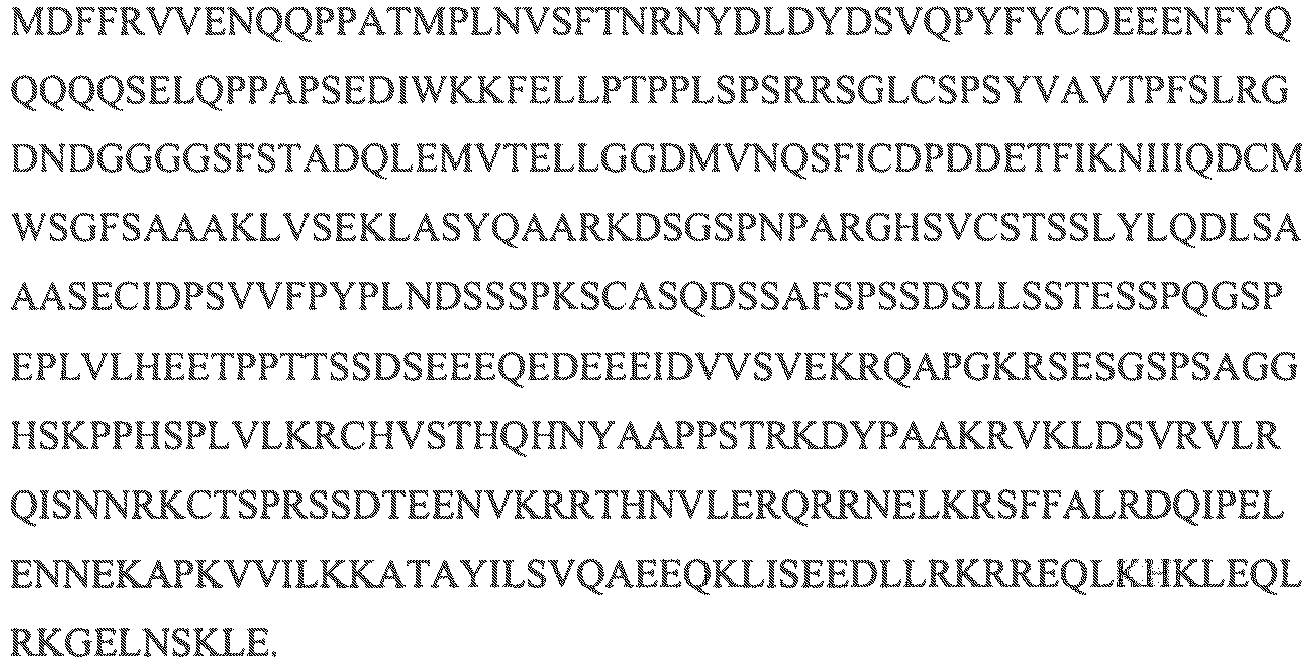
- Described herein is a method of producing an antibody that specifically binds to an antigen, comprising: contacting a cell comprising a nucleic acid sequence (e.g., a transgenic sequence) encoding an onco-peptide (e.g., MYC) or an onco-peptide (e.g., recombinant onco-peptide; e.g., TAT-Myc) with a selected antigen. In some descriptions, the onco-peptide is a fusion peptide. In some descriptions, the onco-peptide is a fusion peptide comprising a PTD. In some descriptions, the onco-peptide is TAT-MYC. In some descriptions, the onco-peptide is Myc-ER. In some descriptions, the onco-peptide is Myc-GR.
- In some descriptions, the onco-peptide is a peptide that promotes cell survival and/or proliferation. In certain descriptions, the cell is a mammalian cell. In some descriptions, the cell is a human cell. In some descriptions, the cell is a B-cell. In some descriptions, the cell is a hematopoietic cell. In some descriptions, the cell is a human B-cell. In some descriptions, the cell is a human hematopoietic cell. In some descriptions, the cell (e.g., a mammalian cell) expresses CD79 on its cell surface. In some descriptions, the cell (e.g., a mammalian cell) comprises an intact salvage pathway for purine biosynthesis and is tolerant and or anergic to the selected antigen. In some descriptions, the cell (e.g., a mammalian cell) is a B-cell, a B-cell precursor or progenitor, or a hematopoietic stem cell. In certain descriptions, the antibody specifically binds to the selected antigen. In certain descriptions, the onco-peptide is a peptide that promotes cell survival and/or proliferation.
- In certain descriptions, the cell (e.g., a mammalian cell) is contacted with the selected antigen by any suitable method. In specific descriptions, the cell (e.g., a mammalian cell) comprises a nucleic acid sequence (e.g., a transgenic sequence) that encodes the selected antigen and is, thereby, contacted with the selected antigen. In some descriptions, the selected antigen is a self-antigen.
- In certain descriptions, wherein a B-cell precursor or progenitor or a hematopoietic stem cell are used, a method described herein further comprises allowing or inducing differentiation of the cell (e.g., a mammalian cell) to a B-cell.
- In some descriptions, a MYC sequence (e.g., a transgenic sequence) further comprises a B-cell selective promoter or an inducible promoter. In some descriptions, a nucleic acid sequence (e.g., a transgenic sequence) encoding the selected antigen further comprises a B-cell selective promoter or an inducible promoter. In some descriptions, a B-cell selective promoter is, by way of non-limiting example, the Eµ promoter. In some descriptions, the inducible promoter comprises, by way of non-limiting example, one or more TREs. In certain descriptions, the cell (e.g., a mammalian cell) further comprises a nucleic acid sequence (e.g., a transgenic sequence) that encodes a nucleic acid sequence (e.g., a transgenic sequence) encoding a tTA peptide or an rtTA peptide. In some descriptions, the nucleic acid sequence (e.g., a transgenic sequence) encoding the tTA peptide or the rtTA peptide comprises a B-cell-selective promoter operably linked to the open reading frame encoding the tTA peptide or the rtTA peptide. In some descriptions, the nucleic acid sequence (e.g., transgenic nucleic acid sequence) encoding the tTA peptide or the rtTA peptide comprises the MMTV promoter operably linked to the open reading frame encoding the tTA peptide or the rtTA peptide. Furthermore, in some descriptions, wherein an inducible promoter comprises one or more TREs, a method described herein further comprises contacting the cell (e.g., a mammalian cell) with doxycycline, tetracycline, or an analog thereof.
- In some descriptions, the onco-peptide is a fusion peptide that comprises a receptor that activates the onco-peptide activity (e.g., cell survival and/or proliferation) when bound with a ligand. In some descriptions, the receptor is an estrogen receptor (ER). In specific descriptions, the onco-peptide is a Myc-ER fusion peptide. In specific descriptions, the onco-peptide is a Myc-GR fusion peptide. In certain descriptions, a method described herein further comprises contacting a cell comprising a recombinant onco-peptide that is a fusion peptide comprising a receptor with a ligand that binds the receptor. In specific descriptions, a method described herein further comprises contacting the cell (e.g., a mammalian cell) comprising a Myc-ER fusion peptide with an estrogen receptor modulator (e.g., an ER agonist or antagonist). In specific descriptions, a method described herein further comprises contacting the cell (e.g., a mammalian cell) comprising a Myc-GR fusion peptide with a glucocorticoid receptor modulator (e.g., a GR agonist or antagonist).
- In some descriptions, the onco-peptide is a fusion peptide that comprises a (a) a transporter peptide sequence (e.g., TAT); and (b) a MYC sequence (e.g., c-MYC). In some descriptions, the fusion peptide is a peptide of Formula (I):
transporter peptide sequence -MYC sequence.
In some descriptions, a fusion peptide disclosed herein comprises (a) a transporter peptide sequence; (b) a MYC sequence; and (c) one or more molecules that link the transporter peptide sequence and the MYC sequence (i.e., "X"). In some descriptions, the fusion peptide is a peptide of Formula (II):
transporter peptide sequence-X-MYC sequence,
wherein -X- is molecule that links the transporter peptide sequence and the MYC sequence. In some descriptions, - X- is an amino acid. In some descriptions, -X- is at least one amino acid. - In certain descriptions, a method described herein further comprises inducing MYC expression, Myc activity, or a combination thereof, in the cell (e.g., a mammalian cell). In some descriptions, a method described herein further comprises proliferating or inducing proliferation of the cell (e.g., a mammalian cell). In certain descriptions, the cells (e.g., a mammalian cell) are proliferated at least until forming a lymphoma (e.g., a B-cell lymphoma). In some descriptions, the proliferating cells are lymphoma cells. In certain descriptions, the cells (e.g., a mammalian cell) do not require and/or are not fused, e.g., with a myeloma, prior to proliferation (e.g., in order to provide a cell population sufficient to produce a significant or therapeutic amount of the antibody in a reasonable period of time).
- In some descriptions, the method further comprises recovering from the cell (e.g., a mammalian cell) the antibody that specifically binds the selected antigen. In some descriptions, the antibody produced is soluble and is not membrane bound.
- In some descriptions, the cell (e.g., a mammalian cell) (e.g., a mammalian cell) is present in an organism (e.g., a mammal). In some descriptions, the organism is a xenomouse. In certain descriptions, the organism (e.g., a mammal) further comprises a nucleic acid sequence (e.g., a transgenic sequence) that encodes a transgenic onco-peptide sequence (e.g., MYC). In some descriptions, the organism (e.g., a mammal) further comprises a transgenic nucleic acid sequence encoding a tTA peptide or an rtTA peptide. In some descriptions, the nucleic acid sequence (e.g., transgenic nucleic acid sequence) encoding the tTA peptide or the rtTA peptide comprises a B-cell-selective promoter operably linked to the open reading frame encoding the tTA peptide or the rtTA peptide. In some descriptions, the nucleic acid sequence (e.g., transgenic nucleic acid sequence) encoding the tTA peptide or the rtTA peptide comprises the MMTV promoter operably linked to the open reading frame encoding the tTA peptide or the rtTA peptide.
- In certain descriptions, contacting a cell comprising a Myc peptide (e.g., a recombinant Myc peptide (e.g., a Myc fusion peptide as described herein)) comprises introducing (or otherwise providing to) the selected antigen into the organism (e.g., a mammal) by any suitable manner. In some descriptions, the onco-peptide is a fusion peptide. In some descriptions, the onco-peptide is a fusion peptide comprising a PTD. In some descriptions, the onco-peptide is TAT-MYC. In some descriptions, the onco-peptide is Myc-ER. In some descriptions, the onco-peptide is Myc-GR. In some descriptions, the organism is a xenomouse. In certain descriptions, the organism (e.g., a mammal) further comprises a nucleic acid sequence (e.g., transgenic nucleic acid sequence) encoding the selected antigen. In some descriptions, the nucleic acid sequence (e.g., transgenic nucleic acid sequence) encoding the selected antigen comprises a B-cell-selective promoter operably linked to the open reading frame encoding the selected antigen.
- In some descriptions, a method described herein further comprises providing doxycycline, tetracycline, or an analog thereof to the organism (e.g., a mammal) to suppress tTA-dependent expression of the Myc peptide. In some descriptions, a method described herein further comprises providing doxycycline, tetracycline, or an analog thereof to the organism (e.g., a mammal) for a period of time sufficient to suppress tTA-dependent expression of the transgenic MYC gene, and withdrawing the doxycycline, tetracycline, or analog thereof after the period of time to induce tTA-dependent expression of the transgenic MYC.
- In some descriptions, a method described herein further comprises recovering from the organism (e.g., a mammal; e.g., a xenomouse) a cell (e.g., B-cells) that express the antibody that specifically binds the selected antigen. In certain descriptions, a method described herein further comprises recovering from the cells (e.g., mammalian cells) (e.g., B-cells) the antibody that specifically binds the selected antigen. In some descriptions, a method described herein further comprises recovering from the organism (e.g., a mammal) the antibody that specifically binds the selected antigen.
- Described herein is a method of producing an antibody that specifically binds to an antigen, comprising:
contacting a cell with a selected antigen and an onco-peptide (e.g. Myc). - In some descriptions, the onco-peptide is a peptide that promotes cell survival and/or proliferation. In certain descriptions, the cell is a mammalian cell. In some descriptions, the cell is a human cell. In some descriptions, the cell is a B-cell. In some descriptions, the cell is a hematopoietic cell. In some descriptions, the cell is a human B-cell. In some descriptions, the cell is a human hematopoietic cell. In some descriptions, the cell (e.g., a mammalian cell) expresses CD79 on its cell surface. In some descriptions, the cell (e.g., a mammalian cell) comprises an intact salvage pathway for purine biosynthesis and is tolerant and or anergic to the selected antigen. In some descriptions, the cell (e.g., a mammalian cell) is a B-cell, a B-cell precursor or progenitor, or a hematopoietic stem cell. In certain descriptions, the antibody specifically binds to the selected antigen. In certain descriptions, the onco-peptide is a peptide that promotes cell survival and/or proliferation.
- In certain descriptions, the cell (e.g., a mammalian cell) is contacted with the selected antigen by any suitable method. In specific descriptions, the cell (e.g., a mammalian cell) comprises a nucleic acid sequence (e.g., a transgenic sequence) that encodes the selected antigen and is, thereby, contacted with the selected antigen. In some descriptions, the selected antigen is a self-antigen.
- In certain descriptions, wherein a B-cell precursor or progenitor or a hematopoietic stem cell are used, a method described herein further comprises allowing or inducing differentiation of the cell (e.g., a mammalian cell) to a B-cell.
- In some descriptions, the onco-peptide is a fusion peptide that comprises a receptor that activates the onco-peptide activity (e.g., cell survival and/or proliferation) when bound with a ligand. In some descriptions, the receptor is an estrogen receptor (ER). In some descriptions, the receptor is a glucocorticoid receptor (GR). In specific descriptions, the onco-peptide is a Myc-ER fusion peptide. In some descriptions, the onco-peptide is a Myc-GR fusion peptide. In certain descriptions, a method described herein further comprises contacting a cell comprising a recombinant onco-peptide that is a fusion peptide comprising a receptor with a ligand that binds the receptor. In specific descriptions, a method described herein further comprises contacting the cell (e.g., a mammalian cell) comprising a Myc-ER fusion peptide with an estrogen receptor modulator (e.g., an ER agonist or antagonist). In specific descriptions, a method described herein further comprises contacting the cell (e.g., a mammalian cell) comprising a Myc-GR fusion peptide with an glucocorticoid receptor modulator (e.g., a GR agonist or antagonist).
- In some descriptions, the onco-peptide is a fusion peptide that comprises a (a) a transporter peptide sequence (e.g., TAT); and (b) a MYC sequence (e.g., c-MYC). In some descriptions, the fusion peptide is a peptide of Formula (I):
transporter peptide sequence -MYC sequence.
In some descriptions, a fusion peptide disclosed herein comprises (a) a transporter peptide sequence; (b) a MYC sequence; and (c) one or more molecules that link the transporter peptide sequence and the MYC sequence (i.e., "X"). In some descriptions, the fusion peptide is a peptide of Formula (II):
transporter peptide sequence-X-MYC sequence,
wherein -X- is molecule that links the transporter peptide sequence and the MYC sequence. In some descriptions, - X- is an amino acid. In some descriptions, -X- is at least one amino acid. - In some descriptions, the method further comprises recovering from the cell (e.g., a mammalian cell) the antibody that specifically binds the selected antigen. In some descriptions, the antibody produced is soluble and is not membrane bound.
- In certain descriptions, contacting a cell comprising a Myc peptide (e.g., a recombinant Myc peptide, a TAT-Myc fusion peptide) comprises introducing (or otherwise providing to) the selected antigen into the organism (e.g., a mammal) by any suitable manner. In some descriptions, the Myc peptide is a fusion peptide. In some descriptions, the Myc peptide is a TAT-Myc peptide as described herein. In some descriptions, the organism is a xenomouse. In certain descriptions, the organism (e.g., a mammal) further comprises a nucleic acid sequence (e.g., transgenic nucleic acid sequence) encoding the selected antigen. In some descriptions, the nucleic acid sequence (e.g., transgenic nucleic acid sequence) encoding the selected antigen comprises a B-cell-selective promoter operably linked to the open reading frame encoding the selected antigen.
- In some descriptions, a method described herein further comprises recovering from the organism (e.g., a mammal) a cell (e.g., B-cells) that express the antibody that specifically binds the selected antigen. In certain descriptions, a method described herein further comprises recovering from the cells (e.g., mammalian cells), B-cells) the antibody that specifically binds the selected antigen. In some descriptions, a method described herein further comprises recovering from the organism (e.g., a mammal) the antibody that specifically binds the selected antigen.
- Described herein is a method for producing an antibody that specifically binds to an antigen, comprising: contacting a cell with a selected antigen, wherein the cell (e.g., a mammalian cell) over-expresses an onco-peptide (e.g., recombinant onco-peptide; e.g., Myc). In some descriptions, the Myc peptide is a fusion peptide. In some descriptions, the Myc peptide is a TAT-Myc peptide as described herein.
- In some descriptions, the onco-peptide is a peptide that promotes cell survival and/or proliferation. In certain descriptions, the cell is a mammalian cell. In some descriptions, the cell is a human cell. In some descriptions, the cell is a B-cell. In some descriptions, the cell is a hematopoietic cell. In some descriptions, the cell is a human B-cell. In some descriptions, the cell is a human hematopoietic cell. In some descriptions, the cell (e.g., a mammalian cell) expresses CD79 on its cell surface. In some descriptions, the cell (e.g., a mammalian cell) comprising a nucleic acid sequence (e.g., a transgenic sequence) comprises an intact salvage pathway for purine biosynthesis. In some descriptions, the cell (e.g., a mammalian cell) comprising a nucleic acid sequence (e.g., a transgenic sequence) comprises an intact salvage pathway for purine biosynthesis and is tolerant and or anergic to the selected antigen. In some descriptions, the cell (e.g., a mammalian cell) is a B-cell, a B-cell progenitor or precursor, or a hematopoietic stem cell. In certain descriptions, the antibody specifically binds to the selected antigen. In certain descriptions, the onco-peptide is a peptide that promotes cell survival and or proliferation.
- In some descriptions, over-expression of MYC is induced by contacting the cell with a small molecule, a biologic, a peptide, an antibody, or a combination thereof. In some descriptions, the small molecule is an antagonist of Max-1, Mxi-1, MAD, or a combination thereof. In some descriptions, the small molecule is an antagonist of Max-1. In certain instances, the down-regulation of an Max-1 gene and/or polypeptide upregulates the expression of a MYC proto-oncogene and/or a polypeptide encoded by a MYC proto-oncogene. In some descriptions, the small molecule is an antagonist of Mxi-1. In certain instances, the down-regulation of an Mxi-1 gene and/or polypeptide upregulates the expression of a MYC proto-oncogene and/or a polypeptide encoded by a MYC proto-oncogene. In some descriptions, the small molecule is an antagonist of MAD. In certain instances, the down-regulation of a MAD-1 gene and/or polypeptide upregulates the expression of a MYC proto-oncogene and/or a polypeptide encoded by a MYC proto-oncogene. In some descriptions, over-expression of MYC is induced by contacting the cell with a siRNA molecule for Max-1, Mxi-1, MAD, or a combination thereof. In some descriptions, over-expression of MYC is induced by contacting the cell with a shRNA molecule for Max-1, Mxi-1, MAD, or a combination thereof.
- In certain descriptions, the cell (e.g., a mammalian cell) is contacted with the selected antigen by any suitable method. In specific descriptions, the cell (e.g., a mammalian cell) comprises a nucleic acid sequence (e.g., a transgenic sequence) that encodes a nucleic acid sequence (e.g., a transgenic sequence) that encodes the selected antigen. In some descriptions, the selected antigen is a self-antigen.
- In certain descriptions, wherein a B-cell precursor or progenitor or a hematopoietic stem cell are used, a method described herein further comprises allowing or inducing differentiation of the cell (e.g., a mammalian cell) to a B-cell.
- In some descriptions, a MYC sequence (e.g., a transgenic sequence) further comprises a B-cell selective promoter or an inducible promoter. In some descriptions, a nucleic acid sequence (e.g., a transgenic sequence) encoding the selected antigen further comprises a B-cell selective promoter or an inducible promoter. In some descriptions, a B-cell selective promoter is, by way of non-limiting example, the Eµ promoter. In some descriptions, the inducible promoter comprises, by way of non-limiting example, one or more TREs. In certain descriptions, the cell (e.g., a mammalian cell) further comprises a nucleic acid sequence (e.g., a transgenic sequence) that encodes a tTA peptide or an rtTA peptide. In some descriptions, the nucleic acid sequence (e.g., transgenic nucleic acid sequence) encoding the tTA peptide or the rtTA peptide comprises a B-cell-selective promoter operably linked to the open reading frame encoding the tTA peptide or the rtTA peptide. In some descriptions, the nucleic acid sequence (e.g., transgenic nucleic acid sequence) encoding the tTA peptide or the rtTA peptide comprises the MMTV promoter operably linked to the open reading frame encoding the tTA peptide or the rtTA peptide. Furthermore, in some descriptions, wherein an inducible promoter comprises one or more TREs, a method described herein further comprises contacting the cell (e.g., a mammalian cell) with doxycycline, tetracycline, or an analog thereof.
- In some descriptions, the onco-peptide is a fusion peptide that comprises a receptor that activates the onco-peptide activity (e.g., cell survival and/or proliferation) when bound with a ligand. In some descriptions, the receptor is an estrogen receptor (ER). In specific descriptions, the onco-peptide is a Myc-ER fusion peptide. In some descriptions, the receptor is an Glucocorticoid receptor (GR). In specific descriptions, the onco-peptide is a Myc-GR fusion peptide. In certain descriptions, a method described herein further comprises contacting a cell comprising a fusion peptide comprising a receptor with a ligand that specifically binds to the receptor. In specific descriptions, a method described herein further comprises contacting a cell comprising a Myc-ER peptide with an estrogen receptor modulator (e.g., an ER agonist or antagonist). In specific descriptions, a method described herein further comprises contacting a cell comprising a Myc-GR peptide with an glucocorticoid receptor modulator (e.g., a GR agonist or antagonist).
- In some descriptions, the onco-peptide is a fusion peptide that comprises a (a) a transporter peptide sequence (e.g., TAT); and (b) a MYC sequence (e.g., c-MYC). In some descriptions, the fusion peptide is a peptide of Formula (I):
transporter peptide sequence -MYC sequence.
In some descriptions, a fusion peptide disclosed herein comprises (a) a transporter peptide sequence; (b) a MYC sequence; and (c) one or more molecules that link the transporter peptide sequence and the MYC sequence (i.e., "X"). In some descriptions, the fusion peptide is a peptide of Formula (II):
transporter peptide sequence-X-MYC sequence,
wherein -X- is molecule that links the transporter peptide sequence and the MYC sequence. In some descriptions, - X- is an amino acid. In some descriptions, -X- is at least one amino acid. - In certain descriptions, a method described herein further comprises inducing MYC expression, Myc activity, or a combination thereof, in the cell (e.g., a mammalian cell). In some descriptions, a method described herein further comprises proliferating or inducing proliferation of the cell (e.g., a mammalian cell). In certain descriptions, the cells (e.g., mammalian cells) are proliferated at least until forming a lymphoma (e.g., a B-cell lymphoma). In some descriptions, the proliferating cells are lymphoma cells. In certain descriptions, the cells (e.g., mammalian cells) do not require and/or are not fused, e.g., with a myeloma, prior to proliferation (e.g., in order to provide a cell population sufficient to produce a significant or therapeutic amount of the antibody in a reasonable period of time).
- In some descriptions, the method further comprises recovering the antibody that specifically binds the selected antigen. In some descriptions, the antibody produced is soluble and is not membrane bound. In some descriptions, the antibody produced is membrane bound. In some descriptions, the antibody produced is intracellular.
- In some descriptions, the cell (e.g., a mammalian cell) is present in an organism (e.g., a mammal). In some descriptions, the organism is a xenomouse. In certain descriptions, the organism (e.g., a mammal) comprises a nucleic acid sequence (e.g., a transgenic sequence) that encodes an onco-peptide. In some descriptions, the organism (e.g., a mammal) further comprises a nucleic acid sequence (e.g., a transgenic sequence) that encodes a tTA peptide or an rtTA peptide. In some descriptions, the nucleic acid sequence (e.g., transgenic nucleic acid sequence) encoding the tTA peptide or the rtTA peptide comprises a B-cell-selective promoter operably linked to the open reading frame encoding the tTA peptide or the rtTA peptide. In some descriptions, the nucleic acid sequence (e.g., transgenic nucleic acid sequence) encoding the tTA peptide or the rtTA peptide comprises the MMTV promoter operably linked to the open reading frame encoding the tTA peptide or the rtTA peptide.
- In certain descriptions, contacting the cell (e.g., a mammalian cell) comprising MYC (e.g., a transgenic MYC) comprises introducing (or otherwise providing to) the selected antigen into the organism (e.g., a mammal) by any suitable manner. In certain descriptions, the organism (e.g., a mammal) further comprises a nucleic acid sequence (e.g., a transgenic sequence) encoding the selected antigen. In some descriptions, the nucleic acid sequence (e.g., transgenic nucleic acid sequence) encoding the selected antigen comprises a B-cell-selective promoter operably linked to the open reading frame encoding the selected antigen.
- In some descriptions, a method described herein further comprises providing doxycycline, tetracycline, or an analog thereof to the organism (e.g., a mammal) to suppress tTA-dependent expression of the nucleic acid sequence (e.g., transgenic nucleic acid sequence) encoding a Myc peptide (e.g., a recombinant Myc peptide (e.g., a Myc fusion peptide as described herein)). In some descriptions, the onco-peptide is a fusion peptide. In some descriptions, the onco-peptide is a fusion peptide comprising a PTD. In some descriptions, the onco-peptide is TAT-MYC. In some descriptions, the onco-peptide is Myc-ER. In some descriptions, the onco-peptide is Myc-GR. In some descriptions, a method described herein further comprises (a) providing doxycycline, tetracycline, or an analog thereof to the organism (e.g., a mammal) for a period of time sufficient to suppress tTA-dependent expression of the nucleic acid sequence (e.g., transgenic nucleic acid sequence) encoding the Myc peptide, and (b) withdrawing the doxycycline, tetracycline, or analog thereof after the period of time to induce tTA-dependent expression of the nucleic acid sequence (e.g., transgenic nucleic acid sequence) encoding the Myc peptide.
- In some descriptions, a method described herein further comprises recovering from the organism (e.g., a mammal) a cell (e.g., B-cell) that express the antibody that specifically binds the selected antigen. In certain descriptions, a method described herein further comprises recovering from the cell (e.g., a mammalian cell) (e.g., B-cell) the antibody that specifically binds the selected antigen. In some descriptions, a method described herein further comprises recovering from the organism (e.g., a mammal) the antibody that specifically binds the selected antigen.
- Described herein is a method of producing an antibody that specifically binds to an antigen, comprising: introducing (or otherwise providing to) a selected antigen into an organism (e.g., a mammal), wherein the organism (e.g., a mammal) comprises a nucleic acid sequence (e.g., a transgenic sequence) that encodes an onco-peptide (e.g., recombinant onco-peptide; e.g., Myc), and comprises an inducible promoter or a B-cell-selective promoter operably linked to the open reading frame of the encoded onco-peptide.
- In some descriptions, the onco-peptide is a fusion peptide. In some descriptions, the onco-peptide is a fusion peptide comprising a PTD. In some descriptions, the onco-peptide is TAT-MYC. In some descriptions, the onco-peptide is Myc-ER. In some descriptions, the onco-peptide is Myc-GR.
- In some descriptions, the organism is a xenomouse.
- Described herein is a method of producing an antibody that specifically binds to an antigen, comprising:
- a. providing a cell (e.g., a B-cell) expressing an antibody that specifically binds an antigen, wherein the cell (e.g., a mammalian cell) comprises an onco-peptide (e.g., a recombinant onco-peptide; e.g., Myc) or a nucleic acid sequence (e.g., a transgenic sequence) encoding an onco-peptide (e.g., a recombinant onco-peptide; e.g., Myc); and
- b. inducing activity of the onco-peptide in the cell (e.g., a mammalian cell).
- In some descriptions, the onco-peptide is a fusion peptide. In some descriptions, the onco-peptide is a fusion peptide comprising a PTD. In some descriptions, the onco-peptide is TAT-MYC. In some descriptions, the onco-peptide is Myc-ER. In some descriptions, the onco-peptide is Myc-GR.
- In some descriptions, the cell (e.g., a mammalian cell) is prepared by contacting a cell with the selected antigen. In some descriptions, the selected antigen is a self-antigen. In certain descriptions, the antibody specifically binds to the selected antigen.
- In some descriptions, the cell is a human cell. In certain descriptions, the cell (e.g., a mammalian cell) is a B-cell, a B-cell precursor or progenitor, or a hematopoietic stem cell. In some descriptions, the B-cell is an anergic B-cell. In some descriptions, the cell (e.g., a mammalian cell) expresses CD79 on its cell surface. In some descriptions, the B-cell is a mouse B-cell. In some descriptions, the cell (e.g., a mammalian cell; e.g., B-cell) is from an organism (e.g., a mammal; e.g., a xenomouse) that was administered (i.e., inoculated with, or immunized against) the selected antigen. In some descriptions, the organism is a xenomouse. In some descriptions, the organism (e.g., a mammal) comprises an onco-peptide (e.g., a recombinant onco-peptide; e.g., Myc) or a nucleic acid sequence (e.g., a transgenic sequence) encoding an onco-peptide (e.g., a recombinant onco-peptide; e.g., Myc). In some descriptions, the onco-peptide is a fusion peptide. In some descriptions, the onco-peptide is a fusion peptide comprising a PTD. In some descriptions, the onco-peptide is TAT-MYC. In some descriptions, the onco-peptide is Myc-ER. In some descriptions, the onco-peptide is Myc-GR. In some descriptions, the organism (e.g., a mammal) further comprises a nucleic acid sequence (e.g., a transgenic sequence) that encodes the selected antigen. In some descriptions, a MYC sequence (e.g., a transgenic sequence) further comprises a B-cell selective promoter or an inducible promoter. In some descriptions, a nucleic acid sequence (e.g., a transgenic sequence) encoding the selected antigen further comprises a B-cell selective promoter or an inducible promoter. In some descriptions, a B-cell selective promoter is, by way of non-limiting example, the Eµ promoter. In some descriptions, the inducible promoter comprises, by way of non-limiting example, one or more TREs.
- In certain descriptions, wherein a B-cell precursor or progenitor or a hematopoietic stem cell are used, a method described herein further comprises allowing or inducing differentiation of the cell (e.g., a mammalian cell) into a B-cell.
- In some descriptions, the method further comprises recovering the antibody. In certain descriptions, activity of the onco-peptide induces expansion of the cell (e.g., a mammalian cell) population.
- In some descriptions, the cell (e.g., a mammalian cell) (e.g., B-cell) is present in an organism (e.g., a mammal) and inducing the activity of the onco-peptide (e.g., recombinant onco-peptide; e.g., Myc) in the cell (e.g., a mammalian cell) (e.g., B-cell) occurs in vivo. In some descriptions, the organism is a xenomouse. In some descriptions, the method further comprises introducing (or otherwise providing to) an onco-peptide (e.g., a recombinant onco-peptide; e.g., Myc) or the nucleic acid sequence (e.g., transgenic nucleic acid sequence) encoding an onco-peptide (e.g., a recombinant onco-peptide; e.g., Myc) into the cell (e.g., a mammalian cell). In some descriptions, the onco-peptide or the nucleic acid sequence (e.g., transgenic nucleic acid sequence) encoding the onco-peptide is introduced into the cell (e.g., a mammalian cell) ex vivo and then the cell (e.g., a mammalian cell) is introduced into an organism (e.g., a mammal). In some descriptions, the organism is a xenomouse. In specific descriptions, the process comprises introducing (or otherwise providing to) a onco-peptide (e.g., recombinant onco-peptide; e.g., Myc) into a cell (e.g., B-cell) ex vivo. In some descriptions, the onco-peptide (e.g., recombinant onco-peptide; e.g., Myc) comprises a protein transduction domain, e.g., HIV-1 Tat or Vpr (e.g., Tat-Myc or Vpr-Myc).
- In some descriptions, inducing the activity of an onco-peptide includes inducing the expression of a nucleic acid sequence encoding the onco-peptide, inducing the activity of the onco-peptide, or a combination thereof. In certain instances, inducing activity of the onco-peptide includes inducing over-expression of the onco-peptide. In some descriptions, inducible expression or over-expression of the onco-peptide is achieved by activating an inducible promoter of a nucleic acid sequence (e.g., a transgenic sequence) that induces expression of an onco-peptide. In some descriptions, over-expression of MYC is induced by contacting the cell with a small molecule, a biologic, a peptide, an antibody, or a combination thereof. In some descriptions, the small molecule is an antagonist of Max-1, Mxi-1, MAD, or a combination thereof. In some descriptions, the small molecule is an antagonist of Max-1. In some descriptions, the small molecule is an antagonist of Mxi-1. In some descriptions, the small molecule is an antagonist of MAD. In some descriptions, over-expression of MYC is induced by contacting the cell with a siRNA molecule for Max-1, Mxi-1, MAD, or a combination thereof. In some descriptions, over-expression of MYC is induced by contacting the cell with a shRNA molecule for Max-1, Mxi-1, MAD, or a combination thereof.
- In certain descriptions, the nucleic acid sequence (e.g., transgenic nucleic acid sequence) encoding an onco-peptide (e.g., recombinant onco-peptide; e.g., Myc) comprises a promoter, e.g., a B-cell selective promoter or an inducible promoter, operably linked to the open reading frame encoding the onco-peptide (e.g., recombinant onco-peptide; e.g., Myc). In some specific descriptions, the nucleic acid sequence (e.g., transgenic nucleic acid sequence) encoding an onco-peptide (e.g., recombinant onco-peptide; e.g., Myc) comprises a B-cell-selective promoter operably linked to an open reading frame. In certain specific descriptions, the nucleic acid sequence (e.g., transgenic nucleic acid sequence) encoding the onco-peptide comprises an inducible promoter operably linked to an open reading frame for the Myc peptide. In some descriptions, the inducible promoter comprises one or more TREs and the cell (e.g., a mammalian cell) further expresses a tTA peptide or an rtTA peptide. In some descriptions, the cell (e.g., a mammalian cell) expresses the tTA peptide. In some descriptions, the onco-peptide is a fusion peptide. In some descriptions, the onco-peptide is a fusion peptide comprising a PTD. In some descriptions, the onco-peptide is TAT-MYC. In some descriptions, the onco-peptide is Myc-ER. In some descriptions, the onco-peptide is Myc-GR.
- In some descriptions, the onco-peptide is a fusion peptide that is inducibly activated. In specific descriptions, the onco-peptide is a fusion peptide comprising a receptor that activates the survival and/or proliferative characteristic of the onco-peptide when modulated (i.e., bound with a ligand, such, as an agonist or antagonist). In specific descriptions, the onco-peptide (e.g., a recombinant onco-peptide; e.g., Myc) is a fusion peptide comprising an estrogen receptor (e.g., Myc-ER). In some descriptions, inducing activity of the onco-peptide in the cell (e.g., a mammalian cell) comprises contacting the cell (e.g., a mammalian cell) with an ER ligand. In specific descriptions, the onco-peptide (e.g., a recombinant onco-peptide; e.g., Myc) is a fusion peptide comprising an glucocorticoid receptor (e.g., Myc-GR). In some descriptions, inducing activity of the onco-peptide in the cell (e.g., a mammalian cell) comprises contacting the cell (e.g., a mammalian cell) with a GR ligand.
- In some descriptions, the onco-peptide is a fusion peptide that comprises a (a) a transporter peptide sequence (e.g., TAT); and (b) a MYC sequence (e.g., c-MYC). In some descriptions, the fusion peptide is a peptide of Formula (I):
transporter peptide sequence -MYC sequence.
In some descriptions, a fusion peptide disclosed herein comprises (a) a transporter peptide sequence; (b) a MYC sequence; and (c) one or more molecules that link the transporter peptide sequence and the MYC sequence (i.e., "X"). In some descriptions, the fusion peptide is a peptide of Formula (II):
transporter peptide sequence-X-MYC sequence,
wherein -X- is molecule that links the transporter peptide sequence and the MYC sequence. In some descriptions, - X- is an amino acid. In some descriptions, -X- is at least one amino acid. - In some descriptions, the cell (e.g., a mammalian cell; e.g., a B-cell) comprising an onco-peptide (e.g., recombinant onco-peptide; e.g., Myc) or a nucleic acid sequence (e.g., a transgenic sequence) encoding an onco-peptide (e.g., a recombinant onco-peptide; e.g., Myc) is present in an organism (e.g., a mammal). In some descriptions, the Myc peptide is a fusion peptide. In some descriptions, the Myc peptide is a TAT-Myc peptide as described herein. In some descriptions, the organism is a xenomouse. In some descriptions, the organism is administered (i.e., immunized against, inoculated with) an antigen and allowed to mount an immune response to the selected antigen. In some descriptions, the organism is exposed to the selected antigen by any suitable manner. In some descriptions, the selected antigen is administered with an adjuvant. In some descriptions, the selected antigen is covalently linked to a carrier. Examples of carries include but are not limited to bovine serum albumin, keyhole limpet hemocyanin, ovalbumin and hyroglobulin.
- In some descriptions, a cell or organism utilized in a method disclosed herein includes those that inducibly over-express an onco-peptide (e.g., recombinant onco-peptide; e.g., Myc) and are engineered to express an antigen. Such cells and organisms are prepared by any suitable method including, by way of non-limiting example, retroviral mediated transduction of bone marrow hematopoietic stem cells, production of transgenic organisms (or crossing the onco-peptide over-expressing organisms with an organism that expresses the selected antigen), or any other method for gene delivery into the cell (e.g., a mammalian cell) or organism. In some descriptions, over-expression of MYC is induced by contacting the cell with a small molecule, a biologic, a peptide, an antibody, or a combination thereof. In some descriptions, the small molecule is an antagonist of Max-1, Mxi-1, MAD, or a combination thereof. In some descriptions, the small molecule is an antagonist of Max-1. In some descriptions, the small molecule is an antagonist of Mxi-1. In some descriptions, the small molecule is an antagonist of MAD. In some descriptions, over-expression of MYC is induced by contacting the cell with a siRNA molecule for Max-1, Mxi-1, MAD, or a combination thereof. In some descriptions, over-expression of MYC is induced by contacting the cell with a shRNA molecule for Max-1, Mxi-1, MAD, or a combination thereof. In some descriptions, the cell (e.g., a mammalian cell) or organism is maintained under conditions in which an onco-peptide (e.g., recombinant onco-peptide; e.g., Myc) is not over-expressed until the production of antibodies is desired. In some descriptions, the cell (e.g., a mammalian cell) or organism is maintained under conditions in which the onco-peptide activity is not induced until the production of antibodies is desired. In some descriptions, the cell (e.g., a mammalian cell) or organism is maintained under conditions under which the onco-peptide is over-expressed in a cell (e.g., a mammalian cell; e.g., a B-cell) that produces antibodies that specifically binds to the selected antigen.
- In some descriptions, B cells specific for a heterologous antigen, a self antigen, or an auto antigen are tolerant to the heterologous antigen, self antigen, or auto antigen. In some descriptions, induction of onco-peptide (e.g., recombinant onco-peptide; e.g., Myc) activity in tolerant B cells induces a break in tolerance to an antigen and induces production of antibodies that specifically binds to the selected antigen. In some descriptions, a tolerant B-cell is anergic to an antigen. In some descriptions, a tolerant B-cell is tolerant to an antigen that is immunologically similar to an auto antigen or self antigen. In some descriptions, a tolerant B-cell is tolerant to an antigen that is a heterologous antigen. In some descriptions, the heterologous antigen is homologous to an auto antigen or self antigen. In some descriptions, the selected antigen specific B-cells produced by over-expression of MYC are removed from the organism and are cloned to produce immortal antibody producing B-cells without the need for cell fusion to a myeloma partner. In some descriptions, the Myc peptide is a fusion peptide. In some descriptions, the Myc peptide is a TAT-Myc peptide as described herein.
- Described herein is a method for producing an antibody that specifically binds to an antigen, comprising:
- a. providing a B-cell that is tolerant to an antigen; and
- b. inducing onco-peptide (e.g., recombinant onco-peptide; e.g., Myc) activity in a B-cell that is tolerant to the selected antigen.
- Described herein is a method of producing an antibody that specifically binds to an antigen, comprising: administering to an immuno-deficient organism (e.g., a mammal) a plurality of hematopoietic stem cells (e.g., recombinant hematopoietic stem cells) that comprise a nucleic acid sequence (e.g., a transgenic sequence) encoding an onco-peptide (e.g., a recombinant onco-peptide; e.g., Myc); and contacting a selected antigen with the immuno-deficient organism (e.g., a mammal). In some descriptions, the onco-peptide is a fusion peptide. In some descriptions, the onco-peptide is a fusion peptide comprising a PTD. In some descriptions, the onco-peptide is TAT-MYC. In some descriptions, the onco-peptide is Myc-ER. In some descriptions, the onco-peptide is Myc-GR.
- In some descriptions, the organism is a xenomouse.
- In certain descriptions, the onco-peptide is a Myc peptide. In some descriptions, the onco-peptide is a fusion peptide. In some descriptions, the onco-peptide is a fusion peptide comprising a PTD. In some descriptions, the onco-peptide is TAT-MYC. In some descriptions, the onco-peptide is inducibly activated (e.g., inducibly expressed or the function is inducibly activated). In certain descriptions, the onco-peptide is a fusion peptide comprising a receptor, and the function of the onco-peptide is inducibly activated by modulating or binding the receptor (e.g., with a ligand, such as an agonist or antagonist). In specific descriptions, the onco-peptide is Myc-ER. In specific descriptions, the onco-peptide is Myc-GR. In further descriptions, the method further comprises inducing onco-peptide (e.g., recombinant onco-peptide; e.g., Myc) activity in the organism (e.g., a mammal). In some descriptions, the organism is a xenomouse. For example, in the case of Myc-ER, Myc activity is optionally induced by administering an estrogen receptor ligand to the immuno-deficient organism (e.g., a mammal); in the case of a MYC nucleic acid sequence with an inducible promoter comprising one or more TREs, Myc activity is induced by providing doxycycline, tetracycline, or an analog thereof to the immuno-deficient organism (e.g., a mammal); or in the case of Myc-GR, Myc activity is induced by administering a glucocorticoid receptor ligand to the immune-deficient organism.
- In some descriptions, the hematopoietic stem cells further comprise a nucleic acid sequence (e.g., a transgenic sequence) encoding a polypeptide that inhibits apoptosis (e.g., Bcl-2, Bcl-x, Mcl-1).
- In some descriptions, a method described herein further comprises inducing a plurality of the hematopoietic stem cells to differentiate into B-cells. In some descriptions, a method disclosed herein further comprises recovering a plurality of the B-cells that express an antibody that specifically binds to the selected antigen from the immuno-deficient organism (e.g., a mammal). In some descriptions, a method described herein further comprises recovering the antibody from the immuno-deficient organism (e.g., a mammal) or from a plurality of B-cells that express the antibody.
- In some descriptions, the nucleic acid sequence (e.g., transgenic nucleic acid sequence) encoding an onco-peptide comprises an inducible promoter or a B-cell-selective promoter. In some descriptions, the nucleic acid sequence (e.g., transgenic nucleic acid sequence) encoding an onco-peptide comprises an inducible promoter comprising one or more TREs. In some descriptions, the hematopoietic stem cells express tTA or rtTA. In some descriptions, a method described herein further comprises providing doxycycline, tetracycline, or an analog thereof to the immuno-deficient organism (e.g., a mammal) to suppress the tTA-dependent transactivation, and withdrawing the doxycycline, tetracycline, or analog thereof to induce tTA-dependent transactivation. In some descriptions, the B-cell selective promoter is the Eµ promoter.
- In some descriptions, an immuno-deficient organism (e.g., a mammal) is obtained by irradiating the organism (e.g., a mammal). In some descriptions, the immuno-deficient organism (e.g., a mammal) is a Rag-1ko, Rag-2, SCID, DNA-PK, Ku70, Ku80, XRCC4, or µMT mouse. In some descriptions, the organism is a xenomouse.
- In certain descriptions, the selected antigen is administered to the organism by any suitable method. In some descriptions, the selected antigen is a self antigen. In some descriptions, the organism is an immuno-deficient mammal. In some descriptions, the organism is an immuno-deficient mouse. In some descriptions, the organism is an immune-deficient xenomouse. In some descriptions, the immuno-deficient organism (e.g., a mammal) expresses the selected antigen. In some descriptions, the immuno-deficient organism (e.g., a mammal) that expresses the selected antigen comprises an exogenous nucleic acid sequence that encodes the selected antigen. In some descriptions, the selected antigen is introduced by transfection with a nucleic acid expression vector or infection with a recombinant virus expression vector. In some descriptions, the recombinant virus expression vector is a recombinant lentivirus. In certain descriptions, the antibody specifically binds to the selected antigen. In some descriptions, the Myc peptide is a fusion peptide. In some descriptions, the Myc peptide is a TAT-Myc peptide as described herein.
- In some descriptions, hematopoietic stem cells that over-express MYC are used to reconstitute the peripheral lymphoid compartments of immuno-deficient organisms. In some descriptions, over-expression of MYC is induced by contacting the cell with a small molecule, a biologic, a peptide, an antibody, or a combination thereof. In some descriptions, the small molecule is an antagonist of Max-1, Mxi-1, MAD, or a combination thereof. In some descriptions, the small molecule is an antagonist of Max-1. In some descriptions, the small molecule is an antagonist of Mxi-1. In some descriptions, the small molecule is an antagonist of MAD. In some descriptions, over-expression of MYC is induced by contacting the cell with a siRNA molecule for Max-1, Mxi-1, MAD, or a combination thereof. In some descriptions, over-expression of MYC is induced by contacting the cell with a shRNA molecule for Max-1, Mxi-1, MAD, or a combination thereof. In some descriptions, the immuno-deficient organisms are rendered immuno-deficient by lethal irradiation. In some descriptions, immuno-deficient organisms are lethally irradiated mice. In some descriptions, the immuno-deficient organism is any immuno-deficient or immuno-compromised mouse. In some descriptions, the immuno-deficient animal is a Rag-1 knock out, Rag-2, SCID, DNA-PK, Ku70, Ku80, XRCC4, or µMT mouse. The peripheral lymphoid compartment of an immuno-deficient animal is typically reconstituted in 8-12 weeks after transplantation of hematopoietic stem cells. In some descriptions, organisms that are reconstituted with hematopoietic stem cells that over-express MYC, are further genetically altered to express the selected antigen. In some descriptions, organisms that are reconstituted with hematopoietic stem cells that over-express MYC are administered (i.e., immunized with or inoculated against) the selected antigen.
- In some description B-cell populations (e.g., anergic B-cells, such as AN1/T3) are isolated from organisms reconstituted with hematopoietic stem cells and are used in any method described herein. In some descriptions, hematopoietic stem cells that over-express MYC reconstitute the peripheral hematopoietic compartment of immuno-deficient organisms, are exposed to the selected antigen and develop into antigen specific AN1/T3 cell populations. In some descriptions, induction of Myc activity in these cells generates immortal antigen specific B-cells.
- The hematopoietic stem cells utilized herein are prepared by any suitable method. In some descriptions, hematopoietic stem cells are isolated from an organism that over-expresses Myc. In some descriptions, over-expression of MYC is induced by contacting the cell with a small molecule, a biologic, a peptide, an antibody, or a combination thereof. In some descriptions, the small molecule is an antagonist of Max-1, Mxi-1, MAD, or a combination thereof. In some descriptions, the small molecule is an antagonist of Max-1. In some descriptions, the small molecule is an antagonist of Mxi-1. In some descriptions, the small molecule is an antagonist of MAD. In some descriptions, over-expression of MYC is induced by contacting the cell with a siRNA molecule for Max-1, Mxi-1, MAD, or a combination thereof. In some descriptions, the organism is a xenomouse. In some descriptions, hematopoietic stem cells are isolated from organisms that comprise antigen specific B-cell populations. In some descriptions, hematopoietic stem cells are isolated from organisms that comprise antigen binding B-cell populations. In some descriptions, hematopoietic stem cells are isolated from organisms that comprise antigen specific AN1/T3 cell populations. In some descriptions, hematopoietic stem cells are isolated from organisms that comprise antigen specific anergic B-cell populations. In some descriptions, hematopoietic stem cells are isolated from wild type organisms. In some descriptions, the organisms are mice (e.g., transgenic mice or genetically altered mice). In some descriptions, organisms are treated with 5FU, in order to enrich for long-term hematopoietic stem cells, and induce their proliferation in vivo. In some descriptions, the organisms (e.g., mice) are treated with from 0.01 to 100 mg/mouse of 5FU. In some descriptions, the organisms (e.g., mice) are treated with from 0.1 to 50 mg/mouse of 5FU. In some descriptions, the organisms (e.g., mice) are treated with from 1 to 10 mg/mouse of 5FU. In some descriptions, the organisms (e.g., mice) are treated with 5 mg/mouse of 5FU. In some descriptions, bone marrow cells containing 5FU enriched populations of hematopoietic stem cells are isolated, by any suitable method, from the femurs and tibia bones of organisms that over-express MYC. In some descriptions, over-expression of MYC is induced by contacting the cell with a small molecule, a biologic, a peptide, an antibody, or a combination thereof. In some descriptions, the small molecule is an antagonist of Max-1, Mxi-1, MAD, or a combination thereof. In some descriptions, the small molecule is an antagonist of Max-1. In some descriptions, the small molecule is an antagonist of Mxi-1. In some descriptions, the small molecule is an antagonist of MAD. In some descriptions, over-expression of MYC is induced by contacting the cell with a siRNA molecule for Max-1, Mxi-1, MAD, or a combination thereof. In some descriptions, hematopoietic stem cells are isolated approximately 1-10 days after administration of 5FU. In some descriptions, hematopoietic stem cells are isolated approximately 5 days after administration of 5FU. In some descriptions, hematopoietic stem cells are cultured in vitro in a media comprising human IL-3, IL-6 and Stem Cell Factor (SCF). In certain descriptions, isolated hematopoietic stem cells over-express MYC. In some descriptions, over-expression of MYC is induced by contacting the cell with a small molecule, a biologic, a peptide, an antibody, or a combination thereof. In some descriptions, the small molecule is an antagonist of Max-1, Mxi-1, MAD, or a combination thereof. In some descriptions, the small molecule is an antagonist of Max-1. In some descriptions, the small molecule is an antagonist of Mxi-1. In some descriptions, the small molecule is an antagonist of MAD. In some descriptions, over-expression of MYC is induced by contacting the cell with a siRNA molecule for Max-1, Mxi-1, MAD, or a combination thereof. In some descriptions, hematopoietic stem cells are genetically altered to over-express MYC in vitro by introducing (or otherwise providing to) a nucleic acid into the hematopoietic stem cells in which the nucleic acid directs the over-expression of MYC. In some descriptions, the over-expression of MYC is inducible. In some descriptions, the hematopoietic stem cells are genetically altered to over-express MYC by infecting the hematopoietic stem cells in vitro with a virus that directs the over-expression of MYC. In some descriptions, the virus is a lentivirus, retrovirus or adenovirus. In some descriptions, the hematopoietic stem cells are genetically altered to over-express MYC by transfection with a nucleic acid. In some descriptions, the hematopoietic stem cells are also genetically altered to express a reporter gene. In certain descriptions, reporter genes are utilized and allow selection or isolation of cells that are genetically altered to express Myc. In some descriptions, the reporter gene is GFP (green fluorescent protein). In some descriptions, hematopoietic stem cells are transduced with a virus that directs the over-expression of MYC and also GFP. In some descriptions, the hematopoietic stem cells are infected with a lentivirus that directs the over-expression of MYC and GFP. Described herein are hematopoietic stem cells that over-express MYC, as described herein, and are used to generate immortal antibody producing B-cells.
- In various descriptions, of any of the methods disclosed herein, polyclonal populations of antibodies that specifically binds to the selected antigen, as well as the cell (e.g., a mammalian cell) producing such antibodies, are isolated directly from the tissues (spleen, lymph nodes, blood, etc.) or serum of organisms. In some descriptions, monoclonal populations of antibody producing cells are isolated from organisms, and monoclonal antibodies are recovered from culture media.
- Described herein is any isolated B-cell prepared by a method described herein. Described herein is a cell (e.g., a mammalian cell) that has been engineered to produce a recombinant form of any antibody prepared according any process disclosed herein. Described herein is a B-cell that expresses a human or humanized antibody, and an onco-peptide (e.g., recombinant onco-peptide; e.g., Myc), a nucleic acid sequence (e.g., a transgenic sequence) that encodes an onco-peptide (e.g., recombinant onco-peptide; e.g., Myc), and/or wherein the B-cell over-expresses Myc. In some descriptions, over-expression of MYC is induced by contacting the cell with a small molecule, a biologic, a peptide, an antibody, or a combination thereof. In some descriptions, the small molecule is an antagonist of Max-1, Mxi-1, MAD, or a combination thereof. In some descriptions, the small molecule is an antagonist of Max-1. In some descriptions, the small molecule is an antagonist of Mxi-1. In some descriptions, the small molecule is an antagonist of MAD. In some descriptions, over-expression of MYC is induced by contacting the cell with a siRNA molecule for Max-1, Mxi-1, MAD, or a combination thereof. In some descriptions, over-expression of MYC is induced by contacting the cell with a shRNA molecule for Max-1, Mxi-1, MAD, or a combination thereof. In certain descriptions, the onco-peptide is a fusion peptide comprising a transduction domain (e.g., Tat-Myc or Vpr-Myc), a receptor that activates function of the onco-peptide when modulated or bound (e.g., Myc-ER, Myc-GR), or a combination thereof (e.g., Tat-Myc-ER, Tat-Myc-GR, Vpr-Myc-ER, or Vpr-Myc-GR). Described herein is an isolated antibody prepared according to the process of any of the methods disclosed herein.
- In certain descriptions, a method disclosed herein is utilized to prepare human or humanized antibodies. In some descriptions, the cells (e.g., mammalian cells) or organisms of any of a method disclosed herein express human antibodies. In some descriptions, the organism is a xenomouse.
- Described herein is a method of producing a human or humanized antibody comprising: providing a cell (e.g., a mammalian cell) that expresses human antibodies and comprises a nucleic acid sequence (e.g., a transgenic sequence) that encodes a Myc peptide, or a Myc peptide; and contacting the cell (e.g., a mammalian cell) with a selected antigen. In some descriptions, the onco-peptide is a fusion peptide. In some descriptions, the onco-peptide is a fusion peptide comprising a PTD. In some descriptions, the onco-peptide is TAT-MYC. In some descriptions, the onco-peptide is Myc-ER. In some descriptions, the onco-peptide is Myc-GR.
- In some descriptions, the cell (e.g., a mammalian cell) that expresses human antibodies and comprises a nucleic acid sequence (e.g., a transgenic sequence) that encodes the Myc peptide, or the Myc is a cell that expresses cell-surface CD79. In some descriptions, the cell (e.g., a mammalian cell) expresses CD79 and comprises an intact salvage pathway for purine biosynthesis. In some descriptions, the cell (e.g., a mammalian cell) expresses CD79, comprises an intact salvage pathway for purine biosynthesis and is tolerant and or anergic to the selected antigen. In some descriptions, the cell (e.g., a mammalian cell) that expresses human antibodies and comprises the nucleic acid sequence (e.g., a transgenic sequence) that encodes a Myc peptide or a Myc peptide is a B-cell, a B-cell progenitor or precursor, or a hematopoietic stem cellIn some descriptions, the B-cell over-expresses Myc. In some descriptions, over-expression of MYC is induced by contacting the cell with a small molecule, a biologic, a peptide, an antibody, or a combination thereof. In some descriptions, the small molecule is an antagonist of Max-1, Mxi-1, MAD, or a combination thereof. In some descriptions, the small molecule is an antagonist of Max-1. In some descriptions, the small molecule is an antagonist of Mxi-1. In some descriptions, the small molecule is an antagonist of MAD. In some descriptions, over-expression of MYC is induced by contacting the cell with a siRNA molecule for Max-1, Mxi-1, MAD, or a combination thereof. In some descriptions, over-expression of MYC is induced by contacting the cell with a shRNA molecule for Max-1, Mxi-1, MAD, or a combination thereof. In some descriptions, the cell (e.g., a mammalian cell) that expresses human antibodies and comprises the nucleic acid sequence that encodes a Myc peptide or the Myc peptide is present in an organism (e.g., a mammal), and wherein the method further comprises recovering the antibody producing cell from the organism (e.g., a mammal). In some descriptions, the Myc peptide is a fusion peptide. In some descriptions, the Myc peptide is a TAT-Myc peptide as described herein. In some descriptions, the organism is a xenomouse. In some descriptions, the organism (e.g., a mammal) is a mouse. In some descriptions, the organism (e.g., a mammal) is an MMTV-tTA/TRE-MYC mouse. In some descriptions, the organism (e.g., a mammal) is obtained by: (a) presenting an immuno-deficient organism (e.g., a mammal); and (b) administering to the organism (e.g., a mammal) a plurality of hematopoietic stem cells that over-express MYC. In some descriptions, over-expression of MYC is induced by contacting the cell with a small molecule, a biologic, a peptide, an antibody, or a combination thereof. In some descriptions, the small molecule is an antagonist of Max-1, Mxi-1, MAD, or a combination thereof. In some descriptions, the small molecule is an antagonist of Max-1. In some descriptions, the small molecule is an antagonist of Mxi-1. In some descriptions, the small molecule is an antagonist of MAD. In some descriptions, over-expression of MYC is induced by contacting the cell with a siRNA molecule for Max-1, Mxi-1, MAD, or a combination thereof. In some descriptions, over-expression of MYC is induced by contacting the cell with a shRNA molecule for Max-1, Mxi-1, MAD, or a combination thereof. In some descriptions, the hematopoietic stem cells are human stem cells, including by way of example primary human cord blood. In further descriptions, the human stem cells are transduced to produce conditionally-activated MYC, including by way of example only MYC-ER and Myc-GR. In further or alternative descriptions, the aforementioned human stem cells are also transduced with the cDNA of an antigen: in such descriptions, the human stem cells will produce both the selected antigen and the conditionally-activated MYC. In further or alternative descriptions, the resulting human stem cells are administered to an immuno-deficient organism (e.g., a mammal) - as one option, upon detection of B-cells in the periphery, the immuno-deficient organism (e.g., a mammal) is provided with the agent that induces conditional expression of MYC. Alternatively, the human stem cells that have been transduced to produce conditionally-activated Myc are provided directly to the immuno-deficient organism (e.g., a mammal); upon detection of the B-cells in the periphery, the resulting organism (e.g., a mammal) is provided with lymphoma cells (including lymphoma cells from the same species or genera as the immuno-deficient organism (e.g., a mammal)) that express the selected antigen(s) of interest; the resulting organism (e.g., a mammal) is then provided with the agent that conditionally-activates MYC.
- In any of the descriptions, described herein, the immuno-deficient organism (e.g., a mammal) is obtained by irradiating the organism (e.g., a mammal). In some descriptions, the immuno-deficient organism (e.g., a mammal) is a Rag-1ko, Rag-2, SCID, DNA-PK, Ku70, Ku80, XRCC4, or µMT mouse. In some descriptions, the organism (e.g., a mammal) expresses the selected antigen. In some descriptions, a method described herein further comprises recovering the antibody produced by the antibody producing cell. In some descriptions, a method described herein further comprises subjecting the antibody producing cell to conditions that induce over-expression of MYC. In some descriptions, the nucleic acid sequence (e.g., a transgenic sequence) that encodes the Myc peptide comprises an inducible promoter or a B-cell-selective promoter. In some descriptions, prior to isolating the antibody producing cell from the organism (e.g., a mammal) the antibody producing cell is subjected to conditions that induce over-expression of MYC. In some descriptions, over-expression of MYC is induced by contacting the cell with a small molecule, a biologic, a peptide, an antibody, or a combination thereof. In some descriptions, the small molecule is an antagonist of Max-1, Mxi-1, MAD, or a combination thereof. In some descriptions, the small molecule is an antagonist of Max-1. In some descriptions, the small molecule is an antagonist of Mxi-1. In some descriptions, the small molecule is an antagonist of MAD. In some descriptions, over-expression of MYC is induced by contacting the cell with a siRNA molecule for Max-1, Mxi-1, MAD, or a combination thereof. In some descriptions, over-expression of MYC is induced by contacting the cell with a shRNA molecule for Max-1, Mxi-1, MAD, or a combination thereof.
- In some descriptions, subsequent to isolating the antibody producing cell from the organism (e.g., a mammal) the antibody producing cell is subjected to conditions that induce over-expression of MYC. In some descriptions, over-expression of MYC is induced by contacting the cell with a small molecule, a biologic, a peptide, an antibody, or a combination thereof. In some descriptions, the small molecule is an antagonist of Max-1, Mxi-1, MAD, or a combination thereof. In some descriptions, the small molecule is an antagonist of Max-1. In some descriptions, the small molecule is an antagonist of Mxi-1. In some descriptions, the small molecule is an antagonist of MAD. In some descriptions, over-expression of MYC is induced by contacting the cell with a siRNA molecule for Max-1, Mxi-1, MAD, or a combination thereof. In some descriptions, over-expression of MYC is induced by contacting the cell with a shRNA molecule for Max-1, Mxi-1, MAD, or a combination thereof. In some descriptions, the organism (e.g., a mammal) is an MMTV-rtTA/TRE-MYC; MMTV-rtTA/TRE-MYC/Rag-1-/-; MMTV-tTA/TRE-MYC; or MMTV-tTA/TRE-MYC/Rag-1-/- mouse and the conditions that induce over-expression of MYC are exposure to doxycycline, tetracycline, or an analog thereof. In some descriptions, the Myc peptide is a Myc-ER fusion peptide. In some descriptions, a method described herein further comprises contacting the cell (e.g., a mammalian cell) with an estrogen receptor ligand. In some descriptions, the cell (e.g., a mammalian cell) is contacted with the selected antigen in the absence of an estrogen receptor ligand. In some descriptions, the Myc peptide is a Myc-GR fusion peptide. In some descriptions, a method described herein further comprises contacting the cell (e.g., a mammalian cell) with a glucocorticoid receptor ligand. In some descriptions, the cell (e.g., a mammalian cell) is contacted with the selected antigen in the absence of an glucocorticoid receptor ligand.
- Described herein is a method of producing an antibody comprising: (a) contacting a cell comprising Myc (e.g., recombinant Myc) with a selected antigen; (b) recovering the cell (e.g., a mammalian cell) comprising Myc (e.g., recombinant Myc) wherein the cell (e.g., a mammalian cell) is now an immortal cell; and (c) isolating the antibody from the immortal cell wherein the antibody binds specifically to the selected antigen. In some descriptions, the onco-peptide is a fusion peptide. In some descriptions, the onco-peptide is a fusion peptide comprising a PTD. In some descriptions, the onco-peptide is TAT-MYC. In some descriptions, the onco-peptide is Myc-ER. In some descriptions, the onco-peptide is Myc-GR. In some descriptions, the antibody is human or humanized. In some descriptions, the antibody is CDR engrafted. In some descriptions, the antibody is chimeric. In some descriptions, the antibody is a human IgG. In some descriptions, the antibody is or comprises one or more polypeptides derived from a human IgG1, IgG4, IgG2, IgD, IgA or IgM. In some descriptions, the cell (is a mammalian cell. In some descriptions, the cell (e.g., a mammalian cell) is a chicken B-cell. In some descriptions, the cell (e.g., a mammalian cell) is a mouse cell. In some descriptions, the cell (e.g., a mammalian cell) is present in an organism. In some descriptions, the organism is a xenomouse. In some descriptions, the cell (e.g., a mammalian cell) is an AN1/T3 cell. In some descriptions, the cell (e.g., a mammalian cell) is a HSC. In some descriptions, the cell (e.g., a mammalian cell) is an immature B-cell. In some descriptions, the cell (e.g., a mammalian cell) comprises a nucleic acid sequence (e.g., a transgenic sequence) encoding a Myc peptide (e.g., a recombinant Myc peptide (e.g., a Myc fusion peptide as described herein)). In some descriptions, the Myc peptide is a fusion peptide. In some descriptions, the Myc peptide is a TAT-Myc peptide as described herein. In some descriptions, the cell (e.g., a mammalian cell) comprises one or more nucleic acid sequences (e.g., transgenic sequences) that encode a human or humanized antibody. In some descriptions, the cell (e.g., a mammalian cell) comprises cell surface expression of CD79. In some descriptions, the cell (e.g., a mammalian cell) comprises a cell surface B-cell receptor. In some descriptions, the cell (e.g., a mammalian cell) binds specifically to the selected antigen. In some descriptions, the cell (e.g., a mammalian cell) is anergic or tolerant to the selected antigen. In some descriptions, the cell (e.g., a mammalian cell) is anergic or tolerant to the selected antigen prior to contacting antigen in the presence of Myc (e.g., recombinant Myc) activity. In some descriptions, the Myc peptide is a fusion peptide. In some descriptions, the Myc peptide is a TAT-Myc peptide as described herein. In some descriptions, the cell (e.g., a mammalian cell) is mortal (i.e., not immortalized) and/or non-malignant. In some descriptions, the cell (e.g., a mammalian cell) is mortal (i.e., not immortalized) and/or non-malignant prior to contacting antigen in the presence of Myc (e.g., recombinant Myc) activity. In some descriptions, the cell (e.g., a mammalian cell) comprises an intact, functional salvage pathway for purine biosynthesis. In some description the cell (e.g., a mammalian cell) comprises a functional hypoxanthine-guanine phosphoribosyltransferase enzyme. In some descriptions, the immortal cell is malignant. In some descriptions, the immortal cell is a lymphoma. In some descriptions, the immortal cell expresses CD79. In some descriptions, the immortal cell comprises Myc (e.g., recombinant Myc) activity. In some descriptions, the immortal cell comprises an intact, functional salvage pathway for purine biosynthesis. In some description the immortal cell comprises a functional hypoxanthine-guanine phosphoribosyltransferase enzyme. In some descriptions, the immortal cell comprises no more than one nucleus. In some descriptions, the immortal cell is not a hybridoma. In some descriptions, the immortal cell is not a myeloma. In some descriptions, the immortal cell comprises the same number of chromosomes as were present in the cell (e.g., a mammalian cell) Myc (e.g., recombinant Myc) activity prior to contacting the selected antigen. In some descriptions, the immortal cell comprises no more than 5 additional chromosomes than were present in the cell (e.g., a mammalian cell) Myc (e.g., recombinant Myc) activity prior to contacting the selected antigen. In some descriptions, the immortal cell comprises no more than 4 additional chromosomes than were present in the cell (e.g., a mammalian cell) Myc (e.g., recombinant Myc) activity prior to contacting the selected antigen. In some descriptions, the immortal cell comprises no more than 3 additional chromosomes than were present in the cell (e.g., a mammalian cell) Myc (e.g., recombinant Myc) activity prior to contacting the selected antigen. In some descriptions, the immortal cell comprises no more than 2 additional chromosomes than were present in the cell (e.g., a mammalian cell) Myc (e.g., recombinant Myc) activity prior to contacting the selected antigen. In some descriptions, the immortal cell comprises no more than 1 additional chromosome than was present in the cell (e.g., a mammalian cell) Myc (e.g., recombinant Myc) activity prior to contacting the selected antigen. In some descriptions, the immortal cell comprises no additional chromosomes than were present in the cell (e.g., a mammalian cell) Myc (e.g., recombinant Myc) activity prior to contacting the selected antigen. In some descriptions, Myc (e.g., recombinant Myc) activity is inducible. In some descriptions, Myc (e.g., recombinant Myc) activity is inducible and Myc (e.g., recombinant Myc) activity is not present in the cell (e.g., a mammalian cell) prior to contact with antigen. In some descriptions, the cell (e.g., a mammalian cell) over-expresses Myc. In some descriptions, over-expression of MYC is induced by contacting the cell with a small molecule, a biologic, a peptide, an antibody, or a combination thereof. In some descriptions, the small molecule is an antagonist of Max-1, Mxi-1, MAD, or a combination thereof. In some descriptions, the small molecule is an antagonist of Max-1. In some descriptions, the small molecule is an antagonist of Mxi-1. In some descriptions, the small molecule is an antagonist of MAD. In some descriptions, over-expression of MYC is induced by contacting the cell with a siRNA molecule for Max-1, Mxi-1, MAD, or a combination thereof. In some descriptions, over-expression of MYC is induced by contacting the cell with a shRNA molecule for Max-1, Mxi-1, MAD, or a combination thereof. In some descriptions, the selected antigen is an auto antigen, a self antigen or a tolerant antigen. In some descriptions, the selected antigen is an auto antigen or a self antigen that is native to the organism. In some descriptions, the selected antigen is homologous to an auto antigen, self antigen or tolerant antigen. In some descriptions, the selected antigen is from 60-100% homologous. In some descriptions, the selected antigen is 60, 65, 70, 75, 80, 85, 90 or 95 % homologous to a self antigen, auto antigen or tolerant antigen. In some descriptions, the selected antigen is 91, 92, 93, 94, 95, 96, 97, 98 or 99% homologous to a self antigen, auto antigen or tolerant antigen. In some descriptions, the selected antigen binds to a BCR but fails to transduce a signal through the BCR in the absence of Myc (e.g., recombinant Myc) activity. In some descriptions, the selected antigen fails to induce an immune response when immunized into an organism in the absence of Myc (e.g., recombinant Myc) activity. In some descriptions, contacting a cell comprising Myc (e.g., recombinant Myc) activity with the selected antigen comprises over-expression of the selected antigen in an organism by a nucleic acid sequence (e.g., a transgenic sequence) encoding the selected antigen. In some descriptions, contacting a cell comprising Myc (e.g., recombinant Myc) activity with the selected antigen comprises over-expression of the selected antigen in the cell (e.g., a mammalian cell) by a nucleic acid sequence (e.g., a transgenic sequence) encoding the selected antigen. In some descriptions, contacting a cell comprising Myc (e.g., recombinant Myc) activity with the selected antigen comprises immunizing an organism with the selected antigen.
- In some descriptions, a method described herein is used to produce immortal cell lines that produce human antibodies. In some descriptions, organisms are produced that are genetically altered to produce human antibodies and over-express MYC. In some descriptions, over-expression of MYC is induced by contacting the cell with a small molecule, a biologic, a peptide, an antibody, or a combination thereof. In some descriptions, the small molecule is an antagonist of Max-1, Mxi-1, MAD, or a combination thereof. In some descriptions, the small molecule is an antagonist of Max-1. In some descriptions, the small molecule is an antagonist of Mxi-1. In some descriptions, the small molecule is an antagonist of MAD. In some descriptions, over-expression of MYC is induced by contacting the cell with a siRNA molecule for Max-1, Mxi-1, MAD, or a combination thereof. In some descriptions, the organisms are mice (e.g., a xenomouse). In some description the organisms are produced by cross breeding an organism that is genetically altered to produce a human antibody with an organism that over-expresses Myc. In some descriptions, over-expression of MYC is induced by contacting the cell with a small molecule, a biologic, a peptide, an antibody, or a combination thereof. In some descriptions, the small molecule is an antagonist of Max-1, Mxi-1, MAD, or a combination thereof. In some descriptions, the small molecule is an antagonist of Max-1. In some descriptions, the small molecule is an antagonist of Mxi-1. In some descriptions, the small molecule is an antagonist of MAD. In some descriptions, over-expression of MYC is induced by contacting the cell with a siRNA molecule for Max-1, Mxi-1, MAD, or a combination thereof. In some descriptions, the organism is genetically altered to produce a human antibody, a CDR engrafted antibody or a chimeric antibody. In some descriptions, a mouse strain that carries a transgenic BAC construct encoding the human immunoglobulin locus (IgH and IgL) is crossed with a mouse that that over-expresses Myc. In some descriptions, over-expression of MYC is induced by contacting the cell with a small molecule, a biologic, a peptide, an antibody, or a combination thereof. In some descriptions, the small molecule is an antagonist of Max-1, Mxi-1, MAD, or a combination thereof. In some descriptions, the small molecule is an antagonist of Max-1. In some descriptions, the small molecule is an antagonist of Mxi-1. In some descriptions, the small molecule is an antagonist of MAD. In some descriptions, over-expression of MYC is induced by contacting the cell with a siRNA molecule for Max-1, Mxi-1, MAD, or a combination thereof. In some descriptions, the mouse that over-expresses Myc is an Eµ-MYC mouse or a MMTV-rtTA/TRE-MYC mouse. In some descriptions, the mouse that over-expresses Myc expresses Myc-ER. In some descriptions, the mouse that over-expresses Myc expresses Myc-GR. In some descriptions, organisms that are genetically altered to produce human antibodies and over-express MYC are exposed to an antigen. In some descriptions, organisms produced that are genetically altered to produce human antibodies and over-express MYC are immunized with an antigen. In some descriptions, organisms produced that are genetically altered to produce human antibodies and over-express MYC also express a self antigen, neo-self antigen, an auto antigen or an antigen. In some descriptions, hematopoietic stem cells are produced from the organisms described herein that are genetically altered to produce human antibodies and that over-express MYC. In some descriptions, over-expression of MYC is induced by contacting the cell with a small molecule, a biologic, a peptide, an antibody, or a combination thereof. In some descriptions, the small molecule is an antagonist of Max-1, Mxi-1, MAD, or a combination thereof. In some descriptions, the small molecule is an antagonist of Max-1. In some descriptions, the small molecule is an antagonist ofMxi-1. In some descriptions, the small molecule is an antagonist of MAD. In some descriptions, over-expression of MYC is induced by contacting the cell with a siRNA molecule for Max-1, Mxi-1, MAD, or a combination thereof. In some descriptions, hematopoietic stem cells produced from any animal described herein are used to generate immortal antibody producing B-cells. In some descriptions, immortal antibody producing B-cells are produced by the method described herein using organisms that are genetically altered to produce human antibodies and that over-express MYC. In some descriptions, over-expression of MYC is induced by contacting the cell with a small molecule, a biologic, a peptide, an antibody, or a combination thereof. In some descriptions, the small molecule is an antagonist of Max-1, Mxi-1, MAD, or a combination thereof. In some descriptions, the small molecule is an antagonist of Max-1. In some descriptions, the small molecule is an antagonist of Mxi-1. In some descriptions, the small molecule is an antagonist of MAD. In some descriptions, over-expression of MYC is induced by contacting the cell with a siRNA molecule for Max-1, Mxi-1, MAD, or a combination thereof. In some descriptions, the antibodies produce by the methods described herein are humanized antibodies, CDR engrafted antibodies or chimeric antibodies.
- Described herein is a method of producing an antibody comprising: (a) contacting a cell comprising Myc (e.g., recombinant Myc) activity with a selected antigen, wherein the cell (e.g., a mammalian cell) is present in an organism (e.g., a mammal), (b) recovering the cell (e.g., a mammalian cell) wherein the cell (e.g., a mammalian cell) is now an immortal cell and (c) isolating the antibody from the immortal cell wherein the antibody binds specifically to the selected antigen. In some descriptions, Myc is a fusion peptide. In some descriptions, Myc is a fusion peptide comprising a PTD. In some descriptions, Myc is TAT-MYC. In some descriptions, Myc is Myc-ER. In some descriptions, Myc is Myc-GR. In some descriptions, the organism is a xenomouse.
- In some descriptions, the organism (e.g., a mammal) is obtained by: (a) presenting an immuno-deficient organism (e.g., a mammal); and (b) administering to the organism (e.g., a mammal) a plurality of hematopoietic stem cells that over-express MYC. In some descriptions, MYC encodes a fusion peptide. In some descriptions, MYC encodes a fusion peptide comprising a PTD. In some descriptions, MYC encodes TAT-MYC. In some descriptions, MYC encodes Myc-ER. In some descriptions, MYC encodes Myc-GR. In some descriptions, over-expression of MYC is induced by contacting the cell with a small molecule, a biologic, a peptide, an antibody, or a combination thereof. In some descriptions, the small molecule is an antagonist of Max-1, Mxi-1, MAD, or a combination thereof. In some descriptions, the small molecule is an antagonist of Max-1. In some descriptions, the small molecule is an antagonist of Mxi-1. In some descriptions, the small molecule is an antagonist of MAD. In some descriptions, over-expression of MYC is induced by contacting the cell with a siRNA molecule for Max-1, Mxi-1, MAD, or a combination thereof. In some descriptions, the cell (e.g., a mammalian cell) is derived from the plurality of hematopoietic stem cells that over-express MYC. In some descriptions, over-expression of MYC is induced by contacting the cell with a small molecule, a biologic, a peptide, an antibody, or a combination thereof. In some descriptions, the small molecule is an antagonist of Max-1, Mxi-1, MAD, or a combination thereof. In some descriptions, the small molecule is an antagonist of Max-1. In some descriptions, the small molecule is an antagonist of Mxi-1. In some descriptions, the small molecule is an antagonist of MAD. In some descriptions, over-expression of MYC is induced by contacting the cell with a siRNA molecule for Max-1, Mxi-1, MAD, or a combination thereof. In some descriptions, the immuno-deficient organism (e.g., a mammal) is obtained by irradiating the organism (e.g., a mammal). In some descriptions, the immuno-deficient organism (e.g., a mammal) is a Rag-1ko, Rag-2, SCID, DNA-PK, Ku70, Ku80, XRCC4, or µMT mouse. In some descriptions, the organism (e.g., a mammal) expresses the selected antigen. In some descriptions, a method described herein further comprises recovering the antibody produced by the antibody producing cell. In some descriptions, a method described herein further comprises subjecting the antibody producing cell to conditions that induce over-expression of MYC. In some descriptions, over-expression of MYC is induced by contacting the cell with a small molecule, a biologic, a peptide, an antibody, or a combination thereof. In some descriptions, the small molecule is an antagonist of Max-1, Mxi-1, MAD, or a combination thereof. In some descriptions, the small molecule is an antagonist of Max-1. In some descriptions, the small molecule is an antagonist of Mxi-1. In some descriptions, the small molecule is an antagonist of MAD. In some descriptions, over-expression of MYC is induced by contacting the cell with a siRNA molecule for Max-1, Mxi-1, MAD, or a combination thereof. In some descriptions, over-expression of MYC is induced by contacting the cell with a shRNA molecule for Max-1, Mxi-1, MAD, or a combination thereof. In some descriptions, the nucleic acid sequence (e.g., a transgenic sequence) that encodes the Myc peptide comprises an inducible promoter or a B-cell-selective promoter. In some descriptions, prior to isolating the antibody producing cell from the organism (e.g., a mammal) the antibody producing cell is subjected to conditions that induce over-expression of MYC. In some descriptions, over-expression of MYC is induced by contacting the cell with a small molecule, a biologic, a peptide, an antibody, or a combination thereof. In some descriptions, the small molecule is an antagonist of Max-1, Mxi-1, MAD, or a combination thereof. In some descriptions, the small molecule is an antagonist of Max-1. In some descriptions, the small molecule is an antagonist of Mxi-1. In some descriptions, the small molecule is an antagonist of MAD. In some descriptions, over-expression of MYC is induced by contacting the cell with a siRNA molecule for Max-1, Mxi-1, MAD, or a combination thereof. In some descriptions, over-expression of MYC is induced by contacting the cell with a shRNA molecule for Max-1, Mxi-1, MAD, or a combination thereof.
- Described herein is a method of producing a human or humanized antibody comprising:
- a. providing an antibody producing human cell;
- b. isolating a human gene that encodes the antibody from the antibody producing human cell; and
- c. introducing (or otherwise providing to) the human gene that encodes the antibody into an cell (e.g., a mammalian cell) that comprises nucleic acid sequence (e.g., a transgenic sequence) that encodes an onco-peptide (e.g., recombinant onco-peptide; e.g., Myc).
- In some descriptions, the human gene encodes human IgH and IgL, wherein the IgH and IgL together form an antibody that specifically binds the selected antigen. In some descriptions, the cell (e.g., a mammalian cell) is a B-cell. In some descriptions, a method described herein further comprises transplanting the cell (e.g., a mammalian cell) into a mouse. In some descriptions, the human gene isolated encodes a first antibody and a second antibody. In some descriptions, a method described herein further comprises recovering the antibody from the cell (e.g., a mammalian cell). In some descriptions, the nucleic acid sequence (e.g., a transgenic sequence) that encodes the Myc peptide comprises an inducible promoter or a B-cell-selective promoter. In some descriptions, the Myc peptide is a Myc-ER fusion peptide. In some descriptions, the Myc peptide is a Myc-GR fusion peptide. In some descriptions, the Myc peptide is a TAT-Myc fusion peptide disclosed herein.
- Described herein is a method of producing a human or humanized antibody comprising: (a) introducing (or otherwise providing to) at least one gene encoding a human immunoglobulin into a cell that over-expresses Myc; and (b) isolating the encoded human immunoglobulin.
- In some descriptions, over-expression of MYC is induced by contacting the cell with a small molecule, a biologic, a peptide, an antibody, or a combination thereof. In some descriptions, the small molecule is an antagonist of Max-1, Mxi-1 , MAD, or a combination thereof. In some descriptions, the small molecule is an antagonist of Max-1. In some descriptions, the small molecule is an antagonist of Mxi-1. In some descriptions, the small molecule is an antagonist of MAD. In some descriptions, over-expression of MYC is induced by contacting the cell with a siRNA molecule for Max-1, Mxi-1, MAD, or a combination thereof. In some descriptions, over-expression of MYC is induced by contacting the cell with a shRNA molecule for Max-1, Mxi-1, MAD, or a combination thereof.
- In some descriptions, peripheral blood B-cells are obtained from a human. In some descriptions, the human has a serum antibody titer to a certain antigen. The B-cells are obtained using standard approaches. In some descriptions, the B-cells are enriched or purified by any suitable method. In some descriptions, purified B-cells are panned on plastic plates coated with the selected antigen in order to enrich for those B-cells with a specificity of interest. In some descriptions, the selected antigen is conjugated to magnetic beads that are used to isolate the B-cells with the specificity of interest. In some descriptions, nucleic acids, that encode for the heavy and light chain of an antibody, are isolated from enriched populations of antigen specific B-cells. In some descriptions, nucleic acids are isolated from purified antigen specific B-cells. In some descriptions, nucleic acids encoding the heavy and light chain of an antibody are isolated by RT-PCR or PCR. In some descriptions, enriched populations of antigen specific B cells are single-cell sorted into terasaki plates for single cell RT-PCR. In some descriptions, nucleic acids encoding the variable regions for the heavy and light chain of an antibody are isolated.
- In some descriptions, nucleic acids encoding human antibodies are transferred into vectors to enable the expression of the human antibodies in cells that over-express MYC. In some descriptions, over-expression of MYC is induced by contacting the cell with a small molecule, a biologic, a peptide, an antibody, or a combination thereof. In some descriptions, the small molecule is an antagonist of Max-1, Mxi-1, MAD, or a combination thereof. In some descriptions, the small molecule is an antagonist of Max-1. In some descriptions, the small molecule is an antagonist of Mxi-1. In some descriptions, the small molecule is an antagonist of MAD. In some descriptions, over-expression of MYC is induced by contacting the cell with a siRNA molecule for Max-1, Mxi-1, MAD, or a combination thereof. In some descriptions, the vectors are plasmids or viral vectors. In some descriptions, nucleic acids encoding the variable regions of a human antibody are transferred into a vector cassette comprising the constant region of a human antibody. Using this process or any other suitable process, a variety of human antibodies with defined antigen specificity are produced of the desired class, subclass or isotype. In some descriptions, cDNA fragments encoding human variable regions are cloned into retroviral vectors that encode a human heavy and light chain constant region. In some descriptions, the resulting vectors encoding human antibodies are introduced, by any suitable method, into hematopoietic stem cells that over-express MYC. In some descriptions, over-expression of MYC is induced by contacting the cell with a small molecule, a biologic, a peptide, an antibody, or a combination thereof. In some descriptions, the small molecule is an antagonist of Max-1, Mxi-1, MAD, or a combination thereof. In some descriptions, the small molecule is an antagonist of Max-1. In some descriptions, the small molecule is an antagonist of Mxi-1. In some descriptions, the small molecule is an antagonist of MAD. In some descriptions, over-expression of MYC is induced by contacting the cell with a siRNA molecule for Max-1, Mxi-1, MAD, or a combination thereof. Immortalized B-cells are then produced by methods described herein. In some descriptions, hematopoietic stem cells are derived from MMTV-tTA/TRE-MYC/Rag-1 knock out mice, Eµ-MYC/Rag-1 knock out mice, or bone marrow from a Rag-1 knock out mouse that was transduced with a retrovirus directing the over-expression of MYC. In some descriptions, cells expressing human antibodies and Myc are used to reconstitute immuno-deficient organisms and are subject to the protocols described herein to produce antibody or immortal antibody producing B-cells. The resulting cell lines obtained from this approach encode human immunoglobulin sequences specific for the protein of interest. In some descriptions, this is an approach for the generation of cocktails of inhibitory antibodies for viral infections, tumors, bacteria and fungi, etc.
- As disclosed herein, a number of methods are described to produce antigen-specific non-human (e.g., mouse) antibodies from hematopoietic stem cells or B-cells that over-express MYC. In some descriptions, over-expression of MYC is induced by contacting the cell with a small molecule, a biologic, a peptide, an antibody, or a combination thereof. In some descriptions, the small molecule is an antagonist of Max-1, Mxi-1, MAD, or a combination thereof. In some descriptions, the small molecule is an antagonist of Max-1. In some descriptions, the small molecule is an antagonist of Mxi-1. In some descriptions, the small molecule is an antagonist of MAD. In some descriptions, over-expression of MYC is induced by contacting the cell with a siRNA molecule for Max-1, Mxi-1, MAD, or a combination thereof. In some descriptions, over-expression of MYC is induced by contacting the cell with a shRNA molecule for Max-1, Mxi-1, MAD, or a combination thereof. Described herein, antigen specific non-human antibodies are humanized and the humanized antibodies are expressed in hematopoietic stem cells or B-cells that over-express MYC. In some descriptions, over-expression of MYC is induced by contacting the cell with a small molecule, a biologic, a peptide, an antibody, or a combination thereof. In some descriptions, the small molecule is an antagonist of Max-1, Mxi-1, MAD, or a combination thereof. In some descriptions, the small molecule is an antagonist of Max-1. In some descriptions, the small molecule is an antagonist of Mxi-1. In some descriptions, the small molecule is an antagonist of MAD. In some descriptions, over-expression of MYC is induced by contacting the cell with a siRNA molecule for Max-1, Mxi-1, MAD, or a combination thereof. In some descriptions, nucleic acids encoding the selected antigen-specific mouse antibodies are isolated from any source by any suitable method. In some descriptions, the nucleic acids encoding the selected antigen-specific mouse antibodies are isolated from a hybridoma that expresses an antibody of interest. In some descriptions, the nucleic acids encoding the selected antigen-specific non-human (e.g., mouse) antibodies are isolated from any antibody producing cell described herein. In some descriptions, the nucleic acids encoding the selected antigen-specific mouse antibodies are isolated from hematopoietic stem cells that over-express MYC. In some descriptions, over-expression of MYC is induced by contacting the cell with a small molecule, a biologic, a peptide, an antibody, or a combination thereof. In some descriptions, the small molecule is an antagonist of Max-1, Mxi-1, MAD, or a combination thereof. In some descriptions, the small molecule is an antagonist of Max-1. In some descriptions, the small molecule is an antagonist of Mxi-1. In some descriptions, the small molecule is an antagonist of MAD. In some descriptions, over-expression of MYC is induced by contacting the cell with a siRNA molecule for Max-1, Mxi-1, MAD, or a combination thereof. In some descriptions, the nucleic acids encoding the selected antigen-specific non-human (e.g., mouse) antibodies are isolated from immortalized B-cells that over-express MYC. In some descriptions, over-expression of MYC is induced by contacting the cell with a small molecule, a biologic, a peptide, an antibody, or a combination thereof. In some descriptions, the small molecule is an antagonist of Max-1, Mxi-1, MAD, or a combination thereof. In some descriptions, the small molecule is an antagonist of Max-1. In some descriptions, the small molecule is an antagonist of Mxi-1. In some descriptions, the small molecule is an antagonist of MAD. In some descriptions, over-expression of MYC is induced by contacting the cell with a siRNA molecule for Max-1, Mxi-1, MAD, or a combination thereof. In some descriptions, isolated nucleic acids that encode antigen-specific antibodies are genetically altered to encode humanized, CDR engrafted, or chimeric antibodies. In some descriptions, PCR is used to amplify the rearranged VDJ joint sequence derived from the murine Ig heavy chain and VJ regions from the Ig light chain loci. In some descriptions, the PCR-amplified fragments are cloned into a retroviral plasmid that encodes a human Ig heavy chain and human Ig light chain. In some descriptions, the PCR-amplified fragments are cloned into any vector that directs the expression of the desired antibody. In some descriptions, the Ig heavy chain and light chain sequences are spaced by an IRES element, such that both cDNAs are expressed from the same viral vector. A number of different viral vectors are known in the art that enable the generation of different antibody isotypes, classes, and subclasses. In some descriptions, the sequences encoding the Fc regions are further modified to alter effector function, the ability to trigger autoimmune reactions or the ability to induce immune-complex deposition problems.
- In some descriptions, the resulting vectors or retroviruses are introduced into hematopoietic stem cells that over-express MYC. In some descriptions, over-expression of MYC is induced by contacting the cell with a small molecule, a biologic, a peptide, an antibody, or a combination thereof. In some descriptions, the small molecule is an antagonist of Max-1, Mxi-1, MAD, or a combination thereof. In some descriptions, the small molecule is an antagonist of Max-1. In some descriptions, the small molecule is an antagonist of Mxi-1. In some descriptions, the small molecule is an antagonist of MAD. In some descriptions, over-expression of MYC is induced by contacting the cell with a siRNA molecule for Max-1, Mxi-1, MAD, or a combination thereof. In some descriptions, hematopoietic stem cells that over-express MYC are obtained from an organism that inducibly expresses recombinant Myc (e.g., MMTV-tTA/TRE-MYC/Rag-1 knock out mice). In some descriptions, the hematopoietic stem cells described herein are transferred into a lethally irradiated animal, e.g., lethally irradiated C57/BL6 wild type mice. In some descriptions, the resulting mice generate immortal monoclonal antibody producing cells that are antigen specific with all of the added features of the humanized antibodies.
- In some descriptions, nucleic acids encoding human antibodies are isolated from human B-cells obtained from either healthy donors, patients who suffer from antibody-mediated autoimmune diseases (e.g. Sjögren's syndrome, Hashimoto's thyroiditis, Systemic Lupus Erythematosus, Waldenström's macroglobulinemia, etc), or patients who suffer from Non-Hodgkin's lymphomas (e.g. Burkitt's lymphomas, Follicular Like Lymphomas, Diffuse Large B-cell lymphomas, MGUS and Multiple Myeloma). In certain descriptions, the nucleic acids encoding human antibodies are cloned, using any suitable manner, into retroviral vectors as described herein to produce retroviral libraries. In some descriptions, two different retroviral vectors are used to expedite the generation of the retroviral libraries. In some descriptions, the resulting vectors encoding human antibodies are introduced, using any suitable manner, into hematopoietic stem cells that over-express MYC. In some descriptions, over-expression of MYC is induced by contacting the cell with a small molecule, a biologic, a peptide, an antibody, or a combination thereof. In some descriptions, the small molecule is an antagonist of Max-1, Mxi-1, MAD, or a combination thereof. In some descriptions, the small molecule is an antagonist of Max-1. In some descriptions, the small molecule is an antagonist of Mxi-1. In some descriptions, the small molecule is an antagonist of MAD. In some descriptions, over-expression of MYC is induced by contacting the cell with a siRNA molecule for Max-1, Mxi-1, MAD, or a combination thereof. In certain descriptions, such hematopoietic stem cells are then differentiated to produce immortalized B-cells (including conditionally immortalized) that express the selected antigen encoded by the nucleic acids cloned. In some descriptions, hematopoietic stem cells are derived from an organism that conditionally expresses Myc (e.g., MMTV-tTA/TRE-MYC/Rag-1 knock out mice), an organism that expresses recombinant Myc in a B-cell (e.g., Eµ-MYC/Rag-1 knock out mice), or bone marrow from an immuno-deficient animal (e.g., a Rag-1 knock out mouse) that was transduced with a retrovirus directing the over-expression of MYC. In some descriptions, cells expressing human antibodies and Myc are used to reconstitute immuno-deficient organisms and are subject to the protocols described herein to produce antibody or immortal antibody producing B-cells. In some descriptions, the retroviral libraries are used to transduce hematopoietic stem cells obtained from an organism that conditionally expresses Myc (e.g., MMTV-tTA/TRE-MYC/Rag-1 knock out mice) in order to generate bone marrow chimeric mice. In some descriptions, the bone marrow chimeric mice only make B-cells that express human antibodies and are maintained on a doxycycline containing diet until they are ready for immunization (in order to suppress MYC expression). In some descriptions, these mice are immunized in the absence of doxycycline to induce over-expression of MYC in their B-cells. In some descriptions, the reactive, antigen-specific B-cells are isolated and enriched by panning against an antigen. In some descriptions, reactive, antigen-specific B-cells are isolated and enriched by other selection methods known in the art. Examples of selections methods include but are not limited to panning, cell sorting and magnetic bead isolation techniques. In some descriptions, antigen-specific populations are grown in the presence of Myc (e.g., recombinant Myc) activity in order to produce immortal monoclonal cell lines that generate human antibodies. In some descriptions, antigen-specific populations are grown in the presence of Myc over-expression in order to produce immortal monoclonal cell lines that generate human antibodies. In some descriptions, the approach described above is used to specifically isolate human IgA antibodies to specific antigens. Human IgA antibodies are highly sought after for prophylaxis. In some descriptions, the Fc region of any antibody produced herein is easily exchanged or altered to produce different classes, subclasses, or isotypes or antibodies with modified effector functions.
- In certain descriptions, any onco-peptide is suitable for use herein. In some descriptions, the onco-peptide promotes cell viability, cell immortality, cell growth and/or cell proliferation. In specific descriptions, an onco-peptide is, by way of non-limiting example, a Myc peptide (e.g., a recombinant Myc peptide (e.g., a Myc fusion peptide as described herein)), a Notch-1 peptide (e.g., a recombinant peptide), an Akt peptide (e.g., a recombinant peptide), an hTERT peptide (e.g., a recombinant peptide), and the like. Further examples of onco-peptides (e.g., peptides encoded by a protooncogene or an oncogene) are set forth in
U.S. 2007/01 16691 . In some descriptions, the Myc peptide is a fusion peptide. In some descriptions, the Myc peptide comprises a PTD. In some descriptions, the Myc peptide is a TAT-Myc peptide as described herein. In some descriptions, the Myc peptide is Myc-ER. In some descriptions, the Myc peptide is Myc-GR. - While in certain descriptions, described herein, Myc is the onco-peptide utilized, it is to be understood that any onco-peptide that promotes cell viability, cell immortality, cell growth and/or cell proliferation is optionally substituted for the Myc peptide. In some descriptions, over-expression of an onco-peptide results in onco-peptide activity.
- For example, Myc-over-expressing BCRHEL transgenic B-cells mount a vigorous response to soluble HEL (hen egg-white lysozyme) and engender a polyclonal autoimmune lymphoprolifeative disease prior to the onset of a malignancy (
Figures 1-6 ) The over-expression of MYC in auto reactive B-cells renders the B-cells independent of T-cell help, through Myc's abilities to provide proliferative and survival signals. In some descriptions, the expanded population of Myc over-expressing, auto reactive B-cells develop into a B-cell lymphoma that remains dependent upon both continuous exposure to its cognate antigen and Myc (e.g., recombinant Myc) activity. In some descriptions, B-cells are harvested from, e.g., the lymph nodes, spleens and/or bone marrow of tumor-bearing organisms. In certain descriptions, such B-cells are used to establish many cell lines that expressed the BCRHEL nucleic acid sequence (e.g., a transgenic sequence) and secrete anti-HEL antibody, without requiring cell fusion of the primary cells to a fusion partner. - In some descriptions, the onco-peptide is a fusion peptide. In certain descriptions, fusion peptides include, e.g., a transduction domain and/or a receptor (e.g., Tat-Myc, Myc-ER, Myc-GR, Tat-Myc-ER, and Tat-Myc-GR).
- In some descriptions, the onco-peptide is Bcl-2, Mcl-1, a Bcl-2 family member or a derivative thereof. In some descriptions, the onco-peptide is Ras, H-Ras, K-Ras, N-Ras, ERK, c-erbB2, RET or TRK or a derivative thereof. Described herein is a method of producing an antibody specific for an antigen, comprising contacting a cell (e.g., a mammalian cell) comprising a nucleic acid sequence (e.g., a transgenic sequence) that encodes SV40 T-Ag with a selected antigen. In certain descriptions, an onco-peptide of a method disclosed herein is optionally substituted with a growth factor that promotes cell viability, cell immortality, cell growth and/or cell proliferation. Described herein is a method of producing an antibody specific for an antigen, comprising contacting a cell (e.g., a mammalian cell) comprising a nucleic acid sequence (e.g., a transgenic sequence) that encodes a growth factor receptor polypeptide with a selected antigen. Examples of growth factors receptors include but are not limited to epidermal growth factor receptor (EGFr), platelet derived growth factor receptor (PDGFr), erbB2, erbB4, vascular endothelial growth factor receptor (VEGFr), tyrosine kinase with immunoglobulin-like and epidermal growth factor homology domains (TIE-2), insulin growth factor-I (IGFI) receptor, macrophage colony stimulating factor (cfms), BTK, ckit, cmet, fibroblast growth factor (FGF) receptors, Trk receptors (TrkA, TrkB, and TrkC), ephrin (eph) receptors, and the RET protooncogene. Described herein is a method of producing an antibody specific for an antigen, comprising contacting a cell (e.g., a mammalian cell) comprising a nucleic acid sequence (e.g., a transgenic sequence) that encodes a dominant negative tumor suppressor polypeptide with a selected antigen. In some descriptions, the dominant negative suppressor polypeptide is a dominant negative of p53.
- Described herein is a method of producing an antibody specific for an antigen, comprising introducing (or otherwise providing to) a selected antigen into an organism (e.g., a mammal), wherein the organism (e.g., a mammal) comprises a nucleic acid sequence (e.g., a transgenic sequence)that encodes Bcl-2, Mcl-1, a Bcl-2 family member, Ras, H-Ras, K-Ras, N-Ras, ERK, c-erbB2, RET or TRK, SV40 T-Ag, EGFr, PDGFr, erbB2, erbB4, VEGFr, TIE-2, IGFI receptor, cfms, BTK, ckit, cmet, an FGF receptor, a Trk receptor, an eph receptor, RET and/or a dominant negative of p53. In some descriptions, the method further comprises generating immortal antigen-specific B-cells from the organism (e.g., a mammal). In some descriptions, the organism is a xenomouse. In some descriptions, the method further comprises generating hematopoietic stem cells that are genetically altered to express at least one of the listed nucleic acid sequences (e.g., a transgenic sequence). In some descriptions, a method disclosed herein further comprises the preparation of human antibodies. In some descriptions, the method further comprises recovering the antibody specific for an antigen from a plurality of recombinant B-cells generated by the expansion of the B-cell. In some descriptions, a method disclosed herein further comprises generating antibody producing immortal, antigen-specific B-cells from an anergic B-cell that over-expresses at least one of the listed nucleic acid sequences (e.g., a transgenic sequence). In some descriptions, a method disclosed herein further comprises an antigen that is a self-antigen or an auto-antigen. In some descriptions, a method disclosed herein further comprises contacting a B-cell with the selected antigen and over-expressing the nucleic acid sequence (e.g., a transgenic sequence). In some descriptions, a method disclosed herein comprises immunizing the organism (e.g., a mammal) with the selected antigen. In some descriptions, the organism (e.g., a mammal) further carries and expresses a nucleic acid sequence (e.g., a transgenic sequence) encoding the selected antigen. In some descriptions, a method disclosed herein further comprises a nucleic acid sequence (e.g., a transgenic sequence) encoding a Myc peptide (e.g., a recombinant Myc peptide (e.g., a Myc fusion peptide as described herein)). In some descriptions, the Myc peptide is a fusion peptide. In some descriptions, the Myc peptide comprises a PTD. In some descriptions, the Myc peptide is a TAT-Myc peptide as described herein. In some descriptions, the Myc peptide is Myc-ER. In some descriptions, the Myc peptide is Myc-GR.
- Described herein is a method of producing an antibody selective for an antigen, comprising (a) administering to an immunodeficient organism (e.g., a mammal) a plurality of hematopoietic stem cells that comprise a nucleic acid sequence (e.g., a transgenic sequence) encoding Bcl-2, Mcl-1, a Bcl-2 family member, Ras, H-Ras, K-Ras, N-Ras, ERK, c-erbB2, RET or TRK, SV40 T-Ag, EGFr, PDGFr, erbB2, erbB4, VEGFr, TIE-2, IGFI receptor, cfms, BTK, ckit, cmet, an FGF receptor, a Trk receptor, an eph receptor, RET and/or a dominant negative of p53; and (b) introducing (or otherwise providing to) a selected antigen into the immuno-deficient organism (e.g., a mammal). In some descriptions, the organism is a xenomouse. In some descriptions, the hematopoietic stem cells further comprise a nucleic acid sequence (e.g., a transgenic sequence) encoding a Myc peptide. In some descriptions, the Myc peptide is a fusion peptide. In some descriptions, the Myc peptide comprises a PTD. In some descriptions, the Myc peptide is a TAT-Myc peptide as described herein. In some descriptions, the Myc peptide is Myc-ER. In some descriptions, the Myc peptide is Myc-GR. In some descriptions, a method disclosed herein further comprises inducing a plurality of the hematopoietic stem cells to differentiate into B-cells. In some descriptions, a method described herein further comprises recovering a plurality of the B-cells that express the antibody selective for the selected antigen from the immuno-deficient organism (e.g., a mammal). In some descriptions, a method described herein further comprises recovering the antibody from the plurality of B-cells that express the antibody. In some descriptions, the nucleic acid sequence (e.g., a transgenic sequence) comprises an inducible promoter or a B-cell-selective promoter. In some descriptions, the nucleic acid sequence (e.g., a transgenic sequence) comprises an inducible promoter comprising one or more TREs. In some descriptions, the hematopoietic stem cells express tTA or rtTA. In some descriptions, a method described herein providing doxycycline, tetracycline, or an analog thereof to the immuno-deficient organism (e.g., a mammal) for a period of time sufficient to suppress the tTA-dependent transactivation, and withdrawing the doxycycline, tetracycline, or analog thereof after the period to induce tTA-dependent transactivation. In some descriptions, the nucleic acid sequence (e.g., a transgenic sequence) comprises a B-cell-selective promoter and the B-cell selective promoter is the Eµ promoter. In some descriptions, the organism or cell further comprises a nucleic acid sequence (e.g., a transgenic sequence) encoding a fusion peptide. In some descriptions, the selected antigen is a self-antigen or an auto antigen. In some descriptions, the immuno-deficient organism (e.g., a mammal) is obtained by irradiating the organism (e.g., a mammal). In some descriptions, the immuno-deficient organism (e.g., a mammal) is a Rag-1ko, Rag-2, SCID, DNA-PK, Ku70, Ku80, XRCC4, or µMT mouse. In some descriptions, the immuno-deficient organism (e.g., a mammal) expresses the selected antigen. In some descriptions, the immuno-deficient organism (e.g., a mammal) that expresses the selected antigen is transgenic for the selected antigen. In some descriptions, the selected antigen is introduced by transfection with a nucleic acid expression vector or infection with a recombinant virus expression vector. In some descriptions, the recombinant virus expression vector is a recombinant lentivirus.
- Described herein is a method of producing a human or humanized antibody comprising: (a) providing a cell (e.g., a mammalian cell) that expresses human antibodies and comprises nucleic acid sequence (e.g., a transgenic sequence) that encodes a recombinant Bcl-2, Mcl-1, a Bcl-2 family member, Ras, H-Ras, K-Ras, N-Ras, ERK, c-erbB2, RET or TRK, SV40 T-Ag, EGFr, PDGFr, erbB2, erbB4, VEGFr, TIE-2, IGFI receptor, cfms, BTK, ckit, cmet, an FGF receptor, a Trk receptor, an eph receptor, RET and/or a dominant negative p53 polypeptide; and (b) contacting the cell (e.g., a mammalian cell) with a selected antigen. In some descriptions, the cell (e.g., a mammalian cell) that expresses human antibodies and comprises nucleic acid sequence (e.g., a transgenic sequence)that encodes a recombinant Bcl-2, Mcl-1, a Bcl-2 family member, Ras, H-Ras, K-Ras, N-Ras, ERK, c-erbB2, RET or TRK, SV40 T-Ag, EGFr, PDGFr, erbB2, erbB4, VEGFr, TIE-2, IGFI receptor, cfms, BTK, ckit, cmet, an FGF receptor, a Trk receptor, an eph receptor, RET and/or a dominant negative p53 polypeptide is a B-cell. In some descriptions, the cell (e.g., a B-cell) inducibly over-expresses Myc. In some descriptions, the cell (e.g., a B-cell) inducibly over-expresses the recombinant Bcl-2, Mcl-1, a Bcl-2 family member, Ras, H-Ras, K-Ras, N-Ras, ERK, c-erbB2, RET or TRK, SV40 T-Ag, EGFr, PDGFr, erbB2, erbB4, VEGFr, TIE-2, IGFI receptor, cfms, BTK, ckit, cmet, an FGF receptor, a Trk receptor, an eph receptor, RET and/or dominant negative p53 polypeptide. In some descriptions, the cell (e.g., a mammalian cell) that expresses human antibodies and comprises nucleic acid sequence (e.g., a transgenic sequence)a that encodes
Bcl 2, Mcl-1, a Bcl-2 family member, Ras, H-Ras, K-Ras, N-Ras, ERK, c-erbB2, RET or TRK, SV40 T-Ag, EGFr, PDGFr, erbB2, erbB4, VEGFr, TIE-2, IGFI receptor, cfms, BTK, ckit, cmet, an FGF receptor, a Trk receptor, an eph receptor, RET and/or dominant negative p53 polypeptide is present in an organism (e.g., a mammal), and wherein the method further comprises recovering the antibody producing cell from the organism (e.g., a mammal). In some descriptions, the organism is a xenomouse. In some descriptions, the organism (e.g., a mammal) is a mouse. In some descriptions, the organism (e.g., a mammal) utilized in a method described herein is obtained by: (a) presenting an immuno-deficient organism (e.g., a mammal); and (b) administering to the organism (e.g., a mammal) a plurality of hematopoietic stem cells that over-express Bcl-2, Mcl-1, a Bcl-2 family member, Ras, H-Ras, K-Ras, N-Ras, ERK, c-erbB2, RET or TRK, SV40 T-Ag, EGFr, PDGFr, erbB2, erbB4, VEGFr, TIE-2, IGFI receptor, cfms, BTK, ckit, cmet, an FGF receptor, a Trk receptor, an eph receptor, RET and/or a dominant negative p53 polypeptide. In some descriptions, the immuno-deficient organism (e.g., a mammal) is obtained by irradiating the organism (e.g., a mammal). In some descriptions, the immuno-deficient organism (e.g., a mammal) is a Rag-1ko, Rag-2, SCID, DNA-PK, Ku70, Ku80, XRCC4, or µMT mouse. In some descriptions, the organism (e.g., a mammal) expresses the selected antigen. In some descriptions, a method described herein further comprises recovering the antibody produced by the antibody producing cell or animal. In some descriptions, a method described herein further comprises subjecting the antibody producing cell to conditions that induce over-expression of Bcl-2, Mcl-1, a Bcl-2 family member, Ras, H-Ras, K-Ras, N-Ras, ERK, c-erbB2, RET or TRK, SV40 T-Ag, EGFr, PDGFr, erbB2, erbB4, VEGFr, TIE-2, IGFI receptor, cfms, BTK, ckit, cmet, an FGF receptor, a Trk receptor, an eph receptor, RET and/or a dominant negative p53 polypeptide. - Described herein is a method of producing a human or humanized antibody comprising: (a) providing an antibody producing human cell; (b) isolating a human gene that encodes the antibody from the antibody producing human cell; and (c) introducing (or otherwise providing to) the human gene that encodes the antibody into an cell (e.g., a mammalian cell) that comprises nucleic acid sequence (e.g., a transgenic sequence)that encodes a Bcl-2, Mcl-1, a Bcl-2 family member, Ras, H-Ras, K-Ras, N-Ras, ERK, c-erbB2, RET or TRK, SV40 T-Ag, EGFr, PDGFr, erbB2, erbB4, VEGFr, TIE-2, IGFI receptor, cfms, BTK, ckit, cmet, an FGF receptor, a Trk receptor, an eph receptor, RET and/or a dominant negative p53 polypeptide. In some descriptions, the cell (e.g., a mammalian cell) over-expresses a Myc peptide (e.g., a recombinant Myc peptide (e.g., a Myc fusion peptide as described herein)). In some descriptions, the Myc peptide is a fusion peptide. In some descriptions, the Myc peptide is a TAT-Myc peptide as described herein. In some descriptions, over-expression of MYC is induced by contacting the cell with a small molecule, a biologic, a peptide, an antibody, or a combination thereof. In some descriptions, the small molecule is an antagonist of Max-1, Mxi-1, MAD, or a combination thereof. In some descriptions, the small molecule is an antagonist of Max-1. In some descriptions, the small molecule is an antagonist of Mxi-1. In some descriptions, the small molecule is an antagonist of MAD. In some descriptions, over-expression of MYC is induced by contacting the cell with a siRNA molecule for Max-1, Mxi-1, MAD, or a combination thereof. In some descriptions, over-expression of MYC is induced by contacting the cell with a shRNA molecule for Max-1, Mxi-1, MAD, or a combination thereof. In some descriptions, the human gene encodes human IgH and IgL, wherein the IgH and IgL together form an antibody that specifically binds the selected antigen. In some descriptions, the cell (e.g., a mammalian cell) is a B-cell. In some descriptions, a method described herein further comprises transplanting the cell (e.g., a mammalian cell) into a mouse. In some descriptions, the human gene isolated encodes a first antibody and a second antibody. In some descriptions, a method described herein further comprises recovering the antibody from the cell (e.g., a mammalian cell).
- Described herein is a method of producing a human or humanized antibody comprising: (a) introducing (or otherwise providing to) at least one gene encoding a human immunoglobulin into a cell that over-expresses a Bcl-2, Mcl-1, a Bcl-2 family member, Ras, H-Ras, K-Ras, N-Ras, ERK, c-erbB2, RET or TRK, SV40 T-Ag, EGFr, PDGFr, erbB2, erbB4, VEGFr, TIE-2, IGFI receptor, cfms, BTK, ckit, cmet, an FGF receptor, a Trk receptor, an eph receptor, RET and/or a dominant negative p53 polypeptide; and (b) isolating the encoded human immunoglobulin.
- Described herein is an isolated B-cell for producing a human or humanized antibody, wherein the B-cell over-expresses a Bcl-2, Mcl-1, a Bcl-2 family member, Ras, H-Ras, K-Ras, N-Ras, ERK, c-erbB2, RET or TRK, SV40 T-Ag, EGFr, PDGFr, erbB2, erbB4, VEGFr, TIE-2, IGFI receptor, cfms, BTK, ckit, cmet, an FGF receptor, a Trk receptor, an eph receptor, RET and/or a dominant negative p53 polypeptide. In some descriptions, the B-cell also over-expresses a Myc peptide (e.g., a recombinant Myc peptide (e.g., a Myc fusion peptide as described herein)). In some descriptions, the Myc peptide is a fusion peptide. In some descriptions, the Myc peptide is a TAT-Myc peptide as described herein. In some descriptions, over-expression of MYC is induced by contacting the cell with a small molecule, a biologic, a peptide, an antibody, or a combination thereof. In some descriptions, the small molecule is an antagonist of Max-1, Mxi-1, MAD, or a combination thereof. In some descriptions, the small molecule is an antagonist of Max-1. In some descriptions, the small molecule is an antagonist of Mxi-1. In some descriptions, the small molecule is an antagonist of MAD. In some descriptions, over-expression of MYC is induced by contacting the cell with a siRNA molecule for Max-1, Mxi-1, MAD, or a combination thereof. In some descriptions, over-expression of MYC is induced by contacting the cell with a shRNA molecule for Max-1, Mxi-1, MAD, or a combination thereof.
- Onco-peptide (e.g., recombinant onco-peptide; e.g., Myc) activity, in some descriptions, terminates (or reduces) B-cell tolerance to an antigen (e.g., a soluble antigen). Therefore, in some descriptions, of a method described herein, the cell (e.g., a mammalian cell) comprising a nucleic acid sequence (e.g., a transgenic sequence) encoding an onco-peptide is tolerant and/or anergic to the selected antigen.
- Described herein is a method of producing an antibody and/or cell that produces an antibody that specifically binds to an antigen, comprising: contacting a cell comprising a nucleic acid sequence (e.g., a transgenic sequence) that encodes an onco-peptide (e.g., recombinant onco-peptide; e.g., Myc) with a selected antigen, wherein in the absence of onco-peptide activity (expression and/or onco-peptide function), the cell (e.g., a mammalian cell) is tolerant and/or anergic to the selected antigen.
- In some descriptions, the onco-peptide is a fusion peptide. In some descriptions, the onco-peptide comprises a PTD. In some descriptions, the onco-peptide is a TAT-Myc peptide as described herein. In some descriptions, the onco-peptide is Myc-ER. In some descriptions, the onco-peptide is Myc-GR.
- In certain descriptions, the method further comprises recovering one or more B-cells that express the antibody that specifically binds to the selected antigen. In specific descriptions, the selected antigen is a self antigen.
- Described herein is a method of producing an antibody that specifically binds to an antigen comprising:
- a. inducing onco-peptide (e.g., recombinant onco-peptide; e.g., Myc) activity (expression and/or onco-peptide function) in a cell (e.g., B-cell) that expresses the selected antibody and comprises an onco-peptide that is inducibly activated (e.g., Myc-ER, Myc-GR) and/or a nucleic acid sequence (e.g., a transgenic sequence) that encodes an onco-peptide (e.g., TRE-Myyc, TRE-Myc-ER, or TRE-Myc-GR);
- b. recovering the antibody expressed from the organism cell (e.g., B-cell).
- In some descriptions, the onco-peptide is a fusion peptide. In some descriptions, the onco-peptide comprises a PTD. In some descriptions, the onco-peptide is a TAT-Myc peptide as described herein. In some descriptions, the onco-peptide is Myc-ER. In some descriptions, the onco-peptide is Myc-GR.
- In some descriptions, the organism is a xenomouse.
- In some descriptions, in the absence of onco-peptide (activity (expression and/or onco-peptide function), the cell (e.g., a mammalian cell) is tolerant and/or anergic to the selected antigen. In some descriptions, the Myc peptide is a fusion peptide. In some descriptions, the Myc peptide is a TAT-Myc peptide as described herein. In certain descriptions, induction of onco-peptide activity induces proliferation of the cell (e.g., a mammalian cell). Thus, in certain descriptions, the antibody is recovered from a plurality of cells (e.g., B-cells) generated by the expansion of the cell. In specific descriptions, the selected antigen is a self antigen. In some descriptions, the process of recovering an antibody comprises recovering the nucleic acids that encode said antibody.
- Described herein is a method of producing an antibody that specifically binds to a self-antigen comprising:
- a. inducing onco-peptide (e.g., recombinant onco-peptide; e.g., Myc) activity (expression and/or onco-peptide function) in a cell (e.g., B-cell) that expresses the selected antibody and comprises a onco-peptide that is inducibly activated (e.g., Myc-ER, or Myc-GR) and/or a nucleic acid sequence (e.g., a transgenic sequence) that encodes an onco-peptide (e.g., TRE-Myc, TRE-Myc-GR, or TRE-Myc-ER);
- b. recovering the antibody expressed from cell (e.g., B-cell).
- In some descriptions, the onco-peptide is a fusion peptide. In some descriptions, the onco-peptide comprises a PTD. In some descriptions, the onco-peptide is a TAT-Myc peptide as described herein. In some descriptions, the onco-peptide is Myc-ER. In some descriptions, the onco-peptide is Myc-GR.
- Described herein is a method of producing an antibody that specifically binds to a self-antigen comprising:
- a. contacting a cell that expresses the antibody with an onco-peptide (e.g., recombinant onco-peptide; e.g., Myc, TAT-Myc);
- b. recovering the antibody expressed from cell (e.g., B-cell).
- In some descriptions, the onco-peptide is a fusion peptide. In some descriptions, the onco-peptide comprises a PTD. In some descriptions, the onco-peptide is a TAT-Myc peptide as described herein. In some descriptions, the onco-peptide is Myc-ER. In some descriptions, the onco-peptide is Myc-GR.
- In some descriptions, the method comprises: contacting a cell that expresses the antibody with a TAT-Myc fusion peptide disclosed herein; and recovering the antibody expressed from cell (e.g., B-cell).
- In some descriptions, in the absence of onco-peptide activity (expression and/or onco-peptide function), the cell (e.g., a mammalian cell) is tolerant and/or anergic to the self antigen.
- In some descriptions, the onco-peptide is a fusion peptide. In some descriptions, the onco-peptide comprises a PTD. In some descriptions, the onco-peptide is a TAT-Myc peptide as described herein. In some descriptions, the onco-peptide is Myc-ER. In some descriptions, the onco-peptide is Myc-GR.
- In certain descriptions, induction of onco-peptide activity induces proliferation of the cell (e.g., a mammalian cell). Thus, in certain descriptions, the antibody is recovered from a plurality of cells (e.g., B-cells) generated by the expansion of the cell.
- Described herein is a method of producing an antibody selective for an antigen, comprising, (a) inducing Myc (e.g., recombinant Myc) activity in a tolerant B-cell that comprises a Myc peptide or a nucleic acid sequence (e.g., a transgenic sequence) encoding a Myc peptide; (b) expanding the B-cell after the induction; and (c) recovering the expressed antibody that is selective for the selected antigen from a plurality of recombinant B-cells generated by the expansion of the B-cell.
- In some descriptions, the Myc peptide is a fusion peptide. In some descriptions, the Myc peptide comprises a PTD. In some descriptions, the Myc peptide is a TAT-Myc peptide as described herein. In some descriptions, the Myc peptide is Myc-ER. In some descriptions, the Myc peptide is Myc-GR.
- Described herein is a method of producing an antibody selective for an antigen, comprising, (a) inducing Myc (e.g., recombinant Myc) activity in a B-cell that expresses the antibody, and comprises a Myc peptide or a nucleic acid sequence (e.g., a transgenic sequence) encoding the Myc peptide; (b) expanding the B-cell after the induction; and (c) recovering the expressed antibody from a plurality of recombinant B-cells generated by the expansion of the B-cell.
- In some descriptions, the onco-peptide is a fusion peptide. In some descriptions, the onco-peptide comprises a PTD. In some descriptions, the onco-peptide is a TAT-Myc peptide as described herein. In some descriptions, the onco-peptide is Myc-ER. In some descriptions, the onco-peptide is Myc-GR.
- In certain descriptions, expansion of a cell (e.g., a mammalian cell or a B-cell) described in the methods herein is caused by the induction of the onco-peptide (e.g., recombinant onco-peptide; e.g., Myc) activity in the cell (e.g., B-cell).
- For example, Myc over-expressing Ars.A1 mice break tolerance and develop activated antigen-specific B-cells. Ars.A1 mice express a transgenic B-cell receptor (BCR) specific for the selected antigen arsenate (Ars). In these mice the transgenic BCR specifically binds to arsenate, but exhibits low affinity cross-reactivity with DNA. Immature B cells from these organisms are anergic as a consequence of Ag recognition in the bone marrow. Eu-MYC mice express Myc almost exclusively in B-cells under the control of the immunoglobulin heavy chain gene enhancer. Mice derived from a cross between the Ars.A1 mouse and the Eµ-MYC strain develop a Burkitt's like lymphoma. The tumors are composed of mature, activated B-cells that express arsenate-specific IgM on their surface. Thus, as disclosed herein, in some descriptions, Myc (e.g., recombinant Myc) activity breaks tolerance for auto reactive B-cells in the context of a low-affinity, anti-DNA antibody. In some descriptions, Myc (e.g., recombinant Myc) activity breaks tolerance of B-cells that bind self antigen or auto antigens. Described herein is a method of producing an antibody that specifically binds to an antigen, comprising, (a) inducing Myc (e.g., recombinant Myc) activity in a B-cell that binds the selected antigen, and comprises a nucleic acid sequence (e.g., a transgenic sequence) encoding the recombinant Myc peptide; (b) expanding the B-cell after the induction; and (c) recovering the expressed antibody from a plurality of recombinant B-cells generated by the expansion of the B-cell.
- In some descriptions, the onco-peptide is a fusion peptide. In some descriptions, the onco-peptide comprises a PTD. In some descriptions, the onco-peptide is a TAT-Myc peptide as described herein. In some descriptions, the onco-peptide is Myc-ER. In some descriptions, the onco-peptide is Myc-GR.
- In some descriptions, the nucleic acid sequence (e.g., a transgenic nucleic acid sequence) encoding the Myc peptide comprises a B-cell selective promoter.
- Described herein is a method of producing an antibody specific for an antigen, comprising contacting a cell comprising a nucleic acid sequence (e.g., a transgenic sequence) that encodes a Myc peptide with a selected antigen. In some descriptions, the Myc peptide is a fusion peptide. In some descriptions, the Myc peptide comprises a PTD. In some descriptions, the Myc peptide is a TAT-Myc peptide as described herein. In some descriptions, the Myc peptide is Myc-ER. In some descriptions, the Myc peptide is Myc-GR.
- In some descriptions, the cell (e.g., a mammalian cell) is a B-cell or a cell that expresses a B cell receptor on its cell surface. In some descriptions, the cell (e.g., a mammalian cell) expresses CD79 on its cells surface. In some descriptions, the cell (e.g., a mammalian cell) is a mammalian cell. In some descriptions, the cell (e.g., a mammalian cell) is a B-cell with an intact salvage pathway for purine biosynthesis.
- In some descriptions, the nucleic acid sequence (e.g., a transgenic sequence) that encodes a Myc peptide comprises a B-cell-selective promoter operably linked to an open reading frame of the encoded Myc peptide. In some descriptions, the B-cell selective promoter is the Eµ promoter. In some descriptions, the organism (e.g., a mammal) further comprises a nucleic acid sequence (e.g., a transgenic sequence) encoding the selected antigen.
- Described herein is a method of producing an antibody specific for an antigen, comprising (a) contacting a CD79 expressing cell with a selected antigen wherein the CD79 expressing cell comprises Myc, and (b) recovering a CD79 expressing cell that produces an antibody that specifically binds to the selected antigen.
- In some descriptions, the Myc peptide is a fusion peptide. In some descriptions, the Myc peptide comprises a PTD. In some descriptions, the Myc peptide is a TAT-Myc peptide as described herein. In some descriptions, the Myc peptide is Myc-ER. In some descriptions, the Myc peptide is Myc-GR.
- In some descriptions, the recovered CD79 expressing cell that produces an antibody that specifically binds to the selected antigen maintains the same number of chromosomes throughout the process. In some descriptions, the CD79 expressing cell that produces an antibody that specifically binds to the selected antigen produces soluble antibody. In some descriptions, the recovered CD79 expressing cell that produces an antibody that specifically binds to the selected antigen is expanded in vitro and the antibody that specifically binds to the selected antigen is isolated. In some descriptions, the isolated antibody is purified.
- In some descriptions, an organism provided in a method disclosed herein is an organism with a B-cell specific expression of an onco-peptide and/or a temporally regulated over-expression of a polypeptide. In some descriptions, the organism is a xenomouse. In certain descriptions, a cell utilized in any process described herein is a cell from such an organism. For example, MMTV-tTA/TRE-MYC mice enable B-cell specific, temporally regulated over-expression of MYC following the withdrawal of doxycycline from the diet. At four months of age and after withdraw of doxycycline, mice derived from a cross between the Ars.A1 mouse and the MMTV-tTA/TRE-MYC mouse generate activated peripheral B-cells and anti-nuclear antibodies. These mice also accumulate immune complex deposits in their kidneys and develop B-cell lymphomas. Generation of immortal antibody producing cell lines from the tumors of these mice does not require cell fusion with a myeloma partner cell.
- In some descriptions, any Myc over-expressing organism or cell is utilized in a method described herein. In some descriptions, the organism is a xenomouse. In some descriptions, organisms that over-express MYC predominantly in the B cell population, such as, for example, the Eµ-MYC mouse strain, are utilized.
- In some descriptions, organisms or cells that over-express MYC in an inducible manner are utilized. In some descriptions, the organism is a xenomouse. In some descriptions, the Myc over-expression is regulated in a temporal manner and in some descriptions, Myc over-expression is suppressed until the production of the antibodies commences. Some descriptiondescriptions comprises the use of the MMTV-tTA/TRE-MYC mouse strain. In MMTV-tTA/TRE-MYC mice, Myc over-expression is commenced by removing the doxycycline or tetracycline from the diet of the mice or mouse cells. In some descriptiondescriptions, the MMTV-tTA/TRE-MYC mouse strain is administered doxycycline or tetracycline from birth until they are to be used to produce the antibody of interest, thereby minimizing the formation of spontaneous lymphoproliferative diseases.
- In some descriptiondescriptions, MMTV-rtTA/TRE-MYC mouse strains are utilized. In MMTV-rtTA/TRE-MYC mice, Myc over-expression is commenced by the addition of doxycycline or tetracycline to the diet of the mice.
- In some descriptiondescriptions, Myc-ER mice or mouse cells are utilized. In Myc-ER mice and mouse cells, Myc is activated by the addition of estrogen or an analogue of estrogen. In some descriptiondescriptions, the analogue of estrogen is 40HT. In some descriptiondescriptions, alternative inducible gene expression/repression systems are utilized. In some descriptions, any animal that over-expresses Myc is used to produce immortalized antibody producing cells by methods described herein.
- In some descriptions, Myc-GR mice or mouse cells are utilized. In Myc-GR mice and mouse cells, Myc is activated by the addition of glucocorticoid or an analogue of glucocorticoid. In some descriptions, alternative inducible gene expression/repression systems are utilized. In some descriptions, any animal that is genetically altered to over-express MYC is used to produce immortalized antibody producing cells by methods described herein.
- In some descriptions, antigen responsive immortal B-cell lines are generated from anergic AN1/T3 cell populations found within an organism that over-expresses MYC. In some descriptions, MYC encodes a fusion peptide. In some descriptions, MYC encodes a fusion peptide comprising a PTD. In some descriptions, MYC encodes a TAT-Myc peptide as described herein. In some descriptions, MYC encodes Myc-ER. In some descriptions, MYC encodes Myc-GR.
- In some descriptions, over-expression of MYC is induced by contacting the cell with a small molecule, a biologic, a peptide, an antibody, or a combination thereof. In some descriptions, the small molecule is an antagonist of Max-1, Mxi-1, MAD, or a combination thereof. In some descriptions, the small molecule is an antagonist of Max-1. In some descriptions, the small molecule is an antagonist of Mxi-1. In some descriptions, the small molecule is an antagonist of MAD. In some descriptions, over-expression of MYC is induced by contacting the cell with a siRNA molecule for Max-1, Mxi-1, MAD, or a combination thereof. I
- n some descriptions, these organisms express the selected antigen of interest. In some descriptions, the selected antigen is expressed by retroviral-mediated transduction of bone marrow derived hematopoietic stem cells (hematopoietic stem cells) with a recombinant retrovirus directing the expression of the selected antigen, using a standard transgenic approach, or any other method for gene delivery into organisms. In some descriptions, the organisms are exposed to the selected antigen by immunization. In some descriptions, the organisms genetically altered to express Myc are MMTV-tTA/TRE-MYC organisms that are maintained on doxycycline from conception. In some descriptions, the MMTV-tTA/TRE-MYC organisms are MMTV-tTA/TRE-MYC mice.
- In some descriptions, lymphocytes comprising the AN1/T3 cell populations are isolated from the spleens of organisms genetically altered to express Myc and are further cultured in vitro. In some descriptions, the lymphocytes comprising the AN1/T3 populations are isolated from the blood, bone marrow, or lymph nodes. In some descriptions, lymphocytes comprising the AN1/T3 cell populations are cultured under conditions in which MYC is over-expressed. In some descriptions, lymphocytes comprising the AN1/T3 cell populations are cultured in the presence of antigen under conditions in which MYC is over-expressed. In some descriptions, lymphocytes comprising the AN1/T3 cell populations are cultured in the presence of antigen under conditions in which MYC is over-ecpressed. In some descriptions, lymphocytes comprising the AN1/T3 cell populations are cultured in the presence of antigen and Myc (e.g., recombinant Myc) activity. In some descriptions, lymphocytes comprising the AN1/T3 cell populations are cultured in the presence of doxycycline. In some descriptions, the doxycycline concentration is from about 0.01 nM to about 300 nM. In some descriptions, the doxycycline concentration is from about 1 nM to about 100 nM. In some descriptions, the doxycycline concentration is from 40 nM to 60 nM. In some descriptions, tetracycline, or an analogue of tetracycline, is used in place of doxycycline.
- In some descriptions, antigen-specific B-cells are selected and isolated by various suitable methods. A multitude of cell selection and cell isolation suitable techniques are available including, but are not limited to flow cytometry, panning, magnetic beads, affinity columns, immunoprecipitation and various means of cell sorting.
- In some descriptions, the cultured cells are re-exposed to antigen under condition in which a Myc peptide (e.g., a recombinant Myc peptide) is activated. In some descriptions, the Myc peptide is a fusion peptide. In some descriptions, the Myc peptide comprises a PTD. In some descriptions, the Myc peptide is a TAT-Myc peptide as described herein. In some descriptions, the Myc peptide is a Myc-ER peptide as described herein. In some descriptions, the Myc peptide is a Myc-GR peptide as described herein.
- In some descriptions, the selected antigen-specific B-cells or cells that express an antigen-specific antibody are cloned and expanded without the need for cell fusion to a partner cell.
- Described herein is a method of producing an antibody specific for an antigen, comprising (a) providing an AN1/T3 B-cell wherein the AN1/T3 B-cell comprises a nucleic acid sequence (e.g., a transgenic sequence) encoding a Myc peptide, (b) contacting the AN1/T3 B-cell with an antigen wherein the AN1/T3 B-cell binds specifically to the selected antigen; and (c) inducing Myc (e.g., recombinant Myc) activity in the AN1/T3 B-cell.
- In some descriptions, the Myc peptide is a fusion peptide. In some descriptions, the Myc peptide comprises a PTD. In some descriptions, the Myc peptide is a TAT-Myc peptide as described herein. In some descriptions, the Myc peptide is a Myc-ER peptide as described herein. In some descriptions, the Myc peptide is a Myc-GR peptide as described herein.
- In some descriptions, a method described herein further comprises recovering the antibody that specifically binds to the selected antigen from a plurality of the AN1/T3 B-cells. In some descriptions, the AN1/T3 B-cell is present in an organism (e.g., a mammal) and inducing of Myc (e.g., recombinant Myc) activity in the AN1/T3 B-cell occurs in vivo. In some descriptions, the contacting the AN1/T3 B-cell with an antigen occurs in vitro or ex vivo.
- In some descriptions, a method described herein further comprises introducing (or otherwise providing to) a Myc peptide or a nucleic acid sequence (e.g., a transgenic sequence) encoding the Myc peptide inot the AN1/T3 B-cell ex vivo prior to the inducing step. In some descriptions, the Myc peptide is a fusion peptide. In some descriptions, the Myc peptide comprises a PTD. In some descriptions, the Myc peptide is a TAT-Myc peptide as described herein. In some descriptions, the Myc peptide is a Myc-ER peptide as described herein. In some descriptions, the Myc peptide is a Myc-GR peptide as described herein.
- In some descriptions, a method described herein comprises introducing (or otherwise providing to) the Myc peptide into the AN1/T3 B-cell ex vivo. In some descriptions, the Myc peptide is a fusion peptide. In some descriptions, the Myc peptide comprises a PTD. In some descriptions, the Myc peptide is a TAT-Myc peptide as described herein. In some descriptions, the Myc peptide is a Myc-ER peptide as described herein. In some descriptions, the Myc peptide is a Myc-GR peptide as described herein.
- In some descriptions, the AN1/T3 B-cell is an anergic B-cell prior to induction of Myc (e.g., recombinant Myc) activity. In some descriptions, the selected antigen is a self-antigen. In some descriptions, the nucleic acid sequence (e.g., a transgenic nucleic acid sequence) encoding the Myc peptide comprises a B-cell specific promoter operably linked to the open reading frame encoding the Myc peptide. In some descriptions, the nucleic acid sequence (e.g., a transgenic nucleic acid sequence) encoding the Myc peptide comprises an inducible promoter operably linked to the open reading frame for the Myc peptide. In some descriptions, the inducible promoter comprises one or more TREs and the recombinant AN1/T3 B-cell further expresses a tTA peptide or an rtTA peptide. In some descriptions, the recombinant AN1/T3 B-cell expresses the tTA peptide.
- In some descriptions, the Myc peptide is a Myc-ER fusion peptide. In some descriptions, inducing Myc (e.g., recombinant Myc) activity comprises contacting the recombinant AN1/T3 B-cell with an ER ligand. In some descriptions, the Myc peptide is a Myc-GR fusion peptide. In some descriptions, inducing Myc (e.g., recombinant Myc) activity comprises contacting the recombinant AN1/T3 B-cell with a GR ligand. In some descriptions, the AN1/T3 B-cell is a mouse cell. In some descriptions, contacting the AN1/T3 B-cell with an antigen comprises immunizing an organism with the selected antigen.
- In some descriptions, the immunized organism is an organism (e.g., a mammal) carrying the nucleic acid sequence (e.g., a transgenic nucleic acid sequence) encoding the Myc peptide.In some descriptions, the nucleic acid sequence (e.g., a transgenic sequence) is an inducible nucleic acid sequence (e.g., a transgenic sequence). In some descriptions, the inducible nucleic acid sequence (e.g., a transgenic sequence) comprises a promoter, and wherein the promoter comprises one or more TREs. In some descriptions, the organism (e.g., a mammal) further carries and expresses a tTA nucleic acid sequence (e.g., a transgenic sequence) or an rtTA nucleic acid sequence (e.g., a transgenic sequence). In some descriptions, the organism (e.g., a mammal) further carries and expresses a nucleic acid sequence (e.g., a transgenic sequence) encoding the selected antigen.
- Described herein is a method of producing an antibody specific for an antigen, comprising (a) providing an anergic or tolerant B-cell wherein the anergic or tolerant B-cell comprises a nucleic acid sequence (e.g., a transgenic sequence) encoding a Myc peptide, (b) contacting the anergic or tolerant B-cell with an antigen wherein the anergic or tolerant B-cell binds specifically to the selected antigen; and (c) inducing Myc (e.g., recombinant Myc) activity in the anergic or tolerant B-cell.
- In some descriptions, the Myc peptide is a fusion peptide. In some descriptions, the Myc peptide comprises a PTD. In some descriptions, the Myc peptide is a TAT-Myc peptide as described herein. In some descriptions, the Myc peptide is a Myc-ER peptide as described herein. In some descriptions, the Myc peptide is a Myc-GR peptide as described herein.
- In some descriptions, a method described herein further comprises recovering the antibody that specifically binds to the selected antigen from a plurality of the anergic or tolerant B-cells. In some descriptions, the anergic or tolerant B-cell is present in an organism (e.g., a mammal) and inducing of Myc (e.g., recombinant Myc) activity in the anergic or tolerant B-cell occurs in vivo. In some descriptions, the contacting the anergic or tolerant B-cell with an antigen occurs in vitro or ex vivo.
- In some descriptions, a method described herein further comprises introducing (or otherwise providing to) a Myc peptide or a nucleic acid sequence (e.g., a transgenic sequence) encoding the Myc peptide inot the anergic or tolerant B-cell ex vivo prior to the inducing step. In some descriptions, the Myc peptide is a fusion peptide. In some descriptions, the Myc peptide is a TAT-Myc peptide as described herein.
- In some descriptions, a method described herein comprises introducing (or otherwise providing to) the Myc peptide into the anergic or tolerant B-cell ex vivo. In some descriptions, the Myc peptide is a fusion peptide. In some descriptions, the Myc peptide comprises a PTD. In some descriptions, the Myc peptide is a TAT-Myc peptide as described herein. In some descriptions, the Myc peptide is a Myc-ER peptide as described herein. In some descriptions, the Myc peptide is a Myc-GR peptide as described herein.
- In some descriptions, the anergic or tolerant B-cell is anergic or tolerant to the selected antigen prior to induction of Myc (e.g., recombinant Myc) activity. In some descriptions, the selected antigen is a self-antigen.
- In some descriptions, the nucleic acid sequence (e.g., a transgenic nucleic acid sequence) encoding the Myc peptide comprises a B-cell specific promoter operably linked to the open reading frame encoding the Myc peptide. In some descriptions, the nucleic acid sequence (e.g., a transgenic nucleic acid sequence) encoding the Myc peptide comprises an inducible promoter operably linked to the open reading frame for the Myc peptide. In some descriptions, the inducible promoter comprises one or more TREs and the anergic or tolerant B-cell further expresses a tTA peptide or an rtTA peptide. In some descriptions, the anergic or tolerant B-cell expresses the tTA peptide.
- In some descriptions, the Myc peptide is a Myc-ER fusion peptide. In some descriptions, inducing Myc (e.g., recombinant Myc) activity comprises contacting the anergic or tolerant B-cell with an ER ligand. In some descriptions, the Myc peptide is a Myc-GR fusion peptide. In some descriptions, inducing Myc (e.g., recombinant Myc) activity comprises contacting the anergic or tolerant B-cell with a GR ligand.
- In some descriptions, the anergic or tolerant B-cell is a mouse cell. In some descriptions, contacting the anergic or tolerant B-cell with an antigen comprises administering the selected antigen to an organism (i.e., immunizing or inoculating the organism). In some descriptions, the organism is a xenomouse. In some descriptions, the immunized animal is an organism (e.g., a mammal) carrying the nucleic acid sequence (e.g., a transgenic nucleic acid sequence) encoding the Myc peptide. In some descriptions, the nucleic acid sequence (e.g., a transgenic sequence) is an inducible nucleic acid sequence (e.g., a transgenic sequence). In some descriptions, the inducible nucleic acid sequence (e.g., a transgenic sequence) comprises a promoter, and wherein the promoter comprises one or more TREs. In some descriptions, the organism (e.g., a mammal) further carries and expresses a tTA nucleic acid sequence (e.g., a transgenic sequence) or an rtTA nucleic acid sequence (e.g., a transgenic sequence). In some descriptions, the organism (e.g., a mammal) further carries and expresses a nucleic acid sequence (e.g., a transgenic sequence) encoding the selected antigen.
- In some description, the transformation of AN 1/T3 populations into B-cell lines that produce antigen specific antibodies of interest occurs in vivo. In some descriptions, a method described herein comprises the use of MMTV-tTA/TRE-Myc organisms. In some descriptions, a method described herein comprises the use of Eu-Myc, Myc-GR, or Myc-ER organisms. In some descriptions, any organism is used in which the organism over-expresses Myc. In some descriptions, over-expression of MYC is induced by contacting the cell with a small molecule, a biologic, a peptide, an antibody, or a combination thereof. In some descriptions, the small molecule is an antagonist of Max-1, Mxi-1, MAD, or a combination thereof. In some descriptions, the small molecule is an antagonist of Max-1. In some descriptions, the small molecule is an antagonist of Mxi-1. In some descriptions, the small molecule is an antagonist of MAD. In some descriptions, over-expression of MYC is induced by contacting the cell with a siRNA molecule for Max-1, Mxi-1, MAD, or a combination thereof. In some descriptions, a method described herein comprises the use of any organism in which Myc (e.g., recombinant Myc) activity is inducible. In some descriptions, a method described herein comprises the use of any mouse in which Myc (e.g., recombinant Myc) activity is present or is inducible. In some descriptions, the organisms that over-express MYC are mice. In some descriptions, the organism is a xenomouse. In some descriptions, the organisms are exposed to the selected antigen and the organisms are maintained under condition in which MYC is over-ecpressed. In some descriptions, a method described herein comprises organisms that express the selected antigen by means of an integrated nucleic acid sequence (e.g., a transgenic sequence). In some descriptions, tolerance to an antigen is broken by over-expression of MYC in vivo. In some descriptions, B-cell tolerance to an antigen is broken by Myc (e.g., recombinant Myc) activity in an AN1-T3 cell that binds the selected antigen. In some descriptions, tolerance is broken by inducing Myc (e.g., recombinant Myc) activity in an anergic B-cell in vivo. In some descriptions, over-expression of MYC in vivo results in the expansion of populations of B-cells, once anergic, that produce antibodies to the selected antigen. In some descriptions, MYC is over-ecpressed at 3 weeks of age or later to induce proliferation of antigen-specific B-cells. In some descriptions, MYC is over-ecpressed from 3-30 weeks of age. In some descriptions, MYC is over-ecpressed from 3-6, 3-12, or 3-24 weeks of age. In some descriptions, MYC is over-ecpressed at 6 weeks of age. In some descriptions, Myc is expressed at any point in the life cycle of the organism that provides for the generation of antigen-specific B-cells as described herein.
- In some descriptions, MMTV-tTA/TRE-MYC mice exposed to antigen are maintained on a doxycycline containing diet since conception. These mice are later switched to normal mouse chow to induce the over-expression of MYC. In some descriptions, doxycycline is withdrawn from the diet at 3-30 weeks of age. In some descriptions, doxycycline is withdrawn from the diet from 3-6, 3-12, or 3-24 weeks of age. In some descriptions, doxycycline is withdrawn from the diet at 6 weeks of age. In some descriptions, doxycycline is withdrawn from the diet at any point in the life cycle of the organism that provides for the generation of antigen-specific B-cells as described herein. The organisms are examined daily for clinical signs associated with the development of B-cell lymphomas. In some descriptions, mice that develop B-cell lymphomas present with scruffy fur, externally evident lymphadenopathy, dehydration, sluggishness, hind limb paralysis - ascending, etc. Once tumors develop, leukocytes are isolated from the resulting tumors or from lymph nodes, spleen, bone marrow, or blood. In some descriptions, leukocytes are isolated prior to the development of tumors. In some descriptions, antigen-specific B-cells that over-express MYC are recovered from the isolated leukocytes. In some descriptions, the recovered antigen-specific B-cells are expanded in vitro. In some descriptions, antigen specific antibody to antigens if recovered from the selected antigen-specific B-cells that are genetically altered to express Myc. In some descriptions, monoclonal cell lines are generated from the recovered antigen-specific B-cells. In some descriptions, monoclonal antibodies are recovered from the selected antigen-specific B-cells. In some descriptions, the cells (e.g., mammalian cells) are stored frozen for future access to viable, primary antibody producing cells. In some descriptions, antibodies are isolated directly from organisms that present with B-cell lymphomas.
- Described herein is a method of producing an antibody specific for an antigen, comprising (a) providing an anergic or tolerant B-cell, wherein the anergic or tolerant B-cell comprises a nucleic acid sequence (e.g., a transgenic sequence) encoding a Myc peptide, (b) contacting the anergic or tolerant B-cell with an antigen in vivo wherein the anergic or tolerant B-cell binds specifically to the selected antigen; and (c) inducing Myc (e.g., recombinant Myc) activity in the anergic or tolerant B-cell.
- In some descriptions, the Myc peptide is a fusion peptide. In some descriptions, the Myc peptide comprises a PTD. In some descriptions, the Myc peptide is a TAT-Myc peptide as described herein. In some descriptions, the Myc peptide is a Myc-ER peptide as described herein. In some descriptions, the Myc peptide is a Myc-GR peptide as described herein.
- In some descriptions, the anergic or tolerant B-cells, upon exposure to antigen and Myc (e.g., recombinant Myc) activity, are transformed into immortal B-cells that produce an antibody that specifically binds to the selected antigen. In some descriptions, the anergic or tolerant B-cells, upon exposure to antigen and Myc (e.g., recombinant Myc) activity, are transformed into immortal B-cells that produce an antibody that specifically binds to the selected antigen and cell fusion is not required.
- In some descriptions, a method described herein further comprises recovering the antibody that specifically binds to the selected antigen from a plurality of the immortal B-cells that produce an antibody that specifically binds to the selected antigen. In some descriptions, the anergic or tolerant B-cell is present in an organism (e.g., a mammal) and inducing of Myc (e.g., recombinant Myc) activity in the anergic or tolerant B-cell occurs in vivo.
- In some descriptions, a method described herein further comprises introducing (or otherwise providing to) a Myc peptide or a nucleic acid sequence (e.g., a transgenic sequence) encoding the Myc peptide into the anergic or tolerant B-cell ex vivo prior to the inducing step. In some descriptions, the Myc peptide is a fusion peptide. In some descriptions, the Myc peptide comprises a PTD. In some descriptions, the Myc peptide is a TAT-Myc peptide as described herein. In some descriptions, the Myc peptide is a Myc-ER peptide as described herein. In some descriptions, the Myc peptide is a Myc-GR peptide as described herein.
- In some descriptions, a method described herein comprises introducing (or otherwise providing to) the Myc peptide (e.g., a recombinant Myc peptide (e.g., a Myc fusion peptide as described herein)) into the anergic or tolerant B-cell ex vivo. In some descriptions, the Myc peptide is a fusion peptide. In some descriptions, the Myc peptide comprises a PTD. In some descriptions, the Myc peptide is a TAT-Myc peptide as described herein. In some descriptions, the Myc peptide is a Myc-ER peptide as described herein. In some descriptions, the Myc peptide is a Myc-GR peptide as described herein.
- In some descriptions, the anergic or tolerant B-cell is anergic or tolerant to the selected antigen prior to induction of Myc (e.g., recombinant Myc) activity. In some descriptions, the selected antigen is a self-antigen. In some descriptions, the nucleic acid sequence (e.g., a transgenic nucleic acid sequence) encoding the Myc peptide comprises a B-cell specific promoter operably linked to the open reading frame encoding the Myc peptide. In some descriptions, the nucleic acid sequence (e.g., a transgenic nucleic acid sequence) encoding the Myc peptide comprises an inducible promoter operably linked to the open reading frame for the Myc peptide. In some descriptions, the inducible promoter comprises one or more TREs and the recombinant anergic or tolerant B-cell further expresses a tTA peptide or an rtTA peptide. In some descriptions, the recombinant anergic or tolerant B-cell expresses the tTA peptide.
- In some descriptions, the Myc peptide is a Myc-ER fusion peptide. In some descriptions, inducing Myc (e.g., recombinant Myc) activity comprises contacting the recombinant anergic or tolerant B-cell with an ER ligand. In some descriptions, the Myc peptide is a Myc-GR fusion peptide. In some descriptions, inducing Myc (e.g., recombinant Myc) activity comprises contacting the recombinant anergic or tolerant B-cell with a GR ligand. In some descriptions, the anergic or tolerant B-cell is a mouse cell. In some descriptions, contacting the anergic or tolerant B-cell with an antigen comprises immunizing an organism with the selected antigen. In some descriptions, the organism is a xenomouse. In some descriptions, the immunized animal is an organism (e.g., a mammal) carrying the nucleic acid sequence (e.g., a transgenic nucleic acid sequence) encoding the Myc peptide. In some descriptions, the nucleic acid sequence (e.g., a transgenic sequence) is an inducible nucleic acid sequence (e.g., a transgenic sequence). In some descriptions, the inducible nucleic acid sequence (e.g., a transgenic sequence) comprises a promoter, and wherein the promoter comprises one or more TREs. In some descriptions, the organism (e.g., a mammal) further carries and expresses a tTA nucleic acid sequence (e.g., a transgenic sequence) or an rtTA nucleic acid sequence (e.g., a transgenic sequence). In some descriptions, the organism (e.g., a mammal) further carries and expresses a nucleic acid sequence (e.g., a transgenic sequence) encoding the selected antigen.
- Described herein is a method of producing an antibody specific for an antigen, comprising (a) providing a CD79 positive cell, wherein the CD79 positive cell comprises a nucleic acid sequence (e.g., a transgenic sequence) encoding a Myc peptide, (b) contacting the CD79 positive cell with an antigen wherein the CD79 positive cell binds specifically to the selected antigen; and (c) inducing Myc (e.g., recombinant Myc) activity in the CD79 positive cell.
- In some descriptions, the CD79 positive cell, upon exposure to antigen and Myc (e.g., recombinant Myc) activity, is transformed into an immortal CD79 positive cell that produce antibody that specifically binds to the selected antigen. In some descriptions, the CD79 positive cells, upon exposure to antigen and Myc (e.g., recombinant Myc) activity, are transformed into immortal B-cells that produce antibody that specifically binds to the selected antigen and cell fusion is not required. In some descriptions, a method described herein further comprises recovering the antibody that specifically binds to the selected antigen from a plurality of the immortal CD79 positive cells that produce antibody that specifically binds to the selected antigen.
- In some descriptions, the CD79 positive cell is present in an organism (e.g., a mammal) and induction of Myc (e.g., recombinant Myc) activity in the CD79 positive cell occurs in vivo. In some descriptions, the Myc peptide is a fusion peptide. In some descriptions, the Myc peptide comprises a PTD. In some descriptions, the Myc peptide is a TAT-Myc peptide as described herein. In some descriptions, the Myc peptide is a Myc-ER peptide as described herein. In some descriptions, the Myc peptide is a Myc-GR peptide as described herein.
- In some descriptions, a method described herein further comprises introducing (or otherwise providing to) a Myc peptide or a nucleic acid sequence (e.g., a transgenic sequence) encoding the Myc peptide inot the CD79 positive cell ex vivo prior to the inducing step. In some descriptions, the Myc peptide is a fusion peptide. In some descriptions, the Myc peptide comprises a PTD. In some descriptions, the Myc peptide is a TAT-Myc peptide as described herein. In some descriptions, the Myc peptide is a Myc-ER peptide as described herein. In some descriptions, the Myc peptide is a Myc-GR peptide as described herein.
- In some descriptions, a method described herein comprises introducing (or otherwise providing to) the Myc peptide (e.g., a recombinant Myc peptide (e.g., a Myc fusion peptide as described herein)) into the CD79 positive cell ex vivo. In some descriptions, the Myc peptide is a fusion peptide. In some descriptions, the Myc peptide comprises a PTD. In some descriptions, the Myc peptide is a TAT-Myc peptide as described herein. In some descriptions, the Myc peptide is a Myc-ER peptide as described herein. In some descriptions, the Myc peptide is a Myc-GR peptide as described herein.
- In some descriptions, the Myc peptide comprises a protein transduction domain. In some descriptions, the CD79 positive cell is anergic or tolerant to the selected antigen prior to induction of Myc (e.g., recombinant Myc) activity. In some descriptions, the selected antigen is a self-antigen. In some descriptions, the nucleic acid sequence (e.g., a transgenic nucleic acid sequence) encoding the Myc peptide comprises a B-cell specific promoter operably linked to the open reading frame encoding the Myc peptide. In some descriptions, the nucleic acid sequence (e.g., a transgenic nucleic acid sequence) encoding the Myc peptide comprises an inducible promoter operably linked to the open reading frame for the Myc peptide. In some descriptions, the inducible promoter comprises one or more TREs and the recombinant CD79 positive cell further expresses a tTA peptide or an rtTA peptide. In some descriptions, the recombinant CD79 positive cell expresses the tTA peptide.
- In some descriptions, the Myc peptide is a Myc-ER fusion peptide. In some descriptions, inducing Myc (e.g., recombinant Myc) activity comprises contacting the recombinant CD79 positive cell with an ER ligand. In some descriptions, the Myc peptide is a Myc-GR fusion peptide. In some descriptions, inducing Myc (e.g., recombinant Myc) activity comprises contacting the recombinant CD79 positive cell with a GR ligand. In some descriptions, the CD79 positive cell is a mouse cell. In some descriptions, contacting the CD79 positive cell with an antigen comprises immunizing an organism with the selected antigen. In some descriptions, the organism is a xenomouse. In some descriptions, the immunized animal is an organism (e.g., a mammal) carrying the nucleic acid sequence (e.g., a transgenic nucleic acid sequence) encoding the Myc peptide. In some descriptions, the nucleic acid sequence (e.g., a transgenic sequence) is an inducible nucleic acid sequence (e.g., a transgenic sequence). In some descriptions, the inducible nucleic acid sequence (e.g., a transgenic sequence) comprises a promoter, and wherein the promoter comprises one or more TREs. In some descriptions, the organism (e.g., a mammal) further carries and expresses a tTA nucleic acid sequence (e.g., a transgenic sequence) or an rtTA nucleic acid sequence (e.g., a transgenic sequence). In some descriptions, the organism (e.g., a mammal) further carries and expresses a nucleic acid sequence (e.g., a transgenic sequence) encoding the selected antigen.
- In some descriptions, immortal antibody producing cells are obtained from any animal capable of generating B-cells, including but not limited to humans, non-human primates, mice, rats, chickens, rabbits, pigs, cows or sheep.
- In some descriptions, the antibody producing cells (e.g., animal B-cells) that are generated are immortal and/or malignant. Hybridomas that result from the cell (e.g., a mammalian cell) fusion of a B-cell and a myeloma partner are typically heterokaryons and comprise two or more separate nuclei. In some descriptions, antibody producing cells (e.g., B-cells) have one nucleus. Hybridomas that result from the cell (e.g., a mammalian cell) fusion of a B-cell and a myeloma partner contain an increased number of chromosomes that often equals the sum of the number of chromosomes present in each of the parent donor cells. Over time and over multiple cell passages, a hybridoma often loses some chromosomes but generally retains more chromosomes than were present in any one of the donor parent cells. In some descriptions, the antibody producing cells (e.g., B-cells), that are generated by any of the methods described herein, display the same number of chromosomes as were present before Myc was over-expressed. In some descriptions, the antibody producing cells that are generated by the methods described herein comprise no more than 5 additional chromosomes when compared to a time just prior to antibody production. In some descriptions, the antibody producing cells that are generated by the methods described herein comprise no more than 4 additional chromosomes when compared to a time just prior to antibody production. In some descriptions, the antibody producing cells that are generated by the methods described herein comprise no more than 3 additional chromosomes when compared to a time just prior to antibody production. In some descriptions, the antibody producing cells that are generated by the methods described herein comprise no more than 2 additional chromosomes when compared to a time just prior to antibody production. In some descriptions, the antibody producing cells that are generated by the methods described herein comprise no more than 1 additional chromosome when compared to a time just prior to antibody production. In some descriptions, the antibody producing cells that are generated by the methods described herein comprise no additional chromosomes when compared to a time just prior to antibody production.
- In some descriptions, reactive splenic B-cells are cultured in the presence of an onco-peptide (e.g., Myc). In some descriptions, reactive splenic B-cells are cultured in the presence of a Myc peptide. In some descriptions, reactive splenic B-cells are cultured in the presence of a TAT-Myc fusion peptide disclosed herein. In some descriptions, culturing reactive splenic B-cells in the presence of a TAT-Myc fusion peptide facilitates the fusion of the B-cell and a myeloma fusion partner. In some descriptions, culturing reactive splenic B-cells in the presence of a TAT-Myc fusion peptide ameliorates the need for using reactive B-cells in S-phase when fusing a B-cell with a myeloma fusion partner.
- Disclosed herein, in certain descriptions, is a fusion peptide comprising (a) a transporter peptide sequence; and (b) a MYC sequence. In some descriptions, the fusion peptide is a peptide of Formula (I): transporter peptide sequence -MYC sequence.
- In some descriptions, a fusion peptide disclosed herein comprises (a) a transporter peptide sequence; (b) a MYC sequence; and (c) one or more molecules that link the transporter peptide sequence and the MYC sequence. In some descriptions, the fusion peptide is a peptide of Formula (II):
transporter peptide sequence-X-MYC sequence,
wherein -X- is molecule that links the transporter peptide sequence and the MYC sequence. In some descriptions, - X- is an amino acid. In some descriptions, -X- is at least one amino acid. - In some descriptions, a fusion peptide disclosed herein comprises (a) TAT, and (b) c-MYC. In some descriptions, a fusion peptide disclosed herein comprises (a) TAT[48-57], and (b) c-MYC. In some descriptions, a fusion peptide disclosed herein comprises (a) TAT[57-48], and (b) c-MYC.
- In some descriptions, a fusion peptide disclosed herein comprises (a) TAT, (b) a linker amino acid, and (c) c-MYC. In some descriptions, a fusion peptide disclosed herein comprises (a) TAT[48-57], (b) a linker amino acid, and (c) c-MYC. In some descriptions, a fusion peptide disclosed herein comprises (a) TAT[57-48], (b) a linker amino acid, and (c) c-MYC.
- In some descriptions, a fusion peptide disclosed herein further comprises at least one amino acid sequence that facilitates purification of the fusion protein. In some descriptions, a fusion peptide disclosed herein comprises a protein tag. In some descriptions, a fusion peptide disclosed herein comprises a polyhistidine tag. In some descriptions, a fusion peptide disclosed herein comprises an epitope tag. In some descriptions, a fusion peptide disclosed herein comprises at least one of a polyhistidine tag and an epitope tag. In some descriptions, a fusion peptide disclosed herein comprises at least one of a 6-histidine tag and a V5 epitope tag.
- In some descriptions, the histidine tag is a 6-histidine tag. In some descriptions, the histidine tag comprises the sequence HHHHHH. In some descriptions, a polyHis tag is added to a fusion protein disclosed herein by any suitable method. In some descriptions, a TAT-MYC peptide sequence is cloned into an expression vector encoding a polyHis-tag. In some descriptions, a polyHis tag is added by PCR (i.e., the PCR primers comprise a polyHis sequence).
- In some descriptions, a fusion peptide disclosed herein further comprises at least one protein tag. In some descriptions, a fusion peptide disclsoed herein comprises an epitope tag. In some descriptions, a fusion peptide disclosed herein further comprises a V5 epitope tag. In some descriptions, the V5 tag comprises the amino acids: GKPIPNPLLGLDST. In some descriptions, the V5 tag comprises the amino acids: IPNPLLGLD. In some descriptions, a V5 tag is added to a fusion protein disclosed herein by any suitable method. In some descriptions, a TAT-MYC peptide sequence is cloned into an expression vector encoding a V5 tag. In some descriptions, a V5 tag is added by PCR (i.e., the PCR primers comprise a V5 sequence).
- In some descriptions, the amino acids are in the D formation. In some descriptions, the amino acids are in the L formation. In some descriptions, a first plurality of amino acids are in the D formation and a second plurality are in the L formation.
- In some descriptions, the MYC Increasing Agent comprises:
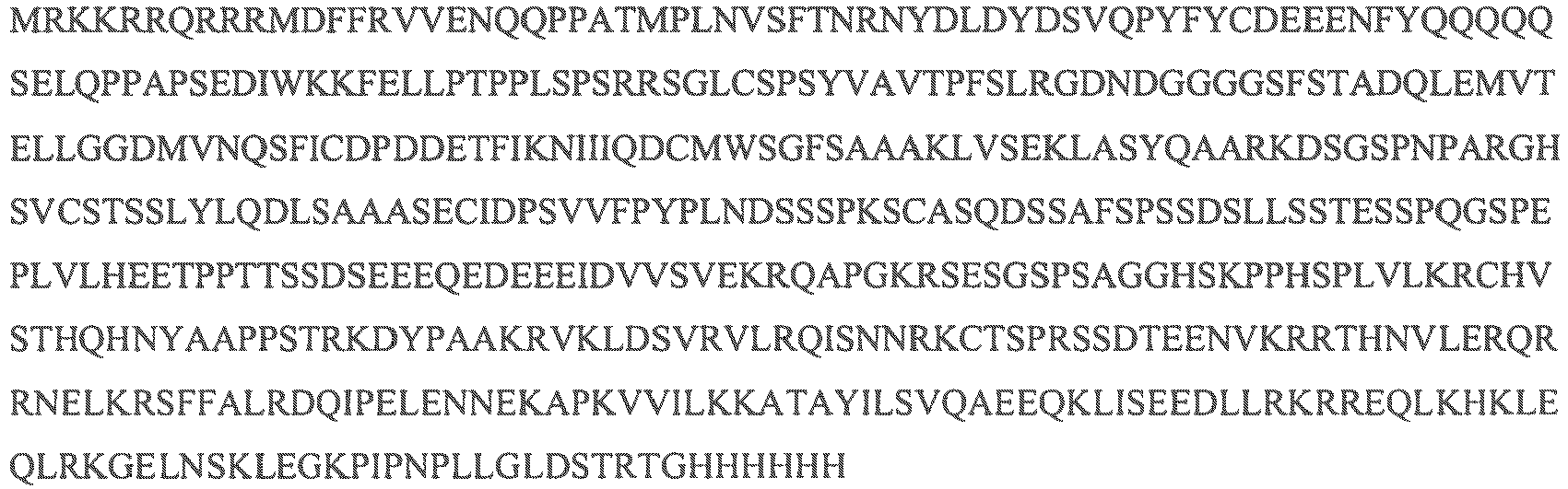
- In some descriptions, a TAT-MYC fusion peptide disclosed herein is constructed by any suitable method. In some descriptions, a nucleotide sequence encoding a TAT-MYC fusion peptide is generated by PCR. In some descriptions, a forward primer for a human MYC sequence comprises an in frame N-terminal 9-amino-acid sequence of the TAT protein transduction domain (i.e., RKKRRQRRR). In some descriptions, a reverse primer for a human MYC sequence is designed to remove the stop codon. In some descriptions, the PCR product is cloned into any suitable expression vector (hereinafter, p-TAT-MYC). In some descriptions, the expression vector comprises a polyhistidine tag and a V5 tag.
- Described herein are certain recombinant polypeptides. In certain instances, antibodies comprise light and a heavy polypeptide chains. In some descriptions, DNA sequences that encode the light and the heavy chains of the antibody are made, either simultaneously or separately, using reverse transcriptase and DNA polymerase in accordance with suitable methods. In some descriptions, PCR is initiated by consensus constant region primers or by more specific primers based on the published heavy and light chain DNA and amino acid sequences. As discussed above, in some descriptions, PCR is also used to isolate DNA clones encoding the antibody light and heavy chains. In some descriptions, the libraries are screened by consensus primers or larger homologous probes, such as mouse constant region probes.
- In some descriptions, DNA, e.g., plasmid DNA, is isolated from the cell (e.g., a mammalian cell) by any suitable method, restriction mapped and sequenced in accordance with any suitable techniques set forth in detail, e.g., in the foregoing references relating to recombinant DNA techniques. In some descriptions, the DNA is synthetic at any point during the isolation process or subsequent analysis.
- In some descriptions, following manipulation of the isolated genetic material to provide antibodies, or antigen-binding fragments, variants, or derivatives thereof, the polynucleotides encoding the antibodies are inserted in an expression vector for introduction into host cells which are therein used to produce the desired quantity of antibody.
- In certain descriptions, recombinant expression of an antibody, or fragment, derivative or analog thereof, e.g., a heavy or light chain of an antibody that binds to a target molecule utilizes construction of an expression vector containing a nucleic acid that encodes the antibody. Once a polynucleotide encoding an antibody molecule or a heavy or light chain of an antibody, or portion thereof (preferably containing the heavy or light chain variable domain), is obtained, the vector for the production of the antibody molecule is in some descriptions, produced by recombinant DNA technology using any suitable techniques. Thus, processes for preparing a protein by expressing a polynucleotide containing an antibody encoding nucleotide sequence are described herein. In some descriptions, processes any suitable manner of construct expression vectors containing antibody coding sequences comprising appropriate transcriptional and translational control signals is utilized in the a method disclosed herein. These processes include, for example, in vitro recombinant DNA techniques, synthetic techniques, and in vivo genetic recombination. Thus, described herein are replicable vectors comprising a nucleotide sequence encoding an antibody molecule, or a heavy or light chain thereof, or a heavy or light chain variable domain, operably linked to a promoter. In some descriptions, vectors comprise the nucleotide sequence encoding the constant region of the antibody and the variable domain of the antibody is incorporated into the vector for expression of the entire heavy or light chain.
- In some descriptions, the cloned variable region genes are inserted into an expression vector along with the heavy and light chain constant region genes synthesized as discussed above. In some descriptions, an expression vector that is capable of eliciting expression in eukaryotic cells is used. Examples of suitable vectors include, but are not limited to plasmids pcDNA3, pHCMV/Zeo, pCR3.1, pEF1/His, pIND/GS, pRc/HCMV2, pSV40/Zeo2, pTRACER-HCMV, pUB6/V5-His, pVAX1, and pZeoSV2 (available from Invitrogen, San Diego, Calif.), and plasmid pC1 (available from Promega, Madison, Wis.). In certain instances, screening large numbers of transformed cells for those that express suitably high levels of immunoglobulin heavy and light chains is accomplished by any suitable method, for example, by robotic systems.
- In some descriptions, once the vector or DNA sequence encoding an antibody is prepared, the expression vector is introduced into an appropriate host cell as disclosed herein. Introduction of the plasmid into the host cell is accomplished by any suitable method. These include, but are not limited to, transfection (including electroporation), protoplast fusion, calcium phosphate precipitation, cell fusion with enveloped DNA, DEAE-dextran mediated transfection, microinjection, cationic lipid-mediated transfection, transduction, scrape loading, ballistic introduction and infection with intact virus. See, e.g., Ridgway (1988) "Mammalian Expression Vectors" in Vectors, ed. Rodriguez and Denhardt (Butterworths, Boston, Mass.), Chapter 24.2, pp. 470-472; Sambrook et al. (1989) Molecular Cloning: A Laboratory Manual, 2nd Ed., Cold Spring Harbor Laboratory Press, Cold Spring Harbor, N.Y. In certain instances, host cells harboring the expression construct are grown under conditions appropriate to the production of the light chains and heavy chains, and assayed for heavy and/or light chain protein synthesis. Exemplary assay techniques include ELISA, radioimmunoassay (RIA), or fluorescence-activated cell sorter analysis (FACS), immunohistochemistry and the like.
- As disclosed herein, introducing (or otherwise providing to) nucleic acids and/or vectors into host organisms are achieved by any suitable method. Examples of such processes include but are not limited to gene gun techniques, naked DNA immunization, the development of transgenic or transchromosomal organisms, lentiviral mediated production of transgenic organisms, vaccinia viruses transduction and adenovirus.
- As disclosed herein, introducing (or otherwise providing to) antigens into organisms or cells comprises any manner suitable. Some examples of such processes include but are not limited to the use of Tat fusion peptides, various immunization protocols, infection by virus, and introduction of whole cells expressing antigen.
- In some descriptions, an expression vector is transferred to a host cell by any suitable method, and the transfected cells are then cultured by conventional techniques or techniques disclosed herein to produce an antibody for use in the methods described herein. Thus, described herein are host cells that contain a polynucleotide encoding an antibody, or a heavy or light chain thereof, operably linked to a heterologous promoter. In some descriptions, for the expression of double-chained antibodies, vectors encoding both the heavy and light chains are used to express of the entire immunoglobulin molecule.
- In some descriptions, a number of selection systems are used including, but not limited to, the herpes simplex virus thymidine kinase (Wigler et al. (1977) Cell 11:223), hypoxanthine-guanine phosphoribosyltransferase (Szybalska and Szybalski (1992) Proc. Natl. Acad. Sci. USA 48:202), and adenine phosphoribosyltransferase (Lowy et al. (1980) Cell 22:817) genes employed in tk-, hgprt- or aprt-cells, respectively. Also, in some descriptions, antimetabolite resistance is used as the basis of selection for the following genes: dhfr, which confers resistance to methotrexate (Wigler et al. (1980) Natl. Acad. Sci. USA 77:357; O'Hare et al. (1981) Proc. Natl. Acad. Sci. USA 78:1527); gpt, which confers resistance to mycophenolic acid (Mulligan and Berg (1981) Proc. Natl. Acad. Sci. USA 78:2072); neo, which confers resistance to the aminoglycoside G-418 Clinical Pharmacy 12:488-505; Wu and Wu (1991) Biotherapy 3:87-95; Tolstoshev (1993) Ann. Rev. Pharmacol. Toxicol. 32:573-596; Mulligan (1993) Science 260:926-932; and Morgan and Anderson (1993) Ann. Rev. Biochem. 62:191-217 (1993); TIB TECH 11(5):155-215 (May, 1993); and hygro, which confers resistance to hygromycin (Santerre et al. (1984) Gene 30:147. In some descriptions, methods commonly known in the art of recombinant DNA technology which are used as described in Ausubel et al. (1993) Current Protocols in Molecular Biology (John Wiley & Sons, NY); Kriegler (1990) "Gene Transfer and Expression" in A Laboratory Manual (Stockton Press, NY); Dracopoli et al. (eds) (1994) Current Protocols in Human Genetics (John Wiley & Sons, N.Y.) Chapters 12 and 13; Colberre-Garapin et al. (1981) J. Mol. Biol. 150:1.
- In some descriptions, the expression level of an antibody molecule is increased by vector amplification (for a review, see Bebbington and Hentschel (1987) "The Use of Vectors Based on Gene Amplification for the Expression of Cloned Genes in Mammalian Cells in DNA Cloning" (Academic Press, NY) Vol. 3. When a marker in the vector system expressing antibody is amplifiable, an increase in the level of inhibitor present in the culture of the host cell will increase the number of copies of the marker gene. Since the amplified region is associated with the antibody gene, production of the antibody will also increase (Crouse et al. (1983) Mol. Cell. Biol. 3:257).
- In various descriptions, of any of the methods disclosed herein, antibodies that specifically binds to the selected antigen, as well as the cell (e.g., a mammalian cell) producing such antibodies, are isolated (i.e., harvested) directly from the tissues (spleen, lymph nodes, blood, etc.) or serum of an organism (e.g., mouse, rabbit). In some descriptions, the antibodies are isolated from splenic tissue. In some descriptions, the antibodies are harvested from lymph nodes. In some descriptions, the antibodies are harvested from blood. In some descriptions, the tissue is harvested by any suitable method.
- In some descriptions, the antibodies are harvested in the presence of the selected antigen. In some descriptions, the antibodies are harvested in the presence of an onco-peptide (e.g., Myc, TAT-Myc). In some descriptions, the antibodies are harvested in the presence of an onco-peptide (e.g., Myc, TAT-Myc) and the selected antigen. In some descriptions, the antibodies are harvested in the presence of a Myc peptide. In some descriptions, the antibodies are harvested in the presence of a TAT-Myc fusion peptide disclosed herein.
- In some descriptions, harvesting antibodies in the presence of the selected antigen, the selected antigen and an onco-peptide, the selected antigen and Myc, or the selected antigen and TAT-Myc reduces the concentration of tolerant and/or anergic cells. In some descriptions, harvesting antibodies in the presence of the selected antigen and TAT-Myc reduces the concentration of tolerant and/or anergic cells (e.g., B-cells).
- In some descriptions, antibodies produced by a method disclosed herein are reengineered for large scale production. In some descriptions, antibodies produced by a method disclosed herein are reengineered for large scale production for clinical use or for use in a diagnostic assay, e.g., under GMP or cGMP guidelines. In some descriptions, a selected antibody is prepared by:
- a. recovering a nucleic acid encoding the antibody from cell (e.g., a B-cell) that comprises a nucleic acid sequence (e.g., a transgenic sequence) encoding an onco-peptide (e.g., recombinant onco-peptide; e.g., Myc) and/or a recombinant onco-peptide (e.g., recombinant onco-peptide; e.g., Myc);
- b. introducing (or otherwise providing to) the nucleic acid encoding the antibody into a cell that is suitable for large scale production of an antibody.
- In some descriptions, cell that is genetically altered to express an onco-peptide and comprises a nucleic acid encoding the antibody is prepared according to any process described herein. In certain descriptions, the method further comprises selecting an antibody. In some descriptions, the antibody is a human antibody. In certain descriptions, cell expresses human antibodies. In certain descriptions, the method further comprises contacting cell (e.g., B-cell) that comprises a nucleic acid sequence (e.g., a transgenic sequence) encoding an onco-peptide (e.g., recombinant onco-peptide; e.g., Myc) with a selected antibody.
- In some descriptions, the method further comprises culturing the cell (e.g., a mammalian cell) that is suitable for large scale production of an antibody under conditions suitable for production of the antibody. In certain descriptions, the method further comprises recovering a quantity of the antibody, e.g., at least 0.1 kg, at least 0.5 kg, at least 1 kg, at least 10 kg, or at least 100 kg.
- Any suitable cell line for large scale production is used in such processes. Examples of cells or cell lines that are suitable for large scale production include, but are not limited to, Chinese hamster ovary CHO, NSO, BSC-1 cells, LLC-MK cells, CV-1, HeLa, HEK293, VERO, MDBK, MDCK, MDOK, CRFK, RAF, TCMK, LLC-PK, PK15, WI-38, MRC-5, T-FLY, BHK, BRL 3A, HepG2, HT1080 or derivatives thereof.
- In some descriptions, nucleic acids encoding an antibody, produced by the methods described herein, are recovered from a B-cell that is genetically altered to express Myc and are reintroduced into a CHO cell or derivatives thereof (including dhfr- CHO cells used with a DHFR selectable marker), for the purpose of large scale production of the antibody. In some descriptions, nucleic acids encoding the antibody of interest are recovered from a B-cell that is genetically altered to express Myc and are reintroduced into a plant cell that is suitable for large scale production of an antibody. In some descriptions, nucleic acids encoding an antibody, produced by the methods described herein, are recovered from a B-cell that is genetically altered to express Myc and are reintroduced into a plant, part of a plant or a plant seed that is suitable for large scale production of an antibody. In some descriptions, nucleic acids encoding an antibody, produced by the methods described herein, are recovered from a B-cell that is genetically altered to express Myc and are reintroduced into cotton, potato, soybean, corn, maize, tobacco, soybean, alfalfa, or rice for the purpose of large scale production of the antibody. In some descriptions, nucleic acids encoding an antibody, produced by the methods described herein, are recovered from a B-cell that is genetically altered to express Myc and are reintroduced into a yeast for the purpose of large scale production of the antibody. Urlaub and Chasin, (1980) Proc. Natl. Acad. Sci. USA 77:4216-4220 and R. J. Kaufman and P. A. Sharp (1982) Mol. Biol. 159:601-621.
- In vitro production allows scale-up to give large amounts of the desired polypeptides. Techniques for cell (e.g., a mammalian cell) cultivation under tissue culture conditions include homogeneous suspension culture, e.g. in an airlift reactor or in a continuous stirrer reactor, or immobilized or entrapped cell culture, e.g. in hollow fibers, microcapsules, on agarose microbeads or ceramic cartridges. If necessary and/or desired, in some descriptions, solutions of polypeptides are purified in any manner suitable. Examples of such methods include but are not limited to chromatography methods, for example gel filtration, ion-exchange chromatography, chromatography over DEAE-cellulose or (immuno-)affinity chromatography.
- Once an antibody molecule is recombinantly expressed, in some descriptions, the antibody is purified by any suitable method for purification of an immunoglobulin molecule, for example, by chromatography (e.g., ion exchange, affinity, protein A affinity, protein G affinity, gel filtration and sizing column chromatography), centrifugation, differential solubility, or by any other standard technique for the purification of antibodies. In some descriptions, isolated antibodies include serum containing such antibodies, or antibodies that have been purified to varying degrees.
- In some descriptions, the methods disclosed herein are used to generate antibodies that bind and/or are specific to antigens. In some descriptions, the selected antigens are therapeutic targets. In some descriptions, the antibodies are generated to bind an antigen wherein the selected antigen is a TNF or TNF receptor family member or TNF like molecule. In some descriptions, the TNF or TNF receptor family member or TNF like molecule is selected from the list: TNF-alpha, lymphotoxin (LT), LT-alpha (also known as TNF-beta), LT-beta (found in complex heterotrimers with LT-alpha), OPGL, FasL, CD27L, CD30L, CD40L, 4-1BBL, DcR3, OX40, OX40L, TNF-gamma (International Publication No.
WO 96/14328 WO 97/34911 WO 98/07880 WO 98/30694 WO 98/18921 WO 96/34095 WO 97/35904 WO 98/30693 WO 98/30694 WO 98/41629 WO 98/56892 WO 98/54202 WO 98/06842 - In some descriptions, the methods disclosed herein are used to generate antibodies that bind and/or are specific to selected self antigens. In some description, the self antigens originate from within an organism, tissue, or cell. In some descriptions, a self antigen comprises an endogenous antigen. In some descriptions, a self antigen comprises an endogenous antigen produced by an endogenous retrovirus. In some descriptions, self antigens comprise neo-self antigens, microbially or parasite encoded neo-self antigens, or other neo-self antigens expressed as a result of genetic alteration to an organism or cell. In some descriptions, a chimeric mouse expresses a neo-self antigen. In some descriptions, the methods disclosed herein are used to generate antibodies that bind and/or are specific to selected self antigens that antagonize a disease process, In some descriptions, the disease process is an autoimmune disorder. In some descriptions, the methods disclosed herein are used to generate antibodies that bind and/or are specific to selected auto antigens or immunologically reactive epitopes that mimic that of a self antigen or autoantigen.
- In some descriptions, the methods disclosed herein are used to generate antibodies that bind and/or are specific to antigens and antagonize a disease processes. In some descriptions, the selected antigens are antibodies that contribute to the pathogenesis of a disorder or disease process.
- In some descriptions, utilizing the techniques disclosed herein, monoclonal antibodies displaying a multitude of specificities are generated that neutralize entire clades of HIV variants. In some descriptions, this is accomplished by targeting an obligatory structural component of the HIV viral envelope proteins. In some descriptions, this is accomplished by targeting a host co-receptor protein.
- In some descriptions, the selected antigen is an autoimmune antigen. In some descriptions, the autoimmune antigen is selected from the list: thyroglobulin, thyroid peroxidase, cytoplasmic TSH receptor, intrinsic factor, beta-adrenergic receptor, acetyl choline receptor, myelin basic protein, amyloid beta, amyloid precursor protein, collagen, sodium-iodide symporter, histone polypeptides and nucleic acids.
- The technologies disclosed herein provide new strategies for the rapid development of diagnostic and therapeutic antibodies for the detection and treatment of emerging infectious diseases and chronic illnesses such as cancer and autoimmunity. In some descriptions, the antibodies produced by the methods disclosed herein are used for detecting or quantitating various serum proteins for diagnostic purposes. In some descriptions, the antibodies produced by the methods disclosed herein are used for detecting or quantitating various markers for diagnostic purposes. In some descriptions, the markers are cell surface markers. In some descriptions, the markers are cancer or tumor markers.
- In some descriptions, the antibodies produced by the methods disclosed herein are used for therapeutic treatments. In some descriptions, the antibodies produced by the methods disclosed herein are reagents used for diagnostic and/or research purposes.
- In some descriptions, antibodies capable of selectively binding to a wide range of antigens are produced by a method described herein. Any antigen capable of inducing an immune response when introduced to an organism is suitable for use in a method described herein. In some descriptions, antigens that are normally subjected to self tolerance mechanisms, such as auto antigens, are used in a method described herein. In some descriptions, the selected antigens fail to induce antibodies utilizing standard immunization protocols.
- In some descriptions, a method described herein utilizes any organism suitable for antibody production, including a cell or tissue thereof. In some descriptions, organisms suitable for antibody production include any animal of the Vertebrate class, Mammalia (i.e., mammals), including, and without limitation, primates, rodents, livestock and domestic pets. In certain instances, in the production of an antibody, a suitable experimental animal, such as, for example, but not limited to, a rabbit, a sheep, a hamster, a guinea pig, a mouse, a rat, or a chicken, is administered (i.e., immunized with, inoculated against) an antigen against which an antibody is desired.
- Described herein, are pharmaceutical compositions comprising an antibody and one or more physiologically acceptable carriers. Physiologically acceptable carriers include excipients and auxiliaries which facilitate processing of the active agents into preparations which are used pharmaceutically. In certain instances, proper formulation is dependent upon the route of administration chosen. A summary of pharmaceutical compositions is found, for example, in Remington: The Science and Practice of Pharmacy, Nineteenth Ed (Easton, Pa.: Mack Publishing Company, 1995); Hoover, John E., Remington's Pharmaceutical Sciences, Mack Publishing Co., Easton, Pennsylvania 1975; Liberman, H.A. and Lachman, L., Eds., Pharmaceutical Dosage Forms, Marcel Decker, New York, N.Y., 1980; and Pharmaceutical Dosage Forms and Drug Delivery Systems, Seventh Ed. (Lippincott Williams & Wilkins, 1999).
- Described herein are pharmaceutical compositions that include an antibody and a pharmaceutically acceptable diluent(s), excipient(s), or carrier(s). In addition, an antibody is optionally administered as pharmaceutical compositions in which they are mixed with other active ingredients, as in combination therapy. In some descriptions, the pharmaceutical compositions includes other medicinal or pharmaceutical agents, carriers, adjuvants, such as preserving, stabilizing, wetting or emulsifying agents, solution promoters, salts for regulating the osmotic pressure, and/or buffers. In addition, the pharmaceutical compositions also contain other therapeutically valuable substances.
- A pharmaceutical composition refers to a mixture of an antibody with other chemical components, such as carriers, stabilizers, diluents, dispersing agents, suspending agents, thickening agents, and/or excipients. The pharmaceutical composition facilitates administration of an antibody to an organism. In practicing the methods of treatment or use described herein, therapeutically effective amounts of an antibody are administered in a pharmaceutical composition to an organism (e.g., a mammal) having a condition, disease, or disorder to be treated. Preferably, the organism (e.g., a mammal) is a human. A therapeutically effective amount varies depending on the severity and stage of the condition, the age and relative health of the subject, the potency of the antibody used and other factors. Antibodies are optionally used singly or in combination with one or more therapeutic agents as components of mixtures.
- The pharmaceutical formulations described herein are optionally administered to a subject by multiple administration routes, including but not limited to, oral, parenteral (e.g., intravenous, subcutaneous, intramuscular), intranasal, buccal, topical, rectal, or transdermal administration routes. The pharmaceutical formulations described herein include, but are not limited to, aqueous liquid dispersions, self-emulsifying dispersions, solid solutions, liposomal dispersions, aerosols, solid dosage forms, powders, immediate release formulations, controlled release formulations, fast melt formulations, tablets, capsules, pills, delayed release formulations, extended release formulations, pulsatile release formulations, multiparticulate formulations, and mixed immediate and controlled release formulations.
- An antibody is optionally used in the preparation of medicaments for the prophylactic and/or therapeutic treatment of inflammatory conditions or conditions that would benefit, at least in part, from amelioration. In addition, a method for treating any of the diseases or conditions described herein in a subject in need of such treatment, involves administration of pharmaceutical compositions containing an antibody as described herein, or a pharmaceutically acceptable salt, pharmaceutically acceptable N-oxide, pharmaceutically active metabolite, pharmaceutically acceptable prodrug, or pharmaceutically acceptable solvate thereof, in therapeutically effective amounts to said subject.
- Furthermore, described herein is a method of treating a disorder mediated by an antigen by administering a therapeutically effective antibody that binds or specifically binds to the selected antigen to an individual in need thereof. Exemplary antigens are set forth herein and include, by way of non-limiting example, viral antigens (e.g., HIV antigens, influenza antigens, SARS antigens), autoantigens, tumor antigens, various pathogen related antigens and the like. Likewise, disorders mediated by the selected antigen include any disorder so mediated, including, by way of non-limiting example, viral infections and syndromes caused thereby (e.g., HIV, influenza, and SARS), as well as pathogenic and parasitic infections and syndromes caused thereby.
- In certain instances wherein the patient's condition does not improve, upon the doctor's discretion the administration of an antibody is optionally administered chronically, that is, for an extended period of time, including throughout the duration of the patient's life in order to ameliorate or otherwise control or limit the symptoms of the patient's disease or condition.
- In the case wherein the patient's status does improve, upon the doctor's discretion the administration of an antibody is optionally given continuously; alternatively, the dose of drug being administered is temporarily reduced or temporarily suspended for a certain length of time (i.e., a "drug holiday"). The length of the drug holiday optionally varies between 2 days and 1 year, including by way of example only, 2 days, 3 days, 4 days, 5 days, 6 days, 7 days, 10 days, 12 days, 15 days, 20 days, 28 days, 35 days, 50 days, 70 days, 100 days, 120 days, 150 days, 180 days, 200 days, 250 days, 280 days, 300 days, 320 days, 350 days, or 365 days. The dose reduction during a drug holiday includes from 10%-100%, including, by way of example only, 10%, 15%, 20%, 25%, 30%, 35%, 40%, 45%, 50%, 55%, 60%, 65%, 70%, 75%, 80%, 85%, 90%, 95%, or 100%.
- Once improvement of the patient's conditions has occurred, a maintenance dose is administered if necessary. Subsequently, the dosage or the frequency of administration, or both, is reduced, as a function of the symptoms, to a level at which the improved disease, disorder or condition is retained. In some descriptions, patients require intermittent treatment on a long-term basis upon any recurrence of symptoms.
- In some descriptions, the pharmaceutical composition described herein is in unit dosage forms suitable for single administration of precise dosages. In unit dosage form, the formulation is divided into unit doses containing appropriate quantities of an antibody. In some descriptions, the unit dosage is in the form of a package containing discrete quantities of the formulation. Non-limiting examples are packaged tablets or capsules, and powders in vials or ampoules. In some descriptions, aqueous suspension compositions are packaged in single-dose non-reclosable containers. Alternatively, multiple-dose reclosable containers are used, in which case it is typical to include a preservative in the composition. By way of example only, formulations for parenteral injection are presented in unit dosage form, which include, but are not limited to ampoules, or in multi dose containers, with an added preservative.
- The daily dosages appropriate for an antibody are from about 0.01 to 2.5 mg/kg per body weight. An indicated daily dosage in the larger organism (e.g., a mammal), including, but not limited to, humans, is in the range from about 0.5 mg to about 100 mg, conveniently administered in divided doses, including, but not limited to, up to four times a day or in extended release form. Suitable unit dosage forms for oral administration include from about 1 to 50 mg active ingredient. The foregoing ranges are merely suggestive, as the number of variables in regard to an individual treatment regime is large, and considerable excursions from these recommended values are not uncommon. Such dosages are optionally altered depending on a number of variables, not limited to the activity of the antibody used, the disease or condition to be treated, the mode of administration, the requirements of the individual subject, the severity of the disease or condition being treated, and the judgment of the practitioner.
- Toxicity and therapeutic efficacy of such therapeutic regimens are optionally determined in cell cultures or experimental organisms, including, but not limited to, the determination of the LD50 (the dose lethal to 50% of the population) and the ED50 (the dose therapeutically effective in 50% of the population). The dose ratio between the toxic and therapeutic effects is the therapeutic index, which is expressed as the ratio between LD50 and ED50. An antibody exhibiting high therapeutic indices is preferred. The data obtained from cell culture assays and animal studies are optionally used in formulating a range of dosage for use in human. The dosage of such an antibody lies preferably within a range of circulating concentrations that include the ED50 with minimal toxicity. The dosage optionally varies within this range depending upon the dosage form employed and the route of administration utilized.
- Antibody compositions described herein are also optionally used in combination with other therapeutic reagents that are selected for their therapeutic value for the condition to be treated. In general, the compositions described herein and, in descriptions, where combinational therapy is employed, other agents do not have to be administered in the same pharmaceutical composition, and, because of different physical and chemical characteristics, are optionally administered by different routes. The initial administration is optionally made according to established protocols, and then, based upon the observed effects, the dosage, modes of administration and times of administration subsequently modified.
- In certain instances, it is appropriate to administer an antibody composition as described herein in combination with another therapeutic agent. By way of example only, if one of the side effects experienced by a patient upon receiving an antibody composition as described herein is nausea, then it is appropriate to administer an anti-nausea agent in combination with the initial therapeutic agent. Or, by way of example only, the therapeutic effectiveness of an antibody are enhanced by administration of an adjuvant (i.e., by itself the adjuvant has minimal therapeutic benefit, but in combination with another therapeutic agent, the overall therapeutic benefit to the patient is enhanced). Or, by way of example only, the benefit experienced by a patient is increased by administering an antibody with another therapeutic agent (which also includes a therapeutic regimen) that also has therapeutic benefit. In any case, regardless of the disease, disorder or condition being treated, the overall benefit experienced by the patient is either simply additive of the two therapeutic agents or the patient experiences a synergistic benefit.
- Therapeutically-effective dosages vary when the drugs are used in treatment combinations. Methods for experimentally determining therapeutically-effective dosages of drugs and other agents for use in combination treatment regimens are documented methodologies. One example of such a method is the use of metronomic dosing, i.e., providing more frequent, lower doses in order to minimize toxic side effects. Combination treatment further includes periodic treatments that start and stop at various times to assist with the clinical management of the patient.
- In any case, the multiple therapeutic agents (one of which is an antibody as described herein) are administered in any order, or even simultaneously. If simultaneously, the multiple therapeutic agents are optionally provided in a single, unified form, or in multiple forms (by way of example only, either as a single pill or as two separate pills). In some descriptions, one of the therapeutic agents is given in multiple doses, or both are given as multiple doses. If not simultaneous, the timing between the multiple doses optionally varies from more than zero weeks to less than four weeks. In addition, the combination methods, compositions and formulations are not to be limited to the use of only two agents; the use of multiple therapeutic combinations is also envisioned.
- It is understood that the dosage regimen to treat, prevent, or ameliorate the condition(s) for which relief is sought, is optionally modified in accordance with a variety of factors. These factors include the disorder from which the subject suffers, as well as the age, weight, sex, diet, and medical condition of the subject. Thus, the dosage regimen actually employed varies widely, in some descriptions, and therefore deviates from the dosage regimens set forth herein.
- The pharmaceutical agents which make up the combination therapy disclosed herein are optionally a combined dosage form or in separate dosage forms intended for substantially simultaneous administration. The pharmaceutical agents that make up the combination therapy are optionally also be administered sequentially, with either therapeutic agent being administered by a regimen calling for two-step administration. The two-step administration regimen optionally calls for sequential administration of the active agents or spaced-apart administration of the separate active agents. The time period between the multiple administration steps ranges from, a few minutes to several hours, depending upon the properties of each pharmaceutical agent, such as potency, solubility, bioavailability, plasma half-life and kinetic profile of the pharmaceutical agent. Circadian variation of the target molecule concentrations are optionally used to determine the optimal dose interval.
- In addition, an antibody is optionally used in combination with procedures that provide additional or synergistic benefit to the patient. By way of example only, patients are expected to find therapeutic and/or prophylactic benefit in the methods described herein, wherein pharmaceutical compositions of an antibody and /or combinations with other therapeutics are combined with genetic testing to determine whether that individual is a carrier of a mutant gene that is correlated with certain diseases or conditions.
- An antibody and the additional therapy(ies) are optionally administered before, during or after the occurrence of a disease or condition, and the timing of administering the composition containing an antibody varies in some descriptions,. Thus, for example, an antibody is used as a prophylactic and is administered continuously to subjects with a propensity to develop conditions or diseases in order to prevent the occurrence of the disease or condition. An antibody and compositions are optionally administered to a subject during or as soon as possible after the onset of the symptoms. The administration of the agents are optionally initiated within the first 48 hours of the onset of the symptoms, preferably within the first 48 hours of the onset of the symptoms, more preferably within the first 6 hours of the onset of the symptoms, and most preferably within 3 hours of the onset of the symptoms. The initial administration is optionally via any route practical, such as, for example, an intravenous injection, a bolus injection, infusion over 5 minutes to about 5 hours, a pill, a capsule, transdermal patch, buccal delivery, and the like, or combination thereof. An antibody is preferably administered as soon as is practicable after the onset of a disease or condition is detected or suspected, and for a length of time necessary for the treatment of the disease, such as, for example, from about 1 month to about 3 months. The length of treatment optionally varies for each subject, and the length is then determined using the known criteria. For example, an antibody or a formulation containing an antibody are administered for at least 2 weeks, preferably about 1 month to about 5 years, and more preferably from about 1 month to about 3 years.
- The following example demonstrates the production of B-cell lines derived from MMTV-tTA/TRE-MYC mice.
- MMTV-tTA/TRE-MYC mice are maintained on doxycycline for eight weeks after birth and then are switched to a normal diet. The mice develop an externally evident lymphadenopathy and splenomegaly, and present with a number of the clinical signs that are consistently seen associated with lymphoid neoplasia (scruffy fur, hunched posture, labored breathing, anemia, organomegaly, etc.). The mice are euthanized and their lymph nodes and spleens are collected for analysis. Single cell suspensions are generated from some of the lymph nodes and a portion of the spleen. Those cells are used for flow cytometric analyses. The initial characterization of the tumors demonstrated the high prevalence of activated B-cells. Some of the same cells are used to seed cultures to generate B-cell lines. These cells are cultured in lymphocyte media (
RPMI 1640, 10% Fetal Calf Serum, penicillin/streptomycin, L-glutamine, HEPES, non-essential amino acids, sodium pyruvate and 2-β-mercaptoethanol). Approximately 14-21 days later, some of the wells began to exhibit clonal outgrowth of cell lines. The cells are carefully expanded until they were adapted to growth in large flasks. A portion of the cells are cryopreserved in 10% DMSO. - Two cell lines were initially picked, designated TBLK6 and TBLK7. These cell lines exhibited different surface expression levels of CD138. The levels of immunoglobulin secretion into the tissue culture medium is measured after seeding. Both cell lines spontaneously secreted immunoglobulin into the growth medium. The levels of secretion are increased by the addition of IL-4 and IL-6 into the initial innoculum. Immunoglobulins secreted by both TBLK6 and TBLK7 are shown to be IgM. In some descriptions, single cells are cloned from both cell lines in order to generate true monoclonal populations. A number of MMTV-tTA/TRE-MYC bigenic mice are generated that, in some descriptions, are used for isolating the
AN 1/T3 population by cell sorting. - The following example demonstrates that antibodies produced in Myc-over-expressing mice function in vivo.
- A model of lethal viral infection is used. Porcine Rabies Virus (PRV) is a member of the alpha-herpes viruses that has been previously shown to be lethal in mice following intravenous administration. Two variants of PRV are constructed. In one instance, one of the sequences for a gene called US-9 was fused to GFP. This construct allowed for tracking of the virus and virally infected cells in a different setting. US-9 protein expression is confirmed and US-9 retained its function as the fusion protein. The virus is fully pathogenic in spite of its additional genetic cargo. A variant of PRV that encodes the open reading frame for HEL is also generated. This viral variant is shown to express HEL by Western blot analysis of infected cells.
- GFP-expressing virus or the HEL-expressing virus innocula are incubated with HEL-specific antibodies diluted 1:500 for one hour, on ice. The mixtures are then injected intravenously into groups of four mice. The mice are then monitored for four days following administration of the virus and antibody mixtures.
- As shown in
Figure 7 , the GFP-expressing virus is not affected by the presence of HEL-specific antibodies. The kinetics of mortality in that group is comparable with our previous experience with wild type PRV strains. In contrast, the kinetics of mortality in mice that received the HEL-expressing virus is significantly delayed and that group of mice lived for almost twice as long as the mice injected with the GFP-expressing virus. The viruses used for these experiments only expressed the US-9 fusion protein transiently. After entry into cells, they produce wild type PRV. - In this example, Myc-ER is used for the conditional immortalization of long term hematopoietic stem cells. A group of bone marrow chimeric mice are generated using 5FU enriched bone marrow derived stem cells, as follows. For bone marrow derived hematopoietic stem cells, 5mg/mouse of 5-fluorouracil (5FU) is administered intravenously, in order to enrich for long-term hematopoietic stem cells, and induce HSC proliferation in vivo. The bone marrow cells are collected from the femurs and
tibia bones 5 days later. The red blood cells are lysed, using a hypotonic lysis buffer. The remaining cells are washed twice in media and plated at a concentration of 2x10⁶ cells/ml, in a 24 well plate, in DMEM media supplemented with 15%, heat inactivated fetal calf serum, penicillin/streptomycin, L-glutamine, Non-essential amino acids, recombinant human IL-3, IL-6 and Stem Cell Factor (SCF). The cells are cultured for 24 hours prior to the first spin infection, and are subject to theinfection procedure 3 times, every 24 hours. A day after the last spin infection, the cells are analyzed by flow cytometry. Lentivirally transduced bone marrow derived hematopoietic stem cells are reconstituted to lethally irradiated mice, and are checked for GFP expression in lymphoid organs 12 weeks later. In this instance, hematopoietic stem cells are allowed to reconstitute a normal peripheral lymphoid compartment for 8-12 weeks after bone marrow transplantation. The splenic, GFP+ AN-1/T3 cells are isolated from those mice and are used for in vitro immortalization protocols, as described above. The key difference is that instead of withdrawing doxycycline from the system, 10nM 4-hydroxytamoxifen (4OHT) is added to the medium. In some descriptions, sorted, GFP+ AN-1/T3 cells are adoptively transferred into cohorts of wild type recipient mice that are treated once weekly with 1mg/mouse of 4OHT, intraperitoneally. The mice are monitored daily for the appearance of clinical signs associated with the development of B-cell lymphomas. The resulting tumors are collected and used for generating B-cell lines as described herein, using 4OHT instead of doxycycline as the regulator of Myc function. - In this example, Myc-ER and Bcl-2 are used for the conditional immortalization of long term hematopoietic stem cells. A group of bone marrow chimeric mice are generated using 5FU enriched bone marrow derived stem cells, as follows. For bone marrow derived hematopoietic stem cells, 5mg/mouse of 5-fluorouracil (5FU) is administered intravenously, in order to enrich for long-term hematopoietic stem cells, and induce HSC proliferation in vivo. The bone marrow cells are collected from the femurs and
tibia bones 5 days later. The red blood cells are lysed, using a hypotonic lysis buffer. The remaining cells are washed twice in media and plated at a concentration of 2x10⁶ cells/ml, in a 24 well plate, in DMEM media supplemented with 15% heat inactivated fetal calf serum, penicillin/streptomycin, L-glutamine, Non-essential amino acids, recombinant human IL-3, IL-6 and Stem Cell Factor (SCF). The cells are cultured for 24 hours prior to the first spin infection with a GFP expressing lentivirus encoding the expression of Myc-ER and Bcl-2. The cells are subject to theinfection procedure 3 times, every 24 hours. A day after the last spin infection, the cells are analyzed by flow cytometry. Lentivirally transduced bone marrow derived hematopoietic stem cells are reconstituted to lethally irradiated mice. In this instance, hematopoietic stem cells are allowed to reconstitute a normal peripheral lymphoid compartment for 8-12 weeks after bone marrow transplantation. The splenic, GFP+ AN-1/T3 cells are isolated from those mice and are used for in vitro immortalization protocols, as described above. 4OHT is added to the medium to induce Myc activation. In some descriptions, sorted, GFP+ AN-1/T3 cells are adoptively transferred into cohorts of wild type recipient mice that are treated once weekly with 1mg/mouse of 4OHT, intraperitoneally. The mice are monitored daily for the appearance of clinical signs associated with the development of B-cell lymphomas. The resulting tumors are collected and used for generating B-cell lines as described herein, using 4OHT instead of doxycycline as the regulator of Myc function. - In order to test the notion of harnessing the MMTV-tTA/TRE-MYC mice to generate novel antibodies to antigens of interest through the introduction into the system as neo-self antigens, a retroviral bone marrow chimeric mice was used. A variant of pMIG that encodes the cDNA for H5 hemagglutinin from a highly pathogenic avian influenza virus, A/Ty/Ont/7732/66 (H5N9) was first generated. HSCs enriched from the bone marrows of mice that had been treated with 5-
fluorouracil 5 days prior to collection, were then obtained as previously described. The donor cells were obtained from TRE-MYC mice. The cells were transduced with one retrovirus that encodes the tetracycline transactivator protein (tTA) as well as pMIG-H5. The transduced bone marrow HSCs were used to reconstitute lethally irradiated mice. The bone marrow chimeric mice were maintained on SEPTRA and observed daily for externally evident clinical signs of hematological malignancies (scruffy fur, labored breathing, externally evident lymphadenopathy or splenomegaly, hunched posture, dehydration, and hind limb paralysis). A cohort of 5 mice presented with clinical signs of hematological malignancies in a 7-day period, starting 6 weeks after transplantation. The lymph nodes and spleens were collected from euthanized mice and used to generate single cell suspensions. The remaining cells were either cultured to begin to generate cell lines, or cryopreserved for subsequent analysis. A small fraction of the cells were stained for cell surface markers and analyzed by flow cytometry (Figure 8). Figure 8 shows the surface profile of the cells isolated from the bone marrow chimeric mice that had developed a hematological malignancy. These are uniformly B220+/IgM+, suggesting that the mice had developed a B-cell leukemia/lymphoma composed of mature B-cells. These cells were placed in culture and clonally expanded populations were passed starting 8 days after initial seeding. This is a significantly faster timeline than what is normally achieved with current approaches to monoclonal antibody production. In addition, the accelerated time frame by which this novel approach has allowed us to generate novel antibodies to hemagglutinin should render this approach as a rapid response platform for the development of novel neutralizing antibodies to new and emerging infectious diseases and other biological threats. - The serum from these mice was collected to test for the presence of antibodies to H5. Immunoblot analysis with the serum from these mice showed specific reactivity to the HA1 subunit of the H5 HA (
Figure 9 ). Cell lysates were obtained from 293FT cells that had been transiently transfected with two different expression plasmids encoding H5 (pCDNA3-H5 and pMIG-H5). The blots were probed with sera obtained from leukemic mice. The banding pattern that developed was consistent with H5 and the cleavage products that develop during the normal maturation and processing of HA in a cell. The mature HA is composed of two subunits the HA1 and the HA2. The HA1 subunit (-40 kDa) forms the globular head of the molecule and is responsible for binding to the host cell sialic acid receptors on surface glycoproteins and glycolipids. - The HA2 (-20 kDa) subunit is responsible for fusion of the viral envelope with the endosomal membranes of the host cell during entry. A protease cleavage site separates these two subunits and cleavage by host protease is required for entry into the host cell. The vast majority of virus neutralizing epitopes are found in the HA1 subunit 22 and inhibit virus-receptor interaction.
- Heamagglutination inhibition analysis of mouse serum obtained from retroviral chimeric mice was performed to test the ability of the sera to block virus-receptor interaction with a variety influenza A isolates including H5N2, H1N1, H7N2, H3N2, and H6N8 subtypes (
Figure 10 ). - Mice (n=20) are divided into two groups. The first group (n=10) are immunized with HEL mixed with Freund's adjuvant. The second group (n=10) are immunized with HEL mixed with Tat-Myc.
- Both groups receive a booster shot of the HEL antigen mixed with Incomplete Freund's Adjuvant.
- Sera is collected from each mouse at
days - On day 35, spleens are harvested from each mouse. The spleens are processed into single cell suspension. Red blood cells (RBCs) are removed with lysis buffer. Lymphocytes are divided into two samples. The first sample is incubated with Tat-Myc and HEL antigen. The second sample is incubated with TAT-MYC.
- Mice (n=20) are divided into two groups. The first group (n=10) are immunized with Fluvirin (2007-2008) mixed with Freund's adjuvant. The second group (n=10) are immunized with Fluvirin (2007-2008) mixed with Tat-Myc.
- Both groups receive a booster shot of the Fluvirin (2007-2008) mixed with Incomplete Freund's Adjuvant (IFA).
- Sera is collected from each mouse at
days - On day 35, spleens are harvested from each mouse. The spleens are processed into single cell suspension. Red blood cells (RBCs) are removed with lysis buffer. Lymphocytes are divided into two samples. The first sample is incubated with Tat-Myc and Fluvirin (2007-2008). The second sample is incubated with TAT-MYC.
Claims (3)
- A fusion peptide comprising:(a) an HIV TAT sequence of SEQ ID NO: 6;(b) a MYC polypeptide sequence that is at least 95% identical to the sequence of NCBI Accession Number NP002458.2; and(c) at least two protein tags, wherein the protein tags comprise a 6-histidine tag and a V5 epitope tag comprising the sequence GKPIPNPLLGLDST,wherein the fusion peptide promotes cell survival and/or proliferation.
- The fusion peptide of claim 1, wherein the MYC sequence is 100% identical to the sequence of NCBI Accession Number NP002458.2.
- The fusion peptide of claim 1, wherein the fusion peptide has the following sequence:
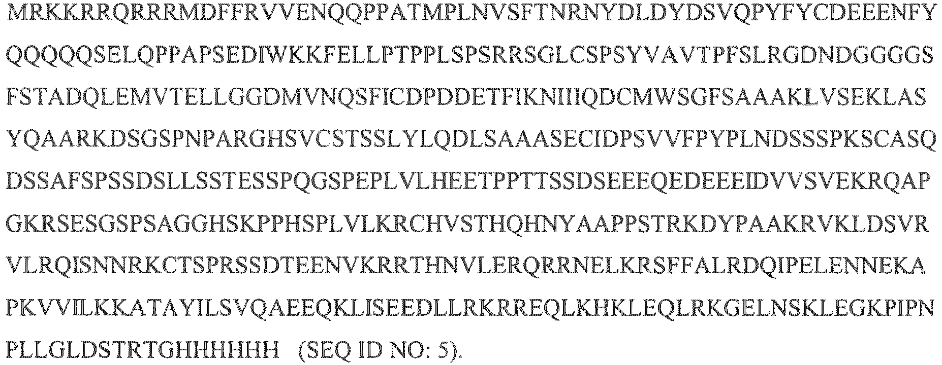
Applications Claiming Priority (2)
| Application Number | Priority Date | Filing Date | Title |
|---|---|---|---|
| US5404708P | 2008-05-16 | 2008-05-16 | |
| PCT/US2009/003105 WO2009139930A2 (en) | 2008-05-16 | 2009-05-18 | Antibodies and processes for preparing the same |
Publications (3)
| Publication Number | Publication Date |
|---|---|
| EP2285832A2 EP2285832A2 (en) | 2011-02-23 |
| EP2285832A4 EP2285832A4 (en) | 2012-06-27 |
| EP2285832B1 true EP2285832B1 (en) | 2020-08-26 |
Family
ID=41319226
Family Applications (1)
| Application Number | Title | Priority Date | Filing Date |
|---|---|---|---|
| EP09747016.5A Active EP2285832B1 (en) | 2008-05-16 | 2009-05-18 | Antibodies and processes for preparing the same |
Country Status (10)
| Country | Link |
|---|---|
| US (5) | US8986702B2 (en) |
| EP (1) | EP2285832B1 (en) |
| KR (2) | KR101803099B1 (en) |
| AU (1) | AU2009246876B2 (en) |
| CA (1) | CA2723114C (en) |
| EA (1) | EA021131B1 (en) |
| ES (1) | ES2820873T3 (en) |
| IL (3) | IL287311B2 (en) |
| SG (2) | SG10201803982TA (en) |
| WO (1) | WO2009139930A2 (en) |
Families Citing this family (14)
| Publication number | Priority date | Publication date | Assignee | Title |
|---|---|---|---|---|
| ATE491022T1 (en) | 2005-10-18 | 2010-12-15 | Nat Jewish Health | CONDITIONALLY IMMORTALIZED ADULT LONG-TERM STEM CELLS AND METHOD FOR THE PRODUCTION AND USE OF SUCH CELLS |
| EP2583550A1 (en) | 2007-03-13 | 2013-04-24 | National Jewish Health | Methods for generation of antibodies |
| EP2285832B1 (en) | 2008-05-16 | 2020-08-26 | Taiga Biotechnologies, Inc. | Antibodies and processes for preparing the same |
| DK2313496T3 (en) * | 2008-07-21 | 2014-12-15 | Taiga Biotechnologies Inc | Anukleære differentiated cells, and method for preparation thereof |
| ES2879225T3 (en) * | 2008-08-28 | 2021-11-22 | Taiga Biotechnologies Inc | MYC modulators, methods of using them, and methods to identify agents that modulate MYC |
| AU2012216462B2 (en) * | 2008-08-28 | 2015-04-23 | Htyr Acquisition Llc | Modulators of MYC, methods of using the same, and methods of identifying agents that modulate MYC |
| EP2877189B1 (en) | 2012-07-20 | 2021-01-06 | Taiga Biotechnologies, Inc. | Enhanced reconstitution and autoreconstitution of the hematopoietic compartment |
| US10272115B2 (en) | 2013-03-11 | 2019-04-30 | Taiga Biotechnologies, Inc. | Production and use of red blood cells |
| US9365825B2 (en) | 2013-03-11 | 2016-06-14 | Taiga Biotechnologies, Inc. | Expansion of adult stem cells in vitro |
| AU2017367730A1 (en) | 2016-12-02 | 2019-06-06 | Taiga Biotechnologies, Inc. | Nanoparticle formulations |
| EP4026554A1 (en) * | 2017-08-03 | 2022-07-13 | Taiga Biotechnologies, Inc. | Methods and compositions for the treatment of melanoma |
| US10149898B2 (en) | 2017-08-03 | 2018-12-11 | Taiga Biotechnologies, Inc. | Methods and compositions for the treatment of melanoma |
| JP2022527117A (en) | 2019-04-08 | 2022-05-30 | タイガ バイオテクノロジーズ,インク. | Compositions and Methods for Cryopreservation of Immune Cells |
| WO2020232141A2 (en) | 2019-05-14 | 2020-11-19 | Taiga Biotechnologies, Inc. | Compositions and methods for treating t cell exhaustion |
Family Cites Families (143)
| Publication number | Priority date | Publication date | Assignee | Title |
|---|---|---|---|---|
| ZA811368B (en) | 1980-03-24 | 1982-04-28 | Genentech Inc | Bacterial polypedtide expression employing tryptophan promoter-operator |
| WO1986003780A1 (en) | 1984-12-21 | 1986-07-03 | Techniclone Research Partners I | Method for electrically immortalizing lymphoid cells |
| DE3531430A1 (en) | 1985-09-03 | 1987-03-05 | Biotest Pharma Gmbh | METHOD FOR OBTAINING COMPONENTS FROM A LIQUID WITH COMPONENTS PRESENT IN AREAS SEPARATELY SEPARATED |
| US4963489A (en) | 1987-04-14 | 1990-10-16 | Marrow-Tech, Inc. | Three-dimensional cell and tissue culture system |
| US4900322A (en) | 1986-09-22 | 1990-02-13 | Adams James D | Blood component pooling valve and kit |
| US5476996A (en) | 1988-06-14 | 1995-12-19 | Lidak Pharmaceuticals | Human immune system in non-human animal |
| US5399493A (en) | 1989-06-15 | 1995-03-21 | The Regents Of The University Of Michigan | Methods and compositions for the optimization of human hematopoietic progenitor cell cultures |
| US5804604A (en) | 1989-12-21 | 1998-09-08 | Biogen, Inc. | Tat-derived transport polypeptides and fusion proteins |
| US5747641A (en) | 1989-12-21 | 1998-05-05 | Biogen Inc | Tat-derived transport polypeptide conjugates |
| US5849288A (en) | 1990-01-15 | 1998-12-15 | Yeda Research And Development Co. Ltd. | Method for production of monoclonal antibodies in chimeric mice or rats having xenogeneic antibody-producing cells |
| US6255458B1 (en) | 1990-08-29 | 2001-07-03 | Genpharm International | High affinity human antibodies and human antibodies against digoxin |
| US6004811A (en) | 1991-03-07 | 1999-12-21 | The Massachussetts General Hospital | Redirection of cellular immunity by protein tyrosine kinase chimeras |
| US5912170A (en) | 1991-03-07 | 1999-06-15 | The General Hospital Corporation | Redirection of cellular immunity by protein-tyrosine kinase chimeras |
| US6753162B1 (en) | 1991-03-07 | 2004-06-22 | The General Hospital Corporation | Targeted cytolysis of HIV-infected cells by chimeric CD4 receptor-bearing cells |
| US5843728A (en) | 1991-03-07 | 1998-12-01 | The General Hospital Corporation | Redirection of cellular immunity by receptor chimeras |
| IL101147A (en) | 1991-03-07 | 2004-06-20 | Gen Hospital Corp | Redirection of cellular immunity by receptor chimeras |
| US5851828A (en) | 1991-03-07 | 1998-12-22 | The General Hospital Corporation | Targeted cytolysis of HIV-infected cells by chimeric CD4 receptor-bearing cells |
| AU781922B2 (en) | 1991-12-17 | 2005-06-23 | Genpharm International, Inc. | Transgenic non-human animals capable of producing heterologous antibodies |
| US5289858A (en) | 1991-12-18 | 1994-03-01 | Abbott Laboratories | System for accommodating withdrawal of liquid from a bulk supply |
| US8211422B2 (en) | 1992-03-18 | 2012-07-03 | The United States Of America, As Represented By The Secretary, Department Of Health And Human Services | Chimeric receptor genes and cells transformed therewith |
| CA2135642C (en) | 1992-08-21 | 1999-12-14 | James G. Barsoum | Tat-derived transport polypeptides |
| US5580760A (en) | 1993-02-22 | 1996-12-03 | The United States Of America As Represented By The Department Of Health And Human Services | FUSE binding protein and cDNA therefor |
| WO1995003402A1 (en) | 1993-07-22 | 1995-02-02 | Merck & Co., Inc. | EXPRESSION OF HUMAN INTERLEUKIN-1β IN A TRANSGENIC ANIMAL |
| US5599705A (en) | 1993-11-16 | 1997-02-04 | Cameron; Robert B. | In vitro method for producing differentiated universally compatible mature human blood cells |
| AU683158B2 (en) * | 1994-03-03 | 1997-10-30 | Alexion Pharmaceuticals, Inc. | Terminal complement inhibitor fusion genes and proteins |
| US5827642A (en) | 1994-08-31 | 1998-10-27 | Fred Hutchinson Cancer Research Center | Rapid expansion method ("REM") for in vitro propagation of T lymphocytes |
| US6713247B1 (en) | 1996-09-03 | 2004-03-30 | Signal Pharmaceuticials, Inc. | Human CNS cell lines and methods of use therefor |
| IL132941A0 (en) * | 1997-05-21 | 2001-03-19 | Univ Leland Stanford Junior | Composition and method for enhancing transport across biological membranes |
| EP0893493A3 (en) | 1997-07-21 | 2002-12-04 | Aventis Pharma Deutschland GmbH | Genetically modified cells and their use for prophylaxis or treatment of diseases |
| GB9720585D0 (en) | 1997-09-26 | 1997-11-26 | Smithkline Beecham Biolog | Vaccine |
| CA2314267A1 (en) * | 1997-12-10 | 1999-06-17 | Washington University | Anti-pathogen system and methods of use thereof |
| DE69923550T2 (en) | 1998-04-08 | 2006-02-16 | Shionogi & Co., Ltd. | Method to isolate progenitor cells from osteoclasts and induce their differentiation into osteoclasts. |
| US6835567B1 (en) | 1998-04-14 | 2004-12-28 | Signal Pharmaceuticals, Inc. | PNS cell lines and methods of use therefor |
| US6451558B1 (en) | 1998-08-03 | 2002-09-17 | Novartis Ag | Genes in the control of hematopoiesis |
| US6913925B1 (en) | 1998-08-12 | 2005-07-05 | Signal Pharmaceuticals Llc | Human mesencephalon cell lines and methods of use therefor |
| CA2364690A1 (en) | 1999-02-28 | 2000-10-19 | Washington University | Novel transduction molecules and methods for using same |
| AU4070500A (en) | 1999-04-05 | 2000-10-23 | Biocrystal Limited | Assay kits and methods for immune complex-mediated activation involving shed antigens |
| US6451601B1 (en) | 1999-04-12 | 2002-09-17 | Modex Therapeutiques, S.A. | Transiently immortalized cells for use in gene therapy |
| US6358739B1 (en) | 1999-04-12 | 2002-03-19 | Modex Therapeutiques, S.A. | Transiently immortalized cells |
| AU4342900A (en) | 1999-04-12 | 2000-11-14 | Modex Therapeutiques, S.A. | Transiently immortalized cells for use in gene therapy |
| US7135287B1 (en) | 1999-10-02 | 2006-11-14 | Biosite, Inc. | Human antibodies |
| US7311920B1 (en) * | 1999-10-08 | 2007-12-25 | University Of Maryland Biotechnology Institute | Virus coat protein/receptor chimeras and methods of use |
| CA2390011A1 (en) | 1999-11-10 | 2001-05-17 | Rigel Pharmaceuticals, Inc. | Methods and compositions comprising renilla gfp |
| CN1297044A (en) | 1999-11-22 | 2001-05-30 | 上海博容基因开发有限公司 | Polypeptide-human methionyl tRNA synthetase 29 and polynucleotide for coding said polypeptide |
| EP1103615A1 (en) | 1999-11-25 | 2001-05-30 | Universite De Geneve | Vectors capable of immortalizing non-dividing cells and cells immortalized with said vectors |
| US20010049393A1 (en) | 1999-12-07 | 2001-12-06 | Whitehead Institute For Biomedical Research | Methods for defining MYC target genes and uses thereof |
| WO2001079557A2 (en) * | 2000-04-12 | 2001-10-25 | Agensys, Inc. | Gtp-binding protein useful in treatment and detection of cancer |
| EP1285577A4 (en) | 2000-05-15 | 2007-05-09 | Sonoko Habu | Chimeric mouse having an immune system constructed with human cd34-positive cells and use thereof |
| US20020155127A1 (en) | 2000-06-02 | 2002-10-24 | Danher Wang | Genetic vaccine against human immunodeficiency virus |
| US20030072794A1 (en) | 2000-06-09 | 2003-04-17 | Teni Boulikas | Encapsulation of plasmid DNA (lipogenes™) and therapeutic agents with nuclear localization signal/fusogenic peptide conjugates into targeted liposome complexes |
| US20070248618A1 (en) | 2004-03-16 | 2007-10-25 | Cohen David I | Tat-Based vaccine Compositions and Methods of Making and Using Same |
| DE60138404D1 (en) | 2000-09-25 | 2009-05-28 | Baylor College Medicine | IMPROVED SYSTEM FOR REGULATING TRANSGENEXPRESSION |
| AU2002227112A1 (en) * | 2000-10-30 | 2002-05-15 | Kalobios, Inc. | Affinity maturation by competitive selection |
| WO2002036806A2 (en) | 2000-11-01 | 2002-05-10 | The Government Of The United States Of America, Represented By The Secretary, Department Of Health And Human Services | Expression vectors able to elicit improved immune response and methods of using same |
| CA2402459A1 (en) | 2000-12-01 | 2002-06-06 | Central Institute For Experimental Animals | Method of producing a mouse suitable for engraftment, differentiation and proliferation of heterologous cells, mouse produced by this method and use of the mouse |
| US20020090606A1 (en) * | 2001-01-08 | 2002-07-11 | Sandy Stewart | Method and specific phage library for high throughput proteomics |
| WO2002057436A2 (en) | 2001-01-19 | 2002-07-25 | Gendel Limited | Red blood cell from a transgenic animal as vehicle for polypeptide delivery |
| JP3454818B1 (en) | 2001-03-16 | 2003-10-06 | 直哉 小林 | Liver cell proliferation method, liver cell obtained by the method, and use thereof |
| GB0117631D0 (en) * | 2001-07-19 | 2001-09-12 | Syngenta Ltd | Improvements in or relating to organic compounds |
| NZ531208A (en) | 2001-08-31 | 2005-08-26 | Avidex Ltd | Multivalent soluble T cell receptor (TCR) complexes |
| CN100506978C (en) | 2001-09-21 | 2009-07-01 | 中国人民解放军军事医学科学院野战输血研究所 | Hemapoietic stem/ancestral cell enriching method and its in vitro directional induction and differentiation |
| GB0124391D0 (en) | 2001-10-11 | 2001-11-28 | Gene Expression Technologies L | Control of gene expression |
| WO2003039462A2 (en) | 2001-11-02 | 2003-05-15 | Tanox, Inc. | B-cell lymphoma specific antigen for use in diagnosis and treatment of b-cell malignancies |
| US7670781B2 (en) | 2002-01-03 | 2010-03-02 | The Trustees Of The University Of Pennsylvania | Activation and expansion of T-cells using an agent that provides a primary activation signal and another agent that provides a co-stimulatory signal |
| US7745140B2 (en) | 2002-01-03 | 2010-06-29 | The Trustees Of The University Of Pennsylvania | Activation and expansion of T-cells using an engineered multivalent signaling platform as a research tool |
| US20030216315A1 (en) | 2002-02-13 | 2003-11-20 | Duke University | Modulation of immune response by non-peptide binding stress response polypeptides |
| CN1240439C (en) | 2002-03-28 | 2006-02-08 | 南京凯基生物科技发展有限公司 | Tumor gene switch medicine |
| US7211191B2 (en) | 2004-09-30 | 2007-05-01 | Thermogenesis Corp. | Blood component separation method and apparatus |
| JP2005523012A (en) | 2002-04-16 | 2005-08-04 | デイナ−ファーバー キャンサー インスティチュート,インコーポレイテッド | Cancer model |
| GB2387599B (en) | 2002-04-17 | 2005-08-10 | Jason Peter Brown | Methods for producing antibodies |
| ATE453669T1 (en) | 2002-05-10 | 2010-01-15 | New Century Pharmaceuticals | FERRITE INFUSION PROTEINS FOR USE FOR VACCINES AND OTHER APPLICATIONS |
| NZ556144A (en) | 2002-05-16 | 2008-04-30 | Bavarian Nordic As | Fusion protein comprising the amino acid sequence of at least three different HIV proteins selected from Vif, Vpr, Vpu, Vpx, Rev and Tat, wherein the fusion protein does not contain specific clevage sequences for cellular proteases |
| CA2497552C (en) | 2002-09-06 | 2014-05-27 | The Government Of The United States Of America, Represented By The Secretary, Department Of Health And Human Services | Immunotherapy with in vitro-selected antigen-specific lymphocytes after nonmyeloablative lymphodepleting chemotherapy |
| CA2501870C (en) | 2002-10-09 | 2013-07-02 | Avidex Limited | Single chain recombinant t cell receptors |
| PT1408114E (en) | 2002-10-11 | 2007-04-30 | Imvision Gmbh | Modular antigen-transporter-molecules (mat-molecules) for modulating immune responses, corresponding constructs and methods and uses thereof |
| AU2003274633A1 (en) | 2002-10-15 | 2004-05-04 | Ramot At Tel-Aviv University Ltd. | Erythrocyte differentiation factor, gene encoding same, and methods of use thereof |
| EP2048159B1 (en) | 2002-11-09 | 2014-01-01 | Immunocore Ltd. | T cell receptor display |
| US20060247192A1 (en) | 2002-12-05 | 2006-11-02 | Jenkinson John D | Control of apoptosis |
| US8575110B2 (en) | 2003-02-17 | 2013-11-05 | Alpha-O Peptides G | Peptidic nanoparticles as drug delivery and antigen display systems |
| GB0304068D0 (en) | 2003-02-22 | 2003-03-26 | Avidex Ltd | Substances |
| US7482016B2 (en) | 2003-03-19 | 2009-01-27 | The J. David Gladstone Institutes | Immunogenic compositions comprising HIV-1 acetylated Tat polypeptides |
| WO2004104185A1 (en) | 2003-05-08 | 2004-12-02 | Xcyte Therapies, Inc. | Generation and isolation of antigen-specific t cells |
| EP1631676A4 (en) | 2003-06-10 | 2006-12-20 | Toolgen Inc | Transducible dna-binding proteins |
| WO2005014785A2 (en) | 2003-06-18 | 2005-02-17 | The George Washington University | Conditionally-immortalized hematopoietic progenitor cell lines |
| WO2005084158A2 (en) | 2003-06-20 | 2005-09-15 | The Regents Of The University Of California | Polypeptide transduction and fusogenic peptides |
| IL161903A0 (en) | 2003-07-17 | 2005-11-20 | Gamida Cell Ltd | Ex vivo progenitor and stem cell expansion for usein the treatment of disease of endodermally- deri ved organs |
| RU2259567C2 (en) * | 2003-10-30 | 2005-08-27 | Медицинский исследовательский центр "Иммункулус" | Method for determining nervous system pathology risk group by means of screening and health state monitoring of people suffering from neuropsychical diseases |
| ES2436429T3 (en) | 2003-11-19 | 2014-01-02 | Survac Aps | Therapeutic use of peptides derived from the Bcl-XL protein in cancer patients |
| US7705049B2 (en) | 2004-01-21 | 2010-04-27 | New York University | Methods for treating non-melanoma cancers with PABA |
| JP2005232148A (en) | 2004-02-03 | 2005-09-02 | Technion Research & Development Foundation Ltd | Use of propargylamine as neuroprotective agent |
| EP1765860B1 (en) | 2004-05-19 | 2008-12-10 | MediGene Ltd. | High affinity ny-eso t cell receptor |
| CA2567349C (en) | 2004-05-19 | 2012-11-27 | Avidex Ltd | Method of improving t cell receptors |
| ATE475669T1 (en) | 2004-06-29 | 2010-08-15 | Immunocore Ltd | CELLS EXPRESSING A MODIFIED T-CELL RECEPTOR |
| WO2006137836A2 (en) * | 2004-08-17 | 2006-12-28 | Research Development Foundation | Bacterial vector systems |
| GB0420963D0 (en) | 2004-09-21 | 2004-10-20 | Reneuron Ltd | Hepatocyte |
| WO2006045064A2 (en) | 2004-10-20 | 2006-04-27 | Whitehead Institute For Biomedical Research | Cultured hematopoietic stem cells and method for expansion and analysis thereof |
| US7527971B2 (en) | 2004-10-20 | 2009-05-05 | Vitro Diagnostics, Inc. | Adult stem cell lines |
| US8795650B2 (en) | 2004-12-09 | 2014-08-05 | The Regents Of The University Of California | Derivation of unlimited quantities of neutrophils or monocyte/dendritic cells |
| US20060156422A1 (en) | 2005-01-10 | 2006-07-13 | Medical Research Council | Methods and compositions for the generation of antibodies |
| US20070098715A1 (en) * | 2005-03-25 | 2007-05-03 | Seth Ettenberg | Antibodies against the tenascin major antigens |
| US8207136B2 (en) | 2005-04-26 | 2012-06-26 | The Board Of Trustees Of The University Of Illinois | Nucleoside compounds and methods of use thereof |
| EP1885754B1 (en) | 2005-05-25 | 2011-02-09 | Immunocore Ltd. | T cell receptors which specifically bind to vygfvracl-hla-a24 |
| US20070093420A1 (en) * | 2005-08-26 | 2007-04-26 | Yeomans David C | Therapy procedure for drug delivery for trigeminal pain |
| US20070047583A1 (en) | 2005-08-29 | 2007-03-01 | Siemens Aktiengesellschaft | Method for using a short address in a packet header |
| ATE491022T1 (en) | 2005-10-18 | 2010-12-15 | Nat Jewish Health | CONDITIONALLY IMMORTALIZED ADULT LONG-TERM STEM CELLS AND METHOD FOR THE PRODUCTION AND USE OF SUCH CELLS |
| WO2007056326A2 (en) * | 2005-11-04 | 2007-05-18 | Alnylam Pharmaceuticals, Inc. | Compositions and methods for inhibiting expression of nav1.8 gene |
| EP1792627A1 (en) | 2005-12-05 | 2007-06-06 | ImVisioN AG | Modulation of the immune response by administration of intralymphatic transduction allergen (ITAG-)-molecules |
| CN101045914A (en) | 2006-03-29 | 2007-10-03 | 中国人民解放军军事医学科学院野战输血研究所 | Extracorporeal induction process for differentiating hemopoietic stem/ancestral cell into mature red blood cell and its application |
| WO2008039818A2 (en) | 2006-09-26 | 2008-04-03 | Government Of The United States Of America, Represented By The Secretary, Department Of Health And Human Services | Modified t cell receptors and related materials and methods |
| WO2008038002A2 (en) | 2006-09-29 | 2008-04-03 | Medigene Limited | T cell therapies |
| EP2583550A1 (en) | 2007-03-13 | 2013-04-24 | National Jewish Health | Methods for generation of antibodies |
| CA2699312A1 (en) | 2007-11-02 | 2009-05-07 | Taiga Biotechnologies, Inc. | Compounds for treating abnormal cellular proliferation |
| WO2009117439A2 (en) | 2008-03-17 | 2009-09-24 | The Scripps Research Institute | Combined chemical and genetic approaches for generation of induced pluripotent stem cells |
| CN112280739A (en) | 2008-05-06 | 2021-01-29 | 安斯泰来再生医药协会 | Methods for preparing enucleated erythroid cells derived from pluripotent stem cells |
| EP2285832B1 (en) | 2008-05-16 | 2020-08-26 | Taiga Biotechnologies, Inc. | Antibodies and processes for preparing the same |
| US8481492B2 (en) | 2008-06-13 | 2013-07-09 | Life & Brain Gmbh | Fusion protein and use thereof |
| DK2313496T3 (en) | 2008-07-21 | 2014-12-15 | Taiga Biotechnologies Inc | Anukleære differentiated cells, and method for preparation thereof |
| ES2879225T3 (en) | 2008-08-28 | 2021-11-22 | Taiga Biotechnologies Inc | MYC modulators, methods of using them, and methods to identify agents that modulate MYC |
| AU2009313290B2 (en) | 2008-11-06 | 2016-04-21 | Indiana University Research & Technology Corporation | Materials and methods to enhance hematopoietic stem cells engraftment procedures |
| CA2743669C (en) | 2008-11-24 | 2018-10-16 | Helmholtz Zentrum Muenchen Deutsches Forschungszentrum Fuer Gesundheit Und Umwelt (Gmbh) | High affinity t cell receptor and use thereof |
| WO2011100477A2 (en) | 2010-02-10 | 2011-08-18 | Taiga Biotechnologies, Inc. | Antibodies and processes for preparing the same |
| US20120107317A1 (en) | 2010-10-27 | 2012-05-03 | The University Of Hong Kong | Use of cytoplasmic c-myc for regulating immune responses |
| WO2012129201A1 (en) | 2011-03-22 | 2012-09-27 | The United States Of America, As Represented By The Secretary, Department Of Health And Human Services | Methods of growing tumor infiltrating lymphocytes in gas-permeable containers |
| PT3392270T (en) | 2011-09-15 | 2020-11-24 | Us Health | T cell receptors recognizing hla-a1- or hla-cw7-restricted mage |
| EP2758036A2 (en) | 2011-09-21 | 2014-07-30 | Yissum Research and Development Company of The Hebrew University of Jerusalem | Nano delivery systems |
| CN107557334B (en) | 2012-05-03 | 2021-06-25 | 弗雷德哈钦森癌症研究中心 | T cell receptor with enhanced affinity and preparation method thereof |
| KR20150020180A (en) | 2012-05-23 | 2015-02-25 | 더 오하이오 스테이트 유니버시티 | Lipid nanoparticle compositions for antisense oligonucleotides delivery |
| DE102012209673A1 (en) | 2012-06-08 | 2013-12-24 | Artcline Gmbh | A method for producing a leukocyte preparation and leukocyte preparation |
| EP2877189B1 (en) | 2012-07-20 | 2021-01-06 | Taiga Biotechnologies, Inc. | Enhanced reconstitution and autoreconstitution of the hematopoietic compartment |
| US20150191524A1 (en) | 2012-07-27 | 2015-07-09 | The Board Of Trustees Of The University Of Illinoi | Engineering t cell receptors |
| CA2884309A1 (en) | 2012-09-07 | 2014-03-13 | Massachusetts Eye And Ear Infirmary | Methods and compositions for regenerating hair cells and/or supporting cells from differentiated cochlear cells or differentiated utricular cells by modulation of notch and cmyc activity |
| US20140109246A1 (en) | 2012-09-24 | 2014-04-17 | The Regents Of The University Of Colorado, A Body Corporate | Methods of generating xenochimaeric mice with tumor and hematopoietic system from the same heterologous species |
| GB2508414A (en) | 2012-11-30 | 2014-06-04 | Max Delbrueck Centrum | Tumour specific T cell receptors (TCRs) |
| JP6416131B2 (en) | 2013-03-01 | 2018-10-31 | アメリカ合衆国 | Method for producing an enriched tumor-reactive T cell population from a tumor |
| US9844569B2 (en) | 2013-03-01 | 2017-12-19 | The United State Of America, As Represented By The Secretary, Department Of Health And Human Services | Methods of producing enriched populations of tumor reactive T cells from peripheral blood |
| US10272115B2 (en) | 2013-03-11 | 2019-04-30 | Taiga Biotechnologies, Inc. | Production and use of red blood cells |
| US9365825B2 (en) | 2013-03-11 | 2016-06-14 | Taiga Biotechnologies, Inc. | Expansion of adult stem cells in vitro |
| CA2945393C (en) | 2014-04-24 | 2021-03-23 | Board Of Regents, The University Of Texas System | Application of induced pluripotent stem cells to generate adoptive cell therapy products |
| CN107427573B (en) | 2014-12-24 | 2022-12-27 | 耐克西缪恩有限公司 | Nanoparticle compositions and methods for immunotherapy |
| BR112018006689A2 (en) | 2015-10-02 | 2018-10-09 | Dana Farber Cancer Inst Inc | bromodomain inhibitor combination therapy and control point block |
| SG11201805592RA (en) | 2016-01-15 | 2018-07-30 | Berkeley Lights Inc | Methods of producing patient-specific anti-cancer therapeutics and methods of treatment therefor |
| US20200054660A1 (en) | 2016-12-09 | 2020-02-20 | St. Jude Children's Research Hospital | Dna methylation profiling for t-cell immunotherapy |
| US10149898B2 (en) | 2017-08-03 | 2018-12-11 | Taiga Biotechnologies, Inc. | Methods and compositions for the treatment of melanoma |
-
2009
- 2009-05-18 EP EP09747016.5A patent/EP2285832B1/en active Active
- 2009-05-18 US US12/467,957 patent/US8986702B2/en active Active
- 2009-05-18 WO PCT/US2009/003105 patent/WO2009139930A2/en not_active Ceased
- 2009-05-18 AU AU2009246876A patent/AU2009246876B2/en active Active
- 2009-05-18 CA CA2723114A patent/CA2723114C/en active Active
- 2009-05-18 EA EA201001762A patent/EA021131B1/en not_active IP Right Cessation
- 2009-05-18 KR KR1020107028384A patent/KR101803099B1/en not_active Expired - Fee Related
- 2009-05-18 KR KR1020177017674A patent/KR20170078862A/en not_active Ceased
- 2009-05-18 SG SG10201803982TA patent/SG10201803982TA/en unknown
- 2009-05-18 ES ES09747016T patent/ES2820873T3/en active Active
- 2009-05-18 IL IL287311A patent/IL287311B2/en unknown
- 2009-05-18 SG SG2013037908A patent/SG190661A1/en unknown
-
2010
- 2010-10-19 IL IL208810A patent/IL208810A0/en active IP Right Grant
- 2010-12-07 US US12/962,197 patent/US20110263820A1/en not_active Abandoned
-
2015
- 2015-03-18 US US14/661,786 patent/US9428571B2/en active Active
-
2016
- 2016-08-23 US US15/244,138 patent/US10442853B2/en active Active
-
2019
- 2019-03-17 IL IL265409A patent/IL265409B/en unknown
- 2019-10-10 US US16/598,690 patent/US11667695B2/en active Active
Non-Patent Citations (3)
| Title |
|---|
| DATABASE BIOSIS [online] BIOSCIENCES INFORMATION SERVICE, PHILADELPHIA, PA, US; September 2002 (2002-09-01), LI CHENWGWEN ET AL: "Reconstitution of functional human B lymphocytes in NOD/SCID mice engrafted with ex vivo expanded CD34+ cord blood cells", Database accession no. PREV200200550865 * |
| LEI WU ET AL: "Fusion Protein Vectors to Increase Protein Production and Evaluate the Immunogenicity of Genetic Vaccines", MOLECULAR THERAPY : THE JOURNAL OF THE AMERICAN SOCIETY OF GENE THERAPY, vol. 2, no. 3, 1 September 2000 (2000-09-01), US, pages 288 - 297, XP055473229, ISSN: 1525-0016, DOI: 10.1006/mthe.2000.0126 * |
| LI CHENWGWEN ET AL: "Reconstitution of functional human B lymphocytes in NOD/SCID mice engrafted with ex vivo expanded CD34+ cord blood cells", EXPERIMENTAL HEMATOLOGY (CHARLOTTESVILLE), vol. 30, no. 9, September 2002 (2002-09-01), pages 1036 - 1043, ISSN: 0301-472X * |
Also Published As
| Publication number | Publication date |
|---|---|
| ES2820873T3 (en) | 2021-04-22 |
| US20110263820A1 (en) | 2011-10-27 |
| US20150259399A1 (en) | 2015-09-17 |
| IL265409B (en) | 2021-12-01 |
| IL208810A0 (en) | 2011-01-31 |
| EA201001762A1 (en) | 2012-02-28 |
| IL287311B2 (en) | 2024-06-01 |
| US9428571B2 (en) | 2016-08-30 |
| KR20110016943A (en) | 2011-02-18 |
| CA2723114A1 (en) | 2009-11-19 |
| US10442853B2 (en) | 2019-10-15 |
| SG10201803982TA (en) | 2018-07-30 |
| AU2009246876A1 (en) | 2009-11-19 |
| KR101803099B1 (en) | 2017-11-30 |
| IL287311A (en) | 2021-12-01 |
| KR20170078862A (en) | 2017-07-07 |
| CA2723114C (en) | 2018-02-27 |
| EP2285832A4 (en) | 2012-06-27 |
| EP2285832A2 (en) | 2011-02-23 |
| IL287311B1 (en) | 2024-02-01 |
| WO2009139930A3 (en) | 2010-03-18 |
| US20090291094A1 (en) | 2009-11-26 |
| US20170044237A1 (en) | 2017-02-16 |
| EA021131B1 (en) | 2015-04-30 |
| WO2009139930A2 (en) | 2009-11-19 |
| US11667695B2 (en) | 2023-06-06 |
| US8986702B2 (en) | 2015-03-24 |
| IL265409A (en) | 2019-05-30 |
| SG190661A1 (en) | 2013-06-28 |
| AU2009246876B2 (en) | 2015-04-02 |
| US20200148749A1 (en) | 2020-05-14 |
Similar Documents
| Publication | Publication Date | Title |
|---|---|---|
| US11667695B2 (en) | Antibodies and processes for preparing the same | |
| JP2022533621A (en) | Engineered immune cells containing recognition molecules | |
| JP6412072B2 (en) | Anti-human receptor protein tyrosine phosphatase sigma antibody | |
| TWI841576B (en) | Co-receptor systems for treating infectious diseases | |
| WO2011100477A2 (en) | Antibodies and processes for preparing the same | |
| WO2007072866A1 (en) | Anti-ilt7 antibody | |
| HK1153764A (en) | Antibodies and processes for preparing the same | |
| HK1153764B (en) | Antibodies and processes for preparing the same |
Legal Events
| Date | Code | Title | Description |
|---|---|---|---|
| PUAI | Public reference made under article 153(3) epc to a published international application that has entered the european phase |
Free format text: ORIGINAL CODE: 0009012 |
|
| 17P | Request for examination filed |
Effective date: 20101027 |
|
| AK | Designated contracting states |
Kind code of ref document: A2 Designated state(s): AT BE BG CH CY CZ DE DK EE ES FI FR GB GR HR HU IE IS IT LI LT LU LV MC MK MT NL NO PL PT RO SE SI SK TR |
|
| AX | Request for extension of the european patent |
Extension state: AL BA RS |
|
| REG | Reference to a national code |
Ref country code: HK Ref legal event code: DE Ref document number: 1153764 Country of ref document: HK |
|
| A4 | Supplementary search report drawn up and despatched |
Effective date: 20120530 |
|
| RIC1 | Information provided on ipc code assigned before grant |
Ipc: C07K 16/40 20060101ALI20120518BHEP Ipc: C07K 16/10 20060101ALI20120518BHEP Ipc: C07K 16/00 20060101ALI20120518BHEP Ipc: C12N 15/09 20060101ALI20120518BHEP Ipc: C07K 16/18 20060101AFI20120518BHEP Ipc: C07K 14/00 20060101ALI20120518BHEP Ipc: C12N 15/11 20060101ALI20120518BHEP |
|
| 17Q | First examination report despatched |
Effective date: 20130409 |
|
| STAA | Information on the status of an ep patent application or granted ep patent |
Free format text: STATUS: EXAMINATION IS IN PROGRESS |
|
| REG | Reference to a national code |
Ref country code: DE Ref legal event code: R079 Ref document number: 602009062664 Country of ref document: DE Free format text: PREVIOUS MAIN CLASS: C07K0016180000 Ipc: A61K0039000000 |
|
| RIC1 | Information provided on ipc code assigned before grant |
Ipc: C07K 16/00 20060101ALI20200122BHEP Ipc: A61K 39/00 20060101AFI20200122BHEP Ipc: C12N 15/113 20100101ALI20200122BHEP Ipc: C07K 16/10 20060101ALI20200122BHEP Ipc: C07K 16/40 20060101ALI20200122BHEP |
|
| GRAP | Despatch of communication of intention to grant a patent |
Free format text: ORIGINAL CODE: EPIDOSNIGR1 |
|
| STAA | Information on the status of an ep patent application or granted ep patent |
Free format text: STATUS: GRANT OF PATENT IS INTENDED |
|
| INTG | Intention to grant announced |
Effective date: 20200316 |
|
| GRAS | Grant fee paid |
Free format text: ORIGINAL CODE: EPIDOSNIGR3 |
|
| GRAA | (expected) grant |
Free format text: ORIGINAL CODE: 0009210 |
|
| STAA | Information on the status of an ep patent application or granted ep patent |
Free format text: STATUS: THE PATENT HAS BEEN GRANTED |
|
| AK | Designated contracting states |
Kind code of ref document: B1 Designated state(s): AT BE BG CH CY CZ DE DK EE ES FI FR GB GR HR HU IE IS IT LI LT LU LV MC MK MT NL NO PL PT RO SE SI SK TR |
|
| AX | Request for extension of the european patent |
Extension state: AL BA RS |
|
| REG | Reference to a national code |
Ref country code: GB Ref legal event code: FG4D |
|
| REG | Reference to a national code |
Ref country code: CH Ref legal event code: EP |
|
| REG | Reference to a national code |
Ref country code: AT Ref legal event code: REF Ref document number: 1305715 Country of ref document: AT Kind code of ref document: T Effective date: 20200915 |
|
| REG | Reference to a national code |
Ref country code: IE Ref legal event code: FG4D |
|
| REG | Reference to a national code |
Ref country code: DE Ref legal event code: R096 Ref document number: 602009062664 Country of ref document: DE |
|
| REG | Reference to a national code |
Ref country code: SE Ref legal event code: TRGR |
|
| REG | Reference to a national code |
Ref country code: DK Ref legal event code: T3 Effective date: 20200929 |
|
| REG | Reference to a national code |
Ref country code: FI Ref legal event code: FGE |
|
| REG | Reference to a national code |
Ref country code: NL Ref legal event code: FP |
|
| REG | Reference to a national code |
Ref country code: CH Ref legal event code: NV Representative=s name: VALIPAT S.A. C/O BOVARD SA NEUCHATEL, CH |
|
| REG | Reference to a national code |
Ref country code: NO Ref legal event code: T2 Effective date: 20200826 |
|
| REG | Reference to a national code |
Ref country code: LT Ref legal event code: MG4D |
|
| PG25 | Lapsed in a contracting state [announced via postgrant information from national office to epo] |
Ref country code: LT Free format text: LAPSE BECAUSE OF FAILURE TO SUBMIT A TRANSLATION OF THE DESCRIPTION OR TO PAY THE FEE WITHIN THE PRESCRIBED TIME-LIMIT Effective date: 20200826 Ref country code: BG Free format text: LAPSE BECAUSE OF FAILURE TO SUBMIT A TRANSLATION OF THE DESCRIPTION OR TO PAY THE FEE WITHIN THE PRESCRIBED TIME-LIMIT Effective date: 20201126 Ref country code: PT Free format text: LAPSE BECAUSE OF FAILURE TO SUBMIT A TRANSLATION OF THE DESCRIPTION OR TO PAY THE FEE WITHIN THE PRESCRIBED TIME-LIMIT Effective date: 20201228 Ref country code: GR Free format text: LAPSE BECAUSE OF FAILURE TO SUBMIT A TRANSLATION OF THE DESCRIPTION OR TO PAY THE FEE WITHIN THE PRESCRIBED TIME-LIMIT Effective date: 20201127 Ref country code: HR Free format text: LAPSE BECAUSE OF FAILURE TO SUBMIT A TRANSLATION OF THE DESCRIPTION OR TO PAY THE FEE WITHIN THE PRESCRIBED TIME-LIMIT Effective date: 20200826 |
|
| REG | Reference to a national code |
Ref country code: AT Ref legal event code: MK05 Ref document number: 1305715 Country of ref document: AT Kind code of ref document: T Effective date: 20200826 |
|
| PG25 | Lapsed in a contracting state [announced via postgrant information from national office to epo] |
Ref country code: IS Free format text: LAPSE BECAUSE OF FAILURE TO SUBMIT A TRANSLATION OF THE DESCRIPTION OR TO PAY THE FEE WITHIN THE PRESCRIBED TIME-LIMIT Effective date: 20201226 Ref country code: LV Free format text: LAPSE BECAUSE OF FAILURE TO SUBMIT A TRANSLATION OF THE DESCRIPTION OR TO PAY THE FEE WITHIN THE PRESCRIBED TIME-LIMIT Effective date: 20200826 Ref country code: PL Free format text: LAPSE BECAUSE OF FAILURE TO SUBMIT A TRANSLATION OF THE DESCRIPTION OR TO PAY THE FEE WITHIN THE PRESCRIBED TIME-LIMIT Effective date: 20200826 |
|
| REG | Reference to a national code |
Ref country code: ES Ref legal event code: FG2A Ref document number: 2820873 Country of ref document: ES Kind code of ref document: T3 Effective date: 20210422 |
|
| PG25 | Lapsed in a contracting state [announced via postgrant information from national office to epo] |
Ref country code: CZ Free format text: LAPSE BECAUSE OF FAILURE TO SUBMIT A TRANSLATION OF THE DESCRIPTION OR TO PAY THE FEE WITHIN THE PRESCRIBED TIME-LIMIT Effective date: 20200826 Ref country code: EE Free format text: LAPSE BECAUSE OF FAILURE TO SUBMIT A TRANSLATION OF THE DESCRIPTION OR TO PAY THE FEE WITHIN THE PRESCRIBED TIME-LIMIT Effective date: 20200826 Ref country code: RO Free format text: LAPSE BECAUSE OF FAILURE TO SUBMIT A TRANSLATION OF THE DESCRIPTION OR TO PAY THE FEE WITHIN THE PRESCRIBED TIME-LIMIT Effective date: 20200826 |
|
| REG | Reference to a national code |
Ref country code: DE Ref legal event code: R097 Ref document number: 602009062664 Country of ref document: DE |
|
| PG25 | Lapsed in a contracting state [announced via postgrant information from national office to epo] |
Ref country code: AT Free format text: LAPSE BECAUSE OF FAILURE TO SUBMIT A TRANSLATION OF THE DESCRIPTION OR TO PAY THE FEE WITHIN THE PRESCRIBED TIME-LIMIT Effective date: 20200826 |
|
| PG25 | Lapsed in a contracting state [announced via postgrant information from national office to epo] |
Ref country code: SK Free format text: LAPSE BECAUSE OF FAILURE TO SUBMIT A TRANSLATION OF THE DESCRIPTION OR TO PAY THE FEE WITHIN THE PRESCRIBED TIME-LIMIT Effective date: 20200826 |
|
| PLBE | No opposition filed within time limit |
Free format text: ORIGINAL CODE: 0009261 |
|
| STAA | Information on the status of an ep patent application or granted ep patent |
Free format text: STATUS: NO OPPOSITION FILED WITHIN TIME LIMIT |
|
| 26N | No opposition filed |
Effective date: 20210527 |
|
| PG25 | Lapsed in a contracting state [announced via postgrant information from national office to epo] |
Ref country code: SI Free format text: LAPSE BECAUSE OF FAILURE TO SUBMIT A TRANSLATION OF THE DESCRIPTION OR TO PAY THE FEE WITHIN THE PRESCRIBED TIME-LIMIT Effective date: 20200826 |
|
| PG25 | Lapsed in a contracting state [announced via postgrant information from national office to epo] |
Ref country code: MC Free format text: LAPSE BECAUSE OF FAILURE TO SUBMIT A TRANSLATION OF THE DESCRIPTION OR TO PAY THE FEE WITHIN THE PRESCRIBED TIME-LIMIT Effective date: 20200826 Ref country code: LU Free format text: LAPSE BECAUSE OF NON-PAYMENT OF DUE FEES Effective date: 20210518 |
|
| REG | Reference to a national code |
Ref country code: BE Ref legal event code: MM Effective date: 20210531 |
|
| PG25 | Lapsed in a contracting state [announced via postgrant information from national office to epo] |
Ref country code: BE Free format text: LAPSE BECAUSE OF NON-PAYMENT OF DUE FEES Effective date: 20210531 |
|
| PG25 | Lapsed in a contracting state [announced via postgrant information from national office to epo] |
Ref country code: HU Free format text: LAPSE BECAUSE OF FAILURE TO SUBMIT A TRANSLATION OF THE DESCRIPTION OR TO PAY THE FEE WITHIN THE PRESCRIBED TIME-LIMIT; INVALID AB INITIO Effective date: 20090518 Ref country code: CY Free format text: LAPSE BECAUSE OF FAILURE TO SUBMIT A TRANSLATION OF THE DESCRIPTION OR TO PAY THE FEE WITHIN THE PRESCRIBED TIME-LIMIT Effective date: 20200826 |
|
| PGFP | Annual fee paid to national office [announced via postgrant information from national office to epo] |
Ref country code: NO Payment date: 20230523 Year of fee payment: 15 Ref country code: NL Payment date: 20230519 Year of fee payment: 15 Ref country code: IT Payment date: 20230526 Year of fee payment: 15 Ref country code: IE Payment date: 20230522 Year of fee payment: 15 Ref country code: FR Payment date: 20230526 Year of fee payment: 15 Ref country code: DK Payment date: 20230524 Year of fee payment: 15 Ref country code: DE Payment date: 20230519 Year of fee payment: 15 Ref country code: CH Payment date: 20230605 Year of fee payment: 15 |
|
| PGFP | Annual fee paid to national office [announced via postgrant information from national office to epo] |
Ref country code: SE Payment date: 20230519 Year of fee payment: 15 Ref country code: FI Payment date: 20230523 Year of fee payment: 15 |
|
| PGFP | Annual fee paid to national office [announced via postgrant information from national office to epo] |
Ref country code: ES Payment date: 20230724 Year of fee payment: 15 |
|
| REG | Reference to a national code |
Ref country code: DE Ref legal event code: R082 Ref document number: 602009062664 Country of ref document: DE Representative=s name: HERNANDEZ, YORCK, DIPL.-ING., DE Ref country code: DE Ref legal event code: R081 Ref document number: 602009062664 Country of ref document: DE Owner name: HTYR ACQUISITION LLC (N.D.GES.D. STAATES DELAW, US Free format text: FORMER OWNER: TAIGA BIOTECHNOLOGIES, INC., AURORA, CO, US |
|
| PG25 | Lapsed in a contracting state [announced via postgrant information from national office to epo] |
Ref country code: MK Free format text: LAPSE BECAUSE OF FAILURE TO SUBMIT A TRANSLATION OF THE DESCRIPTION OR TO PAY THE FEE WITHIN THE PRESCRIBED TIME-LIMIT Effective date: 20200826 |
|
| REG | Reference to a national code |
Ref country code: GB Ref legal event code: 732E Free format text: REGISTERED BETWEEN 20240502 AND 20240508 |
|
| PG25 | Lapsed in a contracting state [announced via postgrant information from national office to epo] |
Ref country code: MT Free format text: LAPSE BECAUSE OF FAILURE TO SUBMIT A TRANSLATION OF THE DESCRIPTION OR TO PAY THE FEE WITHIN THE PRESCRIBED TIME-LIMIT Effective date: 20200826 |
|
| REG | Reference to a national code |
Ref country code: DE Ref legal event code: R119 Ref document number: 602009062664 Country of ref document: DE |
|
| REG | Reference to a national code |
Ref country code: CH Ref legal event code: PL |
|
| REG | Reference to a national code |
Ref country code: DK Ref legal event code: EBP Effective date: 20240531 Ref country code: SE Ref legal event code: EUG |
|
| REG | Reference to a national code |
Ref country code: NL Ref legal event code: MM Effective date: 20240601 |
|
| PG25 | Lapsed in a contracting state [announced via postgrant information from national office to epo] |
Ref country code: NO Free format text: LAPSE BECAUSE OF NON-PAYMENT OF DUE FEES Effective date: 20240531 |
|
| PG25 | Lapsed in a contracting state [announced via postgrant information from national office to epo] |
Ref country code: FI Free format text: LAPSE BECAUSE OF NON-PAYMENT OF DUE FEES Effective date: 20240518 |
|
| PG25 | Lapsed in a contracting state [announced via postgrant information from national office to epo] |
Ref country code: NO Free format text: LAPSE BECAUSE OF NON-PAYMENT OF DUE FEES Effective date: 20240531 Ref country code: FI Free format text: LAPSE BECAUSE OF NON-PAYMENT OF DUE FEES Effective date: 20240518 Ref country code: CH Free format text: LAPSE BECAUSE OF NON-PAYMENT OF DUE FEES Effective date: 20240531 |
|
| PG25 | Lapsed in a contracting state [announced via postgrant information from national office to epo] |
Ref country code: NL Free format text: LAPSE BECAUSE OF NON-PAYMENT OF DUE FEES Effective date: 20240601 |
|
| PG25 | Lapsed in a contracting state [announced via postgrant information from national office to epo] |
Ref country code: DE Free format text: LAPSE BECAUSE OF NON-PAYMENT OF DUE FEES Effective date: 20241203 |
|
| PG25 | Lapsed in a contracting state [announced via postgrant information from national office to epo] |
Ref country code: DK Free format text: LAPSE BECAUSE OF NON-PAYMENT OF DUE FEES Effective date: 20240531 |
|
| PG25 | Lapsed in a contracting state [announced via postgrant information from national office to epo] |
Ref country code: IE Free format text: LAPSE BECAUSE OF NON-PAYMENT OF DUE FEES Effective date: 20240518 |
|
| PG25 | Lapsed in a contracting state [announced via postgrant information from national office to epo] |
Ref country code: FR Free format text: LAPSE BECAUSE OF NON-PAYMENT OF DUE FEES Effective date: 20240531 |
|
| PG25 | Lapsed in a contracting state [announced via postgrant information from national office to epo] |
Ref country code: IT Free format text: LAPSE BECAUSE OF NON-PAYMENT OF DUE FEES Effective date: 20240518 |
|
| PGFP | Annual fee paid to national office [announced via postgrant information from national office to epo] |
Ref country code: GB Payment date: 20250321 Year of fee payment: 17 |
|
| REG | Reference to a national code |
Ref country code: ES Ref legal event code: FD2A Effective date: 20250627 |
|
| PG25 | Lapsed in a contracting state [announced via postgrant information from national office to epo] |
Ref country code: ES Free format text: LAPSE BECAUSE OF NON-PAYMENT OF DUE FEES Effective date: 20240519 |
|
| PG25 | Lapsed in a contracting state [announced via postgrant information from national office to epo] |
Ref country code: SE Free format text: LAPSE BECAUSE OF NON-PAYMENT OF DUE FEES Effective date: 20240519 |
|
| PG25 | Lapsed in a contracting state [announced via postgrant information from national office to epo] |
Ref country code: TR Free format text: LAPSE BECAUSE OF FAILURE TO SUBMIT A TRANSLATION OF THE DESCRIPTION OR TO PAY THE FEE WITHIN THE PRESCRIBED TIME-LIMIT Effective date: 20200826 |