EP2280078B1 - Marker for determination of breast cancer, test method, and test kit - Google Patents
Marker for determination of breast cancer, test method, and test kit Download PDFInfo
- Publication number
- EP2280078B1 EP2280078B1 EP09723964.4A EP09723964A EP2280078B1 EP 2280078 B1 EP2280078 B1 EP 2280078B1 EP 09723964 A EP09723964 A EP 09723964A EP 2280078 B1 EP2280078 B1 EP 2280078B1
- Authority
- EP
- European Patent Office
- Prior art keywords
- hsa
- mir
- rna
- micro
- breast cancer
- Prior art date
- Legal status (The legal status is an assumption and is not a legal conclusion. Google has not performed a legal analysis and makes no representation as to the accuracy of the status listed.)
- Not-in-force
Links
- 206010006187 Breast cancer Diseases 0.000 title claims description 145
- 208000026310 Breast neoplasm Diseases 0.000 title claims description 145
- 238000012360 testing method Methods 0.000 title claims description 38
- 239000003550 marker Substances 0.000 title claims description 27
- 238000010998 test method Methods 0.000 title claims description 15
- 108091070501 miRNA Proteins 0.000 claims description 157
- 239000002679 microRNA Substances 0.000 claims description 128
- 210000002966 serum Anatomy 0.000 claims description 48
- 238000000034 method Methods 0.000 claims description 26
- 108091067267 Homo sapiens miR-370 stem-loop Proteins 0.000 claims description 18
- 108091061613 Homo sapiens miR-638 stem-loop Proteins 0.000 claims description 18
- 108091070494 Homo sapiens miR-22 stem-loop Proteins 0.000 claims description 16
- 108091061683 Homo sapiens miR-601 stem-loop Proteins 0.000 claims description 16
- 108091061674 Homo sapiens miR-658 stem-loop Proteins 0.000 claims description 16
- 108091092307 Homo sapiens miR-494 stem-loop Proteins 0.000 claims description 14
- 239000003153 chemical reaction reagent Substances 0.000 claims description 10
- 108091063726 Homo sapiens miR-572 stem-loop Proteins 0.000 claims description 7
- 108091061636 Homo sapiens miR-630 stem-loop Proteins 0.000 claims description 7
- 108091070380 Homo sapiens miR-92a-1 stem-loop Proteins 0.000 claims description 7
- 108091070381 Homo sapiens miR-92a-2 stem-loop Proteins 0.000 claims description 7
- 238000010208 microarray analysis Methods 0.000 claims description 5
- 238000003753 real-time PCR Methods 0.000 claims description 5
- 238000000636 Northern blotting Methods 0.000 claims description 4
- 239000002253 acid Substances 0.000 claims description 2
- ZGIDUMMFPXRICP-UHFFFAOYSA-M cesium;guanidine;chloride Chemical compound [Cl-].[Cs+].NC(N)=N ZGIDUMMFPXRICP-UHFFFAOYSA-M 0.000 claims description 2
- XFIOKOXROGCUQX-UHFFFAOYSA-N chloroform;guanidine;phenol Chemical compound NC(N)=N.ClC(Cl)Cl.OC1=CC=CC=C1 XFIOKOXROGCUQX-UHFFFAOYSA-N 0.000 claims description 2
- 206010028980 Neoplasm Diseases 0.000 description 59
- 201000011510 cancer Diseases 0.000 description 43
- 239000000523 sample Substances 0.000 description 41
- 210000002381 plasma Anatomy 0.000 description 35
- 108091032973 (ribonucleotides)n+m Proteins 0.000 description 16
- 210000000481 breast Anatomy 0.000 description 15
- 210000004369 blood Anatomy 0.000 description 13
- 238000001514 detection method Methods 0.000 description 13
- 108090000623 proteins and genes Proteins 0.000 description 13
- 230000035945 sensitivity Effects 0.000 description 12
- 239000008280 blood Substances 0.000 description 11
- 210000004027 cell Anatomy 0.000 description 11
- 210000001519 tissue Anatomy 0.000 description 11
- 206010027476 Metastases Diseases 0.000 description 10
- 230000009401 metastasis Effects 0.000 description 10
- 238000003745 diagnosis Methods 0.000 description 9
- 210000005075 mammary gland Anatomy 0.000 description 9
- 238000009396 hybridization Methods 0.000 description 6
- 238000009607 mammography Methods 0.000 description 6
- 208000037819 metastatic cancer Diseases 0.000 description 6
- 208000011575 metastatic malignant neoplasm Diseases 0.000 description 6
- 230000001575 pathological effect Effects 0.000 description 6
- 108700011259 MicroRNAs Proteins 0.000 description 5
- 210000001165 lymph node Anatomy 0.000 description 5
- 238000002559 palpation Methods 0.000 description 5
- 102000004169 proteins and genes Human genes 0.000 description 5
- 238000003757 reverse transcription PCR Methods 0.000 description 5
- 208000009458 Carcinoma in Situ Diseases 0.000 description 4
- 241000282412 Homo Species 0.000 description 4
- 241000124008 Mammalia Species 0.000 description 4
- 238000004458 analytical method Methods 0.000 description 4
- 239000000539 dimer Substances 0.000 description 4
- 238000005516 engineering process Methods 0.000 description 4
- 201000004933 in situ carcinoma Diseases 0.000 description 4
- 238000010606 normalization Methods 0.000 description 4
- 239000002773 nucleotide Substances 0.000 description 4
- 125000003729 nucleotide group Chemical group 0.000 description 4
- 102000040650 (ribonucleotides)n+m Human genes 0.000 description 3
- 108091032955 Bacterial small RNA Proteins 0.000 description 3
- 102100025475 Carcinoembryonic antigen-related cell adhesion molecule 5 Human genes 0.000 description 3
- 208000006402 Ductal Carcinoma Diseases 0.000 description 3
- 230000009471 action Effects 0.000 description 3
- 239000000427 antigen Substances 0.000 description 3
- 108091007433 antigens Proteins 0.000 description 3
- 102000036639 antigens Human genes 0.000 description 3
- 230000000295 complement effect Effects 0.000 description 3
- 201000010099 disease Diseases 0.000 description 3
- 208000037265 diseases, disorders, signs and symptoms Diseases 0.000 description 3
- 230000009545 invasion Effects 0.000 description 3
- 206010073096 invasive lobular breast carcinoma Diseases 0.000 description 3
- 239000007788 liquid Substances 0.000 description 3
- 230000007246 mechanism Effects 0.000 description 3
- 239000012528 membrane Substances 0.000 description 3
- 208000029691 metastatic malignant neoplasm in the lymph nodes Diseases 0.000 description 3
- 238000002493 microarray Methods 0.000 description 3
- 230000035772 mutation Effects 0.000 description 3
- 238000012545 processing Methods 0.000 description 3
- 238000003860 storage Methods 0.000 description 3
- 238000005406 washing Methods 0.000 description 3
- 102000002260 Alkaline Phosphatase Human genes 0.000 description 2
- 108020004774 Alkaline Phosphatase Proteins 0.000 description 2
- 241000244203 Caenorhabditis elegans Species 0.000 description 2
- 108020004414 DNA Proteins 0.000 description 2
- 230000004544 DNA amplification Effects 0.000 description 2
- 230000007067 DNA methylation Effects 0.000 description 2
- 108010033040 Histones Proteins 0.000 description 2
- 208000000265 Lobular Carcinoma Diseases 0.000 description 2
- 206010073099 Lobular breast carcinoma in situ Diseases 0.000 description 2
- 238000012228 RNA interference-mediated gene silencing Methods 0.000 description 2
- 238000011529 RT qPCR Methods 0.000 description 2
- 206010039491 Sarcoma Diseases 0.000 description 2
- 230000005856 abnormality Effects 0.000 description 2
- 238000009825 accumulation Methods 0.000 description 2
- 208000009956 adenocarcinoma Diseases 0.000 description 2
- 201000005389 breast carcinoma in situ Diseases 0.000 description 2
- 201000003714 breast lobular carcinoma Diseases 0.000 description 2
- 239000000872 buffer Substances 0.000 description 2
- 244000309466 calf Species 0.000 description 2
- 230000008859 change Effects 0.000 description 2
- 230000000052 comparative effect Effects 0.000 description 2
- 210000000805 cytoplasm Anatomy 0.000 description 2
- 230000007423 decrease Effects 0.000 description 2
- 238000004090 dissolution Methods 0.000 description 2
- 230000003828 downregulation Effects 0.000 description 2
- 230000009368 gene silencing by RNA Effects 0.000 description 2
- 238000002372 labelling Methods 0.000 description 2
- 201000011059 lobular neoplasia Diseases 0.000 description 2
- 108020004999 messenger RNA Proteins 0.000 description 2
- 108091057645 miR-15 stem-loop Proteins 0.000 description 2
- 108091027943 miR-16 stem-loop Proteins 0.000 description 2
- 201000010879 mucinous adenocarcinoma Diseases 0.000 description 2
- 239000002243 precursor Substances 0.000 description 2
- 102000003998 progesterone receptors Human genes 0.000 description 2
- 108090000468 progesterone receptors Proteins 0.000 description 2
- 238000003762 quantitative reverse transcription PCR Methods 0.000 description 2
- 238000012216 screening Methods 0.000 description 2
- 238000007619 statistical method Methods 0.000 description 2
- ANRHNWWPFJCPAZ-UHFFFAOYSA-M thionine Chemical compound [Cl-].C1=CC(N)=CC2=[S+]C3=CC(N)=CC=C3N=C21 ANRHNWWPFJCPAZ-UHFFFAOYSA-M 0.000 description 2
- 210000000779 thoracic wall Anatomy 0.000 description 2
- 238000013519 translation Methods 0.000 description 2
- 239000011534 wash buffer Substances 0.000 description 2
- WURBVZBTWMNKQT-UHFFFAOYSA-N 1-(4-chlorophenoxy)-3,3-dimethyl-1-(1,2,4-triazol-1-yl)butan-2-one Chemical compound C1=NC=NN1C(C(=O)C(C)(C)C)OC1=CC=C(Cl)C=C1 WURBVZBTWMNKQT-UHFFFAOYSA-N 0.000 description 1
- 102000008682 Argonaute Proteins Human genes 0.000 description 1
- 108010088141 Argonaute Proteins Proteins 0.000 description 1
- 102000008081 Arrestins Human genes 0.000 description 1
- 108010074613 Arrestins Proteins 0.000 description 1
- 208000010839 B-cell chronic lymphocytic leukemia Diseases 0.000 description 1
- 108010022366 Carcinoembryonic Antigen Proteins 0.000 description 1
- 201000009030 Carcinoma Diseases 0.000 description 1
- 208000017667 Chronic Disease Diseases 0.000 description 1
- 206010009944 Colon cancer Diseases 0.000 description 1
- 208000001333 Colorectal Neoplasms Diseases 0.000 description 1
- 238000000018 DNA microarray Methods 0.000 description 1
- 102000004190 Enzymes Human genes 0.000 description 1
- 108090000790 Enzymes Proteins 0.000 description 1
- 102000047351 Exportin-5 Human genes 0.000 description 1
- 238000001134 F-test Methods 0.000 description 1
- 102000003688 G-Protein-Coupled Receptors Human genes 0.000 description 1
- 108090000045 G-Protein-Coupled Receptors Proteins 0.000 description 1
- 102000003886 Glycoproteins Human genes 0.000 description 1
- 108090000288 Glycoproteins Proteins 0.000 description 1
- 101000847058 Homo sapiens Exportin-5 Proteins 0.000 description 1
- 208000005726 Inflammatory Breast Neoplasms Diseases 0.000 description 1
- 206010021980 Inflammatory carcinoma of the breast Diseases 0.000 description 1
- 208000037396 Intraductal Noninfiltrating Carcinoma Diseases 0.000 description 1
- 206010073094 Intraductal proliferative breast lesion Diseases 0.000 description 1
- 208000031422 Lymphocytic Chronic B-Cell Leukemia Diseases 0.000 description 1
- 208000028018 Lymphocytic leukaemia Diseases 0.000 description 1
- 206010025323 Lymphomas Diseases 0.000 description 1
- 206010064912 Malignant transformation Diseases 0.000 description 1
- 208000007054 Medullary Carcinoma Diseases 0.000 description 1
- 108091030146 MiRBase Proteins 0.000 description 1
- 108091027766 Mir-143 Proteins 0.000 description 1
- 108091028684 Mir-145 Proteins 0.000 description 1
- 239000000020 Nitrocellulose Substances 0.000 description 1
- 239000004677 Nylon Substances 0.000 description 1
- 102000043276 Oncogene Human genes 0.000 description 1
- 108700020796 Oncogene Proteins 0.000 description 1
- 208000025618 Paget disease of nipple Diseases 0.000 description 1
- 208000024024 Paget disease of the nipple Diseases 0.000 description 1
- 101710086015 RNA ligase Proteins 0.000 description 1
- 108091005682 Receptor kinases Proteins 0.000 description 1
- 102000003661 Ribonuclease III Human genes 0.000 description 1
- 108010057163 Ribonuclease III Proteins 0.000 description 1
- 102000004389 Ribonucleoproteins Human genes 0.000 description 1
- 108010081734 Ribonucleoproteins Proteins 0.000 description 1
- 238000010818 SYBR green PCR Master Mix Methods 0.000 description 1
- 102000044209 Tumor Suppressor Genes Human genes 0.000 description 1
- 108700025716 Tumor Suppressor Genes Proteins 0.000 description 1
- 241000251539 Vertebrata <Metazoa> Species 0.000 description 1
- 210000000577 adipose tissue Anatomy 0.000 description 1
- 125000003275 alpha amino acid group Chemical group 0.000 description 1
- 238000003556 assay Methods 0.000 description 1
- 238000000376 autoradiography Methods 0.000 description 1
- 210000000227 basophil cell of anterior lobe of hypophysis Anatomy 0.000 description 1
- SQVRNKJHWKZAKO-UHFFFAOYSA-N beta-N-Acetyl-D-neuraminic acid Natural products CC(=O)NC1C(O)CC(O)(C(O)=O)OC1C(O)C(O)CO SQVRNKJHWKZAKO-UHFFFAOYSA-N 0.000 description 1
- 230000033228 biological regulation Effects 0.000 description 1
- 210000000601 blood cell Anatomy 0.000 description 1
- 210000000988 bone and bone Anatomy 0.000 description 1
- 201000010983 breast ductal carcinoma Diseases 0.000 description 1
- 230000003139 buffering effect Effects 0.000 description 1
- 238000012767 chemiluminescent enzyme immunoassay Methods 0.000 description 1
- 238000000546 chi-square test Methods 0.000 description 1
- 208000032852 chronic lymphocytic leukemia Diseases 0.000 description 1
- 238000003759 clinical diagnosis Methods 0.000 description 1
- 239000002299 complementary DNA Substances 0.000 description 1
- 210000002808 connective tissue Anatomy 0.000 description 1
- 238000012937 correction Methods 0.000 description 1
- 238000012217 deletion Methods 0.000 description 1
- 230000037430 deletion Effects 0.000 description 1
- 238000006209 dephosphorylation reaction Methods 0.000 description 1
- 238000009795 derivation Methods 0.000 description 1
- 238000011161 development Methods 0.000 description 1
- 230000018109 developmental process Effects 0.000 description 1
- 238000006389 diacetylation reaction Methods 0.000 description 1
- 230000004069 differentiation Effects 0.000 description 1
- 238000007865 diluting Methods 0.000 description 1
- 229940079593 drug Drugs 0.000 description 1
- 239000003814 drug Substances 0.000 description 1
- 208000028715 ductal breast carcinoma in situ Diseases 0.000 description 1
- 201000007273 ductal carcinoma in situ Diseases 0.000 description 1
- 239000000975 dye Substances 0.000 description 1
- 230000000694 effects Effects 0.000 description 1
- 238000001962 electrophoresis Methods 0.000 description 1
- 230000001973 epigenetic effect Effects 0.000 description 1
- 102000015694 estrogen receptors Human genes 0.000 description 1
- 108010038795 estrogen receptors Proteins 0.000 description 1
- 238000000605 extraction Methods 0.000 description 1
- 239000007850 fluorescent dye Substances 0.000 description 1
- 230000012010 growth Effects 0.000 description 1
- 108091008039 hormone receptors Proteins 0.000 description 1
- -1 hsa-miR-92 Proteins 0.000 description 1
- 238000003018 immunoassay Methods 0.000 description 1
- 201000004653 inflammatory breast carcinoma Diseases 0.000 description 1
- 210000000936 intestine Anatomy 0.000 description 1
- 208000030776 invasive breast carcinoma Diseases 0.000 description 1
- 206010073095 invasive ductal breast carcinoma Diseases 0.000 description 1
- 210000004072 lung Anatomy 0.000 description 1
- 208000003747 lymphoid leukemia Diseases 0.000 description 1
- 230000036212 malign transformation Effects 0.000 description 1
- 239000000463 material Substances 0.000 description 1
- 238000005259 measurement Methods 0.000 description 1
- 208000023356 medullary thyroid gland carcinoma Diseases 0.000 description 1
- 230000009245 menopause Effects 0.000 description 1
- 238000003253 miRNA assay Methods 0.000 description 1
- 230000004048 modification Effects 0.000 description 1
- 238000012986 modification Methods 0.000 description 1
- 230000009826 neoplastic cell growth Effects 0.000 description 1
- 229920001220 nitrocellulos Polymers 0.000 description 1
- 108091027963 non-coding RNA Proteins 0.000 description 1
- 102000042567 non-coding RNA Human genes 0.000 description 1
- 229920001778 nylon Polymers 0.000 description 1
- 210000000056 organ Anatomy 0.000 description 1
- 230000037361 pathway Effects 0.000 description 1
- 230000037452 priming Effects 0.000 description 1
- 230000002250 progressing effect Effects 0.000 description 1
- 230000035755 proliferation Effects 0.000 description 1
- 239000000941 radioactive substance Substances 0.000 description 1
- 230000001105 regulatory effect Effects 0.000 description 1
- 230000008844 regulatory mechanism Effects 0.000 description 1
- SQVRNKJHWKZAKO-OQPLDHBCSA-N sialic acid Chemical compound CC(=O)N[C@@H]1[C@@H](O)C[C@@](O)(C(O)=O)OC1[C@H](O)[C@H](O)CO SQVRNKJHWKZAKO-OQPLDHBCSA-N 0.000 description 1
- 210000003491 skin Anatomy 0.000 description 1
- 210000000813 small intestine Anatomy 0.000 description 1
- FQENQNTWSFEDLI-UHFFFAOYSA-J sodium diphosphate Chemical compound [Na+].[Na+].[Na+].[Na+].[O-]P([O-])(=O)OP([O-])([O-])=O FQENQNTWSFEDLI-UHFFFAOYSA-J 0.000 description 1
- 229910000162 sodium phosphate Inorganic materials 0.000 description 1
- 239000001488 sodium phosphate Substances 0.000 description 1
- 229940048086 sodium pyrophosphate Drugs 0.000 description 1
- 239000000243 solution Substances 0.000 description 1
- 239000006228 supernatant Substances 0.000 description 1
- 208000024891 symptom Diseases 0.000 description 1
- 235000019818 tetrasodium diphosphate Nutrition 0.000 description 1
- 239000001577 tetrasodium phosphonato phosphate Substances 0.000 description 1
- 238000002560 therapeutic procedure Methods 0.000 description 1
- 210000000115 thoracic cavity Anatomy 0.000 description 1
- 238000011269 treatment regimen Methods 0.000 description 1
- RYFMWSXOAZQYPI-UHFFFAOYSA-K trisodium phosphate Chemical compound [Na+].[Na+].[Na+].[O-]P([O-])([O-])=O RYFMWSXOAZQYPI-UHFFFAOYSA-K 0.000 description 1
- 201000007423 tubular adenocarcinoma Diseases 0.000 description 1
- 239000000439 tumor marker Substances 0.000 description 1
- 238000005199 ultracentrifugation Methods 0.000 description 1
Images


Classifications
-
- C—CHEMISTRY; METALLURGY
- C12—BIOCHEMISTRY; BEER; SPIRITS; WINE; VINEGAR; MICROBIOLOGY; ENZYMOLOGY; MUTATION OR GENETIC ENGINEERING
- C12Q—MEASURING OR TESTING PROCESSES INVOLVING ENZYMES, NUCLEIC ACIDS OR MICROORGANISMS; COMPOSITIONS OR TEST PAPERS THEREFOR; PROCESSES OF PREPARING SUCH COMPOSITIONS; CONDITION-RESPONSIVE CONTROL IN MICROBIOLOGICAL OR ENZYMOLOGICAL PROCESSES
- C12Q1/00—Measuring or testing processes involving enzymes, nucleic acids or microorganisms; Compositions therefor; Processes of preparing such compositions
- C12Q1/68—Measuring or testing processes involving enzymes, nucleic acids or microorganisms; Compositions therefor; Processes of preparing such compositions involving nucleic acids
- C12Q1/6876—Nucleic acid products used in the analysis of nucleic acids, e.g. primers or probes
- C12Q1/6883—Nucleic acid products used in the analysis of nucleic acids, e.g. primers or probes for diseases caused by alterations of genetic material
- C12Q1/6886—Nucleic acid products used in the analysis of nucleic acids, e.g. primers or probes for diseases caused by alterations of genetic material for cancer
-
- C—CHEMISTRY; METALLURGY
- C12—BIOCHEMISTRY; BEER; SPIRITS; WINE; VINEGAR; MICROBIOLOGY; ENZYMOLOGY; MUTATION OR GENETIC ENGINEERING
- C12Q—MEASURING OR TESTING PROCESSES INVOLVING ENZYMES, NUCLEIC ACIDS OR MICROORGANISMS; COMPOSITIONS OR TEST PAPERS THEREFOR; PROCESSES OF PREPARING SUCH COMPOSITIONS; CONDITION-RESPONSIVE CONTROL IN MICROBIOLOGICAL OR ENZYMOLOGICAL PROCESSES
- C12Q2600/00—Oligonucleotides characterized by their use
- C12Q2600/112—Disease subtyping, staging or classification
-
- C—CHEMISTRY; METALLURGY
- C12—BIOCHEMISTRY; BEER; SPIRITS; WINE; VINEGAR; MICROBIOLOGY; ENZYMOLOGY; MUTATION OR GENETIC ENGINEERING
- C12Q—MEASURING OR TESTING PROCESSES INVOLVING ENZYMES, NUCLEIC ACIDS OR MICROORGANISMS; COMPOSITIONS OR TEST PAPERS THEREFOR; PROCESSES OF PREPARING SUCH COMPOSITIONS; CONDITION-RESPONSIVE CONTROL IN MICROBIOLOGICAL OR ENZYMOLOGICAL PROCESSES
- C12Q2600/00—Oligonucleotides characterized by their use
- C12Q2600/158—Expression markers
-
- C—CHEMISTRY; METALLURGY
- C12—BIOCHEMISTRY; BEER; SPIRITS; WINE; VINEGAR; MICROBIOLOGY; ENZYMOLOGY; MUTATION OR GENETIC ENGINEERING
- C12Q—MEASURING OR TESTING PROCESSES INVOLVING ENZYMES, NUCLEIC ACIDS OR MICROORGANISMS; COMPOSITIONS OR TEST PAPERS THEREFOR; PROCESSES OF PREPARING SUCH COMPOSITIONS; CONDITION-RESPONSIVE CONTROL IN MICROBIOLOGICAL OR ENZYMOLOGICAL PROCESSES
- C12Q2600/00—Oligonucleotides characterized by their use
- C12Q2600/178—Oligonucleotides characterized by their use miRNA, siRNA or ncRNA
Definitions
- the present invention relates to a marker for diagnosis of breast cancer, a method of testing for breast cancer, and a test kit for breast cancer. More specifically, the present invention relates to a marker for detecting the onset of breast cancer or an early stage (clinical stage 0) of breast cancer, the marker being composed of a micro-RNA that is found in serum or plasma, a method of testing for breast cancer, and a test kit for breast cancer.
- Non-Patent Document 1 Breast cancer is a malignant tumor that is the most frequently found in females in many civilized countries, and the risk of such a malignant tumor progressing to invasive breast cancer still exceeds 10% for females of all ages. This is partly attributed to the fact that cancer tissue cannot be detected by palpation or mammography examination until the cancer tissue grows to a certain size.
- micro-RNA RNA that is composed of 19 to 25 nucleotides and that is not translated as an amino acid sequence of proteins. It has been confirmed that a large number of miRNAs are present in living organisms including humans, and they are systematically and highly conserved (Non-Patent Documents 2 to 4). In particular, in humans, about 800 types of miRNAs have been discovered to date (Non-Patent Documents 5 to 10).
- Non-Patent Document 11 Calin et al. reported that frequent down-regulation of miR-15 and miR-16, which are miRNAs, occurs in lymphocytic leukemia (Non-Patent Document 11). Furthermore, recently, Michael et al. reported that the expression of miR-143 and miR-145, which are miRNAs, decreases in human colorectal cancer (Non-Patent Document 12).
- Lawie Charles et al. (2008) discloses the detection of elevated levels of tumor-associated micro-RNAs in serum of patients with diffuse large ⁇ -cell lymphoma .
- An object of the present invention is to provide a marker which can detect the onset of breast cancer that cannot be detected by palpation or mammography examination and that is highly likely to be overlooked by existing pathological cell diagnosis or the presence of breast cancer cells even in an early stage (clinical stage 0) of breast cancer, and which is simple and has high reliability, the marker being composed of a micro-RNA that is present in serum or plasma; a test method; and a test kit.
- the inventors of the present invention have conducted intensive studies in order to solve the above problem or a problem that existing diagnostic markers for detecting breast cancer have low accuracy in terms of diagnostic sensitivity (sensitivity) and specificity and thus diagnosis cannot be accurately performed.
- sensitivity diagnostic sensitivity
- specificity specificity
- the level of a micro-RNA ( ⁇ ) that is present in serum or plasma of a subject is significantly reduced after the onset of breast cancer
- the level of a micro-RNA ( ⁇ ) that is present in serum or plasma is constant regardless of the affection of breast cancer
- the micro-RNAs can be used as a marker with which the onset of breast cancer or an early stage (clinical stage 0) of breast cancer can be diagnosed on the basis of the dynamics of these micro-RNAs.
- a marker of the present invention is a marker associated with breast cancer, and more specifically, (A) "a marker for detecting the onset of breast cancer or the early stage (clinical stage 0) of breast cancer” or a marker used as a prognostic factor of breast cancer, and is characterized by being (B) "a micro-RNA that is found in serum or plasma”.
- the present invention provides a method in which the (B) is used as the (A).
- the micro-RNA is preferably a micro-RNA ( ⁇ ) that is present in the serum or the plasma at a significantly reduced level after the onset of breast cancer, or during or after an early stage (during or after clinical stage 0) of breast cancer compared with that before the onset of breast cancer or before the early stage (before clinical stage 0) of breast cancer.
- the micro-RNA is at least one micro-RNA ( ⁇ ) selected from the group consisting of hsa-miR-92a1, hsa-miR-92a2, hsa-miR-22, hsa-miR-370, hsa-miR-601, hsa-miR-658, and hsa-miR-494.
- micro-RNA is preferably a combination of the micro-RNA ( ⁇ ) and a micro-RNA ( ⁇ ) that is present in the serum or the plasma at a constant level before and after the onset of breast cancer, or before, during, and after the early stage (before, during, and after clinical stage 0) of breast cancer.
- the micro-RNA is a combination of the micro-RNA ( ⁇ ) and the micro-RNA ( ⁇ ), and a numerical value obtained by dividing the level of the micro-RNA ( ⁇ ) present in the serum or the plasma by the level of the micro-RNA ( ⁇ ) present in the serum or the plasma after the onset of breast cancer, or during or after the early stage (during or after clinical stage 0) of breast cancer is statistically significantly reduced compared with that before the onset of breast cancer or before the early stage (before clinical stage 0) of breast cancer.
- the micro-RNA ( ⁇ ) is preferably at least one micro-RNA selected from the group consisting of hsa-miR-638, hsa-miR-630, and hsa-miR-572.
- a method of testing for breast cancer as disclosed herein is characterized by including detecting the dynamics of a level of a micro-RNA that is present in serum or plasma derived from a subject and preferably includes (i) a step of comparing a numerical value obtained from a sample derived from a subject with a numerical value obtained from a sample derived from one or more healthy subjects or one or more breast cancer patients, each of the numerical values being obtained as a level of the micro-RNA ( ⁇ ) present in the serum or the plasma, to test whether or not there is a statistically significant difference between the numerical values, or (ii) a step of comparing a numerical value obtained from a sample derived from a subject with a numerical value obtained from a sample derived from one or more healthy subjects or one or more breast cancer patients, each of the numerical values being obtained as a normalized level of the micro-RNA ( ⁇ ) present in the serum or the plasma and determined by dividing a level of the micro-RNA ( ⁇ ) present in the serum or the plasma by a level of the micro-RNA
- a test kit for breast cancer is characterized by including at least a reagent for measuring the micro-RNA functioning as the marker of the present invention.
- the present disclosure can provide a marker, a test method, and a test kit which have high reliability. Furthermore, in the test of breast cancer, whether a subject is positive or negative can be determined by simply collecting the blood. This is preferable because the burden on the subject is low.
- a marker of the present invention is a marker associated with breast cancer and is characterized by being a micro-RNA that is found in serum or plasma.
- the marker of the present invention may be a micro-RNA ( ⁇ ) alone or a combination of the micro-RNA ( ⁇ ) and a micro-RNA ( ⁇ ).
- Micro-RNAs are small RNA molecules that do not code proteins, and it is believed that micro-RNAs relate to various biological phenomena such as development, differentiation, and proliferation. At least several hundred miRNA genes that code these small RNAs are present on the genome. First, an early miRNA having a length of several hundred to several thousand nucleotides is transcribed from the genes, and is then digested by a protein complex called a Microprocessor to produce a hairpin-shaped precursor miRNA composed of about 60 to 70 nucleotides.
- the precursor miRNA is moved from the nucleus to cytoplasm through exportin-5, and further digested by ribonuclease III called a Dicer to produce a mature miRNA dimer composed of 19 to 25 nucleotides.
- the mature miRNA dimer forms a complex with Argonaute protein, which is associated with RNA interference (RNAi), and functions.
- RNAi RNA interference
- the mechanism of the action thereof is believed to be partial (incomplete) hybridization with a messenger RNA, a part of which is (which is partially) complementary to a miRNA, and an unknown translation suppressing action concurrent with the hybridization. Accordingly, it is believed that the complex engages in a very unique expression control mechanism that one type of miRNA controls the translation of a plurality of messenger RNAs coding proteins.
- micro-RNA ( ⁇ ) a level of a specific miRNA
- micro-RNA ( ⁇ ) a level of a specific miRNA
- any of the numerical value (Copies/ ⁇ L) measured by a real-time RT-PCR (or RT-qPCR) detection method and the signal intensity detected by a microarray analysis method or a northern blot analysis method can be used as an index as the "level”.
- the micro-RNA ( ⁇ ) is preferably at least one micro-RNA selected from the group consisting of hsa-miR-638, hsa-miR-630, and hsa-miR-572.
- the levels of these micro-RNAs are steady (constant) before and after the onset of breast cancer or before, during, and after an early stage (before, during, and after clinical stage 0) of breast cancer.
- the dynamics of the fluctuating level of the micro-RNA ( ⁇ ), which is caused by the affection of breast cancer or the progression of the stage of breast cancer is detected, different samples can be normalized on the basis of the level of the micro-RNA ( ⁇ ) that is present in serum or plasma. That is, a numerical value obtained by dividing the level of the micro-RNA ( ⁇ ) that is present in serum or plasma by the level of the micro-RNA ( ⁇ ) that is present in serum or plasma is significantly reduced after the onset of breast cancer, or during or after the early stage (during or after clinical stage 0) of breast cancer as compared with the numerical value before the onset of breast cancer or before the early stage (clinical stage 0) of breast cancer. Therefore, the combination of the micro-RNAs ( ⁇ ) and ( ⁇ ) is suitable for the marker of the present invention.
- micro-RNAs ( ⁇ ) are shown below.
- micro-RNAs such as hsa-miR-92a1, hsa-miR-92a2, hsa-miR-22, hsa-miR-370, hsa-miR-601, hsa-miR-658, and hsa-miR-494.
- micro-RNAs are specifically reduced with canceration of cells derived from tissue of the breast, mammary ducts, and mammary glands (that is, the levels of these micro-RNAs that are present in serum or plasma are statistically significantly reduced after the onset of breast cancer, or during or after the early stage (clinical stage 0) of breast cancer compared with the levels before the onset of breast cancer or before the early stage (clinical stage 0) of breast cancer). Therefore, these micro-RNAs are suitable for a marker for detecting the onset of breast cancer or the early stage (clinical stage 0) of breast cancer.
- micro-RNAs ( ⁇ ) may be used alone or in combination of two or more types of micro-RNAs ( ⁇ ).
- hsa-miR-92a1 and hsa-miR-92a2 are transcribed from different genome regions, the sequences of the mature miRNAs are identical.
- hsa-miR-92a1 and hsa-miR-92a2 are also referred to as "hsa-miR-*92a" in combination.
- micro-RNAs ( ⁇ ) are shown below.
- breast cancer includes both ductal carcinoma originating from a terminal duct and lobular carcinoma originating from a lobule.
- cancer refers to a malignant tumor, but may be simply referred to as “tumor”.
- malignant transformation and “canceration” may be used as synonyms.
- onset refers to the time when a subject is diagnosed as having a specified disease by comprehensive diagnosis on the basis of clinical symptoms specific to the disease, test data, and the like.
- prognostic factor refers to determination information for forecasting the course of the disease after an operation, forecasting the future, and selecting an appropriate therapeutic method.
- prognostic factors include the size of a lump, the degree of metastasis to lymph nodes, the presence or absence of distant metastasis, the presence or absence of a hormone receptor (estrogen receptor (ER) or progesterone receptor (PgR)), the menopause status, and the nuclear grade.
- prognostic factors include the size of a lump, the degree of metastasis to lymph nodes, the presence or absence of distant metastasis, the presence or absence of a hormone receptor (estrogen receptor (ER) or progesterone receptor (PgR)), the menopause status, and the nuclear grade.
- estrogen receptor estrogen receptor
- PgR progesterone receptor
- stages of breast cancer are defined by the terms a non-invasive cancer (intraepithelial carcinoma), a locally invasive cancer, a regional invasive cancer, and a distant metastatic cancer (metastatic cancer) or on the basis of the size of a tumor, the presence or absence of invasion to lymph nodes, and distant metastasis of cancer cells, and specifically represented as stages 0 to IV (clinical stages 0 to IV).
- a non-invasive cancer intraepithelial carcinoma
- a locally invasive cancer a locally invasive cancer
- regional invasive cancer a regional invasive cancer
- metastatic cancer metastatic cancer
- Non-invasive cancer refers to a cancer that stays in a local area, and corresponds to the earliest-stage cancer. Although the size of the cancer may significantly increase in the breast, invasion to surrounding tissue or metastasis to other sites in the body has not been caused. This cancer is usually detected by mammography examination.
- Locally invasive cancer refers to a cancer that stays in the breast, though the cancer invades surrounding tissue.
- Regional invasive cancer refers to a cancer that has invaded also tissue disposed around the breast, such as the chest wall or lymph nodes.
- Distant metastatic cancer (metastatic cancer): Distant metastatic cancer (metastatic cancer) refers to a cancer that has spread by metastasis from the breast to other sites in the body.
- Stage 0 The tumor is confined to a mammary duct or a mammary gland, and there is no invasion to surrounding mammary gland tissue (non-invasive cancer (intraepithelial carcinoma)). This is also referred to as the early stage of breast cancer.
- Stage I The diameter of the tumor is less than 2 cm, and metastasis outside the breast is not observed.
- Stage II The diameter of the tumor is 2 to 5 cm, and metastasis is observed in lymph nodes under the arm (one side or both sides) at the same side as that of the tumor.
- Stage III The diameter of a tumor exceeds 5 cm. Metastasis to lymph nodes is observed, and adhesion to surrounding tissue or adhesion to lymph nodes is observed. Alternatively, regardless of the size of the tumor, metastasis to the skin, chest wall, and thoracic lymph nodes at the downstream of the breast is observed.
- Stage IV Regardless of the size of the tumor, metastasis to organs and tissue such as the lung and bones distant from the breast or metastasis to lymph nodes of sites distant from the breast is observed.
- breast cancer is classified on the basis of the type of tissue in which cancer cells are generated for the first time, and the range of the spread of the focus.
- a cancer originating from a mammary duct is referred to as "ductal carcinoma", and about 90% of breast cancer cases are ductal carcinoma.
- a cancer originating from a mammary gland is referred to as lobular carcinoma.
- a cancer originating from adipose tissue or connective tissue is referred to as "sarcoma”.
- sarcoma is rare among breast cancer cases.
- Ductal carcinoma in situ is a cancer that is confined in a mammary duct. Although this cancer has not invaded tissue around the breast, this cancer may spread along the mammary duct and the size thereof may increase in the breast. This cancer accounts for 20% to 30% of breast cancer cases.
- Lobular carcinoma in situ proliferates inside a mammary gland. This cancer often generated in a plurality of sites of breasts at both sides. Lobular carcinoma in situ accounts for 1% to 2% of breast cancer cases.
- Infiltrating ductal carcinoma is a cancer that originates from a mammary duct and that has invaded beyond a duct wall into surrounding mammary gland tissue. This cancer may spread by metastasis to a site other than the breast and accounts for 65% to 80% of breast cancer cases.
- Infiltrating lobular carcinoma is a cancer that originates from a mammary gland, that invades into surrounding mammary gland tissue, and that further spreads by metastasis to a site other than the breast. This cancer accounts for 10% to 15% of breast cancer cases.
- inflammatory breast cancer which accounts for about 1% of all breast cancer cases, and Paget's disease of nipple are known.
- medullary carcinoma, tubular carcinoma, mucinous carcinoma (colloid carcinoma), and the like are also known as invasive cancers whose occurrence frequency is low, the invasive cancers originating from a mammary duct.
- Breast cancer according to the present application include all carcinomas (cancers) originating from tissue of the breast, mammary ducts, and mammary glands, and non-invasive cancers in clinical stage 0 (intraepithelial carcinomas), which cannot be detected by palpation or mammography examination.
- a method of testing for breast cancer of the present invention is characterized by including detection of the dynamics of a level of a micro-RNA that is present in serum or plasma derived from a subject and preferably includes step (i) or (ii) below.
- the total RNA is extracted from a specimen.
- the term "specimen” refers to the blood taken from a breast cancer patient (which refers to a human or a mammal other than a human which has been diagnosed as having breast cancer by pathological cell diagnosis), a healthy subject (which refers to a human or a mammal other than a human which has no specific chronic diseases and has no problem with their daily life and actions, and more specifically, a human or a mammal other than a human which has been diagnosed as not having a cancer during a regular physical checkup (including cancer screening) conducted once a year or more), a subject (which refers to a human or a mammal other than a human for which the onset of breast cancer is suspected), or the like.
- the blood is collected from a subject such as a human, and the supernatant obtained from the blood is then centrifuged to remove blood cells.
- the resulting blood is used as serum or plasma.
- the total RNA is extracted from the obtained serum or plasma. Examples of a method for extracting the total RNA include a guanidine-cesium chloride ultracentrifugation method, and an acid guanidinium-phenol-chloroform (AGPC) method.
- AGPC acid guanidinium-phenol-chloroform
- a miRNA according to the present invention contained in the extracted total RNA is detected.
- the detection method is not particularly limited as long as the amount of expression of the miRNA according to the present invention can be measured by the method. Examples of the method include a microarray analysis method, a northern blot analysis method, and a real-time RT-PCR detection method.
- microarray analysis method is the method described in Example 1.
- the total RNA is separated by electrophoresis in accordance with the chain length, and transferred to a nitrocellulose membrane or a nylon membrane.
- This membrane is incubated in a buffer with a probe having a sequence complementary to the miRNA according to the present invention and labeled with a radioactive substance (e.g., 32 P etc.) or the like.
- a radioactive substance e.g. 32 P etc.
- the miRNA according to the present invention hybridized with the probe can be detected on the basis of the label attached to the probe.
- each of prehybridization, hybridization, and a washing step is conducted under a stringent condition.
- the term "stringent condition" is a temperature of 37°C in a hybridization buffer containing 0.25 M sodium phosphate (pH 7.2), 7% SDS, and 0.5% sodium pyrophosphate, for example, when DNA labeled with 32 P is used as the probe.
- the stringent condition is a temperature of 37°C in a washing buffer containing 2 x SSC and 1% SDS, and room temperature in a washing buffer containing 0.1 x SSC.
- other standard conditions of the detection method may be used.
- the miRNA according to the present invention hybridized with the probe can be quantitatively determined using autoradiography on the basis of the band density.
- the miRNA according to the present invention can be quantitatively detected by PCR using random priming cDNA corresponding to the total RNA and a primer complementary to the miRNA according to the present invention, via a fluorescent dye, such as SYBR Green, which is specifically bonded to double-strand DNA.
- the real-time RT-PCR detection method can be conducted in accordance with a known method or an instruction manual from a manufacturer of a detection device or a reagent (for example, ABI Prism 7900 Sequence Detection System (Perkin-Elmer Applied Biosystems), SYBR Green PCR Master Mix (Perkin-Elmer Applied Biosystems), or the like).
- step (i) or step (ii) is preferably conducted.
- Step (i) is a step of comparing a numerical value obtained from a sample derived from a subject with a numerical value obtained from a sample derived from one or more healthy subjects or one or more breast cancer patients, each of the numerical values being obtained as a level of the micro-RNA ( ⁇ ) present in the serum or the plasma, to test whether or not there is a statistically significant difference between the numerical values.
- step (i) is a step of comparing the absolute value of the amount of expression of at least one micro-RNA ( ⁇ ) selected from the group consisting of hsa-miR-*92a, hsa-miR-22, hsa-miR-370, hsa-miR-601, hsa-miR-658, and hsa-miR-494; preferably hsa-miR-494, hsa-miR-370, and hsa-miR-*92a; and particularly preferably a micro-RNA ( ⁇ ) of hsa-miR-494
- a specimen is obtained in advance before the onset of breast cancer or before the early stage (before clinical stage 0) of breast cancer, and the amount of expression of the micro-RNA ( ⁇ ) obtained from a sample derived from the specimen is used as a comparison target.
- Step (ii) is a step of comparing a numerical value obtained from a sample derived from a subject with a numerical value obtained from a sample derived from one or more healthy subjects or one or more breast cancer patients, each of the numerical values being obtained as a normalized level of the micro-RNA ( ⁇ ) present in the serum or the plasma and determined by dividing a level of the micro-RNA ( ⁇ ) present in the serum or the plasma by a level of the micro-RNA ( ⁇ ) present in the serum or the plasma, to test whether or not there is a statistically significant difference between the numerical values.
- step (ii) is a step of normalizing the amount of expression of a micro-RNA ( ⁇ ) by dividing the amount of expression of at least one micro-RNA ( ⁇ ) selected from the group consisting of hsa-miR-*92a, hsa-miR-22, hsa-miR-370, hsa-miR-601, hsa-miR-658, and hsa-miR-494; preferably a micro-RNA of hsa-miR-370 or hsa-miR-*92a by the amount of expression of at least one micro-RNA ( ⁇ ) selected from the group consisting of hsa-miR-638, hsa-miR-630, and hsa-miR-572, preferably hsa-miR-638, and comparing the obtained relative value of the amount of expression of the micro-RNA ( ⁇ )
- step (i) when the subject is one individual, preferably, a specimen is obtained in advance before the onset of breast cancer or before the early stage (clinical stage 0) of breast cancer, and the amount of expression of the micro-RNA ( ⁇ ) obtained from a sample derived from the specimen is used as a comparison target.
- Step (ii) is more preferable than step (i) because since normalization has been performed, the result does not depend on the type of micro-RNA ( ⁇ ) used, the subject (individual), or whether or not the blood is collected before the onset of breast cancer or before the early stage (clinical stage 0) of breast cancer.
- step (ii) One embodiment of a specific procedure of step (ii) and the subsequent diagnosis of breast cancer will be exemplified below.
- Normalization is performed by dividing the amount of expression of each of hsa-miR-*92a, hsa-miR-22, hsa-miR-370, hsa-miR-601, hsa-miR-658, and hsa-miR-494 by the amount of expression of hsa-miR-638, hsa-miR-630, or hsa-miR-572 corrected in advance by the signal intensity obtained by a microarray analysis method with an internal standard. That is, normalization is performed by dividing the level of the micro-RNA ( ⁇ ) present in serum or plasma by the level of the micro-RNA ( ⁇ ) present in serum or plasma.
- the subject can be diagnosed as having breast cancer.
- the normalized amount of expression of hsa-miR-*92a, hsa-miR-22, hsa-miR-370, hsa-miR-601, hsa-miR-658, or hsa-miR-494 derived from a breast cancer patient is statistically significantly reduced as compared with the corrected amount of expression of hsa-miR-*92a, hsa-miR-22, hsa-miR-370, hsa-miR-601, hsa-miR-658, or hsa-miR-494 derived from a healthy subject.
- an appropriate method may be selected in accordance with the number of samples and the like. Examples of the method include a Ttest (T-test), an F-test, and a chi-square test.
- a test kit for breast cancer of the present disclosure is characterized by including at least a reagent for measuring a micro-RNA functioning as a marker of the present invention, wherein the test kit includes various instruments, materials, reagents and/or primers for amplifying genes, reagents related to gene amplification, and the like which are necessary for conducting the method of testing for breast cancer of the present invention.
- the reagents for measuring a micro-RNA include various enzymes, buffering solutions, washing liquids, and dissolution liquids.
- the reagents include at least a dissolution liquid for diluting a specimen and further include a primer for PCR, the primer being used for detecting the micro-RNA.
- a set of necessary instruments such as a microtiter plate and equipment for DNA amplification with which a plurality of specimens can be treated at the same time may further be included as kit elements.
- a microreactor element specifically, a chip instrument may be included in the above test kit.
- Such an embodiment of an improved configuration is preferably a system that adopts a configuration in which digitized data with respect to signals obtained from a chip is taken in, a file is formed through computer processing, and the file is stored in a predetermined directory on a computer.
- the system may further include a capacity with which numerical data is statistically processed to display a significant difference from a control (experimental control).
- the data processing is performed using suitable software that enables statistical analysis through necessary correction and normalization.
- a person skilled in the art can establish a system for such a data processing using existing technologies, methods, and procedures.
- the blood was collected from respective healthy subjects (humans who were diagnosed as not having breast cancer during regular physical checkup including cancer screening and conducted once a year or more) and breast cancer patients (humans who were diagnosed as having breast cancer by pathological cell diagnosis) shown in Table 1, and serum was then separated from the blood.
- the total RNA was extracted from the serum using "(trade name) Isogen-LS" (produced by Nippon Gene Co., Ltd.), and the concentration of the total RNA was adjusted to 100 ng/ ⁇ L.
- RNA labeling reaction of the total RNA was conducted using "(trade name) Alkaline Phosphatase (Calf intestine) (CIAP)" (produced by Takara Bio Inc.), which is an alkaline phosphatase derived from calf small intestine. Subsequently, labeling was performed with cyanine, which is a dye, using "(trade name) T4 RNA Ligase (Cloned)” (produced by Ambion, Inc.). The above operation was conducted using "(trade name) miRNA Labeling Reagent and Hybridization Kit (catalogue No.: 5190-0408)” (produced by Agilent technologies) in accordance with the protocol attached thereto.
- RNA labeled with cyanine was conducted using a microarray slide of "(trade name) Human miRNA Microarray kit 8 x 15K V2 (Catalogue No.: G4470B) (produced by Agilent technologies) to obtain signals.
- microarray slide was scanned using "(trade name) DNA Microarray Scanner” (produced by Agilent technologies).
- DNA Microarray Scanner produced by Agilent technologies.
- Feature Extraction 9.5.3 Software and "Agilent Scan Control Software (ver. 7.0)” that were attached to the above scanner were used.
- a T-test (significant difference test) of the averages of the healthy subjects and the breast cancer patients was conducted on the basis of the analysis data obtained in Example 1. Specifically, the T-test was conducted using the numerical values of hsa-miR-92a, hsa-miR-22, hsa-miR-370, hsa-miR-601, and hsa-miR-658 obtained from the subjects (sample Nos. 4 to 10) and the breast cancer patients (sample Nos. 13 to 21) as they are. The obtained p-values are shown in Table 2.
- Table 2 shows that breast cancer can be diagnosed more accurately by normalizing the markers of hsa-miR-370, hsa-miR-601, hsa-miR-92, hsa-miR-22, and hsa-miR-658 with hsa-miR-638.
- Sample Nos. i to iv shown in Table 3 were each quantified by a predetermined immunoassay method using, as specimens, the bloods (serums or plasmas) derived from four breast cancer patients among sample Nos. 13 to 21 and, as markers, CA15-3, CEA, NCC-ST-439, and BCA225, all of which have been hitherto used. The obtained results are shown in Table 3.
- Table 3 Comparative Example 1 No. Sample No. Existing breast cancer marker CA15-3 CEA NCC-ST-439 BCA225 i 31 1.9 7.2 95 ii 9 1 2.1 55 iii 14.1 1.3 1.7 130 iv 3.9 2.8 2.4 ⁇ 30 Remarks Reference value 25 U/mL or less 5.0 ng/mL or less 7.0 U/mL or less 160 U/mL or less Antigen (Protein) Sugar chain antigen 15-3 Carcinoembryonic antigen Sialic acid sugar chain antigen Mucin-type glycoprotein Assay method CLEIA EIA
- the blood was collected from respective healthy subjects and breast cancer patients, and the serum was separated from the blood.
- the total RNA was extracted from the serum using "(trade name) Isogen-LS” (produced by Nippon Gene Co., Ltd.).
- hsa-miR-*92a and hsa-miR-638 were quantified by RT-PCR using the total RNA extracted from each of the samples.
- the measurement of the RT-PCR was conducted using "(trade name) TaqMan MicroRNA Assay Kit (produced by Applied Biosystems Japan Ltd.)" in accordance with a protocol attached thereto. The results thereof are shown in Tables 4 to 7 and Fig. 1 .
- Table 4 hsa-miR-*92a alone Median value 0.25 0.75 Maximum Minimum The number of samples Breast cancer patients 0.002056 0.000532 0.522054 0.166300 0.000005 17 Healthy subjects 0.019906 0.006089 0.038408 0.199123 0.000346 28
- Table 5 hsa-miR-92a/hsa-miR-638 Median value 0.25 0.75 Maximum Minimum The number of samples Breast cancer patients 0.385240 0.189680 0.792999 2.696266 0.005926 17 Healthy subjects 10.263893 3.935504 19.925313 80.168510 0.649503 28
- Table 7 hsa-miR-*92a/638 (normalized) Breast cancer patients Healthy subjects Test Positive 17 7 Negative 0 21
- Table 6 shows the results of the test using the values of hsa-miR-*92a obtained by taking serum from respective 28 healthy subjects and 17 breast cancer patients.
- the diagnostic sensitivity was 70.6% and the specificity was 75%.
- hsa-miR-*92a is a marker that is excellent in terms of diagnostic sensitivity and specificity as compared with generally used breast cancer markers, which have a diagnostic sensitivity of about 20% to 40% and a specificity of about 30% to 50%.
- Table 7 shows the results of the test using the values obtained by dividing hsa-miR-*92a by hsa-miR-638.
- the diagnostic sensitivity was 100%.
- breast cancer could be diagnosed with high accuracy as compared with the case where the test was conducted using hsa-miR-*92a only.
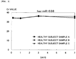
- the serums were left to stand at room temperature (25°C) for seven days.
- a change in the amount of each of the micro-RNAs with time was quantitatively determined by RT-PCR as in Example 3.
- the marker of the invention of the subject application can easily detect the onset of breast cancer that cannot be detected by palpation or mammography examination and that is highly likely to be overlooked by existing pathological cell diagnosis or breast cancer in an early stage (clinical stage 0) with high reliability. Therefore, the marker of the invention is suitable for use in a test kit for breast cancer, the test kit including at least a reagent for measuring a micro-RNA.
Landscapes
- Chemical & Material Sciences (AREA)
- Life Sciences & Earth Sciences (AREA)
- Health & Medical Sciences (AREA)
- Organic Chemistry (AREA)
- Proteomics, Peptides & Aminoacids (AREA)
- Engineering & Computer Science (AREA)
- Immunology (AREA)
- Pathology (AREA)
- Analytical Chemistry (AREA)
- Zoology (AREA)
- Genetics & Genomics (AREA)
- Wood Science & Technology (AREA)
- Physics & Mathematics (AREA)
- Biotechnology (AREA)
- Microbiology (AREA)
- Molecular Biology (AREA)
- Hospice & Palliative Care (AREA)
- Biophysics (AREA)
- Oncology (AREA)
- Biochemistry (AREA)
- Bioinformatics & Cheminformatics (AREA)
- General Engineering & Computer Science (AREA)
- General Health & Medical Sciences (AREA)
- Measuring Or Testing Involving Enzymes Or Micro-Organisms (AREA)
- Investigating Or Analysing Biological Materials (AREA)
Description
- The present invention relates to a marker for diagnosis of breast cancer, a method of testing for breast cancer, and a test kit for breast cancer. More specifically, the present invention relates to a marker for detecting the onset of breast cancer or an early stage (clinical stage 0) of breast cancer, the marker being composed of a micro-RNA that is found in serum or plasma, a method of testing for breast cancer, and a test kit for breast cancer.
- Breast cancer is a malignant tumor that is the most frequently found in females in many civilized countries, and the risk of such a malignant tumor progressing to invasive breast cancer still exceeds 10% for females of all ages (Non-Patent Document 1). This is partly attributed to the fact that cancer tissue cannot be detected by palpation or mammography examination until the cancer tissue grows to a certain size.
- Hitherto, it has been believed that the onset and progression of cancers including breast cancer are caused by accumulation of multi-step mutations of oncogenes and tumor suppressor genes. Recently, it was found that so-called epigenetic mutations, such as DNA methylation and histone diacetylation, which do not involve mutations of the primary structure of genes also relate to the onset and progression of cancers.
- It is becoming clear that abnormality of DNA methylation or histone modification is controlled by low-molecular-weight RNAs called micro-RNAs. It should be noted that a micro-RNA (hereinafter also referred to as "miRNA") is a small noncoding RNA that is composed of 19 to 25 nucleotides and that is not translated as an amino acid sequence of proteins. It has been confirmed that a large number of miRNAs are present in living organisms including humans, and they are systematically and highly conserved (Non-Patent
Documents 2 to 4). In particular, in humans, about 800 types of miRNAs have been discovered to date (Non-Patent Documents 5 to 10). - Regarding the relationship between a cancer and a miRNA, Calin et al. reported that frequent down-regulation of miR-15 and miR-16, which are miRNAs, occurs in lymphocytic leukemia (Non-Patent Document 11). Furthermore, recently, Michael et al. reported that the expression of miR-143 and miR-145, which are miRNAs, decreases in human colorectal cancer (Non-Patent Document 12).
- However, these reports do not describe whether or not the decrease in the expression of the miRNAs affects clinical pathological characteristics of cancers, and there are many unknown points regarding the expression regulation mechanism and expression abnormality of micro-RNAs in cancers, and target genes of the micro-RNAs.
- [Non-Patent Document 1] Krupnick JG, Benovic JL: The role of receptor kinases and arrestins in G protein-coupled receptor regulation, Annu Rev Pharmacol Toxicol 1998, Vol. 38, p. 289-319
- [Non-Patent Document 2] Ambros V, MicroRNA pathways in flies and worms: growth, death, fat, stress, and timing, Cell, 2003, Vol. 113, p. 673-6
- [Non-Patent Document 3] Grosshans H, Slack FJ, Micro-RNAs: small is plentiful, J. Cell Biol., 2002, Vol. 156, p. 17-21
- [Non-Patent Document 4] Nelson P, Kiriakidou M, Sharma A, Maniataki E, Mourelatos Z, The microRNA world: small is mighty, Trends Biochem. Sci., 2003, Vol. 28, p. 534-40
- [Non-Patent Document 5] Lagos-Quintana M, Rauhut R, Lendeckel W, Tuschl T, Identification of novel genes coding for small expressed RNAs, Science, 2001, Vol. 294, p. 853-8
- [Non-Patent Document 6] Lau NC, Lim LP, Weinstein EG, Bartel DP, An abundant class of tiny RNAs with probable regulatory roles in Caenorhabditis elegans, Science, 2001, Vol. 294, p. 858-62
- [Non-Patent Document 7] Lee RC, Ambros V, An extensive class of small RNAs in Caenorhabditis elegans, Science, 2001, Vol. 294, p. 862-4
- [Non-Patent Document 8] Mourelatos Z, Dostie J, Paushkin S et al., miRNP: miRNPs: a novel class of ribonucleoproteins containing numerous microRNAs, Genes Dev., 2002, Vol. 16, p. 720-8
- [Non-Patent Document 9] Lim LP, Glasner ME, Yekta S, Burge CB, Bartel DP, Vertebrate microRNA genes, Science, 2003, Vol. 299, p. 1540
- [Non-Patent Document 10] "Genome Summary List of Genome Currently Available", [online], miRBase, [Searched on March 27, 2009], Internet <URL: http://microrna.sanger.ac.uk/cgi-bin/targets/v5/genome.pl?order#by=genome#name>
- [Non-Patent Document 11] Calin GA, Dumitru CD, Shimizu M et al., Frequent deletions and down-regulation of micro-RNA genes miR15 and miR16 at 13ql4 in chronic lymphocytic leukemia, Proc Natl. Acad. Sci. U.S.A., 2002, Vol. 99, p. 15524-9
- [Non-Patent Document 12] Michael MZ, SM OC, van Holst Pellekaan NG, Young GP, James RJ, Reduced accumulation of specific microRNAs in colorectal neoplasia Mol Cancer Res, 2003, Vol. 1, p. 882-91
- Lawie Charles et al. (2008) discloses the detection of elevated levels of tumor-associated micro-RNAs in serum of patients with diffuse large β-cell lymphoma.
- An object of the present invention is to provide a marker which can detect the onset of breast cancer that cannot be detected by palpation or mammography examination and that is highly likely to be overlooked by existing pathological cell diagnosis or the presence of breast cancer cells even in an early stage (clinical stage 0) of breast cancer, and which is simple and has high reliability, the marker being composed of a micro-RNA that is present in serum or plasma; a test method; and a test kit.
- The inventors of the present invention have conducted intensive studies in order to solve the above problem or a problem that existing diagnostic markers for detecting breast cancer have low accuracy in terms of diagnostic sensitivity (sensitivity) and specificity and thus diagnosis cannot be accurately performed. As a result, it was found that the level of a micro-RNA (α) that is present in serum or plasma of a subject is significantly reduced after the onset of breast cancer, the level of a micro-RNA (β) that is present in serum or plasma is constant regardless of the affection of breast cancer, and the micro-RNAs can be used as a marker with which the onset of breast cancer or an early stage (clinical stage 0) of breast cancer can be diagnosed on the basis of the dynamics of these micro-RNAs. This finding led to the completion of the present invention. which is defined in the appended claims.
- A marker of the present invention is a marker associated with breast cancer, and more specifically, (A) "a marker for detecting the onset of breast cancer or the early stage (clinical stage 0) of breast cancer" or a marker used as a prognostic factor of breast cancer, and is characterized by being (B) "a micro-RNA that is found in serum or plasma". In other words, the present invention provides a method in which the (B) is used as the (A).
- The micro-RNA is preferably a micro-RNA (α) that is present in the serum or the plasma at a significantly reduced level after the onset of breast cancer, or during or after an early stage (during or after clinical stage 0) of breast cancer compared with that before the onset of breast cancer or before the early stage (before clinical stage 0) of breast cancer. The micro-RNA is at least one micro-RNA (α) selected from the group consisting of hsa-miR-92a1, hsa-miR-92a2, hsa-miR-22, hsa-miR-370, hsa-miR-601, hsa-miR-658, and hsa-miR-494.
- In addition, the micro-RNA is preferably a combination of the micro-RNA (α) and a micro-RNA (β) that is present in the serum or the plasma at a constant level before and after the onset of breast cancer, or before, during, and after the early stage (before, during, and after clinical stage 0) of breast cancer. More preferably, the micro-RNA is a combination of the micro-RNA (α) and the micro-RNA (β), and a numerical value obtained by dividing the level of the micro-RNA (α) present in the serum or the plasma by the level of the micro-RNA (β) present in the serum or the plasma after the onset of breast cancer, or during or after the early stage (during or after clinical stage 0) of breast cancer is statistically significantly reduced compared with that before the onset of breast cancer or before the early stage (before clinical stage 0) of breast cancer.
- The micro-RNA (β) is preferably at least one micro-RNA selected from the group consisting of hsa-miR-638, hsa-miR-630, and hsa-miR-572.
- A method of testing for breast cancer as disclosed herein is characterized by including detecting the dynamics of a level of a micro-RNA that is present in serum or plasma derived from a subject and preferably includes (i) a step of comparing a numerical value obtained from a sample derived from a subject with a numerical value obtained from a sample derived from one or more healthy subjects or one or more breast cancer patients, each of the numerical values being obtained as a level of the micro-RNA (α) present in the serum or the plasma, to test whether or not there is a statistically significant difference between the numerical values, or (ii) a step of comparing a numerical value obtained from a sample derived from a subject with a numerical value obtained from a sample derived from one or more healthy subjects or one or more breast cancer patients, each of the numerical values being obtained as a normalized level of the micro-RNA (α) present in the serum or the plasma and determined by dividing a level of the micro-RNA (α) present in the serum or the plasma by a level of the micro-RNA (β) present in the serum or the plasma, to test whether or not there is a statistically significant difference between the numerical values. In particular, in steps (i) and (ii), the sample used as a comparison reference is more preferably a sample derived from one or more breast cancer patients.
- Furthermore, a test kit for breast cancer is characterized by including at least a reagent for measuring the micro-RNA functioning as the marker of the present invention.
- According to the present invention, even the onset of breast cancer that cannot be detected by palpation or mammography examination and that is highly likely to be overlooked by existing pathological cell diagnosis or breast cancer in an early stage (clinical stage 0) can be easily tested by collecting the blood from a subject, and the difference between positivity and negativity is more significantly sensitive to that of existing breast cancer markers. Thus, the present disclosure can provide a marker, a test method, and a test kit which have high reliability. Furthermore, in the test of breast cancer, whether a subject is positive or negative can be determined by simply collecting the blood. This is preferable because the burden on the subject is low.
-
- [
Fig. 1] Fig. 1 is a graph of the results shown in Table 5 of Example 3. - [
Fig. 2] Fig. 2 is a graph showing the storage stability of hsa-miR-*92a evaluated by RT-PCR. - [
Fig. 3] Fig. 3 is a graph showing the storage stability of hsa-miR-638 evaluated by RT-PCR. - Next, a marker, a method of testing for breast cancer, and the use of a test kit for breast cancer of the present invention will be described in detail.
- A marker of the present invention is a marker associated with breast cancer and is characterized by being a micro-RNA that is found in serum or plasma.
- As described below, the marker of the present invention may be a micro-RNA (α) alone or a combination of the micro-RNA (α) and a micro-RNA (β).
- Micro-RNAs (miRNAs) are small RNA molecules that do not code proteins, and it is believed that micro-RNAs relate to various biological phenomena such as development, differentiation, and proliferation. At least several hundred miRNA genes that code these small RNAs are present on the genome. First, an early miRNA having a length of several hundred to several thousand nucleotides is transcribed from the genes, and is then digested by a protein complex called a Microprocessor to produce a hairpin-shaped precursor miRNA composed of about 60 to 70 nucleotides. Subsequently, the precursor miRNA is moved from the nucleus to cytoplasm through exportin-5, and further digested by ribonuclease III called a Dicer to produce a mature miRNA dimer composed of 19 to 25 nucleotides. It is believed that the mature miRNA dimer forms a complex with Argonaute protein, which is associated with RNA interference (RNAi), and functions. The mechanism of the action thereof is believed to be partial (incomplete) hybridization with a messenger RNA, a part of which is (which is partially) complementary to a miRNA, and an unknown translation suppressing action concurrent with the hybridization. Accordingly, it is believed that the complex engages in a very unique expression control mechanism that one type of miRNA controls the translation of a plurality of messenger RNAs coding proteins.
- Recently, the inventors detected a mature miRNA dimer, which should be present in cytoplasm, in serum or plasma. Although the mechanism through which miRNAs enter the bloodstream and the reason therefor are not completely clear, it was found that a level of a specific miRNA (micro-RNA (α)) fluctuates whereas a level of a specific miRNA (micro-RNA (β)) is steady (constant).
- In the present invention, for example, any of the numerical value (Copies/µL) measured by a real-time RT-PCR (or RT-qPCR) detection method and the signal intensity detected by a microarray analysis method or a northern blot analysis method can be used as an index as the "level".
- Among mature miRNA dimers that are present in serum or plasma, the micro-RNA (β) is preferably at least one micro-RNA selected from the group consisting of hsa-miR-638, hsa-miR-630, and hsa-miR-572. The levels of these micro-RNAs are steady (constant) before and after the onset of breast cancer or before, during, and after an early stage (before, during, and after clinical stage 0) of breast cancer.
- Accordingly, when the dynamics of the fluctuating level of the micro-RNA (α), which is caused by the affection of breast cancer or the progression of the stage of breast cancer, is detected, different samples can be normalized on the basis of the level of the micro-RNA (β) that is present in serum or plasma. That is, a numerical value obtained by dividing the level of the micro-RNA (α) that is present in serum or plasma by the level of the micro-RNA (β) that is present in serum or plasma is significantly reduced after the onset of breast cancer, or during or after the early stage (during or after clinical stage 0) of breast cancer as compared with the numerical value before the onset of breast cancer or before the early stage (clinical stage 0) of breast cancer. Therefore, the combination of the micro-RNAs (α) and (β) is suitable for the marker of the present invention.
- The sequences of such micro-RNAs (β) are shown below.
-
- (hsa-miR-638) agggaucgcgggcggguggcggccu
- (hsa-miR-630) aguauucuguaccagggaaggu
- (hsa-miR-572) guccgcucggcgguggccca
- Examples of the micro-RNA (α) include micro-RNAs such as hsa-miR-92a1, hsa-miR-92a2, hsa-miR-22, hsa-miR-370, hsa-miR-601, hsa-miR-658, and hsa-miR-494. The levels of these micro-RNAs are specifically reduced with canceration of cells derived from tissue of the breast, mammary ducts, and mammary glands (that is, the levels of these micro-RNAs that are present in serum or plasma are statistically significantly reduced after the onset of breast cancer, or during or after the early stage (clinical stage 0) of breast cancer compared with the levels before the onset of breast cancer or before the early stage (clinical stage 0) of breast cancer). Therefore, these micro-RNAs are suitable for a marker for detecting the onset of breast cancer or the early stage (clinical stage 0) of breast cancer.
- These micro-RNAs (α) may be used alone or in combination of two or more types of micro-RNAs (α).
- It should be noted that although hsa-miR-92a1 and hsa-miR-92a2 are transcribed from different genome regions, the sequences of the mature miRNAs are identical. Hereinafter, hsa-miR-92a1 and hsa-miR-92a2 are also referred to as "hsa-miR-*92a" in combination.
- The sequences of such micro-RNAs (α) are shown below.
-
- (hsa-miR-*92a) uauugcacuugucccggccugu
- (hsa-miR-22) aagcugccaguugaagaacugu
- (hsa-miR-370) gccugcugggguggaaccuggu
- (hsa-miR-601) uggucuaggauuguuggaggag
- (hsa-miR-658) ggcggagggaaguagguccguuggu
- (hsa-miR-494) ugaaacauacacgggaaaccuc
- In this description, the "breast cancer" includes both ductal carcinoma originating from a terminal duct and lobular carcinoma originating from a lobule. It should be noted that the term "cancer" refers to a malignant tumor, but may be simply referred to as "tumor". The terms "malignant transformation" and "canceration" may be used as synonyms.
- In this description, the term "onset" refers to the time when a subject is diagnosed as having a specified disease by comprehensive diagnosis on the basis of clinical symptoms specific to the disease, test data, and the like.
- In this description, the term "prognostic factor" refers to determination information for forecasting the course of the disease after an operation, forecasting the future, and selecting an appropriate therapeutic method. In addition to the marker of the present invention, in general, the following factors are known as prognostic factors. Examples thereof include the size of a lump, the degree of metastasis to lymph nodes, the presence or absence of distant metastasis, the presence or absence of a hormone receptor (estrogen receptor (ER) or progesterone receptor (PgR)), the menopause status, and the nuclear grade.
- In general, the clinical stages (stages) of breast cancer are defined by the terms a non-invasive cancer (intraepithelial carcinoma), a locally invasive cancer, a regional invasive cancer, and a distant metastatic cancer (metastatic cancer) or on the basis of the size of a tumor, the presence or absence of invasion to lymph nodes, and distant metastasis of cancer cells, and specifically represented as
stages 0 to IV (clinical stages 0 to IV). - Non-invasive cancer (intraepithelial carcinoma): Non-invasive cancer refers to a cancer that stays in a local area, and corresponds to the earliest-stage cancer. Although the size of the cancer may significantly increase in the breast, invasion to surrounding tissue or metastasis to other sites in the body has not been caused. This cancer is usually detected by mammography examination.
- Locally invasive cancer: Locally invasive cancer refers to a cancer that stays in the breast, though the cancer invades surrounding tissue.
- Regional invasive cancer: Regional invasive cancer refers to a cancer that has invaded also tissue disposed around the breast, such as the chest wall or lymph nodes.
- Distant metastatic cancer (metastatic cancer): Distant metastatic cancer (metastatic cancer) refers to a cancer that has spread by metastasis from the breast to other sites in the body.
- Stage 0: The tumor is confined to a mammary duct or a mammary gland, and there is no invasion to surrounding mammary gland tissue (non-invasive cancer (intraepithelial carcinoma)). This is also referred to as the early stage of breast cancer.
- Stage I: The diameter of the tumor is less than 2 cm, and metastasis outside the breast is not observed.
- Stage II: The diameter of the tumor is 2 to 5 cm, and metastasis is observed in lymph nodes under the arm (one side or both sides) at the same side as that of the tumor.
- Stage III: The diameter of a tumor exceeds 5 cm. Metastasis to lymph nodes is observed, and adhesion to surrounding tissue or adhesion to lymph nodes is observed. Alternatively, regardless of the size of the tumor, metastasis to the skin, chest wall, and thoracic lymph nodes at the downstream of the breast is observed.
- Stage IV: Regardless of the size of the tumor, metastasis to organs and tissue such as the lung and bones distant from the breast or metastasis to lymph nodes of sites distant from the breast is observed.
- In addition, breast cancer is classified on the basis of the type of tissue in which cancer cells are generated for the first time, and the range of the spread of the focus. A cancer originating from a mammary duct is referred to as "ductal carcinoma", and about 90% of breast cancer cases are ductal carcinoma. A cancer originating from a mammary gland is referred to as lobular carcinoma. A cancer originating from adipose tissue or connective tissue is referred to as "sarcoma". However, such a sarcoma is rare among breast cancer cases.
- Ductal carcinoma in situ is a cancer that is confined in a mammary duct. Although this cancer has not invaded tissue around the breast, this cancer may spread along the mammary duct and the size thereof may increase in the breast. This cancer accounts for 20% to 30% of breast cancer cases.
- Lobular carcinoma in situ proliferates inside a mammary gland. This cancer often generated in a plurality of sites of breasts at both sides. Lobular carcinoma in situ accounts for 1% to 2% of breast cancer cases.
- Infiltrating ductal carcinoma is a cancer that originates from a mammary duct and that has invaded beyond a duct wall into surrounding mammary gland tissue. This cancer may spread by metastasis to a site other than the breast and accounts for 65% to 80% of breast cancer cases.
- Infiltrating lobular carcinoma is a cancer that originates from a mammary gland, that invades into surrounding mammary gland tissue, and that further spreads by metastasis to a site other than the breast. This cancer accounts for 10% to 15% of breast cancer cases.
- In addition, inflammatory breast cancer, which accounts for about 1% of all breast cancer cases, and Paget's disease of nipple are known. Furthermore, medullary carcinoma, tubular carcinoma, mucinous carcinoma (colloid carcinoma), and the like are also known as invasive cancers whose occurrence frequency is low, the invasive cancers originating from a mammary duct.
- Breast cancer according to the present application include all carcinomas (cancers) originating from tissue of the breast, mammary ducts, and mammary glands, and non-invasive cancers in clinical stage 0 (intraepithelial carcinomas), which cannot be detected by palpation or mammography examination.
- A method of testing for breast cancer of the present invention is characterized by including detection of the dynamics of a level of a micro-RNA that is present in serum or plasma derived from a subject and preferably includes step (i) or (ii) below.
- Step (i): A step of comparing a numerical value obtained from a sample derived from a subject with a numerical value obtained from a sample derived from one or more healthy subjects or one or more breast cancer patients, each of the numerical values being obtained as a level of the micro-RNA (α) present in the serum or the plasma, to test whether or not there is a statistically significant difference between the numerical values.
- Step (ii): A step of comparing a numerical value obtained from a sample derived from a subject with a numerical value obtained from a sample derived from one or more healthy subjects or one or more breast cancer patients, each of the numerical values being obtained as a normalized level of the micro-RNA (α) present in the serum or the plasma and determined by dividing a level of the micro-RNA (α) present in the serum or the plasma by a level of the micro-RNA (β) present in the serum or the plasma, to test whether or not there is a statistically significant difference between the numerical values.
- In the method of testing for breast cancer of the present invention, specifically, first, the total RNA is extracted from a specimen. In this description, the term "specimen" refers to the blood taken from a breast cancer patient (which refers to a human or a mammal other than a human which has been diagnosed as having breast cancer by pathological cell diagnosis), a healthy subject (which refers to a human or a mammal other than a human which has no specific chronic diseases and has no problem with their daily life and actions, and more specifically, a human or a mammal other than a human which has been diagnosed as not having a cancer during a regular physical checkup (including cancer screening) conducted once a year or more), a subject (which refers to a human or a mammal other than a human for which the onset of breast cancer is suspected), or the like.
- Specifically, the blood is collected from a subject such as a human, and the supernatant obtained from the blood is then centrifuged to remove blood cells. The resulting blood is used as serum or plasma. The total RNA is extracted from the obtained serum or plasma. Examples of a method for extracting the total RNA include a guanidine-cesium chloride ultracentrifugation method, and an acid guanidinium-phenol-chloroform (AGPC) method.
- Next, a miRNA according to the present invention contained in the extracted total RNA is detected. The detection method is not particularly limited as long as the amount of expression of the miRNA according to the present invention can be measured by the method. Examples of the method include a microarray analysis method, a northern blot analysis method, and a real-time RT-PCR detection method.
- An example of the microarray analysis method is the method described in Example 1.
- In the northern blot analysis method, the total RNA is separated by electrophoresis in accordance with the chain length, and transferred to a nitrocellulose membrane or a nylon membrane. This membrane is incubated in a buffer with a probe having a sequence complementary to the miRNA according to the present invention and labeled with a radioactive substance (e.g., 32P etc.) or the like. As a result, the miRNA according to the present invention hybridized with the probe can be detected on the basis of the label attached to the probe. It should be noted that each of prehybridization, hybridization, and a washing step is conducted under a stringent condition. Herein, the term "stringent condition" is a temperature of 37°C in a hybridization buffer containing 0.25 M sodium phosphate (pH 7.2), 7% SDS, and 0.5% sodium pyrophosphate, for example, when DNA labeled with 32P is used as the probe. In the washing step, the stringent condition is a temperature of 37°C in a washing buffer containing 2 x SSC and 1% SDS, and room temperature in a washing buffer containing 0.1 x SSC. In other cases, other standard conditions of the detection method may be used. When detection is performed by a radioactively labeled probe, the miRNA according to the present invention hybridized with the probe can be quantitatively determined using autoradiography on the basis of the band density.
- Furthermore, according to the real-time RT-PCR (or RT-qPCR) detection method, the miRNA according to the present invention can be quantitatively detected by PCR using random priming cDNA corresponding to the total RNA and a primer complementary to the miRNA according to the present invention, via a fluorescent dye, such as SYBR Green, which is specifically bonded to double-strand DNA. The real-time RT-PCR detection method can be conducted in accordance with a known method or an instruction manual from a manufacturer of a detection device or a reagent (for example, ABI Prism 7900 Sequence Detection System (Perkin-Elmer Applied Biosystems), SYBR Green PCR Master Mix (Perkin-Elmer Applied Biosystems), or the like).
- After the detection of the miRNA according to the present invention contained in the extracted total RNA, either step (i) or step (ii) is preferably conducted.
- Step (i) is a step of comparing a numerical value obtained from a sample derived from a subject with a numerical value obtained from a sample derived from one or more healthy subjects or one or more breast cancer patients, each of the numerical values being obtained as a level of the micro-RNA (α) present in the serum or the plasma, to test whether or not there is a statistically significant difference between the numerical values.
- That is, step (i) is a step of comparing the absolute value of the amount of expression of at least one micro-RNA (α) selected from the group consisting of hsa-miR-*92a, hsa-miR-22, hsa-miR-370, hsa-miR-601, hsa-miR-658, and hsa-miR-494; preferably hsa-miR-494, hsa-miR-370, and hsa-miR-*92a; and particularly preferably a micro-RNA (α) of hsa-miR-494
- (i-1) with the absolute value of the amount of expression of the micro-RNA (α) obtained from a sample derived from one or more healthy subjects to test whether or not the former absolute value is statistically significantly low (where the significance level is preferably 5% and more preferably 1%), or
- (i-2) with the absolute value of the amount of expression of a micro-RNA (α) obtained from a sample derived from one or more breast cancer patients to test whether or not the former absolute value is statistically significantly high.
- More specifically, in (i-1), as a result of the comparison, when the absolute value is statistically significantly low, a subject who provided the sample is diagnosed as having breast cancer. Conversely, when there is no statistically significant difference, the result of the comparison becomes useful information indicating that the subject has been diagnosed as a healthy subject. In (i-2), as a result of the comparison, when the absolute value is statistically significantly high, a subject who provided the sample is diagnosed as a healthy subject. Conversely, when there is no statistically significant difference, the result of the comparison becomes useful information indicating that the subject has been diagnosed as having breast cancer.
- When the subject is one individual, preferably, a specimen is obtained in advance before the onset of breast cancer or before the early stage (before clinical stage 0) of breast cancer, and the amount of expression of the micro-RNA (α) obtained from a sample derived from the specimen is used as a comparison target.
- Step (ii) is a step of comparing a numerical value obtained from a sample derived from a subject with a numerical value obtained from a sample derived from one or more healthy subjects or one or more breast cancer patients, each of the numerical values being obtained as a normalized level of the micro-RNA (α) present in the serum or the plasma and determined by dividing a level of the micro-RNA (α) present in the serum or the plasma by a level of the micro-RNA (β) present in the serum or the plasma, to test whether or not there is a statistically significant difference between the numerical values.
- That is, step (ii) is a step of normalizing the amount of expression of a micro-RNA (α) by dividing the amount of expression of at least one micro-RNA (α) selected from the group consisting of hsa-miR-*92a, hsa-miR-22, hsa-miR-370, hsa-miR-601, hsa-miR-658, and hsa-miR-494; preferably a micro-RNA of hsa-miR-370 or hsa-miR-*92a by the amount of expression of at least one micro-RNA (β) selected from the group consisting of hsa-miR-638, hsa-miR-630, and hsa-miR-572, preferably hsa-miR-638, and comparing the obtained relative value of the amount of expression of the micro-RNA (α)
- (ii-1) with a relative value of the amount of expression of the micro-RNA (α) obtained from a sample derived from one or more healthy subjects to test whether or not the former relative value is statistically significantly low (where the significance level is preferably 5% and more preferably 1%), or
- (ii-2) with a relative value of the amount of expression of the micro-RNA (α) obtained from a sample derived from one or more breast cancer patients to test whether or not the former relative value is statistically significantly high.
- More specifically, in (ii-1), as a result of the comparison, when the relative value is statistically significantly low, the subject who provided the sample is diagnosed as having breast cancer. Conversely, when there is no statistically significant difference, the result of the comparison becomes useful information indicating that the subject has been diagnosed as a healthy subject. In (ii-2), as a result of the comparison, when the relative value is statistically significantly high, the subject who provided the sample is diagnosed as a healthy subject. Conversely, when there is no statistically significant difference, the result of the comparison becomes useful information indicating that the subject has been diagnosed as having breast cancer.
- As in step (i), when the subject is one individual, preferably, a specimen is obtained in advance before the onset of breast cancer or before the early stage (clinical stage 0) of breast cancer, and the amount of expression of the micro-RNA (α) obtained from a sample derived from the specimen is used as a comparison target.
- Step (ii) is more preferable than step (i) because since normalization has been performed, the result does not depend on the type of micro-RNA (α) used, the subject (individual), or whether or not the blood is collected before the onset of breast cancer or before the early stage (clinical stage 0) of breast cancer.
- One embodiment of a specific procedure of step (ii) and the subsequent diagnosis of breast cancer will be exemplified below.
- Normalization is performed by dividing the amount of expression of each of hsa-miR-*92a, hsa-miR-22, hsa-miR-370, hsa-miR-601, hsa-miR-658, and hsa-miR-494 by the amount of expression of hsa-miR-638, hsa-miR-630, or hsa-miR-572 corrected in advance by the signal intensity obtained by a microarray analysis method with an internal standard. That is, normalization is performed by dividing the level of the micro-RNA (α) present in serum or plasma by the level of the micro-RNA (β) present in serum or plasma.
- When the normalized amount of expression of hsa-miR-*92a, hsa-miR-22, hsa-miR-370, hsa-miR-601, hsa-miR-658, or hsa-miR-494 derived from a subject is significantly reduced as compared with the normalized amount of expression of hsa-miR-*92a, hsa-miR-22, hsa-miR-370, hsa-miR-601, hsa-miR-658, or hsa-miR-494 derived from a healthy subject, the subject can be diagnosed as having breast cancer. For example, the normalized amount of expression of hsa-miR-*92a, hsa-miR-22, hsa-miR-370, hsa-miR-601, hsa-miR-658, or hsa-miR-494 derived from a breast cancer patient is statistically significantly reduced as compared with the corrected amount of expression of hsa-miR-*92a, hsa-miR-22, hsa-miR-370, hsa-miR-601, hsa-miR-658, or hsa-miR-494 derived from a healthy subject.
- As for a statistical method of testing whether or not there is a significant difference in step (i) or (ii), an appropriate method may be selected in accordance with the number of samples and the like. Examples of the method include a Ttest (T-test), an F-test, and a chi-square test.
- By such a test method, even for a subject who is in
clinical stage 0 of breast cancer and before the onset of breast cancer, it can be determined that the subject has breast cancer with high reliability. Therefore, it is possible to provide important information that may help in deciding what treatment strategy to follow such as medication or an operation. - A test kit for breast cancer of the present disclosure is characterized by including at least a reagent for measuring a micro-RNA functioning as a marker of the present invention, wherein the test kit includes various instruments, materials, reagents and/or primers for amplifying genes, reagents related to gene amplification, and the like which are necessary for conducting the method of testing for breast cancer of the present invention.
- Examples of the reagents for measuring a micro-RNA include various enzymes, buffering solutions, washing liquids, and dissolution liquids. Specifically, the reagents include at least a dissolution liquid for diluting a specimen and further include a primer for PCR, the primer being used for detecting the micro-RNA. A set of necessary instruments such as a microtiter plate and equipment for DNA amplification with which a plurality of specimens can be treated at the same time may further be included as kit elements.
- According to a high throughput embodiment of the method of testing for breast cancer of the present invention, a microreactor element, specifically, a chip instrument may be included in the above test kit. Such an embodiment of an improved configuration is preferably a system that adopts a configuration in which digitized data with respect to signals obtained from a chip is taken in, a file is formed through computer processing, and the file is stored in a predetermined directory on a computer. Furthermore, the system may further include a capacity with which numerical data is statistically processed to display a significant difference from a control (experimental control). The data processing is performed using suitable software that enables statistical analysis through necessary correction and normalization. A person skilled in the art can establish a system for such a data processing using existing technologies, methods, and procedures.
- Next, the present invention will be described in more detail by way of Examples.
- The blood was collected from respective healthy subjects (humans who were diagnosed as not having breast cancer during regular physical checkup including cancer screening and conducted once a year or more) and breast cancer patients (humans who were diagnosed as having breast cancer by pathological cell diagnosis) shown in Table 1, and serum was then separated from the blood. The total RNA was extracted from the serum using "(trade name) Isogen-LS" (produced by Nippon Gene Co., Ltd.), and the concentration of the total RNA was adjusted to 100 ng/µL.
- Next, dephosphorylation reaction of the total RNA was conducted using "(trade name) Alkaline Phosphatase (Calf intestine) (CIAP)" (produced by Takara Bio Inc.), which is an alkaline phosphatase derived from calf small intestine. Subsequently, labeling was performed with cyanine, which is a dye, using "(trade name) T4 RNA Ligase (Cloned)" (produced by Ambion, Inc.). The above operation was conducted using "(trade name) miRNA Labeling Reagent and Hybridization Kit (catalogue No.: 5190-0408)" (produced by Agilent technologies) in accordance with the protocol attached thereto.
- Furthermore, hybridization of the total RNA labeled with cyanine was conducted using a microarray slide of "(trade name) Human miRNA Microarray kit 8 x 15K V2 (Catalogue No.: G4470B) (produced by Agilent technologies) to obtain signals.
- Subsequently, the microarray slide was scanned using "(trade name) DNA Microarray Scanner" (produced by Agilent technologies). For signal detection, "Feature Extraction 9.5.3 Software" and "Agilent Scan Control Software (ver. 7.0)" that were attached to the above scanner were used.
- The obtained results are shown in Table 1.
- [Table 1]
Table 1: Example 1 Sample No. Derivation of specimen Micro-RNA(α)[hsa-miR-] Micro-RNA (β) [hsa-miR-] *92a 22 370 601 658 494 638 630 572 1 Male 86.0328 - - - - 24.1678 891.496 - 40.5002 2 Female 164.835 - - - - 38.4196 2842.82 - 115.124 3 Male 555.067 - - - - 22.2111 7400.18 - 141.962 4 Female 4.9112 1.1041 13.348 6.5309 5.1405 - 781.85 301.89 - 5 Healthy subject Female 86.033 19.435 23.929 11.651 7.1342 - 891.5 368.61 - 6 Female 164.84 54.97 53.94 24.107 17.603 - 2842.8 902.63 - 7 Female 37.789 8.7482 6.4142 5.9387 3.0777 - 582.09 168.4 - 8 Female 555.07 68.946 75.813 114.91 60.459 - 7400.2 4352.3 - 9 Female 27.639 12.887 8.6969 6.1194 0.4488 - 3139.8 223.07 - 10 Female 1.1296 4.4697 5.6448 3.7716 1.3054 - 1933.5 231.36 - 11 Female 0.146762 - - - - -1.95187 2598.58 - 161.39 12 Female 3.64585 - - - - -1.66446 4297.62 - 247.888 13 Female 1.1017 0.001 6.0851 18.115 10.246 - 2145.8 414.7 - 14 Female 7.7428 0.897 9.0196 28.427 18.597 - 3911.7 752.39 - 15 Breast cancer patient Female 0.001 0.001 0.001 0.3747 -0.6201 - 1044.48 49.9934 - 16 Female 3.64585 0.33198 4.21704 3.29853 -0.2897 - 4297.62 329.343 - 17 Female 0.001 2.1077 1.3858 2.0594 1.5543 - 801.72 72.411 - 18 Female 37.118 36.601 6.7462 6.2106 0.0422 - 3534.5 264.92 - 19 Female 0.001 0.8274 0.5354 2.704 1.415 - 1028.7 49.713 - 20 Female 0.001 0.001 0.001 0.001 -14.34 - 10289 514.33 - 21 Female 2.9398 0.1292 0.8485 1.7492 0.7915 - 1184.2 113.6 - - A T-test (significant difference test) of the averages of the healthy subjects and the breast cancer patients was conducted on the basis of the analysis data obtained in Example 1. Specifically, the T-test was conducted using the numerical values of hsa-miR-92a, hsa-miR-22, hsa-miR-370, hsa-miR-601, and hsa-miR-658 obtained from the subjects (sample Nos. 4 to 10) and the breast cancer patients (sample Nos. 13 to 21) as they are. The obtained p-values are shown in Table 2. Furthermore, the numerical values of hsa-miR-92a, hsa-miR-22, hsa-miR-370, hsa-miR-601, and hsa-miR-658 obtained from the healthy subjects (sample Nos. 4 to 10) and the breast cancer patients (sample Nos. 13 to 21) shown in Table 1 were divided by hsa-miR-638 obtained from the corresponding healthy subjects (sample Nos. 4 to 10) and breast cancer patients (sample Nos. 13 to 21), and the T-test was conducted. The obtained p-values are shown in Table 2.
- [Table 2]
Table 2: Example 2 Micro-RNA (α) [hsa-miR-] *92a 22 370 601 658 p-Value that is divided (normalized) by micro-RNA (β) = hsa-miR-638 0.026606507 0.030637942 0.012629431 0.028309554 0.027608012 p-Value that is not divided by micro-RNA (β) 0.161400855 0.105371999 0.063186344 0.295651814 0.215317149 - Referring to Table 2, as a result of the significant difference test of the averages of the healthy subjects and the breast cancer patients, the p-values of all the markers normalized by hsa-miR-638 satisfied p < 0.05, and thus a significant difference was observed. Accordingly, Table 2 showed that breast cancer can be diagnosed more accurately by normalizing the markers of hsa-miR-370, hsa-miR-601, hsa-miR-92, hsa-miR-22, and hsa-miR-658 with hsa-miR-638.
- Sample Nos. i to iv shown in Table 3 were each quantified by a predetermined immunoassay method using, as specimens, the bloods (serums or plasmas) derived from four breast cancer patients among sample Nos. 13 to 21 and, as markers, CA15-3, CEA, NCC-ST-439, and BCA225, all of which have been hitherto used. The obtained results are shown in Table 3.
- [Table 3]
Table 3: Comparative Example 1 No. Sample No. Existing breast cancer marker CA15-3 CEA NCC-ST-439 BCA225 i 31 1.9 7.2 95 ii 9 1 2.1 55 iii 14.1 1.3 1.7 130 iv 3.9 2.8 2.4 ≤ 30 Remarks Reference value 25 U/mL or less 5.0 ng/mL or less 7.0 U/mL or less 160 U/mL or less Antigen (Protein) Sugar chain antigen 15-3 Carcinoembryonic antigen Sialic acid sugar chain antigen Mucin-type glycoprotein Assay method CLEIA EIA - Referring to Table 3, except for CA15-3 and NCC-ST-439 of sample No. i, all the obtained results satisfied the index of the reference value of the existing breast cancer markers. In other words, when breast cancer was diagnosed using the existing breast cancer markers, all samples were diagnosed as false negative, and consequently, breast cancer could not be determined.
- The blood was collected from respective healthy subjects and breast cancer patients, and the serum was separated from the blood. The total RNA was extracted from the serum using "(trade name) Isogen-LS" (produced by Nippon Gene Co., Ltd.). Subsequently, hsa-miR-*92a and hsa-miR-638 were quantified by RT-PCR using the total RNA extracted from each of the samples. The measurement of the RT-PCR was conducted using "(trade name) TaqMan MicroRNA Assay Kit (produced by Applied Biosystems Japan Ltd.)" in accordance with a protocol attached thereto. The results thereof are shown in Tables 4 to 7 and
Fig. 1 . - [Table 4]
Table 4: hsa-miR-*92a alone Median value 0.25 0.75 Maximum Minimum The number of samples Breast cancer patients 0.002056 0.000532 0.522054 0.166300 0.000005 17 Healthy subjects 0.019906 0.006089 0.038408 0.199123 0.000346 28 - [Table 5]
Table 5: hsa-miR-92a/hsa-miR-638 Median value 0.25 0.75 Maximum Minimum The number of samples Breast cancer patients 0.385240 0.189680 0.792999 2.696266 0.005926 17 Healthy subjects 10.263893 3.935504 19.925313 80.168510 0.649503 28 - [Table 6]
Table 6: hsa-miR-*92a alone Breast cancer patients Healthy subjects Test Positive 12 7 Negative 5 21 - Note that the diagnostic sensitivity (sensitivity) and the specificity are calculated as follows.


- (However, the threshold is assumed to be 0.25.)
- [Table 7]
Table 7: hsa-miR-*92a/638 (normalized) Breast cancer patients Healthy subjects Test Positive 17 7 Negative 0 21 - Note that the diagnostic sensitivity (sensitivity) and the specificity are calculated as follows.


- (However, the threshold is assumed to be 0.25.)
- Table 6 shows the results of the test using the values of hsa-miR-*92a obtained by taking serum from respective 28 healthy subjects and 17 breast cancer patients. When 25% of the value obtained in healthy subject samples was set to the threshold, the diagnostic sensitivity was 70.6% and the specificity was 75%. Thus, hsa-miR-*92a is a marker that is excellent in terms of diagnostic sensitivity and specificity as compared with generally used breast cancer markers, which have a diagnostic sensitivity of about 20% to 40% and a specificity of about 30% to 50%.
- Table 7 shows the results of the test using the values obtained by dividing hsa-miR-*92a by hsa-miR-638. When 25% of the value obtained in healthy subject samples was similarly set to the threshold, the diagnostic sensitivity was 100%. Thus, breast cancer could be diagnosed with high accuracy as compared with the case where the test was conducted using hsa-miR-*92a only.
- The storage stability of hsa-miR-*92a and hsa-miR-638 obtained by separating serums from the bloods of healthy subjects was evaluated.
- As for a specific procedure, first, the serums were left to stand at room temperature (25°C) for seven days. Next, a change in the amount of each of the micro-RNAs with time was quantitatively determined by RT-PCR as in Example 3.
- The results obtained with regard to hsa-miR-*92a are shown in
Fig. 2 , and the results obtained with regard to hsa-miR-638 are shown inFig. 3 . - Referring to
Figs. 2 and3 , the Ct value of all samples did not substantially change for seven days, indicating that the micro-RNAs in serum are very stable. These results show that the micro-RNAs in serum can be satisfactorily used as a marker used for determining breast cancer in clinical diagnosis. - The marker of the invention of the subject application can easily detect the onset of breast cancer that cannot be detected by palpation or mammography examination and that is highly likely to be overlooked by existing pathological cell diagnosis or breast cancer in an early stage (clinical stage 0) with high reliability. Therefore, the marker of the invention is suitable for use in a test kit for breast cancer, the test kit including at least a reagent for measuring a micro-RNA.
Claims (10)
- A method of testing for breast cancer comprising:(i) detecting the expression of a micro-RNA (α) and a micro-RNA (β) in total RNA extracted from serum or plasma derived from a subject,
wherein the micro-RNA (α) is a micro-RNA that is present in the serum or the plasma at a significantly reduced level after the onset of breast cancer, or during or after an early stage (during or after clinical stage 0) of breast cancer compared with that before the onset of breast cancer or before the early stage (before clinical stage 0) of breast cancer,
wherein the micro-RNA (β) is a micro-RNA that is present in the serum or the plasma at a constant level before and after the onset of breast cancer, or before, during, and after the early stage (before, during, and after clinical stage 0) of breast cancer; and(ii) obtaining a numerical value as a normalized level of the micro-RNA (α) by dividing the level of the micro-RNA (α) by the level of the micro-RNA (β) and using the normalized level of micro-RNA as a marker of breast cancer;wherein the micro-RNA (α) is at least one type of micro-RNA selected from hsa-miR-92a1, hsa-miR92a2, hsa-miR-22, hsa-miR-370, hsa-miR-601, hsa-miR-658 and hsa-miR-494. - The method according to claim 1, further comprising:(iii) comparing the numerical value obtained from step (ii) with a numerical value obtained from a sample derived from one or more healthy subjects or one or more breast cancer patients, each of the numerical values being obtained as a normalized level of the micro-RNA (α) and determined by dividing a level of the micro-RNA (α) by a level of the micro-RNA (β) to test whether or not there is a statistically significant difference between the numerical values,thereby determining whether or not the subject has breast cancer.
- The method according to claim 1 or 2, wherein the micro-RNA (α) is at least one type of micro-RNA selected from hsa-miR-92a1, hsa-miR-92a2, hsa-miR-22, hsa-miR-370, hsa-miR-601 and hsa-miR-658.
- The method according to any one of claims 1 to 3, wherein the micro-RNA (β) is at least one type of micro-RNA selected from hsa-miR-638, hsa-miR-630, and hsa-miR-572.
- The method according to any one of claims 1 to 4, wherein the numerical value obtained in step (ii) is compared with a numerical value obtained from a sample derived from one or more breast cancer patients.
- The method according to any one of claims 2 to 5, wherein the numerical value obtained in step (ii) is compared with a numerical value obtained from a sample derived from one or more breast cancer patients and the subject is diagnosed as having breast cancer when it is determined in step (iii) that there is no statistically significant difference.
- The method according to any one of Claims 1 to 6, wherein the step of detecting the expression of a micro-RNA (α) and/or a micro-RNA (β) is performed by a microarray analysis, northern blot and/or real time RT-PCR method.
- The method according to any one of Claims 1 to 7, wherein total RNA is extracted by a guanidine-cesium chloride ultracentriguation or acid guanidinium-phenol-chloroform (AGPC) method.
- Use of a kit in a method of testing for breast cancer according to any one of claims 1 to 8, wherein the kit comprises at least a reagent for measuring a micro-RNA which includes a PCR primer for detecting a micro-RNA (α) and a PCR primer for detecting a micro-RNA (β), wherein the micro-RNA (α) is selected from hsa-miR-92a1, hsa-miR-92a2, hsa-miR-22, hsa-miR-370, hsa-miR-601, hsa-miR-658, and hsa-miR-494.
- The use according to claim 9, wherein the micro-RNA (β) is selected from hsa-miR-638, hsa-miR-630, and hsa-miR-572.
Applications Claiming Priority (2)
| Application Number | Priority Date | Filing Date | Title |
|---|---|---|---|
| JP2008083897 | 2008-03-27 | ||
| PCT/JP2009/056293 WO2009119809A1 (en) | 2008-03-27 | 2009-03-27 | Marker for determination of breast cancer, test method, and test kit |
Publications (3)
| Publication Number | Publication Date |
|---|---|
| EP2280078A1 EP2280078A1 (en) | 2011-02-02 |
| EP2280078A4 EP2280078A4 (en) | 2011-08-17 |
| EP2280078B1 true EP2280078B1 (en) | 2016-05-18 |
Family
ID=41113994
Family Applications (1)
| Application Number | Title | Priority Date | Filing Date |
|---|---|---|---|
| EP09723964.4A Not-in-force EP2280078B1 (en) | 2008-03-27 | 2009-03-27 | Marker for determination of breast cancer, test method, and test kit |
Country Status (4)
| Country | Link |
|---|---|
| US (2) | US20110028332A1 (en) |
| EP (1) | EP2280078B1 (en) |
| JP (1) | JP5737935B2 (en) |
| WO (1) | WO2009119809A1 (en) |
Families Citing this family (9)
| Publication number | Priority date | Publication date | Assignee | Title |
|---|---|---|---|---|
| CA2812287A1 (en) * | 2010-09-22 | 2012-03-29 | Dave S.B. Hoon | Direct blood assay for detection of circulating microrna in cancer patients |
| CN102766696B (en) * | 2012-08-15 | 2014-05-28 | 中国人民解放军第二军医大学 | Micro ribonucleic acid (RNA) 572 kit and detection method for early predicting postoperative cognitive dysfunction |
| EP2746406B1 (en) | 2012-12-18 | 2017-08-23 | Samsung Electronics Co., Ltd. | Composition and kit for diagnosing breast cancer including miRNAs within vesicle, and method of diagnosing breast cancer using the same |
| KR101520545B1 (en) | 2013-09-06 | 2015-05-15 | 가천대학교 산학협력단 | Composition for diagnosing muscle inflammation and uses thereof |
| BR112016005413A8 (en) * | 2013-09-11 | 2018-04-03 | Fund De Amparo A Pesquisa Do Estado De Sao Paulo | USES OF AT LEAST ONE MIRNA, KIT, BREAST CANCER DIAGNOSIS METHODS AND METHODS FOR METASTASIS RISK ASSESSMENT |
| JP2015109009A (en) * | 2013-12-05 | 2015-06-11 | ソニー株式会社 | Electronic device, information processing method, and information processing system |
| RU2017100434A (en) * | 2014-06-13 | 2018-07-18 | Торэй Индастриз, Инк. | KIT OR DEVICE FOR DETECTION OF BREAST CANCER AND METHOD OF DETECTION |
| JP7298913B2 (en) * | 2017-12-13 | 2023-06-27 | 国立大学法人広島大学 | Methods to Aid in Breast Cancer Detection |
| WO2021006279A1 (en) * | 2019-07-08 | 2021-01-14 | Preferred Networks, Inc. | Data processing and classification for determining a likelihood score for breast disease |
Family Cites Families (10)
| Publication number | Priority date | Publication date | Assignee | Title |
|---|---|---|---|---|
| EP1771563A2 (en) * | 2004-05-28 | 2007-04-11 | Ambion, Inc. | METHODS AND COMPOSITIONS INVOLVING MicroRNA |
| WO2007148235A2 (en) * | 2006-05-04 | 2007-12-27 | Rosetta Genomics Ltd | Cancer-related nucleic acids |
| EP1904111A4 (en) * | 2005-06-03 | 2009-08-19 | Univ Johns Hopkins | COMPOSITIONS AND METHODS FOR DECREASING MICROARN EXPRESSION FOR THE TREATMENT OF NEOPLASIA |
| EP1904110A4 (en) * | 2005-06-03 | 2009-02-11 | Southern Adelaide Health Servi | Targeting cells with altered microrna expression |
| WO2007002375A2 (en) * | 2005-06-23 | 2007-01-04 | U.S. Genomics, Inc. | Methods and compositions for analysis of microrna |
| US8445198B2 (en) * | 2005-12-01 | 2013-05-21 | Medical Prognosis Institute | Methods, kits and devices for identifying biomarkers of treatment response and use thereof to predict treatment efficacy |
| WO2007147409A2 (en) * | 2006-06-19 | 2007-12-27 | Københavns Universitet | Ribozyme mediated stabilization of polynucleotides |
| CN101424640B (en) * | 2007-11-02 | 2012-07-25 | 江苏命码生物科技有限公司 | Detection method of microribonucleic acid in serum, reagent kit for detection, biochip and its production and application method |
| EP2260110B1 (en) * | 2008-02-08 | 2014-11-12 | Asuragen, INC. | miRNAs DIFFERENTIALLY EXPRESSED IN LYMPH NODES FROM CANCER PATIENTS |
| EP2268832A2 (en) * | 2008-03-06 | 2011-01-05 | Asuragen, INC. | Microrna markers for recurrence of colorectal cancer |
-
2009
- 2009-03-27 EP EP09723964.4A patent/EP2280078B1/en not_active Not-in-force
- 2009-03-27 JP JP2010505838A patent/JP5737935B2/en not_active Expired - Fee Related
- 2009-03-27 WO PCT/JP2009/056293 patent/WO2009119809A1/en not_active Ceased
- 2009-03-27 US US12/934,948 patent/US20110028332A1/en not_active Abandoned
-
2012
- 2012-09-14 US US13/618,415 patent/US20130012412A1/en not_active Abandoned
Non-Patent Citations (3)
| Title |
|---|
| ANONYMOUS: "MicroRNA sequences", 1 January 2005 (2005-01-01), pages 1 - 4, XP055169881, Retrieved from the Internet <URL:http://nar.oxfordjournals.org/content/suppl/2005/11/18/33.20.e179.DC1/gni178_S2.pdf> [retrieved on 20150216] * |
| HUANG YONG ET AL: "A bootstrap based analysis pipeline for efficient classification of phylogenetically related animal miRNAs", BMC GENOMICS, BIOMED CENTRAL LTD, LONDON, UK, vol. 8, no. 1, 66, 6 March 2007 (2007-03-06), pages 1 - 9, XP021022373, ISSN: 1471-2164, DOI: 10.1186/1471-2164-8-66 * |
| IORIO MARILENA V ET AL: "MicroRNA gene expression deregulation in human breast cancer", CANCER RESEARCH, AMERICAN ASSOCIATION FOR CANCER RESEARCH, US, vol. 65, no. 16, 15 August 2005 (2005-08-15), pages 7065 - 7070, XP002603432, ISSN: 0008-5472, DOI: 10.1158/0008-5472.CAN-05-1783 * |
Also Published As
| Publication number | Publication date |
|---|---|
| EP2280078A4 (en) | 2011-08-17 |
| US20110028332A1 (en) | 2011-02-03 |
| US20130012412A1 (en) | 2013-01-10 |
| JP5737935B2 (en) | 2015-06-17 |
| EP2280078A1 (en) | 2011-02-02 |
| WO2009119809A1 (en) | 2009-10-01 |
| JPWO2009119809A1 (en) | 2011-07-28 |
Similar Documents
| Publication | Publication Date | Title |
|---|---|---|
| EP2280078B1 (en) | Marker for determination of breast cancer, test method, and test kit | |
| Zhou et al. | Urinary microRNA-30a-5p is a potential biomarker for ovarian serous adenocarcinoma | |
| Tembe et al. | MicroRNA and mRNA expression profiling in metastatic melanoma reveal associations with BRAF mutation and patient prognosis | |
| Tam et al. | Robust global microRNA expression profiling using next-generation sequencing technologies | |
| Jia et al. | Identification of four serum microRNAs from a genome-wide serum microRNA expression profile as potential non-invasive biomarkers for endometrioid endometrial cancer | |
| EP2971132B1 (en) | Tissue and blood-based mirna biomarkers for the diagnosis, prognosis and metastasis-predictive potential in colorectal cancer | |
| US11136628B2 (en) | Biomarkers useful for detection of types, grades and stages of human breast cancer | |
| US20180327856A1 (en) | Diagnostic mirnas for differential diagnosis of incidental pancreatic cystic lesions | |
| EP2268832A2 (en) | Microrna markers for recurrence of colorectal cancer | |
| EP3122905B1 (en) | Circulating micrornas as biomarkers for endometriosis | |
| Rao et al. | Identification of plasma exosomes long non-coding RNA HAGLR and circulating tumor cells as potential prognosis biomarkers in non-small cell lung cancer | |
| CN101684488B (en) | Small RNA and application thereof | |
| KR20130029079A (en) | Marker consisting of plasma microrna and a new method for diagnosis of hepatocellular carcinoma | |
| Guo et al. | Serum microRNA-155 in early diagnosis and prognosis of breast cancer | |
| CN108949969B (en) | Application of long non-coding RNA in colorectal cancer | |
| Srinivasan et al. | Development of microRNA as diagnostic markers in head and neck cancer | |
| EP3842552A1 (en) | Kit for in vitro testing panel of genes in pap smear samples for endometriosis and method of non-invasively and qualitatively determining severity of endometriosis | |
| HK40049419A (en) | Circulating micrornas as biomarkers for endometriosis | |
| WO2025014839A1 (en) | Systems and methods for detection of non-coding rnas | |
| WO2023115601A1 (en) | Mirna detection kit for prognosis of colorectal cancer |
Legal Events
| Date | Code | Title | Description |
|---|---|---|---|
| PUAI | Public reference made under article 153(3) epc to a published international application that has entered the european phase |
Free format text: ORIGINAL CODE: 0009012 |
|
| 17P | Request for examination filed |
Effective date: 20101019 |
|
| AK | Designated contracting states |
Kind code of ref document: A1 Designated state(s): AT BE BG CH CY CZ DE DK EE ES FI FR GB GR HR HU IE IS IT LI LT LU LV MC MK MT NL NO PL PT RO SE SI SK TR |
|
| AX | Request for extension of the european patent |
Extension state: AL BA RS |
|
| DAX | Request for extension of the european patent (deleted) | ||
| A4 | Supplementary search report drawn up and despatched |
Effective date: 20110715 |
|
| RIC1 | Information provided on ipc code assigned before grant |
Ipc: C12Q 1/68 20060101AFI20110708BHEP Ipc: C12N 15/09 20060101ALI20110708BHEP |
|
| 17Q | First examination report despatched |
Effective date: 20121009 |
|
| GRAP | Despatch of communication of intention to grant a patent |
Free format text: ORIGINAL CODE: EPIDOSNIGR1 |
|
| INTG | Intention to grant announced |
Effective date: 20151216 |
|
| GRAS | Grant fee paid |
Free format text: ORIGINAL CODE: EPIDOSNIGR3 |
|
| GRAA | (expected) grant |
Free format text: ORIGINAL CODE: 0009210 |
|
| AK | Designated contracting states |
Kind code of ref document: B1 Designated state(s): AT BE BG CH CY CZ DE DK EE ES FI FR GB GR HR HU IE IS IT LI LT LU LV MC MK MT NL NO PL PT RO SE SI SK TR |
|
| REG | Reference to a national code |
Ref country code: GB Ref legal event code: FG4D |
|
| REG | Reference to a national code |
Ref country code: CH Ref legal event code: EP |
|
| REG | Reference to a national code |
Ref country code: IE Ref legal event code: FG4D Ref country code: AT Ref legal event code: REF Ref document number: 800510 Country of ref document: AT Kind code of ref document: T Effective date: 20160615 |
|
| REG | Reference to a national code |
Ref country code: DE Ref legal event code: R096 Ref document number: 602009038732 Country of ref document: DE |
|
| REG | Reference to a national code |
Ref country code: NL Ref legal event code: MP Effective date: 20160518 |
|
| REG | Reference to a national code |
Ref country code: LT Ref legal event code: MG4D |
|
| PG25 | Lapsed in a contracting state [announced via postgrant information from national office to epo] |
Ref country code: NL Free format text: LAPSE BECAUSE OF FAILURE TO SUBMIT A TRANSLATION OF THE DESCRIPTION OR TO PAY THE FEE WITHIN THE PRESCRIBED TIME-LIMIT Effective date: 20160518 Ref country code: NO Free format text: LAPSE BECAUSE OF FAILURE TO SUBMIT A TRANSLATION OF THE DESCRIPTION OR TO PAY THE FEE WITHIN THE PRESCRIBED TIME-LIMIT Effective date: 20160818 Ref country code: FI Free format text: LAPSE BECAUSE OF FAILURE TO SUBMIT A TRANSLATION OF THE DESCRIPTION OR TO PAY THE FEE WITHIN THE PRESCRIBED TIME-LIMIT Effective date: 20160518 Ref country code: LT Free format text: LAPSE BECAUSE OF FAILURE TO SUBMIT A TRANSLATION OF THE DESCRIPTION OR TO PAY THE FEE WITHIN THE PRESCRIBED TIME-LIMIT Effective date: 20160518 |
|
| REG | Reference to a national code |
Ref country code: AT Ref legal event code: MK05 Ref document number: 800510 Country of ref document: AT Kind code of ref document: T Effective date: 20160518 |
|
| PG25 | Lapsed in a contracting state [announced via postgrant information from national office to epo] |
Ref country code: GR Free format text: LAPSE BECAUSE OF FAILURE TO SUBMIT A TRANSLATION OF THE DESCRIPTION OR TO PAY THE FEE WITHIN THE PRESCRIBED TIME-LIMIT Effective date: 20160819 Ref country code: PT Free format text: LAPSE BECAUSE OF FAILURE TO SUBMIT A TRANSLATION OF THE DESCRIPTION OR TO PAY THE FEE WITHIN THE PRESCRIBED TIME-LIMIT Effective date: 20160919 Ref country code: LV Free format text: LAPSE BECAUSE OF FAILURE TO SUBMIT A TRANSLATION OF THE DESCRIPTION OR TO PAY THE FEE WITHIN THE PRESCRIBED TIME-LIMIT Effective date: 20160518 Ref country code: ES Free format text: LAPSE BECAUSE OF FAILURE TO SUBMIT A TRANSLATION OF THE DESCRIPTION OR TO PAY THE FEE WITHIN THE PRESCRIBED TIME-LIMIT Effective date: 20160518 Ref country code: SE Free format text: LAPSE BECAUSE OF FAILURE TO SUBMIT A TRANSLATION OF THE DESCRIPTION OR TO PAY THE FEE WITHIN THE PRESCRIBED TIME-LIMIT Effective date: 20160518 Ref country code: HR Free format text: LAPSE BECAUSE OF FAILURE TO SUBMIT A TRANSLATION OF THE DESCRIPTION OR TO PAY THE FEE WITHIN THE PRESCRIBED TIME-LIMIT Effective date: 20160518 |
|
| PG25 | Lapsed in a contracting state [announced via postgrant information from national office to epo] |
Ref country code: IT Free format text: LAPSE BECAUSE OF FAILURE TO SUBMIT A TRANSLATION OF THE DESCRIPTION OR TO PAY THE FEE WITHIN THE PRESCRIBED TIME-LIMIT Effective date: 20160518 |
|
| PG25 | Lapsed in a contracting state [announced via postgrant information from national office to epo] |
Ref country code: SK Free format text: LAPSE BECAUSE OF FAILURE TO SUBMIT A TRANSLATION OF THE DESCRIPTION OR TO PAY THE FEE WITHIN THE PRESCRIBED TIME-LIMIT Effective date: 20160518 Ref country code: RO Free format text: LAPSE BECAUSE OF FAILURE TO SUBMIT A TRANSLATION OF THE DESCRIPTION OR TO PAY THE FEE WITHIN THE PRESCRIBED TIME-LIMIT Effective date: 20160518 Ref country code: DK Free format text: LAPSE BECAUSE OF FAILURE TO SUBMIT A TRANSLATION OF THE DESCRIPTION OR TO PAY THE FEE WITHIN THE PRESCRIBED TIME-LIMIT Effective date: 20160518 Ref country code: EE Free format text: LAPSE BECAUSE OF FAILURE TO SUBMIT A TRANSLATION OF THE DESCRIPTION OR TO PAY THE FEE WITHIN THE PRESCRIBED TIME-LIMIT Effective date: 20160518 Ref country code: CZ Free format text: LAPSE BECAUSE OF FAILURE TO SUBMIT A TRANSLATION OF THE DESCRIPTION OR TO PAY THE FEE WITHIN THE PRESCRIBED TIME-LIMIT Effective date: 20160518 |
|
| REG | Reference to a national code |
Ref country code: DE Ref legal event code: R097 Ref document number: 602009038732 Country of ref document: DE |
|
| REG | Reference to a national code |
Ref country code: FR Ref legal event code: PLFP Year of fee payment: 9 |
|
| PG25 | Lapsed in a contracting state [announced via postgrant information from national office to epo] |
Ref country code: PL Free format text: LAPSE BECAUSE OF FAILURE TO SUBMIT A TRANSLATION OF THE DESCRIPTION OR TO PAY THE FEE WITHIN THE PRESCRIBED TIME-LIMIT Effective date: 20160518 Ref country code: AT Free format text: LAPSE BECAUSE OF FAILURE TO SUBMIT A TRANSLATION OF THE DESCRIPTION OR TO PAY THE FEE WITHIN THE PRESCRIBED TIME-LIMIT Effective date: 20160518 Ref country code: BE Free format text: LAPSE BECAUSE OF FAILURE TO SUBMIT A TRANSLATION OF THE DESCRIPTION OR TO PAY THE FEE WITHIN THE PRESCRIBED TIME-LIMIT Effective date: 20160518 |
|
| PLBE | No opposition filed within time limit |
Free format text: ORIGINAL CODE: 0009261 |
|
| STAA | Information on the status of an ep patent application or granted ep patent |
Free format text: STATUS: NO OPPOSITION FILED WITHIN TIME LIMIT |
|
| 26N | No opposition filed |
Effective date: 20170221 |
|
| PG25 | Lapsed in a contracting state [announced via postgrant information from national office to epo] |
Ref country code: SI Free format text: LAPSE BECAUSE OF FAILURE TO SUBMIT A TRANSLATION OF THE DESCRIPTION OR TO PAY THE FEE WITHIN THE PRESCRIBED TIME-LIMIT Effective date: 20160518 |
|
| REG | Reference to a national code |
Ref country code: CH Ref legal event code: PL |
|
| PG25 | Lapsed in a contracting state [announced via postgrant information from national office to epo] |
Ref country code: MC Free format text: LAPSE BECAUSE OF FAILURE TO SUBMIT A TRANSLATION OF THE DESCRIPTION OR TO PAY THE FEE WITHIN THE PRESCRIBED TIME-LIMIT Effective date: 20160518 |
|
| REG | Reference to a national code |
Ref country code: IE Ref legal event code: MM4A |
|
| PG25 | Lapsed in a contracting state [announced via postgrant information from national office to epo] |
Ref country code: LU Free format text: LAPSE BECAUSE OF NON-PAYMENT OF DUE FEES Effective date: 20170327 |
|
| REG | Reference to a national code |
Ref country code: FR Ref legal event code: PLFP Year of fee payment: 10 |
|
| PG25 | Lapsed in a contracting state [announced via postgrant information from national office to epo] |
Ref country code: CH Free format text: LAPSE BECAUSE OF NON-PAYMENT OF DUE FEES Effective date: 20170331 Ref country code: IE Free format text: LAPSE BECAUSE OF NON-PAYMENT OF DUE FEES Effective date: 20170327 Ref country code: LI Free format text: LAPSE BECAUSE OF NON-PAYMENT OF DUE FEES Effective date: 20170331 |
|
| PG25 | Lapsed in a contracting state [announced via postgrant information from national office to epo] |
Ref country code: MT Free format text: LAPSE BECAUSE OF NON-PAYMENT OF DUE FEES Effective date: 20170327 |
|
| PG25 | Lapsed in a contracting state [announced via postgrant information from national office to epo] |
Ref country code: HU Free format text: LAPSE BECAUSE OF FAILURE TO SUBMIT A TRANSLATION OF THE DESCRIPTION OR TO PAY THE FEE WITHIN THE PRESCRIBED TIME-LIMIT; INVALID AB INITIO Effective date: 20090327 |
|
| REG | Reference to a national code |
Ref country code: DE Ref legal event code: R082 Ref document number: 602009038732 Country of ref document: DE Representative=s name: KRAUS & WEISERT PATENTANWAELTE PARTGMBB, DE Ref country code: DE Ref legal event code: R081 Ref document number: 602009038732 Country of ref document: DE Owner name: KONICA MINOLTA, INC., JP Free format text: FORMER OWNERS: KONICA MINOLTA HOLDINGS, INC., TOKIO/TOKYO, JP; KURODA, MASAHIKO, TOKYO, JP; MIRACURE INC., HINO-SHI, TOKYO, JP Ref country code: DE Ref legal event code: R081 Ref document number: 602009038732 Country of ref document: DE Owner name: MIRACURE INC., HINO-SHI, JP Free format text: FORMER OWNERS: KONICA MINOLTA HOLDINGS, INC., TOKIO/TOKYO, JP; KURODA, MASAHIKO, TOKYO, JP; MIRACURE INC., HINO-SHI, TOKYO, JP Ref country code: DE Ref legal event code: R081 Ref document number: 602009038732 Country of ref document: DE Owner name: KURODA, MASAHIKO, JP Free format text: FORMER OWNERS: KONICA MINOLTA HOLDINGS, INC., TOKIO/TOKYO, JP; KURODA, MASAHIKO, TOKYO, JP; MIRACURE INC., HINO-SHI, TOKYO, JP |
|
| PG25 | Lapsed in a contracting state [announced via postgrant information from national office to epo] |
Ref country code: BG Free format text: LAPSE BECAUSE OF FAILURE TO SUBMIT A TRANSLATION OF THE DESCRIPTION OR TO PAY THE FEE WITHIN THE PRESCRIBED TIME-LIMIT Effective date: 20160518 |
|
| PG25 | Lapsed in a contracting state [announced via postgrant information from national office to epo] |
Ref country code: CY Free format text: LAPSE BECAUSE OF NON-PAYMENT OF DUE FEES Effective date: 20160518 |
|
| PG25 | Lapsed in a contracting state [announced via postgrant information from national office to epo] |
Ref country code: MK Free format text: LAPSE BECAUSE OF FAILURE TO SUBMIT A TRANSLATION OF THE DESCRIPTION OR TO PAY THE FEE WITHIN THE PRESCRIBED TIME-LIMIT Effective date: 20160518 |
|
| REG | Reference to a national code |
Ref country code: DE Ref legal event code: R082 Ref document number: 602009038732 Country of ref document: DE Representative=s name: KRAUS & WEISERT PATENTANWAELTE PARTGMBB, DE Ref country code: DE Ref legal event code: R081 Ref document number: 602009038732 Country of ref document: DE Owner name: KONICA MINOLTA, INC., JP Free format text: FORMER OWNERS: KONICA MINOLTA, INC., TOKYO, JP; KURODA, MASAHIKO, TOKYO, JP; MIRACURE INC., HINO-SHI, TOKYO, JP Ref country code: DE Ref legal event code: R081 Ref document number: 602009038732 Country of ref document: DE Owner name: MIRACURE INC., HINO-SHI, JP Free format text: FORMER OWNERS: KONICA MINOLTA, INC., TOKYO, JP; KURODA, MASAHIKO, TOKYO, JP; MIRACURE INC., HINO-SHI, TOKYO, JP |
|
| REG | Reference to a national code |
Ref country code: GB Ref legal event code: 732E Free format text: REGISTERED BETWEEN 20191212 AND 20191218 |
|
| PG25 | Lapsed in a contracting state [announced via postgrant information from national office to epo] |
Ref country code: TR Free format text: LAPSE BECAUSE OF FAILURE TO SUBMIT A TRANSLATION OF THE DESCRIPTION OR TO PAY THE FEE WITHIN THE PRESCRIBED TIME-LIMIT Effective date: 20160518 |
|
| PGFP | Annual fee paid to national office [announced via postgrant information from national office to epo] |
Ref country code: GB Payment date: 20200318 Year of fee payment: 12 Ref country code: DE Payment date: 20200317 Year of fee payment: 12 |
|
| PGFP | Annual fee paid to national office [announced via postgrant information from national office to epo] |
Ref country code: FR Payment date: 20200214 Year of fee payment: 12 |
|
| PG25 | Lapsed in a contracting state [announced via postgrant information from national office to epo] |
Ref country code: IS Free format text: LAPSE BECAUSE OF FAILURE TO SUBMIT A TRANSLATION OF THE DESCRIPTION OR TO PAY THE FEE WITHIN THE PRESCRIBED TIME-LIMIT Effective date: 20160918 |
|
| REG | Reference to a national code |
Ref country code: DE Ref legal event code: R119 Ref document number: 602009038732 Country of ref document: DE |
|
| GBPC | Gb: european patent ceased through non-payment of renewal fee |
Effective date: 20210327 |
|
| PG25 | Lapsed in a contracting state [announced via postgrant information from national office to epo] |
Ref country code: DE Free format text: LAPSE BECAUSE OF NON-PAYMENT OF DUE FEES Effective date: 20211001 Ref country code: FR Free format text: LAPSE BECAUSE OF NON-PAYMENT OF DUE FEES Effective date: 20210331 Ref country code: GB Free format text: LAPSE BECAUSE OF NON-PAYMENT OF DUE FEES Effective date: 20210327 |