EP0816380B1 - Ocif protein and methods for the production of the same - Google Patents
Ocif protein and methods for the production of the same Download PDFInfo
- Publication number
- EP0816380B1 EP0816380B1 EP96902484A EP96902484A EP0816380B1 EP 0816380 B1 EP0816380 B1 EP 0816380B1 EP 96902484 A EP96902484 A EP 96902484A EP 96902484 A EP96902484 A EP 96902484A EP 0816380 B1 EP0816380 B1 EP 0816380B1
- Authority
- EP
- European Patent Office
- Prior art keywords
- seq
- amino acid
- ocif
- protein
- cdna
- Prior art date
- Legal status (The legal status is an assumption and is not a legal conclusion. Google has not performed a legal analysis and makes no representation as to the accuracy of the status listed.)
- Expired - Lifetime
Links
Images


Classifications
-
- C—CHEMISTRY; METALLURGY
- C07—ORGANIC CHEMISTRY
- C07K—PEPTIDES
- C07K14/00—Peptides having more than 20 amino acids; Gastrins; Somatostatins; Melanotropins; Derivatives thereof
- C07K14/435—Peptides having more than 20 amino acids; Gastrins; Somatostatins; Melanotropins; Derivatives thereof from animals; from humans
- C07K14/705—Receptors; Cell surface antigens; Cell surface determinants
- C07K14/70578—NGF-receptor/TNF-receptor superfamily, e.g. CD27, CD30, CD40, CD95
-
- A—HUMAN NECESSITIES
- A61—MEDICAL OR VETERINARY SCIENCE; HYGIENE
- A61P—SPECIFIC THERAPEUTIC ACTIVITY OF CHEMICAL COMPOUNDS OR MEDICINAL PREPARATIONS
- A61P19/00—Drugs for skeletal disorders
- A61P19/08—Drugs for skeletal disorders for bone diseases, e.g. rachitism, Paget's disease
- A61P19/10—Drugs for skeletal disorders for bone diseases, e.g. rachitism, Paget's disease for osteoporosis
Definitions
- This invention relates to a novel protein, osteoclastogenesis inhibitory factor (0CIF), and methods for producing the protein.
- osteoporosis A typical example of disease caused by the progression of abnormal bone metabolism is osteoporosis.
- the disease is known to be provoked by the condition in which bone resorption by osteoclasts exceeds bone formation by osteoblasts, but the mechanism of osteoporosis has not yet been completely elucidated.
- Osteoporosis causes pain in the bone and makes the bone fragile, leading to fracture. Since osteoporosis increases the number of bedridden old people, it has become a social issue with the increasing number of old people. Therefore, efficacious drugs for the treatment of the disease are expected to be developed. Bone mass reduction caused by the abnormal bone metabolism is thought to be prevented by inhibiting bone resorption, improving bone formation, or improving the balanced metabolism.
- Bone formation is expected to be promoted by stimulating growth, differentiation, or activation of osteoblasts.
- Many cytokines are reported to stimulate growth or differentation of osteoblasts , i.e. fibroblast growth factor (FGF) (Rodan S.B. et al., Endocrinology vol. 121, p1917, 1987), insulin-like growth factor-I (IGF-I) (Hock J. M. et al., Endocrinology vol. 122, p254, 1988), insulin-like growth factor-II (IGF-II) (McCarthy T. et al., Endocrinology vol. 124, p301, 1989), Activin A (Centrella M.
- FGF fibroblast growth factor
- Vasculotropin Vesenovial et al., Mol, Cell, Biol. vol. 11, p250, 1991
- Vasculotropin Vearonique M et al., Biochem. Biophys. Res. Commun. vol. 199, p380, 1994
- BMP bone morphogenetic protein
- Yamaguchi A et al., J. Cell Biol. vol. 113, p682, 1991, Sampath T.K. et al., J. Biol Chem. vol.267, p20532, 1992, and Knutsen R. et al., Biochem. Biophys. Res. Commun. vol.194, p1352, 1993.
- interleukin-4 Watanabe, K. et al., Biochem. Biophys. Res. Commun. vol. 172, p1035, 1990
- interferon-y Gowen M. et al., J. Bone Miner. Res., vol.1, p469, 1986
- cytokines are expected to be efficacious drugs for improving bone mass reduction by stimulating bone formation and/or by inhibiting bone resorption.
- the cytokines such as insulin like growth factor-I and bone morphogenetic proteins are now investigated in clinical trials for their effects in treatment of patients with bone diseases. Calcitonin is already used as a drug to care osteoporosis and to diminish pain in osteoporosis.
- Examples of drugs now clinically utilized for the treatment of bone diseases and for shortening the treatment period are dihydroxyvitamine D 3 , vitamin K 2 , calcitonin and its derivatives, hormones such as estradiol, ipriflavon, and calcium preparations.
- these drugs do not provide satisfactory therapeutic effects, and novel drug substances have been expected to be developed.
- bone metabolism is controlled in the balance between bone resorption and bone formation. Therefore, cytokines which inhibit osteoclast differentiation and/or maturation are expected to be developed as drugs for the treatment of bone diseases such as osteoporosis.
- This invention was initiated from the view point described above.
- the purpose of this invention is to offer both a novel factor termed osteoclastogenesis inhibitory factor (OCIF) and a procedure to produce the factor efficiently.
- OCIF osteoclastogenesis inhibitory factor
- the inventors have intensively searched for osteoclastogenesis inhibitory factors in human embryonic fibloblast IMR-90 (ATCC CCL186) conditioned medium and have found a novel osteoclastogenesis inhibitory factor (0CIF) which inhibits differentiation and/or maturation of osteoclasts.
- the inventors have established a method for accumulating the protein to a high concentration by culturing IMR-90 cells using alumina ceramic pieces as the cell adherence matrices.
- the inventors have also established an efficient method for isolating the protein, OCIF, from the IMR-90 conditioned medium using the following sequential column chromatography, ion-exchange, heparin affinity, cibacron-blue affinity, and reverse phase.
- the inventors based on the amino acid sequence of the purified natural OCIF, successfully cloned a cDNA encoding this protein.
- the inventors established also a procedure to produce this protein which inhibits differentiation of osteoclasts.
- This invention concerns a protein which is produced by human lung fibroblast cells, has molecular weights in SDS-PAGE of 60KD under reducing conditions and 60 KD and 120 KD under non-reducing conditions, has affinity for both cation-exchange resins and heparin, reduces its activity to inhibit differentiation and maturation of osteoclasts if treated for 10 minutes at 70°C or for 30 minutes at 56°C, and lose its activity to inhibit differentiation and maturation of osteoclasts by the treatment for 10 minutes at 90°C.
- the amino acid sequence of the protein OCIF which is described in the present invention is provided by SEQ In Nos 1, 2 and 3, and is clearly different from any known factors inhibiting formation of osteoclasts.
- a method to purify OCIF protein comprises: (1) culturing human fibroblasts, (2) applying the conditioned medium to a heparin column to obtain the adsorbed fraction, (3) purifying the OCIF protein using a cation-exchange column, (4) purifying the OCIF protein using a heparin affinity column, (5) purifying the OCIF protein using a cibacron blue affinity column, (6) isolating the OCIF protein using reverse-phase column chromatography.
- Cibacron blue F3GA coupled to a carrier made of synthetic hydrophilic polymers is an example of materials used to prepare Cibacron blue columns. These columns are conventionally called "blue colomns".
- the invention includes a method for accumulating the OCIF protein to a high concentration by culturing human fibroblasts using alumina ceramic pieces as the cell-adherence matrices.
- the inventors determined the amino acid sequences of the peptides derived from OCIF, designed the primers based on these amino acid sequences, and obtained cDNA fragments encoding OCIF from a cDNA library of IMR-90 cells.
- the OCIF protein of the present invention can be isolated from human fibroblast conditioned medium with high yield.
- the procedure to isolate OCIF is based on ordinary techniques for purifying proteins from biomaterials, in accordance with the physical and chemical properties of OCIF protein.
- concentrating procedure includes ordinary biochemical techniques such as ultrafiltration, lyophylization, and dialysis.
- Purifying procedure includes combinations of several chromatographic techniques for purifying proteins such as ion-exchange column chromatography, affinity column chromatography, gel filtration column chromatography, hydrophobic column chromatography, reverse phase column chromatography, and preparative gel electrophoresis.
- the human fibroblast used for production of the OCIF protein is preferably IMR-90.
- a method for producing the IMR-90 conditioned medium is preferably a process comprising, adhering human embryonic fibroblast IMR-90 cells to alumina ceramic pieces in roller-bottles, using DMEM medium supplemented with 5 % new born calf serum for the cell culture, and cultivating the cells in roller-bottles for 7 to 10 days by stand cultivation.
- CHAPS (3-[(3-cholamid opropyl)-dimethylammonio]-1-propanesulfonate) is prefarably added to the buffer as a detergent in the purification steps of OCIF protein.
- OCIF protein of the instant invention can be initially obtained as a heparin binding basic OCIF fraction by applying the culture medium to a heparin column (Heparin-Sepharose CL-6B, Pharmacia), eluting with 10 mM Tris-HCl buffer, pH 7.5, containing 2 M NaCl, and then by applying the OCIF fraction to a Q • anion-exchange column (HiLoad-Q/FF, Pharmacia), and collecting non-adsorbed fraction.
- OCIF protein can be purified by subjecting the obtained OCIF fraction to purification on a S ⁇ cation-exchange column (HiLoad-S/FF, Pharmacia).
- Heparin-5PW, TOSOH Cibacrone Blue column
- Bluetooth-5PW, TOSOH Cibacrone Blue column
- BU-300 C4, Perkin Elmer a reverse-phase column
- the present invention relates to a method of cloning cDNA encoding the OCIF protein based on the amino acid sequence of natural OCIF and a method of obtaining recombinant OCIF protein that inhibits differentiation and/or maturation of osteoclasts.
- the OCIF protein is purified according to the method described in the present invention and is treated with endopeptidase (for example, lysylendopeptidase).
- endopeptidase for example, lysylendopeptidase.
- the amino acid sequences of the peptides produced by the digestion are determined and the mixture of oligonucleotides that can encode each internal amino acid sequence was systhesized.
- the OCIF cBNA fragment The OCIF cBNA fragment.
- OCIF cDNA encoding the OCIF protein is cloned from a cDNA library using the obtained OCIF DNA fragment as a probe.
- the OCIF cDNA containing the entire coding region is inserted into an expression vector.
- the recombinant OCIF can be produced by expressing the OCIF cDNA containing the entire coding region in mammalian cells or bacteria.
- novel proteins OCIF2, OCIF3, OCIF4, and OCIF5 are variants of OCIF and have the activity described above.
- OCIF variants are obtained from the cDNA library constructed with IMR-90 poly(A) + RNA by hybridization using the OCIF cDNA fragment as a probe.
- Each of the OCIF variant cDNAs containing the entire coding region is inserted into an expression vector.
- Each recombinant OCIF variant can be produced by expressing each of the OCIF variant cDNAs containing the entire coding region in the conventional hosts.
- Each recombinant OCIF variant can be purified according to the method described in this invention.
- Each recombinant OCIF variant has an ability to inhibit osteoclastogenesis.
- OCIF mutants are substitution mutants comprising replacement of one cysteine residue possibly involved in dimer formation with serine residue, and various deletion mutants of OCTF. Substitutions or deletions are introduced into the OCIF cDNA using polymerase chain reaction (PCR) or by restriction enzyme digestion. Each of these mutated OCIF cDNAs is inserted into a vector containing an appropriate promoter for gene expression. The resultant expression vector for each of the OCIF mutants is transfected into eukaryotic cells such as mammalian cells. Each OCIF mutant can be obtained and purified from the conditioned media of the transfected cells.
- the present invention provides OCIF variants, mutants or truncated cDNA, as defined in the accompanying claims.
- the present invention provides polyclonal antibodies and a method to quantitatively determine OCIF concentration using these polyclonal antibodies.
- antigens natural OCIF obtained from IMR-90 conditioned medium, recombinant OCIF produced by such hosts as microorganisms and eukaryotes using OCIF cDNA, synthetic peptides designed based on the amino acid sequence of OCIF, or peptides obtained from OCIF by partial digestion can be used.
- Anti-OCIF polyclonal antibodies are obtained by immunizing appropriate mammals with the antigens in combination with adjuvants for immunization if necessary, purifying from the serum by the ordinary purification methods.
- the anti-OCIF polyclonal antibodies which are labelled with radioisotopes or enzyme can be used in radio-immunoassay (RIA) system or immunoassay (EIA) system.
- RIA radio-immunoassay
- EIA immunoassay
- the antibodies of the present invention can be used in radio immunoassay (RIA) or enzyme immunoassay (EIA). By using these assay systems, the concentration of OCIF in biological materials such as blood and ascites can be easily determined.
- RIA radio immunoassay
- EIA enzyme immunoassay
- the present invention provides novel monoclonal antibodies and a method to quantitatively determine OCIF concentration using these monoclonal antibodies.
- Anti-OCIF monoclonal antibodies can be produced by the conventional method using OCIF as an antigen.
- Native OCIF obtained from the culture medium of IMR-90 cells and recombinant OCIF produced by such hosts as microorganisms and eukaryotes using OCIF cDNA can be used as antigens.
- synthesized peptides designed based on the amino acid sequence of OCIF and peptides obtained from OCIF by partial digestion can be also used as antigens.
- Immunized lymphocytes obtained by immunization of mammals with the antigen or by an in vitro immunization method were fused with myeloma of mammals to obtain hybridoma.
- the hybridoma clones secreting antibody which recognizes 0CIF were selected from the hybridomas obtained by the cell fusion.
- the desired antibodies can be obtained by cell culture of the selected hybridoma clones.
- small animals such as mice or rats are generally used for immunization.
- OCIF is suitably diluted with a saline solution (0.15 M NaCl), and is intravenously or intraperitoneally administered with an adjuvant to animals for 2 -5 times every 2 -20 days.
- the immunized animal was killed three days after final immunization, the spleen was taken out and the splenocytes were used as immunized B lymphocytes.
- Mouse myeloma cell lines for cell fusion with the immunized B lymphocytes include, for example, p3/x63-Ag8, p3-U1, NS-1, MFC-11, SP-2/0, F0, p3x63 Ag8.653, and S194. Rat R-210 cell line may also be used.
- Human B lymphocytes are immunized by an in vitro immunization method and are fused with human myeloma cell line or EB virus transformed human B lymphocytes which are used as a parent cell line for cell fusion, to produce human type antibody.
- Cell fusion of the immunized B lymphocytes and myeloma cell line is carried out principally by the conventional methods.
- the method of Koehler G. et al. (Nature 256, 495-497, 1975) is generally used, and also an electric pulse method can be applied to cell fusion.
- the immunized B lymphocytes and transformed B cells are mixed at conventional ratios and a cell culture medium without FBS containing polyethylene glycol is generally used for cell fusion.
- the B lymphocytes fused with myeloma cell lines are cultured in HAT selection medium containing FBS to select hybridoma.
- EIA For screening of hybridoma producing anti-OCIF antibody, EIA, plaque assay, Ouchterlony, or agglutination assay can be principally used. Among them, EIA is simple and easy to operate with sufficient accuracy and is generally used. By EIA using purified OCIF, the desired antibody can be selected easily and accurately.
- hybridoma can be cultured by the conventional method of cell culture and frozen for stock if necessary.
- the antibody can be produced by culturing hybridoma using the ordinary cell culture method or by transplanting hybridoma intraperitoneally to animals.
- the antibody can be purified by the ordinary purification methods such as salt precipitation, gel filtration, and affinity chromatography.
- the obtained antibody specifically reacts with OCIF and can be used for determination of OCIF concentration and for purification of OCIF.
- the antibodies of the present invention recognize epitopes of OCIF and have high affinity to 0CIF. Therefore, they can be used for the construction of EIA. By (using) this assay system, the concentration of OCIF in biological materials such as blood and ascites can be easily determined.
- OCIF of the present invention may be used as an agent used for treating bone diseases.
- the present invention provides the use of the proteins of the present invention in the preparation of medicaments as set out in the accompanying claims.
- the OCIF protein of the invention is useful as a pharmaceutical ingredients for treating or improving decreased bone mass in such as osteoporosis, bone diseases such as rheumatism, osteoarthritis, and abnormal bone metabolism in multiple myeloma.
- the OCIF protein is also useful as an antigen to establish immunological diagnosis of the diseases.
- Pharmaceutical preparations containing the OCIF protein as an active ingredients are formulated and can be orally or parenterally administered.
- the preparation contains the OCIF protein of the present invention as an efficacious ingredient and is safely administered to human and animals.
- the pharmaceutical preparations include compositions for injection or intravenous drip, suppositories, nasal preparations, sublingual preparations, and tapes for percutaneous absorption.
- the pharmaceutical preparation for injection can be prepared by mixing the pharmacologically efficacious amount of OCIF protein and pharmaceutically acceptable carriers.
- the carriers are vehicles and/or activators, e.g. amino acids, saccharides, cellulose derivatives, and other organic and inorganic compounds which are generally added to active ingredients.
- pH adjuster, buffer, stabilizer, solubilizing agent, etc. can be added, if necessary.
- Human fetal lung fibroblast IMR-90 (ATCC-CCL186) cells were cultured on alumina ceramic pieces (80 g) (alumina: 99.5%, manufactured by Toshiba Ceramic K.K.) in DMEM medium (manufactured by Gibco BRL Co.) supplemented with 5% CS and 10mM HEPES buffer (500 ml/roller bottle) at 37°C under the presence of 5% CO 2 for 7 to 10 days using 60 roller bottles (490 cm 2 , 110 x 171mm, manufactured by Coning Co.)in static culture. The conditioned medium was harvested, and a fresh medium was added to the roller bottles. About 30L of IMR-90 conditioned medium per batch culture was obtained. The conditioned medium was designated as sample 1.
- Osteoclast development inhibitory activity was assayed by measuring tartrate-resistant acid phosphatase(TRAP) activity according to the methods of M. Kumegawa et. al (Protein ⁇ Nucleic ⁇ Acid ⁇ Enzyme, vol. 34 p999, 1989) and N. Takahashi et.al (Endocrynology, vol. 122, p1373, 1988 ) with modifications.
- bone marrow cells obtained from 17 day-old mouse were suspended in ⁇ -MEM (manufactured by GIBCO BRL Co.) containing 10% FBS, 2x10 -8 M of activated vitamin D 3 , and each test sample, and were inoculated to each well of 96-well plate at a cell density of 3x10 5 cells/0.2 ml/well.
- the plates were incubated for 7 days at 37°C in humidified 5%CO 2 . Cultures were further continued by replacing 0.16 ml of old medium with the same volume of fresh medium on day 3 and day 5 after starting cultivation. On day 7, after washing the plates with phosphate buffered saline, cells were fixed with ethanol/acetone (1:1) for 1 min.
- osteoclast development was tested by determining for phosphatase activity using a kit (Acid Phosphatase, Leucocyte, Catalog No. 387-A, manufactured by Sigma Co.). The decrease of TRAP positive cells was taken as an indication of OCIF activity.
- the 90L of IMR-90 conditioned medium (sample 1) was filtrated with 0.22 ⁇ membrane filter (hydrophilic Milidisk, 2000 cm 2 , Milipore Co.), and was divided into three portions. Each portion (30 1) was applied to a heparin Sepharose CL-6B column (5 x 4.1 cm, Pharmacia Co.) equilibrated with 10mM Tris-HCl containing 0.3M NaCl, pH 7.5. After washing the column with 10mM Tris-HCl, pH 7.5 at a flow rate of 500 ml/hr., heparin Sepharose CL-6B adsorbent protein fraction was eluted with 10mM Tris-HCl, pH 7.5, containing 2M NaCl. The fraction was designated as sample 2.
- the heparin Sepharose-adsorbent fraction (sample 2) was dialyzed against 10mM Tris-HCl, pH 7.5, supplemented with CHAPS to a final concentration of 0. 1%, incubated at 4 °C overnight, and divided into two portions. Each portion was then applied to an anion-exchange column (HiLoad-Q/FF, 2.6 x 10 cm, Pharmacia Co.) which was equilibrated with 50mM Tris-HCl, 0.1% CHAPS, pH 7.5 to obtain a non-adsorbent fraction (1000 ml). The fraction was designated as sample 3.
- the HiLoad-Q non-adsorbent fraction (sample 3) was applied to a cation-exchange column (HiLoad-S/HP, 2.6 x 10 cm, Pharmacia Co.) which was equilibrated with 50 mM Tris-HCl, 0. 1% CHAPS, pH 7.5. After washing the column with 50 mM Tris-HCl, 0. 1% CHAPS, pH 7.5, the adsorbed protein was eluted with linear gradient from 0 to 1 M NaCl at a flow rate of 8 ml/min for 100 min. and fractions (12 ml) were collected. Each ten fractions from number 1 to 40 was pooled to form one portion. Each 100 ⁇ l of the four portions was tested for OCIF activity. OCIF activity was observed in fractions from 11 to 30 (as shown in Figure 1). The fractions from 21 to 30 which had higher specific activity were collected and was designated as sample 4.
- HiLoad-S fraction from 21 to 30 was diluted with 240 ml of 50 mM Tris-HCl, 0. 1% CHAPS, pH 7.5, and applied to heparin-5PW affinity column (0.8 x 7.5 cm, Tosoh Co.) which was equilibrated with 50mM Tris-HCl, 0. 1% CHAPS, pH 7.5. After washing the column with 50mM Tris-HCl, 0.1% CHAPS, pH 7.5, the adsorbed protein was eluted with linear gradient from 0 to 2M NaCl at a flow rate of 0.5ml/min for 60 min. and fractions (0.5 ml) were collected. Fifty ⁇ l was removed from each fraction to test for 0CIF activity. The active fractions, eluted with 0.7 to 1.3M NaCl was pooled and was designated as sample 5.
- the blue 5PW fraction obtained by collecting fractions from 49 to 50 was acidified with 10 ⁇ l of 25% TFA and applied to a reverse phase C4 column (BU-300, 2.1x220mm, manufactured by Perkin-Elmer) which was equilibrated with 0.1% of TFA and 25% of acetonitrile.
- the adsorbed protein was eluted with linear gradient from 25 to 55% acetonitrile at a flow rate of 0. 2 ml/min. for 60 min., and each protein peak was collected (Fig. 3).
- One hundred ⁇ l of each peak fraction was tested for OCIF activity, and peak 6 and the peak 7 had OCIF activity. The result was shown in Table 1.
- OCIF activity eluted from reverse phase C4 column Sample Dilution 1/40 1/120 1/360 1/1080 Peak 6 ++ ++ + - Peak 7 ++ + - - [ ++ means OCIF activity inhibiting osteoclast development more than 80%, + means OCIF activity inhibiting osteoclast development between 30% and 80%, and - means no OCIF activity.]
- the two protein peaks (6 and 7) with OCIF activity were subjected to SDS-polyacrylamide gel electrophoresis under reducing and non-reducing conditions. Briefly, 20 ⁇ l of each peak fraction was concentrated under vacuum and dissolved in 1.5 ⁇ l of 10mM Tris-HCl, pH 8, 1mM EDTA, 2.5% SDS, 0.01% bromophenol blue, and incubated at 37°C overnight under non-reducing conditions or under reducing conditions (with 5% of 2-mercaptoethanol).
- a protein band with an apparent 60 KD was detected in the peak 6 protein under both reducing and non-reducing conditions.
- a protein band with an apparent 60 KD was detected under reducing conditions and a protein band with an apparent 120 KD was detected under non-reducing conditions in the peak 7 protein. Therefore, the protein of peak 7 was considered to be a homodimer of the protein of peak 6.
- Each 2 fractions (1 ml) from No. 51-70 of blue-5PW fraction was acidified with 10 ⁇ l of 25% TFA, and was applied to a reverse phase C4 column (BU-300, 2. 1x220mm, manufactured by Perkin-Elmer Co.) equilibrated with 25% of acetonitrile containing 0.1 % TFA.
- the adsorbed protein was eluted with a 12 ml linear gradient of 25 to 55% acetonitrile at a flow rate of 0. 2 ml/min, and the protein fractions corresponding to peak 6 and peak 7 were collected, respectively.
- the protein of each peak was applied to a protein sequencer (PROCISE 494, Perkin-Elmer Co.). However, the N-terminal sequence of the protein of each peak could not be analyzed. Therefore, N-terminal of the protein of each peak was considered to be blocked. So, internal amino acid sequences of these proteins were analyzed.
- the protein of peak 6 or peak 7 purified by C4-HPLC was concentrated by centrifugation and pyridilethylated under reducing conditions. Briefly, 50 ⁇ l 1 of 0. 5 M Tris-HCl, pH 8. 5, containing 100 ⁇ g of dithiothreitol, 10mM EDTA, 7 M guanidine-HCl, and 1% CHAPS was added to each samples, and the mixture was incubated overnight in the dark at a room temperature. Each the mixture was acidified with 25% TFA (a final concentration 0.1%) and was applied to a reversed phase C4 column (BU-300, 2.1x30mm, Perkin-Elmer Co.) equilibrated with 20 % acetonitrile containing 0.1 % TFA.
- the pyridil-ethylated OCIF protein was eluted with a 9 ml linear gradient from 20 to 50% acetonitrile at a flow rate of 0.3 ml/min, and each protein peak was collected.
- the pyridil-ethyrated OCIF protein was concentrated under vacuum , and dissolved in 25 ⁇ l of 0.1 M Tris-HCl, pH 9, containing 8 M Urea, and 0. 1 % Tween 80. Seventy three ⁇ l of 0. 1 M Tris-HCl, pH 9, and 0. 02 ⁇ g of lysyl endopeptidase (Wako Pure Chemical, Japan) were added to the tube, and incubated at 37°C for 15 hours.
- Each digest was acidified with 1 ⁇ l of 25% TFA and was applied to a reverse phase C8 column (RP-300, 2.1x220mm, Perkin-Elmer Co.) equilibrated with 0.1% TFA.
- the peptide fragments were eluted from the column with linear gradient from 0 to 50 % acetonitrile at a flow rate of 0.2 ml/min for 70 min., and each peptide peak was collected.
- Each peptide fragment (P1 - P3) was applied to the protein sequencer. The sequences of the peptides were shown in Sequence Numbers 1 - 3, respectively.
- poly(A) + RNA was isolated from 1x10 8 cells of IMR-90 by using Fast Track mRNA isolation kit (Invitrogen) according to the manufacturer's instructions.
- the following two mixed primers were synthesized based on the amino acid sequences of two peptides (peptide P2 and peptide P3, sequence numbers 2 and 3, respectively).
- All the oligonucleotides in the mixed primers No. 2F can code for the amino acid sequence from the sixth residue, glutamine (GIn) to the twelfth residue, leucine (Leu), in peptide P2.
- All the oligonucleotides in the mixed primers No. 3R can code for the amino acid sequence from the sixth residue, histidine (His), to the twelfth residue, lysine (Lys), in peptide P3.
- the sequences of the mixed primers No. 2F and No. 3R were shown in Table 3.
- First strand cDNA was generated using Superscript II cDNA synthesis kit (Gibco BRL) and 1 ug of poly(A) + RNA obtained in the example 7-i) according to the manufacturer's instructions.
- the DNA fragment encoding OCIF was obtained by PCR using the cDNA template and the primers shown in EXAMPLE 7-ii). PCR was performed with the conditions as follows; 10X Ex Taq Buffer (Takara Shuzo) 5 ul 2.5 mM solution of dNTPs 4 ul cDNA solution 1 ul Ex Taq (Takara Shuzo) 0.25 ul sterile distilled water 29.75 ul 40 uM solution of primers No. 2F 5 ul 40 uM solution of primers No. 3R 5 ul
- the components of the reaction were mixed in a microcentrifuge tube. An initial denaturation step at 95 °C for 3 min was followed by 30 cycles of denaturation at 95°C for 30 sec annealing at 50 °C for 30 sec and extention at 70 °C for 2min. After the amplification, final extention step was performed at 70 °C for 5min. The size of PCR products were determined on a 1.5 % agarose gel electrophoresis. About 400 bp OCIF DNA fragment was obtained.
- the OCIF cDNA fragment amplified by PCR In EXAMPLE 7-iii) was inserted in the plasmid, pBluescript II SK - using DNA ligation kit ver. 2 (Takara Shuzo) according to the method by Marchuk, D. et al. (Nucleic Acids Res., vol 19, p1154, 1991).
- E. coli. DH5 ⁇ (Gibco BRL) was transformed with ligation mixture. The transformants were grown and a plasmid containing the OCIF cDNA (about 400 bp) was purified using the commonly used method. This plasmid was called pBSOCIF.
- the sequence of OCIF cDNA in pBSOCIF was determined using Taq Dye Deoxy Terminater Cycle Sequencing kit (Perkin Elmer). The size of the OCIF cDNA is 397 bp.
- the OCIF cDNA encodes an amino acid sequence containing 132 residues.
- the amino acid sequences of the internal peptides (peptide P2 and peptide P3, sequence number 2 and 3, respectively) that were used to design the primers were found at N- or C- terninal side in the amino acid sequence of the 132 amino acid polypeptide predicted by the 397 bp OCIF cDNA.
- the amino acid sequence of the internal peptide P1 (sequence number 1) was also found in the predicted amino acid sequence of the polypeptide.
- the 397 bp OCIF cDNA was prepared according to the conditions described in EXAMPLE 7-iii).
- the OCIF cDNA was subjected to a preparative agarose gel electrophoresis.
- the OCIF cDNA was purified from the gel using QIAEX gel extraction kit (QIAGEN), labeled with [ ⁇ 32 P]dCTP using Megaprime DNA labeling system (Amersham) and used to select a phage containing the full length OCIF cDNA.
- cDNA was generated using Great Lengths cDNA synthesis kit (Clontech), oligo (dT) primer, [ ⁇ 32 ]dCTP and 2.5 ug of poly(A) + RNA obtained in the example 7-i) according to the manufacturer's instructions. EcoRI-SalI-NotI adaptor was ligated to the cDNA. The cDNA was separated from the free adaptor and unincorporated free [ ⁇ 32 P]dCTP. The purified cDNA was precipitated with ethanol and dissolved in 10 ul of TE buffer (10 mMTris-HCl (pH8.0), 1 mM EDTA). The cDNA with the adaptor was inserted in ⁇ ZAP EXPRESS vector (Stratagene) at EcoRI site.
- the recombinant ⁇ ZAP EXPRESS phage DNA containing the cDNA was in vitro packaged using Gigapack gold II packaging extract (Stratagene) and recombinant ⁇ ZAP EXPRESS phage library was prepared.
- Recombinant phages obtained in EXAMPLE 10 were infected to E. Coli, XL1-Blue MRF' (Stratagene) at 37 °C for 15 min..
- the infected E.coli cells were added to NZY medium containing 0.7 % agar at 50°C and plated on the NZY agar plates. After the plates were incubated at 37°C overnight, Hybond N (Amersham) were placed on the surface of plates containing plaques.
- the membranes were denatured in the alkali solution, neutralized, and washed in 2xSSC according to the standard protocol.
- the phage DNA was immobilized on the membranes using UV Crosslink (Stratagene).
- the membranes were incubated in the hybridization buffer (Amersham) containing 100 ⁇ g/ml salmon sperm DNA at 65°C for 4 hours and then incubated at 65 °C overnight in the same buffer containing 2x10 5 cpm/ml denatured OCIF DNA probe.
- the membranes were washed twice with 2xSSC and twice with a solution containing 0.1xSSC and 0. 1% SDS at 65 °C for 10 min each time.
- the positive clones were purified by repeating the screening twice.
- the purified ⁇ ZAP EXPRESS phage clone containing about 1.6 kb DNA insert was used in the experiments described below. This phage was called ⁇ OCIF.
- the culture broth of infected XL1-Blue MRF' was prepared.
- Purified 10CIF and ExAssist helper phage (Stratagene) were co-infected into E. coli strain XL-1 blue MRF' according to the protocol supplied with the kit.
- the culture broth of the co-infected XL-1 blue MRF' was added to a culture of E. coli strain XLOR (Stratagene) to transform them.
- pBKOCIF a Kanamycin-resistant transformant harboring a plasmid designated pBKOCIF which is a pBKCMV (Stratagene) vector containing the 1.6 kb insert fragment.
- the transformant including the plasmid containing about 1.6 kb OCIF cDNA was obtained by picking up the kanamycin-resistant colonies.
- the plasmid was called pBKOCIF.
- the transformant has been deposited to National Institute of Bioscience and Human-Technology (NIBH), Agency of Industrial Science and Tecnology as "FERM BP-5267" as pBK/01F10.
- a national deposit accesion number, FERM P-14998 was transfered to the international deposit, on October 25, 1995 according to the Budapest treaty.
- the transformant pBK/01F10 was grown and the plasmid pBK0CIF was purified according to the standard protocol.
- the nucleotide sequence of OCIF cDNA obtained in EXAMPLE 11 was determined using Taq Dye Deoxy Terminater Cycle Sequencing kit (Perkin Elmer).
- the primers used were T3, T7 primers (Stratagene) and synthetic primers designed according to the OCIF cDNA sequence. The sequences of these primers are shown in sequence numbers 16 to 29.
- the nucleotide sequence of the OCIF cDNA is shown in sequence number 6 and the amino acid sequence predicted by the cDNA sequence is shown in sequence number 5.
- pBKOCIF containing about 1.6 kb OCIF cDNA was prepared as described in EXAMPLE 11, and digested with restriction enzymes, BamHI and XhoI.
- the OCIF cDNA insert was cut out, separated by an agarose gel electrophoresis, and purified using QIAEX gel extraction kit (QIAGEN).
- the purified OCIF cDNA insert was ligated using DNA ligation kit ver. 2 (Takara Shuzo) to the expression vector pCEP4 (Invitrogen) digested with restriction enzymes, BamHI and XhoI.
- E. coli. DH5 ⁇ Gibco BRL
- the transformants were grown and the plasmid containing the OCIF cDNA (about 1.6 kb) was purified using QIAGEN column (QIAGEN).
- the expression plasmid pCEPOCIF was precipitated with ethanol, and dissolved in sterile distilled water was used in the expreriments described below.
- Recombinant OCIF was produced using the expression plasmid, pCEPOCIF prepared in EXAMPLE 13-i) according to the method described below.
- 8x10 5 cells of 293/EBNA (Invitrogen) were inoculated in each well of the 6-well plate using IMDM containing 10 % fetal calf serum (Gibco BRL). After the cells were incubated for 24 hours, the culture medium was removed and the cells were washed with serum free IMDM.
- the expression plasmid, pCEP0CIF and lipofectamine (Gibco BRL) were diluted with 0PTI-MEM (Gibco BRL) and were mixed, and added to the cells in each well according to the manufacture' s instructions.
- pCEP0CIF and 12 ⁇ l of lipofectamine were used for each transfection. After the cells were incubated with pCEPOCIF and lipofectamine for 38 hours, the medium was replaced with 1 ml of OPTI-MEM. After the transfected cells were incubated for 30 hours, the conditioned medium was harvested and used for the biological assay. The biological activity of OCIF was analysed according to the method described below.
- ⁇ -MEM manufactured by GIBCO BRL Co.
- OCIF activity mesuring kit Acid Phosphatase, Leucocyte, Catalog No.387-A, Sigma Co.
- the decrease of the number of TRAP positive cells was taken as an OCIF activity.
- the conditioned medium showed the same OCIF activity as natural OCIF protein from IMR-90 conditioned medium (Table 4). OCIF activity of 293/EBNA conditioned medium.
- 293/EBNA-conditioned medium (1.8 1) obtained by cultivating the cells described in example 13-ii) was supplemented with 0.1 % of CHAPS and filtrated with 0.22 ⁇ m membrane filter (Steribecs GS, Milipore Co.). The conditioned medium was applied to 50 ml of a heparin Sepharose CL-6B column (2.6 x 10 cm, Pharmacia Co.) equilibrated with 10mM Tris-HCl, pH 7.5. After washing the column with 10mM Tris-HCl, pH 7.5, the adsorbed protein was eluted from the column with linear gradient from 0 to 2 M NaCl at a flow rate of 4 ml/min for 100 min. and fractions (8 ml) were collected. Using 150 ⁇ l of each fraction, OCIF activity was assayed according to the method described in EXAMPLE 2. OCIF active fraction (112 ml) eluted with approximately 0.6 to 1.2 M NaCl was obtained.
- One hundred twelve ml of the active fraction was diluted to 1000 ml with 10 mM Tris-HCl, 0.1% CHAPS, pH 7.5, and applied to a heparin affinity column (heparin-5PW, 0.8 x 7. 5 cm, Tosoh Co.) equilibrated with 10mM Tris-HCl, 0.1% CHAPS, pH 7.5. After washing the column with 10mM Tris-HCl, 0.1% CHAPS, pH 7.5, the adsorbed protein was eluted from the column with linear gradient from 0 to 2 M NaCl at a flow rate of 0. 5ml/min for 60 min., and fractions (0.5 ml) were collected.
- heparin affinity column heparin-5PW, 0.8 x 7. 5 cm, Tosoh Co.
- pBK0CIF containing about 1.6 kb OCIF cDNA was prepared as described in EXAMPLE 11, and digested with restriction enzymes, SalI and EcoRV. About 1.4 kb OCIF cDNA insert was separated by an agarose gel electrophoresis, and purified from the gel using QIAEX gel extraction kit (QIAGEN).
- the expression vector, pcDL-SR ⁇ 296 (Molecular and Cellular Biology, vol 8, p466, 1988) was digested with restriction enzymes, PstI and KpnI. About 3.4 kb of the expression vector fragment was cut out, separated by agarose gel electrophoresis, and purified from the gel using QIAEX gel extraction kit (QIAGEN).
- the ends of the purified OCIF cDNA insert and the expression vector fragment were blunted using DNA blunting kit (Takara Shuzo).
- the purified OCIF cDNA insert and the expression vector fragment were ligated using DNA ligation kit ver. 2 (Takara Shuzo).
- E.coli. DH5a ⁇ (Gibco BRL) was transformed with the ligation mixture.
- the transformant containing the OCIF expression plasmid, pSR ⁇ OCIF was obtained.
- the transformant containing the OCIF expression plasmid, pSR ⁇ OCIF preprared in the example 13-i) and the transformant containing the mouse DHFR expression plasmid, pBAdDSV shown in W092/01053 were grown according to the standard method. Both plasmids were purified by alkali treatment, polyethylene glycol precipitation, and cesium chrolide density gradient ultra centrifugation according to method of Maniatis et al. (Molecular cloning, 2nd edition).
- CHOdhFr- cells ATCC, CRL 9096 were cultured in IMDM containing 10 % fetal calf serum. The cells were adapted to EX-CELL 301 (JRH Biosciecnce) and then adapted to EX-CELL PF CHO (JRH Biosciecnce) according to the manufacture's instructions.
- CHOdhFr- cells prepared in EXAMPLE 14-iii) were transfected by electroporation with pSR ⁇ OCIF and pBAdDSV prepared in EXAMPLE 14-ii).
- 200 ⁇ g of pSR ⁇ OCIF and 20 ⁇ g of pBAdDSV were dissolved under sterile conditions in 0.8 ml of IMDM (Gibco BRL) containing 10 % fetal calf serum CG.
- 2x10 7 cells of CHOdhFr- were suspended in 0.8 ml of this medium.
- the cell suspension was transfered to a cuvette (Bio Rad) and the cells were transfected by electroporation using gene pulser (Bio Rad) under condition of 360 V and 960 ⁇ F.
- the suspension of electroporated cells was transferred to T-flasks (Sumitomo Bakelite) containing 10 ml of EX-CELL PF-CHO, and incubated in the CO 2 incubator for 2 days. Then the transfected cells were inoculated in each well of a 96 well plate (Sumitomo Bakelite) at a density of 5000 cells/well and cultured for about 2 weeks.
- the transformants expressing DHFR are selected since EX-CELL PF-CHO does not contain nucleotides and the parental cell line CHO dhFr- can not grow in this medium. Most of the transformants expressing DHFR express OCIF since the OCIF expression plasmid was used ten times as much as the mouse DHFR expression plasmid. The transformants whose conditioned medium had high OCIF activity were selected among the transformants expressing DHFR according to the method described in EXAMPLE 2. The transformants that express large amounts of OCIF were cloned by limiting dilution. The clones whose conditioned medium had high OCIF activity were selected as described above and the transformant expressing large amount of OCIF, 5561, was obtained.
- EX-CELL 301 medium (3 l) in a 3 l-spiner flask was inoculated with the clone (5561) at a cell-density of 1x10 5 cells/ml.
- the 5561 cells were cultured in a spiner flask at 37°C for 4 to 5 days.
- the concentration of the 5561 cells reached to 1x10 6 cells/ml, about 2.7 l of the conditioned medium was harvested.
- about 2.7 l of EX-CELL 301 was added to the spiner flask and the 5561 cells were cultured repeatedly. About 20 l of the conditioned medium was harvested using the three spiner flasks.
- CHOcells-conditioned medium (1.0 1) described in EXAMPL 14-v) was supplemented with 1.0 g of CHAPS and filtrated with 0.22 ⁇ m membrane filter (Steribecks GS, Milipore Co.).
- the conditioned medium was applied to a heparin Sepharose-FF column (2.6 x 10 cm, Pharmacia Co.) equilibrated with 10 mM Tris-HCl, pH 7.5. After washing the column with 10 mM Tris-HCl, 0. % CHAPS, pH 7.5, the adsorbed protein was eluted from the column with linear gradient from 0 to 2 M NaCl at a flow rate of 4 ml/min for 100 min. and fractions (8 ml) were collected. Using 150 ⁇ l of each fraction, OCIF activity was assayed according to the method described in EXAMPLE 2. Active fraction (112 ml) eluted with approximately 0.6 to 1.2 M NaCl was obtained.
- the 112 ml of active fraction was diluted to 1200 ml with 10 mM Tris-HCl, 0.1% CHAPS, pH 7.5, and applied to a affinity column (blue-5PW, 0.5 x 5.0 cm, Tosoh Co.) equilibrated with 10 mM Tris-HCl, 0. 1% CHAPS, pH 7. 5. After washing the column with 10 mM Tris-HCl, 0.1% CHAPS, pH 7.5, the adsorbed protein was eluted from the column with linear gradient from 0 to 3 M NaCl at a flow rate of 0.5ml/min for 60 min., and fractions (0.5 ml) were collected.
- rOCIF(E) and rOCIF(C) were adsorbed to polyvinylidene difluoride (PVDF) membranes with Prospin (PERKIN ELMER Co.).
- PVDF polyvinylidene difluoride
- the membranes were washed with 20 % ethanol and the N-terminal amino acid sequences of the adsorbed proteins were analyzed by protein sequencer (PROCISE 492, PERKIN ELMER Co.). The determined N-terminal amino acid sequence is shown in sequence No. 7.
- the N-terminal amino acid of r0CIF(E) and r0CIF(C) was the 22th amino acid of glutamine from Met as translation starting point, as shown in sequence number 5.
- the 21 amino acids from Met to Gln were identified as a signal peptide.
- the N-terminal amino acid sequence of OCIF isolated from IMR-90 conditioned medium was undetectable. Accordingly, the N-terminal glutamine of OCIF may be blocked by converting from glutamine to pyroglutamine within culturing or purifing.
- Each the r0CIF(E) and nOCIF sample was diluted with ⁇ -MEM (GIBCO BRL Co.) containing 10% FBS and 2x10 -8 M of activated vitamin D 3 (a final concentration of 250 ng/ml).
- ⁇ -MEM GEBCO BRL Co.
- Each sample was serially diluted with the same medium, and 100 ⁇ l of each diluted sample was added to each well in 96-well plates.
- Bone marrow cells obtained from mice, 17 days-old, were inoculated at a cell density of 3x10 5 cells/100 ⁇ l/ well to each well in 96-well plates and cultured for 7 days at 37°C in humidified 5%CO 2 .
- the cells were fixed and stained with a acid phosphatase mesuring kit (Acid Phosphatase, Leucocyte, No387-A, Sigma) according to the method described in EXAMPLE 2.
- the decrease of acid phosphatase activity (TRAP) was taken as OCIF activity.
- the decrease of acid phosphatase-positive cells was evaluated by solubilizing the pigment of dye and measuring absorbance. In detail, 100 ⁇ l of a mixture of 0. 1 N NaOH and dimethylsulfoxide (1:1) was added to each well and the well was vibrated to solubilize the dye.
- n0CIF and r0CIF(E) inhibited osteoclast formation in a dose dependent manner in the concentration of 16 ng/ml or higher
- Murine bone marrow-derived stromal ST2 cells (RIKEN Cell Bank RCB0224) ; 5x10 3 cells per 100 ⁇ l 1 of ⁇ -MEM containing 10% FBS, and spleen cells from ddy mice, 8 weeks-old, ; 1x10⁵ cells per 100 ⁇ l in the same medium, were inoculated to each well in 96-well plates and cultured for 5 days at 37°C in humidified 5%CO 2 . On day 5, the cells were fixed and stained with a kit for acid phosphatase (Acid Phosphatase, Leucocyte, No387-A, Sigma). The decrease of acid phosphatase-positive cells was taken as OCIF activity.
- the decrease of acid phosphatase-positive cells was evaluated according to the method described in EXAMPLE 16-i). The results are shown in Table 6 ; rOCIF(E) and rOCIF(C), and Table 7 ; rOCIF(E) and nOCIF. Inhibition of osteoclast formation in co-cultures of stromal cells and mouse spleen cells. OCIF concentration(ng/ml) 50 25 13 6 0 rOCIF(E) 3 22 83 80 100 rOCIF(C) 13 19 70 96 100 (%) Inhibition of osteoclast formation in co-cultures of stromal cells and mouse spleen cells.
- Bone marrow cells from ddy mice, 17 days-old, at a cell density of 3x10 5 cells per 100 ⁇ l of ⁇ -MEM containing 10% FBS were inoculated to each well in 96-wells plates and cultured for 5 days at 37°C in humidified 5%CO 2 .
- the cells were fixed with ethanol/aceton (1:1) for 1 min. at room temperature and stained with a kit for acid phosphatase (Acid Phosphatase, Leucocyte, No387-A, Sigma) according to the method described in EXAMPLE 2.
- the decrease of acid phosphatase-positive cells was taken as OCIF activity.
- Newborn mouse calvaria-derived pre-adipocyte MC3T3-G2/PA6 cells (RIKEN Cell Bank RCB1127) : 5x10 3 cells per 100 ⁇ l of ⁇ -MEM containing 10% FBS, and spleen cells from ddy mouse, 8 weeks-old, ; 1x10 5 cells per 100 ⁇ l in the same medium, were inoculated to each well in 96-well plates and cultured for 5 days at 37 °C in humidified 5%CO 2 . On day 5, the cells were fixed and stained with a kit for acid phosphatase (Acid Phosphatase, Leucocyte, No387-A, Sigma).
- nOCIF and rOCIF(E) inhibited osteoclast formation in a dose dependent manner in the concentration of 2 ng/ml or higher
- Each r0CIF(E) and r0CIF(C) sample containing 100 ⁇ g of OCIF protein was supplemented with 1/100 volume of 25 % trifluoro acetic acid and applied to a reverse phase column (PROTEIN-RP, 2.0x250 mm, YMC Co.) equilibrated with 30 % acetonitrile containing 0.1 % trifluoro acetic acid.
- OCIF protein was eluted from the column with linear gradient from 30 to 55 % acetonitrile at a flow rate of 0.2 ml/min for 50 min. and each OCIF peak was collected. Each the monomer-type OCIF peak fraction and dimer-type OCIF peak fraction was lyophilized, respectively.
- a protein band with an apparent molecular weight of 60 KD was detected in each monomer-type OCIF sample, and a protein band with an apparent molecular weight of 120 KD was detected in each dimer-type OCIF sample in non-reducing conditions.
- a protein band with an apparent molecular weight of 60 KD was detected in each monomer-type OCIF sample under reducing conditions. Accordingly, molecular weights of monomer-type nOCIF from IMR-90 cells, rOCIF from 293/EBNA cells and rOCIF from CHO cells were almost the same. Molecular weights of dimer-type nOCIF from IMR-90 cells, rOCIF from 293/EBNA cells, and rOCIF from CHO cells were also the same.
- Each sample containing 5 ⁇ g of the isolated monomer-type and dimer-type nOCIF purified using reverse phase column according to EXAMPLE 3-iv) and each sample containing 5 ⁇ g of monomer-type and dimer-type r0CIF described in EXAMPLE 17 were concentrated under vaccum. Each sample was dissolved in 9.5 ⁇ l of 50 mM sodium phosphate buffer, pH 8.6, containing 100 mM 2-mercaptoethanol, supplemented with 0.5 ⁇ l of 250 U/ml N-glycanase (Seikagaku kogyo Co.) and incubated for one day at 37 °C.
- Each sample was supplemented with 10 ⁇ l of 20 mM Tris-HCl, pH 8.0 containing 2 mM EDTA, 5 % SDS, and 0.02 % bromo-phenol blue and heated for 5 min at 100 °C.
- Each 1 ⁇ l of the samples was subjected to SDS-polyacrylamide gel electrophoresis, and protein bands on the gel were stained with silver as described in EXAMPLE 4. The patterns of electrophoresis are shown in Figure 8.
- the plasmid pBK0CIF which is inserted OCIF cDNA to pBKCMV (Stratagene), was obtained from one of some purified positive phage as in example 10 and 11. And more, during the screening of the cDNA library with the 397 bp OCIF cDNA probe, the transformants containing plasmids whose insert sizes were different from that of pBKOCIF were obtained. These transformants containing the plasmids were grown and the plasmids were purified according to the standard method. The sequence of the insert DNA in each plasmid was determined using Taq Dye Deoxy Terminater Cycle Sequencing kit (Perkin Elmer).
- the used primers were T3, T7 primers (Stratagene) and synthetic primers prepared based on the nucleotide sequence of OCIF cDNA.
- the nucleotide sequence of OCIF2 is shown in the sequence number 8 and the amino acid sequence of OCIF 2 predicted by the nucleotide sequence is shown in the sequence number 9.
- the nucleotide sequence of OCIF3 is shown in the sequence number 10 and the amino acid sequence of 0CIF3 predicted by the nucleotide sequence is shown in the sequence number 11.
- the nucleotide sequence of OCIF4 is shown in the sequence number 12 and the amino acid sequence of OCIF4 predicted by the nucleotide sequence is shown in the sequence number 13.
- the nucleotide sequence of OCIF5 is shown in the sequence number 14 and the amino acid sequence of OCIF5 predicted by the nucleotide sequence is shown in the sequence number 15.
- the structures of OCIF variants are shown in Figures 9 to 12 and are described in brief below. OCIF2
- OCIF2 cDNA has a deletion of 21 bp from guanine at nucleotide number 265 to guanine at nucleotide number 285 in OCIF cDNA (sequence number 6). Accordingly OCIF2 has a deletion of 7 amino acids from glutamic acid (Glu) at amino acid number 68 to glutamine (Gln) at amino acid number 74 in OCIF (sequence number 5).
- Glu glutamic acid
- Gln glutamine
- OCIF3 cDNA has a point mutation at nucleotide number 9 in OCIF cDNA (sequence number 6) where cytidine is replaced with guanine. Accordingly OCIF3 has a mutation and asparagine (Asn) at amino acid number -19 in OCIF (sequence number 5) is replaced with lysine (Lys). The mutation seems to be located in the signal sequence and have no essential effect on the secreted OCIF3. OCIF3 cDNA has a deletion of 117 bp from guanine at nucleotide number 872 to cytidine at nucleotide number 988 in OCIF cDNA (sequence number 6). Accordingly OCIF3 has a deletion of 39 amino acids from threonine (Thr) at amino acid number 270 to leucine (Leu) at amino acid number 308 in OCIF (sequence number 5).
- Thr threonine
- Leu leu
- OCIF4 cDNA has two point mutations in OCIF cDNA (sequence number 6). Cytidine at nucleotide number 9 is replaced with guanine and guanine at nucleotide number 22 is replaced with thymidine in OCIF cDNA (sequence number 6). Accordingly OCIF4 has two mutations. Asparagine (Asn) at amino acid number -19 in OCIF (sequence number 5) is replaced with lysine (Lys), and alanine (Ala) at amino acid number -14 is replaced with serine (Ser). These mutations seem to be located in the signal sequence and have no essential effect on the secreted OCIF4.
- OCIF4 cDNA has about 4 kb DNA, which is the intron 2 of OCIF gene, inserted between nucleotide number 400 and nucleotide number 401 in OCIF cDNA (sequence number 6). The open reading frame stops in intron 2. Accordingly OCIF4 has an additional novel amino acid sequence containing 21 amino acids after alanine (Ala) at amino acid number 112 in OCIF (sequence number 5).
- OCIF5 cDNA has a point mutation at nucleotide number 9 in OCIF cDNA (sequence number 6) where cytidine is replaced with guanine. Accordingly OCIF5 has a mutation and asparagine (Asn) at amino acid number -19 in OCIF (sequence number 5) is replaced with lysine (Lys). The mutation seems to be located in the signal sequence and have no essential effect on the secreted OCIF5. 0CIF5 cDNA has the latter portion (about 1.8 kb) of intron 2 between nucleotide number 400 and nucleotide number 401 in OCIF cDNA (sequence number 6). The open reading frame stops in the latter portion of intron 2. Accordingly OCIF5 has an additional novel amino acid sequence containing 12 amino acids after alanine (Ala) at amino acid number 112 in OCIF (sequence number 5).
- the plasmid containing OCIF2 or OCIF3 cDNA was obtained as described in EXAMPLE 20 and called pBKOCIF2 and pBKOCIF3, respectively.
- pBKOCIF2 and pBKOCIF3 were digested with restriction enzymes, BamHI and XhoI.
- the OCIF2 and OCIF3 cDNA inserts were separated by agarose gel electrophoresis, and purified from the gel using QIAEX gel extraction kit (QIAGEN). The purified OCIF2 and OCIF3 cDNA inserts were individually ligated using DNA ligation kit ver.
- the purified OCIF4 cDNA insert was ligated using DNA ligation kit ver. 2 (Takara Shuzo) to the expression vector pCEP4 (Invitrogen) that had been digested with restriction enzymes, NheI and XhoI (Takara Shuzo).
- E.coli. DH5 ⁇ (Gibco BRL) was transformed with the ligation mixture.
- the plasmid containing OCIF5 cDNA was obtained as described in EXAMPLE 20 and was called pBKOCIF5.
- pBKOCIF5 was digested with restriction enzyme, HindIII (Takara Shuzo).
- the 5'portion of the coding region in the OCIF5 cDNA insert was separated by agarose gel electrophoresis, and purified from the gel using QIAEX gel extraction kit (QIAGEN).
- the OCIF expression plasmid, pCEPOCIF, obtained in EXAMPLE 13-i) was digested with restriction enzyme, HindIII (Takara Shuzo).
- the 5'portion of the coding region in the OCIF cDNA was removed.
- the rest of the plasmid that contains pCEP vector and the 3'portion of the coding region of OCIF cDNA was called pCEPOCIF-3'.
- pCEPOCIF-3' was separated by an agarose gel electrophoresis, and purified from the gel using QIAEX gel extraction kit (QIAGEN).
- the OCIF5 cDNA HindIII fragment and pCEP0CIF-3' were ligated using DNA ligation kit ver. 2 (Takara Shuzo).
- E.coli. DH5 ⁇ (Gibco BRL) was transformed with the ligation mixture.
- the obtained transformants were grown at 37 °C overnight and the OCIF variants expression plasmids (pCEPOCIF2, pCEPOCIF3, pCEPOCIF4, and pCEPOCIF5) were purified using QIAGEN column (QIAGEN).
- These OCIF-variants-expression plasmids were precipitated with ethanol, dissolved in sterile distilled water, and used in the expreriments described below.
- Recombinant OCIF variants were produced using the expression plasmid, pCEPOCIF2, pCEPOCIF3, pCEPOCIF4, and pCEPOCIF5 prepared as described in EXAMPLE 21-i) according to the method described in EXAMPLE 13-ii).
- the biological activities of recombinant OCIF variants were analysed. The results were that these OCIF variants (0CIF2, OCIF3, OCIF4, and OCIF5) had a weak activity.
- the plasmid vector (5 ⁇ g) described in EXAMPLE 11 was digested with restriction enzymes Bam HI and Xho I ( Takara Shuzo).
- the digested DNA was subjected to a preparative agarose gel electrophoresis.
- DNA fragment with an approximate size of 1.6 kilobase pairs (kb) that contained the entire coding sequence for OCIF was purified from the gel using QIAEX gel extraction kit (QIAGEN).
- QIAEX gel extraction kit QIAEX gel extraction kit
- the purified DNA was dissolved in 20 ⁇ l of sterile distilled water. This solution was designated DNA solution 1.
- p Bluescript II SK + (3 ⁇ g) (Stratagene) was digested with restriction enzymes Bam HI and Xho I (Takara Shuzo).
- the digested DNA was subjected to preparative agarose gel electrophoresis.
- DNA fragment with an approximate size of 3.0 kb was purified from the gel using QIAEX DNA extraction kit (QIAGEN).
- QIAEX DNA extraction kit QIAEX DNA extraction kit
- the purified DNA was dissolved in 20 ⁇ l of sterile distilled water.
- the solution was designated DNA solution 2.
- One microliter of DNA solution 2, 4 ⁇ l of DNA solution 1 and 5 ⁇ l of ligation buffer I of DNA ligation kit ver. 2 (Takara Shuzo) were mixed and incubated at 16 °C for 30 min. (The ligation mixture was used for the transformation of E. coli in a manner described below).
- Conditions for transformation of E. coli were as follows. One hundred microliters of competent E.
- coli DH5 ⁇ cells (GIBCO BRL) and 5 ⁇ l of the ligation mixture was mixed in a sterile 15-ml tube (IWAKI glass). The tube was kept on ice for 30 min. After incubation for 45 sec at 42°C, to the cells was added 250 ⁇ l of L broth (1% Tryptone, 0.5% yeast extract, 1% NaCl). The cell suspension was then incubated for 1hr. at 37°C with shaking. Fifty microliters of the cell suspension was plated onto an L-agar plate containing 50 ⁇ g/ml of ampicillin. The plate was incubated overnight at 37°C.
- OCIF mutants were prepared in which one of the five Cys residues present in OCIF at positions 174, 181, 256, 298 and 379 (in SEQUENCE NO 4) was replaced with Ser residue and were designated OCIF-C19S(174Cys to Ser), OCIF-C20S (181Cys to Ser), OCIF-C21S (256Cys to Ser), OCIF-C22S (298Cys to Ser) and OCIF-C23S (379Cys to Ser), respectively.
- PCR polymerase chain reaction
- primers used for each mutation were used for each mutation and other components were unchanged.
- Primers used for the reactions are shown in Table 10.
- the nucleotide sequences of the primers are shown in SEQUENCE NO: 20,23,27 and 30-40.
- the PCRs were performed under the following conditions as follows. An initial denaturation step at 97°C for 3 min was followed by 25 cycles of denaturation at 95°C for 1 min annealing at 55°C for 1 min and extension at 72°C for 3 min. After these amplification cycles, final extension was performed at 70°C for 5 min. The size of the PCR prodcts was confirmed by agarose gel electrophoresis using reaction solution. After the first PCR, excess primers were removed using Amicon microcon (Amicon).
- PCR 3 10X Ex Taq Buffer (Takara Shuzo) 10 - ⁇ l 2.5 mM solution of dNTPs 8 ⁇ l solution containing DNA fragment obtained from PCR 1 5 ⁇ l solution containing DNA fragment obtained from PCR 2 5 ⁇ l sterile distilled water 61.
- the reaction conditions were exactly the same as those for PCR 1 or PCR 2.
- the size of the PCR prodcts was confirmed by 1.0 % or 1.5 % agarose gel electrophoresis.
- the DNA fragments were precipitated with ethanol, dried under vacuum and dissolved in 40 ⁇ l of sterile distilled water.
- the solutions containing DNA fragments with mutation C19S, C20S, C21S, C22S and C23S were designated as DNA solution A, DNA solution B, DNA solution C, DNA solution D and DNA solution E, respectively.
- the DNA fragment which is contained in solution A (20 ⁇ l) was digested with restriction enzymes Nde I and Sph I (Takara Shuzo).
- a DNA fragment with an approximate size of 400 base pairs (bp) was extracted from a preparative agarose gel and dissolved in 20 ⁇ l of sterile distilled water. This DNA solution was designated DNA solution 3.
- Two micrograms of pSK + -OCIF was digested with restriction enzymes Nde I and Sph I.
- a DNA fragment with an approximate size of 4.2 kb was purified from a preparative agarose gel with QIAEX gel extraction kit and dissolved in 20 ⁇ l of sterile distilled water. This DNA solution was designated as DNA solution 4.
- Two microliters of DNA solution 3, 3 ⁇ l of DNA solution 4 and 5 ⁇ l of ligation buffer I of DNA ligation kit ver. 2 were mixed and ligation reaction was carried out. Competent E.
- coli DH5 ⁇ cells were transformed with 5 ⁇ l of the ligation mixture. Ampicillin-resistant transformants were screened for a clone containing a plasmid DNA. DNA structure was analyzed by restriction enzyme mapping and by DNA sequencing. The plasmid thus obtained was named pSK-OCIF-C19S.
- DNA fragment which is contained in solution B (20 ⁇ l) was digested with restriction enzymes Nde I and Sph I.
- a DNA fragment with an approximate size of 400 bp was extracted from a preparative agarose gel with QIAEX gel extraction kit and dissolved in 20 ⁇ l of sterile distilled water. This DNA solution was designated DNA solution 5.
- Two microliters of DNA solution 5, 3 ⁇ l of DNA solution 4 and 5 ⁇ l of ligation buffer I of DNA ligation kit ver. 2 were mixed and ligation reaction was carried out.
- Competent E. coli DH5 ⁇ cells were transformed with 5 ⁇ l of the ligation mixture. Ampicillin-resistant transformants were screened for a clone containing a plasmid DNA.
- DNA structure was analyzed by restriction enzyme mapping and by DNA sequencing.
- the plasmid thus obtained was named pSK-OCIF-C20S.
- the DNA fragment which is contained in solution C (20 ⁇ l) was digested with restriction enzymes Nde I and Sph I.
- a DNA fragment with an approximate size of 400 bp was extracted from a preparative agarose gel with QIAEX gel extraction kit and dissolved in 20 ⁇ l of sterile distilled water. This DNA solution was designated as DNA solution 6.
- Two microliters of DNA solution 6, 3 ⁇ l of DNA solution 4 and 5 ⁇ l of ligation buffer I of DNA ligation kit ver. 2 were mixed and ligation reaction was carried out. Competent E.
- DNA solution 7 The DNA fragment which is contained in solution D (20 ⁇ l) was digested with restriction enzymes Nde I and Bst PI. A DNA fragment with an approximate size of 600 bp was extracted from a preparative agarose gel with QIAEX gel extraction kit and dissolved in 20 ⁇ l of sterile distilled water. This DNA solution was designated as DNA solution 7.
- DNA solution 8 Two micrograms of pSK + -OCIF was digested with restriction enzymes Nde I and Bst PI. A DNA fragment with an approximate size of 4.0 kb was extracted from a preparative agarose gel with QIAEX gel extraction kit and dissolved in 20 ⁇ l of sterile distilled water. This DNA solution was designated as DNA solution 8. Two microliters of DNA solution 7, 3 ⁇ l of DNA solution 8 and 5 ⁇ l of ligation buffer I of DNA ligation kit ver. 2 were mixed and ligation reaction was carried out. Competent E. coli DH5 ⁇ cells were transformed with 5 ⁇ l 1 of the ligation mixture.
- Ampicillin-resistant transformants were screened for a clone containing a plasmid DNA in which the 600-bp Nde I-BstPI fragment with the mutation (the C22S mutation) is substituted for the 600-bp Nde I-Bst PI fragment of pSK+ -OCIF by analyzing the DNA structure.
- DNA structure was analyzed by restriction enzyme mapping and by DNA sequencing. The plasmid thus obtained was named pSK-OCIF-C22S.
- DNA solution E The DNA fragment which is contained in solution E (20 ⁇ l) was digested with restriction enzymes Bst PI and Eco RV. A DNA fragment with an approximate size of 120 bp was extracted from a preparative agarose gel with QIAEX gel extraction kit and dissolved in 20 ⁇ l of sterile distilled water. This DNA solution was designated as DNA solution 9. Two micrograms of pSK + -OCIF was digested with restriction enzymes Bst EII and Eco RV. A DNA fragment with an approximate size of 4.5 kb was extracted from a preparative agarose gel with QIAEX gel extraction kit and dissolved in 20 ⁇ l of sterile distilled water. This DNA solution was designated as DNA solution 10.
- pSK-OCIF-C19S, pSK-OCIF-C20S, pSK-OCIF-C21S, pSK-OCIF-C22S and pSK-OCIF-C23S were digested with restriction enzymes Bam HI and Xho I.
- the 1.6 kb Bam HI-Xho I DNA fragment encoding each OCIF mutant was isolated and dissolved in 20 ⁇ l of sterile distilled water.
- the DNA solutions that contain 1.6 kb cDNA fragments derived from pSK-OCIF-C19S, pSK-OCIF-C20S, pSK-OCIF-C21S, pSK-OCIF-C22S and pSK-OCIF-C23S were designated C19S DNA solution, C20S DNA solution, C21S DNA solution, C22S DNA solution and C23S DNA solution, respectively.
- Five micrograms of a expression vector pCEP 4 (Invitrogen) was digested with restriction enzymes Bam HI and Xho I. A DNA fragment with an approximate size of 10 kb was purified and dissolved in 40 ⁇ l of sterile distilled water. This DNA solution was designated as pCEP 4 DNA solution.
- the plasmide which were obtained containing the cDNA encoding 0CIF-C19S, OCIF-C20S, OCIF-C21S, OCIF-C22S and OCIF-C23S were designated pCEP4-OCIF-C19S, pCEP4-OCIF-C20S, pCEP4-OCIF-C21S, pCEP4-OCIF-C22S and pCEP4-OCIF-C23S, respectively.
- OCIF mutants with deletions of from Thr 2 to Ala 42, from Pro 43 to Cys 84, from Glu 85 to Lys 122, from Arg 123 to Cys 164, from Asp 177 to Gln 251 and from Ile 252 to His 326 were prepared (positions of the amino acid residues are shown in SEQUENCE NO: 4). These mutants were designated as 0CIF-DCR1, OCIF-DCR2, OCIF-DCR3, OCIF-DCR4, OCIF-DDD1 and OCIF-DDD2, respectively. Mutagenesis was performed by two-step PCR as described in EXAMPLE 22-(ii).
- the primer sets for the reactions are shown in Table 11 and the nucleotide sequences of the primers are shown in SEQUENCE NO:19, 25, 40-53, and 54. mutants primer-1 primer-2 primer-3 primer-4 OCIF-DCR1 XhoI F DCR1R IF 2 DCR1F OCIF-DCR2 XhoI F DCR2R IF 2 DCR2F OCIF-DCR3 XhoI F DCR3R IF 2 DCR3F OCIF-DCR4 XhoI F DCR4R IF 16 DCR4F OCIF-DDD1 IF 8 DDD1R IF 14 DDD1F OCIF-DDD2 IF 8 DDD2R IF 14 DDD2F
- the final PCR products were precipitated with ethanol, dried under vacuum and dissolved in 40 ⁇ l of sterile distilled water.
- DNA solutions of DNA fragment coding for portions of OCIF-DCR1, OCIF-DCR2, OCIF-DCR3, OCIF-DCR4, OCIF-DDD1 and OCIF-DDD2 were designated as DNA solutions F, G, H, I, J and K, respectively.
- the DNA fragment which is contained in solution F (20 ⁇ l) was digested with restriction enzymes Nde I and Xho I.
- a DNA fragment with an approximate size of 500 bp was extracted from a preparative agarose gel with QIAEX gel extraction kit and dissolved in 20 ⁇ l of sterile distilled water. This DNA solution was designated DNA solution 11.
- Two micrograms of pSK+ -OCIF was digested with restriction enzymes Nde I and Xho I.
- a DNA fragment with an approximate size of 4.0 kb was extracted from a preparative agarose gel with QIAEX gel extraction kit and dissolved in 20 ⁇ l of sterile distilled water. This DNA solution was designated DNA solution 12. Two microliters of DNA solution 11, 3 ⁇ l of DNA solution 12 and 5 ⁇ l of ligation buffer I of DNA ligation kit ver. 2 were mixed and ligation was carried out. Competent E. coli DH5 ⁇ cells were transformed with 5 ⁇ l of the ligation mixture. Ampicillin-resistant transformants were screened for a clone containing a plasmid DNA. DNA structure was analyzed by restriction enzyme mapping and by DNA sequencing. The plasmid thus obtained was named pSK-OCIF-DCR1.
- DNA fragment which is contained in solution G (20 ⁇ l) was digested with restriction enzymes Nde I and Xho I.
- a DNA fragment with an approximate size of 500 bp was extracted from a preparative agarose gel with QIAEX gel extraction kit and dissolved in 20 ⁇ l of sterile distilled water. This DNA solution was designated as DNA solution 13.
- Two microliters of DNA solution 13, 3 ⁇ l of DNA solution 12 and 5 ⁇ l of ligation buffer I of DNA ligation kit ver. 2 were mixed and ligation was carried out.
- Competent E. coli DH5a cells were transformed with 5 ⁇ l of the ligation mixture. Ampicillin-resistant transformants were screened for a clone containing plasmid DNA.
- DNA structure was analyzed by restriction enzyme mapping and by DNA sequencing.
- the plasmid thus obtained was named pSK-OCIF-DCR2.
- the DNA fragment which is contained in solution H (20 ⁇ l) was digested with restriction enzymes Nde I and Xho I.
- a DNA fragment with an approximate size of 500 bp was extracted from a preparative agarose gel with QIAEX gel extraction kit and dissolved in 20 ⁇ l of sterile distilled water. This DNA solution was designated as DNA solution 14.
- Two microliters of DNA solution 14, 3 ⁇ l of DNA solution 12 and 5 ⁇ l of ligation buffer I of DNA ligation kit ver. 2 were mixed and ligation reaction was carried out. Competent E.
- DNA solution 15 The DNA fragment which is contained in solution I (20 ⁇ l) was digested with restriction enzymes Xho I and Sph I. A DNA fragment with an approximate size of 900 bp was extracted from a preparative agarose gel with QIAEX gel extraction kit and dissolved in 20 ⁇ l of sterile distilled water. This DNA solution was designated as DNA solution 15.
- DNA solution 16 Two micrograms of pSK+ -OCIF was digested with restriction enzymes Xho I and Sph I. A DNA fragment with an approximate size of 3.6 kb was extracted from a preparative agarose gel with QIAEX gel extraction kit and dissolved in 20 ⁇ l of sterile distilled water. This DNA solution was designated as DNA solution 16. Two microliters of DNA solution 15, 3 ⁇ l of DNA solution 16 and 5 ⁇ l of ligation buffer I of DNA ligation kit ver. 2 were mixed and ligation reaction was carried out. Competent E. coli DH5 ⁇ cells were transformed with 5 ⁇ l of the ligation mixture. Ampicillin-resistant transformants were screened for a clone containing a plasmid DNA.
- DNA structure was analyzed by restriction enzyme mapping and by DNA sequencing.
- the plasmid thus obtained was named pSK-OCIF-DCR4.
- the DNA fragment which is contained in solution J (20 ⁇ l) was digested with restriction enzymes BstP I and Nde I.
- a DNA fragment with an approximate size of 400 bp was extracted from a preparative agarose gel with QIAEX gel extraction kit and dissolved in 20 ⁇ l of sterile distilled water. This DNA solution was designated as DNA solution 17.
- Two microliters of DNA solution 17, 3 ⁇ l of DNA solution 8 and 5 ⁇ l of ligation buffer I of DNA ligation kit ver. 2 were mixed and ligation reaction was carried out. Competent E.
- DNA solution 18 The DNA fragment which is contained in solution K (20 ⁇ l was digested with restriction enzymes Nde I and BstP I. A DNA fragment with an approximate size of 400 bp was extracted from a preparative agarose gel with QIAEX gel extraction kit and dissolved in 20 ⁇ l of sterile distilled water. This DNA solution was designated as DNA solution 18.
- pSK-OCIF-DCR1, pSK-OCIF-DCR2, pSK-OCIF-DCR3, pSK-OCIF-DCR4, pSK-OCIF-DDD1 and pSK-OCIF-DDD2 were digested with restriction enzymes Bam HI and Xho I.
- the Bam HI-Xho I DNA fragment containing entire coding sequence for each OCIF mutant was isolated and dissolved in 20 ⁇ l of sterile distilled water.
- DNA solutions that contain the Bam HI-Xho I fragment derived from pSK-OCIF-DCR1, pSK-OCIF-DCR2, pSK-OCIF-DCR3, pSK-OCIF-DCR4, pSK-OCIF-DDD1 and pSK-OCIF-DDD2 were designated DCR1 DNA solution, DCR2 DNA solution, DCR3 DNA solution, DCR4 DNA solution, DDD1 DNA solution and DDD2 DNA solution, respectively.
- One microliter of pCEP 4 DNA solution and 6 ⁇ l of either DCR1 DNA solution, DCR2 DNA solution, DCR3 DNA solution, DCR4 DNA solution, DDD1 DNA solution or DDD2 DNA solution were independently mixed with 7 ⁇ l of ligation buffer I of DNA ligation kit ver. 2 and ligation reactions were carried out.
- Competent E. coli DH5 ⁇ cells (100 ⁇ l) were transformed with 7 ⁇ l of each ligation mixture.
- Ampicillin-resistant transformants were screened for a clone containing a plasmid DNA in which the DNA fragment with deletions is inserted between the recognition sites of Bam HI and Xho I of pCEP 4 by analyzing the DNA structure.
- the plasmids containing the cDNA encoding OCIF-DCR1, OCIF-DCR2, OCIF-DCR3, OCIF-DCR4, OCIF-DDD1 and OCIF-DDD2 were designated as pCEP4-OCIF-DCR1, pCEP4-OCIF-DCR2, pCEP4-OCIF-DCR3, pCEP4-OCIF-DCR4, pCEP4-OCIF-DDD1 and pCEP4-OCIF-DDD2, respectively.
- a series of OCIF mutants with deletions of from Cys at amino acid residue 379 to Leu 380, from Ser 331 to Leu 380, from Asp 252 to Leu 380, from Asp 177 to Leu 380, from Arg 123 to Leu 380 and from Cys 86 to Leu 380 was prepared. Positions of the amino acid residues are shown in SEQUENCE NO: 4. These mutants were designated as OCIF-CL, OCIF-CC, OCIF-CDD2, OCIF-CDD1, OCIF-CCR4 and OCIF-CCR3, respectively.
- OCIF-CL Mutagenesis for OCIF-CL was performed by the two-step PCR as described in EXAMPLE 22-(ii).
- the primer set for the reaction is shown in Table 12.
- the nucleotide sequences of the primers are shown in SEQUENCE NO:23, 40, 55, and 56.
- the final PCR products were precipitated with ethanol, dried under vacuum and dissolved in 40 ⁇ l of sterile distilled water. This DNA solution was designated as solution L.
- DNA fragment which is contained in solution L (20 ⁇ l) was digested with restriction enzymes BstP I and EcoR V.
- a DNA fragment with an approximate size of 100 bp was extracted from a preparative agarose gel with QIAEX gel extraction kit and dissolved in 20 ⁇ l of sterile distilled water. This DNA solution was designated as DNA solution 19.
- Two microliters of DNA solution 19, 3 ⁇ l of DNA solution 10 (described in EXAMPLE 22-(ii)) and 5 ⁇ l of ligation buffer I of DNA ligation kit ver. 2 were mixed and ligation reaction was carried out. Competent E. coli DH5 ⁇ cells were transformed with 5 ⁇ l of the ligation mixture.
- Ampicillin-resistant transformants were screened for a clone containing a plasmid DNA.
- DNA structure was analyzed by restriction enzyme mapping and by DNA sequencing.
- the plasmid thus obtained was named pSK-OCIF-CL Mutagenesis of OCIF cDNA to prepare OCIF-CC, OCIF-CDD2, OCIF-CDD1, 0CIF-CCR4 and OCIF-CCR3 was performed by a one-step PCR. PCR reactions for mutagenesis to prepare OCIF-CC, OCIF-CDD2, OCIF-CDD1, OCIF-CCR4 and OCIF-CCR3 10X Ex Taq Buffer (Takara Shuzo) 10 ⁇ l 2.
- the solutions containing DNA fragment with the CC deletion, the CDD2 deletion, the CDD1 deletion, the CCR4 deletion and the CCR3 deletion were designated as CC DNA solution, CDD2 DNA solution, CDD1 DNA solution, CCR4 DNA solution and CC R3 DNA solution, respectively.
- mutants primers for the mutagenesis OCIF-CC CC R OCIF-CDD2 CDD2 R OCIF-CDD1 CDD1 R OCIF-CCR4 CCR4 R OCIF-CCR3 CCR3 R
- pSK-OCIF-CL was digested with restriction enzymes Bam HI and Xho I.
- the Bam HI-Xho I DNA fragment containing the entire coding sequence for OCIF-CL was isolatedand dissolved in 20 ⁇ l of sterile distilled water. This DNA solution was designated as CL DNA solution.
- CL DNA solution One microliter of pCEP 4 DNA solution and 6 ⁇ l of either of CL DNA solution, CC DNA solution, CDD2 DNA solution, CDD1 DNA solution, CCR4 DNA solution or CCR3 DNA solution were independently mixed with 7 ⁇ l of ligation buffer I of DNA ligation kit ver. 2 and ligation reactions were carried out. Competent E.
- coli DH5 ⁇ cells (100 ⁇ l) were transformed with 7 ⁇ l of each ligation mixture. Ampicillin-resistant transformants were screened for clones containing plasmids which have the desirable mutations in OCIF cDNA by analyzing the DNA structure. In each plasmid, OCIF cDNA fragment having a deletion were inserted between the recognition sites of Xho I and Bam HI of pCEP 4.
- the plasmids containing the cDNA encoding OCIF-CL, OCIF-CC, OCIF-CDD1, OCIF-CDD2, OCIF-CCR4 and OCIF-CCR3 were designated pCEP4-OCIF-CL, pCEP4-OCIF-CC, pCEP4-OCIF-CDD2, pCEP4-OCIF-CDD1, pCEP4-OCIF-CCR4 and pCEP4-OCIF-CCR3, respectively.
- OCIF mutants with C-terminal truncation were prepared.
- OCIF mutant in which 10 residues of from Gln at 371 to Leu at 380 are replaced with 2 residues of Leu-Val was designated OCIF-CBst.
- OCIF mutant in which 83 residues of from Cys 298 to Leu 380 are replaced with 3 residues of Ser-Leu-Asp was designated OCIF-CSph.
- OCIF mutant in which 214 residues of from Asn 167 to Leu 380 are removed was designated OCIF-CBsp.
- OCIF muatant in which 319 residues of from Asp 62 to Leu 380 are replaced with 2 residues of Leu-Val was designated OCIF-CPst. Positions of the amino acid residues are shown in SEQUENCE NO: 4.
- pSK-OCIF-CBst pSK-OCIF-CSph
- pSK-OCIF-CBsp pSK-OCIF-CPst were digested with restriction enzymes Bam HI and Xho I.
- the 1.5 kb of DNA fragment containing entire coding sequence for each OCIF mutant was isolated and dissolved in 20 ⁇ l of sterile distilled water.
- Competent E. coli DH5 ⁇ cells (100 ⁇ l) were transformed with 7 ⁇ l of each ligation mixture. Ampicillin-resistant transformants were screened for clones containing plasmids in which cDNA fragment is inserted between the recognition sites of Bam HI and Xho I of pCEP 4 by analyzing the DNA structure.
- the plasmids containing the cDNA encoding OCIF-CBst, OCIF-CSph, OCIF-CBsp and OCIF-CPst were designated as pCEP4-OCIF-CBst, pCEP4-OCIF- CSph, pCEP4-OCIF-CBsp and pCEP4-OCIF-CPst, respectively.
- E. coli clones harboring the expression vectors for OCIF mutants (total of 21 clones) were grown and the vectors were purified by QIAGEN column (QIAGEN). All the expression vectors were precipitated with ethanol and dissolved in appropriate volumes of sterile distilled water and used for further manipupations shown below.
- OCIF mutants were produced using the expression vectors prepared in EXAMPLE 22-v). The method was essentially the same as described in EXAMPLE 13. Only the modified points are described below.
- a 24-well plate was used for the DNA transfection. 2X10 5 cells of 293/EBNA suspended in IMDM containing 10% fetal bovine serum were seeded into each well of the plate. One microgram of purified vector DNA and 4 ⁇ l of lipofectamine were used for each transfection. Mixture of an expression vector and lipofectamine in OPTI-MEM (GIBCO BRL) in a final volume of 0.5 ml was added to the cells in a well.
- OPTI-MEM OPTI-MEM
- the medium was replaced with 0.5 ml of Ex-cell 301 medium (JSR).
- JSR Ex-cell 301 medium
- the cells were incubated at 37 °C for 48 more hours in the CO 2 incubator.
- the conditioned medium was collected and used for assay for in vitro biological activity.
- the nucleotide sequences of cDNAs for the OCIF mutants are shown in SEQUENCE NO:83-103.
- the deduced amino acid sequences for the OCIF mutants are shown in SEQUENCE NO: 62-82.
- the assay for in vitro biological activity was performed as described in EXAMPLE 13.
- Antigen concentration of each conditioned medium was determined by ELISA as described in EXAMPLE 24.
- Table 14 shows specific activity of the mutants relative to that of the unaltered OCIF. mutants activity the unaltered 0IF ++ OCIF-C19S + OCIF-C20S ⁇ OCIF-C21S ⁇ OCIF-C22S + OCIF-C23S + + OCIF-DCR1 ⁇ OCIF-DCR2 ⁇ OCIF-DCR3 ⁇ OCIF-DCR4 ⁇ OCIF-DDD1 + OCIF-DDD2 ⁇ OCIF-CL + + OCIF-CC + + OCIF-CDD2 + + OCIF-CDD1 + OCIF-CCR4 ⁇ OCIF-CCR3 ⁇ OCIF-CBst ++ OCIF-CSph + + OCIF-CBsp ⁇ OCIF-CPst ⁇ ++ indicates relative activity more than 50% of that of the unaltered OCIF + indicates relative activity between 10% and 50% ⁇ indicates relative activity less than 10%, or production level
- Ten microliters of the final conditioned medium was used for western blot analysis.
- Ten microliters of the sample were mixed with 10 ⁇ l of SDS-PAGE sample buffer (0.5 M Tris-HCl, 20% glycerol, 4% SDS, 20 ⁇ g/ml bromo phenol blue, pH 6.8) boiled for 3 min. and subjected to a 10 % SDS polyacryl amide gel electrophoresis under non-reducing conditions. After the electrophoresis, the separated proteins were blotted to PVDF membrane (ProBlott R , Perkin Elmer) using a semi-dry electroblotter (BIO-RAD).
- the membrane was incubated at 37°C with horseradish peroxidase labeled anti-OCIF antibodies for 2 hr. After the membrane was washed, protein bands which react with the labeled antibodies were detected using ECL system (Amersham). Two protein bands with approximate molecular masses of 60kD and 120kD were detected for the unaltered OCIF. On the other hand, almost exclusively 60kD protein band was detected for OCIF-C23S, OCIF-CL and OCIF CC. Protein bands with an approximate masses of 40kD-50kD and 30kD-40kD were the major ones for OCIF-CDD2 and OCIF-CDDI, respectively.
- the library was titered, and 1x10 6 pfu of phage was mixed with XL1-Blue MRA host E. coli cells and plated on 20 plates (9 cm x 13 cm) with 9 ml per plate of top agarose. The plates were incubated overnight at 37°C. Filter plaque lifts were prepared using Hybond-N nylon membranes (Amersham). The membranes were processed by denaturation in a solution containing 1.5 M NaCl and 0.5 M NaOH for 1 minute at room temperature. The membranes were then neutralized by placing successively for one minute each in 1 M Tris-HCl (pH7. 5) and a solution containing 1.5 M NaCl and 0. 5 M Tris-HCl (pH 7.5).
- the membranes were then transferred onto a filter paper wet with 2xSSC. Phage DNA was fixed on the membranes with 1200 ⁇ Joules of UV energy in STRATALINKER UV crosslinker 2400 (STRATAGENE) and the membranes were air dried. The membranes were immersed in Rapid Hybridization buffer (Amersham) and incubated for one hour at 65 °C before hybridization with 32 P-labeled cDNA probe in the same buffer overnight at 65°C. Screening probe was prepared by labeling the OCIF cDNA with 32 P using the Megaprime DNA labeling system (Amersham). Approximately, 5x10 5 cpm probe was used for each ml of hybridization buffer.
- the membranes were rinsed in 2xSSC for five minutes at room temperature. The membranes were then washed four times, 20 minutes each time, in 0.5xSSC containing 0.1 % SDS at 65 °C. After the final wash, the membranes were dried and subjected to autoradiography at -80 °C with SUPER HR-H X-ray film (FUJI PFOTO FILM Co., Ltd. ) and an intensifying screen. Upon examination of the autoradiograms, six positive signals were detected. Agar plugs were picked from the regions corresponded to these signals for phage purification. Each agar plug was soaked overnight in 0.5 ml of SM buffer containing 1% chloroform to extract phage.
- SUPER HR-H X-ray film FUJI PFOTO FILM Co., Ltd.
- Each extract containing phage was diluted 1000 fold with SM buffer and an aliquot of 1 ml or 20 ml was mixed with host E. coli described above. The mixture was plated on agar plates with top agarose as described above. The plates were incubated overnight at 37 °C, and filter lifts were prepared, prehybridized, hybridized, washed and autoradiographed as described above. This process of phage purification was applied to all six positive signals initially detected on the autoradiograms and was repeated until all phage plaques on agar plates hybridize with the cDNA probe. After purification, agar plugs of each phage isolate were soaked in SM buffer containing 1% chloroform and stored at 4 °C. Six individual phage isolates were designated ⁇ 0IF3, ⁇ 0IF8, ⁇ 0IF9, ⁇ 0IF11, ⁇ 0IF12 and ⁇ 0IF17, respectively.
- DNA was prepared from each phage isolate by the plate lysate method as described in Molecular Cloning: A Laboratory Manual .
- DNA prepared from each phage was digested with restriction enzymes and the fragments derived from the digestion were separated on agarose gels. The fragments were then transferred to nylon membranes and subjected to Southern blot hybridization using OCIF cDNA as a probe. The results of the analysis revealed that the six phage isolates are individual clones. Among these fragments derived from the restriction enzyme digestion, those fragments which hybridized with the OCIF cDNA probe were subcloned into plasmid vectors and subjected to the nucleotide sequence analysis as described below.
- ⁇ 0IF8 DNA was digested with restriction enzymes EcoRI and NotI, and the DNA fragments derived these from were separated on a 0.7% agarose gel.
- EcoRI/NotI fragment was extracted from the gel using QIAEX II Gel Extraction Kit (QIAGEN) according to the procedure recommended by the manufacturer.
- the 5.8 kb EcoRI/NotI fragment was ligated with pBluescript II SK+ vector (STRATAGENE) which had been linearized with restriction enzymes EcoRI and NotI, using Ready-To-Go T4 DNA Ligase (Pharmacia) according to the procedure recommended by the manufacturer. Competent DH5 ⁇ E.
- coli cells (Amersham) were transformed with the recombinant plasmid and transformants were selected on L-plates containing 50 ⁇ g/ml of ampicillin.
- a clone harboring the recombinant plasmid containing the 5.8 kb EcoRI/NotI fragment was isolated and this plasmid was termed pBSGB-5.8.
- pBSG8-5.8 was digested with HindIII and 0.9 kb of DNA fragment derived from this digestion was isolated in the same manner as described above. This 0.9 kb fragment was then cloned in pBluescript II SK- at the HindIII site as described above.
- This recombinant plasmid containing 0.9 kb HindIII fragment was denoted pBS8H0.9.
- ⁇ 0IF11 DNA was digested with EcoRI and 6 kb, 3.6 kb, 2.6 kb EcoRI fragments were isolated in the same manner as described above and cloned in pBluescript II SK+ vector at the EcoRI site as described above. These recombinant plasmids were termed pBSG11-6, pBSG11-3.6, and pBSG11-2.6, respectively.
- pBSG11-6 was digested with HindIII and the digest was applied on a 0.7 % agarose gel. Three fragments, 2.2 kb, 1.
- the nucleotide sequence of the cloned genomic DNA was determined using ABI Dyedeoxy Terminator Cycle Sequencing Ready Reaction Kit (PERKIN ELMER) and 373A DNA Sequencing system (Applied Biosystems). Plasmids pBSG8-5.8, pBS8H0.9, pBSG11-6, pBSG11-3.6, pBSG11-2.6, pBS6H2.2, pBS6H 1. and pBS6H1.05 were prepared according to the alkaline-SDS procedure as described in Molecular Cloning: A Laboratory Manual and used as templates for the DNA sequence analysis. Nucleotide sequence of the human OCIF gene was presented in Sequence No 104 and Sequence No 105. The nucleotide sequence of the DNA, between exon 1 and exon 2 was not entirely determined. There is a stretch of approximately 17 kb of nucleotides between the sequences given in sequence No. 104 and sequence No. 105.
- mice Male JW rabbits (Kitayama LABES Co.,LTD) weighing 2.5-3.0 kg were used for immunization for preparing antisera. Three male JW rabbits (Kitayama LABES Co., LTD) weighing 2.5-3.0 kg were used for immunization.
- emulsion was prepared by mixing an equal volume of r0CIF (200 ⁇ g/ml) and complete Freund's adjuvant (Difco, Cat. 0638-60-7). The rabbits were immunized subcutaneously six times at the interval of one week with 1 ml of emulsion per injection. The rabbits were injected six times at the interval of seven days subcutaneously.
- Antibody was purified from serum as follows. Antiserum was diluted two-fold with PBS. After adding ammonium sulfate at a final concentration of 40 w/v %, antiserum was allowed to stand at 4 °C for 1 hr.. Precipitate obtained by centrifugation at 8000 x g for 20 min. was dissolved in a small volume of PBS and was dialyzed against PBS. The resulting solution was loaded onto a Protein G-Sepharose column (Pharmacia). After washing with PBS, absorbed immunoglobulin G was eluted with 0.1 M glycine-HCL buffer (pH 3.0). Elutes were neutralized with 1.5 M Tris-HCL buffer (pH 8.7) immediately and were dialyzed against PBS. Protein concentration was determined by absorbance at 280nm (E 13 13.5).
- Horseradish peroxidase labeled antibody was prepared using ImmunoPure Maleimide Activated Horseradish Peroxidase Kit (Pierce, Cat.31494). Briefly, one mg of IgG was incubated with 80 ug of N-succinimidyl-S-acetylthioacetate for 30 min. After deacetylation with 5 mg of hydroxylamine HCl, modified IgG was separeted by polyacrylamide desalting column. Protein pool mixed with one mg of maleimide activated horseradish peroxidase was incubated at room temperature for 1 hr.
- Microtiter plates (Nunc MaxiSorp Immunoplate) were coated with rabbit anti-OCIF IgG by incubating 0.2 ug in 100 ul of 50 mM sodium bicarbonate buffer pH 9.6 at 4C overnight. After blocking the plates by incubating for 1 hour at 37°C with 300 ul of 25% BlockAce/PBS (Snow Brand Milk Products ), 100ul of samples were incubated for 2 hours at room temperature. After washing the plates three times with PBST (PBS containing 0.05% Tween20), 100 ul of 1:10000 diluted horseradish peroxidase labeled anti-OCIF IgG was added and incubated for 2 hours at room temperture.
- PBST PBS containing 0.05% Tween20
- OCIF OCIF-containing phosphatidylcholine
- TMB ScyTek Lab.,Cat.TM4999
- ImmunoReader Nunc NJ2000
- Purified recombinant OCIF was used as a standard protein and a typical standared curve was shown in Fig. 13.
- OCIF was purified to homogeneity from culture medium of human fibroblasts, IMR-90 by the purification method described in Eample 11. Purified OCIF was dissolved in PBS at a concentration of 10 ⁇ g/100 ⁇ l. BALB/c mice were immunized by administrating this solution intraperitoneally three times every two weeks. In the first and the second immunizations, the emulsion composed of an equal volume of OCIF and Freund's complete adjuvant was administered. Three days after the final administration, the spleen was taken out, lymphocytes were isolated and fused with mouse myeloma p3x63-Ag8.653 cells according to the conventinal method using polyethyleneglycol.
- hybridoma clones secreting anti-OCIF antibody were established by cloning 3 - 5 times by limit dilution and by screening using the above solid phase ELISA. Among thus obtained hybridoma clones, several hybridoma clones with high production of anti-OCIF antibody were selected.
- Each hybridoma clone secreting anti-OCIF antibody which was obtained in EXAMPLE 25-i), was transplanted intraperitoneally to mice given Pristane (Aldrich) at a cell density of 1 x 10 6 cells/mouse.
- the accumulated ascites was collected 10 - 14 days after the transplantation and the ascites containing anti-OCIF specific monoclonal antibody of the present invention was obtained.
- Purified antibodies were obtained by Affigel protein A Sepharose
- Each antibody obtained in EXAMPLE 25-ii) was dissolved in PBS and the concentration of protein in the solution was determined by the method of Lowry. Each antibody solution with the same concentration was prepared and then serially diluted with PBS. Monoclonal antibodies, which can recognize 0CIF even at highly diluted solution, were selected by solid phase ELISA described in EXAMPLE 25-ii). Thus three monoclonal antibodies A1G5, E3H8 and D2F4 can be selected.
- the class and subclass of the antibodies of the present invention obtained in EXAMPLE 25-iii) were analyzed using an immunoglobulin class and subclass analysis kit (Amersham). The procedure was carried out according to the protocol disclosed in the directions. The results were shown in Table 15.
- the antibodies of the present invention, E3H8, A1G5 and D2F4 belong to IgG 1 , IgG 2a and IgG 2b , respectively. Analysis of class and subclass of the antibodies in the present invention.
- A1G5, E3H8 and D2F4 which were obtained in EXAMPLE 25-iv
- Sandwich ELISA was constructed by each combination of solid phase antibody and labeled antibody.
- the labeled antibody was prepared using Immuno Pure Maleimide Activated Horseradish Peroxidase Kit (Pierce, Cat. No. 31494).
- Each monoclonal antibody was dissolved in 0. 1 M NaHCO 3 at a concentration of 10 ⁇ g/ml, and 100 ⁇ l of the solution was added to each well in 96-well immunoplates (Nunc, MaxiSorp Cat. No. 442404) followed by allowing to stand at room temperature overnight. Subsequently, each well in the plates was blocked with 50% Blockace (Snow Brand Milk Products, Co., Ltd.) at room temperature for 50 minutes, and then was washed three times with PBS containing 0.1% Tween 20 (washing buffer).
- a series of concentrations of OCIF was prepared by diluting OCIF with 1st reaction buffer (0.2 M Tris-HCl bufer, pH 7.4, containing 40% Blockace and 0.1% Tween 20). Each well in 96-well immunoplates was filled with 100 ⁇ l of the prepared OCIF solution with each concentration, allowed to stand at 37°C for 3 hours, and subsequently washed three times with the washing buffer. For dilution of POD-labeled antibody, 2nd reaction buffer (0.1 M Tris-HCl buffer, pH 7.4, containing 25% Blockace and 0. 1% Tween 20) was used.
- POD-labeled antibody was diluted 400-fold with 2nd reaction buffer, and 100 ⁇ l of the diluted solution was added to each well in the immunoplates. Each imunoplate was allowed to stand at 37 °CC for 2 hours, and subsequently washed three times with the washing buffer. After washing, 100 ⁇ l of a substrate solution (0.1 M citrate-phosphate buffer, pH 4.5, containing 0.4 mg/ml of o-phenylenediamine HCl and 0.006% H 2 O 2 ) was added to each well in the immunoplates and the immunoplates were incubated at 37°C for 15 min. The enzyme reaction was terminated by adding 50 ⁇ l of 6 N H 2 SO 4 to each well. The optical density of each well was determined at 492 nm using an immunoreader (ImmunoReader NJ 2000, Nunc).
- each combination of solid phase and POD-labeled antibodies leads to a accurate determination of OCIF.
- Each monoclonal antibody in the present invention was confirmed to recognize a different epitope of OCIF.
- a typical standard curve of OCIF using a combination of solid phase antibody, A1G5 and POD-labeled antibody, E3H8 was shown in Fig. 14.
- Concentration of OCIF in five samples of normal human serum was determined using an EIA system described in EXAMPLE 25-v).
- the immunoplates were coated with A1G5 as described in EXAMPLE 25-v), and 50 ⁇ l of 1st. reaction buffer was added to each well in the immunoplates. Subsequently, 50 ⁇ l of each human serum was added to each well in the immunoplates. The immnuoplates were incubated at 37°C for 3 hours and then washed three times with the washing buffer. After washing, each well in the immunoplates was filled with 100 ⁇ l of P0D-E3H8 antibody diluted 400-fold with 2nd. reaction buffer and incubated at 37°C for 2 hours.
- mice Male Fischer rats, 6 weeks-old, were subjected to denervation of left forelimb. These rats were assigned to four groups(10 rats/group) and treated as follows ; group A, sham operated rats without administration ; group B, denervated rats with intravenous administration of vehicle ; group C, denervated rats administered OCIF intravenously at a dose of 5 ⁇ g/kg twice a day ; group D, denervated rats administered OCIF intravenously at a dose of 50 ⁇ g/kg twice a day. After denervation, OCIF was administered daily for 14 days. After 2 weeks treatment, the animals were sacrificed and their forelimbs were dissected. Thereafter bones were tested for mechanical strength.
- the present invention provides both a novel protein which inhibits formation of osteoclasts and a efficient procedure to produce the protein.
- the protein of the present invention has an activity to inhibit formation of osteoclasts.
- the protein will be useful for the treatment of many diseases accompanying bone loss, such as osteoporosis, and as an antigen to be used for the immunological diagnosis of such diseases.
Landscapes
- Health & Medical Sciences (AREA)
- Chemical & Material Sciences (AREA)
- Life Sciences & Earth Sciences (AREA)
- Organic Chemistry (AREA)
- Physical Education & Sports Medicine (AREA)
- Medicinal Chemistry (AREA)
- General Health & Medical Sciences (AREA)
- Orthopedic Medicine & Surgery (AREA)
- Biochemistry (AREA)
- Biophysics (AREA)
- Gastroenterology & Hepatology (AREA)
- Genetics & Genomics (AREA)
- Zoology (AREA)
- Molecular Biology (AREA)
- Proteomics, Peptides & Aminoacids (AREA)
- Toxicology (AREA)
- Immunology (AREA)
- Cell Biology (AREA)
- Rheumatology (AREA)
- Bioinformatics & Cheminformatics (AREA)
- Public Health (AREA)
- Chemical Kinetics & Catalysis (AREA)
- General Chemical & Material Sciences (AREA)
- Nuclear Medicine, Radiotherapy & Molecular Imaging (AREA)
- Pharmacology & Pharmacy (AREA)
- Animal Behavior & Ethology (AREA)
- Engineering & Computer Science (AREA)
- Veterinary Medicine (AREA)
- Peptides Or Proteins (AREA)
- Preparation Of Compounds By Using Micro-Organisms (AREA)
- Medicines That Contain Protein Lipid Enzymes And Other Medicines (AREA)
- Micro-Organisms Or Cultivation Processes Thereof (AREA)
- Medicines Containing Material From Animals Or Micro-Organisms (AREA)
Abstract
Description
| OCIF activity eluted from reverse phase C4 | ||||
| Sample | Dilution | |||
| 1/40 | 1/120 | 1/360 | 1/1080 | |
| | ++ | ++ | + | - |
| Peak 7 | ++ | + | - | - |
| [ ++ means OCIF activity inhibiting osteoclast development more than 80%, + means OCIF activity inhibiting osteoclast development between 30% and 80%, and - means no OCIF activity.] |
| Thermostability of | |||
| Sample | Dilution | ||
| 1/300 | 1/900 | 1/2700 | |
| untreated | ++ | + | - |
| 70°C, 10 min | + | - | - |
| 56°C, 30 min | + | - | - |
| 90°C, 10 min | - | - | - |
| [ ++ means OCIF activity inhibiting osteoclast development more than 80%, +means OCIF activity inhibiting osteoclast development between 30% and 80%, and - means no OCIF activity.] |
The peptide fragments were eluted from the column with linear gradient from 0 to 50 % acetonitrile at a flow rate of 0.2 ml/min for 70 min., and each peptide peak was collected. Each peptide fragment (P1 - P3) was applied to the protein sequencer. The sequences of the peptides were shown in Sequence Numbers 1 - 3, respectively.

PCR was performed with the conditions as follows;
| 10X Ex Taq Buffer (Takara Shuzo) | 5 ul |
| 2.5 mM solution of | 4 |
| cDNA solution | |
| 1 ul | |
| Ex Taq (Takara Shuzo) | 0.25 ul |
| sterile distilled water | 29.75 |
| 40 uM solution of | 5 |
| 40 uM solution of | 5 ul |
Purified 10CIF and ExAssist helper phage (Stratagene) were co-infected into E. coli strain XL-1 blue MRF' according to the protocol supplied with the kit. The culture broth of the co-infected XL-1 blue MRF' was added to a culture of E. coli strain XLOR (Stratagene) to transform them. Thus we obtained a Kanamycin-resistant transformant harboring a plasmid designated pBKOCIF which is a pBKCMV (Stratagene) vector containing the 1.6 kb insert fragment.
The transformant including the plasmid containing about 1.6 kb OCIF cDNA was obtained by picking up the kanamycin-resistant colonies. The plasmid was called pBKOCIF. The transformant has been deposited to National Institute of Bioscience and Human-Technology (NIBH), Agency of Industrial Science and Tecnology as "FERM BP-5267" as pBK/01F10. A national deposit (Accession number, FERM P-14998) was transfered to the international deposit, on October 25, 1995 according to the Budapest treaty. The transformant pBK/01F10 was grown and the plasmid pBK0CIF was purified according to the standard protocol.
| OCIF activity of 293/EBNA conditioned medium. | |||||||
| | Dilution | ||||||
| 1/20 | 1/40 | 1/80 | 1/160 | 1/320 | 1/640 | 1/1280 | |
| 0CIF expression vector transfected | ++ | ++ | ++ | ++ | ++ | + | - |
| vector transfected | - | - | - | - | - | - | - |
| untreated | - | - | - | - | - | - | - |
| [ ++ ; OCIF activity inhibiting osteoclast development more than 80%, + ; OCIF activity inhibiting osteoclast development between 30% and 80%, and - ; no 0CIF activity. ] |
| Inhibition of vitamin D3-induced osteoclast formation from murine bone marrow cells | ||||||
| OCIF concentration(ng/ml) | 250 | 125 | 63 | 31 | 16 | 0 |
| rOCIF (E) | 0 | 0 | 3 | 62 | 80 | 100 |
| | 0 | 0 | 27 | 27 | 75 | 100 (%) |
| Inhibition of osteoclast formation in co-cultures of stromal cells and mouse spleen cells. | |||||
| OCIF concentration(ng/ml) | 50 | 25 | 13 | 6 | 0 |
| rOCIF(E) | 3 | 22 | 83 | 80 | 100 |
| rOCIF(C) | 13 | 19 | 70 | 96 | 100 (%) |
| Inhibition of osteoclast formation in co-cultures of stromal cells and mouse spleen cells. | ||||
| OCIF concentration(ng/ml) | 250 | 63 | 16 | 0 |
| rOCIF(E) | 7 | 27 | 37 | 100 |
| rOCIF(C) | 13 | 23 | 40 | 100 (%) |
| nOCIF, rOCIF(E) and rOCIF(C) inhibited osteoclast formation in a dose dependent manner in the concentration of 6 - 16 ng/ml or higher |
| Inhibition of PTH-induced osteoclast formation from murine bone marrow cells. | ||||||
| OCIF concentration(ng/ml) | 125 | 63 | 31 | 16 | 8 | 0 |
| rOCIF(E) | 6 | 58 | 58 | 53 | 88 | 100 |
| nOCIF | 18 | 47 | 53 | 56 | 91 | 100 |
| nOCIF and rOCIF(E) inhibited osteoclast formation in a dose dependent manner in the concentration of 16 ng/ml or higher |
| OCIF concentration (ng/ml) | 500 | 125 | 31 | 7.8 | 2.0 | 0.5 | 0 |
| | 0 | 0 | 1 | 4 | 13 | 49 | 31 |
| rOCIF(E) | 0 | 0 | 1 | 3 | 10 | 37 | 31 |
Accordingly OCIF3 has a mutation and asparagine (Asn) at amino acid number -19 in OCIF (sequence number 5) is replaced with lysine (Lys). The mutation seems to be located in the signal sequence and have no essential effect on the secreted OCIF3. OCIF3 cDNA has a deletion of 117 bp from guanine at nucleotide number 872 to cytidine at nucleotide number 988 in OCIF cDNA (sequence number 6).
Accordingly OCIF3 has a deletion of 39 amino acids from threonine (Thr) at amino acid number 270 to leucine (Leu) at amino acid number 308 in OCIF (sequence number 5).
Accordingly OCIF4 has two mutations. Asparagine (Asn) at amino acid number -19 in OCIF (sequence number 5) is replaced with lysine (Lys), and alanine (Ala) at amino acid number -14 is replaced with serine (Ser). These mutations seem to be located in the signal sequence and have no essential effect on the secreted OCIF4.
OCIF4 cDNA has about 4 kb DNA, which is the
Accordingly OCIF4 has an additional novel amino acid sequence containing 21 amino acids after alanine (Ala) at amino acid number 112 in OCIF (sequence number 5).
Accordingly OCIF5 has a mutation and asparagine (Asn) at amino acid number -19 in OCIF (sequence number 5) is replaced with lysine (Lys). The mutation seems to be located in the signal sequence and have no essential effect on the secreted OCIF5.
0CIF5 cDNA has the latter portion (about 1.8 kb) of
Accordingly OCIF5 has an additional novel amino acid sequence containing 12 amino acids after alanine (Ala) at amino acid number 112 in OCIF (sequence number 5).
The plasmid containing OCIF4 cDNA was obtained as described in EXAMPLE 20 and called pBKOCIF4. pBKOCIF4 was digested with restriction enzymes, SpeI and XhoI (Takara Shuzo). The OCIF4 cDNA insert was separated by an agarose gel electrophoresis, and purified from the gel using QIAEX gel extraction kit (QIAGEN). The purified OCIF4 cDNA insert was ligated using DNA ligation kit ver. 2 (Takara Shuzo) to the expression vector pCEP4 (Invitrogen) that had been digested with restriction enzymes, NheI and XhoI (Takara Shuzo). E.coli. DH5 α (Gibco BRL) was transformed with the ligation mixture.
The plasmid containing OCIF5 cDNA was obtained as described in EXAMPLE 20 and was called pBKOCIF5. pBKOCIF5 was digested with restriction enzyme, HindIII (Takara Shuzo). The 5'portion of the coding region in the OCIF5 cDNA insert was separated by agarose gel electrophoresis, and purified from the gel using QIAEX gel extraction kit (QIAGEN). The OCIF expression plasmid, pCEPOCIF, obtained in EXAMPLE 13-i) was digested with restriction enzyme, HindIII (Takara Shuzo). The 5'portion of the coding region in the OCIF cDNA was removed. The rest of the plasmid that contains pCEP vector and the 3'portion of the coding region of OCIF cDNA was called pCEPOCIF-3'. pCEPOCIF-3' was separated by an agarose gel electrophoresis, and purified from the gel using QIAEX gel extraction kit (QIAGEN). The OCIF5 cDNA HindIII fragment and pCEP0CIF-3' were ligated using DNA ligation kit ver. 2 (Takara Shuzo). E.coli. DH5 α (Gibco BRL) was transformed with the ligation mixture.
The obtained transformants were grown at 37 °C overnight and the OCIF variants expression plasmids (pCEPOCIF2, pCEPOCIF3, pCEPOCIF4, and pCEPOCIF5) were purified using QIAGEN column (QIAGEN). These OCIF-variants-expression plasmids were precipitated with ethanol, dissolved in sterile distilled water, and used in the expreriments described below.
| | 10X Ex Taq Buffer (Takara Shuzo) | 10 µl |
| 2.5 mM solution of | 8 µl | |
| the plasmid vector described in EXAMPLE 11 (8ng/ml) | 2 µl | |
| sterile distilled water | 73.5 | |
| 20 µM solution of | 5 µl | |
| 100 µM solution of primer 2 (for mutagenesis) | 1 µl | |
| Ex Taq (Takara Shuzo) | 0.5 | |
| PCR | ||
| 2 | 10X Ex Taq Buffer (Takara Shuzo) | 10 µl |
| 2.5 mM solution of | 8 µl | |
| the plasmid vector described in EXAMPLE 11 (8ng/ml) | 2 µl | |
| sterile distilled water | 73.5 | |
| 20 µM solution of | 5 µl | |
| 100 µM solution of primer 4 (for mutagenesis) | 1 µl | |
| Ex Taq (Takara Shuzo) | 0.5 µl |
| | 10X Ex Taq Buffer (Takara Shuzo) | 10 - µl |
| 2.5 mM solution of | 8 µl | |
| solution containing DNA fragment obtained from | 5 µl | |
| solution containing DNA fragment obtained from | 5 µl | |
| sterile distilled | 61. 5 | |
| 20 µM solution of | 5 | |
| 20 µM solution of | 5 µl | |
| Ex Taq (Takara Shuzo) | 0.5 µl |
| mutants | primer-1 | primer-2 | primer-3 | primer-4 |
| OCIF-C19S | IF 10 | C19SR | IF 3 | C19SF |
| OCIF-C20S | IF 10 | C20SR | IF 3 | C20SF |
| OCIF-C21S | IF 10 | C21SR | IF 3 | C21SF |
| OCIF-C22S | IF 10 | C22SR | IF 14 | C22SF |
| OCIF-C23S | IF 6 | C23SR | IF 14 | C23SF |
The DNA fragment which is contained in solution A (20 µl) was digested with restriction enzymes Nde I and Sph I (Takara Shuzo). A DNA fragment with an approximate size of 400 base pairs (bp) was extracted from a preparative agarose gel and dissolved in 20 µl of sterile distilled water. This DNA solution was designated
Competent E. coli DH5 α cells were transformed with 5 µl of the ligation mixture. Ampicillin-resistant transformants were screened for a clone containing a plasmid DNA. DNA structure was analyzed by restriction enzyme mapping and by DNA sequencing. The plasmid thus obtained was named pSK-OCIF-C19S.
The DNA fragment which is contained in solution C (20 µl) was digested with restriction enzymes Nde I and Sph I. A DNA fragment with an approximate size of 400 bp was extracted from a preparative agarose gel with QIAEX gel extraction kit and dissolved in 20 µl of sterile distilled water. This DNA solution was designated as
The DNA fragment which is contained in solution D (20 µl) was digested with restriction enzymes Nde I and Bst PI. A DNA fragment with an approximate size of 600 bp was extracted from a preparative agarose gel with QIAEX gel extraction kit and dissolved in 20 µl of sterile distilled water. This DNA solution was designated as DNA solution 7. Two micrograms of pSK + -OCIF was digested with restriction enzymes Nde I and Bst PI. A DNA fragment with an approximate size of 4.0 kb was extracted from a preparative agarose gel with QIAEX gel extraction kit and dissolved in 20 µl of sterile distilled water. This DNA solution was designated as
Competent E. coli DH5 α cells were transformed with 5
| mutants | primer-1 | primer-2 | primer-3 | primer-4 | |
| OCIF-DCR1 | XhoI F | DCR1R | IF 2 | DCR1F | |
| OCIF-DCR2 | XhoI F | DCR2R | IF 2 | DCR2F | |
| OCIF-DCR3 | XhoI F | DCR3R | IF 2 | DCR3F | |
| OCIF-DCR4 | XhoI | DCR4R | IF | 16 | DCR4F |
| OCIF-DDD1 | IF 8 | DDD1R | IF 14 | DDD1F | |
| OCIF-DDD2 | IF 8 | DDD2R | IF 14 | DDD2F |
The DNA fragment which is contained in solution F (20 µl) was digested with restriction enzymes Nde I and Xho I. A DNA fragment with an approximate size of 500 bp was extracted from a preparative agarose gel with QIAEX gel extraction kit and dissolved in 20 µl of sterile distilled water. This DNA solution was designated
The DNA fragment which is contained in solution H (20 µl) was digested with restriction enzymes Nde I and Xho I. A DNA fragment with an approximate size of 500 bp was extracted from a preparative agarose gel with QIAEX gel extraction kit and dissolved in 20 µl of sterile distilled water. This DNA solution was designated as
The DNA fragment which is contained in solution I (20 µl) was digested with restriction enzymes Xho I and Sph I. A DNA fragment with an approximate size of 900 bp was extracted from a preparative agarose gel with QIAEX gel extraction kit and dissolved in 20 µl of sterile distilled water. This DNA solution was designated as
The DNA fragment which is contained in solution J (20 µl) was digested with restriction enzymes BstP I and Nde I. A DNA fragment with an approximate size of 400 bp was extracted from a preparative agarose gel with QIAEX gel extraction kit and dissolved in 20 µl of sterile distilled water. This DNA solution was designated as
| 10X Ex Taq Buffer (Takara Shuzo) | 10 |
| 2. 5 mM solution of | 8 µl |
| the plasmid vector containing the entire OCIF cDNA described in EXAMPLE 11 (8ng/ml) | 2 µl |
| sterile distilled water | 73.5 |
| 20 µM solution of primer | 5 µl |
| 100 µM solution of primer (for mutagenesis) | 1 µl |
| Ex Taq (Takara Shuzo) | 0.5 µl |
| mutants | primer-1 | primer-2 | primer-3 | primer-4 |
| OCIF-CL | IF 6 | CL R | IF 14 | CL F |
Primers used for the mutagenesis are shown in Table 13. Their nucleotide sequences are shown in SEQUENCE NO:57-61. The components of each PCR were mixed in a microcentrifuge tube and PCR was performed as follows. The microcentrifuge tubes were treated for 3 minutes at 97 °C and then incubated sequentially, for 30 seconds at 95 °C, 30 seconds at 50 °C and 3 minutes at 70 °C. This three-step incubation procedure was repeated 25 times, and after that, the tubes were incubated for 5 minutes at 70 °C. An aliquot of the reaction mixture was removed from each tube and analyzed by an agarose gel electrophoresis to confirm the size of each product.
The size of the PCR products was confirmed on an agarose gel. Excess primers in the PCRs were removed using Amicon microcon (Amicon) after completion of the reaction. The DNA fragments were precipitated with ethanol, dried under vacuum and dissolved in 40 µl of sterile distilled water. The DNA fragment in each DNA solution was digested with restriction enzymes Xho I and Bam HI. After the reactions, DNA was precipitated with ethanol, dried under vacuum and dissolved in 20 ul of sterile distilled water.
| mutants | primers for the mutagenesis |
| OCIF-CC | CC R |
| OCIF-CDD2 | CDD2 R |
| OCIF-CDD1 | CDD1 R |
| OCIF-CCR4 | CCR4 R |
| OCIF-CCR3 | CCR3 R |
| mutants | activity |
| the unaltered 0IF | ++ |
| OCIF-C19S | + |
| OCIF-C20S | ± |
| OCIF-C21S | ± |
| OCIF-C22S | + |
| OCIF-C23S | + + |
| OCIF-DCR1 | ± |
| OCIF-DCR2 | ± |
| OCIF-DCR3 | ± |
| OCIF-DCR4 | ± |
| OCIF-DDD1 | + |
| OCIF-DDD2 | ± |
| OCIF-CL | + + |
| OCIF-CC | + + |
| OCIF-CDD2 | + + |
| OCIF-CDD1 | + |
| OCIF-CCR4 | ± |
| OCIF-CCR3 | ± |
| OCIF-CBst | ++ |
| OCIF-CSph | + + |
| OCIF-CBsp | ± |
| OCIF-CPst | ± |
| ++ indicates relative activity more than 50% of that of the unaltered OCIF | |
| + indicates relative activity between 10% and 50% ± indicates relative activity less than 10%, or production level too low to determine the accurate biological activity |
| Analysis of class and subclass of the antibodies in the present invention. | |||||||
| Antibody | IgG1 | IgG2a | IgG2b | IgG3 | IgA | IgM | κ |
| A1G5 | - | + | - | - | - | - | + |
| E3H8 | + | - | - | - | - | - | + |
| D2F4 | - | - | + | - | - | - | + |
1st. reaction buffer containing the known amount of OCIF was treated in the same way and a standard curve of OCIF as shown in fig. 2 was obtained. Using the standard curve of OCIF, the amount of OCIF in human serum sample was determined. The results were shown in Table 14.
| The amount of OCIF in normal human serum | |
| Serum Sample | OCIF Concentration (ng/ml) |
| 1 | 5.0 |
| 2 | 2.0 |
| 3 | 1.0 |
| 4 | 3.0 |
| 5 | 1.5 |
| Name | National Institute of Bioscience and Human-Technology Agency of Industrial Science and Technology Ministry of International Trade and Industry |
| Address | 1-3, Higashi 1-chome, Tsukuba-shi, Ibaraki-ken 305, JAPAN |
| Deposited date | June 21, 1995 |
APPLICATION NUMBER: EP 96902484.3

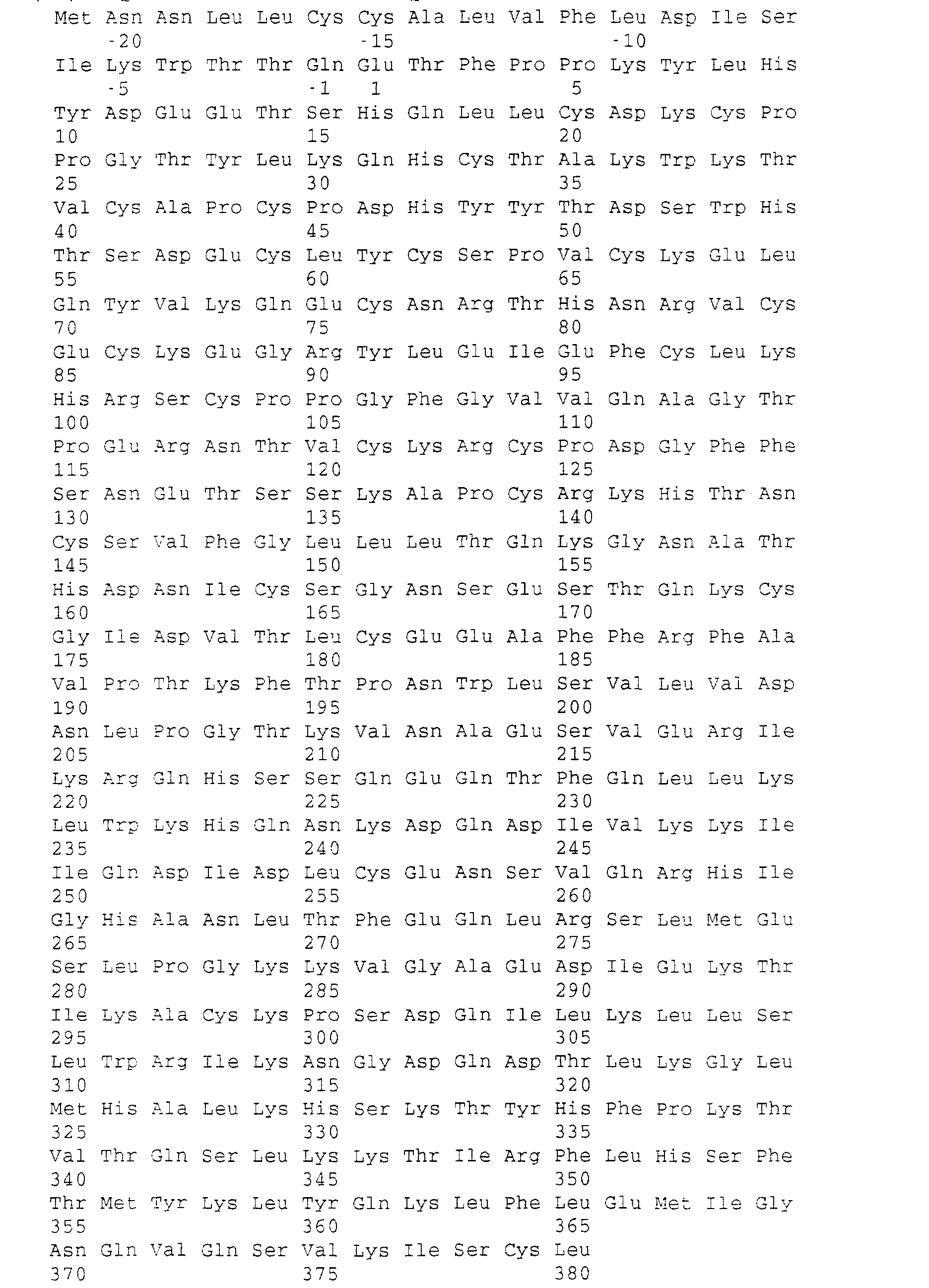


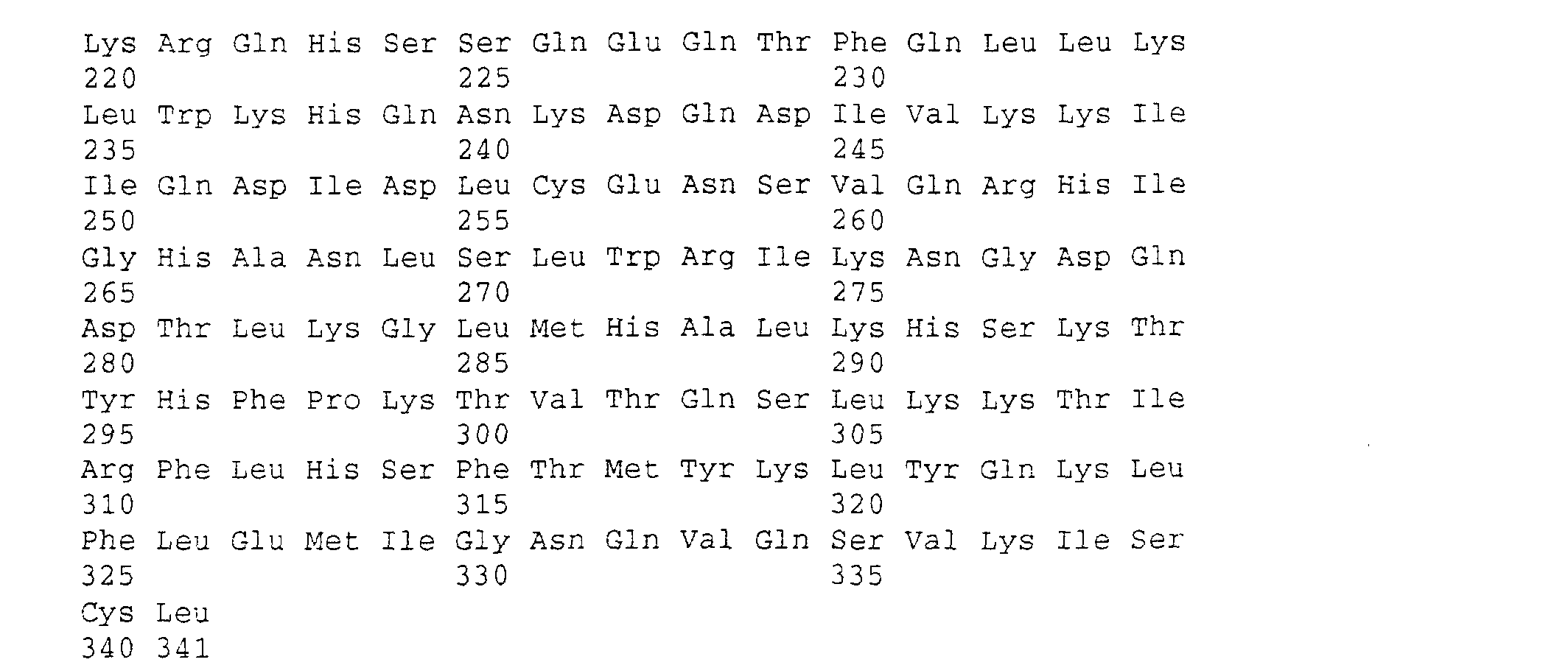


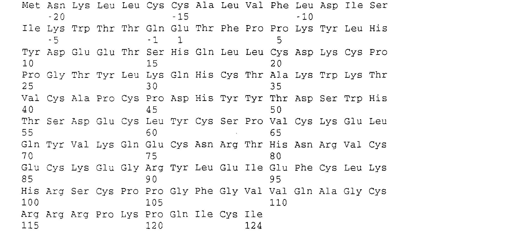


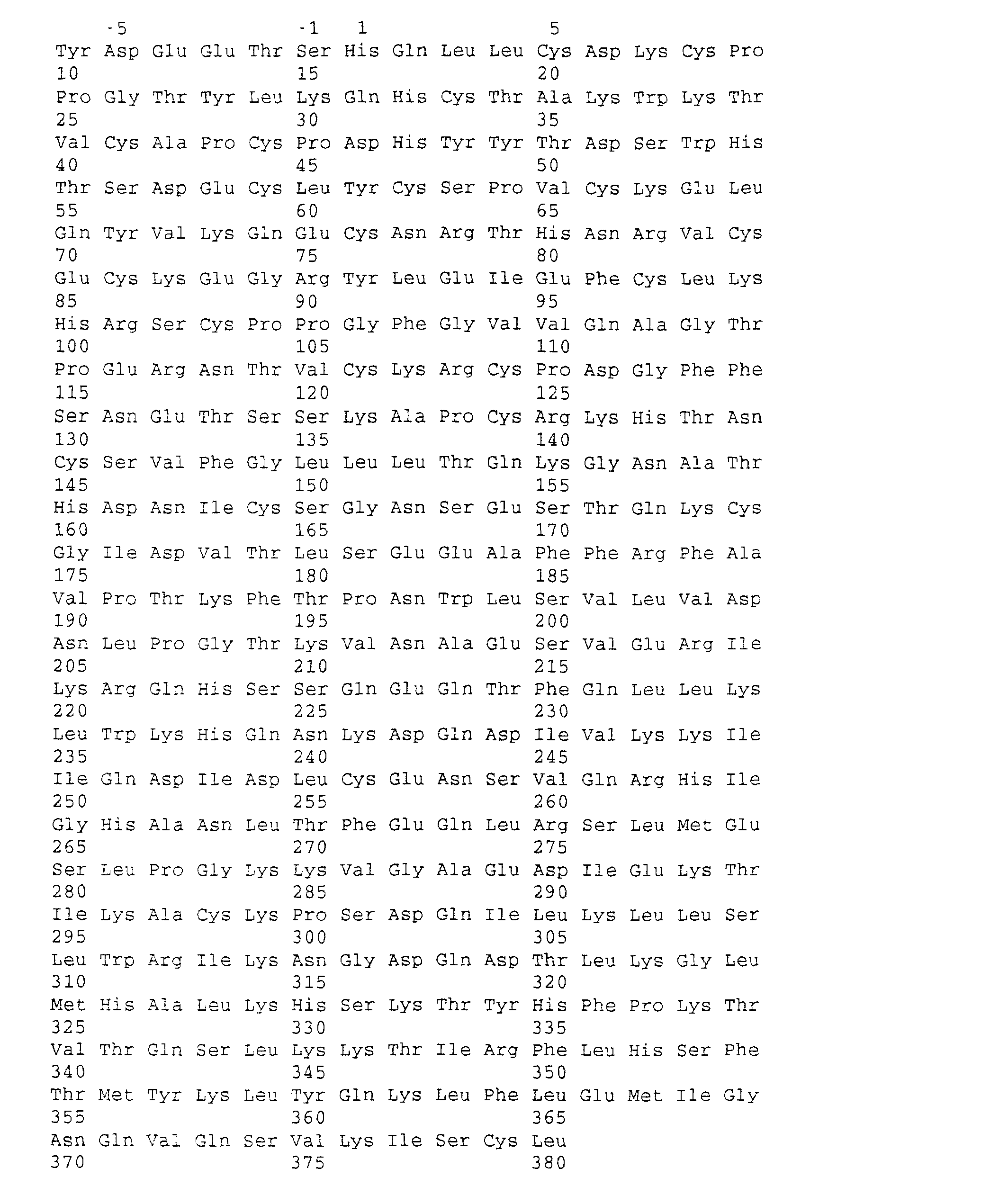


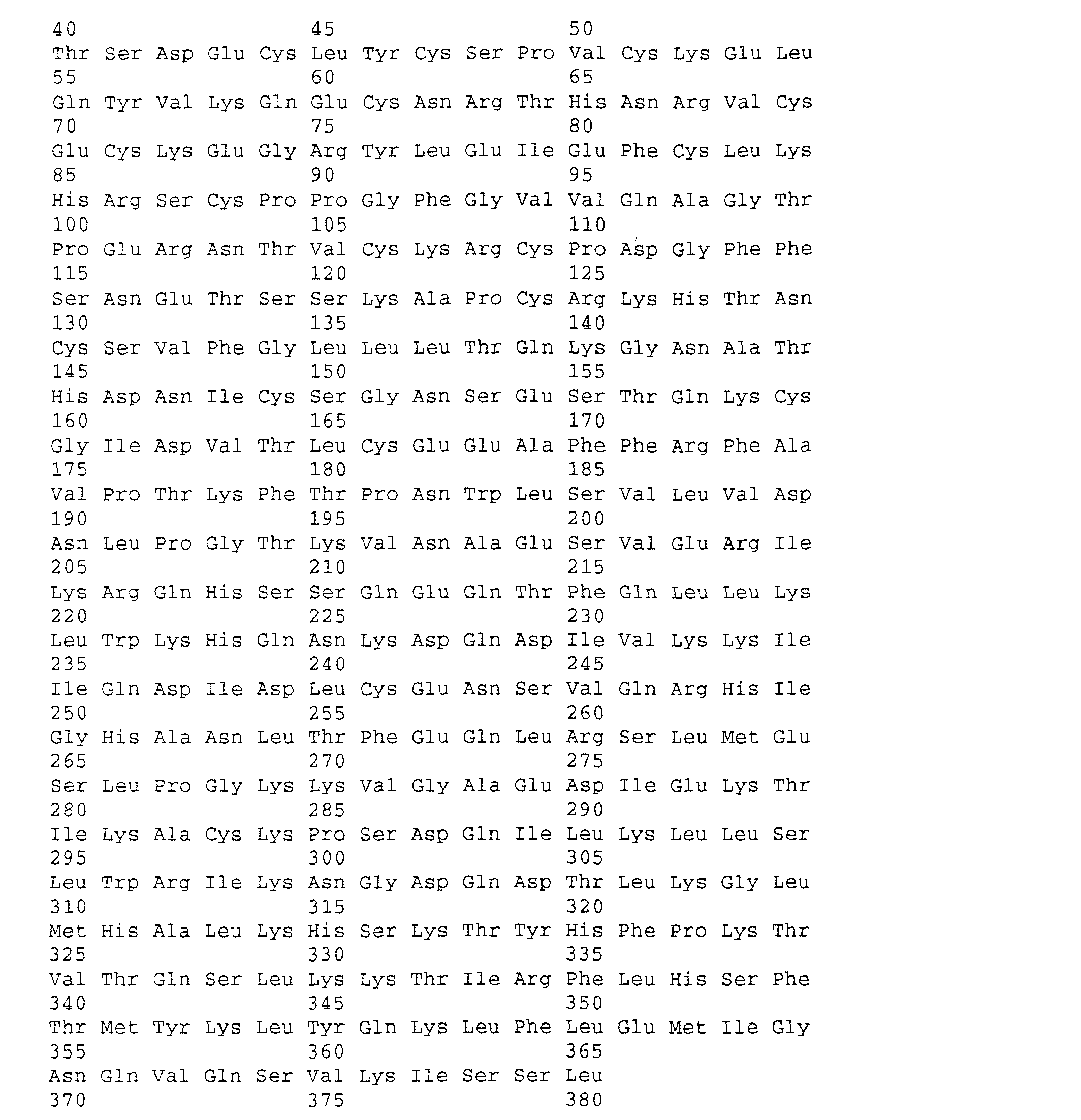

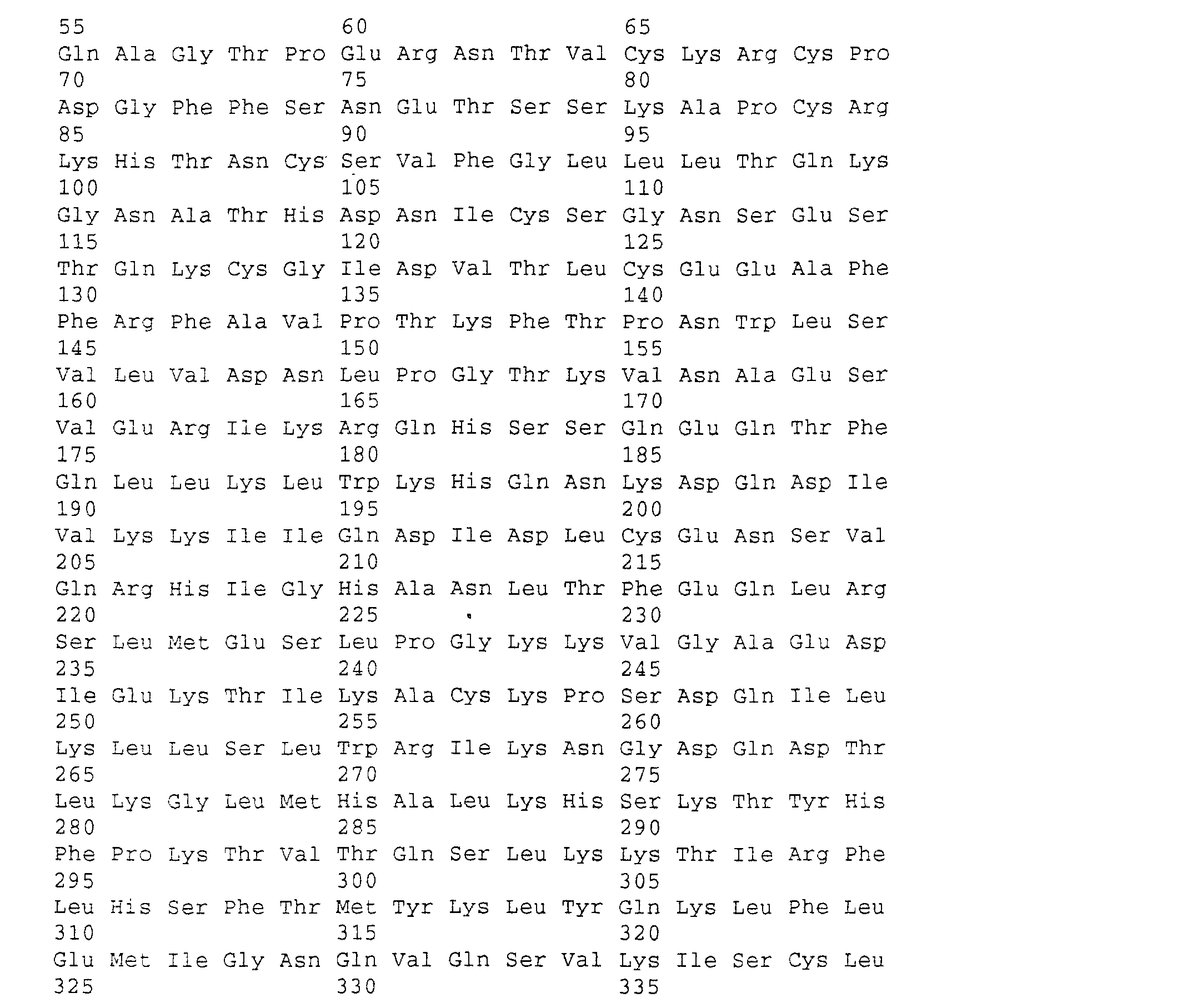


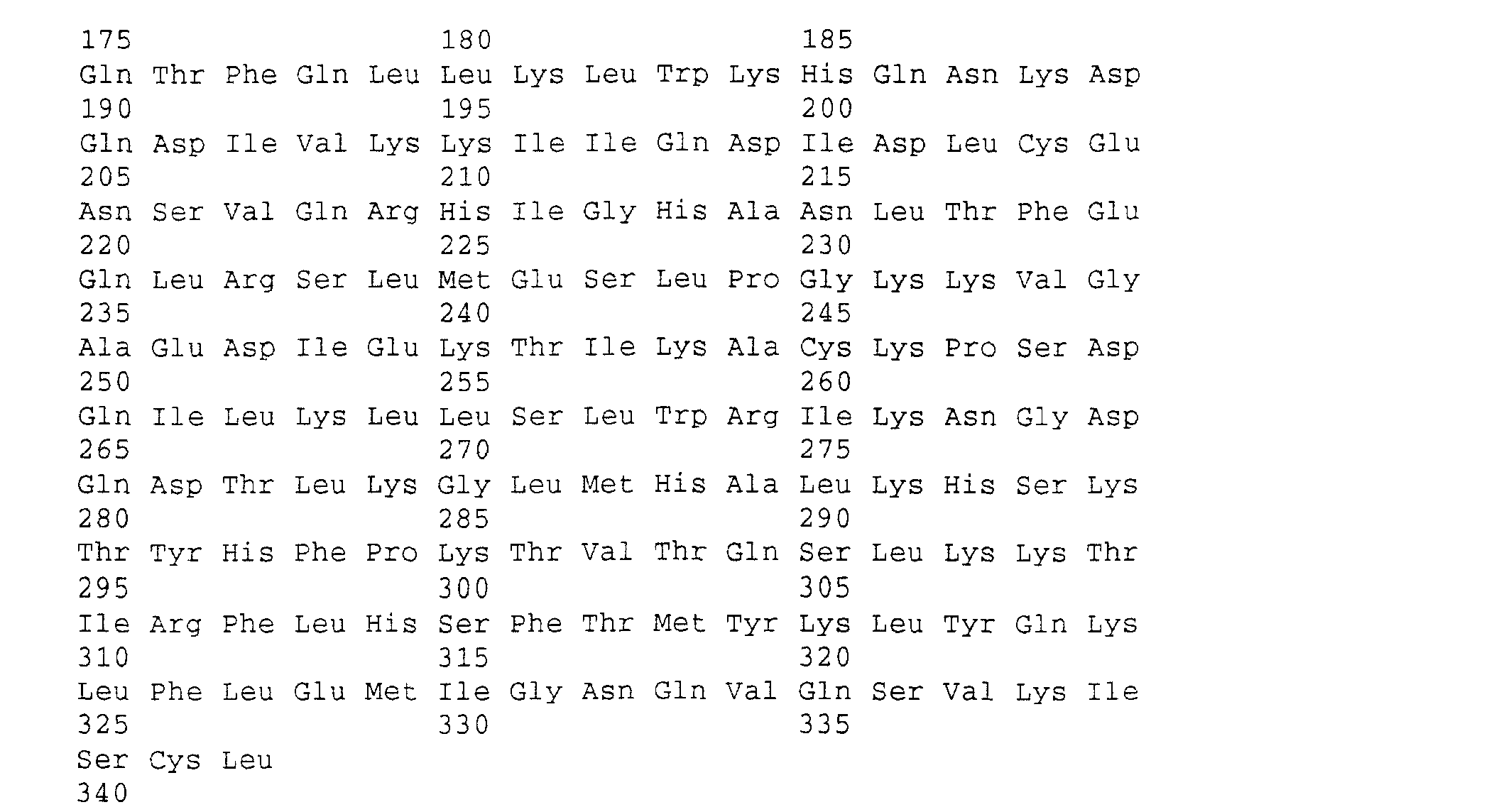

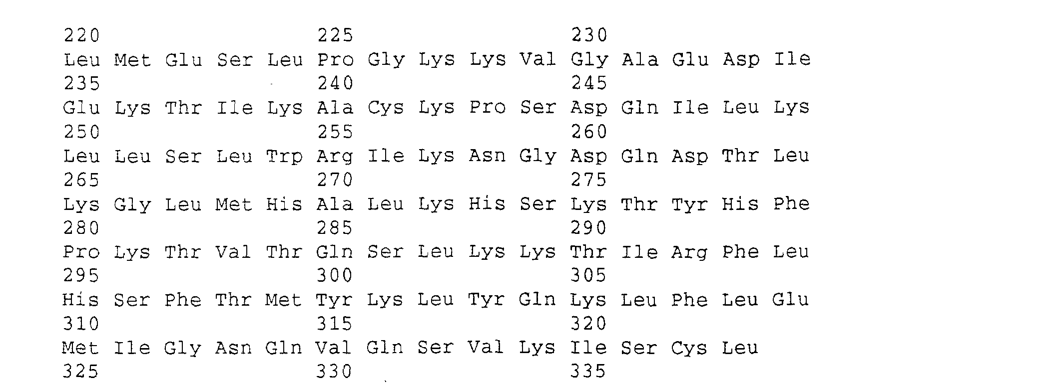
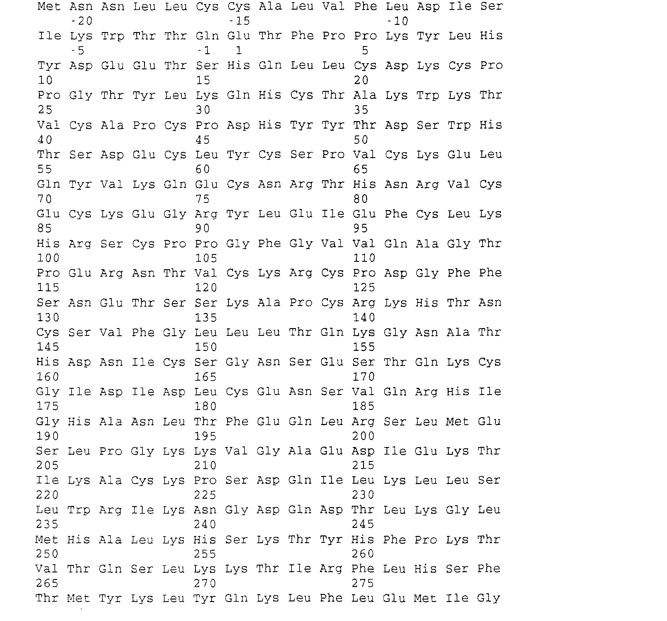

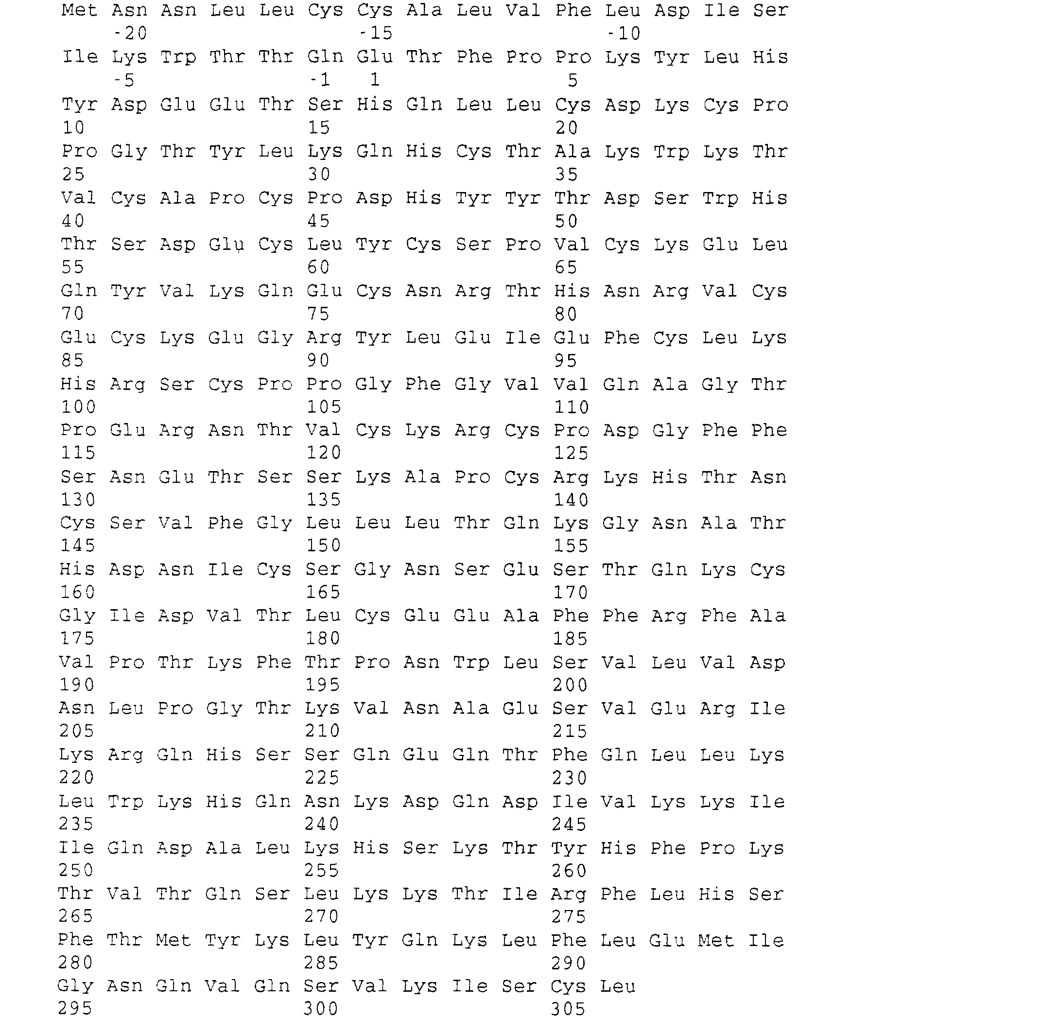


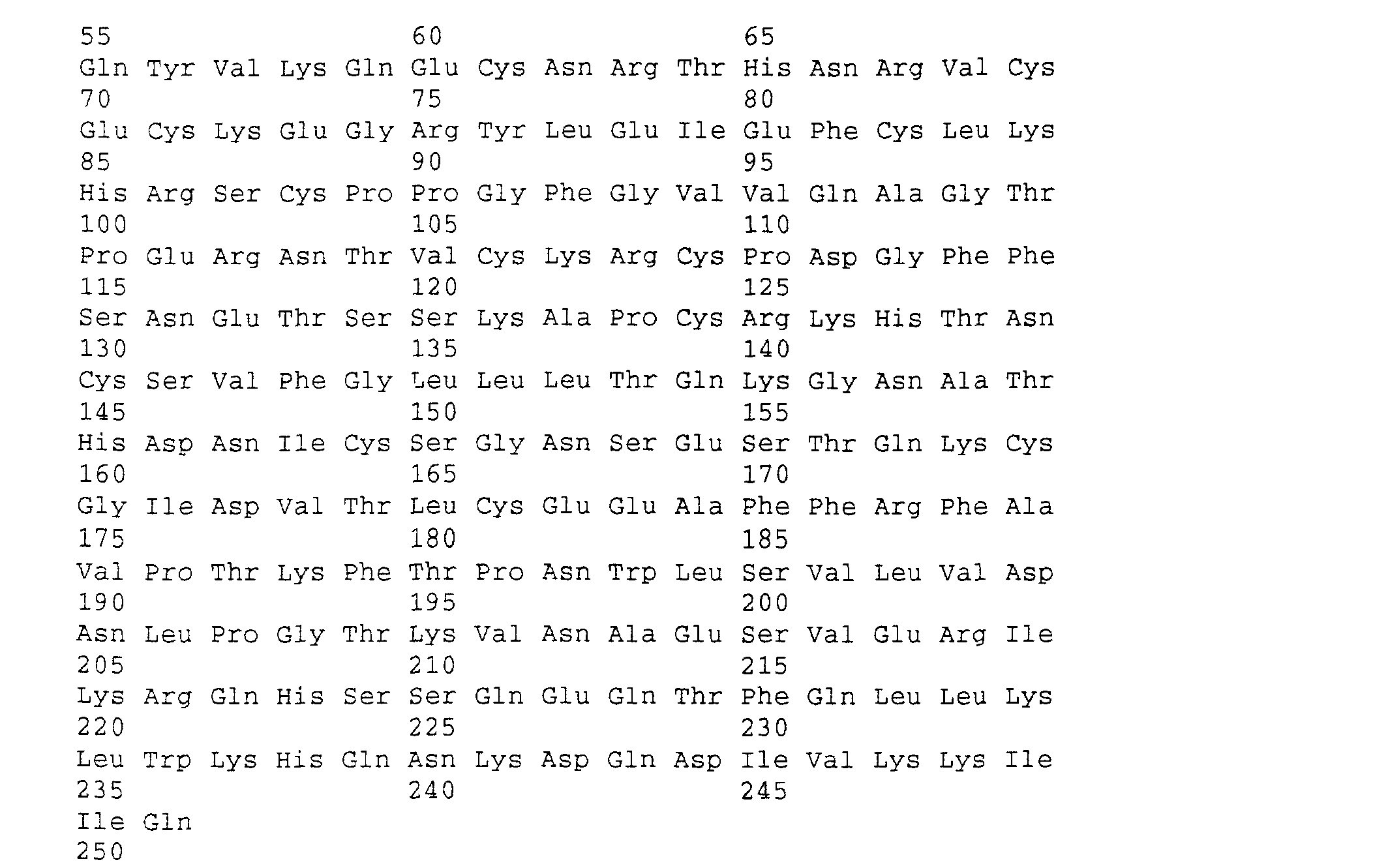


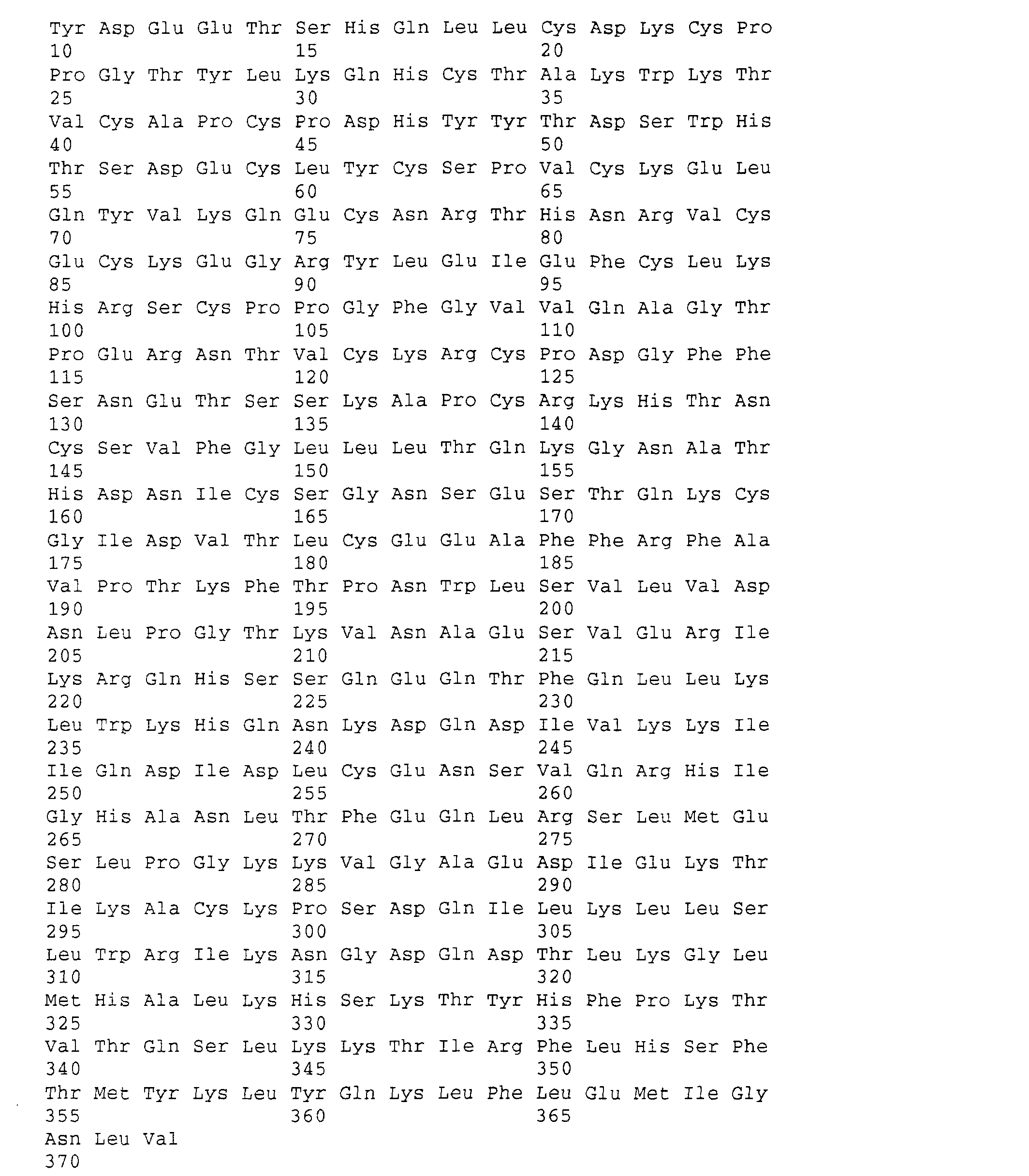


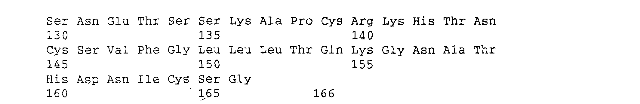


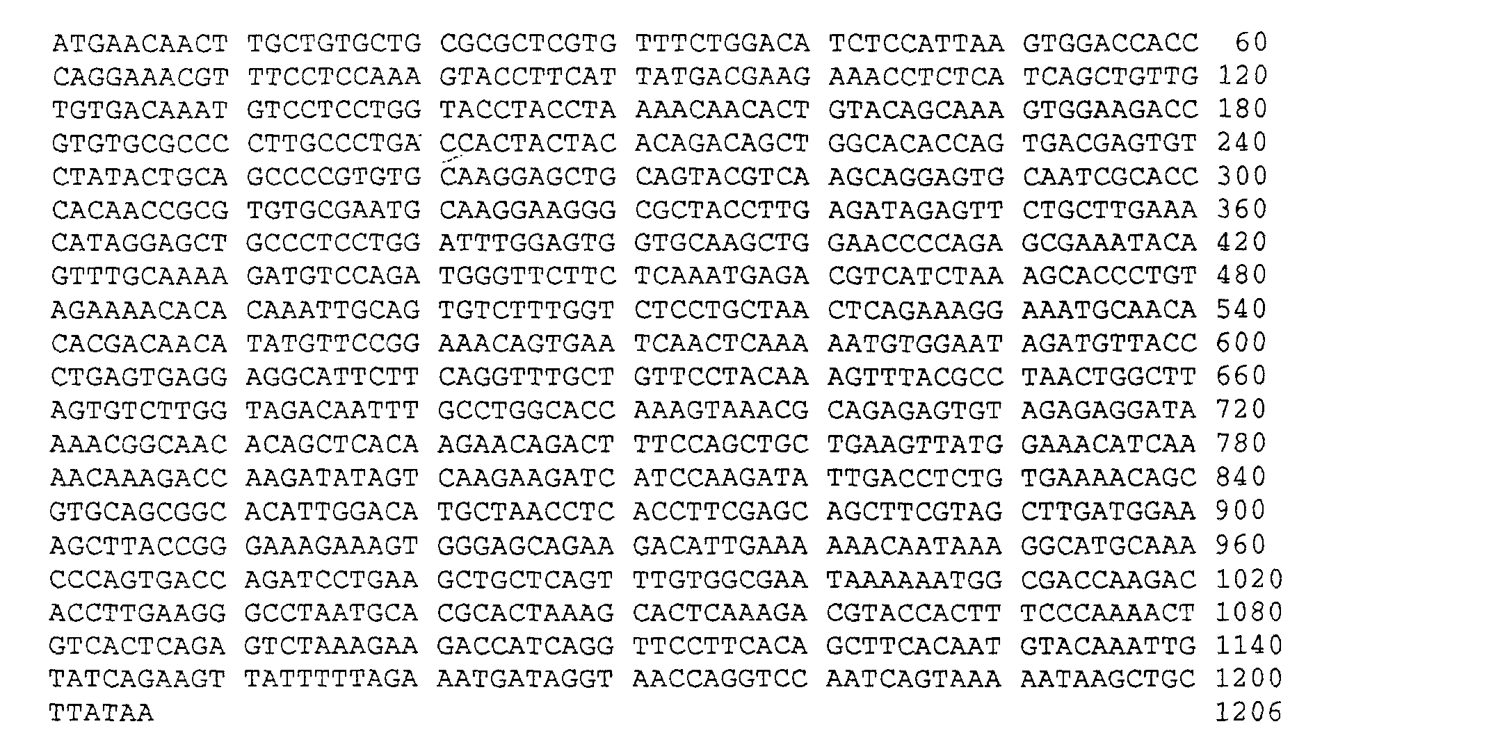


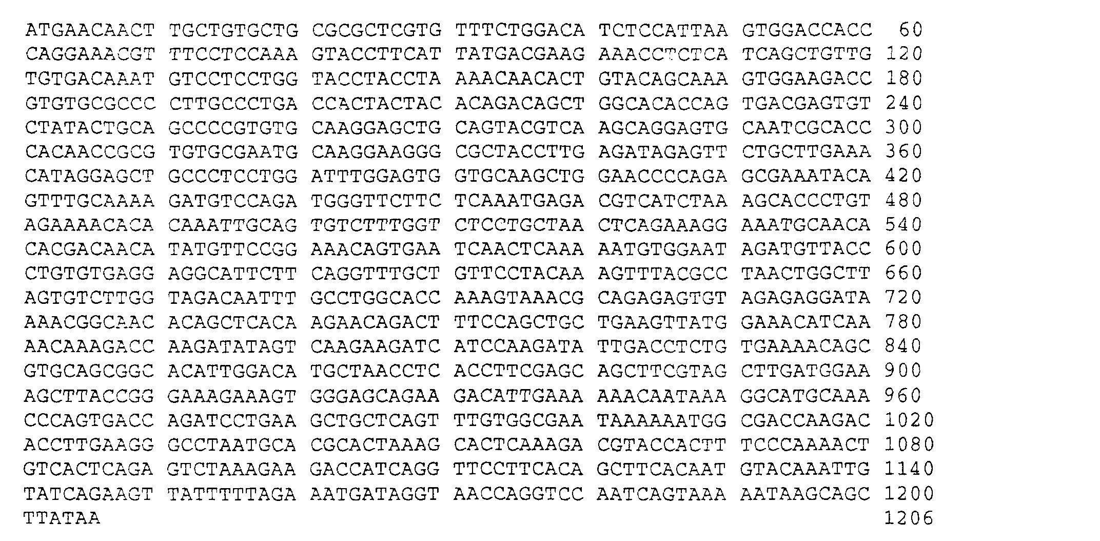


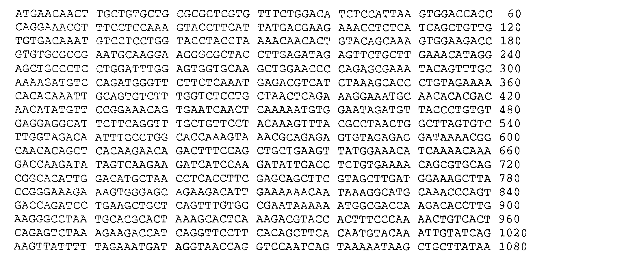
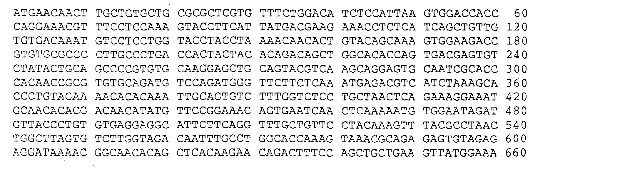


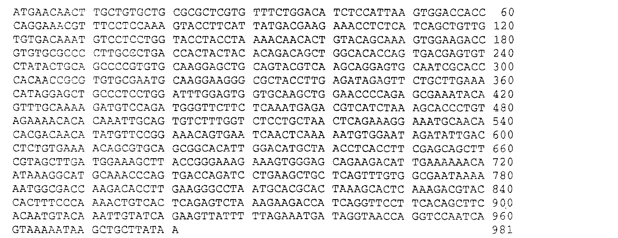


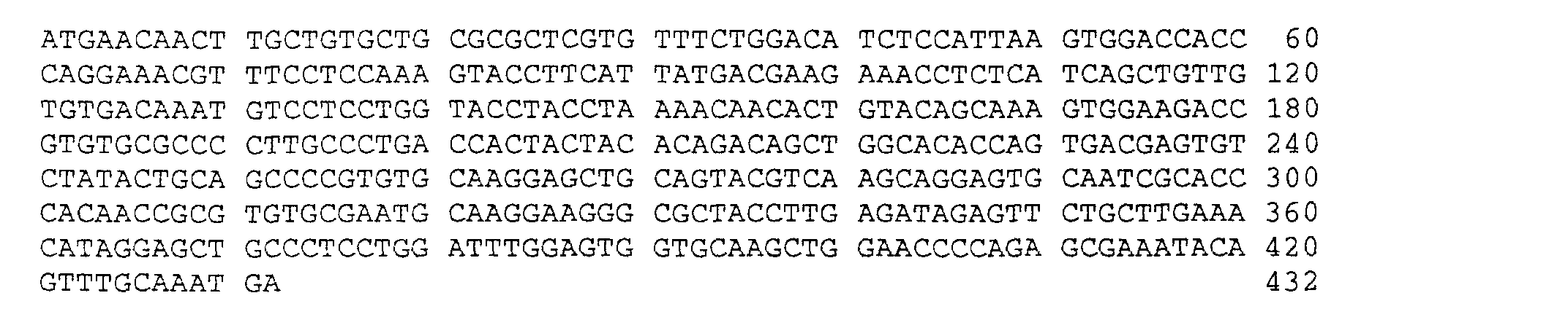


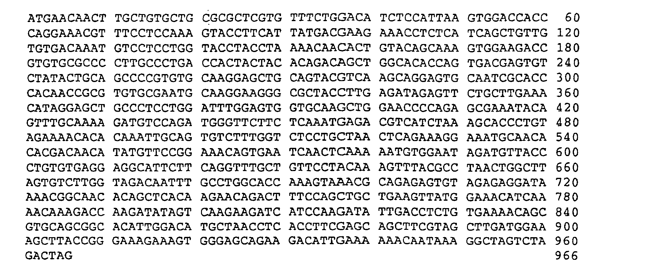
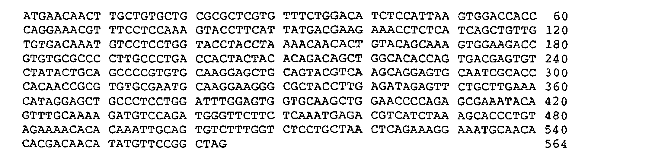
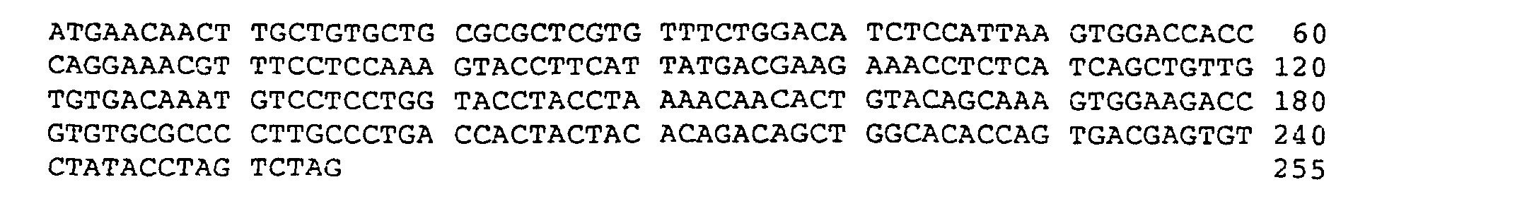


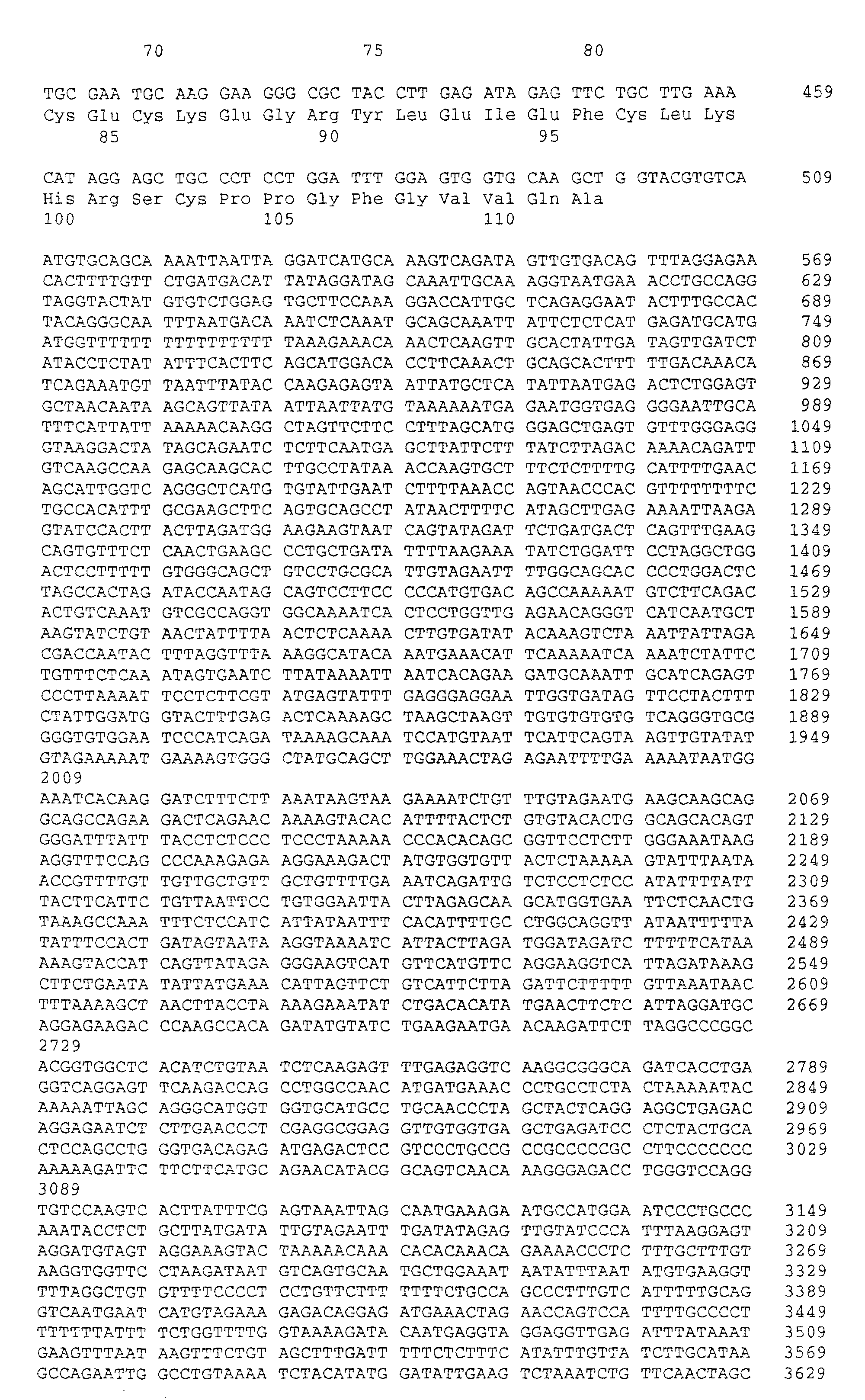
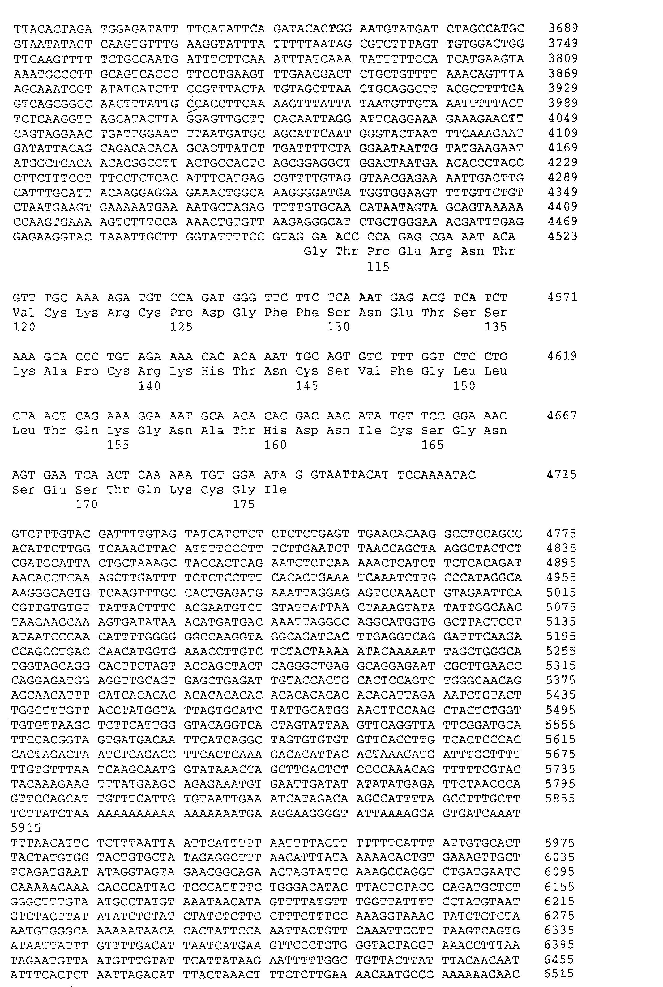


Claims (40)
- An OCIF protein characterised by the following properties:a) molecular weight on SDS-polyacrylamide gel electrophoresis (SDS-PAGE) :approximately 60 kD under reducing conditions;approximately 60 kD and 120 kD under non-reducing conditions;b) a high affinity to cation-exchange column and heparin column;c) a biological activity to inhibit osteoclast differentiation and/or maturation
its activity being decreased by heating at 70°C for 10 min or at 56°C for 30 min;
its activity being lost by heating at 90°C for 10 min;d) internal amino acid sequences provided by SEQ ID NOs 1,2 and 3. - A protein according to claim 1 having N-terminal amino acid sequences provided in SEQ ID NO 7.
- A protein according to claim 1 produced in human fibroblasts.
- A method of producing a protein according to claim 1, 2 or 3 by the following process:cultivating human fibroblasts; and purifying the protein by a combination of ion-exchange column, affinity column and reverse phase-column chromatography.
- A method according to claim 4 by cultivating human fibroblasts on alumina ceramic pieces.
- OCIF protein with amino acid sequence as provided in SEQ ID NO 4.
- OCIF cDNA encoding amino acid sequence as provided in SEQ ID NO 4.
- OCIF cDNA encoding amino acid sequence as provided in SEQ ID No.5
- OCIF cDNA which is inserted in a vector as 1.6 kb fragment carried by the transformed Escherichia coli pBK/01F10 (FERM BP-5267).
- OCIF cDNA with nucleotide sequence as provided in SEQ ID NO 6.
- OCIF cDNA that hybridise to cDNA as provided in SEQ ID NO 6 under stringent conditions.
- An OCIF protein expressed from cDNA encoding amino acid sequence as provided in SEQ ID NO 4.
- An OCIF protein with a biological activity to inhibit osteoclast differentiation and/or maturation, that is obtained as amino acid expressed cDNA sharing at least 80% sequence identity with the amino acid as provided in SEQ ID NO 4.
- A method for the production of a protein according to claim 1 by genetic engineering using cDNA of any of claims 7 to 11.
- The protein according to claim 1, said protein is obtainable by the method of claim 14.
- A method of producing the protein according to claim 12 by gene engineering using mammalian cells as host cells.
- A method of producing the protein according to claim 16 by gene engineering using 293/EBNA cells or CHO cells as mammalian host cells.
- The protein according to claim 6 or 12, said protein is obtainable by the method of claim 16 or 17.
- An OCIF variant, mutant or truncated cDNA selected from:a cDNA with nucleotide sequence as provided in SEQ ID NO 8;a cDNA with nucleotide sequence as provided in SEQ ID NO 10;a cDNA with nucleotide sequence as provided in SEQ ID NO 12;a cDNA with nucleotide sequence as provided in SEQ ID NO 14;a cDNA with nucleotide sequence as provided in SEQ ID NO 83;a cDNA with nucleotide sequence as provided in SEQ ID NO 84;a cDNA with nucleotide sequence as provided in SEQ ID NO 85;a cDNA with nucleotide sequence as provided in SEQ ID NO 86;a cDNA with nucleotide sequence as provided in SEQ ID NO 87;a cDNA with nucleotide sequence as provided in SEQ ID NO 88;a cDNA with nucleotide sequence as provided in SEQ ID NO 89;a cDNA with nucleotide sequence as provided in SEQ ID NO 90;a cDNA with nucleotide sequence as provided in SEQ ID NO 91;a cDNA with nucleotide sequence as provided in SEQ ID NO 92;a cDNA with nucleotide sequence as provided in SEQ ID NO 93;a cDNA with nucleotide sequence as provided in SEQ ID NO 94;a cDNA with nucleotide sequence as provided in SEQ ID NO 95;a cDNA with nucleotide sequence as provided in SEQ ID NO 96;a cDNA with nucleotide sequence as provided in SEQ ID NO 97;a cDNA with nucleotide sequence as provided in SEQ ID NO 98;a cDNA with nucleotide sequence as provided in SEQ ID NO 99;a cDNA with nucleotide sequence as provided in SEQ ID NO 100;a cDNA with nucleotide sequence as provided in SEQ ID NO 101;a cDNA with nucleotide sequence as provided in SEQ ID NO 102; anda cDNA with nucleotide sequence as provided in SEQ ID NO 103.
- A protein encoded by a cDNA selected from those of claim 19.
- An OCIF variant, mutant or truncated cDNA selected from:cDNA encoding amino acid sequence as provided in SEQ ID NO 9;cDNA encoding amino acid sequence as provided in SEQ ID NO 11;cDNA encoding amino acid sequence as provided in SEQ ID NO 13;cDNA encoding amino acid sequence as provided in SEQ ID NO 15;cDNA encoding amino acid sequence as provided in SEQ ID NO 62;cDNA encoding amino acid sequence as provided in SEQ ID NO 63;cDNA encoding amino acid sequence as provided in SEQ ID NO 64;cDNA encoding amino acid sequence as provided in SEQ ID NO 65;cDNA encoding amino acid sequence as provided in SEQ ID NO 66;cDNA encoding amino acid sequence as provided in SEQ ID NO 67;cDNA encoding amino acid sequence as provided in SEQ ID NO 68;cDNA encoding amino acid sequence as provided in SEQ ID NO 69;cDNA encoding amino acid sequence as provided in SEQ ID NO 70;cDNA encoding amino acid sequence as provided in SEQ ID NO 71;cDNA encoding amino acid sequence as provided in SEQ ID NO 72;cDNA encoding amino acid sequence as provided in SEQ ID NO 73;cDNA encoding amino acid sequence as provided in SEQ ID NO 74;cDNA encoding amino acid sequence as provided in SEQ ID NO 75;cDNA encoding amino acid sequence as provided in SEQ ID NO 76;cDNA encoding amino acid sequence as provided in SEQ ID NO 77;cDNA encoding amino acid sequence as provided in SEQ ID NO 78;cDNA encoding amino acid sequence as provided in SEQ ID NO 79;cDNA encoding amino acid sequence as provided in SEQ ID NO 80;cDNA encoding amino acid sequence as provided in SEQ ID NO 81; andcDNA encoding amino acid sequence as provided in SEQ ID NO 82.
- Genomic OCIF DNA encoding amino acid sequence as provided in SEQ ID NO 5.
- Genomic DNAs according to claim 22 with the nucleotide sequence as provided in SEQ ID NO 104 or 105.
- A OCIF variant, mutant or truncated protein selected from:a protein which has an amino acid sequence of amino acid numbers 1 to 373 of SEQ ID NO. 9;a protein which has an amino acid sequence of amino acid numbers 1 to 341 of SEQ ID NO. 11;a protein which has an amino acid sequence of amino acid numbers 1 to 133 of SEQ ID NO. 13;a protein which has an amino acid sequence of amino acid numbers 1 to 124 of SEQ ID NO. 15;a protein which has an amino acid sequence of amino acid numbers 1 to 380 of SEQ ID NO. 62;a protein which has an amino acid sequence of amino acid numbers 1 to 380 of SEQ ID NO. 63;a protein which has an amino acid sequence of amino acid numbers 1 to 380 of SEQ ID NO. 64;a protein which has an amino acid sequence of amino acid numbers 1 to 380 of SEQ ID NO. 65;a protein which has an amino acid sequence of amino acid numbers 1 to 380 of SEQ ID NO. 66;a protein which has an amino acid sequence of amino acid numbers 1 to 339 of SEQ ID NO. 67;a protein which has an amino acid sequence of amino acid numbers 1 to 338 of SEQ ID NO. 68;a protein which has an amino acid sequence of amino acid numbers 1 to 342 of SEQ ID NO. 69;a protein which has an amino acid sequence of amino acid numbers 1 to 338 of SEQ ID NO. 70;a protein which has an amino acid sequence of amino acid numbers 1 to 305 of SEQ ID NO. 71;a protein which has an amino acid sequence of amino acid numbers 1 to 306 of SEQ ID NO. 72;a protein which has an amino acid sequence of amino acid numbers 1 to 378 of SEQ ID NO. 73;a protein which has an amino acid sequence of amino acid numbers 1 to 330 of SEQ ID NO. 74;a protein which has an amino acid sequence of amino acid numbers 1 to 251 of SEQ ID NO. 75;a protein which has an amino acid sequence of amino acid numbers 1 to 176 of SEQ ID NO. 76;a protein which has an amino acid sequence of amino acid numbers 1 to 122 of SEQ ID NO. 77;a protein which has an amino acid sequence of amino acid numbers 1 to 85 of SEQ ID NO. 78;a protein which has an amino acid sequence of amino acid numbers 1 to 372 of SEQ ID NO. 79;a protein which has an amino acid sequence of amino acid numbers 1 to 300 of SEQ ID NO. 80;a protein which has an amino acid sequence of amino acid numbers 1 to 166 of SEQ ID NO. 81; anda protein which has an amino acid sequence of amino acid numbers 1 to 63 of SEQ ID NO. 82.
- OCIF cDNA encoding a protein according to claim 24.
- A pharmaceutical composition comprising a protein according to any of claims 1-3, 6, 12, 13, 15, 18, 20 and 24.
- An antibody having specific activity to a protein according to any of claims 1-3, 6, 12-13, 15, 18, 20 and 24.
- An antibody according to claim 27 that is a polyclonal antibody.
- A antibody according to claim 27 that is a monoclonal antibody.
- A monoclonal antibody according to claim 29 characterized by the following properties:molecular weight of about 150,000 and of subclass IgG1, IgG2a or IgG2b·
- A method of determining the concentration of the protein of the OCIF using the antibodies of any of claims 27 to 30.
- Use of a protein according to any of claims 1-3, 6, 12, 13, 15, 18, 20 and 24 in the preparation of medicament for the treatment or prevention of osteoporosis.
- Use of a protein according to any of claims 1-3, 6, 12, 13, 15, 18, 20 and 24 in the preparation of medicament for the treatment or prevention of decreased bone mass.
- Use of a protein according to any of claims 1-3, 6, 12, 13, 15, 18, 20 and 24 in the preparation of medicament for the treatment or prevention of an abnormal bone metabolism.
- A vector in which any of the following cDNA (a) to (d) is inserted:(a) OCIF cDNA encoding amino acid sequence as provided in SEQ ID No. 4;(b) OCIF cDNA with nucleotide sequence as provided in SEQ ID No. 6;(c) OCIF cDNA encoding amino acid sequence as provided in SEQ ID No. 5;(d) OCIF cDNA which is inserted in a vector as 1-6 Kb fragment carried by the transformed Escherichia coli pBK/01F10 (PERM BP-5267) and encodes a protein having a biological activity to inhibit osteoclast differentiation and/or maturation.
- The vector of claim 35, said vector is carried by the transformed Escherichia coli pBK/01F10 (FERM BP-5267).
- A host cell in which the vector according to claims 35 or 36 is introduced.
- A method for the production of a protein according to claim 1, said method comprises steps (I) and (II);(I) culturing the host cell according to claim 37;(II) recovering said protein from the obtained culture.
- A protein according to claim 1, said protein is obtainable by the method of claim 38.
- The transformed Escherichia coli pBK/01F10 (PERM BP-5267) .
Priority Applications (1)
| Application Number | Priority Date | Filing Date | Title |
|---|---|---|---|
| EP04076464A EP1528103B1 (en) | 1995-02-20 | 1996-02-20 | 'Osteoclast-inhibitory factor' (OCIF) derived proteins and methods for producing the proteins |
Applications Claiming Priority (7)
| Application Number | Priority Date | Filing Date | Title |
|---|---|---|---|
| JP5497795 | 1995-02-20 | ||
| JP5497795 | 1995-02-20 | ||
| JP54977/95 | 1995-02-20 | ||
| JP20750895 | 1995-07-21 | ||
| JP207508/95 | 1995-07-21 | ||
| JP20750895 | 1995-07-21 | ||
| PCT/JP1996/000374 WO1996026217A1 (en) | 1995-02-20 | 1996-02-20 | Novel protein and methods for the production of the same |
Related Child Applications (1)
| Application Number | Title | Priority Date | Filing Date |
|---|---|---|---|
| EP04076464A Division EP1528103B1 (en) | 1995-02-20 | 1996-02-20 | 'Osteoclast-inhibitory factor' (OCIF) derived proteins and methods for producing the proteins |
Publications (3)
| Publication Number | Publication Date |
|---|---|
| EP0816380A1 EP0816380A1 (en) | 1998-01-07 |
| EP0816380A4 EP0816380A4 (en) | 1998-11-18 |
| EP0816380B1 true EP0816380B1 (en) | 2004-08-25 |
Family
ID=26395808
Family Applications (2)
| Application Number | Title | Priority Date | Filing Date |
|---|---|---|---|
| EP96902484A Expired - Lifetime EP0816380B1 (en) | 1995-02-20 | 1996-02-20 | Ocif protein and methods for the production of the same |
| EP04076464A Expired - Lifetime EP1528103B1 (en) | 1995-02-20 | 1996-02-20 | 'Osteoclast-inhibitory factor' (OCIF) derived proteins and methods for producing the proteins |
Family Applications After (1)
| Application Number | Title | Priority Date | Filing Date |
|---|---|---|---|
| EP04076464A Expired - Lifetime EP1528103B1 (en) | 1995-02-20 | 1996-02-20 | 'Osteoclast-inhibitory factor' (OCIF) derived proteins and methods for producing the proteins |
Country Status (20)
| Country | Link |
|---|---|
| US (9) | US7125686B1 (en) |
| EP (2) | EP0816380B1 (en) |
| JP (1) | JP3502102B2 (en) |
| KR (1) | KR100381788B1 (en) |
| CN (1) | CN1218961C (en) |
| AT (2) | ATE274580T1 (en) |
| AU (1) | AU702557C (en) |
| CA (1) | CA2213469A1 (en) |
| DE (2) | DE69637486D1 (en) |
| ES (1) | ES2227579T3 (en) |
| FI (1) | FI973402L (en) |
| HU (1) | HU224570B1 (en) |
| IL (1) | IL117175A (en) |
| MX (1) | MX9706341A (en) |
| NO (1) | NO318898B1 (en) |
| NZ (1) | NZ301362A (en) |
| PT (1) | PT816380E (en) |
| RU (2) | RU2238948C2 (en) |
| TW (1) | TW538049B (en) |
| WO (1) | WO1996026217A1 (en) |
Cited By (1)
| Publication number | Priority date | Publication date | Assignee | Title |
|---|---|---|---|---|
| RU2324705C2 (en) * | 2001-04-03 | 2008-05-20 | Сосьете Де Продюи Нестле С.А. | Osteoprotegrin, its application for preparation of pharmaceutical composition (variants), production of food substance (variants) and fodder, food substance, fodder and pharmaceutical composition for prevention and treatment of disorders related to bone remodeling, and/or immune disorders |
Families Citing this family (61)
| Publication number | Priority date | Publication date | Assignee | Title |
|---|---|---|---|---|
| US7094564B1 (en) | 1995-03-15 | 2006-08-22 | Human Genome Sciences, Inc. | Human tumor necrosis factor receptor |
| US8110659B1 (en) | 1995-03-15 | 2012-02-07 | Human Genome Sciences, Inc. | Human tumor necrosis factor receptor-like genes |
| US7078493B1 (en) | 1995-03-15 | 2006-07-18 | Human Genome Sciences, Inc. | Antibodies to human tumor necrosis factor receptor-like genes |
| JPH11503007A (en) * | 1995-03-15 | 1999-03-23 | ヒューマン・ジェノム・サイエンシズ・インコーポレイテッド | Human tumor necrosis factor receptor |
| US7632922B1 (en) | 1995-12-22 | 2009-12-15 | Amgen, Inc. | Osteoprotegerin |
| US6369027B1 (en) * | 1995-12-22 | 2002-04-09 | Amgen Inc. | Osteoprotegerin |
| JPH1057071A (en) * | 1996-08-19 | 1998-03-03 | Snow Brand Milk Prod Co Ltd | Novel DNA and method for producing protein using the same |
| EP0961832A4 (en) * | 1996-09-18 | 2003-02-26 | Human Genome Sciences Inc | Human tumor necrosis factor receptor-like genes |
| ATE399861T1 (en) * | 1996-12-13 | 2008-07-15 | Schering Corp | SURFACE ANTIGENS FROM MAMMALS |
| DE122010000048I1 (en) | 1996-12-23 | 2011-05-05 | Immunex Corp | LIGAND FOR RECEPTOR ACTIVATOR OF NF-KAPPA B, LIGAND IS A MEMBER OF THE TNF SUPERFAMILY |
| US6271349B1 (en) | 1996-12-23 | 2001-08-07 | Immunex Corporation | Receptor activator of NF-κB |
| CA2781623A1 (en) | 1997-04-15 | 1998-10-22 | Daiichi Sankyo Company, Limited | Novel protein binding to osteoclastogenesis inhibitory factor and process for producing the same |
| US6316408B1 (en) | 1997-04-16 | 2001-11-13 | Amgen Inc. | Methods of use for osetoprotegerin binding protein receptors |
| EP1717315A3 (en) | 1997-04-16 | 2007-06-20 | Amgen Inc. | Osteoprotegerin binding proteins and receptors |
| AU7127598A (en) * | 1997-04-18 | 1998-11-13 | Millennium Pharmaceuticals, Inc. | Novel molecules of the fthma-070-related protein family and the t85-related pro tein family and uses thereof |
| EP0980432A1 (en) * | 1997-05-01 | 2000-02-23 | Amgen, Inc. | Chimeric opg polypeptides |
| US6346388B1 (en) | 1997-08-13 | 2002-02-12 | Smithkline Beecham Corporation | Method of identifying agonist and antagonists for tumor necrosis related receptors TR1 and TR2 |
| RU2203497C2 (en) * | 1997-09-24 | 2003-04-27 | Санкио Компани Лимитед | Method for diagnosing the cases of bone metabolism disorder diseases |
| US6297022B1 (en) | 1997-10-08 | 2001-10-02 | Smithkline Beecham Corporation | Method of identifying agonists and antagonists for tumor necrosis related receptor TR1 |
| US6087555A (en) * | 1997-10-15 | 2000-07-11 | Amgen Inc. | Mice lacking expression of osteoprotegerin |
| JPH11155420A (en) * | 1997-12-02 | 1999-06-15 | Snow Brand Milk Prod Co Ltd | Transgenic animal |
| DK2009025T3 (en) | 1998-05-14 | 2011-11-14 | Immunex Corp | Method for inhibiting osteoclast activity |
| AU6006099A (en) * | 1998-10-09 | 2000-05-01 | Sankyo Company Limited | Preventives or remedies for cachexia |
| EP1127578A4 (en) * | 1998-10-28 | 2004-12-15 | Sankyo Co | Remedies for bone metabolic errors |
| AUPQ167599A0 (en) * | 1999-07-19 | 1999-08-12 | St. Vincent's Institute Of Medical Research | Inhibitor of osteoclast precursor formation |
| AU7720600A (en) * | 1999-09-27 | 2001-04-30 | Eli Lilly And Company | Osteoprotegerin regulatory region |
| JP2001103964A (en) * | 1999-10-05 | 2001-04-17 | Teijin Ltd | Method for inhibiting osteoclast formation or method for inhibiting bone resorption |
| WO2002062990A1 (en) * | 2001-02-07 | 2002-08-15 | Sankyo Company, Limited | Antibody and utilization thereof |
| EP1399555A2 (en) * | 2001-02-09 | 2004-03-24 | Maxygen Holdings Ltd. c/o Close Brothers (Cayman) Limited | Rank ligand-binding polypeptides |
| CN1238379C (en) * | 2001-03-21 | 2006-01-25 | 中国科学院上海生命科学研究院 | New osteoclast formation inhibiting factor and its coding sequence and use |
| US7064189B2 (en) | 2001-05-25 | 2006-06-20 | Human Genome Sciences, Inc. | Antibodies that immunospecifically bind to trail receptors |
| CN103232539B (en) | 2001-06-26 | 2015-06-03 | 安姆根弗里蒙特公司 | Antibodies to opgl |
| CZ20022231A3 (en) * | 2001-06-29 | 2003-02-12 | Sankyo Company Limited | Complex consisting of osteoclastogenesis inhibiting factor and polysaccharide |
| JP4336467B2 (en) * | 2001-10-15 | 2009-09-30 | 株式会社林原生物化学研究所 | Screening method for substances capable of regulating the production of osteoclast formation inhibitory factor |
| EP1482978A1 (en) * | 2002-03-01 | 2004-12-08 | Sankyo Company, Limited | Pharmaceutical composition comprising osteoclastogenesis inhibitory factor |
| JP4761710B2 (en) | 2002-04-05 | 2011-08-31 | アムジェン インコーポレイテッド | Human anti-OPGL neutralizing antibodies as selective OPGL pathway inhibitors |
| JP2006509504A (en) * | 2002-12-11 | 2006-03-23 | ユニバーシティー オブ マサチューセッツ | Method for introducing siRNA into adipocytes |
| RU2323227C2 (en) * | 2003-03-24 | 2008-04-27 | Санкио Компани Лимитед | Polymeric modifying agents and pharmaceutical compositions |
| AU2004260961B2 (en) * | 2003-07-29 | 2010-05-20 | Abbott Laboratories Gmbh | Analytical method for pancreatin and comparable compositions |
| US7740861B2 (en) * | 2004-06-16 | 2010-06-22 | University Of Massachusetts | Drug delivery product and methods |
| TWI364458B (en) * | 2004-08-27 | 2012-05-21 | Wyeth Res Ireland Ltd | Production of tnfr-lg |
| ES2375995T3 (en) | 2004-09-17 | 2012-03-08 | University Of Massachusetts | COMPOSITIONS AND THEIR USES FOR DEFICIENCIES OF LISOSOMAL ENZYME. |
| EP1874331B1 (en) * | 2005-04-19 | 2014-09-03 | Kings College London | Use of bip against bone loss and osteoporosis |
| RU2354662C2 (en) * | 2005-07-21 | 2009-05-10 | Андрей Георгиевич Зарайский | Noggin2-based activin activity blocking method |
| JP5489462B2 (en) * | 2005-10-24 | 2014-05-14 | ユニバーシティ オブ マサチューセッツ | Compositions for gene therapy of bone pathology and methods of use thereof |
| ES2839549T3 (en) * | 2005-11-23 | 2021-07-05 | Acceleron Pharma Inc | Activin-ActRIIa antagonists for use in stimulating bone growth |
| US10741034B2 (en) | 2006-05-19 | 2020-08-11 | Apdn (B.V.I.) Inc. | Security system and method of marking an inventory item and/or person in the vicinity |
| KR101577935B1 (en) * | 2007-10-11 | 2015-12-16 | 다이이찌 산쿄 가부시키가이샤 | ANTIBODY TARGETING OSTEOCLAST-RELATED PROTEIN Siglec-15 |
| CA2704056A1 (en) * | 2007-10-29 | 2009-05-07 | University Of Massachusetts | Encapsulated nanoparticles for nucleic acid delivery |
| NZ605450A (en) | 2009-04-09 | 2014-07-25 | Daiichi Sankyo Co Ltd | Anti-siglec-15 antibody |
| WO2012045481A2 (en) | 2010-10-05 | 2012-04-12 | Daiichi Sankyo Company, Limited | Antibody targeting osteoclast-related protein siglec-15 |
| CN102011025A (en) * | 2010-10-11 | 2011-04-13 | 宁波市鄞州品达电器焊料有限公司 | Ultrahigh-temperature-resistant lead-free solder |
| WO2013147213A1 (en) | 2012-03-30 | 2013-10-03 | 第一三共株式会社 | Anti-siglec15 antibody with modified cdr |
| US9963740B2 (en) * | 2013-03-07 | 2018-05-08 | APDN (B.V.I.), Inc. | Method and device for marking articles |
| WO2015054188A1 (en) | 2013-10-07 | 2015-04-16 | Apdn (B.V.I), Inc. | Multimode image and spectral reader |
| US10745825B2 (en) | 2014-03-18 | 2020-08-18 | Apdn (B.V.I.) Inc. | Encrypted optical markers for security applications |
| CN106103121B (en) | 2014-03-18 | 2019-12-06 | 亚普蒂恩(B.V.I.)公司 | Encrypted optical marker for security applications |
| JP6339855B2 (en) * | 2014-05-14 | 2018-06-06 | 住友ゴム工業株式会社 | Airless tire and manufacturing method thereof |
| CN109070130B (en) | 2016-04-11 | 2022-03-22 | 亚普蒂恩(B V I)公司 | Method for marking cellulose products |
| US10995371B2 (en) | 2016-10-13 | 2021-05-04 | Apdn (B.V.I.) Inc. | Composition and method of DNA marking elastomeric material |
| US10920274B2 (en) | 2017-02-21 | 2021-02-16 | Apdn (B.V.I.) Inc. | Nucleic acid coated submicron particles for authentication |
Family Cites Families (88)
| Publication number | Priority date | Publication date | Assignee | Title |
|---|---|---|---|---|
| US175840A (en) * | 1876-04-11 | Improvement in circular-knitting machines | ||
| US100069A (en) * | 1870-02-22 | Geokge h | ||
| US139325A (en) * | 1873-05-27 | Improvement in carriage-springs | ||
| US166097A (en) * | 1875-07-27 | Improvement in buttons for wearing apparel | ||
| US207827A (en) * | 1878-09-10 | Improvement in siphon-condensers | ||
| US127637A (en) * | 1872-06-04 | Improvement in couplings for divided axles | ||
| US176647A (en) * | 1876-04-25 | Improvement in car-couplings | ||
| US208045A (en) * | 1878-09-17 | Improvement in ether-meters for refrigerating-machines | ||
| US104485A (en) * | 1870-06-21 | Improvement in sleeping-cars | ||
| US169117A (en) * | 1875-10-26 | Improvement in revolution-indicators | ||
| US86827A (en) * | 1869-02-09 | Improved rubber boot | ||
| US81720A (en) * | 1868-09-01 | Isaac williams | ||
| US150989A (en) * | 1874-05-19 | Improvement in pin and dowel machines | ||
| US33535A (en) * | 1861-10-22 | Improvement in machines for making bricks | ||
| US45456A (en) * | 1864-12-13 | Improved apparatus for carbureting air | ||
| US216297A (en) * | 1879-06-10 | Improvement in ironing-stands | ||
| US144480A (en) * | 1873-11-11 | Improvement in cutting attachments for sewing-machines | ||
| US103978A (en) * | 1870-06-07 | Improvement in bee-hives | ||
| US181418A (en) * | 1876-08-22 | Improvement in gas apparatus | ||
| US86826A (en) * | 1869-02-09 | Improvement in mechanical movement | ||
| US100488A (en) * | 1870-03-01 | Improved tire-xjfsetting machine | ||
| US886826A (en) * | 1907-12-14 | 1908-05-05 | Reece Button Hole Machine Co | Button-sewing machine. |
| US4179337A (en) | 1973-07-20 | 1979-12-18 | Davis Frank F | Non-immunogenic polypeptides |
| US4710473A (en) | 1983-08-10 | 1987-12-01 | Amgen, Inc. | DNA plasmids |
| WO1986000922A1 (en) | 1984-07-30 | 1986-02-13 | The Salk Institute For Biological Studies | Retroviral gene transfer vectors |
| US4959314A (en) * | 1984-11-09 | 1990-09-25 | Cetus Corporation | Cysteine-depleted muteins of biologically active proteins |
| JP2604135B2 (en) | 1986-02-28 | 1997-04-30 | ライオン株式会社 | Oral bone disease treatment |
| US5266683A (en) * | 1988-04-08 | 1993-11-30 | Stryker Corporation | Osteogenic proteins |
| JPH04502854A (en) | 1988-10-24 | 1992-05-28 | カターソン,ブルース | Methods and compositions for early stage diagnosis, monitoring and treatment of osteoarthritis |
| WO1990014363A1 (en) | 1989-05-19 | 1990-11-29 | Amgen Inc. | Metalloproteinase inhibitor |
| RU2018840C1 (en) * | 1989-12-11 | 1994-08-30 | Российский научный центр "Восстановительная травматология и ортопедия" им.акад.Г.А.Илизарова | Method for modelling stimulation of reparative osteogenesis under conditions of distraction osteosynthesis |
| WO1993012227A1 (en) | 1991-12-17 | 1993-06-24 | Genpharm International, Inc. | Transgenic non-human animals capable of producing heterologous antibodies |
| AU651421B2 (en) | 1990-11-30 | 1994-07-21 | Celtrix Pharmaceuticals, Inc. | Use of a bone morphogenetic protein in synergistic combination with TGF-beta for bone repair |
| US5118667A (en) | 1991-05-03 | 1992-06-02 | Celtrix Pharmaceuticals, Inc. | Bone growth factors and inhibitors of bone resorption for promoting bone formation |
| CA2068389A1 (en) | 1991-05-13 | 1992-11-14 | Masahiko Sato | Method for inhibiting bone resorption |
| TW318142B (en) | 1991-06-03 | 1997-10-21 | Mitsubishi Chemicals Co Ltd | |
| MX9204303A (en) | 1991-07-23 | 1993-11-01 | Rhone Poulenc Rorer Int | REGULATORY FACTOR OF OSTEOCLAST GROWTH. |
| IL99120A0 (en) | 1991-08-07 | 1992-07-15 | Yeda Res & Dev | Multimers of the soluble forms of tnf receptors,their preparation and pharmaceutical compositions containing them |
| US5366859A (en) | 1991-10-31 | 1994-11-22 | Mitsubishi Petrochemical Co., Ltd. | Radioimmunoassay method |
| US5447851B1 (en) | 1992-04-02 | 1999-07-06 | Univ Texas System Board Of | Dna encoding a chimeric polypeptide comprising the extracellular domain of tnf receptor fused to igg vectors and host cells |
| US5427954A (en) | 1992-04-29 | 1995-06-27 | Shriner's Hospitals For Crippled Children | Compositions and methods for detection and treatment of human osteoarthritis |
| JPH07509223A (en) | 1992-04-30 | 1995-10-12 | アムジェン インコーポレイテッド | Methods for treating interleukin-1-mediated and tumor necrosis factor-mediated diseases |
| US5585479A (en) | 1992-07-24 | 1996-12-17 | The United States Of America As Represented By The Secretary Of The Navy | Antisense oligonucleotides directed against human ELAM-I RNA |
| US5578569A (en) | 1993-03-12 | 1996-11-26 | Tam; Cherk S. | Method of increasing bone growth |
| US5457035A (en) | 1993-07-23 | 1995-10-10 | Immunex Corporation | Cytokine which is a ligand for OX40 |
| JPH09502614A (en) | 1993-09-14 | 1997-03-18 | メルク エンド カンパニー インコーポレーテッド | CDNA encoding a novel human protein tyrosine phosphatase |
| US6268212B1 (en) | 1993-10-18 | 2001-07-31 | Amgen Inc. | Tissue specific transgene expression |
| RU2074193C1 (en) * | 1994-06-21 | 1997-02-27 | Игорь Эдуардович Есиповский | Method of purification of human alpha-fetoprotein |
| WO1996000240A1 (en) | 1994-06-27 | 1996-01-04 | Snow Brand Milk Products Co., Ltd. | Novel protein and process for producing the same |
| EP0727211A1 (en) | 1995-02-10 | 1996-08-21 | Smithkline Beecham Corporation | Use of src SH2 specific compounds to treat a bone resorption disease |
| US6001349A (en) | 1995-02-22 | 1999-12-14 | Therion Biologics Corporation | Generation of human cytotoxic T-cells specific for carcinoma self-associated antigens and uses thereof |
| US20030166097A1 (en) | 1995-03-15 | 2003-09-04 | Human Genome Sciences, Inc. | Human tumor necrosis factor receptor |
| US7094564B1 (en) | 1995-03-15 | 2006-08-22 | Human Genome Sciences, Inc. | Human tumor necrosis factor receptor |
| WO1996028546A1 (en) * | 1995-03-15 | 1996-09-19 | Human Genome Sciences, Inc. | Human tumor necrosis factor receptor |
| US5843901A (en) | 1995-06-07 | 1998-12-01 | Advanced Research & Technology Institute | LHRH antagonist peptides |
| WO1997000318A1 (en) | 1995-06-07 | 1997-01-03 | Osteosa Inc. | Osteoclast growth regulatory factor |
| WO1997000317A1 (en) | 1995-06-07 | 1997-01-03 | Osteosa Inc. | Osteoclast growth regulatory factor |
| US6613544B1 (en) | 1995-12-22 | 2003-09-02 | Amgen Inc. | Osteoprotegerin |
| US6369027B1 (en) | 1995-12-22 | 2002-04-09 | Amgen Inc. | Osteoprotegerin |
| US20030207827A1 (en) | 1995-12-22 | 2003-11-06 | William J. Boyle | Osteoprotegerin |
| JPH1057071A (en) | 1996-08-19 | 1998-03-03 | Snow Brand Milk Prod Co Ltd | Novel DNA and method for producing protein using the same |
| US5830850A (en) | 1996-08-28 | 1998-11-03 | Mount Sinai School Of Medicine Of The City Of New York | Methods for the treatment of bone resorption disorders, including osteoporosis |
| ATE399861T1 (en) | 1996-12-13 | 2008-07-15 | Schering Corp | SURFACE ANTIGENS FROM MAMMALS |
| EP0951546A2 (en) | 1996-12-20 | 1999-10-27 | The Board Of Regents, The University Of Texas System | Compositions and methods of use for osteoclast inhibitory factors |
| US6271349B1 (en) * | 1996-12-23 | 2001-08-07 | Immunex Corporation | Receptor activator of NF-κB |
| DE122010000048I1 (en) * | 1996-12-23 | 2011-05-05 | Immunex Corp | LIGAND FOR RECEPTOR ACTIVATOR OF NF-KAPPA B, LIGAND IS A MEMBER OF THE TNF SUPERFAMILY |
| CA2781623A1 (en) | 1997-04-15 | 1998-10-22 | Daiichi Sankyo Company, Limited | Novel protein binding to osteoclastogenesis inhibitory factor and process for producing the same |
| US5843678A (en) | 1997-04-16 | 1998-12-01 | Amgen Inc. | Osteoprotegerin binding proteins |
| US6316408B1 (en) | 1997-04-16 | 2001-11-13 | Amgen Inc. | Methods of use for osetoprotegerin binding protein receptors |
| EP1717315A3 (en) | 1997-04-16 | 2007-06-20 | Amgen Inc. | Osteoprotegerin binding proteins and receptors |
| US6656508B2 (en) | 1997-04-17 | 2003-12-02 | Amgen Inc. | Sustained-release alginate gels |
| EP0980432A1 (en) | 1997-05-01 | 2000-02-23 | Amgen, Inc. | Chimeric opg polypeptides |
| AU7705098A (en) | 1997-05-29 | 1998-12-30 | Human Genome Sciences, Inc. | Human tumor necrosis factor receptor-like protein 8 |
| RU2203497C2 (en) | 1997-09-24 | 2003-04-27 | Санкио Компани Лимитед | Method for diagnosing the cases of bone metabolism disorder diseases |
| US6297022B1 (en) * | 1997-10-08 | 2001-10-02 | Smithkline Beecham Corporation | Method of identifying agonists and antagonists for tumor necrosis related receptor TR1 |
| US6087555A (en) | 1997-10-15 | 2000-07-11 | Amgen Inc. | Mice lacking expression of osteoprotegerin |
| US6790823B1 (en) | 1998-04-23 | 2004-09-14 | Amgen Inc. | Compositions and methods for the prevention and treatment of cardiovascular diseases |
| DK2009025T3 (en) | 1998-05-14 | 2011-11-14 | Immunex Corp | Method for inhibiting osteoclast activity |
| EP1127578A4 (en) | 1998-10-28 | 2004-12-15 | Sankyo Co | Remedies for bone metabolic errors |
| AU6078500A (en) | 1999-07-09 | 2001-01-30 | Amgen, Inc. | Combination therapy for conditions leading to bone loss |
| US20030144187A1 (en) | 1999-09-03 | 2003-07-31 | Colin R. Dunstan | Opg fusion protein compositions and methods |
| JP2003508491A (en) | 1999-09-03 | 2003-03-04 | アムジエン・インコーポレーテツド | Compositions and methods for the prevention or treatment of cancer and bone loss associated with cancer |
| WO2001044472A1 (en) | 1999-12-16 | 2001-06-21 | Amgen, Inc. | Tnfr/opg-like molecules and uses thereof |
| US20030103978A1 (en) | 2000-02-23 | 2003-06-05 | Amgen Inc. | Selective binding agents of osteoprotegerin binding protein |
| CN103232539B (en) | 2001-06-26 | 2015-06-03 | 安姆根弗里蒙特公司 | Antibodies to opgl |
| CZ20022231A3 (en) | 2001-06-29 | 2003-02-12 | Sankyo Company Limited | Complex consisting of osteoclastogenesis inhibiting factor and polysaccharide |
| EP1482978A1 (en) | 2002-03-01 | 2004-12-08 | Sankyo Company, Limited | Pharmaceutical composition comprising osteoclastogenesis inhibitory factor |
| JP2009521229A (en) | 2005-12-23 | 2009-06-04 | ジェイ.クレイグ ベンター インスティチュート | Installing genomes or partial genomes into cells or cell-like systems |
-
1996
- 1996-02-19 IL IL11717596A patent/IL117175A/en active IP Right Review Request
- 1996-02-20 WO PCT/JP1996/000374 patent/WO1996026217A1/en not_active Ceased
- 1996-02-20 CA CA002213469A patent/CA2213469A1/en not_active Withdrawn
- 1996-02-20 HU HU9900422A patent/HU224570B1/en not_active IP Right Cessation
- 1996-02-20 EP EP96902484A patent/EP0816380B1/en not_active Expired - Lifetime
- 1996-02-20 ES ES96902484T patent/ES2227579T3/en not_active Expired - Lifetime
- 1996-02-20 DE DE69637486T patent/DE69637486D1/en not_active Expired - Lifetime
- 1996-02-20 CN CNB96192019XA patent/CN1218961C/en not_active Expired - Lifetime
- 1996-02-20 JP JP52555396A patent/JP3502102B2/en not_active Expired - Fee Related
- 1996-02-20 PT PT96902484T patent/PT816380E/en unknown
- 1996-02-20 EP EP04076464A patent/EP1528103B1/en not_active Expired - Lifetime
- 1996-02-20 AT AT96902484T patent/ATE274580T1/en not_active IP Right Cessation
- 1996-02-20 DE DE69633225T patent/DE69633225T2/en not_active Expired - Lifetime
- 1996-02-20 RU RU2002120050A patent/RU2238948C2/en not_active IP Right Cessation
- 1996-02-20 AU AU46773/96A patent/AU702557C/en not_active Revoked
- 1996-02-20 NZ NZ301362A patent/NZ301362A/en not_active IP Right Cessation
- 1996-02-20 FI FI973402A patent/FI973402L/en unknown
- 1996-02-20 RU RU97115710/04A patent/RU2194714C2/en not_active IP Right Cessation
- 1996-02-20 AT AT04076464T patent/ATE391178T1/en not_active IP Right Cessation
- 1996-02-20 KR KR1019970705767A patent/KR100381788B1/en not_active Ceased
- 1996-07-03 TW TW085108022A patent/TW538049B/en active
-
1997
- 1997-08-19 NO NO19973801A patent/NO318898B1/en not_active Application Discontinuation
- 1997-08-20 US US08/915,004 patent/US7125686B1/en not_active Expired - Fee Related
- 1997-08-20 MX MX9706341A patent/MX9706341A/en not_active IP Right Cessation
-
1998
- 1998-04-17 US US09/062,113 patent/US7205397B2/en not_active Expired - Fee Related
-
2002
- 2002-09-03 US US10/232,858 patent/US6855808B2/en not_active Expired - Fee Related
-
2004
- 2004-02-25 US US10/785,114 patent/US7276344B2/en not_active Expired - Fee Related
- 2004-02-25 US US10/785,109 patent/US20040142426A1/en not_active Abandoned
- 2004-08-31 US US10/929,958 patent/US20050014229A1/en not_active Abandoned
- 2004-08-31 US US10/929,748 patent/US7468268B2/en not_active Expired - Fee Related
- 2004-11-03 US US10/979,654 patent/US20050124054A1/en not_active Abandoned
- 2004-11-03 US US10/979,303 patent/US20050118682A1/en not_active Abandoned
Cited By (1)
| Publication number | Priority date | Publication date | Assignee | Title |
|---|---|---|---|---|
| RU2324705C2 (en) * | 2001-04-03 | 2008-05-20 | Сосьете Де Продюи Нестле С.А. | Osteoprotegrin, its application for preparation of pharmaceutical composition (variants), production of food substance (variants) and fodder, food substance, fodder and pharmaceutical composition for prevention and treatment of disorders related to bone remodeling, and/or immune disorders |
Also Published As
Similar Documents
| Publication | Publication Date | Title |
|---|---|---|
| EP0816380B1 (en) | Ocif protein and methods for the production of the same | |
| US6919434B1 (en) | Monoclonal antibodies that bind OCIF | |
| JPWO1996026217A1 (en) | Novel protein and method for producing same | |
| AU714107B2 (en) | Neurturin and related growth factors | |
| CA2210724A1 (en) | Human interleukin-1 receptor accessory protein | |
| JP3255699B2 (en) | Human IL-2 receptor gamma chain molecule | |
| CA2299619A1 (en) | Human orphan receptor ntr-1 | |
| WO1995010610A1 (en) | BETAGLYCAN POLYPEPTIDES HAVING TGF-β BINDING ACTIVITY | |
| WO1995010610A9 (en) | BETAGLYCAN POLYPEPTIDES HAVING TGF-β BINDING ACTIVITY | |
| WO2003051290A2 (en) | Treating b-cell mediated diseases by modulating dr6 activity | |
| HK1074457A (en) | 'osteoclast-inhibitory factor' (ocif) derived proteins and methods for producing the proteins | |
| EP1044267A2 (en) | Novel nucleic acid and polypeptide with homology to the tnf-receptors | |
| JP3793180B2 (en) | Novel protein and method for producing the same | |
| JP4025853B2 (en) | Novel protein and method for producing the same | |
| WO1998012204A9 (en) | Human b-cell translocation genes-2 and 3 |
Legal Events
| Date | Code | Title | Description |
|---|---|---|---|
| PUAI | Public reference made under article 153(3) epc to a published international application that has entered the european phase |
Free format text: ORIGINAL CODE: 0009012 |
|
| 17P | Request for examination filed |
Effective date: 19970909 |
|
| AK | Designated contracting states |
Kind code of ref document: A1 Designated state(s): AT BE CH DE DK ES FR GB GR IE IT LI LU MC NL PT SE |
|
| RIN1 | Information on inventor provided before grant (corrected) |
Inventor name: NAKAGAWA, NOBUAKI Inventor name: YASUDA, HISATAKA Inventor name: SHIMA, NOBUYUKI Inventor name: KOBAYASHI, FUMIE Inventor name: YANO, KAZUKI Inventor name: MOCHIZUKI, SHIN'ICHI Inventor name: TSUDA, EISUKE Inventor name: GOTO, MASAAKI |
|
| RHK1 | Main classification (correction) |
Ipc: C12N 15/12 |
|
| A4 | Supplementary search report drawn up and despatched |
Effective date: 19980930 |
|
| AK | Designated contracting states |
Kind code of ref document: A4 Designated state(s): AT BE CH DE DK ES FR GB GR IE IT LI LU MC NL PT SE |
|
| 17Q | First examination report despatched |
Effective date: 20020122 |
|
| RAP1 | Party data changed (applicant data changed or rights of an application transferred) |
Owner name: SANKYO CO., LTD. |
|
| GRAP | Despatch of communication of intention to grant a patent |
Free format text: ORIGINAL CODE: EPIDOSNIGR1 |
|
| RTI1 | Title (correction) |
Free format text: OCIF PROTEIN AND METHODS FOR THE PRODUCTION OF THE SAME |
|
| GRAS | Grant fee paid |
Free format text: ORIGINAL CODE: EPIDOSNIGR3 |
|
| GRAA | (expected) grant |
Free format text: ORIGINAL CODE: 0009210 |
|
| AK | Designated contracting states |
Kind code of ref document: B1 Designated state(s): AT BE CH DE DK ES FR GB GR IE IT LI LU MC NL PT SE |
|
| REG | Reference to a national code |
Ref country code: GB Ref legal event code: FG4D |
|
| REG | Reference to a national code |
Ref country code: CH Ref legal event code: EP |
|
| REG | Reference to a national code |
Ref country code: IE Ref legal event code: FG4D |
|
| REF | Corresponds to: |
Ref document number: 69633225 Country of ref document: DE Date of ref document: 20040930 Kind code of ref document: P |
|
| REG | Reference to a national code |
Ref country code: SE Ref legal event code: TRGR |
|
| REG | Reference to a national code |
Ref country code: CH Ref legal event code: NV Representative=s name: E. BLUM & CO. PATENTANWAELTE |
|
| REG | Reference to a national code |
Ref country code: GR Ref legal event code: EP Ref document number: 20040403712 Country of ref document: GR |
|
| REG | Reference to a national code |
Ref country code: PT Ref legal event code: SC4A Free format text: AVAILABILITY OF NATIONAL TRANSLATION Effective date: 20041108 |
|
| REG | Reference to a national code |
Ref country code: ES Ref legal event code: FG2A Ref document number: 2227579 Country of ref document: ES Kind code of ref document: T3 |
|
| PLBE | No opposition filed within time limit |
Free format text: ORIGINAL CODE: 0009261 |
|
| STAA | Information on the status of an ep patent application or granted ep patent |
Free format text: STATUS: NO OPPOSITION FILED WITHIN TIME LIMIT |
|
| ET | Fr: translation filed | ||
| 26N | No opposition filed |
Effective date: 20050526 |
|
| REG | Reference to a national code |
Ref country code: CH Ref legal event code: PFA Owner name: SANKYO CO., LTD. Free format text: SANKYO CO., LTD.#5-1, NIHONBASHI HONCHO 3-CHOME CHUO-KU#TOKYO 103-8426 (JP) -TRANSFER TO- SANKYO CO., LTD.#5-1, NIHONBASHI HONCHO 3-CHOME CHUO-KU#TOKYO 103-8426 (JP) |
|
| PGFP | Annual fee paid to national office [announced via postgrant information from national office to epo] |
Ref country code: ES Payment date: 20080324 Year of fee payment: 13 Ref country code: DK Payment date: 20080215 Year of fee payment: 13 Ref country code: CH Payment date: 20080214 Year of fee payment: 13 |
|
| PGFP | Annual fee paid to national office [announced via postgrant information from national office to epo] |
Ref country code: SE Payment date: 20080219 Year of fee payment: 13 Ref country code: PT Payment date: 20080201 Year of fee payment: 13 Ref country code: NL Payment date: 20080203 Year of fee payment: 13 Ref country code: MC Payment date: 20080128 Year of fee payment: 13 Ref country code: LU Payment date: 20080303 Year of fee payment: 13 Ref country code: IT Payment date: 20080227 Year of fee payment: 13 Ref country code: IE Payment date: 20080218 Year of fee payment: 13 Ref country code: GB Payment date: 20080220 Year of fee payment: 13 Ref country code: DE Payment date: 20080214 Year of fee payment: 13 |
|
| PGFP | Annual fee paid to national office [announced via postgrant information from national office to epo] |
Ref country code: AT Payment date: 20080213 Year of fee payment: 13 |
|
| PGFP | Annual fee paid to national office [announced via postgrant information from national office to epo] |
Ref country code: FR Payment date: 20080208 Year of fee payment: 13 |
|
| PGFP | Annual fee paid to national office [announced via postgrant information from national office to epo] |
Ref country code: BE Payment date: 20080417 Year of fee payment: 13 |
|
| REG | Reference to a national code |
Ref country code: CH Ref legal event code: PL |
|
| PGFP | Annual fee paid to national office [announced via postgrant information from national office to epo] |
Ref country code: GR Payment date: 20080116 Year of fee payment: 13 |
|
| REG | Reference to a national code |
Ref country code: DK Ref legal event code: EAP |
|
| REG | Reference to a national code |
Ref country code: GB Ref legal event code: S29 Free format text: OFFER FILED |
|
| REG | Reference to a national code |
Ref country code: FR Ref legal event code: TP |
|
| PG25 | Lapsed in a contracting state [announced via postgrant information from national office to epo] |
Ref country code: LU Free format text: LAPSE BECAUSE OF THE APPLICANT RENOUNCES Effective date: 20090306 Ref country code: AT Free format text: LAPSE BECAUSE OF THE APPLICANT RENOUNCES Effective date: 20090210 |
|
| REG | Reference to a national code |
Ref country code: PT Ref legal event code: PC4A Owner name: DAIICHI SANKYO COMPANY, LIMITED, JP Effective date: 20090519 |
|
| REG | Reference to a national code |
Ref country code: PT Ref legal event code: MA4A Effective date: 20090528 |
|
| PG25 | Lapsed in a contracting state [announced via postgrant information from national office to epo] |
Ref country code: LI Free format text: LAPSE BECAUSE OF THE APPLICANT RENOUNCES Effective date: 20040825 Ref country code: CH Free format text: LAPSE BECAUSE OF THE APPLICANT RENOUNCES Effective date: 20040825 |
|
| REG | Reference to a national code |
Ref country code: GB Ref legal event code: S29 Free format text: OFFER ACCEPTED |
|
| REG | Reference to a national code |
Ref country code: ES Ref legal event code: PC2A |
|
| BERE | Be: lapsed |
Owner name: DAIICHI SANKYO CY LTD Effective date: 20090228 |
|
| PG25 | Lapsed in a contracting state [announced via postgrant information from national office to epo] |
Ref country code: PT Free format text: LAPSE BECAUSE OF THE APPLICANT RENOUNCES Effective date: 20090603 Ref country code: DE Free format text: LAPSE BECAUSE OF THE APPLICANT RENOUNCES Effective date: 20090228 |
|
| NLS | Nl: assignments of ep-patents |
Owner name: DAIICHI SANKYO COMPANY, LIMITED Effective date: 20090703 |
|
| REG | Reference to a national code |
Ref country code: FR Ref legal event code: RT |
|
| PG25 | Lapsed in a contracting state [announced via postgrant information from national office to epo] |
Ref country code: MC Free format text: LAPSE BECAUSE OF NON-PAYMENT OF DUE FEES Effective date: 20090228 |
|
| PG25 | Lapsed in a contracting state [announced via postgrant information from national office to epo] |
Ref country code: DK Free format text: LAPSE BECAUSE OF THE APPLICANT RENOUNCES Effective date: 20090212 |
|
| NLV4 | Nl: lapsed or anulled due to non-payment of the annual fee |
Effective date: 20090901 |
|
| REG | Reference to a national code |
Ref country code: IE Ref legal event code: MM4A |
|
| REG | Reference to a national code |
Ref country code: FR Ref legal event code: ST Effective date: 20091030 |
|
| PG25 | Lapsed in a contracting state [announced via postgrant information from national office to epo] |
Ref country code: NL Free format text: LAPSE BECAUSE OF NON-PAYMENT OF DUE FEES Effective date: 20090901 Ref country code: GB Free format text: LAPSE BECAUSE OF THE APPLICANT RENOUNCES Effective date: 20090701 |
|
| PG25 | Lapsed in a contracting state [announced via postgrant information from national office to epo] |
Ref country code: IE Free format text: LAPSE BECAUSE OF NON-PAYMENT OF DUE FEES Effective date: 20090220 |
|
| PG25 | Lapsed in a contracting state [announced via postgrant information from national office to epo] |
Ref country code: BE Free format text: LAPSE BECAUSE OF NON-PAYMENT OF DUE FEES Effective date: 20090228 |
|
| REG | Reference to a national code |
Ref country code: ES Ref legal event code: FD2A Effective date: 20090221 |
|
| PG25 | Lapsed in a contracting state [announced via postgrant information from national office to epo] |
Ref country code: FR Free format text: LAPSE BECAUSE OF NON-PAYMENT OF DUE FEES Effective date: 20090302 |
|
| PG25 | Lapsed in a contracting state [announced via postgrant information from national office to epo] |
Ref country code: ES Free format text: LAPSE BECAUSE OF NON-PAYMENT OF DUE FEES Effective date: 20090221 |
|
| PG25 | Lapsed in a contracting state [announced via postgrant information from national office to epo] |
Ref country code: GR Free format text: LAPSE BECAUSE OF THE APPLICANT RENOUNCES Effective date: 20090213 |
|
| PG25 | Lapsed in a contracting state [announced via postgrant information from national office to epo] |
Ref country code: IT Free format text: LAPSE BECAUSE OF NON-PAYMENT OF DUE FEES Effective date: 20090220 |
|
| PG25 | Lapsed in a contracting state [announced via postgrant information from national office to epo] |
Ref country code: SE Free format text: LAPSE BECAUSE OF NON-PAYMENT OF DUE FEES Effective date: 20090212 |