EP0317804B1 - HIV peptides and methods for detection of HIV - Google Patents
HIV peptides and methods for detection of HIV Download PDFInfo
- Publication number
- EP0317804B1 EP0317804B1 EP88118299A EP88118299A EP0317804B1 EP 0317804 B1 EP0317804 B1 EP 0317804B1 EP 88118299 A EP88118299 A EP 88118299A EP 88118299 A EP88118299 A EP 88118299A EP 0317804 B1 EP0317804 B1 EP 0317804B1
- Authority
- EP
- European Patent Office
- Prior art keywords
- peptide
- hiv
- formula
- group
- yes
- Prior art date
- Legal status (The legal status is an assumption and is not a legal conclusion. Google has not performed a legal analysis and makes no representation as to the accuracy of the status listed.)
- Expired - Lifetime
Links
Classifications
-
- C—CHEMISTRY; METALLURGY
- C07—ORGANIC CHEMISTRY
- C07K—PEPTIDES
- C07K14/00—Peptides having more than 20 amino acids; Gastrins; Somatostatins; Melanotropins; Derivatives thereof
-
- C—CHEMISTRY; METALLURGY
- C07—ORGANIC CHEMISTRY
- C07K—PEPTIDES
- C07K14/00—Peptides having more than 20 amino acids; Gastrins; Somatostatins; Melanotropins; Derivatives thereof
- C07K14/005—Peptides having more than 20 amino acids; Gastrins; Somatostatins; Melanotropins; Derivatives thereof from viruses
-
- C07K16/1143—
-
- C07K16/1145—
-
- C—CHEMISTRY; METALLURGY
- C12—BIOCHEMISTRY; BEER; SPIRITS; WINE; VINEGAR; MICROBIOLOGY; ENZYMOLOGY; MUTATION OR GENETIC ENGINEERING
- C12N—MICROORGANISMS OR ENZYMES; COMPOSITIONS THEREOF; PROPAGATING, PRESERVING, OR MAINTAINING MICROORGANISMS; MUTATION OR GENETIC ENGINEERING; CULTURE MEDIA
- C12N2740/00—Reverse transcribing RNA viruses
- C12N2740/00011—Details
- C12N2740/10011—Retroviridae
- C12N2740/16011—Human Immunodeficiency Virus, HIV
- C12N2740/16111—Human Immunodeficiency Virus, HIV concerning HIV env
- C12N2740/16122—New viral proteins or individual genes, new structural or functional aspects of known viral proteins or genes
Definitions
- HIV human immunodeficiency virus
- LAV lymphadenopathy-associated virus
- HTLV-III human T-cell lymphotropic virus type-III
- ARV AIDS-associated retrovirus
- HIV antibodies anti-HIV
- serum, plasma, saliva or other biological samples of HIV patients or individuals at risk for AIDS In the first generation of tests for HIV antibodies, inactivated crude or purified viral protein from lysates of HIV-infected cells are used to coat a solid phase. The lysate-coated solid phase is incubated with a biological sample suspected of containing anti-HIV, washed and then anti-human antibody tagged with a detectable label is added to the solid phase.
- the label which may be an enzyme, radioisotope or fluorescent molecule is measured to determine presence of HIV antibodies in the sample.
- HIV lysates as a source of HIV antigens is that the infectious nature of the HIV virus makes manufacturing the virus potentially hazardous. Also, HIV viral lysates may contain impurities which interfere with testing for HIV antibodies. Therefore, alternate sources of HIV antigens are needed which are safe and noninfectious. There is also a need for HIV antigens which are well-defined and do not cross-react with other non-HIV antibodies and other interfering materials contained in the sample to be tested.
- HIV antibodies to gp160 and gp120 are found in human saliva of asymptomatic, AIDS-related complex and AIDS patients. Archibald, et al., Blood , 67 :831-834 (1986). These salivary antibodies represent mostly IgA immune response.
- the envelope (env) region of the HIV gene encodes an approximately 856 residue precursor protein with various potential glycosylation sites.
- the precursor glycoprotein corresponds to a molecular weight of 160,000 daltons (gp160) and is processed at the -Lys Arg- pair to yield an N-terminal protein of 480 amino acids (gp120) and a 345 amino acid protein (gp41).
- Berman et al. disclose several polypeptides mimicking amino acid sequences of AIDS-related viral proteins.
- An immunochemical reagent is made by combination of two or more of the synthetic polypeptide sequences selected from the following group: a polypeptide sequence mimicking at least one antigenic determinant of the gag antigen of HIV, a polypeptide sequence mimicking at least one antigenic determinant of the glycoprotein gp120 of HIV and a polypeptide sequence mimicking at least one antigenic determinant of the glycoprotein gp41 of HIV.
- polypeptide sequences disclosed should be substantially free of the naturally occurring gag, gp120 and gp41 proteins of HIV even though these reagents may optionally contain naturally occurring HIV proteins.
- the Berman et al. disclosure provides no indication as to which HIV antigen or epitope would be important for detecting salivary antibodies.
- Palker, et al. disclose a synthetic peptide derived from the COOH-terminal region of gp120 containing 15 amino acids. This relatively short peptide was used to evaluate reactivity of HIV-positive patients' antibodies and as an immunoadsorbent to evaluate functional importance of human antibody response to the COOH terminus of gp120.
- peptides having sequences mimicking short regions of gp120, gp41 and p24 are disclosed. Those peptides, which can be used for detection of anti-HIV in blood screening procedures, mimic proteins encoded by the gag or env regions of the viral genome. Preferably, the peptides disclosed by Cosand contain fewer than 25 amino acids.
- a LAV envelope antigen having a molecular weight of about 110,000 or antigens of lower molecular weight (less than 200 amino acids) derived from the preceding one are disclosed as useful in the diagnosis of LAV antibodies in the sera of patients.
- fusion protein may contain an AIDS virus env protein.
- AIDS virus env protein fragments are disclosed.
- gp160 peptides which can be used for detecting HIV antibodies in biological samples, for producing HIV antibodies and for vaccines. These peptides substantially mimic regions of gp160 or its gp120 fragment and provide noninfectious and pure sources of HIV antigens.
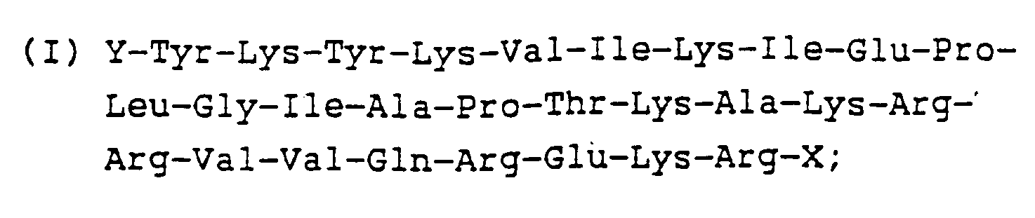
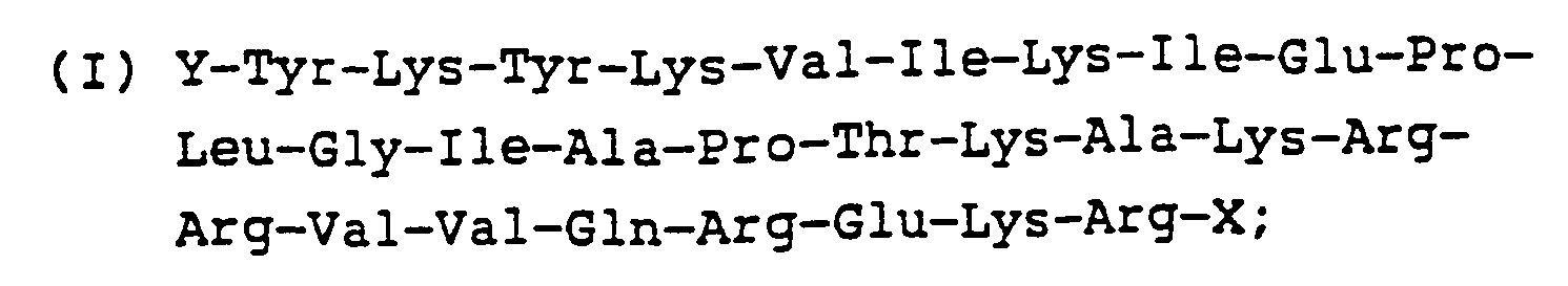
- a short peptide (Peptide I) corresponding to amino acids 490-517 of gp120 encoded in the region between base pairs (bp) 7221-7305 [Sanchez-Pescador, et al., Science , 227 :484-492 (1985)] was synthesized for testing immunogenicity/antigenicity.
- This peptide sequence is shown below: wherein Y corresponds to -H or an amino acid, and X may be -OH, -NH2 or -NR1R2 (wherein R corresponds to an alkyl group).
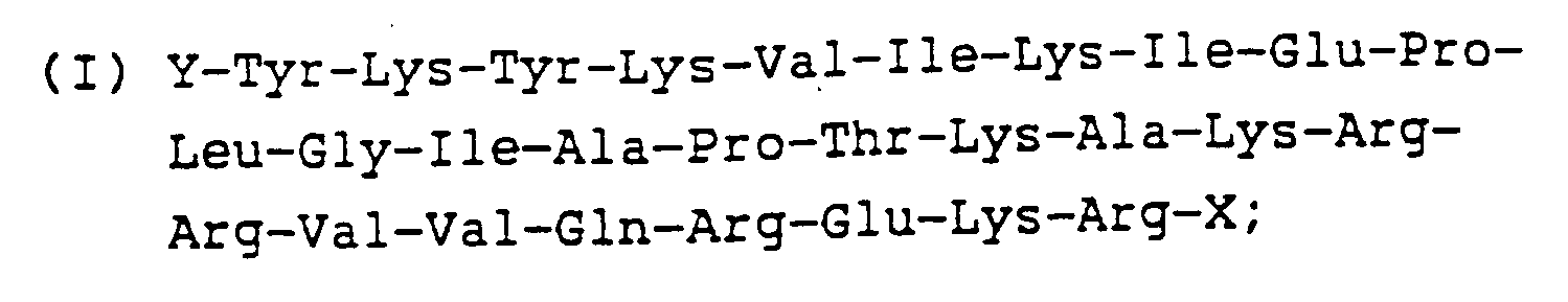
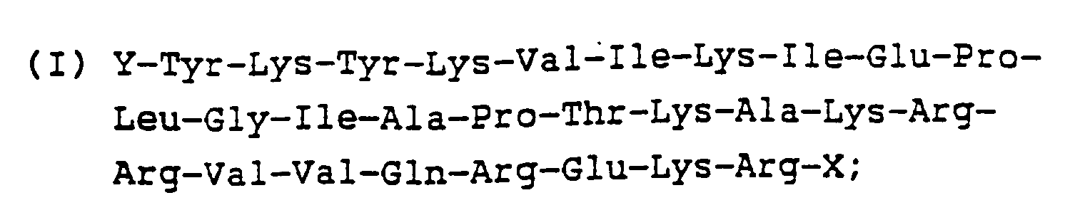
- the amino acid sequence of Peptide II corresponds to amino acids 482-517 of gp120 encoded in the region between bp 7197-7305 [Sanchez-Pescador, et al., supra].
- the amino acid sequence for Peptide II is shown below: wherein Y and X are the same as described above for Peptide I.
- Peptide III extends from amino acids 455 to 511 of gp120 encoded in the region between bp 7165-7335 [Muesing, et al., Nature , 313 :450-458 (1985)].
- the amino acid sequence of this peptide is shown below: wherein Y and X are as explained for Peptide I.
- peptides can be used as sources of HIV antigens alone or in combination or may be linked to larger carrier molecules. It is preferred that one or more of the Peptides I-III be used in combination with other antigens and epitopes, particularly other HIV antigens such as p24 and gp41.
- the peptides of the invention can be synthesized by a number of methods including synthesis in solution or by solid phase peptide methodology using stepwise or fragment coupling protocols. They can also be synthesized enzymatically or made as fused proteins or peptides by recombinant DNA methodology. Regions of genes coding for these peptides may also be synthesized, cloned and expressed using recombinant DNA technology, and the sequences may be subcloned and expressed in suitable expression systems such as E.coli , yeast or mammalian cells.
- the peptides described herein may be combined with other epitopes or antigens of HIV, with or without carrier molecules, and expressed by recombinant DNA methods or made by synthetic methods as fusion proteins. Substitution, deletion or addition analogs of the peptides of the invention can also be made by methods well known to those skilled in the art.
- the peptides corresponding to the various examples herein can be used alone, in combination with one another or in combination with other antigens of interest.
- various of these peptides can be used as physical mixtures or chemically coupled to each other with or without spacer molecules. It is also possible to couple peptides chemically or physically with carrier peptides, proteins or supports. These peptides can be used with other HIV antigens or epitopes such as gp41, gp120, p55, p24 and others.
- the peptides, particularly Peptide III can be coupled to carrier molecules like thyroglobulin or BSA and used as antigens alone or in combination with other epitopes or antigens.
- the following examples illustrate methods of making the peptides, as well as methods of using the peptides as sources for pure, well-defined, noninfectious HIV antigens.
- This example illustrates the synthesis of Peptide I on a resin support by stepwise solid phase synthesis starting with the carboxy-terminal residue.
- a procedure such as the procedure described in Barany and Merrifield, The Peptides , 2 :1984, Gross, E., and Guidehofer, J., Eds., Academic Press, New York, N.Y. (1980) can be used for this synthesis.
- a Boc-L-Arg(Tos)-OCH2-Pam resin was transferred to a reaction vessel of an Applied Biosystems Synthesizer, Model 430A, available from Applied Biosystems, Foster City, California.
- the fully protected peptide-resin (0.7 g) was allowed to swell in methylene chloride (CH2Cl2) for 5 minutes.
- the N ⁇ -Boc protecting groups were removed using 60% trifluoroacetic acid (TFA/CH2Cl2) deprotection, CH2Cl2 washes, 10% N,N diisopropylethylamine (DIEA/CH2Cl2) neutralization and finally washing with CH2Cl2 again.
- the resin was dried in vacuo .
- the peptide resin so obtained was treated with 9 ml of anhydrous hydrofluoric acid (HF) to which 1 ml of p-cresol had been added, for 60 minutes at 0°C.
- HF anhydrous hydrofluoric acid
- the HF was distilled off in vacuo at 0°C.
- the cleaved free peptide and resin were washed 3 times with 15 ml aliquots of diethyl ether, and the peptide was extracted by means of 3 extractions with 15 ml of 40% aqueous acetic acid.
- the aqueous extracts were combined and washed three times with 10 ml aliquots of diethyl ether, whereupon the aqueous layer was lyophilized to provide the crude peptide for purification.
- the polypeptide was purified by reversed-phase high performance liquid chromotography (HPLC) on C4 columns employing gradients of 0.1% TFA/water (A) and 100% acetonitrile (B) as the solvent systems at a 1 ml/min. flow rate for the analytical (Vydac-214-TP54, Vydac Separation Group, Hesperia, California) or 3 ml/mm flow rate for the semi-preparative (Vydac-214-TP510) columns.
- the gradient used was: The polypeptide elution from the HPLC column was monitored at 222 nm and 280 nm.
- composition of the polypeptide was confirmed by hydrolysis in 6 N hydrochloric acid (HCl)/0.3% phenol at 150° C for 2 hours in vacuo and subsequently analyzed on a Beckman 6300 amino acid analyzer with a SICA 7000 A integration available from Beckman Instruments, LaBrea, California.
- Peptides II and III were synthesized in a manner similar to the one described above. Methionine was used in the sulfoxide form, trytophan was protected by the formyl group (CHO); Ser, Bzl; Asp, OBZL.
- CHO formyl group
- Ser, Bzl Ser, Bzl
- Asp OBZL
- peptide-resins containing methionine sulfoxide and formyl-tryptophan peptides were deprotected and cleaved off the resin using "low high" HF protocols as described by Tam, et al., J. Am. Chem. Soc. , 105 :6442-6455 (1983).
- Desired peptides can also be synthesized using unprotected methionine and tryptophan with the appropriate uses of scavengers during deprotection and cleavage. All peptides were purified using reversed phase C4 HPLC wih 0.1% aqueous TFA and a 100% acetonitrile gradient system.
- Peptides I, II and III may be conjugated to larger carrier molecules such as bovine serum albumin (BSA), keyhole limpet hemocyanin (KLH) or thyroglobulin using water soluble carbodiimide or maleimido-benzoyl-N-hydroxysuccinimide (MBS) as described in Liu, et al., Biochem. , 18 :690-697 (1979), and Kitagawa, et al., J. Biochem. , 92 :585-590 (1982). These conjugates can then be used to raise sequence specific antibodies.
- BSA bovine serum albumin
- KLH keyhole limpet hemocyanin
- MFS maleimido-benzoyl-N-hydroxysuccinimide
- This example demonstrates a method of detecting HIV antibodies in biological samples using the peptides described in Example 1.
- Biological samples which can be tested by the methods described herein include blood preparations such as serum or plasma, urine and saliva.
- the peptides of the invention are particularly useful in a saliva test for the presence of IgA, IgG or IgM antibodies to HIV, especially when the peptides are combined with other HIV antigens.
- Peptides I and III were tested side-by-side with recombinant DNA-derived HIV p24 and HIV gp41 to determine their usefulness as sources of HIV antigens in a method for detecting HIV antibodies in biological samples.
- an overcoat solution consisting of 10% bovine serum albumin, 3% sucrose and 0.05% Tween® 20 in phosphate buffered saline (PBS) (0.01 M KH2P04; 0.15 M NaCl: pH 7.2) were added to the wells. Following a 30 minute incubation, the overcoat solution was removed and the plate washed five times with distilled water. Coated plates were stored at 2-8°C for subsequent use.
- PBS phosphate buffered saline
- the coated plates were used in an assay for detecting anti-HIV as follows: one hundred microliters of a serum sample diluted 1:800 or a saliva sample diluted 1:10 in a diluent consisting of 10% bovine serum albumin and 2% Tween® 20 in phosphate buffered saline (0.01 M KH2P04; 0.15 M NaCl: pH 7.2) were added to wells of the coated microtiter plates. After a 1 hour incubation at room temperature, the sample was removed and the wells washed five times with distilled water.
- an antibody-enzyme conjugate (alkaline phosphatase:goat anti-human IgG or alkaline phosphatase:goat anti-human IgA) were added to the wells.
- the antibody-enzyme conjugate can also be an IgM antibody conjugate if IgM antibody is being detected.
- the antibody-enzyme conjugates are made as described in Engvall, et al., Biochim. Biophys. Acta , 251 :427-434 (1971); Korn et al., J. Mol. Biol. , 65 :525-529 (1972); and Avrameas and Ternynck, Immunochemistry , 6 :53-66 (1969).
- This example demonstrates a microparticle assay for detection of HIV antibodies utilizing Peptide III, recombinant DNA-derived p24 and recombinant DNA-derived gp41.
- PBS phosphate buffered saline
- Sulfo-MBS sulfo-m-maleimido-benzoyl-N-hydroxysuccinimide
- the solution was stirred for 1 hour at room temperature after which the microparticles were isolated by centrifugation at 5000 x g speed, washed twice with PBS and resuspended in 1 ml PBS. Thirty microliters of a solution of Peptide III (1 mg/ml in distilled water) were added to the resuspended microparticles and stirred for 2 hours at room temperature. The microparticles were isolated by centrifugation at 5000 x g speed, washed twice with PBS containing 0.05% Tween® 20, and resuspended in PBS to yield a 0.125% solution. After resuspension in PBS, the particles were stored at 2-8°C for subsequent use in combination with p24 and gp41 coated microparticles in an assay for anti-HIV.
- microparticle solutions of p24 coated microparticles and gp41 coated microparticles one hundred microliters of amino-modified microparticles, 2.5% solids, 3.0 ⁇ m average diameter, commercially available from Polyscience, were added to 500 microliters of 50 mM N-methylmorpholine, pH 7.5 and 300 microliters of 2-iminothiolane (10 milligrams per milliliter in ice-cold 0.1 M sodium bicarbonate) for each antigen. The solution was incubated for 1 hour at room temperature.
- the antigens may be attached to the microparticles or other surfaces by a variety of methods, e.g., adsorption, use of specific antibodies, or the use of other various chemical activators.
- a prefilter was situated above the unit, which contained the filter and the antigen-coated microparticles. Two hundred microliters of a 1:10 dilution of either serum or saliva in a sample diluent (10% bovine serum albumin, 2% Tween® 20 in PBS) were added to the prefilter. After the sample was absorbed, 200 microliters of an antibody-enzyme conjugate comprising a mixture of goat anti-human IgG and IgA:alkaline phosphatase were added to the matrix through the prefilter.
- a detergent wash solution (1M guanadine hydrochloride, 1M NaCl, .05% Tween® 20 in PBS
- a chromogen indicator bromo-chloro indolyl phosphate/nitro blue tetrazolium
- Peptides I, II or III or any combination of these peptides with one another or with other HIV antigens may be employed to produce antisera or as a vaccine.
- Antisera is specifically produced by immunizing rabbits with injections of Peptides I or III according to the present invention as follows.
- the peptide is coupled to a carrier protein thyroglobulin by the following general procedure: To a solution of the selected peptide (2 mg) in 1 ml of either distilled water or dimethylformamide, is added 36 ⁇ l of a solution of 1-ethyl-3 (3-dimethylamino propyl) carbodiimide (Sigma Chemical Co., St. Louis, Missouri, 7.0 mg/ml H2O) at 0°C. The mixture is stirred for 5-10 minutes at 0°C.
- New Zealand white rabbits were inoculated with 250 mg of the conjugated peptide mixed (1:1) with complete Freund's adjuvant. All subsequent boosts contained conjugated Peptide mixed 1:1 with incomplete Freund's adjuvant. Animals were bled two weeks after each boost. Bleeds were processed to yield polyclonal antibodies in the serum. Peptide antibodies so generated immunoprecipitated gp160 and gp120 from 35S-methionine and 35S-cysteine labelled cell lysates. These antibodies may, for example, be utilized as reagents in a diagnostic assay, for affinity purification of gp120 or gp160 antigens or as a passive vaccine for therapeutic or prophylactic applications.
- An active vaccine solution according to the present invention may be prepared by suspending Peptides I, II or III or a combination of these (or in combination with other HIV antigens) in an immunologically acceptable diluent, adjuvant or carrier. Initial and booster injections or oral delivery are used to confer immunity.
- Monoclonal antibodies according to the present invention may be produced by injecting mice with immunizing doses of Peptides I, II or III or any combination of these with or without other epitopes of HIV.
- the peptide of interest is coupled to a carrier protein before injection as described in Example 4.
- the mouse spleens are then removed from the immunized animals and spleen cells are fused to myeloma cells (e.g. NS-1 cells) using polyethylene glycol.
- Myeloma cells e.g. NS-1 cells
- Hybridoma cells producing monoclonals are selected by screening in a suitable cell culture medium such as hypoxanthine aminoptern thymidine (HAT) medium.
- Monoclonal antibodies specific for HIV proteins may be isolated by affinity chromatography from media in which such hybridomas have been cultured.
- the peptides of the invention have many advantages. First, they are noninfectious and therefore safer in diagnostic applications. Second, these peptides can be produced in large quantities at a low cost. Third, a diagnostic assay including these peptides alone, in combination with one another or in combination with other HIV epitopes is more sensitive and specific than previous HIV assays. Fourth, the peptides of the invention appear to have excellent application for saliva screening assays for HIV.
- the peptides of the invention may be used in conjunction with a number of HIV or other antigens or antibodies in diagnostic testing and in production of antibodies for vaccines or diagnostic assays.
Landscapes
- Chemical & Material Sciences (AREA)
- Organic Chemistry (AREA)
- Health & Medical Sciences (AREA)
- Life Sciences & Earth Sciences (AREA)
- Molecular Biology (AREA)
- Medicinal Chemistry (AREA)
- Biophysics (AREA)
- General Health & Medical Sciences (AREA)
- Genetics & Genomics (AREA)
- Gastroenterology & Hepatology (AREA)
- Proteomics, Peptides & Aminoacids (AREA)
- Biochemistry (AREA)
- Virology (AREA)
- Peptides Or Proteins (AREA)
- Measuring Or Testing Involving Enzymes Or Micro-Organisms (AREA)
- Preparation Of Compounds By Using Micro-Organisms (AREA)
- Micro-Organisms Or Cultivation Processes Thereof (AREA)
- Medicines Containing Antibodies Or Antigens For Use As Internal Diagnostic Agents (AREA)
Abstract
Description
- A retrovirus termed human immunodeficiency virus (HIV) is now known to be the etiologic agent in acquired immune deficiency syndrome (AIDS). Various isolates of this virus have been termed lymphadenopathy-associated virus (LAV), human T-cell lymphotropic virus type-III (HTLV-III) or AIDS-associated retrovirus (ARV). Although the modes of transmission for HIV are not completely understood, the most common forms of transmission of the HIV virus are through sexual contact, use of contaminated intravenous equipment and transfusions with contaminated blood products. Testing for HIV has become extremely important in diagnosing exposure to the virus and particularly for protecting blood products from contamination.
- To detect exposure to HIV, levels of HIV antibodies (anti-HIV) are measured in serum, plasma, saliva or other biological samples of HIV patients or individuals at risk for AIDS. In the first generation of tests for HIV antibodies, inactivated crude or purified viral protein from lysates of HIV-infected cells are used to coat a solid phase. The lysate-coated solid phase is incubated with a biological sample suspected of containing anti-HIV, washed and then anti-human antibody tagged with a detectable label is added to the solid phase. The label, which may be an enzyme, radioisotope or fluorescent molecule is measured to determine presence of HIV antibodies in the sample.
- The problem with using viral lysates as a source of HIV antigens is that the infectious nature of the HIV virus makes manufacturing the virus potentially hazardous. Also, HIV viral lysates may contain impurities which interfere with testing for HIV antibodies. Therefore, alternate sources of HIV antigens are needed which are safe and noninfectious. There is also a need for HIV antigens which are well-defined and do not cross-react with other non-HIV antibodies and other interfering materials contained in the sample to be tested.
- Studies have shown that HIV antibodies to gp160 and gp120 are found in human saliva of asymptomatic, AIDS-related complex and AIDS patients. Archibald, et al., Blood, 67:831-834 (1986). These salivary antibodies represent mostly IgA immune response.
- Various isolates of HIV have been cloned and their genetic structures established. Nucleotide and deduced amino acid sequences of the various regions of the HIV genome are published. However, the published sequences do not provide any information as to which portions of the HIV molecule, when synthesized as peptides, would have antigenic or immunogenic properties similar to the corresponding region of naturally-occurring HIV proteins.
- The envelope (env) region of the HIV gene encodes an approximately 856 residue precursor protein with various potential glycosylation sites. The precursor glycoprotein corresponds to a molecular weight of 160,000 daltons (gp160) and is processed at the -Lys Arg- pair to yield an N-terminal protein of 480 amino acids (gp120) and a 345 amino acid protein (gp41).
- It has been demonstrated by radioimmune precipitation followed by polyacrylamide gel electrophoresis (RIP-PAGE) as well as by enzyme-linked immunosorbent assay (ELISA) that sera from AIDS and AIDS-related complex (ARC) patients react with env and gag gene encoded proteins of HIV. These proteins include but are not limited to gp160 gp120, gp41, p55, p36, p24, p18 and p12.
- In European Patent Application No. 227,169, Berman et al. disclose several polypeptides mimicking amino acid sequences of AIDS-related viral proteins. An immunochemical reagent is made by combination of two or more of the synthetic polypeptide sequences selected from the following group: a polypeptide sequence mimicking at least one antigenic determinant of the gag antigen of HIV, a polypeptide sequence mimicking at least one antigenic determinant of the glycoprotein gp120 of HIV and a polypeptide sequence mimicking at least one antigenic determinant of the glycoprotein gp41 of HIV. Berman et al. state that the polypeptide sequences disclosed should be substantially free of the naturally occurring gag, gp120 and gp41 proteins of HIV even though these reagents may optionally contain naturally occurring HIV proteins. The Berman et al. disclosure provides no indication as to which HIV antigen or epitope would be important for detecting salivary antibodies.
- In Wong-Staal, et al., U.S. Pat. Applic. Ser. No. 779,431, a 15 amino acid long peptide from gp120 is described. This peptide is useful as an immunogen in the production of monoclonal antibodies and in an ELISA assay.
- In Proc. Nat'l Acad. Sci. USA, 84:2479-2483 (1987), Palker, et al. disclose a synthetic peptide derived from the COOH-terminal region of gp120 containing 15 amino acids. This relatively short peptide was used to evaluate reactivity of HIV-positive patients' antibodies and as an immunoadsorbent to evaluate functional importance of human antibody response to the COOH terminus of gp120.
- In Cosand, U.S. Patent No. 4,629,783, peptides having sequences mimicking short regions of gp120, gp41 and p24 are disclosed. Those peptides, which can be used for detection of anti-HIV in blood screening procedures, mimic proteins encoded by the gag or env regions of the viral genome. Preferably, the peptides disclosed by Cosand contain fewer than 25 amino acids.
- In Montagnier et al., WO 86/02383, a LAV envelope antigen having a molecular weight of about 110,000 or antigens of lower molecular weight (less than 200 amino acids) derived from the preceding one are disclosed as useful in the diagnosis of LAV antibodies in the sera of patients.
- In Gallo et al., EP-A-0 231 914, synthetic peptides containing portions of the epitopic sequence HTLV env (460-550) are disclosed which peptides are useful as reagents in immunoassays for detection of AIDS antibodies, as immunogens for eliciting polyclonal or monoclonal antibodies against AIDS virus env protein and as components in AIDS vaccines.
- In Chan et al., EP-A-0 212 532, a method for producing fusion proteins is disclosed wherein the fusion protein may contain an AIDS virus env protein. Particular immunoreactive AIDS virus env protein fragments are disclosed.
- We have developed novel gp160 peptides which can be used for detecting HIV antibodies in biological samples, for producing HIV antibodies and for vaccines. These peptides substantially mimic regions of gp160 or its gp120 fragment and provide noninfectious and pure sources of HIV antigens.
- First, a short peptide (Peptide I) corresponding to amino acids 490-517 of gp120 encoded in the region between base pairs (bp) 7221-7305 [Sanchez-Pescador, et al., Science, 227:484-492 (1985)] was synthesized for testing immunogenicity/antigenicity. This peptide sequence is shown below:
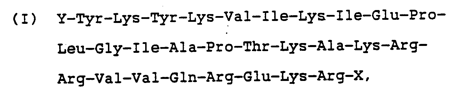
wherein Y corresponds to -H or an amino acid, and X may be -OH, -NH₂ or -NR₁R₂ (wherein R corresponds to an alkyl group). - The amino acid sequence of Peptide II corresponds to amino acids 482-517 of gp120 encoded in the region between bp 7197-7305 [Sanchez-Pescador, et al., supra]. The amino acid sequence for Peptide II is shown below:

wherein Y and X are the same as described above for Peptide I. - Peptide III extends from amino acids 455 to 511 of gp120 encoded in the region between bp 7165-7335 [Muesing, et al., Nature, 313:450-458 (1985)]. The amino acid sequence of this peptide is shown below:

wherein Y and X are as explained for Peptide I. - These peptides can be used as sources of HIV antigens alone or in combination or may be linked to larger carrier molecules. It is preferred that one or more of the Peptides I-III be used in combination with other antigens and epitopes, particularly other HIV antigens such as p24 and gp41.
- The peptides of the invention can be synthesized by a number of methods including synthesis in solution or by solid phase peptide methodology using stepwise or fragment coupling protocols. They can also be synthesized enzymatically or made as fused proteins or peptides by recombinant DNA methodology. Regions of genes coding for these peptides may also be synthesized, cloned and expressed using recombinant DNA technology, and the sequences may be subcloned and expressed in suitable expression systems such as E.coli, yeast or mammalian cells. The peptides described herein may be combined with other epitopes or antigens of HIV, with or without carrier molecules, and expressed by recombinant DNA methods or made by synthetic methods as fusion proteins. Substitution, deletion or addition analogs of the peptides of the invention can also be made by methods well known to those skilled in the art.
- The peptides corresponding to the various examples herein can be used alone, in combination with one another or in combination with other antigens of interest. For example, various of these peptides can be used as physical mixtures or chemically coupled to each other with or without spacer molecules. It is also possible to couple peptides chemically or physically with carrier peptides, proteins or supports. These peptides can be used with other HIV antigens or epitopes such as gp41, gp120, p55, p24 and others. The peptides, particularly Peptide III, can be coupled to carrier molecules like thyroglobulin or BSA and used as antigens alone or in combination with other epitopes or antigens.
- The following examples illustrate methods of making the peptides, as well as methods of using the peptides as sources for pure, well-defined, noninfectious HIV antigens.
- This example illustrates the synthesis of Peptide I on a resin support by stepwise solid phase synthesis starting with the carboxy-terminal residue. A procedure such as the procedure described in Barany and Merrifield, The Peptides, 2:1984, Gross, E., and Meinehofer, J., Eds., Academic Press, New York, N.Y. (1980) can be used for this synthesis. A Boc-L-Arg(Tos)-OCH₂-Pam resin was transferred to a reaction vessel of an Applied Biosystems Synthesizer, Model 430A, available from Applied Biosystems, Foster City, California. Protected amino acids were coupled in a stepwise manner to the resin support by preformed symmetric anhydride chemistry, except in the cases of arginine, asparagine and glutamine addition where the DCC/HOBT protocol described by Konig and Geiger, Chem. Ber., 103:788-798 (1970) was employed. All amino-terminal residues were protected by t-butyloxy carbonyl (t/BOC-linkage) and side chains of various amino acid residues were protected by the following groups: Arg, Tos; Lys, 2-ClZ; Glu, OBZl; Thr, Bzl; Tyr, 2-BrZ.
- The fully protected peptide-resin (0.7 g) was allowed to swell in methylene chloride (CH₂Cl₂) for 5 minutes. The Nα-Boc protecting groups were removed using 60% trifluoroacetic acid (TFA/CH₂Cl₂) deprotection, CH₂Cl₂ washes, 10% N,N diisopropylethylamine (DIEA/CH₂Cl₂) neutralization and finally washing with CH₂Cl₂ again. The resin was dried in vacuo. The peptide resin so obtained was treated with 9 ml of anhydrous hydrofluoric acid (HF) to which 1 ml of p-cresol had been added, for 60 minutes at 0°C. The HF was distilled off in vacuo at 0°C. The cleaved free peptide and resin were washed 3 times with 15 ml aliquots of diethyl ether, and the peptide was extracted by means of 3 extractions with 15 ml of 40% aqueous acetic acid. The aqueous extracts were combined and washed three times with 10 ml aliquots of diethyl ether, whereupon the aqueous layer was lyophilized to provide the crude peptide for purification. The polypeptide was purified by reversed-phase high performance liquid chromotography (HPLC) on C₄ columns employing gradients of 0.1% TFA/water (A) and 100% acetonitrile (B) as the solvent systems at a 1 ml/min. flow rate for the analytical (Vydac-214-TP54, Vydac Separation Group, Hesperia, California) or 3 ml/mm flow rate for the semi-preparative (Vydac-214-TP510) columns. The gradient used was:

The polypeptide elution from the HPLC column was monitored at 222 nm and 280 nm. The composition of the polypeptide was confirmed by hydrolysis in 6 N hydrochloric acid (HCl)/0.3% phenol at 150° C for 2 hours in vacuo and subsequently analyzed on a Beckman 6300 amino acid analyzer with a SICA 7000 A integration available from Beckman Instruments, LaBrea, California. - Peptides II and III were synthesized in a manner similar to the one described above. Methionine was used in the sulfoxide form, trytophan was protected by the formyl group (CHO); Ser, Bzl; Asp, OBZL. For peptide-resins containing methionine sulfoxide and formyl-tryptophan, peptides were deprotected and cleaved off the resin using "low high" HF protocols as described by Tam, et al., J. Am. Chem. Soc., 105:6442-6455 (1983). Desired peptides can also be synthesized using unprotected methionine and tryptophan with the appropriate uses of scavengers during deprotection and cleavage. All peptides were purified using reversed phase C₄ HPLC wih 0.1% aqueous TFA and a 100% acetonitrile gradient system.
- Peptides I, II and III may be conjugated to larger carrier molecules such as bovine serum albumin (BSA), keyhole limpet hemocyanin (KLH) or thyroglobulin using water soluble carbodiimide or maleimido-benzoyl-N-hydroxysuccinimide (MBS) as described in Liu, et al., Biochem., 18:690-697 (1979), and Kitagawa, et al., J. Biochem., 92:585-590 (1982). These conjugates can then be used to raise sequence specific antibodies.
- This example demonstrates a method of detecting HIV antibodies in biological samples using the peptides described in Example 1. Biological samples which can be tested by the methods described herein include blood preparations such as serum or plasma, urine and saliva. The peptides of the invention are particularly useful in a saliva test for the presence of IgA, IgG or IgM antibodies to HIV, especially when the peptides are combined with other HIV antigens.
- Peptides I and III were tested side-by-side with recombinant DNA-derived HIV p24 and HIV gp41 to determine their usefulness as sources of HIV antigens in a method for detecting HIV antibodies in biological samples.
- Four antigen solutions were prepared in 50 mM sodium carbonate, pH 9.5 as follows: Peptide I, 10 µg/ml; Peptide III, 6.25 µg/ml; recombinant DNA-derived HIV p24 protein, 1.64 µg/ml; and recombinant DNA-derived HIV gp41 protein, 125 units/ml. The p24 and gp41 proteins were made by methods described in copending U.S. Patent Application Serial No. 020,287, filed Feb. 27, 1987, assigned to the same assignee as that of the present invention.
- One hundred microliters (100 µl) of each antigen solution were added to wells of a polystyrene microtiter plate available from Dynatech Laboratories, Alexandria, Virginia. Other solid phases which may be utilized in the methods described herein include beads, paper strips, microparticles, nitrocellulose membranes, polystyrene tubes or other suitable plastic or paper supports. The solution and the plate were incubated for 1 hour at room temperature after which the solution was removed and the plate washed five times with distilled water. Two hundred and fifty microliters of an overcoat solution consisting of 10% bovine serum albumin, 3% sucrose and 0.05% Tween® 20 in phosphate buffered saline (PBS) (0.01 M KH₂P0₄; 0.15 M NaCl: pH 7.2) were added to the wells. Following a 30 minute incubation, the overcoat solution was removed and the plate washed five times with distilled water. Coated plates were stored at 2-8°C for subsequent use.
- The coated plates were used in an assay for detecting anti-HIV as follows: one hundred microliters of a serum sample diluted 1:800 or a saliva sample diluted 1:10 in a diluent consisting of 10% bovine serum albumin and 2% Tween® 20 in phosphate buffered saline (0.01 M KH₂P0₄; 0.15 M NaCl: pH 7.2) were added to wells of the coated microtiter plates. After a 1 hour incubation at room temperature, the sample was removed and the wells washed five times with distilled water. One hundred microliters of an antibody-enzyme conjugate (alkaline phosphatase:goat anti-human IgG or alkaline phosphatase:goat anti-human IgA) were added to the wells. The antibody-enzyme conjugate can also be an IgM antibody conjugate if IgM antibody is being detected.The antibody-enzyme conjugates are made as described in Engvall, et al., Biochim. Biophys. Acta, 251:427-434 (1971); Korn et al., J. Mol. Biol., 65:525-529 (1972); and Avrameas and Ternynck, Immunochemistry, 6:53-66 (1969). Following another 1 hour incubation at room temperature, the conjugate was removed and the wells washed five times with distilled water. One hundred microliters of a p-nitrophenylphosphate substrate solution were added to the wells and incubated at room temperature for 30 minutes. One hundred microliters of 2N NaOH were added to the wells to stop the reaction. Absorbance values of the wells were read at 405 nm. A positive result was determined at a cutoff value of 0.200 O.D. or greater, which was established based on the results generated from 100 negative and 50 positive paired serum and saliva samples. The results are set forth in Tables 1, 2 and 3.
- The results indicate that all confirmed seropositive samples tested contained serum IgG to gp41, Peptide I and Peptide III, but the serum reactivity to p24 varied. In saliva, the IgG reactivity in seropositive samples varied for all antigens tested. Although the salivary IgA reactivity varied for p24 and gp41, all seropositive samples contained salivary IgA to Peptide I and Peptide III. For the confirmed negative samples, no reactivity was detected in either serum or saliva for any of the antigens tested. In general, the reactivity of Peptide III was greater than that of Peptide I.
- These results demonstrate that Peptide I and Peptide III are specifically reactive with antibody to HIV. In addition, the results indicate that at least a portion of gp120 must be present when testing for antibodies to HIV in saliva samples.
Table 1 Patient Diagnosis Confirmed Seropos. Microtiter Serum IgG p24 gp41 Peptide I Peptide III AIDS Yes 0.089 >3.0 1.734 >3.0 AIDS Yes 0.034 2.009 0.981 1.730 AIDS Yes 0.003 >3.0 1.836 2.009 AIDS Yes 0.006 2.105 0.301 0.873 AIDS Yes 0.049 2.980 2.437 >3.0 AIDS Yes 0.097 >3.0 >3.0 >3.0 AIDS Yes 0.102 2.874 1.920 2.941 AIDS Yes 0.031 2.541 1.337 2.226 ARC Yes 0.198 >3.0 2.011 >3.0 ARC Yes 0.157 >3.0 1.990 2.798 Asymptomatic Yes 2.983 >3.0 1.933 2.322 Asymptomatic Yes >3.0 >3.0 >3.0 >3.0 Asymptomatic Yes 0.895 >3.0 >3.0 >3.0 Asymptomatic Yes >3.0 >3.0 >3.0 >3.0 Asymptomatic Yes 1.971 >3.0 2.909 >3.0 Hemophiliac Yes 0.301 2.179 ND 1.899 Hemophiliac Yes 1.127 2.357 ND 0.207 Hemophiliac Yes 1.193 1.715 ND 1.119 Hemophiliac Yes 0.702 2.770 ND 2.248 Hemophiliac Yes >3.0 >3.0 ND >3.0 Hemophiliac Yes 0.077 2.552 ND 0.558 Hemophiliac No 0.051 0.086 ND 0.003 Hemophiliac No 0.006 0.029 ND 0.079 Hemophiliac No 0.018 0.068 ND 0.044 High Risk No 0.005 0.076 ND 0.101 High Risk No 0.018 0.006 ND 0.022 Healthy Hetero. No 0.000 0.114 0.043 0.097 Healthy Hetero. No 0.003 0.093 0.055 0.081 Healthy Hetero. No 0.019 0.008 0.092 0.005 Healthy Hetero. No 0.058 0.055 0.003 0.037 Table 2 Patient Diagnosis Confirmed Seropos. Microtiter Saliva IgG p24 gp41 Peptide I Peptide III AIDS Yes 0.044 0.971 0.204 0.568 AIDS Yes 0.013 0.111 0.123 0.228 AIDS Yes 0.033 0.758 0.229 0.318 AIDS Yes 0.022 0.014 0.041 0.059 AIDS Yes 0.005 0.296 0.476 0.935 AIDS Yes 0.041 >3.0 2.037 >3.0 AIDS Yes 0.027 0.246 0.398 0.837 AIDS Yes 0.035 0.216 0.271 0.440 ARC Yes 0.009 1.230 0.311 1.773 ARC Yes 0.036 0.931 0.292 0.909 Asymptomatic Yes 1.852 2.707 0.279 0.532 Asymptomatic Yes 2.190 >3.0 >3.0 >3.0 Asymptomatic Yes 0.139 >3.0 >3.0 >3.0 Asymptomatic Yes >3.0 >3.0 2.542 >3.0 Asymptomatic Yes 0.365 >3.0 1.352 2.874 Hemophiliac Yes 0.054 0.970 ND 0.304 Hemophiliac Yes 0.296 0.997 ND 0.041 Hemophiliac Yes 0.558 0.504 ND 0.171 Hemophiliac Yes 0.174 1.194 ND 0.407 Hemophiliac Yes >3.0 >3.0 ND 1.458 Hemophiliac Yes 0.026 1.201 ND 0.079 Hemophiliac No 0.037 0.053 ND 0.029 Hemophiliac No 0.062 0.055 ND 0.023 Hemophiliac No 0.010 0.006 ND 0.019 High Risk No 0.057 0.027 ND 0.095 High Risk No 0.051 0.011 ND 0.053 Healthy Hetero. No 0.003 0.019 0.006 0.106 Healthy Hetero. No 0.079 0.038 0.050 0.009 Healthy Hetero. No 0.042 0.111 0.013 0.060 Healthy Hetero. No 0.053 0.040 0.061 0.093 Table 3 Patient Diagnosis Confirmed Seropos. Microtiter Saliva IgA p24 gp41 Peptide I Peptide III AIDS Yes 0.076 0.099 0.938 1.480 AIDS Yes 0.035 0.065 0.559 1.165 AIDS Yes 0.077 0.007 0.232 0.440 AIDS Yes 0.064 0.040 0.982 1.251 AIDS Yes 0.096 0.151 1.010 1.604 AIDS Yes 0.056 0.355 >3.0 >3.0 AIDS Yes 0.002 0.175 0.449 1.120 AIDS Yes 0.069 0.040 0.933 1.738 ARC Yes 0.040 0.239 2.845 >3.0 ARC Yes 0.094 0.172 2.036 2.836 Asymptomatic Yes 0.144 0.078 0.840 1.054 Asymptomatic Yes 0.419 0.110 >3.0 >3.0 Asymptomatic Yes 0.288 0.773 1.635 >3.0 Asymptomatic Yes 0.146 0.186 2.424 >3.0 Asymptomatic Yes 0.634 0.678 1.544 2.988 Hemophiliac Yes 0.045 0.020 ND >3.0 Hemophiliac Yes 0.003 0.008 ND 2.520 Hemophiliac Yes 0.174 0.093 ND 1.835 Hemophiliac Yes 0.140 0.094 ND >3.0 Hemophiliac Yes 0.280 0.435 ND >3.0 Hemophiliac Yes 0.072 0.063 ND 2.947 Hemophiliac No 0.061 0.016 ND 0.103 Hemophiliac No 0.003 0.046 ND 0.011 Hemophiliac No 0.044 0.008 ND 0.038 High Risk No 0.083 0.007 ND 0.055 High Risk No 0.094 0.056 ND 0.103 Healthy Hetero. No 0.002 0.064 0.073 0.054 Healthy Hetero. No 0.054 0.013 0.007 0.098 Healthy Hetero. No 0.016 0.017 0.084 0.008 Healthy Hetero. No 0.042 0.004 0.067 0.051 - This example demonstrates a microparticle assay for detection of HIV antibodies utilizing Peptide III, recombinant DNA-derived p24 and recombinant DNA-derived gp41. One hundred microliters of amino-modified microparticles, 2.5% solids, 0.45 µm average diameter, commercially available from Polyscience, Warrington, Pennsylvania, were added to 1 ml of phosphate buffered saline (PBS) and 3.0 ml of sulfo-m-maleimido-benzoyl-N-hydroxysuccinimide (Sulfo-MBS) (1.0 mg/ml in PBS). The solution was stirred for 1 hour at room temperature after which the microparticles were isolated by centrifugation at 5000 x g speed, washed twice with PBS and resuspended in 1 ml PBS. Thirty microliters of a solution of Peptide III (1 mg/ml in distilled water) were added to the resuspended microparticles and stirred for 2 hours at room temperature. The microparticles were isolated by centrifugation at 5000 x g speed, washed twice with PBS containing 0.05% Tween® 20, and resuspended in PBS to yield a 0.125% solution. After resuspension in PBS, the particles were stored at 2-8°C for subsequent use in combination with p24 and gp41 coated microparticles in an assay for anti-HIV.
- To make separate microparticle solutions of p24 coated microparticles and gp41 coated microparticles, one hundred microliters of amino-modified microparticles, 2.5% solids, 3.0 µm average diameter, commercially available from Polyscience, were added to 500 microliters of 50 mM N-methylmorpholine, pH 7.5 and 300 microliters of 2-iminothiolane (10 milligrams per milliliter in ice-cold 0.1 M sodium bicarbonate) for each antigen. The solution was incubated for 1 hour at room temperature. Two hundred microliters of the p24 antigen and gp41 antigen (described in Example 2 except that concentrations were as follows: p24, 654 µg/ml; gp41, 100,000 units/ml) were each incubated with 200 microliters of Sulfo-MBS (3.75 µg/ml in PBS) for 1 hour at room temperature. Each activated antigen was then added to the microparticles and incubated overnight at room temperature. The microparticles were isolated by centrifugation, washed twice with PBS containing 0.05% Tween® 20, and resuspended in PBS to yield a 0.125% solution. After resuspension in PBS, the particles were stored at 2-8°C for subsequent use in the microparticle assay for HIV antibodies described below.
- Twenty microliters each of the antigen-coated microparticle solutions (Peptide III, p24 and gp41) were added dropwise to the center of a Whatman GF-D glass fiber filter arranged in a microparticle assay format. This assay format is described in more detail in copending U.S. Patent Application serial No. 831,013, filed on Feb. 18, 1986, assigned to the same assignee as that of the present invention and incorporated by reference herein. Three hundred microliters of an overcoat solution (10% bovine serum albumin, 3% sucrose, 0.05% Tween® 20 in PBS) were added and the unit incubated at 45° C for 90 minutes to dry the filter. It is to be noted that, in addition to the technique described in the foregoing examples, the antigens may be attached to the microparticles or other surfaces by a variety of methods, e.g., adsorption, use of specific antibodies, or the use of other various chemical activators.
- A prefilter was situated above the unit, which contained the filter and the antigen-coated microparticles. Two hundred microliters of a 1:10 dilution of either serum or saliva in a sample diluent (10% bovine serum albumin, 2% Tween® 20 in PBS) were added to the prefilter. After the sample was absorbed, 200 microliters of an antibody-enzyme conjugate comprising a mixture of goat anti-human IgG and IgA:alkaline phosphatase were added to the matrix through the prefilter. After absorption, the prefilter was removed and 1 milliliter of a detergent wash solution (1M guanadine hydrochloride, 1M NaCl, .05% Tween® 20 in PBS) was added to the matrix to remove any excess antibody-enzyme conjugate. Then, 150 microliters of a chromogen indicator (bromo-chloro indolyl phosphate/nitro blue tetrazolium) were added to the matrix. After 2 minutes, 1 milliliter of the wash solution was added to the matrix. The matrix was checked visually. The appearance of a colored spot indicated that the specimen contained detectable levels of antibody to HIV. Samples tested by the foregoing procedure but not containing detectable levels of antibody to HIV produced no color in the matrix. The results are set forth in Tables 4 and 5.
Table 4 Microparticle Assay Patient Diagnosis Confirmed Seropos. Serum p24 gp41 p24,gp41,PIII AIDS Yes Neg Pos Pos AIDS Yes Neg Pos Pos AIDS Yes Neg Pos Pos AIDS Yes Neg Pos Pos AIDS Yes Neg Pos Pos AIDS Yes Neg Pos Pos AIDS Yes Neg Pos Pos AIDS Yes Neg Pos Pos ARC Yes Neg Pos Pos ARC Yes Neg Pos Pos Asymptomatic Yes Pos Pos Pos Asymptomatic Yes Pos Pos Pos Asymptomatic Yes Pos Pos Pos Asymptomatic Yes Pos Pos Pos Asymptomatic Yes Pos Pos Pos Hemophiliac Yes Pos Pos Pos Hemophiliac Yes Pos Pos Pos Hemophiliac Yes Pos Pos Pos Hemophiliac Yes Pos Pos Pos Hemophiliac Yes Pos Pos Pos Hemophiliac Yes Pos Pos Pos Hemophiliac No Neg Neg Neg Hemophiliac No Neg Neg Neg Hemophiliac No Neg Neg Neg High Risk No Neg Neg Neg High Risk No Neg Neg Neg Healthy Hetero. No Neg Neg Neg Healthy Hetero. No Neg Neg Neg Healthy Hetero. No Neg Neg Neg Healthy Hetero. No Neg Neg Neg Table 5 Microparticle Assay Patient Diagnosis Confirmed Seropos. Saliva p24 gp41 p24,gp41,PIII AIDS Yes Neg Pos Pos AIDS Yes Neg Neg Pos AIDS Yes Neg Pos Pos AIDS Yes Neg Neg Pos AIDS Yes Neg Pos Pos AIDS Yes Neg Pos Pos AIDS Yes Neg Pos Pos AIDS Yes Neg Pos Pos ARC Yes Neg Pos Pos ARC Yes Neg Pos Pos Asymptomatic Yes Pos Pos Pos Asymptomatic Yes Pos Pos Pos Asymptomatic Yes Pos Pos Pos Asymptomatic Yes Pos Pos Pos Asymptomatic Yes Pos Pos Pos Hemophiliac Yes Neg Pos Pos Hemophiliac Yes Pos Pos Pos Hemophiliac Yes Pos Pos Pos Hemophiliac Yes Neg Pos Pos Hemophiliac Yes Pos Pos Pos Hemophiliac Yes Neg Pos Pos Hemophiliac No Neg Neg Neg Hemophiliac No Neg Neg Neg Hemophiliac No Neg Neg Neg High Risk No Neg Neg Neg High Risk No Neg Neg Neg Healthy Hetero. No Neg Neg Neg Healthy Hetero. No Neg Neg Neg Healthy Hetero. No Neg Neg Neg Healthy Hetero. No Neg Neg Neg - Peptides I, II or III or any combination of these peptides with one another or with other HIV antigens, may be employed to produce antisera or as a vaccine.
- Antisera is specifically produced by immunizing rabbits with injections of Peptides I or III according to the present invention as follows. The peptide is coupled to a carrier protein thyroglobulin by the following general procedure: To a solution of the selected peptide (2 mg) in 1 ml of either distilled water or dimethylformamide, is added 36 µl of a solution of 1-ethyl-3 (3-dimethylamino propyl) carbodiimide (Sigma Chemical Co., St. Louis, Missouri, 7.0 mg/ml H₂O) at 0°C. The mixture is stirred for 5-10 minutes at 0°C. Next, 0.5 ml of thyroglobulin solution (Sigma, 10 mg/ml in PBS) is added to this reaction mixture and stirred overnight at 0°C. Finally, the mixture is dialyzed against PBS buffer with three changes of the buffer.
- New Zealand white rabbits were inoculated with 250 mg of the conjugated peptide mixed (1:1) with complete Freund's adjuvant. All subsequent boosts contained conjugated Peptide mixed 1:1 with incomplete Freund's adjuvant. Animals were bled two weeks after each boost. Bleeds were processed to yield polyclonal antibodies in the serum. Peptide antibodies so generated immunoprecipitated gp160 and gp120 from ³⁵S-methionine and ³⁵S-cysteine labelled cell lysates. These antibodies may, for example, be utilized as reagents in a diagnostic assay, for affinity purification of gp120 or gp160 antigens or as a passive vaccine for therapeutic or prophylactic applications.
- An active vaccine solution according to the present invention may be prepared by suspending Peptides I, II or III or a combination of these (or in combination with other HIV antigens) in an immunologically acceptable diluent, adjuvant or carrier. Initial and booster injections or oral delivery are used to confer immunity.
- Monoclonal antibodies according to the present invention may be produced by injecting mice with immunizing doses of Peptides I, II or III or any combination of these with or without other epitopes of HIV. The peptide of interest is coupled to a carrier protein before injection as described in Example 4. The mouse spleens are then removed from the immunized animals and spleen cells are fused to myeloma cells (e.g. NS-1 cells) using polyethylene glycol. Hybridoma cells producing monoclonals are selected by screening in a suitable cell culture medium such as hypoxanthine aminoptern thymidine (HAT) medium. Monoclonal antibodies specific for HIV proteins may be isolated by affinity chromatography from media in which such hybridomas have been cultured.
- The peptides of the invention have many advantages. First, they are noninfectious and therefore safer in diagnostic applications. Second, these peptides can be produced in large quantities at a low cost. Third, a diagnostic assay including these peptides alone, in combination with one another or in combination with other HIV epitopes is more sensitive and specific than previous HIV assays. Fourth, the peptides of the invention appear to have excellent application for saliva screening assays for HIV.
- While specific examples have been given to illustrate the invention, it is to be understood that those skilled in the art will recognize variations which come within the scope of the invention as claimed. For example, the peptides of the invention may be used in conjunction with a number of HIV or other antigens or antibodies in diagnostic testing and in production of antibodies for vaccines or diagnostic assays.
Claims (16)
- An immunoassay method for detection of HIV antibodies in a biological sample comprising:a) coating a solid phase with a peptide selected from the group consisting ofi) a peptide of the formula:
 ii) a peptide of the formula:
 iii) a peptide of the formula:and substitution, deletion and addition analogs thereof, wherein Y corresponds to -H or an amino acid, and X is -OH, -NH₂ or -NR₁R₂ (wherein R is an alkyl group);
 b) incubating the solid phase with the biological sample;c) incubating the solid phase with an anti-human Ig labeled with a detectable label; andd) detecting the label to determine the presence of anti-HIV in the sample.
 - The immunoassay method of claim 1 wherein the solid phase is further coated in step (a) with an HIV antigen selected from the group consisting of p24, gp41 and gp120 and combinations thereof.
- The immunoassay method of claim 1 wherein the solid phase has properties independently selected from one or more of the following:a) the solid phase in step (a) is coated with a peptide which is a fusion protein of the peptide in step (a) in combination with other epitopes of HIV;b) the anti-human Ig is selected from the group consisting of anti-human IgG, IgA, IgM and mixtures thereof;c) the detectable label is selected from the group consisting of enzymes, radioisotopes and fluorescent molecules.
- An immunoassay method for detection of HIV antibodies in human saliva test sample comprising:a) coating a solid phase with a peptide selected from the group consisting ofi) a peptide of the formula:
 ii) a peptide of the formula:
 iii) a peptide of the formula:and substitution, deletion and addition analogs thereof, wherein Y corresponds to -H or an amino acid, and X is -OH, -NH₂ or -NR₁R₂ (wherein R is an alkyl group);
 b) incubating the solid phase with the saliva test sample;c) incubating the solid phase with an anti-human Ig labeled with a detectable label; andd) detecting the label to determine the presence of anti-HIV antibodies in the saliva test sample.
 - The immunoassay method of claim 4 wherein the solid phase has properties independently selected from one or more of the following:a) the solid phase is further coated in step (a) with an HIV antigen selected from the group consisting of p24, gp41 and gp120 and combinations thereof;b) the peptide is Peptide III and the HIV antigens are p24 and gp41;c) the anti-human Ig is selected from the group consisting of anti-human IgG, IgA, IgM and mixtures thereof;d) the detectable label is selected from the group consisting of enzymes, radioisotopes and fluorescent molecules.
- A method for preparing an HIV antibody comprising immunizing an animal with a peptide selected from the group consisting ofi) a peptide of the formula:
 ii) a peptide of the formula:
 iii) a peptide of the formula:and substitution, deletion and addition analogs thereof, wherein Y corresponds to -H or an amino acid, and X is -OH, -NH₂ or -NR₁R₂ (wherein R is an alkyl group), and mixtures thereof in a suitable adjuvant and obtaining antibodies from the sera of the animal.
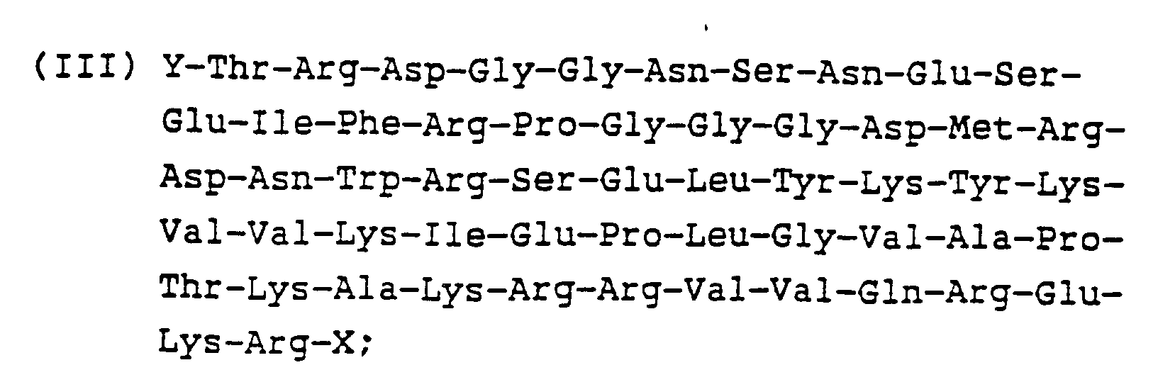
- The method of claim 6 wherein the animal is immunized with the peptide in combination with at least one other HIV antigen.
- A method for preparing a monoclonal antibody to HIV comprising injecting an animal with an immunizing dose of a peptide selected from the group consisting ofi) a peptide of the formula:
 ii) a peptide of the formula:
 iii) a peptide of the formula:and substitution, deletion and addition analogs thereof, wherein Y corresponds to -H or an amino acid, and X is -OH, -NH₂ or -NR₁R₂ (wherein R is an alkyl group), and mixtures thereof, removing the spleen from the animal, fusing the spleen with myeloma cells, culturing the fused cells in a suitable culture medium, and collecting HIV antibodies from said culture.
- The method of claim 8 wherein the animal is immunized with the peptide in combination with at least one other HIV antigen.
- A method for preparing an HIV vaccine comprising suspending a peptide selected from the group consisting ofi) a peptide of the formula:
 ii) a peptide of the formula:
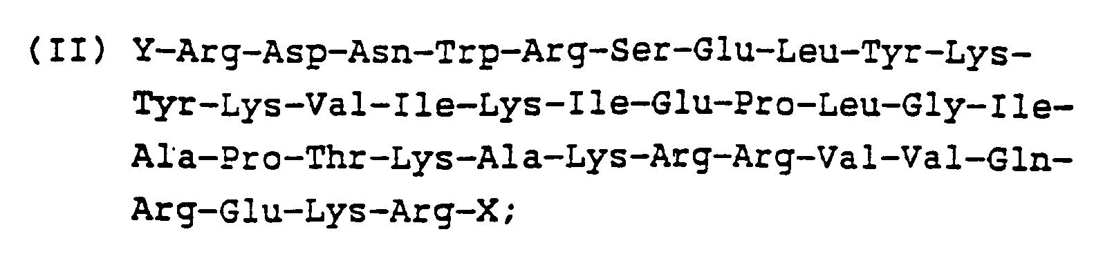 iii) a peptide of the formula:and substitution, deletion and addition analogs thereof, wherein Y corresponds to -H or an amino acid, and X is -OH, -NH₂ or -NR₁R₂ (wherein R is an alkyl group), and mixtures thereof in a suitable pharmaceutically acceptable carrier.
- The method of Claim 10 wherein the peptide is further combined with at least one other HIV antigen.
- The method according to Claim 6, 8 or 10 wherein said peptide is made in combination with other epitopes of HIV as a fusion protein.
- The method according to Claim 7, 9 or 11 wherein said other HIV antigen is selected from the group consisting of HIV p24, gp 41, gp 120 and combinations thereof.
- A process for preparing a peptide selected from the group consisting ofi) a peptide of the formula:
 ii) a peptide of the formula:
 iii) a peptide of the formula:and substitution, deletion and addition analogs thereof, wherein Y corresponds to -H or an amino acid, and X is -OH, -NH₂ or -NR₁R₂ (wherein R is an alkyl group), wherein said process comprises preparing said peptides either
 a) in solution or by solid phase peptide methodology using stepwise or fragment coupling protocols;b) enzymatically;c) by synthesizing, cloning and expressing regions of genes coding for said peptides using recombinant DNA methodology.
 - A process according to Claim 14, part (c), wherein the genes are subcloned and expressed in E. coli, yeast or mammalian cells.
- A process according to Claim 14 wherein the peptides are combined with other epitopes or antigens of HIV as fusion products.
Applications Claiming Priority (2)
| Application Number | Priority Date | Filing Date | Title |
|---|---|---|---|
| US12480187A | 1987-11-24 | 1987-11-24 | |
| US124801 | 1987-11-24 |
Publications (3)
| Publication Number | Publication Date |
|---|---|
| EP0317804A2 EP0317804A2 (en) | 1989-05-31 |
| EP0317804A3 EP0317804A3 (en) | 1990-09-12 |
| EP0317804B1 true EP0317804B1 (en) | 1995-10-04 |
Family
ID=22416845
Family Applications (1)
| Application Number | Title | Priority Date | Filing Date |
|---|---|---|---|
| EP88118299A Expired - Lifetime EP0317804B1 (en) | 1987-11-24 | 1988-11-03 | HIV peptides and methods for detection of HIV |
Country Status (8)
| Country | Link |
|---|---|
| EP (1) | EP0317804B1 (en) |
| JP (1) | JPH01165598A (en) |
| KR (1) | KR890008565A (en) |
| AT (1) | ATE128716T1 (en) |
| AU (1) | AU621317B2 (en) |
| CA (1) | CA1340157C (en) |
| DE (1) | DE3854550T2 (en) |
| ES (1) | ES2080047T3 (en) |
Families Citing this family (14)
| Publication number | Priority date | Publication date | Assignee | Title |
|---|---|---|---|---|
| CA1219232A (en) * | 1983-08-17 | 1987-03-17 | Richard A. Lerner | Polypeptide-induced monoclonal receptors to protein ligands |
| US5939074A (en) * | 1986-12-30 | 1999-08-17 | The United States Of America As Represented By The Department Of Health And Human Services | Multideterminant peptide antigens |
| US4865966A (en) * | 1987-04-17 | 1989-09-12 | New York University | Method for detecting antibodies to human immunodeficiency virus |
| JPH03271233A (en) * | 1990-03-19 | 1991-12-03 | Inst Pasteur | Induction of protection against viral infection by synergy between viral envelope glycoproteins and peptides corresponding to neutralizing epitopes of the glycoproteins |
| US5876724A (en) * | 1990-03-19 | 1999-03-02 | Institut Pasteur | Induction of neutralizing antibody against viral infection by synergy between virus envelope glycoprotein and peptides corresponding to neutralization epitopes of the glycoprotein |
| AU641814B2 (en) * | 1990-09-25 | 1993-09-30 | Peptech (Uk) Limited | Aids therapy and vaccine |
| DE69230106T2 (en) * | 1991-08-29 | 2000-03-30 | The Government Of The United States Of America As Represented By The Secretary Of The Department Of Health And Human Services, Rockville | PEPTIDE-ANTIGENE COMPOSED OF MULTI-DETERMINANTS THAT STIMULATE THE T-HELPER'S LYMPHOCYTE RESPONSE TO HIV IN A WIDE RANGE OF AFFECTED PEOPLE |
| DE4405810A1 (en) | 1994-02-23 | 1995-08-24 | Behringwerke Ag | Peptides derived from a retrovirus from the HIV group and their use |
| US6846905B2 (en) | 1997-08-15 | 2005-01-25 | Abbott Laboratories | Antigen constructs useful in the detection and differentiation of antibodies to HIV |
| US7736909B2 (en) | 2003-01-09 | 2010-06-15 | Board Of Regents, The University Of Texas System | Methods and compositions comprising capture agents |
| DE102006012885A1 (en) * | 2006-03-18 | 2007-09-20 | Institut Virion/Serion Gmbh | Diagnostic test system for the simultaneous, quantitative detection of HIV1 and HIV2 antibodies and / or HIV antigens in human sample material |
| KR20120122869A (en) | 2009-06-02 | 2012-11-07 | 보드 오브 리전츠, 더 유니버시티 오브 텍사스 시스템 | Identification of small molecules recognized by antibodies in subjects with neurodegenerative diseases |
| US8759259B2 (en) | 2009-10-16 | 2014-06-24 | The Board Of Regents Of The University Of Texas System | Compositions and methods for producing cyclic peptoid libraries |
| EA034031B1 (en) * | 2011-01-06 | 2019-12-20 | Бионор Иммуно Ас | Dimeric peptide for inducing an immune response against hiv and use thereof |
Family Cites Families (7)
| Publication number | Priority date | Publication date | Assignee | Title |
|---|---|---|---|---|
| EP0201540B2 (en) * | 1984-10-18 | 2001-10-31 | Institut Pasteur | Envelope antigens of lymphadenopathy associated virus and their applications |
| JPS61188356A (en) * | 1985-02-18 | 1986-08-22 | Ricoh Co Ltd | Parallel conveyance copying machine |
| EP0212532A1 (en) * | 1985-08-12 | 1987-03-04 | Syntex (U.S.A.) Inc. | Method for producing fusion proteins |
| US4772547A (en) * | 1986-02-03 | 1988-09-20 | Hoffmann-La Roche Inc. | HTLV-III envelope peptides |
| AU1711888A (en) * | 1987-04-24 | 1988-12-02 | Biogen, Inc. | Immunotherapeutic methods and compositions |
| GB8714802D0 (en) * | 1987-06-24 | 1987-07-29 | Proteus Biotech Ltd | Synthetic polypeptides |
| EP0307149B1 (en) * | 1987-09-04 | 1993-01-07 | International Murex Technologies Corporation | Solid phase immunoassay for an antibody and biological constructions for use therein |
-
1988
- 1988-11-03 EP EP88118299A patent/EP0317804B1/en not_active Expired - Lifetime
- 1988-11-03 AT AT88118299T patent/ATE128716T1/en not_active IP Right Cessation
- 1988-11-03 DE DE3854550T patent/DE3854550T2/en not_active Expired - Fee Related
- 1988-11-03 ES ES88118299T patent/ES2080047T3/en not_active Expired - Lifetime
- 1988-11-17 AU AU25678/88A patent/AU621317B2/en not_active Ceased
- 1988-11-22 JP JP63295886A patent/JPH01165598A/en active Pending
- 1988-11-22 CA CA000583731A patent/CA1340157C/en not_active Expired - Fee Related
- 1988-11-23 KR KR1019880015398A patent/KR890008565A/en not_active Ceased
Also Published As
| Publication number | Publication date |
|---|---|
| DE3854550T2 (en) | 1996-04-25 |
| EP0317804A3 (en) | 1990-09-12 |
| DE3854550D1 (en) | 1995-11-09 |
| JPH01165598A (en) | 1989-06-29 |
| KR890008565A (en) | 1989-07-12 |
| ES2080047T3 (en) | 1996-02-01 |
| AU2567888A (en) | 1989-05-25 |
| EP0317804A2 (en) | 1989-05-31 |
| ATE128716T1 (en) | 1995-10-15 |
| AU621317B2 (en) | 1992-03-12 |
| CA1340157C (en) | 1998-12-01 |
Similar Documents
| Publication | Publication Date | Title |
|---|---|---|
| EP0292454B1 (en) | Synthetic peptide antigen for the detection of hiv-2 infection | |
| US4772547A (en) | HTLV-III envelope peptides | |
| CA1336473C (en) | Synthetic peptide antigens for the detection of hiv-1 infection | |
| CA1336529C (en) | Synthetic peptide antigens for the detection of htlv-1 infection | |
| EP0317804B1 (en) | HIV peptides and methods for detection of HIV | |
| EP0247557B1 (en) | HTLV-III(LAV) Envelope peptides | |
| AU654240B2 (en) | A method of eliciting the production of antibodies against HTLV-1 in mammals | |
| JP2598245B2 (en) | Antibodies to HTLV-III / LAV virus-related peptides | |
| JP3390002B2 (en) | Peptides and analogs and mixtures thereof for detecting antibodies to HTLV-I and HTLV-II viruses - Patents.com | |
| EP0491713B1 (en) | Rubella e1 and c peptides | |
| US5283320A (en) | Peptides for HTLV-2 infection diagnosis of, therapy for, vaccination against, for distinguishing between HTLV-1 and HTLV-2 infections and antibodies derived therefrom | |
| AU627738B2 (en) | Htlv-i / hiv-1 fusion proteins | |
| EP0426314B1 (en) | HIV related peptides | |
| US6039957A (en) | Oligomeric HIV-1 envelope glycoproteins | |
| CA1338028C (en) | Synthetic peptide antigens for the detection of hiv-1 infection | |
| Cogniaux et al. | Characterization of monoclonal antibodies against the p17 core protein of the human immunodeficiency virus 1 | |
| CA1338001C (en) | Synthetic peptide antigens for the detection of htlv-1 infection | |
| JPH01163198A (en) | Synthetic peptide antigen for detecting hiv-2 infection, and composition and use thereof | |
| HK1006921A (en) | Peptides and antibodies derived therefrom for the diagnosis of , therapy for and vaccination against htlv-1 infection |
Legal Events
| Date | Code | Title | Description |
|---|---|---|---|
| PUAI | Public reference made under article 153(3) epc to a published international application that has entered the european phase |
Free format text: ORIGINAL CODE: 0009012 |
|
| AK | Designated contracting states |
Kind code of ref document: A2 Designated state(s): AT BE CH DE ES FR GB IT LI NL |
|
| 17P | Request for examination filed |
Effective date: 19891003 |
|
| D17P | Request for examination filed (deleted) | ||
| PUAL | Search report despatched |
Free format text: ORIGINAL CODE: 0009013 |
|
| AK | Designated contracting states |
Kind code of ref document: A3 Designated state(s): AT BE CH DE ES FR GB IT LI NL |
|
| R17P | Request for examination filed (corrected) |
Effective date: 19910311 |
|
| 17Q | First examination report despatched |
Effective date: 19920923 |
|
| GRAA | (expected) grant |
Free format text: ORIGINAL CODE: 0009210 |
|
| AK | Designated contracting states |
Kind code of ref document: B1 Designated state(s): AT BE CH DE ES FR GB IT LI NL |
|
| REF | Corresponds to: |
Ref document number: 128716 Country of ref document: AT Date of ref document: 19951015 Kind code of ref document: T |
|
| REF | Corresponds to: |
Ref document number: 3854550 Country of ref document: DE Date of ref document: 19951109 |
|
| ITF | It: translation for a ep patent filed | ||
| REG | Reference to a national code |
Ref country code: ES Ref legal event code: FG2A Ref document number: 2080047 Country of ref document: ES Kind code of ref document: T3 |
|
| ET | Fr: translation filed | ||
| PLBE | No opposition filed within time limit |
Free format text: ORIGINAL CODE: 0009261 |
|
| STAA | Information on the status of an ep patent application or granted ep patent |
Free format text: STATUS: NO OPPOSITION FILED WITHIN TIME LIMIT |
|
| 26N | No opposition filed | ||
| REG | Reference to a national code |
Ref country code: GB Ref legal event code: IF02 |
|
| PGFP | Annual fee paid to national office [announced via postgrant information from national office to epo] |
Ref country code: GB Payment date: 20021002 Year of fee payment: 15 Ref country code: AT Payment date: 20021002 Year of fee payment: 15 |
|
| PGFP | Annual fee paid to national office [announced via postgrant information from national office to epo] |
Ref country code: NL Payment date: 20021011 Year of fee payment: 15 |
|
| PGFP | Annual fee paid to national office [announced via postgrant information from national office to epo] |
Ref country code: FR Payment date: 20021105 Year of fee payment: 15 |
|
| PGFP | Annual fee paid to national office [announced via postgrant information from national office to epo] |
Ref country code: ES Payment date: 20021118 Year of fee payment: 15 |
|
| PGFP | Annual fee paid to national office [announced via postgrant information from national office to epo] |
Ref country code: DE Payment date: 20021127 Year of fee payment: 15 |
|
| PGFP | Annual fee paid to national office [announced via postgrant information from national office to epo] |
Ref country code: BE Payment date: 20021213 Year of fee payment: 15 |
|
| PGFP | Annual fee paid to national office [announced via postgrant information from national office to epo] |
Ref country code: CH Payment date: 20021223 Year of fee payment: 15 |
|
| PG25 | Lapsed in a contracting state [announced via postgrant information from national office to epo] |
Ref country code: GB Free format text: LAPSE BECAUSE OF NON-PAYMENT OF DUE FEES Effective date: 20031103 Ref country code: AT Free format text: LAPSE BECAUSE OF NON-PAYMENT OF DUE FEES Effective date: 20031103 |
|
| PG25 | Lapsed in a contracting state [announced via postgrant information from national office to epo] |
Ref country code: ES Free format text: LAPSE BECAUSE OF NON-PAYMENT OF DUE FEES Effective date: 20031104 |
|
| PG25 | Lapsed in a contracting state [announced via postgrant information from national office to epo] |
Ref country code: LI Free format text: LAPSE BECAUSE OF NON-PAYMENT OF DUE FEES Effective date: 20031130 Ref country code: CH Free format text: LAPSE BECAUSE OF NON-PAYMENT OF DUE FEES Effective date: 20031130 Ref country code: BE Free format text: LAPSE BECAUSE OF NON-PAYMENT OF DUE FEES Effective date: 20031130 |
|
| BERE | Be: lapsed |
Owner name: *ABBOTT LABORATORIES Effective date: 20031130 |
|
| PG25 | Lapsed in a contracting state [announced via postgrant information from national office to epo] |
Ref country code: NL Free format text: LAPSE BECAUSE OF NON-PAYMENT OF DUE FEES Effective date: 20040601 |
|
| PG25 | Lapsed in a contracting state [announced via postgrant information from national office to epo] |
Ref country code: DE Free format text: LAPSE BECAUSE OF NON-PAYMENT OF DUE FEES Effective date: 20040602 |
|
| GBPC | Gb: european patent ceased through non-payment of renewal fee |
Effective date: 20031103 |
|
| REG | Reference to a national code |
Ref country code: CH Ref legal event code: PL |
|
| PG25 | Lapsed in a contracting state [announced via postgrant information from national office to epo] |
Ref country code: FR Free format text: LAPSE BECAUSE OF NON-PAYMENT OF DUE FEES Effective date: 20040730 |
|
| NLV4 | Nl: lapsed or anulled due to non-payment of the annual fee |
Effective date: 20040601 |
|
| REG | Reference to a national code |
Ref country code: FR Ref legal event code: ST |
|
| REG | Reference to a national code |
Ref country code: ES Ref legal event code: FD2A Effective date: 20031104 |
|
| PG25 | Lapsed in a contracting state [announced via postgrant information from national office to epo] |
Ref country code: IT Free format text: LAPSE BECAUSE OF NON-PAYMENT OF DUE FEES;WARNING: LAPSES OF ITALIAN PATENTS WITH EFFECTIVE DATE BEFORE 2007 MAY HAVE OCCURRED AT ANY TIME BEFORE 2007. THE CORRECT EFFECTIVE DATE MAY BE DIFFERENT FROM THE ONE RECORDED. Effective date: 20051103 |