EP0316558A1 - Factor IX DNA - Google Patents
Factor IX DNA Download PDFInfo
- Publication number
- EP0316558A1 EP0316558A1 EP88116366A EP88116366A EP0316558A1 EP 0316558 A1 EP0316558 A1 EP 0316558A1 EP 88116366 A EP88116366 A EP 88116366A EP 88116366 A EP88116366 A EP 88116366A EP 0316558 A1 EP0316558 A1 EP 0316558A1
- Authority
- EP
- European Patent Office
- Prior art keywords
- dna
- factor
- sequence
- human factor
- human
- Prior art date
- Legal status (The legal status is an assumption and is not a legal conclusion. Google has not performed a legal analysis and makes no representation as to the accuracy of the status listed.)
- Withdrawn
Links
- 108010076282 Factor IX Proteins 0.000 title abstract description 95
- 102100022641 Coagulation factor IX Human genes 0.000 title abstract description 86
- 229960004222 factor ix Drugs 0.000 title abstract description 82
- 108020004414 DNA Proteins 0.000 claims abstract description 103
- 229960000027 human factor ix Drugs 0.000 claims abstract description 55
- 108020004511 Recombinant DNA Proteins 0.000 claims abstract description 28
- 238000010367 cloning Methods 0.000 claims abstract description 28
- 108091028043 Nucleic acid sequence Proteins 0.000 claims abstract description 21
- 229920001184 polypeptide Polymers 0.000 claims abstract description 16
- 108090000765 processed proteins & peptides Proteins 0.000 claims abstract description 16
- 102000004196 processed proteins & peptides Human genes 0.000 claims abstract description 16
- 239000002243 precursor Substances 0.000 claims abstract description 9
- 108020004999 messenger RNA Proteins 0.000 claims description 44
- 230000000295 complement effect Effects 0.000 claims description 12
- 102000053602 DNA Human genes 0.000 claims description 11
- 102000004190 Enzymes Human genes 0.000 claims description 8
- 108090000790 Enzymes Proteins 0.000 claims description 8
- 240000004808 Saccharomyces cerevisiae Species 0.000 claims description 5
- 241000894006 Bacteria Species 0.000 claims description 4
- 229930003448 Vitamin K Natural products 0.000 claims description 4
- 230000009471 action Effects 0.000 claims description 4
- SHUZOJHMOBOZST-UHFFFAOYSA-N phylloquinone Natural products CC(C)CCCCC(C)CCC(C)CCCC(=CCC1=C(C)C(=O)c2ccccc2C1=O)C SHUZOJHMOBOZST-UHFFFAOYSA-N 0.000 claims description 4
- 235000019168 vitamin K Nutrition 0.000 claims description 4
- 239000011712 vitamin K Substances 0.000 claims description 4
- 150000003721 vitamin K derivatives Chemical class 0.000 claims description 4
- 229940046010 vitamin k Drugs 0.000 claims description 4
- 230000001419 dependent effect Effects 0.000 claims description 3
- 238000005516 engineering process Methods 0.000 claims description 3
- 238000001727 in vivo Methods 0.000 claims description 3
- 238000010188 recombinant method Methods 0.000 claims 1
- 239000002773 nucleotide Substances 0.000 abstract description 70
- 239000002299 complementary DNA Substances 0.000 abstract description 57
- 239000000523 sample Substances 0.000 abstract description 51
- 239000012634 fragment Substances 0.000 abstract description 40
- 238000000034 method Methods 0.000 abstract description 32
- 241000588724 Escherichia coli Species 0.000 abstract description 20
- 208000009429 hemophilia B Diseases 0.000 abstract description 11
- 230000008569 process Effects 0.000 abstract description 8
- 238000010353 genetic engineering Methods 0.000 abstract description 6
- 238000004519 manufacturing process Methods 0.000 abstract description 4
- 230000002159 abnormal effect Effects 0.000 abstract description 3
- 230000023555 blood coagulation Effects 0.000 abstract description 2
- 125000003729 nucleotide group Chemical group 0.000 description 56
- 241000283690 Bos taurus Species 0.000 description 44
- 108090000623 proteins and genes Proteins 0.000 description 23
- 239000013612 plasmid Substances 0.000 description 21
- 108091008146 restriction endonucleases Proteins 0.000 description 19
- 235000001014 amino acid Nutrition 0.000 description 17
- 150000001413 amino acids Chemical class 0.000 description 17
- 210000004027 cell Anatomy 0.000 description 16
- 238000009396 hybridization Methods 0.000 description 16
- 239000000203 mixture Substances 0.000 description 16
- 239000013598 vector Substances 0.000 description 13
- TWRXJAOTZQYOKJ-UHFFFAOYSA-L Magnesium chloride Chemical compound [Mg+2].[Cl-].[Cl-] TWRXJAOTZQYOKJ-UHFFFAOYSA-L 0.000 description 12
- 108091034117 Oligonucleotide Proteins 0.000 description 11
- JLCPHMBAVCMARE-UHFFFAOYSA-N [3-[[3-[[3-[[3-[[3-[[3-[[3-[[3-[[3-[[3-[[3-[[5-(2-amino-6-oxo-1H-purin-9-yl)-3-[[3-[[3-[[3-[[3-[[3-[[5-(2-amino-6-oxo-1H-purin-9-yl)-3-[[5-(2-amino-6-oxo-1H-purin-9-yl)-3-hydroxyoxolan-2-yl]methoxy-hydroxyphosphoryl]oxyoxolan-2-yl]methoxy-hydroxyphosphoryl]oxy-5-(5-methyl-2,4-dioxopyrimidin-1-yl)oxolan-2-yl]methoxy-hydroxyphosphoryl]oxy-5-(6-aminopurin-9-yl)oxolan-2-yl]methoxy-hydroxyphosphoryl]oxy-5-(6-aminopurin-9-yl)oxolan-2-yl]methoxy-hydroxyphosphoryl]oxy-5-(6-aminopurin-9-yl)oxolan-2-yl]methoxy-hydroxyphosphoryl]oxy-5-(6-aminopurin-9-yl)oxolan-2-yl]methoxy-hydroxyphosphoryl]oxyoxolan-2-yl]methoxy-hydroxyphosphoryl]oxy-5-(5-methyl-2,4-dioxopyrimidin-1-yl)oxolan-2-yl]methoxy-hydroxyphosphoryl]oxy-5-(4-amino-2-oxopyrimidin-1-yl)oxolan-2-yl]methoxy-hydroxyphosphoryl]oxy-5-(5-methyl-2,4-dioxopyrimidin-1-yl)oxolan-2-yl]methoxy-hydroxyphosphoryl]oxy-5-(5-methyl-2,4-dioxopyrimidin-1-yl)oxolan-2-yl]methoxy-hydroxyphosphoryl]oxy-5-(6-aminopurin-9-yl)oxolan-2-yl]methoxy-hydroxyphosphoryl]oxy-5-(6-aminopurin-9-yl)oxolan-2-yl]methoxy-hydroxyphosphoryl]oxy-5-(4-amino-2-oxopyrimidin-1-yl)oxolan-2-yl]methoxy-hydroxyphosphoryl]oxy-5-(4-amino-2-oxopyrimidin-1-yl)oxolan-2-yl]methoxy-hydroxyphosphoryl]oxy-5-(4-amino-2-oxopyrimidin-1-yl)oxolan-2-yl]methoxy-hydroxyphosphoryl]oxy-5-(6-aminopurin-9-yl)oxolan-2-yl]methoxy-hydroxyphosphoryl]oxy-5-(4-amino-2-oxopyrimidin-1-yl)oxolan-2-yl]methyl [5-(6-aminopurin-9-yl)-2-(hydroxymethyl)oxolan-3-yl] hydrogen phosphate Polymers Cc1cn(C2CC(OP(O)(=O)OCC3OC(CC3OP(O)(=O)OCC3OC(CC3O)n3cnc4c3nc(N)[nH]c4=O)n3cnc4c3nc(N)[nH]c4=O)C(COP(O)(=O)OC3CC(OC3COP(O)(=O)OC3CC(OC3COP(O)(=O)OC3CC(OC3COP(O)(=O)OC3CC(OC3COP(O)(=O)OC3CC(OC3COP(O)(=O)OC3CC(OC3COP(O)(=O)OC3CC(OC3COP(O)(=O)OC3CC(OC3COP(O)(=O)OC3CC(OC3COP(O)(=O)OC3CC(OC3COP(O)(=O)OC3CC(OC3COP(O)(=O)OC3CC(OC3COP(O)(=O)OC3CC(OC3COP(O)(=O)OC3CC(OC3COP(O)(=O)OC3CC(OC3COP(O)(=O)OC3CC(OC3COP(O)(=O)OC3CC(OC3CO)n3cnc4c(N)ncnc34)n3ccc(N)nc3=O)n3cnc4c(N)ncnc34)n3ccc(N)nc3=O)n3ccc(N)nc3=O)n3ccc(N)nc3=O)n3cnc4c(N)ncnc34)n3cnc4c(N)ncnc34)n3cc(C)c(=O)[nH]c3=O)n3cc(C)c(=O)[nH]c3=O)n3ccc(N)nc3=O)n3cc(C)c(=O)[nH]c3=O)n3cnc4c3nc(N)[nH]c4=O)n3cnc4c(N)ncnc34)n3cnc4c(N)ncnc34)n3cnc4c(N)ncnc34)n3cnc4c(N)ncnc34)O2)c(=O)[nH]c1=O JLCPHMBAVCMARE-UHFFFAOYSA-N 0.000 description 11
- FAPWRFPIFSIZLT-UHFFFAOYSA-M Sodium chloride Chemical compound [Na+].[Cl-] FAPWRFPIFSIZLT-UHFFFAOYSA-M 0.000 description 10
- 125000003275 alpha amino acid group Chemical group 0.000 description 10
- 230000000875 corresponding effect Effects 0.000 description 10
- 235000018102 proteins Nutrition 0.000 description 10
- 102000004169 proteins and genes Human genes 0.000 description 10
- LFQSCWFLJHTTHZ-UHFFFAOYSA-N Ethanol Chemical compound CCO LFQSCWFLJHTTHZ-UHFFFAOYSA-N 0.000 description 9
- PEDCQBHIVMGVHV-UHFFFAOYSA-N Glycerine Chemical compound OCC(O)CO PEDCQBHIVMGVHV-UHFFFAOYSA-N 0.000 description 9
- 239000002253 acid Substances 0.000 description 9
- 150000007513 acids Chemical class 0.000 description 9
- 238000002360 preparation method Methods 0.000 description 9
- 238000012163 sequencing technique Methods 0.000 description 9
- 239000000243 solution Substances 0.000 description 9
- QKNYBSVHEMOAJP-UHFFFAOYSA-N 2-amino-2-(hydroxymethyl)propane-1,3-diol;hydron;chloride Chemical compound Cl.OCC(N)(CO)CO QKNYBSVHEMOAJP-UHFFFAOYSA-N 0.000 description 8
- 238000004458 analytical method Methods 0.000 description 8
- 238000006243 chemical reaction Methods 0.000 description 8
- 238000013519 translation Methods 0.000 description 8
- 238000003556 assay Methods 0.000 description 7
- 230000015572 biosynthetic process Effects 0.000 description 7
- 238000012512 characterization method Methods 0.000 description 7
- 238000002474 experimental method Methods 0.000 description 7
- 210000004185 liver Anatomy 0.000 description 7
- 238000003786 synthesis reaction Methods 0.000 description 7
- 102000004594 DNA Polymerase I Human genes 0.000 description 6
- 108010017826 DNA Polymerase I Proteins 0.000 description 6
- 238000012300 Sequence Analysis Methods 0.000 description 6
- DBMJMQXJHONAFJ-UHFFFAOYSA-M Sodium laurylsulphate Chemical compound [Na+].CCCCCCCCCCCCOS([O-])(=O)=O DBMJMQXJHONAFJ-UHFFFAOYSA-M 0.000 description 6
- 238000002105 Southern blotting Methods 0.000 description 6
- 230000001580 bacterial effect Effects 0.000 description 6
- 239000000499 gel Substances 0.000 description 6
- 238000002955 isolation Methods 0.000 description 6
- 238000002372 labelling Methods 0.000 description 6
- 229910001629 magnesium chloride Inorganic materials 0.000 description 6
- 239000000463 material Substances 0.000 description 6
- 238000012216 screening Methods 0.000 description 6
- -1 Ca++ ions Chemical class 0.000 description 5
- KCXVZYZYPLLWCC-UHFFFAOYSA-N EDTA Chemical compound OC(=O)CN(CC(O)=O)CCN(CC(O)=O)CC(O)=O KCXVZYZYPLLWCC-UHFFFAOYSA-N 0.000 description 5
- 108700024394 Exon Proteins 0.000 description 5
- 208000032843 Hemorrhage Diseases 0.000 description 5
- 108091034057 RNA (poly(A)) Proteins 0.000 description 5
- HEMHJVSKTPXQMS-UHFFFAOYSA-M Sodium hydroxide Chemical compound [OH-].[Na+] HEMHJVSKTPXQMS-UHFFFAOYSA-M 0.000 description 5
- 238000000246 agarose gel electrophoresis Methods 0.000 description 5
- 229960000723 ampicillin Drugs 0.000 description 5
- AVKUERGKIZMTKX-NJBDSQKTSA-N ampicillin Chemical compound C1([C@@H](N)C(=O)N[C@H]2[C@H]3SC([C@@H](N3C2=O)C(O)=O)(C)C)=CC=CC=C1 AVKUERGKIZMTKX-NJBDSQKTSA-N 0.000 description 5
- 239000000872 buffer Substances 0.000 description 5
- 238000010804 cDNA synthesis Methods 0.000 description 5
- 244000309466 calf Species 0.000 description 5
- VHJLVAABSRFDPM-QWWZWVQMSA-N dithiothreitol Chemical compound SC[C@@H](O)[C@H](O)CS VHJLVAABSRFDPM-QWWZWVQMSA-N 0.000 description 5
- 239000011780 sodium chloride Substances 0.000 description 5
- 235000019333 sodium laurylsulphate Nutrition 0.000 description 5
- HEDRZPFGACZZDS-UHFFFAOYSA-N Chloroform Chemical compound ClC(Cl)Cl HEDRZPFGACZZDS-UHFFFAOYSA-N 0.000 description 4
- 108020004635 Complementary DNA Proteins 0.000 description 4
- 241000701959 Escherichia virus Lambda Species 0.000 description 4
- 241000283973 Oryctolagus cuniculus Species 0.000 description 4
- ISWSIDIOOBJBQZ-UHFFFAOYSA-N Phenol Chemical compound OC1=CC=CC=C1 ISWSIDIOOBJBQZ-UHFFFAOYSA-N 0.000 description 4
- 102000004160 Phosphoric Monoester Hydrolases Human genes 0.000 description 4
- 108090000608 Phosphoric Monoester Hydrolases Proteins 0.000 description 4
- 108010076504 Protein Sorting Signals Proteins 0.000 description 4
- 238000003776 cleavage reaction Methods 0.000 description 4
- 238000003745 diagnosis Methods 0.000 description 4
- 230000029087 digestion Effects 0.000 description 4
- 230000002068 genetic effect Effects 0.000 description 4
- 239000011521 glass Substances 0.000 description 4
- 238000011534 incubation Methods 0.000 description 4
- 239000003550 marker Substances 0.000 description 4
- 230000007017 scission Effects 0.000 description 4
- 230000009466 transformation Effects 0.000 description 4
- 239000001226 triphosphate Substances 0.000 description 4
- 235000011178 triphosphate Nutrition 0.000 description 4
- 125000002264 triphosphate group Chemical class [H]OP(=O)(O[H])OP(=O)(O[H])OP(=O)(O[H])O* 0.000 description 4
- 108091032973 (ribonucleotides)n+m Proteins 0.000 description 3
- 206010053567 Coagulopathies Diseases 0.000 description 3
- 102000012410 DNA Ligases Human genes 0.000 description 3
- 108010061982 DNA Ligases Proteins 0.000 description 3
- 102100031780 Endonuclease Human genes 0.000 description 3
- 101500006448 Mycobacterium bovis (strain ATCC BAA-935 / AF2122/97) Endonuclease PI-MboI Proteins 0.000 description 3
- 101710163270 Nuclease Proteins 0.000 description 3
- 108010092799 RNA-directed DNA polymerase Proteins 0.000 description 3
- 108010022999 Serine Proteases Proteins 0.000 description 3
- 102000012479 Serine Proteases Human genes 0.000 description 3
- 229930006000 Sucrose Natural products 0.000 description 3
- CZMRCDWAGMRECN-UGDNZRGBSA-N Sucrose Chemical compound O[C@H]1[C@H](O)[C@@H](CO)O[C@@]1(CO)O[C@@H]1[C@H](O)[C@@H](O)[C@H](O)[C@@H](CO)O1 CZMRCDWAGMRECN-UGDNZRGBSA-N 0.000 description 3
- 239000004098 Tetracycline Substances 0.000 description 3
- 208000034158 bleeding Diseases 0.000 description 3
- 230000000740 bleeding effect Effects 0.000 description 3
- 210000004369 blood Anatomy 0.000 description 3
- 239000008280 blood Substances 0.000 description 3
- 230000035602 clotting Effects 0.000 description 3
- 238000010276 construction Methods 0.000 description 3
- RGWHQCVHVJXOKC-SHYZEUOFSA-J dCTP(4-) Chemical compound O=C1N=C(N)C=CN1[C@@H]1O[C@H](COP([O-])(=O)OP([O-])(=O)OP([O-])([O-])=O)[C@@H](O)C1 RGWHQCVHVJXOKC-SHYZEUOFSA-J 0.000 description 3
- 239000008367 deionised water Substances 0.000 description 3
- 208000037265 diseases, disorders, signs and symptoms Diseases 0.000 description 3
- 230000000694 effects Effects 0.000 description 3
- 238000001114 immunoprecipitation Methods 0.000 description 3
- 230000000968 intestinal effect Effects 0.000 description 3
- 238000012986 modification Methods 0.000 description 3
- 230000004048 modification Effects 0.000 description 3
- 229940046166 oligodeoxynucleotide Drugs 0.000 description 3
- 239000007858 starting material Substances 0.000 description 3
- 239000005720 sucrose Substances 0.000 description 3
- 229960002180 tetracycline Drugs 0.000 description 3
- 229930101283 tetracycline Natural products 0.000 description 3
- 235000019364 tetracycline Nutrition 0.000 description 3
- 238000013518 transcription Methods 0.000 description 3
- 230000035897 transcription Effects 0.000 description 3
- XLYOFNOQVPJJNP-UHFFFAOYSA-N water Chemical compound O XLYOFNOQVPJJNP-UHFFFAOYSA-N 0.000 description 3
- 101800001401 Activation peptide Proteins 0.000 description 2
- 102400000069 Activation peptide Human genes 0.000 description 2
- 229920001817 Agar Polymers 0.000 description 2
- 108010071619 Apolipoproteins Proteins 0.000 description 2
- 108010017384 Blood Proteins Proteins 0.000 description 2
- 102000004506 Blood Proteins Human genes 0.000 description 2
- RTZKZFJDLAIYFH-UHFFFAOYSA-N Diethyl ether Chemical compound CCOCC RTZKZFJDLAIYFH-UHFFFAOYSA-N 0.000 description 2
- 108010042407 Endonucleases Proteins 0.000 description 2
- 102000004533 Endonucleases Human genes 0.000 description 2
- 108010048049 Factor IXa Proteins 0.000 description 2
- ZHNUHDYFZUAESO-UHFFFAOYSA-N Formamide Chemical compound NC=O ZHNUHDYFZUAESO-UHFFFAOYSA-N 0.000 description 2
- 108060003951 Immunoglobulin Proteins 0.000 description 2
- LRHPLDYGYMQRHN-UHFFFAOYSA-N N-Butanol Chemical compound CCCCO LRHPLDYGYMQRHN-UHFFFAOYSA-N 0.000 description 2
- 108010021757 Polynucleotide 5'-Hydroxyl-Kinase Proteins 0.000 description 2
- 102000008422 Polynucleotide 5'-hydroxyl-kinase Human genes 0.000 description 2
- JUJWROOIHBZHMG-UHFFFAOYSA-N Pyridine Chemical compound C1=CC=NC=C1 JUJWROOIHBZHMG-UHFFFAOYSA-N 0.000 description 2
- 239000012506 Sephacryl® Substances 0.000 description 2
- 229920005654 Sephadex Polymers 0.000 description 2
- 239000012507 Sephadex™ Substances 0.000 description 2
- XSQUKJJJFZCRTK-UHFFFAOYSA-N Urea Chemical compound NC(N)=O XSQUKJJJFZCRTK-UHFFFAOYSA-N 0.000 description 2
- ZKHQWZAMYRWXGA-KNYAHOBESA-N [[(2r,3s,4r,5r)-5-(6-aminopurin-9-yl)-3,4-dihydroxyoxolan-2-yl]methoxy-hydroxyphosphoryl] dihydroxyphosphoryl hydrogen phosphate Chemical compound C1=NC=2C(N)=NC=NC=2N1[C@@H]1O[C@H](COP(O)(=O)OP(O)(=O)O[32P](O)(O)=O)[C@@H](O)[C@H]1O ZKHQWZAMYRWXGA-KNYAHOBESA-N 0.000 description 2
- 230000004913 activation Effects 0.000 description 2
- 238000001994 activation Methods 0.000 description 2
- 239000008272 agar Substances 0.000 description 2
- 239000011543 agarose gel Substances 0.000 description 2
- 125000000539 amino acid group Chemical group 0.000 description 2
- 238000013459 approach Methods 0.000 description 2
- 239000008346 aqueous phase Substances 0.000 description 2
- 150000001720 carbohydrates Chemical class 0.000 description 2
- 238000005119 centrifugation Methods 0.000 description 2
- 238000004587 chromatography analysis Methods 0.000 description 2
- 239000013599 cloning vector Substances 0.000 description 2
- SUYVUBYJARFZHO-RRKCRQDMSA-N dATP Chemical compound C1=NC=2C(N)=NC=NC=2N1[C@H]1C[C@H](O)[C@@H](COP(O)(=O)OP(O)(=O)OP(O)(O)=O)O1 SUYVUBYJARFZHO-RRKCRQDMSA-N 0.000 description 2
- SUYVUBYJARFZHO-UHFFFAOYSA-N dATP Natural products C1=NC=2C(N)=NC=NC=2N1C1CC(O)C(COP(O)(=O)OP(O)(=O)OP(O)(O)=O)O1 SUYVUBYJARFZHO-UHFFFAOYSA-N 0.000 description 2
- 229910021641 deionized water Inorganic materials 0.000 description 2
- 238000000432 density-gradient centrifugation Methods 0.000 description 2
- 201000010099 disease Diseases 0.000 description 2
- 238000001962 electrophoresis Methods 0.000 description 2
- UHBYWPGGCSDKFX-VKHMYHEASA-N gamma-carboxy-L-glutamic acid Chemical group OC(=O)[C@@H](N)CC(C(O)=O)C(O)=O UHBYWPGGCSDKFX-VKHMYHEASA-N 0.000 description 2
- 239000005337 ground glass Substances 0.000 description 2
- 230000002209 hydrophobic effect Effects 0.000 description 2
- 125000002887 hydroxy group Chemical group [H]O* 0.000 description 2
- 102000018358 immunoglobulin Human genes 0.000 description 2
- 229940072221 immunoglobulins Drugs 0.000 description 2
- 230000035772 mutation Effects 0.000 description 2
- 239000004810 polytetrafluoroethylene Substances 0.000 description 2
- 229920001343 polytetrafluoroethylene Polymers 0.000 description 2
- 238000011160 research Methods 0.000 description 2
- 238000000926 separation method Methods 0.000 description 2
- 239000007790 solid phase Substances 0.000 description 2
- 239000000758 substrate Substances 0.000 description 2
- 238000000856 sucrose gradient centrifugation Methods 0.000 description 2
- 238000012360 testing method Methods 0.000 description 2
- 150000003522 tetracyclines Chemical class 0.000 description 2
- 230000001131 transforming effect Effects 0.000 description 2
- DQJCDTNMLBYVAY-ZXXIYAEKSA-N (2S,5R,10R,13R)-16-{[(2R,3S,4R,5R)-3-{[(2S,3R,4R,5S,6R)-3-acetamido-4,5-dihydroxy-6-(hydroxymethyl)oxan-2-yl]oxy}-5-(ethylamino)-6-hydroxy-2-(hydroxymethyl)oxan-4-yl]oxy}-5-(4-aminobutyl)-10-carbamoyl-2,13-dimethyl-4,7,12,15-tetraoxo-3,6,11,14-tetraazaheptadecan-1-oic acid Chemical compound NCCCC[C@H](C(=O)N[C@@H](C)C(O)=O)NC(=O)CC[C@H](C(N)=O)NC(=O)[C@@H](C)NC(=O)C(C)O[C@@H]1[C@@H](NCC)C(O)O[C@H](CO)[C@H]1O[C@H]1[C@H](NC(C)=O)[C@@H](O)[C@H](O)[C@@H](CO)O1 DQJCDTNMLBYVAY-ZXXIYAEKSA-N 0.000 description 1
- JKMHFZQWWAIEOD-UHFFFAOYSA-N 2-[4-(2-hydroxyethyl)piperazin-1-yl]ethanesulfonic acid Chemical compound OCC[NH+]1CCN(CCS([O-])(=O)=O)CC1 JKMHFZQWWAIEOD-UHFFFAOYSA-N 0.000 description 1
- SFYDWLYPIXHPML-UHFFFAOYSA-N 3-nitro-1-(2,4,6-trimethylphenyl)sulfonyl-1,2,4-triazole Chemical compound CC1=CC(C)=CC(C)=C1S(=O)(=O)N1N=C([N+]([O-])=O)N=C1 SFYDWLYPIXHPML-UHFFFAOYSA-N 0.000 description 1
- 125000002103 4,4'-dimethoxytriphenylmethyl group Chemical group [H]C1=C([H])C([H])=C(C([H])=C1[H])C(*)(C1=C([H])C([H])=C(OC([H])([H])[H])C([H])=C1[H])C1=C([H])C([H])=C(OC([H])([H])[H])C([H])=C1[H] 0.000 description 1
- 102000007592 Apolipoproteins Human genes 0.000 description 1
- 241000304886 Bacilli Species 0.000 description 1
- 244000063299 Bacillus subtilis Species 0.000 description 1
- 235000014469 Bacillus subtilis Nutrition 0.000 description 1
- 108010075254 C-Peptide Proteins 0.000 description 1
- UXVMQQNJUSDDNG-UHFFFAOYSA-L Calcium chloride Chemical compound [Cl-].[Cl-].[Ca+2] UXVMQQNJUSDDNG-UHFFFAOYSA-L 0.000 description 1
- CURLTUGMZLYLDI-UHFFFAOYSA-N Carbon dioxide Chemical compound O=C=O CURLTUGMZLYLDI-UHFFFAOYSA-N 0.000 description 1
- 108091033380 Coding strand Proteins 0.000 description 1
- 108090000056 Complement factor B Proteins 0.000 description 1
- 102000003712 Complement factor B Human genes 0.000 description 1
- 108010008286 DNA nucleotidylexotransferase Proteins 0.000 description 1
- 230000006820 DNA synthesis Effects 0.000 description 1
- 102100029764 DNA-directed DNA/RNA polymerase mu Human genes 0.000 description 1
- 102000007260 Deoxyribonuclease I Human genes 0.000 description 1
- 108010008532 Deoxyribonuclease I Proteins 0.000 description 1
- 108090000204 Dipeptidase 1 Proteins 0.000 description 1
- 108010062466 Enzyme Precursors Proteins 0.000 description 1
- 102000010911 Enzyme Precursors Human genes 0.000 description 1
- 241001131785 Escherichia coli HB101 Species 0.000 description 1
- 241001646716 Escherichia coli K-12 Species 0.000 description 1
- 108010061932 Factor VIIIa Proteins 0.000 description 1
- 108010014173 Factor X Proteins 0.000 description 1
- 108010080805 Factor XIa Proteins 0.000 description 1
- 241000233866 Fungi Species 0.000 description 1
- 241000193385 Geobacillus stearothermophilus Species 0.000 description 1
- 108010015899 Glycopeptides Proteins 0.000 description 1
- 102000002068 Glycopeptides Human genes 0.000 description 1
- 102000003886 Glycoproteins Human genes 0.000 description 1
- 108090000288 Glycoproteins Proteins 0.000 description 1
- 102100024319 Intestinal-type alkaline phosphatase Human genes 0.000 description 1
- 101710184243 Intestinal-type alkaline phosphatase Proteins 0.000 description 1
- 108091092195 Intron Proteins 0.000 description 1
- 101000836210 Mus musculus Somatotropin Proteins 0.000 description 1
- 239000000020 Nitrocellulose Substances 0.000 description 1
- 108091092724 Noncoding DNA Proteins 0.000 description 1
- 108020004485 Nonsense Codon Proteins 0.000 description 1
- 108020005187 Oligonucleotide Probes Proteins 0.000 description 1
- 102000045595 Phosphoprotein Phosphatases Human genes 0.000 description 1
- 108700019535 Phosphoprotein Phosphatases Proteins 0.000 description 1
- 108010094028 Prothrombin Proteins 0.000 description 1
- 102100027378 Prothrombin Human genes 0.000 description 1
- 241000589516 Pseudomonas Species 0.000 description 1
- VMHLLURERBWHNL-UHFFFAOYSA-M Sodium acetate Chemical compound [Na+].CC([O-])=O VMHLLURERBWHNL-UHFFFAOYSA-M 0.000 description 1
- 208000027418 Wounds and injury Diseases 0.000 description 1
- 239000003513 alkali Substances 0.000 description 1
- 230000004075 alteration Effects 0.000 description 1
- 238000002669 amniocentesis Methods 0.000 description 1
- 230000000603 anti-haemophilic effect Effects 0.000 description 1
- 239000002585 base Substances 0.000 description 1
- 230000008901 benefit Effects 0.000 description 1
- 230000003115 biocidal effect Effects 0.000 description 1
- 230000000903 blocking effect Effects 0.000 description 1
- 210000000601 blood cell Anatomy 0.000 description 1
- 239000001110 calcium chloride Substances 0.000 description 1
- 235000011148 calcium chloride Nutrition 0.000 description 1
- 229910001628 calcium chloride Inorganic materials 0.000 description 1
- 229940041514 candida albicans extract Drugs 0.000 description 1
- 239000004202 carbamide Substances 0.000 description 1
- 235000011089 carbon dioxide Nutrition 0.000 description 1
- 210000004671 cell-free system Anatomy 0.000 description 1
- 239000001913 cellulose Substances 0.000 description 1
- 229920002678 cellulose Polymers 0.000 description 1
- 230000008859 change Effects 0.000 description 1
- 239000003795 chemical substances by application Substances 0.000 description 1
- 229960005091 chloramphenicol Drugs 0.000 description 1
- WIIZWVCIJKGZOK-RKDXNWHRSA-N chloramphenicol Chemical compound ClC(Cl)C(=O)N[C@H](CO)[C@H](O)C1=CC=C([N+]([O-])=O)C=C1 WIIZWVCIJKGZOK-RKDXNWHRSA-N 0.000 description 1
- 230000015271 coagulation Effects 0.000 description 1
- 238000005345 coagulation Methods 0.000 description 1
- 229940000425 combination drug Drugs 0.000 description 1
- 239000012141 concentrate Substances 0.000 description 1
- 235000008504 concentrate Nutrition 0.000 description 1
- 230000008878 coupling Effects 0.000 description 1
- 238000010168 coupling process Methods 0.000 description 1
- 238000005859 coupling reaction Methods 0.000 description 1
- 230000002089 crippling effect Effects 0.000 description 1
- HAAZLUGHYHWQIW-KVQBGUIXSA-N dGTP Chemical compound C1=NC=2C(=O)NC(N)=NC=2N1[C@H]1C[C@H](O)[C@@H](COP(O)(=O)OP(O)(=O)OP(O)(O)=O)O1 HAAZLUGHYHWQIW-KVQBGUIXSA-N 0.000 description 1
- NHVNXKFIZYSCEB-XLPZGREQSA-N dTTP Chemical compound O=C1NC(=O)C(C)=CN1[C@@H]1O[C@H](COP(O)(=O)OP(O)(=O)OP(O)(O)=O)[C@@H](O)C1 NHVNXKFIZYSCEB-XLPZGREQSA-N 0.000 description 1
- 230000006378 damage Effects 0.000 description 1
- 230000002950 deficient Effects 0.000 description 1
- 238000012217 deletion Methods 0.000 description 1
- 230000037430 deletion Effects 0.000 description 1
- 230000003297 denaturating effect Effects 0.000 description 1
- 238000010511 deprotection reaction Methods 0.000 description 1
- 238000001514 detection method Methods 0.000 description 1
- 238000010586 diagram Methods 0.000 description 1
- 238000010790 dilution Methods 0.000 description 1
- 239000012895 dilution Substances 0.000 description 1
- 208000035475 disorder Diseases 0.000 description 1
- 210000005069 ears Anatomy 0.000 description 1
- 235000013601 eggs Nutrition 0.000 description 1
- 230000006862 enzymatic digestion Effects 0.000 description 1
- ZMMJGEGLRURXTF-UHFFFAOYSA-N ethidium bromide Chemical compound [Br-].C12=CC(N)=CC=C2C2=CC=C(N)C=C2[N+](CC)=C1C1=CC=CC=C1 ZMMJGEGLRURXTF-UHFFFAOYSA-N 0.000 description 1
- 229960005542 ethidium bromide Drugs 0.000 description 1
- 239000013604 expression vector Substances 0.000 description 1
- 238000000605 extraction Methods 0.000 description 1
- 238000009313 farming Methods 0.000 description 1
- 230000002349 favourable effect Effects 0.000 description 1
- 238000005194 fractionation Methods 0.000 description 1
- 238000001502 gel electrophoresis Methods 0.000 description 1
- 238000012224 gene deletion Methods 0.000 description 1
- 125000000291 glutamic acid group Chemical group N[C@@H](CCC(O)=O)C(=O)* 0.000 description 1
- 108010013113 glutamyl carboxylase Proteins 0.000 description 1
- 239000001963 growth medium Substances 0.000 description 1
- 229960000789 guanidine hydrochloride Drugs 0.000 description 1
- PJJJBBJSCAKJQF-UHFFFAOYSA-N guanidinium chloride Chemical compound [Cl-].NC(N)=[NH2+] PJJJBBJSCAKJQF-UHFFFAOYSA-N 0.000 description 1
- 206010073071 hepatocellular carcinoma Diseases 0.000 description 1
- 229940094991 herring sperm dna Drugs 0.000 description 1
- 238000004128 high performance liquid chromatography Methods 0.000 description 1
- 210000005260 human cell Anatomy 0.000 description 1
- 230000001900 immune effect Effects 0.000 description 1
- 230000000984 immunochemical effect Effects 0.000 description 1
- 238000000338 in vitro Methods 0.000 description 1
- 238000011065 in-situ storage Methods 0.000 description 1
- 238000010348 incorporation Methods 0.000 description 1
- 230000000977 initiatory effect Effects 0.000 description 1
- 208000014674 injury Diseases 0.000 description 1
- 238000003780 insertion Methods 0.000 description 1
- 230000037431 insertion Effects 0.000 description 1
- 238000001990 intravenous administration Methods 0.000 description 1
- 230000006623 intrinsic pathway Effects 0.000 description 1
- 238000001155 isoelectric focusing Methods 0.000 description 1
- 210000001853 liver microsome Anatomy 0.000 description 1
- 230000000527 lymphocytic effect Effects 0.000 description 1
- 210000004962 mammalian cell Anatomy 0.000 description 1
- 238000005259 measurement Methods 0.000 description 1
- 244000005700 microbiome Species 0.000 description 1
- 229920001220 nitrocellulos Polymers 0.000 description 1
- 239000002736 nonionic surfactant Substances 0.000 description 1
- 239000002751 oligonucleotide probe Substances 0.000 description 1
- 238000002515 oligonucleotide synthesis Methods 0.000 description 1
- 125000003854 p-chlorophenyl group Chemical group [H]C1=C([H])C(*)=C([H])C([H])=C1Cl 0.000 description 1
- 230000007030 peptide scission Effects 0.000 description 1
- 239000012071 phase Substances 0.000 description 1
- 150000003904 phospholipids Chemical class 0.000 description 1
- 230000026731 phosphorylation Effects 0.000 description 1
- 238000006366 phosphorylation reaction Methods 0.000 description 1
- 238000013492 plasmid preparation Methods 0.000 description 1
- 239000013600 plasmid vector Substances 0.000 description 1
- 238000007747 plating Methods 0.000 description 1
- 229920002401 polyacrylamide Polymers 0.000 description 1
- 238000002264 polyacrylamide gel electrophoresis Methods 0.000 description 1
- 102000054765 polymorphisms of proteins Human genes 0.000 description 1
- 238000001556 precipitation Methods 0.000 description 1
- 230000035935 pregnancy Effects 0.000 description 1
- 229940039716 prothrombin Drugs 0.000 description 1
- 238000000746 purification Methods 0.000 description 1
- UMJSCPRVCHMLSP-UHFFFAOYSA-N pyridine Natural products COC1=CC=CN=C1 UMJSCPRVCHMLSP-UHFFFAOYSA-N 0.000 description 1
- 230000002285 radioactive effect Effects 0.000 description 1
- 239000011347 resin Substances 0.000 description 1
- 229920005989 resin Polymers 0.000 description 1
- 230000000717 retained effect Effects 0.000 description 1
- 210000001995 reticulocyte Anatomy 0.000 description 1
- 239000001632 sodium acetate Substances 0.000 description 1
- 235000017281 sodium acetate Nutrition 0.000 description 1
- 239000001509 sodium citrate Substances 0.000 description 1
- NLJMYIDDQXHKNR-UHFFFAOYSA-K sodium citrate Chemical compound O.O.[Na+].[Na+].[Na+].[O-]C(=O)CC(O)(CC([O-])=O)C([O-])=O NLJMYIDDQXHKNR-UHFFFAOYSA-K 0.000 description 1
- 239000007787 solid Substances 0.000 description 1
- 239000000126 substance Substances 0.000 description 1
- 208000024891 symptom Diseases 0.000 description 1
- OFVLGDICTFRJMM-WESIUVDSSA-N tetracycline Chemical compound C1=CC=C2[C@](O)(C)[C@H]3C[C@H]4[C@H](N(C)C)C(O)=C(C(N)=O)C(=O)[C@@]4(O)C(O)=C3C(=O)C2=C1O OFVLGDICTFRJMM-WESIUVDSSA-N 0.000 description 1
- 125000003294 thymin-1-yl group Chemical group [H]N1C(=O)N(*)C([H])=C(C1=O)C([H])([H])[H] 0.000 description 1
- 238000012546 transfer Methods 0.000 description 1
- 238000000844 transformation Methods 0.000 description 1
- 125000001493 tyrosinyl group Chemical group [H]OC1=C([H])C([H])=C(C([H])=C1[H])C([H])([H])C([H])(N([H])[H])C(*)=O 0.000 description 1
- 239000012138 yeast extract Substances 0.000 description 1
- NWONKYPBYAMBJT-UHFFFAOYSA-L zinc sulfate Chemical compound [Zn+2].[O-]S([O-])(=O)=O NWONKYPBYAMBJT-UHFFFAOYSA-L 0.000 description 1
- 229910000368 zinc sulfate Inorganic materials 0.000 description 1
- 239000011686 zinc sulphate Substances 0.000 description 1
- 235000009529 zinc sulphate Nutrition 0.000 description 1
Images


Classifications
-
- C—CHEMISTRY; METALLURGY
- C07—ORGANIC CHEMISTRY
- C07H—SUGARS; DERIVATIVES THEREOF; NUCLEOSIDES; NUCLEOTIDES; NUCLEIC ACIDS
- C07H21/00—Compounds containing two or more mononucleotide units having separate phosphate or polyphosphate groups linked by saccharide radicals of nucleoside groups, e.g. nucleic acids
-
- C—CHEMISTRY; METALLURGY
- C12—BIOCHEMISTRY; BEER; SPIRITS; WINE; VINEGAR; MICROBIOLOGY; ENZYMOLOGY; MUTATION OR GENETIC ENGINEERING
- C12N—MICROORGANISMS OR ENZYMES; COMPOSITIONS THEREOF; PROPAGATING, PRESERVING, OR MAINTAINING MICROORGANISMS; MUTATION OR GENETIC ENGINEERING; CULTURE MEDIA
- C12N9/00—Enzymes; Proenzymes; Compositions thereof; Processes for preparing, activating, inhibiting, separating or purifying enzymes
- C12N9/14—Hydrolases (3)
- C12N9/48—Hydrolases (3) acting on peptide bonds (3.4)
- C12N9/50—Proteinases, e.g. Endopeptidases (3.4.21-3.4.25)
- C12N9/64—Proteinases, e.g. Endopeptidases (3.4.21-3.4.25) derived from animal tissue
- C12N9/6421—Proteinases, e.g. Endopeptidases (3.4.21-3.4.25) derived from animal tissue from mammals
- C12N9/6424—Serine endopeptidases (3.4.21)
- C12N9/644—Coagulation factor IXa (3.4.21.22)
-
- C—CHEMISTRY; METALLURGY
- C12—BIOCHEMISTRY; BEER; SPIRITS; WINE; VINEGAR; MICROBIOLOGY; ENZYMOLOGY; MUTATION OR GENETIC ENGINEERING
- C12Y—ENZYMES
- C12Y304/00—Hydrolases acting on peptide bonds, i.e. peptidases (3.4)
- C12Y304/21—Serine endopeptidases (3.4.21)
- C12Y304/21022—Coagulation factor IXa (3.4.21.22)
Definitions
- This invention is in the field of genetic engineering relating to factor IX DNA.
- Factor IX Greek factor or antihaemophilic factor B
- zymogen of a serine protease which is required for blood coagulation via the intrinsic pathway of clotting (Jackson & Nemerson, Ann.Rev.Biochem. 49 , 765-811, 1980). This factor is synthesised in the liver and requires vitamin K for its biosynthesis (Di Scipio & Davie, Biochem. 18 , 899-904, 1979).
- Human factor IX has been purified and characterised, but details of the amino acid sequence are fragmentary. It is a single-chain glycoprotein, with a molecular weight of approximately 60,000 (Suomela, Eur.J.Biochem. 71 , 145-154, 1976). Like other vitamin K-dependent plasma proteins, human factor IX contains in the amino-terminal region approximately 12 gamma-carboxyglutamic acid residues (Di Scipio & Davie, Biochem. 18 , 899-904, 1979).
- factor IX is acted upon by activated factor XI (XIa) by the cleavage of two internal peptide bonds, releasing an activation glycopeptide of 10,000 daltons (Di Scipio et al ., J.Clin. Invest. 61 , 1528-1538, 1978).
- the activated factor IX (IXa) is composed of two chains held together by at least one disulphide bond.
- Factor IXa then participates in the next step in the coagulation cascade by acting on factor X in the presence of activated factor VIII, Ca++ ions, and phospholipids (Lindquist et al ., J.Biol.Chem. 253 , 1902-1909, 1978).
- the current method of diagnosing Christmas disease involves measurement of the titre of factor IX in plasma by a combination of a clotting assay and an immunochemical assay.
- Treatment of haemorrhage in the disease consists of factor IX replacement by means of intravenous transfusion of human plasma protein concentrates enriched in factor IX.
- the enrichment of plasma in factor IX is a time-consuming process.
- the invention arises from the finding that an extensive DNA sequence of the human factor IX genome can be obtained by a clever and laborious combination of chemical synthesis and artificial biosynthesis, starting from elementary nucleotide or dinucleotide "building blocks", as will be described below.
- a major feature of the invention comprises recombinant DNA which comprises a cloning vehicle DNA sequence and a sequence foreign thereto (i.e. foreign to the vehicle) which is substantially the same as a sequence occurring in the human factor IX genome.
- a 11873 nucleotide long part of such a foreign sequence has been identified and a very large part of it has been sequenced by the Maxam-Gilbert sequencing method.
- a 129 nucleotide length of this sequence is more than sufficient to characterise it unambiguously as coding for a specific protein and a particular such length is regarded herein as useful to characterise the whole sequence inserted in the cloning vehicle as one occurring in the human factor IX genome.
- a further feature of the invention therefore comprises recombinant DNA which comprises a cloning vehicle or vector DNA sequence and a DNA sequence foreign thereto which consists of or includes substantially the following sequence of 129 nucleotides (which should be read in rows of 30 across the page):- ATGTAACATG TAACATTAAG AATGGCAGAT GCGAGCAGTT TTGTAAAAAT AGTGCTGATA ACAAGGTGGT TTGCTCCTGT ACTGAGGGAT ATCGACTTGC AGAAAACCAG AAGTCCTGTG AACCAGCAG (1)
- the invention includes particularly recombinant DNA which comprises a cloning vehicle DNA sequence and a sequence foreign to the cloning vehicle, wherein the foreign sequence includes substantially the whole of an exon sequence of the human factor IX genome.
- the 129-nucleotide sequence described above corresponds substantially to such an exon sequence.
- exon sequence which independently characterises the human factor IX DNA is the 203-nucleotide sequence substantially as follows (again reading in rows of 30 across the page):- TGCCATTTCC ATGTGGAAGA GTTTCTGTTT CACAAACTTC TAAGCTCACC CGTGCTGAGG CTGTTTTTCC TGATGTGGAC TATGTAAATT CTACTGAAGC TGAAACCATT TTGGATAACA TCACTCAAAG CACCCAATCA TTTAATGACT TCACTCGGGT TGTTGGTGGA GAAGATGCCA AACCAGGTCA ATTCCCTTGG CAG
- the invention also includes recombinant DNA which comprises a cloning vehicle sequence and a DNA sequence foreign to the cloning vehicle, wherein the foreign sequence comprises a DNA sequence which is complementary to human factor IX mRNA.
- a recombinant cDNA can be isolated from a library of recombinant cDNA clones derived from human liver mRNA by using an exon of the genomic human factor IX DNA (or part thereof) as a probe to screen this library and thence isolating the resulting clones.
- the invention also includes recombinant DNA in which the foreign sequence is any fragment of human factor IX DNA, particularly of length at least 50 and preferably at least 75 nucleotides or base-pairs. It includes such recombinant DNA whether or not part of the 129 or 203-base-pair sequence defined above. It includes especially part or all of the exon sequences of human factor IX genomic DNA. Various short lengths up to about 11 kilobases (11,000 nucleotides or base-pairs) long have been prepared by use of various restriction endonucleases. Methods of isolating recombinant DNA from clones are well known and some are described hereinafter.
- the DNA of the invention can be single or double stranded form.
- the recombinant human factor IX DNA of this invention is useful as a tool of recombinant DNA technology. Thus it is useful as the first stage in the production of artificial human factor IX and in the preparation of probes for diagnostic purposes.
- cDNA or genomic clones will be introduced into a suitable expression vector in either mammalian or bacterial systems.
- the gene might be too long to be conveniently retained in one clone. Therefore a suitable artificial "minigene" will be designed and constructed from suitable parts of the cDNA and genomic clones.
- the minigene will be under the control of its own promoter or instead will be replaced by an artificial one, perhaps the mouse metallothioneine I promoter.
- the resultant 'minigene' will then be introduced into mammalian tissue culture cells e.g.
- a hepatoma cell line a hepatoma cell line, and selection for clones of cells synthesising maximum amounts of biologically active factor IX will be carried out.
- "genetic farming" could be employed as has been demonstrated for mouse growth hormone (Palmiter et al , Nature 300 , 611-615, 1982).
- the minigene would be micro-injected into the pronucleus of fertilised eggs, followed by in vivo cloning and selection for progeny producing the largest quantity of human factor IX in blood.
- a suitable strong bacterial promoter e.g. a Lac or Trp promoter or the lamdba P R or P L , and a factor IX polypeptide obtained therefrom.
- the natural factor IX polypeptide is synthesised as a precursor containing both a signal and propeptide region. They are both normally cleaved off in the production of the definitive length protein. Even this product is merely a precursor. It is biologically inactive and must be gamma-carboxylated at 12 specific N-terminal glutamic acid residues in the so called 'GLA' domain by the action of a specific vitamin K-dependent carboxylase. In addition, two carbohydrate molecules are added to the connecting peptide region of the molecule, but it remains unknown whether they are required for activity. The substrate for the carboxylase is unknown and could be the precursor factor IX polypeptide or alternatively the definitive length protein.
- the recombinant human genomic factor IX DNA or recombinant human mRNA-derived factor IX DNA has a wide variety of uses. It can be cleaved by enzymes or combinations of two or more enzymes into shorter fragments of DNA which can be recombined into the cloning vehicle, producing "sub-clones". These sub-clones can themselves be cleaved by restriction enzymes to DNA molecules suitable for preparing probes.
- a probe DNA (by definition) is labelled in some way, conveniently radiolabelled, and can be used to examine in detail mutations in the human DNA which ordinarily would produce factor IX. Several different probes have been produced for examining several different regions of the genome where mutation was suspected to have occurred in patients.
- Useful probes can contain intron and/or exon regions of the genomic DNA or can contain cDNA derived from the mRNA.
- the invention includes particularly probe DNA, i.e. which is labelled, and of a length suitable for the probing was envisaged. It can be single-stranded or double-stranded over at least the human factor IX DNA probing sequences thereof and such sequences will usually have a length of at least 15 nucleotides, preferably at least 19-30 nucleotides in order to have a reasonable probability of being unique They will not usually be larger than 5 kb and rarely longer than 10 kb.
- the invention accordingly includes a DNA molecule, comprising part of the human factor IX DNA sequence, whether or not labelled, whether intron or exon or partly both. It also includes human cDNA corresponding to part of all of human factor IX mRNA. It includes particularly a solution of any DNA of the invention, which is a form in which it is conveniently obtainable by electroelution from a gel.
- the invention includes, of course, a host transformed with any of the recombinant DNA of the invention.
- the host can be a bacterium, for example an appropriate strain of E . coli , chosen according to the nature of the cloning vehicle employed.
- Useful hosts may include strains of Pseudomonas , Bacillus subtilis and Bacillus stearothermophilus , other Bacilli , yeasts and other fungi and mammalian (including human) cells.
- a recombinant DNA of the invention can be extracted by means of probes from a library of cloned human genomic DNA. This is a known recombinant library and the invention does not, of course, extend to human genomic factor IX DNA when present in such a library.
- the probes used were of bovine factor IX cDNA (DNA complementary of bovine mRNA), which were prepared by an elaborate process involving firstly the preparation of recombinant bovine cDNA from a bovine mRNA starting material, secondly the chemical syntheses of oligonucleotides, thirdly their use to probe the recombinant bovine cDNA, in order to extract bovine factor IX cDNA and fourthly the preparation of suitable probes of shorter length from the recombinant bovine factor IX cDNA.
- the first probe tried appeared to contain an irrelevant sequence and the second probe tried, not containing it, proved successful in enabling a single clone of the human genomic factor IX DNA to be isolated.
- This clone is designated lambda HIX-1.
- the steps involved are described in more detail in the sub-section "Examples" appearing hereinafter, and the second probe comprises the 247 base-pair DNA sequence of bovine factor IX cDNA indicated in Figure 5 of the drawings.
- the invention therefore provides specifically a recombinant DNA which comprises a cloning vehicle sequence and a DNA sequence foreign to the cloning vehicle, which recombinant DNA hybridises to a 247 base-pair sequence of bovine factor IX cDNA indicated in Figure 5 (by the arrows at each end thereof).
- the cloning vehicle or vector employed in the invention can be any of those known in the genetic engineering art (but will be chosen to be compatible with the host). They include E . coli . plasmids, e.g. pBR322, pAT153 and modifications thereof, plasmids with wider host ranges, e.g. RP4 plasmids specific to other bacterial hosts, phages, especially lambda phage, and cosmids.
- a cosmid cloning vehicle contains a fragment of phage DNA including its cos (cohesive-end site) inserted in a plasmid. The resultant recombinant DNA is circular and has the capacity to accommodate very large fragments of additional foreign DNA.
- Fragments of human factor IX genomic DNA can be prepared by digesting the cloned DNA with various restriction enzymes. If desired, the fragments can be religated to a cloning vehicle to prepare further recombinant DNA and thereby obtain "sub-clones".
- a new cloning vehicle has been prepared.
- This is a modified pAT153 plasmid prepared by ligating a Bam HI and Hind III double digest of pAT153 to a pair of complementary double sticky-ended oligonucleotides having a DNA sequence providing a Bam HI restriction residue at one end, a Hind III restriction residue at the other end and a Pvu II restriction site in between.
- the invention includes human factor IX cDNA (complementary to human factor IX mRNA) which contains substantially the same sequences.
- a library of human cDNA has been prepared and probed with human factor IX genomic DNA to isolate human factor IX cDNA from the library.
- the probe DNA is conveniently of relatively short length and must include at least one exon sequence.
- the invention therefore includes a process of preparing a host transformed with recombinant DNA, comprising cloning vector sequences and a sequence of nucleotides comprised in cDNA complementary to human factor IX mRNA, which process comprises probing a library of clones containing recombinant DNA complementary to human mRNA with a probe comprising a labelled DNA comprising a sequence complementary to part or all of an exon region of the human factor IX genome.
- E . coli K-12 strain MC 1061 (Casadaban & Cohen, J.Mol. Biol. 138 , 179-207, 1980), E . coli K-12 strain HB 101 (Boyer & Roulland-Dussoix, J.Mol.Biol. 41 , 459-472, 1969) and E . coli K-12 strain K803 which is a known strain used by genetic engineers.
- Bovine mRNA was obtained from calf liver and isolated by the guanidine hydrochloride method (Chirgwin et al ., Biochem. 18 , 5294-5299, 1979). The mRNA preparation was passaged through an oligo dT-cellulose column (Caton and Robertson, Nucl. Acids Res. 7 , 1445-1456, 1979) to isolate poly(A) + mRNA. Poly(A) + mRNA was translated in a rabbit reticulocyte cell-free system in the presence of 35S-cysteine as described by Pelham and Jackson (Eur. J.Biochem. 67 , 247-256, 1976).
- factor IX polypeptide was precipitated by the addition of specific anti-factor IX immunoglobulins.
- the immunoprecipitation procedure was as described by Choo et al ., Biochem.J. 181 , 285-294, 1979.
- the immunoprecipitated material was washed throughly and resolved on a two-dimensional SDS-polyacrylamide gel (Choo et al ., Biochem.J. 181 , 285-294, 1979), by isoelectric focussing in one dimension and electrophoresis in another.
- Some polypeptides of known molecular weight were subjected to this procedure, to serve as reference points.
- the immunoprecipitated material showed 4 pronounced spots, all in the 50,000 molecular weight region and with separated isoelectric points. These predominant spots of molecular weight about 50,000 represent a single polypeptide chain plus a possible prepeptide signal sequence, a deduction compatible with published data (Katayama et al .,Proc. Natl.Acad. Sci.USA 76 , 4990-4994, 1979).
- the specific immunological/cell-free translation assay established above was used to monitor the enrichment of factor IX mRNA on sucrose gradient centrifugations.
- Total poly(A)+mRNA was resolved by two successive separations by sucrose gradient centrifugations.
- individual fractions from the gradient were assayed by the above method, a fraction of size 20-22 Svedberg units (approx. 2.5 kilobases of RNA) region was found to be enriched (approx. ten-fold) for the bovine factor IX mRNA. This enriched fraction was used in the subsequent cloning experiments.
- oligo N2A and oligo N2B contained all 16 possible sequences that might occur in a 17 nucleotide long region of the mRNA corresponding to amino acids 70-75.
- the oligo N2A-N2B mixture is hereinafter called "oligo N2" for brevity.
- Figure 1 of the drawings shows the two selected regions of the known amino acid sequence of bovine factor IX, the corresponding mRNA and the oligonucleotides synthesised. Since some of the amino acids are coded for by more than one nucleotide triplet, there are 4 ambiguities in the mRNA sequence shown for amino acids 70-75 and therefore 16 possible individual sequences.
- nucleotide mixtures oligo N1 and oligo N2 were synthesized using the solid phase phosphotriester method of Duckworth et al ., Nucl.Acids Res. 9 , 1691-1706, 1981, modified in two ways. Firstly, 0 -chlorophenyl rather than p -chlorophenyl blocking groups were used for the phosphotriester grouping, and were incorporated in the mononucleotide and dinucleotide "building blocks".
- Figures 2 and 3 of the drawings show (a) dinucleotide and (b) mononucleotide "building blocks”.
- reaction cell used for the successive addition of mono- or dinucleotide "building blocks” was miniaturised so that the coupling step with the condensing agent 1-(mesitylene-2-sulphonyl)-3-nitro-1,2,4-triazole (MSNT) was carried out in a volume of 0.5ml pyridine containing 3.5 micromoles of polydimethylacrylamide resin, 17.5 micromoles of incoming dinucleotide ( or 35 micromoles of mononucleotide) and 210 micromoles of MSNT.
- MSNT condensing agent 1-(mesitylene-2-sulphonyl)-3-nitro-1,2,4-triazole
- Figure 4 of the drawings is an elevational view of the microreaction cell 1 and stopper 2 used for oligonucleotide synthesis, drawn 70% of actual size.
- the device comprises a glass-to-PTFE tubing joint 3 at the inlet end of the stopper 2.
- the stopper has an internal conduit 4 which at its lower end passes into a hollow tapered ground glass male member 5 and thence into a sintered glass outlet 6 to the stopper.
- the cell 1 has a ground glass female member 7 complementary to the member 5 of the stopper, leading to reaction chamber 8, the lower end of which terminates in a sintered glass outlet 9. This communicates with glass tubing 10, and a 1.2mm. "Interflow" tap 11. Further glass tubing 10, beyond the tap 11, leads to the outlet glass-to-PTFE tubing joint 12. Pairs of ears 13 on the stopper and cell enable them to be joined together by springs (not shown) in a liquid-tight manner.
- fractionation was carried out by high pressure liquid chromatography (Duckworth et al ., see above) and the peak tubes corresponding to the product of correct chain length were located by labelling of fractions at their 5′-hydroxyl ends using [gamma-32P]-ATP and T4 polynucleotide kinase, followed by 20% 7M urea polyacrylamide gel electrophoresis.
- the position on the gel of the 17- and 14- oligonucleotides was determined by separately labelling, by the method described above, 17- and 14- nucleotide long "marker" oligonucleotides and subjecting these to the same gel electrophoresis.
- First strand cDNA was synthesised using the sucrose gradient-enriched poly(A) + bovine mRNA as template.
- the conditions used were as described by Huddleston & Brownlee, Nucl. Acids Res. 10 , 1029-1030, 1981, except that 2 micrograms of oligo N-1, 20-30 micrograms of the mRNA, 10 microcuries [alpha-32P] -dATP (Amersham, 3000 Ci/mmole), and 50 U of reverse transcriptase were used in a 50 microlitre reaction.
- "dNTP" in Figure 1 denotes the mixture of 4 deoxynucleoside triphosphates required for synthesis.
- Oligo N-1 hybridises to the corresponding region on the mRNA (refer to Figure 1) and thereby acts as a primer for the initiation of transcription. It was used in order to achieve a further enrichment for factor IX mRNA.
- the cDNA was extracted with phenol and desalted on a Sephadex G100 column, before it was treated with alkali (0.1M NaOH, 1mM EDTA) for 30 min. at 60°C to remove the mRNA strand. Second strand DNA synthesis was then carried out exactly as published (Huddleston & Brownlee, Nucl. Acids Res. 10 , 1029-1038, 1981).
- the double-stranded DNA was next cleaved with the restriction enzyme MboI and ligated to the plasmid vector pBR322 which had been cut with Bam HI and treated with calf intestinal alkaline phosphatase to minimise vector self-religation.
- Phosphatase treatment was carried out by incubating 5 micrograms of Bam HI-cut pBR 322 plasmid with 0.5 microgram calf intestinal phosphatase (Boehringer; in 10mM Tris - HCl buffer, pH 8.0) in a volume of 50 microlitres at 37°C for 10 minutes, see Huddleston & Brownlee supra .
- E . coli strain MC 1061 The ligated DNA was used to transform E . coli strain MC 1061.
- E . coli MC 1061 was grown to early exponential phase as indicated by an absorbancy of 0.2 at 600 nm and made "competent" by treating the pelleted bacterial cells first with one half volume, followed by repelleting, and then with 1/50 volume of the original growth medium of 100mM CaCl2 15% v/v glycerol and 10mM PIPES-NaOH, pH 6.6 at 0°C. Cells were immediately frozen in a dry ice/ethanol bath to -70°C.
- first strand cDNA was synthesised as described for the above library except that oligo dT (12-18) was used as a primer to initiate cDNA synthesis.
- the cDNA was tailed with dCTP using terminal transferase and back-copied with the aid of oligo dG (12-18) primer and reverse transcriptase to give double stranded DNA, exactly according to the method of Land et al ., Nucl. Acids Res. 9 , 2251-2266, 1981.
- this material was annealed by hybridisation to a dGTP-tailed pBR322 plasmid at the Pst I site.
- the hybrid DNA was used to transform E . coli strain MC 1061. A library of approximately 10,000 tetracycline-resistant colonies was obtained. Of these, approximately 80% were found to be sensitive to ampicillin, due to insertion of DNA into the ampicillin-resistant gene at the Pst I site.
- the library of colonies was transferred onto 13 Whatman 541 filter papers and amplified with chloramphenicol, to increase the number of copies of the plasmid in the colonies, as described by Gergen et al ., Nucl. Acids Res., 1 , 2115-2136 (1979).
- Hybridisation was carried out at 47°C for 20h in the same solution containing 3 x 10⁵cpm (0.7 nanogram/ml) of labelled oligo N-2 probe. Labelling was done by phosphorylation of the oligonucleotides at the 5′ hydroxyl end using [gamma-32P]-ATP and T4 phosphokinase (Huddleston & Brownlee, Nucl.Acids Res. 10 , 1029-1038, 1981). At the end of the hybridisation, filters were washed successively at 0-4°C (2h), 25°C (10 min), 37°C (10 min) and 47°C (10 min). After radioautography of the filters from this screening, one colony showed a positive signal above background. This colony was designated BIX-1 clone.
- the underlined portion denotes the sequence corresponding to the oligo N-2 probe sequence
- the asterisk denotes a nonsense codon
- the brackets enclose a sequence which does not correspond to Katayama's amino acid data and the arrows indicate Hinf I restriction sites.
- the Katayama numbering system for amino acids is shown and this sequence is in the opposite orientation to the direction of transcription of the tetracycline-resistant gene of the plasmid.
- BIX-2 clone was found to have a DNA insert of 102 nucleotides and this spans the nucleotide positions 7-108 as shown in Figure 5.
- the nucleotide sequences for BIX-1 and BIX-2 clones over this region were identical.
- a library of cloned human genomic DNA namely a Hae III/AluI lambda phage Charon 4A library prepared by Lawn et al ., Cell, 15 , 1157-1174, 1978, was used.
- 106 phage recombinants from this library were screened using the in situ plaque hybridisation procedure as described by T. Maniatis et al ., Cell, 15 , 687, 1978. Pre-hybridisation and hybridisation were carried out at 42°C in 50% formamide.
- the first fragment corresponds to nucleotide numbers -8 to 317 on the numbering system of Figure 5, and was isolated by Sau 3AI digestion of BIX-1 plasmid DNA.
- the isolated DNA was labelled to high specific activity by incorporation of [alpha -32P] -dATP using a nick translation (Rigby et al ., J. Mol.Biol. 113 , 237-251, 1977, modified, vide infra ). Using this probe, 10 clones were isolated.
- the initial lambda HIX-1 clone was characterised by cleavage with various single and double digests with different restriction endonucleases and Southern blotting of fragments using the bovine factor IX cDNA probe (results omitted for brevity).
- the subsequently isolated lambda HIX-2 and 3 clones were characterised in the same way except that the human cDNA probe, pATIXcVII (see Section L below) was used for the Southern blots. From these results it emerged that the sequences in the factor IX genome corresponding to lambda HIX-2 and 3 overlapped with lambda HIX-1 as shown in Figure 6(e).
- Section (d) of this Figure 6 are summarised the results of the analysis using the restriction enzymes Eco RI (E), Hind III (H), Bgl II (B), Bam HI (Ba) and Pvu II (P), and this serves as a restriction enzyme map.
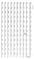
- Figure 7 shows the sequence of one strand of the DNA.
- the nucleotides are arbitrarily numbered from 1 to 11873 in the 5′ to 3′ direction.
- the original 403-nucleotide sequence runs from Figure 7 nucleotides Nos. 4372 to 4774 and is indicated by 0-0′.
- the 129-nucleotide sequence lying within the 403 one runs from Figure 7 nucleotides Nos. 4442 to 4570 and is indicated by J-J′. This corresponds exactly to the "w" exon.
- nucleotides Nos. 1-7830 contains two short exons (nucleotides 4442-4570 and 7140-7342 respectively) marked w and x in Figures 6(a)&9,J-J′ and J′-J ⁇ in Figures 7 and 9. These code for amino acids 85-127, and 128-195 respectively of the amino acid sequence predicted from the human factor IX cDNA clone ( Figure 9). There are no differences in amino acid sequences predicted from the genomic and cDNA clones of the invention in these two exon regions.
- sequence of the gene between residues 7831-11873 is less complete, containing several gaps, but is still a useful characterisation of the gene as it contains two "Alu I repeat" sequences, nucleotides 7960-8155 and 9671-9938. Alu I sequences are found in many genes. The repetition is not exact but there is a typical degree of homology between them. This further characterisation provides a useful cross-check on the accuracy of the restriction enzyme map. This emerges more clearly from the restriction enzyme chart of Figure 8.
- Figure 8 is a chart produced by a computer analysis of the sequence data of the 11873 nucleotide long sequence of Figure 7.
- Column 1 of Figure 8 gives the arbitrary nucleotide number allotted to the nucleotide of Figure 7.
- Column 2 apportions the nucleotide number as a fraction of the whole sequence.
- Column 3 shows the restriction enzymes which will cut the DNA within various short sequences of nucleotides shown in Column 4.
- the short sequences of Column 4 begin with the nucleotide numbered in Column 1. With the aid of this chart the positions of the restriction sites shown in Figure 6(d) and some of the sequences shown in Figure 6(c) can be determined very accurately.
- sequences II-IV are produced by restriction at the following sites (denoted by the first nucleotide number at the 5′ end of each site).
- Particularly important sites are arrowed in Figure 8.
- Some of the relevant nucleotide numbers are shown in Figure 6(c), the number given being that of the nucleotide at the 5′ end of each site.
- pHIX-17 DNA was digested with Eco RI.
- the digested material was resolved on 0.8% agarose gel and a 1.4 kb fragment was isolated in solution by electroelution. It can be stored in the usual manner. This 1.4 kb long molecule was used for the initial sequencing. Only about 1.0 kb is inserted DNA, the remaining 0.4 kb being of pBR322.
- a 403 nucleotide length of the inserted DNA was sequenced and is identified as 0--0′ in Figure 7. The same 1.4 kb fragment was also labelled and used as a probe in Section M.
- Hind III cleave ds DNA to leave sticky or "overhanging" ends.
- Hind III cleaves - AAGCTT -TTCGAA between the adenine-carrying nucleotides of each strand leaving the sticky-ended complementary strands:- -A -TTCGA which are present in the oligo N3/N4 combination.
- pAT153 was digested with Hind III and Bam HI and the 3393 nucleotide long linear fragment was separated from the 346 nucleotide shorter fragment by 0.7% agarose gel electrophoresis, followed by electroelution of the appropriate bands visualised by ethidium bromide fluorescence under UV light.
- the Bam HI Hind III 3393-long fragment was ligated to an equimolar mixture of oligo N3 and oligo N4 which themselves had been pretreated, as a mixture, with T4 polynucleotide kinase and ATP, to phosphorylate their respective 5′-terminal OH groups.
- This part of the sequence is shown in Figure 11 along the unique restriction sites.
- the novel part of the plasmid sequence is underlined: the remainder is present in the parent plasmid pAT153.
- the vector allows blunt-end cloning (after treatment with phosphatase) into the inserted Pvu II site.
- the cloned DNA can be excised, assuming that it lacks appropriate internal restriction sites, with Bam HI/ Hind III, Bam HI/ Cla I or Bam HI/ Eco RI double digests.
- the sites adjacent to the PvuII site are also convenient for end labelling with 32P for characterization of the ends of cloned DNA by the Maxam-Gilbert sequencing method.
- RNA was extracted from a human liver and a 20-22 Svedberg unit enriched fraction of mRNA prepared exactly as described for bovine mRNA in Section B above, except that a 'translation assay' was not used.
- the first steps in the construction of the double-stranded DNA were carried out using the 'Stanford protocol' kindly supplied from Professor P Berg's department at Stanford University, USA. This itself is a modification of Wickens, Buell & Schimke (J.Biol.Chem. 253 , 2483-2495, 1978) and some further modifications, incorporated in the description given below were made in the present work.
- the second strand synthesis was carried out by adding directly to the above solution 20 microlitres of 5x second strand buffer (250 mM Hepes/KOH pH 6.9, 250 mM KCl, 50 mM MgCl2), 4 microlitres of a 2.5 mM mixture of each of the four deoxynucleoside triphosphates, 10 microlitres of E . coli DNA polymerase I (6 units per microlitre) and making the volume of the solution up to 100 microlitres with deionized water.
- 5x second strand buffer 250 mM Hepes/KOH pH 6.9, 250 mM KCl, 50 mM MgCl2
- 4 microlitres of a 2.5 mM mixture of each of the four deoxynucleoside triphosphates 10 microlitres of E . coli DNA polymerase I (6 units per microlitre) and making the volume of the solution up to 100 microlitres with deionized water.
- S1 nuclease digestion was carried out by the addition of 400 microlitres of S1 nuclease buffer (0.03 M sodium acetate pH 4.4, 0.25 M NaCl, 1 mM ZnSO4) and 1 microlitre of S1 nuclease (at 500 units per microlitre). After incubating for 30 minutes at 37°C, 10 microlitres of 0.5 M EDTA (pH 8.0) was added. Double stranded DNA was deproteinised by shaking with an equal volume of a phenol: chloroform (1:1) mixture, followed by ether extraction of the aqueous phase and precipitation of ds DNA by addition of 2 volumes of ethanol.
- DNA polymerase I "fill in" of S1 ends was carried out by a further incubation of the sample dissolved in 25 microlitres of 50 mM tris-chloride, pH 7.5, 10 mM MgCl2, 5 mM dithiothreitol and containing 0.02 mM dNTP and 6 units of DNA polymerase I. After incubating for 10 minutes at room temperature, 5 microlitres of EDTA (0.1 M at pH 7.4) and 3 microlitres of 5% sodium dodecyl sulphate (SDS) were added.
- SDS sodium dodecyl sulphate
- the following part of the protocol differs from the 'Stanford protocol'.
- the sample was fractionated on a "mini"-Sephacryl S400 column run in a disposable 1 ml pipette in 0.2 M NaCl, 10 mM Tris-chloride, pH 7.5 and 1 mM EDTA.
- the first 70% of the "break-through” peak of radioactivity was pooled (0.4 ml) and deproteinised by shaking with an equal volume of n-butanol:chloroform (1:4).
- To the aqueous phase was added 1 microgram of yeast RNA (BDH) as carrier followed by 2 volumes of ethanol.
- yeast RNA yeast RNA
- nick-translation was carried out as before and the material used in a hybridisation reaction for 16 hours at 65°C in 3x SSC, 10x Denhardts solution, 0.1% SDS and 50 micrograms/ml sonicated denatured E . coli DNA and 100 micrograms/ml of sonicated denatured herring sperm DNA.
- hydridisation filters were washed at 65°C successively in 3x SSC, 0.1% SDS (2 changes, half an hour each) and 2x SSC, 0.1% SDS (2 changes, half an hour each).
- Nucleotides 115-2002 were derived by sequencing clone pATIXcVII. (The actual extent of this clone is greater as it extends in a 5′ direction to nucleotide 17. The sequence between 17 and 111 is inverted with respect to the remainder of the sequence presumably due to a cloning artefact.) Nucleotides 1-130 were derived from clone pATIXcVI which extends from nucleotides 1-1548 of Figure 9. The sequence from Nos.
- 2002-2778 was derived by isolating 4 additional clones designated pATIX108.1, pATIX108.2, pATIX108.3 and pATIXDB.
- the first 3 were derived from a mini-library (designated GGB108) of cDNA clones constructed exactly as described in section K above except that sucrose density gradient centrifugation was used instead of chromatography on "Sephacryl" S-400 to fractionate the double-stranded DNA according to size.
- a fraction of m.w. from 1 kb-5 kb was selected and an amplified library of 10,000 independent clones containing approximately 20% background non-recombinant clones was obtained.
- Clone pATIXDB derived from another cDNA library (designated DB1) constructed as described in section K except that total poly A+ human liver mRNA was used as the starting material and sucrose density gradient centrifugation was used to fractionate the DNA according to size as in the construction of the mini-library GGB108.
- the complexity of this library was 95,000 with an estimated background of non-recomibinants of 50%.
- Clones pATIX108.1 and pATIX108.2 were selected from a group of 30 hybridization-positive clones isolated by Grunstein-Hogness screening of the mini library GGB108 using a 32P-nick translated probe derived from a Sau 3AI restriction enzyme fragment, itself derived from nucleotides 1796-2002 of clone pATIXcVII. From pATIX108.1 the sequence of nucleotides 2009-2756 was determined ( Figure 9). Following this the sequence of a part of pATIX108.2, specifically nucleotides 1950-2086, provided the overlap with pATIXcVII.
- clone pATIX108.3 was selected and sequenced from nucleotides 2642-2778. This clone was followed by three A nucleotides, which sequence was confirmed as a vestigial marker for the poly A tail, by the subsequent isolation of clone pATIXDB by a similar method.
- pATIXDB was sequenced from Nos. 2760-2778 and ended in 42 A nucleotides, thus marking the 3′ end of the mRNA.
- Figure 9 shows that the predicted amino acid sequence codes for a protein of 456 amino acids, but included in this are 41 residues of precursor amino acid sequence preceding the N-terminal tyrosine residue ( ) of the definitive length factor IX protein.
- the precursor section of the protein shows a basic amino acid domain (amino acids -1 to -4) as well as the more usual hydrophobic signal peptide domain (amino acids -21 to -36).
- the definitive factor IX protein consists of 415 amino acids with 12 potential gamma-carboxyglutamic acid residues at amino acids 7, 8, 15, 17, 20, 21, 26, 27, 30, 33, 36 and 40. Two potential carbohydrate attachment sites occur at amino acid residues 157 and 167.
- the activation peptide encompasses residues 146-180, which are cut out in the activation of Factor IX (see Background of Invention) by the peptide cleavage of an R-A and R-V bond. This leaves a light chain spanning residues 1-145 and a heavy chain spanning residues 181-415.
- the 3′ non-coding region of the mRNA is extensive, consisting of 1390 residues (including the UAAUGA double terminator 1389-1394 but excluding the poly A tail).
- the factor IX cDNA is cleavable by the restriction enzyme Hae III to give a fragment from nuclotides 133-1440, i.e. a 1307 nuclotide long region of DNA entirely encompassing the definitive factor IX protein sequence.
- the nucleotide sequence recognised by Hae III is GGCC. This fragment should be a suitable starting material for the expression of factor IX protein from suitable promoters in bacterial, yeast of mammalian cells. Another suitable fragment could be derived using the unique Stu I site at residue 41 (corresponding to an early part of the hydrophobic signal peptide region) and linking it to a suitable promoter.
- the nucleotide sequence recognised by Stu I is AGGCCT.
- probes of the invention for the assay of alterations of genes of some patients suffering from Christmas disease has been demonstrated as follows. Two patients with severe Christmas disease, who also developed antibodies to factor IX, were selected for study. The DNA from 50 ml of blood was digested separately with Eco RI, Hind III, Bgl II and Bcl I and Southern blots prepared for probing with 32P-nick translated probe II (Figure 6). No specific bands were observed with either patient under conditions where a control digest gave the pattern shown in Figure 13. Similarly no bands were observed in the patients when probe I, III or IV ( Figure 6) was substituted for probe II.
- a factor IX gene probe (this time pATIXcVII - the cDNA probe) was mixed with an irrelevant autosomal gene probe which was specific for the human A1 apolipoprotein (Shoulders and Baralle, Nucl.Acids Res. 10 , 4873-4882, 1982).
- This experiment showed that patient 1 had the normal A1 apolipoprotein gene, characterised by a 12 kb band in an Eco RI digest, and confirmed that he lacked the 5.5 kb band observed with pATIXcVII and characteristic of the normal factor IX gene.
Landscapes
- Chemical & Material Sciences (AREA)
- Health & Medical Sciences (AREA)
- Life Sciences & Earth Sciences (AREA)
- Organic Chemistry (AREA)
- Engineering & Computer Science (AREA)
- Genetics & Genomics (AREA)
- Biochemistry (AREA)
- Wood Science & Technology (AREA)
- Molecular Biology (AREA)
- General Health & Medical Sciences (AREA)
- Bioinformatics & Cheminformatics (AREA)
- Zoology (AREA)
- Biomedical Technology (AREA)
- General Engineering & Computer Science (AREA)
- Biotechnology (AREA)
- Medicinal Chemistry (AREA)
- Microbiology (AREA)
- Micro-Organisms Or Cultivation Processes Thereof (AREA)
- Preparation Of Compounds By Using Micro-Organisms (AREA)
- Saccharide Compounds (AREA)
- Investigating Or Analysing Biological Materials (AREA)
- Enzymes And Modification Thereof (AREA)
- Peptides Or Proteins (AREA)
Abstract
Description
- This invention is in the field of genetic engineering relating to factor IX DNA.
- Factor IX (Christmas factor or antihaemophilic factor B) is the zymogen of a serine protease which is required for blood coagulation via the intrinsic pathway of clotting (Jackson & Nemerson, Ann.Rev.Biochem. 49, 765-811, 1980). This factor is synthesised in the liver and requires vitamin K for its biosynthesis (Di Scipio & Davie, Biochem. 18, 899-904, 1979).
- Human factor IX has been purified and characterised, but details of the amino acid sequence are fragmentary. It is a single-chain glycoprotein, with a molecular weight of approximately 60,000 (Suomela, Eur.J.Biochem. 71, 145-154, 1976). Like other vitamin K-dependent plasma proteins, human factor IX contains in the amino-terminal region approximately 12 gamma-carboxyglutamic acid residues (Di Scipio & Davie, Biochem. 18, 899-904, 1979).
- During the clotting process, and in the presence of Ca⁺⁺ ions, factor IX is acted upon by activated factor XI (XIa) by the cleavage of two internal peptide bonds, releasing an activation glycopeptide of 10,000 daltons (Di Scipio et al., J.Clin. Invest. 61, 1528-1538, 1978). The activated factor IX (IXa) is composed of two chains held together by at least one disulphide bond. Factor IXa then participates in the next step in the coagulation cascade by acting on factor X in the presence of activated factor VIII, Ca⁺⁺ ions, and phospholipids (Lindquist et al., J.Biol.Chem. 253, 1902-1909, 1978).
- Individuals deficient in factor IX (Christmas disease or haemophilia B) show bleeding symptoms which persist throughout life. Bleeding may occur spontaneously or following injury. This may take place virtually anywhere. Bleeding into the joints is common, and after repeated haemorrhages, may result in permanent and crippling deformities. The condition is a sex-linked disorder affecting males. Its frequency in the population is approximately 1 in 30,000 males.
- The current method of diagnosing Christmas disease involves measurement of the titre of factor IX in plasma by a combination of a clotting assay and an immunochemical assay. Treatment of haemorrhage in the disease consists of factor IX replacement by means of intravenous transfusion of human plasma protein concentrates enriched in factor IX. The enrichment of plasma in factor IX is a time-consuming process.
- After considerable research and experiment, important progress has now been made towards producing artificial human factor IX by recombinant DNA technology (genetic engineering). Thus, the cloning of DNA sequences which are substantially the same as extensive sequences occurring in the human factor IX genome has been achieved.
- The invention arises from the finding that an extensive DNA sequence of the human factor IX genome can be obtained by a clever and laborious combination of chemical synthesis and artificial biosynthesis, starting from elementary nucleotide or dinucleotide "building blocks", as will be described below.
- A major feature of the invention comprises recombinant DNA which comprises a cloning vehicle DNA sequence and a sequence foreign thereto (i.e. foreign to the vehicle) which is substantially the same as a sequence occurring in the human factor IX genome. A 11873 nucleotide long part of such a foreign sequence has been identified and a very large part of it has been sequenced by the Maxam-Gilbert sequencing method. A 129 nucleotide length of this sequence is more than sufficient to characterise it unambiguously as coding for a specific protein and a particular such length is regarded herein as useful to characterise the whole sequence inserted in the cloning vehicle as one occurring in the human factor IX genome. Other cloned sequences can then be verified as belonging to the human factor IX genome by determining that part thereof is identical to a region of the first-mentioned sequence, i.e. the sequences have a common identity in an overlapping region. A further feature of the invention therefore comprises recombinant DNA which comprises a cloning vehicle or vector DNA sequence and a DNA sequence foreign thereto which consists of or includes substantially the following sequence of 129 nucleotides (which should be read in rows of 30 across the page):-
ATGTAACATG TAACATTAAG AATGGCAGAT
GCGAGCAGTT TTGTAAAAAT AGTGCTGATA
ACAAGGTGGT TTGCTCCTGT ACTGAGGGAT
ATCGACTTGC AGAAAACCAG AAGTCCTGTG
AACCAGCAG (1) - The invention includes particularly recombinant DNA which comprises a cloning vehicle DNA sequence and a sequence foreign to the cloning vehicle, wherein the foreign sequence includes substantially the whole of an exon sequence of the human factor IX genome. The 129-nucleotide sequence described above corresponds substantially to such an exon sequence. Another such exon sequence which independently characterises the human factor IX DNA is the 203-nucleotide sequence substantially as follows (again reading in rows of 30 across the page):-
TGCCATTTCC ATGTGGAAGA GTTTCTGTTT
CACAAACTTC TAAGCTCACC CGTGCTGAGG
CTGTTTTTCC TGATGTGGAC TATGTAAATT
CTACTGAAGC TGAAACCATT TTGGATAACA
TCACTCAAAG CACCCAATCA TTTAATGACT
TCACTCGGGT TGTTGGTGGA GAAGATGCCA
AACCAGGTCA ATTCCCTTGG CAG - The intron sequences of the human factor IX genome are excised during the transcription process by which mRNA is made in human cells. Only exon sequences are translated into protein. DNA coding for factor IX has been prepared from human mRNA. This cDNA has been partly sequenced and found to contain the same 129- and 203-nucleotide sequences set out above.
- The invention also includes recombinant DNA which comprises a cloning vehicle sequence and a DNA sequence foreign to the cloning vehicle, wherein the foreign sequence comprises a DNA sequence which is complementary to human factor IX mRNA. Such a recombinant cDNA can be isolated from a library of recombinant cDNA clones derived from human liver mRNA by using an exon of the genomic human factor IX DNA (or part thereof) as a probe to screen this library and thence isolating the resulting clones.
- The invention also includes recombinant DNA in which the foreign sequence is any fragment of human factor IX DNA, particularly of length at least 50 and preferably at least 75 nucleotides or base-pairs. It includes such recombinant DNA whether or not part of the 129 or 203-base-pair sequence defined above. It includes especially part or all of the exon sequences of human factor IX genomic DNA. Various short lengths up to about 11 kilobases (11,000 nucleotides or base-pairs) long have been prepared by use of various restriction endonucleases. Methods of isolating recombinant DNA from clones are well known and some are described hereinafter. The DNA of the invention can be single or double stranded form.
- The recombinant human factor IX DNA of this invention is useful as a tool of recombinant DNA technology. Thus it is useful as the first stage in the production of artificial human factor IX and in the preparation of probes for diagnostic purposes.
- In the production of artificial human factor IX it is contemplated that appropriate cDNA or genomic clones will be introduced into a suitable expression vector in either mammalian or bacterial systems. For mammalian studies, the gene might be too long to be conveniently retained in one clone. Therefore a suitable artificial "minigene" will be designed and constructed from suitable parts of the cDNA and genomic clones. The minigene will be under the control of its own promoter or instead will be replaced by an artificial one, perhaps the mouse metallothioneine I promoter. The resultant 'minigene' will then be introduced into mammalian tissue culture cells e.g. a hepatoma cell line, and selection for clones of cells synthesising maximum amounts of biologically active factor IX will be carried out. Alternatively "genetic farming" could be employed as has been demonstrated for mouse growth hormone (Palmiter et al,
Nature 300, 611-615, 1982). The minigene would be micro-injected into the pronucleus of fertilised eggs, followed by in vivo cloning and selection for progeny producing the largest quantity of human factor IX in blood. Alternatively, it is contemplated that the cDNA clone or selected parts of it will be linked to a suitable strong bacterial promoter, e.g. a Lac or Trp promoter or the lamdba PR or PL, and a factor IX polypeptide obtained therefrom. - The natural factor IX polypeptide is synthesised as a precursor containing both a signal and propeptide region. They are both normally cleaved off in the production of the definitive length protein. Even this product is merely a precursor. It is biologically inactive and must be gamma-carboxylated at 12 specific N-terminal glutamic acid residues in the so called 'GLA' domain by the action of a specific vitamin K-dependent carboxylase. In addition, two carbohydrate molecules are added to the connecting peptide region of the molecule, but it remains unknown whether they are required for activity. The substrate for the carboxylase is unknown and could be the precursor factor IX polypeptide or alternatively the definitive length protein. Therefore various relevant polypeptides both with and without the precursor domains will be "constructed" using genetic engineering methods in bacterial hosts. They will then be tested as substrates for the conversion of inactive to biologically active factor IV in vitro by the action of partially purified preparations of the carboxylase enzyme which can be isolated from liver microsomes or other suitable sources.
- For diagnostic purposes, the recombinant human genomic factor IX DNA or recombinant human mRNA-derived factor IX DNA has a wide variety of uses. It can be cleaved by enzymes or combinations of two or more enzymes into shorter fragments of DNA which can be recombined into the cloning vehicle, producing "sub-clones". These sub-clones can themselves be cleaved by restriction enzymes to DNA molecules suitable for preparing probes. A probe DNA (by definition) is labelled in some way, conveniently radiolabelled, and can be used to examine in detail mutations in the human DNA which ordinarily would produce factor IX. Several different probes have been produced for examining several different regions of the genome where mutation was suspected to have occurred in patients. Failure to obtain hybridisation from such a probe indicates that the sequence of the probe differs in the patient's DNA. In particular it has been shown that Christmas disease can be detected or confirmed by such methodology. Useful probes can contain intron and/or exon regions of the genomic DNA or can contain cDNA derived from the mRNA.
- The invention includes particularly probe DNA, i.e. which is labelled, and of a length suitable for the probing was envisaged. It can be single-stranded or double-stranded over at least the human factor IX DNA probing sequences thereof and such sequences will usually have a length of at least 15 nucleotides, preferably at least 19-30 nucleotides in order to have a reasonable probability of being unique They will not usually be larger than 5 kb and rarely longer than 10 kb.
- The invention accordingly includes a DNA molecule, comprising part of the human factor IX DNA sequence, whether or not labelled, whether intron or exon or partly both. It also includes human cDNA corresponding to part of all of human factor IX mRNA. It includes particularly a solution of any DNA of the invention, which is a form in which it is conveniently obtainable by electroelution from a gel.
- The invention includes, of course, a host transformed with any of the recombinant DNA of the invention. The host can be a bacterium, for example an appropriate strain of E.coli, chosen according to the nature of the cloning vehicle employed. Useful hosts may include strains of Pseudomonas, Bacillus subtilis and Bacillus stearothermophilus, other Bacilli, yeasts and other fungi and mammalian (including human) cells.
- One process practised in connection with this invention for preparing a host transformed with the recombinant DNA of the invention is based on the following steps:-
- (1) synthesising an oligodeoxynucleotide having a nucleotide sequence comprising that occurring in bovine factor IX messenger RNA coding for amino acids 70-75 or 348-352 of bovine factor IX, and labelling the oligodeoxynucleotide to form a probe;
- (2) preparing complementary DNA to a mixture of bovine mRNAs;
- (3) inserting the complementary DNA in a cloning vector to form a mixture of recombinant bovine cDNAs;
- (4) transforming a host with said mixture of recombinant bovine cDNAs to form a library of clones and multiplying said clones;
- (5) probing the clones with the synthetic oligodeoxynucleotide probe obtained in
step 1 and isolating the resultant recombinant bovine factor IX cDNA-containing clone; - (6) digesting the recombinant bovine factor IX cDNA from said clone with one or more enzymes to produce a bovine factor IX cDNA molecule comprising a shorter sequence of bovine factor IX DNA, but preferably at least 50 base-pairs long; and
- (7) probing a library of recombinant human genomic DNA in a transformed host with the shorter sequence bovine factor IX cDNA molecule, to hybridise the human genomic DNA to the said recombinant bovine factor IX DNA and isolating the resultant recombinant DNA-transformed host.
-
- Figure 1 shows the structure of a published amino-acid sequence of bovine factor IX polypeptide, the deduced sequence of the mRNA from which it would be translated and the structures of oligonucleotides (oligo-N1 and N2) synthesised in the course of this invention;
- Figures 2 and 3 show the chemical formulae of "building blocks" used to synthesise the oligonucleotides referred to in Figures 1 and 11;
- Figure 4 is an elevational view, partly sectioned, showing an apparatus for synthesising oligonucleotides;
- Figure 5 shows the sequence of part of the bovine factor IX cDNA obtained in this invention;
- Figure 6 is a map showing the organisation of an approximately 27 kb length of human factor IX genomic DNA and is divided into five portions, showing:-
- (a) the exon regions;
- (b) the 11,873- nucleotide length sequenced;
- (c) cDNA molecules obtained by restriction with various endonucleases, sub-cloned and subsequently used as probes;
- (d) DNA molecules obtained by restriction with various endonucleases; and
- (e) three regions of human factor IX genomic DNA derived from three clones in lambda phage vector.
- Figure 7 shows the sequence of the DNA of Figure 6(b) and in parts the encoded protein;
- Figure 8 shows a restriction enzyme chart of the sequence shown in Figure 7;
- figure 9 shows part of the sequence of the human factor IX cDNA and its encoded protein;
- Figure 10 shows the structure of a pair of complementary oligonucleotides (oligo N3 and N4) synthesised in the course of this invention;
- Figure 11 shows part of the DNA sequence of the vector pAT153/PvuII/8 of this invention, in the region where it differs from pAT153;
- Figure 12 is a diagram of plasmid pHIX17 of the invention showing the origin of the 1.4 kb fragment used for probing and initial sequencing; and
- Figure 13 shows the position of the major radioactive bands on probing a "Southern blot" of normal human DNA, cut by the restriction enzymes EcoRI(E), HindIII(H), BglII(B) and BclI(Bc), with a sub-clone of the recombinant human factor IX DNA of this invention.
- A recombinant DNA of the invention can be extracted by means of probes from a library of cloned human genomic DNA. This is a known recombinant library and the invention does not, of course, extend to human genomic factor IX DNA when present in such a library. The probes used were of bovine factor IX cDNA (DNA complementary of bovine mRNA), which were prepared by an elaborate process involving firstly the preparation of recombinant bovine cDNA from a bovine mRNA starting material, secondly the chemical syntheses of oligonucleotides, thirdly their use to probe the recombinant bovine cDNA, in order to extract bovine factor IX cDNA and fourthly the preparation of suitable probes of shorter length from the recombinant bovine factor IX cDNA. The first probe tried appeared to contain an irrelevant sequence and the second probe tried, not containing it, proved successful in enabling a single clone of the human genomic factor IX DNA to be isolated. This clone is designated lambda HIX-1. The steps involved are described in more detail in the sub-section "Examples" appearing hereinafter, and the second probe comprises the 247 base-pair DNA sequence of bovine factor IX cDNA indicated in Figure 5 of the drawings. The invention therefore provides specifically a recombinant DNA which comprises a cloning vehicle sequence and a DNA sequence foreign to the cloning vehicle, which recombinant DNA hybridises to a 247 base-pair sequence of bovine factor IX cDNA indicated in Figure 5 (by the arrows at each end thereof).
- The cloning vehicle or vector employed in the invention can be any of those known in the genetic engineering art (but will be chosen to be compatible with the host). They include E.coli. plasmids, e.g. pBR322, pAT153 and modifications thereof, plasmids with wider host ranges, e.g. RP4 plasmids specific to other bacterial hosts, phages, especially lambda phage, and cosmids. A cosmid cloning vehicle contains a fragment of phage DNA including its cos (cohesive-end site) inserted in a plasmid. The resultant recombinant DNA is circular and has the capacity to accommodate very large fragments of additional foreign DNA.
- Fragments of human factor IX genomic DNA can be prepared by digesting the cloned DNA with various restriction enzymes. If desired, the fragments can be religated to a cloning vehicle to prepare further recombinant DNA and thereby obtain "sub-clones". In connection with this embodiment a new cloning vehicle has been prepared. This is a modified pAT153 plasmid prepared by ligating a BamHI and HindIII double digest of pAT153 to a pair of complementary double sticky-ended oligonucleotides having a DNA sequence providing a BamHI restriction residue at one end, a HindIII restriction residue at the other end and a PvuII restriction site in between.
- While the invention is described herein with reference to human genomic factor IX DNA in particular, the invention includes human factor IX cDNA (complementary to human factor IX mRNA) which contains substantially the same sequences. A library of human cDNA has been prepared and probed with human factor IX genomic DNA to isolate human factor IX cDNA from the library. For this purpose the probe DNA is conveniently of relatively short length and must include at least one exon sequence. The invention therefore includes a process of preparing a host transformed with recombinant DNA, comprising cloning vector sequences and a sequence of nucleotides comprised in cDNA complementary to human factor IX mRNA, which process comprises probing a library of clones containing recombinant DNA complementary to human mRNA with a probe comprising a labelled DNA comprising a sequence complementary to part or all of an exon region of the human factor IX genome.
- E.coli K-12 strain MC 1061 (Casadaban & Cohen, J.Mol. Biol. 138, 179-207, 1980), E.coli K-12 strain HB 101 (Boyer & Roulland-Dussoix, J.Mol.Biol. 41, 459-472, 1969) and E.coli K-12 strain K803 which is a known strain used by genetic engineers.
- Highly purified bovine factor IX and rabbit anti-bovine factor IX antiserum were gifts from Dr. M.P. Esnouf. Analysis of the purified bovine factor IX on a denaturating polyacrylamide gel showed that it has a purity of greater than 99%. Specific anti-factor IX immunoglobulins used for immunoprecipitation experiments were purified as described by Choo et al., Biochem.J. 199, 527-535, 1981, by passage of the crude antiserum through a Sepharose-4B column onto which pure bovine factor IX has been coupled.
- Bovine mRNA was obtained from calf liver and isolated by the guanidine hydrochloride method (Chirgwin et al., Biochem. 18, 5294-5299, 1979). The mRNA preparation was passaged through an oligo dT-cellulose column (Caton and Robertson, Nucl. Acids Res. 7, 1445-1456, 1979) to isolate poly(A) + mRNA. Poly(A) + mRNA was translated in a rabbit reticulocyte cell-free system in the presence of ³⁵S-cysteine as described by Pelham and Jackson (Eur. J.Biochem. 67, 247-256, 1976). At the end of the translation reaction, factor IX polypeptide was precipitated by the addition of specific anti-factor IX immunoglobulins. The immunoprecipitation procedure was as described by Choo et al., Biochem.J. 181, 285-294, 1979. The immunoprecipitated material was washed throughly and resolved on a two-dimensional SDS-polyacrylamide gel (Choo et al., Biochem.J. 181, 285-294, 1979), by isoelectric focussing in one dimension and electrophoresis in another. Some polypeptides of known molecular weight were subjected to this procedure, to serve as reference points. The immunoprecipitated material showed 4 pronounced spots, all in the 50,000 molecular weight region and with separated isoelectric points. These predominant spots of molecular weight about 50,000 represent a single polypeptide chain plus a possible prepeptide signal sequence, a deduction compatible with published data (Katayama et al.,Proc. Natl.Acad. Sci.USA 76, 4990-4994, 1979).
- When the gel analysis was repeated for the same material but immunoprecipitated in the presence of unlabelled pure bovine factor IX, the 4 spots appeared at reduced intensity, indicating that the translation product is specifically competed for by pure factor IX. Thirdly, immunoprecipitation was performed using a control rabbit antiserum, i.e. from a rabbit which had not been immunised with factor IX. None of the 4 spots appeared. These results therefore indicate that the translation product was a factor IX polypeptide.
- The specific immunological/cell-free translation assay established above was used to monitor the enrichment of factor IX mRNA on sucrose gradient centrifugations. Total poly(A)+mRNA was resolved by two successive separations by sucrose gradient centrifugations. When individual fractions from the gradient were assayed by the above method, a fraction of size 20-22 Svedberg units (approx. 2.5 kilobases of RNA) region was found to be enriched (approx. ten-fold) for the bovine factor IX mRNA. This enriched fraction was used in the subsequent cloning experiments.
- Starting from a knowledge of the amino acid sequence of bovine factor IX (Katayama et al., Proc.Natl.Acad.Sci. USA 76, 4990-4994, 1979), the synthesis of two mixtures of oligonucleotide probes was designed. These probes consisted of DNA sequences coding for two different regions of the protein. The regions selected were those known to differ in sequence in the analogous serine proteases, prothrombin, Factor C and Factors VII and X and were those corresponding to amino acids 70-75 and 348-352 respectively. The 70-75 region was particularly favourable in that the mixture of oligonucleotides synthesised, i.e. oligo N2A and oligo N2B, contained all 16 possible sequences that might occur in a 17 nucleotide long region of the mRNA corresponding to amino acids 70-75. The oligo N2A-N2B mixture is hereinafter called "oligo N2" for brevity.
- Figure 1 of the drawings shows the two selected regions of the known amino acid sequence of bovine factor IX, the corresponding mRNA and the oligonucleotides synthesised. Since some of the amino acids are coded for by more than one nucleotide triplet, there are 4 ambiguities in the mRNA sequence shown for amino acids 70-75 and therefore 16 possible individual sequences.
- The nucleotide mixtures oligo N1 and oligo N2 were synthesized using the solid phase phosphotriester method of Duckworth et al., Nucl.Acids Res. 9, 1691-1706, 1981, modified in two ways. Firstly, 0-chlorophenyl rather than p-chlorophenyl blocking groups were used for the phosphotriester grouping, and were incorporated in the mononucleotide and dinucleotide "building blocks". Figures 2 and 3 of the drawings show (a) dinucleotide and (b) mononucleotide "building blocks". DMT = 4,4′ - dimethoxytrityl and B = 6-N-benzoyladenin-9-yl, 4-N-benzoylcytosin-1-yl, 2-N-isobutyrylguanin-9-yl or thymin-1-yl, depending on the nucleotide selected. Secondly, the "reaction cell" used for the successive addition of mono- or dinucleotide "building blocks" was miniaturised so that the coupling step with the condensing agent 1-(mesitylene-2-sulphonyl)-3-nitro-1,2,4-triazole (MSNT) was carried out in a volume of 0.5ml pyridine containing 3.5 micromoles of polydimethylacrylamide resin, 17.5 micromoles of incoming dinucleotide ( or 35 micromoles of mononucleotide) and 210 micromoles of MSNT.
- Figure 4 of the drawings is an elevational view of the
microreaction cell 1 andstopper 2 used for oligonucleotide synthesis, drawn 70% of actual size. The device comprises a glass-to-PTFE tubing joint 3 at the inlet end of thestopper 2. The stopper has aninternal conduit 4 which at its lower end passes into a hollow tapered groundglass male member 5 and thence into asintered glass outlet 6 to the stopper. Thecell 1 has a ground glassfemale member 7 complementary to themember 5 of the stopper, leading toreaction chamber 8, the lower end of which terminates in asintered glass outlet 9. This communicates withglass tubing 10, and a 1.2mm. "Interflow"tap 11.Further glass tubing 10, beyond thetap 11, leads to the outlet glass-to-PTFE tubing joint 12. Pairs ofears 13 on the stopper and cell enable them to be joined together by springs (not shown) in a liquid-tight manner. - After completion of the synthesis and deprotection, fractionation was carried out by high pressure liquid chromatography (Duckworth et al., see above) and the peak tubes corresponding to the product of correct chain length were located by labelling of fractions at their 5′-hydroxyl ends using [gamma-³²P]-ATP and T4 polynucleotide kinase, followed by 20% 7M urea polyacrylamide gel electrophoresis. The position on the gel of the 17- and 14- oligonucleotides was determined by separately labelling, by the method described above, 17- and 14- nucleotide long "marker" oligonucleotides and subjecting these to the same gel electrophoresis.
- Two different approaches were used for the generation of cloned cDNA library:-
- First strand cDNA was synthesised using the sucrose gradient-enriched poly(A) + bovine mRNA as template. The conditions used were as described by Huddleston & Brownlee, Nucl. Acids Res.10, 1029-1030, 1981, except that 2 micrograms of oligo N-1, 20-30 micrograms of the mRNA, 10 microcuries [alpha-³²P] -dATP (Amersham, 3000 Ci/mmole), and 50 U of reverse transcriptase were used in a 50 microlitre reaction. "dNTP" in Figure 1 denotes the mixture of 4 deoxynucleoside triphosphates required for synthesis. Oligo N-1 hybridises to the corresponding region on the mRNA (refer to Figure 1) and thereby acts as a primer for the initiation of transcription. It was used in order to achieve a further enrichment for factor IX mRNA. At the end of the cDNA synthesis reaction, the cDNA was extracted with phenol and desalted on a Sephadex G100 column, before it was treated with alkali (0.1M NaOH, 1mM EDTA) for 30 min. at 60°C to remove the mRNA strand. Second strand DNA synthesis was then carried out exactly as published (Huddleston & Brownlee, Nucl. Acids Res. 10, 1029-1038, 1981).
- The double-stranded DNA was next cleaved with the restriction enzyme MboI and ligated to the plasmid vector pBR322 which had been cut with BamHI and treated with calf intestinal alkaline phosphatase to minimise vector self-religation. Phosphatase treatment was carried out by incubating 5 micrograms of BamHI-
cut pBR 322 plasmid with 0.5 microgram calf intestinal phosphatase (Boehringer; in 10mM Tris - HCl buffer, pH 8.0) in a volume of 50 microlitres at 37°C for 10 minutes, see Huddleston & Brownlee supra. - The ligated DNA was used to transform E.coli strain MC 1061. For transformation E.coli MC 1061 was grown to early exponential phase as indicated by an absorbancy of 0.2 at 600 nm and made "competent" by treating the pelleted bacterial cells first with one half volume, followed by repelleting, and then with 1/50 volume of the original growth medium of 100mM CaCl₂ 15% v/v glycerol and 10mM PIPES-NaOH, pH 6.6 at 0°C. Cells were immediately frozen in a dry ice/ethanol bath to -70°C. For transformation, 200 microlitre aliquots were mixed with 10 microlitres of the recombinant DNA and incubated at 0°C for 10 minutes followed by 37°C for 5 minutes. 200 microlitres of L-broth (bactotryptone 10g., yeast extract 5g., sodium chloride 10g., made up to 1 litre with deionised water) were then added and incubation continued for a further 30 minutes at 37°C. The solution was then plated on the appropriate antibiotic agar (see below). A library of about 7,000 ampicillin-resistant colonies was thus obtained. They were ampicillin-resistant because they contained the beta-lactamase gene of
pBR 322. Of these, approx. 85% were found to be tetracycline-sensitive. - In the preparation of this library, first strand cDNA was synthesised as described for the above library except that oligo dT(12-18) was used as a primer to initiate cDNA synthesis. Following this, the cDNA was tailed with dCTP using terminal transferase and back-copied with the aid of oligo dG(12-18) primer and reverse transcriptase to give double stranded DNA, exactly according to the method of Land et al., Nucl. Acids Res. 9, 2251-2266, 1981. After a further tailing with dCTP, this material was annealed by hybridisation to a dGTP-tailed pBR322 plasmid at the PstI site. The hybrid DNA was used to transform E.coli strain MC 1061. A library of approximately 10,000 tetracycline-resistant colonies was obtained. Of these, approximately 80% were found to be sensitive to ampicillin, due to insertion of DNA into the ampicillin-resistant gene at the PstI site.
- The library of colonies, in an unordered fashion, was transferred onto 13 Whatman 541 filter papers and amplified with chloramphenicol, to increase the number of copies of the plasmid in the colonies, as described by Gergen et al., Nucl. Acids Res., 1, 2115-2136 (1979). The filters were pre-hybridised at 65°C for 4h in 6 x NET (1 x NET = 0.15m NaCl, 1mM EDTA, 15mM Tris-HCl, pH 7.5), 5 x Denhardt's, 0.5% NP40 non-ionic surfactant, and 1 microgram/ml. yeast RNA as described by Wallace et al., Nucl. Acids Res. 9, 879-894 (1981). Hybridisation was carried out at 47°C for 20h in the same solution containing 3 x 10⁵cpm (0.7 nanogram/ml) of labelled oligo N-2 probe. Labelling was done by phosphorylation of the oligonucleotides at the 5′ hydroxyl end using [gamma-³²P]-ATP and T4 phosphokinase (Huddleston & Brownlee, Nucl.Acids Res. 10, 1029-1038, 1981). At the end of the hybridisation, filters were washed successively at 0-4°C (2h), 25°C (10 min), 37°C (10 min) and 47°C (10 min). After radioautography of the filters from this screening, one colony showed a positive signal above background. This colony was designated BIX-1 clone.
- Screening of this library, in an ordered array fashion, using oligo N-2 probe as described above has resulted in the identification of a positive clone. This was designated BIX-2 clone.
- Characterisation of BIX-1 clone by restriction endonuclease cleavage indicated that it contained a DNA insert of about 430 base-pairs (data omitted, for brevity). Figure 5 shows part of the nucleotide sequence of the coding strand, determined by the Maxam-Gilbert method, extending over 304 nucleotides and provides direct evidence that it has the identity of a bovine factor IX sequence. Thus, nearly all of this 304 nucleotide sequence (corresponding to amino acid residues 52-139) agrees with the nucleotide sequence predicted from the known bovine factor IX amino acid sequence data (Katayama et al., Proc.Natl.Acad.Sci. 76, 4990-4994, 1979). Over this region, there are no discrepancies between BIX-1 and these published data for factor IX, except at nucleotides 38-40 where the amino acid coded for is Asp instead of Thr. This amino acid change was similarly observed in a second, independent cDNA clone (BIX-2; see below). The remainder of the 304-nucleotide sequence, i.e. that shown in brackets in Figure 5, does not agree with the published bovine factor IX amino acid data of Katayama.
- In Figure 5, the underlined portion denotes the sequence corresponding to the oligo N-2 probe sequence, the asterisk denotes a nonsense codon, the brackets enclose a sequence which does not correspond to Katayama's amino acid data and the arrows indicate HinfI restriction sites. The Katayama numbering system for amino acids is shown and this sequence is in the opposite orientation to the direction of transcription of the tetracycline-resistant gene of the plasmid.
- By similar methods, BIX-2 clone was found to have a DNA insert of 102 nucleotides and this spans the nucleotide positions 7-108 as shown in Figure 5. The nucleotide sequences for BIX-1 and BIX-2 clones over this region (nucleotide 7-108) were identical.
- A library of cloned human genomic DNA, namely a HaeIII/AluI lambda phage Charon 4A library prepared by Lawn et al., Cell, 15, 1157-1174, 1978, was used. 10⁶ phage recombinants from this library were screened using the in situ plaque hybridisation procedure as described by T. Maniatis et al., Cell, 15, 687, 1978. Pre-hybridisation and hybridisation were carried out at 42°C in 50% formamide. After hybridisation, filters were washed at room temperature with 2 x SSC (1 x SSC = 0.15mM NaCl, 15mM sodium citrate, at pH 7.2) and 0.1% SDS, then at 65°C with 1 x SSC and 0.1% SDS.
- Two DNA molecules, being restriction fragments from the factor IX cDNA cloned in BIX-1, were radiolabelled and used as probes in the hybridisation. The first fragment corresponds to nucleotide numbers -8 to 317 on the numbering system of Figure 5, and was isolated by Sau3AI digestion of BIX-1 plasmid DNA. The isolated DNA was labelled to high specific activity by incorporation of [alpha -³²P] -dATP using a nick translation (Rigby et al., J. Mol.Biol. 113, 237-251, 1977, modified, vide infra). Using this probe, 10 clones were isolated. These were plaque-purified and re-hybridised with a 247-nucleotide fragment from BIX-1 clone. This fragment, derived from nucleotides 3-249 can be seen from Figure 5. It contains only sequences in agreement with the Katayama bovine factor IX amino acid sequence and was isolated by HinfI digestion of BIX-1 plasmid DNA. Only a single clone gave a positive hybridisation signal with this 247-nucleotide probe. This clone was further plaque-purified and the resulting clone was designated "lambda HIX-1".
- A sub-clone, pATIXcVII, of recombinant human factor IX cDNA from human liver mRNA, and prepared as described in Section L below, was linearised by digestion with HindIII and BamHI. The resulting 2 kb cDNA molecule was purified by 1% agarose gel electrophoresis. After electroelution, about 100 ng of this cDNA was nick-translated with [alpha ³²p] dATP (see above) and used as a hybridisation probe to screen the HaeIII/AluI lambda phage Charon 4A human genomic DNA library for further genomic clones, using standard stringent hybridisation conditions. Two further human factor IX genomic clones, designated lambda HIX-2 and lambda HIX-3, were thus obtained.
- The initial lambda HIX-1 clone was characterised by cleavage with various single and double digests with different restriction endonucleases and Southern blotting of fragments using the bovine factor IX cDNA probe (results omitted for brevity). The subsequently isolated lambda HIX-2 and 3 clones were characterised in the same way except that the human cDNA probe, pATIXcVII (see Section L below) was used for the Southern blots. From these results it emerged that the sequences in the factor IX genome corresponding to lambda HIX-2 and 3 overlapped with lambda HIX-1 as shown in Figure 6(e). In Section (d) of this Figure 6 are summarised the results of the analysis using the restriction enzymes EcoRI (E), HindIII (H), BglII (B), BamHI (Ba) and PvuII (P), and this serves as a restriction enzyme map.
- Numerous sub-clones were isolated from a knowledge of the rectriction enzyme map as described in Section J(ii) below, the majority in a vector pAT153/PvuII/8. Examples of these sub-clones are shown in Figure 6(c) and a number were used and were of a convenient length for sequence analysis by the Maxam-Gilbert method (Maxam & Gilbert, Proc.Natl.Acad.Sci.USA 74, 56-564, 1980).
- Initially sequencing was done on part of a 1.4 kb EcoRI restriction fragment from the sub-clone pHIX-17, see below and J(i). A 403-nucleotide (base-pair) length was sequenced, of which a 129-nucleotide length was identified as lying within an exon region. This is the 129-nucleotide sequence used above to define the factor IX DNA.
- Subsequently, a region of 11873 bases was sequenced in the central portion of the gene [see Figure 6(b)]. Figure 7 shows the sequence of one strand of the DNA. The nucleotides are arbitrarily numbered from 1 to 11873 in the 5′ to 3′ direction. The original 403-nucleotide sequence runs from Figure 7 nucleotides Nos. 4372 to 4774 and is indicated by 0-0′. The 129-nucleotide sequence lying within the 403 one, runs from Figure 7 nucleotides Nos. 4442 to 4570 and is indicated by J-J′. This corresponds exactly to the "w" exon.
- In detail, the sequence of nucleotides Nos. 1-7830 contains two short exons (nucleotides 4442-4570 and 7140-7342 respectively) marked w and x in Figures 6(a)&9,J-J′ and J′-J˝ in Figures 7 and 9. These code for amino acids 85-127, and 128-195 respectively of the amino acid sequence predicted from the human factor IX cDNA clone (Figure 9). There are no differences in amino acid sequences predicted from the genomic and cDNA clones of the invention in these two exon regions. The sequence of the gene between residues 7831-11873 is less complete, containing several gaps, but is still a useful characterisation of the gene as it contains two "AluI repeat" sequences, nucleotides 7960-8155 and 9671-9938. AluI sequences are found in many genes. The repetition is not exact but there is a typical degree of homology between them. This further characterisation provides a useful cross-check on the accuracy of the restriction enzyme map. This emerges more clearly from the restriction enzyme chart of Figure 8.
- Figure 8 is a chart produced by a computer analysis of the sequence data of the 11873 nucleotide long sequence of Figure 7.
Column 1 of Figure 8 gives the arbitrary nucleotide number allotted to the nucleotide of Figure 7.Column 2 apportions the nucleotide number as a fraction of the whole sequence.Column 3 shows the restriction enzymes which will cut the DNA within various short sequences of nucleotides shown inColumn 4. The short sequences ofColumn 4 begin with the nucleotide numbered inColumn 1. With the aid of this chart the positions of the restriction sites shown in Figure 6(d) and some of the sequences shown in Figure 6(c) can be determined very accurately. For example sequences II-IV are produced by restriction at the following sites (denoted by the first nucleotide number at the 5′ end of each site).II 3624 - 4769 III 6380 - 7378 IV 10589 - 11868 - Further sequence analysis of the sub-clones V, VI, VII and VIII shown in Figure 6(c) indicates that the factor IX gene is divided into at least 7 exon regions separated by at least 6 introns. The positions of the exons are shown in Figure 6(a) by the solid blocks labelled t, u, v, w, x, y and z. The "z" exon is much the longest and its 3′-end coincides with the 3′-end of the MRNA. The location of these exons relative to the cDNA sequence is discussed below (section L) and it is clear that the "t" exon shown in Figure 6(a) is not a marker for the 5′-end of the gene, as its sequence fails to match that of the extreme 5′-end of the cDNA clone (see below). This suggests that the factor IX gene will be longer at its 5′-end than the 27 kb region shown in Figure 6, and will contain at least one further exon.
- Additionally, pHIX-17 DNA was digested with EcoRI. The digested material was resolved on 0.8% agarose gel and a 1.4 kb fragment was isolated in solution by electroelution. It can be stored in the usual manner. This 1.4 kb long molecule was used for the initial sequencing. Only about 1.0 kb is inserted DNA, the remaining 0.4 kb being of pBR322. A 403 nucleotide length of the inserted DNA was sequenced and is identified as 0--0′ in Figure 7. The same 1.4 kb fragment was also labelled and used as a probe in Section M.
- A derivative of the plasmid pAT153 (Twig & Sherratt, Nature 283, 216-218, 1980) was prepared for subcloning of PvuII fragments of factor IX genomic clones, and for ease of characterisation of the resultant subclones. Two partially complementary synthetic deoxyoligonucleotides, oligo N3, and, oligo N4, were synthesised by the solid phase phosphotriester method described in Section C above. Each has "overhanging" BamHI and HindIII recognition sequences and an internal PvuII recognition sequence. Figure 10 shows the structures of oligo N3 and oligo N4. BamHI and HindIII cleave ds DNA to leave sticky or "overhanging" ends. For example HindIII cleaves
- AAGCTT
-TTCGAA
between the adenine-carrying nucleotides of each strand leaving the sticky-ended complementary strands:-
-A
-TTCGA
which are present in the oligo N3/N4 combination. - pAT153 was digested with HindIII and BamHI and the 3393 nucleotide long linear fragment was separated from the 346 nucleotide shorter fragment by 0.7% agarose gel electrophoresis, followed by electroelution of the appropriate bands visualised by ethidium bromide fluorescence under UV light. After treatment with calf intestinal phosphatase, as described in Section D(i), the BamHIHindIII 3393-long fragment was ligated to an equimolar mixture of oligo N3 and oligo N4 which themselves had been pretreated, as a mixture, with T4 polynucleotide kinase and ATP, to phosphorylate their respective 5′-terminal OH groups. After transforming competent MC 1061 cells (see above) and plating on L-broth plates containing 20 micrograms/ml final concentration of ampicillin, 11 colonies were selected for further analysis. 1 ml plasmid preparation, see Holmes and Quigley, Analytical Biochem. 114, 193-197 (1981), was isolated from the 11 colonies. The plasmid DNA was then analysed for its ability to be linearised by the restriction enzymes BamHI, HindIII and PvuII. Four clones were positive in this assay and one, labelled pAT153/PvuII/8, was selected for sequence analysis by the Maxam-Gilbert method across the newly constructed section of the plasmid. This part of the sequence is shown in Figure 11 along the unique restriction sites. The novel part of the plasmid sequence is underlined: the remainder is present in the parent plasmid pAT153. The vector allows blunt-end cloning (after treatment with phosphatase) into the inserted PvuII site. The cloned DNA can be excised, assuming that it lacks appropriate internal restriction sites, with BamHI/HindIII, BamHI/ClaI or BamHI/EcoRI double digests. The sites adjacent to the PvuII site are also convenient for end labelling with ³²P for characterization of the ends of cloned DNA by the Maxam-Gilbert sequencing method.
- The following subcloning experiments were carried out as a first step towards sequencing of the factor IX gene, and to facilitate the isolation of a small DNA fragment to be used as a probe for the analysis of genomic DNA from haemophilia B patients (see sections M).
- An approximately 11 kilobase BglII fragment (see Figure 6) within the factor IX DNA insert in lambda HIX-1 clone was inserted into the BamHI site of pBR322. Transformation was carried out in the E. coli strain, HB 101. The resulting "sub-clone" was designated pHIX-17 (Figure 12).
-
- (a) Plasmid DNA from pHIX-17 was prepared and cleaved with PvuII. Five discrete fragments, all derived from the DNA insert of pHIX-17, were isolated. The sizes of these fragments were approximately 2.3, 1.3, 1.2, 1.1 and 1.0 kilobases. These fragments were blunt-end ligated into the PvuII site of the pAT153/PvuII/8 vector and transformed into E. coli HB 101. Five clones of recombinant DNA which carried the 2.3, 1.3, 1.2, 1.1 and 1.0 kb fragments were obtained and these were designated pATIXPvu-1, 2, 3, 4 and 5 respectively. Factor IX DNA from pATIXPvu-2 is abbreviated as IV and pATIXPvu-5 as III in Figure 6(c).
- (b) Phage DNA from the lambda HIX-1 genomic clone was digested with EcoRI. Three different fragments (approximately 5, 2.3, 0.96, kb; see Figure 6), all derived from the insert into the phage, were isolated and inserted in pAT153/PvuII/8 vector at the EcoRI site and cloned in E.coli HB 101 to form sub-clones. The three resulting clones for each of these fragments were designated pATIXEco-1, 2 and 4 respectively which are shown in the restriction map of Figure 6(d). pATIXEco-1 was further digested with both EcoRI and BglII, and the "overhanging ends" of the restriction sites filled in with deoxynucleoside triphosphates using the Klenow fragment of DNA polymerase I. After isolation of the resulting 1.1 kb fragment by agarose gel electrophoresis and electroelution, it was blunt-end ligated using T4 DNA ligase into the PvuII site of pAT153/PvuII and allowed to transform E.coli MC 1061. The resultant sub-clone was designated pATIXBE and the factor IX DNA sequence thereof is abbreviated as II inFigure 6(c).
- (c) Phage DNA from lambda HIX-2 was digested with HindIII and EcoRI giving a 1.8 kb and a 2.6 kb fragment amongst others. These fragments were eluted separately, filled in as described in (b) above, cloned as above into the PvuII site of pAT153/PvuII/8 and allowed to transform E.coli MC 1061. The resultant clones were designated pATIXHE-1, and the factor IX DNA sequence thereof is abbreviated as V in Figure 6(c), and pATIXEco-6 and the factor IX DNA sequence thereof is abbreviated as VI in Figure 6(c).
- (d) Phage DNA from lambda HIX-3 was digested with EcoRI and HindIII and the fragments of 2.3 kb and 2.7 kb were sub-cloned exactly as described in (c) above. The resultant clones were designated pATIXEH-1, abbreviation VII in Figure 6(c), and pATIXHE-2, abbreviation VIII in Figure 6(c).
- Messenger RNA was extracted from a human liver and a 20-22 Svedberg unit enriched fraction of mRNA prepared exactly as described for bovine mRNA in Section B above, except that a 'translation assay' was not used. The first steps in the construction of the double-stranded DNA were carried out using the 'Stanford protocol' kindly supplied from Professor P Berg's department at Stanford University, USA. This itself is a modification of Wickens, Buell & Schimke (J.Biol.Chem. 253, 2483-2495, 1978) and some further modifications, incorporated in the description given below were made in the present work.
- For the first
strand cDNA synthesis 6 micrograms of poly(A)+ 20-22S human mRNA was incubated with 5 microlitres of 10x buffer (0.5 M Tris-chloride, pH 8.5 at room temperature, 0.4 M KCl, 0.08M MgCl₂ and 4 mM dithiothreitol), 20 microlitres of a 2.5 mM mixture of each of the four deoxynucleoside triphosphates, 0.5 microlitres of oligo dT(12-18), 1 microlitre (containing 0.5 microcurie) of [alpha-³²P] -dATP, 2 microlitres of reverse transcriptase (14 units per microlitre) and the volume made up to 50 microlitres with deionized water. After incubation for 1 hour at 42°C, the solution was boiled for 1
above solution 20 microlitres of 5x second strand buffer (250 mM Hepes/KOH pH 6.9, 250 mM KCl, 50 mM MgCl₂), 4 microlitres of a 2.5 mM mixture of each of the four deoxynucleoside triphosphates, 10 microlitres of E.coli DNA polymerase I (6 units per microlitre) and making the volume of the solution up to 100 microlitres with deionized water. After incubation for 5 hours at 15°C, S₁ nuclease digestion was carried out by the addition of 400 microlitres of S₁ nuclease buffer (0.03 M sodium acetate pH 4.4, 0.25 M NaCl, 1 mM ZnSO₄) and 1 microlitre of S₁ nuclease (at 500 units per microlitre). After incubating for 30 minutes at 37°C, 10 microlitres of 0.5 M EDTA (pH 8.0) was added. Double stranded DNA was deproteinised by shaking with an equal volume of a phenol: chloroform (1:1) mixture, followed by ether extraction of the aqueous phase and precipitation of ds DNA by addition of 2 volumes of ethanol. After 16 hours at -20°C, ds DNA was recovered by centrifugation. DNA polymerase I "fill in" of S₁ ends was carried out by a further incubation of the sample dissolved in 25 microlitres of 50 mM tris-chloride, pH 7.5, 10 mM MgCl₂, 5 mM dithiothreitol and containing 0.02 mM dNTP and 6 units of DNA polymerase I. After incubating for 10 minutes at room temperature, 5 microlitres of EDTA (0.1 M at pH 7.4) and 3 microlitres of 5% sodium dodecyl sulphate (SDS) were added. - The following part of the protocol differs from the 'Stanford protocol'. The sample was fractionated on a "mini"-Sephacryl S400 column run in a disposable 1 ml pipette in 0.2 M NaCl, 10 mM Tris-chloride, pH 7.5 and 1 mM EDTA. The first 70% of the "break-through" peak of radioactivity was pooled (0.4 ml) and deproteinised by shaking with an equal volume of n-butanol:chloroform (1:4). To the aqueous phase was added 1 microgram of yeast RNA (BDH) as carrier followed by 2 volumes of ethanol. After 16 hours at -20°C double stranded DNA was recovered by centrifugation for blunt-end ligation into calf intestinal phosphatase-treated, PvuII-cut pAT153/PvuII/8, using T4 DNA ligase (see I and J(ii) above). After performing a trial experiment, it was found that when the bulk of the sample was incubated with 200 nanograms of vector DNA in a suitable buffer (1 mM ATP, 50 mM Tris-chloride, pH 7.4, 10 mM MgCl₂ and 12 mM dithiothreitol) and using 10 microlitres of T4 DNA ligase in a total volume of 0.2 ml, then on subsequent transformation of competent E.coli MC 1061 cells a total of 58,000 ampicillin-resistant colonies were obtained. Up to 20% of these were estimated to derive from "background" non-recombinants derived by religation of the vector itself. This 20-22S cDNA library was amplified by growing the E.coli for a further 6 hours at 37°C. 1 ml aliquots of this amplified library were stored at -20°C in L broth containing 15% glycerol, before screening for factor IX cDNA clones.
- 6000 colonies of the amplified 20-22S human cDNA library were plated on each of ten 15 cm agar plates and after growing overnight were blotted into Whatman 541 filter paper. After preparing filters for hybridisation as described in section E(i) above, the immobilised colonies were probed with a 1.1 kb molecule of [alpha-³²P] -nick translated human factor IX genomic DNA isolated from the pATIXBE subclone (Section J, above). This linear 1.1 kb section of factor IX genomic cDNA was isolated from pATIXBE by cleavage with the restriction enzymes BamHI and HindIII, followed by separation of the 1.1 kb section from the vector by 1.5% agarose gel electrophoresis. After electroelution, nick-translation was carried out as before and the material used in a hybridisation reaction for 16 hours at 65°C in 3x SSC, 10x Denhardts solution, 0.1% SDS and 50 micrograms/ml sonicated denatured E.coli DNA and 100 micrograms/ml of sonicated denatured herring sperm DNA. After hydridisation filters were washed at 65°C successively in 3x SSC, 0.1% SDS (2 changes, half an hour each) and 2x SSC, 0.1% SDS (2 changes, half an hour each). After radioautography, 7 clones were selected as positive, but on dilution followed by re-screening by hydridisation as above, only 5 proved to be positive. Plasmid DNA was isolated from each of these 5 clones and one, designated pATIXcVII, was selected for sequence analysis as it appeared to be the longest of the 5 clones as judged by its electrophoretic mobility on 1% agarose gel electrophoresis. A second shorter clone, designated pATIXcVII was also selected for partial sequence analysis.
- Sequencing was carried out by the Maxam-Gilbert method and a 2778 nucleotide long section of sequence is shown in Figure 9. Nucleotides 115-2002 were derived by sequencing clone pATIXcVII. (The actual extent of this clone is greater as it extends in a 5′ direction to
nucleotide 17. The sequence between 17 and 111 is inverted with respect to the remainder of the sequence presumably due to a cloning artefact.) Nucleotides 1-130 were derived from clone pATIXcVI which extends from nucleotides 1-1548 of Figure 9. The sequence from Nos. 2002-2778 was derived by isolating 4 additional clones designated pATIX108.1, pATIX108.2, pATIX108.3 and pATIXDB. The first 3 were derived from a mini-library (designated GGB108) of cDNA clones constructed exactly as described in section K above except that sucrose density gradient centrifugation was used instead of chromatography on "Sephacryl" S-400 to fractionate the double-stranded DNA according to size. A fraction of m.w. from 1 kb-5 kb was selected and an amplified library of 10,000 independent clones containing approximately 20% background non-recombinant clones was obtained. Clone pATIXDB derived from another cDNA library (designated DB1) constructed as described in section K except that total poly A+ human liver mRNA was used as the starting material and sucrose density gradient centrifugation was used to fractionate the DNA according to size as in the construction of the mini-library GGB108. The complexity of this library was 95,000 with an estimated background of non-recomibinants of 50%. Clones pATIX108.1 and pATIX108.2 were selected from a group of 30 hybridization-positive clones isolated by Grunstein-Hogness screening of the mini library GGB108 using a ³²P-nick translated probe derived from a Sau3AI restriction enzyme fragment, itself derived from nucleotides 1796-2002 of clone pATIXcVII. From pATIX108.1 the sequence of nucleotides 2009-2756 was determined (Figure 9). Following this the sequence of a part of pATIX108.2, specifically nucleotides 1950-2086, provided the overlap with pATIXcVII. The remaining 28 hybridization positive clones were screened by carrying out a triple enzymatic digestion with the restriction enzymes EcoRI, BamHI and HindIII and screening the product of the digest for an EcoRI restriction fragment extending in the 3′ direction from the cut atposition 2480. By this approach, clone pATIX108.3 was selected and sequenced from nucleotides 2642-2778. This clone was followed by three A nucleotides, which sequence was confirmed as a vestigial marker for the poly A tail, by the subsequent isolation of clone pATIXDB by a similar method. pATIXDB was sequenced from Nos. 2760-2778 and ended in 42 A nucleotides, thus marking the 3′ end of the mRNA. - Figure 9 shows that the predicted amino acid sequence codes for a protein of 456 amino acids, but included in this are 41 residues of precursor amino acid sequence preceding the N-terminal tyrosine residue () of the definitive length factor IX protein. The precursor section of the protein shows a basic amino acid domain (amino acids -1 to -4) as well as the more usual hydrophobic signal peptide domain (amino acids -21 to -36).

- The definitive factor IX protein consists of 415 amino acids with 12 potential gamma-carboxyglutamic acid residues at
amino acids - The exact location of the boundaries between exons (see Section H, above) and how they are joined in the mRNA is marked in Figure 9. The exons are marked t, u, v, w, x, y, z. It can be seen that there is a rough agreement between the exon domains and the protein regions. For ex mple, the exon for the signal peptide is distinct from that of the GLA region. Also that of the activation peptide is separated from the serine protease domain.
- The 3′ non-coding region of the mRNA is extensive, consisting of 1390 residues (including the UAAUGA double terminator 1389-1394 but excluding the poly A tail).
- The factor IX cDNA is cleavable by the restriction enzyme HaeIII to give a fragment from nuclotides 133-1440, i.e. a 1307 nuclotide long region of DNA entirely encompassing the definitive factor IX protein sequence. The nucleotide sequence recognised by HaeIII is GGCC. This fragment should be a suitable starting material for the expression of factor IX protein from suitable promoters in bacterial, yeast of mammalian cells. Another suitable fragment could be derived using the unique StuI site at residue 41 (corresponding to an early part of the hydrophobic signal peptide region) and linking it to a suitable promoter. The nucleotide sequence recognised by StuI is AGGCCT.
- The standard (Southern) blotting procedure, Southern, J.Mol. Biol. 98, 503-517, 1975) was used. In a typical experiment, 10-20 micrograms of human genomic DNA (prepared from uncultured blood cells or cultured lymphocytic cells) were digested with one of a number of restriction endonucleases and loaded onto a single gel slot. Following electrophoresis on 0.8% agarose gel and transfer onto nitrocellulose it was hybridised with a probe of ³²P- labelled probe II or of 1.4 kb EcoRI fragment (see Section H). Labelling of the probe was carried out by nick translation using the method of Rigby et al., supra, modified as follows. About 100 nanograms of the probe was mixed with 40 microcuries of [alpha ³²P] dATP (activity about 3,000 Curies/mMole, obtained from Amersham International PLC) in 0.05M Tris-HCl, pH 7.5, 0.01M MgCl₂, 0.001M dithiothreitol and dCTP, dGTP, dTTP each at a final concentration of 20 micromolar in a volume of 29 microlitres. To this was added 1 microlitre of "solution X" made up of a mixture of 6 units of DNA polymerase I (E.coli), 0.6 nanograms of pancreatic DNase I (Worthington), 1 microgram of crystalline BSA in 10 microlitres of 50% v/v glycerol containing 0.05M Tris-HCl, pH 7.5, 0.01M MgCl₂ and 0.001M dithiothreitol. The mixture was incubated for 2 hours at 15°C, after which high molecular weight DNA was purified by chromatography on G-100 "Sephadex". Figure 13 shows the major bands obtained with DNA from normal individuals probed with either probe II (Figure 6) or labelled 1.4 kb EcoRI fragment. With each of the 4 enzymes used, EcoRI, HindIII, BglII and BclI, a single major band of about 4.8, 5.2 11 and 1.7 kb was obtained.
- The fact that these restriction fragments had the same length as those observed in the restriction map of clone lambda HIX-1 confirmed that the conditions of Southern blotting were precise enough to detect the factor IX gene in total DNA preparations. This provides the basis for analysis of DNA from the blood of patients with Christmas disease.
- The value of the probes of the invention for the assay of alterations of genes of some patients suffering from Christmas disease has been demonstrated as follows. Two patients with severe Christmas disease, who also developed antibodies to factor IX, were selected for study. The DNA from 50 ml of blood was digested separately with EcoRI, HindIII, BglII and BclI and Southern blots prepared for probing with ³²P-nick translated probe II (Figure 6). No specific bands were observed with either patient under conditions where a control digest gave the pattern shown in Figure 13. Similarly no bands were observed in the patients when probe I, III or IV (Figure 6) was substituted for probe II. In order to control for possible mischance of some experimental artefact giving the observed 'negative' signal, a factor IX gene probe (this time pATIXcVII - the cDNA probe) was mixed with an irrelevant autosomal gene probe which was specific for the human A1 apolipoprotein (Shoulders and Baralle, Nucl.Acids Res. 10, 4873-4882, 1982). This experiment showed that
patient 1 had the normal A1 apolipoprotein gene, characterised by a 12 kb band in an EcoRI digest, and confirmed that he lacked the 5.5 kb band observed with pATIXcVII and characteristic of the normal factor IX gene. It was concluded that both patients have a sequence of at least 18 kb deleted from their factor IX gene. Two other patients, designatedpatients - These results suggest that diagnosis of haemophiliacs and the heterozygous (carrier) females would be possible in families and this is now under examination. The altered pattern seen in the patient's DNA, whether absence of a band or the presence of a band in an abnormal position, serves as a "disease marker", which can be used to assess for its presence or absence in a suspected carrier. This same test can be applied to antenatal diagnosis, if DNA from foetal cells are available from an amniocentesis. "Genetic diagnosis" should considerably improve existing methods of antenatal diagnosis based on the assay of foetal factor IX protein levels, with the added advantage that the test can be carried out earlier in pregnancy. Genetic methods using natural polymorphisms within the factor IX gene as allelic markers should also make 100% carrier detection a reality, thereby improving the existing somewhat unsatisfactory methods where probability values are offered to patients.
- Deposits have been made at the National Collection of Industrial Bacteria, Torry Research Station, P O Box 31, 135 Abbey Road, Aberdeen AB9 8DG Scotland, as follows:
- (1) the phage present in the clone hereinbefore designated lambda HIX-1 deposited as a phage, under Accession No. 11749 on 30 July 1982;
- (2) and E. coli strain containing the modified pAT153 plasmid (hereinbefore designated pAT153/Pvu II/8), under Accession No. 11747 on 19 July 1982;
- (3) the E. coli clone hereinbefore designated BIX-1 containing a recombinant bovine factor IX DNA, under Accession No. 11748 on 19 July 1982; and
- (4) E. coli K-12 strain 803 mentioned above, which is a suitable host for the lambda HIX-1b phage, under Accession No. 11752. All deposits were made under the provisions of the Budapest Treaty on the International Recognition of the Deposit of Micro-organisms for the Purposes of Patent Procedure.
Claims (10)
Applications Claiming Priority (4)
| Application Number | Priority Date | Filing Date | Title |
|---|---|---|---|
| GB8222485 | 1982-08-04 | ||
| GB8222485 | 1983-05-06 | ||
| GB8312491 | 1983-05-06 | ||
| GB838312491A GB8312491D0 (en) | 1983-05-06 | 1983-05-06 | Genetic engineering |
Related Parent Applications (1)
| Application Number | Title | Priority Date | Filing Date |
|---|---|---|---|
| EP83304487.8 Division | 1983-08-03 |
Publications (1)
| Publication Number | Publication Date |
|---|---|
| EP0316558A1 true EP0316558A1 (en) | 1989-05-24 |
Family
ID=26283513
Family Applications (2)
| Application Number | Title | Priority Date | Filing Date |
|---|---|---|---|
| EP88116366A Withdrawn EP0316558A1 (en) | 1982-08-04 | 1983-08-03 | Factor IX DNA |
| EP83304487A Expired EP0107278B1 (en) | 1982-08-04 | 1983-08-03 | Molecular cloning of the gene for human anti-haemophilic factor ix |
Family Applications After (1)
| Application Number | Title | Priority Date | Filing Date |
|---|---|---|---|
| EP83304487A Expired EP0107278B1 (en) | 1982-08-04 | 1983-08-03 | Molecular cloning of the gene for human anti-haemophilic factor ix |
Country Status (4)
| Country | Link |
|---|---|
| EP (2) | EP0316558A1 (en) |
| JP (2) | JP2625412B2 (en) |
| DE (1) | DE3380848D1 (en) |
| WO (1) | WO1984000560A1 (en) |
Cited By (2)
| Publication number | Priority date | Publication date | Assignee | Title |
|---|---|---|---|---|
| FR2650838A1 (en) * | 1989-08-09 | 1991-02-15 | Transgene Sa | FACTOR IX EXPRESSION VECTORS IN A SUPERIOR EUKARYOTE CELL, PROCESS FOR THE PREPARATION OF FACTOR IX BY TRANSGENIC ANIMALS, AND FACTOR IX OBTAINED |
| US6093392A (en) * | 1997-03-14 | 2000-07-25 | Childrens Hospital Of Phildelphia | Methods and compositions for use in gene therapy for treatment of hemophilia |
Families Citing this family (19)
| Publication number | Priority date | Publication date | Assignee | Title |
|---|---|---|---|---|
| IL74909A (en) * | 1984-04-20 | 1992-01-15 | Genentech Inc | Preparation of functional human factor viii and dna sequences,expression vectors,transformed microorganisms and cell lines used therein |
| US4965199A (en) * | 1984-04-20 | 1990-10-23 | Genentech, Inc. | Preparation of functional human factor VIII in mammalian cells using methotrexate based selection |
| FR2564106B1 (en) * | 1984-05-09 | 1988-04-22 | Transgene Sa | FACTOR IX EXPRESSION VECTORS, CELLS TRANSFORMED BY THESE VECTORS, AND PROCESS FOR THE PREPARATION OF FACTOR IX. |
| US5171569A (en) * | 1985-03-15 | 1992-12-15 | National Research Development Corporation | Factor IX preparations uncontaminated by plasma components or pox virus |
| EP0218713B1 (en) * | 1985-04-22 | 1992-03-25 | Genetics Institute, Inc. | High yield production of active factor ix |
| FR2599752B1 (en) * | 1986-06-10 | 1989-11-03 | Transgene Sa | ALPHA1- ANTITRYPSIN VARIANTS USEFUL IN PARTICULAR AS KALLIKREIN INHIBITORS |
| FR2600334B1 (en) * | 1986-06-23 | 1989-05-12 | Transgene Sa | VECTORS FOR INTEGRATION INTO EUKARYOTIC CELLS PROVIDING EXPRESSION OF FACTOR IX, CELULLAR LINES OBTAINED AND PROCESS FOR THEIR PREPARATION |
| JPH08511423A (en) * | 1993-06-10 | 1996-12-03 | ジェネティック セラピー,インコーポレイテッド | Adenovirus vector for treatment of hemophilia |
| WO1999003496A1 (en) | 1997-07-21 | 1999-01-28 | The University Of North Carolina At Chapel Hill | Factor ix antihemophilic factor with increased clotting activity |
| AU2002226028A1 (en) | 2000-11-14 | 2002-05-27 | Board Of Regents, Unversity Of Texas Systems | Mutant human factor ix with an increased resistance to inhibition by heparin |
| SI1969127T2 (en) | 2005-12-21 | 2017-10-30 | Cnj Holdings, Inc | Method of producing biologically active vitamin k dependent proteins by recombinant methods |
| AU2008245524A1 (en) | 2007-04-26 | 2008-11-06 | Cnj Holdings, Inc. | Recombinant vitamin K dependent proteins with high sialic acid content and methods of preparing same |
| WO2009130198A2 (en) | 2008-04-21 | 2009-10-29 | Novo Nordisk A/S | Hyperglycosylated human coagulation factor ix |
| DK3581650T3 (en) | 2008-09-15 | 2023-03-13 | Uniqure Biopharma B V | FACTOR IX POLYPEPTIDE MUTANT, USE THEREOF AND A PROCEDURE FOR MANUFACTURE THEREOF |
| WO2011053738A1 (en) | 2009-10-30 | 2011-05-05 | Inspiration Biopharmaceuticals, Inc. | Method of producing recombinant vitamin k dependent proteins |
| ES2796829T3 (en) | 2009-12-18 | 2020-11-30 | Csl Ltd | Polypeptide purification procedure |
| BR112015011462A2 (en) | 2012-11-20 | 2017-09-26 | Univ North Carolina Chapel Hill | Processes and Compositions for Modified Factor Ix Proteins |
| WO2017070167A1 (en) | 2015-10-20 | 2017-04-27 | The University Of North Carolina At Chapel Hill | Methods and compositions for modified factor ix fusion proteins |
| AU2019360270B2 (en) | 2018-10-18 | 2025-08-07 | Intellia Therapeutics, Inc. | Compositions and methods for expressing factor IX. |
-
1983
- 1983-08-03 EP EP88116366A patent/EP0316558A1/en not_active Withdrawn
- 1983-08-03 WO PCT/GB1983/000191 patent/WO1984000560A1/en not_active Ceased
- 1983-08-03 JP JP58502561A patent/JP2625412B2/en not_active Expired - Lifetime
- 1983-08-03 EP EP83304487A patent/EP0107278B1/en not_active Expired
- 1983-08-03 DE DE8383304487T patent/DE3380848D1/en not_active Expired
-
1996
- 1996-10-23 JP JP8281135A patent/JP2764034B2/en not_active Expired - Lifetime
Non-Patent Citations (9)
| Title |
|---|
| J. Clin. Invest, vol. 61, 1978, pages 1528-1538 (Di Scipio et al.) * |
| NATURE, vol. 229, no. 5879, 09 September 1982, MacMillan Journals Ltd., Chesham, Bucks (GB); K.H. CHOO et al., pp. 178-180 * |
| NATURE, vol. 229, no. 5879, 9th September 1982, pages 178-180, Macmillan Journals Ltd, Chesham, Bucks, GB; K.H. CHOO et al.: "Molecular cloning of the gene for human anti-haemophilic factor IX" * |
| Nuc. Acids Res., vol. 9, no. 4, 1981, pages 879-894 (Wallace et al.) * |
| NUCLEIC ACIDS RESEARCH, vol. 11, no. 8, April 1983, pages 2325-2335, IRL Press Ltd, Oxford, GB; M. JAYE et al.: "Isolation of a human anti-haemophilic factor IX cDNA clone using a unique 52-base synthetic oligonucleotide probe deduced from the amino acid sequence of bovine factor IX" * |
| Proc. Natl. Acad. Sci. USA, vol. 77, no. 9, 1980, pages 5153-5157 (MacGillivray et al.) * |
| Proc. Natl. Acad. Sci. USA, vol. 78, no. 3, 1981, pages 1466-1470 (Chung et al.) * |
| PROCEEDINGS OF THE NATIONAL ACADEMY OF SCIENCES OF THE USA, vol. 76, no. 10, October 1979, pages 4990-4994, Washington, US; K. KATAYAMA et al.: "Comparison of amino acid sequence of bovine coagulation factor IX (Christmas Factor) with that of other vitamin K-dependent plasma proteins" * |
| PROCEEDINGS OF THE NATIONAL ACADEMY OF SCIENCES OF THE USA, vol. 79, no. 21, November 1982, pages 6461-6464, Washington, US; K. KURACHI et al.: "Isolation and characterization of a cDNA coding for human factor IX" * |
Cited By (5)
| Publication number | Priority date | Publication date | Assignee | Title |
|---|---|---|---|---|
| FR2650838A1 (en) * | 1989-08-09 | 1991-02-15 | Transgene Sa | FACTOR IX EXPRESSION VECTORS IN A SUPERIOR EUKARYOTE CELL, PROCESS FOR THE PREPARATION OF FACTOR IX BY TRANSGENIC ANIMALS, AND FACTOR IX OBTAINED |
| WO1991002056A1 (en) * | 1989-08-09 | 1991-02-21 | Transgene S.A. | Cell lineages expressing a biologically active ix factor |
| US5814716A (en) * | 1989-08-09 | 1998-09-29 | Transgene S.A. | Cell lines from a transgenic mouse which express biologically active IX factor |
| US6093392A (en) * | 1997-03-14 | 2000-07-25 | Childrens Hospital Of Phildelphia | Methods and compositions for use in gene therapy for treatment of hemophilia |
| US7998734B2 (en) | 1997-03-14 | 2011-08-16 | The Children's Hospital Of Philadelphia | Methods and compositions for use in gene therapy for treatment of hemophilia |
Also Published As
| Publication number | Publication date |
|---|---|
| EP0107278B1 (en) | 1989-11-15 |
| JPS59501343A (en) | 1984-08-02 |
| JP2764034B2 (en) | 1998-06-11 |
| WO1984000560A1 (en) | 1984-02-16 |
| DE3380848D1 (en) | 1989-12-21 |
| JPH09173081A (en) | 1997-07-08 |
| EP0107278A1 (en) | 1984-05-02 |
| JP2625412B2 (en) | 1997-07-02 |
Similar Documents
| Publication | Publication Date | Title |
|---|---|---|
| EP0107278B1 (en) | Molecular cloning of the gene for human anti-haemophilic factor ix | |
| EP0218713B1 (en) | High yield production of active factor ix | |
| EP0200421B1 (en) | Expression of factor vii activity in mammalian cells | |
| US4757006A (en) | Human factor VIII:C gene and recombinant methods for production | |
| EP0162067B1 (en) | Production of factor viii and related products | |
| KR950000303B1 (en) | How to prepare a tissue plasminogen activating factor (TPA) analog | |
| JP2610783B2 (en) | Gene encoding polykringle plasminogen activator and vector containing the same | |
| US5344773A (en) | Human uterine tissue plasminogen activator produced by recombinant DNA | |
| JPS62111690A (en) | Human protein c and its production | |
| EP0182448A2 (en) | Production of factor VIII and related products | |
| AU621281B2 (en) | Hybrid proteins | |
| US8603777B1 (en) | Expression of factor VII and IX activities in mammalian cells | |
| GB2125409A (en) | Genetic engineering | |
| HK1005037B (en) | Hybrid plasminogen activators | |
| EP0297066B1 (en) | Novel fibrinolytic enzymes | |
| EP0299706A2 (en) | Nonglycosylated plasminogen activator and method of preparation | |
| CA1214125A (en) | Human factor ix dna | |
| EP0236040A2 (en) | Amino acid modified prourokinase and method of preparation | |
| US5580559A (en) | Hybrid plasminogen activator | |
| US5242819A (en) | DNA molecules encoding hybrid proteins of tissue plasminogen activator and urokinase | |
| US5409700A (en) | Pharmaceutical compositions comprising modified and unmodified plasminogen activators | |
| EP0462632A1 (en) | Method for producing a desired human protein by recombinant DNA techniques |
Legal Events
| Date | Code | Title | Description |
|---|---|---|---|
| PUAI | Public reference made under article 153(3) epc to a published international application that has entered the european phase |
Free format text: ORIGINAL CODE: 0009012 |
|
| 17P | Request for examination filed |
Effective date: 19881004 |
|
| AC | Divisional application: reference to earlier application |
Ref document number: 107278 Country of ref document: EP |
|
| AK | Designated contracting states |
Kind code of ref document: A1 Designated state(s): AT BE CH DE FR GB IT LI LU NL SE |
|
| RAP1 | Party data changed (applicant data changed or rights of an application transferred) |
Owner name: BRITISH TECHNOLOGY GROUP LTD |
|
| 17Q | First examination report despatched |
Effective date: 19941122 |
|
| RAP1 | Party data changed (applicant data changed or rights of an application transferred) |
Owner name: BRITISH TECHNOLOGY GROUP LIMITED |
|
| STAA | Information on the status of an ep patent application or granted ep patent |
Free format text: STATUS: THE APPLICATION IS DEEMED TO BE WITHDRAWN |
|
| 18D | Application deemed to be withdrawn |
Effective date: 20000629 |