EP0218713B1 - High yield production of active factor ix - Google Patents
High yield production of active factor ix Download PDFInfo
- Publication number
- EP0218713B1 EP0218713B1 EP86902988A EP86902988A EP0218713B1 EP 0218713 B1 EP0218713 B1 EP 0218713B1 EP 86902988 A EP86902988 A EP 86902988A EP 86902988 A EP86902988 A EP 86902988A EP 0218713 B1 EP0218713 B1 EP 0218713B1
- Authority
- EP
- European Patent Office
- Prior art keywords
- factor
- vitamin
- dna
- medium
- cells
- Prior art date
- Legal status (The legal status is an assumption and is not a legal conclusion. Google has not performed a legal analysis and makes no representation as to the accuracy of the status listed.)
- Expired - Lifetime
Links
- 229960004222 factor ix Drugs 0.000 title claims abstract description 72
- 238000004519 manufacturing process Methods 0.000 title claims description 6
- 108010076282 Factor IX Proteins 0.000 claims abstract description 76
- 102100022641 Coagulation factor IX Human genes 0.000 claims abstract description 73
- SHUZOJHMOBOZST-UHFFFAOYSA-N phylloquinone Natural products CC(C)CCCCC(C)CCC(C)CCCC(=CCC1=C(C)C(=O)c2ccccc2C1=O)C SHUZOJHMOBOZST-UHFFFAOYSA-N 0.000 claims abstract description 21
- 239000002299 complementary DNA Substances 0.000 claims abstract description 17
- 210000004978 chinese hamster ovary cell Anatomy 0.000 claims abstract description 11
- 229930003448 Vitamin K Natural products 0.000 claims abstract description 10
- 235000019168 vitamin K Nutrition 0.000 claims abstract description 10
- 239000011712 vitamin K Substances 0.000 claims abstract description 10
- 150000003721 vitamin K derivatives Chemical class 0.000 claims abstract description 10
- 229940046010 vitamin k Drugs 0.000 claims abstract description 10
- 238000012258 culturing Methods 0.000 claims abstract description 4
- 210000004027 cell Anatomy 0.000 claims description 26
- 238000000034 method Methods 0.000 claims description 17
- 108090000623 proteins and genes Proteins 0.000 claims description 14
- MJVAVZPDRWSRRC-UHFFFAOYSA-N Menadione Chemical group C1=CC=C2C(=O)C(C)=CC(=O)C2=C1 MJVAVZPDRWSRRC-UHFFFAOYSA-N 0.000 claims description 12
- ABSPRNADVQNDOU-UHFFFAOYSA-N Menaquinone 1 Natural products C1=CC=C2C(=O)C(CC=C(C)C)=C(C)C(=O)C2=C1 ABSPRNADVQNDOU-UHFFFAOYSA-N 0.000 claims description 11
- 239000011772 phylloquinone Substances 0.000 claims description 11
- MBWXNTAXLNYFJB-NKFFZRIASA-N phylloquinone Chemical group C1=CC=C2C(=O)C(C/C=C(C)/CCC[C@H](C)CCC[C@H](C)CCCC(C)C)=C(C)C(=O)C2=C1 MBWXNTAXLNYFJB-NKFFZRIASA-N 0.000 claims description 11
- 235000019175 phylloquinone Nutrition 0.000 claims description 11
- 229960001898 phytomenadione Drugs 0.000 claims description 11
- 239000011652 vitamin K3 Chemical group 0.000 claims description 6
- 235000012711 vitamin K3 Nutrition 0.000 claims description 6
- 230000000694 effects Effects 0.000 description 18
- 108020004414 DNA Proteins 0.000 description 17
- 229960000027 human factor ix Drugs 0.000 description 15
- LFQSCWFLJHTTHZ-UHFFFAOYSA-N Ethanol Chemical compound CCO LFQSCWFLJHTTHZ-UHFFFAOYSA-N 0.000 description 10
- 239000012634 fragment Substances 0.000 description 10
- 108091026890 Coding region Proteins 0.000 description 8
- FBOZXECLQNJBKD-ZDUSSCGKSA-N L-methotrexate Chemical compound C=1N=C2N=C(N)N=C(N)C2=NC=1CN(C)C1=CC=C(C(=O)N[C@@H](CCC(O)=O)C(O)=O)C=C1 FBOZXECLQNJBKD-ZDUSSCGKSA-N 0.000 description 7
- 108020001096 dihydrofolate reductase Proteins 0.000 description 7
- 108010052305 exodeoxyribonuclease III Proteins 0.000 description 7
- 229960000485 methotrexate Drugs 0.000 description 7
- 102100024746 Dihydrofolate reductase Human genes 0.000 description 6
- FAPWRFPIFSIZLT-UHFFFAOYSA-M Sodium chloride Chemical compound [Na+].[Cl-] FAPWRFPIFSIZLT-UHFFFAOYSA-M 0.000 description 6
- 239000003636 conditioned culture medium Substances 0.000 description 6
- 239000013612 plasmid Substances 0.000 description 6
- 239000000523 sample Substances 0.000 description 6
- 239000013598 vector Substances 0.000 description 6
- 238000007792 addition Methods 0.000 description 5
- 239000013604 expression vector Substances 0.000 description 5
- 239000000243 solution Substances 0.000 description 5
- 108091034117 Oligonucleotide Proteins 0.000 description 4
- 238000003556 assay Methods 0.000 description 4
- 238000010367 cloning Methods 0.000 description 4
- 238000002955 isolation Methods 0.000 description 4
- 210000004962 mammalian cell Anatomy 0.000 description 4
- 238000002360 preparation method Methods 0.000 description 4
- 235000018102 proteins Nutrition 0.000 description 4
- 102000004169 proteins and genes Human genes 0.000 description 4
- 229940088594 vitamin Drugs 0.000 description 4
- 229930003231 vitamin Natural products 0.000 description 4
- 235000013343 vitamin Nutrition 0.000 description 4
- 239000011782 vitamin Substances 0.000 description 4
- 150000003722 vitamin derivatives Chemical class 0.000 description 4
- DGVVWUTYPXICAM-UHFFFAOYSA-N β‐Mercaptoethanol Chemical compound OCCS DGVVWUTYPXICAM-UHFFFAOYSA-N 0.000 description 4
- 102000004594 DNA Polymerase I Human genes 0.000 description 3
- 108010017826 DNA Polymerase I Proteins 0.000 description 3
- PEDCQBHIVMGVHV-UHFFFAOYSA-N Glycerine Chemical compound OCC(O)CO PEDCQBHIVMGVHV-UHFFFAOYSA-N 0.000 description 3
- TWRXJAOTZQYOKJ-UHFFFAOYSA-L Magnesium chloride Chemical compound [Mg+2].[Cl-].[Cl-] TWRXJAOTZQYOKJ-UHFFFAOYSA-L 0.000 description 3
- 108020004511 Recombinant DNA Proteins 0.000 description 3
- 239000000427 antigen Substances 0.000 description 3
- 102000036639 antigens Human genes 0.000 description 3
- 108091007433 antigens Proteins 0.000 description 3
- 239000012141 concentrate Substances 0.000 description 3
- 230000002950 deficient Effects 0.000 description 3
- 238000012217 deletion Methods 0.000 description 3
- 230000037430 deletion Effects 0.000 description 3
- 230000029087 digestion Effects 0.000 description 3
- 239000011521 glass Substances 0.000 description 3
- 239000003550 marker Substances 0.000 description 3
- 229920001184 polypeptide Polymers 0.000 description 3
- 239000000843 powder Substances 0.000 description 3
- 239000013615 primer Substances 0.000 description 3
- 108090000765 processed proteins & peptides Proteins 0.000 description 3
- 102000004196 processed proteins & peptides Human genes 0.000 description 3
- 230000010076 replication Effects 0.000 description 3
- 239000011780 sodium chloride Substances 0.000 description 3
- 238000001890 transfection Methods 0.000 description 3
- 241000701161 unidentified adenovirus Species 0.000 description 3
- PGOHTUIFYSHAQG-LJSDBVFPSA-N (2S)-6-amino-2-[[(2S)-5-amino-2-[[(2S)-2-[[(2S)-2-[[(2S)-2-[[(2S)-4-amino-2-[[(2S)-2-[[(2S)-2-[[(2S)-2-[[(2S)-2-[[(2S)-5-amino-2-[[(2S)-5-amino-2-[[(2S)-2-[[(2S)-2-[[(2S)-2-[[(2S,3R)-2-[[(2S)-5-amino-2-[[(2S)-2-[[(2S)-2-[[(2S,3R)-2-[[(2S)-2-[[(2S)-2-[[(2S)-2-[[(2S)-2-[[(2S)-5-amino-2-[[(2S)-1-[(2S,3R)-2-[[(2S)-2-[[(2S)-2-[[(2R)-2-[[(2S)-2-[[(2S)-2-[[2-[[(2S)-2-[[(2S)-2-[[(2S)-2-[[(2S)-1-[(2S)-2-[[(2S)-2-[[(2S)-2-[[(2S)-2-amino-4-methylsulfanylbutanoyl]amino]-3-(1H-indol-3-yl)propanoyl]amino]-5-carbamimidamidopentanoyl]amino]propanoyl]pyrrolidine-2-carbonyl]amino]-3-methylbutanoyl]amino]-4-methylpentanoyl]amino]-4-methylpentanoyl]amino]acetyl]amino]-3-hydroxypropanoyl]amino]-4-methylpentanoyl]amino]-3-sulfanylpropanoyl]amino]-4-methylsulfanylbutanoyl]amino]-5-carbamimidamidopentanoyl]amino]-3-hydroxybutanoyl]pyrrolidine-2-carbonyl]amino]-5-oxopentanoyl]amino]-3-hydroxypropanoyl]amino]-3-hydroxypropanoyl]amino]-3-(1H-imidazol-5-yl)propanoyl]amino]-4-methylpentanoyl]amino]-3-hydroxybutanoyl]amino]-3-(1H-indol-3-yl)propanoyl]amino]-5-carbamimidamidopentanoyl]amino]-5-oxopentanoyl]amino]-3-hydroxybutanoyl]amino]-3-hydroxypropanoyl]amino]-3-carboxypropanoyl]amino]-3-hydroxypropanoyl]amino]-5-oxopentanoyl]amino]-5-oxopentanoyl]amino]-3-phenylpropanoyl]amino]-5-carbamimidamidopentanoyl]amino]-3-methylbutanoyl]amino]-4-methylpentanoyl]amino]-4-oxobutanoyl]amino]-5-carbamimidamidopentanoyl]amino]-3-(1H-indol-3-yl)propanoyl]amino]-4-carboxybutanoyl]amino]-5-oxopentanoyl]amino]hexanoic acid Chemical compound CSCC[C@H](N)C(=O)N[C@@H](Cc1c[nH]c2ccccc12)C(=O)N[C@@H](CCCNC(N)=N)C(=O)N[C@@H](C)C(=O)N1CCC[C@H]1C(=O)N[C@@H](C(C)C)C(=O)N[C@@H](CC(C)C)C(=O)N[C@@H](CC(C)C)C(=O)NCC(=O)N[C@@H](CO)C(=O)N[C@@H](CC(C)C)C(=O)N[C@@H](CS)C(=O)N[C@@H](CCSC)C(=O)N[C@@H](CCCNC(N)=N)C(=O)N[C@@H]([C@@H](C)O)C(=O)N1CCC[C@H]1C(=O)N[C@@H](CCC(N)=O)C(=O)N[C@@H](CO)C(=O)N[C@@H](CO)C(=O)N[C@@H](Cc1cnc[nH]1)C(=O)N[C@@H](CC(C)C)C(=O)N[C@@H]([C@@H](C)O)C(=O)N[C@@H](Cc1c[nH]c2ccccc12)C(=O)N[C@@H](CCCNC(N)=N)C(=O)N[C@@H](CCC(N)=O)C(=O)N[C@@H]([C@@H](C)O)C(=O)N[C@@H](CO)C(=O)N[C@@H](CC(O)=O)C(=O)N[C@@H](CO)C(=O)N[C@@H](CCC(N)=O)C(=O)N[C@@H](CCC(N)=O)C(=O)N[C@@H](Cc1ccccc1)C(=O)N[C@@H](CCCNC(N)=N)C(=O)N[C@@H](C(C)C)C(=O)N[C@@H](CC(C)C)C(=O)N[C@@H](CC(N)=O)C(=O)N[C@@H](CCCNC(N)=N)C(=O)N[C@@H](Cc1c[nH]c2ccccc12)C(=O)N[C@@H](CCC(O)=O)C(=O)N[C@@H](CCC(N)=O)C(=O)N[C@@H](CCCCN)C(O)=O PGOHTUIFYSHAQG-LJSDBVFPSA-N 0.000 description 2
- 108091032973 (ribonucleotides)n+m Proteins 0.000 description 2
- QKNYBSVHEMOAJP-UHFFFAOYSA-N 2-amino-2-(hydroxymethyl)propane-1,3-diol;hydron;chloride Chemical compound Cl.OCC(N)(CO)CO QKNYBSVHEMOAJP-UHFFFAOYSA-N 0.000 description 2
- KCXVZYZYPLLWCC-UHFFFAOYSA-N EDTA Chemical compound OC(=O)CN(CC(O)=O)CCN(CC(O)=O)CC(O)=O KCXVZYZYPLLWCC-UHFFFAOYSA-N 0.000 description 2
- 108090000288 Glycoproteins Proteins 0.000 description 2
- 102000003886 Glycoproteins Human genes 0.000 description 2
- 108091028043 Nucleic acid sequence Proteins 0.000 description 2
- 108020005067 RNA Splice Sites Proteins 0.000 description 2
- 238000012300 Sequence Analysis Methods 0.000 description 2
- 108010000499 Thromboplastin Proteins 0.000 description 2
- 102000002262 Thromboplastin Human genes 0.000 description 2
- 239000007983 Tris buffer Substances 0.000 description 2
- 239000011543 agarose gel Substances 0.000 description 2
- 230000001580 bacterial effect Effects 0.000 description 2
- 230000023555 blood coagulation Effects 0.000 description 2
- 239000000872 buffer Substances 0.000 description 2
- AIYUHDOJVYHVIT-UHFFFAOYSA-M caesium chloride Chemical compound [Cl-].[Cs+] AIYUHDOJVYHVIT-UHFFFAOYSA-M 0.000 description 2
- 229960001714 calcium phosphate Drugs 0.000 description 2
- 239000001506 calcium phosphate Substances 0.000 description 2
- 229910000389 calcium phosphate Inorganic materials 0.000 description 2
- 235000011010 calcium phosphates Nutrition 0.000 description 2
- 210000000349 chromosome Anatomy 0.000 description 2
- 239000000284 extract Substances 0.000 description 2
- 230000006251 gamma-carboxylation Effects 0.000 description 2
- 239000001963 growth medium Substances 0.000 description 2
- 206010073071 hepatocellular carcinoma Diseases 0.000 description 2
- 210000004185 liver Anatomy 0.000 description 2
- 239000000203 mixture Substances 0.000 description 2
- 238000010369 molecular cloning Methods 0.000 description 2
- 238000012544 monitoring process Methods 0.000 description 2
- 239000002777 nucleoside Substances 0.000 description 2
- 125000003835 nucleoside group Chemical group 0.000 description 2
- 238000010791 quenching Methods 0.000 description 2
- 108091008146 restriction endonucleases Proteins 0.000 description 2
- 238000012163 sequencing technique Methods 0.000 description 2
- QORWJWZARLRLPR-UHFFFAOYSA-H tricalcium bis(phosphate) Chemical compound [Ca+2].[Ca+2].[Ca+2].[O-]P([O-])([O-])=O.[O-]P([O-])([O-])=O QORWJWZARLRLPR-UHFFFAOYSA-H 0.000 description 2
- LENZDBCJOHFCAS-UHFFFAOYSA-N tris Chemical compound OCC(N)(CO)CO LENZDBCJOHFCAS-UHFFFAOYSA-N 0.000 description 2
- PIEPQKCYPFFYMG-UHFFFAOYSA-N tris acetate Chemical compound CC(O)=O.OCC(N)(CO)CO PIEPQKCYPFFYMG-UHFFFAOYSA-N 0.000 description 2
- 238000011144 upstream manufacturing Methods 0.000 description 2
- BRZYSWJRSDMWLG-DJWUNRQOSA-N (2r,3r,4r,5r)-2-[(1s,2s,3r,4s,6r)-4,6-diamino-3-[(2s,3r,4r,5s,6r)-3-amino-4,5-dihydroxy-6-[(1r)-1-hydroxyethyl]oxan-2-yl]oxy-2-hydroxycyclohexyl]oxy-5-methyl-4-(methylamino)oxane-3,5-diol Chemical compound O1C[C@@](O)(C)[C@H](NC)[C@@H](O)[C@H]1O[C@@H]1[C@@H](O)[C@H](O[C@@H]2[C@@H]([C@@H](O)[C@H](O)[C@@H]([C@@H](C)O)O2)N)[C@@H](N)C[C@H]1N BRZYSWJRSDMWLG-DJWUNRQOSA-N 0.000 description 1
- -1 0.02 Chemical compound 0.000 description 1
- 208000030507 AIDS Diseases 0.000 description 1
- 102000002260 Alkaline Phosphatase Human genes 0.000 description 1
- 108020004774 Alkaline Phosphatase Proteins 0.000 description 1
- 241000894006 Bacteria Species 0.000 description 1
- 108010039209 Blood Coagulation Factors Proteins 0.000 description 1
- 102000015081 Blood Coagulation Factors Human genes 0.000 description 1
- 108010017384 Blood Proteins Proteins 0.000 description 1
- 102000004506 Blood Proteins Human genes 0.000 description 1
- 101100148606 Caenorhabditis elegans pst-1 gene Proteins 0.000 description 1
- 241000283707 Capra Species 0.000 description 1
- CURLTUGMZLYLDI-UHFFFAOYSA-N Carbon dioxide Chemical compound O=C=O CURLTUGMZLYLDI-UHFFFAOYSA-N 0.000 description 1
- 206010053567 Coagulopathies Diseases 0.000 description 1
- 108020004705 Codon Proteins 0.000 description 1
- 101150074155 DHFR gene Proteins 0.000 description 1
- 108050009160 DNA polymerase 1 Proteins 0.000 description 1
- 239000003155 DNA primer Substances 0.000 description 1
- 241001524679 Escherichia virus M13 Species 0.000 description 1
- 108060002716 Exonuclease Proteins 0.000 description 1
- 208000034454 F12-related hereditary angioedema with normal C1Inh Diseases 0.000 description 1
- 208000031220 Hemophilia Diseases 0.000 description 1
- 208000009292 Hemophilia A Diseases 0.000 description 1
- 206010019799 Hepatitis viral Diseases 0.000 description 1
- 241000282412 Homo Species 0.000 description 1
- 241001529936 Murinae Species 0.000 description 1
- 241001045988 Neogene Species 0.000 description 1
- 101710163270 Nuclease Proteins 0.000 description 1
- 241000283973 Oryctolagus cuniculus Species 0.000 description 1
- 229910019142 PO4 Inorganic materials 0.000 description 1
- 108010021757 Polynucleotide 5'-Hydroxyl-Kinase Proteins 0.000 description 1
- 102000008422 Polynucleotide 5'-hydroxyl-kinase Human genes 0.000 description 1
- 108010076504 Protein Sorting Signals Proteins 0.000 description 1
- VMHLLURERBWHNL-UHFFFAOYSA-M Sodium acetate Chemical compound [Na+].CC([O-])=O VMHLLURERBWHNL-UHFFFAOYSA-M 0.000 description 1
- 108091081024 Start codon Proteins 0.000 description 1
- 239000004098 Tetracycline Substances 0.000 description 1
- 208000001435 Thromboembolism Diseases 0.000 description 1
- 208000007536 Thrombosis Diseases 0.000 description 1
- 210000001766 X chromosome Anatomy 0.000 description 1
- 239000004480 active ingredient Substances 0.000 description 1
- 239000005557 antagonist Substances 0.000 description 1
- 230000005540 biological transmission Effects 0.000 description 1
- 239000008280 blood Substances 0.000 description 1
- 210000004369 blood Anatomy 0.000 description 1
- 239000003114 blood coagulation factor Substances 0.000 description 1
- 235000011089 carbon dioxide Nutrition 0.000 description 1
- 230000015556 catabolic process Effects 0.000 description 1
- 238000004113 cell culture Methods 0.000 description 1
- 238000012512 characterization method Methods 0.000 description 1
- 238000006243 chemical reaction Methods 0.000 description 1
- 239000003153 chemical reaction reagent Substances 0.000 description 1
- 230000035602 clotting Effects 0.000 description 1
- 238000010276 construction Methods 0.000 description 1
- SUYVUBYJARFZHO-RRKCRQDMSA-N dATP Chemical compound C1=NC=2C(N)=NC=NC=2N1[C@H]1C[C@H](O)[C@@H](COP(O)(=O)OP(O)(=O)OP(O)(O)=O)O1 SUYVUBYJARFZHO-RRKCRQDMSA-N 0.000 description 1
- SUYVUBYJARFZHO-UHFFFAOYSA-N dATP Natural products C1=NC=2C(N)=NC=NC=2N1C1CC(O)C(COP(O)(=O)OP(O)(=O)OP(O)(O)=O)O1 SUYVUBYJARFZHO-UHFFFAOYSA-N 0.000 description 1
- RGWHQCVHVJXOKC-SHYZEUOFSA-J dCTP(4-) Chemical compound O=C1N=C(N)C=CN1[C@@H]1O[C@H](COP([O-])(=O)OP([O-])(=O)OP([O-])([O-])=O)[C@@H](O)C1 RGWHQCVHVJXOKC-SHYZEUOFSA-J 0.000 description 1
- HAAZLUGHYHWQIW-KVQBGUIXSA-N dGTP Chemical compound C1=NC=2C(=O)NC(N)=NC=2N1[C@H]1C[C@H](O)[C@@H](COP(O)(=O)OP(O)(=O)OP(O)(O)=O)O1 HAAZLUGHYHWQIW-KVQBGUIXSA-N 0.000 description 1
- NHVNXKFIZYSCEB-XLPZGREQSA-N dTTP Chemical compound O=C1NC(=O)C(C)=CN1[C@@H]1O[C@H](COP(O)(=O)OP(O)(=O)OP(O)(O)=O)[C@@H](O)C1 NHVNXKFIZYSCEB-XLPZGREQSA-N 0.000 description 1
- WRTKMPONLHLBBL-KVQBGUIXSA-N dXTP Chemical compound O1[C@H](COP(O)(=O)OP(O)(=O)OP(O)(O)=O)[C@@H](O)C[C@@H]1N1C(NC(=O)NC2=O)=C2N=C1 WRTKMPONLHLBBL-KVQBGUIXSA-N 0.000 description 1
- 230000007812 deficiency Effects 0.000 description 1
- 238000006731 degradation reaction Methods 0.000 description 1
- 230000001627 detrimental effect Effects 0.000 description 1
- ZBCBWPMODOFKDW-UHFFFAOYSA-N diethanolamine Chemical compound OCCNCCO ZBCBWPMODOFKDW-UHFFFAOYSA-N 0.000 description 1
- 238000010790 dilution Methods 0.000 description 1
- 239000012895 dilution Substances 0.000 description 1
- 201000010099 disease Diseases 0.000 description 1
- 208000037265 diseases, disorders, signs and symptoms Diseases 0.000 description 1
- 238000004090 dissolution Methods 0.000 description 1
- 239000003623 enhancer Substances 0.000 description 1
- 102000013165 exonuclease Human genes 0.000 description 1
- 230000001036 exonucleolytic effect Effects 0.000 description 1
- 239000013613 expression plasmid Substances 0.000 description 1
- 238000000605 extraction Methods 0.000 description 1
- 238000009472 formulation Methods 0.000 description 1
- 239000000499 gel Substances 0.000 description 1
- 125000000291 glutamic acid group Chemical group N[C@@H](CCC(O)=O)C(=O)* 0.000 description 1
- 230000013595 glycosylation Effects 0.000 description 1
- 238000006206 glycosylation reaction Methods 0.000 description 1
- 238000003306 harvesting Methods 0.000 description 1
- 230000023597 hemostasis Effects 0.000 description 1
- 208000016861 hereditary angioedema type 3 Diseases 0.000 description 1
- 229960002661 human antihemophilic factor Drugs 0.000 description 1
- 238000009396 hybridization Methods 0.000 description 1
- 238000001727 in vivo Methods 0.000 description 1
- 238000001990 intravenous administration Methods 0.000 description 1
- 238000002372 labelling Methods 0.000 description 1
- 239000007788 liquid Substances 0.000 description 1
- 239000006166 lysate Substances 0.000 description 1
- 229910001629 magnesium chloride Inorganic materials 0.000 description 1
- 238000013507 mapping Methods 0.000 description 1
- 239000000463 material Substances 0.000 description 1
- 230000001404 mediated effect Effects 0.000 description 1
- 239000002609 medium Substances 0.000 description 1
- 101150091879 neo gene Proteins 0.000 description 1
- 239000002773 nucleotide Substances 0.000 description 1
- 125000003729 nucleotide group Chemical group 0.000 description 1
- 239000008194 pharmaceutical composition Substances 0.000 description 1
- ISWSIDIOOBJBQZ-UHFFFAOYSA-N phenol group Chemical group C1(=CC=CC=C1)O ISWSIDIOOBJBQZ-UHFFFAOYSA-N 0.000 description 1
- NBIIXXVUZAFLBC-UHFFFAOYSA-K phosphate Chemical compound [O-]P([O-])([O-])=O NBIIXXVUZAFLBC-UHFFFAOYSA-K 0.000 description 1
- 239000010452 phosphate Substances 0.000 description 1
- MBWXNTAXLNYFJB-LKUDQCMESA-N phylloquinone Chemical compound C1=CC=C2C(=O)C(C/C=C(C)/CCCC(C)CCCC(C)CCCC(C)C)=C(C)C(=O)C2=C1 MBWXNTAXLNYFJB-LKUDQCMESA-N 0.000 description 1
- 238000007747 plating Methods 0.000 description 1
- 230000008488 polyadenylation Effects 0.000 description 1
- 238000011176 pooling Methods 0.000 description 1
- 238000001556 precipitation Methods 0.000 description 1
- 238000000746 purification Methods 0.000 description 1
- 239000011541 reaction mixture Substances 0.000 description 1
- 230000006798 recombination Effects 0.000 description 1
- 238000005215 recombination Methods 0.000 description 1
- 210000002966 serum Anatomy 0.000 description 1
- 239000004017 serum-free culture medium Substances 0.000 description 1
- 239000001632 sodium acetate Substances 0.000 description 1
- 235000017281 sodium acetate Nutrition 0.000 description 1
- 239000000758 substrate Substances 0.000 description 1
- 208000011580 syndromic disease Diseases 0.000 description 1
- 229930101283 tetracycline Natural products 0.000 description 1
- 229960002180 tetracycline Drugs 0.000 description 1
- 235000019364 tetracycline Nutrition 0.000 description 1
- 150000003522 tetracyclines Chemical class 0.000 description 1
- 210000001519 tissue Anatomy 0.000 description 1
- 238000012546 transfer Methods 0.000 description 1
- 201000001862 viral hepatitis Diseases 0.000 description 1
- 210000000605 viral structure Anatomy 0.000 description 1
- 239000013603 viral vector Substances 0.000 description 1
- 229940041603 vitamin k 3 Drugs 0.000 description 1
- PJVWKTKQMONHTI-UHFFFAOYSA-N warfarin Chemical compound OC=1C2=CC=CC=C2OC(=O)C=1C(CC(=O)C)C1=CC=CC=C1 PJVWKTKQMONHTI-UHFFFAOYSA-N 0.000 description 1
- 229960005080 warfarin Drugs 0.000 description 1
- XLYOFNOQVPJJNP-UHFFFAOYSA-N water Substances O XLYOFNOQVPJJNP-UHFFFAOYSA-N 0.000 description 1
- NWONKYPBYAMBJT-UHFFFAOYSA-L zinc sulfate Chemical compound [Zn+2].[O-]S([O-])(=O)=O NWONKYPBYAMBJT-UHFFFAOYSA-L 0.000 description 1
- 229910000368 zinc sulfate Inorganic materials 0.000 description 1
- 239000011686 zinc sulphate Substances 0.000 description 1
- 235000009529 zinc sulphate Nutrition 0.000 description 1
Images

Classifications
-
- C—CHEMISTRY; METALLURGY
- C12—BIOCHEMISTRY; BEER; SPIRITS; WINE; VINEGAR; MICROBIOLOGY; ENZYMOLOGY; MUTATION OR GENETIC ENGINEERING
- C12N—MICROORGANISMS OR ENZYMES; COMPOSITIONS THEREOF; PROPAGATING, PRESERVING, OR MAINTAINING MICROORGANISMS; MUTATION OR GENETIC ENGINEERING; CULTURE MEDIA
- C12N1/00—Microorganisms, e.g. protozoa; Compositions thereof; Processes of propagating, maintaining or preserving microorganisms or compositions thereof; Processes of preparing or isolating a composition containing a microorganism; Culture media therefor
- C12N1/20—Bacteria; Culture media therefor
-
- C—CHEMISTRY; METALLURGY
- C12—BIOCHEMISTRY; BEER; SPIRITS; WINE; VINEGAR; MICROBIOLOGY; ENZYMOLOGY; MUTATION OR GENETIC ENGINEERING
- C12N—MICROORGANISMS OR ENZYMES; COMPOSITIONS THEREOF; PROPAGATING, PRESERVING, OR MAINTAINING MICROORGANISMS; MUTATION OR GENETIC ENGINEERING; CULTURE MEDIA
- C12N9/00—Enzymes; Proenzymes; Compositions thereof; Processes for preparing, activating, inhibiting, separating or purifying enzymes
- C12N9/14—Hydrolases (3)
- C12N9/48—Hydrolases (3) acting on peptide bonds (3.4)
- C12N9/50—Proteinases, e.g. Endopeptidases (3.4.21-3.4.25)
- C12N9/64—Proteinases, e.g. Endopeptidases (3.4.21-3.4.25) derived from animal tissue
- C12N9/6421—Proteinases, e.g. Endopeptidases (3.4.21-3.4.25) derived from animal tissue from mammals
- C12N9/6424—Serine endopeptidases (3.4.21)
- C12N9/644—Coagulation factor IXa (3.4.21.22)
-
- C—CHEMISTRY; METALLURGY
- C12—BIOCHEMISTRY; BEER; SPIRITS; WINE; VINEGAR; MICROBIOLOGY; ENZYMOLOGY; MUTATION OR GENETIC ENGINEERING
- C12Y—ENZYMES
- C12Y304/00—Hydrolases acting on peptide bonds, i.e. peptidases (3.4)
- C12Y304/21—Serine endopeptidases (3.4.21)
- C12Y304/21022—Coagulation factor IXa (3.4.21.22)
Definitions
- This invention relates generally to the cloning and expression in high yield of Factor IX, and more particularly, to the production of biologically-active Factor IX by means of culturing mammalian cells in media containing vitamin K which Factor IX cDNA has been chromosomally-integrated.
- Factor IX The plasma glycoprotein, Factor IX plays a critical role in the blood-clotting process. Normally synthesized in the liver, Factor IX requires vitamin K activity for the ⁇ -carboxylation of its 12 amino-terminal glutamic acid residues. A deficiency of Factor IX in the body characterizes a type of hemophilia (type B). Treatment for this disease is presently limited to intravenous transfusion of human plasma protein concentrates of Factor IX. However, in addition to the practical disadvantages of time and expense, transfusion of blood concentrates involves the risk of transmission of viral hepatitis, acquired immune deficiency syndrome or thromboembolic diseases to the recipient. An alternative method of producing Factor IX, other than extraction from human plasma, is therefore highly desirable.
- PCT patent application WO 84/00360 published February 16, 1984 describes the identification and cloning of a human Factor IX nucleotide sequence or fragments thereof for use primarily as diagnostic probes.
- This application only prophetically refers to the production of a human Factor IX polypeptide through growth in mammalian tissue culture cells, preferably a hepatoma cell line.
- the inventors' subsequent article, D. S. Anson et al "Expression of Active Human Clotting Factor IX from Recombinant DNA Clones in Mammalian Cells", Nature , Vol.
- Vitamin K is added to the culture medium in the form of K1 or K3.
- K3 is the vitamin of choice
- a concentrate of from about 0.1 ng to 10 ug of vitamin can be added per ml of cell culture, with the preferred concentration range of between 5 ng to 10 ug.
- vitamin K1 may be added in a desired concentration range of between about 10 ng to 50 ug of vitamin per ml of media or preferably 100 ng to 100 ug of vitamin per ml of media.
- the length of time required to produce the maximum quantity of active Factor IX can be easily determined by simple experimentation, i.e. analyzing aliquots of conditioned medium at various time intervals for the amount of active Factor IX present until the maximum concentration is determined.
- the biologically active Factor IX produced by the eucaryotic expression of the cloned Factor IX gene in accordance with the present invention can be used for the in vivo treatment of humans by physicians.
- the amount of active ingredient will, of course, depend upon the severity of the condition being treated, the route of administration chosen, and the specific activity of the active Factor IX.
- the active Factor IX produced according to the present invention may be administered by any route and in any pharmaceutical formulation and dosage which are typical for the natural serum-derived Factor IX. Desirably, the route, formulation and dosage regimens will take into account differences in activity, if any, between the natural Factor IX and the protein produced as described herein.
- Fig. 1 illustrates the structure of expression plasmids p91023-IX and pAaD26SVpA3.
- a unique oligonucleotide with the sequence 5'pGTACAGGAGCAAACACC-3'OH was synthesized which was homologous to Factor IX RNA approximately 475 bp downstream from the initiator codon (Kurachi et al., 1982 supra ; Choo et al., 1982, supra ).
- This oligonucleotide was labeled at the 5' end using polynucleotide kinase (New England Biolabs) and Y-32P-ATP (New England Nuclear).
- Human liver double-stranded cDNA was prepared (Maniatis et al., Molecular Cloning, A Laboratory Manual , Cold Spring Harbor, 1982) and inserted into a gt10 vector as described by Toole et al., Nature , 313 : 342-347, (1984). Approximately 100,000 recombinant phage plaques were screened on duplicate filters with the labeled oligonucletide probe by the method of Woo et al., Proc. Nat'l. Acad. Sci USA , 75 :3688-3692, (1978).
- Hybridization was at 42 o C for 40 hours in 5X SSC, 5X Denhardt's, 0.5% by weight SDS and 10mM EDTA and washed extensively at 42 o in 2X SSC.
- Duplicate positives were plaque purified and DNA was prepared from plate stocks [Maniatis et al., (1982), supra ] and analyzed by restriction digestion.
- the first library screened was an amplified library of DNA partially digested with HaeIII/AluI and cloned into Charon 4A [Lawn et al., Cell , 15 :1157-1174, (1978)].
- the second library was prepared by the method described by Maniatis et al., (1982) supra using a Sau3A partial digest cloned into Charon 28.
- a third library was prepared from the human lymphoblastoid line GM1202A (NIGMS mutant cell repository) which contains four X chromosomes.
- DNA sequences were obtained following subcloning into M13 phage vectors [Norrander et al., Gene , 26 :101-106 (1983)] by the dideoxy chain termination method [Sanger et al., Proc. Nat'l. Acad. Sci USA , 74 5463-5467 (1977)] using synthetic oligonucleotide primers.
- the Factor IX cDNA was subcloned into M13 vectors following an ExoIII generated series of deletions according to the following procedures.
- a plasmid containing the region to be sequenced is digested with a restriction enzyme which cuts at a unique site to one side of the region to be sequenced.
- the DNA (approximately 20ug) is ethanol precipitated and dissolved in 100ul of a solution containing 50mM Tris, pH 8.0, 1mM MgCl2, 1mM 2-mercaptoethanol (ExoIII buffer).
- the tube is warmed to 30 o C and 200 units of ExoIII (Pharmacia P-L Biochemicals) are added. Based on the estimated degradation rate under these conditions of approximately 200 base pairs/min/end, aliquots are quenched at 30 second intervals thereby generating ends throughout the region to be sequenced.
- the aliquots are quenched by pooling into a tube containing 300ul of a solution containing 500mM NaCl and 20mM EDTA, pH 8.0 (Exo quench).
- the deleted DNA is ethanol precipitated and dissolved in 80ul of H2O.
- Mild Sl digestion is initiated by the addition of 80ul of a solution containing 60mM sodium acetate, 2mM ZnSO4, 500mM NaCl, 10% by weight glycerol, pH 4.6 and 50 units Sl nuclease (Sigma). After 15 minutes at 20 o C the reaction is quenched with 40ul of 500mM Tris-HCl, 1M NaCl, pH 8.0 and ethanol precipitated.
- Blunt ends are created by subsequent treatment with 5 units of Klenow fragment of DNA polymerase I (BRL) in a 100ul solution containing 10mM Tris-HCl, 10mM MgCl2 , 10mM 2-mercaptoethanol, 100uM dXTP's (containing dATP, dCTP, dGTP, dTTP), pH 7.5 at 37 o C for 15 minutes.
- the reaction mixture is phenol extracted and the solution centrifuged through a 1ml G50 spin column (prepared in a 1ml syringe in 10mM Tris, pH 8.0) to recover 100ul.
- the DNA is ethanol precipitated and then digested by a restriction enzyme (that leaves "sticky” ends) which cuts on the opposite end of the region to be sequenced than the original restriction cut site prior to treatment with ExoIII.
- a restriction enzyme that leaves "sticky” ends
- the DNA fragments are resolved on a Tris-acetate agarose gel. Gel slices which correspond to approximately 200 bp size classes throughout the size range desired are removed. One fragment end would be from an ExoIII generated blunt end within the region to be sequenced and the other end from the re-cut site.
- the DNA fragments are purified by glass powder affinity [Vogelstein and Gillespie, Proc.-Nat'l. Acad. Sci. USA , 76 :615-619 (1979)] following dissolution in NaI.
- M13 recombinant phages which hybridize to a probe made specifically for the region over which the sequence is desired are selected for subsequent preparation of single-stranded template DNA [Sanger et al., (1977), supra ].
- sequence with-universal primer only one or two isolates from each size class to obtain overlapping sequences covering the entire region to be sequenced up to 4 kb.
- the sequence of the second-strand can be obtained by repeating the procedure, deleting from the opposite side of the region to be sequenced.
- a 4.5 kb HindIII fragment containing the Factor IX promoter and 5' coding region was cloned into Charon 21A.
- a 2.5 kb fragment of Factor IX cDNA containing the entire Factor IX coding sequence from the eleventh codon of the signal sequence coding region to the Xbal site 66 bp from the polyA tail was subcloned into plasmid AN7 (a derivative of ⁇ VX). [See Maniatis et al., (1982), supra ]. There were 57 bp of the exon 1 sequence within the Factor IX cDNA.
- the exon 1 containing Charon 21A phage were plated on bacteria harboring the recombinant AN7 plasmid and subsequently harvested as a plate stock. Phage which had recombined with the supF containing AN7 plasmid could now be selected by plating onto the supF ⁇ bacterial line W3110 (Maniatis et al., (1982), supra ]. Approximately one in 2 x 10⁴ phage were supF+. One isolate was chosen for large scale DNA preparation and shown by restriction mapping and subsequent sequence analysis to have recombined correctly with the AN7 plasmid within the 57 bp region of homology to obtain a Factor IX "mini gene".
- the 2.3kb Xba fragment containing the Factor IX "mini gene” was purified from AN7 by glass powder affinity. Approximately 5 ug of the fragment was digested with 50 units of ExoIII in 40ul of ExoIII buffer. After 1 minute, 8ul aliquots were removed to 120ul of Exo quench at 15 second intervals. The DNA was treated with exonuclease Sl and Klenow fragment of DNA polymerase 1 as above and then ligated to Pst adapters
- Microtitre plates were coated with anti-human Factor IX murine monoclonal antibody (Hybritech). The plates were washed, and sample or standard Factor IX preparations were added after dilution into alpha media.
- the Factor IX standard was purified human Factor IX diluted from 4.0ug to 1.0ng.
- the secondary antibody (rabbit anti Factor IX, Calbiochem) was applied and washed, and alkaline phosphatase conjugated goat anti-rabbit IgG (Zymea) was applied.
- the substrate was alkaline phosphate (tablets) (Sigma #104) diluted in diethanolamine and results were read at 410nm.
- One unit of activity is defined as that amount present in 1ml of normal pooled plasma.
- the Factor IX coding sequence was inserted into the Pst 1 site of plasmid p91023(A) which is described in Example 3 of Japanese patent publication No. 12288/86, published January 20, 1986 which corresponds to United States patent application Ser. No. 677,813, filed December 4, 1983.
- the resulting clones are screened for proper orientation of the Factor IX gene with those having proper orientation selected as p91023-IX.
- the Factor IX expression vector contains the SV40 enhancer upstream from the AdMLP, a cDNA copy of the adenovirus tripartite leader (TPL), the adenovirus VA genes, [Kaufman, PNAS , 82 :689-693 (1985)], and the inserted Factor IX cDNA coding region upstream from the DHFR coding region. EcoRl(Rl), BamHl(bam) and Xhol(X) restriction sites are indicated.
- the DHFR expression vector, pAdD26SVpA(3) [Kaufman, et al., Mol. Cell.
- the vector contains the SV40 early polyadenylation site, 2.7 KB derived from pSVOa [Mellon et al., Cell , 27 :279-288 (1981)] which contains the ColEl origin of replication, a pBR322 derivative lacking sequences detrimental to replication in mammalian cells, tetracycline resistance, and the SV40 origin of replication.
- the Factor IX expression vector p91023-IX was introduced with pAdD26SVpA3, into DHFR deficient Chinese hamster ovary cells by calcium phosphate mediated DNA transfection.
- MTX methotrexate
- DUKX Bll DHFR deficient CHO cells (Urlaub, et al, Proc. Nat'l Acad. Sci . 77 :4210-4220, 1980; Kaufman, et al, J. Mol. Biol . 159 :601-621, 1982) were transfected with a mixture of 25ug of p91023-IX and 2.5 ug of pAdD26SVpA3 (Kaufman & Sharp, op. cit) using calcium-phosphate. Transformants were selected in media lacking nucleosides as described (Kaufman & Sharp, op. cit).
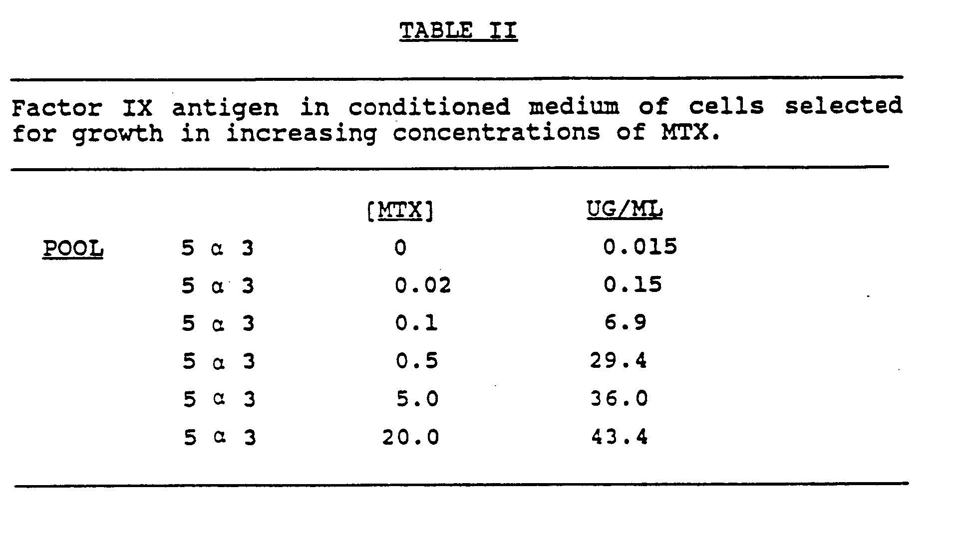
- Initial transformants were pooled (approximately 25 transformants/pool) and subjected to growth in increasing concentrations of MTX. These pools were analyzed for Factor IX expression by 35S-methionine labeling and immune precipitation from the conditioned media and cell extracts with a monoclonal antibody which recognizes human Factor IX. A band of approximately 55k daltons was observed in the cell extracts and its level increased as cells were selected for higher degrees of MTX resistance. When the conditioned medium was similarly analyzed, a heterogeneous smear was observed which ranged from 72k daltons down to 55k daltons. The smear heterogeneity is likely due to the heterogeneity of glycosylation of the secreted material.
- the level of the secreted Factor IX antigen determined by Elisa increased approximately 3000-fold upon selection in 20 um MTX. (See Table II)
- the level of Factor IX antigen (i.e. not biologically active) in the 5 ⁇ 3 pool of cells was determined to be 43.4ug/ml.
- Vitamin K was added to the medium containing the CHO cells which produce Factor IX antigen.
- Table III shows the results of monitoring Factor IX activity after adding increasing concentrations of vitamin K1 (3-phytylmenadione, Sigma) to the CHO Factor IX producing cells (5 ⁇ 3, 20 um MTX) for 24 hours.
- Vitamin K1 had no effect on activity when added to conditioned media immediately before the assay.
- the CHO cells produced up to .275 units of active Factor IX/ml/day/106 cells.
- Vitamin K1 Cells Vitamin K1 m Units/ml/day 5 ⁇ 3 (20um) 100ng/ml 42.5 5 ⁇ 3 (20um) 500ng/ml 105.0 5 ⁇ 3 (20um) 1ug/ml 192.0 5 ⁇ 3 (20um) 5ug/ml 275.0 5 ⁇ 3 (20um) 10ug/ml 268.0 CHO 5ug/ml 17.5
- the specific activity of Factor IX from the CHO cells is 16.5 mU/ug. Since the level of Factor IX in normal plasma is 1 unit/ml/5ug of Factor IX protein, the specific activity (units of Factor IX activity/mg of Factor IX protein from CHO cells) is 9% that derived from normal plasma.
Landscapes
- Chemical & Material Sciences (AREA)
- Health & Medical Sciences (AREA)
- Life Sciences & Earth Sciences (AREA)
- Engineering & Computer Science (AREA)
- Organic Chemistry (AREA)
- Bioinformatics & Cheminformatics (AREA)
- Zoology (AREA)
- Wood Science & Technology (AREA)
- Genetics & Genomics (AREA)
- Biomedical Technology (AREA)
- General Engineering & Computer Science (AREA)
- Biochemistry (AREA)
- General Health & Medical Sciences (AREA)
- Biotechnology (AREA)
- Microbiology (AREA)
- Medicinal Chemistry (AREA)
- Molecular Biology (AREA)
- Tropical Medicine & Parasitology (AREA)
- Virology (AREA)
- Preparation Of Compounds By Using Micro-Organisms (AREA)
- Micro-Organisms Or Cultivation Processes Thereof (AREA)
- Medicines That Contain Protein Lipid Enzymes And Other Medicines (AREA)
- Compounds Of Unknown Constitution (AREA)
- Medicines Containing Material From Animals Or Micro-Organisms (AREA)
Abstract
Description
- This invention relates generally to the cloning and expression in high yield of Factor IX, and more particularly, to the production of biologically-active Factor IX by means of culturing mammalian cells in media containing vitamin K which Factor IX cDNA has been chromosomally-integrated.
- The plasma glycoprotein, Factor IX, plays a critical role in the blood-clotting process. Normally synthesized in the liver, Factor IX requires vitamin K activity for the γ-carboxylation of its 12 amino-terminal glutamic acid residues. A deficiency of Factor IX in the body characterizes a type of hemophilia (type B). Treatment for this disease is presently limited to intravenous transfusion of human plasma protein concentrates of Factor IX. However, in addition to the practical disadvantages of time and expense, transfusion of blood concentrates involves the risk of transmission of viral hepatitis, acquired immune deficiency syndrome or thromboembolic diseases to the recipient. An alternative method of producing Factor IX, other than extraction from human plasma, is therefore highly desirable.
- The application of recombinant DNA techniques to the production of Factor IX has elicited considerable information about the protein. The cDNA coding for human Factor IX has been isolated, characterized, and cloned into expression vectors. See, e.g., K. H. Choo et al, "Molecular Cloning of the Gene for Human Anti-hemophilic Factor IX', Nature, Vol. 299: 178-180 (Sept. 1982) and K. Kurachi et al, "Isolation and Characterization of a cDNA Coding for Human Factor IX," Proc. Natl. Acad. Sci. U.S.A., Vol. 79: 6461-65 (November 1982).
- PCT patent application WO 84/00360 published February 16, 1984 describes the identification and cloning of a human Factor IX nucleotide sequence or fragments thereof for use primarily as diagnostic probes. This application only prophetically refers to the production of a human Factor IX polypeptide through growth in mammalian tissue culture cells, preferably a hepatoma cell line. The inventors' subsequent article, D. S. Anson et al, "Expression of Active Human Clotting Factor IX from Recombinant DNA Clones in Mammalian Cells", Nature, Vol. 315, pp 683-685 (June 20, 1985), describes the production of very low levels of a purportedly active human Factor IX polypeptide from a transformed rat hepatoma cell line. (See page 685, column 1)
European patent application 162,782, published November 11, 1985, refers to the construction of a recombinant viral vector containing a sequence coding for human Factor IX. Biologically active human Factor IX in low yields is assertedly obtained from bacterial cells containing the episomally integrated recombinant Factor IX sequence. The influence of viral components on growth of the host cells and glyco-protein of the protein, however, can result in unreliable and varied batches of protein. [See, also H. de la Salle et al, "Active γ-carboxylated human Factor IX expressed using recombinant DNA techniques," Nature, 316: 268-270 (July 18, 1985)]
Another recent report, S. Busby et al. "Expression of active human Factor IX in transfected cells," Nature, 316: 271-273 (July 18, 1985) also refers to expression of low levels of recombinant Factor IX in BHK cells co-transfected with a neo gene marker. - Even these recent studies therefore demonstrate the continued difficulty in obtaining a stable system for producing high yields of Factor IX in biologically active and reliably consistent form.
- In accordance with the present invention it is surprisingly discovered that high yields of biologically active Factor IX protein can be produced by culturing a CHO cell line containing chromosomally integrated Factor IX cDNA and adding heterologous vitamin K to the culture medium for a predetermined time prior to harvesting the polypeptide. Even cells that have not previously been demonstrated to provide γ- carboxylation can be shown to provide active Factor IX by this method.
- Vitamin K is added to the culture medium in the form of K1 or K3. Where K3 is the vitamin of choice, a concentrate of from about 0.1 ng to 10 ug of vitamin can be added per ml of cell culture, with the preferred concentration range of between 5 ng to 10 ug. Alternatively, vitamin K1 may be added in a desired concentration range of between about 10 ng to 50 ug of vitamin per ml of media or preferably 100 ng to 100 ug of vitamin per ml of media.
- The length of time required to produce the maximum quantity of active Factor IX can be easily determined by simple experimentation, i.e. analyzing aliquots of conditioned medium at various time intervals for the amount of active Factor IX present until the maximum concentration is determined.
- The biologically active Factor IX produced by the eucaryotic expression of the cloned Factor IX gene in accordance with the present invention can be used for the in vivo treatment of humans by physicians. The amount of active ingredient will, of course, depend upon the severity of the condition being treated, the route of administration chosen, and the specific activity of the active Factor IX.
- The active Factor IX produced according to the present invention may be administered by any route and in any pharmaceutical formulation and dosage which are typical for the natural serum-derived Factor IX. Desirably, the route, formulation and dosage regimens will take into account differences in activity, if any, between the natural Factor IX and the protein produced as described herein.
- The following examples refer to the initial isolation and cloning of the Factor IX cDNA, sequencing thereof and the preparation of an expression vector system capable of expressing active Factor IX. Fig. 1 illustrates the structure of expression plasmids p91023-IX and pAaD26SVpA3.
- A unique oligonucleotide with the sequence 5'pGTACAGGAGCAAACACC-3'OH was synthesized which was homologous to Factor IX RNA approximately 475 bp downstream from the initiator codon (Kurachi et al., 1982 supra; Choo et al., 1982, supra). This oligonucleotide was labeled at the 5' end using polynucleotide kinase (New England Biolabs) and Y-³²P-ATP (New England Nuclear). Human liver double-stranded cDNA was prepared (Maniatis et al., Molecular Cloning, A Laboratory Manual, Cold Spring Harbor, 1982) and inserted into a gt10 vector as described by Toole et al., Nature, 313: 342-347, (1984). Approximately 100,000 recombinant phage plaques were screened on duplicate filters with the labeled oligonucletide probe by the method of Woo et al., Proc. Nat'l. Acad. Sci USA, 75:3688-3692, (1978). Hybridization was at 42oC for 40 hours in 5X SSC, 5X Denhardt's, 0.5% by weight SDS and 10mM EDTA and washed extensively at 42o in 2X SSC. Duplicate positives were plaque purified and DNA was prepared from plate stocks [Maniatis et al., (1982), supra] and analyzed by restriction digestion.
- Three human genomic libraries were screened using human Factor IX cDNA nick-translated probes by the method of Benton, et al., Science, 196:180-182 (1977). The first library screened was an amplified library of DNA partially digested with HaeIII/AluI and cloned into Charon 4A [Lawn et al., Cell, 15:1157-1174, (1978)]. The second library was prepared by the method described by Maniatis et al., (1982) supra using a Sau3A partial digest cloned into Charon 28. A third library was prepared from the human lymphoblastoid line GM1202A (NIGMS mutant cell repository) which contains four X chromosomes. In this case DNA partially digested with Sau3A was cloned as above into Jl (kindly supplied by J. Mullins) a derivative of L47.1 [Loenen, et al., Gene 10:249 (1980)] in which polylinkers have been inserted replacing the two small EcoRl-BamHl fragments. Positive hybridizing plaques were purified and the phage DNA prepared following CsCl step gradient purification of a one liter liquid lysate [Maniatis et al., (1982), supra].
- DNA sequences were obtained following subcloning into M13 phage vectors [Norrander et al., Gene, 26:101-106 (1983)] by the dideoxy chain termination method [Sanger et al., Proc. Nat'l. Acad. Sci USA, 74 5463-5467 (1977)] using synthetic oligonucleotide primers. The Factor IX cDNA was subcloned into M13 vectors following an ExoIII generated series of deletions according to the following procedures.
- A plasmid containing the region to be sequenced is digested with a restriction enzyme which cuts at a unique site to one side of the region to be sequenced. The DNA (approximately 20ug) is ethanol precipitated and dissolved in 100ul of a solution containing 50mM Tris, pH 8.0, 1mM MgCl₂, 1mM 2-mercaptoethanol (ExoIII buffer). The tube is warmed to 30oC and 200 units of ExoIII (Pharmacia P-L Biochemicals) are added. Based on the estimated degradation rate under these conditions of approximately 200 base pairs/min/end, aliquots are quenched at 30 second intervals thereby generating ends throughout the region to be sequenced. The aliquots are quenched by pooling into a tube containing 300ul of a solution containing 500mM NaCl and 20mM EDTA, pH 8.0 (Exo quench). The deleted DNA is ethanol precipitated and dissolved in 80ul of H₂O. Mild Sl digestion is initiated by the addition of 80ul of a solution containing 60mM sodium acetate, 2mM ZnSO₄, 500mM NaCl, 10% by weight glycerol, pH 4.6 and 50 units Sl nuclease (Sigma). After 15 minutes at 20oC the reaction is quenched with 40ul of 500mM Tris-HCl, 1M NaCl, pH 8.0 and ethanol precipitated. Blunt ends are created by subsequent treatment with 5 units of Klenow fragment of DNA polymerase I (BRL) in a 100ul solution containing 10mM Tris-HCl, 10mM MgCl₂, 10mM 2-mercaptoethanol, 100uM dXTP's (containing dATP, dCTP, dGTP, dTTP), pH 7.5 at 37oC for 15 minutes. The reaction mixture is phenol extracted and the solution centrifuged through a 1ml G50 spin column (prepared in a 1ml syringe in 10mM Tris, pH 8.0) to recover 100ul.
- The DNA is ethanol precipitated and then digested by a restriction enzyme (that leaves "sticky" ends) which cuts on the opposite end of the region to be sequenced than the original restriction cut site prior to treatment with ExoIII. Following digestion, the DNA fragments are resolved on a Tris-acetate agarose gel. Gel slices which correspond to approximately 200 bp size classes throughout the size range desired are removed. One fragment end would be from an ExoIII generated blunt end within the region to be sequenced and the other end from the re-cut site. The DNA fragments are purified by glass powder affinity [Vogelstein and Gillespie, Proc.-Nat'l. Acad. Sci. USA, 76:615-619 (1979)] following dissolution in NaI.
- These DNA fragments are then ligated into M13 cloning vectors in an assymetric manner wherein the blunt end is nearest the universal primer binding site. M13 recombinant phages which hybridize to a probe made specifically for the region over which the sequence is desired are selected for subsequent preparation of single-stranded template DNA [Sanger et al., (1977), supra]. Generally it is necessary to sequence with-universal primer only one or two isolates from each size class to obtain overlapping sequences covering the entire region to be sequenced up to 4 kb. The sequence of the second-strand can be obtained by repeating the procedure, deleting from the opposite side of the region to be sequenced.
- A 4.5 kb HindIII fragment containing the Factor IX promoter and 5' coding region was cloned into Charon 21A. A 2.5 kb fragment of Factor IX cDNA containing the entire Factor IX coding sequence from the eleventh codon of the signal sequence coding region to the Xbal site 66 bp from the polyA tail was subcloned into plasmid AN7 (a derivative of πVX). [See Maniatis et al., (1982), supra]. There were 57 bp of the exon 1 sequence within the Factor IX cDNA. The exon 1 containing Charon 21A phage were plated on bacteria harboring the recombinant AN7 plasmid and subsequently harvested as a plate stock. Phage which had recombined with the supF containing AN7 plasmid could now be selected by plating onto the supF⁻ bacterial line W3110 (Maniatis et al., (1982), supra]. Approximately one in 2 x 10⁴ phage were supF⁺. One isolate was chosen for large scale DNA preparation and shown by restriction mapping and subsequent sequence analysis to have recombined correctly with the AN7 plasmid within the 57 bp region of homology to obtain a Factor IX "mini gene".
- The 2.3kb Xba fragment containing the Factor IX "mini gene" was purified from AN7 by glass powder affinity. Approximately 5 ug of the fragment was digested with 50 units of ExoIII in 40ul of ExoIII buffer. After 1 minute, 8ul aliquots were removed to 120ul of Exo quench at 15 second intervals. The DNA was treated with exonuclease Sl and Klenow fragment of DNA polymerase 1 as above and then ligated to Pst adapters

- The DNA ligated to the adaptors was then loaded onto a Tris-acetate agarose gel and DNA of approximately 2.3kb was purified by glass powder affinity and ligated into a M13mp11 vector digested with Pst. Recombinant plaques were screened with an oligonucleotide homologous to the first 17bp the of Factor IX coding sequence. Positive hybridizing plaques were sequenced with the same oligonucleotide to find the deletion end point at the 5' end of the gene. Universal primer sequencing determined the 3' deletion end point. The entire sequence of the biologically active recombinant Factor IX cDNA is shown in Table I below.

- To prepare samples for the following Factor IX assay, approximately 4x10⁶ logarithmically growing cells were rinsed four times (5 ml each) with serum-free media containing additions as indicated. After 24 hours at 37oC samples were taken, frozen in a dry ice/ethanol bath, and stored at -70oC until assayed.
- Microtitre plates were coated with anti-human Factor IX murine monoclonal antibody (Hybritech). The plates were washed, and sample or standard Factor IX preparations were added after dilution into alpha media. The Factor IX standard was purified human Factor IX diluted from 4.0ug to 1.0ng. The secondary antibody (rabbit anti Factor IX, Calbiochem) was applied and washed, and alkaline phosphatase conjugated goat anti-rabbit IgG (Zymea) was applied. The substrate was alkaline phosphate (tablets) (Sigma #104) diluted in diethanolamine and results were read at 410nm.
- The one stage activated partial thromboplastin time assay as described by R. Biggs, Human Blood Coagulation Haemostasis and Thrombosis (Ed. 1), Oxford, Blackwell, Scientific, pg. 614 (1972) was carried out with equal volumes of: 1) activated partial thromboplastin reagent (general diagnostics) 2) Factor IX deficient plasma (George B. King Biomedical) and 3) normal pooled plasma as standard (George B. King Biomedical) or the sample. One unit of activity is defined as that amount present in 1ml of normal pooled plasma.
- The Factor IX coding sequence was inserted into the Pst 1 site of plasmid p91023(A) which is described in Example 3 of Japanese patent publication No. 12288/86, published January 20, 1986 which corresponds to United States patent application Ser. No. 677,813, filed December 4, 1983. The resulting clones are screened for proper orientation of the Factor IX gene with those having proper orientation selected as p91023-IX. The Factor IX expression vector, p91023-IX, contains the SV40 enhancer upstream from the AdMLP, a cDNA copy of the adenovirus tripartite leader (TPL), the adenovirus VA genes, [Kaufman, PNAS, 82:689-693 (1985)], and the inserted Factor IX cDNA coding region upstream from the DHFR coding region. EcoRl(Rl), BamHl(bam) and Xhol(X) restriction sites are indicated. The DHFR expression vector, pAdD26SVpA(3) [Kaufman, et al., Mol. Cell. Bio., 2:1304-1309 (1982)], contains the adenovirus major late promoter including the first late leader and 5' splice site. The leader exon from the RNA transcript is spliced properly to an introduced 3' splice site. The vector contains the SV40 early polyadenylation site, 2.7 KB derived from pSVOa [Mellon et al., Cell, 27:279-288 (1981)] which contains the ColEl origin of replication, a pBR322 derivative lacking sequences detrimental to replication in mammalian cells, tetracycline resistance, and the SV40 origin of replication.
- The Factor IX expression vector p91023-IX was introduced with pAdD26SVpA3, into DHFR deficient Chinese hamster ovary cells by calcium phosphate mediated DNA transfection. (See Figure 1.) Cells selected for the DHFR positive phenotype expressed Factor IX at a low level. Then the transformants were pooled and selected for growth in the following sequentially increasing concentrations of methotrexate (MTX), i.e., 0.02, 0.1, 0.5, 1.0, 5.0 and 20uM. Cells selected in this manner contain amplified copies of the introduced DHFR genes as well as amplified Factor IX genes. The amplified genes were generally localized to one or two specific chromosomes [Kaufman et al., Mol. Cell. Bio. 3, 699-711 (1983)].
- DUKX Bll DHFR deficient CHO cells (Urlaub, et al, Proc. Nat'l Acad. Sci. 77:4210-4220, 1980; Kaufman, et al, J. Mol. Biol. 159:601-621, 1982) were transfected with a mixture of 25ug of p91023-IX and 2.5 ug of pAdD26SVpA3 (Kaufman & Sharp, op. cit) using calcium-phosphate. Transformants were selected in media lacking nucleosides as described (Kaufman & Sharp, op. cit). A similar transfection which gave similar results included the addition of 2.5ug of pSV2 Neo [Southern, et al, J. Mol. Appl. Genet., 1:327-341 (1982)]. For the latter transfection, the DHFR⁺ transformants were initially selected in media lacking nucleosides with the addition of the antibiotic G418 (1mg/ml) in order to select for the pSV2 neo marker. The addition of a G418 resistance marker can be useful to facilitate transfer of chromosomes containing transfected DNA into other cells where selection directly for the amplified DHFR gene copies is not possible.
- Initial transformants were pooled (approximately 25 transformants/pool) and subjected to growth in increasing concentrations of MTX. These pools were analyzed for Factor IX expression by ³⁵S-methionine labeling and immune precipitation from the conditioned media and cell extracts with a monoclonal antibody which recognizes human Factor IX. A band of approximately 55k daltons was observed in the cell extracts and its level increased as cells were selected for higher degrees of MTX resistance. When the conditioned medium was similarly analyzed, a heterogeneous smear was observed which ranged from 72k daltons down to 55k daltons. The smear heterogeneity is likely due to the heterogeneity of glycosylation of the secreted material. In addition, the level of the secreted Factor IX antigen determined by Elisa increased approximately 3000-fold upon selection in 20 um MTX. (See Table II) The level of Factor IX antigen (i.e. not biologically active) in the 5α3 pool of cells was determined to be 43.4ug/ml.
- When the conditioned media from the Factor IX producing CHO cells (5α3 in various concentrations of MTX) was analyzed for Factor IX activity, there was no activity detected above background. Vitamin K was added to the medium containing the CHO cells which produce Factor IX antigen. Table III shows the results of monitoring Factor IX activity after adding increasing concentrations of vitamin K1 (3-phytylmenadione, Sigma) to the CHO Factor IX producing cells (5
α 3, 20 um MTX) for 24 hours. Significantly, as the concentration of vitamin K1 increased to 5ug/ml, so did the Factor IX activity in the conditioned medium. Vitamin K1 had no effect on activity when added to conditioned media immediately before the assay. Thus, in the presence of vitamin K1, the CHO cells produced up to .275 units of active Factor IX/ml/day/10⁶ cells. - To determine the validity of a concern that very little of the fat soluble vitamin K1 was actually being delivered to the cell, a similar effect has been demonstrated for the water soluble derivative vitamin K3 (menadione, Sigma). Lower levels of K3 are required for maximal activity (0.005ug/ml) to be obtained with vitamin K. The specificity of the vitamin K3 dependence on Factor IX activity was demonstrated by adding warfarin (at 1ug/ml), a specific antagonist of vitamin K, which blocked the appearance of active Factor IX.
TABLE III Factor IX activity v. vitamin K1 Cells Vitamin K1 m Units/ml/ day 5 α 3 (20um) 100ng/ml 42.5 5 α 3 (20um) 500ng/ml 105.0 5 α 3 (20um) 1ug/ml 192.0 5 α 3 (20um) 5ug/ml 275.0 5 α 3 (20um) 10ug/ml 268.0 CHO 5ug/ml 17.5 - Thus the specific activity of Factor IX from the CHO cells is 16.5 mU/ug. Since the level of Factor IX in normal plasma is 1 unit/ml/5ug of Factor IX protein, the specific activity (units of Factor IX activity/mg of Factor IX protein from CHO cells) is 9% that derived from normal plasma.
Claims (7)
- A method for producing biologically active Factor IX comprising culturing in a medium containing vitamin K, a CHO cell line transfected with chromosomally integrated DNA carrying a gene encoding Factor IX, said gene being capable of being expressed in the above cell line.
- The method according to claim 1, wherein said DNA comprises the cDNA sequence shown in Table 1.
- The method according to claim 1 or 2, wherein vitamin K is vitamin K1 or vitamin K3.
- The method according to claim 3, wherein said medium comprises a concentration of about 0.1ng to 50µg vitamin K3/ml medium.
- The method according to claim 3 or 4, wherein said medium comprises a concentration of about 5ng to about 10µg vitamin K3/ml medium.
- The method according to claim 3, wherein said medium comprises a concentration of about 10ng to about 50µg vitamin K1/ml medium.
- The method according to claim 3, wherein said medium comprises a concentration of about 100ng to about 100µg vitamin K1/ml medium.
Priority Applications (1)
| Application Number | Priority Date | Filing Date | Title |
|---|---|---|---|
| AT86902988T ATE74164T1 (en) | 1985-04-22 | 1986-04-17 | MANUFACTURING WITH HIGH PERFORMANCE OF ACTIVE FACTOR IX. |
Applications Claiming Priority (2)
| Application Number | Priority Date | Filing Date | Title |
|---|---|---|---|
| US72543685A | 1985-04-22 | 1985-04-22 | |
| US725436 | 1996-10-03 |
Publications (3)
| Publication Number | Publication Date |
|---|---|
| EP0218713A1 EP0218713A1 (en) | 1987-04-22 |
| EP0218713A4 EP0218713A4 (en) | 1987-04-28 |
| EP0218713B1 true EP0218713B1 (en) | 1992-03-25 |
Family
ID=24914546
Family Applications (1)
| Application Number | Title | Priority Date | Filing Date |
|---|---|---|---|
| EP86902988A Expired - Lifetime EP0218713B1 (en) | 1985-04-22 | 1986-04-17 | High yield production of active factor ix |
Country Status (8)
| Country | Link |
|---|---|
| US (1) | US4770999A (en) |
| EP (1) | EP0218713B1 (en) |
| JP (1) | JP2584443B2 (en) |
| KR (1) | KR880700060A (en) |
| AT (1) | ATE74164T1 (en) |
| AU (1) | AU5864086A (en) |
| DE (1) | DE3684546D1 (en) |
| WO (1) | WO1986006408A1 (en) |
Families Citing this family (56)
| Publication number | Priority date | Publication date | Assignee | Title |
|---|---|---|---|---|
| US4775624A (en) * | 1985-02-08 | 1988-10-04 | Eli Lilly And Company | Vectors and compounds for expression of human protein C |
| US5171569A (en) * | 1985-03-15 | 1992-12-15 | National Research Development Corporation | Factor IX preparations uncontaminated by plasma components or pox virus |
| US4959318A (en) * | 1985-06-27 | 1990-09-25 | Zymogenetics, Inc. | Expression of protein C |
| US5516650A (en) * | 1985-06-27 | 1996-05-14 | Zymogenetics, Inc. | Production of activated protein C |
| EP0245949B1 (en) * | 1986-04-09 | 1997-10-29 | Eli Lilly And Company | A method of using eukaryotic expression vectors comprising the bk virus enhancer |
| FR2599752B1 (en) * | 1986-06-10 | 1989-11-03 | Transgene Sa | ALPHA1- ANTITRYPSIN VARIANTS USEFUL IN PARTICULAR AS KALLIKREIN INHIBITORS |
| FR2600334B1 (en) * | 1986-06-23 | 1989-05-12 | Transgene Sa | VECTORS FOR INTEGRATION INTO EUKARYOTIC CELLS PROVIDING EXPRESSION OF FACTOR IX, CELULLAR LINES OBTAINED AND PROCESS FOR THEIR PREPARATION |
| US5082774A (en) * | 1988-08-30 | 1992-01-21 | The General Hospital Corporation | Recombinant human nerve growth factor |
| FR2638643B1 (en) * | 1988-11-09 | 1991-04-12 | Transgene Sa | DNA SEQUENCE ENCODING HUMAN FACTOR IX OR AN ANALOGUE PROTEIN, EXPRESSION VECTOR, TRANSFORMED CELLS, PROCESS FOR PREPARING FACTOR IX, AND PRODUCTS OBTAINED THEREFROM |
| US5047335A (en) * | 1988-12-21 | 1991-09-10 | The Regents Of The University Of Calif. | Process for controlling intracellular glycosylation of proteins |
| US5225537A (en) * | 1989-12-29 | 1993-07-06 | Zymogenetics, Inc. | Methods for producing hybrid phospholipid-binding proteins |
| DE69131292T2 (en) * | 1990-01-29 | 1999-09-30 | Zymogenetics, Inc. | ANTICOAGULATING PROTEINS |
| ES2109336T3 (en) * | 1990-11-26 | 1998-01-16 | Genetics Inst | EXPRESSION OF PACE IN HOST CELLS AND METHODS OF USING IT. |
| US5997864A (en) | 1995-06-07 | 1999-12-07 | Novo Nordisk A/S | Modified factor VII |
| US5833982A (en) | 1991-02-28 | 1998-11-10 | Zymogenetics, Inc. | Modified factor VII |
| US5788965A (en) * | 1991-02-28 | 1998-08-04 | Novo Nordisk A/S | Modified factor VII |
| US5817788A (en) * | 1991-02-28 | 1998-10-06 | Zymogenetics, Inc. | Modified factor VII |
| US5861374A (en) * | 1991-02-28 | 1999-01-19 | Novo Nordisk A/S | Modified Factor VII |
| AU671586B2 (en) * | 1991-03-01 | 1996-09-05 | Aventis Behring Llc | Preparation of factor IX |
| US6039944A (en) * | 1992-02-28 | 2000-03-21 | Zymogenetics, Inc. | Modified Factor VII |
| US6372716B1 (en) | 1994-04-26 | 2002-04-16 | Genetics Institute, Inc. | Formulations for factor IX |
| AT403167B (en) * | 1994-11-14 | 1997-11-25 | Immuno Ag | SELECTION AND EXPRESSION OF FOREIGN PROTEINS BY MEANS OF A SELECTION-AMPLIFICATION SYSTEM |
| US5770700A (en) * | 1996-01-25 | 1998-06-23 | Genetics Institute, Inc. | Liquid factor IX formulations |
| US6475725B1 (en) | 1997-06-20 | 2002-11-05 | Baxter Aktiengesellschaft | Recombinant cell clones having increased stability and methods of making and using the same |
| WO2001070968A2 (en) * | 2000-03-22 | 2001-09-27 | Octagene Gmbh | Production of recombinant blood clotting factors in human cell lines |
| AU2002225681A1 (en) * | 2000-11-15 | 2002-05-27 | Globe Immune, Inc. | Yeast-dentritic cell vaccines and uses thereof |
| EP1418810A4 (en) | 2001-08-03 | 2006-08-02 | Us Gov Health & Human Serv | ORAL TREATMENT OF HEMOPHILIA |
| ES2466024T3 (en) | 2001-10-10 | 2014-06-09 | Ratiopharm Gmbh | Remodeling and glycoconjugation of fibroblast growth factor (FGF) |
| US20070026485A1 (en) | 2003-04-09 | 2007-02-01 | Neose Technologies, Inc. | Glycopegylation methods and proteins/peptides produced by the methods |
| CA2539434C (en) | 2003-09-23 | 2014-03-18 | University Of North Carolina At Chapel Hill | Methods and compositions for the correlation of single nucleotide polymorphisms in the vitamin k epoxide reductase gene and warfarin dosage |
| ATE489105T1 (en) | 2004-03-19 | 2010-12-15 | Baxter Int | FACTOR IXA FOR THE TREATMENT OF BLEEDING DISORDERS |
| KR101146160B1 (en) * | 2004-06-30 | 2012-07-16 | 넥타르 테라퓨틱스 | Polymer-factor ix moiety conjugates |
| AU2005329450A1 (en) * | 2005-03-15 | 2006-09-28 | University Of North Carolina At Chapel Hill | Methods and compositions for producing active Vitamin K-dependent proteins |
| EP1707634A1 (en) | 2005-03-29 | 2006-10-04 | Octapharma AG | Method for isolation of recombinantly produced proteins |
| EP2975135A1 (en) | 2005-05-25 | 2016-01-20 | Novo Nordisk A/S | Glycopegylated factor IX |
| SI1969127T2 (en) | 2005-12-21 | 2017-10-30 | Cnj Holdings, Inc | Method of producing biologically active vitamin k dependent proteins by recombinant methods |
| US8383388B2 (en) | 2006-06-19 | 2013-02-26 | Catalyst Biosciences, Inc. | Modified coagulation factor IX polypeptides and use thereof for treatment |
| US8716448B2 (en) | 2009-02-03 | 2014-05-06 | Amunix Operating Inc. | Coagulation factor VII compositions and methods of making and using same |
| PL2451963T3 (en) * | 2009-07-10 | 2014-09-30 | Csl Ltd | Method of increasing the expression yield of vitamin k-dependent proteins |
| WO2011095604A1 (en) | 2010-02-04 | 2011-08-11 | Octapharma Biopharmaceuticals Gmbh | Half-life prolongation of proteins |
| PT2616090T (en) | 2010-09-17 | 2023-10-16 | Takeda Pharmaceuticals Co | Stabilization of immunoglobulins through aqueous formulation with histidine at weak acidic to neutral ph |
| CN107190034A (en) | 2010-10-05 | 2017-09-22 | 诺沃—诺迪斯克保健股份有限公司 | Produce method of protein |
| US9631002B2 (en) | 2010-12-21 | 2017-04-25 | The University Of North Carolina At Chapel Hill | Methods and compositions for producing active vitamin K-dependent proteins |
| PT3564260T (en) | 2012-02-15 | 2023-01-18 | Bioverativ Therapeutics Inc | Factor viii compositions and methods of making and using same |
| EP2822577B1 (en) | 2012-02-15 | 2019-02-06 | Bioverativ Therapeutics Inc. | Recombinant factor viii proteins |
| TW202003554A (en) | 2013-08-14 | 2020-01-16 | 美商百歐維拉提夫治療公司 | Factor VIII-XTEN fusions and uses thereof |
| BR112018002150A2 (en) | 2015-08-03 | 2018-09-18 | Bioverativ Therapeutics Inc | factor ix fusion proteins and methods of manufacturing and using them |
| PE20200012A1 (en) | 2016-11-23 | 2020-01-06 | Bioverativ Therapeutics Inc | MONO- AND BI-SPECIFIC ANTIBODIES THAT BIND TO COAGULATION FACTOR IX AND COAGULATION FACTOR X |
| IL308416B2 (en) | 2016-12-02 | 2025-08-01 | Bioverativ Therapeutics Inc | Methods for treating hemophilic arthritis using chimeric blood clotting factors |
| CR20190389A (en) | 2017-01-31 | 2019-11-26 | Bioverativ Therapeutics Inc | Factor ix fusion proteins and methods of making and using same |
| US20210107994A1 (en) | 2017-03-31 | 2021-04-15 | Public University Corporation Nara Medical University | Medicinal composition usable for preventing and/or treating blood coagulation factor ix abnormality, comprising multispecific antigen binding molecule replacing function of blood coagulation factor viii |
| WO2018199214A1 (en) | 2017-04-27 | 2018-11-01 | 中外製薬株式会社 | Coagulation factor ix with improved pharmacokinetics |
| TWI904068B (en) | 2017-08-09 | 2025-11-11 | 美商生物化學醫療公司 | Nucleic acid molecules and uses thereof |
| BR112020022164A2 (en) | 2018-05-18 | 2021-02-02 | Bioverativ Therapeutics Inc. | methods of treating hemophilia a |
| CA3108799A1 (en) | 2018-08-09 | 2020-02-13 | Bioverativ Therapeutics Inc. | Nucleic acid molecules and uses thereof for non-viral gene therapy |
| AU2019360270B2 (en) | 2018-10-18 | 2025-08-07 | Intellia Therapeutics, Inc. | Compositions and methods for expressing factor IX. |
Citations (2)
| Publication number | Priority date | Publication date | Assignee | Title |
|---|---|---|---|---|
| EP0191606A2 (en) * | 1985-02-08 | 1986-08-20 | Eli Lilly And Company | Vectors and methods for expression of human protein C activity |
| EP0200421A2 (en) * | 1985-04-17 | 1986-11-05 | Zymogenetics, Inc. | Expression of factor VII activity in mammalian cells |
Family Cites Families (5)
| Publication number | Priority date | Publication date | Assignee | Title |
|---|---|---|---|---|
| EP0316558A1 (en) * | 1982-08-04 | 1989-05-24 | Btg International Limited | Factor IX DNA |
| GB2125409B (en) * | 1982-08-04 | 1985-11-13 | Nat Res Dev | Genetic engineering |
| FR2564106B1 (en) * | 1984-05-09 | 1988-04-22 | Transgene Sa | FACTOR IX EXPRESSION VECTORS, CELLS TRANSFORMED BY THESE VECTORS, AND PROCESS FOR THE PREPARATION OF FACTOR IX. |
| JPH082307B2 (en) * | 1984-05-22 | 1996-01-17 | トランスジ−ン ソシエテ アノニム | Method for producing factor IX |
| DE3688983T2 (en) * | 1985-03-15 | 1994-02-17 | British Tech Group | Factor IX protein. |
-
1986
- 1986-04-17 EP EP86902988A patent/EP0218713B1/en not_active Expired - Lifetime
- 1986-04-17 AU AU58640/86A patent/AU5864086A/en not_active Abandoned
- 1986-04-17 WO PCT/US1986/000817 patent/WO1986006408A1/en not_active Ceased
- 1986-04-17 AT AT86902988T patent/ATE74164T1/en not_active IP Right Cessation
- 1986-04-17 JP JP61502661A patent/JP2584443B2/en not_active Expired - Lifetime
- 1986-04-17 US US06/929,294 patent/US4770999A/en not_active Expired - Lifetime
- 1986-04-17 DE DE8686902988T patent/DE3684546D1/en not_active Expired - Lifetime
- 1986-12-22 KR KR860700914A patent/KR880700060A/en not_active Withdrawn
Patent Citations (2)
| Publication number | Priority date | Publication date | Assignee | Title |
|---|---|---|---|---|
| EP0191606A2 (en) * | 1985-02-08 | 1986-08-20 | Eli Lilly And Company | Vectors and methods for expression of human protein C activity |
| EP0200421A2 (en) * | 1985-04-17 | 1986-11-05 | Zymogenetics, Inc. | Expression of factor VII activity in mammalian cells |
Also Published As
| Publication number | Publication date |
|---|---|
| WO1986006408A1 (en) | 1986-11-06 |
| KR880700060A (en) | 1988-02-15 |
| JP2584443B2 (en) | 1997-02-26 |
| ATE74164T1 (en) | 1992-04-15 |
| DE3684546D1 (en) | 1992-04-30 |
| AU5864086A (en) | 1986-11-18 |
| US4770999A (en) | 1988-09-13 |
| EP0218713A4 (en) | 1987-04-28 |
| JPS62502514A (en) | 1987-10-01 |
| EP0218713A1 (en) | 1987-04-22 |
Similar Documents
| Publication | Publication Date | Title |
|---|---|---|
| EP0218713B1 (en) | High yield production of active factor ix | |
| US5344773A (en) | Human uterine tissue plasminogen activator produced by recombinant DNA | |
| EP0162067B1 (en) | Production of factor viii and related products | |
| US5278049A (en) | Recombinant molecule encoding human protease nexin | |
| JP2561677B2 (en) | Expression of protein C | |
| EP0227462B1 (en) | Novel peptide plasminogen activators | |
| JP2610783B2 (en) | Gene encoding polykringle plasminogen activator and vector containing the same | |
| EP0107278B1 (en) | Molecular cloning of the gene for human anti-haemophilic factor ix | |
| EP0201153A2 (en) | Modified enzyme and process for its preparation | |
| JPS62111690A (en) | Human protein c and its production | |
| EP0289586A1 (en) | Enhancing gamma-carboxylation of recombinant vitamin k-dependent proteins | |
| EP0182448A2 (en) | Production of factor VIII and related products | |
| JP2645237B2 (en) | Gene encoding hybrid plasminogen activator | |
| EP0297066B1 (en) | Novel fibrinolytic enzymes | |
| GB2125409A (en) | Genetic engineering | |
| Connors et al. | DHFR coamplification of t-PA in DHFR+ bovine endothelial cells: in vitro characterization of the purified serine protease | |
| EP0271003A2 (en) | Expression vectors | |
| Bahnak et al. | Steady state levels of factor X mRNA in liver and Hep G2 cells | |
| JPH0847390A (en) | Recombinant DNA molecule encoding active human TPA | |
| JP3045307B2 (en) | Cell culture method for producing activated protein C | |
| CA1214125A (en) | Human factor ix dna | |
| US6682733B1 (en) | Fibrinolytic enzymes | |
| JPH022338A (en) | Simultaneous development in eucaryotic cell |
Legal Events
| Date | Code | Title | Description |
|---|---|---|---|
| PUAI | Public reference made under article 153(3) epc to a published international application that has entered the european phase |
Free format text: ORIGINAL CODE: 0009012 |
|
| 17P | Request for examination filed |
Effective date: 19861201 |
|
| AK | Designated contracting states |
Kind code of ref document: A1 Designated state(s): AT BE CH DE FR GB IT LI LU NL SE |
|
| A4 | Supplementary search report drawn up and despatched |
Effective date: 19870428 |
|
| 17Q | First examination report despatched |
Effective date: 19890925 |
|
| GRAA | (expected) grant |
Free format text: ORIGINAL CODE: 0009210 |
|
| AK | Designated contracting states |
Kind code of ref document: B1 Designated state(s): AT BE CH DE FR GB IT LI LU NL SE |
|
| PG25 | Lapsed in a contracting state [announced via postgrant information from national office to epo] |
Ref country code: IT Free format text: LAPSE BECAUSE OF FAILURE TO SUBMIT A TRANSLATION OF THE DESCRIPTION OR TO PAY THE FEE WITHIN THE PRE;WARNING: LAPSES OF ITALIAN PATENTS WITH EFFECTIVE DATE BEFORE 2007 MAY HAVE OCCURRED AT ANY TIME BEFORE 2007. THE CORRECT EFFECTIVE DATE MAY BE DIFFERENT FROM THE ONE RECORDED.SCRIBED TIME-LIMIT Effective date: 19920325 Ref country code: LI Effective date: 19920325 Ref country code: SE Effective date: 19920325 Ref country code: AT Effective date: 19920325 Ref country code: CH Effective date: 19920325 Ref country code: BE Effective date: 19920325 Ref country code: NL Effective date: 19920325 |
|
| REF | Corresponds to: |
Ref document number: 74164 Country of ref document: AT Date of ref document: 19920415 Kind code of ref document: T |
|
| PG25 | Lapsed in a contracting state [announced via postgrant information from national office to epo] |
Ref country code: LU Free format text: LAPSE BECAUSE OF NON-PAYMENT OF DUE FEES Effective date: 19920430 |
|
| REF | Corresponds to: |
Ref document number: 3684546 Country of ref document: DE Date of ref document: 19920430 |
|
| ET | Fr: translation filed | ||
| REG | Reference to a national code |
Ref country code: CH Ref legal event code: PL |
|
| ET1 | Fr: translation filed ** revision of the translation of the patent or the claims | ||
| NLV1 | Nl: lapsed or annulled due to failure to fulfill the requirements of art. 29p and 29m of the patents act | ||
| PLBI | Opposition filed |
Free format text: ORIGINAL CODE: 0009260 |
|
| PLAB | Opposition data, opponent's data or that of the opponent's representative modified |
Free format text: ORIGINAL CODE: 0009299OPPO |
|
| 26 | Opposition filed |
Opponent name: IMMUNO AKTIENGESELLSCHAFT Effective date: 19921223 |
|
| R26 | Opposition filed (corrected) |
Opponent name: IMMUNO AKTIENGESELLSCHAFT Effective date: 19921223 |
|
| PLBM | Termination of opposition procedure: date of legal effect published |
Free format text: ORIGINAL CODE: 0009276 |
|
| STAA | Information on the status of an ep patent application or granted ep patent |
Free format text: STATUS: OPPOSITION PROCEDURE CLOSED |
|
| 27C | Opposition proceedings terminated |
Effective date: 19940521 |
|
| REG | Reference to a national code |
Ref country code: GB Ref legal event code: IF02 |
|
| REG | Reference to a national code |
Ref country code: FR Ref legal event code: CD |
|
| PGFP | Annual fee paid to national office [announced via postgrant information from national office to epo] |
Ref country code: GB Payment date: 20050314 Year of fee payment: 20 |
|
| PGFP | Annual fee paid to national office [announced via postgrant information from national office to epo] |
Ref country code: FR Payment date: 20050401 Year of fee payment: 20 |
|
| PGFP | Annual fee paid to national office [announced via postgrant information from national office to epo] |
Ref country code: DE Payment date: 20050429 Year of fee payment: 20 |
|
| PG25 | Lapsed in a contracting state [announced via postgrant information from national office to epo] |
Ref country code: GB Free format text: LAPSE BECAUSE OF EXPIRATION OF PROTECTION Effective date: 20060416 |
|
| REG | Reference to a national code |
Ref country code: GB Ref legal event code: PE20 |