DE102004023187A1 - Identification of surface-associated antigens for tumor diagnosis and therapy - Google Patents
Identification of surface-associated antigens for tumor diagnosis and therapy Download PDFInfo
- Publication number
- DE102004023187A1 DE102004023187A1 DE102004023187A DE102004023187A DE102004023187A1 DE 102004023187 A1 DE102004023187 A1 DE 102004023187A1 DE 102004023187 A DE102004023187 A DE 102004023187A DE 102004023187 A DE102004023187 A DE 102004023187A DE 102004023187 A1 DE102004023187 A1 DE 102004023187A1
- Authority
- DE
- Germany
- Prior art keywords
- nucleic acid
- tumor
- associated antigen
- expression
- cells
- Prior art date
- Legal status (The legal status is an assumption and is not a legal conclusion. Google has not performed a legal analysis and makes no representation as to the accuracy of the status listed.)
- Withdrawn
Links
- 206010028980 Neoplasm Diseases 0.000 title claims abstract description 469
- 239000000427 antigen Substances 0.000 title claims abstract description 354
- 108091007433 antigens Proteins 0.000 title claims abstract description 354
- 102000036639 antigens Human genes 0.000 title claims abstract description 354
- 238000003745 diagnosis Methods 0.000 title abstract description 13
- 238000002560 therapeutic procedure Methods 0.000 title description 15
- 150000007523 nucleic acids Chemical class 0.000 claims abstract description 378
- 102000039446 nucleic acids Human genes 0.000 claims abstract description 347
- 108020004707 nucleic acids Proteins 0.000 claims abstract description 347
- 108090000623 proteins and genes Proteins 0.000 claims abstract description 298
- 210000004027 cell Anatomy 0.000 claims abstract description 249
- 230000014509 gene expression Effects 0.000 claims abstract description 224
- 102000004169 proteins and genes Human genes 0.000 claims abstract description 175
- 108090000765 processed proteins & peptides Proteins 0.000 claims abstract description 94
- 208000037265 diseases, disorders, signs and symptoms Diseases 0.000 claims abstract description 52
- 230000001225 therapeutic effect Effects 0.000 claims abstract description 50
- 210000001151 cytotoxic T lymphocyte Anatomy 0.000 claims abstract description 49
- 238000001514 detection method Methods 0.000 claims abstract description 48
- 201000010099 disease Diseases 0.000 claims abstract description 46
- 239000012634 fragment Substances 0.000 claims abstract description 46
- 210000001744 T-lymphocyte Anatomy 0.000 claims abstract description 40
- 230000002159 abnormal effect Effects 0.000 claims abstract description 38
- 239000000523 sample Substances 0.000 claims abstract description 34
- 230000000694 effects Effects 0.000 claims abstract description 30
- 239000000203 mixture Substances 0.000 claims abstract description 28
- 230000000295 complement effect Effects 0.000 claims abstract description 26
- 241000282414 Homo sapiens Species 0.000 claims abstract description 25
- 210000002443 helper t lymphocyte Anatomy 0.000 claims abstract description 22
- 102000004127 Cytokines Human genes 0.000 claims abstract description 18
- 108090000695 Cytokines Proteins 0.000 claims abstract description 18
- 238000001890 transfection Methods 0.000 claims abstract description 18
- 238000004519 manufacturing process Methods 0.000 claims abstract description 16
- 239000003814 drug Substances 0.000 claims abstract description 15
- 230000001472 cytotoxic effect Effects 0.000 claims abstract description 13
- 230000001461 cytolytic effect Effects 0.000 claims abstract description 12
- 231100000433 cytotoxic Toxicity 0.000 claims abstract description 12
- 230000002163 immunogen Effects 0.000 claims abstract description 12
- 229940124597 therapeutic agent Drugs 0.000 claims abstract description 10
- 239000012472 biological sample Substances 0.000 claims abstract description 9
- 239000000032 diagnostic agent Substances 0.000 claims abstract description 8
- 229940039227 diagnostic agent Drugs 0.000 claims abstract description 8
- 230000008105 immune reaction Effects 0.000 claims abstract description 6
- 239000003053 toxin Substances 0.000 claims abstract description 6
- 231100000765 toxin Toxicity 0.000 claims abstract description 6
- 230000002401 inhibitory effect Effects 0.000 claims abstract description 4
- 230000030833 cell death Effects 0.000 claims abstract description 3
- 239000013610 patient sample Substances 0.000 claims abstract 2
- 210000001519 tissue Anatomy 0.000 claims description 189
- 238000000034 method Methods 0.000 claims description 140
- 102000004196 processed proteins & peptides Human genes 0.000 claims description 70
- 229920001184 polypeptide Polymers 0.000 claims description 51
- 201000011510 cancer Diseases 0.000 claims description 49
- 239000008194 pharmaceutical composition Substances 0.000 claims description 46
- 102000053602 DNA Human genes 0.000 claims description 42
- 239000003795 chemical substances by application Substances 0.000 claims description 35
- 125000003729 nucleotide group Chemical group 0.000 claims description 35
- 239000002773 nucleotide Substances 0.000 claims description 34
- 108091028043 Nucleic acid sequence Proteins 0.000 claims description 25
- 125000003275 alpha amino acid group Chemical group 0.000 claims description 24
- 230000028993 immune response Effects 0.000 claims description 19
- 239000003550 marker Substances 0.000 claims description 18
- 210000004443 dendritic cell Anatomy 0.000 claims description 17
- 208000026310 Breast neoplasm Diseases 0.000 claims description 16
- 238000006243 chemical reaction Methods 0.000 claims description 16
- 230000000692 anti-sense effect Effects 0.000 claims description 15
- 239000002671 adjuvant Substances 0.000 claims description 12
- 239000013604 expression vector Substances 0.000 claims description 12
- 239000013598 vector Substances 0.000 claims description 12
- 206010009944 Colon cancer Diseases 0.000 claims description 11
- 102000004190 Enzymes Human genes 0.000 claims description 11
- 108090000790 Enzymes Proteins 0.000 claims description 11
- 108020004511 Recombinant DNA Proteins 0.000 claims description 11
- 230000003321 amplification Effects 0.000 claims description 11
- 210000000612 antigen-presenting cell Anatomy 0.000 claims description 11
- 238000012544 monitoring process Methods 0.000 claims description 11
- 238000003199 nucleic acid amplification method Methods 0.000 claims description 11
- 102000040430 polynucleotide Human genes 0.000 claims description 11
- 108091033319 polynucleotide Proteins 0.000 claims description 11
- 239000002157 polynucleotide Substances 0.000 claims description 11
- 206010006187 Breast cancer Diseases 0.000 claims description 9
- 238000002955 isolation Methods 0.000 claims description 9
- 208000006265 Renal cell carcinoma Diseases 0.000 claims description 8
- 208000029742 colonic neoplasm Diseases 0.000 claims description 8
- 210000003719 b-lymphocyte Anatomy 0.000 claims description 7
- 210000002540 macrophage Anatomy 0.000 claims description 7
- 210000000170 cell membrane Anatomy 0.000 claims description 6
- 230000006698 induction Effects 0.000 claims description 6
- 230000002062 proliferating effect Effects 0.000 claims description 6
- 230000009467 reduction Effects 0.000 claims description 6
- 230000028327 secretion Effects 0.000 claims description 6
- 238000011161 development Methods 0.000 claims description 5
- 102000005962 receptors Human genes 0.000 claims description 5
- 108020003175 receptors Proteins 0.000 claims description 5
- 108700012359 toxins Proteins 0.000 claims description 5
- 208000020816 lung neoplasm Diseases 0.000 claims description 4
- 230000036647 reaction Effects 0.000 claims description 4
- 102000004457 Granulocyte-Macrophage Colony-Stimulating Factor Human genes 0.000 claims description 3
- 108010017213 Granulocyte-Macrophage Colony-Stimulating Factor Proteins 0.000 claims description 3
- 238000012258 culturing Methods 0.000 claims description 3
- 230000006378 damage Effects 0.000 claims description 3
- 239000003937 drug carrier Substances 0.000 claims description 3
- 230000002285 radioactive effect Effects 0.000 claims description 3
- 229930182490 saponin Natural products 0.000 claims description 3
- 150000007949 saponins Chemical class 0.000 claims description 3
- 108091032973 (ribonucleotides)n+m Proteins 0.000 claims description 2
- 102000019034 Chemokines Human genes 0.000 claims description 2
- 108010012236 Chemokines Proteins 0.000 claims description 2
- 206010027476 Metastases Diseases 0.000 claims description 2
- 210000001124 body fluid Anatomy 0.000 claims description 2
- 239000010839 body fluid Substances 0.000 claims description 2
- 201000008275 breast carcinoma Diseases 0.000 claims description 2
- 230000010261 cell growth Effects 0.000 claims description 2
- 208000019065 cervical carcinoma Diseases 0.000 claims description 2
- 230000009918 complex formation Effects 0.000 claims description 2
- 208000037841 lung tumor Diseases 0.000 claims description 2
- 201000001441 melanoma Diseases 0.000 claims description 2
- 208000023958 prostate neoplasm Diseases 0.000 claims description 2
- 239000001397 quillaja saponaria molina bark Substances 0.000 claims description 2
- 230000029305 taxis Effects 0.000 claims description 2
- 241001515965 unidentified phage Species 0.000 claims description 2
- 239000013603 viral vector Substances 0.000 claims description 2
- 230000000259 anti-tumor effect Effects 0.000 claims 1
- 239000000470 constituent Substances 0.000 claims 1
- 230000009401 metastasis Effects 0.000 claims 1
- 230000000580 secretagogue effect Effects 0.000 claims 1
- 239000000126 substance Substances 0.000 abstract description 20
- 238000011282 treatment Methods 0.000 abstract description 11
- 230000009089 cytolysis Effects 0.000 abstract description 3
- 239000012190 activator Substances 0.000 abstract description 2
- 239000000824 cytostatic agent Substances 0.000 abstract description 2
- 230000001085 cytostatic effect Effects 0.000 abstract description 2
- 210000000265 leukocyte Anatomy 0.000 abstract description 2
- 229960001438 immunostimulant agent Drugs 0.000 abstract 1
- 239000003022 immunostimulating agent Substances 0.000 abstract 1
- 230000003308 immunostimulating effect Effects 0.000 abstract 1
- 239000000411 inducer Substances 0.000 abstract 1
- 239000003112 inhibitor Substances 0.000 abstract 1
- 230000010534 mechanism of action Effects 0.000 abstract 1
- 235000018102 proteins Nutrition 0.000 description 123
- 235000001014 amino acid Nutrition 0.000 description 56
- 150000001413 amino acids Chemical class 0.000 description 54
- 238000004458 analytical method Methods 0.000 description 44
- 238000010195 expression analysis Methods 0.000 description 38
- 230000004913 activation Effects 0.000 description 36
- 108020004414 DNA Proteins 0.000 description 35
- 238000003757 reverse transcription PCR Methods 0.000 description 32
- 230000006870 function Effects 0.000 description 29
- 239000013615 primer Substances 0.000 description 29
- 239000000047 product Substances 0.000 description 29
- 108091034117 Oligonucleotide Proteins 0.000 description 25
- 238000009826 distribution Methods 0.000 description 25
- 108010044266 Dopamine Plasma Membrane Transport Proteins Proteins 0.000 description 24
- 230000003053 immunization Effects 0.000 description 24
- 210000004072 lung Anatomy 0.000 description 24
- 108700024394 Exon Proteins 0.000 description 23
- 238000002649 immunization Methods 0.000 description 23
- 102000006441 Dopamine Plasma Membrane Transport Proteins Human genes 0.000 description 22
- 210000000481 breast Anatomy 0.000 description 21
- 229920002477 rna polymer Polymers 0.000 description 21
- 102100034577 Desmoglein-3 Human genes 0.000 description 20
- 101000662952 Homo sapiens Putative uncharacterized protein encoded by LINC00052 Proteins 0.000 description 20
- 101000658581 Homo sapiens Transmembrane 4 L6 family member 4 Proteins 0.000 description 20
- 102100034897 Transmembrane 4 L6 family member 4 Human genes 0.000 description 19
- 210000001072 colon Anatomy 0.000 description 19
- 210000004185 liver Anatomy 0.000 description 19
- 238000013519 translation Methods 0.000 description 19
- 102100024152 Cadherin-17 Human genes 0.000 description 18
- 101710196881 Cadherin-17 Proteins 0.000 description 18
- 102100034573 Desmoglein-4 Human genes 0.000 description 18
- 102100037625 Putative uncharacterized protein encoded by LINC00052 Human genes 0.000 description 18
- 201000009030 Carcinoma Diseases 0.000 description 17
- 101000888580 Homo sapiens Calcium-activated chloride channel regulator 2 Proteins 0.000 description 17
- 101000924320 Homo sapiens Desmoglein-4 Proteins 0.000 description 17
- 108091006621 SLC12A1 Proteins 0.000 description 17
- 239000000463 material Substances 0.000 description 17
- 102100031836 Adhesion G-protein coupled receptor G2 Human genes 0.000 description 16
- 101710096373 Adhesion G-protein coupled receptor G2 Proteins 0.000 description 16
- 102100025677 Alkaline phosphatase, germ cell type Human genes 0.000 description 16
- 102100039532 Calcium-activated chloride channel regulator 2 Human genes 0.000 description 16
- 102100040838 Claudin-19 Human genes 0.000 description 16
- 101000574440 Homo sapiens Alkaline phosphatase, germ cell type Proteins 0.000 description 16
- 238000013459 approach Methods 0.000 description 16
- 210000000349 chromosome Anatomy 0.000 description 16
- 108020004999 messenger RNA Proteins 0.000 description 16
- 210000001550 testis Anatomy 0.000 description 16
- 102100029727 Enteropeptidase Human genes 0.000 description 15
- 101000641959 Homo sapiens Villin-1 Proteins 0.000 description 15
- 102000056430 Member 1 Solute Carrier Family 12 Human genes 0.000 description 15
- 102000054384 Villin-1 Human genes 0.000 description 15
- 238000003752 polymerase chain reaction Methods 0.000 description 15
- 210000002307 prostate Anatomy 0.000 description 15
- 210000002784 stomach Anatomy 0.000 description 15
- 210000004881 tumor cell Anatomy 0.000 description 15
- 101000986629 Homo sapiens ATP-binding cassette sub-family C member 4 Proteins 0.000 description 14
- JLCPHMBAVCMARE-UHFFFAOYSA-N [3-[[3-[[3-[[3-[[3-[[3-[[3-[[3-[[3-[[3-[[3-[[5-(2-amino-6-oxo-1H-purin-9-yl)-3-[[3-[[3-[[3-[[3-[[3-[[5-(2-amino-6-oxo-1H-purin-9-yl)-3-[[5-(2-amino-6-oxo-1H-purin-9-yl)-3-hydroxyoxolan-2-yl]methoxy-hydroxyphosphoryl]oxyoxolan-2-yl]methoxy-hydroxyphosphoryl]oxy-5-(5-methyl-2,4-dioxopyrimidin-1-yl)oxolan-2-yl]methoxy-hydroxyphosphoryl]oxy-5-(6-aminopurin-9-yl)oxolan-2-yl]methoxy-hydroxyphosphoryl]oxy-5-(6-aminopurin-9-yl)oxolan-2-yl]methoxy-hydroxyphosphoryl]oxy-5-(6-aminopurin-9-yl)oxolan-2-yl]methoxy-hydroxyphosphoryl]oxy-5-(6-aminopurin-9-yl)oxolan-2-yl]methoxy-hydroxyphosphoryl]oxyoxolan-2-yl]methoxy-hydroxyphosphoryl]oxy-5-(5-methyl-2,4-dioxopyrimidin-1-yl)oxolan-2-yl]methoxy-hydroxyphosphoryl]oxy-5-(4-amino-2-oxopyrimidin-1-yl)oxolan-2-yl]methoxy-hydroxyphosphoryl]oxy-5-(5-methyl-2,4-dioxopyrimidin-1-yl)oxolan-2-yl]methoxy-hydroxyphosphoryl]oxy-5-(5-methyl-2,4-dioxopyrimidin-1-yl)oxolan-2-yl]methoxy-hydroxyphosphoryl]oxy-5-(6-aminopurin-9-yl)oxolan-2-yl]methoxy-hydroxyphosphoryl]oxy-5-(6-aminopurin-9-yl)oxolan-2-yl]methoxy-hydroxyphosphoryl]oxy-5-(4-amino-2-oxopyrimidin-1-yl)oxolan-2-yl]methoxy-hydroxyphosphoryl]oxy-5-(4-amino-2-oxopyrimidin-1-yl)oxolan-2-yl]methoxy-hydroxyphosphoryl]oxy-5-(4-amino-2-oxopyrimidin-1-yl)oxolan-2-yl]methoxy-hydroxyphosphoryl]oxy-5-(6-aminopurin-9-yl)oxolan-2-yl]methoxy-hydroxyphosphoryl]oxy-5-(4-amino-2-oxopyrimidin-1-yl)oxolan-2-yl]methyl [5-(6-aminopurin-9-yl)-2-(hydroxymethyl)oxolan-3-yl] hydrogen phosphate Polymers Cc1cn(C2CC(OP(O)(=O)OCC3OC(CC3OP(O)(=O)OCC3OC(CC3O)n3cnc4c3nc(N)[nH]c4=O)n3cnc4c3nc(N)[nH]c4=O)C(COP(O)(=O)OC3CC(OC3COP(O)(=O)OC3CC(OC3COP(O)(=O)OC3CC(OC3COP(O)(=O)OC3CC(OC3COP(O)(=O)OC3CC(OC3COP(O)(=O)OC3CC(OC3COP(O)(=O)OC3CC(OC3COP(O)(=O)OC3CC(OC3COP(O)(=O)OC3CC(OC3COP(O)(=O)OC3CC(OC3COP(O)(=O)OC3CC(OC3COP(O)(=O)OC3CC(OC3COP(O)(=O)OC3CC(OC3COP(O)(=O)OC3CC(OC3COP(O)(=O)OC3CC(OC3COP(O)(=O)OC3CC(OC3COP(O)(=O)OC3CC(OC3CO)n3cnc4c(N)ncnc34)n3ccc(N)nc3=O)n3cnc4c(N)ncnc34)n3ccc(N)nc3=O)n3ccc(N)nc3=O)n3ccc(N)nc3=O)n3cnc4c(N)ncnc34)n3cnc4c(N)ncnc34)n3cc(C)c(=O)[nH]c3=O)n3cc(C)c(=O)[nH]c3=O)n3ccc(N)nc3=O)n3cc(C)c(=O)[nH]c3=O)n3cnc4c3nc(N)[nH]c4=O)n3cnc4c(N)ncnc34)n3cnc4c(N)ncnc34)n3cnc4c(N)ncnc34)n3cnc4c(N)ncnc34)O2)c(=O)[nH]c1=O JLCPHMBAVCMARE-UHFFFAOYSA-N 0.000 description 14
- 230000001105 regulatory effect Effects 0.000 description 14
- 102100028163 ATP-binding cassette sub-family C member 4 Human genes 0.000 description 13
- 101001012451 Homo sapiens Enteropeptidase Proteins 0.000 description 13
- 210000003734 kidney Anatomy 0.000 description 13
- 210000000496 pancreas Anatomy 0.000 description 13
- 238000000338 in vitro Methods 0.000 description 12
- 230000002611 ovarian Effects 0.000 description 12
- 101001027295 Homo sapiens Metabotropic glutamate receptor 8 Proteins 0.000 description 11
- 238000010240 RT-PCR analysis Methods 0.000 description 11
- 210000004556 brain Anatomy 0.000 description 11
- 230000001413 cellular effect Effects 0.000 description 11
- 239000002299 complementary DNA Substances 0.000 description 11
- 210000003238 esophagus Anatomy 0.000 description 11
- 230000004048 modification Effects 0.000 description 11
- 238000012986 modification Methods 0.000 description 11
- 230000002018 overexpression Effects 0.000 description 11
- 150000003839 salts Chemical class 0.000 description 11
- 101710196827 Claudin-19 Proteins 0.000 description 10
- 102100037636 Metabotropic glutamate receptor 8 Human genes 0.000 description 10
- 239000012636 effector Substances 0.000 description 10
- 229940088598 enzyme Drugs 0.000 description 10
- 238000009396 hybridization Methods 0.000 description 10
- 230000003993 interaction Effects 0.000 description 10
- 238000011835 investigation Methods 0.000 description 10
- 239000012528 membrane Substances 0.000 description 10
- 210000004379 membrane Anatomy 0.000 description 10
- 108010052285 Membrane Proteins Proteins 0.000 description 9
- 241000699666 Mus <mouse, genus> Species 0.000 description 9
- 230000002496 gastric effect Effects 0.000 description 9
- 238000001727 in vivo Methods 0.000 description 9
- 238000003780 insertion Methods 0.000 description 9
- 230000037431 insertion Effects 0.000 description 9
- 230000007246 mechanism Effects 0.000 description 9
- 210000001672 ovary Anatomy 0.000 description 9
- 238000012360 testing method Methods 0.000 description 9
- 210000001541 thymus gland Anatomy 0.000 description 9
- 102000002260 Alkaline Phosphatase Human genes 0.000 description 8
- 108020004774 Alkaline Phosphatase Proteins 0.000 description 8
- 108091026890 Coding region Proteins 0.000 description 8
- 241000701022 Cytomegalovirus Species 0.000 description 8
- 102000018697 Membrane Proteins Human genes 0.000 description 8
- 206010061535 Ovarian neoplasm Diseases 0.000 description 8
- 238000010171 animal model Methods 0.000 description 8
- 230000001419 dependent effect Effects 0.000 description 8
- 206010017758 gastric cancer Diseases 0.000 description 8
- 208000010749 gastric carcinoma Diseases 0.000 description 8
- 238000003364 immunohistochemistry Methods 0.000 description 8
- 239000002502 liposome Substances 0.000 description 8
- 230000004807 localization Effects 0.000 description 8
- 210000001165 lymph node Anatomy 0.000 description 8
- 210000003491 skin Anatomy 0.000 description 8
- 201000000498 stomach carcinoma Diseases 0.000 description 8
- 238000006467 substitution reaction Methods 0.000 description 8
- 238000013518 transcription Methods 0.000 description 8
- 230000035897 transcription Effects 0.000 description 8
- 210000003932 urinary bladder Anatomy 0.000 description 8
- 208000017897 Carcinoma of esophagus Diseases 0.000 description 7
- 206010019695 Hepatic neoplasm Diseases 0.000 description 7
- 101000749327 Homo sapiens Claudin-19 Proteins 0.000 description 7
- 208000008839 Kidney Neoplasms Diseases 0.000 description 7
- 241001465754 Metazoa Species 0.000 description 7
- 206010030155 Oesophageal carcinoma Diseases 0.000 description 7
- 238000012217 deletion Methods 0.000 description 7
- 230000037430 deletion Effects 0.000 description 7
- 201000005619 esophageal carcinoma Diseases 0.000 description 7
- 238000002474 experimental method Methods 0.000 description 7
- 238000010166 immunofluorescence Methods 0.000 description 7
- 238000002347 injection Methods 0.000 description 7
- 239000007924 injection Substances 0.000 description 7
- 208000014018 liver neoplasm Diseases 0.000 description 7
- 238000002360 preparation method Methods 0.000 description 7
- 210000000952 spleen Anatomy 0.000 description 7
- 238000001262 western blot Methods 0.000 description 7
- 101000889267 Homo sapiens Choline transporter-like protein 5 Proteins 0.000 description 6
- 206010058467 Lung neoplasm malignant Diseases 0.000 description 6
- OKKJLVBELUTLKV-UHFFFAOYSA-N Methanol Chemical compound OC OKKJLVBELUTLKV-UHFFFAOYSA-N 0.000 description 6
- 238000000636 Northern blotting Methods 0.000 description 6
- 206010033128 Ovarian cancer Diseases 0.000 description 6
- 210000004369 blood Anatomy 0.000 description 6
- 239000008280 blood Substances 0.000 description 6
- 238000004113 cell culture Methods 0.000 description 6
- 238000012512 characterization method Methods 0.000 description 6
- 230000000052 comparative effect Effects 0.000 description 6
- 208000035475 disorder Diseases 0.000 description 6
- 238000005516 engineering process Methods 0.000 description 6
- 238000010230 functional analysis Methods 0.000 description 6
- 210000000987 immune system Anatomy 0.000 description 6
- 230000005764 inhibitory process Effects 0.000 description 6
- 201000005296 lung carcinoma Diseases 0.000 description 6
- 210000003819 peripheral blood mononuclear cell Anatomy 0.000 description 6
- 230000035755 proliferation Effects 0.000 description 6
- 239000000243 solution Substances 0.000 description 6
- 238000003786 synthesis reaction Methods 0.000 description 6
- 238000012546 transfer Methods 0.000 description 6
- 102100039505 Choline transporter-like protein 5 Human genes 0.000 description 5
- 206010061968 Gastric neoplasm Diseases 0.000 description 5
- 208000000236 Prostatic Neoplasms Diseases 0.000 description 5
- FAPWRFPIFSIZLT-UHFFFAOYSA-M Sodium chloride Chemical compound [Na+].[Cl-] FAPWRFPIFSIZLT-UHFFFAOYSA-M 0.000 description 5
- 108091008874 T cell receptors Proteins 0.000 description 5
- 102000016266 T-Cell Antigen Receptors Human genes 0.000 description 5
- 230000015572 biosynthetic process Effects 0.000 description 5
- 238000007418 data mining Methods 0.000 description 5
- 238000004520 electroporation Methods 0.000 description 5
- 238000000605 extraction Methods 0.000 description 5
- 239000000499 gel Substances 0.000 description 5
- 238000001502 gel electrophoresis Methods 0.000 description 5
- 230000000968 intestinal effect Effects 0.000 description 5
- 239000003446 ligand Substances 0.000 description 5
- 238000000746 purification Methods 0.000 description 5
- 230000000638 stimulation Effects 0.000 description 5
- 241000701161 unidentified adenovirus Species 0.000 description 5
- 241001430294 unidentified retrovirus Species 0.000 description 5
- 238000002255 vaccination Methods 0.000 description 5
- 229960005486 vaccine Drugs 0.000 description 5
- 238000010200 validation analysis Methods 0.000 description 5
- FWMNVWWHGCHHJJ-SKKKGAJSSA-N 4-amino-1-[(2r)-6-amino-2-[[(2r)-2-[[(2r)-2-[[(2r)-2-amino-3-phenylpropanoyl]amino]-3-phenylpropanoyl]amino]-4-methylpentanoyl]amino]hexanoyl]piperidine-4-carboxylic acid Chemical compound C([C@H](C(=O)N[C@H](CC(C)C)C(=O)N[C@H](CCCCN)C(=O)N1CCC(N)(CC1)C(O)=O)NC(=O)[C@H](N)CC=1C=CC=CC=1)C1=CC=CC=C1 FWMNVWWHGCHHJJ-SKKKGAJSSA-N 0.000 description 4
- 108010047041 Complementarity Determining Regions Proteins 0.000 description 4
- 102000016928 DNA-directed DNA polymerase Human genes 0.000 description 4
- 108010014303 DNA-directed DNA polymerase Proteins 0.000 description 4
- 108010032035 Desmoglein 3 Proteins 0.000 description 4
- 108010013369 Enteropeptidase Proteins 0.000 description 4
- 244000286779 Hansenula anomala Species 0.000 description 4
- DGAQECJNVWCQMB-PUAWFVPOSA-M Ilexoside XXIX Chemical compound C[C@@H]1CC[C@@]2(CC[C@@]3(C(=CC[C@H]4[C@]3(CC[C@@H]5[C@@]4(CC[C@@H](C5(C)C)OS(=O)(=O)[O-])C)C)[C@@H]2[C@]1(C)O)C)C(=O)O[C@H]6[C@@H]([C@H]([C@@H]([C@H](O6)CO)O)O)O.[Na+] DGAQECJNVWCQMB-PUAWFVPOSA-M 0.000 description 4
- 108010054477 Immunoglobulin Fab Fragments Proteins 0.000 description 4
- ISWSIDIOOBJBQZ-UHFFFAOYSA-N Phenol Chemical compound OC1=CC=CC=C1 ISWSIDIOOBJBQZ-UHFFFAOYSA-N 0.000 description 4
- 102000043977 Tetraspanins Human genes 0.000 description 4
- 108700031126 Tetraspanins Proteins 0.000 description 4
- 241000700618 Vaccinia virus Species 0.000 description 4
- 239000004480 active ingredient Substances 0.000 description 4
- 238000007792 addition Methods 0.000 description 4
- 210000003679 cervix uteri Anatomy 0.000 description 4
- 238000010367 cloning Methods 0.000 description 4
- 150000001875 compounds Chemical class 0.000 description 4
- 230000018109 developmental process Effects 0.000 description 4
- 210000001198 duodenum Anatomy 0.000 description 4
- 210000002615 epidermis Anatomy 0.000 description 4
- 208000021045 exocrine pancreatic carcinoma Diseases 0.000 description 4
- 230000013595 glycosylation Effects 0.000 description 4
- 238000006206 glycosylation reaction Methods 0.000 description 4
- 206010073071 hepatocellular carcinoma Diseases 0.000 description 4
- 238000011534 incubation Methods 0.000 description 4
- 230000005012 migration Effects 0.000 description 4
- 238000013508 migration Methods 0.000 description 4
- 210000001616 monocyte Anatomy 0.000 description 4
- 230000035772 mutation Effects 0.000 description 4
- 210000000056 organ Anatomy 0.000 description 4
- 208000008443 pancreatic carcinoma Diseases 0.000 description 4
- 239000013612 plasmid Substances 0.000 description 4
- 230000004044 response Effects 0.000 description 4
- 238000012216 screening Methods 0.000 description 4
- 238000000926 separation method Methods 0.000 description 4
- 229940083542 sodium Drugs 0.000 description 4
- 229910052708 sodium Inorganic materials 0.000 description 4
- 239000011734 sodium Substances 0.000 description 4
- 241000894007 species Species 0.000 description 4
- GVJHHUAWPYXKBD-UHFFFAOYSA-N (±)-α-Tocopherol Chemical compound OC1=C(C)C(C)=C2OC(CCCC(C)CCCC(C)CCCC(C)C)(C)CCC2=C1C GVJHHUAWPYXKBD-UHFFFAOYSA-N 0.000 description 3
- QTBSBXVTEAMEQO-UHFFFAOYSA-N Acetic acid Chemical compound CC(O)=O QTBSBXVTEAMEQO-UHFFFAOYSA-N 0.000 description 3
- 102000000905 Cadherin Human genes 0.000 description 3
- 108050007957 Cadherin Proteins 0.000 description 3
- OYPRJOBELJOOCE-UHFFFAOYSA-N Calcium Chemical compound [Ca] OYPRJOBELJOOCE-UHFFFAOYSA-N 0.000 description 3
- 208000010667 Carcinoma of liver and intrahepatic biliary tract Diseases 0.000 description 3
- 102000002029 Claudin Human genes 0.000 description 3
- 108050009302 Claudin Proteins 0.000 description 3
- 102100039498 Cytotoxic T-lymphocyte protein 4 Human genes 0.000 description 3
- 241000588724 Escherichia coli Species 0.000 description 3
- 108010008177 Fd immunoglobulins Proteins 0.000 description 3
- 108010027915 Glutamate Receptors Proteins 0.000 description 3
- 102000018899 Glutamate Receptors Human genes 0.000 description 3
- PEDCQBHIVMGVHV-UHFFFAOYSA-N Glycerine Chemical compound OCC(O)CO PEDCQBHIVMGVHV-UHFFFAOYSA-N 0.000 description 3
- 108010043121 Green Fluorescent Proteins Proteins 0.000 description 3
- 102000004144 Green Fluorescent Proteins Human genes 0.000 description 3
- 206010073069 Hepatic cancer Diseases 0.000 description 3
- 101000889276 Homo sapiens Cytotoxic T-lymphocyte protein 4 Proteins 0.000 description 3
- 102000001706 Immunoglobulin Fab Fragments Human genes 0.000 description 3
- 108010065805 Interleukin-12 Proteins 0.000 description 3
- 102000013462 Interleukin-12 Human genes 0.000 description 3
- 108091006671 Ion Transporter Proteins 0.000 description 3
- 102000037862 Ion Transporter Human genes 0.000 description 3
- WHUUTDBJXJRKMK-VKHMYHEASA-N L-glutamic acid Chemical compound OC(=O)[C@@H](N)CCC(O)=O WHUUTDBJXJRKMK-VKHMYHEASA-N 0.000 description 3
- 230000004988 N-glycosylation Effects 0.000 description 3
- 108010038807 Oligopeptides Proteins 0.000 description 3
- 102000015636 Oligopeptides Human genes 0.000 description 3
- 108700026244 Open Reading Frames Proteins 0.000 description 3
- 229910019142 PO4 Inorganic materials 0.000 description 3
- 238000012228 RNA interference-mediated gene silencing Methods 0.000 description 3
- 108020004459 Small interfering RNA Proteins 0.000 description 3
- 241000700605 Viruses Species 0.000 description 3
- 230000001594 aberrant effect Effects 0.000 description 3
- 239000002253 acid Substances 0.000 description 3
- 125000000539 amino acid group Chemical group 0.000 description 3
- 238000009175 antibody therapy Methods 0.000 description 3
- 239000002246 antineoplastic agent Substances 0.000 description 3
- 239000000074 antisense oligonucleotide Substances 0.000 description 3
- 238000012230 antisense oligonucleotides Methods 0.000 description 3
- 239000011230 binding agent Substances 0.000 description 3
- 229910052791 calcium Inorganic materials 0.000 description 3
- 239000011575 calcium Substances 0.000 description 3
- 229910000389 calcium phosphate Inorganic materials 0.000 description 3
- 238000004422 calculation algorithm Methods 0.000 description 3
- 230000004663 cell proliferation Effects 0.000 description 3
- 239000003153 chemical reaction reagent Substances 0.000 description 3
- KRKNYBCHXYNGOX-UHFFFAOYSA-N citric acid Chemical compound OC(=O)CC(O)(C(O)=O)CC(O)=O KRKNYBCHXYNGOX-UHFFFAOYSA-N 0.000 description 3
- 231100000135 cytotoxicity Toxicity 0.000 description 3
- 210000001047 desmosome Anatomy 0.000 description 3
- 230000018732 detection of tumor cell Effects 0.000 description 3
- 230000004069 differentiation Effects 0.000 description 3
- 230000008030 elimination Effects 0.000 description 3
- 238000003379 elimination reaction Methods 0.000 description 3
- 230000002255 enzymatic effect Effects 0.000 description 3
- 230000009368 gene silencing by RNA Effects 0.000 description 3
- 230000002068 genetic effect Effects 0.000 description 3
- 210000004602 germ cell Anatomy 0.000 description 3
- 239000005090 green fluorescent protein Substances 0.000 description 3
- 210000002216 heart Anatomy 0.000 description 3
- 230000001024 immunotherapeutic effect Effects 0.000 description 3
- 238000009169 immunotherapy Methods 0.000 description 3
- 238000000126 in silico method Methods 0.000 description 3
- 230000000977 initiatory effect Effects 0.000 description 3
- 230000010354 integration Effects 0.000 description 3
- 229940117681 interleukin-12 Drugs 0.000 description 3
- 210000000936 intestine Anatomy 0.000 description 3
- 230000003834 intracellular effect Effects 0.000 description 3
- 201000002250 liver carcinoma Diseases 0.000 description 3
- 210000004962 mammalian cell Anatomy 0.000 description 3
- 210000004165 myocardium Anatomy 0.000 description 3
- 208000025440 neoplasm of neck Diseases 0.000 description 3
- 230000036961 partial effect Effects 0.000 description 3
- 238000002823 phage display Methods 0.000 description 3
- 239000010452 phosphate Substances 0.000 description 3
- -1 phosphate triesters Chemical class 0.000 description 3
- 238000001556 precipitation Methods 0.000 description 3
- 239000002243 precursor Substances 0.000 description 3
- 239000003755 preservative agent Substances 0.000 description 3
- 230000008569 process Effects 0.000 description 3
- 230000009145 protein modification Effects 0.000 description 3
- 210000005084 renal tissue Anatomy 0.000 description 3
- 210000002966 serum Anatomy 0.000 description 3
- 210000002027 skeletal muscle Anatomy 0.000 description 3
- 238000002415 sodium dodecyl sulfate polyacrylamide gel electrophoresis Methods 0.000 description 3
- 210000000130 stem cell Anatomy 0.000 description 3
- 230000003612 virological effect Effects 0.000 description 3
- 238000005406 washing Methods 0.000 description 3
- YBJHBAHKTGYVGT-ZKWXMUAHSA-N (+)-Biotin Chemical compound N1C(=O)N[C@@H]2[C@H](CCCCC(=O)O)SC[C@@H]21 YBJHBAHKTGYVGT-ZKWXMUAHSA-N 0.000 description 2
- WOVKYSAHUYNSMH-RRKCRQDMSA-N 5-bromodeoxyuridine Chemical compound C1[C@H](O)[C@@H](CO)O[C@H]1N1C(=O)NC(=O)C(Br)=C1 WOVKYSAHUYNSMH-RRKCRQDMSA-N 0.000 description 2
- 108091027075 5S-rRNA precursor Proteins 0.000 description 2
- 108010006533 ATP-Binding Cassette Transporters Proteins 0.000 description 2
- 241000283707 Capra Species 0.000 description 2
- 108010078791 Carrier Proteins Proteins 0.000 description 2
- VEXZGXHMUGYJMC-UHFFFAOYSA-M Chloride anion Chemical compound [Cl-] VEXZGXHMUGYJMC-UHFFFAOYSA-M 0.000 description 2
- 102000011799 Desmoglein Human genes 0.000 description 2
- 108050002238 Desmoglein Proteins 0.000 description 2
- 101710183213 Desmoglein-4 Proteins 0.000 description 2
- 102000011800 Desmosomal cadherin Human genes 0.000 description 2
- 108050002237 Desmosomal cadherin Proteins 0.000 description 2
- 206010061818 Disease progression Diseases 0.000 description 2
- AOJJSUZBOXZQNB-TZSSRYMLSA-N Doxorubicin Chemical compound O([C@H]1C[C@@](O)(CC=2C(O)=C3C(=O)C=4C=CC=C(C=4C(=O)C3=C(O)C=21)OC)C(=O)CO)[C@H]1C[C@H](N)[C@H](O)[C@H](C)O1 AOJJSUZBOXZQNB-TZSSRYMLSA-N 0.000 description 2
- KCXVZYZYPLLWCC-UHFFFAOYSA-N EDTA Chemical compound OC(=O)CN(CC(O)=O)CCN(CC(O)=O)CC(O)=O KCXVZYZYPLLWCC-UHFFFAOYSA-N 0.000 description 2
- 108700028146 Genetic Enhancer Elements Proteins 0.000 description 2
- 241000238631 Hexapoda Species 0.000 description 2
- 102000008949 Histocompatibility Antigens Class I Human genes 0.000 description 2
- 108090000144 Human Proteins Proteins 0.000 description 2
- 102000003839 Human Proteins Human genes 0.000 description 2
- 108090001005 Interleukin-6 Proteins 0.000 description 2
- 208000005016 Intestinal Neoplasms Diseases 0.000 description 2
- KFZMGEQAYNKOFK-UHFFFAOYSA-N Isopropanol Chemical compound CC(C)O KFZMGEQAYNKOFK-UHFFFAOYSA-N 0.000 description 2
- 108091054437 MHC class I family Proteins 0.000 description 2
- 108700018351 Major Histocompatibility Complex Proteins 0.000 description 2
- 241000699670 Mus sp. Species 0.000 description 2
- 241000283973 Oryctolagus cuniculus Species 0.000 description 2
- 238000012408 PCR amplification Methods 0.000 description 2
- 206010061902 Pancreatic neoplasm Diseases 0.000 description 2
- 201000011152 Pemphigus Diseases 0.000 description 2
- 108091005804 Peptidases Proteins 0.000 description 2
- 102000035195 Peptidases Human genes 0.000 description 2
- 108010067902 Peptide Library Proteins 0.000 description 2
- NBIIXXVUZAFLBC-UHFFFAOYSA-N Phosphoric acid Chemical compound OP(O)(O)=O NBIIXXVUZAFLBC-UHFFFAOYSA-N 0.000 description 2
- ZLMJMSJWJFRBEC-UHFFFAOYSA-N Potassium Chemical compound [K] ZLMJMSJWJFRBEC-UHFFFAOYSA-N 0.000 description 2
- ONIBWKKTOPOVIA-UHFFFAOYSA-N Proline Chemical compound OC(=O)C1CCCN1 ONIBWKKTOPOVIA-UHFFFAOYSA-N 0.000 description 2
- 206010060862 Prostate cancer Diseases 0.000 description 2
- 102000003923 Protein Kinase C Human genes 0.000 description 2
- 108090000315 Protein Kinase C Proteins 0.000 description 2
- 108010076504 Protein Sorting Signals Proteins 0.000 description 2
- 108010022999 Serine Proteases Proteins 0.000 description 2
- 102000012479 Serine Proteases Human genes 0.000 description 2
- 102000008145 Sodium-Potassium-Chloride Symporters Human genes 0.000 description 2
- 108010074941 Sodium-Potassium-Chloride Symporters Proteins 0.000 description 2
- 241000282898 Sus scrofa Species 0.000 description 2
- 230000005867 T cell response Effects 0.000 description 2
- 108091023040 Transcription factor Proteins 0.000 description 2
- 102000040945 Transcription factor Human genes 0.000 description 2
- GLNADSQYFUSGOU-GPTZEZBUSA-J Trypan blue Chemical compound [Na+].[Na+].[Na+].[Na+].C1=C(S([O-])(=O)=O)C=C2C=C(S([O-])(=O)=O)C(/N=N/C3=CC=C(C=C3C)C=3C=C(C(=CC=3)\N=N\C=3C(=CC4=CC(=CC(N)=C4C=3O)S([O-])(=O)=O)S([O-])(=O)=O)C)=C(O)C2=C1N GLNADSQYFUSGOU-GPTZEZBUSA-J 0.000 description 2
- 230000005856 abnormality Effects 0.000 description 2
- 238000009825 accumulation Methods 0.000 description 2
- RJURFGZVJUQBHK-UHFFFAOYSA-N actinomycin D Natural products CC1OC(=O)C(C(C)C)N(C)C(=O)CN(C)C(=O)C2CCCN2C(=O)C(C(C)C)NC(=O)C1NC(=O)C1=C(N)C(=O)C(C)=C2OC(C(C)=CC=C3C(=O)NC4C(=O)NC(C(N5CCCC5C(=O)N(C)CC(=O)N(C)C(C(C)C)C(=O)OC4C)=O)C(C)C)=C3N=C21 RJURFGZVJUQBHK-UHFFFAOYSA-N 0.000 description 2
- 239000013543 active substance Substances 0.000 description 2
- 125000001931 aliphatic group Chemical group 0.000 description 2
- 150000001408 amides Chemical class 0.000 description 2
- 230000006907 apoptotic process Effects 0.000 description 2
- 238000003556 assay Methods 0.000 description 2
- TZCXTZWJZNENPQ-UHFFFAOYSA-L barium sulfate Chemical compound [Ba+2].[O-]S([O-])(=O)=O TZCXTZWJZNENPQ-UHFFFAOYSA-L 0.000 description 2
- 239000002585 base Substances 0.000 description 2
- 230000008901 benefit Effects 0.000 description 2
- 230000033228 biological regulation Effects 0.000 description 2
- 238000001574 biopsy Methods 0.000 description 2
- 210000001185 bone marrow Anatomy 0.000 description 2
- 210000005013 brain tissue Anatomy 0.000 description 2
- 208000003362 bronchogenic carcinoma Diseases 0.000 description 2
- 239000000872 buffer Substances 0.000 description 2
- 239000006172 buffering agent Substances 0.000 description 2
- 239000001506 calcium phosphate Substances 0.000 description 2
- 235000011010 calcium phosphates Nutrition 0.000 description 2
- 239000002775 capsule Substances 0.000 description 2
- 239000000969 carrier Substances 0.000 description 2
- 230000017455 cell-cell adhesion Effects 0.000 description 2
- OSASVXMJTNOKOY-UHFFFAOYSA-N chlorobutanol Chemical compound CC(C)(O)C(Cl)(Cl)Cl OSASVXMJTNOKOY-UHFFFAOYSA-N 0.000 description 2
- 230000000112 colonic effect Effects 0.000 description 2
- 235000018417 cysteine Nutrition 0.000 description 2
- 150000001945 cysteines Chemical class 0.000 description 2
- 230000003013 cytotoxicity Effects 0.000 description 2
- 239000003599 detergent Substances 0.000 description 2
- 238000002405 diagnostic procedure Methods 0.000 description 2
- 238000010790 dilution Methods 0.000 description 2
- 239000012895 dilution Substances 0.000 description 2
- 230000005750 disease progression Effects 0.000 description 2
- 229940079593 drug Drugs 0.000 description 2
- 238000001493 electron microscopy Methods 0.000 description 2
- 239000000839 emulsion Substances 0.000 description 2
- 210000000981 epithelium Anatomy 0.000 description 2
- ZMMJGEGLRURXTF-UHFFFAOYSA-N ethidium bromide Chemical compound [Br-].C12=CC(N)=CC=C2C2=CC=C(N)C=C2[N+](CC)=C1C1=CC=CC=C1 ZMMJGEGLRURXTF-UHFFFAOYSA-N 0.000 description 2
- 229960005542 ethidium bromide Drugs 0.000 description 2
- 230000005284 excitation Effects 0.000 description 2
- 238000000684 flow cytometry Methods 0.000 description 2
- MURGITYSBWUQTI-UHFFFAOYSA-N fluorescin Chemical compound OC(=O)C1=CC=CC=C1C1C2=CC=C(O)C=C2OC2=CC(O)=CC=C21 MURGITYSBWUQTI-UHFFFAOYSA-N 0.000 description 2
- 238000009472 formulation Methods 0.000 description 2
- 230000037433 frameshift Effects 0.000 description 2
- 210000001035 gastrointestinal tract Anatomy 0.000 description 2
- ZJYYHGLJYGJLLN-UHFFFAOYSA-N guanidinium thiocyanate Chemical compound SC#N.NC(N)=N ZJYYHGLJYGJLLN-UHFFFAOYSA-N 0.000 description 2
- 210000004408 hybridoma Anatomy 0.000 description 2
- 239000012642 immune effector Substances 0.000 description 2
- 229940121354 immunomodulator Drugs 0.000 description 2
- 230000001771 impaired effect Effects 0.000 description 2
- 238000007901 in situ hybridization Methods 0.000 description 2
- 208000015181 infectious disease Diseases 0.000 description 2
- 201000009019 intestinal benign neoplasm Diseases 0.000 description 2
- 238000001990 intravenous administration Methods 0.000 description 2
- 230000000670 limiting effect Effects 0.000 description 2
- 210000004698 lymphocyte Anatomy 0.000 description 2
- 230000001404 mediated effect Effects 0.000 description 2
- 230000002503 metabolic effect Effects 0.000 description 2
- 238000000386 microscopy Methods 0.000 description 2
- 238000010369 molecular cloning Methods 0.000 description 2
- 238000002703 mutagenesis Methods 0.000 description 2
- 231100000350 mutagenesis Toxicity 0.000 description 2
- 208000025402 neoplasm of esophagus Diseases 0.000 description 2
- 239000002858 neurotransmitter agent Substances 0.000 description 2
- 239000003921 oil Substances 0.000 description 2
- 229940124276 oligodeoxyribonucleotide Drugs 0.000 description 2
- 201000002528 pancreatic cancer Diseases 0.000 description 2
- 210000004923 pancreatic tissue Anatomy 0.000 description 2
- 239000002245 particle Substances 0.000 description 2
- 201000001976 pemphigus vulgaris Diseases 0.000 description 2
- 210000005259 peripheral blood Anatomy 0.000 description 2
- 239000011886 peripheral blood Substances 0.000 description 2
- 230000035699 permeability Effects 0.000 description 2
- 108040007629 peroxidase activity proteins Proteins 0.000 description 2
- 102000013415 peroxidase activity proteins Human genes 0.000 description 2
- NBIIXXVUZAFLBC-UHFFFAOYSA-K phosphate Chemical compound [O-]P([O-])([O-])=O NBIIXXVUZAFLBC-UHFFFAOYSA-K 0.000 description 2
- 230000026731 phosphorylation Effects 0.000 description 2
- 238000006366 phosphorylation reaction Methods 0.000 description 2
- 230000004962 physiological condition Effects 0.000 description 2
- 230000035790 physiological processes and functions Effects 0.000 description 2
- 230000008488 polyadenylation Effects 0.000 description 2
- 230000001323 posttranslational effect Effects 0.000 description 2
- 229910052700 potassium Inorganic materials 0.000 description 2
- 239000011591 potassium Substances 0.000 description 2
- XJMOSONTPMZWPB-UHFFFAOYSA-M propidium iodide Chemical compound [I-].[I-].C12=CC(N)=CC=C2C2=CC=C(N)C=C2[N+](CCC[N+](C)(CC)CC)=C1C1=CC=CC=C1 XJMOSONTPMZWPB-UHFFFAOYSA-M 0.000 description 2
- 201000001514 prostate carcinoma Diseases 0.000 description 2
- 230000001177 retroviral effect Effects 0.000 description 2
- 235000017709 saponins Nutrition 0.000 description 2
- 238000007423 screening assay Methods 0.000 description 2
- 230000019491 signal transduction Effects 0.000 description 2
- 208000017520 skin disease Diseases 0.000 description 2
- 239000011780 sodium chloride Substances 0.000 description 2
- 230000000392 somatic effect Effects 0.000 description 2
- 230000002269 spontaneous effect Effects 0.000 description 2
- 238000010561 standard procedure Methods 0.000 description 2
- 239000006228 supernatant Substances 0.000 description 2
- 239000000725 suspension Substances 0.000 description 2
- 230000008685 targeting Effects 0.000 description 2
- 238000010998 test method Methods 0.000 description 2
- 230000002103 transcriptional effect Effects 0.000 description 2
- 230000001052 transient effect Effects 0.000 description 2
- QORWJWZARLRLPR-UHFFFAOYSA-H tricalcium bis(phosphate) Chemical compound [Ca+2].[Ca+2].[Ca+2].[O-]P([O-])([O-])=O.[O-]P([O-])([O-])=O QORWJWZARLRLPR-UHFFFAOYSA-H 0.000 description 2
- 241000701447 unidentified baculovirus Species 0.000 description 2
- 238000011144 upstream manufacturing Methods 0.000 description 2
- 206010046766 uterine cancer Diseases 0.000 description 2
- 208000024719 uterine cervix neoplasm Diseases 0.000 description 2
- 210000004291 uterus Anatomy 0.000 description 2
- 239000003981 vehicle Substances 0.000 description 2
- GSVQIUGOUKJHRC-YFKPBYRVSA-N (2s)-3-(n-acetyl-3-amino-2,4,6-triiodoanilino)-2-methylpropanoic acid Chemical compound OC(=O)[C@@H](C)CN(C(C)=O)C1=C(I)C=C(I)C(N)=C1I GSVQIUGOUKJHRC-YFKPBYRVSA-N 0.000 description 1
- YYGNTYWPHWGJRM-UHFFFAOYSA-N (6E,10E,14E,18E)-2,6,10,15,19,23-hexamethyltetracosa-2,6,10,14,18,22-hexaene Chemical compound CC(C)=CCCC(C)=CCCC(C)=CCCC=C(C)CCC=C(C)CCC=C(C)C YYGNTYWPHWGJRM-UHFFFAOYSA-N 0.000 description 1
- FDKXTQMXEQVLRF-ZHACJKMWSA-N (E)-dacarbazine Chemical compound CN(C)\N=N\c1[nH]cnc1C(N)=O FDKXTQMXEQVLRF-ZHACJKMWSA-N 0.000 description 1
- 102000040650 (ribonucleotides)n+m Human genes 0.000 description 1
- QAPSNMNOIOSXSQ-YNEHKIRRSA-N 1-[(2r,4s,5r)-4-[tert-butyl(dimethyl)silyl]oxy-5-(hydroxymethyl)oxolan-2-yl]-5-methylpyrimidine-2,4-dione Chemical compound O=C1NC(=O)C(C)=CN1[C@@H]1O[C@H](CO)[C@@H](O[Si](C)(C)C(C)(C)C)C1 QAPSNMNOIOSXSQ-YNEHKIRRSA-N 0.000 description 1
- PNDPGZBMCMUPRI-HVTJNCQCSA-N 10043-66-0 Chemical compound [131I][131I] PNDPGZBMCMUPRI-HVTJNCQCSA-N 0.000 description 1
- PXFBZOLANLWPMH-UHFFFAOYSA-N 16-Epiaffinine Natural products C1C(C2=CC=CC=C2N2)=C2C(=O)CC2C(=CC)CN(C)C1C2CO PXFBZOLANLWPMH-UHFFFAOYSA-N 0.000 description 1
- 108020004463 18S ribosomal RNA Proteins 0.000 description 1
- OTLLEIBWKHEHGU-UHFFFAOYSA-N 2-[5-[[5-(6-aminopurin-9-yl)-3,4-dihydroxyoxolan-2-yl]methoxy]-3,4-dihydroxy-6-(hydroxymethyl)oxan-2-yl]oxy-3,5-dihydroxy-4-phosphonooxyhexanedioic acid Chemical compound C1=NC=2C(N)=NC=NC=2N1C(C(C1O)O)OC1COC1C(CO)OC(OC(C(O)C(OP(O)(O)=O)C(O)C(O)=O)C(O)=O)C(O)C1O OTLLEIBWKHEHGU-UHFFFAOYSA-N 0.000 description 1
- BFSVOASYOCHEOV-UHFFFAOYSA-N 2-diethylaminoethanol Chemical compound CCN(CC)CCO BFSVOASYOCHEOV-UHFFFAOYSA-N 0.000 description 1
- WUIABRMSWOKTOF-OYALTWQYSA-N 3-[[2-[2-[2-[[(2s,3r)-2-[[(2s,3s,4r)-4-[[(2s,3r)-2-[[6-amino-2-[(1s)-3-amino-1-[[(2s)-2,3-diamino-3-oxopropyl]amino]-3-oxopropyl]-5-methylpyrimidine-4-carbonyl]amino]-3-[(2r,3s,4s,5s,6s)-3-[(2r,3s,4s,5r,6r)-4-carbamoyloxy-3,5-dihydroxy-6-(hydroxymethyl)ox Chemical compound OS([O-])(=O)=O.N([C@H](C(=O)N[C@H](C)[C@@H](O)[C@H](C)C(=O)N[C@@H]([C@H](O)C)C(=O)NCCC=1SC=C(N=1)C=1SC=C(N=1)C(=O)NCCC[S+](C)C)[C@@H](O[C@H]1[C@H]([C@@H](O)[C@H](O)[C@H](CO)O1)O[C@@H]1[C@H]([C@@H](OC(N)=O)[C@H](O)[C@@H](CO)O1)O)C=1NC=NC=1)C(=O)C1=NC([C@H](CC(N)=O)NC[C@H](N)C(N)=O)=NC(N)=C1C WUIABRMSWOKTOF-OYALTWQYSA-N 0.000 description 1
- WYWHKKSPHMUBEB-UHFFFAOYSA-N 6-Mercaptoguanine Natural products N1C(N)=NC(=S)C2=C1N=CN2 WYWHKKSPHMUBEB-UHFFFAOYSA-N 0.000 description 1
- FVFVNNKYKYZTJU-UHFFFAOYSA-N 6-chloro-1,3,5-triazine-2,4-diamine Chemical group NC1=NC(N)=NC(Cl)=N1 FVFVNNKYKYZTJU-UHFFFAOYSA-N 0.000 description 1
- 102000043966 ABC-type transporter activity proteins Human genes 0.000 description 1
- 108010005456 AMPA 4 glutamate receptor ionotropic Proteins 0.000 description 1
- 102000005416 ATP-Binding Cassette Transporters Human genes 0.000 description 1
- 102100020969 ATP-binding cassette sub-family E member 1 Human genes 0.000 description 1
- 108010066676 Abrin Proteins 0.000 description 1
- DLFVBJFMPXGRIB-UHFFFAOYSA-N Acetamide Chemical class CC(N)=O DLFVBJFMPXGRIB-UHFFFAOYSA-N 0.000 description 1
- 108010085238 Actins Proteins 0.000 description 1
- 102000007469 Actins Human genes 0.000 description 1
- 101710117290 Aldo-keto reductase family 1 member C4 Proteins 0.000 description 1
- 102100024321 Alkaline phosphatase, placental type Human genes 0.000 description 1
- 108010032595 Antibody Binding Sites Proteins 0.000 description 1
- 108020000948 Antisense Oligonucleotides Proteins 0.000 description 1
- 108020005544 Antisense RNA Proteins 0.000 description 1
- 108090001008 Avidin Proteins 0.000 description 1
- WOVKYSAHUYNSMH-UHFFFAOYSA-N BROMODEOXYURIDINE Natural products C1C(O)C(CO)OC1N1C(=O)NC(=O)C(Br)=C1 WOVKYSAHUYNSMH-UHFFFAOYSA-N 0.000 description 1
- 241000894006 Bacteria Species 0.000 description 1
- 208000012904 Bartter disease Diseases 0.000 description 1
- 208000010062 Bartter syndrome Diseases 0.000 description 1
- 108010006654 Bleomycin Proteins 0.000 description 1
- 241000283690 Bos taurus Species 0.000 description 1
- 108091003079 Bovine Serum Albumin Proteins 0.000 description 1
- COVZYZSDYWQREU-UHFFFAOYSA-N Busulfan Chemical compound CS(=O)(=O)OCCCCOS(C)(=O)=O COVZYZSDYWQREU-UHFFFAOYSA-N 0.000 description 1
- 108010029697 CD40 Ligand Proteins 0.000 description 1
- 102100032937 CD40 ligand Human genes 0.000 description 1
- 102100039536 Calcium-activated chloride channel regulator 1 Human genes 0.000 description 1
- 102100039534 Calcium-activated chloride channel regulator 4 Human genes 0.000 description 1
- 108090000565 Capsid Proteins Proteins 0.000 description 1
- 102000005367 Carboxypeptidases Human genes 0.000 description 1
- 108010006303 Carboxypeptidases Proteins 0.000 description 1
- 208000005623 Carcinogenesis Diseases 0.000 description 1
- DLGOEMSEDOSKAD-UHFFFAOYSA-N Carmustine Chemical compound ClCCNC(=O)N(N=O)CCCl DLGOEMSEDOSKAD-UHFFFAOYSA-N 0.000 description 1
- 108090000994 Catalytic RNA Proteins 0.000 description 1
- 102000053642 Catalytic RNA Human genes 0.000 description 1
- 102100025064 Cellular tumor antigen p53 Human genes 0.000 description 1
- 102100023321 Ceruloplasmin Human genes 0.000 description 1
- JWBOIMRXGHLCPP-UHFFFAOYSA-N Chloditan Chemical compound C=1C=CC=C(Cl)C=1C(C(Cl)Cl)C1=CC=C(Cl)C=C1 JWBOIMRXGHLCPP-UHFFFAOYSA-N 0.000 description 1
- 108010062745 Chloride Channels Proteins 0.000 description 1
- 102000011045 Chloride Channels Human genes 0.000 description 1
- 108010049048 Cholera Toxin Proteins 0.000 description 1
- 102000009016 Cholera Toxin Human genes 0.000 description 1
- 108090000317 Chymotrypsin Proteins 0.000 description 1
- GUTLYIVDDKVIGB-OUBTZVSYSA-N Cobalt-60 Chemical compound [60Co] GUTLYIVDDKVIGB-OUBTZVSYSA-N 0.000 description 1
- 102100038385 Coiled-coil domain-containing protein R3HCC1L Human genes 0.000 description 1
- 108020004635 Complementary DNA Proteins 0.000 description 1
- 241000699800 Cricetinae Species 0.000 description 1
- CMSMOCZEIVJLDB-UHFFFAOYSA-N Cyclophosphamide Chemical compound ClCCN(CCCl)P1(=O)NCCCO1 CMSMOCZEIVJLDB-UHFFFAOYSA-N 0.000 description 1
- PMATZTZNYRCHOR-CGLBZJNRSA-N Cyclosporin A Chemical compound CC[C@@H]1NC(=O)[C@H]([C@H](O)[C@H](C)C\C=C\C)N(C)C(=O)[C@H](C(C)C)N(C)C(=O)[C@H](CC(C)C)N(C)C(=O)[C@H](CC(C)C)N(C)C(=O)[C@@H](C)NC(=O)[C@H](C)NC(=O)[C@H](CC(C)C)N(C)C(=O)[C@H](C(C)C)NC(=O)[C@H](CC(C)C)N(C)C(=O)CN(C)C1=O PMATZTZNYRCHOR-CGLBZJNRSA-N 0.000 description 1
- 229930105110 Cyclosporin A Natural products 0.000 description 1
- 108010036949 Cyclosporine Proteins 0.000 description 1
- OABOXRPGTFRBFZ-IMJSIDKUSA-N Cys-Cys Chemical compound SC[C@H](N)C(=O)N[C@@H](CS)C(O)=O OABOXRPGTFRBFZ-IMJSIDKUSA-N 0.000 description 1
- UHDGCWIWMRVCDJ-CCXZUQQUSA-N Cytarabine Chemical compound O=C1N=C(N)C=CN1[C@H]1[C@@H](O)[C@H](O)[C@@H](CO)O1 UHDGCWIWMRVCDJ-CCXZUQQUSA-N 0.000 description 1
- HMFHBZSHGGEWLO-SOOFDHNKSA-N D-ribofuranose Chemical compound OC[C@H]1OC(O)[C@H](O)[C@@H]1O HMFHBZSHGGEWLO-SOOFDHNKSA-N 0.000 description 1
- 239000003155 DNA primer Substances 0.000 description 1
- 239000003298 DNA probe Substances 0.000 description 1
- 230000006820 DNA synthesis Effects 0.000 description 1
- 101150071251 DSG4 gene Proteins 0.000 description 1
- 108010092160 Dactinomycin Proteins 0.000 description 1
- 241000252212 Danio rerio Species 0.000 description 1
- 241000702421 Dependoparvovirus Species 0.000 description 1
- 102000006375 Desmocollins Human genes 0.000 description 1
- 108010019063 Desmocollins Proteins 0.000 description 1
- BWGNESOTFCXPMA-UHFFFAOYSA-N Dihydrogen disulfide Chemical compound SS BWGNESOTFCXPMA-UHFFFAOYSA-N 0.000 description 1
- 206010052804 Drug tolerance Diseases 0.000 description 1
- 241000283073 Equus caballus Species 0.000 description 1
- 241000701959 Escherichia virus Lambda Species 0.000 description 1
- 108700039887 Essential Genes Proteins 0.000 description 1
- 241000282326 Felis catus Species 0.000 description 1
- 229920001917 Ficoll Polymers 0.000 description 1
- PXGOKWXKJXAPGV-UHFFFAOYSA-N Fluorine Chemical compound FF PXGOKWXKJXAPGV-UHFFFAOYSA-N 0.000 description 1
- GHASVSINZRGABV-UHFFFAOYSA-N Fluorouracil Chemical compound FC1=CNC(=O)NC1=O GHASVSINZRGABV-UHFFFAOYSA-N 0.000 description 1
- 101150036885 GRM8 gene Proteins 0.000 description 1
- 229910052688 Gadolinium Inorganic materials 0.000 description 1
- 201000003741 Gastrointestinal carcinoma Diseases 0.000 description 1
- 241001295925 Gegenes Species 0.000 description 1
- 108700004714 Gelonium multiflorum GEL Proteins 0.000 description 1
- 108700039691 Genetic Promoter Regions Proteins 0.000 description 1
- 206010018338 Glioma Diseases 0.000 description 1
- 102100030668 Glutamate receptor 4 Human genes 0.000 description 1
- 102100022767 Glutamate receptor ionotropic, kainate 3 Human genes 0.000 description 1
- 101710112357 Glutamate receptor ionotropic, kainate 3 Proteins 0.000 description 1
- 102100031181 Glyceraldehyde-3-phosphate dehydrogenase Human genes 0.000 description 1
- 102000003886 Glycoproteins Human genes 0.000 description 1
- 108090000288 Glycoproteins Proteins 0.000 description 1
- 108010031186 Glycoside Hydrolases Proteins 0.000 description 1
- 102000005744 Glycoside Hydrolases Human genes 0.000 description 1
- 102100031573 Hematopoietic progenitor cell antigen CD34 Human genes 0.000 description 1
- 108010088652 Histocompatibility Antigens Class I Proteins 0.000 description 1
- 241000282412 Homo Species 0.000 description 1
- 101000760602 Homo sapiens ATP-binding cassette sub-family D member 1 Proteins 0.000 description 1
- 101000783786 Homo sapiens ATP-binding cassette sub-family E member 1 Proteins 0.000 description 1
- 101000690301 Homo sapiens Aldo-keto reductase family 1 member C4 Proteins 0.000 description 1
- 101000888572 Homo sapiens Calcium-activated chloride channel regulator 1 Proteins 0.000 description 1
- 101000888577 Homo sapiens Calcium-activated chloride channel regulator 4 Proteins 0.000 description 1
- 101000721661 Homo sapiens Cellular tumor antigen p53 Proteins 0.000 description 1
- 101000743767 Homo sapiens Coiled-coil domain-containing protein R3HCC1L Proteins 0.000 description 1
- 101000924316 Homo sapiens Desmoglein-1 Proteins 0.000 description 1
- 101000935587 Homo sapiens Flavin reductase (NADPH) Proteins 0.000 description 1
- 101001068480 Homo sapiens Guanylyl cyclase-activating protein 1 Proteins 0.000 description 1
- 101000990566 Homo sapiens HEAT repeat-containing protein 6 Proteins 0.000 description 1
- 101000777663 Homo sapiens Hematopoietic progenitor cell antigen CD34 Proteins 0.000 description 1
- 101000688216 Homo sapiens Intestinal-type alkaline phosphatase Proteins 0.000 description 1
- 101001091371 Homo sapiens Kallikrein-8 Proteins 0.000 description 1
- 101000801684 Homo sapiens Phospholipid-transporting ATPase ABCA1 Proteins 0.000 description 1
- 101001116548 Homo sapiens Protein CBFA2T1 Proteins 0.000 description 1
- 101000914514 Homo sapiens T-cell-specific surface glycoprotein CD28 Proteins 0.000 description 1
- 101000914484 Homo sapiens T-lymphocyte activation antigen CD80 Proteins 0.000 description 1
- 101000658574 Homo sapiens Transmembrane 4 L6 family member 1 Proteins 0.000 description 1
- 201000009495 Hypotrichosis Diseases 0.000 description 1
- 108060003951 Immunoglobulin Proteins 0.000 description 1
- 108010021625 Immunoglobulin Fragments Proteins 0.000 description 1
- 102000008394 Immunoglobulin Fragments Human genes 0.000 description 1
- 108010067060 Immunoglobulin Variable Region Proteins 0.000 description 1
- 102000006992 Interferon-alpha Human genes 0.000 description 1
- 108010047761 Interferon-alpha Proteins 0.000 description 1
- 102100024319 Intestinal-type alkaline phosphatase Human genes 0.000 description 1
- ZCYVEMRRCGMTRW-AHCXROLUSA-N Iodine-123 Chemical compound [123I] ZCYVEMRRCGMTRW-AHCXROLUSA-N 0.000 description 1
- OIRFJRBSRORBCM-UHFFFAOYSA-N Iopanoic acid Chemical compound CCC(C(O)=O)CC1=C(I)C=C(I)C(N)=C1I OIRFJRBSRORBCM-UHFFFAOYSA-N 0.000 description 1
- 102000011782 Keratins Human genes 0.000 description 1
- 108010076876 Keratins Proteins 0.000 description 1
- 229930195714 L-glutamate Natural products 0.000 description 1
- 102000003855 L-lactate dehydrogenase Human genes 0.000 description 1
- 108700023483 L-lactate dehydrogenases Proteins 0.000 description 1
- FBOZXECLQNJBKD-ZDUSSCGKSA-N L-methotrexate Chemical compound C=1N=C2N=C(N)N=C(N)C2=NC=1CN(C)C1=CC=C(C(=O)N[C@@H](CCC(O)=O)C(O)=O)C=C1 FBOZXECLQNJBKD-ZDUSSCGKSA-N 0.000 description 1
- GQYIWUVLTXOXAJ-UHFFFAOYSA-N Lomustine Chemical compound ClCCN(N=O)C(=O)NC1CCCCC1 GQYIWUVLTXOXAJ-UHFFFAOYSA-N 0.000 description 1
- 206010025323 Lymphomas Diseases 0.000 description 1
- 102000043129 MHC class I family Human genes 0.000 description 1
- 241000124008 Mammalia Species 0.000 description 1
- 108010090665 Mannosyl-Glycoprotein Endo-beta-N-Acetylglucosaminidase Proteins 0.000 description 1
- BAQCROVBDNBEEB-UBYUBLNFSA-N Metrizamide Chemical compound CC(=O)N(C)C1=C(I)C(NC(C)=O)=C(I)C(C(=O)N[C@@H]2[C@H]([C@H](O)[C@@H](CO)OC2O)O)=C1I BAQCROVBDNBEEB-UBYUBLNFSA-N 0.000 description 1
- 108010040897 Microfilament Proteins Proteins 0.000 description 1
- 102000002151 Microfilament Proteins Human genes 0.000 description 1
- 241000699660 Mus musculus Species 0.000 description 1
- 241000244206 Nematoda Species 0.000 description 1
- 229930193140 Neomycin Natural products 0.000 description 1
- 208000034176 Neoplasms, Germ Cell and Embryonal Diseases 0.000 description 1
- 206010029113 Neovascularisation Diseases 0.000 description 1
- 206010029260 Neuroblastoma Diseases 0.000 description 1
- 239000000020 Nitrocellulose Substances 0.000 description 1
- CTQNGGLPUBDAKN-UHFFFAOYSA-N O-Xylene Chemical compound CC1=CC=CC=C1C CTQNGGLPUBDAKN-UHFFFAOYSA-N 0.000 description 1
- 230000004989 O-glycosylation Effects 0.000 description 1
- 208000010191 Osteitis Deformans Diseases 0.000 description 1
- 238000010222 PCR analysis Methods 0.000 description 1
- 229930012538 Paclitaxel Natural products 0.000 description 1
- 208000027868 Paget disease Diseases 0.000 description 1
- 229930040373 Paraformaldehyde Natural products 0.000 description 1
- 241001494479 Pecora Species 0.000 description 1
- 108010043958 Peptoids Proteins 0.000 description 1
- 241000009328 Perro Species 0.000 description 1
- 108010081690 Pertussis Toxin Proteins 0.000 description 1
- 102100033616 Phospholipid-transporting ATPase ABCA1 Human genes 0.000 description 1
- 108010004729 Phycoerythrin Proteins 0.000 description 1
- 241000288906 Primates Species 0.000 description 1
- 206010036790 Productive cough Diseases 0.000 description 1
- 239000004365 Protease Substances 0.000 description 1
- 102000001253 Protein Kinase Human genes 0.000 description 1
- 101000762949 Pseudomonas aeruginosa (strain ATCC 15692 / DSM 22644 / CIP 104116 / JCM 14847 / LMG 12228 / 1C / PRS 101 / PAO1) Exotoxin A Proteins 0.000 description 1
- 238000011529 RT qPCR Methods 0.000 description 1
- 102000007056 Recombinant Fusion Proteins Human genes 0.000 description 1
- 108010008281 Recombinant Fusion Proteins Proteins 0.000 description 1
- 206010038389 Renal cancer Diseases 0.000 description 1
- 108091028664 Ribonucleotide Proteins 0.000 description 1
- PYMYPHUHKUWMLA-LMVFSUKVSA-N Ribose Natural products OC[C@@H](O)[C@@H](O)[C@@H](O)C=O PYMYPHUHKUWMLA-LMVFSUKVSA-N 0.000 description 1
- 108010039491 Ricin Proteins 0.000 description 1
- 241000283984 Rodentia Species 0.000 description 1
- 102000037054 SLC-Transporter Human genes 0.000 description 1
- 108091006207 SLC-Transporter Proteins 0.000 description 1
- 101150085000 SLC6A3 gene Proteins 0.000 description 1
- 101100121445 Saccharomyces cerevisiae (strain ATCC 204508 / S288c) GCN20 gene Proteins 0.000 description 1
- 241000710961 Semliki Forest virus Species 0.000 description 1
- 238000012300 Sequence Analysis Methods 0.000 description 1
- 241000700584 Simplexvirus Species 0.000 description 1
- 241000710960 Sindbis virus Species 0.000 description 1
- 101000895926 Streptomyces plicatus Endo-beta-N-acetylglucosaminidase H Proteins 0.000 description 1
- 102000003673 Symporters Human genes 0.000 description 1
- 108090000088 Symporters Proteins 0.000 description 1
- 102100027213 T-cell-specific surface glycoprotein CD28 Human genes 0.000 description 1
- 102100027222 T-lymphocyte activation antigen CD80 Human genes 0.000 description 1
- 108700026226 TATA Box Proteins 0.000 description 1
- 238000012288 TUNEL assay Methods 0.000 description 1
- GKLVYJBZJHMRIY-OUBTZVSYSA-N Technetium-99 Chemical compound [99Tc] GKLVYJBZJHMRIY-OUBTZVSYSA-N 0.000 description 1
- 206010043276 Teratoma Diseases 0.000 description 1
- BHEOSNUKNHRBNM-UHFFFAOYSA-N Tetramethylsqualene Natural products CC(=C)C(C)CCC(=C)C(C)CCC(C)=CCCC=C(C)CCC(C)C(=C)CCC(C)C(C)=C BHEOSNUKNHRBNM-UHFFFAOYSA-N 0.000 description 1
- 238000012338 Therapeutic targeting Methods 0.000 description 1
- RYYWUUFWQRZTIU-UHFFFAOYSA-N Thiophosphoric acid Chemical class OP(O)(S)=O RYYWUUFWQRZTIU-UHFFFAOYSA-N 0.000 description 1
- 108091036066 Three prime untranslated region Proteins 0.000 description 1
- 108700009124 Transcription Initiation Site Proteins 0.000 description 1
- 108700019146 Transgenes Proteins 0.000 description 1
- 102100034902 Transmembrane 4 L6 family member 1 Human genes 0.000 description 1
- 108090000631 Trypsin Proteins 0.000 description 1
- 102000004142 Trypsin Human genes 0.000 description 1
- 102100040245 Tumor necrosis factor receptor superfamily member 5 Human genes 0.000 description 1
- 208000006105 Uterine Cervical Neoplasms Diseases 0.000 description 1
- JXLYSJRDGCGARV-WWYNWVTFSA-N Vinblastine Natural products O=C(O[C@H]1[C@](O)(C(=O)OC)[C@@H]2N(C)c3c(cc(c(OC)c3)[C@]3(C(=O)OC)c4[nH]c5c(c4CCN4C[C@](O)(CC)C[C@H](C3)C4)cccc5)[C@@]32[C@H]2[C@@]1(CC)C=CCN2CC3)C JXLYSJRDGCGARV-WWYNWVTFSA-N 0.000 description 1
- 229930003427 Vitamin E Natural products 0.000 description 1
- 101000779569 Zymomonas mobilis subsp. mobilis (strain ATCC 31821 / ZM4 / CP4) Alkaline phosphatase PhoD Proteins 0.000 description 1
- 230000002378 acidificating effect Effects 0.000 description 1
- 150000007513 acids Chemical class 0.000 description 1
- RJURFGZVJUQBHK-IIXSONLDSA-N actinomycin D Chemical compound C[C@H]1OC(=O)[C@H](C(C)C)N(C)C(=O)CN(C)C(=O)[C@@H]2CCCN2C(=O)[C@@H](C(C)C)NC(=O)[C@H]1NC(=O)C1=C(N)C(=O)C(C)=C2OC(C(C)=CC=C3C(=O)N[C@@H]4C(=O)N[C@@H](C(N5CCC[C@H]5C(=O)N(C)CC(=O)N(C)[C@@H](C(C)C)C(=O)O[C@@H]4C)=O)C(C)C)=C3N=C21 RJURFGZVJUQBHK-IIXSONLDSA-N 0.000 description 1
- 230000009471 action Effects 0.000 description 1
- 239000000443 aerosol Substances 0.000 description 1
- 239000011543 agarose gel Substances 0.000 description 1
- 238000011366 aggressive therapy Methods 0.000 description 1
- 229910052783 alkali metal Inorganic materials 0.000 description 1
- 150000001340 alkali metals Chemical class 0.000 description 1
- 229910052784 alkaline earth metal Inorganic materials 0.000 description 1
- 125000005600 alkyl phosphonate group Chemical group 0.000 description 1
- 230000000735 allogeneic effect Effects 0.000 description 1
- HMFHBZSHGGEWLO-UHFFFAOYSA-N alpha-D-Furanose-Ribose Natural products OCC1OC(O)C(O)C1O HMFHBZSHGGEWLO-UHFFFAOYSA-N 0.000 description 1
- 229940037003 alum Drugs 0.000 description 1
- 229910000147 aluminium phosphate Inorganic materials 0.000 description 1
- ROBVIMPUHSLWNV-UHFFFAOYSA-N aminoglutethimide Chemical compound C=1C=C(N)C=CC=1C1(CC)CCC(=O)NC1=O ROBVIMPUHSLWNV-UHFFFAOYSA-N 0.000 description 1
- 229960003437 aminoglutethimide Drugs 0.000 description 1
- 238000004873 anchoring Methods 0.000 description 1
- 230000005809 anti-tumor immunity Effects 0.000 description 1
- 230000000890 antigenic effect Effects 0.000 description 1
- 229940034982 antineoplastic agent Drugs 0.000 description 1
- 230000009213 apical protein localization Effects 0.000 description 1
- PYMYPHUHKUWMLA-WDCZJNDASA-N arabinose Chemical compound OC[C@@H](O)[C@@H](O)[C@H](O)C=O PYMYPHUHKUWMLA-WDCZJNDASA-N 0.000 description 1
- PYMYPHUHKUWMLA-UHFFFAOYSA-N arabinose Natural products OCC(O)C(O)C(O)C=O PYMYPHUHKUWMLA-UHFFFAOYSA-N 0.000 description 1
- 125000003118 aryl group Chemical group 0.000 description 1
- 230000002238 attenuated effect Effects 0.000 description 1
- LMEKQMALGUDUQG-UHFFFAOYSA-N azathioprine Chemical compound CN1C=NC([N+]([O-])=O)=C1SC1=NC=NC2=C1NC=N2 LMEKQMALGUDUQG-UHFFFAOYSA-N 0.000 description 1
- 229960002170 azathioprine Drugs 0.000 description 1
- 230000001580 bacterial effect Effects 0.000 description 1
- 229940092690 barium sulfate Drugs 0.000 description 1
- 238000009227 behaviour therapy Methods 0.000 description 1
- 229960000686 benzalkonium chloride Drugs 0.000 description 1
- CADWTSSKOVRVJC-UHFFFAOYSA-N benzyl(dimethyl)azanium;chloride Chemical compound [Cl-].C[NH+](C)CC1=CC=CC=C1 CADWTSSKOVRVJC-UHFFFAOYSA-N 0.000 description 1
- SRBFZHDQGSBBOR-UHFFFAOYSA-N beta-D-Pyranose-Lyxose Natural products OC1COC(O)C(O)C1O SRBFZHDQGSBBOR-UHFFFAOYSA-N 0.000 description 1
- 230000004071 biological effect Effects 0.000 description 1
- 238000010170 biological method Methods 0.000 description 1
- 230000031018 biological processes and functions Effects 0.000 description 1
- 229960002685 biotin Drugs 0.000 description 1
- 235000020958 biotin Nutrition 0.000 description 1
- 239000011616 biotin Substances 0.000 description 1
- 230000000740 bleeding effect Effects 0.000 description 1
- 229960004395 bleomycin sulfate Drugs 0.000 description 1
- 210000000601 blood cell Anatomy 0.000 description 1
- KGBXLFKZBHKPEV-UHFFFAOYSA-N boric acid Chemical compound OB(O)O KGBXLFKZBHKPEV-UHFFFAOYSA-N 0.000 description 1
- 239000004327 boric acid Substances 0.000 description 1
- 229940098773 bovine serum albumin Drugs 0.000 description 1
- 229950004398 broxuridine Drugs 0.000 description 1
- 229960002092 busulfan Drugs 0.000 description 1
- 210000004899 c-terminal region Anatomy 0.000 description 1
- 238000010804 cDNA synthesis Methods 0.000 description 1
- YYRMJZQKEFZXMX-UHFFFAOYSA-L calcium bis(dihydrogenphosphate) Chemical compound [Ca+2].OP(O)([O-])=O.OP(O)([O-])=O YYRMJZQKEFZXMX-UHFFFAOYSA-L 0.000 description 1
- 229940095658 calcium ipodate Drugs 0.000 description 1
- 159000000007 calcium salts Chemical class 0.000 description 1
- 230000036952 cancer formation Effects 0.000 description 1
- 150000004657 carbamic acid derivatives Chemical class 0.000 description 1
- 150000001720 carbohydrates Chemical class 0.000 description 1
- 235000014633 carbohydrates Nutrition 0.000 description 1
- OKTJSMMVPCPJKN-BJUDXGSMSA-N carbon-11 Chemical compound [11C] OKTJSMMVPCPJKN-BJUDXGSMSA-N 0.000 description 1
- 150000004649 carbonic acid derivatives Chemical class 0.000 description 1
- 125000002057 carboxymethyl group Chemical group [H]OC(=O)C([H])([H])[*] 0.000 description 1
- 231100000504 carcinogenesis Toxicity 0.000 description 1
- 229960005243 carmustine Drugs 0.000 description 1
- 230000015556 catabolic process Effects 0.000 description 1
- 230000001364 causal effect Effects 0.000 description 1
- 230000009087 cell motility Effects 0.000 description 1
- 230000005889 cellular cytotoxicity Effects 0.000 description 1
- 230000030570 cellular localization Effects 0.000 description 1
- 210000003169 central nervous system Anatomy 0.000 description 1
- 230000002490 cerebral effect Effects 0.000 description 1
- 208000013557 cerebral hemisphere cancer Diseases 0.000 description 1
- 201000008860 cerebrum cancer Diseases 0.000 description 1
- 201000010881 cervical cancer Diseases 0.000 description 1
- 230000008859 change Effects 0.000 description 1
- 230000003196 chaotropic effect Effects 0.000 description 1
- HVZGHKKROPCBDE-HZIJXFFPSA-L chembl2068725 Chemical compound [Ca+2].CN(C)\C=N\C1=C(I)C=C(I)C(CCC([O-])=O)=C1I.CN(C)\C=N\C1=C(I)C=C(I)C(CCC([O-])=O)=C1I HVZGHKKROPCBDE-HZIJXFFPSA-L 0.000 description 1
- 125000003636 chemical group Chemical group 0.000 description 1
- 230000000973 chemotherapeutic effect Effects 0.000 description 1
- 210000004978 chinese hamster ovary cell Anatomy 0.000 description 1
- JCKYGMPEJWAADB-UHFFFAOYSA-N chlorambucil Chemical compound OC(=O)CCCC1=CC=C(N(CCCl)CCCl)C=C1 JCKYGMPEJWAADB-UHFFFAOYSA-N 0.000 description 1
- 229960004630 chlorambucil Drugs 0.000 description 1
- 229960004926 chlorobutanol Drugs 0.000 description 1
- 238000004587 chromatography analysis Methods 0.000 description 1
- 229960002376 chymotrypsin Drugs 0.000 description 1
- 229960001265 ciclosporin Drugs 0.000 description 1
- DQLATGHUWYMOKM-UHFFFAOYSA-L cisplatin Chemical compound N[Pt](N)(Cl)Cl DQLATGHUWYMOKM-UHFFFAOYSA-L 0.000 description 1
- 229960004316 cisplatin Drugs 0.000 description 1
- 238000004140 cleaning Methods 0.000 description 1
- 238000003776 cleavage reaction Methods 0.000 description 1
- 238000012411 cloning technique Methods 0.000 description 1
- 239000013599 cloning vector Substances 0.000 description 1
- 238000004040 coloring Methods 0.000 description 1
- 230000024203 complement activation Effects 0.000 description 1
- 238000011970 concomitant therapy Methods 0.000 description 1
- 230000001143 conditioned effect Effects 0.000 description 1
- 238000012790 confirmation Methods 0.000 description 1
- 238000011109 contamination Methods 0.000 description 1
- 239000012228 culture supernatant Substances 0.000 description 1
- 229960004397 cyclophosphamide Drugs 0.000 description 1
- 229930182912 cyclosporin Natural products 0.000 description 1
- 108010004073 cysteinylcysteine Proteins 0.000 description 1
- 229960000684 cytarabine Drugs 0.000 description 1
- 230000016396 cytokine production Effects 0.000 description 1
- 229960003901 dacarbazine Drugs 0.000 description 1
- 229960000640 dactinomycin Drugs 0.000 description 1
- 230000034994 death Effects 0.000 description 1
- 230000002950 deficient Effects 0.000 description 1
- 230000007850 degeneration Effects 0.000 description 1
- 238000006731 degradation reaction Methods 0.000 description 1
- 238000004925 denaturation Methods 0.000 description 1
- 230000036425 denaturation Effects 0.000 description 1
- 239000005547 deoxyribonucleotide Substances 0.000 description 1
- 125000002637 deoxyribonucleotide group Chemical group 0.000 description 1
- 238000001212 derivatisation Methods 0.000 description 1
- 238000001784 detoxification Methods 0.000 description 1
- 239000003085 diluting agent Substances 0.000 description 1
- 239000000539 dimer Substances 0.000 description 1
- 230000003292 diminished effect Effects 0.000 description 1
- 206010013023 diphtheria Diseases 0.000 description 1
- NAGJZTKCGNOGPW-UHFFFAOYSA-N dithiophosphoric acid Chemical class OP(O)(S)=S NAGJZTKCGNOGPW-UHFFFAOYSA-N 0.000 description 1
- 239000002934 diuretic Substances 0.000 description 1
- 229940030606 diuretics Drugs 0.000 description 1
- PRAKJMSDJKAYCZ-UHFFFAOYSA-N dodecahydrosqualene Natural products CC(C)CCCC(C)CCCC(C)CCCCC(C)CCCC(C)CCCC(C)C PRAKJMSDJKAYCZ-UHFFFAOYSA-N 0.000 description 1
- 239000002552 dosage form Substances 0.000 description 1
- 238000012137 double-staining Methods 0.000 description 1
- 229960004679 doxorubicin Drugs 0.000 description 1
- 241001493065 dsRNA viruses Species 0.000 description 1
- 238000004043 dyeing Methods 0.000 description 1
- 230000005014 ectopic expression Effects 0.000 description 1
- 230000002500 effect on skin Effects 0.000 description 1
- 230000005684 electric field Effects 0.000 description 1
- 210000001671 embryonic stem cell Anatomy 0.000 description 1
- 230000012202 endocytosis Effects 0.000 description 1
- 230000002357 endometrial effect Effects 0.000 description 1
- 238000007848 endpoint PCR Methods 0.000 description 1
- 239000003623 enhancer Substances 0.000 description 1
- 210000001842 enterocyte Anatomy 0.000 description 1
- 210000000918 epididymis Anatomy 0.000 description 1
- 201000010063 epididymitis Diseases 0.000 description 1
- VJJPUSNTGOMMGY-MRVIYFEKSA-N etoposide Chemical compound COC1=C(O)C(OC)=CC([C@@H]2C3=CC=4OCOC=4C=C3[C@@H](O[C@H]3[C@@H]([C@@H](O)[C@@H]4O[C@H](C)OC[C@H]4O3)O)[C@@H]3[C@@H]2C(OC3)=O)=C1 VJJPUSNTGOMMGY-MRVIYFEKSA-N 0.000 description 1
- 229960005420 etoposide Drugs 0.000 description 1
- 210000003527 eukaryotic cell Anatomy 0.000 description 1
- 238000011156 evaluation Methods 0.000 description 1
- 231100000776 exotoxin Toxicity 0.000 description 1
- 239000002095 exotoxin Substances 0.000 description 1
- 239000004744 fabric Substances 0.000 description 1
- 210000003608 fece Anatomy 0.000 description 1
- 239000000945 filler Substances 0.000 description 1
- 229910052731 fluorine Inorganic materials 0.000 description 1
- 239000011737 fluorine Substances 0.000 description 1
- YCKRFDGAMUMZLT-BJUDXGSMSA-N fluorine-18 atom Chemical compound [18F] YCKRFDGAMUMZLT-BJUDXGSMSA-N 0.000 description 1
- 229960002949 fluorouracil Drugs 0.000 description 1
- 238000013467 fragmentation Methods 0.000 description 1
- 238000006062 fragmentation reaction Methods 0.000 description 1
- 230000004927 fusion Effects 0.000 description 1
- 108020001507 fusion proteins Proteins 0.000 description 1
- 102000037865 fusion proteins Human genes 0.000 description 1
- UIWYJDYFSGRHKR-UHFFFAOYSA-N gadolinium atom Chemical compound [Gd] UIWYJDYFSGRHKR-UHFFFAOYSA-N 0.000 description 1
- WIGCFUFOHFEKBI-UHFFFAOYSA-N gamma-tocopherol Natural products CC(C)CCCC(C)CCCC(C)CCCC1CCC2C(C)C(O)C(C)C(C)C2O1 WIGCFUFOHFEKBI-UHFFFAOYSA-N 0.000 description 1
- 239000011521 glass Substances 0.000 description 1
- 108020004445 glyceraldehyde-3-phosphate dehydrogenase Proteins 0.000 description 1
- 230000003676 hair loss Effects 0.000 description 1
- 210000003958 hematopoietic stem cell Anatomy 0.000 description 1
- 230000002440 hepatic effect Effects 0.000 description 1
- 231100000844 hepatocellular carcinoma Toxicity 0.000 description 1
- 210000003494 hepatocyte Anatomy 0.000 description 1
- 239000000710 homodimer Substances 0.000 description 1
- 102000054751 human RUNX1T1 Human genes 0.000 description 1
- 125000002887 hydroxy group Chemical group [H]O* 0.000 description 1
- 238000003384 imaging method Methods 0.000 description 1
- 210000003297 immature b lymphocyte Anatomy 0.000 description 1
- 210000002861 immature t-cell Anatomy 0.000 description 1
- 210000002865 immune cell Anatomy 0.000 description 1
- 230000001900 immune effect Effects 0.000 description 1
- 210000004201 immune sera Anatomy 0.000 description 1
- 229940042743 immune sera Drugs 0.000 description 1
- 102000018358 immunoglobulin Human genes 0.000 description 1
- 229940072221 immunoglobulins Drugs 0.000 description 1
- 238000011532 immunohistochemical staining Methods 0.000 description 1
- 238000012405 in silico analysis Methods 0.000 description 1
- 238000011065 in-situ storage Methods 0.000 description 1
- 238000010348 incorporation Methods 0.000 description 1
- 229940055742 indium-111 Drugs 0.000 description 1
- APFVFJFRJDLVQX-AHCXROLUSA-N indium-111 Chemical compound [111In] APFVFJFRJDLVQX-AHCXROLUSA-N 0.000 description 1
- 230000002458 infectious effect Effects 0.000 description 1
- 230000028709 inflammatory response Effects 0.000 description 1
- 238000001802 infusion Methods 0.000 description 1
- 102000006495 integrins Human genes 0.000 description 1
- 108010044426 integrins Proteins 0.000 description 1
- 238000009830 intercalation Methods 0.000 description 1
- 230000019734 interleukin-12 production Effects 0.000 description 1
- 230000017306 interleukin-6 production Effects 0.000 description 1
- 210000004347 intestinal mucosa Anatomy 0.000 description 1
- 210000004020 intracellular membrane Anatomy 0.000 description 1
- 238000007912 intraperitoneal administration Methods 0.000 description 1
- 229960001943 iocetamic acid Drugs 0.000 description 1
- PNDPGZBMCMUPRI-UHFFFAOYSA-N iodine Chemical compound II PNDPGZBMCMUPRI-UHFFFAOYSA-N 0.000 description 1
- 230000001057 ionotropic effect Effects 0.000 description 1
- 229960002979 iopanoic acid Drugs 0.000 description 1
- 230000002427 irreversible effect Effects 0.000 description 1
- 210000002510 keratinocyte Anatomy 0.000 description 1
- 238000009533 lab test Methods 0.000 description 1
- 150000002632 lipids Chemical class 0.000 description 1
- 239000007788 liquid Substances 0.000 description 1
- 210000005228 liver tissue Anatomy 0.000 description 1
- 229960002247 lomustine Drugs 0.000 description 1
- 210000000210 loop of henle Anatomy 0.000 description 1
- 239000007937 lozenge Substances 0.000 description 1
- 239000006166 lysate Substances 0.000 description 1
- 230000002934 lysing effect Effects 0.000 description 1
- 230000036244 malformation Effects 0.000 description 1
- 230000036210 malignancy Effects 0.000 description 1
- 230000003211 malignant effect Effects 0.000 description 1
- 208000027202 mammary Paget disease Diseases 0.000 description 1
- 210000005075 mammary gland Anatomy 0.000 description 1
- 210000003519 mature b lymphocyte Anatomy 0.000 description 1
- 238000005259 measurement Methods 0.000 description 1
- MIKKOBKEXMRYFQ-WZTVWXICSA-N meglumine amidotrizoate Chemical compound C[NH2+]C[C@H](O)[C@@H](O)[C@H](O)[C@H](O)CO.CC(=O)NC1=C(I)C(NC(C)=O)=C(I)C(C([O-])=O)=C1I MIKKOBKEXMRYFQ-WZTVWXICSA-N 0.000 description 1
- GLVAUDGFNGKCSF-UHFFFAOYSA-N mercaptopurine Chemical compound S=C1NC=NC2=C1NC=N2 GLVAUDGFNGKCSF-UHFFFAOYSA-N 0.000 description 1
- 229960001428 mercaptopurine Drugs 0.000 description 1
- 206010061289 metastatic neoplasm Diseases 0.000 description 1
- 229960000485 methotrexate Drugs 0.000 description 1
- 229960000554 metrizamide Drugs 0.000 description 1
- 238000002493 microarray Methods 0.000 description 1
- 244000005700 microbiome Species 0.000 description 1
- 238000007431 microscopic evaluation Methods 0.000 description 1
- 210000000110 microvilli Anatomy 0.000 description 1
- 229960000350 mitotane Drugs 0.000 description 1
- 238000002156 mixing Methods 0.000 description 1
- 102000035118 modified proteins Human genes 0.000 description 1
- 108091005573 modified proteins Proteins 0.000 description 1
- 235000019691 monocalcium phosphate Nutrition 0.000 description 1
- 229940035032 monophosphoryl lipid a Drugs 0.000 description 1
- 238000010172 mouse model Methods 0.000 description 1
- 230000036457 multidrug resistance Effects 0.000 description 1
- 210000003205 muscle Anatomy 0.000 description 1
- 239000013642 negative control Substances 0.000 description 1
- 229960004927 neomycin Drugs 0.000 description 1
- 210000005170 neoplastic cell Anatomy 0.000 description 1
- 230000009826 neoplastic cell growth Effects 0.000 description 1
- 210000004498 neuroglial cell Anatomy 0.000 description 1
- 229920001220 nitrocellulos Polymers 0.000 description 1
- 231100001223 noncarcinogenic Toxicity 0.000 description 1
- 231100000252 nontoxic Toxicity 0.000 description 1
- 230000003000 nontoxic effect Effects 0.000 description 1
- 231100001221 nontumorigenic Toxicity 0.000 description 1
- 239000000346 nonvolatile oil Substances 0.000 description 1
- 238000010606 normalization Methods 0.000 description 1
- 238000011275 oncology therapy Methods 0.000 description 1
- 210000003463 organelle Anatomy 0.000 description 1
- 125000000962 organic group Chemical group 0.000 description 1
- 201000008968 osteosarcoma Diseases 0.000 description 1
- 229940127084 other anti-cancer agent Drugs 0.000 description 1
- 108700025694 p53 Genes Proteins 0.000 description 1
- 229960001592 paclitaxel Drugs 0.000 description 1
- 238000004091 panning Methods 0.000 description 1
- 229920002866 paraformaldehyde Polymers 0.000 description 1
- 238000007911 parenteral administration Methods 0.000 description 1
- 230000001717 pathogenic effect Effects 0.000 description 1
- 230000001575 pathological effect Effects 0.000 description 1
- 108010082406 peptide permease Proteins 0.000 description 1
- 238000010647 peptide synthesis reaction Methods 0.000 description 1
- 230000000144 pharmacologic effect Effects 0.000 description 1
- 239000012071 phase Substances 0.000 description 1
- 125000002467 phosphate group Chemical group [H]OP(=O)(O[H])O[*] 0.000 description 1
- 150000008298 phosphoramidates Chemical class 0.000 description 1
- 150000003014 phosphoric acid esters Chemical class 0.000 description 1
- 108010031345 placental alkaline phosphatase Proteins 0.000 description 1
- 108700028325 pokeweed antiviral Proteins 0.000 description 1
- 229920002401 polyacrylamide Polymers 0.000 description 1
- 239000001267 polyvinylpyrrolidone Substances 0.000 description 1
- 229920000036 polyvinylpyrrolidone Polymers 0.000 description 1
- 235000013855 polyvinylpyrrolidone Nutrition 0.000 description 1
- 239000011148 porous material Substances 0.000 description 1
- 239000013641 positive control Substances 0.000 description 1
- 230000004481 post-translational protein modification Effects 0.000 description 1
- 102000035123 post-translationally modified proteins Human genes 0.000 description 1
- 108091005626 post-translationally modified proteins Proteins 0.000 description 1
- 239000002244 precipitate Substances 0.000 description 1
- 230000002265 prevention Effects 0.000 description 1
- 230000037452 priming Effects 0.000 description 1
- 229960000624 procarbazine Drugs 0.000 description 1
- DERJYEZSLHIUKF-UHFFFAOYSA-N procarbazine hydrochloride Chemical compound Cl.CNNCC1=CC=C(C(=O)NC(C)C)C=C1 DERJYEZSLHIUKF-UHFFFAOYSA-N 0.000 description 1
- 238000012545 processing Methods 0.000 description 1
- 210000001236 prokaryotic cell Anatomy 0.000 description 1
- 230000001915 proofreading effect Effects 0.000 description 1
- 230000000644 propagated effect Effects 0.000 description 1
- 230000001681 protective effect Effects 0.000 description 1
- 239000003531 protein hydrolysate Substances 0.000 description 1
- 108060006633 protein kinase Proteins 0.000 description 1
- 230000006337 proteolytic cleavage Effects 0.000 description 1
- 229940024999 proteolytic enzymes for treatment of wounds and ulcers Drugs 0.000 description 1
- 238000007388 punch biopsy Methods 0.000 description 1
- 238000003908 quality control method Methods 0.000 description 1
- 239000011535 reaction buffer Substances 0.000 description 1
- 238000003753 real-time PCR Methods 0.000 description 1
- 230000007115 recruitment Effects 0.000 description 1
- 230000002829 reductive effect Effects 0.000 description 1
- 230000022532 regulation of transcription, DNA-dependent Effects 0.000 description 1
- 201000010174 renal carcinoma Diseases 0.000 description 1
- 208000015347 renal cell adenocarcinoma Diseases 0.000 description 1
- 230000010076 replication Effects 0.000 description 1
- 230000002441 reversible effect Effects 0.000 description 1
- 238000012552 review Methods 0.000 description 1
- 239000002336 ribonucleotide Substances 0.000 description 1
- 125000002652 ribonucleotide group Chemical group 0.000 description 1
- 150000003290 ribose derivatives Chemical class 0.000 description 1
- 108091092562 ribozyme Proteins 0.000 description 1
- 210000003079 salivary gland Anatomy 0.000 description 1
- 230000007017 scission Effects 0.000 description 1
- 238000013515 script Methods 0.000 description 1
- 238000007860 single-cell PCR Methods 0.000 description 1
- 230000005783 single-strand break Effects 0.000 description 1
- 210000000813 small intestine Anatomy 0.000 description 1
- ZEYOIOAKZLALAP-UHFFFAOYSA-M sodium amidotrizoate Chemical compound [Na+].CC(=O)NC1=C(I)C(NC(C)=O)=C(I)C(C([O-])=O)=C1I ZEYOIOAKZLALAP-UHFFFAOYSA-M 0.000 description 1
- 239000001509 sodium citrate Substances 0.000 description 1
- NLJMYIDDQXHKNR-UHFFFAOYSA-K sodium citrate Chemical compound O.O.[Na+].[Na+].[Na+].[O-]C(=O)CC(O)(CC([O-])=O)C([O-])=O NLJMYIDDQXHKNR-UHFFFAOYSA-K 0.000 description 1
- 239000007787 solid Substances 0.000 description 1
- 238000010532 solid phase synthesis reaction Methods 0.000 description 1
- 239000002904 solvent Substances 0.000 description 1
- 238000003892 spreading Methods 0.000 description 1
- 230000007480 spreading Effects 0.000 description 1
- 210000003802 sputum Anatomy 0.000 description 1
- 208000024794 sputum Diseases 0.000 description 1
- 229940031439 squalene Drugs 0.000 description 1
- TUHBEKDERLKLEC-UHFFFAOYSA-N squalene Natural products CC(=CCCC(=CCCC(=CCCC=C(/C)CCC=C(/C)CC=C(C)C)C)C)C TUHBEKDERLKLEC-UHFFFAOYSA-N 0.000 description 1
- 230000000087 stabilizing effect Effects 0.000 description 1
- 230000004936 stimulating effect Effects 0.000 description 1
- 239000013589 supplement Substances 0.000 description 1
- 230000020382 suppression by virus of host antigen processing and presentation of peptide antigen via MHC class I Effects 0.000 description 1
- 239000006188 syrup Substances 0.000 description 1
- 235000020357 syrup Nutrition 0.000 description 1
- 239000003826 tablet Substances 0.000 description 1
- RCINICONZNJXQF-MZXODVADSA-N taxol Chemical compound O([C@@H]1[C@@]2(C[C@@H](C(C)=C(C2(C)C)[C@H](C([C@]2(C)[C@@H](O)C[C@H]3OC[C@]3([C@H]21)OC(C)=O)=O)OC(=O)C)OC(=O)[C@H](O)[C@@H](NC(=O)C=1C=CC=CC=1)C=1C=CC=CC=1)O)C(=O)C1=CC=CC=C1 RCINICONZNJXQF-MZXODVADSA-N 0.000 description 1
- 229940056501 technetium 99m Drugs 0.000 description 1
- MPLHNVLQVRSVEE-UHFFFAOYSA-N texas red Chemical compound [O-]S(=O)(=O)C1=CC(S(Cl)(=O)=O)=CC=C1C(C1=CC=2CCCN3CCCC(C=23)=C1O1)=C2C1=C(CCC1)C3=[N+]1CCCC3=C2 MPLHNVLQVRSVEE-UHFFFAOYSA-N 0.000 description 1
- 229940126622 therapeutic monoclonal antibody Drugs 0.000 description 1
- RTKIYNMVFMVABJ-UHFFFAOYSA-L thimerosal Chemical compound [Na+].CC[Hg]SC1=CC=CC=C1C([O-])=O RTKIYNMVFMVABJ-UHFFFAOYSA-L 0.000 description 1
- 229940033663 thimerosal Drugs 0.000 description 1
- 210000001578 tight junction Anatomy 0.000 description 1
- MNRILEROXIRVNJ-UHFFFAOYSA-N tioguanine Chemical compound N1C(N)=NC(=S)C2=NC=N[C]21 MNRILEROXIRVNJ-UHFFFAOYSA-N 0.000 description 1
- 229960003087 tioguanine Drugs 0.000 description 1
- 229930003799 tocopherol Natural products 0.000 description 1
- 229960001295 tocopherol Drugs 0.000 description 1
- 239000011732 tocopherol Substances 0.000 description 1
- 235000010384 tocopherol Nutrition 0.000 description 1
- 210000003437 trachea Anatomy 0.000 description 1
- 238000010361 transduction Methods 0.000 description 1
- 230000026683 transduction Effects 0.000 description 1
- 230000009261 transgenic effect Effects 0.000 description 1
- 230000014621 translational initiation Effects 0.000 description 1
- 102000035160 transmembrane proteins Human genes 0.000 description 1
- 108091005703 transmembrane proteins Proteins 0.000 description 1
- 230000032258 transport Effects 0.000 description 1
- 230000001960 triggered effect Effects 0.000 description 1
- GPRLSGONYQIRFK-MNYXATJNSA-N triton Chemical compound [3H+] GPRLSGONYQIRFK-MNYXATJNSA-N 0.000 description 1
- 239000012588 trypsin Substances 0.000 description 1
- 230000005748 tumor development Effects 0.000 description 1
- 239000000439 tumor marker Substances 0.000 description 1
- 208000025421 tumor of uterus Diseases 0.000 description 1
- 238000010396 two-hybrid screening Methods 0.000 description 1
- 210000002700 urine Anatomy 0.000 description 1
- 208000012991 uterine carcinoma Diseases 0.000 description 1
- 230000002792 vascular Effects 0.000 description 1
- 108090000195 villin Proteins 0.000 description 1
- KDQAABAKXDWYSZ-PNYVAJAMSA-N vinblastine sulfate Chemical compound OS(O)(=O)=O.C([C@H](C[C@]1(C(=O)OC)C=2C(=CC3=C([C@]45[C@H]([C@@]([C@H](OC(C)=O)[C@]6(CC)C=CCN([C@H]56)CC4)(O)C(=O)OC)N3C)C=2)OC)C[C@@](C2)(O)CC)N2CCC2=C1NC1=CC=CC=C21 KDQAABAKXDWYSZ-PNYVAJAMSA-N 0.000 description 1
- 229960004982 vinblastine sulfate Drugs 0.000 description 1
- AQTQHPDCURKLKT-JKDPCDLQSA-N vincristine sulfate Chemical compound OS(O)(=O)=O.C([C@@H](C[C@]1(C(=O)OC)C=2C(=CC3=C([C@]45[C@H]([C@@]([C@H](OC(C)=O)[C@]6(CC)C=CCN([C@H]56)CC4)(O)C(=O)OC)N3C=O)C=2)OC)C[C@@](C2)(O)CC)N2CCC2=C1NC1=CC=CC=C21 AQTQHPDCURKLKT-JKDPCDLQSA-N 0.000 description 1
- 229960002110 vincristine sulfate Drugs 0.000 description 1
- 235000019165 vitamin E Nutrition 0.000 description 1
- 239000011709 vitamin E Substances 0.000 description 1
- 229940046009 vitamin E Drugs 0.000 description 1
- XLYOFNOQVPJJNP-UHFFFAOYSA-N water Substances O XLYOFNOQVPJJNP-UHFFFAOYSA-N 0.000 description 1
- 239000008096 xylene Substances 0.000 description 1
- 210000005253 yeast cell Anatomy 0.000 description 1
- GVJHHUAWPYXKBD-IEOSBIPESA-N α-tocopherol Chemical compound OC1=C(C)C(C)=C2O[C@@](CCC[C@H](C)CCC[C@H](C)CCCC(C)C)(C)CCC2=C1C GVJHHUAWPYXKBD-IEOSBIPESA-N 0.000 description 1
Classifications
-
- C—CHEMISTRY; METALLURGY
- C12—BIOCHEMISTRY; BEER; SPIRITS; WINE; VINEGAR; MICROBIOLOGY; ENZYMOLOGY; MUTATION OR GENETIC ENGINEERING
- C12Q—MEASURING OR TESTING PROCESSES INVOLVING ENZYMES, NUCLEIC ACIDS OR MICROORGANISMS; COMPOSITIONS OR TEST PAPERS THEREFOR; PROCESSES OF PREPARING SUCH COMPOSITIONS; CONDITION-RESPONSIVE CONTROL IN MICROBIOLOGICAL OR ENZYMOLOGICAL PROCESSES
- C12Q1/00—Measuring or testing processes involving enzymes, nucleic acids or microorganisms; Compositions therefor; Processes of preparing such compositions
- C12Q1/68—Measuring or testing processes involving enzymes, nucleic acids or microorganisms; Compositions therefor; Processes of preparing such compositions involving nucleic acids
- C12Q1/6876—Nucleic acid products used in the analysis of nucleic acids, e.g. primers or probes
- C12Q1/6883—Nucleic acid products used in the analysis of nucleic acids, e.g. primers or probes for diseases caused by alterations of genetic material
- C12Q1/6886—Nucleic acid products used in the analysis of nucleic acids, e.g. primers or probes for diseases caused by alterations of genetic material for cancer
-
- A—HUMAN NECESSITIES
- A61—MEDICAL OR VETERINARY SCIENCE; HYGIENE
- A61K—PREPARATIONS FOR MEDICAL, DENTAL OR TOILETRY PURPOSES
- A61K39/00—Medicinal preparations containing antigens or antibodies
- A61K39/395—Antibodies; Immunoglobulins; Immune serum, e.g. antilymphocytic serum
- A61K39/39533—Antibodies; Immunoglobulins; Immune serum, e.g. antilymphocytic serum against materials from animals
- A61K39/39558—Antibodies; Immunoglobulins; Immune serum, e.g. antilymphocytic serum against materials from animals against tumor tissues, cells, antigens
-
- A—HUMAN NECESSITIES
- A61—MEDICAL OR VETERINARY SCIENCE; HYGIENE
- A61K—PREPARATIONS FOR MEDICAL, DENTAL OR TOILETRY PURPOSES
- A61K45/00—Medicinal preparations containing active ingredients not provided for in groups A61K31/00 - A61K41/00
- A61K45/06—Mixtures of active ingredients without chemical characterisation, e.g. antiphlogistics and cardiaca
-
- A—HUMAN NECESSITIES
- A61—MEDICAL OR VETERINARY SCIENCE; HYGIENE
- A61P—SPECIFIC THERAPEUTIC ACTIVITY OF CHEMICAL COMPOUNDS OR MEDICINAL PREPARATIONS
- A61P13/00—Drugs for disorders of the urinary system
- A61P13/08—Drugs for disorders of the urinary system of the prostate
-
- A—HUMAN NECESSITIES
- A61—MEDICAL OR VETERINARY SCIENCE; HYGIENE
- A61P—SPECIFIC THERAPEUTIC ACTIVITY OF CHEMICAL COMPOUNDS OR MEDICINAL PREPARATIONS
- A61P35/00—Antineoplastic agents
-
- A—HUMAN NECESSITIES
- A61—MEDICAL OR VETERINARY SCIENCE; HYGIENE
- A61P—SPECIFIC THERAPEUTIC ACTIVITY OF CHEMICAL COMPOUNDS OR MEDICINAL PREPARATIONS
- A61P35/00—Antineoplastic agents
- A61P35/04—Antineoplastic agents specific for metastasis
-
- A—HUMAN NECESSITIES
- A61—MEDICAL OR VETERINARY SCIENCE; HYGIENE
- A61P—SPECIFIC THERAPEUTIC ACTIVITY OF CHEMICAL COMPOUNDS OR MEDICINAL PREPARATIONS
- A61P37/00—Drugs for immunological or allergic disorders
- A61P37/02—Immunomodulators
- A61P37/04—Immunostimulants
-
- A—HUMAN NECESSITIES
- A61—MEDICAL OR VETERINARY SCIENCE; HYGIENE
- A61P—SPECIFIC THERAPEUTIC ACTIVITY OF CHEMICAL COMPOUNDS OR MEDICINAL PREPARATIONS
- A61P43/00—Drugs for specific purposes, not provided for in groups A61P1/00-A61P41/00
-
- C—CHEMISTRY; METALLURGY
- C07—ORGANIC CHEMISTRY
- C07K—PEPTIDES
- C07K14/00—Peptides having more than 20 amino acids; Gastrins; Somatostatins; Melanotropins; Derivatives thereof
- C07K14/435—Peptides having more than 20 amino acids; Gastrins; Somatostatins; Melanotropins; Derivatives thereof from animals; from humans
- C07K14/46—Peptides having more than 20 amino acids; Gastrins; Somatostatins; Melanotropins; Derivatives thereof from animals; from humans from vertebrates
- C07K14/47—Peptides having more than 20 amino acids; Gastrins; Somatostatins; Melanotropins; Derivatives thereof from animals; from humans from vertebrates from mammals
- C07K14/4701—Peptides having more than 20 amino acids; Gastrins; Somatostatins; Melanotropins; Derivatives thereof from animals; from humans from vertebrates from mammals not used
- C07K14/4748—Tumour specific antigens; Tumour rejection antigen precursors [TRAP], e.g. MAGE
-
- C—CHEMISTRY; METALLURGY
- C07—ORGANIC CHEMISTRY
- C07K—PEPTIDES
- C07K16/00—Immunoglobulins [IGs], e.g. monoclonal or polyclonal antibodies
- C07K16/18—Immunoglobulins [IGs], e.g. monoclonal or polyclonal antibodies against material from animals or humans
- C07K16/28—Immunoglobulins [IGs], e.g. monoclonal or polyclonal antibodies against material from animals or humans against receptors, cell surface antigens or cell surface determinants
- C07K16/30—Immunoglobulins [IGs], e.g. monoclonal or polyclonal antibodies against material from animals or humans against receptors, cell surface antigens or cell surface determinants from tumour cells
-
- C—CHEMISTRY; METALLURGY
- C12—BIOCHEMISTRY; BEER; SPIRITS; WINE; VINEGAR; MICROBIOLOGY; ENZYMOLOGY; MUTATION OR GENETIC ENGINEERING
- C12Q—MEASURING OR TESTING PROCESSES INVOLVING ENZYMES, NUCLEIC ACIDS OR MICROORGANISMS; COMPOSITIONS OR TEST PAPERS THEREFOR; PROCESSES OF PREPARING SUCH COMPOSITIONS; CONDITION-RESPONSIVE CONTROL IN MICROBIOLOGICAL OR ENZYMOLOGICAL PROCESSES
- C12Q1/00—Measuring or testing processes involving enzymes, nucleic acids or microorganisms; Compositions therefor; Processes of preparing such compositions
- C12Q1/68—Measuring or testing processes involving enzymes, nucleic acids or microorganisms; Compositions therefor; Processes of preparing such compositions involving nucleic acids
- C12Q1/6844—Nucleic acid amplification reactions
- C12Q1/686—Polymerase chain reaction [PCR]
-
- A—HUMAN NECESSITIES
- A61—MEDICAL OR VETERINARY SCIENCE; HYGIENE
- A61K—PREPARATIONS FOR MEDICAL, DENTAL OR TOILETRY PURPOSES
- A61K39/00—Medicinal preparations containing antigens or antibodies
- A61K39/395—Antibodies; Immunoglobulins; Immune serum, e.g. antilymphocytic serum
-
- C—CHEMISTRY; METALLURGY
- C12—BIOCHEMISTRY; BEER; SPIRITS; WINE; VINEGAR; MICROBIOLOGY; ENZYMOLOGY; MUTATION OR GENETIC ENGINEERING
- C12Q—MEASURING OR TESTING PROCESSES INVOLVING ENZYMES, NUCLEIC ACIDS OR MICROORGANISMS; COMPOSITIONS OR TEST PAPERS THEREFOR; PROCESSES OF PREPARING SUCH COMPOSITIONS; CONDITION-RESPONSIVE CONTROL IN MICROBIOLOGICAL OR ENZYMOLOGICAL PROCESSES
- C12Q2600/00—Oligonucleotides characterized by their use
- C12Q2600/106—Pharmacogenomics, i.e. genetic variability in individual responses to drugs and drug metabolism
-
- C—CHEMISTRY; METALLURGY
- C12—BIOCHEMISTRY; BEER; SPIRITS; WINE; VINEGAR; MICROBIOLOGY; ENZYMOLOGY; MUTATION OR GENETIC ENGINEERING
- C12Q—MEASURING OR TESTING PROCESSES INVOLVING ENZYMES, NUCLEIC ACIDS OR MICROORGANISMS; COMPOSITIONS OR TEST PAPERS THEREFOR; PROCESSES OF PREPARING SUCH COMPOSITIONS; CONDITION-RESPONSIVE CONTROL IN MICROBIOLOGICAL OR ENZYMOLOGICAL PROCESSES
- C12Q2600/00—Oligonucleotides characterized by their use
- C12Q2600/156—Polymorphic or mutational markers
-
- G—PHYSICS
- G01—MEASURING; TESTING
- G01N—INVESTIGATING OR ANALYSING MATERIALS BY DETERMINING THEIR CHEMICAL OR PHYSICAL PROPERTIES
- G01N33/00—Investigating or analysing materials by specific methods not covered by groups G01N1/00 - G01N31/00
- G01N33/48—Biological material, e.g. blood, urine; Haemocytometers
- G01N33/50—Chemical analysis of biological material, e.g. blood, urine; Testing involving biospecific ligand binding methods; Immunological testing
- G01N33/53—Immunoassay; Biospecific binding assay; Materials therefor
Landscapes
- Health & Medical Sciences (AREA)
- Chemical & Material Sciences (AREA)
- Life Sciences & Earth Sciences (AREA)
- Organic Chemistry (AREA)
- Immunology (AREA)
- General Health & Medical Sciences (AREA)
- Medicinal Chemistry (AREA)
- Proteomics, Peptides & Aminoacids (AREA)
- Engineering & Computer Science (AREA)
- Genetics & Genomics (AREA)
- Zoology (AREA)
- Pharmacology & Pharmacy (AREA)
- Public Health (AREA)
- Veterinary Medicine (AREA)
- Animal Behavior & Ethology (AREA)
- Bioinformatics & Cheminformatics (AREA)
- Chemical Kinetics & Catalysis (AREA)
- Biophysics (AREA)
- Molecular Biology (AREA)
- Biochemistry (AREA)
- Wood Science & Technology (AREA)
- Analytical Chemistry (AREA)
- Nuclear Medicine, Radiotherapy & Molecular Imaging (AREA)
- General Chemical & Material Sciences (AREA)
- Microbiology (AREA)
- Oncology (AREA)
- Pathology (AREA)
- Physics & Mathematics (AREA)
- Biotechnology (AREA)
- General Engineering & Computer Science (AREA)
- Epidemiology (AREA)
- Hospice & Palliative Care (AREA)
- Biomedical Technology (AREA)
- Cell Biology (AREA)
- Toxicology (AREA)
- Gastroenterology & Hepatology (AREA)
- Mycology (AREA)
- Urology & Nephrology (AREA)
- Peptides Or Proteins (AREA)
- Measuring Or Testing Involving Enzymes Or Micro-Organisms (AREA)
Abstract
Description
Trotz interdisziplinärer Ansätze und Ausreizung klassischer Therapiemodalitäten gehören Krebserkrankungen weiterhin zu den führenden Todesursachen. Neuere therapeutische Konzepte zielen darauf ab, das patienteneigene Immunsystem durch Einsatz von rekombinanten Tumorvakzinen und anderen spezifischen Maßnahmen wie Antikörpertherapie in das therapeutische Gesamtkonzept mit einzubeziehen. Voraussetzung für den Erfolg einer solchen Strategie ist die Erkennung von Tumor-spezifischen oder Tumor-assoziierten Antigenen bzw. Epitopen durch das Immunsystem des Patienten, dessen Effektorfunktionen verstärkt werden sollen. Tumorzellen unterscheiden sich biologisch wesentlich von ihren nichtmalignen Ursprungszellen. Diese Differenzen sind durch während der Tumorentwicklung erworbene genetische Veränderungen bedingt und führen u.a. auch zur der Bildung qualitativ oder quantitativ veränderter molekularer Strukturen in den Krebszellen. Werden solche Tumor-assoziierten Strukturen vom spezifischen Immunsystem des tumortragenden Wirtes erkannt, spricht man von Tumor-assoziierten Antigenen.In spite of interdisciplinary approaches and spreading classical therapies include cancer to the leading ones Causes of death. Newer therapeutic concepts aim to do that the patient's own immune system by using recombinant tumor vaccines and other specific actions like antibody therapy into the overall therapeutic concept. requirement for the Success of such a strategy is the detection of tumor-specific or tumor-associated antigens or epitopes by the immune system the patient whose effector functions are to be boosted. tumor cells are biologically significantly different from their non-malignant Original cells. These differences are due during tumor development Acquired genetic changes conditioned and lead et al also for the formation qualitatively or quantitatively changed molecular structures in the cancer cells. Become such tumor-associated Structures of the specific immune system of the tumor-bearing host Recognized, it is called tumor-associated antigens.
An der spezifischen Erkennung von Tumor-assoziierten Antigenen sind zelluläre und humorale Mechanismen beteiligt, die zwei miteinander funktionell vernetzte Einheiten darstellen: CD4+ und CD8+ T-Lymphozyten erkennen prozessierte Antigene, die auf den Molekülen der MHC- (Major Histocompatibility complex = Histokompatibilitäts-Antigene) Klassen II bzw. I präsentiert werden, während B-Lymphozyten zirkulierende Antikörpermoleküle produzieren, die direkt an unprozessierte Antigene binden.The specific recognition of tumor-associated antigens involves cellular and humoral mechanisms, which represent two units which are functionally crosslinked with one another: CD4 + and CD8 + T lymphocytes recognize processed antigens which are located on the molecules of the MHC (Major Histocompatibility Complex = Histocompatibility Complex). Antigens) classes II or I, while B lymphocytes produce circulating antibody molecules that bind directly to unprocessed antigens.
Die potentielle klinisch-therapeutische Bedeutung von Tumor-assoziierten Antigenen ergibt sich aus der Tatsache, dass die Erkennung von Antigenen auf neoplastischen Zellen durch das Immunsystem zur Initiierung von cytotoxischen Effektormechanismen führt und bei Vorhandensein von T-Helferzellen die Elimination der Krebszellen bewirken kann (Pardoll, Nat. Med. 4:525-31, 1998). Entsprechend ist es eine zentrale Zielsetzung der Tumorimmunologie, diese Strukturen molekular zu definieren. Die molekulare Natur dieser Antigene blieb lange enigmatisch. Erst als entsprechende Klonierungstechniken entwickelt wurden, gelang es, durch Analyse der Zielstrukturen von cytotoxischen T-Lymphozyten (CTL) (van der Bruggen et al., Science 254:1643-7, 1991) bzw. mit zirkulierenden Autoantikörpern (Sahin et al., Curr. Opin. Immunol. 9:709-16, 1997) als Sonden cDNA- Expressionsbanken von Tumoren systematisch auf Tumor-assoziierte Antigene zu analysieren. Hierzu wurden cDNA-Expressionsbanken aus frischem Tumorgewebe hergestellt und in geeigneten Systemen als Proteine rekombinant exprimiert. Aus Patienten isolierte Immuneffektoren, nämlich CTL-Klone mit Tumor-spezifischem Lysemuster, oder zirkulierende Autoantikörper wurden genutzt, um die respektiven Antigene zu klonieren.The potential clinical-therapeutic significance of tumor-associated Antigens arises from the fact that the detection of antigens on neoplastic cells by the immune system for initiation of cytotoxic effector mechanisms and in the presence of Helper T cells can cause the elimination of cancer cells (Pardoll, Nat. Med. 4: 525-31, 1998). Accordingly, it is a central objective tumor immunology, to define these structures molecularly. The molecular nature of these antigens has long remained enigmatic. First were developed as appropriate cloning techniques succeeded it, by analyzing the target structures of cytotoxic T-lymphocytes (CTL) (van der Bruggen et al., Science 254: 1643-7, 1991) or with circulating autoantibodies (Sahin et al., Curr. Opin Immunol 9: 709-16, 1997) as probe cDNA expression libraries of tumors to analyze systematically for tumor-associated antigens. For this purpose, cDNA expression libraries were prepared from fresh tumor tissue and recombinantly expressed in suitable systems as proteins. Immune effectors isolated from patients, namely CTL clones with tumor-specific Lysis patterns, or circulating autoantibodies were used to determine the to clone respective antigens.
Durch diese Ansätze sind in den letzten Jahren eine Vielzahl von Antigenen in verschiedenen Neoplasien definiert worden. Von großem Interesse ist dabei die Klasse der Cancer/Testis-Antigene (CTA). CTA und sie kodierende Gene (Cancer/Testis-Gene oder CTG) sind durch ihr charakteristisches Expressionsmuster definiert (Türeci et al, Mol. Med. Today 3:342-9, 1997). Sie finden sich nicht in Normalgeweben bis auf Testis bzw. Keimzellen, werden jedoch in einer Reihe von humanen Malignomen exprimiert und zwar nicht tumortypspezifisch, sondern mit unterschiedlicher Häufigkeit in Tumorentitäten ganz unterschiedlicher Herkunft (Chen & Old, Cancer J. Sci. Am. 5:16-7, 1999). Antikörper gegen CTA sind in gesunden Personen nicht nachweisbar, wohl aber in Tumorpatienten. Insbesondere aufgrund ihrer Gewebeverteilung ist diese Antigenklasse von besonderem Wert für immuntherapeutische Vorhaben und wird in derzeit laufenden klinischen Patientenstudien getestet (Marchand et al., Int. J. Cancer 80:219-30, 1999; Knuth et al., Cancer Chemother. Pharmacol. 46: S46–51, 2000).By these approaches In recent years, a variety of antigens in different Neoplasias have been defined. Of great interest is the Class of Cancer / Testis Antigens (CTA). CTA and their coding genes (Cancer / Testis genes or CTG) are defined by their characteristic pattern of expression (Türeci et al, Mol. Med. Today 3: 342-9, 1997). They are not found in normal tissues except for testis or germ cells, however, are in a series of human malignancies expressed and not tumor type specific, but with different frequency in tumor entities of very different origin (Chen & Old, Cancer J. Sci. Am. 5: 16-7, 1999). antibody against CTA are undetectable in healthy individuals, but they are in tumor patients. In particular, due to their tissue distribution this antigen class is of particular value for immunotherapeutic projects and is being tested in ongoing clinical patient trials (Marchand et al., Int J. Cancer 80: 219-30, 1999, Knuth et al. Cancer Chemother. Pharmacol. 46: S46-51, 2000).
Allerdings nutzen die oben dargestellten klassischen Verfahren zur Antigenidentifizierung Immuneffektoren (zirkulierende Autoantikörper oder CTL-Klone) aus Patienten mit in der Regel bereits fortgeschrittenem Krebs als Sonden. Aus einer Reihe von Daten geht hervor, dass Tumore z.B. zur Tolerisierung und Anergisierung von T-Zellen führen können und gerade im Verlauf der Erkrankung diejenigen Spezifitäten aus dem Immuneffektorenrepertoire verloren gehen, die eine effektive Immunerkennung bewirken könnten. Aus laufenden Patientenstudien hat sich noch kein gesicherter Beweis für eine tatsächliche Wirkung der bisher entdeckten und genutzten Tumor-assoziierten Antigene ergeben. Entsprechend kann nicht ausgeschlossen werden, dass spontane Immunantworten evozierende Proteine die falschen Zielstrukturen sind.Indeed use the classical methods of antigen identification outlined above Immune effectors (circulating autoantibodies or CTL clones) from patients with usually advanced cancer as probes. Out a number of data indicate that tumors e.g. for tolerization and anergization of T cells can and especially in the course of the disease those specificities lost to the immunoeffector repertoire, which is an effective Immune detection could cause. Ongoing patient studies have not yet secured evidence for one actual Effect of previously discovered and used tumor-associated antigens result. Accordingly, it can not be ruled out that spontaneous Immune responses evoke proteins to the wrong target structures are.
Es war die Aufgabe der vorliegenden Erfindung Zielstrukturen für eine Diagnose und Therapie von Krebserkrankungen bereitzustellen.It was the object of the present invention to provide target structures for a diagnosis and therapy of To provide cancer.
Diese Aufgabe wird erfindungsgemäß durch den Gegenstand der Patentansprüche gelöst.These Task is achieved by the subject matter of the claims solved.
Erfindungsgemäß wurde eine Strategie für eine Identifizierung und Bereitstellung Tumorassoziierter Antigene und der dafür kodierenden Nukleinsäuren verfolgt. Diese Strategie beruht auf der Auswertung humaner Protein- und Nukleinsäuredatenbanken im Hinblick auf potenzielle, krebsspezifische Antigene, die auf der Zelloberfläche zugänglich sind. Die Definition der dafür notwendigen Filterkriterien zusammen mit einer Hochdurchsatz-Methodik zur Analyse möglichst aller Gene bildet den zentralen Bestandteil der Erfindung. Durch Datamining wird zunächst eine möglichst komplette Liste aller bekannter Gene aufgestellt, die dem Grundprinzip Gen zu mRNA zu Protein folgend auf das Vorhandensein einer oder mehrerer Transmembrandomänen hin untersucht werden. Hieran schließen sich eine Homologiesuche, eine Einteilung der Treffer in gewebsspezifische Gruppen (u.a. Tumorgewebe) und eine Überprüfung der realen Existenz der mRNA an. Schließlich werden die so identifizierten Proteine z.B, durch Expressionsanalysen und proteinchemische Verfahren auf ihre aberrante Aktivierung in Tumoren evaluiert.According to the invention was a strategy for an identification and provision of tumor-associated antigens and the one for it encoding nucleic acids tracked. This strategy is based on the evaluation of human protein and nucleic acid databases with regard to potential, cancer-specific antigens that are on the cell surface accessible are. The definition of that necessary filter criteria along with a high-throughput methodology for analysis preferably of all genes forms the central component of the invention. By data mining will be first one possible Complete list of all known genes that are the basic principle Gene to mRNA to protein following the presence of one or multiple transmembrane domains be examined. This is followed by a homology search, a classification of the hits into tissue-specific groups (including tumor tissue) and a review of real existence of the mRNA. Finally, the so identified Proteins, for example, by expression analyzes and protein-chemical methods evaluated for their aberrant activation in tumors.
Datamining ist ein bekanntes Verfahren zur Identifizierung von Tumor-assoziierten Genen. Bei den herkömmlichen Strategien werden allerdings in der Regel Transkriptome von Normalgewebebanken elektronisch von Tumorgewebsbanken unter der Annahme subtrahiert, dass die verbleibenden Gene Tumor-spezifisch sind (Schmitt et al., Nucleic Acids Res. 27:4251-60, 1999; Vasmatzis et al., Proc. Natl. Acad. Sci. USA. 95:300-4, 1998; Scheurle et al., Cancer Res. 60:4037-43, 2000).Data Mining is a known method of identifying tumor-associated Genes. In the conventional However, strategies usually become transcriptomes of normal tissue banks subtracted electronically from tumor vaults assuming that the remaining genes are tumor-specific (Schmitt et al., Nucleic Acids Res. 27: 4251-60, 1999; Vasmatzis et al., Proc. Natl. Acad. Sci. USA. 95: 300-4, 1998; Scheurle et al., Cancer Res. 60: 4037-43, 2000).
Das erfindungsgemäße Konzept beruht jedoch darauf, Datamining zur elektronischen Extraktion aller Gene, die für auf der Zelloberfläche zugängliche, krebsspezifische Antigene kodieren, zu nutzen und diese sodann auf ektope Expression in Tumoren zu evaluieren.The inventive concept based, however, on data mining for the electronic extraction of all genes, the for on the cell surface accessible, encode cancer-specific antigens, exploit them and then on to evaluate ectopic expression in tumors.
Somit betrifft die Erfindung in einem Aspekt eine Strategie zur Identifizierung von differentiell in Tumoren exprimierten Genen. Diese kombiniert Datamining von öffentlichen Sequenzbanken ("in silico") mit darauffolgenden laborexperimentellen ("wet bench") Untersuchungen.Consequently In one aspect, the invention relates to a strategy for identification of differentially expressed in tumors genes. This combined Datamining of public Sequence banks ("in silico ") with following Laboratory experiments ("wet bench ") investigations.
Eine kombinierte Strategie basierend auf unterschiedlichen bioinformatischen Skripten ermöglichte erfindungsgemäß die Identifizierung von Genen, die für auf der Zelloberfläche zugängliche, krebsspezifische Antigene kodieren. Die Identifizierung und Bereitstellung dieser Tumor-assoziierten Gene und der dadurch kodierten Genprodukte erfolgte erfindungsgemäß unabhängig von einer immunogenen Wirkung.A Combined strategy based on different bioinformatics Scripts enabled According to the invention, the identification of genes responsible for accessible on the cell surface, encode cancer-specific antigens. Identification and provision of these tumor-associated genes and the gene products encoded thereby took place according to the invention independently of an immunogenic effect.
Die erfindungsgemäß identifizierten Tumor-assoziierten Antigene weisen eine Aminosäuresequenz auf, die von einer Nukleinsäure kodiert wird, die aus der Gruppe ausgewählt ist bestehend aus (a) einer Nukleinsäure, die eine Nukleinsäuresequenz umfasst, die aus der Gruppe bestehend aus SEQ ID NOs: 1, 3, 5, 7, 9, 11, 13, 15, 17, 19, 21, 23, 25, 27, 29, 69, 71, 73, 75, 79, 80, 85, 87 einem Teil oder Derivat davon ausgewählt ist, (b) einer Nukleinsäure, die unter stringenten Bedingungen mit der Nukleinsäure unter (a) hybridisiert, (c) einer Nukleinsäure, die in Bezug auf die Nukleinsäure unter (a) oder (b) degeneriert ist, und (d) einer Nukleinsäure, die zu der Nukleinsäure unter (a), (b) oder (c) komplementär ist. In einer bevorzugten Ausführungsform weist ein erfindungsgemäß identifiziertes Tumorassoziiertes Antigen eine Aminosäuresequenz auf, die von einer Nukleinsäure kodiert wird, die aus der Gruppe bestehend aus SEQ ID NOs: 1, 3, 5, 7, 9, 11, 13, 15, 17, 19, 21, 23, 25, 27, 29, 69, 71, 73, 75, 79, 80, 85, 87 ausgewählt ist. In einer weiteren bevorzugten Ausführungsform umfasst ein erfindungsgemäß identifiziertes Tumor-assoziiertes Antigen eine Aminosäuresequenz, die aus der Gruppe bestehend aus SEQ ID NOs: 2, 4, 6, 8, 10, 12, 14, 16, 18, 20, 22, 24, 26, 28, 30, 61 bis 68, 70, 72, 74, 76, 81, 82, 86, 88 einem Teil oder Derivat davon ausgewählt ist.The identified according to the invention Tumor-associated antigens have an amino acid sequence derived from a nucleic acid which is selected from the group consisting of (a) one Nucleic acid, the one nucleic acid sequence consisting of the group consisting of SEQ ID NOs: 1, 3, 5, 7, 9, 11, 13, 15, 17, 19, 21, 23, 25, 27, 29, 69, 71, 73, 75, 79, 80, 85, 87 is selected from a part or derivative thereof, (b) a nucleic acid which hybridized under stringent conditions with the nucleic acid under (a), (c) a nucleic acid, in terms of the nucleic acid is degenerate under (a) or (b), and (d) a nucleic acid which to the nucleic acid under (a), (b) or (c) is complementary. In a preferred Embodiment has an inventively identified Tumor associated antigen has an amino acid sequence derived from a nucleic acid is encoded from the group consisting of SEQ ID NOs: 1, 3, 5, 7, 9, 11, 13, 15, 17, 19, 21, 23, 25, 27, 29, 69, 71, 73, 75, 79, 80, 85, 87 selected is. In a further preferred embodiment, an inventively identified Tumor-associated antigen is an amino acid sequence selected from the group consisting of SEQ ID NOs: 2, 4, 6, 8, 10, 12, 14, 16, 18, 20, 22, 24, 26, 28, 30, 61 to 68, 70, 72, 74, 76, 81, 82, 86, 88 a Part or derivative thereof selected is.
Die vorliegende Erfindung betrifft allgemein die Verwendung von erfindungsgemäß identifizierten Tumor-assoziierten Antigenen oder von Teilen davon, von dafür kodierenden Nukleinsäuren oder von Nukleinsäuren, die gegen die kodierenden Nukleinsäuren gerichtet sind, oder von Antikörpern, die gegen die erfindungsgemäß identifizierten Tumor-assoziierten Antigene oder Teile davon gerichtet sind, für die Therapie und Diagnose. Diese Nutzung kann einzelne, aber auch Kombinationen von mehreren dieser Antigene, funktionalen Fragmente, Nukleinsäuren, Antikörper etc. betreffen, in einer Ausführungsform auch in Kombination mit anderen Tumor-assoziierten Genen und Antigenen für eine Diagnose, Therapie und Verlaufskontrolle.The The present invention generally relates to the use of inventively identified Tumor-associated antigens or parts thereof, coding for it nucleic acids or of nucleic acids, which are directed against the coding nucleic acids, or of antibodies which are identified against the invention Tumor-associated antigens or parts thereof are targeted for therapy and diagnosis. This use can be individual, but also combinations of several of these antigens, functional fragments, nucleic acids, antibodies, etc. in one embodiment also in combination with other tumor-associated genes and antigens for one Diagnosis, therapy and follow-up.
Die Eigenschaft der erfindungsgemäß identifizierten Tumor-assoziierten Antigene, dass sie auf oder an der Zelloberfläche lokalisiert sind, qualifiziert sie als geeignete Ziele oder Mittel für die Therapie und Diagnose. Besonders geeignet hierfür ist ein Teil der erfindungsgemäß identifizierten Tumor-assoziierten Antigene, der dem nicht-Transmembrananteil, insbesondere dem extrazellulären Anteil der Antigene entspricht oder davon umfasst wird. Somit ist erfindungsgemäß ein Teil der erfindungsgemäß identifizierten Tumor-assoziierten Antigene, der dem nicht-Transmembrananteil der Antigene entspricht oder davon umfasst ist, oder ein entsprechender Teil der für die erfindungsgemäß identifizierten Antigene kodierenden Nukleinsäuren für eine Therapie oder Diagnose bevorzugt. Ähnlich ist die Verwendung von Antikörpern bevorzugt, die gegen einen Teil der erfindungsgemäß identifizierten Tumor-assoziierten Antigene gerichtet sind, der dem nicht-Transmembrananteil der Antigene entspricht oder davon umfasst ist.The ability of the tumor-associated antigens identified according to the invention to be located on or at the cell surface qualifies them as suitable targets or agents for therapy and diagnosis. Particularly suitable for this is a part of the tumor-associated antigens identified according to the invention, which corresponds to or is comprised of the non-transmembrane portion, in particular the extracellular portion of the antigens. Thus, according to the invention, part of the tumor-associated antigens identified according to the invention which corresponds to or is comprised of the non-transmembrane portion of the antigens or a corresponding portion of the nucleic acids coding for the antigens identified according to the invention is preferred for therapy or diagnosis. Similarly, it is preferred to use antibodies directed against a portion of the tumor-associated antigens identified in accordance with the invention which corresponds to or is comprised by the non-transmembrane portion of the antigens.
Bevorzugte Erkrankungen für eine Therapie und/oder Diagnose sind solche, bei denen eine selektive Expression oder abnormale Expression von einem oder mehreren der erfindungsgemäß identifizierten Tumor-assoziierten Antigene vorliegt.preferred Diseases for a therapy and / or diagnosis are those in which selective expression or abnormal expression of one or more of the inventions identified Tumor-associated antigens is present.
Die Erfindung betrifft auch solche Nukleinsäuren und Genprodukte, die tumorzellassoziiert exprimiert werden und durch verändertes Spleißen (Spleißvarianten) der Nukleinsäuren der erfindungsgemäß identifizierten Tumor-assoziierten Antigene bzw. durch veränderte Translation unter Nutzung alternativer offener Leserahmen entstehen. Die erfindungsgemäßen Spleißvarianten sind erfindungsgemäß als Targets für die Diagnostik und Therapie von Tumorerkrankungen verwendbar.The The invention also relates to such nucleic acids and gene products that associate tumor cells be expressed and changed by Splice (Splice variants) of the nucleic acids identified according to the invention Tumor-associated antigens or by altered translation under use alternative open reading frame arise. The splice variants according to the invention are according to the invention as targets for the Diagnosis and therapy of tumor diseases usable.
Für die Entstehung von Spleißvarianten können verschiedenste Mechanismen ursächlich sein, beispielsweise
- – die Nutzung variabler Transkriptionsinitiationsstellen
- – die Nutzung zusätzlicher Exons
- – vollständiges oder unvollständiges Ausspleißen von einzelnen oder mehreren Exons,
- – über Mutation veränderte Spleißregulatorsequenzen (Deletion bzw. Schaffung neuer Donor/Acceptorsequenzen),
- – die unvollständige Elimination von Intronsequenzen.
- - the use of variable transcription initiation sites
- - the use of additional exons
- Complete or incomplete splicing of single or multiple exons,
- Mutation-modified splice regulatory sequences (deletion or creation of new donor / acceptor sequences),
- - the incomplete elimination of intron sequences.
Das veränderte Spleißen eines Gens führt zu einer veränderten Transkriptsequenz (Spleißvariante). Wird eine Spleißvariante im Bereich ihrer veränderten Sequenz translatiert, resultiert ein verändertes Protein, welches sich von dem ursprünglichen in Struktur und Funktion deutlich unterscheiden kann. Bei tumorassoziierten Spleißvarianten können tumorassoziierte Transkripte und tumorassoziierte Proteine/Antigene entstehen. Diese können als molekulare Marker sowohl zum Nachweis von Tumorzellen als auch zum therapeutischen Targeting von Tumoren genutzt werden. Die Detektion von Tumorzellen z.B. im Blut, Serum, Knochenmark, Sputum, Bronchial-Lavage, Körpersekreten und Gewebsbiopsien kann erfindungsgemäß z.B. nach Extraktion von Nukleinsäuren durch PCR-Amplifikation mit Spleißvarianten-spezifischen Oligonukleotiden erfolgen.The changed Splice a gene leads to an altered one Transcript sequence (splice variant). Becomes a splice variant in the field of their changed Sequence translated results in a modified protein, which is from the original one clearly distinguishable in structure and function. In tumor-associated splice variants can tumor-associated transcripts and tumor-associated proteins / antigens arise. These can be as molecular markers for the detection of tumor cells as well as for therapeutic targeting of tumors. The detection of tumor cells e.g. in the blood, serum, bone marrow, sputum, bronchial lavage, body secretions and tissue biopsies may be performed according to the invention e.g. after extraction of nucleic acids by PCR amplification with splice variant-specific oligonucleotides respectively.
Zum Nachweis eignen sich erfindungsgemäß alle Sequenz-abhängigen Detektionssysteme. Neben der PCR sind diese z.B. Genchip-/Microarraysysteme, Northern-Blot, RNAse protection assays (RDA) und andere. Allen Detektionssystemen ist gemeinsam, dass die Detektion auf einer spezifischen Hybridisierung mit mindestens einer Spleißvarianten-spezifischen Nukleinsäuresequenz basiert. Die Detektion von Tumorzellen kann jedoch auch erfindungsgemäß durch Antikörper erfolgen, die ein durch die Spleißvariante kodiertes spezifisches Epitop erkennen. Für die Herstellung der Antikörper können Peptide zur Immunisierung verwendet werden, die für diese Spleißvariante spezifisch sind. Für die Immunisierung eignen sich besonders die Aminosäuren, die deutliche Epitopunterschiede zu der/den Variante(n) des Genprodukts aufweisen, welche bevorzugt in gesunden Zellen gebildet wird/werden. Der Nachweis der Tumorzellen mit Antikörper kann dabei an einer vom Patienten isolierten Probe oder als Imaging mit intravenös applizierten Antikörpern erfolgen.To the Detection according to the invention are all sequence-dependent detection systems. In addition to the PCR, these are e.g. Genchip / Microarray Systems, Northern Blot, RNAse protection assays (RDA) and others. All detection systems is common that detection on a specific hybridization with at least one splice variant-specific nucleic acid sequence based. However, the detection of tumor cells can also according to the invention by antibody carried out, which is a coded by the splice variant specific Recognize epitope. For the production of antibodies can Peptides used for immunization are for this splice variant are specific. For Immunization is particularly suitable for the amino acids distinct epitope differences to the variant (s) of the gene product which is preferably formed in healthy cells. The detection of the tumor cells with antibodies can be carried out at one of the Patient isolated sample or as intravenously applied imaging antibodies respectively.
Neben der diagnostischen Nutzbarkeit stellen Spleißvarianten, die neue oder veränderte Epitope aufweisen, attraktive Targets für die Immuntherapie dar. Die erfindungsgemäßen Epitope können zum Targeting von therapeutisch wirksamen monoklonalen Antikörpern oder T-Lymphozyten genutzt werden. Bei der passiven Immuntherapie werden hierbei Antikörper oder T-Lymphozyten adoptiv transferiert, die Spleißvarianten-spezifische Epitope erkennen. Die Generierung von Antikörpern kann wie bei anderen Antigenen auch unter Nutzung von Standardtechnologien (Immunisierung von Tieren, Panningstrategien zur Isolation von rekombinanten Antikörpern) unter Nutzung von Polypeptiden, die diese Epitope beinhalten, erfolgen. Alternativ können zur Immunisierung Nukleinsäuren genutzt werden, die für Oligo- oder Polypeptide kodieren, die diese Epitope beinhalten. Verschiedene Techniken zur in vitro oder in vivo Generierung von epitopspezifischen T-Lymphozyten sind bekannt und ausführlich beschrieben (z.B. Kessler JH, et al. 2001, Sahin et al., 1997) und basieren ebenfalls auf der Nutzung von Oligo- oder Polypeptide, die die Spleißvarianten-spezifischen Epitope beinhalten, oder Nukleinsäuren, die für diese kodieren. Oligo- oder Polypeptide, die die Spleißvarianten-spezifischen Epitope beinhalten, oder Nukleinsäuren, die für diese Polypeptide kodieren, sind auch für die Nutzung als pharmazeutisch wirksame Substanzen bei der aktiven Immuntherapie (Vakzinierung, Vakzintherapie) verwendbar.In addition to diagnostic utility, splice variants that have new or altered epitopes are attractive targets for immunotherapy. The epitopes of the invention can be used to target therapeutically effective monoclonal antibodies or T lymphocytes. In passive immunotherapy, antibodies or T lymphocytes that recognize splice variant-specific epitopes are adoptively transferred. The generation of antibodies, as with other antigens, can also be done using standard technologies (immunization of animals, panning strategies for the isolation of recombinant antibodies) using polypeptides containing these epitopes. Alternatively, for immunization, nucleic acids encoding oligo- or polypeptides containing these epitopes may be used. Various techniques for in vitro or in vivo generation of epitope-specific T lymphocytes are known and described in detail (eg Kessler JH, et al., 2001, Sahin et al., 1997) and are also based on the use of oligo- or polypeptides containing the Include splice variant-specific epitopes, or nucleic acids encoding them. Oligo- or polypeptides that are the splice variant specifi or nucleic acids encoding these polypeptides are also useful for use as pharmaceutically active substances in active immunotherapy (vaccination, vaccine therapy).
In einem weiteren Aspekt betrifft die Erfindung auch posttranslational modifizierte Proteindomänen wie zum Beispiel Glykosylierungen oder Myristilierungen. Diese Art von Modifikationen kann zu einem differentiellen Erkennungsmuster eines Antigens durch z.B. einen Antikörper führen und verschiedene, unter Umständen krankheitsassoziierte Zustände erkennen. Vor allem durch den Einsatz von Antikörpern kann diese Differenzierung eines Antigens sowohl diagnostisch als auch therapeutisch genutzt werden. Für Tumorzellen ist publiziert, dass die tumorassoziierte zelluläre Entartung zu veränderten posttranslationalen Modifikationen führen kann (Durand & Seta. 2000. Clin Chem 46: 795–805; Granovsky et al. 2000. Nat Med 6: 306–312). Insbesondere Glykosylierungsmuster sind auf Tumorzellen stark verändert. Erfindungsgemäß können diese speziellen Epitope diagnostisch Tumorzellen von nicht karzinogenen Zellen unterscheiden. Sollte ein posttranslational modifizierbares Epitop in normalen, nicht entarteten Zellen glykosyliert und in Tumorzellen deglykosyliert sein, erlaubt diese Situation die Entwicklung eines tumorspezifischen therapeutischen Antikörpers im Sinne der Erfindung.In In another aspect, the invention also relates post-translationally modified protein domains such as glycosylations or myristilations. This kind from modifications can lead to a differential recognition pattern an antigen by e.g. lead an antibody and various, under Circumstances illness-associated conditions detect. Especially through the use of antibodies can this differentiation an antigen used both diagnostically and therapeutically become. For Tumor cells is published that tumor-associated cellular degeneration to changed post-translational modifications (Durand & Seta 2000. Clin Chem 46: 795-805; Granovsky et al. 2000. Nat Med 6: 306-312). In particular glycosylation pattern are heavily altered on tumor cells. According to the invention, these special epitopes diagnostic tumor cells of non-carcinogenic Distinguish cells. Should be a posttranslational modifiable Epitope glycosylated in normal, non-degenerated cells and in Tumor cells deglycosylated, this situation allows the development a tumor-specific therapeutic antibody in the context of the invention.
In einem Aspekt betrifft die Erfindung eine pharmazeutische Zusammensetzung, umfassend ein Mittel, das das erfindungsgemäß identifizierte Tumor-assoziierte Antigen erkennt und vorzugsweise selektiv für Zellen ist, die eine Expression oder abnormale Expression eines erfindungsgemäß identifizierten Tumor-assoziierten Antigens aufweisen. Das Mittel kann in bestimmten Ausführungsformen die Induktion des Zelltods, die Reduktion des Zellwachstums, die Schädigung der Zellmembran oder die Sekretion von Zytokinen bewirken und weist vorzugsweise eine tumorhemmende Aktivität auf. In einer Ausführungsform ist das Mittel eine Antisense-Nukleinsäure, die selektiv mit der Nukleinsäure hybridisiert, die für das Tumor-assoziierte Antigen kodiert. In einer weiteren Ausführungsform ist das Mittel ein Antikörper, der selektiv an das Tumor-assoziierte Antigen bindet, insbesondere ein komplementaktivierter Antikörper, der selektiv an das Tumor-assoziierte Antigen bindet. In einer weiteren Ausführungsform umfasst das Mittel mehrere Mittel, die jeweils selektiv verschiedene Tumor-assoziierte Antigene erkennen, wobei mindestens eines der Tumorassoziierten Antigene ein erfindungsgemäß identifiziertes Tumor-assoziiertes Antigen ist. Die Erkennung muss nicht direkt mit einer Hemmung von Aktivität oder Expression des Antigens einhergehen. In diesem Aspekt der Erfindung dient das selektiv auf Tumoren beschränkte Antigen vorzugsweise als Markierung zur Rekrutierung von Effektormechanismen an diesen spezifischen Ort. In einer bevorzugten Ausführungsform ist das Mittel ein cytotoxischer T-Lymphozyt, der das Antigen auf einem HLA-Molekül erkennt und die derartig markierte Zelle lysiert. In einer weiteren Ausführungsform ist das Mittel ein Antikörper, der selektiv an das Tumor-assoziierte Antigen bindet und somit natürliche oder artifizielle Effektormechanismen zu dieser Zelle rekrutiert. In einer weiteren Ausführungsform ist das Mittel ein Helfer-T-Lymphozyt, der Effektorfunktionen von anderen Zellen, die spezifisch dieses Antigen erkennen, stärkt.In In one aspect, the invention relates to a pharmaceutical composition, comprising an agent comprising the tumor-associated identified according to the invention Recognizes antigen and is preferably selective for cells expressing or abnormal expression of a tumor-associated identified according to the invention Have antigen. The agent may be in certain embodiments the induction of cell death, the reduction of cell growth, the damage effect the cell membrane or the secretion of cytokines and points preferably a tumor-inhibiting activity. In one embodiment the agent is an antisense nucleic acid which hybridizes selectively with the nucleic acid, the for the tumor-associated antigen encodes. In a further embodiment is the agent an antibody, which selectively binds to the tumor-associated antigen, in particular a complement-activated antibody, which binds selectively to the tumor-associated antigen. In another embodiment The means comprises several means, each selectively different Recognize tumor-associated antigens, wherein at least one of Tumor-associated antigens a tumor-associated identified according to the invention Antigen is. The detection does not have to directly with an inhibition of activity or expression of the antigen. In this aspect of the invention The selectively tumor-limited antigen is preferably used as Label for recruitment of effector mechanisms to this specific Place. In a preferred embodiment For example, the agent is a cytotoxic T lymphocyte that has the antigen on one HLA molecule recognizes and lysed the cell labeled in this way. In another embodiment is the agent an antibody, which binds selectively to the tumor-associated antigen and thus natural or recruited artificial effector mechanisms to this cell. In a further embodiment the agent is a helper T lymphocyte, the effector functions of strengthens other cells that specifically recognize this antigen.
In einem Aspekt betrifft die Erfindung eine pharmazeutische Zusammensetzung, umfassend ein Mittel, das die Expression oder Aktivität eines erfindungsgemäß identifizierten Tumorassoziierten Antigens hemmt. In einer bevorzugten Ausführungsform ist das Mittel eine Antisense-Nukleinsäure, die selektiv mit der Nukleinsäure hybridisiert, die für das Tumorassoziierte Antigen kodiert. In einer weiteren Ausführungsform ist das Mittel ein Antikörper, der selektiv an das Tumor-assoziierte Antigen bindet. In einer weiteren Ausführungsform umfasst das Mittel mehrere Mittel, die jeweils selektiv die Expression oder Aktivität verschiedener Tumor-assoziierter Antigene hemmen, wobei mindestens eines der Tumorassoziierten Antigene ein erfindungsgemäß identifiziertes Tumor-assoziiertes Antigen ist.In In one aspect, the invention relates to a pharmaceutical composition, comprising an agent expressing the expression or activity of a identified according to the invention Tumor associated antigen inhibits. In a preferred embodiment the agent is an antisense nucleic acid which hybridizes selectively with the nucleic acid, the for encodes the tumor-associated antigen. In a further embodiment is the agent an antibody, which binds selectively to the tumor-associated antigen. In another embodiment The agent comprises several agents, each selectively expressing or activity inhibit various tumor-associated antigens, at least one of the tumor-associated antigens an inventively identified Tumor-associated antigen.
Die Aktivität eines erfindungsgemäß identifizierten Tumor-assoziierten Antigens kann eine jegliche Aktivität eines Proteins oder Peptids sein. Somit können die erfindungsgemäßen Therapie- und Diagnosverfahren auch auf Hemmung oder Reduktion dieser Aktivität oder auf ein Testen dieser Aktivität abzielen.The activity an inventively identified Tumor-associated antigen can be any activity of a Be protein or peptide. Thus, the therapeutic and diagnostic methods also on inhibition or reduction of this activity or on a test of this activity are targeting.
Des weiteren betrifft die Erfindung eine pharmazeutische Zusammensetzung, die ein Mittel umfasst, das bei einer Verabreichung selektiv die Menge an Komplexen zwischen einem HLA-Molekül und einem Peptidepitop aus dem erfindungsgemäß identifizierten Tumorassoziierten Antigen erhöht. Das Mittel umfasst in einer Ausführungsform einen oder mehrere Bestandteile, die aus der Gruppe ausgewählt sind bestehend aus (i) dem Tumor-assoziierten Antigen oder einem Teil davon, (ii) einer Nukleinsäure, die für das Tumor-assoziierte Antigen oder einen Teil davon kodiert, (iii) einer Wirtszelle, die das Tumor-assoziierte Antigen oder einen Teil davon exprimiert, und (iv) isolierten Komplexen zwischen Peptidepitopen aus dem Tumor-assoziierten Antigen und einem MHC-Molekül. In einer Ausführungsform umfasst das Mittel mehrere Mittel, die jeweils selektiv die Menge an Komplexen zwischen MHC- Molekülen und Peptidepitopen verschiedener Tumor-assoziierter Antigene erhöhen, wobei mindestens eines der Tumor-assoziierten Antigene ein erfindungsgemäß identifiziertes Tumor-assoziiertes Antigen ist.The invention further relates to a pharmaceutical composition comprising an agent which, upon administration, selectively increases the amount of complexes between an HLA molecule and a peptide epitope from the tumor-associated antigen identified according to the invention. The agent in one embodiment comprises one or more components selected from the group consisting of (i) the tumor-associated antigen or a portion thereof, (ii) a nucleic acid encoding the tumor-associated antigen or a portion thereof , (iii) a host cell expressing the tumor-associated antigen or portion thereof, and (iv) isolated complexes between peptide epitopes from the tumor-associated antigen and an MHC molecule. In one embodiment, the agent comprises a plurality of agents, each of which selectively increases the amount of complexes between MHC molecules and peptide epitopes of various tumor-associated anti gene, wherein at least one of the tumor-associated antigens is a tumor-associated antigen identified according to the invention.
Des weiteren betrifft die Erfindung eine pharmazeutische Zusammensetzung, die einen oder mehrere Bestandteile umfasst, die aus der Gruppe ausgewählt sind bestehend aus (i) einem erfindungsgemäß identifizierten Tumor-assoziierten Antigen oder einem Teil davon, (ii) einer Nukleinsäure, die für ein erfindungsgemäß identifiziertes Tumor-assoziiertes Antigen oder einen Teil davon kodiert, (iii) einem Antikörper, der an ein erfindungsgemäß identifiziertes Tumor-assoziiertes Antigen oder einen Teil davon bindet, (iv) einer Antisense-Nukleinsäure, die spezifisch mit einer Nukleinsäure, die für ein erfindungsgemäß identifiziertes Tumorassoziiertes Antigen kodiert, hybridisiert, (v) einer Wirtszelle, die ein erfindungsgemäß identifiziertes Tumor-assoziiertes Antigen oder einen Teil davon exprimiert, und (vi) isolierten Komplexen zwischen einem erfindungsgemäß identifizierten Tumor-assoziierten Antigen oder einem Teil davon und einem HLA-Molekül.Of Furthermore, the invention relates to a pharmaceutical composition, which includes one or more components selected from the group selected are composed of (i) a tumor-associated identified according to the invention Antigen or a part thereof, (ii) a nucleic acid which for a identified according to the invention Tumor-associated antigen or a part thereof encoded, (iii) an antibody, which is identified to an inventively Tumor-associated antigen or a part thereof binds, (iv) one Antisense nucleic acid, which is specifically associated with a nucleic acid which is identified according to the invention Tumor-associated antigen encodes, hybridizes, (v) a host cell, the one identified according to the invention Tumor-associated antigen or a part thereof expressed, and (vi) isolated complexes between one identified according to the invention Tumor-associated antigen or a part thereof and an HLA molecule.
Eine Nukleinsäure, die für ein erfindungsgemäß identifiziertes Tumor-assoziiertes Antigen oder einen Teil davon kodiert, kann in der pharmazeutische Zusammensetzung in einem Expressionsvektor vorliegen und funktionell mit einem Promotor verbunden sein.A Nucleic acid, the for an inventively identified Tumor-associated antigen or part thereof may be encoded in the pharmaceutical composition is present in an expression vector and functionally linked to a promoter.
Eine in einer erfindungsgemäßen pharmazeutischen Zusammensetzung enthaltene Wirtszelle kann das Tumor-assoziierte Antigen oder den Teil davon sekretieren, auf der Oberfläche exprimieren oder kann zusätzlich ein HLA-Molekül exprimieren, das an das Tumorassoziierte Antigen oder den Teil davon bindet. In einer Ausführungsform exprimiert die Wirtszelle das HLA-Molekül endogen. In einer weiteren Ausführungsform exprimiert die Wirtszelle das HLA-Molekül und/oder das Tumor-assoziierte Antigen oder den Teil davon rekombinant. Vorzugsweise ist die Wirtszelle nicht-proliferativ. In einer bevorzugten Ausführungsform ist die Wirtszelle eine Antigen-präsentierende Zelle, insbesondere eine dendritische Zelle, ein Monozyt oder ein Makrophage.A in a pharmaceutical according to the invention Composition contained host cell may be the tumor-associated To secrete antigen or part of it, to express it on the surface or in addition an HLA molecule expressing the tumor-associated antigen or part thereof binds. In one embodiment the host cell expresses the HLA molecule endogenously. In another Embodiment expressed the host cell is the HLA molecule and / or the tumor-associated antigen or part thereof recombinantly. Preferably, the host cell is non-proliferative. In a preferred embodiment the host cell is an antigen-presenting cell, in particular a dendritic cell, a monocyte or a macrophage.
Ein in einer erfindungsgemäßen pharmazeutischen Zusammensetzung enthaltener Antikörper kann ein monoklonaler Antikörper sein. In weiteren Ausführungsformen ist der Antikörper ein chimärer oder humanisierter Antikörper, ein Fragment eines natürlichen Antikörpers oder ein synthetischer Antikörper, die durch kombinatorische Techniken hergestellt werden können. Der Antikörper kann mit einem therapeutisch oder diagnostisch nützlichen Mittel gekoppelt sein.One in a pharmaceutical according to the invention Composition contained antibody can be a monoclonal antibody be. In further embodiments is the antibody a chimera or humanized antibody, a fragment of a natural one antibody or a synthetic antibody, which can be produced by combinatorial techniques. Of the antibody may be coupled with a therapeutically or diagnostically useful agent.
Eine in einer erfindungsgemäßen pharmazeutischen Zusammensetzung enthaltene Antisense-Nukleinsäure kann eine Sequenz von 6-50, insbesondere 10-30, 15-30 oder 20-30 zusammenhängenden Nukleotiden aus der Nukleinsäure, die für das erfindungsgemäß identifizierte Tumor-assoziierte Antigen kodiert, umfassen.A in a pharmaceutical according to the invention Composition containing antisense nucleic acid may have a sequence of 6-50, in particular 10-30, 15-30 or 20-30 contiguous nucleotides from the Nucleic acid, the for identified according to the invention Encoded tumor-associated antigen.
In weiteren Ausführungsformen bindet ein durch eine erfindungsgemäße pharmazeutische Zusammensetzung entweder direkt oder durch die Expression einer Nukleinsäure bereitgestelltes Tumor-assoziiertes Antigen oder ein Teil davon an MHC-Moleküle auf der Oberfläche von Zellen, wobei die Bindung vorzugsweise eine cytolytische Reaktion hervorruft und/oder eine Zytokinausschüttung induziert.In further embodiments binds by a pharmaceutical composition according to the invention either directly or provided by the expression of a nucleic acid Tumor-associated antigen or part of it on MHC molecules on the surface of cells, preferably the binding is a cytolytic reaction causes and / or induces a cytokine secretion.
Eine erfindungsgemäße pharmazeutische Zusammensetzung kann einen pharmazeutisch verträglichen Träger und/oder ein Adjuvans umfassen. Das Adjuvans kann aus Saponin, GM-CSF, CpG-Oligonukleotiden, RNA, einem Zytokin oder einem Chemokin ausgewählt sein. Eine erfindungsgemäße pharmazeutische Zusammensetzung wird vorzugsweise zur Behandlung einer Erkrankung eingesetzt, die sich durch die selektive Expression oder abnormale Expression eines Tumor-assoziierten Antigens auszeichnet. In einer bevorzugten Ausführungsform ist die Erkrankung Krebs.A Pharmaceutical according to the invention Composition may comprise a pharmaceutically acceptable carrier and / or adjuvant. The adjuvant may consist of saponin, GM-CSF, CpG oligonucleotides, RNA, a Cytokine or a chemokine be selected. A pharmaceutical composition according to the invention is preferably used to treat a disease that themselves by the selective expression or abnormal expression of a Tumor-associated antigen distinguishes. In a preferred embodiment the disease is cancer.
Des Weiteren betrifft die Erfindung Verfahren zur Behandlung oder Diagnose einer Erkrankung, die sich durch die Expression oder abnormale Expression eines oder mehrerer Tumor-assoziierter Antigene auszeichnet. In einer Ausführungsform umfasst die Behandlung die Verabreichung einer erfindungsgemäßen pharmazeutischen Zusammensetzung.Of Furthermore, the invention relates to methods of treatment or diagnosis a disorder characterized by expression or abnormal expression one or more tumor-associated antigens. In an embodiment the treatment comprises the administration of a pharmaceutical according to the invention Composition.
In einem Aspekt betrifft die Erfindung ein Verfahren zur Diagnose einer Erkrankung, die sich durch die Expression oder abnormale Expression eines erfindungsgemäß identifizierten Tumor-assoziierten Antigens auszeichnet. Das Verfahren umfasst den Nachweis (i) einer Nukleinsäure, die für das Tumor-assoziierte Antigen kodiert, oder eines Teils davon und/oder (ii) den Nachweis des Tumor-assoziierten Antigens oder eines Teils davon, und/oder (iii) den Nachweis eines Antikörpers gegen das Tumor-assoziierte Antigen oder einen Teil davon und/oder (iv) den Nachweis von cytotoxischen oder Helfer-T-Lymphozyten, die für das Tumor-assoziierte Antigen oder einen Teil davon spezifisch sind in einer aus einem Patienten isolierten biologischen Probe. In bestimmten Ausführungsformen umfasst der Nachweis (i) die Kontaktierung der biologischen Probe mit einem Mittel, das spezifisch an die Nukleinsäure, die für das Tumor-assoziierte Antigen kodiert, oder den Teil davon, an das Tumor-assoziierte Antigen oder den Teil davon, an den Antikörper oder an cytotoxische oder Helfer-T-Lymphozyten, die für das Tumor-assoziierte Antigen oder Teile davon spezifisch sind, bindet und (ii) den Nachweis der Komplexbildung zwischen dem Mittel und der Nukleinsäure oder dem Teil davon, dem Tumor-assoziierten Antigen oder dem Teil davon, dem Antikörper oder den zytotoxischen oder Helfer-T-Lymphozyten. In einer Ausführungsform zeichnet sich die Erkrankung durch die Expression oder abnormale Expression mehrerer verschiedener Tumor-assoziierter Antigene aus und der Nachweis umfasst einen Nachweis mehrerer Nukleinsäuren, die für die mehreren verschiedenen Tumorassoziierten Antigene kodieren, oder von Teilen davon, den Nachweis der mehreren verschiedenen Tumor-assoziierten Antigene oder von Teilen davon, den Nachweis mehrerer Antikörper, die an die mehreren verschiedenen Tumor-assoziierten Antigene oder an Teile davon binden, oder den Nachweis mehrerer cytotoxischer oder Helfer-T-Lymphozyten, die für die mehreren verschiedenen Tumor-assoziierten Antigene spezifisch sind. In einer weiteren Ausführungsform wird die isolierte biologische Probe aus dem Patienten mit einer vergleichbaren normalen biologischen Probe verglichen.In one aspect, the invention relates to a method of diagnosing a disease characterized by the expression or abnormal expression of a tumor-associated antigen identified according to the invention. The method comprises detecting (i) a nucleic acid encoding the tumor-associated antigen, or a portion thereof, and / or (ii) detecting the tumor-associated antigen or a portion thereof, and / or (iii) detecting an antibody against the tumor-associated antigen or a part thereof and / or (iv) the detection of cytotoxic or helper T lymphocytes specific for the tumor-associated antigen or a part thereof in a biological sample isolated from a patient , In certain embodiments, detection comprises (i) contacting the biological sample with an agent that specifically binds to the nucleic acid encoding the tumor-associated antigen, or the portion thereof, to the tumor-associated antigen or portion thereof the antibody or to cytotoxic or helper T lymphocytes specific for the tumor-associated antigen or parts thereof, and (ii) the detection of complex formation between the agent and the nucleic acid or part thereof, the tumor-associated antigen or the part thereof, the antibody or the cytotoxic or helper T lymphocytes. In one embodiment, the disease is characterized by the expression or abnormal expression of a plurality of different tumor-associated antigens, and the detection comprises detection of multiple nucleic acids encoding the plurality of different tumor-associated antigens or portions thereof, detection of multiple different tumor-associated antigens. associated antigens or portions thereof, the detection of multiple antibodies that bind to the several different tumor-associated antigens or portions thereof, or the detection of multiple cytotoxic or helper T lymphocytes specific for the several different tumor-associated antigens , In another embodiment, the isolated biological sample from the patient is compared to a comparable normal biological sample.
Die erfindungsgemäßen Diagnoseverfahren können auch eine Nutzung der erfindungsgemäß identifizierten Tumor-assoziierten Antigene als prognostische Marker betreffen, um eine Metastatisierung z.B. durch Testen des Migrationsverhalten von Zellen und daher einen verschlechterten Krankheitsverlauf zu prädizieren, wodurch unter anderem die Planung einer aggressiveren Therapie ermöglicht wird.The inventive diagnostic method can also a use of the inventively identified tumor-associated Antigens as prognostic markers relate to metastatic disease e.g. by testing the migration behavior of cells and therefore one worsening disease progression, causing, among other things the planning of a more aggressive therapy is made possible.
In einem weiteren Aspekt betrifft die Erfindung ein Verfahren zur Bestimmung der Regression, des Verlaufs oder des Ausbruchs einer Erkrankung, die sich durch die Expression oder abnormale Expression eines erfindungsgemäß identifizierten Tumor-assoziierten Antigens auszeichnet, umfassend die Überwachung einer Probe aus einem Patienten, der die Erkrankung aufweist oder in Verdacht steht, an der Erkrankung zu erkranken in Bezug auf einen oder mehrere Parameter, die aus der Gruppe ausgewählt sind bestehend aus (i) der Menge der Nukleinsäure, die für das Tumor-assoziierte Antigen kodiert, oder eines Teil davon, (ii) der Menge des Tumor-assoziierten Antigens oder eines Teils davon, (iii) der Menge an Antikörpern, die an das Tumor-assoziierte Antigen oder einen Teil davon binden, und (iv) der Menge an cytolytischen T-Zellen oder Helfer-T-Zellen, die für einen Komplex zwischen dem Tumor-assoziierten Antigen oder einem Teil davon und einem MHC-Molekül spezifisch sind. Vorzugsweise umfasst das Verfahren die Bestimmung des oder der Parameter zu einem ersten Zeitpunkt in einer ersten Probe und zu einem zweiten Zeitpunkt in einer weiteren Probe, wobei durch einen Vergleich der beiden Proben der Verlauf der Erkrankung ermittelt wird. In bestimmten Ausführungsformen zeichnet sich die Erkrankung durch die Expression oder abnormale Expression mehrerer verschiedener Tumor-assoziierter Antigene aus und die Überwachung umfasst eine Überwachung (i) der Menge mehrerer Nukleinsäuren, die für die mehreren verschiedenen Tumor-assoziierten Antigene kodieren, oder von Teilen davon und/oder (ii) der Menge der mehreren verschiedenen Tumor-assoziierten Antigene oder von Teilen davon und/oder (iii) der Menge mehrerer Antikörper, die an die mehreren verschiedenen Tumor-assoziierten Antigene oder an Teile davon binden, und/oder (iv) der Menge mehrerer cytolytischer T-Zellen oder Helfer-T-Zellen, die für Komplexe zwischen den mehreren verschiedenen Tumor-assoziierten Antigenen oder von Teilen davon und MHC-Molekülen spezifisch sind.In In another aspect, the invention relates to a method for determining the regression, the course or the onset of a disease, which are identified by the expression or abnormal expression of an inventively identified Tumor-associated antigen, comprising monitoring a sample from a patient having the disease or is suspected of having the disease in relation to one or multiple parameters selected from the group consisting of (i) the amount of nucleic acid necessary for the tumor-associated antigen or part of it, (ii) the amount of tumor-associated Or part of it, (iii) the amount of antibodies that bind to the tumor-associated antigen or a part thereof, and (iv) the amount of cytolytic T cells or helper T cells that for one Complex between the tumor-associated antigen or a part and an MHC molecule are specific. Preferably, the method comprises the determination of the parameter or parameters at a first time in a first Sample and at a second time in another sample, wherein a comparison of the two samples determined the course of the disease becomes. In certain embodiments The disease is characterized by expression or abnormal Expression of several different tumor-associated antigens and the surveillance includes a surveillance (i) the amount of several nucleic acids, the for code the several different tumor-associated antigens, or parts thereof and / or (ii) the amount of the several different ones Tumor-associated antigens or parts thereof and / or (iii) the amount of several antibodies, which belongs to the several different tumor-associated antigens or bind to parts thereof, and / or (iv) the amount of several cytolytic T cells or helper T cells responsible for complexes between the several various tumor-associated antigens or parts thereof and MHC molecules specific are.
Ein Nachweis einer Nukleinsäure oder eines Teils davon oder eine Überwachung der Menge einer Nukleinsäure oder eines Teils davon kann erfindungsgemäß mit einer Polynukleotid-Sonde erfolgen, die spezifisch mit der Nukleinsäure oder dem Teil davon hybridisiert, oder kann durch selektive Amplifikation der Nukleinsäure oder des Teils davon erfolgen. In einer Ausführungsform umfasst die Polynukleotid-Sonde eine Sequenz von 6-50, insbesondere 10-30, 15-30 oder 20-30 zusammenhängenden Nukleotiden aus der Nukleinsäure.One Detection of a nucleic acid or part of it or monitoring the amount of one nucleic acid or a part thereof can be carried out according to the invention with a polynucleotide probe, the specifically with the nucleic acid or the part thereof hybridized, or may be by selective amplification the nucleic acid or part of it. In one embodiment, the polynucleotide probe comprises a sequence of 6-50, especially 10-30, 15-30 or 20-30 contiguous Nucleotides from the nucleic acid.
Ein Nachweis eines Tumor-assoziierten Antigens oder eines Teils davon oder eine Überwachung der Menge eines Tumor-assoziierten Antigens oder eines Teils davon kann erfindungsgemäß mit einem Antikörper erfolgen, der spezifisch an das Tumor-assoziierte Antigen oder den Teil davon bindet.One Detection of a tumor-associated antigen or a part thereof or a surveillance the amount of a tumor-associated antigen or part thereof can according to the invention with a Antibodies take place, specific to the tumor-associated antigen or part thereof binds.
In bestimmten Ausführungsformen liegt das nachzuweisende Tumor-assoziierte Antigen oder der Teil davon in einem Komplex mit einem MHC-Molekül, insbesondere einem HLA-Molekül vor.In certain embodiments is the tumor-associated antigen to be detected or the part thereof in a complex with an MHC molecule, especially an HLA molecule.
Ein Nachweis eines Antikörpers oder die Überwachung der Menge an Antikörpern kann erfindungsgemäß mit einem Protein oder Peptid erfolgen, das spezifisch an den Antikörper bindet.One Detection of an antibody or the surveillance the amount of antibodies can according to the invention with a Protein or peptide that binds specifically to the antibody.
Ein Nachweis von cytolytischen T-Zellen oder Helfer-T-Zellen oder die Überwachung der Menge an cytolytischen T-Zellen oder Helfer-T-Zellen, die für Komplexe zwischen einem Antigen oder einem Teil davon und MHC-Molekülen spezifisch sind, kann erfindungsgemäß mit einer Zelle erfolgen, die den Komplex zwischen dem Antigen oder dem Teil davon und einem MHC-Molekül präsentiert.Detection of cytolytic T cells or helper T cells or monitoring the amount of Cytolytic T cells or helper T cells specific for complexes between an antigen or a part thereof and MHC molecules can be made according to the invention with a cell comprising the complex between the antigen or part thereof and an MHC molecule presents.
Die für einen Nachweis oder für eine Überwachung verwendete Polynukleotid-Sonde, der Antikörper, das Protein oder Peptid oder die Zelle sind vorzugsweise nachweisbar markiert. In bestimmten Ausführungsformen ist der nachweisbare Marker ein radioaktiver Marker oder ein Enzymmarker. Der Nachweis von T-Lymphozyten kann zusätzlich durch Nachweis ihrer Proliferation, ihrer Zytokinproduktion, sowie ihrer cytotoxischen Aktivität erfolgen, die durch die spezifische Stimulation mit dem Komplex aus MHC und Tumor-assoziiertem Antigen oder Teilen davon ausgelöst wird. Der Nachweis von T-Lymphozyten kann ferner durch ein rekombinantes MHC-Molekül oder auch einen Komplex aus mehreren MHC-Molekülen, die mit dem jeweiligen immunogenen Fragment aus einem oder mehreren der Tumor-assoziierten Antigene beladen sind, und durch Kontaktierung des spezifischen T-Zell-Rezeptors erfolgen, der spezifische T-Lymphozyten identifizieren kann.The for one Proof or for a surveillance used polynucleotide probe, antibody, protein or peptide or the cell are preferably detectably labeled. In particular embodiments the detectable marker is a radioactive marker or an enzyme marker. The detection of T-lymphocytes can additionally by proof of their Proliferation, their cytokine production, as well as their cytotoxic activity, by the specific stimulation with the complex of MHC and Tumor-associated antigen or parts thereof is triggered. The detection of T-lymphocytes may also be by a recombinant MHC molecule or also a complex of several MHC molecules, which correspond to the respective one immunogenic fragment from one or more of the tumor-associated Antigens are loaded, and by contacting the specific T cell receptors are made that identify specific T lymphocytes can.
In einem weiteren Aspekt betrifft die Erfindung ein Verfahren zur Behandlung, Diagnose oder Überwachung einer Erkrankung, die sich durch die Expression oder abnormale Expression eines erfindungsgemäß identifizierten Tumor-assoziierten Antigens auszeichnet, umfassend die Verabreichung eines Antikörpers, der an das Tumor-assoziierte Antigen oder einen Teil davon bindet und mit einem therapeutischen oder diagnostischen Mittel gekoppelt ist. Der Antikörper kann ein monoklonaler Antikörper sein. In weiteren Ausführungsformen ist der Antikörper ein chimärer oder humanisierter Antikörper oder ein Fragment eines natürlichen Antikörpers.In In another aspect, the invention relates to a method of treatment, Diagnosis or monitoring a disorder characterized by expression or abnormal expression an inventively identified Tumor-associated antigen, comprising the administration an antibody, which binds to the tumor-associated antigen or a part thereof and coupled with a therapeutic or diagnostic agent is. The antibody can be a monoclonal antibody be. In further embodiments is the antibody a chimera or humanized antibody or a fragment of a natural one Antibody.
Die Erfindung betrifft auch ein Verfahren zur Behandlung eines Patienten mit einer Erkrankung, die sich durch die Expression oder abnormale Expression eines erfindungsgemäß identifizierten Tumor-assoziierten Antigens auszeichnet, umfassend (i) die Entfernung einer Probe mit immunreaktiven Zellen aus dem Patienten, (ii) die Kontaktierung der Probe mit einer Wirtszelle, die das Tumor-assoziierte Antigen oder einen Teil davon exprimiert, unter Bedingungen, die eine Produktion cytolytischer T-Zellen gegen das Tumor-assoziierte Antigen oder einen Teil davon begünstigen, und (iii) das Einbringen der cytolytischen T-Zellen in den Patienten in einer Menge, die geeignet ist, Zellen zu lysieren, die das Tumorassoziierte Antigen oder einen Teil davon exprimieren. Die Erfindung betrifft ebenfalls die Klonierung des T-Zell-Rezeptors von cytolytischen T-Zellen gegen das Tumor-assoziierte Antigen. Dieser kann in andere T-Zellen transferiert werden, die damit die erwünschte Spezifität erhalten und wie unter (iii) in den Patienten eingebracht werden können.The The invention also relates to a method for treating a patient with a disorder characterized by expression or abnormal Expression of an inventively identified Tumor-associated antigen comprising (i) the removal a sample with immunoreactive cells from the patient, (ii) the Contacting the sample with a host cell that is the tumor-associated Antigen or part of it expressed under conditions that a production of cytolytic T cells against the tumor-associated antigen or favor part of it, and (iii) introducing the cytolytic T cells into the patient in an amount, which is capable of lysing cells containing the tumor-associated antigen or express a part of it. The invention also relates the cloning of the T cell receptor by cytolytic T cells against the tumor-associated antigen. This can be transferred to other T cells which are the desired ones specificity and introduced into the patient as in (iii) can.
In einer Ausführungsform exprimiert die Wirtszelle ein HLA-Molekül endogen. In einer weiteren Ausführungsform exprimiert die Wirtszelle ein HLA-Molekül und/oder das Tumorassoziierte Antigen oder den Teil davon rekombinant. Vorzugsweise ist die Wirtszelle nicht-proliferativ. In einer bevorzugten Ausführungsform ist die Wirtszelle eine Antigenpräsentierende Zelle, insbesondere eine dendritische Zelle, ein Monozyt oder ein Makrophage.In an embodiment the host cell expresses an endogenous HLA molecule. In another embodiment the host cell expresses an HLA molecule and / or the tumor associated Antigen or part of it recombinant. Preferably, the host cell nonproliferative. In a preferred embodiment the host cell is an antigen-presenting cell, in particular a dendritic cell, a monocyte or a macrophage.
In einem weiteren Aspekt betrifft die Erfindung ein Verfahren zur Behandlung eines Patienten mit einer Erkrankung, die sich durch die Expression oder abnormale Expression eines Tumorassoziierten Antigens auszeichnet, umfassend (i) die Identifizierung einer für ein erfindungsgemäß identifiziertes Tumor-assoziiertes Antigen kodierenden Nukleinsäure, die von Zellen exprimiert wird, die mit der Erkrankung assoziiert sind, (ii) die Transfektion einer Wirtszelle mit der Nukleinsäure oder einem Teil davon, (iii) die Kultivierung der transfizierten Wirtszelle für eine Expression der Nukleinsäure (dies ist bei Erreichen einer hohen Transfektionsrate nicht obligat) und (iv) das Einbringen der Wirtszellen oder eines Extrakts davon in den Patienten in einer Menge, die geeignet ist, die Immunreaktion gegen die Zellen des Patienten, die mit der Erkrankung assoziiert sind, zu erhöhen. Das Verfahren kann ferner die Identifizierung eines MHC-Moleküls, das das Tumor-assoziierte Antigen oder einen Teil davon präsentiert, umfassen, wobei die Wirtszelle das identifizierte MHC-Molekül exprimiert und das Tumor-assoziierte Antigen oder einen Teil davon präsentiert. Die Immunreaktion kann eine B-Zellen-Reaktion oder eine T-Zellen-Reaktion umfassen. Des weiteren kann eine T-Zellen-Reaktion die Produktion von cytolytischen T-Zellen und/oder Helfer-T-Zellen umfassen, die spezifisch für die Wirtszellen sind, die das Tumor-assoziierte Antigen oder einen Teil davon präsentieren oder spezifisch für Zellen des Patienten sind, die das Tumor-assoziierte Antigen oder einen Teil davon exprimieren.In In another aspect, the invention relates to a method of treatment a patient with a disorder that is affected by the expression or abnormal expression of a tumor-associated antigen, comprising (i) the identification of an identified for an inventive Tumor-associated antigen-encoding nucleic acid that expresses from cells which is associated with the disease, (ii) the transfection a host cell with the nucleic acid or a part thereof, (iii) culturing the transfected host cell for expression the nucleic acid (this is not obligatory when reaching a high transfection rate) and (iv) introducing the host cells or an extract thereof in the patient in an amount that is appropriate to the immune response against the cells of the patient associated with the disease are to increase. The method may further include the identification of an MHC molecule, the presenting the tumor-associated antigen or a part thereof, wherein the host cell expresses the identified MHC molecule and presenting the tumor-associated antigen or part thereof. The immune response may be a B-cell response or a T-cell response include. Furthermore, a T-cell reaction can increase the production of cytolytic T cells and / or helper T cells that are specific for the Host cells are the tumor-associated antigen or part present it or specific to Cells of the patient are the tumor-associated antigen or express a part of it.
Die Erfindung betrifft auch ein Verfahren zur Behandlung einer Erkrankung, die sich durch die Expression oder abnormale Expression eines erfindungsgemäß identifizierten Tumorassoziierten Antigens auszeichnet, umfassend (i) die Identifikation von Zellen aus dem Patienten, die abnormale Mengen des Tumor-assoziierten Antigens exprimieren, (ii) die Isolierung einer Probe der Zellen, (iii) die Kultivierung der Zellen und (iv) das Einbringen der Zellen in den Patienten in einer Menge, die geeignet ist, eine Immunreaktion gegen die Zellen auszulösen.The invention also relates to a method of treating a disorder characterized by the expression or abnormal expression of a tumor-associated antigen identified according to the invention comprising (i) the identification of cells from the patient which express abnormal amounts of the tumor-associated antigen (ii ) isolating a sample of the cells, (iii) culturing the cells, and (iv) introducing the cells into the patient in an amount suitable to induce an immune response to the cells to trigger.
Vorzugsweise sind die erfindungsgemäß verwendeten Wirtszellen nicht-proliferativ oder werden nicht-proliferativ gemacht. Eine Erkrankung, die sich durch die Expression oder abnormale Expression eines Tumor-assoziierten Antigens auszeichnet, ist insbesondere Krebs.Preferably are the inventively used Host cells are nonproliferative or are made non-proliferative. A disorder characterized by expression or abnormal expression of a tumor-associated antigen is particular Cancer.
Des weiteren betrifft die vorliegende Erfindung eine Nukleinsäure, die aus der Gruppe ausgewählt ist bestehend aus (a) einer Nukleinsäure, die eine Nukleinsäuresequenz umfasst, die aus der Gruppe bestehend aus SEQ ID NOs: 69, 71, 73, 79, 80, 85, 87, einem Teil oder Derivat davon ausgewählt ist, (b) einer Nukleinsäure, die unter stringenten Bedingungen mit der Nukleinsäure unter (a) hybridisiert, (c) einer Nukleinsäure, die in Bezug auf die Nukleinsäure unter (a) oder (b) degeneriert ist, und (d) einer Nukleinsäure, die zu der Nukleinsäure unter (a), (b) oder (c) komplementär ist. Des weiteren betrifft die Erfindung eine Nukleinsäure, die für ein Protein oder Polypeptid kodiert, das eine Aminosäuresequenz umfasst, ausgewählt aus der Gruppe bestehend aus SEQ ID NOs: 70, 72, 74, 81, 82, 86, 88 einem Teil oder Derivat davon.Of Furthermore, the present invention relates to a nucleic acid, the selected from the group is composed of (a) a nucleic acid containing a nucleic acid sequence consisting of the group consisting of SEQ ID NOs: 69, 71, 73, 79, 80, 85, 87, a part or derivative thereof, (b) a nucleic acid, under stringent conditions with the nucleic acid under (a) hybridizes, (c) a nucleic acid which, relative to the nucleic acid under (a) or (b) is degenerate, and (d) a nucleic acid which to the nucleic acid under (a), (b) or (c) is complementary. Furthermore it concerns the invention a nucleic acid, the for encodes a protein or polypeptide having an amino acid sequence includes, selected from the group consisting of SEQ ID NOs: 70, 72, 74, 81, 82, 86, 88 part or derivative thereof.
In einem weiteren Aspekt betrifft die Erfindung ein rekombinantes Nukleinsäuremolekül, insbesondere DNA- oder RNA-Molekül, das eine erfindungsgemäße Nukleinsäure umfasst.In In another aspect, the invention relates to a recombinant nucleic acid molecule, in particular DNA or RNA molecule, which comprises a nucleic acid according to the invention.
Die Erfindung betrifft auch Wirtszellen, die eine erfindungsgemäße Nukleinsäure oder ein rekombinantes Nukleinsäuremolekül, das eine erfindungsgemäße Nukleinsäure umfasst, enthalten.The The invention also relates to host cells comprising a nucleic acid according to the invention or a recombinant nucleic acid molecule having a Nucleic acid according to the invention comprises, contain.
Die Wirtszelle kann ferner eine Nukleinsäure umfassen, die für ein HLA-Molekül kodiert. In einer Ausführungsform exprimiert die Wirtszelle das HLA-Molekül endogen. In einer weiteren Ausführungsform exprimiert die Wirtszelle das HLA-Molekül und/oder die erfindungsgemäße Nukleinsäure oder einen Teil davon rekombinant. Vorzugsweise ist die Wirtszelle nicht-proliferativ. In einer bevorzugten Ausführungsform ist die Wirtszelle eine Antigen-präsentierende Zelle, insbesondere eine dendritische Zelle, ein Monozyt oder ein Makrophage.The The host cell may further comprise a nucleic acid encoding an HLA molecule. In one embodiment the host cell expresses the HLA molecule endogenously. In another embodiment the host cell expresses the HLA molecule and / or the nucleic acid according to the invention or a part of it recombinant. Preferably, the host cell is non-proliferative. In a preferred embodiment the host cell is an antigen-presenting cell, in particular a dendritic cell, a monocyte or a macrophage.
In einer weiteren Ausführungsform betrifft die Erfindung Oligonukleotide, die mit einer erfindungsgemäß identifizierten Nukleinsäure hybridisieren und als genetische Sonden oder als "Antisense-Moleküle" verwendet werden können. Nukleinsäuremoleküle in der Form von Oligonukleotid-Primern oder kompetenten Proben, die mit einer erfindungsgemäß identifizierten Nukleinsäure oder Teilen davon hybridisieren, können zum Auffinden von Nukleinsäuren verwendet werden, die zu der erfindungsgemäß identifizierten Nukleinsäure homolog sind. PCR-Amplifikation, Southern- und Northern-Hybridisierung können zum Auffinden homologer Nukleinsäuren eingesetzt werden. Die Hybridisierung kann unter niedrig-, besser unter mittel- und am besten unter hoch-stringenten Bedingungen erfolgen. Der Begriff „stringente Bedingungen" betrifft erfindungsgemäß Bedingungen, die eine spezifische Hybridisierung zwischen Polynukleotiden erlauben.In a further embodiment The invention relates to oligonucleotides which are identified with an inventively identified nucleic acid hybridize and used as genetic probes or as "antisense molecules" can. Nucleic acid molecules in the Form of oligonucleotide primers or competent samples with an inventively identified nucleic acid or parts thereof can be used to detect nucleic acids which are identified according to the invention nucleic acid are homologous. PCR amplification, Southern and Northern hybridization can for finding homologous nucleic acids be used. Hybridization may be lower, better under medium and best under highly stringent conditions. The term "stringent Conditions " according to the invention which allow specific hybridization between polynucleotides.
In einem weiteren Aspekt betrifft die Erfindung ein Protein oder Polypeptid, das von einer Nukleinsäure kodiert wird, die aus der Gruppe ausgewählt ist bestehend aus (a) einer Nukleinsäure, die eine Nukleinsäuresequenz umfasst, die aus der Gruppe bestehend aus SEQ ID NOs: 69, 71, 73, 79, 80, 85, 87, einem Teil oder Derivat davon ausgewählt ist, (b) einer Nukleinsäure, die unter stringenten Bedingungen mit der Nukleinsäure unter (a) hybridisiert, (c) einer Nukleinsäure, die in Bezug auf die Nukleinsäure unter (a) oder (b) degeneriert ist, und (d) einer Nukleinsäure, die zu der Nukleinsäure unter (a), (b) oder (c) komplementär ist. In einer bevorzugten Ausführungsform betrifft die Erfindung ein Protein oder Polypeptid, das eine Aminosäuresequenz umfasst, ausgewählt aus der Gruppe bestehend aus SEQ ID NOs: 61 bis 68, 70, 72, 74, 81, 82, 86, 88, einem Teil oder Derivat davon.In In another aspect, the invention relates to a protein or polypeptide, that encodes a nucleic acid is selected from the group is composed of (a) a nucleic acid containing a nucleic acid sequence consisting of the group consisting of SEQ ID NOs: 69, 71, 73, 79, 80, 85, 87, a part or derivative thereof, (b) a nucleic acid, under stringent conditions with the nucleic acid under (a) hybridizes, (c) a nucleic acid which, relative to the nucleic acid under (a) or (b) is degenerate, and (d) a nucleic acid which to the nucleic acid under (a), (b) or (c) is complementary. In a preferred embodiment For example, the invention relates to a protein or polypeptide having an amino acid sequence includes, selected from the group consisting of SEQ ID NOs: 61 to 68, 70, 72, 74, 81, 82, 86, 88, a part or derivative thereof.
In einem weiteren Aspekt betrifft die Erfindung ein immunogenes Fragment eines erfindungsgemäß identifizierten Tumor-assoziierten Antigens. Das Fragment bindet vorzugsweise an einen menschlichen HLA-Rezeptor oder menschlichen Antikörper. Vorzugsweise umfasst ein erfindungsgemäßes Fragment eine Sequenz von mindestens 6, insbesondere mindestens 8, mindestens 10, mindestens 12, mindestens 15, mindestens 20, mindestens 30 oder mindestens 50 Aminosäuren. Insbesondere umfasst ein erfindungsgemäßes immunogenes Fragment eine Aminosäuresequenz, die aus der Gruppe bestehend aus SEQ ID NOs: 61 bis 68, einem Teil oder Derivat davon ausgewählt ist.In In another aspect, the invention relates to an immunogenic fragment an inventively identified Tumor-associated antigen. The fragment preferably binds a human HLA receptor or human antibody. Preferably comprises a fragment according to the invention a sequence of at least 6, in particular at least 8, at least 10, at least 12, at least 15, at least 20, at least 30 or at least 50 amino acids. In particular, an immunogenic fragment according to the invention comprises a Amino acid sequence those from the group consisting of SEQ ID NOs: 61 to 68, a part or derivative thereof is.
In einem weiteren Aspekt betrifft die Erfindung ein Mittel, das an ein erfindungsgemäß identifiziertes Tumor-assoziiertes Antigen oder an einen Teil davon bindet. In einer bevorzugten Ausführungsform ist das Mittel ein Antikörper. In weiteren Ausführungsformen ist der Antikörper ein chimärer, ein humanisierter oder mit kombinatorischen Techniken hergestellte Antikörper oder ein Fragment eines Antikörpers. Des weiteren betrifft die Erfindung einen Antikörper, der selektiv an einen Komplex aus (i) einem erfindungsgemäß identifizierten Tumor-assoziierten Antigen oder einem Teil davon und (ii) einem MHC-Molekül bindet, an das das erfindungsgemäß identifizierte Tumor-assoziierte Antigen oder der Teil davon bindet, wobei der Antiköper nicht alleine an (i) oder (ii) bindet. Ein erfindungsgemäßer Antikörper kann ein monoklonaler Antikörper sein. In weiteren Ausführungsformen ist der Antikörper ein chimärer oder humanisierter Antikörper oder ein Fragment eines natürlichen Antikörpers.In a further aspect, the invention relates to an agent which binds to a tumor-associated antigen identified according to the invention or to a part thereof. In a preferred embodiment, the agent is an antibody. In other embodiments, the antibody is a chimeric antibody, a humanized or combinatorial antibody, or a fragment of an antibody. Furthermore it concerns the invention provides an antibody which selectively binds to a complex of (i) a tumor-associated antigen or part thereof and (ii) an MHC molecule to which the tumor-associated antigen or part thereof identified according to the invention binds, wherein the antibody does not bind to (i) or (ii) alone. An antibody of the invention may be a monoclonal antibody. In further embodiments, the antibody is a chimeric or humanized antibody or a fragment of a natural antibody.
Des weiteren betrifft die Erfindung ein Konjugat zwischen einem erfindungsgemäßen Mittel, das an ein erfindungsgemäß identifiziertes Tumor-assoziiertes Antigen oder an einen Teil davon bindet, oder einem erfindungsgemäßen Antikörper und einem therapeutischen oder diagnostischen Mittel. In einer Ausführungsform ist das therapeutische oder diagnostische Mittel ein Toxin.Of Furthermore, the invention relates to a conjugate between a composition according to the invention, that to an inventively identified Tumor-associated antigen or to a part thereof binds, or an antibody of the invention and a therapeutic or diagnostic agent. In one embodiment the therapeutic or diagnostic agent is a toxin.
In einem weiteren Aspekt betrifft die Erfindung einen Kit zum Nachweis der Expression oder abnormalen Expression eines erfindungsgemäß identifizierten Tumor-assoziierten Antigens, umfassend Mittel zum Nachweis (i) der Nukleinsäure, die für das Tumor-assoziierte Antigen kodiert, oder eines Teils davon, (ii) des Tumor-assoziierten Antigens oder eines Teils davon, (iii) von Antikörpern, die an das Tumor-assoziierte Antigen oder einen Teil davon binden, und/oder (iv) von T-Zellen, die für einen Komplex zwischen dem Tumor-assoziierten Antigen oder einem Teil davon und einem MHC-Molekül spezifisch sind. In einer Ausführungsform sind die Mittel zum Nachweis der Nukleinsäure oder des Teils davon Nukleinsäuremoleküle für die selektive Amplifikation der Nukleinsäure, die insbesondere eine Sequenz von 6-50, insbesondere 10-30, 15-30 oder 20-30 zusammenhängenden Nukleotiden aus der Nukleinsäure umfassen.In In another aspect, the invention relates to a kit for detection the expression or abnormal expression of an inventively identified Tumor-associated antigen comprising means for detecting (i) the Nucleic acid, the for the tumor-associated antigen encodes, or a part thereof, (ii) the tumor-associated antigen or a part thereof, (iii) of antibodies which bind to the tumor-associated antigen or a part thereof, and / or (iv) T cells responsible for a complex between the Tumor-associated antigen or a part thereof and an MHC molecule specific are. In one embodiment the means for detecting the nucleic acid or the part thereof nucleic acid molecules for the selective amplification the nucleic acid, in particular a sequence of 6-50, especially 10-30, 15-30 or 20-30 contiguous Nucleotides from the nucleic acid include.
Detaillierte Beschreibung der Erfindungdetailed Description of the invention
Erfindungsgemäß werden Gene beschrieben, die in Tumorzellen selektiv exprimiert oder aberrant exprimiert werden und Tumor-assoziierte Antigene darstellen.According to the invention Genes that are selectively expressed or aberrant in tumor cells expressed and represent tumor-associated antigens.
Erfindungsgemäß sind diese Gene oder ihre Derivate bevorzugte Zielstrukturen für therapeutische Ansätze. Konzeptionell können die therapeutischen Ansätze auf eine Hemmung der Aktivität des selektiv exprimierten Tumor-assoziierten Genproduktes zielen. Dies ist dann sinnvoll, wenn die aberrante respektive selektive Expression funktionell von tumorpathogenetischer Bedeutung ist und ihre Unterbindung mit einer selektiven Schädigung der entsprechenden Zellen einhergeht. Andere therapeutische Konzepte betrachten Tumor-assoziierte Antigene als Markierungen, die Effektormechanismen mit zellschädigendem Potential selektiv zu Tumorzellen rekrutieren. Hierbei ist die Funktion des Zielmoleküls selbst und seine Rolle bei der Tumorentstehung vollkommen unerheblich.According to the invention these are Genes or their derivatives are preferred targets for therapeutic Approaches. Conceptually, you can the therapeutic approaches on an inhibition of activity of the selectively expressed tumor-associated gene product. This is useful if the aberrant or selective Expression is functionally of tumor pathogenetic importance and their ligation with a selective damage to the corresponding cells accompanied. Other therapeutic concepts consider tumor-associated antigens as markers, the effector mechanisms with cell damaging Recruit potential selectively to tumor cells. Here is the function of the target molecule itself and its role in tumorigenesis completely irrelevant.
Mit „Derivat" einer Nukleinsäure ist erfindungsgemäß gemeint, dass einzelne oder multiple Nukleotidsubstitutionen, -deletionen und/oder -additionen in der Nukleinsäure vorliegen. Weiterhin umfasst der Begriff „Derivat" auch eine chemische Derivatisierung einer Nukleinsäure an einer Base, einem Zucker oder Phosphat eines Nukleotids. Der Begriff „Derivat" umfasst auch Nukleinsäuren, die nicht in der Natur vorkommende Nukleotide und Nukleotidanaloga enthalten.With "derivative" of a nucleic acid according to the invention that single or multiple nucleotide substitutions, deletions and / or additions in the nucleic acid. Furthermore includes the term "derivative" is also a chemical Derivatization of a nucleic acid at a base, a sugar or phosphate of a nucleotide. Of the The term "derivative" also includes nucleic acids that contain non-naturally occurring nucleotides and nucleotide analogs.
Eine Nukleinsäure ist erfindungsgemäß vorzugsweise Desoxyribonukleinsäure (DNA) oder Ribonukleinsäure (RNA). Nukleinsäuren umfassen erfindungsgemäß genomische DNA, cDNA, mRNA, rekombinant hergestellte und chemisch synthetisierte Moleküle. Eine Nukleinsäure kann erfindungsgemäß als einzelsträngiges oder doppelsträngiges und lineares oder kovalent kreisförmig geschlossenes Molekül vorliegen.A nucleic acid is preferred according to the invention deoxyribonucleic acid (DNA) or ribonucleic acid (RNA). nucleic acids include genomic according to the invention DNA, cDNA, mRNA, recombinantly produced and chemically synthesized Molecules. A nucleic acid can according to the invention as a single-stranded or double and linear or covalently circular closed molecule.
Die erfindungsgemäß beschriebenen Nukleinsäuren sind vorzugsweise isoliert. Der Begriff „isolierte Nukleinsäure" bedeutet erfindungsgemäß, dass die Nukleinsäure (i) in vitro amplifiziert wurde, zum Beispiel durch Polymerase-Kettenreaktion (PCR), (ii) rekombinant durch Klonierung produziert wurde, (iii) gereinigt wurde, zum Beispiel durch Spaltung und gelelektrophoretische Auftrennung, oder (iv) synthetisiert wurde, zum Beispiel durch chemische Synthese. Eine isolierte Nukleinsäure ist eine Nukleinsäure, die für eine Manipulierung durch rekombinante DNA-Techniken zur Verfügung steht.The described according to the invention nucleic acids are preferably isolated. The term "isolated nucleic acid" according to the invention means that the nucleic acid (i) was amplified in vitro, for example by polymerase chain reaction (PCR), (ii) was produced recombinantly by cloning, (iii) was purified, for example, by cleavage and gel electrophoresis Separation, or (iv) was synthesized, for example by chemical Synthesis. An isolated nucleic acid is a nucleic acid that for one Manipulation by recombinant DNA techniques is available.
Eine Nukleinsäure ist dann zu einer anderen Nukleinsäure „komplementär", wenn die beiden Sequenzen miteinander hybridisieren und ein stabiles Duplexmolekül eingehen können, wobei die Hybridisierung vorzugsweise unter Bedingungen erfolgt, die eine spezifische Hybridisierung zwischen Polynukleotiden erlauben (stringente Bedingungen). Stringente Bedingungen sind beispielsweise in Molecular Cloning: A Laboratory Manual, J. Sambrook et al., Hrsg., 2. Auflage, Cold Spring Harbor Laboratory Press, Cold Spring Harbor, New York, 1989, oder Current Protocols in Molecular Biology, F.M. Ausubel et al., Hrsg., John Wiley & Sons, Inc., New York beschrieben und betreffen beispielsweise die Hybridisierung bei 65°C in Hybridisierungspuffer (3,5 × SSC, 0,02% Ficoll, 0,02% Polyvinylpyrrolidon, 0,02% Rinderserumalbumin, 2,5 mM NaH2PO4 (pH 7), 0,5% SDS, 2 mM EDTA). SSC ist eine Lösung mit jeweils 0,15 M Natriumchlorid und Natriumcitrat, pH 7. Nach der Hybridisierung wird die Membran, auf die die DNA übertragen wurde, beispielsweise in 2 × SSC bei Raumtemperatur und sodann in 0,1 – 0,5 × SSC/0,1 × SDS bei Temperaturen bis 68°C gewaschen.One nucleic acid is "complementary" to another nucleic acid if the two sequences hybridize with each other and can form a stable duplex molecule, preferably hybridizing under conditions allowing specific hybridization between polynucleotides (stringent conditions) Molecular Cloning: A Laboratory Manual, J. Sambrook et al., Ed., 2nd Ed., Cold Spring Harbor Laboratory Press, Cold Spring Harbor, New York, 1989, or Current Protocols in Molecular Biology, FM Ausubel et al., Eds , John Wiley & Sons, Inc., New York and refer, for example, to hybridization at 65 ° C in hybridization buffer (3.5x SSC, 0.02% Ficoll, 0.02% polyvinylpyrrolidone, 0.02% bovine serum albumin, 2.5mM NaH 2 PO 4 (pH 7 ), 0.5% SDS, 2 mM EDTA). SSC is a solution of 0.15 M sodium chloride and sodium citrate, pH 7 each. After hybridization, the membrane to which the DNA has been transferred becomes, for example, 2 × SSC at room temperature and then 0.1-0.5 × SSC / 0.1 × SDS at temperatures up to 68 ° C.
Komplementäre Nukleinsäuren weisen erfindungsgemäß mindestens 40%, insbesondere mindestens 50%, mindestens 60%, mindestens 70%, mindestens 80%, mindestens 90% und vorzugsweise mindestens 95%, mindestens 98% oder mindestens 99% Identität der Nukleotide auf.Have complementary nucleic acids at least according to the invention 40%, in particular at least 50%, at least 60%, at least 70%, at least 80%, at least 90% and preferably at least 95%, at least 98% or at least 99% identity of the nucleotides.
Nukleinsäuren, die für Tumor-assoziierte Antigene kodieren, können erfindungsgemäß alleine oder in Kombination mit anderen Nukleinsäuren, insbesondere heterologen Nukleinsäuren, vorliegen. In bevorzugten Ausführungsformen liegt eine Nukleinsäure funktionell in Verbindung mit Expressionskontrollsequenzen oder regulatorischen Sequenzen vor, die in Bezug zu der Nukleinsäure homolog oder heterolog sein können. Eine kodierende Sequenz und eine regulatorische Sequenz sind dann „funktionell" miteinander verbunden, falls sie derart kovalent miteinander verknüpft sind, dass die Expression oder Transkription der kodierenden Sequenz unter der Kontrolle oder unter dem Einfluss der regulatorischen Sequenz steht. Falls die kodierende Sequenz in ein funktionelles Protein translatiert werden soll, führt bei einer funktionellen Verbindung einer regulatorischen Sequenz mit der kodierenden Sequenz eine Induktion der regulatorischen Sequenz zu einer Transkription der kodierenden Sequenz, ohne dass es zu einer Leserasterverschiebung in der kodierenden Sequenz oder zu einem Unvermögen der kodierenden Sequenz kommt, in das gewünschte Protein oder Peptid translatiert zu werden.Nucleic acids that for tumor-associated Encode antigens alone according to the invention or in combination with other nucleic acids, in particular heterologous nucleic acids, available. In preferred embodiments is a nucleic acid functionally in conjunction with expression control sequences or regulatory Sequences which are homologous or heterologous with respect to the nucleic acid could be. A coding sequence and a regulatory sequence are then "functionally" linked together, if they are covalently linked such that expression or transcription of the coding sequence under control or under the influence of the regulatory sequence. if the coding sequence are translated into a functional protein should, leads in a functional connection of a regulatory sequence with the coding sequence an induction of the regulatory sequence to transcribe the coding sequence without causing it a frame shift in the coding sequence or to an inability the coding sequence comes into the desired protein or peptide to be translated.
Der Begriff „Expressionskontrollsequenz" oder „regulatorische Sequenz" umfasst erfindungsgemäß Promotoren, Enhancer und andere Kontrollelemente, die die Expression eines Gens steuern. In bestimmten erfindungsgemäßen Ausführungsformen sind die Expressionskontrollsequenzen regulierbar. Die genaue Struktur von regulatorischen Sequenzen kann speziesabhängig oder zelltypusabhängig variieren, umfasst jedoch im allgemeinen 5'-nicht-transkribierte und 5'-nicht-translatierte Sequenzen, die an der Initiation der Transkription bzw. Translation beteiligt sind wie TATA-Box, Capping-Sequenz, CAAT-Sequenz und ähnliches. Insbesondere umfassen 5'-nicht-transkribierte Regulationssequenzen eine Promotorregion, die eine Promotorsequenz für eine transkriptionelle Kontrolle des funktionell verbundenen Gens einschließt. Regulatorische Sequenzen können auch Enhancer-Sequenzen oder stromaufwärts gelegene Aktivatorsequenzen umfassen.Of the Term "expression control sequence" or "regulatory Sequence "includes according to the invention promoters, Enhancers and other controls that control the expression of a gene Taxes. In certain embodiments of the invention, the expression control sequences are adjustable. The exact structure of regulatory sequences can be species dependent or cell type dependent but generally includes 5'-untranscribed and 5'-untranslated Sequences involved in the initiation of transcription or translation involved are like TATA box, capping sequence, CAAT sequence and the like. In particular, 5'-non-transcribed ones Regulatory sequences a promoter region containing a promoter sequence for one transcriptional control of the functionally linked gene. regulatory Sequences can also enhancer sequences or upstream include activator sequences.
Zum einen können also die hier dargestellten Tumorassoziierten Antigene mit beliebigen Expressionskontrollsequenzen und Promotoren kombiniert werden. Zum anderen aber können erfindungsgemäß die Promotoren der hier dargestellten Tumor-assoziierten Genprodukte mit beliebigen anderen Genen kombiniert werden. Dies erlaubt, die selektive Aktivität dieser Promotoren zu nutzen.To the one can So the tumor associated antigens shown here with any Expression control sequences and promoters are combined. To the but others can According to the promoters the tumor-associated gene products shown here with any other genes are combined. This allows the selective activity of this To use promoters.
Des weiteren kann eine Nukleinsäure erfindungsgemäß in Verbindung mit einer anderen Nukleinsäure vorliegen, die für ein Polypeptid kodiert, das eine Sekretion des durch die Nukleinsäure kodierten Proteins oder Polypeptids aus einer Wirtszelle steuert. Auch kann eine Nukleinsäure erfindungsgemäß in Verbindung mit einer anderen Nukleinsäure vorliegen, die für ein Polypeptid kodiert, das eine Verankerung des kodierten Proteins oder Polypeptids auf der Zellmembran der Wirtszelle oder seine Kompartimentalisierung in bestimmte Organellen dieser Zelle herbeiführt.Of another may be a nucleic acid according to the invention in conjunction with another nucleic acid, the for encodes a polypeptide having a secretion of that encoded by the nucleic acid Controls protein or polypeptide from a host cell. Also can a nucleic acid according to the invention in conjunction with another nucleic acid present for encodes a polypeptide anchoring the encoded protein or polypeptide on the cell membrane of the host cell or its compartmentalization into certain organelles of this cell.
In einer bevorzugten Ausführungsform ist ein rekombinantes DNA-Molekül erfindungsgemäß ein Vektor, gegebenenfalls mit einem Promotor, der die Expression einer Nukleinsäure, z.B. einer Nukleinsäure, die für ein erfindungsgemäßes Tumor-assoziiertes Antigen kodiert, steuert. Der Begriff „Vektor" wird dabei in seiner allgemeinsten Bedeutung verwendet und umfasst jegliche intermediären Vehikel für eine Nukleinsäure, die es z.B. ermöglichen, die Nukleinsäure in prokaryontische und/oder in eukaryontische Zellen einzubringen und gegebenenfalls in ein Genom zu integrieren. Solche Vektoren werden vorzugsweise in der Zelle repliziert und/oder exprimiert. Ein intermediäres Vehikel kann z.B. für den Gebrauch bei der Elektroporation, beim Mikroprojektilbeschuss, bei der liposomalen Verabreichung, beim Transfer mit Hilfe von Agrobakterien oder bei der Insertion über DNA- oder RNA-Viren angepasst sein. Vektoren umfassen Plasmide, Phagemide, Bacteriophage oder Virusgenome.In a preferred embodiment is a recombinant DNA molecule According to the invention, a vector, optionally with a promoter which promotes expression of a nucleic acid, e.g. a nucleic acid, the for an inventive tumor-associated Antigen encodes controls. The term "vector" is used in its most general Meaning uses and includes any intermediary vehicle for one Nucleic acid, which it e.g. enable, the nucleic acid in prokaryotic and / or eukaryotic cells and, if appropriate, to integrate into a genome. Such vectors are preferably replicated and / or expressed in the cell. An intermediary Vehicle may e.g. For use in electroporation, in microprojectile bombardment, in liposomal administration, when transferred by Agrobacteria or at the insertion over DNA or RNA viruses be adjusted. Vectors include plasmids, phagemids, bacteriophages or virus genomes.
Die Nukleinsäuren, die für ein erfindungsgemäß identifiziertes Tumor-assoziiertes Antigen kodieren, können für eine Transfektion von Wirtszellen eingesetzt werden. Mit Nukleinsäuren ist dabei sowohl rekombinante DNA wie auch RNA gemeint. Rekombinante RNA kann durch in vitro-Transkription von einer DNA-Matritze hergestellt werden. Sie kann des weiteren vor Applikation durch stabilisierende Sequenzen, Capping und Poly-Adenylierung modifiziert werden. Der Begriff „Wirtszelle" betrifft erfindungsgemäß jede Zelle, die mit einer exogenen Nukleinsäure transformierbar oder transfizierbar ist. Der Begriff „Wirtszellen" umfasst erfindungsgemäß prokaryontische (z.B. E. coli) oder eukaryontische (z.B. dendritische Zellen, B-Zellen, CHO-Zellen, COS-Zellen, K562-Zellen, Hefezellen und Insektenzellen). Besonders bevorzugt sind Säugerzellen wie Zellen aus Mensch, Maus, Hamster, Schwein, Ziege und Primaten. Die Zellen können aus einer Vielzahl von Gewebetypen abgeleitet sein und umfassen primäre Zellen und Zelllinien. Spezifische Beispiele umfassen Keratinozyten, periphere Blutleukozyten, Stammzellen des Knochenmarks und embryonale Stammzellen. In weiteren Ausführungsformen ist die Wirtszelle eine Antigen-präsentierende Zelle, insbesondere eine dendritische Zelle, ein Monozyt oder Makrophage. Eine Nukleinsäure kann in der Wirtszelle in einer einzigen oder in mehreren Kopien vorliegen und wird in einer Ausführungsform in der Wirtszelle exprimiert.The nucleic acids encoding a tumor-associated antigen identified according to the invention can be used for transfection of host cells. By nucleic acids is meant both recombinant DNA and RNA. Recombinant RNA can be made by in vitro transcription from a DNA template. It can also be pre-administered by stabilizing sequences, capping and Poly adenylation be modified. According to the invention, the term "host cell" refers to any cell that is transformable or transfectable with an exogenous nucleic acid.The term "host cells" includes according to the invention prokaryotic (eg E. coli) or eukaryotic (eg dendritic cells, B cells, CHO cells, COS Cells, K562 cells, yeast cells and insect cells). Particularly preferred are mammalian cells such as human, mouse, hamster, pig, goat and primate cells. The cells can be derived from a variety of tissue types and include primary cells and cell lines. Specific examples include keratinocytes, peripheral blood leukocytes, bone marrow stem cells and embryonic stem cells. In further embodiments, the host cell is an antigen-presenting cell, in particular a dendritic cell, a monocyte or macrophage. A nucleic acid may be present in the host cell in single or multiple copies and in one embodiment is expressed in the host cell.
Der Begriff "Expression" wird erfindungsgemäß in seiner allgemeinsten Bedeutung verwendet und umfasst die Produktion von RNA oder von RNA und Protein. Er umfasst auch eine teilweise Expression von Nukleinsäuren. Des Weiteren kann die Expression transient oder stabil erfolgen. Bevorzugte Expressionssysteme in Säugerzellen umfassen pcDNA3.1 und pRc/CMV (Invitrogen, Carlsbad, CA), die einen selektierbaren Marker enthalten wie ein Gen, das eine Resistenz gegenüber G418 verleiht (und somit eine Selektion stabil transfizierter Zelllinien ermöglicht), und die Enhancer-Promotor-Sequenzen von Cytomegalovirus (CMV).Of the Term "expression" according to the invention in its used most general meaning and includes the production of RNA or RNA and protein. It also includes partial expression of nucleic acids. Furthermore, the expression can be transient or stable. Preferred expression systems in mammalian cells include pcDNA3.1 and pRc / CMV (Invitrogen, Carlsbad, CA) containing a selectable Markers contain a gene that has resistance to G418 confers (and thus a selection of stably transfected cell lines ) Allows and the cytomegalovirus (CMV) enhancer-promoter sequences.
In den Fällen der Erfindung, in denen ein HLA-Molekül ein Tumor-assoziiertes Antigen oder einen Teil davon präsentiert, kann ein Expressionsvektor auch eine Nukleinsäuresequenz umfassen, die für das HLA-Molekül kodiert. Die Nukleinsäuresequenz, die für das HLA-Molekül kodiert, kann auf demselben Expressionsvektor wie die Nukleinsäure, die für das Tumor-assoziierte Antigen oder den Teil davon kodiert, vorliegen oder beide Nukleinsäuren können auf verschiedenen Expressionsvektoren vorliegen. Im letzteren Fall können die beiden Expressionsvektoren in eine Zelle cotransfiziert werden. Falls eine Wirtszelle weder das Tumor-assoziierte Antigen oder den Teil davon noch das HLA-Molekül exprimiert, werden beide dafür kodierenden Nukleinsäuren entweder auf demselben Expressionsvektor oder auf verschiedenen Expressionsvektoren in die Zelle transfiziert. Falls die Zelle bereits das HLA-Molekül exprimiert, kann nur die Nukleinsäuresequenz, die für das Tumor-assoziierte Antigen oder den Teil davon kodiert, in die Zelle transfiziert werden.In the cases of the invention in which an HLA molecule is a tumor-associated antigen or part of it presented, For example, an expression vector may also comprise a nucleic acid sequence encoding the HLA molecule. The nucleic acid sequence, the for encodes the HLA molecule, may be on the same expression vector as the nucleic acid, the for the Tumor-associated antigen or the part thereof encoded, present or both nucleic acids can present on different expression vectors. In the latter case can they both expression vectors are cotransfected into a cell. If a host cell neither the tumor-associated antigen or the Part of it still the HLA molecule Both are used for expressing encoding nucleic acids either on the same expression vector or on different Expression vectors are transfected into the cell. If the cell already expressing the HLA molecule, only the nucleic acid sequence, the for the Tumor-associated antigen or part of it encodes into the cell be transfected.
Erfindungsgemäß umfasst sind Kits zur Amplifikation einer Nukleinsäure, die für ein Tumor-assoziiertes Antigen kodiert. Solche Kits umfassen beispielsweise ein Paar von Amplifikationsprimern, die an die Nukleinsäure hybridisieren, die für das Tumor-assoziierte Antigen kodiert. Die Primer umfassen vorzugsweise eine Sequenz von 6-50, insbesondere 10-30, 15-30 oder 20-30 zusammenhängenden Nukleotiden aus der Nukleinsäure und sind nicht-überlappend, um die Bildung von Primer-Dimeren zu vermeiden. Einer der Primer wird an einen Strang der Nukleinsäure hybridisieren, die für das Tumor-assoziierte Antigen kodiert, und der andere Primer wird an den komplementären Strang in einer Anordnung hybridisieren, die eine Amplifikation der Nukleinsäure, die für das Tumor-assoziierte Antigen kodiert, erlaubt.According to the invention are kits for amplifying a nucleic acid encoding a tumor-associated antigen. Such Kits include, for example, a pair of amplification primers, to the nucleic acid hybridize for the tumor-associated antigen encodes. The primers preferably comprise a sequence of 6-50, especially 10-30, 15-30 or 20-30 contiguous Nucleotides from the nucleic acid and are non-overlapping, to avoid the formation of primer dimers. One of the primers will hybridize to a strand of nucleic acid that is associated with the tumor Antigen encodes and the other primer attaches to the complementary strand hybridize in an arrangement involving amplification of the nucleic acid, the for the Tumor-associated antigen encoded, allowed.
„Antisense"-Moleküle oder „Antisense"-Nukleinsäuren können zur Regulierung, insbesondere der Reduktion der Expression einer Nukleinsäure verwendet werden. Der Begriff „Antisense-Molekül" oder „Antisense-Nukleinsäure" betrifft erfindungsgemäß ein Oligonukleotid, das ein Oligoribonukleotid, Oligodesoxyribonukleotid, modifiziertes Oligoribonukleotid oder modifiziertes Oligodesoxyribonukleotid ist und das unter physiologischen Bedingungen an DNA, die ein bestimmtes Gen umfasst, oder mRNA dieses Gens hybridisiert, wodurch die Transkription dieses Gens und/oder die Translation dieser mRNA gehemmt wird. Ein "Antisense-Molekül" umfasst erfindungsgemäß auch ein Konstrukt, das eine Nukleinsäure oder einen Teil davon in reverser Orientierung in Bezug auf ihren natürlichen Promotor enthält. Ein Antisense-Transkript einer Nukleinsäure oder eines Teils davon kann ein Duplexmolekül mit der natürlich vorkommenden mRNA, die das Enzym spezifiziert, eingehen und so eine Akkumulation von oder die Translation der mRNA in das aktive Enzym verhindern. Eine weitere Möglichkeit ist die Verwendung von Ribozymen zur Inaktivierung einer Nukleinsäure. Bevorzugte erfindungsgemäße Antisense-Oligonukleotide weisen eine Sequenz von 6-50, insbesondere 10-30, 15-30 oder 20-30 zusammenhängenden Nukleotiden aus der Ziel-Nukleinsäure auf und sind vorzugsweise vollständig zu der Ziel-Nukleinsäure oder einem Teil davon komplementär."Antisense" molecules or "antisense" nucleic acids can be used for Regulation, in particular the reduction of expression of a nucleic acid used become. The term "antisense molecule" or "antisense nucleic acid" refers to an oligonucleotide according to the invention, that is an oligoribonucleotide, oligodeoxyribonucleotide, modified Oligoribonucleotide or modified oligodeoxyribonucleotide and that under physiological conditions to DNA that has a specific Gene, or mRNA of this gene hybridizes, thereby reducing transcription this gene and / or the translation of this mRNA is inhibited. An "antisense molecule" according to the invention also includes a Construct a nucleic acid or part of it in reverse orientation with respect to its natural Contains promoter. An antisense transcript of a nucleic acid or part thereof can be a duplex molecule with the course occurring mRNA, which specifies the enzyme, enter and so on Accumulation of or translation of the mRNA into the active enzyme prevent. One more way is the use of ribozymes to inactivate a nucleic acid. preferred have antisense oligonucleotides according to the invention a sequence of 6-50, especially 10-30, 15-30 or 20-30 contiguous Nucleotides from the target nucleic acid are and are preferred Completely to the target nucleic acid or a part of it complementary.
In bevorzugten Ausführungsformen hybridisiert das Antisense-Oligonukleotid mit einer N-terminalen oder 5'-stromaufwärts gelegenen Stelle wie einer Translationsinitiations-, Transkriptionsinitiations- oder Promotorstelle. In weiteren Ausführungsformen hybridisiert das Antisense-Oligonukleotid mit einer 3'-nicht-translatierten Region oder mRNA-Spleiß-Stelle.In preferred embodiments hybridizes the antisense oligonucleotide with an N-terminal or 5'-upstream Place such as a translation initiation, transcriptional initiation or promoter site. In other embodiments, this hybridizes Antisense oligonucleotide with a 3 'untranslated region or mRNA splice site.
In einer Ausführungsform besteht ein erfindungsgemäßes Oligonukleotid aus Ribonukleotiden, Desoxyribonukleotiden oder einer Kombination davon. Dabei sind das 5'-Ende eines Nukleotids und das 3'-Ende eines anderen Nukleotids durch eine Phosphodiesterbindung miteinander verknüpft. Diese Oligonukleotide können in herkömmlicher Weise synthetisiert oder rekombinant produziert werden.In one embodiment, an oligonucleotide of the invention consists of ribonucleotides, deoxyribonucleotides or a combination thereof. The 5'-end of a nucleotide and the 3'-end are of another nucleotide linked by a phosphodiester bond. These oligonucleotides can be synthesized in a conventional manner or produced recombinantly.
In bevorzugten Ausführungsformen ist ein erfindungsgemäßes Oligonukleotid ein „modifiziertes" Oligonukleotid. Dabei kann das Oligonukleotid, um beispielsweise seine Stabilität oder therapeutische Wirksamkeit zu erhöhen, auf verschiedenste Art und Weise modifiziert sein ohne dass seine Fähigkeit, an sein Ziel zu binden, beeinträchtigt wird. Der Begriff „modifiziertes Oligonukleotid" bedeutet erfindungsgemäß ein Oligonukleotid, bei dem (i) mindestens zwei seiner Nukleotide durch eine synthetische Internukleosidbindung (d.h. eine Internukleosidbindung, die keine Phosphodiesterbindung ist) miteinander verknüpft sind und/oder (ii) eine chemische Gruppe kovalent mit dem Oligonukleotid verbunden ist, die normalerweise nicht bei Nukleinsäuren auftritt. Bevorzugte synthetische Internukleosidbindungen sind Phosphorothioate, Alkylphosphonate, Phosphorodithioate, Phosphatester, Alkylphosphonothioate, Phosphoramidate, Carbamate, Carbonate, Phosphattriester, Acetamidate, Carboxymethylester und Peptide.In preferred embodiments is an oligonucleotide according to the invention a "modified" oligonucleotide. In this case, the oligonucleotide, for example, its stability or therapeutic Increase effectiveness, be modified in a variety of ways without his Ability, to bind to its target, impaired becomes. The term "modified Oligonucleotide "means an oligonucleotide according to the invention, in which (i) at least two of its nucleotides are replaced by a synthetic one Internucleoside binding (i.e., an internucleoside binding that does not produce Phosphodiester bond is) linked together and / or (ii) a chemical Group is covalently linked to the oligonucleotide, which is normally does not occur with nucleic acids. Preferred synthetic internucleoside bonds are phosphorothioates, Alkylphosphonates, phosphorodithioates, phosphate esters, alkylphosphonothioates, Phosphoramidates, carbamates, carbonates, phosphate triesters, acetamidates, Carboxymethyl esters and peptides.
Der Begriff „modifiziertes Oligonukleotid" umfasst auch Oligonukleotide mit einer kovalent modifizierten Base und/oder Zucker. „Modifizierte Oligonukleotide" umfassen beispielsweise Oligonukleotide mit Zuckerresten, die kovalent an organische Gruppen mit einem geringen Molekulargewicht gebunden sind, die keine Hydroxylgruppe an der 3'-Position und keine Phosphatgruppe an der 5'-Position sind. Modifizierte Oligonukleotide können beispielsweise einen 2'-O-alkylierten Riboserest oder einen anderen Zucker anstelle von Ribose wie Arabinose umfassen.Of the Term "modified Oligonucleotide " also oligonucleotides with a covalently modified base and / or Sugar. "Modified Oligonucleotides " For example, oligonucleotides with sugar residues, the covalently bound organic groups with a low molecular weight are not containing any hydroxyl group at the 3'-position and no phosphate group the 5'-position are. Modified oligonucleotides can, for example, a 2'-O-alkylated ribose or another sugar instead of ribose such as arabinose.
Die erfindungsgemäß beschriebenen Proteine und Polypeptide sind vorzugsweise isoliert. Die Begriffe „isoliertes Protein" oder „isoliertes Polypeptid" bedeuten, dass das Protein oder Polypeptid von seiner natürlichen Umgebung getrennt ist. Ein isoliertes Protein oder Polypeptid kann in einem im wesentlichen aufgereinigten Zustand vorliegen. Der Begriff „im wesentlichen aufgereinigt" bedeutet, dass das Protein oder Polypeptid im wesentlichen frei von anderen Substanzen vorliegt, mit denen es in der Natur oder in vivo vorliegt.The described according to the invention Proteins and polypeptides are preferably isolated. The terms "isolated Protein "or" isolated Polypeptide "mean that the protein or polypeptide is separated from its natural environment. An isolated protein or polypeptide may be present in a substantially have been cleaned up. The term "substantially purified" means that the Protein or polypeptide substantially free of other substances present in nature or in vivo.
Solche Proteine und Polypeptide dienen beispielsweise der Herstellung von Antikörpern und sind in einem immunologischen oder diagnostischen Assay oder als Therapeutika einsetzbar. Erfindungsgemäß beschriebene Proteine und Polypeptide können aus biologischen Proben wie Gewebe- oder Zellhomogenaten isoliert werden und können auch rekombinant in einer Vielzahl pro- oder eukaryontischer Expressionssysteme exprimiert werden.Such Proteins and polypeptides serve, for example, for the production of antibodies and are in an immunological or diagnostic assay or can be used as therapeutics. Proteins described in accordance with the invention and Polypeptides can isolated from biological samples such as tissue or cell homogenates can and can also expressed recombinantly in a variety of pro- or eukaryotic expression systems become.
„Derivate" eines Proteins oder Polypeptids oder einer Aminosäuresequenz im Sinne dieser Erfindung umfassen Aminosäure-Insertionsvarianten, Aminosäure-Deletionsvarianten und/oder Aminosäure-Substitutionsvarianten."Derivatives" of a protein or Polypeptide or an amino acid sequence For the purposes of this invention, amino acid insertion variants include amino acid deletion variants and / or amino acid substitution variants.
Aminosäure-Insertionsvarianten umfassen amino- und/oder carboxyterminale Fusionen, sowie Insertionen von einzelnen oder mehreren Aminosäuren in einer bestimmten Aminosäuresequenz. Bei Aminosäure-Sequenzvarianten mit einer Insertion werden ein oder mehrere Aminosäurereste in eine vorbestimmte Stelle in einer Aminosäuresequenz eingebracht, obwohl eine zufällige Insertion mit geeignetem Screening des resultierenden Produkts auch möglich ist. Aminosäure-Deletionsvarianten sind durch das Entfernen von einer oder mehreren Aminosäuren aus der Sequenz charakterisiert. Aminosäure-Substitutionsvarianten zeichnen sich dadurch aus, dass wenigstens ein Rest in der Sequenz entfernt und ein anderer Rest an dessen Stelle eingefügt wird. Vorzugsweise befinden sich die Modifikationen an Positionen in der Aminosäuresequenz, die zwischen homologen Proteinen oder Polypeptiden nicht konserviert sind. Vorzugsweise werden Aminosäuren durch andere mit ähnlichen Eigenschaften, wie Hydrophobizität, Hydrophilizität, Elektronegativität, Volumen der Seitenkette und ähnliches, ersetzt (konservative Substitution). Konservative Substitutionen betreffen beispielsweise den Austausch einer Aminosäure durch eine andere, wobei beide Aminosäuren in derselben nachstehenden Gruppe aufgeführt sind:
- 1. kleine aliphatische, nicht-polare oder leicht-polare Reste: Ala, Ser, Thr (Pro, Gly)
- 2. negativ geladene Reste und ihre Amide: Asn, Asp, Glu, Gln
- 3. positiv geladene Reste: His, Arg, Lys
- 4. große aliphatische, nicht-polare Reste: Met, Leu, Ile, Val (Cys)
- 5. große aromatische Reste: Phe, Tyr, Trp.
- 1. small aliphatic, non-polar or slightly polar residues: Ala, Ser, Thr (Pro, Gly)
- 2. negatively charged residues and their amides: Asn, Asp, Glu, Gln
- 3. positively charged residues: His, Arg, Lys
- 4. large aliphatic, non-polar residues: Met, Leu, Ile, Val (Cys)
- 5. large aromatic residues: Phe, Tyr, Trp.
Drei Reste sind aufgrund ihrer besonderen Rolle für die Proteinarchitektur in Klammern gesetzt. Gly ist der einzige Rest ohne eine Seitenkette und verleiht der Kette somit Flexibilität. Pro besitzt eine ungewöhnliche Geometrie, die die Kette stark einschränkt. Cys kann eine Disulfidbrücke bilden.Three Residues are due to their special role in protein architecture in Brackets set. Gly is the only remainder without a side chain and thus gives the chain flexibility. Pro has an unusual one Geometry that severely restricts the chain. Cys can form a disulfide bridge.
Die oben beschriebenen Aminosäure-Varianten können leicht mit Hilfe von bekannten Peptidsynthesetechniken wie z.B. durch „Solid Phase Synthesis" (Merrifield, 1964) und ähnliche Verfahren oder durch rekombinante DNA-Manipulation hergestellt werden. Techniken, um Substitutionsmutationen an vorbestimmten Stellen in DNA einzubringen, die eine bekannte oder teilweise bekannte Sequenz besitzt, sind gut bekannt und umfassen z.B. M13-Mutagenese. Die Manipulation von DNA-Sequenzen zur Herstellung von Proteinen mit Substitutionen, Insertionen oder Deletionen ist z.B. in Sambrook et. al. (1989) ausführlich beschrieben.The amino acid variants described above can readily by known peptide synthesis techniques such as e.g. through "Solid Phase Synthesis "(Merrifield, 1964) and similar Method or by recombinant DNA manipulation. Techniques for substitution mutations at predetermined sites in To introduce DNA, a known or partially known sequence have, are well known and include e.g. M13 mutagenesis. The Manipulation of DNA sequences for the production of proteins with substitutions, Insertions or deletions are e.g. in Sambrook et. al. (1989) in detail described.
„Derivate" von Proteinen oder Polypeptiden umfassen erfindungsgemäß auch einzelne oder multiple Substitutionen, Deletionen und/oder Additionen jeglicher Moleküle, die mit dem Enzym assoziiert sind, wie Kohlenhydrate, Lipide und/oder Proteine oder Polypeptide. Ferner erstreckt sich der Begriff „Derivat" auch auf alle funktionellen chemischen Äquivalente der Proteine oder Polypeptide."Derivatives" of proteins or Polypeptides according to the invention also comprise single or multiple substitutions, Deletions and / or additions of any molecules associated with the enzyme are such as carbohydrates, lipids and / or proteins or polypeptides. Further, the term "derivative" also extends to all functional chemical equivalents the proteins or polypeptides.
Ein Teil oder Fragment eines Tumor-assoziierten Antigens weist erfindungsgemäß eine funktionelle Eigenschaft des Polypeptids auf, aus dem es abgeleitet ist. Solche funktionellen Eigenschaften umfassen die Interaktion mit Antikörpern, die Interaktion mit anderen Polypeptiden oder Proteinen, die selektive Bindung von Nukleinsäuren und eine enzymatische Aktivität. Eine bedeutende Eigenschaft ist die Fähigkeit, einen Komplex mit HLA einzugehen und gegebenenfalls eine Immunreaktion zu erzeugen. Diese Immunreaktion kann auf Stimulation von cytotoxischen oder Helfer-T-Zellen beruhen. Vorzugsweise umfasst ein erfindungsgemäßer Teil oder Fragment eines Tumor-assoziierten Antigens eine Sequenz von mindestens 6, insbesondere mindestens 8, mindestens 10, mindestens 12, mindestens 15, mindestens 20, mindestens 30 oder mindestens 50 aufeinanderfolgenden Aminosäuren aus dem Tumor-assoziierten Antigen. Ein Teil oder Fragment eines Tumor-assoziierten Antigens ist vorzugsweise ein Teil des Tumor-assoziierten Antigens, der dem nicht-Transmembrananteil, insbesondere dem extrazellulären Anteil des Antigens entspricht oder davon umfasst wird.One Part or fragment of a tumor-associated antigen according to the invention has a functional Property of the polypeptide from which it is derived. Such functional properties include the interaction with antibodies that Interaction with other polypeptides or proteins that are selective Binding of nucleic acids and an enzymatic activity. An important feature is the ability to complex with To enter HLA and optionally to produce an immune response. This immune reaction may be due to stimulation of cytotoxic or Helper T cells are based. Preferably, an inventive part or Fragment of a tumor-associated antigen a sequence of at least 6, in particular at least 8, at least 10, at least 12, at least 15, at least 20, at least 30 or at least 50 consecutive amino acids from the tumor-associated antigen. A part or fragment of a Tumor-associated antigen is preferably a part of the tumor-associated Antigen, which is the non-transmembrane component, especially the extracellular Proportion of the antigen is equal or included.
Ein Teil oder ein Fragment einer Nukleinsäure, die für ein Tumor-assoziiertes Antigen kodiert, betrifft erfindungsgemäß den Teil der Nukleinsäure, der zumindest für das Tumor-assoziierte Antigen und/oder für einen Teil oder ein Fragment des Tumor-assoziierten Antigens wie vorstehend definiert kodiert. Vorzugsweise ist ein Teil oder Fragment einer Nukleinsäure, die für ein Tumor-assoziiertes Antigen kodiert, derjenige Teil, der dem offenen Leserahmen, insbesondere wie im Sequenzprotokoll angegeben entspricht.One Part or fragment of a nucleic acid responsible for a tumor-associated antigen encoded according to the invention relates to the part the nucleic acid, at least for the tumor-associated Antigen and / or for a portion or fragment of the tumor-associated antigen such as encoded as defined above. Preferably, a part or fragment a nucleic acid, the for encodes a tumor-associated antigen, that part which belongs to the open reading frame, in particular as indicated in the sequence listing equivalent.
Die Isolierung und Identifizierung von Genen, die für Tumor-assoziierte Antigene kodieren, ermöglicht auch die Diagnose einer Erkrankung, die sich durch die Expression von einem oder mehreren Tumor-assoziierten Antigenen auszeichnet. Diese Verfahren umfassen die Bestimmung einer oder mehrerer Nukleinsäuren, die für ein Tumor-assoziiertes Antigen kodieren, und/oder die Bestimmung der kodierten Tumor-assoziierten Antigene und/oder von davon abgeleiteten Peptiden. Eine Bestimmung der Nukleinsäure kann in herkömmlicher Weise erfolgen, einschließlich durch Polymerase-Kettenreaktion oder Hybridisierung mit einer markierten Sonde. Eine Bestimmung von Tumor-assoziierten Antigenen oder davon abgeleiteten Peptiden kann durch ein Screening von Patienten-Antiseren in Bezug auf eine Erkennung des Antigens und/oder der Peptide erfolgen. Sie kann auch durch ein Screening von T-Zellen des Patienten auf Spezifität für das entsprechende Tumor-assoziierte Antigen erfolgen.The Isolation and identification of genes responsible for tumor-associated antigens code, also allows the diagnosis of a disease characterized by the expression of one or more tumor-associated antigens. These Methods include the determination of one or more nucleic acids that for a Encode tumor-associated antigen, and / or the determination of the encoded tumor-associated antigens and / or derived therefrom Peptides. A determination of the nucleic acid can be made in a conventional manner done, including by polymerase chain reaction or hybridization with a labeled probe. A determination of tumor-associated antigens or derived therefrom Peptides can be related by screening patient antisera to a detection of the antigen and / or the peptides take place. she can also be identified by screening the patient for T cell specificity for the corresponding Tumor-associated antigen.
Die vorliegende Erfindung ermöglicht auch die Isolierung von Proteinen, die an hier beschriebene Tumor-assoziierte Antigene binden, einschließlich Antikörper und zelluläre Bindepartner der Tumor-assoziierten Antigene.The present invention enables also the isolation of proteins associated with tumor-associated here described Bind antigens, including antibody and cellular Binding partner of the tumor-associated antigens.
Erfindungsgemäß werden auch in bestimmten Ausführungsformen „dominant negative" Polypeptide bereitgestellt, die von Tumor-assoziierten Antigenen abgeleitet sind. Ein dominant negatives Polypeptid ist eine inaktive Variante eines Proteins, die durch Interaktion mit der zellulären Maschinerie ein aktives Protein von seiner Interaktion mit der zellulären Maschinerie verdrängt oder mit dem aktiven Protein kompetitiert, wodurch die Wirkung des aktiven Proteins verringert wird. Zum Beispiel kann ein dominant negativer Rezeptor, der einen Liganden bindet, jedoch kein Signal in Reaktion auf die Bindung des Liganden erzeugt, die biologische Wirkung des Liganden verringern. In ähnlicher Weise kann eine dominant negative katalytisch-inaktive Kinase, die normalerweise mit Zielproteinen interagiert, jedoch die Zielproteine nicht phosphoryliert, die Phosphorylierung der Zielproteine in Reaktion auf ein zelluläres Signal verringern. In ähnlicher Weise kann ein dominant negativer Transkriptionsfaktor, der an eine Promotorstelle in der Kontrollregion eines Gens bindet, jedoch die Transkription des Gens nicht erhöht, die Wirkung eines normalen Transkriptionsfaktors durch die Besetzung von Promotorbindestellen ohne eine Erhöhung der Transkription verringern.According to the invention also in certain embodiments "dominant provided negative "polypeptides, which are derived from tumor-associated antigens. A dominant negative polypeptide is an inactive variant of a protein, which, through interaction with the cellular machinery, is an active Protein is displaced or displaced by its interaction with the cellular machinery competes with the active protein, reducing the effect of the active Protein is reduced. For example, a dominant negative Receptor that binds ligand but no signal in response produced on the binding of the ligand, the biological effect of the Reduce ligands. In similar Way, a dominant negative catalytically-inactive kinase, the normally interacts with target proteins, but the target proteins not phosphorylated, the phosphorylation of the target proteins in response on a cellular Reduce signal. In similar Way, a dominant negative transcription factor attached to a Promoter site in the control region of a gene binds, but the Transcription of the gene does not increase, the effect of a normal transcription factor on the occupation of promoter binding sites without increasing transcription.
Das Ergebnis der Expression eines dominant negativen Polypeptids in einer Zelle ist eine Verringerung der Funktion aktiver Proteine. Der Fachmann kann dominant negative Varianten eines Proteins beispielsweise durch herkömmliche Mutageneseverfahren und Bewerten der dominant negativen Wirkung des Varianten-Polypeptids herstellen.The result of expression of a dominant negative polypeptide in a cell is a reduction in the function of active proteins. The expert can dominant dominant variants of a protein example by conventional mutagenesis methods and evaluating the dominant negative effect of the variant polypeptide.
Erfindungsgemäß umfasst sind auch Stoffe wie Polypeptide, die an Tumor-assoziierte Antigene binden. Solche Bindestoffe können z.B. in Screening-Assays für einen Nachweis von Tumor-assoziierten Antigenen und Komplexen von Tumor-assoziierten Antigenen mit ihren Bindepartnern sowie bei einer Aufreinigung der Tumor-assoziierten Antigene und von Komplexen davon mit ihren Bindepartnern Verwendung finden. Solche Stoffe können auch für eine Hemmung der Aktivität Tumor-assoziierter Antigene beispielsweise durch Bindung an solche Antigene Verwendung finden.According to the invention are also substances such as polypeptides attached to tumor-associated antigens tie. Such binders can e.g. in screening assays for a detection of tumor-associated antigens and complexes of Tumor-associated antigens with their binding partners and in a Purification of tumor-associated antigens and complexes thereof find use with their binding partners. Such substances can also for one Inhibition of activity Tumor-associated antigens, for example by binding to such Find antigens use.
Erfindungsgemäß umfasst sind daher Bindestoffe wie z.B. Antikörper oder Antikörperfragmente, die die Fähigkeit aufweisen, selektiv an Tumor-assoziierte Antigene zu binden. Antikörper umfassen polyklonale und monoklonale Antikörper, die in herkömmlicher Weise hergestellt werden.According to the invention therefore binders such as e.g. Antibodies or antibody fragments, the ability have to selectively bind to tumor-associated antigens. Include antibodies polyclonal and monoclonal antibodies found in conventional Be made way.
Es ist bekannt, dass nur ein kleiner Teil eines Antikörpermoleküls, das Paratop, an der Bindung des Antikörpers an sein Epitop beteiligt ist (vgl. Clark, W.R. (1986), The Experimental Foundations of Modern Immunology, Wiley & Sons, Inc., New York; Roitt, I. (1991), Essential Immunology, 7. Auflage, Blackwell Scientific Publications, Oxford). Die pFc'- und Fc-Regionen sind z.B. Effektoren der Komplementkaskade, sind jedoch nicht an der Antigenbindung beteiligt. Ein Antikörper, von dem die pFc'-Region enzymatisch abgespalten wurde oder der ohne die pFc'-Region hergestellt wurde, bezeichnet als F(ab')2-Fragment, trägt beide Antigenbindestellen eines vollständigen Antikörpers. In ähnlicher Weise trägt ein Antikörper, von dem die Fc-Region enzymatisch abgespalten wurde oder der ohne die Fc-Region hergestellt wurde, bezeichnet als Fab-Fragment, eine Antigenbindestelle eines intakten Antikörpermoleküls. Des weiteren bestehen Fab-Fragmente aus einer kovalent gebundenen leichten Kette eines Antikörpers und einem Teil der schweren Kette des Antikörpers, bezeichnet als Fd. Die Fd-Fragmente sind die Haupt-Determinanten der Antikörper-Spezifität (ein einzelnes Fd-Fragment kann mit bis zu zehn verschiedenen leichten Ketten assoziiert werden, ohne die Spezifität des Antikörpers zu verändern) und Fd-Fragmente behalten bei einer Isolierung die Fähigkeit, an ein Epitop zu binden.It is known that only a small portion of an antibody molecule, the paratope, is involved in the binding of the antibody to its epitope (see Clark, WR (1986), The Experimental Foundations of Modern Immunology, Wiley & Sons, Inc., New York, Roitt, I. (1991), Essential Immunology, 7th Edition, Blackwell Scientific Publications, Oxford). The pFc 'and Fc regions are, for example, effectors of the complement cascade but are not involved in antigen binding. An antibody from which the pFc 'region has been enzymatically cleaved or which has been produced without the pFc' region, termed the F (ab ') 2 fragment, carries both antigen binding sites of a complete antibody. Similarly, an antibody from which the Fc region has been enzymatically cleaved or which has been produced without the Fc region, termed a Fab fragment, carries an antigen binding site of an intact antibody molecule. Furthermore, Fab fragments consist of a covalently linked light chain of an antibody and part of the heavy chain of the antibody, designated Fd. The Fd fragments are the major determinants of antibody specificity (a single Fd fragment can be up to ten different light chains are associated without altering the specificity of the antibody) and Fd fragments retain the ability to bind to an epitope upon isolation.
Innerhalb des Antigen-bindenden Teils eines Antikörpers befinden sich komplementaritätsbestimmende Regionen (CDRs), die direkt mit dem Epitop des Antigens wechselwirken, und Gerüstregionen (FRs), die die Tertiärstruktur des Paratops aufrechterhalten. Sowohl in dem Fd-Fragment der schweren Kette als auch in der leichten Kette von IgG-Immunglobulinen befinden sich vier Gerüstregionen (FR1 bis FR4), die jeweils durch drei komplementaritätsbestimmende Regionen (CDR1 bis CDR3) getrennt sind.Within of the antigen-binding portion of an antibody are complementarity-determining Regions (CDRs) that interact directly with the epitope of the antigen, and scaffolding regions (FRs), the tertiary structure of the paratops. Both in the Fd fragment of the heavy Chain as well as in the light chain of IgG immunoglobulins four skeletal regions (FR1 to FR4), each by three complementarity determining Regions (CDR1 to CDR3) are separated.
Die CDRs und insbesondere die CDR3-Regionen und noch mehr die CDR3-Region der schweren Kette sind größtenteils für die Antikörper-Spezifität verantwortlich.The CDRs and in particular the CDR3 regions and even more the CDR3 region the heavy chain are mostly for the Responsible for antibody specificity.
Man weiß, dass die Nicht-CDR-Regionen eines Säuger-Antikörpers durch ähnliche Regionen von Antikörpern mit der gleichen oder einer anderen Spezifität ersetzt werden können, wobei die Spezifität für das Epitop des ursprünglichen Antikörpers erhalten bleibt. Dies ermöglichte die Entwicklung sogenannter „humanisierter" Antikörper, bei denen nicht-menschliche CDRs kovalent mit menschlichen FR- und/oder Fc/pFc'-Regionen für die Herstellung eines funktionellen Antikörpers verbunden sind.you White, that the non-CDR regions of a mammalian antibody by similar Regions of antibodies can be replaced with the same or a different specificity, where the specificity for the epitope of the original one antibody preserved. This enabled the development of so-called "humanized" antibodies which non-human CDRs are covalently linked to human FR and / or Fc / pFc regions for the Production of a functional antibody are connected.
Zum Beispiel beschreibt die WO 92/04381 die Herstellung und Verwendung von humanisierten RSV-Antikörpern aus Maus, bei denen mindestens ein Teil der FR-Regionen aus Maus durch FR-Regionen eines menschlichen Ursprungs ersetzt wurden. Solche Antikörper, einschließlich Fragmente intakter Antikörper mit einer Antigen-Bindefähigkeit werden oft als „chimäre" Antikörper bezeichnet.To the For example, WO 92/04381 describes the preparation and use of humanized RSV antibodies from mouse, where at least part of the FR regions are mouse replaced by FR regions of a human origin. Such Antibody, including Fragments of intact antibody with an antigen-binding ability are often referred to as "chimeric" antibodies.
Erfindungsgemäß werden auch F(ab')2-, Fab-, Fv- und Fd-Fragmente von Antikörpern, chimäre Antikörper, bei denen die Fc- und/oder FR- und/oder CDR1- und/oder CDR2- und/oder leichte Kette-CDR3-Regionen durch homologe menschliche oder nicht-menschliche Sequenzen ersetzt wurden, chimäre F(ab')2-Fragment-Antikörper, bei denen die FR- und/oder CDR1- und/oder CDR2- und/oder leichte Kette-CDR3-Regionen durch homologe menschliche oder nicht-menschliche Sequenzen ersetzt wurden, chimäre Fab-Fragment-Antikörper, bei denen die FR- und/oder CDR1- und/oder CDR2- und/oder leichte Kette-CDR3-Regionen durch homologe menschliche oder nicht-menschliche Sequenzen ersetzt wurden, und chimäre Fd-Fragment-Antikörper, bei denen die FR- und/oder CDR1- und/oder CDR2-Regionen durch homologe menschliche oder nicht-menschliche Sequenzen ersetzt wurden, bereitgestellt. Erfindungsgemäß umfasst sind auch sogenannte einzelkettige Antikörper.According to the invention, F (ab ') 2 , Fab, Fv and Fd fragments of antibodies, chimeric antibodies, in which the Fc and / or FR and / or CDR1 and / or CDR2 and / or light Chain CDR3 regions have been replaced by homologous human or non-human sequences, chimeric F (ab ') 2 fragment antibodies in which the FR and / or CDR1 and / or CDR2 and / or light chain CDR3 Regions were replaced by homologous human or non-human sequences, chimeric Fab fragment antibodies in which the FR and / or CDR1 and / or CDR2 and / or light chain CDR3 regions are replaced by homologous human or non-human human chimeric Fd fragment antibodies in which the FR and / or CDR1 and / or CDR2 regions have been replaced by homologous human or non-human sequences. Also included according to the invention are so-called single-chain antibodies.
Vorzugsweise ist ein erfindungsgemäß verwendeter Antikörper gegen eine der in SEQ ID NOs: 2, 4, 6, 8, 10, 12, 14, 16, 18, 20, 22, 24, 26, 28, 30, 61 bis 68, 70, 72, 74, 76, 81, 82, 86, 88 dargestellten Sequenzen oder einen Teil oder ein Derivat davon gerichtet und/oder kann durch Immunisierung mit diesen Peptiden erhalten werden.Preferably is an inventively used antibody against one of the in SEQ ID NOs: 2, 4, 6, 8, 10, 12, 14, 16, 18, 20, 22, 24, 26, 28, 30, 61 to 68, 70, 72, 74, 76, 81, 82, 86, 88 Sequences or a part or a derivative thereof directed and / or can be obtained by immunization with these peptides.
Erfindungsgemäß umfasst sind auch Polypeptide, die spezifisch an Tumor-assoziierte Antigene binden. Beispielsweise können solche Polypeptid-Bindestoffe durch degenerierte Peptid-Bibliotheken bereitgestellt werden, die einfach in Lösung in einer immobilisierten Form oder als Phagen-Display-Bibliotheken hergestellt werden können. Kombinatorische Bibliotheken aus Peptiden mit einer oder mehreren Aminosäuren können ebenfalls hergestellt werden. Ferner können Bibliotheken aus Peptoiden und nicht-peptidischen synthetischen Resten hergestellt werden.According to the invention are also polypeptides specific to tumor-associated antigens tie. For example, you can such polypeptide binding agents by degenerate peptide libraries which are simply immobilized in solution Form or as phage display libraries. Combinatorial Libraries of peptides with one or more amino acids may also be used getting produced. Furthermore, can Libraries of peptoids and non-peptide synthetic Residues are produced.
Phagen-Display kann besonders wirksam bei der Identifizierung erfindungsgemäßer Bindepeptide sein. Dabei wird beispielsweise eine Phagen-Bibliothek (durch Verwendung beispielsweise des m13-, fd- oder lambda-Phagen) hergestellt, die Inserts einer Länge von 4 bis etwa 80 Aminosäureresten präsentiert. Es werden sodann Phagen ausgewählt, die Inserts tragen, die an das Tumor-assoziierte Antigen binden. Dieser Prozess kann über mehrere Zyklen einer Rückselektion von Phagen wiederholt werden, die an das Tumor-assoziierte Antigen binden. Wiederholte Runden führen zu einer Anreicherung von Phagen, die bestimmte Sequenzen tragen. Es kann eine Analyse von DNA-Sequenzen erfolgen, um die Sequenzen der exprimierten Polypeptide zu identifizieren. Der kleinste lineare Anteil der Sequenz, der an das Tumor-assoziierte Antigen bindet, kann bestimmt werden. Das „two-hybrid-System" aus Hefe kann auch für die Identifizierung von Polypeptiden eingesetzt werden, die an ein Tumor-assoziiertes Antigen binden. Erfindungsgemäß beschriebene Tumor-assoziierte Antigene oder Fragmente davon können für ein Screening von Peptid-Bibliotheken, einschließlich Phagen-Display-Bibliotheken, eingesetzt werden, um Peptid-Bindepartner der Tumor-assoziierten Antigene zu identifizieren und selektieren. Solche Moleküle können beispielsweise für Screening-Assays, Aufreinigungsprotokolle, für eine Interferenz mit der Funktion des Tumor-assoziierten Antigens und für andere Zwecke, die dem Fachmann bekannt sind, verwendet werden.Phage display may be particularly effective in identifying binding peptides of the invention. In this case, for example, a phage library (by use for example, the m13, fd or lambda phage), the Inserts of a length from 4 to about 80 amino acid residues presents. Phages are then selected, carry the inserts that bind to the tumor-associated antigen. This process can be over several cycles of a backselection be repeated by phages attached to the tumor-associated antigen tie. Repeated laps lead to an accumulation of phages carrying certain sequences. There may be an analysis of DNA sequences to the sequences of the expressed polypeptides. The smallest linear Proportion of the sequence that binds to the tumor-associated antigen be determined. The "two-hybrid system" from yeast can also for the Identification of polypeptides that are associated with a tumor-associated Bind antigen. Described according to the invention Tumor-associated antigens or fragments thereof may be used for screening of peptide libraries, including phage display libraries, be used to peptide binding partner to identify and select the tumor-associated antigens. Such molecules can for example Screening Assays, Purification Protocols, for Interference with the Function of the tumor-associated antigen and for other purposes to those skilled in the art are known to be used.
Die vorstehend beschriebenen Antikörper und andere Bindemoleküle können beispielsweise für die Identifizierung von Gewebe verwendet werden, das ein Tumor-assoziiertes Antigen exprimiert. Antikörper können auch an spezifische diagnostische Stoffe für eine Darstellung von Zellen und Geweben gekoppelt werden, die Tumor-assoziierte Antigene exprimieren. Sie können ferner an therapeutisch nützliche Stoffe gekoppelt werden. Diagnostische Stoffe umfassen in nicht begrenzender Weise Bariumsulfat, Iocetaminsäure, Iopansäure, Calcium-Ipodat, Natrium-Diatrizoat, Meglumin-Diatrizoat, Metrizamid, Natrium-Tyropanoat und Radiodiagnostika, einschließlich Positronen-Emitter wie Fluor-18 und Kohlenstoff-11, gamma-Emitter wie Iod-123, Technetium-99m, Iod-131 und Indium-111, Nuklide für magnetische Kernresonanz wie Fluor und Gadolinium. Der Begriff „therapeutisch nützlicher Stoff" meint erfindungsgemäß jedes therapeutische Molekül, das wunschgemäß selektiv zu einer Zelle geführt wird, die ein oder mehrere Tumor-assoziierte Antigene exprimiert, einschließlich Antikrebsmittel, mit radioaktivem Iod versehene Verbindungen, Toxine, cytostatische oder cytolytische Arzneistoffe, usw. Antikrebsmittel umfassen beispielsweise Aminoglutethimid, Azathioprin, Bleomycinsulfat, Busulfan, Carmustin, Chlorambucil, Cisplatin, Cyclophosphamid, Cyclosporin, Cytarabin, Dacarbazin, Dactinomycin, Daunorubin, Doxorubicin, Taxol, Etoposid, Fluoruracil, Interferon-α, Lomustin, Mercaptopurin, Methotrexat, Mitotan, Procarbazin-HCl, Thioguanin, Vinblastinsulfat und Vincristinsulfat. Weitere Antikrebsmittel sind beispielsweise in Goodman und Gilman, „The Pharmacological Basis of Therapeutics", 8. Auflage, 1990, McGraw-Hill, Inc., insbesondere Kapitel 52 (Antineoplastic Agents (Paul Calabresi und Bruce A. Chabner)) beschrieben. Toxine können Proteine wie Pokeweed-antivirales Protein, Choleratoxin, Pertussistoxin, Ricin, Gelonin, Abrin, Diphtherie-Exotoxin oder Pseudomonas-Exotoxin sein. Toxinreste können auch Hochenergie-emittierende Radionuklide wie Kobalt-60 sein.The antibody described above and other binding molecules can for example The identification of tissue used is a tumor-associated Antigen expressed. antibody can also to specific diagnostic substances for a representation of cells and tissues expressing tumor-associated antigens. You can and therapeutically useful Be coupled substances. Diagnostic substances do not include in Barium sulfate, iocetamic acid, iopanoic acid, calcium ipodate, sodium diatrizoate, Meglumine diatrizoate, metrizamide, sodium tylopanoate and radiodiagnostics, including Positron emitters such as fluorine-18 and carbon-11, gamma-emitter such as iodine-123, technetium-99m, iodine-131 and indium-111, nuclides for magnetic Nuclear resonance such as fluorine and gadolinium. The term "therapeutic more useful Fabric "means according to the invention each therapeutic molecule, the desired selectively led to a cell which expresses one or more tumor-associated antigens, including Anticancer agents, compounds provided with radioactive iodine, toxins, cytostatic or cytolytic drugs, etc. anticancer agents include, for example, aminoglutethimide, azathioprine, bleomycin sulfate, Busulfan, carmustine, chlorambucil, cisplatin, cyclophosphamide, cyclosporin, Cytarabine, dacarbazine, dactinomycin, daunorubin, doxorubicin, taxol, Etoposide, fluorouracil, interferon-α, lomustine, mercaptopurine, methotrexate, Mitotane, procarbazine HCl, thioguanine, vinblastine sulfate and vincristine sulfate. Other anticancer agents are described, for example, in Goodman and Gilman, "The Pharmacological Basis of Therapeutics ", 8th Edition, 1990, McGraw-Hill, Inc., especially Chapter 52 (Antineoplastic Agents (Paul Calabresi and Bruce A. Chabner)). toxins can Proteins such as pokeweed antiviral protein, cholera toxin, pertussis toxin, Ricin, gelonin, abrin, diphtheria exotoxin or Pseudomonas exotoxin be. Toxin residues can also be high energy emitting radionuclides such as cobalt-60.
Der Begriff „Patient" bedeutet erfindungsgemäß Mensch, nicht menschlicher Primat oder ein anderes Tier, insbesondere Säugetier wie Kuh, Pferd, Schwein, Schaf, Ziege, Hund, Katze oder Nagetier wie Maus und Ratte. In einer besonders bevorzugten Ausführungsform ist der Patient ein Mensch.Of the Term "patient" means according to the invention human, non-human primate or another animal, in particular mammal like cow, horse, pig, sheep, goat, dog, cat or rodent like mouse and rat. In a particularly preferred embodiment the patient is a human.
Der Begriff „Erkrankung" betrifft erfindungsgemäß jeden pathologischen Zustand, bei dem Tumor-assoziierte Antigene exprimiert oder abnormal exprimiert werden. „Abnormale Expression" bedeutet erfindungsgemäß, dass die Expression gegenüber dem Zustand bei einem gesunden Individuum verändert, vorzugsweise erhöht ist. Eine Erhöhung der Expression betrifft eine Erhöhung um mindestens 10%, insbesondere mindestens 20%, mindestens 50% oder mindestens 100%. In einer Ausführungsform wird das Tumor-assoziierte Antigen nur in Gewebe eines erkrankten Individuums exprimiert, während die Expression bei einem gesunden Individuum reprimiert ist. Ein Beispiel einer solchen Erkrankung ist Krebs, insbesondere Seminome, Melanome, Teratome, Gliome, Colon-, Rektal-, Nieren-, Brust-, Prostata-, Gebärmutter-, Ovarial-, Endometrial-, Speiseröhren-, Blut-, Leber-, Pankreas-, Haut-, Gehirn- und Lungenkrebs, Lymphome und Neuroblastome.According to the invention, the term "disease" refers to any pathological condition in which tumor-associated antigens are expressed or abnormally expressed. "Abnormal expression" means according to the invention that the expression is changed, preferably increased, in relation to the condition in a healthy individual. An increase in expression relates to an increase of at least 10%, in particular at least 20%, at least 50% or at least 100%. In one embodiment, the tumor-associated antigen is expressed only in tissue of a diseased individual, while expression is repressed in a healthy individual. An example of such a disease is cancer, especially Seminome, Melano me, teratomas, gliomas, colon, rectal, renal, breast, prostate, uterine, ovarian, endometrial, esophageal, blood, liver, pancreatic, dermal, cerebral and lung cancers; Lymphomas and neuroblastomas.
Beispiele hierfür sind Lungen-, Brust-, Prostata-, Colontumor, Nierenzell-, Zervix-, Colon- und Mammakarzinom oder Metastasen der vorstehenden Krebsarten oder Tumore.Examples therefor are lung, breast, prostate, colon tumor, renal cell, cervix, Colon and breast cancer or metastases of the above cancers or tumors.
Eine biologische Probe kann erfindungsgemäß eine Gewebe- und/oder zelluläre Probe sein und kann für eine Verwendung in den verschiedenen, hier beschriebenen Verfahren in herkömmlicher Weise gewonnen werden, wie durch Gewebebiopsie, einschließlich Stanzbiopsie, und Entnahme von Blut, Bronchialaspirat, Urin, Fäces oder anderen Körperflüssigkeiten.A biological sample can according to the invention a tissue and / or cellular sample be and can for a use in the various methods described herein in conventional Be obtained by tissue biopsy, including punch biopsy, and removal of blood, bronchial aspirate, urine, feces or other body fluids.
Der Begriff „immunreaktive Zelle" bedeutet erfindungsgemäß eine Zelle, die in eine Immunzelle (wie B-Zelle, Helfer-T-Zelle oder cytolytische T-Zelle) bei geeigneter Stimulierung reifen kann. Immunreaktive Zellen umfassen CD34+ hämatopoietische Stammzellen, unreife und reife T-Zellen sowie unreife und reife B-Zellen. Falls die Herstellung cytolytischer oder Helfer-T-Zellen, die ein Tumor-assoziiertes Antigen erkennen, gewünscht ist, wird die immunreaktive Zelle mit einer Zelle, die ein Tumor-assoziiertes Antigen exprimiert, unter Bedingungen in Kontakt gebracht, die eine Produktion, Differenzierung und/oder Selektion von cytolytischen sowie Helfer-T-Zellen begünstigen. Die Differenzierung von T-Zell-Vorläufern in eine cytolytische T-Zelle bei einer Exposition gegenüber einem Antigen ist ähnlich zur klonalen Selektion des Immunsystems.The term "immunoreactive cell" means according to the invention a cell which can mature into an immune cell (such as B-cell, helper-T-cell or cytolytic T-cell) with appropriate stimulation Immunoreactive cells include CD34 + hematopoietic stem cells, immature and mature T Cells and immature and mature B cells If the production of cytolytic or helper T cells recognizing a tumor-associated antigen is desired, the immunoreactive cell is challenged with a cell expressing a tumor-associated antigen Differentiation of T cell progenitors into a cytolytic T cell upon exposure to an antigen is similar to clonal selection of the immune system ,
Manche therapeutische Verfahren beruhen auf einer Reaktion des Immunsystems eines Patienten, die zu einer Lyse Antigen-präsentierender Zellen führt, wie Krebszellen, die ein oder mehrere Tumor-assoziierte Antigene präsentieren. Dabei werden beispielsweise autologe cytotoxische T-Lymphozyten, die für einen Komplex aus einem Tumor-assoziierten Antigen und einem MHC-Molekül spezifisch sind, an einen Patienten mit einer Zellabnormalie verabreicht. Die Produktion solcher cytotoxischer T-Lymphozyten in vitro ist bekannt. Ein Beispiel für ein Verfahren zur Differenzierung von T-Zellen findet sich in der WO-A-96/33265. Im Allgemeinen wird eine Probe mit Zellen wie Blutzellen aus dem Patienten entnommen und die Zellen werden mit einer Zelle in Kontakt gebracht, die den Komplex präsentiert und eine Vermehrung von cytotoxischen T-Lymphozyten auslösen kann (z.B. dendritische Zellen). Die Zielzelle kann eine transfizierte Zelle wie eine COS-Zelle sein. Diese transfizierten Zellen präsentieren den gewünschten Komplex auf ihrer Oberfläche und stimulieren bei einer Kontaktierung mit cytotoxischen T-Lymphozyten deren Vermehrung.Some Therapeutic procedures are based on a reaction of the immune system a patient that leads to lysis of antigen-presenting cells, such as Cancer cells presenting one or more tumor-associated antigens. For example, autologous cytotoxic T lymphocytes, the for a complex of a tumor-associated antigen and an MHC molecule specific are administered to a patient with a cell abnormality. The Production of such cytotoxic T lymphocytes in vitro is known. An example for a method for differentiation of T cells can be found in the WO-A-96 / 33,265th in the Generally, a sample containing cells, such as blood cells from the patient removed and the cells are brought into contact with a cell, which presents the complex and can cause an increase of cytotoxic T lymphocytes (e.g., dendritic cells). The target cell can be transfected Cell be like a COS cell. Present these transfected cells the wished Complex on its surface and stimulate upon contact with cytotoxic T lymphocytes their multiplication.
Die klonal expandierten autologen cytotoxischen T-Lymphozyten werden sodann an den Patienten verabreicht.The clonally expanded autologous cytotoxic T lymphocytes then administered to the patient.
Bei einem anderen Verfahren zur Selektion Antigen-spezifischer cytotoxischer T-Lymphozyten werden fluorogene Tetramere von MHC-Klasse I-Molekül/Peptid-Komplexen für einen Nachweis spezifischer Klone von cytotoxischen T-Lymphozyten verwendet (Altman et al., Science 274:94–96, 1996; Dunbar et al., Curr. Biol. 8:413–416, 1998). Lösliche MHC-Klasse I-Moleküle werden in vitro in Gegenwart von β2-Mikroglobulin und eines Peptid-Antigens, das an das Klasse I-Molekül bindet, gefaltet. Nach Aufreinigung der MHC/Peptid-Komplexe werden diese mit Biotin markiert. Tetramere werden durch Mischen der biotinylierten Peptid-MHC-Komplexe mit markiertem Avidin (z.B. Phycoerythrin) bei einem molaren Verhältnis von 4:1 gebildet. Tetramere werden sodann mit cytotoxischen T-Lymphozyten wie peripherem Blut oder Lymphknoten in Kontakt gebracht. Die Tetramere binden an cytotoxische T-Lymphozyten, die den Peptid-Antigen/MHC-Klasse I-Komplex erkennen. Zellen, die an die Tetramere gebunden werden, können durch Fluoreszenzgesteuerte Zellsortierung für eine Isolierung reaktiver cytotoxischer T-Lymphozyten sortiert werden. Die isolierten cytotoxischen T-Lymphozyten können sodann in vitro vermehrt werden.In another method of selecting antigen-specific cytotoxic T lymphocytes, fluorogenic tetramers of MHC class I molecule / peptide complexes are used for detection of specific clones of cytotoxic T lymphocytes (Altman et al., Science 274: 94-96 , Dunbar, et al., Curr. Biol. 8: 413-416, 1998). Soluble MHC class I molecules are folded in vitro in the presence of β 2 -microglobulin and a peptide antigen that binds to the class I molecule. After purification of the MHC / peptide complexes, they are labeled with biotin. Tetramers are formed by mixing the biotinylated peptide-MHC complexes with labeled avidin (eg, phycoerythrin) at a 4: 1 molar ratio. Tetramers are then contacted with cytotoxic T lymphocytes such as peripheral blood or lymph nodes. The tetramers bind to cytotoxic T lymphocytes that recognize the peptide antigen / MHC class I complex. Cells that bind to the tetramers can be sorted by fluorescence-directed cell sorting to isolate reactive cytotoxic T lymphocytes. The isolated cytotoxic T lymphocytes can then be propagated in vitro.
Bei einem therapeutischen Verfahren, das als adoptiver Transfer bezeichnet wird (Greenberg, J. Immunol. 136(5):1917, 1986; Riddel et al., Science 257:238, 1992; Lynch et al., Eur. J. Immunol. 21:1403–1410, 1991; Kast et al., Cell 59:603-614, 1989), werden Zellen, die den gewünschten Komplex präsentieren (z.B. dendritische Zellen), mit cytotoxischen T-Lymphozyten des zu behandelnden Patienten kombiniert, was zu einer Vermehrung spezifischer cytotoxischer T-Lymphozyten führt. Die vermehrten cytotoxischen T-Lymphozyten werden sodann an einen Patienten mit einer zellulären Abnormalie verabreicht, die sich durch bestimmte abnormale Zellen auszeichnet, die den spezifischen Komplex präsentieren. Die cytotoxischen T-Lymphozyten lysieren sodann die abnormalen Zellen, wodurch eine gewünschte therapeutische Wirkung erreicht wird.at a therapeutic procedure called adoptive transfer (Greenberg, J. Immunol., 136 (5): 1917, 1986; Riddel et al., Science 257: 238, 1992; Lynch et al., Eur. J. Immunol. 21: 1403-1410, 1991; Kast et al., Cell 59: 603-614, 1989), cells are obtained which are the desired Present complex (e.g., dendritic cells) with cytotoxic T lymphocytes of the patient to be treated combined, resulting in an increase of specific cytotoxic T lymphocytes leads. The increased cytotoxic T lymphocytes are then sent to a patient with a cellular abnormality administered, which is characterized by certain abnormal cells, that present the specific complex. The cytotoxic T lymphocytes then lyse the abnormal cells, making a desired therapeutic effect is achieved.
Oft lassen sich aus dem T-Zell-Repertoire eines Patienten lediglich niedrig-affine T-Zellen gegen einen solchen spezifischen Komplex vermehren, da die hochaffinen durch Toleranzentwicklung ausgelöscht worden sind. Eine Alternative kann hier ein Transfer des T-Zell-Rezeptors selbst sein. Hierfür werden ebenfalls Zellen, die den gewünschten Komplex präsentieren (z.B. dendritische Zellen), mit cytotoxischen T-Lymphozyten von Gesunden kombiniert. Dies führt zu einer Vermehrung hochaffiner spezifischer cytotoxischer T-Lymphozyten, wenn der Spender mit dem spezifischen Komplex bisher keinen Kontakt hatte. Der hochaffine T-Zell-Rezeptor aus diesen vermehrten spezifischen T-Lymphozyten wird kloniert und kann durch Gentransfer z.B. mit retroviralen Vektoren beliebig in T-Zellen von anderen Patienten transduziert werden. Adoptiver Transfer erfolgt dann mit diesen genetisch veränderten T-Lymphozyten (Stanislawski et al., Nat. Immunol. 2:962-70, 2001 ; Kessels et al., Nat. Immunol. 2:957-61, 2001).Often, only low-affinity T cells can be multiplied from a patient's T-cell repertoire against such a specific complex because the high affinities have been eradicated by tolerance development. An alternative here may be a transfer of the T cell receptor itself. For this are also cells, which present the desired complex (eg, dendritic cells) combined with healthy cytotoxic T lymphocytes. This results in proliferation of high affinity specific cytotoxic T lymphocytes if the donor has so far had no contact with the specific complex. The high-affinity T cell receptor from these increased specific T lymphocytes is cloned and can be transduced by gene transfer, for example, with retroviral vectors as desired in T cells from other patients. Adoptive transfer then occurs with these genetically altered T lymphocytes (Stanislawski et al., Nat. Immunol., 2: 962-70, 2001, Kessels et al., Nat., Immunol., 2: 957-61, 2001).
Die vorstehenden therapeutischen Aspekte gehen davon aus, dass zumindest manche der abnormalen Zellen des Patienten einen Komplex aus einem Tumor-assoziierten Antigen und einem HLA-Molekül präsentieren. Eine Identifizierung solcher Zellen kann in an sich bekannter Weise erfolgen. Sobald Zellen, die den Komplex präsentieren, identifiziert wurden, können sie mit einer Probe aus dem Patienten, die cytotoxische T-Lymphozyten enthält, kombiniert werden. Falls die Zellen, die den Komplex präsentieren, durch die cytotoxischen T-Lymphozyten lysiert werden, kann angenommen werden, dass ein Tumor-assoziiertes Antigen präsentiert wird.The The above therapeutic aspects assume that at least Some of the patient's abnormal cells are a complex of one Presenting tumor-associated antigen and an HLA molecule. An identification such cells can be done in a conventional manner. As soon as Cells that present the complex, have been identified she with a sample from the patient, the cytotoxic T lymphocytes contains be combined. If the cells that present the complex can be lysed by the cytotoxic T lymphocytes, it can be assumed be presented that a tumor-associated antigen.
Der adoptive Transfer ist nicht die einzige Therapieform, die erfindungsgemäß anwendbar ist. Cytotoxische T-Lymphozyten können auch in vivo in an sich bekannter Weise erzeugt werden. Bei einem Verfahren werden nicht-proliferative Zellen verwendet, die den Komplex exprimieren. Die Zellen, die dabei verwendet werden, werden diejenigen sein, die normalerweise den Komplex exprimieren, wie bestrahlte Tumorzellen oder Zellen, die mit einem oder beiden Genen transfiziert wurden, die für eine Präsentation des Komplexes notwendig sind (d.h. das Antigen-Peptid und das präsentierende HLA-Molekül). Verschiedene Zelltypen können eingesetzt werden. Des weiteren können Vektoren verwendet werden, die eines oder beide der interessierenden Gene tragen. Virale oder bakterielle Vektoren sind besonders bevorzugt. Zum Beispiel können Nukleinsäuren, die für ein Tumor-assoziiertes Antigen oder einen Teil davon kodieren, funktionell mit Promotor- und Enhancersequenzen verknüpft werden, die eine Expression des Tumor-assoziierten Antigens oder eines Fragments davon in bestimmten Geweben oder Zelltypen steuern. Die Nukleinsäure kann in einen Expressionsvektor eingebaut werden. Expressionsvektoren können nicht-modifizierte extrachromosomale Nukleinsäuren, Plasmide oder virale Genome sein, in die eine Insertion exogener Nukleinsäuren möglich ist. Nukleinsäuren, die für ein Tumor-assoziiertes Antigen kodieren, können auch in ein retrovirales Genom inseriert werden, wodurch die Integration der Nukleinsäure in das Genom des Zielgewebes oder der Zielzelle ermöglicht wird. Bei diesen Systemen trägt ein Mikroorganismus wie Vacciniavirus, Poxvirus, Herpes simplex-Virus, Retrovirus oder Adenovirus das interessierende Gen und „infiziert" de facto Wirtszellen. Eine weitere bevorzugte Form ist die Einbringung des Tumor-assoziierten Antigens in Form von rekombinanter RNA. Diese kann z.B. durch liposomalen Transfer oder durch Elektroporation in Zellen eingebracht werden. Die resultierenden Zellen präsentieren den interessierenden Komplex und werden von autologen cytotoxischen T-Lymphozyten erkannt, die sich sodann vermehren.Of the Adoptive transfer is not the only form of therapy that can be used according to the invention is. Cytotoxic T lymphocytes may also be in vivo in themselves be generated in a known manner. In a procedure become non-proliferative Using cells that express the complex. The cells that are there will be those that are normally used Express complex, such as irradiated tumor cells or cells that were transfected with one or both genes for a presentation of the complex (i.e., the antigenic peptide and the presenting one HLA molecule). Different cell types can be used. Furthermore, vectors can be used carrying one or both of the genes of interest. Viral or bacterial vectors are particularly preferred. For example, nucleic acids that for a Tumor-associated Encode antigen or a part thereof, functional with promoter and Linked enhancer sequences be an expression of the tumor-associated antigen or control a fragment thereof in certain tissues or cell types. The nucleic acid can be incorporated into an expression vector. expression vectors can unmodified extrachromosomal nucleic acids, plasmids or viral Genome in which an insertion of exogenous nucleic acids is possible. nucleic acids, the for can encode a tumor-associated antigen, also in a retroviral Genome be inserted, whereby the integration of the nucleic acid in the Genome of the target tissue or the target cell is made possible. In these systems enters Microorganism such as vaccinia virus, poxvirus, herpes simplex virus, Retrovirus or adenovirus the gene of interest and "inactivates" de facto host cells. Another preferred form is the introduction of the tumor-associated Antigen in the form of recombinant RNA. This can e.g. by liposomal Transfer or by electroporation into cells. Present the resulting cells the complex of interest and are cytotoxic by autologous T lymphocytes recognized, which then multiply.
Eine ähnliche Wirkung kann durch Kombination des Tumor-assoziierten Antigens oder eines Fragments davon mit einem Adjuvans erreicht werden, um einen Einbau in Antigenpräsentierende Zellen in vivo zu ermöglichen. Das Tumor-assoziierte Antigen oder ein Fragment davon können als Protein, als DNA (z.B. innerhalb eines Vektors) oder als RNA repräsentiert sein. Das Tumor-assoziierte Antigen wird prozessiert, um einen Peptidpartner für das HLA-Molekül zu ergeben, während ein Fragment davon präsentiert werden kann, ohne dass eine weitere Prozessierung erforderlich ist. Letzteres ist insbesondere der Fall, wenn diese an HLA-Moleküle binden können. Verabreichungsformen, bei denen das Gesamt-Antigen in vivo von einer dendritischen Zelle prozessiert wird, sind bevorzugt, da hier auch Helfer-T-Zell-Antworten entstehen können. Eine effektive Immunantwort benötigt diese (Ossendorp et al., Immunol. Lett. 74:75-9, 2000; Ossendorp et al., J. Exp. Med. 187:693–702, 1998). Im allgemeinen kann eine wirksame Menge des Tumor-assoziierten Antigens an einen Patienten z.B. durch eine intradermale Injektion verabreicht werden. Die Injektion kann aber auch intranodal in einen Lymphknoten erfolgen (Maloy et al., Proc. Natl. Acad. Sci. USA 98:3299-303, 2001). Sie kann auch in Kombination mit Reagenzien erfolgen, die eine Aufnahme in dendritische Zellen erleichtern. Bevorzugte Tumor-assoziierte Antigene umfassen diejenigen, die mit allogenen Krebs-Antiseren oder mit T-Zellen vieler Krebs-Patienten reagieren. Von besonderem Interesse sind aber auch solche, gegen die keine spontanen Immunantworten vorbestehen. Gegen diese können nachweislich Immunantworten induziert werden, die Tumoren lysieren können (Keogh et al., J. Immunol. 167:787-96, 2001; Appella et al., Biomed. Pept. Proteins Nucleic Acids 1:177-84, 1995; Wentworth et al., Mol. Immunol. 32:603-12, 1995).A similar Effect can be achieved by combining the tumor-associated antigen or a fragment of it with an adjuvant can be achieved by a Incorporation in antigen presenting To allow cells in vivo. The tumor-associated antigen or a fragment thereof may be referred to as Protein, as DNA (e.g., within a vector) or represented as RNA be. The tumor-associated antigen is processed to be a peptide partner for the HLA molecule to surrender while a fragment of it presented can be used without further processing. The latter is especially the case when they bind to HLA molecules can. Modes of administration in which the total antigen in vivo from a dendritic Cell is processed are preferred because here also helper T cell responses can arise. An effective immune response is needed these (Ossendorp et al., Immunol. Lett. 74: 75-9, 2000; Ossendorp et al., J. Exp. Med. 187: 693-702, 1998). In general, an effective amount of the tumor can be associated Antigen to a patient e.g. by an intradermal injection be administered. The injection can also be intranodal in one Lymph nodes (Maloy et al., Proc. Natl. Acad Sci. USA 98: 3299-303, 2001). It can also be done in combination with reagents that facilitate uptake into dendritic cells. Preferred tumor-associated Antigens include those with allogeneic cancer antisera or with T cells from many cancer patients react. Of particular interest, however, are those against that do not pre-empt spontaneous immune responses. Against these can be proven Immune responses can be induced, which can lyse tumors (Keogh et al., J. Immunol. 167: 787-96, 2001; Appella et al., Biomed. Pept. Proteins Nucleic Acids 1: 177-84, 1995; Wentworth et al., Mol. Immunol. 32: 603-12, 1995).
Die erfindungsgemäß beschriebenen pharmazeutischen Zusammensetzungen können auch als Vakzinen für die Immunisierung eingesetzt werden. Die Begriffe „Immunisierung" oder „Vakzinierung" bedeuten erfindungsgemäß eine Erhöhung oder Aktivierung einer Immunreaktion gegenüber einem Antigen. Tiermodelle können für ein Testen einer immunisierenden Wirkung gegenüber Krebs durch Verwendung eines Tumor-assoziierten Antigens oder einer dafür kodierenden Nukleinsäure eingesetzt werden. Zum Beispiel können menschliche Krebszellen in eine Maus für die Schaffung eines Tumors eingebracht werden und eine oder mehrere Nukleinsäuren, die für Tumor-assoziierte Antigene kodieren, können verabreicht werden. Die Wirkung auf die Krebszellen (beispielsweise Verringerung der Tumorgröße) kann als Maß für die Wirksamkeit einer Immunisierung durch die Nukleinsäure gemessen werden.The pharmaceutical compositions described according to the invention can also be used as vaccines for immunization. The terms "immunization" or "vaccination" mean according to the invention an increase or activation of an immune reaction to an antigen. animal models can be used for testing an immunizing effect against cancer by using a tumor-associated antigen or a nucleic acid coding therefor. For example, human cancer cells may be introduced into a mouse for the creation of a tumor, and one or more nucleic acids encoding tumor-associated antigens may be administered. The effect on the cancer cells (e.g., reduction in tumor size) can be measured as a measure of the effectiveness of nucleic acid immunization.
Als Teil der Zusammensetzung für eine Immunisierung werden eines oder mehrere Tumor-assoziierte Antigene oder stimulierende Fragmente davon mit einem oder mehreren Adjuvanzien für eine Induktion einer Immunreaktion oder eine Erhöhung einer Immunreaktion verabreicht. Ein Adjuvans ist eine Substanz, die in das Antigen eingebaut oder gemeinsam mit diesem verabreicht wird und die Immunreaktion verstärkt. Adjuvanzien können die Immunreaktion durch Bereitstellen eines Antigen-Reservoirs (extrazellulär oder in Makrophagen), Aktivierung von Makrophagen und Stimulierung bestimmter Lymphozyten verstärken. Adjuvanzien sind bekannt und umfassen in nicht begrenzender Weise Monophosphoryl-Lipid-A (MPL, SmithKline Beecham), Saponine wie QS21 (SmithKline Beecham), DQS21 (SmithKline Beecham; WO 96/33739), QS7, QS17, QS18 und QS-L1 (So et al., Mol. Cells 7:178–186, 1997), unvollständiges Freundsches Adjuvans, vollständiges Feundsches Adjuvans, Vitamin E, Montanid, Alaun, CpG-Oligonukleotide (vgl. Krieg et al., Nature 374:546-9, 1995) und verschiedene Wasser-in-Öl-Emulsionen, die aus biologisch abbaubaren Ölen wie Squalen und/oder Tocopherol hergestellt werden. Vorzugsweise werden die Peptide in einer Mischung mit DQS21/MPL verabreicht. Das Verhältnis von DQS21 zu MPL beträgt typischerweise etwa 1:10 bis 10:1, vorzugsweise etwa 1:5 bis 5:1 und insbesondere etwa 1:1. Für eine Verabreichung an den Menschen sind DQS21 und MPL typischerweise in einer Vakzine-Formulierung in einem Bereich von etwa 1 μg bis etwa 100 μg vorhanden.When Part of the composition for An immunization will be one or more tumor-associated antigens or stimulating fragments thereof with one or more adjuvants for one Induced induction of an immune response or an increase in an immune response. An adjuvant is a substance that is incorporated into the antigen or administered together with it and enhances the immune response. adjuvants can the immune response by providing an antigen reservoir (extracellular or in Macrophages), activation of macrophages and stimulation of certain Amplify lymphocytes. Adjuvants are known and include in a non-limiting manner Monophosphoryl lipid A (MPL, SmithKline Beecham), saponins such as QS21 (SmithKline Beecham), DQS21 (SmithKline Beecham, WO 96/33739), QS7, QS17, QS18 and QS-L1 (So et al., Mol. Cells 7: 178-186, 1997), incomplete Freund's Adjuvant, complete Feund's adjuvant, vitamin E, montanide, alum, CpG oligonucleotides (see Krieg et al., Nature 374: 546-9, 1995) and various water-in-oil emulsions, made of biodegradable oils as squalene and / or tocopherol are produced. Preferably the peptides are administered in a mixture with DQS21 / MPL. The relationship from DQS21 to MPL typically about 1:10 to 10: 1, preferably about 1: 5 to 5: 1 and in particular about 1: 1. For an administration to humans are typically DQS21 and MPL in a vaccine formulation ranging from about 1 μg to about 100 μg present.
Andere Stoffe, die eine Immunreaktion des Patienten stimulieren, können auch verabreicht werden. Zum Beispiel sind Zytokine bei einer Vakzinierung aufgrund ihrer regulatorischen Eigenschaften auf Lymphozyten verwendbar. Solche Zytokine umfassen z.B. Interleukin-12 (IL-12), von dem gezeigt wurde, dass es die schützenden Wirkungen von Vakzinen verstärkt (vgl. Science 268:1432-1434, 1995), GM-CSF und IL-18.Other Substances that stimulate an immune response of the patient can also be administered. For example, cytokines are at a vaccination due to their regulatory properties on lymphocytes usable. Such cytokines include e.g. Interleukin-12 (IL-12), shown by the it became that protective Increases effects of vaccines (see Science 268: 1432-1434, 1995), GM-CSF and IL-18.
Es gibt eine Reihe von Verbindungen, die eine Immunreaktion verstärken und die daher bei einer Vakzinierung eingesetzt werden können. Diese umfassen co-stimulierende Moleküle, die in Form von Proteinen oder Nukleinsäuren bereitgestellt werden. Solche co-stimulierenden Moleküle sind beispielsweise B7-1 und B7-2 (CD80 bzw. CD86), die auf dendritischen Zellen (DC) exprimiert werden und mit dem auf den T-Zellen exprimierten CD28-Molekül interagieren. Diese Interaktion stellt eine Co-Stimulierung (Signal 2) für eine Antigen/MHC/TCR-stimulierte (Signal 1) T-Zelle bereit, wodurch die Vermehrung der T-Zelle und die Effektorfunktion verstärkt wird. B7 interagiert auch mit CTLA4 (CD152) auf T-Zellen und Untersuchungen, die CTLA4- und B7-Liganden einbeziehen, zeigen, dass die B7-CTLA4-Interaktion eine Antitumor-Immunität und CTL-Vermehrung verstärken kann (Zheng, P. et al., Proc. Natl. Acad. Sci. USA 95(11):6284-6289 (1998)).It There are a number of compounds that enhance an immune response and which can therefore be used in a vaccination. These include co-stimulatory molecules, which are provided in the form of proteins or nucleic acids. Such co-stimulatory molecules For example, B7-1 and B7-2 (CD80 and CD86, respectively) are dendritic Cells (DC) and expressed with that expressed on the T cells CD28 molecule to interact. This interaction represents a co-stimulation (signal 2) for an antigen / MHC / TCR-stimulated (Signal 1) T cell, thereby the proliferation of the T cell and enhances the effector function becomes. B7 also interacts with CTLA4 (CD152) on T cells and studies, Involving the CTLA4 and B7 ligands indicate that the B7-CTLA4 interaction an anti-tumor immunity and enhance CTL proliferation (Zheng, P. et al., Proc. Natl. Acad Sci. USA 95 (11): 6284-6289 (1998)).
B7 wird typischerweise nicht auf Tumorzellen exprimiert, so dass diese keine wirksamen Antigen-präsentierenden Zellen (APCs) für T-Zellen sind. Eine Induktion der B7-Expression würde ermöglichen, dass Tumorzellen wirksamer eine Vermehrung von cytotoxischen T-Lymphozyten und eine Effektorfunktion stimulieren. Eine Co-Stimulierung durch eine Kombination von B7/IL-6/IL-12 zeigte eine Induktion des IFN-gamma- und Th1-Zytokin-Profils in einer T-Zell-Population, was zu einer weiter verstärkten T-Zell-Aktivität führt (Gajewski et al., J. Immunol. 154:5637-5648 (1995)).B7 is typically not expressed on tumor cells, so this no effective antigen presenting Cells (APCs) for T cells are. Induction of B7 expression would allow that tumor cells are more effective in increasing cytotoxic T lymphocytes and a Stimulate effector function. A co-stimulation by a combination of B7 / IL-6 / IL-12 showed induction of the IFN gamma and Th1 cytokine profile in one T-cell population, which leads to a further enhanced T-cell activity (Gajewski et al., J. Immunol. 154: 5637-5648 (1995)).
Eine vollständige Aktivierung von cytotoxischen T-Lymphozyten und eine vollständige Effektorfunktion erfordert eine Mitwirkung von T-Helferzellen durch die Interaktion zwischen dem CD40-Liganden auf den T-Helferzellen und dem CD40-Molekül, das von dendritischen Zellen exprimiert wird (Ridge et al., Nature 393:474 (1998), Bennett et al., Nature 393:478 (1998), Schönberger et al., Nature 393:480 (1998)). Der Mechanismus dieses co-stimulierenden Signals betrifft wahrscheinlich die Steigerung der B7- und assoziierten IL-6/IL-12-Produktion durch die dendritischen Zellen (Antigen-präsentierenden Zellen). Die CD40-CD40L-Interaktion komplementiert so die Interaktionen des Signals 1 (Antigen/MHC-TCR) und des Signals 2 (B7-CD28).A full Activation of cytotoxic T lymphocytes and complete effector function is required an involvement of T helper cells through the interaction between the CD40 ligand on T helper cells and the CD40 molecule derived from dendritic cells (Ridge et al., Nature 393: 474 (1998), Bennett et al., Nature 393: 478 (1998), Schonberger et al., Nature 393: 480 (1998)). The mechanism of this co-stimulatory Signal probably affects the increase in B7 and associated IL-6 / IL-12 production by the dendritic cells (antigen-presenting cells). The CD40-CD40L interaction thus complements the interactions of signal 1 (antigen / MHC-TCR) and the signal 2 (B7-CD28).
Die Verwendung von anti-CD40-Antikörpern für eine Stimulierung von dendritischen Zellen würde erwartungsgemäß direkt eine Reaktion gegenüber Tumor-Antigenen verstärken, die normalerweise außerhalb des Bereichs einer entzündlichen Reaktion liegen oder von nicht-professionellen Antigen-präsentierenden Zellen (Tumorzellen) präsentiert werden. In diesen Situationen werden T-Helfer- und B7-co-stimulierende Signale nicht bereitgestellt. Dieser Mechanismus könnte im Zusammenhang mit Therapien verwendet werden, die auf Antigengepulsten dendritischen Zellen basieren, oder in Situationen, bei denen T-Helfer-Epitope nicht in bekannten TRA-Vorläufern definiert wurden.The use of anti-CD40 antibodies for stimulation of dendritic cells would be expected to directly enhance a response to tumor antigens that are normally outside the range of an inflammatory response or presented by non-professional antigen presenting cells (tumor cells). In these situations, T helper and B7 co-stimulating signals are not provided. This mechanism could be used in the context of therapies based on antigen-pulsed dendritic cells, or in situations where T helper epitopes are not found in be knew TRA precursors were defined.
Erfindungsgemäß vorgesehen ist auch eine Verabreichung von Nukleinsäuren, Polypeptiden oder Peptiden. Eine Verabreichung von Polypeptiden und Peptiden kann in an sich bekannter Weise erfolgen. In einer Ausführungsform erfolgt die Verabreichung von Nukleinsäuren durch ex vivo-Verfahren, d.h. durch Entfernung von Zellen aus einem Patienten, genetische Veränderung der Zellen, um ein Tumor-assoziiertes Antigen einzubauen, und Wiedereinbringung der veränderten Zellen in den Patienten. Dies umfasst im Allgemeinen das Einbringen einer funktionellen Kopie eines Gens in die Zellen eines Patienten in vitro und die Rückführung der genetisch veränderten Zellen in den Patienten. Die funktionelle Kopie des Gens steht unter funktioneller Kontrolle von regulatorischen Elementen, die eine Expression des Gens in den genetisch veränderten Zellen erlauben. Transfektions- und Transduktionsverfahren sind dem Fachmann bekannt. Erfindungsgemäß vorgesehen ist auch eine Verabreichung von Nukleinsäuren in vivo durch die Verwendung von Vektoren wie Viren und zielgesteuerten Liposomen.Provided according to the invention is also administration of nucleic acids, polypeptides or peptides. Administration of polypeptides and peptides may be per se done in a known manner. In one embodiment, the administration takes place of nucleic acids by ex vivo methods, i. by removing cells from one Patients, genetic change of the cells to incorporate a tumor-associated antigen and reintroduction the changed Cells in the patient. This generally includes the introduction a functional copy of a gene into the cells of a patient in vitro and the repatriation of the genetically modified Cells in the patient. The functional copy of the gene is below functional control of regulatory elements that have a Allow expression of the gene in the genetically modified cells. transfection and transduction methods are known in the art. Provided according to the invention is also administration of nucleic acids in vivo by use of vectors such as viruses and targeted liposomes.
In einer bevorzugten Ausführungsform ist ein viraler Vektor für die Verabreichung einer Nukleinsäure, die für ein Tumor-assoziiertes Antigen kodiert, aus der Gruppe ausgewählt bestehend aus Adenoviren, Adeno-assoziierten Viren, Poxviren, einschließlich Vacciniavirus und attenuierten Poxviren, Semliki-Forest-Virus, Retroviren, Sindbis-Virus und Ty-Virusähnlichen Partikeln. Besonders bevorzugt sind Adenoviren und Retroviren. Die Retroviren sind üblicherweise replikationsdefizient (d.h. sie sind unfähig, infektiöse Partikel zu erzeugen).In a preferred embodiment is a viral vector for the administration of a nucleic acid, the for a Tumor-associated antigen encoded, consisting of selected from the group from adenoviruses, adeno-associated viruses, poxviruses, including vaccinia virus and attenuated poxviruses, Semliki forest virus, retroviruses, Sindbis virus and Ty virus-like Particles. Particularly preferred are adenoviruses and retroviruses. The Retroviruses are common replication-deficient (i.e., they are incapable of infectious particles to create).
Verschiedene Verfahren können eingesetzt werden, um erfindungsgemäß Nukleinsäuren in Zellen in vitro oder in vivo einzubringen. Solche Verfahren umfassen die Transfektion von Nukleinsäure-Kalziumphosphat-Präzipitaten, die Transfektion von Nukleinsäuren, die mit DEAE assoziiert sind, die Transfektion oder Infektion mit den vorstehenden Viren, die die interessierenden Nukleinsäuren tragen, die Liposomen-vermittelte Transfektion und ähnliches. In bestimmten Ausführungsformen ist eine Steuerung der Nukleinsäure an bestimmte Zellen bevorzugt. In solchen Ausführungsformen kann ein Träger, der für die Verabreichung einer Nukleinsäure an eine Zelle (z.B. ein Retrovirus oder ein Liposom) eingesetzt wird, ein gebundenes Zielsteuerungsmolekül aufweisen. Zum Beispiel kann ein Molekül wie ein Antikörper, der für ein Oberflächenmembran-Protein auf der Zielzelle spezifisch ist, oder ein Ligand für einen Rezeptor auf der Zielzelle in den Nukleinsäureträger eingebaut oder daran gebunden werden. Bevorzugte Antikörper umfassen Antikörper, die selektiv ein Tumor-assoziiertes Antigen binden. Falls eine Verabreichung einer Nukleinsäure durch Liposomen erwünscht ist, können Proteine, die an ein Oberflächenmembran-Protein binden, das mit der Endozytose assoziiert ist, in die Liposomenformulierung eingebaut werden, um eine Zielsteuerung und/oder Aufnahme zu ermöglichen. Solche Proteine umfassen Kapsid-Proteine oder Fragmente davon, die für einen bestimmten Zelltyp spezifisch sind, Antikörper gegen Proteine, die internalisiert werden, Proteine, die eine intrazelluläre Stelle ansteuern, und ähnliches.Various Procedures can be used to according to the invention nucleic acids in cells in vitro or in vivo. Such methods include transfection of nucleic acid-calcium phosphate precipitates, the transfection of nucleic acids, that are associated with DEAE, transfection or infection with the above viruses carrying the nucleic acids of interest, liposome-mediated transfection and the like. In certain embodiments is a control of the nucleic acid preferred to certain cells. In such embodiments, a carrier that for the Administration of a nucleic acid to a cell (e.g., a retrovirus or a liposome) will have a bound targeting molecule. For example, can a molecule like an antibody, the for a surface membrane protein is specific on the target cell, or a ligand for one Receptor on the target cell incorporated into or bound to the nucleic acid carrier become. Preferred antibodies include antibodies, which selectively bind a tumor-associated antigen. If an administration a nucleic acid desired by liposomes is, can Proteins attached to a surface membrane protein bind associated with endocytosis into the liposome formulation be installed to allow a target control and / or recording. Such proteins include capsid proteins or fragments thereof which for one specific cell types, antibodies to proteins that internalize be, proteins that drive an intracellular site, and the like.
Die erfindungsgemäßen therapeutischen Zusammensetzungen können in pharmazeutisch verträglichen Zubereitungen verabreicht werden. Solche Zubereitungen können gewöhnlich pharmazeutisch verträgliche Konzentrationen von Salzen, Pufferstoffen, Konservierungsstoffen, Trägern, ergänzenden immunitätssteigernden Stoffen wie Adjuvanzien (z.B. CpG-Oligonukleotide) und Zytokine und gegebenenfalls andere therapeutische Wirkstoffe enthalten.The therapeutic according to the invention Compositions can in pharmaceutically acceptable Preparations are administered. Such preparations can usually be pharmaceutical compatible Concentrations of salts, buffer substances, preservatives, carriers, supplementary immunity-enhancing Substances such as adjuvants (e.g., CpG oligonucleotides) and cytokines and optionally other therapeutic agents.
Die erfindungsgemäßen therapeutischen Wirkstoffe können auf jedem herkömmlichen Weg verabreicht werden, einschließlich durch Injektion oder durch Infusion. Die Verabreichung kann beispielsweise oral, intravenös, intraperitoneal, intramuskulär, subkutan oder transdermal erfolgen. Eine therapeutische Verabreichung von Antikörpern erfolgt vorzugsweise durch ein Lungenaerosol. Die Verabreichung von Antisense-Nukleinsäuren erfolgt vorzugsweise durch langsame intravenöse Verabreichung.The therapeutic according to the invention Active ingredients can on every conventional Be administered, including by injection or by infusion. For example, administration may be oral, intravenous, intraperitoneal, intramuscularly, done subcutaneously or transdermally. A therapeutic administration of antibodies is preferably done by a lung aerosol. The administration of antisense nucleic acids is preferably by slow intravenous administration.
Die erfindungsgemäßen Zusammensetzungen werden in wirksamen Mengen verabreicht. Eine „wirksame Menge" betrifft die Menge, die alleine oder zusammen mit weiteren Dosen eine gewünschte Reaktion oder eine gewünschte Wirkung erzielt. Im Fall einer Behandlung einer bestimmten Erkrankung oder eines bestimmten Zustands, der sich durch die Expression eines oder mehrerer Tumor-assoziierter Antigene auszeichnet, betrifft die gewünschte Reaktion die Hemmung des Krankheitsverlaufs. Dies umfasst die Verlangsamung des Fortschreitens der Erkrankung und insbesondere eine Unterbrechung des Fortschreitens der Erkrankung. Die gewünschte Reaktion bei einer Behandlung einer Erkrankung oder eines Zustands kann auch die Verzögerung des Ausbruchs oder eine Verhinderung des Ausbruchs der Erkrankung oder des Zustands sein.The Compositions of the invention are administered in effective amounts. An "effective amount" refers to the amount alone or together with other doses a desired reaction or a desired one Achieved effect. In case of treatment of a certain disease or a particular condition resulting from the expression of a or more tumor-associated antigens the desired Reaction the inhibition of disease progression. This includes slowing down the Progression of the disease and in particular a break the progression of the disease. The desired reaction during a treatment illness or condition can also delay the delay Outbreak or prevention of the onset of the disease or of the state.
Eine wirksame Menge einer erfindungsgemäßen Zusammensetzung wird von dem zu behandelnden Zustand, der Schwere der Krankheit, den individuellen Parametern des Patienten, einschließlich Alter, physiologischer Zustand, Größe und Gewicht, der Dauer der Behandlung, der Art einer begleitenden Therapie (falls vorhanden), dem spezifischen Verabreichungsweg und ähnlichen Faktoren abhängen.An effective amount of a composition of the invention will depend on the one being treated Condition, the severity of the disease, the individual parameters of the patient, including age, physiological condition, height and weight, duration of treatment, type of concomitant therapy (if any), the specific route of administration and similar factors.
Die erfindungsgemäßen pharmazeutischen Zusammensetzungen sind vorzugsweise steril und enthalten eine wirksame Menge der therapeutisch wirksamen Substanz für die Erzeugung der gewünschten Reaktion oder der gewünschten Wirkung.The pharmaceutical according to the invention Compositions are preferably sterile and contain an effective Amount of therapeutically active substance for the production of the desired Reaction or desired Effect.
Die Dosen der erfindungsgemäßen Zusammensetzungen, die verabreicht werden, können von verschiedenen Parametern wie der Verabreichungsart, dem Zustand des Patienten, dem gewünschten Verabreichungszeitraum, usw. abhängen. Für den Fall, dass eine Reaktion bei einem Patienten bei einer anfänglichen Dosis unzureichend ist, können höhere Dosen (oder effektiv höhere Dosen, die durch einen anderen, stärker lokalisierten Verabreichungsweg erzielt werden) eingesetzt werden.The Doses of the compositions according to the invention, which can be administered of various parameters such as the mode of administration, the condition of the patient, the desired Administration period, etc. For the Case that is a reaction in a patient at an initial dose is insufficient higher Cans (or effectively higher Canned by another, more localized route of administration be achieved) are used.
Im Allgemeinen werden für eine Behandlung oder für eine Erzeugung oder Erhöhung einer Immunreaktion Dosen des Tumor-assoziierten Antigens von 1 ng bis 1 mg, vorzugsweise von 10 ng bis 100 μg formuliert und verabreicht. Falls die Verabreichung von Nukleinsäuren (DNA sowie RNA), die für Tumor-assoziierte Antigene kodieren, erwünscht ist, werden Dosen von 1 ng bis 0,1 mg formuliert und verabreicht.in the Generally be for a treatment or for a generation or increase an immune response doses of the tumor-associated antigen of 1 ng to 1 mg, preferably from 10 ng to 100 ug formulated and administered. In case of administration of nucleic acids (DNA as well as RNA) necessary for tumor-associated antigens encode, desired For example, doses of 1 ng to 0.1 mg are formulated and administered.
Die erfindungsgemäßen pharmazeutischen Zusammensetzungen werden im Allgemeinen in pharmazeutisch verträglichen Mengen und in pharmazeutisch verträglichen Zusammensetzungen verabreicht. Der Begriff „pharmazeutisch verträglich" betrifft ein nicht- toxisches Material, das nicht mit der Wirkung des aktiven Bestandteils der pharmazeutischen Zusammensetzung wechselwirkt. Solche Zubereitungen können gewöhnlich Salze, Pufferstoffe, Konservierungsstoffe, Träger und gegebenenfalls andere therapeutische Wirkstoffe enthalten. Bei einer Verwendung in der Medizin sollten die Salze pharmazeutisch verträglich sein. Nicht-pharmazeutisch verträgliche Salze können jedoch für die Herstellung pharmazeutisch verträglicher Salze davon verwendet werden und sind erfindungsgemäß umfasst. Solche pharmakologisch und pharmazeutisch verträglichen Salze umfassen in nicht begrenzender Weise diejenigen, die aus den nachstehenden Säuren hergestellt werden: Chlorwasserstoff-, Bromwasserstoff-, Schwefel-, Salpeter-, Phosphor-, Malein-, Essig-, Salicyl-, Citronen-, Ameisen-, Malon-, Bernsteinsäure und ähnliches. Pharmazeutisch verträgliche Salze können auch als Alkalimetall- oder Erdalkalimetallsalze wie Natrium-, Kalium- oder Calciumsalze hergestellt werden.The pharmaceutical according to the invention Compositions are generally in pharmaceutically acceptable Quantities and administered in pharmaceutically acceptable compositions. The term "pharmaceutical compatible "refers to a non-toxic material, not with the effect of the active ingredient of the pharmaceutical Composition interacts. Such preparations can usually be salts, Buffering agents, preservatives, carriers and optionally others contain therapeutic agents. When used in the In medicine, the salts should be pharmaceutically acceptable. Non-pharmaceutically compatible Salts can however for the preparation of pharmaceutically acceptable salts thereof used are and are included according to the invention. Such pharmacologically and pharmaceutically acceptable salts do not include in limiting manner those made from the following acids: Hydrochloric, hydrobromic, sulfuric, nitric, phosphoric, Maleic, acetic, salicylic, citric, formic, malonic, succinic and the like. Pharmaceutically acceptable Salts can also as alkali metal or alkaline earth metal salts such as sodium, potassium or calcium salts.
Eine erfindungsgemäße pharmazeutische Zusammensetzung kann einen pharmazeutisch verträglichen Träger umfassen. Der Begriff „pharmazeutisch verträglicher Träger" betrifft erfindungsgemäß einen oder mehrere kompatible feste oder flüssige Füllstoffe, Verdünnungsmittel oder Kapselsubstanzen, die für eine Verabreichung an einen Menschen geeignet sind. Der Begriff „Träger" betrifft einen organischen oder anorganischen Bestandteil, natürlicher oder synthetischer Natur, in dem der aktive Bestandteil kombiniert wird, um eine Anwendung zu erleichtern. Die Bestandteile der erfindungsgemäßen pharmazeutischen Zusammensetzung sind gewöhnlich derart, dass keine Interaktion auftritt, die die gewünschte pharmazeutische Wirksamkeit wesentlich beeinträchtigt.A Pharmaceutical according to the invention Composition may comprise a pharmaceutically acceptable carrier. The term "pharmaceutical compatible Carrier "according to the invention relates to a or more compatible solid or liquid fillers, diluents or capsule substances used for suitable for administration to a human. The term "carrier" refers to an organic or inorganic component, natural or synthetic Nature in which the active ingredient is combined to an application to facilitate. The components of the pharmaceutical according to the invention Composition are ordinary such that no interaction occurs that the desired pharmaceutical Effectively impaired.
Die erfindungsgemäßen pharmazeutischen Zusammensetzungen können geeignete Pufferstoffe wie Essigsäure in einem Salz, Citronensäure in einem Salz, Borsäure in einem Salz und Phosphorsäure in einem Salz enthalten.The pharmaceutical according to the invention Compositions can suitable buffering agents such as acetic acid in a salt, citric acid in one Salt, boric acid in a salt and phosphoric acid contained in a salt.
Die pharmazeutischen Zusammensetzungen können auch gegebenenfalls geeignete Konservierungsstoffe wie Benzalkoniumchlorid, Chlorbutanol, Parabene und Thimerosal enthalten.The Pharmaceutical compositions may also be suitable as appropriate Preservatives such as benzalkonium chloride, chlorobutanol, parabens and thimerosal.
Die pharmazeutischen Zusammensetzungen werden gewöhnlich in einer einheitlichen Dosisform dargeboten und können in an sich bekannter Weise hergestellt werden. Erfindungsgemäße pharmazeutische Zusammensetzungen können beispielsweise in Form von Kapseln, Tabletten, Lutschpastillen, Lösungen, Suspensionen, Sirupen, Elixieren oder als Emulsion vorliegen.The Pharmaceutical compositions are usually in a uniform Dosage form presented and can be prepared in a conventional manner. Inventive pharmaceutical Compositions can for example, in the form of capsules, tablets, lozenges, Solutions, Suspensions, syrups, elixirs or as an emulsion.
Zusammensetzungen, die für eine parenterale Verabreichung geeignet sind, umfassen gewöhnlich eine sterile wässrige oder nicht-wässrige Zubereitung des Wirkstoffs, die vorzugsweise mit dem Blut des Empfängers isotonisch ist. Verträgliche Träger und Lösungsmittel sind beispielsweise Ringer-Lösung und isotonische Natriumchloridlösung. Zusätzlich werden gewöhnlich sterile, fixierte Öle als Lösungs- oder Suspensionsmedium eingesetzt.compositions the for suitable for parenteral administration, usually include one sterile aqueous or non-aqueous Preparation of the active ingredient, preferably isotonic with the blood of the recipient is. compatible carrier and solvents are for example Ringer's solution and isotonic sodium chloride solution. additionally become ordinary sterile, fixed oils as a solution or suspension medium used.
Die vorliegende Erfindung wird durch die nachstehenden Abbildungen und Beispiele ausführlich beschrieben, die ausschließlich der Erläuterung dienen und nicht begrenzend zu verstehen sind. Dem Fachmann sind aufgrund der Beschreibung und der Beispiele weitere Ausführungsformen zugänglich, die ebenfalls erfindungsgemäß umfasst sind.The present invention will be described in detail by the following figures and examples written for the sole purpose of illustration and not by way of limitation. On the basis of the description and the examples, further embodiments are accessible to the person skilled in the art, which are likewise included according to the invention.
Abbildungenpictures
Quantitative Expressionsanalyse von FLJ31461 in Normalgeweben (linke Seite) und in verschiedenen Tumoren (Pools bestehend aus jeweils 3-4 Einzelproben, rechte Seite). A Lineare Darstellung der relativen Expression (-fache Aktivierung); B Logarithmische Darstellung der relativen Expression (-fache Aktivierung). In den meisten Tumoren findet sich eine mindestens 100-fache Überexpression von FLJ31461 im Vergleich zu den Expressionsleveln in den gesunden Geweben.quantitative Expression analysis of FLJ31461 in normal tissues (left side) and in different tumors (pools consisting of 3-4 individual samples each, right side). A Linear representation of relative expression (-fx Activation); B Logarithmic representation of relative expression (fold activation). In most tumors there is at least one 100-fold overexpression of FLJ31461 compared to the expression levels in the healthy ones Tissues.
- A Quantitative Expressionsanalyse von FLJ31461 in verschiedenen Tumortypen (Mamma, Lunge, HNO) im Vergleich zu den jeweiligen Normalgeweben (Anzahl der untersuchten Normalgewebe: n = 9 (Mamma) bzw. n = 8 (Lunge)).A Quantitative expression analysis of FLJ31461 in different Tumor types (breast, lung, ENT) compared to the respective normal tissues (Number of normal tissues examined: n = 9 (mamma) or n = 8 (Lung)).
- B Gelbild einer konventionellen RT-PCR Analyse von FLJ31461 in Mamma-, Lungen- und HNO-Tumoren mit den zugehörigen Normalgeweben NX; M – DNA-Längenmarker.B gel image of a conventional RT-PCR analysis of FLJ31461 in mammary, lung and ENT tumors with the associated normal tissues N X ; M - DNA length marker.
Angegeben ist die Anzahl positiv getesteter Tumorproben/Gesamtzahl der analysierten Tumorproben. ND: nicht analysiert. Während alle untersuchten somatischen Normalgewebe (jeweils 3-10 Gewebe, je nach Gewebstyp) keine Expression von FLJ21461 aufweisen, ist das Gen in sehr vielen Tumoren mit unterschiedlicher Häufigkeit angeschaltet.stated is the number of positive tested tumor samples / total number of analyzed Tumor samples. ND: not analyzed. While all examined somatic Normal tissue (3-10 tissues each, depending on the tissue type) no expression of FLJ21461, the gene is very different in many tumors frequency turned on.
Die dargestellt PCR erfolgte mit DSG-4 spezifischen Oligonukleotiden SEQ ID NO: 77, 78.The PCR was carried out with DSG-4-specific oligonucleotides SEQ ID NO: 77, 78.
Quantitative Expressionsanalyse von DSG3 in Normalgeweben (linke Seite) und in verschiedenen Tumoren (Pools bestehend aus jeweils 3-4 Einzelproben, rechte Seite). A Lineare Darstellung der relativen Expression (fache Aktivierung); B Logarithmische Darstellung der relativen Expression (fache Aktivierung).quantitative Expression analysis of DSG3 in normal tissues (left side) and in different tumors (pools consisting of 3-4 individual samples, right side). A Linear representation of relative expression (fold Activation); B Logarithmic representation of relative expression (fold activation).
- A Quantitative Expressionsanalyse von DSG3 in verschiedenen Zervix- und Ösophagustumoren, sowie in „HNO-Karzinomen" im Vergleich zur Expression in den zugehörigen Normalgeweben (n = 3 (Zervix); n = 4 (Ösophagus)). Logarithmische Darstellung.Quantitative expression analysis of DSG3 in different Cervical and esophageal tumors, and in "ENT cancers" compared to Expression in the associated Normal tissues (n = 3 (cervix); n = 4 (esophagus)). logarithmic Presentation.
- B Quantitative Expressionsanalyse von DSG3 in verschiedenen Lungenkarzinomen im Vergleich zur Expression in den zugehörigen Normalgeweben (n = 8). Logarithmische Darstellung.B Quantitative expression analysis of DSG3 in different Lung carcinoma compared to expression in the associated normal tissues (n = 8). Logarithmic representation.
Quantitative Expressionsanalyse von SLC6A3 in Normalgeweben (links) und in Tumorproben (Pools bestehend aus jeweils 3-4 Einzelproben, rechts). A Lineare Darstellung der relativen Expression (fache Aktivierung); B Logarithmische Darstellung der relativen Expression (fache Aktivierung).quantitative Expression analysis of SLC6A3 in normal tissues (left) and in tumor samples (Pools consisting of 3-4 individual samples, right). A linear Representation of relative expression (fold activation); B logarithmic Representation of relative expression (fold activation).
- A Quantitative Expressionsanalyse von SLC6A3 in verschiedenen Nierentumoren im Vergleich zur Expression in der normalen Niere (n = 5). Logarithmische Darstellung der relativen Expression.A Quantitative expression analysis of SLC6A3 in different Kidney tumors compared to expression in the normal kidney (n = 5). Logarithmic representation of relative expression.
- B Konventionelle Endpunkt-RT-PCR Analyse von SLC6A3-spezifischen Transkripten (Doppelbestimmung) in Nierentumoren und verschiedenen normalen Nierengeweben. Bild nach gelelektrophoretischer Auftrennung der SLC6A3-spezifischen Fragmente.B Conventional endpoint RT-PCR analysis of SLC6A3-specific Transcripts (duplicate) in kidney tumors and various normal kidney tissues. Image after gel electrophoresis separation of the SLC6A3-specific fragments.
Quantitative Expressionsanalyse von SLC6A3 in Brust-, Ovarial-, Lungen- und Prostatakarzinomen; Logarithmische Darstellung der relativen Expression (fache Aktivierung). „Gewebe"N-Normalgewebe; „Gewebe"1-X-Tumorgewebequantitative Expression analysis of SLC6A3 in breast, ovarian, lung and prostate cancers; logarithmic Representation of relative expression (fold activation). "Tissue" N-normal tissue; "tissue" 1-X tumor tissue
Konventionelle RT-PCR Analyse von SLC6A3 in Brust-, Ovarial-, Lungen- und Prostatakarzinomen nach geleltrophoretischer Auftrennung in einer Doppelbestimmung. M-DNA-Längenmarkerconventional RT-PCR analysis of SLC6A3 in breast, ovarian, lung and prostate cancers gelrophoretischer separation in a double determination. M-DNA-length marker
Quantitative Expressionsanalyse von GRM8 in Normalgeweben (links) und Tumorgeweben (Pools bestehend aus jeweils 3-4 Einzelproben, rechts). A Lineare Darstellung der relativen Expression (fache Aktivierung); B Logarithmische Darstellung der relativen Expression (fache Aktivierung).quantitative Expression analysis of GRM8 in normal tissues (left) and tumor tissues (Pools consisting of 3-4 individual samples, right). A linear Representation of relative expression (fold activation); B logarithmic Representation of relative expression (fold activation).
Quantitative Expressionsanalyse von CDH17 in Normalgeweben (links) und in Tumorgeweben (Pools bestehend aus jeweils 3-4 Einzelproben, rechts). A Lineare Darstellung der relativen Expression (fache Aktivierung); B Logarithmische Darstellung der relativen Expression (fache Aktivierung). C Quantitative Expressionsanalyse von CDH17 in verschiedenen Kolon- und Magenkarzinomen im Vergleich zur Expression in den zugehörigen Normalgeweben. Logarithmische Darstellung. D Quantitative Expressionsanalyse von CDH17 in verschiedenen Ösophagus- und Pankreaskarzinomen im Vergleich zur Expression in den zugehörigen Normalgeweben. Logarithmische Darstellungquantitative Expression analysis of CDH17 in normal tissues (left) and in tumor tissues (Pools consisting of 3-4 individual samples, right). A linear Representation of relative expression (fold activation); B logarithmic Representation of relative expression (fold activation). C Quantitative Expression analysis of CDH17 in various colon and gastric carcinomas in comparison to the expression in the associated normal tissues. logarithmic Presentation. D Quantitative expression analysis of CDH17 in different esophageal and pancreatic carcinomas compared to expression in the associated normal tissues. Logarithmic representation
Quantitative Expressionsanalyse von ABCC4 in Normalgeweben (links) und in Tumoren (Pools bestehend aus jeweils 3-4 Einzelproben, rechts). A Lineare Darstellung der relativen Expression (fache Aktivierung); B Logarithmische Darstellung der relativen Expression (fache Aktivierung).quantitative Expression analysis of ABCC4 in normal tissues (left) and in tumors (Pools consisting of 3-4 individual samples, right). A linear Representation of relative expression (fold activation); B logarithmic Representation of relative expression (fold activation).
Quantitative Expressionsanalyse von VIL1 in Normalgeweben (links) und Tumorgeweben (Pools bestehend aus jeweils 3-4 Einzelproben, rechts). A Lineare Darstellung der relativen Expression (-fache Aktivierung); B Logarithmische Darstellung der relativen Expression ( -fache Aktivierung).quantitative Expression analysis of VIL1 in normal tissues (left) and tumor tissues (Pools consisting of 3-4 individual samples, right). A linear Representation of relative expression (fold activation); B logarithmic Representation of relative expression (fold activation).
Quantitative Expressionsanalyse von MCG34032 in Normalgeweben (links) und verschiedenen Tumoren (Pools bestehend aus jeweils 3-4 Einzelproben, rechts). A Lineare Darstellung der relativen Expression (-fache Aktivierung); B Logarithmische Darstellung der relativen Expression (-fache Aktivierung). C Quantitative Expressionsanalyse von MCG34032 in verschiedenen Ösophagus-, Pankreas- und Kolonkarzinomen im Vergleich zur Expression in den zugehörigen Normalgeweben. Logarithmische Darstellung. D Quantitative Expressionsanalyse von MCG34032 in verschiedenen Lungen-, Ovarial- und Nierenkarzinomen im Vergleich zur Expression in den zugehörigen Normalgeweben. Logarithmische Darstellung.quantitative Expression analysis of MCG34032 in normal tissues (left) and various Tumors (pools consisting of 3-4 individual samples each, right). A Linear representation of relative expression (fold activation); B Logarithmic representation of relative expression (fold activation). C Quantitative expression analysis of MCG34032 in various esophageal, Pancreatic and colon carcinomas compared to expression in the associated Normal tissues. Logarithmic representation. D Quantitative expression analysis of MCG34032 in various lung, ovarian and renal carcinomas in comparison to the expression in the associated normal tissues. logarithmic Presentation.
Quantitative Expressionsanalyse von PRSS7 in Normalgeweben (links) und verschiedenen Tumorgeweben (Pools bestehend aus jeweils 3-4 Einzelproben, rechts). A Lineare Darstellung der relativen Expression (-fache Aktivierung); B Logarithmische Darstellung der relativen Expression (-fache Aktivierung).quantitative Expression analysis of PRSS7 in normal tissues (left) and various Tumor tissue (pools consisting of 3-4 individual samples, right). A Linear representation of relative expression (fold activation); B Logarithmic representation of relative expression (fold activation).
- A Quantitative Expressionsanalyse von PRS7 in verschiedenen Magen- und Ösophaguskarzinomen im Vergleich zur Expression in den zugehörigen Normalgeweben (Magen: n = 7; Ösophagus: n = 3). Als Vergleich wurde die Expression im normalen Duodenum gemessen (n = 2). Logarithmische Darstellung.A Quantitative expression analysis of PRS7 in various gastric and esophageal carcinomas compared to expression in the respective normal tissues (stomach: n = 7, esophagus: n = 3). As comparison expression was measured in the normal duodenum (n = 2). Logarithmic representation.
- B Quantitative Expressionsanalyse von PRS7 in verschiedenen Pankreas- und Leberkarzinomen im Vergleich zur Expression in den zugehörigen Normalgeweben jeweils n = 4). Als Vergleich wurde die Expression im normalen Duodenum gemessen (n = 2). Logarithmische Darstellung.B Quantitative expression analysis of PRS7 in different Pancreatic and liver carcinomas compared to expression in the associated Normal tissues each n = 4). As a comparison, the expression measured in the normal duodenum (n = 2). Logarithmic representation.
Konventionelle RT-PCR Analyse von PRS7 und gelektrophoretische Auftrennung der amplifizierten Fragmente A in verschieden Magennormalgeweben und Magentumoren. B in verschieden Ösophgusnormalgeweben und -tumoren C in verschieden Lebernormalgeweben und Lebertumoren D in verschiedenen Pankreastumoren und -normalgeweben.conventional RT-PCR analysis of PRS7 and gel electrophoretic separation of the amplified fragments A in different stomach normal tissues and Stomach tumors. B in different oesophageal normal tissues and tumors C in various liver normal tissues and liver tumors D in various pancreatic tumors and normal tissues.
Quantitative Expressionsanalyse von CLCA2 in Normalgeweben (links) und verschiedenen Tumoren (Pools bestehend aus jeweils 3-4 Einzelproben, rechts). A Lineare Darstellung der relativen Expression (-fache Aktivierung); B Logarithmische Darstellung der relativen Expression (-fache Aktivierung).quantitative Expression analysis of CLCA2 in normal tissues (left) and various Tumors (pools consisting of 3-4 individual samples each, right). A Linear representation of relative expression (fold activation); B Logarithmic representation of relative expression (fold activation).
- A Quantitative Expressionsanalyse von CLCA2 in verschiedenen Pankreas-, HNO- und Uterustumoren im Vergleich zur Expression in den zugehörigen Normalgeweben (Pankreas: n = 4; Uterus: n = 3). Logarithmische Darstellung.Quantitative expression analysis of CLCA2 in different Pancreatic, ENT and uterine tumors compared to expression in the associated Normal tissues (pancreas: n = 4, uterus: n = 3). Logarithmic representation.
- B Quantitative Expressionsanalyse von CLCA2 in verschiedenen Kolon- und Magenkarzinomen im Vergleich zur Expression in den zugehörigen Normalgeweben (Kolon: n = 6; Magen: n = 7). Logarithmische Darstellung.B Quantitative expression analysis of CLCA2 in different Colon and gastric carcinomas compared to expression in the associated normal tissues (Colon: n = 6, stomach: n = 7). Logarithmic representation.
Konventionelle CLCA2-spezifische RT-PCR nach gelelektrophoretischer Auftrennung A in verschieden Kolonnormalgeweben und Kolontumoren B in verschieden Magennormalgeweben und -tumoren C in verschieden Pankreasnormalgeweben und -tumoren D in verschieden Uterusnormalgeweben und -tumoren, sowie in HNO-Tumoren.conventional CLCA2-specific RT-PCR after gel electrophoresis separation A in different colonic normal tissues and colon tumors B in different Gastric normal tissues and tumors C in various normal pancreatic tissues and tumors D in various uterine normal tissues and tumors, as well as in ENT tumors.
Quantitative Expressionsanalyse von TM4SF4 in Normalgeweben (links) und verschiedenen Tumoren (Pools bestehend aus jeweils 3-4 Einzelproben, rechts). A Lineare Darstellung der relativen Expression (-fache Aktivierung); B Logarithmische Darstellung der relativen Expression (-fache Aktivierung).quantitative Expression analysis of TM4SF4 in normal tissues (left) and various Tumors (pools consisting of 3-4 individual samples each, right). A Linear representation of relative expression (fold activation); B Logarithmic representation of relative expression (fold activation).
- A Quantitative Expressionsanalyse von TM4SF4 in verschiedenen Lebertumoren im Vergleich zu 4 verschiedenen Normalgeweben der Leber (N0 bis N3); lineare Darstellung. B Logarithmische Darstellung der relativen Expression von TM4SF4 in 12 verschiedenen Kolontumoren im Vergleich zu normalen Darmproben (NG, Normalgewebe; 6 verschiedene Normalgewebe untersucht (n = 6)).A Quantitative expression analysis of TM4SF4 in different Liver tumors compared to 4 different normal tissues of the liver (N0 to N3); linear representation. B Logarithmic representation of the relative expression of TM4SF4 in 12 different colon tumors compared to normal intestinal samples (NG, normal tissue, 6 different Normal tissue examined (n = 6)).
Quantitative Expressionsanalyse von Claudin19 in Normalgeweben (links) und in verschiedenen Tumoren (Pools bestehend aus jeweils 3-4 Einzelproben, rechts). A Lineare Darstellung der relativen Expression (-fache Aktivierung); B Logarithmische Darstellung der relativen Expression (-fache Aktivierung).quantitative Expression analysis of Claudin19 in normal tissues (left) and in different tumors (pools consisting of 3-4 individual samples, right). A Linear representation of relative expression (-fx Activation); B Logarithmic representation of relative expression (fold activation).
- A Quantitative Expressionsanalyse von Claudin19 in verschieden Mammakarzinomen und zugehörigen Brust-Normalgeweben. B Konventionelle RT-PCR mit Analyse von Claudin19 in verschieden Brusttumorproben sowie in einem Normalgewebe; M-DNA-Längenmarker.A quantitative expression analysis of Claudin19 in different Breast carcinoma and associated normal breast tissues. B Conventional RT-PCR with analysis of claudin19 in different Breast tumor specimens as well as in a normal tissue; M-DNA size marker.
Konventionelle RT-PCR Analyse von Claudin19 A in verschieden Magennormalgeweben und Magentumoren und B in verschieden Lebernormalgeweben und Lebertumoren; M-DNA-Längenmarkerconventional RT-PCR analysis of Claudin19 A in different normal stomach tissues and gastric tumors and B in various liver normal tissues and liver tumors; M-DNA-length marker
Quantitative Expressionsanalyse von ALPPL2 in Normalgeweben (links) und in Tumoren (Pools bestehend aus jeweils 3-4 Einzelproben, rechts). A Lineare Darstellung der relativen Expression (-fache Aktivierung); B Logarithmische Darstellung der relativen Expression ( -fache Aktivierung).quantitative Expression analysis of ALPPL2 in normal tissues (left) and in tumors (Pools consisting of 3-4 individual samples, right). A linear Representation of relative expression (fold activation); B logarithmic Representation of relative expression (fold activation).
Gelbild einer konventionellen RT-PCR Analyse von ALPPL2 in verschieden Kolon- und Magenkarzinomen sowie den jeweils zugehörigen Normalgeweben nach gelelektrophoretischer Auftrennung; M-DNA-Längenmarkergel a conventional RT-PCR analysis of ALPPL2 in different colon and gastric carcinoma and the respectively associated normal tissues after gel electrophoretic separation; M-DNA-length marker
Quantitative Expressionsanalyse von GPR64 in Normalgeweben (links) und in Tumoren (Pools bestehend aus jeweils 3-4 Einzelproben, rechts). A Lineare Darstellung der relativen Expression (-fache Aktivierung); B Logarithmische Darstellung der relativen Expression ( -fache Aktivierung).quantitative Expression analysis of GPR64 in normal tissues (left) and in tumors (Pools consisting of 3-4 individual samples, right). A linear Representation of relative expression (fold activation); B logarithmic Representation of relative expression (fold activation).
- A Quantitative Expressionsanalyse von GPR64 in verschieden Ovarialkarzinomen und zugehörigen Ovarial-Normalgeweben B Gelbild einer RT-PCR Analyse von GPR64 in verschieden Ovarialkarzinomen und Normalgeweben; M-DNA-Längenmarker.A Quantitative expression analysis of GPR64 in different Ovarian carcinomas and associated Ovarian normal tissues B gel image of a RT-PCR analysis of GPR64 in various ovarian carcinomas and normal tissues; M-DNA size marker.
Quantitative Expressionsanalyse von SLC12A1 in Normalgeweben (links) und in Tumoren (Pools bestehend aus jeweils 3-4 Einzelproben, rechts). A Lineare Darstellung der relativen Expression (-fache Aktivierung); B Logarithmische Darstellung der relativen Expression ( -fache Aktivierung).quantitative Expression analysis of SLC12A1 in normal tissues (left) and in tumors (Pools consisting of 3-4 individual samples, right). A linear Representation of relative expression (fold activation); B logarithmic Representation of relative expression (fold activation).
Quantitative Expressionsanalyse von SLC12A1 A in 12 verschiedenen Nierentumoren im Vergleich zur Expression in der normalen Niere (n = 3) B in Brust-, Ovar- und Pankreastumoren im Vergleich zur Expression in den zugehörigen Normalgeweben (Mamma: n = 9, Ovar: n = 8, Prostata: n = 3). Logarithmische Darstellungen.quantitative Expression analysis of SLC12A1 A in 12 different kidney tumors compared to expression in the normal kidney (n = 3) B in breast, Ovarian and pancreatic tumors compared to expression in the associated normal tissues (Breast: n = 9, ovary: n = 8, prostate: n = 3). Logarithmic representations.
Konventionelle RT-PCR Analyse von SLC12A1 in C Nierentumoren und verschiedenen normalen Nieren und in D verschiedenen Tumortypen (Mamma, Prostata, Ovar) mit den zugehörigen Normalgeweben; M – DNA-Längenmarker.conventional RT-PCR analysis of SLC12A1 in C renal tumors and various normal kidneys and in D different types of tumors (breast, prostate, Ovary) with the associated Normal tissues; M - DNA length marker.
Material und MethodenMaterial and methods
Die Begriffe „in silico ", und „elektronisch" beziehen sich rein auf die Nutzung von auf Datenbanken beruhenden Verfahren, mit denen auch Labor-experimentelle Vorgänge simuliert werden können.The Terms "in silico ", and" electronic "refer purely to the use of database-based procedures that allow also laboratory experimental procedures can be simulated.
Alle anderen Begriffe und Termini sind, falls nicht explizit anders definiert, so verwendet, wie sie der Fachmann versteht. Die genannten Techniken und Verfahren erfolgen in an sich bekannter Weise und sind z.B. in Sambrook et al., Molecular Cloning: A Laboratory Manual, 2. Auflage (1989), Cold Spring Harbor Laboratory Press, Cold Spring Harbor, N.Y. beschrieben. Alle Verfahren, die die Verwendung von Kits und Reagenzien einschließen, sind entsprechend den Angaben der Hersteller durchgeführt.All other terms and terms are, unless explicitly defined otherwise, used as the expert understands. The techniques mentioned and methods are carried out in a manner known per se and are e.g. in Sambrook et al., Molecular Cloning: A Laboratory Manual, 2nd Ed (1989), Cold Spring Harbor Laboratory Press, Cold Spring Harbor, N.Y. described. All procedures involving the use of kits and Include reagents, are carried out according to the manufacturer's instructions.
A. Datamining-basierte Strategie zur Identifizierung von Tumor-assoziierten AntigenenA. Datamining-based Strategy for the identification of tumor-associated antigens
Erfindungsgemäß wurden öffentliche humane Protein- und Nukleinsäuredatenbanken im Hinblick auf krebsspezifische Antigene untersucht, die auf der Zelloberfläche zugänglich sind. Die Definition der dafür notwendigen Filterkriterien zusammen mit einer Hochdurchsatz-Methodik zur Analyse möglichst aller Proteine bildet den zentralen Bestandteil dieser Strategie.According to the invention were public human protein and nucleic acid databases with regard to cancer specific antigens, which are based on cell surface accessible are. The definition of that necessary filter criteria together with a high throughput methodology for analysis as possible of all proteins forms the central component of this strategy.
Den Ausgangspunkt bildeten die validierten Proteineinträge (NP-) bzw. die korrespondierenden mRNAs (NM-) in der „RefSeq" Datenbank (Pruitt et al., Trends Genet. Jan;16(1):44–47, 2000) des „National Center for Biotechnology Information" (NCBI). Dem Grundprinzip Gen zu mRNA zu Protein folgend wurden die Proteine zunächst auf das Vorhandensein einer oder mehrerer Transmembrandomänen hin untersucht. Hierbei kam das Proteinanalyseprogramm TMHMM Server v. 2.0 (Krogh et al., Journal of Molecular Biology 305(3):567–580, 2001) zum Einsatz, dessen Ergebnisse anschließend noch einmal mit dem Programm ALOM 2 (Nakai et al., Genomics 14:897–911, 1992) verifiziert wurden. Die Prädiktion weiterer Signalsequenzen, die die intrazelluläre Lokalisation von Proteinen beeinflussen, wurde mit den Programmen PSORT II (Horton et al., Intelligent Systems for Molecular Biology 4: 109–115, 1996) und iPSORT (Bannai et al., Bioinformatics, 18(2) 298– 305, 2002) durchgeführt. Aus der humanen NP-Fraktion mit insgesamt 19.110 Einträgen wurden auf diese Weise 4.634 Proteine filtriert.The The starting point was the validated protein entries (NP-) or the corresponding mRNAs (NM-) in the "RefSeq" database (Pruitt et al., Trends Genet. Jan; 16 (1): 44-47, 2000) of the "National Center for Biotechnology Information "(NCBI). Following the basic principle of gene to mRNA to protein became the proteins first to the presence of one or more transmembrane domains examined. Here came the protein analysis program TMHMM Server v. 2.0 (Krogh et al., Journal of Molecular Biology 305 (3): 567-580, 2001) then use the results again with the program ALOM 2 (Nakai et al., Genomics 14: 897-911, 1992). The prediction other signal sequences affecting the intracellular localization of proteins was influenced by the programs PSORT II (Horton et al., Intelligent Systems for Molecular Biology 4: 109-115, 1996) and iPSORT (Bannai et al., Bioinformatics, 18 (2) 298-305, 2002). Out of the human NP fraction with a total of 19,110 entries filtered 4,634 proteins in this way.
Die korrespondierende mRNA jedes dieser 4.634 Proteine wurde anschließend einer Homologiesuche in der EST-Datenbank (Boguski et al., Nat. Genet. 4(4):332–333, 1993) des NCBI mit Hilfe des „BLAST" Algorithmus (Altschul et al., Nucleic Acids Res.25:3389-3402, 1997) unterzogen. Die Filterkriterien wurden bei dieser Suche mit einem e-Value < 10e-20 und einer minimalen Sequenzidentität von 93% so eingestellt, dass die daraus resultierenden Treffer mit sehr hoher Wahrscheinlichkeit nur aus der jeweiligen mRNA und nicht aus homologen Transkripten hervorgegangen sein können. Fast alle mRNAs erzielten dabei mindestens einen Treffer in der EST-Datenbank, wobei die Anzahl der Treffer in Einzelfällen mehr als 4.000 betrug.The corresponding mRNA of each of these 4,634 proteins was subsequently Homology search in the EST database (Boguski et al., Nat. Genet. 4 (4): 332-333, 1993) of the NCBI using the "BLAST" algorithm (Altschul et al., Nucleic Acids Res. 25: 3389-3402, 1997). The filter criteria were chosen in this search with an e-value <10e-20 and a minimum sequence identity of 93% adjusted so that the resulting hits with very high probability only from the respective mRNA and not from homologues Transcripts may have emerged. Almost all mRNAs were achieved doing at least one hit in the EST database, where the number the hits in individual cases more than 4,000 was.
Für jeden Einzelnen dieser validen Treffer wurde anschließend die gewebsspezifische Herkunft der zugrunde liegenden cDNA Bibliothek sowie der Name der Bibliothek ermittelt. Die daraus resultierenden Gewebe wurden in vier verschiedene Gruppen eingeteilt, die von dispensiblen Organen (Gruppe 3) bis hin zu absolut lebensnotwendigen Organen reichten (Gruppe 0). Eine weitere Gruppe 4 bildeten alle Proben, die aus Krebsgewebe gewonnen wurden. Die Verteilung der Treffer auf die fünf Gruppen wurde in einer Tabelle festgehalten, die nach dem besten Verhältnis der Summe der Gruppen 3 und 4 gegenüber der Summe der Gruppen 0–2 sortiert wurde. Dabei erreichten diejenigen mRNAs einen Spitzenplatz, deren EST Treffer ausschließlich Krebsgewebe entstammten, gefolgt von denjenigen, die darüber hinaus noch in Geweben dispensibler Organe der Gruppe 3 zu finden sind. Ein weiteres Kriterium für die Aussagekraft dieser Verteilung bildete die Anzahl der unabhängigen cDNA-Bibliotheken, aus denen die ESTs gewonnen wurden und die in einer eigenen Spalte in der Tabelle festgehalten wurde.For each Some of these valid scores subsequently became the tissue-specific one Origin of the underlying cDNA library as well as the name of the Library determined. The resulting tissues were in divided into four different groups, by dispensible organs (Group 3) to absolutely vital organs (Group 0). Another group 4 formed all samples from cancerous tissue were won. The distribution of hits on the five groups was recorded in a table based on the best ratio of Sum of groups 3 and 4 opposite the sum of the groups 0-2 was sorted. These mRNAs reached a top spot, their EST hits exclusively Cancerous tissue came from, followed by those beyond are still found in tissues of dispensable organs of group 3. Another criterion for the significance of this distribution was the number of independent cDNA libraries which the ESTs were won and which have their own column in the table was recorded.
Da es sich bei den im ersten Ansatz ermittelten Transkripten und den korrespondierenden Proteinen zunächst um hypothetische Konstrukte handelt, wurden noch weitere Filterkriterien hinzugezogen, die die reale Existenz der mRNAs und damit auch der Proteine belegen sollten. Dazu wurde jede mRNA mit Hilfe des Programms „Spidey" (Wheelan et al., Genome Res. 11(11):1952–1957, 2001) mit dem vorhergesagten Genlocus verglichen. Nur diejenigen Transkripte, die mindestens einen Spleißvorgang aufweisen, d.h. die sich auf mindestens 2 Exons verteilen, wurden für weitergehende Analysen verwendet.There it is in the first approach detected transcripts and the corresponding proteins first are hypothetical constructs, were even more filter criteria involved the real existence of the mRNAs and thus the Proteins should prove. For this purpose, each mRNA was analyzed using the program "Spidey" (Wheelan et al. Genome Res. 11 (11): 1952-1957, 2001) compared with the predicted gene locus. Only those Transcripts having at least one splicing operation, i. the spread over at least 2 exons were used for further analysis.
Die sequenzielle Anwendung aller genannten Filter führte zu den erfindungsgemäßen Tumor-assoziierten Antigenen, die aufgrund einer vorhergesagten Transmembrandomäne und der damit verbundenen Topologie als von extrazellulär zugänglich anzusehen sind. Das aus den EST-Daten abgeleitete Expressionsprofil weist in allen Fällen auf eine krebsspezifische Expression hin, die sich höchstens noch auf dispensible Organe erstrecken kann.The Sequential application of all mentioned filters led to the tumor-associated antigens according to the invention, due to a predicted transmembrane domain and the associated topology are considered to be of extracellular accessibility. The Expression profile derived from the EST data is evident in all cases a cancer-specific expression out, the most at least dispensible Organs can extend.
B. Validierungsstrategie der durch in silico Analyse identifizierten Tumor-assoziierten AntigeneB. Validation strategy the tumor-associated antigens identified by in silico analysis
Zur Nutzung der Antigene für immuntherapeutische Zwecke (Antikörpertherapie mittels monoklonaler Antikörper, Vakzinierung, T-Zell Rezeptor-vermittelte therapeutische Ansätze; vgl. EP-B-0 879 282) bei der Krebstherapie sowie für diagnostische Fragestellungen ist die Validierung der erfindungsgemäß identifizierten Antigene von zentraler Bedeutung. Die Validierung erfolgt dabei durch Expressionsanalyse sowohl auf RNA als auch auf Proteinebene.to Use of the antigens for immunotherapeutic purposes (antibody therapy using monoclonal antibodies, vaccination, T cell receptor-mediated therapeutic approaches; see. EP-B-0 879 282) of cancer therapy as well diagnostic issues is the validation of the inventively identified antigens central. The validation is carried out by expression analysis at both RNA and protein level.
1. Untersuchung der RNA Expression1. investigation the RNA expression
Die erste Charakterisierung der identifizierten Tumorantigene erfolgt mit Hilfe von RNA, die aus verschiedenen Geweben bzw. aus gewebespezifischen Zelllinien gewonnen wird. Weil das differentielle Expressionsmuster aus gesundem Gewebe im Vergleich zu Tumorgewebe eine entscheidende Bedeutung für die spätere therapeutische Anwendung hat, erfolgt die Charakterisierung der Zielgene bevorzugt mit Hilfe dieser Gewebeproben.The first characterization of the identified tumor antigens takes place with the help of RNA from different tissues or from tissue specific Cell lines is obtained. Because the differential expression pattern from healthy tissue compared to tumor tissue a crucial Meaning of the later one has therapeutic application, the characterization of the Target genes preferably with the help of these tissue samples.
Die Isolierung von Gesamt-RNA aus nativen Gewebeproben oder aus Tumorzelllinien erfolgt mit Verfahren, die in der Molekularbiologie Standard sind. Zum Beispiel kann die Isolierung mit Hilfe des RNeasy Maxi Kits (Qiagen, Kat. Nr. 75162) nach Vorschrift durch den Hersteller erfolgen. Dieses Isolierungsverfahren beruht auf der Verwendung von Guanidiniumisothiocyanat als chaotropes Reagenz. Alternativ kann die Isolierung mit saurem Phenol durchgeführt werden (Chomczynski & Sacchi, Anal. Biochem. 162: 156–159, 1987). Nach Aufarbeitung des Gewebes mittels Guanidiniumisothiocyanat wird die RNA mit saurem Phenol extrahiert, anschließend die RNA mit Isopropanol gefällt und in DEPC-behandeltes Wasser aufgenommen.The Isolation of total RNA from native tissue samples or from tumor cell lines is done with procedures that are standard in molecular biology. For example, the isolation can be done using the RNeasy Maxi Kit (Qiagen, Cat. No. 75162) according to the instructions of the manufacturer. This isolation method relies on the use of guanidinium isothiocyanate as a chaotropic reagent. Alternatively, the isolation with acid Phenol performed become (Chomczynski & Sacchi, Anal. Biochem. 162: 156-159, 1987). After working up the tissue by means of guanidinium isothiocyanate the RNA is extracted with acidic phenol, then the RNA precipitated with isopropanol and taken up in DEPC-treated water.
2–4 μg der so isolierten RNA werden anschließend z.B. mittels Superscript II (Invitrogen) entsprechend dem Protokoll des Herstellers in cDNA umgeschrieben. Das Priming der cDNA Synthese erfolgt dabei mit Hilfe von zufälligen Hexameren (z.B. Roche Diagnostics) nach Standardprotokollen des jeweiligen Herstellers. Zur Qualitätskontrolle werden die cDNAs mit Primern in 30 Zyklen amplifiziert, die spezifisch für das nur gering exprimierte p53 Gen sind. Nur p53 positive cDNA Proben werden für die weiteren Reaktionsschritte verwendet.2-4 μg of the so isolated RNA are subsequently added e.g. using Superscript II (Invitrogen) according to the protocol rewritten by the manufacturer in cDNA. The priming of cDNA synthesis takes place with the help of random Hexamers (e.g., Roche Diagnostics) according to standard protocols of respective manufacturer. For quality control, the cDNAs amplified with primers in 30 cycles specific to the only are low-expressed p53 gene. Only p53 positive cDNA samples will be for the used further reaction steps.
Zur detaillierten Antigenanalyse wird auf Basis eines cDNA-Archivs, das aus verschiedenen Normal- und Tumorgeweben sowie aus Tumorzelllinien isoliert wurde, eine Expressionsanalyse mittels PCR bzw. quantitativer PCR (gPCR) durchgeführt. Dazu werden 0,5 μl cDNA aus dem obigen Ansatz mit einer DNA-Polymerase (z.B. 1 U HotStarTaq DNA-Polymerase, Qiagen) analog den Protokollen des jeweiligen Herstellers amplifiziert (Gesamtvolumen des Ansatzes: 25–50 μl). Neben der Polymerase enthält der Amplifikationsansatz 0,3 mM dNTPs, Reaktionsbuffer (Endkonzentration 1 x, abhängig vom Hersteller der DNA-Polymerase) und je 0,3 mM des genspezifischen „sense" und „anti-sense" Primers.to detailed antigen analysis is based on a cDNA archive, that from different normal and tumor tissues as well as tumor cell lines was isolated, an expression analysis by means of PCR or quantitative PCR (gPCR) performed. These are 0.5 .mu.l cDNA from the above approach with a DNA polymerase (e.g., 1 U HotStarTaq DNA polymerase, Qiagen) analogously to the protocols of the respective manufacturer amplified (Total volume of the mixture: 25-50 μl). Next the polymerase contains the amplification mixture 0.3 mM dNTPs, reaction buffer (final concentration 1 x, depending from the manufacturer of the DNA polymerase) and in each case 0.3 mM of the gene-specific "sense" and "antisense" primer.
Die spezifischen Primer des Zielgens werden, soweit möglich, so ausgewählt, das sie in zwei unterschiedlichen Exons liegen und somit genomische Kontaminationen nicht zu falsch positiven Ergebnissen führen. Bei einer nicht quantitativen Endpunkt-PCR wird die cDNA typischerweise 15 Minuten bei 95°C inkubiert, um die DNA zu denaturieren und um das Hot-Start-Enzyms zu aktivieren. Anschließend wird die DNA in 35 Zyklen amplifiziert (1 min 95°C, 1 min Primer spezifische Hybridisierungstemperatur (ca. 55–65°C), 1 min 72°C zur Elongation der Amplifikate). 10 μl des PCR Ansatzes werden anschließend auf Agarosegelen aufgetragen und im elektrischen Feld aufgetrennt. Durch Färben mit Ethidiumbromid wird die DNA in den Gelen sichtbar gemacht und das Ergebnis der PCR durch ein Foto dokumentiert.The specific primers of the target gene are, as far as possible, so selected, that they are in two different exons and thus genomic Contaminations do not lead to false positive results. at a non-quantitative endpoint PCR, the cDNA is typically 15 minutes at 95 ° C incubated to denature the DNA and to activate the hot-start enzyme. Subsequently The DNA is amplified in 35 cycles (1 min 95 ° C, 1 min primer specific Hybridization temperature (about 55-65 ° C), 1 min 72 ° C for elongation of the amplificates). 10 μl of PCR approach will follow applied on agarose gels and separated in the electric field. By dyeing with ethidium bromide, the DNA is visualized in the gels and the result of the PCR is documented by a photo.
Alternativ zur konventionellen PCR kann die Expressionsanalyse eines Zielgens auch durch quantitative „real time" PCR erfolgen. Zu dieser Analyse sind inzwischen verschiedene Analysesysteme erhältlich, die bekanntesten sind das ABI 7900HT Sequence detection system (Applied Biosystems), der iCycler (Biorad) sowie der Light cycler (Roche Diagnostics). Wie oben beschrieben wird ein spezifischer PCR Ansatz einem Lauf in einem der „real time" PCR Geräten unterzogen. Durch Zusatz eines DNA interkalierenden Farbstoffes (z.B. Ethidiumbromid, SYBRGreen) wird die neu synthetisierte DNA durch spezifische Lichtanregung (nach Angaben der Farbstoffhersteller) sichtbar gemacht. Durch eine Vielzahl von Messpunkten während der Amplifikation kann der gesamte Prozess verfolgt und eine quantitative Bestimmung der Nukleinsäurekonzentration des Zielgens durchgeführt werden. Die Normalisierung des PCR Ansatzes erfolgt durch Messung eines „housekeeping Gens" (z.B. 18S RNA, β-Actin, GAPDH). Alternative Strategien über Fluoreszenz- markierte DNA-Sonden erlauben ebenfalls die quantitative Bestimmung des Zielgens aus einer spezifischen Gewebeprobe (siehe TagMan Applikationen der Fa. Applied Biosystems).alternative for conventional PCR, the expression analysis of a target gene also by quantitative "real time "PCR. Various analysis systems are now available for this analysis. the best known are the ABI 7900HT Sequence detection system (Applied Biosystems), the iCycler (Biorad) and the Light cycler (Roche Diagnostics). As described above, a specific PCR approach a run in one of the "real time "PCR devices subjected. By adding a DNA-intercalating dye (e.g., ethidium bromide, SYBRGreen) is the newly synthesized DNA by specific light excitation (according to the dye manufacturer) made visible. By a Variety of measuring points during the amplification can track the entire process and a quantitative Determination of nucleic acid concentration of the target gene become. The normalization of the PCR approach is performed by measurement a housekeeping gene (e.g., 18S RNA, β-actin, GAPDH). Alternative strategies over Fluorescence-labeled DNA probes also allow the quantitative Determination of the target gene from a specific tissue sample (see TagMan applications of the company Applied Biosystems).
2. Klonierung2. Cloning
Die Klonierung des gesamten Zielgens, die für die weitere Charakterisierung des Tumorantigens notwendig ist, erfolgt nach gängigen molekularbiologischen Verfahren (z.B. in „Current Protocols in Molecular Biology", John Wiley & Sons Ltd., Wiley InterScience). Zur Klonierung bzw. Sequenzanalyse des Zielgens wird dieses zunächst mit einer DNA-Polymerase mit „proof reading Funktion" (z.B. pfu, Roche Diagnostics) amplifiziert. Das Amplifikat wird anschließend mit Standardverfahren in einen Klonierungsvektor ligiert. Positive Klone werden durch Sequenzanalyse identifiziert und anschließend mit Hilfe von Prädiktionsprogrammen und bekannten Algorithmen charakterisiert.The cloning of the entire target gene, which is necessary for the further characterization of the tumor antigen, is carried out by conventional molecular biological methods (eg in "Current Protocols in Molecular Biology", John Wiley & Sons Ltd., Wiley InterScience) this is first amplified with a DNA polymerase with "proof reading function" (eg pfu, Roche Diagnostics). The amplificate is then ligated by standard methods into a cloning vector. Positive clones are identified by sequence analysis and then by means of prediction programs and be knew about algorithms.
3. Gewinnung von Antikörpern3rd extraction of antibodies
Die Charakterisierung der erfindungsgemäß identifizierten Tumor-assoziierten Antigene erfolgt beispielsweise durch die Verwendung von Antikörpern. Ferner umfasst die Erfindung die diagnostische oder therapeutische Verwendung von Antikörpern. Dabei können Antikörper Proteine in nativem und/oder denaturierten Zustand erkennen (Anderson et al., J. Immunol. 143: 1899–1904, 1989; Gardsvoll, J. Immunol. Methods 234: 107–116, 2000; Kayyem et al., Eur. J. Biochem. 208: 1–8, 1992; Spiller et al., J. Immunol. Methods 224: 51–60, 1999).The Characterization of the tumor-associated identified according to the invention Antigens occur, for example, through the use of antibodies. Further For example, the invention includes the diagnostic or therapeutic use of antibodies. It can antibody Recognize proteins in native and / or denatured state (Anderson et al., J. Immunol. 143: 1899-1904, 1989; Gardsvoll, J. Immunol. Methods 234: 107-116, 2000; Kayyem et al. Eur. J. Biochem. 208: 1-8, 1992; Spiller et al., J. Immunol. Methods 224: 51-60, 1999).
Antiseren, die spezifische Antikörper enthalten, die an das Zielprotein spezifisch binden, können über verschiedene Standardverfahren hergestellt werden; vgl. beispielsweise „Monoclonal Antibodies: A Practical Approach" von Philip Shepherd, Christopher Dean ISBN 0-19-963722-9, „Antibodies: A Laboratory Manual" von Ed Harlow, David Lane ISBN: 0879693142 und „Using Antibodies: A Laboratory Manual: Portable Protocol NO" von Edward Harlow, David Lane, Ed Harlow ISBN: 0879695447. Dabei ist auch möglich, affine und spezifische Antikörper zu generieren, die komplexe Membranproteine in ihrer nativen Form erkennen (Azorsa et al., J. Immunol. Methods 229: 35–48, 1999; Anderson et al., J. Immunol. 143: 1899–1904, 1989; Gardsvoll, J. Immunol. Methods. 234: 107–116, 2000). Dies ist vor allem für die Herstellung von therapeutischen Antikörpern von Bedeutung, hat aber auch für viele diagnostische Anwendungen eine hohe Bedeutung. Dazu kann sowohl mit dem gesamten Protein als auch mit extrazellulären Teilsequenzen immunisiert werden.antisera the specific antibodies which bind specifically to the target protein may be different Standard methods are produced; see. for example, "Monoclonal Antibodies: A Practical Approach "by Philip Shepherd, Christopher Dean ISBN 0-19-963722-9, "Antibodies: A Laboratory Manual "by Ed Harlow, David Lane ISBN: 0879693142 and "Using Antibodies: A Laboratory Manual: Portable Protocol NO "by Edward Harlow, David Lane, Ed. Harlow ISBN: 0879695447. This is also possible, affine and specific antibodies to generate the complex membrane proteins in their native form (Azorsa et al., J. Immunol. Methods 229: 35-48, 1999; Anderson et al., J. Immunol. 143: 1899-1904, 1989; Gardsvoll, J. Immunol. Methods. 234: 107-116, 2000). This is especially for but has the production of therapeutic antibodies of importance also for Many diagnostic applications are very important. This can be done with both the entire protein as well as with extracellular partial sequences immunized become.
Immunisierung und Gewinnung von polyklonalen AntikörpernImmunization and extraction of polyclonal antibodies
Verschiedenste Immunisierungsprotokolle sind publiziert. Eine Spezies (z.B. Kaninchen, Mäuse) wird durch eine erste Injektion des gewünschten Zielproteins immunisiert. Durch eine zweite oder dritte Immunisierung innerhalb eines definierten Zeitraums (ca. 2–4 Wochen nach der vorangegangenen Immunisierung) lässt sich die Immunantwort des Tieres gegen das Immunogen verstärken. Wiederum nach verschiedenen definierten Zeitabständen (1. Blutung nach 4 Wochen, anschließend alle 2–3 Wochen bis zu 5 Entnahmen) wird den Tieren Blut entnommen und Immunserum gewonnen. Die so entnommenen Immunseren enthalten polyklonale Antikörper, mit denen das Zielprotein im Western blot, durch die Durchflusszytometrie, Immunfluoreszenz oder Immunhistochemie nachgewiesen und charakterisiert werden kann.various Immunization protocols are published. One species (e.g., rabbit, mice) is immunized by a first injection of the desired target protein. By a second or third immunization within a defined Period (about 2-4 Weeks after the previous immunization), the immune response of the Reinforce animal against the immunogen. Again after different defined time intervals (1st bleeding after 4 weeks, subsequently every 2-3 Weeks to 5 withdrawals) the animals are bled and immune serum won. The immune sera thus obtained contain polyclonal antibodies, with the target protein in Western blot, by flow cytometry, Immunofluorescence or immunohistochemistry detected and characterized can be.
Die Immunisierung der Tiere erfolgt in der Regel über eines von vier gut etablierten Verfahren, wobei auch andere Verfahren existieren. Immunisiert werden kann dabei mit für das Zielprotein spezifischen Peptiden, dem gesamten Protein, mit extrazellulären Teilsequenzen eines Proteins, das experimentell oder über Prädiktionsprogramme identifiziert werden kann. Da die Prädiktionsprogramme nicht immer fehlerfrei arbeiten, wird u.U. auch mit zwei Domänen gearbeitet, die voneinander durch eine Transmembrandomäne getrennt sind. Eine der beiden Domänen muss dann extrazellulär sein, was dann experimentell belegt werden kann (siehe nachstehend). Die Durchführung einer Immunisierung wird als Service von verschiedenen Dienstleistern kommerziell angeboten.
- (1) Im ersten Fall werden Peptide (Länge: 8–12 Aminosäuren) über in vitro Verfahren synthetisiert (durch einen kommerziellen Service möglich) und diese Peptide zur Immunisierung verwendet. In der Regel erfolgen 3 Immunisierungen (z.B. mit einer Konzentration von 5–100 μg/Immunisierung).
- (2) Alternativ kann die Immunisierung durch rekombinante Proteine erfolgen. Dazu wird die klonierte DNA des Zielgens in einen Expressionsvektor kloniert und das Zielprotein analog den Bedingungen des jeweiligen Herstellers (z.B. Roche Diagnostics, Invitrogen, Clontech, Qiagen) z.B. zellfrei in vitro, in Bakterien (z.B. E. coli), in Hefe (z.B. S. pombe), in Insektenzellen oder in Säugetierzellen synthetisiert. Dabei ist auch die Synthese des Zielproteins mit Hilfe von viralen Expressionssystemen möglich (z.B. Baculovirus, Vacciniavirus, Adenovirus). Nach Synthese in einem der Systeme wird das Zielprotein aufgereinigt. Die Aufreinigung erfolgt dabei in der Regel über chromatographische Verfahren. Dabei können auch Proteine für die Immunisierung verwendet werden, die über einen molekularen Anker als Hilfsmittel zur Reinigung verfügen (z.B. His-Tag, Qiagen; FLAG-Tag, Roche Diagnostics; GST-Fusionsproteine). Eine Vielzahl von Protokollen befindet sich z.B. in den „Current Protocols in Molecular Biology", John Wiley & Sons Ltd., Wiley InterScience. Nach Reinigung des Zielproteins erfolgt eine Immunisierung wie vorstehend beschrieben.
- (3) Falls eine Zelllinie zur Verfügung steht, die das gewünschte Protein endogen synthetisiert, kann auch diese Zelllinie direkt zur Herstellung des spezifischen Antiserums verwendet werden. Die Immunisierung erfolgt dabei in 1–3 Injektionen mit jeweils ca. 1–5 × 10⁷ Zellen.
- (4) Die Immunisierung kann auch durch Injektion von DNA (DNA-Immunisierung) erfolgen. Dazu wird das Zielgen zunächst in einen Expressionsvektor kloniert, so dass die Zielsequenz unter der Kontrolle eines starken eukaryontischen Promotors steht (z.B. CMV-Promotor). Anschließend wird DNA (z.B. 1–10 μg pro Injektion) als Immunogen mit einer „gene gun" in stark durchblutete, kapillare Bereiche eines Organismus transferiert (z.B. Maus, Kaninchen). Die transferierte DNA wird von Zellen des Tieres aufgenommen, das Zielgen wird exprimiert und das Tier entwickelt schließlich eine Immunantwort gegen das Zielprotein (Jung et al., Mol. Cells 12: 41–49, 2001; Kasinrerk et al., Hybrid Hybridomics 21: 287–293, 2002).
- (1) In the first case, peptides (8-12 amino acids in length) are synthesized by in vitro methods (possible through a commercial service) and these peptides are used for immunization. As a rule, 3 immunizations are carried out (eg with a concentration of 5-100 μg / immunization).
- (2) Alternatively, immunization may be by recombinant proteins. For this, the cloned DNA of the target gene is cloned into an expression vector and the target protein analogously to the conditions of the respective manufacturer (eg Roche Diagnostics, Invitrogen, Clontech, Qiagen) cell-free in vitro, in bacteria (eg E. coli), in yeast (eg pombe), in insect cells or in mammalian cells. It is also the synthesis of the target protein using viral expression systems possible (eg baculovirus, vaccinia virus, adenovirus). After synthesis in one of the systems, the target protein is purified. The purification is usually carried out by chromatographic methods. Proteins can also be used for immunization which have a molecular anchor as a cleaning aid (eg His-Tag, Qiagen, FLAG-Tag, Roche Diagnostics, GST fusion proteins). A variety of protocols are found, for example, in "Current Protocols in Molecular Biology," John Wiley & Sons Ltd, Wiley InterScience. After purification of the target protein, immunization is performed as described above.
- (3) If a cell line is available which endogenously synthesizes the desired protein, this cell line can also be used directly for the production of the specific antiserum. The immunization is carried out in 1-3 injections, each with about 1-5 × 10 7 cells.
- (4) Immunization may also be by injection of DNA (DNA immunization). For this purpose, the target gene is first cloned into an expression vector, so that the target sequence under the control of a strong eukaryotic promoter (eg CMV promoter). Subsequently, DNA (eg 1-10 μg per injection) as an immunogen is transferred with a "gene gun" into strongly perfused, capillary areas of an organism (eg mouse, rabbit) The transferred DNA is taken up by cells of the animal, the target gene is expressed and the animal eventually develops an immune response against the target protein (Jung et al., Mol. Cells 12: 41-49, 2001; Kasinrerk et al., Hybrid Hybridomics 21: 287-293, 2002).
Gewinnung monoklonaler AntikörperObtaining monoclonal antibody
Monoklonale Antikörper werden traditionell mit Hilfe der Hybridoma Technologie hergestellt (Technische Details: siehe „Monoclonal Antibodies: A Practical Approach" von Philip Shepherd, Christopher Dean ISBN 0-19-963722-9; „Antibodies: A Laboratory Manual" von Ed Harlow, David Lane ISBN: 0879693142, „Using Antibodies: A Laboratory Manual: Portable Protocol NO" von Edward Harlow, David Lane, Ed Harlow ISBN: 0879695447). Als ein neues Verfahren wird auch die so genannte „SLAM" Technologie eingesetzt. Hierbei werden B-Zellen aus Vollblut isoliert und die Zellen monoklonalisiert. Anschließend wird der Überstand der vereinzelten B-Zelle auf ihre Antikörperspezifität hin analysiert. Im Gegensatz zur Hybridomatechnologie wird anschließend die variable Region des Antikörpergens durch eine Einzelzell-PCR amplifiziert und in einen geeigneten Vektor kloniert. Auf diese Art und Weise wird die Gewinnung von monoklonalen Antikörpern beschleunigt (de Wildt et al. J. Immunol. Methods 207:61–67, 1997).monoclonal antibody are traditionally made using Hybridoma technology (Technical details: see "Monoclonal Antibodies: A Practical Approach "by Philip Shepherd, Christopher Dean ISBN 0-19-963722-9; "Antibodies: A Laboratory Manual "by Ed Harlow, David Lane ISBN: 0879693142, "Using Antibodies: A Laboratory Manual: Portable Protocol NO "by Edward Harlow, David Lane, Ed Harlow ISBN: 0879695447). As a new method is also the so-called "SLAM" technology used B cells isolated from whole blood and the cells monoclonalized. Subsequently becomes the supernatant the isolated B-cell analyzed for their antibody specificity. In contrast to hybridoma technology, the variable region of the antibody gene amplified by a single cell PCR and into a suitable vector cloned. In this way, the extraction of monoclonal antibodies Accelerated (de Wildt et al., J. Immunol., Methods 207: 61-67, 1997).
4. Validierung der Targets mit proteinchemischen Verfahren unter Verwendung von Antikörpern4. Validation the targets with protein-chemical methods using antibodies
Mit den Antikörpern, die wie vorstehend beschrieben herstellbar sind, lassen sich eine Reihe von wichtigen Aussagen zu dem Zielprotein treffen. Im Einzelnen sind die nachstehenden Analysen zur Validierung des Zielproteins sinnvoll:With the antibodies, which can be produced as described above, can be a Make a series of important statements about the target protein. In detail are the following analyzes for the validation of the target protein sense:
Spezifität des AntikörpersSpecificity of the antibody
Um zu zeigen, dass ein Antikörper spezifisch nur an das gewünschte Zielprotein bindet, eignen sich am besten auf Zellkultur-basierende Tests mit anschließendem Western Blot (verschiedene Variationen sind z.B. in „Current Protocols in Proteinchemistry", John Wiley & Sons Ltd., Wiley InterScience, beschrieben). Für den Nachweis werden Zellen mit einer cDNA für das Zielprotein transfiziert, die unter Kontrolle eines starken eukaryontischen Promotors steht (z.B. Cytomegalievirus-Promotor; CMV). Zur Transfektion von Zelllinien mit DNA sind die verschiedensten Verfahren (z.B. Elektroporation, auf Liposomen basierende Transfektion, Kalziumphosphatpräzipitation) gut etabliert (z.B. Lemoine et al., Methods Mol. Biol. 75: 441-7, 1997). Alternativ können auch Zelllinien verwendet werden, die das Zielgen endogen exprimieren (Nachweis über Zielgen-spezifische RT-PCR). Zur Kontrolle werden im Experiment im Idealfall homologe Gene gleichzeitig transfiziert, um im folgenden Western Blot die Spezifität des analysierten Antikörpers nachweisen zu können.Around to show that an antibody specifically only to the desired Target protein binds, is best suited to cell culture-based Tests with following Western Blot (various variations are described, for example, in "Current Protocols in Protein Chemistry ", John Wiley & Sons Ltd., Wiley InterScience). For detection, cells with a cDNA for transfected the target protein under the control of a strong eukaryotic Promoter (e.g., cytomegalovirus promoter, CMV). For transfection cell lines with DNA are a variety of methods (e.g. Electroporation, liposome-based transfection, calcium phosphate precipitation) well established (e.g., Lemoine et al., Methods Mol. Biol. 75: 441-7, 1997). Alternatively you can Cell lines expressing the target gene endogenously can also be used (Proof of Target gene-specific RT-PCR). To control are in the experiment Ideally, homologous genes are simultaneously transfected to the following Western blot the specificity of the analyzed antibody to be able to prove.
Im anschließenden Western Blot werden Zellen aus Zellkultur oder Gewebeproben, die das Zielprotein enthalten könnten, in einer 1%igen SDS Lösung lysiert und die Proteine dabei denaturiert. Die Lysate werden auf 8-15%igen denaturierenden Polyacrylamidgelen (enthalten 1 % SDS) der Größe nach elektrophoretisch aufgetrennt (SDS-Polyacrylamid Gelelektrophorese, SDS-PAGE). Anschließend werden die Proteine durch eines von mehreren Blotting Verfahren (z.B. semi-dry Elektroblot; Biorad) auf eine spezifische Membran transferiert (z.B. Nitrozellulose, Schleicher & Schüll). Auf dieser Membran kann das gewünschte Protein sichtbar gemacht werden. Dazu wird die Membran zunächst mit dem Antikörper, der das Zielprotein erkennt (Verdünnung ca. 1:20–1:200, je nach Spezifität des Antikörpers), für 60 Minuten inkubiert. Nach einem Waschschritt wird die Membran mit einem zweiten, mit einem Marker (z.B. Enzyme wie Peroxidase oder alkalische Phosphatase) gekoppelten Antikörper inkubiert, der den ersten Antikörper erkennt. In einer Farb- oder chemilumineszenten Reaktion kann anschließend das Zielprotein auf der Membran sichtbar gemacht werden (z.B. ECL, Amersham Bioscience). Ein Antikörper mit einer hohen Spezifität für das Zielprotein sollte im Idealfall nur das gewünschte Protein selbst erkennen.in the subsequent Western Blot are cells from cell culture or tissue samples that could contain the target protein, in a 1% SDS solution lysed and thereby denaturing the proteins. The lysates will be on 8-15% denaturing polyacrylamide gels (contain 1% SDS) in size electrophoretically separated (SDS-polyacrylamide gel electrophoresis, SDS-PAGE). Subsequently The proteins are processed by one of several blotting methods (e.g., semi-dry electroblot; Biorad) to a specific membrane transferred (e.g., nitrocellulose, Schleicher & Schuell). On this membrane can the wished Protein be visualized. For this purpose, the membrane is first with the antibody, which recognizes the target protein (dilution approx. 1: 20-1: 200, depending on specificity the antibody), for 60 Incubated for a few minutes. After a washing step, the membrane is with a second, with a marker (e.g., enzymes such as peroxidase or alkaline phosphatase) coupled antibody, the first antibody recognizes. In a color or chemiluminescent reaction, the Target protein on the membrane (e.g., ECL, Amersham Bioscience). An antibody with a high specificity for the Target protein should ideally recognize only the desired protein itself.
Lokalisation des ZielproteinsLocalization of the target protein
Zur Bestätigung der im „in silico" Ansatz identifizierten Membranlokalisation des Zielproteins werden verschiedene Verfahren verwendet. Ein wichtiges und gut etabliertes Verfahren unter Verwendung der vorstehend beschriebenen Antikörper ist die Immunfluoreszenz (IF). Dazu werden Zellen etablierter Zelllinien benutzt, die entweder das Zielprotein synthetisieren (Nachweis der RNA in der RT-PCR oder des Proteins im Western blot) oder aber mit Plasmid-DNA transfiziert worden sind. Zur Transfektion von Zelllinien mit DNA sind die verschiedensten Verfahren (z.B. Elektroporation, auf Liposomen basierende Transfektion, Kalziumphosphatpräzipitation) gut etabliert (z.B. Lemoine et al., Methods Mol. Biol. 75: 441-7, 1997). Das transfizierte Plasmid kann bei der Immunfluoreszenz das unmodifizierte Protein kodieren oder aber auch unterschiedliche Aminosäuremarker an das Zielprotein koppeln. Die wichtigsten Marker sind z.B. das fluoreszierende „green fluorescent protein" (GFP) in seinen verschiedenen differentiell fluoreszierenden Formen, kurze Peptidsequenzen von 6–12 Aminosäuren, für die hoch affine und spezifische Antikörper zur Verfügung stehen, oder die kurze Aminosäuresequenz Cys-Cys-X-X-Cys-Cys, die über ihre Cysteine spezifische fluoreszierende Substanzen binden kann (Invitrogen). Zellen, die das Zielprotein synthetisieren, werden z.B. mit Paraformaldehyd oder Methanol fixiert. Anschließend können die Zellen bei Bedarf durch Inkubation mit Detergenzien (z.B. 0,2% Triton X-100) permeabilisiert werden. Anschließend werden die Zellen mit einem primären Antikörper inkubiert, der gegen das Zielprotein oder gegen einen der gekoppelten Marker gerichtet ist. Nach einem Waschschritt wird der Ansatz mit einem zweiten, mit einem fluoreszierenden Marker (z.B. Fluorescin, Texas Red; DAKO) gekoppelten Antikörper inkubiert, der an den ersten Antikörper bindet. Anschließend werden die so markierten Zellen mit Glycerin überschichtet und mit Hilfe eines Fluoreszenzmikroskops nach den Angaben des Herstellers analysiert. Spezifische Fluoreszenzemissionen werden dabei, abhängig von den eingesetzten Substanzen, durch spezifische Anregung erreicht. Die Analyse erlaubt in der Regel die sichere Lokalisation des Zielproteins, wobei zur Bestätigung der Antikörperqualität und des Zielproteins in Doppelfärbungen zusätzlich zum Zielprotein auch die gekoppelten Aminosäuremarker oder andere Markerproteine angefärbt werden, deren Lokalisation bereits in der Literatur beschrieben ist. Ein Sonderfall stellt das GFP und seine Derivate dar, die direkt angeregt werden können und selbst fluoreszieren. Die Membranpermeabilität, die durch den Einsatz von Detergenzien gesteuert werden kann, erlaubt in der Immunfluoreszenz die Demonstration, ob ein immunogenes Epitop innerhalb oder außerhalb der Zelle lokalisiert ist. Die Prädiktion der ausgewählten Proteine kann so experimentell untermauert werden. Alternativ kann der Nachweis von extrazellulären Domänen mittels Durchflusszytometrie erfolgen. Dazu werden Zellen unter nicht permeabilisierenden Bedingungen (z.B. mit PBS/Na-Azid/2% FCS/5 mM EDTA) fixiert und im Durchflusszytometer nach Angaben des Herstellers analysiert. Nur extrazelluläre Epitope können bei diesem Verfahren von dem zu analysierenden Antikörper erkannt werden. Im Unterschied zu Immunfluoreszenz kann durch Verwendung von z.B. Propidiumiodid oder Trypanblau zwischen toten und lebenden Zellen unterschieden werden und damit falsch positive Ergebnisse vermieden werden.Various methods are used to confirm the target protein membrane localization identified in the "in silico" approach An important and well-established method using the antibodies described above is immunofluorescence (IF) using cells from established cell lines that either synthesize the target protein. Detection of the RNA in the RT-PCR or of the protein in the Western blot) or transfected with plasmid DNA For the transfection of cell lines with DNA, a variety of methods (eg electroporation, liposome-based transfection, Kalziumphos phosphate precipitation) (eg Lemoine et al., Methods Mol. Biol. 75: 441-7, 1997). The transfected plasmid can encode the unmodified protein in immunofluorescence or else couple different amino acid markers to the target protein. The most important markers are, for example, the fluorescent green fluorescent protein (GFP) in its various differentially fluorescent forms, short peptide sequences of 6-12 amino acids for which high affinity and specific antibodies are available, or the short amino acid sequence Cys-Cys-XX Cys-Cys, which can bind specific fluorescent substances via its cysteines (Invitrogen) Cells which synthesize the target protein are fixed eg with paraformaldehyde or methanol and then the cells can be incubated as required by incubation with detergents (eg 0.2% Triton The cells are then incubated with a primary antibody which is directed against the target protein or against one of the coupled markers After a washing step, the batch is incubated with a second, with a fluorescent marker (eg Fluorescin, Texas Red DAKO) incubated antibody that binds to the first antibody nd, the cells thus marked are overlaid with glycerol and analyzed by means of a fluorescence microscope according to the manufacturer's instructions. Specific fluorescence emissions are achieved by specific excitation, depending on the substances used. The analysis usually allows the safe localization of the target protein, which is stained in addition to the target protein and the coupled amino acid marker or other marker proteins for confirmation of antibody quality and the target protein in double staining, the localization is already described in the literature. A special case is the GFP and its derivatives, which can be excited directly and fluoresce themselves. Membrane permeability, which can be controlled by the use of detergents, allows in immunofluorescence the demonstration of whether an immunogenic epitope is located inside or outside the cell. The prediction of the selected proteins can thus be confirmed experimentally. Alternatively, the detection of extracellular domains can be done by flow cytometry. For this purpose, cells are fixed under non-permeabilizing conditions (eg with PBS / Na-azide / 2% FCS / 5 mM EDTA) and analyzed in the flow cytometer according to the manufacturer's instructions. Only extracellular epitopes can be recognized in this method by the antibody to be analyzed. In contrast to immunofluorescence, it is possible to distinguish between dead and living cells by using, for example, propidium iodide or trypan blue, thus avoiding false-positive results.
Ein weiterer wichtiger Nachweis erfolgt durch die Immunhistochemie (IHC) an spezifischen Gewebeproben. Ziel dieses Verfahrens ist es, die Lokalisation eines Proteins in einem funktionell intakten Gewebeverband zu identifizieren. Die IHC dient im einzelnen dazu, um (1) die Menge an Zielprotein in Tumor- und Normalgeweben abschätzen zu können, (2) zu analysieren, wie viele Zellen in Tumor- und gesundem Gewebe das Zielgen synthetisieren, und (3) den Zelltyp in einem Gewebe (Tumor, gesunde Zellen) zu definieren, in dem das Zielprotein nachweisbar ist. Alternativ können die Proteinmengen eines Zielgens durch Gewebsimmunfluoreszenz mittels Digitalkamera und geeigneter Software (z.B. Tillvision, Till-photonics, Deutschland) quantifiziert werden. Die Technologie ist häufig publiziert worden, Details für Färbung und Mikroskopie sind daher z.B. „Diagnostic Immunohistochemistry" von David J., MD Dabbs ISBN: 0443065667 oder in „Microscopy, Immunohistochemistry, and Antigen Retrieval Methods: For Light and Electron Microscopy" ISBN: 0306467704 zu entnehmen. Zu beachten ist, dass aufgrund der Eigenschaften von Antikörpern unterschiedliche Protokolle verwendet werden müssen (nachstehend ist ein Beispiel beschrieben), um zu einem aussagekräftigen Ergebnis zu kommen.One further important evidence is provided by immunohistochemistry (IHC) on specific tissue samples. The aim of this procedure is to Localization of a protein in a functionally intact tissue association to identify. The IHC serves in detail to (1) the amount to be able to estimate target protein in tumor and normal tissues, (2) to analyze how many cells in tumor and healthy tissue synthesize the target gene, and (3) to define the cell type in a tissue (tumor, healthy cells), in which the target protein is detectable. Alternatively, the Protein amounts of a target gene by tissue immunofluorescence using Digital camera and appropriate software (e.g., Tillvision, Till-photonics, Germany). The technology is widely published been, details for coloring and microscopy are therefore e.g. "Diagnostic Immunohistochemistry" by David J., MD Dabbs ISBN: 0443065667 or in "Microscopy, Immunohistochemistry, and Antigen Retrieval Methods: For Light and Electron Microscopy "ISBN: 0306467704 refer to. It should be noted that due to the properties of antibodies different protocols must be used (below is an example described) in order to come to a meaningful result.
In der Regel werden histologisch definierte Tumorgewebe und als Referenz vergleichbare gesunde Gewebe in der IHC eingesetzt. Als Positiv- und Negativkontrollen können dabei auch Zelllinien dienen, bei denen die Präsenz des Zielgens durch RT-PCR Analysen bekannt ist. Eine Hintergrundkontrolle ist immer mitzuführen.In usually histologically defined tumor tissue and as a reference comparable healthy tissue used in the IHC. As a positive and negative controls can It also serve cell lines in which the presence of the target gene by RT-PCR Analyzes is known. A background check is always to be carried.
Formalin-fixierte (ein anderes Fixierungsverfahren mit z.B. Methanol ist auch möglich) und in Paraffin eingebettete Gewebestücke mit einer Dicke von 4 μm werden auf einem Glasträger aufgebracht und z.B. mit Xylol deparaffiniert. Die Proben werden mit TBS-T gewaschen und in Serum blockiert. Anschließend erfolgt die Inkubation mit dem ersten Antikörper (Verdünnung: 1:2 bis 1:2000) für 1–18 Stunden, wobei in der Regel affinitätsgereinigete Antikörper verwendet werden. Nach einem Waschschritt erfolgt eine ca. 30–60 minütige Inkubation mit einem zweiten Antikörper, der mit einer Alkalischen Phosphatase (alternativ: z.B. Peroxidase) gekoppelt und gegen den ersten Antikörper gerichtet ist. Anschließend erfolgt eine Farbreaktion unter Verwendung der Alkalischen Phosphatase (vgl. beispielsweise Shi et al., J. Histochem. Cytochem. 39: 741–748, 1991; Shin et al., Lab. Invest. 64: 693–702, 1991). Zum Nachweis der Antikörper-Spezifität kann die Reaktion durch vorherige Zugabe des Immunogens kompetitiert werden.Formalin-fixed (another fixation method with, for example, methanol is also possible) and paraffin-embedded pieces of tissue 4 μm thick on a glass slide applied and e.g. deparaffinized with xylene. The samples will be washed with TBS-T and blocked in serum. Then done incubation with the first antibody (dilution: 1: 2 to 1: 2000) for 1-18 hours, usually affinity purified antibody be used. After a washing step, an approximately 30-60 minute incubation with a second antibody, that with an alkaline phosphatase (alternatively: e.g., peroxidase) coupled and directed against the first antibody. Then done a color reaction using alkaline phosphatase (cf. for example, Shi et al., J. Histochem. Cytochem. 39: 741-748, 1991; Shin et al., Lab. Invest. 64: 693-702, 1991). For proof of antibody specificity, the Reaction can be competed by prior addition of the immunogen.
Analyse von ProteinmodifikationenAnalysis of protein modifications
Sekundäre Proteinmodifikationen wie zum Beispiel N- und O-Glykosylierungen oder Myristilierungen können die Zugänglichkeit von immunogenen Epitopen behindern oder sogar ganz verhindern und damit die Wirksamkeit von Antikörpertherapien in Frage stellen. Zudem konnte vielfach nachgewiesen werden, dass sich Art und Menge der sekundären Modifikationen in Normal- und Tumorgewebe unterscheiden (z.B. Durand & Seta, 2000; Clin. Chem. 46: 795–805; Hakomori, 1996; Cancer Res. 56: 5309-18). Die Analyse dieser Modifikationen ist daher essentiell für den Therapieerfolg eines Antikörpers. Potentielle Bindestellen lassen sich durch spezifische Algorithmen prädizieren.Secondary protein modifications such as N- and O-glycosylations or myristilations, the accessibility of immunogenic epitopes hinder or even completely prevent and hence the effectiveness of antibody therapies question. In addition, it has been proven many times that type and amount of secondary Modifications in normal and tumor tissues differ (e.g., Durand & Seta, 2000; Clin. Chem. 46: 795-805; Hakomori, 1996; Cancer Res. 56: 5309-18). The analysis of these modifications is therefore essential for the therapeutic success of an antibody. Potential binding sites can be determined by specific algorithms predict.
Die Analyse von Proteinmodifikationen erfolgt in der Regel im Western Blot (siehe vorstehend). Vor allem Glykosylierungen, die in der Regel eine Größe von mehreren kDa haben, führen zu einer größeren Gesamtmasse des Zielproteins, die sich in der SDS-PAGE auftrennen lässt. Zum Nachweis von spezifischen O- und N-glykosidischen Bindungen werden Proteinlysate vor der Denaturierung durch SDS mit O- oder N-Glykosylasen inkubiert (nach Angaben des jeweiligen Herstellers, z.B. PNgase, Endoglykosidase F, Endoglykosidase H, Roche Diagnostics). Anschließend erfolgt ein Western Blot wie vorstehend beschrieben. Bei Verringerung der Größe eines Zielproteins kann so nach Inkubation mit einer Glykosidase eine spezifische Glykosylierung nachgewiesen und auf diesem Weg auch die Tumorspezifität einer Modifikation analysiert werden.The Analysis of protein modifications usually occurs in Western Blot (see above). Especially glycosylations, which in the Usually a size of several kDa have lead to a larger total mass of the target protein, which can be separated in SDS-PAGE. To the Detection of specific O- and N-glycosidic bonds Protein lysates prior to denaturation by SDS with O- or N-glycosylases incubated (according to the manufacturer's instructions, for example PNgase, Endoglycosidase F, endoglycosidase H, Roche Diagnostics). Then done a Western blot as described above. In reducing the Size of one Target protein can be so after incubation with a glycosidase detected specific glycosylation and in this way also the tumor specificity a modification are analyzed.
Funktionsanalyse des ZielgensFunctional analysis of the target gene
Die Funktion des Zielmoleküls kann entscheidend für seinen therapeutischen Nutzen sein, so dass funktionelle Analysen ein wichtiger Baustein bei der Charakterisierung von therapeutisch nutzbaren Molekülen sind. Die Funktionsanalyse kann entweder in Zellen in Zellkulturexperimenten oder aber in vivo mit Hilfe von Tiermodellen erfolgen. Dabei wird das Gen des Zielmoleküls entweder durch Mutation ausgeschaltet („knockout") oder aber die Zielsequenz in die Zelle bzw. den Organismus eingefügt („knockin"). Man kann so funktionelle Veränderungen im zellulären Kontext einerseits durch den Funktionsverlust des zu analysierenden Genes („loss of function") analysieren. Im zweiten Fall lassen sich Veränderungen analysieren, die durch die Ergänzung des analysierten Genes verursacht werden („gain of function").The Function of the target molecule can be crucial for its therapeutic benefit, allowing functional analysis an important building block in the characterization of therapeutic usable molecules are. The functional analysis can be done either in cells in cell culture experiments or in vivo using animal models. It will the gene of the target molecule either knocked out by mutation or the target sequence into the cell or the organism inserted ("Knockin '). You can be so functional changes in the cellular Context on the one hand by the loss of function of the analyzed Genes ("loss of function "). In the second case, changes can be made analyze that by the supplement caused by the analyzed gene ("gain of function").
a. Funktionsanalyse in Zellena. Functional analysis in cell
Transfektion. Zur Analyse des „gain of function" muss das Gen des Zielmoleküls in die Zelle transferiert werden. Dazu werden Zellen mit einer DNA transfiziert, die die Synthese des Zielmoleküls erlauben. In der Regel steht das Gen des Zielmoleküls dabei unter Kontrolle eines starken eukaryontischen Promotors (z.B. Cytomegalievirus-Promotor; CMV). Zur Transfektion von Zelllinien mit DNA sind die verschiedensten Verfahren (z.B. Elektroporation, auf Liposomen basierende Transfektion, Kalziumphosphatpräzipitation) gut etabliert (z.B. Lemoine et al., Methods Mol. Biol. 75: 441-7, 1997). Das Gen kann dabei entweder ohne genomische Integration transient oder aber mit genomischer Integration nach Selektion mit z.B. Neomycin stabil synthetisiert werden.Transfection. To analyze the "gain of function "must the gene of the target molecule be transferred to the cell. These are cells with a DNA transfected, which allow the synthesis of the target molecule. Usually stands the gene of the target molecule thereby under the control of a strong eukaryotic promoter (e.g. Cytomegalovirus promoter; CMV). For transfection of cell lines with DNA are a variety of methods (e.g., electroporation, liposome-based transfection, calcium phosphate precipitation) well established (e.g., Lemoine et al., Methods Mol. Biol. 75: 441-7, 1997). The gene can either transient without genomic integration or with genomic integration after selection with e.g. neomycin be stably synthesized.
RNA interference (siRNA). Eine Expressionsinhibition des Zielgens, die unter Umständen einen vollständigen Funktionsverlust des Zielmoleküls in Zellen induziert, kann durch die „RNA interference" (siRNA) Technologie in Zellen erzeugt werden (Hannon, GJ. 2002. RNA interference. Nature 418: 244-51; Czauderna et al. 2003. Nucl. Acid Res. 31: 670-82). Dazu werden Zellen mit kurzen, ca. 20–25 Nukleotide langen, doppelsträngigen RNA Molekülen transfiziert, die für das Zielmolekül spezifisch sind. Ein enzymatischer Prozess führt anschließend zum Abbau der spezifischen RNA des Zielgens was in einer verminderten Expression des Zielproteins resultiert und ermöglicht somit die funktionelle Analyse des Zielgens.RNA interference (siRNA). An expression inhibition of the target gene, the in certain circumstances a complete one Loss of function of the target molecule can be induced in cells by the "RNA interference" (siRNA) technology in cells (Hannon, GJ 2002. RNA interference 418: 244-51; Czauderna et al. 2003. Nucl. Acid Res. 31: 670-82). These are cells with short, about 20-25 nucleotides long, double-stranded RNA molecules transfected for the target molecule are specific. An enzymatic process then leads to Degradation of the specific RNA of the target gene in a diminished Expression of the target protein results and thus enables the functional Analysis of the target gene.
Zelllinien, die mittels Transfektion oder siRNA modifiziert wurden, können anschließend auf unterschiedliche Art und Weise analysiert werden. Nachstehend sind die geläufigsten Beispiele aufgeführt.Cell lines which were modified by means of transfection or siRNA, can subsequently different way to be analyzed. Below are the most common Examples listed.
1. Proliferation1. Proliferation
Eine Vielzahl von Verfahren ist zur Analyse der Zellproliferation etabliert und wird von verschiedenen Unternehmen kommerziell angeboten (z.B. Roche Diagnostics, Invitrogen, Details zu den Testverfahren sind in den zahlreichen Applikationsprotokollen beschrieben). Die Zellzahl in Zellkulturexperimenten lässt sich durch einfaches Auszählen oder durch kolometrische Tests ermitteln, die die metabolische Aktivität der Zellen messen (z.B. wst-1, Roche Diagnostics). Metabolische Testverfahren messen indirekt über enzymatische Marker die Zellzahl in einem Experiment. Direkt kann die Zellproliferation durch Analyse der DNA Syntheserate z.B. durch Zugabe von Bromdesoxyuridin (BrdU) gemessen werden, der Nachweis des integrierten BrdU erfolgt über spezifische Antikörper kolometrisch.A large number of methods are established for the analysis of cell proliferation and are commercially available from various companies (eg Roche Diagnostics, Invitrogen, details of the test methods are described in the numerous application protocols). The cell count in cell culture experiments can be determined by simple counting or by colometric tests that measure the metabolic activity of the cells (eg, wst-1, Roche Diagnostics). Metabolic test methods indirectly measure the number of cells in an experiment via enzymatic markers. Direct cell proliferation can be analyzed by analyzing the DNA synthesis rate For example, by addition of bromodeoxyuridine (BrdU) are measured, the detection of the integrated BrdU is carried out via specific antibodies colometrically.
2. Apoptose und Zytotoxizität2. Apoptosis and cytotoxicity
Eine große Anzahl von Testsystemen zum Nachweis von zellulärer Apoptose und von Zytotoxizität ist verfügbar. Ein entscheidendes Charakteristikum ist die spezifische, enzymabhängige Fragmentierung der genomischer DNA, die irreversibel ist und sicher zum Tod der Zelle führt. Verfahren zum Nachweis dieser spezifischen DNA Fragmente sind kommerziell erhältlich. Als zusätzliches Verfahren steht der „TUNEL assay" zur Verfügung, der DNA Einzelstrangbrüche auch in Gewebeschnitten nachweisen kann. Zytotoxizität wird vor allem über eine veränderte Zellpermeabilität nachgewiesen, die als Marker für den Vitalitätszustand von Zellen dient. Dazu werden entweder im Zellkulturüberstand Marker analysiert, die normalerweise intrazellulär zu finden sind. Alternativ kann auch die Aufnahmefähigkeit von Farbmarkern analysiert werden, die von intakten Zellen nicht aufgenommen werden. Die bekanntesten Beispiele für Farbmarker sind Trypanblau und Propidiumiodid, ein üblicher intrazellulärer Marker ist die Laktatdehydrogenase, die im Überstand enzymatisch nachgewiesen werden kann. Unterschiedliche Testsysteme stehen von verschiedenen kommerziellen Anbietern (z.B. Roche Diagnostics, Invitrogene) zur Verfügung.A size Number of test systems for detecting cellular apoptosis and cytotoxicity is available. One the crucial characteristic is the specific, enzyme-dependent fragmentation the genomic DNA that is irreversible and sure to kill the Cell leads. Methods for detecting these specific DNA fragments are commercial available. As an additional Procedure is the "TUNEL assay ", the DNA single-strand breaks can also detect in tissue sections. Cytotoxicity is present everything over a changed one cell permeability detected as markers for the state of vitality of cells. These are either in cell culture supernatant Analyzes markers that are normally found intracellularly. alternative can also absorb capacity color markers are not analyzed by intact cells be recorded. The best known examples of color markers are trypan blue and propidium iodide, a common one intracellular Marker is the lactate dehydrogenase, which is detected enzymatically in the supernatant can be. Different test systems are different commercial suppliers (e.g., Roche Diagnostics, Invitrogene) Available.
3. Migrationstests3. Migration tests
Die Fähigkeit von Zellen zur Migration wird in einem spezifischen Migrationstest vorzugsweise mit Hilfe einer Boyden Kammer (Corning Costar) analysiert (Cinamon G., Alon R. J. Immunol. Methods. 2003 Feb; 273(1-2):53-62; Stockton et al. 2001. Mol. Biol. Cell. 12: 1937-56). Dazu werden Zellen auf einem Filter mit spezifischer Porengröße kultiviert. Zellen, die migrieren können, sind in der Lage, durch diesen Filter in ein weiteres darunter liegendes Kulturgefäß zu wandern. Eine anschließende mikroskopische Analyse erlaubt dann die Bestimmung eines möglicherweise veränderten Migrationsverhaltens, dass durch den „gain of function" bzw. „loss of function" des Zielmoleküls induziert wurde.The ability cells to migrate is in a specific migration test preferably analyzed using a Boyden chamber (Corning Costar) (Cinamon G., Alon R.J. Immunol., Methods, 2003 Feb; 273 (1-2): 53-62; Stockton et al. 2001. Mol. Biol. Cell. 12: 1937-56). To do this Cells on a filter with specific pore size cultivated. Cells that can migrate are able to pass through this filter into another underneath Hiking culture vessel. A subsequent one Microscopic analysis then allows the determination of one possibly changed Migration behavior that is due to the "gain of function" or "loss of Function "of the target molecule induced has been.
b. Funktionsanalyse in Tiermodellenb. Functional analysis in animal models
Alternativ zu Zellkulturexperimenten bieten sich zur Analyse der Zielgenfunktion aufwendige in vivo Experimente in Tiermodellen an. Diese Modelle haben im Vergleich zu den zellbasierenden Verfahren den Vorteil, dass sie Fehlentwicklungen bzw. Krankheiten nachweisen können, die erst im Kontext des gesamten Organismus nachweisbar sind. Eine Vielzahl von Modellen für humane Erkrankungen sind inzwischen verfigbar (Abate-Shen & Shen. 2002. Trends in Genetics S1–5; Matsusue et. al. 2003. J. Clin. Invest. 111:737-47). Verschiedene Tiermodelle wie zum Beispiel Hefe, Nematoden oder Zebrafische sind inzwischen intensiv charakterisiert worden. Bevorzugte Modelle sind aber im Vergleich zu anderen Spezies Tiermodelle wie zum Beispiel die Maus (Mus musculus), weil sie die biologischen Prozesse im humanen Kontext am besten abbilden können. Für Mäuse sind in den letzten Jahren sowohl transgene Verfahren etabliert worden, die neue Gene in das Mausgenom integrieren („gain of function"; Jegstrup I. et al. 2003. Lab Anim. 2003 Jan.;37(1):1-9). Alternativ werden durch andere methodische Ansätze Gene im Mausgenom ausgeschaltet und so ein Funktionsverlust eines gewünschten Gens induziert (knockout Modelle, „loss of function"; Zambrowicz BP & Sands AT. 2003. Nat. Rev. Drug Discov. 2003 Jan;2(1):38–51; Niwa H. 2001. Cell Struct. Funct. 2001 Jun;26(3):137-48.); technische Details sind vielfältig publiziert.alternative Cell culture experiments offer an analysis of the target gene function elaborate in vivo experiments in animal models. These models have the advantage compared to the cell-based methods that they can detect malformations or diseases that only in the context of the whole organism are detectable. A variety of models for Human diseases are now available (Abate-Shen & Shen. 2002. Trends in Genetics S1-5; Matsusue et. al. 2003. J. Clin. Invest. 111: 737-47). Various Animal models such as yeast, nematodes or zebrafish are been intensively characterized. Preferred models are but in comparison to other species, animal models such as the Mouse (Mus musculus) because they are human biological processes Best able to map context. For mice are in recent years, both transgenic procedures have been established, integrate the new genes into the mouse genome ("gain of function"; Jegstrup I. et al. 2003. Lab Anim. 2003 Jan.; 37 (1): 1-9). Alternatively, by other methodological approaches Genes in the mouse genome turned off and so a loss of function of a desired Gene induces (knockout models, "loss of function"; Zambrowicz BP & Sands AT 2003. Nat. Rev. Drug Discov. 2003 Jan; 2 (1): 38-51; Niwa H. 2001. Cell Struct. Funct. 2001 Jun; 26 (3): 137-48.); technical details are widely published.
Nach Generierung der Mausmodelle können Veränderungen, die durch das Transgen bzw. durch den Funktionsverlust eines Gens induziert wurden, im Kontext des Gesamtorganismus analysiert werden (Balling R, 2001. Ann. Rev. Genomics Hum. Genet. 2:463-92). So sind zum Beispiel Verhaltenstests genauso wie biochemische Untersuchen etablierter Blutparameter möglich. Histologische Analysen, Immunhistochemie oder die Elektronenmikroskopie ermöglichen die Charakterisierung von Veränderungen auf zellulärer Ebene. Das spezifische Expressionsmuster eines Genes kann durch eine in situ Hybridisierung nachgewiesen werden (Peters et. al. 2003. Hum. Mol. Genet 12: 2109-20).To Generation of mouse models can changes, by the transgene or by the loss of function of a gene be analyzed in the context of the whole organism (Balling R, 2001 Ann. Rev. Genomics Hum Genet 2: 463-92). So are for example behavioral tests as well as biochemical investigations established blood parameters possible. Histological analysis, immunohistochemistry or electron microscopy enable the characterization of changes on cellular Level. The specific expression pattern of a gene can by In situ hybridization can be detected (Peters et al. 2003. Hum. Mol. Genet 12: 2109-20).
Beispiel 1: Identifizierung des hypothetischen Proteins FLJ31461 als diagnostisches und therapeutisches Krebs-TargetExample 1: Identification of the hypothetical protein FLJ31461 as a diagnostic and therapeutic cancer target
FLJ31461 (SEQ ID NO: 1) mit der Genbank-Accession-Nummer NM_152454 ist ein putatives funktionell bisher nicht charakterisiertes Gen, das auf Chromosom 15 (15g25.3) lokalisiert ist. Das Gen besteht aus 3 Exons. Zwei mögliche offene Leserahmen werden von dem Gen kodiert. Der erste Leserahmen kodiert für ein Protein mit einer Länge von 136 Aminosäuren. Das Genprodukt (SEQ ID NO: 2), das in der RefSeq Datenbank des NCBI unter der Nummer NP_689667 (SEQ ID NO: 2) abgelegt ist, hat somit ein kalkuliertes Molekulargewicht von ca. 15 kDa (ohne Berücksichtigung von Modifikationen). Der zweite Leserahmen kodiert ein Protein mit einer Länge von 100 Aminosäuren (Nukleotidsequenz: Seq ID NO 69; Aminosäuresequenz: SEQ ID NO 70). Prädiktionsprogramme sagen übereinstimmend für beide Genprodukte von FLJ31461 eine einzige Transmembrandomäne voraus, die hypothetischen Proteine zeigen keine Homologie zu bereits bekannten Proteinen.FLJ31461 (SEQ ID NO: 1) with the Genbank accession number NM_152454 is a putative functionally uncharacterized gene located on chromosome 15 (15g25.3). The gene consists of 3 exons. Two possible open reading frames are encoded by the gene. The first reading frame encodes a protein with a length of 136 amino acids. The gene product (SEQ ID NO: 2) used in the RefSeq data Bank of NCBI under the number NP_689667 (SEQ ID NO: 2) is stored, thus has a calculated molecular weight of about 15 kDa (without consideration of modifications). The second reading frame encodes a protein with a length of 100 amino acids (nucleotide sequence: Seq ID NO 69, amino acid sequence: SEQ ID NO 70). Prediction programs consistently agree on a single transmembrane domain for both gene products of FLJ31461, the hypothetical proteins show no homology to previously known proteins.
In Sequenzanalysen des FLJ31461 Gens identifizierten wir eine Insertion von einem Nukleotid im kodierenden Bereich, die zu einer Leserahmenverschiebung und damit im Bezug auf die SEQ IDs No. 2 und 70 zu modifizierten Genprodukten führt. Die daraus resultierenden neuen Nukleinsäuresequenzen (SEQ ID NO: 71 und 73) kodieren für veränderte Genprodukte (SEQ ID NO: 72 und 74). Zum einen entsteht basierend auf der SEQ ID NO 71 ein neues, 98 Aminosäure langes hypothetisches Protein, für das zwei Transmembrandomänen prädiziert werden (SEQ ID NO: 72). Zum anderen entsteht basierend auf die SEQ ID NO 73 ein 133 Aminosäuren langes hypothetisches Protein mit einer einzelnen Transmembrandomäne. Auch für diese beiden neuen Peptide konnten keine Homologien zu bereits bekannten Proteinen identifiziert werden.In Sequence analyzes of the FLJ31461 gene we identified an insertion from a nucleotide in the coding region resulting in a frame shift and thus with reference to SEQ IDs no. 2 and 70 to be modified Gene products leads. The resulting new nucleic acid sequences (SEQ ID NO: 71 and 73) code for changed Gene products (SEQ ID NOs: 72 and 74). On the one hand arises based on SEQ ID NO 71 a new, 98 amino acid long hypothetical protein, for the two transmembrane domains predicted (SEQ ID NO: 72). On the other hand arises based on the SEQ ID NO 73 a 133 amino acids long hypothetical protein with a single transmembrane domain. Also for this both new peptides could not homologies to already known Proteins are identified.
Erfindungsgemäß wurde
nach der Etablierung einer FLJ31461-spezifischen quantitativen RT-PCR (Primerpaar SEQ
ID NO: 31, 32) die Menge der genspezifischen Transkripte in gesundem
Gewebe und in Karzinomproben untersucht (
FLJ31461 ist somit ein typischer Vertreter der Klasse der sog. Cancer/Testis-Antigene, die in Normalgeweben ausschließlich in den Keimzellen der Testis exprimiert sind. In Tumoren jedoch werden Cancer/Testis-Antigene häufig eingeschaltet, obwohl sie in den zugrunde liegenden somatischen Normalgewebszellen nicht exprimiert werden. Mehrere Mitglieder dieser funktionell und strukturell heterogenen Klasse werden aufgrund ihrer attraktiven selektiven Gewebsverteilung bereits für spezifische immuntherapeutischen Ansätze bei Krebserkrankungen in Phase I/II Studien getestet (z.B. Scanlan MJ, Gure AO, Jungbluth AA, Old LJ, Chen YT. 2002. Immunol Rev. 2002 Oct; 188: 22–32.)FLJ31461 is thus a typical representative of the class of so-called. Cancer / testis antigens, those in normal tissues exclusively are expressed in the germ cells of the testis. In tumors, however Cancer / Testis antigens are common even though they are in the underlying somatic Normal tissue cells are not expressed. Several members of this Functionally and structurally heterogeneous class are due to their attractive selective tissue distribution already for specific immunotherapeutic approaches in cancers in Phase I / II studies (e.g., Scanlan MJ, Gure AO, Jungbluth AA, Old LJ, Chen YT. 2002. Immunol Rev. 2002 Oct; 188: 22-32.)
Die extrazelluläre Domäne von FLJ31461 kann erfindungsgemäß als Zielstruktur von monoklonalen Antikörpern genutzt werden. Prädiktionsprogramme sagen für die N-terminale Domäne (SEQ ID NO: 2, Aminosäuren 1–39) eine extrazelluläre Lokalisation voraus. Da die Lokalisation noch nicht experimentell validiert ist könnte auch die C-terminale Domäne (SEQ ID NO: 2, Aminosäuren 63–136) extrazellulär sein. Zur Herstellung von Antikörpern wurden die unter SEQ ID NO 61 und 62 aufgeführten Peptide verwendet.The extracellular domain of FLJ31461 can according to the invention as a target structure of monoclonal antibodies be used. prediction programs say for the N-terminal domain (SEQ ID NO: 2, amino acids 1-39) an extracellular Localization ahead. Because the localization is not experimental yet could be validated also the C-terminal domain (SEQ ID NO: 2, amino acids 63-136) extracellular be. For the production of antibodies the peptides listed under SEQ ID NOs. 61 and 62 were used.
Beispiel 2: Identifizierung von DSG4 (Desmoglein 4) als diagnostisches und therapeutisches Krebs-TargetExample 2: Identification of DSG4 (desmoglein 4) as a diagnostic and therapeutic cancer target
Das
Gen DSG4 (Desmoglein4; SEQ ID NO: 75) und sein Translationsprodukt
(SEQ ID NO: 76) ist ein Mitglied der desmosomalen Cadherin-Familie,
dass am NCBI unter der Accession-Nummer
NM_177986 (Nukleotidsequenz) bzw. NP_817123 (Proteinsequenz) publiziert
ist. Das Gen besteht aus 16 Exons und liegt auf Chromosom 18 (18g12)
in der Nähe
der homologen Gene DSG1 und DSG3. Die abgeleitete Aminosäuresequenz
kodiert für
ein Vorläuferprotein
mit einer Länge
von 1040 Aminosäuren.
Das prozessierte Protein hat eine Länge von 991 Aminosäuren und
ohne Modifikationen ein Molekulargewicht von ca. 108 kDa. Wie die
anderen Desmogleine ist auch DSG4 ein glykosyliertes Typ 1 Zelloberflächenprotein
und konnte in Desmosomen nachgewiesen werden (Kljuic et al. 2003.
Cell 113: 249–260).
Desmosomen sind komplexe interzelluläre Verbindungen, die epithelialen
Geweben (wie z.B. der Epidermis) mechanische Stabilität zu verleihen.
Autoantikörper
gegen Desmogleine und der damit verbundene Verlust der Zell-Zell-Kontakte
in der Epidermis, sind an der Hauterkrankung Pemphigus Vulgaris
beteiligt. Zudem konnte ein Zusammenhang mit der Hypotrichose nachgewiesen
werden. Der Bezug von DSG4 zu den Erkrankungen konnte in Tiermodellen
nachgewiesen werden (Kljuic et al. 2003. Cell 113: 249–260). Es
ist bekannt, daß DSG4
in den meisten gesunden Geweben nicht exprimiert wird. Signifikante
Expression ist bisher nur in der Speicheldrüse, in der Testis, in der Prostata und
in der Haut nachgewiesen (Whittock, Bower, 2003. J Invest Derm 120:
523–530).
Dagegen sind bisher keine Daten zur Expression von DSG4 in Tumoren
bekannt. Erfindungsgemäß wurde
mit DSG4 spezifischen Oligonukleotiden die Expression in gesunden
Geweben und Tumoren untersucht. Erfindungsgemäß wurden mehrere Sets von DSG4-spezifischen
Oligonukleotiden für
RT-PCR-Untersuchungen verwendet um Spleissvarianten zu analysieren
(DSG-4 Primerpaar SEQ ID NO: 77, 78, bzw DSG4-Primerpaar-2 SEQ ID
NO: 83, 84, DSG-4 Primerpaar SEQ ID NO: 89, 90 ). Für die RT-PCR
wurden Fluoreszensmarkierte Oligonukleotide verwendet und Amplifikationsprodukte
nach der PCR über
Bestimmung der Fluoreszensintensität der amplifizierten Bande
quantifiziert (
Beispiel 3: Identifizierung von DSG3 (Desmoglein3) als diagnostisches und therapeutisches Krebs-TargetExample 3: Identification of DSG3 (desmoglein3) as a diagnostic and therapeutic cancer target
Das Gen DSG3 (Desmoglein3; SEQ ID NO: 3) und sein Translationsprodukt (SEQ ID NO: 4) ist ein Mitglied der desmosomalen Cadherin-Familie, dass am NCBI unter der Accession-Nummer NM_001944 (Nukleotidsequenz) bzw. NP_001935 (Proteinsequenz) publiziert ist. Das Gen besteht aus 15 Exons und liegt auf Chromosom 18 (18g12.1-g12.2). Die abgeleitete Aminosäuresequenz kodiert für ein Protein mit 999 Aminosäuren und einer hypothetischen Größe von ca. 130 kDa. DSG3 ist ein glykosyliertes Zelloberflächenprotein des Typ 1 und konnte in Desmosomen nachgewiesen werden (Silos et al. J. Biol. Chem. 271: 17504–17511, 1996). Desomosomen sind komplexe interzelluläre Verbindungen, die Keratinfilamente von angrenzenden Zellen verbinden, um epithelialen Geweben (wie z.B. der Epidermis) mechanische Stabilität zu verleihen. Die desmosomalen Cadherine Desmoglein und Desmocollin sind Kalzium-abhängige Adhäsionsmoleküle. Autoantikörper gegen Desmoglein3 und der damit verbundene Verlust der Zell-Zell-Kontakte in der Epidermis, sind an der Hauterkrankung Pemphigus Vulgaris beteiligt (Amagai et al., 1991. Cell 67: 869–877). Dies konnte auch im Tiermodel nachgewiesen werden (Koch et al., 1997. J Cell Biol 5: 1091–1102).The Gen DSG3 (desmoglein3; SEQ ID NO: 3) and its translation product (SEQ ID NO: 4) is a member of the desmosomal cadherin family, that at NCBI under the accession number NM_001944 (nucleotide sequence) or NP_001935 (protein sequence) published is. The gene consists of 15 exons and is located on chromosome 18 (18g12.1-g12.2). The deduced amino acid sequence encodes for a protein with 999 amino acids and a hypothetical size of approx. 130 kDa. DSG3 is a glycosylated cell surface protein of type 1 and could in desmosomes (Silos et al., J. Biol. Chem. 17504-17511, 1996). Desomosomes are complex intercellular compounds called keratin filaments from adjacent cells connect to epithelial tissues (such as e.g. the epidermis) to provide mechanical stability. The desmosomal Cadherin desmoglein and desmocollin are calcium-dependent adhesion molecules. Autoantibodies against Desmoglein3 and the associated loss of cell-cell contacts in the epidermis, are due to the skin disease Pemphigus vulgaris involved (Amagai et al., 1991. Cell 67: 869-877). This was also possible in the Animal model can be detected (Koch et al., 1997. J Cell Biol 5: 1091 to 1102).
Erfindungsgemäß wurde
nach Etablierung einer DSG3-spezifischen quantitativen RT-PCR (Primerpaar
SEQ ID NO: 33, 34) die Menge der genspezifischen Transkripte in
gesundem Gewebe und in Karzinomproben untersucht (
In
quantitativen RT-PCR Analysen von HNO („Head-Neck")-Tumoren konnten DSG3-spezifische Transkripte
in 4 von 5 getesteten Proben nachgewiesen werden (
Die am N-Terminus lokalisierte extrazelluläre Domäne des Typ I Membranproteins Desmoglein3 (SEQ ID NO: 4, Aminosäuren 1-611) kann erfindungsgemäß als Zielstruktur von monoklonalen Antikörpern genutzt werden.The at the N-terminus localized extracellular domain of the type I membrane protein Desmoglein3 (SEQ ID NO: 4, amino acids 1-611) can be used according to the invention as a target structure of monoclonal antibodies be used.
Beispiel 4: Identifizierung des Transporters SLC6A3 (solute carrier family 6) als diagnostisches und therapeutisches Krebs-TargetExample 4: Identification of the transporter SLC6A3 (solute carrier family 6) as a diagnostic and therapeutic cancer target
Das Gen SLC6A3 (SEQ ID NO: 5) und sein Translationsprodukt (SEQ ID NO: 6) ist ein Mitglied der Natrium-Neurotransmitter Symporter Familie (SNF-Familie) und unter der Accession-Nummer NM_001044 (Nukleotidsequenz) bzw. NP_001035 (Proteinsequenz) abgelegt. Das Gen besteht aus 16 Exons und liegt auf Chromosom 5 (5p15.3). Das SLC6A3-Gen kodiert für ein Glykoprotein mit einer Länge von 620 Aminosäuren. SLC6A3 ist ein integrales Membranprotein mit insgesamt 12 Transmembrandomänen, das als Homooligomer Teil eines Ionentransporter-Komplexes ist (Hastrup et al., 2003. J Biol Chem 278: 45045-48).The Gene SLC6A3 (SEQ ID NO: 5) and its translation product (SEQ ID NO: 6) is a member of the Sodium Neurotransmitter Symporter family (SNF family) and under accession number NM_001044 (nucleotide sequence) or NP_001035 (protein sequence). The gene consists of 16 Exons and is located on chromosome 5 (5p15.3). The SLC6A3 gene encodes a glycoprotein with a length of 620 amino acids. SLC6A3 is an integral membrane protein with a total of 12 transmembrane domains, the as homooligomer is part of an ion transporter complex (Hastrup et al., 2003. J Biol Chem 278: 45045-48).
Erfindungsgemäß wurde
nach Etablierung einer SLC6A3-spezifischen quantitativen RT-PCR
(Primerpaar SEQ ID NO: 35, 36) die Verteilung von SLC6A3-spezifischen
Transkripten in gesundem Gewebe und in Karzinomproben untersucht
(
Die verschiedenen extrazellulären Domänen von SLC6A3 können erfindungsgemäß als Zielstrukturen von monoklonalen therapeutischen Antikörpern genutzt werden. Die folgenden Sequenzbereiche werden, bezogen auf SEQ ID NO 6, bei SLC6A3 als extrazellulär prädiziert (basierend auf eine Analyse mit der Software TMHMM2): Aminosäuren 89-97, 164-237, 288-310, 369-397, 470-478, 545-558. Zur Herstellung von SLC6A3-spezifischen Antikörpern wurden die unter SEQ ID NO 63 und 64 aufgeführten Peptide verwendet.The different extracellular domains from SLC6A3 According to the invention as target structures of monoclonal therapeutic antibodies are used. The following Sequence regions are referenced to SEQ ID NO 6 in SLC6A3 extracellular predicted (based on an analysis with the software TMHMM2): amino acids 89-97, 164-237, 288-310, 369-397, 470-478, 545-558. For the preparation of SLC6A3-specific antibodies the peptides listed under SEQ ID NOs. 63 and 64 were used.
Beispiel 5: Identifizierung von GRM8 als diagnostisches und therapeutisches Krebs-TargetExample 5: Identification of GRM8 as a diagnostic and therapeutic cancer target
Das Gen GRM8/GluR8 oder „Metabotropher Glutamatrezeptor 8" (SEQ ID NO: 7) und sein Translationsprodukt (SEQ ID NO: 8) gehört zur Familie der Glutamatrezeptoren und ist in der „RefSeq" Datenbank des NCBI unter der Accession-Nummer NM_000845 (Nukleotidsequenz) bzw. NP_000836 (Proteinsequenz) abgelegt. Das Gen besteht aus 10 Exons und liegt auf Chromosom 7 (7g31.3-g32.1). Das vom GRM8 Gen kodierte Protein hat eine Länge von 908 Aminosäuren, das kalkulierte Molekulargewicht beträgt 102 kDa. Prädiktionsprogramme sagen 7 Transmembrandomänen voraus. Das Protein hat hohe Homologie (67 bis 70% Ähnlichkeit) zu GluR4 und GluR7 (Scherrer et al., 1996. Genomics 31: 230–233).The Gene GRM8 / GluR8 or "Metabotropher Glutamate receptor 8 "(SEQ ID NO: 7) and its translation product (SEQ ID NO: 8) belongs to the family of the glutamate receptors and is in the "RefSeq" database of the NCBI under the accession number NM_000845 (nucleotide sequence) or NP_000836 (protein sequence). The gene consists of 10 exons and is located on chromosome 7 (7g31.3-g32.1). The protein encoded by the GRM8 gene has a length of 908 amino acids, the calculated molecular weight is 102 kDa. prediction programs say 7 transmembrane domains ahead. The protein has high homology (67 to 70% similarity) to GluR4 and GluR7 (Scherrer et al., 1996. Genomics 31: 230-233).
L-Glutamat ist ein wichtiger Neurotransmitter im Zentralen Nervensystem und aktiviert sowohl die ionotrophen und metabotrophen Glutamatrezeptoren. GRM8 spezifische Transkripte wurden bisher im Gehirn bzw. in Gliazellen nachgewiesen. Allerdings wurden bisher keine quantitativ vergleichenden Transkript- oder Proteinuntersuchungen über eine größere Anzahl von Geweben berichtet (Wu et al., 1998. Brain Res. 53: 88–97).L-glutamate is an important neurotransmitter in the central nervous system and activates both the ionotropic and metabotrophic glutamate receptors. GRM8-specific transcripts have so far been in the brain or in glial cells demonstrated. However, no quantitative comparisons have so far been made Transcript or protein studies on a larger number of tissues have been reported (Wu et al., 1998. Brain Res. 53: 88-97).
Erfindungsgemäß wurde
nach Etablierung einer GRM8-spezifischen quantitativen RT-PCR (Primerpaar
SEQ ID NO: 37, 38) die Verteilung vom GRM8-spezifischen Transkripten
in gesundem Gewebe und in Karzinomproben untersucht (
Die extrazellulären Domänen von GRM8 können erfindungsgemäß als Zielstrukturen von therapeutischen monoklonalen Antikörpern genutzt werden. Bezogen auf die SEQ ID NO 8 sind die Aminosäuren 1-582, 644-652, 717-743 und 806-819 extrazellulär lokalisiert.The extracellular domains from GRM8 according to the invention as target structures used by therapeutic monoclonal antibodies. Based to SEQ ID NO 8 are amino acids 1-582, 644-652, 717-743 and 806-819 extracellular localized.
Beispiel 6: Identifizierung von Cadherin 17 (CDH17) als diagnostisches und therapeutisches Krebs-TargetExample 6: Identification of cadherin 17 (CDH17) as a diagnostic and therapeutic cancer target
Das Gen CDH17 (SEQ ID NO: 9) und sein Translationsprodukt (SEQ ID NO: 10) ist ein Mitglied der Cadherin-Familie und unter der Accession-Nummer NM_004063 (Nukleotidsequenz) bzw. NP_004054 (Proteinsequenz) in der „ReiSeq" Datenbank des NCBI veröffentlicht. Das Gen besteht aus 18 Exons und liegt auf Chromosom 8 (8g22.1). Es kodiert für ein Typ-1-Transmembranprotein mit einer Länge von 832 Aminosäuren, das ohne sekundäre Modifikationen ein kalkuliertes Molekulargewicht von 92,1 kDa hat. Prädiktionsprogramme sagen übereinstimmend eine Transmembrandomäne voraus. Cadherin 17 wurde als protonenabhängiger Peptidtransporter von Dantzig et al. (Science 264: 430–433, 1994) kloniert. Mit Hilfe von immunhistologischen Färbungen konnte die Expression von CDH17 im gesamten Gastrointestinaltrakt und im Pankreas nachgewiesen werden, andere Gewebe wie Leber, Niere, Lunge und Gehirn zeigen keine Expression. Allerdings wurden bisher keine quantitativ vergleichenden Transkript- oder Proteinuntersuchungen über eine größere Anzahl von unterschiedlichen Geweben berichtet.The Gen CDH17 (SEQ ID NO: 9) and its translation product (SEQ ID NO: 10) is a member of the cadherin family and under the accession number NM_004063 (nucleotide sequence) or NP_004054 (protein sequence) in the "ReiSeq" database of the NCBI released. The gene consists of 18 exons and is located on chromosome 8 (8g22.1). It codes for a type 1 transmembrane protein with a length of 832 amino acids, the without secondary Modifications has a calculated molecular weight of 92.1 kDa. prediction programs say coincidentally a transmembrane domain ahead. Cadherin 17 was used as a proton-dependent peptide transporter of Dantzig et al. (Science 264: 430-433, 1994). With help of immunohistological stains was able to express CDH17 throughout the gastrointestinal tract and be detected in the pancreas, other tissues such as liver, kidney, Lungs and brain show no expression. However, so far have been no quantitative comparative transcript or protein studies on a larger number reported from different tissues.
Das kalziumabhängige Glykoprotein Cadherin17 enthält 7 Cadherin Domänen in der extrazellulären Region (Gessner et al., Ann N Y Acad Sci.;915:136-43, 2000). Die intrazelluläre Domäne zeigt keine Homologie zu anderen Cadherinen.The calcium-dependent Glycoprotein Cadherin17 contains 7 cadherin domains in the extracellular region (Gessner et al., Ann N Y Acad Sci., 915: 136-43, 2000). The intracellular domain shows no homology to other cadherins.
Erfindungsgemäß wurde
nach Etablierung einer CDH17-spezifischen quantitativen RT-PCR (Primerpaar
SEQ ID NO: 39, 40) die Verteilung von CDH17-spezifischen Transkripten
in gesundem Gewebe und in Karzinomproben untersucht (
Die extrazelluläre Domäne von CDH17 kann erfindungsgemäß als Zielstruktur von monoklonalen Antikörpern genutzt werden. Bezogen auf die SEQ ID NO 10 sind die Aminosäuren 1-785 extrazellulär lokalisiert (Prädiktion erfolgte mit Hilfe der Software TMHMM2).The extracellular domain of CDH17 can according to the invention as a target structure of monoclonal antibodies be used. Referring to SEQ ID NO 10, the amino acids are 1-785 extracellular localized (prediction was done with the help of the software TMHMM2).
Beispiel 7: Identifizierung von ABCC4 als diagnostisches und therapeutisches Krebs-TargetExample 7: Identification of ABCC4 as a diagnostic and therapeutic cancer target
Das Gen ABCC4 (SEQ ID NO: 11) und sein Translationsprodukt (SEQ ID NO: 12) kodieren einen ABC-Transporter (ATP-binding-casette), der unter der Accession-Nummer NM_005845 (Nukleotidsequenz) bzw. NP_005836 (Proteinsequenz) in der „RefSeq" Datenbank des NCBI veröffentlicht. Das Gen besteht aus 31 Exons und ist auf Chromosom 13 (13g31) lokalisiert. Es kodiert für ein Protein mit einer Länge von 1325 Aminosäuren, das ohne Modifikationen ein kalkuliertes Molekulargewicht von ca. 149 kDa hat. ABCC4 ist ein integrales Membranprotein. Die Topologie von ABCC4 ist noch nicht geklärt, Prädiktionsprogramme sagen 12-14 Transmembrandomänen voraus. ABC-Transporter befördern verschiedenste Moleküle über extra- und intrazelluläre Membranen. Gemäß ihren Transporterspezifikationen werden sie in 7 verschiedene Gruppen eingeteilt (ABC1, MDR/TAP, MRP, ALD, OABP, GCN20, White). ABCC4 ist ein Mitglied der sog. MRP Familie, der „Multi-Drug-Resistance" Proteine. Die spezifische Funktion von ABCC4 ist bisher noch ungeklärt, allerdings scheint der Transporter eine Rolle bei der zellulären Detoxifikation zu spielen, die für die chemotherapeutische Resistenz vieler Tumoren verantwortlich gemacht wird.The Gene ABCC4 (SEQ ID NO: 11) and its translation product (SEQ ID NO: 12) encode an ABC transporter (ATP-binding-casette), which is available under the accession number NM_005845 (nucleotide sequence) or NP_005836 (Protein sequence) in the "RefSeq" database of the NCBI released. The gene consists of 31 exons and is located on chromosome 13 (13g31). It codes for a protein with a length of 1325 amino acids, without modifications, a calculated molecular weight of approx. Has 149 kDa. ABCC4 is an integral membrane protein. The topology ABCC4 is not clear yet prediction programs say 12-14 transmembrane domains ahead. Transport ABC transporters various molecules via extra and intracellular Membranes. According to her Transporter specifications, they are divided into 7 different groups (ABC1, MDR / TAP, MRP, ALD, OABP, GCN20, White). ABCC4 is a member the so-called MRP family, the "multi-drug-resistance" proteins Function of ABCC4 is still unclear, however, seems the Transporters play a role in cellular detoxification the for the chemotherapeutic resistance of many tumors responsible is done.
In
der Northernblotanalyse wurde eine ubiquitäre Verteilung des 6 kb großen Transkriptes
des ABCC4 beschrieben (Cancer Res. 58: 2741–2747, 1998). Allerdings wurden
bisher keine quantitativ vergleichenden Transkriptions- oder Proteinuntersuchungen über eine
größere Anzahl
von Geweben publiziert. Erfindungsgemäß wurde nach Etablierung einer
ABCC4-spezifischen
quantitativen RT-PCR (Primerpaar SEQ ID NO: 41, 42) die Verteilung
spezifischer Transkripte in gesundem Gewebe und in Karzinomproben
untersucht (
Die extrazellulären Domänen von ABCC4 können erfindungsgemäß als Zielstrukturen von monoklonalen Antikörpern genutzt werden. Die genaue Lokalisation der extrazellulären Domänen ist noch offen. Bezogen auf die SEQ ID NO 12 prädiziert die Software TMHMM2 die Aminosäuren 114-132, 230-232, 347-350, 730-768, 879-946 und 999-1325 als extrazellulär.The extracellular domains from ABCC4 according to the invention as target structures of monoclonal antibodies be used. The exact location of the extracellular domains is still open. Referring to SEQ ID NO 12, the software predicts TMHMM2 the amino acids 114-132, 230-232, 347-350, 730-768, 879-946 and 999-1325 as extracellular.
Beispiel 8: Identifizierung von VIL1 als diagnostisches und therapeutisches Krebs-TargetExample 8: Identification of VIL1 as a diagnostic and therapeutic cancer target
Das Gen VIL1 oder „Villin1" (SEQ ID NO: 13) und sein Translationsprodukt (SEQ ID NO: 14) ist unter der Accession-Nummer NM_007127 (Nukleotidsequenz) bzw. NP_009058 (Proteinsequenz) in der „RefSeq" Datenbank des NCBI abgelegt. Das Gen besteht aus 19 Exons und liegt auf Chromosom 2 (2g35-q36). Das Gen kodiert für ein Protein mit 826 Aminosäuren, das ohne Modifikationen ein kalkuliertes Molekulargewicht von ca. 92 kDa hat. Villin ist die strukturelle Hauptkomponente der Mikrovilli in Zellen der gastrointestinalen und urogenitalen Epithelien. Es ist ein kalziumreguliertes, aktinbindendes Protein.The Gen VIL1 or "Villin1" (SEQ ID NO: 13) and its translation product (SEQ ID NO: 14) is under the accession number NM_007127 (nucleotide sequence) or NP_009058 (protein sequence) in the "RefSeq" database of the NCBI stored. The gene consists of 19 exons and is located on chromosome 2 (2g35-q36). The gene codes for a protein with 826 amino acids, without modifications, a calculated molecular weight of approx. 92 kDa has. Villin is the main structural component of microvilli in cells of the gastrointestinal and urogenital epithelia. It is a calcium-regulated, actin-binding protein.
Pringault
et al. (EMBO J. 5: 3119–3124,
1986) klonierten Villin1 und konnten die Existenz zweier Transkripte
(2,7 kb und 3,5 kb) nachweisen. Diese Varianten entstehen durch
die Verwendung alternativer Polyadenylierungssignale im letzten
Exon. VIL1 spezifische Transkripte wurden bisher in einer Vielzahl
von Geweben wie Gehirn, Herz, Lunge, Darm, Niere und der Leber beschrieben.
Allerdings wurden bisher keine quantitativ vergleichenden Transkript-
oder Proteinuntersuchungen über
eine größere Anzahl
von Geweben berichtet. Erfindungsgemäß wurde nach Etablierung einer
VIL1-spezifischen quantitativen RT-PCR (Primerpaar SEQ ID NO: 43,
44) die Verteilung der spezifischen Transkripte in gesundem Gewebe
und in Karzinomproben untersucht (
Beispiel 9: Identifizierung von MGC34032 als diagnostisches und therapeutisches Krebs-TargetExample 9: Identification of MGC34032 as a diagnostic and therapeutic cancer target
Das Translationsprodukt (SEQ ID NO: 16) des Gens MGC34032 (SEQ ID NO: 15) ist ein hypothetisches Protein mit bisher unbekannter Funktion, in der Gendatenbank ist die Sequenz unter der Accession-Nummer NM_152697 (Nukleotidsequenz) bzw. NP_689910 (Proteinsequenz) abgelegt. Das Gen besteht aus 28 Exons und liegt auf Chromosom 1 (1p31.1). Das Gen kodiert für ein Protein mit einer Länge von 719 Aminosäuren, das ein kalkuliertes Molekulargewicht von ca. 79 kDa hat. Prädiktionsprogramme sagen übereinstimmend 8 Transmembrandomänen voraus. Homologien sind nicht bekannt, Publikationen zu MGC34032 gibt es bisher nicht.The Translation product (SEQ ID NO: 16) of gene MGC34032 (SEQ ID NO: 15) is a hypothetical protein with previously unknown function, in the gene database, the sequence is under the accession number NM_152697 (Nucleotide sequence) or NP_689910 (protein sequence) filed. The Gene consists of 28 exons and is located on chromosome 1 (1p31.1). The Gene codes for a protein with a length of 719 amino acids, which has a calculated molecular weight of about 79 kDa. prediction programs say coincidentally 8 transmembrane domains ahead. Homologies are unknown, there are publications on MGC34032 not so far.
Erfindungsgemäß wurde
nach Etablierung einer MGC34032-spezifischen quantitativen RT-PCR (Primerpaar SEQ
ID NO: 45, 46) die Verteilung von spezifischen Transkripten in gesundem
Gewebe und Karzinomproben untersucht (
Die extrazellulären Domänen von MCG34032 können erfindungsgemäß als Zielstrukturen von monoklonalen Antikörpern genutzt werden. Bezogen auf die SEQ ID NO 16 sind die Aminosäuren 62-240, 288-323, 395-461 und 633-646 extrazellulär lokalisiert (Prädiktion erfolgte mit Hilfe der Software TMHMM2).The extracellular domains from MCG34032 according to the invention as target structures of monoclonal antibodies be used. With reference to SEQ ID NO 16, amino acids 62-240, 288-323, 395-461 and 633-646 localized extracellularly (prediction was done with the help of the software TMHMM2).
Beispiel 10: Identifizierung der Serinprotease PRSS7 (Enterokinase) als diagnostisches und therapeutisches Krebs-TargetExample 10: Identification the serine protease PRSS7 (enterokinase) as a diagnostic and therapeutic cancer target
Das Gen PRSS7 (SEQ ID NO: 17) und sein Translationsprodukt (SEQ ID NO: 18) gehört zur Familie der Serinprotasen, in der „RefSeq" Datenbank des NCBI ist des Gen unter der Accession-Nummer NM_002772 (Nukleotidsequenz) bzw. NP_002763 (Proteinsequenz) publiziert. Das Gen besteht aus 25 Exons und liegt auf Chromosom 21 (21g21). Das Gen kodiert ein Protein mit einer Länge von 1019 Aminosäuren, das posttranslational weiter prozessiert wird. Das aktive Enzym besteht aus 2 durch eine Disulfidbrücke verbundenen Peptidketten, die durch eine proteolytische Spaltung aus einem gemeinsamen Vorläufermolekül entstanden sind. Die schwere Kette besteht aus 784 Aminosäuren und enthält ein potentielles Myristylierungssignal, die leichte Kette aus 235 Aminosäuren zeigt eine deutliche Homologie zu bekannten Serinproteasen. Prädiktionsprogramme sagen eine Transmembrandomäne für PRSS7 vorher. Das 4,4 kb Transkript der Enterokinase konnte durch Northernblot-Analysen im Dünndarm nachgewiesen werden. Sie dient dort zur initialen Aktivierung der proteolytischen Enzyme des Pankreas (wie beispielsweise Trypsin, Chymotrypsin und Carboxypeptidase). Imamura und Kitamoto (Am J Physiol Gastrointest Liver Physiol 285: G1235-G1241, 2003) konnten die Lokalisation der Enterokinase mittels immunhistochemischer Anfärbung und in situ Hybridisierung in den apikalen Zellen und Enterozyten des Duodenums nachweisen.The gene PRSS7 (SEQ ID NO: 17) and its translation product (SEQ ID NO: 18) belong to the family of serine proteases, in the "RefSeq" database of the NCBI is the gene under the accession number NM_002772 (nucleotide sequence) or NP_002763 ( The gene consists of 25 exons and is located on chromosome 21 (21g21). The gene encodes a protein with a length of 1019 amino acids, which is further processed posttranslationally. The active enzyme consists of 2 disulfide-linked peptide chains, which are formed by proteolytic cleavage from a common precursor molecule. The heavy chain consists of 784 amino acids and contains a potential myristylation signal, the 235 amino acid chain shows a clear homology to known serine proteases. Prediction programs predict a transmembrane domain for PRSS7. The 4.4 kb transcript of enterokinase was detected by Northern blot analysis in the small intestine. It serves for the initial activation of the proteolytic enzymes of the pancreas (such as trypsin, chymotrypsin and carboxypeptidase). Imamura and Kitamoto (Am J Physiol Gastrointest Liver Physiol 285: G1235-G1241, 2003) were able to detect the localization of enterokinase by immunohistochemical staining and in situ hybridization in the apical cells and enterocytes of the duodenum.
Erfindungsgemäß wurde
nach Etablierung einer PRSS7-spezifischen quantitativen RT-PCR (Primerpaar
SEQ ID NO: 47, 48) die Verteilung spezifischer Transkripte in gesundem
Gewebe und in Karzinomproben untersucht (
Der extrazelluläre Teil von PRSS7 kann erfindungsgemäß als Zielstruktur von monoklonalen Antikörpern genutzt werden. Bezogen auf die SEQ ID NO 18 sind die Aminosäuren ab Aminosäurerest 50 extrazellulär lokalisiert (Prädiktion erfolgte mit Hilfe der Software TMHMM2).Of the extracellular Part of PRSS7 can according to the invention as a target structure of monoclonal antibodies be used. Based on the SEQ ID NO 18, the amino acids are from amino acid residue 50 extracellular localized (prediction was done with the help of the software TMHMM2).
Beispiel 11: Identifizierung von CLCA2 als diagnostisches und therapeutisches Krebs-TargetExample 11: Identification of CLCA2 as a diagnostic and therapeutic cancer target
Das Gen CLCA2 oder „calcium activated chloride channel 2" (SEQ ID NO:19) gehört zur Familie der Chloridionen Transporter. Das Gen ist in der „RefSeq" Datenbank des NCBI unter der Accession-Nummer NM_006536 (Nukleotidsequenz) bzw. NP_006527 (Proteinsequenz; SEQ ID NO: 20) abgelegt. Das Gen es besteht aus 14 Exons und liegt auf dem Chromosom 1 (1p31-p22) direkt neben seinen stark homologen Verwandten CLCA1 und CLCA4 (Gruber, 1999. Genome 42, 1030-1032). Ein spezifisches Transkript mit einer Länge von ca. 3,6kb konnte im Northern blot nachgewiesen werden. Das Gen kodiert für ein Protein mit einer Länge von 943 Aminosäuren, das ein kalkuliertes Molekulargewicht von ca. 120 kDa hat. Über eine konservierte Protease-Schnittstelle wird in transfizierten HEK 293 Zellen das primäre Translationsprodukt in 86 kDa und 34 kDa große Polypeptide prozessiert, die beide zellmembrangebunden sind. Experimentell konnten 5 Transmembrandomänen sowie eine große, N-terminal lokalisierte extrazelluläre Domäne nachgewiesen werden. Im N-terminalen Bereich enthält das CLCA2 Protein eine Signalsequenz und mehrere konservierte Cysteine. Über das gesamte Protein sind mehrere potentielle Stellen für N-Glykosylierungen und Protein Kinase C-abhängige Phosphorylierungsstellen verteilt. Die Funktion des Proteins als Ionentransporter ist durch zellbiologische Untersuchungen bestätigt (Gruber, 1999. Am J Physiol 276, C1261-C1270).The Gene CLCA2 or "calcium activated chloride channel 2 "(SEQ ID NO: 19) to the family of chloride ion transporters. The gene is in the "RefSeq" database of the NCBI under the accession number NM_006536 (nucleotide sequence) or NP_006527 (Protein sequence, SEQ ID NO: 20). The gene it consists of 14 exons and is located on chromosome 1 (1p31-p22) next to his strongly homologous relatives CLCA1 and CLCA4 (Gruber, 1999. Genome 42, 1030-1032). A specific transcript with a length of about 3,6kb could be detected in the Northern blot. The gene encodes for a Protein with a length of 943 amino acids, which has a calculated molecular weight of about 120 kDa. Over a conserved protease interface is transfected in HEK 293 Cells the primary Translation product processed in 86 kDa and 34 kDa polypeptides, both of which are cell membrane bound. Experimentally, 5 transmembrane domains as well a big, N-terminal localized extracellular domain can be detected. in the N-terminal area contains the CLCA2 protein has one signal sequence and several conserved cysteines. About the entire protein are several potential sites for N-glycosylations and protein kinase C-dependent Distributed phosphorylation sites. The function of the protein as Ion transporter confirmed by cell biological studies (Gruber, 1999. Am J Physiol 276, C1261-C1270).
In RNA-Dot-Blots, in Northernblots sowie in RT-PCR Analysen wurden CLCA2-spezifische Transkripte bisher selektiv in der Lunge, der Trachea und in der Brustdrüse (Gruber, 1999. Am J Physiol 276, C1261-C1270) sowie in Testis-, Prostata- und Uterusgewebe (Agnel, 1999. FEBS Letters 435, 295-301) beschrieben.In RNA dot blots, Northern blots and RT-PCR analyzes CLCA2-specific transcripts have been selective in the lung, the Trachea and in the mammary gland (Gruber, 1999. Am J Physiol 276, C1261-C1270) and in Testis, Prostate and uterine tissues (Agnel, 1999. FEBS Letters 435, 295-301) described.
Erfindungsgemäß wurde
nach Etablierung einer CLCA2-spezifischen quantitativen RT-PCR (Primerpaar
SEQ ID NO: 49, 50) die Verteilung spezifischer Transkripte in gesundem
Gewebe und in Tumorproben untersucht (
Die extrazellulären Teile von CLCA2 können erfindungsgemäß als Zielstrukturen von monoklonalen Antikörpern genutzt werden. Die Membrantopologie des Proteins ist noch unklar. TMHMM2 prädiziert lediglich eine, am C-terminus lokalisierte Transmembrandomäne, wogegen experimentell drei extrazelluläre Domänen (bezogen auf SEQ ID NO 20; Aminosäuren 1-235, 448-552 und 925-943) identifiziert wurden (Gruber et. al. 1999. J Am Physiol 276:C1261-1268).The extracellular Parts of CLCA2 can according to the invention as target structures of monoclonal antibodies be used. The membrane topology of the protein is still unclear. TMHMM2 predicts only one transmembrane domain located at the C-terminus, whereas experimentally three extracellular Domains (related on SEQ ID NO 20; amino acids 1-235, 448-552 and 925-943) (Gruber et al., 1999. J Am Physiol 276: C1261-1268).
Beispiel 12: Identifizierung von TM4SF4 ("transmembrane 4 superfamily member 4") als diagnostisches und therapeutisches Krebs-TargetExample 12: Identification of TM4SF4 ("transmembrane 4 superfamily member 4 ") as a diagnostic and therapeutic cancer target
Das Gen TM4SF4 (SEQ ID NO:21) mit seinem Translationsprodukt (SEQ ID NO:22) ist ein Mitglied der Tetraspanin Familie (Hemler, 2001. J Cell Biol 155, 1103-07). In der „RefSeq" Gendatenbank des NCBI ist TM4SF4 unter NM_004617 (Nukleotidsequenz) bzw. NP_004608 (Proteinsequenz) aufgeführt. Das Gen besteht aus 5 Exons und liegt auf dem Chromosom 3 (3g25) in der Nähe eines weiteren nah verwandten Tetraspanins, dem TM4SF1. Im Northernblot konnten in Darmgewebe 2 Transkriptvarianten mit einer Größe von 1,4 kb und 1,0 kb nachgewiesen werden (Wice & Gordon, 1995. J Biol Chem 270, 21907-18). Das Gen kodiert für ein Protein mit einer Länge von 202 Aminosäuren und einem kalkulierten Molekulargewicht von ca. 21,5 kDa. Prädiktionsprogramme sagen übereinstimmend 4 Transmembrandomänen für TM4SF4 voraus. Das Protein ist im Bereich der zweiten extrazellulären Domäne N-glykosyliert (2 Konsensusmotive) und in der Zellmembran lokalisiert. Der Grad der N-Glykosylierung scheint dabei einen Einfluss auf die Regulation der Zellproliferation zu haben, wobei der Mechanismus nicht bekannt ist. Je stärker das Protein glykosyliert ist, desto stärker scheint der inhibierende Effekt zu sein (Wice & Gordon, 1995. J Biol Chem 270, 21907-18). Tetraspanine bilden Komplexe mit verschiedenen Mitgliedern aus der Gruppe der Integrine. Diesen hochmolekularen Multi-Komplexen werden in der Zelle eine Vielzahl von wichtigen Funktionen zugeschrieben. So haben sie z.B. eine Funktion bei der Zell-Zell Adhäsion und bei interzellulären Kontakten, bei der Signaltransduktion und bei der Zellmotilität (Bereditchevski, 2001. J Cell Sci 114, 4143-51). So konnte für einige Mitglieder der Tetraspanin Familie eine Interaktion mit dem Protein -kinase-C Signaltransduktionsweg nachgewiesen werden.The Gene TM4SF4 (SEQ ID NO: 21) with its translation product (SEQ ID NO: 22) is a member of the tetraspanin family (Hemler, 2001. J Cell Biol 155, 1103-07). In the "RefSeq" gene database of NCBI TM4SF4 is under NM_004617 (nucleotide sequence) or NP_004608 (protein sequence). The Gene consists of 5 exons and is located on chromosome 3 (3g25) in nearby another closely related tetraspanin, the TM4SF1. In Northern blot could in intestinal tissue 2 transcript variants with a size of 1.4 kb and 1.0 kb (Wice & Gordon, 1995. J Biol Chem 270, 21907-18). The gene encodes a Protein with a length of 202 amino acids and a calculated molecular weight of about 21.5 kDa. prediction programs say coincidentally 4 transmembrane domains for TM4SF4 ahead. The protein is N-glycosylated in the region of the second extracellular domain (2 consensus motifs) and localized in the cell membrane. The degree N-glycosylation seems to influence regulation to have cell proliferation, the mechanism being unknown is. The stronger the protein is glycosylated, the stronger the inhibitory To be an effect (Wice & Gordon, 1995. J Biol Chem 270, 21907-18). Tetraspanins form complexes with various members of the group of integrins. This high molecular weight Multi-complexes are attributed in the cell a variety of important functions. So they have e.g. a function in cell-cell adhesion and in intercellular Contacts, signal transduction and cell motility (Bereditchevski, 2001. J Cell Sci 114, 4143-51). So could for Some members of the Tetraspanin family interact with the Protein kinase C signal transduction pathway can be detected.
TM4SF4
Transkripte sind im Periportalbereich der Leber sowie in spezifischen
Darmabschnitten beschrieben, wurden jedoch in anderen Geweben und
insbesondere in Tumoren bisher nicht analysiert (Wice & Gordon, 1995.
J Biol Chem 270, 21907-18). Erfindungsgemäß wurde nach Etablierung einer
TM4SF4-spezifischen quantitativen RT-PCR (Primerpaar SEQ ID NO:
51, 52) die Verteilung spezifischer Transkripte in gesundem Gewebe
und in Karzinomproben untersucht (
Zusammenfassend lässt sich TM4SF4 als ein Membranprotein charakterisieren, dessen Expression auf Zellsubpopulationen weniger ausgewählter Normalgewebe beschränkt ist. TM4SF4 ist vor allem in periportalen Hepatozyten in der Leber und in der apikalen Membran der Epithelien des Magendarmtraktes nachweisbar. Bei apikaler Proteinlokalisation ist das Protein in normalen Zellen durch Antikörper nicht erreichbar, weil diese im Darmepithel lumenwärts gerichtet ist und keinen Bezug zum Gefäßsystem hat. Bei Darmtumoren allerdings sind solche im Normalgewebe nicht erreichbaren Moleküle durch die unkontrollierte Proliferation und die Neovaskularisierung des Tumors nicht mehr kompartimentiert, sondern für therapeutische Antikörper zugänglich.In summary let yourself TM4SF4 as a membrane protein characterize its expression Cell subpopulations of less selected normal tissues. TM4SF4 is mainly found in periportal hepatocytes in the liver and detectable in the apical membrane of the epithelia of the gastrointestinal tract. At apical protein localization, the protein is in normal cells by antibodies unreachable because they are directed lumenward in the intestinal epithelium is and not related to the vascular system Has. In intestinal tumors, however, those in normal tissue are not achievable molecules through uncontrolled proliferation and neovascularization of the tumor is no longer compartmentalized but for therapeutic antibody accessible.
Die beiden extrazellulären Domänen von TM4SF4 können somit erfindungsgemäß als Zielstrukturen von monoklonalen Antikörpern genutzt werden. Bezogen auf die SEQ ID NO 22 sind die Aminosäuren 23-45 und 110-156 extrazellulär lokalisiert (Prädiktion erfolgte mit Hilfe der Software TMHMM2). Für die Peptide mit der SEQ ID NO 65 und 66 konnten bereits erfolgreich polyklonale Antikörper generiert werden (Wice & Gordon, 1995. J Biol Chem 270, 21907-18). Für therapeutische Ansätze zur Entwicklung von tumorspezifischen Antikörpern eignen sich unter anderem die Peptide SEQ ID NO 67 und SEQ ID NO 68, die jeweils ein konserviertes Motiv „NXS/T" für posttranslationelle N-Glykosylierungen enthalten, wobei „X" für eine beliebige Aminosäure außer Prolin steht.The two extracellular domains of TM4SF4 can thus be used according to the invention as target structures of monoclonal antibodies. Based on SEQ ID NO 22, amino acids 23-45 and 110-156 are located extracellularly (prediction was carried out using the software TMHMM2). Polyclonal antibodies have already been successfully generated for the peptides with SEQ ID NOs. 65 and 66 (Wice & Gordon, 1995. J Biol Chem 270, 21907-18). Suitable therapeutic approaches for the development of tumor-specific antibodies include the peptides SEQ ID NO 67 and SEQ ID NO 68, each containing a conserved motif "NXS / T" for post-translational N-glycosylations, where "X" for any amino acid except Prolin stands.
Beispiel 13: Identifizierung von CLDN 19 als diagnostisches und therapeutisches Krebs-TargetExample 13: Identification CLDN 19 as a diagnostic and therapeutic cancer target
Das Gen CLDN19 oder Claudin19 (SEQ ID NO:23) und sein Translationsprodukt (SEQ ID NO:24) ist als ein Mitglied der Claudin-Familie in der NCBI Gendatenbank unter NM_148960 (Nukleotidsequenz) bzw. NP_683763 (Proteinsequenz) eingetragen. Das Transkript hat eine theoretische Länge von 2859 bp und besteht aus 5 Exons, das Gen ist auf dem Chromosom 1 lokalisiert (3p34.1).The Gene CLDN19 or claudin 19 (SEQ ID NO: 23) and its translation product (SEQ ID NO: 24) is as a member of the Claudin family in NCBI Gene database under NM_148960 (nucleotide sequence) or NP_683763 (protein sequence) entered. The transcript has a theoretical length of 2859 bp and consists of 5 exons, the gene is on chromosome 1 isolated (3p34.1).
Das
Gen kodiert für
ein Protein mit einer Länge
von 224 Aminosäuren,
das ein kalkuliertes Molekulargewicht von ca. 21,5 kDa hat. Prädiktionsprogramme
sagen übereinstimmend
die für
die Familie der Claudine charakteristischen 4 Transmembrandomänen für Claudin
19 voraus. Claudin 19 ist bisher funktionell nicht näher charakterisiert,
allerdings sind für
die Familie der Claudine einige Funktionen beschrieben. Claudine
spielen eine wichtige Rolle bei der Zell-Zell Adhäsion und
bei interzellulären
Kontakten. Sie sind Teil von großen, Molekülkomplexen und bilden so Membranporen
(„tight
junctions") für Zell-Zell
Kontakte. Erfindungsgemäß wurde
nach Etablierung einer CLDN19-spezifischen quantitativen RT-PCR
(Primerpaar SEQ ID NO: 53, 54) die Verteilung spezifischer Transkripte
in gesundem Gewebe und in Karzinomproben untersucht (
CLDN19
wird von unterschiedlichen Tumoren exprimiert, insbesondere in Nieren-,
Magen-, Speiseröhren-,
Leber- und Brustkarzinomen waren CLDN19-spezifische Transkripte
nachweisbar. In detaillierten quantitativen und konventionellen
RT-PCR Analysen konnten wir nachweisen, das CLDN19 in etwa einem Viertel
der untersuchten Brustkarzinome mindestens 10-fach überexprimiert
ist, wogegen nur in einer von vier Normalgewebeproben überhaupt
eine schwache genspezifische Expression nachweisbar war (
Die beiden extrazellulären Domänen (Aminosäuren 28-76 und 142-160 bezogen auf SEQ ID NO 24) von CLDN19 können erfindungsgemäß als Zielstrukturen von monoklonalen Antikörpern genutzt werden.The both extracellular domains (Amino acids 28-76 and 142-160 relative to SEQ ID NO 24) of CLDN19 can be used according to the invention as target structures of monoclonal antibodies be used.
Beispiel 14: Identifizierung von ALPPL2 als diagnostisches und therapeutisches Krebs-TargetExample 14: Identification of ALPPL2 as a diagnostic and therapeutic cancer target
Das Gen ALPPL2 bzw. „Stammzell-spezifische Alkalische Phosphatase" oder GCAP (SEQ ID NO: 25) und sein Translationsprodukt (SEQ ID NO: 26) sind in der Gendatenbank unter NM_031313 (Nukleotidsequenz) bzw. NP_112603 (Proteinsequenz) eingegeben. ALPPL2 gehört zur Familie der alkalischen Phosphatasen (AP), die aus insgesamt 4, sehr homologen Mitgliedern besteht (Homologie: 90–98%). APs sind über fast alle Spezies konserviert (LeDu, 2002. J Biol Chem 277, 49808–49814). Das Gen kodiert für ein Transkript mit einer Länge von 2486 bp und besteht aus 11 Exons. ALPPL2 liegt auf dem Chromosom 2 (2g37.1) in der Nähe seiner nah verwandten Familienmitglieder ALPP uns ALPI.The Gen ALPPL2 or "stem cell-specific Alkaline phosphatase "or GCAP (SEQ ID NO: 25) and its translation product (SEQ ID NO: 26) are in the gene database under NM_031313 (nucleotide sequence) or NP_112603 (protein sequence) entered. ALPPL2 belongs to the family of alkaline phosphatases (AP), consisting of a total of 4, very homologous Consists of members (homology: 90-98%). APs are about almost conserved all species (LeDu, 2002. J Biol Chem 277, 49808-49814). The gene codes for a transcript with a length of 2486 bp and consists of 11 exons. ALPPL2 is on the chromosome 2 (2g37.1) nearby his closely related family members ALPP and ALPI.
Das Gen kodiert für ein Protein mit einer Länge von 532 Aminosäuren und einem kalkulierten Molekulargewicht von ca. 57,3 kDa. ALPPL2 ist glykosyliert und als Homodimer über einen GPI Anker in der Plasmamembran lokalisiert. Die genaue physiologische Funktion des Enzyms ist nicht bekannt. Die Enzymaktivität der gewebsunspezifischen AP, die allerdings nicht von den gewebsspezifischen AP Aktivität differenziert wird, wird als Tumormarker für verschiedene Tumorerkrankungen wie Osteosarkome oder Paget's Disease eingesetzt (Millán, 1995. Crit Rev Clin Lab Sci 32, 1–39). Auch die gewebsspezifischen APs dienen als Tumormarker, z.B. für das hepatozelluläre Karzinom oder für Ovarialtumoren. ALPPL2 wird als diagnostischer Marker „in situ" für die Diagnose von Keimzelltumoren eingesetzt (Roelofs et al., 1999. J Pathol 189, 236–244).The Gene codes for a protein with a length of 532 amino acids and a calculated molecular weight of about 57.3 kDa. ALPPL2 is glycosylated and used as a homodimer via a GPI anchor in the Plasma membrane isolated. The exact physiological function of the Enzyme is unknown. The enzyme activity of the tissue-unspecific AP, which, however, does not differentiate from the tissue-specific AP activity is used as a tumor marker for various tumor diseases such as osteosarcomas or Paget's disease used (Millán, 1995. Crit Rev Clin Lab Sci 32, 1-39). Also, the tissue-specific APs serve as tumor markers, e.g. for hepatocellular carcinoma or for Ovarian tumors. ALPPL2 is used as a diagnostic marker "in situ" for diagnosis of germ cell tumors (Roelofs et al., 1999. J Pathol 189, 236-244).
Laut
Literaturangaben ist ALPPL2 spezifisch in der Testis und im Thymus
sowie in einigen Stammzelltumoren exprimiert (LeDu, 2002. J Biol
Chem 277, 49808-49814; Omim 171810, Datenbank des NCBI). Erfindungsgemäß wurde
nach Etablierung einer ALPPL2-spezifischen quantitativen RT-PCR
(Primerpaar SEQ ID NO: 55, 56) die Verteilung dieses Genproduktes
in gesundem Gewebe und in Karzinomproben untersucht (
Das gesamte ALPPL2 Protein (SEQ ID NO 26) ist extrazellulär lokalisiert und kann somit erfindungsgemäß als Zielstruktur zur Entwicklung von monoklonalen Antikörpern genutzt werden.The entire ALPPL2 protein (SEQ ID NO 26) is located extracellularly and can thus according to the invention as a target structure used for the development of monoclonal antibodies.
Beispiel 15: Identifizierung von GPR64 als diagnostisches und therapeutisches Krebs-TargetExample 15: Identification GPR64 as a diagnostic and therapeutic cancer target
Das Gen GPR64 bzw. „G-Protein gekoppelter Rezeptor 64" (SEQ ID NO:27) und sein Translationsprodukt (SEQ ID NO: 28) gehört zur großen Gruppe der 7-Transmembran Rezeptoren. In der Gendatenbank des NCBI ist das Gen unter NM_005756 (Nukleotidsequenz) bzw. NP_005747 (Proteinsequenz) abgelegt. Das Gen kodiert für ein Transkript mit einer Länge von 3045 bp und besteht aus 27 Exons. GPR64 liegt auf dem Chromosom X (Xp22).The Gen GPR64 or "G protein coupled receptor 64 "(SEQ ID NO: 27) and its translation product (SEQ ID NO: 28) belongs to the large group the 7-transmembrane receptors. In the gene database of the NCBI is the gene under NM_005756 (nucleotide sequence) or NP_005747 (protein sequence) stored. The gene codes for a transcript with a length of 3045 bp and consists of 27 exons. GPR64 is located on the chromosome X (Xp22).
Das Gen kodiert für ein Protein mit einer Länge von 987 Aminosäuren, das ein kalkuliertes Molekulargewicht von ca. 108 kDa hat. Für GPR64 sind 7 Transmembrandomänen prädiziert, das Gen besitzt eine große N-terminal lokalisierte extrazelluläre Domäne, die stark glykosyliert ist. Die genaue physiologische Funktion des Proteins ist nicht bekannt.The Gene codes for a protein with a length of 987 amino acids, which has a calculated molecular weight of about 108 kDa. For GPR64 are 7 transmembrane domains predicted, the gene has a large N-terminal localized extracellular Domain, which is heavily glycosylated. The exact physiological function of the Protein is unknown.
Laut
Literaturangaben ist GPR64 spezifisch im Nebenhoden exprimiert (Osterhoff
1997. DNA Cell Biol 16, 379–389).
Erfindungsgemäß wurde
nach Etablierung einer GPR64-spezifischen
quantitativen RT-PCR (Primerpaar SEQ ID NO: 57, 58) die Verteilung
dieses Genproduktes in gesundem Gewebe und in Karzinomproben untersucht
(
Die vier extrazellulären Domänen von GPR64 können erfindungsgemäß als Zielstrukturen von monoklonalen Antikörpern genutzt werden. Bezogen auf die SEQ ID NO 28 sind die Aminosäuren 1-625, 684-695, 754-784 und 854-856 extrazellulär lokalisiert (Prädiktion erfolgte mit Hilfe der Software TMHMM2).The four extracellular domains from GPR64 according to the invention as target structures of monoclonal antibodies be used. With reference to SEQ ID NO 28, amino acids 1-625, 684-695, 754-784 and 854-856 isolated extracellularly (prediction was done with the help of the software TMHMM2).
Beispiel 16: Identifizierung des Natrium/Kalium/Chloridtransporters SLC12A1 (solute carrier family 12) als diagnostisches und therapeutisches Krebs-TargetExample 16: Identification of the sodium / potassium / chloride transporter SLC12A1 (solute carrier family 12) as a diagnostic and therapeutic cancer target
Das Gen SLC12A1 (SEQ ID NO: 29) und sein Translationsprodukt (SEQ ID NO: 30) gehört zur Familie der Natrium-Kalium-Chlorid-Kotransporter und ist in der „RefSeq" Datenbank des NCBI unter der Accession-Nummer NM_000338 (Nukleotidsequenz) bzw. NP_000329 (Proteinsequenz) eingetragen. Das Gen besteht aus 26 Exons und liegt auf Chromosom 15 (15g15-g21.1). Es kodiert für ein Protein mit einer Länge von 1099 Aminosäuren, das ohne sekundäre Modifikationen ein kalkuliertes Molekulargewicht von ca. 120 kDa hat. SLC12A1 ist ein integrales Membranprotein mit 10 Transmembrandomänen. SLC12A1 vermittelt die Reabsoption von Natriumchlorid in der Henle-Schleife und ist der Ansatzpunkt vieler klinisch relevanter Diuretika (Quaggin et al., Mammalian Genome 6: 557–561, 1995). Mutationen im SLC12A1 Lokus werden mit dem Bartter Syndrom assoziiert (Simon et al., Nature Genet. 13: 183–188, 1996).The Gene SLC12A1 (SEQ ID NO: 29) and its translation product (SEQ ID NO: 30) to the family of sodium potassium chloride cotransporters and is in the "RefSeq" database of the NCBI under the accession number NM_000338 (nucleotide sequence) or NP_000329 (Protein sequence) registered. The gene consists of 26 exons and lies on chromosome 15 (15g15-g21.1). It codes for a protein with a length of 1099 Amino acids, that without secondary Modifications a calculated molecular weight of about 120 kDa Has. SLC12A1 is an integral membrane protein with 10 transmembrane domains. SLC12A1 mediates the reabsoption of sodium chloride in the Henle loop and is the starting point of many clinically relevant diuretics (Quaggin et al., Mammalian Genome 6: 557-561, 1995). Mutations in the SLC12A1 locus are associated with the Bartter syndrome (Simon et al., Nature Genet. 13: 183-188, 1996).
Erfindungsgemäß wurde
nach Etablierung einer SLC12A1-spezifischen quantitativen RT-PCR (Primerpaar SEQ
ID NO: 59, 60) die Verteilung spezifischer Transkripte in gesundem
Gewebe und in Karzinomproben untersucht (
Die
extrazellulären
Domänen
von SLC12A1 können
erfindungsgemäß als Zielstrukturen
von monoklonalen Antikörpern
genutzt werden. Bezogen auf die SEQ ID NO 30 sind die Aminosäuren 1-181,
234-257, 319-327, 402-415, 562-564 und 630-1099 extrazellulär lokalisiert
(Prädiktion
erfolgte mit Hilfe der Software TMHMM2). SEQUENCE
LISTING 


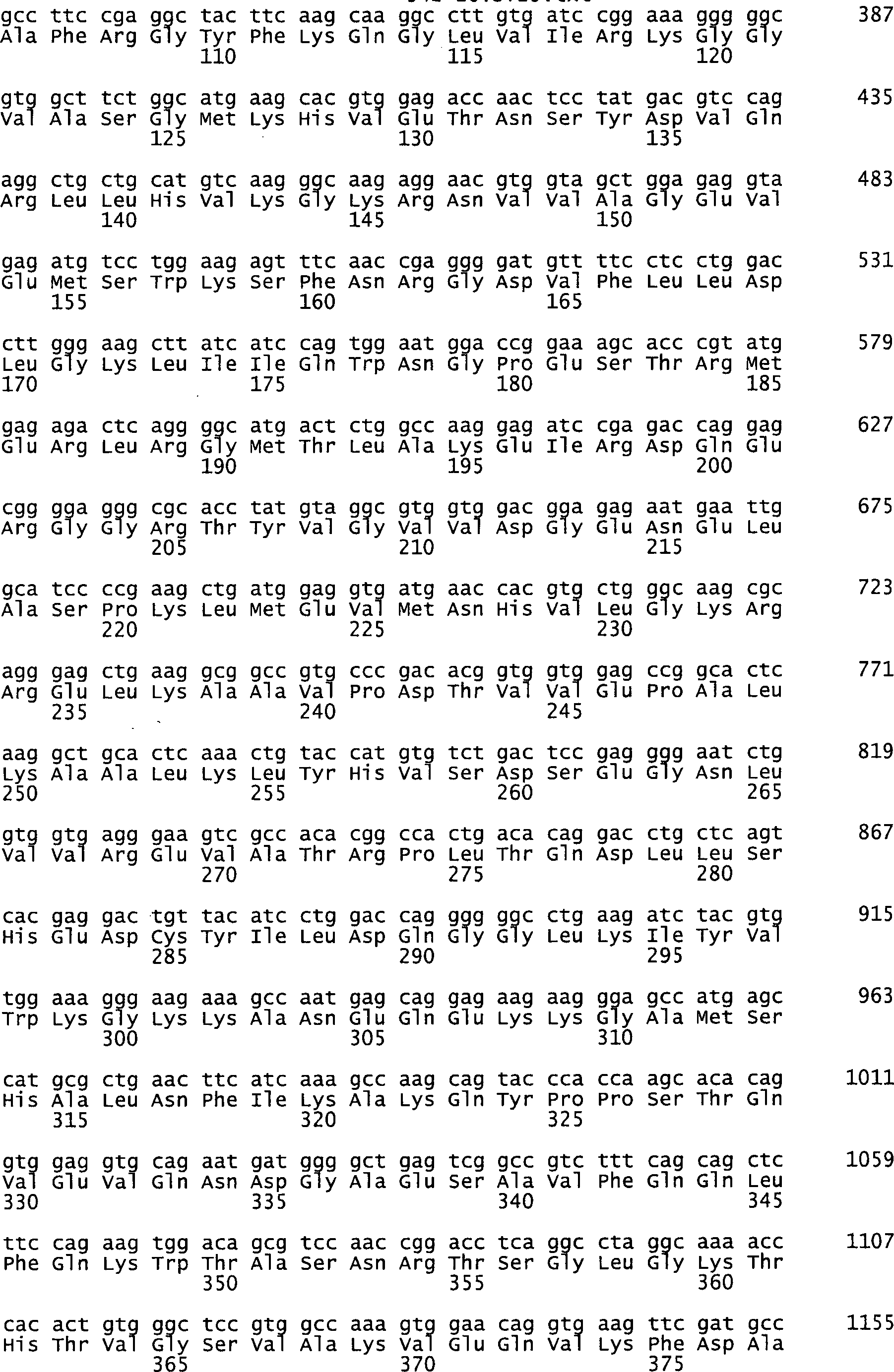


Claims (97)
Priority Applications (42)
| Application Number | Priority Date | Filing Date | Title |
|---|---|---|---|
| DE102004023187A DE102004023187A1 (en) | 2004-05-11 | 2004-05-11 | Identification of surface-associated antigens for tumor diagnosis and therapy |
| CA2820168A CA2820168C (en) | 2004-05-11 | 2005-05-11 | Identification of surface-associated antigens for tumor diagnosis and therapy |
| EP18157525.9A EP3382036A1 (en) | 2004-05-11 | 2005-05-11 | Identification of surface-associated antigens for tumour diagnosis and therapy |
| PL05747772T PL1759013T3 (en) | 2004-05-11 | 2005-05-11 | Identification of surface-associated antigens for tumor diagnosis and therapy |
| EP10010156A EP2348135A3 (en) | 2004-05-11 | 2005-05-11 | Identification of surface-associated Antigenes used for the diagnosis and therapy of tumours |
| PT05747772T PT1759013E (en) | 2004-05-11 | 2005-05-11 | Identification of surface-associated antigens for tumor diagnosis and therapy |
| EP10010168A EP2267158A3 (en) | 2004-05-11 | 2005-05-11 | Identification of surface-associated Antigenes used for the diagnosis and therapy of tumours |
| EP10010159A EP2275571A3 (en) | 2004-05-11 | 2005-05-11 | Identification of surface-associated Antigenes used for the diagnosis and therapy of tumours |
| SI200531066T SI1759013T1 (en) | 2004-05-11 | 2005-05-11 | Identification of surface-associated antigens for tumor diagnosis and therapy |
| EP10010158A EP2290103A3 (en) | 2004-05-11 | 2005-05-11 | Identification of surface-associated Antigenes used for the diagnosis and therapy of tumours |
| DE502005009602T DE502005009602D1 (en) | 2004-05-11 | 2005-05-11 | IDENTIFICATION OF SURFACE ASSOCIATED ANTIGENES FOR TUMOR DIAGNOSIS AND THERAPY |
| EP10010162A EP2258868A3 (en) | 2004-05-11 | 2005-05-11 | Identification of surface-associated Antigenes used for the diagnosis and therapy of tumours |
| AU2005244425A AU2005244425B2 (en) | 2004-05-11 | 2005-05-11 | Identification of surface-associated antigens for tumor diagnosis and therapy |
| EP10010640A EP2275572A3 (en) | 2004-05-11 | 2005-05-11 | Identification of surface-associated Antigenes used for the diagnosis and therapy of tumours |
| EP05747772A EP1759013B1 (en) | 2004-05-11 | 2005-05-11 | Identification of surface-associated antigens for tumor diagnosis and therapy |
| EP10005074A EP2239340A3 (en) | 2004-05-11 | 2005-05-11 | Identification of surface-oriented antigens for tumour diagnosis and treatment |
| CA002563671A CA2563671A1 (en) | 2004-05-11 | 2005-05-11 | Identification of surface-associated antigens for tumor diagnosis and therapy |
| EP10010161A EP2258867A3 (en) | 2004-05-11 | 2005-05-11 | Identification of surface-associated Antigenes used for the diagnosis and therapy of tumours |
| HR20100412T HRP20100412T1 (en) | 2004-05-11 | 2005-05-11 | Identification of surface-associated antigens for tumor diagnosis and therapy |
| EP10010163A EP2258869A3 (en) | 2004-05-11 | 2005-05-11 | Identification of surface-associated Antigenes used for the diagnosis and therapy of tumours |
| ES05747772T ES2346067T3 (en) | 2004-05-11 | 2005-05-11 | IDENTIFICATION OF ANTIGENS ASSOCIATED TO THE SURFACE FOR DIAGNOSIS AND THERAPY OF TUMORS. |
| PCT/EP2005/005104 WO2005110338A2 (en) | 2004-05-11 | 2005-05-11 | Identification of surface-associated antigens for tumor diagnosis and therapy |
| EP10010155.9A EP2280082B1 (en) | 2004-05-11 | 2005-05-11 | Identification of surface-associated Antigenes used for the diagnosis and therapy of tumours |
| EP10010160A EP2290104A3 (en) | 2004-05-11 | 2005-05-11 | Identification of surface-associated Antigenes used for the diagnosis and therapy of tumours |
| EP10010164A EP2287337A3 (en) | 2004-05-11 | 2005-05-11 | Identification of surface-associated Antigenes used for the diagnosis and therapy of tumours |
| DK05747772.1T DK1759013T3 (en) | 2004-05-11 | 2005-05-11 | Identification of surface-associated antigens for tumor diagnosis |
| EP10010167A EP2270209A3 (en) | 2004-05-11 | 2005-05-11 | Identification of surface-associated Antigenes used for the diagnosis and therapy of tumours |
| US11/596,106 US7785801B2 (en) | 2004-05-11 | 2005-05-11 | Identification of surface-associated antigens for tumor diagnosis and therapy |
| JP2007512091A JP2007537197A (en) | 2004-05-11 | 2005-05-11 | Identification of surface-associated antigens for tumor diagnosis and treatment |
| EP10010165A EP2267156A3 (en) | 2004-05-11 | 2005-05-11 | Identification of surface-associated Antigenes used for the diagnosis and therapy of tumours |
| EP10010157A EP2275570A3 (en) | 2004-05-11 | 2005-05-11 | Identification of surface-associated Antigenes used for the diagnosis and therapy of tumours |
| EP10010166A EP2267157A3 (en) | 2004-05-11 | 2005-05-11 | Identification of surface-associated Antigenes used for the diagnosis and therapy of tumours |
| AT05747772T ATE468409T1 (en) | 2004-05-11 | 2005-05-11 | IDENTIFICATION OF SURFACE-ASSOCIATED ANTIGENS FOR TUMOR DIAGNOSIS AND THERAPY |
| US12/851,980 US9255131B2 (en) | 2004-05-11 | 2010-08-06 | Identification of surface-associated antigens for tumor diagnosis and therapy |
| JP2011229287A JP5732369B2 (en) | 2004-05-11 | 2011-10-18 | Identification of surface-associated antigens for tumor diagnosis and treatment |
| JP2012228762A JP5796003B2 (en) | 2004-05-11 | 2012-10-16 | Identification of surface-associated antigens for tumor diagnosis and treatment |
| US14/036,969 US9533043B2 (en) | 2004-05-11 | 2013-09-25 | Identification of surface-associated antigens for tumor diagnosis and therapy |
| JP2014240820A JP2015083576A (en) | 2004-05-11 | 2014-11-28 | Identification of surface-associated antigens for tumor diagnosis and treatment |
| US15/009,061 US20160193335A1 (en) | 2004-05-11 | 2016-01-28 | Identification of Surface-Associated Antigens for Tumor Diagnosis and Therapy |
| JP2016086914A JP6434444B2 (en) | 2004-05-11 | 2016-04-25 | Identification of surface-associated antigens for tumor diagnosis and treatment |
| US15/675,148 US10724103B2 (en) | 2004-05-11 | 2017-08-11 | Identification of surface associated antigen FLJ31461 for tumor diagnosis and therapy |
| JP2018042560A JP2018104464A (en) | 2004-05-11 | 2018-03-09 | Identification of surface associated antigens for purpose of diagnosis and treatment of tumor |
Applications Claiming Priority (1)
| Application Number | Priority Date | Filing Date | Title |
|---|---|---|---|
| DE102004023187A DE102004023187A1 (en) | 2004-05-11 | 2004-05-11 | Identification of surface-associated antigens for tumor diagnosis and therapy |
Publications (1)
| Publication Number | Publication Date |
|---|---|
| DE102004023187A1 true DE102004023187A1 (en) | 2005-12-01 |
Family
ID=35064875
Family Applications (2)
| Application Number | Title | Priority Date | Filing Date |
|---|---|---|---|
| DE102004023187A Withdrawn DE102004023187A1 (en) | 2004-05-11 | 2004-05-11 | Identification of surface-associated antigens for tumor diagnosis and therapy |
| DE502005009602T Expired - Lifetime DE502005009602D1 (en) | 2004-05-11 | 2005-05-11 | IDENTIFICATION OF SURFACE ASSOCIATED ANTIGENES FOR TUMOR DIAGNOSIS AND THERAPY |
Family Applications After (1)
| Application Number | Title | Priority Date | Filing Date |
|---|---|---|---|
| DE502005009602T Expired - Lifetime DE502005009602D1 (en) | 2004-05-11 | 2005-05-11 | IDENTIFICATION OF SURFACE ASSOCIATED ANTIGENES FOR TUMOR DIAGNOSIS AND THERAPY |
Country Status (14)
| Country | Link |
|---|---|
| US (5) | US7785801B2 (en) |
| EP (18) | EP1759013B1 (en) |
| JP (6) | JP2007537197A (en) |
| AT (1) | ATE468409T1 (en) |
| AU (1) | AU2005244425B2 (en) |
| CA (2) | CA2820168C (en) |
| DE (2) | DE102004023187A1 (en) |
| DK (1) | DK1759013T3 (en) |
| ES (1) | ES2346067T3 (en) |
| HR (1) | HRP20100412T1 (en) |
| PL (1) | PL1759013T3 (en) |
| PT (1) | PT1759013E (en) |
| SI (1) | SI1759013T1 (en) |
| WO (1) | WO2005110338A2 (en) |
Families Citing this family (43)
| Publication number | Priority date | Publication date | Assignee | Title |
|---|---|---|---|---|
| DE10347710B4 (en) | 2003-10-14 | 2006-03-30 | Johannes-Gutenberg-Universität Mainz | Recombinant vaccines and their use |
| DE102004023187A1 (en) | 2004-05-11 | 2005-12-01 | Ganymed Pharmaceuticals Ag | Identification of surface-associated antigens for tumor diagnosis and therapy |
| DE102005046490A1 (en) | 2005-09-28 | 2007-03-29 | Johannes-Gutenberg-Universität Mainz | New nucleic acid molecule comprising promoter, a transcriptable nucleic acid sequence, a first and second nucleic acid sequence for producing modified RNA with transcriptional stability and translational efficiency |
| US8535677B2 (en) | 2006-06-06 | 2013-09-17 | Oxford Biotherapeutics, Ltd. | Antibody drug conjugate treatment of colorectal cancer |
| GB0611116D0 (en) * | 2006-06-06 | 2006-07-19 | Oxford Genome Sciences Uk Ltd | Proteins |
| US20100092457A1 (en) * | 2006-08-14 | 2010-04-15 | Forerunner Pharma Research Co., Ltd. | Diagnosis and Treatment of Cancer Using Anti-Desmoglein-3 Antibodies |
| DE102007034993A1 (en) * | 2007-07-26 | 2009-01-29 | Hanna Diehl | Soluble Cadherin 17 for the diagnosis and risk stratification of colon or colon cancer |
| JP5726729B2 (en) * | 2008-06-25 | 2015-06-03 | インスティチュート・パスツール・コリアInstitut Pasteur Korea | Genome-wide visual identification of human cofactors for HIV-1 infection |
| JP5371017B2 (en) * | 2008-10-10 | 2013-12-18 | 独立行政法人放射線医学総合研究所 | Markers for diagnosis of cervical adenocarcinoma or prognosis of cervical cancer |
| SG10201401604VA (en) | 2009-04-20 | 2014-08-28 | Oxford Biotherapeutics Ltd | Antibodies Specific To Cadherin-17 |
| AU2011248354A1 (en) * | 2010-05-03 | 2012-11-08 | Genentech, Inc. | Compositions and methods for the diagnosis and treatment of tumor |
| AU2011318574B2 (en) * | 2010-10-20 | 2016-03-03 | Oxford Biotherapeutics Ltd. | Antibodies |
| US9511151B2 (en) | 2010-11-12 | 2016-12-06 | Uti Limited Partnership | Compositions and methods for the prevention and treatment of cancer |
| RU2670745C9 (en) | 2011-05-24 | 2018-12-13 | Бионтех Рна Фармасьютикалс Гмбх | Individualised vaccines for cancer |
| CN102311242B (en) * | 2011-05-27 | 2014-10-01 | 刘长福 | Water-soluble polymer grinding aid produced from papermaking waste liquid and preparation method thereof |
| KR101314828B1 (en) * | 2011-09-26 | 2013-10-04 | 한국원자력연구원 | The method for the inhibition of radiation resistance and cell growth, migration, and invasion by regulating the expression or activity of TM4SF4 in non small lung cancer |
| WO2013143555A1 (en) | 2012-03-26 | 2013-10-03 | Biontech Ag | Rna formulation for immunotherapy |
| US10988516B2 (en) | 2012-03-26 | 2021-04-27 | Uti Limited Partnership | Methods and compositions for treating inflammation |
| US9603948B2 (en) | 2012-10-11 | 2017-03-28 | Uti Limited Partnership | Methods and compositions for treating multiple sclerosis and related disorders |
| EP2925348B1 (en) | 2012-11-28 | 2019-03-06 | BioNTech RNA Pharmaceuticals GmbH | Individualized vaccines for cancer |
| WO2014180490A1 (en) | 2013-05-10 | 2014-11-13 | Biontech Ag | Predicting immunogenicity of t cell epitopes |
| SG10201912301XA (en) | 2013-11-04 | 2020-02-27 | Uti Lp | Methods and compositions for sustained immunotherapy |
| JP7044551B2 (en) | 2014-09-10 | 2022-03-30 | ジェネンテック, インコーポレイテッド | Immunogenic variant peptide screening platform |
| WO2016045732A1 (en) | 2014-09-25 | 2016-03-31 | Biontech Rna Pharmaceuticals Gmbh | Stable formulations of lipids and liposomes |
| WO2016128060A1 (en) | 2015-02-12 | 2016-08-18 | Biontech Ag | Predicting t cell epitopes useful for vaccination |
| JP6492914B2 (en) | 2015-04-15 | 2019-04-03 | 株式会社デンソー | Optical scanning device |
| JP2018513141A (en) * | 2015-04-20 | 2018-05-24 | コンセジョ スペリオール デ インベスティガショネス シエンティフィカス | Agents that specifically bind to RGD motifs of human cadherin-17, human cadherin-5, human cadherin-6 and human cadherin-20 |
| HK1252348A1 (en) | 2015-05-06 | 2019-05-24 | Uti Limited Partnership | Nanoparticle compositions for sustained therapy |
| JP7026613B2 (en) | 2015-08-07 | 2022-02-28 | イマジナブ・インコーポレーテッド | Antigen binding construct for target molecule |
| WO2017059902A1 (en) | 2015-10-07 | 2017-04-13 | Biontech Rna Pharmaceuticals Gmbh | 3' utr sequences for stabilization of rna |
| DK3383920T3 (en) * | 2015-11-30 | 2024-04-02 | Univ California | DELIVERY OF TUMOR-SPECIFIC PAYLOAD AND IMMUNE ACTIVATION USING A HUMAN ANTIBODY DIRECTED AGAINST A HIGHLY SPECIFIC TUMOR CELL SURFACE ANTIGEN |
| BR112018012374A2 (en) | 2015-12-16 | 2018-12-04 | Gritstone Oncology, Inc. | identification, manufacture and use of neoantigen |
| EP3402571A4 (en) | 2016-01-15 | 2019-08-28 | The J. David Gladstone Institutes | METHODS OF TREATING DISEASES BY REGULATING METABOLIC DIFFERENTIATION OF LYMPHOCYTES T |
| WO2018147960A1 (en) | 2017-02-08 | 2018-08-16 | Imaginab, Inc. | Extension sequences for diabodies |
| CN106919801B (en) * | 2017-03-08 | 2023-07-18 | 杭州大伽信息科技有限公司 | An immunohistochemical staining auxiliary analysis system and its application method |
| WO2018224166A1 (en) | 2017-06-09 | 2018-12-13 | Biontech Rna Pharmaceuticals Gmbh | Methods for predicting the usefulness of disease specific amino acid modifications for immunotherapy |
| JP7227237B2 (en) | 2017-10-10 | 2023-02-21 | グリットストーン バイオ インコーポレイテッド | Identification of neoantigens using hotspots |
| KR102905054B1 (en) | 2017-11-22 | 2025-12-29 | 시애틀 프로젝트 코포레이션 | Reduced presentation of junctional epitopes to neoantigens |
| JP7436372B2 (en) | 2017-11-29 | 2024-02-21 | ユーティアイ・リミテッド・パートナーシップ | How to treat autoimmune diseases |
| CN112543813B (en) * | 2018-03-29 | 2025-02-25 | 澳大利亚纽卡斯尔大学 | Diagnostic and prognostic approaches for estrogen-induced cancers |
| WO2020263796A1 (en) * | 2019-06-23 | 2020-12-30 | Guangdong Tcrcure Biopharma Technology Co., Ltd. | Anti-alpp car-t cell therapy |
| EP4071171B1 (en) * | 2019-12-06 | 2025-06-25 | Korea Atomic Energy Research Institute | Anti-tm4sf4 antibody and use thereof |
| AU2022293217A1 (en) * | 2021-06-18 | 2023-12-14 | Industry Academy Cooperation Foundation Of Sejong University | Anti-tm4sf4 humanized antibody and use thereof |
Family Cites Families (25)
| Publication number | Priority date | Publication date | Assignee | Title |
|---|---|---|---|---|
| US4732847A (en) * | 1981-06-09 | 1988-03-22 | University Of Hawaii | Monoclonal antibodies for DNA-RNA hybrid complexes and their uses |
| US5081227A (en) * | 1988-03-04 | 1992-01-14 | La Jolla Cancer Research Foundation | Germ cell alkaline phosphatase |
| GB9019812D0 (en) | 1990-09-11 | 1990-10-24 | Scotgen Ltd | Novel antibodies for treatment and prevention of infection in animals and man |
| WO1993010813A1 (en) | 1991-11-27 | 1993-06-10 | THE UNITED STAES OF AMERICA, represented by THE SECRETARY, DEPARTMENT OF HEALTH AND HUMAN SERVICES | Nucleotide and amino acid sequence of pemphigus vulgaris antigen and methods of use |
| US5677139A (en) | 1995-04-21 | 1997-10-14 | President And Fellows Of Harvard College | In vitro differentiation of CD34+ progenitor cells into T lymphocytes |
| UA56132C2 (en) | 1995-04-25 | 2003-05-15 | Смітклайн Бічем Байолоджікалс С.А. | Vaccine composition (variants), method for stabilizing qs21 providing resistance against hydrolysis (variants), method for manufacturing vaccine |
| WO1997026328A1 (en) | 1996-01-17 | 1997-07-24 | Imperial College Innovations Limited | Immunotherapy using cytotoxic t lymphocytes (ctl) |
| AU6263999A (en) | 1998-09-28 | 2000-04-17 | Chiron Corporation | Human genes and gene expression products |
| EP1252300B1 (en) * | 2000-02-03 | 2011-01-19 | Dendreon Corporation | Nucleic acid molecules encoding transmembrane serine proteases, the encoded proteins and methods based thereon |
| KR100374423B1 (en) * | 2000-04-25 | 2003-03-28 | 주식회사 아이씨엔지 | Development of Multi-MAGE or Multi-GAGE Isotypes Recogniznig Primer for Cancer Detection |
| WO2001094629A2 (en) * | 2000-06-05 | 2001-12-13 | Avalon Pharmaceuticals | Cancer gene determination and therapeutic screening using signature gene sets |
| JP2004519215A (en) * | 2000-08-15 | 2004-07-02 | イミュネックス・コーポレーション | Claudin polypeptide |
| US6697557B2 (en) * | 2000-10-04 | 2004-02-24 | University Of South Florida | Two-dimensional optical filter and associated methods |
| DE10057894A1 (en) * | 2000-11-22 | 2002-06-06 | Adnagen Gmbh | Kit for diagnosis and monitoring of testicular tumors, comprises pairs of primers for amplifying specific markers in blood, allows early detection of metastasis |
| JP2005527180A (en) | 2001-04-18 | 2005-09-15 | プロテイン デザイン ラブス, インコーポレイテッド | Lung cancer diagnosis method, composition of lung cancer modifier and screening method |
| US20050037445A1 (en) * | 2001-06-25 | 2005-02-17 | Poulsen Hans Skovgaard | Oncology drug innovation |
| US20040076955A1 (en) * | 2001-07-03 | 2004-04-22 | Eos Biotechnology, Inc. | Methods of diagnosis of bladder cancer, compositions and methods of screening for modulators of bladder cancer |
| AU2002313559A1 (en) * | 2001-08-23 | 2003-03-10 | Oxford Biomedica (Uk) Limited | Genes |
| EP1293569A3 (en) * | 2001-09-14 | 2004-03-31 | Research Association for Biotechnology | Full-length cDNAs |
| JP2003088388A (en) * | 2001-09-14 | 2003-03-25 | Herikkusu Kenkyusho:Kk | NEW FULL-LENGTH cDNA |
| DE10211088A1 (en) * | 2002-03-13 | 2003-09-25 | Ugur Sahin | Gene products differentially expressed in tumors and their use |
| DE10254601A1 (en) * | 2002-11-22 | 2004-06-03 | Ganymed Pharmaceuticals Ag | Gene products differentially expressed in tumors and their use |
| AU2004231740A1 (en) * | 2003-04-17 | 2004-11-04 | The Trustees Of Columbia University In The City Ofnew York | Desmoglein 4 is a novel gene involved in hair growth |
| EP2251039A3 (en) * | 2003-05-30 | 2010-12-08 | Nippon Shinyaku Co., Ltd. | Oligo double-stranded rna inhibiting the expression of bcl-2 and pharmaceutical composition containing the same |
| DE102004023187A1 (en) * | 2004-05-11 | 2005-12-01 | Ganymed Pharmaceuticals Ag | Identification of surface-associated antigens for tumor diagnosis and therapy |
-
2004
- 2004-05-11 DE DE102004023187A patent/DE102004023187A1/en not_active Withdrawn
-
2005
- 2005-05-11 PL PL05747772T patent/PL1759013T3/en unknown
- 2005-05-11 EP EP05747772A patent/EP1759013B1/en not_active Expired - Lifetime
- 2005-05-11 SI SI200531066T patent/SI1759013T1/en unknown
- 2005-05-11 US US11/596,106 patent/US7785801B2/en not_active Expired - Lifetime
- 2005-05-11 EP EP10010159A patent/EP2275571A3/en not_active Withdrawn
- 2005-05-11 EP EP10010640A patent/EP2275572A3/en not_active Withdrawn
- 2005-05-11 DE DE502005009602T patent/DE502005009602D1/en not_active Expired - Lifetime
- 2005-05-11 EP EP10010160A patent/EP2290104A3/en not_active Withdrawn
- 2005-05-11 AT AT05747772T patent/ATE468409T1/en active
- 2005-05-11 EP EP10010168A patent/EP2267158A3/en not_active Withdrawn
- 2005-05-11 AU AU2005244425A patent/AU2005244425B2/en not_active Ceased
- 2005-05-11 ES ES05747772T patent/ES2346067T3/en not_active Expired - Lifetime
- 2005-05-11 EP EP10010167A patent/EP2270209A3/en not_active Withdrawn
- 2005-05-11 EP EP10010155.9A patent/EP2280082B1/en not_active Expired - Lifetime
- 2005-05-11 EP EP18157525.9A patent/EP3382036A1/en not_active Withdrawn
- 2005-05-11 EP EP10010158A patent/EP2290103A3/en not_active Withdrawn
- 2005-05-11 EP EP10010165A patent/EP2267156A3/en not_active Withdrawn
- 2005-05-11 WO PCT/EP2005/005104 patent/WO2005110338A2/en not_active Ceased
- 2005-05-11 EP EP10010157A patent/EP2275570A3/en not_active Withdrawn
- 2005-05-11 EP EP10010162A patent/EP2258868A3/en not_active Withdrawn
- 2005-05-11 PT PT05747772T patent/PT1759013E/en unknown
- 2005-05-11 EP EP10010156A patent/EP2348135A3/en not_active Withdrawn
- 2005-05-11 EP EP10010164A patent/EP2287337A3/en not_active Withdrawn
- 2005-05-11 DK DK05747772.1T patent/DK1759013T3/en active
- 2005-05-11 EP EP10010161A patent/EP2258867A3/en not_active Withdrawn
- 2005-05-11 CA CA2820168A patent/CA2820168C/en not_active Expired - Lifetime
- 2005-05-11 HR HR20100412T patent/HRP20100412T1/en unknown
- 2005-05-11 EP EP10005074A patent/EP2239340A3/en not_active Withdrawn
- 2005-05-11 EP EP10010166A patent/EP2267157A3/en not_active Withdrawn
- 2005-05-11 CA CA002563671A patent/CA2563671A1/en not_active Abandoned
- 2005-05-11 JP JP2007512091A patent/JP2007537197A/en active Pending
- 2005-05-11 EP EP10010163A patent/EP2258869A3/en not_active Withdrawn
-
2010
- 2010-08-06 US US12/851,980 patent/US9255131B2/en not_active Expired - Fee Related
-
2011
- 2011-10-18 JP JP2011229287A patent/JP5732369B2/en not_active Expired - Fee Related
-
2012
- 2012-10-16 JP JP2012228762A patent/JP5796003B2/en not_active Expired - Lifetime
-
2013
- 2013-09-25 US US14/036,969 patent/US9533043B2/en active Active
-
2014
- 2014-11-28 JP JP2014240820A patent/JP2015083576A/en active Pending
-
2016
- 2016-01-28 US US15/009,061 patent/US20160193335A1/en not_active Abandoned
- 2016-04-25 JP JP2016086914A patent/JP6434444B2/en not_active Expired - Lifetime
-
2017
- 2017-08-11 US US15/675,148 patent/US10724103B2/en not_active Expired - Lifetime
-
2018
- 2018-03-09 JP JP2018042560A patent/JP2018104464A/en active Pending
Also Published As
Similar Documents
| Publication | Publication Date | Title |
|---|---|---|
| EP2280082B1 (en) | Identification of surface-associated Antigenes used for the diagnosis and therapy of tumours | |
| EP1664113B1 (en) | Identification of tumour-associated cell surface antigens for diagnosis and therapy | |
| EP2327417B1 (en) | Identification of surface-associated antigens for tumour diagnosis and therapy |
Legal Events
| Date | Code | Title | Description |
|---|---|---|---|
| 8181 | Inventor (new situation) |
Inventor name: SCHLUETER, VOLKER, DR., 82061 NEURIED, DE Inventor name: T?RECI, OZLEM, DR., 55116 MAINZ, DE Inventor name: HELFTENBEIN, GERD, DR., 35329 GEMUENDEN, DE Inventor name: THIEL, PHILIPPE, DR., 55268 NIEDER-OLM, DE Inventor name: USENER, DIRK, DR., 65185 WIESBADEN, DE Inventor name: SCHNEIDER, SANDRA, DR., 68199 MANNHEIM, DE Inventor name: SAHIN, UGUR, DR., 55116 MAINZ, DE |
|
| 8181 | Inventor (new situation) |
Inventor name: THIEL, PHILIPPE, DR., 82152 PLANEGG, DE Inventor name: USENER, DIRK, DR., 65193 WIESBADEN, DE Inventor name: SCHNEIDER, SANDRA, DR., 76297 STUTENSEE, DE Inventor name: HELFTENBEIN, GERD, DR., 35329 GEMUENDEN, DE Inventor name: SAHIN, UGUR, DR., 55116 MAINZ, DE Inventor name: SCHLUETER, VOLKER, DR., 82061 NEURIED, DE Inventor name: TUERECI, OEZLEM, DR., 55116 MAINZ, DE |
|
| R012 | Request for examination validly filed |
Effective date: 20110428 |
|
| R119 | Application deemed withdrawn, or ip right lapsed, due to non-payment of renewal fee |