CN114585749A - Determination of nucleic acid sequence concentration - Google Patents
Determination of nucleic acid sequence concentration Download PDFInfo
- Publication number
- CN114585749A CN114585749A CN202080073038.5A CN202080073038A CN114585749A CN 114585749 A CN114585749 A CN 114585749A CN 202080073038 A CN202080073038 A CN 202080073038A CN 114585749 A CN114585749 A CN 114585749A
- Authority
- CN
- China
- Prior art keywords
- sequence
- nucleic acid
- concentration
- sample
- amplified
- Prior art date
- Legal status (The legal status is an assumption and is not a legal conclusion. Google has not performed a legal analysis and makes no representation as to the accuracy of the status listed.)
- Granted
Links
Images


Classifications
-
- C—CHEMISTRY; METALLURGY
- C12—BIOCHEMISTRY; BEER; SPIRITS; WINE; VINEGAR; MICROBIOLOGY; ENZYMOLOGY; MUTATION OR GENETIC ENGINEERING
- C12Q—MEASURING OR TESTING PROCESSES INVOLVING ENZYMES, NUCLEIC ACIDS OR MICROORGANISMS; COMPOSITIONS OR TEST PAPERS THEREFOR; PROCESSES OF PREPARING SUCH COMPOSITIONS; CONDITION-RESPONSIVE CONTROL IN MICROBIOLOGICAL OR ENZYMOLOGICAL PROCESSES
- C12Q1/00—Measuring or testing processes involving enzymes, nucleic acids or microorganisms; Compositions therefor; Processes of preparing such compositions
- C12Q1/68—Measuring or testing processes involving enzymes, nucleic acids or microorganisms; Compositions therefor; Processes of preparing such compositions involving nucleic acids
- C12Q1/6844—Nucleic acid amplification reactions
- C12Q1/6851—Quantitative amplification
-
- C—CHEMISTRY; METALLURGY
- C12—BIOCHEMISTRY; BEER; SPIRITS; WINE; VINEGAR; MICROBIOLOGY; ENZYMOLOGY; MUTATION OR GENETIC ENGINEERING
- C12Q—MEASURING OR TESTING PROCESSES INVOLVING ENZYMES, NUCLEIC ACIDS OR MICROORGANISMS; COMPOSITIONS OR TEST PAPERS THEREFOR; PROCESSES OF PREPARING SUCH COMPOSITIONS; CONDITION-RESPONSIVE CONTROL IN MICROBIOLOGICAL OR ENZYMOLOGICAL PROCESSES
- C12Q1/00—Measuring or testing processes involving enzymes, nucleic acids or microorganisms; Compositions therefor; Processes of preparing such compositions
- C12Q1/68—Measuring or testing processes involving enzymes, nucleic acids or microorganisms; Compositions therefor; Processes of preparing such compositions involving nucleic acids
- C12Q1/6844—Nucleic acid amplification reactions
- C12Q1/686—Polymerase chain reaction [PCR]
-
- C—CHEMISTRY; METALLURGY
- C12—BIOCHEMISTRY; BEER; SPIRITS; WINE; VINEGAR; MICROBIOLOGY; ENZYMOLOGY; MUTATION OR GENETIC ENGINEERING
- C12Q—MEASURING OR TESTING PROCESSES INVOLVING ENZYMES, NUCLEIC ACIDS OR MICROORGANISMS; COMPOSITIONS OR TEST PAPERS THEREFOR; PROCESSES OF PREPARING SUCH COMPOSITIONS; CONDITION-RESPONSIVE CONTROL IN MICROBIOLOGICAL OR ENZYMOLOGICAL PROCESSES
- C12Q2531/00—Reactions of nucleic acids characterised by
- C12Q2531/10—Reactions of nucleic acids characterised by the purpose being amplify/increase the copy number of target nucleic acid
- C12Q2531/113—PCR
-
- C—CHEMISTRY; METALLURGY
- C12—BIOCHEMISTRY; BEER; SPIRITS; WINE; VINEGAR; MICROBIOLOGY; ENZYMOLOGY; MUTATION OR GENETIC ENGINEERING
- C12Q—MEASURING OR TESTING PROCESSES INVOLVING ENZYMES, NUCLEIC ACIDS OR MICROORGANISMS; COMPOSITIONS OR TEST PAPERS THEREFOR; PROCESSES OF PREPARING SUCH COMPOSITIONS; CONDITION-RESPONSIVE CONTROL IN MICROBIOLOGICAL OR ENZYMOLOGICAL PROCESSES
- C12Q2545/00—Reactions characterised by their quantitative nature
- C12Q2545/10—Reactions characterised by their quantitative nature the purpose being quantitative analysis
Landscapes
- Chemical & Material Sciences (AREA)
- Life Sciences & Earth Sciences (AREA)
- Organic Chemistry (AREA)
- Engineering & Computer Science (AREA)
- Zoology (AREA)
- Wood Science & Technology (AREA)
- Proteomics, Peptides & Aminoacids (AREA)
- Health & Medical Sciences (AREA)
- Molecular Biology (AREA)
- Chemical Kinetics & Catalysis (AREA)
- Immunology (AREA)
- Microbiology (AREA)
- Biophysics (AREA)
- Analytical Chemistry (AREA)
- Physics & Mathematics (AREA)
- Biotechnology (AREA)
- Biochemistry (AREA)
- Bioinformatics & Cheminformatics (AREA)
- General Engineering & Computer Science (AREA)
- General Health & Medical Sciences (AREA)
- Genetics & Genomics (AREA)
- Measuring Or Testing Involving Enzymes Or Micro-Organisms (AREA)
- Apparatus Associated With Microorganisms And Enzymes (AREA)
- Investigating Or Analysing Biological Materials (AREA)
Abstract
本发明涉及通过将校正系数应用于在片段化核酸中测量的被测序列的浓度来确定未片段化核酸中被测序列的浓度的方法。

The present invention relates to a method for determining the concentration of a tested sequence in an unfragmented nucleic acid by applying a correction factor to the concentration of the tested sequence measured in the fragmented nucleic acid.
Description
Cross Reference to Related Applications
This application claims priority from european patent application No. EP19306346 filed on 16/10/2019, the contents of which are incorporated herein by reference in their entirety.
Technical Field
The present invention relates to a method for measuring the concentration of nucleic acid sequences in a biological sample.
Background
Normal humans have two sets of 23 chromosomes in each healthy diploid cell. In some cases, a mutation may occur in any one or more of the chromosomes, resulting in a chromosomal abnormality. These abnormalities may be associated with genetic diseases, cancer and other diseases. Detection of chromosomal abnormalities can identify individuals susceptible to a particular disease or determine the treatment best suited for a given individual. In this regard, the detection of chromosomal abnormalities is of great value.
In addition to human health, the detection of chromosomal abnormalities is also relevant to other species, including but not limited to insects, bacteria, plants, and mixed samples containing organisms, such as soil and food, where transgenic organism (GMO) detection may be performed as a result of genetic engineering, for example, for quality control. Detection of genetic mutations that occur in viral, viroid and other chromosome-free genomes is also highly relevant.
In this context, the characteristics of detecting genomic abnormalities (i.e., changes in a particular nucleic acid sequence in a biological sample) in a qualitative and quantitative manner are relevant.
Such detection is currently performed efficiently by amplifying a target nucleic acid in a target sample. Amplification can be performed by combining oligonucleotide primers with a sample and then subjecting the sample to amplification conditions (e.g., Polymerase Chain Reaction (PCR) conditions) compatible with nucleic acid quantitation. These amplification methods are capable of producing multiple copies of a single target nucleic acid sequence, thereby reaching a detection threshold.
However, it is more relevant to measure the concentration of such specific nucleic acid sequences in a biological sample in a quantitative manner, for example when the goal is not only to detect but also to quantify genetic diseases, for example to monitor the evolution of rare mutations.
In some cases, nucleic acids present in a biological sample are compromised, often fragmented in short length sequences. In such a population of fragmented nucleic acids, the sequences to be amplified in a given PCR assay are sometimes randomly cut. In this case, amplification of the nucleic acid sequence cannot be performed by the PCR method, resulting in underestimation of the presence of the target nucleic acid sequence.
The present invention proposes a method to correct this underestimation problem.
Disclosure of Invention
The present invention relates to a method of determining the concentration of a measured sequence (as further defined below) in an unfragmented nucleic acid, comprising correcting a measured concentration of said measured sequence in a sample comprising a fragmented nucleic acid with a correction factor, thereby obtaining the concentration of said measured sequence in the unfragmented nucleic acid, wherein the correction factor is based on a Length Distribution (LD) and at least one parameter of the measurement method.
In some embodiments, there is provided a method of determining the concentration of a test sequence in an unfragmented nucleic acid, comprising the steps of:
i. measuring the concentration of the sequenced sequence in the sample comprising fragmented nucleic acids with a measurement method; and
correcting the measured concentration of the sequence in the sample comprising fragmented nucleic acid with a correction factor to obtain the concentration of the sequence in non-fragmented nucleic acid, wherein the correction factor is based on the Length Distribution (LD) and at least one parameter of the measurement method.
In some embodiments, there is provided a method of determining the concentration of a test sequence in an unfragmented nucleic acid, comprising the steps of:
i. determining a Length Distribution (LD) of nucleic acids in a sample comprising fragmented nucleic acids, wherein fragmented nucleic acids are derived from the non-fragmented nucleic acids;
measuring the concentration of the sequenced sequence in the sample comprising fragmented nucleic acids with a measurement method; and
correcting the measured concentration of the sequence in the sample comprising fragmented nucleic acid with a correction factor to obtain the concentration of the sequence in non-fragmented nucleic acid, wherein the correction factor is based on the Length Distribution (LD) and at least one parameter of the measurement method.
In some embodiments, the method comprises:
i. providing a sample of fragmented nucleic acids derived from the non-fragmented nucleic acids;
providing a nucleic acid fragment Length Distribution (LD) of the fragmented nucleic acids;
measuring the concentration of the sequence to be detected in the fragmented nucleic acid sample using a measurement method;
determining a correction factor based on the nucleic acid fragment Length Distribution (LD) of the fragmented nucleic acids and parameters of the measurement method; and
v. correcting the concentration of the sequenced sequence in fragmented nucleic acid with the correction factor to obtain the concentration of the sequenced sequence in unfragmented nucleic acid.
In embodiments, the sample of fragmented nucleic acids is any combination of the following three classes:
i. a cell-free sample or a cell-containing sample;
a naturally fragmented sample or an artificially fragmented sample; and
any type of deoxyribonucleic acid (DNA) or ribonucleic acid (RNA).
In an embodiment, the measurement method is an isothermal quantitative nucleic acid amplification method, preferably selected from the group consisting of loop-mediated isothermal amplification and quantitative nucleic acid sequence-based amplification.
In another embodiment, the measuring method is a non-isothermal quantitative nucleic acid amplification method, preferably selected from the group consisting of quantitative polymerase chain reaction, real-time polymerase chain reaction, digital polymerase chain reaction, multiple polymerase chain reaction and multiple digital polymerase chain reaction.
In embodiments, the parameter of the measurement method comprises the length of the sequence to be amplified, as further defined below. In a particular configuration of this embodiment, at least 5% of the nucleic acid fragments are shorter in length than the sequence to be amplified. In another particular configuration of this embodiment, at most 95% of the nucleic acid fragments are shorter in length than the sequence to be amplified.
In embodiments, the length of the sequence to be amplified is greater than 40bp and less than 200bp, preferably greater than 50bp and less than 170bp, more preferably greater than 65bp and less than 150bp, even more preferably greater than 70bp and less than 130 bp.
In embodiments, the nucleic acid fragment Length Distribution (LD) is comprised in the range of 25bp to 350bp, preferably 30bp to 320bp, more preferably 35bp to 290bp, even more preferably 40bp to 270 bp.
The invention further relates to a method for determining the first sequenced sequence S in an unfragmented nucleic acid1And the second sequenced column S2The method of a function of the second concentration of (a), comprising the steps of:
i. determination of S in the unfragmented nucleic acid according to any of the determination methods described above1The concentration of (c);
determining S in said non-fragmented nucleic acid according to any of the determination methods described above2The concentration of (c); and
calculating said S1Concentration and the said S2Said function of concentration;
wherein said S1Concentration and the said S2The concentrations are determined in the same sample; and wherein with S1The length of the sequence to be amplified which is relevant differs from that of S2The length of the relevant sequence to be amplified.
The invention also relates to a system configured to determine the concentration of a detected sequence in an unfragmented nucleic acid, comprising:
i. a module configured to measure a concentration of the sequenced nucleic acid in a sample of fragmented nucleic acids, the fragmented nucleic acids derived from the non-fragmented nucleic acids;
a module configured to calculate a correction factor based on a nucleic acid fragment Length Distribution (LD) of the fragmented nucleic acids and the measured parameters; and
a module configured to calculate a concentration of the sequenced sequence in unfragmented nucleic acid using the correction factor.
In a specific configuration, the module configured to measure the concentration of the sequenced sequence in the fragmented nucleic acid is an isothermal quantitative nucleic acid amplification module, preferably selected from a loop-mediated isothermal amplification module and a quantitative nucleic acid sequence-based amplification module.
In an alternative configuration, the module configured to measure the concentration of the sequenced sequence in the fragmented nucleic acid is a non-isothermal quantitative nucleic acid amplification module, which is preferably selected from the group consisting of a quantitative polymerase chain reaction module, a real-time polymerase chain reaction module, a digital polymerase chain reaction module, a multiplex polymerase chain reaction module, and a multiplex digital polymerase chain reaction.
In one embodiment, the measured parameter comprises the length of the sequence to be amplified.
Definition of
In the present invention, the following terms have the following meanings:
the term "amplicon" refers to the nucleic acid product of an amplification reaction. The amplicon may be single-stranded or double-stranded, or a combination thereof.
The term "amplification" refers to a reaction in which replication occurs repeatedly over time to form multiple copies of at least one fragment of a template molecule. Amplification can produce an exponential or linear increase in copy number as amplification proceeds. Typical amplification increases copy number and/or signal by more than 1,000 fold. Exemplary amplification reactions for the droplet-based assays disclosed herein may include Polymerase Chain Reaction (PCR) or ligase chain reaction, each of which is driven by thermal cycling. Droplet-based assays may also or alternatively use other amplification reactions that can be performed isothermally. Amplification can be performed, or its occurrence determined, in an amplification mixture, which is any composition capable of producing multiple copies of a nucleic acid target molecule (if present) in the composition. An "amplification mixture" may comprise any combination of at least one primer or primer pair, at least one probe, at least one replicase (e.g., at least one polymerase, such as at least one DNA and/or RNA polymerase, such as a reverse transcriptase), and deoxynucleotide (and/or nucleotide) triphosphates (dntps and/or NTPs), and buffers containing any components necessary for replicase activity, and the like.
The term "assay" refers to a process and/or reaction used to characterize a sample, as well as any signals, values, data, and/or results obtained from the process and/or reaction.
The term "cleavage" is equivalent to the term "fragmentation" and refers to a nucleic acid sequence.
The term "sequenced" refers to the exact sequence that is quantified from a sample in the methods of the invention. In the case of point mutations, the sequence being sequenced includes the detected mutation. In some embodiments, a fluorescent reporter is used. When a fluorescent reporter linked to a specific sequence is used (e.g., as shown in FIG. 1A), the "sequence to be detected" may be shorter than and contained within the sequence to be amplified (as further defined herein). When free fluorescent reporters are used (e.g., as shown in FIG. 1B), the sequenced sequences are typically amplicons.
The term "digital PCR" or "dPCR" refers to a PCR assay performed in a partition of a sample to determine the presence/absence, concentration, and/or copy number of a nucleic acid target in the sample based on how many sample partitions support amplification of the target. The concept of digital PCR can be extended to other types of analytes besides nucleic acids.
The term "label" refers to a recognizable and/or distinguishable marker or identifier attached to or incorporated into any entity (e.g., a compound, biological particle (e.g., a cell, bacteria, spore, virus, or organelle) or droplet), such as a compound, biological particle (e.g., a cell, bacteria, spore, virus, or organelle). For example, the label may be a dye that makes the entity optically detectable and/or optically distinguishable. Exemplary dyes for labeling are fluorescent dyes (fluorophores) and fluorescence quenchers.
The term "length" in relation to a nucleic acid refers to the number of consecutive nucleotides or bases that form a single-stranded molecule or the number of consecutive base pairs that form a double-stranded molecule. The length is measured in nucleotides and base pairs.
The term "multiplex digital PCR" refers to a digital PCR assay performed for the simultaneous amplification of at least two different nucleic acid sequences, in particular two, three, four, five, six, seven, eight or more different nucleic acid sequences (as if a number of separate PCR reactions were performed together in one vessel). The process uses a plurality of primers to amplify nucleic acids in a sample. In particular, "multiplex digital PCR" includes "double digital PCR" and "triple digital PCR". In contrast, a digital PCR assay performed to amplify a nucleic acid sequence is "single-plex digital PCR," often abbreviated as "digital PCR.
The term "multiplex PCR" refers to a PCR assay performed for the simultaneous amplification of at least two different nucleic acid sequences, in particular two, three, four, five, six, seven, eight or more different nucleic acid sequences (as if a number of separate PCR reactions were performed together in one vessel). The process uses a plurality of primers to amplify nucleic acids in a sample. In particular, "multiplex PCR" includes "double PCR" and "triple PCR". In contrast, a PCR assay performed to amplify a nucleic acid sequence is a "singleplex PCR", often referred to simply as "PCR".
The term "nucleic acid" refers to both deoxyribonucleic acid (DNA) or ribonucleic acid (RNA), whether it is an amplification product, synthetically produced, RNA reverse transcription product, or naturally occurring. Typically, a nucleic acid is a single-stranded or double-stranded molecule, consisting of naturally occurring nucleotides.
The term "nucleotide" in addition to referring to naturally occurring ribonucleotide or deoxyribonucleotide monomers, is understood herein to refer to related structural variants thereof, including derivatives and analogs, as well as chemical modifications, that are functionally equivalent with respect to the particular context in which the nucleotide is used (e.g., hybridization to the complementary base: adenine (A) paired with thymine (T), guanine (G) paired with cytosine (C)), unless the context clearly indicates otherwise.
The term "partition" refers to a large volume of separated portions. A partition may be a sample partition resulting from the formation of a large volume of a sample (e.g., a prepared sample). The partitions generated from the large volumes may be substantially uniform in size or may be of different sizes (e.g., a collection of two or more separate, uniformly sized partitions). An exemplary partition is a "droplet". The size of the partitions may also vary in a predetermined size distribution or a random size distribution.
The term "PCR" or "polymerase chain reaction" refers to a nucleic acid amplification assay that relies on alternating cycles of heating and cooling (i.e., thermal cycling) to achieve successive cycles of replication. PCR can be performed by thermal cycling between two or more temperature set points, e.g., a higher melting (denaturation) temperature and a lower annealing/extension temperature, or by thermal cycling between three or more temperature set points, e.g., a higher melting temperature, a lower annealing temperature, and an intermediate extension temperature, etc. Other forms of PCR (e.g., Touchdown PCR) in which the annealing and/or extension temperatures may change during the cycling reaction may be included in this definition.
The term "primer" refers to an oligonucleotide that is capable of acting as a starting point for template-directed nucleic acid synthesis when placed under conditions to begin polynucleotide extension; for example, under conditions comprising the necessary nucleoside triphosphates (determined by the replicated template) and polymerase in a suitable buffer and suitable temperature or temperature cycling (e.g., as in a polymerase chain reaction).
The term "probe" refers to a nucleic acid linked to at least one label, such as at least one dye.
The term "qualitative PCR" refers to a PCR-based assay that determines the presence or absence of a target in a sample, typically without any substantial quantification of the presence of the target. In exemplary embodiments, qualitative digital PCR can be performed by determining that a packet contains at least a predetermined percentage of positive droplets (positive sample) or no droplets (negative sample).
The terms "quantitative PCR", "qPCR", "real-time quantitative polymerase chain reaction" or "kinetic polymerase chain reaction" refer to a PCR-based assay that determines the concentration and/or copy number of a target in a sample. The technique uses PCR to simultaneously amplify and quantify target nucleic acids by means of intercalating fluorescent dyes that are detectable only after hybridization to the target nucleic acids or sequence-specific probes containing fluorescent reporters that are detectable only during sequence amplification.
The term "real-time PCR" refers to a PCR-based analysis that measures amplicon formation during a reaction (e.g., after completion of one or more thermal cycles prior to the final thermal cycle of the reaction). Real-time PCR generally provides quantification of a target based on the kinetics of target amplification.
The term "replication" refers to the process of forming copies (i.e., direct copies and/or complementary copies) of a nucleic acid or fragment thereof. Replication typically involves enzymes such as polymerases and/or ligases and the like. The replicated nucleic acids and/or fragments are the templates (and/or targets) for replication.
The term "sample" refers to a compound, composition, and/or mixture of interest from any suitable source. A sample is a general target for determining aspects of an assay sample (e.g., aspects related to at least one analyte that may be present in the sample). The sample can be analyzed in its native state, as collected, and/or in an altered state, e.g., after storage, extraction, lysis, dilution, concentration, purification, filtration, mixing with one or more reagents, pre-amplification (e.g., target enrichment achieved by limited PCR cycles (e.g., < 15) on the sample prior to PCR), removal of amplicons prior to PCR (e.g., treatment with uracil-d-glycosylase (UDG, also known as UNG, uracil-N-glycosylase gene)) to eliminate carryover contamination by any previously produced amplicons (i.e., which can be digested with UDG because it was produced with dUTP instead of dTTP)), partitions, or any combination thereof, etc. Clinical samples may include nasopharyngeal wash, blood, plasma, acellular plasma, buffy coat, saliva, urine, stool, saliva, mucus, wound swab, tissue biopsy, milk, liquid aspirate, swab (e.g., nasopharyngeal swab), and/or tissue, among others. The sample may be collected for diagnostic purposes (e.g., quantitative measurement of a clinical analyte, such as an infectious agent) or for monitoring purposes (e.g., determination of whether a target environmental analyte (e.g., a bio-threat agent) exceeds a predetermined threshold).
The term "sequence to be amplified" refers to a nucleic acid comprising the "sequence to be sequenced" (as defined above), starting from the sequence of the forward primer and ending with the sequence complementary to the reverse primer, and comprising any additional base pairs located between the primer sequences. The amplification product of the sequence to be amplified is an amplicon. Finally, amplification allows for confirmationThe concentration is determined for the quantification of the "sequence to be amplified". The length of the sequence to be amplified is denoted as "La”。
Drawings
FIG. 1 is a schematic illustration of a quantitative nucleic acid amplification method, by way of example and not limitation. Here, the double-stranded fragmented nucleic acid is represented by a horizontal black solid line. The two primers (forward primer FP and reverse primer RP) define the left and right boundaries of the sequence to be amplified (SA), respectively. Both strands of the sequence to be amplified (SA) are replicated during the amplification process. In FIG. 1A, the sequenced sequence (DS) is a portion of the sequence to be amplified (SA). The fluorescently labeled Probe (Probe) binds specifically to the sequence to be sequenced. In FIG. 1B, the sequence to be Detected (DS) and the sequence to be amplified (SA) are the same. Fluorescent Dyes (FD) are embedded in the double-stranded nucleic acid generated during the amplification process.
FIG. 2 is a graph showing the Length Distribution (LD) of DNA fragments in the sample used in example E1, the length distribution being concentrated around 150 bp. f (i) is the probability (Y-axis-arbitrary units) that a fragment in a sample is i base pairs long (X-axis).
FIG. 3 shows the length L estimated for the sample shown in FIG. 2 as the sequence to be amplifiedaThe probability P that the sequence to be amplified is not cleaved (in bp base pairs).
FIG. 4 shows the length L estimated for the sample shown in FIG. 2 as the sequence to be amplifiedaPredicted Correction Factor (PCF) as a function of (in base pairs bp).
Detailed Description
Methods and systems for correcting measured concentrations of sequenced sequences in a nucleic acid sample comprising fragmented nucleic acids are provided. The methods and systems provided herein allow for the determination of a corrected concentration of a sequence being measured that is closer to the true concentration of the sequence being measured in unfragmented nucleic acids. When the concentration of a sequenced sequence is measured by amplifying a sequence to be amplified that includes the sequenced sequence, a fragment that includes a portion of the sequence to be amplified but is shorter in length than the sequence to be amplified will not be copied. This failure to replicate a truncated target region (i.e., the sequence to be amplified) results in an underestimate of the concentration. The methods provided herein can be used to correct underestimates of nucleic acid concentration in an unfragmented nucleic acid sample.
Accordingly, the present invention provides, in some aspects, a method of determining the concentration of a test sequence in non-fragmented nucleic acid, including determining the copy number of the test sequence, comprising the following steps (hereinafter referred to as the "basic method"). In some aspects, the invention provides a method of calibrating the concentration of a sequenced sequence (including the copy number of the sequenced sequence) in a sample comprising fragmented nucleic acid molecules. Kits, software, devices, and other articles of manufacture useful for the methods described herein are also provided.
I. Method of the present application
The present invention relates to a method of determining the concentration of a sequence to be determined in an unfragmented nucleic acid, comprising correcting a measured concentration of the sequence to be determined in a sample comprising fragmented nucleic acid with a correction factor, thereby obtaining the concentration of the sequence to be determined in the unfragmented nucleic acid, wherein the correction factor is based on a length distribution and at least one parameter of the measurement method.
In some embodiments, there is provided a method of determining the concentration of a test sequence in an unfragmented nucleic acid, comprising the steps of:
i. measuring the concentration of the sequenced sequence in the sample comprising fragmented nucleic acids with a measurement method; and
correcting the measured concentration of the sequence in the sample comprising fragmented nucleic acid with a correction factor to obtain the concentration of the sequence in non-fragmented nucleic acid, wherein the correction factor is based on the Length Distribution (LD) and at least one parameter of the measurement method.
In some embodiments, there is provided a method of determining the concentration of a test sequence (as further defined below) in an unfragmented nucleic acid, comprising the steps of:
i. amplifying a target region comprising the sequence to be detected (hereinafter referred to as "sequence to be amplified") in the sample comprising fragmented nucleic acids;
measuring the concentration of the sequence under test; and
correcting the measured concentration of the sequence in the sample comprising fragmented nucleic acid with a correction factor to obtain the concentration of the sequence in non-fragmented nucleic acid, wherein the correction factor is based on the Length Distribution (LD) and at least one parameter of the measurement method.
In some embodiments, provided herein is a method of determining the concentration of a test sequence in an unfragmented nucleic acid, comprising the steps of:
i. determining a Length Distribution (LD) of nucleic acids in a sample comprising fragmented nucleic acids, wherein fragmented nucleic acids are derived from the non-fragmented nucleic acids;
measuring the concentration of the sequenced sequence in the sample comprising fragmented nucleic acids with a measurement method; and correcting the measured concentration of the sequence in the sample comprising fragmented nucleic acid with a correction factor to obtain the concentration of the sequence in non-fragmented nucleic acid, wherein the correction factor is based on the Length Distribution (LD) and at least one parameter of the measurement method.
In some embodiments, provided herein is a method of calibrating a measured concentration of a sequence being sequenced in a sample comprising fragmented nucleic acids to more closely reflect the concentration of the sequence being sequenced in non-fragmented nucleic acids, comprising the steps of:
i. determining a Length Distribution (LD) of nucleic acids in a sample comprising fragmented nucleic acids, wherein fragmented nucleic acids are derived from the non-fragmented nucleic acids; and
correcting the measured concentration with a correction factor to obtain the concentration of the sequence under test in the non-fragmented nucleic acid, wherein the correction factor is based on the Length Distribution (LD) and at least one parameter of the measurement method used to determine the concentration of the sequence under test.
In some embodiments, provided herein is a method of calibrating a measured concentration of a sequenced sequence in a sample comprising fragmented nucleic acids, wherein the measured concentration of the sequenced sequence is an underestimation of the actual concentration of the sequenced sequence in the sample, comprising the steps of:
i. determining a Length Distribution (LD) of nucleic acids in a sample comprising fragmented nucleic acids, wherein fragmented nucleic acids are derived from the non-fragmented nucleic acids; and
correcting the measured concentration with a correction factor to obtain the concentration of said sequence under test in the non-fragmented nucleic acid, wherein the correction factor is based on the Length Distribution (LD) and at least one parameter of the measurement method used to determine the concentration of the sequence under test.
In some embodiments, provided herein is a method of correcting a measured concentration of a sequence being sequenced in a sample comprising fragmented nucleic acid so that it more closely reflects the concentration of the sequence being sequenced in non-fragmented nucleic acid, comprising the steps of:
i. determining a Length Distribution (LD) of nucleic acids in a sample comprising fragmented nucleic acids, wherein fragmented nucleic acids are derived from the non-fragmented nucleic acids;
measuring the concentration of the sequenced sequence in the sample comprising fragmented nucleic acids with a measurement method; and
correcting the measured concentration of the sequence in the sample comprising fragmented nucleic acid with a correction factor to obtain the concentration of the sequence in non-fragmented nucleic acid, wherein the correction factor is based on the Length Distribution (LD) and at least one parameter of the measurement method.
In some embodiments, provided herein is a method of correcting a measured concentration of a sequenced sequence in a sample comprising fragmented nucleic acids, wherein the measured concentration of the sequenced sequence is an underestimate of the true concentration of the sequenced sequence in the sample, comprising the steps of:
i. determining a Length Distribution (LD) of nucleic acids in a sample comprising fragmented nucleic acids, wherein fragmented nucleic acids are derived from the non-fragmented nucleic acids;
measuring the concentration of the sequenced sequence in the sample comprising fragmented nucleic acids with a measurement method; and
correcting the measured concentration of said sequence in the sample comprising fragmented nucleic acid with a correction factor to obtain the concentration of said sequence in non-fragmented nucleic acid, wherein the correction factor is based on the Length Distribution (LD) and at least one parameter of the measurement method.
In some embodiments, the methods of the present application do not include obtaining a sequence of fragmented nucleic acids. In some embodiments, the method does not comprise obtaining or predicting genetic coordinates of the fragmented nucleic acids. In some embodiments, the method does not comprise assembling the fragmented nucleic acids into a contiguous sequence.
In some embodiments, the sample of fragmented nucleic acids is any combination of the following three classes, as described in the section "nucleic acid sample and length distribution" below:
i. a cell-free sample or a cell-containing sample;
a naturally fragmented sample or an artificially fragmented sample; and
any type of deoxyribonucleic acid or ribonucleic acid.
In some embodiments, the method of measuring does not include sequencing the nucleic acids in the sample. In some embodiments, measuring the concentration of the sequenced sequence can include amplifying a sequence to be amplified that includes the sequenced sequence. In some embodiments, the measurement method is an isothermal quantitative nucleic acid amplification method (e.g., loop-mediated isothermal amplification or quantitative nucleic acid sequence-based amplification). In some embodiments, the measurement method is a non-isothermal quantitative nucleic acid amplification method (e.g., quantitative polymerase chain reaction, real-time polymerase chain reaction, digital polymerase chain reaction, multiplex polymerase chain reaction, and multiplex digital polymerase chain reaction).
In some embodiments according to any of the foregoing methods, as shown in fig. 1B, the sequence being detected and the sequence to be amplified are the same, and the measuring step comprises detecting incorporation of a label (e.g., an intercalating dye, such as a fluorescent dye comprising a fluorophore) into nucleic acid produced during the amplification process.
In some embodiments, the sequenced sequence is a subset of the sequence to be amplified, and/or the measuring step comprises detecting binding of a labeled probe to the sequenced sequence (e.g., a fluorescently labeled probe comprising a fluorophore), as shown in fig. 1A.
The measuring method implemented in the measuring step of the basic method of the invention essentially uses some parameters relevant for determining the correction factor. In a specific embodiment, the measurement method is an amplification method using a replication and amplification mixture.
In some embodiments, the method may further comprise the step of determining a correction factor based on the length distribution of the nucleic acids in the sample and at least one parameter of the measurement method.
In some embodiments, the at least one parameter of the measurement method may comprise the length of the sequence to be amplified. In some embodiments, the at least one parameter of the measurement method comprises only the length of the sequence to be amplified.
Thus, for example, in some embodiments, provided herein is a method of determining the concentration of a detected sequence in an unfragmented nucleic acid, comprising the steps of:
i. determining a Length Distribution (LD) of nucleic acids in a sample comprising fragmented nucleic acids, wherein fragmented nucleic acids are derived from the non-fragmented nucleic acids;
measuring the concentration of the sequenced sequence in the sample comprising fragmented nucleic acids with a measurement method; and correcting the measured concentration of the sequence to be amplified in the sample comprising fragmented nucleic acid with a correction factor to obtain the concentration of the sequence to be amplified in non-fragmented nucleic acid, wherein the correction factor is based on a Length Distribution (LD) and a length of a sequence to be amplified comprising the sequence to be amplified.
In some embodiments, provided herein is a method of calibrating a measured concentration of a sequence being sequenced in a sample comprising fragmented nucleic acid to more closely reflect the concentration of the sequence being sequenced in non-fragmented nucleic acid, comprising the steps of:
i. determining a Length Distribution (LD) of nucleic acids in a sample comprising fragmented nucleic acids, wherein fragmented nucleic acids are derived from the non-fragmented nucleic acids; and
correcting the measured concentration with a correction factor to obtain the concentration of the sequence to be amplified in the unfragmented nucleic acid, wherein the correction factor is based on the Length Distribution (LD) and the length of the sequence to be amplified that comprises the sequence to be sequenced.
In some embodiments, provided herein is a method of correcting a measured concentration of a sequence being sequenced in a sample comprising fragmented nucleic acid so that it more closely reflects the concentration of the sequence being sequenced in non-fragmented nucleic acid, comprising the steps of:
i. determining a Length Distribution (LD) of nucleic acids in a sample comprising fragmented nucleic acids, wherein fragmented nucleic acids are derived from the non-fragmented nucleic acids;
measuring the concentration of the sequenced sequence in the sample comprising fragmented nucleic acids with a measurement method; and
correcting the measured concentration of the sequence to be amplified in the sample comprising fragmented nucleic acid with a correction factor to obtain the concentration of the sequence to be amplified in non-fragmented nucleic acid, wherein the correction factor is based on the Length Distribution (LD) and the length of the sequence to be amplified comprising the sequence to be sequenced.
The length of the sequence to be amplified can be expressed as LaIt is actually the sum of the length of the primers (forward and reverse) plus the length of any additional base pairs located between the primers.
In embodiments, the length L of the sequence to be amplifiedaEqual to or longer than the length of the primers (forward and reverse), and in some embodiments no more than about 200bp, such as no more than about 170bp, no more than 150bp, or no more than 130 bp.
For replication, the primer must first bind to the nucleic acid. Binding is optimal when all bases of the primer are complementary to the nucleic acid (adenine complementary to thymine and guanine complementary to cytosine for DNA; adenine complementary to uracil and guanine complementary to cytosine for RNA), but may also be effective if several bases of the primer are not complementary to the nucleic acid. In other words, if the nucleic acid sequence is shortened by several bases where the primers should bind, the replication process may still be efficient. The correlation parameter may be LaOr L isa-n, wherein n is an integer greater than 0; or r.La(r multiplied by L)a) Wherein the shortening factor r ranges between 75% and 100%, provided that r.LaAlways an integer value. In some embodiments, n is an integer from 1 to 15, 1 to 10, or 1 to 5. In some embodiments, r is a value greater than 0.75 and less than 1, greater than 0.8 and less than 1, greater than 0.85 and less than 1, greater than 0.9 and less than 1, or greater than 0.95 and less than 1.
Using a length parameter LaThe correction factor can be determined according to the section "determine correction factor" belowAny of the embodiments described in (1) to determine a correction factor.
The correction factor may be LaN is approximately calculated, where n is an integer greater than 0 (e.g., 1 to 15, 1 to 10, or 1 to 5), or r.La(r multiplied by L)a) Approximately, where the shortening factor r ranges between 75% and 100% and is assumed to be r.LaAlways an integer value to account for shortened segments that still result in duplication, as described in the section "determine correction coefficients" below.
In some embodiments, there is provided a method of determining the concentration of a test sequence in an unfragmented nucleic acid, comprising the steps of: i. determining a length distribution of nucleic acids in a sample comprising fragmented nucleic acids, wherein fragmented nucleic acids are derived from the non-fragmented nucleic acids; measuring the concentration of the sequenced sequence in the sample comprising fragmented nucleic acids with a measurement method; correcting the measured concentration of the sequenced sequence in the sample comprising fragmented nucleic acid with a correction factor to obtain the concentration of the sequenced sequence in non-fragmented nucleic acid,
wherein the correction factor is determined by:

wherein L is (L)a-n) or r x La,
Wherein L isaIs the length of the sequence to be amplified,
wherein n is an integer greater than 0 and less than 15;
wherein r is between 0.75 and 1;
wherein f (i) is the probability that the fragment length in the sample is i base pairs; and
wherein Is the average length of the nucleic acid fragments.
In some embodiments, L is La。
With respect to the distribution of the lengths of the nucleic acid fragments, the length is less thanLa(or L)a-n or r.La) The fragment of (a) is not duplicated anyway. If such a fragment contains a portion of the sequence to be amplified, it will not be replicated, which may lead to underestimation of the concentration.
The correction coefficients described herein may also be based on the probability that the sequence to be amplified is fragmented based on sequence fragmentation bias (bias). In some embodiments, the relative probability that the sequence to be amplified will be fragmented is known. In some embodiments, the probability that a sequence to be amplified will be fragmented depends on the source of the fragmentation (e.g., naturally occurring nucleic acid fragmentation or fragmentation by physical methods such as sonication). For example, the correction coefficient may be adjusted by multiplying the correction coefficient before adjustment by the probability that the sequence to be amplified is fragmented based on the sequence fragmentation deviation. Alternatively, the probability that a sequence to be amplified is fragmented based on sequence fragmentation bias can be explained by modifying the fragment Length Distribution (LD) curve.
In some embodiments, the at least one parameter of the measurement method further comprises a parameter of the amplification step selected from the group consisting of: GC content of the sequence to be amplified; GC content of the amplification primers; the length of the amplification primer; the type of polymerase used; and the temperature of the amplification cycle. In some embodiments, additional parameters of the amplification step may be incorporated as coefficients multiplied by the fragment Length Distribution (LD) curve or as calibration coefficients multiplied by the correction coefficients as described in the "calibration correction coefficients" section below.
In some embodiments, the at least one parameter of the measurement method further comprises a parameter of the measurement step selected from the group consisting of: detecting the sequence of the probe; photostability of the fluorophores used; chemical stability of the fluorophore used; quantum yield of the fluorophore used; and the wavelength of the fluorophore used. In some embodiments, additional parameters of the amplification step may be incorporated as coefficients multiplied by the fragment Length Distribution (LD) curve or as calibration coefficients multiplied by the correction coefficients as described in the "calibration correction coefficients" section below.
In some embodiments, correcting comprises multiplying the concentration measured in the sample comprising fragmented nucleic acids by a correction factor. In some embodiments, correcting comprises multiplying the concentration measured in the sample comprising fragmented nucleic acids by a correction factor and an additional correction factor. In some embodiments, the additional correction factor is based on the probability that the sequence to be amplified is fragmented based on sequence fragmentation bias. In some embodiments, the additional correction factor is based on at least one parameter of the measurement method as described above. In some embodiments, the additional correction factor is based on at least one parameter that affects the measurement method of sequence amplification (e.g., the GC content of the sequence to be amplified). In some embodiments, the additional correction factor is based on at least one parameter of the measurement method that affects the detection of the sequence being detected (e.g., the photostability or chemical stability of the fluorophore used). In some embodiments, the additional correction factor is based on an experimentally determined calibration factor as described in the "calibration correction factor" section below.
In some embodiments, a correction is applied if at least 5% of the nucleic acids in the sample are shorter in length than the sequence to be amplified. In some embodiments, no more than 95% of the nucleic acid fragments in the sample are shorter in length than the sequence to be amplified.
The present application also provides a method of determining a first sequenced column (sequenced column S) in the same unfragmented nucleic acid sample1) And the second sequence to be tested (sequence S to be tested)2) As a function of the second concentration of (a). The function may be a fraction or a ratio.
To this end, the method described herein may further comprise:
i. measuring the concentration of the second detected sequence in the sample comprising fragmented nucleic acids using a measurement method; and
correcting the concentration of the second sequenced column in fragmented nucleic acid with a correction factor to obtain the concentration of the second detected sequence in non-fragmented nucleic acid, wherein the correction factor is based on the Length Distribution (LD) and at least one parameter of the measurement method.
In some embodiments, measuring the concentration of the second sequenced column comprises amplifying a second sequence to be amplified comprising the second sequenced column. In some embodiments, the measurement method comprises multiple amplification steps. In some embodiments, the length of the first sequence to be amplified is different from the length of the second sequence to be amplified, and different correction factors are applied to the first tested sequence and the second sequenced column. In some embodiments, the first sequence to be amplified and the second sequence to be amplified are the same length. In some embodiments, other parameters of the measurement method differ between the first and second sequenced sequences (e.g., parameters related to binding/detection of probes, or any parameters that affect amplification as described in the "determine correction factors" section below).
In some embodiments, the first sequence to be amplified comprises a mutant allele sequenced or a variant allele sequenced, and the second sequence to be amplified comprises a corresponding reference allele sequenced. In some embodiments, the first sequence to be amplified comprises an insertion or deletion compared to the corresponding reference sequence comprised by the second sequence to be amplified. In some embodiments, the method further comprises the step of determining a corrected Mutant Allele Fraction (MAF) of the first tested sequence compared to the second sequenced column. In some embodiments, the method further comprises the step of determining a corrected Variant Allele Fraction (VAF) of the first tested sequence compared to the second sequenced column.
In some embodiments, the first sequence to be amplified is amplified from a variant nucleic acid comprising a Copy Number Variation (CNV), and the second sequence to be amplified is amplified from a reference nucleic acid. In some embodiments, the method further comprises determining a corrected Copy Number Variation (CNV) of the reference sequence compared to the variant sequencereal) The step (2).
In any of the preceding embodiments, the method may further comprise determining a distribution of fragment Lengths (LDs) based on the distribution of fragment Lengths (LDs) having a first distribution of fragment Lengths (LDs)1) And a first nucleic acid sample having a second fragment Length Distribution (LD)2) The measured concentration of the sequence to be detected in the second nucleic acid sample to calibrate the correction factor.
Nucleic acid sample and Length distribution
In some embodiments provided herein, a sample comprising fragmented nucleic acids is provided. Fragmented nucleic acids are in fact fragments from the original nucleic acid (i.e. non-fragmented nucleic acid) that have undergone fragmentation. In fact, the concentration of the sequenced sequence in the unfragmented nucleic acid is a popular measure, but available samples are actually fragmented. In some embodiments, the method does not comprise fragmenting non-fragmented nucleic acids to produce a sample comprising fragmented nucleic acids.
The sample may comprise any type of fragmented nucleic acid. In particular, the sample may be a cell-free sample, i.e.a biological fluid from which nucleic acids have been released from cells (e.g.saliva, plasma, urine or whole blood); or a cell-containing sample, i.e., a biological sample that substantially contains cells, such as a biopsy. Furthermore, the sample may be a naturally fragmented sample, i.e. the non-fragmented nucleic acid is naturally degraded before sampling (i.e. in vivo) or after sampling due to preservation handling or storage conditions. The sample may eventually be an artificially fragmented sample, i.e. the non-fragmented nucleic acid is sampled and then artificially degraded according to the requirements of the measurement method. In addition, the sample may be deoxyribonucleic acid (DNA) or ribonucleic acid (RNA).
Examples of cell-free, naturally fragmented DNA samples are:
DNA digested by caspase-activated dnase during apoptosis and subsequently digested by lysosomal dnase II following phagocytosis by dying cells; and
circulating DNA cleaved by plasma nucleases.
Examples of naturally fragmented RNA samples include, but are not limited to, messenger RNA cleaved by ribonucleases (e.g., endonucleases and exonucleases). These samples may be cell-free samples or cell-containing samples.
Examples of artificially fragmented DNA and RNA samples are:
those that fragment nucleic acids after formalin fixation and then paraffin-embedded (FFPE) (examples of cell-containing samples); and
those that fragment nucleic acids using acoustic shearing (examples of cell-free samples).
The measuring method carried out in the measuring step of the basic method of the present invention may be an isothermal quantitative nucleic acid amplification method or a non-isothermal quantitative nucleic acid amplification method.
Isothermal quantitative nucleic acid amplification methods may be loop-mediated isothermal amplification or amplification based on quantitative nucleic acid sequences. It can be combined with a reverse transcription step to detect RNA.
The non-isothermal quantitative nucleic acid amplification method may be quantitative polymerase chain reaction, real-time polymerase chain reaction, digital polymerase chain reaction, multiple polymerase chain reaction, or multiple digital polymerase chain reaction. It can be combined with a reverse transcription step to detect RNA.
In some embodiments of the methods, a Length Distribution (LD) of nucleic acid fragments is provided. In the present invention, a fragment refers to a single nucleic acid resulting from natural or artificial fragmentation of an original non-fragmented nucleic acid. By way of illustration and not limitation, an original non-fragmented nucleic acid of 10000 base pairs (bp) in length can be fragmented, for example, into 25 fragments of 75bp in length, 50 fragments of 100bp in length, and 25 fragments of 125bp in length, resulting in a population of short-chain nucleic acids. The length distribution of the nucleic acid fragments in the population can then be defined, i.e., the number of fragments of a given length for all possible lengths. The length distribution can also be defined by a common statistical function (e.g., a gaussian or poisson distribution) or parameters such as mean and standard deviation.
Suitable apparatus for measuring the length distribution of Nucleic Acid fragments in a sample are, for example, the Tape Station 4200 instrument or the Bioanalyzer 2100 instrument from Agilent Technologies, or the LabChip GX Touch Nucleic Acid Analyzer from Perkinelmer.
With respect to the distribution of the lengths of the nucleic acid fragments, the length is less than the length L of the sequence to be amplifieda(or L)a-n or r.LaAs described above) will not be copied. If such a fragment contains a portion of the sequence to be amplified, it will not be replicated, which may lead to underestimation of the concentration. The methods provided herein can be used to correct for nuclei in an unfragmented nucleic acid sampleUnderestimation of acid concentration generation.
If only a few segments are shorter than L in lengtha(or L)a-n or r.La) The correction coefficient may be small. In fact, the Applicant notes that when at least 5% of the nucleic acid fragments are shorter in length than La(or L)a-n or r.La) The correction coefficients are more relevant. The length is shorter than La(or L)a-n or r.La) The proportion of fragments of (a) may be at least 6%, 7%, 8%, 9%, 10%, 11%, 12%, 13%, 14%, 15%, 16%, 17%, 18%, 19%, 20%, 21%, 22%, 23%, 24%, 25%, 26%, 27%, 28%, 29%, 30%, 31%, 32%, 33%, 34%, 35%, 36%, 37%, 38%, 39%, 40%, 41%, 42%, 43%, 44%, 45%, 46%, 47%, 48%, 49%, 50%, 51%, 52%, 53%, 54%, 55%, 56%, 57%, 58%, 59%, 60%, 61%, 62%, 63%, 64%, 65%, 66%, 67%, 68%, 69%, 70%, 71%, 72%, 73%, 74%, 75%, 76%, 77%, 78%, 79%, 80%, 81%, 82%, 83%, 84% or 85%. In some embodiments, when at least 5% of the nucleic acid fragments are in length La(or L)a-n or r.La) In short, the steps of determining the correction factors and correcting the concentration of the sequenced sequence are applied.
Furthermore, if almost all segments are shorter than L in lengtha(or L)a-n or r.La) The correction coefficient may be very high. In fact, the Applicant notes that, when at most 95% of the nucleic acid fragments are shorter in length than La(or L)a-n or r.La) The correction coefficients are more relevant. The length is shorter than La(or L)a-n or r.La) The proportion of fragments of (a) may be at most 94%, 93%, 92%, 91%, 90%, 89%, 88%, 87%, 86%, 85%, 84%, 83%, 82%, 81%, 80%, 79%, 78%, 77%, 76%, 75%, 74%, 73%, 72%, 71%, 70%, 69%, 68%, 67%, 66%, 65%, 64%, 63%, 62%, 61%, 60%, 59%, 58%, 57%, 56%, 55%, 54%, 53%, 52%, 51%50%, 49%, 48%, 47%, 46%, 45%, 44%, 43%, 42%, 41%, 40%, 39%, 38%, 37%, 36%, 35%, 34%, 33%, 32%, 31%, 30%, 29%, 28%, 27%, 26%, 25%, 24%, 23%, 22%, 21%, 20%, 19%, 18%, 17%, 16%, or 15%.
In particular, the length is shorter than La(or L)a-n or r.La) The ratio of fragments of (a) may be in a range selected from: 5-95%, 5-90%, 5-85%, 5-80%, 5-75%, 5-70%, 5-65%, 5-60%, 5-55%, 5-50%, 10-95%, 10-90%, 10-85%, 10-80%, 10-75%, 10-70%, 10-65%, 10-60%, 10-55%, 10-50%, 15-95%, 15-90%, 15-85%, 15-80%, 15-75%, 15-70%, 15-65%, 15-60%, 15-55%, 15-50%, 20-95%, 20-90%, 20-85%, 20-80%, 20-75%, 20-70%, 20-65%, 20-60%, 20-55%, 20-50%, 25-95%, 25-90%, 25-85%, 25-80%, 25-75%, 25-70%, 25-65%, 25-60%, 25-55%, 25-50%, 30-95%, 30-90%, 30-85%, 30-80%, 30-75%, 30-70%, 30-65%, 30-60%, 30-55%, 30-50%, 35-95%, 35-90%, 35-85%, 35-80%, 35-75%, 35-70%, 35-65%, 35-60%, 35-55%, 35-50%, 40-95%, 40-90%, 40-85%, 40-80%, 40-75%, 40-70%, 40-65%, 40-60%, 40-55%, 40-50%, 45-95%, 45-90%, 45-85%, 45-80%, 45-75%, 45-70%, 45-65%, 45-60%, 45-55%, 45-50%.
In another embodiment, LaLonger than 40bp and shorter than 200bp, preferably longer than 50bp and shorter than 170bp, more preferably longer than 65bp and shorter than 150bp, even more preferably longer than 70bp and shorter than 130 bp. In some embodiments, the length L of the sequence to be amplifiedaAt 65-70bp, 70-75bp, 75-80bp, 80-90bp, 90-100bp, 100-110bp, 110-120bp or 120-130 bp.
In another embodiment, the distribution of nucleic acid fragment lengths is comprised in the range of 10bp to 1000 bp. The nucleic acid fragment length distribution is preferably comprised in the range of 25bp to 350bp, preferably 30bp to 320bp, more preferably 35bp to 290bp, even more preferably 40bp to 270 bp.
Determining a correction factor
In some embodiments, a correction factor is determined. Indeed, if the original non-fragmented nucleic acid is fragmented inside the sequence to be amplified, the measurement method used does not detect the sequence to be amplified. This lack of detection results in concentrations being underestimated. The correction factor depends on the nucleic acid fragment length distribution and on parameters that are relevant for defining the probability that the copy of the sequence to be amplified is cleaved during fragmentation, for example the length of the sequence to be amplified, and possibly parameters that are relevant specifically for the measurement method.
In some embodiments, the measured concentration of the sequence being sequenced is corrected with a correction factor to obtain the concentration of the sequence being sequenced in the unfragmented nucleic acid (i.e., in the original sample).
In some embodiments, the measurement method performed to measure the concentration of the sequenced sequence inherently uses some parameters that are relevant to determining the correction factor. In some embodiments, the measurement method is an amplification method using a replication and amplification mixture.
A parameter of particular interest is the length of the sequence to be amplified, hereinafter referred to as La. Length L of the sequence to be amplifiedaIn effect, is the sum of the length of the primers (forward and reverse) plus the length of any additional base pairs located between the primers.
In embodiments, the sequence L to be amplifiedaIs equal to or longer than the length of the primers (forward and reverse), and is less than 200bp, preferably less than 170bp, more preferably less than 150bp, even more preferably less than 130 bp.
For replication, the primer must first bind to the nucleic acid. When all the bases of the primer are complementary to the nucleic acid (adenine is complementary to thymine and guanine is complementary to cytosine for DNA; for DNA)RNA, adenine complementary to uracil and guanine complementary to cytosine), binding may be optimal, but may also be effective if several bases of the primer are not complementary to the nucleic acid. In other words, if the nucleic acid sequence is shortened by a few bases where the primers should bind, the replication process may still be efficient. The correlation parameter may be LaOr L isa-n, wherein n is an integer from 1 to 15; or r.La(r multiplied by L)a) Wherein the shortening factor r ranges between 75% and 100%, provided that r.LaAlways an integer value. In some embodiments, r is a value greater than 0.75 and less than 1, greater than 0.8 and less than 1, greater than 0.9 and less than 1, or greater than 0.95 and less than 1, provided that r.LaAlways an integer value.
In some embodiments, the value of n or r can be predicted based on the predicted strength of primer binding to the sequence to be amplified, which is shortened by several bases where the primer should bind. For example, the value of n or r can be predicted based on the sequence to be amplified and/or the parameters of the primer (e.g., GC content of the sequence to be amplified; GC content of the amplification primer; length of the amplification primer; type of polymerase used; temperature of the amplification cycle). In some embodiments, the value of n or r may be predicted based on: (a) predicted melting temperature (Tm) of the primer binding to the full length sequence to be amplified, (b) and the sequence to be amplified (i.e., L) shortened by a factor of n or r as described abovea-n or r.La) The predicted melting temperature of the bound primer, and (c) the annealing temperature used in the amplification method.
In some embodiments, the value of n or r may be determined experimentally, for example in a calibration step described in the section "calibration correction factor" below. In some embodiments, n or r may be experimentally determined for a given sequence to be amplified and a sample of amplification primer sets, and then used to calibrate the L parameter for any sample in which the sequence to be amplified and the amplification conditions are the same.
Using a length parameter LaThe correction coefficient may be determined in the following manner.
Let P (X) be the probability of event X.
Let LaIs the length (in number of base pairs) of the sequence to be amplified.
Let f be the probability distribution of the nucleic acid fragment lengths in the sample (f (i)) the probability that the fragment length in the sample is i base pairs.
Order to Is the average length of the fragments (in number of base pairs).
Let us assume that the cleavage positions of non-fragmented nucleic acids are random and equally probable along base pairs.
Let us partition the probability system Ω in mutually exclusive events:

wherein
Then the

By applying Bayes rule P (A &' B) ═ P (A/B) P (B)
P ({ sequence to be amplified is not cleaved }. sup.S (L)aI)) -P ({ sequence to be amplified not cleaved }/S (L))/S (L)a,i))P(S(La,i))
And
p ({ sequence to be amplified is not cleaved }/S (L)aI)) -1-P ({ sequence to be amplified is cleaved }/S (L) }a,i))

Further, let N be the total number of fragments in the fragmented nucleic acid sample:


thus:


and is provided with

Finally, the measured concentration of the sequence to be determined in the sample of nucleic acids to be fragmented (C)Measuring) Multiplying by a correction factor to obtain the concentration of the sequence to be determined in the unfragmented nucleic acid sample (C)Reality (reality)):


The correction factor depends on the length of the nucleic acid fragment in the sampleCloth (f) and parameters of the amplification method, i.e.the length L of the sequence to be amplifieda。
The correction factor may be LaN is approximately calculated, where n is an integer greater than 0 (e.g., an integer from 1 to 15, 1 to 10, or 1 to 5), or r.La(r multiplied by L)a) Approximately, where the shortening factor r ranges between 75% and 100% and is assumed to be r.LaAlways an integer value to account for shortened fragments that still result in duplication.
For example, in some embodiments in which a set of amplification primers can still bind to and amplify a target region shortened by factor n, the correction factor is determined by:

where L is the length of the target region minus n (L)a-n), wherein n is an integer from 1 to 15;
wherein f (i) is the probability that the fragment length in the sample is i base pairs; and
wherein Is the average length of the nucleic acid fragments.
In some embodiments wherein a set of amplification primers can still bind to and amplify a target region shortened by a shortening factor r, the correction factor is determined by:

where L is the length of the target region multiplied by r (r.L)a) Wherein r is at least 0.75 and less than 1, provided that r.LaAlways an integer value.
Wherein f (i) is the probability that the fragment length in the sample is i base pairs; and
wherein Is the average length of the nucleic acid fragments.
Notably, in some cases, it may be relevant to measure the fraction of nucleic acids that undergo mutation, such as the Mutant Allele Fraction (MAF). In this particular case, the first sequenced column (S)1) Is a mutant (mut), the second sequence to be tested (S)2) Is wild type (wt). When both sequences to be sequenced are related to sequences of different lengths to be amplified, the correction factors for the two concentrations are different and must be taken into account.
In some cases, measuring the ratio of amplified variant nucleic acid (var) to amplified reference nucleic acid (ref), i.e., Copy Number Variation (CNV), may also be relevant (variants are not necessarily reference mutations).
In some embodiments, the method comprises the following steps.
In a first step, S in unfragmented nucleic acids is determined according to the basic method described above1The concentration of (c). In this step, consider S1The length of the sequence to be amplified is correlated to determine the correction factor.
In the second step, S in the unfragmented nucleic acid is determined according to the basic method described above2The concentration of (c). In this step, consider S2The length of the sequence to be amplified is correlated to determine the correction factor.
In a third step, S is determined1Concentration and S2As a function of concentration.
In the method, the S1Concentration and the said S2The concentrations were determined in the same sample. To perform S on the same sample1And S2Measurement of concentration, multiplex PCR or multiplex digital PCR is particularly suitable.
In the method, with S1The length of the sequence to be amplified and S are related2The length of the relevant sequences to be amplified is different.
For the particular case of Mutant Allele Fraction (MAF), the function is a fraction determined by the following relationship:


the corrected Variant Allele Fraction (VAF) can be determined according to the same method used to determine the Mutant Allele Fraction (MAF).
For the particular case of Copy Number Variation (CNV), the function is a ratio determined by the following relationship:


calibrating correction coefficients
In some embodiments, the method further comprises the step of calibrating the correction coefficients. In some embodiments, the calibration coefficients are calibrated based on a parameter of the sequence to be amplified or a parameter of the measurement method (e.g., a parameter of the amplification or detection step).
In some embodiments provided herein, the correction factor may also be based on the probability that the sequence to be amplified is fragmented based on sequence fragmentation bias. In some embodiments, the relative probability that the sequence to be amplified will be fragmented is known. In some embodiments, the probability that a sequence to be amplified will be fragmented depends on the source of the fragmentation (e.g., naturally occurring nucleic acid fragmentation or fragmentation by physical methods such as sonication). In some embodiments, fragmentation bias is dependent on chromatin structure of the region comprising the sequence to be amplified. In some embodiments, fragmentation bias is dependent on the relative occurrence of sequences within the sequence to be amplified that are associated with fragmentation (e.g., sites targeted by restriction enzymes).
For example, in some embodiments, the nucleic acid sample may include DNA digested by a caspase-activated dnase during apoptosis. In some embodiments, the correction factor may comprise a correction factor based on the probability that the sequence to be amplified is fragmented during apoptosis. In most somatic tissues, apoptotic cleavage of DNA results in the formation of fragments approximately 195bp in length and multiples thereof, whereas the fragmentation pattern of neuronal chromatin is characterized by a size of approximately 165 bp. The repeatable length corresponds to the size of a single nucleosome (with a degraded DNA linker). Within the nucleosome core, histones protect the DNA from nucleases, while the linker is easily digested. Thus, in some embodiments, a correction factor is applied to a correction coefficient representing the probability that a sequence to be amplified is fragmented based on its localization in chromatin structure (e.g., in nucleosomes or linker regions).
In some embodiments, the sequence fragmentation bias is a positional bias. For example, in some embodiments, the nucleic acid sample is an RNA sample. RNA transcripts can be cleaved preferentially at certain positions within the transcript, for example at the beginning and/or end of the transcript (Tuerk A, Wiktorin G, G ü ler S (2017) Mixture models temporal multiplex positional bias in RNA-Seq data and lead to an acid transcription conversion strategy 13(5): e 1005515). Therefore, this type of deviation is referred to as positional deviation. Thus, in some embodiments, a correction factor is applied to a correction coefficient that represents the probability that the sequence to be amplified is fragmented based on the location of the sequence to be amplified in the RNA transcript.
In some embodiments, the nucleic acid sample may comprise artificially fragmented nucleic acids, e.g., nucleic acids produced by mechanical shearing or enzymatic fragmentation. For nucleic acid samples comprising enzymatically fragmented nucleic acids, a correction factor can be applied to a correction factor that accounts for the recognition sequence or sequence bias of the enzyme used (e.g., sequence bias of a transposase).
In some embodiments, the at least one parameter of the measurement method may further comprise a parameter of the amplification step selected from the group consisting of: GC content of the sequence to be amplified; GC content of the amplification primers; the length of the amplification primer; the type of polymerase used; and the temperature of the amplification cycle. In some embodiments, the at least one parameter of the measurement method may further comprise a parameter of the measurement step selected from the group consisting of: detecting the sequence of the probe; photostability of the fluorophore used; chemical stability of the fluorophore used; quantum yield of the fluorophore used; and the wavelength of the fluorophore used. In some embodiments, additional parameters of the amplification step may be incorporated as coefficients multiplied by a Length Distribution (LD) curve or as calibration coefficients multiplied by a correction coefficient.
In some embodiments, the effect of the additional parameters of the measurement method as described above may be determined experimentally and incorporated into the correction factor as an experimentally determined calibration factor.
In some embodiments, the method further comprises determining a distribution of fragment Lengths (LD) based on the distribution of fragment Lengths (LD) in the sample1) And a second fragment Length Distribution (LD)2) The measured concentration of the sequence under test in the second nucleic acid sample to calibrate the correction factor. In some embodiments, the first nucleic acid sample is an unfragmented sample. In some embodiments, the first nucleic acid sample comprises fragmented nucleic acids, wherein less than 5% of the nucleic acid fragments are shorter in length than the sequence to be amplified. In some embodiments, the first nucleic acid sample is used to determine a true-value (ground-truth) correction factor for the second nucleic acid sample. The relative error between the true correction factor and the predicted correction factor can be determined and applied as a calibration factor to calculate the corrected concentration for the sequence under test.
In some embodiments, n or r may be determined from the value of n or r that yields the best fit between the predicted correction coefficients and the true value correction factor, as will be readily understood by those skilled in the art.
In some methods according to any of the embodiments provided herein, the method further comprises correcting the concentration with a calibration factor based on any of the parameters described above. In some embodiments, the calibration factor may be applied as a modified dilution factor according to any of the measurement methods provided herein.
System of the present application
Finally, the invention relates to a system configured to determine the concentration of a detected sequence in an unfragmented nucleic acid. The system includes the following modules.
A first module is configured to measure a concentration of a sequenced sequence in a sample of fragmented nucleic acids, the fragmented nucleic acids derived from the non-fragmented nucleic acids. This module implements the measurement steps of the basic method of the invention.
Suitable modules include real-time thermocyclers and perform amplification reactions in a suitable buffer using a reaction mixture comprising primers, intercalating fluorescent dyes or fluorescent probes, and a polymerase. Alternatively, a digital PCR platform may be used instead of a real-time thermal cycler. This digital PCR platform consists of a PCR vessel (usually a tube, plate or microfluidic chip), a partitioning system, a thermal cycler and fluorescence reader, and analysis software.
A second module is configured to calculate a correction factor based on a nucleic acid fragment length distribution of the fragmented nucleic acids and the measured parameter. The module is typically a computer device that includes a display screen, at least one microprocessor, a data exchange module, and at least one computer-readable storage medium. Alternatively, the module may be connected to a remote server comprising at least one microprocessor, a data exchange module, and at least one computer readable storage medium.
When the program is executed by a computer or a remote server, the computer program including instructions may cause the computer or the remote server to automatically calculate the correction coefficient.
The program may use a computer-readable storage medium including instructions when executed by a computer or a remote server. In an embodiment, the computer-readable storage medium is a non-transitory computer-readable storage medium.
A third module is configured to calculate a concentration of the detected sequence in the non-fragmented nucleic acid using the correction factor.
The first module can be an isothermal quantitative nucleic acid amplification module or a non-isothermal quantitative nucleic acid amplification module.
Isothermal quantitative nucleic acid amplification modules typically include primers, intercalating fluorescent dyes and a polymerase compatible with isothermal amplification, performing amplification reactions in a suitable isothermal buffer, and a temperature-controlled fluorescence scanner equipped with compatible analytical software. Suitable modules are modules which perform loop-mediated isothermal amplification, amplification based on quantitative nucleic acid sequences, signal-mediated amplification of RNA technology and strand displacement amplification.
The non-isothermal quantitative nucleic acid amplification module generally comprises a quantitative polymerase chain reaction module, a real-time polymerase chain reaction module, a digital polymerase chain reaction module, a multiple polymerase chain reaction module and a multiple digital polymerase chain reaction module.
The second module may calculate a correction factor using the length of the sequence to be amplified as a parameter of the measurement performed by the first module.
All these parameters may be included, individually or in combination, in the calculation of the correction coefficients performed by the second module.
Exemplary embodiments
One aspect of the present application provides a method of correcting the concentration of a sequence being detected in an unfragmented nucleic acid. Another aspect of the present application provides a system configured to determine a concentration of a detected sequence in an unfragmented nucleic acid.
To this end, the present application provides the following exemplary embodiments:
i. providing a sample of fragmented nucleic acids derived from the non-fragmented nucleic acids;
providing a nucleic acid fragment Length Distribution (LD) of the fragmented nucleic acids;
measuring the concentration of the sequenced sequence in the sample of fragmented nucleic acids using a measurement method;
determining a correction factor based on the nucleic acid fragment Length Distribution (LD) of the fragmented nucleic acids and parameters of the measurement method; and
v. correcting the concentration of the sequenced sequence in fragmented nucleic acid with the correction factor to obtain the concentration of the sequenced sequence in unfragmented nucleic acid.
Embodiment 2. the method of determining the concentration of a sequence to be detected in non-fragmented nucleic acid according to embodiment 1, wherein the sample of fragmented nucleic acid is any combination of the following three classes:
i. a cell-free sample or a cell-containing sample;
a naturally fragmented sample or an artificially fragmented sample; and
any type of deoxyribonucleic or ribonucleic acid.
Embodiment 3. the method of determining the concentration of a detected sequence in an unfragmented nucleic acid according to any of the preceding embodiments, wherein the measurement method is an isothermal quantitative nucleic acid amplification method, preferably selected from the group consisting of loop-mediated isothermal amplification and quantitative nucleic acid sequence based amplification.
Embodiment 4. the method of determining the concentration of a detected sequence in an unfragmented nucleic acid according to embodiment 1 or 2, wherein the measuring method is a non-isothermal quantitative nucleic acid amplification method, preferably selected from the group consisting of quantitative polymerase chain reaction, real-time polymerase chain reaction, digital polymerase chain reaction, multiple polymerase chain reaction and multiple digital polymerase chain reaction.
Embodiment 5. the method of determining the concentration of a measured sequence in an unfragmented nucleic acid according to embodiment 3 or 4, wherein the parameter of the measurement method comprises the length of the sequence to be amplified.
Embodiment 6. the method of determining the concentration of a sequence to be detected in an unfragmented nucleic acid according to embodiment 5, wherein at least 5% of the nucleic acid fragments are shorter in length than the sequence to be amplified.
Embodiment 7. the method of determining the concentration of a sequence to be detected in an unfragmented nucleic acid according to embodiment 5 or 6, wherein at most 95% of the nucleic acid fragments are shorter in length than the sequence to be amplified.
Embodiment 8. the method of determining the concentration of a sequence to be detected in an unfragmented nucleic acid according to any one of embodiments 5 to 7, wherein the length of the sequence to be amplified is greater than 40bp and less than 200bp, preferably greater than 50bp and less than 170bp, more preferably greater than 65bp and less than 150bp, even more preferably greater than 70bp and less than 130 bp.
Embodiment 9. the method of determining the concentration of a sequence to be detected in an unfragmented nucleic acid according to any one of embodiments 1 to 8, wherein the nucleic acid fragment length distribution is comprised in the range of 25bp to 350bp, preferably 30bp to 320bp, more preferably 35bp to 290bp, even more preferably 40bp to 270 bp.
Embodiment 10A method of determining the first sequenced column S in an unfragmented nucleic acid1And the second sequenced column S2The method of a function of the second concentration of (a), comprising:
determining S in the unfragmented nucleic acid according to any one of embodiments 5 to 91The concentration of (c);
v. determining S in the unfragmented nucleic acid according to any one of embodiments 5 to 92The concentration of (c); and
calculating said S1Concentration and the said S2Said function of concentration;
wherein said S1Concentration and the said S2The concentrations are determined in the same sample; and
wherein is in combination with S1The length of the sequence to be amplified and S are related2The length of the relevant sequences to be amplified is different.
Embodiment 11 a system configured to determine the concentration of a detected sequence in an unfragmented nucleic acid, comprising:
i. a module configured to measure a concentration of the sequenced nucleic acid in a sample of fragmented nucleic acids, the fragmented nucleic acids derived from the non-fragmented nucleic acids;
a module configured to calculate a correction factor based on a nucleic acid fragment Length Distribution (LD) of the fragmented nucleic acids and the measured parameters; and
a module configured to calculate a concentration of the sequenced sequence in unfragmented nucleic acid using the correction factor.
Embodiment 12 the system configured to determine the concentration of a sequence being tested in an unfragmented nucleic acid according to embodiment 11, wherein the module configured to measure the concentration of the sequence being tested in the fragmented nucleic acid is an isothermal quantitative nucleic acid amplification module, preferably selected from the group consisting of a loop-mediated isothermal amplification module and a quantitative nucleic acid sequence based amplification module.
Embodiment 13 the system configured to determine the concentration of a detected sequence in an unfragmented nucleic acid according to embodiment 11, wherein the module configured to measure the concentration of the detected sequence in the fragmented nucleic acid is a non-isothermal quantitative nucleic acid amplification module, preferably selected from the group consisting of a quantitative polymerase chain reaction module, a real-time polymerase chain reaction module, a digital polymerase chain reaction module, a multiplex polymerase chain reaction module, and a multiplex digital polymerase chain reaction.
Embodiment 14 the system according to embodiment 12 or 13 configured to determine the concentration of a measured sequence in an unfragmented nucleic acid, wherein the measured parameter comprises the length of the sequence to be amplified.
Embodiment (IV)
One aspect of the present application provides methods of correcting problems associated with underestimation of the presence of a target nucleic acid sequence in a nucleic acid sample comprising fragmented nucleic acids. Another aspect of the present application provides a system configured to determine a concentration of a detected sequence in an unfragmented nucleic acid.
To this end, the present application provides the following embodiments:
embodiment 1' a method of determining the concentration of a sequence to be detected in an unfragmented nucleic acid, comprising the steps of:
i. determining a length distribution of nucleic acids in a sample comprising fragmented nucleic acids, wherein fragmented nucleic acids are derived from the non-fragmented nucleic acids;
measuring the concentration of the sequenced sequence in the sample comprising fragmented nucleic acids with a measurement method; and
correcting the measured concentration of the sequence under test in the sample comprising fragmented nucleic acid with a correction factor to obtain the concentration of the sequence under test in non-fragmented nucleic acid, wherein the correction factor is based on the length distribution and at least one parameter of the measurement method.
Embodiment 2 'the method of embodiment 1', wherein measuring the concentration of the sequence to be detected comprises amplifying the sequence to be amplified comprising the sequence to be detected.
Embodiment 3 'the method of embodiment 2', wherein the sequence to be sequenced and the sequence to be amplified are the same, and wherein the measuring step comprises detecting the incorporation of a label into the nucleic acid generated during the amplification process.
Embodiment 4 'the method of embodiment 3', wherein the label is a fluorescent dye comprising a fluorophore.
Embodiment 5 'the method of embodiment 2', wherein the sequenced sequences are a subset of the sequences to be amplified, and wherein the measuring step comprises detecting binding of a labeled probe to the sequenced sequences.
Embodiment 6 'the method of embodiment 4', wherein the labeled probe is a fluorescently labeled probe comprising a fluorophore.
Embodiment 7' the method of any one of the preceding embodiments, wherein the method further comprises the step of determining a correction factor based on the Length Distribution (LD) of the nucleic acids in the sample and at least one parameter of the measurement method.
Embodiment 8' the method of any one of the preceding embodiments, wherein the at least one parameter of the measurement method comprises the length (L) of the sequence to be amplifieda)。
Embodiment 9 ' the method of any one of embodiments 2 ' to 8 ', wherein the correction factor is determined by:

wherein L is the length of the sequence to be amplified (L)a);
Wherein f (i) is the probability that the fragment length in the sample is i base pairs; and
wherein Is the average length of the nucleic acid fragments.
Embodiment 10 ' the method of any one of embodiments 2 ' to 8 ', wherein a set of amplification primers can still bind to the sequence to be amplified shortened by a factor n, and wherein the correction factor is determined by:

wherein L is the length of the sequence to be amplified minus n (L)a-n), wherein n is an integer from 1 to 15;
wherein f (i) is the probability that the fragment length in the sample is i base pairs; and
wherein Is the average length of the nucleic acid fragments.
Embodiment 11 'the method of embodiment 10' wherein n is an integer from 1 to 5.
Embodiment 12 ' the method of any one of embodiments 2 ' to 8 ', wherein a set of amplification primers can still bind to the sequence to be amplified that is shortened by a shortening factor r, and wherein the correction factor is determined by:

wherein L is the length of the sequence to be amplified multiplied by r (r.L)a) Wherein r is at least 0.75 and less than 1, provided that r.LaAlways an integer value;
wherein f (i) is the probability that the fragment length in the sample is i base pairs; and
wherein Is the average length of the nucleic acid fragments.
Embodiment 13' the method of any one of the preceding embodiments, wherein the correction factor is further based on the probability that the sequence to be amplified is fragmented based on the sequence fragmentation bias.
Embodiment 14 ' the method of any one of embodiments 2 ' -13 ', wherein the at least one parameter of the measurement method further comprises a parameter of the amplification step selected from the group consisting of: GC content of the sequence to be amplified; GC content of the amplification primers; the length of the amplification primer; the type of polymerase used; temperature of the amplification cycle.
Embodiment 15 ' the method of any one of embodiments 4 ' or 6 ', wherein the at least one parameter of the measurement method further comprises a parameter of the measurement step selected from the group consisting of: detecting the sequence of the probe; photostability of the fluorophores used; chemical stability of the fluorophore used; quantum yield of the fluorophore used; and the wavelength of the fluorophore used.
Embodiment 16' the method of any one of the preceding embodiments, wherein correcting comprises multiplying the concentration measured in the sample comprising fragmented nucleic acids by a correction factor.
Embodiment 17'. the method of any one of the preceding embodiments, wherein a correction is applied if the length of at least 5% of the nucleic acids in the sample is shorter than the length of the sequence to be amplified.
Embodiment 18' the method of any one of the preceding embodiments, wherein no more than 95% of the nucleic acid fragments in the sample are shorter in length than the sequence to be amplified.
Embodiment 19' the method of any one of the preceding embodiments, wherein the length of the sequence to be amplified is greater than 40bp and less than 200 bp.
Embodiment 20 'the method of embodiment 19', wherein the length of the sequence to be amplified is greater than 50bp and less than 170 bp.
Embodiment 21 'the method of embodiment 19', wherein the length of the sequence to be amplified is greater than 65bp and less than 150 bp.
Embodiment 22 'the method of embodiment 19', wherein the length of the sequence to be amplified is greater than 70bp and less than 130 bp.
Embodiment 23' the method of any one of the preceding embodiments, wherein the Length Distribution (LD) of the nucleic acid fragments in the sample is comprised in the range of 25bp to 350 bp.
Embodiment 24 'the method of embodiment 23', wherein the Length Distribution (LD) of the nucleic acid fragments in the sample is comprised in the range of 30bp to 320 bp.
Embodiment 25 'the method of embodiment 23', wherein the Length Distribution (LD) of the nucleic acid fragments in the sample is comprised in the range of 35bp to 290 bp.
Embodiment 26 '. the method of embodiment 23', wherein the Length Distribution (LD) of the nucleic acid fragments in the sample is comprised in the range of 40bp to 270 bp.
Embodiment 27' the method of any one of the preceding embodiments, wherein the method does not comprise obtaining a sequence of fragmented nucleic acids.
Embodiment 28'. the method of any one of the preceding embodiments, wherein the method does not comprise obtaining or predicting genetic coordinates of the fragmented nucleic acid.
Embodiment 29' the method of any one of the preceding embodiments, wherein the method does not comprise assembling fragmented nucleic acids into a contiguous sequence.
Embodiment 31' the method of any one of the preceding embodiments, wherein the method of measuring does not comprise sequencing the nucleic acids in the sample.
Embodiment 32' the method of any one of the preceding embodiments, wherein the sample comprising fragmented nucleic acids is a cell-containing sample.
Embodiment 33 ' the method of any one of embodiments 1 ' to 31 ', wherein the sample comprising fragmented nucleic acids is a cell-free sample.
The method of any one of the preceding embodiments, wherein the sample comprising fragmented nucleic acids is a naturally fragmented sample.
Embodiment 35 ' the method of any one of embodiments 1 ' -29 ' or 31 ' -33 ', wherein the sample comprising fragmented nucleic acids is an artificially fragmented sample.
Embodiment 36' the method of any one of the preceding embodiments, wherein the sample comprising fragmented nucleic acids is a deoxyribonucleic acid sample.
Embodiment 37' the method of any one of the preceding embodiments, wherein the sample comprising fragmented nucleic acids is a ribonucleic acid sample.
Embodiment 38 ' the method of determining the concentration of a sequence to be detected in an unfragmented nucleic acid according to any one of embodiments 2 ' to 37 ', wherein the measurement method is an isothermal quantitative nucleic acid amplification method.
Embodiment 39 'the method of embodiment 38', wherein the method of measurement is selected from the group consisting of: loop-mediated isothermal amplification and amplification based on quantitative nucleic acid sequences.
Embodiment 40 ' the method of any one of embodiments 2 ' to 37 ', wherein the measuring method is a non-isothermal quantitative nucleic acid amplification method.
Embodiment 41 'the method of embodiment 40', wherein the method of measurement is selected from the group consisting of: quantitative polymerase chain reaction, real-time polymerase chain reaction, digital polymerase chain reaction, multiple polymerase chain reaction, and multiple digital polymerase chain reaction.
Embodiment 42' the method of any one of the preceding embodiments, wherein the method further comprises a second fragment Length Distribution (LD) based on having a second fragment Length Distribution (LD)1) And a second fragment Length Distribution (LD)2) The measured concentration of the sequence under test in the second nucleic acid sample to calibrate the correction factor.
Embodiment 43'. the method of any preceding embodiment, wherein the method further comprises:
i. measuring the concentration of the second detected sequence in the sample comprising fragmented nucleic acids using a measurement method; and
correcting the measured concentration of the second detected sequence in the fragmented nucleic acid with a correction factor to obtain the concentration of the second detected sequence in the non-fragmented nucleic acid, wherein the correction factor is based on the Length Distribution (LD) and at least one parameter of the measurement method.
Embodiment 44 '. the method of embodiment 43', wherein measuring the concentration of the second sequenced column comprises amplifying a second sequence to be amplified comprising the second sequenced column.
Embodiment 45 'the method of embodiment 44', wherein the length of the first sequence to be amplified is different from the length of the second sequence to be amplified.
Embodiment 46 ' the method of any one of embodiments 43 ' -45 ', wherein the first sequence to be amplified comprises a mutant allele sequenced sequence and the second sequence to be amplified comprises a corresponding reference allele sequenced sequence.
Embodiment 47 ' the method of any one of embodiments 43 ' -45 ', wherein the first sequence to be amplified comprises an insertion or deletion compared to the corresponding reference sequence comprised by the second sequence to be amplified.
Embodiment 48 ' the method of any one of embodiments 43 ' -45 ', wherein the method further comprises the step of determining the corrected Mutant Allele Fraction (MAF) of the first tested sequence compared to the second tested sequence.
Embodiment 49 'embodiment 48' method, wherein corrected MAF (MAF)real) The following calculations were used:

Embodiment 51 '. the method of any one of embodiments 43 ' -45 ', wherein the first sequence to be amplified is amplified from a variant nucleic acid comprising a Copy Number Variation (CNV) and the second sequence to be amplified is amplified from a reference nucleic acid.
Embodiment 52 '. embodiment 43 ' -45 ' wherein the first sequence to be amplified is amplified from a variant nucleic acid comprising a Copy Number Variation (CNV) and the second sequence to be amplified is amplified from a reference nucleic acid.
Embodiment 53 'the method of embodiment 50', wherein the method further comprises determining the corrected Copy Number Variation (CNV) of the reference sequence compared to the variant sequence using the following formulareal) The steps of (1):

embodiment 54 ' the method of any one of embodiments 43 ' -51 ', wherein the measuring method comprises a multiplex amplification step.
Embodiment 55' a system configured to determine the concentration of a detected sequence in an unfragmented nucleic acid, comprising:
i. a module configured to measure a concentration of the sequenced sequence in a sample comprising fragmented nucleic acids, the fragmented nucleic acids derived from the non-fragmented nucleic acids;
a module configured to calculate a correction factor based on a Length Distribution (LD) of nucleic acids in a sample and the measured parameter; and
a module configured to calculate a concentration of the sequenced sequence in unfragmented nucleic acid using the correction coefficients.
Embodiment 56 'the system configured to determine the concentration of a sequence being tested in an unfragmented nucleic acid according to embodiment 53', wherein the module configured to measure the concentration of the sequence being tested in the fragmented nucleic acid is an isothermal quantitative nucleic acid amplification module, a loop-mediated isothermal amplification module and a quantitative nucleic acid sequence based amplification module being selected.
Embodiment 57 'the system according to embodiment 53', wherein the module configured to measure the concentration of the sequenced sequence in the fragmented nucleic acid is a non-isothermal quantitative nucleic acid amplification module selected from the group consisting of a quantitative polymerase chain reaction module, a real-time polymerase chain reaction module, a digital polymerase chain reaction module, a multiple polymerase chain reaction module, and a multiple digital polymerase chain reaction.
Embodiment 58 'the system according to embodiment 54' or 55 configured to determine the concentration of a measured sequence in an unfragmented nucleic acid, wherein the measured parameter comprises the length of the sequence to be amplified.
Embodiment 59' a method of determining the first sequenced column S in an unfragmented nucleic acid1And the second sequenced column S2A Copy Number Variation (CNV) at a second concentration, comprising:
i. measuring S in a nucleic acid sample comprising fragmented nucleic acids1And S2The concentration of (c);
correcting S in a sample comprising fragmented nucleic acids with a correction factor1And S2To obtain the concentration of the detected sequence in the non-fragmented nucleic acid, wherein the correction factor is based on the Length Distribution (LD) and at least one parameter of the measurement method;
use of S1And S2Calculating CNV for the corrected concentration;
wherein S1And S2Is measured in the same sample; and
wherein is in combination with S1At least one parameter of the associated measuring method being different from S2At least one parameter of the associated measurement method.
Embodiment 60 'the method of embodiment 59' wherein S is measured1And S2Comprises amplifying a first sequence to be amplified comprising S1 and a second sequence comprising S2Of a second sequence to be amplified.
Embodiment 61 '. embodiment 60' wherein the at least one parameter of the measurement method comprises the length of the sequence to be amplified.
Embodiment 62 'the method of embodiment 61' comprising S1Is different from the length of the sequence to be amplified comprising S2Length of the sequence to be amplified.
While various embodiments have been described and illustrated, the detailed description should not be construed as limited thereto. Various modifications may be made to the embodiments by those skilled in the art without departing from the true spirit and scope of the disclosure as defined by the claims.
Example 1
Method of example 1:
a. preparation of sonicated DNA
The starting sample was 200 ng/. mu.l stock DNA (═ 6.06E +4 cp/. mu.l) (human genomic DNA, Bio-35025, Bioline, Paris, France), with an average length of greater than 50kbp, according to the manufacturer.
Use of microtube-15(15 to 20. mu.l. + -. 1), which may contain up to 1. mu.g of DNA, depending on the manufacturer. Therefore, using TE: Tris-EDTA was used to perform the dilution step of the DNA stock solution. This resulted in a 60 ng/. mu.l (═ 1.82E +4 cp/. mu.l) DNA solution, which can be in
 Ultrasonic treatment is carried out in microtube-15.
In M220 Focused-ultrasonic: ( Brighton, United Kingdom).
First, it can be prepared by The smallest fragments achieved (length distribution centered around 150 bp) were as close as possible to the distribution of human DNA fragment lengths, i.e.to the patterns found in human plasma (163, 316 and 465 bp).
Subsequently, length distributions centered at 200bp, 350bp and 550bp were prepared.
All ultrasonic treatments are in microtube-50 (55. mu.l. + -. 2.5) or microtube Snap-Cap (130. mu.l. + -.5), which may contain up to 5. mu.g of DNA depending on the manufacturer.
b. Validation of sonicated DNA samples Using TapeStation
Grayscale data were extracted from electrophoretic images of the 4200TapeStation system (Agilent Technologies, Santa Clara, California, USA) to obtain the base pair distribution required to calculate all theoretical correction factors. To convert mass units to base pair units, the image intensity is inverted and then divided by the expected fragment length at the image pixel location.
High sensitivity D1000 ScreenTape was used.
c. Primers, probes and fluorophores
The primers and probes used were synthesized by Eurogentec (Eurogentec, Angers, France) and purified by reverse phase High Performance Liquid Chromatography (HPLC).
The fluorophores used in the TriPlex PCR experiments were: FAM, HEX and cyanine (Cy5) as shown below:
FAM fluorophore in blue channel, using the sequence to be amplified with a length of 117 base pairs to detect the sequence BRAF V600 WT;
HEX fluorophore in green channel, using a sequence to be amplified of 78 base pairs in length to detect the sequence EGFR L858 WT;
the Cy5 fluorophore in the red channel, using the sequence to be amplified of 81 base pairs in length to detect the sequence ALB.
The preparation of the template BRAF-EGFR-ALB is shown in Table 1. The symbol { } represents a locked nucleic acid base.
TABLE 1
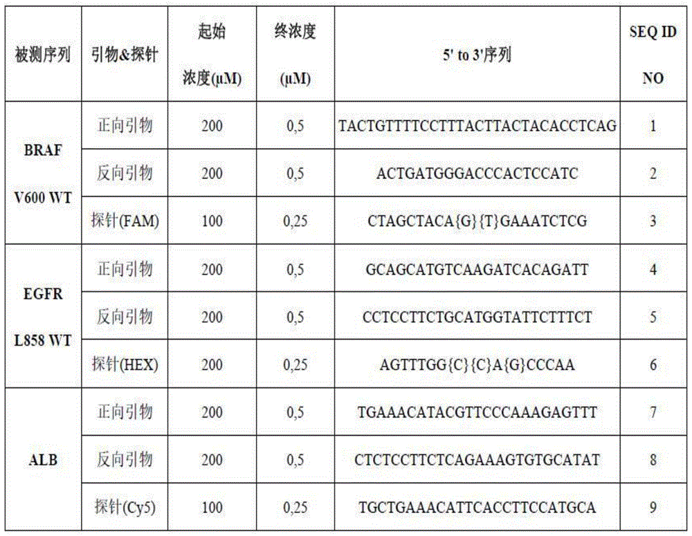
PCR mixtures
Use of PCR reaction Multiplex qPCR
 (Quanta Biosciences, Beverly, MA, USA) at a final concentration of 1X. Add 0.1. mu.M fluorescein (V) to the PCR mixtureWR International,Fontenay-sous-Bois,France)。
The PCR mixture was assembled as follows:
· qPCR Multiplex
 (1X)
(1X)
fluorescein (0.1. mu.M)
Oligonucleotide BRAF V600 WT, FAM fluorophore (1X)
Oligonucleotide EGFR L858 WT, HEX fluorophore (1X)
Oligonucleotide ALB, Cy5 fluorophore (1X)
Water
TABLE 2

Samples were obtained by diluting the target sequences in the PCR mixture, so the expected final concentration of each target sequence was 3000 cp/. mu.l in the non-sonicated case, as described in table 2.
dPCR experiment
Samples were loaded into the inlet chambers of a Sapphire chip (Stilla Technologies, Villejuif, France), each chamber loaded with a volume of 27 μ Ι _ volume. Three replicates (three chambers per sample) were used per sonicated sample. One non-sonicated sample was loaded in triplicate into three separate chambers.
NaicaTMGeode (Stilla Technologies, Villejuif, France) is programmed to partition a sample.
The PCR conditions were as follows: 10 minutes at 95 ℃ followed by 45 cycles of 30 seconds at 95 ℃ and 15 seconds at 58 ℃.
Using NaicaTMThe default exposure times for image acquisition by Prism3(Stilla Technologies, Villejuif, France) were 65ms, 250ms, and 50ms for the blue, green, and red channels, respectively.
f. Correction factor calculation
The predicted correction factor ("prediction") according to the method of the invention is calculated from the experimentally measured fragment length distribution of the sample (length distribution centered around 150bp as shown in FIG. 2) and the length of the base pairs of the sequence to be amplified (bp).
The calculation of the correction factor according to the method of the invention requires the probability that the sequence to be amplified is not cleaved as a function of the length of the sequence to be amplified (a distribution of lengths centered around 150bp as shown in FIG. 3). The correction factors under the same conditions are shown in fig. 4.
A true value correction factor ("true value") is obtained in vitro by calculating the ratio between the experimentally measured concentration of the measured sequence and the sonicated sample in the non-sonicated sample.
Relative error ("relative error") is defined as the error of a predicted correction factor relative to a true correction factor.
Results for example 1:
the experimental and theoretical results are compared in table 3. The predictive and true correction factors were obtained by TriPlex digital PCR experiments measuring the concentration of sequences to be amplified of different sequence lengths (78bp, 81bp, 117bp) in sonicated samples of different fragment lengths (150bp, 200bp, 350bp, 550 bp). Each experimental measurement was performed in triplicate and the values shown are the average of triplicate values.
TABLE 3

It can be seen that the relative error values obtained are in the range of 1% to 7%, indicating that the predicted correction factor is always accurate and that there is a slight overestimation of the correction factor with respect to the true value.
From the above it can be concluded that the method of the invention provides directly usable results under realistic conditions, since the length of the sequence to be amplified represents the standard PCR sequence, and since the fragment length distribution conforms to the naturally occurring fragmentation.
Sequence listing
<110> Statla technologies
Ba La Ande Lie
Ramie Dangla
<120> determination of the concentration of nucleic acid sequences
<130> CV - 1302/PCT
<150> EP19306346.8
<151> 2019-10-16
<160> 9
<170> BiSSAP 1.3.6
<210> 1
<211> 30
<212> DNA
<213> Artificial sequence
<220>
<223> BRAF V600 WT-forward primer
<400> 1
tactgttttc ctttacttac tacacctcag 30
<210> 2
<211> 21
<212> DNA
<213> Artificial sequence
<220>
<223> BRAF V600 WT-reverse primer
<400> 2
actgatggga cccactccat c 21
<210> 3
<211> 20
<212> DNA
<213> Artificial sequence
<220>
<223> BRAF V600 WT-probe (FAM)
<220>
<221> modified_base
<222> 10
<223> /mod_base="OTHER"
/note = "locked nucleic acid (LAN) base"
<220>
<221> modified_base
<222> 11
<223> /mod_base="OTHER"
/note = "locked nucleic acid (LAN) base"
<400> 3
<210> 4
<211> 23
<212> DNA
<213> Artificial sequence
<220>
<223> EGFR L858 WT-forward primer
<400> 4
gcagcatgtc aagatcacag att 23
<210> 5
<211> 25
<212> DNA
<213> Artificial sequence
<220>
<223> EGFR L858 WT-reverse primer
<400> 5
cctccttctg catggtattc tttct 25
<210> 6
<211> 16
<212> DNA
<213> Artificial sequence
<220>
<223> EGFR L858 WT-probe (HEX)
<220>
<221> modified_base
<222> 8
<223> /mod_base="OTHER"
/note = "locked nucleic acid (LAN) base"
<220>
<221> modified_base
<222> 9
<223> /mod_base="OTHER"
/note = "locked nucleic acid (LAN) base"
<220>
<221> modified_base
<222> 11
<223> /mod_base="OTHER"
/note = "locked nucleic acid (LAN) base"
<400> 6
agtttggcca gcccaa 16
<210> 7
<211> 25
<212> DNA
<213> Artificial sequence
<220>
<223> ALB-Forward primer
<400> 7
tgaaacatac gttcccaaag agttt 25
<210> 8
<211> 26
<212> DNA
<213> Artificial sequence
<220>
<223> ALB-reverse primer
<400> 8
ctctccttct cagaaagtgt gcatat 26
<210> 9
<211> 25
<212> DNA
<213> Artificial sequence
<220>
<223> ALB-Probe (Cy5)
<400> 9
tgctgaaaca ttcaccttcc atgca 25
Claims (15)
1. A method of determining the concentration of a sequence under test in an unfragmented nucleic acid, the method comprising the steps of:
i. determining a Length Distribution (LD) of nucleic acids in a sample comprising fragmented nucleic acids, wherein the fragmented nucleic acids are derived from the non-fragmented nucleic acids;
measuring the concentration of the sequenced sequence in the sample comprising fragmented nucleic acids with a measurement method; and
correcting the measured concentration of the sequence in the sample comprising fragmented nucleic acids with a correction factor to obtain the concentration of the sequence in non-fragmented nucleic acids, wherein the correction factor is based on the Length Distribution (LD) and at least one parameter of the measurement method.
2. The method of claim 1, wherein measuring the concentration of the sequence being measured comprises amplifying a sequence to be amplified comprising the sequence being measured, and the at least one parameter of the measurement method comprises the length (L) of the sequence to be amplifieda)。
3. The method of claim 2, wherein the correction coefficient is determined by:

wherein L is the length of the sequence to be amplified (L)a);
Wherein f (i) is the probability that the fragment length in said sample is i base pairs; and
wherein Is the average length of the nucleic acid fragments.
4. The method of claim 2, wherein the correction is applied if at least 5% of the nucleic acids in the sample are shorter in length than the sequence to be amplified.
5. The method of claim 2, wherein the sequence to be amplified is greater than 40bp and less than 200bp in length.
6. The method of claim 1, wherein the Length Distribution (LD) of nucleic acid fragments in the sample is comprised in the range of 25bp to 350 bp.
7. The method of any one of the preceding claims, wherein the method does not comprise obtaining or predicting genetic coordinates of the fragmented nucleic acids.
8. The method of claim 1, wherein the measurement method is an isothermal quantitative nucleic acid amplification method.
9. The method of claim 1, wherein the measurement method is a non-isothermal quantitative nucleic acid amplification method.
10. The method of claim 1, wherein the method further comprises:
i. measuring a concentration of a second sequence to be detected in the sample comprising fragmented nucleic acids with a measurement method, wherein measuring the concentration of the second sequenced column comprises amplifying a second sequence to be amplified comprising the second sequenced column; and
correcting the measured concentration of the second detected sequence in the fragmented nucleic acid with a correction factor to obtain the concentration of the second detected sequence in the non-fragmented nucleic acid, wherein the correction factor is based on the Length Distribution (LD) and at least one parameter of the measurement method;
wherein the length of the first sequence to be amplified is different from the length of the second sequence to be amplified.
11. The method of claim 10, wherein the measurement method comprises a multiplex amplification step.
12. A system configured to determine a concentration of a detected sequence in an unfragmented nucleic acid, the system comprising:
i. an amplification module configured to measure a concentration of the sequenced sequence in a sample comprising fragmented nucleic acids, the fragmented nucleic acids derived from the non-fragmented nucleic acids;
a module configured to calculate a correction factor based on a Length Distribution (LD) of nucleic acids in the sample and the measured at least one parameter; and
a module configured to calculate a concentration of the sequenced sequence in unfragmented nucleic acid using the correction factor.
13. The system configured to determine the concentration of a detected sequence in an unfragmented nucleic acid according to claim 12, wherein the module configured to measure the concentration of the detected sequence in the fragmented nucleic acid is an isothermal quantitative nucleic acid amplification module selected from a loop-mediated isothermal amplification module and a quantitative nucleic acid sequence based amplification module.
14. The system configured to determine the concentration of a detected sequence in an unfragmented nucleic acid according to claim 12, wherein the module configured to measure the concentration of the detected sequence in the fragmented nucleic acid is a non-isothermal quantitative nucleic acid amplification module selected from a quantitative polymerase chain reaction module, a real-time polymerase chain reaction module, a digital polymerase chain reaction module, a multiplex polymerase chain reaction module, and a multiplex digital polymerase chain reaction module.
15. The system configured to determine the concentration of a detected sequence in an unfragmented nucleic acid according to claim 12, wherein the measured parameter comprises a length of a sequence to be amplified.
Applications Claiming Priority (3)
| Application Number | Priority Date | Filing Date | Title |
|---|---|---|---|
| EP19306346.8 | 2019-10-16 | ||
| EP19306346 | 2019-10-16 | ||
| PCT/EP2020/079219 WO2021074382A1 (en) | 2019-10-16 | 2020-10-16 | Determination of nucleic acid sequence concentrations |
Publications (2)
| Publication Number | Publication Date |
|---|---|
| CN114585749A true CN114585749A (en) | 2022-06-03 |
| CN114585749B CN114585749B (en) | 2024-09-03 |
Family
ID=68581672
Family Applications (1)
| Application Number | Title | Priority Date | Filing Date |
|---|---|---|---|
| CN202080073038.5A Active CN114585749B (en) | 2019-10-16 | 2020-10-16 | Determination of nucleic acid sequence concentration |
Country Status (8)
| Country | Link |
|---|---|
| US (1) | US20240093281A1 (en) |
| EP (2) | EP4640845A3 (en) |
| JP (1) | JP2022552544A (en) |
| KR (1) | KR20220097894A (en) |
| CN (1) | CN114585749B (en) |
| AU (1) | AU2020366567A1 (en) |
| CA (1) | CA3153350A1 (en) |
| WO (1) | WO2021074382A1 (en) |
Citations (12)
| Publication number | Priority date | Publication date | Assignee | Title |
|---|---|---|---|---|
| US20040219565A1 (en) * | 2002-10-21 | 2004-11-04 | Sakari Kauppinen | Oligonucleotides useful for detecting and analyzing nucleic acids of interest |
| JP2008178338A (en) * | 2007-01-24 | 2008-08-07 | Aisin Seiki Co Ltd | Nucleic acid amplification method for amplifying target nucleic acid in nucleic acid sample contaminated with fragmented nucleic acid, and kit thereof |
| CN101971178A (en) * | 2007-07-23 | 2011-02-09 | 香港中文大学 | Determination of Nucleic Acid Sequence Imbalances |
| CN103180460A (en) * | 2010-09-23 | 2013-06-26 | 凯杰有限公司 | Method for detecting and/or quantifying human DNA |
| EP2868752A1 (en) * | 2013-10-31 | 2015-05-06 | Lexogen GmbH | Nucleic acid copy number determination based on fragment estimates |
| CN107075564A (en) * | 2014-12-10 | 2017-08-18 | 深圳华大基因研究院 | The method and apparatus for determining tumour nucleic acid concentration |
| EP3293270A1 (en) * | 2015-05-06 | 2018-03-14 | Annoroad Gene Technology | Reagent kit, apparatus, and method for detecting chromosome aneuploidy |
| CN107868816A (en) * | 2016-09-23 | 2018-04-03 | 豪夫迈·罗氏有限公司 | The method for determining the amount of nucleic acid interested in untreated sample |
| CN109112209A (en) * | 2017-06-25 | 2019-01-01 | 国家卫生计生委科学技术研究所 | Reference material for noninvasive antenatal detection fetal aneuploidy chromosome |
| CN109312400A (en) * | 2016-03-25 | 2019-02-05 | 凯锐思公司 | Synthetic nucleic acid inclusions |
| EP3460071A1 (en) * | 2017-09-22 | 2019-03-27 | Lexogen GmbH | Estimating pre-pcr fragment numbers from post-pcr frequencies of unique molecular identifiers |
| WO2019178157A1 (en) * | 2018-03-16 | 2019-09-19 | Karius, Inc. | Sample series to differentiate target nucleic acids from contaminant nucleic acids |
Family Cites Families (3)
| Publication number | Priority date | Publication date | Assignee | Title |
|---|---|---|---|---|
| US6303305B1 (en) * | 1999-03-30 | 2001-10-16 | Roche Diagnostics, Gmbh | Method for quantification of an analyte |
| US9892230B2 (en) * | 2012-03-08 | 2018-02-13 | The Chinese University Of Hong Kong | Size-based analysis of fetal or tumor DNA fraction in plasma |
| US12492430B2 (en) * | 2017-04-11 | 2025-12-09 | Tecan Genomics, Inc. | Library quantitation and qualification |
-
2020
- 2020-10-16 EP EP25186796.6A patent/EP4640845A3/en active Pending
- 2020-10-16 AU AU2020366567A patent/AU2020366567A1/en not_active Abandoned
- 2020-10-16 JP JP2022522786A patent/JP2022552544A/en active Pending
- 2020-10-16 KR KR1020227015017A patent/KR20220097894A/en active Pending
- 2020-10-16 US US17/768,820 patent/US20240093281A1/en active Pending
- 2020-10-16 EP EP20792650.2A patent/EP4045681B1/en active Active
- 2020-10-16 CA CA3153350A patent/CA3153350A1/en active Pending
- 2020-10-16 WO PCT/EP2020/079219 patent/WO2021074382A1/en not_active Ceased
- 2020-10-16 CN CN202080073038.5A patent/CN114585749B/en active Active
Patent Citations (12)
| Publication number | Priority date | Publication date | Assignee | Title |
|---|---|---|---|---|
| US20040219565A1 (en) * | 2002-10-21 | 2004-11-04 | Sakari Kauppinen | Oligonucleotides useful for detecting and analyzing nucleic acids of interest |
| JP2008178338A (en) * | 2007-01-24 | 2008-08-07 | Aisin Seiki Co Ltd | Nucleic acid amplification method for amplifying target nucleic acid in nucleic acid sample contaminated with fragmented nucleic acid, and kit thereof |
| CN101971178A (en) * | 2007-07-23 | 2011-02-09 | 香港中文大学 | Determination of Nucleic Acid Sequence Imbalances |
| CN103180460A (en) * | 2010-09-23 | 2013-06-26 | 凯杰有限公司 | Method for detecting and/or quantifying human DNA |
| EP2868752A1 (en) * | 2013-10-31 | 2015-05-06 | Lexogen GmbH | Nucleic acid copy number determination based on fragment estimates |
| CN107075564A (en) * | 2014-12-10 | 2017-08-18 | 深圳华大基因研究院 | The method and apparatus for determining tumour nucleic acid concentration |
| EP3293270A1 (en) * | 2015-05-06 | 2018-03-14 | Annoroad Gene Technology | Reagent kit, apparatus, and method for detecting chromosome aneuploidy |
| CN109312400A (en) * | 2016-03-25 | 2019-02-05 | 凯锐思公司 | Synthetic nucleic acid inclusions |
| CN107868816A (en) * | 2016-09-23 | 2018-04-03 | 豪夫迈·罗氏有限公司 | The method for determining the amount of nucleic acid interested in untreated sample |
| CN109112209A (en) * | 2017-06-25 | 2019-01-01 | 国家卫生计生委科学技术研究所 | Reference material for noninvasive antenatal detection fetal aneuploidy chromosome |
| EP3460071A1 (en) * | 2017-09-22 | 2019-03-27 | Lexogen GmbH | Estimating pre-pcr fragment numbers from post-pcr frequencies of unique molecular identifiers |
| WO2019178157A1 (en) * | 2018-03-16 | 2019-09-19 | Karius, Inc. | Sample series to differentiate target nucleic acids from contaminant nucleic acids |
Non-Patent Citations (1)
| Title |
|---|
| TUERK A等: "Mixture models reveal multiple positional bias types in RNA-Seq data and lead to accurate transcript concentration estimates", 《PLOS COMPUT BIOL》, vol. 13, no. 5, pages 1 - 25 * |
Also Published As
| Publication number | Publication date |
|---|---|
| EP4640845A3 (en) | 2025-12-31 |
| EP4640845A2 (en) | 2025-10-29 |
| EP4045681A1 (en) | 2022-08-24 |
| EP4045681B1 (en) | 2025-07-16 |
| WO2021074382A1 (en) | 2021-04-22 |
| US20240093281A1 (en) | 2024-03-21 |
| AU2020366567A1 (en) | 2022-06-02 |
| JP2022552544A (en) | 2022-12-16 |
| CN114585749B (en) | 2024-09-03 |
| CA3153350A1 (en) | 2021-04-22 |
| KR20220097894A (en) | 2022-07-08 |
Similar Documents
| Publication | Publication Date | Title |
|---|---|---|
| AU2022202625B2 (en) | Methods and compositions for rapid nucleic acid library preparation | |
| EP2802666B1 (en) | Genotyping by next-generation sequencing | |
| US8940487B2 (en) | Methods and compositions for universal detection of nucleic acids | |
| US10501784B2 (en) | Sequence amplification with linear primers | |
| JP7602464B2 (en) | Quantitative amplicon sequencing for multiple copy number variation detection and allelic ratio quantification | |
| US20240229123A1 (en) | Multiplex nucleic acid detection method, composition and kit | |
| CN113832258A (en) | RNA amplification and detection using attenuated probes | |
| CN115996938A (en) | Quantitative Blocker Displacement Amplification (QBDA) sequencing for calibration-free and multiplex variant allele frequency quantification | |
| JP7036820B2 (en) | Detection of sequence variants | |
| WO2012032510A1 (en) | Primers for amplifying dna and methods of selecting same | |
| CN114585749B (en) | Determination of nucleic acid sequence concentration | |
| JP2026016403A (en) | Determination of nucleic acid sequence concentration | |
| CN114250273A (en) | Composition for nucleic acid detection | |
| US20250122586A1 (en) | Compositions and methods for the detection of h3n2 influenza variants | |
| HK40064558A (en) | Compositions for rapid nucleic acid library preparation |
Legal Events
| Date | Code | Title | Description |
|---|---|---|---|
| PB01 | Publication | ||
| PB01 | Publication | ||
| SE01 | Entry into force of request for substantive examination | ||
| SE01 | Entry into force of request for substantive examination | ||
| GR01 | Patent grant | ||
| GR01 | Patent grant |