CN114072175A - Materials and methods for treating tumors - Google Patents
Materials and methods for treating tumors Download PDFInfo
- Publication number
- CN114072175A CN114072175A CN202080047643.5A CN202080047643A CN114072175A CN 114072175 A CN114072175 A CN 114072175A CN 202080047643 A CN202080047643 A CN 202080047643A CN 114072175 A CN114072175 A CN 114072175A
- Authority
- CN
- China
- Prior art keywords
- ctla4
- amino acid
- antibody
- binding
- ctla
- Prior art date
- Legal status (The legal status is an assumption and is not a legal conclusion. Google has not performed a legal analysis and makes no representation as to the accuracy of the status listed.)
- Pending
Links
Images


Classifications
-
- C—CHEMISTRY; METALLURGY
- C07—ORGANIC CHEMISTRY
- C07K—PEPTIDES
- C07K16/00—Immunoglobulins [IGs], e.g. monoclonal or polyclonal antibodies
- C07K16/18—Immunoglobulins [IGs], e.g. monoclonal or polyclonal antibodies against material from animals or humans
- C07K16/28—Immunoglobulins [IGs], e.g. monoclonal or polyclonal antibodies against material from animals or humans against receptors, cell surface antigens or cell surface determinants
- C07K16/2803—Immunoglobulins [IGs], e.g. monoclonal or polyclonal antibodies against material from animals or humans against receptors, cell surface antigens or cell surface determinants against the immunoglobulin superfamily
- C07K16/2818—Immunoglobulins [IGs], e.g. monoclonal or polyclonal antibodies against material from animals or humans against receptors, cell surface antigens or cell surface determinants against the immunoglobulin superfamily against CD28 or CD152
-
- A—HUMAN NECESSITIES
- A61—MEDICAL OR VETERINARY SCIENCE; HYGIENE
- A61P—SPECIFIC THERAPEUTIC ACTIVITY OF CHEMICAL COMPOUNDS OR MEDICINAL PREPARATIONS
- A61P35/00—Antineoplastic agents
-
- A—HUMAN NECESSITIES
- A61—MEDICAL OR VETERINARY SCIENCE; HYGIENE
- A61K—PREPARATIONS FOR MEDICAL, DENTAL OR TOILETRY PURPOSES
- A61K39/00—Medicinal preparations containing antigens or antibodies
- A61K2039/505—Medicinal preparations containing antigens or antibodies comprising antibodies
-
- A—HUMAN NECESSITIES
- A61—MEDICAL OR VETERINARY SCIENCE; HYGIENE
- A61K—PREPARATIONS FOR MEDICAL, DENTAL OR TOILETRY PURPOSES
- A61K39/00—Medicinal preparations containing antigens or antibodies
- A61K2039/545—Medicinal preparations containing antigens or antibodies characterised by the dose, timing or administration schedule
-
- C—CHEMISTRY; METALLURGY
- C07—ORGANIC CHEMISTRY
- C07K—PEPTIDES
- C07K2299/00—Coordinates from 3D structures of peptides, e.g. proteins or enzymes
-
- C—CHEMISTRY; METALLURGY
- C07—ORGANIC CHEMISTRY
- C07K—PEPTIDES
- C07K2317/00—Immunoglobulins specific features
- C07K2317/20—Immunoglobulins specific features characterized by taxonomic origin
- C07K2317/24—Immunoglobulins specific features characterized by taxonomic origin containing regions, domains or residues from different species, e.g. chimeric, humanized or veneered
-
- C—CHEMISTRY; METALLURGY
- C07—ORGANIC CHEMISTRY
- C07K—PEPTIDES
- C07K2317/00—Immunoglobulins specific features
- C07K2317/30—Immunoglobulins specific features characterized by aspects of specificity or valency
- C07K2317/33—Crossreactivity, e.g. for species or epitope, or lack of said crossreactivity
-
- C—CHEMISTRY; METALLURGY
- C07—ORGANIC CHEMISTRY
- C07K—PEPTIDES
- C07K2317/00—Immunoglobulins specific features
- C07K2317/50—Immunoglobulins specific features characterized by immunoglobulin fragments
- C07K2317/52—Constant or Fc region; Isotype
-
- C—CHEMISTRY; METALLURGY
- C07—ORGANIC CHEMISTRY
- C07K—PEPTIDES
- C07K2317/00—Immunoglobulins specific features
- C07K2317/50—Immunoglobulins specific features characterized by immunoglobulin fragments
- C07K2317/56—Immunoglobulins specific features characterized by immunoglobulin fragments variable (Fv) region, i.e. VH and/or VL
- C07K2317/567—Framework region [FR]
-
- C—CHEMISTRY; METALLURGY
- C07—ORGANIC CHEMISTRY
- C07K—PEPTIDES
- C07K2317/00—Immunoglobulins specific features
- C07K2317/50—Immunoglobulins specific features characterized by immunoglobulin fragments
- C07K2317/56—Immunoglobulins specific features characterized by immunoglobulin fragments variable (Fv) region, i.e. VH and/or VL
- C07K2317/569—Single domain, e.g. dAb, sdAb, VHH, VNAR or nanobody®
-
- C—CHEMISTRY; METALLURGY
- C07—ORGANIC CHEMISTRY
- C07K—PEPTIDES
- C07K2317/00—Immunoglobulins specific features
- C07K2317/70—Immunoglobulins specific features characterized by effect upon binding to a cell or to an antigen
- C07K2317/71—Decreased effector function due to an Fc-modification
-
- C—CHEMISTRY; METALLURGY
- C07—ORGANIC CHEMISTRY
- C07K—PEPTIDES
- C07K2317/00—Immunoglobulins specific features
- C07K2317/70—Immunoglobulins specific features characterized by effect upon binding to a cell or to an antigen
- C07K2317/73—Inducing cell death, e.g. apoptosis, necrosis or inhibition of cell proliferation
- C07K2317/732—Antibody-dependent cellular cytotoxicity [ADCC]
-
- C—CHEMISTRY; METALLURGY
- C07—ORGANIC CHEMISTRY
- C07K—PEPTIDES
- C07K2317/00—Immunoglobulins specific features
- C07K2317/70—Immunoglobulins specific features characterized by effect upon binding to a cell or to an antigen
- C07K2317/76—Antagonist effect on antigen, e.g. neutralization or inhibition of binding
-
- C—CHEMISTRY; METALLURGY
- C07—ORGANIC CHEMISTRY
- C07K—PEPTIDES
- C07K2317/00—Immunoglobulins specific features
- C07K2317/70—Immunoglobulins specific features characterized by effect upon binding to a cell or to an antigen
- C07K2317/77—Internalization into the cell
-
- C—CHEMISTRY; METALLURGY
- C07—ORGANIC CHEMISTRY
- C07K—PEPTIDES
- C07K2317/00—Immunoglobulins specific features
- C07K2317/90—Immunoglobulins specific features characterized by (pharmaco)kinetic aspects or by stability of the immunoglobulin
- C07K2317/92—Affinity (KD), association rate (Ka), dissociation rate (Kd) or EC50 value
Landscapes
- Health & Medical Sciences (AREA)
- Chemical & Material Sciences (AREA)
- Immunology (AREA)
- Organic Chemistry (AREA)
- Life Sciences & Earth Sciences (AREA)
- General Health & Medical Sciences (AREA)
- Medicinal Chemistry (AREA)
- Nuclear Medicine, Radiotherapy & Molecular Imaging (AREA)
- Biochemistry (AREA)
- Animal Behavior & Ethology (AREA)
- General Chemical & Material Sciences (AREA)
- Public Health (AREA)
- Veterinary Medicine (AREA)
- Chemical Kinetics & Catalysis (AREA)
- Pharmacology & Pharmacy (AREA)
- Biophysics (AREA)
- Genetics & Genomics (AREA)
- Molecular Biology (AREA)
- Proteomics, Peptides & Aminoacids (AREA)
- Peptides Or Proteins (AREA)
- Medicines Containing Antibodies Or Antigens For Use As Internal Diagnostic Agents (AREA)
- Medicines That Contain Protein Lipid Enzymes And Other Medicines (AREA)
Abstract
The present application discloses methods and uses of CTLA4 binding proteins for treating tumors, wherein the CTLA4 binding proteins specifically bind CTLA4 and do not compete with ligands of CTLA4 for binding to the CTLA 4. The CTLA4 binding proteins described herein are effective in inhibiting tumor growth.
Description
The application relates to the field of biomedicine, in particular to a method and application for treating tumors by using CTLA4 binding protein.
In tumor immune response, the inhibition mechanism mediated by CTLA4 is often one of the reasons for tumor cells to escape from the immune system, so CTLA4 is a key target for tumor immunotherapy. Currently approved CTLA antibodies (e.g., lpilimumab) exert anti-tumor effects by blocking the interaction of CTLA-4 with its ligand. The binding of currently approved CTLA antibodies to CTLA4 protein may reduce the level of CTLA4 protein on the cell membrane surface by endocytic degradation, which may, however, result in a decrease in ADCC activity.
There remains a need for CTLA4 antibodies that exert anti-tumor effects through other mechanisms to overcome potential problems of poor therapeutic efficacy, immunotoxicity or drug resistance.
Disclosure of Invention
The application provides an application of CTLA4 conjugated protein in preparing a medicament for treating tumors. CTLA 4-binding proteins described herein have one or more of the following properties: 1) is capable of specifically binding CTLA4, but does not substantially compete with ligands of CTLA4 for binding to the CTLA 4; 2) does not substantially compete with ipilimumab for binding to CTLA 4; 3) (ii) capable of recognizing or binding to one or more amino acids in the ABEDC "strand, the A' B loop, the C" D loop, the DE loop and/or the EF loop of human CTLA 4; 4) does not compete with molecules that recognize or bind to amino acid residues K95, E97, M99, Y104, L106, and/or I108 in human CTLA4 for binding to the CTLA 4; 5) capable of inhibiting tumor or tumor cell growth/proliferation; 6) the endocytic degradation of CTLA4 is not triggered, and the endocytic degradation rate of CTLA4 on the surface of 293 cell membrane expressing CTLA4 is lower than about 50%.
In one aspect, the present application provides the use of a CTLA4 binding protein in the manufacture of a medicament for the treatment of a tumor, wherein the CTLA4 binding protein specifically binds CTLA4 and does not compete with ligands of CTLA4 for binding to the CTLA 4.
In certain embodiments, the CTLA 4-binding protein does not block binding of CTLA4 to its ligand.
In certain embodiments, the ligand of CTLA4 is selected from CD80 or a functional fragment thereof and CD86 or a functional fragment thereof.
In certain embodiments, the CTLA4 binding protein does not compete with a reference antibody for binding to the CTLA4, wherein the reference antibody is ipilimumab.
In certain embodiments, the CTLA4 binding protein binds to human CTLA4 and monkey CTLA 4.
In certain embodiments, wherein the CTLA4 binding protein binds to or recognizes the abcdd "strand, a' B, of human CTLA4
One or more amino acids in the loop, C "D loop, DE loop, and/or EF loop.
In certain embodiments, the CTLA4 binding protein binds or recognizes one or more amino acids in the C' chain, C "D loop, and/or DE loop of human CTLA 4.
In certain embodiments, the CTLA4 binding protein recognizes or binds to amino acid residues in human CTLA4 selected from the group consisting of: e48, V49, D65, L84, D88 and Y92. In certain embodiments, the CTLA4 binding protein also recognizes or binds to amino acid residues in human CTLA4 selected from the group consisting of: v46, T47, D64, I67 and G83.
In certain embodiments, the CTLA4 binding proteins recognize or bind a conformational epitope in human CTLA4 comprising one or more of the following amino acid residues: v46, T47, E48, V49, D64, D65, I67, G83, L84, D88 and Y92. In certain embodiments, the CTLA4 binding protein recognizes or binds a conformational epitope in human CTLA4 comprising the following amino acid residues: e48, V49, D65, L84, D88 and Y92.
In certain embodiments, the CTLA4 binding protein does not compete for binding to the CTLA4 with molecules that recognize or bind to one or more of the following amino acid residues in human CTLA 4: m3, K95, E97, M99, Y104, L106 and I108.
In certain embodiments, the CTLA4 binding protein does not compete for binding to the CTLA4 with molecules that recognize or bind to one or more of the following amino acid residues in human CTLA 4: k95, E97, M99, Y104, L106 and I108.
In certain embodiments, the CTLA4 binding protein does not compete with molecules that recognize or bind one or more amino acids in the F chain, FG loop, and/or G chain of CTLA4 for binding to the CTLA 4.
In certain embodiments, the CTLA4 binding protein comprises at least one CDR in the VH having the amino acid sequence shown in SEQ ID No. 7. In certain embodiments, the CTLA 4-binding protein comprises HCDR3 in the VH having the amino acid sequence shown in SEQ ID No. 7. In certain embodiments, the CTLA 4-binding protein comprises HCDR2 in the VH having the amino acid sequence shown in SEQ ID No. 7. In certain embodiments, the CTLA 4-binding protein comprises HCDR1 in the VH having the amino acid sequence shown in SEQ ID No. 7.
In certain embodiments, the CTLA4 binding protein is an antibody or antigen-binding fragment thereof and comprises an antibody heavy chain CDR3, which heavy chain CDR3 comprises the amino acid sequence set forth in SEQ ID No. 1. In certain embodiments, the CTLA4 binding protein is an antibody or antigen-binding fragment thereof and comprises an antibody heavy chain CDR3, which heavy chain CDR3 comprises the amino acid sequence set forth in SEQ ID No. 2.
In certain embodiments, the CTLA4 binding protein is an antibody or antigen-binding fragment thereof and comprises an antibody heavy chain CDR2, which heavy chain CDR2 comprises the amino acid sequence set forth in SEQ ID No. 3. In certain embodiments, the CTLA4 binding protein is an antibody or antigen-binding fragment thereof and comprises an antibody heavy chain CDR2, which heavy chain CDR2 comprises the amino acid sequence set forth in SEQ ID No. 4.
In certain embodiments, the CTLA4 binding protein is an antibody or antigen-binding fragment thereof and comprises an antibody heavy chain CDR1, which heavy chain CDR1 comprises the amino acid sequence set forth in SEQ ID No. 5. In certain embodiments, the CTLA4 binding protein is an antibody or antigen-binding fragment thereof and comprises an antibody heavy chain CDR1, which heavy chain CDR1 comprises the amino acid sequence set forth in SEQ ID No. 6.
In certain embodiments, the CTLA4 binding protein is an antibody or antigen-binding fragment thereof, and comprises antibody heavy chain FR1, which heavy chain FR1 comprises the amino acid sequence set forth in SEQ ID No. 27. In certain embodiments, the CTLA4 binding protein is an antibody or antigen-binding fragment thereof and comprises antibody heavy chain FR1, which heavy chain FR1 comprises the amino acid sequence set forth in any one of SEQ ID NOs 21 and 25.
In certain embodiments, the CTLA4 binding protein is an antibody or antigen-binding fragment thereof, and comprises antibody heavy chain FR2, which heavy chain FR2 comprises the amino acid sequence set forth in SEQ ID No. 28. In certain embodiments, the CTLA4 binding protein is an antibody or antigen-binding fragment thereof and comprises antibody heavy chain FR2, which heavy chain FR2 comprises the amino acid sequence set forth in any one of SEQ ID NOs 22 and 26.
In certain embodiments, the CTLA4 binding protein is an antibody or antigen-binding fragment thereof, and comprises antibody heavy chain FR3, which heavy chain FR3 comprises the amino acid sequence shown in SEQ ID No. 33. In certain embodiments, the CTLA4 binding protein is an antibody or antigen-binding fragment thereof and comprises antibody heavy chain FR3, which heavy chain FR3 comprises the amino acid sequence set forth in any one of SEQ ID NOs 23 and 35.
In certain embodiments, the CTLA4 binding protein is an antibody or antigen-binding fragment thereof, and comprises antibody heavy chain FR4, which heavy chain FR4 comprises the amino acid sequence shown in SEQ ID No. 34. In certain embodiments, the CTLA4 binding protein is an antibody or antigen-binding fragment thereof and comprises antibody heavy chain FR4, which heavy chain FR4 comprises the amino acid sequence set forth in any one of SEQ ID NOs 24 and 36.
In certain embodiments, the CTLA 4-binding protein is an antibody or antigen-binding fragment thereof, and comprises a heavy chain variable region comprising the amino acid sequence set forth in SEQ ID No. 7. In certain embodiments, the CTLA 4-binding protein is an antibody or antigen-binding fragment thereof, and comprises a heavy chain variable region comprising the amino acid sequence set forth in any one of SEQ ID NOs 8 and 17-20.
In certain embodiments, the CTLA4 binding protein comprises a single domain antibody or antigen-binding fragment thereof.
In certain embodiments, the CTLA4 binding protein comprises a single domain antibody or antigen-binding fragment thereof, and the single domain antibody or antigen-binding fragment thereof comprises the amino acid sequence set forth in SEQ ID No. 7. In certain embodiments, the CTLA 4-binding protein comprises a single domain antibody or antigen-binding fragment thereof, and the single domain antibody or antigen-binding fragment thereof comprises the amino acid sequence set forth in any one of SEQ ID NOs 8 and 17-20.
In certain embodiments, the CTLA 4-binding protein has a property selected from the group consisting of: a) at 2.6X10⁸K of M or lessDValues bind to human CTLA 4; b) inhibiting tumor cell growth; and c) causing less than about 50% endocytic degradation of CTLA4 protein expressed on the cell membrane.
In certain embodiments, the CTLA 4-binding protein further comprises an antibody Fc domain. In certain embodiments, the antibody Fc domain comprises an Fc domain derived from an IgG antibody. In certain embodiments, the CTLA 4-binding protein comprises the antibody Fc domain and the antibody heavy chain variable region, and the antibody heavy chain variable region is fused, directly or indirectly, to the antibody Fc domain.
In another aspect, the present application provides a pharmaceutical composition comprising a CTLA4 binding protein described herein, and optionally a pharmaceutically acceptable carrier.
In certain embodiments, the CTLA 4-binding protein does not block binding of CTLA4 to its ligand.
In certain embodiments, the ligand of CTLA4 is selected from CD80 or a functional fragment thereof and CD86 or a functional fragment thereof.
In certain embodiments, the CTLA4 binding protein does not compete with a reference antibody for binding to the CTLA4, wherein the reference antibody is ipilimumab.
In certain embodiments, the CTLA4 binding protein binds to human CTLA4 and monkey CTLA 4.
In certain embodiments, the CTLA4 binding protein binds or recognizes one or more amino acids in the C' chain, C "D loop, and/or DE loop of human CTLA 4.
In certain embodiments, the CTLA4 binding protein recognizes or binds to amino acid residues in human CTLA4 selected from the group consisting of: e48, V49, D65, L84, D88 and Y92. In certain embodiments, the CTLA4 binding protein also recognizes or binds to amino acid residues in human CTLA4 selected from the group consisting of: v46, T47, D64, I67 and G83.
In certain embodiments, the CTLA4 binding proteins recognize or bind a conformational epitope in human CTLA4 comprising one or more of the following amino acid residues: v46, T47, E48, V49, D64, D65, I67, G83, L84, D88 and Y92. In certain embodiments, the CTLA4 binding protein recognizes or binds a conformational epitope in human CTLA4 comprising the following amino acid residues: e48, V49, D65, L84, D88 and Y92.
In certain embodiments, the CTLA4 binding protein does not compete for binding to the CTLA4 with molecules that recognize or bind to one or more of the following amino acid residues in human CTLA 4: m3, K95, E97, M99, Y104, L106 and I108.
In certain embodiments, the CTLA4 binding protein does not compete for binding to the CTLA4 with molecules that recognize or bind to one or more of the following amino acid residues in human CTLA 4: k95, E97, M99, Y104, L106 and I108.
In certain embodiments, the CTLA4 binding protein does not compete for binding to the CTLA4 with molecules that recognize or bind one or more amino acids in CTLA4F chain, FG loop, and/or G chain.
In certain embodiments, the CTLA4 binding protein is an antibody or antigen-binding fragment thereof and comprises an antibody heavy chain CDR3, which heavy chain CDR3 comprises the amino acid sequence set forth in SEQ ID No. 1. In certain embodiments, the CTLA4 binding protein is an antibody or antigen-binding fragment thereof and comprises an antibody heavy chain CDR3, which heavy chain CDR3 comprises the amino acid sequence set forth in SEQ ID No. 2.
In certain embodiments, the CTLA4 binding protein is an antibody or antigen-binding fragment thereof and comprises an antibody heavy chain CDR2, which heavy chain CDR2 comprises the amino acid sequence set forth in SEQ ID No. 3. In certain embodiments, the CTLA4 binding protein is an antibody or antigen-binding fragment thereof and comprises an antibody heavy chain CDR2, which heavy chain CDR2 comprises the amino acid sequence set forth in SEQ ID No. 4.
In certain embodiments, the CTLA4 binding protein is an antibody or antigen-binding fragment thereof and comprises an antibody heavy chain CDR1, which heavy chain CDR1 comprises the amino acid sequence set forth in SEQ ID No. 5. In certain embodiments, the CTLA4 binding protein is an antibody or antigen-binding fragment thereof and comprises an antibody heavy chain CDR1, which heavy chain CDR1 comprises the amino acid sequence set forth in SEQ ID No. 6.
In certain embodiments, the CTLA4 binding protein is an antibody or antigen-binding fragment thereof, and comprises antibody heavy chain FR1, which heavy chain FR1 comprises the amino acid sequence set forth in SEQ ID No. 27. In certain embodiments, the CTLA4 binding protein is an antibody or antigen-binding fragment thereof and comprises antibody heavy chain FR1, which heavy chain FR1 comprises the amino acid sequence set forth in any one of SEQ ID NOs 21 and 25.
In certain embodiments, the CTLA4 binding protein is an antibody or antigen-binding fragment thereof, and comprises antibody heavy chain FR2, which heavy chain FR2 comprises the amino acid sequence set forth in SEQ ID No. 28. In certain embodiments, the CTLA4 binding protein is an antibody or antigen-binding fragment thereof and comprises antibody heavy chain FR2, which heavy chain FR2 comprises the amino acid sequence set forth in any one of SEQ ID NOs 22 and 26.
In certain embodiments, the CTLA4 binding protein is an antibody or antigen-binding fragment thereof, and comprises antibody heavy chain FR3, which heavy chain FR3 comprises the amino acid sequence shown in SEQ ID No. 33. In certain embodiments, the CTLA4 binding protein is an antibody or antigen-binding fragment thereof and comprises antibody heavy chain FR3, which heavy chain FR3 comprises the amino acid sequence set forth in any one of SEQ ID NOs 23 and 35.
In certain embodiments, the CTLA4 binding protein is an antibody or antigen-binding fragment thereof, and comprises antibody heavy chain FR4, which heavy chain FR4 comprises the amino acid sequence shown in SEQ ID No. 34. In certain embodiments, the CTLA4 binding protein is an antibody or antigen-binding fragment thereof and comprises antibody heavy chain FR4, which heavy chain FR4 comprises the amino acid sequence set forth in any one of SEQ ID NOs 24 and 36.
In certain embodiments, the CTLA 4-binding protein is an antibody or antigen-binding fragment thereof, and comprises a heavy chain variable region comprising the amino acid sequence set forth in SEQ ID No. 7. In certain embodiments, the CTLA 4-binding protein is an antibody or antigen-binding fragment thereof, and comprises a heavy chain variable region comprising the amino acid sequence set forth in any one of SEQ ID NOs 8 and 17-20.
In certain embodiments, the CTLA4 binding protein comprises a single domain antibody or antigen-binding fragment thereof.
In certain embodiments, the CTLA4 binding protein comprises a single domain antibody or antigen-binding fragment thereof, and the single domain antibody or antigen-binding fragment thereof comprises the amino acid sequence set forth in SEQ ID No. 7. In certain embodiments, the CTLA 4-binding protein comprises a single domain antibody or antigen-binding fragment thereof, and the single domain antibody or antigen-binding fragment thereof comprises the amino acid sequence set forth in any one of SEQ ID NOs 8 and 17-20.
In certain embodiments, the CTLA 4-binding protein has a property selected from the group consisting of: a) at 2.6X10⁸K of M or lessDValues bind to human CTLA 4; b) inhibiting tumor cell growth; and c) allowing less than about 50% degradation of CTLA4 at the surface of 293 cell membranes expressing CTLA 4.
In certain embodiments, the CTLA 4-binding protein further comprises an antibody Fc domain. In certain embodiments, the antibody Fc domain comprises an Fc domain derived from an IgG antibody. In certain embodiments, the CTLA 4-binding protein comprises the antibody Fc domain and an antibody heavy chain variable region, and the antibody heavy chain variable region is fused, directly or indirectly, to the antibody Fc domain.
In another aspect, the present application provides a pharmaceutical composition (e.g., a pharmaceutical composition for treating a tumor) comprising a CTLA 4-binding protein described herein.
In another aspect, the present application provides the use of the pharmaceutical composition for the preparation of a medicament for the treatment of a tumor.
In certain embodiments, the tumor is a solid tumor. In certain embodiments, the tumor is selected from: colon cancer and melanoma.
In another aspect, the present application provides a CTLA 4-binding protein or the pharmaceutical composition described herein for use in treating a tumor. In certain embodiments, the tumor is a solid tumor.
In another aspect, the present application provides a method of treating a tumor comprising the steps of: administering to a subject in need thereof a CTLA 4-binding protein described herein or a pharmaceutical composition described herein. In certain embodiments, the tumor is a solid tumor.
Other aspects and advantages of the present disclosure will be readily apparent to those skilled in the art from the following detailed description. Only exemplary embodiments of the present disclosure have been shown and described in the following detailed description. As those skilled in the art will recognize, the disclosure enables those skilled in the art to make changes to the specific embodiments disclosed without departing from the spirit and scope of the invention as claimed in the present application. Accordingly, the descriptions in the drawings and the specification of the present application are illustrative only and not limiting.
The specific features of the invention to which this application relates are set forth in the appended claims. The features and advantages of the invention to which this application relates will be better understood by reference to the exemplary embodiments described in detail below and the accompanying drawings. The brief description of the drawings is as follows:
figure 1 shows the binding affinity of CTLA4 binding proteins described herein.
Figure 2 shows the effect of CTLA4 binding proteins described herein on binding to cell surface CTLA 4.
Figures 3A-3B show the blocking effect of CTLA4 binding proteins described herein on binding of cell surface CTLA4 to its ligands.
Figure 4 shows the blocking effect of CTLA4 binding proteins described herein on binding of CTLA4 to CD 80.
Figures 5A-5B show the effect of CTLA4 binding proteins described herein in blocking binding of CTLA4 to its ligand.
Figures 6A-6E show the results of CTLA4 binding proteins described herein competing with B7-1 for binding to human CTLA 4.
Figures 7A-7B show the species specificity of CTLA4 binding proteins binding CTLA4 as described herein.
Figures 8A-8C show the activation effect of CTLA4 binding proteins on PBMCs as described herein.
Figure 9 shows the anti-tumor effect of CTLA4 binding proteins described herein in mice.
Figure 10 shows the effect of CTLA4 binding proteins described herein on inhibiting tumor recurrence in mice.
Figure 11 shows the anti-tumor effect of CTLA4 binding proteins described herein in mice.
Figure 12 shows the anti-tumor effect of CTLA4 binding proteins described herein.
Figures 13A-13C show the effect of CTLA4 binding proteins on T cell population ratios as described herein.
Figures 14A-14F show binding of CTLA4 binding proteins described herein to human CTLA 4.
Figures 15A-15D show the structure of CTLA4 binding proteins described herein bound to human CTLA4 to form complexes.
Figure 16 shows the CTLA4 binding proteins stimulating upregulation of IL12 expression levels as described herein.
Figure 17 shows key epitopes of CTLA4 binding proteins described herein that bind to human CTLA 4.
Figure 18 shows the decrease in the amount of CTLA4 protein on the cell membrane surface caused by endocytic degradation of CTLA 4-binding proteins described herein.
The embodiments of the present invention are described below with reference to specific embodiments, and other advantages and effects of the present invention will be easily understood by those skilled in the art from the disclosure of the present specification.
In this application, the term "treating" generally refers to preventing, alleviating, and/or reversing one or more symptoms of a disease or disorder, and may also refer to inhibiting or arresting the recurrence of the disease or disorder. In some cases, the treatment can be prophylactic (e.g., can prevent or inhibit the onset of a disease, or prevent the occurrence of a clinical phenomenon or subclinical symptom thereof), or can therapeutically inhibit or alleviate the symptoms after the occurrence of a disease.
In this application, the term "not block" generally refers to not substantially preventing or destroying the performance or effect of the associated function or process. For example, "not blocking" an interaction between a protein (e.g., CTLA4) and another protein (e.g., CTLA4 ligand, such as CD80 or CD86) generally means not substantially preventing or disrupting the interaction therebetween. In some instances, the "not substantially prevent or destroy" may refer to a reduction or decrease in the relevant function/process of no more than about 35%, no more than about 30%, or less, as compared to normal. The normal condition may be without any intervention (e.g., without administration of any CTLA 4-binding protein of the present application).
In the present application, the term "CTLA 4" generally refers to cytotoxic T-lymphocyte-associated protein 4 (also known as CTLA-4 or CD152), which is a protein receptor that is an immune checkpoint and regulates downstream immune responses. CTLA4 is an immunosuppressive receptor belonging to the CD28 family. In vivo, CTLA4 is only on T cells (CD 4)+And CD8+Cells) and binds to both ligands CD80 and CD86 (also referred to as B7-1 and B7-2, respectively). For example, the term "CTLA 4" can include a polypeptide or fragment thereof that has at least about 85% amino acid sequence identity to NCBI accession number AAL07473.1 and that specifically binds CD80 and/or CD 86. The term "CTLA 4" includes the work of the entire CTLA4 receptor, its extracellular domain, and CTLA4 comprising a covalently linked second moiety, e.g., a protein domainA fusion protein capable of active moiety. Also included within the definition of CTLA4 are variants that differ from the amino acid sequence of naturally occurring CTLA4 but retain the ability to specifically bind ligands CD80 and/or CD 86. CTLA4 sequences are known in the art and are provided, for example, in accession No. 1493. The term "CTLA 4" as used herein includes human CTLA4(hCTLA4), variants, isomers, and species homologs of hCTLA4, and analogs having at least one common epitope of hCTLA 4. For example, the term "CTLA 4" also encompasses CTLA4 from other species such as other mammals (e.g., rat, mouse, rabbit, non-human primate, pig, or cow). The complete hCTLA4 sequence can be found in GenBank accession No. 1493.
In the present application, the term "C' chain" generally refers to a portion of the 3-dimensional structure of human CTLA 4. For example, the C' chain can comprise amino acid residues 45-54 of human CTLA4 (e.g., the amino acid sequence of which can be set forth in SEQ ID NO: 14).
In the present application, the term "C" D ring "generally refers to a portion of the 3-dimensional structure of human CTLA 4. For example, the C "D loop can comprise amino acid residues 50-60, 67-72 of human CTLA4 (e.g., the amino acid sequence of which can be set forth in SEQ ID NO: 14).
In the present application, the term "DE loop" generally refers to a portion of the 3-dimensional structure of human CTLA 4. For example, the DE loop may comprise amino acid residues 67-81 of human CTLA4 (e.g., the amino acid sequence of which may be set forth in SEQ ID NO: 14).
In the present application, the term "N-terminus" generally refers to the N-terminus of the amino acid sequence of a protein/polypeptide. For example, the N-terminus may be the start site for protein/polypeptide synthesis. For example, the N-terminus may be the N-terminus of the primary structure of human CTLA 4.
In the present application, the term "F chain" generally refers to a portion of the 3-dimensional structure of human CTLA 4. For example, the F chain can comprise amino acid residues 90-99 of human CTLA4 (e.g., the amino acid sequence can be set forth in SEQ ID NO: 14).
In the present application, the term "FG loop" generally refers to a portion of the 3-dimensional structure of human CTLA 4. For example, the FG loop may be referred to as a CDR 3-like fragment. The FG loop may comprise amino acid residues 105-109, 112-115 of human CTLA4 (e.g., the amino acid sequence of which can be set forth in SEQ ID NO: 14).
In the present application, the term "G chain" generally refers to a portion of the 3-dimensional structure of human CTLA 4. For example, the G chain may comprise CTLA4 (e.g., the amino acid sequence of which is shown in SEQ ID NO:14) at position 105-109; 112-115 amino acid residue.
The Structural information of CTLA4 can be found in the Structural basis for co-stimulation by the human CTLA-4/B7-2 complex (Nature 2001, Schwartz et. Al.) and the 3D structure of the crystalline structure of the B7-1/CTLA-4 complex of the human immune responses (Nature 2001, Stamper et. al.) CTLA4 and its ligands are recorded in the database of the Protein Bank (PDB), with the respective accession numbers: 1I85 and 1I 8L.
In the present application, the term "CTLA 4-binding protein" generally refers to a protein that can specifically bind CTLA 4. For example, the CTLA4 binding protein can comprise or be an antibody or antigen-binding fragment thereof.
In the present application, the term "specific binding" generally refers to the binding of a binding molecule (e.g., the CTLA4 binding protein) to a target molecule (e.g., CLTA4), and the binding is distinct from a non-specific interaction (e.g., the non-specific interaction can include binding of the CTLA4 binding protein to non-CTLA 4, e.g., to bovine serum albumin or casein). In the present application, specific binding may be determined by binding of the binding molecule to the target molecule relative to binding of the binding molecule to a control molecule (other than the target molecule). In certain instances, the binding specificity may refer to the CTLA4 binding protein specifically binding to a particular epitope on CTLA4, and not substantially binding to any other protein or other epitope thereof.
In the present application, the term "binding affinity" generally refers to the strength of the sum of the non-covalent interactions between a single binding site of a binding molecule (e.g. the CTLA4 binding protein) and its bound target molecule (e.g. CTLA 4). For example, the binding affinityThe force may reflect the inherent binding affinity of the line interaction between the binding molecule and the target molecule. In some cases, the level of binding affinity can be determined by Biacore, KinExA, or Fortibio methods. For example, the binding molecules with higher affinity can remain bound for longer periods of time and may be more difficult to dissociate. In the present application, the CTLA4 binding protein can be expressed as 2.6x10⁸K of M or lessDValues bind to human CTLA 4.
In the present application, the term "monkey" generally refers to animals belonging to the order primates, latissimaurida and stenotinales. In the present application, the monkey may be a cynomolgus monkey (Macaca fascicularis) or a rhesus monkey (Macaca mulatta). For example, the monkey may be a cynomolgus monkey (Macaca fascicularis).
In the present application, the term "rat" generally refers to an animal belonging to the rodent superfamily. In the present application, the mouse may be a mouse (Mus musculus) or a rat (Rattus norvegicus f. For example, the rat may be a rat.
In the present application, the term "ipilimumab" generally refers to the human IgG1k monoclonal antibody, which specifically binds to the extracellular region of human CTLA-4. The trade name is (Bristol-Myers Squibb), which the FDA has approved in 2011 for use in the treatment of melanoma. The ipilimumab can block the binding of human CTLA-4 and the ligand B7-1 thereof. Specific structures of the complex formed by binding ipilimumab and human CTLA-4 can be found in Ramagopal UA et al, Structural basis for cancer immunological by the first-in-class checkpoint inhibitor ipilimumab, Proc. The structure and amino acid sequence information of Ipilimumab can be found in Lipson EJ, etc., an anti-CTLA-4 anti for metabolic melanoma, Clin Cancer Res.2011
 Nov 15; 17(22) 6958-62; or see antibody 10D1 in U.S. patent application US 20020086041.
In the present application, the term "KN 044" generally refers to a CTLA4 binding protein comprising an immunoglobulin single variable domain that specifically binds CTLA 4. The KN044 can block binding of CTLA4 to CD80, and has high binding affinity to CTLA 4. The KN044 may comprise the amino acid sequence shown in SEQ ID NO. 11.
In the present application, the term "endocytosis" generally refers to the process by which a cell membrane transfers extracellular substances into a cell by deforming movement. The endocytosis may comprise phagocytosis and pinocytosis. For example, a cell may dent into a "vesicle" that encapsulates the macromolecular species (e.g., protein), yet leaves the cell membrane and enters the interior of the cell.
In the present application, the term "single domain antibody" generally refers to an antigen binding domain or fragment, such as a VHH domain or a VH domain or a VL domain, respectively. The terms antigen binding molecule or antigen binding protein are used interchangeably and also include the term Nanobodies. The immunoglobulin single variable domain is further a light chain variable domain sequence (e.g., a VL-sequence), or a heavy chain variable domain sequence (e.g., a VH-sequence); more particularly, they may be heavy chain variable domain sequences derived from conventional four-chain antibodies or heavy chain variable domain sequences derived from heavy chain antibodies. Thus, the immunoglobulin single variable domain may be a domain antibody, or an immunoglobulin sequence suitable for use as a domain antibody, a single domain antibody, or an immunoglobulin sequence suitable for use as a single domain body, a "dAbs" or an immunoglobulin sequence suitable for use as a dAbs, or a nanobody, including but not limited to a VHH sequence. Immunoglobulin single variable domains include fully human, humanized, other sequence-optimized or chimeric immunoglobulin sequences. The structure of the immunoglobulin single variable domain and immunoglobulin single variable domain may be considered-but not limited to-consists of four framework regions or "FRs", which are referred to in the art and herein as "framework region 1" or "FR 1", respectively; "framework region 2" or "FR 2"; or "framework region 3" or "FR 3"; and "framework region 4" or "FR 4"; wherein the framework regions are inserted into three complementarity determining regions or "CDRs," which are referred to in the art as "complementarity determining region 1" or "CDR 1," respectively; "complementarity determining region 2" or "CDR 2"; and "complementarity determining region 3" or "CDR 3".
In the present application, the term "Fc" generally refers to an Fc region from the constant region of human IgG1, IgG2, IgG3, or IgG4 (the constant region amino acid sequences of which are referenced to entries P01857, P01859, P01860, P01861 in the www.uniprot.org protein database, respectively), which comprises the hinge or partial hinge region of an immunoglobulin constant region, a CH2 region, and a CH3 region. Herein, the amino acid sequence of the "human immunoglobulin Fc region" can be obtained by mutating 1 to 5 amino acids of the CH2 region to increase or remove Fc-mediated ADCC or CDC activity or to enhance or diminish the affinity of FcRn; or the stability of the protein is increased by mutating 1-4 amino acids of the Hinge region.
In the present application, the term "epitope" generally refers to the site on an antigen to which an antibody binds. Epitopes can be formed by contiguous amino acids (linear epitopes) or non-contiguous amino acids that are spatially adjacent due to tertiary folding of the protein (conformational epitopes). Epitopes formed by consecutive amino acids are generally retained when exposed to denaturing solvents, whereas epitopes formed by tertiary folding are generally lost when treated with denaturing solvents. Epitopes generally comprise at least 3, usually more, at least 5 or 8-10 amino acids in a unique spatial conformation. Methods for determining the spatial conformation of an epitope include, for example, X-ray crystallography and 2-dimensional nuclear magnetic resonance. See, e.g., epitopic Mapping Protocols in Methods in Molecular Biology, Vol.66, Glenn E.Morris, Ed (1996). The term "conformational epitope" as used herein generally refers to non-contiguous amino acid residues of an antigen that are adjacent in spatial position due to tertiary folding of the protein, such as the CTLA4 antigen. These non-contiguous amino acid residues can accumulate on the surface when the polypeptide chain is folded to form the native protein. Conformational epitopes include, but are not limited to, functional epitopes.
In this application, the term "KD"used interchangeably with" KD "generally refers to the dissociation equilibrium constant, in M (mol/L), of a particular antibody-antigen interaction. KD can be calculated from the concentrations of substance AB and substance a and substance B resulting from dissociation thereof: KD ═ c (a) × (b)/c (ab). ByThe formula shows that the larger the KD value is, the more dissociation is shown, and the weaker the affinity between the substances A, B is represented; conversely, a smaller KD value indicates less dissociation and a stronger affinity between the substances A, B.
In the present application, the term "about" generally means varying from 0.5% to 10% above or below the stated value, for example, varying from 0.5%, 1%, 1.5%, 2%, 2.5%, 3%, 3.5%, 4%, 4.5%, 5%, 5.5%, 6%, 6.5%, 7%, 7.5%, 8%, 8.5%, 9%, 9.5%, or 10% above or below the stated value.
Pharmaceutical application of CTLA4 binding protein
In one aspect, the present application provides the use of a CTLA4 binding protein in the manufacture of a medicament for the treatment of a tumor, wherein the CTLA4 binding protein specifically binds CTLA4 and does not compete with ligands of CTLA4 for binding to the CTLA 4.
In the present application, the CTLA 4-binding proteins can specifically bind CTLA 4. For example, the CTLA4 binding protein can be found to bind specifically only to CTLA4, as detected by ELISA. For example, the CTLA 4-binding protein binds to K in CTLA4 as measured by surface plasmon resonance (SRP) methodDThe value may be about 2.8 x10⁻⁸About 2.6x10⁻⁸About 2.5 x10⁻⁸About 2.0 x10⁻⁸Below or lower. For example, the CTLA 4-binding protein can bind CTLA4 with an EC50 value of about 2 or more, about 2.5 or more, about 3 or more, about 3.5 or more, or higher, as detected by ELISA.
In the present application, the methods of detecting that the CTLA4 binding protein specifically binds to CTLA4 comprise Surface Plasmon Resonance (SPR) assays, Scatchard assays, competitive binding assays (e.g., Radioimmunoassays (RIA), Enzyme Immunoassays (EIA), and/or sandwich competitive assays.
In the present application, the CTLA 4-binding proteins may not block binding of CTLA4 to its ligand.
In the present application, the determination of whether binding of CTLA4 to its ligand is blocked can be made by a method selected from the group consisting of: competitive ELISA, cell neutralization assays, and Biofilm Layer Interference (BLI) methods. For example, the CTLA4 binding protein competes with CD80 for binding to CTLA4 in experiments using CTLA4 binding protein at a concentration between about 0.002 and about 2000 times the concentration of CD80 protein (e.g., between about 50ng/mL for CD80 and between about 0.01ng/mL and 100 μ g/mL for CTLA4 binding protein) or between about 0.002 and about 500 times the concentration of CD86 protein (e.g., between about 50ng/mL for CD86 and between about 0.01ng/mL and about 25 μ g/mL for CTLA4 binding protein) as determined by competition ELISA, resulting in less than about 30% decrease in absorbance. As another example, the CTLA4 binding protein competes with human CD80 or about 1.6 μ g/mL of human CD86 for binding to HEK293 cells expressing human CTLA4 at a concentration range of about 0.01 to about 10000nM as determined by competitive FACS resulting in a decrease in mean fluorescence value of less than about 30%.
In the present application, the term "substantially non-blocking" may refer to the inability to observe a significant concentration-dependent blocking effect on the interaction of CTLA4 with its ligand (CD80 or CD86) within a concentration range of about 0.01 to about 100000 ng/mL. For example, the binding of the CTLA4 binding protein to CTLA4 at a concentration range of about 0.01 to about 10000ng/mL had less than about 30% impact on binding of CTLA4 to its ligand CD80 or CD86 (as measured by a change in OD value in an ELISA assay, or alternatively, as measured by a change in MFI in a FACS assay), and no apparent concentration dependence. In some cases, the "non-blocking" may also be confirmed by studies of epitopes. For example, the epitope recognized by the CTLA 4-binding protein does not include the epitope to which CLTA4 binds to its ligand (CD80 or CD 86); or the epitope recognized by the binding protein of CTLA4 does not overlap with the epitope bound by CLTA4 and its ligand (CD80 or CD 86); or CTLA4 binding proteins do not recognize the epitope to which CLTA4 binds to each other's ligand (CD80 or CD 86).
In the present application, the ligand of CTLA4 may be selected from CD80 or a functional fragment thereof and CD86 or a functional fragment thereof.
In the present application, the functional fragment may be a mutant or fragment of a protein having the biological activity of CD80 or CD 86. For example, the functional fragment of CD80 may have at least one functional region of CD 80. The functional region of CD80 may refer to the entire protein region of CD80 or a partial region that retains full domain activity. For example, the functional region of CD80 may comprise the functional V region of CD80 (amino acids 43-123) or a fragment thereof, and has the ability to bind CD28 and CTLA 4; and/or, the functional region of CD80 may comprise the entire extracellular domain (amino acids 35-242) of CD80 or a fragment thereof. For example, the functional fragment of CD86 may have at least one functional region of CD 86. The CD86 functional region may comprise the V region of CD86 (amino acids 33-131), the whole extracellular domain of CD86 (amino acids 24-274), etc. The amino acid codes of the above functional regions of CD80 and CD86 are referred to the information in the Uniprot database. Wherein the accession number of CD80 in Uniprot is P33681, and the accession number of CD86 is P42081.
In the present application, the CTLA4 binding proteins may not compete with a reference antibody for binding to the CTLA4, wherein the reference antibody may be selected from ipilimumab and KN 044.
In the present application, binding of the CTLA4 binding protein to the CTLA4 may not be affected by binding of the reference antibody to the CTLA 4. In certain instances, the CTLA4 binding protein can bind to a certain position/epitope on the CTLA4, while the reference antibody can bind to another position/epitope on the CTLA4 that is different from the aforementioned position/epitope. For example, the CTLA4 binding proteins described herein may be positioned differently from the CTLA4 to which ipilimumab binds, respectively, and the CTLA4 binding proteins described herein may not compete with ipilimumab for binding to the CTLA 4. For another example, the CTLA 4-binding protein described herein may bind to the CTLA4 at a different position from that of KN044, and the CTLA 4-binding protein described herein may not compete with KN044 for binding to the CTLA 4.
In the present application, the CTLA4 binding proteins can bind to human CTLA4 and monkey CTLA 4. In the present application, the CTLA4 binding protein can not substantially bind to mouse CTLA4 or rat CTLA 4. In the present application, the binding of the CTLA4 binding protein to CTLA4 can be species-specific.
In the present application, the CTLA4 binding proteins can bind to or recognize one or more amino acids in the abcd "strand, a' B loop, C" D loop, DE loop, and/or EF loop of human CTLA 4.
In the present application, the CTLA4 binding proteins can bind to or recognize one or more amino acids in the C' chain, C "D loop, and/or DE loop of human CTLA 4.
For example, the CTLA4 binding protein can bind to or recognize one or more amino acids in the C' chain of human CTLA 4: e48, V49, V46 and T47.
For example, the CTLA4 binding protein can bind to or recognize amino acid I67 in the C "D loop of human CTLA 4.
In the present application, the Protein 3D structure of human CTLA4 can be found in the "structure" section of the UniProt database, and the accession numbers of the Protein of human CTLA4 in the Protein Data Bank (PDB) database are: 1I 85.
For example, in the present application, the CTLA4 binding protein can recognize or bind to amino acid residues in human CTLA4 selected from the group consisting of: e48, V49, D65, L84, D88 and Y92. In the present application, the CTLA4 binding proteins can also recognize or bind to amino acid residues in human CTLA4 selected from the group consisting of: v46, T47, D64, I67 and G83.
For example, in the present application, the CTLA4 binding proteins can recognize or bind a conformational epitope in human CTLA4 comprising one or more of the following amino acid residues: v46, T47, E48, V49, D64, D65, I67, G83, L84, D88 and Y92.
In the present application, the CTLA4 binding proteins can recognize or bind to a conformational epitope in human CTLA4 comprising the following amino acid residues: e48, V49, D65, L84, D88 and Y92. In the present application, mutation of one or more of amino acid residues E48, V49, D65, L84, D88 and Y92 of human CTLA4 may result in the CTLA4 binding protein losing the ability to bind specifically to human CTLA4 (e.g., K which may allow binding of the CTLA4 binding protein to the mutated human CTLA4)DUp-regulation of the value by at least about 800-fold, at least about 1000-fold, at least about 1200-fold, or more). In the present application, the CTLA4 binding proteins specifically recognize and/or are associated with the conformational epitopes in human CTLA4In combination, mutation of any one or more of the amino acid residues E48, V49, D65, L84, D88 and Y92 in human CTLA4 can result in a change in the conformation of the conformational epitope.
In the present application, the CTLA4 binding protein may not compete for binding to the CTLA4 with molecules that recognize or bind to one or more of the following amino acid residues in human CTLA 4: k95, E97, M99, Y104, L106 and I108.
In the present application, the CTLA4 binding proteins may not compete with molecules that recognize or bind one or more amino acids in the F chain, FG loop, and/or G chain of CTLA4 for binding to the CTLA 4.
In the present application, the recognition or binding site of the CTLA4 binding protein to CTLA4 may not be located at the F chain, FG loop, and/or G chain of the CTLA 4. For example, the CTLA4 binding protein may not bind to one or more amino acids in the F chain, FG loop, and/or G chain of CTLA 4. Binding of the other CTLA 4-binding proteins to CTLA4 does not affect binding of CTLA 4-binding proteins described herein to CTLA4 if the other CTLA 4-binding proteins recognize or bind one or more amino acids in the F chain, FG loop, and/or G chain of CTLA 4.
For example, the CTLA4 binding protein may not bind to or recognize one or more amino acids in the F chain of human CTLA 4: k95, E97 and M99. For example, the CTLA4 binding protein may not bind to or recognize one or more amino acids in the FG loop of human CTLA 4: l106 and I108.
In the present application, the CTLA 4-binding protein may comprise at least one CDR in the VH having an amino acid sequence shown in SEQ ID NO. 7.
QVQLVESGGGX 1VQX 2GGSLRLSCAASX 3X 4X 5NX 6X 7X 8X 9MGWFRQAPGKX 10X 11ERVAAX 12X 13X 14X 15GTX 16X 17YYADSVKGRFTISRDNX 18KNTLX 19LQMNSLX 20X 21EDTAX 22YYCX 23X 24X 25X 26X 27X 28X 29AWCX 30RX 31X 32X 33X 34X 35X 36X 37WGQGTX 38VTVSX 39(SEQ ID NO:7) wherein X1Is S or L; x2Is A or P; x10Is E or G; x11Is R or L; x18Is G or S; x19Is F or Y; x20Is K or R; x21Is P or A; x22Is M or V; x38Is Q or L; x39Is S or no amino acid; the remaining X is any amino acid.
In the present application, the CTLA 4-binding protein may comprise the VH of SEQ ID NO:7 as described above, as defined by any method, as the CDR: at least one (e.g., 1, 2, or 3) of HCDR1, HCDR2, and HCDR 3. For example, the CTLA 4-binding protein may comprise at least one (e.g., 1, 2 or 3) of the VH shown in SEQ ID NO:8 as defined by any method for HCDR1, HCDR2 and HCDR 3.
For example, the CTLA4 binding protein may comprise HCDR3 in the VH amino acid sequence shown as SEQ ID NO: 7. In certain instances, the CTLA 4-binding protein can comprise HCDR3 in the VH having the amino acid sequence shown in SEQ ID No. 8. For example, the CTLA4 binding protein may comprise HCDR2 in the VH amino acid sequence shown as SEQ ID NO: 7. In certain instances, the CTLA 4-binding protein can comprise HCDR3 in the VH having the amino acid sequence shown in SEQ ID No. 8. For example, the CTLA4 binding protein may comprise HCDR1 in the VH amino acid sequence shown as SEQ ID NO: 7. In certain instances, the CTLA 4-binding protein can comprise HCDR3 in the VH having the amino acid sequence shown in SEQ ID No. 8.
In the present application, the above-mentioned at least one CDR in the VH having an amino acid sequence shown in SEQ ID NO. 7 or at least one CDR in the VH having an amino acid sequence shown in SEQ ID NO. 8, wherein the CDR partitioning method may be any method known in the art for CDR partitioning.
For example, the VH domains may be numbered according to the general numbering scheme given by Kabat et al ("Sequence of proteins of immunological interest", US Public Health Services, NIH Bethesda, Md., Pub. No. 91), according to which,
FR1 contains the amino acid residues at positions 1-30,
-CDR1 comprises amino acid residues at positions 31-35,
FR2 contains the amino acids at positions 36-49,
-CDR2 comprises amino acid residues at positions 50-65,
FR3 contains the amino acid residues at positions 66-94,
-CDR3 comprises amino acid residues at positions 95 to 102, and
-FR4 comprises the amino acid residue at position 103-113.
CDRs of antibodies can also be defined in the art by a variety of methods, such as 1) Kabat definition rules based on sequence variability (Wu and Kabat, J Exp Med 132:211-50, 1970; kabat et al, Sequences of proteins of Immunological Interest, 5 th edition, Public Health Service, National Institutes of Health, Bethesda, Md., 1991), 2) Chothia definition rules based on the position of the structural loop regions (A1-Lazikani et al, Jmol Biol 273:927-48, 1997), 3) AbM definition rules that weigh the two above rules with the AbM antibody model software of Oxford Molecular, 4) Contact definition rules based on the resulting crystal structure analysis of the complex. The division of the CDR amino acid origin-destination sites of the CTLA4 binding proteins described herein using these methods can be as follows:
| kabat definition | Definition of AbM | Chothia definition | Contact definition | |
| Heavy chain CDR1 | H31-H35 | H26-H35 | H26-H32 | H30-H35 |
| Heavy chain CDR2 | H50-H65 | H50-H58 | H52-H56 | H47-H58 |
| Heavy chain CDR3 | H95-H102 | H95-H102 | H95-H102 | H93-H101 |
Wherein, Ha-Hb may refer to an amino acid sequence from the a-th to the b-th position from the N-terminus of the heavy chain of the antibody. For example, H31-H35 may refer to the amino acid sequence from position 31 to position 35 in the Kabat numbering of the heavy chain of an antibody, after numbering according to the Kabat numbering convention.
In the present application, the CTLA4 binding protein can comprise or be an antibody or antigen-binding fragment thereof and comprises an antibody heavy chain CDR3, which heavy chain CDR3 can comprise the amino acid sequence set forth in SEQ ID No. 1. X1X 2X 3X 4X 5X 6X 7AWCX 8RX 9X 10X 11X 12X 13X 14X 15(SEQ ID NO:1), wherein X1-X 12Are all arbitrary amino acids.
In the present application, the CTLA4 binding protein can comprise or be an antibody or antigen-binding fragment thereof and comprises an antibody heavy chain CDR3, which heavy chain CDR3 can comprise the amino acid sequence shown in SEQ ID No. 2.
In the present application, the CTLA4 binding protein can comprise or be an antibody or antigen-binding fragment thereof and comprises an antibody heavy chain CDR2, which heavy chain CDR2 can comprise the amino acid sequence set forth in SEQ ID No. 3. X1X 2X 3X 4GTX 5X 6(SEQ ID NO:3) wherein X1-X 6Are all arbitrary amino acids.
In the present application, the CTLA4 binding protein can comprise or be an antibody or antigen-binding fragment thereof and comprises an antibody heavy chain CDR2, which heavy chain CDR2 can comprise the amino acid sequence set forth in SEQ ID No. 4.
In the present application, the CTLA4 binding protein can comprise or be an antibody or antigen-binding fragment thereof and comprises an antibody heavy chain CDR1, which heavy chain CDR1 can comprise the amino acid sequence set forth in SEQ ID No. 5. X1 X 2 X 3N X 4 X 5 X 6 X 7(SEQ ID NO:5) in which X1-X 7Are all arbitrary amino acids.
In the present application, the CTLA4 binding protein can comprise or be an antibody or antigen-binding fragment thereof and comprises an antibody heavy chain CDR1, which heavy chain CDR1 can comprise the amino acid sequence set forth in SEQ ID No. 6.
In the present application, the CTLA 4-binding protein can comprise the structure shown below: FR1-CDR1-FR2-CDR2-FR3-CDR3-FR 4.
In the present application, the structures and numbering according to the numbering scheme described above may or may not also correspond to the actual numbering of the amino acid residues in the actual sequence.
In the present application, the CTLA 4-binding protein can comprise or be an antibody or antigen-binding fragment thereof, and its heavy chain CDR1 can comprise the amino acid sequence shown in SEQ ID No. 6, heavy chain CDR2 can comprise the amino acid sequence shown in SEQ ID No. 4, and heavy chain CDR3 can comprise the amino acid sequence shown in SEQ ID No. 2.
In the present application, the CTLA4 binding protein can comprise or be an antibody or antigen-binding fragment thereof, and comprises the antibody heavy chain FR1, which heavy chain FR1 can comprise the amino acid sequence shown in SEQ ID No. 27. QVQLVESGGG X1VQ X 2GGSLRLSCAAS (SEQ ID NO:27), wherein X1Is S or L; x2Is A or P.
In the present application, the heavy chain FR1 may be located N-terminal to the heavy chain CDR 1. For example, the C-terminus of the heavy chain FR1 can be directly linked to the N-terminus of the heavy chain CDR 1.
For example, the CTLA4 binding protein can comprise or be an antibody or antigen-binding fragment thereof, and comprises antibody heavy chain FR1, which heavy chain FR1 can comprise the amino acid sequence set forth in any one of SEQ ID NOs 21 and 25.
In the present application, the CTLA4 binding protein can comprise or be an antibody or antigen-binding fragment thereof, and can comprise the antibody heavy chain FR2, which heavy chain FR2 comprises the amino acid sequence shown in SEQ ID No. 28. MGWFRQAPGKX1X 2ERVAA (SEQ ID NO:28), wherein X1Is E or G; x2Is R or L.
In the present application, the heavy chain FR2 may be located between the heavy chain CDR1 and the heavy chain CDR 2. For example, the N-terminus of the heavy chain FR2 can be directly linked to the C-terminus of the heavy chain CDR 1; and the C-terminus of the heavy chain FR2 can be directly linked to the N-terminus of the heavy chain CDR 2.
For example, the CTLA4 binding protein can comprise or be an antibody or antigen-binding fragment thereof, and can comprise the antibody heavy chain FR2, which heavy chain FR2 comprises the amino acid sequence set forth in any one of SEQ ID NOs 22 and 26.
In the present application, the CTLA4 binding protein can comprise or be an antibody or antigen-binding fragment thereof, and can comprise the antibody heavy chain FR3, which heavy chain FR3 comprises the amino acid sequence shown in SEQ ID No. 33. YYADSVKGRFTISRDN X1 KNTL X 2 LQMNSL X 3 X 4 EDTA X 5YYC, wherein X1Is G or S; x2Is F or Y; x3Is K or R; x4Is P or A; x5Is M or V.
In the present application, the heavy chain FR3 may be located between the heavy chain CDR2 and the heavy chain CDR 3. For example, the N-terminus of the heavy chain FR3 can be directly linked to the C-terminus of the heavy chain CDR 2; and the C-terminus of the heavy chain FR3 can be directly linked to the N-terminus of the heavy chain CDR 3.
For example, the CTLA4 binding protein can comprise or be an antibody or antigen-binding fragment thereof, and can comprise the antibody heavy chain FR3, which heavy chain FR3 comprises the amino acid sequence set forth in any one of SEQ ID NOs 23 and 35.
In the present application, the CTLA4 binding protein can comprise or be an antibody or antigen-binding fragment thereof, and can comprise the antibody heavy chain FR4, which heavy chain FR4 comprises the amino acid sequence shown in SEQ ID No. 34. WGQGTX1VTVSX 2(SEQ ID NO:34), wherein X1Is Q or L; x2Is S or no amino acid.
In the present application, the heavy chain FR4 may be located C-terminal to the heavy chain CDR 3. For example, the N-terminus of the heavy chain FR4 can be directly linked to the C-terminus of the heavy chain CDR 3.
For example, the CTLA4 binding protein can comprise or be an antibody or antigen-binding fragment thereof, and can comprise the antibody heavy chain FR4, which heavy chain FR4 comprises the amino acid sequence set forth in any one of SEQ ID NOs 24 and 36.
In the present application, the CTLA 4-binding protein can comprise or be an antibody or antigen-binding fragment thereof, and comprises a heavy chain variable region that can comprise the amino acid sequence set forth in SEQ ID No. 7.
QVQLVESGGGX 1VQX 2GGSLRLSCAASX 3X 4X 5NX 6X 7X 8X 9MGWFRQAPGKX 10X 11ERVAAX 12X 13X 14X 15GTX 16X 17YYADSVKGRFTISRDNX 18KNTLX 19LQMNSLX 20X 21EDTAX 22YYCX 23X 24X 25X 26X 27X 28X 29AWCX 30RX 31X 32X 33X 34X 35X 36X 37WGQGTX 38VTVSX 39(SEQ ID NO:7),
Wherein X1Is S or L; x2Is A or P; x10Is E or G; x11Is R or L; x18Is G or S; x19Is F or Y; x20Is K or R; x21Is P or A; x22Is M or V; x38Is Q or L; x39Is S or no amino acid; the remaining X is any amino acid.
In the present application, the CTLA 4-binding protein can comprise or be an antibody or antigen-binding fragment thereof, and it has an amino acid sequence comprising any one of SEQ ID nos. 8 and 17-20.
In the present application, the CTLA 4-binding protein can comprise or be a single domain antibody or antigen-binding fragment thereof, and it has an amino acid sequence comprising any one of SEQ ID nos. 8 and 17-20.
In the present application, the CTLA 4-binding protein can comprise or be an antibody 138, the antibody 138 comprising a heavy chain CDR 1-heavy chain CDR3, wherein heavy chain CDR1 can comprise the amino acid sequence set forth in SEQ ID No. 6, heavy chain CDR2 can comprise the amino acid sequence set forth in SEQ ID No. 4, and heavy chain CDR3 can comprise the amino acid sequence set forth in SEQ ID No. 2. In the present application, the antibody 138 may comprise a heavy chain variable region that may comprise the amino acid sequence set forth in SEQ ID NO. 8. In the present application, the antibody 138 may be a single domain antibody or antigen binding fragment thereof having an amino acid sequence comprising the amino acid sequence set forth in SEQ ID NO. 8.
In the present application, the CTLA 4-binding proteins can be humanized modified. For example, protein surface amino acid humanization may be performed based on a universal humanized VHH framework hNbBcII10FGLA (PDB number: 3EAK), with reference to a humanized antibody (PDB number: 1OHQ), and partial amino acid VLP of VHH sequence framework 1(frame 1, FR1), partial amino acid GL of VHH sequence framework 2(FR2), partial amino acid RSKRAAV of VHH sequence framework 3(FR3), and amino acid L of VHH sequence framework 4(FR4) may be engineered.
For example, antibody 138 can be subjected to such humanized modifications to yield about 4 humanized variants of 138 (hu138V 1-hu 138V 4).
Wherein hu138V1 may comprise HFR1-HFR4, wherein HFR1 comprises the amino acid sequence shown in SEQ ID No.21, HFR2 comprises the amino acid sequence shown in SEQ ID No.22, HFR3 comprises the amino acid sequence shown in SEQ ID No.23, and HFR4 comprises the amino acid sequence shown in SEQ ID No. 24; hu138V1 may comprise a heavy chain variable region wherein the heavy chain variable region comprises the amino acid sequence set forth in SEQ ID No. 17.
Wherein hu138V2 may comprise HFR1-HFR4, wherein HFR1 comprises the amino acid sequence shown in SEQ ID No.25, HFR2 comprises the amino acid sequence shown in SEQ ID No.22, HFR3 comprises the amino acid sequence shown in SEQ ID No.23, and HFR4 comprises the amino acid sequence shown in SEQ ID No. 24; hu138V2 may comprise a heavy chain variable region wherein the heavy chain variable region comprises the amino acid sequence set forth in SEQ ID No. 18.
Wherein hu138V3 may comprise HFR1-HFR4, wherein HFR1 comprises the amino acid sequence shown in SEQ ID No.21, HFR2 comprises the amino acid sequence shown in SEQ ID No.26, HFR3 comprises the amino acid sequence shown in SEQ ID No.23, and HFR4 comprises the amino acid sequence shown in SEQ ID No. 24; hu138V3 may comprise a heavy chain variable region wherein the heavy chain variable region comprises the amino acid sequence set forth in SEQ ID No. 19.
Wherein hu138V4 may comprise HFR1-HFR4, wherein HFR1 comprises the amino acid sequence shown in SEQ ID No.25, HFR2 comprises the amino acid sequence shown in SEQ ID No.26, HFR3 comprises the amino acid sequence shown in SEQ ID No.23, and HFR4 comprises the amino acid sequence shown in SEQ ID No. 24; hu138V4 may comprise a heavy chain variable region wherein the heavy chain variable region comprises the amino acid sequence set forth in SEQ ID No. 20.
In the present application, the CTLA 4-binding protein can comprise or be an antibody or antigen-binding fragment thereof, and it has an amino acid sequence comprising any one of SEQ ID nos. 8 and 17-20.
In the present application, the CTLA 4-binding protein can comprise or be a single domain antibody or antigen-binding fragment thereof, and it has an amino acid sequence comprising any one of SEQ ID nos. 8 and 17-20.
In the present application, reference to protein, polypeptide and/or amino acid sequences in the present application should also be understood to include at least the following ranges: variants or homologues having the same or similar function as said protein or polypeptide.
In the present application, the variant may be a protein or polypeptide having one or more amino acids substituted, deleted, or added in the amino acid sequence of the protein and/or the polypeptide (e.g., the CTLA 4-binding protein). For example, the functional variant may comprise a protein or polypeptide that has been altered by at least 1, such as 1-30, 1-20 or 1-10, and further such as 1, 2, 3, 4 or 5 amino acid substitutions, deletions and/or insertions. The functional variant may substantially retain the biological properties of the protein or the polypeptide prior to the alteration (e.g., substitution, deletion, or addition). For example, the functional variant may retain at least 60%, 70%, 80%, 90%, or 100% of the biological activity (e.g., the ability to bind to CTLA4) of the protein or the polypeptide prior to the alteration. For example, the substitution may be a conservative substitution.
In the present application, a portion of the amino acid sequence of the antigen binding protein may be homologous to a corresponding amino acid sequence in an antibody from a particular species, or belong to a particular class. For example, both the variable and constant regions of an antibody can be from the variable and constant regions of an antibody from one animal species (e.g., human). In the present application, the homolog may be a protein or polypeptide having at least about 85% (e.g., having at least about 85%, about 90%, about 91%, about 92%, about 93%, about 94%, about 95%, about 96%, about 97%, about 98%, about 99% or more) sequence homology to the amino acid sequence of the protein and/or the polypeptide (e.g., the CTLA 4-binding protein).
In the present application, homology generally refers to similarity, similarity or relatedness between two or more sequences. The "percentage of sequence homology" can be calculated by: the two sequences to be aligned are compared in a comparison window, the number of positions in the two sequences at which the same nucleobase (e.g., A, T, C, G, I) or the same amino acid residue (e.g., Ala, Pro, Ser, Thr, Gly, Val, Leu, Ile, Phe, Tyr, Trp, Lys, Arg, His, Asp, Glu, Asn, gin, Cys, and Met) is determined to yield the number of matched positions, the number of matched positions is divided by the total number of positions in the comparison window (i.e., the window size), and the result is multiplied by 100 to yield the percentage of sequence homology. Alignment to determine percent sequence homology can be accomplished in a variety of ways known in the art, for example, using publicly available computer software such as BLAST, BLAST-2, ALIGN, or Megalign (DNASTAR) software. One skilled in the art can determine suitable parameters for aligning sequences, including any algorithms necessary to achieve maximum alignment over the full length of the sequences being compared or over a region of the target sequence. The homology can also be determined by the following method: FASTA and BLAST. The FASTA algorithm is described in "improved tools for biological sequence comparison" by w.r.pearson and d.j.lipman, proceedings of the national academy of sciences of the united states (proc.natl.acad.sci.), 85: 2444 2448, 1988; and "rapid and sensitive protein similarity search" by d.j.lipman and w.r.pearson, Science, 227: 1435-1441, 1989. BLAST algorithms are described in "a basic local contrast (alignment) search tool" by s.altschul, w.gish, w.miller, e.w.myers and d.lipman, journal of molecular biology, 215: 403-410, 1990.
In the present application, the CTLA4 binding protein can be at 2.6 × 10⁸M or less (e.g., may be at 2.5X 10)8M or less, at 2.0X 10⁸M or less, at 1.5X 10⁸M or less, at 1.0X 10⁸M is less or at 0.5X 10⁸M or less) ofDValues bind to human CTLA 4.
In the present application, the CTLA4 binding proteins can inhibit tumor cell growth.
In the present application, the CTLA4 binding proteins can cause less than about 50% (e.g., can be at less than about 45%, less than about 40%, less than about 35%, less than about 30%, less than about 25%, less than about 20%, less than about 15%, less than about 10%, less than about 5% or less) CTLA4 endocytic degradation rate on CTLA 4-expressing 293 cells.
In the present application, the degree of endocytic degradation can be evaluated by "endocytic degradation rate". For a certain cellular exogenous macromolecule (e.g., a soluble protein) that interacts with a cell, the "endocytic degradation rate" of the macromolecular species may refer to the ratio (usually expressed as a percentage) of the amount of the macromolecular species that enters the cell via endocytosis to the total amount of the macromolecular species that interacts with the cell (which may generally be the sum of the amount endocytosis and the amount bound to the cell surface). For a macromolecular substance on the surface of a cell membrane (e.g. a certain membrane protein on the cell), the "endocytosis degradation rate" of a certain macromolecule may refer to the proportion (usually expressed by percentage) of the total amount of the macromolecular substance entering the cell via the endocytosis and degraded (generally, the total amount may be the sum of the endocytosis degradation amount and the amount remaining on the surface of the cell membrane after endocytosis degradation; or may be expressed as the total amount of the cell membrane surface without endocytosis degradation).
In the present application, the CTLA 4-binding proteins may not trigger endocytic degradation of CTLA4 on the surface of CTLA 4-expressing cells. This is in contrast to the common approach of CTLA4 antibodies to reduce cell membrane surface CTLA4 levels by endocytic degradation, whereas a decrease in CTLA4 levels can lead to a series of immune imbalances (see inactivation functional defects in tissues with immune regulation products to LRBA and CTLA-4 mechanisms). Therefore, the CTLA4 binding protein disclosed in the present application can maintain the amount of CTLA4 on the cell surface of CTLA4, enhance ADCC activity, and thus enhance tumor killing effect; meanwhile, the CTLA4 binding protein can maintain the quantity of CTLA4 on the surface of T cells in normal tissues, so that the function of the CTLA4 in a non-tumor environment is maintained, and adverse reactions or immune toxicity possibly brought by the CTLA4 binding protein is reduced or avoided.
In the present application, the CTLA 4-binding proteins can also comprise an antibody Fc domain. In the present application, the inclusion of an antibody Fc domain in the CTLA4 binding protein can allow the CTLA4 binding protein to form a dimeric molecule while extending the in vivo half-life of the binding protein.
In the present application, the antibody Fc domain may comprise an Fc domain derived from an IgG antibody. For example, the Fc may be from a different subtype of immunoglobulin, e.g., IgG (e.g., IgG1, IgG2, IgG3, or IgG4 subtype), IgA1, IgA2, IgD, IgE, or IgM.
In the present application, the antibody Fc domain may be mutated to alter the relevant Fc-mediated activity. For example, the mutation may be selected from the group consisting of: mutations that alter Fc-mediated CDC activity, mutations that alter Fc-mediated ADCC activity, and mutations that alter FcRn-mediated half-life in vivo. In the present application, the mutation may occur in the CH2 region, the CH3 region and/or the hinge region. The mutation site of the Fc and/or the amino acid sequence of the Fc after mutation can be found in WO2017020802a 1. In the present application, the antibody Fc domain may comprise the amino acid sequence shown in SEQ ID NO. 29.
In the present application, the CTLA 4-binding protein can comprise the antibody Fc domain and the antibody heavy chain variable region, and the antibody heavy chain variable region is fused directly or indirectly to the antibody Fc domain. For example, the antibody heavy chain variable region may be linked to the antibody Fc domain by a linker. For example, the linker may be a linker peptide. In the present application, the linker peptide may be GGGGS, GS or AP.
In the present application, the CTLA 4-binding protein may comprise the antibody Fc domain and the antibody heavy chain variable region, and the antibody Fc domain may cause the CTLA 4-binding protein to form a dimeric structure. For example, the dimer structure may be a homodimer, or alternatively, may be a heterodimer.
For example, the CTLA4 binding protein can comprise the antibody 138 and the antibody Fc domain. For example, the antibody Fc domain may be the Fc domain of a human IgG. In certain instances, the CTLA4 binding protein can comprise or be 138-Fc, wherein the Fc domain of the human IgG can be C-terminal to the 138. For example, the 138-Fc can comprise the amino acid sequence set forth in SEQ ID NO. 9. In certain instances, the CTLA4 binding protein can be 138-FcAG, wherein the Fc domain of the modified human IgG is C-terminal to the 138.
Composition and application thereof
In another aspect, the present application provides a pharmaceutical composition (e.g., a pharmaceutical composition for treating a tumor) comprising a CTLA 4-binding protein described herein.
In the present application, the pharmaceutical composition may further include a pharmaceutically acceptable carrier.
In the present application, the CTLA 4-binding proteins can specifically bind CTLA 4. For example, the CTLA4 binding protein can be found to bind specifically only to CTLA4, as detected by ELISA. For example, the CTLA 4-binding protein binds to K in CTLA4 as measured by surface plasmon resonance (SRP) methodDThe value may be about 2.8 x10⁻⁸About 2.6x10⁻⁸The followingAbout 2.5 x10⁻⁸About 2.0 x10⁻⁸Below or lower. For example, the CTLA 4-binding protein can bind CTLA4 with an EC50 value of about 2 or more, about 2.5 or more, about 3 or more, about 3.5 or more, or higher, as detected by ELISA.
In the present application, the CTLA 4-binding proteins may not block binding of CTLA4 to its ligand.
In the present application, the determination of whether binding of CTLA4 to its ligand is blocked can be made by a method selected from the group consisting of: competitive ELISA, cell neutralization assays, and Biofilm Layer Interference (BLI) methods. For example, the CTLA4 binding protein competes with CD80 for binding to CTLA4 in experiments using CTLA4 binding protein at a concentration between about 0.002 and about 2000 times the concentration of CD80 protein (e.g., between about 50ng/mL for CD80 and between about 0.01ng/mL and 100 μ g/mL for CTLA4 binding protein) or between about 0.002 and about 500 times the concentration of CD86 protein (e.g., between about 50ng/mL for CD86 and between about 0.01ng/mL and about 25 μ g/mL for CTLA4 binding protein) as determined by competition ELISA, resulting in less than about 30% decrease in absorbance. As another example, the CTLA4 binding protein competes with human CD80 or about 1.6 μ g/mL of human CD86 for binding to HEK293 cells expressing human CTLA4 at a concentration range of about 0.01 to about 10000nM as determined by competitive FACS resulting in a decrease in mean fluorescence value of less than about 30%.
In the present application, the term "substantially non-blocking" may refer to the inability to observe a significant concentration-dependent blocking effect on the interaction of CTLA4 with its ligand (CD80 or CD86) within a concentration range of about 0.01 to about 100000 ng/mL. For example, the binding of the CTLA4 binding protein to CTLA4 at a concentration range of about 0.01 to about 10000ng/mL had less than about 30% impact on binding of CTLA4 to its ligand CD80 or CD86 (as measured by a change in OD value in an ELISA assay, or alternatively, as measured by a change in MFI in a FACS assay), and no apparent concentration dependence. In some cases, the "non-blocking" may also be confirmed by studies of epitopes. For example, the epitope recognized by the CTLA 4-binding protein does not include the epitope to which CLTA4 binds to its ligand (CD80 or CD 86); or the epitope recognized by the binding protein of CTLA4 does not overlap with the epitope bound by CLTA4 and its ligand (CD80 or CD 86); or CTLA4 binding proteins do not recognize the epitope to which CLTA4 binds to each other's ligand (CD80 or CD 86).
In the present application, the ligand of CTLA4 may be selected from CD80 or a functional fragment thereof and CD86 or a functional fragment thereof.
In the present application, the functional fragment may be a mutant or fragment of a protein having the biological activity of CD80 or CD 86. For example, the functional fragment of CD80 may have at least one functional region of CD 80. The functional region of CD80 may refer to the entire protein region of CD80 or a partial region that retains full domain activity. For example, the functional region of CD80 may comprise the functional V region of CD80 (amino acids 43-123) or a fragment thereof, and has the ability to bind CD28 and CTLA 4; and/or, the functional region of CD80 may comprise the entire extracellular domain (amino acids 35-242) of CD80 or a fragment thereof. For example, the functional fragment of CD86 may have at least one functional region of CD 86. The CD86 functional region may comprise the V region of CD86 (amino acids 33-131), the whole extracellular domain of CD86 (amino acids 24-274), etc. The amino acid codes of the above functional regions of CD80 and CD86 are referred to the information in the Uniprot database. Wherein the accession number of CD80 in Uniprot is P33681, and the accession number of CD86 is P42081.
In the present application, the CTLA4 binding proteins may not compete with a reference antibody for binding to the CTLA4, wherein the reference antibody may be selected from ipilimumab and KN 044.
In the present application, binding of the CTLA4 binding protein to the CTLA4 may not be affected by binding of the reference antibody to the CTLA 4. In certain instances, the CTLA4 binding protein can bind to a certain position/epitope on the CTLA4, while the reference antibody can bind to another position/epitope on the CTLA4 that is different from the aforementioned position/epitope. For example, the CTLA4 binding proteins described herein may be positioned differently from the CTLA4 to which ipilimumab binds, respectively, and the CTLA4 binding proteins described herein may not compete with ipilimumab for binding to the CTLA 4. For another example, the CTLA 4-binding protein described herein may bind to the CTLA4 at a different position from that of KN044, and the CTLA 4-binding protein described herein may not compete with KN044 for binding to the CTLA 4.
In the present application, the CTLA4 binding proteins can bind to human CTLA4 and monkey CTLA 4. In the present application, the CTLA4 binding protein can not substantially bind to mouse CTLA4 or rat CTLA 4. In the present application, the binding of the CTLA4 binding protein to CTLA4 can be species-specific.
In the present application, the CTLA4 binding proteins can bind to or recognize one or more amino acids in the abcd "strand, a' B loop, C" D loop, DE loop, and/or EF loop of human CTLA 4.
In the present application, the CTLA4 binding proteins can bind to or recognize one or more amino acids in the C' chain, C "D loop, and/or DE loop of human CTLA 4.
For example, the CTLA4 binding protein can bind to or recognize one or more amino acids in the C' chain of human CTLA 4: e48, V49, V46 and T47.
For example, the CTLA4 binding protein can bind to or recognize amino acid I67 in the C "D loop of human CTLA 4.
In the present application, the Protein 3D structure of human CTLA4 can be found in the "structure" section of the UniProt database, and the accession numbers of the Protein of human CTLA4 in the Protein Data Bank (PDB) database are: 1I 85.
For example, in the present application, the CTLA4 binding proteins can recognize or bind to amino acid residues in human CTLA4 selected from the group consisting of: e48, V49, D65, L84, D88 and Y92. In the present application, the CTLA4 binding proteins can also recognize or bind to amino acid residues in human CTLA4 selected from the group consisting of: v46, T47, D64, I67 and G83.
For example, in the present application, the CTLA4 binding proteins can recognize or bind a conformational epitope in human CTLA4 comprising one or more of the following amino acid residues: v46, T47, E48, V49, D64, D65, I67, G83, L84, D88 and Y92.
In the present application, the CTLA4 binding proteins can recognize or bind to a conformational epitope in human CTLA4 comprising the following amino acid residues: e48, V49, D65, L84, D88 and Y92. In thatIn the present application, mutation of one or more of amino acid residues E48, V49, D65, L84, D88 and Y92 of human CTLA4 may result in loss of ability of the CTLA4 binding protein to specifically bind to human CTLA4 (e.g., K binding the CTLA4 binding protein to the mutated human CTLA4 may be caused to bind toDUp-regulation of the value by at least 800-fold, at least 1000-fold, at least 1200-fold or more). In the present application, the CTLA4 binding proteins specifically recognize and/or bind to the conformational epitope in human CTLA4, and mutation of any one or more of the amino acid residues E48, V49, D65, L84, D88, and Y92 in human CTLA4 can result in a change in the conformation of the conformational epitope.
In the present application, the CTLA4 binding protein may not compete for binding to the CTLA4 with molecules that recognize or bind to one or more of the following amino acid residues in human CTLA 4: k95, E97, M99, Y104, L106 and I108.
In the present application, the CTLA4 binding proteins may not compete with molecules that recognize or bind one or more amino acids in the F chain, FG loop, and/or G chain of CTLA4 for binding to the CTLA 4.
In the present application, the recognition or binding site of the CTLA4 binding protein to CTLA4 may not be located at the F chain, FG loop, and/or G chain of the CTLA 4. For example, the CTLA4 binding protein may not bind to one or more amino acids in the F chain, FG loop, and/or G chain of CTLA 4. Binding of the other CTLA 4-binding proteins to CTLA4 does not affect binding of CTLA 4-binding proteins described herein to CTLA4 if the other CTLA 4-binding proteins recognize or bind one or more amino acids in the F chain, FG loop, and/or G chain of CTLA 4.
For example, the CTLA4 binding protein may not bind to or recognize one or more amino acids in the F chain of human CTLA 4: k95, E97 and M99. For example, the CTLA4 binding protein may not bind to or recognize one or more amino acids in the FG loop of human CTLA 4: l106 and I108. In the present application, the CTLA4 binding protein can comprise or be an antibody or antigen-binding fragment thereof and comprises an antibody heavy chain CDR3, which heavy chain CDR3 can comprise the amino acid sequence set forth in SEQ ID No. 1. X1 X 2 X 3 X 4 X 5 X 6 X 7AWC X 8R X 9 X 10 X 11 X 12 X 13 X 14 X 15(SEQ ID NO:1), wherein X1-X 12Are all arbitrary amino acids.
In the present application, the CTLA4 binding protein can comprise or be an antibody or antigen-binding fragment thereof and comprises an antibody heavy chain CDR3, which heavy chain CDR3 can comprise the amino acid sequence shown in SEQ ID No. 2.
In the present application, the CTLA4 binding protein can comprise or be an antibody or antigen-binding fragment thereof and comprises an antibody heavy chain CDR2, which heavy chain CDR2 can comprise the amino acid sequence set forth in SEQ ID No. 3. X1 X 2 X 3 X 4GT X 5 X 6(SEQ ID NO:3) wherein X1-X 6Are all arbitrary amino acids.
In the present application, the CTLA4 binding protein can comprise or be an antibody or antigen-binding fragment thereof and comprises an antibody heavy chain CDR2, which heavy chain CDR2 can comprise the amino acid sequence set forth in SEQ ID No. 4.
In the present application, the CTLA4 binding protein can comprise or be an antibody or antigen-binding fragment thereof and comprises an antibody heavy chain CDR1, which heavy chain CDR1 can comprise the amino acid sequence set forth in SEQ ID No. 5. X1 X 2 X 3N X 4 X 5 X 6 X 7(SEQ ID NO:5) in which X1-X 7Are all arbitrary amino acids.
In the present application, the CTLA4 binding protein can comprise or be an antibody or antigen-binding fragment thereof, and comprises the antibody heavy chain FR1, which heavy chain FR1 can comprise the amino acid sequence shown in SEQ ID No. 27. QVQLVESGGG X1VQ X 2GGSLRLSCAAS (SEQ ID NO:27), wherein X1Is S or L; x2Is A or P.
For example, the CTLA4 binding protein can comprise or be an antibody or antigen-binding fragment thereof, and comprises antibody heavy chain FR1, which heavy chain FR1 can comprise the amino acid sequence set forth in any one of SEQ ID NOs 21 and 25.
In the present application, the CTLA4 binding protein can comprise or be an antibody or antigen-binding fragment thereof, and can comprise the antibody heavy chain FR2, which heavy chain FR2 comprises the amino acid sequence shown in SEQ ID No. 28. MGWFRQAPGKX1X 2ERVAA (SEQ ID NO:28), wherein X1Is E or G; x2Is R or L.
For example, the CTLA4 binding protein can comprise or be an antibody or antigen-binding fragment thereof, and can comprise the antibody heavy chain FR2, which heavy chain FR2 comprises the amino acid sequence set forth in any one of SEQ ID NOs 22 and 26.
In the present application, the CTLA4 binding protein can comprise or be an antibody or antigen-binding fragment thereof, and can comprise the antibody heavy chain FR3, which heavy chain FR3 comprises the amino acid sequence shown in SEQ ID No. 33. YYADSVKGRIDSNX1KNTLX 2LQMNSLX 3X 4EDTAX 5YYC, wherein X1Is G or S; x2Is F or Y; x3Is K or R; x4Is P or A; x5Is M or V.
For example, the CTLA4 binding protein can comprise or be an antibody or antigen-binding fragment thereof, and can comprise the antibody heavy chain FR3, which heavy chain FR3 comprises the amino acid sequence set forth in any one of SEQ ID NOs 23 and 35.
In the present application, the CTLA4 binding protein can comprise or be an antibody or antigen-binding fragment thereof, and can comprise the antibody heavy chain FR4, which heavy chain FR4 comprises the amino acid sequence shown in SEQ ID No. 34. WGQGTX1VTVSX 2(SEQ ID NO:34), wherein X1Is Q or L; x2Is S or no amino acid.
For example, the CTLA4 binding protein can comprise or be an antibody or antigen-binding fragment thereof, and can comprise the antibody heavy chain FR4, which heavy chain FR4 comprises the amino acid sequence set forth in any one of SEQ ID NOs 24 and 36.
In the present application, the CTLA 4-binding protein can comprise or be an antibody or antigen-binding fragment thereof, and comprises a heavy chain variable region that can comprise the amino acid sequence set forth in SEQ ID No. 7.
QVQLVESGGGX 1VQX 2GGSLRLSCAASX 3X 4X 5NX 6X 7X 8X 9MGWFRQAPGKX 10X 11ERVAAX 12X 13X 14X 15GTX 16X 17YYADSVKGRFTISRDNX 18KNTLX 19LQMNSLX 20X 21EDTAX 22YYCX 23X 24X 25X 26X 27X 28X 29AWCX 30RX 31X 32X 33X 34X 35X 36X 37WGQGTX 38VTVSX 39(SEQ ID NO:7) wherein X1Is S or L; x2Is A or P; x10Is E or G; x11Is R or L; x18Is G or S; x19Is F or Y; x20Is K or R; x21Is P or A; x22Is M or V; x38Is Q or L; x39Is S or no amino acid; the remaining X is any amino acid.
In the present application, the CTLA 4-binding protein can comprise or be an antibody or antigen-binding fragment thereof, and it has an amino acid sequence comprising any one of SEQ ID nos. 8 and 17-20.
In the present application, the CTLA 4-binding protein can comprise or be a single domain antibody or antigen-binding fragment thereof, and it has an amino acid sequence comprising any one of SEQ ID nos. 8 and 17-20.
In the present application, the CTLA 4-binding protein can comprise or be an antibody 138, the antibody 138 comprising a heavy chain CDR 1-heavy chain CDR3, wherein heavy chain CDR1 can comprise the amino acid sequence set forth in SEQ ID No. 6, heavy chain CDR2 can comprise the amino acid sequence set forth in SEQ ID No. 4, and heavy chain CDR3 can comprise the amino acid sequence set forth in SEQ ID No. 2. In the present application, the antibody 138 may comprise a heavy chain variable region that may comprise the amino acid sequence set forth in SEQ ID NO. 8. In the present application, the antibody 138 may be a single domain antibody or antigen binding fragment thereof having an amino acid sequence comprising the amino acid sequence set forth in SEQ ID NO. 8.
In the present application, the CTLA4 binding protein may comprise or be a humanized variant of antibody 138 (hu138V 1-hu 138V 4). In the present application, the antibody 138 may be a single domain antibody or antigen-binding fragment thereof having an amino acid sequence comprising any one of SEQ ID NO 8 and SEQ ID NO 17-20.
In the present application, the CTLA4 binding protein can be at 2.6 × 10⁸M or less (e.g., may be at 2.5X 10)8M or less, at 2.0X 10⁸M or less, at 1.5X 10⁸M or less, at 1.0X 10⁸M is less or at 0.5X 10⁸M or less) ofDValues bind to human CTLA 4.
In the present application, the CTLA4 binding proteins can inhibit tumor cell growth.
In the present application, the CTLA4 binding proteins can cause less than about 50% (e.g., can be at less than about 45%, less than about 40%, less than about 35%, less than about 30%, less than about 25%, less than about 20%, less than about 15%, less than about 10%, less than about 5% or less) CTLA4 endocytic degradation rate on CTLA 4-expressing 293 cells.
In the present application, the CTLA 4-binding proteins can also comprise an antibody Fc domain. In the present application, the inclusion of an antibody Fc domain in the CTLA4 binding protein can allow the CTLA4 binding protein to form a dimeric molecule while extending the in vivo half-life of the binding protein.
In the present application, the antibody Fc domain may comprise an Fc domain derived from an IgG antibody. For example, the Fc may be from a different subtype of immunoglobulin, e.g., IgG (e.g., IgG1, IgG2, IgG3, or IgG4 subtype), IgA1, IgA2, IgD, IgE, or IgM. In the present application, the antibody Fc domain may be mutated to alter the relevant Fc-mediated activity. For example, the mutation may be selected from the group consisting of: mutations that alter Fc-mediated CDC activity, mutations that alter Fc-mediated ADCC activity, and mutations that alter FcRn-mediated half-life in vivo. In the present application, the mutation may occur in the CH2 region, the CH3 region and/or the hinge region. In the present application, the antibody Fc domain may comprise the amino acid sequence shown in SEQ ID NO. 29.
In the present application, the CTLA 4-binding protein can comprise the antibody Fc domain and the antibody heavy chain variable region, and the antibody heavy chain variable region is fused directly or indirectly to the antibody Fc domain. For example, the antibody heavy chain variable region may be linked to the antibody Fc domain by a linker. For example, the linker may be a linker peptide. In the present application, the linker peptide may be GGGGS, GS or AP.
In the present application, the CTLA 4-binding protein may comprise the antibody Fc domain and the antibody heavy chain variable region, and the antibody Fc domain may cause the CTLA 4-binding protein to form a dimeric structure. For example, the dimer structure may be a homodimer, or alternatively, may be a heterodimer.
For example, the CTLA4 binding protein can comprise the antibody 138 and the antibody Fc domain. For example, the antibody Fc domain may be the Fc domain of a human IgG. In certain instances, the CTLA4 binding protein can comprise or be 138-Fc, wherein the Fc domain of the human IgG can be C-terminal to the 138. For example, the 138-Fc can comprise the amino acid sequence set forth in SEQ ID NO. 9. In certain instances, the CTLA4 binding protein can be 138-FcAG, wherein the Fc domain of the modified human IgG is C-terminal to the 138.
The pharmaceutically acceptable carrier generally refers to an adjuvant that can be used to prepare a pharmaceutical composition or formulation, is generally safe, non-toxic, and is neither biologically nor otherwise undesirable. The adjuvant used is typically one suitable for administration to a human or other mammal. In preparing the compositions, the active ingredient is typically mixed with, diluted by or blocked by an adjuvant. When an adjuvant is used as a diluent, it may be a solid, semi-solid or liquid material which acts as a vehicle, carrier or medium for the active ingredient of the antibody.
The pharmaceutically acceptable carrier may include buffers, antioxidants, preservatives, low molecular weight polypeptides, proteins, hydrophilic polymers, amino acids, sugars, chelating agents, counter ions, metal complexes, and/or nonionic surfactants, and the like. For example, the pharmaceutically acceptable carrier may include solvents, dispersion media, coatings, antibacterial and antifungal agents, isotonic and/or absorption delaying agents.
In the present application, the pharmaceutical composition may be formulated for oral administration, intravenous administration, intramuscular administration, in situ administration at the tumor site, inhalation, rectal administration, vaginal administration, transdermal administration or administration via subcutaneous depot.
The pharmaceutical compositions described herein may comprise a prophylactically and/or therapeutically effective amount of the CTLA4 binding protein. The prophylactically and/or therapeutically effective amount is the amount required to be able to prevent and/or treat (at least partially treat) a disease or disorder and/or any complications thereof in a subject suffering from or at risk of developing the same. An "effective amount" generally refers to an amount effective, at the dosages and time periods required, to achieve at least the effect desired or indicated (including therapeutic or prophylactic results).
The pharmaceutical composition can be used for inhibiting tumor growth. For example, the pharmaceutical compositions of the present application may inhibit or delay the progression or progression of a disease, may reduce tumor size (even substantially eliminate tumors), and/or may alleviate and/or stabilize a disease state. The actual dosage level of the CTLA 4-binding protein in the pharmaceutical composition can vary, and is effective to achieve a desired therapeutic response for a particular subject, pharmaceutical composition, and mode of administration.
In another aspect, the present application provides a use of the pharmaceutical composition for the preparation of a medicament for treating a tumor.
In the present application, the tumor may be a solid tumor. In certain embodiments, the tumor may be selected from: colon cancer and melanoma.
In the present application, the colon cancer may comprise colorectal cancer.
In another aspect, the present application provides a CTLA 4-binding protein or the pharmaceutical composition described herein for use in treating a tumor.
In another aspect, the present application provides a method of treating a tumor comprising the steps of: administering to a subject in need thereof a CTLA 4-binding protein described herein or a pharmaceutical composition described herein.
In the present application, the method may be an in vivo and/or in vitro method.
In the present application, the method may increase the immune response of the subject. For example, PBMC activation, and/or the effect of cytokine (e.g., IL-2) release, may be stimulated in the subject.
In the present application, the subject may include humans and non-human animals, for example, the non-human animals may comprise mammals. For example, the subject may be selected from a human and a monkey.
Without wishing to be bound by any theory, the following examples are intended only to illustrate the working of the apparatus, method and system of the present application and are not intended to limit the scope of the invention of the present application.
Examples
Example 1 preparation of CTLA4 binding proteins
According to the method described in CN107400166A, the anti-CTLA 4 single domain antibody 138 was recombinantly expressed, the CDR1, CDR2 and CDR3 of which comprise the amino acid sequences shown in SEQ ID NO 6, SEQ ID NO 4 and SEQ ID NO 2, respectively. The amino acid sequence of the 138 antibody is shown in SEQ ID NO. 8.
The amino acid sequence of the heavy chain variable region of the 138 antibody is shown in SEQ ID NO. 8. This protein is also referred to herein as KN 138.
The amino acid sequence of the human IgG1-Fc region (SEQ ID NO:10) was obtained from the amino acid sequence of the constant region of human immunoglobulin gamma 1(IgG1) (P01857) on protein database Unit. A nucleic acid fragment encoding human IgG1-Fc was obtained from human PBMC total RNA by reverse transcription PCR, and a nucleic acid fragment encoding the above fusion protein 138-Fc of 138 and Fc was obtained by overlap PCR. It was then subcloned into the vector pCDNA4(Invitrogen, Cat V86220). HEK293 cells are transfected by the recombinant 138-Fc plasmid for antibody expression. Diluting the recombinant expression plasmid with Freestyle293 medium and adding PEI (polyethyleneimine) solution required for transformation, adding each group of plasmid/PEI mixture into HEK293 cell suspension respectively, and placing at 37 ℃ and 10% CO2Culturing at 90 rpm; simultaneously, 50 mu g/L IGF-1 is supplemented. Four hours later, the cells were supplemented with EX293 medium, 2mM glutamine and 50. mu.g/L IGF-1, and cultured at 135 rpm. After 24 hours 3.8mM VPA was added. After 5-6 days of culture, the transient expression culture supernatant is collected and purified by Protein A affinity chromatography to obtain 138-Fc (this Protein is also referred to as KN138-Fc in this application). The amino acid sequence of 138-Fc is shown in SEQ ID NO. 9.
138-Fc was subjected to mutagenesis of the human immunoglobulin Fc region to remove ADCC and CDC activities of the Fc region (the amino acid sequence of the Fc region is shown in WO2017/020802, SEQ ID NO:29 in the present application) according to the method described in CN107849130A and/or WO2017/020802, thereby preparing 138-FcAG.
According to the method described in CN107400166A, an anti-CTLA 4 single-domain antibody KN044 is expressed recombinantly, and the amino acid sequence of the antibody is shown in SEQ ID NO. 11.
The anti-CTLA 4 antibody ipilimumab was recombinantly expressed according to the method described in CN 107400166A. Briefly, the antibody gene was cloned according to the method for the preparation of antibody 10D1 described in US20020086041a1 and cloned into the vector pCDNA 4. The recombinantly constructed plasmids were expressed by transient transfection of HEK293 cells and the resulting CTLA4 antibody was renamed to 10D1 (in this application, 10D1 is used interchangeably with ipilimumab).
Iplilimumab-AG was prepared from ipilimumab by the method described in CN 107849130A. Wherein the amino acid sequence of the ipilimumab-AG heavy chain is shown as SEQ ID NO. 12; the amino acid sequence of the light chain of ipilimumab-AG is shown in SEQ ID NO 13.
Example 2 binding of CTLA4 binding proteins to CTLA4
2.1 BLI method Using biofilm interference technique
The 138-Fc prepared in example 1 was tested for binding to recombinant human CTLA4 by biofilm interference technique BLI. Measurements were made using an Octet K2 instrument. The recombinant camelid anti-human Fc antibody was first immobilized onto AHC biosensor. Then, the fusion protein of human CTLA4(SEQ ID NO:13) and mouse Fc CTLA4-muFc (SEQ ID NO:14) was diluted in gradient and injected with sample for 150s, dissociation time was 900s, and 10mM glycine-HCl (pH1.7) was regenerated for 5 s. The binding rate (kon) and dissociation rate (Kdis) were calculated using a simple one-to-one Languir binding model (Octet K2 Data analysis software version 9.0 (Data analysis 9.0)). The equilibrium dissociation constant (kD) is calculated as the ratio kdis/kon.
The affinity results of the assay are shown in table 1 and figure 1.
TABLE 1
| K D(M) | Kon(1/Ms) | Kdis(1/s) | |
| 138-Fc | 2.53*10 -8 | 2.00*10 5 | 5.06*10 -3 |
2.2 binding to cell surface CTLA4
Human HEK293-CTLA4 cells obtained plasmid containing full-length gene of human CTLA4 protein by transient transfection, and human CTLA4 was transiently expressed on the membrane.
HEK293-CTLA4 cells were density adjusted to 3X 10 using 3% BSA/PBS6Individual cells/ml. KN044 or 138-Fc prepared in example 1 was diluted with 3% BSA/PBS to obtain different concentrations (1000nM, 250.00nM, 62.50nM, 15.63nM, 3.91nM, 0.98nM, 0.24nM, 0.06nM, 0.02 nM). 3 μ g/mL CTLA4-muFc was formulated with 3% BSA/PBS.
Mu.l of the cell suspension, 50. mu.l of CTLA-4-muFC, and 50. mu.l of KN044 or 138-Fc were added to a 1.5ml centrifuge tube, and the mixture was left on ice for 30 min. Then, APC-goat anti-mouse IgG was diluted 1: 100 and allowed to stand on ice for 30min for reaction. After washing with 1% BSA/PBS and resuspending, the cells were tested on a FACS machine (using a Guava easy cell instrument).
The flow results were analyzed using Guava 5.3.1 and the Mean Fluorescence Intensity (MFI) of each test sample was calculated. The resulting log of MFI versus sample concentration was calculated and a four parameter fit was made to the experimental results using GraphPad Prism 6.0.
The results are shown in FIG. 2 and Table 2. 138-Fc and KN044 can both bind cell surface expressed human CTLA4 in a concentration-dependent manner. And the EC50 value was slightly higher than KN044 according to 138-Fc, indicating that the binding was slightly lower than KN 044.
TABLE 2
| 138-Fc | KN044 | |
| EC50 | 3.054nM | 2.014nM |
Example 3 blocking Effect of CTLA4 binding proteins on the interaction of CTLA4 and its ligands
3.1 competitive ELISA method
To wells with a CTLA4-Fc (amino acid sequence shown in SEQ ID NO: 16) concentration of 3. mu.g/well was added CB 3% BSA, and the plates were coated at room temperature for 2 hours, after which 138-Fc prepared in example 1 or ipilimumab (as a positive control) was added. Samples were diluted in buffer A and incubated at 25 ℃ for 2 hours.
Buffer A contained 1% BSA, 0.05% PBST20, and 50ng/mL of CD80-muFc (fusion protein of human CD80 protein and mouse Fc fragment, whose amino acid sequence is shown in SEQ ID NO: 30) or CD86-muFc (fusion protein of human CD86 protein and mouse Fc fragment, whose amino acid sequence is shown in SEQ ID NO: 31). Goat anti-mouse IgG1-HRP (thermo) was added, diluted 1: 3000 in buffer B (buffer B containing 1% BSA and 0.05% PBST20) and the absorbance read at 405 nm.
Data processing and mapping analysis were performed using the software SotfMax Pro v5.4, and a 138-Fc to CTLA4-CD80 blocking curve was obtained by four parameter fitting. The results are shown in FIGS. 3A-3B, which demonstrate that 138-Fc does not block the CTLA4-CD80 (FIG. 3A) or CTLA4-CD86 (FIG. 3B) interactions.
3.2 Biofilm Layer Interference (BLI) method
The instrument required for the experiment was Octet K2, the sensor was AMC biosensor (fortebio, 18-5088), and the reagents included PBST (8 g NaCl, 0.2g KCl, 1.44g Na were weighed out)2HPO 4And 0.24g KH2PO 4Dissolving in 800ml of ultrapure water, addingAdding 0.5ml of Tween 20, mixing uniformly, and fixing the volume to 1L).
The fusion protein of human CTLA4 and mouse Fc, CTLA4-muFc (whose amino acid sequence is shown in SEQ ID NO: 15), was immobilized on the probe, first binding the first protein P1 (138-Fc prepared in example 1) and then binding the second protein P2 (138-Fc prepared in example 1) (results are shown in FIG. 4, curve 1); alternatively, the first protein P1 (138-Fc prepared in example 1) was allowed to bind, and then the second protein P2(CD80-Fc, self-prepared, fusion protein of human CD80 protein and human Fc protein, whose amino acid sequence is shown in SEQ ID NO: 32) was allowed to bind (the result is shown in FIG. 4, curve 2). The results in figure 4 demonstrate that CD80-Fc is still able to bind well to CTLA4 protein when 138-Fc has bound, thus judging that 138-Fc does not compete with CD80 for binding to CTLA 4.
3.3 Competition experiments for CTLA4 binding proteins and cell-expressed proteins
HEK293 cells were transiently transfected with plasmids containing the full-length gene of human CTLA4 or plasmids containing the full-length gene of human CD86 protein to obtain HEK293-CTLA4 cells and HEK293-CD86 cells, respectively. HEK293-CTLA4 cells and HEK293-CD86 cells transiently express human CTLA4 or human CD86 on the membrane,
HEK293-CTLA4 cells and HEK293-CD86 cells were density adjusted to 3X 10 using 3% BSA/PBS6One per ml. KN044 or 138-Fc prepared in example 1 was diluted with 3% BSA/PBS to obtain different concentrations (1000nM, 250.00nM, 62.50nM, 15.63nM, 3.91nM, 0.98nM, 0.24nM, 0.06nM, 0.02 nM). 3 μ g/mL CTLA4-muFc was formulated with 3% BSA/PBS. 4.8. mu.g/mL CD86-muFc was formulated with 3% BSA/PBS.
According to different experimental requirements, the following components are respectively added into a 1.5ml centrifuge tube:
competition experiments with HEK293-CTL4/CD 86-muFc: mu.l HEK293-CTL4 cell suspension, 50. mu.l CD86-muFC, 50. mu.l KN044 or 138-Fc at different concentrations, were left on ice for 30 min.
Competition experiments with HEK293-CD86/CTLA 4-muFc: mu.l HEK293-CD86 cell suspension, 50. mu.l CTLA4-muFC, 50. mu.l KN044 or 138-Fc at different concentrations, and left on ice for 30 min.
Then, APC-goat anti-mouse IgG was diluted 1: 100 and allowed to stand on ice for 30min for reaction. After washing with 1% BSA/PBS and resuspending, detection was performed on a FACS machine (using a Guava easy cell instrument).
The flow results were analyzed using Guava 5.3.1 and the Mean Fluorescence Intensity (MFI) of each test sample was calculated. The log of MFI versus sample concentration was calculated and a four parameter fit was made to the experimental results using GraphPad Prism 6.0.
Results referring to FIGS. 5A-5B, the results demonstrate that 138-Fc does not block the CTLA4-CD80 or CTLA4-CD86 interactions.
3.4 blocking Effect of CTLA4 binding proteins on cell-expressed CTLA4 and its ligands binding to each other
Human HEK293 cells were transiently transfected with a plasmid containing the human hCTLA-4-RFP protein gene or a plasmid containing the B7-1-GFP protein gene (pCDNA4, Invitrogen, Cat V86220) to obtain transfected cell lines 293-hCTLA-4-RFP and 293-B7-1-GFP, respectively. Thereby transiently expressing the human hCTLA-4-RFP protein or the B7-1-GFP protein on the membrane, respectively. EGFP protein fused at the C-terminal of B7-1 (namely CD80) in B7-1-GFP protein; the C-terminal fusion RFP protein of human CTLA4 in the human hCTLA-4-RFP protein can be used for inspecting the expression level of B7-1 protein on the membrane through green fluorescence intensity and inspecting the expression of CTLA4 protein on the membrane through red fluorescence intensity. The expression of fluorescence was measured after 24 hours, and the results are shown in FIG. 6A.
Adding different antibodies (138-Fc) of 100nM (hIgG 1 which does not bind human CTLA4 protein is used as a negative control, and Iplilimumab is used as a positive control) into cell suspensions of the cell strain 293-hCTLA-4-RFP respectively, incubating for 5min, adding B7-1-GFP cells with equal cell number, mixing uniformly, incubating for 5min, and observing cell aggregation by a fluorescence microscope. The results are shown in FIG. 6B. As a result, it was found that the interaction between 293-hTLA-4-RFP and 293-B7-1-GFP was not affected by the addition of 138-Fc. In contrast, Ipilimumab blocks the interaction between these two cells.
The expression of 293-hCTLA-4-RFP and 293-B7-1-GFP cells is respectively detected by flow, and the expression ratio of 293-B7-1-GFP: 293-hCTLA-4-RFP according to the 5: 1 cell number ratio of mixing, incubation for 5min flow detection. The results are shown in FIG. 6C.
100nM of different antibodies (138-Fc, with hIgG1 as negative control and Iplilimumab as positive control) were added to the cell suspension of 293-hTLA-4-RFP, and after incubation for 5min, 293-B7-1-GFP cells, the number of which was 5 times that of 293-hTLA-4-RFP cells, were added, and after incubation for 5min, cell aggregation was detected by flow-based assay, the results are shown in FIG. 6D. As a result, it was found that 138-Fc did not block the interaction of CTLA4 and its ligands, and in contrast, Iplilimumab blocked the interaction of CTLA4 and its ligands.
Different antibodies (138-Fc, hIgG1 is used as a negative control and Iplilimumab is used as a positive control) with different concentrations (0nM, 0.16nM, 0.32nM, 0.64nM, 1.28nM, 2.56nM, 5.12nM, 10.24nM, 20.48nM, 40.96nM, 81.92nM and 163.84nM) are added to the cell suspension of the cell line 293-hCTLA-4-RFP, after 5min incubation, 293-B7-1-GFP cells with the number 5 times that of the 293-hCTLA-4-RFP cells are added, and after 5min even mixing incubation, the cell aggregation is detected by flow. The binding degree of 293-hTLA-4-RFP cells to 293-B7-1-GFP cells at different antibody doses was calculated.
Wherein the degree of binding is [ RFP ═ RFP+GFP +/(RFP +GFP ++RFP +GFP -)]*100%。
The results are shown in FIG. 6E. As a result, it was found that the binding degree of 293-hCTLA-4-RFP cells to 293-B7-1-GFP cells was decreased by administering Iplilimumab, while 138-Fc did not affect both cells. It can be seen that 138-Fc did not block the interaction of CTLA4 and its ligands.
Example 4 species specificity of CTLA4 binding proteins
4.1 binding to CTLA4 in human, monkey, rat
Monkey CTLA4 protein and rat CTLA4 protein were purchased from Qianzhengzhou. Mu.g/well of monkey CTLA4 protein, rat CTLA4 protein or human CTLA4 protein was coated on the plate overnight at 4 ℃ and then the 138-Fc gradient dilution series prepared in example 1 was added to the plate and reacted at room temperature for 2 hours. Then, the mixture was added with the coat anti-human IgG-HRP (purchased from Sigma) and reacted at room temperature for 2 hours. Then, a developing solution was added, and the absorbance was read at a wavelength of 405 nm.
Data processing and mapping analysis were performed using the software SotfMax Pro v5.4, and binding curves of 138-Fc to monkey, rat or human CTLA4 and EC50 values were obtained by four parameter fitting to reflect its affinity for CTLA 4.
The results are shown in FIG. 7A. Wherein the ordinate is OD405The abscissa is the concentration of 138-Fc (in ng/mL). The results indicate that 138-Fc can efficiently bind to human or monkey CTLA4 protein, but not rat CTLA4 protein.
4.2 binding to mouse CTLA4
Biotinylated mouse CTLA4 protein, mCTLA-biotin, was obtained using the Biotinylation kit from Thermo.
Mu.g/well of 138-Fc protein prepared in example 1 was coated onto plates overnight at 4 ℃ before a gradient dilution series of mCTLA4-biotin was added and the reaction was carried out for 2 hours at room temperature. Then SA-HRP (purchased from Sigma) was added and reacted at room temperature for 1.5 hours. The absorbance was read at 405nm with the addition of a developing solution.
Data processing and mapping analysis were performed using the software SotfMax Pro v5.4, and binding curves of 138-Fc to monkey, rat or human CTLA4 and EC50 values were obtained by four parameter fitting to reflect its affinity for CTLA 4.
The results are shown in FIG. 7B. Wherein the ordinate is OD405The abscissa is the concentration of 138-Fc (in ng/mL). The results indicate that 138-Fc binds weakly to mouse CTLA4 protein.
Example 5 activation of CTLA4 binding proteins on SEB-stimulated PBMC
Peripheral Blood Mononuclear Cells (PBMCs) were isolated from peripheral blood concentrated leukocytes of healthy donors using density gradient centrifugation of human lymphocyte isolates (tertiary amines).
Staphylococcus aureus enterotoxin B (SEB) is a superantigen which can effectively stimulate host immune response by directly combining Major Histocompatibility Complex (MHC) II molecules on antigen presenting cells and specific V beta regions of T cell receptors, thereby inducing the release of various cytokines. Human PBMC system stimulated with SEB can be used to evaluate CTLA4 binding protein facilitatorsAnd the activation of PBMC is carried out, and the release of the cytokine IL-2 is stimulated. PBMC at 2X 10⁵The cells/well were plated in 96-well plates, and 138-Fc prepared in example 1 and a positive control antibody KN044 were added simultaneously, and one group to which only PBS was added was used as a negative control (labeled SEB only), and after 60min of reaction, SEB was added, and after 5 days of co-culture, IL-2 content in the supernatant was measured by Elisa.
The results of the experiment are shown in FIGS. 8A-8C, showing that: under the stimulation of superantigen SEB (200ng/ml), the PBMC derived from 3 different individuals can obviously stimulate the secretion of IL-2 by the 138-Fc system under the high concentration (200nM), the other system can also observe a certain IL-2 secretion under the high concentration (100nM), and the last system can not observe the secretion of IL-2 under the concentration range of 0.2-200 nM. While the positive control KN044 antibody can stimulate IL-2 secretion from human PBMC dose-dependently in all three systems. It can be seen that 138-Fc can activate SEB-stimulated PBMCs in vitro, but its activation effect is weaker than CTLA 4-binding protein KN044 with blocking activity.
Example 6 anti-tumor Effect of CTLA4 binding proteins in humanized mice
6.1 tumor-inhibiting Effect in tumor-bearing mouse model
Mice were humanized with CTLA4 (i.e., the mice expressed human CTLA4 protein) and inoculated with 5 x10⁵MC38 tumor cells.
On days 7, 10, 13 and 16 after inoculation, 100. mu.g of the test sample or an equivalent amount of human immunoglobulin (calculated as about 20g of the body weight of the mouse, the administration dose is about 5mg/kg) was intraperitoneally administered as a control group. Tumor size was measured every 3 days from day 7 post-inoculation until day 28. The samples examined therein included various doses (nM) of the 138-Fc prepared in example 1, the anti-tumor effect of the CTLA4 monoclonal antibody drug ipilimumab (abbreviated lpilim) on which BMS has been marketed and the human immunoglobulin group (abbreviated hIg) as a negative control. The number of mice per treatment was 5.
The results are shown in FIG. 9, where hIg is human immunoglobulin IgG1 and lpilim is ipilimumab. The results indicate that the tumor suppression effect of 138-Fc prepared in example 1 in CTLA4 humanized mice was comparable to that of BMS ipilimumab at a dose of about 5 mg/kg.
6.2 tumor suppression Effect in the Re-excitation tumor-bearing mouse model
A humanized mouse of CTLA4 (i.e., expressing human CTLA4 protein) treated with ipilimumab from example 6.1 or 138-Fc antibody prepared in example 1 for tumor regression was re-injected subcutaneously at a site different from the site of previous tumorigenesis by 5X 10 on day 100 after the first tumor cell inoculation5MC38 tumor cells, and a re-challenge (rechallenge) mouse model. Meanwhile, non-tested humanized mice of CTLA4 were inoculated with 5 × 10 cells at the same time5MC38 tumor cells, and MC38 tumor model was constructed as a negative control.
Tumor size was measured every 3 days from day 7 after inoculation until day 28.
The results are shown in FIG. 10, where hIg is human immunoglobulin IgG1 and lpilim is ipilimumab. The results show that the mice with tumor regression after 138-Fc treatment still maintain good tumor inhibition effect after tumor implantation again, and the tumor inhibition effect is equivalent to that after the tumor implantation again in the ipilimumab administration group.
6.3 tumor-inhibiting Effect in tumor-bearing mouse model
A6-8 week female CTLA4 humanized mouse (i.e., the mouse expresses human CTLA4 protein) was injected subcutaneously with 1 × 10⁶MC38 tumor cells.
Mice were injected intraperitoneally with 0.8mpk human IgG, ipilimumab-AG, or 138-Fc, 138-FcAG prepared in example 1 at 0.5mpk, respectively, on days 0, 3, 6, and 9 after subcutaneous tumor inoculation. And tumor volumes were measured on days 9, 12, 16, and 19 after tumor inoculation.
The results are shown in FIG. 11, which shows the change in tumor volume with human IgG, ipilimumab-AG, 138-Fc and 138-FcAG treatment. Wherein groups 1-5 respectively represent the tumor suppression effects of the above human IgG control group (at a dose of 0.8mpk twice weekly), ipilimumab-AG (at a dose of 0.8mpk twice weekly), 138-Fc (at a dose of 0.5mpk twice weekly), 138-FcAG (at a dose of 0.5mpk twice weekly) treatment. The results show that the 138-Fc tumor inhibition effect is equivalent to that of ipilimumab. While the antibody with the ADCC activity removed, 138-FcAG and ipilimumab-AG, showed substantially no effect of inhibiting tumor growth at the experimental dose.
6.4 immune cell analysis in tumor microenvironment in tumor-bearing mouse model
A6-8 week female CTLA4 humanized mouse (i.e., the mouse expresses human CTLA4 protein) was injected subcutaneously with 1 × 10⁶MC38 tumor cells.
Each mouse was injected intraperitoneally with 0.8mpk of human IgG, ipilimumab-AG, or 138-Fc prepared in example 1 at 0.5mpk, on days 0, 3, 6, and 9 after subcutaneous injection.
Mice were sacrificed on day 11 and tumors removed. The corresponding tumors in each group of mice are shown in figure 12. In the figure, G1, G2, G5, G6 and G7 show the tumor morphology in the order of ipilimumab, ipilimumab-AG, 138-Fc, 138-FcAG and human IgG treatment.
After the tumor was broken and immune cells were isolated, it was stained by CD4, CD8, FOXP3 biomarkers and cells of different staining profiles were typed by flow cytometry. The results are shown in FIGS. 13A-13C.
From the results shown in FIGS. 13A-13C, it can be seen that the ratio of regulatory T cells (Treg, suppressor T cells, indicated as FOXP3 positive and CD4 positive) to all CD4 positive T cells or all T cells in the 138-Fc and ipilimumab mice administered was significantly reduced compared to the negative control group (hIgG group), while the ratio of Treg in the 138-FcAG and ipilimumab-AG administered group (i.e., lpl-FcAg) in which ADCC activity was removed was not different from that in the negative control group. Meanwhile, in the mice administered with 138-Fc and ipilimumab, the ratio of CD8 positive T cells (typically specifically activated killer T cells) to regulatory T cells (Treg, suppressor T cells, indicated as FOXP3 positive and CD4 positive) was significantly improved compared to the negative control group (hIgG group) and the 138-FcAG (also indicated as 138-FcAg) and ipilimumab-AG administration groups (i.e., lpl-FcAg) that abolished ADCC activity. The ratio of CD 8-positive T cells in the ADCC-activity-drug-deprived group was not different from that in the negative control group.
The above results indicate that CTLA4 binding antibody 138-FcAG without blocking activity exhibits comparable tumor suppression effects in mouse tumor models with ipilimumab with blocking activity; at the same time, it can reduce the proportion of Treg in tumor and increase the proportion of CD8 positive T cell, like ipilimumab. The above effects are associated with Fc-mediated ADCC activity.
EXAMPLE 7 Crystal Structure
FIGS. 14A-14F show the relative positions of the 138 antibody, 10D1(ipilimumab), B7-1, respectively, in binding to human CTLA-4. Wherein, FIG. 14A shows the complex structure of human CTLA-4/B7-1; FIG. 14B shows the complex structure of human CTLA-4/ipilimumab; figure 14C shows the complex structure of human CTLA-4/138 antibody; FIG. 14D shows the human CTLA-4/ipilimumab and human CTLA-4/B7-1 complex centered on human CTLA-4, showing the relative positions of ipilimumab and B7-1 binding to human CTLA-4; figure 14E shows a human CTLA-4/138 antibody centered on human CTLA-4 and a human CTLA-4/B7-1 complex, showing the relative positions of the 138 antibody and B7-1 binding to human CTLA-4; figure 14F shows a detailed view of the interaction of the human CTLA-4/138 antibody complex and identifies important amino acid residue positions on CDR 1-3.
FIG. 15A shows the structure of the complex of human CTLA-4/B7-1, human CTLA-4/tremelimumab, human CTLA-4/ipilimumab and CTLA-4/138, in that order from left to right. Figure 15B shows a schematic diagram of a complex of different antibodies binding to human CTLA-4 centered on human CTLA-4. Figure 15C shows a purification scheme of monomeric, dimeric human CTLA-4 complexed with 138, respectively, wherein a represents the formation of monomeric human CTLA-4 complexed with 138; b shows the formation of a complex of dimeric human CTLA-4 with 138. FIG. 15D shows the binding pattern of dimeric human CTLA-4 to B7-1 and 138 on the surface of a cell membrane and another cell membrane.
The crystal structure of the human CTLA-4/138 antibody complex is specifically shown in table 3.
TABLE 3
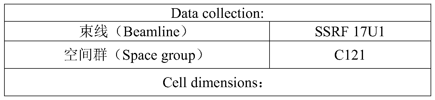

Tables 4A and 4B show, in sequence, a list of amino acids in which the human CTLA-4/ipilimumab complex and the CTLA-4/138 complex are involved in the interaction.
TABLE 4A
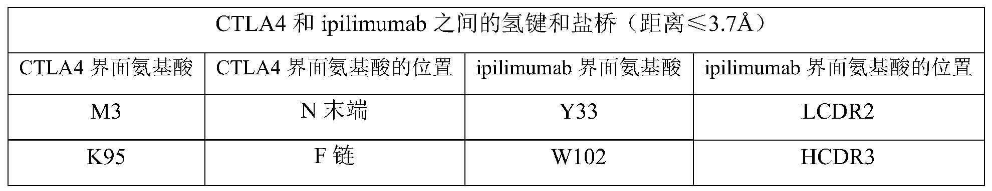

TABLE 4B

Example 8 validation of the ADCC Activity of CTLA 4-binding proteins at the cellular level
A plasmid expressing full-length human CTLA4 protein was transiently transfected into a CHO cell line CHO-CD20 stably expressing human CD20 protein, and the cell line was made to express both human CD20 and human CTLA4 protein, and the cell was named CHO-CD20-CTLA 4. After 48h, the cells were harvested, 25. mu.l of different antibodies at different concentrations (300. mu.g/ml, 30. mu.g/ml, 3. mu.g/ml, 0.3. mu.g/ml, 0.03. mu.g/ml), 25. mu.l of target cells (CHO-CD20-CTLA4), 25. mu.l of Jurkat-CD16a-176V-NFAT-RE (this cell strain was stably transfected with human CD16a protein in Jurkat cells purchased from ATCC, and ADCC activity-related reporter gene system NFAT-RE. which expresses luciferase when the target cells are activated by ADCC effect, further examined for the strength of ADCC activity by fluorescence substrate reaction) were added to 96-well white plates, respectively. The mixed solution is incubated for 4h at 37 ℃, 20 mu l of Luciferase substrate is added into each hole, and the machine is immediately used for detection.
The results are shown in FIG. 16, in which panels 1-8 correspond to the results for KN044-FcAG, 138-Fc, 138-FcAG, KN044, human IgG1, Rituxan and negative control (no target cells added), respectively. The results in FIG. 16 demonstrate that 138-Fc and KN044 exhibit dose-dependent ADCC activity, with 138-Fc being slightly more active than KN044, while both CTLA4 binding proteins were less active than Rituxan. After mutagenesis, ADCC activity was not substantially detectable by 138-Fc AG and KN 044-FcAG.
Example 9 Octet K2 detection of epitopes binding by CTLA4 binding proteins to human CTLA-4
Amino acid mutation was performed on human CTLA-4, and the binding activity of the mutated human CTLA-4 variant to 138-Fc was examined by the BLI method, which was specifically examined in example 2.1. See table 5 for the situation.
TABLE 5
| CTLA-4 variants | KD(M) | Kon(1/Ms) | Kdis(1/s) | KD (variant)/KD (wild type) |
| Wild type | 5.21*10 -8 | 1.29*10 5 | 6.70*10 -3 | 1 |
| T47A | 7.27*10 -7 | 2.69*10 4 | 1.95*10 -2 | 14.0 |
| E48A | - | - | - | >1000 |
| V49A | - | - | - | >1000 |
| L63A | 2.91*10 -8 | 2.74*10 5 | 8.00*10 -3 | 0.56 |
| D64A | 3.15*10 -5 | 4.17*10 2 | 1.32*10 -2 | 604.6 |
| D65A | - | - | - | >1000 |
| I67A | 1.36*10 -6 | 6.96*10 4 | 9.48*10 -2 | 26.1 |
| L84A | - | - | - | >1000 |
| D88A | - | - | - | >1000 |
| Y92A | - | - | - | >1000 |
Figure 17 shows the ratio of KD values for 138-Fc binding to human CTLA-4 variants and 138-Fc binding to human CTLA-4 (WT). The results in figure 17 show that amino acid mutations in E48A, V49A, D65A, L84A, D88A and Y92A all up-regulate the KD values of the variants by more than 1000-fold, thus it can be seen that E48, VV49, D65, L84, D88 and Y92 in human CTLA-4 are likely to be part of the epitope to which 138-Fc binds human CTLA-4.
Example 10: humanization of CTLA4 binding proteins
10.1 obtaining of humanized sequence
The humanization method is completed by adopting a method of protein surface amino acid humanization (resurfacing) or a VHH humanization universal framework grafting method (CDR grafting to a universal framework).
The humanization procedure was as follows: the antibody 138 prepared in example 1 was homologously modeled with Modeller9 as modeling software. The reference homology sequence was NbBcII10 antibody (PDB accession number: 3DWT), and the relative solvent accessibility (relative solvent access) of amino acids was calculated from the three-dimensional structure of the protein. If a certain amino acid of 138 is exposed to the solvent, the amino acid is replaced with the amino acid at the same position of the reference human antibody 10HQ sequence, and finally the whole replacement is completed.
The specific steps of the VHH humanized universal framework transplantation method are as follows: firstly, a universal humanized VHH framework hNbBcII10FGLA (PDB No. 3EAK) designed by Cile Vincke et al based on sequence homology was obtained, the framework design was based on a nanobody NbBcII10 antibody (PDB No. 3DWT), protein surface amino acid humanization was performed with reference to a humanized antibody (PDB No. 1OHQ), and partial amino acid VLP of VHH sequence framework 1(framework1, i.e., FR1), partial amino acid GL of VHH sequence framework 2(FR2), partial amino acid RSKRAAV of VHH sequence framework 3(FR3) and amino acid L of VHH sequence framework 4(FR4) were engineered.
Humanization of 138 yielded 4 humanized variants of 138 (hu138V 1-hu 138V4), respectively. The sequence numbering of these humanized variants, and the numbering of the amino acid residues therein, is in accordance with the Kabat numbering, and Table 6 shows the alignment of the humanized sequences. Wherein the presence of the amino acid residue is represented by the symbol "√".
TABLE 6
| Q5V | S11L | A14P | T19R | E44G | R45L | G75S | F80Y | K87R | P88A | M93V | Q121L | |
| hu138V1 | √ | √ | √ | √ | √ | √ | √ | √ | ||||
| hu 138V2 | √ | √ | √ | √ | √ | √ | √ | √ | √ | √ | ||
| hu 138V3 | √ | √ | √ | √ | √ | √ | √ | √ | √ | √ | ||
| hu 138V4 | √ | √ | √ | √ | √ | √ | √ | √ | √ | √ | √ | √ |
Wherein hu138V1 comprises HFR1-HFR4, wherein HFR1 comprises the amino acid sequence shown in SEQ ID No.21, HFR2 comprises the amino acid sequence shown in SEQ ID No.22, HFR3 comprises the amino acid sequence shown in SEQ ID No.23, and HFR4 comprises the amino acid sequence shown in SEQ ID No. 24; hu138V1 comprises a heavy chain variable region, wherein the heavy chain variable region comprises the amino acid sequence set forth in SEQ ID No. 17.
Wherein hu138V2 comprises HFR1-HFR4, wherein HFR1 comprises the amino acid sequence shown in SEQ ID No.25, HFR2 comprises the amino acid sequence shown in SEQ ID No.22, HFR3 comprises the amino acid sequence shown in SEQ ID No.23, and HFR4 comprises the amino acid sequence shown in SEQ ID No. 24; hu138V2 comprises a heavy chain variable region, wherein the heavy chain variable region comprises the amino acid sequence set forth in SEQ ID No. 18.
Wherein hu138V3 comprises HFR1-HFR4, wherein HFR1 comprises the amino acid sequence shown in SEQ ID No.21, HFR2 comprises the amino acid sequence shown in SEQ ID No.26, HFR3 comprises the amino acid sequence shown in SEQ ID No.23, and HFR4 comprises the amino acid sequence shown in SEQ ID No. 24; hu138V3 comprises a heavy chain variable region, wherein the heavy chain variable region comprises the amino acid sequence set forth in SEQ ID No. 19.
Wherein hu138V4 comprises HFR1-HFR4, wherein HFR1 comprises the amino acid sequence shown in SEQ ID No.25, HFR2 comprises the amino acid sequence shown in SEQ ID No.26, HFR3 comprises the amino acid sequence shown in SEQ ID No.23, and HFR4 comprises the amino acid sequence shown in SEQ ID No. 24; hu138V4 comprises a heavy chain variable region, wherein the heavy chain variable region comprises the amino acid sequence set forth in SEQ ID No. 20.
10.2 preparation of fusion proteins from mammalian cells
The humanized sequence obtained in 10.1 was subjected to gene synthesis by Suzhou Honghed Biotech Co., Ltd.and enzyme cleavage sites were added to both ends. The humanized variant of 138 (hu138V 1-hu 138V4) was digested in two, and fused with a DNA fragment encoding human IgG1 Fc. The fusion fragment was cloned into a conventional mammalian expression vector to obtain a recombinant plasmid for expressing a humanized 138-Fc target protein (hu138V 1-Ld-Fc. about. hu138V4-Ld-Fc) in mammals.
The recombinant plasmid is transfected into HEK293 cells for resistingTransient expression in vivo. The recombinant plasmids were diluted with Freestyle293 medium and added with PEI (polyethylenimine) solution required for transformation, and each set of plasmid/PEI mixtures was added separately to HEK293 cell suspension and left at 37 ℃ with 5% CO2Culturing at 130 rpm. Four hours later, the cells were supplemented with EXCELL293 medium (2 mM glutamine added) and cultured at 130 rpm. After 24 hours 3.8mM VPA was added and after 72 hours 4g/L glucose was added. And after culturing for 5-6 days, collecting transient expression culture supernatant, and purifying by Protein A affinity chromatography to obtain the target Protein. The purity of the target protein is preliminarily inspected by SDSPAGE and SECHPC. The expression and purity analyses are shown in Table 7.
TABLE 7
| Target protein | EC 50(ng/mL) |
| hu138v1-Ld-Fc | 36.2 |
| hu138v2-Ld-Fc | 34.6 |
| hu138v3-Ld-Fc | 34.9 |
| hu138v4-Ld-Fc | 33.6 |
10.3 testing the thermostability of the protein of interest
The protein of interest prepared in a trace volume of 10.2 was measured using a protein thermostability detector from UNCHAINED LABS.
Temperature rising procedure: the initial temperature is 15 ℃, the heating rate is 0.3 ℃/min, and the end temperature is 95 ℃. The fluorescence absorbance of the sample at each wavelength was recorded at each temperature. Fitting the Tm value of the denaturation temperature into the highest point of the next first derivative of the BCM at the confidence wavelength by software; the initial polymerization temperature Tagg value is one tenth of the first derivative at 473nm for static light scattering SLS. Higher Tm1 values indicate more stable protein. The results are shown in Table 8. As is clear from the results in Table 8, the Tm1 values of the target proteins were all higher than 60 ℃ and the stability was excellent.
TABLE 8
| Target protein | Tm1 | Tagg |
| hu138v1-Ld-Fc | 67.4 | 69.7 |
| hu138v2-Ld-Fc | 67.5 | 68.8 |
| hu138v3-Ld-Fc | 67.2 | 68.5 |
| hu138v4-Ld-Fc | 66.9 | 69.0 |
Example 11: CTLA4 binding protein mediated endocytic degradation of cell membrane surface CTLA4
HEK293-CTLA4 cells expressing human CTLA4 protein were obtained as in example 3.3.
The initial concentration of each CTLA4 binding protein to be detected was adjusted to 120 nM. A24-well plate was loaded with 100. mu.l growth medium and 100. mu.l CTLA4 binding protein to be tested per well. Density of HEK293-CTLA4 cells was adjusted to 1.2 x10⁶One/ml, 100. mu.l per well. Followed by incubation at 37 ℃ for 1-6 hours. Thereafter, the cells were harvested, the supernatant was discarded, and 50. mu.l of human CTLA4 antibody PE-anti-human CD152 (diluted 1: 50) was added. Thereafter, the cells were incubated at 4 ℃ for 45min and washed 2 times with 1% BSA/PBS. 200 μ L of 1% BSA/PBS was added to each tube to resuspend the cells and the change in the amount of CTLA4 on the cell membrane in HEK293-CTLA4 after incubation with the different CTLA4 binding proteins to be detected was detected by Mean Fluorescence Intensity (MFI) using FACS.
As shown in FIG. 18, after incubation of HEK293-CTLA4 cells with 138-Fc for 1-6 hours, there was no substantial difference in the expression level of CTLA4 on the cell surface compared to the inactive control isotype antibody (human IgG1) or compared to the control group without any antibody treatment. This indicates that 138-Fc can not cause endocytosis and degradation of CTLA4 molecules on the surface of cell membrane or can further stabilize CTLA4 molecules on the surface of membrane under incubation of 1-6 hours at 37 ℃. The level of CTLA4 expression of the cells treated by KN044 and ipilimumab is obviously reduced, which indicates that KN044 or ipilimumab reduces the density of CLTA4 on the surface of cell membranes through endocytosis.
Example 12: preliminary toxicity assessment of CTLA4 binding proteins in immune reconstituted mice
To preliminarily evaluate the immunotoxicity of the CTLA 4-binding proteins of different properties, mice were selected for reconstitution (i.e., human immunoblasts were implanted in immunodeficient mice to reconstitute the human immune system in the mice) and 100 μ g of the CTLA 4-binding proteins to be tested (138-Fc prepared in example 1, the CTLA4 monoclonal antibody drug ipilimumab (abbreviated lpilim) on which BMS has been marketed) or PBS of the same volume was intraperitoneally administered twice a week as a control (the administration dose was about 5mg/kg, calculated as about 20g of the mouse weight). The number of mice in each group was 5.
From the administration, the state of the mice was observed daily until day 19. As a result, mice in the ipilimumab administration group all developed acute immune responses of mice induced by ipilimumab administration by day 19; whereas only two mice in the 138-Fc group developed drug responses; mice remained alive in the control group.
It can be seen that the 138-Fc protein exhibits lower pharmacological toxicity than ipilimumab.
The foregoing detailed description is provided by way of illustration and example, and is not intended to limit the scope of the appended claims. Various modifications of the presently recited embodiments will be apparent to those of ordinary skill in the art and are intended to be within the scope of the appended claims and their equivalents.
Claims (47)
Applications Claiming Priority (3)
| Application Number | Priority Date | Filing Date | Title |
|---|---|---|---|
| CNPCT/CN2019/094751 | 2019-07-04 | ||
| CN2019094751 | 2019-07-04 | ||
| PCT/CN2020/100232 WO2021000953A1 (en) | 2019-07-04 | 2020-07-03 | Substance and method for treating tumor |
Publications (1)
| Publication Number | Publication Date |
|---|---|
| CN114072175A true CN114072175A (en) | 2022-02-18 |
Family
ID=74101201
Family Applications (1)
| Application Number | Title | Priority Date | Filing Date |
|---|---|---|---|
| CN202080047643.5A Pending CN114072175A (en) | 2019-07-04 | 2020-07-03 | Materials and methods for treating tumors |
Country Status (5)
| Country | Link |
|---|---|
| US (1) | US12448450B2 (en) |
| EP (1) | EP3995147A4 (en) |
| JP (1) | JP7557521B2 (en) |
| CN (1) | CN114072175A (en) |
| WO (1) | WO2021000953A1 (en) |
Families Citing this family (1)
| Publication number | Priority date | Publication date | Assignee | Title |
|---|---|---|---|---|
| WO2023070160A1 (en) * | 2021-10-28 | 2023-05-04 | Monash University | Variant ctla4 proteins |
Citations (2)
| Publication number | Priority date | Publication date | Assignee | Title |
|---|---|---|---|---|
| CN103833851A (en) * | 2014-03-14 | 2014-06-04 | 东南大学 | Single-domain antibody aiming at apolipoprotein A1 and application thereof |
| CN107400166A (en) * | 2016-05-19 | 2017-11-28 | 苏州康宁杰瑞生物科技有限公司 | for CTLA4 single domain antibody and its derived protein |
Family Cites Families (5)
| Publication number | Priority date | Publication date | Assignee | Title |
|---|---|---|---|---|
| FR2783517B1 (en) | 1998-09-22 | 2001-02-09 | Oreal | NOVEL 10-HYROXY-2-DECENOIC ACID DERIVATIVES AND USE IN A COMPOSITION FOR PROMOTING SKIN DEQUAMATION, AND COMPOSITION COMPRISING THE SAME |
| CN106397592A (en) | 2015-07-31 | 2017-02-15 | 苏州康宁杰瑞生物科技有限公司 | Single-domain antibody directed at programmed death ligand (PD-L1) and derived protein thereof |
| WO2018068201A1 (en) * | 2016-10-11 | 2018-04-19 | Nanjing Legend Biotech Co., Ltd. | Single-domain antibodies and variants thereof against ctla-4 |
| EP3424524B1 (en) * | 2017-07-04 | 2024-12-11 | CureVac SE | Cancer rna-vaccine |
| CN112218892B (en) * | 2018-03-19 | 2023-04-25 | 上海药明生物技术有限公司 | Novel anti-CTLA-4 antibody polypeptides |
-
2020
- 2020-07-03 US US17/624,795 patent/US12448450B2/en active Active
- 2020-07-03 WO PCT/CN2020/100232 patent/WO2021000953A1/en not_active Ceased
- 2020-07-03 CN CN202080047643.5A patent/CN114072175A/en active Pending
- 2020-07-03 JP JP2022500131A patent/JP7557521B2/en active Active
- 2020-07-03 EP EP20835357.3A patent/EP3995147A4/en active Pending
Patent Citations (2)
| Publication number | Priority date | Publication date | Assignee | Title |
|---|---|---|---|---|
| CN103833851A (en) * | 2014-03-14 | 2014-06-04 | 东南大学 | Single-domain antibody aiming at apolipoprotein A1 and application thereof |
| CN107400166A (en) * | 2016-05-19 | 2017-11-28 | 苏州康宁杰瑞生物科技有限公司 | for CTLA4 single domain antibody and its derived protein |
Also Published As
| Publication number | Publication date |
|---|---|
| EP3995147A4 (en) | 2023-08-16 |
| US12448450B2 (en) | 2025-10-21 |
| EP3995147A1 (en) | 2022-05-11 |
| US20220251207A1 (en) | 2022-08-11 |
| WO2021000953A1 (en) | 2021-01-07 |
| JP2022539260A (en) | 2022-09-07 |
| JP7557521B2 (en) | 2024-09-27 |
Similar Documents
| Publication | Publication Date | Title |
|---|---|---|
| JP7450592B2 (en) | FGFR2 inhibitors alone or in combination with immunostimulants in cancer treatment | |
| KR101719118B1 (en) | Anti-ox40 antibodies and methods of using the same | |
| EP3511346A1 (en) | Anti-ctla4 and anti-pd-1 bifunctional antibody, pharmaceutical composition thereof and use thereof | |
| KR20210142638A (en) | CD3 antigen-binding fragment and applications thereof | |
| CN111050792A (en) | anti-LAG-3 antibodies and uses thereof | |
| EP4389767A1 (en) | Monoclonal antibody targeting sirp? and use thereof | |
| CN118871474A (en) | Anti-PD-L1 and VEGF bispecific antibodies and their applications | |
| JP2022505925A (en) | Anti-TIM-3 antibody | |
| EP4223778A1 (en) | Anti-claudin18.2 and cd3 bispecific antibody and use thereof | |
| WO2021219048A1 (en) | Bispecific antibody targeting nkg2a and pd-l1, and application | |
| CN115260312A (en) | Antibodies or antigen-binding fragments that bind to OX40 | |
| WO2024001641A1 (en) | Antibody of cd101 and use thereof | |
| US12448450B2 (en) | Method for treating tumor using anti-CTLA4 single-domain antibodies | |
| JP2020508636A (en) | IFN-γ-induced regulatory T cell converting anti-cancer (IRTCA) antibody and use thereof | |
| WO2018119288A1 (en) | Anti-human cxcr3 antibodies for treatment of vitiligo | |
| KR20220101126A (en) | Anti-Siglec-9 Antibody Molecules | |
| WO2024085182A1 (en) | Therapeutic agent for t cell tumor | |
| HK40063855A (en) | Substances and methods for treating tumors | |
| US20240141070A1 (en) | Ox40/pd-l1 bispecific antibody | |
| TW202222834A (en) | Pd-l1 antibody and use thereof | |
| US20250277038A1 (en) | Anti-pd-l1 antibody and use thereof | |
| EP4293046A1 (en) | Anti-pd-1 antibody and use thereof | |
| WO2022188865A1 (en) | BIFUNCTIONAL MOLECULE BINDING TO HUMAN TGFβ AND PD-L1, AND USE THEREOF | |
| TW202405013A (en) | Antigen binding protein and use thereof | |
| EP3559036A1 (en) | Anti-human cxcr3 antibodies for treatment of vitiligo |
Legal Events
| Date | Code | Title | Description |
|---|---|---|---|
| PB01 | Publication | ||
| PB01 | Publication | ||
| REG | Reference to a national code |
Ref country code: HK Ref legal event code: DE Ref document number: 40063855 Country of ref document: HK |
|
| SE01 | Entry into force of request for substantive examination | ||
| SE01 | Entry into force of request for substantive examination |