CN112218892B - Novel anti-CTLA-4 antibody polypeptides - Google Patents
Novel anti-CTLA-4 antibody polypeptides Download PDFInfo
- Publication number
- CN112218892B CN112218892B CN201980020246.6A CN201980020246A CN112218892B CN 112218892 B CN112218892 B CN 112218892B CN 201980020246 A CN201980020246 A CN 201980020246A CN 112218892 B CN112218892 B CN 112218892B
- Authority
- CN
- China
- Prior art keywords
- cancer
- antibody
- ctla
- antibody polypeptide
- human
- Prior art date
- Legal status (The legal status is an assumption and is not a legal conclusion. Google has not performed a legal analysis and makes no representation as to the accuracy of the status listed.)
- Active
Links
Images


Classifications
-
- C—CHEMISTRY; METALLURGY
- C07—ORGANIC CHEMISTRY
- C07K—PEPTIDES
- C07K16/00—Immunoglobulins [IGs], e.g. monoclonal or polyclonal antibodies
- C07K16/18—Immunoglobulins [IGs], e.g. monoclonal or polyclonal antibodies against material from animals or humans
- C07K16/28—Immunoglobulins [IGs], e.g. monoclonal or polyclonal antibodies against material from animals or humans against receptors, cell surface antigens or cell surface determinants
- C07K16/2803—Immunoglobulins [IGs], e.g. monoclonal or polyclonal antibodies against material from animals or humans against receptors, cell surface antigens or cell surface determinants against the immunoglobulin superfamily
- C07K16/2818—Immunoglobulins [IGs], e.g. monoclonal or polyclonal antibodies against material from animals or humans against receptors, cell surface antigens or cell surface determinants against the immunoglobulin superfamily against CD28 or CD152
-
- A—HUMAN NECESSITIES
- A61—MEDICAL OR VETERINARY SCIENCE; HYGIENE
- A61P—SPECIFIC THERAPEUTIC ACTIVITY OF CHEMICAL COMPOUNDS OR MEDICINAL PREPARATIONS
- A61P35/00—Antineoplastic agents
-
- C—CHEMISTRY; METALLURGY
- C07—ORGANIC CHEMISTRY
- C07K—PEPTIDES
- C07K2317/00—Immunoglobulins specific features
- C07K2317/20—Immunoglobulins specific features characterized by taxonomic origin
- C07K2317/21—Immunoglobulins specific features characterized by taxonomic origin from primates, e.g. man
-
- C—CHEMISTRY; METALLURGY
- C07—ORGANIC CHEMISTRY
- C07K—PEPTIDES
- C07K2317/00—Immunoglobulins specific features
- C07K2317/20—Immunoglobulins specific features characterized by taxonomic origin
- C07K2317/22—Immunoglobulins specific features characterized by taxonomic origin from camelids, e.g. camel, llama or dromedary
-
- C—CHEMISTRY; METALLURGY
- C07—ORGANIC CHEMISTRY
- C07K—PEPTIDES
- C07K2317/00—Immunoglobulins specific features
- C07K2317/20—Immunoglobulins specific features characterized by taxonomic origin
- C07K2317/24—Immunoglobulins specific features characterized by taxonomic origin containing regions, domains or residues from different species, e.g. chimeric, humanized or veneered
-
- C—CHEMISTRY; METALLURGY
- C07—ORGANIC CHEMISTRY
- C07K—PEPTIDES
- C07K2317/00—Immunoglobulins specific features
- C07K2317/30—Immunoglobulins specific features characterized by aspects of specificity or valency
- C07K2317/33—Crossreactivity, e.g. for species or epitope, or lack of said crossreactivity
-
- C—CHEMISTRY; METALLURGY
- C07—ORGANIC CHEMISTRY
- C07K—PEPTIDES
- C07K2317/00—Immunoglobulins specific features
- C07K2317/50—Immunoglobulins specific features characterized by immunoglobulin fragments
- C07K2317/52—Constant or Fc region; Isotype
-
- C—CHEMISTRY; METALLURGY
- C07—ORGANIC CHEMISTRY
- C07K—PEPTIDES
- C07K2317/00—Immunoglobulins specific features
- C07K2317/50—Immunoglobulins specific features characterized by immunoglobulin fragments
- C07K2317/56—Immunoglobulins specific features characterized by immunoglobulin fragments variable (Fv) region, i.e. VH and/or VL
- C07K2317/565—Complementarity determining region [CDR]
-
- C—CHEMISTRY; METALLURGY
- C07—ORGANIC CHEMISTRY
- C07K—PEPTIDES
- C07K2317/00—Immunoglobulins specific features
- C07K2317/50—Immunoglobulins specific features characterized by immunoglobulin fragments
- C07K2317/56—Immunoglobulins specific features characterized by immunoglobulin fragments variable (Fv) region, i.e. VH and/or VL
- C07K2317/569—Single domain, e.g. dAb, sdAb, VHH, VNAR or nanobody®
-
- C—CHEMISTRY; METALLURGY
- C07—ORGANIC CHEMISTRY
- C07K—PEPTIDES
- C07K2317/00—Immunoglobulins specific features
- C07K2317/60—Immunoglobulins specific features characterized by non-natural combinations of immunoglobulin fragments
- C07K2317/64—Immunoglobulins specific features characterized by non-natural combinations of immunoglobulin fragments comprising a combination of variable region and constant region components
-
- C—CHEMISTRY; METALLURGY
- C07—ORGANIC CHEMISTRY
- C07K—PEPTIDES
- C07K2317/00—Immunoglobulins specific features
- C07K2317/70—Immunoglobulins specific features characterized by effect upon binding to a cell or to an antigen
-
- C—CHEMISTRY; METALLURGY
- C07—ORGANIC CHEMISTRY
- C07K—PEPTIDES
- C07K2317/00—Immunoglobulins specific features
- C07K2317/70—Immunoglobulins specific features characterized by effect upon binding to a cell or to an antigen
- C07K2317/73—Inducing cell death, e.g. apoptosis, necrosis or inhibition of cell proliferation
- C07K2317/732—Antibody-dependent cellular cytotoxicity [ADCC]
-
- C—CHEMISTRY; METALLURGY
- C07—ORGANIC CHEMISTRY
- C07K—PEPTIDES
- C07K2317/00—Immunoglobulins specific features
- C07K2317/70—Immunoglobulins specific features characterized by effect upon binding to a cell or to an antigen
- C07K2317/73—Inducing cell death, e.g. apoptosis, necrosis or inhibition of cell proliferation
- C07K2317/734—Complement-dependent cytotoxicity [CDC]
-
- C—CHEMISTRY; METALLURGY
- C07—ORGANIC CHEMISTRY
- C07K—PEPTIDES
- C07K2317/00—Immunoglobulins specific features
- C07K2317/70—Immunoglobulins specific features characterized by effect upon binding to a cell or to an antigen
- C07K2317/76—Antagonist effect on antigen, e.g. neutralization or inhibition of binding
-
- C—CHEMISTRY; METALLURGY
- C07—ORGANIC CHEMISTRY
- C07K—PEPTIDES
- C07K2317/00—Immunoglobulins specific features
- C07K2317/90—Immunoglobulins specific features characterized by (pharmaco)kinetic aspects or by stability of the immunoglobulin
- C07K2317/92—Affinity (KD), association rate (Ka), dissociation rate (Kd) or EC50 value
-
- C—CHEMISTRY; METALLURGY
- C07—ORGANIC CHEMISTRY
- C07K—PEPTIDES
- C07K2317/00—Immunoglobulins specific features
- C07K2317/90—Immunoglobulins specific features characterized by (pharmaco)kinetic aspects or by stability of the immunoglobulin
- C07K2317/94—Stability, e.g. half-life, pH, temperature or enzyme-resistance
-
- C—CHEMISTRY; METALLURGY
- C07—ORGANIC CHEMISTRY
- C07K—PEPTIDES
- C07K2319/00—Fusion polypeptide
-
- C—CHEMISTRY; METALLURGY
- C07—ORGANIC CHEMISTRY
- C07K—PEPTIDES
- C07K2319/00—Fusion polypeptide
- C07K2319/30—Non-immunoglobulin-derived peptide or protein having an immunoglobulin constant or Fc region, or a fragment thereof, attached thereto
Landscapes
- Health & Medical Sciences (AREA)
- Chemical & Material Sciences (AREA)
- Immunology (AREA)
- Organic Chemistry (AREA)
- Medicinal Chemistry (AREA)
- Life Sciences & Earth Sciences (AREA)
- General Health & Medical Sciences (AREA)
- Proteomics, Peptides & Aminoacids (AREA)
- Genetics & Genomics (AREA)
- Molecular Biology (AREA)
- Biophysics (AREA)
- Biochemistry (AREA)
- Nuclear Medicine, Radiotherapy & Molecular Imaging (AREA)
- General Chemical & Material Sciences (AREA)
- Chemical Kinetics & Catalysis (AREA)
- Pharmacology & Pharmacy (AREA)
- Animal Behavior & Ethology (AREA)
- Public Health (AREA)
- Veterinary Medicine (AREA)
- Peptides Or Proteins (AREA)
- Medicines Containing Antibodies Or Antigens For Use As Internal Diagnostic Agents (AREA)
Abstract
本公开提供抗CTLA‑4的抗体多肽、编码该多肽的多核苷酸、包含该多肽的药物组合物及其用途。

The present disclosure provides an anti-CTLA-4 antibody polypeptide, a polynucleotide encoding the polypeptide, a pharmaceutical composition comprising the polypeptide and uses thereof.
Description
Priority statement
This patent application claims the benefit of priority from PCT application number PCT/CN2018/079495 filed on day 19 of 3.2018.
Technical Field
The present invention relates generally to novel anti-human CTLA-4 antibody polypeptides.
Background
Cancer immunotherapy has become a popular research area for cancer therapy. Cytotoxic T lymphocyte-associated protein 4 (CTLA-4) is one of the effective targets for immunoaudit points. CTLA-4 is typically expressed rapidly in T cells of this type within 1 hour after antigen binding to TCR following T cell activation. CTLA-4 can inhibit T cell signaling by competing with CD 28. In addition to induction of expression in activated T cells, constitutive expression of CTLA-4 on regulatory T cell (Treg) surfaces suggests that CTLA-4 may be necessary for contact-mediated inhibition and is involved in the production of immunosuppressive cytokines such as transforming growth factor beta and interleukin-10 by Treg.
CTLA-4 blockade has been shown to induce tumor regression in many preclinical and clinical studies. There are two anti-CTLA-4 antibodies in clinical development. Epimumab (MDX-010, BMS-734016), the fully human anti-CTLA-4 monoclonal antibody IgG1-kappa isotype, is an immunomodulator and has been approved as monotherapy for the treatment of advanced melanoma.
A single domain antibody (sdAb) is an antibody that comprises a single variable antibody domain. Like whole antibodies, can selectively bind to a particular antigen. Single domain antibodies are much smaller than common antibodies that consist of two protein heavy and two light chains. The first single domain antibody was designed from the heavy chain antibodies found in camelids (Hamers-Casterman C, atarouch T, muyldermans S, robinson G, hamers C, songa EB, bendahman N, hamers R (1993) natural antibodies free of light chains. Nature 363 (6428): 446-448.), these are referred to as VHH fragments. Most single domain antibody studies are now based on heavy chain variable domains.
Single domain antibodies have a number of advantages. For example, they generally have high solubility and stability and can be readily produced in yeast, plant and mammalian cells (Harmsen MM, de Haard HJ (2007) camelid single domain antibody fragments, property, production and use. Appl Microbiol Biotechnol 77 (1): 13-22.). In addition, they have good thermal stability and good tissue penetration. And they are also cost effective. The advantages of single domain antibodies make them suitable for many biotechnology and therapeutic applications. For example, they may be used to treat diseases including, but not limited to, cancer, infectious diseases, inflammation, and neurodegenerative diseases.
Although antibodies against CTLA-4 have been developed, there is room for improvement in anti-CTLA-4 antibodies as therapeutic agents. Accordingly, it is desirable in the art to develop novel anti-CTLA-4 antibodies, particularly single domain antibodies directed against CTLA-4.
Disclosure of Invention
In the present disclosure, the articles "a," "an," and "the" refer to one or more of the grammatical objects of the strip (i.e., at least one). For example, "an antibody" refers to one or more antibodies.
The present invention provides novel anti-CTLA-4 monoclonal antibodies, as well as amino acid and nucleic acid sequences thereof, and uses thereof.
In one aspect, the invention provides an antibody polypeptide comprising a heavy chain variable domain that specifically binds CTLA-4, wherein the heavy chain variable domain comprises:
1, 2 or 3 heavy chain Complementarity Determining Region (CDR) sequences selected from the group consisting of SEQ ID NO. 1, SEQ ID NO. 2, SEQ ID NO. 3 and SEQ ID NO. 10.
In certain embodiments, the heavy chain variable domain comprises a heavy chain variable domain comprising SEQ ID NO. 1, SEQ ID NO. 10 and SEQ ID NO. 3. In certain embodiments, the heavy chain variable domain comprises a heavy chain variable domain comprising SEQ ID NO. 1, SEQ ID NO. 2 and SEQ ID NO. 3.
In certain embodiments, wherein the heavy chain variable domain comprises a heavy chain variable region selected from the group consisting of SEQ ID NO. 4, SEQ ID NO. 6 and SEQ ID NO. 8 and homologous sequences thereof, which homologous sequences have at least 80% sequence identity while maintaining a specific binding affinity to CTLA-4.
In certain embodiments, the antibody polypeptides provided by the present disclosure further comprise one or more amino acid residue substitutions or modifications while maintaining a specific binding affinity to CTLA-4.
In certain embodiments, at least one substitution or modification is in one or more CDR sequences, and/or in one or more VH sequences, but not in any CDR sequences.
In certain embodiments, the antibody polypeptide is a single domain antibody or a heavy chain antibody.
In certain embodiments, the heavy chain variable domain is derived from a VHH domain.
In certain embodiments, the antibody polypeptide further comprises an immunoglobulin constant region, optionally a constant region of a human Ig, or optionally a constant region of a human IgG.
In certain embodiments, the antibody polypeptide is isolated.
In certain embodiments, the heavy chain variable domain is camelid-derived or humanized.
In certain embodiments, the antibody polypeptide is a nanobody.
In certain embodiments, the antibody polypeptides provided herein can specifically bind to human CTLA4, the EC of which is determined by flow cytometry 50 The value is not more than 0.5nM.
In certain embodiments, the antibody polypeptides provided by the present disclosure can block binding between CTLA4 and cell surface expressed CD80, and the IC is determined by flow cytometry 50 A value of no more than 0.15nM; or blocking binding between CTLA4 and CD86 expressed on cell surface, and determining IC by flow cytometry 50 The value is not more than 0.25nM.
In certain embodiments, the antibody polypeptides provided herein can specifically bind cynomolgus CTLA-4, and/or murine CTLA-4.
In certain embodiments, the antibody polypeptides provided by the present disclosure are linked to one or more conjugated groups.
In certain embodiments, the conjugated group comprises a scavenging modifier, a chemotherapeutic agent, a toxin, a radioisotope, a lanthanide, a luminescent label, a fluorescent label, an enzyme substrate label, a DNA alkylating agent, a topoisomerase inhibitor, a tubulin binding agent, or other anti-cancer drug.
In another aspect, the invention provides an antibody or antigen binding fragment thereof which competes for the same epitope as the antibody polypeptide of any preceding claim.
The invention also provides a pharmaceutical composition comprising an antibody polypeptide provided by the present disclosure, comprising an antibody or antigen-binding fragment thereof, and a pharmaceutically acceptable carrier.
The invention also provides a polynucleotide encoding an antibody polypeptide provided by the present disclosure. In certain embodiments, the polynucleotide is isolated.
In certain embodiments, the polynucleotides provided by the present disclosure comprise a nucleotide sequence selected from the group consisting of SEQ ID NO. 5, SEQ ID NO. 7, and SEQ ID NO. 9, and/or a homologous sequence thereof having at least 80% (e.g., at least 85%, 88%, 90%, 92%, 93%, 94%, 95%, 96%, 97%, 98%, or 99%) sequence identity, and/or a variant thereof having only degenerate substitutions.
The invention also provides a vector comprising the polynucleotide provided by the present disclosure.
The invention also provides a host cell comprising the vector provided by the present disclosure.
The invention also provides a method of expressing an antibody polypeptide provided by the present disclosure, comprising culturing a host cell provided by the present disclosure under conditions that allow expression of a vector provided by the present disclosure.
The invention also provides a method of treating a disease or disorder in a subject who would benefit from modulation of CTLA-4 activity, comprising administering to the subject a therapeutically effective amount of any one of the antibody polypeptides provided herein or the pharmaceutical composition provided herein.
In certain embodiments, the disease or disorder is a CTLA-4-associated disease or disorder.
In certain embodiments, the disease or disorder is cancer, an autoimmune disease, an inflammatory disease, an infectious disease, graft Versus Host Disease (GVHD), or graft rejection.
In certain embodiments, the cancer is lymphoma, bladder cancer, bone cancer, brain and central nervous system cancer, breast cancer, uterine or endometrial cancer, rectal cancer, esophageal cancer, head and neck cancer, anal cancer, gastrointestinal cancer, intraepithelial tumors, kidney (kidney) cancer or renal cell (renal) cancer, leukemia, liver cancer, lung cancer, melanoma, myeloma, pancreatic cancer, prostate cancer, sarcoma, skin cancer, squamous cell carcinoma, gastric cancer, testicular cancer, vulval cancer, cancer of the endocrine system, parathyroid cancer, adrenal cancer, penile cancer, childhood solid tumors, tumor hemangiomas, spinal tumors, pituitary adenomas, or epidermoid carcinoma.
In certain embodiments, the disease or disorder is an environmentally-induced cancer caused by asbestos or a hematological malignancy, wherein the cancer is selected from multiple myeloma, B-cell lymphoma, hodgkin's lymphoma, primary mediastinal B-cell lymphoma, non-hodgkin's lymphoma, acute myeloid lymphoma, chronic myelogenous leukemia, chronic Lymphocytic Leukemia (CLL), follicular lymphoma, diffuse large B-cell lymphoma (DLBCL), burkitt's lymphoma, immunoblastic large cell lymphoma, precursor B-lymphoblastic lymphoma, mantle cell lymphoma, acute Lymphoblastic Leukemia (ALL), mycosis fungoides, anaplastic large cell lymphoma, T-cell lymphoma, and precursor T-cell lymphoma, and any combination of the foregoing cancers.
In certain embodiments, the subject is a human.
In certain embodiments, the mode of administration is oral, nasal, intravenous, subcutaneous, sublingual, or intramuscular.
In another aspect, the invention also provides a method of modulating CTLA-4 activity in a CTLA-4 expressing cell comprising exposing the CTLA-4 expressing cell to an antibody polypeptide provided by the present disclosure.
The invention also provides a method of detecting the presence or amount of CTLA-4 in a sample, comprising contacting the sample with an antibody polypeptide provided by the present disclosure, and determining the presence or amount of CTLA-4 in the sample.
The invention also provides a method of diagnosing a CTLA-4-associated disease or disorder in a subject, comprising: a) Contacting a sample obtained from a subject with any of the antibody polypeptides provided by the present disclosure; b) Determining the presence or amount of CTLA-4 in the sample; and c) correlating the presence or amount of CTLA-4 with the presence or status of the CTLA-4-related disease or disorder in the subject.
The invention also provides the use of an antibody polypeptide provided by the present disclosure in the manufacture of a medicament for treating a CTLA-4-associated disease or disorder in a subject.
The invention also provides for the use of the antibody polypeptides provided by the present disclosure in the preparation of reagents for diagnosing CTLA-4-associated diseases or disorders in a subject.
The invention also provides a kit comprising an antibody polypeptide comprising the disclosure for detecting CTLA-4.
Drawings
FIG. 1A shows the measurement of binding of W3166-z13 and W3166-z17 to human CTLA4 on the cell surface by FACS method.
FIG. 1B shows the measurement of binding of W3166-z13 and W3166-z17 to human CTLA4 by ELISA.
FIG. 2A shows the measurement of binding of W3166-z13 and W3166-z17 to cynomolgus CTLA4 on the cell surface by FACS method.
FIG. 2B shows the measurement of binding of W3166-z13 and W3166-z17 to cynomolgus CTLA4 by ELISA.
FIG. 3A shows the measurement of the blockade of CD80 binding to human CTLA4 by ELISA method for W3166-z13 and W3166-z 17.
FIG. 3B shows the measurement of the blockade of CD86 binding to human CTLA4 by ELISA method for W3166-z13 and W3166-z 17.
FIG. 4A shows that measurements by FACS showed that W3166-z13 and W3166-z17 were more effective at blocking CD80 binding to cell surface human CTLA4 than W316-BMK 1.
FIG. 4B shows that measurements by FACS showed that W3166-z13 and W3166-z17 were more effective at blocking CD86 binding to cell surface human CTLA4 than W316-BMK 1.
FIG. 5A shows that human heterologous MLR assays showed that W3166-z13 and W3166-z17 enhanced IFN-gamma production more than W316-BMK 1.
FIG. 5B shows that W3166-z13 and W3166-z17 enhance IL-2 production in human heterologous MLR in a dose-dependent manner. The potency was comparable to W316-BMK 1.
FIG. 6 shows measurements in an epitope binding ELISA test showing that W3166-z13 and W3166-z17 have similar sets of table bits as W316-BMK 1.
FIG. 7 shows that W3166-z13 and W3166-z17 can induce ADCC effects in human CTLA4 transfected cells.
FIG. 8 shows that W3166-z13 and W3166-z17 can not induce CDC effects in human CTLA4 transfected cells.
FIG. 9 shows that the FACS measurements show that W3166-z13 and W3166-z17 are stable in human serum stabilization tests.
FIG. 10 shows FACS measurements showing that W3166-z13 and W3166-z17 specifically bind human CTLA-4 and do not cross react with hICOS, BTLA, hCD28 and hPD 1.
Detailed Description
The following description of the invention is merely illustrative of various embodiments of the invention. Therefore, the specific modifications discussed should not be construed as limiting the scope of the invention. It will be apparent to those skilled in the art that various equivalents, changes, and modifications can be made without departing from the scope of the invention, and it is to be understood that such equivalent embodiments are intended to be included in the present disclosure. All references, including publications, patents, and patent applications, cited in this disclosure are hereby incorporated by reference in their entirety.
Definition of the definition
The term "antibody" as used in this disclosure includes any immunoglobulin, monoclonal antibody, polyclonal antibody, multivalent antibody, bivalent antibody or monovalent antibody that binds to a particular antigen. The term "antibody" as used in the present disclosure is intended to broadly encompass conventional four-chain antibodies and less conventional antibodies that do not have four chains (e.g., natural antibodies lacking light chains).
Traditional intact antibodies are heterotetramers composed of two heavy (H) chains and two light (L) chains. Mammalian heavy chains are divided into alpha, delta, epsilon, gamma and mu, each heavy chain comprising a variable region (V H ) And a first, second and third constant region (C, respectively) H1 、C H2 、C H3 ) The method comprises the steps of carrying out a first treatment on the surface of the Mammalian light chains are classified as lambda or kappa, each light chain comprising a variable region (V L ) And a constant region. Conventional antibodies are "Y" shaped in that the handle of the Y consists of the second and third constant regions of two heavy chains, joined together by disulfide bonds. Each arm of Y comprises a variable region and a first constant region of a single heavy chain, the heavy chain being bound to the variable and constant regions of a single light chain. The variable regions of the light and heavy chains are responsible for antigen binding. The variable region in both chains typically comprises three highly variable loops, known as Complementarity Determining Regions (CDRs) (light chain CDRs comprising LCDR1, LCDR2 and LCDR3, and heavy chain CDRs comprising HCDR1, HCDR2 and HCDR 3). CDR boundaries of antibodies and antigen binding fragments of the present disclosure may be defined or recognized by Kabat, IMGT, chothia or the convention of Al-Lazikani (Al-Lazikani, B., chothia, C., lesk, A.M., J.Mol.Biol.,273 (4), 927 (1997); chothia, C.et Al, J Mol biol. Dec 5; 18)651-63 (1985); chothia, c.and Lesk, A.M., J.Mol.Biol.,196,901 (1987); chothia, c.et al, nature. Dec 21-28;342 (6252) 877-83 (1989); kabat e.a.et al, immune related protein sequences, 5 th edition, public health service, national institutes of health, bescens da, md. (1991); marie-Paule Lefranc et al, development and comparative immunology, 27:55-77 (2003); marie-Paule Lefranc et al, immunome Research,1 (3), (2005); marie-Paule Lefranc, B cell molecular biology (second edition), chapter 26, 481-514, (2015)). Three CDRs are located between flanking stretches, called Framework Regions (FRs), which are more conserved than the CDRs and form a scaffold that supports the hypervariable loops. The constant regions of the heavy and light chains do not participate in antigen binding, but exhibit different effector functions. Antibodies are classified according to the amino acid sequence of their heavy chain constant region. The five major classes or isotypes of antibodies are IgA, igD, igE, igG and IgM, which are characterized by having corresponding alpha, delta, epsilon, gamma and mu heavy chains. Several major antibodies can be divided into subclasses such as IgG1 (gamma heavy chain), igG2 (gamma 2 heavy chain), igG3 (gamma 3 heavy chain), igG4 (gamma 4 heavy chain), igA1 (alpha 1 heavy chain) or IgA2 (alpha 2 heavy chain).
Unlike traditional heterotetrameric antibodies, dimeric immunoglobulins also exist that naturally lack light chains. For example, such antibodies are found in camelids (camels, dromedaries, llamas, alpacas, etc.), also known as heavy chain antibodies with a molecular weight of about 80kD (mers-masterman c.et al, 1993, nature, 363:446-448).
The term "antibody polypeptide" as used in this disclosure refers to an antigen binding protein or polypeptide comprising antibody fragments (e.g., CDRs, and/or variable region sequences). The antibody polypeptide may comprise or may be, for example, a heavy chain antibody (VHH antibody), a variable domain of a heavy chain antibody, a VHH domain, or a single domain antibody comprising a single variable domain. The antibody polypeptide may further comprise additional domains, such as a constant region, an Fc domain, and/or a second variable domain that specifically binds to a different antigen or different epitope.
"heavy chain antibody" and "VHH antibody" are used interchangeably in the present disclosure to mean comprising two V' s H Domain and light chain-free antibodies (Riechmann l.and Muyldermans s., J Immunol)Methods, dec 10;231 (1-2) 25-38 (1999); muydermans s., J biotechnol jun;74 277-302 (2001); WO94/04678; WO94/25591; U.S. patent No. 6,005,079). In spite of the lack of light chains, heavy chain antibodies have a truly antigen binding repertoire (Hamers-Casterman C.et al, 1993, nature,363:446-448;Nguyen VK.et al, 2002, immunogenetics,54 (1): 39-47;Nguyen VK.et al, 2003, immunology,109 (1): 93-101).
As used in this disclosure, "VHH domain" refers to a heavy chain variable domain derived from a heavy chain antibody. VHH domains represent the smallest known antigen binding unit generated by an adaptive immune response (Koch-Nolte F.et al, 2007, FASEB J.,21 (13): 3490-8.Epub 2007 Jun 15).
"Single domain antibody" refers to an antibody fragment comprising only a single variable region of a heavy chain or a single variable region of a light chain. In certain embodiments, a single domain antibody has or comprises only a single heavy chain variable domain of a heavy chain antibody.
"nanobody" refers to an antibody fragment consisting of one VHH domain and two constant domains CH2 and CH3 of a heavy chain antibody.
In some cases, two or more VHH domains can be covalently linked to a peptide linker to generate a bivalent or multivalent domain antibody. The two VHH domains of a bivalent domain antibody may target the same or different antigens.
The term "bivalent" as used in this disclosure refers to an antibody or antibody polypeptide having two antigen binding sites; the term "monovalent" refers to an antibody or antibody polypeptide having only one antigen binding site; and the term "multivalent" refers to an antibody or antibody polypeptide having multiple antigen binding sites. In certain embodiments, the antibody or antibody polypeptide is bivalent.
The term "chimeric" as used in this disclosure refers to an antibody or antibody polypeptide in which a portion of the heavy chain is derived from one species and the other portion of the heavy chain is derived from another species. In an illustrative example, a chimeric antibody may comprise a constant region derived from a human and a variable region derived from a non-human animal such as camelidae. In certain embodiments, the non-human animal is a mammal, such as a camelid, mouse, rat, rabbit, goat, sheep, guinea pig, or hamster.
The term "humanized" as used in this disclosure means antibodies or antibody polypeptides that include CDRs from a non-human animal, FR regions from a human, and where applicable constant regions from a human.
As used herein, "CTLA-4" refers to cytotoxic T lymphocyte-associated protein 4 derived from any vertebrate, including mammals such as primates (e.g., humans, monkeys) and rodents (e.g., mice and rats). Exemplary sequences for human CTLA-4 include human CTLA-4 protein (NCBI reference sequence No. AAL 07473.1). Exemplary sequences for CTLA-4 include cynomolgus monkey (monkey) CTLA-4 protein (NCBI reference sequence No. XP_ 005574071.1).
The term "CTLA-4" as used in the present disclosure is intended to encompass any form of CTLA-4, e.g., 1) naturally untreated CTLA-4 molecules, "full length" CTLA-4 chains or naturally occurring CTLA-4 variants, including, e.g., splice variants or allelic variants; 2) Any form of CTLA-4 produced following intracellular processing; or 3) full length, CTLA-4 subunit fragments (e.g., truncated forms, extracellular/transmembrane domains) or modified forms thereof (e.g., mutant forms, glycosylated/pegylated, his-tag/immunofluorescent fusion forms) produced by recombinant methods.
The term "anti-CTLA-4" antibody polypeptide as used in this disclosure refers to an antibody polypeptide that specifically binds CTLA-4 (e.g., human or monkey CTLA-4).
The term "specific binding" or "specifically binding" as used in this disclosure refers to a non-random binding reaction between two molecules, such as a binding reaction between an antibody and an antigen. In certain embodiments, the antibody polypeptides provided herein specifically bind to human CTLA-4 with binding affinity (K D )≤10 -6 M (e.g.,.ltoreq.5X10) -7 M、≤2x10 -7 M、≤10 -7 M、≤5x10 -8 M、≤2x10 -8 M、≤10 -8 M、≤5x10 -9 M、≤4x10 -9 M、≤3x10 -9 M、≤2x10 -9 M or less than or equal to 10 -9 M). K for use in the present disclosure D Refers to the ratio of dissociation to association (k) off /k on ) Which can be achieved by usingAny conventional method known in the art, including but not limited to surface plasmon resonance, micro-scale thermophoresis, high performance liquid chromatography-mass spectrometry, and flow cytometry (e.g., FACS). In certain embodiments, K may be suitably determined using a flow cytometer D Values.
The term "block binding" or "compete for the same epitope" as used in the present disclosure refers to the ability of an antibody polypeptide to inhibit the binding between two molecules (e.g., human CTLA-4 and anti-CTLA-4 antibodies) to any detectable extent. In certain embodiments, an antibody polypeptide that blocks binding between two molecules inhibits binding between two molecules by at least 85% or at least 90%. In certain embodiments, the inhibition may be greater than 85% or greater than 90%.
The term "epitope" as used herein refers to a specific group of atoms or amino acids on an antigen to which an antibody binds. If two antibodies exhibit competitive binding to an antigen, they may bind to the same or closely related epitope in the antigen. For example, an antibody polypeptide may be considered to bind to the same/closely related epitope as a reference antibody if the antibody polypeptide prevents the reference antibody from binding to the antigen by at least 85%, or at least 90%, or at least 95%.
Those of skill in the art will recognize that by determining, without undue experimentation, whether a given antibody prevents binding of an antibody of the invention to a CTLA-4 antigen polypeptide, it can be determined whether the former is identical to an epitope to which the latter (e.g., camelid VHH antibody W3166, and humanized antibodies W3166-z13 and W3166-z 17) bind. If a given antibody competes with an antibody of the invention, as shown by the reduced binding of the antibody of the invention to a CTLA-4 antigen polypeptide, both antibodies bind to the same or closely related epitope. Alternatively, if an antibody of the invention inhibits binding of a given antibody to a CTLA-4 antigen polypeptide, both antibodies bind to the same or closely related epitope.
"conservative substitutions" with respect to an amino acid sequence refer to the replacement of an amino acid residue with a different amino acid residue having a side chain of similar physicochemical properties. For example, conservative substitutions may occur between amino acid residues with hydrophobic side chains (e.g., met, ala, val, leu and Ile), between residues with neutral hydrophilic side chains (e.g., cys, ser, thr, asn and gin), between amino acids with acidic side chains (e.g., asp, glu), between amino acids with basic side chains (e.g., his, lys, and Arg), or between residues with aromatic side chains (e.g., trp, tyr, and Phe). As known in the art, conservative substitutions typically do not result in a significant change in the conformational structure of the protein, and thus may preserve the biological activity of the protein.
The terms "homologous" and "homologous" are used interchangeably herein and refer to a nucleic acid sequence (or its complementary strand) or an amino acid sequence that is at least 80% (e.g., at least 85%, 88%, 90%, 91%, 92%, 93%, 94%, 95%, 96%, 97%, 98%, 99%) identical when optimally aligned with another sequence.
"percent (%) sequence identity" with respect to an amino acid sequence (or nucleic acid sequence) refers to the percentage of amino acid (or nucleic acid) residues in a candidate sequence that are identical to amino acid (or nucleic acid) residues in a reference sequence after the sequences are aligned and inserted into gaps, if necessary, to achieve the maximum number of identical amino acids (or nucleic acids). Conservative substitutions of amino acid residues may or may not be considered as identical residues. The alignment for determining the percentage of amino acid (or Nucleic acid) sequence identity can be achieved by using, for example, BLASTN, BLASTp (which can be referred to on the National Center for Biotechnology Information (NCBI) website, see also Altschul S.F.et al, J.mol.biol.,215:403-410 (1990), stephen F.et al, nucleic Acids Res.,25:3389-3402 (1997)), clustalW2 (which can be referred to on the European institute of Biotechnology website, see also Higgins D.G.et al, methods in Enzymology,266:383-402 (1996)), larkin M.A.et al, bioinformatics (Oxford), 23 (21): 2947-8 (2007)), ALIGN or Megalign (DNASTAR) software and other publicly available tools. The person skilled in the art may use default parameters provided by the tool or may customize the parameters suitable for alignment, for example by selecting a suitable algorithm.
As used herein, "effector function" refers to the biological activity that results from the binding of the Fc region of an antibody to its effectors, such as the C1 complex and Fc receptor. Exemplary effector functions include: complement Dependent Cytotoxicity (CDC) induced by the interaction of antibodies on the C1 complex with C1 q; antibody-dependent cell-mediated cytotoxicity (ADCC) induced by binding of antibodies on effector cells to the Fc region of Fc receptors; phagocytosis.
As used herein, "treating" or "treatment" a condition includes preventing or alleviating the condition, slowing the rate of occurrence or progression of the condition, reducing the risk of progression of the condition, preventing or delaying the progression of symptoms associated with the condition, alleviating or ending symptoms associated with the condition, completely or partially resolving the condition, curing a certain condition, or some combination thereof.
An "isolated" substance has been altered by humans from its natural state. If an "isolated" component or substance occurs in nature, it has been altered or removed from its original environment, or both. For example, a polynucleotide or polypeptide naturally occurring in a living animal is not "isolated," but is "isolated" if the same polynucleotide or polypeptide has been sufficiently separated from coexisting materials in its natural state so as to exist in a substantially pure state. An "isolated nucleic acid sequence" refers to the sequence of an isolated nucleic acid molecule. In certain embodiments, an "isolated nucleic acid sequence" refers to an antibody polypeptide that is at least 60%, 70%, 75%, 80%, 81%, 82%, 83%, 84%, 85%, 86%, 87%, 88%, 89%, 90%, 91%, 92%, 93%, 94%, 95%, 96%, 97%, 98%, 99% pure as determined by electrophoretic methods (e.g., SDS-PAGE, isoelectric focusing, capillary electrophoresis) or chromatographic methods (e.g., ion exchange chromatography or reverse phase HPLC).
The term "vector" as used in this disclosure refers to a vector into which a polynucleotide encoding a protein can be operably inserted to allow expression of the protein. Vectors may be used to transform, transduce or transfect host cells such that the genetic elements carried thereby are expressed within the host cells. Exemplary vectors include plasmids, phagemids, cosmids, artificial chromosomes such as Yeast Artificial Chromosomes (YACs), bacterial Artificial Chromosomes (BACs), or P1-derived artificial chromosomes (PACs), phages, e.g., lambda or M13 phages, and animal viruses. Animal virus species useful as vectors include retroviruses (including lentiviruses), adenoviruses, adeno-associated viruses, herpesviruses (e.g., herpes simplex viruses), poxviruses, baculoviruses, papillomaviruses, and papoviruses (e.g., SV 40). The vector may contain a variety of elements that control expression, including promoter sequences, transcription initiation sequences, enhancer sequences, selection elements, and reporter genes. In addition, the vector may comprise an origin of replication. The vector may also include materials that facilitate its entry into the cell, including but not limited to viral particles, liposomes, or protein coatings. The vector may be an expression vector or a cloning vector. The invention provides vectors (e.g., expression vectors) comprising a vector encoding a nucleic acid sequence provided by the present disclosure of an antibody polypeptide, at least one promoter (e.g., SV40, CMV, EF-1 a) operably linked to the nucleic acid sequence, and at least one selectable marker. Exemplary vectors include, but are not limited to, retrovirus (including lentivirus), adenovirus, adeno-associated virus, herpes virus (e.g., herpes simplex virus), poxvirus, baculovirus, papilloma virus, pasteur virus (e.g., SV 40), lambda phage and M13 phage, plasmid pcDNA3.3, pMD18-T, pOptivec, pCMV, pEGFP, pIRES, pQD-Hyg-GSeu, pALTER, pBAD, pcDNA, pCal, pL, pET, pGEMEX, pGEX, pCI, pEGFT, pSV2, pFUSE, pVITRO, pVIVO, pMAL, pMONO, pSELECT, pUNO, pDUO, psg5L, pBABE, pWPXL, pBI, p15TV-L, pPro, pTD, pRS10, pLexA, pACT2.2, pCMV-SCRIPT.RTM, pCDM8, pCDNA1.1/amp, pcDNA3.1, pRc/RSV, PCR 2.1, pEF-1, pFB, pSG5, pXT1, pCDEF3, pSVSPORT, pEF-Bos, and the like.
The term "host cell" as used in this disclosure refers to a cell into which an exogenous polynucleotide and/or vector has been introduced.
As used in this disclosure, a "CTLA-4-associated" disease or condition refers to any disease or condition caused, exacerbated, or otherwise associated with increased or decreased expression or activity of CTLA-4. In certain embodiments, the CTLA-4-associated symptom is an immune-related disease, such as cancer, an autoimmune disease, an inflammatory disease or infectious disease, graft Versus Host Disease (GVHD), or graft rejection.
As used in this disclosure, "cancer" refers to any condition characterized by malignant cell growth or tumor, abnormal proliferation, infiltration, or metastasis, including solid tumors and non-solid cancers (hematological malignancies), such as leukemia. As used in this disclosure, "solid tumor" refers to a solid mass of tumor cells and/or malignant cells.
The term "pharmaceutically acceptable" means that the specified carrier, diluent, excipient, and/or salt is generally chemically and/or physically compatible with the other ingredients comprising the formulation, and physiologically compatible with the recipient thereof.
anti-CTLA-4 antibody polypeptides
The present invention provides anti-CTLA-4 antibody polypeptides comprising one or more (e.g., 1, 2, or 3) CDR sequences of anti-CTLA-4 single domain antibody W3166.
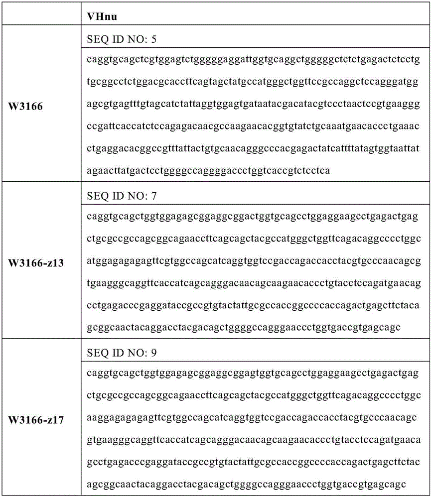
As used herein, "W3166" refers to a VHH antibody whose heavy chain variable region comprises the sequence of SEQ ID NO. 4.
As used herein, "W3166-z13" refers to a W3166-based humanized VHH antibody comprising a heavy chain variable region comprising the sequence of SEQ ID NO. 6. The affinity of the antigen of W3166-z13 is comparable to that of its parent antibody W3166.
As used herein, "W3166-z17" refers to a W3166-based humanized VHH antibody comprising a heavy chain variable region comprising the sequence of SEQ ID NO. 8. The affinity of the antigen of W3166-z17 is comparable to that of its parent antibody W3166.
The CDR sequences of anti-CTLA-4 single domain antibodies are shown in Table 1. Heavy chain variable region sequences are also provided in tables 2 and 3.
TABLE 1 CDR amino acid sequences

TABLE 2 variable region amino acid sequences


TABLE 3 variable region nucleotide sequences
In certain embodiments, the antibody polypeptides provided by the present disclosure are single domain antibodies.
In certain embodiments, the heavy chain variable domain of an antibody polypeptide provided by the present disclosure is derived from a VHH domain. The VHH domain is a heavy chain variable domain derived from a naturally occurring light chain-free antibody, e.g. an antibody from the family camelidae (see e.g. WO 9404678), e.g. in camels, llamas, dromedaries, alpacas and alpacas. The VHH domain is a single polypeptide and is stable.
In certain embodiments, the heavy chain variable domains of the antibody polypeptides provided by the present disclosure are derived from camelids.
CDRs are known to be responsible for antigen binding, but not all 6 CDRs are indispensable or unchanged. In other words, one or more CDRs can be replaced, altered or modified in the anti-CTLA-4 single domain antibody W3166, yet retain substantially their specific binding affinity to CTLA-4.
In certain embodiments, the anti-CTLA-4 antibody polypeptides provided by the present disclosure comprise the heavy chain CDR3 sequence of W3166. In certain embodiments, the anti-CTLA-4 antibody polypeptides provided by the present disclosure comprise the heavy chain CDR3 sequence of SEQ ID NO. 3. The heavy chain CDR3 region is centrally located in the antigen binding site and is therefore considered to be most exposed to the antigen, providing the greatest free energy for the affinity of the antibody for the antigen. Heavy chain CDR3 is also considered to be the CDR which has been the most diverse so far in terms of length, amino acid composition and conformation at the antigen binding site (Tonegawa S.Nature.302:575-81). The diversity of heavy chain CDR3 is sufficient to produce most antibody specificities (Xu JL, davis MM. Immunity. 13:37-45) and ideal antigen binding affinities (Schier R, etc. J Mol biol. 263:551-67).
In certain embodiments, the antibody polypeptides provided by the present disclosure comprise a suitable Framework Region (FR) sequence, so long as the antibody polypeptide is capable of specifically binding to CTLA-4. The CDR sequences provided in table 1 are obtained from camelid antibodies, but can be grafted to any suitable FR sequence of any suitable species (e.g., mouse, human, rat, rabbit, etc.) using suitable methods known in the art, such as recombinant techniques.
In certain embodiments, the antibody polypeptides provided by the present disclosure are humanized. Humanized antibody polypeptides are useful for reducing immunogenicity in humans. Because the non-human CDR sequences are grafted to human or substantially human FR sequences, the humanized antibody polypeptide is chimeric in its variable region. Humanization of antibody polypeptides can be achieved essentially by replacing CDR genes of non-human (e.g., murine) with corresponding human CDR genes in human immunoglobulin genes (see, e.g., jones et al (1986) Nature 321:522-525;Riechmann et al (1988) Nature 332:323-327;Verhoeyen et al (1988) Science 239:1534-1536).
Suitable human heavy chain variable domains may be selected for this purpose using methods known in the art. In an illustrative example, a "best fit" method may be used in which non-human (e.g., camelid) antibody variable domain sequences are screened or BLAST-screened from a database of known human variable domain sequences, the human sequence closest to the non-human search sequence is identified and used as the human backbone to which the non-human CDR sequences are grafted (see, e.g., sims et al, (1993) j.immunol.151:2296;Chothia et al (1987) j.mot.biol.196:901). Alternatively, non-human CDRs are grafted with a framework from conserved sequences derived from all human antibodies (see, e.g., carter et at. (1992) proc.Natl. Acad. Sci.USA,89:4285;Presta et al. (1993) J.Immunol., 151:2623).
In certain embodiments, the humanized antibody polypeptides provided by the present disclosure consist essentially of all human sequences except the CDR sequences of a human. In certain embodiments, the variable region FRs and constant region, if present, are derived entirely or substantially from human immunoglobulin sequences. The human FR sequence and the human constant region sequence may be derived from different human immunoglobulin genes, e.g., the FR sequence is derived from one human antibody and the constant region is derived from another human antibody. In some embodiments, the humanized antibody polypeptide comprises human FR1-4.
In certain embodiments, the humanized antibody polypeptides provided by the present disclosure include one or more FR sequences of W3166-z13 or W3166-z 17.
Both exemplary humanized anti-CTLA-4 single domain antibodies W3166-z13 and W3166-z17 retain specific binding affinity to CTLA-4 and are at least comparable to, or even better than, the parent camelid antibody in this regard.
In certain embodiments, the FR region derived from a human may comprise the same amino acid sequence as a human immunoglobulin. In certain embodiments, one or more amino acid residues of the human FR are replaced with corresponding residues from the parent non-human antibody. In certain embodiments, this may be useful for approximating a humanized antibody polypeptide to a non-human parent antibody structure. In certain embodiments, the humanized antibody polypeptides provided by the present disclosure comprise no more than 10, 9, 8, 7, 6, 5, 4, 3, 2, or 1 amino acid residue substitutions in each human FR sequence, or no more than 10, 9, 8, 7, 6, 5, 4, 3, 2, or 1 amino acid residue substitutions in all FRs of the heavy or light chain variable domain. In certain embodiments, such amino acid residue changes may be present in only the heavy chain FR region, only the light chain FR region, or both chains.
In certain embodiments, the antibody polypeptides provided by the present disclosure comprise a heavy chain variable domain selected from the group consisting of SEQ ID NO. 4, SEQ ID NO. 6, and SEQ ID NO. 8.
In certain embodiments, the anti-CTLA-4 antibody polypeptides provided by the present disclosure comprise all or a portion of a heavy chain variable domain. In one embodiment, the anti-CTLA-4 antibody polypeptide provided by the present disclosure is a single domain antibody consisting of all or a portion of the heavy chain variable domain provided by the present disclosure. More information on such single domain antibodies is available in the art (see, e.g., U.S. patent No. 6,248,516).
In certain embodiments, the anti-CTLA-4 antibody polypeptides provided by the present disclosure further comprise an immunoglobulin constant region. In certain embodiments, the immunoglobulin constant region comprises a heavy chain. The heavy chain constant region comprises a CH1, hinge, and/or CH2-CH3 region. In certain embodiments, the heavy chain constant region comprises an Fc region. In certain embodiments, the heavy chain constant region comprises or is a CH2-CH3 region.
In certain embodiments, the anti-CTLA-4 antibody polypeptides provided by the present disclosure have constant regions of immunoglobulins (Ig), optionally human Ig, optionally human IgG. The constant region may be of any suitable isotype. In certain embodiments, the anti-CTLA-4 antibody polypeptides provided herein comprise an IgG1 isotype constant region that can induce ADCC or CDC, or an IgG4 or IgG2 isotype constant region that has reduced or depleted effector function. Effector functions such as ADCC and CDC can lead to cytotoxicity of CTLA-4 expressing cells. Effector function may be assessed by various methods, such as Fc receptor binding assays, C1q binding assays, and cell lysis assays.
The binding affinity of the antibody polypeptides provided by the present disclosure may be used K D The value represents, K D The value represents the ratio of dissociation rate to binding rate (k) at which antigen and antigen binding molecule bind to reach equilibrium off /k on ). Antigen binding affinity (e.g. K D ) Suitable assays may be performed using suitable methods known in the art, including, for example, flow cytometry. In certain embodiments, binding of an antibody polypeptide to an antigen at different concentrations can be determined by flow cytometry, and the measured average fluorescence intensity (MFI) can be first plotted against the antibody concentration, and then calculated by fitting the specific binding fluorescence intensity (Y) and antibody concentration (X) to a single point saturation equation using Prims version 5 (GraphPad software, san diego, california): y=b max *X/(K D +X), wherein B max Refers to the maximum specific binding of the antibody polypeptide to be tested to the antigen.
In certain embodiments, the anti-CTLA-4 antibody polypeptides provided herein can specifically bind human CTLA-4 with binding affinity (K D ) By flow cytometryMeasured as not more than 5x10 -11 M is not more than 1x10 -10 M is not more than 5x10 -10 M is not more than 1x10 -9 M is not more than 5x10 -9 M。
Binding of antibody polypeptides to human CTLA-4 may also be accomplished using "half maximal effector concentrations" (EC 50 ) The values represent the concentration at which a 50% maximum effect (e.g., binding or inhibition, etc.) of an antibody is observed. EC (EC) 50 The values may be measured by methods known in the art, for example, sandwich methods such as ELISA, immunoblotting, flow cytometry, and other binding assays. In certain embodiments, the antibody polypeptides provided by the present disclosure are assayed by flow cytometry with an EC of no more than 0.5nM, no more than 1nM, no more than 2nM 50 (i.e., 50% binding concentration) to human CTLA-4.
In certain embodiments, the anti-CTLA-4 antibody polypeptides provided by the present disclosure can cross-react with cynomolgus CTLA-4.
In certain embodiments, the binding affinity of the antibody polypeptide to cynomolgus CTLA-4 is similar to its binding to human CTLA-4. For example, exemplary single domain antibodies W3166, W3166-z13 or W3166-z17 bind to cynomolgus monkey CTLA-4 with a similar K as human CTLA-4 D Or EC (EC) 50 Values.
In certain embodiments, the antibody polypeptides provided by the present disclosure are administered under flow cytometry with an EC of no more than 0.1nM, no more than 0.5nM, no more than 1nM 50 Or under flow cytometry with an EC of no more than 10nM, no more than 5nM, no more than 2nM, or no more than 1.2nM 50 Specifically binds to cynomolgus CTLA-4.
In certain embodiments, the antibody polypeptides provided herein have a specific binding affinity to human CTLA-4 sufficient for diagnostic and/or therapeutic use.
In certain embodiments, the antibody polypeptides provided herein block binding of human CTLA-4 to its ligands CD80 and CD86, thereby providing biological activity, including, for example, including that produced by activated T cells (e.g., CD4 + T cells and CD8 + T cells) to produce cytokines, inducing activated T cells (e.g., CD 4) + T cells and CD8 + T cells) proliferation and reversing inhibition of T regAnd (5) a function. Exemplary cytokines include IL-2 and IFN gamma. Cytokine production can be determined using methods known in the art, such as by ELISA. Comprises [ 3 H]Methods such as thymidine incorporation can also be used to detect T cell proliferation.
The antibody polypeptides provided by the present disclosure may be monoclonal, humanized, chimeric, recombinant, labeled, bivalent, or anti-idiotypic. Recombinant antibody polypeptides are antibody polypeptides that are produced in vitro, rather than in an animal, using recombinant methods.
Variants
The antibody polypeptides provided by the present disclosure also include various variants thereof. In certain embodiments, the antibody polypeptides comprise various types of variants of the exemplary antibodies provided herein, namely W3166, W3166-z13, and W3166-z17.
In certain embodiments, the antibody polypeptide variants include one or more modifications or substitutions in one or more CDR sequences provided in table 1, one or more variable region sequences provided in table 2 (but not in any CDR sequences), and/or in a constant region (e.g., fc region). Such variants retain their CTLA-4 specific binding affinity of the parent antibody, but have one or more properties conferred by modification or substitution. For example, antibody polypeptide variants may have improved antigen binding affinity, improved productivity, improved stability, improved glycosylation pattern, reduced risk of glycosylation, reduced deamination, reduced or depleted effector function, improved FcRn receptor binding, increased pharmacokinetic half-life, pH sensitivity, and/or conjugation compatibility (e.g., one or more introduced cysteine residues).
The parent antibody sequences may be screened using methods known in the art, such as "alanine scanning mutagenesis," to identify suitable or preferred residues to be modified or substituted (see, e.g., cunningham and Wells (1989) Science, 244:1081-1085). Briefly, residues of interest (e.g., charged residues such as Arg, asp, his, lys and Glu) can be identified and replaced with neutral or negatively charged amino acids (e.g., alanine or polyalanine), and modified antibody polypeptides prepared and screened for a property of interest. If a substitution at a particular amino acid position shows a functional change of interest, that position can be identified as a potential modification or substitution residue. Potential residues can be further assessed by substitution with different types of residues (e.g., cysteine residues, positively charged residues, etc.).
Affinity variants
The affinity variants may comprise modifications or substitutions of one or more CDR sequences, one or more FR sequences provided in table 1, or heavy chain variable region sequences provided in table 2. From the CDR sequences in table 1 and the variable region sequences in table 2, the person skilled in the art can easily recognize FR sequences, since it is well known in the art that the CDR regions are flanked by two FR regions in the variable region. The affinity variant retains specific binding affinity to the parent antibody CTLA-4, even with higher CTLA-4 specific binding affinity than the parent antibody. In certain embodiments, substitutions of at least one (or all) of the CDR sequences, FR sequences, or variable region sequences comprise conservative substitutions.
The skilled artisan will appreciate that one or more amino acid residues may be substituted in the CDR sequences and variable region sequences provided in tables 1 and 2, and that the resulting antibody polypeptide still retains binding affinity to CTLA-4, even with improved binding affinity. Various methods known in the art may be used to achieve this. For example, a library of antibody variants (e.g., fab or scFv variants) can be generated and expressed by phage display techniques, and then screened for binding affinity to human CTLA-4. As another example, computer software can be used to mimic binding of an antibody to human CTLA-4 and recognize amino acid residues on the antibody that form a binding interface. These residues may be circumvented in substitutions to prevent a decrease in binding affinity, or targeted substitutions to provide stronger binding.
In certain embodiments, the humanized antibody polypeptides provided by the present disclosure include one or more CDR sequences, and/or one or more amino acid residue substitutions in one or more FR sequences. In certain embodiments, the affinity variants comprise no more than 10, 9, 8, 7, 6, 5, 4, 3, 2, or 1 substitutions in total in the CDR sequences and/or FR sequences.
In certain embodiments, the anti-CTLA-4 antibody polypeptide comprises 1, 2, or 3 CDR sequences having at least 80% (e.g., at least 85%, 88%, 90%, 91%, 92%, 93%, 94%, 95%, 96%, 97%, 98%, 99%) sequence identity to that (or those) listed in table 1, while also maintaining a similar or even higher binding affinity level for CTLA-4 to the parent antibody.
In certain embodiments, the anti-CTLA-4 antibody polypeptide comprises one or more variable region sequences having at least 80% (e.g., at least 85%, 88%, 90%, 91%, 92%, 93%, 94%, 95%, 96%, 97%, 98%, 99%) sequence identity to that (or those) listed in table 2, while also maintaining a similar or even higher binding affinity level for CTLA-4 to the parent antibody. In certain embodiments, a total of 1 to 10 amino acids are substituted, inserted, or deleted in the variable region sequences listed in table 2. In some embodiments, substitutions, insertions, or deletions occur in regions outside of the CDRs (e.g., in the FRs).
Glycosylation variants
The anti-CTLA-4 antibody polypeptides provided by the present disclosure also include glycosylation variants, which can be obtained by increasing or decreasing the degree of glycosylation of the antibody polypeptide.
An antibody polypeptide may comprise one or more amino acid residues with side chains to which a carbohydrate moiety (e.g., an oligosaccharide structure) may be attached. Glycosylation of antibodies is typically either N-linked or O-linked. N-linked refers to the attachment of a carbohydrate moiety to a side chain of an asparagine residue, e.g., an asparagine residue in a tripeptide sequence such as asparagine-X-serine and asparagine-X-threonine, where X is any amino acid other than proline. O-linked glycosylation refers to the attachment of one of the sugars N-acetylgalactosamine, galactose or xylose to a hydroxy amino acid, most commonly serine or threonine. Removal of the native glycosylation site can be conveniently accomplished, for example, by altering the amino acid sequence such that one of the above tripeptide sequences (for the N-linked glycosylation site) or serine or threonine residues (for the O-linked glycosylation site) present in the sequence is replaced. By introducing such tripeptide sequences or serine or threonine residues, new glycosylation sites can be created in a similar manner. In certain embodiments, the heavy chain CDR2 of the antibodies provided by the present disclosure comprises an N55Q substitution (kabat numbering) such that the potential glycosylation site is removed.
Cysteine engineered variants
The present disclosure provides anti-CTLA-4 antibody polypeptides further comprising a cysteine engineered variant comprising one or more introduced free cysteine amino acid residues.
The free cysteine residues are not part of a disulfide bridge. The cysteine engineered variants may be used to bind to, for example, cytotoxic and/or imaging compounds, tags or radioisomers, etc., at the engineered cysteine site by, for example, maleimide or haloacetyl. Methods for designing antibody polypeptides to introduce free cysteine residues are known in the art, see, e.g., WO2006/034488.
Variants
The present disclosure provides anti-CTLA-4 antibody polypeptides further comprising an Fc variant comprising one or more amino acid residue modifications or substitutions in its Fc region and/or hinge region.
In certain embodiments, the anti-CTLA-4 antibody polypeptide comprises one or more amino acid substitutions that improve pH-dependent binding to neonatal Fc receptor (FcRn). The pharmacokinetic half-life of this variant is long because it binds to FcRn at acidic pH, thus protecting it from lysosomal degradation, and then translocates and releases the cells. Methods for designing antibody polypeptides to improve binding affinity to FcRn are well known in the art, see, e.g., vaughn, D.et al, structure,6 (1): 63-73,1998; kontermann, R.et al, antibody engineering, volume 1, chapter 27: fc region engineering to improve PK, spellinger publication, 2010; yeung, Y.et al, cancer Research,70:3269-3277 (2010); and Hinton, P.et al, J.immunology,176:346-356 (2006).
In certain embodiments, the anti-CTLA-4 antibody polypeptide comprises one or more amino acid substitutions that alter antibody-dependent cellular cytotoxicity (ADCC). Certain amino acid residues of the Fc region (e.g., CH2 domain) may be substituted to provide altered (e.g., enhanced, reduced, or depleted) ADCC activity. Alternatively or additionally, the carbohydrate structure on the antibody may be altered to alter (e.g., enhance, reduce or deplete) ADCC activity. Methods for altering ADCC activity by antibody engineering have been described in the art, see, e.g., shields RL et al, J Biol chem.2001.276 (9): 6591-604; idusogenie EE et al, J Immunol 2000.164 (8): 4178-84; steurer W.et al, J Immunol.1995,155 (3): 1165-74; idusogenie EE et al, J Immunol 2001,166 (4): 2571-5; lazar GA.et al, PNAS,2006,103 (11): 4005-4010; ryan mc.et al, mol.cancer ter., 2007,6:3009-3018; richards JO et al Mol Cancer ter.2008, 7 (8): 2517-27; shields R.L.et al, J.biol.chem,2002,277:26733-26740; shinkawa T.et al, J.biol. Chem,2003,278:3466-3473.
In certain embodiments, the anti-CTLA-4 antibody polypeptide comprises one or more amino acid substitutions that alter Complement Dependent Cytotoxicity (CDC), e.g., by improving or reducing C1q binding and/or CDC (see, e.g., WO99/51642;Duncan&Winter Nature 322:738-40 (1988); U.S. Pat. No. 5,648,260; U.S. Pat. No. 5,624,821); and WO94/29351 regarding other examples of variants of the Fc region.
In certain embodiments, the anti-CTLA-4 antibody polypeptide comprises one or more amino acid substitutions at the interface of the Fc region to accelerate and/or promote heterodimerization. These modifications include introducing a protuberance into a first Fc polypeptide and introducing a cavity into a second Fc polypeptide, wherein the protuberance can be positioned in the cavity to facilitate interaction of the first and second Fc polypeptides to form a heterodimer or complex. Methods for producing antibodies using these modifications are known in the art, for example, as described in U.S. Pat. No. 5,731,168.
A variety of techniques can be used to generate VHH or single domain antibodies. For example, VHH can be obtained using methods known in the art, for example by immunizing a camel and obtaining hybridomas therefrom, or by cloning a single domain antibody library using molecular biology techniques known in the art and subsequently selecting for VHH using phage display.
In another aspect of the invention, the present disclosure provides an antibody polypeptide that can comprise two or more linked single domain antibodies. The sequences of single domain antibodies may be identical and directed against the same target or antigen. Depending on the number of VHHs attached, the antibody polypeptide may be bivalent (2 VHHs), trivalent (3 VHHs), tetravalent (4 VHHs) or a molecule with a higher valency.
Conjugate(s)
In certain embodiments, the anti-CTLA-4 antibody polypeptide further comprises a conjugated group. The conjugated group may be attached to the antibody polypeptide. The conjugated group is a non-protein moiety that may be attached to an antibody polypeptide. It is contemplated that there may be a variety of conjugated groups attached to the antibody polypeptides provided by the present disclosure (see, e.g., "conjugate vaccine", donated microbiology and immunology, j.m.use and r.e.lewis, jr. Edit, new york cager press, (1989)). These conjugated groups may be attached to the antibody polypeptide by covalent binding, affinity binding, intercalation, coordination binding, complexation, association, mixing or addition, etc.
In certain embodiments, the antibody polypeptides disclosed in the present disclosure may be designed to comprise specific sites beyond the epitope-binding portion, which may be used to bind to one or more conjugated groups. For example, such sites may include one or more active amino acid residues, such as cysteine or histidine residues, to facilitate covalent attachment to the conjugated group.
In certain embodiments, the antibody may be linked to a conjugated group, either indirectly or through another conjugated group. For example, the antibody polypeptide may be conjugated to biotin and then indirectly conjugated to a second conjugate that is abamectin Ding Gonge. The conjugate may be a scavenging modifier, a toxin (e.g., a chemotherapeutic agent), a detectable label (e.g., a radioisotope, a lanthanide, a luminescent label, a fluorescent label, or an enzyme substrate label), or a purification group.
A "toxin" may be any agent that is harmful to or capable of damaging or killing a cell. Exemplary toxins include, but are not limited to, paclitaxel, cytochalasin B, gramicidin D, ethidium bromide, ipecine, mitomycin, etoposide, tenoposide, vincristine, MMAE, MMAF, DM, vinblastine, colchicine, doxorubicin, daunorubicin, dihydroxyanthraquinone, mitoxantrone, milteframycin, dactinomycin, 1-dehydroproteinsterone, glucocorticoids, procaine, tetracaine, lidocaine, propranolol, puromycin and analogs thereof, antimetabolites (e.g., methotrexate, 6-mercaptopurine, 6-thioguanine, cytarabine, 5-fluorouracil norhydrazine), alkylating agents (e.g., chloroform, chloro Ding Liuniao, melphalan, carmustine (BSNU) and lomustine (CCNU), cyclophosphamide, busulfamide, dibromonitrobenzene, streptozotocin, mitomycin C and cisplatin (II), anthracyclines (e.g., mitomycin), mitomycin (e.g., mitomycin), and the like, and the antimuscarin (e.g., the mitomycin), the antimuscarin (e.g., the mitomycin) and the antimuscarin (e.g., the mitomycin).
Exemplary detectable labels may include fluorescent labels (e.g., fluorescein, rhodamine, dansyl, phycoerythrin, or texas red), enzyme substrate labels (e.g., horseradish peroxidase, alkaline phosphatase, luciferase, glucoamylase, lysozyme, glycooxidase, or beta-D-galactosidase), radioisotopes (e.g., 123 I、 124 I、 125 I、 131 I、 35 S、 3 H、 111 In、 112 In、 14 C、 64 Cu、 67 Cu、 86 Y、 88 Y、 90 Y、 177 Lu、 211 At、 186 Re、 188 Re、 153 Sm、 212 bi and Bi 32 P, other lanthanoid), luminescent labels, chromophores, digoxin, biotin/avidin, DNA molecules for detection, or gold.
In certain embodiments, the conjugated groups may be scavenging modifiers that help increase the half-life of the antibody. Illustrative examples include water-soluble polymers such as PEG, carboxymethyl cellulose, dextran, polyvinyl alcohol, polyvinylpyrrolidone, ethylene glycol/propylene glycol copolymers, and the like. The multimer may be of any molecular weight and may be branched or unbranched. The number of multimers attached to an antibody may vary, and if more than one multimer is attached, it may be the same or different molecules.
In certain embodiments, the conjugated groups may be purified groups, such as magnetic beads.
In certain embodiments, the antibody polypeptides provided by the present disclosure may be used as a substrate for conjugation.
Polynucleotide and recombination method
The present invention provides polynucleotides encoding anti-CTLA-4 antibody polypeptides.
The term "nucleic acid" or "polynucleotide" as used in this disclosure refers to deoxyribonucleic acid (DNA) or ribonucleic acid (RNA) and polymers thereof in single or double stranded form. Unless specifically limited, the term includes polynucleotides containing known natural nucleotide analogs that have similar binding properties as the reference nucleic acid and are metabolized in a manner similar to naturally occurring nucleotides. Unless otherwise indicated, a particular polynucleotide sequence also implicitly encompasses conservatively modified variants thereof (e.g., degenerate codon substitutions), alleles, homologous sequences, SNPs, and complementary sequences, as well as the sequence explicitly indicated. Specifically, degenerate codon substitutions may be achieved by substituting a mixed base and/or deoxyribose residue for the sequence resulting from the third position of one or more selected (or all) codons (see Batzer et al, nucleic Acid Res.19:5081 (1991); ohtsuka et al, J.biol. Chem.260:2605-2608 (1985); and Rossolini et al, mol. Cell. Probes 8:91-98 (1994)).
In certain embodiments, the polynucleotide comprises one or more nucleotide sequences as set forth in SEQ ID Nos 5, 7, 9 (e.g., at least 85%, 88%, 90%, 92%, 93%, 94%, 95%, 96%, 97%, 98%, or 99%), and/or homologous sequences thereof having at least 80% sequence identity, and/or variants thereof having only degenerate substitutions, and encodes the variable regions of the exemplary antibodies provided by the present disclosure. DNA encoding monoclonal antibodies can be readily isolated and sequenced using conventional procedures (e.g., using oligonucleotide probes that are capable of binding specifically to genes encoding heavy and light chains of the antibody). The coding DNA may also be obtained synthetically.
Isolated polynucleotides encoding anti-CTLA-4 antibody polypeptides (e.g., comprising the sequences shown in table 3) can be inserted into vectors for further cloning (amplification of DNA) or expression using recombinant techniques known in the art. There are many vectors available. The carrier assembly generally includes, but is not limited to, one or more of the following: a signal sequence, an origin of replication, one or more marker genes, an enhancer element, a promoter (e.g., SV40, CMV, EF-1. Alpha.) and a transcription termination sequence.
The present invention provides vectors (e.g., expression vectors) comprising at least one promoter (e.g., SV40, CMV, EF-1 a) operably linked to a nucleic acid sequence provided by the present disclosure encoding an antibody polypeptide, and at least one selectable marker. Exemplary vectors include, but are not limited to, retrovirus (including lentivirus), adenovirus, adeno-associated virus, herpes virus (e.g., herpes simplex virus), poxvirus, baculovirus, papilloma virus, pasteur virus (e.g., SV 40), lambda phage and M13 phage, plasmid pcDNA3.3, pMD18-T, pOptivec, pCMV, pEGFP, pIRES, pQD-Hyg-GSeu, pALTER, pBAD, pcDNA, pCal, pL, pET, pGEMEX, pGEX, pCI, pEGFT, pSV2, pFUSE, pVITRO, pVIVO, pMAL, pMONO, pSELECT, pUNO, pDUO, psg5L, pBABE, pWPXL, pBI, p15TV-L, pPro, pTD, pRS10, pLexA, pACT2.2, pCMV-SCRIPT.RTM, pCDM8, pCDNA1.1/amp, pcDNA3.1, pRc/RSV, PCR 2.1, pEF-1, pFB, pSG5, pXT1, pCDEF3, pSVSPORT, pEF-Bos, and the like.
Vectors comprising polynucleotide sequences encoding antibody polypeptides may be introduced into host cells for cloning or gene expression. Suitable host cells for cloning or expressing DNA in the vectors of the present disclosure are prokaryotes, yeast, or higher eukaryote cells described above. Prokaryotes suitable for this purpose include eubacteria, such as gram-negative or gram-positive organisms, for example enterobacteriaceae such as e.coli, e.g. e.coli, enterobacteria, erwinia, klebsiella, proteus, salmonella, e.g. salmonella typhimurium, serratia, e.g. serratia marcescens, and shigella, and bacilli, e.g. bacillus subtilis and bacillus licheniformis, pseudomonas, e.g. pseudomonas aeruginosa, and streptomyces.
In addition to prokaryotes, eukaryotic microbes such as filamentous fungi, yeast, etc., are suitable cloning or expression hosts for expressing anti-CTLA-4 antibody polypeptides. Saccharomyces cerevisiae, or Saccharomyces cerevisiae, is the most commonly used lower eukaryotic host microorganism. However, many other genera, species, and strains are also common and used in the present disclosure, such as schizosaccharomyces pombe; kluyveromyces hosts, such as, for example, kluyveromyces lactis, kluyveromyces fragilis (ATCC 12,424), kluyveromyces bulgaricus (ATCC 16,045), kluyveromyces wikipedia (ATCC 24,178), kluyveromyces Wo Erdi (ATCC 56,500), kluyveromyces drosophila (ATCC 36,906), kluyveromyces marxianus Wen Kelu and Kluyveromyces marxianus; yarrowia yeast (EP 402,226); methanol yeast (EP 183,070); candida species; trichoderma reesei (EP 244,234); neurospora crassa; schwann yeasts such as schwann yeast; and filamentous fungi such as, for example, aspergillus, penicillium, curvularia, and Aspergillus hosts such as Aspergillus nidulans and Aspergillus niger.
Suitable host cells for expressing glycosylated antibodies or antigen fragments provided by the present disclosure are derived from multicellular organisms. Exemplary invertebrate cells include plant cells and insect cells. Many baculovirus strains and varieties from hosts and corresponding permissive insect host cells have been identified, such as spodoptera frugiperda (trichomonas), aedes aegypti (mosquito), aedes albopictus (mosquito), drosophila melanogaster (drosophila) and bombyx mori. A variety of viral strains for transfection are publicly available, for example, L-1 variants of Spodoptera frugiperda NPV and Bm-5 strain of Bombyx mori NPV, and according to the present invention, these viruses are useful as viruses in the present disclosure, particularly for transfection of Spodoptera frugiperda cells. Plant cells of cultured cotton, corn, potato, soybean, petunia, tomato, and tobacco can also be used as hosts.
However, there is a maximum interest in vertebrate cells, and passage of vertebrate cells in culture (tissue culture) has become a routine approach. An exemplary mammalian host cell line that can be used is the SV40 transformed monkey kidney CV1 line (COS-7, ATCC CRL 1651); human embryonic kidney (293 or 293 cells grown subcloned for suspension culture, graham et al, J.Gen. Virol.36:59 (1977)); baby hamster kidney cells (BHK, ATCC CCL 10); chinese hamster ovary cells/-DHFR (CHO, urlaub et al, proc.Natl. Acad.sci.usa 77:4216 (1980)); mouse testis support cells (TM 4, mather, biol. Reprod.23:243-251 (1980)); monkey kidney cells (CV 1 ATCC CCL 70); african green monkey kidney cells (VERO-76, ATCC CRL-1587); human cervical cancer cells (HELA, ATCC CCL 2); canine kidney cells (MDCK, ATCC CCL 34); buffalo rat hepatocytes (BRL 3a, atcc CRL 1442); human lung cells (W138, ATCC CCL 75); human hepatocytes (Hep G2, HB 8065); mouse mammary tumor (MMT 060562,ATCC CCL51); TRI cells (Mather et al, annals N.Y. Acad. Sci.383:44-68 (1982)); MRC 5 cells; FS4 cells; human liver cancer cell line (Hep G2). In certain preferred embodiments, the host cell is a 293F cell.
The host cells are transformed with the above-described expression or cloning vectors to produce the anti-CTLA-4 antibody polypeptide, and cultured in conventional nutrient media suitably modified for the purpose of inducing promoters, selecting transformants, or amplifying the genes encoding the desired sequences. In another embodiment, the antibody polypeptide may be produced by homologous recombination as known in the art.
Host cells for producing the antibody polypeptides provided by the present disclosure may be cultured in a variety of media. Commercially available media such as Ham's F (sigma), minimal Essential Medium (MEM) (sigma), RPMI-1640 (sigma) and Dulbecco's Modified Eagle Medium (DMEM) (sigma) are suitable for culturing host cells. Furthermore, any of the compositions described in Ham et al, meth.Enz.58:44 (1979), barnes et al, anal biochem.102:255 (1980), U.S. Pat. No. 4,767,704;4,657,866;4,927,762;4,560,655; or 5,122,469; WO 90/03430; WO 87/00195; or as described in us reissue patent 30,985, may be used as a medium for culturing host cells. Any of these media may be supplemented as desired with hormones and/or other growth factors (e.g., insulin, transferrin or epidermal growth factor), salts (e.g., sodium chloride, calcium, magnesium and phosphate), buffers (e.g., HEPES), nucleotides (e.g., adenosine and thymidine), antibiotics (e.g., gentamicin drugs), trace elements (defined as inorganic compounds, typically present in a final concentration in the micromolar range), and glucose or equivalent energy sources. Any other necessary supplements may also be included at appropriate concentrations as known to those skilled in the art. Culture conditions, such as temperature, pH, etc., are those of the host cells previously selected for expression and will be apparent to those skilled in the art.
When recombinant techniques are used, the antibody polypeptide may be produced intracellularly, in the interstitial space, or directly secreted into the medium. If the antibodies are produced intracellularly, as a first step, the particulate debris, host cells or lysed debris is removed, for example, by centrifugation or ultrafiltration. Carter et al, bio/Technology 10:163-167 (1992) describes a method of isolating antibodies secreted into the E.coli interstitial space. Briefly, cell pastes were thawed in the presence of sodium acetate (pH 3.5), EDTA and phenylmethylsulfonyl fluoride (PMSF) for about 30 minutes. Cell debris can be removed by centrifugation. When antibodies are secreted into the culture medium, the supernatant from such expression systems is typically first concentrated using a commercially available protein concentration filter, such as an Amicon or milbepecon ultrafiltration device. In any of the above steps, a protease inhibitor such as PMSF may be included to inhibit proteolysis, and an antibiotic may be included to prevent the growth of foreign contaminants.
The anti-CTLA-4 antibody polypeptide prepared from the cells may be purified using, for example, hydroxyapatite chromatography, gel electrophoresis, dialysis, DEAE cellulose ion exchange chromatography, ammonium sulfate precipitation, salting out, and affinity chromatography, with affinity chromatography being a preferred purification technique.
In certain embodiments, protein a immobilized on a solid phase is used for immunoaffinity purification of antibody polypeptides. The suitability of protein a as an affinity ligand depends on the type and isotype of any immunoglobulin Fc domain present in the antibody. Protein a can be used to purify antibodies based on human γ1, γ2 or γ4 heavy chains (Lindmarm et al, J.Immunol. Meth.62:1-13 (1983)). Protein G was recommended for all mouse isotypes and human gamma 3 (Guss et al, EMBO J.5:1567 1575 (1986)). The matrix to which the affinity ligand is attached is typically agarose, but there are other matrices as well. Mechanically stable matrices, such as controlled pore glass or poly (styrene divinyl) benzene, have faster flow rates and shorter processing times than agarose. When the antibody comprises a CH3 domain, bakerbond ABX TM Resins (J.T. Baker, new Jersey Phillips fort) can be used for purification. Other techniques of protein purification, such as fractionation on ion exchange columns, ethanol precipitation, reverse phase HPLC, chromatography on silica, heparin SEPHAROSE on anion or cation exchange resins (e.g., polyaspartic acid columns), can also be used depending on the antibody to be recovered TM Chromatography, chromatofocusing, SDS-PAGE and ammonium sulfate precipitation.
After any preliminary purification steps, the mixture comprising the antibody of interest and the contaminant may be subjected to low pH hydrophobic interaction chromatography using an elution buffer (pH between about 2.5-4.5), preferably at low salt concentrations (e.g., from about 0-0.25M salt).
Pharmaceutical compositions
The invention further provides pharmaceutical compositions comprising the anti-CTLA-4 antibody polypeptides provided herein and one or more pharmaceutically acceptable carriers.
Pharmaceutically acceptable carriers for use in the pharmaceutical compositions disclosed herein can include, for example, pharmaceutically acceptable liquid, gel, or solid carriers, aqueous carriers, non-aqueous carriers, antibacterial agents, isotonic agents, buffers, antioxidants, anesthetics, suspending/dispersing agents, sequestering or chelating agents, diluents, adjuvants, excipients, or non-toxic auxiliary substances, other ingredients known in the art, or various combinations thereof.
Suitable ingredients may include, for example, antioxidants, fillers, binders, disintegrants, buffers, preservatives, lubricants, flavouring agents, thickening agents, colouring agents, emulsifying agents or stabilizing agents, for example sugars and cyclodextrins. Suitable antioxidants may include, for example, methionine, ascorbic acid, EDTA, sodium thiosulfate, platinum, catalase, citric acid, cysteine, thioglycerol, thioglycolic acid, thiosorbitol, butylated hydroxytoluene, and/or propyl gallate. As disclosed in the present disclosure, inclusion of one or more antioxidants, such as methionine, in a composition comprising an antibody polypeptide and a conjugate as provided in the present disclosure can reduce oxidation of the antibody polypeptide. This reduction in oxidation can prevent or reduce loss of binding affinity, thereby improving antibody stability and maximizing shelf life. Thus, in certain embodiments, provided compositions comprise one or more antibody polypeptides disclosed in the present disclosure and one or more antioxidants such as methionine. Further provided are methods of preventing oxidation, extending shelf life, and/or improving efficacy of an antibody polypeptide of the present disclosure by mixing the antibody polypeptide with one or more antioxidants, such as methionine.
To further illustrate, pharmaceutically acceptable carriers may include, for example, aqueous carriers such as sodium chloride injection, ringer's injection, isotonic dextrose injection, sterile water injection or dextrose and lactate ringer's injection, non-aqueous carriers such as fixed oils of vegetable origin, cottonseed oil, corn oil, sesame or peanut oil, antibacterial agents to inhibit bacteria or to inhibit fungal concentrations, isotonic agents such as sodium chloride or dextrose, buffers such as phosphate or citrate buffers, antioxidants such as sodium bisulfate, local anesthetics such as procaine hydrochloride, suspending and dispersing agents such as sodium carboxymethyl cellulose, hydroxypropyl methylcellulose or polyvinylpyrrolidone, emulsifying agents such as polysorbate 80 (tween-80), sequestering or chelating agents such as EDTA (ethylenediamine tetraacetic acid) or EGTA (ethylene glycol tetraacetic acid), ethanol, polyethylene glycol, propylene glycol, sodium hydroxide, hydrochloric acid, citric acid or lactic acid. The antimicrobial agent used as a carrier may be added to a pharmaceutical composition in a multi-dose container comprising phenol or cresol, mercury, benzyl alcohol, chlorobutanol, methyl and propyl parabens, thimerosal, benzalkonium chloride, and benzethonium bromide. Suitable excipients may include, for example, water, physiological saline, dextrose, glycerol or ethanol. Suitable non-toxic auxiliary substances may include, for example, wetting or emulsifying agents, pH buffering agents, stabilizers, solubility enhancers, or agents such as sodium acetate, sorbitan monolaurate, triethanolamine oleate, or cyclodextrins.
The pharmaceutical composition may be a liquid solution, suspension, emulsion, pill, capsule, tablet, sustained release formulation or powder. Oral formulations may include standard carriers such as pharmaceutical grades of mannitol, lactose, starch, magnesium stearate, polyvinylpyrrolidone, sodium saccharine, cellulose, magnesium carbonate, and the like.
In certain embodiments, the pharmaceutical composition is formulated as an injectable composition. The injectable pharmaceutical composition may be prepared in any conventional form, for example, as a liquid solution, suspension, emulsion or solid form suitable for producing a liquid solution, suspension or emulsion. Formulations for injection may include sterile and/or non-hot solutions ready for injection, sterile dry soluble products ready for mixing with solvents prior to use, such as lyophilized powders, including subcutaneous tablets, sterile suspensions ready for injection, sterile dry insoluble products ready for combination with carriers prior to use, and sterile and/or non-hot emulsions. The solution may be an aqueous solution or a non-aqueous solution.
In certain embodiments, the unit dose parenteral formulation is packaged in an ampoule, vial or syringe with needle. All formulations for parenteral administration should be sterile and non-exothermic, as known and practiced in the art.
In certain embodiments, sterile lyophilized powders are prepared by dissolving an antibody polypeptide disclosed in the present disclosure in a suitable solvent. The solvent may contain an excipient to enhance stability, or other pharmacological component of the powder, or a reconstituted solution prepared from the powder. Excipients that may be used include, but are not limited to, water, dextrose, sorbitol, fructose, corn syrup, xylitol, glycerol, dextrose, sucrose or other suitable agents. The solvent may contain a buffer such as citrate, sodium or potassium phosphate or other such buffers known to those skilled in the art, which in one embodiment is about neutral pH. Subsequent sterile filtration of the solution, followed by lyophilization under standard conditions known to those skilled in the art, provides the desired formulation. In one embodiment, the resulting solution will be dispensed into vials for lyophilization. Each vial may contain a single dose or multiple doses of an anti-CTLA-4 antibody polypeptide or a composition thereof. The vial may be overfilled in an amount greater than that required for a dose or group of doses (e.g., about 10%) to facilitate accurate sample extraction and accurate administration. The lyophilized powder may be stored under suitable conditions, for example, at about 4 ℃ to room temperature.
The freeze-dried powder is recombined by water for injection, so that a preparation for parenteral administration can be provided. In one embodiment, sterile and/or unheated water or other liquid suitable carrier is added to the lyophilized powder for reconstitution. The exact dosage will depend on the treatment method chosen and can be determined empirically.
Application method
The invention also provides a method of treatment comprising: administering to a subject in need thereof a therapeutically effective amount of an antibody polypeptide, thereby treating or preventing a CTLA-4-associated disorder or disease. In some embodiments, the CTLA-4-associated disorder or disease is cancer, autoimmune disease, inflammatory disease, infectious disease, graft Versus Host Disease (GVHD), or graft rejection.
Exemplary cancers include, but are not limited to, lymphoma, bladder cancer, bone cancer, brain and central nervous system cancer, breast cancer, uterine or endometrial cancer, rectal cancer, esophageal cancer, head and neck cancer, anal cancer, gastrointestinal cancer, intraepithelial tumors, kidney (kidney) cancer or renal cell (renal) cancer, leukemia, liver cancer, lung cancer (non-small cell lung cancer or small cell lung cancer), melanoma, myeloma, pancreatic cancer, prostate cancer, sarcoma, skin cancer, squamous cell carcinoma, gastric cancer, testicular cancer, vulval cancer, cancer of the endocrine system, parathyroid cancer, adrenal cancer, penile cancer, childhood solid tumors, tumor hemangiomas, spinal tumors, pituitary adenomas, or epidermoid cancer.
Exemplary autoimmune diseases include, but are not limited to, acquired immunodeficiency syndrome (aids, a viral disease with autoimmune components), alopecia areata, ankylosing spondylitis, antiphospholipid syndrome, autoimmune addison's disease, autoimmune hemolytic anemia, autoimmune hepatitis, autoimmune Inner Ear Disease (AIED), autoimmune lymphoproliferative syndrome (ALPS), autoimmune Thrombocytopenic Purpura (ATP), behcet's disease, cardiomyopathy, celiac hydroalcoholic hepatitis; chronic Fatigue Immune Dysfunction Syndrome (CFIDS), chronic inflammatory demyelinating polyneuropathy (CIPD), cicatricial pemphigus, collectin disease, CREST syndrome, crohn's disease, degoss disease, juvenile dermatomyositis, discoid lupus, primary mixed cryoglobulinemia, fibromyalgia, fibromyositis, graves' disease, grignard-Barre syndrome, hashimoto thyroiditis, idiopathic pulmonary fibrosis, idiopathic Thrombocytopenic Purpura (ITP), igA nephropathy, insulin dependent diabetes mellitus, juvenile chronic arthritis (Steve's disease), juvenile rheumatoid arthritis, meniere's disease, mixed connective tissue disease, multiple sclerosis, myasthenia gravis, lupus anemia, polyarteritis nodosa, polyarthritis, polymorpha rheumatica, polymyositis and dermatomyositis, primary agallosis, primary biliary cirrhosis, psoriasis, psoriatic arthritis, raynaud's phenomenon, rate's syndrome, rheumatic fever, rheumatoid arthritis, sarcoidosis, scleroderma (progressive systemic sclerosis (PSS), also known as Systemic Sclerosis (SS)), sjogren's syndrome, hard Han syndrome, systemic lupus erythematosus, large arteritis, temporal arteritis/giant cell arteritis, ulcerative colitis, uveitis, vitiligo and Wegener's granulomatosis.
Inflammatory diseases, including chronic and acute inflammatory diseases. Exemplary inflammatory diseases include Alzheimer's disease, asthma, atopic allergy, atherosclerosis, bronchial asthma, eczema, glomerulonephritis, graft versus host disease, hemolytic anemia, osteoarthritis, sepsis, stroke, tissue and organ transplantation, vasculitis, diabetic retinopathy, and ventilator-induced lung injury.
Exemplary infectious diseases include, but are not limited to, fungal infections, parasitic/protozoal infections, or chronic viral infections, e.g., malaria, coccidiosis, mycoplasmosis, histoplasmosis, onychomycosis, aspergillosis, blastomycosis, candida albicans, paracoccidiosis, microsporosis, acanthamoeba keratitis, amebiasis, ascariasis, babesiasis, balanozoosis, bepotash's ascariasis, chagas disease, clonorchiasis, bipulmonary fluke, maidenhair nematodiasis, echinococcosis, elephantosis, enterobiasis, fasciolopsis, gingivalis, filariasis, giardiasis, jaw nematodiasis, membranous taeniasis, isospora, wheeza, leishmaniasis, lyme, echinococcosis, myiasis, discoidea, myiasis, schistosomiasis lice, sarcoptic mite, schistosomiasis, comatose, nematode, tapeworm, toxoplasmosis, trichinosis, whipworm infection, hepatitis B (HBV) infection, hepatitis C (HCV) infection, herpes virus, epstein-barr virus, aids virus, cytomegalovirus, herpes simplex virus type I, herpes simplex virus type II, human papilloma virus, adenovirus, human immunodeficiency virus I, human immunodeficiency virus II, kaposi-westerr sarcoma-associated herpes virus epidemic, circovirus, human T lymphocyte trophic virus type I, human T lymphocyte trophic virus type II, varicella zoster, JC virus or BK virus.
The therapeutically effective amount of the antibody polypeptide provided by the present disclosure will depend on various factors known in the art, such as body weight, age, history of use, active drug, likelihood of health and cross-reaction of the subject, allergies, sensitivity and adverse side effects, as well as route of administration and the extent of disease progression. As these and other conditions or requirements dictate, the dosage may be proportionally reduced or increased by one of ordinary skill in the art (e.g., a doctor or veterinarian).
In certain embodiments, an antibody polypeptide as provided by the present disclosure may be administered at a therapeutically effective dose of about 0.01mg/kg to about 100 mg/kg. In some of these embodiments, the antibody polypeptide is administered at a dose of about 50mg/kg or less, and in some of these embodiments, the dose is 10mg/kg or less, 5mg/kg or less, 3mg/kg or less, 1mg/kg or less, 0.5mg/kg or less, or 0.1mg/kg or less. In certain embodiments, the dosage administered may vary during the course of treatment. For example, in certain embodiments, the initial dose may be higher than the subsequent dose. In certain embodiments, the dosage administered may vary during the course of treatment according to the subject's response.
The dosage regimen can be adjusted to provide the best desired response (e.g., therapeutic response). For example, one dose may be administered, or several separate doses may be administered over time.
The antibody polypeptides disclosed in the present disclosure may be administered by any route known in the art, such as parenteral (e.g., subcutaneous, intraperitoneal, intravenous, including intravenous infusion, intramuscular, or intradermal injection) or parenteral (e.g., oral, intranasal, intraocular, sublingual, rectal, or topical) route.
In certain embodiments, the antibody polypeptides of the present disclosure may be administered alone or in combination with one or more additional therapeutic means or agents. For example, an antibody polypeptide of the disclosure may be administered in combination with another therapeutic agent (e.g., a chemotherapeutic agent or an anticancer drug).
In certain embodiments, an antibody polypeptide administered in combination with one or more additional therapeutic agents as disclosed herein may be administered simultaneously with one or more other therapeutic agents, and in some of these embodiments, the antibody polypeptide and additional therapeutic agents may be administered as the same pharmaceutical ingredient. However, an antibody polypeptide administered "in conjunction" with another therapeutic agent need not be administered simultaneously with or in the same component of the agent. An antibody polypeptide administered before or after another agent is considered to be administered "in conjunction" with the agent (as the phrase is used in this disclosure), even though the antibody polypeptide and the second agent are administered via different routes. Additional therapeutic agents administered in combination with the antibody polypeptides of the present disclosure are administered according to the list listed in the product information table for the additional therapeutic agents, or according to the physician's desktop reference manual 2003 (physician's desktop reference manual, 57 th edition; pharmaceutical economics, ISBN:1563634457; 57 th edition (11 th 2002)), or the procedures known in the art, where possible.
The invention further provides methods of using anti-CTLA-4 antibody polypeptides.
In certain embodiments, the invention provides methods of detecting the presence or amount of CTLA-4 in a sample, comprising contacting the sample with an antibody polypeptide provided by the disclosure, and determining the presence or amount of CTLA-4 in the sample.
In certain embodiments, the invention provides a method of diagnosing a CTLA-4-associated disease or disorder in a subject, comprising: a) Contacting a sample obtained from a subject with any of the antibody polypeptides provided by the present disclosure; b) Determining the presence or amount of CTLA-4 in the sample; c) Correlating the presence of CTLA-4 with a CTLA-4-associated disease or disorder in the subject.
In certain embodiments, the invention provides kits comprising the antibody polypeptides provided herein, optionally conjugated to a detectable group. The kit can be used for detecting CTLA-4 or diagnosing CTLA-4 related diseases.
In certain embodiments, the present invention also provides the use of an antibody polypeptide provided by the present disclosure in the manufacture of a medicament for treating a CTLA-4-associated disease or disorder in a subject, for preparing a diagnostic reagent for diagnosing a CTLA-4-associated disease or disorder.
Advantages are that
The present disclosure provides antibody polypeptides that are superior to existing therapies in many ways. For example, the antibody polypeptides provided by the present disclosure have better affinity for cell surface human CTLA4, can more effectively block binding of CTLA4 to cell surface CD80 and CD86 and more effectively induce ADCC effects of hCTLA4 transfected cells than ipilimab.
The following examples are provided to better illustrate the claimed invention and should not be construed as limiting the scope of the invention. All or a portion of the specific compositions, materials, and methods described below are within the scope of the present invention. These specific compositions, materials, and methods are not intended to limit the invention, but are merely illustrative of specific embodiments that fall within the scope of the invention. Those skilled in the art can develop equivalent compositions, materials and methods without utilizing the inventive capabilities and without departing from the scope of the present invention. It should be understood that many variations to the process described in this disclosure may be made while still remaining within the scope of the present invention. It is the intention of the inventors to include such variations within the scope of the invention.
Example 1: materials and methods
1.1 protein preparation
1.1.1 preparation of human CTLA-4 and cynomolgus monkey (cynomolgus monkey) CTLA-4ECD proteins
The CTLA-4 extracellular domain (ECD) gene and 6-histidine (6 xHis) tag or Fc tag of human and cynomolgus monkey (cyno) were cloned into expression vector pcdna3.3 and then transfected with the Expi293 expression system kit (english strapdyl a 14524) (english strapdyl a 14527). Cells were cultured in an Expi293 expression medium (Ing. Strapdesk A1435101) in a humidified incubator at 37℃with 8% CO2 and spun at 135rpm on an orbital shaker platform. The resulting supernatant was used for protein purification. His-tagged proteins were purified using Ni-NTA column (GE healthcare 17-5247-01) and Fc-tagged proteins were purified using protein A column (GE healthcare 17-5438-02).
1.1.2 reference antibodies
Reference antibody W316-BMK1
The DNA sequences of the anti-CTLA-4 antibody ipilimumab variable regions (VH and VL) (sequence based on clone 10D1 in US patent 6984720B 1) were synthesized by biotechnology (Shanghai, china) and then cloned into modified pcdna3.4 expression vectors with human IgG1 or human IgG4 constant regions. Plasmids containing VH and BL genes were co-transfected into Expi293 cells. Cells were cultured for 5 days and the supernatant was collected for antibody protein purification. The anti-CTLA-4 reference W316-BMK1 antibody in IgG1 form is referred to in the examples below simply as "W316-BMK1", unless otherwise indicated (e.g., to distinguish it from its IgG4 counterpart "W316-BMK1.Igg1", in a few cases "W316-BMK1. Igg4").
1.1.3 antibody purification
Cell culture supernatants containing antibody proteins were collected, adjusted to pH 7.0 and loaded into protein a columns. The antibody was eluted with glycine-HCl (pH 2.5) and then immediately neutralized with 1M Tris. Antibody concentration was measured with Nano Drop. Protein purity was determined by SDS-PAGE and HPLC-SEC.
1.2. Cell and cell line preparation
1.2.1. Preparation of engineered cell lines
CHO-K1 or 293F cells were transfected with pcDNA 3.3 expression plasmids containing the corresponding coding genes for full-length human CTLA-4, cynomolgus CTLA-4, human CD80 or human CD86 by Lipofectamine2000 (english, curvulyl). The cells are cultured with a medium containing a suitable selection pressure. And (3) screening the stable cell lines with high expression of human CTLA-4, CD80 and CD86 and the cell pool of the CTLA-4 of the cynomolgus monkey by adopting a limiting dilution method.
1.2.2 cultivation of cell lines
CHO-K1 cell lines were subcultured with T-75 flasks and complete growth medium F12-K containing 10% FBS and 8. Mu.g/ml blasticidin. The medium was refreshed every 2-3 days and CHO-K1 cells were isolated with trypsin EDTA solution. For long term storage, cells were frozen in complete growth medium containing 5% (v/v) DMSO and stored in a liquid nitrogen phase.
1.3 VHH generation
1.3.1 immunization
To induce a humoral immune response against CTLA-4 in one llama, the animals received seven doses of CTLA-4ECD protein alternately subcutaneously every one to two weeks. The dose range for each injection is 500ug to 1000ug.
1.3.2 serum titre detection
ELISA was used to detect CTLA-4 specific antibody titers in preimmune serum and immune serum. ELISA plates (Nunc, rochester, minnesota, USA) were coated with 1. Mu.g/ml CTLA-4ECD protein and incubated overnight at 4 ℃. After blocking and rinsing, serial dilutions of preimmune serum and immune serum were added, incubated for 1h at room temperature, and then goat anti-llama IgG HRP (Novas organism, litterton, korotu) was added for 30 min at room temperature. After rinsing, TMB substrate was added and the reaction stopped with 2M HCl. Absorbance at 450nm was read using a microplate detector (mevalonate instrument).
1.3.3 phage library construction
7 days after the last injection, 50 ml of blood was collected and passed through a density gradient on Ficoll-Hypaque (GE medical treatment, UK less Cha Erfeng)Peripheral Blood Mononuclear Cells (PBMCs) were prepared by centrifugation. Will be 1x10 7 Per ml of PBMCs were incubated with biotinylated CTLA4-ECD protein-coupled microspheres for 30 min at 4℃and cells bound to the microspheres were purified by MACS isolation (Meitian-Biotechnology). Total RNA was extracted from selected cells using RNeasy Plus Mini kit (Kaij) and transcribed into cDNA using Supermix first strand synthesis of SuperScript III. The purified cDNA is used as a template, and a signal peptide domain specific primer and a CH2 domain specific primer are used for amplifying the Ig heavy chain coding gene fragment. PCR fragments of approximately 900bp (representing conventional IgG) and 700bp (representing pure heavy chain IgG lacking CH1 domain) were amplified. The two types of heavy chain encoding genes were then size-separated on agarose gel and the gene encoding only heavy chain IgG was purified using QIAquick gel extraction kit (qiagen, hilden, germany). The VHH library was amplified using the purified fragments as templates with the frame 1 (FR 1) and frame 4 (FR 4) specific primer pairs. The amplification procedure introduced Sfi I restriction site at the 5 'end of FR1 and Not I restriction site at the 3' end of FR 4. The VHH gene library amplified by PCR was about 300-400bp, added to agarose gel and purified using QIAquick gel extraction kit. The purified fragments were then cleaved with Sfi I and Not I and purified using a QIAquick-PCR purification kit (QIAquick, hilden, germany). The VHH gene fragment was finally ligated into the phage vector pFL249 and electrotransformed into E.coli TG 1. After transformation, TG1 cells were cultured in SOC medium with shaking at 200rpm for 1h, and then E.coli TG1 was inoculated on 2YT agar plates added with 100. Mu.g/mL Carb and 1% (w/v) glucose, and cultured overnight at 37 ℃. The following day, colonies were scraped into liquid 2YT medium supplemented with 1/3 (v/v) 80% glycerol and stored at-80 ℃.
1.3.4 panning and screening
To screen for VHH that specifically bind to human CTLA4, the immobilized CTLA-4ECD protein is screened using phage display technology. Briefly, 10ug of CTLA-4ECD protein was coated on an immunoplate (Nunc, rochester, minnesota, usa) in 1ml of coating buffer (Na 2CO3/NaHCO3, ph=9.2) overnight at 4 ℃. After 1h of sealing with 10% skimmed milk, 1X10 is added 12 Phage library, incubated for 2h at room temperature. By using a composition containing 0After rinsing 10 times with 5% (v/v) Tween 20 (PBST) PBS, the nonspecifically adsorbed phages were discarded, the target-specific phages were eluted with glycine-HCl at pH2.2, and G1 cells at exponential growth phase were neutralized with 1M Tris-HCl at pH8.0, incubated at 37℃for 45min without shaking. The infected TG1 cells were plated on 2YT agar plates and incubated overnight at 37 ℃. The following day, colonies were scraped from the plates with 3ml 2YT, 1/3 (v/v) 80% glycerol was added, and frozen at-80 ℃. The scraped pool was inoculated into 2YT-Carb containing 100. Mu.g/ml ampicillin and phage rescue was performed with helper phage M13KO7 in 2YT medium containing 50. Mu.g/ml kanamycin and 1mM IPTG for the next round of panning.
After the required panning step, phage-infected TG1 cell colonies cultured on the plates were scraped off and pFL249 phagemids containing VHH fragments were extracted. pFL249 plasmid was digested with Sfi I and Not I, and VHH expression vector was constructed and ligated into linear expression vector pET-bac containing 6-histidine and c-Myc tag genes. The ligation product was transformed into competent cells of E.coli BL21 (DE 3) and cultured in ZYM-5052 medium at 25℃for 48h at 180rpm. Expression of his-and c-Myc tag fusion VHH proteins in BL21 culture supernatants was detected by ELISA and FACS.
ELISA as a first screening method was used to detect binding of VHH E.coli culture supernatant to CTLA-4ECD protein. Briefly, 96-well plates (Nunc) were coated with CTLA-4ECD protein overnight at 4 ℃. After blocking and rinsing, the 3-fold diluted E.coli supernatant was transferred to the coated plate and incubated for 1h at room temperature. The dishes were then rinsed and subsequently incubated with the antibody goat anti-c-Myc-HRP (Bethyl) secondary antibody for 1h. After rinsing, TMB substrate was added and the color reaction stopped with 2M HCl. Absorbance at 450nm was read using a microplate detector (mevalonate instrument).
To confirm the native binding of CTLA-4 antibodies to conformational CTLA-4 molecules expressed on the cell membrane, flow cytometry analyses were performed on 293F cell lines transfected with CTLA-4 and parental 293F cell lines of negative control. First, a sample of VHH E.coli supernatant was used at 1X 10 in 96-well U-bottom plates (BD) 5 The density of individual cells/wells was incubated at 4℃for 1h, then mice were used at 4 ℃anti-c-Myc-biotin secondary antibody (Sigma) was incubated for 30 min, then 20 min in the dark at 4 ℃. 2 washes were performed between each step, and then the cells were resuspended in 1 XPBS/1% BSA for flow cytometry (Intellicyt) analysis.
1.3.5 sequencing
Positive E.coli clones screened by ELISA and FACS were sent to platinum (Shanghai China) for VHH gene nucleotide sequencing. Sequencing results were analyzed using the CLC master bench (schek, hilton, germany).
1.3.6 VHH protein production
BL21 E.coli clones carrying the VHH gene were cultured in 40ml ZYM-5052 medium for 48 hours at a rotation speed of 230 rpm. The expression of VHH protein in BL21 supernatant was fused by SDS-PAGE and purified using Ni-NTA column. The purity of the VHH was determined by SDS-PAGE and SEC-HPLC. For cloning with low supernatant expression, the E.coli cells are broken by ultrasonic waves (Xinzhi, china Ningbo) to release soluble VHH protein, and the whole cell lysate is purified.
1.3.7 Production of VHH-Fc (hIgG 1) chimeric proteins
The relevant clones were converted to VHH-Fc hIgG1 fusion antibodies. Briefly, the VHH gene was amplified from pET-bac vector using VHH specific cloning primers containing appropriate restriction sites, followed by fusion cloning into a modified human Fc (IgG 1) expression pcdna3.3 vector to create the corresponding VHH-Fc hig 1 chimeric antibody clone. Expi-293 cells were transiently transfected to express chimeric antibodies. The culture supernatant containing the antibodies was collected and purified by protein a chromatography.
1.4 antibody humanization and removal of PTM
VHHs with high affinity and specificity for CTLA-4 were selected for humanization. The "best match" method is used for humanization of the VHH chain. The amino acid sequences of the VHH framework regions were impacted using the human germline V gene database, and the humanized VHH sequences were generated using the Kabat CDR definition with the VHH CDR sequences replacing the best matched human CDR sequences. Certain residues of the framework regions are back mutated to maintain consistency with the affinity of the VHH. The humanized gene was reverse translated, codon optimized to accommodate mammalian expression, and synthesized by GENEWIZ. These genes were reamplified with cloning primers containing appropriate restriction sites and cloned into a modified pcdna3.3 vector to express humanized VHHs linked to the human IgG1 Fc region. Meanwhile, the post-translational modification (PTM) N-linked glycosylation residue N55 (kabat numbering) in CDR2 of W3166 was replaced with Q (N55Q, kabat numbering). Binding of CTLA-4 was detected by SPR, and two humanized and PTM removed clones, W3166-z13 and W3166-z17, were obtained. The W3166VHH antibody was fused to human IgG1 isotype Fc, abbreviated as "W3166-z13" and "W3166-z17" in the present disclosure.
1.5 in vitro identification
1.5.1 binding of human CTLA-4 (ELISA and FACS)
For ELISA binding, 96-well plates (Nunc) were pre-coated with 1.0. Mu.g/ml of homemade human CTLA 4ECD protein at 4℃overnight. After blocking with 2% BSA-PBS, serial dilutions of antibody were added to each well and incubated for 1 hour at room temperature. HRP-labeled goat anti-human IgG (Bethyl a 80-304P) was used as secondary antibody and incubated for 1h. Color development with TMB substrate was followed by termination with 2N-HCl. Reading absorbance at 450nm with microplate spectrophotometer M5e)。
M5e)。
For FACS binding, CTLA-4 expressing cells of an engineered human CTLA-4 expressing CTLA-4 are expressed at 1X 10 5 Individual cells/wells were seeded in U-bottom 96-well plates (COSTAR 3799). After centrifugation at 1500rpm for 4 minutes at 4 ℃, the supernatant was removed and antibodies serially diluted in 1% bsa-DPBS were added to the cells. The dishes were incubated at 4℃for 1 hour. After rinsing, PE-labeled goat anti-human IgG antibody (Jackson 109-115-098) was added and incubated at 4 ℃ for 1 hour. The binding of the antibodies to the cells was detected by flow cytometry and the average fluorescence intensity (MFI) of the antibodies was analyzed by FlowJo.
FIGS. 1A and 1B show that W3166-z13 and W3166-z17 bind to human CTLA-4 and immobilized human CTLA-4ECD proteins, respectively, on the cell surface in a dose dependent manner. W3166-z13 and W3166-z17 bind to cell surface human CTLA-4, EC 50 Values 0.3252nM and 0.2975nM, respectively; in contrast, W316-BMK1 was tested with an EC of 0.5898nM 50 Binds to human CTLA-4 at the cell surface. W3166-z13 and W3166-z17 bind to immobilized human CTLA-4ECD proteins, EC 50 Values 0.0983nM and 0.0512nM, respectively; in contrast, W316-BMK1 binds to immobilized human CTLA-4ECD protein, its EC 50 0.0800nM. Binding EC of W3166-z13 and W3166-z17 50 Similar to W316-BMK 1.
1.5.2 binding of cynomolgus monkey CTLA-4 ((ELISA and FACS)
Binding of the detection antibodies to cynomolgus ELISA was assessed by ELISA and FACS as described above. For ELISA binding, 96-well plates were coated with 1.0 μg/ml of self-made cynomolgus monkey CTLA 4ECD protein. For FACS binding, engineered cynomolgus CTLA4 expressing cell pools were used.
FIGS. 2A and 2B show that W3166-z13 and W3166-z17 bind to cell surface cynomolgus CTLA-4 and immobilized cynomolgus CTLA-4ECD proteins, respectively, in a dose dependent manner. W3166-z13 and W3166-z17 bind to cell-surface-food cynomolgus monkey CTLA-4, EC 50 Values were 1.501nM and 1.162nM, respectively; in contrast, W316-BMK1 was tested with an EC of 1.737nM 50 Binding to cynomolgus CTLA-4 on the cell surface. W3166-z13 and W3166-z17 bind to immobilized cynomolgus monkey CTLA-4ECD proteins, EC 50 Values 0.0732nM and 0.0401nM, respectively; in contrast, W316-BMK1 binds to immobilized cynomolgus CTLA-4ECD protein, its EC 50 0.0348nM. Binding EC of W3166-z13 and W3166-z17 50 Similar to W316-BMK 1.
1.5.3 competitive ELIS
The competition ELISA was used to determine whether W3166-z13 and W3166-z17 could block the binding of hTLA 4 to hCD80 or hCD86 proteins.
Briefly, 96-well plates (Nunc) were coated with 1.0. Mu.g/ml human CTLA4-ECD protein, 4℃in the chamber. After blocking with 2% bovine serum albumin, serial dilutions of the test antibodies were pre-mixed with 0.25 μg/ml his-tagged human CD80 or CD86 protein (indoor) and pipetted into each well and incubated for 1 hour at room temperature. After washing with PBST, a biotin-labeled anti-His monoclonal antibody (gold strei, a 00613) was added and incubated for 1 hour. After 6 washes, binding of hCD80 or hCD86 to hCDLA-4 was detected with streptavidin HRP (Lifetechnologies, SNN 1004). To be used forTMB was developed as a substrate and the reaction was stopped with 2N-HCl. Reading absorbance at 450nm with microplate spectrophotometer M5e)。
M5e)。
Detection by competitive ELISA showed that W3166-z13 and W3166-z17 blocked binding of hCD80 to hCDLA-4, IC 50 The values were 1.1000nM and 0.9076nM, respectively. In contrast, W316-BMK1 was tested with an IC of 0.8379nM 50 Blocking binding of hCD80 to hCTLA-4 (figure 3A). The results also show that W3166-z13 and W3166-z17 block hCD86 binding to hTLA-4, IC 50 The values were 2.0610nM and 1.6670nM, respectively. In contrast, W316-BMK1 was tested with an IC of 0.7546nM 50 Blocking binding of hCD80 to hCTLA-4 (figure 3B). It can be seen that W3166-z13 and W3166-z17 can block binding of hCDLA 4 to hCD80 or hCD86 proteins as effectively as W316-BMK 1.
1.5.4 competitive FACS
Competitive FACS was used to examine whether W3166-z13 and W3166-z17 could block hCDLA 4 binding to hCD80 or hCD86 proteins.
Human CD80 or CD86 transfected cells (in the chamber) at 1X 10 per well 5 Each cell was added to a 96-well plate (COSTAR 3799) and centrifuged at 1500rpm for 4 minutes at 4℃and then the supernatant was removed. The detection antibodies were serially diluted with 1% BSA-DPBS and pre-mixed (in-house) with biotinylated human CTLA4-ECD protein, and the mixture was added to CD80 or CD86 expressing cells in culture plates and incubated for 1 hour at 4 ℃. After rinsing, streptavidin PE (BD-Pharmingen, 554061) was added to the cells and incubated for 1 hour at 4 ℃. Fluorescence values were determined with a flow cytometer and analyzed with FlowJo.
The results showed that W3166-z13 and W3166-z17 blocked hCD80 binding to hCDLA-4, IC, by competitive FACS detection 50 The values were 0.1089nM and 0.0786nM, respectively. In contrast, W316-BMK1 was tested with an IC of 0.4281nM 50 Blocking binding of hCD80 to hCTLA-4 (figure 4A). The results also show that W3166-z13 and W3166-z17 block hCD86 binding to hTLA-4, IC 50 The values were 0.2203nM and 0.1632nM, respectively. In contrast, W316-BMK1 was tested with an IC of 1.1140nM 50 Blocking hCD80 and hBinding of CTLA-4 (FIG. 4B). FIGS. 4A and 4B show that W3166-z13 and W3166-z17 block hCDLA 4 binding to cell surface hCD80 or hCD86 proteins more effectively than W316-BMK 1.
1.5.5 Primary PBMC SEB stimulation assay
Human Peripheral Blood Mononuclear Cells (PBMC) were freshly isolated from healthy blood donors using Ficoll-Paque (STEMCELL-07861) PLUS gradient centrifugation. Isolated PBMC in intact RPMI-1640 (containing 10% FBS and 1% PS) were mixed with SEB (staphylococcal enterotoxin B) at concentrations of serial dilutions W3166-z13, W3166-z17 and 0.1ng/mL and added to the RPMI-1640 medium in 96 well round bottom plates. The dishes were incubated at 37℃under 5% CO 2. The IL-2 and IFN-gamma content was determined on day 3 after incubation.
Human interferon-gamma and IL-2 were measured by ELISA using paired antibody pairs. Recombinant human IFN-gamma (Pai Pu Tai Ke, 300-02-250 UG) and recombinant human IL-2 (Andi, 202 IL) are used as standard substances respectively. Plates were pre-coated with specific capture antibodies (craftsman, M700A) for interferon-gamma (craftsman, M700A) and IL-2 (additively, MAB 602). After blocking, the standard or sample is added to the plate and incubated for 2 hours at room temperature. After removal of unbound material, biotin-conjugated detection antibodies to interferon-gamma (Instrapdyl, M701B) or IL-2 (Andi, BAF 202) were added to the wells, incubated for 1 hour, and then incubated with HRP-conjugated streptavidin for 30 minutes at room temperature. Rinsing is performed between each step. Color development was performed by dispensing 100. Mu.L TMB substrate, followed by termination with 100. Mu.L 2N HCl. Reading absorbance at 450nm with microplate spectrophotometer  M5e)。
M5e)。
The results indicate that W3166-z13 and W3166-z17 enhanced IFN-. Gamma.production (FIG. 5A) and IL-2 production (FIG. 5B) in the SEB stimulation assay, with efficacy comparable to W316-BMK 1.
1.5.6 epitope binding ELISA
The binding epitope of W3166-z13 and W3166-z17 was epitope bound to W316-BMK1 by ELISA.
Briefly, 96-well plates (Nu) were coated with 1.0. Mu.g/ml human CTLA4 proteinnc), 4 ℃ (indoor). After blocking with 2% bovine serum albumin, serial dilutions of antibody were premixed with 0.02 μg/ml W316-BMK 1-biotin, added and incubated for 1 hour at room temperature. After rinsing, HRP conjugated streptavidin was added and incubated for 1 hour. After 6 rinses, binding of hCD80 or hCD86 to hCDLA-4 was detected with streptavidin HRP (Lifetechnologies, SNN 1004). TMB was used as substrate for color development and the reaction was stopped with 2N-HCl. Reading absorbance at 450nm with microplate spectrophotometer M5e)。
M5e)。
FIG. 6 shows that W3166-z13 and W3166-z17 have a similar epitope set ("bin") as W316-BMK 1.
1.5.7 antibody-dependent cell-mediated cytotoxicity (ADCC) assays
Using The ADCC test was performed with the EuTDA cytotoxic agent (perkin elmer AD 0116). Briefly, 1X10 per well 5 Is loaded with BATDA reagent and plated into 96-well plates containing serial dilutions of W3166-z13 and W3166-z 17. PBMCs were then added as effector cells to the culture plates at an effector/target ratio of 50:1. The plates were placed in 5% CO at 37 ℃C 2 The incubator was maintained for 4 hours. Target cell lysis was determined with DELFIA europium solution (perkin elmer). Europium and ligand form a highly fluorescent and stable chelate (EuTDA) which can be used +.>
 M5e read.
FIG. 7 shows that W3166-z13 and W3166-z17 induce ADCC effects on hCDA 4 transfected cells. The IgG1 and IgG4 isotypes of the standard antibodies were referenced and designated W316-BMK1.IgG1 and W316-BMK1.IgG4, respectively. As shown in FIG. 7, both W3166-z13 and W3166-z17 induced ADCC effects on hCTLA4 transfected cells. EC of W3166-z17 inducing ADCC effect 50 EC for ADCC induced by W316-BMK1.IgG1 at 0.2474nM 50 1.279nM.
1.5.8 Complement Dependent Cytotoxicity (CDC) assay
Homemade engineered human CTLA4 expressing cells and serial dilutions of W3166-z13 and W3166-z17 were mixed and added to 96-well plates. Human complement was added at a dilution ratio of 1:50. The plates were placed in a 5% carbon dioxide incubator at 37℃for 4 hours. Target cell lysis was determined by CellTiter-Glo (Probeggar) method. Rituximab-induced Raji cell lysis was used as a positive control.
FIG. 8 shows that W3166-z13 and W3166-z17 did not induce CDC effects on hCDLA 4 transfected cells.
1.5.9 Affinity for FACS measurement
Cells transfected with human CTLA-4 or cynomolgus CTLA-4 were transfected at 5X10 4 The density of individual cells/wells was seeded into 96-well U-bottom plates (BD). The detection antibodies were serially diluted 1:2-fold in 1% BSA-PBS and incubated with the cells for 1 hour at 4 ℃. After centrifugation at 1500rpm for 4 minutes, the supernatant was removed. FITC conjugated goat anti-human IgG Fc secondary antibody (Jackson immune research laboratory) was added and incubated for 30 min in the dark at 4 ℃. Cells were rinsed once, resuspended in 1% BSA-PBS and analyzed by flow cytometry (BD CantoII). Fluorescence intensity was converted to number of bound molecules/cell based on quantitative beads (QuantumTM MESF kit, bangs laboratories). Calculation of K with Graphpad Prism5 D . Table 4 shows the affinity K of W3166-z17 for cell surface human CTLA4 D (M) is superior to W316-BMK1.
Table 4 affinity for FACS measurement

1.5.10 surface plasmon resonance (Biacore) determination of kinetic binding affinity
The binding rate constant (ka) and dissociation rate constant (kd) of CTLA-4ECD antibodies were determined using SPR techniques. The affinity constant (KD) is thus determined. Biacore T200, S series sensor chip CM5, amine coupling kit and 10XHbS-EP were from GE healthcare. Goat anti-human IgG Fc antibodies were from Jackson immunoresearch laboratories (cat No. 109-005-098). In the immobilization step, the activation buffer was prepared by mixing 400mM EDC and 100mM NHS immediately prior to injection. The CM5 sensor chip was activated with an activation buffer for 420 seconds. 30. Mu.g/mL of goat anti-human IgG-Fc antibody in 10mM NaAc (pH 4.5) was injected into the Fc1-Fc4 channel at a flow rate of 5. Mu.L/min for 200s. The chip was deactivated with 1M ethanolamine-HCl (GE). Antibodies are then captured on the chip. Briefly, 4. Mu.g/mL of antibody in flow buffer (HBS-EP+) was injected separately into the Fc3 channel for 30s at a flow rate of 10. Mu.L/min. 8 different concentrations (20, 10, 5, 2.5, 1.25, 0.625, 0.3125 and 0.15625 nM) of human CTLA-4 (W316. Hpro1.ECD. His) analyte and blank flow buffer were sequentially injected into the Fc1-Fc4 channel at a flow rate of 30. Mu.L/min, binding phase 120s, then dissociation phase 2400s. After each dissociation phase, regeneration buffer (10 mM glycine pH 1.5) was injected at a rate of 10. Mu.L/min for 30s. Table 5 shows that W3166-z17 has similar affinity to W316-BMK1 for human CTLA-4 protein.
TABLE 5 determination of kinetic binding affinities by surface plasmon resonance (by Biacore)


1.5.11 human serum stability test
Test antibodies were incubated in freshly isolated human serum (serum content > 90%) at 37 ℃. At the indicated time points, serum-treated aliquots were removed from the incubator and flash frozen in liquid nitrogen and then stored at-80 ℃ until testing. The samples were thawed immediately prior to the stability test. Serial dilutions of antibodies were incubated with CTLA-4 transfected cells for 1 hour at 4 ℃. After rinsing, FITC-conjugated goat anti-human IgG antibodies (Jackson immunization experiments) were added to the cells and incubated for 1 hour at 4 ℃. Finally, the cells were rinsed and resuspended in 1% BSA-PBS. The MFI fluorescence values were determined by flow cytometry and analyzed by FlowJo.
As shown in fig. 9, throughout the testDuring this period (0-14 days), W3166-z13 and W3166-z17 showed stable EC 50 Values between 0.2137-0.2440nM and 0.1900-0.2212nM, respectively, indicate that they are stable in human serum stability assays.
1.5.12 Cross-family binding assay
96-well plates (Nunc) were coated with 1.0. Mu.g/ml of hCTLA-4.His, hICOS. MFC, hCD28.MFc, hBTLA. His and hPD-1.MFc, respectively, overnight at 4 ℃. After blocking with 2% BSA-PBS, 10. Mu.g/ml and 1.0. Mu.g/ml of test antibody were added to the plates, respectively, and incubated for 1 hour at room temperature. HRP-labeled goat anti-human IgG secondary antibody (Bethyl a 80-304P) was then added and incubated for 1 hour. PBST rinsing was performed between each step. TMB is used as a substrate for color development, and the color development reaction is stopped by 2N-HCl. Reading absorbance at 450nm with microplate spectrophotometer  M5e)。
M5e)。
FIG. 10 shows that W3166-z13 and W3166-z17 specifically bind to human CTLA-4 without cross-reacting with hICOS, BTLA, hCD28 and hPD 1.
1.5.13 nonspecific binding assay (FACS)
Non-specific binding assays were performed with different human tumor cell lines. Briefly, live cells were centrifuged at 1500rpm for 4 minutes and then resuspended in an appropriate volume of 1% BSA-PBS at a concentration of 1X10 6 Individual cells/ml. Mu.l of the cell suspension was added to each well of a 96-well U-shaped plate. After centrifugation, cells were resuspended with 100. Mu.l/well of post-dilution W3166-z17 and 10. Mu.g/ml isotype control antibody in 1% BSA-PBS. After 1 hour incubation at 4℃cells were rinsed twice with 1% BSA-PBS and then incubated with 5. Mu.g/ml goat anti-human IgG Fc-PE (Jackson, 109-115-098 and 126973) for 30 minutes at 4 ℃. After two rinses, 100. Mu.l/well of 1% BSA-PBS was re-selected and kept at 4℃in the dark until FACS analysis (BD Canto II).
The results indicated that W3166-z17 did not bind non-specifically (Table 6). IgG1 kappa isotype antibodies and IgG1 lambda isotype antibodies that do not bind CTLA-4 were used as isotype controls. PE-labeled goat anti-human antibody was used as the sole control for the secondary antibody.
TABLE 6

1.5.14 Determination of thermal stability by DSF method
DSF analysis was performed using real-time fluorescent quantitative PCR (quantino 7Flex, sammer feichi technology). Briefly, 19 μl of antibody solution was mixed with 1 μl of 62.5X SYPRO orange solution (english-strapdown) and added to a 96-well plate (Biosystems). The plate was heated from 26 ℃ to 95 ℃ at a rate of 2 ℃/min and the fluorescence data generated therefrom was collected. The negative derivative of fluorescence with temperature is calculated and the maximum is defined as the melting temperature Th. If a protein has multiple unfolded transitions, the first two Tm's, designated T, are reported m1 And T m2 。T m1 Is generally interpreted as the formalization temperature Tm to facilitate comparison between different proteins. Data acquisition and Tm calculation were performed automatically by its operating software (quantskio real-time PCR, PCR software v 1.3).
Table 7 shows DSF test results. The Tm of W3166-z17 is 54.1 ℃.

Sequence listing
<110> Shanghai pharmaceutical Biotechnology Co., ltd
<120> novel anti-CTLA-4 antibody polypeptides
<130> AJ3297PCT2007CN
<160> 10
<170> PatentIn version 3.5
<210> 1
<211> 10
<212> PRT
<213> alpaca (Lama glama)
<400> 1
Gly Arg Thr Phe Ser Ser Tyr Ala Met Gly
1 5 10
<210> 2
<211> 17
<212> PRT
<213> alpaca (Lama glama)
<400> 2
Ser Ile Arg Trp Ser Asp Asn Thr Thr Tyr Val Pro Asn Ser Val Lys
1 5 10 15
Gly
<210> 3
<211> 17
<212> PRT
<213> alpaca (Lama glama)
<400> 3
Gly Pro Thr Arg Leu Ser Phe Tyr Ser Gly Asn Tyr Arg Thr Tyr Asp
1 5 10 15
Ser
<210> 4
<211> 126
<212> PRT
<213> alpaca (Lama glama)
<400> 4
Gln Val Gln Leu Val Glu Ser Gly Gly Gly Leu Val Gln Ala Gly Gly
1 5 10 15
Ser Leu Arg Leu Ser Cys Ala Ala Ser Gly Arg Thr Phe Ser Ser Tyr
20 25 30
Ala Met Gly Trp Phe Arg Gln Ala Pro Gly Met Glu Arg Glu Phe Val
35 40 45
Ala Ser Ile Arg Trp Ser Asp Asn Thr Thr Tyr Val Pro Asn Ser Val
50 55 60
Lys Gly Arg Phe Thr Ile Ser Arg Asp Asn Ala Lys Asn Thr Val Tyr
65 70 75 80
Leu Gln Met Asn Thr Leu Lys Pro Glu Asp Thr Ala Val Tyr Tyr Cys
85 90 95
Ala Thr Gly Pro Thr Arg Leu Ser Phe Tyr Ser Gly Asn Tyr Arg Thr
100 105 110
Tyr Asp Ser Trp Gly Gln Gly Thr Leu Val Thr Val Ser Ser
115 120 125
<210> 5
<211> 378
<212> DNA
<213> alpaca (Lama glama)
<400> 5
caggtgcagc tcgtggagtc tgggggagga ttggtgcagg ctgggggctc tctgagactc 60
tcctgtgcgg cctctggacg caccttcagt agctatgcca tgggctggtt ccgccaggct 120
ccagggatgg agcgtgagtt tgtagcatct attaggtgga gtgataatac gacatacgtc 180
cctaactccg tgaagggccg attcaccatc tccagagaca acgccaagaa cacggtgtat 240
ctgcaaatga acaccctgaa acctgaggac acggccgttt attactgtgc aacagggccc 300
acgagactat cattttatag tggtaattat agaacttatg actcctgggg ccaggggacc 360
ctggtcaccg tctcctca 378
<210> 6
<211> 126
<212> PRT
<213> Artificial sequence (Artificial Sequence)
<220>
<223> artificially synthesized
<400> 6
Gln Val Gln Leu Val Glu Ser Gly Gly Gly Leu Val Gln Pro Gly Gly
1 5 10 15
Ser Leu Arg Leu Ser Cys Ala Ala Ser Gly Arg Thr Phe Ser Ser Tyr
20 25 30
Ala Met Gly Trp Phe Arg Gln Ala Pro Gly Met Glu Arg Glu Phe Val
35 40 45
Ala Ser Ile Arg Trp Ser Asp Gln Thr Thr Tyr Val Pro Asn Ser Val
50 55 60
Lys Gly Arg Phe Thr Ile Ser Arg Asp Asn Ser Lys Asn Thr Leu Tyr
65 70 75 80
Leu Gln Met Asn Ser Leu Arg Pro Glu Asp Thr Ala Val Tyr Tyr Cys
85 90 95
Ala Thr Gly Pro Thr Arg Leu Ser Phe Tyr Ser Gly Asn Tyr Arg Thr
100 105 110
Tyr Asp Ser Trp Gly Gln Gly Thr Leu Val Thr Val Ser Ser
115 120 125
<210> 7
<211> 378
<212> DNA
<213> Artificial sequence (Artificial Sequence)
<220>
<223> artificially synthesized
<400> 7
caggtgcagc tggtggagag cggaggcgga ctggtgcagc ctggaggaag cctgagactg 60
agctgcgccg ccagcggcag aaccttcagc agctacgcca tgggctggtt cagacaggcc 120
cctggcatgg agagagagtt cgtggccagc atcaggtggt ccgaccagac cacctacgtg 180
cccaacagcg tgaagggcag gttcaccatc agcagggaca acagcaagaa caccctgtac 240
ctccagatga acagcctgag acccgaggat accgccgtgt actattgcgc caccggcccc 300
accagactga gcttctacag cggcaactac aggacctacg acagctgggg ccagggaacc 360
ctggtgaccg tgagcagc 378
<210> 8
<211> 126
<212> PRT
<213> Artificial sequence (Artificial Sequence)
<220>
<223> artificially synthesized
<400> 8
Gln Val Gln Leu Val Glu Ser Gly Gly Gly Val Val Gln Pro Gly Gly
1 5 10 15
Ser Leu Arg Leu Ser Cys Ala Ala Ser Gly Arg Thr Phe Ser Ser Tyr
20 25 30
Ala Met Gly Trp Phe Arg Gln Ala Pro Gly Lys Glu Arg Glu Phe Val
35 40 45
Ala Ser Ile Arg Trp Ser Asp Gln Thr Thr Tyr Val Pro Asn Ser Val
50 55 60
Lys Gly Arg Phe Thr Ile Ser Arg Asp Asn Ser Lys Asn Thr Leu Tyr
65 70 75 80
Leu Gln Met Asn Ser Leu Arg Pro Glu Asp Thr Ala Val Tyr Tyr Cys
85 90 95
Ala Thr Gly Pro Thr Arg Leu Ser Phe Tyr Ser Gly Asn Tyr Arg Thr
100 105 110
Tyr Asp Ser Trp Gly Gln Gly Thr Leu Val Thr Val Ser Ser
115 120 125
<210> 9
<211> 378
<212> DNA
<213> Artificial sequence (Artificial Sequence)
<220>
<223> artificially synthesized
<400> 9
caggtgcagc tggtggagag cggaggcgga gtggtgcagc ctggaggaag cctgagactg 60
agctgcgccg ccagcggcag aaccttcagc agctacgcca tgggctggtt cagacaggcc 120
cctggcaagg agagagagtt cgtggccagc atcaggtggt ccgaccagac cacctacgtg 180
cccaacagcg tgaagggcag gttcaccatc agcagggaca acagcaagaa caccctgtac 240
ctccagatga acagcctgag acccgaggat accgccgtgt actattgcgc caccggcccc 300
accagactga gcttctacag cggcaactac aggacctacg acagctgggg ccagggaacc 360
ctggtgaccg tgagcagc 378
<210> 10
<211> 17
<212> PRT
<213> Artificial sequence (Artificial Sequence)
<220>
<223> artificially synthesized
<400> 10
Ser Ile Arg Trp Ser Asp Gln Thr Thr Tyr Val Pro Asn Ser Val Lys
1 5 10 15
Gly
Claims (37)
Applications Claiming Priority (3)
| Application Number | Priority Date | Filing Date | Title |
|---|---|---|---|
| CNPCT/CN2018/079495 | 2018-03-19 | ||
| CN2018079495 | 2018-03-19 | ||
| PCT/CN2019/078480 WO2019179388A1 (en) | 2018-03-19 | 2019-03-18 | Novel anti-ctla-4 antibody polypeptide |
Publications (2)
| Publication Number | Publication Date |
|---|---|
| CN112218892A CN112218892A (en) | 2021-01-12 |
| CN112218892B true CN112218892B (en) | 2023-04-25 |
Family
ID=67988224
Family Applications (1)
| Application Number | Title | Priority Date | Filing Date |
|---|---|---|---|
| CN201980020246.6A Active CN112218892B (en) | 2018-03-19 | 2019-03-18 | Novel anti-CTLA-4 antibody polypeptides |
Country Status (4)
| Country | Link |
|---|---|
| US (1) | US20230167177A1 (en) |
| EP (1) | EP3768721A4 (en) |
| CN (1) | CN112218892B (en) |
| WO (1) | WO2019179388A1 (en) |
Families Citing this family (17)
| Publication number | Priority date | Publication date | Assignee | Title |
|---|---|---|---|---|
| CN113614109A (en) | 2018-12-21 | 2021-11-05 | Ose免疫疗法公司 | Bifunctional anti-PD-1/IL-7 molecules |
| WO2020165374A1 (en) | 2019-02-14 | 2020-08-20 | Ose Immunotherapeutics | Bifunctional molecule comprising il-15ra |
| US12448450B2 (en) * | 2019-07-04 | 2025-10-21 | Jiangsu Alphamab Biopharmaceuticals Co., Ltd. | Method for treating tumor using anti-CTLA4 single-domain antibodies |
| AU2020406083A1 (en) | 2019-12-17 | 2022-06-16 | Boehringer Ingelheim International Gmbh | Bifunctional molecules comprising an IL-7 variant |
| CN110951807A (en) * | 2019-12-31 | 2020-04-03 | 成都大学 | A kind of fusion expression of cecropin antibacterial peptide and its purification method |
| CN115626956A (en) * | 2020-01-16 | 2023-01-20 | 三优生物医药(上海)有限公司 | CTLA-4 binding molecules and uses thereof |
| WO2022112198A1 (en) | 2020-11-24 | 2022-06-02 | Worldwide Innovative Network | Method to select the optimal immune checkpoint therapies |
| EP4320155A1 (en) | 2021-04-09 | 2024-02-14 | Ose Immunotherapeutics | New scaffold for bifunctional molecules with improved properties |
| US20240182572A1 (en) | 2021-04-09 | 2024-06-06 | Ose Immunotherapeutics | Scaffold for bifunctional molecules comprising pd-1 or cd28 and sirp binding domains |
| CN115873114B (en) * | 2021-09-29 | 2025-02-28 | 浙江纳米抗体技术中心有限公司 | CTLA-4 binding molecules and their applications |
| WO2024003360A1 (en) | 2022-07-01 | 2024-01-04 | Institut Curie | Biomarkers and uses thereof for the treatment of neuroblastoma |
| WO2024028386A1 (en) | 2022-08-02 | 2024-02-08 | Ose Immunotherapeutics | Multifunctional molecule directed against cd28 |
| EP4687991A1 (en) | 2023-03-30 | 2026-02-11 | Ose Immunotherapeutics | Lipid-based nanoparticle targeted at activated immune cells for the expression of immune cell enhancing molecule and use thereof |
| WO2024200826A1 (en) | 2023-03-30 | 2024-10-03 | Ose Immunotherapeutics | Lipid-based nanoparticle targeted at activated immune cells for the expression of immune cell inhibiting molecule and use thereof |
| WO2025061993A1 (en) | 2023-09-21 | 2025-03-27 | Domain Therapeutics | Anti-ccr8 monoclonal antibodies and their therapeutic use |
| WO2025061994A1 (en) | 2023-09-21 | 2025-03-27 | Domain Therapeutics | Anti-ccr8 monoclonal antibodies and their therapeutic use |
| WO2025242835A1 (en) | 2024-05-22 | 2025-11-27 | Ose Immunotherapeutics | Molecules comprising masking linkers and uses thereof for the treatment of cancer |
Family Cites Families (10)
| Publication number | Priority date | Publication date | Assignee | Title |
|---|---|---|---|---|
| CA2356215C (en) * | 1998-12-23 | 2015-11-24 | Pfizer Inc. | Human monoclonal antibodies to ctla-4 |
| IL149701A0 (en) * | 2001-05-23 | 2002-11-10 | Pfizer Prod Inc | Use of anti-ctla-4 antibodies |
| AU2007331672A1 (en) * | 2006-12-15 | 2008-06-19 | Ablynx N.V. | Amino acid sequences that modulate the interaction between cells of the immune system |
| CN105296433B (en) * | 2014-08-01 | 2018-02-09 | 中山康方生物医药有限公司 | A kind of CTLA4 antibody, its medical composition and its use |
| DK3303394T3 (en) * | 2015-05-29 | 2020-07-06 | Agenus Inc | ANTI-CTLA-4 ANTIBODIES AND PROCEDURES FOR USE THEREOF |
| CN106188297B (en) * | 2015-07-20 | 2019-09-27 | 广西医科大学 | Nano antibody Nb91 of anti-CTLA-4 and the preparation method and application thereof |
| EA038179B1 (en) * | 2015-11-18 | 2021-07-20 | Мерк Шарп И Доум Корп. | Ctla4 binders |
| CN107406504B (en) * | 2015-11-19 | 2021-04-30 | 蔡则玲 | CTLA-4 antibodies and uses thereof |
| CN107400166A (en) * | 2016-05-19 | 2017-11-28 | 苏州康宁杰瑞生物科技有限公司 | for CTLA4 single domain antibody and its derived protein |
| WO2018068201A1 (en) * | 2016-10-11 | 2018-04-19 | Nanjing Legend Biotech Co., Ltd. | Single-domain antibodies and variants thereof against ctla-4 |
-
2019
- 2019-03-18 CN CN201980020246.6A patent/CN112218892B/en active Active
- 2019-03-18 EP EP19771152.6A patent/EP3768721A4/en not_active Withdrawn
- 2019-03-18 US US16/981,806 patent/US20230167177A1/en not_active Abandoned
- 2019-03-18 WO PCT/CN2019/078480 patent/WO2019179388A1/en not_active Ceased
Also Published As
| Publication number | Publication date |
|---|---|
| EP3768721A1 (en) | 2021-01-27 |
| WO2019179388A1 (en) | 2019-09-26 |
| EP3768721A4 (en) | 2021-12-29 |
| US20230167177A1 (en) | 2023-06-01 |
| CN112218892A (en) | 2021-01-12 |
Similar Documents
| Publication | Publication Date | Title |
|---|---|---|
| CN112218892B (en) | Novel anti-CTLA-4 antibody polypeptides | |
| US12485298B2 (en) | Anti-CD3epsilon antibodies | |
| CN110305215B (en) | Novel anti-LAG-3 antibody peptides | |
| JP7438180B2 (en) | Novel anti-LAG-3 antibody polypeptide | |
| TW202417494A (en) | Novel anti-lilrb4 antibodies and uses thereof | |
| CN111936516B (en) | Novel anti-EGFR antibody polypeptides | |
| CN110305216B (en) | Novel anti-TIM-3 antibodies | |
| US12486325B2 (en) | Anti-CD3epsilon antibodies | |
| EA042856B1 (en) | NEW ANTI-CD3 EPSILON ANTIBODIES | |
| NZ762170B2 (en) | Novel anti-cd3epsilon antibodies |
Legal Events
| Date | Code | Title | Description |
|---|---|---|---|
| PB01 | Publication | ||
| PB01 | Publication | ||
| SE01 | Entry into force of request for substantive examination | ||
| SE01 | Entry into force of request for substantive examination | ||
| GR01 | Patent grant | ||
| GR01 | Patent grant |