CN113811549A - Untargeted and targeted IL-10 FC fusion proteins - Google Patents
Untargeted and targeted IL-10 FC fusion proteins Download PDFInfo
- Publication number
- CN113811549A CN113811549A CN202080028439.9A CN202080028439A CN113811549A CN 113811549 A CN113811549 A CN 113811549A CN 202080028439 A CN202080028439 A CN 202080028439A CN 113811549 A CN113811549 A CN 113811549A
- Authority
- CN
- China
- Prior art keywords
- domain
- mab
- fusion protein
- heterodimeric
- protein
- Prior art date
- Legal status (The legal status is an assumption and is not a legal conclusion. Google has not performed a legal analysis and makes no representation as to the accuracy of the status listed.)
- Pending
Links
Images


Classifications
-
- C—CHEMISTRY; METALLURGY
- C07—ORGANIC CHEMISTRY
- C07K—PEPTIDES
- C07K14/00—Peptides having more than 20 amino acids; Gastrins; Somatostatins; Melanotropins; Derivatives thereof
- C07K14/435—Peptides having more than 20 amino acids; Gastrins; Somatostatins; Melanotropins; Derivatives thereof from animals; from humans
- C07K14/52—Cytokines; Lymphokines; Interferons
- C07K14/54—Interleukins [IL]
- C07K14/5428—IL-10
-
- C—CHEMISTRY; METALLURGY
- C07—ORGANIC CHEMISTRY
- C07K—PEPTIDES
- C07K14/00—Peptides having more than 20 amino acids; Gastrins; Somatostatins; Melanotropins; Derivatives thereof
- C07K14/435—Peptides having more than 20 amino acids; Gastrins; Somatostatins; Melanotropins; Derivatives thereof from animals; from humans
- C07K14/705—Receptors; Cell surface antigens; Cell surface determinants
- C07K14/70503—Immunoglobulin superfamily
- C07K14/70517—CD8
-
- C—CHEMISTRY; METALLURGY
- C07—ORGANIC CHEMISTRY
- C07K—PEPTIDES
- C07K14/00—Peptides having more than 20 amino acids; Gastrins; Somatostatins; Melanotropins; Derivatives thereof
- C07K14/435—Peptides having more than 20 amino acids; Gastrins; Somatostatins; Melanotropins; Derivatives thereof from animals; from humans
- C07K14/705—Receptors; Cell surface antigens; Cell surface determinants
- C07K14/70503—Immunoglobulin superfamily
- C07K14/70521—CD28, CD152
-
- C—CHEMISTRY; METALLURGY
- C07—ORGANIC CHEMISTRY
- C07K—PEPTIDES
- C07K14/00—Peptides having more than 20 amino acids; Gastrins; Somatostatins; Melanotropins; Derivatives thereof
- C07K14/435—Peptides having more than 20 amino acids; Gastrins; Somatostatins; Melanotropins; Derivatives thereof from animals; from humans
- C07K14/705—Receptors; Cell surface antigens; Cell surface determinants
- C07K14/7056—Lectin superfamily, e.g. CD23, CD72
-
- C—CHEMISTRY; METALLURGY
- C12—BIOCHEMISTRY; BEER; SPIRITS; WINE; VINEGAR; MICROBIOLOGY; ENZYMOLOGY; MUTATION OR GENETIC ENGINEERING
- C12N—MICROORGANISMS OR ENZYMES; COMPOSITIONS THEREOF; PROPAGATING, PRESERVING, OR MAINTAINING MICROORGANISMS; MUTATION OR GENETIC ENGINEERING; CULTURE MEDIA
- C12N15/00—Mutation or genetic engineering; DNA or RNA concerning genetic engineering, vectors, e.g. plasmids, or their isolation, preparation or purification; Use of hosts therefor
- C12N15/09—Recombinant DNA-technology
- C12N15/63—Introduction of foreign genetic material using vectors; Vectors; Use of hosts therefor; Regulation of expression
- C12N15/79—Vectors or expression systems specially adapted for eukaryotic hosts
- C12N15/85—Vectors or expression systems specially adapted for eukaryotic hosts for animal cells
-
- C—CHEMISTRY; METALLURGY
- C12—BIOCHEMISTRY; BEER; SPIRITS; WINE; VINEGAR; MICROBIOLOGY; ENZYMOLOGY; MUTATION OR GENETIC ENGINEERING
- C12N—MICROORGANISMS OR ENZYMES; COMPOSITIONS THEREOF; PROPAGATING, PRESERVING, OR MAINTAINING MICROORGANISMS; MUTATION OR GENETIC ENGINEERING; CULTURE MEDIA
- C12N5/00—Undifferentiated human, animal or plant cells, e.g. cell lines; Tissues; Cultivation or maintenance thereof; Culture media therefor
- C12N5/06—Animal cells or tissues; Human cells or tissues
- C12N5/0602—Vertebrate cells
- C12N5/0684—Cells of the urinary tract or kidneys
- C12N5/0686—Kidney cells
-
- C—CHEMISTRY; METALLURGY
- C07—ORGANIC CHEMISTRY
- C07K—PEPTIDES
- C07K2319/00—Fusion polypeptide
- C07K2319/30—Non-immunoglobulin-derived peptide or protein having an immunoglobulin constant or Fc region, or a fragment thereof, attached thereto
-
- C—CHEMISTRY; METALLURGY
- C12—BIOCHEMISTRY; BEER; SPIRITS; WINE; VINEGAR; MICROBIOLOGY; ENZYMOLOGY; MUTATION OR GENETIC ENGINEERING
- C12N—MICROORGANISMS OR ENZYMES; COMPOSITIONS THEREOF; PROPAGATING, PRESERVING, OR MAINTAINING MICROORGANISMS; MUTATION OR GENETIC ENGINEERING; CULTURE MEDIA
- C12N2510/00—Genetically modified cells
-
- C—CHEMISTRY; METALLURGY
- C12—BIOCHEMISTRY; BEER; SPIRITS; WINE; VINEGAR; MICROBIOLOGY; ENZYMOLOGY; MUTATION OR GENETIC ENGINEERING
- C12N—MICROORGANISMS OR ENZYMES; COMPOSITIONS THEREOF; PROPAGATING, PRESERVING, OR MAINTAINING MICROORGANISMS; MUTATION OR GENETIC ENGINEERING; CULTURE MEDIA
- C12N2800/00—Nucleic acids vectors
- C12N2800/10—Plasmid DNA
- C12N2800/106—Plasmid DNA for vertebrates
- C12N2800/107—Plasmid DNA for vertebrates for mammalian
Landscapes
- Health & Medical Sciences (AREA)
- Chemical & Material Sciences (AREA)
- Life Sciences & Earth Sciences (AREA)
- Organic Chemistry (AREA)
- Genetics & Genomics (AREA)
- Zoology (AREA)
- Biochemistry (AREA)
- General Health & Medical Sciences (AREA)
- Biophysics (AREA)
- Molecular Biology (AREA)
- Engineering & Computer Science (AREA)
- Toxicology (AREA)
- Gastroenterology & Hepatology (AREA)
- Medicinal Chemistry (AREA)
- Proteomics, Peptides & Aminoacids (AREA)
- Immunology (AREA)
- Biomedical Technology (AREA)
- Wood Science & Technology (AREA)
- Bioinformatics & Cheminformatics (AREA)
- Cell Biology (AREA)
- Biotechnology (AREA)
- General Engineering & Computer Science (AREA)
- Microbiology (AREA)
- Urology & Nephrology (AREA)
- Plant Pathology (AREA)
- Physics & Mathematics (AREA)
- Peptides Or Proteins (AREA)
- Preparation Of Compounds By Using Micro-Organisms (AREA)
Abstract
本发明提供非靶向和靶向性IL‑10‑Fc融合蛋白、其组合物和使用方法。所述靶向性IL‑10‑Fc融合蛋白包括结合PD‑1、TIGIT、CD8和NKG2D的那些。

The present invention provides non-targeted and targeted IL-10-Fc fusion proteins, compositions and methods of use thereof. The targeted IL-10-Fc fusion proteins include those that bind PD-1, TIGIT, CD8 and NKG2D.
Description
Background
This application claims the benefit of U.S. provisional application No. 62/808,749 filed on day 2, 21, 2019 and U.S. provisional application No. 62/808,751 filed on day 2, 21, 2019, the contents of which are incorporated herein by reference in their entirety.
Background
In order for the immune system to produce an effective anti-tumor response, two things must occur. T cells in the tumor environment must first engage antigenic tumor peptides presented by the Major Histocompatibility Complex (MHC) on the tumor cells. Next, T cells must be induced by cytokines such as IL-15 and IL-2 to produce co-stimulatory cytokines such as IFN γ. In the absence of cytokine induction, recognition of tumor-only peptides would result in T cells becoming anergic, thus resulting in tolerance. Thus, a very promising approach in cancer immunotherapy is cytokine-based therapy. In fact, IL-2 has been approved for use in patients with metastatic renal cell carcinoma and malignant melanoma.
In addition to T cell anergy, tumor cells evade the immune response by down-regulation of MHC molecules (Seliger et al, 2002). Although Interleukin 10(IL-10) is conventionalCytokines are recognized for their immunosuppressive properties (Taga, k. and Tosato, g.,1992), but have been found to have an immunostimulatory function in the tumor environment. In line with this, Mumm et al (2011) reported that IL-10 treatment significantly increased CD8 in the tumor microenvironment+Number of T cells, CD8 in the tumor+IFN γ and granzyme expression by T cells, and expression of intratumoral antigen presenting molecules such as MHC-I and MHC-II, all of which are the fundamental mechanisms of effector anti-tumor responses.
Biologically functional IL-10 is a homodimer of domain exchanges formed by non-covalent interactions between two IL-10 monomers. Biofunctional IL-10 dimers bind to the IL-10 receptor, which consists of two alpha and two beta subunits (or R1 and R2 subunits). Notably, the IL-10 dimer becomes biologically inactive when the interaction between the individual IL-10 monomers is disrupted. In addition, IL-10 has a short plasma half-life as other cytokines. Rachmawaii et al (2004) reported that human IL-10 injected into rats undergoes rapid initial clearance, resulting in a half-life of 1.7 minutes. Thus, in a first aspect, the present invention addresses the problem of potential disruption of biologically functional IL-10 dimers and their short circulating half-lives by providing IL-10 fusion proteins and novel IL-10 variants engineered to have reduced potency.
Although IL-10 can enhance immune responses, e.g., in cancer, by stimulating intratumoral CD8+T cells, IL-10, may also be enhanced by CD4+Suppression of immune responses by regulatory T cells. In line with this, Chan et al (2015) reported that IL-10 stimulates CD8+Secretion of IFN γ by T cells, incubation of IL-10 with large numbers of PBMCs actually resulted in inhibition of IFN γ secretion, suggesting that IL-10 immunosuppression is by non-CD 8+The lymphocytes are enhanced. Furthermore, a high ratio of CD8/CD 4T cells in TIL is generally considered a good prognostic indicator of tumor therapy. Thus, in another aspect, the invention provides IL-10 fusion proteins that target CD 8.
Upon T cell activation, immune checkpoint proteins such as PD-1 are upregulated to deplete activated T cells upon binding to an immune checkpoint ligand such as PD-L1Autoimmunity was excluded. However, immune checkpoint proteins are also upregulated in Tumor Infiltrating Lymphocytes (TILs) and immune checkpoint ligands are overexpressed on tumor cells as another immune escape mechanism for tumor cells. Have proven to be effective, such as by (nivolumab) and
 drugs such as pembrolizumab that block immune checkpoint interactions to abrogate the inhibitory effects on TIL are highly effective for cancer treatment. Despite the promise of checkpoint blockade therapies such as nivolumab and pembrolizumab, many patients still fail to achieve an adequate response to checkpoint blockade alone. Therefore, there is a need to identify treatment modalities that are superimposed with checkpoint blockade that can increase the response rate of patients. This can be particularly complex as the additional treatment modality should not compete with checkpoint blockade. Thus, in another aspect, the invention provides an IL-10 fusion protein targeting PD-1 that is selective for TIL expressing PD-1 and does not compete with checkpoint blocking antibodies that can be combined therewith.
Disclosure of Invention
In one aspect, the present invention provides a heterodimeric Fc fusion protein comprising: (a) a fusion protein comprising a first protein domain, a second protein domain, and a first Fc domain, wherein the first protein domain is covalently linked to the second protein domain, and wherein the second protein domain is covalently linked to the first Fc domain; and (b) a second Fc domain; wherein the first and second Fc domains comprise modifications that promote heterodimerization of the first and second Fc domains, and wherein the first protein domain comprises a first IL-10 monomer domain and the second protein domain comprises a second IL-10 monomer domain. In some cases, the heterodimeric Fc fusion protein has a single chain IL-10 heterologous Fc fusion or scIL 10-heterologous Fc form.
In some embodiments, the second protein domain is covalently linked to the N-terminus of the first Fc domain. In certain embodiments, the second protein domain is covalently linked to the C-terminus of the first Fc domain. In some embodiments, the second protein domain is covalently linked to the first Fc domain by a first domain linker. In some embodiments, the first protein domain is linked to the second protein domain by a second domain linker.
In some embodiments, the modification that promotes heterodimerization of the first and second Fc domains is a set of amino acid substitutions selected from the group consisting of: L368D/K370S and S364K according to EU numbering; L368D/K370S and S364K/E357L; L368D/K370S and S364K/E357Q; T411E/K360E/Q362E and D401K; L368E/K370S and S364K; K370S and S364K/E357Q and T366S/L368A/Y407V: T366W (optionally including a disulfide bridge, T366S/L368A/Y407V/Y349C: T366W/S354C). In some embodiments, the first and/or second Fc domain has an additional set of amino acid substitutions comprising Q295E/N384D/Q418E/N421D according to EU numbering. In some embodiments, the first and/or second Fc domain has an additional set of amino acid substitutions consisting of: according to the EU numbering system, G236R/L328R, E233P/L234V/L235A/G236_/S239K, E233P/L234V/L235A/G236_/S239K/A327G, E233P/L234V/L235A/G236_/S267K/A327G, E233P/L234V/L235A/G236_ and E233P/L234V/L235A/G236 _/S267K.
In some embodiments, the heterodimeric Fc fusion protein comprises the amino acid modification G446del/K4447del in the first and/or second Fc domain.
In some embodiments, the second Fc domain and/or the second Fc domain comprises another amino acid substitution selected from the group consisting of M428L, N434S, and M428L/N434S according to EU numbering.
In some embodiments, the first and/or second IL-10 monomer domain has a polypeptide sequence selected from the group consisting of seq id no: SEQ ID NO:1 (human IL-10(109H) precursor sequence), SEQ ID NO:2 (human IL-10(109L) precursor sequence), SEQ ID NO:3 (human IL-10(109H) mature form sequence), SEQ ID NO:4 (human IL-10(109L) mature form sequence), as depicted in FIG. 1.
In some embodiments, the first and/or second IL-10 monomer domains are variant IL-10 monomer domains. In some embodiments, variant IL-10 monomer domains include IL-10 monomer domains having one or more amino acid substitutions that result in altered affinity for the IL-10 receptor, altered potency, altered potential for deamidation, altered potential for aspartic acid isomerization, altered potential for degradation of related PTMs, altered potential degradation sites, altered disulfide bridges, and/or altered potential N-glycosylation sites.
In some embodiments, the variant IL-10 monomer domain comprises one or more amino acid modifications at an amino acid residue selected from the group consisting of N21, D28, Q38, M39, D41, Q42, L43, D44, N45, I87, E142, D144, E151, and N160. In some embodiments, the variant IL-10 monomer domain comprises one or more amino acid modifications selected from N21D, D28N, Q38E, M39T, D41N, Q42E, L43V, D44N, N45D, I87A, E142Q, D144N, E151Q, and N160 del.
In some embodiments, the first IL-10 monomer domain covalently linked to the second IL-10 monomer domain forms a single chain IL-10 comprising SEQ ID NO:23 or SEQ ID NO:23 with amino acid modifications 109L and 269L. In some embodiments, the single-chain IL-10 comprises one or more amino acid modifications selected from the group consisting of N21, Q38, D41, N45, D144, E151, N181, Q198, N205, D304, E311, and N320.
In some embodiments, the single-chain IL-10 comprises one or more amino acid modifications selected from the group consisting of N21D, Q38E, D41N, N45D, D144N, E151Q, N181D, Q198E, N205D, D304N, E311Q, and N320 del. In some embodiments, the single-chain IL-10 comprises amino acid modifications selected from the group consisting of N21D/N181D/N320del, N45D/N205D/N320del, Q38E/N45D/N205D/N320del, D41N/N45D/N205D/N320del, N45D/D144N/N205D/N320del, N45D/E151Q/N205D/N320del, N45D/N181D/N205D/N320del, N45D/N205D/D304N/N320del, N21D/N45D/N181D/N205D/N320del, Q38D/N45/Q D/N205/N D/N36205 del D/N36205/N D/N36205 del D/N D/N36205/N D/N36205 del.
In some embodiments, the Fc fusion protein (e.g., scIL 10-heterologous Fc (scIL10-heteroFc) fusion protein) is XENP30005(SEQ ID NOs: 25 and 26), XENP30008(SEQ ID NOs: 27 and 28), or XENP30013(SEQ ID NOs: 29 and 30), as depicted in fig. 78A-78G.
In some embodiments, the Fc fusion protein is a member selected from the group consisting of XENP25239, XENP25240, and XENP25241, as depicted in figures 21A-21C.
Provided herein is a composition comprising a heterodimeric Fc fusion protein (e.g., scIL 10-heterologous Fc fusion protein) as outlined herein for use in treating cancer in a subject. Further, provided herein are one or more nucleic acids encoding the heterodimeric Fc fusion proteins outlined herein. Further, provided herein is a host cell comprising one or more nucleic acids encoding the heterodimeric Fc fusion proteins outlined herein.
In another aspect, provided herein is a method of making a heterodimeric Fc fusion protein (e.g., scIL 10-a heterodimeric Fc fusion protein) comprising culturing any of the host cells described under conditions whereby the heterodimeric Fc fusion protein is produced; and recovering the heterodimeric Fc fusion protein.
In another aspect, provided herein is a method of purifying a heterodimeric Fc fusion protein described herein. The method comprises the following steps: (a) providing a composition comprising a heterodimeric Fc fusion protein; (b) loading the composition onto an ion exchange column; and (c) collecting the fraction containing the heterodimeric Fc fusion protein.
In another aspect, the present invention provides a heterodimeric Fc fusion protein comprising: (a) a first fusion protein comprising an Antigen Binding Domain (ABD) and a first Fc domain, wherein the antigen binding domain is linked to the N-terminus of the first Fc domain; and (b) a second fusion protein comprising a first protein domain, a second protein domain, and a second Fc domain, wherein the first protein domain is covalently linked to the second protein domain, and wherein the second protein domain is covalently linked to the second Fc domain; wherein the first and second Fc domains comprise modifications that promote heterodimerization of the first and second Fc domains, and wherein the first protein domain comprises a first IL-10 monomer domain and the second protein domain comprises a second IL-10 monomer domain. In some cases, the heterodimeric Fc fusion protein has an anti-X X single chain IL-10 heterologous Fc fusion or an anti-X scIL 10-heterologous Fc form. In some embodiments, the IL-10 monomer of scIL10 is linked to the N-terminus or C-terminus of the Fc domain.
In some embodiments, the second protein domain (e.g., the second IL-10 monomer domain) is covalently linked to the N-terminus of the second Fc domain. In some embodiments, the second protein domain is covalently linked to the C-terminus of the second Fc domain. In some embodiments, a first domain linker is used to link a first protein domain (e.g., a first IL-10 monomer domain) to a second protein domain (e.g., a second IL-10 monomer). In some embodiments, the antigen binding domain is linked to the first Fc domain using a second domain linker.
In some embodiments, the modification that promotes heterodimerization of the first and second Fc domains is a set of amino acid substitutions selected from the group consisting of: L368D/K370S and S364K according to EU numbering; L368D/K370S and S364K/E357L; L368D/K370S and S364K/E357Q; T411E/K360E/Q362E and D401K; L368E/K370S and S364K; K370S and S364K/E357Q and T366S/L368A/Y407V: T366W (optionally including a disulfide bridge, T366S/L368A/Y407V/Y349C: T366W/S354C).
In some embodiments, the first and/or second Fc domain has an additional set of amino acid substitutions comprising Q295E/N384D/Q418E/N421D according to EU numbering.
In some embodiments, the first and/or second Fc domain has an additional set of amino acid substitutions consisting of: according to the EU numbering system, G236R/L328R, E233P/L234V/L235A/G236_/S239K, E233P/L234V/L235A/G236_/S239K/A327G, E233P/L234V/L235A/G236_/S267K/A327G, E233P/L234V/L235A/G236_ and E233P/L234V/L235A/G236 _/S267K.
In some embodiments, the first Fc domain and/or the second Fc domain comprises another amino acid substitution selected from the group consisting of M428L, N434S, and M428L/N434S according to EU numbering.
In some embodiments, the first and/or second IL-10 monomer domain comprises a polypeptide sequence selected from the group consisting of seq id no: SEQ ID NO:1 (human IL-10(109H) precursor sequence), SEQ ID NO:2 (human IL-10(109L) precursor sequence), SEQ ID NO:3 (human IL-10(109H) mature form sequence), SEQ ID NO:4 (human IL-10(109L) mature form sequence), as depicted in FIG. 1.
In some embodiments, the first IL-10 monomer domain and/or the second IL-10 monomer domain is a variant IL-10 monomer domain.
In some embodiments, variant IL-10 monomer domains include IL-10 monomer domains having one or more amino acid substitutions that result in altered affinity for the IL-10 receptor, altered potency, altered potential for deamidation, altered potential for aspartic acid isomerization, altered potential for degradation of related PTMs, altered potential degradation sites, altered disulfide bridges, and/or altered potential N-glycosylation sites.
In some embodiments, the variant IL-10 monomer domain comprises one or more amino acid modifications at an amino acid residue selected from the group consisting of N21, D28, Q38, M39, D41, Q42, L43, D44, N45, I87, E142, D144, E151, and N160. In some embodiments, the variant IL-10 monomer domain comprises one or more amino acid modifications selected from N21D, D28N, Q38E, M39T, D41N, Q42E, L43V, D44N, N45D, I87A, E142Q, D144N, E151Q, and N160 del.
In some embodiments, the first IL-10 monomer domain covalently linked to the second IL-10 monomer domain forms a single chain IL-10 comprising SEQ ID NO:23 or SEQ ID NO:23 with amino acid modifications 109L and 269L. In some embodiments, the single chain IL-10 further comprises one or more amino acid modifications selected from the group consisting of N21, Q38, D41, N45, D144, E151, N181, Q198, N205, D304, E311, and N320. In some embodiments, the single-chain IL-10 further comprises one or more amino acid modifications selected from the group consisting of N21D, Q38E, D41N, N45D, D144N, E151Q, N181D, Q198E, N205D, D304N, E311Q, and N320 del. In some embodiments, the single-chain IL-10 comprises amino acid modifications selected from the group consisting of N21D/N181D/N320del, N45D/N205D/N320del, Q38E/N45D/N205D/N320del, D41N/N45D/N205D/N320del, N45D/D144N/N205D/N320del, N45D/E151Q/N205D/N320del, N45D/N181D/N205D/N320del, N45D/N205D/D304N/N320del, N21D/N45D/N181D/N205D/N320del, Q38D/N45/Q D/N205/N D/N36205 del D/N36205/N D/N36205 del D/N D/N36205/N D/N36205 del.
In some embodiments, the Antigen Binding Domain (ABD) is selected from the group consisting of a PD-1 binding domain, a non-competitive PD-1 binding domain, a TIGIT binding domain, a CD8 binding domain, and a NKG2D binding domain.
In some embodiments, the PD-1 binding domain is a humanized PD-1 ABD. In some embodiments, the humanized PD-1ABD comprises a variable heavy chain and variable light chain pair selected from the group consisting of: 1C11[ PD-1] _ H0L0, 1C11[ P, D-1] _ H3L3, 1C11[ PD-1] _ H3.240_ L3.148, 1C11[ PD-1] _ H3.241_ L3.148, 1C11[ PD-1] _ H3.234_ L3.144, 1C11[ PD-1] _ H3.241_ L3.92, 1C11[ PD-1] _ H3.303_ L3.152, 1C11_ H3.329_ L3.220, 1C11_ H3.328_ L3.152, pimavantimab variable heavy and variable light chains, nivolumab variable heavy and variable light chains, pidilizumab variable heavy and variable light chains, BADIlizumab PD-variable light chains, MK-75 and variable light chains, MK-9 and light chain variable light chain, variable light chain [ light chain ] variable light chain [ PD-041 ] variable light chain [ H041 ] and light chain [ 778 ] variable light chain [ PD-H3H 3.048 ] variable light chain H005-1[ PD-1] variable heavy and variable light chain, 317-4B6[ PD-1] variable heavy and variable light chain, 326-4A 3[ PD-1] variable heavy and variable light chain, hPD-1mAb 7(1.2) [ PD-1] variable heavy and variable light chain, clone 38[ PD-1] variable heavy and variable light chain, clone 39[ PD-1] variable heavy and variable light chain, clone 41[ PD-1] variable heavy and variable light chain, clone 48[ PD-1] variable heavy and variable light chain, PD1-17[ PD-1] variable heavy and variable light chain, PD1-28[ PD-1] variable heavy and variable light chain, PD1-33[ PD-1] variable heavy and variable light chain, PD1-35[ PD-1] variable heavy and variable light chain, PD-1] variable heavy and variable light chain, LOPD180 variable heavy and variable light chains, Ab948 variable heavy and variable light chains, humanized EH-12.2H7[ PD-1] variable heavy and variable light chains, RG1H10 variable heavy and variable light chains, RG1H10-H2A-22-1S variable heavy and variable light chains, RG1H10-H2A-27-2S variable heavy and variable light chains, RG1H10-3C variable heavy and variable light chains, RG1H 10-16C variable heavy and variable light chains, RG1H10-17C variable heavy and variable light chains, RG1H10-19C variable heavy and variable light chains, RG1H10-21C variable heavy and variable light chains, RG1H10-23C2 variable heavy and variable light chains, and 7[ PD1AB-6 ] variable heavy and variable light chains, variable mAb 1H10-23C2 variable heavy and variable light chains, as depicted in FIG. 100A.
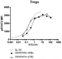
In some embodiments, a heterodimeric Fc fusion protein that binds PD-1 (e.g., anti-PD-1 x scIL 10-heterologous Fc) is a member selected from the group consisting of: XENP25953, XENP27830 and XENP 27831.
In some embodiments, the non-competitive PD-1 binding domain is a humanized non-competitive PD 1ABD that does not bind the same epitope as nivolumab and/or pembrolizumab. In some embodiments, the humanized non-competitive PD-1ABD comprises a variable heavy chain and variable light chain pair selected from the group consisting of: mAb A [ PD-1] _ H1_ L1, mAb B [ PD-1] _ H1_ L1, mAb C [ PD-1] _ H1_ L1, mAb C [ PD-1] _ H1_ L1.1, mAb C [ PD-1] _ H1_ L1.3, mAb C [ PD-1] _ H1_ L1.45, mAb C [ PD-1] _ H1_ L1.117, mAb C [ PD-1] _ H1_ L1.129, mAb C [ PD-1] _ H1_ L1.135, mAb C [ PD-1] _ H1_ L1.136, mAb C [ PD-1] _ H48 _ L1.140, 39C [ PD-1] _ H1_ L2, mAb C [ PD-1] _ H19 _ L1.19, mAb C [ PD-1] _ H19.19.1.19 _L1.19, mAb [ PD-1.19 ] H1.19 mAb C [ PD-1] _ H1.19_ L1.129, mAb C [ PD-1] _ H1.19_ L1.135, mAb C [ PD-1] _ H1.19_ L1.136, mAb C [ PD-1] _ H1.19_ L1.140, mAb C [ PD-1] _ H1.19_ L2, mAb C [ PD-1] _ H1.48_ L1, mAb C [ PD-1] _ H1.48_ L1.1, mAb C [ PD-1] _ H1.48_ L1.3, mAb C [ PD-1] _ H1.48_ L1.45, mAb C [ PD-1] _ H1.48_ L1.117, mAb C [ PD-1] _ H1.48_ L1.129, PD-1] _ H1.48_ L1.135, mAb C [ PD-1] _ H1.125_ L1.1.1.125 mAb [ PD-L1.1 ] H1.140, mAb [ PD-1.125 ] L1.1.1.1.1.140, mAb [ PD-L1 ] H1.1.1.1.1.1.125 mAb [ PD-L1 ] L1 mAb C [ PD-1] _ H1.125_ L1.45, mAb C [ PD-1] _ H1.125_ L1.117, mAb C [ PD-1] _ H1.125_ L1.129, mAb C [ PD-1] _ H1.125_ L1.135, mAb C [ PD-1] _ H1.125_ L1.136, mAb C [ PD-1] _ H1.125_ L1.140, mAb C [ PD-1] _ H1.125_ L2, mAb C [ PD-1] _ H1.130_ L1, mAb C [ PD-1] _ H1.130_ L1.1, mAb C [ PD-1] _ H1.130_ L1.3, mAb C [ PD-1] _ H1.130_ L1.45, PD-1] _ H1.130_ L1.130, mAb C [ PD-1] _ H1.130_ L1, mAb C [ PD-1] _ H1.130_ L1.130, mAb C [ C1 ] H1.130_ L1.130, mAb 130_ L1, mAb 130, mAb [ PD-1.130 ] H1.130.130, mAb [ 1.130 ] L1, mAb 130.130.130.130, mAb [ C1 ] H1.130 ] 1, mAb [ 1.130 ] L1.130 ] H1, mAb 130.130, mAb [ 1.130 ] L1.130.130, mAb [ 1.130 ] C1.130.130 ] L1, mAb [ 1.130 ] C1.130.130.130 ] L1, mAb [ mAb 130, mAb [ 1.130 ] 1.135, mAb [ 1.130 ] C1.130 mAb C [ PD-1] _ H1.132_ L1.1, mAb C [ PD-1] _ H1.132_ L1.3, mAb C [ PD-1] _ H1.132_ L1.45, mAb C [ PD-1] _ H1.132_ L1.117, mAb C [ PD-1] _ H1.132_ L1.129, mAb C [ PD-1] _ H1.132_ L1.135, mAb C [ PD-1] _ H1.132_ L1.136, mAb C [ PD-1] _ H1.132_ L1.140, mAb C [ PD-1] _ H1.132_ L2, mAb C [ PD-1] _ H1.169_ L1, mAb C [ PD-1] _ H1.169_ L1.1, mAb C [ PD-1] _ PD 1.3, mAb C [ PD-1] _ H1.169_ L1.1, mAb [ PD-1.169 _ L1.1 ], mAb [ PD-1.1.1.1.1 ] PD-L1, mAb 169 [ PD-1.1.1 ] PD-L1, mAb [ 1.1.1.1.1 ] PD-L1, mAb [ PD-L1.1.1.1 ] 1, mAb [ PD-L1.1 ] 1, mAb [ 1.1.1.1.1.1, mAb [ PD-L1 ] 1.1.1, mAb [ PD-L1.1.1.1, mAb [ 1] 1.1 ] L1.1.1.1, mAb [ PD-L1 ] 1, mAb [ PD-1.1.1.1.1, mAb [ PD-1.1.1.1, mAb [ 1, mAb [ 1.1 ] 1] 1.1.1.1.1.1, mAb [ 1.1, mAb [ 1] 1, mAb [ PD-1] 1, mAb [ PD-1] H1, mAb [ PD-1] 1, mAb [ PD-1, mAb [ 1] 1, mAb [ PD-1] 1, mAb [ PD-1, mAb [ 1] 1, mAb [ PD-1] 1, mAb [ PD-1] 1, mAb [ PD-1, mAb [ 1] 1, mAb [ 1, mAb, mAb C [ PD-1] _ H1.169_ L2, mAb C [ PD-1] _ H1.175_ L1, mAb C [ PD-1] _ H1.175_ L1.1, mAb C [ PD-1] _ H1.175_ L1.3, mAb C [ PD-1] _ H1.175_ L1.45, mAb C [ PD-1] _ H1.175_ L1.117, mAb C [ PD-1] _ H1.175_ L1.129, mAb C [ PD-1] _ H1.175_ L1.135, mAb C [ PD-1] _ H1.175_ L1.136, mAb C [ PD-1] _ H1.1 _ L1.140, mAb C [ PD-1] _ H1.175_ L2, PD-C [ PD-1] _ H4 ] L1.829, mAb C [ PD-1] _ L1.35, mAb C [ PD-L1 ] _ H1.35, mAb [ PD-L1 ] L1.35, mAb [ PD-L1 ] H1.11, mAb [ PD-L1 ] L1.35, mAb [ mAb ] H1.35, mAb [ PD-L1 ] L1, mAb [ 1] 1.35, mAb [ 1] L1, mAb [ 1] L1, mAb [ 1, mAb ] L1, mAb [ 1] L1.35, mAb ] L1, mAb [ PD-L1, mAb ] L1, mAb [ 1] L1, mAb [ PD-L1, mAb [ 1] 1, mAb [ 1] L1, mAb [ 1, mAb ] 1, mAb [ 1] L1, mAb [ 1, mAb ] 1 [ 1] 1, mAb ] 1] L1 [ 1, mAb ] L1, mAb [ 1] 1, mAb ] L1, mAb [ 1] 1 [ 1, mAb [ 1] 1 [ 1] L1 ] 1 [ 1] 1 [ 1] L1 [ 1] 1 [ 1] 1 [ 1] 1 [ 1] 1 [ 1] L1 [ 1] 1 [ 25 ] 1 [ 1] 1 [ 25 ] L1 [ 25 ] 1 [ 1] 1 [ 1] L1 [ 1] L1, mAb ] L1 [ 1, mAb ] L1, mAb ] 1 [ 1, mAb ] L1, mAb [ 25 ] L1, mAb, mAb C [ PD-1] _ H2_ L1.136, mAb C [ PD-1] _ H2_ L1.140 and mAb C [ PD-1] _ H2_ L2, as depicted in FIGS. 104A-104B.
In some embodiments, the CD8 binding domain is a humanized CD8 ABD. In some embodiments, the humanized CD8 ABD comprises a variable heavy chain and variable light chain pair selected from the group consisting of OKT8_ H2L1 and 1C11B3_ H1L1 as depicted in figure 92.
In some embodiments, the NKG2D binding domain is a humanized NKG2D ABD. In some embodiments, the humanized NKG2D ABD comprises a variable heavy chain and variable light chain pair selected from the group consisting of: MS [ NKG2D ] H0_ L0, 1D7B4[ NKG2D ] _ H1_ L1, KYK-1.0[ NKG2D ] _ H1_ L1, KYK-2.0[ NKG2D ] _ H0_ L0, 11B2D10[ NKG2D ] _ H0_ L0, 6E5A7[ NKG2D ] _ H0_ L0, 6H7E7[ NKG2D ] H0_ L0, mAb E [ NKG2 0 ] _ H0 ], 16F 0[ NKG2 0 ] _ 0 ] 0, 0[ NKG2 0 ] 0L 0, 0[ NKG 2L ] _ 0 ] 0, NKG 3B 0[ NKG 2L 0 ] 0[ NKG-0 ] 0, NKG-0 [ NKG 2L 0 ] 0, NKG-0, 0[ NKG-0 ] 0, NKG-0 [ NKG 2L 0, 0[ NKG-0 ] 0, 0[ NKG-0, mAb 0[ NKG-0 ] 0, NKG-0 [ NKG-0 ] 0, mAb 0[ NKG-0 ] 0, NKG-0 [ NKG-0 ] 0, 0[ NKG-0 ] 0[ NKG-0, NKG-0 [ NKG-0 ] 0, mAb 0[ NKG-0, mAb [ NKG-0 ] 0, mAb [ NKG-0, mAb 3L 0[ NKG-0 ] 0[ NKG-0 [ NKG 2L 0 ] 0[ NKG-0 ] 0[ NKG-0, mAb 0[ NKG-0 ] 0, mAb 0, 0[ NKG-0, mAb [ NKG-0 ] 0[ NKG-0, mAb [ NKG-0 ] mAb 0 ] 0, mAb 0 ] 0, NKG-0 ] 0, NKG-0, mAb [ NKG-0 ] mAb [ NKG-0 ] 0[ NKG-0 ] mAb [ NKG-0 ] 0[ NKG-0, mAb [ NKG-0 ] mAb [ NKG-0 ] mAb 0 ] mAb 0[ NKG-0 [ NK, mAb C [ NKG2D ] _ H1_ L1, mAb C [ NKG2D ] _ H2_ L1, mAb C [ NKG2D ] _ H1_ L2, and mAb C [ NKG2D ] _ H2_ L2, as depicted in fig. 93A-93C.
Provided herein is a composition comprising the described heterodimeric Fc fusion proteins (e.g., an anti-X scIL 10-heterologous Fc fusion protein) for use in treating cancer in a subject. Further, one or more nucleic acids encoding any of the heterodimeric Fc fusion proteins described herein are provided. Further, a host cell is provided comprising one or more nucleic acids encoding any of the heterodimeric Fc fusion proteins described herein.
In one aspect, a method of making a heterodimeric Fc fusion protein (e.g., an anti-X scIL 10-heterodimeric Fc fusion protein) is provided, comprising culturing a host cell described herein under conditions whereby the heterodimeric Fc fusion protein is produced; and recovering the protein.
In another aspect, a method of purifying a heterodimeric Fc fusion protein described herein (e.g., an anti-X scIL 10-heterologous Fc fusion protein) is provided. The method comprises the following steps: (a) providing a composition comprising a heterodimeric Fc fusion protein; (b) loading the composition onto an ion exchange column; and (c) collecting the fraction containing the heterodimeric Fc fusion protein.
In another aspect, the present invention provides a heterodimeric Fc fusion protein comprising: (a) a first fusion protein comprising a first antigen binding domain and a first Fc domain, wherein the first Antigen Binding Domain (ABD) is covalently linked to the N-terminus of the first Fc domain; and (b) a second fusion protein comprising a second antigen-binding domain (ABD), a second Fc domain, a first protein domain, and a second protein domain, wherein the second antigen-binding domain is covalently linked to the N-terminus of the second Fc domain, wherein the first protein domain is covalently linked to the second protein domain, and wherein the second protein domain is covalently linked to the C-terminus of the second Fc domain; wherein the first and second Fc domains comprise modifications that promote heterodimerization of the first and second Fc domains, and wherein the first protein domain comprises a first IL-10 monomer domain and the second protein domain comprises a second IL-10 monomer domain. In some cases, the heterodimeric Fc fusion protein has the form of a (anti-X) 2X heterologous Fc-single chain IL-10 fusion or a (anti-X) 2-heterologous Fc-scIL10 fusion.
In some embodiments, the modification that promotes heterodimerization of the first and second Fc domains is a set of amino acid substitutions selected from the group consisting of: L368D/K370S and S364K according to EU numbering; L368D/K370S and S364K/E357L; L368D/K370S and S364K/E357Q; T411E/K360E/Q362E and D401K; L368E/K370S and S364K; K370S and S364K/E357Q and T366S/L368A/Y407V: T366W (optionally including a disulfide bridge, T366S/L368A/Y407V/Y349C: T366W/S354C).
In some embodiments, the first and/or second Fc domain has an additional set of amino acid substitutions comprising Q295E/N384D/Q418E/N421D according to EU numbering.
In some embodiments, the first and/or second Fc domain has an additional set of amino acid substitutions consisting of: according to the EU numbering system, G236R/L328R, E233P/L234V/L235A/G236_/S239K, E233P/L234V/L235A/G236_/S239K/A327G, E233P/L234V/L235A/G236_/S267K/A327G, E233P/L234V/L235A/G236_ and E233P/L234V/L235A/G236 _/S267K.
In some embodiments, the first Fc domain and/or the second Fc domain comprises another amino acid substitution selected from the group consisting of M428L, N434S, and M428L/N434S according to EU numbering.
In some embodiments, the first and/or second IL-10 monomer domain comprises a polypeptide sequence selected from the group consisting of seq id no: SEQ ID NO:1 (human IL-10(109H) precursor sequence), SEQ ID NO:2 (human IL-10(109L) precursor sequence), SEQ ID NO:3 (human IL-10(109H) mature form sequence), SEQ ID NO:4 (human IL-10(109L) mature form sequence), as depicted in FIG. 1.
In some embodiments, the first IL-10 monomer domain and/or the second IL-10 monomer domain is a variant IL-10 monomer domain.
In some embodiments, variant IL-10 monomer domains include IL-10 monomer domains having one or more amino acid substitutions that result in altered affinity for the IL-10 receptor, altered potency, altered potential for deamidation, altered potential for aspartic acid isomerization, altered potential for degradation of related PTMs, altered potential degradation sites, altered disulfide bridges, and/or altered potential N-glycosylation sites.
In some embodiments, the variant IL-10 monomer domain comprises one or more amino acid modifications at an amino acid residue selected from the group consisting of N21, D28, Q38, M39, D41, Q42, L43, D44, N45, I87, E142, D144, E151, and N160. In some embodiments, the variant IL-10 monomer domain comprises one or more amino acid modifications selected from N21D, D28N, Q38E, M39T, D41N, Q42E, L43V, D44N, N45D, I87A, E142Q, D144N, E151Q, and N160 del.
In some embodiments, the first IL-10 monomer domain covalently linked to the second IL-10 monomer domain forms a single chain IL-10 comprising SEQ ID NO:23 or SEQ ID NO:23 with amino acid modifications 109L and 269L.
In some embodiments, the single chain IL-10 further comprises one or more amino acid modifications selected from the group consisting of N21, Q38, D41, N45, D144, E151, N181, Q198, N205, D304, E311, and N320. In some embodiments, the single-chain IL-10 further comprises one or more amino acid modifications selected from the group consisting of N21D, Q38E, D41N, N45D, D144N, E151Q, N181D, Q198E, N205D, D304N, E311Q, and N320 del. In some embodiments, the single-chain IL-10 comprises amino acid modifications selected from the group consisting of N21D/N181D/N320del, N45D/N205D/N320del, Q38E/N45D/N205D/N320del, D41N/N45D/N205D/N320del, N45D/D144N/N205D/N320del, N45D/E151Q/N205D/N320del, N45D/N181D/N205D/N320del, N45D/N205D/D304N/N320del, N21D/N45D/N181D/N205D/N320del, Q38D/N45/Q D/N205/N D/N36205 del D/N36205/N D/N36205 del D/N D/N36205/N D/N36205 del.
In some embodiments, the first ABD and/or the second ABD are selected from the group consisting of a PD-1 binding domain, a non-competitive PD-1 binding domain, a TIGIT binding domain, a CD8 binding domain, and a NKG2D binding domain.
In some embodiments, the PD-1 binding domain is a humanized PD-1 ABD. In some embodiments, the humanized PD-1ABD comprises a variable heavy chain and variable light chain pair selected from the group consisting of: 1C11[ PD-1] _ H0L0, 1C11[ P, D-1] _ H3L3, 1C11[ PD-1] _ H3.240_ L3.148, 1C11[ PD-1] _ H3.241_ L3.148, 1C11[ PD-1] _ H3.234_ L3.144, 1C11[ PD-1] _ H3.241_ L3.92, 1C11[ PD-1] _ H3.303_ L3.152, 1C11_ H3.329_ L3.220, 1C11_ H3.328_ L3.152, pembrolizumab variable heavy and variable light chains, nivolumab variable heavy and variable heavy chains, nivolumab variable and variable light chains, pidilizumab variable and variable light chains, PD-75 variable and variable light chains, BAP 049E, variable and light chains, variable light chains [ PD-1] variable light chains, variable light chains [ 10 ] variable light chains, variable light chains [ PD-049 ] variable light chains, variable light chains [ 10 ] and variable light chains [ 10 ] variable light chains, and light chains [ 10 ] variable light [ 10 ] variable light chains, and variable light chains [ PD-1] variable light [ 10 ] variable [ 10 ] variable light chains, H005-1[ PD-1] variable heavy and variable light chain, 317-4B6[ PD-1] variable heavy and variable light chain, 326-4A3[ PD-1] variable heavy and variable light chain, hPD-1mAb 7(1.2) [ PD-1] variable heavy and variable light chain, clone 38[ PD-1] variable heavy and variable light chain, clone 39[ PD-1] variable heavy and variable light chain, clone 41[ PD-1] variable heavy and variable light chain, clone 48[ PD-1] variable heavy and variable light chain, PD1-17[ PD-1] variable heavy and variable light chain, PD1-28[ PD-1] variable heavy and variable light chain, PD1-33[ PD-1] variable heavy and variable light chain, PD1-35[ PD-1] variable heavy and variable light chain, PD-1] variable heavy and variable light chain, LOPD180 variable heavy and variable light chains, Ab948 variable heavy and variable light chains, humanized EH-12.2H7[ PD-1] variable heavy and variable light chains, RG1H10 variable heavy and variable light chains, RG1H10-H2A-22-1S variable heavy and variable light chains, RG1H 10-H2A-27-2S variable heavy and variable light chains, RG1H10-3C variable heavy and variable light chains, RG1H10-16C variable heavy and variable light chains, RG1H10-17C variable heavy and variable light chains, RG1H10-19C variable heavy and variable light chains, RG1H 10-21C variable heavy and variable light chains, RG1H10-23C2 variable heavy and variable light chains, and 7[ PD1 AB-6 ] variable heavy and variable light chains, variable mAb 1H10-23C2 variable heavy and variable light chains, as depicted in FIG. 100A.
In some embodiments, the non-competitive PD-1 binding domain is a humanized non-competitive PD 1ABD that does not bind the same epitope as nivolumab and/or pembrolizumab. In some embodiments, the humanized non-competitive PD-1ABD comprises a variable heavy chain and variable light chain pair selected from the group consisting of: mAb A [ PD-1] _ H1_ L1, mAb B [ PD-1] _ H1_ L1, mAb C [ PD-1] _ H1_ L1, mAb C [ PD-1] _ H1_ L1.1, mAb C [ PD-1] _ H1_ L1.3, mAb C [ PD-1] _ H1_ L1.45, mAb C [ PD-1] _ H1_ L1.117, mAb C [ PD-1] _ H1_ L1.129, mAb C [ PD-1] _ H1_ L1.135, mAb C [ PD-1] _ H1_ L1.136, mAb C [ PD-1] _ H48 _ L1.140, 39C [ PD-1] _ H1_ L2, mAb C [ PD-1] _ H19 _ L1.19, mAb C [ PD-1] _ H19.19.1.19 _L1.19, mAb [ PD-1.19 ] H1.19 mAb C [ PD-1] _ H1.19_ L1.129, mAb C [ PD-1] _ H1.19_ L1.135, mAb C [ PD-1] _ H1.19_ L1.136, mAb C [ PD-1] _ H1.19_ L1.140, mAb C [ PD-1] _ H1.19_ L2, mAb C [ PD-1] _ H1.48_ L1, mAb C [ PD-1] _ H1.48_ L1.1, mAb C [ PD-1] _ H1.48_ L1.3, mAb C [ PD-1] _ H1.48_ L1.45, mAb C [ PD-1] _ H1.48_ L1.117, mAb C [ PD-1] _ H1.48_ L1.129, PD-1] _ H1.48_ L1.135, mAb C [ PD-1] _ H1.125_ L1.1.1.125 mAb [ PD-L1.1 ] H1.140, mAb [ PD-1.125 ] L1.1.1.1.1.140, mAb [ PD-L1 ] H1.1.1.1.1.1.125 mAb [ PD-L1 ] L1 mAb C [ PD-1] _ H1.125_ L1.45, mAb C [ PD-1] _ H1.125_ L1.117, mAb C [ PD-1] _ H1.125_ L1.129, mAb C [ PD-1] _ H1.125_ L1.135, mAb C [ PD-1] _ H1.125_ L1.136, mAb C [ PD-1] _ H1.125_ L1.140, mAb C [ PD-1] _ H1.125_ L2, mAb C [ PD-1] _ H1.130_ L1, mAb C [ PD-1] _ H1.130_ L1.1, mAb C [ PD-1] _ H1.130_ L1.3, mAb C [ PD-1] _ H1.130_ L1.45, PD-1] _ H1.130_ L1.130, mAb C [ PD-1] _ H1.130_ L1, mAb C [ PD-1] _ H1.130_ L1.130, mAb C [ C1 ] H1.130_ L1.130, mAb 130_ L1, mAb 130, mAb [ PD-1.130 ] H1.130.130, mAb [ 1.130 ] L1, mAb 130.130.130.130, mAb [ C1 ] H1.130 ] 1, mAb [ 1.130 ] L1.130 ] H1, mAb 130.130, mAb [ 1.130 ] L1.130.130, mAb [ 1.130 ] C1.130.130 ] L1, mAb [ 1.130 ] C1.130.130.130 ] L1, mAb [ mAb 130, mAb [ 1.130 ] 1.135, mAb [ 1.130 ] C1.130 mAb C [ PD-1] _ H1.132_ L1.1, mAb C [ PD-1] _ H1.132_ L1.3, mAb C [ PD-1] _ H1.132_ L1.45, mAb C [ PD-1] _ H1.132_ L1.117, mAb C [ PD-1] _ H1.132_ L1.129, mAb C [ PD-1] _ H1.132_ L1.135, mAb C [ PD-1] _ H1.132_ L1.136, mAb C [ PD-1] _ H1.132_ L1.140, mAb C [ PD-1] _ H1.132_ L2, mAb C [ PD-1] _ H1.169_ L1, mAb C [ PD-1] _ H1.169_ L1.1, mAb C [ PD-1] _ PD 1.3, mAb C [ PD-1] _ H1.169_ L1.1, mAb [ PD-1.169 _ L1.1 ], mAb [ PD-1.1.1.1.1 ] PD-L1, mAb 169 [ PD-1.1.1 ] PD-L1, mAb [ 1.1.1.1.1 ] PD-L1, mAb [ PD-L1.1.1.1 ] 1, mAb [ PD-L1.1 ] 1, mAb [ 1.1.1.1.1.1, mAb [ PD-L1 ] 1.1.1, mAb [ PD-L1.1.1.1, mAb [ 1] 1.1 ] L1.1.1.1, mAb [ PD-L1 ] 1, mAb [ PD-1.1.1.1.1, mAb [ PD-1.1.1.1, mAb [ 1, mAb [ 1.1 ] 1] 1.1.1.1.1.1, mAb [ 1.1, mAb [ 1] 1, mAb [ PD-1] 1, mAb [ PD-1] H1, mAb [ PD-1] 1, mAb [ PD-1, mAb [ 1] 1, mAb [ PD-1] 1, mAb [ PD-1, mAb [ 1] 1, mAb [ PD-1] 1, mAb [ PD-1] 1, mAb [ PD-1, mAb [ 1] 1, mAb [ 1, mAb, mAb C [ PD-1] _ H1.169_ L2, mAb C [ PD-1] _ H1.175_ L1, mAb C [ PD-1] _ H1.175_ L1.1, mAb C [ PD-1] _ H1.175_ L1.3, mAb C [ PD-1] _ H1.175_ L1.45, mAb C [ PD-1] _ H1.175_ L1.117, mAb C [ PD-1] _ H1.175_ L1.129, mAb C [ PD-1] _ H1.175_ L1.135, mAb C [ PD-1] _ H1.175_ L1.136, mAb C [ PD-1] _ H1.1 _ L1.140, mAb C [ PD-1] _ H1.175_ L2, PD-C [ PD-1] _ H4 ] L1.829, mAb C [ PD-1] _ L1.35, mAb C [ PD-L1 ] _ H1.35, mAb [ PD-L1 ] L1.35, mAb [ PD-L1 ] H1.11, mAb [ PD-L1 ] L1.35, mAb [ mAb ] H1.35, mAb [ PD-L1 ] L1, mAb [ 1] 1.35, mAb [ 1] L1, mAb [ 1] L1, mAb [ 1, mAb ] L1, mAb [ 1] L1.35, mAb ] L1, mAb [ PD-L1, mAb ] L1, mAb [ 1] L1, mAb [ PD-L1, mAb [ 1] 1, mAb [ 1] L1, mAb [ 1, mAb ] 1, mAb [ 1] L1, mAb [ 1, mAb ] 1 [ 1] 1, mAb ] 1] L1 [ 1, mAb ] L1, mAb [ 1] 1, mAb ] L1, mAb [ 1] 1 [ 1, mAb [ 1] 1 [ 1] L1 ] 1 [ 1] 1 [ 1] L1 [ 1] 1 [ 1] 1 [ 1] 1 [ 1] 1 [ 1] L1 [ 1] 1 [ 25 ] 1 [ 1] 1 [ 25 ] L1 [ 25 ] 1 [ 1] 1 [ 1] L1 [ 1] L1, mAb ] L1 [ 1, mAb ] L1, mAb ] 1 [ 1, mAb ] L1, mAb [ 25 ] L1, mAb, mAb C [ PD-1] _ H2_ L1.136, mAb C [ PD-1] _ H2_ L1.140 and mAb C [ PD-1] _ H2_ L2, as depicted in FIGS. 104A-104B.
In some embodiments, the CD8 binding domain is a humanized CD8 ABD. In some embodiments, the humanized CD8 ABD comprises a variable heavy chain and variable light chain pair selected from the group consisting of OKT8_ H2L1 and 1C11B3_ H1L1 as depicted in figure 92.
In some embodiments, the NKG2D binding domain is a humanized NKG2D ABD. In some embodiments, the humanized NKG2D ABD comprises a variable heavy chain and variable light chain pair selected from the group consisting of: MS [ NKG2D ] H0_ L0, 1D7B4[ NKG2D ] _ H1_ L1, KYK-1.0[ NKG2D ] _ H1_ L1, KYK-2.0[ NKG2D ] _ H0_ L0, 11B2D10[ NKG2D ] _ H0_ L0, 6E5A7[ NKG2D ] _ H0_ L0, 6H7E7[ NKG2D ] H0_ L0, mAb E [ NKG2 0 ] _ H0 ], 16F 0[ NKG2 0 ] _ 0 ] 0, 0[ NKG2 0 ] 0L 0, 0[ NKG 2L ] _ 0 ] 0, NKG 3B 0[ NKG 2L 0 ] 0[ NKG-0 ] 0, NKG-0 [ NKG 2L 0 ] 0, NKG-0, 0[ NKG-0 ] 0, NKG-0 [ NKG 2L 0, 0[ NKG-0 ] 0, 0[ NKG-0, mAb 0[ NKG-0 ] 0, NKG-0 [ NKG-0 ] 0, mAb 0[ NKG-0 ] 0, NKG-0 [ NKG-0 ] 0, 0[ NKG-0 ] 0[ NKG-0, NKG-0 [ NKG-0 ] 0, mAb 0[ NKG-0, mAb [ NKG-0 ] 0, mAb [ NKG-0, mAb 3L 0[ NKG-0 ] 0[ NKG-0 [ NKG 2L 0 ] 0[ NKG-0 ] 0[ NKG-0, mAb 0[ NKG-0 ] 0, mAb 0, 0[ NKG-0, mAb [ NKG-0 ] 0[ NKG-0, mAb [ NKG-0 ] mAb 0 ] 0, mAb 0 ] 0, NKG-0 ] 0, NKG-0, mAb [ NKG-0 ] mAb [ NKG-0 ] 0[ NKG-0 ] mAb [ NKG-0 ] 0[ NKG-0, mAb [ NKG-0 ] mAb [ NKG-0 ] mAb 0 ] mAb 0[ NKG-0 [ NK, mAb C [ NKG2D ] _ H1_ L1, mAb C [ NKG2D ] _ H2_ L1, mAb C [ NKG2D ] _ H1_ L2, and mAb C [ NKG2D ] _ H2_ L2, as depicted in fig. 93A-93C.
A composition comprising any of the heterodimeric Fc fusion proteins (e.g., anti-X) 2-heterologous Fc-scIL10 fusions) outlined herein is provided for use in treating cancer in a subject. Also provided are one or more nucleic acids encoding any of the heterodimeric Fc fusion proteins outlined herein. Also provided is a host cell comprising one or more nucleic acids encoding any of the heterodimeric Fc fusion proteins outlined herein.
In one aspect, there is provided a method of making any of the heterodimeric Fc fusion proteins (e.g., anti-X) 2-heterologous Fc-scIL10 fusions) outlined herein, comprising culturing the summarized host cell under conditions whereby the heterodimeric Fc fusion protein is produced; and recovering the protein.
In another aspect, the present invention provides a dimeric Fc fusion protein comprising: (a) a first fusion protein comprising a first IL-10 monomer domain and a first Fc domain, wherein the IL-10 monomer domain is covalently linked to the first Fc domain; and (b) a second fusion protein comprising a second IL-10 monomer domain and a second Fc domain, wherein the second IL-10 monomer domain is covalently linked to the second Fc domain. In some embodiments, the dimeric Fc fusion has the form of a (IL-10)2-Fc fusion.
In some embodiments, the first and second fusion proteins are the same. In some embodiments, the first IL-10 monomer domain and the second IL-10 monomer domain is the same.
In some embodiments, the first and/or second Fc domain comprises a set of amino acid substitutions selected from the group consisting of: C219S, C220S, S228P, G236R/L328R, E233P/L234V/L235A/G236_/S239K, E233P/L234V/L235A/G236_/S239K/A327G, E233P/L234V/L235A/G236_/S267K/A327G, E233P/L234V/L235A/G236_ and E233P/L234V/L235A/G236_/S267K according to EU numbering.
In some embodiments, the first and/or second Fc domain has an additional set of amino acid substitutions comprising Q295E/N384D/Q418E/N421D according to EU numbering.
In some embodiments, the first and/or second Fc domain has an additional set of amino acid substitutions consisting of: according to the EU numbering system, G236R/L328R, E233P/L234V/L235A/G236_/S239K, E233P/L234V/L235A/G236_/S239K/A327G, E233P/L234V/L235A/G236_/S267K/A327G, E233P/L234V/L235A/G236_ and E233P/L234V/L235A/G236 _/S267K.
In some embodiments, the first and/or second Fc domain comprises another amino acid substitution selected from the group consisting of M428L, N434S, and M428L/N434S according to EU numbering.
In some embodiments, the first IL-10 monomer domain is covalently linked to the N-terminus of the first Fc domain and the second IL-10 monomer domain is covalently linked to the N-terminus of the second Fc domain. In certain embodiments, the first IL-10 monomer domain is covalently linked to the C-terminus of the first Fc domain and the second IL-10 monomer domain is covalently linked to the C-terminus of the second Fc domain.
In some embodiments, a first domain linker is used to link the first IL-10 monomer domain to the first Fc domain, and/or a second domain linker is used to link the second IL-10 monomer domain to the second Fc domain. In particular embodiments, a first domain linker is used to link the first IL-10 monomer domain to the first Fc domain, and a second domain linker is used to link the second IL-10 monomer domain to the second Fc domain. In further embodiments, the first IL-10 monomer domain is linked to the first Fc domain using a first domain linker. In further embodiments, a second domain linker is used to link the second IL-10 monomer domain to the second Fc domain.
In some embodiments, the first IL-10 monomer domain and/or the second IL-10 monomer domain has a leucine at position 109 instead of a histidine. In other words, in some embodiments, the first IL-10 monomer domain has a leucine at position 109 instead of a histidine. In some embodiments, the second IL-10 monomer domain has a leucine instead of a histidine at position 109. In some embodiments, the first IL-10 monomer domain and the second IL-10 monomer domain have a leucine instead of a histidine at position 109.
In some embodiments, the first IL-10 monomer domain and/or the second IL-10 monomer domain has a histidine instead of a leucine at position 109. In other words, in some embodiments, the first IL-10 monomer domain and the second IL-10 monomer domain have a histidine instead of a leucine at position 109. In certain embodiments, the first IL-10 monomer domain has a histidine instead of a leucine at position 109. In other embodiments, the second IL-10 monomer domain has a histidine instead of a leucine at position 109.
In some embodiments, the first and/or second IL-10 monomer domain comprises a polypeptide sequence selected from the group consisting of seq id no: SEQ ID NO:1 (human IL-10(109H) precursor sequence), SEQ ID NO:2 (human IL-10(109L) precursor sequence), SEQ ID NO:3 (human IL-10(109H) mature form sequence), SEQ ID NO:4 (human IL-10(109L) mature form sequence). In some embodiments, the first IL-10 monomer domain comprises a polypeptide sequence selected from the group consisting of seq id no: SEQ ID NO:1 (human IL-10(109H) precursor sequence), SEQ ID NO:2 (human IL-10(109L) precursor sequence), SEQ ID NO:3 (human IL-10(109H) mature form sequence), SEQ ID NO:4 (human IL-10(109L) mature form sequence). In certain embodiments, the second IL-10 monomer domain comprises a polypeptide sequence selected from the group consisting of seq id no: SEQ ID NO:1 (human IL-10(109H) precursor sequence), SEQ ID NO:2 (human IL-10(109L) precursor sequence), SEQ ID NO:3 (human IL-10(109H) mature form sequence), SEQ ID NO:4 (human IL-10(109L) mature form sequence).
In some embodiments, the first and/or second IL-10 monomer domains are variant IL-10 monomer domains. In some embodiments, the first IL-10 monomer domain is a variant IL-10 monomer domain. In some embodiments, the second IL-10 monomer domain is a variant IL-10 monomer domain.
In some embodiments, the variant IL-10 monomer domain comprises one or more amino acid modifications at an amino acid residue selected from the group consisting of N21, D28, Q38, M39, D41, Q42, L43, D44, N45, I87, E142, D144, E151, and N160. In some embodiments, the variant IL-10 monomer domain comprises one or more amino acid modifications selected from the group consisting of N21D, D28N, Q38E, M39T, D41N, Q42E, L43V, D44N, N45D, I87A, E142Q, D144N, E151Q, and N160 del.
In some embodiments, the first and second fusion proteins in the form of (IL-10)2-Fc fusions each comprise a polypeptide sequence selected from the group consisting of: XENP24628, XENP24629, XENP24630, XENP24631, XENP24632, XENP24633, and XENP24634, as depicted in fig. 17-18.
In one aspect, the invention provides a heterodimeric Fc fusion protein comprising: a) a first fusion protein comprising a first IL-10 monomer domain and a first Fc domain, wherein the first IL-10 monomer domain is covalently linked to the first Fc domain; and (b) a second Fc domain; wherein the first and second Fc domains comprise a modification that promotes heterodimerization of the first and second Fc domains. In some embodiments, the heterodimeric Fc fusion protein has the form of a (IL10-NC-IL10) -heterologous Fc fusion.
In some embodiments, the heterodimeric Fc fusion protein further comprises a second IL-10 monomer domain non-covalently linked to the first IL-10 monomer domain. In some embodiments, the first IL-10 monomer domain is linked to the N-terminus of the first Fc domain. In some embodiments, the first IL-10 monomer domain is linked to the C-terminus of the first Fc domain. In some embodiments, the first IL-10 monomer domain is linked to the first Fc domain using a first domain linker.
In some embodiments, the modification that promotes heterodimerization of the first and second Fc domains is a set of amino acid substitutions selected from the group consisting of: L368D/K370S and S364K according to EU numbering; L368D/K370S and S364K/E357L; L368D/K370S and S364K/E357Q; T411E/K360E/Q362E and D401K; L368E/K370S and S364K; K370S and S364K/E357Q and T366S/L368A/Y407V: T366W (optionally including a disulfide bridge, T366S/L368A/Y407V/Y349C: T366W/S354C).
In some embodiments, the first and/or second Fc domain has an additional set of amino acid substitutions comprising Q295E/N384D/Q418E/N421D according to EU numbering.
In some embodiments, the first and/or second Fc domain has an additional set of amino acid substitutions consisting of: according to the EU numbering system, G236R/L328R, E233P/L234V/L235A/G236_/S239K, E233P/L234V/L235A/G236_/S239K/A327G, E233P/L234V/L235A/G236_/S267K/A327G, E233P/L234V/L235A/G236_ and E233P/L234V/L235A/G236 _/S267K.
In some embodiments, the first and/or second Fc domain comprises another amino acid substitution selected from the group consisting of M428L, N434S, and M428L/N434S according to EU numbering.
In some embodiments, the first and/or second IL-10 monomer domain has a polypeptide sequence selected from the group consisting of seq id no: SEQ ID NO:1 (human IL-10(109H) precursor sequence), SEQ ID NO:2 (human IL-10(109L) precursor sequence), SEQ ID NO:3 (human IL-10(109H) mature form sequence), SEQ ID NO:4 (human IL-10(109L) mature form sequence).
In some embodiments, the first and/or second IL-10 monomer domains are variant IL-10 monomer domains. In some embodiments, variant IL-10 monomer domains include IL-10 monomer domains having one or more amino acid substitutions that result in altered affinity for the IL-10 receptor, altered potency, altered potential for deamidation, altered potential for aspartic acid isomerization, altered potential for degradation of related PTMs, altered potential degradation sites, altered disulfide bridges, and/or altered potential N-glycosylation sites.
In some embodiments, the variant IL-10 monomer domain comprises one or more amino acid modifications at an amino acid residue selected from the group consisting of N21, D28, Q38, M39, D41, Q42, L43, D44, N45, I87, E142, D144, E151, and N160. In some embodiments, the variant IL-10 monomer domain comprises one or more amino acid modifications selected from the group consisting of N21D, D28N, Q38E, M39T, D41N, Q42E, L43V, D44N, N45D, I87A, E142Q, D144N, E151Q, and N160 del.
In another aspect, the present invention provides a dimeric Fc fusion protein comprising: (a) a first fusion protein comprising a first protein domain and a first Fc domain, wherein the first protein domain is covalently linked to the first Fc domain; (b) a second fusion protein comprising a second protein domain and a second Fc domain, wherein the second protein domain is covalently linked to the second Fc domain; wherein the first protein domain comprises a first IL-10 monomer domain comprising an insertion peptide and the second protein domain comprises a second IL-10 monomer domain comprising an insertion peptide. This dimeric Fc fusion protein has the form of a (IL10M1)2-Fc fusion.
In some embodiments, the first protein domain is covalently linked to the N-terminus of the first Fc domain, and/or the second protein domain is covalently linked to the N-terminus of the second Fc domain. In some embodiments, the first protein domain is covalently linked to the C-terminus of the first Fc domain, and/or the second protein domain is covalently linked to the C-terminus of the second Fc domain. In some embodiments, the first protein domain is covalently linked to the first Fc domain by a first domain linker. In some embodiments, the second protein domain is covalently linked to the second Fc domain by a second domain linker.
In some embodiments, the first Fc domain and/or the second Fc domain comprises another amino acid substitution selected from the group consisting of M428L, N434S, and M428L/N434S according to EU numbering.
In some embodiments, the insertion peptide comprises a domain linker engineered between helices D and E of the IL-10 monomer domain.
In some embodiments, the first IL-10 monomer domain and/or the second IL-10 monomer domain comprises a polypeptide sequence selected from the group consisting of: 24(IL10M1) as depicted in figure 15D and huIL10M1 variants as depicted in figures 75A-75B.
In some embodiments, the first IL-10 monomer domain and/or the second IL-10 monomer domain comprises a variant IL-10 domain comprising one or more amino acid modifications selected from the group consisting of: N21D, D28N, Q38E, M39T, D41N, Q42E, L43V, D44N, N45D, I87A, E142Q, D144N, E151Q, C12Q, C108Q, Q38Q/D41Q, Q38Q/Q42Q, Q38Q/N45Q, Q38Q/E Q, Q38Q/D144Q, D41Q/Q42Q, D41Q/N45, D41Q/E142, D41Q/D144Q, Q42Q/N45Q, Q42Q/E72, N42Q/E142, N42/Q, N Q/E142, N45/N Q, N41Q/N Q, N41/N Q, N42/N Q, N/N42/N Q, N/N Q/N Q/Q, N Q/N Q, N/N42/N/Q/N Q/Q, N42/Q/N Q, N/Q/N/Q/N42/Q/N42/Q/N/Q, N/N42/N/Q/N/Q, N/Q, N Q/Q, N/Q, N Q/N/Q/N/Q, N/Q/N/Q, N/Q/N/Q, N42/Q, N42/Q, N/Q, N/Q, N/N42/Q, N/Q, N42/Q/N/Q, N/Q, N/Q, N42/Q, N Q/Q, N/Q, N/Q, N42/Q, N/Q, N/, F56C/Y153C, C62A/C114A, A64C/S118C, M68C/V121C, V76C/A139C, L47Q, S118A and A139Q.
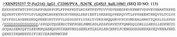
In some embodiments, the first fusion protein and the second fusion protein are the same. In some embodiments, the first fusion protein and the second fusion protein of the (IL10M1)2-Fc fusion each comprise the polypeptide sequence of XENP25236, as depicted in figure 26. In some embodiments, the first fusion protein and the second fusion protein of the (IL10M1)2-Fc fusion each comprise the polypeptide sequence of XENP25237, as depicted in figure 27.
In another aspect, the present invention provides a heterodimeric Fc fusion protein comprising: (a) a fusion protein comprising a first protein domain, a second protein domain, and a first Fc domain, wherein the first protein domain is linked to the first Fc domain, and wherein the second protein domain is covalently linked to the first protein domain; and (b) a second Fc domain; wherein the first and second Fc domains comprise modifications that promote heterodimerization of the first and second Fc domains, and wherein the first protein domain comprises a first IL10 monomer and the second protein domain comprises a second IL10 monomer, and wherein each of the first and second IL10 monomers comprises an insertion peptide. The heterodimeric Fc fusion protein has the form of a (IL10M1) 2-heterologous Fc fusion.
In some embodiments, the first protein domain is linked to the N-terminus of the first Fc domain. In some embodiments, the first protein domain is linked to the C-terminus of the first Fc domain. In some embodiments, the first protein domain is linked to the second protein domain using a first domain linker, and/or the first protein domain is linked to the first Fc domain using a second domain linker.
In some embodiments, the modification that promotes heterodimerization of the first and second Fc domains is a set of amino acid substitutions selected from the group consisting of: L368D/K370S and S364K according to EU numbering; L368D/K370S and S364K/E357L; L368D/K370S and S364K/E357Q; T411E/K360E/Q362E and D401K; L368E/K370S and S364K; K370S and S364K/E357Q and T366S/L368A/Y407V: T366W (optionally including a disulfide bridge, T366S/L368A/Y407V/Y349C: T366W/S354C).
In some embodiments, the first and/or second Fc domain has an additional set of amino acid substitutions comprising Q295E/N384D/Q418E/N421D according to EU numbering.
In some embodiments, the first and/or second Fc domain has an additional set of amino acid substitutions consisting of: according to the EU numbering system, G236R/L328R, E233P/L234V/L235A/G236_/S239K, E233P/L234V/L235A/G236_/S239K/A327G, E233P/L234V/L235A/G236_/S267K/A327G, E233P/L234V/L235A/G236_ and E233P/L234V/L235A/G236 _/S267K.
In some embodiments, the first second Fc domain and/or the second Fc domain comprises another amino acid substitution selected from the group consisting of M428L, N434S, and M428L/N434S according to EU numbering.
In some embodiments, the insertion peptide comprises a domain linker engineered between helices D and E of the IL-10 monomer domain.
In some embodiments, the first IL-10 monomer domain and/or the second IL-10 monomer domain comprises a polypeptide sequence selected from the group consisting of: 24(IL10M1) as depicted in figure 15D and huIL10M1 variants as depicted in figures 75A-75B.
In some embodiments, the first IL-10 monomer domain and/or the second IL-10 monomer domain includes a variant IL-10 domain. In some embodiments, the variant IL-10 domain comprises one or more amino acid modifications selected from the group consisting of: N21D, D28N, Q38E, M39T, D41N, Q42E, L43V, D44N, N45D, I87A, E142Q, D144N, E151Q, C12Q, C108Q, Q38Q/D41Q, Q38Q/Q42Q, Q38Q/N45Q, Q38Q/E Q, Q38Q/D144Q, D41Q/Q42Q, D41Q/N45, D41Q/E142, D41Q/D144Q, Q42Q/N45Q, Q42Q/E72, N42Q/E142, N42/Q, N Q/E142, N45/N Q, N41Q/N Q, N41/N Q, N42/N Q, N/N42/N Q, N/N Q/N Q/Q, N Q/N Q, N/N42/N/Q/N Q/Q, N42/Q/N Q, N/Q/N/Q/N42/Q/N42/Q/N/Q, N/N42/N/Q/N/Q, N/Q, N Q/Q, N/Q, N Q/N/Q/N/Q, N/Q/N/Q, N/Q/N/Q, N42/Q, N42/Q, N/Q, N/Q, N/N42/Q, N/Q, N42/Q/N/Q, N/Q, N/Q, N42/Q, N Q/Q, N/Q, N/Q, N42/Q, N/Q, N/, F56C/Y153C, C62A/C114A, A64C/S118C, M68C/V121C, V76C/A139C, L47Q, S118A and A139Q.
In another aspect, the present invention provides a heterodimeric Fc fusion protein comprising: (a) a fusion protein comprising a first protein domain and a first Fc domain, wherein the first protein domain is covalently linked to the first Fc domain; and (b) a second Fc domain; wherein the first and second Fc domains comprise modifications that promote heterodimerization of the first and second Fc domains, and wherein the first protein domain comprises an IL-10 monomer comprising an insertion peptide. The heterodimeric Fc fusions have the form of (IL10M1) 1-heterodimeric Fc fusions.
In some embodiments, the first protein domain (e.g., an IL-10 monomer comprising an insertion peptide) is linked to the N-terminus of the first Fc domain. In some embodiments, the first protein domain is linked to the C-terminus of the first Fc domain. In some embodiments, the first protein domain is linked to the first Fc domain using a domain linker.
In some embodiments, the modification that promotes heterodimerization of the first and second Fc domains is a set of amino acid substitutions selected from the group consisting of: L368D/K370S and S364K according to EU numbering; L368D/K370S and S364K/E357L; L368D/K370S and S364K/E357Q; T411E/K360E/Q362E and D401K; L368E/K370S and S364K; K370S and S364K/E357Q and T366S/L368A/Y407V: T366W (optionally including a disulfide bridge, T366S/L368A/Y407V/Y349C: T366W/S354C).
In some embodiments, the first and/or second Fc domain has an additional set of amino acid substitutions comprising Q295E/N384D/Q418E/N421D according to EU numbering.
In some embodiments, the first and/or second Fc domain has an additional set of amino acid substitutions consisting of: according to the EU numbering system, G236R/L328R, E233P/L234V/L235A/G236_/S239K, E233P/L234V/L235A/G236_/S239K/A327G, E233P/L234V/L235A/G236_/S267K/A327G, E233P/L234V/L235A/G236_ and E233P/L234V/L235A/G236 _/S267K.
In some embodiments, the first Fc domain and/or the second Fc domain comprises another amino acid substitution selected from the group consisting of M428L, N434S, and M428L/N434S according to EU numbering.
In some embodiments, the insertion peptide comprises a domain linker engineered between helices D and E of the IL-10 monomer domain.
In some embodiments, the first IL-10 monomer domain and/or the second IL-10 monomer domain comprises a polypeptide sequence selected from the group consisting of: 24(IL10M1) as depicted in figure 15D and huIL10M1 variants as depicted in figures 75A-75B.
In some embodiments, the first IL-10 monomer domain and/or the second IL-10 monomer domain comprises a variant IL-10 domain comprising one or more amino acid modifications selected from the group consisting of: N21D, D28N, Q38E, M39T, D41N, Q42E, L43V, D44N, N45D, I87A, E142Q, D144N, E151Q, C12Q, C108Q, Q38Q/D41Q, Q38Q/Q42Q, Q38Q/N45Q, Q38Q/E Q, Q38Q/D144Q, D41Q/Q42Q, D41Q/N45, D41Q/E142, D41Q/D144Q, Q42Q/N45Q, Q42Q/E72, N42Q/E142, N42/Q, N Q/E142, N45/N Q, N41Q/N Q, N41/N Q, N42/N Q, N/N42/N Q, N/N Q/N Q/Q, N Q/N Q, N/N42/N/Q/N Q/Q, N42/Q/N Q, N/Q/N/Q/N42/Q/N42/Q/N/Q, N/N42/N/Q/N/Q, N/Q, N Q/Q, N/Q, N Q/N/Q/N/Q, N/Q/N/Q, N/Q/N/Q, N42/Q, N42/Q, N/Q, N/Q, N/N42/Q, N/Q, N42/Q/N/Q, N/Q, N/Q, N42/Q, N Q/Q, N/Q, N/Q, N42/Q, N/Q, N/, F56C/Y153C, C62A/C114A, A64C/S118C, M68C/V121C, V76C/A139C, L47Q, S118A and A139Q.
In another aspect, the present invention provides a heterodimeric Fc fusion protein comprising: (a) a first fusion protein comprising a first protein domain and a first Fc domain, wherein the first protein domain is covalently linked to the first Fc domain; and (b) a second fusion protein comprising a second protein domain and a second Fc domain, wherein the second protein domain is covalently linked to the second Fc domain; wherein the first protein domain comprises helices A-D of the IL-10 monomer domain and the second protein domain comprises helices E-F of the IL-10 monomer domain. Heterodimeric Fc fusion proteins have (split IL10) a 1-heterologous Fc form.
In some embodiments, the first protein domain is covalently linked to the N-terminus of the first Fc domain, and/or the second protein domain is covalently linked to the N-terminus of the second Fc domain.
In some embodiments, the first protein domain is covalently linked to the C-terminus of the first Fc domain, and/or the second protein domain is covalently linked to the C-terminus of the second Fc domain.
In some embodiments, the first protein domain is covalently linked to the first Fc domain by a first domain linker, and/or the second protein domain is covalently linked to the second Fc domain by a second domain linker.
In some embodiments, the first Fc domain and/or the second Fc domain comprises another amino acid substitution selected from the group consisting of M428L, N434S, and M428L/N434S according to EU numbering.
In some embodiments, the first protein domain comprises a polypeptide sequence of SEQ ID NO:21(hl-10 (a-D)) or a polypeptide sequence having at least 90% (e.g., 90%, 91%, 92%, 93%, 94%, 95%, 96%, 97%, 98%, 99% or more) sequence identity to SEQ ID NO:21 as depicted in fig. 15A. In some embodiments, the second protein domain comprises a polypeptide sequence of SEQ ID NO:22(hl-10(E-F)) or a polypeptide sequence having at least 90% (e.g., 90%, 91%, 92%, 93%, 94%, 95%, 96%, 97%, 98%, 99% or more) sequence identity to SEQ ID NO:22 as depicted in fig. 15B.
In some embodiments, a (split IL10) 1-heterodimeric Fc fusion protein in the form of a heterologous Fc comprises: (a) a first fusion protein having the polypeptide sequence of chain 1 of XENP25242 as depicted in figure 30, and (b) a second fusion protein having the polypeptide sequence of chain 2 of XENP25242 as depicted in figure 30.
In some embodiments, a (split IL10) 1-heterodimeric Fc fusion protein in the form of a heterologous Fc comprises: (a) a first fusion protein having the polypeptide sequence of XENP25243 chain 1 as depicted in figure 30, and (b) a second fusion protein having the polypeptide sequence of XENP25243 chain 2 as depicted in figure 30.
In some embodiments, a (split IL10) 1-heterodimeric Fc fusion protein in the form of a heterologous Fc comprises: (a) a first fusion protein having the polypeptide sequence of XENP25244 chain 1 as depicted in figure 30, and (b) a second fusion protein having the polypeptide sequence of XENP25244 chain 2 as depicted in figure 30.
A nucleic acid is provided that encodes any of the heterodimeric fusion proteins described herein. A host cell is provided that comprises a nucleic acid encoding any of the heterodimeric fusion proteins described herein.
In some embodiments, a method of making a heterodimeric Fc fusion protein (e.g., (split IL10) 1-heterodimeric Fc fusion protein) is provided, comprising culturing the described host cell under conditions whereby the heterodimeric Fc fusion protein is produced; and recovering the heterodimeric Fc fusion protein.
In another aspect, the present invention provides a heterodimeric Fc fusion protein comprising: (a) a first fusion protein comprising a first protein domain, a second protein domain, and a first Fc domain, wherein the first protein domain is covalently linked to the second protein domain and the second protein domain is covalently linked to the first Fc domain; and (b) a second fusion protein comprising a third protein domain, a fourth protein domain, and a second Fc domain, wherein the third protein domain is covalently linked to the fourth protein domain and the fourth protein domain is covalently linked to the second Fc domain; wherein the first protein domain and the second protein domain each comprise helices A-D of the IL-10 monomer domain, and wherein the third protein domain and the fourth protein domain each comprise helices E-F of the IL-10 monomer domain. The heterodimeric Fc fusion protein has the form of a (split IL10) 2-heterologous Fc fusion.
In some embodiments, the second protein is covalently linked to the N-terminus of the first Fc domain, and/or the fourth protein domain is covalently linked to the N-terminus of the second Fc domain.
In some embodiments, the second protein is covalently linked to the C-terminus of the first Fc domain, and/or the fourth protein domain is covalently linked to the C-terminus of the second Fc domain.
In some embodiments, the first protein domain is covalently linked to the second protein domain by a first domain linker, and/or the third protein domain is covalently linked to the third protein domain by a second domain linker.
In some embodiments, the second protein domain is covalently linked to the first Fc domain by a third domain linker, and/or the fourth protein domain is covalently linked to the second Fc domain by a fourth domain linker.
In some embodiments, the first and/or second Fc domain comprises another amino acid substitution selected from the group consisting of M428L, N434S, and M428L/N434S according to EU numbering. In one embodiment, the first Fc domain comprises another amino acid substitution selected from the group consisting of M428L, N434S, and M428L/N434S. In one embodiment, the second Fc domain comprises another amino acid substitution selected from the group consisting of M428L, N434S, and M428L/N434S. In some embodiments, the first and second Fc domains each comprise another amino acid substitution selected from the group consisting of M428L, N434S, and M428L/N434S.
In some embodiments, the first protein domain and/or the second protein domain comprises a polypeptide sequence of SEQ ID NO:21(hIL-10(a-D)) or a polypeptide sequence having at least 90% (e.g., 90%, 91%, 92%, 93%, 94%, 95%, 96%, 97%, 98%, 99% or more) sequence identity to SEQ ID NO:21 as depicted in fig. 15A. In some embodiments, the third protein domain and/or the fourth protein domain comprises a polypeptide sequence of SEQ ID NO:22 (hIL-10 (E-F)) or a polypeptide sequence having at least 90% (e.g., 90%, 91%, 92%, 93%, 94%, 95%, 96%, 97%, 98%, 99% or more) sequence identity to SEQ ID NO:22 as depicted in fig. 15B.
A nucleic acid is provided that encodes any of the heterodimeric fusion proteins described herein (e.g., (split IL10) 2-heterologous Fc fusions). A host cell is provided that comprises a nucleic acid encoding any of the heterodimeric fusion proteins described herein.
In some embodiments, a method of making a heterodimeric Fc fusion protein (e.g., (split IL10) 2-heterodimeric Fc fusion protein) is provided, comprising culturing the described host cell under conditions whereby the heterodimeric Fc fusion protein is produced; and recovering the heterodimeric Fc fusion protein.
In another aspect, the present invention provides a heterodimeric Fc fusion protein comprising: (a) a first fusion protein comprising an Antigen Binding Domain (ABD) and a first Fc domain, wherein the antigen binding domain is covalently linked to the N-terminus of the first Fc domain; and (b) a second fusion protein comprising a protein domain and a second Fc domain, wherein the protein domain is covalently linked to the N-terminus of the second Fc domain, and wherein the protein domain comprises a first IL-10 monomer domain. The heterodimeric Fc fusion protein has the form of an anti-X IL 10-heterologous Fc fusion.
In some embodiments, the heterodimeric Fc fusion protein further comprises a second IL-10 monomer domain non-covalently linked to the first IL-10 monomer domain.
In some embodiments, the antigen binding domain is linked to the first Fc domain using a first domain linker. In some embodiments, the first IL-10 monomer domain is linked to the second Fc domain using a second domain linker.
In some embodiments, the modification that promotes heterodimerization of the first and second Fc domains is a set of amino acid substitutions selected from the group consisting of: L368D/K370S and S364K according to EU numbering; L368D/K370S and S364K/E357L; L368D/K370S and S364K/E357Q; T411E/K360E/Q362E and D401K; L368E/K370S and S364K; K370S and S364K/E357Q and T366S/L368A/Y407V: T366W (optionally including a disulfide bridge, T366S/L368A/Y407V/Y349C: T366W/S354C).
In some embodiments, the first and/or second Fc domain has an additional set of amino acid substitutions comprising Q295E/N384D/Q418E/N421D according to EU numbering.
In some embodiments, the first and/or second Fc domain has an additional set of amino acid substitutions consisting of: according to the EU numbering system, G236R/L328R, E233P/L234V/L235A/G236_/S239K, E233P/L234V/L235A/G236_/S239K/A327G, E233P/L234V/L235A/G236_/S267K/A327G, E233P/L234V/L235A/G236_ and E233P/L234V/L235A/G236 _/S267K.
In some embodiments, the first Fc domain and/or the second Fc domain comprises another amino acid substitution selected from the group consisting of M428L, N434S, and M428L/N434S according to EU numbering.
In some embodiments, the first and/or second IL-10 monomer domain comprises a polypeptide sequence selected from the group consisting of seq id no: SEQ ID NO:1 (human IL-10(109H) precursor sequence), SEQ ID NO:2 (human IL-10(109L) precursor sequence), SEQ ID NO:3 (human IL-10(109H) mature form sequence), SEQ ID NO:4 (human IL-10(109L) mature form sequence), as depicted in FIG. 1.
In some embodiments, the first IL-10 monomer domain and/or the second IL-10 monomer domain is a variant IL-10 monomer domain.
In some embodiments, variant IL-10 monomer domains include IL-10 monomer domains having one or more amino acid substitutions that result in altered affinity for the IL-10 receptor, altered potency, altered potential for deamidation, altered potential for aspartic acid isomerization, altered potential for degradation of related PTMs, altered potential degradation sites, altered disulfide bridges, and/or altered potential N-glycosylation sites. In some embodiments, the variant IL-10 monomer domain comprises one or more amino acid modifications at an amino acid residue selected from the group consisting of N21, D28, Q38, M39, D41, Q42, L43, D44, N45, I87, E142, D144, E151, and N160. In some embodiments, the variant IL-10 monomer domain comprises one or more amino acid modifications selected from the group consisting of N21D, D28N, Q38E, M39T, D41N, Q42E, L43V, D44N, N45D, I87A, E142Q, D144N, E151Q, and N160 del.
In some embodiments, the Antigen Binding Domain (ABD) is selected from the group consisting of a PD-1 binding domain, a non-competitive PD-1 binding domain, a TIGIT binding domain, a CD8 binding domain, and a NKG2D binding domain.
In some embodiments, the PD-1 binding domain is a humanized PD-1 ABD. In some embodiments, the humanized PD-1ABD comprises a variable heavy chain and variable light chain pair selected from the group consisting of: 1C11[ PD-1] _ H0L0, 1C11[ P, D-1] _ H3L3, 1C11[ PD-1] _ H3.240_ L3.148, 1C11[ PD-1] _ H3.241_ L3.148, 1C11[ PD-1] _ H3.234_ L3.144, 1C11[ PD-1] _ H3.241_ L3.92, 1C11[ PD-1] _ H3.303_ L3.152, 1C11_ H3.329_ L3.220, 1C11_ H3.328_ L3.152, pembrolizumab variable heavy and variable light chains, nivolumab variable heavy and variable heavy chains, nivolumab variable and variable light chains, pidilizumab variable and variable light chains, PD-75 variable and variable light chains, BAP 049E, variable and light chains, variable light chains [ PD-1] variable light chains, variable light chains [ 10 ] variable light chains, variable light chains [ PD-049 ] variable light chains, variable light chains [ 10 ] and variable light chains [ 10 ] variable light chains, and light chains [ 10 ] variable light [ 10 ] variable light chains, and variable light chains [ PD-1] variable light [ 10 ] variable [ 10 ] variable light chains, H005-1[ PD-1] variable heavy and variable light chain, 317-4B6[ PD-1] variable heavy and variable light chain, 326-4A3[ PD-1] variable heavy and variable light chain, hPD-1mAb 7(1.2) [ PD-1] variable heavy and variable light chain, clone 38[ PD-1] variable heavy and variable light chain, clone 39[ PD-1] variable heavy and variable light chain, clone 41[ PD-1] variable heavy and variable light chain, clone 48[ PD-1] variable heavy and variable light chain, PD1-17[ PD-1] variable heavy and variable light chain, PD1-28[ PD-1] variable heavy and variable light chain, PD1-33[ PD-1] variable heavy and variable light chain, PD1-35[ PD-1] variable heavy and variable light chain, PD-1] variable heavy and variable light chain, LOPD180 variable heavy and variable light chains, Ab948 variable heavy and variable light chains, humanized EH-12.2H7[ PD-1] variable heavy and variable light chains, RG1H10 variable heavy and variable light chains, RG1H10-H2A-22-1S variable heavy and variable light chains, RG1H 10-H2A-27-2S variable heavy and variable light chains, RG1H10-3C variable heavy and variable light chains, RG1H10-16C variable heavy and variable light chains, RG1H10-17C variable heavy and variable light chains, RG1H10-19C variable heavy and variable light chains, RG1H 10-21C variable heavy and variable light chains, RG1H10-23C2 variable heavy and variable light chains, and 7[ PD1 AB-6 ] variable heavy and variable light chains, variable mAb 1H10-23C2 variable heavy and variable light chains, as depicted in FIG. 100A.
In some embodiments, the non-competitive PD-1 binding domain is a humanized non-competitive PD 1ABD that does not bind the same epitope as nivolumab and/or pembrolizumab. In some embodiments, the humanized non-competitive PD-1ABD comprises a variable heavy chain and variable light chain pair selected from the group consisting of: mAb A [ PD-1] _ H1_ L1, mAb B [ PD-1] _ H1_ L1, mAb C [ PD-1] _ H1_ L1, mAb C [ PD-1] _ H1_ L1.1, mAb C [ PD-1] _ H1_ L1.3, mAb C [ PD-1] _ H1_ L1.45, mAb C [ PD-1] _ H1_ L1.117, mAb C [ PD-1] _ H1_ L1.129, mAb C [ PD-1] _ H1_ L1.135, mAb C [ PD-1] _ H1_ L1.136, mAb C [ PD-1] _ H48 _ L1.140, 39C [ PD-1] _ H1_ L2, mAb C [ PD-1] _ H19 _ L1.19, mAb C [ PD-1] _ H19.19.1.19 _L1.19, mAb [ PD-1.19 ] H1.19 mAb C [ PD-1] _ H1.19_ L1.129, mAb C [ PD-1] _ H1.19_ L1.135, mAb C [ PD-1] _ H1.19_ L1.136, mAb C [ PD-1] _ H1.19_ L1.140, mAb C [ PD-1] _ H1.19_ L2, mAb C [ PD-1] _ H1.48_ L1, mAb C [ PD-1] _ H1.48_ L1.1, mAb C [ PD-1] _ H1.48_ L1.3, mAb C [ PD-1] _ H1.48_ L1.45, mAb C [ PD-1] _ H1.48_ L1.117, mAb C [ PD-1] _ H1.48_ L1.129, PD-1] _ H1.48_ L1.135, mAb C [ PD-1] _ H1.125_ L1.1.1.125 mAb [ PD-L1.1 ] H1.140, mAb [ PD-1.125 ] L1.1.1.1.1.140, mAb [ PD-L1 ] H1.1.1.1.1.1.125 mAb [ PD-L1 ] L1 mAb C [ PD-1] _ H1.125_ L1.45, mAb C [ PD-1] _ H1.125_ L1.117, mAb C [ PD-1] _ H1.125_ L1.129, mAb C [ PD-1] _ H1.125_ L1.135, mAb C [ PD-1] _ H1.125_ L1.136, mAb C [ PD-1] _ H1.125_ L1.140, mAb C [ PD-1] _ H1.125_ L2, mAb C [ PD-1] _ H1.130_ L1, mAb C [ PD-1] _ H1.130_ L1.1, mAb C [ PD-1] _ H1.130_ L1.3, mAb C [ PD-1] _ H1.130_ L1.45, PD-1] _ H1.130_ L1.130, mAb C [ PD-1] _ H1.130_ L1, mAb C [ PD-1] _ H1.130_ L1.130, mAb C [ C1 ] H1.130_ L1.130, mAb 130_ L1, mAb 130, mAb [ PD-1.130 ] H1.130.130, mAb [ 1.130 ] L1, mAb 130.130.130.130, mAb [ C1 ] H1.130 ] 1, mAb [ 1.130 ] L1.130 ] H1, mAb 130.130, mAb [ 1.130 ] L1.130.130, mAb [ 1.130 ] C1.130.130 ] L1, mAb [ 1.130 ] C1.130.130.130 ] L1, mAb [ mAb 130, mAb [ 1.130 ] 1.135, mAb [ 1.130 ] C1.130 mAb C [ PD-1] _ H1.132_ L1.1, mAb C [ PD-1] _ H1.132_ L1.3, mAb C [ PD-1] _ H1.132_ L1.45, mAb C [ PD-1] _ H1.132_ L1.117, mAb C [ PD-1] _ H1.132_ L1.129, mAb C [ PD-1] _ H1.132_ L1.135, mAb C [ PD-1] _ H1.132_ L1.136, mAb C [ PD-1] _ H1.132_ L1.140, mAb C [ PD-1] _ H1.132_ L2, mAb C [ PD-1] _ H1.169_ L1, mAb C [ PD-1] _ H1.169_ L1.1, mAb C [ PD-1] _ PD 1.3, mAb C [ PD-1] _ H1.169_ L1.1, mAb [ PD-1.169 _ L1.1 ], mAb [ PD-1.1.1.1.1 ] PD-L1, mAb 169 [ PD-1.1.1 ] PD-L1, mAb [ 1.1.1.1.1 ] PD-L1, mAb [ PD-L1.1.1.1 ] 1, mAb [ PD-L1.1 ] 1, mAb [ 1.1.1.1.1.1, mAb [ PD-L1 ] 1.1.1, mAb [ PD-L1.1.1.1, mAb [ 1] 1.1 ] L1.1.1.1, mAb [ PD-L1 ] 1, mAb [ PD-1.1.1.1.1, mAb [ PD-1.1.1.1, mAb [ 1, mAb [ 1.1 ] 1] 1.1.1.1.1.1, mAb [ 1.1, mAb [ 1] 1, mAb [ PD-1] 1, mAb [ PD-1] H1, mAb [ PD-1] 1, mAb [ PD-1, mAb [ 1] 1, mAb [ PD-1] 1, mAb [ PD-1, mAb [ 1] 1, mAb [ PD-1] 1, mAb [ PD-1] 1, mAb [ PD-1, mAb [ 1] 1, mAb [ 1, mAb, mAb C [ PD-1] _ H1.169_ L2, mAb C [ PD-1] _ H1.175_ L1, mAb C [ PD-1] _ H1.175_ L1.1, mAb C [ PD-1] _ H1.175_ L1.3, mAb C [ PD-1] _ H1.175_ L1.45, mAb C [ PD-1] _ H1.175_ L1.117, mAb C [ PD-1] _ H1.175_ L1.129, mAb C [ PD-1] _ H1.175_ L1.135, mAb C [ PD-1] _ H1.175_ L1.136, mAb C [ PD-1] _ H1.1 _ L1.140, mAb C [ PD-1] _ H1.175_ L2, PD-C [ PD-1] _ H4 ] L1.829, mAb C [ PD-1] _ L1.35, mAb C [ PD-L1 ] _ H1.35, mAb [ PD-L1 ] L1.35, mAb [ PD-L1 ] H1.11, mAb [ PD-L1 ] L1.35, mAb [ mAb ] H1.35, mAb [ PD-L1 ] L1, mAb [ 1] 1.35, mAb [ 1] L1, mAb [ 1] L1, mAb [ 1, mAb ] L1, mAb [ 1] L1.35, mAb ] L1, mAb [ PD-L1, mAb ] L1, mAb [ 1] L1, mAb [ PD-L1, mAb [ 1] 1, mAb [ 1] L1, mAb [ 1, mAb ] 1, mAb [ 1] L1, mAb [ 1, mAb ] 1 [ 1] 1, mAb ] 1] L1 [ 1, mAb ] L1, mAb [ 1] 1, mAb ] L1, mAb [ 1] 1 [ 1, mAb [ 1] 1 [ 1] L1 ] 1 [ 1] 1 [ 1] L1 [ 1] 1 [ 1] 1 [ 1] 1 [ 1] 1 [ 1] L1 [ 1] 1 [ 25 ] 1 [ 1] 1 [ 25 ] L1 [ 25 ] 1 [ 1] 1 [ 1] L1 [ 1] L1, mAb ] L1 [ 1, mAb ] L1, mAb ] 1 [ 1, mAb ] L1, mAb [ 25 ] L1, mAb, mAb C [ PD-1] _ H2_ L1.136, mAb C [ PD-1] _ H2_ L1.140 and mAb C [ PD-1] _ H2_ L2, as depicted in FIGS. 104A-104B.
In some embodiments, the CD8 binding domain is a humanized CD8 ABD. In some embodiments, the humanized CD8 ABD comprises a variable heavy chain and variable light chain pair selected from the group consisting of OKT8_ H2L1 and 1C11B3_ H1L1 as depicted in figure 92.
In some embodiments, the NKG2D binding domain is a humanized NKG2D ABD. In some embodiments, the humanized NKG2D ABD comprises a variable heavy chain and variable light chain pair selected from the group consisting of: MS [ NKG2D ] H0_ L0, 1D7B4[ NKG2D ] _ H1_ L1, KYK-1.0[ NKG2D ] _ H1_ L1, KYK-2.0[ NKG2D ] _ H0_ L0, 11B2D10[ NKG2D ] _ H0_ L0, 6E5A7[ NKG2D ] _ H0_ L0, 6H7E7[ NKG2D ] H0_ L0, mAb E [ NKG2 0 ] _ H0 ], 16F 0[ NKG2 0 ] _ 0 ] 0, 0[ NKG2 0 ] 0L 0, 0[ NKG 2L ] _ 0 ] 0, NKG 3B 0[ NKG 2L 0 ] 0[ NKG-0 ] 0, NKG-0 [ NKG 2L 0 ] 0, NKG-0, 0[ NKG-0 ] 0, NKG-0 [ NKG 2L 0, 0[ NKG-0 ] 0, 0[ NKG-0, mAb 0[ NKG-0 ] 0, NKG-0 [ NKG-0 ] 0, mAb 0[ NKG-0 ] 0, NKG-0 [ NKG-0 ] 0, 0[ NKG-0 ] 0[ NKG-0, NKG-0 [ NKG-0 ] 0, mAb 0[ NKG-0, mAb [ NKG-0 ] 0, mAb [ NKG-0, mAb 3L 0[ NKG-0 ] 0[ NKG-0 [ NKG 2L 0 ] 0[ NKG-0 ] 0[ NKG-0, mAb 0[ NKG-0 ] 0, mAb 0, 0[ NKG-0, mAb [ NKG-0 ] 0[ NKG-0, mAb [ NKG-0 ] mAb 0 ] 0, mAb 0 ] 0, NKG-0 ] 0, NKG-0, mAb [ NKG-0 ] mAb [ NKG-0 ] 0[ NKG-0 ] mAb [ NKG-0 ] 0[ NKG-0, mAb [ NKG-0 ] mAb [ NKG-0 ] mAb 0 ] mAb 0[ NKG-0 [ NK, mAb C [ NKG2D ] _ H1_ L1, mAb C [ NKG2D ] _ H2_ L1, mAb C [ NKG2D ] _ H1_ L2, and mAb C [ NKG2D ] _ H2_ L2, as depicted in fig. 93A-93C.
A nucleic acid is provided that encodes any of the heterodimeric fusion proteins described herein in the form of an anti-xx IL-10-heterologous Fc fusion. A host cell is provided that comprises a nucleic acid encoding any of the heterodimeric fusion proteins described herein.
In some embodiments, a method of making a heterodimeric Fc fusion protein (e.g., an anti-X IL-10-heterodimeric Fc fusion protein) is provided, comprising culturing the described host cell under conditions whereby the heterodimeric Fc fusion protein is produced; and recovering the heterodimeric Fc fusion protein.
In some embodiments, provided herein is a method of purifying an anti-xx IL-10-heterologous Fc fusion protein described herein in the form of a heterodimeric Fc fusion protein. The method comprises the following steps: (a) providing a composition comprising a heterodimeric Fc fusion protein; (b) loading the composition onto an ion exchange column; and (c) collecting the fraction containing the heterodimeric Fc fusion protein.
Drawings
FIG. 1 depicts the sequences of human IL-10(IL-10(109H) and IL-10(109L) sequences) and its receptor.
FIG. 2 depicts the sequences of mouse IL-10 and its receptor to facilitate the study of IL-10 fusion proteins of the invention in preclinical studies.
FIG. 3 depicts the sequence of cynomolgus monkey IL-10 and its receptor to facilitate the study of the IL-10 fusion proteins of the invention in preclinical studies.
FIG. 4 depicts the sequence of a viral IL-10 homolog.
Figure 5 depicts useful pairs (including skew and pI variants) for the Fc heterodimerization variant panel. Variants without the corresponding "monomer 2" are pI variants that can be used alone on either monomer.
Figure 6 depicts a list of the isostructural variant antibody constant regions and their respective substitutions. pI _ (-) indicates a lower pI variant, and pI _ (+) indicates a higher pI variant. These may optionally and independently be combined with other heterodimerization variants of the invention (as well as other variant types, as outlined herein).
Figure 7 depicts a useful ablative variant (sometimes referred to as a "knock-out" or "KO" variant) that ablates Fc γ R binding. Typically, ablative variants are found on both monomers, although in some cases they may be on only one monomer.
FIG. 8 shows a particularly useful embodiment of the "non-cytokine" component of the IL-10 fusions of the present invention.
Figure 9 shows a particularly useful embodiment of the "non-cytokine"/"non-Fv" components of the present invention.
FIG. 10 shows the sequences of several useful homodimeric IL-10 fusion backbones based on human IgG1, without cytokine sequences. The homodimeric Fc scaffold 1 was based on human IgG1(356E/358M allotype) and included E233P/L234V/L235A/G236del/S267K ablation variant and C220S. Homodimer Fc scaffold 2 was based on human IgG1 (356D/358L allotype) and included E233P/L234V/L235A/G236del/S267K ablation variant and C220S. Homodimeric Fc scaffold 3 was based on human IgG4 and an S228P (according to EU numbering; S241P in Kabat) variant with Fab arm exchange ablated (as known in the art). Homodimer Fc scaffold 4 is based on human IgG2 and includes the S267K ablation variant. Alternative forms of homodimer scaffold 4 may include C219S and/or C220S. These sequences can be used with any IL-10 fusion of the invention that utilizes a homodimeric Fc region. These sequences may also be used with forms of the targeted IL-10 fusions of the present invention that utilize homodimeric Fc regions. In targeted IL-10 fusion forms that include a variable heavy domain covalently linked to an Fc, the variable heavy domain may be covalently linked to the Fc domain by a corresponding CH1 domain and a partial hinge region, exemplary sequences of which are depicted in fig. 12, and C220S and/or C219S substitutions may be excluded.
FIGS. 11A-11B show the sequences of several useful heterodimeric IL-10 fusion backbones based on human IgG1, without cytokine sequences. Although these scaffolds can be used in the IL-10 fusions of the invention, they can also be used in the form of targeted IL-10 fusions of the invention. Heterodimeric Fc scaffold 1 is based on human IgG1(356E/358M allotype) and includes an L368D/K370S skew variant and a Q295E/N384D/Q418E/N421D pI variant on the first heterodimeric Fc chain, an S364K/E357Q skew variant on the second heterodimeric Fc chain, and an E233P/L234V/L235A/G236del/S267K ablative variant and C220S on both chains. Heterodimeric Fc scaffold 2 is based on human IgG1(356E/358M allotype) and includes an L368D/K370S skew variant and a Q295E/N384D/Q418E/N421D pI variant on the first heterodimeric Fc chain, an S364K skew variant on the second heterodimeric Fc chain, and an E233P/L234V/L235A/G236del/S267K ablative variant and C220S on both chains. Heterodimeric Fc scaffold 3 is based on human IgG1(356E/358M allotype) and includes an L368E/K370S skew variant and a Q295E/N384D/Q418E/N421D pI variant on the first heterodimeric Fc chain, an S364K skew variant on the second heterodimeric Fc chain, and an E233P/L234V/L235A/G236del/S267K ablative variant and C220S on both chains. The heterodimeric Fc backbone 4 is based on human IgG1(356E/358M allotype) and includes K360E/Q362E/T411E skew variant and Q295E/N384D/Q418E/N421D pI variant on the first heterodimeric Fc chain, D401K skew variant on the second heterodimeric Fc chain, and E233P/L234V/L235A/G236del/S267K ablative variant and C220S on both chains. Heterodimeric Fc backbone 5 is based on human IgG1(356D/358L allotype) and includes L368D/K370S skew variant and Q295E/N384D/Q418E/N421D pI variant on the first heterodimeric Fc chain, S364K/E357Q skew variant on the second heterodimeric Fc chain, and E233P/L234V/L235A/G236del/S267K ablative variant and C220S on both chains. Heterodimeric Fc backbone 6 is based on human IgG1(356E/358M allotype) and includes L368D/K370S skew variant and Q295E/N384D/Q418E/N421D pI variant on the first heterodimeric Fc chain, S364K/E357Q skew variant on the second heterodimeric Fc chain, and E233P/L234V/L235A/G236del/S267K ablative variant, deglycosylated N297A variant, and C220S on both chains. The heterodimeric Fc backbone 7 is based on human IgG1(356E/358M allotype) and includes an L368D/K370S skew variant and a Q295E/N384D/Q418E/N421D pI variant on the first heterodimeric Fc chain, an S364K/E357Q skew variant on the second heterodimeric Fc chain, and an E233P/L234V/L235A/G236del/S267K ablative variant, a deglycosylated N297S variant, and a C220S on both chains. The heterodimeric Fc backbone 8 is based on human IgG4 and includes an L368D/K370S skew variant and a Q295E/N384D/Q418E/N421D pI variant on the first heterodimeric Fc chain, an S364K/E357Q skew variant on the second heterodimeric Fc chain, and an S228P (S241P in Kabat according to EU numbering) variant on both chains with Fab arm exchange ablated (as known in the art). The heterodimeric Fc backbone 9 is based on human IgG2 and includes an L368D/K370S skew variant and a Q295E/N384D/Q418E/N421D pI variant on a first heterodimeric Fc chain, an S364K/E357Q skew variant on a second heterodimeric Fc chain. The heterodimeric Fc backbone 10 is based on human IgG2 and includes an L368D/K370S skew variant and a Q295E/N384D/Q418E/N421D pI variant on the first heterodimeric Fc chain, an S364K/E357Q skew variant on the second heterodimeric Fc chain, and an S267K ablation variant on both chains. Alternative forms of heterodimeric Fc scaffolds 9 and 10 may include C220S and/or C219S (in the case of IgG 2-based scaffolds). The heterodimeric Fc backbone 11 is based on human IgG1(356E/358M allotype) and includes an L368D/K370S skew variant and a Q295E/N384D/Q418E/N421D pI variant on the first heterodimeric Fc chain, an S364K/E357Q skew variant on the second heterodimeric Fc chain, and an E233P/L234V/L235A/G236del/S267K ablative variant, an M428L/N434S Xtend variant, and C220S on both chains. The heterodimeric Fc backbone 12 is based on human IgG1(356E/358M allotype) and includes an L368D/K370S skew variant on the first heterodimeric Fc chain, an S364K/E357Q skew variant and a P217R/P229R/N276K pI variant on the second heterodimeric Fc chain, and an E233P/L234V/L235A/G236del/S267K ablative variant and C220S on both chains.
As will be appreciated by those skilled in the art and outlined below, these sequences can be used with any IL-10 fusion form that requires a heterodimeric Fc region. It should be noted that for the C-terminal Fc fusion form (e.g., heterologous Fc-scIL10), the scaffold may also comprise a deletion of K447 in one or both strands.
These sequences may also be used with any of the targeting IL-10 fusions of the present invention that require a heterodimeric Fc region. In targeted IL-10 fusion forms that include a variable heavy domain covalently linked to an Fc, the variable heavy domain may be covalently linked to the Fc domain by a corresponding CH1 domain and a partial hinge region, exemplary sequences of which are depicted in fig. 12, and C220S and/or C219S substitutions may be excluded.
Included within each of these backbones are sequences that are 90%, 95%, 98%, and 99% identical to the recited sequences (as defined herein) and/or contain 1, 2, 3, 4, 5, 6, 7, 8, 9, or 10 additional amino acid substitutions (as compared to the "parent" of the figure, as one of skill in the art would understand, already contain multiple amino acid modifications as compared to the parent human IgG1 (or IgG2 or IgG4, depending on the backbone)). That is, the recited scaffold may contain additional amino acid modifications (typically amino acid substitutions) in addition to or as an alternative to the skew, pI, and ablative variants contained in the scaffold of this figure.
Fig. 12 depicts an exemplary CH1 and partial hinge region that can be used to covalently link a variable domain to a backbone in the context of a particular form of a targeted IL-10 fusion, as depicted in fig. 10 and 11.
Fig. 13 depicts the "non-Fv" framework of the cognate light chain (i.e., constant light chain) of the bottle opener (bottle-opener) and center-scFv bispecific antibodies useful in the present invention.
FIG. 14 depicts a cartoon representation of the IL-10 components and configurations of IL-10 fusion proteins useful in the present invention. FIG. 14A depicts the hIL-10(A-D) domain comprising residues 1-116 of the mature form of IL-10. FIG. 14B depicts the hIL-10(E-F) domain comprising residues 117-160 of the mature form of IL-10. FIG. 14C depicts the formation of a biologically active IL-10 homodimer by domain exchange between hIL-10(A-D) of a first IL-10 monomer and hIL-10(E-F) of a second IL-10 monomer. Figure 14D depicts a single chain IL-10 complex or "scIL-10" comprising a first IL-10 monomer covalently linked to a second IL-10 monomer, optionally through a linker. FIG. 14E depicts the monomeric IL-10 or "IL 10M 1" described by Josephson et al (2000), which was generated by engineering a domain linker between the hIL-10(A-D) and hIL-10(E-F) domains in the IL-10 monomer. FIG. 14F depicts a split IL-10 comprising an hIL-10(A-D) domain non-covalently linked to an hIL-10(E-F) domain.
FIG. 15 depicts the sequence and configuration of the IL-10 component of an IL-10 fusion protein useful in the present invention. FIG. 15A depicts the hIL-10(A-D) domain comprising residues 1-116 of the mature form of IL-10. FIG. 15B depicts the hIL-10(E-F) domain comprising residues 117-160 of the mature form of IL-10. Figure 15C depicts a single chain IL-10 complex or "scIL-10" comprising a first IL-10 monomer covalently linked to a second IL-10 monomer, although it is noted that scIL-10 can optionally include a linker between the first and second IL-10 monomers as known in the art. FIG. 15D depicts the monomeric IL-10 or "IL 10M 1" described by Josephson et al (2000), which was generated by engineering a domain linker between the hIL-10(A-D) and hIL-10(E-F) domains in an IL-10 monomer, but one skilled in the art would recognize that the linker could be replaced by other linkers known in the art. It should be noted that while the IL-10 sequences depicted herein comprise 109H, the sequences may also comprise 109L, as well as sequences that are 90%, 95%, 98%, and 99% identical (as defined herein), and/or contain 1, 2, 3, 4, 5, 6, 7, 8, 9, or 10 additional amino acid substitutions, including substitutions that modulate affinity/potency, introduce/remove disulfide bridges, increase yield, remove charge variants, remove glycosylation sites, and/or improve stability.
FIG. 16 depicts exemplary forms of IL10-Fc class of IL-10 fusions. N-terminal IL-10Fc fusion or "(IL 10)2the-Fc "(fig. 16A) format comprises two identical monomers, each monomer comprising an IL-10 monomer (e.g., XENP24628) covalently linked to the N-terminus of a homodimeric Fc chain, optionally through a domain linker. C-terminal IL-10Fc fusion or "Fc- (IL10)2The "(figure 16B) format includes two identical monomers, each monomer comprising an IL-10 monomer (e.g., XENP24632) covalently linked to the C-terminus of a homodimeric Fc chain, optionally through a domain linker. "(IL 10-NC-IL10) -heterologous Fc" (FIG. 16C) form comprises: a first monomer comprising a first IL-10 monomer covalently linked (optionally through a domain linker) to the N-terminus of a first heterodimeric Fc chain; and a second monomer comprising a complementary second heterodimeric Fc chain that is an "Fc only" or an "empty Fc," while the second IL-10 monomer is transfected separately to form a non-covalent IL-10 dimer (e.g., XENP 25955). The "heterologous Fc- (IL10-NC-IL 10)" (fig. 16D) format comprises: a first monomer comprising a first IL-10 monomer covalently linked (optionally through a domain linker) to the C-terminus of a first heterodimeric Fc chain; and the first A dimonomer comprising a complementary second heterodimeric Fc chain that is an "Fc-only" or "empty Fc", while a second IL-10 monomer is transfected separately to form a domain-exchanged IL-10 dimer.
FIG. 17 depicts (IL10)2Sequences of exemplary IL-10 fusions of the Fc form of IL10-Fc class (e.g., XENP24628, XENP246289, XENP24630, and XENP 24631). The IL-10 sequence is in italics, the linker is double underlined (but the linker may be replaced by other linkers as will be appreciated by those skilled in the art), and the slash (/) indicates one or more boundaries between the IL-10 monomer, linker and Fc region. It should be noted that although the IL-10 sequences depicted herein comprise 109L, IL-10 fusions can utilize IL-10 sequences comprising 109H, as well as IL-10 sequences that are 90%, 95%, 98%, and 99% identical (as defined herein), and/or contain 1, 2, 3, 4, 5, 6, 7, 8, 9, or 10 additional amino acid substitutions, including substitutions that modulate affinity/potency, introduce/remove disulfide bridges, increase yield, remove charge variants, remove glycosylation sites, and/or improve stability.
FIG. 18 depicts Fc- (IL10)2Forms of IL10-Fc class of exemplary IL-10 fusion sequences (e.g., XENP24632, XENP24633 and XENP 24634). The IL-10 sequence is in italics, the linker is double underlined (but the linker may be replaced by other linkers as will be appreciated by those skilled in the art), and the slash (/) indicates one or more boundaries between the IL-10 monomer, linker and Fc region. It should be noted that although the IL-10 sequences depicted herein comprise 109L, IL-10 fusions can utilize IL-10 sequences comprising 109H, as well as IL-10 sequences that are 90%, 95%, 98%, and 99% identical (as defined herein), and/or contain 1, 2, 3, 4, 5, 6, 7, 8, 9, or 10 additional amino acid substitutions, including substitutions that modulate affinity/potency, introduce/remove disulfide bridges, increase yield, remove charge variants, remove glycosylation sites, and/or improve stability.
FIG. 19 depicts (IL10-NC-IL10) -sequences of exemplary IL-10 fusions of the IL10-Fc class of heterologous Fc forms (e.g., XENP 25955). The IL-10 sequence is in italics, the linker is double underlined (but the linker may be replaced by other linkers as will be appreciated by those skilled in the art), and the slash (/) indicates one or more boundaries between the IL-10 monomer, linker and Fc region. It should be noted that although the IL-10 sequences depicted herein comprise 109L, IL-10 fusions can utilize IL-10 sequences comprising 109H, as well as IL-10 sequences that are 90%, 95%, 98%, and 99% identical (as defined herein), and/or contain 1, 2, 3, 4, 5, 6, 7, 8, 9, or 10 additional amino acid substitutions, including substitutions that modulate affinity/potency, introduce/remove disulfide bridges, increase yield, remove charge variants, remove glycosylation sites, and/or improve stability.
Fig. 20A-fig. 20B depict exemplary forms of IL-10 fusions of the scIL10-Fc class that utilize a single chain IL-10 ("scIL 10") complex as depicted in fig. 15. The N-terminal single chain IL-10Fc fusion or "scIL 10-heterologous Fc" (fig. 20A) form comprises: a first monomer comprising a scIL-10 covalently linked (optionally through a domain linker) to the N-terminus of the first heterodimeric Fc chain; and a second monomer comprising a complementary second heterodimeric Fc chain that is an "Fc only" or an "empty Fc" (e.g., XENP 25238). The C-terminal single chain IL-10Fc fusion or "heterologous Fc-scIL 10" (fig. 20B) form comprises: a first monomer comprising a scIL-10 covalently linked (optionally through a domain linker) to the C-terminus of a first heterodimeric Fc chain; and a second monomer comprising a complementary second heterodimeric Fc chain that is an "Fc only" or an "empty Fc" (e.g., XENP 28740). scIL-10 complexes used in this class of forms comprise a first IL-10 monomer covalently linked to a second IL-10 monomer, optionally through a domain linker.
21A-21C depict sequences of an exemplary IL-10 fusion of the scIL 10-a scIL10-Fc class of heterologous Fc forms (e.g., XENP25238, XENP25239, XENP25240, XENP25241, XENP25880, and XENP 28295). The IL-10 sequence is underlined, the linker is double underlined (but the linker may be replaced by other linkers as will be appreciated by those skilled in the art), and the slash (/) indicates one or more boundaries between the IL-10 monomer, linker and Fc region. It should be noted that although the IL-10 sequences depicted herein comprise 109L (and 269L in the context of scIL-10), IL-10 fusions can utilize IL-10 sequences comprising 109H (and 269H in the context of scIL-10), as well as IL-10 sequences that are 90%, 95%, 98%, and 99% identical (as defined herein), and/or that contain 1, 2, 3, 4, 5, 6, 7, 8, 9, or 10 additional amino acid substitutions, including substitutions that modulate affinity/potency, introduce/remove disulfide bridges, increase yield, remove charge variants, remove glycosylation sites, and/or improve stability.
Figure 22 depicts the sequences of exemplary IL-10 fusions of the scIL10-Fc class in the form of a heterologous Fc-scIL10 (e.g., XENP 28740). The IL-10 sequence is underlined, the linker is double underlined (but the linker may be replaced by other linkers as will be appreciated by those skilled in the art), and the slash (/) indicates one or more boundaries between the IL-10 monomer, linker and Fc region. It should be noted that although the IL-10 sequences depicted herein comprise 109L (and 269L in the context of scIL-10), IL-10 fusions can utilize IL-10 sequences comprising 109H (and 269H in the context of scIL-10), as well as IL-10 sequences that are 90%, 95%, 98%, and 99% identical (as defined herein), and/or that contain 1, 2, 3, 4, 5, 6, 7, 8, 9, or 10 additional amino acid substitutions, including substitutions that modulate affinity/potency, introduce/remove disulfide bridges, increase yield, remove charge variants, remove glycosylation sites, and/or improve stability.
FIGS. 23A-23F depict exemplary forms of IL10M1-Fc class of IL-10 fusions that utilize monomeric IL-10(IL10M1) as described by Josephson et al (2000), as depicted in FIGS. 14 and 15. N-terminal bivalent IL10M1 Fc fusion or "(IL 10M1)2the-Fc "(fig. 23A) format comprises two identical monomers, each monomer comprising IL10M1 (e.g., XENP25326) covalently linked to the N-terminus of a homodimeric Fc chain, optionally through a domain linker. C-terminal divalent IL10M1 Fc fusion or "Fc- (IL10M1)2The "(figure 23B) format includes two identical monomers, each monomer comprising IL10M1 (e.g., XENP25327) covalently linked to the C-terminus of a homodimeric Fc chain, optionally through a domain linker. One-armed N-terminal bivalent IL10M1 Fc fusion or "(IL 10M1)2-heterologous Fc "(fig. 23C) form comprising: a first monomer comprising and further co-polymerizingA first IL10M1 covalently linked (optionally through a linker) to a second IL10M1 covalently linked (optionally through a linker) to the N-terminus of the first heterodimeric Fc chain; and a second monomer comprising a complementary second heterodimeric Fc chain that is an "Fc only" or an "empty Fc". One-armed C-terminal bivalent IL10M1 Fc fusion or "heterologous Fc- (IL10M1)2The "(fig. 23D) format contains: a first monomer comprising a first IL10M1 covalently linked (optionally through a linker) to a second IL10M1 further covalently linked (optionally through a linker) to the C-terminus of the first heterodimeric Fc chain; and a second monomer comprising a complementary second heterodimeric Fc chain that is an "Fc only" or an "empty Fc". N-terminal monovalent IL10M1 fusion or "(IL 10M1) 1The heteromeric Fc "(fig. 23E) form comprises IL10M1 covalently linked (optionally via a linker) to the N-terminus of the first heteromeric Fc chain and a second monomer comprising a complementary second heteromeric Fc chain that is an" Fc only "or an" empty Fc ". C-terminal monovalent IL10M1 fusion or "heterologous Fc- (IL10M1)1The "(figure 23F) format comprises IL10M1 covalently linked (optionally through a linker) to the C-terminus of a first heterodimeric Fc chain and a second monomer comprising a complementary second heterodimeric Fc chain that is an" Fc only "or an" empty Fc ". The IL10M1 used in this class of format was generated by engineering the domain linker between helices D and E of the IL-10 monomer.
FIG. 24 depicts (IL10M1)1Sequences of exemplary IL-10 fusions of the IL10M1-Fc class of heterologous Fc forms (e.g., XENP 14246). The IL-10M1 sequence is in italics, the linker is double underlined (but the linker may be replaced by other linkers as will be appreciated by those skilled in the art), and the slash (/) indicates one or more boundaries between IL10M1, the linker, and the Fc region. It should be noted that although the IL10M1 sequence depicted herein comprises 109H (numbered according to the mature form of IL-10 monomer without an internal linker), an IL-10 fusion may utilize an IL10M1 sequence comprising 109L, as well as IL10M1 sequences that are 90%, 95%, 98% and 99% identical (as defined herein), and/or contain 1, 2, 3, 4, 5, 6, 7, 8, 9 or 10 additional amino acid substitutions, including modulating affinity/potency, introducing/removing disulfide bridges, increasing yield, removing charge variants, all, Substitutions that remove glycosylation sites and/or improve stability.
FIG. 25 depicts heterologous Fc- (IL10M1)1A sequence of an exemplary IL-10 fusion of the IL10M1-Fc class of forms (e.g., XENP 14247). The IL-10M1 sequence is in italics, the linker is double underlined (but the linker may be replaced by other linkers as will be appreciated by those skilled in the art), and the slash (/) indicates one or more boundaries between IL10M1, the linker, and the Fc region. It should be noted that while the IL10M1 sequences depicted herein comprise 109H (numbered according to the mature form of IL-10 monomer without an internal linker), IL-10 fusions may utilize IL10M1 sequences comprising 109L, as well as IL10M1 sequences that are 90%, 95%, 98%, and 99% identical (as defined herein), and/or contain 1, 2, 3, 4, 5, 6, 7, 8, 9, or 10 additional amino acid substitutions, including substitutions that modulate affinity/potency, introduce/remove disulfide bridges, increase yield, remove charge variants, remove glycosylation sites, and/or improve stability.
FIG. 26 depicts (IL10M1)2Sequences of exemplary IL-10 fusions of the Fc form of IL10M1-Fc class (e.g., XENP 25236). The IL-10M1 sequence is in italics, the linker is double underlined (but the linker may be replaced by other linkers as will be appreciated by those skilled in the art), and the slash (/) indicates one or more boundaries between IL10M1, the linker, and the Fc region. It should be noted that while the IL10M1 sequences depicted herein comprise 109H (numbered according to the mature form of IL-10 monomer without an internal linker), IL-10 fusions may utilize IL10M1 sequences comprising 109L, as well as IL10M1 sequences that are 90%, 95%, 98%, and 99% identical (as defined herein), and/or contain 1, 2, 3, 4, 5, 6, 7, 8, 9, or 10 additional amino acid substitutions, including substitutions that modulate affinity/potency, introduce/remove disulfide bridges, increase yield, remove charge variants, remove glycosylation sites, and/or improve stability.
FIG. 27 depicts Fc- (IL10M1)2Forms of IL10M1-Fc class of exemplary IL-10 fusion sequences (e.g., XENP 25237). The IL-10M1 sequence is in italics and the linker is double underlined (but as will be appreciated by those skilled in the art, the linker may beReplaced by other linkers) and the slash (/) indicates one or more boundaries between IL10M1, the linker, and the Fc region. It should be noted that while the IL10M1 sequences depicted herein comprise 109H (numbered according to the mature form of IL-10 monomer without an internal linker), IL-10 fusions may utilize IL10M1 sequences comprising 109L, as well as IL10M1 sequences that are 90%, 95%, 98%, and 99% identical (as defined herein), and/or contain 1, 2, 3, 4, 5, 6, 7, 8, 9, or 10 additional amino acid substitutions, including substitutions that modulate affinity/potency, introduce/remove disulfide bridges, increase yield, remove charge variants, remove glycosylation sites, and/or improve stability.
FIG. 28 depicts (IL10M1)2Sequences of exemplary IL-10 fusions of the IL10M1-Fc class of heterologous Fc forms (e.g., XENP 26887). The IL-10M1 sequence is in italics, the linker is double underlined (but the linker may be replaced by other linkers as will be appreciated by those skilled in the art), and the slash (/) indicates one or more boundaries between IL10M1, the linker, and the Fc region. It should be noted that while the IL10M1 sequences depicted herein comprise 109H (numbered according to the mature form of IL-10 monomer without an internal linker), IL-10 fusions may utilize IL10M1 sequences comprising 109L, as well as IL10M1 sequences that are 90%, 95%, 98%, and 99% identical (as defined herein), and/or contain 1, 2, 3, 4, 5, 6, 7, 8, 9, or 10 additional amino acid substitutions, including substitutions that modulate affinity/potency, introduce/remove disulfide bridges, increase yield, remove charge variants, remove glycosylation sites, and/or improve stability.
FIG. 29 depicts an exemplary form of an IL-10 fusion of the split IL10-Fc class, which utilizes the hIL-10(A-D) and hIL-10(E-F) domains as depicted in FIGS. 14A-14F and 15A-15D. N-terminal monovalent cleavage IL-10Fc fusion or "(cleavage IL10)1-heterologous Fc "(fig. 29A) comprising: a first monomer comprising an hIL-10(a-D) domain covalently linked (optionally through a linker) to the N-terminus of the first heterodimeric Fc chain; and a second monomer comprising an hIL-10(E-F) domain (e.g., XENP25242) covalently linked (optionally through a linker) to the N-terminus of a complementary second heterodimeric Fc chain. One at C endSplit IL-10Fc fusion or "heterologous Fc- (split IL10)1"(FIG. 29B) includes: a first monomer comprising an hIL-10(a-D) domain covalently linked (optionally through a linker) to the C-terminus of a first heterodimeric Fc chain; and a second monomer comprising an hIL-10(E-F) domain covalently linked (optionally through a linker) to the C-terminus of a complementary second heterodimeric Fc chain. N-terminal bivalent split IL-10Fc fusion or "(split IL10)2-heterologous Fc "(fig. 29C) comprising: a first monomer comprising a first hIL-10(a-D) domain covalently linked (optionally through a linker) to a second hIL-10(a-D) domain further covalently linked (optionally through a linker) to the N-terminus of the first heterodimeric Fc chain; and a second monomer comprising a first hIL-10(E-F) domain covalently linked (optionally through a linker) to a second hIL-10(E-F) domain further covalently linked (optionally through a linker) to the N-terminus of a complementary second heterodimeric Fc chain. C-terminal bivalent split IL-10Fc fusion or "heterologous Fc- (split IL10) 2"(FIG. 29D) includes: a first monomer comprising a first hIL-10(a-D) domain covalently linked (optionally through a linker) to a second hIL-10(a-D) domain further covalently linked (optionally through a linker) to the C-terminus of the first heterodimeric Fc chain; and a second monomer comprising a first hIL-10(E-F) domain covalently linked (optionally through a linker) to a second hIL-10(E-F) domain further covalently linked (optionally through a linker) to the C-terminus of a complementary second heterodimeric Fc chain.
FIG. 30 depicts the sequences of exemplary IL-10 fusions of the split IL 10-heterologous Fc form of the split IL10-Fc class (e.g., XENP25242, XENP25243, and XENP 25244). The sequence of the IL-10 component is in italics, the linker is double underlined (although the linker may be replaced by other linkers as will be appreciated by those skilled in the art), and the slash (/) indicates one or more boundaries between the IL-10 component, the linker, and the Fc region. It should be noted that although the hIL-10(A-D) sequence depicted herein comprises 109L (numbering according to the mature form of the IL-10 monomer), IL-10 fusions may utilize a hIL-10(A-D) sequence comprising 109H. In addition, IL-10 fusions can utilize hIL-10(A-D) and/or hIL-10(E-F) sequences that are 90%, 95%, 98%, and 99% identical (as defined herein), and/or contain 1, 2, 3, 4, 5, 6, 7, 8, 9, or 10 amino acid substitutions, including substitutions that modulate affinity/potency, introduce/remove disulfide bridges, increase yield, remove charge variants, remove glycosylation sites, and/or improve stability.
FIG. 31 depicts the sequences of exemplary IL-10 fusions of the split IL10-Fc class (e.g., XENP25879) in the form of a heterologous Fc-split IL 10. The sequence of the IL-10 component is in italics, the linker is double underlined (although the linker may be replaced by other linkers as will be appreciated by those skilled in the art), and the slash (/) indicates one or more boundaries between the IL-10 component, the linker, and the Fc region. It should be noted that although the hIL-10(A-D) sequence depicted herein comprises 109L (numbering according to the mature form of the IL-10 monomer), IL-10 fusions may utilize a hIL-10(A-D) sequence comprising 109H. In addition, IL-10 fusions can utilize hIL-10(A-D) and/or hIL-10(E-F) sequences that are 90%, 95%, 98%, and 99% identical (as defined herein), and/or contain 1, 2, 3, 4, 5, 6, 7, 8, 9, or 10 amino acid substitutions, including substitutions that modulate affinity/potency, introduce/remove disulfide bridges, increase yield, remove charge variants, remove glycosylation sites, and/or improve stability.
Fig. 32A-32C depict a) chromatograms showing purity and homogeneity of purified fraction 2 of XENP25880 (cation exchange chromatography after protein a chromatography) and peak B isolated from cation exchange separation as depicted in fig. 32A relative to peak BC as determined by B) analytical size exclusion chromatography using multi-angle light scattering (aSEC-MALS) and C) analytical cation exchange chromatography (aCIEX). Fig. 32B also depicts the molecular weight of the protein species in the peaks as determined by multi-angle light scattering.
FIGS. 33A-33E are depicted in A) CD4+CD25+CD45RA-T cells, B) CD4+CD25InCD45RA+T cells, C) CD8+CD25+CD45RA-T cells, D) CD8+CD25InCD45RA+T cells and E) Tregs by IL10-Fc form (XENP24628), scIL10-Fc form (XENP25880), IL10M1-Fc formInduction of STAT3 phosphorylation by IL-10 fusion proteins of formula (XENP25236) and split IL10-Fc form (XENP25242) and recombinant IL-10 control. The data show that IL-10 fusion in the form of scIL0-Fc (XENP25880) induces STAT3 phosphorylation on various lymphocyte populations.
FIGS. 34A-34E are depicted in A) CD4+CD45RA-T cells, B) CD4+CD45RA+T cells, C) CD8+CD45RA-T cells, D) CD8+CD45RA+Induction of STAT3 phosphorylation on T cells and E) Tregs by IL-10 fusion proteins with N-terminal IL-10(XENP 25981) and C-terminal IL-10(XENP28741) and recombinant IL-10 controls. The data show that C-terminal Fc fusion XENP28741 induced STAT3 phosphorylation more efficiently than N-terminal Fc fusion XENP 25981.
FIGS. 35A-35D depict the sequences of exemplary IL-10 monomeric variants designed to reduce their affinity for the IL-10 receptor complex and/or reduce their potency. It should be noted that these IL-10 variant sequences are based on the IL-10(109L) sequence, but they may be based on the IL-10(109H) sequence. Each substitution herein may be used alone or in combination with any other substitution depicted herein. In addition, as will be clear to those of skill in the art, although the substitutions herein are described in the context of IL-10 monomers, each substitution can be used alone or in combination in the context of IL-10 domains (e.g., hIL-10(A-D) and hIL-10(E-F)), scIL-10, and IL10M 1. Furthermore, each substitution depicted in this figure can be used alone or in combination with any other substitution depicted herein, including substitutions that modulate affinity/potency, introduce/remove disulfide bridges, increase yield, remove charge variants, remove glycosylation sites, and/or improve stability.
FIGS. 36A-36D depict the sequences of exemplary IL10M1 variants designed to reduce their affinity for the IL-10 receptor complex and/or reduce their potency. It should be noted that these IL-10 variant sequences are based on the IL-10(109H) sequence, but they may be based on the IL-10(109L) sequence. Each substitution depicted in this figure can be used alone or in combination with any other substitution depicted herein, including substitutions that modulate affinity/potency, introduce/remove disulfide bridges, increase yield, remove charge variants, remove glycosylation sites, and/or improve stability.
FIGS. 37A-37E depict (IL10M1)1-sequences of exemplary IL-10 fusions of the IL10M1-Fc class of heterologous Fc forms comprising IL10M1 variants engineered to reduce affinity and potency. The IL-10M1 sequence is underlined, the linker is double underlined (but the linker may be replaced by other linkers as will be appreciated by those skilled in the art), and the slash (/) indicates one or more boundaries between IL10M1, the linker and the Fc region. It should be noted that while the IL10M1 sequences depicted herein comprise 109H (numbered according to the mature form of IL-10 monomer without an internal linker), IL-10 fusions may utilize IL10M1 sequences comprising 109L, as well as IL10M1 sequences that are 90%, 95%, 98%, and 99% identical (as defined herein), and/or contain 1, 2, 3, 4, 5, 6, 7, 8, 9, or 10 additional amino acid substitutions, including substitutions that modulate affinity/potency, introduce/remove disulfide bridges, increase yield, remove charge variants, remove glycosylation sites, and/or improve stability.
FIG. 38 depicts a polypeptide comprising an affinity engineered IL10M1 variant that binds to human IL-10R1 (IL10M1)1Dissociation constant (K) for heterologous Fc fusionsD) Association rate (k)a) And dissociation rate (k)d) As determined by Octet (a BLI-based method). Bivalent human IL-10-Fc was captured on an AR2G biosensor and immersed in various concentrations of the indicated test articles. Indicates poor fit from the biphasic sensorgram. The data show that we have engineered IL-10 variants with a range of affinities for IL-10R 1.
Figures 39A-39G depict the sequences of exemplary scIL-10 variants designed to reduce their affinity for the IL-10 receptor complex and/or reduce their potency. It should be noted that these IL-10 variant sequences are based on the IL-10(109L) sequence, but they may also be based on the IL-10(109H) sequence. Each substitution depicted in this figure can be used alone or in combination with any other substitution depicted herein, including substitutions that modulate affinity/potency, introduce/remove disulfide bridges, increase yield, remove charge variants, remove glycosylation sites, and/or enhance stability. It is also noted that these scIL-10 variants can optionally include a linker between each IL-10 sequence, but the numbering of substitutions will be based on the full-length scIL-10 sequence as depicted in fig. 15C.
Fig. 40A-fig. 40N depict sequences of scll 10-an exemplary IL-10 fusion of the scll 10-Fc class of heterologous Fc forms, comprising a scll-10 variant engineered to reduce affinity and potency. The IL-10 sequence is underlined, the linker is double underlined (but the linker may be replaced by other linkers as will be appreciated by those skilled in the art), and the slash (/) indicates one or more boundaries between the IL-10 monomer, linker and Fc region. It should be noted that although the IL-10 sequences depicted herein comprise 109L (and 269L in the context of scIL-10), IL-10 fusions can utilize IL-10 sequences comprising 109H (and 269H in the context of scIL-10), as well as IL-10 sequences that are 90%, 95%, 98%, and 99% identical (as defined herein), and/or that contain 1, 2, 3, 4, 5, 6, 7, 8, 9, or 10 additional amino acid substitutions, including substitutions that modulate affinity/potency, introduce/remove disulfide bridges, increase yield, remove charge variants, remove glycosylation sites, and/or improve stability.
Fig. 41A-fig. 41F depict sequences of exemplary IL-10 fusions of the scIL10-Fc class in the form of a heterologous Fc-scIL10 comprising scIL-10 variants engineered to reduce affinity and potency. The IL-10 sequence is underlined, the linker is double underlined (but the linker may be replaced by other linkers as will be appreciated by those skilled in the art), and the slash (/) indicates one or more boundaries between the IL-10 monomer, linker and Fc region. It should be noted that although the IL-10 sequences depicted herein comprise 109L (and 269L in the context of scIL-10), IL-10 fusions can utilize IL-10 sequences comprising 109H (and 269H in the context of scIL-10), as well as IL-10 sequences that are 90%, 95%, 98%, and 99% identical (as defined herein), and/or that contain 1, 2, 3, 4, 5, 6, 7, 8, 9, or 10 additional amino acid substitutions, including substitutions that modulate affinity/potency, introduce/remove disulfide bridges, increase yield, remove charge variants, remove glycosylation sites, and/or improve stability.
FIGS. 42A-42E are depicted in A) CD4+CD45RA-T cells, B) CD4+CD45RA+T cells, C) CD8+CD45RA-T cells, D) CD8+CD45RA+T cells and E) induction of STAT3 phosphorylation on Tregs by scIL 10-heterologous Fc fusions comprising affinity engineered scIL-10 variants. The data suggest that we have engineered IL-10 fusions with broad potency (as indicated by induction of STAT3 phosphorylation).
FIGS. 43A-43C depict a pair of purified CD8 of scIL 10-heterologous Fc fusion comprising affinity engineered scIL-10 variants+T cells a) IFN γ secretion, B) proliferation (as indicated by the percentage of cells expressing Ki 67) and C) enhancement of activation (as indicated by CD25 expression). The data show that the IL-10 fusion protein of the invention enhances CD8+IFN gamma secretion from T cells, CD8+Activation of T cells and CD8+Proliferation of T cells.
FIGS. 44A-44C depict pairs of purified CD8 scIL 10-heterologous Fc fusion pairs comprising additional affinity engineered scIL-10 variants+T cells a) IFN γ secretion, B) proliferation (as indicated by the percentage of cells expressing Ki 67) and C) enhancement of activation (as indicated by CD25 expression). The data show that the IL-10 fusion protein of the invention enhances CD8+IFN gamma secretion from T cells, CD8+Activation of T cells and CD8 +Proliferation of T cells.
FIGS. 45A-45C depict CD8+Correlation of T cells a) IFN γ secretion, B) proliferation and C) activated EC50 with EC50 inducing STAT3 phosphorylation by scIL 10-heterologous Fc fusions comprising additional affinity engineered scIL-10 variants. Data show that induction of STAT3 phosphorylation by EC50 and CD8+Secretion and activation of IFN gamma by T cells and CD8+Enhanced EC50 positive correlation of proliferation of T cells.
Fig. 46A-46C depict a) chromatograms showing purification fraction 2 of XENP25238 (cation exchange chromatography after protein a chromatography), and the purity and homogeneity of peak B separated from cation exchange separation as depicted in fig. 46A relative to peak BC, as determined by B) analytical size exclusion chromatography using multi-angle light scattering (aSEC-MALS) and C) analytical cation exchange chromatography (aCIEX). Fig. 46B also depicts the molecular weight of the protein species in the peaks as determined by multi-angle light scattering.
FIG. 47 is depicted in A) CD4+CD25+CD45RA-T cells, B) CD4+CD25InCD45RA+T cells, C) CD8+CD25+CD45RA-T cells, D) CD8+CD25InCD45RA+T cells, and E) induction of STAT3 phosphorylation on Tregs by scIL 10-heteromeric Fc fusion protein with a scIL-10 complex with a lower pI on the heteromeric Fc chain (XENP25238) and a higher pI on the heteromeric Fc chain (XENP 25880).
Figure 48 depicts an exemplary sequence of a single chain IL-10 complex (scIL-10) comprising a domain linker between a first IL-10 monomer and a second IL-10 monomer. The IL-10 sequence is underlined, the linker is double underlined (but the linker may be replaced by other linkers as will be appreciated by those skilled in the art), and the slash (/) indicates one or more boundaries between the IL-10 monomer and the linker. It should be noted that although the IL-10 sequences depicted herein comprise 109L (and 269L in the context of full-length scIL-10), IL-10 fusions may utilize IL-10 sequences comprising 109H (and 269H in the context of scIL-10), as well as IL-10 sequences that are 90%, 95%, 98%, and 99% identical (as defined herein), and/or that contain 1, 2, 3, 4, 5, 6, 7, 8, 9, or 10 additional amino acid substitutions, including substitutions that modulate affinity/potency, introduce/remove disulfide bridges, increase yield, remove charge variants, remove glycosylation sites, and/or improve stability.
Fig. 49A-49B depict sequences of scll 10-an exemplary IL-10 fusion of the scll 10-Fc class of heterologous Fc forms comprising a scll-10 complex with a domain linker between first and second IL-10 monomers. The IL-10 sequence is underlined, the linker is double underlined (but the linker may be replaced by other linkers as will be appreciated by those skilled in the art), and the slash (/) indicates one or more boundaries between the IL-10 monomer, linker and Fc region. It should be noted that although the IL-10 sequences depicted herein comprise 109L (and 269L in the context of scIL-10), IL-10 fusions may utilize IL-10 sequences comprising 109H (and 269H in the context of scIL-10). In addition, although the IL-10 sequences depicted herein are wild-type, IL-10 fusions may utilize IL-10 sequences that are 90%, 95%, 98%, and 99% identical (as defined herein), and/or contain 1, 2, 3, 4, 5, 6, 7, 8, 9, or 10 additional amino acid substitutions, including substitutions that modulate affinity/potency, introduce/remove disulfide bridges, increase yield, remove charge variants, remove glycosylation sites, and/or improve stability.
FIGS. 50A-50E are depicted in A) CD4+CD25+CD45RA-T cells, B) CD4+CD25InCD45RA+T cells, C) CD8+CD25+CD45RA-T cells, D) CD8+CD25InCD45RA+T cells, and E) Tregs by having G engineered within the scIL-10 complex without a linker (XENP25238)4S-linker (XENP25239), (G)4S)2Linkers (XENP25240) and (G)4S)3Induction of STAT3 phosphorylation by scIL 10-heterologous Fc fusion protein of linker (XENP 25241).
Fig. 51A-51B depict analytical cation exchange separation curves for a) XENP25880 and B) XENP25981 protein a purified (purified fraction 1) samples from production lot 20180213.
FIG. 52 depicts the total protein yield (all proteins purified by protein A in purification fraction 1; mg/L), the homodimer percentage and the heterodimer percentage (determined by analytical cation exchange separation of protein A purification material), and the homodimer and heterodimer yields (calculated based on total protein yield and homodimer or heterodimer percentage) for various production batches of XENP25880 and XENP 25981. The data show that production of XENP25981 generally resulted in higher yields of scIL 10-heterologous Fc fusions than empty Fc homodimers, indicating that the N45D substitution increased heterodimer yields.
Figure 53 depicts the sequences of XENP25986 and XENP25993 (exemplary variant scIL 10-heterologous Fc fusions with N45D and N205D modifications). The IL-10 sequence is underlined, the linker is double underlined (but the linker may be replaced by other linkers as will be appreciated by those skilled in the art), and the slash (/) indicates one or more boundaries between the IL-10 monomer, linker and Fc region. It should be noted that although the IL-10 sequences depicted herein comprise 109L (and 269L in the context of scIL-10), IL-10 fusions may utilize IL-10 sequences comprising 109H (and 269H in the context of scIL-10). In addition, IL-10 fusions may utilize IL-10 sequences that are 90%, 95%, 98%, and 99% identical (as defined herein), and/or contain 1, 2, 3, 4, 5, 6, 7, 8, 9, or 10 additional amino acid substitutions, including substitutions that modulate affinity/potency, introduce/remove disulfide bridges, increase yield, remove charge variants, remove glycosylation sites, and/or improve stability.
FIG. 54 depicts the total protein yield (all proteins purified by protein A in purification fraction 1; mg/L), the homodimer percentage and the heterodimer percentage (determined by analytical cation exchange separation of protein A purification material), and the homodimer and heterodimer yields (calculated based on total protein yield and homodimer or heterodimer percentage) of XENP25880, XENP25981, XENP25986 and XENP25993 from production lot 20180213. The data show that scIL 10-heterologous Fc fusions comprising IL-10 variants with N45D (and N205D) substitutions have improved heterodimer yields.
Figure 55 depicts the sequence of an exemplary IL-10 monomer variant designed to remove the potential for deamidation. It should be noted that these IL-10 variant sequences are based on the IL-10(109L) sequence, but they may be based on the IL-10(109H) sequence. Each substitution herein can be used alone or in combination with any other substitution delineated herein, including substitutions that modulate affinity/potency, introduce/remove disulfide bridges, increase yield, remove charge variants, remove glycosylation sites, and/or improve stability. In addition, as will be clear to those of skill in the art, although the substitutions herein are described in the context of IL-10 monomers, each substitution can be used alone or in combination in the context of IL-10 domains (e.g., hIL-10(A-D) and hIL-10(E-F)), scIL-10, and IL10M 1.
Fig. 56A-fig. 56C depict the sequences of exemplary scIL-10 variants designed to remove the potential for deamidation. It should be noted that these IL-10 variant sequences are based on the IL-10(109L) sequence, but they may also be based on the IL-10(109H) sequence. It should also be noted that while these IL-10 variant sequences include N45D substitutions to improve yield, they can be used without N45D substitutions. Each substitution depicted in this figure can be used alone or in combination with any other substitution depicted herein, including substitutions that modulate affinity/potency, introduce/remove disulfide bridges, increase yield, remove charge variants, remove glycosylation sites, and/or improve stability. It is also noted that these scIL-10 variants can optionally include a linker between each IL-10 sequence, but the numbering of substitutions will be based on the full length scIL-10 sequence as depicted in fig. 15A-15D.
Figure 57 depicts the sequence of an exemplary IL-10 monomer variant designed to remove the potential for aspartic acid isomerization. It should be noted that these IL-10 variant sequences are based on the IL-10(109L) sequence, but they may be based on the IL-10(109H) sequence. Each substitution herein can be used alone or in combination with any other substitution delineated herein, including substitutions that modulate affinity/potency, introduce/remove disulfide bridges, increase yield, remove charge variants, remove glycosylation sites, and/or improve stability. In addition, as will be clear to those of skill in the art, although the substitutions herein are depicted in the context of IL-10 monomers, each substitution can be used alone or in combination in the context of IL-10 domains (e.g., hIL-10(A-D) and hIL-10(E-F)), scIL-10, and IL10M 1.
Figure 58 depicts the sequence of an exemplary scIL-10 variant designed to remove the potential for aspartic acid isomerization. It should be noted that these IL-10 variant sequences are based on the IL-10(109L) sequence, but they may also be based on the IL-10(109H) sequence. It should also be noted that while these IL-10 variant sequences include N45D substitutions to improve yield, they can be used without N45D substitutions. Each substitution depicted in this figure can be used alone or in combination with any other substitution depicted herein, including substitutions that modulate affinity/potency, introduce/remove disulfide bridges, increase yield, remove charge variants, remove glycosylation sites, and/or improve stability. It is also noted that these scIL-10 variants can optionally include a linker between each IL-10 sequence, but the numbering of substitutions will be based on the full-length scIL-10 sequence as depicted in fig. 15A-15D.
Fig. 59A-fig. 59H depict sequences of scll 10-exemplary IL-10 fusions of the scll 10-Fc class of heterologous Fc forms that include a scll-10 variant engineered to remove the potential to degrade related PTMs. The IL-10 sequence is underlined, the linker is double underlined (but the linker may be replaced by other linkers as will be appreciated by those skilled in the art), and the slash (/) indicates one or more boundaries between the IL-10 monomer, linker and Fc region. It should be noted that although the IL-10 sequences depicted herein comprise 109L (and 269L in the context of scIL-10), IL-10 fusions can utilize IL-10 sequences comprising 109H (and 269H in the context of scIL-10), as well as IL-10 sequences that are 90%, 95%, 98%, and 99% identical (as defined herein), and/or that contain 1, 2, 3, 4, 5, 6, 7, 8, 9, or 10 additional amino acid substitutions, including substitutions that modulate affinity/potency, introduce/remove disulfide bridges, increase yield, remove charge variants, remove glycosylation sites, and/or improve stability.
Figure 60 depicts the sequences of exemplary IL-10 fusions of the scIL10-Fc class in the form of a heterologous Fc-scIL10 comprising scIL-10 variants engineered to remove the potential to degrade related PTMs. The IL-10 sequence is underlined, the linker is double underlined (but the linker may be replaced by other linkers as will be appreciated by those skilled in the art), and the slash (/) indicates one or more boundaries between the IL-10 monomer, linker and Fc region. It should be noted that although the IL-10 sequences depicted herein comprise 109L (and 269L in the context of scIL-10), IL-10 fusions can utilize IL-10 sequences comprising 109H (and 269H in the context of scIL-10), as well as IL-10 sequences that are 90%, 95%, 98%, and 99% identical (as defined herein), and/or that contain 1, 2, 3, 4, 5, 6, 7, 8, 9, or 10 additional amino acid substitutions, including substitutions that modulate affinity/potency, introduce/remove disulfide bridges, increase yield, remove charge variants, remove glycosylation sites, and/or improve stability.
Fig. 61A-fig. 61C depict analytical cation exchange separation curves for protein a purified (purified fraction 1) samples of a) XENP25981, B) XENP28641, and C) XENP28635, showing removal of charged variants.
FIGS. 62A-62E are depicted in A) CD4+CD45RA-T cells, B) CD4+CD45RA+T cells, C) CD8+CD45RA-T cells, D) CD8+CD45RA+T cells and E) induction of STAT3 phosphorylation on Tregs by scIL 10-heterologous Fc fusions comprising scIL-10 variants engineered to remove potential degradation sites. The data show that scll-10 variants engineered to remove potential degradation sites do not lose their activity.
FIGS. 63A-63C depict pairs of purified CD8 with scIL 10-heterologous Fc fusion comprising scIL-10 variants engineered to remove potential degradation sites+T cells a) IFN γ secretion, B) proliferation (as indicated by the percentage of cells expressing Ki 67) and C) enhancement of activation (as indicated by CD25 expression). The data show that scll-10 variants engineered to remove potential degradation sites do not lose their activity.
FIG. 64 depicts the sequence of an exemplary IL-10 monomer variant designed to remove potential N-glycosylation sites. It should be noted that these IL-10 variant sequences are based on the IL-10(109L) sequence, but they may be based on the IL-10(109H) sequence. Each substitution herein can be used alone or in combination with any other substitution delineated herein, including substitutions that modulate affinity/potency, introduce/remove disulfide bridges, increase yield, remove charge variants, remove glycosylation sites, and/or improve stability. In addition, as will be clear to those of skill in the art, although the substitutions herein are described in the context of IL-10 monomers, each substitution can be used alone or in combination in the context of IL-10 domains (e.g., hIL-10(A-D) and hIL-10(E-F)), scIL-10, and IL10M 1.
Fig. 65A-65B depict the sequences of exemplary scIL-10 variants designed to remove potential N-glycosylation sites. It should be noted that these IL-10 variant sequences are based on the IL-10(109L) sequence, but they may also be based on the IL-10 (109H) sequence. It should also be noted that while these IL-10 variant sequences include N45D substitutions to improve yield, they can be used without N45D substitutions. Each substitution depicted in this figure can be used alone or in combination with any other substitution depicted herein, including substitutions that modulate affinity/potency, introduce/remove disulfide bridges, increase yield, remove charge variants, remove glycosylation sites, and/or improve stability. It is also noted that these scIL-10 variants can optionally include a linker between each IL-10 sequence, but the numbering of substitutions will be based on the full-length scIL-10 sequence as depicted in fig. 15C.
Figure 66A-figure 66D depict sequences of scll 10-exemplary IL-10 fusions of the scIL10-Fc class of heterologous Fc forms that include a scIL-10 variant engineered to remove potential N-glycosylation sites. The IL-10 sequence is underlined, the linker is double underlined (but the linker may be replaced by other linkers as will be appreciated by those skilled in the art), and the slash (/) indicates one or more boundaries between the IL-10 monomer, linker and Fc region. It should be noted that although the IL-10 sequences depicted herein comprise 109L (and 269L in the context of scIL-10), IL-10 fusions can utilize IL-10 sequences comprising 109H (and 269H in the context of scIL-10), as well as IL-10 sequences that are 90%, 95%, 98%, and 99% identical (as defined herein), and/or that contain 1, 2, 3, 4, 5, 6, 7, 8, 9, or 10 additional amino acid substitutions, including substitutions that modulate affinity/potency, introduce/remove disulfide bridges, increase yield, remove charge variants, remove glycosylation sites, and/or improve stability.
Figure 67 depicts the sequence of an exemplary IL-10 monomer variant designed to remove disulfide bridges. It should be noted that these IL-10 variant sequences are based on the IL-10(109L) sequence, but they may be based on the IL-10(109H) sequence. Each substitution herein can be used alone or in combination with any other substitution delineated herein, including substitutions that modulate affinity/potency, introduce/remove disulfide bridges, increase yield, remove charge variants, remove glycosylation sites, and/or improve stability. In addition, as will be clear to those of skill in the art, although the substitutions herein are described in the context of IL-10 monomers, each substitution can be used alone or in combination in the context of IL-10 domains (e.g., hIL-10(A-D) and hIL-10(E-F)), scIL-10, and IL10M 1.
Figure 68 depicts the sequence of an exemplary scIL-10 variant designed to remove disulfide bridges. It should be noted that these IL-10 variant sequences are based on the IL-10(109L) sequence, but they may also be based on the IL-10(109H) sequence. It should also be noted that while these IL-10 variant sequences include N45D substitutions to improve yield, they can be used without N45D substitutions. It should also be noted that while these IL-10 variant sequences include N320A substitutions to reduce the potential for deamidation, they can be used without N320A substitutions. Each substitution depicted in this figure can be used alone or in combination with any other substitution depicted herein, including substitutions that modulate affinity/potency, introduce/remove disulfide bridges, increase yield, remove charge variants, remove glycosylation sites, and/or improve stability. It is also noted that these scIL-10 variants can optionally include a linker between each IL-10 sequence, but the numbering of substitutions will be based on the full-length scIL-10 sequence as depicted in fig. 15C.
Figure 69 depicts the sequence of an exemplary IL-10 fusion of scIL 10-a heterologous Fc form of scIL10-Fc class, comprising a scIL-10 variant engineered to remove disulfide bridges. The IL-10 sequence is underlined, the linker is double underlined (but the linker may be replaced by other linkers as will be appreciated by those skilled in the art), and the slash (/) indicates one or more boundaries between the IL-10 monomer, linker and Fc region. It should be noted that although the IL-10 sequences depicted herein comprise 109L (and 269L in the context of scIL-10), IL-10 fusions can utilize IL-10 sequences comprising 109H (and 269H in the context of scIL-10), as well as IL-10 sequences that are 90%, 95%, 98%, and 99% identical (as defined herein), and/or that contain 1, 2, 3, 4, 5, 6, 7, 8, 9, or 10 additional amino acid substitutions, including substitutions that modulate affinity/potency, introduce/remove disulfide bridges, increase yield, remove charge variants, remove glycosylation sites, and/or improve stability.
Figures 70A-70B depict sequences of exemplary IL10M1 variants designed to introduce one or more additional disulfide bridges or to remove one or more disulfide bridges. It should be noted that these IL-10 variant sequences are based on the IL-10(109H) sequence, but they may also be based on the IL-10(109L) sequence. Each substitution depicted in this figure can be used alone or in combination with any other substitution depicted herein, including substitutions that modulate affinity/potency, introduce/remove disulfide bridges, passage yield, remove charge variants, remove glycosylation sites, and/or improve stability. In addition, as will be clear to one of skill in the art, although substitutions herein are designed in the context of IL10M1, each substitution can be used in the context of IL-10 monomers, IL-10 domains (e.g., hIL-10(A-D) and hIL-10(E-F)), and scIL-10.
FIGS. 71A-71E depict (IL10M1) sequences of exemplary IL10 fusions of the IL10M1-Fc class of the 1-heterologous Fc form, comprising IL10M1 variants designed to introduce one or more additional disulfide bridges or to remove one or more disulfide bridges. The IL-10 sequence is underlined, the linker is double underlined (but the linker may be replaced by other linkers as will be appreciated by those skilled in the art), and the slash (/) indicates one or more boundaries between the IL-10 monomer, linker and Fc region. It should be noted that although the IL-10 sequences depicted herein comprise 109H (and 269L in the context of scIL-10), IL-10 fusions may utilize IL-10 sequences comprising 109L (and 269H in the context of scIL-10), as well as IL-10 sequences that are 90%, 95%, 98%, and 99% identical (as defined herein), and/or that contain 1, 2, 3, 4, 5, 6, 7, 8, 9, or 10 additional amino acid substitutions, including substitutions that modulate affinity/potency, introduce/remove disulfide bridges, increase yield, remove charge variants, remove glycosylation sites, and/or improve stability.
Figure 72A-figure 72AQ depict the effect of in silico point mutations at the core residue of IL10M1 on its affinity for IL10M1 and its stability. WT denotes the wild-type residue, pos denotes the position in IL-10 (numbering according to the mature form sequence of human IL-10), and mut denotes substitution. The predictions depicted in this figure provide background for engineering IL10M1 variants with improved stability.
Figure 73 depicts the sequence of an exemplary IL10M1 variant designed to improve stability. It should be noted that these IL-10 variant sequences are based on the IL-10(109H) sequence, but they may also be based on the IL-10(109L) sequence. Each substitution depicted in this figure can be used alone or in combination with any other substitution depicted herein, including substitutions that modulate affinity/potency, introduce/remove disulfide bridges, increase yield, remove charge variants, remove glycosylation sites, and/or improve stability. In addition, as will be clear to one of skill in the art, although substitutions herein are designed in the context of IL10M1, each substitution can be used in the context of IL-10 monomers, IL-10 domains (e.g., hIL-10(A-D) and hIL-10(E-F)), and scIL-10.
FIGS. 74A-74B depict (IL10M1)1-a sequence of an exemplary IL-10 fusion of the IL10M1-Fc class in a heterologous Fc form comprising an IL10M1 variant engineered for improved stability. The IL-10M1 sequence is underlined, the linker is double underlined (but the linker may be replaced by other linkers as will be appreciated by those skilled in the art), and the slash (/) indicates one or more boundaries between IL10M1, the linker and the Fc region. It should be noted that although the IL10M1 sequence depicted herein comprises 109H (numbered according to the mature form of IL-10 monomer without an internal linker), an IL-10 fusion may utilize an IL10M1 sequence comprising 109L, as well as IL10M1 sequences that are 90%, 95%, 98%, and 99% identical (as defined herein), and/or contain 1, 2, 3, 4, 5, 6, 7, 8, 9, or 10 additional amino acid substitutions.
Fig. 75A-75B depict the sequences of exemplary IL10M1 variants designed with an alternative linker between helices D and E to increase rigidity or increase flexibility. It should be noted that these IL-10 variant sequences are based on the IL-10(109H) sequence, but they may be based on the IL-10(109L) sequence. Each of the IL10M1 sequences depicted in this figure may include one or more substitutions depicted herein, including substitutions that modulate affinity/potency, introduce/remove disulfide bridges, increase yield, remove charge variants, remove glycosylation sites, and/or improve stability.
FIGS. 76A-76D depict (IL10M1)1-a sequence of an exemplary IL-10 fusion of the IL10M1-Fc class of heterologous Fc forms comprising an IL10M1 variant engineered to have an alternative linker between helices D and E to increase rigidity or increase flexibility. The IL-10M1 sequence is underlined, the linker is double underlined (but the linker may be replaced by other linkers as will be appreciated by those skilled in the art), and the slash (/) indicates one or more boundaries between IL10M1, the linker and the Fc region. It should be noted that while the IL10M1 sequences depicted herein comprise 109H (numbered according to the mature form of IL-10 monomer without an internal linker), IL-10 fusions may utilize IL10M1 sequences comprising 109L, as well as IL10M1 sequences that are 90%, 95%, 98%, and 99% identical (as defined herein), and/or contain 1, 2, 3, 4, 5, 6, 7, 8, 9, or 10 additional amino acid substitutions, including substitutions that modulate affinity/potency, introduce/remove disulfide bridges, increase yield, remove charge variants, remove glycosylation sites, and/or improve stability.
FIGS. 77A-77B depict the sequences of preferred IL-10 variants engineered in the context of the IL-10(109H) sequence. Each substitution depicted in this figure can be used alone or in combination with any other substitution depicted herein, including substitutions that modulate affinity/potency, introduce/remove disulfide bridges, increase yield, remove charge variants, remove glycosylation sites, and/or improve stability.
Figure 78A-78G depicts exemplary sequences of scIL 10-a scIL10-Fc class of IL-10 fusions of the heterologous Fc form, comprising preferred IL-10 variants engineered in the context of the IL-10IL-10(109H) sequence. The IL-10 sequence is italicized, the linker is double underlined (although the linker may be replaced by other linkers as will be appreciated by those skilled in the art), and the slash (/) indicates one or more boundaries between the IL-10 component, the linker, and the Fc region. It should be noted that although the IL-10 sequences depicted herein comprise 109H (and 269H in the context of scIL-10), IL-10 fusions may utilize IL-10 sequences comprising 109L (and 269L in the context of scIL-10), as well as IL-10 sequences that are 90%, 95%, 98%, and 99% identical (as defined herein), and/or that contain 1, 2, 3, 4, 5, 6, 7, 8, 9, or 10 additional amino acid substitutions, including substitutions that modulate affinity/potency, introduce/remove disulfide bridges, increase yield, remove charge variants, remove glycosylation sites, and/or improve stability.
FIG. 79A-FIG. 79G depict exemplary sequences of IL-10 fusions of the scIL10-Fc class in the form of a heterologous Fc-scIL10 comprising preferred IL-10 variants engineered in the context of IL-10IL-10(109H) sequences. The IL-10 sequence is italicized, the linker is double underlined (although the linker may be replaced by other linkers as will be appreciated by those skilled in the art), and the slash (/) indicates one or more boundaries between the IL-10 component, the linker, and the Fc region. It should be noted that although the IL-10 sequences depicted herein comprise 109H (and 269H in the context of scIL-10), IL-10 fusions may utilize IL-10 sequences comprising 109L (and 269L in the context of scIL-10), as well as IL-10 sequences that are 90%, 95%, 98%, and 99% identical (as defined herein), and/or that contain 1, 2, 3, 4, 5, 6, 7, 8, 9, or 10 additional amino acid substitutions, including substitutions that modulate affinity/potency, introduce/remove disulfide bridges, increase yield, remove charge variants, remove glycosylation sites, and/or improve stability.
FIGS. 80A-80J depict CD4+CD45RA+T cells by a) an exemplary IL-10 fusion based on the 109H variant; B) XENP27220 vs XENP 30004; C) XENP27226 vs XENP 30008; D) XENP27991 vs XENP 30009; E) XENP27990 vs XENP 30010; F) XENP27993 vs XENP 30011; g) XENP27222 to XENP 30012; H) XENP25986 vs XENP 30013; I) XENP25993 vs XENP 30014; and J) induction of STAT3 phosphorylation by XENP25981 relative to XENP 30005.
FIGS. 81A-81J depict CD4+CD45RA-Exemplary I on T cells by A) 109H-based variantsAn L-10 fusion; B) XENP27220 vs XENP 30004; C) XENP27226 vs XENP 30008; D) XENP27991 vs XENP 30009; E) XENP27990 vs XENP 30010; F) XENP27993 vs XENP 30011; G) XENP27222 to XENP 30012; H) XENP25986 vs XENP 30013; I) XENP25993 vs XENP 30014; and J) induction of STAT3 phosphorylation by XENP25981 relative to XENP 30005.
FIGS. 82A-82J depict CD8+CD45RA+T cells by a) an exemplary IL-10 fusion based on the 109H variant; B) XENP27220 vs XENP 30004; C) XENP27226 vs XENP 30008; D) XENP27991 vs XENP 30009; E) XENP27990 vs XENP 30010; F) XENP27993 vs XENP 30011; g) XENP27222 to XENP 30012; H) XENP25986 vs XENP 30013; I) XENP25993 vs XENP 30014; and J) induction of STAT3 phosphorylation by XENP25981 relative to XENP 30005.
FIGS. 83A-83J are depicted on CD8+CD45RA-T cells by a) an exemplary IL-10 fusion based on the 109H variant; B) XENP27220 vs XENP 30004; C) XENP27226 vs XENP 30008; D) XENP27991 vs XENP 30009; E) XENP27990 vs XENP 30010; F) XENP27993 vs XENP 30011; g) XENP27222 to XENP 30012; H) XENP25986 vs XENP 30013; I) XENP25993 vs XENP 30014; and J) induction of STAT3 phosphorylation by XENP25981 relative to XENP 30005.
Fig. 84-fig. 84J depict exemplary IL-10 fusions based on the 109H variant by a) on Tregs; B) XENP27220 vs XENP 30004; C) XENP27226 vs XENP 30008; D) XENP27991 vs XENP 30009; e) XENP27990 vs XENP 30010; F) XENP27993 vs XENP 30011; G) XENP27222 to XENP 30012; H) XENP25986 vs XENP 30013; I) XENP25993 vs XENP 30014; and J) induction of STAT3 phosphorylation by XENP25981 relative to XENP 30005.
FIGS. 85A-85J depict exemplary IL-10 fusions by A) a 109H-based variant on monocytes; B) XENP27220 vs XENP 30004; C) XENP27226 vs XENP 30008; D) XENP27991 vs XENP 30009; E) XENP27990 vs XENP 30010; F) XENP27993 vs XENP 30011; G) XENP27222 to XENP 30012; H) XENP25986 vs XENP 30013; I) XENP25993 vs XENP 30014; and J) induction of STAT3 phosphorylation by XENP25981 relative to XENP 30005.
FIGS. 86A-86D depict the sequences of exemplary IL-10 fusion proteins engineered with Xtend Fc (M428L/N434S). The IL-10 sequence is underlined, the linker is double underlined (but the linker may be replaced by other linkers as will be appreciated by those skilled in the art), and the slash (/) indicates one or more boundaries between the IL-10 monomer, linker and Fc region. It should be noted that although the IL-10 sequences depicted herein comprise 109L (and 269L in the context of scIL-10), IL-10 fusions can utilize IL-10 sequences comprising 109H (and 269H in the context of scIL-10), as well as IL-10 sequences that are 90%, 95%, 98%, and 99% identical (as defined herein), and/or that contain 1, 2, 3, 4, 5, 6, 7, 8, 9, or 10 additional amino acid substitutions, including substitutions that modulate affinity/potency, introduce/remove disulfide bridges, increase yield, remove charge variants, remove glycosylation sites, and/or improve stability.
FIG. 87 depicts an exemplary form of a targeted IL-10 fusion based on the IL10-Fc class, referred to herein as the "targeted IL 10-Fc" class. The "mAb-IL 10" format (fig. 87A) included two identical monomers, each monomer comprising an antigen binding domain covalently linked to the N-terminus of a homodimeric Fc chain covalently linked to an IL-10 monomer by the C-terminus, optionally through a linker. The "IL 10-mAb" format (fig. 87B) included two identical monomers, each monomer comprising an IL-10 monomer covalently linked, optionally through a linker, to an antigen binding domain covalently linked to the N-terminus of a homodimeric Fc chain. The "mAb-center-IL 10" format (fig. 87C) included two identical monomers, each monomer comprising an antigen binding domain covalently linked to an IL-10 monomer, optionally covalently linked to a homodimeric Fc chain through a linker. It should be noted that although the antigen binding domain is described as a Fab, the antigen binding domain may be any antigen binding molecule as defined herein, such as a scFv. The "anti-X IL 10-heterologous Fc" format (fig. 87D) comprises: a first monomer comprising an antigen binding domain covalently linked to the N-terminus of a first heterodimeric Fc chain; and a second monomer comprising a first IL-10 monomer covalently linked (optionally through a domain linker) to the N-terminus of the second heterodimeric Fc chain, and the second IL-10 monomer alone transfected to form a non-covalent interaction with the first IL-10 monomer.
Fig. 88A-fig. 88E depict exemplary forms of targeted IL-10 fusions based on the scIL10-Fc class, referred to herein as the "targeted scIL 10-Fc" class. The "anti-X scIL 10-heterologous Fc" format (fig. 88A) comprises: a first monomer comprising an antigen binding domain covalently linked to the N-terminus of a first heterodimeric Fc chain; and a second monomer comprising a scIL-10 covalently linked (optionally through a domain linker) to the N-terminus of the second heterodimeric Fc chain. In one aspect, the "anti-X x heterologous Fc-scIL 10" form (fig. 88B) comprises: a first monomer comprising an antigen binding domain covalently linked to the N-terminus of a first heterodimeric Fc chain; and a second monomer comprising a second heterodimeric Fc chain covalently linked to scIL-10 via the C-terminus (optionally via a domain linker). In another aspect, the "anti-X x heterologous Fc-scIL 10" form comprises: a first monomer comprising an antigen binding domain covalently linked to the N-terminus of a first heterodimeric Fc chain covalently linked to scIL-10 through the C-terminus (optionally through a domain linker); and a second monomer comprising a complementary second heterodimeric Fc chain that is an "Fc only" or an "empty Fc". "(anti-X) 2-heterologous Fc-scIL10 "format (fig. 88C) comprising: a first monomer comprising an antigen binding domain covalently linked to the N-terminus of a first heterodimeric Fc chain; and a second monomer comprising an antigen binding domain covalently linked to the N-terminus of a second heterodimeric Fc chain covalently linked to the scIL-10 through the C-terminus (optionally through a domain linker). "(anti-X)2central-scIL 10 "format (fig. 88D) comprising: a first monomer comprising an antigen binding domain covalently linked to the N-terminus of a first heterodimeric Fc chain; and a second monomer comprising an antigen binding domain covalently linked to a scIL-10, the scIL-10 heterodimerizing with a second monomerThe N-terminus of the bulk Fc chain is covalently linked (optionally via a linker). "scIL 10- (anti-X)2-heterologous Fc "format (fig. 88E) comprising: a first monomer comprising an antigen binding domain covalently linked to the N-terminus of a first heterodimeric Fc chain; and a second monomer comprising a scIL-10 covalently linked, optionally through a linker, to an antigen binding domain covalently linked (optionally through a linker) to the N-terminus of the second heterodimeric Fc chain. It should be noted that although the antigen binding domain is depicted as a Fab, the antigen binding domain may be any antigen binding molecule as defined herein, such as a scFv.
FIGS. 89A-89M depict exemplary forms of targeted IL-10 fusions based on the IL10M1-Fc class, referred to herein as the "targeted IL10M 1-Fc" class. "anti X x (IL10M1)1-heterologous Fc "format (fig. 89A) comprising: a first monomer comprising an antigen binding domain covalently linked to the N-terminus of a first heterodimeric Fc chain; and a second monomer comprising IL10M1 covalently linked (optionally through a domain linker) to the N-terminus of the second heterodimeric Fc chain. In one aspect, "anti-X x heterologous Fc- (IL10M1)1"form (fig. 89B) comprises: a first monomer comprising an antigen binding domain covalently linked to the N-terminus of a first heterodimeric Fc chain; and a second monomer comprising a second heterodimeric Fc chain covalently linked to IL10M1 through the C-terminus (optionally through a domain linker). In another aspect, "anti-X x heterologous Fc- (IL10M1)1"form (a) comprises: a first monomer comprising an antigen binding domain covalently linked to the N-terminus of a first heterodimeric Fc chain covalently linked to IL10M1 through the C-terminus (optionally through a domain linker); and a second monomer comprising a complementary second heterodimeric Fc chain that is an "Fc only" or an "empty Fc". "(anti-X) 2Heterologous Fc- (IL10M1)1"form (fig. 89C) comprises: a first monomer comprising an antigen binding domain covalently linked to the N-terminus of a first heterodimeric Fc chain; and a second monomer comprising an antigen binding domain covalently linked to the N-terminus of a second heterodimeric Fc chain covalently linked to IL10M1 through the C-terminus (optionallyGround through domain linker). "(IL 10M1)1- (anti X)2-heterologous Fc "format (fig. 89D) comprising: a first monomer comprising an antigen binding domain covalently linked to the N-terminus of a first heterodimeric Fc chain; and a second monomer comprising IL10M1 covalently linked, optionally through a linker, to an antigen binding domain covalently linked (optionally through a linker) to the N-terminus of the second heterodimeric Fc chain. "(anti-X)2-center- (IL10M1)1"form (fig. 89E) comprises: a first monomer comprising an antigen binding domain covalently linked to the N-terminus of a first heterodimeric Fc chain; and a second monomer comprising an antigen binding domain covalently linked to IL10M1, said IL10M1 being covalently linked (optionally through a linker) to the N-terminus of the second heterodimeric Fc chain. "anti X x (IL10M1) 2-heterologous Fc "format (fig. 89F) comprising: a first monomer comprising an antigen binding domain covalently linked to the N-terminus of a first heterodimeric Fc chain; and a second monomer comprising a first IL10M1 covalently linked, optionally through a domain linker, to a second IL10M1, said second IL10M1 being covalently linked (optionally through a domain linker) to the N-terminus of a second heterodimeric Fc chain. In one aspect, "anti-X x heterologous Fc- (IL10M1)2"form (fig. 89G) comprises: a first monomer comprising an antigen binding domain covalently linked to the N-terminus of a first heterodimeric Fc chain; and a second monomer comprising a second heterodimeric Fc chain covalently linked (optionally through a domain linker) through the C-terminus to a first IL10M1, said first IL10M1 being covalently linked to a second IL10M1, optionally through a domain linker. In another aspect, "anti-X x heterologous Fc- (IL10M1)2"form (a) comprises: a first monomer comprising an antigen binding domain covalently linked to the N-terminus of a first heterodimeric Fc chain covalently linked via the C-terminus to a first IL10M1 (optionally via a domain linker), said first IL10M1 being covalently linked via a domain linker to a second IL10M 1; and a second monomer comprising a complementary second heterodimeric Fc chain that is an "Fc only" or an "empty Fc". "(anti-X) 2Heterologous Fc- (IL10M1)2"form (fig. 89H) comprises: a first monomer, a second monomer and a third monomer,comprising an antigen binding domain covalently linked to the N-terminus of a first heterodimeric Fc chain; and a second monomer comprising an antigen binding domain covalently linked to the N-terminus of a second heterodimeric Fc chain covalently linked via the C-terminus to first IL10M1 (optionally via a domain linker), and first IL10M1 covalently linked via a domain linker to second IL10M 1. "(IL 10M1)2- (anti X)2-heterologous Fc "format (fig. 89I) comprising: a first monomer comprising an antigen binding domain covalently linked to the N-terminus of a first heterodimeric Fc chain; and a second monomer comprising a first IL10M1 covalently linked, optionally through a linker, to a second IL10M1, the second IL10M1 being covalently linked (optionally through a domain linker) to an antigen binding domain, the antigen binding domain being covalently linked (optionally through a linker) to the N-terminus of a second heterodimeric Fc chain. "(anti-X)2-center- (IL10M1)2"form (fig. 89J) comprises: a first monomer comprising an antigen binding domain covalently linked to the N-terminus of a first heterodimeric Fc chain; and a second monomer comprising an antigen binding domain covalently linked to a first IL10M1, the first IL10M1 being covalently linked to a second IL10M1, optionally through a domain linker, the second IL10M1 being covalently linked (optionally through a linker) to the N-terminus of a second heterodimeric Fc chain. "mAb- (IL10M1) 2The "format (fig. 89K) comprises two identical monomers, each comprising an antigen binding domain covalently linked to the N-terminus of a homodimeric Fc chain covalently linked to IL10M1 through the C-terminus, optionally through a linker. "(IL 10M1)2The mAb "format (fig. 89L) comprises two identical monomers, each comprising IL10M1 covalently linked, optionally through a linker, to an antigen binding domain covalently linked to the N-terminus of a homodimeric Fc chain. "mAb-center- (IL10M1)2The "format (fig. 89M) comprises two identical monomers, each monomer comprising an antigen binding domain covalently linked to IL10M1, said IL10M1 being covalently linked to a homodimeric Fc chain, optionally via a linker. It should be noted that although the antigen binding domain is depicted as a Fab, the antigen binding domain may beAny antigen binding molecule as defined herein, such as an scFv.
FIG. 90 depicts an exemplary form of targeted IL-10 fusions based on the split IL10-Fc class, referred to herein as the "targeted split IL 10-Fc" class. In one aspect, "anti X x (cleavage IL10)1-The heterologous Fc "format (fig. 90A) comprises: a first monomer comprising an antigen binding domain covalently linked to the N-terminus of a first heterodimeric Fc chain; and a second monomer comprising an hIL-10(A-D) domain covalently linked (optionally via a domain linker) to the N-terminus of the second heterodimeric Fc chain, and an hIL-10(E-F) domain transfected separately to allow non-covalent interaction with the hIL-10(A-D) domain. On the other hand, "anti X x (cleavage IL10) 1-The heterologous Fc "format comprises: a first monomer comprising an antigen binding domain covalently linked to the N-terminus of a first heterodimeric Fc chain; and a second monomer comprising an hIL-10(E-F) domain covalently linked (optionally via a domain linker) to the N-terminus of the second heterodimeric Fc chain, and an hIL-10(A-D) domain transfected separately to allow non-covalent interaction with the hIL-10(E-F) domain. "(anti-X)2x heterologous Fc- (Split IL10)1"form (fig. 90B) includes: a first monomer comprising an antigen binding domain covalently linked to the N-terminus of a first heterodimeric Fc chain covalently linked through the C-terminus to an hIL-10(a-D) domain; and a second monomer comprising an antigen binding domain covalently linked to the N-terminus of a second heterodimeric Fc chain covalently linked to the hIL-10(E-F) domain through the C-terminus. "(Split IL10)1- (anti X)2-heterologous Fc "format (fig. 90C) comprising: a first monomer comprising an hIL-10(A-D) domain covalently linked to an antigen-binding domain, said antigen-binding domain being covalently linked to the N-terminus of a first heterodimeric Fc chain; and a second monomer comprising an hIL-10(E-F) domain covalently linked to an antigen binding domain, the antigen binding domain being covalently linked to the N-terminus of the second heterodimeric Fc chain. "(anti-X) 2-centre- (split IL10)1-heterologous Fc "format (fig. 90D) comprising: a first monomer comprising a first monomer with hIL-10(A-D)An antigen-binding domain covalently linked to a domain, said hIL-10(A-D) domain being covalently linked to a first heterodimeric Fc chain; and a second monomer comprising an antigen binding domain covalently linked to an hIL-10(E-F) domain, said hIL-10(E-F) domain being covalently linked to a second heterodimeric Fc chain. In one aspect, "anti X x (cleavage IL10)2-heterologous Fc "format (fig. 90E) comprising: a first monomer comprising an antigen binding domain covalently linked to the N-terminus of a first heterodimeric Fc chain; and a second monomer comprising a first hIL-10(A-D) domain covalently linked to a second hIL-10(A-D) domain, the second hIL-10(A-D) domain being covalently linked (optionally through a domain linker) to the N-terminus of the second heterodimeric Fc chain, and a first hIL-10(E-F) domain covalently linked to a second hIL-10(E-F) domain, the second hIL-10(E-F) domain being transfected separately to allow non-covalent interaction with the hIL-10(A-D) domain. On the other hand, "anti X x (cleavage IL10)2-heterologous Fc "forms comprising: a first monomer comprising an antigen binding domain covalently linked to the N-terminus of a first heterodimeric Fc chain; and a second monomer comprising a first hIL-10(E-F) domain covalently linked to a second hIL-10(E-F) domain, said second hIL-10(E-F) domain being covalently linked (optionally through a domain linker) to the N-terminus of the second heterodimeric Fc chain, and a first hIL-10(a-D) domain covalently linked to a second hIL-10(a-D) domain, said second hIL-10(a-D) domain being transfected separately to allow non-covalent interaction with the hIL-10(E-F) domain. "(anti-X) 2x heterologous Fc- (Split IL10)2"form (FIG. 90F) comprises: a first monomer comprising an antigen binding domain covalently linked to the N-terminus of a first heterodimeric Fc chain covalently linked by the C-terminus to a first hIL-10(a-D) domain covalently linked to a second hIL-10(a-D) domain; and a second monomer comprising an antigen binding domain covalently linked to the N-terminus of a second heterodimeric Fc chain covalently linked by the C-terminus to a first hIL-10(E-F) domain covalently linked to a second hIL-10(E-F) domain. "(Split IL10)2- (anti X)2-heterologous Fc "forms(FIG. 90G) comprises: a first monomer comprising a first hIL-10(a-D) domain covalently linked to a second hIL-10(a-D) domain, said second hIL-10(a-D) domain being covalently linked to an antigen binding domain covalently linked to the N-terminus of a first heterodimeric Fc chain; and a second monomer comprising a first hIL-10(E-F) domain covalently linked to a second hIL-10(E-F) domain, the second hIL-10(E-F) domain being covalently linked to an antigen binding domain covalently linked to the N-terminus of a second heterodimeric Fc chain. "(anti-X) 2-centre- (split IL10)2-heterologous Fc "format (fig. 90H) comprising: a first monomer comprising an antigen-binding domain covalently linked to a first hIL-10(A-D) domain, said first hIL-10(A-D) domain being covalently linked to a second hIL-10(A-D) domain covalently linked to a first heterodimeric Fc chain; and a second monomer comprising an antigen-binding domain covalently linked to a first hIL-10(E-F) domain, said first hIL-10(E-F) domain being covalently linked to a second hIL-10(E-F) domain covalently linked to a second heterodimeric Fc chain.
FIGS. 91A-91B depict sequences of RSV-targeted IL-10 fusion controls. The IL-10 component is italicized, the linker is double underlined (but the linker may be replaced by other linkers as will be appreciated by those skilled in the art), and the slash (/) indicates one or more boundaries between the IL-10 component, the variable region, and the constant/Fc region. The CDRs are underlined. As mentioned herein and this is true for each sequence containing a CDR, the precise identification of the CDR position may vary slightly depending on the numbering process used (as shown in Table 1), and thus includes herein not only underlined CDRs but also V's using other numbering systems HAnd VLCDRs contained within the domains. As will be understood by those skilled in the art, VHAnd VLThe domains can be formatted as Fab or scFv for use in RSV targeting IL-10 fusion proteins of the invention.
FIG. 92 depicts the V of an exemplary humanized anti-CD 8 antibodyHAnd VLA domain sequence. The CDRs are underlined. As mentioned herein and for each sequence containing CDRs, depending on the numbering process used (as is the case)Shown in table 1), the precise identification of CDR locations may be slightly different, and thus not only the underlined CDRs but also the V's using other numbering systems are included hereinHAnd VLCDRs contained within the domains. As will be understood by those skilled in the art, VHAnd VLThe domains may be formatted as Fab or scFv for use in the CD8 targeting IL-10 fusion proteins of the invention.
FIGS. 93A-93C depict the V of exemplary anti-NKG 2D antibodiesHAnd VLA domain sequence. The CDRs are underlined. As mentioned herein and this is true for each sequence containing a CDR, the precise identification of the CDR position may vary slightly depending on the numbering process used (as shown in Table 1), and thus includes herein not only underlined CDRs but also V's using other numbering systems HAnd VLCDRs contained within the domains. As will be understood by those skilled in the art, VHAnd VLThe domains may be formatted as Fab or scFv for use in the NKG 2D-targeting IL-10 fusion proteins of the invention.
FIG. 94 depicts "anti X x (IL10M1)1Sequences of exemplary IL-10 fusions targeting CD8 in the form of a heterologous Fc ". The IL-10 component is italicized, the linker is double underlined (but the linker may be replaced by other linkers as will be appreciated by those skilled in the art), and the slash (/) indicates one or more boundaries between the IL-10 component, the variable region, and the constant/Fc region. The CDRs are underlined. As mentioned herein and this is true for each sequence containing a CDR, the precise identification of the CDR position may vary slightly depending on the numbering process used (as shown in Table 1), and thus includes herein not only underlined CDRs but also V's using other numbering systemsHAnd VLCDRs contained within the domains. As will be understood by those skilled in the art, VHAnd VLThe domains may be formatted as Fab or scFv for use in the CD8 targeting IL-10 fusion proteins of the invention.
FIG. 95 depicts "mAb- (IL10M1)2An exemplary form of a sequence of an IL-10 fusion targeting CD 8. IL-10 component is italicized, linker double underlined (although the linker may be replaced by other linkers as will be appreciated by those skilled in the art), and the slash (/) indicates one or more boundaries between the IL-10 component, the variable region, and the constant/Fc region. The CDRs are underlined. As mentioned herein and this is true for each sequence containing a CDR, the precise identification of the CDR position may vary slightly depending on the numbering process used (as shown in Table 1), and thus includes herein not only underlined CDRs but also V's using other numbering systemsHAnd VLCDRs contained within the domains. As will be understood by those skilled in the art, VHAnd VLThe domains may be formatted as Fab or scFv for use in the CD8 targeting IL-10 fusion proteins of the invention.
FIG. 96A-FIG. 96C depict the sequences of exemplary CD8 targeted IL-10 fusions in the form of "anti-X scIL 10-heterologous Fc". The IL-10 component is italicized, the linker is double underlined (but the linker may be replaced by other linkers as will be appreciated by those skilled in the art), and the slash (/) indicates one or more boundaries between the IL-10 component, the variable region, and the constant/Fc region. The CDRs are underlined. As mentioned herein and this is true for each sequence containing a CDR, the precise identification of the CDR position may vary slightly depending on the numbering process used (as shown in Table 1), and thus includes herein not only underlined CDRs but also V's using other numbering systems HAnd VLCDRs contained within the domains. As will be understood by those skilled in the art, VHAnd VLThe domains may be formatted as Fab or scFv for use in the CD8 targeting IL-10 fusion proteins of the invention. It should be noted that while the IL-10 sequence depicted herein comprises 109L, the sequence may also comprise 109H, as well as sequences that are 90%, 95%, 98%, and 99% identical (as defined herein), and/or contain 1, 2, 3, 4, 5, 6, 7, 8, 9, or 10 additional amino acid substitutions, including substitutions that modulate affinity/potency, introduce/remove disulfide bridges, increase yield, remove charge variants, remove glycosylation sites, and/or improve stability.
FIG. 97A-FIG. 97C depict exemplary forms of "anti-X scIL 10-heterologous FcThe sequence of an IL-10 fusion targeting NKG 2D. The IL-10 component is italicized, the linker is double underlined (but the linker may be replaced by other linkers as will be appreciated by those skilled in the art), and the slash (/) indicates one or more boundaries between the IL-10 component, the variable region, and the constant/Fc region. The CDRs are underlined. As mentioned herein and this is true for each sequence containing a CDR, the precise identification of the CDR position may vary slightly depending on the numbering process used (as shown in Table 1), and thus includes herein not only underlined CDRs but also V's using other numbering systems HAnd VLCDRs contained within the domains. As will be understood by those skilled in the art, VHAnd VLThe domains may be formatted as Fab or scFv for use in the NKG 2D-targeting IL-10 fusion proteins of the invention. It should be noted that while the IL-10 sequence depicted herein comprises 109L, the sequence may also comprise 109H, as well as sequences that are 90%, 95%, 98%, and 99% identical (as defined herein), and/or contain 1, 2, 3, 4, 5, 6, 7, 8, 9, or 10 additional amino acid substitutions, including substitutions that modulate affinity/potency, introduce/remove disulfide bridges, increase yield, remove charge variants, remove glycosylation sites, and/or improve stability.
FIG. 98 depicts the sequences of exemplary CD 8-targeted IL-10 fusions comprising an exemplary "anti-X scIL 10-heterologous Fc" form of an IL-10 potency variant. The IL-10 component is italicized, the linker is double underlined (but the linker may be replaced by other linkers as will be appreciated by those skilled in the art), and the slash (/) indicates one or more boundaries between the IL-10 component, the variable region, and the constant/Fc region. The CDRs are underlined. As mentioned herein and this is true for each sequence containing a CDR, the precise identification of the CDR position may vary slightly depending on the numbering process used (as shown in Table 1), and thus includes herein not only underlined CDRs but also V's using other numbering systems HAnd VLCDRs contained within the domains. As will be understood by those skilled in the art, VHAnd VLThe domains may be formatted as Fab or scFv for the CD8 targeting IL-10 a fusion protein. It should be noted that while the IL-10 sequence depicted herein comprises 109L, the sequence may also comprise 109H, as well as sequences that are 90%, 95%, 98%, and 99% identical (as defined herein), and/or contain 1, 2, 3, 4, 5, 6, 7, 8, 9, or 10 additional amino acid substitutions, including substitutions that modulate affinity/potency, introduce/remove disulfide bridges, increase yield, remove charge variants, remove glycosylation sites, and/or improve stability.
FIGS. 99A-99E are depicted in A) CD4+CD45RA-T cells, B) CD4+CD45RA+T cells, C) CD8+CD45RA-T cells, D) CD8+CD45RA+T cells and E) induction of STAT3 phosphorylation on Tregs by IL-10 fusions targeting CD8 and NKG 2D. The data show that IL-10 fusion XENP25794 targeting CD8 is far more effective at CD8 than non-targeting IL-10 fusion (XENP25880)+STAT3 phosphorylation was induced on T cells. In addition, with CD4+IL-10 fusions targeting NKG2D (XENP25952) have also been shown to be directed against CD8 in contrast to T cells+T cell selectivity.
FIG. 100A-FIG. 100G depict exemplary V of humanized anti-PD-1 antibodiesHAnd VLA domain sequence. The CDRs are underlined. As mentioned herein and this is true for each sequence containing a CDR, the precise identification of the CDR position may vary slightly depending on the numbering process used (as shown in Table 1), and thus includes herein not only underlined CDRs but also V's using other numbering systemsHAnd VLCDRs contained within the domains. As will be understood by those skilled in the art, VHAnd VLThe domains may be formatted as Fab or scFv for use in PD-1 targeted IL-10 fusion proteins of the invention.
FIG. 101 depicts the sequence of an exemplary PD-1 targeted IL-10 fusion in the form of "anti-X scIL 10-heterologous Fc". The IL-10 component is italicized, the linker is double underlined (but the linker may be replaced by other linkers as will be appreciated by those skilled in the art), and the slash (/) indicates one or more edges between the IL-10 component, the variable region, and the constant/Fc regionAnd (4) boundary. The CDRs are underlined. As mentioned herein and this is true for each sequence containing a CDR, the precise identification of the CDR position may vary slightly depending on the numbering process used (as shown in Table 1), and thus includes herein not only underlined CDRs but also V's using other numbering systems HAnd VLCDRs contained within the domains. As will be understood by those skilled in the art, VHAnd VLThe domains may be formatted as Fab or scFv for use in PD-1 targeted IL-10 fusion proteins of the invention. It should be noted that while the IL-10 sequence depicted herein comprises 109L, the sequence may also comprise 109H, as well as sequences that are 90%, 95%, 98%, and 99% identical (as defined herein), and/or contain 1, 2, 3, 4, 5, 6, 7, 8, 9, or 10 additional amino acid substitutions, including substitutions that modulate affinity/potency, introduce/remove disulfide bridges, increase yield, remove charge variants, remove glycosylation sites, and/or improve stability.
FIGS. 102A-102C depict sequences of exemplary PD-1 targeted IL-10 fusions of the "anti-X scIL 10-heterologous Fc" form comprising exemplary IL-10 potency variants. The IL-10 component is italicized, the linker is double underlined (but the linker may be replaced by other linkers as will be appreciated by those skilled in the art), and the slash (/) indicates one or more boundaries between the IL-10 component, the variable region, and the constant/Fc region. The CDRs are underlined. As mentioned herein and this is true for each sequence containing a CDR, the precise identification of the CDR position may vary slightly depending on the numbering process used (as shown in Table 1), and thus includes herein not only underlined CDRs but also V's using other numbering systems HAnd VLCDRs contained within the domains. As will be understood by those skilled in the art, VHAnd VLThe domains may be formatted as Fab or scFv for use in PD-1 targeted IL-10 fusion proteins of the invention. It should be noted that although the IL-10 sequence depicted herein comprises 109L, the sequence may also comprise 109H, as well as 90%, 95%, 98%, and 99% identical (as defined herein), and/or contain 1, 2, 3, 4, 5, 6, 7, 8, 9, or 10 additional amino acidsSubstituted sequences, including substitutions that modulate affinity/potency, introduce/remove disulfide bridges, increase yield, remove charge variants, remove glycosylation sites, and/or improve stability.
FIG. 103 depicts sequences of exemplary PD-1 targeted IL-10 fusions of an "anti-X x heterologous Fc-scIL 10" form comprising exemplary IL-10 potency variants. The IL-10 component is italicized, the linker is double underlined (but the linker may be replaced by other linkers as will be appreciated by those skilled in the art), and the slash (/) indicates one or more boundaries between the IL-10 component, the variable region, and the constant/Fc region. The CDRs are underlined. As mentioned herein and this is true for each sequence containing a CDR, the precise identification of the CDR position may vary slightly depending on the numbering process used (as shown in Table 1), and thus includes herein not only underlined CDRs but also V's using other numbering systems HAnd VLCDRs contained within the domains. As will be understood by those skilled in the art, VHAnd VLThe domains may be formatted as Fab or scFv for use in PD-1 targeted IL-10 fusion proteins of the invention. It should be noted that while the IL-10 sequence depicted herein comprises 109L, the sequence may also comprise 109H, as well as sequences that are 90%, 95%, 98%, and 99% identical (as defined herein), and/or contain 1, 2, 3, 4, 5, 6, 7, 8, 9, or 10 additional amino acid substitutions, including substitutions that modulate affinity/potency, introduce/remove disulfide bridges, increase yield, remove charge variants, remove glycosylation sites, and/or improve stability.
Fig. 104A-fig. 104B depict the sequences of exemplary humanized variable regions of non-competing anti-PD-1 mAb a, mAb B, and mAb C. CDRs are in bold and the slashes indicate the boundaries of the variable domains. As mentioned herein and this is true for each sequence containing a CDR, the precise identification of the CDR position may vary slightly depending on the numbering process used (as shown in Table 1), and thus includes herein not only underlined CDRs but also V's using other numbering systemsHAnd VLCDRs contained within the domains. As will be understood by those skilled in the art, V HAnd VLThe domains may be formatted as Fab or scFv for use in PD-1 targeted IL-10 fusion proteins of the invention.
Fig. 105 depicts epitope binning of nivolumab-based bivalent anti-PD-1 mAb, internally prepared pembrolizumab, chimeric mAb a, chimeric mAb B, and chimeric mAb C, as indicated by normalized BLI response Octet. A normalized BLI response greater than 0.5 indicates that the antibody pair is not binned to the same epitope.
FIG. 106A-FIG. 106C depict sequences of exemplary non-competitive PD-1 targeted IL-10 fusions comprising exemplary IL-10 potency variants. The IL-10 component is italicized, the linker is double underlined (but the linker may be replaced by other linkers as will be appreciated by those skilled in the art), and the slash (/) indicates one or more boundaries between the IL-10 component, the variable region, and the constant/Fc region. The CDRs are underlined. As mentioned herein and this is true for each sequence containing a CDR, the precise identification of the CDR position may vary slightly depending on the numbering process used (as shown in Table 1), and thus includes herein not only underlined CDRs but also V's using other numbering systemsHAnd VLCDRs contained within the domains. As will be understood by those skilled in the art, V HAnd VLThe domains may be formatted as Fab or scFv for use in PD-1 targeted IL-10 fusion proteins of the invention. It should be noted that while the IL-10 sequence depicted herein comprises 109L, the sequence may also comprise 109H, as well as sequences that are 90%, 95%, 98%, and 99% identical (as defined herein), and/or contain 1, 2, 3, 4, 5, 6, 7, 8, 9, or 10 additional amino acid substitutions, including substitutions that modulate affinity/potency, introduce/remove disulfide bridges, increase yield, remove charge variants, remove glycosylation sites, and/or improve stability.
FIGS. 107A-107E are depicted in A) CD4+CD45RA-CD25+T cells, B) CD4+CD45RA+CD25+T cells, C) CD8+CD45RAIs low inCD25Height ofT cells, D) CD8+CD45RAHeight ofCD25InT cells and E) passage on TregsInduction of STAT3 phosphorylation by IL-10 fusions targeted to PD-1. The data show that IL-10 fusions targeting PD-1 induced STAT3 phosphorylation on various T cell populations far more efficiently than non-targeting IL-10 fusions (XENP 25880).
FIG. 108A-FIG. 108C depict IL-10 fusion targeting PD-1 vs purified CD8+T cells a) IFN γ secretion, B) proliferation (as indicated by the percentage of cells expressing Ki 67) and C) enhancement of activation (as indicated by CD25 expression).
FIG. 109 depicts non-targeting IL-10 fusion proteins (XENP30007, XENP30008, and XENP30010) and PD-1 targeting IL-10 fusion proteins (XENP30520, XENP30521, and XENP30522) with different scIL-10 potency variants versus CD8+Induction of T cell proliferation (e.g., Total CD 8)+CD8 in T cell population+The percentage of Ki67 +). The data show that for each reduced potency IL-10-Fc fusion, induction of CD8 by PD-1 targeting is restored+Potency of T cells.
FIG. 110 is depicted on CD8+PD-1+Induction of STAT3 phosphorylation on T cells by targeted IL-10 fusion proteins with PD-1 targeting arms based on mAb C _ H1_ L1.1 (XENP30521, XENP31266, XENP31268, and XENP31267), 1C11 based PD-1 targeting arms (XENP31270, XENP31269, XENP31272, and XENP31271), or Numax/motavizumab (motavizumab) (xena) based RSV targeting arms (XENP31274, XENP31273, XENP31276, and XENP 31275). The data show that PD-1-targeting IL-10 fusion proteins are more potent than the corresponding RSV-targeting IL-10 fusion proteins (with the same IL-10 variant and the same terminal linkage). For less potent IL-10 fusion proteins (with IL10.105), C-terminal ligation of the scIL-10 moiety results in reduced potency; for more potent IL-10 fusion proteins, C-terminal ligation of the scIL-10 moiety results in enhanced potency.
Figure 111 depicts the sequences of TIGIT of human and cynomolgus monkeys to facilitate development of antigen binding domains that bind to both for clinical development.
FIG. 112 depicts the sequence of a phage-derived anti-TIGIT antibody with an ablation variant (E233P/L234V/L235A/G236del/S267K, "IgG 1_ PVA _/S267 k"). The CDRs are underlined. As herein describedThis is mentioned and is true for each sequence containing a CDR, the precise identification of the CDR position may be slightly different depending on the numbering process used (as shown in table 1), and thus includes herein not only the underlined CDRs but also the V's using other numbering systemsHAnd VLCDRs contained within the domains.
Figure 113 depicts the sequence of XENP19351, a bivalent anti TIGIT mAb based on 10a7, which 10a7 is a known TIGIT/TIGIT ligand interaction blocker as described in WO 2015/009856. The CDRs are underlined. As mentioned herein and this is true for each sequence containing a CDR, the precise identification of the CDR position may vary slightly depending on the numbering process used (as shown in Table 1), and thus includes herein not only underlined CDRs but also V's using other numbering systems HAnd VLCDRs contained within the domains.
Figure 114 depicts the sequence of XENP19352, a bivalent anti TIGIT mAb based on 1F4, which 1F4 is a known non-blocker of TIGIT/TIGIT ligand interaction as described in WO 2015/009856. The CDRs are underlined. As mentioned herein and this is true for each sequence containing a CDR, the precise identification of the CDR position may vary slightly depending on the numbering process used (as shown in Table 1), and thus includes herein not only underlined CDRs but also V's using other numbering systemsHAnd VLCDRs contained within the domains.
Fig. 115 depicts mean MFI (indicating binding of phage clones to Jurkat-TIGIT cells) within single cell gate (single cell gate) in each well of an exemplary plate containing clone 2A5B 4.
Fig. 116. Jurkat-TIGIT cells within a single cell gate are represented as single parameter histograms (corresponding to each well of an exemplary plate containing clone 2A5B 4) to assess relative binding of phage clones using overlap of control populations.
Figure 117 depicts binding of XENP27507 (bivalent anti TIGIT mAb based on 2A5B 4), five additional phage-derived anti TIGIT mabs as comparators, XENP19351 (control based on 10a7 as described in WO 2015/009856), and commercial anti TIGIT antibodies to TIGIT-transfected Jurkat cells. The data shows a range of binding efficacies and potencies.
FIG. 118 depicts the apparent dissociation constants (K) for XENP27507 (bivalent anti-TIGIT mAb based on 2A5B 4), five additional phage-derived anti-TIGIT mAbs as comparators, and XENP19351 (control based on 10A7 as described in WO 2015/009856) for human and cynomolgus TIGITD) Association rate (k)a) And dissociation rate (k)d) As determined by Octet. The data show that each of the clones bound human and cynomolgus TIGIT with a range of affinities.
FIG. 119 depicts epitope binning of XENP27507 (bivalent anti-TIGIT mAb based on 2A5B 4) and five additional phage-derived anti-TIGIT mAbs as comparators with XENP19351(TIGIT: a confirmed blocker of TIGIT ligand interaction) and XENP19352(TIGIT: a confirmed non-blocker of TIGIT ligand interaction), as indicated by normalized BLI response Octet. A normalized BLI response greater than 0.5 indicates that the antibody pair is not binned to the same epitope. The data show that each phage-derived mAb was binned to the same epitope as XENP19351, indicating that they are also blockers of TIGIT: TIGIT ligand interactions.
Fig. 121A-121B depict the percent blocking of binding of specified concentrations of XENP27507, five additional phage-derived anti-TIGIT mAb as a comparator, positive control XENP19351, and negative control PBS for TIGIT-mFc to a) CD112 transfected CHO cells and B) PVR transfected CHO cells. The data show that each phage-derived mAb blocked TIGIT binding to its ligands (CD112 and PVR) in a dose-dependent manner.
FIGS. 122A-122C depict exemplary anti-TIGIT ABD of a form that may be used in the present inventionA variable heavy chain and a variable light chain. The CDRs are underlined. As mentioned herein and this is true for each sequence containing a CDR, the precise identification of the CDR position may vary slightly depending on the numbering process used (as shown in Table 1), and thus includes herein not only underlined CDRs but also V's using other numbering systemsHAnd VLCDRs contained within the domains.
FIGS. 123A-123B depict sequences of illustrative TIGIT-targeted IL-10 fusions that include illustrative IL-10 potency variants. The IL-10 component is italicized, the linker is double underlined (but the linker may be replaced by other linkers as will be appreciated by those skilled in the art), and the slash (/) indicates one or more boundaries between the IL-10 component, the variable region, and the constant/Fc region. The CDRs are underlined. As mentioned herein and this is true for each sequence containing a CDR, the precise identification of the CDR position may vary slightly depending on the numbering process used (as shown in Table 1), and thus includes herein not only underlined CDRs but also V's using other numbering systems HAnd VLCDRs contained within the domains. As will be understood by those skilled in the art, VHAnd VLThe domains may be formatted as Fab or scFv for use in TIGIT-targeted IL-10 fusion proteins of the invention. It should be noted that while the IL-10 sequence depicted herein comprises 109L, the sequence may also comprise 109H, as well as sequences that are 90%, 95%, 98%, and 99% identical (as defined herein), and/or contain 1, 2, 3, 4, 5, 6, 7, 8, 9, or 10 additional amino acid substitutions, including substitutions that modulate affinity/potency, introduce/remove disulfide bridges, increase yield, remove charge variants, remove glycosylation sites, and/or improve stability.
FIG. 124 depicts the non-targeting IL-10 fusion proteins (XENP30007, XENP30008, and XENP30010) and TIGIT-targeted IL-10 fusion proteins (XENP30523, XENP30524, and XENP30525) versus CD8 with different scIL-10 potency variants+Induction of T cell proliferation (e.g., Total CD 8)+CD8 in T cell population+The percentage of Ki67 +). The data show that for each reduced potency IL-10-Fc fusion, by TIGITTargeting restored induced CD8+Potency of T cells.
Detailed Description
I. Definition of
In order that the present application may be more fully understood, several definitions are set forth below. Such definitions are intended to encompass grammatical equivalents.
By "ablation" herein is meant reduction or removal of binding and/or activity. Thus, for example, "ablated Fc γ R binding" means an amino acid variation in the Fc region that is 50% less in initial binding than an Fc region that does not contain the particular variation, with preferably less than 70-80-90-95-98% loss of binding, and in general, binding is below the level of binding detectable in a Biacore assay. Particularly useful in Fc γ R-bound ablation are those shown in fig. 7. However, the Fc monomers of the invention retain binding to FcRn unless otherwise indicated.
As used herein, "ADCC" or "antibody-dependent cell-mediated cytotoxicity" refers to a cell-mediated reaction in which nonspecific cytotoxic cells expressing Fc γ rs recognize bound antibodies on target cells and subsequently cause lysis of the target cells. ADCC is associated with binding to Fc γ RIIIa; increased binding to Fc γ RIIIa results in increased ADCC activity. As discussed herein, many embodiments of the present invention completely ablate ADCC activity.
"modification" herein means amino acid substitution, insertion and/or deletion in the polypeptide sequence or alteration of a moiety chemically linked to a protein. For example, the modification may be an altered carbohydrate or PEG structure attached to the protein. By "amino acid modification" is meant herein amino acid substitutions, insertions and/or deletions in a polypeptide sequence. For clarity, unless otherwise indicated, amino acid modifications are always directed to the amino acids encoded by DNA, e.g., 20 amino acids with codons in the form of DNA and RNA.
By "amino acid substitution" or "substitution" herein is meant the replacement of an amino acid at a particular position in a parent polypeptide sequence with a different amino acid. In particular, in some embodiments, substitutions are directed to amino acids that do not naturally occur at a particular location, do not naturally occur in vivo, or in any organism. For example, the substitution E272Y or 272Y refers to a variant polypeptide in which the glutamic acid at position 272 is replaced by a tyrosine, in this case an Fc variant. For clarity, a protein that has been engineered to alter a nucleic acid coding sequence without altering the starting amino acid (e.g., changing CGG (encoding arginine) to CGA (still encoding arginine) to increase expression levels in a host organism) is not an "amino acid substitution"; that is, although a new gene encoding the same protein is produced, if the protein has the same amino acid at a specific position where it starts, the protein is not an amino acid substitution.
As used herein, "amino acid insertion" or "insertion" means the addition of an amino acid residue or sequence at a particular position in a parent polypeptide sequence. For example, -233E represents the insertion of glutamic acid after position 233 and before position 234. Additionally, -233ADE or a233ADE indicates the insertion of AlaAspGlu after position 233 and before position 234.
As used herein, "amino acid deletion" or "deletion" means the removal of an amino acid residue or sequence at a particular position in a parent polypeptide sequence. For example, E233-, E233#, E233() or E233del refers to the absence of glutamic acid at position 233. In addition, EDA 233-or EDA233# refers to the deletion of the sequence GluAspala starting at position 233.
As used herein, "variant protein," "protein variant," or "variant" means a protein that differs from a parent protein as a result of at least one modification. A protein variant may refer to the protein itself, a composition comprising the protein, an amino acid sequence encoding the protein, or a DNA sequence encoding the protein. Preferably, the protein variant has at least one amino acid modification as compared to the parent protein, e.g., from about one to about seventy amino acid modifications, and preferably from about one to about five amino acid modifications as compared to the parent. The modification may be an addition, deletion or substitution. As described below, in some embodiments, the parent polypeptide, e.g., the Fc parent polypeptide, is a human wild-type sequence, such as an Fc region from human IgG1, IgG2, IgG3, or IgG 4. The protein variant sequences herein will preferably have at least about 80% identity, and most preferably at least about 90% identity, more preferably at least about 95-98-99% identity to the parent protein sequence.
As used herein, "protein" means at least two covalently linked amino acids, including proteins, polypeptides, oligopeptides, and peptides. When a biologically functional molecule comprises two or more proteins, each protein may be referred to as a "monomer" or as a "subunit" or as a "domain"; and a biofunctional molecule may be referred to as a "complex".
"residue" as used herein means a position in a protein and its associated amino acid entity. For example, asparagine 297 (also referred to as Asn297 or N297) is the residue at position 297 in human antibody IgG 1.
As used herein, "IgG subclass modification" or "isotype modification" means an amino acid modification that converts one amino acid of one IgG isotype to a different alignment of the corresponding amino acid in the IgG isotype. For example, because IgG1 contains tyrosine and IgG2 contains phenylalanine at EU position 296, the F296Y substitution in IgG2 is considered an IgG subclass modification.
As used herein, "non-naturally occurring modification" means an amino acid modification that is not of the same type. For example, since none of the iggs comprises a serine at position 434, substitution 434S in IgG1, IgG2, IgG3, or IgG4 (or hybrids thereof) is considered a non-naturally occurring modification.
"amino acid" and "amino acid entity" as used herein means one of the 20 naturally occurring amino acids encoded by DNA and RNA.
"effector function" as used herein means a biochemical event resulting from the interaction of an antibody Fc region with an Fc receptor or ligand. Effector functions include, but are not limited to, ADCC, ADCP and CDC.
As used herein, "IgG Fc ligand" or "Fc ligand" means a molecule, preferably a polypeptide, from any organism that binds to the Fc region of an IgG antibody to form an Fc/Fc ligand complex. Fc ligands include, but are not limited to, Fc γ RI, Fc γ RII, Fc γ RIII, FcRn, C1q, C3, mannan-binding lectin, mannose receptor, staphylococcal protein a, streptococcal protein G, and viral Fc γ R. Fc ligands also include Fc receptor homologs (FcRH), which are a family of Fc receptors homologous to Fc γ R (Davis et al, 2002, immunologic Reviews 190:123- & 136, which is incorporated herein by reference in its entirety). The Fc ligand may comprise an undiscovered molecule that binds Fc. Particular IgG Fc ligands are FcRn and Fc γ receptors.
As used herein, "Fc γ receptor," "Fc γ R," or "Fc γ R (fcgamma ar)", means any member of the family of proteins that bind the Fc region of IgG antibodies and are encoded by the Fc γ R gene. In humans, this family includes, but is not limited to, Fc γ RI (CD64), including isoforms Fc γ RIa, Fc γ RIb, and Fc γ RIc; fc γ RII (CD32), including isoforms Fc γ RIIa (including allotype H131 and R131), Fc γ RIIb (including Fc γ RIIb-1 and Fc γ RIIb-2), and Fc γ RIIc; and Fc γ RIII (CD16), including isoforms Fc γ RIIIa (including allotypes V158 and F158) and Fc γ RIIIb (including allotype Fc γ RIIIb-NA1 and Fc γ RIIIb-NA2) (Jefferis et al, 2002, immunological letters 82:57-65, which is incorporated herein by reference in its entirety), as well as any undiscovered human Fc γ R or Fc γ R isoforms or allotypes. The Fc γ R may be from any organism, including but not limited to humans, mice, rats, rabbits, and monkeys. Mouse Fc γ rs include, but are not limited to, Fc γ RI (CD64), Fc γ RII (CD32), Fc γ RIII (CD16), and Fc γ RIII-2(CD16-2), as well as any mouse Fc γ R or Fc γ R isotype or allotype not found.
As used herein, "FcRn" or "neonatal Fc receptor" means a protein that binds the Fc region of an IgG antibody and is encoded at least in part by the FcRn gene. The Fc γ R may be from any organism, including but not limited to humans, mice, rats, rabbits, and monkeys. As known in the art, a functional FcRn protein comprises two polypeptides, often referred to as the heavy and light chains. The light chain is beta-2-microglobulin (beta-2-microglobulin), and the heavy chain is encoded by the FcRn gene. Unless otherwise indicated herein, FcRn or FcRn protein refers to the complex of FcRn heavy chain and β 2-microglobulin. Various Fc variants can be used to increase binding to FcRn and in some cases to increase serum half-life. Typically, the Fc monomers of the invention retain binding to FcRn (and, as described below, amino acid variants may be included to increase binding to FcRn) unless otherwise specified.
As used herein, "parent polypeptide" means a starting polypeptide that is subsequently modified to produce a variant. The parent polypeptide may be a naturally occurring polypeptide or a variant or engineered version of a naturally occurring polypeptide. A parent polypeptide may refer to the polypeptide itself, a composition comprising the parent polypeptide, or an amino acid sequence encoding the parent polypeptide.
As used herein, "Fc" or "Fc region" or "Fc domain" means a polypeptide that comprises a constant region of an antibody, in some cases not all or a portion of a first constant region immunoglobulin domain (e.g., CH1), and in some cases, optionally including all or a portion of a hinge. For IgG, the Fc domain includes all or a portion of the immunoglobulin domains CH2 and CH3 (C γ 2 and C γ 3), and optionally the hinge region between CH1(C γ 1) and CH2(C γ 2). Thus, in some cases, the Fc domain includes, from N-terminus to C-terminus, CH2-CH3 and the hinge-CH 2-CH 3. In some embodiments, the Fc domain is an Fc domain from IgG1, IgG2, IgG3, or IgG4, with IgG1 hinge-CH 2-CH3 and IgG4 hinge-CH 2-CH3 being particularly useful in many embodiments. In addition, in the case of the human IgG1 Fc domain, the hinge oftentimes includes a C220S amino acid substitution. In addition, in the case of the human IgG4 Fc domain, the hinge oftentimes includes an S228P amino acid substitution. Although the boundaries of the Fc region may vary, the human IgG heavy chain Fc region is generally defined to include residues E216, C226 or a231 of its carboxy-terminus, where the numbering is according to the EU index as in Kabat. In some embodiments, the Fc region is amino acid modified, e.g., to alter binding to one or more fcyr or FcRn, as described more fully below.
As will be appreciated by those skilled in the art, the precise numbering and placement of heavy constant region domains may vary between different numbering systems. Useful comparisons of heavy constant region numbering according to EU and Kabat are as follows, see Edelman et al, 1969, Proc Natl Acad Sci USA 63:78-85, and Kabat et al, 1991, Sequences of Proteins of Immunological Interest (Sequences of Proteins of Immunological Interest), 5 th edition, United States Public Health Service (United States Public Health Service), National institutes of Health, Besserda (Bethesda), which are incorporated by reference in their entirety.
TABLE 1
| EU numbering | Kabat numbering | |
| CH1 | 118-215 | 114-223 |
| Hinge assembly | 216-230 | 226-243 |
| CH2 | 231-340 | 244-360 |
| CH3 | 341-447 | 361-478 |
As used herein, "Fc variant" or "variant Fc" means a protein that includes amino acid modifications in the Fc domain. The modification may be an addition, deletion or substitution. The Fc variants of the present invention are defined in terms of the amino acid modifications that make up them. Thus, for example, N434S or 434S is an Fc variant having a substitution of serine at position 434 relative to a parent Fc polypeptide, wherein numbering is according to the EU index. Likewise, M428L/N434S defines an Fc variant having substitutions M428L and N434S relative to the parent Fc polypeptide. The identity of the WT amino acids may be unspecific, in which case the aforementioned variant is referred to as 428L/434S. It should be noted that the order in which the substitutions are provided is arbitrary, that is, for example, 428L/434S is the same Fc variant as 434S/428L, and so on. For all positions discussed herein that relate to antibodies or derivatives and fragments thereof, amino acid position numbering is done according to the EU index unless otherwise indicated. The EU index, or EU index as in the Kabat or EU numbering scheme, refers to the numbering of EU antibodies (Edelman et al, 1969, Proc. Natl. Acad. Sci. USA 63:78-85, which is hereby incorporated by reference in its entirety). The modification may be an addition, deletion or substitution.
As used herein, "fusion protein" refers to the covalent linkage of at least two proteins or protein domains. Fusion proteins can include artificial sequences, e.g., domain linkers, variant Fc domains, variant IL-10 domains, and the like, as described herein. An "Fc fusion protein" or "immunoadhesin" as used herein refers to a protein comprising an Fc region, typically linked (optionally via a domain linker, as described herein) to one or more different protein domains. Thus, an "IL-10 Fc fusion" comprises an Fc region (optionally but typically via a domain linker) linked to IL-10, an IL-10(A-D) domain, an IL10(E-F) domain, a monomeric IL-10, and/or a single chain IL-10 complex (scIL-10), as described herein. In some cases, the two Fc fusion proteins may form a homodimeric Fc fusion protein or a heterodimeric Fc fusion protein, the latter being preferred. In some cases, one monomer of the heterodimeric Fc fusion protein comprises a separate Fc domain (e.g., an empty Fc domain) and the other monomer is an Fc fusion comprising a variant Fc domain and an IL-10 domain, as outlined herein.
"position" as used herein means a position in a protein sequence. Positions may be numbered sequentially or according to the EU index of established formats such as antibody numbering.
Herein, "strandedness" in the context of the monomers of the heterodimeric proteins of the present invention means that, like the "matched" two strands of DNA, the heterodimerization variant is incorporated into each monomer in order to maintain, generate and/or enhance the ability to "match" to form heterodimers. For example, if some pI variants are engineered as monomeric a (e.g., to make the pI higher), then spatial variants that are "charge pairs" that can also be utilized do not interfere with the pI variants, e.g., the charge variants that make the pI higher are placed on the same "strand" or "monomer" to preserve both functionalities. Similarly, for "skewed" variants that occur in pairs of groups as outlined more fully below, the skilled person will consider that the pI determines the strands or monomers that will enter the group incorporated into the pair, such that pI separation is also maximized using the pI of the skewed variants.
Herein, "wild-type or WT" means an amino acid sequence or a nucleotide sequence found in nature, including allelic variations. The WT protein has an amino acid sequence or a nucleotide sequence that has not been intentionally modified.
The heterodimeric proteins of the invention are typically isolated or recombinant. When used to describe various polypeptides disclosed herein, "isolated" means a polypeptide that has been identified and isolated and/or recovered from a cell or cell culture that expresses the polypeptide. Typically, the isolated polypeptide will be prepared by at least one purification step. By "isolated protein" is meant a protein that is substantially free of other proteins from the cell culture, such as host cell proteins. "recombinant" means a protein produced in an exogenous host cell using recombinant nucleic acid techniques.
"percent (%) amino acid sequence identity" with respect to a protein sequence is defined as the percentage of amino acid residues in a candidate sequence that are identical to the amino acid residues in a particular (parent) sequence, after aligning the sequences and introducing gaps, if necessary, to achieve the maximum percent sequence identity, and without considering any conservative substitutions as part of the sequence identity. Alignment for the purpose of determining percent amino acid sequence identity can be accomplished in a variety of ways within the capabilities of the art, for example, using publicly available computer software such as BLAST, BLAST-2, ALIGN, or megalign (dnastar) software. One skilled in the art can determine appropriate parameters for measuring alignment, including any algorithms required to achieve maximum alignment over the full length of the sequences being compared. One particular procedure is the ALIGN-2 procedure outlined in paragraphs [0279] to [0280] of U.S. publication No. 20160244525, which is hereby incorporated by reference.
The degree of identity between an amino acid sequence of the invention (the "invention sequence") and a parent amino acid sequence is calculated as the number of exact matches in an alignment of the two sequences divided by the length of the "invention sequence" or the length of the parent sequence, whichever is shortest. Results are expressed as percent identity.
In some embodiments, two or more amino acid sequences are at least 50%, 60%, 70%, 80%, or 90% identical. In some embodiments, two or more amino acid sequences have at least 95%, 97%, 98%, 99%, or even 100% identity.
"IL-10 monomer" or "IL-10 monomer domain" refers herein to a monomer consisting of 6 helices, which are referred to herein as helices A-F. As discussed herein, IL10 monomer domains can be wild-type human sequences (e.g., SEQ ID NO:1 from FIG. 1) or variants thereof (e.g., see, e.g., FIGS. 35A-35D). Note that in the context of the forms and sequences described herein, residue 109 (numbered based on the mature form IL10 sequence) may be H or L.
The IL-10 monomers of the invention, when present together in the form described herein, bind to the IL-10 receptor which consists of two alpha and two beta subunits (or R1 and R2 subunits). Dissociation constant (K) at interactionD) Aspect expresses the strength or affinity of specific binding, among othersSmall KDIndicating greater affinity and greater KDIndicating a lesser affinity. Binding properties can be determined by methods well known in the art, such as bio-layer interferometry and methods based on surface plasmon resonance. One such method entails measuring the rate of association and dissociation of an antigen binding site/antigen or receptor/ligand complex, where the rate depends on the concentration of the complex partner, the affinity of the interaction, and geometric parameters that affect the rate equally in both directions. Thus, the association rate (k) can be determined a) And dissociation rate (k)d) And k isd/kaIs equal to the dissociation constant KD(see Nature 361: 186-.
Specific binding of a particular molecule may be, for example, by K to a ligand (in this case usually a receptor)DIt is represented by the following molecules: at least about 10-4M, at least about 10-5M, at least about 10-6M, at least about 10-7M, at least about 10-8M, at least about 10-9M, alternatively at least about 10-10M, at least about 10-11M, at least about 10-12M or greater. Typically, the K of a molecule that specifically binds to its receptorDWill be the K of the control molecule relative to the receptor D20 times, 50 times, 100 times, 500 times, 1000 times, 5,000 times, 10,000 times, or more.
Furthermore, specific binding of a particular molecule may be, for example, by k to a ligand or receptoraOr molecules with association rates of: at least 20-fold, 50-fold, 100-fold, 500-fold, 1000-fold, 5,000-fold, 10,000-fold, or more, for the receptor relative to a control.
By "fused" or "covalently linked" herein is meant that the components (e.g., IL-10 monomer domain and Fc domain) are linked by peptide bonds, either directly or indirectly through a domain linker as outlined herein.
As used herein, the term "single-chain" refers to a molecule comprising domains of amino acids linearly linked by peptide bonds. In certain embodiments, a biofunctional IL10 is a single chain IL-10 complex or "scIL 10", i.e., two IL-10 monomer domains are fused to form a single peptide chain. In one particular such embodiment, the C-terminus of a first IL-10 monomer is linked to the N-terminus of a second IL-10 monomer, designated herein as "scIL 10". It should be noted that these single chain constructs, in which two IL-10 monomers are located on the same amino acid chain, remain part of a heterodimeric molecule containing two amino acid chains (e.g., a scIL10 component and a "bare arm" Fc component).
Dimeric Fc fusion proteins
In some aspects, the invention relates to dimeric Fc fusion proteins comprising an Fc region, typically linked (optionally by a domain linker) to one or more different protein domains.
In one aspect, the dimeric Fc fusion protein is a homodimeric Fc fusion protein. In another aspect, the dimeric Fc fusion protein is a heterodimeric Fc fusion protein. As will be appreciated, unless otherwise indicated, the discussion herein of the components of the fusion proteins encompassed by the present disclosure applies to both homodimeric and heterodimeric Fc fusion proteins, as appropriate.
In one aspect, the dimeric Fc fusion protein is an IL-10 dimeric Fc fusion protein that includes IL-10 monomer domains in different orientations such that they are present together to bind to the IL10 receptor. The Fc domain may be derived from an IgG Fc domain, such as an IgG1, IgG2, IgG3, or IgG4 Fc domain, with an IgG1 Fc domain being particularly useful in the present invention. As described herein, IgG1 Fc domains may typically, but not always, be used in conjunction with ablative variants to ablate effector functions. Similarly, an IgG4 Fc domain may be used when low effector function is desired.
For any of the Fc fusion proteins described herein, the carboxy-terminal portion of each chain defines a constant region primarily responsible for effector function. Kabat et al collected multiple primary sequences of the variable regions of the heavy and light chains. Based on the degree of sequence conservation, it classifies and lists individual primary sequences into CDRs and frameworks (see "immunologically relevant sequences", 5 th edition, NIH publication nos. 91-3242, e.a. kabat et al, which are incorporated herein by reference in their entirety). Throughout the present specification, the Kabat numbering system is generally used when referring to residues in the variable domain (roughly residues 1-107 of the light chain variable region and residues 1-113 of the heavy chain variable region) and the EU numbering system is used for the Fc region (e.g., Kabat et al, supra (1991)).
Within the IgG subclass of immunoglobulins, several immunoglobulin domains are present in the heavy chains. By "immunoglobulin (Ig) domain" is meant herein a region of an immunoglobulin with a different tertiary structure. Of interest to the present invention are heavy chain domains, including constant heavy Chain (CH) domains and hinge domains. In the context of IgG antibodies, IgG isotypes each have three CH regions. Thus, in the context of IgG, the "CH" domains are as follows: "CH 1" refers to position 118-. "hinge" refers to position 216-. "CH 2" refers to position 231 as indexed by the EU as in Kabat-. As shown in table 1, the exact numbering and placement of the heavy chain domains may vary between different numbering systems. As shown herein and described below, pI variants can be in one or more CH regions as well as hinge regions, as discussed below.
Within the IgG subclass of immunoglobulins, several immunoglobulin domains are present in the heavy chains. By "immunoglobulin (Ig) domain" is meant herein a region of an immunoglobulin with a different tertiary structure. Of interest to the present invention are heavy chain domains, including constant heavy Chain (CH) domains and hinge domains. In the context of IgG antibodies, IgG isotypes each have three CH regions. Thus, in the context of IgG, the "CH" domains are as follows: "CH 1" refers to position 118-. "hinge" refers to position 216-. "CH 2" refers to position 231 as indexed by the EU as in Kabat-. As shown in table 1, the exact numbering and placement of the heavy chain domains may vary between different numbering systems. As shown herein and described below, pI variants can be in one or more CH regions as well as hinge regions, as discussed below.
Another type of Ig domain of the heavy chain is the hinge region. By "hinge" or "hinge region" or "antibody hinge region" or "immunoglobulin hinge region" is meant herein a flexible polypeptide comprising amino acids between the first and second heavy chain constant domains of an antibody. Structurally, the IgG CH1 domain terminates at EU position 215 and the IgG CH2 domain begins at residue EU position 231. Thus, for IgG, an antibody hinge is defined herein to include positions 216 (E216 in IgG 1) to 230 (P230 in IgG 1), wherein the numbering is according to the EU index as in Kabat. In some embodiments, such as in the context of an Fc region, including a hinge, generally refers to position 216 and 230. As noted herein, pI variants can also be made in the hinge region.
As described herein and known in the art, the dimeric proteins of the present invention comprise different domains, which may also overlap. These domains include, but are not limited to, the Fc domain, CH2 domain, CH3 domain and hinge domain and IL-10 monomer domain. As described herein, these domains are linked together in different forms, as generally outlined in fig. 16, 20, 23 and 29.
In some embodiments described herein, when a protein domain, such as an IL-10 monomer, is linked to an Fc domain, it is the C-terminus of the protein domain that is linked to the N-terminus of the Fc domain. In other embodiments described herein, when a protein domain, such as an IL-10 monomer, is linked to an Fc domain, it is the N-terminus of the protein fragment that is linked to the C-terminus of the Fc domain.
In some of the constructs and sequences outlined herein for Fc domain proteins, the C-terminus of the IL-10 monomer is linked to the N-terminus of the domain linker, and its C-terminus is linked to the N-terminus of the constant Fc domain (N-IL-10 monomer domain-linker-Fc domain-C), although as will be appreciated, the order of the components may be switched to: (N-Fc domain-joint-IL-10 monomer domain-C). In other constructs and sequences outlined herein, the C-terminus of the first protein domain is linked to the N-terminus of the second protein domain, optionally via a domain linker, and the C-terminus of the second protein domain is linked to the N-terminus of the constant Fc domain, optionally via a domain linker. In another construct, the N-terminus of the first protein domain is linked to the C-terminus of the second protein domain, optionally through a domain linker, and the N-terminus of the second protein fragment is linked to the C-terminus of the constant Fc domain, optionally through a domain linker. In other constructs and sequences outlined herein, a constant Fc domain is provided that is not attached to the first protein fragment or the second protein fragment. A dimeric Fc fusion protein may contain two or more of the exemplary monomeric Fc domain proteins described herein-as will be appreciated, a homodimeric Fc fusion protein will contain two identical monomeric Fc domain proteins, while a heterodimeric Fc fusion protein will contain two or more different monomeric Fc domain proteins.
In some embodiments, the linker is a "domain linker" used to link any two domains together as outlined herein, some of which are depicted in the figures. While any suitable linker may be used, many embodiments use glycine-serine polymers, including, for example, (GS) n, (GSGGS) n, (GGGGS) n, and (GGGS) n, where n is an integer of at least one (and typically 0 to 1 to 2 to 3 to 4 to 5) and any peptide sequence that allows for the recombinant linkage of two domains of sufficient length and flexibility to allow each domain to retain its biological function. In some cases, and noting the "chain type", as outlined below, the linker is a charged domain linker.
As noted above, the present disclosure encompasses heterodimeric and non-heterodimeric (also referred to herein as homodimers) forms. As will be appreciated by those skilled in the art, such homodimeric forms may be monospecific antibodies (e.g., "classical monoclonal antibodies" and/or Fc fusion proteins that bind a single target) or non-heterodimeric bispecific forms. Thus, the present invention provides monoclonal (monospecific) Fc fusion proteins or non-heterodimeric bispecific proteins. Non-heterodimeric bispecific forms are known in the art and include many different forms as generally depicted in Spiess et al, Molecular Immunology (67):95-106 (2015) and Kontermann, mAbs 4:2, 182-.
In some embodiments, the present invention provides heterodimeric Fc fusion proteins that rely on the use of two different heavy chain variant Fc sequences that will self-assemble to form a heterodimeric Fc domain fusion polypeptide. In one embodiment, the heterodimeric Fc fusion protein contains at least two constant domains that can be engineered to produce heterodimers, such as pI engineering. Other Fc domains that may be used include fragments containing one or more of the CH1, CH2, CH3 and hinge domains of the invention that have been engineered with pis. In particular, the forms depicted in fig. 16C-D, 20A-B, 23C-F, and 29A-D are heterodimeric Fc fusion proteins, meaning that the proteins have two associated Fc sequences and at least one protein fragment (e.g., 1, 2, or more protein fragments) that self-assemble into a heterodimeric Fc domain. In some cases, the first protein fragment is linked to a first Fc sequence and the second protein fragment is linked to a second Fc sequence. In some cases, a heterodimeric Fc fusion protein contains a first protein fragment linked to a second protein fragment linked to a first Fc sequence; and a second Fc sequence that is not linked to the first or second protein fragment.
The present invention relates to novel constructs for providing dimeric Fc fusion proteins that allow binding to one or more binding partners, ligands or receptors. Fc fusion constructs are based on the self-assembly properties of two Fc domains of the heavy chain of an antibody (e.g., two "monomers" that assemble into a "dimer"). Heterodimeric Fc fusion forms are prepared by altering the amino acid sequence of each monomer as discussed more fully below. Thus, the present invention relates generally to the production of non-heterodimeric and heterodimeric Fc fusion proteins that can co-engage one or more binding partners or ligands or receptors in several ways. In additional embodiments, the heterodimeric Fc fusion proteins rely on amino acid variants in the constant region that differ on each chain to facilitate heterodimer formation and/or allow heterodimers to be more easily purified than homodimers. There are a variety of mechanisms that can be used to generate the heterodimers of the present invention. In addition, as will be appreciated by those skilled in the art, these mechanisms may be combined to ensure hyper-heterodimerization. Thus, amino acid variants that result in the production of heterodimers are referred to as "heterodimerization variants". As discussed below, heterodimerization variants can include spatial variants (e.g., "knobs and holes" or "skew" variants described below and "charge pairs" described below) as well as "pI variants," which allow purification of homodimers away from heterodimers. Useful mechanisms of heterodimerization include "knob and hole" ("KIH"; sometimes described herein as "skew" variants (see discussion in WO 2014/145806)), as described in WO2014/145806, "electrostatic steering" or "charge pairs," as described in WO2014/145806, and pI variants as described in WO2014/145806 and generally additional Fc variants as outlined below, as generally described in WO2014/145806, which is hereby incorporated by reference in its entirety.
In the present invention, there are several basic mechanisms that can facilitate purification of heterodimeric proteins and antibodies; one mechanism relies on the use of pI variants such that each monomer and subsequently each dimer species has a different pI, allowing isoelectric purification of a-A, A-B and B-B dimer proteins. Alternatively, some versions also allow size-based separation. As outlined further below, it is also possible to "skew" the formation of heterodimers compared to homodimers. Thus, combinations of spatial heterodimerization variants and pI or charge pair variants are particularly useful in the present invention.
In general, embodiments particularly useful for the present invention rely on a set of variants comprising skewed variants that encourage heterodimerization formation compared to homodimerization formation, coupled to pI variants that increase the difference in pI between the two monomer and each dimer species.
In addition, depending on the form of the heterodimeric Fc fusion protein, pI variants may be contained within the constant domains and/or Fc domains of the monomers, or domain linkers may be used, as outlined more fully below. That is, the invention provides pI variants on one or both of the monomers and/or the charged domain linker. In addition, additional amino acid engineering for alternative functions can also confer pI changes, such as Fc, FcRn, and KO variants.
In the present invention where pI is used as a separation mechanism to allow purification of the heterodimeric protein, amino acid variants may be introduced into one or both of the monomeric polypeptides; that is, the pI of one of the monomers (referred to herein as "monomer a" for simplicity) can be engineered away from monomer B, or both monomer a and monomer B can change as the pI of monomer a increases and the pI of monomer B decreases. As discussed, the pI change for either monomer or both monomers can be made by: removing or adding a charged residue (e.g., substitution of a neutral amino acid by a positively or negatively charged amino acid residue, e.g., glutamine to glutamic acid), changing a charged residue from positively or negatively charged to an opposite charge (e.g., aspartic acid to lysine), or changing a charged residue to a neutral residue (e.g., charge loss; lysine to serine). A number of these variations are shown in the drawings.
Thus, this embodiment of the invention provides for generating sufficient pI change of at least one of the monomers such that the heterodimer can be separated from the homodimer. As will be appreciated by those skilled in the art and as discussed further below, this may be accomplished by: the "wild type" heavy chain constant region and variant regions that have been engineered to increase or decrease their pI (wt A: B + or wt A: B-) or to increase one region and decrease the other region (A +: B-or A-: B +), are used.
Thus, in general, a component of some embodiments of the invention is an amino acid variant in the constant region that involves altering the isoelectric point (pI) of at least one, if not both, of the monomers of the dimeric protein by incorporating an amino acid substitution ("pI variant" or "pI substitution") into one or both of the monomers. The separation of the heterodimer from the two homodimers can be accomplished when the pIs of the two monomers differ by as little as 0.1 pH units, with 0.2, 0.3, 0.4, and 0.5 or more pH units being useful in the present invention.
As will be appreciated by those skilled in the art, the number of pI variants to be included on each or both monomers to obtain good separation will depend in part on the starting pI of the fractions. That is, to determine which monomer is engineered or in which "direction" (e.g., more positive or more negative), the sequence of the Fc domain, and in some cases, one or more protein domains linked to the Fc domain, is calculated and a decision is made accordingly. As known in the art, different Fc domains and/or protein domains will have different starting pis for use in the present invention. Typically, the pis are engineered such that the total pI difference of the individual monomers is at least about 0.1log, as outlined herein, preferably 0.2 to 0.5.
Furthermore, as will be appreciated by those skilled in the art and as outlined herein, in some embodiments, heterodimers can be separated from homodimers based on size. As shown, for example, several formats allow for separation of heterodimers and homodimers based on size.
Where pI variants are used to achieve heterodimerization, by using one or more constant regions of one or more Fc domains, a more modular approach for designing and purifying heterodimeric Fc fusion proteins is provided. Thus, in some embodiments, heterodimerization variants (including skewed and purified heterodimerization variants) must be engineered. In addition, in some embodiments, by importing pI variants from different IgG isotypes, the likelihood of immunogenicity being caused by the pI variants is significantly reduced, such that the pI is altered without introducing significant immunogenicity. Thus, another problem to be solved is to elucidate low pI constant domains with high human sequence content, e.g. to minimize or avoid non-human residues at any particular position.
Additional benefits that can occur with this pI engineering are also an extension of serum half-life and an increase in FcRn binding. That is, as described in USSN 13/194,904 (incorporated by reference in its entirety), lowering the pI of antibody constant domains (including those found in antibodies and Fc fusions) may result in longer serum retention in vivo. These pI variants with increased serum half-life also facilitate pI changes for purification.
In addition, it should be noted that pI variants of heterodimerization variants confer additional benefits to the analysis and quality control process of Fc fusion proteins, as the ability to eliminate, minimize, and distinguish in the presence of homodimers is significant. Similarly, it is important to be able to reliably test the reproducibility of the production of heterodimeric Fc fusion proteins.
A.Heterodimeric variants
The present invention provides heterodimeric proteins comprising heterodimeric Fc fusion proteins in various forms that utilize heterodimeric variants to allow heterodimer formation and/or purification away from homodimers. Heterodimeric fusion constructs are based on the self-assembly properties of two Fc domains, e.g., two "monomers" that assemble into a "dimer".
There are suitable sets of pairs of heterodimerization skewed variants. These variants appear in "groups" of "pairs". That is, one of the pairs is incorporated into a first monomer and the other of the pairs is incorporated into a second monomer. It should be noted that these groups do not necessarily represent "knob in hole" variants, with one-to-one correspondence of residues on one monomer to residues on another monomer; that is, these pairs of groups form an interface between the two monomers that encourages heterodimer formation and discourages homodimer formation, allowing the percentage of heterodimers that form spontaneously under biological conditions to exceed 90%, rather than the expected 50% (25% homodimer a/a: 50% heterodimer a/B: 25% homodimer B/B).
B.Space variant
In some embodiments, heterodimer formation can be facilitated by the addition of a steric variant. That is, by altering the amino acids in each heavy chain, the different heavy chains are more likely to associate to form a heterodimeric structure than to form a homodimer with the same Fc amino acid sequence. Suitable spatial variants are included in FIG. 29 of USSN 15/141,350 and in FIG. 5, all of which are hereby incorporated by reference in their entirety.
One mechanism, commonly referred to in the art as "knobs and holes", refers to the Engineering of amino acids to form steric influences to favor heterodimer formation and to disfavor homodimer formation, as described in USSN 61/596,846, Ridgway et al, Protein Engineering 9(7):617 (1996); atwell et al, J.mol.biol.1997270: 26; as described in U.S. patent No. 8,216,805, all of which are incorporated herein by reference in their entirety. The figures identify a number of "monomer a-monomer B" pairs that rely on "knobs and holes". In addition, these "knob and hole" mutations can be combined with disulfide bonds to bias heterodimerization, as described in Merchant et al, Nature Biotech.16:677 (1998).
Another mechanism for producing heterodimers is sometimes referred to as "electrostatic steering," as described in Gunasekaran et al, J.biol. chem.285(25):19637(2010), which is incorporated herein by reference in its entirety. This is sometimes referred to herein as a "charge pair". In this embodiment, static electricity is used to bias formation towards heterodimerization. As will be appreciated by those skilled in the art, these charge pairs may also affect pI and hence purification, and thus may also be considered pI variants in some cases. However, since these are generated to force heterodimerization to occur and are not used as purification tools, they are classified as "spatial variants". These include, but are not limited to, D221E/P228E/L368E paired with D221R/P228R/K409R (e.g., these are "single" corresponding groups) and C220E/P228E/368E paired with C220R/E224R/P228R/K409R.
Other monomer a and monomer B variants can optionally and independently be combined in any amount with other variants (pI variants as outlined herein or other spatial variants shown in figure 37 of US 2012/0149876, which are expressly incorporated herein by reference in their entirety).
In some embodiments, the steric variants outlined herein may optionally and independently be incorporated into one or both monomers with any pI variant (or other variants, such as Fc variants, FcRn variants, etc.), and may or may not be independently and optionally included in the proteins of the invention.
A list of suitable skew variants is found in fig. 5. Particularly suitable in various embodiments are pairs of the group including, but not limited to, S364K/E357Q, L368D/K370S; L368D/K370S: S364K; L368E/K370S: S364K; T411T/E360E/Q362E: D401K; L368D/K370S: S364K/E357L; K370S: S364K/E357Q; and T366S/L368A/Y407V: T366W (optionally including disulfide bridges, T366S/L368A/Y407V/Y349C: T366W/S354C). For nomenclature, the pair "S364K/E357Q: L368D/K370S" means that one of the monomers has the double variant set S364K/E357Q and the other has the double variant set L368D/K370S; as described above, the "strand type" of these pairs depends on the starting pI.
C.pI (isoelectric point) variants of heterodimers
Generally, as will be appreciated by those skilled in the art, there are two general classes of pI variants: variants that increase the pI of the protein (basic change) and variants that decrease the pI of the protein (acidic change). As described herein, all combinations of these variants can be used: one monomer may be wild type or a variant that does not exhibit a significantly different pI than wild type, and the other may be more basic or more acidic. Alternatively, each monomer can be varied, one more basic and one more acidic.
A preferred combination of pI variants is shown in fig. 30 of USSN 15/141,350, all of which are incorporated herein by reference in their entirety. As outlined herein and shown in the figures, these changes are shown with respect to IgG1, but all isotypes can be changed in this manner, as well as isotype hybrids. In the case where the heavy chain constant domain is from IgG2-4, R133E and R133Q may also be used.
In one embodiment, if one of the Fc monomers comprises a CH1 domain, then a preferred combination of pI variants has one monomer comprising the 208D/295E/384D/418E/421D variant (N208D/Q295E/N384D/Q418E/N421D when compared to human IgG 1). In some examples, the second monomer comprises a positively charged domain linker, including (GKPGS) 4. In some cases, the first monomer comprises a CH1 domain, including position 208. Thus, in constructs that do not include a CH1 domain (e.g., for heterodimeric Fc fusion proteins that do not utilize a CH1 domain on one of the domains), a preferred negative pI variant Fc panel includes 295E/384D/418E/421D variations (Q295/N295E/N384D/Q418E/N421D when compared to human IgG 1).
In some embodiments, mutations are made in the hinge of the Fc domain, including positions 216, 217, 218, 219, 220, 221, 222, 223, 224, 225, 226, 227, 228, 229, and 230. Thus, the pI mutation and in particular the substitution can be made in one or more of positions 216-230, wherein 1, 2, 3, 4 or 5 mutations can be used in the present invention. Likewise, all possible combinations are envisaged, alone or together with other pI variants in other domains.
Specific substitutions that can be used to reduce the pI of the hinge domain include, but are not limited to, a deletion at position 221, a non-natural valine or threonine at position 222, a deletion at position 223, a non-natural glutamic acid at position 224, a deletion at position 225, a deletion at position 235, and a deletion or a non-natural alanine at position 236. In some cases, pI substitutions are made only in the hinge domain, and in other cases these substitutions are added in any combination to other pI variants in other domains.
In some embodiments, mutations may be made in the CH2 region, including positions 233, 234, 235, 236, 274, 296, 300, 309, 320, 322, 326, 327, 334, and 339. It should be noted that the 233-236 changes can be made to increase the effector function in the IgG2 backbone (as well as 327A). Also, all possible combinations can be made for these 14 positions; for example, pI antibodies may have 1, 2, 3, 4, 5, 6, 7, 8, 9, or 10 CH2 pI substitutions.
Specific substitutions that may be used to reduce the pI of the CH2 domain include, but are not limited to: non-natural glutamine or glutamic acid at position 274, non-natural phenylalanine at position 296, non-natural phenylalanine at position 300, non-natural valine at position 309, non-natural glutamic acid at position 320, non-natural glutamic acid at position 322, non-natural glutamic acid at position 326, non-natural glycine at position 327, non-natural glutamic acid at position 334, non-natural threonine at position 339, and all possible combinations within CH2 and with other domains.
In this embodiment, the mutation may be independently and optionally selected from positions 355, 359, 362, 384, 389, 392, 397, 418, 419, 444 and 447. Specific substitutions that may be used to reduce the pI of the CH3 domain include, but are not limited to: an unnatural glutamine or glutamic acid at position 355, an unnatural serine at position 384, an unnatural asparagine or glutamic acid at position 392, an unnatural methionine at position 397, an unnatural glutamic acid at position 419, an unnatural glutamic acid at position 359, an unnatural glutamic acid at position 362, an unnatural glutamic acid at position 389, an unnatural glutamic acid at position 418, an unnatural glutamic acid at position 444, and a deleted or unnatural aspartic acid at position 447.
D.Isoform variants
In addition, many embodiments of the invention rely on the "import" of pI amino acids at specific positions from one IgG isotype to another, thereby reducing or eliminating the possibility of introducing unwanted immunogenicity into variants. Many of these are shown in fig. 21 of U.S. published application No. 2014/0370013, which is incorporated herein by reference. That is, IgG1 is a common isotype of therapeutic antibodies for a variety of reasons, including high effector function. However, the heavy chain constant region of IgG1 has a higher pI (8.10 versus 7.31) than the heavy chain constant region of IgG 2. By introducing an IgG2 residue at a specific position into the IgG1 backbone, the pI of the resulting monomer is reduced (or increased) and additionally exhibits a longer serum half-life. For example, IgG1 has a glycine (pI 5.97) at position 137 and IgG2 has a glutamic acid (pI 3.22); the input of glutamate will affect the pI of the resulting protein. As described below, many amino acid substitutions are typically required to significantly affect the pI of a variant Fc fusion protein. It should be noted, however, that even changes in the IgG2 molecule allow for increased serum half-life, as discussed below.
In other embodiments, non-isoform amino acid changes are made to reduce the overall charge state of the resulting protein (e.g., by changing a higher pI amino acid to a lower pI amino acid), or to allow for modulation of stability in structure, etc., as described further below.
In addition, by engineering both the heavy and light constant domains with pI, significant changes can be seen for each monomer of the heterodimer. As discussed herein, differing the pI of two monomers by at least 0.5 can be separated by ion exchange chromatography or isoelectric focusing or other methods sensitive to isoelectric point.
E.Calculate pI
The pI of each monomer may depend on the pI of the variant heavy chain constant domain and the pI of the total monomer, including the variant heavy chain constant domain and the fusion partner. Thus, in some embodiments, pI variation is calculated based on a variant heavy chain constant domain using the graph of fig. 19 in U.S. published application No. 2014/0370013. As discussed herein, which monomer to engineer is typically determined by the intrinsic pI of each monomer.
F.pI variants which also confer better FcRn binding in vivo
Where the pI variant reduces the pI of the monomer, it may have the additional benefit of improving serum retention in vivo.
While still under examination, it is believed that the Fc region has a longer half-life in vivo because binding to FcRn in the endosome at pH 6 sequesters Fc (Ghetie and Ward,1997, immunology Today 18(12): 592. 598, which is hereby incorporated by reference in its entirety). The endosomal compartment then circulates Fc to the cell surface. Once the compartment is opened to the extracellular space, a higher pH of about 7.4 induces release of Fc back into the blood. In mice, Dall 'Acqua et al showed that Fc mutants with increased FcRn binding at pH 6 and pH 7.4 actually had reduced serum concentrations and the same half-life as wild-type Fc (Dall' Acqua et al, 2002, journal of immunology 169: 5171-5180, which is hereby incorporated by reference in its entirety). It is believed that the increased affinity of the Fc of FcRn at pH 7.4 prevents release of Fc back into the blood. Thus, Fc mutations that would increase Fc half-life in vivo would ideally enhance FcRn binding at lower pH while still allowing Fc release at higher pH. In the pH range of 6.0 to 7.4, the amino acid histidine changes its charge state. Thus, finding His residues at important positions in the Fc/FcRn complex is not unexpected.
G.Additional Fc variants for additional function
In addition to pI amino acid variants, there are many useful Fc amino acid modifications that can be made for various reasons, including but not limited to altering binding to one or more Fc γ rs, altering binding to FcRn, and the like.
Thus, proteins of the invention may comprise amino acid modifications, including heterodimerization variants as outlined herein, including pI variants and spatial variants. Each set of variants may be independent and optionally included or excluded in any particular heterodimeric protein.
Fc gamma R variants
Thus, there are a number of useful Fc substitutions that can alter binding to one or more of the Fc γ receptors. Substitutions that result in increased binding as well as decreased binding may be useful. For example, it is known that increased binding to Fc γ RIIIa results in increased ADCC (antibody-dependent cell-mediated cytotoxicity; cell-mediated reactions in which non-specific cytotoxic cells expressing Fc γ R recognize bound antibody on target cells and subsequently cause target cell lysis). Similarly, in some cases, reduced binding to Fc γ RIIb (an inhibitory receptor) may also be advantageous. Amino acid substitutions that may be used in the present invention include those listed in USSN 11/124,620 (particularly figure 41), USSN 11/174,287, USSN 11/396,495, USSN 11/538,406, all of which are expressly incorporated herein by reference in their entirety and particularly with respect to the variants disclosed therein. Specific variants that may be used include, but are not limited to 236A, 239D, 239E, 332D, 239D/332E, 267D, 267E, 328F, 267E/328F, 236A/332E, 239D/332E/330Y, 239D, 332E/330L, 243A, 243L, 264A, 264V, and 299T.
In addition, amino acid substitutions that increase affinity for Fc γ RIIc may also be included in the Fc domain variants outlined herein. For example, substitutions described in USSN 11/124,620 and 14/578,305 are useful.
In addition, there are additional Fc substitutions that can be used to increase binding to FcRn and increase serum half-life, as specifically disclosed in USSN 12/341,769, which is incorporated herein by reference in its entirety, including but not limited to 434S, 434A, 428L, 308F, 259I, 428L/434S, 259I/308F, 436I/428L, 436I or V/434S, 436V/428L, and 259I/308F/428L.
I.Ablation variants
Similarly, another class of functional variants is the "Fc γ R ablative variant" or the "Fc knockout (FcKO or KO)" variant. In these embodiments, for some therapeutic applications, it is desirable to reduce or eliminate the normal binding of the Fc domain to one or more or all of the Fc γ receptors (e.g., Fc γ R1, Fc γ RIIa, Fc γ RIIb, Fc γ RIIIa, etc.) to avoid other mechanisms of action. That is, for example, in many embodiments, particularly when immunomodulatory proteins are used, it is desirable to ablate Fc γ RIIIa binding to eliminate or significantly reduce ADCC activity such that one Fc domain comprises one or more ablated variants of Fc γ receptors. These ablation variants are depicted in fig. 31 of USSN 15/141,350, each of which is incorporated herein by reference in its entirety, and each can be independently and optionally included or excluded, with preferred aspects utilizing ablation variants selected from the group consisting of:
According to the EU index, G236R/L328R, E233P/L234V/L235A/G236del/S239K, E233P/L234V/L235A/G236del/S267K, E233P/L234V/L235A/G236del/S239K/A327G, E233P/L234V/L235A/G236del/S267K/A327G and E233P/L39234 234V/L235A/G236 del. In addition, an ablation variant for use in the present invention is also depicted in fig. 7. It should be noted that the ablation variants mentioned herein ablate fcyr binding, but not FcRn binding in general.
J.Combination of heterodimer variants and Fc variants
As will be appreciated by those skilled in the art, all of the listed heterodimerization variants (including skewed variants and/or pI variants) can be optionally and independently combined in any manner so long as they retain their "strand-type" or "monomer separation". In addition, all of these variants can be combined into any of the heterodimeric forms.
In the case of pI variants, although particularly useful embodiments are shown in the figures, the basic rules of varying pI differences between two monomers to facilitate purification can be followed to generate other combinations.
In addition, any heterodimerization variant, skew and pI can also be independently and optionally combined with Fc ablation variants, Fc variants, FcRn variants as generally outlined herein.
In addition, the monomer Fc domain can comprise a set of amino acid substitutions including C220S/S267K/L368D/K370S or C220S/S267K/S364K/E357Q.
In addition, the heterodimeric Fc fusion protein can comprise skewed variants (e.g., a set of amino acid substitutions as shown in fig. 1A-1C of USSN 15/141,350, all of which are incorporated herein by reference in their entirety), wherein particularly useful skewed variants are selected from the group consisting of: S364K/E357Q, L368D/K370S; L368D/K370S: S364K; L368E/K370S: S364K; T411T/E360E/Q362E: D401K; L368D/K370S: S364K/E357L; K370S: S364K/E357Q; T366S/L368A/Y407V T366W; and T366S/L368A/Y407V: T366W (optionally including a disulfide bridge, T366S/L368A/Y407V/Y349C: T366W/S354C), an optional ablation variant, an optional charged domain linker; and optionally a pI variant.
In some embodiments, the Fc domain comprises one or more amino acid substitutions selected from the group consisting of: 236R, S239D, S239E, F243L, M252Y, V259I, S267D, S267E, S67K, S298A, V308F, L328F, L328R, 330L, I332D, I332E, M428L, N434A, N434S, 236R/L328R, S239D/I332E, 236R/L328F, V259I/V308F, S267E/L328F, M428L/N43S, Y436I/M428L, N436V/M428L, V436L/N434L, Y436L/N434L/L, 267 36239/L233 36235, S239/I332/72/L/S252/S54/T L, V233/N233/233/E233/233/233/233, S233/233, S233/233, and S233/233/233.
In one embodiment, a particular combination of skew and pI variants useful in the present invention is T366S/L368A/Y407V: T366W (optionally including disulfide bridges, T366S/L368A/Y407V/Y349C: T366W/S354C), wherein one monomer comprises Q295E/N384D/Q418E/N481D and the other monomer comprises a positively charged domain linker. As will be understood in the art, the "knob in hole" variant does not alter the pI and therefore can be used on either monomer.
In another embodiment, the heterodimers of the invention comprise a combination of a heterodimer variant and an Fc domain variant, such as those depicted in the figures.
In some embodiments, exemplary CH1 and a partial hinge region are used to covalently link the variable domain to the Fc backbone of a targeting IL-10 fusion protein of the invention. Such sequences are provided in figure 12 and the sequence listing of the corresponding SEQ ID NOs.
In some embodiments, the Fc domain of any of the non-targeting and targeting IL-10 fusion proteins provided herein comprises a modification that promotes heterodimerization of the Fc domain. In some embodiments, the modification that promotes heterodimerization is a set of amino acid substitutions selected from the group consisting of: L368D/K370S and S364K according to EU numbering; L368D/K370S and S364K/E357L; L368D/K370S and S364K/E357Q; T411E/K360E/Q362E and D401K; L368E/K370S and S364K; K370S and S364K/E357Q and T366S/L368A/Y407V: T366W (optionally including a disulfide bridge, T366S/L368A/Y407V/Y349C: T366W/S354C). In some embodiments, the Fc domain has an additional set of amino acid substitutions comprising Q295E/N384D/Q418E/N421D according to EU numbering. In some embodiments, the Fc domain has another set of amino acid substitutions consisting of: according to the EU numbering system, G236R/L328R, E233P/L234V/L235A/G236_/S239K, E233P/L234V/L235A/G236_/S239K/A327G, E233P/L234V/L235A/G236_/S267K/A327G, E233P/L234V/L235A/G236_ and E233P/L234V/L235A/G236 _/S267K. In some embodiments, the Fc domain comprises another amino acid substitution selected from the group consisting of M428L, N434S, and M428L/N434S according to EU numbering. Additional modifications are provided in the figures, including FIGS. 5A-11C.
The present invention relates to biologically functional forms of interleukin 10. As mentioned above, the biologically functional form of interleukin 10 (or "IL-10" or "IL 10") is a homodimer of domain exchanges formed by non-covalent interactions between two IL-10 monomers (also referred to herein as "IL-10 monomer domains"). Biofunctional IL-10 dimers bind to the IL-10 receptor, which consists of two alpha and two beta subunits (or R1 and R2 subunits). Notably, the IL-10 dimer becomes biologically inactive when the interaction between the individual IL-10 monomers is disrupted. According to the Fc fusion protein described in further detail herein, different forms of the IL-10 monomer domain can be used. Some exemplary forms are shown in fig. 14. Each monomer may comprise two domains: (i) (A-D) domain (FIG. 14A) comprising residues 1-116 of the mature form of the human IL-10 protein, and (ii) (E-F) domain (FIG. 14B) comprising residue 117-160 of the mature form of the human IL-10 protein. As discussed above, the bioactive IL-10 comprises a domain exchanged homodimer, which is schematically shown in fig. 14C. Another form used in the present invention is a single chain IL-10 complex or "scIL-10" comprising a first IL-10 monomer covalently linked to a second IL-10 monomer, optionally via a linker (see FIG. 14D). Another form of IL-10 used in the present invention is shown in FIG. 14E, which depicts monomeric IL-10 or "IL 10M 1" as described, for example, in Josephson et al (2000), which is hereby incorporated by reference in its entirety, particularly with respect to IL10M1, generated by engineering a domain linker between hIL-10(A-D) (e.g., the A-D domain of an IL-10 monomer) and hIL-10(E-F) (e.g., the E-F domain of an IL-10 monomer). Another IL-10 form used in the present invention is a split IL-10 form comprising an hIL-10(A-D) domain non-covalently linked to an hIL-10(E-F) domain, as depicted in FIG. 14F.
In some embodiments, the human IL-10 protein has an amino acid sequence set forth in NCBI reference SEQ. No. NP-000563.1, SEQ ID NO:1 (human IL-10(109H) precursor sequence), SEQ ID NO:2 (human IL-10(109L) precursor sequence), SEQ ID NO:3 (human IL-10(109H) mature form sequence), SEQ ID NO:4 (human IL-10(109L) mature form sequence), all as depicted in FIG. 1. It is understood that the IL-10 protein used in the present invention may comprise histidine (109H) at position 109 or leucine (109L) at position 109 in other embodiments. In some embodiments, the IL-10 monomer domain sequence has at least 90%, e.g., 90%, 91%, 92%, 93%, 94%, 95%, 96%, 97%, 98%, 99% or more sequence identity to SEQ ID NO:1 (human IL-10(109H) precursor sequence), SEQ ID NO:2 (human IL-10(109L) precursor sequence), SEQ ID NO:3 (human IL-10(109H) mature form sequence), SEQ ID NO:4 (human IL-10(109L) mature form sequence) all shown in FIG. 1. The IL-10 monomer domain of the Fc fusion protein may also have 1, 2, 3, 4, 5, 6, 7, 8, or 9 amino acid mutations in the amino acid sequence depicted in figure 1. Additional exemplary sequences and configurations of IL-10 components for use in the present invention are provided in the accompanying figures and sequence listing of corresponding sequence identifiers, including FIGS. 15A-15D, 21A-21C, 22, 35A-35D, 36A-36E, 39A-39G, 40A-40M, 41A-41F, 48, 49A-49B, 53, 55, 56A-56C, 57, 58, 59A-59H, 60, 64, 65A-65B, 66A-66D, 67, 68, 69, 70A-70B, 71A-71E, 73, 4A-74B, 75A-75B, 76A-76D, 78A-78G, 79A-79G, and 86A-86D.
The invention also provides variant IL-10 monomer domains. These variants can be used as part of any of the biofunctional IL-10 complexes described herein as well as IL-10-Fc fusions.
A. Expression variants
In some embodiments, the IL-10 monomer domain subunits of the present invention may comprise variants that improve/increase expression, particularly for heterodimeric forms. Such variants may include variants that reduce or remove the potential for deamidation, reduce or remove the potential for aspartic acid isomerization, reduce or remove the potential for degradation related PTMs, reduce or remove potential degradation sites, reduce/remove disulfide bridges, and/or reduce or remove potential N-glycosylation sites. Exemplary sequences of IL-10 components and expression variants for use in the invention are provided in the accompanying figures including FIGS. 15A-15D, 21A-21C, 22, 35A-35D, 36A-36E, 39A-39G, 40A-40M, 41A-41F, 48, 49A-49B, 53, 55, 56A-56C, 57, 58, 59A-59H, 60, 64, 65A-65B, 66A-66D, 67, 68, 69, 70A-70B, 71A-71E, 73, 4A-74B, 75A-75B, 76A-76D, 78A-78G, 79A-79G, and 86A-86D, as well as in the sequence listing of corresponding sequence identifiers.
It will be appreciated that different forms of the IL-10Fc fusion proteins of the invention will contain different amino acid modifications and combinations of amino acid modifications to achieve the above described functions and thereby improve/increase the expression of the proteins of the invention. Exemplary embodiments of the different variant sequences of the different forms of the Fc fusion proteins of the invention are depicted in the sequence listing of FIGS. 15A-15D, 21A-21C, 22, 35A-35D, 36A-36E, 39A-39G, 40A-40M, 41A-41F, 48, 49A-49B, 53, 55, 56A-56C, 57, 58, 59A-59H, 60, 64, 65A-65B, 66A-66D, 67, 68, 69, 70A-70B, 71A-71E, 73, 4A-74B, 75A-75B, 76A-76D, 78A-78G, 79A-79G and 86A-86D and the corresponding sequence identifiers.
B. Affinity variants
The present invention provides IL-10 variants that form biofunctional IL-10 with altered (i.e., reduced or increased) binding affinity for the IL-10 receptor. Such variants may also or alternatively exhibit altered (reduced or increased) potency.
Suitable sites on IL-10 into which amino acid modifications may be introduced include N21, D28, Q38, M39, D41, Q42, L43, D44, N45, I87, E142, D144 and E151 (numbering according to human IL-10 (mature form sequence of IL-10, as depicted in FIG. 1, with 109H or 109L sequence)). Exemplary modifications at these sites include N21D, D28N, Q38E, M39T, D41N, Q42E, L43V, D44N, N45D, I87A, E142Q, D144N and E151Q. In some embodiments, the IL-10 variant comprises one or more substitutions selected from the group consisting of: n21, D28, Q38, D41, D44, Q42, N45, E142, D144 and E151 (numbering according to the mature form of human IL-10 as depicted in FIG. 1). In some embodiments, the IL-10 variant comprises one or more substitutions selected from the group consisting of: N21D, D28N, Q38E, D41N, Q42E, D44N, N45D, E142Q, D144N and E151Q (numbering according to the mature form of human IL-10 as depicted in fig. 1). In some embodiments, the IL-10 variant further comprises one or more substitutions selected from the group consisting of M39, L43, and I87. In some cases, the substitution includes M39T, L43V, I87A, and any combination thereof. IL-10 affinity/potency variants can include modifications at one or more of these sites. In further embodiments, one or more amino acid modifications to alter affinity and/or potency may be combined with one or more additional modifications (including expression variants). These modifications can be used alone or in combination with any other IL-10 variant, such as modulating affinity/potency, introducing/removing disulfide bridges, increasing yield, removing charge variants, removing glycosylation sites, and/or improving stability variants.
Exemplary combinations of affinity/potency variants for use in the invention include Q38/D41, Q38/Q42, Q38/N45, Q38/E142, Q38/D144, D41/Q42, D41/N45, D41/E142, D41/D144, Q42/N45, Q42/E142, Q42/D144, N45/E142, N45/D144, E142/D144, N21/Q42, N21/N45, N21/E151, N21/N45, Q42/E151, N45/E151, N21/Q42/N45, N21/Q42/E151, Q42/N45/E151, N21/Q42/N45 and N21/Q42/N45/E151.
In some embodiments, any form of IL-10 variant of the present invention comprises any of the following substitutions selected from the group consisting of: Q38E/Q198E, D41N/D201N, Q42E/Q202E, N45D/N205D, E142Q/E302Q, E142Q/E302Q, D144N/D304N, Q38E/D41N/Q198E/D201N, Q38E/Q42E/Q198E/Q202E, Q38E/N45D/Q198E/N205D, Q38D/E142/Q198D/E302D, Q38/D144/Q198D/D D, D41/Q42/Q72/D D/Q202, D41/N72/D D/D36205/D D/D/D/D/D/D/D/D/D/D/D/D/D D/D D/D D/D/D/D/D/D D/D D/D D/D D/D D/D/D/D, N21/N181, E151/E311, N21/Q42/N181/Q202, N21/N45/N181/N205, N21/E151/N181/E311, Q42/E151/E311, N45/E151/N205/E311, N21/Q42/N45/N181/Q202/N205, N21/Q42/E151/N181/Q202/E311, Q42/N45/E151/Q202/N205/E311, N21/Q42/N45/E151/N202/Q205/E311, Q38/N45/N205, D41/N45/N205, N45/D144/N205, N45, N21/N45/N205, N45/N181/N205, N45/N205/E151/N205, N45D/N205D/E311Q and N45D/N205D/D304N.
In some embodiments, any form of IL-10 variant of the present invention comprises any of the following substitutions selected from the group consisting of: n10, N92, N126, N160, N10/N45/N170/N205, N45/N92/N205/N252, N45/N126/N205/N286, N45/N160/N205, N45/N205/N320, N45/N160/N205/N320, N10/N45/N170/N205/R319/N320, N10/N45/N170/N205/I318/R319/N320, N10/N45/N170/N205/K317/N205/N320, N45/N170/N205/N320, N45/N205/N170/N205/N320, N205/N205N/N320, N205/N205N/N205N/N320, N205N/N205N/N205N/N320, N/N205N/N320, N/N205N/N205N/N320, N/N205N/N320, N/N320, N205N/N320, N/N205/N320, N/N, D28A and D28A/N45D/D188A/N205D. In some embodiments, the scIL10 variant comprises any of the following substitutions selected from the group consisting of: N10A, N92A, N126A, N160A, N170A, N170Q, N252A, N286A, N320A, K317_, I318_, R319_, N320_, and any combination thereof. In some embodiments, the one or more substitutions reduce the potential for deamidation of the fusion protein.
In some embodiments, any form of IL-10 variant of the present invention comprises any of the following substitutions selected from the group consisting of: D28A and D28A/N45D/D188A/N205D. In some embodiments, the one or more substitutions reduce the potential for aspartic acid isomerization.
In some embodiments, any form of IL-10 variant of the present invention comprises any of the following substitutions selected from the group consisting of: N116D, N116Q, K117P, S118A, N45D/N116D/N205D, N45D/N116D/N205D, N45D/K117P/N205D, N45D/S118A/N205D, N45D/N205D/N276D, N45D/N205D/N276Q, N45D/N205D/K277P, N45D/N205D/S178A and N45D/N116D/N205D/N276D. In some embodiments, the substitution reduces N-glycosylation sites.
In some embodiments, any form of IL-10 variant of the present invention comprises any of the following substitutions selected from the group consisting of: C12A/C108A and C12A/N45D/C108A/C172A/N205D/C268A/N320A. In some embodiments, the one or more substitutions reduce the potential for formation of disulfide bridges.
In some embodiments, any form of IL-10 variant of the present invention comprises any of the following substitutions selected from the group consisting of: F37C/M140C, Q38C/S141C, D41C/K138C, L47C/K138C, L48C/E142C, S51C/A120C, D55C/A120C, F56C/Y153C, C62A/C114A, A64C/S118C, M68C/V121C and V76C/A139C. In some embodiments, the IL10M1 variant comprises one or more cysteine substitutions at the following residues: f37, Q38, D41, L47, L48, S51, D55, F56, a64, M68 and V76 in helices a-D; and S118, A120, V121, K138, A139, M140, S141, E142, and Y153 in helices E-F. In some cases, disulfide bridges between the following amino acid pairs were found in IL10M1 variants: F37C: M140C, Q38C: S141C, D41C: K138C, L47C: K138C, L48C: E142C, S51C: A120C, D55C: A120C, F56C: Y153C, A64C: S118C, M68C: V121C and V76C: A139C promote stabilization of the interaction between helix A-D and helix E-F. In some embodiments, one or more substitutions allow for the formation of disulfide bridges.
In some embodiments, any form of IL-10 variant of the present invention comprises any of the following substitutions selected from the group consisting of: L47Q, S118A and a 139Q. In some embodiments, any form of IL-10 variant comprises any of the following substitutions selected from the group consisting of: l19, L23, L26, a29, F30, V33, F37, L47, L48, L52, F56, a64, L65, S66, M68, I69, F71, Y72, V76, M77, a80, E81, I87, V91, L94, G95, L98, L101, L105, L112, S118, V121, V124, F128, K138, a139, M140, S141, F143, F146, I147 and I150.
Exemplary sequences of IL-10 components and affinity variants for use in the invention are provided in the accompanying figures including FIGS. 15A-15D, 21A-21C, 22, 35A-35D, 36A-36E, 39A-39G, 40A-40M, 41A-41F, 48, 49A-49B, 53, 55, 56A-56C, 57, 58, 59A-59H, 60, 64, 65A-65B, 66A-66D, 67, 68, 69, 70A-70B, 71A-71E, 73, 4A-74B, 75A-75B, 76A-76D, 78A-78G, 79A-79G, and 86A-86D, and sequence listings for the corresponding sequence identifiers.
The biofunctional IL-10 dimer may comprise two wild-type IL-10 monomers or a variant IL-10 monomer and a wild-type IL-10 monomer.
Antigen binding domains
As discussed below, the term "antibody" or "antigen binding domain" is typically used. Antigen binding domains useful in the invention can take a variety of forms as described herein, including conventional antibodies as well as antibody derivatives, fragments and mimetics as described herein and depicted in the figures. In some embodiments, the invention provides antibody fusion proteins containing a checkpoint antigen binding domain and an Fc domain. In some embodiments, the invention provides antibody fusion proteins comprising an immune cell antigen binding domain and an Fc domain.
Conventional antibody structural units typically comprise tetramers. Each tetramer is typically composed of two identical pairs of polypeptide chains, each pair having one "light chain" (typically having a molecular weight of about 25 kDa) and one "heavy chain" (typically having a molecular weight of about 50-70 kDa). Human light chains are classified as kappa and lambda light chains. The present invention relates to antibodies or antibody fragments (antibody monomers) typically based on the IgG class, which have several subclasses, including but not limited to IgG1, IgG2, IgG3, and IgG 4. Generally, IgG1, IgG2, and IgG4 are used more frequently than IgG 3. It should be noted that IgG1 has different allotypes with polytypes at 356(D or E) and 358(L or M). The sequences described herein use 356D/358M allotypes, however other allotypes are included herein. That is, any sequence comprising an IgG1 Fc domain included herein can have 356E/358L instead of the 356D/358M allotype.
Additionally, at least one cysteine at position 220 of many of the sequences herein is replaced with a serine; typically, this is the "scFv monomer" side of most of the sequences depicted herein, although it may also be on the "Fab monomer" side or both, to reduce disulfide formation. Substitutions of one or both of these cysteines are specifically included in the sequences herein (C220S).
The PD-1 Antigen Binding Domain (ABD) (e.g., anti-PD-1 component) of a targeted IL-10Fc fusion protein is typically a set of 6 CDRs and/or variable heavy and variable light domains that form an Fv domain that can bind to human PD-1. As shown herein, the anti-PD-1 ABD may be in the form of an scFv, wherein the vh and vl domains are linked using an scFv linker, which may optionally be a charged scFv linker. As understood by those skilled in the art, scFv can be assembled from N-terminus to C-terminus as N-vl-scFv linker-vl-C or as N-vl-scFv linker-vl-C, where the C-terminus of the scFv domain is typically linked to a hinge-CH 2-CH3 Fc domain. Suitable fvs (including sets of CDRs and variable heavy/variable light domains) may be used in scFv or Fab formats, shown in the figures and disclosed in WO2017/218707 and PCT/US2018/059887 filed on 8/11/2018, which are hereby expressly incorporated herein in their entirety, in particular for the figures, legends and SEQ identifiers depicting anti-PD-1 sequences.
In some embodiments, the PD-1ABD is based on the 1C11 clone, as shown. In some embodiments, the PD-1ABD of the present invention is based on: a variant heavy chain based on the heavy chain of clone 1C11 shown in figures 100A-100B and a variant light chain based on the light chain of clone 1C11 shown in figures 100A-100B. In some embodiments, PD-1ABD comprises a variant heavy and light chain pair selected from the group consisting of the heavy and light chain pairs depicted in figures 100A-100G and corresponding SEQ ID NOs, including 1C11[ PD-1] _ H0L0, 1C11[ PD-1] _ H3L3, 1C11[ PD-1] _ H3.240_ L3.148, 1C11[ PD-1] _ H3.241_ L3.148, 1C11[ PD-1] _ H3.234_ L3.144, 1C11[ PD-1] _ H3.241_ L3.92, 1C11[ PD-1] _ H3.303_ L3.152, 1C11_ H3.329_ L3.220, 1C 8_ H3.328_ L3.152, pimavana and VH chains, and VH chains VH 048 VL 639 and VH chains, VH chains VH 779 and VH chains, 317-4B6, 326-4A3 VH and VL chains, hPD-1mAb7 (1.2) VH and VL chains, clone 38[ PD-1] VH and VL chains, clone 39[ PD-1] VH and VL chains, clone 41[ PD-1] VH and VL chains, clone 48[ PD-1] VH and VL chains, PD1-17[ PD-1] VH and VL chains, PD1-28[ PD-1] VH and VL chains, PD1-33[ PD-1] VH and VL chains, PD1-35[ PD-1] VH and VL chains, LOPD180[ PD-1] VH and VL chains, Ab948[ PD-1] VH and VH chains, humanized human EH-12.2H7 VH and VL chains, RG1H10 VH and VL chains, RG1H10-H A-VL 22-1S and VH 3-10H 2H 387 VH chains, humanized RG1H10 VH and VL chains, RG1H 6851H 10-VL 2-H A-VL chains, RG-3H 3-VL chains, and VH 463C 3-1-3C chains, RG1H10-16C VH and VL chain, RG1H10-17C VH and VL chain, RG1H10-19C VH and VL chain, RG1H10-21C VH and VL chain, RG1H10-23C2 VH and VL chain, mAb7[ PD-1] VH and VL chain and PD1AB-6[ PD-1] VH and VL chain. As will be appreciated by those skilled in the art, VH and VL domains may be formatted as Fab or scFv for use in PD-1 targeted IL-10 fusion proteins of the invention.
In some embodiments, the non-competitive PD-1ABD is based on mAb a, mAb B, and mAb C clones, as shown. In some embodiments, the non-competitive PD-1ABD of the present invention is based on: a variant heavy chain based on the heavy chain of the mAb a, mAb B, or mAb C clone shown in fig. 104A-104B and a variant light chain based on the light chain of the mAb a, mAb B, or mAb C clone shown in fig. 104A-104B. In some embodiments, the non-competitive PD-1ABD comprises a variant heavy and light chain pair selected from the group consisting of the heavy and light chain pairs depicted in figures 104A-104B and the corresponding SEQ ID NOs, including mAb a [ PD-1] _ H1_ L1 and mAb B [ PD-1] _ H1_ L1.
In some embodiments, the non-competing PD-1ABD comprises a variant heavy chain selected from the group consisting of mAb C [ PD-1] H1, mAb C [ PD-1] H1.19, mAb C [ PD-1] H1.48, mAb C [ PD-1] H1.125, mAb C [ PD-1] H1.130, mAb C [ PD-1] H1.132, mAb C [ PD-1] H1.169, mAb C [ PD-1] H1.175 and mAb C [ PD-1] H2, and a variant heavy chain selected from the group consisting of mAb C [ PD-1] L1, mAb C [ PD-1] L1.1, mAb C [ PD-1] L1.3, mAb C [ PD-1] L1.45, mAb C [ PD-1] L1.117, mAb C [ PD-1] L1.135, mAb C [ PD-1] L1.136, mAb C [ PD-1] L1.135 A variant light chain of the group consisting of mAb C [ PD-1] _ L1.140 and mAb C [ PD-1] _ L2. As will be appreciated by those skilled in the art, the VH and VL domains can be formatted as Fab or scFv for use in the targeted IL-10 fusion proteins of the present invention.
In some embodiments, the TIGIT ABD comprises a variant heavy chain selected from the group consisting of 2A5B4[ TIGIT ] _ H1_ L1, 4.1D3.q1e (tiragolumab)) [ TIGIT ] _ H0_ L0, 10a7[ TIGIT ] VH and VL chains, 1F4[ TIGIT ], and 4.1D3[ TIGIT ] VH and VL chains. As will be appreciated by those skilled in the art, the VH and VL domains can be formatted as Fab or scFv for use in the targeted IL-10 fusion proteins of the present invention.
In some embodiments, the TIGIT ABD comprises a variant heavy chain selected from the group consisting of Hu14D7 VH1, Hu14D7 VH2, and Hu14D7 VH3, and a variant light chain selected from the group consisting of Hu14D7 VL1, Hu14D7 VL2, and Hu14D7 VL 3. As will be appreciated by those skilled in the art, the VH and VL domains can be formatted as Fab or scFv for use in the targeted IL-10 fusion proteins of the present invention.
In some embodiments, the TIGIT ABD comprises a variant heavy chain selected from the group consisting of Hu26B10 VH1, Hu26B10 VH2, and Hu26B10 VH3, and a variant light chain selected from the group consisting of Hu26B10 VL1, Hu26B10 VL2, and Hu26B10 VL 3. As will be appreciated by those skilled in the art, the VH and VL domains can be formatted as Fab or scFv for use in the targeted IL-10 fusion proteins of the present invention.
In some embodiments, the TIGIT ABD comprises a variant heavy and light chain pair selected from the group consisting of: MEB125.31C6.A1.205 VH4/VL1, MEB125.31C6.A1.205 VH5/VL4, MEB125.31C6.A1.205 VH5/VL3, 15A6 VH and VL chains, 22G2A6 VH and VL chains, 11G11 VH and VL chains, 10D7 VH and VL chains, 10D7 VH and VL chains, 313R19 VH and VL chains, and eptigilizumab (etiilimab) VH and VL chains. As will be appreciated by those skilled in the art, the VH and VL domains can be formatted as Fab or scFv for use in the targeted IL-10 fusion proteins of the present invention.
In some embodiments, the NKG2D ABD comprises a variant heavy and light chain pair selected from the group consisting of: MS [ NKG2D ] H0_ L0, 1D7B4[ NKG2D ] _ H1_ L1, KYK-1.0[ NKG2D ] _ H1_ L1, KYK-2.0 [ NKG2D ] _ H0_ L0, 11B2D10[ NKG2D ] _ H0_ L0, 6E5A7[ NKG2D ] _ H0_ L0, 6H7E7[ NKG2D ] H0_ L0, mAb E [ NKG 20 ] _ H0 ], 16F 0[ NKG 20 ] _ 0 ] 0, 0[ NKG 20 ] 0L 0, 0[ NKG 2L ] _ 0 ] 0, NKG 3B 0[ NKG 2L 0 ] 0[ NKG-0 ] 0, NKG-0 [ NKG 2L 0 ] 0, NKG-0, 0[ NKG-0 ] 0, NKG-0 [ NKG 2L 0, 0[ NKG-0 ] 0, 0[ NKG-0, mAb 0[ NKG-0 ] 0, NKG-0 [ NKG-0 ] 0, mAb 0[ NKG-0 ] 0, NKG-0 [ NKG-0 ] 0, 0[ NKG-0 ] 0[ NKG-0, NKG-0 [ NKG-0 ] 0, mAb 0[ NKG-0, mAb [ NKG-0 ] 0, mAb [ NKG-0, mAb 3L 0[ NKG-0 ] 0[ NKG-0 [ NKG 2L 0 ] 0[ NKG-0 ] 0[ NKG-0, mAb 0[ NKG-0 ] 0, mAb 0, 0[ NKG-0, mAb [ NKG-0 ] 0[ NKG-0, mAb [ NKG-0 ] mAb 0 ] 0, mAb 0 ] 0, NKG-0 ] 0, NKG-0, mAb [ NKG-0 ] mAb [ NKG-0 ] 0[ NKG-0 ] mAb [ NKG-0 ] 0[ NKG-0, mAb [ NKG-0 ] mAb [ NKG-0 ] mAb 0 ] mAb 0[ NKG-0 [ NK, mAb C [ NKG2D ] _ H1_ L1, mAb C [ NKG2D ] _ H1_ L2, mAb C [ NKG2D ] _ H2_ L1 and mAb C [ NKG2D ] _ H2_ L2. As will be appreciated by those skilled in the art, the VH and VL domains can be formatted as Fab or scFv for use in the targeted IL-10 fusion proteins of the present invention.
In some embodiments, the CD8 ABD comprises a variant heavy and light chain pair selected from the group consisting of OKT8_ H2_ L1 and 1C11B3[ CD8] _ H1_ L1. As will be appreciated by those skilled in the art, the VH and VL domains can be formatted as Fab or scFv for use in the targeted IL-10 fusion proteins of the present invention.
V. Domain linker
In some embodiments, IL-10 monomers and/or IL-10 monomer helical domains (e.g., helices (A-D) and (E-F) are linked together by a linker. optionally, monomers and/or monomer helical domains are not linked by a linker. in other embodiments, IL-10 monomers or helical domains are not covalently linked.
In some embodiments, the linker is a "domain linker" used to link together any two domains as outlined herein. Although any suitable linker may be used, many embodiments utilize a glycine-serine polymer comprising a glycine-serine polymer and any peptide sequence that allows for the recombinant connection of the two domains in a length and flexibility sufficient to allow each domain to retain its biological function Including, for example (GS)n、(GSGGS)n、(GGGGS)nAnd (GGGS)nWhere n is an integer of at least 0 (and typically 0 to 1 to 2 to 3 to 4 to 5). In some cases, useful linkers include (GGGGS)0Or (GGGGS)1Or (GGGGS)2. An exemplary domain linker is depicted in fig. 103. In some cases and when "chained" is noted, charged domain linkers can be used as discussed herein, as outlined below.
Useful non-targeted forms of the invention
As shown in FIGS. 16, 20, 23, and 29, there are many useful forms of the non-targeting IL-10 fusion proteins of the present invention. In general, the fusion proteins of the invention have two functional components: the IL-10 dimer component and the Fc component, both of which may take different forms than those outlined herein, and each of which may be combined with the other components in any configuration. As shown in FIGS. 87, 88, 89 and 90, there are many useful forms of targeting IL-10 fusion proteins of the present invention. In general, the fusion proteins of the invention have two functional components: an IL-10 component, an Fc component, and an antigen-binding domain, all of which can take forms different from those outlined herein, and each of which can be combined with the other components in any configuration. It is to be understood that the IL-10 component in any of the forms described herein can include IL-10 having a histidine at position 109 or a leucine at position 109. The IL-10 component can also include any IL-10 monomeric variant described herein and known in the art.
In some embodiments, the IL-10 monomers are covalently linked, optionally with a domain linker, and referred to herein as single chain IL-10 complexes or "scIL-10".
In some embodiments, the IL-10 monomers are not covalently linked, but rather are covalently linked to the first and second Fc domains, respectively, that are assembled as dimers.
In some embodiments, the different helices of the IL-10 monomer are linked to each other or to the first and second Fc domains. In a further embodiment, the helices (A-D) of the IL-10 monomers are covalently linked to the helices (E-F) in a single strand, optionally through a domain linker. In other embodiments, helix (a-D) is covalently linked to a first Fc domain (optionally via a domain linker) and helix (E-F) is covalently linked to a second Fc domain (optionally via a domain linker) such that, when the first and second Fc domains are associated, (a-D) helix non-covalently interacts with the (E-F) helix. It will be understood that in some embodiments, the first and second Fc domains are the same, and in some embodiments, the first and second Fc domains are not the same.
The first and/or second Fc domain may have deletions in the Fc region to help enhance manufacturability. In some embodiments, such deletions may include, but are not limited to, G446del/K447 del.
The first and second Fc domains may have a set of amino acid substitutions selected from the group consisting of: a) L368D/K370S and S364K according to EU numbering; b) L368D/K370S and S364K/E357L; c) L368D/K370S and S364K/E357Q; d) T411E/K360E/Q362E and D401K; e) L368E/K370S and S364K; f) K370S and S364K/E357Q; and g) T366S/L368A/Y407V: T366W (optionally including disulfide bridges, T366S/L368A/Y407V/Y349C: T366W/S354C).
In some embodiments, the first and/or second Fc domain has an additional set of amino acid substitutions comprising Q295E/N384D/Q418E/N421D according to EU numbering.
Optionally, the first and/or second Fc domain has an additional set of amino acid substitutions consisting of: according to the EU numbering system, G236R/L328R, E233P/L234V/L235A/G236_/S239K, E233P/L234V/L235A/G236_/S239K/A327G, E233P/L234V/L235A/G236_/S267K/A327G, E233P/L234V/L235A/G236_ and E233P/L234V/L235A/G236 _/S267K.
Optionally, the first and/or second Fc domain has a 428L/434S variant for half-life extension.
It is to be understood that any of the amino acid substitutions discussed herein, including any of the foregoing substitutions, can be used in any combination with the G445del/K447del modification.
In a further preferred embodiment, the forms described herein contain antigen binding domains, wherein those antigen binding domains target IL-15. In a further embodiment, the antigen binding domain is a non-competitive PD-1 binding domain.
A.IL10-Fc fusion forms
To engineer IL-10 fusion proteins in which IL-10 homodimers were pre-complexed, one embodiment includes the form of an IL10-Fc fusion (cartoon representation depicted in FIG. 16). One such form of this class is (IL10)2The Fc form (cartoon representation depicted in fig. 16A) comprising two identical monomers, each monomer comprising an IL-10 monomer covalently linked to the N-terminus of a homodimeric Fc chain. Exemplary proteins in the form of (IL10)2-Fc include XENP24628, XENP24629, XENP24630 and XENP24631, the sequences of which are depicted in fig. 17.
Another embodiment is Fc- (IL10)2A form (cartoon schematic depicted in fig. 16B) comprising two identical monomers, each monomer comprising an IL-10 monomer covalently linked to the C-terminus of a homodimeric Fc chain. Fc- (IL10)2Exemplary proteins in form include XENP24632, XENP24633, and XENP246334, the sequences of which are depicted in fig. 18.
Another embodiment is (IL10-NC-IL10) -heterologous Fc form (cartoon schematic depicted in fig. 16C), comprising: a first monomer comprising a first IL-10 monomer covalently linked (optionally through a domain linker) to the N-terminus of a first heterodimeric Fc chain; and a second monomer comprising a complementary second heterodimeric Fc chain that is an "Fc-only" or "empty Fc", while the second IL-10 monomer is transfected separately to form a non-covalent IL-10 dimer. An exemplary protein in the form of (IL10-NC-IL10) -heterologous Fc includes XENP25955, the sequence of which is depicted in fig. 19.
In some embodiments, this form of IL-10 monomer comprises any of the L-10 monomer domains (e.g., variant IL-10 monomer domains) described herein (e.g., FIGS. 55, 57, 64, 67, 70A-70B and 73, corresponding SEQ ID NOs and those shown in the sequence Listing).
B.scIL10-Fc fusion forms
In another aspect, the present disclosure provides a form of pre-complexing an IL-10 homodimer into a single chain IL-10 complex (or "scIL-10"), wherein a first IL-10 monomer is covalently linked to a second IL-10 monomer. One class of IL-10 fusions that utilize scIL-10 is referred to herein as scIL10-Fc fusions (cartoon representation depicted in fig. 20).
In some embodiments, this form of scIL-10 comprises an amino acid sequence, such as, but not limited to, SEQ ID NO:23 and any of the scIL10 variants described herein (e.g., FIGS. 21A-21C, 22, 35A-35D, 39A-39G, 40A-40M, 41A-41F, 48, 49A-49B, 53, 55, 56A-56C, 58, 59A-59H, 60, 64, 65A-65B, 66A-66D, 67, 68, 69, 70A-70B, 73, 78A-78G, 79A-79G, and 86A-86D, the corresponding SEQ ID NOs, and those shown in the sequence Listing). In some embodiments, the scIL-10 component comprises an amino acid sequence having at least 90% (e.g., at least 90%, 91%, 92%, 93%, 94%, 95%, 96%, 97%, 98%, 99% or more) sequence identity to SEQ ID No. 23.
One embodiment is a scIL 10-heterologous Fc form (cartoon schematic depicted in fig. 20A) comprising: a first monomer comprising a scIL-10 covalently linked to the N-terminus of a first heterodimeric Fc chain; and a second monomer comprising a complementary second heterodimeric Fc chain that is an "Fc only" or an "empty Fc". Exemplary proteins of the scIL 10-heterologous Fc form include XENP25238, XENP25239, XENP25240, XENP25241, XENP25880, and XENP28295, the sequences of which are depicted in fig. 21A-21C and the corresponding SEQ ID NOs in the sequence listing.
In some embodiments, the scIL 10-heterologous Fc fusion protein comprises the sequences set forth in figures 78A-78G and in the sequence listing for XENP30003, XENP30004, XENP30005, XENP30006, XENP30007, XENP30008, XENP30009, XENP30010, XENP30011, XENP30012, XENP30013, XENP30014, and XENP 30015. XENP30007 is also known as scil10.104-Fc. XENP30008 is also known as scil10.105-Fc. XENP30010 is also known as scil10.107-Fc.
In some embodiments, the heterologous Fc-scIL10 fusion protein comprises the sequences set forth in figures 79A-79G and in the sequence listing for XENP30016, XENP30017, XENP30018, XENP30019, XENP30020, XENP30021, XENP30022, XENP30023, XENP30024, XENP30025, XENP30026, XENP30027, and XENP 30028.
In some embodiments, the scIL10 fusion protein containing the M428L/N434S substitutions in the Fc domain comprises the sequences listed in figures 86A-86D and in the sequence listing for XENP31091, XENP31092, XENP31093, XENP31094, XENP31095, XENP31096, XENP31830 and XENP 31831.
Another embodiment is a heterologous Fc-scIL10 form (cartoon schematic depicted in fig. 20B) comprising: a first monomer comprising a scIL-10 covalently linked (optionally through a domain linker) to the C-terminus of a first heterodimeric Fc chain; and a second monomer comprising a complementary second heterodimeric Fc chain that is an "Fc only" or an "empty Fc". An exemplary protein of the heterologous Fc-scIL10 is XENP28740, the sequence of which is depicted in figure 22 and the corresponding SEQ ID NO in the sequence listing.
In some embodiments, this exemplary form of the Fc fusion protein comprises the form shown in figure 20A with an IL-10 variant and/or with amino acid substitutions in the Fc domain. In some embodiments, this exemplary form of an Fc fusion protein has the sequences listed in the sequence listing of figure 21A and corresponding sequence identifiers. In some embodiments, the Fc fusion protein has the sequence set forth in figures 78A-78G and in the sequence listing for XENP30003, XENP30004, XENP30005, XENP30006, XENP30007, XENP30008, XENP30009, XENP30010, XENP30011, XENP30012, XENP30013, XENP30014, and XENP 30015. In further embodiments, this exemplary form of the Fc fusion protein has the sequences set forth in FIGS. 78A-78G for XENP30005(SEQ ID NO:25 and SEQ ID NO:26), XENP30008(SEQ ID NO:27 and SEQ ID NO:28), and XENP30013(SEQ ID NO:29 and SEQ ID NO: 30). In further embodiments, the Fc fusion protein of this exemplary form comprises the sequences listed in figures 78A-78G for XENP30005, XENP30008, and XENP30013, with additional amino acid modifications (alone or in any combination) selected from the group consisting of: g445del, K447del, G445del/K447del, M428L, N434S and M428L/N434S according to the EU numbering.
C.IL10M1-Fc fusions
In another aspect, the disclosure provides a form engineered to circumvent the need for domain exchange between two IL-10 monomers. As discussed above, the formation of biologically active IL-10 homodimers of non-covalent interactions of destruction to produce biologically inactive IL-10 monomer. However, Josephson et al (2000) reported "IL-10M 1" (or "IL 10M 1"; the sequence depicted in FIG. 15D), a bioactive IL-10 monomer generated by engineering a Gly-Ser linker (GGGSGG; SEQ ID NO: XX) between helices D and E of the IL-10 monomer. Josephson et al reported that IL-10M1 was able to bind IL-10R1 and recruit IL-10R2 to induce an IL-10 cellular response. Thus, in this aspect, the Fc fusion protein utilizes IL10M1, and this form is referred to herein as an IL10M1-Fc fusion (cartoon representation depicted in fig. 23A-23F).
In some embodiments, this form of IL10M1 comprises an amino acid sequence, such as, but not limited to, SEQ ID NO:24 and any IL10M1 variants described herein (e.g., FIGS. 36A-36E, 55, 57, 64, 67, 70A-70B, 71A-71E, 73, 74A-74B, 75A-75B, and 76A-76D, the corresponding SEQ ID NO and those shown in the sequence Listing). In some embodiments, the IL10M1 variant comprises one or more substitutions selected from the group consisting of: n21, D28, Q38, M39, D41, Q42, L43, D44, N45, I87, E142, D144, E151, Q38/D41, Q38/Q42, Q38/N45, Q38/E142, Q38/D144, D41/Q42, D41/N45, D41/E142, D41/D144, Q42/N45, Q42/E142, Q42/D144, N45/E142, N45/D144, E142/D144, N21/Q42, N21/N45, N21/E151, N21/N45, Q42/E151, N45/E151, N21/Q42/N45, N21/Q42/E151, Q42/N45/E151, N21/Q42/N45 and N21/Q42/E151. In some embodiments, the IL10M1 component comprises an amino acid sequence having at least 90% (e.g., at least 90%, 91%, 92%, 93%, 94%, 95%, 96%, 97%, 98%, 99% or more) sequence identity to SEQ ID No. 24.
In some cases, the form of IL10M1 component comprises an amino acid sequence selected from the group consisting of the sequences depicted in the sequence listing of figures 75A-75B and corresponding sequence identifiers. In some embodiments, the IL10M1 component comprises a linker (e.g., insertion peptide) selected from the group consisting of: PGGSGG (SEQ ID NO: XX), GPGSGG (SEQ ID NO: XX), GGPSGG (SEQ ID NO: XX), GGGPGG (SEQ ID NO: XX), GGGSPG (SEQ ID NO: XX), GGGSGP (SEQ ID NO: XX), GGGGG (SEQ ID NO: XX), GGGG (SEQ ID NO: XX), GGG, GG, G, and GGGGSGGGGS (SEQ ID NO: XX).
In one embodiment, (IL10M1)1A heterologous Fc (cartoon representation depicted in fig. 23E) comprising IL10M1 covalently linked (optionally through a linker) to the N-terminus of a first heterodimeric Fc chain, and a second monomer comprising a complementary second heterodimeric Fc chain that is an "Fc only" or an "empty Fc". An exemplary protein in the form of (IL10M1) 1-heterologous Fc is XENP14246, the sequence of which is depicted in FIG. 24. In another embodiment, a heterologous Fc- (IL10M1)1The format (cartoon schematic depicted in figure 23F) comprises IL10M1 covalently linked (optionally through a linker) to the C-terminus of a first heterodimeric Fc chain, and a second monomer comprising a complementary second heterodimeric Fc chain that is an "Fc only" or an "empty Fc". An exemplary protein in the form of a heterologous Fc- (IL10M1)1 is XENP14247, the sequence of which is depicted in figure 25 and the corresponding SEQ ID NO in the sequence listing. In some embodiments, (IL10M1) 1-the heterologous Fc fusion protein comprises the sequences listed in figures 37A-37E and in the sequence listing for XENP25753, XENP25754, XENP25755, XENP25756, XENP25757, XENP25758, XENP25759, XENP25760, XENP25761, XENP25763, XENP25766, XENP25767 and XENP25768 and the corresponding sequence identifiers.
In another embodiment, (IL10M1)2The Fc form (cartoon representation depicted in fig. 23A) comprises two identical monomers, wherein each monomer comprises IL10M1 covalently linked to the N-terminus of a homodimeric Fc chain. (IL10M1)2An exemplary protein in the-Fc form is XENP25236, the sequence of which is depicted in fig. 26. In another embodiment, Fc- (IL10M1)2The format (cartoon representation depicted in fig. 23B) comprises two identical monomers, wherein each monomer comprises IL10M1 covalently linked to the C-terminus of a homodimeric Fc chain. An exemplary protein in the form of Fc- (IL10M1)2 is XENP25237, the sequence of which is depicted in figure 27 and the corresponding SEQ ID NO in the sequence listing.
In another embodiment, (IL10M1)2A heterologous Fc form (cartoon schematic depicted in fig. 23C) comprising: a first monomer comprising a first IL10M1 covalently linked (optionally through a linker) to a second IL10M1 further covalently linked (optionally through a linker) to the N-terminus of the first heterodimeric Fc chain; and a second monomer comprising a complementary second heterodimeric Fc chain that is an "Fc only" or an "empty Fc". An exemplary protein in the form of (IL10M1) 2-heterologous Fc is XENP26887, the sequence of which is depicted in fig. 28 and the corresponding SEQ ID NO in the sequence listing.
D.Split IL10-Fc fusions
In another aspect, instead of using a Gly-Ser linker between IL10(A-D) and IL10(E-F) domains as in IL10M1, a bioactive IL-10 monomer is generated by "splitting" the IL-10 monomer into its two domains (i.e., hIL-10(A-D) and hIL-10(E-F), the sequences of which are depicted in FIGS. 15A-15B and SEQ ID NO:21 and SEQ ID NO:22, respectively), and dimerization of the IL-10 domains will provide a bioactive molecule referred to herein as "split IL 10". Thus, the present disclosure provides a form referred to herein as a split IL10-Fc fusion (cartoon schematic depicted in fig. 29A-29D).
In some embodiments, this form of hIL-10(A-D) and hIL-10(E-F) domains comprises amino acid sequences such as, but not limited to, SEQ ID NO:21 and SEQ ID NO:22, respectively, and any hIL-10(A-D) and hIL-10(E-F) domain variants described herein (e.g., FIGS. 55, 57, 64, 67, 70A-70B and 73, the corresponding SEQ ID NO, and those shown in the sequence Listing).
One embodiment is a split IL 10-heterologous Fc form (cartoon schematic depicted in fig. 29A) comprising: a first monomer comprising an hIL-10(A-D) covalently linked to the N-terminus of a first heterodimeric Fc chain; and a second monomer comprising hIL-10(E-F) covalently linked to the N-terminus of a complementary second heterodimeric Fc chain, such that dimerization of the heterodimeric Fc chain forces dimerization of the IL-10 domains. Exemplary proteins that split IL 10-heterologous Fc forms include XENP25242, XENP25243, and XENP25244, the sequences of which are depicted in FIG. 30 and the corresponding SEQ ID NOs in the sequence Listing.
In another embodiment, a heterologous Fc-split IL10 form (cartoon schematic depicted in fig. 29B) comprises: a first monomer comprising an hIL-10(a-D) domain covalently linked (optionally through a linker) to the C-terminus of a first heterodimeric Fc chain; and a second monomer comprising an hIL-10(E-F) domain covalently linked (optionally through a linker) to the C-terminus of a complementary second heterodimeric Fc chain. An exemplary protein in the form of heterologous Fc-split IL10 includes XENP25879, the sequence of which is depicted in figure 31 and the corresponding SEQ ID NO in the sequence listing.
Useful targeting forms of the invention
As shown in FIGS. 87, 88, 89 and 90, there are many useful forms of the targeted IL-10 fusion proteins of the present invention. In general, the fusion proteins of the invention have several components: an IL-10 component, an Fc component, and an antigen-binding domain, all of which can take forms different from those outlined herein, and each of which can be combined with the other components in any configuration. It is to be understood that any form of the IL-10 component described herein can include IL-10 having a histidine at position 109 or a leucine at position 109. The IL-10 component may also include any IL-10 monomer, IL10M1, scIL10, and split IL10 variants described herein and known in the art. As described above, the Fc domain of such targeted IL-10 fusion proteins includes modifications that promote heterodimerization of the Fc domain.
A.Targeted IL10-Fc fusion forms
FIGS. 87A-87D depict exemplary forms of targeted IL-10 fusions based on the IL10-Fc class, referred to herein as the "targeted IL 10-Fc" class. Figures 88A-88E depict exemplary forms of targeted IL-10 fusions based on the scIL10-Fc class, referred to herein as the "targeted scIL 10-Fc" class. FIGS. 89A-89M depict exemplary forms of targeted IL-10 fusions based on the IL10M1-Fc class, referred to herein as the "targeted IL10M 1-Fc" class. FIGS. 90A-90H depict exemplary forms of targeted IL-10 fusions based on the split IL10-Fc class, referred to herein as the "targeted split IL 10-Fc" class.
In some embodiments, a targeting IL-10Fc fusion protein of the invention comprises an antigen binding domain, such as a Fab or scFv. In some embodiments, a "mAb-IL 10" form (see, e.g., fig. 87A) comprises two identical monomers, each monomer comprising an antigen binding domain covalently linked to the N-terminus of a homodimeric Fc chain covalently linked to an IL-10 monomer through the C-terminus, optionally through a linker. In some embodiments, the "IL 10-mAb" format (see, e.g., fig. 87B) comprises two identical monomers, each monomer comprising an IL-10 monomer covalently linked, optionally through a linker, to an antigen binding domain covalently linked to the N-terminus of a homodimeric Fc chain. In some embodiments, a "mAb-center-IL 10" form (see, e.g., fig. 87C) comprises two identical monomers, each monomer comprising an antigen binding domain covalently linked to an IL-10 monomer, said IL-10 monomer being covalently linked to a homodimeric Fc chain, optionally through a linker. It should be noted that although the antigen binding domain is depicted as a Fab, the antigen binding domain may be any antigen binding molecule as defined herein, such as a scFv. In some embodiments, an "anti-X IL 10-heterologous Fc" form (see, e.g., fig. 87D) comprises: a first monomer comprising an antigen binding domain covalently linked to the N-terminus of a first heterodimeric Fc chain; and a second monomer comprising a first IL-10 monomer covalently linked (optionally through a domain linker) to the N-terminus of the second heterodimeric Fc chain, and the second IL-10 monomer alone transfected to form a non-covalent interaction with the first IL-10 monomer. It is to be understood that any form of the antigen binding domains described herein, including those shown in figures 87A-87D, can be directed against any suitable antigen, including but not limited to CD8, NKG2D, PD-1, and TIGIT. In some embodiments, any of the IL-10 monomers or variants thereof described herein can be used in a targeted IL10-Fc format.
In some embodiments, a form of the CD8 antigen binding domain described herein comprises the sequences of the variable heavy and variable light chain pairs depicted in figure 92 and the corresponding SEQ ID NOs in the sequence listing. Non-limiting examples of useful variable heavy and variable light chain pairs for CD8 ABD include OKT8_ H2_ L1 and 1C11B3_ H1_ L1.
In some embodiments, the NKG2D antigen binding domain of the forms described herein comprises the sequences of the variable heavy and variable light chain pairs depicted in figures 93A-93C and the corresponding SEQ ID NOs in the sequence listing. Non-limiting examples of useful variable heavy and variable light chain pairs of NKG2 ABD include MS [ NKG2 ] H _ L, 1D7B [ NKG2 ] H _ L, KYK-1.0 [ NKG2 ] H _ L, KYK-2.0[ NKG2 ] H _ L, 11B2D [ NKG2 ] mAb H _ L, 6E5A [ NKG2 ] H _ L, 6H7E [ NKG2 ] H _ L, mAb E [ NKG2 ] H _ L, 16F [ NKG2 ] H _ L, mAb D [ NKG2 ] H _ L, 1D7B [ NKG2 ] H _ L, A [ NKG2 ] NKG _ L, NKG2 ] NKA [ G2 ] H _ L, NKB [ G2 ] B [ G2 ] L, NKG 2L [ B ] mAb H _ L, NKG 2L, mAb [ B [ L ] mAb [ L, mAb [ 1H _ L, mAb [ B2 ] mAb [ L, mAb [ L ] mAb [ B [ L, mAb [ NKG2 ] L, mAb [ NKG 2L, mAb [ B [ L, mAb [ NKG2 ] L, mAb [ NKG 2L, mAb [ NKG 2L, mAb [ B [ L, mAb [ B [ L, mAb [ B [ L ] L, mAb [ B [ L ] L, mAb [ B [ L ] L, mAb [ B [ L ] L, mAb [ B [ L ] L, mAb [ B [ L ] L, mAb [ B [ L ] L, mAb [ B [ L ] L, mAb B [ NKG2D ] _ H3_ L1.1, mAb B [ NKG2D ] _ H3_ L2, mAb C [ NKG2D ] _ H1_ L1, mAb C [ NKG2D ] _ H2_ L1, mAb C [ NKG2D ] _ H1_ L2 and mAb C [ NKG2D ] _ H2_ L2.
In some embodiments, a PD-1 antigen-binding domain of the form described herein comprises the sequences of the variable heavy and variable light chain pairs depicted in figures 100A-100G and the corresponding SEQ ID NOs in the sequence listing. Non-limiting examples of useful variable heavy and variable light chain pairs for PD-1ABD include 1C11[ PD-1] H0L0, 1C11[ PD-1] H3L3, 1C11[ PD-1] H3.240_ L3.148, 1C11[ PD-1] H3.241_ L3.148, 1C11[ PD-1] H3.234_ L3.144, 1C11[ PD-1] H3.241_ L3.92, 1C11[ PD-1] H3.303_ L3.152, 1C11_ H3.329_ L3.220, 1C11_ H3.328_ L3.152, pembrolizumab variable heavy and variable light chain, nivolumab variable heavy and variable chain, nivolumab variable and variable light chain, picolizumab variable light chain, light chain variable light chain and variable light chain, MK-light chain variable light chain and variable light chain [ variable light chain ] variable light chain and variable light chain [ variable light chain ] variable light chain [ PD-1] H041H 048 _ L048.92, variable light chain and variable light chain [ PD-1] variable light chain, APE2058[ PD-1] variable heavy and variable light chains, H005-1[ PD-1] variable heavy and variable light chains, 317-4B 6[ PD-1] variable heavy and variable light chains, 326-4A3[ PD-1] variable heavy and variable light chains, hPD-1mAb 7(1.2) [ PD-1] variable heavy and variable light chains, clone 38[ PD-1] variable heavy and variable light chains, clone 39[ PD-1] variable heavy and variable light chains, clone 41[ PD-1] variable heavy and variable light chains, clone 48[ PD-1] variable heavy and variable light chains, PD1-17[ PD-1] variable heavy and variable light chains, PD1-28[ PD-1] variable heavy and variable light chains, PD1-33[ PD-1] variable heavy and variable light chains, PD1-35[ PD-1] variable heavy and variable light chains, LOPD180 variable heavy and variable light chains, Ab948 variable heavy and variable light chains, humanized EH-12.2H7[ PD-1] variable heavy and variable light chains, RG1H10 variable heavy and variable light chains, RG1H10-H2A-22-1S variable heavy and variable light chains, RG1H10-H2A-27-2S variable heavy and variable light chains, RG1H10-3C variable heavy and variable light chains, RG1H10-16C variable heavy and variable light chains, RG1H10-17C variable heavy and variable light chains, RG1H10-19C variable heavy and variable light chains, RG1H10-21C variable heavy and variable light chains, RG1H10-23C2 variable heavy and variable light chains, mAb7[ PD-1], and PD1AB-6[ PD-1] variable heavy and variable light chains.
In some embodiments, a non-competitive PD-1 antigen-binding domain of a form described herein comprises the sequences of the variable heavy and variable light chain pairs depicted in figures 104A-104B and the corresponding SEQ ID NOs in the sequence listing. Non-limiting examples of useful variable heavy and variable light chain pairs of non-competing PD-1 ABDs include mAb A [ PD-1] P H1_ L1, mAb B [ PD-1] P H1_ L1, mAb C [ PD-1] P H1_ L1, mAb C [ PD-1] P H1_ L1.1, mAb C [ PD-1] P H1_ L1.3, mAb C [ PD-1] P H1_ L1.45, mAb C [ PD-1] P H1_ L1.117, mAb C [ PD-1] P H1_ L1.129, mAb C [ PD-1] P H1_ L1.135, mAb C [ PD-1] P H1_ L1.136, mAb C [ PD-1] P H1_ L1.140, mAb C [ PD-1] P H581 _ L1.19, mAb C [ PD-1] P H581.19.1.1.19, mAb C [ PD-1] P1.3 _ L1.3 mAb C [ PD-1] _ H1.19_ L1.45, mAb C [ PD-1] _ H1.19_ L1.117, mAb C [ PD-1] _ H1.19_ L1.129, mAb C [ PD-1] _ H1.19_ L1.135, mAb C [ PD-1] _ H1.19_ L1.136, mAb C [ PD-1] _ H1.19_ L1.140, mAb C [ PD-1] _ H1.19_ L2, mAb C [ PD-1] _ H1.48_ L1, mAb C [ PD-1] _ H1.48_ L1.1, mAb C [ PD-1] _ H1.48_ L1.3, mAb C [ PD-1] _ H1.48_ L1.45, PD-1] _ H1.48_ L1.1, mAb C [ PD-1] _ H1.48_ L1.140, mAb C [ PD-1.48 _ L1.1, mAb [ PD-L1.48 ] H1.1, mAb [ PD-1.48 _ L1.1, mAb [ PD-1.140, mAb [ PD-1.48 ] L1, mAb [ PD-L1.1.1.35, mAb [ PD-L1.1.1, mAb [ PD-L1.35 ] L1, mAb [ PD-L1.1.35 ] H1, mAb [ PD-L1.1.1, mAb [ PD-L1.1.35 ] L1.1, mAb [ 1.1.1, mAb [ 1.35 ] L1, mAb [ 1.1.1.35 ] L1.1.1, mAb [ 48 ] H1.35 ] L1, mAb [ 1.35 ] L1, mAb [ 1.35 ] H1, mAb [ PD-L1, mAb [ PD-1.1.1 ] H1.1.1, mAb [ 1, mAb [ PD-1.1 ] L1, mAb [ PD-1] L1.35 ] L1, mAb [ 1.1, mAb [ 1, mAb [ PD-1] L1.35 ] L1, mAb [ 1] L1, mAb [ PD-1] L1, mAb [ 1.35 ] H1, mAb [ PD-1] L1, mAb [ 1.1 ] L1, mAb [ PD-L1.1 ] L1, mAb [ PD-1, mAb [ 1.1 ] L1.26, mAb [ 1.35 ] L1.1 ] L1, mAb [ 1.1 ] H1, mAb [ PD-1.23, mAb [ 1, mAb [ PD-1, mAb [, mAb C [ PD-1] _ H1.125_ L1.1, mAb C [ PD-1] _ H1.125_ L1.3, mAb C [ PD-1] _ H1.125_ L1.45, mAb C [ PD-1] _ H1.125_ L1.117, mAb C [ PD-1] _ H1.125_ L1.129, mAb C [ PD-1] _ H1.125_ L1.135, mAb C [ PD-1] _ H1.125_ L1.136, mAb C [ PD-1] _ H1.125_ L1.140, mAb C [ PD-1] _ H1.125_ L2, mAb C [ PD-1] _ H1.130_ L1, mAb C [ PD-1] _ H1.130_ L1.1, PD-1] _ H1.130_ L1.130, mAb C [ PD-1] L1.130.130.130, mAb C [ PD-L1.130 ] L1.130, mAb C [ PD-1.130 ] L1.130, mAb 1.130 mAb C [ PD-1] _ H1.130_ L2, mAb C [ PD-1] _ H1.132_ L1, mAb C [ PD-1] _ H1.132_ L1.1, mAb C [ PD-1] _ H1.132_ L1.3, mAb C [ PD-1] _ H1.132_ L1.45, mAb C [ PD-1] _ H1.132_ L1.117, mAb C [ PD-1] _ H1.132_ L1.129, mAb C [ PD-1] _ H1.132_ L1.135, mAb C [ PD-1] _ H1.132_ L1.136, mAb C [ PD-1] _ H1.132_ L1.140, mAb C [ PD-1] _ H1.132_ L2, mAb C [ PD-1] _ H1.1, mAb C [ PD-1] _ H1.169_ L1.1.1, mAb [ PD-L1.135 ], mAb 169.169 [ PD-1.1.L 1.1.1.1.1.1, mAb [ PD-1] H169.L 1.1.1.1, mAb [ PD-L1 ] L1.135, mAb [ PD-1.1 ] H169 mAb C [ PD-1] _ H1.169_ L1.136, mAb C [ PD-1] _ H1.169_ L1.140, mAb C [ PD-1] _ H1.169_ L2, mAb C [ PD-1] _ H1.175_ L1, mAb C [ PD-1] _ H1.175_ L1.1, mAb C [ PD-1] _ H1.175_ L1.3, mAb C [ PD-1] _ H1.175_ L1.45, mAb C [ PD-1] _ H1.175_ L1.117, mAb C [ PD-1] _ H1.175_ L1.129, mAb C [ PD-1] _ H1.1 _ L1.135, mAb C [ PD-1] _ H1.175_ L1.136, PD-1] _ H1.175_ L1.861, mAb C [ PD-1] _ H1.5634 _ L1.3526, mAb C [ PD-1] L1.358, mAb [ PD-L1.358 ] H1.8, mAb [ PD-L1 ] H1.8, mAb [ PD-1] H1.3526, mAb [ PD-L1.8 ] H1.8, mAb [ PD-L1.8 ] mAb [ PD-L1.8, mAb [ PD-L1.8 ] H1.8, mAb [ PD-L1.8, mAb [ PD-1] H1.8, mAb [ PD-L1 ] mAb [ PD-1.1.8 ] mAb [ 1] mAb [ 1.8, mAb [ 1] mAb [ 1.8 ] H1 ] mAb [ 1.8, mAb [ 1.1 ] H1.8, mAb [ 1] H1 ] 1.8, mAb [ 1.26, mAb [ 1] H1 ] mAb [ 1] mAb [ 1] H1.1 ] H1 ] H1.1.1 ] mAb [ 1.68, mAb [ 1] H1 ] 1.68, mAb [ 1] H1.35, mAb [ 1 [ 1.35, mAb [ 1.26, mAb [ 1] L1 ] mAb [ 1] H1 ] mAb [ 1] H1 ] L1.35, mAb [ 1.68, mAb [ 1] H1 [ 1] H1 ] 1.26, mAb [ 1] H1 ] L1.68, mAb [ 1, mAb [ 1.35, mAb [ 1.68, mAb [ 1] mAb [ 1.35, mAb [ 1] H1 [ 1] H1.68, mAb [ 1] L1, mAb, mAb C [ PD-1] _ H2_ L1.129, mAb C [ PD-1] _ H2_ L1.135, mAb C [ PD-1] _ H2_ L1.136, mAb C [ PD-1] _ H2_ L1.140 and mAb C [ PD-1] _ H2_ L2.
In some embodiments, a TIGIT antigen binding domain of the form described herein comprises the sequences of the variable heavy and variable light chain pairs depicted in figures 122A-122C and the corresponding SEQ ID NOs in the sequence listing. Non-limiting examples of useful variable heavy and variable light chain pairs for TIGIT ABD include 2A5B [ TIGIT ] VH L, Genentech 4.1D3.Q1E (alternatively Lagrub) _ H _ L, 10A [ TIGIT ] variable heavy and variable light chain, 1F [ TIGIT ] variable heavy and variable light chain, 4.1D [ TIGIT ] variable heavy and variable light chain, Hu14D VH VL, Hu14D VH, Hu14D VH, Hu14D VL, Hu26B VH, Hu VL, Hu26B VH, Hu B VH 26B VH, Hu VL, Hu B VH 26B VH, Hu VL, Hu V, 11G11 variable heavy and variable light chain, 10D7 variable heavy and variable light chain, 313R19 variable heavy and variable light chain, and eptigizumab variable heavy and variable light chain.
B.Targeted scIL10-Fc fusion forms
In further embodiments, the present disclosure provides forms such as those shown in fig. 88A-88E. Figure 88 depicts an exemplary form of a targeted IL-10 fusion based on the scIL10-Fc class, referred to herein as the "targeted scIL 10-Fc" class. In some embodiments, an "anti-X scIL 10-heterologous Fc" form (see, e.g., fig. 88A) comprises: a first monomer comprising an antigen binding domain covalently linked to the N-terminus of a first heterodimeric Fc chain; and a second monomer comprising a scIL-10 covalently linked (optionally through a domain linker) to the N-terminus of the second heterodimeric Fc chain. In some embodiments, a form of "anti-X x heterologous Fc-scIL 10" (see, e.g., fig. 88B) comprises: a first monomer comprising an antigen binding domain covalently linked to the N-terminus of a first heterodimeric Fc chain; and a second monomer comprising a second heterodimeric Fc chain covalently linked to scIL-10 via the C-terminus (optionally via a domain linker). In some embodiments, the "anti-X x heterologous Fc-scIL 10" form comprises: a first monomer comprising an antigen binding domain covalently linked to the N-terminus of a first heterodimeric Fc chain covalently linked to scIL-10 through the C-terminus (optionally through a domain linker); and a second monomer comprising a complementary second heterodimeric Fc chain that is an "Fc only" or an "empty Fc". In some embodiments, "(anti-X) 2A heterologous Fc-scIL10 "form (see, e.g., fig. 88C) comprising: a first monomer comprisingAn antigen binding domain covalently linked to the N-terminus of the first heterodimeric Fc chain; and a second monomer comprising an antigen binding domain covalently linked to the N-terminus of a second heterodimeric Fc chain covalently linked to the scIL-10 through the C-terminus (optionally through a domain linker). In some embodiments, "(anti-X)2central-scIL 10 "form (see, e.g., fig. 88D) comprising: a first monomer comprising an antigen binding domain covalently linked to the N-terminus of a first heterodimeric Fc chain; and a second monomer comprising an antigen binding domain covalently linked to scIL-10, said scIL-10 being covalently linked (optionally through a linker) to the N-terminus of the second heterodimeric Fc chain. In some embodiments, "scIL 10- (anti-X)2A heterologous Fc "format (see, e.g., fig. 88E) comprising: a first monomer comprising an antigen binding domain covalently linked to the N-terminus of a first heterodimeric Fc chain; and a second monomer comprising a scIL-10 covalently linked, optionally through a linker, to an antigen binding domain covalently linked (optionally through a linker) to the N-terminus of the second heterodimeric Fc chain. It should be noted that although the antigen binding domain is depicted as a Fab, the antigen binding domain may be any antigen binding molecule as defined herein, such as a scFv. It is to be understood that any form of the antigen binding domains described herein, including those shown in figures 88A-88E, may be directed against any suitable antigen, including but not limited to CD8, NKG2D, PD-1, and TIGIT.
In some embodiments, a targeted IL-10Fc fusion protein (e.g., a targeted anti-CD 8 ABD x scIL10 heterodimeric Fc fusion protein) comprises: a first monomer comprising an anti-CD 8 antigen-binding domain covalently linked to the N-terminus of a first heterodimeric Fc chain; and a second monomer comprising a scIL-10 covalently linked to the N-terminus of the second heterodimeric Fc chain. In some embodiments, the scIL-10 is covalently linked to the second Fc chain through a domain linker. In some embodiments, the anti-CD 8 ABD comprises a VH and VL combination of 1C11B3_ H1_ L1. In some embodiments, the scIL-10 of the fusion protein comprises the sequence of figure 15C or any of the scIL10 monomer variants described herein. In some embodiments, each monomer of scIL10 comprises an amino acid sequence as set forth in figures 39A-39G, 55, 56A-56C, 57, 58, 64, and 65A-65B. In some embodiments, the scIL is selected from the group consisting of: huIL10_ huIL10, huIL10.22_ huIL10.22, huIL10.38_ huIL10.38, huIL10.64_ huIL10.64, huIL10.102_ huIL10.102, huIL10.104_ huIL10.104, huIL10.105_ huIL10.105, and huIL10.107_ huIL10.107, as depicted in the figures. In some embodiments, the scIL monomer is selected from the group consisting of: huIL10, huIL10.22, huIL10.38, huIL10.64, huIL10.102, huIL10.104, huIL10.105, and huIL10.107, as depicted in the figures.
In some embodiments, the targeting anti-CD 8 ABD x scIL10 heterodimer Fc fusion protein comprises: a first monomer comprising a scIL10, a domain linker, and a first Fc domain; a second monomer comprising an anti-CD 8 VH domain and a second Fc domain; and an anti-CD 8 VL domain, such that the VH domain and VL domain form an anti-CD 8 ABD. In some cases, scIL10 comprises two IL-10 monomers. In some cases, scIL10 comprises two IL-10 monomers covalently linked by a linker. In some embodiments, the targeting IL-10Fc fusion protein is XENP25415, XENP25791, XENP25794, XENP26883, and XENP26879, as depicted in fig. 96A-96C and fig. 98 and the sequences corresponding to SEQ ID NOs in the sequence listing.
In some embodiments, a targeted IL-10Fc fusion protein (e.g., a targeted anti-NKG 2D ABD x scIL10 heterodimer Fc fusion protein) comprises: a first monomer comprising an anti-NKG 2D antigen binding domain covalently linked to the N-terminus of a first heterodimeric Fc chain; and a second monomer comprising a scIL-10 covalently linked to the N-terminus of the second heterodimeric Fc chain. In some embodiments, the scIL-10 is covalently linked to the second Fc chain through a domain linker. In some embodiments, the anti-NKG 2D ABD comprises a VH and VL combination selected from any one of the group consisting of: MS [ NKG2D ] H0_ L0, 1D7B4[ NKG2D ] _ H1_ L1, KYK-1.0[ NKG2D ] _ H1_ L1, KYK-2.0[ NKG2D ] _ H0_ L0, 11B2D10[ NKG2D ] _ H0_ L0, 6E5A7[ NKG2D ] _ H0_ L0, 6H7E7[ NKG2D ] H0_ L0, mAb E [ NKG2 0 ] _ H0 ], 16F 0[ NKG2 0 ] _ 0 ] 0, 0[ NKG2 0 ] 0L 0, 0[ NKG 2L ] _ 0 ] 0, NKG 3B 0[ NKG 2L 0 ] 0[ NKG-0 ] 0, NKG-0 [ NKG 2L 0 ] 0, NKG-0, 0[ NKG-0 ] 0, NKG-0 [ NKG 2L 0, 0[ NKG-0 ] 0, 0[ NKG-0, mAb 0[ NKG-0 ] 0, NKG-0 [ NKG-0 ] 0, mAb 0[ NKG-0 ] 0, NKG-0 [ NKG-0 ] 0, 0[ NKG-0 ] 0[ NKG-0, NKG-0 [ NKG-0 ] 0, mAb 0[ NKG-0, mAb [ NKG-0 ] 0, mAb [ NKG-0, mAb 3L 0[ NKG-0 ] 0[ NKG-0 [ NKG 2L 0 ] 0[ NKG-0 ] 0[ NKG-0, mAb 0[ NKG-0 ] 0, mAb 0, 0[ NKG-0, mAb [ NKG-0 ] 0[ NKG-0, mAb [ NKG-0 ] mAb 0 ] 0, mAb 0 ] 0, NKG-0 ] 0, NKG-0, mAb [ NKG-0 ] mAb [ NKG-0 ] 0[ NKG-0 ] mAb [ NKG-0 ] 0[ NKG-0, mAb [ NKG-0 ] mAb [ NKG-0 ] mAb 0 ] mAb 0[ NKG-0 [ NK, mAb C [ NKG2D ] _ H1_ L1, mAb C [ NKG2D ] _ H2_ L1, mAb C [ NKG2D ] _ H1_ L2 and mAb C [ NKG2D ] _ H2_ L2. In some embodiments, the scIL-10 of the fusion protein comprises the sequence of figure 15C or any of the scIL10 monomer variants described herein. In some embodiments, each monomer of scIL10 comprises an amino acid sequence as set forth in figures 39A-39G, 55, 56A-56C, 57, 58, 64, and 65A-65B. In some embodiments, the scIL is selected from the group consisting of: huIL10_ huIL10, huIL10.22_ huIL10.22, huIL10.38_ huIL10.38, huIL10.64_ huIL10.64, huIL10.102_ huIL10.102, huIL10.104_ huIL10.104, huIL10.105_ huIL10.105, and huIL10.107_ huIL10.107, as depicted in the figures. In some embodiments, the scIL monomer is selected from the group consisting of: huIL10, huIL10.22, huIL10.38, huIL10.64, huIL10.102, huIL10.104, huIL10.105, and huIL10.107, as depicted in the figures.
In some embodiments, the targeted anti-NKG 2D ABD x scIL10 heterodimer Fc fusion protein comprises: a first monomer comprising a scIL10, a domain linker, and a first Fc domain; a second monomer comprising an anti-NKG 2D VH domain and a second Fc domain; and an anti-NKG 2D VL domain, such that the VH domain and VL domain form an anti-NKG 2D ABD. In some cases, scIL10 comprises two IL-10 monomers. In some cases, scIL10 comprises two IL-10 monomers covalently linked by a linker. In some embodiments, the IL-10Fc fusion proteins targeting NKG2D are XENP25952, XENP30526, XENP30527, XENP30528, and XENP31819, as depicted in fig. 97A-97C and the sequence corresponding to SEQ ID NO in the sequence listing.
In some embodiments, a targeting IL-10Fc fusion protein (e.g., a targeting anti-PD-1 ABD x scIL10 heterodimer Fc fusion protein) comprises: a first monomer comprising an anti-PD-1 antigen-binding domain covalently linked to the N-terminus of a first heterodimeric Fc chain; and a second monomer comprising a scIL-10 covalently linked to the N-terminus of the second heterodimeric Fc chain. In certain embodiments, a targeting IL-10Fc fusion protein in the form of an "anti-PD-1 x heterologous Fc-scIL 10" comprises: a first monomer comprising an anti-PD-1 antigen-binding domain covalently linked to the N-terminus of a first heterodimeric Fc chain; and a second monomer comprising a scIL-10 covalently linked to the C-terminus of the second heterodimeric Fc chain. In some embodiments, the scIL-10 is covalently linked to the second Fc chain through a domain linker. In some embodiments, the anti-PD-1 ABD comprises a VH and VL combination selected from any one of the group consisting of: 1C11[ PD-1] _ H0L0, 1C11[ P, D-1] _ H3L3, 1C11[ PD-1] _ H3.240_ L3.148, 1C11[ PD-1] _ H3.241_ L3.148, 1C11[ PD-1] _ H3.234_ L3.144, 1C11[ PD-1] _ H3.241_ L3.92, 1C11[ PD-1] _ H3.303_ L3.152, 1C11_ H3.329_ L3.220, 1C11_ H3.328_ L3.152, pembrolizumab variable heavy and variable light chains, nivolumab variable heavy and variable heavy chains, nivolumab variable and variable light chains, picolizumab variable and variable heavy chains, PD-75 variable and light chains, BAP 049E, variable and light chains, variable heavy and light chains [ PD-1] variable light chains, variable heavy and variable light chains [ 10 ] variable light chains [ PD-H048 ] variable light chains, and variable light chains [ PD-771 ] variable light chains [ 10 ] variable light chain [ 10 ] and light chain [ 10 ] variable light chain [ 10-H3.152, 1] variable light chain [ PD-H3.200 H005-1[ PD-1] variable heavy and variable light chain, 317-4B6[ PD-1] variable heavy and variable light chain, 326-4A3[ PD-1] variable heavy and variable light chain, hPD-1mAb 7(1.2) [ PD-1] variable heavy and variable light chain, clone 38[ PD-1] variable heavy and variable light chain, clone 39[ PD-1] variable heavy and variable light chain, clone 41[ PD-1] variable heavy and variable light chain, clone 48[ PD-1] variable heavy and variable light chain, PD1-17[ PD-1] variable heavy and variable light chain, PD1-28[ PD-1] variable heavy and variable light chain, PD 1-33 [ PD-1] variable heavy and variable light chain, PD1-35[ PD-1] variable heavy and variable light chain, PD-1] variable heavy and variable light chain, LOPD180 variable heavy and variable light chain, Ab948 variable heavy and variable light chain, humanized EH-12.2H7[ PD-1] variable heavy and variable light chain, RG1H10 variable heavy and variable light chain, RG1H10-H2A-22-1S variable heavy and variable light chain, RG1H10-H2A-27-2S variable heavy and variable light chain, RG1H10-3C variable heavy and variable light chain, RG1H10-16C variable heavy and variable light chain, RG1H10-17C variable heavy and variable light chain, RG1H10-19C variable heavy and variable light chain, RG1H10-21C variable heavy and variable light chain, RG1H10-23C2 variable heavy and variable light chain, PD 7[ PD-631 ] and PD 1-6 [ PD1AB-6 ] variable heavy and variable light chain. In some embodiments, the scIL-10 of the fusion protein comprises the sequence of figure 15C or any of the scIL10 monomer variants described herein. In some embodiments, each monomer of scIL10 comprises an amino acid sequence as set forth in figures 39A-39G, 55, 56A-56C, 57, 58, 64, and 65A-65B. In some embodiments, the scIL is selected from the group consisting of: huIL10_ huIL10, huIL10.22_ huIL10.22, huIL10.38_ huIL10.38, huIL10.64_ huIL10.64, huIL10.102_ huIL10.102, huIL10.104_ huIL10.104, huIL10.105_ huIL10.105, and huIL10.107_ huIL10.107, as depicted in the figures. In some embodiments, the scIL monomer is selected from the group consisting of: huIL10, huIL10.22, huIL10.38, huIL10.64, huIL10.102, huIL10.104, huIL10.105, and huIL10.107, as depicted in the figures.
In some embodiments, the targeted anti-PD-1 ABD x scIL10 heterodimer Fc fusion protein comprises: a first monomer comprising, from N-terminus to C-terminus: scIL10, a domain linker, and a first Fc domain; a second monomer comprising, from N-terminus to C-terminus: an anti-PD-1 VH domain and a second Fc domain; and an anti-PD-1 VL domain, such that the VH domain and VL domain form an anti-PD-1 ABD. In certain embodiments, the targeted anti-PD-1 ABD x scIL10 heterodimer Fc fusion protein comprises: a first monomer comprising, from N-terminus to C-terminus: a first Fc domain, a domain linker, and scIL 10; a second monomer comprising, from N-terminus to C-terminus: an anti-PD-1 VH domain and a second Fc domain; and an anti-PD-1 VL domain, such that the VH domain and VL domain form an anti-PD-1 ABD. In some cases, scIL10 comprises two IL-10 monomers. In some cases, scIL10 comprises two IL-10 monomers covalently linked by a linker. In some embodiments, the IL-10Fc fusion protein targeting PD-1 is XENP25953, XENP27830, XENP27831, XENP31269, XENP31270, XENP31271, and XENP31272, as depicted in figure 101, figures 102A-102C, and figure 103, and the sequence corresponding to SEQ ID NO in the sequence listing.
In some embodiments, a targeted IL-10Fc fusion protein (e.g., a targeted non-competitive anti-PD-1 ABD x scIL10 heterodimer Fc fusion protein) comprises: a first monomer comprising a non-competitive anti-PD-1 antigen-binding domain covalently linked to the N-terminus of a first heterodimeric Fc chain; and a second monomer comprising a scIL-10 covalently linked to the N-terminus of the second heterodimeric Fc chain. In certain embodiments, a targeting IL-10Fc fusion protein in the form of an "anti-PD-1 x heterologous Fc-scIL 10" comprises: a first monomer comprising a non-competitive anti-PD-1 antigen-binding domain covalently linked to the N-terminus of a first heterodimeric Fc chain; and a second monomer comprising a scIL-10 covalently linked to the C-terminus of the second heterodimeric Fc chain. In some embodiments, the scIL-10 is covalently linked to the second Fc chain through a domain linker. In some embodiments, the anti-PD-1 ABD comprises a VH and VL combination selected from any one of the group consisting of: mAb A [ PD-1] _ H1_ L1, mAb B [ PD-1] _ H1_ L1, mAb C [ PD-1] _ H1_ L1, mAb C [ PD-1] _ H1_ L1.1, mAb C [ PD-1] _ H1_ L1.3, mAb C [ PD-1] _ H1_ L1.45, mAb C [ PD-1] _ H1_ L1.117, mAb C [ PD-1] _ H1_ L1.129, mAb C [ PD-1] _ H1_ L1.135, mAb C [ PD-1] _ H1_ L1.136, mAb C [ PD-1] _ H48 _ L1.140, 39C [ PD-1] _ H1_ L2, mAb C [ PD-1] _ H19 _ L1.19, mAb C [ PD-1] _ H19.19.1.19 _L1.19, mAb [ PD-1.19 ] H1.19 mAb C [ PD-1] _ H1.19_ L1.129, mAb C [ PD-1] _ H1.19_ L1.135, mAb C [ PD-1] _ H1.19_ L1.136, mAb C [ PD-1] _ H1.19_ L1.140, mAb C [ PD-1] _ H1.19_ L2, mAb C [ PD-1] _ H1.48_ L1, mAb C [ PD-1] _ H1.48_ L1.1, mAb C [ PD-1] _ H1.48_ L1.3, mAb C [ PD-1] _ H1.48_ L1.45, mAb C [ PD-1] _ H1.48_ L1.117, mAb C [ PD-1] _ H1.48_ L1.129, PD-1] _ H1.48_ L1.135, mAb C [ PD-1] _ H1.125_ L1.1.1.125 mAb [ PD-L1.1 ] H1.140, mAb [ PD-1.125 ] L1.1.1.1.1.140, mAb [ PD-L1 ] H1.1.1.1.1.1.125 mAb [ PD-L1 ] L1 mAb C [ PD-1] _ H1.125_ L1.45, mAb C [ PD-1] _ H1.125_ L1.117, mAb C [ PD-1] _ H1.125_ L1.129, mAb C [ PD-1] _ H1.125_ L1.135, mAb C [ PD-1] _ H1.125_ L1.136, mAb C [ PD-1] _ H1.125_ L1.140, mAb C [ PD-1] _ H1.125_ L2, mAb C [ PD-1] _ H1.130_ L1, mAb C [ PD-1] _ H1.130_ L1.1, mAb C [ PD-1] _ H1.130_ L1.3, mAb C [ PD-1] _ H1.130_ L1.45, PD-1] _ H1.130_ L1.130, mAb C [ PD-1] _ H1.130_ L1, mAb C [ PD-1] _ H1.130_ L1.130, mAb C [ C1 ] H1.130_ L1.130, mAb 130_ L1, mAb 130, mAb [ PD-1.130 ] H1.130.130, mAb [ 1.130 ] L1, mAb 130.130.130.130, mAb [ C1 ] H1.130 ] 1, mAb [ 1.130 ] L1.130 ] H1, mAb 130.130, mAb [ 1.130 ] L1.130.130, mAb [ 1.130 ] C1.130.130 ] L1, mAb [ 1.130 ] C1.130.130.130 ] L1, mAb [ mAb 130, mAb [ 1.130 ] 1.135, mAb [ 1.130 ] C1.130 mAb C [ PD-1] _ H1.132_ L1.1, mAb C [ PD-1] _ H1.132_ L1.3, mAb C [ PD-1] _ H1.132_ L1.45, mAb C [ PD-1] _ H1.132_ L1.117, mAb C [ PD-1] _ H1.132_ L1.129, mAb C [ PD-1] _ H1.132_ L1.135, mAb C [ PD-1] _ H1.132_ L1.136, mAb C [ PD-1] _ H1.132_ L1.140, mAb C [ PD-1] _ H1.132_ L2, mAb C [ PD-1] _ H1.169_ L1, mAb C [ PD-1] _ H1.169_ L1.1, mAb C [ PD-1] _ PD 1.3, mAb C [ PD-1] _ H1.169_ L1.1, mAb [ PD-1.169 _ L1.1 ], mAb [ PD-1.1.1.1.1 ] PD-L1, mAb 169 [ PD-1.1.1 ] PD-L1, mAb [ 1.1.1.1.1 ] PD-L1, mAb [ PD-L1.1.1.1 ] 1, mAb [ PD-L1.1 ] 1, mAb [ 1.1.1.1.1.1, mAb [ PD-L1 ] 1.1.1, mAb [ PD-L1.1.1.1, mAb [ 1] 1.1 ] L1.1.1.1, mAb [ PD-L1 ] 1, mAb [ PD-1.1.1.1.1, mAb [ PD-1.1.1.1, mAb [ 1, mAb [ 1.1 ] 1] 1.1.1.1.1.1, mAb [ 1.1, mAb [ 1] 1, mAb [ PD-1] 1, mAb [ PD-1] H1, mAb [ PD-1] 1, mAb [ PD-1, mAb [ 1] 1, mAb [ PD-1] 1, mAb [ PD-1, mAb [ 1] 1, mAb [ PD-1] 1, mAb [ PD-1] 1, mAb [ PD-1, mAb [ 1] 1, mAb [ 1, mAb, mAb C [ PD-1] _ H1.169_ L2, mAb C [ PD-1] _ H1.175_ L1, mAb C [ PD-1] _ H1.175_ L1.1, mAb C [ PD-1] _ H1.175_ L1.3, mAb C [ PD-1] _ H1.175_ L1.45, mAb C [ PD-1] _ H1.175_ L1.117, mAb C [ PD-1] _ H1.175_ L1.129, mAb C [ PD-1] _ H1.175_ L1.135, mAb C [ PD-1] _ H1.175_ L1.136, mAb C [ PD-1] _ H1.1 _ L1.140, mAb C [ PD-1] _ H1.175_ L2, PD-C [ PD-1] _ H4 ] L1.829, mAb C [ PD-1] _ L1.35, mAb C [ PD-L1 ] _ H1.35, mAb [ PD-L1 ] L1.35, mAb [ PD-L1 ] H1.11, mAb [ PD-L1 ] L1.35, mAb [ mAb ] H1.35, mAb [ PD-L1 ] L1, mAb [ 1] 1.35, mAb [ 1] L1, mAb [ 1] L1, mAb [ 1, mAb ] L1, mAb [ 1] L1.35, mAb ] L1, mAb [ PD-L1, mAb ] L1, mAb [ 1] L1, mAb [ PD-L1, mAb [ 1] 1, mAb [ 1] L1, mAb [ 1, mAb ] 1, mAb [ 1] L1, mAb [ 1, mAb ] 1 [ 1] 1, mAb ] 1] L1 [ 1, mAb ] L1, mAb [ 1] 1, mAb ] L1, mAb [ 1] 1 [ 1, mAb [ 1] 1 [ 1] L1 ] 1 [ 1] 1 [ 1] L1 [ 1] 1 [ 1] 1 [ 1] 1 [ 1] 1 [ 1] L1 [ 1] 1 [ 25 ] 1 [ 1] 1 [ 25 ] L1 [ 25 ] 1 [ 1] 1 [ 1] L1 [ 1] L1, mAb ] L1 [ 1, mAb ] L1, mAb ] 1 [ 1, mAb ] L1, mAb [ 25 ] L1, mAb, mAb C [ PD-1] _ H2_ L1.136, mAb C [ PD-1] _ H2_ L1.140 and mAb C [ PD-1] _ H2_ L2.
In some embodiments, a targeted, non-competitive anti-PD-1 ABD x scIL10 heterodimer Fc fusion protein comprises: a first monomer comprising, from N-terminus to C-terminus: scIL10, a domain linker, and a first Fc domain; a second monomer comprising, from N-terminus to C-terminus: an anti-PD-1 VH domain and a second Fc domain; and an anti-PD-1 VL domain, such that the VH domain and VL domain form a non-competitive anti-PD-1 ABD. In certain embodiments, a targeted, non-competitive anti-PD-1 ABD x scIL10 heterodimer Fc fusion protein comprises: a first monomer comprising, from N-terminus to C-terminus: a first Fc domain, a domain linker, and scIL 10; a second monomer comprising, from N-terminus to C-terminus: an anti-PD-1 VH domain and a second Fc domain; and an anti-PD-1 VL domain, such that the VH domain and VL domain form a non-competitive anti-PD-1 ABD. In some cases, scIL10 comprises two IL-10 monomers. In some cases, scIL10 comprises two IL-10 monomers covalently linked by a linker. In some embodiments, the non-competitive PD-1 targeting IL-10Fc fusion proteins are XENP30520, XENP30521, XENP30522, XENP31266, XENP31267, and XENP31268, as depicted in figures 106A-106C and the sequence corresponding to SEQ ID NO in the sequence listing. XENP30520 is also known as α PD-1(mAb C _ H1_ L1.1) x huIL10.104. XENP30521 is also known as α PD-1(mAb C _ H1_ L1.1) x huIL10.105. XENP30522 is also known as α PD-1(mAb C _ H1_ L1.1) x huIL10.107.
In some embodiments, a targeting IL-10Fc fusion protein (e.g., a targeting non-competitive anti-TIGIT ABD x scIL10 heterodimer Fc fusion protein) comprises: a first monomer comprising a non-competitive anti-TIGIT antigen binding domain covalently linked to the N-terminus of a first heterodimeric Fc chain; and a second monomer comprising a scIL-10 covalently linked to the N-terminus of the second heterodimeric Fc chain. In certain embodiments, a targeting IL-10Fc fusion protein in the form of an "anti-TIGIT x heterologous Fc-scIL 10" comprises: a first monomer comprising an anti-TIGIT antigen binding domain covalently linked to the N-terminus of a first heterodimeric Fc chain; and a second monomer comprising a scIL-10 covalently linked to the C-terminus of the second heterodimeric Fc chain. In some embodiments, the scIL-10 is covalently linked to the second Fc chain through a domain linker. In some embodiments, the anti-TIGIT ABD comprises a VH and VL combination selected from any one of the group consisting of: 2A5B [ TIGIT ] _ H _ L, Genentech _4.1D3.Q1E (Teraguzumab) _ H _ L, 10A [ TIGIT ] variable heavy and variable light chains, 1F [ TIGIT ] variable heavy and variable light chains, 4.1D [ TIGIT ] variable heavy and variable light chains, Hu14D VH _ VL, Hu26B VH _ L, Hu26B VH _ VL, Hu26B VH _ L, variable light chains, 1.11.11.11B VH _ VL, variable light chains, variable light chain variable light chains, variable light chain variable light chains, variable light chain variable light chains, variable light chain, and an eptigizumab ozogamicin variable heavy chain and variable light chain.
In some embodiments, the targeting anti-TIGIT ABD x scIL10 heterodimer Fc fusion protein comprises: a first monomer comprising, from N-terminus to C-terminus: scIL10, a domain linker, and a first Fc domain; a second monomer comprising, from N-terminus to C-terminus: an anti-TIGIT VH domain and a second Fc domain; and an anti-TIGIT VL domain, such that the VH domain and VL domain form an anti-TIGIT ABD. In certain embodiments, the targeting anti-TIGIT ABD x scIL10 heterodimer Fc fusion protein comprises: a first monomer comprising, from N-terminus to C-terminus: a first Fc domain, a domain linker, and scIL 10; a second monomer comprising, from N-terminus to C-terminus: an anti-TIGIT VH domain and a second Fc domain; and an anti-TIGIT VL domain, such that the VH domain and VL domain form a non-competitive anti-TIGIT ABD. In some cases, scIL10 comprises two IL-10 monomers. In some cases, scIL10 comprises two IL-10 monomers covalently linked by a linker. In some embodiments, the anti-TIGIT targeting IL-10Fc fusion protein is XENP30523, XENP30524, and XENP30525, as depicted in fig. 123A-123B and the sequence corresponding to SEQ ID NO in the sequence listing.
XENP30523 is also known as α TIGIT (2A5B4) x huIL10.104. XENP30524 is also known as α TIGIT (2A5B4) x huIL10.105. XENP30525 is also known as α TIGIT (2A5B4) x huIL10.107.
C.Targeted IL10M1-Fc fusion forms
FIGS. 89A-89M depict additional embodiments of targeted IL-10 fusions based on the IL10M1-Fc class, referred to herein as the "targeted IL10M 1-Fc" class. In some embodiments, "anti-X x (IL10M1)1A heterologous Fc "format (see, e.g., fig. 89A) comprising: a first monomer comprising an antigen binding domain covalently linked to the N-terminus of a first heterodimeric Fc chain; and a second monomer comprising IL10M1 covalently linked (optionally through a domain linker) to the N-terminus of the second heterodimeric Fc chain. In some embodiments, "anti-X x heterologous Fc- (IL10M1)1"form (see, e.g., fig. 89B) comprises: a first monomer comprisingAn antigen-binding domain covalently linked to the N-terminus of the first heterodimeric Fc chain; and a second monomer comprising a second heterodimeric Fc chain covalently linked to IL10M1 through the C-terminus (optionally through a domain linker). In some embodiments, "anti-X x heterologous Fc- (IL10M1)1"form (a) comprises: a first monomer comprising an antigen binding domain covalently linked to the N-terminus of a first heterodimeric Fc chain covalently linked to IL10M1 through the C-terminus (optionally through a domain linker); and a second monomer comprising a complementary second heterodimeric Fc chain that is an "Fc only" or an "empty Fc". In some embodiments, "(anti-X) 2Heterologous Fc- (IL10M1)1"form (see, e.g., fig. 89C) comprises: a first monomer comprising an antigen binding domain covalently linked to the N-terminus of a first heterodimeric Fc chain; and a second monomer comprising an antigen binding domain covalently linked to the N-terminus of a second heterodimeric Fc chain covalently linked to IL10M1 through the C-terminus (optionally through a domain linker). In some embodiments, "(IL 10M1)1- (anti X)2A heterologous Fc "format (see, e.g., fig. 89D) comprising: a first monomer comprising an antigen binding domain covalently linked to the N-terminus of a first heterodimeric Fc chain; and a second monomer comprising IL10M1 covalently linked, optionally through a linker, to an antigen binding domain covalently linked (optionally through a linker) to the N-terminus of the second heterodimeric Fc chain. In some embodiments, "(anti-X)2-center- (IL10M1)1"form (see, e.g., fig. 89E) comprises: a first monomer comprising an antigen binding domain covalently linked to the N-terminus of a first heterodimeric Fc chain; and a second monomer comprising an antigen binding domain covalently linked to IL10M1, said IL10M1 being covalently linked (optionally through a linker) to the N-terminus of the second heterodimeric Fc chain. In some embodiments, "anti-X x (IL10M1) 2A heterologous Fc "format (see, e.g., fig. 89F) comprising: a first monomer comprising an antigen binding domain covalently linked to the N-terminus of a first heterodimeric Fc chain; and a second monomer comprisingOptionally a first IL10M1 covalently linked to a second IL10M1 via a domain linker, said second IL10M1 being covalently linked (optionally via a domain linker) to the N-terminus of the second heterodimeric Fc chain. In some embodiments, "anti-X x heterologous Fc- (IL10M1)2"form (see, e.g., fig. 89G) comprises: a first monomer comprising an antigen binding domain covalently linked to the N-terminus of a first heterodimeric Fc chain; and a second monomer comprising a second heterodimeric Fc chain covalently linked (optionally through a domain linker) through the C-terminus to a first IL10M1, said first IL10M1 being covalently linked to a second IL10M1, optionally through a domain linker. In some embodiments, "anti-X x heterologous Fc- (IL10M1)2"form (a) comprises: a first monomer comprising an antigen binding domain covalently linked to the N-terminus of a first heterodimeric Fc chain covalently linked via the C-terminus to a first IL10M1 (optionally via a domain linker), said first IL10M1 being covalently linked via a domain linker to a second IL10M 1; and a second monomer comprising a complementary second heterodimeric Fc chain that is an "Fc only" or an "empty Fc". In some embodiments, "(anti-X) 2Heterologous Fc- (IL10M1)2"form (see, e.g., fig. 89H) comprises: a first monomer comprising an antigen binding domain covalently linked to the N-terminus of a first heterodimeric Fc chain; and a second monomer comprising an antigen binding domain covalently linked to the N-terminus of a second heterodimeric Fc chain covalently linked via the C-terminus to first IL10M1 (optionally via a domain linker), and first IL10M1 covalently linked via a domain linker to second IL10M 1. In some embodiments, "(IL 10M1)2- (anti X)2A heterologous Fc "format (see, e.g., fig. 89I) comprising: a first monomer comprising an antigen binding domain covalently linked to the N-terminus of a first heterodimeric Fc chain; and a second monomer comprising a first IL10M1 covalently linked, optionally through a linker, to a second IL10M1, the second IL10M1 being covalently linked (optionally through a domain linker) to an antigen binding domain, the antigen binding domain being covalently linked (optionally through a domain linker) to the N-terminus of a second heterodimeric Fc chain (optionallyOptionally via a linker). In some embodiments, "(anti-X)2-center- (IL10M1)2"form (see, e.g., fig. 89J) comprises: a first monomer comprising an antigen binding domain covalently linked to the N-terminus of a first heterodimeric Fc chain; and a second monomer comprising an antigen binding domain covalently linked to a first IL10M1, the first IL10M1 being covalently linked to a second IL10M1, optionally through a domain linker, the second IL10M1 being covalently linked (optionally through a linker) to the N-terminus of a second heterodimeric Fc chain. In some embodiments, the "mAb- (IL10M1) 2A "format (see, e.g., fig. 89K) comprises two identical monomers, each monomer comprising an antigen binding domain covalently linked to the N-terminus of a homodimeric Fc chain covalently linked to IL10M1 through the C-terminus, optionally through a linker. In some embodiments, "(IL 10M1)2The mAb "format (see, e.g., fig. 89L) comprises two identical monomers, each monomer comprising IL10M1 covalently linked, optionally through a linker, to an antigen binding domain covalently linked to the N-terminus of a homodimeric Fc chain. In some embodiments, a "mAb-center- (IL10M1)2A "format (see, e.g., fig. 89M) comprises two identical monomers, each monomer comprising an antigen binding domain covalently linked to IL10M1, said IL10M1 being covalently linked to a homodimeric Fc chain, optionally through a linker. It should be noted that although the antigen binding domain is depicted as a Fab, the antigen binding domain may be any antigen binding molecule as defined herein, such as a scFv.
In some embodiments, a targeted IL-10Fc fusion protein (e.g., a targeted anti-CD 8 x IL10M1 heterodimeric Fc fusion protein) comprises: an anti-CD 8 ABD covalently linked to the N-terminus of the first heterodimeric Fc chain; and a second monomer comprising IL10M1 covalently linked to the N-terminus of a second heterodimeric Fc chain. In some embodiments, IL10M1 is covalently linked to the N-terminus of the second heterodimeric Fc chain through a domain linker. In some embodiments, the anti-CD 8 ABD comprises a VH and VL combination of OKT8_ H2_ L1. In some embodiments, the anti-CD 8 ABD comprises a VH and VL combination of 1C11B3_ H1_ L1. In some embodiments, the targeting IL-10Fc fusion protein is XENP25365 or XENP25366, as depicted in figure 94 and the sequence corresponding to SEQ ID NO in the sequence listing.
In some embodiments, a targeted IL-10Fc fusion protein (e.g., a targeted anti-CD 8 mAb x (IL10M1)2 heterodimeric Fc fusion protein) comprises two identical monomers, each monomer comprising an anti-CD 8 ABD covalently linked to the N-terminus of a homodimeric Fc chain covalently linked to IL10M1 through the C-terminus. In some embodiments, the Fc chain is linked to IL10M1 through a linker. In some embodiments, each monomer comprises two strands, such that the first strand comprises, from N-terminus to C-terminus: a VH domain, an Fc domain, an optional linker, and IL10M1 of anti-CD 8 ABD, and a second chain comprising a VL domain of anti-CD 9 ABD. In some embodiments, the anti-CD 8 ABD comprises a VH and VL combination of 1C11B3_ H1_ L1. In some embodiments, the targeting IL-10Fc fusion protein is XENP26172, as depicted in figure 95 and the sequence corresponding to SEQ ID NO in the sequence listing.
D.Targeted split IL10-Fc fusion format
FIGS. 90A-90H depict additional embodiments of targeted IL-10 fusions based on the split IL10-Fc class, referred to herein as the "targeted split IL 10-Fc" class. In some embodiments, "anti-X x (split IL10)1-A heterologous Fc "format (see, e.g., fig. 90A) comprises: a first monomer comprising an antigen binding domain covalently linked to the N-terminus of a first heterodimeric Fc chain; and a second monomer comprising an hIL-10(A-D) domain covalently linked (optionally via a domain linker) to the N-terminus of the second heterodimeric Fc chain, and an hIL-10(E-F) domain transfected separately to allow non-covalent interaction with the hIL-10(A-D) domain. In some embodiments, "anti-X x (split IL10) 1-The heterologous Fc "format comprises: a first monomer comprising an antigen binding domain covalently linked to the N-terminus of a first heterodimeric Fc chain; and a second monomer comprising an hIL-10(E-F) domain covalently linked (optionally via a domain linker) to the N-terminus of the second heterodimeric Fc chain, and separately transfected to associate it with hIL-10: (A-D)E-F) Domain non-covalently interacting hIL-10(A-D) Domain. In some embodiments, "(anti-X)2x heterologous Fc- (Split IL10)1"form (see, e.g., fig. 90B) comprises: a first monomer comprising an antigen binding domain covalently linked to the N-terminus of a first heterodimeric Fc chain covalently linked through the C-terminus to an hIL-10(a-D) domain; and a second monomer comprising an antigen binding domain covalently linked to the N-terminus of a second heterodimeric Fc chain covalently linked to the hIL-10(E-F) domain through the C-terminus. In some embodiments, "(Split IL10)1- (anti X)2A heterologous Fc "format (see, e.g., fig. 90C) comprises: a first monomer comprising an hIL-10(A-D) domain covalently linked to an antigen-binding domain, said antigen-binding domain being covalently linked to the N-terminus of a first heterodimeric Fc chain; and a second monomer comprising an hIL-10(E-F) domain covalently linked to an antigen binding domain, the antigen binding domain being covalently linked to the N-terminus of the second heterodimeric Fc chain. In some embodiments, "(anti-X) 2-centre- (split IL10)1A heterologous Fc "format (see, e.g., fig. 90D) comprises: a first monomer comprising an antigen binding domain covalently linked to an hIL-10(A-D) domain, said hIL-10(A-D) domain being covalently linked to a first heterodimeric Fc chain; and a second monomer comprising an antigen binding domain covalently linked to an hIL-10(E-F) domain, said hIL-10(E-F) domain being covalently linked to a second heterodimeric Fc chain. In some embodiments, "anti-X x (split IL10)2A heterologous Fc "format (see, e.g., fig. 90E) comprising: a first monomer comprising an antigen binding domain covalently linked to the N-terminus of a first heterodimeric Fc chain; and a second monomer comprising a first hIL-10(A-D) domain covalently linked to a second hIL-10(A-D) domain, the second hIL-10(A-D) domain covalently linked (optionally through a domain linker) to the N-terminus of a second heterodimeric Fc chain, and a first hIL-10(E-F) domain covalently linked to a second hIL-10(E-F) domain, the second hIL-10(E-F) domain transfected separately to transfect it with hIL-10(A-D) domainNon-covalent interactions. In some embodiments, "anti-X x (split IL10) 2-heterologous Fc "forms comprising: a first monomer comprising an antigen binding domain covalently linked to the N-terminus of a first heterodimeric Fc chain; and a second monomer comprising a first hIL-10(E-F) domain covalently linked to a second hIL-10(E-F) domain, said second hIL-10(E-F) domain being covalently linked (optionally through a domain linker) to the N-terminus of the second heterodimeric Fc chain, and a first hIL-10(a-D) domain covalently linked to a second hIL-10(a-D) domain, said second hIL-10(a-D) domain being transfected separately to allow non-covalent interaction with the hIL-10(E-F) domain. In some embodiments, "(anti-X)2x heterologous Fc- (Split IL10)2"form (see, e.g., fig. 90F) comprises: a first monomer comprising an antigen binding domain covalently linked to the N-terminus of a first heterodimeric Fc chain covalently linked by the C-terminus to a first hIL-10(a-D) domain covalently linked to a second hIL-10(a-D) domain; and a second monomer comprising an antigen binding domain covalently linked to the N-terminus of a second heterodimeric Fc chain covalently linked by the C-terminus to a first hIL-10(E-F) domain covalently linked to a second hIL-10(E-F) domain. In some embodiments, "(Split IL10) 2- (anti X)2A heterologous Fc "format (see, e.g., fig. 90G) comprising: a first monomer comprising a first hIL-10(a-D) domain covalently linked to a second hIL-10(a-D) domain, said second hIL-10(a-D) domain being covalently linked to an antigen binding domain covalently linked to the N-terminus of a first heterodimeric Fc chain; and a second monomer comprising a first hIL-10(E-F) domain covalently linked to a second hIL-10(E-F) domain, the second hIL-10(E-F) domain being covalently linked to an antigen binding domain covalently linked to the N-terminus of a second heterodimeric Fc chain. In some embodiments, "(anti-X)2-centre- (split IL10)2A heterologous Fc "format (see, e.g., fig. 90H) comprises: a first monomer comprising an antigen binding domain covalently linked to a first hIL-10(A-D) domain, said first hIL-10(A-D) domain covalently linked to a second hIL-10(A-D) domainA second hIL-10(A-D) domain of a heterodimeric Fc chain covalently linked; and a second monomer comprising an antigen-binding domain covalently linked to a first hIL-10(E-F) domain, said first hIL-10(E-F) domain being covalently linked to a second hIL-10(E-F) domain covalently linked to a second heterodimeric Fc chain.
E.Other Targeted Fc fusion forms containing IL10
In some embodiments of the form of (anti-X) 2-center-scIL 10, the heterodimeric Fc fusion protein comprises: a) a first fusion protein comprising a first antigen binding domain and a first Fc domain, wherein the first antigen binding domain is covalently linked to the N-terminus of the first Fc domain; and b) a second fusion protein comprising a second antigen-binding domain, a second Fc domain, a first protein domain, and a second protein domain, wherein the first protein domain is covalently linked to the second protein domain, wherein the second protein domain is covalently linked to the N-terminus of the second Fc domain, and wherein the first protein domain is covalently linked to the second antigen-binding domain; wherein the first and second Fc domains comprise modifications that promote heterodimerization of the first and second Fc domains, and wherein the first protein domain comprises a first IL-10 monomer and the second protein domain comprises a second IL-10 monomer.
In some embodiments of the scIL10- (anti-X) 2-heterologous Fc form, the heterodimeric Fc fusion protein comprises: a) a first fusion protein comprising a first antigen binding domain and a first Fc domain, wherein the first antigen binding domain is covalently linked to the N-terminus of the first Fc domain; and b) a second fusion protein comprising a second antigen-binding domain, a second Fc domain, a first protein domain, and a second protein domain, wherein the second antigen-binding domain is covalently linked to the N-terminus of the second Fc domain, wherein the first protein domain is covalently linked to the second protein domain, and wherein the second protein domain is covalently linked to the second antigen-binding domain; wherein the first and second Fc domains comprise modifications that promote heterodimerization of the first and second Fc domains, and wherein the first protein domain comprises a first IL-10 monomer and the second protein domain comprises a second IL-10 monomer.
In some embodiments of the scIL10- (anti-X) 2-heterologous Fc anti X X (IL10M1) 1-heterologous Fc form, the heterodimeric Fc fusion protein comprises: a) a first fusion protein comprising a first antigen binding domain and a first Fc domain, wherein the first antigen binding domain is covalently linked to the N-terminus of the first Fc domain; and b) a second fusion protein comprising a protein domain and a second Fc domain, wherein the protein domain is covalently linked to the second Fc domain; wherein the first and second Fc domains comprise modifications that promote heterodimerization of the first and second Fc domains, and wherein the protein domain comprises an IL-10 monomer domain comprising an insertion peptide. In some embodiments, the protein domain is covalently linked to the N-terminus of the second Fc domain. In some embodiments, the protein domain is covalently linked to the C-terminus of the second Fc domain.
In some embodiments of the form of (anti-X) 2X heterologous Fc- (IL10M1)1, the heterodimeric Fc fusion protein comprises: a) A first fusion protein comprising a first antigen binding domain, a first protein domain, and a first Fc domain, wherein the first antigen binding domain is covalently linked to the N-terminus of the first Fc domain; and b) a second fusion protein comprising a second antigen-binding domain and a second Fc domain, wherein the second antigen-binding domain is covalently linked to the N-terminus of the second Fc domain; wherein the first and second Fc domains comprise modifications that promote heterodimerization of the first and second Fc domains, and wherein the protein domain comprises an IL-10 monomer domain comprising an insertion peptide.
In some embodiments, the first protein domain is covalently linked to the C-terminus of the first Fc domain. In some embodiments, the first protein domain is covalently linked to the N-terminus of the first antigen binding domain.
In some embodiments of the (anti-X) 2-center- (IL10M1) 1-heteromultimeric Fc form, the heteromultimeric Fc fusion protein comprises: a) a first fusion protein comprising a first antigen binding domain, a first protein domain, and a first Fc domain, wherein the first antigen binding domain is covalently linked to the first protein domain and wherein the first protein domain is covalently linked to the N-terminus of the first Fc domain; and b) a second fusion protein comprising a second antigen-binding domain and a second Fc domain, wherein the second antigen-binding domain is covalently linked to the N-terminus of the second Fc domain; wherein the first and second Fc domains comprise modifications that promote heterodimerization of the first and second Fc domains, and wherein the protein domain comprises an IL-10 monomer domain comprising an insertion peptide.
In some embodiments of the anti-X x (IL10M1) 2-heteromultimeric Fc fusion protein comprises: a) a first fusion protein comprising a first antigen binding domain and a first Fc domain, wherein the first antigen binding domain is covalently linked to the N-terminus of the first Fc domain; and b) a second fusion protein comprising a first protein domain, a second protein domain, and a second Fc domain, wherein the first protein domain is covalently linked to the second protein domain, and wherein the second protein domain is covalently linked to the N-terminus of the second Fc domain; wherein the first and second Fc domains comprise modifications that promote heterodimerization of the first and second Fc domains, and wherein the first and second protein domains each comprise an IL-10 monomer comprising an insertion peptide.
In some embodiments of the anti-X x heterologous Fc- (IL10M1)2 form, the heterodimeric Fc fusion protein comprises: a) a first fusion protein comprising a first antigen binding domain and a first Fc domain, wherein the first antigen binding domain is covalently linked to the N-terminus of the first Fc domain; b) a second Fc domain; and c) a first protein domain and a second protein domain, wherein the first protein domain is covalently linked to the second protein domain, wherein the first and second Fc domains comprise modifications that promote heterodimerization of the first and second Fc domains, and wherein the first and second protein domains each comprise an IL-10 monomer comprising an insertion peptide. In some embodiments, the first protein domain is covalently linked to the second protein domain, and wherein the second protein domain is covalently linked to the C-terminus of the first Fc domain. In some embodiments, the first protein domain is covalently linked to the second protein domain, and wherein the second protein domain is covalently linked to the C-terminus of the second Fc domain.
In some embodiments of the anti-X x heterologous Fc- (IL10M1)2 form, the heterodimeric Fc fusion protein comprises: a) a first fusion protein comprising a first antigen binding domain, a first protein domain, a second protein domain, and a first Fc domain, wherein the first antigen binding domain is covalently linked to the N-terminus of the first Fc domain, and wherein the first protein domain is covalently linked to the second protein domain, and the second protein domain is covalently linked to the C-terminus of the first Fc domain; and b) a second fusion protein comprising a second antigen-binding domain and a second Fc domain, wherein the second antigen-binding domain is covalently linked to the second Fc domain; wherein the first and second Fc domains comprise modifications that promote heterodimerization of the first and second Fc domains, and wherein the first and second protein domains each comprise an IL-10 monomer domain comprising an insertion peptide.
In some embodiments of the anti-X x heterologous Fc- (IL10M1)2 form, the heterodimeric Fc fusion protein comprises: ) A first fusion protein comprising a first antigen binding domain, a first protein domain, a second protein domain, and a first Fc domain, wherein the first antigen binding domain is covalently linked to the N-terminus of the first Fc domain, and wherein the first protein domain is covalently linked to the second protein domain, and the second protein domain is covalently linked to the N-terminus of the first antigen binding domain; and b) a second fusion protein comprising a second antigen-binding domain and a second Fc domain, wherein the second antigen-binding domain is covalently linked to the second Fc domain; wherein the first and second Fc domains comprise modifications that promote heterodimerization of the first and second Fc domains, and wherein the first and second protein domains each comprise an IL-10 monomer domain comprising an insertion peptide.
In the anti X)2-center- (IL10M1)2-in some embodiments of the heterologous Fc form, the heterodimeric Fc fusion protein comprises: a) a first fusion protein comprising a first antigen binding domain, a first protein domain, a second protein domain, and a first Fc domain, wherein the first protein domain is covalently linked to the second protein domain, and the second protein domain is covalently linked to the first Fc domain The protein domain is covalently linked to the N-terminus of the first Fc domain, and wherein the first antigen binding domain is covalently linked to the N-terminus of the first protein domain; and b) a second fusion protein comprising a second antigen-binding domain and a second Fc domain, wherein the second antigen-binding domain is covalently linked to the second Fc domain; wherein the first and second Fc domains comprise modifications that promote heterodimerization of the first and second Fc domains, and wherein the first and second protein domains each comprise an IL-10 monomer domain comprising an insertion peptide.
In some embodiments of the mAb- (IL10M1)2 form, the heterodimeric Fc fusion protein comprises: a) a first fusion protein comprising a first antigen binding domain, a first protein domain, and a first Fc domain, wherein the first antigen binding domain is covalently linked to the N-terminus of the Fc domain; and b) a second fusion protein comprising a second antigen-binding domain, a second Fc domain, and a second protein domain, wherein the second antigen-binding domain is covalently linked to the N-terminus of the second Fc domain; wherein the first and second Fc domains comprise modifications that promote heterodimerization of the first and second Fc domains, and wherein the first and second protein domains each comprise an IL-10 monomer comprising an insertion peptide. In some embodiments, the first protein domain is covalently linked to the C-terminus of the first Fc domain, and the second protein domain is covalently linked to the C-terminus of the second Fc domain. In some embodiments, the first protein domain is covalently linked to the N-terminus of the first antigen-binding domain and the second protein domain is covalently linked to the N-terminus of the second antigen-binding domain.
In some embodiments of the mAb-center- (IL10M1)2 form, the heterodimeric Fc fusion protein comprises: a) a first fusion protein comprising a first antigen binding domain, a first protein domain, and a first Fc domain, wherein the first antigen binding domain is covalently linked to the N-terminus of the first protein domain, and wherein the first protein domain is covalently linked to the N-terminus of the first Fc domain; and b) a second fusion protein comprising a second antigen-binding domain, a second Fc domain, and a second protein domain, wherein the second antigen-binding domain is covalently linked to the N-terminus of the second protein domain, and wherein the second protein domain is covalently linked to the N-terminus of the second Fc domain; wherein the first and second Fc domains comprise modifications that promote heterodimerization of the first and second Fc domains, and wherein the first and second protein domains each comprise an IL-10 monomer comprising an insertion peptide.
In some embodiments of the mAb-center- (IL10M1)2 form, the heterodimeric Fc fusion protein comprises: a) a first fusion protein comprising a first antigen binding domain, a first protein domain, and a first Fc domain, wherein the first antigen binding domain is covalently linked to the N-terminus of the first protein domain, and wherein the first protein domain is covalently linked to the N-terminus of the first Fc domain; and b) a second fusion protein comprising a second antigen-binding domain, a second Fc domain, and a second protein domain, wherein the second antigen-binding domain is covalently linked to the N-terminus of the second protein domain, and wherein the second protein domain is covalently linked to the N-terminus of the second Fc domain; wherein the first and second Fc domains comprise modifications that promote heterodimerization of the first and second Fc domains, and wherein the first and second protein domains each comprise an IL-10 monomer comprising an insertion peptide.
In some embodiments of the anti-X x (split IL10) 1-heteromultimeric Fc fusion protein comprises: a) a first fusion protein comprising a first antigen binding domain and a first Fc domain, wherein the first antigen binding domain is covalently linked to the N-terminus of the first Fc domain; and b) a second fusion protein comprising a second Fc domain and a first protein domain, wherein the second Fc domain is covalently linked to the first protein domain; wherein the first and second Fc domains comprise modifications that promote heterodimerization of the first and second Fc domains, and wherein the first protein domain comprises helices (A-D) of human IL-10. In some embodiments, the heterodimeric Fc fusion protein further comprises a second protein domain non-covalently linked to the first protein domain. In some embodiments, the second protein domain comprises the helix (E-F) of human IL-10.
In some embodiments of the (anti-X) 2X heteromeric Fc- (split IL10)1 form, the heterodimeric Fc fusion protein comprises: a) A first fusion protein comprising a first protein domain, a first antigen binding domain, and a first Fc domain, wherein the first antigen binding domain is covalently linked to the N-terminus of the first Fc domain; and b) a second fusion protein comprising a second protein domain, a second antigen-binding domain, and a second Fc domain, wherein the second antigen-binding domain is covalently linked to the N-terminus of the second Fc domain, wherein the first and second Fc domains comprise modifications that promote heterodimerization of the first and second Fc domains, and wherein the first protein domain is a human IL-10(a-D) domain and the second protein domain is a human IL-10(E-F) domain. In some embodiments, the first protein domain is covalently linked to the C-terminus of the first Fc domain, and the second protein domain is covalently linked to the C-terminus of the second Fc domain. In some embodiments, the first protein domain is covalently linked to the N-terminus of the first antigen-binding domain and the second protein domain is covalently linked to the N-terminus of the second antigen-binding domain.
In (anti X)2-centre- (split IL10)1-in some embodiments of the heterologous Fc form, the heterodimeric Fc fusion protein comprises: a) a first fusion protein comprising a first protein domain, a first antigen binding domain, and a first Fc domain, wherein the first antigen binding domain is covalently linked to the first protein domain and the first protein domain is covalently linked to the N-terminus of the first Fc domain; and b) a second fusion protein comprising a second protein domain, a second antigen-binding domain, and a second Fc domain, wherein the second antigen-binding domain is covalently linked to the second protein domain and the second protein domain is covalently linked to the N-terminus of the second Fc domain, wherein the first and second Fc domains comprise modifications that promote heterodimerization of the first and second Fc domains, and wherein the first protein domain is a human IL-10(a-D) domain and the second protein domain is a human IL-10(E-F) domain.
anti-X x (cleavage IL10)2-in some embodiments of the heterologous Fc form, the heterodimeric Fc fusion protein comprises: a) a first fusion protein comprising a first antibodyA primary binding domain and a first Fc domain, wherein the first antigen binding domain is covalently linked to the N-terminus of the first Fc domain; and b) a second fusion protein comprising a first protein domain and a second Fc domain, wherein the first and second Fc domains comprise modifications that promote heterodimerization of the first and second Fc domains, and wherein the first protein domain comprises a first human IL-10(A-D) sequence covalently linked to a second human IL-10(A-D) sequence and the second protein domain comprises a first human IL-10(E-F) sequence covalently linked to a second human IL-10(E-F) sequence. In some embodiments, the first protein domain is covalently linked to the second Fc domain, and the second protein domain is non-covalently linked to the first protein domain. In some embodiments, the second protein domain is covalently linked to the second Fc domain, and the first protein domain is non-covalently linked to the second protein domain.
In some embodiments of the anti-X) 2X heteromeric Fc- (split IL10)2 form, the heterodimeric Fc fusion protein comprises: a) A first fusion protein comprising a first protein domain, a first antigen binding domain, and a first Fc domain; and b) a second fusion protein comprising a second protein domain, a second antigen-binding domain, and a second Fc domain, wherein the first and second Fc domains comprise modifications that promote heterodimerization of the first and second Fc domains, and wherein the first protein domain comprises a first human IL-10(a-D) sequence covalently linked to a second human IL-10(a-D) sequence, and the second protein domain comprises a first human IL-10(E-F) sequence covalently linked to a second human IL-10(E-F) sequence. In some embodiments, the first protein domain is covalently linked to the C-terminus of the first Fc domain, and the second protein domain is covalently linked to the C-terminus of the second Fc domain. In some embodiments, the first antigen-binding domain is covalently linked to the N-terminus of the first Fc domain and the first protein domain is covalently linked to the N-terminus of the first antigen-binding domain, and wherein the second antigen-binding domain is covalently linked to the N-terminus of the second Fc domain and the second protein domain is covalently linked to the N-terminus of the second antigen-binding domain. In some embodiments, the first protein domain is covalently linked to the N-terminus of the first Fc domain and the second protein domain is covalently linked to the N-terminus of the second Fc domain, and wherein the first antigen-binding domain is covalently linked to the N-terminus of the first protein domain and the second antigen-binding domain is covalently linked to the N-terminus of the second protein domain.
In some embodiments of the mAb-IL10 form, the dimeric Fc fusion protein comprises: (a) a first fusion protein comprising a first IL-10 monomer domain, a first Fc domain, and a first antigen binding domain, wherein the first IL-10 monomer domain is covalently linked to the C-terminus of the first Fc domain and the first antigen binding domain is covalently linked to the N-terminus of the first Fc domain; and (b) a second fusion protein comprising a second IL-10 monomer domain, a second Fc domain, and a second antigen-binding domain, wherein the second IL-10 monomer domain is covalently linked to the C-terminus of the second Fc domain and the second antigen-binding domain is covalently linked to the N-terminus of the second Fc domain. In some embodiments, a first domain linker is used to link the first IL-10 monomer domain to the first Fc domain, and/or a second domain linker is used to link the second IL-10 monomer domain to the second Fc domain. In some embodiments, the first antigen-binding domain is linked to the first Fc domain using a third domain linker, and/or the second antigen-binding domain is linked to the second Fc domain using a fourth domain linker. In some embodiments, the first Fc domain and/or the second Fc domain comprises another amino acid substitution selected from the group consisting of M428L, N434S, and M428L/N434S according to EU numbering. In some embodiments, the first and/or second IL-10 monomer domain comprises a leucine at position 109. In some embodiments, the first and/or second IL-10 monomer domain comprises a polypeptide sequence selected from the group consisting of seq id no: SEQ ID NO:1 (human IL-10(109H) precursor sequence), SEQ ID NO:2 (human IL-10(109L) precursor sequence), SEQ ID NO:3 (human IL-10(109H) mature form sequence), SEQ ID NO: 4 (human IL-10(109L) mature form sequence). In some embodiments, the first IL-10 monomer domain and/or the second IL-10 monomer domain is a variant IL-10 monomer domain. In some embodiments, variant IL-10 monomer domains include IL-10 monomer domains having one or more amino acid substitutions that result in altered affinity for the IL-10 receptor, altered potency, altered potential for deamidation, altered potential for aspartic acid isomerization, altered potential for degradation of related PTMs, altered potential degradation sites, altered disulfide bridges, and/or altered potential N-glycosylation sites. In some embodiments, the variant IL-10 monomer domain comprises one or more amino acid modifications at an amino acid residue selected from the group consisting of N21, D28, Q38, M39, D41, Q42, L43, D44, N45, I87, E142, D144, and E151. In some embodiments, the variant IL-10 monomer domain comprises one or more amino acid modifications selected from the group consisting of N21D, D28N, Q38E, M39T, D41N, Q42E, L43V, D44N, N45D, I87A, E142Q, D144N, and E151Q.
In some embodiments of the IL10mAb form, the dimeric Fc fusion protein comprises: (a) a first fusion protein comprising a first IL-10 monomer domain, a first Fc domain, and a first antigen binding domain, wherein the first IL-10 monomer domain is covalently linked to the first antigen binding domain and the first antigen binding domain is further covalently linked to the N-terminus of the first Fc domain; and (b) a second fusion protein comprising a second IL-10 monomer domain, a second Fc domain, and a second antigen-binding domain, wherein the second IL-10 monomer domain is covalently linked to the second antigen-binding domain and the second antigen-binding domain is covalently linked to the N-terminus of the second Fc domain. In some embodiments, the first IL-10 monomer domain is linked to the first antigen-binding domain using a first domain linker, and/or the second IL-10 monomer domain is linked to the second antigen-binding domain using a second domain linker. In some embodiments, the first antigen-binding domain is linked to the first Fc domain using a third domain linker, and/or the second antigen-binding domain is linked to the second Fc domain using a fourth domain linker. In some embodiments, the first and/or second Fc domain comprises another amino acid substitution selected from the group consisting of M428L, N434S, and M428L/N434S according to EU numbering. In some embodiments, the first and/or second IL-10 monomer domain comprises a histidine at position 109. In some embodiments, the first and/or second IL-10 monomer domain comprises a leucine at position 109. In some embodiments, the first and/or second IL-10 monomer domain comprises a polypeptide sequence selected from the group consisting of seq id no: SEQ ID NO:1 (human IL-10(109H) precursor sequence), SEQ ID NO:2 (human IL-10 (109L) precursor sequence), SEQ ID NO:3 (human IL-10(109H) mature form sequence), SEQ ID NO:4 (human IL-10 (109L) mature form sequence). In some embodiments, the first IL-10 monomer domain and/or the second IL-10 monomer domain is a variant IL-10 monomer domain. In some embodiments, variant IL-10 monomer domains include IL-10 monomer domains having one or more amino acid substitutions that result in altered affinity for the IL-10 receptor, altered potency, altered potential for deamidation, altered potential for aspartic acid isomerization, altered potential for degradation of related PTMs, altered potential degradation sites, altered disulfide bridges, and/or altered potential N-glycosylation sites. In some embodiments, the variant IL-10 monomer domain comprises one or more amino acid modifications at an amino acid residue selected from the group consisting of N21, D28, Q38, M39, D41, Q42, L43, D44, N45, I87, E142, D144, and E151. In some embodiments, the variant IL-10 monomer domain comprises one or more amino acid modifications selected from the group consisting of N21D, D28N, Q38E, M39T, D41N, Q42E, L43V, D44N, N45D, I87A, E142Q, D144N, and E151Q.
In some embodiments of the mAb-center-IL 10 form, the dimeric Fc fusion protein comprises: (a) a first fusion protein comprising a first IL-10 monomer domain, a first Fc domain, and a first antigen binding domain, wherein the first IL-10 monomer domain is covalently linked to the first antigen binding domain and the first IL-10 monomer domain is further covalently linked to the N-terminus of the first Fc domain; and (b) a second fusion protein comprising a second IL-10 monomer domain, a second Fc domain, and a second antigen-binding domain, wherein the second IL-10 monomer domain is covalently linked to the second antigen-binding domain and the second IL-10 monomer domain is covalently linked to the N-terminus of the second Fc domain.
In some embodiments, the first IL-10 monomer domain is linked to the first antigen-binding domain using a first domain linker, and/or the second IL-10 monomer domain is linked to the second antigen-binding domain using a second domain linker. In some embodiments, the first IL-10 monomer domain is linked to the first Fc domain using a third domain linker, and/or the second IL-10 monomer domain is linked to the second Fc domain using a fourth domain linker. In some embodiments, the first and/or second Fc domain comprises another amino acid substitution selected from the group consisting of M428L, N434S, and M428L/N434S according to EU numbering. In some embodiments, the first and/or second IL-10 monomer domain comprises a histidine at position 109. In some embodiments, the first and/or second IL-10 monomer domain comprises a leucine at position 109. In some embodiments, the first and/or second IL-10 monomer domain comprises a polypeptide sequence selected from the group consisting of seq id no: SEQ ID NO:1 (human IL-10(109H) precursor sequence), SEQ ID NO:2 (human IL-10(109L) precursor sequence), SEQ ID NO: 3 (human IL-10(109H) mature form sequence), SEQ ID NO:4 (human IL-10(109L) mature form sequence). In some embodiments, the first IL-10 monomer domain and/or the second IL-10 monomer domain is a variant IL-10 monomer domain. In some embodiments, variant IL-10 monomer domains include IL-10 monomer domains having one or more amino acid substitutions that result in altered affinity for the IL-10 receptor, altered potency, altered potential for deamidation, altered potential for aspartic acid isomerization, altered potential for degradation of related PTMs, altered potential degradation sites, altered disulfide bridges, and/or altered potential N-glycosylation sites. The variant IL-10 monomer domain comprises one or more amino acid modifications at an amino acid residue selected from the group consisting of N21, D28, Q38, M39, D41, Q42, L43, D44, N45, I87, E142, D144, and E151. In some embodiments, the variant IL-10 monomer domain comprises one or more amino acid modifications selected from the group consisting of N21D, D28N, Q38E, M39T, D41N, Q42E, L43V, D44N, N45D, I87A, E142Q, D144N, and E151Q.
It is to be understood that any form of the antigen binding domains described herein, including those shown in the figures (including but not limited to figures 87A-87D, 88A-88E, 89A-89M, and 90A-90H), can be directed against any suitable antigen, including but not limited to CD8, NKG2D, PD-1, and TIGIT. In some embodiments, any of the antigen binding domains described herein that bind CD8 (e.g., the first and/or second antigen binding domains) are incorporated into a targeted IL-10 fusion protein format. The sequence of an exemplary CD8 ABD is provided in figure 92 and the sequence listing. In some embodiments, any of the antigen-binding domains described herein that bind NKG2D (e.g., the first and/or second antigen-binding domains) are incorporated into a targeting format. The sequence of an exemplary NKG2D ABD is provided in FIGS. 93A-93C and the sequence listing. In some embodiments, any of the antigen binding domains described herein that bind PD-1 (e.g., the first and/or second antigen binding domains) are incorporated into a targeting format. The sequences of exemplary PD-1 ABDs are provided in FIGS. 100A-100G and the sequence listing. Exemplary sequences of non-competing PD-1 ABDs are provided in FIGS. 104A-104B and the sequence listing. In some embodiments, any antigen binding domain described herein that binds TIGIT (e.g., the first and/or second antigen binding domain) is incorporated into the targeting format. The sequence of an exemplary TIGIT ABD is provided in fig. 122A-122C and the sequence listing.
In another aspect of the targeting forms described herein, any of the IL-10 monomer domains (e.g., variant IL-10 monomer domains) described herein (e.g., those shown in FIGS. 55, 57, 64, 67, 70A-70B, and 73 and the corresponding SEQ ID NOs) can be used in a suitable targeting IL-10 fusion protein form. In some embodiments, the hIL-10(A-D) and hIL-10(E-F) sequences described herein (including but not limited to the respective SEQ ID NOs: 21 and 22 of split IL 10) and hIL-10(A-D) and hIL-10(E-F) variants (e.g., those shown in FIGS. 55, 57, 64, 67, 70A-70B and 73 and the respective SEQ ID NOs) can be used in targeting IL-10 fusion protein formats. In certain embodiments, any of the IL10M1 sequences (including but not limited to SEQ ID NO:24) and IL10M1 variants (e.g., those shown in FIGS. 36A-36E, 55, 57, 64, 67, 70A-70B, 71A-71E, 73, 74A-74B, 75A-75B, and 76A-76D and the corresponding SEQ ID NO) described herein can be used to target IL-10 fusion protein forms. In some embodiments, any of the IL10M1 sequences comprises an amino acid sequence having at least 90% (e.g., at least 90%, 91%, 92%, 93%, 94%, 95%, 96%, 97%, 98%, 99% or more) sequence identity to SEQ ID No. 24. In some embodiments, any of the scIL-10 sequences (including, but not limited to, SEQ ID NO:23) and scIL10 variants described herein (e.g., those shown in FIGS. 21A-21C, 22, 35A-35D, 39A-39G, 40A-40M, 41A-41F, 48, 49A-49B, 53, 55, 56A-56C, 58, 59A-59H, 60, 64, 65A-65B, 66A-66D, 67, 68, 69, 70A-70B, 73, 78A-78G, 79A-79G, and 86A-86D, as well as the corresponding SEQ ID NO) can be used in targeting IL-10 fusion protein forms. In some embodiments, any of the scll 10 sequences comprises an amino acid sequence having at least 90% (e.g., at least 90%, 91%, 92%, 93%, 94%, 95%, 96%, 97%, 98%, 99% or more) sequence identity to SEQ ID No. 23.
In one aspect, provided herein is a composition comprising any of the heterodimeric Fc fusion proteins or dimer fusion proteins outlined herein (including the figures) for use in treating cancer in a subject. Also provided are one or more nucleic acids encoding such heterodimeric Fc fusion proteins or dimer fusion proteins. Also provided is a host cell comprising one or more such nucleic acids encoding a heterodimeric Fc fusion protein or dimer fusion protein described herein.
In another aspect, provided herein is a method of producing any of the heterodimeric Fc fusion proteins or dimer fusion proteins described. In some embodiments, the method comprises culturing any of the host cells outlined herein under conditions conducive to production of the heterodimeric Fc fusion protein or the dimeric fusion protein; and recovering the protein.
Useful embodiments of the invention
As will be understood by those skilled in the art and as discussed more fully below, the non-targeting IL-10 fusion proteins of the present invention can assume a wide variety of configurations, as generally depicted in the accompanying figures, including, but not limited to, FIGS. 16A-16D, 20A-20B, 23A-23F, and 29A-29D. The targeted IL-10 fusion proteins of the present invention can take a wide variety of configurations, as generally depicted in the figures, including, but not limited to, FIGS. 87A-87D, 88A-88D, 89A-89M, and 90A-90H.
In one embodiment of the invention is the scIL 10-heterologous Fc form depicted in figure 20A, having an IL-10 variant and/or having amino acid substitutions in the Fc domain. Generally, such forms include: a first monomer/fusion protein comprising a scIL-10 covalently linked (optionally through a domain linker) to the N-terminus of a first heterodimeric Fc chain; and a second monomer/fusion protein comprising a complementary second heterodimeric Fc chain that is an "Fc only" or an "empty Fc". In such embodiments, the scIL-10 comprises a first IL-10 monomer domain linked to a second IL-10 monomer domain.
In further embodiments, this exemplary form of the Fc fusion protein has the sequence set forth in FIGS. 78B, 78C, and 78F for XENP30005(SEQ ID NOS: 25 and 26), XENP30008(SEQ ID NOS: 27 and 28), or XENP30013(SEQ ID NOS: 29 and 30), respectively. In further embodiments, this exemplary form of the Fc fusion protein comprises the sequences set forth in FIGS. 78B, 78C, and 78F for XENP30005(SEQ ID NOS: 25 and 26), XENP30008(SEQ ID NOS: 27 and 28), or XENP30013(SEQ ID NOS: 29 and 30), respectively, with additional amino acid modifications (alone or in any combination) selected from the group consisting of: g445del, K447del, G445del/K447del, M428L, N434S and M428L/N434S according to the EU numbering.
In some embodiments, the scIL 10-heterologous Fc form fusion proteins of the invention comprise an IL-10 monomer comprising any human IL10 precursor or mature sequence (109H or 109L) depicted in figure 1 within the scIL 10-heterologous Fc form depicted in figure 20A. In additional embodiments, the IL-10 monomer comprises an IL-10 monomer variant designed to reduce its affinity for the IL-10 receptor complex and/or reduce its potency. In such affinity/potency variants, the variant IL-10 monomer may comprise one or more substitutions at a position selected from: n21, D28, Q38, M39, D41, Q42, L43, D44, N45, I87, E142, D144 and E151, alone or in any combination. Exemplary sequences of variant IL-10 monomers are shown in FIGS. 35A-35C-it is understood that, while the depicted sequences are in the context of the IL-10(109L) sequence, the same substitutions may be made in the context of the IL-10(109H) sequence. Any of the sequences depicted in figures 35A-35C can be used in the context of the scIL 10-heterologous Fc form depicted in figure 20A.
In some embodiments, the scIL 10-heterologous Fc form fusion proteins of the invention include modifications in the Fc domain that improve manufacturability. In further embodiments, the scIL 10-heterologous Fc form fusion proteins of the invention include a G446del/K4447del modification in the Fc region of one or both Fc domains. In further embodiments, the scIL 10-heterologous Fc form fusion proteins of the invention include a G446del/K4447del modification and a M428L/N434S modification in one or both Fc domains.
In additional embodiments, the scIL 10-heterologous Fc form fusion proteins of the invention include a domain linker between the first and second IL-10 monomer domains and/or between scIL10 and an Fc domain. In further embodiments, the domain linker used is selected from those shown in the figures. In further embodiments, the scIL-10 complex comprising a domain linker between a first IL-10 monomer and a second IL-10 monomer comprises a sequence as depicted in figure 48, including SEQ ID NO: XX (scIL-10 (G)4S))、SEQ ID NO:XX(scIL-10(G4S)2) Or SEQ ID NO: XX (scIL-10 (G)4S)3)。
In additional embodiments, the scIL 10-heterologous Fc form fusion protein of the invention comprising a domain linker between the first and second IL-10 monomers has a sequence as depicted in figures 49A-49B. In a further embodiment, the protein is selected from the group consisting of XENP25239, XENP25240, and XENP 25241.
In one aspect, the invention provides an IL-10 fusion protein of the form "anti-X scIL 10-heterologous Fc" shown in figure 87A, comprising: a first fusion protein comprising an antigen binding domain covalently linked to the N-terminus of a first heterodimeric Fc chain; and a second fusion protein comprising a scIL-10 covalently linked (optionally through a domain linker) to the N-terminus of the second heterodimeric Fc chain.
In one embodiment, the antigen binding domain used in the anti-X scIL 10-heterologous Fc format is directed against targets including, but not limited to, CD8, NKG2D, PD-1, and TIGIT.
In embodiments of an anti-X scIL 10-heterologous Fc form in which anti-X (antigen binding domain) binds PD-1, exemplary sequences of such forms are provided in fig. 98-100G. In a further embodiment, the PD-1ABD sequence used in the anti-X scIL 10-heterologous Fc form is a humanized anti-PD 1 ABD. In further embodiments, the humanized anti-PD 1ABD comprises a variable heavy chain sequence according to any one of the sequence listing of figures 100A-100G and the corresponding SEQ ID NO. In further embodiments, the humanized anti-PD 1ABD comprises a variable light chain sequence according to any one of the sequence listing of figures 100A-100G and the corresponding SEQ ID NO. In some embodiments, the humanized anti-PD 1ABD comprises a variable heavy chain sequence and a variable light chain sequence according to any one of the sequence listing of figures 100A-100G and the corresponding SEQ ID NO. In a further embodiment, the humanized non-competitive anti-PD 1ABD comprises a variable heavy chain sequence according to any one of figures 104A-104B and the sequence listing of the corresponding SEQ ID NO. In a further embodiment, the humanized non-competitive anti-PD 1ABD comprises a variable light chain sequence according to any one of figures 104A-104B and the sequence listing of the corresponding SEQ ID NO. In some embodiments, the humanized non-competitive anti-PD 1ABD comprises a variable heavy chain sequence and a variable light chain sequence according to any one of figures 104A-104B and the sequence listing of the corresponding SEQ ID NO. In further embodiments, the humanized non-competitive anti-PD 1ABD is combined in an anti-X scIL 10-heterologous Fc format with either or both of the first and second Fc domains containing M428L/N434S modifications. In further embodiments, humanized anti-PD 1 is combined in an anti-X scIL 10-heterologous Fc format with either or both of the first and second Fc domains containing M428L/N434S modifications. In additional embodiments, the anti-X scIL 10-heterologous Fc form fusion proteins of the invention are XENP25953, XENP27830 and XENP 27831.
In embodiments of anti-X scIL 10-heterologous Fc forms in which anti-X (antigen binding domain) binds TIGIT, exemplary sequences of such forms are provided in figures 98-100G. In some embodiments, the TIGIT ABD sequence used in the anti-X scIL 10-heterologous Fc form is a humanized anti-TIGIT ABD. In a further embodiment, a humanized anti-TIGIT ABD comprises a variable heavy chain sequence according to any one of figures 122A-122C and the sequence listing of the corresponding SEQ ID NO. In a further embodiment, the humanized anti-TIGIT ABD comprises a variable light chain sequence according to any one of figures 122A-122C and the sequence listing of the corresponding SEQ ID NO. In some embodiments, a humanized anti-TIGIT ABD comprises a variable heavy chain sequence and a variable light chain sequence according to any one of figures 122A-122C and the sequence listing of the corresponding SEQ ID NO. In additional embodiments, the humanized anti-TIGIT ABD is combined in an anti-X scIL 10-heterologous Fc form with either or both of the first and second Fc domains containing M428L/N434S modifications. In a further embodiment, the anti-X scIL 10-heterologous Fc form fusion proteins of the invention are XENP30523, XENP30524, and XENP 30525.
In embodiments of anti-X scIL 10-heterologous Fc forms in which anti-X (antigen binding domain) binds CD8, exemplary sequences of such forms are provided in fig. 98-100G. In some embodiments, the CD8 ABD sequence used in the anti-X scIL 10-heterologous Fc form is humanized anti-CD 8 ABD. In a further embodiment, the humanized anti-CD 8 ABD comprises a variable heavy chain sequence according to any one of those listed in figure 92 and the sequence listing of the corresponding SEQ ID NO. In a further embodiment, the humanized anti-CD 8 ABD comprises a variable light chain sequence according to any one of those listed in figure 92 and the sequence listing of the corresponding SEQ ID NO. In some embodiments, the humanized anti-CD 8 ABD comprises a variable heavy chain sequence and a variable light chain sequence according to any one of those listed in figure 92 and the sequence listing of the corresponding SEQ ID NO. In further embodiments, humanized anti-CD 8 ABD is combined in an anti-X scIL 10-heterologous Fc format with either or both of the first and second Fc domains containing M428L/N434S modifications. In a further embodiment, the anti-X scIL 10-heterologous Fc form fusion proteins of the invention are XENP25365, XENP25366, and XENP 30525.
In embodiments of anti-X scIL 10-heterologous Fc forms in which anti-X (antigen binding domain) binds NKG2D, exemplary sequences of such forms are provided in figures 98-100G. In some embodiments, the NKG2D ABD sequence used in the anti-X scIL 10-heterologous Fc form is a humanized anti-NKG 2D ABD. In further embodiments, the humanized anti-NKG 2D ABD comprises a variable heavy chain sequence according to any one of the list in the sequence listing of figures 93A-93C and the corresponding SEQ ID NO. In further embodiments, the humanized anti-NKG 2D ABD comprises a variable light chain sequence according to any one of the sequence listing of figures 93A-93C and the corresponding SEQ ID NO. In some embodiments, the humanized anti-NKG 2D ABD comprises a variable heavy chain sequence and a variable light chain sequence according to any one of those listed in the sequence listing of figures 93A-93C and the corresponding SEQ ID NO. In further embodiments, humanized anti-NKG 2D ABD is combined in an anti-X scIL 10-heterologous Fc format with either or both of the first and second Fc domains containing M428L/N434S modifications. In additional embodiments, the anti-X scIL 10-heterologous Fc form fusion proteins of the invention are XENP25953, XENP30526, XENP30527, XENP30528, and XENP 31819.
IX. nucleic acids of the invention
The invention also provides nucleic acid compositions encoding the Fc fusion proteins, IL-10 monomer domains and IL-10 dimer complexes of the invention (or, in the case of monomeric Fc domain proteins, nucleic acids encoding those proteins).
As will be appreciated by those skilled in the art, the nucleic acid composition will depend on the form of the fusion protein. Thus, for example, where the format requires three amino acid sequences, the three nucleic acid sequences can be incorporated into one or more expression vectors for expression. Similarly, only two nucleic acids need some form; again, the two nucleic acids may be placed into one or two expression vectors.
As known in the art, the nucleic acids encoding the components of the invention may be incorporated into expression vectors as known in the art and depending on the host cell used to produce the heterodimeric Fc fusion proteins of the invention. In general, the nucleic acid is operably linked to any number of regulatory elements (promoters, origins of replication, selectable markers, ribosome binding sites, inducers, etc.). The expression vector may be an extrachromosomal or an integrated vector.
The nucleic acids and/or expression vectors of the invention are then transformed into any number of different types of host cells as are well known in the art, including mammalian cells, bacterial cells, yeast cells, insect cells, and/or fungal cells, where mammalian cells (e.g., CHO cells) can be used in many embodiments.
In some embodiments, the nucleic acids encoding each monomer are each contained within a single expression vector, typically under the control of different or the same promoter, if applicable depending on the format. In a particularly useful embodiment of the invention, each of the two or three nucleic acids is contained on a different expression vector.
The Fc fusion proteins of the present invention are prepared by culturing a host cell comprising one or more expression vectors as are well known in the art. Once produced, conventional fusion protein or antibody purification steps are performed, including ion exchange chromatography steps. As discussed herein, differing the pI of two monomers by at least 0.5 can be separated by ion exchange chromatography or isoelectric focusing or other methods sensitive to isoelectric point. That is, pI substitutions comprising changing the isoelectric point (pI) of each monomer result in each monomer having a different pI and the heterodimer also having a different pI, thereby facilitating isoelectric purification of the heterodimer (e.g., anion exchange chromatography, cation exchange chromatography). These substitutions also help to identify and monitor any contaminating homodimers after purification (e.g., IEF gel, cIEF, and analytical IEX column).
Biological and biochemical functions of IL-10 immunomodulatory Fc fusion proteins
Typically, the Fc fusion proteins of the present invention are administered to a patient suffering from cancer and efficacy is assessed in a variety of ways as described herein. Therefore, standard efficacy can be performedUpon determination, such as cancer load, tumor size, assessment of the presence or extent of metastasis, etc., an immunooncology therapy may also be estimated based on the immune status assessment. This can be done in a variety of ways, including in vitro and in vivo assays. For example, assessment of immune status changes (e.g., the presence of ICOS + CD4+ T cells following ipi treatment) can be performed in conjunction with "old" measurements such as tumor burden, size, invasiveness, LN involvement, metastasis, and the like. Thus, any or all of the following may be evaluated: PVRIG vs. CD4+T cell activation or proliferation, CD8+T (CTL) cell activation or proliferation, CD8+Inhibition of T cell-mediated cytotoxic activity and/or CTL-mediated cell depletion, NK cell activity and NK-mediated cell depletion; enhancement of Treg cell differentiation and proliferation and Treg or myeloid-derived suppressor cell (MDSC) -mediated immunosuppression or immune tolerance by PVRIG; and/or the effect of PVRIG on the production of proinflammatory cytokines by immune cells, e.g., IL-2, IFN- γ or TNF- α production by T cells or other immune cells.
In some embodiments, by using, for example, CFSE dilution methods, Ki67 intracellular staining of immune effector cells and3the H-thymus incorporation method assesses immune cell proliferation for treatment evaluation.
In some embodiments, treatment is assessed by assessing an increase in gene expression or an increase in protein content of activation-related markers including one or more of: CD25, CD69, CD137, ICOS, PD1, GITR, OX40, and cell degranulation as measured by surface expression of CD 107A.
Typically, gene expression assays are performed as known in the art.
Typically, gene expression assays are performed as known in the art.
In some embodiments, the therapeutic evaluation is performed by evaluating cytotoxic activity measured by target cell viability assays by assessing various cellular parameters such as enzymatic activity (including protease activity), cell membrane permeability, cell adhesion, ATP production, coenzyme production, and nucleotide uptake activity. Specific examples of such assays include, but are not limited to, a bench topTrypan Blue (Trypan Blue) or PI staining,51Cr or35S release, LDH activity, MTT and/or WST assays, calcein-AM assays, luminescence-based assays, and other assays.
In some embodiments, the assessment of treatment is performed by assessing T cell activity as measured by cytokine production using well known techniques, using intracellular metrics in culture supernatants of cytokines including, but not limited to, IFN γ, TNF α, GM-CSF, IL2, IL6, IL4, IL5, IL10, IL 13.
Thus, a determination evaluating one or more of the following may be used to assess treatment: (i) an increase in immune response; (ii) an increase in α β and/or γ δ T cell activation; (iii) an increase in cytotoxic T cell activity; (iv) an increase in NK and/or NKT cell activity; (v) remission of α β and/or γ δ T cell suppression; (vi) an increase in proinflammatory cytokine secretion; (vii) an increase in IL-2 secretion; (viii) an increase in interferon gamma production; (ix) an increase in Th1 response; (x) A decrease in Th2 response; (xi) Reduction or elimination of cell number and/or activity of at least one of regulatory T cells (Tregs). .
A.Determination of measurement efficacy and potency
In some embodiments, T cell activation is assessed using a Mixed Lymphocyte Reaction (MLR) assay as known in the art. An increase in activity is indicative of immunostimulatory activity. Appropriate activity increases are summarized below.
In one embodiment, the signaling pathway assay measures an increase or decrease in an immune response, as measured, for example, by phosphorylation or dephosphorylation of different factors or by measuring other post-translational modifications. IL-12 mediates IFN γ expression and secretion by phosphorylation of STAT4 (Morinobu et al, 2002). Thus, in a preferred embodiment, the signaling pathway assay measures an increase or decrease in an immune response, as indicated by phosphorylation of STAT 4. An increase in activity is indicative of immunostimulatory activity. Appropriate activity increases are summarized below.
In one embodiment, the signaling pathway assay measures an increase or decrease in α β and/or γ δ T cell activation, as measured, for example, by cytokine secretion or by proliferation or by changes in expression of activation markers such as, for example, CD137, CD107a, PD 1. An increase in activity indicates immune spike activation. Appropriate activity increases are summarized below.
In one embodiment, the signaling pathway assay measures an increase or decrease in cytotoxic T cell activity, as measured, for example, by direct killing of target cells (e.g., cancer cells) or by cytokine secretion or by proliferation or by changes in expression of activation markers such as, for example, CD137, CD107a, PD 1. An increase in activity is indicative of immunostimulatory activity. Appropriate activity increases are summarized below.
In one embodiment, the signaling pathway assay measures an increase or decrease in NK and/or NKT cell activity as measured, for example, by direct killing of target cells such as, for example, cancer cells or by cytokine secretion or by changes in expression of activation markers such as, for example, CD107 a. An increase in activity is indicative of immunostimulatory activity. Appropriate activity increases are summarized below.
In one embodiment, the signaling pathway assay measures an increase or decrease in α β and/or γ δ T cell inhibition as measured, for example, by cytokine secretion or by proliferation or by changes in expression of activation markers such as, for example, CD137, CD107a, PD1, and the like. An increase in activity is indicative of immunostimulatory activity. Appropriate activity increases are summarized below.
In one embodiment, the signaling pathway assay measures an increase or decrease in proinflammatory cytokine secretion as measured, for example, by ELISA or by Luminex or by a multi-bead based method or by intracellular staining and FACS analysis or by Alispot et al. An increase in activity is indicative of immunostimulatory activity. Appropriate activity increases are summarized below.
In one embodiment, the signaling pathway assay measures an increase or decrease in IL-2 secretion as measured, for example, by ELISA or by Luminex or by bead-based multiplexing methods or by intracellular staining and FACS analysis or by Alispot et al. An increase in activity is indicative of immunostimulatory activity. Appropriate activity increases are summarized below.
In one embodiment, the signaling pathway assay measures an increase or decrease in interferon- γ production as measured, for example, by ELISA or by Luminex or by multi-bead based methods or by intracellular staining and FACS analysis or by Alispot et al. An increase in activity is indicative of immunostimulatory activity. Appropriate activity increases are summarized below.
In one embodiment, the signaling pathway assay measures an increase or decrease in Th1 response as measured, for example, by cytokine secretion or by changes in expression of activation markers. An increase in activity is indicative of immunostimulatory activity. Appropriate activity increases are summarized below.
In one embodiment, the signaling pathway assay measures an increase or decrease in Th2 response as measured, for example, by cytokine secretion or by changes in expression of activation markers. An increase in activity is indicative of immunostimulatory activity. Appropriate activity increases are summarized below.
In one embodiment, the signaling pathway assay measures an increase or decrease in cell number and/or activity of at least one regulatory T cell (Treg), as measured, for example, by flow cytometry or by IHC. Decreased response is indicative of immunostimulatory activity. As outlined below, the appropriate reduction is the same as the increase.
In one embodiment, the signaling pathway assay measures an increase or decrease in the number of M2 macrophage cells, as measured, for example, by flow cytometry or by IHC. Decreased response is indicative of immunostimulatory activity. As outlined below, the appropriate reduction is the same as the increase.
In one embodiment, the signaling pathway assay measures an increase or decrease in the tumorigenic activity of M2 macrophages as measured, for example, by cytokine secretion or by changes in expression of activation markers. Decreased response is indicative of immunostimulatory activity. As outlined below, the appropriate reduction is the same as the increase.
In one embodiment, the signaling pathway assay measures an increase or decrease in neutrophil increase of N2, as measured, for example, by flow cytometry or by IHC. Decreased response is indicative of immunostimulatory activity. As outlined below, the appropriate reduction is the same as the increase.
In one embodiment, the signaling pathway assay measures an increase or decrease in neutrophil tumorigenic activity of N2, as measured, for example, by cytokine secretion or by a change in expression of an activation marker. Decreased response is indicative of immunostimulatory activity. As outlined below, the appropriate reduction is the same as the increase.
In one embodiment, the signaling pathway assay measures an increase or decrease in inhibition of T cell activation as measured, for example, by cytokine secretion or by proliferation or by changes in expression of activation markers such as, for example, CD137, CD107a, PD1, and the like. An increase in activity is indicative of immunostimulatory activity. Appropriate activity increases are summarized below.
In one embodiment, the signaling pathway assay measures an increase or decrease in inhibition of CTL activation, as measured, for example, by direct killing of target cells such as, for example, cancer cells or by cytokine secretion or by proliferation or by changes in expression of activation markers such as, for example, CD137, CD107a, PD1, and the like. An increase in activity is indicative of immunostimulatory activity. Appropriate activity increases are summarized below.
In one embodiment, the signaling pathway assay measures an increase or decrease in α β and/or γ δ T cell depletion as measured, for example, by a change in expression of an activation marker. Decreased response is indicative of immunostimulatory activity. As outlined below, the appropriate reduction is the same as the increase.
In one embodiment, the signaling pathway assay measures an increase or decrease in an α β and/or γ δ T cell response, as measured, for example, by cytokine secretion or by proliferation or by changes in expression of activation markers such as, for example, CD137, CD107a, PD1, and the like. An increase in activity is indicative of immunostimulatory activity. Appropriate activity increases are summarized below.
In one embodiment, the signaling pathway assay measures an increase or decrease in stimulation of an antigen-specific memory response as measured, for example, by cytokine secretion or by proliferation or by changes in expression of activation markers such as, for example, CD45RA, CCR7, and the like. An increase in activity is indicative of immunostimulatory activity. Appropriate activity increases are summarized below. .
In one embodiment, the signaling pathway assay measures an increase or decrease in apoptosis or lysis of cancer cells as measured, for example, by a cytotoxicity assay such as, for example, MTT, Cr release, calcein AM, or by a flow cytometry-based assay such as, for example, CFSE dilution or propidium iodide staining, and the like. An increase in activity is indicative of immunostimulatory activity. Appropriate activity increases are summarized below.
In one embodiment, the signaling pathway assay measures an increase or decrease in cytotoxicity or cytostatic effect of a stimulus on cancer cells as measured, for example, by a cytotoxicity assay such as, for example, MTT, Cr release, calcein AM, or by a flow cytometry-based assay such as, for example, CFSE dilution or propidium iodide staining, and the like. An increase in activity is indicative of immunostimulatory activity. Appropriate activity increases are summarized below.
In one embodiment, the signaling pathway assay measures an increase or decrease in cancer cells as measured, for example, by a cytotoxicity assay such as, for example, MTT, Cr release, calcein AM, or by a flow cytometry-based assay such as, for example, CFSE dilution or propidium iodide staining, or the like. An increase in activity is indicative of immunostimulatory activity. Appropriate activity increases are summarized below.
In one embodiment, the signaling pathway assay measures an increase or decrease in Th17 activity, as measured, for example, by cytokine secretion or by proliferation or by changes in expression of activation markers. An increase in activity is indicative of immunostimulatory activity. Appropriate activity increases are summarized below.
In one embodiment, the signaling pathway assay measures an increase or decrease in induction of complement-dependent cytotoxicity and/or antibody-dependent cell-mediated cytotoxicity as measured, for example, by a cytotoxicity assay such as, for example, MTT, Cr release, Calcine AM, or by a flow cytometry-based assay such as, for example, CFSE dilution or propidium iodide staining, and the like. An increase in activity is indicative of immunostimulatory activity. Appropriate activity increases are summarized below.
In one embodiment, T cell activation is measured, for example, by direct killing of target cells (e.g., cancer cells) or by cytokine secretion or by proliferation or by changes in expression of activation markers (e.g., CD137, CD107a, PD1, etc.). For T cells, increases in proliferation, cell surface activation markers (e.g., CD25, CD69, CD137, PD1), cytotoxicity (ability to kill target cells), and cytokine production (e.g., IL-2, IL-4, IL-6, IFN γ, TNF-a, IL-10, IL-17A) would indicate immunomodulation, which would be consistent with enhanced killing of cancer cells.
In one embodiment, NK cell activation is measured, for example, by direct killing of target cells such as, for example, cancer cells or by cytokine secretion or by proliferation or by altering expression of an activation marker such as, for example, CD107a or the like. For NK cells, increased proliferation, cytotoxicity (ability to kill target cells and increase expression of CD107a, granzyme and perforin), cytokine production (e.g., IFN γ and TNF), and cell surface receptor expression (e.g., CD25) would indicate immune modulation, consistent with enhanced cancer cell killing.
In one embodiment, γ δ T cell activation is measured, for example, by cytokine secretion or by proliferation or by altering expression of activation markers.
In one embodiment, Th1 cell activation is measured, for example, by cytokine secretion or by changes in expression of activation markers.
An appropriate increase (or decrease, as the case may be, as outlined above) in activity or response is 10%, 20%, 30%, 40%, 50%, 60%, 70%, 80%, 90%, 95% or 98% to 99% greater than the signal in a reference sample or control sample, e.g., a test sample that does not contain an IL-12 heterodimer fusion protein of the invention. Similarly, at least one, two, three, four or five fold increase in the relative amount of the compound to the reference or control sample shows efficacy.
XI treatment
Once made, the compositions of the invention can be used in a number of oncology applications by treating cancer, typically promoting T cell activation (e.g., no longer inhibiting T cells), in combination with the heterodimeric Fc fusion proteins of the invention.
Thus, the heterodimer compositions of the invention can be used to treat these cancers.
A.Fusion protein compositions for in vivo administration
By combining the fusion protein with the desired degree of purity with an optional pharmaceutically acceptable carrier, excipient or stabilizer (e.g., Remington's Pharmaceutical Sciences, 16 th edition, Osol, A. eds. [1980 ] in the form of a lyophilized formulation or an aqueous solution]As generally outlined in (a) for storage in lyophilized formulations or aqueous solutions. Acceptable carriers, buffers, excipients, or stabilizers are nontoxic to recipients at the dosages and concentrations employed, and include buffers such as phosphate, citrate, and other organic acids; antioxidants, including ascorbic acid and methionine; preservatives (for example octadecyl dimethyl benzyl ammonium chloride; hexamethyl ammonium chloride; benzalkonium chloride, benzethonium chloride; phenol, butanol or benzyl alcohol; alkyl parabens, such as methyl or propyl paraben; catechol; resorcinol; cyclohexanol; 3-pentanol; and m-cresol); low molecular weight (less than about 10 residues) polypeptides; proteins, such as serum albumin, gelatin, or immunoglobulins; hydrophilic polymers such as polyvinylpyrrolidone; amino acids such as glycine, glutamine, asparagine, histidine, arginine or lysine; monosaccharides, disaccharides, and other carbohydrates including glucose, mannose, or dextrins; chelating agents, such as EDTA; sugars, such as sucrose, mannitol, trehalose, or sorbitol; salt-forming counterions, such as sodium; metal complexes (e.g., zinc-protein complexes); and/or nonionic surfactants, e.g. TWEEN TM、PLURONICSTMOr polyethylene glycol (PEG).
B.Mode of administration
The fusion proteins of the invention and chemotherapeutic agents are administered to a subject according to known methods, such as intravenous administration (as bolus (bolus) or by continuous infusion over a period of time).
C.Mode of treatment
In the methods of the invention, therapy is used to provide a positive therapeutic response to a disease or disorder. A "positive therapeutic response" is intended to ameliorate a disease or disorder and/or ameliorate symptoms associated with a disease or disorder. For example, a positive therapeutic response will refer to one or more of the following improvements to the disease: (1) reducing the number of neoplastic cells; (2) increase neoplastic cell death; (3) inhibiting neoplastic cell survival; (5) inhibit (i.e., slow to some extent and preferably stop) tumor growth; (6) increasing patient survival; and (7) some alleviation of one or more symptoms associated with the disease or disorder.
A positive therapeutic response for any given disease or condition can be determined by a standardized response standard specific for that disease or condition. Tumor responses to changes in tumor morphology (i.e., overall tumor burden, tumor size, etc.) can be estimated using screening techniques such as Magnetic Resonance Imaging (MRI) scans, x-ray imaging, Computed Tomography (CT) scans, bone scan imaging, endoscopy, and tumor biopsy sampling, including Bone Marrow Aspiration (BMA) and counting of tumor cells in circulation.
In addition to these positive therapeutic responses, a subject undergoing therapy may experience the beneficial effects of an improvement in symptoms associated with the disease.
The treatment according to the invention comprises the "therapeutically effective amount" of the drug used. "therapeutically effective amount" means an amount effective at the dosage and for the period of time required to achieve the desired therapeutic result.
The therapeutically effective amount may vary according to factors such as the disease state, age, sex, and weight of the individual, and the ability of the drug to elicit a desired response in the individual. A therapeutically effective amount is also an amount by which the therapeutically beneficial effect of the protein or protein portion exceeds any toxic or detrimental effect thereof.
A "therapeutically effective amount" of a tumor therapy can also be measured by its ability to stabilize the progression of the disease. The ability of a compound to inhibit cancer can be assessed in an animal model system predictive of the efficacy of human tumors.
Alternatively, such properties of the composition can be assessed by testing the ability of the compound to inhibit tumor growth or induce apoptosis by in vitro assays known to skilled practitioners. A therapeutically effective amount of a therapeutic compound can reduce tumor size or otherwise alleviate a symptom in a subject. One of ordinary skill in the art will be able to determine such amounts based on such factors as the size of the subject, the severity of the subject's symptoms, and the particular composition or route of administration selected.
The dosage regimen is adjusted to provide the best desired response (e.g., therapeutic response). For example, a single bolus may be administered, or several divided doses may be administered over time, or the dose may be reduced or increased proportionally as indicated by the exigencies of the therapeutic condition. For ease of administration and uniformity of dosage, parenteral compositions can be formulated in dosage units. Dosage units as used herein refers to physically discrete units suitable as unitary dosages for the subject to be treated; each unit containing a predetermined quantity of active compound calculated to produce the desired therapeutic effect in association with the required pharmaceutical carrier.
The specifications for the dosage unit form of the present invention are dictated by and directly dependent on: (a) the unique characteristics of the active compounds and the particular therapeutic effect to be achieved, and (b) the individual sensitivity poses inherent limitations in the art to compounding such active compounds for therapy.
Effective dosages and dosage regimens for the heterodimeric proteins of the invention depend on the disease or condition to be treated and can be determined by one skilled in the art.
An exemplary, non-limiting range of therapeutically effective amounts of heterodimeric proteins for use in the present invention is about 0.1-100 mg/kg.
All cited references are expressly incorporated herein by reference in their entirety.
While specific embodiments of the invention have been described above for purposes of illustration, it will be understood by those skilled in the art that numerous variations in detail may be made without departing from the invention as described in the appended claims.
Examples
The examples provided below are intended to illustrate the invention. These examples are not intended to limit the invention to any particular application or theory of operation. Useful comparisons of heavy constant region numbering according to EU and Kabat are as follows, see Edelman et al, 1969, Proc Natl Acad Sci USA 63:78-85, and Kabat et al, 1991, Sequences of Proteins of Immunological Interest (Sequences of Proteins of Immunological Interest), 5 th edition, United States Public Health Service (United States Public Health Service), National institutes of Health, Besserda (Bethesda), which are incorporated by reference in their entirety. Those skilled in the antibody art will appreciate that this convention consists of non-sequential numbering in specific regions of immunoglobulin sequences that enables standardized citations of conserved positions in immunoglobulin families. Thus, the position of any given immunoglobulin as defined by the EU index does not necessarily correspond to its sequential sequence.
General and specific scientific techniques are summarized in U.S. publications 2015/0307629, 2014/0288275 and WO2014/145806, each of which is expressly incorporated herein in its entirety by reference, and in particular for the techniques summarized therein.
Example 1: engineering of IL-10 fusion proteins
As mentioned above, cytokines such as IL-10 have short half-lives and require high dose therapy to achieve a cytokine concentration at the target (e.g., tumor site) sufficient to induce an immune response. However, based on observations of other cytokines, high dose treatment with IL-10 may lead to systemic toxicity. In addition, native IL-10 monomers are not biologically active, and biologically active IL-10 dimers become inactive when the interaction between the two monomer units is disrupted. To address both of these problems, we engineered various forms of IL-10 as Fc fusion proteins (collectively referred to herein as IL-10 fusion proteins or IL-10 fusions) with the goal of both promoting bioactive IL-10 and enhancing serum half-life through FcRn-mediated recirculation.
1A: IL-10 fusion protein forms
As mentioned above, biologically active IL-10 is a domain-exchanged homodimer formed by non-covalent interactions between two IL-10 monomers. Each IL-10 monomer consists of 6 helices, referred to herein as helices A-F. It has been previously reported that non-covalent interactions to form biologically active IL-10 homodimers are caused by domain exchanges between helix A-D of a first IL-10 monomer (or "hIL-10 (A-D)"; residues 1-116 of the mature form of IL-10) and helix E-F of a second IL-10 monomer (or "hIL-10 (E-F)"; residue 117-160 of the mature form of IL-10). Here, we describe four classes of IL-10 fusion proteins engineered to retain the dimeric nature of IL-10 or circumvent the need for domain exchange between IL-10 monomers.
1A (a): IL10-Fc fusions
To engineer IL-10 fusion proteins in which IL-10 homodimers were pre-complexed, the first class of IL-10 fusions we envisioned was the IL10-Fc fusion (cartoon representation depicted in FIG. 16). One such form of this category we have as prototypes engineered is (IL10)2The Fc form (cartoon representation depicted in fig. 16A) comprising two identical monomers, each monomer comprising an IL-10 monomer covalently linked to the N-terminus of a homodimeric Fc chain. (IL10)2An exemplary protein in the form of-Fc includes XENP24628, the sequence of which is depicted in fig. 17. Another form of this class we have engineered as prototypes is Fc- (IL10)2A form (cartoon schematic depicted in fig. 16B) comprising two identical monomers, each monomer comprising an IL-10 monomer covalently linked to the C-terminus of a homodimeric Fc chain. Fc- (IL10)2An exemplary protein in form includes XENP24632, the sequence of which is depicted in fig. 18. Another form of this class that we have engineered as a prototype is the (IL10-NC-IL10) -heterologous Fc form (cartoon representation depicted in fig. 16C), which comprises: a first monomer comprising a first IL-10 monomer covalently linked (optionally through a domain linker) to the N-terminus of a first heterodimeric Fc chain; and a second monomer comprising a complementary second heterodimeric Fc chain that is an "Fc-only" or "empty Fc", while the second IL-10 monomer is transfected separately, such that a non-covalent IL-10 dimer is formed. Exemplary proteins in the form of (IL10-NC-IL10) -heterologous Fc include XE NP25955, the sequence of which is depicted in figure 19.
1A (b): scIL10-Fc fusions
Next, we sought to pre-complex the IL-10 homodimer into a single chain IL-10 complex (or "scIL-10"), in which a first IL-10 monomer was covalently linked to a second IL-10 monomer. We envision a class of IL-10 fusions that utilize scIL-10, referred to herein as scIL10-Fc fusions (cartoon representation depicted in FIG. 20). One such form of this class that we have engineered as a prototype is the scIL 10-heterologous Fc form (cartoon schematic depicted in fig. 20A), which comprises: a first monomer comprising a scIL-10 covalently linked to the N-terminus of a first heterodimeric Fc chain; and a second monomer comprising a complementary second heterodimeric Fc chain that is an "Fc only" or an "empty Fc". An exemplary protein of scIL 10-heterologous Fc form includes XENP25880, the sequence of which is depicted in figure 21. Another such form of this class that we have engineered as a prototype is the heterologous Fc-scIL10 form (cartoon schematic depicted in fig. 20B), which comprises: a first monomer comprising a scIL-10 covalently linked (optionally through a domain linker) to the C-terminus of a first heterodimeric Fc chain; and a second monomer comprising a complementary second heterodimeric Fc chain that is an "Fc only" or an "empty Fc". An exemplary protein for the heterologous Fc-scIL10 is XENP28740, the sequence of which is depicted in fig. 22.
1A (c): IL10M1-Fc fusions
Next, we explored forms of IL-10 fusions that were engineered to circumvent the need for domain exchange between two IL-10 monomers. As discussed above, the formation of biologically active IL-10 homodimers of non-covalent interactions of destruction to produce biologically inactive IL-10 monomer. However, Josephson et al (2000) reported "IL-10M 1" (or "IL 10M 1"; the sequence depicted in FIG. 15D), a bioactive IL-10 monomer generated by engineering a Gly-Ser linker (GGGSGG) between helices D and E of the IL-10 monomer. Josephson et al reported that IL-10M1 was able to bind IL-10R1 and recruit IL-10R2 to induce an IL-10 cellular response. Thus, we have envisioned a class of IL-10 fusion proteins that utilize IL10M1, referred to herein as IL10M1-Fc fusions (FIGS.)Cartoon illustration depicted in 23). One such form of this category we have as prototypes engineered is (IL10M1)1-a heterologous Fc (cartoon schematic depicted in fig. 23E) comprising: IL10M1 covalently linked (optionally through a linker) to the N-terminus of the first heterodimeric Fc chain; and a second monomer comprising a complementary second heterodimeric Fc chain that is an "Fc only" or an "empty Fc". (IL10M1) 1An exemplary protein in the form of a heterologous Fc is XENP14246, the sequence of which is depicted in fig. 24. Another such form of this class we have as a prototype engineered is the heterologous Fc- (IL10M1)1(cartoon representation depicted in fig. 23F), comprising: IL10M1 covalently linked (optionally through a linker) to the C-terminus of the first heterodimeric Fc chain; and a second monomer comprising a complementary second heterodimeric Fc chain that is an "Fc only" or an "empty Fc". Heterologous Fc- (IL10M1)1An exemplary protein in form is XENP14247, the sequence of which is depicted in fig. 25. Another form of this class we have engineered as prototypes is (IL10M1)2The Fc form (cartoon representation depicted in fig. 23A) comprising two identical monomers, wherein each monomer comprises IL10M1 covalently linked to the N-terminus of a homodimeric Fc chain. (IL10M1)2An exemplary protein in the-Fc form is XENP25236, the sequence of which is depicted in fig. 26. Another form of this class we have engineered as prototypes is Fc- (IL10M1)2A form (cartoon representation depicted in fig. 23B) comprising two identical monomers, wherein each monomer comprises IL10M1 covalently linked to the C-terminus of a homodimeric Fc chain. Fc- (IL10M1) 2An exemplary protein in form is XENP25237, the sequence of which is depicted in fig. 27. Another form of this class we have engineered as prototypes is (IL10M1)2-a heterologous Fc form (cartoon schematic depicted in fig. 23C) comprising: a first monomer comprising a first IL10M1 covalently linked (optionally through a linker) to a second IL10M1 further covalently linked (optionally through a linker) to the N-terminus of the first heterodimeric Fc chain; and a second monomer comprising a complementary second heterodimeric Fc chain that is an "Fc only" or an "empty Fc". (IL10M1)2An exemplary protein in the form of a heterologous Fc is XENP26887, the sequence of which is depicted in figure 28.
1A (d): split IL10-Fc fusions
We conclude that instead of using a Gly-Ser linker between the IL10(A-D) and IL10(E-F) domains as in IL10M1, a bioactive IL-10 monomer can be generated by "splitting" the IL-10 monomer into its two domains (i.e., hIL-10(A-D) and hIL-10(E-F), the sequences of which are depicted in FIGS. 15A-15B, respectively), and that dimerization of the IL-10 domains will provide a bioactive molecule referred to herein as "split IL 10". Thus, we envision an IL-10 fusion that utilizes split IL10, referred to herein as a split IL10-Fc fusion (cartoon representation depicted in FIG. 29). One such form of this class that we have engineered as a prototype is the split IL 10-heterologous Fc form (cartoon schematic depicted in fig. 29A), which comprises: a first monomer comprising an hIL-10(A-D) covalently linked to the N-terminus of a first heterodimeric Fc chain; and a second monomer comprising hIL-10(E-F) covalently linked to the N-terminus of a complementary second heterodimeric Fc chain, such that dimerization of the heterodimeric Fc chain forces dimerization of the IL-10 domains. An exemplary protein that splits IL 10-a heterologous Fc form includes XENP25242, the sequence of which is depicted in figure 30. Another such form of this class that we have engineered as a prototype is the heterologous Fc-split IL10 form (cartoon schematic depicted in fig. 29B), which comprises: a first monomer comprising an hIL-10(a-D) domain covalently linked (optionally through a linker) to the C-terminus of a first heterodimeric Fc chain; and a second monomer comprising an hIL-10(E-F) domain covalently linked (optionally through a linker) to the C-terminus of a complementary second heterodimeric Fc chain. An exemplary protein in the form of heterologous Fc-split IL10 includes XENP25879, the sequence of which is depicted in figure 31.
1B: generation of prototype IL-10 fusion proteins
To generate XENP24628 (an exemplary IL-10 fusion protein of the IL10-Fc class), a plasmid encoding an IL-10 monomer was constructed by standard gene synthesis and then subcloned into a pTT5 expression vector containing an Fc fusion partner (e.g., a homodimeric Fc chain, as depicted in FIG. 10).
To generate XENP25880 (an exemplary IL-10 fusion protein of the scIL10-Fc class), a plasmid encoding the scIL-10 complex was constructed by standard gene synthesis and then subcloned into the pTT5 expression vector containing an Fc fusion partner (e.g., a first heterodimeric Fc chain, as depicted in fig. 11). Another pTT5 expression vector encoding a null Fc (e.g., a corresponding second heterodimeric Fc chain as depicted in fig. 11) was also used.
To generate XENP25236 (an exemplary IL-10 fusion protein of the IL10M1-Fc class), a plasmid encoding IL10M1 was constructed by standard gene synthesis and then subcloned into the pTT5 expression vector containing an Fc fusion partner (e.g., a homodimeric Fc chain, as depicted in fig. 10).
To generate XENP25242 (split IL10-Fc fusion class of exemplary IL-10 fusion proteins), plasmids encoding hIL-10(A-D) and hIL-10(E-F) were constructed by standard gene synthesis and then subcloned into a pTT5 expression vector containing an Fc fusion partner (e.g., heterodimeric Fc backbone, as depicted in FIG. 11).
Proteins were produced by transient transfection in HEK293E cells and were purified by a two-step purification process comprising protein a chromatography (purification part 1) followed by ion exchange chromatography (purification part 2).
1B (a): purification of IL-10 fusion proteins
To illustrate the purification of IL-10 fusion proteins as described above, we describe here the purification and characterization of XENP 25880. XENP25880 was purified from HEK293E supernatant as described above. Figure 32A depicts a chromatogram showing purified fraction 2 of XENP25880 (cation exchange chromatography followed by protein a chromatography). The chromatogram shows the separation of two peaks (peak a and peak B) whose identity (identity), purity and homogeneity are further characterized by analytical size exclusion chromatography (aSEC-MALS) and analytical cation exchange chromatography (aCIEX) using multi-angle light scattering, as generally described below.
Peaks a and B isolated from purified fraction 2 of XENP25880 were analyzed using aSEC-MALS to infer its component protein species. Analysis was performed on an Agilent 1200 High Performance Liquid Chromatography (HPLC) system. UV detection with 280nMWavelength, using 1 XPBS, pH 7.4 as mobile phase the samples were injected into the Superdex at a rate of 1.0 ml/min at 4 ℃ TM20010/300 GL column (GE Healthcare Life Sciences) for 25 minutes. At the belt with Of T-rEX refractive index detectors
 MALS was performed on (Wyatt Technology, Santa Barbara, Cali.) by Wyatt stunt, Inc., of Santa Barbara, Calif. Analyses were performed using the Agilent OpenLab Chromatographic Data System (CDS) ChemStation version AIC version c.01.07 and ASTRA 6.1.7.15 versions. The chromatogram of the aSEC separation curve depicting peaks a and B and the MW of the component species determined by MALS are depicted in fig. 32B. The curve shows that peak B contains a major species (dominants species) of 92kD, which is consistent with the calculated molecular weight of 89.8kDa (based on the amino acid sequence) of
 XENP 25880.
The peaks from purified fraction 2 were also analyzed using analytical CIEX to further assess the purity and homogeneity of peaks a and B. Analysis was performed on an Agilent 1200 High Performance Liquid Chromatography (HPLC) system. Samples were injected onto Proteomix SCX-NP 55 μ M non-porous columns (Sepax Technologies, inc., Newark, Del.) using a 0-40% NaCl gradient in 20mM MES, pH 6.0 buffer at 1.0 ml/min using a UV detection wavelength of 280 nM. Analysis was performed using Agilent OpenLAB CDS ChemStation version AIC version c.01.07. A chromatogram depicting the aCIEX separation of peaks a and B is depicted in fig. 32C. Notably, the aCIEX separation shows that in the peak B material, in addition to the main peak, there is a charge variant. This will be addressed later in example 3D.
1C: bioactivity of IL-10 fusion protein
Next, we investigated whether the prototype IL-10 fusion protein in each of the above categories is biologically active. Upon binding of cytokines to their receptors, the cytokine receptor associated Janus kinases (JAKs) phosphorylate STAT proteins which are then transported into the nucleus to regulate further downstream processes. Specifically, IL-10 binds to the IL-10 receptor complex and activates JAK1 and Tyk2, which phosphorylate STAT 3. Therefore, we used STAT3 phosphorylation as an indicator of the biological activity of prototype IL-10 fusion proteins of the IL10-Fc form (XENP24628), the scIL10-Fc form (XENP25880), the IL10M1-Fc form (XENP25236), and the split IL10-Fc form (XENP 25242).
Human PBMC were stimulated for 2 days with 100ng/ml anti-CD 3 antibody (OKT 3; plate bound) to induce expression of IL-10 receptor. After stimulation, T cells were harvested and treated with IL-10 fusion for 15 minutes. After treatment, PBMCs were stained with anti-CD 4-BV 605(RPA-T4) and anti-CD 8-Alexa700(SK1) antibodies for 30-45 minutes at room temperature. Cells were washed with pre-cooled (-20 ℃) 90% methanol and incubated for 20-60 minutes. After incubation with methanol, cells were washed again and stained with anti-CD 45RA-PE (HI100), anti-CD 25-BV421(M-A251), anti-FoxP 3-Alexa488(259D) and anti-pSTAT 3-Alexa647(pY705) to label various populations and STAT3 phosphorylation. PBMCs were analyzed by flow cytometry and data depicting STAT3 phosphorylation on various lymphocyte populations are depicted in fig. 33. Notably, the data show that the prototype IL-10 fusion of the scIL10-Fc form (XENP25880) induced STAT3 phosphorylation on various lymphocyte populations (although not as effective as recombinant IL-10), whereas IL-10-Fc, IL10M1-Fc and IL-10 fusions of the split IL10-Fc form only showed baseline levels of STAT3 phosphorylation.
1D: alternative C-terminal Fc fusion forms
Next, we investigated whether the geometry and localization of IL-10 in the fusion proteins of the invention affect their activity. Thus, we generated an IL-10 fusion protein having an IL-10 molecule covalently linked to the C-terminus of the Fc region.
One such form of our prototype engineered scIL10-Fc fusion class is the heterologous Fc-scIL10 form (cartoon schematic depicted in fig. 20B) which comprises: a first monomer comprising a scIL-10 covalently linked to the N-terminus of a first heterodimeric Fc chain; and a second monomer comprising a complementary second heterodimeric Fc chain that is an "Fc only" or an "empty Fc". An exemplary protein in the form of a heterologous Fc-scIL10 is XENP28741, the sequence of which is depicted in fig. 22.
Here we compared the induction of STAT3 phosphorylation by XENP28741 and XENP25981 (corresponding scIL 10-heterologous Fc fusion as a comparator; the sequences of which are depicted in FIG. 21). XENP28741 and XENP25981 were produced and purified as generally described in example 1B.
Fresh PBMC were treated with IL-10 fusion for 15 min. After treatment, PBMCs were stained with anti-CD 3-BUV 395(UCHT1), anti-CD 4-BV605(RPA-T4), anti-CD 8-Alexa700(SK1) and anti-CD 14-BV510(M5E2) antibodies for 30-45 minutes at room temperature. Cells were washed with pre-cooled (-20 ℃) 90% methanol and incubated for 20-60 minutes. After incubation with methanol, cells were washed again and stained with anti-CD 45RA-PE (HI100), anti-CD 25-BV421(M-A251), anti-FoxP 3-Alexa488 (259D) and anti-pSTAT 3-Alexa647(pY705) to label various populations and STAT3 phosphorylation. PBMCs were analyzed by flow cytometry and data depicting STAT3 phosphorylation on various lymphocyte populations are depicted in fig. 34. The data show that the C-terminal Fc fusion XENP28741 exhibits greater potency than the N-terminal Fc fusion XENP25981 for inducing phosphorylation of STAT3, suggesting that the location of IL-10 in the fusion proteins of the invention does affect their biological activity.
Example 2: attenuating the efficacy of IL-10 fusion proteins
We conclude that reducing the affinity of IL-10 fusion proteins for IL10R1 (and thus their potency) will reduce antigen silencing and prolong the half-life of IL-10 fusion proteins. To this end, we designed IL-10 variants with the aim of reducing the affinity and potency of the IL-10 fusion proteins of the invention and investigated them in the context of scIL10-Fc fusions.
2A: engineering IL-10 variants to reduce affinity and potency
Using the crystal structure of human IL-10 complexed with IL-10R1 (as reported by Josephson et al (2001) [ PDB code 1J7V ]) and modeled in the Molecular Operating Environment (MOE; Chemical Computing Group, Montreal, Quebec, Canada) software, we identified the following as IL-10 residues in contact with IL-10R 1: n21, D28, Q38, D41, D44, Q42, N45, E142, D144 and E151 (numbering according to the mature form sequence of human IL-10, as depicted in FIG. 1).
Based on the above, we designed a number of IL-10 variants with isostructural substitutions at the contact residues (due to the less immunogenic potential of the isostructural substitutions) with the aim of reducing the potency of biologically functional IL-10. Exemplary substitutions include N21D, D28N, Q38E, D41N, Q42E, D44N, N45D, E142Q, D144N, and E151Q. Furthermore, Yoon et al (2012) have previously reported that viral IL-10 (an IL-10 homolog encoded by EB virus; the sequence of which is depicted in FIG. 4) has a much weaker affinity for IL-10R1 than human IL-10. We investigated the substitutions at M39, L43 and I87 described by Yoon et al, with the corresponding viral IL-10 residues (i.e., M39T, L43V and I87A) as controls. It is contemplated that the substitutions described herein may be used alone or in combination in the context of IL-10 monomers, IL-10 domains (e.g., hIL-10(A-D) and hIL-10(E-F)), IL10M1, or scIL-10. The sequence of an exemplary IL-10 monomeric variant comprising a substitution as described above is depicted in figure 35. It should be noted that the substitution numbering of the IL-10 monomer is based on the mature form of the IL-10 sequence.
2B: affinity of variant IL-10 for IL-10 receptor
To investigate the effect of the substitutions described in example 2A on the affinity of IL-10 for IL10R1, we first engineered the substitutions in the context of IL-10M 1. The sequence of an exemplary IL10M1 variant is depicted in fig. 36. It should be noted that the substitution numbering of the IL-10M1 variant is based on the mature form of the IL-10 sequence.
Exemplary (IL10M1) comprising a selected IL10M1 variant (the sequence of which is depicted in FIG. 40)1Heterologous Fc fusion proteins were produced and purified as generally described in example 1B. Next, we investigated the affinity of variant IL-10 fusions to human IL10R1 using Octet, a BioLayer interference technique (BLI) based method. The experimental procedure for Octet generally comprises the following: immobilization (capture of ligand onto biosensor); association (ligand coated biosensing)The device is immersed in a well containing a serial dilution of the analyte); and dissociation (returning the biosensor to the well containing the buffer) to determine the affinity of the test article. Reference wells containing only buffer were also included in the method for background correction during data processing. Specifically, IL10R1-Fc was captured using an AR2G biosensor and immersed in various concentrations of each IL10M1-Fc test preparation. Kinetic analysis was performed by global fitting of binding data with a 1:1Langmuir binding model. Resulting dissociation constant (K) D) Association rate (k)a) And dissociation rate (k)d) Depicted in fig. 38. Since some sensorgrams are biphasic (as shown), the dissociation constants depicted do not adequately reflect the effect of affinity engineering on some variants; thus, the maximum response is also depicted. In summary, dissociation constants and/or responses show that we have engineered IL-10 variants with a range of affinities for IL-10R 1.
2C: in vitro potency of variant IL-10 in STAT3 phosphorylation assay
Next, we engineered the substitutions identified in example 2A in the context of single chain IL-10 complexes. The sequences of exemplary scIL-10 variants are depicted in figure 39. It should also be noted that for scIL10 variants, the substitutions in monomer 1 and monomer 2 need not be the same. For example, for the scIL10 variant designated huil10.65, the first monomer contained the N45D variant, but the corresponding N45D variant was not included in the second monomer (i.e., N205D was not included).
Exemplary scIL 10-heterologous Fc and heterologous Fc-scIL10 fusion proteins (sequences of which are depicted in figures 40-41, respectively) comprising the selected scIL-10 variants were generated and purified as generally described in example 1B. Next, we investigated the in vitro activity of variant IL-10 fusions in the STAT3 phosphorylation assay, as described in example 1D. The data depicted in FIG. 42 shows that engineering IL-10 as described above provides IL-10 fusions with broad potency in inducing STAT3 phosphorylation, suggesting that the substitutions identified in example 2A are useful in modulating the affinity and potency of IL-10 in a variety of contexts (i.e., various IL-10 configurations and various IL-10 fusion forms).
2D: induction of IFN gamma by IL-10 fusion proteins
As mentioned above, IL-10 can be immunostimulatory or immunosuppressive, depending on the context. Consistent with immunostimulatory effects, IL-10 has been reported to enhance cancer immunity by inducing IFN γ -mediated cytotoxicity of T cells (Mumm et al (2013)). Chan et al (2015) reported that IL-10 enhances CD8, inter alia+IFN γ secretion by T cells while suppressing IFN γ in bulk PBMC cultures. Furthermore, Chan et al indicate that IL-10 does not directly induce IFN γ secretion, but rather elicits CD8 upon appropriate stimulation (e.g., ligation of TCR)+T cells secrete IFN γ. Thus, we investigated isolated CD8 stimulated with the above IL-10 fusion protein and triggered by an agonistic anti-CD 3 antibody+IFN γ secretion by T cells. In addition, we studied CD8+CD25 on T cells (as known in the art as a T cell activation marker) expression and CD8 expressing Ki67 (a protein known in the art to be strictly associated with cell proliferation)+Percentage of T cells.
For these studies, we used the use of EasySepTMHuman CD8+T cell enrichment kit (EasySep)TM Human CD8+CD8 purified from PBMC by T Cell engineering Kit, Wincouver Stem Cell Technologies, Canada +T cells. First CD8+T cells were incubated with 10. mu.g/mL of immobilized anti-CD 3 antibody (OKT3) and 2. mu.g/mL of immobilized anti-CD 28 antibody (28.2) for 3 days to induce CD8+IL-10 receptor expression on T cells. The CD8 was then stimulated with the indicated test article+T cells were 3 days and then stimulated with 1. mu.g/mL of soluble anti-CD 3 antibody for 4-5 hours. The supernatants were harvested and evaluated for CD8 using a Human IFN-gamma Tissue Culture Kit (Human IFN-gamma Tissue Culture Kit, Meso Scale Discovery in Rockwell, Md.)+IFN γ secretion by T cells. Cells were then stained with anti-CD 8-PE (RPA-T8) and anti-CD 25-BV421(M-A251) on ice for 45-60 minutes. Cells were washed and eBioscience was usedTM Foxp 3/Transcription factor staining buffer kit (Transcription factor Fact)or stabilizing Buffer Set (ThermoFisher Scientific) cells were stained with anti-Ki 67-APC and evaluated by flow cytometry.
As depicted in FIGS. 43-44 data show, IL-10 fusion protein does enhance CD8+Secretion of IFN gamma by T cells, activation of CD8+T cells (as indicated by CD25 expression), and induces CD8+Proliferation of T cells (as indicated by the percentage of cells expressing Ki 67). Consistent with examples 2B-2C showing a range of binding affinities and efficacy in inducing STAT3 phosphorylation, fusions with affinity engineered scIL-10 variants were enhanced in CD8 +T cells exhibit a range of potency in terms of IFN γ secretion, proliferation and activation. Notably, as depicted in fig. 45A-C, EC50 and CD8, which tested preparations induced phosphorylation of STAT3+Secretion and activation of IFN gamma by T cells and CD8+Enhanced EC50 induction of T cell proliferation was positively correlated.
Example 3: engineering scIL10-Fc forms to optimize production
Here we describe engineered attempts to improve the production of IL-10 fusions in the form of scIL 10-Fc.
3A: exchange of scIL-10 from one Fc chain to another
As depicted in fig. 32A, the separation between peaks a and B is not optimal. We conclude that this is due to the isoelectric point of each species (heterodimer versus homodimer) and that swapping the scIL-10 complex from one arm of the heterodimer Fc region (with higher pI) to the other arm of the heterodimer Fc region (with lower pI) will occur between the heterodimer scIL 10-heterodimer scIL with homodimer (scIL10)2Greater isoelectric point differences are provided between Fc and empty Fc species. Thus, we investigated the generation and purification of XENP25238 (sequence depicted in figure 21), an IL-10 fusion in the form of a scIL 10-heterologous Fc fusion with a scIL-10 complex covalently linked to a heterodimeric Fc chain with a lower pI. The XENP25238 was produced and purified as generally described in example 1B. FIG. 46A depicts a chromatogram showing purification fraction 2 of XENP25238 showing two peaks The peaks were isolated (peak B and peak BC), and their identity, purity and homogeneity were further characterized by aSEC-MALS and asciex, as generally described in example 1B (a) and depicted in fig. 46B-C. Analysis showed that peak B contained a major species of 88 kD. Notably, as indicated by purification section 2, peak B comprising the scIL 10-heterologous Fc fusion XENP25238 predominated over peak BC comprising the empty Fc (pI (+): pI (+)) homodimer. This represents an improvement over XENP25880, where purified fraction 2 (as depicted in figure 32A) produced peak a comprising empty Fc (pI (-): pI (-)) homodimers that predominated over peak B comprising the scIL 10-heterologous Fc fusion XENP 25880. As with XENP25880, the asciex separation shows that in the peak B material containing XENP25238, there is a charge variation in addition to the main peak.
An experiment to assess STAT3 phosphorylation was performed as described in example 1C to determine the biological activity of XENP 25880. As the data depicted in figure 47 shows, exchanging the scIL-10 complex from one strand of the heterodimeric Fc region to another strand had no effect on activity.
3B: engineering linkers in scIL-10 constructs
As another approach to improving the production of the scIL10-Fc fusion proteins of the invention, we investigated engineering domain linkers of different lengths between a first IL-10 monomer and a second IL-10 monomer within a scIL-10 complex. Figure 48 depicts the sequence of an exemplary scIL-10 complex comprising a domain linker, and figure 49 depicts the sequence of an exemplary scIL-10-Fc fusion protein comprising a scIL-10 complex, produced and purified as generally described in example 1B.
Engineering linkers within the scIL-10 complex appeared to have no substantial effect on the production of scIL 10-heterologous Fc form of IL-10 fusion (data not shown); however, activity was also unaffected, as depicted in figure 50, which shows the induction of STAT3 phosphorylation by scIL 10-heterologous Fc fusions with different linker lengths (experiments were performed as described in example 2C).
3C: yield enhancement by N45D (and N205D) substitution
Although the purpose of introducing the N45D substitution was to reduce the affinity of IL-10 for the IL-10 receptor, the data depicted in example 2C show that scIL 10-heterologous Fc XENP25981 containing N45D and N205D exhibits similar activity to the WT scIL 10-heterologous Fc XENP 25880. However, we surprisingly found that the N45D substitution increased the yield of scIL 10-heterologous Fc heterodimers.
FIG. 52 depicts yields from expression of XENP25880 and XENP 25981. The figure lists the total yield (mg/L), which depicts the yield of Fc-containing protein purified by protein a; the percentages of homodimers and heterodimers (determined by aCIEX separation of protein A-purified Fc-containing proteins; an exemplary chromatogram is depicted in FIG. 51); and homodimer and heterodimer yields (mg/L) based on total yield and homodimer/heterodimer percentage. Although there were some batch-to-batch differences in yield, the data showed that production of XENP25981 generally resulted in higher yields of scIL 10-heterologous Fc fusions than the empty Fc homodimers.
To investigate whether the N45D (and N205D) modification achieved heterodimer yield improvement, we engineered the N45D (and N205D) modification into the context of other variants (e.g., XENP25986 and XENP25993, the sequences of which are depicted in fig. 53). As described above, the yields from expression of XENP25880, XENP25981, XENP25986, and XENP25993 are depicted in figure 54. The data show that engineered N45D enhanced heterodimer expression relative to WT scIL 10-heterologous Fc XENP25880, except for Q38E (in XENP 25986) or Q42E (in XENP 25993).
3D: removal of charge variants
As shown in examples 1b (a) and 3A, purified samples of scIL 10-heterologous Fc form molecules (such as XENP25880 and XENP25238) contain charge variants in addition to the major peaks. We conclude that this charge heterogeneity is caused by degradation-related post-translational modifications (PTMs), such as deamidation contributing negative charges. While deamidation is generally slow and occurs after long-term stress (e.g., during storage), we hypothesize that engineering a biologically functional IL-10 homodimer into a single chain IL-10 complex and/or an Fc fusion disrupts IL-10 structure, making it more likely that specific asparagine residues (such as those at the IL-10: IL-10 covalent attachment interface in the scIL-10 complex; or at the IL-10: Fc covalent attachment interface) will deamidate. Thus, we have engineered IL-10 (and scIL-10) variants that include substitution of selected asparagine residues with alanine or glutamine (much less so than asparagine despite the susceptibility to deamidation).
In particular, it has been previously pointed out that asparagine preceding a small residue such as glycine, serine or alanine is most susceptible to deamidation (Stephenson et al (1989)). Thus, we engineered modifications at residues N10, N92, N126 and N160 (and the corresponding residues N170, N252, N286 and N320 in the scIL-10 complex). Exemplary IL-10 monomeric variants include one or more of the following substitutions: N10A, N10Q, N92A, N126A, N160A. Exemplary scll-10 variants include one or more of the following substitutions: N10A, N92A, N126A, N160A, N170A, N170Q, N252A, N286A, N320A, K317_, I318_, R319_, and N320 _. Exemplary IL-10 monomeric variants and sequences of corresponding scIL-10 variants are depicted in figures 55 and 56, respectively. It should be noted that the substitution numbering for the IL-10 variant is based on the mature form IL-10 sequence and the substitution numbering for the scIL10 variant is based on the scIL-10 sequence, as depicted in figure 15. It should also be noted that for scIL-10 variants, the substitutions in monomer 1 and monomer 2 need not be the same. For example, for the scIL10 variant designated huil10.90, the first monomer contained no modification at N160, while the second monomer contained a modification at the corresponding residue (i.e., N320A).
Alternatively, we concluded that the domain linker between the scIL-10 complex and the Fc chain ((G)4S)2) Providing another deamidation site (i.e., the N-G motif). Thus, we engineered XENP28295 and XENP28904 (scIL 10-heterologous Fc fusion without domain linker) and XENP28905 (another scIL 10-heterologous Fc fusion without domain linker and additionally comprising scIL-10 variant with N10A and N170A substitutions as described above), the sequences of which are depicted in fig. 59.
In addition to deamidation, aspartic acid isomerization can also contribute to heterogeneity. As known to those skilled in the art, there are specific motifs that result in aspartic acid residues being more susceptible to isomerization (see, e.g., Sydow et al (2014), incorporated herein by reference). We generated an exemplary IL-10 variant in which residue D28 was replaced with alanine (sequences of IL-10 monomeric variants and corresponding scIL-10 variants are depicted in figures 57-58).
The scIL-10 variants were engineered as part of a scIL 10-heterologous Fc fusion protein (whose sequence is depicted in fig. 59), and part of a heterologous Fc-scIL10 fusion (whose sequence is depicted in fig. 60), and were produced and purified as generally described in example 1B. Protein a purified samples of XENP28295, XENP28635, and XENP28641 were characterized by aaciex as generally described in example 1b (a), the chromatograms of which are depicted in fig. 61. When comparing the chromatogram of XENP25981 depicted in fig. 61A with the chromatogram of XENP28295 depicted in fig. 61B, we noticed that the acidic peak of the fusion without the N-terminal linker disappeared, probably due to the elimination of the N320 deamidation. When comparing the chromatogram of XENP25981 depicted in fig. 61A with the chromatograms of XENP28295 and XENP28641 depicted in fig. 61C, we noticed that the acidic peak of the fusion with the scIL-10 variant comprising N160A and N320A disappeared, which again could be due to the elimination of N320 deamidation. Unexpectedly, as shown in the chromatogram of XENP28635 of fig. 61D, the substitution of N10A (and the corresponding substitution of N170A) appears to remove the leading acidic peak.
Importantly, scIL-10 variants engineered to remove potential degradation sites did not affect the activity of IL-10 fusions, as induced by STAT3 phosphorylation (experiments performed as described in example 2B; data depicted in FIG. 62) and CD8+IFN gamma secretion from T cells, CD8+T cell proliferation and CD8+Enhancement of T cell activation (experiments performed as generally described in example 2D; data depicted in FIG. 63).
3E: removal of potential N-glycosylation sites
The IL-10 monomer includes a glycosylation motif (N-X-S) at residue 116-118 (based on the mature form sequence of human IL-10). Although human IL-10 has been reported to be non-glycosylated at this site (Westerhof et al (2012)), we designed IL-10 variants (and corresponding scIL-10 variants) to ablate the potential for misglycosylation. Thus, we engineered modifications at the following residues: n116, K117, S118 (according to the human IL-10 mature form sequence), and the corresponding N276, K277 and S278 in the scIL-10 complex. Exemplary IL-10 monomeric variants include one or more of the following substitutions: N116D, N116Q, K117P and S118A. Exemplary scll-10 variants include one or more of the following substitutions: N116D, N116Q, K117P, S118A, N276D, N276Q, K227P, and S228A. The sequences of exemplary IL-10 monomeric variants and scIL-10 variants designed to remove potential N-glycosylation sites are depicted in fig. 63-63. The scIL-10 variants were engineered as part of a scIL 10-heterologous Fc fusion protein (the sequence of which is depicted in figure 66) and were produced and purified as generally described in example 1B.
3F: removal of disulfide bridges
IL-10 monomers contain two sets of disulfide bridges (formed by C12: C108 and C62: C114) that stabilize the assembly of helices A-D (Walter et al, 2014). We conclude that disulfide bridges may unnecessarily complicate the production of the scIL10-Fc fusion proteins of the invention and, for example, increase heterogeneity. Thus, we engineered the modification to remove disulfide bridges. Sequences of exemplary IL-10 monomeric variants (and corresponding scIL-10 variants) with modifications introduced to ablate disulfide bridge formation are depicted in fig. 67-68. The scIL-10 variants were engineered as part of a scIL 10-heterologous Fc fusion protein (the sequence of which is depicted in figure 69) and were produced and purified as generally described in example 1B.
3G: removal of C-terminal lysine or Glycine-lysine
As another method for improving the production/purification of the IL-10 fusion protein of the present invention, the IL-10 fusion protein is engineered to remove lysine (K) or glycine-lysine (GK) at the C-terminus of the Fc region. The sequences of exemplary IL-10 fusion proteins with the C-terminal K or GK removed are interspersed throughout the figure, with annotations G446del and/or K447 del.
Example 4: further engineering IL10M1-Fc forms
Prototype (IL10M1) as described in example 1C 2The Fc fusion XENP25236 is biologically inactive in the pSTAT3 assay, although the IL10M1 molecule alone is active as described by Josephson et al (2000). We conclude that the preservation of IL10M1 activity in the context of fusion to an Fc region requires further engineering of the IL10M1 molecule. Here we describe the design of IL10M1 variants with the aim of restoring the activity of fusions of the IL10M1-Fc class. It should be noted that the amino acid numbering in the following subsection of example 4 is according to the mature form sequence of human IL-10 (the IL10M1 linker is not included in the numbering).
4A: engineering disulfide bridges in IL10M1
In one approach, we conclude that, given intramolecular domain exchange (i.e., helix a-D and helix E-F within the same IL10M1 molecule) rather than intermolecular domain exchange (i.e., between helix a-D and helix E-F of two separate IL-10 monomers), disulfide bridges (Walter et al, 2014) that stabilize helix a-D are no longer necessary and may in fact be detrimental to the activity of the IL10M1-Fc fusion, e.g., by misfolding. Thus, we designed IL10M1 variants comprising modifications at C12, C62, C108 and/or C114 to remove disulfide bridges. An exemplary IL10M1 variant is depicted in fig. 68, and an exemplary IL10M1-Fc fusion comprising the variant is depicted in fig. 69.
Alternatively, it may be desirable to additionally stabilize the interaction between helix A-D and helix E-F in view of intramolecular domain exchange (i.e.helix A-D and helix E-F within the same IL10M1 molecule) rather than intermolecular domain exchange (i.e.between helix A-D and helix E-F of two separate IL-10 monomers). Based on the crystal structure of IL10M1 described by Josephson et al (2000) (PDB code 1Y6K), we identified the following residues as suitable for substitution with cysteine residues: f37, Q38, D41, L47, L48, S51, D55, F56, a64, M68 and V76 in helices a-D; and S118, a120, V121, K138, a139, M140, S141, E142, and Y153. In particular, we envisage the introduction of disulfide bridges between the following pairs of residues: F37C: M140C, Q38C: S141C, D41C: K138C, L47C: K138C, L48C: E142C, S51C: A120C, D55C: A120C, F56C: Y153C, A64C: S118C, M68C: V121C and V76C: A139C to promote stabilization of the interaction between helix A-D and helix E-F. An exemplary IL10M1 variant with engineered disulfide bridges is depicted in figure 70, and an exemplary IL10M1-Fc fusion comprising the variant is depicted in figure 71.
4B: stability variants
Consistent with the above idea of stabilizing IL10M1 molecules, we used modeling in MOE software to identify single substitutions in the core residues that could contribute to increased stability. Core residues we have identified as suitable for substitution include: l19, L23, L26, a29, F30, V33, F37, L47, L48, L52, F56, a64, L65, S66, M68, I69, F71, Y72, V76, M77, a80, E81, I87, V91, L94, G95, L98, L101, L105, L112, S118, V121, V124, F128, K138, a139, M140, S141, F143, F146, I147 and I150. Using the Residue Scanning (Residue Scanning) module in MOE software, we performed in silico single point mutations at the above identified residues to predict their effect on the stability of IL10M1 and its affinity for IL10R1, the data of which are shown in fig. 72A-AQ.
In addition, we used the Rosetta software suite (rosettacommon) for further stability modeling. IL-10 dimer structures were prepared according to the Rosetta relaxation protocol (Rosetta's relax protocols). Next, we run multiple fixed-framework optimization schemes of 100 trajectories each to explore the effect of allowing hydrophobic residues (relative to all amino acid residues) as independent mutations or combinations in the context of a single starting structure and in the structure ensemble (ensembles). We also use a flexible skeleton approach that iterates between design and skeleton movement. We used only the sites in the core of each chain as identified above.
For illustration purposes, we designed IL10M1 variants and generated IL10M1 and the corresponding IL10M1-Fc fusions with mutations (i.e., L47Q, S118A and a139Q) consistently predicted to improve stability between the different methods described above, the sequences of which are depicted in fig. 73-74, respectively.
4C: alternative linkers engineered for IL10M1
Next, we designed IL10M1 variants with different linkers. In one approach, we aimed to increase the rigidity of the linker by introducing proline residues, removing serine residues, or shortening the linker length. The sequence of an exemplary IL10M1 variant designed in view of this approach is depicted in fig. 75. Alternatively, we aim to increase the flexibility of the joint, for example by extending the length of the joint. The sequence of an exemplary IL10M1 variant designed in view of this approach is also depicted in fig. 75. The sequences of IL10M1-Fc fusions with these exemplary IL10M1 variants are depicted in figure 76.
Example 5: IL-10 fusions comprising IL-10 variants having a 109H-based sequence
Most of the above IL-10 fusions and their component IL-10 variants are generated in the context of an IL-10(109L) sequence. We engineered an exemplary IL-10 variant in the context of the IL-10(109H) sequence (the sequence of which is depicted in FIG. 77). Next, we engineered IL-10 fusions of the form of scIL 10-heterologous Fc and heterologous Fc-scIL10 with IL-10(109H) variants, the sequences of which are depicted in FIGS. 78 and 79, to determine whether different IL-10 sequence backgrounds affect the activity of the IL-10 fusions. These IL-10 fusions were generated as generally described in example 1B.
The activity of IL-10 fusions with IL-10(109H) variants was determined in the pSTAT3 assay as generally described above, the data of which are depicted in FIGS. 80-85. The data show that IL-10 fusions based on IL-10(109H) maintain a range of potency on various cell types. Surprisingly, the data show that each IL-10 fusion based on the IL-10(109H) sequence exhibits lower efficacy (i.e., maximal induction of STAT3 phosphorylation) on each cell type studied as compared to the corresponding IL-10 fusion (e.g., the same variant) based on the IL-10(109L) sequence. This suggests that a particular residue at position 109 of IL-10 may be important for the modulation of IL-10 fusion activity.
Example 6: further engineering IL-10 fusion proteins to increase half-life
To further increase the half-life, the IL-10 fusion proteins of the invention were further engineered with Xtend Fc (M428L/N434S substitutions). The sequence of an exemplary IL-10 fusion protein with Xtend is depicted in FIG. 86.
Example 7: engineering of Targeted IL-10 fusion proteins
As described above, we envision selectively targeting IL-10 to TILs expressing checkpoint receptors, such as PD-1, or selectively targeting IL-10 to CD8+T cells. Thus, we have envisioned a variety of targeted IL-10 forms, the cartoon representations of which are depicted in FIGS. 87-90.
FIG. 87 depicts an exemplary form of a targeted IL-10 fusion based on the IL10-Fc class, referred to herein as the "targeted IL 10-Fc" class.
Figure 88 depicts an exemplary form of a targeted IL-10 fusion based on the scIL10-Fc class, referred to herein as the "targeted scIL 10-Fc" class. One such form of this class we engineered is the "anti-X scIL 10-heterologous Fc" form, which comprises: a first monomer comprising an antigen binding domain covalently linked to the N-terminus of a first heterodimeric Fc chain; and a second monomer comprising a scIL-10 covalently linked (optionally through a domain linker) to the N-terminus of the second heterodimeric Fc chain.
FIG. 89 depicts an exemplary form of a targeted IL-10 fusion based on the IL10M1-Fc class, referred to herein as the "targeted IL10M 1-Fc" class. One such form of this class we have engineered is "anti X x (IL10M1)1-a heterologous Fc "form comprising: a first monomer comprising an antigen binding domain covalently linked to the N-terminus of a first heterodimeric Fc chain; and a second monomer comprising IL10M1 covalently linked (optionally through a domain linker) to the N-terminus of the second heterodimeric Fc chain. Another such form of this class we have engineered is "mAb- (IL10M1)2", which comprises two identical monomers, each monomer comprising an antigen binding domain covalently linked to the N-terminus of a homodimeric Fc chain covalently linked to IL10M1 through the C-terminus, optionally through a linker.
FIG. 90 depicts an exemplary form of targeted IL-10 fusions based on the split IL10-Fc class, referred to herein as the "targeted split IL 10-Fc" class.
Example 8: IL-10 fusion proteins targeting CD8
As mentioned above, IL-10 is predominantly through CD8+T cells enhance cancer immunity. Indeed, as described by Chan et al (2015), incubation of IL-10 with large numbers of PBMCs resulted in inhibition of IFN γ secretion, suggesting that immunosuppressive effects of IL-10 are through non-CD 8 +The cell type is enhanced. In addition, a high CD8/CD 4T cell ratio in TIL is generally considered a good prognostic marker for tumor treatment. Therefore, we concluded that IL-10 fusion proteins targeting CD8 were engineered to selectively target CD8+It would be useful for T cells to enhance the immunostimulatory potential of IL-10.
8A: generation of prototype CD 8-targeted IL-10 fusion proteins
Exemplary anti-CD 8 and anti-NKG 2D variable regions of CD 8-targeted IL-10 fusion proteins useful in the present invention are depicted in figures 92 and 93.
Plasmids encoding the IL-10 component or anti-CD 8 or anti-NKG 2D variable regions were constructed by standard gene synthesis and then subcloned into the pTT5 expression vector containing the fusion partner (e.g., backbone and/or CH 1/partial hinge, as depicted in fig. 10, 11 and/or 12). Proteins were produced by transient transfection in HEK293E cells and were purified by a two-step purification process including protein a chromatography (GE healthcare) and ion exchange chromatography.
"anti X x (IL10M1)1Sequences of exemplary CD 8-targeting IL-10 fusions of heterologous Fc "are depicted in figure 94. "mAb- (IL10M1)2The sequence of an exemplary CD 8-targeting IL-10 fusion in "format is depicted in figure 95. The sequences of exemplary CD 8-targeting and NKG 2D-targeting IL-10 fusions in the form of "anti-X scIL 10-heterologous Fc" are depicted in figures 96 and 98, respectively. The sequences of exemplary CD 8-targeted IL-10 fusions of "anti-X scIL 10-heterologous Fc" comprising IL-10 variants are depicted in FIG. 97.
8B: biological Activity of IL-10 fusion proteins targeting CD8
We investigated the induction of STAT3 phosphorylation on various lymphocyte populations by prototype CD 8-targeted IL-10 fusions in experiments performed as generally described in example 1D, the data of which are depicted in fig. 99. The data show that IL-10 fusion XENP25794 targeting CD8 was more effective at CD8 than the non-targeting IL-10 fusion (XENP25880)+STAT3 phosphorylation was induced on T cells. Notably, IL-10 fusions targeting CD8 were at CD4 for the concentration range of the test article studied+The efficacy of inducing STAT3 phosphorylation on T cells appears to be less than that of XENP 25880. In addition, with CD4+IL-10 fusions targeting NKG2D (XENP25952) also showed targeting of CD8 in contrast to T cells+T cell selectivity, indicating that targets other than CD8 can be used to target IL-10 fusion proteins to CD8+T cells.
Example 9: PD-1 targeted IL-10 fusion proteins
As described above, immune checkpoint proteins such as PD-1 are upregulated in tumor infiltrating lymphocytes. Thus, IL-10 fusion proteins targeting PD-1 are contemplated to selectively target T cells in the tumor environment.
9A: generation of prototype PD-1-targeted IL-10 fusion proteins
Exemplary anti-PD-1 variable regions of PD-1 targeted IL-10 fusion proteins useful in the present invention are depicted in figure 100.
Plasmids encoding the IL-10 component or anti-PD-1 variable region were constructed by standard gene synthesis and then subcloned into the pTT5 expression vector containing the fusion partner (e.g., backbone and/or CH 1/partial hinge, as depicted in fig. 10, 11, and/or 12). Proteins were produced by transient transfection in HEK293E cells and were purified by a two-step purification process including protein a chromatography (GE healthcare) and ion exchange chromatography.
Sequences of exemplary PD-1 targeted IL-10 fusions in the form of "anti-X scIL 10-heterologous Fc" are depicted in figure 101. In addition, contains IL-10 variants "anti X X scIL 10-heterologous Fc" form of exemplary targeting PD-1 IL-10 fusion sequence depicted in figure 102. The sequence of an IL-10 fusion protein targeting PD-1 in the form of an "anti-X x heterologous Fc-scIL 10" is depicted in FIG. 103.
9B: non-competitive PD-1 binding domains
In addition, because it would be useful to combine the targeted IL-10 fusion protein of the invention with a PD-1 blocking antibody, or to administer the targeted IL-10 fusion protein of the invention after treatment with a PD-1 blocking antibody, it is important that the PD-1 targeting arm of the targeted IL-10 fusion protein does not bind the same or a similar epitope as the PD-1 blocking antibody. PD-1 blocking antibodies contemplated herein include, but are not limited to, nivolumab and pembrolizumab.
Exemplary non-competitive anti-PD-1 binding domains contemplated for use in PD-1 targeted IL-10 fusion proteins of the invention are referred to as mAb a, mAb B, and mAb C (sequences of their humanized variable regions are depicted in figure 104, and humanization is performed using string content optimization (see, e.g., U.S. patent No. 7,657,380 issued on 2/2010)).
Tandem epitope binning was performed to demonstrate that the mAb does not compete with nivolumab and pembrolizumab. Epitope binning was performed using an Octet HTX instrument. Murine Fc fusions of human PD-1 were first captured using AMC (anti-mouse Fc) biosensors, immersed in 100nM of a primary antibody (as indicated on the left side of figure 105), and then immersed in 100nM of a secondary antibody (as indicated on the top of figure 105). BLI responses were normalized to BLI responses by immersion of the biosensor in HBS-EP buffer, followed by immersion in anti-PD-1 antibody. If the antibody pair provides a normalized BLI response of less than 0.5, the pair is considered to compete or partially compete and be in the same epitope bin, i.e., recognize very similar or highly overlapping epitopes. If the antibody pair provides a normalized BLI response greater than 0.5, the pair is considered noncompetitive and binned to a different epitope. The antibodies tested were nivolumab-based bivalent anti-PD-1 mAb, internally prepared pembrolizumab, chimeric mAb a, chimeric mAb B and chimeric mAb C. PD-L1-Fc was also included to study the blocking of the PD-1: PD-L1 interaction by the antibody. Binning showed that anti-PD-1 mAb a, mAb B, and mAb C did not compete with nivolumab or pembrolizumab. In addition, mAb A does not appear to block the PD-1: PD-L1 interaction, while mAb B and mAb C are partial blockers of the PD-1: PD-L1 interaction. The sequence of an exemplary PD-1 targeted IL-10 fusion using the PD-1 targeting arm of humanized mAb C is depicted in figure 106.
9C: biological Activity of IL-10 fusion proteins targeting PD-1
We investigated the induction of STAT3 phosphorylation on various lymphocyte populations by IL-10 fusions prototyped to PD1 in experiments performed as generally described in example 1C, the data of which are depicted in fig. 107. It should be noted that for this experiment PBMCs were first activated for 2 days with 100ng/ml anti-CD 3 antibody (OKT 3; plate bound) to induce the expression of PD-1 and mimic the tumor environment. The data show that IL-10 fusions targeting PD-1 induced STAT3 phosphorylation on various T cell populations more efficiently than non-targeting IL-10 fusions (XENP 25880).
We also investigated the IL-10 fusion pair CD8 prototyped for PD-1+Enhancement of IFN γ secretion and proliferation of T cell populations. For these studies, we used the use of EasySepTMHuman CD8+CD8 purified from PBMC by T cell enrichment kit (Wencouver Stem cell technology, Canada)+T cells. First CD8+T cells were incubated with 10. mu.g/mL of immobilized anti-CD 3 antibody (OKT3) and 2. mu.g/mL of immobilized anti-CD 28 antibody (28.2) for 3 days to induce CD8+IL-10 receptor expression on T cells. The CD8 was then stimulated with the indicated test article+T cells were 3 days and then stimulated with 1. mu.g/mL of soluble anti-CD 3 antibody for 6 hours. Supernatants were harvested and evaluated for CD8 using a human IFN-. gamma.tissue culture kit (Meso Scale Discovery of Rockwell, Md.) +IFN γ secretion by T cells. Cells were then stained with anti-CD 8-PE (RPA-T8) and anti-CD 25-BV421(M-A251) on ice for 45-60 minutes. Cells were washed and eBioscience was usedTM Foxp 3/transcription factor staining buffer kit (seimer feishell science), cells were stained with anti-Ki 67-APC and evaluated by flow cytometry. Showing CD8+IFN gamma secretion from T cells, CD8+T cell proliferation and CD8+Data for T cell activation are depicted in figure 108.
9D: potency of PD-1 targeting to IL-10-Fc fusions with reduced potency for restoration in inducing CD8+ T cell proliferation
Next, the effect of PD-1 targeting on IL-10-Fc fusions with reduced potency was investigated. First CD8+T cells were incubated with 10. mu.g/mL of immobilized anti-CD 3 antibody (OKT3) and 2. mu.g/mL of immobilized anti-CD 28 antibody (28.2) for 3 days to induce CD8+IL-10 receptor expression on T cells. The CD8 was then stimulated with the indicated test article+T cells were 3 days and then stimulated with 1. mu.g/mL of soluble anti-CD 3 antibody for 6 hours. Cells were then stained with staining antibodies including anti-CD 8-BV421(SK1) for 45-60 minutes on ice. Next, the cells were washed and eBioscience was usedTM Foxp 3/transcription factor staining buffer kit (seimer feishell science), cells were stained with anti-Ki 67-APC and evaluated by flow cytometry. The test articles studied were XENP30007(scil10.104-Fc), XENP30008(scil10.105-Fc), XENP30010(scil10.107-Fc), XENP30520(α PD-1(mAb C _ H1_ L1.1) x huil10.104), XENP30521 (α PD-1(mAb C _ H1_ L1.1) x huil10.105)) and XENP30522(α PD-1(mAb C _ H1_ L1.1) x huil10.107)).
Showing CD8+T cell proliferation (e.g., by CD8 with Ki67++Indicated by the percentage of T cells) is depicted in figure 109. For each IL-10-Fc fusion of reduced potency, CD8 was induced by PD-1 targeted recovery+Efficacy of T cell proliferation. Presumably, the CD8 that was targeted for proliferation+The T cells are PD-1 expressing CD8+T cells, similar to those that would be found in a tumor environment.
9E: further modulating the potency of IL-10-Fc fusions targeting PD-1
Next, the effect of different PD-1 targeting arms and forms of IL-10-Fc fusions targeting PD-1 were investigated in a STAT3 phosphorylation assay as generally described above. The test article studied used a PD-1 targeting arm based on mAb C _ H1_ L1.1, a PD-1 targeting arm based on 1C11, or a RSV targeting arm based on Numax/mevizumab; a weaker IL-10 variant (IL10.105) or a stronger IL-10 variant (IL 10.102); and an N-terminal scIL-10 versus a C-terminal scIL-10.
Data showing phosphorylation of STAT3 is depicted in graph 110. In each case, IL-10-Fc fusions targeting PD-1 were more potent than the corresponding RSV-targeting IL-10-Fc fusions. In most, but not all cases, the 1C 11-based PD-1 targeting arm enhanced potency to a greater extent than the mAb C _ H1_ L1.1-based PD-1 targeting arm. Notably, for less potent IL-10-Fc fusions (with IL10.105), C-terminal ligation of scIL-10 resulted in lower potency regardless of the specific targeting arm; whereas for the more potent IL-10-Fc fusion (with IL10.102), C-terminal ligation of scIL-10 resulted in greater potency regardless of the particular targeting arm.
Example 10: IL-10 fusion protein targeting TIGIT
As described above, immune checkpoint proteins such as TIGIT are upregulated in tumor infiltrating lymphocytes. Therefore, IL-10 fusion proteins targeting TIGIT are envisaged to selectively target T cells in the tumour environment.
10A: generation of TIGIT binding domains
Recombinant human TIGIT and cynomolgus TIGIT (sequences depicted in figure 111) were used for phage panning. Internal de novo phage libraries (In-house de novo phage libraries) displaying Fab and scFv variants on phage coat protein pIII were created (referred to herein as "Fab libraries" and "scFv libraries", respectively). The Fab library and scFv library were panned for five rounds as follows: 1) human TIGIT, 2) cynomolgus TIGIT, 3) human TIGIT, 4) cynomolgus TIGIT, and 5) human TIGIT, increasing in stringency (in terms of antigen concentration and wash stringency). After each round, the eluted phage was added to log phase XL1-Blue cells (Agilent, Wilmington, Del.) and amplified overnight at 37 ℃ at 250 rpm.
192 clones from each of panning rounds 3, 4, and 5 of both the Fab library and the scFv library were sequenced, resulting in 1,152 clones. We investigated cell surface binding of these clones on TIGIT transfected Jurkat cells (referred to herein as Jurkat-TIGIT). 100K Jurkat-TIGIT cells were used per well in 384-well plates, incubated with phage supernatant or control anti-TIGIT-APC (Biolegend, San Diego, Calif.) for 1 hour at 4 ℃ and washed. Cells were then stained with secondary antibody and analyzed for phage binding by iQue Screen (Intellicy of Albuquerque, N.Mex.). FIG. 115-116 depicts exemplary plate reads and associated histograms (380 clones +4 controls) with controls (control anti-TIGIT mAb, parent K07 phage + staining antibody, staining antibody only, and cell only) and our favorite clone (i.e., 2A5B 4; the sequence of which is depicted as a bivalent mAb in FIG. 112) highlighted. Based on analysis of MFI and histograms indicating binding of phage clones to Jurkat-TIGIT cells, 92 unique clones were selected from 1,152 clones.
Plasmids containing 92 cloned variable heavy and variable light domains were constructed by Gibson assembly and subcloned into the pTT5 expression vector containing the coding sequence for the IgG1 constant region (using the E233P/L234V/L235A/G236del/S67K ablative variant). DNA was transfected in HEK293E for expression and the resulting bivalent mAb was purified from the supernatant using protein a chromatography.
Next, we investigated the binding of 92 bivalent mAbs to Jurkat-TIGIT. Jurkat-TIGIT cells were incubated with specified concentrations of XENP27507 and phage-derived anti-TIGIT mAb and a control antibody XENP19351 (a bivalent anti-TIGIT mAb based on 10A7, as described in WO 2015/009856, with E233P/L234V/L235A/G236del/S67K ablative variants; the sequences of which are depicted in FIG. 113) for 1 hour at 4 ℃. The cells were then plated with Alexa
647 AffiniPure F(ab')2Fragment goat anti-human IgG, Fc γ fragment specific secondary antibody (Jackson ImmunoResearch, West Grove, Penn.) were stained at 4 ℃ for 1 hour and analyzed by flow cytometry. Data illustrating binding of XENP27507 to 5 other phage-derived mabs is depicted in fig. 117 and shows that each phage-derived mAb is capable of binding to Jurkat-TIGIT cells.
Finally, we used the BioLayer interference technique (BLI) based method Octet for TIGIT affinity screening of 92 bivalent mAbs. The experimental procedure for Octet generally comprises the following: immobilization (capture of ligand onto biosensor); association (dipping the ligand coated biosensor into a well containing the analyte); and dissociation (returning the biosensor to the well containing the buffer) to determine the affinity of the test article. Reference wells containing only buffer were also included in the method for background correction during data processing. Specifically, HIS-tagged human TIGIT or HIS-tagged cynomolgus monkey TIGIT was captured using HIS1K biosensor and immersed in 100nM of each bivalent mAb. Kinetic analysis was performed by global fitting of binding data with a 1:1Langmuir binding model. Resulting apparent dissociation constants (K) for XENP27507, selected 5 phage-derived mAbs and control mAb XENP19351Dapp) Association rate (k)a) And dissociation rate (k)d) Depicted in fig. 118. K based on TIGIT of human and cynomolgus monkeyDapp(KDApparently due to affinity problems) values rank the clones. The data show that each phage-derived mAb has a range of apparent K DBinding to human and cynomolgus TIGIT.
Cell surface TIGIT binding and K based on the above studiesDappWe identified-20 suitable TIGIT ABDs (out of 1,152 initial sequences) that have a good balance of cell surface antigen binding and affinity.
10A (a): characterization of TIGIT blockages
Blockade of checkpoint receptor/ligand interactions is essential for the suppression of T cell anergy. Therefore, it may be useful that a TIGIT targeting arm that targets an IL-10 fusion protein is capable of blocking binding of CD112 and PVR to TIGIT.
In a first experiment, we performed tandem epitope binning using Octet to investigate whether phage-derived mAbs bound the same epitope as XENP19351 (a confirmed blocker of TIGIT ligand) or XENP19352 (a confirmed non-blocker of TIGIT ligand; the sequences of which are depicted in FIG. 114), concluding that phage-derived mAbs binned to the same epitope as XENP19351 are blockers of TIGIT/TIGIT ligand interaction. The HIS1K biosensor was first immersed in XENP23423, then 100nM XENP19351 or XENP19352, and then 100nM of the indicated test article. BLI responses were normalized to BLI responses by immersion of the biosensor in HBS-EP buffer, followed by immersion in antibody. If the antibody response to the BLI provided is less than 0.5, the pair is considered to compete and be located in the same epitope box, i.e., recognize very similar or highly overlapping epitopes. If the antibody pair provides a normalized BLI response greater than 0.5, the pair is considered noncompetitive and binned to a different epitope. The normalized BLI response for each antibody pair is summarized in fig. 119. The data show that each phage-derived mAb was binned to the same epitope as XENP19351 (as indicated by a BLI response below 0.5) and a different epitope from XENP19352 (as indicated by a BLI response greater than 0.5), indicating that the phage-derived mAb is a blocker of TIGIT: TIGIT ligand interaction.
To confirm the above, we investigated the blocking of TIGIT binding to cell surface CD112 and PVR in cell-based assays. Phage-derived mAb, positive control antibody XENP19351(TIGIT: a confirmed blocker of TIGIT ligand interaction) or negative control anti-RSV mAb XENP15074 at the indicated concentrations were mixed with 1. mu.g/mL of murine Fc fusion of human TIGIT (TIGIT-mFc) at room temperature for 30 minutes. CD 112-or PVR-transfected CHO cells were then added and incubated at 4 ℃ for 30 minutes. Cells were washed and stained with anti-murine Fc-a647 antibody to detect binding of TIGIT-mFc to CD112 or PVR on CHO cells and analyzed by flow cytometry. Data depicting binding of TIGIT-mFc to CD 112-transfected CHO cells or PVR-transfected CHO cells, respectively, are depicted in fig. 120A-B, and demonstrate that phage-derived mabs (including 2A5B 4-based mabs) are capable of blocking binding of TIGIT to PVR and CD 112. In another experiment performed similarly, the percent blocking of PVR and CD112 was studied, the data of which are depicted in fig. 121A-B.
10B: production of prototype TIGIT-targeted IL-10 fusion proteins
Prototype TIGIT-targeting IL-10 fusion proteins described herein were constructed using anti-TIGIT variable regions as described above. The sequences of additional anti-TIGIT variable regions of TIGIT-targeted IL-10 fusion proteins useful in the invention are depicted in figure 122.
Plasmids encoding the IL-10 component or anti-TIGIT variable region were constructed by standard gene synthesis and then subcloned into the pTT5 expression vector containing the fusion partner (e.g., backbone and/or CH 1/partial hinge, as depicted in fig. 10, 11, and/or 12). Proteins were produced by transient transfection in HEK293E cells and were purified by a two-step purification process including protein a chromatography (GE healthcare) and ion exchange chromatography.
The sequence of an exemplary TIGIT-targeting IL-10 fusion in the form of "anti-X scIL 10-heterologous Fc" is depicted in figure 123.
10C: potency of TIGIT-Targeted restorative potency of reduced potency IL-10-Fc fusions in inducing CD8+ T cell proliferation
Next, the effect of TIGIT targeting on IL-10-Fc fusions with reduced potency was investigated. First CD8+T cells were incubated with 10. mu.g/mL of immobilized anti-CD 3 antibody (OKT3) and 2. mu.g/mL of immobilized anti-CD 28 antibody (28.2) for 3 days to induce CD8+IL-10 receptor expression on T cells. The CD8 was then stimulated with the indicated test article+T cells were 3 days and then stimulated with 1. mu.g/mL of soluble anti-CD 3 antibody for 6 hours. Cells were then stained with staining antibodies including anti-CD 8-BV421(SK1) for 45-60 minutes on ice. Next, the cells were washed and eBioscience was used TM Foxp 3/transcription factor staining buffer kit (seimer feishell science), cells were stained with anti-Ki 67-APC and evaluated by flow cytometry. The test articles studied were XENP30007(scil10.104-Fc), XENP30008(scil10.105-Fc), XENP30010(scil10.107-Fc), XENP30523(α TIGIIT (2A5B4) x huIL10.104)), XENP30524 (α TIGIIT (2A5B4) x huIL10.105)) and XENP30525(α TIGIIT (2A5B4) x huIL10.107)).
Showing CD8+T cell proliferation (e.g., by CD8 with Ki67++Indicated by the percentage of T cells) is depicted in figure 124. IL-for each reduction in potency10-Fc fusion inducing CD8 by TIGIT targeted recovery+Efficacy of T cell proliferation. Presumably, the CD8 that was targeted for proliferation+The T cell is CD8 expressing TIGIT+T cells, similar to those that would be found in a tumor environment.
The above examples are provided to give those of skill in the art a complete disclosure and description of how to make and use embodiments of the compositions, systems, and methods of the present invention, and are not intended to limit the scope of what the inventors regard as their invention. Modifications of the above-described modes for carrying out the invention which are obvious to those skilled in the art are intended to be within the scope of the following claims. All patents and publications mentioned in the specification are indicative of the levels of those skilled in the art to which the invention pertains. All references cited in this disclosure are incorporated by reference to the same extent as if each reference had been individually incorporated by reference in its entirety.
All headings and section designations are used for clarity and reference only and should not be considered limiting in any way. For example, those skilled in the art will appreciate the usefulness of various aspects from different headings and sections combined as desired in accordance with the spirit and scope of the invention described herein.
All references cited herein are incorporated by reference in their entirety and for all purposes as if each individual publication or patent application was specifically and individually indicated to be incorporated by reference in its entirety for all purposes. As will be apparent to those skilled in the art, many modifications and variations can be made thereto without departing from the spirit and scope of the application. The specific embodiments and examples described herein are offered by way of example only, and the application is to be limited only by the terms of the appended claims, along with the full scope of equivalents to which such claims are entitled.
Claims (202)
Applications Claiming Priority (5)
| Application Number | Priority Date | Filing Date | Title |
|---|---|---|---|
| US201962808749P | 2019-02-21 | 2019-02-21 | |
| US201962808751P | 2019-02-21 | 2019-02-21 | |
| US62/808,749 | 2019-02-21 | ||
| US62/808,751 | 2019-02-21 | ||
| PCT/US2020/019363 WO2020172631A2 (en) | 2019-02-21 | 2020-02-21 | Untargeted and targeted il-10 fc-fusion proteins |
Publications (1)
| Publication Number | Publication Date |
|---|---|
| CN113811549A true CN113811549A (en) | 2021-12-17 |
Family
ID=69960732
Family Applications (1)
| Application Number | Title | Priority Date | Filing Date |
|---|---|---|---|
| CN202080028439.9A Pending CN113811549A (en) | 2019-02-21 | 2020-02-21 | Untargeted and targeted IL-10 FC fusion proteins |
Country Status (4)
| Country | Link |
|---|---|
| US (1) | US12006345B2 (en) |
| EP (1) | EP3927728A2 (en) |
| CN (1) | CN113811549A (en) |
| WO (1) | WO2020172631A2 (en) |
Cited By (2)
| Publication number | Priority date | Publication date | Assignee | Title |
|---|---|---|---|---|
| WO2024082681A1 (en) * | 2022-10-17 | 2024-04-25 | 厦门大学 | Truncated respiratory syncytial virus f protein and use thereof |
| WO2025007862A1 (en) * | 2023-07-03 | 2025-01-09 | 施慧达药业集团(吉林)有限公司 | Therapeutic use of protein heterodimer |
Families Citing this family (17)
| Publication number | Priority date | Publication date | Assignee | Title |
|---|---|---|---|---|
| CN115925931A (en) * | 2015-08-14 | 2023-04-07 | 默沙东公司 | anti-TIGIT antibody |
| WO2019204646A1 (en) | 2018-04-18 | 2019-10-24 | Xencor, Inc. | Lag-3 targeted heterodimeric fusion proteins containing il-15/il-15ra fc-fusion proteins and lag-3 antigen binding domains |
| MA52289A (en) | 2018-04-18 | 2021-02-24 | Xencor Inc | FC HETERODIMERUS IL-15 / IL-15RA FUSION PROTEINS AND THEIR USES |
| EP3897853A1 (en) * | 2018-12-20 | 2021-10-27 | Xencor, Inc. | Targeted heterodimeric fc fusion proteins containing il-15/il-15ra and nkg2d antigen binding domains |
| TW202128757A (en) | 2019-10-11 | 2021-08-01 | 美商建南德克公司 | Pd-1 targeted il-15/il-15ralpha fc fusion proteins with improved properties |
| IL298007A (en) * | 2020-05-12 | 2023-01-01 | Regeneron Pharma | Novel il10 agonists and methods of using them |
| GB2621482B (en) | 2020-05-13 | 2024-09-04 | Bonum Therapeutics Inc | Compositions of protein complexes and methods of use thereof |
| KR20230166150A (en) * | 2020-08-19 | 2023-12-06 | 젠코어 인코포레이티드 | Anti-cd28 compositions |
| US20240010695A1 (en) * | 2020-12-09 | 2024-01-11 | Asher Biotherapeutics, Inc. | Fusions of mutant interleukin-10 polypeptides with antigen binding molecules for modulating immune cell function |
| US20240034764A1 (en) * | 2020-12-10 | 2024-02-01 | Joint Stock Company "Biocad" | Immunocytokine for activating human il-10ra receptor and use thereof |
| KR20240004838A (en) * | 2021-05-04 | 2024-01-11 | 임뮤노라이즌 엘티디. | Anti-NKG2D antibodies and uses thereof |
| WO2023010021A1 (en) * | 2021-07-27 | 2023-02-02 | Xencor, Inc. | Il-18-fc fusion proteins |
| CA3252619A1 (en) | 2022-02-23 | 2023-08-31 | Xencor, Inc. | Anti-cd28 x anti-psma antibodies |
| EP4612165A1 (en) * | 2022-11-02 | 2025-09-10 | Synerkine Pharma B.V. | Engineered immunocytokines, fusion polypeptides, and il10 polypeptides |
| WO2024249957A2 (en) * | 2023-06-01 | 2024-12-05 | Synthekine, Inc. | Il10 inverted monomers |
| WO2024249898A2 (en) * | 2023-06-02 | 2024-12-05 | Synthekine, Inc. | Fused il10 polypeptides |
| EP4656659A1 (en) * | 2024-05-29 | 2025-12-03 | LoopLab Bio GmbH | Modified fc-engagers for treating immune diseases |
Family Cites Families (72)
| Publication number | Priority date | Publication date | Assignee | Title |
|---|---|---|---|---|
| US5731168A (en) | 1995-03-01 | 1998-03-24 | Genentech, Inc. | Method for making heteromultimeric polypeptides |
| SK782002A3 (en) | 1999-07-21 | 2003-08-05 | Lexigen Pharm Corp | FC fusion proteins for enhancing the immunogenicity of protein and peptide antigens |
| DE60122286T2 (en) | 2000-02-11 | 2007-08-02 | Merck Patent Gmbh | INCREASING THE CIRCULATORY HALF-TIME OF ANTIBODY-BASED FUSION PROTEINS |
| US20030186386A1 (en) | 2000-02-11 | 2003-10-02 | Hansen Christian Karsten | Interleukin 10 |
| WO2001058950A1 (en) | 2000-02-11 | 2001-08-16 | Maxygen Aps | Improved interleukin 10 |
| ES2288967T3 (en) | 2000-06-29 | 2008-02-01 | Merck Patent Gmbh | REINFORCEMENT OF IMMUNE ANSWERS MEDIATED BY THE ANTIBODY-CYTOKIN FUSION PROTEIN THROUGH THE TREATMENT COMBINED BY AGENTS THAT IMPROVE THE INCORPORATION OF IMMUNOCITOQUINE. |
| BR0207854A (en) | 2001-03-07 | 2004-08-24 | Merck Patent Gmbh | Expression technology for proteins containing a hybrid isotype antibody moiety |
| AU2003280315A1 (en) | 2002-11-14 | 2004-06-03 | Maxygen, Inc. | Conjugates of interleukin-10 and polymers |
| AU2003303222A1 (en) | 2002-12-19 | 2004-07-14 | Universiteit Gent | Mutant proteins showing increased secretion |
| AU2004297616B2 (en) | 2003-12-04 | 2008-12-18 | Xencor, Inc. | Methods of generating variant proteins with increased host string content and compositions thereof |
| JP4992025B2 (en) | 2005-05-31 | 2012-08-08 | ザ リージェンツ オブ ザ ユニバーシティ オブ コロラド,ア ボディー コーポレイト | Mutant IL-10 |
| US7939056B2 (en) | 2005-11-14 | 2011-05-10 | The Brigham And Women's Hospital, Inc. | Interleukin-10 compositions for the treatment of adenocarcinomas |
| ES2606490T3 (en) | 2006-05-08 | 2017-03-24 | Philogen S.P.A. | Antibody-directed cytokines for therapy |
| ES2807753T3 (en) | 2007-10-30 | 2021-02-24 | Philogen Spa | An antigen associated with rheumatoid arthritis |
| CN107056951A (en) | 2008-10-02 | 2017-08-18 | 阿帕特夫研究和发展有限公司 | CD86 antagonist Mutiple Targets associated proteins |
| GB0908613D0 (en) | 2009-05-20 | 2009-06-24 | Immunocore Ltd | T Cell Reseptors |
| WO2012045334A1 (en) | 2010-10-05 | 2012-04-12 | Synthon Bv | Biologically active il-10 fusion proteins |
| AU2011325833C1 (en) | 2010-11-05 | 2017-07-13 | Zymeworks Bc Inc. | Stable heterodimeric antibody design with mutations in the Fc domain |
| EA201892619A1 (en) | 2011-04-29 | 2019-04-30 | Роше Гликарт Аг | IMMUNOCONJUGATES CONTAINING INTERLEUKIN-2 MUTANT POLYPETIPS |
| US11492383B2 (en) | 2011-06-24 | 2022-11-08 | Stephen D. Gillies | Light chain immunoglobulin fusion proteins and methods of use thereof |
| US20140314712A1 (en) | 2011-11-08 | 2014-10-23 | Umc Utrecht Holding B.V. | Fusion protein comprising an interleukin 4 and interleukin |
| EP2882774B1 (en) | 2012-08-08 | 2018-10-03 | Roche Glycart AG | Interleukin-10 fusion proteins and uses thereof |
| CN114507282B (en) | 2012-10-04 | 2025-05-16 | 达纳-法伯癌症研究所公司 | Human monoclonal anti-PD-L1 antibodies and methods of use |
| US10738132B2 (en) | 2013-01-14 | 2020-08-11 | Xencor, Inc. | Heterodimeric proteins |
| CN111138543B (en) | 2013-03-15 | 2024-06-11 | Xencor股份有限公司 | Heterodimer protein |
| CA2908208A1 (en) | 2013-04-24 | 2014-10-30 | Armo Biosciences, Inc. | Interleukin-10 compositions and uses thereof |
| KR20160030936A (en) | 2013-07-16 | 2016-03-21 | 제넨테크, 인크. | Methods of treating cancer using pd-1 axis binding antagonists and tigit inhibitors |
| PT3049439T (en) | 2013-09-26 | 2020-03-31 | Ablynx Nv | Bispecific nanobodies |
| WO2015065987A1 (en) | 2013-11-01 | 2015-05-07 | Ibc Pharmaceuticals, Inc. | Bispecific antibodies that neutralize both tnf-alpha and il-6: novel therapeutic agent for autoimmune disease |
| CN120242030A (en) | 2013-11-11 | 2025-07-04 | 阿尔莫生物科技股份有限公司 | Methods of using interleukin-10 to treat diseases and conditions |
| US20160375101A1 (en) | 2014-01-15 | 2016-12-29 | Armo Biosciences, Inc. | Methods of Using Interleukin-10 for Treating Diseases and Disorders |
| JP2017506075A (en) | 2014-02-06 | 2017-03-02 | エフ・ホフマン−ラ・ロシュ・アクチェンゲゼルシャフト | Interleukin-10 immunoconjugate |
| LT3122781T (en) | 2014-03-28 | 2020-03-25 | Xencor, Inc. | Bispecific antibodies that bind to cd38 and cd3 |
| KR20170080675A (en) | 2014-11-05 | 2017-07-10 | 제넨테크, 인크. | Anti-fgfr2/3 antibodies and methods using same |
| CN104403004B (en) | 2014-11-24 | 2017-10-13 | 苏州丁孚靶点生物技术有限公司 | Preparation and use of antibody-interferon heterodimers |
| JP6621478B2 (en) | 2014-12-19 | 2019-12-18 | アルカーメス,インコーポレイテッド | Single chain Fc fusion protein |
| CA2981543A1 (en) | 2015-04-22 | 2016-10-27 | Immunomedics, Inc. | Isolation, detection, diagnosis and/or characterization of circulating trop-2-positive cancer cells |
| HUE071467T2 (en) | 2015-05-13 | 2025-08-28 | Ablynx Nv | T-cell recruiting polypeptides based on TCR-alpha/-beta reactivity |
| US20190307849A1 (en) | 2015-05-29 | 2019-10-10 | Armo Biosciences, Inc. | Methods of using interleukin-10 for treating diseases and disorders |
| AU2016303485A1 (en) | 2015-07-31 | 2018-03-01 | Tarveda Therapeutics, Inc. | Compositions and methods for immunomodulation |
| WO2017087768A1 (en) | 2015-11-18 | 2017-05-26 | Duke University | Targeted delivery of interleukin-10 to tumor infiltrating lymphocytes |
| US11110122B2 (en) | 2016-06-03 | 2021-09-07 | New York University | Methods and reagents for modulating macrophage phenotype |
| MA45255A (en) | 2016-06-14 | 2019-04-17 | Xencor Inc | BISPECIFIC CONTROL POINT INHIBITORS ANTIBODIES |
| SG11201810509PA (en) | 2016-06-20 | 2018-12-28 | Kymab Ltd | Anti-pd-l1 antibodies |
| AU2017291321B2 (en) * | 2016-06-22 | 2020-06-18 | Alkermes, Inc. | Compositions and methods for modulating IL-10 immunostimulatory and anti-inflammatory properties |
| SG10201606949QA (en) | 2016-08-19 | 2018-03-28 | Singapore Health Serv Pte Ltd | Immunosuppressive composition for use in treating immunological disorders |
| CA3031540A1 (en) | 2016-09-14 | 2018-03-22 | Immunomedics, Inc. | Anti-histone therapy in acute kidney injury |
| WO2018064611A1 (en) | 2016-09-30 | 2018-04-05 | Baylor College Of Medicine | Antibody based gene therapy with tissue-directed expression |
| JP7142630B2 (en) | 2016-10-14 | 2022-09-27 | ゼンコア インコーポレイテッド | IL15/IL15Rα heterodimeric FC-fusion protein |
| JP6957614B2 (en) | 2016-10-31 | 2021-11-02 | コーロン ライフ サイエンス インコーポレイテッドKolon Life Science, Inc. | Compositions that reduce or treat pain |
| TW201825123A (en) | 2016-12-19 | 2018-07-16 | 南韓商韓美藥品股份有限公司 | Long-Acting Protein Conjugates for Brain Targeting |
| US20190336615A1 (en) | 2017-01-27 | 2019-11-07 | Silverback Therapeutics, Inc. | Tumor targeting conjugates and methods of use thereof |
| EP3576782A4 (en) | 2017-02-02 | 2020-12-30 | Silverback Therapeutics, Inc. | CONSTRUCTIVE PEPTIDE COMPOSITIONS AND METHOD OF USING THEREOF |
| SG11201907922PA (en) | 2017-03-03 | 2019-09-27 | Obsidian Therapeutics Inc | Cd19 compositions and methods for immunotherapy |
| WO2018161026A1 (en) | 2017-03-03 | 2018-09-07 | Obsidian Therapeutics, Inc. | Il15 compositions and methods for immunotherapy |
| CA3055200A1 (en) | 2017-03-03 | 2018-09-07 | Obsidian Therapeutics, Inc. | Compositions and methods for immunotherapy |
| CN111372600A (en) | 2017-03-24 | 2020-07-03 | 奥菲斯生物科学公司 | PantId for treatment of autoimmune disease |
| US11596670B2 (en) | 2017-03-30 | 2023-03-07 | Biora Therapeutics, Inc. | Treatment of a disease of the gastrointestinal tract with IL-10 or an IL-10 agonist |
| GB201707238D0 (en) | 2017-05-05 | 2017-06-21 | Univ Oxford Innovation Ltd | Composition |
| GB201709808D0 (en) | 2017-06-20 | 2017-08-02 | Kymab Ltd | Antibodies |
| US11471490B2 (en) | 2017-07-03 | 2022-10-18 | Torque Therapeutics, Inc. | T cells surface-loaded with immunostimulatory fusion molecules and uses thereof |
| WO2019024979A1 (en) | 2017-07-31 | 2019-02-07 | Institute For Research In Biomedicine | Antibodies with functional domains in the elbow region |
| US11883503B2 (en) | 2017-09-25 | 2024-01-30 | Dingfu Biotarget Co., Ltd. | Methods and compositions for cancer treatment |
| EP3688036A4 (en) | 2017-09-25 | 2021-06-23 | Dingfu Biotarget Co., Ltd | Proteinaceous heterodimer and use thereof |
| JP7544598B2 (en) | 2017-11-10 | 2024-09-03 | アーモ・バイオサイエンシーズ・インコーポレイテッド | Compositions and methods of use of interleukin-10 in combination with immune checkpoint pathway inhibitors |
| CA3086653A1 (en) | 2017-12-28 | 2019-07-04 | Julius-Maximilians-Universitat Wurzburg | Tumor necrosis factor (tnf) receptor superfamily (tnfrsf) receptor-activating antibody fusion proteins with fcyr-independent agonistic activity (tnfrsf receptor-activating antibody fusion proteins with fcyr-independ ent agonistic activity; traaffiaa) |
| US11529389B2 (en) | 2018-03-09 | 2022-12-20 | University of Pittsburgh—of the Commonwealth System of Higher Education | Delivering biological drugs to tissues |
| AU2019243580A1 (en) | 2018-03-28 | 2020-11-12 | Orionis Biosciences BV | Bi-functional proteins and construction thereof |
| GB201805963D0 (en) | 2018-04-11 | 2018-05-23 | Avacta Life Sciences Ltd | PD-L1 Binding Affirmers and Uses Related Thereto |
| SG11202010443WA (en) | 2018-05-09 | 2020-11-27 | Legochem Biosciences Inc | Compositions and methods related to anti-cd19 antibody drug conjugates |
| JP7653260B2 (en) | 2018-06-05 | 2025-03-28 | キングス・カレッジ・ロンドン | BTNL3/8-Targeted Constructs for Payload Delivery to the Gastrointestinal System |
| EP3813881A4 (en) | 2018-06-29 | 2022-09-14 | Verseau Therapeutics, Inc. | COMPOSITIONS AND METHODS FOR MODULATING MONOCYTE AND MACROPHAGE INFLAMMATORY PHENOTYPES AND THEIR USES IN IMMUNOTHERAPY |
-
2020
- 2020-02-21 CN CN202080028439.9A patent/CN113811549A/en active Pending
- 2020-02-21 US US16/798,247 patent/US12006345B2/en active Active
- 2020-02-21 EP EP20714054.2A patent/EP3927728A2/en active Pending
- 2020-02-21 WO PCT/US2020/019363 patent/WO2020172631A2/en not_active Ceased
Cited By (2)
| Publication number | Priority date | Publication date | Assignee | Title |
|---|---|---|---|---|
| WO2024082681A1 (en) * | 2022-10-17 | 2024-04-25 | 厦门大学 | Truncated respiratory syncytial virus f protein and use thereof |
| WO2025007862A1 (en) * | 2023-07-03 | 2025-01-09 | 施慧达药业集团(吉林)有限公司 | Therapeutic use of protein heterodimer |
Also Published As
| Publication number | Publication date |
|---|---|
| WO2020172631A2 (en) | 2020-08-27 |
| WO2020172631A3 (en) | 2020-10-08 |
| EP3927728A2 (en) | 2021-12-29 |
| US20210171596A1 (en) | 2021-06-10 |
| US12006345B2 (en) | 2024-06-11 |
Similar Documents
| Publication | Publication Date | Title |
|---|---|---|
| CN113811549A (en) | Untargeted and targeted IL-10 FC fusion proteins | |
| US12139525B2 (en) | Targeted heterodimeric Fc fusion proteins containing IL-15 IL-15alpha and antigen binding domains | |
| US11524991B2 (en) | PD-1 targeted heterodimeric fusion proteins containing IL-15/IL-15Ra Fc-fusion proteins and PD-1 antigen binding domains and uses thereof | |
| JP7366056B2 (en) | A heterodimeric fusion protein targeting LAG-3, comprising an IL-15/IL-15RA Fc fusion protein and a LAG-3 antigen binding domain | |
| US11505595B2 (en) | TIM-3 targeted heterodimeric fusion proteins containing IL-15/IL-15RA Fc-fusion proteins and TIM-3 antigen binding domains | |
| JP7273453B2 (en) | A bispecific heterodimeric fusion protein comprising an IL-15/IL-15R alpha Fc fusion protein and a fragment of a PD-1 antibody | |
| CN113423734A (en) | PD-1 targeted IL-15/IL-15R alpha FC fusion protein and application thereof in combination therapy | |
| WO2023196905A1 (en) | Lag-3 targeted heterodimeric fusion proteins containing il-15/il-15ra fc-fusion proteins and lag-3 antigen binding domains | |
| CN112469477A (en) | Heterodimeric antibodies binding to fibroblast activation proteins | |
| WO2022140701A1 (en) | Icos targeted heterodimeric fusion proteins containing il-15/il-15ra fc-fusion proteins and icos antigen binding domains | |
| HK40052102A (en) | Lag-3 targeted heterodimeric fusion proteins containing il-15/il-15ra fc-fusion proteins and lag-3 antigen binding domains | |
| HK40016394A (en) | Bispecific checkpoint inhibitor antibodies |
Legal Events
| Date | Code | Title | Description |
|---|---|---|---|
| PB01 | Publication | ||
| PB01 | Publication | ||
| SE01 | Entry into force of request for substantive examination | ||
| SE01 | Entry into force of request for substantive examination |