Disclosure of Invention
The invention aims to provide a keratin PEO composite biological nanofiber membrane and a preparation method thereof, which aim to solve at least one technical problem in the prior art.
In order to solve the technical problems, the invention provides a preparation method of a keratin PEO composite biological nanofiber membrane, which comprises the following steps:
s1, mixing the keratin and PEO according to the mass percentage of 95: 5, blending, mixing with water, and stirring for 4-6h to obtain a keratin PEO mixed solution with the mass percentage concentration of 12-15%;
s2, adding the modified nano-hydroxyapatite powder into the keratin PEO mixed solution, and fully stirring to obtain a keratin/PEO/modified nano-hydroxyapatite mixed solution with the mass percentage of nano-hydroxyapatite of 5-20%;
s3, dropwise adding Ethylene Glycol Diglycidyl Ether (EGDE) into the keratin/PEO/modified nano-hydroxyapatite mixed solution, and then stirring for 1-2.5 hours to primarily crosslink keratin to obtain a keratin/PEO/modified nano-hydroxyapatite blended mixed spinning solution;
s4, preparing a nano-hydroxyapatite-reinforced keratin/PEO composite nanofiber membrane by an electrostatic spinning technology; the electrostatic spinning conditions were: under the condition of room temperature, the external voltage is 18-24 kV, the spinning receiving distance is 16-20cm, and the filling speed is 0.8-1.5 ml/h;
and S5, carrying out oxidation secondary crosslinking on the composite nanofiber membrane by adopting oxygen to prepare the water-insoluble nano-hydroxyapatite reinforced keratin/PEO composite biological nanofiber membrane, wherein the oxidation secondary crosslinking time is 5-10 days, and the breaking stress of the composite biological nanofiber membrane is 1.3-2.4 Mpa.
The keratin/PEO composite biological nanofiber membrane enhanced by the water-insoluble nano-hydroxyapatite is obtained by the primary crosslinking of the keratin and the secondary crosslinking of the composite nanofiber membrane through oxidation, the breaking strength of the composite biological nanofiber membrane is 250% times that of the prior art biological nanofiber membrane, and the composite biological nanofiber membrane has good biocompatibility and multiplied mechanical property and has wider application prospect in the aspect of biomedical science.
Further, in step S2, the content of the nano-hydroxyapatite is 14 to 16% by mass. When the mass percentage of the nano hydroxyapatite is 14 to 16 percent, the fracture stress of the composite biological nanofiber membrane is 2.1 to 2.4 Mpa.
Further, the electrospinning method may be a needle type, a bubble type, a disc type, or the like.
Wherein, the stirring tool preferably adopts a magnetic stirrer.
Further, in step S3, the ethylene glycol diglycidyl ether has a concentration of 7% by mass in the mixed spinning solution.
Further, the keratin is wool, rabbit hair, human hair, pig hair, cow hair, camel hair, or the like.
Further, the keratin is prepared by adopting the following steps:
s11, opening and removing impurities from natural keratin fibers, cutting the natural keratin fibers into fiber sections of 5-10mm, washing the fiber sections for about 40 min by using a petroleum ether solvent to remove the impurities, repeatedly washing the fiber sections for 4-5 times by using distilled water, and naturally airing the fiber sections at room temperature;
s12, immersing the natural keratin fibers into a dissolving solution containing a reducing agent, a protein denaturant and a surfactant, wherein the solid-to-liquid ratio of the dissolving solution is 6-12 g/250 ml; stirring for 4-6h at 90-95 ℃, wherein the rotating speed of the stirrer is 180-;
s13, filtering the mixture obtained after stirring by using a 100-150-mesh screen to remove undissolved natural keratin fibers, dialyzing the keratin salt solution obtained by filtering by using a dialysis bag with the intercepted molecular weight of 8000-14000 Da at room temperature to remove salt and small molecular substances in the solution, wherein the dialysis time is 36-72 h, and the distilled water is replaced once every 12 h;
s14, carrying out centrifugal treatment on the keratin solution obtained by dialysis to remove a precipitate part in the dialysate, wherein the centrifugal speed is 5000-;
s15, concentrating the supernatant to obtain a keratin concentrated solution with the keratin mass percentage concentration of 10-14%;
s16, carrying out freeze drying on the keratin concentrated solution for 24-36h to obtain keratin powder.
Further, the reducing agent is sodium metabisulfite, dithiothreitol, thioglycolic acid and the like; in the dissolving solution, the mass percentage concentration of the reducing agent is 5-7%;
the protein denaturant is urea, thiourea or lithium bromide; in the dissolving solution, the mass percent concentration of the protein denaturant is 40-45%;
the surfactant is sodium dodecyl sulfate; in the dissolving solution, the mass percentage concentration of the surfactant is 1-5%.
Further, the modified nano-hydroxyapatite powder is sodium hexametaphosphate modified nano-hydroxyapatite powder.
Further, the preparation method of the sodium hexametaphosphate modified nano-hydroxyapatite powder comprises the following steps:
s21, putting the nano-hydroxyapatite into water, stirring at a rotating speed of 50-100r/min to disperse the nano-hydroxyapatite to obtain nano-hydroxyapatite dispersion liquid with the mass percentage concentration of 10%, and then preparing sodium hexametaphosphate solution with the mass percentage concentration of 10%;
s22, dropwise adding a sodium hexametaphosphate solution into the nano-hydroxyapatite dispersion liquid which is continuously stirred at the rotating speed of 50-100r/min to fully react, wherein the volume ratio of the added sodium hexametaphosphate solution to the nano-hydroxyapatite dispersion liquid is 12.5:1-7.5: 1;
after the sodium hexametaphosphate solution is dripped, continuously stirring for 1 hour to obtain a mixed solution;
s23, carrying out ultrasonic treatment on the reacted mixed solution for 10 minutes to uniformly disperse the mixed solution;
s24, adding absolute ethyl alcohol with the same volume as the mixed solution to extract hydroxyapatite, centrifuging at 8000r/min for 20min to collect modified nano-hydroxyapatite, repeatedly washing the modified nano-hydroxyapatite with deionized water for 4-5 times to remove unreacted ions, and centrifuging at 8000r/min with a centrifuge to obtain wet modified nano-hydroxyapatite;
s25, freeze-drying the wet modified nano-hydroxyapatite for 24-36h to obtain the modified nano-hydroxyapatite powder.
In addition, the invention also discloses a composite biological nanofiber membrane obtained by the preparation method of the composite biological nanofiber membrane.
In addition, the invention also discloses a wound plaster adopting the composite biological nanofiber membrane, which comprises a waterproof adhesive tape body and a medicine soaking pad; the medicine soaking pad consists of a plurality of layers of the composite biological nanofiber membranes; the medicine soaking pad is arranged in the middle of one side of the waterproof adhesive tape body.
By adopting the technical scheme, the invention has the following beneficial effects:
in the preparation method of the keratin PEO composite biological nanofiber membrane provided by the invention, in view of the characteristics that keratin has good biocompatibility and bioactivity but poor mechanical property, and nano hydroxyapatite has the characteristics of small size and good biocompatibility, the preparation of the nano hydroxyapatite reinforced keratin/PEO composite biological nanofiber membrane with good mechanical property by taking the modified nano hydroxyapatite as a reinforcement is provided. The breaking strength of the composite biological nanofiber membrane is 250% times of that of the biological nanofiber membrane in the prior art, and the composite biological nanofiber membrane has good biocompatibility and multiplied mechanical properties and has wider application prospect in the aspect of biomedical science.
Drawings
In order to more clearly illustrate the embodiments of the present invention or the technical solutions in the prior art, the drawings used in the description of the embodiments or the prior art will be briefly described below, and it is obvious that the drawings in the following description are some embodiments of the present invention, and other drawings can be obtained by those skilled in the art without creative efforts.
FIG. 1a is a scanning electron microscope image of a nano-hydroxyapatite reinforced keratin/PEO composite biological nanofiber membrane without adding nano-hydroxyapatite in an example;
FIG. 1b is a graph showing the fiber diameter distribution in the nano-hydroxyapatite reinforced keratin/PEO composite bio-nanofiber membrane without adding nano-hydroxyapatite in the example;
FIG. 2a is a scanning electron microscope image of a nano hydroxyapatite reinforced keratin/PEO composite biological nanofiber membrane with a nano hydroxyapatite content of 5% in an example;
fig. 2b is a fiber diameter distribution diagram of the nano-hydroxyapatite reinforced keratin/PEO composite biological nanofiber membrane with the nano-hydroxyapatite content of 5% in the example;
FIG. 3a is a scanning electron microscope image of a nano-hydroxyapatite reinforced keratin/PEO composite biological nanofiber membrane with a nano-hydroxyapatite content of 10% in an example;
fig. 3b is a fiber diameter distribution diagram of the nano-hydroxyapatite reinforced keratin/PEO composite biological nanofiber membrane with the nano-hydroxyapatite content of 10% in the example;
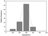
FIG. 4a is a scanning electron microscope image of a nano-hydroxyapatite reinforced keratin/PEO composite biological nanofiber membrane with a nano-hydroxyapatite content of 15% in an example;
fig. 4b is a fiber diameter distribution diagram of the nano-hydroxyapatite reinforced keratin/PEO composite biological nanofiber membrane with the nano-hydroxyapatite content of 15% in the example;
FIG. 5a is a scanning electron microscope image of a nano-hydroxyapatite reinforced keratin/PEO composite biological nanofiber membrane with a nano-hydroxyapatite content of 20% in an example;
fig. 5b is a fiber diameter distribution diagram of the nano-hydroxyapatite reinforced keratin/PEO composite biological nanofiber membrane with the nano-hydroxyapatite content of 20% in the example;
FIG. 6 is a graph showing the particle size distribution of HA at a 12.5:1 volume ratio of sodium hexametaphosphate solution to hydroxyapatite dispersion in the examples;
FIG. 7 is a graph showing the particle size distribution of HA at a volume ratio of 10:1 of the sodium hexametaphosphate solution to the hydroxyapatite dispersion in the example;
FIG. 8 is a graph showing the particle size distribution of HA at a volume ratio of 7.5:1 of the sodium hexametaphosphate solution to the hydroxyapatite dispersion in the examples;
FIG. 9 is a transmission electron microscope image of the modified nano-hydroxyapatite in the example;
FIG. 10 is a diagram of the state of the aqueous solution of sodium hexametaphosphate modified nano-hydroxyapatite in the example after standing for 48 hours;
fig. 11 is a stress-strain graph of nano-hydroxyapatite enhanced keratin/PEO/composite biological nanofiber membranes of different nano-hydroxyapatite contents in examples;
FIG. 12 is a comparative scanning electron microscope picture of keratin/PEO composite biological nanofiber membranes in which no nano-hydroxyapatite is added and the addition amount of modified nano-hydroxyapatite is 15% after L929 cell culture for 1, 3, 5, and 7 days in the example;
fig. 13 is a side view of the wound covering of example 5;
FIG. 14 is a top view of the wound covering of example 5 in an air-permeable state;
FIG. 15 is a top view of the wound covering of example 5 in a waterproof state;
FIG. 16a is a graphical representation of the topographical features of nanofibers produced from dope after cross-linking in an example;
FIG. 16b is a graph of the topographical features of nanofibers made from the uncrosslinked dope of the examples.
Detailed Description
The present invention will now be further explained with reference to specific embodiments, it being understood that the embodiments described are only some, but not all, of the embodiments of the present invention. All other embodiments, which can be derived by a person skilled in the art from the embodiments given herein without making any creative effort, shall fall within the protection scope of the present invention.
Example 1
The embodiment discloses a preparation method of a keratin PEO composite biological nanofiber membrane, which comprises the following steps:
(1) opening and removing impurities from human hair fibers, cutting the human hair fibers into fiber sections of 5-10mm, and then cleaning and drying the fiber sections;
(2) immersing the mixture into a dissolving solution containing a reducing agent, a protein denaturant and a surfactant at a solid-to-liquid ratio of 12g/250ml, and stirring the mixture in a water bath kettle at the temperature of 95 ℃ for 4 hours at the rotating speed of a stirrer of 180 r/min.
Wherein the reducing agent is sodium metabisulfite, and the mass percentage concentration of the reducing agent is 5 percent of that of the solution; the protein denaturant is urea, and the mass percentage concentration of the urea is 40 percent of that of the solution; the surfactant is sodium dodecyl sulfate, and the mass percentage concentration of the surfactant is 1 percent of that of the dissolving solution.
(3) Filtering the mixture obtained after stirring by using a 100-mesh screen to remove unreacted fibers, and dialyzing in a water bath at 37 ℃ for 36 hours to remove small molecular substances;
(4) removing precipitate by centrifugation at 6000rpm for 0.25 hr, collecting supernatant, and concentrating to obtain concentrated solution of keratin with concentration of 10%. The concentrated keratin solution was freeze-dried for 24h to obtain keratin powder.
(5) Putting nano hydroxyapatite into water, stirring the nano hydroxyapatite by using a magnetic stirrer at a rotating speed of 50r/min to disperse the nano hydroxyapatite to obtain a hydroxyapatite dispersion liquid with the nano hydroxyapatite mass percentage concentration of 10%, then preparing a sodium hexametaphosphate solution with the mass percentage concentration of 10%, slowly and dropwise adding the sodium hexametaphosphate solution into the nano hydroxyapatite solution continuously stirred at the rotating speed of 50r/min to enable the nano hydroxyapatite dispersion liquid to fully react, adding the sodium hexametaphosphate solution and the hydroxyapatite dispersion liquid in a volume ratio of 12.5:1, continuously stirring the nano hydroxyapatite dispersion liquid for 1 hour after the sodium hexametaphosphate solution is dropwise added, and then ultrasonically treating the mixed solution after the reaction for 10 minutes to enable the mixed solution to be more uniformly dispersed.
Adding absolute ethyl alcohol with the same volume to extract hydroxyapatite, centrifuging for 20min at 8000r/min to collect modified nano-hydroxyapatite, repeatedly washing the modified nano-hydroxyapatite with deionized water for 4 times to remove unreacted ions, and centrifuging at 8000r/min with a centrifuge to obtain wet modified nano-hydroxyapatite. And (3) freeze-drying the wet modified nano hydroxyapatite for 24 hours to obtain the modified nano hydroxyapatite powder.
(6) Mixing keratin and PEO according to the mass percentage of 95: 5, mixing with water, stirring for 4 hours on a magnetic stirrer to obtain a keratin PEO mixed solution with the keratin mass percentage concentration of 12%, adding the modified nano-hydroxyapatite powder into the stirred keratin/PEO mixed solution, and fully stirring on the magnetic stirrer to obtain the keratin/PEO/modified nano-hydroxyapatite mixed solution with the modified nano-hydroxyapatite mass percentage concentration of 5%.
And (2) dropwise adding Ethylene Glycol Diglycidyl Ether (EGDE) into the keratin/PEO/modified nano-hydroxyapatite mixed solution, and then continuously stirring for 1h on a magnetic stirrer to fully crosslink keratin to obtain the keratin/PEO/modified nano-hydroxyapatite blended spinning solution. Wherein the dropping amount of the Ethylene Glycol Diglycidyl Ether (EGDE) is 7 percent of the mass of the blended spinning solution.
As shown in fig. 16a, the spinnability of the spinning solution after the cross-linking treatment is obviously improved, the obtained nanofiber has good morphological characteristics and uniform thickness without bead knots, while as shown in fig. 16b, the nanofiber obtained by the spinning solution without the cross-linking treatment has poor spinnability, and a large number of beads exist in the fiber.
Preparing the nano-hydroxyapatite reinforced keratin/PEO composite nanofiber membrane under the room temperature condition by adopting a needle type electrostatic spinning method, applying an external voltage of 18 kV, spinning a receiving distance of 16cm, and a filling speed of 0.8ml/h, and then crosslinking the nano-hydroxyapatite reinforced keratin/PEO composite nanofiber membrane prepared by the electrostatic spinning method for 5 days by adopting oxygen oxidation secondary crosslinking to obtain the water-insoluble nano-hydroxyapatite reinforced keratin/PEO composite biological nanofiber membrane. Fig. 2a is a scanning electron microscope image of the nano-hydroxyapatite reinforced keratin/PEO composite biological nanofiber membrane, and fig. 2b is a diameter distribution condition of the nanofibers of the composite biological nanofiber membrane. The fracture stress of the composite biological nanofiber membrane is 1.4 MPa.
Example 2
The embodiment discloses a preparation method of a keratin PEO composite biological nanofiber membrane, which comprises the following steps:
(1) opening rabbit hair fiber, removing impurities, cutting into fiber sections of 5-10mm, and then cleaning and drying in the sun;
(2) immersing the mixture into a dissolving solution containing a reducing agent, a protein denaturant and a surfactant at a solid-to-liquid ratio of 10g/250ml, and stirring the mixture in a water bath kettle at 90 ℃ for 5 hours at the rotating speed of a stirrer of 200 r/min.
Wherein the reducing agent is thioglycolic acid, and the mass percentage concentration of the reducing agent in the dissolving solution is 7%; the protein denaturant is thiourea, the mass percentage concentration of the protein denaturant in the dissolving solution is 45 percent of the solution, the surfactant is sodium dodecyl sulfate, and the mass percentage concentration of the protein denaturant in the dissolving solution is 2 percent of the solution.
(3) Filtering the mixture obtained after stirring by using a 150-mesh screen to remove unreacted fibers, and dialyzing in a water bath at 37 ℃ for 72 hours to remove small molecular substances;
(4) removing precipitate by centrifugation at 5000rpm for 0.5 hr, collecting supernatant, and concentrating to obtain concentrated solution of keratin with mass percent concentration of 14%. The concentrated keratin solution was freeze-dried for 36h to obtain keratin powder.
(5) Putting the nano hydroxyapatite into water, and stirring the nano hydroxyapatite by using a magnetic stirrer at the rotating speed of 100r/min to disperse the nano hydroxyapatite so as to obtain a dispersion liquid with the mass percentage concentration of the nano hydroxyapatite of 10 percent;
then preparing a sodium hexametaphosphate solution with the mass percentage concentration of 10 percent, slowly dropwise adding the sodium hexametaphosphate solution into the nano hydroxyapatite solution which is continuously stirred at the rotating speed of 100r/min to ensure that the nano hydroxyapatite solution is fully reacted, adding the sodium hexametaphosphate solution and the hydroxyapatite dispersion liquid with the volume ratio of 7.5:1, continuously stirring for 1 hour after the dropwise adding of the sodium hexametaphosphate is completed, and then carrying out ultrasonic treatment on the reacted mixed solution for 10 minutes to ensure that the mixed solution is more uniformly dispersed. Adding absolute ethyl alcohol with the same volume to extract hydroxyapatite, centrifuging for 20min at 8000r/min to collect modified nano-hydroxyapatite, repeatedly washing the modified nano-hydroxyapatite with deionized water for 5 times to remove unreacted ions, and centrifuging at 8000r/min with a centrifuge to obtain wet modified nano-hydroxyapatite. And (3) freeze-drying the wet modified nano-hydroxyapatite for 36 hours to obtain the modified nano-hydroxyapatite powder.
(6) Mixing keratin and PEO according to the mass percentage of 95: 5, mixing with water, stirring for 6 hours on a magnetic stirrer to obtain a keratin PEO mixed solution with the mass percentage concentration of 15%, adding the modified nano-hydroxyapatite powder into the stirred keratin/PEO mixed solution, fully stirring on the magnetic stirrer to obtain a keratin/PEO/modified nano-hydroxyapatite mixed solution with the mass percentage concentration of 10%, dropwise adding Ethylene Glycol Diglycidyl Ether (EGDE) with the mass percentage concentration of 7% into the keratin/PEO/modified nano-hydroxyapatite mixed solution, and then continuously stirring on the magnetic stirrer for 2.5 hours to fully crosslink keratin, thereby obtaining the keratin/PEO/modified nano-hydroxyapatite blended spinning solution. Preparing the nano-hydroxyapatite reinforced keratin/PEO composite nanofiber membrane under the room temperature condition by adopting a bubble electrospinning method, externally applying a voltage of 24 kV, spinning a receiving distance of 20cm, and filling at a speed of 1.5ml/h, and then crosslinking the nano-hydroxyapatite reinforced keratin/PEO composite nanofiber membrane prepared by the electrostatic spinning method for 10 days by adopting oxygen oxidation secondary crosslinking to obtain the water-insoluble nano-hydroxyapatite reinforced keratin/PEO composite biological nanofiber membrane. Fig. 3a is a scanning electron microscope image of the nano-hydroxyapatite reinforced keratin/PEO composite biological nanofiber membrane, and fig. 3b is a diameter distribution of the nanofibers of the composite biological nanofiber membrane. The experiment shows that the rupture stress of the composite biological nanofiber membrane is 1.67 MPa.
Example 3
Opening and removing impurities from pig hair fibers, cutting into fiber sections of 5-10mm, cleaning, drying in the sun, soaking into a solution containing a reducing agent, a protein denaturant and a surfactant at a solid-to-liquid ratio of 6g/250ml, and stirring in a water bath kettle at 92 ℃ for 6h at the rotation speed of 190 r/min. Wherein the reducing agent is dithiothreitol, and the mass percentage concentration is 6 percent; the protein denaturant is lithium bromide, the mass percent concentration of the lithium bromide is 42 percent of the solution, the surfactant is sodium dodecyl sulfate, and the mass percent concentration of the sodium dodecyl sulfate is 5 percent of the solution. Filtering the mixture obtained after stirring by using a 120-mesh screen to remove unreacted fibers, dialyzing in 37 ℃ water bath for 48 hours to remove small molecular substances, centrifuging to remove precipitated substances, wherein the centrifugation speed is 5000rpm, and the centrifugation time is 0.3 hour, then taking supernatant, and concentrating the obtained supernatant to obtain the keratin concentrated solution with the mass percentage concentration of 12%. The concentrated keratin solution was freeze-dried for 30h to obtain keratin powder.
Putting 10% of nano-hydroxyapatite in percentage by mass into water, stirring the nano-hydroxyapatite at a rotating speed of 60r/min by using a magnetic stirrer to disperse the nano-hydroxyapatite, then preparing 10% of sodium hexametaphosphate solution, slowly dropwise adding the sodium hexametaphosphate solution into the nano-hydroxyapatite continuously stirred at the rotating speed of 60r/min to fully react, adding the sodium hexametaphosphate solution and the hydroxyapatite dispersion liquid in a volume ratio of 10:1, continuously stirring the nano-hydroxyapatite for 1 hour after completing the dropwise addition of the sodium hexametaphosphate, and then ultrasonically treating the reacted mixed solution for 10 minutes to ensure that the mixed solution is more uniformly dispersed. Adding absolute ethyl alcohol with the same volume to extract hydroxyapatite, centrifuging for 20min at 8000r/min to collect modified nano-hydroxyapatite, repeatedly washing the modified nano-hydroxyapatite with deionized water for 4 times to remove unreacted ions, and centrifuging at 8000r/min with a centrifuge to obtain wet modified nano-hydroxyapatite. And (3) freeze-drying the wet modified nano-hydroxyapatite for 30h to obtain the modified nano-hydroxyapatite powder.
Mixing keratin and PEO according to the mass percentage of 95: 5, mixing with water, stirring for 5 hours on a magnetic stirrer to obtain a keratin PEO mixed solution with the mass percentage concentration of 13%, adding 15% of modified nano-hydroxyapatite powder into the stirred keratin/PEO mixed solution, fully stirring on the magnetic stirrer to obtain a keratin/PEO/modified nano-hydroxyapatite mixed aqueous solution, dropwise adding 7% of Ethylene Glycol Diglycidyl Ether (EGDE) into the keratin/PEO/modified nano-hydroxyapatite mixed aqueous solution, and then continuously stirring on the magnetic stirrer for 2 hours to fully crosslink keratin, thereby obtaining the keratin/PEO/modified nano-hydroxyapatite mixed spinning solution. Preparing the nano-hydroxyapatite reinforced keratin/PEO composite nanofiber membrane under the room temperature condition by adopting a disc type electrostatic spinning method, externally applying a voltage of 20 kV, spinning a receiving distance of 18cm, and pouring at a pouring speed of 1ml/h, and then crosslinking the nano-hydroxyapatite reinforced keratin/PEO composite nanofiber membrane prepared by the electrostatic spinning method for 8 days by adopting oxygen oxidation secondary crosslinking to obtain the water-insoluble nano-hydroxyapatite reinforced keratin/PEO composite biological nanofiber membrane. Fig. 4a is a scanning electron microscope image of the nano-hydroxyapatite reinforced keratin/PEO composite biological nanofiber membrane, and fig. 4b is a diameter distribution of the nanofibers of the composite biological nanofiber membrane. The experiment shows that the rupture stress of the composite biological nanofiber membrane is 2.23 MPa. In addition, the experiment proves that when the mass percentage of the nano hydroxyapatite is 14-16%, the breaking stress of the composite biological nano fiber membrane is 2.1-2.4Mpa, and the mechanical effect of the fiber membrane is particularly outstanding.
Example 4
The embodiment discloses a preparation method of a keratin PEO composite biological nanofiber membrane, which comprises the following steps:
(1) opening and removing impurities from wool fibers, cutting the wool fibers into fiber sections of 5-10mm, and then cleaning and drying the wool fibers in the sun;
(2) immersing the mixture into a dissolving solution containing a reducing agent, a protein denaturant and a surfactant at a solid-to-liquid ratio of 11g/250ml, and stirring the mixture in a water bath kettle at 93 ℃ for 5 hours at a rotating speed of a stirrer of 220 r/min. Wherein the reducing agent is mercaptoethanol, and the mass percentage concentration in the dissolving solution is 7 percent; the protein denaturant is urea, the mass percentage concentration of the dissolving solution is 40 percent of that of the solution, the surfactant is sodium dodecyl sulfate, and the mass percentage concentration of the dissolving solution is 4 percent of that of the solution.
(3) Filtering the mixture obtained after stirring by using a 100-mesh screen to remove unreacted fibers, dialyzing in 37 ℃ water bath for 36 hours to remove small molecular substances, centrifuging to remove precipitated substances, wherein the centrifugation speed is 6000rpm, the centrifugation time is 0.25 hour, taking supernate, and concentrating the obtained supernate to obtain the keratin concentrated solution with the keratin mass percentage concentration of 10%. The concentrated keratin solution was freeze-dried for 36h to obtain keratin powder.
(4) Putting 10% of nano-hydroxyapatite in percentage by mass into water, stirring the nano-hydroxyapatite at a rotating speed of 80r/min by using a magnetic stirrer to disperse the nano-hydroxyapatite, then preparing 10% of sodium hexametaphosphate solution, slowly and dropwise adding the sodium hexametaphosphate solution into the nano-hydroxyapatite which is continuously stirred at the rotating speed of 80r/min to fully react, adding the sodium hexametaphosphate solution and the hydroxyapatite dispersion liquid in a volume ratio of 8:1, continuously stirring the nano-hydroxyapatite for 1 hour after the dropwise addition of the sodium hexametaphosphate is completed, and then ultrasonically treating the reacted mixed solution for 10 minutes to ensure that the mixed solution is more uniformly dispersed.
Adding absolute ethyl alcohol with the same volume to extract hydroxyapatite, centrifuging for 20min at 8000r/min to collect modified nano-hydroxyapatite, repeatedly washing the modified nano-hydroxyapatite with deionized water for 5 times to remove unreacted ions, and centrifuging at 8000r/min with a centrifuge to obtain wet modified nano-hydroxyapatite. And (3) freeze-drying the wet modified nano-hydroxyapatite for 36 hours to obtain the modified nano-hydroxyapatite powder.
(5) Mixing keratin and PEO according to the mass percentage of 95: 5, mixing with water, stirring for 5 hours on a magnetic stirrer to obtain a keratin PEO mixed solution with the mass percentage concentration of 14%, adding the modified nano-hydroxyapatite powder into the stirred keratin/PEO mixed solution, fully stirring on the magnetic stirrer to obtain a keratin/PEO/modified nano-hydroxyapatite mixed solution with the mass percentage concentration of 20%, dropwise adding Ethylene Glycol Diglycidyl Ether (EGDE) with the mass percentage concentration of 7% into the keratin/PEO/modified nano-hydroxyapatite mixed solution, and then continuously stirring for 1.5 hours on the magnetic stirrer to fully crosslink keratin, thereby obtaining the keratin/PEO/modified nano-hydroxyapatite blended spinning solution.
Preparing a nano-hydroxyapatite enhanced keratin/PEO composite nanofiber membrane under the room temperature condition by adopting a needle type electrostatic spinning method, applying a voltage of 22 kV, spinning a receiving distance of 18cm, and a pouring speed of 1.2ml/h, then crosslinking the nano-hydroxyapatite enhanced keratin/PEO composite nanofiber membrane prepared by adopting the electrostatic spinning method for 6 days by adopting oxygen oxidation secondary crosslinking to obtain a water-insoluble nano-hydroxyapatite enhanced keratin/PEO composite biological nanofiber membrane, wherein fig. 5a is a scanning electron microscope image of the nano-hydroxyapatite enhanced keratin/PEO composite biological nanofiber membrane, and fig. 5b is the diameter distribution condition of nanofibers of the composite biological nanofiber membrane. The fracture stress of the composite biological nanofiber membrane is 1.73 MPa.
The test and experimental results show that:
1. particle size and morphology of sodium hexametaphosphate modified hydroxyapatite
In the preparation method of the sodium hexametaphosphate modified nano-hydroxyapatite powder, the volume ratio of the sodium hexametaphosphate solution to the hydroxyapatite dispersion liquid is 12.5:1-7.5:1, and the average particle size of the modified nano-hydroxyapatite is optimal.
As shown in fig. 6, the volume ratio of the sodium hexametaphosphate solution to the hydroxyapatite dispersion was 12.5:1, the average particle size of the modified nano-hydroxyapatite is 190.4 nm; as shown in fig. 7, the ratio of sodium hexametaphosphate to nano-hydroxyapatite is 10:1, the average particle size of the modified nano-hydroxyapatite is 192.1 nm; as shown in fig. 8, the ratio of sodium hexametaphosphate to nano-hydroxyapatite was 7.5:1, the average particle size of the modified nano-hydroxyapatite is 221.9 nm; wherein, fig. 9 is a transmission electron microscope picture of the modified nano-hydroxyapatite; as shown in fig. 10, the modified nano-hydroxyapatite aqueous solution is still in a clear and transparent state after standing for 48 hours, so that the dispersibility of the hydroxyapatite is significantly improved by modifying the hydroxyapatite with sodium hexametaphosphate. And the unmodified hydroxyapatite is dispersed and then stands for 4 hours, and then precipitation occurs at the bottom of the container.
2. Nano-hydroxyapatite enhanced keratin/PEO composite biological nanofiber membrane mechanical property
The stress-strain curve of the nano-hydroxyapatite reinforced keratin/PEO composite biological nanofiber membrane with different modified nano-hydroxyapatite contents is shown in fig. 11, and the breaking strength of the nano-hydroxyapatite reinforced keratin/PEO composite biological nanofiber membrane is remarkably increased by adding 5-20% of the modified nano-hydroxyapatite in percentage by mass. And when the content of the nano-hydroxyapatite is 15%, the breaking strength is optimal, and meanwhile, the breaking elongation of the nano-hydroxyapatite reinforced keratin/PEO composite biological nanofiber membrane is moderate. The mechanical properties of the nano-hydroxyapatite reinforced keratin/PEO/composite biological nanofiber membrane with different nano-hydroxyapatite contents are shown in Table 1.
TABLE 1
| Content of hydroxyapatite (%)
|
Maximum tensile strength (Mpa)
|
Elongation at Break (%)
|
| 0
|
1.01±0.19
|
54±6
|
| 5
|
1.41±0.17
|
55±5
|
| 10
|
1.67±0.22
|
50±3
|
| 15
|
2.23±0.16
|
44±7
|
| 20
|
1.73±0.23
|
36±5 |
Therefore, comprehensive judgment shows that when the content of the nano-hydroxyapatite is 15%, the nano-hydroxyapatite with the nano-hydroxyapatite content enhances the mechanical property of the keratin/PEO/composite biological nanofiber membrane to be optimal.
In addition, scanning electron microscope pictures of keratin/PEO composite biological nanofiber membranes without nano-hydroxyapatite and with 15% of modified nano-hydroxyapatite added amount after L929 cell culture for 1, 3, 5 and 7 days (1 d, 3d, 5d and 7d in the figure) are shown in FIG 12, and FIG 12 shows that the addition of nano-hydroxyapatite does not influence the biocompatibility of the original material, so the keratin/PEO composite biological nanofiber membrane enhanced by nano-hydroxyapatite has good biocompatibility.
Example 5
The embodiment discloses a wound plaster adopting the composite biological nanofiber membrane, which comprises a waterproof adhesive tape body 20 and a medicine soaking pad 10, as shown in fig. 13; the medicine soaking pad 10 consists of a plurality of layers of composite biological nanofiber membranes; the medicine soaking pad cloth is arranged in the middle of one side of the waterproof adhesive tape body.
The composite biological nanofiber membrane is laid on a wound as a substitute of gauze after being soaked with the medicine, has good anti-tearing mechanical property, and simultaneously has good biocompatibility with wound cells due to the biological components of keratin, so that the rejection phenomenon of the wound is avoided, and the rapid healing of the wound is facilitated. In a wound healing experiment of a mouse, compared with the existing wound plaster, the wound plaster disclosed by the invention has the advantage that the wound healing speed is 1-2 days faster.
In addition, as shown in fig. 13-15, the present embodiment further includes a first elastic waterproof tape 30 and a second elastic waterproof tape 40; the first elastic waterproof adhesive tape and the second elastic waterproof adhesive tape are arranged on the other opposite side of the medicine soaking pad on the waterproof adhesive tape body; on the length direction of waterproof sticky tape body, first elasticity waterproof sticky tape and second elasticity waterproof sticky tape extend and set up in the corresponding position stack of dipping the medicine pad from two outside lateral medians of dipping the medicine pad respectively.
The waterproof adhesive tape body, the first elastic waterproof adhesive tape and the second elastic waterproof adhesive tape are respectively provided with a ventilation window 21, a first ventilation port 31 and a second ventilation port 41 at the corresponding parts of the medicine soaking pad; when the ventilation window, the first ventilation port and the second ventilation port are arranged in an overlapping mode, the medicine soaking pad 10 is communicated with the outside air, so that the ventilation of the wound is facilitated, the suppuration is prevented, and the wound healing is accelerated; respectively stretching the first elastic waterproof adhesive tape and the second elastic waterproof adhesive tape to enable the first ventilation opening 31 and the second ventilation opening 41 to be respectively staggered with the ventilation window 21, wherein the non-first ventilation opening part of the first elastic waterproof adhesive tape covers the ventilation window 21, and the non-second ventilation opening part of the second elastic waterproof adhesive tape covers the first ventilation opening; thereby isolating the medicine soaking pad and realizing waterproof arrangement.
In life, when the adhesive bandage needs to be waterproof, the adhesive bandage is often temporary or transient, and a wound is wrapped airtightly for a long time, is easy to suppuration and is slow in healing speed.
Finally, it should be noted that: the above embodiments are only used to illustrate the technical solution of the present invention, and not to limit the same; while the invention has been described in detail and with reference to the foregoing embodiments, it will be understood by those skilled in the art that: the technical solutions described in the foregoing embodiments may still be modified, or some or all of the technical features may be equivalently replaced; and the modifications or the substitutions do not make the essence of the corresponding technical solutions depart from the scope of the technical solutions of the embodiments of the present invention.