CN109492706B - Chromosome classification prediction device based on recurrent neural network - Google Patents
Chromosome classification prediction device based on recurrent neural network Download PDFInfo
- Publication number
- CN109492706B CN109492706B CN201811425376.1A CN201811425376A CN109492706B CN 109492706 B CN109492706 B CN 109492706B CN 201811425376 A CN201811425376 A CN 201811425376A CN 109492706 B CN109492706 B CN 109492706B
- Authority
- CN
- China
- Prior art keywords
- chromosome
- classification
- module
- neural network
- recurrent neural
- Prior art date
- Legal status (The legal status is an assumption and is not a legal conclusion. Google has not performed a legal analysis and makes no representation as to the accuracy of the status listed.)
- Active
Links
Images


Classifications
-
- G—PHYSICS
- G06—COMPUTING OR CALCULATING; COUNTING
- G06F—ELECTRIC DIGITAL DATA PROCESSING
- G06F18/00—Pattern recognition
- G06F18/20—Analysing
- G06F18/24—Classification techniques
- G06F18/241—Classification techniques relating to the classification model, e.g. parametric or non-parametric approaches
-
- G—PHYSICS
- G06—COMPUTING OR CALCULATING; COUNTING
- G06N—COMPUTING ARRANGEMENTS BASED ON SPECIFIC COMPUTATIONAL MODELS
- G06N3/00—Computing arrangements based on biological models
- G06N3/02—Neural networks
- G06N3/04—Architecture, e.g. interconnection topology
- G06N3/044—Recurrent networks, e.g. Hopfield networks
-
- G—PHYSICS
- G06—COMPUTING OR CALCULATING; COUNTING
- G06N—COMPUTING ARRANGEMENTS BASED ON SPECIFIC COMPUTATIONAL MODELS
- G06N3/00—Computing arrangements based on biological models
- G06N3/02—Neural networks
- G06N3/04—Architecture, e.g. interconnection topology
- G06N3/045—Combinations of networks
-
- G—PHYSICS
- G06—COMPUTING OR CALCULATING; COUNTING
- G06V—IMAGE OR VIDEO RECOGNITION OR UNDERSTANDING
- G06V10/00—Arrangements for image or video recognition or understanding
- G06V10/20—Image preprocessing
- G06V10/26—Segmentation of patterns in the image field; Cutting or merging of image elements to establish the pattern region, e.g. clustering-based techniques; Detection of occlusion
- G06V10/267—Segmentation of patterns in the image field; Cutting or merging of image elements to establish the pattern region, e.g. clustering-based techniques; Detection of occlusion by performing operations on regions, e.g. growing, shrinking or watersheds
Landscapes
- Engineering & Computer Science (AREA)
- Theoretical Computer Science (AREA)
- Physics & Mathematics (AREA)
- General Physics & Mathematics (AREA)
- Data Mining & Analysis (AREA)
- Evolutionary Computation (AREA)
- Life Sciences & Earth Sciences (AREA)
- Artificial Intelligence (AREA)
- General Engineering & Computer Science (AREA)
- Computing Systems (AREA)
- Software Systems (AREA)
- Molecular Biology (AREA)
- Computational Linguistics (AREA)
- Biophysics (AREA)
- Biomedical Technology (AREA)
- Mathematical Physics (AREA)
- General Health & Medical Sciences (AREA)
- Health & Medical Sciences (AREA)
- Bioinformatics & Cheminformatics (AREA)
- Bioinformatics & Computational Biology (AREA)
- Computer Vision & Pattern Recognition (AREA)
- Evolutionary Biology (AREA)
- Multimedia (AREA)
- Image Analysis (AREA)
- Investigating Or Analysing Biological Materials (AREA)
Abstract
The invention discloses a chromosome classification prediction device based on a recurrent neural network, which comprises a memory, a processor and a computer program, wherein a chromosome image preprocessing module and a chromosome classification result prediction model are stored in the memory; the chromosome classification result prediction model comprises a sequence feature extraction module and a fusion classification module for fusing and classifying the sequence features of the two chromosomes output by the sequence feature extraction module; the processor, when executing the computer program, implements the steps of: receiving a chromosome image, and sequentially carrying out effective pixel marking, chromosome contour detection and division into N rectangular images by a chromosome image preprocessing module; respectively inputting N rectangular images of two chromosomes into a sequence feature extraction module, and extracting sequence features Sn1And Sn2Outputting to a fusion classification module, and outputting the classification prediction probability of the chromosome through calculation; n is an integer between 10 and 20. The device can output the prediction probability of the chromosome classification result with higher accuracy.
Description
Technical Field
The invention belongs to the field of medical image data processing, and particularly relates to a chromosome classification prediction device based on a recurrent neural network.
Background
With the great development of deep learning in the image field, the deep learning-based method is widely applied to medical image data. At present, computer systems based on deep learning have prominent effects in the aspects of identifying and segmenting CT, pathological sections, ultrasonic images, MRI images and the like.
Image recognition (Image recognition), i.e. Image Classification (Image Classification), is an important research direction of computer vision, and the task of the Image recognition is to classify a single Image into C categories by a computer algorithm. Image classification is commonly used in practical applications for optical character recognition, face detection, and the like. In medical imaging, image classification methods are often used to determine whether tissues or cells are diseased.
Image classification methods are currently based primarily on Convolutional Neural Networks (CNNs), 1998, LECUN et al first proposed after the Convolutional Neural Network LeNet model was used by many banks in the united states to recognize handwritten numbers on checks. CNN models with various different architectures are champions of multiple games in ImageNet competition, and the CNN is widely applied in the fields of image processing and target recognition and becomes a general neural network for deep learning in the field of image processing.
Chromosomes (chromosomes) are a specific form of genetic material present in eukaryotic cells at mitosis or meiosis, are the result of tightly packed chromatin structures in interphase cells, are higher order structures of chromatin that occur only during cell division. Chromosomes are species-specific, and their number, size and morphology vary with the species, cell type and stage of development. Chromosomes are substances that carry genetic information in the nucleus of a cell and are cylindrical or rod-shaped under a microscope. The classification and identification of human chromosomes are a basic task in medical genetics, and the realization of automatic analysis and identification of human chromosomes by applying a computer technology is an important research topic of a human chromosome image analysis technology.
The chromosome analysis system is also called chromosome image analysis system/chromosome karyotype analysis system, and is mainly applied to clinical medical analysis and diagnosis under modern microscope, and has the biggest advantages of high definition, direct observation under computer large screen, automatic computer identification and chromosome segmentation, standard chromosome karyotype comparison and automatic arrangement, high accuracy of analysis and judgment, convenient image storage and processing, and provision of precious data for later analysis and summarization, and is the trend of medical microscopic image analysis. The objective of the research on automatic chromosome analysis systems is to reduce the labor intensity of technicians, relieve them from complicated and repetitive labor, and finally apply these systems to clinical applications for cytogenetic identification of tumor patients, examination of prenatal and postnatal care, and the like. Although the application of neural networks in automatic chromosome analysis systems has been developed and perfected for many years, certain limitations still exist: firstly, it needs a set of accurate and unmistakable classified chromosome database, which is not easy to be obtained by general researchers; secondly, the classification result is not accurate enough and even can not reach the level of a well-trained cytology researcher; thirdly, the neural network has its inherent disadvantages of huge training data volume and long training time.
Therefore, in future research, the network should be improved and perfected continuously by optimizing the structure of the neural network, extracting effective features and reducing unnecessary operations.
Disclosure of Invention
The invention discloses a chromosome classification prediction device based on a recurrent neural network, which can output the prediction probability of a chromosome classification result with higher accuracy through calculation, and the prediction probability can assist doctors to carry out cytogenetic identification and bearing and rearing examination of tumor patients.
A recurrent neural network-based chromosome classification prediction apparatus comprising a computer memory in which a chromosome image preprocessing module and a chromosome classification result prediction model are stored and a computer processor, and a computer program stored in the computer memory and executable on the computer processor; the chromosome classification result prediction model comprises a sequence feature extraction module and a fusion classification module for fusing and classifying the sequence features of the two chromosomes output by the sequence feature extraction module;
the computer processor, when executing the computer program, performs the steps of:
receiving chromosome images, sequentially performing mask M acquisition, chromosome contour detection and separation and dividing each chromosome into N rectangular images taking N main trunk nodes as centers by a chromosome image preprocessing module, and taking the N rectangular images as images to be detected;
n rectangular images of two chromosomesRespectively input into a sequence feature extraction module, and after feature extraction, the extracted sequence features Sn1And Sn2Outputting to a fusion classification module, and outputting the classification prediction probability of the chromosome through calculation;
wherein, the value range of N is an integer between 10 and 20.
The mask M obtaining method is to use a threshold value method, take pixels between left and right threshold values in a chromosome as effective pixels, set the corresponding coordinate point position as 1, and set the coordinate point position corresponding to a non-effective pixel as 0 to obtain the mask M.
Specifically, L and R of the left and right thresholds L and R range to [0, 255 ]. And if the pixel value v of the image meets the condition that L is less than v and less than R, the pixel is an effective pixel, the position of the coordinate point corresponding to the pixel is set to be 1, and otherwise, the position is set to be 0.
The chromosome contour detection and separation comprises the following steps: using contour detection algorithm to obtain the largest two connected regions in the chromosome image for the mask M, separating the two connected regions, and respectively using the two connected regions as the images M of the two chromosomes in the chromosome pair1And M2. The chromosome image shot under the microscope has a plurality of connected regions, and the coordinate points corresponding to the effective pixels in the mask M include interference points that do not belong to the chromosome region, so it is necessary to extract each stain in the chromosome pair by a contour detection algorithm and eliminate the interference points.
The method for dividing each chromosome into N rectangular images taking N main trunk nodes as centers comprises the following steps:
(1) image M1And M2Coordinate points corresponding to the middle effective pixels respectively form a point set;
(2) obtaining a fitting curve by using a curve interpolation method for the point set S;
(3) for the fitted curve, take N coordinate points (x) equidistant in the x direction1,y1),(x2,y2)...(xN,yN) As a backbone node of the chromosome;
(4) each chromosome is divided into N rectangular images with N backbone nodes as the center.
The curve interpolation method is selected from linear interpolation, bilinear interpolation or B-spline curve interpolation.
Preferably, the curve interpolation method is a B-spline curve interpolation method, and the robustness of the method is higher than that of the other two interpolation methods.
The N rectangular images are H in height and W in width, and the value range of H and W is an integer from 10 to 20.
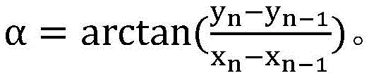
The chromosome image preprocessing module rotates the N rectangular images by an angle alpha with the main trunk node as the center, and the method for selecting the rotation angle alpha comprises the following steps:

the chromosome has a certain curvature, so that the rotation angle of the input rectangular frame needs to be consistent with the slope direction between two main trunk nodes, and the obtained inclination angle of the rectangular frame is ensured to be consistent with the trend of the chromosome.
The sequence feature extraction module comprises: extracting convolution layers and activation layers of image features f of N rectangular images, and outputting sequence features S to the input image features f through N times of circulationnThe recurrent neural network layer.
Specifically, the number of the convolution layers is 3-5, and an activation layer is arranged behind each convolution layer. Each convolution layer and activation layer is referred to as a convolution-activation layer, i.e., the network structure that extracts the image features f of the N rectangular images is 3-5 convolution-activation layers.
The step length of the recurrent neural network layer is N. The recurrent neural network layer is selected from RNN, LSTM or GRU.
Preferably, the recurrent neural network layer is LSTM, and the LSTM has long and short term memory function and is more suitable for long-distance sequence detection;
the chromosome classification result prediction model is optimally trained by adopting a cross entropy loss and gradient descent method in the training process.
The optimization training means that the difference value between the real label and the chromosome classification label obtained by the prediction of the chromosome classification result prediction model is minimized.
The method for minimizing the difference comprises the following specific steps:
(1) solving the loss H of the real label and the prediction confidence coefficient by using cross entropy loss, wherein the specific formula of the cross entropy loss is as follows:

wherein p and q represent the prediction confidence and the real label, and y represents the prediction probability for the class and the one-hot encoding of the true label, respectively.
(2) Optimizing H until training converges by using a gradient descent method with a learning rate of lr in a range of 10-4~10-2。
The random gradient descent method includes: SGD, Adam, RMSprop, etc.
The fusion classification module comprises a classifier selected from logistic regression, support vector machine or k-nearest neighbor classification.
The fusion classification module fuses sequence characteristics Sn1And Sn2The method of (a) is selected from the group consisting of splicing, summation or integration.
The chromosome classification result prediction model can be trained on line and then stored in a chromosome classification prediction device; or online training is completed, and the received chromosome image to be predicted in each application can be used as a training sample after being preprocessed by the chromosome image preprocessing module, so that the chromosome classification result prediction model is optimized and updated.
The chromosome classification device provided by the invention can output the prediction probability of the chromosome classification result with higher accuracy through calculation, and the prediction probability can assist doctors to carry out cytogenetic identification and bearing and rearing examination of tumor patients.
Drawings
FIG. 1 is a chromosome image inputted in the example;
FIG. 2 is a rectangle divided by centering around a chromosome trunk node in the example;
FIG. 3 shows the structure of the sequence feature extraction model and the classifier in the embodiment.
Detailed Description
For further understanding of the present invention, the method for classifying chromosomes based on recurrent neural networks provided by the present invention is specifically described below with reference to specific implementation methods, but the present invention is not limited thereto, and the insubstantial modifications and adaptations made by those skilled in the art under the core teaching of the present invention still fall within the scope of the present invention.
The embodiment provides a chromosome classification prediction device based on a recurrent neural network, which comprises a computer memory, a computer processor and a computer program stored in the computer memory and executable on the computer processor, wherein a chromosome image preprocessing module and a chromosome classification result prediction model are stored in the computer memory; the chromosome classification result prediction model comprises a sequence feature extraction module and a fusion classification module for fusing and classifying the sequence features of the two chromosomes output by the sequence feature extraction module.
The computer program when executed by a computer processor implements the steps of:
s1, receiving the chromosome image, sequentially performing mask M acquisition, chromosome contour detection and separation and division of each chromosome into N rectangular images taking N main nodes as centers by the chromosome image preprocessing module, wherein the value range of N is an integer between 10 and 20. The chromosome image in this example is shown in FIG. 1.
S101, obtaining a mask M.
And marking effective pixels in a pair of chromosome images shot under a microscope by using a threshold method to obtain a mask M.
Specifically, using the left threshold L being 10 and the right threshold R being 240, if the pixel value v of the image satisfies L < v < R, the position of the coordinate point corresponding to the pixel is set to 1, otherwise the position is set to 0.
And S102, detecting the chromosome contour.
Using contour detection algorithm to obtain the largest two connected regions in the chromosome image for the mask M, separating the two connected regions, and respectively using the two connected regions as the images M of the two chromosomes in the chromosome pair1And M2。
S103, each chromosome is divided into N rectangular images taking N main trunk nodes as centers.
S1031, image M1And M2Coordinate points corresponding to the middle effective pixels respectively form a point set S1And S2;
S1032, set of point pairs S1And S2Obtaining a fitting curve C by respectively using a B spline interpolation method1And C2;
S1033, fitting the curve C1And C215 coordinate points (x) are respectively taken at equal distance in the x direction1,y1),(x2,y2)...(x15,y15) As the backbone node of the chromosome.
S104, taking 15 main nodes as the center, as shown in figure 2, in the image M1Cutting up 15 rectangular images with the height of 10, the width of 10 and the rotation angle of alpha; simultaneously in the image M2Cutting up 15 rectangular images with the height of 10, the width of 10 and the rotation angle of alpha; 15 rectangular images of two chromosomes are used as input of the chromosome classification result prediction model in a grading mode.
The method of alpha selection is calculated using the following formula:

S2, respectively inputting the 15 rectangular images of the two chromosomes into a sequence feature extraction module, and extracting the sequence after feature extractionColumn characteristics Sn1And Sn2And outputting the chromosome classification prediction probability to a fusion classification module through calculation.
As shown in fig. 3, the sequence feature extraction module includes: extracting convolution-activation layers of image features f of 15 rectangular images, and outputting sequence features S to the input image features f through 15 times of circulationnThe recurrent neural network layer. Wherein, the convolution-activation layer is 3; the recurrent neural network layer is LSTM with a step size of 15.
The fusion classification module comprises a classifier which is a logistic regression.
The chromosome classification result prediction model is completed on line, and a cross entropy loss and gradient descent method is adopted for optimization training in the training process. The optimization training means that the difference value between the real label and the chromosome classification label obtained by the prediction of the chromosome classification result prediction model is minimized. The class labels are specific classes in chromosomes 1-23.
The method for minimizing the difference comprises the following specific steps:
and (3) solving the loss H of the real label and the prediction confidence coefficient by using cross entropy loss, wherein the specific formula of the cross entropy loss is as follows:

wherein p and q represent the prediction confidence and the real label, and y represents the prediction probability for the class and the one-hot encoding of the true label, respectively.
Learning rate of use lr 10-4The SGD gradient descent method of (a) optimizes H to minimize H until the training converges.
The above-mentioned embodiments are intended to illustrate the technical solutions and advantages of the present invention, and it should be understood that the above-mentioned embodiments are only the most preferred embodiments of the present invention, and are not intended to limit the present invention, and any modifications, additions, equivalents, etc. made within the scope of the principles of the present invention should be included in the scope of the present invention.
Claims (5)
1. A recurrent neural network-based chromosome classification prediction apparatus comprising a computer memory, a computer processor, and a computer program stored in the computer memory and executable on the computer processor,
the computer memory is stored with a chromosome image preprocessing module and a chromosome classification result prediction model; the chromosome classification result prediction model comprises a sequence feature extraction module and a fusion classification module for fusing and classifying the sequence features of the two chromosomes output by the sequence feature extraction module;
the computer processor, when executing the computer program, performs the steps of:
receiving chromosome images, sequentially performing mask M acquisition, chromosome contour detection and separation and dividing each chromosome into N rectangular images taking N main trunk nodes as centers by a chromosome image preprocessing module, and taking the N rectangular images as images to be detected;
respectively inputting N rectangular images of two chromosomes into a sequence feature extraction module, and extracting sequence features S after feature extractionn1And Sn2Outputting to a fusion classification module, and outputting the classification prediction probability of the chromosome through calculation;
wherein the value range of N is an integer between 10 and 20;
the mask M acquisition method comprises the steps of using a threshold value method, taking pixels between left and right threshold values in a chromosome as effective pixels, setting the positions of corresponding coordinate points as 1, and setting the positions of coordinate points corresponding to non-effective pixels as 0 to obtain a mask M;
the chromosome contour detection and separation comprises the following steps: obtaining two maximum connected areas in the mask M by using a contour detection algorithm; separating two connected regions as images M of two chromosomes in a chromosome pair1And M2;
The method for dividing each chromosome into N rectangular images taking N main trunk nodes as centers comprises the following steps:
(1) image M1And M2Coordinate points corresponding to the middle effective pixels respectively form a point set;
(2) obtaining a fitting curve by using a curve interpolation method for the point set;
(3) for the fitted curve, take N coordinate points (x) equidistant in the x direction1,y1),(x2,y2)…(xN,yN) As a backbone node of the chromosome;
(4) dividing each chromosome into N rectangular images by taking N main trunk nodes as centers;
the height of the N rectangular images is H, the width of the N rectangular images is W, and the value range of H and W is an integer from 10 to 20;
the chromosome image preprocessing module rotates the N rectangular images by an angle alpha with the main trunk node as the center, and the method for selecting the rotation angle alpha comprises the following steps:
2. the recurrent neural network-based chromosome classification prediction apparatus according to claim 1, wherein the curve interpolation method is selected from linear interpolation, bilinear interpolation, or B-spline curve interpolation.
3. The recurrent neural network-based chromosome classification predicting device according to claim 1, wherein the sequence feature extracting module comprises: extracting convolution layers and activation layers of image features f of N rectangular images, and outputting sequence features S to the input image features f through N times of circulationnThe recurrent neural network layer.
4. The recurrent neural network-based chromosome classification prediction apparatus according to claim 1, wherein the chromosome classification result prediction model is optimally trained by using a cross entropy loss and gradient descent method in a training process.
5. The recurrent neural network-based chromosome classification predicting device according to claim 1, wherein the fusion classification module comprises a classifier selected from the group consisting of logistic regression, support vector machine, and k-nearest neighbor classification.
Priority Applications (1)
| Application Number | Priority Date | Filing Date | Title |
|---|---|---|---|
| CN201811425376.1A CN109492706B (en) | 2018-11-27 | 2018-11-27 | Chromosome classification prediction device based on recurrent neural network |
Applications Claiming Priority (1)
| Application Number | Priority Date | Filing Date | Title |
|---|---|---|---|
| CN201811425376.1A CN109492706B (en) | 2018-11-27 | 2018-11-27 | Chromosome classification prediction device based on recurrent neural network |
Publications (2)
| Publication Number | Publication Date |
|---|---|
| CN109492706A CN109492706A (en) | 2019-03-19 |
| CN109492706B true CN109492706B (en) | 2020-12-01 |
Family
ID=65696890
Family Applications (1)
| Application Number | Title | Priority Date | Filing Date |
|---|---|---|---|
| CN201811425376.1A Active CN109492706B (en) | 2018-11-27 | 2018-11-27 | Chromosome classification prediction device based on recurrent neural network |
Country Status (1)
| Country | Link |
|---|---|
| CN (1) | CN109492706B (en) |
Families Citing this family (12)
| Publication number | Priority date | Publication date | Assignee | Title |
|---|---|---|---|---|
| TWI765262B (en) | 2019-06-26 | 2022-05-21 | 長佳智能股份有限公司 | Method for training a separation model for separation of overlapping chromosome based on simulation, and method and system for implementing separation of overlapping chromosome using the separation model |
| CN110533684B (en) * | 2019-08-22 | 2022-11-25 | 杭州德适生物科技有限公司 | Chromosome karyotype image cutting method |
| CN110533672B (en) * | 2019-08-22 | 2022-10-28 | 杭州德适生物科技有限公司 | Chromosome sorting method based on strip recognition |
| CN113785361A (en) * | 2019-10-17 | 2021-12-10 | 美达系统硬件与软件股份有限公司 | Chromosome automated analysis method |
| CN111105032B (en) * | 2019-11-28 | 2022-08-30 | 华南师范大学 | Chromosome structure abnormality detection method, system and storage medium based on GAN |
| EP3832594A1 (en) * | 2019-12-02 | 2021-06-09 | Koninklijke Philips N.V. | A method and system for processing medical images |
| CN110879996A (en) * | 2019-12-03 | 2020-03-13 | 上海北昂医药科技股份有限公司 | A method for locating and sorting chromosome division phases |
| CN111223084A (en) * | 2020-01-07 | 2020-06-02 | 华南师范大学 | Chromosome cutting data processing method, system and storage medium |
| CN112037180B (en) * | 2020-08-12 | 2023-08-08 | 湖南自兴智慧医疗科技有限公司 | Chromosome segmentation method and device |
| CN115148280A (en) * | 2021-03-31 | 2022-10-04 | 揽华智慧医疗科技(江苏)有限公司 | Chromosome classification method and system |
| CN113792779B (en) * | 2021-09-08 | 2024-09-06 | 清华大学 | Method and device for coloring nodes in neural network |
| CN115579129B (en) * | 2022-10-24 | 2026-01-30 | 西交利物浦大学 | Transformer-based methods, devices, equipment, and storage media for Down syndrome screening. |
Citations (3)
| Publication number | Priority date | Publication date | Assignee | Title |
|---|---|---|---|---|
| CN105925668A (en) * | 2016-04-12 | 2016-09-07 | 江苏省农业科学院 | Rapid positioning method of cotton single locus quality gene in chromosome |
| CN107326070A (en) * | 2017-06-21 | 2017-11-07 | 南京农业大学 | The special codominant marker of one haynaldia villosa 4VL chromosome and its primer and purposes |
| CN108445222A (en) * | 2018-02-01 | 2018-08-24 | 浙江艾明德生物科技有限公司 | A kind of kit and preparation method quantitatively detecting cardic fatty acid binding protein |
Family Cites Families (9)
| Publication number | Priority date | Publication date | Assignee | Title |
|---|---|---|---|---|
| CN1120445C (en) * | 2000-01-13 | 2003-09-03 | 北京工业大学 | Dynamic neuron fuzzy calculation model capable of automatic distinquish human body chromosome pattern |
| CN101014938A (en) * | 2003-09-22 | 2007-08-08 | 金炯胤 | Methods for Monitoring Structural Health |
| JP4626658B2 (en) * | 2008-02-14 | 2011-02-09 | ソニー株式会社 | Display device, imaging device, and position detection device |
| CN102252633B (en) * | 2011-05-05 | 2013-03-20 | 陕西威蓝工业自动化有限公司 | Method for measuring track direction and horizontal irregularity based on plot points |
| CN204207728U (en) * | 2014-08-14 | 2015-03-18 | 绍兴县中国轻纺城纽妃诗服装服饰有限公司 | A kind of can the clothing of monitoring of human health situation |
| CN204207729U (en) * | 2014-08-14 | 2015-03-18 | 绍兴县中国轻纺城纽妃诗服装服饰有限公司 | A kind of health monitoring intelligent clothing |
| CN107025386B (en) * | 2017-03-22 | 2020-07-17 | 杭州电子科技大学 | A method for gene association analysis based on deep learning algorithm |
| CN108846802A (en) * | 2018-01-25 | 2018-11-20 | 湖南省自兴人工智能研究院 | A kind of removal chromosomal G-banding mid-term gray level image Noise Method |
| CN108763874A (en) * | 2018-05-25 | 2018-11-06 | 南京大学 | A kind of chromosome classification method and device based on generation confrontation network |
-
2018
- 2018-11-27 CN CN201811425376.1A patent/CN109492706B/en active Active
Patent Citations (3)
| Publication number | Priority date | Publication date | Assignee | Title |
|---|---|---|---|---|
| CN105925668A (en) * | 2016-04-12 | 2016-09-07 | 江苏省农业科学院 | Rapid positioning method of cotton single locus quality gene in chromosome |
| CN107326070A (en) * | 2017-06-21 | 2017-11-07 | 南京农业大学 | The special codominant marker of one haynaldia villosa 4VL chromosome and its primer and purposes |
| CN108445222A (en) * | 2018-02-01 | 2018-08-24 | 浙江艾明德生物科技有限公司 | A kind of kit and preparation method quantitatively detecting cardic fatty acid binding protein |
Also Published As
| Publication number | Publication date |
|---|---|
| CN109492706A (en) | 2019-03-19 |
Similar Documents
| Publication | Publication Date | Title |
|---|---|---|
| CN109492706B (en) | Chromosome classification prediction device based on recurrent neural network | |
| CN114821014B (en) | Multi-task target detection and recognition method and device based on multimodal and adversarial learning | |
| CN114600155B (en) | Weak supervised multitasking learning for cell detection and segmentation | |
| Wan et al. | Accurate segmentation of overlapping cells in cervical cytology with deep convolutional neural networks | |
| CN110472676A (en) | Stomach morning cancerous tissue image classification system based on deep neural network | |
| US10991098B1 (en) | Methods for automated chromosome analysis | |
| WO2020253629A1 (en) | Detection model training method and apparatus, computer device, and storage medium | |
| US20190065817A1 (en) | Method and system for detection and classification of cells using convolutional neural networks | |
| CN111898432B (en) | Pedestrian detection system and method based on improved YOLOv3 algorithm | |
| CN114998603B (en) | An underwater target detection method based on deep multi-scale feature factor fusion | |
| CN109034045A (en) | A kind of leucocyte automatic identifying method based on convolutional neural networks | |
| CN113420827A (en) | Semantic segmentation network training and image semantic segmentation method, device and equipment | |
| WO2018052586A1 (en) | Method and system for multi-scale cell image segmentation using multiple parallel convolutional neural networks | |
| CN112819821B (en) | Cell nucleus image detection method | |
| CN112614119A (en) | Medical image region-of-interest visualization method, device, storage medium and equipment | |
| CN114782948B (en) | Global interpretation method and system for cervical fluid-based cytological smear | |
| CN106340016A (en) | DNA quantitative analysis method based on cell microscope image | |
| CN115359264B (en) | A deep learning method for identifying densely distributed adhesion cells | |
| Zhou et al. | Nuclei segmentation via sparsity constrained convolutional regression | |
| EP3848472A2 (en) | Methods and systems for automated counting and classifying microorganisms | |
| CN115423802A (en) | Automatic classification and segmentation method for squamous epithelial tumor cell picture based on deep learning | |
| CN118608547B (en) | Cell nucleus segmentation method and system based on prototype and semi-supervised deep learning | |
| CN113256618A (en) | Tumor identification system and method based on IHC staining | |
| Khoshdeli et al. | Deep learning models delineates multiple nuclear phenotypes in h&e stained histology sections | |
| CN115019133A (en) | Method and system for detecting weak target in image based on self-training and label anti-noise |
Legal Events
| Date | Code | Title | Description |
|---|---|---|---|
| PB01 | Publication | ||
| PB01 | Publication | ||
| SE01 | Entry into force of request for substantive examination | ||
| SE01 | Entry into force of request for substantive examination | ||
| GR01 | Patent grant | ||
| GR01 | Patent grant |