CN107817232B - Automated immunoassay system for performing diagnostic assays for allergies and autoimmune diseases - Google Patents
Automated immunoassay system for performing diagnostic assays for allergies and autoimmune diseases Download PDFInfo
- Publication number
- CN107817232B CN107817232B CN201711006475.1A CN201711006475A CN107817232B CN 107817232 B CN107817232 B CN 107817232B CN 201711006475 A CN201711006475 A CN 201711006475A CN 107817232 B CN107817232 B CN 107817232B
- Authority
- CN
- China
- Prior art keywords
- complex
- conjugate
- reaction
- immune
- incubating
- Prior art date
- Legal status (The legal status is an assumption and is not a legal conclusion. Google has not performed a legal analysis and makes no representation as to the accuracy of the status listed.)
- Active
Links
Images


Classifications
-
- G—PHYSICS
- G01—MEASURING; TESTING
- G01N—INVESTIGATING OR ANALYSING MATERIALS BY DETERMINING THEIR CHEMICAL OR PHYSICAL PROPERTIES
- G01N21/00—Investigating or analysing materials by the use of optical means, i.e. using sub-millimetre waves, infrared, visible or ultraviolet light
- G01N21/62—Systems in which the material investigated is excited whereby it emits light or causes a change in wavelength of the incident light
- G01N21/63—Systems in which the material investigated is excited whereby it emits light or causes a change in wavelength of the incident light optically excited
- G01N21/64—Fluorescence; Phosphorescence
- G01N21/645—Specially adapted constructive features of fluorimeters
-
- G—PHYSICS
- G01—MEASURING; TESTING
- G01N—INVESTIGATING OR ANALYSING MATERIALS BY DETERMINING THEIR CHEMICAL OR PHYSICAL PROPERTIES
- G01N21/00—Investigating or analysing materials by the use of optical means, i.e. using sub-millimetre waves, infrared, visible or ultraviolet light
- G01N21/62—Systems in which the material investigated is excited whereby it emits light or causes a change in wavelength of the incident light
- G01N21/63—Systems in which the material investigated is excited whereby it emits light or causes a change in wavelength of the incident light optically excited
- G01N21/64—Fluorescence; Phosphorescence
- G01N21/6428—Measuring fluorescence of fluorescent products of reactions or of fluorochrome labelled reactive substances, e.g. measuring quenching effects, using measuring "optrodes"
-
- G—PHYSICS
- G01—MEASURING; TESTING
- G01N—INVESTIGATING OR ANALYSING MATERIALS BY DETERMINING THEIR CHEMICAL OR PHYSICAL PROPERTIES
- G01N21/00—Investigating or analysing materials by the use of optical means, i.e. using sub-millimetre waves, infrared, visible or ultraviolet light
- G01N21/17—Systems in which incident light is modified in accordance with the properties of the material investigated
- G01N21/25—Colour; Spectral properties, i.e. comparison of effect of material on the light at two or more different wavelengths or wavelength bands
- G01N21/27—Colour; Spectral properties, i.e. comparison of effect of material on the light at two or more different wavelengths or wavelength bands using photo-electric detection ; circuits for computing concentration
- G01N21/274—Calibration, base line adjustment, drift correction
-
- G—PHYSICS
- G01—MEASURING; TESTING
- G01N—INVESTIGATING OR ANALYSING MATERIALS BY DETERMINING THEIR CHEMICAL OR PHYSICAL PROPERTIES
- G01N21/00—Investigating or analysing materials by the use of optical means, i.e. using sub-millimetre waves, infrared, visible or ultraviolet light
- G01N21/75—Systems in which material is subjected to a chemical reaction, the progress or the result of the reaction being investigated
- G01N21/76—Chemiluminescence; Bioluminescence
-
- G—PHYSICS
- G01—MEASURING; TESTING
- G01N—INVESTIGATING OR ANALYSING MATERIALS BY DETERMINING THEIR CHEMICAL OR PHYSICAL PROPERTIES
- G01N33/00—Investigating or analysing materials by specific methods not covered by groups G01N1/00 - G01N31/00
- G01N33/48—Biological material, e.g. blood, urine; Haemocytometers
- G01N33/50—Chemical analysis of biological material, e.g. blood, urine; Testing involving biospecific ligand binding methods; Immunological testing
- G01N33/53—Immunoassay; Biospecific binding assay; Materials therefor
- G01N33/5306—Improving reaction conditions, e.g. reduction of non-specific binding, promotion of specific binding
-
- G—PHYSICS
- G01—MEASURING; TESTING
- G01N—INVESTIGATING OR ANALYSING MATERIALS BY DETERMINING THEIR CHEMICAL OR PHYSICAL PROPERTIES
- G01N33/00—Investigating or analysing materials by specific methods not covered by groups G01N1/00 - G01N31/00
- G01N33/48—Biological material, e.g. blood, urine; Haemocytometers
- G01N33/50—Chemical analysis of biological material, e.g. blood, urine; Testing involving biospecific ligand binding methods; Immunological testing
- G01N33/53—Immunoassay; Biospecific binding assay; Materials therefor
- G01N33/543—Immunoassay; Biospecific binding assay; Materials therefor with an insoluble carrier for immobilising immunochemicals
- G01N33/54313—Immunoassay; Biospecific binding assay; Materials therefor with an insoluble carrier for immobilising immunochemicals the carrier being characterised by its particulate form
- G01N33/54326—Magnetic particles
- G01N33/5434—Magnetic particles using magnetic particle immunoreagent carriers which constitute new materials per se
-
- G—PHYSICS
- G01—MEASURING; TESTING
- G01N—INVESTIGATING OR ANALYSING MATERIALS BY DETERMINING THEIR CHEMICAL OR PHYSICAL PROPERTIES
- G01N33/00—Investigating or analysing materials by specific methods not covered by groups G01N1/00 - G01N31/00
- G01N33/48—Biological material, e.g. blood, urine; Haemocytometers
- G01N33/50—Chemical analysis of biological material, e.g. blood, urine; Testing involving biospecific ligand binding methods; Immunological testing
- G01N33/53—Immunoassay; Biospecific binding assay; Materials therefor
- G01N33/543—Immunoassay; Biospecific binding assay; Materials therefor with an insoluble carrier for immobilising immunochemicals
- G01N33/54393—Improving reaction conditions or stability, e.g. by coating or irradiation of surface, by reduction of non-specific binding, by promotion of specific binding
-
- G—PHYSICS
- G01—MEASURING; TESTING
- G01N—INVESTIGATING OR ANALYSING MATERIALS BY DETERMINING THEIR CHEMICAL OR PHYSICAL PROPERTIES
- G01N33/00—Investigating or analysing materials by specific methods not covered by groups G01N1/00 - G01N31/00
- G01N33/48—Biological material, e.g. blood, urine; Haemocytometers
- G01N33/50—Chemical analysis of biological material, e.g. blood, urine; Testing involving biospecific ligand binding methods; Immunological testing
- G01N33/53—Immunoassay; Biospecific binding assay; Materials therefor
- G01N33/564—Immunoassay; Biospecific binding assay; Materials therefor for pre-existing immune complex or autoimmune disease, i.e. systemic lupus erythematosus, rheumatoid arthritis, multiple sclerosis, rheumatoid factors or complement components C1-C9
-
- G—PHYSICS
- G01—MEASURING; TESTING
- G01N—INVESTIGATING OR ANALYSING MATERIALS BY DETERMINING THEIR CHEMICAL OR PHYSICAL PROPERTIES
- G01N33/00—Investigating or analysing materials by specific methods not covered by groups G01N1/00 - G01N31/00
- G01N33/48—Biological material, e.g. blood, urine; Haemocytometers
- G01N33/50—Chemical analysis of biological material, e.g. blood, urine; Testing involving biospecific ligand binding methods; Immunological testing
- G01N33/53—Immunoassay; Biospecific binding assay; Materials therefor
- G01N33/569—Immunoassay; Biospecific binding assay; Materials therefor for microorganisms, e.g. protozoa, bacteria, viruses
- G01N33/56911—Bacteria
- G01N33/5695—Mycobacteria
-
- G—PHYSICS
- G01—MEASURING; TESTING
- G01N—INVESTIGATING OR ANALYSING MATERIALS BY DETERMINING THEIR CHEMICAL OR PHYSICAL PROPERTIES
- G01N33/00—Investigating or analysing materials by specific methods not covered by groups G01N1/00 - G01N31/00
- G01N33/48—Biological material, e.g. blood, urine; Haemocytometers
- G01N33/50—Chemical analysis of biological material, e.g. blood, urine; Testing involving biospecific ligand binding methods; Immunological testing
- G01N33/53—Immunoassay; Biospecific binding assay; Materials therefor
- G01N33/569—Immunoassay; Biospecific binding assay; Materials therefor for microorganisms, e.g. protozoa, bacteria, viruses
- G01N33/56983—Viruses
-
- G—PHYSICS
- G01—MEASURING; TESTING
- G01N—INVESTIGATING OR ANALYSING MATERIALS BY DETERMINING THEIR CHEMICAL OR PHYSICAL PROPERTIES
- G01N33/00—Investigating or analysing materials by specific methods not covered by groups G01N1/00 - G01N31/00
- G01N33/48—Biological material, e.g. blood, urine; Haemocytometers
- G01N33/50—Chemical analysis of biological material, e.g. blood, urine; Testing involving biospecific ligand binding methods; Immunological testing
- G01N33/58—Chemical analysis of biological material, e.g. blood, urine; Testing involving biospecific ligand binding methods; Immunological testing involving labelled substances
- G01N33/582—Chemical analysis of biological material, e.g. blood, urine; Testing involving biospecific ligand binding methods; Immunological testing involving labelled substances with fluorescent label
-
- G—PHYSICS
- G01—MEASURING; TESTING
- G01N—INVESTIGATING OR ANALYSING MATERIALS BY DETERMINING THEIR CHEMICAL OR PHYSICAL PROPERTIES
- G01N33/00—Investigating or analysing materials by specific methods not covered by groups G01N1/00 - G01N31/00
- G01N33/48—Biological material, e.g. blood, urine; Haemocytometers
- G01N33/50—Chemical analysis of biological material, e.g. blood, urine; Testing involving biospecific ligand binding methods; Immunological testing
- G01N33/68—Chemical analysis of biological material, e.g. blood, urine; Testing involving biospecific ligand binding methods; Immunological testing involving proteins, peptides or amino acids
- G01N33/6854—Immunoglobulins
-
- G—PHYSICS
- G01—MEASURING; TESTING
- G01N—INVESTIGATING OR ANALYSING MATERIALS BY DETERMINING THEIR CHEMICAL OR PHYSICAL PROPERTIES
- G01N33/00—Investigating or analysing materials by specific methods not covered by groups G01N1/00 - G01N31/00
- G01N33/48—Biological material, e.g. blood, urine; Haemocytometers
- G01N33/50—Chemical analysis of biological material, e.g. blood, urine; Testing involving biospecific ligand binding methods; Immunological testing
- G01N33/68—Chemical analysis of biological material, e.g. blood, urine; Testing involving biospecific ligand binding methods; Immunological testing involving proteins, peptides or amino acids
- G01N33/6893—Chemical analysis of biological material, e.g. blood, urine; Testing involving biospecific ligand binding methods; Immunological testing involving proteins, peptides or amino acids related to diseases not provided for elsewhere
-
- G—PHYSICS
- G01—MEASURING; TESTING
- G01N—INVESTIGATING OR ANALYSING MATERIALS BY DETERMINING THEIR CHEMICAL OR PHYSICAL PROPERTIES
- G01N35/00—Automatic analysis not limited to methods or materials provided for in any single one of groups G01N1/00 - G01N33/00; Handling materials therefor
- G01N35/0098—Automatic analysis not limited to methods or materials provided for in any single one of groups G01N1/00 - G01N33/00; Handling materials therefor involving analyte bound to insoluble magnetic carrier, e.g. using magnetic separation
-
- G—PHYSICS
- G01—MEASURING; TESTING
- G01N—INVESTIGATING OR ANALYSING MATERIALS BY DETERMINING THEIR CHEMICAL OR PHYSICAL PROPERTIES
- G01N35/00—Automatic analysis not limited to methods or materials provided for in any single one of groups G01N1/00 - G01N33/00; Handling materials therefor
- G01N35/10—Devices for transferring samples or any liquids to, in, or from, the analysis apparatus, e.g. suction devices, injection devices
- G01N35/1009—Characterised by arrangements for controlling the aspiration or dispense of liquids
- G01N35/1011—Control of the position or alignment of the transfer device
-
- G—PHYSICS
- G01—MEASURING; TESTING
- G01N—INVESTIGATING OR ANALYSING MATERIALS BY DETERMINING THEIR CHEMICAL OR PHYSICAL PROPERTIES
- G01N21/00—Investigating or analysing materials by the use of optical means, i.e. using sub-millimetre waves, infrared, visible or ultraviolet light
- G01N21/62—Systems in which the material investigated is excited whereby it emits light or causes a change in wavelength of the incident light
- G01N21/63—Systems in which the material investigated is excited whereby it emits light or causes a change in wavelength of the incident light optically excited
- G01N21/64—Fluorescence; Phosphorescence
- G01N21/645—Specially adapted constructive features of fluorimeters
- G01N2021/6484—Optical fibres
-
- G—PHYSICS
- G01—MEASURING; TESTING
- G01N—INVESTIGATING OR ANALYSING MATERIALS BY DETERMINING THEIR CHEMICAL OR PHYSICAL PROPERTIES
- G01N35/00—Automatic analysis not limited to methods or materials provided for in any single one of groups G01N1/00 - G01N33/00; Handling materials therefor
- G01N35/02—Automatic analysis not limited to methods or materials provided for in any single one of groups G01N1/00 - G01N33/00; Handling materials therefor using a plurality of sample containers moved by a conveyor system past one or more treatment or analysis stations
- G01N35/04—Details of the conveyor system
- G01N2035/0439—Rotary sample carriers, i.e. carousels
- G01N2035/0453—Multiple carousels working in parallel
-
- G—PHYSICS
- G01—MEASURING; TESTING
- G01N—INVESTIGATING OR ANALYSING MATERIALS BY DETERMINING THEIR CHEMICAL OR PHYSICAL PROPERTIES
- G01N35/00—Automatic analysis not limited to methods or materials provided for in any single one of groups G01N1/00 - G01N33/00; Handling materials therefor
- G01N35/10—Devices for transferring samples or any liquids to, in, or from, the analysis apparatus, e.g. suction devices, injection devices
- G01N2035/1027—General features of the devices
- G01N2035/1048—General features of the devices using the transfer device for another function
- G01N2035/1062—General features of the devices using the transfer device for another function for testing the liquid while it is in the transfer device
-
- G—PHYSICS
- G01—MEASURING; TESTING
- G01N—INVESTIGATING OR ANALYSING MATERIALS BY DETERMINING THEIR CHEMICAL OR PHYSICAL PROPERTIES
- G01N2201/00—Features of devices classified in G01N21/00
- G01N2201/04—Batch operation; multisample devices
- G01N2201/0469—One cell, sequential, e.g. successive samples
-
- G—PHYSICS
- G01—MEASURING; TESTING
- G01N—INVESTIGATING OR ANALYSING MATERIALS BY DETERMINING THEIR CHEMICAL OR PHYSICAL PROPERTIES
- G01N2201/00—Features of devices classified in G01N21/00
- G01N2201/06—Illumination; Optics
- G01N2201/062—LED's
-
- G—PHYSICS
- G01—MEASURING; TESTING
- G01N—INVESTIGATING OR ANALYSING MATERIALS BY DETERMINING THEIR CHEMICAL OR PHYSICAL PROPERTIES
- G01N2201/00—Features of devices classified in G01N21/00
- G01N2201/06—Illumination; Optics
- G01N2201/064—Stray light conditioning
- G01N2201/0646—Light seals
-
- G—PHYSICS
- G01—MEASURING; TESTING
- G01N—INVESTIGATING OR ANALYSING MATERIALS BY DETERMINING THEIR CHEMICAL OR PHYSICAL PROPERTIES
- G01N2201/00—Features of devices classified in G01N21/00
- G01N2201/08—Optical fibres; light guides
-
- G—PHYSICS
- G01—MEASURING; TESTING
- G01N—INVESTIGATING OR ANALYSING MATERIALS BY DETERMINING THEIR CHEMICAL OR PHYSICAL PROPERTIES
- G01N2201/00—Features of devices classified in G01N21/00
- G01N2201/08—Optical fibres; light guides
- G01N2201/084—Fibres for remote transmission
-
- G—PHYSICS
- G01—MEASURING; TESTING
- G01N—INVESTIGATING OR ANALYSING MATERIALS BY DETERMINING THEIR CHEMICAL OR PHYSICAL PROPERTIES
- G01N2201/00—Features of devices classified in G01N21/00
- G01N2201/12—Circuits of general importance; Signal processing
- G01N2201/127—Calibration; base line adjustment; drift compensation
- G01N2201/12707—Pre-test of apparatus, e.g. dark test, sensor test
-
- G—PHYSICS
- G01—MEASURING; TESTING
- G01N—INVESTIGATING OR ANALYSING MATERIALS BY DETERMINING THEIR CHEMICAL OR PHYSICAL PROPERTIES
- G01N2333/00—Assays involving biological materials from specific organisms or of a specific nature
- G01N2333/435—Assays involving biological materials from specific organisms or of a specific nature from animals; from humans
- G01N2333/46—Assays involving biological materials from specific organisms or of a specific nature from animals; from humans from vertebrates
- G01N2333/47—Assays involving proteins of known structure or function as defined in the subgroups
- G01N2333/4701—Details
- G01N2333/4703—Regulators; Modulating activity
-
- G—PHYSICS
- G01—MEASURING; TESTING
- G01N—INVESTIGATING OR ANALYSING MATERIALS BY DETERMINING THEIR CHEMICAL OR PHYSICAL PROPERTIES
- G01N2333/00—Assays involving biological materials from specific organisms or of a specific nature
- G01N2333/435—Assays involving biological materials from specific organisms or of a specific nature from animals; from humans
- G01N2333/575—Hormones
- G01N2333/62—Insulins
-
- G—PHYSICS
- G01—MEASURING; TESTING
- G01N—INVESTIGATING OR ANALYSING MATERIALS BY DETERMINING THEIR CHEMICAL OR PHYSICAL PROPERTIES
- G01N2333/00—Assays involving biological materials from specific organisms or of a specific nature
- G01N2333/435—Assays involving biological materials from specific organisms or of a specific nature from animals; from humans
- G01N2333/78—Connective tissue peptides, e.g. collagen, elastin, laminin, fibronectin, vitronectin, cold insoluble globulin [CIG]
-
- G—PHYSICS
- G01—MEASURING; TESTING
- G01N—INVESTIGATING OR ANALYSING MATERIALS BY DETERMINING THEIR CHEMICAL OR PHYSICAL PROPERTIES
- G01N2800/00—Detection or diagnosis of diseases
- G01N2800/24—Immunology or allergic disorders
-
- Y—GENERAL TAGGING OF NEW TECHNOLOGICAL DEVELOPMENTS; GENERAL TAGGING OF CROSS-SECTIONAL TECHNOLOGIES SPANNING OVER SEVERAL SECTIONS OF THE IPC; TECHNICAL SUBJECTS COVERED BY FORMER USPC CROSS-REFERENCE ART COLLECTIONS [XRACs] AND DIGESTS
- Y10—TECHNICAL SUBJECTS COVERED BY FORMER USPC
- Y10T—TECHNICAL SUBJECTS COVERED BY FORMER US CLASSIFICATION
- Y10T436/00—Chemistry: analytical and immunological testing
- Y10T436/11—Automated chemical analysis
- Y10T436/119163—Automated chemical analysis with aspirator of claimed structure
Landscapes
- Health & Medical Sciences (AREA)
- Life Sciences & Earth Sciences (AREA)
- Immunology (AREA)
- Chemical & Material Sciences (AREA)
- Engineering & Computer Science (AREA)
- Physics & Mathematics (AREA)
- Hematology (AREA)
- Biomedical Technology (AREA)
- Molecular Biology (AREA)
- Urology & Nephrology (AREA)
- General Physics & Mathematics (AREA)
- General Health & Medical Sciences (AREA)
- Pathology (AREA)
- Biochemistry (AREA)
- Analytical Chemistry (AREA)
- Microbiology (AREA)
- Food Science & Technology (AREA)
- Cell Biology (AREA)
- Biotechnology (AREA)
- Medicinal Chemistry (AREA)
- Chemical Kinetics & Catalysis (AREA)
- Nuclear Medicine, Radiotherapy & Molecular Imaging (AREA)
- Virology (AREA)
- Tropical Medicine & Parasitology (AREA)
- Proteomics, Peptides & Aminoacids (AREA)
- Optics & Photonics (AREA)
- Plasma & Fusion (AREA)
- Rehabilitation Therapy (AREA)
- Rheumatology (AREA)
- Mathematical Physics (AREA)
- Theoretical Computer Science (AREA)
- Spectroscopy & Molecular Physics (AREA)
- Investigating, Analyzing Materials By Fluorescence Or Luminescence (AREA)
- Investigating Or Analysing Materials By The Use Of Chemical Reactions (AREA)
- Automatic Analysis And Handling Materials Therefor (AREA)
- Measuring Or Testing Involving Enzymes Or Micro-Organisms (AREA)
- Robotics (AREA)
Abstract
The present invention relates to an automated immunoassay system for performing diagnostic assays for allergy and autoimmune diseases, in particular to a quantitative method for performing automated diagnostic measurements comprising: incubating the capture reagent with a streptavidin-coated medium to form a solid phase complex; washing the solid phase complex to remove excess capture reagent; incubating the solid phase complex with a serum sample to form an immune complex; washing the immune complexes to remove any unbound sample; incubating the immune complex with a conjugate to generate an immune-conjugate complex; washing the immune-conjugate complex to remove any unbound conjugate; introducing a substrate capable of producing a quantifiable reaction; and calibrating the reaction produced by the introduction of the substrate.
Description
The present application is a divisional application of Chinese patent application 201480015969.4(PCT/US2014/030373) having application date 2014, 3, 17.
Technical Field
The present teachings relate to a system and method for performing diagnostic assays, and more particularly, to an automated immunoassay system and method for performing diagnostic assays for allergies and autoimmune diseases.
Background
The statements in this section merely provide background information related to the present disclosure and may not constitute prior art.
During automated immunochemical analysis, analyte molecules in a patient biological sample (e.g., serum or plasma) are linked to paramagnetic particles. In order to remove background signals associated with potential chemical sources that may also be present in the sample, multiple washing steps are typically performed in the procedure. However, as a result of these washing steps, a portion of the initial particles for the subsequent chemical process will be lost.
Therefore, there is a need for a method that allows quantification of particles remaining after a washing step in order to normalize the luminescence signal from a patient sample. The present application is intended to ameliorate and address some of these known deficiencies in the art.
Disclosure of Invention
According to one aspect of the present application, there is provided a quantitative method for performing an automated diagnostic assay, the method comprising the steps of: incubating the capture reagent with a streptavidin-coated medium to form a solid phase complex; washing the solid phase complex to remove excess capture reagent; incubating the solid phase complex with a serum sample to form an immune complex; washing the immune complexes to remove any unbound sample; incubating the immune complex with a conjugate to generate an immune-conjugate complex; washing the immune-conjugate complex to remove any unbound conjugate; introducing a substrate capable of producing a quantifiable reaction; and calibrating the reaction produced by the introduction of the substrate.
According to yet another aspect of the present application, a method for controlling particles for binding a fluorescent label into a patient sample is provided. According to this aspect of the disclosure, the method includes binding a luminescent label to the particles, and quantifying the particles retained after a series of washing steps to normalize the luminescent signal from the patient sample. According to this illustrative method, a luminescent label is bound to a particle in proportion to some bound analyte molecules.
According to yet another aspect of the present disclosure, a quantitative method for evaluating allergen-specific immunoglobulin e (ige) in a serum sample is provided that is designed for use on an automated platform. According to this method, a biotinylated capture reagent is incubated with a streptavidin-coated solid phase to attach the capture reagent to the solid phase through a biotin-streptavidin interaction. The capture reagent solid phase complex is then washed to remove excess biotinylated capture reagent. The serum sample is then incubated with a solid phase complex of a capture reagent to allow allergen-specific IgE present in the serum to bind to the capture reagent and generate an immune complex. The immune complex is then washed to remove unbound IgE and subsequently incubated with a labeled anti-IgE conjugate to allow the conjugate to bind to the allergen-specific IgE component of the immune complex and generate an immune-conjugate complex. The immune-conjugate complex is washed to remove unbound labeled anti-IgE and then a substrate capable of generating a quantifiable reaction is introduced. The quantifiable reaction generated by the addition of substrate was calibrated and reported values adjusted for bead retention.
According to certain aspects herein, the step of incubating the biotinylated capture reagent with the streptavidin-coated solid phase is derived from biotinylation of a purified allergen, protein, enzyme or antibody.
According to other aspects herein, the step of incubating the biotinylated capture reagent with a streptavidin-coated solid phase is derived from biotinylation of an allergen extract consisting of a plurality of allergens.
According to still other aspects herein, the step of incubating the biotinylated capture reagent with a streptavidin-coated solid phase is derived from biotinylation of an allergen extract for in vivo human diagnosis or treatment.
According to certain illustrative aspects of the disclosure, the biotinylated capture reagents are present as a blend of multiple biotinylated capture reagents comprising different sources of purified allergens, proteins, enzymes, antibodies, and allergen extracts.
According to yet another particular illustrative aspect of the present disclosure, the streptavidin-coated solid phase is a universal fluorescently labeled magnetic microparticle.
According to certain aspects of the present disclosure, the one or more washing steps comprise washing the solid phase complex by magnetically isolating the complex within a defined area of the reaction cuvette.
According to still other illustrative aspects of the disclosure, the step of incubating the capture reagent-solid phase complex with a serum sample comprises incubating the capture reagent-solid phase complex held in suspension with a reaction diluent comprising a high concentration of Human Serum Albumin (HSA).
According to yet another illustrative aspect of the disclosure, the step of incubating the immune complex with a labeled anti-IgE conjugate comprises incubating the immune complex held in suspension with a conjugate diluent comprising a labeled concentration of polyethylene glycol. According to a particular aspect of the present teachings, a conjugate dilution consists of 100ng/mL anti-IgE-HRP, 100. mu.g/mL apo-HRP,50mM sodium phosphate, pH 6.7,150mM NaCl, 0.05% Tween-20, 1% BSA, 4% (w/v) PEG 6,000, l% (v/v) ProClin950, 0.015% (v/v) antifoam B. According to yet another specific aspect of the present teachings, a conjugate dilution consists of 10ng/mL anti-IgG-HRP, 10. mu.g/mL apo-HRP,50mM sodium phosphate, pH 6.7,150mM NaCl, 0.05% Tween-20, 1% BSA, 4% (w/v) PEG 6,000, l% (v/v) ProClin950, 0.015% (v/v) antifoam B.
According to certain particular aspects of the present disclosure, horseradish peroxidase (HRP) conjugated to an anti-IgE antibody may be used as an indirect label when washing the immunocomplex to remove unbound IgE, particularly because the reaction of PS-Atto with the HRP-labeled conjugate produces a persistent high intensity luminescence for maximum detection sensitivity in solution assays.
According to still other particular aspects of the disclosure, adding a substrate to the immuno-conjugate complex includes adding Lumigen PS-Atto as a substrate capable of producing a quantifiable reaction that is present as a chemiluminescent signal produced by an HRP-PS-Atto reporter system and detected by a luminometer in an optical cartridge.
According to certain aspects of the present teachings, the step of adjusting the quantifiable reaction for bead retention comprises the steps of: transferring the substrate and the immune-conjugate complex into an optical cartridge, wherein the fluorescent signal and the chemiluminescent signal are quantified; using the ratio of initial to final fluorescence to adjust the quantified chemiluminescent signal for bead retention; and calibrating the adjusted chemiluminescent signal to calculate a reported value. To transfer the substrate and the immune-conjugate complex into the optics box, an automated pipette arm with a reusable pipette tip to aspirate the sample may be used. Within the optical box, fluorescence is measured to determine bead retention, and luminescence is measured to detect the RLU signal generated by the chemistry. The measurements were substituted into an algorithm to generate a "bead retention adjusted RLU" which was compared to a calibration curve RLU to determine IgE concentration.
According to still other aspects of the disclosure, the fluorescent label is present as Alexa Fluor 594 biocytin and the universal magnetic particle is present as Thermo Scientific SA-Speed Bead or Bangs Lab BioMag Plus streptavidin.
Still other objects and benefits of the present invention will become apparent from the following written description and drawings.
Drawings
The above-mentioned aspects of the invention and the manner of attaining them will become more apparent and the invention itself will be better understood by reference to the following description of embodiments of the invention taken in conjunction with the accompanying drawings, wherein:
FIG. 1 is a schematic diagram of a method of performing an automated diagnostic assay according to the present application; and
fig. 2 is a top view of an automated immunochemical analysis and reagent system according to the teachings of the present application.
Corresponding reference characters indicate corresponding parts throughout the several views. While the exemplifications set out herein illustrate embodiments of the invention, in various forms, the embodiments disclosed below are not intended to be exhaustive or to be construed as limiting the scope of the invention to the precise forms disclosed.
Detailed Description
The embodiments of the present application described below are not intended to be exhaustive or to limit the application to the precise forms disclosed in the following detailed description. However, the embodiments were chosen and described so that others skilled in the art may recognize and understand the principles and practices of the present application.
Unless defined otherwise, all technical and scientific terms used herein have the same meaning as commonly understood by one of ordinary skill in the art to which this application belongs. Although any methods and materials similar or equivalent to those described herein can be used in the practice or testing of the present application, the specific methods and materials are now described. Also, the techniques used or contemplated herein are standard methods well known to those of ordinary skill in the art, and the materials, methods, and examples are illustrative only and not intended to be limiting.
Before describing in detail the illustrative automated immunoassay systems and methods of the present disclosure, it is to be understood and appreciated herein that as a means of minimizing background signal from excess or unbound material, immunoassays typically require one or more phase separations in a reaction cuvette. To facilitate the separation or washing process, a variety of techniques can be used, including, but not limited to, well coating techniques, bead coating techniques, or the use of paramagnetic particles. Each of these separation media is coated with a capture reagent that will bind to the analyte molecules of interest in the patient's blood sample. According to certain aspects of the present teachings, the biotinylated capture reagents may be present as a blend or mixture (i.e., capture reagents from similar species but from different genera). Those skilled in the art will understand and appreciate herein that a variety of capture reagents are available and can be used in accordance with the present teachings, including those available for FDA approval, such as Mixed Vespid Venom Protein (Mixed wasps, hornet and bumblebee). It is understood herein that the amount and volume of each of the individual capture reagents used according to the present teachings depends on their potency (i.e., their ability to produce a detectable reaction).
When paramagnetic particles are used as the separation medium, they are pulled towards the cuvette wall by a magnet during the washing process and then all the liquid is aspirated. It will be understood and appreciated by those skilled in the art herein that during a conventional washing process, some paramagnetic particles may be aspirated along with the liquid and thus lost to further chemical processing. The loss of magnetic particles may be even more pronounced if the immunoassay step involves multiple washing steps.
It is an object of the present teachings to take into account the loss of paramagnetic particles that occurs on immunochemical analyzers during these washing processes. To this end, according to certain aspects of the present teachings, an analyte of interest in a patient's blood sample is bound to a capture reagent, which in turn has bound to the surface of a paramagnetic particle. The luminescent labels then bind these analyte molecules. When a luminescent reagent or substrate is added to the cuvette, it reacts with the luminescent labels to produce light that can be detected using the optical detector of the analyzer. Furthermore, if the paramagnetic particles are attached to a fluorescent label, reading the contents of the cuvette in a fluorescent manner will provide a method of determining the fraction of particles lost during the washing step.
According to certain aspects of the present disclosure, automated analyzers utilize common paramagnetic particles for assays, including, but not limited to, magnetic beads or microparticles. For each assay on the analyzer, the capture reagent is incubated and bound to the generic particle in the reaction cuvette to produce an assay-specific, particle-based reagent, sometimes referred to herein as a capture-reagent solid phase complex. According to certain aspects of the present disclosure, a capture reagent useful for performing a diagnostic immunoassay consists of biotin-pAb or biotin-allergen, 10mM sodium phosphate, pH7.4, 0.9% NaCl, 0.05% Tween-20, 1% (w/v) human serum albumin, l% (v/v) ProClin950, up to 5% (v/v) glycerol. According to still other aspects of the disclosure, another capture reagent useful for performing diagnostic immunoassays consists of biotin-Ags, 10mM sodium phosphate, pH7.4, 0.9% (w/v) NaCl, 0.05% Tween-20, 1% (w/v) bovine serum albumin, l% (v/v) ProClin950, 1% protease inhibitor cocktail, 0.1mM DTT, 25% (up to 30%) (v/v) glycerol.
After undergoing the washing process, the patient sample, and optionally the diluent (as needed), is added to the particles in the cuvette and incubated. This results in the capture of specific analyte molecules in the patient's blood sample. According to one particular illustrative aspect of the disclosure, the reaction diluent (sample diluent) consists of 10mM sodium phosphate, pH7.4,500 mM NaCl, 0.02% Tween-20, 1% (w/v) human serum albumin, 1% (v/v) human IgG, l% (v/v) ProClin950, 0.005% antifoam-B v/v, 2% (w/v) PEG 6,000. According to yet another particular illustrative aspect of the present disclosure, the reaction diluent (sample diluent) is composed of 10mM sodium phosphate, pH 7.4; 500mM NaCl, 0.02% Tween-20, 25% (w/v) human serum albumin, l% (v/v) ProClin 950.
According to these illustrative embodiments, it is understood herein that a high percentage of HSA (25%) is used in part to increase the viscosity of the reaction medium to keep the beads suspended during the incubation step. In addition, high HSA also reduced non-specific binding during this incubation and improved Relative Light Unit (RLU) linearity upon dilution of patient samples.
Another washing procedure is then performed to remove any excess or unbound sample, followed by addition of the luminescent label and conjugate to the cuvette. When added to the cuvette, it is expected that, after the incubation period, a portion of the conjugate will bind to the capture reagent/sample complex on the paramagnetic particles. The particles were then subjected to another washing process to remove any unbound conjugate, and then the substrate was added to the cuvette and incubated for a short time to allow the chemiluminescent reaction to reach equilibrium.
After equilibrium is reached, luminescence and fluorescence readings are taken for the sample. Because the paramagnetic particles are contained in the common reagent vial of the analyzer and remain in uniform suspension prior to being aspirated into the reaction cuvette, the initial fluorescence measurements of the particles after their aspiration into the cuvette, when combined with the final fluorescence measurements of each test, can be used to determine the fraction of initial particles that remain in the cuvette after immunoassay. The fraction retained is given by:

F denotes the corrected fluorescence signal (i.e. the measurement signal corrected by the counting efficiency of the optical detector). Since the optical detector has a certain time resolution, as the number of photons detected per unit time increases, the probability that two photons will reach the detector within the time resolution also increases. Since these two photons cannot be resolved by the detector, they are counted as a single photon. Thus, the detection efficiency of the optical detector decreases as the incident photon flux increases.
Because of the extremely high flux of fluorescence excitation photons that interact with and scatter from the walls of the container of the paramagnetic particles, the optical detector counts to a certain amount of photons, even in the absence of fluorescent material. The corrected background signal is represented by FBackgroundAnd (4) showing.
It is advantageous to use fluorescence measurements to determine the percentage of the initial paramagnetic particles remaining in the reaction cuvette in an immunoassay, so the method does not limit the system throughput, in particular because the method does not limit the achievable timing or parallel processing. On the other hand, most conventional immunoassay analyzers rely on reproducible processing of paramagnetic particles and samples, which does limit the achievable timing or parallel processing and, therefore, also limits the system throughput. Although changes in processing efficiency over time are not detectable in these conventional immunoassay analyzers, these changes can be detected using fluorescence detection. The teachings of the present disclosure allow for the use of parallel processes (e.g., multiple wash arms) whose wash efficiency varies due to minor mechanical alignment or fluid differences. Fluorescence readings taken after each step of the immunoassay were used to verify the equivalent function of the parallel process.
Automated immunoassay devices and reagent systems for performing diagnostic assays for allergies and autoimmune diseases according to the above-described methods and techniques will now be described in more detail. As described in the methods, it is understood and appreciated herein that the disclosed apparatus for conducting assays may be configured to receive standard or universal collection tubes so that a variety of different tests may be conducted using the system. Those skilled in the art will also understand and appreciate herein that there are a variety of known methods for isolating antigens, including allergens and autoimmune antigens from source materials. Because these isolation methods are widely understood and accepted in the art, they are not discussed in detail herein, particularly since those skilled in the art will recognize that any acceptable antigen isolation method can be incorporated into the present system without departing from the spirit or scope thereof. After the allergens or autoimmune antigens are isolated, they can then be coupled to biotin to generate biotinylated antigens or capture reagents. The biotinylated antigen is then contacted with a streptavidin-linked solid support or membrane. According to certain aspects of the present disclosure, the biotinylated capture reagent may be derived from components including, but not necessarily limited to: purifying allergens, proteins, enzymes, antibodies, DNA, nuclear extracts, cell extracts, and non-protein antigens (e.g., drugs or materials cross-linked with proteins).
Those skilled in the art will understand and appreciate herein that standard biotinylation methods and techniques commonly used in diagnostic allergy immunoassays may be used in accordance with the present teachings; but the biotin/protein ratio of the reaction can be optimized as needed to ensure optimal performance of the various biotinylation reagents for chemical interaction. According to certain of the present teachingsIn some aspects, the linker arm of a specific size of the biotin reagent is NHS-PEG12-biotin. Also, for non-protein antigens, the material can be cross-linked with biotinylated proteins for coating onto streptavidin bead solid phase, but for autoimmune antigens, such as DNA, biotinylated dideoxynucleotides can be incorporated into the DNA.
A schematic illustration of an automated diagnostic assay according to certain aspects of the present disclosure is shown in fig. 1. According to this illustrative embodiment, the manufactured magnetic beads or microparticles with streptavidin coating are mixed (incubated) with a known biotinylated allergen or autoimmune antigen (step 10). Those skilled in the art will understand and appreciate in this context that the well-known affinity binding between streptavidin and biotin facilitates antigen coating onto the bead surface and thus allows the use of universal beads with reagent-loaded formulations. It is also understood and appreciated herein that the amount of time and associated laboratory conditions required to incubate the biotinylated capture reagent with the streptavidin-coated solid phase in accordance with the present teachings may vary depending on the particular experiment being conducted, however, particularly effective incubation times according to certain aspects of the present disclosure range from about 1 minute to about 15 minutes, more particularly from about 5 minutes to about 10 minutes and at temperatures from about 2 ℃ to about 40 ℃, more particularly from about 36.8 ℃ to about 37.2 ℃.
As shown in table 1 below, according to this aspect of the present disclosure, the following beads may be used in the magnetic carriers disclosed herein:
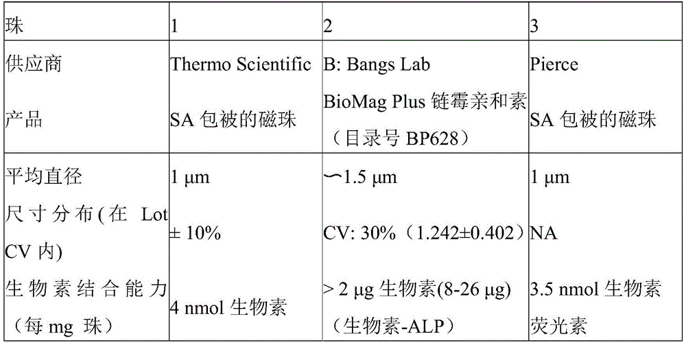
TABLE 1
While various methods can be used to mix or incubate biotinylated allergens or autoimmune antigens with streptavidin-coated beads, according to certain particular embodiments, the products are mixed in a reaction cuvette to coat the beads with the allergens or antigens due to the biotin/streptavidin interaction. According to one illustrative embodiment, 10 μ L of Streptavidin (SA) -coated beads are dispensed into reaction cuvettes, followed by 40 μ L of biotinylated allergen or autoimmune antigen, mixed during dispensing. The mixture is incubated for 1-15 minutes. Excess biotinylated allergen or autoimmune antigen can then be washed away by pulling the magnetic beads to one side of the reaction cuvette and immobilizing them while washing the reaction cuvette with buffer (step 20). According to one illustrative embodiment, the buffer may consist of 10mM sodium phosphate, pH7.4, 0.9% (w/v) NaCl, 0.05% (v/v) Tween-20,10mg/mL HSA, and 1% (v/v) ProClin 950. While one skilled in the art may employ any readily available immobilization technique known in the art to retain the magnetic beads on one side of the reaction cuvette, according to certain specific illustrative embodiments, an external magnet is used to immobilize the magnetic beads while the washing step is performed.
The streptavidin-coated magnetic beads are then released from the magnetic field and allowed to move freely within the reaction cuvette. Then, the biological sample (serum or plasma) is added to the reaction cuvette, and then 40. mu.L of the reaction buffer is added, thereby resuspending the magnetic beads (step 30). In addition to biological samples, according to certain aspects of the present disclosure, high concentrations of Human Serum Albumin (HSA) may also be used in the suspension to facilitate macromolecule binding and to keep the magnetic beads in solution. Human IgG was added to the buffer to maintain reaction linearity.
If the sample contains any antibodies (e.g., IgE, IgG) reactive to any allergen or antigenic bead coating, these antibodies will bind during this sample incubation step. The samples were incubated at 37 ℃ for 40 minutes. After any antibodies in the patient sample bind to the beads, a second washing step is then performed to remove any unbound patient sample (step 40). mu.L of wash buffer concentrate (50mM sodium phosphate, pH7.4, 4.5% (w/v) NaCl, 0.05% Tween-20, 0.05% (v/v) ProClin950, 0.02% (v/v) antifoam-C v/v) was added to resuspend the beads and then hold the beads with a magnet for 1.5 minutes. After removing the solution, the magnet was removed and 200 μ L of wash buffer was added to resuspend the beads. Then, the washing was repeated more than once.
After the second washing step, the beads were resuspended in a suspension specific for human immunoglobulin E (IgE) in the case of an allergy assaySex or in the case of an autoimmune assay, an antibody specific for human immunoglobulin G, M or A (IgG/M/A). According to certain aspects of the present disclosure, the antibody is coupled to an enzyme (e.g., horseradish peroxidase) to bind any particular patient antibody captured by the beads (step 50). The beads are then washed again to remove any excess antibody (step 60), and highly sensitive photo-forming reagents (e.g., chemiluminescent substrates) are added to maximize detection sensitivity (step 70). Illustrative reagents that can be used as chemiluminescent substrates in accordance with the teachings of the present disclosure include, but are not limited to, PS-atto、
 ELISA Pico chemiluminescent substrate or
 ELISA Femto maximum sensitivity substrate. Those skilled in the art will understand and appreciate that a variety of compounds having a variety of structural classes, including xanthene dyes, aromatic amines, and heterocyclic amines, can be used to produce chemiluminescence under these conditions. These compounds are well known in the patent literature and readily available through a variety of commercial suppliers. Some non-limiting chemiluminescent compounds include, but are not limited to, dioxetane-type molecules, fluorescein, and fluorescein,
 PS-2、
PS-2、 PS-3、
PS-3、 TMA-6、
 TMA-3。
Once the highly sensitive light-forming reagent is added to the reaction cuvette, light is generated (step 80). According to certain embodiments, the light may be measured by transferring the solution in the pipette tip to a reading station to read both the luminescence signal and the fluorescence signal. However, it should be understood that light emitted in accordance with the present disclosure may be detected by any suitable known detection method available in the art, including, but not limited to, illuminometers, x-ray films, high speed photographic films, CCD cameras, scintillation counters, chemometers, or visual inspection. Those skilled in the art will readily understand and appreciate that each detection average has a different spectral sensitivity itself; the detection device chosen may be governed by a number of factors, including application and use, cost, and convenience. Also, as used herein, a quantifiable or detectable reaction that can be measured according to the present disclosure implies a positive sample in which allergen-specific IgE causes binding of anti-IgE-HRP, producing luminescence (i.e., RLU ═ relative light units) upon addition of substrate. Furthermore, it is to be understood herein that quantifiable or detectable reactions can also be applied to quantifiable reactions produced by negative samples.
According to certain aspects of the present teachings, RLU produced from a positive/negative sample of any allergen-specific ige (slge) is compared to RLU produced by the entire ige (tgge) calibration curve. A calibration curve was generated by evaluating a large number of pre-diluted whole IgE (tge) calibrators (generated from WHO standards) using biotinylated anti-IgE capture reagents.
For a better understanding of the mechanical aspects of the present disclosure, fig. 2 illustrates an automated immunochemical analyzer and reagent system 100 that may be used to quantify and normalize the luminescence signal of an analyte sample according to the teachings of the present disclosure. In accordance with this illustrative aspect, the automated immunochemical analyzer 100 begins by first dispensing fluorescently labeled paramagnetic particles or fluorescent beads into a cuvette located in a reaction rotator 106. According to one embodiment herein, an exemplary fluorescent Bead comprises a fluorescent Bead (SA-Speed Bead, Atto 590 label), 1 mg/mL.
The fluorescent beads were first placed in a vortexer 102 and transferred to a reaction spinner 106 by an R1 pipettor 104. The R1 pipettor 104 may aspirate a desired amount of fluorescent bead mixture and transfer the aspirated amount into the reaction rotator 106 where it is injected into a cuvette of the reaction rotator 106. After filling the cuvette, the optical pipettor 108 may aspirate the test sample from the cuvette of the reaction rotator 106 and transfer the test sample into the optical cartridge 110. Fluorescence and luminescence measurements may be recorded once the sample is placed in optical box 110. The initial record of the fluorescence and luminescence signals can be used as a baseline measurement of the fluorescence signal that can correspond to the initial concentration of fluorescent beads in the sample. After recording the measurement, the multi-wash pipettor 112 may rinse the test cup with the wash buffer.
The fluorescent beads can then be transferred from the vortexer 102 to a cuvette in the reaction rotator 106 by an R1 pipettor 104. Subsequently, the R1 pipette 104 may aspirate the capture reagent from the reagent rotator 114 and inject the capture reagent into a cuvette located in the reaction rotator 106. After the incubation period, the single wash pipettor 116 may be filled with wash buffer to resuspend the fluorescent beads. A plurality of suspended fluorescent beads can then be concentrated (localized) in the reaction rotator 106 by means of a magnet over a period of time. After the magnet has substantially focused the fluorescent beads in the cuvette, the multi-wash pipette 112 may aspirate and process a portion of the wash buffer, leaving a portion of the fluorescent beads in the cuvette. The multi-wash pipette 112 may continue to inject wash buffer into the cuvette of the reaction rotator 106, resuspending the fluorescent beads. The fluorescent beads may be concentrated in the reaction rotator 106 again by the magnet, and then the multi-wash pipette 112 sucks from the test cup of the reaction rotator 106 and discards a portion of the sample that is not concentrated.
The patient sample may be housed in a sample tube or sample rotator 118. The patient sample is further partially diluted with a sample diluent. At this point, the sample pipettor 120 may aspirate a portion of the patient sample and inject the patient sample into the cuvette of the reaction rotator 106 to resuspend the fluorescent beads. The cuvette containing the patient sample in the reaction rotator 106 may then be incubated at a specific temperature for a specific period of time. After incubation, the single wash pipettor 116 may be injected with wash buffer to resuspend the fluorescent beads. Another concentration process is performed with the reaction rotator 106 by having fluorescent beads substantially collected in a cuvette near the magnet in the reaction rotator 106. After fluorescent beads are concentrated, the multi-wash pipette 112 may aspirate and discard a portion of the fluid that is not concentrated in the cuvette of the reaction rotator 106 during the concentration process.
The sample in the cuvette of the reaction rotator 106 is then subjected to several wash cycles. The wash cycle may include using the multi-wash pipette 112 to inject wash buffer into the cuvette to resuspend the fluorescent beads. Another focusing step may allow fluorescent beads to be collected in a cuvette in reaction rotator 106 using a magnet. After a period of time that allows the fluorescent beads to be sufficiently concentrated, the multi-wash pipette 112 may aspirate and inadvertently discard a portion of the sample, leaving a portion of the fluorescent beads in the cuvette of the reaction rotator 106. Another wash cycle may then be performed by using the multi-wash pipettor 112 to again inject wash buffer into the cuvette and allow the fluorescent beads to be resuspended. Another fluorescent bead concentration process may use a magnet in the reaction rotator 106 to concentrate fluorescent beads from the rest of the sample. Finally, the multi-wash pipettor 112 may aspirate a portion of the sample that was not concentrated by the concentration process.
At this point, the R2 pipettor 122 may aspirate the conjugate contained in the cuvette in the reagent rotator 114. The R2 pipettor 122 then injects the previously aspirated conjugate into the cuvette of the reaction rotator 106. After incubating the test cups in the reaction rotator 106 at controlled times and temperatures, the single wash pipette 116 may inject a wash buffer into the test cups of the reaction rotator 106. Another fluorescent-bead concentration cycle may be performed by allowing the magnet in the reaction rotator 106 to substantially concentrate the fluorescent beads in the cuvette. The multi-flush pipette 112 may aspirate and discard a portion of the sample in the cuvette that was not concentrated during the concentration cycle.
The sample in the cuvette of the reaction rotator 106 may be subjected to two more rinse cycles. The multi-wash pipettor 112 may be filled with wash buffer to resuspend the fluorescent beads in the test cup. Another fluorescent bead concentration cycle may concentrate fluorescent beads by bringing the cuvette of the reaction rotator 106 close to the magnet for a sufficient period of time. After the concentration cycle, the multi-wash pipettor 112 may aspirate and discard a portion of the sample that was not concentrated during the concentration cycle. Then, the fluorescent beads are resuspended by performing a second wash cycle using the multi-wash pipettor 112 to inject wash buffer. Another concentration cycle may utilize a magnet in the reaction rotator 106 to concentrate the fluorescent beads in the test cup. After the concentration process, the multi-wash pipette 112 may aspirate again and discard a portion of the sample that was not concentrated during the concentration cycle.
At this point, the R2 pipettor 122 may aspirate a portion of the conjugate from the reagent rotator 114 and inject the conjugate into the mixed substrate container 124, creating a mixed substrate sample. The R2 pipette may then aspirate and inject the mixed substrate sample from the mixed substrate container 124 into the cuvette of the reaction rotator 106, resuspending the fluorescent beads with the mixed substrate sample. The sample in the cuvette of the reaction rotator 106 may then be aspirated by the optical pipettor 108 and placed in the optics box 110. After the optical cartridge observes fluorescence and luminescence, the sample is discarded and the pipette is rinsed multiple times through the cuvette of the reactor spinner 106, ready for the next experiment.
The following non-limiting examples are provided to further illustrate the invention.
Embodiment 1: a quantitative method for performing an automated diagnostic assay, comprising:
incubating a streptavidin-coated medium comprising a universal fluorescently-labeled magnetic microparticle with a biotinylated capture reagent derived from an allergen or an autoimmune antigen to form a solid phase complex;
washing the solid phase complex to remove excess capture reagent;
incubating the solid phase complex with a serum sample to form an immune complex;
washing the immune complexes to remove any unbound sample;
incubating the immune complex with a conjugate to generate an immune-conjugate complex;
washing the immune-conjugate complex to remove any unbound conjugate;
introducing a substrate capable of producing a quantifiable reaction; and
calibrating the reaction produced by the introduction of the substrate.
Embodiment 2: the method of embodiment 1, wherein the step of incubating the solid phase complex with the serum sample comprises binding allergen-specific human immunoglobulin e (ige) present in the serum sample to the biotinylated capture reagent.
Embodiment 3: the method of embodiment 1, wherein the step of incubating the solid phase complex with the serum sample comprises binding autoimmune specific human immunoglobulin g (igg), autoimmune specific human immunoglobulin m (igm), or autoimmune specific human immunoglobulin a (iga) present in the serum sample to the biotinylated capture reagent.
Embodiment 4: the method of embodiment 1, wherein the biotinylated capture reagent is derived from biotinylation of a purified allergen, protein, enzyme, or antibody.
Embodiment 5: the method according to embodiment 1, wherein the allergen extract consists of a plurality of allergens.
Embodiment 6: the method of embodiment 1, wherein the biotinylated capture reagent is present as a blend of a plurality of biotinylated capture reagents selected from the group consisting of purified allergens, proteins, enzymes, antibodies, and allergen extracts.
Embodiment 7: the method of embodiment 1, wherein one or more washing steps comprises washing the complex by magnetically isolating the complex to be washed within a defined area of a reaction cuvette.
Embodiment 8: the method of embodiment 1, wherein the step of forming a solid phase complex with a streptavidin-coated medium comprises incubating a biotinylated capture reagent held in suspension with a reaction diluent comprising a high concentration of Human Serum Albumin (HSA).
Embodiment 9: the method of embodiment 1, wherein the step of incubating the immunocomplex with the conjugate comprises incubating the immunocomplex held in suspension with a conjugate diluent comprising a labeled concentration of polyethylene glycol.
Embodiment 10: the method of embodiment 9, further comprising the step of using horseradish peroxidase (HRP) as an indirect label when washing the immune complexes to remove unbound sample.
Embodiment 11: the method of embodiment 1, further comprising the step of adjusting the quantifiable reaction for bead retention by:
transferring the substrate and the immune-conjugate complex into an optical cartridge, wherein the fluorescent and chemiluminescent signals are quantified; and
the ratio of initial to final fluorescence was used to adjust the quantified chemiluminescent signal to calculate the reported value.
Embodiment 12: the method of embodiment 11, wherein the step of transferring the substrate and the immune-conjugate complex to an optics box comprises using an automated pipette arm with a reusable pipette tip to aspirate a sample.
Embodiment 13: the method of embodiment 11, further comprising:
measuring fluorescence within the optical cartridge to determine bead retention; and
luminescence within the optical box is measured to detect the relative light unit signals generated.
Embodiment 14: the method of embodiment 13, further comprising substituting fluorescence and luminescence measurements into an algorithm to generate relative light unit signals that are bead retention adjusted.
Embodiment 15: the method of embodiment 14, further comprising comparing the generated bead retention adjusted relative light unit signal to a calibration curve relative light unit signal.
Advantages and improvements of the processes, methods of the present disclosure are set forth in the following examples. These examples are illustrative only and are not intended to limit or exclude other embodiments of the present disclosure.
Example 1: biotinylation of anti-human IgE or allergen extracts:
mu.L of NHS-PEG 12-biotin (Pierce) at 250mM in DMSO was added to 1mL of affinity purified anti-human IgE (Immunoregens) 5.0mg/mL Phosphate Buffered Saline (PBS). Alternatively, 1.6uL of NHS-PEG 12-biotin (Pierce) at 250mM in DMSO was added to 1mL of allergen extract in 1.0mg/mL Phosphate Buffered Saline (PBS).
The reagent solutions were mixed and placed on ice for 2 hours. Free biotin reagent was separated from biotinylated antibody by two dialysis changes of PBS at 2-8 ℃ for 4 hours and overnight (antibody to buffer ratio 1:100 by volume).
Example 2: preparation of fluorescent beads:
add 5. mu.L to ddH2Biotin-fluorescence (Alexa Fluor 594 biocytin, sodium salt, Life Technologies) at 1mM in O was added to 45mL PBSTHP buffer (10mM sodium phosphate, pH7.4, 0.9% (w/v) NaCl, 0.05% (v/v) Tween-20,10mg/mL HSA, 1% (v/v) ProClin 950). And (4) fully mixing.
5mL of SA-Speed Bead (Sera-mag Speedbeads streptavidin-coated magnetic particles, Thermo)10mg/mL was added to the biotin-fluorescent solution and mixed well.
Example 3: determination of specific IgE levels against allergens
10 μ L of fluorescent beads (fluorescently labeled paramagnetic microparticles) were dispensed in a reaction cuvette at a bead concentration of 1 mg/mL; 40 μ L of biotin-allergen (e.g., protein, milk, peanut, etc.) or biotin-anti-IgE antibody was dispensed and mixed into fluorescent beads and incubated at 37 ℃ for 1-10 minutes. After washing, the allergen or anti-IgE coated beads were resuspended in 40. mu.L of reaction buffer. Serum samples obtained from atopic and non-atopic individuals were assayed for allergens. mu.L of the sample was added to 40. mu.L of allergen-coated beads suspended in the reaction cuvette. For the six-point calibration curve, 10 μ L of serum standard (secondary standard calibrated to WHO IgE standard 75/502) was each added to 40 μ L of anti-IgE coated beads in a reaction cuvette. Although a variety of different labeled anti-IgE conjugates can be used in accordance with the present teachings, according to certain teachings the following anti-IgE conjugates are used: for allergy assays, anti-IgE-HRP was used; for the autoimmune assay, anti-IgA-HRP, anti-IgG-HRP and anti-IgM-HRP were used; for ECP, anti-ECP-HRP was used; and for tryptase, anti-tryptase-HRP was used. Also, as used herein, each conjugate has an optimized HRP incorporation rate for chemistry. According to certain aspects of the present teachings, the HRP incorporation ratios for the enumerated conjugates range between about 1.2 and about 5.4. In addition, the present teachings also contemplate the incorporation of other types of conjugate-reporting systems including, but not limited to: an alkaline phosphatase conjugate and a b-galactose conjugate.
The solutions were mixed and incubated at 37 ℃ for 40 minutes. After washing, the beads were resuspended in 50. mu.L of anti-human IgE-HRP conjugate and incubated at 37 ℃ for 30 minutes. 50 μ L of PS-atto (Lumigen) was added to each cuvette and the beads resuspended. The bead suspension was transferred to the pipette tip and the fluorescent and luminescent signals were read in the optical cartridge. The standard curve was determined using a four parameter logistic function equation and specific IgE levels of the allergen interpolated from the standard curve.
Illustrative reagents and component tables that may be used in accordance with the present teachings include, but are not necessarily limited to:beads:fluorescent beads (SA-Speed Bead, Atto 590 marker), 1 mg/mL;capture reagent diluent:IgE: 10mM sodium phosphate, pH7.4, 0.9% (w/v) NaCl, 0.05% Tween-20, 1% (w/v) human serum albumin, l% (v/v) ProClin950, up to 5% (v/v) glycerol; ANA: 10mM sodium phosphate, pH7.4, 0.9% (w/v) NaC1, 0.05% Tween-20, 1% (w/v) bovine serum albumin, 1% protease inhibitor cocktail, 0.1mM DTT, l% (v/v) ProClin950, 25% (up to 30%) (v/v) glycerol;washing buffer concentrate(5 x): 50mM sodium phosphate, pH7.4, 4.5% (w/v) NaCl, 0.05% Tween-20, 0.05% (v/v) ProClin950, 0.02% (v/v) antifoam-C v/v; reagent diluent (reaction diluent and sample diluent) IgE-10 mM sodium phosphate, pH7.4, 500mM NaCl, 0.02% Tween-20, 1% (w/v) human serum albumin, 1% (v/v) human IgG, l% (v/v) ProClin950, 0.005% antifoam-B v/v, 2% (w/v) PEG 6,000; ANA-10mM sodium phosphate, pH7.4, 500mM NaCl, 0.02% Tween-20, 25% (w/v) human serum albumin, l% (v/v) ProClin 950;school Standards and controls: calibration material: patients diluted to sample dilutionA sample; comparison: a patient sample cell;conjugate:conjugate dilution: 50mM sodium phosphate, pH 6.7,150mM NaCl, 0.05% Tween-20, 1% BSA, 5% (w/v) PEG 6,000, l% (v/v) ProClin 950; IgE: 100ng/mL anti-IgE-HRP, 100. mu.g/mL apo-HRP (in dilution), 0.015% antifoam-B v/v; and a substrate: PS-atto A&B. 0.01% antifoam-B v/v.
Although exemplary embodiments incorporating the principles of the present application are disclosed above herein, the present application is not limited to the disclosed embodiments. Rather, this application is intended to cover any variations, uses, or adaptations of the application using its general principles. Further, this application is intended to cover such departures from the present disclosure as come within known or customary practice in the art to which this application pertains and which fall within the limits of the appended claims.
The terminology used herein is for the purpose of describing particular illustrative embodiments only and is not intended to be limiting. As used herein, the singular forms "a", "an" and "the" are intended to include the plural forms as well, unless the context indicates otherwise. The terms "comprises," "comprising," "includes" and "including" are inclusive and therefore specify the presence of stated features, integers, steps, operations, elements, and/or components, but do not preclude the presence or addition of one or more other features, integers, steps, operations, elements, components, and/or groups thereof. The method steps, processes, and operations described herein are not to be construed as necessarily requiring their performance in the particular order discussed or illustrated, unless specifically identified as an order of performance. It should also be understood that additional or alternative steps may be used.
Claims (12)
1. A quantitative method for performing an automated immunoassay, comprising:
incubating a streptavidin-coated medium comprising a universal fluorescently-labeled magnetic microparticle with a biotinylated capture reagent derived from an allergen or an autoimmune antigen to form a solid phase complex;
washing the solid phase complex to remove excess capture reagent;
incubating the solid phase complex with a serum sample to form an immune complex;
washing the immune complexes to remove any unbound sample;
incubating the immune complex with a conjugate to generate an immune-conjugate complex;
washing the immune-conjugate complex to remove any unbound conjugate;
introducing a substrate capable of producing a quantifiable reaction; and
calibrating the reaction produced by introduction of the substrate; and is
Further comprising the step of adjusting the quantifiable reaction to bead retention by:
transferring the substrate and the immune-conjugate complex into an optical cartridge, wherein the fluorescent and chemiluminescent signals are quantified; and
the ratio of initial to final fluorescence was used to adjust the quantified chemiluminescent signal to calculate the reported value.
2. The method of claim 1, wherein the step of incubating the solid phase complex with the serum sample comprises binding allergen-specific human immunoglobulin e (ige) present in the serum sample to the biotinylated capture reagent.
3. The method of claim 1, wherein the step of incubating the solid phase complex with the serum sample comprises binding autoimmune specific human immunoglobulin g (igg), autoimmune specific human immunoglobulin m (igm), or autoimmune specific human immunoglobulin a (iga) present in the serum sample to the biotinylated capture reagent.
4. The method of claim 1, wherein the biotinylated capture reagent is derived from biotinylation of a purified allergen.
5. The method of claim 1, wherein one or more washing steps comprise washing the complex by magnetically isolating the complex to be washed within a defined area of a reaction cuvette.
6. The method of claim 1, wherein the step of forming a solid phase complex with a streptavidin-coated medium comprises incubating a biotinylated capture reagent held in suspension with a reaction diluent comprising a high concentration of Human Serum Albumin (HSA).
7. The method of claim 1, wherein the step of incubating the immunocomplex with the conjugate comprises incubating the immunocomplex held in suspension with a conjugate diluent comprising a labeled concentration of polyethylene glycol.
8. The method of claim 7, further comprising the step of using horseradish peroxidase (HRP) as an indirect label when washing the immune complexes to remove unbound sample.
9. The method of claim 1, wherein the step of transferring the substrate and the immune-conjugate complex to an optics box comprises using an automated pipette arm with a reusable pipette tip to aspirate the sample.
10. The method of claim 1, further comprising:
measuring fluorescence within the optical cartridge to determine bead retention; and
luminescence within the optical box is measured to detect the relative light unit signals generated.
11. The method of claim 10, further comprising substituting fluorescence and luminescence measurements into an algorithm to generate relative light unit signals that are bead retention adjusted.
12. The method of claim 11, further comprising comparing the generated bead retention adjusted relative light unit signal to a calibration curve relative light unit signal.
Applications Claiming Priority (5)
| Application Number | Priority Date | Filing Date | Title |
|---|---|---|---|
| US201361791879P | 2013-03-15 | 2013-03-15 | |
| US201361791295P | 2013-03-15 | 2013-03-15 | |
| US61/791,879 | 2013-03-15 | ||
| US61/791,295 | 2013-03-15 | ||
| CN201480015969.4A CN105308458B (en) | 2013-03-15 | 2014-03-17 | Automated immunoassay system for performing diagnostic assays for allergies and autoimmune diseases |
Related Parent Applications (1)
| Application Number | Title | Priority Date | Filing Date |
|---|---|---|---|
| CN201480015969.4A Division CN105308458B (en) | 2013-03-15 | 2014-03-17 | Automated immunoassay system for performing diagnostic assays for allergies and autoimmune diseases |
Publications (2)
| Publication Number | Publication Date |
|---|---|
| CN107817232A CN107817232A (en) | 2018-03-20 |
| CN107817232B true CN107817232B (en) | 2021-08-17 |
Family
ID=50686181
Family Applications (4)
| Application Number | Title | Priority Date | Filing Date |
|---|---|---|---|
| CN201711319904.0A Active CN108445242B (en) | 2013-03-15 | 2014-03-17 | Device for performing luminescence and fluorescence measurements of a sample and related method |
| CN201711006475.1A Active CN107817232B (en) | 2013-03-15 | 2014-03-17 | Automated immunoassay system for performing diagnostic assays for allergies and autoimmune diseases |
| CN201480015803.2A Active CN105308437B (en) | 2013-03-15 | 2014-03-17 | Apparatus and related methods for performing luminescence and fluorescence measurements of samples |
| CN201480015969.4A Active CN105308458B (en) | 2013-03-15 | 2014-03-17 | Automated immunoassay system for performing diagnostic assays for allergies and autoimmune diseases |
Family Applications Before (1)
| Application Number | Title | Priority Date | Filing Date |
|---|---|---|---|
| CN201711319904.0A Active CN108445242B (en) | 2013-03-15 | 2014-03-17 | Device for performing luminescence and fluorescence measurements of a sample and related method |
Family Applications After (2)
| Application Number | Title | Priority Date | Filing Date |
|---|---|---|---|
| CN201480015803.2A Active CN105308437B (en) | 2013-03-15 | 2014-03-17 | Apparatus and related methods for performing luminescence and fluorescence measurements of samples |
| CN201480015969.4A Active CN105308458B (en) | 2013-03-15 | 2014-03-17 | Automated immunoassay system for performing diagnostic assays for allergies and autoimmune diseases |
Country Status (8)
| Country | Link |
|---|---|
| US (13) | US9658225B2 (en) |
| EP (3) | EP2972361A1 (en) |
| JP (3) | JP6553020B2 (en) |
| CN (4) | CN108445242B (en) |
| AU (3) | AU2014232782B2 (en) |
| CA (2) | CA2905165A1 (en) |
| HK (2) | HK1220759A1 (en) |
| WO (2) | WO2014145619A1 (en) |
Families Citing this family (35)
| Publication number | Priority date | Publication date | Assignee | Title |
|---|---|---|---|---|
| US8866912B2 (en) | 2013-03-10 | 2014-10-21 | Pelican Imaging Corporation | System and methods for calibration of an array camera using a single captured image |
| CN108445242B (en) | 2013-03-15 | 2022-03-04 | Hycor生物医学有限责任公司 | Device for performing luminescence and fluorescence measurements of a sample and related method |
| JP6323816B2 (en) * | 2014-05-19 | 2018-05-16 | システム・インスツルメンツ株式会社 | Automatic analyzer, analysis system, and operation method of automatic analyzer |
| US10661268B2 (en) * | 2014-06-30 | 2020-05-26 | Beacon Technologies, LLC | Pipette tip system, device and method of use |
| CN107430058A (en) * | 2015-02-27 | 2017-12-01 | Hycor生物医学有限责任公司 | For the apparatus and method for the content for suspending and washing multiple test glass |
| EP3278109A4 (en) * | 2015-03-30 | 2018-08-15 | Hycor Biomedical, Inc. | Automated immunoanalyzer system for performing diagnostic assays for autoimmune and infectious diseases |
| EP4582813A3 (en) | 2015-06-26 | 2025-08-27 | Abbott Laboratories | Rotating device in a diagnostic analyzer |
| EP3322533A4 (en) | 2015-07-15 | 2019-03-20 | Hycor Biomedical, LLC | INSTRUMENT THAT CAN BE CUSTOMIZED |
| WO2017015662A1 (en) | 2015-07-23 | 2017-01-26 | Hycor Biomedical, Inc | On-board kitting |
| CN105353133A (en) * | 2015-11-17 | 2016-02-24 | 苏州浩欧博生物医药有限公司 | Kit detecting dermatophagoides-farinae allergen specific IgE antibody and method |
| CN105403692A (en) * | 2015-11-17 | 2016-03-16 | 苏州浩欧博生物医药有限公司 | Kit and method for detecting dermatophagoides pteronyssinus allergen specificity IgE antibody |
| CN105353134A (en) * | 2015-11-17 | 2016-02-24 | 苏州浩欧博生物医药有限公司 | Kit detecting dog-hair allergen specific IgE antibody and method |
| TW201832726A (en) * | 2016-11-01 | 2018-09-16 | 美商海科生醫有限責任公司 | Immunoassay system capable of suggesting assays based on input data |
| CN108132344B (en) * | 2016-11-22 | 2021-06-29 | 博阳生物科技(上海)有限公司 | Immunoassay methods, systems and kits for identifying immunoassays |
| CN107389615B (en) * | 2016-12-20 | 2020-03-24 | 苏州赛德福科学仪器有限公司 | Evaporation light detection device and evaporation light measurement method based on same |
| CN107703123A (en) * | 2017-09-15 | 2018-02-16 | 江苏浩欧博生物医药股份有限公司 | A kind of chemical luminescent analysis reagent kid of serum tryptase and preparation method thereof and detection method |
| EP4582806A3 (en) | 2017-09-19 | 2025-08-13 | Beckman Coulter, Inc. | Analog light measuring and photon counting in chemiluminescence measurements |
| CN107766772B (en) * | 2017-11-23 | 2024-05-17 | 安派科生物医学科技(丽水)有限公司 | Sample inputting device |
| CN108593949B (en) * | 2018-02-11 | 2024-01-26 | 金博特(北京)生物科技有限公司 | Chemiluminescence immunity analyzer |
| IT201800008227A1 (en) * | 2018-08-29 | 2020-02-29 | Cannavale Giuseppe | Method and system for the determination of total or direct immunoglobulins against allergens or other molecules in aqueous samples |
| EP3867644B1 (en) | 2018-08-29 | 2024-02-21 | Cannavale, Giuseppe | System and method for solid phase analysis of biological samples |
| US12163885B2 (en) | 2018-08-31 | 2024-12-10 | Shimadzu Corporation | Analysis device, analysis method, trace liquid collection device, and trace liquid collection method |
| CN109030809B (en) * | 2018-10-31 | 2021-08-20 | 蓝怡科技集团股份有限公司 | Allergen IgE antibody automatic analyzer |
| WO2020157789A1 (en) * | 2019-01-28 | 2020-08-06 | 株式会社島津製作所 | Analysis device |
| CN110596371B (en) * | 2019-09-20 | 2023-04-07 | 郑州安图生物工程股份有限公司 | Method for evaluating magnetic bead recovery rate and reagent needle precision of full-automatic chemiluminescence determinator and kit |
| CN111518780B (en) * | 2020-06-04 | 2022-06-14 | 郑州伊美诺生物技术有限公司 | Preparation method of apo-horseradish peroxidase |
| US11480565B2 (en) * | 2020-06-12 | 2022-10-25 | Bio-Rad Laboratories, Inc. | Automated immunoassay |
| GB202009823D0 (en) * | 2020-06-26 | 2020-08-12 | Stimson William Howard | Automated quantitative assay device and a method of performing the quantitative assays |
| CN112748248B (en) * | 2020-12-23 | 2022-06-14 | 郑州安图生物工程股份有限公司 | Milk allergen specificity IgE detect reagent box |
| CN112666154A (en) * | 2021-01-26 | 2021-04-16 | 安图实验仪器(郑州)有限公司 | Optical signal detection device |
| JP7693173B2 (en) * | 2021-09-24 | 2025-06-17 | 学校法人杏林学園 | Method for detecting macadamia nut allergen-specific IgE, in vitro diagnostic agent for diagnosing macadamia nut allergy, kit for detecting macadamia nut allergen-specific IgE, and method for detecting macadamia nut allergen |
| IT202200010064A1 (en) * | 2022-05-16 | 2023-11-16 | Optoelettronica Italia S R L | DEVICE AND METHOD FOR THE DETECTION OF BIOMOLECULES |
| WO2025155731A1 (en) * | 2024-01-16 | 2025-07-24 | Beckman Coulter, Inc. | Alternative substrate pack for automated clinical analyzer and methods of use |
| WO2025155701A1 (en) * | 2024-01-16 | 2025-07-24 | Beckman Coulter, Inc. | Automated clinical analyzer with alternative substrate and method of use |
| CN118501432B (en) * | 2024-07-18 | 2024-09-27 | 山东省动物疫病预防与控制中心(山东省人畜共患病流调监测中心) | Brucellosis CF-ELISA Antibody Detection Kit |
Citations (2)
| Publication number | Priority date | Publication date | Assignee | Title |
|---|---|---|---|---|
| CN101514991A (en) * | 2008-02-19 | 2009-08-26 | 北京科美东雅生物技术有限公司 | Alpha-fetoprotein magnetic particle chemoluminescence immunoassay kit and method for preparing same |
| CN101545913A (en) * | 2008-03-25 | 2009-09-30 | 北京科美东雅生物技术有限公司 | Chemoluminescence immunoassay measuring kit and preparation method thereof for triiodothyronine magnetic particles |
Family Cites Families (160)
| Publication number | Priority date | Publication date | Assignee | Title |
|---|---|---|---|---|
| AT287916B (en) * | 1969-04-29 | 1971-02-10 | Schwarz O | Process for the preparation of a prophylactic agent useful in obstetrics |
| US3628682A (en) | 1970-06-29 | 1971-12-21 | Beckman Instruments Inc | Lighttight sample introduction system |
| US4291230A (en) | 1979-03-07 | 1981-09-22 | Baxter Travenol Laboratories, Inc. | Fluorometric analyzer including shutter means for simultaneously shielding sample and photodetector during sample change |
| US5643566A (en) * | 1982-09-23 | 1997-07-01 | Cetus Corporation | Formulation processes for lipophilic proteins |
| US4849337A (en) * | 1983-01-31 | 1989-07-18 | Minnesota Mining And Manufacturing Company | Assaying allergen specific IgE levels with fluorogenic enzyme labeled antibody |
| FI831936A0 (en) * | 1983-05-30 | 1983-05-30 | Labsystems Oy | FLUORESCENS, TURBIDITY, LUMINESCENS ELLER ABSORPTION |
| GB8505899D0 (en) | 1985-03-07 | 1985-04-11 | Boots Celltech Diagnostics | Assay reagents |
| US4778751A (en) | 1986-05-12 | 1988-10-18 | Diagnostic Products Corporation | Method for measuring antigens or antibodies in biological fluids using ligand labeled antigens or ligand labeled antibodies |
| US5320808A (en) * | 1988-08-02 | 1994-06-14 | Abbott Laboratories | Reaction cartridge and carousel for biological sample analyzer |
| JP2656564B2 (en) | 1988-08-26 | 1997-09-24 | 株式会社日立製作所 | Immunoassay method |
| DE3901857A1 (en) | 1989-01-23 | 1990-07-26 | Boehringer Mannheim Gmbh | METHOD FOR DETERMINING HIV 2 ANTIBODY |
| US5196306A (en) | 1989-03-29 | 1993-03-23 | E. I. Du Pont De Nemours And Company | Method for the detection or quantitation of an analyte using an analyte dependent enzyme activation system |
| JPH04106471A (en) * | 1990-08-28 | 1992-04-08 | Hitachi Ltd | Measuring device for biological substances |
| CA2055812A1 (en) | 1990-11-20 | 1992-05-21 | Carl N. Skold | Method of stabilizing enzyme conjugates |
| SE9004047L (en) | 1990-12-19 | 1992-06-20 | Francisco Batista | METHOD FOR IMMOBILIZATION OF TIOL COMPOUNDS THROUGH ACTIVATION OF POLYMERS, ACTIVATED POLYMERS, AND PRODUCTS RECEIVED BY THE METHOD |
| JPH04348258A (en) * | 1991-05-27 | 1992-12-03 | Kowa Co | Multi-channel optical measuring device |
| DK130991D0 (en) | 1991-07-04 | 1991-07-04 | Immunodex K S | POLYMER CONJUGATES |
| US5395938A (en) | 1992-08-21 | 1995-03-07 | Nichols Institute Diagnostics | Biotinylated chemiluminescent labels and their conjugates, assays and assay kits |
| JP3207933B2 (en) | 1992-09-09 | 2001-09-10 | 株式会社ヤトロン | Light emission method of acridinium derivative and method of detecting target substance using the same |
| US6399397B1 (en) | 1992-09-14 | 2002-06-04 | Sri International | Up-converting reporters for biological and other assays using laser excitation techniques |
| US5698397A (en) | 1995-06-07 | 1997-12-16 | Sri International | Up-converting reporters for biological and other assays using laser excitation techniques |
| US5674698A (en) | 1992-09-14 | 1997-10-07 | Sri International | Up-converting reporters for biological and other assays using laser excitation techniques |
| US5736410A (en) | 1992-09-14 | 1998-04-07 | Sri International | Up-converting reporters for biological and other assays using laser excitation techniques |
| US6087188A (en) | 1992-11-13 | 2000-07-11 | Alk A/S | Two-site immunoassay for an antibody with chemiluminescent label and biotin bound ligand |
| US5395752A (en) | 1993-03-19 | 1995-03-07 | Ciba Corning Diagnostics Corp. | Long emission wavelength chemiluminescent compounds and their use in test assays |
| AU679008B2 (en) | 1993-05-06 | 1997-06-19 | Chiron Diagnostics Corporation | Mixed luminescent conjugate test assays |
| US5468453A (en) | 1993-06-14 | 1995-11-21 | Cirrus Diagnostics, Inc. | Low carryover pipette probe |
| US6348310B1 (en) | 1994-03-04 | 2002-02-19 | Promega Corporation | Quantitation of individual protein kinase activity |
| GB2289334B (en) | 1994-05-10 | 1998-08-26 | Molecular Light Technology Lim | Enzyme linked chemiluminescent assay |
| DE4418223A1 (en) | 1994-05-25 | 1995-11-30 | Boehringer Mannheim Gmbh | Procedure for the detection of the presence of insulin-dependent diabetes mellitus |
| US6800452B1 (en) | 1994-08-08 | 2004-10-05 | Science Applications International Corporation | Automated methods for simultaneously performing a plurality of signal-based assays |
| DE4439452A1 (en) | 1994-11-04 | 1996-05-09 | Boehringer Mannheim Gmbh | Antibody class-specific anti-interference reagent |
| JP2815545B2 (en) * | 1994-11-07 | 1998-10-27 | 日本分光株式会社 | Liquid chromatograph detector |
| CA2212043C (en) | 1995-02-02 | 2005-08-30 | Chugai Seiyaku Kabushiki Kaisha | Method for assay of analyte by adjustment of chemiluminescence |
| EP2280268B1 (en) | 1995-03-10 | 2014-09-03 | Meso Scale Technologies, LLC. | Multi-array, multi-specific electrochemiluminescence testing |
| US6140045A (en) | 1995-03-10 | 2000-10-31 | Meso Scale Technologies | Multi-array, multi-specific electrochemiluminescence testing |
| JPH08313539A (en) * | 1995-05-16 | 1996-11-29 | Nittec Co Ltd | Container, and methods for liquid agitation, cleaning, and fine magnetic particle-collection using the container, and immunochemical inspecting device using the container |
| US5879885A (en) | 1995-06-07 | 1999-03-09 | Gen-Probe Incorporated | Compositions for detecting and quantifying biological samples |
| US5690907A (en) | 1995-06-08 | 1997-11-25 | The Jewish Hospital Of St. Louis | Avidin-biotin conjugated emulsions as a site specific binding system |
| IL114149A0 (en) | 1995-06-14 | 1995-10-31 | Yeda Res & Dev | Modified avidin and streptavidin molecules and use thereof |
| US5770700A (en) * | 1996-01-25 | 1998-06-23 | Genetics Institute, Inc. | Liquid factor IX formulations |
| US5776487A (en) | 1996-04-19 | 1998-07-07 | Pasteur Sanofi Diagnostics | Liposome reagents for immunoassays |
| SE9602234D0 (en) | 1996-06-06 | 1996-06-06 | Pharmacia Ab | A novel diagnostic method utilizing ECP, and reagents to be used in the methods |
| US5942407A (en) | 1996-06-25 | 1999-08-24 | Immunomatrix, Inc. | Light-emitting immunoassay |
| GB9712818D0 (en) | 1996-07-08 | 1997-08-20 | Cambridge Antibody Tech | Labelling and selection of specific binding molecules |
| SE9603379D0 (en) | 1996-09-17 | 1996-09-17 | Pharmacia & Upjohn Ab | Device for detecting when a measuring probe comes into contact with a liquid surface |
| US5776785A (en) | 1996-12-30 | 1998-07-07 | Diagnostic Products Corporation | Method and apparatus for immunoassay using fluorescent induced surface plasma emission |
| US6432662B1 (en) | 1997-04-15 | 2002-08-13 | Pierce Chemical Company | Assay of peroxidase activity |
| US6165800A (en) | 1997-05-30 | 2000-12-26 | Bayer Corporation | Chemiluminescent energy transfer conjugates and their uses as labels in binding assays |
| US5929049A (en) | 1997-08-08 | 1999-07-27 | Dade Behring Marburg Gmbh | Polysaccharide conjugates of biomolecules |
| JP3366842B2 (en) * | 1997-08-29 | 2003-01-14 | エーザイ株式会社 | Electrochemiluminescence cell |
| US5922558A (en) | 1997-09-12 | 1999-07-13 | Lumigen, Inc. | Methods and compositions for generating chemiluminescence with a peroxidase |
| US6103537A (en) | 1997-10-02 | 2000-08-15 | Aclara Biosciences, Inc. | Capillary assays involving separation of free and bound species |
| US6180340B1 (en) | 1997-10-31 | 2001-01-30 | Gen-Probe Incorporated | Extended dynamic range assays |
| SE9704935D0 (en) | 1997-12-30 | 1997-12-30 | Pharmacia & Upjohn Diag Ab | Method of analysis with particles |
| US6235480B1 (en) | 1998-03-13 | 2001-05-22 | Promega Corporation | Detection of nucleic acid hybrids |
| US6068979A (en) | 1998-04-22 | 2000-05-30 | Lumigen, Inc. | Simplified sequential chemiluminescent detection |
| SE9801563D0 (en) | 1998-04-30 | 1998-04-30 | Pharmacia & Upjohn Diag Ab | Method of separation and kit to be used in the process |
| US6153442A (en) | 1998-05-20 | 2000-11-28 | Dade Behring Inc. | Reagents and methods for specific binding assays |
| US6303325B1 (en) | 1998-05-29 | 2001-10-16 | Dade Behring Inc. | Method for detecting analytes |
| US6379909B1 (en) | 1998-06-24 | 2002-04-30 | Alk-Abello A/S | Method of detecting an antibody in a liquid sample |
| ATE423101T1 (en) | 1998-08-11 | 2009-03-15 | Siemens Healthcare Diagnostics | NEAR INFRARED CHEMILUMINESCENT ACRIDINIUM DERIVATIVES AND THEIR USE |
| US6236456B1 (en) | 1998-08-18 | 2001-05-22 | Molecular Devices Corporation | Optical system for a scanning fluorometer |
| US6399299B1 (en) | 1998-11-02 | 2002-06-04 | Perkinelmer Life Sciences, Inc. | Amplified array analysis system |
| US7759133B2 (en) | 1998-12-22 | 2010-07-20 | Alk-Abello A/S | Method of detecting and/or quantifying a specific IgE antibody in a liquid sample |
| JP3330929B2 (en) * | 1999-01-25 | 2002-10-07 | 浜松ホトニクス株式会社 | Pipette adapter, pipette for absorbance measurement, tip, absorbance measurement device and absorbance measurement method |
| AU6211100A (en) | 1999-07-12 | 2001-01-30 | Merlin Biomedical And Pharmaceutical, Ltd. | Improved assays for detecting human immunodeficiency virus infection |
| US6783948B1 (en) | 1999-07-30 | 2004-08-31 | Bayer Corporation | Chemiluminescent acridinium compounds and analogues thereof as substrates of hydrolytic enzymes |
| AU770488B2 (en) | 1999-08-06 | 2004-02-26 | Maiia Ab | Analytical method and apparatus |
| US6902889B1 (en) | 1999-08-06 | 2005-06-07 | Pharmacia Diagnostics Ab | Analytical method and advice |
| US8153367B2 (en) | 1999-10-29 | 2012-04-10 | Perkinelmer Las, Inc. | Amplified array analysis system |
| US6852986B1 (en) | 1999-11-12 | 2005-02-08 | E. I. Du Pont De Nemours And Company | Fluorometer with low heat-generating light source |
| US6812038B1 (en) | 1999-11-18 | 2004-11-02 | Pharmacia Diagnostics Ab | Assay device and use thereof |
| US7018810B1 (en) | 1999-12-07 | 2006-03-28 | Amgen Inc. | Chordin-like molecules and uses thereof |
| US7018847B2 (en) | 2000-05-05 | 2006-03-28 | Pharmacia Diagnostics Ab | Assay device with timer function |
| US6958214B2 (en) | 2000-07-10 | 2005-10-25 | Sequenom, Inc. | Polymorphic kinase anchor proteins and nucleic acids encoding the same |
| US6777198B2 (en) | 2000-10-11 | 2004-08-17 | Pharmacia Diagnostics Ab | Assay method and kit therefor |
| WO2002037078A2 (en) * | 2000-10-31 | 2002-05-10 | Dpc Cirrus, Inc. | Automated immunoassay analyzer and method of using the same |
| US20030031685A1 (en) | 2000-12-29 | 2003-02-13 | Sabine Stumvoll | Pure allergen component |
| US7247726B2 (en) | 2001-02-26 | 2007-07-24 | Lumigen, Inc. | Compounds for generating chemiluminescence with a peroxidase |
| US6872828B2 (en) | 2001-12-20 | 2005-03-29 | Lumigen, Inc. | Compounds for generating chemiluminescence with a peroxidase |
| US7143960B2 (en) * | 2001-03-14 | 2006-12-05 | 3M Innovative Properties Company | Liquid sample reservoir suitable for use with a spraying apparatus |
| JP2002272498A (en) * | 2001-03-21 | 2002-09-24 | Olympus Optical Co Ltd | Method for biochemical testing |
| KR20020075996A (en) | 2001-03-27 | 2002-10-09 | (주) 바이오알레텍 | Skin-test reagents and serum specific IgE, IgG subtypes detection kits, comprising crude extracts of spider mites such as citrus red mite, two-spotted spider mite and European red mite |
| US7364872B1 (en) | 2001-03-30 | 2008-04-29 | Nanoprobes | Test methods using enzymatic deposition and alteration of metals |
| US7888060B2 (en) | 2001-03-30 | 2011-02-15 | Nanoprobes, Inc. | Method for detecting a target using enzyme directed deposition of elemental metal |
| US7691598B2 (en) | 2001-03-30 | 2010-04-06 | Nanoprobes, Inc. | Method for detecting a target molecule by metal deposition |
| US6670113B2 (en) | 2001-03-30 | 2003-12-30 | Nanoprobes | Enzymatic deposition and alteration of metals |
| US7262019B2 (en) | 2001-05-03 | 2007-08-28 | Immunetics, Inc. | System and methods for detection of Bacillus anthracis related analytes in biological fluids |
| AU2002314772A1 (en) | 2001-05-09 | 2002-11-18 | Biofx Laboratories, Inc. | Stabilizing peroxides with stannous halides |
| US6806058B2 (en) | 2001-05-26 | 2004-10-19 | One Cell Systems, Inc. | Secretions of proteins by encapsulated cells |
| DE10136863A1 (en) * | 2001-07-28 | 2003-02-20 | Berthold Tech Gmbh & Co Kg | Device for the optional measurement of in particular luminescence and / or fluorescence radiation |
| SE0103072D0 (en) | 2001-09-17 | 2001-09-17 | Pharmacia Diagnostics Ab | Multi-analyte assay device with multi-spot detection zone |
| WO2003046140A2 (en) | 2001-11-27 | 2003-06-05 | Diagenics International Corporation | Immunoassay and kit for an early and simulataneous detection of biochemical markers in a patient's sample |
| US6972326B2 (en) | 2001-12-03 | 2005-12-06 | Molecular Probes, Inc. | Labeling of immobilized proteins using dipyrrometheneboron difluoride dyes |
| US20030109067A1 (en) | 2001-12-06 | 2003-06-12 | Immunetech, Inc. | Homogeneous immunoassays for multiple allergens |
| US7560556B2 (en) | 2001-12-20 | 2009-07-14 | Lumigen, Inc. | Assay methods using chemiluminescent detection of peroxidase |
| AU2003299466A1 (en) | 2002-05-03 | 2004-06-07 | Molecular Probes, Inc. | Compositions and methods for detection and isolation of phosphorylated molecules |
| US7445894B2 (en) | 2002-05-03 | 2008-11-04 | Molecular Probes, Inc. | Compositions and methods for detection and isolation of phosphorylated molecules |
| WO2003099104A2 (en) * | 2002-05-20 | 2003-12-04 | The Government Of The United States Of America As Represented By The Secretary, Department Of Health And Human Services | Elisa assay of serum soluble cd22 to assess tumor burden/relapse in subjects with leukemia and lymphoma |
| US6897036B2 (en) | 2002-06-06 | 2005-05-24 | Lumigen, Inc. | Fluorescent detection of peroxidase enzymes |
| CA2492200A1 (en) | 2002-07-12 | 2004-01-22 | Dana-Farber Cancer Institute, Inc. | Novel compositions and methods for the generation of mhc class ii compounds by peptide exchange |
| WO2004013180A2 (en) | 2002-08-01 | 2004-02-12 | Immunomedics, Inc. | Alpha-fetoprotein immu31 antibodies and fusion proteins and methods of use thereof |
| CN100442052C (en) * | 2002-08-15 | 2008-12-10 | 陕西西大北美基因股份有限公司 | Magnetic fluorescent microspheres, preparation method thereof, and method for detecting biomolecules using the magnetic fluorescent microspheres |
| US6924154B2 (en) | 2002-08-20 | 2005-08-02 | Quest Diagnostics Investments Incorporated | Hydrophilic chemilumescent acridinium labeling reagents |
| US7285424B2 (en) * | 2002-08-27 | 2007-10-23 | Kimberly-Clark Worldwide, Inc. | Membrane-based assay devices |
| US7319041B2 (en) | 2002-09-27 | 2008-01-15 | Siemens Medical Solutions Diagnostic | Applications of acridinium compounds and derivatives in homogeneous assays |
| US7309615B2 (en) | 2002-09-27 | 2007-12-18 | Siemens Medical Solutions Diagnostic | High quantum yield acridinium compounds and their uses in improving assay sensitivity |
| SE0300958D0 (en) | 2003-04-02 | 2003-04-02 | Pharmacia Diagnostics Ab | Analytical method and kit therof |
| US7393638B2 (en) | 2003-07-01 | 2008-07-01 | Asiagen Corporation | Assay system and methods for detecting SARS-CV |
| US7521184B2 (en) | 2003-08-22 | 2009-04-21 | Sirna Therapeutics, Inc. | Detection and quantitation of nucleic acid molecules in biological samples |
| JP2005147826A (en) | 2003-11-14 | 2005-06-09 | Hamamatsu Photonics Kk | Fluorescence measurement instrument |
| SE528508C2 (en) | 2004-03-30 | 2006-11-28 | Phadia Ab | Sampling device for collection and collection of a capillary blood sample |
| WO2005114218A2 (en) | 2004-05-15 | 2005-12-01 | Genentech, Inc. | Cross-screening system and methods for detecting a molecule having binding affinity for a target molecule |
| US20060008895A1 (en) | 2004-07-08 | 2006-01-12 | Hsiu-Mei Chen | Microarray chip for detection of immunoglobulin |
| DE102005026163A1 (en) | 2004-08-24 | 2006-03-02 | Dade Behring Marburg Gmbh | Antibodies directed against the Marburg I polymorphism of Factor VII activating protease (FSAP), their production and use |
| AU2005333157B2 (en) | 2004-11-03 | 2011-06-30 | Iris Molecular Diagnostics, Inc. | Microbubbles for affinity separation |
| US7074622B2 (en) | 2004-11-15 | 2006-07-11 | Eastman Kodak Company | Method and system for sorting and separating particles |
| US7183119B2 (en) | 2004-11-15 | 2007-02-27 | Eastman Kodak Company | Method for sensitive detection of multiple biological analytes |
| US7479391B2 (en) * | 2004-12-10 | 2009-01-20 | Tecan Trading Ag | Pipetting apparatus with integrated liquid level and/or gas bubble detection |
| JP2006170662A (en) * | 2004-12-13 | 2006-06-29 | Olympus Corp | Particulate carrier and treatment method of biorelated substance using it |
| US20080124738A1 (en) | 2005-03-01 | 2008-05-29 | Pritest, Inc | Compositions and methods of testing for tuberculosis and mycobacterium infection |
| WO2006116683A1 (en) | 2005-04-27 | 2006-11-02 | The Trustees Of The University Of Pennsylvania | Nanostructure enhanced luminescence |
| EP1877775A4 (en) | 2005-05-02 | 2009-06-24 | Univ Missouri | DETECTION OF CARBOHYDRATE BIOLOGICAL MARKERS |
| GB2425974A (en) | 2005-05-09 | 2006-11-15 | Orion Diagnostica Oy | Sonication of a medium |
| JP5033791B2 (en) | 2005-05-23 | 2012-09-26 | ファディア・アクチボラグ | Two-stage sidestream analysis method and equipment |
| WO2007030748A2 (en) | 2005-09-09 | 2007-03-15 | The University Of Chicago | Methods and compositions for diagnosis and immunotherapy of pollen allergy |
| WO2007053837A1 (en) | 2005-10-31 | 2007-05-10 | Invitrogen Corporation | An immunosorbent assay support and method of use |
| US7521577B2 (en) | 2006-01-12 | 2009-04-21 | Molecular Probes, Inc. | Heavy metal binding compounds and their method of use |
| US7439014B2 (en) * | 2006-04-18 | 2008-10-21 | Advanced Liquid Logic, Inc. | Droplet-based surface modification and washing |
| CA2680532C (en) | 2006-04-18 | 2017-03-21 | Advanced Liquid Logic, Inc. | Droplet-based pyrosequencing |
| US8389297B2 (en) | 2006-04-18 | 2013-03-05 | Duke University | Droplet-based affinity assay device and system |
| US7799534B2 (en) | 2006-05-09 | 2010-09-21 | Beckman Coulter, Inc. | Nonseparation assay methods |
| EP2021113A2 (en) * | 2006-05-11 | 2009-02-11 | Raindance Technologies, Inc. | Microfluidic devices |
| EP3034083B1 (en) | 2006-09-21 | 2020-12-09 | University of Rochester | Antisense oligonucleotides for use in treating myotonic dystrophy |
| US7674629B2 (en) | 2007-03-15 | 2010-03-09 | Saint Louis University | Method for improving chemiluminescent signal |
| US7906293B2 (en) | 2007-04-09 | 2011-03-15 | Abbott Laboratories | Acridinium phenyl esters useful in the analysis of biological |
| US7985557B2 (en) | 2007-05-23 | 2011-07-26 | Ventana Medical Systems, Inc. | Polymeric carriers for immunohistochemistry and in situ hybridization |
| EP2181334B1 (en) | 2007-08-22 | 2015-11-04 | Phadia AB | Read-out method and apparatus |
| ES2447875T3 (en) | 2007-10-02 | 2014-03-13 | Theranos, Inc. | Modular devices for care points and their uses |
| US20100075374A1 (en) | 2007-11-08 | 2010-03-25 | Ambergen, Inc. | Methods for capturing nascent proteins |
| US7842475B2 (en) | 2008-01-08 | 2010-11-30 | Siemens Healthcare Diagnostics Inc. | Stabilization of solid support assay reagents |
| WO2009099379A1 (en) | 2008-02-08 | 2009-08-13 | Phadia Ab | Method, computer program product and system for enabling clinical decision support |
| US9562900B2 (en) * | 2008-02-21 | 2017-02-07 | Otc Biotechnologies, Llc | Methods and compositions of nucleic acid ligands for detection of foodborne and waterborne pathogens |
| ES2544971T3 (en) | 2008-05-13 | 2015-09-07 | Genentech, Inc. | Analysis of drug and antibody conjugates by mass spectrometry with affinity capture based on spheres |
| US9063150B2 (en) | 2008-09-02 | 2015-06-23 | The United States of America as represented by the Secretary of the Department of Health and Human Services, Centers for Disease Control Prevention | Method for detection of antigen-specific antibodies in biological samples |
| CA2744434C (en) | 2008-11-21 | 2017-10-03 | Phadia Ab | Methods, devices and kits for detecting or monitoring acute kidney injury |
| FI20095061A0 (en) * | 2009-01-26 | 2009-01-26 | Wallac Oy | Master module for an optical measuring instrument |
| EP2382471A4 (en) | 2009-01-29 | 2012-09-05 | Siemens Healthcare Diagnostics | MICROARRAYS FOR IGE SPECIFIC OF AN ALLERGEN |
| US8846414B2 (en) * | 2009-09-29 | 2014-09-30 | Advanced Liquid Logic, Inc. | Detection of cardiac markers on a droplet actuator |
| CN101696973A (en) | 2009-10-30 | 2010-04-21 | 深圳市博卡生物技术有限公司 | Allergenic specific IgE antibody immunoassay detection kit and preparation method thereof |
| EP2463661B1 (en) * | 2010-11-15 | 2014-01-08 | F. Hoffmann-La Roche AG | Instrument and method for the automated thermal treatment of liquid samples |
| CN102478571A (en) * | 2010-11-23 | 2012-05-30 | 南京神州英诺华医疗科技有限公司 | Novel allergen in-vitro diagnosis experimental method and device thereof |
| CN201897571U (en) * | 2010-11-23 | 2011-07-13 | 天津陆海石油设备系统工程有限责任公司 | Cuvette support for oil quantitative fluorescence detector additionally provided with protective shell |
| US8658613B2 (en) * | 2010-11-30 | 2014-02-25 | Otc Biotechnologies, Llc | Methods and compositions of nucleic acid ligands for detection of clinical analytes related to human health |
| EP2677316A4 (en) * | 2011-02-15 | 2014-05-14 | Kyowa Medex Co Ltd | Streptavidin-bonded magnetic particles and manufacturing method for same |
| CN108445242B (en) | 2013-03-15 | 2022-03-04 | Hycor生物医学有限责任公司 | Device for performing luminescence and fluorescence measurements of a sample and related method |
| WO2017015662A1 (en) | 2015-07-23 | 2017-01-26 | Hycor Biomedical, Inc | On-board kitting |
| KR20180094109A (en) | 2016-01-07 | 2018-08-22 | 어플라이드 머티어리얼스, 인코포레이티드 | An atomic layer etch system with a remote plasma source and a DC electrode |
| TW201832726A (en) | 2016-11-01 | 2018-09-16 | 美商海科生醫有限責任公司 | Immunoassay system capable of suggesting assays based on input data |
-
2014
- 2014-03-17 CN CN201711319904.0A patent/CN108445242B/en active Active
- 2014-03-17 WO PCT/US2014/030414 patent/WO2014145619A1/en not_active Ceased
- 2014-03-17 CN CN201711006475.1A patent/CN107817232B/en active Active
- 2014-03-17 US US14/215,720 patent/US9658225B2/en active Active
- 2014-03-17 EP EP14722895.1A patent/EP2972361A1/en not_active Withdrawn
- 2014-03-17 EP EP14723936.2A patent/EP2972230B1/en active Active
- 2014-03-17 CA CA2905165A patent/CA2905165A1/en not_active Abandoned
- 2014-03-17 HK HK16108639.2A patent/HK1220759A1/en unknown
- 2014-03-17 AU AU2014232782A patent/AU2014232782B2/en not_active Ceased
- 2014-03-17 JP JP2016503386A patent/JP6553020B2/en active Active
- 2014-03-17 AU AU2014232882A patent/AU2014232882B2/en not_active Ceased
- 2014-03-17 EP EP21186795.7A patent/EP3933384A1/en not_active Withdrawn
- 2014-03-17 US US14/215,861 patent/US9075055B2/en active Active
- 2014-03-17 JP JP2016503397A patent/JP6389868B2/en active Active
- 2014-03-17 CN CN201480015803.2A patent/CN105308437B/en active Active
- 2014-03-17 WO PCT/US2014/030373 patent/WO2014145581A1/en not_active Ceased
- 2014-03-17 CN CN201480015969.4A patent/CN105308458B/en active Active
- 2014-03-17 CA CA2905178A patent/CA2905178A1/en not_active Abandoned
-
2015
- 2015-02-27 US US14/634,011 patent/US9753033B2/en active Active
- 2015-02-27 US US14/634,061 patent/US9766233B2/en active Active
- 2015-03-30 US US14/673,698 patent/US9651550B2/en not_active Expired - Fee Related
- 2015-03-30 US US14/673,647 patent/US9658226B2/en not_active Expired - Fee Related
-
2017
- 2017-04-07 US US15/482,537 patent/US10732111B2/en not_active Expired - Fee Related
- 2017-04-07 US US15/482,508 patent/US10739262B2/en not_active Expired - Fee Related
- 2017-04-07 US US15/482,460 patent/US10732110B2/en not_active Expired - Fee Related
- 2017-08-21 US US15/681,647 patent/US11204323B2/en active Active
- 2017-08-22 US US15/682,780 patent/US10955346B2/en not_active Expired - Fee Related
-
2018
- 2018-08-22 JP JP2018155355A patent/JP2019049538A/en active Pending
- 2018-08-22 AU AU2018220044A patent/AU2018220044A1/en not_active Abandoned
-
2019
- 2019-01-15 HK HK19100641.2A patent/HK1258277A1/en unknown
-
2020
- 2020-07-28 US US16/940,947 patent/US20200371029A1/en active Pending
-
2021
- 2021-03-22 US US17/208,169 patent/US20210293709A1/en not_active Abandoned
Patent Citations (2)
| Publication number | Priority date | Publication date | Assignee | Title |
|---|---|---|---|---|
| CN101514991A (en) * | 2008-02-19 | 2009-08-26 | 北京科美东雅生物技术有限公司 | Alpha-fetoprotein magnetic particle chemoluminescence immunoassay kit and method for preparing same |
| CN101545913A (en) * | 2008-03-25 | 2009-09-30 | 北京科美东雅生物技术有限公司 | Chemoluminescence immunoassay measuring kit and preparation method thereof for triiodothyronine magnetic particles |
Non-Patent Citations (3)
| Title |
|---|
| Antibody arrays: technical considerations and clinical applications in cancer;Marta Sanchez-Carbayo;《Clinical Chemistry》;20060930;第52卷(第9期);第1651-1659页 * |
| Mirian Hervas 等.Integrated electrokinetic magnetic bead-based electrochemical immunoassay on microfluidic chips for reliable control of permitted levels of zearalenone in infant foods.《Analyst》.2011,第136卷 * |
| 食品中农药残留免疫检测技术的研究进展;郑晓冬 等;《中国食品学报》;20041231;第02卷;第88-92页 * |
Also Published As
Similar Documents
| Publication | Publication Date | Title |
|---|---|---|
| CN107817232B (en) | Automated immunoassay system for performing diagnostic assays for allergies and autoimmune diseases | |
| US6087188A (en) | Two-site immunoassay for an antibody with chemiluminescent label and biotin bound ligand | |
| EP0267317B1 (en) | Method for the detection of proteins and viruses | |
| HK1248804A1 (en) | Automated immunoanalyzer system for performing diagnostic assays for autoimmune and infectious diseases | |
| JP5005511B2 (en) | Immunodiagnostics with reduced non-specific reactions | |
| JP2009098138A (en) | Highly sensitive immunoassay | |
| RU2132070C1 (en) | Method of chemiluminescent compound-assisted detection of antibody in sample and method of measuring concentration and/or relative content of specific antibody in sample | |
| HK1252561B (en) | Automated immunoanalyzer system for performing diagnostic assays for allergies and autoimmune diseases | |
| JP2009030997A (en) | Blood group determining kit for type and screening and blood group determining device using the same | |
| CN115667928A (en) | Methods, systems, articles, kits and uses thereof for detection of biomolecules, bioorganelles, bioparticles, cells and microorganisms | |
| JP2010078374A (en) | Method of detecting anti-erythrocyte antibody | |
| RU2710262C1 (en) | Biological analysis method | |
| JP2009085703A (en) | Highly sensitive immunoassay | |
| JP4359416B2 (en) | Measuring method of substance immobilized on microparticle solid phase | |
| JPH04348277A (en) | Method and apparatus for measuring in-vivo substance | |
| CN116068186A (en) | A method, system and use for identifying HOOK effect samples in immunoassays | |
| JP2009085702A (en) | Immune reaction reagent | |
| JPH06148188A (en) | Immunological reinspecting method |
Legal Events
| Date | Code | Title | Description |
|---|---|---|---|
| PB01 | Publication | ||
| PB01 | Publication | ||
| SE01 | Entry into force of request for substantive examination | ||
| SE01 | Entry into force of request for substantive examination | ||
| REG | Reference to a national code |
Ref country code: HK Ref legal event code: DE Ref document number: 1252561 Country of ref document: HK |
|
| GR01 | Patent grant | ||
| GR01 | Patent grant |