Detailed Description
Unless defined otherwise, all technical and scientific terms used herein have the same meaning as commonly understood by one of ordinary skill in the art to which this invention belongs. In order to fully understand the invention described herein, the following terms and definitions are provided herein.
The word "comprise", or variations such as "comprises" or "comprising", will be understood to imply the inclusion of a stated integer or group of integers but not the exclusion of any other integer or group of integers.
The term "about" or "approximately" means within 20%, preferably within 10%, more preferably within 5% of a given value or range.
The term "adjust" as used herein refers to adjusting the operation of a device by increasing a signal to the device to increase the device output, or decreasing a signal to the device to decrease the device output.
The term "afterload" refers to the tension created by the ventricles in order to contract. This can also be seen as the "load" that the heart bleeds must address. Afterload is therefore the result of aortic great vessel compliance, wave reflections and small vessel resistance (left ventricular afterload) or similar pulmonary artery parameters (right ventricular afterload).
The term "preload" refers to the stretching of a single cardiomyocyte immediately prior to contraction and thus correlates with sarcomere length. Since sarcomere length cannot be measured in an intact heart, other preload indicators such as ventricular end-diastolic volume or ventricular end-diastolic pressure are used. For example, preload increases when venous return increases.
The term "cardiomyocyte" refers to a cardiac muscle cell.
The term "stroke volume" (SV) refers to the ejection volume of the right/left ventricle in a single contraction. It is the difference between End Diastolic Volume (EDV) and End Systolic Volume (ESV). Mathematically, SV is EDV-ESV. Stroke volume is affected by changes in preload, afterload, and inotropy (contractility). In normal heart, afterload has no strong effect on SV, whereas in failing heart, SV is highly sensitive to afterload changes.
The term "stroke work" (SW) refers to the work done by the left or right ventricle to inject stroke volume into the aorta or pulmonary artery, respectively. The area enclosed by the pressure/volume ring is a measure of ventricular stroke work, which is the product of stroke volume and mean main or pulmonary artery pressure (afterload), depending on whether the left or right ventricle is considered.
The term "ejection fraction" (EF) refers to the fraction of end-diastolic volume ejected from the ventricle during each contraction. Mathematically, EF ═ SV/EDV. Healthy ventricles typically have ejection fractions greater than 0.55. A low EF generally indicates systolic dysfunction and severe heart failure can result in an EF of less than 0.2. EF is also used as a clinical indicator of cardiac contractility (contractility). Increasing contractility increases the EF, while decreasing contractility decreases the EF.
The term "end-systolic pressure-volume relationship" (ESPVR) describes the maximum pressure that may develop in the left ventricle at any given left ventricular volume or in the right ventricle at any given right ventricular volume. This means that the PV ring cannot cross the line defining the ESPVR for any given contracted state. The ESPVR slope (Ees) represents the end-systolic elasticity, providing an indication of myocardial contractility. ESPVR is relatively insensitive to preload, afterload and heart rate variations. This makes it an indicator of improved contractile function relative to other hemodynamic parameters such as ejection fraction, cardiac output and stroke volume. As the inotropic (contractility) of contraction increases, the ESPVR becomes steeper and moves left. As the contractility (contractility) of contraction decreases, the ESPVR becomes flatter and moves to the right.
The term "preload supplemental stroke work relationship" (PRSW) refers to a measure of myocardial contractility and is a linear relationship between SW and EDV.
The term "pressure-volume area" (PVA) refers to the total mechanical energy generated by ventricular contraction. This is equal to the sum of the work of fighting (SW) covered by the PV ring and the elastic Potential (PE). Mathematically, PVA is PE + SW.
The term "dP/dt max" is a quantitative measure of the overall contractility of the left ventricle. The greater the contraction force during systole, the higher the rate of increase in left ventricular pressure.
The term "dP/dt min" is a quantitative measure of the relaxation of the left ventricle during diastole.
The term "DND" as used herein refers to a donor after circulatory death.
The term "DBD" as used herein refers to a donor after brain death.
The term "Langendorff perfusion" refers to a method of retrograde perfusion of the excised heart through the aorta with a nutrient-rich oxygenated fluid. The reverse pressure causes the aortic valve to close, forcing the solution into the coronary vessels, which typically provides blood to the heart tissue. This supplements the myocardium with nutrients and oxygen, allowing it to beat for several hours after removal from the animal.
The term "working heart" as used herein refers to the clinical ex vivo coronary perfusion through the entire excised heart, driven by the systolic function of the heart and the conventional heart rhythm, by ventricular filling through the left atrium and by left ventricular ejection through the aorta. The excised hearts were connected by cannula to the perfusate reservoir and circulation pump in Langendoff formulation. The direction of perfusate flow through the excised heart in the "working heart" mode is opposite to the direction of perfusate flow during Langedorff perfusion.
The term "ischemia" refers to a condition that occurs when blood flow and oxygen are inhibited from entering the heart.
The term "reperfusion" as used herein refers to the simultaneous immersion of the obtained heart in a continuously flowing supply of perfusion fluid of an oxygen-enriched solution, optionally with simultaneous pumping of the perfusion fluid through the heart.
The term "reperfusion injury" as used herein refers to tissue damage that occurs in the heart when oxygen is supplied to the tissue via the perfusate during the ischemic phase or following hypoxia. The lack of oxygen and nutrients to the heart during the ischemic phase can cause conditions in which circulatory restoration leads to inflammation and oxidative damage by inducing oxidative stress rather than restoring normal function.
The term "cardioplegia" as used herein refers to the intentional temporary cessation of cardiac activity by blocking or halting the heartbeat. Cardioplegia may be applied to the beating heart by direct cooling and/or by cooling and simultaneous administration of a solution containing one or more chemicals which cause cardioplegia.
The term "cardioplegic solution" as used herein refers to a solution containing chemical components which cause cardioplegia, i.e. cardioplegia.
The term "homeostasis" as used herein refers to the maintenance of a stable and relatively constant metabolic balance within or among the myocytes of the heart of the subject.
The term "normal blood potassium" as used herein refers to potassium in blood that has or is characterized by a normal concentration. Normal serum potassium levels ranged from 3.5mEq/L to 5.0 mEq/L.
The term "hyperkalemia" as used herein refers to a potassium concentration that has or is characterized as a significant rise over normal blood potassium concentrations. High blood potassium concentrations include any potassium concentration in excess of 6.0 mEq/L.
The term "normothermic" as used herein means having a normal body temperature, averaging about 37 ℃.
The term "low temperature" as used herein refers to a temperature of less than about 20 ℃.
The medically prescribed events that must occur in order to ethically harvest transplantable hearts from brain-and heart-dead donors must lead to the occurrence of cardiac arrest and a series of ischemic events that lead to myocardial damage and cannot be ameliorated.
Ischemia is accompanied by a significant change in the ion exchange pattern into and out of the cardiomyocytes as a result of the lack of oxygen supply. With reduced and stopped availability of oxygen, the metabolism of the cardiomyocytes changes from aerobic to anaerobic, with the immediate consequence that the intracellular pH level decreases rapidly, leading to secretion of H from the myocytes into the extracellular space+The amount of ions is increased and the ions cross the cell membraneThe potential becomes more deactivated by the loss of ATP, thus significantly reducing Na+/Ca2+And (4) ion exchange. The end result is intracellular Ca2+The ionic level overload increases. Intracellular Ca2+Increased levels of ions activate Ca which disrupts cellular structures2+Dependent on proteases, leading to cell death. The severity of such damage increases with the duration of the ischemic event.
Ischemic damage that occurs during harvesting of a donor heart can be reduced by obtaining a heart that is reperfused as soon as possible after harvest in blood or blood substitute products, such as Viaspan and
(CELSIOR is a registered trademark of Genzyme Corp. (MA, USA)). Reperfusion results in rapid recovery, i.e. an increase in extracellular pH leads to H
+Robust secretion of ions, which reverses Na across cardiac cell membranes
+/Ca
2+Ion exchange resulting in "reverse mode" secretion of accumulated intracellular Na
+Ions, associated with Ca
2+Ion influx with recovery of ATP synthesis followed by Ca
2+Ions are subsequently re-secreted. However, although reperfusion can reestablish aerobic respiration and metabolism in the harvested heart, reperfusion often results in further damage to cardiac myocytes (i.e., reperfusion injury). For example, an immediate increase in intracellular pH can cause reactive oxygen species to be produced, which activate subcellular signaling, which in turn activates the inflammatory cascade leading to apoptosis and cytokine release. In addition, reactive oxygen species directly damage DNA and protein structures, leading to cell death. Another problem associated with reperfusion is that it is very difficult to adjust Ca during perfusion
2+Intracellular levels of ions, with the result that reperfusion further increases intracellular Ca of cardiac myocytes
2+The ions are overloaded.
When cardiac myocytes are overloaded with intracellular Ca during reperfusion2+At ionic levels, cardiac contraction tends to cause destructive necrosis, called contraction band necrosis, which is the result of massive myofibrillar contraction following reperfusion-induced calcium reentry. This form of reperfusion injury is considered to be the most severe.
Thus, the rationale for cooling the donor heart immediately after harvest and during reperfusion is to reduce the metabolic activity in the cardiomyocytes as soon as possible to allow intracellular Ca2+The extent of ischemic injury caused by ion overload is minimized, thereby minimizing reactive oxygen species production during reperfusion, and subsequent intracellular Ca during reperfusion2+The ions are overloaded.
We have found that myocardial damage to the donor heart can be minimized by a strategy that looks at maintaining calcium ion homeostasis in and around the heart during the harvesting and reperfusion processes. Our strategy consists of two parts, where the first part is an oxygenated cardioplegic composition, used as a perfusate during the acquisition of the heart obtained and a period of time immediately following the acquisition during which reperfusion of the heart is obtained for at least 3 minutes. The phase of reperfusion immediately following the acquired heart for at least 3 minutes is called the immediate-early (IE) phase. The second part of our strategy is to avoid cooling the heart during the acquisition process and post-acquisition reperfusion phase, but instead maintain normothermic conditions during acquisition, during IE reperfusion, and during subsequent ex vivo maintenance of the acquired heart.
Accordingly, one exemplary embodiment of the present disclosure is directed to an exemplary cardioplegic composition for causing a rhythmic beat of a donor heart to stop immediately after it contacts the cardioplegic composition. The cardioplegic composition comprises an adenosine-lidocaine mixture, potassium ions at normal blood potassium concentration, Ca at a selected concentration2+Ions to obtain intracellular Ca in the myocytes of the heart2+The ion level is maintained at about 10-4mmol/L, and pH 6.9. Suitable mixtures of adenosine-lidocaine include 300, 325, 350, 375, 400, 425, 450, and 40, 45, 50, 55, 60, 70, 80, and 90 μm lidocaine. The cardioplegic composition further comprises 8.0-12.5mmol/L glucose, 120-140mmol/L NaCl, 4.0-7.0mmol/L KCL, 12.0-16.0mmol/L NaHCO3,0.9-1.4mmol/L NaH2PO4,0.18-0.26mmol/L CaCl2,11.0-15.0mmol/L MgCl27.5-12.5IU/L insulin,100.0-140.0mmol/L D-mannitol, 0.75-1.25mmol/L pyruvic acid and 2.5-3.5mmol/L reduced glutathione. Particularly suitable exemplary cardioplegic compositions include 400 μm ° L/L adenosine, 50 μm ° L/L lidocaine, 10.0mmol/L glucose, 131.8mmol/L NaCl, 5.9mmol/L KCL, 14.0mmol/L NaHCO3,1.2mmol/L NaH2PO4,0.22mmol/L CaCl2,13.0mmol/L MgCl210.0IU/L of insulin, 120.0mmol/L D-mannitol, 1.0mmol/L of pyruvic acid and 3.0mmol/L of reduced glutathione. : by subjecting O to a treatment before and during the donor used for bathing and reperfusion2The air flow is bubbled through the cardioplegic composition, oxygenating the cardioplegic composition.
Another exemplary embodiment of the present disclosure relates to the use of an exemplary oxygenated cardioplegic composition in a heart obtained by normothermic reperfusion at about 35 ℃. Thus, the exemplary oxygenated cardioplegic composition was warmed to about 35 ℃ and then contacted with the heart for at least 3 minutes during the acquisition and subsequent IE reperfusion following completion of the acquisition. After an initial IE reperfusion phase in an exemplary oxygenated cardioplegic composition at normothermic conditions, the harvested heart with contractile function can be maintained ex vivo by resuscitating the harvested heart by fitting the device in a suitable device by interconnecting the plumbing provided within the device to the heart aorta, pulmonary artery, pulmonary vein and vena cava, and bathing the excised heart in a constant flow of perfusate comprising oxygenated blood and/or oxygenated blood replacement solution. In addition, a constant flow of perfusate flows through the ventricle while it is maintained within the device. Such devices are generally configured with: (i) a perfusate pumping system, (ii) a flow sensor for monitoring the flow of perfusate into or out of the loaded heart aorta, pulmonary artery, pulmonary vein and vena cava, (iii) an ECG device interconnectable with the excised heart, (v) a probe interconnecting the loaded heart with an instrument for monitoring physiological function of the excised heart, using a load independent index and a load dependent index, and optionally (vi) a pacemaker for initiating and/or maintaining cardiac contractile function.
The use of the exemplary oxygenated cardioplegic compositions disclosed herein will provide the resulting heart with the ionic complement necessary to maintain the heart ex vivo to continue to produce ATP and pump excess calcium out of the cardiomyocytes while maintaining the heart in a paralyzed, i.e., non-beating, state, thereby minimizing the likelihood of the onset of systolic banding death. Without wishing to be bound by any particular theory, the use of an exemplary oxygenated cardioplegic composition in a normothermic reperfusion-derived heart may promote rapid restoration of calcium ion homeostasis and promote more rapid restoration and functional operation of the derived heart after implantation in a recipient subject.
The following examples are provided to more fully describe the present disclosure and are presented for non-limiting illustration purposes.
Examples
Example 1:
clearly, strategies for minimizing post-harvest ex vivo trauma and injury to the donor heart require an understanding of the ionic changes that occur in the heart during ischemia and during/after reperfusion.
During ischemia, the heart's metabolism changes from aerobic to anaerobic and then protons are produced in the cardiomyocytes. Excess protons flow out through the muscle cell wall, exchanging over Na+/K+Pumped (infusion) Na+Ions. As the ATP stores in the muscle cells are depleted, the muscle cells are unable to pass Na+/K+Na to be pumped+The ions are pumped out. Thus, as the duration of ischemia progresses, there is an accumulation of: (i) na in muscle cells+Ions and (ii) Na inside and outside the muscle cell+Ions and H+Ions.
During reperfusion, the myocyte's external H + ions are washed away, resulting in the appearance of huge Na across the myocyte wall+/H+Gradient of Na causing+The ions flow largely into the muscle cells. Na (Na)+The ion concentration is increased so that Na+/Ca2+The pump operates in reverse mode, resulting in a flux of Na+/Ca2+Pump attempts to balance Na inside and outside the muscle cells+Level of ions, Ca2+Ions flow into the muscle cells. If Ca is present2+The overloaded muscle cells contract, possibly with fatal excessive contracture (excessive contracture is also commonly referred to as "contraction with necrosis")"). Thus, one of the main goals of resuscitating the DCD heart is to relieve Ca in muscle cells2+The ions are overloaded.
Therefore, our goal is to prevent the acquired DCD systole as follows: reperfusion with cardioplegic solution containing anesthetic while providing substrate for regenerating ATP, whereby the reperfused heart can be treated by pumping Na+Ions and Ca2+Ions to restore their homeostasis and thereby minimize ischemic reperfusion injury and damage. Due to the production of ATP to provide cross-Na+/K+Pump and Na+/Ca2+The pump exchanges the energy required for ions, and our idea is that reperfusion of the acquired donor heart facilitates a more rapid recovery of ion homeostasis and cardiac function. Thus, the first study evaluated the effect of reperfusion temperature on the obtained donor heart.
18 pigs were divided into 3 groups and subsequently sacrificed according to standard protocols and medical ethical procedures following the flow scheme shown in figure 1.
6 pigs were divided into the first group. After completion of each heart acquisition, each heart was immediately filled with Quest
The myocardial preservation system (registered trademark of MPS Quest Medical Inc. (Allen, TX, USA)) to precisely control reperfusion pressure and temperature. Obtained hearts from the first group of pigs were perfused with an exemplary oxygenated cardioplegic composition for 3 minutes, the composition was cooled to 5 ℃, and then the reperfusion process was started. Aortic perfusion pressure, coronary flow and myocardial temperature were continuously monitored during the 3 minute
initial reperfusion period 2 device recording. Blood gas samples were measured at 0, 30, 60, 120 and 180 seconds of the initial reperfusion period to collect data, particularly relating to O
2Partial pressure (PaO)
2)、CO
2Partial pressure (PaCO)
2) Changes in pH level, electrolyte level, lactate level occurred.
After the end of the initial 3 minute reperfusion period, from
Quest 2 the hearts were removed from the apparatus and transferred to an isolated heart Perfusion (EVHP) apparatus where they were perfused with a constantly flowing supply of a blood-STEEN solution mixture (Hb 45 g/L; XVIVO Perfusion inc., Englewood, CO, USA) in which their contractile function was restored and maintained in Landorff mode for 6 hours at normal temperature of 35 ℃. Continuous monitoring of aortic pressure and heart rate, and use

Software (labhart is a registered trademark of instruments pty. ltd. (Bella Vista, NSW, Australia)). After 1, 3 and 5 hours of perfusion of the blood-STEEN solution mixture in the EVHP apparatus, each heart switched from Langendorff mode to operating mode by pacing the heart at 100bpm with a left atrial pressure of 0 to 8 mmHg. Cardiac output, coronary blood flow, aortic root and coronary sinus blood gas were measured and cardiac function was assessed using a pressure-volume ring catheter. After completion of these measurements, each heart returned to the Landorff mode immediately.
The 5 pigs were divided into a second group and treated in the manner described above for the first group except that IE reperfusion was accomplished with an exemplary oxygenated cardioplegic composition cooled to 25 ℃ prior to the start of the reperfusion procedure.
7 pigs were divided into a third group and treated in the manner described above for the first group except that IE reperfusion was accomplished with an exemplary oxygenated cardioplegic composition warmed to 35 ℃ prior to initiating the reperfusion procedure.
The data of fig. 2 shows that the recorded myocardial temperature in hearts receiving treatment with IE reperfusion with an exemplary oxygenated cardioplegic composition cooled to 5 ℃ drops to about 10 ℃ at the end of the 3 minute IE reperfusion period. The recorded myocardial temperatures were recorded for hearts re-perfused with the exemplary oxygenated cardioplegic composition IE cooled to 25 ℃, whereas those recorded for hearts re-perfused with the exemplary oxygenated cardioplegic composition normothermic IE were about 35 ℃.
Fig. 3 shows a reduction of about 15% in coronary blood flow velocity in hearts reperfused with an exemplary oxygenated cardioplegic composition cooled to 25 ℃ compared to coronary blood flow in hearts reperfused at ambient temperature with an exemplary oxygenated cardioplegic composition. However, the coronary blood flow rate in hearts reperfused with the exemplary oxygenated cardioplegic composition cooled to 5 ℃ was reduced by nearly 50% compared to coronary blood flow in hearts reperfused at normothermic temperatures with the exemplary oxygenated cardioplegic composition.
Fig. 4 shows that coronary vascular resistance in hearts reperfused with a cool oxygenated cardioplegic composition decreased by about 40% compared to hearts reperfused with a normothermic oxygenated cardioplegic composition, whereas cooling oxygenated cardioplegic composition caused a decrease in coronary vascular resistance of more than 50%.
Figure 5 shows that coronary sinus lactate was reduced by more than 50% in the heart receiving the cooled IE reperfusion treatment and by about 25% in the heart receiving the cooled IE reperfusion treatment compared to coronary sinus lactate levels in the heart receiving the normothermic IE reperfusion treatment.
Figure 6 shows that troponin I (cardiac injury marker) levels increase with decreasing IE reperfusion temperature relative to levels observed in hearts treated with normothermic IE reperfusion.
The electron micrograph of fig. 7(a) shows swollen endothelial cells in cardiac capillaries treated with cooled IE reperfusion for 3 minutes, while the electron micrograph of fig. 7(B) shows typical normal-appearing endothelial cells in cardiac capillaries treated with normothermic IE reperfusion for 3 minutes.
Figure 8 compares the endothelial cell injury and muscle cell injury scores from hearts receiving 3 minutes of cooled IE reperfusion with the heart scores from 3 minutes receiving normothermic IE reperfusion.
Figure 9 shows the effect of IE reperfusion on cardiac index with cool oxygenated cardioplegic composition and cooled oxygenated cardioplegic composition, IP perfusion with normothermic oxygenated cardioplegic composition.
Figure 10 compares the effect of initial IE reperfusion temperature on the subsequent systolic function obtained after 1 hour, 2 hours and 3 hours of cardiac resuscitation and perfusion with blood-STEEN solution mixture.
Figure 11 compares the effect of initial IE reperfusion temperature on the subsequent diastolic function obtained after 1,2 and 3 hours of cardiac resuscitation and perfusion with the blood-STEEN solution mixture.
Data collected from this study demonstrate that initial reperfusion conditions significantly affected the severity of trauma following harvesting of the heart removed from the DCD donor, as well as functional recovery of the reperfused heart.
Example 2:
second study assessment reduction of Ca in cardioplegia2+Influence of ion concentration to determine reduction of extracellular Ca2+Whether the level will be Na+/Ca2+Pump reverse mode function is minimized, thereby reducing myointracellular Ca2+The ions accumulate. Thus, this study evaluated the effect of the 50 μm ° L/L, 220 μm ° L/L, 500 μm ° L/L, and 1250 μm ° L/L Ca2+ ions in cardioplegia (fig. 12). All reperfusion was done at 35 ℃.
The 24 pigs were divided into 4 groups and subsequently sacrificed according to standard protocols and medical ethical procedures following the flow diagram shown in fig. 13. After completion of each heart acquisition, each heart was immediately filled with
Quest 2 myocardial preservation system. Obtained hearts from the first group of pigs were perfused with an exemplary oxygenated cardioplegic composition containing 50 μm ° L/L Ca for 3 minutes
2+Ions which were warmed to 35 ℃ before beginning the reperfusion process. Hearts obtained from a second group of pigs were perfused with an exemplary oxygenated cardioplegic composition comprising 220 μm ° L/L Ca for 3 minutes
2+Ions which were warmed to 35 ℃ before beginning the reperfusion process. Hearts obtained from a third group of pigs were perfused with an exemplary oxygenated cardioplegic composition containing 500 μm ° L/L Ca for 3 minutes
2+Ions which were warmed to 35 ℃ before beginning the reperfusion process. Hearts obtained from fourth group of pigs were perfused with an exemplary oxygenated cardioplegic composition containing 1,250 μm ° L/L Ca for 3 minutes
2+Ions which were warmed to 35 ℃ before beginning the reperfusion process.
Aortic perfusion pressure, coronary flow and myocardial temperature were continuously monitored during the 3 minute
initial reperfusion period 2 device recording. Blood gas samples were measured at 0, 30, 60, 120 and 180 seconds of the initial reperfusion period to collect data, particularly relating to O
2Partial pressure (PaO)
2)、CO
2Partial pressure (PaCO)
2) Changes in pH level, electrolyte level, lactate level occurred.
After the end of the initial 3 minute reperfusion period, from
Quest 2 the hearts were removed from the apparatus and transferred to an isolated heart Perfusion (EVHP) apparatus where they were perfused with a constantly flowing supply of a blood-STEEN solution mixture (Hb 45 g/L; XVIVO Perfusion inc., Englewood, CO, USA) in which their contractile function was restored and maintained in Landorff mode for 1 hour at normal temperature of 35 ℃. Continuous monitoring of aortic pressure and heart rate, and use
And (4) software processing. Each heart was switched from Langendorff mode to operating mode by pacing the heart at 100bpm with a left atrial pressure of 0 to 8mmHg at 1 hour of perfusion of the blood-STEEN solution mixture in the EVHP apparatus. Cardiac output, coronary blood flow, aortic root and coronary sinus blood gas were measured and cardiac function was assessed using a pressure-volume ring catheter. After completion of these measurements, each heart returned to the Landorff mode immediately.
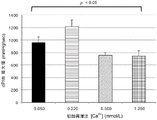
FIG. 14 shows Ca content of 220 μm omicron L/L2+Ionic exemplary oxygenated cardioplegic composition for initial reperfusion at 35 ℃ with another 3 Ca containing heart specific2+Hearts reperfused with oxygenated cardioplegic composition at one of the ion concentrations developed significantly less myocardial edema.
FIG. 15 shows Ca in cardioplegic composition with oxygenation2+The ion concentration is reduced from 1,250 mu m DEG to 220 mu m DEG L/L, and the cardiac output (cardiac weight index) of the heart is re-perfusedEx) improvement. However, with 50 μm O.L/L Ca2+Ionic oxygenated cardioplegic compositions reperfusion has a very weak cardiac output, probably due to "calcium deregulation", in which ischemia alone, through ATP depletion, can cause an increase in cytosolic calcium concentrations.
FIG. 16 shows Ca in cardioplegic composition with oxygenation2+The ion concentration dropped from 1,250 μm ° L/L to 500 μm ° L/L to 220 μm ° L/L, the left ventricular contractility of the reperfused heart during systole (as measured by dP/dt max). However, with 50 μm O.L/L Ca2+Ionic oxygenated cardioplegic compositions reperfused the left ventricle of the heart is poorly contractile and may also be due to calcium abnormalities.
FIG. 17 shows Ca in cardioplegic composition with oxygenation2+The ion concentration was reduced from 1,250 μm ° L/L to 500 μm ° L/L to 220 μm ° L/L, the left ventricular tone of the reperfused heart during diastole (as measured by dP/dt min). However, with 50 μm O.L/L Ca2+Ionic oxygenated cardioplegic compositions reperfused the left ventricle of the heart is poorly relaxant and may also be due to calcium abnormalities.
The data collected in this study demonstrate that the heart obtained with an initial reperfusion of the hypocalcemic oxygenated cardioplegic composition at 35 ℃ can significantly improve myocardial function recovery. The best performance of this study was with Ca2+The ion concentration. However, it appears that Ca is present2+The ion concentration drops too low, e.g., to 50 μm ° L/L, which may have adverse effects.
Example 3:
the next study evaluated whether acidified hypocalcemic oxygenated cardioplegic compositions had potential incremental benefit. Thus, this study evaluated the effect of adjusting the pH of an exemplary hypocalcemic cardioplegic composition from 7.9 to 7.4 to 6.9 to 6.4. The cardioplegic solution comprises 220 [ mu ] m DEG L/L Ca in the cardioplegic solution2+Ion and all reperfusion was completed at 35 ℃ (fig. 18).
The 24 pigs were divided into 4 groups and subsequently sacrificed according to standard protocols and medical ethical procedures following the flow diagram shown in fig. 19. After completion of each heart acquisition, each heart was immediately filled with
Quest 2 myocardial preservation system. Hearts obtained from the first group of pigs were perfused with an exemplary hypocalcemic cardioplegic composition of pH 7.9 for 3 minutes, which was warmed to 35 ℃ before beginning the reperfusion process. Hearts obtained from a second group of pigs were perfused with an exemplary hypocalcemic cardioplegic composition adjusted to pH 7.4 for 3 minutes, which was warmed to 35 ℃ before beginning the reperfusion procedure. Hearts obtained from a third group of pigs were perfused with an exemplary hypocalcemic cardioplegic composition adjusted to pH 6.9 for 3 minutes, which was warmed to 35 ℃ before beginning the reperfusion procedure. Hearts obtained from fourth group of pigs were perfused with an exemplary hypocalcemic cardioplegic composition adjusted to pH 6.4 for 3 minutes, which was warmed to 35 ℃ before beginning the reperfusion procedure.
Aortic perfusion pressure, coronary flow and myocardial temperature were continuously monitored during the 3 minute
initial reperfusion period 2 device recording. Blood gas samples were measured at 0, 30, 60, 120 and 180 seconds of the initial reperfusion period to collect data, particularly involving 02 partial pressures O
2Partial pressure (PaO)
2)、CO
2Partial pressure (PaCO)
2) Changes in pH level, electrolyte level, lactate level occurred.
After the end of the initial 3 minute reperfusion period, from
Quest 2 the hearts were removed from the apparatus and transferred to an isolated heart Perfusion (EVHP) apparatus where they were perfused with a constantly flowing supply of a blood-STEEN solution mixture (Hb 45 g/L; XVIVO Perfusion inc., Englewood, CO, USA) in which their contractile function was restored and maintained in Landorff mode for 1 hour at normal temperature of 35 ℃. Continuous monitoring of aortic pressure and heart rate, and use
And (4) software processing. blood-STEEN solution in
EVHP apparatusAt 1 hour of mixture infusion, each heart was switched from Langendorff mode to operating mode by pacing the heart at 100bpm with a left atrial pressure of 0 to 8 mmHg. Cardiac output, coronary blood flow, aortic root and coronary sinus blood gas were measured and cardiac function was assessed using a pressure-volume ring catheter. After completion of these measurements, each heart returned to the Landorff mode immediately.
Fig. 20 shows that hearts initially reperfused with an exemplary hypocalcemic cardioplegic composition that was mildly acidified (i.e., pH 6.4) exhibited more myocardial edema at 35 ℃ than those reperfused with a hypocalcemic cardioplegic composition that was overbased (i.e., pH 7.9,7.4, 6.9).
Figure 21 shows that the cardiac output (as indicated by heart weight) of the reperfused heart in the slightly acidified hypocalcemic oxygenated cardioplegic composition (i.e., pH 6.9) and the slightly overbased hypocalcemic oxygenated cardioplegic composition (i.e., pH 7.4) is significantly better than the cardiac output of the reperfused heart in the hypocalcemic oxygenated cardioplegic composition adjusted to pH 7.9 or 6.4.
Fig. 22 shows that the left ventricular contractility (as measured by dP/dt max) of the reperfused heart in the slightly acidified hypocalcemic oxygenated cardioplegic composition (i.e., pH 6.9) and the slightly overbased hypocalcemic oxygenated cardioplegic composition (i.e., pH 7.4) during systole is significantly better than the left ventricular contractility of the reperfused heart in the hypocalcemic oxygenated cardioplegic composition adjusted to pH 7.9 or 6.4.
Fig. 23 shows that the left ventricular relaxation during diastole (as measured by dP/dt min) of the reperfused heart in the slightly acidified hypocalcemic oxygenated cardioplegic composition (i.e., pH 6.9) and the slightly overbased hypocalcemic oxygenated cardioplegic composition (i.e., pH 7.4) is significantly better than the left ventricular relaxation of the reperfused heart in the hypocalcemic oxygenated cardioplegic composition adjusted to pH 7.9 or 6.4.
The data collected in this study demonstrate that initial overbased reperfusion is detrimental and that significant peracids (e.g., pH less than 6.5) are also detrimental. However, moderate peracids (e.g., pH 6.6-6.9) appear to be beneficial.
Example 4:
part 1: increase in evaluation of the next studyPlus the duration of donor reperfusion obtained with a mild acidified hypocalcemic oxygenated cardioplegic composition, is of potential incremental benefit. Thus, this study evaluated the use of exemplary weakly acidic (pH 6.9) hypocalcemia (220 μm ° L/L Ca)2+) Effect of reperfusion with oxygenated cardioplegic solution at 35 ℃ for 3 min or 9 min (fig. 24). The cardioplegic solution used in part 1 of this study contained 400 μm ° L/L adenosine and 500 μm ° L/L lidocaine.
The 12 pigs were divided into 2 groups and subsequently sacrificed according to standard protocols and medical ethical procedures following the flow diagram shown in fig. 25. After completion of each heart acquisition, each heart was immediately filled with
Quest 2 myocardial preservation system. Hearts obtained from the first group of pigs were perfused with an exemplary weakly acidic hypocalcemic oxygenated cardioplegic composition for 3 minutes, which was warmed to 35 ℃ prior to the start of the 3 minute reperfusion procedure. Hearts obtained from the second group of pigs were perfused with an exemplary weakly acidic hypocalcemic oxygenated cardioplegic composition warmed to 35 ℃ before beginning the reperfusion procedure for 9 minutes.
Aortic perfusion pressure, coronary flow and myocardial temperature were continuously monitored during the 3 minute
initial reperfusion period 2 device recording. Blood gas samples were measured at 0, 30, 60, 120 and 180 seconds of the initial reperfusion period to collect data, particularly involving 02 partial pressures O
2Partial pressure (PaO)
2)、CO
2Partial pressure (PaCO)
2) Changes in pH level, electrolyte level, lactate level occurred.
From Quest after the end of the initial 3-minute reperfusion period or the initial 9-
minute reperfusion period 2 the hearts are removed from the apparatus and transferred to an isolated heart perfusion (EVHP) apparatus where they are perfused with a continuous flowing supply of a blood-STEEN solution mixture (Hb 45 g/L; XVIVO)Fusion inc., Englewood, CO, USA), in which its contraction function is restored and maintained in Landorff mode, at normal temperature of 35 ℃ for 1 hour, 3 hours, and 5 hours. Continuous monitoring of aortic pressure and heart rate, and use

And (4) software processing. Each heart was switched from Langendorff mode to operating mode by pacing the heart at 100bpm with a left atrial pressure of 0 to 8mmHg at 1 hour of perfusion of the blood-STEEN solution mixture in the EVHP apparatus. Cardiac output, coronary blood flow, aortic root and coronary sinus blood gas were measured and cardiac function was assessed using a pressure-volume ring catheter. After completion of these measurements, each heart immediately returned to the Landorff mode for an additional 2 hours, after which the measurements were repeated (i.e., 3 hours after removal from reperfusion). After completion of these measurements, each heart immediately returned to the Landorff mode for an additional 2 hours, after which the measurements were repeated (i.e., 5 hours after removal from reperfusion).
Fig. 26 shows that the heart initially reperfused for 9 minutes with an exemplary weakly acidic hypocalcemic oxygenated cardioplegic composition exhibited more myocardial edema than those reperfused for 3 minutes only.
Figure 27 shows that as ex vivo cardiac perfusion progressed from 1 hour to 3 hours to 5 hours, the 9 min of initial reperfusion heart tended to deteriorate in function.
These data indicate that the cardioplegic composition may comprise one or more toxic components.
Section 2: the next study evaluated the effect of reducing lidocaine concentrations in an exemplary mildly acidified hypocalcemic oxygenated cardioplegic composition. Thus, this study evaluated the use of exemplary weakly acidic (pH 6.9) hypocalcemia (220 μm ° L/L Ca)2+) Effect of 3 or 9 minute reperfusion at 35 ℃ of oxygenated cardioplegic solution comprising 400 μm ° L/L adenosine and 50 μm ° L/L lidocaine (fig. 28).
The 12 pigs were divided into 2 groups and subsequently sacrificed according to standard protocols and medical ethical procedures following the flow diagram shown in fig. 25. After completion of each heart acquisition, each heart was immediately filled with
Quest 2 myocardial preservation system. Hearts obtained from the first group of pigs were perfused with an exemplary weakly acidic hypocalcemic oxygenated cardioplegic composition for 3 minutes, which was warmed to 35 ℃ prior to the start of the 3 minute reperfusion procedure. Hearts obtained from the second group of pigs were perfused with an exemplary weakly acidic hypocalcemic oxygenated cardioplegic composition warmed to 35 ℃ before beginning the reperfusion procedure for 9 minutes.
Aortic perfusion pressure, coronary flow and myocardial temperature were continuously monitored during the 3 minute
initial reperfusion period 2 device recording. Blood gas samples were measured at 0, 30, 60, 120 and 180 seconds of the initial reperfusion period to collect data, particularly involving 02 partial pressures O
2Partial pressure (PaO)
2)、CO
2Partial pressure (PaCO)
2) Changes in pH level, electrolyte level, lactate level occurred.
From Quest after the end of the initial 3-minute reperfusion period or the initial 9-
minute reperfusion period 2 the hearts were removed from the apparatus and transferred to an isolated heart Perfusion (EVHP) apparatus where they were perfused with a constantly flowing supply of a blood-STEEN solution mixture (Hb 45 g/L; XVIVO Perfusion inc., Englewood, CO, USA) in which their contractile function was restored and maintained in Landorff mode for 1, 3 and 5 hours at 35 ℃ ambient temperature. Continuous monitoring of aortic pressure and heart rate, and use

And (4) software processing. Each heart was switched from Langendorff mode to operating mode by pacing the heart at 100bpm with a left atrial pressure of 0 to 8mmHg at 1 hour of perfusion of the blood-STEEN solution mixture in the EVHP apparatus. Cardiac output, coronary blood flow, aortic root and coronary sinus blood gas were measured and cardiac function was assessed using a pressure-volume ring catheter. After completion of these measurements, each heart was immediately examinedReturn to Landorff mode for an additional 2 hours, after which the measurement is repeated (i.e. 3 hours after removal from reperfusion). After completion of these measurements, each heart immediately returned to the Landorff mode for an additional 2 hours, after which the measurements were repeated (i.e., 5 hours after removal from reperfusion).
Figure 29 shows that there was no significant difference in myocardial edema seen in the initial reperfusion 9 min heart compared to the 3 min perfused heart in an exemplary weakly acidic hypocalcemia-oxygenated cardioplegic composition containing 400 μm ° L/L adenosine and 50 μm ° L/L lidocaine.
Figure 30 shows that in an exemplary weakly acidic hypocalcemic oxygenated cardioplegic composition containing 400 μm ° L/L adenosine and 500 μm ° L/L lidocaine, the initial reperfusion period was extended from 3 minutes to 9 minutes with no adverse effect on cardiac function recovery at 1, 3, 5 hours post-reperfusion.
Figure 31 combines myocardial function data from parts 1 (figure 27) and 2 (figure 30), where it is evident that a lidocaine concentration of 500 μm ° L/L in the cardioplegic composition used for initial ex vivo post-reperfusion has a debilitating effect on the donor heart. This data also demonstrates that extending the initial reperfusion period beyond 3 minutes is not conducive to restoring homeostasis and cardiac function in the harvested donor heart.
The data presented herein show that a suitable composition of cardioplegic solution for initial ex vivo reperfusion of a donor heart prior to initiation of perfusion is shown in table 1.
Table 1: