CN107595387B - A spine image generation system and spine surgery navigation and positioning system based on ultrasonic rubbing technology - Google Patents
A spine image generation system and spine surgery navigation and positioning system based on ultrasonic rubbing technology Download PDFInfo
- Publication number
- CN107595387B CN107595387B CN201710630811.3A CN201710630811A CN107595387B CN 107595387 B CN107595387 B CN 107595387B CN 201710630811 A CN201710630811 A CN 201710630811A CN 107595387 B CN107595387 B CN 107595387B
- Authority
- CN
- China
- Prior art keywords
- ultrasonic
- image
- spine
- patient
- ultrasound
- Prior art date
- Legal status (The legal status is an assumption and is not a legal conclusion. Google has not performed a legal analysis and makes no representation as to the accuracy of the status listed.)
- Active
Links
Images


Classifications
-
- A—HUMAN NECESSITIES
- A61—MEDICAL OR VETERINARY SCIENCE; HYGIENE
- A61B—DIAGNOSIS; SURGERY; IDENTIFICATION
- A61B8/00—Diagnosis using ultrasonic, sonic or infrasonic waves
- A61B8/52—Devices using data or image processing specially adapted for diagnosis using ultrasonic, sonic or infrasonic waves
- A61B8/5207—Devices using data or image processing specially adapted for diagnosis using ultrasonic, sonic or infrasonic waves involving processing of raw data to produce diagnostic data, e.g. for generating an image
-
- A—HUMAN NECESSITIES
- A61—MEDICAL OR VETERINARY SCIENCE; HYGIENE
- A61B—DIAGNOSIS; SURGERY; IDENTIFICATION
- A61B34/00—Computer-aided surgery; Manipulators or robots specially adapted for use in surgery
- A61B34/20—Surgical navigation systems; Devices for tracking or guiding surgical instruments, e.g. for frameless stereotaxis
-
- A—HUMAN NECESSITIES
- A61—MEDICAL OR VETERINARY SCIENCE; HYGIENE
- A61B—DIAGNOSIS; SURGERY; IDENTIFICATION
- A61B8/00—Diagnosis using ultrasonic, sonic or infrasonic waves
- A61B8/08—Clinical applications
-
- A—HUMAN NECESSITIES
- A61—MEDICAL OR VETERINARY SCIENCE; HYGIENE
- A61B—DIAGNOSIS; SURGERY; IDENTIFICATION
- A61B8/00—Diagnosis using ultrasonic, sonic or infrasonic waves
- A61B8/08—Clinical applications
- A61B8/0875—Clinical applications for diagnosis of bone
-
- A—HUMAN NECESSITIES
- A61—MEDICAL OR VETERINARY SCIENCE; HYGIENE
- A61B—DIAGNOSIS; SURGERY; IDENTIFICATION
- A61B8/00—Diagnosis using ultrasonic, sonic or infrasonic waves
- A61B8/13—Tomography
- A61B8/14—Echo-tomography
-
- G—PHYSICS
- G06—COMPUTING OR CALCULATING; COUNTING
- G06T—IMAGE DATA PROCESSING OR GENERATION, IN GENERAL
- G06T7/00—Image analysis
- G06T7/0002—Inspection of images, e.g. flaw detection
- G06T7/0012—Biomedical image inspection
-
- G—PHYSICS
- G06—COMPUTING OR CALCULATING; COUNTING
- G06T—IMAGE DATA PROCESSING OR GENERATION, IN GENERAL
- G06T7/00—Image analysis
- G06T7/30—Determination of transform parameters for the alignment of images, i.e. image registration
- G06T7/33—Determination of transform parameters for the alignment of images, i.e. image registration using feature-based methods
-
- A—HUMAN NECESSITIES
- A61—MEDICAL OR VETERINARY SCIENCE; HYGIENE
- A61B—DIAGNOSIS; SURGERY; IDENTIFICATION
- A61B34/00—Computer-aided surgery; Manipulators or robots specially adapted for use in surgery
- A61B34/10—Computer-aided planning, simulation or modelling of surgical operations
- A61B2034/101—Computer-aided simulation of surgical operations
- A61B2034/105—Modelling of the patient, e.g. for ligaments or bones
-
- A—HUMAN NECESSITIES
- A61—MEDICAL OR VETERINARY SCIENCE; HYGIENE
- A61B—DIAGNOSIS; SURGERY; IDENTIFICATION
- A61B34/00—Computer-aided surgery; Manipulators or robots specially adapted for use in surgery
- A61B34/20—Surgical navigation systems; Devices for tracking or guiding surgical instruments, e.g. for frameless stereotaxis
- A61B2034/2046—Tracking techniques
- A61B2034/2063—Acoustic tracking systems, e.g. using ultrasound
-
- A—HUMAN NECESSITIES
- A61—MEDICAL OR VETERINARY SCIENCE; HYGIENE
- A61B—DIAGNOSIS; SURGERY; IDENTIFICATION
- A61B90/00—Instruments, implements or accessories specially adapted for surgery or diagnosis and not covered by any of the groups A61B1/00 - A61B50/00, e.g. for luxation treatment or for protecting wound edges
- A61B90/36—Image-producing devices or illumination devices not otherwise provided for
- A61B2090/364—Correlation of different images or relation of image positions in respect to the body
- A61B2090/367—Correlation of different images or relation of image positions in respect to the body creating a 3D dataset from 2D images using position information
-
- A—HUMAN NECESSITIES
- A61—MEDICAL OR VETERINARY SCIENCE; HYGIENE
- A61B—DIAGNOSIS; SURGERY; IDENTIFICATION
- A61B90/00—Instruments, implements or accessories specially adapted for surgery or diagnosis and not covered by any of the groups A61B1/00 - A61B50/00, e.g. for luxation treatment or for protecting wound edges
- A61B90/36—Image-producing devices or illumination devices not otherwise provided for
- A61B90/37—Surgical systems with images on a monitor during operation
- A61B2090/376—Surgical systems with images on a monitor during operation using X-rays, e.g. fluoroscopy
-
- A—HUMAN NECESSITIES
- A61—MEDICAL OR VETERINARY SCIENCE; HYGIENE
- A61B—DIAGNOSIS; SURGERY; IDENTIFICATION
- A61B90/00—Instruments, implements or accessories specially adapted for surgery or diagnosis and not covered by any of the groups A61B1/00 - A61B50/00, e.g. for luxation treatment or for protecting wound edges
- A61B90/36—Image-producing devices or illumination devices not otherwise provided for
- A61B90/37—Surgical systems with images on a monitor during operation
- A61B2090/378—Surgical systems with images on a monitor during operation using ultrasound
-
- G—PHYSICS
- G06—COMPUTING OR CALCULATING; COUNTING
- G06T—IMAGE DATA PROCESSING OR GENERATION, IN GENERAL
- G06T2207/00—Indexing scheme for image analysis or image enhancement
- G06T2207/10—Image acquisition modality
- G06T2207/10016—Video; Image sequence
-
- G—PHYSICS
- G06—COMPUTING OR CALCULATING; COUNTING
- G06T—IMAGE DATA PROCESSING OR GENERATION, IN GENERAL
- G06T2207/00—Indexing scheme for image analysis or image enhancement
- G06T2207/10—Image acquisition modality
- G06T2207/10132—Ultrasound image
-
- G—PHYSICS
- G06—COMPUTING OR CALCULATING; COUNTING
- G06T—IMAGE DATA PROCESSING OR GENERATION, IN GENERAL
- G06T2207/00—Indexing scheme for image analysis or image enhancement
- G06T2207/30—Subject of image; Context of image processing
- G06T2207/30004—Biomedical image processing
- G06T2207/30008—Bone
- G06T2207/30012—Spine; Backbone
Landscapes
- Health & Medical Sciences (AREA)
- Life Sciences & Earth Sciences (AREA)
- Engineering & Computer Science (AREA)
- Surgery (AREA)
- Medical Informatics (AREA)
- Nuclear Medicine, Radiotherapy & Molecular Imaging (AREA)
- General Health & Medical Sciences (AREA)
- Public Health (AREA)
- Molecular Biology (AREA)
- Animal Behavior & Ethology (AREA)
- Biomedical Technology (AREA)
- Heart & Thoracic Surgery (AREA)
- Veterinary Medicine (AREA)
- Physics & Mathematics (AREA)
- Radiology & Medical Imaging (AREA)
- Biophysics (AREA)
- Pathology (AREA)
- Computer Vision & Pattern Recognition (AREA)
- Robotics (AREA)
- Rheumatology (AREA)
- Orthopedic Medicine & Surgery (AREA)
- Theoretical Computer Science (AREA)
- General Physics & Mathematics (AREA)
- Quality & Reliability (AREA)
- Ultra Sonic Daignosis Equipment (AREA)
- Apparatus For Radiation Diagnosis (AREA)
- Magnetic Resonance Imaging Apparatus (AREA)
Abstract
Description
技术领域:Technical field:
本发明涉及医学领域,具体涉及一种脊椎图像生成系统、脊椎图像生成方法以及脊柱手术导航定位系统、脊柱手术导航定位方法。The invention relates to the medical field, in particular to a spine image generation system, a spine image generation method, a spine surgery navigation and positioning system, and a spine surgery navigation and positioning method.
背景技术:Background technique:
当前,脊柱退变相关性疾病的发病率和患者数量与日俱增,严重影响人民的身体健康和劳动能力。脊柱手术是治疗椎间盘突出、肿瘤等脊柱疾病的主要手段,其中,以脊柱内镜为代表的微创手术技术因科技进步推进的手术器械设备升级,正越来越趋于成熟,加之创伤小、恢复快的优势,获得越来越多领域的认可和接受。然而这些微创技术对病变的定位精度要求高,加之人体脊柱的特殊性,即骨性结构组成、解剖区域狭长、富含重要的血管神经等复杂性,使得掌握微创技术的门槛较高,主要体现在手术难度及风险较大,操作者培训学习曲线长,难以短期内推广。导航定位技术能降低微创手术技术的门槛,有助于增加手术的操作简便性及保障手术的安全性。At present, the incidence and number of patients with spinal degeneration-related diseases are increasing day by day, which seriously affects people's physical health and labor ability. Spinal surgery is the main method to treat spinal diseases such as intervertebral disc herniation and tumor. Among them, minimally invasive surgery represented by spinal endoscopy is becoming more and more mature due to the upgrading of surgical instruments and equipment promoted by scientific and technological progress. The advantages of fast recovery have been recognized and accepted in more and more fields. However, these minimally invasive techniques have high requirements on the positioning accuracy of lesions, and the particularity of the human spine, that is, the complexity of bony structural composition, long and narrow anatomical area, rich in important blood vessels and nerves, etc., make the threshold for mastering minimally invasive techniques high. It is mainly reflected in the difficulty and risk of surgery, the long learning curve of operator training, and it is difficult to promote in a short time. Navigation and positioning technology can lower the threshold of minimally invasive surgery technology, help to increase the simplicity of surgery and ensure the safety of surgery.
超声扫描人体骨质结构的特点在于:1)超声波不能穿透骨质结构,故超声图像中只能显示骨质表面的线性强回声,获得的信息较少,临床应用范围受限,所述线性强回声为医学超声学概念,是指对超声图像的特征性描述,表现为线性高亮图像;2)超声扫描骨质结构时,容易发生衍射带来噪声,导致骨质结构在超声图像中显示出现偏差,所述噪声为医学超声学概念,是指超声图像中出现的模糊、毛刺状、不清晰的图像特征,骨质结构的噪声表现为模糊、毛刺状、不清晰的线性或片状高亮图像;目前临床中应用的超声扫描技术(包括超声扫描设备和超声处理技术)无法解决上述问题,因此,目前超声扫描技术在骨质结构相关疾病的诊治中应用非常有限。The characteristics of ultrasonic scanning of human bone structure are: 1) Ultrasound cannot penetrate the bone structure, so the ultrasonic image can only display the linear strong echo of the bone surface, the information obtained is less, and the scope of clinical application is limited. Strong echo is the concept of medical ultrasound, which refers to the characteristic description of the ultrasound image, which is expressed as a linear highlight image; 2) When ultrasound scans the bone structure, it is easy to cause noise due to diffraction, which leads to the display of the bone structure in the ultrasound image. If there is a deviation, the noise is the concept of medical ultrasound, which refers to the fuzzy, burr-like and unclear image features in the ultrasound image. The current clinical application of ultrasonic scanning technology (including ultrasonic scanning equipment and ultrasonic processing technology) cannot solve the above problems. Therefore, the current application of ultrasonic scanning technology in the diagnosis and treatment of bone structure-related diseases is very limited.
上述超声扫描骨质结构的特点,在超声扫描脊椎时同样存在,除此之外,不同于人体其他骨质结构如肱骨、股骨等,超声扫描脊椎质结构时存在如下特殊性。如附图6(1)所示,人体脊柱是一个狭长的骨性结构区域,是身体的支柱,分颈、胸、腰、骶及尾五段,由33块椎骨(颈椎7块,胸椎12块,腰椎5块,骶骨、尾骨共9块)由韧带、关节及椎间盘连接而成。脊柱内部自上而下形成一条纵行的脊管,内有脊髓。脊椎由横突、棘突、关节突、椎板、椎板间隙、椎间孔及组成脊椎的其他骨骼成分组成。超声扫描脊椎的特殊性在于:1)超声扫描获得的脊椎骨质信息较少,脊柱作为狭长的骨性结构,由多节椎骨组成,一幅超声扫描图像中所呈现的椎骨节段有限,信息少且仅为骨质表面回波信息;2)相比其他的骨质结构,超声扫描获得的脊椎骨质信息存在更大、更多的偏差,如附图6(2)、6(3)脊椎具有众多骨性突起如棘突、横突、关节突等,而超声图像中骨质回波强,所呈现的超声图像往往为骨质结构特有的光亮而富有噪声的不清晰图像,骨质回波带来的衍射和噪声,将导致脊椎的解剖结构(包括横突、棘突、关节突、椎板及组成脊椎的其他骨骼成分)在超声图像中显示出现较大偏差,常见偏差包括:所述解剖结构中具体的某一点由于发生衍射,在超声图像中可能显示为一个面;所述解剖结构中的某一个面与超声图像中的面大小、比例不准确;所述解剖结构中清晰的毗邻和连接结构,如关节突关节、横突与椎体连接的根部等,在超声图像中显示为模糊的面状结构,连接处不清晰。上述偏差导致常规超声扫描方式无法解决脊椎的骨质结构的超声图像清晰度问题,在超声扫描骨质结构的诸多问题基础之上,超声扫描脊椎骨质信息存在的更多、更大的问题。The above-mentioned characteristics of the osseous structure in ultrasonic scanning are also present in the ultrasonic scanning of the spine. In addition, different from other osseous structures in the human body such as the humerus and femur, the ultrasonic scanning of the vertebral structure has the following particularities. As shown in Figure 6(1), the human spine is a narrow and long bony structural area, which is the pillar of the body. It is divided into five segments: cervical, thoracic, lumbar, sacral and coccygeal. It consists of 5 lumbar vertebrae, 9 sacrum and coccyx) connected by ligaments, joints and intervertebral discs. Inside the spine, a longitudinal spinal canal is formed from top to bottom, which contains the spinal cord. The spine consists of transverse processes, spinous processes, articular processes, lamina, interlaminar spaces, intervertebral foramen, and other skeletal components that make up the spine. The particularity of ultrasound scan spine is: 1) Ultrasound scan obtains less vertebral bone information. As a long and narrow bony structure, the spine consists of multiple vertebrae. The vertebral segments presented in an ultrasound scan image are limited, and the information is limited. 2) Compared with other bone structures, the vertebral bone information obtained by ultrasound scanning has greater and more deviations, as shown in Figures 6(2) and 6(3) The spine has many bony processes such as spinous process, transverse process, articular process, etc., and the echo of bone in the ultrasound image is strong, and the ultrasound image presented is often a bright and noisy image unique to the bone structure. Diffraction and noise caused by echoes will cause large deviations in the anatomical structures of the spine (including transverse processes, spinous processes, articular processes, lamina and other bone components that make up the spine) in ultrasound images. Common deviations include: Due to diffraction, a specific point in the anatomical structure may appear as a face in the ultrasound image; the size and proportion of a certain face in the anatomical structure and the face in the ultrasound image are not accurate; the anatomical structure is clear The adjoining and connecting structures of the vertebral body, such as the facet joint, the root of the connection between the transverse process and the vertebral body, etc., are shown as blurred planar structures in the ultrasound image, and the connection is not clear. The above deviations lead to the fact that the conventional ultrasound scanning method cannot solve the problem of ultrasound image clarity of the bone structure of the spine. On the basis of many problems in the ultrasound scan of the bone structure, there are more and greater problems in the ultrasound scan of the spine bone information.
针对某一具体节段的脊椎而言,例如附图6(2)所示的腰椎,一节腰椎所具有的解剖结构包括横突、棘突、关节突、椎板、椎板间隙、椎间孔及组成脊椎的其他骨骼成分,不同解剖结构在超声图像中显示情况存在显著差异,包括:1)棘突为椎体延伸向人体背侧的骨质结构,因其结构狭长、骨质较薄、存在棘突尖这一易辨识特征,所述骨质较薄是指棘突平行于人体横截面的方向上,左侧骨质表面到右侧骨质表面的厚度较薄,所述棘突尖为棘突的解剖学结构,是指棘突远离脊柱方向的根部为一尖样结构,是棘突的末端,使得棘突作为脊椎中最具特征的解剖结构很容易在超声图像中较完全显示;2)横突、关节突结构在超声图像中的显示情况相较棘突为差,原因包括此二者不像棘突延伸像背侧而与椎体有较大夹角,此二者与椎体夹角较小、无类似棘突尖的易辨识特征,且受限于体位及扫描角度,常常导致二者与相邻骨质结构如椎体难以清晰分辨,加之上述超声扫描骨质结构的偏差,使得二者在超声扫描下获得的骨质回波信息不全、并且不够清晰和准确,所述受限于体位及扫描角度是指,在获取横突、关节突的骨质回波信息的过程中,不可避免地会扫描到脊椎的其它结构、与脊椎相邻的器官,使得脊椎的其它结构、与脊椎相邻的器官的图像会对横突、关节突的骨质回波信息产生干扰,导致横突、关节突的骨质回波信息不全,不够清晰和准确;3)椎板、椎板间隙、椎间孔及组成脊椎的其他骨骼成分受限于上述超声扫描骨质结构的诸多缺陷以及这些结构特殊的解剖位置关系,导致超声不能扫描到这些结构,或者超声图像中不能显示出这些结构全部的骨质回波信息。即使能扫描到这些结构的部分信息,但是也存在着信息准确度不够,存在偏差的问题,具体包括:关节突、椎板、椎板间隙、椎间孔及组成脊椎的其他骨骼成分的解剖结构中具体的某一点由于发生衍射,在超声图像中可能显示为一个面;所述解剖结构中的某一个面与超声图像中相应的面大小、比例不准确;所述解剖结构中的连接部位,例如,横突与椎体连接的根部(参见图6(2)15),在超声图像中显示为模糊的面状结构,连接处不清晰,无法准确识别。For a specific segment of the spine, such as the lumbar vertebra shown in Figure 6(2), the anatomical structures of a lumbar vertebra include transverse process, spinous process, articular process, lamina, lamina space, intervertebral The foramen and other bone components that make up the spine, different anatomical structures show significant differences in ultrasound images, including: 1) The spinous process is the bony structure extending from the vertebral body to the back of the human body, because of its long and narrow structure and thin bone , There is an easily identifiable feature of the spinous process tip. The thin bone means that in the direction of the spinous process parallel to the cross-section of the human body, the thickness from the left bone surface to the right bone surface is thinner, and the spinous process is thin. The anatomical structure with the tip of the spinous process means that the root of the spinous process away from the spine is a pointed structure, which is the end of the spinous process, which makes the spinous process as the most characteristic anatomical structure in the spine. 2) Compared with the spinous process, the transverse process and the articular process structure in the ultrasound image are poorer, the reasons include that the two do not extend like the dorsal side of the spinous process, but have a larger angle with the vertebral body. The angle between the vertebral body and the vertebral body is small, and there is no easily identifiable feature similar to the spinous process tip, and is limited by the body position and scanning angle, which often makes it difficult to clearly distinguish the two from the adjacent bone structures such as the vertebral body. The deviation of the structure makes the bone echo information obtained by the two under ultrasound scanning incomplete, and not clear and accurate enough. The limitation of body position and scanning angle means that the bone echo of the transverse process and articular process is obtained when obtaining the bone echo. During the information process, other structures of the spine and organs adjacent to the spine will inevitably be scanned, so that the images of other structures of the spine and the organs adjacent to the spine will echo information on the bones of the transverse process and articular process. Interfering, resulting in incomplete bone echo information of transverse process and articular process, not clear and accurate; 3) Laminae, lamina space, intervertebral foramen and other bone components that make up the spine are limited by the above-mentioned ultrasonic scanning bone structure Due to the many defects of these structures and the special anatomical position relationship of these structures, the ultrasound cannot scan these structures, or the ultrasound images cannot display all the bone echo information of these structures. Even if part of the information of these structures can be scanned, there is still a problem of insufficient information accuracy and deviation, including: articular process, lamina, lamina space, intervertebral foramen and the anatomical structure of other skeletal components that make up the spine Due to diffraction, a specific point in the anatomical structure may appear as a surface in the ultrasound image; a certain surface in the anatomical structure is inaccurate in size and proportion with the corresponding surface in the ultrasonic image; the connection part in the anatomical structure, For example, the root of the connection between the transverse process and the vertebral body (see Figure 6(2)15) is shown as a blurred planar structure in the ultrasound image, and the connection is not clear and cannot be accurately identified.
目前,临床上脊柱微创手术过程如下:1)术前对患者进行CT扫描;2)手术定位:获得患者当前手术状况下的术中体位,例如X射线透视技术;3)手术定位完成后,进行手术操作,手术操作包括微创手术或开放手术。其中,由于X射线透视技术每次照射仅能生成单幅静态图像,手术定位只能获得患者在某一时刻的术中体位,正常人脊柱有一定的活动度,但各部位活动度不同,颈、腰段活动度较大,胸段活动度极小,骶段几乎无活动性,脊柱天然的活动度加之正常呼吸运动,使得脊柱的位置和弯曲度在手术操作过程中存在位移,单幅静态图像不能准确反映患者实时体位。医生在手术时需要的是患者手术状况下实时的术中体位,目前的情况,只能是医生凭借经验实施手术,导致手术对医生的经验、以及医生在手术时的状态等主观因素依赖程度大,可控程度差。At present, the clinical procedures of minimally invasive spine surgery are as follows: 1) CT scan the patient before surgery; 2) Surgical positioning: obtain the patient's intraoperative position under the current surgical condition, such as X-ray fluoroscopy; 3) After the surgical positioning is completed, Perform surgical procedures, including minimally invasive surgery or open surgery. Among them, because the X-ray fluoroscopy technology can only generate a single static image per irradiation, and the surgical positioning can only obtain the intraoperative position of the patient at a certain moment, the normal spine has a certain degree of mobility, but the mobility of each part is different. , The mobility of the lumbar segment is large, the mobility of the thoracic segment is very small, and the sacral segment has almost no mobility. The natural mobility of the spine and the normal breathing movement make the position and curvature of the spine shift during the surgical operation. The image does not accurately reflect the real-time position of the patient. What the doctor needs during the operation is the real-time intraoperative position of the patient under the operation condition. In the current situation, the doctor can only perform the operation by virtue of experience, resulting in the operation being highly dependent on subjective factors such as the doctor's experience and the doctor's state during the operation. , the controllability is poor.
可见,超声扫描骨质结构总体特点可以概括为:1)超声扫描骨质结构存在问题;2)相比其他骨质结构,超声扫描脊椎难度尤甚;3)临床现有的超声扫描技术(包括超声扫描设备和超声处理技术)存在扫描信息不全、清晰度、准确度不高等缺陷,无法适应脊柱手术的临床应用,图像问题成为脊柱微创手术治疗发展的最大障碍。It can be seen that the general characteristics of ultrasound scanning bone structure can be summarized as follows: 1) There are problems in ultrasound scanning bone structure; 2) Compared with other bone structures, ultrasound scanning of the spine is particularly difficult; 3) The existing clinical ultrasound scanning technologies (including Ultrasound scanning equipment and ultrasonic processing technology) have defects such as incomplete scanning information, low clarity, and low accuracy, which cannot be adapted to the clinical application of spinal surgery. Image problems have become the biggest obstacle to the development of minimally invasive spinal surgery.
目前临床上脊柱手术定位其中最具代表性的是C臂机透视定位技术。该技术的操作模式可以概括为图1所示,即“穿刺-照射-评估-调整-照射验证”,具体流程为:手术前在患者手术部位置入穿刺针,进行第一次X射线照射后,获知穿刺针在患者体内的进针深度和角度并进行调整后,进行第二次X射线照射,再次获知穿刺针在患者体内的进针深度和角度,若进针深度和角度达到预定部位,则定位完成,进行手术操作,反之则重复“评估-调整-照射验证”步骤,直到完成穿刺定位。C臂机透视定位技术确保了定位的准确,但存在如下不足:At present, the most representative clinical spinal surgery positioning is the C-arm fluoroscopic positioning technology. The operation mode of this technology can be summarized as shown in Figure 1, namely "puncture-irradiation-evaluation-adjustment-irradiation verification". The specific process is: insert the puncture needle at the patient's operating site before surgery, and after the first X-ray irradiation , after knowing the depth and angle of the puncture needle in the patient's body and adjusting it, perform a second X-ray irradiation, and learn the depth and angle of the puncture needle in the patient's body again. If the depth and angle of the puncture needle reach the predetermined position, Then the positioning is completed, and the surgical operation is performed. Otherwise, the steps of "assessment-adjustment-irradiation verification" are repeated until the puncture positioning is completed. The C-arm perspective positioning technology ensures accurate positioning, but has the following shortcomings:
1)手术操作者需依赖经验判断多次反复在患者体表穿刺,对患者造成较大创伤。在定位过程中,需要在患者手术部位置入穿刺针,穿刺针要直达脊椎表面,为达到理想的穿刺深度和角度,往往需要多次调整,在患者体内反复穿刺,手术体验很差。1) The operator needs to rely on experience to judge and repeatedly puncture the patient's body surface many times, which will cause greater trauma to the patient. During the positioning process, the puncture needle needs to be inserted into the patient's operating area, and the puncture needle must reach the surface of the spine. In order to achieve the ideal puncture depth and angle, multiple adjustments are often required. Repeated puncture in the patient's body results in a poor surgical experience.
2)手术操作者每次在患者手术部位插入穿刺针后都要进行X射线照射,获知穿刺针在患者体内的进针深度和角度并进行调整后,再次进行X射线照射,再次获知穿刺针在患者体内的进针深度和角度,直至进针深度和角度达到预定部位,则定位完成。在定位过程中需要X线透视,X线辐射非常损伤大,对患者和手术操作者造成潜在辐射损伤。2) The operator must perform X-ray irradiation every time after inserting the puncture needle into the patient's surgical site. After knowing the depth and angle of the puncture needle in the patient's body and adjusting it, X-ray irradiation is performed again, and the puncture needle is again known to be in the patient's body. The needle insertion depth and angle in the patient's body, until the needle insertion depth and angle reach the predetermined position, the positioning is completed. X-ray fluoroscopy is required during the positioning process, and X-ray radiation is very damaging, causing potential radiation damage to patients and surgical operators.
3)该定位技术对手术操作者的经验依赖非常大。由于微创技术对病变的定位精度要求高,操作者的经验与操作中穿刺、照射的次数成正相关。目前在临床工作中,通常情况下,十年左右手术经验的操作者在手术中尚且需要穿刺针6-10次,经验不丰富的手术操作者则需要更多次的反复穿刺和定位,不仅效率低,而且给患者和手术操作者带来更多的X射线辐射,对患者更大的穿刺损伤,不利于该技术的推广应用。3) This positioning technique is very dependent on the experience of the operator. Since minimally invasive techniques have high requirements on the localization accuracy of lesions, the operator's experience is positively correlated with the number of punctures and irradiations during the operation. In current clinical work, under normal circumstances, operators with about ten years of surgical experience still need 6-10 puncture needles during the operation, while inexperienced operators need more repeated puncture and positioning, which is not only efficient In addition, it brings more X-ray radiation to patients and operators, and causes greater puncture damage to patients, which is not conducive to the popularization and application of this technology.
4)C臂机透视定位技术得到的静态图像不能对后续手术实现导航。C臂机透视定位技术所获得的图像为患者当前手术状况下的术中体位,为静态X线图像,不能正确反应手术进行中脊柱活动度带来的改变,不能实时地指导后续手术操作,因此C臂机透视定位技术只用于术前定位,确定手术的入路点,一经确定开始手术,将无法呈现患者在手术中任一时刻手术状况下的术中体位,操作者将完全依靠经验进行手术操作。4) The static image obtained by the C-arm fluoroscopic positioning technology cannot realize the navigation for the subsequent operation. The image obtained by the C-arm fluoroscopic positioning technology is the intraoperative position of the patient under the current surgical condition. It is a static X-ray image, which cannot correctly reflect the changes brought by the spinal range of motion during the operation, and cannot guide the follow-up operation in real time. Therefore, The C-arm fluoroscopic positioning technology is only used for preoperative positioning to determine the entry point of the operation. Once the operation is determined to start, the intraoperative position of the patient under the operation condition at any time during the operation will not be displayed. The operator will rely entirely on experience. surgical operation.
鉴于C臂机透视定位技术的优势和不足,近年来有人提出利用多种模式图像形成融合图像(刘彦斌等在,超声容积导航技术引导腰椎经皮后外侧入路完全内镜下微创手术椎间孔穿刺的应用研究,硕士学位论文,第二军医大学,2015年5月),进而指导定位的方法,其中具有代表性的是超声容积导航技术。该技术的操作模式可以概括为附图2所示,通过超声扫描获得超声图像,将其与术前采集的CT或MRI三维数据进行图像融合。完成图像融合后,利用电磁跟踪系统(电磁追踪器与超声探头距离<80cm,试验全过程保持追踪器位置不变),由超声专业操作者协助脊柱手术操作者在实时图像中找到病变部位。具体流程为:术前进行CT扫描并进行三维重建,将三维CT数据导入超声仪,以三点法进行影像配准。同屏显示CT图像和超声图像,在患者体表4个标记点中任取3点(3点不在同一条直线上),依次分别对3点的CT图像及超声图像进行精确寻找并锁定,完成外标记点的配准,外标记点需要切开或穿刺皮肤并借助金属钉等器材进行设置外标记点。完成外标记点的配准后再进行内标记点的配准,在CT图上寻找任意椎体棘突顶点并锁定,然后在超声图像中实时寻找相应点并锁定,测量配准误差。如误差>5mm,则再选择其他椎体的棘突顶点作为另一个内标记点进行修正。通过内、外标记点的双重配准,将图像配准的误差缩至5mm以内,完成图像融合。图像融合后,保证电磁追踪器及患者的相对位置不变,以腹腔镜消毒隔离套包裹探头,实时引导穿刺。在CT图像上锁定病变节段椎间孔最下缘作为进针点,利用超声图像实时显示该点,准确寻找并锁定,应用超声仪导航实时引导穿刺针在病变节段平面内穿刺。穿刺前定位及穿刺过程中穿刺方向的确认可辅以C臂机透视。若穿刺过程中患者出现下肢放射痛,需行C臂机透视,若穿刺方向存在偏差,根据标记点再次进行图像配准及融合,直至穿刺导针顺利到位。穿刺成功后行C臂机透视进一步确认穿刺针位置。In view of the advantages and disadvantages of C-arm fluoroscopic positioning technology, in recent years, it has been proposed to use multiple modes of images to form fusion images (Liu Yanbin et al., Ultrasound volume navigation technology guides lumbar spine percutaneous posterolateral approach to complete endoscopic minimally invasive intervertebral surgery. Application Research of Hole Puncture, Master's Thesis, Second Military Medical University, May 2015), and then guide the method of positioning, of which the representative is ultrasonic volume navigation technology. The operation mode of this technology can be summarized as shown in FIG. 2 . Ultrasound images are obtained through ultrasound scanning and image fusion is performed with the CT or MRI three-dimensional data collected before surgery. After the image fusion was completed, the electromagnetic tracking system (the distance between the electromagnetic tracker and the ultrasound probe was less than 80 cm, and the position of the tracker was kept unchanged during the whole experiment), and the professional ultrasound operator assisted the spinal surgery operator to find the lesion in the real-time image. The specific process is as follows: CT scan and 3D reconstruction are performed before surgery, 3D CT data is imported into the ultrasound machine, and image registration is performed by the three-point method. The CT image and the ultrasound image are displayed on the same screen, and 3 points are randomly selected from the 4 marker points on the patient's body surface (the 3 points are not on the same straight line), and the CT images and ultrasound images of the 3 points are accurately searched and locked in turn. For the registration of external markers, the external markers need to be cut or punctured in the skin and set with the help of metal nails and other equipment. After completing the registration of the external marker points, the internal marker points are registered, and any vertebral spinous process vertex is found on the CT image and locked, and then the corresponding point is found and locked in the ultrasound image in real time, and the registration error is measured. If the error is greater than 5mm, select the spinous process vertex of other vertebral bodies as another internal marker for correction. Through the double registration of the inner and outer marker points, the error of image registration is reduced to within 5mm, and the image fusion is completed. After the images are fused, the relative positions of the electromagnetic tracker and the patient are kept unchanged, and the probe is wrapped with a laparoscopic sterilized isolation sleeve to guide the puncture in real time. The lowermost edge of the intervertebral foramen of the lesion segment was locked on the CT image as the needle insertion point, and the ultrasound image was used to display the point in real time, so as to find and lock it accurately. The positioning before puncture and the confirmation of the puncture direction during puncture can be supplemented by C-arm fluoroscopy. If the patient has radiating pain in the lower limbs during the puncture process, C-arm fluoroscopy is required. If there is a deviation in the puncture direction, image registration and fusion are performed again according to the marked points until the puncture guide needle is in place smoothly. After successful puncture, C-arm fluoroscopy was performed to further confirm the location of the puncture needle.
超声容积导航技术使手术操作者可以根据的配准图像进行穿刺和手术操作,相比C臂机透视定位技术,极大减少了X线辐射损伤,但存在如下不足:Ultrasound volumetric navigation technology enables operators to perform puncture and surgical operations based on the registered images. Compared with C-arm fluoroscopic positioning technology, X-ray radiation damage is greatly reduced, but it has the following shortcomings:
1)需多次反复在患者体表穿刺,对患者造成较大创伤,操作复杂,过程冗长,步骤繁琐,消耗时间。CT图像及超声图像配准融合过程中,需要设置3个标记点,需要切开或穿刺皮肤并借助针头、螺钉、金属钉等器材设置外标记点。内标记点直接在图像上看图像选取的。进行外标记点和内标记点配准并修正后,完成图像融合。融合后的图像引导穿刺针穿刺到达目标靶点,若穿刺方向存在偏差,根据外标记点和内标记点再次进行图像配准及融合,直至穿刺针顺利到位。1) It is necessary to repeatedly puncture the patient's body surface for many times, causing great trauma to the patient, complicated operation, lengthy process, cumbersome steps, and time-consuming. During the registration and fusion of CT images and ultrasound images, three marker points need to be set. It is necessary to cut or puncture the skin and set external marker points with the help of needles, screws, metal nails and other equipment. The inner marker points are selected by looking at the image directly on the image. After registering and correcting the outer and inner markers, the image fusion is completed. The fused image guides the puncture needle to reach the target point. If there is a deviation in the puncture direction, image registration and fusion are performed again according to the outer marker point and the inner marker point until the puncture needle is in place smoothly.
2)手术全程需要超声专业操作者协助。从超声图像中的解剖部位识别,配准融合所需超声图像及CT图像的选取,再到图像融合后定位病变位置,整个过程均需要手术操作者与超声专业操作者密切配合,导致手术操作者无法快速、独立完成手术。2) The entire operation requires the assistance of an ultrasound professional operator. From the identification of anatomical parts in the ultrasound images, the selection of ultrasound images and CT images required for registration and fusion, to the location of lesions after image fusion, the entire process requires the close cooperation between the operator and the professional ultrasound operator, which leads to the difficulty of the operator. Inability to perform surgery quickly and independently.
3)该定位技术对脊柱手术操作者的经验依赖较大。由于该定位技术所获得的导航图像精度不高,而微创技术对病变的定位精度要求高,因此仍需操作者的具备一定的操作经验以保证操作准确,故该技术对经验依赖度小于C臂机透视定位技术,但无法摆脱对手术操作者的依赖性。3) This positioning technique is highly dependent on the experience of spinal operators. Because the accuracy of the navigation images obtained by this positioning technology is not high, and the minimally invasive technology has high requirements on the positioning accuracy of lesions, it still requires the operator to have a certain operating experience to ensure the accuracy of the operation, so the dependence of this technology on experience is less than C The fluoroscopic positioning technology of the arm machine cannot get rid of the dependence on the surgical operator.
4)导航图像精度不高。由超声导航容积技术得到的融合图像,在导航定位时,其图像精度主要取决于三个方面,第一是点配准模式的精度低,超声图像与CT或MRI图像均富含大量信息,仅选取少数几个特征点进行配准将不能保证精度,对手术带来不利影响;第二是以上几个特征点的选取依赖于超声专业操作者,受主观因素的影响,对图像精度产生不利影响;第三是超声图像的精度低,脊柱是一个狭长的骨性结构区域,而超声图像中骨质回波强,所呈现的超声图像往往为骨质结构特有的光亮而富有噪声的不清晰图像,骨质回波带来的衍射和噪声,将导致解剖结构在超声图像中显示出现偏差,常见偏差包括:解剖结构中具体的点在超声图像中显示为面;解剖结构中的面与超声图像中的面大小比例不准确;解剖结构中清晰的毗邻和连接结构,如关节突关节、横突与椎体连接的根部等,在超声图像中显示为模糊的面状结构,连接处不清晰。上述偏差导致常规超声扫描方式无法解决骨质结构的超声图像清晰度问题,导致其远低于相应的CT或MRI图像,在超声图像中所选取的标记点与在CT或MRI图像中所选取的标记点并不能完全准确地一一对应,导致导航图像精度不高。4) The navigation image accuracy is not high. The image accuracy of the fusion image obtained by the ultrasound navigation volume technology mainly depends on three aspects during the navigation and positioning. The first is the low accuracy of the point registration mode. Both ultrasound images and CT or MRI images are rich in information. Selecting a few feature points for registration will not guarantee the accuracy, which will adversely affect the operation; the second is that the selection of the above feature points depends on the ultrasound professional operator, which is affected by subjective factors and adversely affects the image accuracy; The third is the low accuracy of ultrasound images. The spine is a narrow and long bony structure area. However, the echo of bone in ultrasound images is strong, and the ultrasound images presented are often bright and noisy images unique to bone structures. Diffraction and noise caused by bone echoes will cause deviations in the display of anatomical structures in ultrasound images. Common deviations include: specific points in anatomical structures are displayed as faces in ultrasound images; faces in anatomical structures are different from those in ultrasound images. The face size ratio is not accurate; the clear adjoining and connecting structures in the anatomical structure, such as the facet joint, the root of the connection between the transverse process and the vertebral body, etc., appear as blurred face structures in the ultrasound image, and the connection is not clear. The above deviations lead to the fact that the conventional ultrasound scanning method cannot solve the problem of the ultrasound image clarity of the bone structure, resulting in it being much lower than the corresponding CT or MRI image. The marked points cannot be completely and accurately corresponded one-to-one, resulting in low accuracy of the navigation image.
5)超声容积导航的定位技术操作繁琐,费时费力。以采用脊柱内镜进行经椎板后路髓核摘除术治疗腰椎间盘突出症为例,在开始穿刺直至到达目标靶点的过程中,需行C臂机透视,若穿刺方向存在偏差,需根据标记点再次进行图像配准及融合,直至穿刺导针顺利到位。穿刺成功后仍需行C臂机透视进一步确认穿刺针位置。完成上述定位穿刺后,置入脊柱内镜并逐层深入,操作者根据临床经验辨别棘突、黄韧带、椎板、神经根、椎间盘等解剖结构后,操作者只能根据临床经验运用手术器械进行操作,直至抵达并摘除突出的髓核或纤维环等完成相关手术内容。5) The positioning technology of ultrasonic volume navigation is cumbersome, time-consuming and labor-intensive. Taking the use of spinal endoscopy for the treatment of lumbar intervertebral disc herniation through posterior laminectomy as an example, C-arm fluoroscopy is required during the process of starting the puncture until reaching the target point. Image registration and fusion of the marked points are performed again until the puncture guide needle is in place smoothly. After successful puncture, C-arm fluoroscopy is still required to further confirm the position of the puncture needle. After completing the above positioning and puncture, the spinal endoscope is placed and deepened layer by layer. After the operator identifies the anatomical structures such as the spinous process, ligamentum flavum, lamina, nerve root, and intervertebral disc according to clinical experience, the operator can only use surgical instruments according to clinical experience. Perform operations until the protruding nucleus pulposus or annulus fibrosus is reached and removed.
综上,现有的脊柱导航定位技术主要以C臂机透视定位技术虽然提供了高精度的穿刺定位,但存在X线多次照射、辐射损伤大、多次穿刺、穿刺创伤大、定位技术对操作者的经验依赖大、手术时间延长费时费力等不足,且所获得的静态图像不能对后续手术导航提供帮助;超声容积导航技术虽然极大减少了X线辐射损伤,但同样存在多次穿刺、穿刺创伤较大、需要超声专业操作者协助、导航图像精度低的缺点。故这两种技术共同的缺点是:操作复杂,对手术操作者的经验依赖程度大;多次反复在患者体表穿刺,损伤大;X线辐射对手术操作者和患者的辐射损伤大;定位操作繁琐、费时费力;仅能完成体表穿刺定位,无法为后续的相关手术操作实现导航(导航图像精度有限且无导航系统支持)。In summary, the existing spinal navigation and positioning technology mainly uses C-arm fluoroscopic positioning technology to provide high-precision puncture positioning, but there are multiple X-ray exposures, large radiation damage, multiple punctures, large puncture trauma, and positioning technology. The operator's experience relies heavily, the operation time is prolonged, time-consuming and labor-intensive, etc., and the obtained static images cannot provide assistance for subsequent surgical navigation; although the ultrasound volumetric navigation technology greatly reduces X-ray radiation damage, it also suffers from multiple punctures, The disadvantages of large puncture trauma, the need for professional ultrasound operator assistance, and low accuracy of navigation images. Therefore, the common disadvantages of these two techniques are: the operation is complicated, and the degree of dependence on the experience of the operator is large; the repeated puncture on the patient's body surface will cause great damage; the radiation damage of X-ray radiation to the operator and the patient is large; positioning The operation is cumbersome, time-consuming and labor-intensive; it can only complete the body surface puncture positioning, and cannot achieve navigation for subsequent related surgical operations (the navigation image accuracy is limited and there is no navigation system support).
因此,临床上亟待一种准确快速而又无辐射、无创伤的实时导航定位方案指导脊柱手术,可以实现脊柱手术精准治疗操作,至少需要获取必要部位(横突和棘突)的准确空间位置信息,但是现有技术只能获得棘突的准确空间位置信息,无法获取横突的准确空间位置信息,导致脊柱手术精准治疗的准确度不高。进一步地,脊柱手术还需要获取特定部位的准确空间位置信息,所述的特定部位是指关节突、椎板、椎板间隙、椎间孔的任意一种或任意多种。总而言之,现有技术无法全面并且准确获取脊柱手术所需要的相关部位(包括必要部位和特定部位)的准确空间位置信息。因此要实现脊柱手术精准治疗操作需满足两个层面:满足手术所需必要部位的信息准确,所述必要部位的信息准确才能确保脊柱手术精准治疗操作的准确性;满足手术所需的特定部位的准确空间位置信息,所述特定部位的信息越多越准才能提高脊柱手术精准治疗操作的准确性。现有技术尚不能获得足够的手术所需特定部位的准确空间位置信息,以实现通过导航技术指导脊柱手术的精准治疗操作。Therefore, there is an urgent need for an accurate, fast, non-radiation, non-invasive real-time navigation and positioning scheme to guide spinal surgery, which can achieve precise treatment of spinal surgery, and at least need to obtain accurate spatial position information of necessary parts (transverse and spinous processes). However, the existing technology can only obtain the accurate spatial position information of the spinous process, but cannot obtain the accurate spatial position information of the transverse process, resulting in low accuracy of precise treatment of spinal surgery. Further, spinal surgery also needs to obtain accurate spatial position information of a specific part, and the specific part refers to any one or more of articular process, lamina, lamina space, and intervertebral foramen. All in all, the existing technology cannot comprehensively and accurately obtain accurate spatial position information of relevant parts (including necessary parts and specific parts) required for spinal surgery. Therefore, in order to realize the precise treatment operation of spine surgery, two levels must be satisfied: the information of the necessary parts required by the operation is accurate, and the information of the necessary parts can ensure the accuracy of the precise treatment operation of the spine surgery; Accurate spatial position information, the more information of the specific part, the more accurate the accuracy of the precise treatment operation of the spine surgery can be improved. Existing technologies cannot obtain sufficient accurate spatial position information of specific parts required for surgery, so as to realize precise treatment operations for spinal surgery guided by navigation technology.
发明内容SUMMARY OF THE INVENTION
针对现有的不足,本发明所要解决的技术问题是,提供一种基于超声拓片技术的脊椎图像生成系统和方法,创造性地通过获取的患者手术状况下实时的术中体位对应的二维脊椎表面结构超声图像,经处理后形成超声拓片,所述的超声拓片是带有患者空间定位信息并随患者位置变动而实时动态更新的个性化三维超声骨骼图像,将所述超声拓片与数字医学影像进行轮廓匹配后,进一步获得与患者术中体位一致的实时更新的个性化的脊椎表面地形图,从而彻底解决了现有技术无法获取脊柱手术所需要的相关部位(包括必要部位和特定部位)的准确空间位置信息的问题,以及现有技术实时滞后和不能实时动态显示的问题,实现通过导航技术指导脊柱手术的精准治疗操作。本发明还提供了一种基于超声拓片技术的脊柱手术导航定位系统和方法,利用所述基于超声拓片技术的脊椎图像生成系统获得的与患者术中体位一致的实时更新的个性化的脊椎表面地形图,能够实现实时的术中导航,使手术操作者无需依赖经验、无需超声专业操作者的协助、无需对病患身体进行多次穿刺造成多次创伤,在最大限度减少X线辐射对患者和操作者的损伤的情况下,准确快速通过一次穿刺到达目标靶点,实现了通过导航技术指导脊柱手术的精准治疗操作。In view of the existing deficiencies, the technical problem to be solved by the present invention is to provide a spine image generation system and method based on ultrasonic rubbing technology, which creatively obtains the two-dimensional spine surface corresponding to the real-time intraoperative posture of the patient under the surgical condition. Structural ultrasonic images are processed to form ultrasonic rubbings. The ultrasonic rubbings are personalized three-dimensional ultrasonic skeletal images with patient spatial positioning information and dynamically updated in real time with changes in the patient's position. After contour matching, a real-time updated and personalized spine surface topographic map that is consistent with the patient's intraoperative posture is further obtained, thus completely solving the problem that the existing technology cannot obtain the accuracy of relevant parts (including necessary parts and specific parts) required for spine surgery. The problem of spatial position information, as well as the problems of real-time lag and inability to display dynamically in real-time in the existing technology, realize the precise treatment operation of spinal surgery guided by navigation technology. The present invention also provides a spinal surgery navigation and positioning system and method based on ultrasonic rubbing technology, and a real-time updated personalized spine surface topography obtained by the spine image generation system based on ultrasonic rubbing technology that is consistent with the patient's intraoperative posture It can realize real-time intraoperative navigation, so that the operator does not need to rely on experience, do not need the assistance of professional ultrasound operators, and does not need to perform multiple punctures on the patient's body to cause multiple traumas. In the case of the operator's injury, the target can be accurately and quickly reached the target point through a single puncture, and the precise treatment operation of the spinal surgery guided by the navigation technology is realized.
在完成穿刺到达目标靶点后,本发明提供的脊柱手术导航定位系统还可以指导操作者完成后续相关手术内容,持续为后续手术导航,操作简单,省时省力。以采用脊柱内镜进行经椎板后路髓核摘除术治疗腰椎间盘突出症为例,完成穿刺到达目标靶点后,手术导航定位系统指导脊柱内镜逐层深入,辨别棘突、黄韧带、椎板、神经根、椎间盘等解剖结构,直至抵达并摘除突出的髓核或纤维环等完成相关手术内容,克服了现有技术X线、穿刺损伤大,操作经验依赖度高,导航图像难以准确实时显示等诸多不足。After completing the puncture and reaching the target point, the spinal surgery navigation and positioning system provided by the present invention can also guide the operator to complete the follow-up related operation content, and continue to navigate for the follow-up operation, which is easy to operate and saves time and effort. Taking the use of spinal endoscopy for the treatment of lumbar intervertebral disc herniation through posterior lamina excision as an example, after the puncture reaches the target point, the surgical navigation and positioning system guides the spinal endoscope to go deeper layer by layer to identify the spinous process, ligamentum flavum, ligamentum flavum, Anatomical structures such as lamina, nerve roots, intervertebral discs, etc., until reaching and removing the protruding nucleus pulposus or annulus fibrosus to complete the relevant surgical content, overcoming the existing technology X-ray, puncture damage, high dependence on operating experience, difficult to accurately navigate images Real-time display and many other shortcomings.
本发明采用如下的技术方案:The present invention adopts the following technical scheme:
一种基于超声拓片技术的脊椎图像生成系统,所述的脊椎图像生成系统基于二维脊椎表面结构超声图像生成超声拓片,与数字医学影像进行轮廓匹配后,获得与患者术中体位一致的实时更新的个性化的脊椎表面地形图,所述的系统包括采集单元和处理单元,其中:A spine image generation system based on ultrasonic rubbing technology, the spine image generation system generates ultrasonic rubbings based on two-dimensional spine surface structure ultrasonic images, and after contour matching with digital medical images, obtains real-time updates consistent with the patient's intraoperative posture The personalized topographic map of the spine surface, the system includes an acquisition unit and a processing unit, wherein:
所述的采集单元用于获取患者手术状况下实时术中体位对应的二维脊椎表面结构超声图像,所述的处理单元用于将采集单元获取的患者手术状况下实时的术中体位对应的二维脊椎表面结构超声图像处理后形成超声拓片,再与数字医学影像进行轮廓匹配后,获得与患者术中体位一致的实时更新的个性化的脊椎表面地形图;所述超声图像由包含脊柱区深层肌肉组织和脊椎表面的全部回波信息构成;所述脊椎表面包括横突和棘突;所述的超声拓片是带有患者空间定位信息并随患者位置变动而实时动态更新的个性化三维超声骨骼图像。The acquisition unit is used for acquiring the two-dimensional ultrasonic image of the vertebral surface structure corresponding to the real-time intraoperative posture of the patient under the operating condition of the patient, and the processing unit is used for acquiring the real-time intraoperative intraoperative posture of the patient acquired by the acquisition unit. The ultrasonic image of the vertebral surface structure is processed to form an ultrasonic rubbing, and after contour matching with the digital medical image, a real-time updated personalized spine surface topographic map consistent with the patient's intraoperative posture is obtained; Muscle tissue and all the echo information of the vertebral surface; the vertebral surface includes transverse process and spinous process; the ultrasonic rubbing is a personalized three-dimensional ultrasonic skeleton with the patient's spatial positioning information and dynamically updated in real time with the patient's position change image.
基于本发明的一种基于超声拓片技术的脊椎图像生成系统,本发明还提供了一种基于超声拓片技术的脊椎图像生成方法,所述的脊椎图像生成方法包括如下步骤:Based on a spine image generation system based on the ultrasonic rubbing technique of the present invention, the present invention also provides a spine image generation method based on the ultrasonic rubbing technique. The spine image generation method includes the following steps:
1)获取超声图像:获取患者手术状况下实时术中体位对应的二维脊椎表面结构超声图像,所述超声图像由包含脊柱区深层肌肉组织和脊椎表面的全部回波信息构成,所述脊椎表面包括横突和棘突;1) Obtaining an ultrasound image: obtaining a two-dimensional ultrasound image of the vertebral surface structure corresponding to the real-time intraoperative posture of the patient under the operating condition, and the ultrasound image is composed of all the echo information including the deep musculature in the spinal region and the surface of the vertebral column. including transverse and spinous processes;
2)生成超声拓片:将步骤1)获取的患者手术状况下实时术中体位对应的二维脊椎表面结构超声图像处理生成超声拓片,所述的超声拓片是带有患者空间定位信息并随患者位置变动而实时动态更新的个性化三维超声骨骼图像;2) Generating ultrasonic rubbings: The two-dimensional vertebral surface structure ultrasonic images corresponding to the real-time intraoperative body position obtained in step 1) are processed to generate ultrasonic rubbings. Personalized 3D ultrasound skeletal images that change and dynamically update in real time;
3)生成脊椎表面地形图:将步骤2)获得的所述超声拓片与数字医学影像进行轮廓匹配,获得与患者术中体位一致的实时更新的个性化的脊椎表面地形图。3) Generate a topographic map of the spine surface: perform contour matching between the ultrasonic rubbing obtained in step 2) and the digital medical image to obtain a real-time updated personalized topographic map of the spine surface consistent with the patient's intraoperative posture.
基于本发明的一种基于超声拓片技术的脊椎图像生成系统,本发明还提供了一种基于超声拓片技术的脊柱手术导航定位系统,所述脊柱手术导航定位系统包括导航模块和基于超声拓片技术的脊椎图像生成系统,所述的脊柱手术导航定位系统能够获得与患者术中体位一致的实时更新的个性化的脊椎表面地形图,并基于该脊椎表面地形图进行实时的术中导航,其中:Based on a spine image generation system based on ultrasonic rubbing technology of the present invention, the present invention also provides a spinal surgery navigation and positioning system based on ultrasonic rubbing technology. The spinal surgery navigation and positioning system includes a navigation module and an ultrasonic rubbing technology-based A spine image generation system, the spine surgery navigation and positioning system can obtain a real-time updated personalized spine surface topography consistent with the patient's intraoperative posture, and perform real-time intraoperative navigation based on the spine surface topography, wherein:
所述的基于超声拓片技术的脊椎图像生成系统,基于患者手术状况下实时的术中体位对应的二维脊椎表面结构超声图像处理后形成超声拓片,然后将所述超声拓片与数字医学影像进行轮廓匹配,形成与患者术中体位一致的实时更新的个性化的脊椎表面地形图,所述的超声拓片是带有患者空间定位信息并随患者位置变动而实时动态更新的个性化三维超声骨骼图像;优选的,所述的数字医学影像是CT容积漫游重建图像、磁共振MR、计算机X线摄影CR、数字化计算机X线摄影DR。The described spine image generation system based on ultrasonic rubbing technology is based on the real-time intraoperative body position of the patient under the condition of surgery to form ultrasonic rubbings after ultrasonic image processing of the two-dimensional spinal surface structure corresponding to the intraoperative position, and then outlines the ultrasonic rubbings and digital medical images. Matching to form a real-time updated personalized spine surface topographic map consistent with the patient's intraoperative posture, and the ultrasonic rubbing is a personalized three-dimensional ultrasonic skeletal image with the patient's spatial positioning information and dynamically updated in real time with the patient's position change; Preferably, the digital medical images are CT volumetric roaming reconstruction images, magnetic resonance MR, computed radiography CR, and digital computed radiography DR.
基于本发明的一种超声拓片技术的脊柱手术导航定位系统,本发明还提供了一种基于超声拓片技术的脊柱手术导航定位方法,其特征在于包括如下步骤:Based on a spinal surgery navigation and positioning system based on the ultrasonic rubbing technology of the present invention, the present invention also provides a spinal surgery navigation and positioning method based on the ultrasonic rubbing technology, which is characterized by comprising the following steps:
1)建立手术器械的实体几何模型,实现手术器械的实体几何模型向表面模型的转化,使其在脊椎图像生成系统中所获得的与患者术中体位一致的实时更新的个性化的脊椎表面地形图上呈现出来,实现对手术器械的实时跟踪,所述实体几何模型向表面模型转化的方法优选为三维CAD造型法;1) Establish the solid geometric model of the surgical instrument, and realize the transformation from the solid geometric model of the surgical instrument to the surface model, so that the real-time updated personalized spine surface topography obtained in the spine image generation system is consistent with the patient's intraoperative posture. It is shown on the figure to realize real-time tracking of surgical instruments, and the method for converting the solid geometric model to the surface model is preferably a three-dimensional CAD modeling method;
2)根据与患者术中体位一致的实时更新的个性化的脊椎表面地形图包含的空间位置信息建立地形图坐标系,以患者术中体位的空间位置信息建立患者坐标系,确定地形图坐标系和患者坐标系中对应点的关系,并形成统一坐标系,所述确定地形图坐标系和患者坐标系中对应点关系的方法优选为最近点迭代算法;2) Establish a topographic map coordinate system according to the spatial position information contained in the real-time updated personalized spine surface topographic map consistent with the patient's intraoperative posture, establish a patient coordinate system based on the spatial position information of the patient's intraoperative posture, and determine the topographic map coordinate system and the relationship between the corresponding points in the patient coordinate system, and form a unified coordinate system, the method for determining the relationship between the corresponding points in the topographic map coordinate system and the patient coordinate system is preferably the closest point iterative algorithm;
3)将步骤1)获得的手术器械的表面模型在步骤2)形成的统一坐标系显示出来,实现手术器械和与患者术中体位一致的实时更新的个性化的脊椎表面地形图处于统一坐标系中,实现术中实时导航。3) Display the surface model of the surgical instrument obtained in step 1) in the unified coordinate system formed in step 2), so that the surgical instrument and the real-time updated personalized spine surface topographic map consistent with the patient's intraoperative posture are in a unified coordinate system , to achieve intraoperative real-time navigation.
本发明还提供一种脊柱手术操作系统,所述的脊柱手术操作系统包含如前所述任何一种形式的基于超声拓片技术的脊椎图像生成系统和/或如前所述任何一种形式的基于超声拓片技术的脊柱手术导航定位系统。此操作系统适用于脊柱部位的开放和微创手术,可进行并不仅限于椎间盘突出部分的摘除、肿瘤切除、神经松解、血肿清除等诸多手术操作。The present invention also provides a spine surgery operating system, the spine surgery operating system comprising any one of the aforementioned ultrasound rubbing technology-based spine image generation systems and/or any one of the aforementioned Spinal surgery navigation and positioning system based on ultrasound rubbing technique. This operating system is suitable for open and minimally invasive surgery on the spine, and it can perform many surgical operations such as the removal of intervertebral disc herniation, tumor resection, nerve release, and hematoma removal.
与现有技术相比,本发明的有益效果为:本发明提供的基于超声拓片技术的脊椎图像生成系统和方法,解决了现有技术无法获取脊柱手术所需要的相关部位(包括必要部位和特定部位)的准确空间位置信息的问题,以及现有技术实时滞后和不能实时动态显示的问题,为实现通过导航技术指导脊柱手术的精准治疗操作。本发明提供的基于超声拓片技术的脊柱手术导航定位系统和方法,能够实现实时的术中导航,使手术操作者无需依赖经验、无需超声专业操作者的协助、无需对病患身体进行多次穿刺造成多次创伤,在最大限度减少X线辐射对患者和操作者的损伤的情况下,准确快速通过一次穿刺到达目标靶点,实现了通过导航技术指导脊柱手术的精准治疗操作。Compared with the prior art, the beneficial effects of the present invention are as follows: the system and method for generating a spine image based on the ultrasonic rubbing technique provided by the present invention solves the problem that the prior art cannot obtain relevant parts (including necessary parts and specific parts) required for spinal surgery. In order to realize the accurate treatment operation of spinal surgery guided by navigation technology. The spinal surgery navigation and positioning system and method based on the ultrasonic rubbing technology provided by the present invention can realize real-time intraoperative navigation, so that the surgical operator does not need to rely on experience, the assistance of an ultrasonic professional operator, and the need to perform multiple punctures on the patient's body. It causes multiple traumas, and under the condition of minimizing the damage of X-ray radiation to the patient and the operator, it can accurately and quickly reach the target point through one puncture, and realize the precise treatment operation of spinal surgery guided by navigation technology.
在完成穿刺到达目标靶点后,该脊柱手术导航定位系统可以指导操作者完成后续相关手术内容,持续为后续手术导航,操作简单,省时省力。以采用脊柱内镜进行经椎板后路髓核摘除术治疗腰椎间盘突出症为例,完成穿刺到达目标靶点后,手术导航定位系统指导脊柱内镜逐层深入,辨别棘突、黄韧带、椎板、神经根、椎间盘等解剖结构,直至抵达并摘除突出的髓核或纤维环等完成相关手术内容。具体表现为:After completing the puncture and reaching the target point, the spinal surgery navigation and positioning system can guide the operator to complete the follow-up related surgical content, and continue to navigate for the follow-up surgery. The operation is simple, time-saving and labor-saving. Taking the use of spinal endoscopy for the treatment of lumbar intervertebral disc herniation through posterior lamina excision as an example, after the puncture reaches the target point, the surgical navigation and positioning system guides the spinal endoscope to go deeper layer by layer to identify the spinous process, ligamentum flavum, ligamentum flavum, Anatomical structures such as lamina, nerve roots, intervertebral discs, etc., until reaching and removing the protruding nucleus pulposus or annulus fibrosus, etc., complete the relevant surgical content. Specifically:
1)最大限度减少X线对患者和手术操作者的辐射损伤。1) Minimize the radiation damage of X-ray to patients and operators.
2)仅需在患者体表穿刺1次,最大限度减少穿刺对患者造成的创伤。2) Only one puncture on the patient's body is required to minimize the trauma caused by the puncture to the patient.
3)对手术操作者的经验依赖非常小,脊椎图像生成系统提供随患者体位变动而实时更新的个性化的脊椎表面地形图,以及基于该脊椎表面地形图进行实时术中导航的脊柱手术导航定位系统,将使手术操作者无需依赖经验、无需超声专业操作者的协助,有助于脊柱手术的推广应用。3) There is very little dependence on the experience of the surgical operator. The spine image generation system provides a personalized spine surface topographic map that is updated in real time with the patient's position change, and spinal surgery navigation and positioning for real-time intraoperative navigation based on the spine surface topographic map. The system will make the surgical operator not need to rely on experience and the assistance of professional ultrasound operators, which will help the popularization and application of spinal surgery.
4)患者体位变动而实时更新的个性化的脊椎表面地形图将对后续手术导航提供极大帮助。4) The personalized topographic map of the spine surface updated in real time due to changes in the patient's position will provide great help for subsequent surgical navigation.
5)基于本发明提供的脊椎图像生成系统以及脊柱手术导航定位系统,将使操作简单,省时省力。5) Based on the spine image generation system and the spine surgery navigation and positioning system provided by the present invention, the operation will be simple, time and effort will be saved.
6)导航图像精度高且能与患者实时术中体位一致。6) The navigation image has high precision and can be consistent with the patient's real-time intraoperative posture.
7)本发明提供的脊椎图像生成系统以及脊柱手术导航定位系统,使得定位技术不存在实时滞后和不能动态显示的问题。相对于C臂机能够实现一定程度的实时,实时有滞后,会存在问题,也不能实现动态,存在问题。7) The spine image generation system and spine surgery navigation and positioning system provided by the present invention make the positioning technology free from the problems of real-time lag and inability to display dynamically. Compared with the C-arm machine, it can achieve a certain degree of real-time, but there is a lag in real-time, which will cause problems, and it cannot achieve dynamic, and there are problems.
8)完成体表穿刺定位后,可进一步指导后续相关手术内容。以采用脊柱内镜进行经椎板后路髓核摘除术治疗腰椎间盘突出症为例,在完成穿刺到达目标靶点后(该过程为定位穿刺过程),操作者操作内镜逐层深入,根据临床经验辨别棘突、黄韧带、椎板、神经根、椎间盘等解剖结构后,脊柱手术导航定位系统可以指导操作者运用手术器械,按照正确的角度和距离进行操作,直至抵达并摘除突出的髓核或纤维环等完成相关手术内容。8) After the body surface puncture positioning is completed, the follow-up related surgical contents can be further guided. Taking the use of spinal endoscope for the treatment of lumbar intervertebral disc herniation through posterior laminectomy as an example, after completing the puncture and reaching the target point (this process is the process of positioning puncture), the operator operates the endoscope to go deep layer by layer. After identifying the anatomical structures such as spinous process, ligamentum flavum, lamina, nerve root, intervertebral disc, etc., the spinal surgery navigation and positioning system can guide the operator to use surgical instruments and operate according to the correct angle and distance until the protruding pulp is reached and removed. Nucleus or annulus fibrosus, etc. to complete the relevant surgical content.
相对于现有技术,本发明的优越性体现在:第一,不需设置外标记点,极大减少创伤;第二,提高超声图像精度,进而提升了与患者术中体位一致的实时更新的个性化的脊椎表面地形图这一导航图像的精度;第三,减少对临床经验的依赖,导航图像将极大降低手术难度,减少对操作者操作经验和超声专业者的依赖。第四,克服了现有技术的不足,本发明的系统不仅能够实现准确的定位穿刺,还能为脊柱手术的后续操作提供实时导航。Compared with the prior art, the advantages of the present invention are as follows: first, no external marker points need to be set, which greatly reduces trauma; second, the accuracy of ultrasound images is improved, thereby improving the real-time update of the patient's intraoperative posture. Personalized spine surface topography is the precision of the navigation image; thirdly, it reduces the dependence on clinical experience. The navigation image will greatly reduce the difficulty of surgery and reduce the dependence on the operator's operating experience and ultrasound professionals. Fourth, overcoming the deficiencies of the prior art, the system of the present invention can not only achieve accurate positioning and puncture, but also provide real-time navigation for subsequent operations of spinal surgery.
应用本发明提供的脊椎图像生成系统以及脊柱手术导航定位系统,将极大推动以脊柱内镜技术为代表的脊柱手术微创技术的迅速发展和短期内的推广普及,造福患者和受脊柱相关疾病困扰的广大人民。本发明适用于以脊柱内镜为代表的脊柱微创技术,但不限于脊柱内镜,只要获取了超声拓片,并通过与数字医学影像进行轮廓匹配后获得与患者术中体位一致的实时更新的个性化的脊椎表面地形图,该地形图同样适用于指导脊柱相关疾病的微创和开放手术。The application of the spine image generation system and spine surgery navigation and positioning system provided by the present invention will greatly promote the rapid development and short-term popularization of spine surgery minimally invasive techniques represented by spine endoscopy technology, and benefit patients and patients suffering from spine-related diseases. troubled people. The present invention is applicable to spinal minimally invasive techniques represented by spinal endoscopy, but is not limited to spinal endoscopy, as long as ultrasonic rubbings are obtained, and after contour matching with digital medical images, a real-time update consistent with the patient's intraoperative posture can be obtained. Personalized spine surface topography, which is equally suitable for guiding minimally invasive and open surgery for spine-related diseases.
附图说明Description of drawings
图1是C臂机透视定位技术的操作流程示意图;Fig. 1 is the operation flow schematic diagram of C-arm perspective positioning technology;
图2是超声容积导航技术的操作流程示意图;Fig. 2 is the operation flow schematic diagram of ultrasonic volume navigation technology;
图3是本发明的基于超声拓片技术的脊椎图像生成系统组成示意图;3 is a schematic diagram of the composition of a spine image generation system based on ultrasonic rubbing technology of the present invention;
图4是本发明的基于超声拓片技术的脊柱手术导航定位系统操作流程示意图;Fig. 4 is the operation flow schematic diagram of the spinal surgery navigation and positioning system based on ultrasonic rubbing technology of the present invention;
图5是本发明的基于超声拓片技术的脊柱手术导航定位方法流程图;Fig. 5 is the flow chart of the spinal surgery navigation and positioning method based on ultrasonic rubbing technology of the present invention;
图6是人体脊柱及脊椎解剖示意图,其中:图6(1)为脊柱全面观示意图,图中所示:A、前面观,B、后面观,C、侧面观;图6(2)为腰椎解剖示意图,图中所示:A、右侧面观,B、上面观;图6(3)为颈椎上面观。Fig. 6 is human spine and vertebral anatomy schematic diagram, wherein: Fig. 6 (1) is the overall view schematic diagram of spine, as shown in the figure: A, front view, B, back view, C, side view; Fig. 6 (2) is lumbar vertebra Anatomical schematic diagram, as shown in the figure: A, right side view, B, upper view; Figure 6(3) is the upper view of cervical vertebra.
1、颈椎,2、胸椎,3、腰椎,4、骶骨,5、尾骨,6、上关节突,7、棘突,8、下关节突,9、横突,10、椎体,11、乳突,12、椎孔,13、横突孔,14、椎板,15、横突与椎体连接的根部;1. Cervical vertebra, 2. Thoracic vertebra, 3. Lumbar vertebra, 4. Sacrum, 5. Coccyx, 6. Superior articular process, 7. Spinous process, 8. Inferior articular process, 9. Transverse process, 10. Vertebral body, 11. Mammary Process, 12, vertebral foramen, 13, transverse process foramen, 14, lamina, 15, the root of the connection between the transverse process and the vertebral body;
图7是体绘制中剪切变形法的基本原理示意图。FIG. 7 is a schematic diagram of the basic principle of the shear deformation method in volume rendering.
具体实施方式Detailed ways
下面结合附图和实施例进一步阐述本发明。应该理解,这些实施例仅用于说明本发明而不用于限制本发明的范围。此外应理解,在阅读了本发明的讲授的内容之后,本领域技术人员可以对本发明作各种改动或修改,这些等价形式同样落于本申请所附权利要求书所限定的范围。The present invention will be further described below in conjunction with the accompanying drawings and embodiments. It should be understood that these examples are only used to illustrate the present invention and not to limit the scope of the present invention. In addition, it should be understood that after reading the teaching content of the present invention, those skilled in the art can make various changes or modifications to the present invention, and these equivalent forms also fall within the scope defined by the appended claims of the present application.
本发明提供一种基于超声拓片技术的脊椎图像生成系统,所述的脊椎图像生成系统基于二维脊椎表面结构超声图像生成超声拓片,与数字医学影像进行轮廓匹配后,获得与患者术中体位一致的实时更新的个性化的脊椎表面地形图,所述的系统包括采集单元和处理单元,其中:The invention provides a spine image generation system based on ultrasonic rubbing technology. The spine image generation system generates ultrasonic rubbings based on two-dimensional ultrasonic images of vertebral surface structures. The real-time updated personalized topographic map of the spine surface, the system includes an acquisition unit and a processing unit, wherein:
所述的采集单元用于获取患者手术状况下实时术中体位对应的二维脊椎表面结构超声图像,所述的处理单元用于将采集单元获取的患者手术状况下实时的术中体位对应的二维脊椎表面结构超声图像处理后形成超声拓片,再与数字医学影像进行轮廓匹配后,获得与患者术中体位一致的实时更新的个性化的脊椎表面地形图;所述超声图像由包含脊柱区深层肌肉组织和脊椎表面的全部回波信息构成;所述脊椎表面包括横突和棘突;所述的超声拓片是带有患者空间定位信息并随患者位置变动而实时动态更新的个性化三维超声骨骼图像;The acquisition unit is used for acquiring the two-dimensional ultrasonic image of the vertebral surface structure corresponding to the real-time intraoperative posture of the patient under the operating condition of the patient, and the processing unit is used for acquiring the real-time intraoperative intraoperative posture of the patient acquired by the acquisition unit. The ultrasonic image of the vertebral surface structure is processed to form an ultrasonic rubbing, and after contour matching with the digital medical image, a real-time updated personalized spine surface topographic map consistent with the patient's intraoperative posture is obtained; Muscle tissue and all the echo information of the vertebral surface; the vertebral surface includes transverse process and spinous process; the ultrasonic rubbing is a personalized three-dimensional ultrasonic skeleton with the patient's spatial positioning information and dynamically updated in real time with the patient's position change image;
优选的,所述的超声图像包括所述超声图像的轮廓边缘、轮廓内部的信息;优选的,所述脊椎表面还进一步包括关节突、椎板、椎板间隙、椎间孔以及组成脊椎的其他骨骼成分的任意一种或任意多种;Preferably, the ultrasound image includes the contour edge of the ultrasound image and the information inside the contour; preferably, the spine surface further includes articular process, lamina, lamina space, intervertebral foramen, and other components that make up the spine. any one or more of the skeletal components;
优选的,所述超声图像的获取方法为:通过带有患者空间定位信息的超声扫描设备在患者体表反复扫描,直到识别肌骨界面,提取包含术中二维空间位置参数的脊椎表面全部骨质回波信息,构成患者手术状况下实时的术中体位对应的二维脊椎表面结构超声图像,所述的术中二维空间位置参数是指患者手术状况下脊椎实时的二维空间位置参数;更优选的,所述带有患者空间定位信息的超声扫描设备是带有定位标签的超声探头;Preferably, the ultrasonic image acquisition method is as follows: repeatedly scan the patient's body surface with an ultrasonic scanning device with patient spatial positioning information until the musculoskeletal interface is identified, and extract all the bones on the surface of the spine including the intraoperative two-dimensional spatial position parameters. The mass echo information constitutes a two-dimensional ultrasonic image of the spinal surface structure corresponding to the patient's real-time intraoperative body position under the operation condition, and the intraoperative two-dimensional space position parameter refers to the real-time two-dimensional space position parameter of the spine under the patient's operation condition; More preferably, the ultrasound scanning device with patient spatial positioning information is an ultrasound probe with a positioning label;
优选的,所述超声拓片的获取方法为:将所述患者手术状况下实时的术中体位对应的二维脊椎表面结构超声图像进行优化、叠加后,进一步处理生成超声拓片;Preferably, the method for obtaining the ultrasonic rubbings is as follows: after optimizing and superimposing the real-time two-dimensional ultrasonic images of the vertebral surface structure corresponding to the intraoperative posture of the patient under the operating conditions, further processing to generate ultrasonic rubbings;
优选的,所述轮廓匹配的获取方法为:将所述超声拓片包含的脊椎表面全部骨质回波信息与数字医学影像的轮廓进行逐点匹配,获得与患者术中体位一致的实时更新的个性化的脊椎表面地形图;Preferably, the method for obtaining the contour matching is as follows: matching all the bone echo information on the vertebral surface contained in the ultrasonic rubbing with the contour of the digital medical image point by point to obtain a real-time updated personality consistent with the patient's intraoperative posture. the topographic map of the spine surface;
优选的,所述脊椎表面地形图是由超声拓片所具有的实时动态空间信息赋予到数字医学影像而形成的立体表面观;优选的,所述超声拓片所具有的实时动态空间信息是指脊椎表面和肌骨界面共同构成的空间信息,该空间信息包含超声扫描所获得的脊椎表面、深层肌肉和脂肪组织的超声图像信息和空间位置信息,并能实时动态地显示;优选的,所述立体表面观是基于脊椎表面空间信息形成的表观三维图像,所述的表观三维图像中的沟纹是指所述的全部超声图像信息中包含的脊椎表面的纹理;Preferably, the topographic map of the spine surface is a three-dimensional surface view formed by adding real-time dynamic spatial information of ultrasonic rubbings to digital medical images; The spatial information formed together with the musculoskeletal interface, the spatial information includes the ultrasound image information and spatial position information of the spine surface, deep muscle and adipose tissue obtained by ultrasound scanning, and can be dynamically displayed in real time; preferably, the three-dimensional surface The 3D image is an apparent three-dimensional image formed based on the spatial information of the spine surface, and the grooves in the apparent three-dimensional image refer to the texture of the spine surface included in the entire ultrasound image information;
优选的,所述的数字医学影像可以是CT容积漫游重建图像、磁共振MR、计算机X线摄影CR或者数字化计算机X线摄影DR。Preferably, the digital medical image may be CT volumetric roaming reconstruction image, magnetic resonance MR, computed radiography CR or digital computed radiography DR.
进一步的,所述的采集单元包括超声图像扫描模块和超声图像信息提取模块,其中:Further, the acquisition unit includes an ultrasound image scanning module and an ultrasound image information extraction module, wherein:
所述超声图像扫描模块用于在患者体表反复扫描,直到识别肌骨界面,获取原始数据,所述超声图像信息提取模块用于将所述的原始数据进行提取,获得包含术中二维空间位置参数的脊椎表面全部骨质回波信息,构成患者手术状况下实时的术中体位对应的二维脊椎表面结构超声图像,所述的术中二维空间位置参数是指患者手术状况下脊椎实时的二维空间位置参数;所述的原始数据是指带有患者空间定位信息的超声扫描设备发出的射频信号,所述的扫描允许以各种不同角度和方向进行,所述的方向包括上下、前后、左右、斜向,所述的角度包括相对于患者体表的垂直或任意倾斜角,并且与扫描先后顺序无关;The ultrasonic image scanning module is used to repeatedly scan the patient's body surface until the musculoskeletal interface is identified to obtain raw data, and the ultrasonic image information extraction module is used to extract the raw data to obtain a two-dimensional space including an intraoperative image. All the bone echo information on the spine surface of the position parameter constitutes the real-time intraoperative ultrasound image of the vertebral surface structure corresponding to the patient's intraoperative posture. The two-dimensional spatial position parameters of the two-dimensional space; the raw data refers to the radio frequency signal sent by the ultrasonic scanning device with the patient's spatial positioning information, and the scanning is allowed to be carried out at various angles and directions, and the directions include up and down, Front and rear, left and right, oblique, the angles include vertical or arbitrary inclination angles relative to the patient's body surface, and have nothing to do with the scanning sequence;
优选的,所述的带有患者空间定位信息的超声扫描设备是带有定位标签的超声探头,所述的原始数据是指超声探头发出的射频信号。Preferably, the ultrasonic scanning device with patient spatial positioning information is an ultrasonic probe with a positioning label, and the raw data refers to the radio frequency signal emitted by the ultrasonic probe.
进一步的,所述的处理单元包括超声图像优化模块、超声图像叠加模块、超声拓片生成模块和图像轮廓匹配模块,其中:Further, the processing unit includes an ultrasound image optimization module, an ultrasound image overlay module, an ultrasound rubbing generation module and an image contour matching module, wherein:
所述超声图像优化模块用于将所述采集单元获得的患者手术状况下实时的术中体位对应的二维脊椎表面结构超声图像进行优化,获得消除干扰噪声的超声优化图像;优选的,所述的超声图像优化模块采用超声滤波增强技术对所述采集单元获取的超声图像进行优化,获得消除干扰噪声的超声优化图像,所述的超声滤波增强技术优选自适应中值滤波和数学形态学滤波相结合的方法;The ultrasonic image optimization module is used to optimize the real-time two-dimensional ultrasonic image of the vertebral surface structure corresponding to the intraoperative posture of the patient obtained by the acquisition unit, so as to obtain an ultrasonic optimized image that eliminates interference noise; preferably, the The ultrasonic image optimization module adopts ultrasonic filter enhancement technology to optimize the ultrasonic image obtained by the acquisition unit, and obtains an ultrasonic optimized image that eliminates interference noise. The ultrasonic filter enhancement technology preferably adopts adaptive median filtering and mathematical morphology filtering. method of combining;
所述超声图像叠加模块用于将所述超声图像优化模块获得的超声优化图像进行叠加,使超声骨质回波信息强回声与弱回声之间的差别更大,获得图像效果增强的超声叠加图像;优选的,所述的超声图像叠加为单模态配准方法;The ultrasonic image superposition module is used to superimpose the ultrasonic optimized images obtained by the ultrasonic image optimization module, so that the difference between the strong echoes and weak echoes of the ultrasonic bone echo information is larger, and an ultrasonic superimposed image with an enhanced image effect is obtained. ; Preferably, the ultrasonic image superposition is a single-modality registration method;
所述超声拓片生成模块用于将所述超声图像叠加模块获得的超声叠加图像处理后形成超声拓片;优选的,所述处理的方法为三维重建,更优选的,所述的三维重建的方法为体绘制方法,优选为体绘制加速技术;The ultrasonic rubbing generation module is used to process the ultrasonic superimposed images obtained by the ultrasonic image superimposing module to form ultrasonic rubbings; preferably, the processing method is three-dimensional reconstruction, and more preferably, the three-dimensional reconstruction method is: Volume rendering method, preferably volume rendering acceleration technology;
所述图像轮廓匹配模块用于将所述超声拓片生成模块获得的超声拓片与数字医学影像进行轮廓匹配,形成与患者术中体位一致的实时更新的个性化的脊椎表面地形图,所述轮廓匹配的方法优选基于像素灰度算法中的互信息算法的多模态配准方法,所述的多模态配准方法包括图像的空间变换、灰度插值、相似度衡量、搜索优化;优选的,所述的数字医学影像选自CT容积漫游重建图像、磁共振MR、计算机X线摄影CR、数字化计算机X线摄影DR。The image contour matching module is used to perform contour matching between the ultrasonic rubbings obtained by the ultrasonic rubbing generation module and the digital medical images to form a real-time updated personalized spine surface topographic map consistent with the patient's intraoperative posture, and the contour matching The method is preferably based on the multimodal registration method of the mutual information algorithm in the pixel grayscale algorithm, and the multimodal registration method includes the spatial transformation of the image, grayscale interpolation, similarity measurement, and search optimization; preferably, The digital medical image is selected from CT volume roaming reconstruction image, magnetic resonance MR, computer X-ray CR, digital computer X-ray DR.
基于本发明的一种基于超声拓片技术的脊椎图像生成系统,本发明还提供了一种基于超声拓片技术的脊椎图像生成方法,所述的脊椎图像生成方法包括如下步骤:Based on a spine image generation system based on the ultrasonic rubbing technique of the present invention, the present invention also provides a spine image generation method based on the ultrasonic rubbing technique. The spine image generation method includes the following steps:
1)获取超声图像:获取患者手术状况下实时术中体位对应的二维脊椎表面结构超声图像,所述超声图像由包含脊柱区深层肌肉组织和脊椎表面的全部回波信息构成,所述脊椎表面包括横突和棘突;1) Obtaining an ultrasound image: obtaining a two-dimensional ultrasound image of the vertebral surface structure corresponding to the real-time intraoperative posture of the patient under the operating condition, and the ultrasound image is composed of all the echo information including the deep musculature in the spinal region and the surface of the vertebral column. including transverse and spinous processes;
2)生成超声拓片:将步骤1)获取的患者手术状况下实时术中体位对应的二维脊椎表面结构超声图像处理生成超声拓片,所述的超声拓片是带有患者空间定位信息并随患者位置变动而实时动态更新的个性化三维超声骨骼图像;2) Generating ultrasonic rubbings: The two-dimensional vertebral surface structure ultrasonic images corresponding to the real-time intraoperative body position obtained in step 1) are processed to generate ultrasonic rubbings. Personalized 3D ultrasound skeletal images that change and dynamically update in real time;
3)生成脊椎表面地形图:将步骤2)获得的所述超声拓片与数字医学影像进行轮廓匹配,获得与患者术中体位一致的实时更新的个性化的脊椎表面地形图;3) generating a topographic map of the spine surface: performing contour matching on the ultrasonic rubbing obtained in step 2) with a digital medical image to obtain a real-time updated personalized topographic map of the spine surface consistent with the patient's intraoperative posture;
优选的,所述的超声图像包括所述超声图像的轮廓边缘、轮廓内部的信息;优选的,所述脊椎表面还进一步包括关节突、椎板、椎板间隙、椎间孔以及组成脊椎的其他骨骼成分的任意一种或任意多种;Preferably, the ultrasound image includes the contour edge of the ultrasound image and the information inside the contour; preferably, the spine surface further includes articular process, lamina, lamina space, intervertebral foramen, and other components that make up the spine. any one or more of the skeletal components;
优选的,所述超声图像的获取方法为:通过带有患者空间定位信息的超声扫描设备在患者体表反复扫描,直到识别肌骨界面,提取包含术中二维空间位置参数的脊椎表面全部骨质回波信息,构成患者手术状况下实时的术中体位对应的二维脊椎表面结构超声图像,所述的术中二维空间位置参数是指患者手术状况下脊椎实时的二维空间位置参数;更优选的,所述带有患者空间定位信息的超声扫描设备是带有定位标签的超声探头;Preferably, the ultrasonic image acquisition method is as follows: repeatedly scan the patient's body surface with an ultrasonic scanning device with patient spatial positioning information until the musculoskeletal interface is identified, and extract all the bones on the surface of the spine including the intraoperative two-dimensional spatial position parameters. The mass echo information constitutes a two-dimensional ultrasonic image of the spinal surface structure corresponding to the patient's real-time intraoperative body position under the operation condition, and the intraoperative two-dimensional space position parameter refers to the real-time two-dimensional space position parameter of the spine under the patient's operation condition; More preferably, the ultrasound scanning device with patient spatial positioning information is an ultrasound probe with a positioning label;
优选的,所述超声拓片的生成方法为:将所述患者手术状况下实时的术中体位对应的二维脊椎表面结构超声图像进行优化、叠加后,进一步处理生成超声拓片;Preferably, the method for generating the ultrasonic rubbings is as follows: after optimizing and superimposing the real-time two-dimensional ultrasonic images of the vertebral surface structure corresponding to the intraoperative posture of the patient under the operating conditions, further processing to generate the ultrasonic rubbings;
优选的,所述脊椎表面地形图的生成方法为:将所述超声拓片包含的脊椎表面全部骨质回波信息与数字医学影像的轮廓进行逐点匹配,获得与患者术中体位一致的实时更新的个性化的脊椎表面地形图;Preferably, the method for generating the topographic map of the spine surface is as follows: matching all the bone echo information of the spine surface contained in the ultrasonic rubbing with the contour of the digital medical image point by point, so as to obtain a real-time update consistent with the intraoperative posture of the patient a personalized topographic map of the spine surface;
优选的,所述脊椎表面地形图是由超声拓片所具有的实时动态空间信息赋予到数字医学影像而形成的立体表面观;优选的,所述超声拓片所具有的实时动态空间信息是指脊椎表面和肌骨界面共同构成的空间信息,该空间信息包含超声扫描所获得的脊椎表面、深层肌肉和脂肪组织的超声图像信息和空间位置信息,并能实时动态地显示;优选的,所述立体表面观是基于脊椎表面空间信息形成的表观三维图像,所述的表观三维图像中的沟纹是指所述的全部超声图像信息中包含的脊椎表面的纹理;Preferably, the topographic map of the spine surface is a three-dimensional surface view formed by adding real-time dynamic spatial information of ultrasonic rubbings to digital medical images; The spatial information formed together with the musculoskeletal interface, the spatial information includes the ultrasound image information and spatial position information of the spine surface, deep muscle and adipose tissue obtained by ultrasound scanning, and can be dynamically displayed in real time; preferably, the three-dimensional surface The 3D image is an apparent three-dimensional image formed based on the spatial information of the spine surface, and the grooves in the apparent three-dimensional image refer to the texture of the spine surface included in the entire ultrasound image information;
优选的,所述的数字医学影像可以是CT容积漫游重建图像、磁共振MR、计算机X线摄影CR或者数字化计算机X线摄影DR。Preferably, the digital medical image may be CT volumetric roaming reconstruction image, magnetic resonance MR, computed radiography CR or digital computed radiography DR.
进一步的,所述的步骤1)包括如下步骤:Further, described step 1) comprises the steps:
超声图像扫描:通过带有患者空间定位信息的超声扫描设备在患者体表反复扫描,直到识别肌骨界面,获取原始数据,所述的原始数据是指带有患者空间定位信息的超声扫描设备发出的射频信号,所述的扫描允许以各种不同角度和方向进行,所述的方向包括上下、前后、左右、斜向,所述的角度包括相对于患者体表的垂直或任意倾斜角,并且与扫描先后顺序无关;优选的是,通过带有定位标签的超声探头扫描获取原始数据,所述的原始数据是指超声探头发出的射频信号;Ultrasound image scanning: Repeatedly scan the patient's body surface through the ultrasonic scanning equipment with the patient's spatial positioning information until the musculoskeletal interface is identified, and obtain the original data. the radio frequency signal, the scanning is allowed to be carried out at various angles and directions, the directions include up and down, front and rear, left and right, oblique, and the angles include vertical or arbitrary inclination angles relative to the patient's body surface, and It has nothing to do with the scanning sequence; preferably, raw data is obtained by scanning an ultrasonic probe with a positioning label, and the raw data refers to the radio frequency signal sent by the ultrasonic probe;
超声图像信息提取:从超声图像扫描模块所获取的原始数据进行提取,获得包含术中二维空间位置参数的脊椎表面全部骨质回波信息,构成患者手术状况下实时的术中体位对应的二维脊椎表面结构超声图像,所述的术中二维空间位置参数是指患者手术状况下脊椎实时的二维空间位置参数。Ultrasound image information extraction: Extract the raw data obtained by the ultrasound image scanning module to obtain all the bone echo information on the spine surface including the intraoperative two-dimensional spatial position parameters, which constitutes the real-time intraoperative position corresponding to the patient's surgical condition. The ultrasonic image of the vertebral surface structure, and the intraoperative two-dimensional spatial position parameter refers to the real-time two-dimensional spatial position parameter of the spine under the patient's operating condition.
进一步的,所述的步骤2)包括如下步骤:Further, described step 2) comprises the steps:
超声图像优化:将步骤1)获得患者手术状况下实时的术中体位对应的二维脊椎表面结构超声图像进行优化,获得消除干扰噪声的超声优化图像;优选的,超声图像优化采用超声滤波增强技术,获得消除干扰噪声的超声优化图像;更优选的,所述的超声滤波增强技术可以是中值滤波和数学形态学滤波相结合的方法;Ultrasound image optimization: optimize the two-dimensional spinal surface structure ultrasound image corresponding to the real-time intraoperative posture of the patient obtained in step 1) to obtain an ultrasound optimized image that eliminates interference noise; preferably, the ultrasound image optimization adopts an ultrasound filter enhancement technology , to obtain an ultrasonic optimized image that eliminates interference noise; more preferably, the ultrasonic filtering enhancement technique may be a method combining median filtering and mathematical morphology filtering;
超声图像叠加:将超声图像优化后的超声优化图像进行叠加,使超声骨质回波信息强回声与弱回声之间的差别更大,获得图像效果增强的超声叠加图像;优选的,所述的超声图像叠加为单模态配准方法;Ultrasound image stacking: stacking the optimized ultrasound images after ultrasound images, so that the difference between the strong echoes and weak echoes of the ultrasound bone echo information is larger, and the ultrasound stacking images with enhanced image effects are obtained; preferably, the said Ultrasound image overlay is a single-modal registration method;
超声拓片生成:将超声图像叠加后的超声叠加图像处理后形成超声拓片,优选的处理的方法为三维重建;更优选的所述三维重建的方法为体绘制方法,优选为体绘制加速技术。Ultrasound rubbing generation: Ultrasound rubbings are formed by processing the superimposed ultrasonic superimposed images. The preferred processing method is three-dimensional reconstruction; more preferably, the three-dimensional reconstruction method is a volume rendering method, preferably a volume rendering acceleration technology.
进一步的,所述的步骤3)所述的轮廓匹配方法优选基于像素灰度算法中的互信息算法的多模态配准方法,所述的多模态配准方法包括图像的空间变换、灰度插值、相似度衡量、搜索优化。Further, the contour matching method described in the step 3) is preferably a multimodal registration method based on the mutual information algorithm in the pixel grayscale algorithm, and the multimodal registration method includes the spatial transformation of the image, grayscale Degree interpolation, similarity measurement, search optimization.
基于本发明的一种基于超声拓片技术的脊椎图像生成系统,本发明还提供了一种基于超声拓片技术的脊柱手术导航定位系统,所述脊柱手术导航定位系统包括导航模块和基于超声拓片技术的脊椎图像生成系统,所述的脊柱手术导航定位系统能够获得与患者术中体位一致的实时更新的个性化的脊椎表面地形图,并基于该脊椎表面地形图进行实时的术中导航,其中:Based on a spine image generation system based on ultrasonic rubbing technology of the present invention, the present invention also provides a spinal surgery navigation and positioning system based on ultrasonic rubbing technology. The spinal surgery navigation and positioning system includes a navigation module and an ultrasonic rubbing technology-based A spine image generation system, the spine surgery navigation and positioning system can obtain a real-time updated personalized spine surface topography consistent with the patient's intraoperative posture, and perform real-time intraoperative navigation based on the spine surface topography, wherein:
所述的基于超声拓片技术的脊椎图像生成系统,基于患者手术状况下实时的术中体位对应的二维脊椎表面结构超声图像处理后形成超声拓片,然后将所述超声拓片与数字医学影像进行轮廓匹配,形成与患者术中体位一致的实时更新的个性化的脊椎表面地形图,所述的超声拓片是带有患者空间定位信息并随患者位置变动而实时动态更新的个性化三维超声骨骼图像;优选的,所述的数字医学影像是CT容积漫游重建图像、磁共振MR、计算机X线摄影CR、数字化计算机X线摄影DR。The described spine image generation system based on ultrasonic rubbing technology is based on the real-time intraoperative body position of the patient under the condition of surgery to form ultrasonic rubbings after ultrasonic image processing of the two-dimensional spinal surface structure corresponding to the intraoperative position, and then outlines the ultrasonic rubbings and digital medical images. Matching to form a real-time updated personalized spine surface topographic map consistent with the patient's intraoperative posture, and the ultrasonic rubbing is a personalized three-dimensional ultrasonic skeletal image with the patient's spatial positioning information and dynamically updated in real time with the patient's position change; Preferably, the digital medical images are CT volumetric roaming reconstruction images, magnetic resonance MR, computed radiography CR, and digital computed radiography DR.
所述的导航模块基于与患者术中体位一致的实时更新的个性化的脊椎表面地形图包含的空间位置信息建立地形图坐标系,将带有定位功能的手术器械的实体几何模型向表面模型转化,并呈现在脊椎图像生成系统中获得的与患者术中体位一致的实时更新的个性化的脊椎表面地形图上;以患者术中体位的空间位置信息建立患者坐标系,确定地形图坐标系和患者坐标系中对应点的关系,形成统一坐标系;将转化获得的手术器械的表面模型融入统一坐标系,实现术中实时导航,指导操作者进行手术操作。The navigation module establishes a topographic map coordinate system based on the spatial position information contained in the real-time updated personalized spine surface topographic map consistent with the patient's intraoperative posture, and converts the solid geometric model of the surgical instrument with the positioning function to the surface model. , and present it on the real-time updated personalized spine surface topographic map obtained in the spine image generation system that is consistent with the patient's intraoperative posture; establish the patient coordinate system based on the spatial position information of the patient's intraoperative posture, determine the topographic map coordinate system and The relationship between the corresponding points in the patient coordinate system forms a unified coordinate system; the surface model of the surgical instrument obtained by transformation is integrated into the unified coordinate system to realize real-time intraoperative navigation and guide the operator to perform surgical operations.
进一步的,所述的带有定位功能的手术器械为带有空间磁定位标记的手术器械。Further, the surgical instrument with positioning function is a surgical instrument with spatial magnetic positioning marks.
基于本发明的一种超声拓片技术的脊柱手术导航定位系统,本发明还提供了一种基于超声拓片技术的脊柱手术导航定位方法,其特征在于包括如下步骤:Based on a spinal surgery navigation and positioning system based on the ultrasonic rubbing technology of the present invention, the present invention also provides a spinal surgery navigation and positioning method based on the ultrasonic rubbing technology, which is characterized by comprising the following steps:
1)建立手术器械的实体几何模型,实现手术器械的实体几何模型向表面模型的转化,使其在脊椎图像生成系统中所获得的与患者术中体位一致的实时更新的个性化的脊椎表面地形图上呈现出来,实现对手术器械的实时跟踪,所述实体几何模型向表面模型转化的方法优选为三维CAD造型法;1) Establish the solid geometric model of the surgical instrument, and realize the transformation from the solid geometric model of the surgical instrument to the surface model, so that the real-time updated personalized spine surface topography obtained in the spine image generation system is consistent with the patient's intraoperative posture. It is shown on the figure to realize real-time tracking of surgical instruments, and the method for converting the solid geometric model to the surface model is preferably a three-dimensional CAD modeling method;
2)根据与患者术中体位一致的实时更新的个性化的脊椎表面地形图包含的空间位置信息建立地形图坐标系,以患者术中体位的空间位置信息建立患者坐标系,确定地形图坐标系和患者坐标系中对应点的关系,并形成统一坐标系,所述确定地形图坐标系和患者坐标系中对应点关系的方法优选为最近点迭代算法;2) Establish a topographic map coordinate system according to the spatial position information contained in the real-time updated personalized spine surface topographic map consistent with the patient's intraoperative posture, establish a patient coordinate system based on the spatial position information of the patient's intraoperative posture, and determine the topographic map coordinate system and the relationship between the corresponding points in the patient coordinate system, and form a unified coordinate system, the method for determining the relationship between the corresponding points in the topographic map coordinate system and the patient coordinate system is preferably the closest point iterative algorithm;
3)将步骤1)获得的手术器械的表面模型在步骤2)形成的统一坐标系显示出来,实现手术器械和与患者术中体位一致的实时更新的个性化的脊椎表面地形图处于统一坐标系中,实现术中实时导航。3) Display the surface model of the surgical instrument obtained in step 1) in the unified coordinate system formed in step 2), so that the surgical instrument and the real-time updated personalized spine surface topographic map consistent with the patient's intraoperative posture are in a unified coordinate system , to achieve intraoperative real-time navigation.
本发明还提供一种脊柱手术操作系统,所述的脊柱手术操作系统包含如前所述任何一种形式的基于超声拓片技术的脊椎图像生成系统和/或如前所述任何一种形式的基于超声拓片技术的脊柱手术导航定位系统。此操作系统适用于脊柱部位的开放和微创手术,可进行并不仅限于椎间盘突出部分的摘除、肿瘤切除、神经松解、血肿清除等诸多手术操作。The present invention also provides a spine surgery operating system, the spine surgery operating system comprising any one of the aforementioned ultrasound rubbing technology-based spine image generation systems and/or any one of the aforementioned Spinal surgery navigation and positioning system based on ultrasound rubbing technique. This operating system is suitable for open and minimally invasive surgery on the spine, and it can perform many surgical operations such as the removal of intervertebral disc herniation, tumor resection, nerve release, and hematoma removal.
如图3所示,本发明提供一种基于超声拓片技术的脊椎图像生成系统,所述的脊椎图像生成系统基于二维脊椎表面结构超声图像生成超声拓片,与数字医学影像进行轮廓匹配后,获得与患者术中体位一致的实时更新的个性化的脊椎表面地形图,所述的系统包括采集单元和处理单元。As shown in FIG. 3 , the present invention provides a spine image generation system based on ultrasonic rubbing technology. The spine image generation system generates ultrasonic rubbings based on two-dimensional ultrasonic images of vertebral surface structures, and performs contour matching with digital medical images to obtain A real-time updated personalized spine surface topographic map consistent with the patient's intraoperative posture, the system includes an acquisition unit and a processing unit.
所述的采集单元用于获取患者手术状况下实时术中体位对应的二维脊椎表面结构超声图像,所述的处理单元用于将采集单元获取的患者手术状况下实时的术中体位对应的二维脊椎表面结构超声图像处理后形成超声拓片,再与数字医学影像进行轮廓匹配后,获得与患者术中体位一致的实时更新的个性化的脊椎表面地形图。所述的超声拓片是带有患者空间定位信息并随患者位置变动而实时动态更新的个性化三维超声骨骼图像。所述超声图像由包含脊柱区深层肌肉组织和脊椎表面的全部回波信息构成。所述的超声图像包括所述超声图像的轮廓边缘、轮廓内部的信息。所述脊椎表面包括横突和棘突,还可以进一步包括关节突、椎板、椎板间隙、椎间孔以及组成脊椎的其他骨骼的任意一种或任意多种。所述脊椎表面地形图是由超声拓片所具有的实时动态空间信息赋予到数字医学影像而形成的立体表面观。所述超声拓片所具有的实时动态空间信息是指脊椎表面和肌骨界面共同构成的空间信息,该空间信息包含超声扫描所获得的脊椎表面、深层肌肉和脂肪组织的超声图像信息和空间位置信息,并能实时动态地显示。所述立体表面观是基于脊椎表面空间信息形成的表观三维图像,所述的表观三维图像中的沟纹是指所述的全部超声图像信息中包含的脊椎表面的纹理。The acquisition unit is used for acquiring the two-dimensional ultrasonic image of the vertebral surface structure corresponding to the real-time intraoperative posture of the patient under the operating condition of the patient, and the processing unit is used for acquiring the real-time intraoperative intraoperative posture of the patient acquired by the acquisition unit. The ultrasonic image of the vertebral surface structure is processed to form an ultrasonic rubbing, and then the contour is matched with the digital medical image to obtain a real-time updated personalized spine surface topographic map consistent with the patient's intraoperative posture. The ultrasonic rubbing is a personalized three-dimensional ultrasonic bone image with the patient's spatial positioning information and dynamically updated in real time with the patient's position change. The ultrasound image consists of all echo information including the deep musculature of the spinal region and the surface of the spinal column. The ultrasound image includes the contour edge and the information inside the contour of the ultrasound image. The vertebral surface includes transverse processes and spinous processes, and may further include any one or more of articular processes, lamina, lamina spaces, intervertebral foramen, and other bones that make up the vertebra. The topographic map of the spine surface is a three-dimensional surface view formed by imparting real-time dynamic spatial information of ultrasonic rubbings to digital medical images. The real-time dynamic spatial information possessed by the ultrasonic rubbing refers to the spatial information formed by the vertebral surface and the musculoskeletal interface, and the spatial information includes the ultrasonic image information and spatial position information of the vertebral surface, deep muscle and adipose tissue obtained by ultrasonic scanning. , and can be displayed dynamically in real time. The three-dimensional surface view is an apparent three-dimensional image formed based on the spatial information of the spine surface, and the grooves in the apparent three-dimensional image refer to the texture of the spine surface included in all the ultrasound image information.
所述的数字医学影像是指能够用数值表达的图像信息,通过计算机来完成图像的存储、重建、测量、识别、处理。所述的数字医学影像可以是CT容积漫游重建图像、磁共振MR、计算机X线摄影CR或者数字化计算机X线摄影DR。The digital medical image refers to the image information that can be expressed numerically, and the storage, reconstruction, measurement, identification and processing of the image are completed by a computer. The digital medical image may be CT volume roaming reconstruction image, magnetic resonance MR, computed radiography CR or digital computed radiography DR.
所述的术中体位为患者在手术中所处的体位,主要为仰卧位、俯卧位或侧卧位中的一种,同样存在为满足手术需要所摆放的特殊体位。所述脊柱区深层肌肉组织为医学解剖学概念,具体是指人体背部竖脊肌及其深层结构。所述脊椎表面指人体脊椎靠近体表一侧的骨表面。所述棘突、横突、关节突、椎板、椎板间隙、椎间孔为医学概念,具体指构成人体脊柱的各节椎体上具有解剖学意义的骨性结构。根据背景技术所述,现有超声扫描设备和处理技术难以获得或难以准确获得关节突、椎板、椎板间隙、椎间孔以及组成脊椎的其他骨骼成分全部超声图像信息,根据本发明的系统,能够获取脊柱手术所需要的相关部位的准确空间位置信息。The intraoperative position is the position of the patient during the operation, which is mainly one of a supine position, a prone position or a lateral position, and there are also special positions placed to meet the needs of the operation. The deep muscle tissue in the spinal column area is a medical anatomical concept, and specifically refers to the erector spinae on the back of the human body and its deep structure. The vertebral surface refers to the bone surface on the side of the human vertebrae close to the body surface. The spinous process, transverse process, articular process, lamina, lamina space, and intervertebral foramen are medical concepts, and specifically refer to the anatomical bony structures on each vertebral body constituting the human spine. According to the background art, it is difficult or difficult to accurately obtain all ultrasonic image information of the articular process, lamina, lamina space, intervertebral foramen and other bone components that make up the spine with the existing ultrasonic scanning equipment and processing technology, according to the system of the present invention , can obtain accurate spatial position information of relevant parts required for spinal surgery.
所述的采集单元包括超声图像扫描模块和超声图像信息提取模块,所述超声图像扫描模块用于扫描获取原始数据,所述超声图像信息提取模块用于提取所述超声图像扫描模块扫描获取的原始数据中包含的术中二维空间位置参数的脊椎表面全部骨质回波信息,构成患者手术状况下实时的术中体位对应的二维脊椎表面结构超声图像,所述的术中二维空间位置参数是指患者手术状况下脊椎实时的二维空间位置参数。所述的原始数据是指带有患者空间定位信息的超声扫描设备发出的射频信号,所述的扫描允许以各种不同角度和方向进行,所述的方向包括上下、前后、左右、斜向,所述的角度包括相对于患者体表的垂直或任意倾斜角,并且与扫描先后顺序无关。The acquisition unit includes an ultrasonic image scanning module and an ultrasonic image information extraction module, the ultrasonic image scanning module is used for scanning to obtain original data, and the ultrasonic image information extraction module is used for extracting the original data scanned by the ultrasonic image scanning module. The intraoperative two-dimensional spatial position parameters contained in the data contain all the bone echo information on the surface of the spine, which constitutes the real-time two-dimensional ultrasonic image of the spinal surface structure corresponding to the intraoperative position of the patient under the surgical condition. The intraoperative two-dimensional spatial position The parameters refer to the real-time two-dimensional spatial position parameters of the spine under the patient's surgical condition. The raw data refers to the radio frequency signal sent by the ultrasonic scanning device with the patient's spatial positioning information. The scanning is allowed to be carried out at various angles and directions, and the directions include up and down, front and rear, left and right, and oblique directions. The angle includes a vertical or arbitrary inclination angle with respect to the patient's body surface, and is independent of the scanning sequence.
超声图像的获取方法可以是通过带有患者空间定位信息的超声扫描设备在患者体表反复扫描,直到识别肌骨界面,提取包含术中二维空间位置参数的脊椎表面全部骨质回波信息,构成患者手术状况下实时的术中体位对应的二维脊椎表面结构超声图像,所述的术中二维空间位置参数是指患者手术状况下脊椎实时的二维空间位置参数。The acquisition method of the ultrasound image may be to repeatedly scan the body surface of the patient with an ultrasound scanning device with patient spatial positioning information until the musculoskeletal interface is identified, and extract all the bone echo information on the spine surface including the intraoperative two-dimensional spatial position parameters. Constitute the real-time intraoperative ultrasound image of the vertebral surface structure corresponding to the patient's surgical position. The intraoperative two-dimensional spatial position parameter refers to the real-time two-dimensional spatial position parameter of the spine under the patient's surgical condition.
所述的带有患者空间定位信息的超声扫描设备是带有定位标签的超声探头,所述的原始数据是指超声探头发出的射频信号。The ultrasonic scanning device with patient spatial positioning information is an ultrasonic probe with a positioning label, and the raw data refers to the radio frequency signal sent by the ultrasonic probe.
所述的处理单元包括超声图像优化模块、超声图像叠加模块、超声拓片生成模块和图像轮廓匹配模块。所述超声图像优化模块用于将所述采集单元获得的患者手术状况下实时的术中体位对应的二维脊椎表面结构超声图像进行优化,获得消除干扰噪声的超声优化图像。所述超声图像叠加模块用于将所述超声图像优化模块获得的超声优化图像进行叠加,使超声骨质回波信息强回声与弱回声之间的差别更大,获得图像效果增强的超声叠加图像。所述超声拓片生成模块用于将所述超声图像叠加模块获得的超声叠加图像处理后形成超声拓片。The processing unit includes an ultrasound image optimization module, an ultrasound image overlay module, an ultrasound rubbing generation module and an image contour matching module. The ultrasonic image optimization module is used for optimizing the real-time two-dimensional ultrasonic image of the vertebral surface structure corresponding to the intraoperative posture of the patient obtained by the acquisition unit under the operating condition, so as to obtain an ultrasonic optimized image that eliminates interference noise. The ultrasonic image superposition module is used to superimpose the ultrasonic optimized images obtained by the ultrasonic image optimization module, so that the difference between the strong echoes and weak echoes of the ultrasonic bone echo information is larger, and an ultrasonic superimposed image with an enhanced image effect is obtained. . The ultrasonic rubbing generating module is configured to process the ultrasonic superimposed image obtained by the ultrasonic image superimposing module to form an ultrasonic rubbing.
超声拓片的获取方法可以是将所述患者手术状况下实时的术中体位对应的二维脊椎表面结构超声图像进行优化、叠加后,进一步处理生成超声拓片。The acquisition method of the ultrasonic rubbing may be to optimize and superimpose the real-time ultrasonic image of the vertebral surface structure corresponding to the intraoperative posture of the patient under the operating condition, and then further process to generate the ultrasonic rubbing.
所述的超声图像优化模块采用超声滤波增强技术对所述采集单元获取的超声图像进行优化,获得消除干扰噪声的超声优化图像,所述滤波增强是将信号中特定波段频率滤除的操作,是抑制和防止干扰的一项重要措施。The ultrasonic image optimization module adopts the ultrasonic filter enhancement technology to optimize the ultrasonic image obtained by the acquisition unit to obtain an ultrasonic optimized image that eliminates interference noise. An important measure to suppress and prevent interference.
所述的超声滤波增强技术可以是自适应中值滤波和数学形态学滤波相结合的方法,具体包括:首先采用自适应中值滤波对图像预处理,抑制斑点噪声并保留必要细节;然后采用数学形态学方法对图像进行二次滤波和增强对比度,进一步滤除噪声和保留边界细节。The ultrasonic filter enhancement technology can be a method combining adaptive median filtering and mathematical morphological filtering, which specifically includes: firstly using adaptive median filtering to preprocess the image to suppress speckle noise and retain necessary details; Morphological methods perform secondary filtering and contrast enhancement on the image to further filter out noise and preserve boundary details.
所述中值滤波是一种非线性滤波,它可以去除图像中的脉冲噪声和椒盐噪声,而且能比较好地保护图像边缘不被模糊,可用于二值图象和灰度图像处理。其基本原理是把数字图像中一点的值用该点的一个邻域内各点值的中值代替,其基本技术思路是对窗口内的所有像素灰度进行排序,取排序结果的中值作为原窗口中心点处像素的灰度,中值滤波的主要功能是把周围像素值差异较大的值改为与周围像素值接近的值,即中值,从而消除奇异的噪声点。所述自适应中值滤波能抑制超声图像中的斑点噪声,同时能较好的保留边界细节,但计算量大,算法不具并行性,速度慢,难以满足实时性的要求,应用限于超声图像的后期处理。自适应中值滤波器是在考虑一定区域内图像的统计特性基础上对中值滤波的改进,其滤波方式为使用窗口,在滤波过程中根据图象的局部统计特征来自动地改变滤窗的大小和形状,当判断滤窗中心像素是噪声时,其值用中值代,否则不改变当前像素值。窗内各点像素的权值可以调节,窗口选择可以通过分裂、合并等方法来获得,形状不受限制,自适应滤波器的设计技术思路为:The median filter is a nonlinear filter, which can remove the impulse noise and salt and pepper noise in the image, and can better protect the edge of the image from blurring, and can be used for binary image and grayscale image processing. The basic principle is to replace the value of a point in the digital image with the median value of each point value in a neighborhood of the point. The basic technical idea is to sort all pixel grayscales in the window, and take the median of the sorting result as the original The grayscale of the pixel at the center point of the window, the main function of the median filter is to change the value of the surrounding pixel value with a large difference to the value close to the surrounding pixel value, that is, the median value, so as to eliminate the singular noise point. The adaptive median filter can suppress speckle noise in ultrasound images, and at the same time can better preserve boundary details, but the calculation amount is large, the algorithm does not have parallelism, the speed is slow, and it is difficult to meet the requirements of real-time, and its application is limited to ultrasound images. post processing. The adaptive median filter is an improvement of the median filter based on considering the statistical characteristics of the image in a certain area. Size and shape, when it is judged that the center pixel of the filter window is noise, its value is replaced by the median value, otherwise the current pixel value is not changed. The weight of each point pixel in the window can be adjusted, and the window selection can be obtained by splitting, merging and other methods, and the shape is not limited. The design technical idea of the adaptive filter is as follows:
每个像素的局部区域的均值μ和估计方差σ2 The mean μ and estimated variance σ of the local area for each pixel


设υ2为噪声方差,以所有局部区域估计方差的均值替代,则自适应滤波器的估计式为:Let υ 2 be the noise variance, which is replaced by the mean of the estimated variances of all local regions, then the estimation formula of the adaptive filter is:

式中η为图像中一个M、N像素的矩形局部区域,M、N分别为该邻域像素矩阵的行数和列数;n1,n2为像素坐标;a(n1,n2)为原始图像中(n1,n2)像素的灰度值;b(n1,n2)为图像自适应滤波后(n1,n2)像素的灰度值。In the formula, n is a rectangular local area of M and N pixels in the image, M and N are the number of rows and columns of the neighborhood pixel matrix, respectively; n 1 , n 2 are pixel coordinates; a(n 1 , n 2 ) is the gray value of the (n 1 , n 2 ) pixel in the original image; b(n 1 , n 2 ) is the gray value of the (n 1 , n 2 ) pixel after adaptive filtering of the image.
所述数学形态学滤波,实现的是几何结构的区域滤波,即一个噪声块只要有一点判定条件满足,整个区域噪声就可以全部去掉。因此,此方法对形态学滤波质量有保证,噪声去除较干净,可以保留很好的图像细节,但需要选择合适的结构元素才能达到满意的滤波效果,可满足较高要求的实时性。数学形态学滤波是一种新的非线性滤波器,它以几何学为基础,着重研究图像的几何结构。其基本思想是将图像看成集合,利用预先定义的结构元素探测一幅图像,观察是否能将结构元素很好地填放在图像的内部,同时验证填放结构元素的方法是否正确。通过对图内适合放入结构元素的位置作标记,便可得到关于图像的结构信息,对图像结构信息进行分析可确定下一步应选用的结构元素,以便更有效地进一步滤出感兴趣的信息,如此不断地找出最合适的结构元素来抽取当前最希望得到的信息,抑制不感兴趣的信息,直到滤除全部噪声保留有用信息为止。所述结构元素是具有一定尺寸的背景图像,携带大小,形状,灰度,色度等信息,其尺寸远小于所研究的目标,它在图像中作变换类似于滤波窗口,结构元素的选择恰当与否,将直接影响对输入图像的处理结果;形态变换是构造形态滤波器的基础,基本形态变换包括膨胀、腐蚀、开启和闭合,其定义分别如下:The mathematical morphology filtering implements the regional filtering of the geometric structure, that is, as long as one noise block satisfies a certain judgment condition, the entire region noise can be completely removed. Therefore, this method guarantees the quality of morphological filtering, removes noise relatively cleanly, and can retain good image details, but it is necessary to select appropriate structural elements to achieve satisfactory filtering effects, which can meet high real-time requirements. Mathematical morphological filtering is a new nonlinear filter, which is based on geometry and focuses on the geometric structure of images. The basic idea is to treat the image as a collection, detect an image with pre-defined structural elements, observe whether the structural elements can be well filled in the image, and at the same time verify whether the method of filling structural elements is correct. By marking the positions suitable for placing structural elements in the image, the structural information about the image can be obtained, and the structural elements that should be selected in the next step can be determined by analyzing the structural information of the image, so as to further filter out the information of interest more effectively. , so that the most suitable structural elements are continuously found to extract the most desired information at present, and the uninteresting information is suppressed until all the noise is filtered out and the useful information is retained. The structural element is a background image with a certain size, carrying information such as size, shape, grayscale, chromaticity, etc., and its size is much smaller than the target under study. Whether or not it will directly affect the processing results of the input image; morphological transformation is the basis for constructing morphological filters. The basic morphological transformations include dilation, erosion, opening and closing, which are defined as follows:
(1)膨胀(1) Expansion

(2)腐蚀(2) Corrosion

(3)开启(3) Turn on

(4)闭合(4) Close

上式中,f代表输入图像,b为结构元素,所述结构元素是指一种收集信息的“探针”,用于探测图像,通过在图像中不断地移动结构元素,可根据结构元素与图像对象的相互作用,考察图像各部分间的关系,进而确定图像处理所需几何参数。Df和Db分别是f和b的定义域。在实际应用中,开运算具有对输入图像的平滑作用,可以消除输入图像边缘的毛刺及孤立斑点,即过滤信号中的正脉冲噪声;闭运算具有对输入图像的过滤作用,可以填补输入图像的裂缝及破洞,即过滤信号中负脉冲的噪声。In the above formula, f represents the input image, and b is the structural element. The structural element refers to a "probe" that collects information and is used to detect the image. By continuously moving the structural element in the image, the structural element and the The interaction of image objects, examine the relationship between the parts of the image, and then determine the geometric parameters required for image processing. D f and D b are the domains of f and b, respectively. In practical applications, the open operation has a smoothing effect on the input image, and can eliminate the burrs and isolated spots on the edge of the input image, that is, the positive impulse noise in the filtered signal; the closed operation has a filtering effect on the input image and can fill in the input image. Cracks and holes, that is, noise that filters negative impulses in the signal.
经过上述自适应中值滤波和数学形态学滤波相结合的方法,在本模块下,输入由采集单元获得的患者手术状况下实时术中体位对应的二维脊椎表面结构超声图像,通过超声滤波增强进行优化,最终输出消除干扰噪声的超声优化图像。所述超声图像叠加模块用于将所述超声图像优化模块获得的超声优化图像进行叠加,使超声骨质回波信息强回声与弱回声之间的差别更大,达到增强图像效果。所述的超声图像叠加可以是单模态配准方法。图像配准是对于一幅医学图像寻求一种或一系列空间变换,使它与另一幅医学图像上的对应点达到空间上的一致,配准的结果应使两幅图像上所有的解剖点,或至少是所有具有诊断意义的点及手术感兴趣的点都达到匹配,所述空间上的一致是指人体上的同一解剖点在两张匹配图像上有相同的空间位置。在单模态配准中,同一物体由于图像获取条件的差异或物体自身发生的空间位置改变而导致成像差异。此时待配准图像之间的像素灰度分布常常满足一定的线性关系,适合采用相关系数法来进行配准,所述相关系数(CC)为描述两个变量线性相关密切程度的量,其定义如下:After the combination of the above-mentioned adaptive median filtering and mathematical morphological filtering, under this module, the two-dimensional ultrasonic image of the vertebral surface structure corresponding to the real-time intraoperative posture of the patient obtained by the acquisition unit under the operating condition of the patient is input, and enhanced by ultrasonic filtering. The optimization is performed to finally output an ultrasound-optimized image with interference noise removed. The ultrasonic image superimposing module is used to superimpose the ultrasonic optimized images obtained by the ultrasonic image optimization module, so that the difference between the strong echoes and the weak echoes of the ultrasonic bone echo information is larger, and the image enhancement effect is achieved. The ultrasound image overlay may be a single-modality registration method. Image registration is to seek one or a series of spatial transformations for a medical image to make it spatially consistent with the corresponding points on another medical image. The result of registration should make all the anatomical points on the two images. , or at least all points of diagnostic significance and points of interest in surgery are matched, and the spatial consistency means that the same anatomical point on the human body has the same spatial position on the two matched images. In unimodal registration, the imaging differences of the same object are caused by differences in image acquisition conditions or changes in the spatial position of the object itself. At this time, the pixel gray distribution between the images to be registered often satisfies a certain linear relationship, and it is suitable to use the correlation coefficient method for registration. Defined as follows:
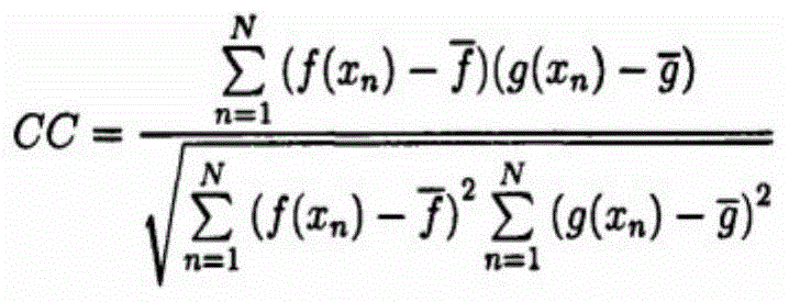
其中
通过上述处理,在本模块下,输入由超声图像优化模块所获得的消除干扰噪声的超声优化图像,进行叠加,随着多幅图像的不断叠加,使得叠加生成的图像中,放大了超声骨质回波信息强回声与弱回声之间差异,输出效果增强、差异明显的消除干扰噪声的超声叠加图像。所述线性强回声为医学超声学概念,是指超声图像的特征性描述,为线性高亮图像。Through the above processing, under this module, the ultrasonic optimized image obtained by the ultrasonic image optimization module to eliminate the interference noise is input and superimposed. The difference between the strong echo and the weak echo of the echo information, the output effect is enhanced, and the difference is obvious to eliminate the ultrasonic superimposed image of the interference noise. The linear strong echo is a medical ultrasound concept, which refers to a characteristic description of an ultrasound image, which is a linear highlight image.
所述超声拓片生成模块用于将所述超声图像叠加模块获得的超声叠加图像处理后形成超声拓片。所述的超声拓片是由超声图像叠加模块获得的超声图像经过三维重建处理后所得到的,是带有患者空间定位信息并随患者位置变动而实时动态更新的个性化三维超声骨骼图像。超声图像的三维重建可以是通过一定的数据处理方法,将超声探头扫描获得的二维图像转换为三维图像的过程,是利用一系列的二维图像重建三维图像模型并进行定性和定量分析的技术,可以实现二维图像中三维结构信息的提取。在三维重建的绘制过程中,数据描述方法主要有表面绘制方法、体绘制方法和混合绘制方法三大类。所述超声拓片生成模块可以采用体绘制方法及体绘制加速技术将脊椎质回波叠加所获得图像进行三维重建,实现二维脊椎表面结构的超声图像向超声拓片的转换,达到准确性与低成本的要求。体绘制方法是直接研究光线通过三维体数据场与体素的相互作用,集中显示皮肤、骨骼、肌肉等物质信息,而无需提取等值面轮廓的方法。体绘制既可以显示体数据的表面信息也可以显示内部信息,其过程包括投射、消隐、渲染和合成等主要步骤。本实施例可以采用体绘制方法为剪切变形法,它将二维离散数据场的投影变换分解为二维数据场剪切变换和二维图像变形两步来实现,从而将二维空间的重采样过程转换成二维平面的重采样过程,极大减少计算量。剪切变形法实现了二维数据场的体绘制可以在图形工作站上以接近实时的速度实现,而不显著降低结果图像的质量。当然体绘制方法仍存在硬件配置要求高、内存需求大、计算量巨大的局限性,基于如上问题,也可以采用体绘制加速技术予以解决。体绘制加速技术包括:基于图像空间的体绘制算法加速、基于物体空间重建算法加速和硬件加速。所述基于图像空间的体绘制算法加速是指,第一,利用图像空间的相关性减少射线的数目。相邻象素存在着相关性,具有相似的颜色,因此并不需图像平面中的所有象素都透射出光线,而是可以间隔地投射出光线。第二,利用对象空间的相关性减少不必要的采样点数目。体数据中会有大量空体元即透明体素存在,这些空体元对体绘制后所产生的最终图像无任何影响,通过构造体数据的存储结构,可以跳过空体元,从而减少采样点数目。所述基于物体空间重建算法加速是指,目前较成熟的算法包括体元投射法、子区域投射法等,此外通过建立特殊的体数据结构,如有序体数据、游程编码等,也可加快对体数据的遍历。所述硬件加速是指,由于超声拓片成像范围仅限全脊柱节段,属于中等规模体数据场,因此基于综合考量,给予硬件加速的纹理绘制算法较优,此算法可提供比其他加速算法更快速的体绘制。未来若需要体数据场的急速扩大,还可引入并行计算的相关算法,进一步实现加速。The ultrasonic rubbing generating module is configured to process the ultrasonic superimposed image obtained by the ultrasonic image superimposing module to form an ultrasonic rubbing. The ultrasonic rubbing is obtained after the ultrasonic image obtained by the ultrasonic image superposition module is processed by three-dimensional reconstruction, and is a personalized three-dimensional ultrasonic bone image with the patient's spatial positioning information and dynamically updated in real time as the patient's position changes. The three-dimensional reconstruction of ultrasonic images can be a process of converting two-dimensional images obtained by scanning an ultrasonic probe into three-dimensional images through certain data processing methods. , which can realize the extraction of three-dimensional structural information in two-dimensional images. In the rendering process of 3D reconstruction, the data description methods mainly include surface rendering methods, volume rendering methods and hybrid rendering methods. The ultrasonic rubbing generation module can use the volume rendering method and the volume rendering acceleration technology to perform three-dimensional reconstruction of the images obtained by superimposing the vertebral echoes, so as to realize the conversion of the ultrasonic images of the two-dimensional vertebral surface structure to the ultrasonic rubbings, so as to achieve accuracy and low cost. requirements. The volume rendering method is to directly study the interaction between light and voxels through the three-dimensional volume data field, and focus on displaying material information such as skin, bone, and muscle without extracting the contour of the isosurface. Volume rendering can display both surface information and internal information of volume data, and its process includes major steps such as projection, concealment, rendering, and compositing. In this embodiment, the volume rendering method can be the shear deformation method, which decomposes the projection transformation of the two-dimensional discrete data field into two steps: the two-dimensional data field shear transformation and the two-dimensional image deformation. The sampling process is converted into a resampling process of a two-dimensional plane, which greatly reduces the amount of calculation. The shear warping method enables volume rendering of a two-dimensional data field to be performed on a graphics workstation at near real-time speed without significantly degrading the quality of the resulting image. Of course, the volume rendering method still has the limitations of high hardware configuration requirements, large memory requirements, and huge computational load. Based on the above problems, volume rendering acceleration technology can also be used to solve it. Volume rendering acceleration technologies include: image space-based volume rendering algorithm acceleration, object space-based reconstruction algorithm acceleration, and hardware acceleration. The acceleration of the volume rendering algorithm based on the image space refers to, first, reducing the number of rays by utilizing the correlation of the image space. Adjacent pixels are correlated and have similar colors, so it is not necessary for all pixels in the image plane to transmit light, but can cast light at intervals. Second, use the correlation of object space to reduce the number of unnecessary sampling points. There will be a large number of empty voxels, that is, transparent voxels, in the volume data. These empty voxels have no effect on the final image generated after volume rendering. By constructing the storage structure of the volume data, the empty voxels can be skipped, thereby reducing sampling. number of points. The acceleration of the reconstruction algorithm based on object space means that the currently more mature algorithms include the voxel projection method, the sub-region projection method, etc. In addition, by establishing a special volume data structure, such as ordered volume data, run-length coding, etc., it can also be accelerated. Traversal of volume data. The hardware acceleration refers to that, since the imaging range of ultrasound rubbings is limited to the whole spinal segment, which belongs to the medium-scale volume data field, based on comprehensive consideration, the texture rendering algorithm given hardware acceleration is better, and this algorithm can provide more advantages than other acceleration algorithms. Fast volume rendering. In the future, if the rapid expansion of the volume data field is required, related algorithms for parallel computing can also be introduced to further accelerate.
下面以剪切变形法为例(图7),阐述其基本原理。剪切变形法的主要思想是把三维离散数据场的直接投影分解为三维数据场的剪切变换和二维图像的变形。如附图7所示,首先,根据视线方向(投影方向),决定一个中间平面,此平面垂直于物空间的某个轴方向,并将物体空间做一个剪切变换,再将物空间垂直投影到中间平面,获得中间图像A’B’。由于是垂直投影,其计算量必然大大低于斜向投影。最后,将得到的中间图像A’B’做一个变形变换,得到最终图像AB。原投影变换的视变换Mview被分解为两个矩阵,即错切矩阵Mshear和变形Mwarp矩阵。因而Mview=Mshear×Mwarp。进一步,对于平行投影,若以一组水平线表示三维离散数据场的一个横切面,使观察方向和坐标系的一个轴平行,垂直于三维数据场的各数据平面,在剪切变换中,各数据平面将平行移动一个距离,该距离不仅与各数据平面所在处的坐标值成正比,而且还决定于对初始观察方向的定义;对于透视投影,当三维离散数据场变换到剪切物体空间时,每一个数据平面不仅要作平移变换,而且还需要作比例变换,因此,由剪切变换后的数据场作投影所得到的图像并非最终图像,而只是中间图像,仍需进行一次图像变形。Taking the shear deformation method as an example (Fig. 7), the basic principle is explained below. The main idea of the shear deformation method is to decompose the direct projection of the three-dimensional discrete data field into the shear transformation of the three-dimensional data field and the deformation of the two-dimensional image. As shown in Figure 7, first, according to the line of sight (projection direction), a middle plane is determined, this plane is perpendicular to a certain axis direction of the object space, the object space is subjected to a shear transformation, and then the object space is vertically projected to the mid-plane, obtaining the intermediate image A'B'. Since it is a vertical projection, the amount of calculation must be much lower than that of the oblique projection. Finally, perform a deformation transformation on the obtained intermediate image A'B' to obtain the final image AB. The view transformation M view of the original projection transformation is decomposed into two matrices, namely the shear matrix M shear and the deformation M warp matrix. Thus M view = M shear × M warp . Further, for parallel projection, if a set of horizontal lines is used to represent a cross-section of a three-dimensional discrete data field, so that the viewing direction is parallel to one axis of the coordinate system, and perpendicular to each data plane of the three-dimensional data field, in shear transformation, each data The plane will move in parallel by a distance that is not only proportional to the coordinate value where each data plane is located, but also depends on the definition of the initial viewing direction; for perspective projection, when the three-dimensional discrete data field is transformed into the clipping object space, Each data plane needs not only translation transformation, but also scale transformation. Therefore, the image obtained by the projection of the data field after shear transformation is not the final image, but only an intermediate image, which still needs to be deformed once.
上述三维离散数据场的剪切变换及二维图像变形可合成为如下的观察变换矩阵:The shear transformation of the above-mentioned 3D discrete data field and the 2D image deformation can be synthesized into the following observation transformation matrix:
Mview=P·S·Mwarp M view =P·S·M warp
式中P是一个坐标变换阵,它使得坐标系的Z轴与三维离散数据场的主要观察方向相重合,S将三维数据场变换到剪切物体空间,Mwarp将剪切物体空间变换到图像空间。对于平行投影而言,剪切是在垂直于Z轴的方向上进行的,故有:In the formula, P is a coordinate transformation matrix, which makes the Z axis of the coordinate system coincide with the main viewing direction of the three-dimensional discrete data field, S transforms the three-dimensional data field to the space of the clipping object, and M warp transforms the space of the clipping object to the image. space. For parallel projection, the shearing is carried out in the direction perpendicular to the Z axis, so there are:

其中SxSy可由Mview的参数求得。对于透视透影,则有:Among them, S x S y can be obtained from the parameters of M view . For perspective perspective, there are:

上式表明,当我们将z=z0的数据平面由物体空间变换到剪切物体空间时,该数据平面应平移(z0s′x,z0s′y)距离,并按系数1/(1+z0s′w)作比例变换。上式中的最后一项Mwarp则是将剪切物体空间变换到图像空间的矩阵。即:The above formula shows that when we transform the data plane of z=z 0 from the object space to the clipping object space, the data plane should be translated by (z 0 s′ x , z 0 s′ y ) distance, and by the coefficient 1/ (1+z 0 s′ w ) for scale transformation. The last term M warp in the above formula is the matrix that transforms the clipping object space into the image space. which is:
Mwarp=S-1·P-1·Mview M warp =S -1 ·P -1 ·M view
根据上述算法,将所述超声图像叠加模块获得的超声图像进行处理,选定Z轴方向为三维离散数据场的主要观察方向,使坐标系的轴与其相重合,对于与其平行的部分进行平行投影,只进行脊椎表面全部骨质回波信息的平移;对于透视投影,则在完成上述平移后,再进行比例变换;所述变换是,在剪切物体空间中,将脊椎表面全部骨质回波信息剪切后投射到剪切物体空间的中间图像平面,形成中间图像,将中间图像平面上的图像通过二维图像变形Mwarp变换到图像空间,得到最终图像。According to the above algorithm, the ultrasonic image obtained by the ultrasonic image superposition module is processed, the Z-axis direction is selected as the main observation direction of the three-dimensional discrete data field, the axis of the coordinate system is coincident with it, and the parallel projection is performed on the part parallel to it. , only the translation of all the bone echo information on the spine surface is performed; for perspective projection, after the above translation is completed, the scale transformation is performed; the transformation is, in the shear object space, the entire spine surface echo information After the information is clipped, it is projected to the intermediate image plane of the clipped object space to form an intermediate image, and the image on the intermediate image plane is transformed into the image space through the two-dimensional image warping M warp to obtain the final image.
通过上述过程,将超声叠加图像通过体绘制方法及加速技术生成为超声拓片,作为带有患者空间定位信息并随患者位置变动而实时动态更新的个性化三维超声骨骼图像,将用于下一步处理。Through the above process, the ultrasonic superimposed image is generated into ultrasonic rubbings by volume rendering method and acceleration technology, as a personalized 3D ultrasonic skeletal image with the patient's spatial positioning information and dynamically updated in real time with the change of the patient's position, which will be used for the next step. .
所述图像轮廓匹配模块用于将所述超声拓片生成模块获得的超声拓片与数字医学影像进行轮廓匹配,形成与患者术中体位一致的实时更新的个性化的脊椎表面地形图。所述超声拓片所具有的实时动态空间信息是指脊椎表面和肌骨界面共同构成的空间信息,该空间信息包含脊椎表面和深层肌肉组织的超声图像信息和空间位置信息,并能实时动态地显示。所述脊椎表面地形图是由超声拓片所具有的实时动态空间信息赋予到数字医学影像而形成的立体表面观,所述立体表面观是基于脊椎表面空间信息形成的表观三维图像,所述的表观三维图像中的沟纹是指所述的全部超声图像信息中包含的脊椎表面的纹理。所述轮廓匹配是指所述超声拓片包含的脊椎表面全部骨质回波信息与数字医学影像的轮廓进行逐点匹配。现以CT容积漫游重建图像为例,阐述轮廓匹配过程。将超声拓片轮廓上所包括的实时动态空间位置信息与CT容积漫游重建图像轮廓上包括空间位置信息,进行对应点的逐一匹配,完成图像轮廓的匹配,进而实现三维层面的匹配。The image contour matching module is used to perform contour matching between the ultrasonic rubbings obtained by the ultrasonic rubbing generating module and the digital medical images to form a real-time updated personalized spine surface topographic map consistent with the patient's intraoperative posture. The real-time dynamic spatial information possessed by the ultrasonic rubbing refers to the spatial information formed by the vertebral surface and the musculoskeletal interface. The spatial information includes the ultrasonic image information and spatial position information of the vertebral surface and the deep musculature, and can be displayed dynamically in real time. . The topographic map of the spine surface is a three-dimensional surface view formed by imparting real-time dynamic spatial information of ultrasonic rubbings to digital medical images, and the three-dimensional surface view is an apparent three-dimensional image formed based on the spatial information of the spine surface. The grooves in the apparent three-dimensional image refer to the texture of the vertebral surface contained in all the ultrasound image information. The contour matching refers to the point-by-point matching of all the bone echo information on the surface of the spine contained in the ultrasonic rubbing with the contour of the digital medical image. Taking CT volume roaming reconstruction image as an example, the contour matching process is described. The real-time dynamic spatial position information included on the contour of the ultrasound rubbing and the spatial position information included in the contour of the CT volume roaming reconstruction image are matched to match the corresponding points one by one to complete the matching of the image contour, and then realize the three-dimensional level matching.
所述轮廓匹配的获取方法可以是将所述超声拓片包含的脊椎表面全部骨质回波信息与数字医学影像的轮廓进行逐点匹配,获得与患者术中体位一致的实时更新的个性化的脊椎表面地形图,所述轮廓匹配的方法优选基于像素灰度算法中的互信息算法的多模态配准方法,所述的多模态配准方法包括图像的空间变换、灰度插值、相似度衡量、搜索优化。本实施例中采用的轮廓匹配优选基于像素灰度算法中的互信息算法的多模态配准方法,所述的多模态配准方法包括脊椎图像的空间变换、灰度插值、相似度衡量、搜索优化。所述互信息算法是通过计算待配准图像的独立熵及联合熵,得到互信息值,进而通过互信息值判断图像配准效果:当两个图像重合度最高时,联合熵最小,互信息值最大,达到最佳配准效果;互信息算法利用的是图像中灰度的统计信息,受到噪声的干扰较少,具有较好的鲁棒性,所述鲁棒性是指控制系统在一定结构、大小的参数摄动下,维持某些性能的特性,尽管超声与CT容积漫游成像的成像方式原理不同,但是在图像中相应位置的灰度分布是类似的,不同模态图像的密度变化是否成线性关系与求解互信息无关,此外,因脊柱各椎体可以看作近似刚体,故脊椎图像匹配属于刚体配准,刚体不易产生形变的特性使得互信息算法在刚体配准中的效果更好。The acquisition method of the contour matching may be to perform point-by-point matching of all the bone echo information on the spine surface contained in the ultrasonic rubbing with the contour of the digital medical image, so as to obtain a real-time updated personalized spine that is consistent with the patient's intraoperative posture. Surface topographic map, the method for contour matching is preferably a multimodal registration method based on the mutual information algorithm in the pixel grayscale algorithm, and the multimodal registration method includes image spatial transformation, grayscale interpolation, similarity Measurement, search optimization. The contour matching used in this embodiment is preferably a multimodal registration method based on the mutual information algorithm in the pixel grayscale algorithm. The multimodal registration method includes spatial transformation of the spine image, grayscale interpolation, and similarity measurement. , search optimization. The mutual information algorithm obtains the mutual information value by calculating the independent entropy and joint entropy of the images to be registered, and then judges the image registration effect by the mutual information value: when the coincidence degree of the two images is the highest, the joint entropy is the smallest, and the mutual information The maximum value can achieve the best registration effect; the mutual information algorithm uses the statistical information of the gray level in the image, which is less disturbed by noise and has better robustness. The robustness refers to the control system in a certain Under the perturbation of the parameters of structure and size, some performance characteristics are maintained. Although the imaging principles of ultrasound and CT volume roaming imaging are different, the grayscale distribution of the corresponding position in the image is similar, and the density of different modal images changes. Whether the relationship is linear or not has nothing to do with solving mutual information. In addition, since each vertebral body of the spine can be regarded as an approximate rigid body, the spine image matching belongs to rigid body registration, and the rigid body is not easy to deform, which makes the mutual information algorithm more effective in rigid body registration. it is good.
图像匹配过程主要包括:图像的空间变换、灰度插值、相似度衡量、搜索优化,在此过程中,需要选择空间变换策略、灰度插值算法、相似性测度方法和优化算法。所述图像的空间变换是指通过一定的规律将图像f(x,y)中的像素点变换到新的位置,从而得到一幅新的图像,f(x′,y′),即

其中θ为图像旋转角度,dy、dx为图像在z轴方向和y轴方向的平移量。在刚体变换中,图像中物体的相对位置没有发生变化。所述仿射变换是一种保持图形的平直性和平行性的空间变换;所述平直性是指变换后图像中直线不会发生弯曲,圆弧弧度也不会改变;所述平行性是指图像中物体间的平行关系不会发生变换的空间变换,但是物体的大小、物体之间的角度可能会发生变化;所述仿射变换是指,相对刚体变换,增加了变换的自由度,即在各坐标方向上的缩放系数,其中当各方向缩放比例一致时,称为均匀仿射变换,否则为非均匀仿射变换或称为剪切变换;均匀仿射变换常用于透镜成像系统,该系统所得到的图像大小与拍摄中物体与摄像头的距离有密切关系,剪切变换常常用于矫正由于仪器原因造成的图像畸变。仿射变换表达式如下:where θ is the rotation angle of the image, and dy and dx are the translations of the image in the z-axis and y-axis directions. In a rigid body transformation, the relative positions of objects in the image are not changed. The affine transformation is a space transformation that maintains the straightness and parallelism of the graph; the straightness means that the straight line in the transformed image will not be bent, and the arc of the arc will not change; the parallelism It refers to the spatial transformation in which the parallel relationship between objects in the image will not be transformed, but the size of the object and the angle between the objects may change; the affine transformation refers to the relative rigid body transformation, which increases the degree of freedom of transformation , that is, the scaling factor in each coordinate direction, when the scaling ratio of each direction is the same, it is called uniform affine transformation, otherwise it is non-uniform affine transformation or shear transformation; uniform affine transformation is often used in lens imaging systems , the size of the image obtained by the system is closely related to the distance between the object and the camera in the shooting, and the shear transformation is often used to correct the image distortion caused by the instrument. The affine transformation expression is as follows:
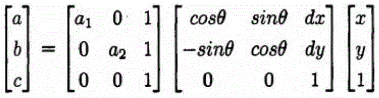
在多模态图像配准中,仿射变换可用于:第一,纠正由于成像方法不同导致的图像中物体比例的不一致;第二,纠正人为操作因素导致的图像畸变;第三,可用于解决成像角度不同导致的图像表现不同。In multimodal image registration, affine transformation can be used to: firstly, to correct the inconsistency of object proportions in the image due to different imaging methods; secondly, to correct image distortion caused by human manipulation factors; thirdly, it can be used to solve Different imaging angles result in different image performance.
所述非线性变换是指,对于人体软组织图像,由于不同时刻不同角度拍摄到的组织往往会发生扭曲形变,在配准中常常采用非线性变换;由于超声扫描过程中,与探头部位接触的人体组织会产生较小尺度的局部形变,因此适用于弹性模型;所述弹性模型是指,可将浮动图像到模板图像的变换视为弹性材料伸缩的过程,整个过程中有外部对材料施加的外力和材料内部反弹的内力,其中外力导致材料发生形变,而内力是材料在受外力过程中对外力的反馈,当内力和外力达到平衡时配准结束;其形变可以通过下面的Navier偏微分方程来描述:The nonlinear transformation refers to that for soft tissue images of the human body, since the tissues captured at different times and angles tend to be distorted, nonlinear transformation is often used in registration; Tissue will produce small-scale local deformation, so it is suitable for elastic model; the elastic model refers to that the transformation from floating image to template image can be regarded as the process of elastic material expansion and contraction, and there are external forces applied to the material throughout the process. and the internal force of the internal rebound of the material, where the external force causes the material to deform, and the internal force is the feedback of the external force in the process of the external force, and the registration ends when the internal force and the external force reach equilibrium; the deformation can be obtained by the following Navier partial differential equation describe:

其中μ(x,y)表示物体在x和y方向上的形变,λ和
所述灰度插值算法是在完成匹配过程,对浮动图像进行空间变换后,原来位于整数坐标的像素在新图像中的坐标可能会变为非整数,为解决像素点不在整数坐标的问题,更好的描述形变后的图像,我们采用灰度插值算法;配准过程需要对浮动图像进行一定的空间变换,使其尽量的与模板图像相似,浮动图像进行变换后,一般将新图像中的整数坐标点进行反变换,得到该点在原图像中的对应坐标,则所得该点为即为插值点;灰度插值算法可通过插值点及其周围点的灰度值计算得到新图像中坐标的灰度值,从而最大程度的保持图像中原有的信息;在利用互信息进行配准的算法中,为了充分保留图像的灰度分布信息,常使用PV插值算法。所述PV插值算法是通过在插值直接得到新图像的灰度分布,因为进行空间变换前的浮动图像中每个像素都对图像的灰度分布有一定的贡献,因此PV插值考虑了所有插值点及其周围邻域像素的灰度值。一般在灰度直方图的计算当中,每统计到灰度值n0出现一次,那么直方图中对应着伽的数值便要加1。与双线性插值类似,新图像中每个像素点(x,y)都对应着原图像中一个插值点(x′,y′),设插值点周围的四个整数点灰度值为n11、n12、n21、n22,,并根据插值点到这些点的距离求得每个点的权重wij。那么新图像的灰度直方图h(a)中,灰度值nij的出现次数要相应的加上wij,即:The gray-scale interpolation algorithm is that after completing the matching process and performing spatial transformation on the floating image, the coordinates of pixels originally located at integer coordinates in the new image may become non-integer. To describe the deformed image well, we use the grayscale interpolation algorithm; the registration process needs to perform a certain spatial transformation on the floating image to make it as similar to the template image as possible. After the floating image is transformed, the integers in the new image are generally converted. The coordinate point is inversely transformed to obtain the corresponding coordinates of the point in the original image, then the obtained point is the interpolation point; the grayscale interpolation algorithm can calculate the grayscale of the coordinates in the new image by calculating the grayscale values of the interpolation point and its surrounding points. In order to keep the original information in the image to the greatest extent; in the registration algorithm using mutual information, in order to fully retain the gray distribution information of the image, the PV interpolation algorithm is often used. The PV interpolation algorithm directly obtains the grayscale distribution of the new image through interpolation, because each pixel in the floating image before spatial transformation has a certain contribution to the grayscale distribution of the image, so PV interpolation considers all interpolation points. and the gray value of the surrounding pixels. Generally, in the calculation of the grayscale histogram, every time the grayscale value n 0 appears once, the value corresponding to Ga in the histogram will be increased by 1. Similar to bilinear interpolation, each pixel point (x, y) in the new image corresponds to an interpolation point (x', y') in the original image, and the gray value of the four integer points around the interpolation point is n. 11 , n 12 , n 21 , n 22 , and the weight w ij of each point is obtained according to the distance from the interpolation point to these points. Then in the grayscale histogram h(a) of the new image, the number of occurrences of the grayscale value n ij should be correspondingly added w ij , namely:
h(nij)=h(nij)+wij h(n ij )=h(n ij )+w ij
设模板图像中灰度为m,那么插值点在联合灰度直方图H(a,b)中的计算为:Assuming that the grayscale in the template image is m, then the calculation of the interpolation point in the joint grayscale histogram H(a, b) is:
H(m,nij)=H(m,nij)+wij H(m, n ij )=H(m, n ij )+w ij
当计算完所有插值点后便得到新的灰度直方图,从而得到浮动图像变换后的边际灰度分布和联合灰度分布。PV插值算法利用直接改变图像的灰度统计信息来代替直接插值产生新的图像,比较适合像互信息之类对灰度分布十分敏感的配准算法。When all the interpolation points are calculated, a new grayscale histogram is obtained, thereby obtaining the marginal grayscale distribution and joint grayscale distribution of the floating image. The PV interpolation algorithm uses directly changing the grayscale statistical information of the image to replace the direct interpolation to generate a new image, which is more suitable for the registration algorithm that is very sensitive to the grayscale distribution, such as mutual information.
所述相似度衡量是指,在医学图像配准,特别是多模态医学图像的配准中,需要定义一个衡量两幅图像之间相似度的标准,当两幅图像相似度达到最大时,即认为两幅图像达到最佳的配准效果;作为优选,完成上述图像空间变换和灰度插值算法后,我们将选择基于像素灰度算法中的互信息算法完成相似度测量。以灰度值为基础,待配准的两幅图像A和B,其对应点的灰度值特征组合为灰度对,互信息的计算就是计算图像A中包含图像B多少信息。当两幅图像达到最佳匹配时,他们的灰度分布将具有最大相关性。The similarity measure means that in the registration of medical images, especially the registration of multimodal medical images, a standard for measuring the similarity between two images needs to be defined. When the similarity between the two images reaches the maximum, That is to say, it is considered that the two images achieve the best registration effect; as an option, after completing the above-mentioned image space transformation and grayscale interpolation algorithm, we will choose the mutual information algorithm based on the pixel grayscale algorithm to complete the similarity measurement. Based on the gray value, the gray value features of the two images A and B to be registered are combined into a gray value pair, and the calculation of mutual information is to calculate how much information image A contains in image B. When two images are best matched, their grayscale distributions will have the greatest correlation.
由于经典互信息算法在图像配准中具有明显缺陷——忽略了图像中存在的空间信息,因此在用互信息来衡量两幅图像相似度时会忽略两幅图像间可能共有的一些空间关系,导致在某些情况下出现配准错误。而基于经典算法进行改进又不能避免巨大计算量带来的计算时间延长,因而我们选择将空间信息引入互信息计算的方法——条件互信息;所述条件互信息是将待配准图片划分为不同的小区域,通过整合两幅图像之间所有对应小区域之间的配准效果来得到整体图像的配准效果,在不增加运算量的同时考虑到了图像中的空间信息;条件互信息CMI是通过扩展联合直方图,将图像中小区域的空间分布与灰度分布相结合而得到的:Since the classical mutual information algorithm has obvious defects in image registration - ignoring the spatial information existing in the image, when using mutual information to measure the similarity of two images, some spatial relationships that may be shared between the two images will be ignored. Causes registration errors in some cases. However, the improvement based on the classical algorithm cannot avoid the prolonged calculation time caused by the huge amount of calculation, so we choose the method of introducing spatial information into mutual information calculation - conditional mutual information; the conditional mutual information is to divide the image to be registered into For different small areas, the registration effect of the whole image is obtained by integrating the registration effect between all the corresponding small areas between the two images, and the spatial information in the image is taken into account without increasing the amount of computation; conditional mutual information CMI is obtained by extending the joint histogram by combining the spatial distribution of small regions in the image with the grayscale distribution:

其中p(s)表示在配准过程中,选取到图像中的区域s进行配准的概率,若将待配准图像划分为相同大小的区域,该概率是相同的。在一幅图像,中灰度值为i的像素点可能分布在整幅图像的不同区域。在条件互信息的计算时,分布在不同区域的灰度值为i的像素点对整体互信息量的贡献与该点空间位置有关。条件互信息表示的是,在区域s中,图像A包含的图像B的信息量,把所有区域的信息量的加权平均值作为最终衡量图像相似度的标准。Among them, p(s) represents the probability of selecting the region s in the image for registration during the registration process. If the to-be-registered image is divided into regions of the same size, the probability is the same. In an image, the pixels whose gray value is i may be distributed in different areas of the whole image. In the calculation of conditional mutual information, the contribution of pixels with gray value i distributed in different regions to the overall mutual information is related to the spatial position of the point. Conditional mutual information represents the information amount of image B contained in image A in region s, and the weighted average of the information amounts of all regions is used as the final standard to measure the similarity of images.
通过上述过程,在本模块下,输入由超声拓片生成模块获得的超声拓片,完成所述超声拓片与数字医学影像的轮廓匹配,输出与患者术中体位一致的实时更新的个性化的脊椎表面地形图,此图像将帮助操作者观察分析患者在术中体位下,与手术相关解剖结构的位置关系,指导穿刺定位及后续手术操作。Through the above process, under this module, input the ultrasonic rubbing obtained by the ultrasonic rubbing generation module, complete the contour matching between the ultrasonic rubbing and the digital medical image, and output the real-time updated personalized spine surface topography consistent with the patient's intraoperative posture This image will help the operator observe and analyze the positional relationship between the patient's intraoperative position and the anatomical structures related to the operation, and guide the puncture positioning and subsequent surgical operations.
如图4所示,本发明提供的一种基于超声拓片技术的脊柱手术导航定位系统,所述的脊柱手术导航定位系统能够获得与患者术中体位一致的实时更新的个性化的脊椎表面地形图,并基于该脊椎表面地形图进行实时的术中导航,所述脊柱手术导航定位系统包括导航模块和基于超声拓片技术的脊椎图像生成系统,其中:As shown in FIG. 4, the present invention provides a spinal surgery navigation and positioning system based on ultrasonic rubbing technology. The spinal surgery navigation and positioning system can obtain a real-time updated personalized spine surface topographic map consistent with the patient's intraoperative posture , and perform real-time intraoperative navigation based on the topographic map of the spine surface. The spine surgery navigation and positioning system includes a navigation module and a spine image generation system based on ultrasonic rubbing technology, wherein:
所述的基于超声拓片技术的脊椎图像生成系统,是基于患者手术状况下实时的术中体位对应的二维脊椎表面结构超声图像处理后形成超声拓片,然后将所述超声拓片与数字医学影像进行轮廓匹配,形成与患者术中体位一致的实时更新的个性化的脊椎表面地形图,所述的超声拓片是带有患者空间定位信息并随患者位置变动而实时动态更新的个性化三维超声骨骼图像。所述的数字医学影像可以选自CT容积漫游重建图像、磁共振MR、计算机X线摄影CR、数字化计算机X线摄影DR。The described spine image generation system based on ultrasonic rubbing technology is based on the real-time intraoperative position of the patient under the condition of surgery to form ultrasonic rubbings after the ultrasonic image processing of the two-dimensional vertebral surface structure corresponding to the intraoperative position, and then the ultrasonic rubbings are processed with digital medical images. Contour matching to form a real-time updated personalized spine surface topographic map consistent with the patient's intraoperative posture, and the ultrasonic rubbing is a personalized three-dimensional ultrasonic skeletal image with the patient's spatial positioning information and dynamically updated in real time with the patient's position change . The digital medical image can be selected from CT volumetric roaming reconstruction images, magnetic resonance MR, computed radiography CR, and digital computed radiography DR.
所述的导航模块基于与患者术中体位一致的实时更新的个性化的脊椎表面地形图包含的空间位置信息建立地形图坐标系,建立带有定位功能的手术器械的实体几何模型并向表面模型转化,并呈现在脊椎图像生成系统中获得的与患者术中体位一致的实时更新的个性化的脊椎表面地形图上;以患者术中体位的空间位置信息建立患者坐标系,确定地形图坐标系和患者坐标系中对应点的关系,形成统一坐标系;将转化获得的手术器械的表面模型融入统一坐标系,实现术中实时导航,指导手术操作者进行操作。所述带有定位功能的手术器械为带有空间磁定位标记的手术器械。The navigation module establishes a topographic map coordinate system based on the spatial position information contained in the real-time updated personalized spine surface topographic map consistent with the patient's intraoperative posture, and establishes a solid geometric model of a surgical instrument with a positioning function to the surface model. Convert and present on the real-time updated personalized spine surface topographic map obtained in the spine image generation system that is consistent with the patient's intraoperative posture; establish the patient coordinate system based on the spatial position information of the patient's intraoperative posture, and determine the topographic map coordinate system The relationship with the corresponding points in the patient coordinate system forms a unified coordinate system; the surface model of the surgical instrument obtained by transformation is integrated into the unified coordinate system to realize real-time intraoperative navigation and guide the surgical operator to operate. The surgical instrument with positioning function is a surgical instrument with spatial magnetic positioning marks.
如图5所示,本发明提供的一种基于超声拓片技术的脊柱手术导航定位方法,步骤如下:As shown in Figure 5, the present invention provides a spinal surgery navigation and positioning method based on ultrasonic rubbing technology, the steps are as follows:
第一步,操作者选择符合手术适应症的患者准备手术。所述手术适应症包括脊柱相关疾病如脊柱肿瘤、椎间盘突出,符合诊疗常规所规定的标准,采用非手术治疗方式无法治愈疾病,采用手术方式将有助于疾病的治疗时,所应采用的手术方式。In the first step, the operator selects patients who meet the surgical indications to prepare for surgery. The surgical indications include spinal-related diseases such as spinal tumors and intervertebral disc herniation, which meet the standards stipulated in the diagnosis and treatment routine, and the disease cannot be cured by non-surgical treatment. Way.
第二步,对拟进行手术治疗的患者进行术前CT扫描,并将扫描所得图像进行容积漫游重建,获得该患者的数字医学影像。此处在进行CT扫描时,患者所选择的体位应与手术拟采用的体位一致,例如,采用拟俯卧位进行手术的患者,在术前CT扫描时也采用俯卧位进行扫描。In the second step, a preoperative CT scan is performed on a patient to be surgically treated, and a volume roaming reconstruction is performed on the scanned image to obtain a digital medical image of the patient. Here, when performing CT scanning, the patient's position should be consistent with the position to be used for surgery. For example, a patient who is to undergo surgery in a prone position should also be scanned in a prone position during preoperative CT scanning.
第三步,根据患者的CT扫描和容积漫游重建图像,操作者团队进行术前分析,确定手术靶点。所述手术靶点是指手术时拟抵达的脊椎骨性解剖位置,例如,采用脊柱内镜进行经椎板后路髓核摘除术这一手术方式治疗腰椎间盘突出症,手术靶点为目标腰椎的椎板结构。In the third step, based on the patient's CT scan and volume roaming reconstruction images, the operator team conducts preoperative analysis to determine the surgical target. The surgical target refers to the vertebral anatomical position to be reached during the operation. For example, in the surgical treatment of lumbar intervertebral disc herniation using spinal endoscopy through posterior laminectomy, the surgical target is the target lumbar spine. Laminar structure.
第四步,常规手术准备及麻醉后,操作者对手术目标区域进行超声扫描,通过带有患者空间定位信息的超声扫描设备在患者体表反复扫描,直到识别肌骨界面,提取包含术中二维空间位置参数的脊椎表面全部骨质回波信息,构成患者手术状况下实时的术中体位对应的二维脊椎表面结构超声图像,所述的术中二维空间位置参数是指患者手术状况下脊椎实时的二维空间位置参数。所述带有患者空间定位信息的超声扫描设备可以是带有定位标签的超声探头。所述手术目标区域指进行手术的相关区域,包括进行手术的具体部位及其毗邻结构,例如,采用脊柱内镜进行腰椎间盘突出症手术,目标区域为病变髓核所在的腰椎椎体、相邻上下位的椎体以及相应腰椎的椎旁区域。所述超声图像由包含脊柱区深层肌肉组织和脊椎表面的全部回波信息构成,所述的超声图像包括所述超声图像轮廓和超声图像边缘的信息。所述脊椎表面包括横突和棘突,还可以包括关节突、椎板、椎板间隙、椎间孔以及组成脊椎的其他骨骼成分的任意一种或任意多种。In the fourth step, after routine surgical preparation and anesthesia, the operator performs ultrasonic scanning on the surgical target area, and repeatedly scans the patient's body surface through the ultrasonic scanning device with the patient's spatial positioning information until the musculoskeletal interface is identified. All bone echo information on the spine surface of the dimensional space position parameter constitutes the real-time intraoperative ultrasound image of the spinal surface structure corresponding to the patient's intraoperative posture. The intraoperative 2D spatial position parameter refers to the patient's surgical condition. Real-time 2D spatial position parameters of the spine. The ultrasound scanning device with patient spatial positioning information may be an ultrasound probe with positioning labels. The surgical target area refers to the relevant area for the operation, including the specific part of the operation and its adjacent structures. For example, when using spinal endoscopy to perform lumbar disc herniation surgery, the target area is the lumbar vertebral body where the diseased nucleus pulposus is located, and the adjacent structures. The upper and lower vertebral bodies and the paravertebral regions of the corresponding lumbar vertebrae. The ultrasound image is composed of all echo information including the deep muscle tissue in the spinal region and the surface of the spine, and the ultrasound image includes the information of the contour of the ultrasound image and the edge of the ultrasound image. The vertebral surface includes transverse processes and spinous processes, and may also include any one or more of articular processes, lamina, lamina spaces, intervertebral foramen, and other skeletal components that make up the vertebra.
步骤四可以包括如下步骤:Step 4 may include the following steps:
超声图像扫描:通过带有患者空间定位信息的超声扫描设备在患者体表反复扫描,直到识别肌骨界面,获取原始数据,所述的原始数据是指带有患者空间定位信息的超声扫描设备发出的射频信号,所述的扫描允许以各种不同角度和方向进行,所述的方向包括上下、前后、左右、斜向,所述的角度包括相对于患者体表的垂直或任意倾斜角,并且与扫描先后顺序无关。带有患者空间定位信息的超声扫描设备可以是带有定位标签的超声探头扫描获取原始数据,所述的原始数据是指超声探头发出的射频信号;Ultrasound image scanning: Repeatedly scan the patient's body surface through the ultrasonic scanning equipment with the patient's spatial positioning information until the musculoskeletal interface is identified, and obtain the original data. the radio frequency signal, the scanning is allowed to be carried out at various angles and directions, the directions include up and down, front and rear, left and right, oblique, and the angles include vertical or arbitrary inclination angles relative to the patient's body surface, and The order of scanning is irrelevant. The ultrasound scanning device with the patient's spatial positioning information may scan an ultrasound probe with a positioning label to obtain raw data, where the raw data refers to the radio frequency signal emitted by the ultrasound probe;
超声图像信息提取:从超声图像扫描获取的原始数据中提取包含术中二维空间位置参数的脊椎表面全部骨质回波信息,构成患者手术状况下实时的术中体位对应的二维脊椎表面结构超声图像,所述的术中二维空间位置参数是指患者手术状况下脊椎实时的二维空间位置参数。Ultrasound image information extraction: Extract all the bone echo information of the spine surface including the intraoperative two-dimensional spatial position parameters from the original data obtained by the ultrasound image scanning, and form the two-dimensional spine surface structure corresponding to the real-time intraoperative posture of the patient under the surgical condition. Ultrasound image, the intraoperative two-dimensional spatial position parameter refers to the real-time two-dimensional spatial position parameter of the spine under the patient's operating condition.
第五步,将所述患者手术状况下实时的术中体位对应的二维脊椎表面结构超声图像进行优化、叠加后,进一步处理生成超声拓片。The fifth step is to optimize and superimpose the real-time two-dimensional ultrasonic images of the vertebral surface structure corresponding to the intraoperative posture of the patient under the operating conditions, and then further process to generate ultrasonic rubbings.
步骤五可以包括如下步骤:
超声图像优化:将步骤四获得的患者手术状况下实时的术中体位对应的二维脊椎表面结构超声图像进行优化,获得消除干扰噪声的超声优化图像;优选的,超声图像优化采用超声滤波增强技术,获得消除干扰噪声的超声优化图像;更优选的,所述的超声滤波增强技术可以是中值滤波和数学形态学滤波相结合的方法。Ultrasound image optimization: optimize the real-time two-dimensional vertebral surface structure ultrasound image corresponding to the intraoperative posture of the patient obtained in step 4 to obtain an ultrasound optimized image that eliminates interference noise; preferably, the ultrasound image optimization adopts the ultrasonic filter enhancement technology , to obtain an ultrasound-optimized image that eliminates interference noise; more preferably, the ultrasound filtering enhancement technique may be a method combining median filtering and mathematical morphology filtering.
超声图像叠加:将所述超声图像优化后的超声图像进行叠加,使超声骨质回波信息强回声与弱回声之间的差别更大,达到增强图像效果,所述的超声图像叠加可以是单模态配准方法。Ultrasound image superposition: the optimized ultrasonic images are superimposed to make the difference between the strong echo and the weak echo of the ultrasonic bone echo information larger, so as to achieve the effect of enhancing the image. The ultrasonic image superposition can be a single Modal registration method.
超声拓片生成:将所述超声图像优化、叠加后获得的超声图像进行三维重建处理,生成超声拓片,所述的三维重建的方法可以是体绘制方法以及体绘制加速技术。Ultrasound rubbing generation: performing three-dimensional reconstruction processing on the ultrasound images obtained after the ultrasound images are optimized and superimposed to generate ultrasound rubbings. The three-dimensional reconstruction method may be a volume rendering method and a volume rendering acceleration technology.
第六步,将步骤五获得的所述超声拓片与数字医学影像进行轮廓匹配,通过超声拓片中包含的脊椎表面全部骨质回波信息与数字医学影像的轮廓进行逐点匹配,得到与患者术中体位一致的实时更新的个性化的脊椎表面地形图。所述的轮廓匹配的方法优是基于像素灰度算法中的互信息算法的多模态配准方法,所述的多模态配准方法包括图像的空间变换、灰度插值、相似度衡量、搜索优化。所述轮廓匹配是指所述超声拓片包含的脊椎表面全部骨质回波信息与数字医学影像的轮廓进行逐点匹配,所述脊椎表面地形图是由超声拓片所具有的实时动态空间信息赋予到数字医学影像而形成的立体表面观,所述超声拓片所具有的实时动态空间信息是指脊椎表面和肌骨界面共同构成的空间信息,该空间信息包含脊椎表面和深层肌肉的超声图像信息和空间位置信息,并能实时动态地显示,所述立体表面观是基于脊椎表面空间信息形成的表观三维图像,所述的表观三维图像中的沟纹是指所述的全部超声图像信息中包含的脊椎表面的纹理。所述实时动态空间信息包含超声扫描所获得的脊椎表面、深层肌肉和脂肪组织的超声图像信息和空间位置信息,并能随带有定位标签的超声探头的移动而动态地实时显示。所述的数字医学影像是CT容积漫游重建图像、磁共振MR、计算机X线摄影CR、数字化计算机X线摄影DR。The sixth step is to perform contour matching between the ultrasonic rubbing obtained in
第七步,建立手术器械的实体几何模型,实现手术器械的实体几何模型向表面模型的转化,使其在脊椎图像生成系统中所获得的与患者术中体位一致的实时更新的个性化的脊椎表面地形图上呈现出来,实现对手术器械的实时跟踪。所述实体几何模型向表面模型转化的方法优选为三维CAD造型法。The seventh step is to establish the solid geometric model of the surgical instrument, and realize the transformation of the solid geometric model of the surgical instrument to the surface model, so that the real-time updated personalized spine obtained in the spine image generation system is consistent with the patient's intraoperative posture. It is displayed on the surface topographic map to realize real-time tracking of surgical instruments. The method for converting the solid geometric model to the surface model is preferably a three-dimensional CAD modeling method.
第八步,根据与患者术中体位一致的实时更新的个性化的脊椎表面地形图包含的空间位置信息建立地形图坐标系,以患者术中体位的空间位置信息建立患者坐标系,确定地形图坐标系和患者坐标系中对应点的关系,并形成统一坐标系。所述确定地形图坐标系和患者坐标系中对应点关系的方法优选为最近点迭代算法。The eighth step is to establish a topographic map coordinate system according to the spatial position information contained in the real-time updated personalized spine surface topographic map consistent with the patient's intraoperative posture, and establish a patient coordinate system based on the spatial position information of the patient's intraoperative posture to determine the topographic map. The relationship between the coordinate system and the corresponding points in the patient coordinate system, and form a unified coordinate system. The method for determining the relationship between the topographic map coordinate system and the corresponding point in the patient coordinate system is preferably the closest point iterative algorithm.
第九步,将转化获得的手术器械的表面模型在统一坐标系显示出来,实现手术器械和与患者术中体位一致的实时更新的个性化的脊椎表面地形图处于统一坐标系中,实现术中实时导航。In the ninth step, the transformed surface model of the surgical instrument is displayed in a unified coordinate system, so that the surgical instrument and the real-time updated personalized spine surface topographic map consistent with the patient's intraoperative posture are in a unified coordinate system, so as to realize intraoperative operation. Live navigation.
第十步,基于导航图像,操作者将在统一坐标系中,根据坐标系所显示的手术器械与患者脊柱解剖结构间的位置关系,进行后续操作。在此步骤中,操作者基于与患者术中体位一致的实时更新的个性化的脊椎表面地形图的指引,利用带有定位标记的手术器械进行徒手或机械臂辅助下定位穿刺成功后,继续依靠导航图像,根据图像所示手术器械与脊柱骨性结构位置间的位置关系,结合操作者所见的解剖结构,在二者相互印证下,完成后续手术操作。以采用脊柱内镜进行经椎板后路髓核摘除术治疗腰椎间盘突出症为例,其后续手术操作为,操作者根据临床经验辨别棘突、黄韧带、椎板、神经根、椎间盘等解剖结构,在脊柱手术导航定位系统指导下,操作者运用手术器械,按照正确的角度和距离进行操作,直至抵达并摘除突出的髓核或纤维环等完成相关手术内容。In the tenth step, based on the navigation image, the operator will perform subsequent operations in the unified coordinate system according to the positional relationship between the surgical instrument displayed by the coordinate system and the patient's spinal anatomy. In this step, based on the guidance of the real-time updated personalized spine surface topographic map consistent with the patient's intraoperative posture, the operator uses surgical instruments with positioning markers to perform manual or robotic arm-assisted positioning and puncture. After successful positioning and puncture, continue to rely on Navigation image, according to the positional relationship between the surgical instrument and the position of the spinal bony structure shown in the image, combined with the anatomical structure seen by the operator, under the mutual verification of the two, the follow-up operation is completed. Taking the use of spinal endoscopy for the treatment of lumbar intervertebral disc herniation through posterior laminectomy as an example, the follow-up operation is as follows: the operator identifies the spinous process, ligamentum flavum, lamina, nerve root, intervertebral disc and other anatomy according to clinical experience. Under the guidance of the spinal surgery navigation and positioning system, the operator uses surgical instruments to operate according to the correct angle and distance until the protruding nucleus pulposus or annulus fibrosus is reached and removed to complete the relevant surgical content.
Claims (48)
Priority Applications (4)
| Application Number | Priority Date | Filing Date | Title |
|---|---|---|---|
| CN201710630811.3A CN107595387B (en) | 2017-07-28 | 2017-07-28 | A spine image generation system and spine surgery navigation and positioning system based on ultrasonic rubbing technology |
| JP2020504215A JP7162793B2 (en) | 2017-07-28 | 2018-07-25 | Spine Imaging System Based on Ultrasound Rubbing Technology and Navigation/Localization System for Spine Surgery |
| PCT/CN2018/096999 WO2019020048A1 (en) | 2017-07-28 | 2018-07-25 | Spinal image generation system based on ultrasonic rubbing technique and navigation positioning system for spinal surgery |
| US16/752,633 US11304680B2 (en) | 2017-07-28 | 2020-01-25 | Spinal image generation system based on ultrasonic rubbing technique and navigation positioning system for spinal surgery |
Applications Claiming Priority (1)
| Application Number | Priority Date | Filing Date | Title |
|---|---|---|---|
| CN201710630811.3A CN107595387B (en) | 2017-07-28 | 2017-07-28 | A spine image generation system and spine surgery navigation and positioning system based on ultrasonic rubbing technology |
Publications (2)
| Publication Number | Publication Date |
|---|---|
| CN107595387A CN107595387A (en) | 2018-01-19 |
| CN107595387B true CN107595387B (en) | 2020-08-07 |
Family
ID=61059985
Family Applications (1)
| Application Number | Title | Priority Date | Filing Date |
|---|---|---|---|
| CN201710630811.3A Active CN107595387B (en) | 2017-07-28 | 2017-07-28 | A spine image generation system and spine surgery navigation and positioning system based on ultrasonic rubbing technology |
Country Status (4)
| Country | Link |
|---|---|
| US (1) | US11304680B2 (en) |
| JP (1) | JP7162793B2 (en) |
| CN (1) | CN107595387B (en) |
| WO (1) | WO2019020048A1 (en) |
Families Citing this family (59)
| Publication number | Priority date | Publication date | Assignee | Title |
|---|---|---|---|---|
| CN107595387B (en) * | 2017-07-28 | 2020-08-07 | 浙江大学 | A spine image generation system and spine surgery navigation and positioning system based on ultrasonic rubbing technology |
| CN108335326B (en) * | 2018-01-31 | 2022-07-26 | 上海联影医疗科技股份有限公司 | Calibration method, device and system for puncture system |
| CN110368026B (en) * | 2018-04-13 | 2021-03-12 | 北京柏惠维康医疗机器人科技有限公司 | Operation auxiliary device and system |
| CN108765473B (en) * | 2018-05-24 | 2021-12-17 | 常州市速瑞医疗科技有限公司 | Image registration method of full-high-definition 3D electronic laparoscope system |
| CN108670301B (en) * | 2018-06-06 | 2020-11-06 | 西北工业大学 | A method for locating the transverse process of the spine based on ultrasound images |
| CN108711187B (en) * | 2018-08-03 | 2021-12-31 | 华侨大学 | Method for establishing human lumbar vertebra three-dimensional simulation model by registering and fusing CT (computed tomography) and MRI (magnetic resonance imaging) signals |
| CN109646089B (en) * | 2019-01-15 | 2021-04-13 | 浙江大学 | Intelligent positioning system and method for spinal cord body surface puncture access point based on multi-mode medical fusion image |
| CN111821032B (en) * | 2019-04-16 | 2022-06-07 | 上银科技股份有限公司 | Medical Image Alignment Method |
| TWI715075B (en) | 2019-06-24 | 2021-01-01 | 瑞昱半導體股份有限公司 | Skin tone image gamut weight detecting method and device thereof |
| CN110464460B (en) * | 2019-07-16 | 2020-11-17 | 江苏霆升科技有限公司 | Method and system for cardiac intervention operation |
| CN110505383B (en) * | 2019-08-29 | 2021-07-23 | 重庆金山医疗技术研究院有限公司 | Image acquisition method, image acquisition device and endoscope system |
| US20210100530A1 (en) * | 2019-10-04 | 2021-04-08 | GE Precision Healthcare LLC | Methods and systems for diagnosing tendon damage via ultrasound imaging |
| US11282218B2 (en) * | 2019-11-25 | 2022-03-22 | Shanghai United Imaging Intelligence Co., Ltd. | Systems and methods for providing medical guidance using a patient depth image |
| CN110974299A (en) * | 2019-12-31 | 2020-04-10 | 上海杏脉信息科技有限公司 | Ultrasonic scanning robot system, ultrasonic scanning method and medium |
| EP4125610B1 (en) * | 2020-04-03 | 2023-08-02 | Koninklijke Philips N.V. | Computer-implemented method for visualization of an elongated anatomical structure |
| US11701176B2 (en) * | 2020-05-06 | 2023-07-18 | Warsaw Orthopedic, Inc. | Spinal surgery system and methods of use |
| CN111627560B (en) * | 2020-05-27 | 2022-03-22 | 四川大学华西医院 | Spinal near-end junction angle operation optimization method based on finite element and machine learning |
| CN111631817A (en) * | 2020-06-05 | 2020-09-08 | 温州医科大学附属第二医院、温州医科大学附属育英儿童医院 | Real-time ultrasound imaging system under spinal endoscopic and using method thereof |
| CN111789634B (en) * | 2020-06-09 | 2021-04-20 | 浙江大学 | A path planning method for automatic ultrasound scanning of human spine |
| CN111784664B (en) * | 2020-06-30 | 2021-07-20 | 广州柏视医疗科技有限公司 | Method for generating distribution map of tumor lymph nodes |
| CN113902654B (en) | 2020-07-06 | 2025-10-03 | 阿里巴巴集团控股有限公司 | Image processing method, device, electronic device and storage medium |
| CN113516614A (en) * | 2020-07-06 | 2021-10-19 | 阿里巴巴集团控股有限公司 | Spinal image processing method, model training method, device and storage medium |
| CN111938819A (en) * | 2020-08-15 | 2020-11-17 | 山东大学齐鲁医院 | A minimally invasive surgery navigation system for spine surgery |
| CN112085833B (en) * | 2020-08-24 | 2022-09-06 | 南昌大学第一附属医院 | A method for analyzing three-dimensional motion of cervical vertebrae in vivo by combining cone beam CT and image fusion |
| US11727550B2 (en) | 2020-09-22 | 2023-08-15 | Boston Scientific Scimed, Inc. | Image processing systems and methods of using the same |
| CN116528752A (en) * | 2020-10-20 | 2023-08-01 | 医疗视野Xr有限公司 | Automatic segmentation and registration system and method |
| CN112581606B (en) * | 2020-12-25 | 2024-03-12 | 江苏集萃复合材料装备研究所有限公司 | A surgical navigation method for spinal pedicle screw implantation |
| WO2022178475A1 (en) * | 2021-02-17 | 2022-08-25 | Boston Scientific Scimed, Inc. | Image processing systems and methods of using the same |
| US12502161B2 (en) * | 2021-03-05 | 2025-12-23 | New York Society For The Relief Of The Ruptured And Crippled, Maintaining The Hospital For Special Surgery | Radiologically navigated intra-operative spinal guidance system |
| CN113469935B (en) * | 2021-04-15 | 2023-06-13 | 卡本(深圳)医疗器械有限公司 | Automatic detection and positioning method for posterior superior iliac spine based on CT image |
| US20240180634A1 (en) * | 2021-04-21 | 2024-06-06 | Polarisar, Inc. | Surgical navigation systems and methods including matching of model to anatomy within boundaries |
| CN113724301B (en) * | 2021-04-23 | 2023-09-08 | 天津师范大学 | Biological tissue transmission image registration system based on mutual convolution |
| US20220398226A1 (en) * | 2021-06-02 | 2022-12-15 | Tbcasoft, Inc. | Methods for accurate downtime caculation |
| CN113813005B (en) * | 2021-08-20 | 2023-01-24 | 中国科学院深圳先进技术研究院 | Robot for cutting vertebral plate of spine |
| CN114128741B (en) * | 2021-09-16 | 2024-02-13 | 广东春浩食品集团有限公司 | Three-point type heart brain anesthesia motor intelligent control platform |
| CN114451992B (en) * | 2021-10-11 | 2023-08-15 | 佗道医疗科技有限公司 | Post-operation nail placement precision evaluation method |
| CN114224485B (en) * | 2021-11-01 | 2024-03-26 | 中国医学科学院北京协和医院 | Navigation system and control method for open type vertebral lamina decompression operation |
| CN114698595A (en) * | 2022-02-15 | 2022-07-05 | 浙江大学 | Modeling method of tumor in-situ tumor animal model and tumor in-situ tumor animal model |
| CN114903518A (en) * | 2022-04-14 | 2022-08-16 | 深圳市第二人民医院(深圳市转化医学研究院) | An intelligent scoliosis Cobb angle measurement method based on three-dimensional ultrasound |
| CN118490350A (en) * | 2022-06-01 | 2024-08-16 | 孙家茂 | High-precision image support method, device and system for auxiliary operation |
| CN115393338B (en) * | 2022-09-02 | 2024-09-27 | 复旦大学附属中山医院 | A method, device and electronic device for constructing a biological tissue recognition model |
| JP7799320B2 (en) * | 2022-10-31 | 2026-01-15 | 国立大学法人山形大学 | Epidural anesthesia support system, epidural anesthesia training method, and display device control method |
| CN117237268A (en) * | 2022-11-18 | 2023-12-15 | 杭州海康慧影科技有限公司 | Ultrasound image processing method, device, electronic equipment and storage medium |
| CN116077053B (en) * | 2022-12-27 | 2025-06-24 | 深圳市联影高端医疗装备创新研究院 | Spinal range of motion measurement method, device, computer equipment and storage medium |
| CN116052158A (en) * | 2023-01-12 | 2023-05-02 | 苏州微创畅行机器人有限公司 | Spinal image processing method, device, computer equipment, storage medium |
| CN116570370B (en) * | 2023-04-24 | 2024-01-30 | 中山大学附属第五医院 | A spinal needle puncture navigation system |
| CN116492052B (en) * | 2023-04-24 | 2024-04-23 | 中科智博(珠海)科技有限公司 | Three-dimensional visual operation navigation system based on mixed reality backbone |
| CN116549800A (en) * | 2023-06-27 | 2023-08-08 | 中国人民解放军海军第九七一医院 | A real-time three-dimensional simulation image navigation system and method for intraspinal anesthesia |
| CN116563379B (en) * | 2023-07-06 | 2023-09-29 | 湖南卓世创思科技有限公司 | Marker positioning method, device and system based on model fusion |
| CN119279626A (en) * | 2023-07-10 | 2025-01-10 | 深圳迈瑞生物医疗电子股份有限公司 | Ultrasonic imaging method and ultrasonic imaging system |
| CN119423814B (en) * | 2023-08-03 | 2025-11-07 | 中慧医学成像(深圳)有限公司 | Dynamic three-dimensional ultrasonic spine detection method and imaging system |
| KR102867898B1 (en) * | 2023-12-05 | 2025-10-01 | 동국대학교 산학협력단 | Method for improving ultrasound elastography and device using the same |
| CN117679123B (en) * | 2023-12-22 | 2025-03-18 | 中国医科大学附属盛京医院 | An automatic identification and positioning system for spinal anesthesia under real-time ultrasound guidance |
| CN118161262B (en) * | 2024-02-01 | 2025-02-18 | 首都医科大学附属北京朝阳医院 | Auxiliary positioning method of spinal minimally invasive surgery robot |
| CN117853570B (en) * | 2024-03-08 | 2024-05-10 | 科普云医疗软件(深圳)有限公司 | Anesthesia puncture auxiliary positioning method |
| CN118680677B (en) * | 2024-05-31 | 2025-02-11 | 江苏省肿瘤医院 | Intraoperative pulmonary nodule localization system based on laparoscopic B-ultrasound images |
| CN119048573B (en) * | 2024-07-16 | 2025-06-24 | 中国人民解放军总医院第四医学中心 | Femur greater trochanter parameter measurement method and device based on CT three-dimensional reconstruction image |
| CN119106463B (en) * | 2024-09-04 | 2025-04-04 | 青岛角石科技有限公司 | A digital design method, medium and system for scoliosis orthopedic brace |
| CN121353351A (en) * | 2025-07-31 | 2026-01-16 | 卡本(深圳)医疗器械有限公司 | Medical image processing method, medical image processing device, computer equipment and storage medium |
Citations (7)
| Publication number | Priority date | Publication date | Assignee | Title |
|---|---|---|---|---|
| US20090018445A1 (en) * | 2007-06-20 | 2009-01-15 | Perception Raisonnement Action En Medecine | Ultrasonic bone motion tracking system |
| CN102497821A (en) * | 2009-07-27 | 2012-06-13 | 香港理工大学 | Three-dimensional (3D) ultrasound imaging system for assessing scoliosis |
| CN103417243A (en) * | 2012-05-24 | 2013-12-04 | 中慧医学成像有限公司 | A three-dimensional ultrasonic imaging device, system and method |
| CN105078577A (en) * | 2014-05-14 | 2015-11-25 | 斯瑞克欧洲控股I公司 | Navigation system and method for tracking the location of a work object |
| CN106456271A (en) * | 2014-03-28 | 2017-02-22 | 直观外科手术操作公司 | Quantitative 3D imaging and printing of surgical implants |
| US9675321B2 (en) * | 2012-04-30 | 2017-06-13 | Christopher Schlenger | Ultrasonographic systems and methods for examining and treating spinal conditions |
| CN106960439A (en) * | 2017-03-28 | 2017-07-18 | 安徽医科大学第二附属医院 | A kind of vertebrae identifying device and method |
Family Cites Families (8)
| Publication number | Priority date | Publication date | Assignee | Title |
|---|---|---|---|---|
| EP0991015B1 (en) | 1998-09-29 | 2004-12-01 | Koninklijke Philips Electronics N.V. | Method for processing ultrasonic medical images of bone structures, and an apparatus for computer assisted surgery |
| EP3542725B1 (en) | 2013-12-20 | 2021-10-13 | Koninklijke Philips N.V. | System for tracking a penetrating instrument |
| US10105120B2 (en) * | 2014-02-11 | 2018-10-23 | The University Of British Columbia | Methods of, and apparatuses for, producing augmented images of a spine |
| US10154239B2 (en) * | 2014-12-30 | 2018-12-11 | Onpoint Medical, Inc. | Image-guided surgery with surface reconstruction and augmented reality visualization |
| NL2014772B1 (en) | 2015-05-06 | 2017-01-26 | Univ Erasmus Med Ct Rotterdam | A lumbar navigation method, a lumbar navigation system and a computer program product. |
| EP3184071A1 (en) * | 2015-12-22 | 2017-06-28 | SpineMind AG | Device for intraoperative image-guided navigation during surgical interventions in the vicinity of the spine and the adjoining thoracic, pelvis or head area |
| CN109195527B (en) * | 2016-03-13 | 2022-02-11 | 乌泽医疗有限公司 | Apparatus and method for use with skeletal surgery |
| CN107595387B (en) * | 2017-07-28 | 2020-08-07 | 浙江大学 | A spine image generation system and spine surgery navigation and positioning system based on ultrasonic rubbing technology |
-
2017
- 2017-07-28 CN CN201710630811.3A patent/CN107595387B/en active Active
-
2018
- 2018-07-25 JP JP2020504215A patent/JP7162793B2/en active Active
- 2018-07-25 WO PCT/CN2018/096999 patent/WO2019020048A1/en not_active Ceased
-
2020
- 2020-01-25 US US16/752,633 patent/US11304680B2/en active Active
Patent Citations (7)
| Publication number | Priority date | Publication date | Assignee | Title |
|---|---|---|---|---|
| US20090018445A1 (en) * | 2007-06-20 | 2009-01-15 | Perception Raisonnement Action En Medecine | Ultrasonic bone motion tracking system |
| CN102497821A (en) * | 2009-07-27 | 2012-06-13 | 香港理工大学 | Three-dimensional (3D) ultrasound imaging system for assessing scoliosis |
| US9675321B2 (en) * | 2012-04-30 | 2017-06-13 | Christopher Schlenger | Ultrasonographic systems and methods for examining and treating spinal conditions |
| CN103417243A (en) * | 2012-05-24 | 2013-12-04 | 中慧医学成像有限公司 | A three-dimensional ultrasonic imaging device, system and method |
| CN106456271A (en) * | 2014-03-28 | 2017-02-22 | 直观外科手术操作公司 | Quantitative 3D imaging and printing of surgical implants |
| CN105078577A (en) * | 2014-05-14 | 2015-11-25 | 斯瑞克欧洲控股I公司 | Navigation system and method for tracking the location of a work object |
| CN106960439A (en) * | 2017-03-28 | 2017-07-18 | 安徽医科大学第二附属医院 | A kind of vertebrae identifying device and method |
Also Published As
| Publication number | Publication date |
|---|---|
| JP2020527087A (en) | 2020-09-03 |
| US20200178937A1 (en) | 2020-06-11 |
| JP7162793B2 (en) | 2022-10-31 |
| CN107595387A (en) | 2018-01-19 |
| WO2019020048A1 (en) | 2019-01-31 |
| US11304680B2 (en) | 2022-04-19 |
Similar Documents
| Publication | Publication Date | Title |
|---|---|---|
| CN107595387B (en) | A spine image generation system and spine surgery navigation and positioning system based on ultrasonic rubbing technology | |
| CN116492052B (en) | Three-dimensional visual operation navigation system based on mixed reality backbone | |
| CN113870098B (en) | Automatic Cobb angle measurement method based on spine layered reconstruction | |
| Pandey et al. | Fast and automatic bone segmentation and registration of 3D ultrasound to CT for the full pelvic anatomy: a comparative study | |
| Lang et al. | Multi-modal registration of speckle-tracked freehand 3D ultrasound to CT in the lumbar spine | |
| CN110946652B (en) | Method and device for planning screw path of bone screw | |
| CN116570370B (en) | A spinal needle puncture navigation system | |
| US12239494B1 (en) | Hand-held stereovision system for image updating in surgery | |
| CN110731817A (en) | A radiation-free percutaneous spine localization method based on automatic contour segmentation and matching of optical scanning | |
| Hacihaliloglu et al. | Statistical shape model to 3D ultrasound registration for spine interventions using enhanced local phase features | |
| Khallaghi et al. | Registration of a statistical shape model of the lumbar spine to 3D ultrasound images | |
| CN114298986A (en) | Thoracic skeleton three-dimensional construction method and system based on multi-viewpoint disordered X-ray film | |
| Rasoulian et al. | Probabilistic registration of an unbiased statistical shape model to ultrasound images of the spine | |
| Zollei | 2D-3D rigid-body registration of X-ray Fluoroscopy and CT images | |
| da Silva et al. | Advances in 3D fusion of multimodal medical images: 3D reconstruction of bone, muscle, and ligament structures under load from radiographs and magnetic resonance imaging | |
| Le Bras et al. | Three-dimensional (3D) detailed reconstruction of human vertebrae from low-dose digital stereoradiography | |
| Hu et al. | Standalone ultrasound-based highly visualized volumetric spine imaging for surgical navigation | |
| Rasoulian et al. | Augmentation of paramedian 3D ultrasound images of the spine | |
| CN113907879A (en) | Personalized cervical endoscope positioning method and system | |
| JP2006139782A (en) | Method of superimposing images | |
| Behnami et al. | Model-based registration of preprocedure MR and intraprocedure US of the lumbar spine | |
| Cresson et al. | Coupling 2D/3D registration method and statistical model to perform 3D reconstruction from partial x-rays images data | |
| Gill et al. | Biomechanically constrained groupwise US to CT registration of the lumbar spine | |
| Liu et al. | Soft-tissue sound-speed-aware ultrasound-CT registration method for computer-assisted orthopedic surgery | |
| Nagpal et al. | CT to US registration of the lumbar spine: a clinical feasibility study |
Legal Events
| Date | Code | Title | Description |
|---|---|---|---|
| PB01 | Publication | ||
| PB01 | Publication | ||
| SE01 | Entry into force of request for substantive examination | ||
| SE01 | Entry into force of request for substantive examination | ||
| GR01 | Patent grant | ||
| GR01 | Patent grant |