CN106333707B - Ultrasound Doppler spectrum correction method, device and ultrasound diagnosis system - Google Patents
Ultrasound Doppler spectrum correction method, device and ultrasound diagnosis system Download PDFInfo
- Publication number
- CN106333707B CN106333707B CN201510400492.8A CN201510400492A CN106333707B CN 106333707 B CN106333707 B CN 106333707B CN 201510400492 A CN201510400492 A CN 201510400492A CN 106333707 B CN106333707 B CN 106333707B
- Authority
- CN
- China
- Prior art keywords
- doppler
- correction
- frequency shift
- blood flow
- shift information
- Prior art date
- Legal status (The legal status is an assumption and is not a legal conclusion. Google has not performed a legal analysis and makes no representation as to the accuracy of the status listed.)
- Active
Links
Images


Landscapes
- Ultra Sonic Daignosis Equipment (AREA)
Abstract
一种超声多普勒图谱校正方法、装置及超声诊断系统,多普勒图谱校正装置实时获取根据超声回波信号检测的多普勒频移信息,检测多普勒频移信息的变化,判断多普勒频移信息的变化是否超过第一设定阈值,当多普勒频移信息的变化大于或等于第一设定阈值时开启多普勒扫描校正,多普勒扫描校正至少包括采样线角度校正和血流方向校正,以便超声诊断系统根据校正后的多普勒扫描参数向被检测机体组织发射超声波。本发明实施例通过检测多普勒频移信息的变化,自动判断并开启合适的多普勒扫描校正。

An ultrasonic Doppler spectrum correction method, device, and ultrasonic diagnostic system. The Doppler spectrum correction device acquires Doppler frequency shift information detected according to ultrasonic echo signals in real time, detects changes in the Doppler frequency shift information, and judges more Whether the change of the Doppler frequency shift information exceeds the first set threshold, when the change of the Doppler frequency shift information is greater than or equal to the first set threshold, the Doppler scanning correction is enabled, and the Doppler scanning correction at least includes the sampling line angle Correction and blood flow direction correction, so that the ultrasonic diagnostic system transmits ultrasonic waves to the detected body tissue according to the corrected Doppler scanning parameters. The embodiments of the present invention automatically determine and enable appropriate Doppler scanning correction by detecting changes in Doppler frequency shift information.
Description
技术领域technical field
本发明涉及一种超声诊断系统,具体涉及超声诊断系统的多普勒图谱校正方法和装置。The invention relates to an ultrasonic diagnosis system, in particular to a Doppler spectrum correction method and device of the ultrasonic diagnosis system.
背景技术Background technique
医用超声成像诊断设备利用超声波在人体中的传播,得到人体组织和器官结构的超声波特征信息。当前的超声诊断系统通常采用多阵元探头。在这种系统中,高压脉冲波加载在探头各阵元上,激励阵元产生高频超声波进而形成发射波束进入人体。探头各阵元接收人体组织结构散射或反射的回波,形成接收波束。超声诊断系统提取超声回波中的信息,形成各种成像模式显示。Medical ultrasonic imaging diagnostic equipment uses the propagation of ultrasonic waves in the human body to obtain ultrasonic characteristic information of human tissues and organ structures. Current ultrasonic diagnostic systems usually use multi-element probes. In this system, high-voltage pulse waves are loaded on each array element of the probe, and the array elements are excited to generate high-frequency ultrasonic waves to form a transmitting beam into the human body. Each array element of the probe receives echoes scattered or reflected by the human tissue structure to form a receiving beam. The ultrasonic diagnostic system extracts the information in the ultrasonic echoes and forms the display in various imaging modes.
脉冲波频谱多普勒成像(简称PW成像)通过超声前端每隔固定时间发射超声脉冲信号进入人体目标组织,检测其多普勒频移信息,并实时获取其频谱,经过特定处理,最终显示为多普勒频谱图,其包含着与人体组织运动或血流的速度相关信息。Pulse wave spectrum Doppler imaging (PW imaging for short) transmits ultrasonic pulse signals into the target tissue of the human body at regular intervals through the ultrasonic front-end, detects its Doppler frequency shift information, and obtains its spectrum in real time. After specific processing, it is finally displayed as Doppler spectrogram, which contains information related to the movement of human tissue or the velocity of blood flow.
PW成像包含单工和多工模式。单工模式下首先通过B模式或Color模式形成组织超声图像,根据组织超声图像对组织和血管进行定位,然后切换到PW工作模式,对定位点进行超声扫描,获取多普勒频谱信息。在显示多普勒频谱图的过程中,B模式或Color模式图像处于冻结状态,操作者根据经验和手法确保探头与人体不发生移动而导致取样偏差。但在特定情况下或有些无意情况下探头位置或扫描角度会发生变化,这时血流谱图形态也会发生变化,传统方法是医生根据经验判断是否需要对检测进行调整,以消除偏差。当医生判断需要对检测进行调整时,通常方法是将超声诊断系统切换到B或Color模式对组织和血管重新进行定位并调节相关参数,如偏转角度和PRF(Pulse Repetition Frequency,脉冲重复频率,即脉冲波朝特定位置的重复发射频率)。PW imaging includes simplex and multiplex modes. In simplex mode, the tissue ultrasound image is first formed in B mode or Color mode, and the tissue and blood vessels are located according to the tissue ultrasound image, and then switched to the PW working mode to scan the positioning point ultrasound to obtain Doppler spectrum information. In the process of displaying the Doppler spectrogram, the B mode or Color mode image is in a frozen state, and the operator ensures that the probe and the human body do not move and cause sampling deviation according to experience and techniques. However, under certain circumstances or in some unintentional circumstances, the probe position or scanning angle will change, and the shape of the blood flow spectrogram will also change. The traditional method is that doctors judge whether the detection needs to be adjusted based on experience to eliminate deviations. When the doctor judges that the detection needs to be adjusted, the usual method is to switch the ultrasonic diagnostic system to B or Color mode to reposition the tissue and blood vessels and adjust the relevant parameters, such as deflection angle and PRF (Pulse Repetition Frequency, ie The repetition frequency of the pulse wave towards a specific location).
发明内容SUMMARY OF THE INVENTION
本申请提供一种超声多普勒图谱校正方法、装置及超声诊断系统,自动判断多普勒频谱图是否需要校正,并自动开启校正。The present application provides an ultrasonic Doppler spectrum correction method, device and ultrasonic diagnostic system, which can automatically determine whether the Doppler spectrum needs to be corrected, and automatically start the correction.
根据第一方面,一种实施例中提供一种超声多普勒图谱校正方法,包括:According to a first aspect, an embodiment provides a method for calibrating an ultrasound Doppler spectrum, comprising:
实时获取根据超声回波信号检测的多普勒频移信息;Real-time acquisition of Doppler frequency shift information detected according to ultrasonic echo signals;
检测多普勒频移信息的变化;Detect changes in Doppler shift information;
判断多普勒频移信息的变化是否超过第一设定阈值;judging whether the change of the Doppler frequency shift information exceeds the first set threshold;
当多普勒频移信息的变化大于或等于第一设定阈值时开启多普勒扫描校正,多普勒扫描校正至少包括采样线角度校正或者血流方向校正,以便超声诊断系统根据校正后的多普勒扫描参数向被检测机体组织发射超声波。When the change of the Doppler frequency shift information is greater than or equal to the first set threshold, the Doppler scanning correction is enabled, and the Doppler scanning correction at least includes the sampling line angle correction or the blood flow direction correction, so that the ultrasonic diagnostic system can perform the correction according to the corrected value. Doppler scan parameters transmit ultrasound waves to the body tissue being examined.
根据第二方面,一种实施例中提供一种超声多普勒图谱校正装置,包括:According to a second aspect, an embodiment provides an ultrasonic Doppler spectrum correction device, comprising:
获取单元,用于实时获取根据超声回波信号检测的多普勒频移信息;an acquisition unit, configured to acquire the Doppler frequency shift information detected according to the ultrasonic echo signal in real time;
检测单元,用于检测多普勒频移信息的变化;a detection unit for detecting changes in Doppler frequency shift information;
判断单元,用于判断多普勒频移信息的变化是否超过第一设定阈值;a judgment unit, used for judging whether the change of the Doppler frequency shift information exceeds the first set threshold;
校正单元,用于当多普勒频移信息的变化大于或等于第一设定阈值时开启多普勒扫描校正,多普勒扫描校正至少包括采样线角度校正或者血流方向校正,以便超声诊断系统根据校正后的多普勒扫描参数向被检测机体组织发射超声波。A correction unit, configured to start Doppler scan correction when the change of Doppler frequency shift information is greater than or equal to a first set threshold, the Doppler scan correction at least includes sampling line angle correction or blood flow direction correction, so as to facilitate ultrasonic diagnosis The system transmits ultrasonic waves to the detected body tissue according to the corrected Doppler scanning parameters.
本发明实施例中,通过检测多普勒频移信息的变化,当探头发生移动时,系统可自动判断出多普勒谱图的形态变化是否超过设定的阈值,并根据判断结果开启合适的多普勒扫描校正。In the embodiment of the present invention, by detecting the change of the Doppler frequency shift information, when the probe moves, the system can automatically determine whether the morphological change of the Doppler spectrogram exceeds the set threshold, and according to the judgment result, enable the appropriate Doppler scan correction.
附图说明Description of drawings
图1为一种实施例的超声诊断系统的结构示意图;1 is a schematic structural diagram of an ultrasonic diagnostic system according to an embodiment;
图2为一种实施例的超声诊断系统的局部结构示意图;2 is a schematic diagram of a partial structure of an ultrasonic diagnostic system according to an embodiment;
图3为一种实施例的多普勒谱图校正流程图;3 is a flow chart of Doppler spectrogram correction of an embodiment;
图4a为采样门位置示意图;Figure 4a is a schematic diagram of the location of the sampling gate;
图4b为采样门位置偏移示意图;Figure 4b is a schematic diagram of the position offset of the sampling gate;
图5为多普勒谱图的示意图;Fig. 5 is the schematic diagram of Doppler spectrogram;
图6为多普勒谱图出现混叠情况的示意图;Fig. 6 is the schematic diagram of the aliasing situation of Doppler spectrogram;
图7为另一种实施例的多普勒谱图校正流程图。FIG. 7 is a flow chart of Doppler spectrogram calibration according to another embodiment.
具体实施方式Detailed ways
下面通过具体实施方式结合附图对本发明作进一步详细说明。The present invention will be further described in detail below through specific embodiments in conjunction with the accompanying drawings.
实施例一:Example 1:
请参考图1,超声诊断系统100包括超声探头110、发射和接收装置120、发射控制装置130、波束合成装置140、多普勒信号处理装置150、校正装置160和人机交互设备170。Referring to FIG. 1 , an ultrasonic
超声探头110用于发射和接收超声波,实际使用中,超声探头110通常包括多个阵元,超声探头110接收电信号,将电信号转换成超声波,并通过多个阵元发射出去。当向被测机体组织180发射超声波时,被测机体组织180会返回带有组织信息的超声回波,超声探头110通过多个阵元接收该超声回波,并转换成电信号输出至发射和接收装置120。The
发射和接收装置120发射和接收装置120一端与超声探头110电连接,另一端分别和发射控制装置130和波束合成装置140连接,用于根据预定的脉冲序列驱动超声探头110发射超声波,并接收超声探头输出的超声回波电信号,该电信号经时间增益波长放大器放大,以补偿不同深度下的超声波衰减,然后再送往波束合成模块。Transmitting and receiving
发射控制装置130用于根据设置的超声检测模式产生预定的脉冲序列,并控制超声探头按照多普勒扫描参数发射超声波。例如控制发射脉冲的形状、延时以及参与发射的阵元,使发射的超声波聚焦到预定扫描线上的预定焦点位置。The
波束合成装置140用于调整各阵元回波的延时并进行变迹,将超声回波合成为一束超声回波,以提高当前接收扫描线回波信号的信噪比。The beam synthesizing
多普勒信号处理装置150与波束合成装置140的输出端连接,用于接收波束合成装置处理后的超声回波,根据超声回波检测多普勒频移信息,并获取多普勒频谱,根据多普勒频谱生成多普勒频谱图,多普勒信号处理装置150的输出端连接人机交互设备170,将多普勒频谱图发送给人机交互设备170进行可视化显示。The Doppler
人机交互设备170用于提供人机交互界面,其用于显示包括多普勒频谱图在内的超声图像,和检测用户的操作。人机交互设备170通常包括显示设备和输入设备。The human-
校正装置160分别和多普勒信号处理装置150和发射控制装置130连接,用于从多普勒信号处理装置150获取多普勒频移信息,根据多普勒频移信息确定是否开启多普勒扫描校正,并将校正后的多普勒扫描参数发送给发射控制装置130,以使发射控制装置130控制超声探头按照校正后的多普勒扫描参数发射超声波。The
在一种具体实施例中,如图2所示,多普勒信号处理装置150包括正交解调单元151、频谱估计单元153和谱压缩单元154,在有的实施例中,多普勒信号处理装置150还包括壁滤波单元152。超声回波信号经波束合成后形成射频回波信号,再经正交解调单元151分解成两路分量信号:同相位分量I(In-phase component)信号和正交分量Q(quadratecomponent)信号。然后,该I、Q两路分量分别经距离选通,即在特定的时间段内累加,该累加时间段和脉冲多普勒发射脉冲都由操作者根据实际情况选择,再进入壁滤波单元152。壁滤波单元152是一个高通滤波器,可以滤除由静止或慢速运动组织引起的杂波。经壁滤波单元152处理后的I、Q两路分量,主要包含由红细胞运动引起的回波,被送往频谱估计单元153,频谱估计单元153一般采用快速傅立叶变换(FFT)来估算频谱(也称为功率谱)。由于估算出来的功率谱动态范围太大,每次估算出来的功率谱需要经过谱压缩单元154进行压缩处理,以压缩到灰度显示范围。最后在显示设备171的屏幕上显示的多普勒频谱图代表的是某时刻、某速度,即某频率偏移的功率谱强度。在有的实施例中,多普勒信号处理装置150还可以包括谱包络检测单元155,对谱压缩后的数据进行分析,以自动跟踪血流峰值速度和平均速度随时间的变化,并在多普勒频谱图上实时显示。此外,经过壁滤波单元152滤波后的I、Q两路数据,还可以送往声音处理模块101,以形成正相血流和逆向血流两路声音数据,并分别经D/A模块102转换后送往扬声器103,产生正向和逆向血流声音。In a specific embodiment, as shown in FIG. 2, the Doppler
校正装置160包括获取单元161、检测单元162、判断单元163和校正单元164。获取单元161用于从频谱估计单元153实时获取根据超声回波信号检测的多普勒频移信息;检测单元162用于检测多普勒频移信息的变化;判断单元163用于判断多普勒频移信息的变化是否超过第一设定阈值;校正单元164用于当多普勒频移信息的变化大于或等于第一设定阈值时开启多普勒扫描校正,多普勒扫描校正至少包括采样线角度校正或者血流方向校正,校正单元164完成校正后将校正后的多普勒扫描参数发送给发射控制装置130,以便发射控制装置130控制超声探头按照多普勒扫描参数发射超声波,从而使得根据该超声波的回波处理后的多普勒谱图更准确地反映血流信息。The
基于上述超声诊断系统,当进入PW扫描模式后,在扫描和处理PW数据的同时对超声多普勒扫描进行校正。多普勒扫描校正可以包括采样门位置校正、采样线角度校正和/或血流方向校正。当多普勒频移信息的变化比较大时,可以进行采样门位置校正、采样线角度校正和血流方向校正;当多普勒频移信息的变化比较小时,可以只进行采样线角度校正和血流方向校正。其具体流程如图3所示,包括以下步骤:Based on the above ultrasound diagnostic system, after entering the PW scan mode, the ultrasound Doppler scan is corrected while scanning and processing the PW data. Doppler scan corrections may include sampling gate position corrections, sampling line angle corrections, and/or blood flow direction corrections. When the change of Doppler frequency shift information is relatively large, sampling gate position correction, sampling line angle correction and blood flow direction correction can be carried out; when the change of Doppler frequency shift information is relatively small, only sampling line angle correction and Correction of blood flow direction. The specific process is shown in Figure 3, including the following steps:
步骤210,获取多普勒频移信息。获取单元161从频谱估计单元153获取实时的多普勒频移信息。Step 210: Obtain Doppler frequency shift information. The
步骤220,检测多普勒频移信息的变化。检测单元162比较当前谱图与之前的谱图的差别,例如在具体实施例中,将最近的N个多普勒谱线的特征值与之前的N个多普勒谱线的特征值进行比较,将比较的差值作为多普勒频移信息的变化,其中N为正整数。特征值可以是N个多普勒谱线的功率值的均值,例如:
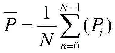
其中,Pi为第i个多普勒谱线的功率值,
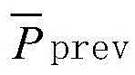
计算当前的平均功率与之前的平均功率的差值,从而得到多普勒频移信息的变化,即:Calculate the difference between the current average power and the previous average power to obtain the change in Doppler shift information, namely:

其中,
步骤230,判断单元163对差值进行判断,校正单元164根据判断结果进行不同的校正处理。本实施例中,设置两个阈值,第一设定阈值A1和第二设定阈值A2,第二设定阈值A2大于第一设定阈值A1,第一设定阈值和第二设定阈值可根据经验设定,为预设的常量阈值。将差值和两个阈值进行比较,当差值大于或等于第二设定阈值时,执行步骤240,当差值大于或等于第一设定阈值且小于第二设定阈值时,执行步骤250,当差值小于第一设定阈值时,则判定当前探头未发生移动或移动很小不需要重新调整,返回步骤210,继续进行PW扫描和实时计算多普勒频移信息。In
步骤240,重新确定采样门。若
在一种具体实施例中,校正单元可采用下面方法重新确定采样门位置:根据当前超声回波信号生成B模式图像或C模式图像,在新生成的B模式图像或C模式图像上根据预先设定的规则确定检测目标点,例如,可以预先设定将血管的横截面的几何中心定为检测目标点,也可以将距离血管壁预定距离的位置定为检测目标点。检测目标点确定后即以该目标的为中心确定采样门。即当采样门的位置偏移到图4b中的位置404处时,通过重新确定采样门位置,将采样门位置重新定位到所要求的403处。In a specific embodiment, the correction unit may use the following method to re-determine the position of the sampling gate: generate a B-mode image or a C-mode image according to the current ultrasound echo signal, and generate a B-mode image or a C-mode image on the newly generated B-mode image or C-mode image The detection target point is determined by certain rules. For example, the geometric center of the cross section of the blood vessel can be preset as the detection target point, or the position at a predetermined distance from the blood vessel wall can be determined as the detection target point. After the detection target point is determined, the sampling gate is determined with the target as the center. That is, when the position of the sampling gate is shifted to the
本领域技术人员可以理解,重新确定采样门位置时也可以采用现有的任一种方案,例如通过对显示屏上的B模式图像或C模式图像的目测方式重新确定采样门位置,当操作人员通过点击或拖移操作选定检测目标点后,系统即可将该检测目标点设为PW采样门中心。重新确定采样门位置后,校正单元160将采样门位置信息发送给发射控制装置130,以便发射控制装置130控制超声探头按照重新确定的采样门位置信息发射超声波。Those skilled in the art can understand that any of the existing solutions can also be used to re-determine the position of the sampling door. For example, by visually inspecting the B-mode image or the C-mode image on the display screen, After selecting the detection target point by clicking or dragging, the system can set the detection target point as the center of the PW sampling gate. After re-determining the sampling gate position, the
定位采样门位置后执行步骤250,自动开启采样线角度或者血流方向校正程序。After locating the position of the sampling gate,
步骤250,自动开启血流方向校正(也称为血流角度校正)。若

在一种具体实施例中,校正单元可采用下面方法对血流方向进行校正:控制探头分至少两个方向进行超声多普勒扫描,根据至少两个方向的回波生成的多普勒频移信息计算出至少两组血流速度大小和血流速度方向,对至少两组血流速度大小和血流速度方向进行合成运算,计算出合成的血流速度。通常,血流速度的方向即为血流方向。因此,这样,即可计算出校正后的血流方向,也即探头发生移动后重新计算得出的血流方向。实际上,该血流方向表现了实际的血流方向与探头之间的相互关系。In a specific embodiment, the correction unit may use the following method to correct the direction of blood flow: control the probe to perform ultrasound Doppler scanning in at least two directions, and generate Doppler frequency shifts according to echoes in at least two directions. The information calculates at least two groups of blood flow velocity magnitudes and blood flow velocity directions, and performs a synthetic operation on at least two groups of blood flow velocity magnitudes and blood flow velocity directions to calculate a synthetic blood flow velocity. Generally, the direction of blood flow velocity is the blood flow direction. Therefore, in this way, the corrected blood flow direction can be calculated, that is, the blood flow direction obtained by recalculation after the probe moves. In fact, the flow direction represents the relationship between the actual flow direction and the probe.
在超声成像中,血流方向在后续中将用于确定采样线的角度、计算特定方向上的血流速度大小等过程。如果探头发生了移动,则原来用来确定采样线的角度和计算参数的血流方向可能已经与实际血流的方向不一致,从而导致扫查和参数测量和计算结果不准确。本发明的实施例中,当判断探头发生移动之后,自动启动对血流方向的校正,使其与实际血流的方向一致,能够使采样线的角度设置更适合,计算的参数更准确。In ultrasound imaging, the blood flow direction will be used in subsequent processes to determine the angle of the sampling line, calculate the blood flow velocity in a specific direction, and so on. If the probe moves, the blood flow direction originally used to determine the angle of the sampling line and the calculated parameters may be inconsistent with the actual blood flow direction, resulting in inaccurate scanning and parameter measurement and calculation results. In the embodiment of the present invention, when it is determined that the probe moves, the correction of the blood flow direction is automatically activated to make it consistent with the actual blood flow direction, which can make the angle setting of the sampling line more suitable and the calculated parameters more accurate.
一些实施例中,还可以进行采样线角度校正。理论上,校正后的血流方向即为真实的血流方向,因此,此时可以修正采样线角度使其尽量接近校正后的血流方向。In some embodiments, sampling line angle correction may also be performed. Theoretically, the corrected blood flow direction is the real blood flow direction. Therefore, at this time, the sampling line angle can be corrected to make it as close to the corrected blood flow direction as possible.
对采样线角度进行校正后,校正单元160将采样线角度发送给发射控制装置130,以便发射控制装置130控制超声探头按照重新确定的采样线角度信息发射超声波。After correcting the sampling line angle, the
本领域技术人员可以理解,对采样线角度和血流方向进行校正时也可以采用现有的任一种方案,例如通过对显示屏上的B模式图像或C模式图像的目测方式重新确定血流方向,Those skilled in the art can understand that any of the existing solutions can also be used to correct the angle of the sampling line and the direction of blood flow, for example, the blood flow can be re-determined by visual inspection of the B-mode image or C-mode image on the display screen direction,
本实施例中,由于多普勒谱图的数据和校正装置判断的数据都来自于多普勒频移信息,因此通过检测多普勒频移信息的变化,即可检测出多普勒谱图形态的变化,当多普勒频移信息的变化超过设定阈值时,多普勒谱图形态同样也会产生较大变化,根据该变化即可自动开启相应的校正处理。由于在生成多普勒谱图的过程中同时也在实时地进行校正的判断,从而一方面可及时对多普勒谱图进行校正,避免得出不真实的结果,另一方面可避免由于依靠医生的主观判断而导致的不必要的校正。In this embodiment, since both the data of the Doppler spectrogram and the data judged by the correction device come from the Doppler frequency shift information, the Doppler spectrogram can be detected by detecting the change of the Doppler frequency shift information. When the change of the Doppler frequency shift information exceeds the set threshold, the shape of the Doppler spectrum will also change greatly, and the corresponding correction processing can be automatically started according to the change. Since the correction judgment is also made in real time in the process of generating the Doppler spectrogram, on the one hand, the Doppler spectrogram can be corrected in time to avoid unreal results, and on the other hand, it can be avoided due to relying on Unnecessary corrections due to the subjective judgment of the physician.
实施例二:Embodiment 2:
通常情况下,多普勒频谱图在显示屏上的显示如图5所示,为适合观察,波形图的整体高度A和显示窗口的高度H的比值处理一定范围,在高度H方向上根据高度A和其代表的血流速度确定合适的标尺刻度,因此高度H也称为标尺。但当完成采样门位置重新定位以及采样线角度和血流方向校正后,会导致谱图发生变化,谱图高度A占据整个标尺的比例过大或者过小,甚至出现混叠的情况,如图6所示,即谱图高度A过高,导致波峰被削平,而削掉的波峰在底部显示。Normally, the Doppler spectrogram is displayed on the display screen as shown in Figure 5. In order to be suitable for observation, the ratio of the overall height A of the waveform graph to the height H of the display window is processed within a certain range. In the height H direction, according to the height A and the blood flow velocity it represents determine the appropriate scale scale, so the height H is also called the scale. However, when the repositioning of the sampling gate and the correction of the sampling line angle and the blood flow direction are completed, the spectrum will change, and the proportion of the spectrum height A occupying the entire scale is too large or too small, and even aliasing occurs, as shown in the figure. 6, that is, the height A of the spectrum is too high, causing the peaks to be flattened, and the clipped peaks are displayed at the bottom.
因此,本实施例中,请参考图7,在实施例一的基础上增加用于对校正后的多普勒频谱图进行显示比例优化的显示优化单元单元,发射控制装置130控制超声探头按照校正后的多普勒扫描参数发射超声波,多普勒信号处理装置150对校正后的超声波的回波进行处理,得到校正后的多普勒频谱图。其流程图如图7所示,在步骤250之后,还包括以下步骤:Therefore, in this embodiment, referring to FIG. 7 , a display optimization unit for optimizing the display scale of the corrected Doppler spectrogram is added on the basis of the first embodiment, and the
步骤260,判断标尺比例是否超过设定范围。显示优化单元检测校正后的多普勒频谱图占据显示窗口高度的比例R1,例如,通过多普勒频谱图的最大值减去最小值可得到谱图高度A,而窗口高度H已知,则比例R1=A/H,然后将该比例R1与设定范围进行比较,例如,当|R-R1|>A3时,则执行步骤270,其中R和A3是人为设定的值,其大小根据实际情况设定。否则继续执行步骤210,进行PW扫描同时实时计算多普勒频移信息和校正处理。
步骤270,进行显示优化。当比例超出设定范围时,显示优化单元自动调整多普勒频谱图占据显示窗口高度的比例,使调整后多普勒频谱图占据显示窗口高度的比例位于设定范围内。
本领域技术人员可以理解,上述实施方式中各种方法的全部或部分步骤可以通过程序来指令相关硬件完成,该程序可以存储于一计算机可读存储介质中,存储介质可以包括:只读存储器、随机存储器、磁盘或光盘等。Those skilled in the art can understand that all or part of the steps of the various methods in the above-mentioned embodiments can be completed by instructing relevant hardware through a program, and the program can be stored in a computer-readable storage medium, and the storage medium can include: read-only memory, Random access memory, magnetic disk or CD, etc.
以上应用了具体个例对本发明进行阐述,只是用于帮助理解本发明,并不用以限制本发明。对于本发明所属技术领域的技术人员,依据本发明的思想,还可以做出若干简单推演、变形或替换。The above specific examples are used to illustrate the present invention, which are only used to help understand the present invention, and are not intended to limit the present invention. For those skilled in the art to which the present invention pertains, according to the idea of the present invention, several simple deductions, modifications or substitutions can also be made.
Claims (19)
Priority Applications (1)
| Application Number | Priority Date | Filing Date | Title |
|---|---|---|---|
| CN201510400492.8A CN106333707B (en) | 2015-07-09 | 2015-07-09 | Ultrasound Doppler spectrum correction method, device and ultrasound diagnosis system |
Applications Claiming Priority (1)
| Application Number | Priority Date | Filing Date | Title |
|---|---|---|---|
| CN201510400492.8A CN106333707B (en) | 2015-07-09 | 2015-07-09 | Ultrasound Doppler spectrum correction method, device and ultrasound diagnosis system |
Publications (2)
| Publication Number | Publication Date |
|---|---|
| CN106333707A CN106333707A (en) | 2017-01-18 |
| CN106333707B true CN106333707B (en) | 2020-12-01 |
Family
ID=57825830
Family Applications (1)
| Application Number | Title | Priority Date | Filing Date |
|---|---|---|---|
| CN201510400492.8A Active CN106333707B (en) | 2015-07-09 | 2015-07-09 | Ultrasound Doppler spectrum correction method, device and ultrasound diagnosis system |
Country Status (1)
| Country | Link |
|---|---|
| CN (1) | CN106333707B (en) |
Cited By (1)
| Publication number | Priority date | Publication date | Assignee | Title |
|---|---|---|---|---|
| US12502524B2 (en) | 2021-12-03 | 2025-12-23 | Kardion Gmbh | Cardiac pump with optical fiber for laser doppler |
Families Citing this family (21)
| Publication number | Priority date | Publication date | Assignee | Title |
|---|---|---|---|---|
| US10966686B2 (en) * | 2017-07-14 | 2021-04-06 | Samsung Medison Co., Ltd. | Ultrasound diagnosis apparatus and method of operating the same |
| DE102018208538A1 (en) | 2018-05-30 | 2019-12-05 | Kardion Gmbh | Intravascular blood pump and process for the production of electrical conductors |
| DE102018208899A1 (en) | 2018-06-06 | 2019-12-12 | Kardion Gmbh | A method for determining the speed of sound in a fluid in the region of an implanted vascular support system |
| DE102018208929A1 (en) | 2018-06-06 | 2019-12-12 | Kardion Gmbh | A method of determining a flow rate of fluid flowing through an implanted vascular support system |
| DE102018208933A1 (en) | 2018-06-06 | 2019-12-12 | Kardion Gmbh | A method of determining a flow rate of fluid flowing through an implanted vascular support system |
| DE102018208892A1 (en) | 2018-06-06 | 2019-12-12 | Kardion Gmbh | A sensor head device for a minimally invasive cardiac assist system and method of manufacturing a sensor head device for a cardiac assist system |
| DE102018208870A1 (en) | 2018-06-06 | 2019-12-12 | Kardion Gmbh | A method of determining a fluid volume flow through an implanted vascular support system |
| DE102018208879A1 (en) | 2018-06-06 | 2020-01-30 | Kardion Gmbh | Method for determining a total fluid volume flow in the area of an implanted, vascular support system |
| DE102018208862A1 (en) | 2018-06-06 | 2019-12-12 | Kardion Gmbh | Implantable vascular support system |
| DE102018208913A1 (en) | 2018-06-06 | 2019-12-12 | Kardion Gmbh | A method of operating an implanted ventricular assist device |
| DE102018208936A1 (en) | 2018-06-06 | 2019-12-12 | Kardion Gmbh | Determining device and method for determining a viscosity of a fluid |
| DE102018208945A1 (en) | 2018-06-06 | 2019-12-12 | Kardion Gmbh | An analysis device and method for analyzing a viscosity of a fluid |
| DE102018210076A1 (en) | 2018-06-21 | 2019-12-24 | Kardion Gmbh | Method and device for detecting a state of wear of a cardiac support system, method and device for operating a cardiac support system and cardiac support system |
| DE102018213350A1 (en) | 2018-08-08 | 2020-02-13 | Kardion Gmbh | Device and method for monitoring a patient's health |
| CN116491977A (en) * | 2019-01-29 | 2023-07-28 | 深圳华大智造云影医疗科技有限公司 | Ultrasonic detection control method, device, and computer-readable storage medium |
| CN110987770B (en) * | 2019-11-07 | 2022-11-15 | 北京工业大学 | Single flowing particle detection method and system based on laser self-mixing feedback interference |
| CN111048168A (en) * | 2019-11-25 | 2020-04-21 | 安徽名流健康管理有限公司 | Doppler ultrasonic diagnosis quality tracing system |
| CN112120734B (en) * | 2020-10-20 | 2022-11-11 | 深圳开立生物医疗科技股份有限公司 | Doppler frequency spectrum generation method and device in blood flow direction and related equipment |
| CN113499100A (en) * | 2021-08-16 | 2021-10-15 | 深圳显融医疗科技有限公司 | Handheld animal intelligent remote color Doppler ultrasonic diagnosis system capable of being connected with various terminals |
| CN115568876B (en) * | 2022-11-24 | 2023-05-09 | 苏州圣泽医疗科技有限公司 | Correction method for blood flow velocity measurement value and Doppler blood flow detection device |
| CN119157569B (en) * | 2024-11-21 | 2025-02-11 | 东北大学 | Cardiac coronary artery micro-blood flow collecting and processing method based on ultrasonic waves |
Citations (4)
| Publication number | Priority date | Publication date | Assignee | Title |
|---|---|---|---|---|
| CN1298688A (en) * | 1999-11-05 | 2001-06-13 | Ge医疗系统环球技术有限公司 | Method and apparatus for pulsation repetive regulation, and ultrasonic imaging device |
| KR20110069190A (en) * | 2009-12-17 | 2011-06-23 | 삼성메디슨 주식회사 | Ultrasound system and method for performing signal correction processing |
| CN102266239A (en) * | 2010-06-04 | 2011-12-07 | 株式会社东芝 | Ultrasonic diagnostic apparatus |
| CN104379064A (en) * | 2012-06-27 | 2015-02-25 | 株式会社东芝 | Ultrasonic diagnostic apparatus and correction method of image data |
Family Cites Families (1)
| Publication number | Priority date | Publication date | Assignee | Title |
|---|---|---|---|---|
| JP5972561B2 (en) * | 2011-12-08 | 2016-08-17 | 東芝メディカルシステムズ株式会社 | Ultrasonic diagnostic apparatus, image processing apparatus, and image processing program |
-
2015
- 2015-07-09 CN CN201510400492.8A patent/CN106333707B/en active Active
Patent Citations (4)
| Publication number | Priority date | Publication date | Assignee | Title |
|---|---|---|---|---|
| CN1298688A (en) * | 1999-11-05 | 2001-06-13 | Ge医疗系统环球技术有限公司 | Method and apparatus for pulsation repetive regulation, and ultrasonic imaging device |
| KR20110069190A (en) * | 2009-12-17 | 2011-06-23 | 삼성메디슨 주식회사 | Ultrasound system and method for performing signal correction processing |
| CN102266239A (en) * | 2010-06-04 | 2011-12-07 | 株式会社东芝 | Ultrasonic diagnostic apparatus |
| CN104379064A (en) * | 2012-06-27 | 2015-02-25 | 株式会社东芝 | Ultrasonic diagnostic apparatus and correction method of image data |
Cited By (1)
| Publication number | Priority date | Publication date | Assignee | Title |
|---|---|---|---|---|
| US12502524B2 (en) | 2021-12-03 | 2025-12-23 | Kardion Gmbh | Cardiac pump with optical fiber for laser doppler |
Also Published As
| Publication number | Publication date |
|---|---|
| CN106333707A (en) | 2017-01-18 |
Similar Documents
| Publication | Publication Date | Title |
|---|---|---|
| CN106333707B (en) | Ultrasound Doppler spectrum correction method, device and ultrasound diagnosis system | |
| US11844656B2 (en) | Ultrasound diagnostic apparatus, method of controlling ultrasound diagnostic apparatus, and non-transitory computer-readable recording medium storing therein computer-readable program for controlling ultrasound diagnostic apparatus | |
| CN103505247B (en) | Diagnostic ultrasound equipment | |
| EP1832894B1 (en) | System and method for automatic gain compensation based on image processing | |
| US11123044B2 (en) | Signal processing device, ultrasonic diagnostic apparatus, and method | |
| EP3840660B1 (en) | Systems and method for performing pulse wave velocity measurements | |
| CN102038522B (en) | Ultrasonic diagnosis apparatus and ultrasoinc data acquisition method | |
| JP2004073287A (en) | Ultrasonic diagnostic apparatus, ultrasonic image display apparatus, and ultrasonic image display method | |
| JP6419976B2 (en) | Ultrasonic diagnostic apparatus and control method of ultrasonic diagnostic apparatus | |
| JP2001128976A (en) | Method and device for prf control, and ultrasonographic apparatus | |
| US11602333B2 (en) | Ultrasound diagnostic apparatus | |
| JP7345624B2 (en) | Ultrasonic diagnostic device, control method for ultrasonic diagnostic device, and processor for ultrasonic diagnostic device | |
| US7850609B2 (en) | Ultrasound diagnostic apparatus | |
| WO2014013839A1 (en) | Ultrasonic diagnostic device and image processing device | |
| EP4363889B1 (en) | Ultrasonic quantification of acoustic attenuation coefficient in the presence of elevation aperture blockage | |
| CN104622505A (en) | Ultrasonic detecting system and method for intracranial blood flow | |
| US20120238877A1 (en) | Ultrasound diagnostic apparatus and ultrasound image producing method | |
| JP4918369B2 (en) | Ultrasonic diagnostic equipment | |
| JP5455567B2 (en) | Ultrasonic diagnostic equipment | |
| US20230240653A1 (en) | An interventional device with an ultrasound transceiver | |
| EP3944820A1 (en) | Interventional device with an ultrasound transceiver | |
| JP2007054226A (en) | Ultrasonic diagnostic equipment |
Legal Events
| Date | Code | Title | Description |
|---|---|---|---|
| C06 | Publication | ||
| PB01 | Publication | ||
| SE01 | Entry into force of request for substantive examination | ||
| SE01 | Entry into force of request for substantive examination | ||
| GR01 | Patent grant | ||
| GR01 | Patent grant | ||
| EE01 | Entry into force of recordation of patent licensing contract |
Application publication date: 20170118 Assignee: Shenzhen Mindray Animal Medical Technology Co.,Ltd. Assignor: SHENZHEN MINDRAY BIO-MEDICAL ELECTRONICS Co.,Ltd. Contract record no.: X2022440020009 Denomination of invention: Ultrasound Doppler spectrum correction method, device and ultrasound diagnosis system Granted publication date: 20201201 License type: Common License Record date: 20220804 |
|
| EE01 | Entry into force of recordation of patent licensing contract |