CN105358567B - Nanopore biosensors for detecting proteins and nucleic acids - Google Patents
Nanopore biosensors for detecting proteins and nucleic acids Download PDFInfo
- Publication number
- CN105358567B CN105358567B CN201480018571.6A CN201480018571A CN105358567B CN 105358567 B CN105358567 B CN 105358567B CN 201480018571 A CN201480018571 A CN 201480018571A CN 105358567 B CN105358567 B CN 105358567B
- Authority
- CN
- China
- Prior art keywords
- clya
- pore
- modified clya
- clya pore
- modified
- Prior art date
- Legal status (The legal status is an assumption and is not a legal conclusion. Google has not performed a legal analysis and makes no representation as to the accuracy of the status listed.)
- Active
Links
Images


Classifications
-
- G—PHYSICS
- G01—MEASURING; TESTING
- G01N—INVESTIGATING OR ANALYSING MATERIALS BY DETERMINING THEIR CHEMICAL OR PHYSICAL PROPERTIES
- G01N33/00—Investigating or analysing materials by specific methods not covered by groups G01N1/00 - G01N31/00
- G01N33/48—Biological material, e.g. blood, urine; Haemocytometers
- G01N33/50—Chemical analysis of biological material, e.g. blood, urine; Testing involving biospecific ligand binding methods; Immunological testing
- G01N33/53—Immunoassay; Biospecific binding assay; Materials therefor
- G01N33/543—Immunoassay; Biospecific binding assay; Materials therefor with an insoluble carrier for immobilising immunochemicals
- G01N33/54306—Solid-phase reaction mechanisms
-
- C—CHEMISTRY; METALLURGY
- C07—ORGANIC CHEMISTRY
- C07K—PEPTIDES
- C07K14/00—Peptides having more than 20 amino acids; Gastrins; Somatostatins; Melanotropins; Derivatives thereof
- C07K14/195—Peptides having more than 20 amino acids; Gastrins; Somatostatins; Melanotropins; Derivatives thereof from bacteria
- C07K14/24—Peptides having more than 20 amino acids; Gastrins; Somatostatins; Melanotropins; Derivatives thereof from bacteria from Enterobacteriaceae (F), e.g. Citrobacter, Serratia, Proteus, Providencia, Morganella, Yersinia
- C07K14/255—Salmonella (G)
-
- C—CHEMISTRY; METALLURGY
- C12—BIOCHEMISTRY; BEER; SPIRITS; WINE; VINEGAR; MICROBIOLOGY; ENZYMOLOGY; MUTATION OR GENETIC ENGINEERING
- C12Q—MEASURING OR TESTING PROCESSES INVOLVING ENZYMES, NUCLEIC ACIDS OR MICROORGANISMS; COMPOSITIONS OR TEST PAPERS THEREFOR; PROCESSES OF PREPARING SUCH COMPOSITIONS; CONDITION-RESPONSIVE CONTROL IN MICROBIOLOGICAL OR ENZYMOLOGICAL PROCESSES
- C12Q1/00—Measuring or testing processes involving enzymes, nucleic acids or microorganisms; Compositions therefor; Processes of preparing such compositions
- C12Q1/68—Measuring or testing processes involving enzymes, nucleic acids or microorganisms; Compositions therefor; Processes of preparing such compositions involving nucleic acids
- C12Q1/6869—Methods for sequencing
-
- G—PHYSICS
- G01—MEASURING; TESTING
- G01N—INVESTIGATING OR ANALYSING MATERIALS BY DETERMINING THEIR CHEMICAL OR PHYSICAL PROPERTIES
- G01N27/00—Investigating or analysing materials by the use of electric, electrochemical, or magnetic means
- G01N27/26—Investigating or analysing materials by the use of electric, electrochemical, or magnetic means by investigating electrochemical variables; by using electrolysis or electrophoresis
- G01N27/28—Electrolytic cell components
- G01N27/30—Electrodes, e.g. test electrodes; Half-cells
- G01N27/327—Biochemical electrodes, e.g. electrical or mechanical details for in vitro measurements
- G01N27/3275—Sensing specific biomolecules, e.g. nucleic acid strands, based on an electrode surface reaction
- G01N27/3278—Sensing specific biomolecules, e.g. nucleic acid strands, based on an electrode surface reaction involving nanosized elements, e.g. nanogaps or nanoparticles
-
- G—PHYSICS
- G01—MEASURING; TESTING
- G01N—INVESTIGATING OR ANALYSING MATERIALS BY DETERMINING THEIR CHEMICAL OR PHYSICAL PROPERTIES
- G01N33/00—Investigating or analysing materials by specific methods not covered by groups G01N1/00 - G01N31/00
- G01N33/48—Biological material, e.g. blood, urine; Haemocytometers
- G01N33/483—Physical analysis of biological material
- G01N33/487—Physical analysis of biological material of liquid biological material
- G01N33/48707—Physical analysis of biological material of liquid biological material by electrical means
- G01N33/48721—Investigating individual macromolecules, e.g. by translocation through nanopores
Landscapes
- Chemical & Material Sciences (AREA)
- Health & Medical Sciences (AREA)
- Life Sciences & Earth Sciences (AREA)
- Engineering & Computer Science (AREA)
- Organic Chemistry (AREA)
- Physics & Mathematics (AREA)
- Molecular Biology (AREA)
- Immunology (AREA)
- Proteomics, Peptides & Aminoacids (AREA)
- General Health & Medical Sciences (AREA)
- Biochemistry (AREA)
- Biomedical Technology (AREA)
- Analytical Chemistry (AREA)
- Biophysics (AREA)
- Zoology (AREA)
- Wood Science & Technology (AREA)
- Genetics & Genomics (AREA)
- Medicinal Chemistry (AREA)
- Biotechnology (AREA)
- Microbiology (AREA)
- General Physics & Mathematics (AREA)
- Urology & Nephrology (AREA)
- Pathology (AREA)
- Hematology (AREA)
- General Engineering & Computer Science (AREA)
- Bioinformatics & Cheminformatics (AREA)
- Spectroscopy & Molecular Physics (AREA)
- Nanotechnology (AREA)
- Food Science & Technology (AREA)
- Chemical Kinetics & Catalysis (AREA)
- Gastroenterology & Hepatology (AREA)
- Electrochemistry (AREA)
- Cell Biology (AREA)
- Investigating Or Analyzing Materials By The Use Of Electric Means (AREA)
- Peptides Or Proteins (AREA)
- Apparatus Associated With Microorganisms And Enzymes (AREA)
- Measuring Or Testing Involving Enzymes Or Micro-Organisms (AREA)
Abstract
本文涉及基于修饰的溶细胞素蛋白质的纳米孔生物传感器。所述纳米孔生物传感器容纳包括蛋白质和核酸的大分子,并且可以额外地包括具有选择性结合性质的配体。This article relates to nanopore biosensors based on modified cytolysin proteins. The nanopore biosensor accommodates macromolecules including proteins and nucleic acids, and may additionally include ligands with selective binding properties.
Description
Technical Field
The present invention relates to a nanopore biosensor based on a modified cytolysin protein. The nanopore biosensor houses macromolecules including folded proteins and nucleic acids including double stranded (ds) DNA, which exhibit enhanced activity, solubility, and electrical properties compared to other nanopore biosensors.
Background
The transport of ions or molecules across biological membranes is an essential process of cell life, and is tightly regulated by ion channels, transporters and pores. Recently, researchers have employed biological in single molecule analysis,1in the solid state, the water-soluble polymer,2DNA origami3And mixed3a,b,4A nanopore.5Biological nanopores are advantageous over their synthetic counterparts, primarily because they can be repeatedly manufactured and modified with atomic-level precision that artificial nanopores cannot compete with. However, biological nanopores still have drawbacks. The mechanical stability of the biological nanopore depends on the particular situation. The alpha hemolysin (α HL) nanopore from Staphylococcus aureus (Staphylococcus aureus) and the porin a (mspa) nanopore from Mycobacterium smegmatis (Mycobacterium smegmatis) remain open in the lipid bilayer at high applied potentials and are surprisingly well-protected against temperature,6pH6b,7and extreme conditions of denaturant concentration.6b,8However, most other porins and channels are less robust. However, it is believed that the greatest disadvantage of biological nanopores is their fixed size. For example, the size of the α HL, MspA or aerolysin nanopore allows for the analysis of single-stranded nucleic acids, aptamers or small peptides,9their small internal diameter (-1 nm) has hampered direct research into other important biological systems such as folded enzymes or ribozymes.
A number of recent studies have sampled the migration of folded proteins through artificial nanopores.10However, the study of proteins with solid-state nanopores is difficult because proteins have a non-uniform charge distribution, they often adsorb to the nanopore surface andthey migrate too quickly to be sampled properly.10cFurther, since proteins have a tightly folded structure, the diameter of the nanopore should be similar to the diameter of the protein.10bRecently, we have introduced as the first biological nanopore cytolysin a (clya) from Salmonella typhi (Salmonella typhi), which enables the study of naturally folded proteins.7aThe ClyA structure is ideal for this task because proteins such as thrombin (37kDa) or malate dehydrogenase (dimer, 35kDa monomer) can be electrophoretically captured between a wide cis inlet (5.5nm, table 1) and a narrow trans outlet (3.3nm, table 1), and can thus be sampled for several minutes. Since the ionic current across ClyA is very sensitive to the vestibular environment, blocking of human and bovine thrombin can be very easily recognized.7aOur work is based on the ClyA structure in which the two native cysteine residues of ClyA-WT (C87 and C285) are replaced by serine (ClyA-SS).7aHowever, when compared to ClyA-WT monomer, ClyA-SS monomer showed low water solubility and low activity (fig. S1), and when the applied potential was higher than +60mV or lower than-90 mV, ClyA-SS monomer spontaneously opened and closed (gated) in the planar lipid bilayer.
Thus, there remains a need in the art for nanopore biosensors having high sensitivity to target analytes and high water solubility and stability over a range of potentials. The nanopore biosensor should have good oligomerisation, voltage dependent gating, and electrical noise performance in single channel current recording. The present invention relates to engineered nanopores in which specific substitutions of native cysteine residues and other residues impart additional properties compared to ClyA-WT and ClyA-SS.
Disclosure of Invention
One aspect of the invention relates to a modified ClyA pore comprising a plurality of subunits, wherein each subunit comprises a polypeptide represented by an amino acid sequence at least 80% identical to SEQ ID No. 1, wherein exactly only one Cys residue is substituted by Ser. In some embodiments, the Cys residue is C285. In certain embodiments, each subunit of the modified ClyA pore comprises at least one additional amino acid substituent selected from L99Q, E103G, F166Y, and K294R. For example, each subunit may comprise a polypeptide represented by the amino acid sequence of SEQ ID NO. 2. In certain embodiments, exactly one Cys residue in each subunit is substituted by Ala. In some embodiments, each subunit of the modified ClyA pore comprises at least one additional amino acid substituent selected from L203V and H207Y. For example, each subunit can comprise a polypeptide represented by the amino acid sequence of SEQ ID NO. 3.
In some embodiments, the modified ClyA pore comprises at least 12 subunits. For example, the modified ClyA pore may comprise 12 subunits or may comprise 13 subunits. In certain embodiments, the modified ClyA pores have a cis diameter of at least 3.5 nm. In certain embodiments, the modified ClyA pore has a trans diameter of at least 6 nm. In some embodiments, the modified ClyA pore remains open when the voltage across the modified ClyA pore ranges from-60 +90 to-150 mV.
In some embodiments, the protein analyte binds to the lumen of the modified ClyA pore. Protein analytes can bind to more than one site within the lumen of the modified ClyA pore. In some embodiments, the protein analyte is a protein having a molecular weight range of 15-70 kDa.
Drawings
FIG. 1 shows a band representation of Salmonella typhi ClyA nanopores (PDB:2WCD, 90% sequence identity) constructed by homology modeling from E.coli ClyA structure.17In FIG. 1a, 1 protomer is highlighted, its secondary structural elements are shaded in dark grey from N to C, and the other protomers are shown in alternating light grey. The side chains of amino acids altered by directed evolution experiments are displayed in spheres. The two native cysteine residues and phenylalanine 166 are labeled. FIG. 1b shows the sequence changes of ClyA-SS, ClyA-CS and ClyA-AS relative to ClyA-WT.
Fig. 2 shows oligomerization and nanopore formation of ClyA-WT, ClyA-SS and evolved ClyA variants. FIG. 2a shows the oligomerization of ClyA nanopores as detected by 4-20% BN-PAGE. Proteins (1mg/ml) were preincubated with 0.5% DDM for 30 min at room temperature before loading into the gel (40. mu.g/lane). Lane 1: protein gradient, lane 2: ClyA-WT, lane 3: ClyA-SS, lane 4: ClyA-AS, lane 5: ClyA-CS. Figure 1b shows a single nanopore conductance profile obtained from 100 nanopores reconstituted in a planar lipid bilayer after pre-oligomerization in 0.5% DDM by ClyA-WT nanopore (top), ClyA-CS nanopore (middle) and ClyA-AS nanopore (bottom). The recording was carried out at 28 ℃ in-35 mV, 15mM Tris. (Tris hydroxymethyl aminomethane) HCl, pH 7.5,150mM NaCl.
FIG. 3 shows the single conductance of 62 ClyA-CS nanopores extracted from the lowest (FIG. 3a), second lowest (FIG. 3b) and third lowest (FIG. 3c) oligomeric bands of ClyA-CS separated on 4-20% acrylamide BN-PAGE. ClyA-CS monomers were preincubated in 0.5% DDM and loaded onto BN-PAGE as depicted in FIG. 2. The cut-out tape is framed by a box and marked with an arrow on the inset. Recordings were made in 15mM tris.hcl at-35 mV, pH 7.5 at 28 ℃, containing 150mM NaCl.
Fig. 4 shows current blocking induced by HT on three types of ClyA-CS nanopores. In fig. 4a, from left to right, through type I, type II and type III ClyA nanopores (grey), each of which contains HT (black) in its vestibule. Type II and type III ClyA nanopores were modeled as 13-mers (13 mers) (type II) or 14-mers (type III) as described in the supplementary information. FIG. 4b shows HT blockage of ClyA-CS nanopores at-35 mV for type I (left), type II (middle) or type III (right). HT Current blockade for type I and type II ClyA-CS at L1 (IRES)%56 ± 1 and 68 ± 1), and L2 (IRES), respectively%23 ± 3 and 31 ± 1) current levels, respectively. The occlusion lasts several minutes, so only the first second current trace is shown. In type I ClyA-CS, L1 represents the most representative of current blockages (70%), while in type II ClyA-CS, L2 accounts for the majority (96%). HT current blocking for type III ClyA-CS nanopores only shows L2 current levels (IRES ═ 32 ± 9). Recordings were made in-35 mV, 15mM tris.hcl, pH 7.5,150mM NaCl. The collection of FIG. 4b was performed using a Bessel low-pass filter (Bessel low-pass filter) with a 2000Hz cut-off and sampled at 10kHz, at a temperature of 28 ℃.
FIG. 5 shows the migration of proteins through type I and type II ClyA-CS nanopores. Figure 5a shows the voltage dependence of HT blocking residence time determined for type I (open circles) and type II (filled rectangles) ClyA-CS nanopores. The lifetime at each voltage is calculated by a single exponential fit that establishes a cumulative distribution (n ≦ 3) from the residence times of at least 50 blockages. The line represents a double exponential fit to the experimental points. FIG. 5b shows HT current blocking at-150 mV for type I (left) and type II (right) ClyA-CS nanopores. The blockages showed L2 current levels for only two nanopores (IRES% ═ 23 ± 2 and 31 ± 5 for ClyA-CS type I and type II, respectively). FIG. 5c shows a typical HT current block at-150 mV, type I ClyA-CS, showing current characteristics of "shoulder" and "tip". Recordings were made in 15mM tris.hcl, pH 7.5,150mM NaCl in the presence of 20nM HT. The collection of traces in fig. 5b was performed using a bessel low pass filter with a 2000Hz cutoff and sampling at 10 kHz. The traces in C were collected using a bezier low pass filter with a 10Hz cutoff and sampled at 50 kHz. The temperature was 28 ℃. The errors are given as standard deviations.
Fig. 6 shows a characterization of the purified ClyA monomer. a) The solubility of the purified ClyA monomer was determined by 4-20% acrylamide BN-PAGE. Each equivalent (40 μ g) of purified ClyA monomer (no detergent) was supplemented with about 10% glycerol and 1-fold Native PAGETM Running Buffer (1x of Native PAGETM Running Buffer) and 1-fold cathode Buffer additive (invitrogen) and loaded into each lane: lane 1: marker, lane 2: ClyA-WT, lane 3: ClyA-SS, lane 4: ClyA-CS, lane 5: ClyA-AS. b) Overexpression of the ClyA variant. Aliquots of bacterial particles derived from overnight cultures overexpressing the ClyA variant were resuspended to a concentration of about 100mg/mL and disrupted by sonication, then centrifuged at 20000g for 10 minutes (4 ℃). 20 μ L of supernatant containing soluble fragments (fraction) of the ClyA protein was loaded onto lanes 2,4, 6, 8 of 12% acrylamide SDS-PAGE. The lysed particles were returned to the original volume by adding a solution containing 15mM tris.hcl, pH 7.5,150mM NaCl, and 2% SDS w/v. 20 μ L of this solution was loaded on lanes 3, 5, 7 and 9 of the same 12% acrylamide SDS-PAGE. Thus, lane 1 protein marker, lanes 2 and 3 supernatant and particle fragments of ClyA-SS, respectively; lanes 4 and 5, supernatant and particle fragment of ClyA-WT, respectively; lanes 6 and 7 are ClyA-CS supernatant and pellet fraction, respectively, and lanes 8 and 9 are ClyA-AS supernatant and pellet fraction, respectively. c) And (4) analyzing the hemolysis. ClyA monomer (0.6. mu.M) was incubated with 100. mu.L of a 1% equine erythrocyte suspension (final volume 110. mu.L), and the decrease in turbidity was measured at 650nm (OD650 nm). ClyA-WT is shown AS a thick gray line, ClyA-SS is shown AS a thick black line, ClyA-CS is shown AS a thin black line, and ClyA-AS is shown AS a dashed line. d) Hemolysis rate (calculated AS the turbidity reaching 50% in reciprocal time) is plotted AS protein concentration, ClyA-WT (triangle, thick grey line), ClyA-SS (square, thick black line), ClyA-AS (circle, dotted line) and ClyA-CS (diamond, thin black line).
FIG. 7 shows an example of the oligomerization of ClyA variants using 4-20% acrylamide BN-PAGE. a) Lane 4 (Round 4), lanes 1 and 2: ClyA-CS, lanes 3 and 4: 4ClyA5, lanes 5 and 6: 4ClyA3, lanes 7 and 8: 4ClyA1, lanes 9 and 10: 4ClyA2, lanes 11 and 12: 4ClyA 6. Samples were prepared as illustrated in figure 6 and supplemented with 0.05% (even lanes) or 0.1% (odd lanes) SDS. SDS was used to combat the "tailing" effect of large amounts of DDM in the samples. b) Round 5. Lane 1, 2:5ClyA2, lane 3:5ClyA1, lane 4: ClyA-AS. Samples were supplemented with 0.05% SDS. The oligomerization was initiated by the addition of 1% DDM and the ClyA variant was partially purified using Ni-NTA affinity chromatography as described in the methods.
FIG. 8 shows the noise signature of three types of ClyA nanopores at-35 mV potential in 150mM NaCl,15mM Tris.HCl. Current power spectral densities of ClyA-CS nanopores at-35 mV for type I (dashed line), type II (dotted line) and type III (solid light gray line) were obtained from the 0.5s trace. The current power spectral density at 0mV is shown as a black line. Each line corresponds to the average of the power spectra calculated from 3 sets of recordings made for different single channels.
Fig. 9 shows the duration of ht (a) and fp (b) clogging on type I ClyA-SS pores. The tracks were recorded at a 10kHz sampling rate using an internal bessel low pass filter set at 2 kHz. Each plotted value corresponds to an average value determined using at least 3 different single channels. The errors are given as standard deviations.
FIG. 10 shows a typical HT current block of-150 mV on type I ClyA, showing current characteristics of "shoulder" and "tip". Recordings were made in 15mM tris.hcl, pH 7.5,150mM NaCl in the presence of 20nM HT. The traces were filtered using a gaussian low pass filter with a 10,000Hz cut-off filter and sampled at 50,000 Hz.
Figure 11 shows dsDNA (double stranded DNA) migration through ClyA nanopores. On the right of the current trace, ClyA (pore structure) and neutravidin (dark grey) are depicted and dsDNA is shown as a black line. a) Segment penetrability of ClyA from Salmonella typhi (sections through), which was constructed by homology modeling of ClyA structures from E.coli (PDB:2WCD, 90% sequence identity).22ClyA is shown in pore measurements including the van der waals radius of the atoms, and the approximate location of residue 103 (serine in the WT sequence) on the indicated pore. b) At +100mV (level I)O+1001.74 ± 0.05nA), addition of 0.12 μ M of 290bp dsDNA 1 to the cis side of ClyA nanopore caused transient current blocking IRES0.63 ± 0.01 (level 1 ×)+1001.10 ± 0.03nA, n-3) due to dsDNA migrating through the pore. Addition of neutravidin to the cis-chamber converts the transient current blockade to a more conductive and persistent current blockade IRES0.68 ± 0.01 (level 1)+1001.19 ± 0.01) due to local migration of DNA through the pore. The open-cell current was restored by reversing the potential to-100 mV (red asterisk). The figure shows a typical instantaneous current blocking. c) A detailed illustration of the current blockage due to the formation of cis-pseudorotaxane (pseudorotoxane) at +100 mV. d) Formation of Trans-pseudorotaxane at-100 mV by helication of dsDNA neutravidin complex from the trans side (see above) (level 1)-100=1.02±0.03nA,IRES0.62 ± 0.01, n is 4). Electrographic recordings were performed at 22 ℃ in 2.5M NaCl,15mM tris.hcl at pH 7.5. By using a 10kHz Bessel low-pass filterData was recorded with a sampling rate of 20 mus (50 kHz).
Figure 12 shows nanopore-DNA rotaxane formation. a) Indicates hybridization of DNA molecules for formation of rotaxanes. The arrow indicates the 3' end of the strand. b) Rotaxane formation. Open pore Current (I.mu.M) of ClyA-2 after addition of DNA hybrid 3 (1. mu.M) complexed with neutravidin (0.3. mu.M) and oligonucleotide (oligo)4 (1. mu.M) to the trans chamber at-100 mVO-1001.71 ± 0.07nA, n 4) to a level of 1-100=1.1±0.04nA(IRESValues of 0.64 ± 0.02, n ═ 4), indicating that dsDNA helicized the pore from the trans side. Step by step to +100mV (red asterisk) production IRES0.77 ± 0.04 (level 2)+1001.31 ± 0.09nA, n-4), indicating that DNA still occupies the pore at a positive applied potential. Level 2 most likely corresponds to ssDNA occupying the vestibule of the pore. Continued switching to positive applied potential did not restore the open pore current (fig. 16), confirming that rotaxane formation was permanent. c-e) removing rotaxanes. c) At +100mV, the ionic current through the ClyA-2 nanopore shows a substantial rapid current blockage (fig. 15), indicating that the ssDNA molecule attached at the cis inlet of the pore occupies the lumen of the pore transiently. d) After rotaxane formation (FIG. 12b), stable ionic current was shown at +100mV nanopore (level 2)+100) This indicates that a single DNA molecule is occupying the pore. e)20 min after 20mM DTT addition to the cis chamber, the DNA molecule above the ClyA pore was removed and the pore opening current was restored at +100mV (I)O+1001.78 ± 0.07, n ═ 4). The recording is performed as described in fig. 11.
FIG. 13 shows ssDNA blocking of ClyA-CS. Addition of 2 μ M biotinylated ssDNA (5a) at +100mV to the cis side of the ClyA-CS nanopore in the presence of 0.6 μ M neutral avidin caused transient current blockages (1.24. + -. 0.02nA, IRES0.69 ± 0.04, n ═ 3), indicating that ssDNA can enter the lumen of the pore, but only transiently. Subsequent addition of a complementary ssDNA strand (5b) converts the current block to level 1+100Obstruction (1.22 + -0.13 nA, I)RES0.67 ± 0.01, n-3), which indicates that dsDNA can now migrate through the entire length of the pore.
FIG. 14 shows DNA crossing ClyAAnd (4) transporting the nanopore. a) A schematic representation of a strand displacement reaction that facilitates release of DNA from the pore. b) ClyA-2 showed a stable open cell current (I) at +50mV and in the presence of 3 (0.3. mu.M)O+500.85 ± 0.01nA, n-3), indicating that the ssDNA strand attached to the well does not spiral through the lumen of the well and prevents migration of dsDNA-type solutions. c) Addition of ssDNA strand 6(40nM) to the cis chamber produces persistent IRES0.70 ± 0.02 (level 2)+500.59 ± 0.02nA, n-5), indicating that dsDNA hybrids helicized the pore. d) Subsequent addition of 1 μ M of 7, also containing 0.3 μ M neutravidin, to the trans chamber (+50mV) promoted the release of the DNA helix and restored the open pore current. Subsequently, dsDNA molecules are sequentially captured and released as shown by the multiple blocking and opening currents. For clarity, neutravidin is not included in the figures. Electrographic recordings were performed at 22 ℃ in 2.5M NaCl at pH 7.5 and 15mM tris. Data was recorded by using a 10kHz bessel low pass filter and using a sampling rate of 20 mus (50 kHz). The current signal in graph d was digitally filtered using a post 2kHz capture low pass gaussian filter. Since capture of DNA in cis is very fast at higher applied potentials, the applied potential for this experiment was set at +50mV to facilitate observation of multiple blockages and releases (fig. 14 d).
FIG. 15 shows the results of a comparative experiment, which shows that all components of FIG. 12 are necessary for the formation of DNA rotaxane. a) Deletion of the bridging sequence 4 renders the linkage between ClyA-2 and 3 impossible. After the-100 mV complex is captured, the complex is easily dislodged from the well at +100mV (asterisk), as shown by the typical current signature of the open pore current of ClyA-2 at +100 mV. b) When captured at-100 mV, the deletion of 2 from the top of the well (e.g., after cleavage with DTT) does not allow the 3.4 DNA to hybridize to bind to the well. The opening current recovered when the potential reversed to +100mV (red asterisk). c) Typical current recordings for ClyA-2 nanopores at +100 mV. d) Full-point histogram (5pA bin) size) of the current trace shown in a for 20 seconds. Level IO+1001.71 + -0.07 nA, 1.62 + -0.12 nA, 1.43 + -0.05 nA and 1.28 + -0.06 nA for A, B,i corresponding to 0.94 + -0.04, 0.84 + -0.03 and 0.75 + -0.03 respectivelyRESThe value is obtained. Each value calculated from 12 experiments can represent a DNA strand hosted in the lumen of ClyA.
Fig. 16 shows the current versus voltage (IV) for ClyA-2 nanopores, a) a typical plot of ClyA-2 before (shallowest gray line) and after (black line) rotaxane formation. The medium gray line indicates that the same nanopore is removed by adding DDT after the DNA molecule is attached to the nanopore. The current log was measured by applying an automated protocol that shifted the voltage from-100 mV to +100mV in 4 seconds. b) Current-voltage curves were calculated from the average of 4 experiments and show steady-state (1s) ClyA-2 open cell current levels (white spheres) and ClyA-2 open cell current levels in rotaxane configuration (black spheres). The single conductance value of the nanopore calculated from the slope of the current-voltage curve was 17.1nS for ClyA-2 under positive and negative bias, 10.8nS for rotaxane under negative bias, and 13.0nS for rotaxane under positive bias. Rotaxanes were prepared as described in figure 12.
FIG. 17 shows dsDNA current blocking for ClyA-CS. To the right of each current trace, the pattern represents a physical interpretation of the current recording. a-d) recording of the current of ClyA-2 after 6.7 hybridization (asterisk) at different applied potentials. e) Ion Current from I after hybridization at +50mV with 6(40nM) and 2O+500.85 + -0.01 nA to level 2+50Obstruction (I)RES0.70 ± 0.02) so that DNA duplex 3(0.3 μ M) is transported through the indicated wells. Reversal of the applied potential to-50 mV restores the open pore current (I)O-500.83 ± 0.00). f) Subsequent addition of 1 μ M neutravidin (black) to the trans chamber blocked the DNA helix within the well as demonstrated after potential reversal to-50 mV, at which point the level of clogging of the well was observed (I)RES=0.67±0.02)。
FIG. 18 shows selective DNA migration through the ClyA-2 pore. a) On the left, at +50mV, the ion current through the ClyA-2 nanopore shows rapid and slight current blocking, indicating that the ssDNA molecule attached at the cis inlet may temporarily occupy the lumen of the pore. In the middle, after dsDNA strand 1(50nM) was added to the cis chamber, there was no change in the current signal, indicating that ClyA-2 was not migrated by dsDNA. On the right, 20 minutes after 20mM DTT was added to the cis chamber, the DNA molecule above the ClyA pore was removed and the DNA could migrate through the pore. b) The same experiment as described in panel a, but in the presence of 1 μ M neutral avidin. c) The same experiment as depicted in panel b, but at +100 mV.
FIG. 19 shows the voltage dependence of the interaction of ssDNA-dsDNA hybrid structures with ClyA-CS pores. a) Panels b (left) and c (right) depict the presentation of the DNA molecules used in the experiments. b) DNA hybrid 3 (FIG. a, left) complexed with neutravidin blocked current at +50mV (left), +70mV (middle) and +100mV (right). c) DNA hybrid 3:6 (panel a, right) complexed with neutravidin has a current block of +50mV (left), +70mV (middle) and +100mV (right).
FIG. 20 shows chain release with Boolean logic. The top represents an OR gate AND the bottom is an AND gate. DNA is represented by directional lines, with arrows indicating the 3' end. Segments within the same DNA strand represent DNA domains that serve as units in hybridization, branch migration, or dissociation. The fields are represented by the letter D followed by a number. T represents a branch domain. The star-like domain represents a domain that is complementary to a sequence of the domain without a star. Fig. 14 shows the implementation of the first reaction of the OR gate.
Fig. 21 shows the alternate transport of DNA through ClyA nanopores. a) Schematic representation of the release of DNA helices by strand displacement, showing the implementation of a second reaction of the OR gate as described in figure 18. b) Open pore current of ClyA-2 at +35 mV. c) The addition of DNA strands 3 (1. mu.M) and 8 (0.5. mu.M) to the cis chamber creates a persistent current block, indicating that the dsDNA hybrid is spiraling the well. d) Subsequent addition of 1 μ M of 9 to the trans chamber at +35mV, which may also contain 1 μ M of neutravidin, promoted the release of the DNA helix and restored the open pore current. Subsequently, additional dsDNA molecules are captured and then released as shown by the cycle of blocking current and opening current. Data was recorded by using a 10kHz low-pass bessel filter and using a sampling rate of 20 mus (50 kHz).
Detailed Description
The present invention relates to nanopore biosensors, which can be used for a variety of applications, including but not limited to the detection and quantification of proteins and the migration of DNA. The nanopores are based on pore-forming bacterial cytotoxins, which serve as channels for macromolecules such as proteins and nucleic acids. The nucleic acid may comprise DNA, such as single stranded DNA (ssdna) or double stranded DNA (dsdna), RNA, peptide nucleic acids and/or nucleic acid analogues.
Modified nanopore biosensor
One aspect of the present invention relates to nanopore biosensors made from modified porins. Exemplary porins include, but are not limited to, cytolysins, hemolysins, porins, DNA packaging proteins, and dyneins. The porin may be a bacterial protein or a viral protein.
In certain embodiments, the modified porin is a pore-forming cytotoxic protein, e.g., a bacterial cytolysin. The cytolysin may be cytolysin a (clya) from a gram-negative bacterium such as salmonella or escherichia coli (e. In some embodiments, the modified cytolysin is a modified ClyA from Salmonella typhi (s.typhi) or Salmonella paratyphi (s.paratyphi). In some embodiments, the modified ClyA pore comprises a plurality of subunits, wherein each subunit comprises a polypeptide represented by an amino acid sequence at least 80% identical to SEQ ID No. 1. In certain embodiments, the subunit is represented by an amino acid sequence that is at least 85% identical, 90% identical, 95% identical, or 100% identical to SEQ ID No. 1. The identity may refer to amino acid identity, or may also refer to structural and/or functional identity. Thus, one or more amino acids may be substituted, deleted, and/or added as compared to SEQ ID NO 1. The modification may alter the pore lumen to alter the size, connectivity and/or structure of the pores. Modifications can also alter the ClyA pores outside the lumen.
In certain embodiments, each subunit comprises a polypeptide represented by an amino acid sequence at least 80% identical to SEQ ID No. 1, wherein exactly only one Cys residue is substituted by Ser. Each subunit may be represented by an amino acid sequence at least 85%, 90%, 95%, 96%, 96%, 98% or 99% identical to SEQ ID NO 1, and in addition, exactly one Cys residue may be substituted by Ser. The Cys residue may be Cys87 and/or Cys285 of SEQ ID NO. 1. In some embodiments, the Cys residue is Cys 285. Other amino acid residues may be substituted, for example, with amino acids having similar properties, such as structure, charge, hydrophobicity or hydrophilicity. In certain embodiments, the substituted residue is one or more of L99, E103, F166, and K294. For example, the substituted residue may be one or more of L99Q, E103G, F166Y and K294R. One exemplary subunit may comprise substituents L99Q, E103G, F166Y, K294R and C285S. Thus, each subunit may comprise a polypeptide represented by the amino acid sequence of SEQ ID NO. 2. An exemplary modified ClyA pore containing the following subunits is referred to as ClyA-CS: where exactly only one Cys residue is substituted by Ser.
The modified ClyA pore can comprise a plurality of subunits, wherein each subunit comprises a polypeptide represented by an amino acid sequence at least 80% identical to SEQ ID No. 1, wherein exactly only one Cys residue is substituted by Ala. The Cys residue may be Cys87 or Cys 285. Each subunit may be represented by an amino acid sequence that is at least 85%, 90%, 95%, 96%, 96%, 98% or 99% identical to SEQ ID No. 1, and exactly one Cys residue may be substituted by Ser and/or exactly one Cys residue may be substituted by Ala. In some embodiments, each subunit is represented by an amino acid sequence that is at least 85%, 90%, 95%, 96%, 96%, 98% or 99% identical to SEQ ID No. 1, and, in addition, exactly one Cys residue may be substituted by Ser and exactly one Cys residue may be substituted by Ala. Other amino acid residues may be substituted, for example, with amino acids having similar properties, such as structure, charge, hydrophobicity or hydrophilicity. In certain embodiments, the substituted residue is one or more of L99, E103, F166, K294, L203, and H207. For example, the substituted residues may be L99Q, E103G, F166Y, K294R, L203V and H207Y. One exemplary subunit may include L99Q, E103G, F166Y, K294R, L203V, H207Y, and C285S. Thus, each subunit may comprise a polypeptide represented by the amino acid sequence of SEQ ID No. 3. An exemplary modified ClyA pore comprising the following subunits may be referred to as ClyA-CS: wherein exactly only one Cys residue is substituted by Ser and exactly only one Cys residue is substituted by Ala.
The invention further relates to nucleic acids encoding said modified ClyA pore. In some embodiments, the nucleic acid encoding the modified ClyA pore is represented by a nucleotide sequence at least 80%, 90%, 95%, 96%, 96%, 98% or 99% identical to SEQ ID No. 4. The nucleic acid may be represented by SEQ ID No. 5 or SEQ ID No. 6. The nucleotide sequence may be codon optimized for expression in a suitable host, e.g., E.coli.
The modified ClyA pores can have pore lumens with diameters of at least 3nm, for example, the diameter measurements can be 3nm, 3.5nm, 4nm, 4.5nm, 5nm, 5.5nm, 6nm, 6.5nm, 7nm, or greater. The size of the pore lumen may depend on the analyte to be detected by the ClyA pore to be modified. The cis diameter of the pore lumen may be at least 3.5nm and/or the trans diameter of the ClyA pore may be at least 6 nm. In general, cis refers to the end of a modified ClyA pore to which an analyte is added; and trans refers to the end of the modified ClyA pore through which the analyte migrates out through the length of the pore lumen. In an artificial lipid bilayer, for example, the trans-terminus of a pore can be inserted into the lipid bilayer while the cis-terminus of the pore remains on the same side of the lipid bilayer. Thus, the cis diameter of a pore is the diameter of the opening at the cis end of the pore to which the analyte is added; and the trans-diameter of the pore is the diameter of the opening at the trans-end of the pore from which the analyte exits.
The size of the pore lumen also depends on the number of subunits in the modified ClyA pore. For example, a macropore composed of 13 or 14 subunits may have a larger internal cavity than a pore composed of 7 subunits. In some embodiments, the modified ClyA pore comprises 12 or more subunits. In certain embodiments, the modified ClyA pore comprises 12 subunits. In certain embodiments, the modified ClyA pore comprises 13 subunits, or 14 subunits. The subunits can preferentially assemble into 12-mers and/or 13-mers depending on their amino acid sequence. In some embodiments, each subunit comprises a polypeptide as disclosed herein. Each subunit can be the same in a single modified ClyA pore, or the subunits can be different, so the subunits in a modified ClyA pore can include sequences that differ from the sequences of other polypeptide subunits in the same modified ClyA pore. In certain embodiments, the modified ClyA pores disclosed herein, such as ClyA-CS pores, can form more than one subtype depending on subunit composition. For example, the modified ClyA-CS pore can have at least 2 or 3 different subtypes (i.e., type I, type II, type III), depending on the subunit. Each subtype may have a different conductance measurement compared to the other subtypes. Subtypes can be preferentially formed by subunits of a particular polypeptide sequence.
Substituents of particular residues may confer novel properties to the modified ClyA pore compared to the wild-type ClyA pore found in nature. The voltage-dependent opening and closing (gating) of the pores at a particular voltage is 1 property. In planar lipid bilayers, for example, ClyA-SS spontaneously opens and closes upon application of a potential above +60mV or below-90 mV. In some embodiments, the modified ClyA pores described herein remain open when the voltage across the pore (i.e., the voltage across the membrane in which the modified ClyA pore is located) ranges from +90mV to-150 mV. Thus, when the voltage across the aperture is maintained at +90, +85, +80, +75, +70, +65, +60, +55, +50, +45, +40, +35, +30, +25, +20, +15, +10, +5, 0, -5, -10, -15, -20, -25, -30, -35, -40, -45, -50, -60, -65, -70, -75, -80, -85, -90, -95, -100, -110, -115, -120, -125, -130, -135, -140, -145, -150mV, and/or the modified ClyA pore can remain open when the voltage across the pore is adjusted to a voltage between +90mV to-150 mV (inclusive) or any subrange therebetween. In certain embodiments, the modified ClyA pores exhibit low electrical noise compared to the signal (i.e., measured current blocking). Thus, the inherent noise of the modified ClyA pore is reduced when using pores as described herein. An exemplary modified ClyA well showed noise measurements from 1.5pA rms to 3pA rms at-35 mV in 150mM NaCl,15mM Tris & HCl, pH 7.5. It is noted that noise can be reduced by increasing the salt concentration and/or varying the length of time during which the current blockage is measured.
In some embodiments, the modified ClyA pore exhibits solubility properties that are different from wild-type ClyA pores. For example, the monomers of the modified ClyA pore can be dissolved in water and/or other solutions in the absence of surfactants such as SDS or DDM. The stable oligomer is a modified ClyA pore: it is capable of withstanding voltages of +150mV to-150 mV applied across a membrane or lipid bilayer into which the modified ClyA pore is inserted.
Nanopores with ligands
Another aspect of the present invention relates to a nanopore biosensor in which a modified ClyA porin is bound to a ligand with selective binding properties. In some embodiments, these modified pores and ligands are used to identify protein analytes in complex biological samples, such as tissues and/or body fluids. The target protein analyte may be present in a low concentration compared to other components of the sample. In some embodiments, the ligands may also be used to target a subpopulation of macromolecular analytes based on the structural or functional properties of the analyte. The presence of the ligand may increase the association of the target protein analyte with the modified pore. For example, at the entrance of the pore, the ligand may act as a selective filter, enhancing capture of the target protein, while rejecting other non-target proteins in the sample.
Exemplary ligands include, but are not limited to, aptamers, antibodies, receptors, and/or peptides that bind to a protein of interest. In some embodiments, the ligand can be an inhibitor of the target protein, inhibiting binding of the target protein to the modified ClyA pore.
In certain embodiments, the ligand that binds to the target protein analyte is added to the sample prior to the detection step described previously. This step may provide additional confirmation of the presence of the target protein analyte. Thus, a method for detecting at least one protein of interest in a sample may comprise (a) contacting the sample with a ligand that binds to the protein of interest; (b) contacting the sample with a modified ClyA pore as disclosed herein; (b) applying a potential across the modified ClyA pore; (c) measuring current through the modified ClyA pore at one or more time intervals; and (d) comparing the current measured at one or more time intervals to a reference current, wherein a change in current relative to the reference current indicates the presence of the protein of interest in the sample. In addition, the change in current can be compared to a sample that has not been contacted with a ligand prior to measuring current through the modified ClyA pore. If the target protein analyte is indeed present, the addition of ligand will inhibit the binding of the target protein to the modified ClyA pore and no current blockage will be detected. Conversely, when no ligand is added, a current blockage will be detected. By combining the two results, the presence and concentration of the protein of interest can be determined. For example, in a given sample containing many different proteins, including target protein analytes, the sample may initially give X blockages per second. The sample may give (X-n) blockages per second after addition of an excess of a particular ligand. Thus, n may reflect the blockage produced by the target protein analyte in the original sample every second, which in turn may provide information about the concentration of the target protein analyte in the original sample.
In certain embodiments, the potential applied across the modified ClyA pore can be altered. For example, the potential applied across the modified ClyA pore can range from-90 mV to +90 mV. The potential may be-90 mV, -85mV, -80mV, -75mV, -70mV, -65mV, -60mV, -55mV, -50mV, -45mV, -40mV, -35mV, -30mV, -25mV, -20mV, -15mV, -10mV, 5mV, 0mV, +5mV, +10mV, +15mV, +20, +25mV, +30mV, +35mV, 40+ mV, +45mV, +50mV, +55mV, +60mV, +65, +70mV, +80mV, +85mV, and/or +90 mV. For each potential, its current may be measured and compared to one or more reference currents. In some embodiments, the reference current is measured in an open, unblocked pore. In some embodiments, the reference current is measured in a modified ClyA pore that binds to a known protein (e.g., a protein whose presence or absence can be determined in solution).
In some embodiments, the modified ClyA pore described herein can be conjugated to one or more aptamers. When more than one aptamer is conjugated, the aptamers may be the same aptamer or may be different aptamers. The one or more aptamers may be conjugated to a Cys residue in the modified ClyA pore. In some embodiments, the modified ClyA pore comprises a Cys residue in place of another amino acid residue in the pore. Such Cys residue substitutions may be combined with substitutions, deletions and/or additions of other amino acids relative to the wild-type porin. For example, modifications in the bore lumen may be designed to alter the size, bonding properties and/or structure of the bore. In some embodiments, the Cys residue is substituted with another amino acid, such as a serine residue. The modified porin may be a pore comprising a plurality of subunits, e.g., 12 subunits, wherein at least one subunit comprises modified amino acids. In certain embodiments, the modified ClyA is from a gram-negative bacterium such as salmonella or escherichia coli (e. In some embodiments, the modified cytolysin is a modified ClyA from salmonella typhi (s.typhi) typhoid or salmonella paratyphi (s.paratyphi). In some embodiments, the modified ClyA pore comprises 12 subunits, each subunit comprising the sequence set forth in SEQ ID No: 2.
The aptamer may be a nucleic acid aptamer comprising DNA, RNA, and/or nucleic acid analogs. The aptamer may be a peptide aptamer, for example, a peptide aptamer comprising a variable peptide loop with both ends attached to a backbone (scaffold). The aptamer can be selected to bind to a particular target protein analyte. In certain embodiments, two or more aptamers are conjugated to the same modified ClyA pore. For example, 5, 6, 7, 8, or 9 aptamers can be conjugated to a modified ClyA pore. Alternatively, 10, 11 or 12 aptamers can be conjugated to the modified ClyA pore. If more than one aptamer is conjugated to the modified ClyA pore, the aptamers can be set at least 2nm apart.
In certain embodiments, the modified ClyA pore described herein is conjugated to one or more peptide ligands. The peptide ligand may be attached to the modified ClyA pore by disulfide bonding, cross-linking, and/or chemical ligation. The modified ClyA pore can also be designed as a fusion protein in which one or more peptide ligands are fused to at least one subunit of the modified ClyA pore. In some embodiments, the modified ClyA pore binds more than one peptide ligand. The peptide ligands may be the same ligand or may be different ligands. Exemplary peptide ligands include, but are not limited to, receptors, antibodies, inhibitors, activators and/or other peptide ligands that bind to the target protein.
Target analyte
1. Protein analytes
In some embodiments, the modified ClyA pore protein is designed to allow protein analytes to bind into the lumen of the pore. This binding mediates strong, reproducible current blocking, readily distinguishable from the unblocked ionic current measured in the unbound pores. The molecular weight of the protein analyte may range from 15-70kDa, for example, exemplary protein analytes may have a molecular weight of 15, 20, 25, 30, 35, 40, 45, 50, 55, 60, 65, or 70 kDa. The analyte may be a dimer or other multimer of a smaller protein.
In some embodiments, the modified ClyA porin comprises more than one binding and/or retention site for a protein analyte. For example, a modified ClyA porin may include two sites: level 2 may be associated with the residence of the protein analyte at a deep, more spatially restricted point, while level 1 may be associated with the residence of the protein analyte at a wider cis-entry location near the pore. Protein analytes can move between two sites, giving rise to two current levels in the same current blocking event. Thus, the modified ClyA porin may provide more than one (i.e., 2) current level measurement when bound to a protein analyte.
In some embodiments, the modified ClyA-CS pores described herein are capable of detecting and quantifying protein analytes. The modified ClyA pore can distinguish between homologues of the same protein, such as bovine thrombin and human thrombin.
a. Detection and identification of proteins
Another aspect of the invention relates to the detection of specific proteins in a sample. In some embodiments, a method for detecting the presence of at least one protein analyte in a sample comprises (a) contacting the sample with a modified ClyA pore disclosed herein; (b) applying one or more potentials through the modified ClyA pore; (c) measuring a current through the modified ClyA pore at each of one or more potentials; and (d) comparing the measured current with a reference current, wherein a change in current relative to the reference current indicates the presence of the protein analyte in the sample. In some embodiments, the change in current is a decrease in current. In some embodiments, the target protein has a molecular weight in the range of 15-50kDa, e.g., a molecular weight of 15, 20, 25, 30, 35, 40, 45, or 50 kDa. The reference current can be the current through the modified ClyA pore measured in the absence of the ligand and/or the current through the modified ClyA pore measured in the presence of the reference ligand. In some embodiments, the reference ligand and the protein analyte are the same. In certain embodiments, the reference ligand and the protein analyte are at least 75%, 80%, 85%, 90%, or 95% identical. Thus, in some embodiments, a method for identifying a protein analyte in a sample comprises (a) contacting the sample with a modified ClyA pore described herein; (b) applying one or more potentials through the modified ClyA pore; (c) measuring a current through the modified ClyA pore at each of one or more potentials; and (d) comparing the measured current to one or more reference currents from known ligands, wherein a match between the measured current and the reference currents indicates that the protein analyte is the same as the known ligand. Similarly, a mismatch between the measured current and the reference current may indicate that the protein analyte is not the same as the known ligand.
In some embodiments, the modified ClyA pore comprises amino acid substitutions, deletions and/or additions compared to the wild-type pore protein. For example, modifications in the pore lumen can be designed to alter the size, binding properties and/or structure of the pore. In some embodiments, the Cys residue is substituted with another amino acid, such as a serine residue. The modified porin may be a pore comprising a plurality of subunits, e.g., 7 to 11 subunits, 12 subunits, 13 subunits, or 14 subunits, wherein at least one subunit comprises modified amino acids. In certain embodiments, the modified ClyA is from a gram-negative bacterium such as salmonella or escherichia coli (e. In some embodiments, the modified cytolysin is a modified ClyA from salmonella typhi (s.typhi) or salmonella paratyphi (s.paratyphi). In some embodiments, the modified ClyA pore comprises 12 subunits, each subunit comprising the sequence set forth in SEQ ID No: 1.
In certain embodiments, a different potential is applied across the modified ClyA pores. For example, the potential applied across the modified ClyA pore can range from-90 mV to +90 mV. The potential may be-90 mV, -85mV, -80mV, -75mV, -70mV, -65mV, -60mV, -55mV, -50mV, -45mV, -40mV, -35mV, -30mV, -25mV, -20mV, -15mV, -10mV, -5mV, 0mV, +5mV, +10, +15mV, +20, +25, +30, +35mV, 40+ mV, +45mV, +50, +55mV, +60, +65, +70mV, +80, +85mV, and/or +90 mV. At each voltage, the current may be measured and compared to one or more reference currents. In some embodiments, the reference current is measured in an open, unblocked pore. In some embodiments, the reference current is measured in a modified ClyA pore that binds to a known protein, e.g., a protein whose presence or absence can be determined in solution.
In certain embodiments, the interaction between the protein analyte and the modified ClyA pore causes a current blockage compared to an open, blocked pore. The magnitude of the current blockage may be taken as a residual current percentage (I) of the open pore currentRES) But is measured. In some embodiments, a characteristic current block is used to identify a particular protein analyte. For example, the current block may be a short, fast current spike. The current blockage may be instantaneous or may last for more than 1 minute. The current blockage may be shallow or may be deep. Shallow current levels may be from about 41-100% IRESValues represent, indicating that the target protein analyte leaves a residual current of about 41%, 45%, 50%, 55%, 60%, 65%, 70%, 75%, 80%, 85%, 90%, 95%, or 100% of the open pore current. Conversely, the deep current level may be from about 0-40% IRESValues represent that the target analyte is left at about the open pore current0%, 5%, 10%, 15%, 20%, 25%, 30%, 35% or 40% residual current. In some embodiments, the current block comprises more than one current level, for example, the current block may comprise a shallow current level and a deep current level. In an exemplary modified ClyA pore, a target analyte can trigger IRESFirst current block and I with a value of about 70%RESA second current blocking value of about 15%. In certain embodiments, the presence of and/or identity to a protein analyte is determined by comparing shallow and deep current levels from the protein analyte to shallow and deep current levels from a reference ligand. The inter-conversion between shallow and deep current levels may be compared, e.g. I may be comparedRESThe values, the rate of transition between shallow and deep current levels may be compared, the relative durations of the shallow and deep current levels may be compared, and/or the number of shallow and deep current levels may be compared.
For any target protein analyte, the percentage of shallow and/or deep current levels may also change when the voltage across the modified ClyA pore is changed. The distribution of shallow and/or deep current levels may differ depending on the target protein analyte. Thus, in some embodiments, two or more target analytes are distinguished from each other according to their current level measurements. Two or more target protein analytes may be distinguished by the intensity and duration of their current blockages and/or by their current level distribution. For example, a first protein may show a large reduction in the percentage of shallow current levels, while a second protein may show a more gradual reduction when the voltage across the aperture is increased.
In certain embodiments, the two or more proteins of interest are about 95%, 90%, 85%, 80%, 75%, or 70% identical. In certain embodiments, two or more proteins of interest or their subunits are about 65%, 60%, 55%, 50% or 45% identical. In some embodiments, the two or more target proteins are species-specific forms of the same protein.
2. Migration of nucleic acids
One aspect of the invention is a modified ClyA pore capable of binding, detecting, recognizing, migrating, and/or modulating the transport of nucleic acids. Nucleic acids that migrate through the modified ClyA pore include, but are not limited to, DNA that may or may not carry post-translational modifications (e.g., methylation); RNA such as transfer RNA (trna), messenger RNA (mrna), ribosomal RNA (rrna), small interfering RNA (sirna), microrna (mirna), small nucleolar RNA (snorna), small nuclear RNA (snrna), extracellular RNA (exrna), Piwi interacting RNA (pirna), and any non-coding RNA (ncrna). Nucleic acid analogs such as 2' -O-methyl substituted RNA, Locked Nucleic Acid (LNA), morpholino, and Peptide Nucleic Acid (PNA), dideoxynucleotide, and the like.
In certain embodiments, the modified ClyA pore is designed for the migration of DNA, such as ssDNA or dsDNA. Although ssDNA may not enter the modified ClyA pores under physiological salt conditions, in particular, due to the repulsive charge from negatively charged residues in the lumen, under conditions of high ionic strength, the DNA migrates through the modified ClyA pores described herein. In some embodiments, the high ionic strength condition is a 2.5M NaCl solution (or equivalent to the ionic strength of a 2.5M NaCl solution). In certain embodiments, the high ionic strength condition is an ionic strength greater than 2.5M sodium chloride, for example, the high ionic strength condition can be a 2.75M, 3M, 3.25M, 3.5M, 3.75M, 4M, 4.25M, 4.5M, 4.75M, or 5M NaCl solution (or can be equivalent to the ionic strength of the NaCl solution described above) and/or can be (or can be equivalent to) a 2.75M, 3M, 3.25M, 3.5M, 3.75M, 4M, 4.25M, 4.5M, 4.75M, or 5M KCl solution. Negatively charged residues disposed within the lumen of the ClyA pore can be screened under these conditions. For example, when dsDNA is added to a modified ClyA pore described herein at +100mV in 2.5M NaCl,15mM Tris-HCl at pH 7.5, a transient current blockage can occur due to the migration of dsDNA through the pore. As shown herein, dsDNA migrates through the modified ClyA pore under these conditions. Furthermore, ssDNA can migrate through the modified ClyA pore, for example under high ionic strength conditions and/or in a folded configuration. ssDNA can migrate through the modified ClyA pore at a different rate than dsDNA.
Accordingly, one aspect of the invention is a method for migrating DNA, for example dsDNA through a modified ClyA pore capable of migrating DNA, comprising the steps of: obtaining a modified ClyA pore as described herein, applying a voltage of at least +50mV across the modified ClyA pore, adding a sample comprising DNA to the cis opening of the modified ClyA pore, and measuring the current flowing through the pore. Current blockade indicates DNA migration. The current can be restored by reversing the potential to a negative potential, such as-100 mV or-50 mV. In some embodiments, the modified ClyA pores are used under high ionic strength conditions.
In some embodiments, the modified ClyA pore for migrating DNA comprises a modified ClyA pore described herein. The modified ClyA pore can be designed for migrating nucleic acids. For example, the modified ClyA pore can comprise at least 12 subunits, wherein each subunit comprises a polypeptide represented by an amino acid sequence at least 80% identical to SEQ ID No. 1, wherein exactly only one Cys residue is substituted by Ser. Each subunit may be represented by an amino acid sequence at least 85%, 90%, 95%, 96%, 96%, 98% or 99% identical to SEQ ID NO 1, and in addition, exactly one Cys residue may be substituted by Ser. The Cys residue may be Cys87 and/or Cys285 of SEQ ID NO. 1. In some embodiments, the Cys residue is Cys 285. Other amino acid residues may be substituted, for example, with amino acids having similar properties, such as structure, charge, hydrophobicity or hydrophilicity. In certain embodiments, the substituted residue is one or more of L99, E103, F166, K294. For example, the substituted residue may be one or more of L99Q, E103G, F166Y and K294R. Thus, each subunit may comprise a polypeptide represented by the amino acid sequence of SEQ ID NO. 2. An exemplary modified ClyA pore comprising the following subunits may be referred to as ClyA-CS: where exactly only one Cys residue is substituted by Ser.
In some embodiments, the modified ClyA pore recognizes and accompanies the particular DNA molecule through a biological membrane at a fixed transmembrane potential. The reaction mechanism may be based on displacement of the DNA strand. For example, a portion of the DNA may be conjugated to a nanopore to transport a selected DNA molecule through the nanopore via a displacement reaction of the DNA strand. In certain embodiments, the modified ClyA pore comprises at least 12 subunits, wherein each subunit comprises C87S, C285S, and D103C substituents, and each subunit is conjugated to an oligonucleotide. Thus, in some embodiments, a method for migrating dsDNA comprises: (a) obtaining a modified ClyA pore comprising at least 12 subunits, wherein each subunit comprises C87S, C285S, and D103C substituents and is conjugated to an oligonucleotide; (b) applying a voltage of +50mV across a modified ClyA pore described herein, (c) adding a sample comprising dsDNA to the cis opening of the modified ClyA pore, (d) adding a first single-stranded nucleic acid to the cis opening of the modified ClyA pore, the first single-stranded nucleic acid comprising (i) a sequence complementary to at least 15 nucleobases of an oligonucleotide conjugated to the ClyA pore, and (ii) a sequence complementary to at least 12 nucleobases of a double-stranded DNA; measuring the current through the modified ClyA pore, wherein a decrease in the current after step (d) indicates that the double stranded DNA migrates through the modified ClyA pore.
In some embodiments, the method optionally comprises adding a second single-stranded nucleic acid to the trans opening of the modified ClyA nanopore, the second single-stranded nucleic acid comprising a sequence complementary to the first single-stranded nucleic acid. Here, after the second single-stranded nucleic acid is added to the trans-opening of the modified ClyA nanopore, the current through the modified ClyA pore increases, indicating that the double-stranded DNA has completely migrated through the pore.
Another aspect of the invention relates to an apparatus for migrating DNA, comprising: a fluid-filled chamber separated by a membrane into a first chamber and a second chamber; an electrode capable of applying an electrical potential across the membrane; one or more nanopores inserted into the membrane; a high ionic strength solution in 1 chamber of the membrane, wherein DNA migrates through the nanopore from a first chamber to a second chamber. In certain embodiments, the DNA is double-stranded DNA. The nanopore can be a ClyA pore, e.g., a modified ClyA pore described herein. The pores, such as modified ClyA pores, can have an inner diameter of at least 2.2 nm. In some embodiments, the membrane is an artificial lipid bilayer. In some embodiments, the potential across the membrane ranges from-100 mV to +100 mV. The high ionic strength solution may contain 2.5M NaCl.
Examples
The general invention has been set forth above and the following examples help illustrate the general invention. These specific examples are included merely to illustrate certain aspects and embodiments of the invention, and are not intended to limit in any way these. However, certain general principles described in the examples may generally be applied to other aspects or embodiments of the invention.
Example 1 tuning of ClyA Properties by directed evolution
Directed evolution methods for cleaving enzymes with desirable properties are used to improve the activity of ClyA-SS, since mutations that can compensate for the deleterious effects of the C87S and C285S substituents also increase the stability of the nanopore in lipid bilayers. Random pools were generated by error-prone PCR (approximately 1-4 mutations per gene per round) in the context of ClyA-SS and screened for hemolytic activity (fig. 6). The most active variant was then purified by Ni-NTA affinity chromatography and used to detect oligomer formation by BN-PAGE (figure 7). The selected nanopore variants are finally screened in planar lipid bilayers for the desired properties (low electrical noise and ability to remain open at high applied potentials) as the final and key criteria for selection. After short 4 rounds of screening, ClyA-CS variants were isolated (table 1), which showed low electrical noise (fig. 8) and remained open in planar lipid bilayers at potentials from +90 to-150. Notably, the serine at position 87 is converted back to cysteine, which is the original residue in the wild-type gene. To obtain a less cysteine ClyA variant suitable for site-specific chemical modification, ClyA-CS was subjected to saturation mutagenesis at position 87, the resulting library was screened, and a less cysteine ClyA-AS with the desired electrical properties (SI) was selected. The evolved ClyA nanopore expressed in soluble fragment of e.coli cells (fig. 6) and in monomers can be purified in one step by affinity chromatography, which increases the yield ten-fold (about 0.6mg per 10ml of culture medium) compared to ClyA-SS.
Example 2 isolation of ClyA nanopores with different sizes
ClyA oligomers, incubated with ClyA monomers with 0.5% w/v β -dodecyl maltoside (DDM), showed multiple bands on BN-PAGE (FIG. 2a), indicating that ClyA may assemble into several oligomeric states. This is particularly attractive because the exact stoichiometry of e.coli ClyA oligomerization is controversial. The ClyA crystal structure (PDB _ ID:2WCD) shows a dodecamer with a 5.5nm opening on the cis side and a 3.3nm opening (Van der Waals radius including amino acid side chains) on the trans entrance, while the earlier cryo-electron microscopy structures show a dodecamer with 811Or 1312A nanopore of a single subunit.
In planar lipid bilayers, ClyA-WT preincubated with DDM showed a broad distribution of open nanopore conductance spans around 2nS (fig. 2b, top), indicating that ClyA-WT can also assemble into nanopores of different sizes and/or geometries in lipid bilayers. The main peak of the ClyA-WT single conductance (ClyA type I) distribution represents only 24% of the recombinant nanopores and shows an average conductance of 1.83 + -0.06 nS (-35mV, 15mM Tris.HCl, pH 7.5 and 150mM NaCl) in the 1.7 to 1.9nS conductance range. The distribution of ClyA-CS open pore conductance shows two main peaks: the first contained 37% of recombinant nanopores, corresponding to type I ClyA-CS (1.79. + -. 0.05 nS); while the second (ClyA-CS type II) contains 23% of the nanopores and shows an average conductance of 2.19 ± 0.09nS (conductance range from 2.1-2.4nS, fig. 2b, middle). ClyA-WT and ClyA-AS also showed a small percentage of ClyA type II (18% and 16%, respectively). The single conductance of ClyA-AS is especially consistent with 52% of the recombinant nanopores corresponding to type I ClyA (fig. 2b, bottom).
To determine if different bands of ClyA oligomers correspond to different size nanopores, ClyA-CS was extracted from the three major oligomer bands in BN-PAGE and single open pore nanopore conductance was measured for 62 nanopores from each band within two days from gel extraction. 62% of the ClyA-CS oligomers from the lowest band formed ClyA-CS type I nanopores (1.78 + -0.04 nS, FIG. 3a), while 68% of the nanopores extracted from the second lowest band (FIG. 3b) recombined into ClyA-CS type II nanopores (2.19 + -2.19 nS). Interestingly, 42% of the nanopores extracted from the third band recombined into a third type of nanopore (ClyA type III) in the lipid bilayer, which showed an average conductance of 2.81 ± 0.11nS in the 2.5-3.0nS conductance range (fig. 3 c).
Taken together, these results indicate that the three major bands of ClyA oligomers observed in BN-PAGE correspond to three different nanopore types of different sizes and different single conductances. This finding is often free of higher order symmetric oligomer structure relative to subunit stoichiometry13This report is consistent. It can therefore be speculated that type I ClyA most likely represents a 12-mer of crystal structure, whereas type II ClyA is likely a 13-mer observed in early cryoelectron microscopy studies. Both of these nanopores exhibit low electrical noise (FIG. 8) and remain open over a wide range of applied potentials (+90mV to-150 mV). Type III ClyA-CS nanopores, which exhibit higher noise than type I and type II nanopores (fig. 8), and are often turned on and off, especially when the applied potential is below-40 mV and above +50 mV; the type III ClyA-CS nanopore may correspond to a previously unobserved variant of the 14-mer of ClyA.
Example 3 HT as molecular Caliper to test different sizes of ClyA nanopores
The ability to use nanopores of the same amino acid composition but of different sizes is a new property of the biological nanopore field, and is important because the size of a nanopore defines its ability to capture and study a particular molecule10b,14. Has been disclosed before7a35mV, HT (human thrombin, 37kDa) causes a very well-defined current block for the type I ClyA-SS nanopore, which lasts for several minutes. The blocking signal was at two current levels, level 1 (percent open-cell nanopore current (I)RES%) 56 ± 1%) and level 2 (I)RES%23 ± 1%, table 1 and fig. 4a), which reflects two residence sites of HT in the lumen of the ClyA nanopore. Level 2 is most likely related to the residence of HT at deep sites, while level 1 is related to the residence of HT near the cis entrance of the nanopore.7aSince thrombin causes such a well-defined pattern of current blockages, HT is used herein as a moleculeThe calipers were used to compare the geometry of different ClyA nanopores.
35mV, the current blocking of type I ClyA-CS nanopores by HT was the same as the current blocking of type I ClyA-SS nanopores (table 1), confirming that the accumulated mutations in the variants disclosed herein are least likely to alter the size and geometry of ClyA nanopores. The current blocking of HT to type II ClyA also switches between the two current levels, but their relative distributions are different. In type I ClyA-CS, HT predominantly resides at binding sites closer to the surface (70% occupancy), while in type II ClyA-CS, HT prefers binding sites deeper within the nanopore (96% occupancy). These results indicate that these two ClyA types are most likely to maintain similar overall nanopore structures but provide different steric hindrance to HT. HT blocked ClyA type III faster (55 + -48 ms) and showed only horizontal 2 current blocking (I)RES% is 32. + -. 9%), indicating that form III ClyA is large enough to allow HT to migrate through the nanopore without hindrance (see below).
Example 4 migration of proteins through ClyA nanopores
Thrombin is a globular protein with a molecular volume close to that of a Sphere (SI) with a diameter of 4.2 nm. Thus, assuming that type I, type II and type III ClyA correspond to nanopores with different oligomeric states (see above), HT should not readily migrate through type I and type II ClyA nanopores (trans diameters of 3.3 and 3.7nm, respectively, including van der waals radii of atoms, table 1). In contrast, HT has the same diameter as ClyA type III (table 1), indicating that the protein may be able to migrate through the nanopore. A powerful method for assessing whether a molecule passes through a nanopore is to study the voltage dependence of the duration of the protein current block. The decrease in duration of current blocking with increasing potential is strong evidence of molecular migration through the nanopore. In contrast, the duration of the current block increases with voltage, indicating that the protein is driven into the nanopore but does not migrate through the nanopore.10f,15The residence time of HT on the type I ClyA-SS nanopore current block increases with the applied potential from-5 mV to-25 mV (FIG. 9a), indicating that HT does not migrate through the nanopore within this voltage intervalRice holes. HT blockade of type I and type II ClyA-CS nanopores is characterized from-60 mV to-150 mV by the fact that the type I and type II evolved ClyA nanopores remain open at applied potentials up to-150 mV. When a potential of greater than-100 mV is applied to a type I ClyA-CS nanopore and a potential of greater than-60 mV is applied to a type II ClyA-CS nanopore, the residence time of the HT current block decreases dramatically with increasing potential (FIG. 5a), indicating that HT molecules migrate through the nanopore in this potential range. Similarly, Dendra2_ M159A (FP, GFP-like protein, 30kDa protein) showed an initial increase (from-25 mV to-40 mV) followed by a decrease in the duration of the current block (from-50 mV to-70 mV), indicating that the FP migrated through type I ClyA-SS nanopores at potentials above-50 mV (FIG. 9 b). Comparison of the duration of blockage of type I and type II ClyA-CS nanopores by HT at the same potential shows that migration of HT through type II ClyA-CS is about two orders of magnitude faster than through type I ClyA-CS (fig. 5b, table 1), consistent with the explanation that type I and type II ClyA describe nanopores of different sizes.
Example 5 fold migration and unfold migration
When a molecule resides in the lumen of a nanopore, the ionic current blockage is proportional to the atomic volume of electrolyte that is excluded by the molecule.10d,10f,16Thus, if a molecule migrates in a folded configuration through a nanopore, IRES%Should remain constant at different applied voltages. Conversely, if the protein is unfolded upon migration, then I isRES%Expected to vary, indicating that the size and shape of the unfolded polypeptide chain differs from that of globular proteins. During migration of HT through type I and type II nanopores from-35 mV to-150 mVRES%The values are the same (level 2, table 1), indicating that HT does not spread out within the nanopore in this potential range. Interestingly, with type I ClyA-CS nanopores alone, the current blocking of HT often occurs at higher I at potentials below-90 mvRES%After which the current increases (cusp value) with respect to the current of the open-pore nanopore (fig. 5c and 10). Although in said IRES%Shoulder values at values may indicate that HT is unfolded at the time of migration, but the current after protein migrationSpiking values additionally indicate that ClyA nanopores may need to be deformed to allow migration of folded HT through type I nanopores.
Example 6 migration of dsDNA through ClyA pores
In this section of work, ClyA-CS was chosen because of its enhanced activity, solubility, and favorable behavior in planar lipid bilayers when compared to wild-type ClyA. The internal diameter of the ClyA dodecamer (3.8 nm at its narrow entrance,17FIG. 11a) is larger than the diameter of dsDNA (type B is 2.2nm), which indicates that dsDNA should be electrophoretically mobile through the pore. However, most likely due to the negatively charged residues (pI ═ 5.1) arranged in the ClyA lumen, ssDNA could not enter the nanopore at physiological salt concentrations.7aBased on previous work on α hemolysin (α HL) nanopores at high alkaline pH,7b,7cthe ability of DNA to migrate through ClyA-CS nanopores was tested at high ionic strength, where the internal charge of the pores was screened. Addition of 0.12. mu.M biotinylated dsDNA 1(290bp, Table 2) to the cis chamber resulted in reaching the opening current (I) due to the entrance of the DNA into the lumen of the well in 2.5M NaCl,15M M Tris.HCl, pH 7.5, and with an applied potential of +100mVO) Instantaneous current blocking (I)B) When the voltage was measured, the residual current (I) was 0.63. + -. 0.01RES=IB/IO) Level 1+1001.10 ± 0.03nA, n ═ 3 experiments) and a residence time of 2.0 ± 0.6 (fig. 11 b). Subsequent addition of 0.3 μ M of neutral avidin (which forms a tight complex with biotin) to the cis chamber converted transient blockages into persistent ones with higher residual current values (I)RES0.68 ± 0.01) current blocking (level 1)+1001.19 ± 0.01nA, n is 4). The opening current can be restored by reversing the applied potential to-100 mV (FIG. 11 b). These results indicate that neutravidin prevents DNA from migrating completely through the ClyA nanopore by forming a cis protein-DNA complex in which DNA occupies the full length of the pore (fig. 11 c). When the dsDNA: when the neutravidin complex spirals through the trans side, a trans complex can also form at-100 mV (level 1)-100=1.02±0.03nA,IRES=0.62±0.01, n ═ 4) (fig. 11 d).
Example 7 rotaxane System Capture of dsDNA within ClyA nanopores
Rotaxanes are supramolecular interlocking systems in which linear units (helices) migrate through short-cycle rings and are terminated by two large substituents (stoppers). Such mechanically linked molecules have applications as switches in molecular electronics or as components of molecular mechanical devices. Rotaxane is prepared from dsDNA22A variety of molecules within, or by locking a biotinylated ssDNA molecule helix through an α HL nanopore with streptavidin on one side and a DNA hairpin on the other (dsDN cannot migrate through α HL).23To demonstrate the migration of dsDNA through the ClyA nanopore here, a rotaxane system was established in which dsDNA molecules added to the trans side of the ClyA nanopore hybridize to the second DNA strand on the cis side after spiraling through the lumen of the nanopore. ClyA nanopore ClyA-2 was used, said ClyA-2 comprising 12 ssDNA molecules 2(51 bases, table 2, fig. 12a) covalently linked at their 5' ends by disulfide bonds to Cys residues introduced at the cis entrance of the pore (at position 103, fig. 11 a). 2 are designed to act as rotaxane resists. Helix 3 is a dsDNA molecule (59bp, table 2) with an additional 31 nucleobases length at the 5' end designed for hybridization with the blocker on the cis side by hybridization with oligonucleotide 6; a 3' biotinylated linker for complexing with neutravidin on the trans side. The ligation of the helices and blockers on the cis side is mediated by ssDNA bridging molecule 4 (table 2, fig. 12a), which ssDNA bridging molecule 4 is complementary to the first 16 nucleobases of 2 and the last 25 nucleobases of 3. When 3 and 4 were added to the trans chamber, at-100 mV the DNA helix was captured by the pore and the potential reversed to +100mV (level 2)+100,IRES0.77 ± 0.04, n-4) is not released from the wells, indicating the formation of DNA rotaxane (fig. 12 b). Interestingly, at +100mV, the residual current of the rotaxane is higher than the I of the cis-or trans-pseudo-rotaxane helixRESValues (0.68. + -. 0.01, 0.62. + -. 0.01, respectively) indicating that at this potential, the unhybridized ssDNA length of 2 is likely to span the stated ssDNA lengthHoles (fig. 12 b). The rotaxane can be decomposed by adding 20mM DDT to the cis chamber, which reduces the disulfide bond between 2 and ClyA and restores the open pore current at +100mV (fig. 12c-12 e).
Selective migration through ClyA-2 pores
The data indicate that ClyA-2 excludes non-specific DNA from the pore space of the pore, indicating that the latticed ssDNA molecules attached to the pore may create a steric or electrostatic hindrance to the migration of the non-helical DNA. Furthermore, DNA attached to the pore often occupies the pore lumen (fig. 15c), thereby preventing entry of other DNA molecules. When a particular DNA molecule hybridizes to ClyA-2, rapid DNA capture can be observed under a positive applied potential. The concentration of ssDNA attached to the wells was estimated beforehand to be about 20 mM.21Thus, an increase in dsDNA concentration near the opening of the pore can enhance capture of specific DNA strands. In this case, the unhybridized strand 2 may still be attached to the pore, in which case the dsDNA structure may have to compete with ssDNA2 for entry into the pore lumen.
Example 8 nanopore using strand displacement reaction: DNA apparatus
At high positive applied potentials, the ion current of ClyA-2 nanopores fluctuates between open and several closed pore levels (fig. 12c, fig. 15 and 16a), indicating that ssDNA molecules attached to the top of the pore enter the lumen of ClyA but are not permanently screwed to the trans side of the pore.1cFurther indicating the interpretation; addition of 90-mer ssDNA 5a complexed with neutravidin at +100mV (table 2) to the cis side of ClyA-CS triggered transient current blocking (fig. 13), which was converted to a persistent DNA migration event upon subsequent addition of equimolar concentrations of complementary ssDNA 5b (table 2, fig. 13). Migration of DNA through a nanopore is often observed above a threshold potential24-27The valve potential can be adjusted by adjusting the charge distribution in the bore,26,27or by varying the ionic strength of the solution.7c,28Thus, most likely because the pores have lower charge density and/or higher flexibility, these findings indicate that ssDNA has a higher threshold for DNA migration than dsDNA (fig. 13).
These resultsIt was shown that ssDNA molecules attached to the ClyA cis inlet could sample the cis solution despite the applied potential. On the other hand, if the DNA molecule becomes double stranded under a positive applied potential (e.g., by strand hybridization), the dsDNA strands will migrate through the well and sample the trans solution. Thus, the nanopore: the DNA device is designed in which hybridization of a particular DNA strand to the cis side of a nanopore facilitates the migration of DNA hybrids through the pore. Nanopore complex is then broken down by a strand displacement reaction (fig. 14a), which will facilitate transport of DNA across the bilayer and return of 2 to sample the cis chamber. Conveniently, the addition of dsDNA molecules to the cis side of ClyA-2 does not create a current block at a positive applied potential (fig. 18), indicating that ssDNA molecules attached to ClyA-2 prevent or greatly reduce DNA migration from solution. Thus, the DNA unit at the top of the nanopore can infer the specificity of the system by promoting the migration of specific DNA molecules while establishing a barrier to non-specific DNA migration. At +50mv, the addition of 3 to the cis side of ClyA-2 did not create a current block, further confirming that ssDNA molecules attached to ClyA-2 prevented DNA in solution from entering the pore (fig. 14 b). Nonetheless, after addition to the cis chamber of 6, which is complementary to the first 15 bases of 2 and the last 12 bases of 3 (table 2), the dsDNA hybrid nanopore shows permanent current blocking, IRES0.70 ± 0.02 (level 2)+500.59 ± 0.02nA, fig. 14c, n ═ 5), which is a marker for DNA capture. The 3 to trans side migration was confirmed by the formation of rotaxanes when neutravidin was added to the trans chamber (FIGS. 14c-d, FIGS. 17 e-f). Importantly, 6 was designed to include a 10 nucleobase 5' single stranded extension as the pivot point for rotaxane dissociation (figure 14 a). Notably, the addition to the trans chamber of 7, ssDNA molecules complementary to 6 (table 2), released 3 from the nanopore by first hybridization to the pivot, which then facilitated strand displacement (fig. 14 a). This was observed by the recovery of the open pore current, since the ssDNA molecules were attached to ClyA returning to the cis side after 3 and 6 had been released from the pore (fig. 14 d). Notably, the nanopore exhibits a single event when a DNA cargo (cargo) is captured from the cis chamber, transported through the nanopore, and released into the trans chamberThe series of open and blocked current levels, which reflects the cycle of trapping, migration and release (fig. 14 d).
At +50mV and +100mV, the DNA strand at the top of the ClyA-2 nanopore greatly reduced capture of non-specific DNA (FIG. 18), indicating that the DNA at the top of the pore prevents migration of dsDNA from solution. Notably, the applied potential was set at +50mV rather than +100mV, as ClyA-2 occasionally created long current blockages at +100mV, similar to the typical events caused by capture of non-specific DNA (FIG. 18). At lower applied potentials, this current blocking is less frequent; thus the experiments were performed at +50mV (FIG. 14) and +35mV (FIG. 20). Another reason for acting at lower applied potentials, as explained in the illustration of fig. 14, is that the frequency of DNA capture decreases with potential, so at lower potentials the capture and release cycles are more easily observed.
Migration of ssDNA and dsDNA
In the rotaxane configuration, ssDNA molecules can span the entire length of the pore under a positive applied potential. This is probably due to I of rotaxane at +100mvRESA value (0.77) higher than I for cis-and trans-pseudorotaxanesRESValues (0.68 and 0.62, respectively, FIGS. 11c and 11d) for the pseudorotaxane with dsDNA immobilized in the wells. Furthermore, the rotaxane single conductance value calculated from the slope of the current-voltage curve was lower at a positive applied potential (10.8nS) than at a positive bias voltage (13.0nS, fig. 16). Since ssDNA is smaller in diameter (d ═ 1nm) than dsDNA (for type B, d ═ 2.2), these results further indicate that ssDNA occupies the pores under a positive bias. To further investigate the ability of ssDNA to span the pore, the ability of DNA hybrid 3 to migrate through the ClyA-CS pore was tested, the DNA hybrid 3 being formed by a 59 nucleobase pair 3 'biotinylated dsDNA segment followed by a 31 nucleobase ssDNA length at the 5' end. Addition of 3 complexed with neutravidin to the cis side of the ClyA-CS pore at +100mV causes a persistent current block, IRES0.67 (FIG. 19), cis-pseudorotaxanes have the same IRESA value (0.68) indicating that at this potential, the DNA hybrid migrated through ClyA and the dsDNA spanned the lumen of the pore. Since 3 can migrate only from the ssDNA endsInitially, these results indicate that at +100mV, the major sequence of ssDNA is able to migrate through the lumen of ClyA. Interestingly, at +50mV and +70mV, the current blocking was only transient (fig. 19), indicating that ssDNA could not pass through the ClyA pore at this voltage. At +70mV, the addition of strand 6, which is complementary to the last 12 nucleobases of 3, produces a persistent current block (FIG. 19), indicating a decrease in the threshold for DNA migration.
Materials and methods
Screening of ClyA nanopores
ClyA was introduced into Escherichia coli by using pT7 plasmid Expression of expression vector BL21(DE3) in cells (Lucigen). Transformants were plated on Brucella Agar (BBL) with 5% horse bloodTMBecton, Dickinson and Company) and grown and overexpressed separately on 96 deep well plates. Monomers from cell lysates were screened for hemolytic activity against equine erythrocytes (biomerieux) for a first time and then purified by using Ni-NTA affinity chromatography. The purified monomer was purified at 0.5% beta-dodecyl maltoside (GLYCON Biochemicals GmbH)12The oligomerization was performed in the presence of the gel, and it was loaded on an electrophoresis gel which is a natural gel to check the oligomerization reaction thereof. Electrical properties of ClyA oligomers were screened in planar lipid bilayers.
Purification of evolved ClyA nanopores
ClyA was introduced into the cells by using pT7 plasmid Expression of expression vector BL21(DE3) in cells. Monomers were purified by using Ni-NTA affinity chromatography and oligomerized in the presence of 0.2% β -dodecyl maltoside (glycerol Biochemicals GmbH).
Electric recording
The ionic current comes from the recording of planar bilayers formed by diphytanoyl-sn-glycero-3-phosphorylcholine (averti Polar Lipids, Alabaster, AL)The measurement is performed. The current was measured by a patch clamp amplifier (Axopatch 200B, Axon Instruments, Forster City, CA) with an Ag/AgCl electrode.18
Construction of ClyA library Using error-prone PCR
Libraries were constructed by amplifying the ClyA gene from plasmid DNA using the T7 promoter and T7 terminator primers (table 3). In our first round of mutagenesis, we used as template a plasmid containing a synthetic gene encoding ClyA-SS from salmonella typhi. From the second round of mutagenesis, we used the DNA plasmid obtained from the previous round of selection. In ClyA-SS, the WT sequence was modified by substituting two Cys residues (positions 87 and 285) with Ser and by ligating DNA encoding a Gly-Ser linker followed by a C-terminal hexahistidine tag.7a
DNA amplification was achieved by error-prone PCR: mu.L of the PCR mix (200. mu.l REDdaq premix (ReadyMix), 8. mu.M final concentration of forward and reverse primers, about 400ng of plasmid template and up to 400. mu.l of ddH2O) into 8 fractions containing 0-0.2mM MnCl2And the reaction mixture of (2) was cycled 27 times (preincubated at 95 ℃ for 3 minutes, then cycled: denatured at 95 ℃ for 15 seconds, annealed at 55 ℃ for 15 seconds, and extended at 72 ℃ for 3 minutes). Each gene in the final library typically produces 1-4 mutations under these conditions. The PCR products were pooled together, Gel purified (QIA Rapid Gel Extraction Kit, Qiagen) and cloned into the pT7 expression plasmid (pT7-SC1) by the MEGAWHOP procedure, which is QIAquick Gel Extraction Kit (QIAquick Gel Extraction Kit), Qiagen19: about 500ng of purified PCR product was mixed with about 300ng of ClyA-SS circular DNA template and amplification was performed in a final volume of 50. mu.L using Phore Hot Start II DNA polymerase (Finnzymes) (preincubation at 98 ℃ for 30 seconds followed by cycling: denaturation at 98 ℃ for 5 seconds, extension at 72 ℃ for 1.5 minutes, cycling 30 times). Circular templates were eliminated by incubation with Dpn I (1FDU) for 2 hours at 37 ℃. The resulting mixture was desalted by dialysis through an agarose gel (2.5% agarose in Milli-Q water) and converted by electroporation into 10G cells (Lucigen). The transformed bacterium is in ampicillin(100. mu.g/mL) growth on LB agar plates overnight (100. mu.g/mL) at 37 ℃ typically resulted>105The transformed bacteria are collected for use in the preparation of library plasmid DNA.
Construction of saturated mutagenic libraries at position 87 of evolved ClyA
To have a library containing cysteine-free ClyA variants, the gene code for 4ClyA4 (table 2) was amplified using 87NNS primers (containing a degenerate codon encoding the full set of amino acids at position 87, table 3) and a T7 terminator primer. PCR conditions were as follows: 0.3mL of final volume PCR mixture (ReadyMix)TM) The PCR mixture contained about 400ng of template plasmid, cycled 30 times (preincubation at 95 ℃ for 3 minutes, then cycled: denaturation at 95 ℃ for 15 seconds, annealing at 55 ℃ for 15 seconds, and extension at 72 ℃ for 3 minutes). The resulting PCR product was cloned into pT7 expression plasmid by the MEGAWHOP procedure (see above) using a 4ClyA4 circular template.
Construction of ClyA-WT
The ClyA-SS gene was amplified using primers 87C and 285C (Table 3). PCR conditions were as follows: 0.3mL of final volume of PCR mix (150. mu.l of REDtaq ReadyMix, 6. mu.M forward and reverse primers, about 400ng template plasmid), 27 cycles (preincubation at 95 ℃ for 3 minutes, then cycles: denaturation at 95 ℃ for 15 seconds, annealing at 55 ℃ for 15 seconds, and extension at 72 ℃ for 3 minutes). The resulting PCR product was cloned into pT7 expression plasmid by the MEGAWHOP process described above using ClyA-SS circular template.
Screening of ClyA libraries and hemolytic assays
Since ClyA-SS showed hemolytic activity of the "detection border", the library was screened for activity only on brucella agar with 5% horse blood in the first two rounds of mutagenesis. Starting from the third round of selection, colonies showing hemolytic activity on brucella agar were further screened for hemolytic activity against horse red blood cells from the crude lysate after overexpression. The goal of this work was to obtain ClyA variants that oligomerize well in β -dodecyl maltoside (DDM) and form nanopores with lower electrical noise and uniform single conductance in the lipid bilayer. Therefore, screening for hemolytic activity alone cannot be the only criterion for selection of such variants (e.g., WT-ClyA is very hemolytic, but shows a non-uniform single current distribution). Thus, starting from the fourth round, proteins from the crude lysate were purified using Ni-NTA affinity chromatography and tested for oligomerization of ClyA in BN-PAGE after incubation in DDM. Well-oligomerized ClyA nanopores were then tested in planar lipid bilayers (with particular emphasis on the uniformity, low electrical noise and stability of the channels formed at high applied potentials).
Screening for hemolytic Activity on Brucella horse blood agar plates
In the case of plasmid DNA by electroporation After expression of BL21(DE3) cells (Lucigen), the transformants were plated on Brucella agar (BBL) with 5% horse blood supplemented with 100. mu.g/ml ampicillinTMBecton, Dickinson and Company) and clones showing hemolytic activity were observed by lysis halo (lytical aura) around colonies after growth overnight at 37 ℃ and grown alone (0.5mL of 2XYT medium containing 100. mu.g/mL ampicillin) on 96 deep well plates by shaking at 37 ℃. The resulting cultures were pooled for the preparation of plasmid DNA (QIAprep, Qiagen) for use as a template for the next round (
rounds 1 and 2) or as a starter for protein overexpression (rounds 3-5).
Screening for hemolytic Activity of crude lysates following overexpression of ClyA
Rounds 3-5 50 μ l starter culture from 400-600 clones (see above) were inoculated into 450 μ l fresh medium in new 96-deep well plates, which were grown at 37 ℃ until OD600About 0.8. IPTG (0.5mM) was then added to induce overexpression and the temperature was lowered to 25 ℃ for overnight incubation. The following day, bacteria were harvested by centrifugation at 3000x g for 15 minutes at 4 ℃, supernatant was discarded, and particles (pellet) were incubated at-70 ℃ for several hours to facilitate cell disruption. The particles were then resuspended in 0.3mL lysis buffer (15mM Tris.HCl pH 7.5,150mM NaCl,1mM MgCl)210. mu.g/mL lysozyme and 0.2 units/mL DNase I),and lysed at 37 ℃ by shaking at 1300RPM for 30 minutes. Thereafter 5-30. mu.L of lysate were added to 100uL of approximately 1% horse red blood cell suspension. The horse red blood cell suspension was prepared by centrifugation of horse blood (biomerieux) at 6000x g for 5 minutes at 4 ℃ and the particles were resuspended in 15mM tris.hcl,150mM NaCl at pH 7.5 (if the supernatant appeared red, the solution was centrifuged again and the particles were resuspended in the same buffer). Hemolytic activity was monitored by a decrease in OD at 650nm over time (approximately 3-10 minute intervals, tested using Multiskan GO microplate spectrophotometer, Thermo Scientific). The hemolytic activity of the clones was compared to the activity of 2-4 parental clones from the previous round grown on the same plate as a reference.
Screening for oligomerization of evolved variants and nanopore formation thereof
In round 5, ClyA-CS clones that were subjected to saturation mutagenesis at position 87 were screened for ClyA variants with fewer cysteines suitable for site-specific chemical modification. Thus, if it is desired that the clone containing the cysteine at position 87 shows the highest activity, clones showing a moderate to high hemolytic activity in the crude lysate are isolated in this round. The ClyA variants were partially purified and selected as described previously.
Overexpression and purification of proteins
Using pT7 plasmid pair containing ClyA gene The cells of the expression vector BL21(DE3) were transformed. The transformants were grown overnight at 37 ℃ on LB agar plates supplemented with 100mg/L ampicillin. The colonies thus produced were pooled together and inoculated into 50mL of a medium containing 100mg/L ampicillin. The culture was grown at 37 ℃ and shaken at 200rpm until it reached an OD of 0.6 to 0.8600. ClyA expression was then induced by addition of 0.5mM IPTG and growth continued at 20 ℃. The following day, bacteria were collected by centrifugation at 6000x g for 10 minutes and the pellets were stored at-70 ℃.
The pellet containing monomeric ClyA was thawed and resuspended in 20mL of wash buffer (10mM imidazole, 150mM NaCl,15mM Tris.HCl, pH 7.5) supplemented with 1mM MgCl2And 0.05 units/mL of DNase I, the bacteria were lysed by sonication. The crude lysate was clarified by centrifugation at 6000x g for 20 minutes and the supernatant was mixed with 200. mu.L of Ni-NTA resin (Qiagen) pre-equilibrated with wash buffer. After 1 hour, the resin was loaded onto a column (Micro Bio Spin, Bio-Rad) and washed with about 5ml of wash buffer. The ClyA was eluted with approximately 0.5mL of wash buffer containing 300mM imidazole. Protein concentration was determined by the Bradford method and the purified protein was stored at 4 ℃.
Construction of ClyA model
13-mer and 14-mer ClyA nanopores were modeled from a 12-mer crystal structure as follows (PDB code: 2 WCD): the central axis (ax12) was established across the full length of the 12-mer, and the distance between the C-alpha atom of residue 114 in monomer A (114Ca-A) and ax12 was measured to give the approximate radius of the pore (r 12). The distance (d) between equivalent atoms in 114Ca-A and monomer B (114Ca-B) was used to calculate the 12 mer proximitySimilar circumference (c 12-d x12), approximate circumference of 13-mer (c 13-d x 13) and approximate circumference of 14-mer (c 14-d x 14). The radii of the three oligomers (r12, r13, r14) are then calculated from the perimeter using a simple trigonometric function. Then, monomers were placed at distances r12, r13, r14 from the center ax, respectively, to construct 12-mer, 13-mer and 14-mer, and these were rotated 360 °/12,360 °/13 and 360 °/14 degrees, respectively. The 12-mer constructed using this method perfectly replicates the x-ray crystal structure Indicating the high degree of symmetry and feasibility of constructing higher order pores.20
The size of the nanopore opening is obtained by increasing the van der waals radius of the atoms of ClyA until the pore closes. The increase in van der waals radius is then taken as the radius of the pores. The diameter of the thrombin was calculated from the sphere corresponding to the molecular volume of the protein measured.20
Electrographic recording in planar lipid bilayers
The applied potential refers to the potential of the counter electrode. The ClyA nanopore is inserted into the lipid bilayer from the cis chamber, which is attached to the ground. The two chambers are separated by a 25 μm thick polytetrafluoroethylene membrane (Goodfellow Cambridge Limited) containing an aperture of about 100 μm diameter. The wells were pretreated with about 5 μ L of 10% hexadecane in pentane and bilayers were formed by adding about 10 μ L of 1, 2-diphytanoyl-sn-trioxy-3-phosphocholine (DPhPC) in pentane (10mg/mL) to both electrophysiology chambers. Typically, 0.01-0.1ng of oligomeric ClyA is added to the cis chamber (0.5mL) enough to obtain a single channel. Electrographic recordings were performed in pH 7.5,150mM NaCl,15mM Tris.HCl. The temperature of the recording chamber was maintained at 28 ℃ by circulating water through a metal box directly contacting the bottom and sides of the recording chamber.
Data recording and analysis
The electrical signals recorded from the planar bilayer were amplified using an Axomatch 200B patch clamp amplifier (Axon Instruments) and converted to Digidata 1440A/DThe transducer (Axon Instruments) is digitized. Data were recorded using the claudex 10.2 software (Molecular Devices) and subsequent analysis was performed using the claupfit software (Molecular Devices). The open hole current and HT blocking were recorded by using a bessel low pass filter at 10kHz and sampling at 50kHz (if not otherwise stated). For the collection of single channel conductance profile data, the traces were further filtered using a gaussian low pass filter with a 200Hz cutoff. The open pore currents of all the inserted channels were determined at +35 and-35 mV to ensure that the distribution could be constructed using pores inserted in the correct direction (the single conductance of ClyA is higher at positive applied potential when recombining from the cis side) and a value corresponding to-35 mV. Opening current values (I) for ClyA were calculated from a Gaussian fit of the full-point histogram (0.3pA bin size)O) And closed cell current value (I) for HTB)。3Histograms of HT and BT blockages were made from at least 10 current blockages for a duration of at least 0.5 second. Residual current value (I)RES) Calculated as follows: i isRES%=IB/IO% of the total weight of the composition. When HT produced two current levels in the same occlusion, their relative contributions were deduced from the peak areas obtained from the gaussian fit of the full-point histogram (in fig. 4b, table 1).3HT occlusion lifetime is calculated by fitting the cumulative distribution of occlusion residence time for at least 50 events to a single index.3HT blocking lifetime was measured from-5 to-20 mV by using a cyclic sweep voltage protocol consisting of 3 steps. In the first "capture" step, the applied potential was set at-60 mV for 2 seconds. In a second "release" step, the applied potential is lowered to the voltage of interest (-5 to-20 mV) for 2 to 10 seconds, during which step HT is released from the wells. Finally, in the "regeneration" step, the potential is briefly reversed to +35mV for 0.2 seconds to regenerate a new unblocked open pore state. The average of at least 50 scans was calculated and a portion of the trajectory corresponding to the release step was fitted to a single exponential. The distribution of the duration of FP-blocking (dwell time), which occasionally shows currents at level 1 and level 2, is over two orders of magnitude and does not fit well to an exponential function. Therefore, median residence time was used for FP. Internal Besse at a sampling rate of 20kHz and 5kHzThe track is recorded at the setting of the low pass filter. The measurements were performed in 150mM NaCl,15mM Tris.HCl, pH 7.5. A chart was produced by Origi (originLab) and the temperature was set at 28 ℃. All values quoted in this work are based on the average of at least three independent records, unless otherwise stated.
ClyA pore for DNA migration
Preparation of DNA
ssDNA molecules were purchased from Integrated DNA Technologies (IDT). 1 was prepared by PCR, wherein one of the two primers was biotinylated at its 5' end. 3 is formed by incubating two complementary ssDNA molecules, one of which contains a biotin moiety at the 3' end. The DNA hybrid is then purified from excess ssDNA using affinity chromatography. 5 and 6 are HPLC purified by IDT.
Preparation of ClyA pores
ClyA was expressed in e.coli (DE3) pLysS cells by using pT7 plasmid. Monomers containing a C-terminal oligohistidine tag were expressed in E.coli cells and in soluble fragments purified by Ni-NTA affinity chromatography. ClyA dodecamers were formed by adding 0.2% β -dodecyl maltoside (DDM) and separated from the monomers by blue native polyacrylamide gel electrophoresis. The lowest band of oligo ClyA-CS was extracted and stored at 4 ℃.
ClyA-2 nanopores are prepared by covalently linking 2 to a ClyA protein in which the two WT cysteine residues (positions 87 and 285) of the ClyA protein are substituted with serine (ClyA-SS) and cysteine is introduced at position 103 (aspartic acid in the WT gene; ClyA-SSC103). By using Hot Start DNA polymerase (Finnzymes) Using the Large primer (mega-primer) method7aConstruction of ClyA-SSC from ClyA-SS103The ClyA-SS also encodes a Gly-Ser-Ser linker linking the C-terminal hexahistidine tag7a. Then DNA (5'-TTTTTTTTTATCTACGAATTCATCAGGGCTAAAGAGTGCAGAGTTACTTAG-3') and ClyA-SSC103The monomers were conjugated and purified as described aboveAnd oligomerization, wherein the DNA comprises a protected thiol group linked to the 5' hydroxyl group of the DNA strand by a C6 linker (5ThioMC6-D, IDT). The purified oligomers were stored in 20% glycerol at-80 ℃.
Preparation of DNA
Using a feed from ReadyMixTMTaq DNA polymerase of PCR reaction mix (Sigma) prepared 1 by PCR amplification of pT7-ClyA-WT DNA template using a 5' biotinylated forward primer (Bio-5 ' TAATACGACTCACTATAGGG-3 ') and a non-biotinylated reverse primer (5'-CATCAGCAGCACTTTGATATCGCCCACC-3'). After a maximum number of 35 cycles, the PCR products of 24 reaction tubes (50 μ Ι _ per tube) were purified by using a PCR rapid purification kit (QIAGEN), the size of which was checked with 2% agarose gel (TAE buffer). Typical sample concentrations are about 200 ng/. mu.L.
3 was prepared by incubating 3' biotinylated ssDNA molecules (5'-GGATGACCTGATCCAGATATTTATTATACAGGTCCAGCGCACCGTCAGCCCAATCGCACTTTTCACAAAAAGAGAGAGAGATCGATTACC-3' -organisms, 3a) with a 20% excess of partially complementary ssDNA (5'-GGTAATCGATCTCTCTCTCTTTTTGTGAAAAGTGCGATTGGGCTGACGGTGCGCTGGAC-3', 3b, table 4). The temperature was raised to 95 ℃ for 1 minute and then gradually lowered to room temperature. Around the estimated annealing temperature, the temperature was decreased in steps of 2 ℃, each step being held for 1 minute. The hybrid DNA was then purified from excess ssDNA by affinity chromatography using a biotin binding column containing monomeric avidin immobilized on agarose beads (Thermo Scientific Pierce). According to protocol 3 was eluted in biotin blocking/elution buffer. Typically, the DNA concentration obtained is about 400 ng/. mu.L. The size of dsDNA was checked by using a 2% agarose gel in TAE buffer. Purified dsDNA was stored at-20 ℃ in the presence of 1. mu.M EDTA.
Electric recording
If not otherwise stated, the signal is collected at a 50KHz sampling rate using a 10KHz Bessel filter. Lipid bilayers were formed by adding 1 to 2 μ L of 10% 1, 2-diphytanoyl-sn-trioxy-3-phosphocholine (DPhPC) (w/v) to pentane. The potential was applied by using an Ag/AgCl electrode immersed in an agar bridge (agar bridge) (3% w/v low melting point agarose in 2.5M NaCl buffer). The applied potential refers to the potential of the working electrode connected to the trans-chamber of the lumen. ClyA nanopore solution (0.01-0.1ng) was added to the cis chamber, which was connected to a ground electrode. After insertion of the single channel, excess protein was removed by several rounds of perfusion. Electrographic recordings were performed at 22 ℃ in pH 8.0, 2.5M NaCl,15mM Tris.HCl. Error represents the standard deviation of the mean of at least three independent replicates, denoted by the letter n.
Is incorporated by reference
All publications and patents mentioned herein are incorporated by reference in their entirety to the same extent as if each individual publication or patent was specifically and individually indicated to be incorporated by reference. In case of conflict, the present application, including any definitions, will control.
Equivalents of the formula
While specific embodiments of the invention have been discussed, the foregoing description is intended to be illustrative and not restrictive. Many variations of the invention will become apparent to those skilled in the art upon review of this specification and the claims that follow. The full scope of the invention should be determined with reference to the claims, along with the full scope of equivalents to which such claims are entitled, and the specification, along with its variants.
Reference to the literature
1.(a)Kasianowicz,J.J.;Brandin,E.;Branton,D.;Deamer,D.W.,Characterization of individual polynucleotide molecules using a membrane channel.Proc Natl Acad Sci U S A 1996,93(24),13770-3;(b)Vercoutere,W.;Winters-Hilt,S.;Olsen,H.;Deamer,D.;Haussler,D.;Akeson,M.,Rapid discrimination among individual DNA hairpin molecules at single-nucleotide resolution using an ion channel.Nat Biotechnol 2001,19(3),248-52;(c)Howorka,S.;Cheley,S.;Bayley,H.,Sequence-specific detection of individual DNA strands using engineered nanopores.Nat Biotechnol2001,19(7),636-9.
2.(a)Dekker,C.,Solid-state nanopores.Nat Nanotechnol 2007,2(4),209-15;(b)Li,J.;Stein,D.;McMullan,C.;Branton,D.;Aziz,M.J.;Golovchenko,J.A.,Ion-beam sculpting at nanometre length scales.Nature 2001,412(6843),166-9.
3.(a)Wei,R.;Martin,T.G.;Rant,U.;Dietz,H.,DNA origami gatekeepers for solid-state nanopores.Angew Chem Int Ed Engl 2012,51(20),4864-7;(b)Bell,N.A.;Engst,C.R.;Ablay,M.;Divitini,G.;Ducati,C.;Liedl,T.;Keyser,U.F.,DNA origaminanopores.Nano Lett 2012,12(1),512-7;(c)Langecker,M.;Arnaut,V.;Martin,T.G.;List,J.;Renner,S.;Mayer,M.;Dietz,H.;Simmel,F.C.,Synthetic lipid membrane channels formed by designed DNA nanostructures.Science 2012,338(6109),932-6.
4.Hall;Scott,A.;Rotem,D.;Mehta,K.;Bayley,H.;Dekker,C.,Hybrid pore formation by directed insertion of alpha hemolysin into solid-state nanopores.Nature Nanotechnology 2011,In press.
5.Venkatesan,B.M.;Bashir,R.,Nanopore sensors for nucleic acid analysis.Nat Nanotechnol 2011,6(10),615-24.
6.(a)Jung,Y.;Bayley,H.;Movileanu,L.,Temperature-responsive protein pores.J Am Chem Soc 2006,128(47),15332-40;(b)Heinz,C.;Engelhardt,H.;Niederweis,M.,The core of the tetrameric mycobacterial porin MspA is an extremely stable beta-sheet domain.J Biol Chem 2003,278(10),8678-85.
7.(a)Soskine,M.;Biesemans,A.;Moeyaert,B.;Cheley,S.;Bayley,H.;Maglia,G.,An Engineered ClyA Nanopore Detects Folded Target Proteins by Selective External Association and Pore Entry.Nano Lett 2012,12(9),4895-900;(b)Franceschini,L.;Mikhailova,E.;Bayley,H.;Maglia,G.,Nucleobase recognition at alkaline pH and apparent pKa of single DNA bases immobilised within a biological nanopore.Chem Commun(Camb)2012,48(10),1520-2;(c)Maglia,M.;Henricus,M.;Wyss,R.;Li,Q.;Cheley,S.;Bayley,H.,DNA strands from denatured duplexes are translocated through engineered protein nanopores at alkaline pH.Nano Letters 2009,9,3831-3836.
8.(a)Pastoriza-Gallego,M.;Oukhaled,G.;Mathe,J.;Thiebot,B.;Betton,J.M.;Auvray,L.C.;Pelta,J.,Urea denaturation of alpha-hemolysin pore inserted in planar lipid bilayer detected by single nanopore recording:Loss of structural asymmetry.FEBS Lett 2007,581(18),3371-3376;(b)Japrung,D.;Henricus,M.;Li,Q.H.;Maglia,G.;Bayley,H.,Urea Facilitates the Translocation of Single-Stranded DNA and RNA Through the alpha-Hemolysin Nanopore.Biophysical Journal 2010,98(9),1856-1863.
9.(a) Mohammad, m.m.; iyer, R.; howard, k.r.; McPike, m.p.; borer, p.n.; movilean, L., Engineering a structured Protein Tunnel for Biomolecular detection.J Am Chem Soc 2012; (b) bikwemu, r.; wolfe, a.j.; xing, x.; movilean, L., cultured transition of polypeptides through a single nanopore. J Phys constants Matter 2010,22(45), 454117; (c) wolfe, a.j.; mohammad, m.m.; cheley, s.; bayer, h.; movilleanu, L., catalysis of the transition of polypeptides through reactive interactions, journal of the American Chemical Society 2007,129(45), 14034-14041; (d) movileanu, l.; schmitschmitt, j.p.; scholtz, j.m.; bayer, H., Interactions of peptides with a protein pore, biophysis J2005, 89(2), 1030-45; (e) payet, l.; martinho, m.; Pastoriza-Gallego, M.; betton, j.m.; auvray, l.; pelta, j.; mathe, j., Thermal underfolding of proteins probe at the single mobile level using nanopores, anal Chem 2012,84(9), 4071-6; (f) Pastoriza-Gallego, M.; rabah, l.; gibrat, g.; thiebot, b.; van der Goot, f.g.; auvray, l.; betton, j.m.; pelta, J., Dynamics of underfolded protein transport through an aerolysin pore.J. Am Chem Soc2011,133(9), 2923-31; (g) okhaled, g.; mathe', j.; biance, a. -l.; bacri, l.; betton, j. -m.; lairez, d.; pelta, j.; auvray, l., Unfolding of Proteins and Long transient configurations Detected by Single Nanopore recording. phys. rev. lett.2007,98,158101; (h) stemureac, r.i.; kachayev, a.; lee, j.s., Modulation of the transformation of peptides through nanoparticles by the application of an AC electric field, chem Commun (camb)2012,48(13), 1928-30; (i) stemureac, r.i.; lee, j.s., Nanopore analysis of the folding of zinc fingers.small 2008,4(10), 1646-50; (j) stemureac, r.; waldner, l.; howard, p.; lee, j.s., Nanopore analysis of a small 86-residual protein. small 2008,4(1), 59-63; (k) stemureac, r.; long, y.t.; kraatz, h.b.; howard, p.; lee, J.S., Transport of alpha-viral peptides through alpha-haemolysin and aerolysin fuels biochemistry 2006,45(30),9172-9.
10.(a)Firnkes,M.;Pedone,D.;Knezevic,J.;Doblinger,M.;Rant,U.,Electrically Facilitated Translocations of Proteins through Silicon Nitride Nanopores:Conjoint and Competitive Action of Diffusion,Electrophoresis,and Electroosmosis.Nano Letters 2010,10(6),2162-2167;(b)Plesa,C.;Kowalczyk,S.W.;Zinsmeester,R.;Grosberg,A.Y.;Rabin,Y.;Dekker,C.,Fast Translocation of Proteins through Solid State Nanopores.Nano Lett 2013;(c)Niedzwiecki,D.J.;Grazul,J.;Movileanu,L.,Single-Molecule Observation of Protein Adsorption onto an Inorganic Surface.Journal of the American Chemical Society 2010,132(31),10816-10822;(d)Fologea,D.;Ledden,B.;McNabb,D.S.;Li,J.L.,Electrical characterization of protein molecules by a solid-state nanopore.Applied Physics Letters 2007,91(5);(e)Han,A.;Creus,M.;Schurmann,G.;Linder,V.;Ward,T.R.;de Rooij,N.F.;Staufer,U.,Label-free detection of single protein molecules and protein-protein interactions using synthetic nanopores.Anal Chem 2008,80(12),4651-8;(f)Stefureac,R.I.;Trivedi,D.;Marziali,A.;Lee,J.S.,Evidence that small proteins translocate through silicon nitride pores in a folded conformation.J Phys Condens Matter 2010,22(45),454133.
12.Eifler,N.;Vetsch,M.;Gregorini,M.;Ringler,P.;Chami,M.;Philippsen,A.;Fritz,A.;Muller,S.A.;Glockshuber,R.;Engel,A.;Grauschopf,U.,Cytotoxin ClyA from Escherichia coli assembles to a 13-meric pore independent of its redox-state.EMBO J 2006,25(11),2652-61.
13.(a)Pogoryelov,D.;Klyszejko,A.L.;Krasnoselska,G.O.;Heller,E.M.;Leone,V.;Langer,J.D.;Vonck,J.;Muller,D.J.;Faraldo-Gomez,J.D.;Meier,T.,Engineering rotor ring stoichiometries in the ATP synthase.Proc Natl Acad Sci U S A 2012,109(25),E1599-608;(b)Bayfield,O.W.;Chen,C.S.;Patterson,A.R.;Luan,W.;Smits,C.;Gollnick,P.;Antson,A.A.,Trp RNA-binding attenuation protein:modifying symmetry and stability of a circular oligomer.PLoS One 2012,7(9),e44309.
14.Niedzwiecki,D.J.;Iyer,R.;Borer,P.N.;Movileanu,L.,Sampling a Biomarker of the Human Immunodeficiency Virus across a Synthetic Nanopore.ACS Nano 2013.
15.(a)Clarke,J.;Wu,H.;Jayasinghe,L.;Patel,A.;Reid,S.;Bayley,H.,Continuous base identification for single-molecule nanopore DNA sequencing.Nature Nanotechnology 2009,4,265-270;(b)Rincon-Restrepo,M.;Mikhailova,E.;Bayley,H.;Maglia,G.,Controlled Translocation of Individual DNA Molecules through Protein Nanopores with Engineered Molecular Brakes.Nano Lett 2011,11(2),746-50.
16.(a)Freedman,K.J.;Jurgens,M.;Prabhu,A.;Ahn,C.W.;Jemth,P.;Edel,J.B.;Kim,M.J.,Chemical,thermal,and electric field induced unfolding of single protein molecules studied using nanopores.Anal Chem 2011,83(13),5137-44;(b)Niedzwiecki,D.J.;Movileanu,L.,Monitoring protein adsorption with solid-state nanopores.J Vis Exp 2011,(58);(c)Talaga,D.S.;Li,J.,Single-molecule protein unfolding in solid state nanopores.J Am Chem Soc 2009,131(26),9287-97.
17.Mueller,M.;Grauschopf,U.;Maier,T.;Glockshuber,R.;Ban,N.,The structure of a cytolytic alpha-helical toxin pore reveals its assembly mechanism.Nature 2009,459(7247),726-U135.
18.Maglia,G.;Heron,A.J.;Stoddart,D.;Japrung,D.;Bayley,H.,Analysis of Single Nucleic Acid Molecules with Protein Nanopores.Methods in Enzymology,Vol 475:Single Molecule Tools,Pt B 2010,474,591-623.
Miyazaki, K., MEGAWHOP cloning, a method of creating random mutagenesis of library via mega primer PCR of white plasmids methods Enzymol 2011,498, 399-.
20.Delhaise,P.;Bardiaux,M.;Demaeyer,M.;Prevost,M.;Vanbelle,D.;Donneux,J.;Lasters,I.;Vancustem,E.;Alard,P.;Wodak,S.J.,The Brugel Package-toward Computer-Aided-Design of Macromolecules.J Mol Graphics 1988,6(4),219-219.
21.King,N.P.et al.Computational design of self-assembling protein nanomaterials with atomic level accuracy.Science 336,1171-1174(2012).
22.Ackermann,D.et al.A double-stranded DNA rotaxane.Nat Nanotechnol 5,436-442(2010).
23.Sanchez-Quesada,J.,Saghatelian,A.,Cheley,S.,Bayley,H.&Ghadiri,M.R.Single DNA rotaxanes of a transmembrane pore protein.Angew Chem Int Ed Engl 43,3063-3067(2004).
24.Meller,A.,Nivon,L.&Branton,D.Voltage-driven DNA translocations through a nanopore.Phys Rev Lett 86,3435-3438(2001).
25.Henrickson,S.E.,Misakian,M.,Robertson,B.&Kasianowicz,J.J.Driven DNA transport into an asymmetric nanometer-scale pore.Phys Rev Lett 85,3057-3060(2000).
26.Maglia,G.,Rincon Restrepo,M.,Mikhailova,E.&Bayley,H.Enhanced translocation of single DNA molecules throughα-hemolysin nanopores by manipulation of internal charge.Proc Natl Acad Sci U S A 105,19720-19725(2008).
27.Butler,T.Z.,Pavlenok,M.,Derrington,I.M.,Niederweis,M.&Gundlach,J.H.Single-molecule DNA detection with an engineered MspA protein nanopore.Proc Natl Acad Sci U S A 105,20647-20652(2008).
28.Wanunu,M.,Morrison,W.,Rabin,Y.,Grosberg,A.Y.&Meller,A.Electrostatic focusing of unlabelled DNA into nanoscale pores using a salt gradient.Nat Nanotechnol 5,160-165(2010)。
Table form
Table 1 parameters for ClyA-SS and three types of ClyA-CS nanopores. The diameters of ClyA-SS and ClyA-CS types I are derived from the crystal structure of E.coli ClyA. The diameters of type II and type III ClyA-CS were measured from a model established by adding one (type II) or two (type III) subunits to the structure of 12-mer ClyA (crystal structure) as described in supplementary information (supplementary information). The diameter of the nanopore is determined, including the van der waals radius of the atoms (supplementary information). The errors are given as standard deviations. (source of data)7a)

TABLE 2 mutations accumulated during each round of directed evolution of the ClyA SS gene

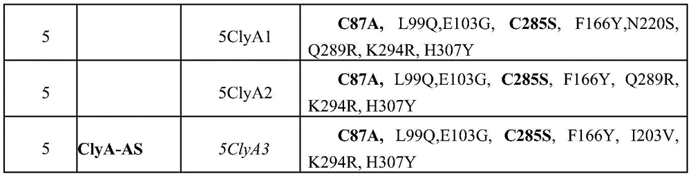
TABLE 3 primer N represents A, G, C or T; s represents G or C, so that the NNS codon encodes a complete set of amino acids

TABLE 4 introduction of DNA molecules used in this work. 1 was prepared by PCR as described in the methods. 3 was prepared by incubating 3a with 20% excess of 3b and then purifying using affinity chromatography as described in the methods. The complementary sequences in these two DNA strands are shown in italics.


ClyA sequence

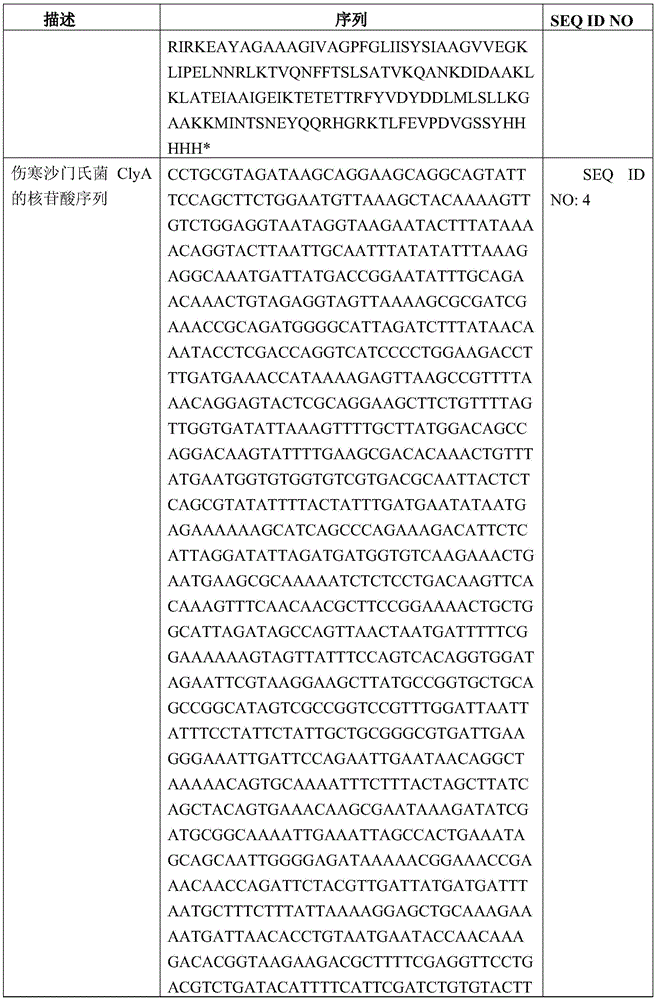


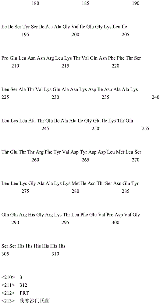


Claims (24)
Applications Claiming Priority (5)
| Application Number | Priority Date | Filing Date | Title |
|---|---|---|---|
| US201361805068P | 2013-03-25 | 2013-03-25 | |
| US61/805,068 | 2013-03-25 | ||
| GB1313477.0 | 2013-07-29 | ||
| GBGB1313477.0A GB201313477D0 (en) | 2013-07-29 | 2013-07-29 | Nanopore biosensors for detection of proteins and nucleic acids |
| PCT/BE2014/000013 WO2014153625A1 (en) | 2013-03-25 | 2014-03-25 | Nanopore biosensors for detection of proteins and nucleic acids |
Publications (2)
| Publication Number | Publication Date |
|---|---|
| CN105358567A CN105358567A (en) | 2016-02-24 |
| CN105358567B true CN105358567B (en) | 2021-12-10 |
Family
ID=49167071
Family Applications (1)
| Application Number | Title | Priority Date | Filing Date |
|---|---|---|---|
| CN201480018571.6A Active CN105358567B (en) | 2013-03-25 | 2014-03-25 | Nanopore biosensors for detecting proteins and nucleic acids |
Country Status (9)
| Country | Link |
|---|---|
| US (7) | US10006905B2 (en) |
| EP (1) | EP2978773B1 (en) |
| JP (1) | JP6592427B2 (en) |
| CN (1) | CN105358567B (en) |
| AU (1) | AU2014245783B2 (en) |
| BR (1) | BR112015024452B1 (en) |
| CA (1) | CA2904202C (en) |
| GB (1) | GB201313477D0 (en) |
| WO (1) | WO2014153625A1 (en) |
Families Citing this family (42)
| Publication number | Priority date | Publication date | Assignee | Title |
|---|---|---|---|---|
| GB0905140D0 (en) | 2009-03-25 | 2009-05-06 | Isis Innovation | Method |
| CN104350067A (en) | 2012-04-10 | 2015-02-11 | 牛津纳米孔技术公司 | mutant lysin pore |
| GB201313477D0 (en) | 2013-07-29 | 2013-09-11 | Univ Leuven Kath | Nanopore biosensors for detection of proteins and nucleic acids |
| AU2015208919B9 (en) | 2014-01-22 | 2021-04-01 | Oxford Nanopore Technologies Limited | Method for attaching one or more polynucleotide binding proteins to a target polynucleotide |
| CN106459159B (en) | 2014-05-02 | 2021-11-30 | 牛津纳米孔技术公司 | Abrupt change hole |
| WO2016034591A2 (en) | 2014-09-01 | 2016-03-10 | Vib Vzw | Mutant pores |
| US10266885B2 (en) | 2014-10-07 | 2019-04-23 | Oxford Nanopore Technologies Ltd. | Mutant pores |
| DK3218519T3 (en) | 2014-11-11 | 2020-12-21 | Bgi Shenzhen | MULTIPASS SEQUENCE |
| GB201502809D0 (en) | 2015-02-19 | 2015-04-08 | Oxford Nanopore Tech Ltd | Mutant pore |
| GB201502810D0 (en) | 2015-02-19 | 2015-04-08 | Oxford Nanopore Tech Ltd | Method |
| EP3283887B1 (en) | 2015-04-14 | 2021-07-21 | Katholieke Universiteit Leuven | Nanopores with internal protein adaptors |
| EP3387432B1 (en) * | 2015-12-08 | 2022-09-28 | Katholieke Universiteit Leuven KU Leuven Research & Development | Modified nanopores, compositions comprising the same, and uses thereof |
| WO2017149316A1 (en) | 2016-03-02 | 2017-09-08 | Oxford Nanopore Technologies Limited | Mutant pore |
| CA3212147A1 (en) | 2016-04-06 | 2017-10-12 | Oxford Nanopore Technologies Plc | Mutant pore |
| WO2017184866A1 (en) | 2016-04-21 | 2017-10-26 | F. Hoffmann-La Roche Ag | Alpha-hemolysin variants and uses thereof |
| FR3053119A1 (en) * | 2016-06-24 | 2017-12-29 | Excilone | METHOD OF ELECTRICALLY DETECTING PEPTIDES, PROTEINS AND OTHER MACROMOLECULES |
| GB201612458D0 (en) * | 2016-07-14 | 2016-08-31 | Howorka Stefan And Pugh Genevieve | Membrane spanning DNA nanopores for molecular transport |
| WO2018039587A1 (en) * | 2016-08-26 | 2018-03-01 | Ohio State Innovation Foundation | Phi29 nanochannel for early detection of breast cancer biomarkers |
| CN110267974A (en) | 2017-02-10 | 2019-09-20 | 牛津纳米孔技术公司 | Modified nanopores, compositions containing the same and uses thereof |
| GB201705764D0 (en) | 2017-04-10 | 2017-05-24 | Imp Innovations Ltd | Analyte detection method |
| GB201707122D0 (en) | 2017-05-04 | 2017-06-21 | Oxford Nanopore Tech Ltd | Pore |
| CN117106038B (en) | 2017-06-30 | 2025-12-09 | 弗拉芒区生物技术研究所 | Novel protein pores |
| US11567061B2 (en) | 2017-07-17 | 2023-01-31 | President And Fellows Of Harvard College | Nanopore-matched protein shuttle for molecular characterization |
| EP4265733A1 (en) | 2017-07-17 | 2023-10-25 | President and Fellows of Harvard College | Analysis of nanopore-matched protein shuttle for molecular characterization |
| JP7353290B2 (en) * | 2018-02-12 | 2023-09-29 | オックスフォード ナノポール テクノロジーズ ピーエルシー | Nanopore assembly and its use |
| GB201809207D0 (en) * | 2018-06-05 | 2018-07-25 | Univ Oxford Innovation Ltd | Molecular electronic device |
| US12105079B2 (en) | 2018-09-11 | 2024-10-01 | Rijksuniversiteit Groningen | Biological nanopores having tunable pore diameters and uses thereof as analytical tools |
| US20200326325A1 (en) | 2019-04-12 | 2020-10-15 | Lisa Diamond | Nanosensor chip with compound nanopores |
| CN111912892B (en) * | 2019-05-07 | 2022-03-25 | 南京大学 | Application of Aeromonasin Nanopore Channel in Biophosphorylation and Related Enzyme Analysis |
| CN112708544A (en) * | 2019-10-25 | 2021-04-27 | 成都今是科技有限公司 | Measuring device and measuring method for gene sequencing |
| CN111235224B (en) * | 2020-01-14 | 2023-06-20 | 广东工业大学 | A method and device for precise modification of biomolecules based on magnetophoretic separation |
| CN112481363B (en) * | 2020-03-09 | 2024-05-28 | 南京大学 | Application of mutant Aerolysin monomers in detecting RNA base sequences and RNA modifications |
| WO2021182957A1 (en) | 2020-03-11 | 2021-09-16 | Rijksuniversiteit Groningen | Engineered plyab nanopores and uses thereof. |
| US20230184767A1 (en) * | 2020-05-20 | 2023-06-15 | The Board Of Trustees Of The University Of Illinois | Methods and compositions for rapid direct detection and differentiation of infectious from noninfectious virus |
| KR102309424B1 (en) * | 2020-09-28 | 2021-10-07 | 한국생명공학연구원 | Screening method for the riboswitch-targeted substances using nanopores |
| GB202107192D0 (en) | 2021-05-19 | 2021-06-30 | Oxford Nanopore Tech Ltd | Method |
| GB202118906D0 (en) | 2021-12-23 | 2022-02-09 | Oxford Nanopore Tech Ltd | Method |
| GB202118908D0 (en) | 2021-12-23 | 2022-02-09 | Oxford Nanopore Tech Ltd | Method |
| WO2024091124A1 (en) | 2022-10-28 | 2024-05-02 | Rijksuniversiteit Groningen | Nanopore-based analysis of proteins |
| GB202216162D0 (en) | 2022-10-31 | 2022-12-14 | Oxford Nanopore Tech Plc | Method |
| GB202307486D0 (en) | 2023-05-18 | 2023-07-05 | Oxford Nanopore Tech Plc | Method |
| GB202317028D0 (en) | 2023-11-06 | 2023-12-20 | Oxford Nanopore Tech Ltd | Method |
Citations (2)
| Publication number | Priority date | Publication date | Assignee | Title |
|---|---|---|---|---|
| US20020094526A1 (en) * | 2000-02-11 | 2002-07-18 | Bayley Hagan P. | Biosensor compositions and methods of use |
| CN101827943A (en) * | 2007-05-22 | 2010-09-08 | 康乃尔研究基金会有限公司 | Composition and method for displaying protein on surface of bacteria and derived vesicles and application of composition and method |
Family Cites Families (150)
| Publication number | Priority date | Publication date | Assignee | Title |
|---|---|---|---|---|
| FI82266C (en) | 1982-10-19 | 1991-02-11 | Cetus Corp | FOERFARANDE FOER FRAMSTAELLNING AV IL-2 MUTEIN. |
| US5198543A (en) | 1989-03-24 | 1993-03-30 | Consejo Superior Investigaciones Cientificas | PHI29 DNA polymerase |
| JPH09500102A (en) | 1993-04-28 | 1997-01-07 | ウォーセスター ファウンデーション フォー エクスペリメンタル バイオロジー | Cell-directed dissolution pore forming agent |
| US5777078A (en) | 1993-04-28 | 1998-07-07 | Worcester Foundation For Experimental Biology | Triggered pore-forming agents |
| DE4320201A1 (en) | 1993-06-18 | 1995-01-12 | Asta Medica Ag | Use of Cetrorelix and other nona and decapeptides for the manufacture of a medicament for combating AIDS and for growth stimulation |
| US5386373A (en) | 1993-08-05 | 1995-01-31 | Pavilion Technologies, Inc. | Virtual continuous emission monitoring system with sensor validation |
| US7569341B2 (en) | 1994-01-31 | 2009-08-04 | Trustees Of Boston University | Nucleic acid directed immobilization arrays and methods of assembly |
| US5561043A (en) | 1994-01-31 | 1996-10-01 | Trustees Of Boston University | Self-assembling multimeric nucleic acid constructs |
| US5795782A (en) | 1995-03-17 | 1998-08-18 | President & Fellows Of Harvard College | Characterization of individual polymer molecules based on monomer-interface interactions |
| US6362002B1 (en) | 1995-03-17 | 2002-03-26 | President And Fellows Of Harvard College | Characterization of individual polymer molecules based on monomer-interface interactions |
| JP3891620B2 (en) | 1996-11-14 | 2007-03-14 | 株式会社アイシン・コスモス研究所 | Nucleic acid probe molecule having a hairpin structure, and nucleic acid detection method using the nucleic acid probe molecule |
| US20020197614A1 (en) | 1997-05-16 | 2002-12-26 | Mosaic Technologies, Inc. | Electrophoretic analysis of target molecules using adapter molecules |
| WO1999005167A1 (en) | 1997-07-25 | 1999-02-04 | University Of Massachusetts | Designed protein pores as components for biosensors |
| US6087099A (en) | 1997-09-08 | 2000-07-11 | Myriad Genetics, Inc. | Method for sequencing both strands of a double stranded DNA in a single sequencing reaction |
| US6127166A (en) | 1997-11-03 | 2000-10-03 | Bayley; Hagan | Molluscan ligament polypeptides and genes encoding them |
| JPH11137260A (en) | 1997-11-06 | 1999-05-25 | Soyaku Gijutsu Kenkyusho:Kk | Anti-influenza viral cyclic dumbbell type rna-dna chimera compound and anti-influenza viral agent |
| US6123819A (en) | 1997-11-12 | 2000-09-26 | Protiveris, Inc. | Nanoelectrode arrays |
| DE19826758C1 (en) | 1998-06-15 | 1999-10-21 | Soft Gene Gmbh | Production of closed, double-stranded DNA molecules for use in gene therapy or genetic vaccination |
| US6743605B1 (en) | 1998-06-24 | 2004-06-01 | Enzo Life Sciences, Inc. | Linear amplification of specific nucleic acid sequences |
| US7155344B1 (en) | 1998-07-27 | 2006-12-26 | Caliper Life Sciences, Inc. | Distributed database for analytical instruments |
| US6150112A (en) | 1998-09-18 | 2000-11-21 | Yale University | Methods for identifying DNA sequences for use in comparison of DNA samples by their lack of polymorphism using Y shape adaptors |
| US6267872B1 (en) | 1998-11-06 | 2001-07-31 | The Regents Of The University Of California | Miniature support for thin films containing single channels or nanopores and methods for using same |
| US6426231B1 (en) | 1998-11-18 | 2002-07-30 | The Texas A&M University System | Analyte sensing mediated by adapter/carrier molecules |
| NO986133D0 (en) | 1998-12-23 | 1998-12-23 | Preben Lexow | Method of DNA Sequencing |
| EP1192453B1 (en) | 1999-06-22 | 2012-02-15 | President and Fellows of Harvard College | Molecular and atomic scale evaluation of biopolymers |
| WO2001002425A2 (en) | 1999-06-29 | 2001-01-11 | University Health Network | Peptide conjugates for the stabilization of membrane proteins and interactions with biological membranes |
| AU7901300A (en) | 1999-08-31 | 2001-03-26 | Stefan Bossmann | Method for producing a channel-forming protein |
| WO2001040516A2 (en) | 1999-12-02 | 2001-06-07 | Molecular Staging Inc. | Generation of single-strand circular dna from linear self-annealing segments |
| WO2001062943A1 (en) | 2000-02-25 | 2001-08-30 | Invitrogen Corporation | Topoisomerase linker-mediated amplification methods |
| US20020132978A1 (en) | 2000-03-21 | 2002-09-19 | Hans-Peter Gerber | VEGF-modulated genes and methods employing them |
| ATE382055T1 (en) | 2000-03-22 | 2008-01-15 | Curagen Corp | WNT-1 RELATED POLYPEPTIDES AND NUCLEIC ACIDS ENCODING THEREOF |
| US7001792B2 (en) | 2000-04-24 | 2006-02-21 | Eagle Research & Development, Llc | Ultra-fast nucleic acid sequencing device and a method for making and using the same |
| DE60129831T2 (en) | 2000-09-25 | 2008-04-30 | Sensovation Ag | Apparatus and method for optical measurement |
| WO2002042496A2 (en) | 2000-11-27 | 2002-05-30 | The Regents Of The University Of California | Methods and devices for characterizing duplex nucleic acid molecules |
| US20030087232A1 (en) | 2001-01-25 | 2003-05-08 | Fred Christians | Methods for screening polypeptides |
| US7807408B2 (en) | 2001-03-19 | 2010-10-05 | President & Fellows Of Harvard College | Directed evolution of proteins |
| JP2002355035A (en) | 2001-05-08 | 2002-12-10 | Inst Of Physical & Chemical Res | Sphingomyelin detection probe |
| US6995236B2 (en) | 2001-05-08 | 2006-02-07 | Riken | Sphingomyelin detecting probe |
| US6863833B1 (en) | 2001-06-29 | 2005-03-08 | The Board Of Trustees Of The Leland Stanford Junior University | Microfabricated apertures for supporting bilayer lipid membranes |
| AU2002320294B2 (en) | 2001-07-03 | 2009-01-15 | The Government Of The United States Of America As Represented By The Secretary Of The Department Of Health And Human Services | Mammalian sweet and amino acid heterodimeric taste receptors |
| ATE376146T1 (en) | 2001-08-31 | 2007-11-15 | Gentex Corp | VEHICLE LAMP ARRANGEMENT WITH HEATSINK |
| DE60324778D1 (en) | 2002-03-15 | 2009-01-02 | Nuevolution As | AN IMPROVED METHOD FOR SYNTHESIS OF MATTRESS-RELATED MOLECULES |
| WO2003095669A1 (en) | 2002-05-10 | 2003-11-20 | The Texas A & M University System | Stochastic sensing through covalent interactions |
| EP1606417A2 (en) | 2003-03-07 | 2005-12-21 | Rubicon Genomics Inc. | In vitro dna immortalization and whole genome amplification using libraries generated from randomly fragmented dna |
| US7704712B2 (en) | 2003-03-25 | 2010-04-27 | Stratagene California | DNA polymerase fusions and uses thereof |
| US7163658B2 (en) | 2003-04-23 | 2007-01-16 | Rouvain Bension | Rapid sequencing of polymers |
| US7344882B2 (en) | 2003-05-12 | 2008-03-18 | Bristol-Myers Squibb Company | Polynucleotides encoding variants of the TRP channel family member, LTRPC3 |
| AU2003904237A0 (en) | 2003-08-08 | 2003-08-21 | Garvan Institute Of Medical Research | Novel translocation assay |
| WO2005056750A2 (en) | 2003-12-11 | 2005-06-23 | Quark Biotech, Inc. | Inversion-duplication of nucleic acids and libraries prepared thereby |
| KR101178776B1 (en) | 2004-02-06 | 2012-09-07 | 카운슬 오브 사이언티픽 앤드 인더스트리얼 리서치 | A computer apparatus for identifying adhesin and adhesin-like proteins of therapeutic potential |
| JP2005253427A (en) | 2004-03-15 | 2005-09-22 | Aisin Seiki Co Ltd | Nucleic acid detection method and nucleic acid isolation method |
| US7238485B2 (en) | 2004-03-23 | 2007-07-03 | President And Fellows Of Harvard College | Methods and apparatus for characterizing polynucleotides |
| WO2005124888A1 (en) | 2004-06-08 | 2005-12-29 | President And Fellows Of Harvard College | Suspended carbon nanotube field effect transistor |
| WO2006020775A2 (en) | 2004-08-13 | 2006-02-23 | Harvard University | An ultra high-throughput opti-nanopore dna readout platform |
| US20060105461A1 (en) | 2004-10-22 | 2006-05-18 | May Tom-Moy | Nanopore analysis system |
| EP1842061A4 (en) | 2004-12-21 | 2009-05-13 | Texas A & M Univ Sys | CHANNELS AND PORES IONIC AT HIGH TEMPERATURE |
| GB0505971D0 (en) | 2005-03-23 | 2005-04-27 | Isis Innovation | Delivery of molecules to a lipid bilayer |
| US7272518B2 (en) | 2005-07-01 | 2007-09-18 | Square D Company | Automated hierarchy classification in utility monitoring systems |
| WO2007024997A2 (en) | 2005-08-22 | 2007-03-01 | Fermalogic, Inc. | Methods of increasing production of secondary metabolites |
| US7877154B2 (en) | 2005-09-30 | 2011-01-25 | Fisher-Rosemount Systems, Inc. | Method and system for controlling a batch process |
| KR100730350B1 (en) | 2005-10-17 | 2007-06-19 | 삼성전자주식회사 | DNA detection method and detection device using surface treated nanopores |
| GB0523282D0 (en) | 2005-11-15 | 2005-12-21 | Isis Innovation | Methods using pores |
| WO2007075987A2 (en) | 2005-12-22 | 2007-07-05 | Pacific Biosciences Of California, Inc. | Active surface coupled polymerases |
| US7932029B1 (en) | 2006-01-04 | 2011-04-26 | Si Lok | Methods for nucleic acid mapping and identification of fine-structural-variations in nucleic acids and utilities |
| US7849581B2 (en) | 2006-05-05 | 2010-12-14 | University Of Utah Research Foundation | Nanopore electrode, nanopore membrane, methods of preparation and surface modification, and use thereof |
| CN101578373A (en) | 2006-09-06 | 2009-11-11 | 费斯生物制药公司 | Fusion peptide therapeutic compositions |
| US7638034B2 (en) | 2006-09-21 | 2009-12-29 | Los Alamos National Security, Llc | Electrochemical detection of single molecules using abiotic nanopores having electrically tunable dimensions |
| WO2008045575A2 (en) | 2006-10-13 | 2008-04-17 | J. Craig Venter Institute, Inc. | Sequencing method |
| US8594848B2 (en) | 2006-11-28 | 2013-11-26 | Lester F. Ludwig | Reconfigurable chemical process systems |
| NZ579083A (en) | 2007-02-20 | 2012-07-27 | Oxford Nanopore Tech Ltd | Lipid bilayer sensor system |
| CA2684801C (en) | 2007-04-04 | 2017-10-10 | The Regents Of The University Of California | Compositions, devices, systems, and methods for using a nanopore |
| US8158344B2 (en) | 2007-06-29 | 2012-04-17 | Duke University | Methods and compositions for correlating genetic markers with multiple sclerosis |
| GB0716005D0 (en) | 2007-08-16 | 2007-09-26 | Imp Innovations Ltd | Single molecule spectroscopy using nanoporpus membranes |
| GB0716264D0 (en) | 2007-08-21 | 2007-09-26 | Isis Innovation | Bilayers |
| EP3540436B1 (en) | 2007-09-12 | 2023-11-01 | President And Fellows Of Harvard College | High-resolution molecular sensor |
| GB2453377A (en) | 2007-10-05 | 2009-04-08 | Isis Innovation | Transmembrane protein pores and molecular adapters therefore. |
| US8951731B2 (en) | 2007-10-15 | 2015-02-10 | Complete Genomics, Inc. | Sequence analysis using decorated nucleic acids |
| GB0724736D0 (en) | 2007-12-19 | 2008-01-30 | Oxford Nanolabs Ltd | Formation of layers of amphiphilic molecules |
| US8502972B2 (en) | 2007-12-31 | 2013-08-06 | Fujirebio Inc. | Clusters of microresonators for cavity mode optical sensing |
| US8231969B2 (en) | 2008-03-26 | 2012-07-31 | University Of Utah Research Foundation | Asymmetrically functionalized nanoparticles |
| WO2009120372A2 (en) | 2008-03-28 | 2009-10-01 | Pacific Biosciences Of California, Inc. | Compositions and methods for nucleic acid sequencing |
| WO2009143425A1 (en) | 2008-05-22 | 2009-11-26 | The Regents Of The University Of California | Membrane precursors and membranes formed therefrom |
| US8652771B2 (en) | 2008-05-28 | 2014-02-18 | University of Souther California | Measurement of succinate in urine samples as a biomarker of kidney damage in diabetic subjects |
| CN102144037A (en) | 2008-07-07 | 2011-08-03 | 牛津纳米孔技术有限公司 | Base-detecting pore |
| CN103695530B (en) | 2008-07-07 | 2016-05-25 | 牛津纳米孔技术有限公司 | Enzyme-hole construct |
| ES2576114T3 (en) | 2008-09-22 | 2016-07-05 | University Of Washington | MSP nanopores and related procedures |
| US9080211B2 (en) | 2008-10-24 | 2015-07-14 | Epicentre Technologies Corporation | Transposon end compositions and methods for modifying nucleic acids |
| GB0820927D0 (en) | 2008-11-14 | 2008-12-24 | Isis Innovation | Method |
| EP2359289A4 (en) | 2008-11-26 | 2015-11-11 | Illumina Inc | Methods and systems for analysis of sequencing data |
| CN102439043A (en) | 2009-01-30 | 2012-05-02 | 牛津纳米孔技术有限公司 | hybrid linker |
| WO2010086622A1 (en) | 2009-01-30 | 2010-08-05 | Oxford Nanopore Technologies Limited | Adaptors for nucleic acid constructs in transmembrane sequencing |
| CN102481341B (en) | 2009-02-23 | 2017-05-17 | 希托马克斯医疗有限公司 | Proproteins and methods of use thereof |
| GB0905140D0 (en) | 2009-03-25 | 2009-05-06 | Isis Innovation | Method |
| FR2943656A1 (en) | 2009-03-25 | 2010-10-01 | Air Liquide | HYDROGEN PRODUCTION METHOD AND PLANT USING A THERMOCINETIC COMPRESSOR |
| BRPI1012752B1 (en) | 2009-04-20 | 2019-06-25 | Oxford Nanopore Technologies Limited | METHOD AND APPARATUS FOR DETECTING AN INTERACTION OF A MOLECULAR ENTITY WITH A MEMBRANE PROTEIN IN A LAYER OF ANFIFYLIC MOLECULES |
| CA2781581C (en) | 2009-12-01 | 2018-03-06 | Oxford Nanopore Technologies Limited | Biochemical analysis instrument |
| FR2955773B1 (en) | 2010-02-01 | 2017-05-26 | Commissariat Energie Atomique | MOLECULAR COMPLEX FOR TARGETING ANTIGENS TO ANTIGEN-PRESENTING CELLS AND ITS APPLICATIONS FOR VACCINATION |
| ES2978883T3 (en) | 2010-02-23 | 2024-09-23 | Univ Washington | Nanopore analyte sequencing |
| US9073990B2 (en) | 2010-04-05 | 2015-07-07 | Bar-Llan University | Protease-activatable pore-forming polypeptides |
| US9593370B2 (en) | 2010-10-01 | 2017-03-14 | Oxford Nanopore Technologies Ltd. | Biochemical analysis apparatus and rotary valve |
| CN102116783B (en) | 2010-12-31 | 2013-05-29 | 北京普源精电科技有限公司 | A Waveform Display Method |
| GB201100516D0 (en) | 2011-01-12 | 2011-02-23 | Isis Innovation | Method using fluorinated amphiphiles |
| CN102174554A (en) | 2011-01-24 | 2011-09-07 | 内蒙古民族大学 | Double-control double-regulation prokaryotic expression vector system and construction method and application thereof |
| WO2012107778A2 (en) | 2011-02-11 | 2012-08-16 | Oxford Nanopore Technologies Limited | Mutant pores |
| KR101963918B1 (en) | 2011-05-27 | 2019-03-29 | 옥스포드 나노포어 테크놀로지즈 리미티드 | Coupling method |
| US9580480B2 (en) | 2011-05-31 | 2017-02-28 | Massachusetts Institute Of Technology | Cell-directed synthesis of multifunctional nanopatterns and nanomaterials |
| BR112014001699A2 (en) | 2011-07-25 | 2017-06-13 | Oxford Nanopore Tech Ltd | method for sequencing a double stranded target polynucleotide, kit, methods for preparing a double stranded target polynucleotide for sequencing and sequencing a double stranded target polynucleotide, and apparatus |
| JP6457811B2 (en) | 2011-09-23 | 2019-01-23 | オックスフォード ナノポール テクノロジーズ リミテッド | Analysis of polymers containing polymer units |
| EP2987870B1 (en) | 2011-10-21 | 2020-02-19 | Oxford Nanopore Technologies Limited | Method of characterizing a target polynucleotide using a transmembrane pore and molecular motor |
| WO2013098561A1 (en) | 2011-12-29 | 2013-07-04 | Oxford Nanopore Technologies Limited | Method for characterising a polynucelotide by using a xpd helicase |
| CA2861808C (en) | 2011-12-29 | 2021-02-23 | Oxford Nanopore Technologies Limited | Enzyme method |
| CA2861457A1 (en) | 2012-01-20 | 2013-07-25 | Genia Technologies, Inc. | Nanopore based molecular detection and sequencing |
| IN2014DN06796A (en) * | 2012-02-15 | 2015-05-22 | Oxford Nanopore Tech Ltd | |
| CN104350067A (en) | 2012-04-10 | 2015-02-11 | 牛津纳米孔技术公司 | mutant lysin pore |
| TWI655213B (en) | 2012-07-13 | 2019-04-01 | Menicon Co., Ltd. | Method for producing self-organized peptide derivative |
| WO2014013262A1 (en) | 2012-07-19 | 2014-01-23 | Oxford Nanopore Technologies Limited | Enzyme construct |
| JP6614972B2 (en) | 2012-07-19 | 2019-12-04 | オックスフォード ナノポール テクノロジーズ リミテッド | Modified helicase |
| WO2014013259A1 (en) | 2012-07-19 | 2014-01-23 | Oxford Nanopore Technologies Limited | Ssb method |
| GB201313121D0 (en) | 2013-07-23 | 2013-09-04 | Oxford Nanopore Tech Ltd | Array of volumes of polar medium |
| AU2013336430B2 (en) | 2012-10-26 | 2018-02-15 | Oxford Nanopore Technologies Limited | Droplet interfaces |
| US9683230B2 (en) | 2013-01-09 | 2017-06-20 | Illumina Cambridge Limited | Sample preparation on a solid support |
| US20140206842A1 (en) | 2013-01-22 | 2014-07-24 | Muhammed Majeed | Peptides Modified with Triterpenoids and Small Organic Molecules: Synthesis and use in Cosmeceutical |
| WO2014122654A2 (en) | 2013-02-07 | 2014-08-14 | Yissum Research Development Company Of The Hebrew University Of Jerusalem Ltd. | Hybrid nanopores and uses thereof for detection of analytes |
| SG11201507138RA (en) | 2013-03-08 | 2015-10-29 | Oxford Nanopore Tech Ltd | Enzyme stalling method |
| GB201314695D0 (en) | 2013-08-16 | 2013-10-02 | Oxford Nanopore Tech Ltd | Method |
| EP3553175B1 (en) | 2013-03-13 | 2021-07-14 | Illumina, Inc. | Method of preparing a nucleic acid sequencing library |
| US10613076B2 (en) | 2013-03-14 | 2020-04-07 | The Trustees Of Boston University | Optoelectronic control of solid-state nanopores |
| GB201313477D0 (en) | 2013-07-29 | 2013-09-11 | Univ Leuven Kath | Nanopore biosensors for detection of proteins and nucleic acids |
| PT3004378T (en) | 2013-05-24 | 2018-03-06 | Illumina Cambridge Ltd | Pyrophosphorolytic sequencing using nanopores |
| WO2015051378A1 (en) | 2013-10-04 | 2015-04-09 | University Of Washington Through Its Center For Commercialization | Systems and methods for nanopore-based analysis of nucleic acids |
| CA2927726A1 (en) | 2013-10-18 | 2015-04-23 | Oxford Nanopore Technologies Limited | Modified helicases for use in nucleic acid sequencing |
| GB201406151D0 (en) | 2014-04-04 | 2014-05-21 | Oxford Nanopore Tech Ltd | Method |
| LT3074534T (en) | 2013-11-26 | 2019-08-26 | Illumina, Inc. | METHODS FOR POLINUCLEOTIDE SEQUENCES |
| WO2015097289A1 (en) | 2013-12-24 | 2015-07-02 | Vib Vzw | Secretion and functional display of chimeric polypeptides |
| AU2015208919B9 (en) | 2014-01-22 | 2021-04-01 | Oxford Nanopore Technologies Limited | Method for attaching one or more polynucleotide binding proteins to a target polynucleotide |
| GB201406155D0 (en) | 2014-04-04 | 2014-05-21 | Oxford Nanopore Tech Ltd | Method |
| GB201403096D0 (en) | 2014-02-21 | 2014-04-09 | Oxford Nanopore Tech Ltd | Sample preparation method |
| US10337060B2 (en) | 2014-04-04 | 2019-07-02 | Oxford Nanopore Technologies Ltd. | Method for characterising a double stranded nucleic acid using a nano-pore and anchor molecules at both ends of said nucleic acid |
| CN106459159B (en) | 2014-05-02 | 2021-11-30 | 牛津纳米孔技术公司 | Abrupt change hole |
| FR3023394B1 (en) | 2014-07-02 | 2017-12-29 | Adn Access Data Networks | DEVICE FOR FACILITATING THE TEACHING OF THE AMBARIC LANGUAGE AND NORMALIZING THE WRITING |
| WO2016034591A2 (en) | 2014-09-01 | 2016-03-10 | Vib Vzw | Mutant pores |
| US10266885B2 (en) | 2014-10-07 | 2019-04-23 | Oxford Nanopore Technologies Ltd. | Mutant pores |
| GB201502809D0 (en) | 2015-02-19 | 2015-04-08 | Oxford Nanopore Tech Ltd | Mutant pore |
| GB201502810D0 (en) | 2015-02-19 | 2015-04-08 | Oxford Nanopore Tech Ltd | Method |
| EP3283887B1 (en) | 2015-04-14 | 2021-07-21 | Katholieke Universiteit Leuven | Nanopores with internal protein adaptors |
| EP3387432B1 (en) | 2015-12-08 | 2022-09-28 | Katholieke Universiteit Leuven KU Leuven Research & Development | Modified nanopores, compositions comprising the same, and uses thereof |
| WO2017149316A1 (en) | 2016-03-02 | 2017-09-08 | Oxford Nanopore Technologies Limited | Mutant pore |
| CA3212147A1 (en) | 2016-04-06 | 2017-10-12 | Oxford Nanopore Technologies Plc | Mutant pore |
| GB201707122D0 (en) | 2017-05-04 | 2017-06-21 | Oxford Nanopore Tech Ltd | Pore |
| CN117106038B (en) | 2017-06-30 | 2025-12-09 | 弗拉芒区生物技术研究所 | Novel protein pores |
-
2013
- 2013-07-29 GB GBGB1313477.0A patent/GB201313477D0/en not_active Ceased
-
2014
- 2014-03-25 WO PCT/BE2014/000013 patent/WO2014153625A1/en not_active Ceased
- 2014-03-25 BR BR112015024452-1A patent/BR112015024452B1/en active IP Right Grant
- 2014-03-25 AU AU2014245783A patent/AU2014245783B2/en active Active
- 2014-03-25 EP EP14724622.7A patent/EP2978773B1/en active Active
- 2014-03-25 CA CA2904202A patent/CA2904202C/en active Active
- 2014-03-25 CN CN201480018571.6A patent/CN105358567B/en active Active
- 2014-03-25 JP JP2016504425A patent/JP6592427B2/en active Active
- 2014-03-25 US US14/779,895 patent/US10006905B2/en active Active
-
2016
- 2016-06-24 US US15/192,317 patent/US20160370358A1/en not_active Abandoned
-
2018
- 2018-05-21 US US15/984,974 patent/US10514378B2/en active Active
-
2019
- 2019-07-24 US US16/521,475 patent/US10802015B2/en active Active
- 2019-09-11 US US16/568,225 patent/US10976311B2/en active Active
-
2021
- 2021-03-09 US US17/196,810 patent/US11761956B2/en active Active
-
2023
- 2023-08-02 US US18/363,859 patent/US20240345073A9/en active Pending
Patent Citations (2)
| Publication number | Priority date | Publication date | Assignee | Title |
|---|---|---|---|---|
| US20020094526A1 (en) * | 2000-02-11 | 2002-07-18 | Bayley Hagan P. | Biosensor compositions and methods of use |
| CN101827943A (en) * | 2007-05-22 | 2010-09-08 | 康乃尔研究基金会有限公司 | Composition and method for displaying protein on surface of bacteria and derived vesicles and application of composition and method |
Non-Patent Citations (7)
| Title |
|---|
| An engineered ClyA nanopore detects folded target proteins by selective external association and pore entry;Soskine M 等;《Nano Lett》;20120806;第12卷(第9期);第4895-4900页 * |
| ClyA cytolysin from Salmonella: Distribution within the genus, regulation of expression by SlyA, and pore-forming characteristics;Christine von Rhein 等;《International Journal of Medical Microbiology》;20091231;第299卷;第21-35页 * |
| Cytotoxin ClyA from Escherichia coli assembles to a 13-meric pore independent of its redox-state;Nora Eifler 等;《The EMBO Journal》;20061231;第25卷(第11期);第2652-2661页 * |
| E. coli Hemolysin E (HlyE, ClyA, SheA): X-Ray Crystal Structure of the Toxin and Observation of Membrane Pores by Electron Microscopy;Alistair J. Wallace等;《Cell》;20000121;第100卷;第265-276页,尤其是第266页Fig.1 Sequence Alignments of HlyE Toxin Family * |
| Nanopore Analysis of Wild-Type and Mutant Prion Protein (PrPC): Single Molecule Discrimination and PrPC Kinetics;Nahid N. Jetha 等;《PLOS ONE》;20130205;第8卷(第2期);第e54982-e54982页 * |
| Nanopore sequencing technology:nanopore preparations;Minsoung Rhee 等;《TRENDS in Biotechnology》;20070222;第25卷(第4期);第175-181页 * |
| Structure-Function Relationships of a Novel Bacterial Toxin, Hemolysin E;Angela Atkins 等;《THE JOURNAL OF BIOLOGICAL CHEMISTRY》;20001229;第275卷(第52期);第41150-41155页,尤其是摘要、第41150页左栏第一段到右栏最后一段、第41154页TABLE III * |
Also Published As
| Publication number | Publication date |
|---|---|
| EP2978773A1 (en) | 2016-02-03 |
| US20240044881A1 (en) | 2024-02-08 |
| AU2014245783A1 (en) | 2015-10-01 |
| BR112015024452A2 (en) | 2017-10-24 |
| US10006905B2 (en) | 2018-06-26 |
| US10976311B2 (en) | 2021-04-13 |
| US20240345073A9 (en) | 2024-10-17 |
| US20190346431A1 (en) | 2019-11-14 |
| US10514378B2 (en) | 2019-12-24 |
| US20200072824A1 (en) | 2020-03-05 |
| EP2978773B1 (en) | 2020-05-27 |
| GB201313477D0 (en) | 2013-09-11 |
| CN105358567A (en) | 2016-02-24 |
| US20160053300A1 (en) | 2016-02-25 |
| AU2014245783B2 (en) | 2018-08-16 |
| BR112015024452B1 (en) | 2023-12-05 |
| US11761956B2 (en) | 2023-09-19 |
| US20160370358A1 (en) | 2016-12-22 |
| WO2014153625A1 (en) | 2014-10-02 |
| JP6592427B2 (en) | 2019-10-16 |
| US10802015B2 (en) | 2020-10-13 |
| CA2904202A1 (en) | 2014-10-02 |
| CA2904202C (en) | 2021-03-30 |
| US20210405039A1 (en) | 2021-12-30 |
| US20180335425A1 (en) | 2018-11-22 |
| JP2016516735A (en) | 2016-06-09 |
Similar Documents
| Publication | Publication Date | Title |
|---|---|---|
| CN105358567B (en) | Nanopore biosensors for detecting proteins and nucleic acids | |
| US20220091096A1 (en) | Nanopores with internal protein adaptors | |
| EP3485029B1 (en) | Biological nanopores for biopolymer sensing and sequencing based on frac actinoporin | |
| JP2010539966A (en) | Molecular adapter | |
| JP2020530276A (en) | New protein pores | |
| CN109072295A (en) | The nano-pore of modification, composition and its application comprising it | |
| JP2025060611A (en) | Biological nanopores with tunable pore diameters and their use as analytical tools | |
| CN104710519A (en) | MSP nanopores and related methods | |
| Class et al. | Patent application title: NANOPORE BIOSENSORS FOR DETECTION OF PROTEINS AND NUCLEIC ACIDS | |
| HK40052928A (en) | Biological nanopores for biopolymer sensing and sequencing based on frac actinoporin | |
| WO2024138424A1 (en) | Nanopore protein and application thereof | |
| Franceschini et al. | DNA analysis with the biological nanopore ClyA | |
| BR112019000680B1 (en) | Biological Nanopores for Biopolymer Detection and Actinoporin-Fragaceatotoxin C-Based Sequencing |
Legal Events
| Date | Code | Title | Description |
|---|---|---|---|
| C06 | Publication | ||
| PB01 | Publication | ||
| C10 | Entry into substantive examination | ||
| SE01 | Entry into force of request for substantive examination | ||
| GR01 | Patent grant | ||
| GR01 | Patent grant |