CN103200877A - Remote center of motion robot for medical image scanning and image-guided targeting - Google Patents
Remote center of motion robot for medical image scanning and image-guided targeting Download PDFInfo
- Publication number
- CN103200877A CN103200877A CN2011800542090A CN201180054209A CN103200877A CN 103200877 A CN103200877 A CN 103200877A CN 2011800542090 A CN2011800542090 A CN 2011800542090A CN 201180054209 A CN201180054209 A CN 201180054209A CN 103200877 A CN103200877 A CN 103200877A
- Authority
- CN
- China
- Prior art keywords
- freedom
- robot
- drive module
- degree
- imaging
- Prior art date
- Legal status (The legal status is an assumption and is not a legal conclusion. Google has not performed a legal analysis and makes no representation as to the accuracy of the status listed.)
- Granted
Links
Images


Classifications
-
- A—HUMAN NECESSITIES
- A61—MEDICAL OR VETERINARY SCIENCE; HYGIENE
- A61B—DIAGNOSIS; SURGERY; IDENTIFICATION
- A61B8/00—Diagnosis using ultrasonic, sonic or infrasonic waves
- A61B8/44—Constructional features of the ultrasonic, sonic or infrasonic diagnostic device
- A61B8/4444—Constructional features of the ultrasonic, sonic or infrasonic diagnostic device related to the probe
-
- A—HUMAN NECESSITIES
- A61—MEDICAL OR VETERINARY SCIENCE; HYGIENE
- A61N—ELECTROTHERAPY; MAGNETOTHERAPY; RADIATION THERAPY; ULTRASOUND THERAPY
- A61N7/00—Ultrasound therapy
-
- A—HUMAN NECESSITIES
- A61—MEDICAL OR VETERINARY SCIENCE; HYGIENE
- A61B—DIAGNOSIS; SURGERY; IDENTIFICATION
- A61B34/00—Computer-aided surgery; Manipulators or robots specially adapted for use in surgery
- A61B34/30—Surgical robots
-
- A—HUMAN NECESSITIES
- A61—MEDICAL OR VETERINARY SCIENCE; HYGIENE
- A61B—DIAGNOSIS; SURGERY; IDENTIFICATION
- A61B8/00—Diagnosis using ultrasonic, sonic or infrasonic waves
- A61B8/08—Clinical applications
- A61B8/0833—Clinical applications involving detecting or locating foreign bodies or organic structures
- A61B8/0841—Clinical applications involving detecting or locating foreign bodies or organic structures for locating instruments
-
- A—HUMAN NECESSITIES
- A61—MEDICAL OR VETERINARY SCIENCE; HYGIENE
- A61B—DIAGNOSIS; SURGERY; IDENTIFICATION
- A61B8/00—Diagnosis using ultrasonic, sonic or infrasonic waves
- A61B8/08—Clinical applications
- A61B8/0833—Clinical applications involving detecting or locating foreign bodies or organic structures
- A61B8/085—Clinical applications involving detecting or locating foreign bodies or organic structures for locating body or organic structures, e.g. tumours, calculi, blood vessels, nodules
-
- A—HUMAN NECESSITIES
- A61—MEDICAL OR VETERINARY SCIENCE; HYGIENE
- A61B—DIAGNOSIS; SURGERY; IDENTIFICATION
- A61B8/00—Diagnosis using ultrasonic, sonic or infrasonic waves
- A61B8/12—Diagnosis using ultrasonic, sonic or infrasonic waves in body cavities or body tracts, e.g. by using catheters
-
- A—HUMAN NECESSITIES
- A61—MEDICAL OR VETERINARY SCIENCE; HYGIENE
- A61B—DIAGNOSIS; SURGERY; IDENTIFICATION
- A61B8/00—Diagnosis using ultrasonic, sonic or infrasonic waves
- A61B8/13—Tomography
- A61B8/14—Echo-tomography
-
- A—HUMAN NECESSITIES
- A61—MEDICAL OR VETERINARY SCIENCE; HYGIENE
- A61B—DIAGNOSIS; SURGERY; IDENTIFICATION
- A61B8/00—Diagnosis using ultrasonic, sonic or infrasonic waves
- A61B8/42—Details of probe positioning or probe attachment to the patient
- A61B8/4209—Details of probe positioning or probe attachment to the patient by using holders, e.g. positioning frames
- A61B8/4218—Details of probe positioning or probe attachment to the patient by using holders, e.g. positioning frames characterised by articulated arms
-
- A—HUMAN NECESSITIES
- A61—MEDICAL OR VETERINARY SCIENCE; HYGIENE
- A61B—DIAGNOSIS; SURGERY; IDENTIFICATION
- A61B8/00—Diagnosis using ultrasonic, sonic or infrasonic waves
- A61B8/42—Details of probe positioning or probe attachment to the patient
- A61B8/4245—Details of probe positioning or probe attachment to the patient involving determining the position of the probe, e.g. with respect to an external reference frame or to the patient
- A61B8/4254—Details of probe positioning or probe attachment to the patient involving determining the position of the probe, e.g. with respect to an external reference frame or to the patient using sensors mounted on the probe
-
- A—HUMAN NECESSITIES
- A61—MEDICAL OR VETERINARY SCIENCE; HYGIENE
- A61B—DIAGNOSIS; SURGERY; IDENTIFICATION
- A61B8/00—Diagnosis using ultrasonic, sonic or infrasonic waves
- A61B8/48—Diagnostic techniques
- A61B8/483—Diagnostic techniques involving the acquisition of a 3D volume of data
-
- A—HUMAN NECESSITIES
- A61—MEDICAL OR VETERINARY SCIENCE; HYGIENE
- A61B—DIAGNOSIS; SURGERY; IDENTIFICATION
- A61B10/00—Instruments for taking body samples for diagnostic purposes; Other methods or instruments for diagnosis, e.g. for vaccination diagnosis, sex determination or ovulation-period determination; Throat striking implements
- A61B10/02—Instruments for taking cell samples or for biopsy
- A61B10/0233—Pointed or sharp biopsy instruments
- A61B10/0241—Pointed or sharp biopsy instruments for prostate
-
- A—HUMAN NECESSITIES
- A61—MEDICAL OR VETERINARY SCIENCE; HYGIENE
- A61B—DIAGNOSIS; SURGERY; IDENTIFICATION
- A61B90/00—Instruments, implements or accessories specially adapted for surgery or diagnosis and not covered by any of the groups A61B1/00 - A61B50/00, e.g. for luxation treatment or for protecting wound edges
- A61B90/36—Image-producing devices or illumination devices not otherwise provided for
- A61B90/37—Surgical systems with images on a monitor during operation
- A61B2090/378—Surgical systems with images on a monitor during operation using ultrasound
-
- A—HUMAN NECESSITIES
- A61—MEDICAL OR VETERINARY SCIENCE; HYGIENE
- A61B—DIAGNOSIS; SURGERY; IDENTIFICATION
- A61B8/00—Diagnosis using ultrasonic, sonic or infrasonic waves
- A61B8/48—Diagnostic techniques
- A61B8/485—Diagnostic techniques involving measuring strain or elastic properties
-
- A—HUMAN NECESSITIES
- A61—MEDICAL OR VETERINARY SCIENCE; HYGIENE
- A61B—DIAGNOSIS; SURGERY; IDENTIFICATION
- A61B8/00—Diagnosis using ultrasonic, sonic or infrasonic waves
- A61B8/52—Devices using data or image processing specially adapted for diagnosis using ultrasonic, sonic or infrasonic waves
- A61B8/5207—Devices using data or image processing specially adapted for diagnosis using ultrasonic, sonic or infrasonic waves involving processing of raw data to produce diagnostic data, e.g. for generating an image
-
- A—HUMAN NECESSITIES
- A61—MEDICAL OR VETERINARY SCIENCE; HYGIENE
- A61N—ELECTROTHERAPY; MAGNETOTHERAPY; RADIATION THERAPY; ULTRASOUND THERAPY
- A61N7/00—Ultrasound therapy
- A61N7/02—Localised ultrasound hyperthermia
Landscapes
- Health & Medical Sciences (AREA)
- Life Sciences & Earth Sciences (AREA)
- Engineering & Computer Science (AREA)
- Surgery (AREA)
- General Health & Medical Sciences (AREA)
- Veterinary Medicine (AREA)
- Nuclear Medicine, Radiotherapy & Molecular Imaging (AREA)
- Public Health (AREA)
- Animal Behavior & Ethology (AREA)
- Biomedical Technology (AREA)
- Heart & Thoracic Surgery (AREA)
- Medical Informatics (AREA)
- Molecular Biology (AREA)
- Radiology & Medical Imaging (AREA)
- Physics & Mathematics (AREA)
- Biophysics (AREA)
- Pathology (AREA)
- Vascular Medicine (AREA)
- Robotics (AREA)
- Ultra Sonic Daignosis Equipment (AREA)
- Manipulator (AREA)
- Magnetic Resonance Imaging Apparatus (AREA)
Abstract
本发明涉及用于医学图像扫描和图像引导定位的运动机器人遥控中心,其在下文中被称为“欧拉”机器人。欧拉机器人允许用于3维(3D)图像重建的超声扫描并实现各种机器人辅助图像引导过程,例如针吸活组织检查、经由皮肤疗法给予、图像引导导航,并便于与其它成像方式的图像融合。欧拉机器人也可与其它手持医学成像探头——例如用于核成像或用于诸如高强度聚焦超声(HIFU)的疗法的定向给予的伽马摄像机——一起使用。3D超声探头也可与欧拉机器人一起使用来提供用于活组织检查或疗法给予的基于自动图像的定位。此外,欧拉机器人支持应用特殊的基于运动的成像方式,例如超声弹性成像。

The present invention relates to a mobile robot telecentre for medical image scanning and image-guided positioning, which is hereinafter referred to as an "Euler" robot. The Euler robot allows ultrasound scanning for 3-dimensional (3D) image reconstruction and enables various robot-assisted image-guided procedures such as needle biopsy, transdermal therapy delivery, image-guided navigation, and facilitates imaging with other imaging modalities fusion. The Euler robot can also be used with other handheld medical imaging probes such as gamma cameras for nuclear imaging or for targeted delivery of therapies such as high-intensity focused ultrasound (HIFU). The 3D ultrasound probe can also be used with the Euler robot to provide automatic image-based positioning for biopsy or therapy administration. In addition, Euler robots support the application of special motion-based imaging modalities, such as ultrasound elastography.
Description
相关申请的引用References to related applications
本申请要求2010年11月11日提交的美国临时专利申请号61/412,589的利益,该临时专利申请为了所有目的特此通过引用被并入,好像在本文被充分阐述的一样。This application claims the benefit of US Provisional Patent Application No. 61/412,589 filed November 11, 2010, which is hereby incorporated by reference for all purposes as if fully set forth herein.
政府利益的声明Statement of Government Interest
在授权号1R21CA141835-01下以美国政府支持完成本发明。美国政府在本发明中有某些权利。This invention was made with US Government support under Grant No. 1R21CA141835-01. The US Government has certain rights in this invention.
发明领域field of invention
本发明涉及用于医学图像扫描的运动机器人遥控中心。更具体地,本发明涉及用于医学图像扫描和图像引导定位的运动机器人遥控中心。The invention relates to a mobile robot remote control center for medical image scanning. More specifically, the present invention relates to a mobile robot telecentre for medical image scanning and image-guided positioning.
发明背景Background of the invention
前列腺癌是美国男子中最常见形式的癌症。与其它癌症一样,早期诊断和治疗对疾病的临床管理和对维持患者寿命的质量并增加预期寿命是关键的。为促进这些目的,先进的成像技术被发展来提高医师准确地探测和在临床上展示相关的细胞变化并给予治疗的能力。Prostate cancer is the most common form of cancer among American men. As with other cancers, early diagnosis and treatment are critical to the clinical management of the disease and to maintaining quality of life and increasing life expectancy for patients. To further these goals, advanced imaging techniques are being developed to improve the physician's ability to accurately detect and clinically demonstrate relevant cellular changes and deliver treatment.
超声弹性成像是有改进癌症探测的潜力的新兴成像方式。支持弹性成像的原理是,不同的组织在应力下展示不同的应变分布。如果使用超声探头引起(激发)应力,则图像可用于可视化因而产生的应变模式。在压缩和回缩运动期间,图像的保持相对未压缩的区揭露了可能与癌症瘤相关的较高硬度的区。弹性成像目前涉及徒手探头运动以引起应力。因此,探头的重复的压缩和回缩的均匀性对优化弹性成像的可靠性是关键的。Ultrasound elastography is an emerging imaging modality that has the potential to improve cancer detection. The principle behind elastography is that different tissues exhibit different strain distributions under stress. If stress is induced (excited) using an ultrasound probe, the images can be used to visualize the resulting strain patterns. Regions of the image that remained relatively uncompressed during compression and retraction motions revealed regions of higher stiffness that may be associated with cancer tumors. Elastography currently involves freehand probe movement to induce stress. Therefore, the uniformity of repeated compression and retraction of the probe is critical to optimize the reliability of elastography.
医师一般通过执行数字直肠检查并测量血液中的前列腺特异抗原(PSA)的水平来筛选前列腺癌。在升高的PSA水平的异常直肠检查或发现之后,医师通过进行前列腺的活组织检查来确认病理组织的存在。前列腺活组织检查可经直肠(TR)(在此期间活组织检查针头穿过直肠壁插入)或经由经会阴(TP)路线(由此,医师将针头经由皮肤插入阴囊和肛门之间的区域中)来执行。在任一情况下,基于医师经验来确定适当的针头插入部位,且该过程通过经直肠超声波检查术或另一成像技术来引导。Physicians typically screen for prostate cancer by performing a digital rectal exam and measuring the level of prostate-specific antigen (PSA) in the blood. Following an abnormal rectal examination or finding of elevated PSA levels, physicians confirm the presence of pathology by performing a biopsy of the prostate. Prostate biopsies can be done through the transrectal (TR) route, during which the biopsy needle is inserted through the wall of the rectum, or via the transperineal (TP) route, whereby the doctor inserts the needle through the skin into the area between the scrotum and anus ) to execute. In either case, the appropriate needle insertion site is determined based on physician experience, and the process is guided by transrectal ultrasonography or another imaging technique.
多种治疗方法可用于活组织检查结果显示前列腺内的异常组织变化的患者。几十年来,对低和中等风险的前列腺癌的确定性治疗是根治性前列腺切除术或外束辐射疗法。更近一些,从业者成功地使用近距离放射疗法来实现等效的结果。近距离放射疗法占前列腺癌治疗的相当大和增长的比例,因为它在患者外背景中以最小的侵袭性被给予。近距离放射疗法涉及将多个“放射性”种子暂时(高剂量率(HDR)近距离放射疗法)或永久地(低剂量率(LDR)近距离放射疗法)插入前列腺中。近距离放射疗法如经直肠和经会阴活组织检查,需要多次刺穿来得到多个组织核心。经会阴活组织检查常常执行使用近距离放射疗法映射模板来便于癌组织的定位并引导未来的活组织检查。近距离放射疗法和活组织检查都一般由二维(2D)经直肠超声波(TRUS)引导。虽然2D TRUS提供软组织解剖的足够成像,它不允许活组织检查针头或植入性近距离放射疗法种子的定位或精确放置。Various treatments are available for patients whose biopsy results show abnormal tissue changes in the prostate. For decades, the definitive treatment for low- and intermediate-risk prostate cancer was radical prostatectomy or external beam radiation therapy. More recently, practitioners have successfully used brachytherapy to achieve equivalent results. Brachytherapy accounts for a substantial and growing proportion of prostate cancer treatment because it is administered with minimal invasiveness in an out-of-patient setting. Brachytherapy involves inserting multiple "radioactive" seeds into the prostate either temporarily (high dose rate (HDR) brachytherapy) or permanently (low dose rate (LDR) brachytherapy). Brachytherapy, such as transrectal and transperineal biopsy, requires multiple punctures to access multiple tissue cores. Transperineal biopsies are often performed using a brachytherapy mapping template to facilitate localization of cancerous tissue and guide future biopsies. Both brachytherapy and biopsy are generally guided by two-dimensional (2D) transrectal ultrasound (TRUS). While 2D TRUS provides adequate imaging of soft tissue anatomy, it does not allow localization or precise placement of biopsy needles or implantable brachytherapy seeds.
前列腺癌的另一可用的治疗选择是腹腔镜根治性前列腺切除术(LRP)。在前列腺切除术期间,包含肿瘤的前列腺的精确切除术和相邻解剖结构的保存对防止肿瘤复发和失禁以及维持性交能力是关键的。然而,由于前列腺周结缔组织和手术中的出血,与前列腺相邻的解剖结构的可视化可能是挑战性的,即使部位在开腹手术期间使用外科手术放大镜或在腹腔镜放大下被观看时也可能是挑战性的。为了解决这个问题,外科医生在LRP期间使用TRUS,并发现TRUS图像可提供阳性手术切缘的降低的速率,并可帮助保存邻近的结构。然而,使用TRUS引导LRP提出了几个挑战。首先,TRUS探头常常被手动地操作;因此,图像稳定性被损害,且3D计算所需的探头位置数据也失去了。此外,TRUS系统太大而不能与高级机器人辅助最小侵入外科手术系统合作来使用,其中在外科机器人和患者之间的空间太有限而不能容纳用于操作TRUS系统的辅助人员。Another available treatment option for prostate cancer is laparoscopic radical prostatectomy (LRP). Precise resection of the tumor-containing prostate and preservation of adjacent anatomical structures during prostatectomy are critical to preventing tumor recurrence and incontinence and maintaining sexual ability. However, visualization of anatomical structures adjacent to the prostate can be challenging due to periprostatic connective tissue and intraoperative hemorrhage, even when the site is viewed during laparotomy with a surgical loupe or under laparoscopic magnification is challenging. To address this issue, surgeons use TRUS during LRP and find that TRUS images can provide a reduced rate of positive surgical margins and can help preserve adjacent structures. However, using TRUS to guide LRP presents several challenges. First, TRUS probes are often manually manipulated; thus, image stability is compromised and probe position data required for 3D calculations is lost. Furthermore, the TRUS system is too large to be used in conjunction with advanced robot-assisted minimally invasive surgery systems, where the space between the surgical robot and the patient is too limited to accommodate the support staff for operating the TRUS system.
最近出现了用于TRUS探头跟踪和针头干预的机器人系统。虽然这些系统提高了图像稳定性并允许操作员跟踪探头位置,但这些系统一般使用外部光学或电磁跟踪系统,其缺乏获得均匀的图像或数字地锁定轨迹所需的自动运动能力。一些机器人系统的另一限制是它们通常处理针头而不是探头,并被建立用于经会阴而不是腔内进入。因此,系统不适合于TRUS引导的LRP,且近距离放射疗法或活组织检查针头插入的点必须仍然基于外科医师经验来确定。Robotic systems for TRUS probe tracking and needle intervention have recently emerged. While these systems improve image stability and allow the operator to track probe position, these systems generally use external optical or electromagnetic tracking systems that lack the automatic motion capabilities needed to obtain a uniform image or digitally lock onto the trajectory. Another limitation of some robotic systems is that they typically handle needles rather than probes and are built for transperineal rather than endoluminal access. Therefore, the system is not suitable for TRUS-guided LRP, and the point of brachytherapy or biopsy needle insertion must still be determined based on surgeon experience.
因此,在本领域中需要机器人超声操纵器,其具有更紧凑的配置来便于近距离放射疗法、针吸活组织检查、图像引导的LRP和超声弹性成像,并允许从业者在癌治疗下可视化区域的3D重建。Therefore, there is a need in the art for a robotic ultrasound manipulator with a more compact configuration to facilitate brachytherapy, needle biopsy, image-guided LRP, and ultrasound elastography, and to allow practitioners to visualize areas under cancer treatment 3D reconstruction of .
概述overview
根据本发明的第一方面,在外科手术和医学干预期间提供导航的方法包括提供机器人装置来与适合于作为基准标记的至少一个医疗仪器合作地操纵超声成像换能器,扫描所关注的区域并使用超声成像换能器测量其中至少一个解剖特征的至少一个参数,在外科手术和干预期间使用链接到机器人装置的可编程计算机跟踪成像换能器的位置,应用从跟踪换能器得到的信息以构建所关注的区域的至少一个三维模型(在该至少一个三维模型中,医疗仪器可在外科手术或干预期间被可视化),以及使用从至少一个三维模型得到的信息操纵在所关注的区域周围的医疗仪器。According to a first aspect of the present invention, a method of providing navigation during surgical procedures and medical interventions comprises providing a robotic device for manipulating an ultrasound imaging transducer in cooperation with at least one medical instrument suitable as a fiducial marker, scanning a region of interest and Measuring at least one parameter of at least one of the anatomical features using an ultrasound imaging transducer, tracking the position of the imaging transducer using a programmable computer linked to the robotic device during surgery and intervention, applying information obtained from the tracking transducer to constructing at least one three-dimensional model of the region of interest in which medical instruments can be visualized during a surgical procedure or intervention, and manipulating objects around the region of interest using information obtained from the at least one three-dimensional model Medical equipment.
根据本发明的第二方面,用于定位至少一个成像探头的机器人装置包括:支承臂;操作地连接到支承臂的运动模块遥控中心,运动模块遥控中心具有构造有皮带并具有至少两个旋转自由度的平行四边形结构;以及操作地连接到运动模块遥控中心的驱动器模块,驱动器模块用于以至少一个自由度操纵成像探头,驱动器模块提供绕着第一旋转轴的一个自由度和/或沿着第二轴平移的一个线性自由度。According to a second aspect of the present invention, a robotic device for positioning at least one imaging probe comprises: a support arm; a kinematic module remote control center operatively connected to the support arm, the kinematic module remote control center having a strap configured with at least two rotationally free degree of parallelogram structure; and a driver module operatively connected to the motion module remote control center, the driver module is used to manipulate the imaging probe with at least one degree of freedom, the driver module provides one degree of freedom around the first rotational axis and/or along One linear degree of freedom for translation along the second axis.
根据本发明的第三方面,用于执行超声引导干预的装置包括提供至少两个旋转自由度的运动模块遥控中心和提供至少一个自由度的用于操纵成像探头的驱动器模块,驱动器模块提供绕着第一旋转轴的一个自由度和/或沿着与末端执行器的纵轴对齐的第二轴平移的一个线性自由度,且驱动器模块还包括旋转导向装置和轨道,轨道几何结构允许额外的医疗仪器相邻于成像探头而被定位。According to a third aspect of the present invention, a device for performing an ultrasound-guided intervention comprises a motion module remote center providing at least two rotational degrees of freedom and a driver module providing at least one degree of freedom for manipulating an imaging probe, the driver module providing a rotation around One degree of freedom in the first rotational axis and/or one linear degree of freedom in translation along the second axis aligned with the longitudinal axis of the end effector, and the drive module also includes a rotational guide and a track whose geometry allows for additional medical The instrument is positioned adjacent to the imaging probe.
根据本发明的第四方面,用于在患者的身体周围执行超声弹性成像的机器人装置包括:提供至少两个旋转自由度的运动模块遥控中心;提供至少一个自由度的用于操纵超声换能器的驱动器模块,驱动器模块提供绕着第一旋转轴的一个自由度和/或沿着与超声换能器的纵轴对齐的第二轴平移的一个线性自由度;以及用于控制用于触诊所关注的解剖区域的超声换能器的重复的压缩和回缩的可编程计算机系统。According to a fourth aspect of the present invention, a robotic apparatus for performing ultrasound elastography around a patient's body comprises: a motion module telecentre providing at least two rotational degrees of freedom; a motion module providing at least one degree of freedom for manipulating an ultrasound transducer A driver module that provides one degree of freedom about a first rotational axis and/or one linear degree of freedom for translation along a second axis aligned with the longitudinal axis of the ultrasonic transducer; and for controlling A programmable computer system for repeated compression and retraction of an ultrasound transducer of an anatomical region of interest.
根据本发明的第五方面,用于对患者执行针吸活组织检查的机器人装置包括:提供至少两个旋转自由度的运动模块遥控中心;提供至少一个自由度的用于操纵活组织检查针头的驱动器模块,驱动器模块提供用于操作活组织检查针头的至少一个自由度,驱动器模块提供沿着轴平移的一个线性自由度;以及用于控制活组织检查针头的插入和回缩以从所关注的解剖区域得到组织样本的可编程计算机系统。According to a fifth aspect of the present invention, a robotic device for performing a needle biopsy on a patient comprises: a motion module telecentre providing at least two rotational degrees of freedom; a robot for manipulating a biopsy needle providing at least one degree of freedom; a driver module providing at least one degree of freedom for manipulating the biopsy needle, the driver module providing one linear degree of freedom for translation along the axis; and for controlling insertion and retraction of the biopsy needle from the area of interest A programmable computer system for obtaining tissue samples from the dissected area.
附图的简要说明Brief description of the drawings
附图提供直观表示,其将用于更完全地描述本文公开的代表性实施方式,并可由本领域技术人员使用来更好地理解它们及其内在优点。在这些附图中,相似的参考数字标识相应的元件,以及:The drawings provide visual representations that will be used to more fully describe representative embodiments disclosed herein, and can be used by those skilled in the art to better understand them and their inherent advantages. In the drawings, like reference numerals identify corresponding elements, and:
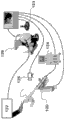
图1示出根据本发明的特征的示例性设备的运动系统图。Figure 1 shows a kinematics diagram of an exemplary device according to features of the invention.
图2示出根据本发明的特征的另一示例性设备的运动系统图。Figure 2 shows a kinematics diagram of another exemplary device according to features of the invention.
图3(a)示出根据本发明的特征的实现Z-Y-X欧拉角的RCM机构的图形表示。Figure 3(a) shows a graphical representation of an RCM mechanism implementing Z-Y-X Euler angles according to a feature of the present invention.
图3(b)示出根据本发明的特征的在固定基础参考系{B}中的工具参考系{T}的固定参考系X-Y-Z旋转的图形表示。Figure 3(b) shows a graphical representation of a fixed reference frame X-Y-Z rotation of a tool reference frame {T} in a fixed base reference frame {B} according to a feature of the invention.
图4示出根据本发明的特征的示例性设备的部分的透视图。Figure 4 shows a perspective view of part of an exemplary device according to features of the invention.
图5示出根据本发明的特征的示例性设备的部分的侧立视图。Figure 5 shows a side elevational view of part of an exemplary apparatus according to features of the invention.
图6示出根据本发明的特征的示例性设备的驱动器模块的分解图。Figure 6 shows an exploded view of a driver module of an exemplary device according to features of the invention.
图7示出具有根据图6的示例性设备的驱动器模块的编码器的基部保持架。FIG. 7 shows a base holder of an encoder with a driver module of the exemplary device according to FIG. 6 .
图8示出根据本发明的特征的又一示例性设备的透视图。Figure 8 shows a perspective view of yet another exemplary device according to features of the invention.
图9示出根据本发明的特征的又一示例性设备的侧立视图。Figure 9 shows a side elevational view of yet another exemplary device according to features of the invention.
图10示出根据本发明的特征的又一示例性设备的驱动器模块的分解图。Figure 10 shows an exploded view of a driver module of yet another exemplary device according to features of the invention.
图11示出根据本发明的特征的与
图12示出根据本发明的特征的与
图13示出根据本发明的特征的示例性系统的系统方框图和安全特征。Figure 13 shows a system block diagram and security features of an exemplary system in accordance with features of the present invention.
图14(a)是由具有根据本发明的特征的示例性设备支承的TRUS探头的试验装置的照片。Figure 14(a) is a photograph of a trial setup of a TRUS probe supported by an exemplary device having features according to the invention.
图14(b)是根据本发明的特征的从基于分割的超声图像的后处理的3D重建。Figure 14(b) is a 3D reconstruction from post-processing of segmentation based ultrasound images according to a feature of the present invention.
图14(c)是使用收集的图像空间的容积再现的成像的腺的快速处理的3D表示。Figure 14(c) is a fast-processing 3D representation of the imaged gland using volumetric reconstruction of the collected image space.
优选实施方式的详细描述Detailed description of the preferred embodiment
现在将参考附图在下文中更充分描述目前公开的主题,在附图中示出了本发明的一些而不是所有实施方式。相似的数字始终指相似的元件。目前公开的主题可体现在很多不同的形式中,且不应被解释为限于本文阐述的实施方式;更确切地,这些实施方式被提供,使得本公开将满足可适用的法律要求。实际上,受益于在前述描述和相关附图中介绍的教导的目前公开的主题所属的领域中的技术人员将想到本文阐述的目前公开的主题的很多修改和其它实施方式。因此,应理解,目前公开的主题不限于所公开的特定实施方式,以及修改和其它实施方式被规定为包括在所附权利要求的范围内。The presently disclosed subject matter now will be described more fully hereinafter with reference to the accompanying drawings, in which some, but not all embodiments of the invention are shown. Like numbers refer to like elements throughout. The presently disclosed subject matter may be embodied in many different forms and should not be construed as limited to the embodiments set forth herein; rather, these embodiments are provided so that this disclosure will satisfy applicable legal requirements. Indeed, many modifications and other implementations of the presently disclosed subject matter set forth herein will come to mind to one skilled in the art to which the presently disclosed subject matter pertains having the benefit of the teachings presented in the foregoing descriptions and the associated drawings. Therefore, it is to be understood that the presently disclosed subject matter is not to be limited to the particular embodiments disclosed and that modifications and other embodiments are intended to be included within the scope of the appended claims.
本发明涉及用于医学图像扫描和图像引导定位的运动机器人遥控中心,其在下文中被称为“欧拉”机器人。欧拉机器人允许用于3维(3D)图像重建的超声扫描并实现各种机器人辅助图像引导过程,例如针吸活组织检查、经由皮肤疗法给予、图像引导导航,并便于与其它成像方式的图像融合。欧拉机器人也可与其它手持医学成像探头——例如用于核成像或用于诸如高强度聚焦超声(HIFU)的疗法的定向给予的伽马摄像机——一起使用。3D超声探头也可与欧拉机器人一起使用来提供用于活组织检查或疗法给予的基于自动图像的定位。此外,欧拉机器人实现特殊的基于运动的成像方式,例如超声弹性成像。欧拉机器人使用运动动力学遥控中心,这是最小侵入进入设备的特征。因此,根据本发明的特征的欧拉机器人也可适用于操纵其它医疗仪器和设备,例如腹腔镜仪器,包括超声波。The present invention relates to a mobile robot telecentre for medical image scanning and image-guided positioning, which is hereinafter referred to as an "Euler" robot. The Euler robot allows ultrasound scanning for 3-dimensional (3D) image reconstruction and enables various robot-assisted image-guided procedures such as needle biopsy, transdermal therapy delivery, image-guided navigation, and facilitates imaging with other imaging modalities fusion. The Euler robot can also be used with other handheld medical imaging probes such as gamma cameras for nuclear imaging or for targeted delivery of therapies such as high-intensity focused ultrasound (HIFU). The 3D ultrasound probe can also be used with the Euler robot to provide automatic image-based positioning for biopsy or therapy administration. In addition, Euler robots enable special motion-based imaging modalities, such as ultrasound elastography. Euler robots use Kinematic Kinetics telecentre, which is a feature of minimal intrusion into the device. Thus, an Euler robot according to features of the present invention may also be adapted for manipulating other medical instruments and equipment, such as laparoscopic instruments, including ultrasound.
参考图1和2,示出了欧拉机器人10的示例性实施方式。欧拉机器人10包括支承臂12和操作地连接到支承臂12的运动模块遥控中心(RCM)14。支承臂12被示为具有带有两个球形接头16和一个圆柱形接头18的一般结构,这些接头可位于期望位置处,以根据临床操作的需要而支承在适当位置上的欧拉机器人10的基部。然而,各种其它结构是可能的,取决于应用和设计偏好。固定架20也可设置在支承臂12的端部处,固定架20可连接到固定支撑物例如医疗桌。Referring to Figures 1 and 2, an exemplary embodiment of an
运动模块遥控中心优选地具有带有皮带的平行四边形结构,如在美国专利号7,021,173(’173专利)中描述的,该专利的全部内容通过引用被并入本文。如在’173专利中讨论的,RCM模块包括第一、第二和第三连杆单元,第一连杆单元在被动转动接头处耦合到基部链接单元。可通过调节基部链接单元和第一连杆单元之间的角度来改变基部调节角度。基部链接单元还包括基部轴,其提供绕着第一轴的一个旋转自由度。第二连杆单元在转动接头处耦合到第一连杆单元,且第二连杆单元也通过另一转动接头耦合到第三连杆单元。第三连杆单元配置成在另一接头(输出轴)处接收用于末端执行器的驱动器或保持架,所述末端执行器例如为超声探头、活组织检查针头或其它医疗仪器。The motion module remote center preferably has a parallelogram structure with belts, as described in US Patent No. 7,021,173 (the '173 patent), which is incorporated herein by reference in its entirety. As discussed in the '173 patent, the RCM module includes first, second and third link units, the first link unit being coupled to the base link unit at a passive swivel joint. The base adjustment angle may be changed by adjusting an angle between the base link unit and the first link unit. The base link unit also includes a base shaft providing a rotational degree of freedom about the first axis. The second link unit is coupled to the first link unit at a rotary joint, and the second link unit is also coupled to the third link unit through another rotary joint. The third linkage unit is configured to receive at the other joint (output shaft) a driver or cage for an end effector such as an ultrasound probe, biopsy needle or other medical instrument.
如进一步在’173专利中描述的,在第一和第二连杆单元之间的转动接头可由电动机启动,而在第二和第三连杆单元之间和在输出轴处的转动接头通过皮带传动机构耦合到第一转动接头。因此,提供第二自由度的RMC模块的第二旋转轴由第一、第二和第三连杆单元以及末端执行器/驱动器保持架实现。系统设计成使得第二连杆可相对于第一连杆被启动,而第三连杆维持其相对于第一连杆的平行方位。末端执行器保持架维持其相对于第二连接单元的平行方位。第一和第二旋转轴以及相应的旋转接头和连杆的配置形成基于双平行四边形的机构。该设计允许绕着轴y旋转末端执行器,其远离该机构并形成经典RCM设计。As further described in the '173 patent, the rotary joint between the first and second linkage units may be actuated by an electric motor, while the rotary joint between the second and third linkage units and at the output shaft is via a belt A transmission mechanism is coupled to the first rotary joint. Thus, the second rotational axis of the RMC module providing the second degree of freedom is realized by the first, second and third linkage units and the end effector/driver cage. The system is designed such that the second link can be activated relative to the first link, while the third link maintains its parallel orientation relative to the first link. The end effector holder maintains its parallel orientation relative to the second linkage unit. The arrangement of the first and second rotational axes and corresponding rotational joints and linkages form a double parallelogram based mechanism. This design allows rotation of the end effector about the axis y, which is away from the mechanism and forms a classic RCM design.
回来参考图1和2,RCM模块14优选地呈现绕着接头30和32的两个旋转自由度(DOF)(Ry,Rz),接头30和32具有在RCM枢轴点处交叉的轴。这个RCM机构的重要特征是,它的结构位于远离枢轴点处,使得空间没有医学干预和目前仪器和/或探头。然而,应理解,可使用其它类型的RCM机构,取决于应用和设计偏好。Referring back to Figures 1 and 2, the
特别参考图1,驱动器模块26(RT驱动器)可操作地连接到RCM模块14。驱动器模块以至少两个自由度操纵位于其中的成像探头24。例如,驱动器模块26可提供绕着第一旋转轴Rx的一个自由度和沿着第二轴Tx平移的一个线性自由度。With particular reference to FIG. 1 , driver module 26 (RT driver) is operatively connected to
参考图2,可选的驱动器模块36(R驱动器)可操作地连接到RCM模块14。驱动器模块36以一个自由度操纵成像探头24(位于其中)。优选地,驱动器模块36提供绕着第一旋转轴Rx的一个自由度。然而,应理解,其它类型的驱动器模块是可能的,取决于应用和设计偏好。特别是,驱动器模块可构造成使得对弹性成像允许触诊和/或机器人适合于保持和操作用于活组织检查的针头。Referring to FIG. 2 , an optional driver module 36 (R Driver) is operably connected to the
根据欧拉机器人10,探头24移动以扫描用于成像的所关注的区域。运动也用于根据需要定探头的方位用于图像引导定位。主要优点是,机器人自动跟踪图像空间的位置和方位,因为它操作探头。因此,对大部分图像引导机器人常见的手术中图像与机器人配准是不需要的。替代地,校准被提前执行一次,且应保持不变。According to the
使用经典3D超声探头,成像的3D体积以及相对于探头的位置随着探头的运动而改变。这使在连续改变的图像内跟随精确解剖目标变得很难。使用欧拉机器人10,目标位置不在固定机器人参考系中改变,且机器人可自动定位探头24以在图像中显示它。通过使用欧拉机器人10来便于数字定位(瞄准由其图像坐标定义的目标)。此外,欧拉机器人10使用较不昂贵的较高图像质量2D设备来实现3D成像。With classical 3D ultrasound probes, the imaged 3D volume and its position relative to the probe changes as the probe moves. This makes it difficult to follow precise anatomical targets within continuously changing images. With the
参考图3(a)和(b),工具参考系T(末端执行器仪器)的方位可通过经典的一组偏航、俯仰和滚转欧拉角(Z-Y-X)来描述。特别是,图3(a)是实现Z-Y-X欧拉角的RCM模块的符号表示,而图3(b)是其工具参考系{T}在固定基础参考系{B}中的固定参考系X-Y-Z旋转。Referring to Fig. 3(a) and (b), the orientation of the tool reference frame T (end effector instrument) can be described by a classical set of yaw, pitch and roll Euler angles (Z-Y-X). In particular, Fig. 3(a) is a symbolic representation of an RCM module implementing Z-Y-X Euler angles, while Fig. 3(b) is its fixed reference frame X-Y-Z rotation of its tool reference frame {T} in a fixed base reference frame {B} .
优选地,RCM机构的远轴(Ry)使用以两个皮带实现的平行四边形机构,如在美国专利号7,021,173中所述的。因为这些接头被机械地耦合,它们用符号方式以单接头连杆1-2表示。欧拉机器人实现三旋转(Θ3,Θ2,Θ1)序列,其绕着从连杆(0)的基础参考系{B}开始到连杆(3)的工具参考系{T}的移动参考系的轴而执行。Preferably, the distal axis (Ry) of the RCM mechanism uses a parallelogram mechanism implemented with two belts, as described in US Patent No. 7,021,173. Because these joints are mechanically coupled, they are symbolically represented as single joint links 1-2. The Euler robot implements a sequence of three rotations (Θ 3 , Θ 2 , Θ 1 ) around a movement starting from the base reference frame {B} of the link (0) to the tool reference frame {T} of the link (3) The axis of the reference system is executed.
这个RCM3连杆配置具有非常有趣的机器人运动学结果,如下。绕着操纵器的移动参考系的轴的欧拉旋转可被表示为:This RCM3 linkage configuration has very interesting robot kinematic results, as follows. Euler about the axis of the manipulator's moving reference frame Rotation can be expressed as:
其中R表示与连杆相关的单独参考系的旋转矩阵。另一方式,工具参考系可被表示为绕着在b中所示的固定{B}坐标参考系的
使得总旋转是:so that the total rotation is:
Rxyz=Rz(Θ3)Ry(Θ2)Rx(Θ1) (方程3)R xyz = R z (Θ 3 )R y (Θ 2 )R x (Θ 1 ) (equation 3)
因为在(1)和(3)中的各个轴旋转矩阵Rx、Ry、Rz是相同的,导致这表明固定X-Y-Z旋转和欧拉Z-Y-X产生相同的最终方位。使用这个机构和旋转参考系的运动学含意是,3-RCM机构的直接运动学解和运动学反解是不重要的:RCM连接角Θ3,Θ2,Θ1等于绕着基本坐标系的固定轴的其相应工具参考系角Θ3,Θ2,Θ1,反之亦然。Because the axis rotation matrices R x , R y , R z in (1) and (3) are the same, resulting in This shows that fixed XYZ rotation and Euler ZYX yield the same final orientation. The kinematic implication of using this mechanism and the rotating reference frame is that the direct kinematic solution and the inverse kinematic solution of the 3-RCM mechanism are unimportant: the RCM connection angles Θ 3 , Θ 2 , Θ 1 are equal to The fixed axes have their corresponding tool reference frame angles Θ 3 , Θ 2 , Θ 1 , and vice versa.
当工具参考系方位由除了固定参考系旋转以外的手段给出时,这些可容易以闭合的形式得到。例如,从(1)中,由公共旋转矩阵所描述的对工具参考系的直接运动学解是:These are readily available in closed form when the tool reference frame orientation is given by means other than fixed reference frame rotation. For example, from (1), the direct kinematic solution to the tool frame of reference described by the common rotation matrix is:
其中sn=sinΘn且cn=cosΘn,n=1、2、3。Wherein sn=sinΘ n and cn=cosΘ n , n=1, 2, 3.
对于反解的情况,给出工具参考系旋转矩阵的元素r:For the case of the inverse solution, the element r of the rotation matrix of the tool reference frame is given:
RCM连接角于是被简单地得到,观察到
这也描述≠0的RCM3机构的奇异点,其中这在其


参考图4-7,将更详细地描述具有驱动器模块26(RT驱动器)的欧拉机器人。特别是,支承超声探头24的RCM模块14在其折叠/收缩位置中示出,在该位置上,平行四边形收缩,如图4和5所示。使用适配器40安装超声探头24,适配器40被定造以匹配探头的形状。然而,应理解,各种其它探头可通过其它特殊适配器等连接到驱动器。Referring to Figures 4-7, the Euler robot with driver module 26 (RT driver) will be described in more detail. In particular, the
参考图6,将更详细地描述驱动器模块26的特征。特别是,适配器40可通过如由包括线性轨道44和导向装置46的结构支承的预装板42——包括传感器——连接到驱动器26。线性轨道44使用滚珠螺杆48由带齿轮的伺服电动机50启动。线性轨道44和导向装置46于是由定造的旋转轨道52和导向装置54的轨道结构支承。旋转轨道52优选地呈现马蹄铁形状,并包括安装在两个轨道部分之间的正扇形齿轮56。Referring to FIG. 6 , the features of the
旋转轨道52和导向装置54呈现与经典线性滚珠轨道类似的结构,但这些轨道具有圆形几何结构,使得因而产生的运动是旋转的而不是线性的。如图7所示,滚珠凹槽60在导向装置中和轨道52的每侧上产生,且两个系列的钢珠62在这些凹槽中循环,并接着通过在导向装置的块内产生的特殊路径穿过滚珠再循环路径64再次循环。The
回来参考图7,马蹄铁形旋转轨道52的启动由其扇形齿轮66给出,扇形齿轮66由来自带齿轮的伺服电动机70的小齿轮68啮合。两个轴都配有冗余编码器72、74,一个编码器72直接连接到螺杆48,另一编码器74通过另一小齿轮连接到旋转轴的小齿轮68。在每个轴上使用一组硬件限制开关76。因为电动机和冗余编码器都是递增的,这些也用于归位。Referring back to FIG. 7 , actuation of the horseshoe shaped revolving
优选地,一组五个力传感器用于测量探头24的所有力-扭矩交互作用,除了绕着其纵轴施加的扭矩以外。优选地,力传感器是具有极端平的设计的用于测量动态和准静态力的小型石英传感器系列,但其它类型的传感器是可能的。四个传感器插在探头适配器40和RT驱动器26之间,以测量横向于探头24施加的所有力和力矩。轴向力由插在滚珠螺母和旋转轨道52之间的传感器测量。Preferably, a set of five force transducers is used to measure all force-torque interactions of the
几个盖80用于包住电动机、编码器和电连接。臂84将机构的基部连接到远侧RCM固定架(未示出),该机构是旋转导向组件。Several covers 80 are used to enclose the motor, encoder and electrical connections.
如上所述,RT驱动器26实现旋转,该旋转加上RCM模块完成这组三个欧拉方位角。探头适配器被构造成使得这些与探头的纵轴对齐。RT驱动器也提供沿着这个轴的运动的线性自由度。旋转轨道的马蹄铁形几何结构被选择成清空在超声探头之上的空间,并因此便于针头绕着探头的手动进入和操纵,用于超声导向干预例如活组织检查。可选地,如果到探头的这个进入是不需要的,则结构可实质上通过使用对旋转轴的圆盘状结构来简化,这消除了对旋转滚珠轨道和导向装置的需要。As mentioned above, the
基于两个考虑因素来选择上面介绍的这个特定的运动学布置以及R和T导向装置和轨道的顺序:1)以确保为手动进入而清空的空间在旋转期间保持在探头之上,以及2)以实现紧凑和坚固的机械结构。因为探头适配器必须具有长形状以稳固地托住探头的柄,线性轨道的最适当的地方沿着其侧面存在,如图所示。线性导向装置必须接着连接到马蹄铁形旋转轨道。因此,旋转导向装置应优选地以驱动器为基础。然而,其它结构和方位是可能的,取决于应用和设计偏好。This particular kinematic arrangement presented above and the sequence of the R and T guides and tracks was chosen based on two considerations: 1) to ensure that the space emptied for manual entry remains above the probe during rotation, and 2) To achieve a compact and robust mechanical structure. Since the probe adapter must have an elongated shape to hold the probe's shank firmly, the most suitable place for the linear track exists along its side, as shown. The linear guides must then be connected to the horseshoe-shaped revolving track. Therefore, the rotary guide should preferably be based on a drive. However, other configurations and orientations are possible, depending on application and design preference.
参考图8-10,将更详细地描述具有驱动器模块36(R驱动器)的欧拉机器人。特别是,支承超声探头24的RCM模块14在其折叠/收缩位置中示出,在该位置上,平行四边形收缩,如图8和9所示。使用适配器90安装超声探头24,适配器90被定造以匹配探头24的形状。然而,应理解,各种其它探头可通过其它特殊适配器等连接到驱动器。Referring to Figures 8-10, the Euler robot with the driver module 36 (R driver) will be described in more detail. In particular, the
参考图10,超声探头24(所示的外部身体探头)以适配器90被支承,使得各种探头可被使用。适配器90还提供用于安装探头24的规则几何形状,因为一般手持探头具有众所周知不规则的几何结构。适配器90还可包括一组安装夹具92用于容易连接到驱动器36/与驱动器36断开。总的来说,这些特征确保同一探头24可在同一相对位置和方位上重复地连接到驱动器36。这对维持图像参考系相对于机器人的校准是重要的。Referring to Figure 10, the ultrasound probe 24 (external body probe shown) is supported with an
驱动器36呈现圆盘状结构。探头24的适配器90连接到使探头24旋转的中心转子94。转子94由帮助隔离该机构的一组滚珠轴承96或o形环98支承。为了减小驱动器36的尺寸,轴承的座圈构造在驱动器36的主体100和盖102内。为了简单起见,轴承不使用笼状结构,而是一序列插入的稍微较小的滚珠。这些滚珠在正常情况下在与主滚珠相反的方向上滚动,可能减小摩擦。转子94由来自具有齿轮105的伺服电动机104的正齿轮传动机构啮合,齿轮105包含在电动机盖106内。安装在基部上的限制开关104触发旋转位置用于使递增编码器归位。驱动器36不使用冗余编码,而是可简单地被添加有由齿轮啮合的另一小齿轮107,类似于上面描述的RT驱动器。The
驱动器的主体100通过定制的力传感器108连接到RCM模块14以测量在轴向方向上施加在探头上的力,探头被固定在安装盖109内。可使用四个应力集中元件的结构。应力测量仪器被应用于桥的相对侧,且半桥连接可用于测量。The
参考图13,示出系统的方框图,其显示使用超声探头的操作。还包括在对欧拉机器人的直接观察可能很难的应用中用于系统监测的视频摄像机,例如在下面的章节中描述的合作机器人方法。Referring to Figure 13, a block diagram of the system showing operation using an ultrasound probe is shown. Also included are video cameras for system monitoring in applications where direct observation of Euler robots may be difficult, such as the cobot approach described in the following sections.
机器人的控制器可基于Intel(Santa Clara,CA)、Asus上的Core2QuadQ9550处理器(ASUSTeK计算机有限公司,台湾)、运行Windows XP的具有4GB的DDR2存储器的P5N7A-VM主板(微软公司,Redmond,WA)。计算机系统可配备有内置不间断电源或操作安全和短范围便携性。电动机控制可在具有板载数字信号处理器的MC8000-DUAL(PMDI,Victoria,B.C.,加拿大)运动控制卡上实现,用于实时运动控制。这个卡对每个轴提供双正交解码器和计数器,其用于每个轴的电动机和冗余解码器。线性伺服电动机放大器可用于所有四个轴。特制的继电器和看门狗板可被包括。单个50连接器电缆将PC连接到在被动臂的基部处的接线箱。为了增加的刚性,臂安装到定制的超大轨道。连接箱还包括力传感器的放大器以减小力传感器电缆的长度。The robot's controller can be based on an Intel (Santa Clara, CA), Core2QuadQ9550 processor on Asus (ASUSTeK Computer Co., Ltd., Taiwan), a P5N7A-VM motherboard with 4 GB of DDR2 memory running Windows XP (Microsoft Corporation, Redmond, WA). ). The computer system may be equipped with a built-in uninterruptible power supply or operational security and short-range portability. Motor control can be implemented on a MC8000-DUAL (PMDI, Victoria, B.C., Canada) motion control card with an onboard digital signal processor for real-time motion control. This card provides dual quadrature decoders and counters for each axis, motors and redundant encoders for each axis. Linear servo motor amplifiers are available for all four axes. Special purpose relay and watchdog boards can be included. A single 50 connector cable connects the PC to the junction box at the base of the passive arm. For added rigidity, the arms are mounted to custom oversized rails. The junction box also includes an amplifier for the force sensor to reduce the length of the force sensor cable.
看门狗每100ms检查一次系统的几个部件的状态,如果故障状况被探测到就禁用到电动机放大器的功率。两个视觉信号用于用信号通知正被供电和在运动中的机器人的操作状态。紧急停止按钮禁用系统,并且还暂停电动机放大器功率。系统的操作从配备有RCM的2-DOF操纵杆或RT驱动器启动按钮或在数字控制下执行。The watchdog checks the status of several components of the system every 100ms and disables power to the motor amplifier if a fault condition is detected. Two visual signals are used to signal the operating status of the robot being powered and in motion. The emergency stop button disables the system and also suspends the motor amplifier power. Operation of the system is performed from a 2-DOF joystick equipped with RCM or RT driver actuation button or under digital control.
TRUS机器人的软件由轴位运动控制软件、机器人运动软件、超声可视化和3D图像处理软件组成。基于运动控制卡(MCI-SoftLi,PMDI)的功能的高级库用Visual C++(微软公司,Redmond,WA)构造运动控制软件。基于Amira可视化平台(Visage Imaging Inc,San Diego,CA)也用Visual C++编写成像部件和机器人的接口。The software of the TRUS robot consists of axis motion control software, robot motion software, ultrasound visualization and 3D image processing software. A high-level library based on the functions of the motion control card (MCI-SoftLi, PMDI) was used to construct the motion control software with Visual C++ (Microsoft Corporation, Redmond, WA). The interface between the imaging components and the robot was also written in Visual C++ based on the Amira visualization platform (Visage Imaging Inc, San Diego, CA).
当探头由TRUS机器人操纵时,位置数据连同图像是容易得到的。这允许记录用于机器人导航和3D重建的图像-位置数据对。机器人可在任意方向上扫描前列腺。在正常情况下提议绕着探头轴的旋转扫描以减小软组织反射。为旋转扫描发展特殊成像算法,因为一般3D重建基于平行的图像切片(例如CT或MRI)。When the probe is being manipulated by the TRUS robot, position data is readily available along with images. This allows recording of image-position data pairs for robot navigation and 3D reconstruction. The robot scans the prostate in any orientation. Rotational scanning around the probe axis is normally proposed to reduce soft tissue reflections. Develop special imaging algorithms for rotational scans, since typical 3D reconstructions are based on parallel image slices (eg CT or MRI).
因此,目前的机器人便于在外科手术和医学干预期间的导航。机器人装置可用于与适合于作为基准标记的至少一个医疗仪器合作地操纵超声成像换能器。扫描所关注的区域并使用超声成像换能器测量其中至少一个解剖特征的至少一个参数。在外科手术和干预期间使用链接到机器人装置的可编程计算机跟踪成像换能器的位置。从跟踪得到的信息应用于换能器以构建所关注的区域的至少一个三维模型(在该区域中,医疗仪器可在外科手术或干预期间被可视化)。使用从至少一个三维模型得到的信息操纵在所关注的区域周围的医疗仪器。Thus, current robots facilitate navigation during surgical procedures and medical interventions. The robotic device may be used to manipulate the ultrasound imaging transducer in cooperation with at least one medical instrument adapted to act as a fiducial marker. A region of interest is scanned and at least one parameter of at least one anatomical feature therein is measured using an ultrasound imaging transducer. The position of the imaging transducer is tracked during surgical procedures and interventions using a programmable computer linked to the robotic device. Information from the tracking is applied to the transducers to construct at least one three-dimensional model of the region of interest (in which the medical instrument may be visualized during surgery or intervention). The medical instrument is steered around the region of interest using information derived from the at least one three-dimensional model.
所使用的特定医疗仪器优选地包括至少一个臂和机器人辅助最小侵入外科手术系统的相关或合并的仪器,包括疗法给予设备、用于得到组织样本的针头、触诊标志器或腹腔镜或其它外科手术仪器。机器人装置可与手动或机器人执行的医疗程序合作地使用。在超声换能器的扫描期间,医疗仪器作为在手术中被捕获的至少一个实时超声图像中的高回声区是可见的。装置操纵超声成像换能器以在任意方向上扫描。机器人装置允许在可用参考系内的超声换能器位置的连续跟踪。计算机系统捕获至少一个超声图像和相应的超声成像换能器位置坐标。机器人装置提供运动遥控中心的测量和超声换能器驱动角,超声图像从超声换能器驱动角获取。从图像收集的信息被分割并用于在目标外科手术部位中产生所关注的解剖特征的三维超声图像容积。The specific medical instrumentation used preferably includes at least one arm and associated or incorporated instrumentation of a robotically assisted minimally invasive surgical system, including therapy delivery equipment, needles for obtaining tissue samples, palpation markers, or laparoscopic or other surgical Surgical instruments. Robotic devices may be used in conjunction with manually or robotically performed medical procedures. During scanning of the ultrasound transducer, the medical instrument is visible as a hyperechoic region in at least one real-time ultrasound image captured during the procedure. The device steers the ultrasound imaging transducer to scan in any direction. The robotic device allows continuous tracking of the position of the ultrasound transducer within the available frame of reference. A computer system captures at least one ultrasound image and corresponding ultrasound imaging transducer position coordinates. The robotic device provides measurements of the kinematic telecentric and ultrasound transducer drive angles from which ultrasound images are acquired. Information gathered from the images is segmented and used to generate a three-dimensional ultrasound image volume of anatomical features of interest in the target surgical site.
参考图11和12,欧拉机器人120与

如上所述,可关于弹性成像使用欧拉机器人。特别是,可提供用于控制用于触诊所关注的解剖区的超声换能器的重复的压缩和回缩的可编程计算机系统。类似地,可关于对患者执行针吸活组织检查使用欧拉机器人。特别是,可提供用于控制活组织检查针头的插入和回缩以从所关注的解剖区得到组织样本的可编程计算机系统。As mentioned above, an Euler robot can be used with respect to elastography. In particular, a programmable computer system for controlling repeated compression and retraction of an ultrasound transducer for palpation of an anatomical region of interest may be provided. Similarly, the Euler robot may be used in connection with performing needle biopsies on patients. In particular, a programmable computer system may be provided for controlling the insertion and retraction of a biopsy needle to obtain a tissue sample from an anatomical region of interest.
实例example
下面的例子被包括以向实践目前公开的主题的代表性实施方式的本领域中的普通技术人员提供指导。根据本公开和本领域中的一般技能水平,技术人员可认识到,下面的实例被规定为仅仅示例性的,以及很多变化、修改和变更可被使用,而不偏离目前公开的主题的范围。下面的实例作为例证而不是作为限制被提供。The following examples are included to provide guidance to those of ordinary skill in the art for practicing representative embodiments of the presently disclosed subject matter. Those skilled in the art, given this disclosure and one's ordinary skill in the art, will appreciate that the following examples are intended to be illustrative only and that many variations, modifications and alterations may be employed without departing from the scope of the presently disclosed subject matter. The following examples are provided as illustrations and not as limitations.
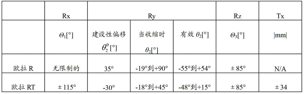
两种类型的原型——欧拉-R(相应于具有驱动器模块36的机器人)和欧拉-RT(相应于具有驱动器模块26的机器人)——在实验室中被构造。呈现紧凑结构的这两个版本由于其RCM运动学结构而具有宽范围的运动,其实现类似于人对探头的操作性的一般定位和扫描运动(表1)。这些值与RCM模块的折叠/收缩位置有关(对于欧拉-R而对于欧拉-RT
表1:欧拉机器人的硬件限制Table 1: Hardware limitations of the Euler robot
几个骨盆模型被构造。图14(a)显示模拟骨盆骨骼、前列腺、膀胱、尿道和代表神经血管束的两个结构的实体模型。实体模型是基于明胶的,由在具有山梨醇、丙三醇和水的溶液中的300Bloom明胶粉末制成,以产生现实的超声视图。使用旋转扫描获取的图像被后处理用于分割并被描绘,如在图14(b)中所示的。图14(c)示出使用“容积再现”模式的直接从“所收集的”图像空间产生的前列腺的3D表示。这可显示腺的3D形状而不分割它。容积再现显示半透明的每个收集的图像体素,且穿过体素的光吸收根据比例因子与其亮度相关。Several pelvic models were constructed. Figure 14(a) shows a mock-up of the pelvic bone, prostate, bladder, urethra, and two structures representing neurovascular bundles. The mockup is gelatin based, made of 300Bloom gelatin powder in a solution with sorbitol, glycerol and water to produce realistic ultrasound views. Images acquired using rotational scans were post-processed for segmentation and depicted as shown in Figure 14(b). Figure 14(c) shows a 3D representation of the prostate directly generated from the "collected" image space using the "volume rendering" mode. This can display the 3D shape of the gland without segmenting it. Volume rendering shows each collected image voxel as translucent, and light absorption through the voxel is related to its brightness according to a scaling factor.
因此,本发明的机器人辅助超声操纵器允许对探头位置和图像空间的方位的内在跟踪,这极大地有利于执行近距离放射疗法、针吸活组织检查、LRP和经直肠超声弹性成像。从业者也受益于在紧凑系统中的运动动力学遥控中心,其将应用范围扩展到操纵医疗仪器和设备,例如腹腔镜仪器,包括腹腔镜超声波。此外,紧凑配置允许从业者与机器人辅助最小侵入外科手术系统合作地使用机器人。最后,自动跟踪超声探头位置并允许探头的均匀应用和回缩的机器人系统优化超声弹性成像的质量和可靠性。Thus, the robot-assisted ultrasound manipulator of the present invention allows intrinsic tracking of probe position and orientation in image space, which greatly facilitates performing brachytherapy, needle biopsy, LRP, and transrectal ultrasound elastography. Practitioners also benefit from a kinematic telecentre in a compact system, which extends the range of applications to manipulating medical instruments and equipment, such as laparoscopic instruments, including laparoscopic ultrasound. Furthermore, the compact configuration allows practitioners to use the robot cooperatively with the robot-assisted minimally invasive surgery system. Finally, a robotic system that automatically tracks the position of the ultrasound probe and allows uniform application and retraction of the probe optimizes the quality and reliability of ultrasound elastography.
虽然本发明关于其优选实施方式而被描述,本领域技术人员将认识到,没有被特别描述的添加、删除、修改和替换可被做出,而不偏离如在所附权利要求中限定的本发明的精神和范围。Although the present invention has been described with respect to its preferred embodiments, those skilled in the art will recognize that additions, deletions, modifications and substitutions not specifically described may be made without departing from the present invention as defined in the appended claims. The spirit and scope of the invention.
Claims (40)
Applications Claiming Priority (3)
| Application Number | Priority Date | Filing Date | Title |
|---|---|---|---|
| US41258910P | 2010-11-11 | 2010-11-11 | |
| US61/412,589 | 2010-11-11 | ||
| PCT/US2011/060362 WO2012065058A2 (en) | 2010-11-11 | 2011-11-11 | Remote center of motion robot for medical image scanning and image-guided targeting |
Publications (2)
| Publication Number | Publication Date |
|---|---|
| CN103200877A true CN103200877A (en) | 2013-07-10 |
| CN103200877B CN103200877B (en) | 2016-02-10 |
Family
ID=46051580
Family Applications (1)
| Application Number | Title | Priority Date | Filing Date |
|---|---|---|---|
| CN201180054209.0A Active CN103200877B (en) | 2010-11-11 | 2011-11-11 | Motion robot remote control center for medical image scanning and image-guided localization |
Country Status (5)
| Country | Link |
|---|---|
| US (1) | US20140039314A1 (en) |
| EP (1) | EP2637571B1 (en) |
| KR (1) | KR102048352B1 (en) |
| CN (1) | CN103200877B (en) |
| WO (1) | WO2012065058A2 (en) |
Cited By (10)
| Publication number | Priority date | Publication date | Assignee | Title |
|---|---|---|---|---|
| CN102652675A (en) * | 2011-03-02 | 2012-09-05 | 通用电气公司 | Device for assisting operating apparatus or tool |
| CN103230304A (en) * | 2013-05-17 | 2013-08-07 | 深圳先进技术研究院 | Surgical navigation system and method |
| CN105962971A (en) * | 2016-05-03 | 2016-09-28 | 上海交通大学 | Multi-freedom-degree driving device of medical ultrasonic probe |
| WO2018040612A1 (en) * | 2016-08-29 | 2018-03-08 | 杭州捷诺飞生物科技股份有限公司 | Biological 3d printing technology-based cutting and repairing integrated surgical robot and method for using same |
| CN108577980A (en) * | 2018-02-08 | 2018-09-28 | 南方医科大学南方医院 | A kind of method, system and device ultrasonic cutter head carried out from motion tracking |
| CN110868937A (en) * | 2017-07-07 | 2020-03-06 | 皇家飞利浦有限公司 | Robotic instrument guide integration with acoustic probes |
| CN111839592A (en) * | 2020-08-13 | 2020-10-30 | 厦门市领汇医疗科技有限公司 | Prostate detection device |
| CN114145781A (en) * | 2021-12-16 | 2022-03-08 | 北京智愈医疗科技有限公司 | A transluminal ultrasound system |
| CN115444557A (en) * | 2017-11-09 | 2022-12-09 | 康坦手术股份有限公司 | Robotic device for minimally invasive medical intervention on soft tissue |
| US20230293253A1 (en) * | 2020-07-20 | 2023-09-21 | The University Of Hong Kong | Fluid-driven robotic needle positioner for image-guided percutaneous interventions |
Families Citing this family (58)
| Publication number | Priority date | Publication date | Assignee | Title |
|---|---|---|---|---|
| US20120095371A1 (en) * | 2010-10-18 | 2012-04-19 | CardioSonic Ltd. | Ultrasound transducer and cooling thereof |
| US20150238167A1 (en) * | 2012-03-22 | 2015-08-27 | Gamma Medical Technologies, Llc | Dual modality endocavity biopsy imaging system and method |
| WO2013142778A2 (en) * | 2012-03-22 | 2013-09-26 | Lall Terrence | Applications of endocavity diagnostic probe in disease treatment |
| US10357304B2 (en) | 2012-04-18 | 2019-07-23 | CardioSonic Ltd. | Tissue treatment |
| US9615815B2 (en) * | 2012-09-28 | 2017-04-11 | Clemson University Research Foundation | Devices that cooperate with ultrasound probes for muscoskeletal evaluations and related systems and methods |
| EP2943111B1 (en) * | 2013-01-14 | 2017-10-18 | B-K Medical ApS | Laparoscopic ultrasound probe for elastographic imaging with automated oscillation mechanism |
| KR101474309B1 (en) * | 2013-03-21 | 2014-12-18 | 가톨릭관동대학교산학협력단 | Fixing apparatus for ultrasonic probe |
| EP2996617B1 (en) | 2013-05-09 | 2021-04-14 | Koninklijke Philips N.V. | Robotic control of an endoscope from anatomical features |
| CN103371870B (en) * | 2013-07-16 | 2015-07-29 | 深圳先进技术研究院 | A kind of surgical navigation systems based on multimode images |
| WO2015085257A1 (en) * | 2013-12-06 | 2015-06-11 | Sonitrack Systems, Inc. | Mechanically driven ultrasound scanning system and method |
| US10064681B2 (en) * | 2014-04-03 | 2018-09-04 | Corbin Clinical Resources, Llc | Method, system, and device for planning and performing, guided and free-handed transperineal prostate biopsies |
| US12144519B2 (en) | 2014-04-03 | 2024-11-19 | Corbin Clinical Resources, Llc | Transperineal prostate biopsy and treatment methods |
| US10743909B2 (en) * | 2014-04-03 | 2020-08-18 | Corbin Clinical Resources, Llc | Transperineal prostate biopsy device, systems, and methods of use |
| US10368850B2 (en) * | 2014-06-18 | 2019-08-06 | Siemens Medical Solutions Usa, Inc. | System and method for real-time ultrasound guided prostate needle biopsies using a compliant robotic arm |
| WO2016025389A1 (en) * | 2014-08-11 | 2016-02-18 | The Regents Of The University Of California | Fine needle elastography device and system for the measurement of material properties |
| US10561397B2 (en) * | 2014-10-31 | 2020-02-18 | Spade Co., Ltd | Ultrasonic probe injection device using RCM |
| CN104622573B (en) * | 2014-12-12 | 2017-06-09 | 北京航空航天大学 | A kind of four-degree-of-freedom remote centre of motion mechanism with high rigidity |
| DE102014226240A1 (en) * | 2014-12-17 | 2016-06-23 | Kuka Roboter Gmbh | System for robot-assisted medical treatment |
| KR20160094105A (en) | 2015-01-30 | 2016-08-09 | 전자부품연구원 | Robot control visualization apparatus |
| JP6657244B2 (en) * | 2015-02-26 | 2020-03-04 | コヴィディエン リミテッド パートナーシップ | Robot controlled remote motion center with software and guide tube |
| JP6393432B2 (en) | 2015-04-21 | 2018-09-19 | コーニンクレッカ フィリップス エヌ ヴェKoninklijke Philips N.V. | Adjustable arm for patient monitoring device |
| WO2017004056A1 (en) | 2015-07-01 | 2017-01-05 | Mako Surgical Corp. | Robotic systems and methods for controlling a tool removing material from a workpiece |
| JP6865739B2 (en) * | 2015-09-28 | 2021-04-28 | コーニンクレッカ フィリップス エヌ ヴェKoninklijke Philips N.V. | Optical alignment of remote motion center robot |
| US10292779B2 (en) * | 2015-11-23 | 2019-05-21 | Sina Robotics And Medical Innovators Co. | Adapting manual laparoscopic surgical instruments for robotic telesurgery applications |
| WO2017177096A1 (en) * | 2016-04-08 | 2017-10-12 | The Regents Of The University Of Michigan | Device for imaging assisted minimally invasive implant and jawbone reconstruction surgery |
| CN109152929B (en) | 2016-04-28 | 2020-09-29 | 皇家飞利浦有限公司 | image-guided treatment delivery |
| WO2017192796A1 (en) * | 2016-05-04 | 2017-11-09 | The Johns Hopkins University | Remote center of motion robot |
| US20190175293A1 (en) * | 2016-06-22 | 2019-06-13 | Koninklijke Philips N.V. | Image guidance for a decoupled kinematic control of a remote-center-of-motion |
| JP6717713B2 (en) * | 2016-08-31 | 2020-07-01 | テルモ株式会社 | Medical device |
| CN106327479B (en) * | 2016-09-23 | 2018-11-20 | 西安电子科技大学 | The device and method that congenital heart disease art medium vessels recognize under angiography intermediary |
| WO2018146682A1 (en) * | 2017-02-09 | 2018-08-16 | Gynisus Ltd | A medical monitoring system and method |
| CN107028626B (en) * | 2017-05-26 | 2023-06-06 | 北京龙慧珩医疗科技发展有限公司 | Elastic registration method and device for prostatectomy |
| BR112020004831B1 (en) | 2017-09-13 | 2022-09-20 | Ultra HOM LLC | CATHETER FOR ULTRASOUND TREATMENT WITH SOLID STATE COOLING |
| CN108553768B (en) * | 2018-05-16 | 2023-11-24 | 天津商业大学 | Prostate particle implantation robot |
| CN109288541B (en) * | 2018-11-15 | 2021-08-06 | 深圳市比邻星精密技术有限公司 | Robot system based on ultrasonic scanning and scanning method thereof |
| IT201900002469A1 (en) * | 2019-02-20 | 2020-08-20 | Univ Degli Studi Di Verona | SURGICAL DEVICE FOR TRANSRECTAL BIOPSY, IN PARTICULAR PROSTATIC, FOR A MOBILE ARM OF A ROBOT |
| IT201900002475A1 (en) * | 2019-02-20 | 2020-08-20 | Univ Degli Studi Di Verona | SURGICAL DEVICE FOR TRANSPERINEAL BIOPSY, IN PARTICULAR PROSTATIC, FOR A MOBILE ARM OF A ROBOT |
| EP3934558A4 (en) | 2019-03-07 | 2022-12-14 | PROCEPT BioRobotics Corporation | ROBOTIC ARMS AND METHODS OF TISSUE RESECTION AND IMAGING |
| CN111920446B (en) * | 2019-05-13 | 2024-11-22 | 张吉民 | Systems that use ultrasound to enhance medical treatment |
| CN112057110B (en) * | 2019-05-22 | 2023-05-23 | 深圳市德力凯医疗设备股份有限公司 | Imaging method of three-dimensional vascular ultrasonic image and navigation equipment in ultrasonic operation |
| CA3082629A1 (en) * | 2019-07-04 | 2021-01-04 | Aaron Fenster | Biopsy apparatus |
| US11071601B2 (en) | 2019-11-11 | 2021-07-27 | Procept Biorobotics Corporation | Surgical probes for tissue resection with robotic arms |
| RU197062U1 (en) * | 2019-12-26 | 2020-03-26 | федеральное государственное автономное образовательное учреждение высшего образования "Санкт-Петербургский политехнический университет Петра Великого" (ФГАОУ ВО "СПбПУ") | HIFU neoplasm thermal ablation robot |
| US11096753B1 (en) | 2020-06-26 | 2021-08-24 | Procept Biorobotics Corporation | Systems and methods for defining and modifying range of motion of probe used in patient treatment |
| US11877818B2 (en) | 2020-06-26 | 2024-01-23 | Procept Biorobotics Corporation | Integration of robotic arms with surgical probes |
| US20220104878A1 (en) * | 2020-10-06 | 2022-04-07 | Mazor Robotics Ltd. | Method, device, and system for image generation based on calculated robotic arm positions |
| KR102570097B1 (en) * | 2020-11-12 | 2023-08-24 | 주식회사 제우기술 | Sampling system and method using robot arm |
| CN115605148A (en) | 2021-04-20 | 2023-01-13 | 普罗赛普特生物机器人公司(Us) | Surgical probe with independent energy source |
| CN113855244B (en) * | 2021-09-08 | 2022-10-18 | 江苏集奥医工交叉科技有限公司 | Surgical robot for treating pain |
| CN114019031B (en) * | 2021-09-13 | 2023-10-13 | 上海工程技术大学 | Main end operating mechanism of force touch system and operation and maintenance device of force touch nuclear power equipment |
| CN113951935A (en) * | 2021-10-26 | 2022-01-21 | 北京智愈医疗科技有限公司 | Automatic ultrasonic inspection system for cavity channel and control method |
| CN114209400A (en) * | 2021-12-13 | 2022-03-22 | 天津科技大学 | Ultrasound image navigation prostate puncture surgical robot |
| KR102710040B1 (en) * | 2022-06-20 | 2024-09-25 | 재단법인 대구경북첨단의료산업진흥재단 | Biopsy robot |
| CN115317094B (en) * | 2022-07-29 | 2024-06-25 | 武汉大学 | RCM puncture device and puncture teaching method |
| WO2024147118A2 (en) * | 2023-01-07 | 2024-07-11 | Endocure Ltd. | Ultrasound system and method for selective diagnosis and treatment of pathogenic cells |
| WO2025054219A1 (en) * | 2023-09-06 | 2025-03-13 | Intuitive Surgical Operations, Inc. | Workflow for an ultrasound guided percutaneous needle robot |
| WO2025264977A1 (en) * | 2024-06-21 | 2025-12-26 | Orchard Ultrasound Innovation Llc | Robotically manipulated tissue treatment system |
| CN118766505A (en) * | 2024-08-23 | 2024-10-15 | 合肥工业大学 | RCM device and robot for abdominal ultrasound examination |
Citations (5)
| Publication number | Priority date | Publication date | Assignee | Title |
|---|---|---|---|---|
| US20050261591A1 (en) * | 2003-07-21 | 2005-11-24 | The Johns Hopkins University | Image guided interventions with interstitial or transmission ultrasound |
| US20070021738A1 (en) * | 2005-06-06 | 2007-01-25 | Intuitive Surgical Inc. | Laparoscopic ultrasound robotic surgical system |
| CN1973753A (en) * | 2006-12-21 | 2007-06-06 | 中国科学院电工研究所 | Micro robot and its in vitro guiding system |
| US20080314181A1 (en) * | 2007-06-19 | 2008-12-25 | Bruce Schena | Robotic Manipulator with Remote Center of Motion and Compact Drive |
| US20090234369A1 (en) * | 2006-06-19 | 2009-09-17 | Robarts Research Institute | Apparatus for guiding a medical tool |
Family Cites Families (7)
| Publication number | Priority date | Publication date | Assignee | Title |
|---|---|---|---|---|
| JP2567928Y2 (en) * | 1992-08-11 | 1998-04-08 | 日本精工株式会社 | Rack-integrated linear guide device |
| US5820623A (en) * | 1995-06-20 | 1998-10-13 | Ng; Wan Sing | Articulated arm for medical procedures |
| JP2005516786A (en) * | 2002-02-06 | 2005-06-09 | ザ ジョンズ ホプキンズ ユニバーシティ | Remote robotic system and method |
| US8521257B2 (en) * | 2006-03-14 | 2013-08-27 | The Johns Hopkins University | Apparatus for insertion of a medical device within a body during a medical imaging process |
| US7832114B2 (en) * | 2007-04-04 | 2010-11-16 | Eigen, Llc | Tracker holder assembly |
| US9895813B2 (en) * | 2008-03-31 | 2018-02-20 | Intuitive Surgical Operations, Inc. | Force and torque sensing in a surgical robot setup arm |
| TW201127573A (en) * | 2010-02-05 | 2011-08-16 | Hon Hai Prec Ind Co Ltd | Robot arm |
-
2010
- 2010-11-11 US US13/885,009 patent/US20140039314A1/en not_active Abandoned
-
2011
- 2011-11-11 EP EP11840646.1A patent/EP2637571B1/en active Active
- 2011-11-11 WO PCT/US2011/060362 patent/WO2012065058A2/en not_active Ceased
- 2011-11-11 KR KR1020137014278A patent/KR102048352B1/en active Active
- 2011-11-11 CN CN201180054209.0A patent/CN103200877B/en active Active
Patent Citations (5)
| Publication number | Priority date | Publication date | Assignee | Title |
|---|---|---|---|---|
| US20050261591A1 (en) * | 2003-07-21 | 2005-11-24 | The Johns Hopkins University | Image guided interventions with interstitial or transmission ultrasound |
| US20070021738A1 (en) * | 2005-06-06 | 2007-01-25 | Intuitive Surgical Inc. | Laparoscopic ultrasound robotic surgical system |
| US20090234369A1 (en) * | 2006-06-19 | 2009-09-17 | Robarts Research Institute | Apparatus for guiding a medical tool |
| CN1973753A (en) * | 2006-12-21 | 2007-06-06 | 中国科学院电工研究所 | Micro robot and its in vitro guiding system |
| US20080314181A1 (en) * | 2007-06-19 | 2008-12-25 | Bruce Schena | Robotic Manipulator with Remote Center of Motion and Compact Drive |
Cited By (14)
| Publication number | Priority date | Publication date | Assignee | Title |
|---|---|---|---|---|
| CN102652675A (en) * | 2011-03-02 | 2012-09-05 | 通用电气公司 | Device for assisting operating apparatus or tool |
| CN103230304A (en) * | 2013-05-17 | 2013-08-07 | 深圳先进技术研究院 | Surgical navigation system and method |
| CN103230304B (en) * | 2013-05-17 | 2015-05-13 | 深圳先进技术研究院 | Surgical navigation system and method |
| CN105962971A (en) * | 2016-05-03 | 2016-09-28 | 上海交通大学 | Multi-freedom-degree driving device of medical ultrasonic probe |
| WO2018040612A1 (en) * | 2016-08-29 | 2018-03-08 | 杭州捷诺飞生物科技股份有限公司 | Biological 3d printing technology-based cutting and repairing integrated surgical robot and method for using same |
| CN110868937A (en) * | 2017-07-07 | 2020-03-06 | 皇家飞利浦有限公司 | Robotic instrument guide integration with acoustic probes |
| CN110868937B (en) * | 2017-07-07 | 2024-03-08 | 皇家飞利浦有限公司 | Integration with robotic instrument guide of acoustic probe |
| CN115444557A (en) * | 2017-11-09 | 2022-12-09 | 康坦手术股份有限公司 | Robotic device for minimally invasive medical intervention on soft tissue |
| CN108577980A (en) * | 2018-02-08 | 2018-09-28 | 南方医科大学南方医院 | A kind of method, system and device ultrasonic cutter head carried out from motion tracking |
| US20230293253A1 (en) * | 2020-07-20 | 2023-09-21 | The University Of Hong Kong | Fluid-driven robotic needle positioner for image-guided percutaneous interventions |
| CN111839592A (en) * | 2020-08-13 | 2020-10-30 | 厦门市领汇医疗科技有限公司 | Prostate detection device |
| CN111839592B (en) * | 2020-08-13 | 2023-12-29 | 厦门市领汇医疗科技有限公司 | Prostate detection device |
| CN114145781A (en) * | 2021-12-16 | 2022-03-08 | 北京智愈医疗科技有限公司 | A transluminal ultrasound system |
| CN114145781B (en) * | 2021-12-16 | 2025-04-18 | 北京智愈医疗科技有限公司 | A transcavitary ultrasound examination system |
Also Published As
| Publication number | Publication date |
|---|---|
| KR20140024837A (en) | 2014-03-03 |
| CN103200877B (en) | 2016-02-10 |
| EP2637571B1 (en) | 2020-01-08 |
| WO2012065058A3 (en) | 2012-08-02 |
| US20140039314A1 (en) | 2014-02-06 |
| EP2637571A2 (en) | 2013-09-18 |
| KR102048352B1 (en) | 2019-11-25 |
| EP2637571A4 (en) | 2017-07-12 |
| WO2012065058A2 (en) | 2012-05-18 |
Similar Documents
| Publication | Publication Date | Title |
|---|---|---|
| CN103200877B (en) | Motion robot remote control center for medical image scanning and image-guided localization | |
| US11096753B1 (en) | Systems and methods for defining and modifying range of motion of probe used in patient treatment | |
| CN108135572B (en) | Transperineal needle guidance | |
| Boctor et al. | Three‐dimensional ultrasound‐guided robotic needle placement: an experimental evaluation | |
| Reinhardt et al. | A computer-assisted device for the intraoperative CT-correlated localization of brain tumors | |
| Cleary et al. | Interventional robotic systems: Applications and technology state‐of‐the‐art | |
| Kettenbach et al. | Robot-assisted biopsy using ultrasound guidance: initial results from in vitro tests | |
| Larson et al. | Design of an MRI-compatible robotic stereotactic device for minimally invasive interventions in the breast | |
| CA2869976C (en) | Cohesive robot-ultrasound probe for prostate biopsy | |
| US9259195B2 (en) | Remotely held needle guide for CT fluoroscopy | |
| US20170239002A1 (en) | Surgical robot platform | |
| Han et al. | Tandem-robot assisted laparoscopic radical prostatectomy to improve the neurovascular bundle visualization: a feasibility study | |
| IL274453B1 (en) | Robotic device for a minimally invasive medical intervention on soft tissues | |
| Kettenbach et al. | Robot-assisted biopsy using computed tomography-guidance: initial results from in vitro tests | |
| JP2005523741A (en) | Device for inserting a medical instrument during a medical imaging process | |
| Boctor et al. | Robotically assisted intraoperative ultrasound with application to ablative therapy of liver cancer | |
| Mohareri et al. | Automatic localization of the da Vinci surgical instrument tips in 3-D transrectal ultrasound | |
| Stoianovici et al. | Endocavity ultrasound probe manipulators | |
| Lin et al. | Robotic system for accurate percutaneous puncture guided by 3D–2D ultrasound | |
| Xiao et al. | Ultrasound robotic system to multi-approach puncture for endoscopic spinal surgery | |
| Moreira et al. | Motorized template for MRI-guided focal cryoablation of prostate cancer | |
| Li et al. | An Ultrasound-Guided Miniature Hand-Eye Integrated Robot for Percutaneous Needle Placement | |
| Kim et al. | Robot for ultrasound-guided prostate imaging and intervention | |
| Wang et al. | Needle Biopsy and Fiber-Optic Compatible Robotic Insertion Platform | |
| Bax | New Mechatronic Systems for the Diagnosis and Treatment of Cancer |
Legal Events
| Date | Code | Title | Description |
|---|---|---|---|
| C06 | Publication | ||
| PB01 | Publication | ||
| C10 | Entry into substantive examination | ||
| SE01 | Entry into force of request for substantive examination | ||
| C14 | Grant of patent or utility model | ||
| GR01 | Patent grant |