CN103168236A - Optogenetic Probes for Membrane Potential Measurements - Google Patents
Optogenetic Probes for Membrane Potential Measurements Download PDFInfo
- Publication number
- CN103168236A CN103168236A CN2011800507345A CN201180050734A CN103168236A CN 103168236 A CN103168236 A CN 103168236A CN 2011800507345 A CN2011800507345 A CN 2011800507345A CN 201180050734 A CN201180050734 A CN 201180050734A CN 103168236 A CN103168236 A CN 103168236A
- Authority
- CN
- China
- Prior art keywords
- cell
- cells
- nucleic acid
- fluorescin
- bacteriorhodopsin
- Prior art date
- Legal status (The legal status is an assumption and is not a legal conclusion. Google has not performed a legal analysis and makes no representation as to the accuracy of the status listed.)
- Granted
Links
Images


Classifications
-
- G—PHYSICS
- G01—MEASURING; TESTING
- G01N—INVESTIGATING OR ANALYSING MATERIALS BY DETERMINING THEIR CHEMICAL OR PHYSICAL PROPERTIES
- G01N33/00—Investigating or analysing materials by specific methods not covered by groups G01N1/00 - G01N31/00
- G01N33/48—Biological material, e.g. blood, urine; Haemocytometers
- G01N33/50—Chemical analysis of biological material, e.g. blood, urine; Testing involving biospecific ligand binding methods; Immunological testing
- G01N33/68—Chemical analysis of biological material, e.g. blood, urine; Testing involving biospecific ligand binding methods; Immunological testing involving proteins, peptides or amino acids
- G01N33/6872—Intracellular protein regulatory factors and their receptors, e.g. including ion channels
-
- C—CHEMISTRY; METALLURGY
- C07—ORGANIC CHEMISTRY
- C07K—PEPTIDES
- C07K14/00—Peptides having more than 20 amino acids; Gastrins; Somatostatins; Melanotropins; Derivatives thereof
- C07K14/195—Peptides having more than 20 amino acids; Gastrins; Somatostatins; Melanotropins; Derivatives thereof from bacteria
-
- C—CHEMISTRY; METALLURGY
- C07—ORGANIC CHEMISTRY
- C07K—PEPTIDES
- C07K14/00—Peptides having more than 20 amino acids; Gastrins; Somatostatins; Melanotropins; Derivatives thereof
-
- C—CHEMISTRY; METALLURGY
- C07—ORGANIC CHEMISTRY
- C07K—PEPTIDES
- C07K14/00—Peptides having more than 20 amino acids; Gastrins; Somatostatins; Melanotropins; Derivatives thereof
- C07K14/435—Peptides having more than 20 amino acids; Gastrins; Somatostatins; Melanotropins; Derivatives thereof from animals; from humans
- C07K14/43504—Peptides having more than 20 amino acids; Gastrins; Somatostatins; Melanotropins; Derivatives thereof from animals; from humans from invertebrates
- C07K14/43595—Peptides having more than 20 amino acids; Gastrins; Somatostatins; Melanotropins; Derivatives thereof from animals; from humans from invertebrates from coelenteratae, e.g. medusae
-
- C—CHEMISTRY; METALLURGY
- C12—BIOCHEMISTRY; BEER; SPIRITS; WINE; VINEGAR; MICROBIOLOGY; ENZYMOLOGY; MUTATION OR GENETIC ENGINEERING
- C12N—MICROORGANISMS OR ENZYMES; COMPOSITIONS THEREOF; PROPAGATING, PRESERVING, OR MAINTAINING MICROORGANISMS; MUTATION OR GENETIC ENGINEERING; CULTURE MEDIA
- C12N15/00—Mutation or genetic engineering; DNA or RNA concerning genetic engineering, vectors, e.g. plasmids, or their isolation, preparation or purification; Use of hosts therefor
- C12N15/09—Recombinant DNA-technology
- C12N15/11—DNA or RNA fragments; Modified forms thereof; Non-coding nucleic acids having a biological activity
- C12N15/62—DNA sequences coding for fusion proteins
- C12N15/625—DNA sequences coding for fusion proteins containing a sequence coding for a signal sequence
-
- C—CHEMISTRY; METALLURGY
- C12—BIOCHEMISTRY; BEER; SPIRITS; WINE; VINEGAR; MICROBIOLOGY; ENZYMOLOGY; MUTATION OR GENETIC ENGINEERING
- C12N—MICROORGANISMS OR ENZYMES; COMPOSITIONS THEREOF; PROPAGATING, PRESERVING, OR MAINTAINING MICROORGANISMS; MUTATION OR GENETIC ENGINEERING; CULTURE MEDIA
- C12N15/00—Mutation or genetic engineering; DNA or RNA concerning genetic engineering, vectors, e.g. plasmids, or their isolation, preparation or purification; Use of hosts therefor
- C12N15/09—Recombinant DNA-technology
- C12N15/63—Introduction of foreign genetic material using vectors; Vectors; Use of hosts therefor; Regulation of expression
- C12N15/79—Vectors or expression systems specially adapted for eukaryotic hosts
- C12N15/85—Vectors or expression systems specially adapted for eukaryotic hosts for animal cells
- C12N15/86—Viral vectors
-
- C—CHEMISTRY; METALLURGY
- C12—BIOCHEMISTRY; BEER; SPIRITS; WINE; VINEGAR; MICROBIOLOGY; ENZYMOLOGY; MUTATION OR GENETIC ENGINEERING
- C12N—MICROORGANISMS OR ENZYMES; COMPOSITIONS THEREOF; PROPAGATING, PRESERVING, OR MAINTAINING MICROORGANISMS; MUTATION OR GENETIC ENGINEERING; CULTURE MEDIA
- C12N7/00—Viruses; Bacteriophages; Compositions thereof; Preparation or purification thereof
-
- G—PHYSICS
- G01—MEASURING; TESTING
- G01N—INVESTIGATING OR ANALYSING MATERIALS BY DETERMINING THEIR CHEMICAL OR PHYSICAL PROPERTIES
- G01N21/00—Investigating or analysing materials by the use of optical means, i.e. using sub-millimetre waves, infrared, visible or ultraviolet light
- G01N21/62—Systems in which the material investigated is excited whereby it emits light or causes a change in wavelength of the incident light
- G01N21/63—Systems in which the material investigated is excited whereby it emits light or causes a change in wavelength of the incident light optically excited
- G01N21/64—Fluorescence; Phosphorescence
- G01N21/6486—Measuring fluorescence of biological material, e.g. DNA, RNA, cells
-
- G—PHYSICS
- G01—MEASURING; TESTING
- G01N—INVESTIGATING OR ANALYSING MATERIALS BY DETERMINING THEIR CHEMICAL OR PHYSICAL PROPERTIES
- G01N33/00—Investigating or analysing materials by specific methods not covered by groups G01N1/00 - G01N31/00
- G01N33/48—Biological material, e.g. blood, urine; Haemocytometers
- G01N33/50—Chemical analysis of biological material, e.g. blood, urine; Testing involving biospecific ligand binding methods; Immunological testing
- G01N33/53—Immunoassay; Biospecific binding assay; Materials therefor
- G01N33/566—Immunoassay; Biospecific binding assay; Materials therefor using specific carrier or receptor proteins as ligand binding reagents where possible specific carrier or receptor proteins are classified with their target compounds
-
- C—CHEMISTRY; METALLURGY
- C07—ORGANIC CHEMISTRY
- C07K—PEPTIDES
- C07K2319/00—Fusion polypeptide
- C07K2319/60—Fusion polypeptide containing spectroscopic/fluorescent detection, e.g. green fluorescent protein [GFP]
-
- C—CHEMISTRY; METALLURGY
- C12—BIOCHEMISTRY; BEER; SPIRITS; WINE; VINEGAR; MICROBIOLOGY; ENZYMOLOGY; MUTATION OR GENETIC ENGINEERING
- C12N—MICROORGANISMS OR ENZYMES; COMPOSITIONS THEREOF; PROPAGATING, PRESERVING, OR MAINTAINING MICROORGANISMS; MUTATION OR GENETIC ENGINEERING; CULTURE MEDIA
- C12N2740/00—Reverse transcribing RNA viruses
- C12N2740/00011—Details
- C12N2740/10011—Retroviridae
- C12N2740/15011—Lentivirus, not HIV, e.g. FIV, SIV
- C12N2740/15041—Use of virus, viral particle or viral elements as a vector
- C12N2740/15043—Use of virus, viral particle or viral elements as a vector viral genome or elements thereof as genetic vector
-
- C—CHEMISTRY; METALLURGY
- C12—BIOCHEMISTRY; BEER; SPIRITS; WINE; VINEGAR; MICROBIOLOGY; ENZYMOLOGY; MUTATION OR GENETIC ENGINEERING
- C12N—MICROORGANISMS OR ENZYMES; COMPOSITIONS THEREOF; PROPAGATING, PRESERVING, OR MAINTAINING MICROORGANISMS; MUTATION OR GENETIC ENGINEERING; CULTURE MEDIA
- C12N2750/00—MICROORGANISMS OR ENZYMES; COMPOSITIONS THEREOF; PROPAGATING, PRESERVING, OR MAINTAINING MICROORGANISMS; MUTATION OR GENETIC ENGINEERING; CULTURE MEDIA ssDNA viruses
- C12N2750/00011—Details
- C12N2750/14011—Parvoviridae
- C12N2750/14111—Dependovirus, e.g. adenoassociated viruses
- C12N2750/14141—Use of virus, viral particle or viral elements as a vector
- C12N2750/14143—Use of virus, viral particle or viral elements as a vector viral genome or elements thereof as genetic vector
-
- C—CHEMISTRY; METALLURGY
- C12—BIOCHEMISTRY; BEER; SPIRITS; WINE; VINEGAR; MICROBIOLOGY; ENZYMOLOGY; MUTATION OR GENETIC ENGINEERING
- C12N—MICROORGANISMS OR ENZYMES; COMPOSITIONS THEREOF; PROPAGATING, PRESERVING, OR MAINTAINING MICROORGANISMS; MUTATION OR GENETIC ENGINEERING; CULTURE MEDIA
- C12N2810/00—Vectors comprising a targeting moiety
-
- G—PHYSICS
- G01—MEASURING; TESTING
- G01N—INVESTIGATING OR ANALYSING MATERIALS BY DETERMINING THEIR CHEMICAL OR PHYSICAL PROPERTIES
- G01N2333/00—Assays involving biological materials from specific organisms or of a specific nature
- G01N2333/435—Assays involving biological materials from specific organisms or of a specific nature from animals; from humans
- G01N2333/705—Assays involving receptors, cell surface antigens or cell surface determinants
- G01N2333/72—Assays involving receptors, cell surface antigens or cell surface determinants for hormones
- G01N2333/726—G protein coupled receptor, e.g. TSHR-thyrotropin-receptor, LH/hCG receptor, FSH
Landscapes
- Health & Medical Sciences (AREA)
- Life Sciences & Earth Sciences (AREA)
- Chemical & Material Sciences (AREA)
- Engineering & Computer Science (AREA)
- Genetics & Genomics (AREA)
- Biomedical Technology (AREA)
- Molecular Biology (AREA)
- Organic Chemistry (AREA)
- Biotechnology (AREA)
- Zoology (AREA)
- Biochemistry (AREA)
- General Health & Medical Sciences (AREA)
- Bioinformatics & Cheminformatics (AREA)
- Wood Science & Technology (AREA)
- General Engineering & Computer Science (AREA)
- Microbiology (AREA)
- Physics & Mathematics (AREA)
- Immunology (AREA)
- Biophysics (AREA)
- Medicinal Chemistry (AREA)
- Urology & Nephrology (AREA)
- Hematology (AREA)
- Plant Pathology (AREA)
- Proteomics, Peptides & Aminoacids (AREA)
- Cell Biology (AREA)
- Pathology (AREA)
- Analytical Chemistry (AREA)
- General Physics & Mathematics (AREA)
- Gastroenterology & Hepatology (AREA)
- Food Science & Technology (AREA)
- Virology (AREA)
- Tropical Medicine & Parasitology (AREA)
- Toxicology (AREA)
- Nuclear Medicine, Radiotherapy & Molecular Imaging (AREA)
- Micro-Organisms Or Cultivation Processes Thereof (AREA)
- Measuring Or Testing Involving Enzymes Or Micro-Organisms (AREA)
Abstract
本发明提供膜电位的光学测量方法,细胞和构建体。这些方法可用于采用现有电极方法不可接触到的细胞。本方法可用于,例如,高通量药物筛选试验中测定能够影响靶细胞膜电位的试剂。
The present invention provides methods, cells, and constructs for optically measuring membrane potential. These methods can be used to measure cells that are inaccessible using existing electrode methods. The methods can be used, for example, in high-throughput drug screening assays to determine agents that affect the membrane potential of target cells.
Description
相关申请的交叉引用Cross References to Related Applications
依据美国法典第35部分119(e)条,本申请要求于2010年12月12日提交的第61/412,972号和2010年8月23日提交的第61/376,049号美国临时申请的权益,其内容以引用的方式全部并入本申请中。Pursuant to Section 119(e) of Part 35, United States Code, this application claims U.S. Provisional Application No. 61/412,972, filed December 12, 2010, and U.S. Provisional Application No. 61/376,049, filed August 23, 2010 rights and interests, the contents of which are incorporated in this application in their entirety by reference.
技术领域technical field
本发明的领域是涉及用于光学测定磷脂双层间电位的方法,构建体和组合物。The field of the invention relates to methods, constructs and compositions for the optical determination of the potential between phospholipid bilayers.
背景技术Background technique
附膜的生物结构可承受膜内外电压差。这种电压,又被称为膜电位,具有多种生物功能,包括传送信息(例如,在神经元中),在产生ATP中作为媒介物(例如,在细菌和线粒体中),为鞭毛电机提供动力(例如,在细菌中)和控制营养素、毒素和信号分子的跨膜运输(在细菌和真核细胞中)。The biological structure attached to the membrane can withstand the voltage difference between the inside and outside of the membrane. This voltage, also known as membrane potential, has a variety of biological functions, including transmitting information (for example, in neurons), acting as a mediator in the production of ATP (for example, in bacteria and mitochondria), providing Motivates (eg, in bacteria) and controls transmembrane transport of nutrients, toxins, and signaling molecules (in bacteria and eukaryotic cells).
尽管膜电位具有重要的的生物学作用,膜电位的测定还是非常困难的。电生理学是通过在膜两侧放置电极来直接记录电位差。电生理学实验设定慢,每次仅能测定一个或少数几个细胞,不能进入深层组织(例如,体内),不能作用于微小细胞(例如,细菌)、坚固细胞壁包围的细胞(例如,酵母)或活动的细胞(例如,精子),不能应用于长期测定,并且在测定中常会损坏或杀死细胞。Despite the important biological role of membrane potential, the measurement of membrane potential is very difficult. Electrophysiology is the direct recording of potential differences by placing electrodes on either side of the membrane. Electrophysiology experiments are slow to set up, can only measure one or a few cells at a time, cannot enter deep tissues (eg, in vivo), cannot act on tiny cells (eg, bacteria), cells surrounded by strong cell walls (eg, yeast) or motile cells (e.g., spermatozoa), cannot be used in long-term assays and often damage or kill the cells during the assay.
因此,需要开发膜电位的新型测定方法。Therefore, there is a need to develop novel assays for membrane potential.
发明内容Contents of the invention
本文描述的方法是使用细菌视紫红质作为光学传感器来检测磷脂层间的电压。我们已经发现该新系统可以光学测定细胞的膜电位或细胞腔隙的结合膜电位,如细胞内的细胞器、人造细胞或其他类脂膜结合结构。The method described here uses bacteriorhodopsin as an optical sensor to detect the voltage between phospholipid layers. We have discovered that this new system can optically measure the membrane potential of cells or the bound membrane potential of cellular compartments, such as intracellular organelles, artificial cells, or other lipid membrane-bound structures.
该方法包括在细胞或细胞器上表达细菌视紫红质,将细胞暴露于光源下和检测细菌视紫红质发射出的荧光,其中通过发射的荧光强度反应膜电位。该方法无需使用电极就可以测定膜电位。该方法可进一步监测一个或多个外部刺激引起的细胞膜电位变化。这不仅有利于科研,并且,例如,在筛选影响膜电压能力的候选试剂中也是很重要的,例如,在药物筛选中。The method comprises expressing bacteriorhodopsin on a cell or an organelle, exposing the cell to a light source and detecting the fluorescence emitted by the bacteriorhodopsin, wherein the membrane potential is reflected by the intensity of the emitted fluorescence. This method measures membrane potential without the use of electrodes. The method can further monitor changes in cell membrane potential caused by one or more external stimuli. This is not only beneficial in scientific research, but is also important, for example, in screening candidate agents for their ability to affect membrane voltage, for example, in drug screening.
本发明还提供表达细菌视紫红质和修饰的细菌视紫红质的细胞,和编码修饰的古细菌视紫红质的核酸构建体,其在真核细胞中测定膜电位及其变化是有用的。在一些具体实施方式中,在此描述的光学传感器使其内源性离子泵活性下降,或相比于天然细菌视紫红质蛋白质,部分或全部活性得以抑制。这样光学感受器可测定电压,但通过构建的离子梯度不能参与改变电压。电压及其变化的检测能够可视化,并且使用光学系统及进行测定。The present invention also provides cells expressing bacteriorhodopsin and modified bacteriorhodopsin, and nucleic acid constructs encoding modified archaealrhodopsin, which are useful for measuring membrane potential and changes thereof in eukaryotic cells. In some embodiments, the optical sensors described herein have their endogenous ion pump activity reduced, or partially or fully inhibited compared to native bacteriorhodopsin protein. In this way the photoreceptors can measure the voltage, but the ionic gradient created by it cannot participate in changing the voltage. The detection of voltage and its change can be visualized and measured using an optical system.
本发明是基于,至少部分上是基于下述发现,该发现是细菌视紫红质蛋白如古细菌视紫红质或变形菌视紫红质以及其修饰的变型,具有降低的离子泵活性(与来自于天然的细菌视紫红质蛋白相比),能够被用作光学传感器在细胞内检测跨膜电压。也就是说,细菌视紫红质和修饰的细菌视紫红质蛋白可用于测定细胞的膜电位以及膜电位的变化。该构建体和方法也可用于组织和器官的体内成像,例如在斑马鱼上,由于电极尺寸的限制其不能用于研究。这不仅有利于科研,而且在筛选新型候选试剂在影响细胞的膜电压能力也是很重要的。The present invention is based, at least in part, on the discovery that bacteriorhodopsin proteins, such as archaeorhodopsin or proteorhodopsin, and modified variants thereof, have reduced ion pump activity (compared to those derived from native bacteriorhodopsin), which can be used as an optical sensor to detect transmembrane voltage in cells. That is, bacteriorhodopsin and modified bacteriorhodopsin proteins can be used to measure the membrane potential of cells and changes in membrane potential. This construct and method can also be used for in vivo imaging of tissues and organs, for example on zebrafish, which cannot be used for research due to limitations in electrode size. This is not only beneficial to scientific research, but also important in screening new candidate reagents for their ability to affect the membrane voltage of cells.
我们已经开发了变形菌视紫红质光学质子传感器(PROPS),其主要作用于细菌细胞,以及基于古细菌视紫红质的荧光电压指示蛋白(VIPs)家族,其也作用于哺乳动物细胞,包括神经元和人类干细胞源性的心肌细胞。VIPs是以来自于古细菌视紫红质3(Arch)及其同系物的电压指示器为基础。这些蛋白以亚毫秒时间分辨率和亚微米空间分辨率指示电动力学。利用VIPs,我们通过在哺乳动物细胞和组织使用电动力学的光学检测表明非接触、高通量和高含量的测定方法。We have developed Proteorhodopsin Optical Proton Sensors (PROPS), which primarily act on bacterial cells, and a family of archaealrhodopsin-based fluorescent voltage indicator proteins (VIPs), which also act on mammalian cells, including neurons. Yuan and human stem cell-derived cardiomyocytes. VIPs are based on voltage indicators derived from archaeal rhodopsin 3 (Arch) and its homologues. These proteins dictate electrokinetics with submillisecond temporal resolution and submicron spatial resolution. Using VIPs, we demonstrate a non-contact, high-throughput and high-content assay method by optical detection using electrokinetics in mammalian cells and tissues.
本文描述的光学传感器不受电极使用的限制,使电生理学研究可以在例如亚细胞区室中(例如,线粒体)或在小细胞中(例如,细菌)中进行。在此描述的光学传感器可用于药物筛选中,装置研究,以及真核和原核细胞电压变化的体内、体外成像中。The optical sensors described here are not limited by the use of electrodes, enabling electrophysiological studies to be performed, for example, in subcellular compartments (eg, mitochondria) or in small cells (eg, bacteria). The optical sensors described here can be used in drug screening, device studies, and in vivo and in vitro imaging of voltage changes in eukaryotic and prokaryotic cells.
我们描述电压指示器蛋白和表达此蛋白的构建体。该构建体具有任意的细胞型的特异性启动子,当细胞分化时启动子开启,例如,分化为神经元细胞,如神经元,或分化为心肌细胞,例如心肌细胞,浦肯野细胞或窦状细胞。该构建体可进一步包括靶信号如线粒体靶信号,其针对在所需膜位置的电压指示器蛋白。我们提供细胞,以及瞬时和稳定表达这些蛋白的细胞系,包括人类干细胞,如诱导多能细胞(iPSC)或胚胎干细胞(ESC),神经祖细胞,神经细胞,心脏祖细胞和心肌细胞。我们还描述了采用描述的电压指示器蛋白筛选药物的方法。We describe a voltage indicator protein and a construct expressing this protein. The construct has any cell-type specific promoter that is turned on when the cell differentiates, for example, into neuronal cells, such as neurons, or into cardiomyocytes, such as cardiomyocytes, Purkinje cells or sinus cells shaped cells. The construct may further include a targeting signal, such as a mitochondrial targeting signal, directed to a voltage indicator protein at a desired membrane location. We offer cells, as well as cell lines transiently and stably expressing these proteins, including human stem cells such as induced pluripotent cells (iPSCs) or embryonic stem cells (ESCs), neural progenitors, neural cells, cardiac progenitors and cardiomyocytes. We also describe methods for screening drugs using the described voltage indicator proteins.
本发明方法使用的细胞,不管是原核细胞还是真核细胞,都被典型的工程化表达细菌视紫红质或修饰的细菌视紫红质,而他们不能天然地表达用于本发明方法的细菌视紫红质蛋白。Cells used in the methods of the invention, whether prokaryotic or eukaryotic, are typically engineered to express bacteriorhodopsin or modified bacteriorhodopsin, whereas they do not naturally express the bacteriorhodopsin used in the methods of the invention protein.
因此,在一个具体实施方式中,发明提供一种测定表达细菌视紫红质蛋白编码的核酸细胞膜电位的方法,该方法包括步骤(a),用至少一个波长的光通过体外、离体或体内激发至少一个包含编码细菌视紫红质蛋白的核酸的细胞,和步骤(b)体外、离体或体内检测来自至少一个细胞的至少一个光学信号,其中和参考相比,由至少一个细胞发射的荧光强度水平指示细胞膜电位。Therefore, in a specific embodiment, the invention provides a method for measuring the membrane potential of a cell expressing a nucleic acid encoded by bacteriorhodopsin protein, the method comprising step (a), using at least one wavelength of light to excite in vitro, in vitro or in vivo at least one cell comprising a nucleic acid encoding a bacteriorhodopsin protein, and step (b) detecting in vitro, ex vivo or in vivo at least one optical signal from the at least one cell, wherein the intensity of fluorescence emitted by the at least one cell is compared to a reference Levels indicate cell membrane potential.
在发明的任一具体实施方式或方面的一些方面中,细菌视紫红质蛋白是修饰的细菌视紫红质蛋白,与来自于天然的细菌视紫红质蛋白相比,其离子泵活性降低。In some aspects of any one embodiment or aspect of the invention, the bacteriorhodopsin is a modified bacteriorhodopsin having reduced ion pump activity compared to bacteriorhodopsin from native sources.
在发明的任一具体实施方式或方面的一些方面中,细菌视紫红质蛋白是变形菌视紫红质蛋白家族中的成员。In some aspects of any one embodiment or aspect of the invention, the bacteriorhodopsin is a member of the Proteus rhodopsin family.
在发明的任一具体实施方式或方面的一些方面中,细菌视紫红质蛋白是古细菌视紫红质蛋白家族的成员。In some aspects of any one embodiment or aspect of the invention, the bacteriorhodopsin is a member of the archaealrhodopsin family of proteins.
在发明的任一具体实施方式或方面的一些方面中,至少一个波长在λ=594-645之间的波长,波长范围在λ=630-645nm之间的波长也是可以使用的。In some aspects of any particular embodiment or aspect of the invention, at least one wavelength between λ=594-645, wavelengths in the wavelength range between λ=630-645nm may also be used.
在发明的任一具体实施方式或方面的一些方面中,细胞是原核细胞。原核细胞可以是革兰阳性或革兰阴性。原核细胞可以是病原性的或非病原性的。In some aspects of any embodiment or aspect of the invention, the cells are prokaryotic cells. Prokaryotic cells can be gram positive or gram negative. Prokaryotic cells can be pathogenic or nonpathogenic.
在发明的任一具体实施方式或方面的一些方面中,细胞是真核细胞。In some aspects of any embodiment or aspect of the invention, the cells are eukaryotic cells.
在发明的任一具体实施方式或方面的一些方面中,真核细胞是哺乳动物细胞。In some aspects of any embodiment or aspect of the invention, the eukaryotic cells are mammalian cells.
在发明的任一具体实施方式或方面的一些方面中,真核细胞是干细胞、多能细胞或祖细胞。In some aspects of any embodiment or aspect of the invention, the eukaryotic cells are stem cells, pluripotent cells or progenitor cells.
在发明的任一具体实施方式或方面的一些方面中,真核细胞是诱导多能细胞。In some aspects of any embodiment or aspect of the invention, the eukaryotic cells are induced pluripotent cells.
在发明的任一具体实施方式或方面的一些方面中,真核细胞是神经细胞。In some aspects of any embodiment or aspect of the invention, the eukaryotic cells are neural cells.
在发明的任一具体实施方式或方面的一些方面中,真核细胞是心肌细胞。In some aspects of any embodiment or aspect of the invention, the eukaryotic cells are cardiomyocytes.
这些细胞可以体外培养或离体培养,或者可以是器官或组织的部分。典型的细胞包括细菌,酵母,植物细胞,和来自脊椎动物或无脊椎动物的细胞。在一些具体实施方式中,真核细胞是人类细胞。在一些具体实施方式中,真核细胞是非人类细胞。在一些具体实施方式中,细胞不天然地表达方法中使用的细菌视紫红质蛋白。These cells can be cultured in vitro or ex vivo, or can be part of an organ or tissue. Typical cells include bacteria, yeast, plant cells, and cells from vertebrates or invertebrates. In some embodiments, the eukaryotic cells are human cells. In some embodiments, the eukaryotic cells are non-human cells. In some embodiments, the cell does not naturally express the bacteriorhodopsin protein used in the methods.
在发明的任一具体实施方式或方面的一些方面中,该方法进一步包括将载体体外,离体或体内转染给至少一个细胞的步骤,所述载体包含编码细菌视紫红质蛋白的核酸。这些细胞可以被瞬时转染或稳定转染。In some aspects of any embodiment or aspect of the invention, the method further comprises the step of transfecting a vector comprising a nucleic acid encoding a bacteriorhodopsin protein into at least one cell in vitro, ex vivo or in vivo. These cells can be transiently or stably transfected.
在发明的任一具体实施方式或方面的一些方面中,编码细菌视紫红质蛋白的核酸与细胞型特异的启动子可操作地连接。In some aspects of any embodiment or aspect of the invention, the nucleic acid encoding bacteriorhodopsin is operably linked to a cell type specific promoter.
在发明的任一具体实施方式或方面的一些方面中,编码细菌视紫红质蛋白的核酸可操作地连接于膜靶向序列。In some aspects of any specific embodiment or aspect of the invention, the nucleic acid encoding bacteriorhodopsin is operably linked to a membrane targeting sequence.
在发明的任一具体实施方式或方面的一些方面中,膜靶向序列是质膜靶向性序列。In some aspects of any specific embodiment or aspect of the invention, the membrane targeting sequence is a plasma membrane targeting sequence.
在发明的任一具体实施方式或方面的一些方面中,膜靶向序列是亚细胞区室靶向序列。In some aspects of any specific embodiment or aspect of the invention, the membrane targeting sequence is a subcellular compartment targeting sequence.
在发明的任一具体实施方式或方面的一些方面中,亚细胞区室是选自于线粒体膜,内质网,肌质网,突触小泡,内涵体和吞噬小体。In some aspects of any embodiment or aspect of the invention, the subcellular compartment is selected from the group consisting of mitochondrial membranes, endoplasmic reticulum, sarcoplasmic reticulum, synaptic vesicles, endosomes and phagosomes.
在发明的任一具体实施方式或方面的一些方面中,细菌视紫红质基因可操作地连接于编码附加荧光蛋白或生色团的核酸。In some aspects of any embodiment or aspect of the invention, the bacteriorhodopsin gene is operably linked to a nucleic acid encoding an additional fluorescent protein or chromophore.
在发明的任一具体实施方式或方面的一些方面中,至少一个附加荧光蛋白是能够指示细胞内离子浓度的蛋白。In some aspects of any one embodiment or aspect of the invention, the at least one additional fluorescent protein is a protein capable of indicating intracellular ion concentrations.
在发明的任一具体实施方式或方面的一些方面中,至少一个能够指示离子浓度的附加荧光蛋白是钙指示剂。In some aspects of any embodiment or aspect of the invention, the at least one additional fluorescent protein capable of indicating ion concentration is a calcium indicator.
在发明的任一具体实施方式或方面的一些方面中,能够指示离子浓度的荧光蛋白是pH指示剂。In some aspects of any one embodiment or aspect of the invention, the fluorescent protein capable of indicating ion concentration is a pH indicator.
在发明的任一具体实施方式或方面的一些方面中,荧光蛋白能够进行非放射性荧光共振,使能量转移到细菌视紫红质,其能量转移速率取决于膜电位。In some aspects of any embodiment or aspect of the invention, the fluorescent protein is capable of non-radioactive fluorescence resonance, enabling energy transfer to bacteriorhodopsin at a rate dependent on membrane potential.
在发明的任一具体实施方式或方面的一些方面中,荧光蛋白的亮度对膜电位和局部化学环境不敏感,因此,和细菌视紫红质的荧光相比可作为参考。In some aspects of any one embodiment or aspect of the invention, the brightness of the fluorescent protein is insensitive to membrane potential and local chemical environment and, therefore, can be compared to the fluorescence of bacteriorhodopsin as a reference.
在发明的任一具体实施方式或方面的一些方面中,该方法还包括如下步骤:采用至少第一和第二波长的光体外、离体或体内激发至少一个细胞;和体外、离体或体内检测至少第一和第二光学信号,该信号由至少第一和第二波长的激发引起,其不同于来自至少一个细胞的至少第一波长。In some aspects of any embodiment or aspect of the invention, the method further comprises the steps of: exciting at least one cell in vitro, ex vivo, or in vivo with light of at least first and second wavelengths; and in vitro, ex vivo, or in vivo At least first and second optical signals resulting from excitation at least first and second wavelengths different from at least the first wavelength from at least one cell are detected.
在发明的任一具体实施方式或方面的一些方面中,至少第二波长是在λ=447-594之间的波长。In some aspects of any embodiment or aspect of the invention, at least the second wavelength is a wavelength between λ=447-594.
在发明的任一具体实施方式或方面的一些方面中,该方法还包括如下步骤:计算细菌视紫红质的荧光发射与附加荧光蛋白的荧光发射的比率,以获取不依赖于表达水平差异的膜电位测定。In some aspects of any specific embodiment or aspect of the invention, the method further comprises the step of calculating the ratio of the fluorescence emission from bacteriorhodopsin to the fluorescence emission from the attached fluorescent protein to obtain a membrane independent of differences in expression levels Potentiometric determination.
在发明的任一具体实施方式或方面的一些方面中,该方法还包括将至少一个细胞体外,离体或体内暴露于能够或预期能够改变膜电位的刺激的步骤。In some aspects of any embodiment or aspect of the invention, the method further comprises the step of exposing at least one cell to a stimulus capable or expected to alter membrane potential in vitro, ex vivo or in vivo.
在发明的任一具体实施方式或方面的一些方面中,刺激是候选试剂。在一些具体实施方式中,给予至少一个候选试剂。在一些具体实施方式中,同时或顺序给予至少两个候选试剂的组合。In some aspects of any embodiment or aspect of the invention, a stimulus is a candidate agent. In some embodiments, at least one candidate agent is administered. In some embodiments, the combination of at least two candidate agents is administered simultaneously or sequentially.
在发明的任一具体实施方式或方面的一些方面中,刺激是细胞培养基组分的变化。In some aspects of any embodiment or aspect of the invention, the stimulus is a change in the composition of the cell culture medium.
在发明的任一具体实施方式或方面的一些方面中,刺激是电流。In some aspects of any embodiment or aspect of the invention, the stimulus is an electrical current.
在发明的任一具体实施方式或方面的一些方面中,该方法还包括在至少第一时间点和至少第二时间点体外,离体或体内测定,至少一个光学信号的步骤。In some aspects of any embodiment or aspect of the invention, the method further comprises the step of determining at least one optical signal at least a first time point and at least a second time point in vitro, ex vivo or in vivo.
在发明的任一具体实施方式或方面的一些方面中,至少第一时间点是在将至少一个细胞暴露于刺激之前,至少第二时间点是将至少一个细胞暴露于刺激之后。In some aspects of any embodiment or aspect of the invention, at least the first time point is before exposing the at least one cell to the stimulus and at least the second time point is after exposing the at least one cell to the stimulus.
在发明的任一具体实施方式或方面的一些方面中,本方法还包括测定来自于暴露在至少第一波长和至少第二波长的光学信号间的荧光比例的步骤。In some aspects of any embodiment or aspect of the invention, the method further comprises the step of determining a ratio of fluorescence between optical signals resulting from exposure to at least a first wavelength and at least a second wavelength.
在发明的任一具体实施方式或方面的一些方面中,该方法包含使用多个细胞。例如在高通量测定模式中。例如,在此具体实施方式中,表达细菌视紫红质蛋白的多个细胞可以暴露于多个候选试剂中,例如候选药物,和筛选候选试剂影响细胞膜电位的能力。In some aspects of any embodiment or aspect of the invention, the method comprises using a plurality of cells. For example in a high-throughput assay format. For example, in this embodiment, a plurality of cells expressing bacteriorhodopsin can be exposed to a plurality of candidate agents, such as drug candidates, and screened for their ability to affect the membrane potential of the cells.
在发明的任一具体实施方式或方面的一些方面中,真核细胞是人类细胞。在一些具体实施方式中,真核细胞是非人类细胞。In some aspects of any embodiment or aspect of the invention, the eukaryotic cells are human cells. In some embodiments, the eukaryotic cells are non-human cells.
在另外的具体实施方式中,发明提供了分离和纯化的核酸,其编码古细菌视紫红质蛋白家族的修饰成员,与来自于古细菌视紫红质蛋白家族的天然成员相比,离子泵活性降低。In another embodiment, the invention provides an isolated and purified nucleic acid encoding a modified member of the archaeal rhodopsin protein family having reduced ion pump activity compared to a native member from the archaeal rhodopsin protein family .
在发明的任一具体实施方式或方面的一些方面中,与衍生源古细菌视紫红质蛋白家族天然成员相比,离子泵活性下降的古细菌视紫红质蛋白家族的修饰成员包括邻近希夫碱基的突变的质子受体。In some aspects of any particular embodiment or aspect of the invention, the modified member of the archaeal rhodopsin family with reduced ion pump activity compared to the natural member of the archaeal rhodopsin protein family from which it was derived comprises a proximal Schiff base mutated proton acceptors.
在发明的任一具体实施方式或方面的一些方面中,分离纯化的核酸可操作地连接于编码膜-靶向序列的核酸。In some aspects of any specific embodiment or aspect of the invention, the isolated and purified nucleic acid is operably linked to a nucleic acid encoding a membrane-targeting sequence.
在发明的任一具体实施方式或方面的一些方面中,膜-靶向序列的核酸是亚细胞膜-靶向序列。In some aspects of any specific embodiment or aspect of the invention, the nucleic acid of the membrane-targeting sequence is a subcellular membrane-targeting sequence.
在发明的任一具体实施方式或方面的一些方面中,亚细胞膜是线粒体膜,内质网,肌质网,突触小泡,内涵体或吞噬小体。In some aspects of any embodiment or aspect of the invention, the subcellular membrane is a mitochondrial membrane, endoplasmic reticulum, sarcoplasmic reticulum, synaptic vesicle, endosome, or phagosome.
在发明的任一具体实施方式或方面的一些方面中,分离纯化的核酸可操作地连接于细胞型特异的启动子。In some aspects of any specific embodiment or aspect of the invention, the isolated and purified nucleic acid is operably linked to a cell type specific promoter.
在发明的任一具体实施方式或方面的一些方面中,分离纯化的核酸可操作地连接于编码附加荧光蛋白或生色团的至少一个核酸。In some aspects of any embodiment or aspect of the invention, the isolated and purified nucleic acid is operably linked to at least one nucleic acid encoding an additional fluorescent protein or chromophore.
在发明的任一具体实施方式或方面的一些方面中,附加荧光蛋白是绿色荧光蛋白或其同系物。In some aspects of any embodiment or aspect of the invention, the additional fluorescent protein is green fluorescent protein or a homologue thereof.
在发明的任一具体实施方式或方面的一些方面中,分离纯化的核酸可操作地连接于编码附加荧光蛋白的核酸,该蛋白可经受荧光共振能量转移成为细菌视紫红质,其能量转移速率取决于膜电位。In some aspects of any specific embodiment or aspect of the invention, the isolated and purified nucleic acid is operably linked to a nucleic acid encoding an additional fluorescent protein that undergoes fluorescence resonance energy transfer to bacteriorhodopsin at a rate dependent on at the membrane potential.
在发明的任一具体实施方式或方面的一些方面中,分离纯化的核酸还包括载体。In some aspects of any specific embodiment or aspect of the invention, the isolated and purified nucleic acid further comprises a vector.
在发明的任一具体实施方式或方面的一些方面中,该载体是病毒载体,如慢病毒载体或腺伴随病毒载体(AAV)。In some aspects of any embodiment or aspect of the invention, the vector is a viral vector, such as a lentiviral vector or an adeno-associated viral vector (AAV).
在另一个具体实施方式中,本发明提供了一种包括上述分离和纯化的核酸的试剂盒。核酸可以通过在缓冲液中或以干燥的形式例如适宜容器中冷冻干燥形式提供。在一些具体实施方式中,试剂盒还包括实施本发明中方法或测定的缓冲液和溶液。根据说明书中提供的描述,本领域技术人员可为该试剂盒挑选和选择合适的试剂。该试剂盒可以包括一个或多个转染试剂,一个或多个缓冲液,一个或多个细胞培养基,一个或多个容器,例如细胞培养皿或阵列,从而进行本发明中的方法或测定。该试剂盒中也可以包括进行测定操作的说明的说明书。In another specific embodiment, the present invention provides a kit comprising the above isolated and purified nucleic acid. Nucleic acids may be provided in buffer or in dry form, eg lyophilized form in a suitable container. In some embodiments, the kits also include buffers and solutions for performing the methods or assays of the invention. According to the description provided in the instruction manual, those skilled in the art can pick and choose suitable reagents for the kit. The kit may include one or more transfection reagents, one or more buffers, one or more cell culture media, one or more containers, such as cell culture dishes or arrays, to perform the methods or assays of the invention . Instructions for performing assay procedures may also be included in the kit.
在另一个具体实施方式中,本发明提供包括编码细菌视紫红质蛋白的核酸的分离细胞。该细胞典型地工程化表达细菌视紫红质蛋白。In another specific embodiment, the invention provides an isolated cell comprising a nucleic acid encoding a bacteriorhodopsin protein. The cells are typically engineered to express bacteriorhodopsin.
在发明的任一具体实施方式或方面的一些方面中,细菌视紫红质蛋白是修饰细菌视紫红质蛋白,与来自于天然的细菌视紫红质蛋白相比,其离子泵活性下降。In some aspects of any one embodiment or aspect of the invention, the bacteriorhodopsin is a modified bacteriorhodopsin having reduced ion pump activity compared to bacteriorhodopsin from native sources.
在发明的任一具体实施方式或方面的一些方面中,修饰细菌视紫红质蛋白包括邻近希夫碱基的突变的质子受体。In some aspects of any one embodiment or aspect of the invention, the modified bacteriorhodopsin comprises a mutated proton acceptor adjacent to a Schiff base.
在发明的任一具体实施方式或方面的一些方面中,细菌视紫红质是变形菌视紫红质家族的成员。In some aspects of any specific embodiment or aspect of the invention, the bacteriorhodopsin is a member of the Proteorhodopsin family.
在发明的任一具体实施方式或方面的一些方面中,细菌视紫红质是古细菌视紫红质家族的成员。In some aspects of any one embodiment or aspect of the invention, the bacteriorhodopsin is a member of the archaeal rhodopsin family.
在发明的任一具体实施方式或方面的一些方面中,细胞是真核细胞。In some aspects of any embodiment or aspect of the invention, the cells are eukaryotic cells.
在发明的任一具体实施方式或方面的一些方面中,细胞是原核细胞。在一些具体实施方式中,原核细胞是革兰阳性细胞。在一些具体实施方式中,原核细胞是病原性细胞。In some aspects of any embodiment or aspect of the invention, the cells are prokaryotic cells. In some embodiments, the prokaryotic cells are Gram positive cells. In some embodiments, prokaryotic cells are pathogenic cells.
在发明的任一具体实施方式或方面的一些方面中,修饰的细菌视紫红质基因可操作地连接于启动子。In some aspects of any specific embodiment or aspect of the invention, the modified bacteriorhodopsin gene is operably linked to a promoter.
在发明的任一具体实施方式或方面的一些方面中,启动子是细胞型特异性启动子。In some aspects of any specific embodiment or aspect of the invention, the promoter is a cell type specific promoter.
在发明的任一具体实施方式或方面的一些方面中,编码修饰细菌视紫红质蛋白的核酸可操作地连接于膜靶向核酸。In some aspects of any one embodiment or aspect of the invention, the nucleic acid encoding the modified bacteriorhodopsin protein is operably linked to the membrane targeting nucleic acid.
在发明的任一具体实施方式或方面的一些方面中,编码修饰细菌视紫红质蛋白的核酸可操作地连接于编码至少一个附加荧光蛋白或生色团的核酸。In some aspects of any embodiment or aspect of the invention, the nucleic acid encoding the modified bacteriorhodopsin protein is operably linked to the nucleic acid encoding at least one additional fluorescent protein or chromophore.
在发明的任一具体实施方式或方面的一些方面中,至少一个附加荧光蛋白是绿色荧光蛋白或其同系物。In some aspects of any embodiment or aspect of the invention, the at least one additional fluorescent protein is green fluorescent protein or a homologue thereof.
在发明的任一具体实施方式或方面的一些方面中,至少一个附加荧光蛋白是能够进行荧光共振能量转移,成为细菌视紫红质的蛋白,其能量转移速率取决于膜电位。In some aspects of any one embodiment or aspect of the invention, the at least one additional fluorescent protein is a protein capable of fluorescence resonance energy transfer to bacteriorhodopsin at a rate dependent on membrane potential.
在发明的任一具体实施方式或方面的一些方面中,至少一个附加荧光蛋白是亮度对膜电位和局部化学环境不敏感的荧光蛋白。In some aspects of any embodiment or aspect of the invention, the at least one additional fluorescent protein is a fluorescent protein whose brightness is insensitive to membrane potential and local chemical environment.
在发明的任一具体实施方式或方面的一些方面中,细胞还包括编码能够指示细胞内离子浓度的荧光蛋白的核酸。In some aspects of any embodiment or aspect of the invention, the cell further comprises a nucleic acid encoding a fluorescent protein capable of indicating the concentration of ions in the cell.
在发明的任一具体实施方式或方面的一些方面中,细胞是干细胞,多能细胞,或诱导多能细胞,或其分化的或其未分化的子代细胞。In some aspects of any embodiment or aspect of the invention, the cells are stem cells, pluripotent cells, or induced pluripotent cells, or differentiated or undifferentiated progeny cells thereof.
在发明的任一具体实施方式或方面的一些方面中,分化细胞是神经元。In some aspects of any specific embodiment or aspect of the invention, the differentiated cells are neurons.
在发明的任一具体实施方式或方面的一些方面中,分化细胞是心肌细胞。In some aspects of any specific embodiment or aspect of the invention, the differentiated cells are cardiomyocytes.
在一个具体实施方式中,本发明提供试剂盒,其包括上述描述的在适宜培养基或容器中的一个或多个细胞。该试剂盒可包括在适宜培养基中的冷冻细胞。该试剂盒包括其他试剂,如一个或多个缓冲液,一个或多个细胞培养基,和一个或多个容器。该试剂盒还可包括在此描述的方法中使用细胞的方法说明书。In a specific embodiment, the invention provides a kit comprising one or more cells as described above in a suitable medium or container. The kit may include frozen cells in a suitable medium. The kit includes other reagents, such as one or more buffers, one or more cell culture media, and one or more containers. The kit may also include method instructions for using the cells in the methods described herein.
在另一个具体实施方式中,本发明提供用于膜电位光学测定的工程细胞的生产方法,包括用编码细菌视紫红质蛋白的核酸转染细胞的步骤。转染可以是瞬时或稳定转染。In another specific embodiment, the present invention provides a method for producing an engineered cell for optical measurement of membrane potential, comprising the step of transfecting the cell with a nucleic acid encoding a bacteriorhodopsin protein. Transfection can be transient or stable.
在一些具体实施方式中,细胞是原核细胞。该原核细胞是革兰阳性或革兰阴性。在一些方面中,原核细胞是致病菌。细胞还可以是干细胞,多能细胞,分化细胞或无限增殖化细胞或细胞株。细胞可以是器官或组织的分离细胞或一部分,如斑马鱼或非人类胚胎或人类胚胎。In some embodiments, the cells are prokaryotic. The prokaryotic cells are Gram-positive or Gram-negative. In some aspects, the prokaryotic cells are pathogenic bacteria. Cells can also be stem cells, pluripotent cells, differentiated cells or immortalized cells or cell lines. The cell may be an isolated cell or part of an organ or tissue, such as a zebrafish or a non-human or human embryo.
在此具体实施方式的一方面,和该具体实施方式的任一方面中,细菌视紫红质蛋白是修饰的细菌视紫红质蛋白。In one aspect of this embodiment, and in any aspect of this embodiment, the bacteriorhodopsin is a modified bacteriorhodopsin.
在此具体实施方式的一方面,和该具体实施方式的任一方面中,编码细菌视紫红质蛋白的核酸可操作地连接于分化的细胞型特异性启动子。In one aspect of this embodiment, and in any aspect of this embodiment, the nucleic acid encoding a bacteriorhodopsin protein is operably linked to a differentiated cell type specific promoter.
在此具体实施方式的一方面,和该具体实施方式的任一方面中,编码细菌视紫红质蛋白的核酸可操作地连接于至少一个编码荧光蛋白或生色团的附加基因。In one aspect of this embodiment, and in any aspect of this embodiment, the nucleic acid encoding a bacteriorhodopsin protein is operably linked to at least one additional gene encoding a fluorescent protein or a chromophore.
在此具体实施方式的一方面中,至少一个编码荧光蛋白的附加基因是绿色荧光蛋白或其同系物。In one aspect of this embodiment, the at least one additional gene encoding a fluorescent protein is green fluorescent protein or a homologue thereof.
在此具体实施方式的一方面,和该具体实施方式的任一方面中,编码细菌视紫红质蛋白的核酸可选择地连接于能够指示细胞内离子浓度的荧光蛋白。In one aspect of this embodiment, and in any aspect of this embodiment, the nucleic acid encoding a bacteriorhodopsin protein is optionally linked to a fluorescent protein capable of indicating intracellular ion concentration.
本说明书提供了示例性核酸和核酸构建体,它们的核酸序列在附带的序列表中提供。具有代表性的是,通过突变该蛋白邻近希夫碱基的质子受体进行修饰,使修饰的视紫红质至少离子泵活性下降。然而,本发明意欲不受这些实施例的限制,因为类似光学测定用于许多现存细菌视紫红质蛋白是可能。任何该类蛋白可用于本发明的方法,且任何该类蛋白也可以修饰使其离子泵活性下降。This specification provides exemplary nucleic acids and nucleic acid constructs, the nucleic acid sequences of which are provided in the accompanying Sequence Listing. Typically, the modified rhodopsin is at least less ion-pump active by mutating the protein's proton acceptor adjacent to the Schiff base. However, the present invention is not intended to be limited by these examples, as similar optical assays are possible for many extant bacteriorhodopsin proteins. Any such protein can be used in the methods of the invention, and any such protein can also be modified so that its ion pump activity is reduced.
附图说明Description of drawings
图1显示在绿色变形菌视紫红质的D97N突变体中的电压灵敏度机理。左侧:绿色变形菌视紫红质(a)跨越磷脂双层膜(b)。右侧:视黄醛(c)的生色团放大图,其通过希夫碱基(d)与蛋白骨架共价结合。在野生型结构中的门冬氨酸97被突变为天冬酰胺(e),进而降低希夫碱基的pKa,从野生型的>12降低为9.8值,以消除质子泵的光循环。跨膜电压降的变化改变局部电化学电位从而使质子(f)位于希夫碱基上,从而改变酸碱平衡。视黄醛的吸收光谱和荧光取决于希夫碱基的质子化状态:质子化形式是有荧光,去质子化形式则没有荧光的。通过测定荧光确定跨膜电压。Figure 1 shows the voltage sensitivity mechanism in the D97N mutant of viridans rhodopsin. Left: Viridans rhodopsin (a) spanning a phospholipid bilayer (b). Right: Magnification of the chromophore of retinal (c), which is covalently bound to the protein backbone via a Schiff base (d). Aspartic acid 97 in the wild-type construct was mutated to asparagine (e), which in turn lowered the pKa of the Schiff base from >12 in the wild-type to a value of 9.8 to eliminate the photocycling of the proton pump. Changes in the transmembrane voltage drop alter the local electrochemical potential to place protons (f) on the Schiff bases, thereby altering the acid-base balance. The absorption spectrum and fluorescence of retinal depend on the protonation state of the Schiff base: the protonated form is fluorescent, and the deprotonated form is not. Transmembrane voltage was determined by measuring fluorescence.
图2A-2D显示GPR D97N是跨膜蛋白,其显示强耐光和环境灵敏的荧光。表达GPR D97N的大肠杆菌细胞在633nm波长下受激发且通过在660–760nm之间的GPR D97N发射的荧光成像。蛋白位于细胞外围的蛋白可以作为跨膜蛋白。图2A显示pH7和pH11时GPR D97N在整个大肠杆菌的可见吸收光谱;图2B显示pH7和pH11时溶于辛基葡萄糖苷的纯化GPRD97N蛋白的荧光发射光谱;图2C显示在相同光照条件下GPR D97N和有机染料Alexa647(分子探针)的光漂白作用曲线。图2D显示通过可见吸收监测的野生型和GPRD97N突变体中希夫碱的pH滴定。D97N突变体的pKa是9.8,野生型蛋白的pKa是>12。Figures 2A-2D show that GPR D97N is a transmembrane protein that exhibits strong photostability and environmentally sensitive fluorescence. E. coli cells expressing GPR D97N were excited at a wavelength of 633 nm and imaged by fluorescence emitted by GPR D97N between 660–760 nm. Proteins Proteins located at the cell periphery can serve as transmembrane proteins. Figure 2A shows the visible absorption spectra of GPR D97N in whole E. coli at
图3显示GPR D97N发挥体内和体外pH功能的荧光强度。在两种情况下,由于希夫碱基的去质子化在高pH时荧光减弱。测定大肠杆菌细胞的pKa值,其通过附加羰基氰化物3-氯苯腙(CCCP)使质子可以渗透细胞膜。这样的处理是有必要的,因为质子从细胞质一侧连接于希夫碱基,在没有CCCP时,细胞在细胞外pH浮动的情况下可维持稳定的细胞质pH。由于细胞内的局部环境影响,细胞内和纯化蛋白内的pKa不同。Figure 3 shows the fluorescence intensity of GPR D97N as a function of pH in vivo and in vitro. In both cases, fluorescence was diminished at high pH due to deprotonation of the Schiff base. Determination of the pKa value of E. coli cells, which are made proton permeable to the cell membrane by the addition of carbonyl cyanide 3-chlorophenylhydrazone (CCCP). Such treatment is necessary because protons attach to Schiff bases from the cytoplasmic side, and in the absence of CCCP, cells maintain a stable cytoplasmic pH in the presence of fluctuations in extracellular pH. The pKa of intracellular and purified proteins differs due to local environmental influences within the cell.
图4A和图4B显示在表达GPRD97N的大肠杆菌中的荧光闪烁。图4A显示pH7.5时3个大肠杆菌的摄影软片。每次暴露100ms。单个细胞的亮度随时间变化。图4B显示pH7.5时单个细胞的闪烁图像。每个痕迹是50秒(s)的记录强度。实验开始后的时间在右侧显示。在1小时的实验中细胞持续闪烁。相同细胞显示为快速闪烁(0和7分钟),缓慢闪烁(28分钟)和‘铃声’行为(18分钟)。Figures 4A and 4B show fluorescent blinking in E. coli expressing GPRD97N. Figure 4A shows photographic films of 3 E. coli bacteria at pH 7.5. Each exposure is 100ms. The brightness of individual cells changes over time. Figure 4B shows scintillation images of individual cells at pH 7.5. Each trace is 50 second(s) of recorded intensity. The time since the experiment started is shown on the right. Cells blinked continuously during the 1 hour experiment. The same cells were shown with fast blinking (0 and 7 minutes), slow blinking (28 minutes) and 'ringing' behavior (18 minutes).
图5显示含有古细菌视紫红质3(Arch3,在一些情况下也称Ar-3)的制粒的图,如在合成生物学网站中所描述的(合成神经生物学的网址为org/protocols/protocoldetail/36/10)(world wide web at synthetic neurobiology.org/protocols/protocoldetail/36/10)。Figure 5 shows a diagram of a pellet containing Archaerhodopsin 3 (Arch3, also called Ar-3 in some cases), as described in the Synthetic Biology website (Synthetic Neurobiology at org/protocols /protocoldetail/36/10) (world wide web at synthetic neurobiology.org/protocols/protocoldetail/36/10).
图6A-6E显示Arch是荧光电压指示器。图6A显示Arch作为电压传感器的模型。pH和膜电位均可改变希夫碱基的质子化。所示晶体结构为细菌视紫质。Arch的结构尚不明确。图6B显示纯化的Arch在中性的、高pH时的吸收光谱(实线)和荧光发射光谱(发射谱见虚线)。图6C上图显示表达Arch的HEK细胞,通过Arch荧光是可见的。图6C下图显示电压依赖性荧光的像素权重矩阵区。标尺10μm。图6D显示Arch发挥膜电位功能的荧光。荧光值在-150mV时分开。图6E显示膜电位在-70mV和+30mV之间Arch的阶梯状动态响应。上升和下降边缘的突增是电子补偿电路的人为产物。数据是20个循环的平均。插入图显示在低于0.5ms分辨率的显像系统中的瞬态特性。Figures 6A-6E show that Arch is a fluorescent voltage indicator. Figure 6A shows a model of Arch as a voltage sensor. Both pH and membrane potential can alter the protonation of Schiff bases. The crystal structure shown is bacteriorhodopsin. The structure of Arch is still unclear. Figure 6B shows the absorption spectrum (solid line) and fluorescence emission spectrum (dotted line for emission spectrum) of purified Arch at neutral and high pH. Figure 6C upper panel shows HEK cells expressing Arch, visualized by Arch fluorescence. The lower panel of Figure 6C shows the pixel weight matrix region for voltage-dependent fluorescence.
图7A-7D显示Arch3WT动作电位的光学记录。表达Arch-GFP的培养的大鼠海马神经元通过GFP荧光成像。Arch荧光显示为蓝绿色,电压依赖性荧光区显示为红色。图7A显示在一连串动作电位的单个实验记录中,通过直流电压记录(下图,虚线)和加权Arch3荧光(上图,实线)测定的整个细胞膜电位。插入图是在单个细胞中269个脉冲响应的平均峰值,用电压(虚线)和荧光(实线)表示。图7B显示单个细胞的多个脉冲序列记录。将注入电流200pA(黑色虚线表示)应用于表达Arch WT的神经元。通过多重电流注入的荧光容易地检测动作电位(灰线表示)。图7C显示动作电位的亚细胞定位。我们以一个表达Arch的神经突起的影像并制作指示像素的权重矩阵,如图中显示的图像,其荧光随着红色记录电位共同变化,覆盖在紫色的时间平均的Arch荧光。我们检测电活性细胞中的亚细胞区。该图顶部显示由每个指示区域对应荧光(F)测定的动作电位时间过程(均值除以n=100脉冲)。图中还显示动作电位的电子记录(V,图底部)。图7D显示一个神经元中动作电位的异质动态学,通过n=33脉冲的均值计算得来。像素图中的箭头标注的区域(见图片)滞后于其他细胞约~1ms(黑色箭头)。标尺5μm。Figures 7A-7D show optical recordings of Arch3WT action potentials. Cultured rat hippocampal neurons expressing Arch-GFP were imaged by GFP fluorescence. Arch fluorescence is shown in turquoise, and voltage-dependent fluorescent regions are shown in red. Figure 7A shows the whole cell membrane potential as measured by DC voltage recordings (lower panel, dashed line) and weighted Arch3 fluorescence (upper panel, solid line) in a single experimental recording of a train of action potentials. The inset is the average peak value of 269 impulse responses in a single cell, represented by voltage (dashed line) and fluorescence (solid line). Figure 7B shows multiple pulse train recordings from a single cell. An injected current of 200 pA (indicated by black dotted line) was applied to neurons expressing Arch WT. Action potentials were readily detected by fluorescence with multiple current injections (indicated by gray lines). Figure 7C shows the subcellular localization of action potentials. We took an image of an Arch-expressing neurite and made a weight matrix indicating the pixels, as shown in the image, whose fluorescence covariates with the red recording potential, overlaid with the time-averaged Arch fluorescence in purple. We detect subcellular regions in electroactive cells. The top of the figure shows the action potential time course (mean divided by n = 100 pulses) measured from the corresponding fluorescence (F) of each indicated area. Electronic recordings of action potentials (V, bottom of figure) are also shown in the figure. Figure 7D shows the heterogeneous dynamics of action potentials in one neuron, calculated by averaging n = 33 spikes. The region marked by the arrow in the pixmap (see image) lags other cells by ~1 ms (black arrow).
图8A-8D表明Arch D95N显示具有电压依赖性荧光而不是光电流。图8A显示在Arch3WT和Arch3D95N突变体中的光电流,在固定为V=0的HEK细胞中表达。用波长λ=640nm,1800W/cm2的光脉冲照射细胞。图8B显示Arch D95N荧光在-150mV和+150mV之间增加3倍,从-120mV到+120mV接近于线性灵敏度。插图显示电压灵敏度图片。标尺5μm。图8C显示瞬态特性,其包含快于500μs的组分(响应的20%)和恒定41ms的组分。图8D显示Arch D95N提供膜电位的准确估计,清楚的分辨电压梯度为10mV,从带有噪声荧光对整个时间量程<12s准确度为260μV/(Hz)1/2进行电压估计。Figures 8A-8D demonstrate that Arch D95N exhibits voltage-dependent fluorescence rather than photocurrent. Figure 8A shows photocurrents in Arch3WT and Arch3D95N mutants expressed in HEK cells fixed at V=0. Cells were irradiated with light pulses of wavelength λ=640nm, 1800W/cm 2 . Figure 8B shows that Arch D95N fluorescence increases 3-fold between -150mV and +150mV, with a near linear sensitivity from -120mV to +120mV. The inset shows the voltage sensitivity picture.
图9A-9C显示用ArchD95N的动作电位的光学记录。图9A显示电子记录的表达Arch WT的神经元膜电位,其经过电流注入脉冲和激光照射(I=1800W/cm2,λ=640nm)。当细胞接近阈值时,照明产生了足够的光电流以抑制动作电位。灰色的条带指激光照射。在表达Arch D95N的神经元上图9B与图9A相同,显示照明对脉冲或静息电位无影响。我们提供了表达ArchD95N的神经元,显示Arch D95N荧光(试验中用蓝绿色显示)和电压依赖荧光区域(实验中用红色显示)。图9C显示一连串动作电位序列的单个实验记录中,通过电子记录和权重的ArchD95N荧光(顶部,荧光线)确定整个细胞膜电位(底部,电压线)。Figures 9A-9C show optical recordings of action potentials with ArchD95N. Figure 9A shows electronically recorded membrane potentials of neurons expressing Arch WT after current injection pulses and laser irradiation (I=1800W/cm2, λ=640nm). As the cell approaches threshold, illumination generates enough photocurrent to suppress the action potential. Gray bands refer to laser exposure. Figure 9B is the same as Figure 9A on Arch D95N expressing neurons, showing that illumination has no effect on spike or resting potentials. We provided ArchD95N expressing neurons showing Arch D95N fluorescence (shown in teal in experiments) and areas of voltage-dependent fluorescence (shown in red in experiments). Figure 9C shows the entire cell membrane potential (bottom, voltage line) determined electronically recorded and weighted for ArchD95N fluorescence (top, fluorescent line) from a single experimental recording of a cascade of action potential sequences.
图10显示现有的基因编码的荧光电压指示器,根据其灵敏度和速度分类-两个决定指示器性能的关键参数。VSFPs,FLARE和SPARC代表基于GFP同系物与膜蛋白的融合的指示器。我们已经开发的的示例性蛋白是变形菌视紫红质光学质子传感器(PROPS),Arch3WT,和Arch3D95N,在右上侧显示。PROPS在细菌中发挥作用,而Arch3WT和Arch3D95N在哺乳动物细胞中发挥作用。注意对数坐标轴。基于细菌视紫红质的电压指示器比其他指示器更灵敏,更迅速。Figure 10 shows existing genetically encoded fluorescent voltage indicators, categorized according to their sensitivity and speed - two key parameters that determine indicator performance. VSFPs, FLARE and SPARC represent indicators based on the fusion of GFP homologues to membrane proteins. Exemplary proteins that we have developed are Proteorhodopsin Optical Proton Sensor (PROPS), Arch3WT, and Arch3D95N, shown on the upper right. PROPS function in bacteria, while Arch3WT and Arch3D95N function in mammalian cells. Note the logarithmic axis. Bacteriorhodopsin based voltage indicators are more sensitive and faster than other indicators.
图11A-11D显示在表达Arch3D95N–eGFP的单个HL-1小鼠心肌细胞中动作电位的光学记录。记录动作电位至多为1000s,不具有光毒性信号。这个实验是采用基因编码电压指示器首次定量测定心脏动作电位。我们显示了在D95N-GFP融合体中展示Arch D95N和GFP荧光的外罩。图11A显示膜片箝记录(虚线)和荧光(实线)测定的动作电位的比较。图11B-11D显示在不断增加的长间隔中单个HL-1细胞中的动作电位的光学记录。11D中的数据已校正光漂白作用。Figures 11A-11D show optical recordings of action potentials in single HL-1 mouse cardiomyocytes expressing Arch3D95N-eGFP. Action potentials were recorded for up to 1000 s without phototoxic signal. This experiment is the first quantitative measurement of cardiac action potentials using a genetically encoded voltage indicator. We show the coats exhibiting Arch D95N and GFP fluorescence in D95N-GFP fusions. Figure 11A shows a comparison of action potentials from patch clamp recordings (dashed line) and fluorescence (solid line) measurements. Figures 11B-11D show optical recordings of action potentials in single HL-1 cells over increasingly long intervals. Data in 11D have been corrected for photobleaching.
图12显示表达Arch3D95N-eGFP的人类诱导多能干细胞(iPS)衍生的心肌细胞中动作电位的光学记录。人类诱导多能干细胞(hiPSC)由细胞动力学有限公司(Cellular Dynamics Inc.)提供。细胞在MatTek皿中以每平方厘米上20000,50000或75000的细胞的密度进行培养,其中该培养皿涂有0.1%明胶。这些条件显示细胞是稀疏且不自发搏动的(20K),融合单分子层是自发搏动(50K)的,和密集的单分子层(75K)。iPS细胞在培养基中培养并保持48小时,且其后每48小时供给维持培养基(均来自细胞动力学公司)(CellularDynamics Inc.)。根据生产说明书使用Mirus LT-1转染iPS细胞。在包括20uL
图13实验证明了VIPs与GFP同系物融合蛋白构建体的开发的一系列改进的电压指示器。每个条状的长度表明蛋白序列或接头区域的长度。颜色表明相应蛋白荧光的颜色。所有的构建体用Arch WT和Arch D95N骨架进行构建。该构建体的序列在下面的序列表提供:Figure 13 experimentally demonstrates a series of improved voltage indicators for the development of VIPs and GFP homolog fusion protein constructs. The length of each bar indicates the length of the protein sequence or linker region. Colors indicate the color of the corresponding protein fluorescence. All constructs were constructed with Arch WT and Arch D95N backbones. The sequence of this construct is provided in the Sequence Listing below:
图14A-14B实验证明了ssFRET的机制。图14A显示当视黄醛上的希夫碱基(SB)质子化,视黄醛的吸收光谱(实线)与GFP的发射光谱(虚线)重叠,且GFP的荧光淬灭。然而,视黄醛在该状态是具有荧光性的。图14B显示当SB去质子化,GFP荧光(实线)变成去-淬灭的,视黄醛荧光消失。Figures 14A-14B experiments demonstrate the mechanism of ssFRET. Figure 14A shows that when the Schiff base (SB) on retinal is protonated, the absorption spectrum of retinal (solid line) overlaps with the emission spectrum of GFP (dashed line), and the fluorescence of GFP is quenched. However, retinal is fluorescent in this state. Figure 14B shows that when SB is deprotonated, GFP fluorescence (solid line) becomes de-quenched and retinal fluorescence disappears.
图15显示ssFRET信号的距离依赖。随着mOrange2和Arch生色团之间的距离减小,ssFRET信号的强度增强。Figure 15 shows the distance dependence of the ssFRET signal. As the distance between mOrange2 and the Arch chromophore decreases, the intensity of the ssFRET signal increases.
图16显示在pADD294中Arch3荧光和mOrange荧光的瞬态特性。在此将mOrange2信号倒置以便于与Arch3信号进行比较。时间过程的相似度与由ssFRET产生的mOrange2荧光调节一致。电压梯度是从-70mV到+30mV。Figure 16 shows the transient properties of Arch3 fluorescence and mOrange fluorescence in pADD294. Here the mOrange2 signal was inverted for comparison with the Arch3 signal. The similarity of the time course is consistent with modulation of mOrange2 fluorescence by ssFRET. The voltage gradient is from -70mV to +30mV.
图17A-17C显示Arch3WT和D95N的pH-依赖性光谱。图17A显示在中pH(粗线)和高pH(细线)时的Arch WT吸收。在中性pH,Arch的最大吸收在558nm。荧光发射(虚线)记录在溶解在1%DM中的2μM蛋白上,λexc=532nm。图17B显示的Arch D95N光谱与图17A的条件相同。最大吸收波长是585nm。图17C显示pH6–11之间记录在纯化蛋白的吸收光谱。在400–750nm之间的吸收光谱的奇异值分解用于计算发挥pH功能的质子化状态的SB百分数。该结果适于Hill功能以测定SB的pKa值。Figures 17A-17C show the pH-dependent spectra of Arch3WT and D95N. Figure 17A shows Arch WT uptake at mid pH (thick line) and high pH (thin line). At neutral pH, Arch has an absorption maximum at 558 nm. Fluorescence emission (dashed line) recorded on 2 μM protein dissolved in 1% DM, λexc = 532 nm. Figure 17B shows the spectrum of Arch D95N under the same conditions as Figure 17A. The maximum absorption wavelength is 585nm. Figure 17C shows the absorption spectra recorded on purified protein between pH 6-11. Singular value decomposition of the absorption spectrum between 400-750 nm was used to calculate the percent SB of the protonated state as a function of pH. This result was adapted to the Hill function to determine the pKa value of SB.
图18显示Arch3WT的频率响应。将振幅50mV和1Hz–1kHz频率的线性调频脉冲正弦波应用于表达Arch3WT(野生型)的HEK细胞。测定荧光膜电位

图19显示Arch3WT对10mV电压梯度的灵敏性。通过直接电压记录V,(粗体黑线,图片中显示阶梯状线)和权重Arch3荧光(细实线,图片中显示锯齿状)测定整个细胞膜电位。Figure 19 shows the sensitivity of Arch3WT to a voltage gradient of 10 mV. V recorded by direct voltage, (bold black line, stepped line shown in the picture) and weighted Arch3 fluorescence (thin solid line, indented in the picture) The whole cell membrane potential was measured.
图20显示Arch报告无外源性视黄醛的动作电位。我们通过无外源性视黄醛的Arch3荧光制作了海马神经体外(DIV)14天的影像。在一个电流脉冲中神经元膜电位的电子记录(粗体黑色实线)和荧光记录(非粗体线,图片中显示锯齿状线)。动作电位被清晰地分辨。Figure 20 shows that Arch reports action potentials without exogenous retinal. We imaged hippocampal neurons in vitro (DIV) for 14 days by Arch3 fluorescence without exogenous retinal. Electronic recording (bold solid black line) and fluorescent recording (non-bold line, jagged line is shown in the picture) of neuronal membrane potential during one current pulse. Action potentials are clearly resolved.
图21显示Arch D95N的频率响应,采用和Arch3WT(图18)相同的测定方式测定。Figure 21 shows the frequency response of Arch D95N, measured in the same manner as Arch3WT (Figure 18).
具体实施方式Detailed ways
发明详述Detailed description of the invention
本发明基于,至少部分基于以下研究发现,与来自于天然的细菌蛋白相比,细菌视紫红质蛋白或修饰细菌视紫红质蛋白的离子泵活性下降,使其可以用作光学探测的传感器,用于检测跨膜结构的电压,例如在出现于细胞膜上的细胞或亚细胞细胞器中。也就是说,细菌视紫红质蛋白和修饰细菌视紫红质蛋白可以用于测定细胞膜电位变化,包括原核细胞和真核细胞。在此描述的光学传感器不受电极使用的限制,可使电生理学研究在例如在亚细胞区室中(例如,线粒体)或在小细胞中(例如,细菌)进行。在此描述的光学感受器可用于药物筛选方法中,装置研究中与体内成像系统中。The present invention is based, at least in part, on the discovery that bacteriorhodopsin or modified bacteriorhodopsin has reduced ion pump activity compared to the native bacterial protein, allowing it to be used as a sensor for optical detection, Used to detect voltages across membrane structures, such as in cells or subcellular organelles that appear on the cell membrane. That is to say, bacteriorhodopsin and modified bacteriorhodopsin can be used to measure changes in cell membrane potential, including prokaryotic cells and eukaryotic cells. The optical sensors described here are not limited by the use of electrodes, allowing electrophysiological studies to be performed, for example, in subcellular compartments (eg, mitochondria) or in small cells (eg, bacteria). The photoreceptors described here can be used in drug screening methods, in device studies and in vivo imaging systems.
细菌视紫红质:光学电压感受器的设计Bacteriorhodopsin: design of an optical voltage receptor
细菌视紫红质是一大类蛋白,其特征在于7个跨膜区域和连接于蛋白核心的亚视黄基生色团(retinilydene chromophore)通过希夫碱基连接于赖氨酸(Beja,O.,等。自然411,786-789(2001))(Beja,O.,et al.Nature411,786-789(2001))。现已知5000多个细菌视紫红质,这些蛋白被发现于生命体的各界。在宿主中,细菌视紫红质蛋白发挥多种功能:一些是光驱动性质子泵(菌视紫质,变形菌视紫红质),其他是光驱动离子通道(通道视紫红质),氯泵(嗜盐菌紫质),或单纯作为光感受器(传感视紫红质)。Bacteriorhodopsins are a large class of proteins characterized by seven transmembrane domains and a retinilydene chromophore attached to the protein core linked to lysine via a Schiff base (Beja, O., et al. Nature 411, 786-789 (2001)) (Beja, O., et al. Nature 411, 786-789 (2001)). More than 5,000 bacteriorhodopsins are known, and these proteins are found in all walks of life. In the host, bacteriorhodopsin proteins perform multiple functions: some are light-driven proton pumps (bacteriorhodopsin, proteorhodopsin), others are light-driven ion channels (channelrhodopsin), chloride pumps ( halophilic rhodopsin), or simply as a photoreceptor (sensing rhodopsin).
亚视黄基生色团使细菌视紫红质具有罕见的光学特性。视黄醛的线性和非线性响应对蛋白宿主的相互作用是高度敏感的:静电环境的微小变化能导致吸收光谱的巨大变化。这些电-光的耦合为细菌视紫红质的电压敏感性提供了基础。The retinylene chromophore gives bacteriorhodopsin its rare optical properties. The linear and nonlinear responses of retinal are highly sensitive to protein-host interactions: small changes in the electrostatic environment can lead to large changes in the absorption spectrum. These electro-optic couplings provide the basis for the voltage sensitivity of bacteriorhodopsin.
在此描述的一些光学感受器是未经修饰的天然蛋白,用于未正常表达细菌视紫红质的细胞中,所述细菌视紫红质是被转染至细胞内的,例如真核细胞。例如,如实施例中显示,野生型Arch3可以用于神经原细胞,特别是用于检测膜电压及其变化。Some of the photoreceptors described herein are unmodified native proteins for use in cells that do not normally express bacteriorhodopsin into which they are transfected, such as eukaryotic cells. For example, as shown in the Examples, wild-type Arch3 can be used in neuronal cells, in particular for the detection of membrane voltage and its changes.
一些细菌视紫红质是源于细菌视紫红质蛋白,通过降低或抑制视紫红蛋白的光诱导离子泵以进行蛋白修饰。这些修饰使修饰的细菌视紫红质蛋白可以感受电压,而无需采用细胞本身的离子泵活性调节细胞膜电位,从而改变的系统电压。其他突变体将其它的有益性质传递给细菌视紫红质电压传感器,包括荧光亮度增强,耐光性提高,电压响应的动态范围与灵敏度之间的调和,响应速度提高,吸收光谱和发射光谱间的调和。Some bacteriorhodopsins are derived from the bacteriorhodopsin protein and undergo protein modification by reducing or inhibiting the light-induced ion pump of rhodopsin. These modifications allow the modified bacteriorhodopsin to sense voltage without using the cell's own ion pump activity to adjust the cell membrane potential, thereby changing the system voltage. Other mutants imparted other beneficial properties to the bacteriorhodopsin voltage sensor, including enhanced fluorescence brightness, improved photostability, compromise between dynamic range and sensitivity of voltage response, increased response speed, and harmony between absorption and emission spectra .
在本发明中,例如消除细菌视紫红质中泵活性的突变体一般包括希夫碱基平衡离子的突变体;位于视紫红质蛋白的第三个跨膜螺旋(螺旋C)的羧基氨基酸((Asp或Glu)。氨基酸序列是RYX(DE),在此X是非保守氨基酸。羧基残基的突变直接影响质子传导途径,质子泵的消除。虽然也有其他可能突变,最典型的突变是Asn或Gln。基于这里描述的说明,本领域技术人员可以制造不同的突变,通过细菌视紫红质导致离子泵活性消失或降低。在一个具体实施方式中,发明的修饰的细菌视紫红质蛋白与发明的方法包括从Asp到Asn或Gln的突变,或者从Glu到Asn或Gln的突变。在一些具体实施方式中,蛋白基本上包括从Asp到Asn或Gln的突变,或从Glu到Asn或Gln的突变。在一些具体实施方式中,蛋白包括从Asp到Asn或Gln的突变,或从Glu到Asn或Gln的突变。In the present invention, for example, mutants that eliminate the pump activity in bacteriorhodopsin generally include mutants of the Schiff base counterion; the carboxyl amino acid (( Asp or Glu). The amino acid sequence is RYX (DE), where X is a non-conservative amino acid. The mutation of the carboxyl residue directly affects the proton conduction pathway, the elimination of the proton pump. Although there are other possible mutations, the most typical mutation is Asn or Gln .Based on the instructions described here, those skilled in the art can make different mutations that lead to the disappearance or reduction of ion pump activity by bacteriorhodopsin.In a specific embodiment, the modified bacteriorhodopsin protein of the invention and the method of the invention Include a mutation from Asp to Asn or Gln, or a mutation from Glu to Asn or Gln. In some embodiments, the protein essentially includes a mutation from Asp to Asn or Gln, or a mutation from Glu to Asn or Gln. In some embodiments, the protein comprises a mutation from Asp to Asn or Gln, or a mutation from Glu to Asn or Gln.
在此提供的举例说明光学电压传感器与制造和使用上述传感器的说明。基于在此提供的说明书和实施例,与光学感受器相似工作方式的其他感受器是可以制备和使用的。Provided herein are illustrative optical voltage sensors and instructions for making and using such sensors. Based on the description and examples provided herein, other susceptors that work in a similar manner to the photoreceptors can be prepared and used.
表1a包括根据本发明有用的细菌视紫红质的例子。例如,本发明中消除细菌视紫红质中泵活性的突变一般包括希夫碱基平衡离子的突变;位于视紫红质蛋白的第三个跨膜螺旋(螺旋C)的羧基氨基酸((Asp或Glu)。表1a指序列中的氨基酸位置,以此基因库(Genbank)号码作为举例。然而,基于在可用氨基酸序列中的变化,位置编码轻微变化。基于在此描述的基序说明,本领域技术人员可以轻易的制作相似突变使其成为其他细菌视紫红质基因,以具有相同的功能性质,也就是所讨论的细菌视紫红质的质子泵活性降低。Table Ia includes examples of bacteriorhodopsins useful according to the invention. For example, the mutation that eliminates the pump activity in bacteriorhodopsin generally includes the mutation of the Schiff base counterion in the present invention; the carboxyl amino acid ((Asp or Glu) located in the third transmembrane helix (helix C) ). Table 1a refers to the amino acid positions in the sequence, exemplified by this Genbank number. However, based on variations in the available amino acid sequences, the positions code for slight variations. Based on the motif descriptions described herein, those skilled in the art One can easily make similar mutations to other bacteriorhodopsin genes to have the same functional properties, namely reduced proton pump activity of the bacteriorhodopsin in question.
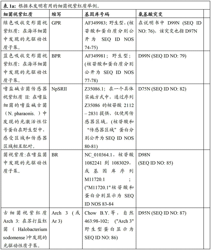
下表1b包括根据本发明方法所说明的可以被突变的附加视紫红质举例:Table 1b below includes examples of epirhodopsins that may be mutated according to the methods of the present invention:
电压指示蛋白(VIP)Voltage indicating protein (VIP)
我们已经开发了一系列基于古细菌视紫红质的荧光电压指示蛋白(VIPs),在哺乳动物细胞发挥作用,包括神经元和人类干细胞源性的心肌细胞。这些蛋白以亚毫秒时间分辨率和亚微米空间分辨率指示电动力学。利用膜电位的光学测定,我们示范了哺乳动物细胞和组织电动力学的非接触、高通量和高含量的研究。这些VIPs具有广泛作用,尤其是在真核细胞中,例如哺乳动物的,包括人类细胞。We have developed a series of archaealrhodopsin-based fluorescent voltage indicator proteins (VIPs) that function in mammalian cells, including neurons and human stem cell-derived cardiomyocytes. These proteins dictate electrokinetics with submillisecond temporal resolution and submicron spatial resolution. Using the optical determination of membrane potential, we demonstrate non-contact, high-throughput, and high-content studies of the electrodynamics of mammalian cells and tissues. These VIPs have a wide range of effects, especially in eukaryotic cells, such as mammalian ones, including human cells.
基于古细菌视紫红质3(Arch3)及其同系物我们开发了VIPs。Arch3是源于苏打盐红菌(H.sodomense),并被公认为是用于高效的黄/绿光神经沉默的遗传编码试剂。基因库中的基因序列:GU045593.1(合成构建体Arch3基因,完整的cds序列.9/28/2009提交).我们已表明这些蛋白集中于真核细胞核的质膜上,且显示电压依赖性荧光。We developed VIPs based on Archaerhodopsin 3 (Arch3) and its homologues. Arch3 is derived from H. sodomense and is recognized as a genetically encoded agent for efficient yellow/green light neural silencing. Gene sequence in GenBank: GU045593.1 (synthetic construct Arch3 gene, complete cds sequence. Submitted 9/28/2009). We have shown that these proteins localize to the plasma membrane of eukaryotic nuclei and show voltage dependence fluorescence.
我们还表明进一步改进的膜定位,在ArchT上具有可比性的电压敏感度,基因库中的基因序列:HM367071.1(合成构建体ArchT基因,完整的cds序列,5/27/2010提交)。ArchT是来源于嗜盐古菌(Halorubrum sp.)的古细菌视紫红质TP009:高效黄/绿光神经沉默的遗传编码试剂,比Arch3光敏感度的高于3.5倍。We also show further improved membrane localization with comparable voltage sensitivity on ArchT, gene sequence in GenBank: HM367071.1 (synthetic construct ArchT gene, complete cds sequence, submitted 5/27/2010). ArchT is an archaerhodopsin derived from Halorubrum sp. TP009: a genetically encoded reagent for highly efficient yellow/green light neural silencing, which is 3.5 times more photosensitivity than Arch3.
表1c总结了可用于构建病毒构建体的序列举例,该构建体基于古细菌视紫红质表达的电压指示器。Table 1c summarizes examples of sequences that can be used to construct viral constructs based on archaeal rhodopsin expressed voltage indicators.

图10表明现有的遗传编码荧光电压指示器,根据其灵敏度和速度分类-决定指示器性能的两个关键参数。我们开发的蛋白是变形菌视紫红质光学质子传感器(PROPS),Arch3WT,和Arch3D95N,在右上侧显示。PROPS仅在细菌中发挥作用,而Arch3WT和Arch3D95N在哺乳动物细胞中发挥作用。我们表面,表明基于细菌视紫红质的电压指示器比其他指示器更加迅速,且更加灵敏。Figure 10 shows that existing genetically encoded fluorescent voltage indicators are categorized according to their sensitivity and speed - two key parameters that determine indicator performance. The proteins we developed are Proteus rhodopsin optical proton sensors (PROPS), Arch3WT, and Arch3D95N, shown on the upper right. PROPS only function in bacteria, while Arch3WT and Arch3D95N function in mammalian cells. We show that bacteriorhodopsin-based voltage indicators are faster and more sensitive than other indicators.
表2显示荧光电压指示蛋白的近似特征,且包括荧光指示器的所有家族的代表成员。虽然在表2中的目录并不全面,本领域技术人员可容易地理解在本发明中有用蛋白类型的特征。Table 2 shows approximate characteristics of fluorescent voltage indicator proteins and includes representative members of all families of fluorescent indicators. Although the list in Table 2 is not comprehensive, those skilled in the art can readily appreciate the characteristics of the types of proteins useful in the present invention.

图9显示在单个大鼠海马神经元中动作电位的光学记录。数据代表单个试验,其中脉冲是由给予电流脉冲引起的。荧光伴随个体动作电位,显示清晰的爆发。该实验是采用基因编码的电压指示器,首次稳定测定单个哺乳动物神经元中的动作电位。Figure 9 shows the optical recording of action potentials in a single rat hippocampal neuron. Data represent a single trial in which the pulse was evoked by the administration of a current pulse. Fluorescence accompanies individual action potentials, showing clear bursts. This experiment is the first to stably measure action potentials in single mammalian neurons using a genetically encoded voltage indicator.
图11显示在表达Arch3D95N–eGFP的单个HL-1小鼠心肌细胞中动作电位的光学记录。动作电位记录至多为1000s,没有光毒性信号。该实验首次采用遗传编码的电压指示器定量测定心脏动作电位。Figure 11 shows optical recordings of action potentials in single HL-1 mouse cardiomyocytes expressing Arch3D95N-eGFP. Action potentials were recorded for up to 1000 s without phototoxic signals. This experiment is the first to quantify cardiac action potentials using a genetically encoded voltage indicator.
图12显示在表达Arch3D95N–eGFP的人类诱导多能干细胞衍生的心肌细胞中动作电位的光学记录。人类诱导多能干细胞(hiPSC)是由细胞动力学有限公司(Cellular Dynamics Inc)提供。细胞在MatTek皿中以每平方厘米20000,50000或75000的细胞密度进行培养,其中该培养皿涂有0.1%明胶。这些条件显示细胞是稀疏且不自发搏动的(20K),融合单分子层是自发搏动(50K)的,和密集的单分子层(75K)。iPS细胞在培养基中培养并保持48小时,且其后每48小时供给维持培养基(均来自细胞动力学公司)(Cellular DynamicsInc.)。根据生产说明书采用Mirus LT-1转染iPS细胞。在包括20uL
我们观察到毗邻细胞的同步搏动,表明VIPs可探测细胞间的传导。我们持续记录10分钟以上,用低光毒性。如对该种群期望的,在细胞种群内,我们观察到了与的心室的,心房的,和节的细胞的动作电位相匹配的细胞。药物的附加导致动作电位波形的变化,该变化与常规膜片箝报告的记录变化相匹配。We observed synchronized beating of adjacent cells, suggesting that VIPs detect cell-to-cell conduction. We record continuously for more than 10 min, with low phototoxicity. As expected for this population, within the cell population we observed cells that matched the action potentials of the ventricular, atrial, and nodal cells. Addition of the drug resulted in changes in the action potential waveform that matched those recorded in conventional patch clamp reports.
具有附加或改进性质的细菌视紫红质和GFP同系物之间融合体的形成Formation of fusions between bacteriorhodopsin and GFP homologues with additional or improved properties
我们将VIPs与GFP-同系物融合,已开发一系列的改进的电压指示器。图16举例说明了这些构建体的例子,图16的说明提供了这些构建体的序列。新开发的传感器的新能力包括,例如,用以增强亮度的FRET光谱移动(ssFRET)和2-光子成像,比率电压成像,和用以同时测定电压和浓度的多峰传感器。We have developed a series of improved voltage indicators by fusing VIPs with GFP-homologs. Figure 16 illustrates examples of these constructs, and the description of Figure 16 provides the sequences of these constructs. New capabilities of the newly developed sensors include, for example, FRET spectral shifting (ssFRET) for brightness enhancement and 2-photon imaging, ratiometric voltage imaging, and multimodal sensors for simultaneous voltage and concentration determination.
用以增强亮度的FRET光谱移动(ssFRET)和2-光子成像Spectral shifting of FRET for brightness enhancement (ssFRET) and 2-photon imaging
第一代VIPs的关键限制是视黄醛的内源性荧光是暗的。成像需要专门系统,包括强烈的红光激光器,高数值孔径的物镜,和电子倍增CCD(EMCCD)相机。理想的是,所用的指示器足够明亮使其可以在一个常规的宽视野或同焦点的荧光显微镜,或2-光子同焦点显微镜中成像,用以体内应用。A key limitation of first-generation VIPs is that the endogenous fluorescence of retinal is dim. Imaging requires specialized systems including an intense red laser, high numerical aperture objectives, and an electron multiplying CCD (EMCCD) camera. Ideally, the indicators used are bright enough to be imaged in a conventional widefield or confocal fluorescence microscope, or a 2-photon confocal microscope, for in vivo applications.
如图10所示,ssFRET提供一种更明亮的VIPs的方法。将GFP-同系物(种属上是指GFP)融合于细菌视紫红质(如,见图13)。视黄醛吸收光谱中的电压依赖性变化使GFP与视黄醛之间的非放射性荧光共振能量转移的电压依赖性速率产生。视黄醛在其吸收,荧光状态熄灭GFP,但视黄醛在非吸收,非荧光状态不会熄灭GFP。因此,可获取GFP和视黄醛的抗相关荧光发射。As shown in Figure 10, ssFRET provides a method for brighter VIPs. A GFP-homologue (species refers to GFP) was fused to bacteriorhodopsin (eg, see Figure 13). The voltage-dependent change in the retinal absorption spectrum results in a voltage-dependent rate of non-radioactive fluorescence resonance energy transfer between GFP and retinal. Retinal extinguishes GFP in its absorbed, fluorescent state, but retinal does not extinguish GFP in its non-absorbed, non-fluorescent state. Thus, anti-correlated fluorescence emissions of GFP and retinal can be obtained.
因此,在一种具体实施方式中,发明提供含有GFP的融合蛋白,所述GFP与细菌视紫红质或修饰的细菌视紫红质进行融合,例如古细菌视紫红质或变形菌视紫红质。该融合蛋白可用于本发明中的任一或所有方法中。Accordingly, in one embodiment, the invention provides a fusion protein comprising GFP fused to bacteriorhodopsin or a modified bacteriorhodopsin, such as archaealrhodopsin or proteorhodopsin. The fusion protein can be used in any or all of the methods of the invention.
为使GFP和视黄醛之间的ssFRET水平最大化,我们选择了GFP同系物,mOrange2,其放射与Arch3在其质子化状态的吸收有最大程度的重叠。随着生色团之间的距离增加FRET的速率迅速下降,因此,我们构建了一系列截短的构建体,该构建体中去除Arch3和mOrange2中的接头和非必须组成。图15显示随着Arch3和mOrange2之间的距离减小,ssFRET信号增强。同样的策略也可应用于从其他细菌视紫红质和GFP同系物形成ssFRET信号。To maximize the level of ssFRET between GFP and retinal, we selected a GFP homologue, mOrange2, whose emission overlaps to the greatest extent with Arch3's uptake in its protonated state. The rate of FRET decreases rapidly with increasing distance between chromophores, therefore, we generated a series of truncated constructs in which linkers and non-essential components in Arch3 and mOrange2 were removed. Figure 15 shows that the ssFRET signal increases as the distance between Arch3 and mOrange2 decreases. The same strategy can also be applied to form ssFRET signals from other bacteriorhodopsin and GFP homologues.
我们已经看出Vm中mOrange2梯度的荧光响应时间和Arch D95N中的荧光响应时间相匹配。该现象和ssFRET是一致。在图9A和11中可观察到与Arch3WT融合的类似结果。We have seen that the fluorescence response time of the mOrange2 gradient in Vm matches that of Arch D95N. This phenomenon is consistent with ssFRET. Similar results can be observed for fusions with Arch3WT in FIGS. 9A and 11 .
比率电压成像ratiometric voltage imaging
VIPs应用的的关键挑战是提取膜电位的准确值,不受光致漂白作用,照明强度的变化,细胞移动或蛋白表达水平的差异的系统性人工干扰。在可接触膜片箝的细胞中,在外界控制的情况下改变膜电位,膜片箝可校准荧光发挥膜电位功能。然而,VIPs的优势是他们功能的发挥在系统上难以接触膜片箝。在这些情况下,直接校正是不可能的。A key challenge for the application of VIPs is to extract accurate values of the membrane potential, free from systematic artifacts such as photobleaching, changes in illumination intensity, cell motility, or differences in protein expression levels. In cells with access to patch clamp, the membrane potential is changed under external control, and patch clamp can calibrate fluorescence to function as membrane potential. However, the advantage of VIPs is that their function is systematically inaccessible to patch clamp. In these cases, direct correction is not possible.
Arch3(WT或D95N)和eGFP的融合能够实现膜电位的比率测定。可采用其他视紫红质来实现相似的比率测定,例如在本申请中描述的使用相同概念的那些视紫红质。eGFP荧光独立于膜电位,Arch3荧光和eGFP荧光的比率提供了膜电位的测定的方法,该膜电位不依赖于表达水平,照明或移动的变化。由于eGFP和Arch3之间的长接头,且eGFP的发射光谱和Arch3的吸收光谱重合较小,所以该结构不能进行ssFRET。Fusion of Arch3 (WT or D95N) and eGFP enables ratiometric measurement of membrane potential. Similar ratiometric assays can be achieved with other rhodopsins, such as those described in this application using the same concept. eGFP fluorescence is independent of membrane potential, and the ratio of Arch3 fluorescence and eGFP fluorescence provides a measure of membrane potential that is independent of changes in expression levels, illumination or movement. Due to the long linker between eGFP and Arch3 and the small overlap between the emission spectrum of eGFP and the absorption spectrum of Arch3, this structure cannot be subjected to ssFRET.
用于电压和浓度同时测定的多峰传感器Multimodal Sensor for Simultaneous Determination of Voltage and Concentration
膜电位只是细胞内信号的多种机制之一。人们常希望将膜电位的变化与其他物质浓度的变化相关联,如Ca++,H+(例如pH),Na+,ATP,cAMP。我们构建了带有pH荧光蛋白(pHluorin)的(荧光pH指示器)Arch与GCaMP3(荧光Ca++指示器)的融合。采用在此启示的概念,也可以使用与基于蛋白的其他荧光指示器的融合物能够进行其他形式的多峰成像。当编码细菌视紫红质的核酸可操作地连接于或融合于附加荧光离子敏感指示器时,离子浓度例如钠,钾,氯和钙,可以同时测定。Membrane potential is just one of many mechanisms for intracellular signaling. It is often desirable to correlate changes in membrane potential with changes in the concentrations of other species such as Ca++, H+ (eg pH), Na+, ATP, cAMP. We constructed a fusion of Arch (a fluorescent pH indicator) with a pH fluorescent protein (pHluorin) and GCaMP3 (a fluorescent Ca++ indicator). Using the concepts taught here, other forms of multimodal imaging can also be enabled using fusions with other protein-based fluorescent indicators. When the bacteriorhodopsin-encoding nucleic acid is operably linked or fused to additional fluorescent ion-sensitive indicators, ion concentrations, such as sodium, potassium, chloride, and calcium, can be measured simultaneously.
附加荧光蛋白Attached fluorescent protein
术语“附加荧光分子”指荧光蛋白而非细菌视紫红质。该分子可以包括,例如,绿色荧光蛋白及其同系物。The term "additional fluorescent molecule" refers to a fluorescent protein rather than bacteriorhodopsin. The molecule can include, for example, green fluorescent protein and its homologues.
荧光蛋白不是细菌视紫红质,这是众所周知的,且广泛地使用,例如,在作者为Rebekka M.Wachter的综述GFP相似的蛋白家族:结构,功能,光电物理和生物传感器的应用的引言和展望中即可找到例子(光化学与光生物学,第82卷,第2期,339–344页,2006年3月)(Photochemistry andPhotobiology Volume82,Issue2,pages339–344,March2006)。此外,由NathanC Shaner,Paul A Steinbach,和Roger Y Tsien写的综述,题目为选择荧光蛋白的指南(自然方法-2,905–909页(2005年))(Nature Methods-2,905-909(2005)),提供了附加的有用荧光蛋白的例子。Fluorescent proteins other than bacteriorhodopsins are well known and widely used, for example, in the review by Rebekka M. Wachter GFP-like protein family: Introduction and perspective on structure, function, optoelectronic physics and applications to biosensors Examples can be found in (Photochemistry and Photobiology Volume 82,
细胞内细胞器靶向的VIPsIntracellular organelle-targeted VIPs
我们展示了细胞内细胞器靶向的VIPs,包括线粒体,内质网,肌质网,突触小泡和吞噬小体。因此,在具体实施方式中,本发明提供构建体,如表达构建体,如病毒构建体,其包括细菌视紫红质可操作地连接于蛋白靶向的序列,该序列使蛋白靶向于细胞内细胞器,包括线粒体,内质网,肌质网,突触小泡和吞噬小体。We demonstrate the targeting of VIPs to intracellular organelles, including mitochondria, endoplasmic reticulum, sarcoplasmic reticulum, synaptic vesicles and phagosomes. Thus, in a particular embodiment, the invention provides constructs, such as expression constructs, such as viral constructs, comprising bacteriorhodopsin operably linked to a protein-targeting sequence that targets the protein to the cell Organelles, including mitochondria, endoplasmic reticulum, sarcoplasmic reticulum, synaptic vesicles and phagosomes.
本发明还提供表达该构建体的细胞,进一步提供在表达该构建体的细胞中测定膜电位变化的方法,和筛选影响一个或多个细胞内细胞膜电位的候选试剂的方法。The invention also provides cells expressing the constructs, further provides methods of measuring changes in membrane potential in cells expressing the constructs, and methods of screening candidate agents that affect the membrane potential of one or more cells.
电压指示蛋白(VIPs)的主要优势Key Advantages of Voltage Indicator Proteins (VIPs)
新开发的VIPs显示高灵敏度。在哺乳动物细胞中,VIPs在-150mV和+150mV之间的荧光大约增加了3倍。在该范围内的大部分内响应是线性的。在1s间隔内我们能够测定膜电压的准确度<1mV。The newly developed VIPs show high sensitivity. In mammalian cells, the fluorescence of VIPs increases approximately 3-fold between -150mV and +150mV. The response is linear for most of the range. We were able to measure the membrane voltage with an accuracy of <1 mV over a 1 s interval.
新开发的VIPs显示高速度。Arch3WT显示在<0.5ms内其瞬态特性显示为90%。神经元动作电位持续1ms,因此该速度达到神经元电活性成像的基准点。然而,Arch3WT保留光诱导的质子泵,因此,光照会使细胞轻微超极化。Newly developed VIPs show high speed. Arch3WT shows that its transient characteristics show 90% within <0.5ms. Neuronal action potentials last 1 ms, so this velocity reaches the benchmark for imaging neuronal electrical activity. However, Arch3WT retains light-induced proton pumps, and thus light hyperpolarizes cells slightly.
修饰的细菌视紫红质,Arch3D95N,有40ms的响应时间且缺乏光诱导的质子泵。虽然该构建体较慢的响应速度妨碍了膜电位检测及其在神经元中的变化,Arch3D95N已足够快可以指示其他类型细胞中的膜电位和动作电位,例如,在心肌细胞中,且在其使用时,不会干扰细胞的膜电位。A modified bacteriorhodopsin, Arch3D95N, has a response time of 40 ms and lacks light-induced proton pumps. While the slow response of this construct hampers the detection of membrane potential and its changes in neurons, Arch3D95N is fast enough to indicate membrane potential and action potential in other cell types, for example, in cardiomyocytes, and in its When used, it does not disturb the membrane potential of the cell.
新开发的VIPs还显示高耐光性。在光漂白作用之前产生的荧光光子数量上,VIPs与GFP是可以媲美的。我们对在哺乳动物细胞中的VIPs常规地观察了数分钟,没有发生光漂白作用或光毒性的迹象。VIPs与GFP或其他已知的荧光蛋白均无同源性。The newly developed VIPs also show high light fastness. VIPs are comparable to GFP in the number of fluorescent photons produced prior to photobleaching. We routinely observed VIPs in mammalian cells for several minutes with no evidence of photobleaching or phototoxicity. VIPs have no homology to GFP or other known fluorescent proteins.
新开发的VIPs还显示远红外光谱。用633nm的激光激发VIPs,放射在近红外,峰值在710nm。红外放射比现有的荧光蛋白都远。这些波长与低细胞自发荧光和良好的组织传导性相一致。由于光谱可推进具有高信噪比的成像,与其他荧光探针的结合进行多谱成像,该特征使这些蛋白在动作电位的光学测定中尤其有用。The newly developed VIPs also display far-infrared spectra. The VIPs are excited by a 633nm laser, and the emission is in the near infrared with a peak at 710nm. Infrared emission is farther than existing fluorescent proteins. These wavelengths are consistent with low cellular autofluorescence and good tissue conductivity. Combination with other fluorescent probes for multispectral imaging makes these proteins particularly useful in the optical measurement of action potentials, since the spectra facilitate imaging with high signal-to-noise ratios.
新开发的VIPs还显示高的靶向性。我们已经在初级神经元培养,心肌细胞(HL-1和人类衍生的iPSC),HEK细胞和格兰阳性细菌、格兰阴性细菌中做了VIPs成像。我们将VIP靶向于内质网和线粒体。这些构建体在线虫(C.elegans,)、斑马鱼和小鼠体内成像也是有用的。The newly developed VIPs also showed high targeting. We have imaged VIPs in primary neuronal cultures, cardiomyocytes (HL-1 and human-derived iPSCs), HEK cells, and Gram-positive and Gram-negative bacteria. We targeted VIP to the endoplasmic reticulum and mitochondria. These constructs are also useful for in vivo imaging in C. elegans (C. elegans, ), zebrafish, and mice.
发明带有细菌视紫红质的构建体,还包括细胞型和/或时间特异的启动子,其可以对活生物体内任何光学可接触的细胞或细胞器的膜电位进行成像。Invention of bacteriorhodopsin-bearing constructs, further comprising cell-type and/or time-specific promoters, allows imaging of the membrane potential of any optically accessible cell or organelle in a living organism.
在一种具体实施方案中,电压感受器的设计包括或基本上包括选择至少三个元素:启动子,细菌视紫红质电压传感器,一个或多个靶向的基序,和任意附属荧光蛋白。在下面的表1a,1b中和表3中列出了这些元素中每个元素的一些非限制性例子。在一种具体实施方式中,选择每栏中至少一个元素以制造具有预期特征的光学电压传感器。在一些具体实施方式中,在此描述的用于电压传感器的方法和组合物包括选择:1)细菌视紫红质蛋白,2)一个或多个突变,使蛋白对电压或其他大量感兴趣的具有敏感性,和消除光驱动充电泵,3)选择适宜用于宿主种属的密码子,4)一个启动子和靶向序列,用以在感兴趣的细胞型中表达蛋白和使蛋白靶向于有用的亚细胞结构,5)含有传统荧光蛋白的任意融合体用以提供比率成像,6)生色团用以插入细菌视紫红质,和7)光学成像电路。In a specific embodiment, the design of a voltage sensor includes or consists essentially of selecting at least three elements: a promoter, a bacteriorhodopsin voltage sensor, one or more targeting motifs, and any accessory fluorescent protein. Some non-limiting examples of each of these elements are listed in Tables 1a, 1b and in Table 3 below. In a specific embodiment, at least one element in each column is selected to produce an optical voltage sensor with desired characteristics. In some embodiments, the methods and compositions described herein for use in voltage sensors include selecting: 1) a bacteriorhodopsin protein, 2) one or more mutations that render the protein sensitive to voltage or other substantial properties of interest Sensitivity, and elimination of light-driven charge pumps, 3) selection of codons appropriate for the host species, 4) a promoter and targeting sequence to express the protein in the cell type of interest and to target the protein to Useful subcellular structures, 5) arbitrary fusions containing conventional fluorescent proteins to provide ratiometric imaging, 6) chromophores for insertion into bacteriorhodopsin, and 7) optical imaging circuitry.

在一种具体实施方式中,光学感受器基因是由传递载体编码的。这样的载体包括但不仅限于:质粒(例如:pBADTOPO,pCI-Neo,pcDNA3.0),粘粒,和病毒(例如慢病毒,腺伴随病毒或杆状病毒)。In a specific embodiment, the photoreceptor gene is encoded by the delivery vector. Such vectors include, but are not limited to: plasmids (eg, pBADTOPO, pCI-Neo, pcDNA3.0), cosmids, and viruses (eg, lentivirus, adeno-associated virus, or baculovirus).
在一种具体实施方式中,绿色吸收变形菌视紫红质(GPR)被用作初始分子。选择这个分子,是由于其具有相对的红移吸收光谱并且其在异源宿主例如大肠杆菌中容易表达。在其他的具体实施方式中,蓝色吸收变形菌视紫红质(BPR)用于电压光学传感器。在此思考的是,如在此述,野外发现的大量细菌视紫红质可设计为光学电压传感器。In a specific embodiment, green absorbing Proteus rhodopsin (GPR) is used as the starting molecule. This molecule was chosen due to its relatively red-shifted absorption spectrum and its ease of expression in heterologous hosts such as E. coli. In other embodiments, blue absorbing Proteus rhodopsin (BPR) is used in the voltage optical sensor. It is contemplated here that, as described herein, bacteriorhodopsins, found in abundance in the wild, can be engineered as optical voltage sensors.
细菌视紫红质除了电压以外还对定量敏感。GPR和BPR的突变体,如在此所述,也对细胞内pH敏感。可以预期的是嗜盐菌紫质对局部氯化物浓度也是敏感的。Bacteriorhodopsin is also quantitatively sensitive in addition to voltage. Mutants of GPR and BPR, as described herein, are also sensitive to intracellular pH. It is expected that halorhodopsins are also sensitive to local chloride concentrations.
在一种具体实施方式中,压力传感器是选自于细菌视紫红质蛋白(野生型或突变体),该蛋白提供在其吸收或荧光谱中电压诱导的移位。来自于这些构建体的初始序列的设计,初始序列包括但不限于表1a-1b中列出的序列,其列出了视紫红质和举例的突变体,所述举例的突变体可以制作为基因以提高蛋白产品性能的。In a specific embodiment, the pressure sensor is selected from a bacteriorhodopsin protein (wild type or mutant) that provides a voltage-induced shift in its absorption or fluorescence spectrum. From the design of the initial sequences of these constructs, the initial sequences include but are not limited to the sequences listed in Table 1a-1b, which lists rhodopsin and exemplary mutants that can be made as genes To improve the performance of protein products.
用以最小化光诱导电荷泵送能力的突变体。视黄醛生色团通过希夫碱基连接于赖氨酸。保守的门冬氨酸作为质子接受器,与希夫碱基相邻。将该门冬氨酸突变为天冬酰胺来抑制质子泵。因此,在一些具体实施方式中,突变选自于该组,包括:D97N(绿色吸收变形菌视紫红质),D99N(蓝色吸收变形菌视紫红质),D75N(传感视紫红质II),和D85N(菌视紫质)。在其他的具体实施方式中,可以突变为抑制泵活性的残基包括(使用菌视紫质编号)D96,Y199,和R82及其在其他细菌视紫红质中的他们的同系物。在另外的具体实施方式中,D95残基可以在古细菌视紫红质中突变来抑制质子泵(例如,D95N)。Mutants designed to minimize light-induced charge-pumping ability. The retinal chromophore is linked to lysine via a Schiff base. The conserved aspartic acid acts as a proton acceptor and is adjacent to a Schiff base. Mutating the aspartate to asparagine inhibits the proton pump. Thus, in some embodiments, the mutation is selected from the group comprising: D97N (green absorbing proteus rhodopsin), D99N (blue absorbing proteus rhodopsin), D75N (sensing rhodopsin II) , and D85N (bacteriorhodopsin). In other embodiments, residues that may be mutated to inhibit pump activity include (using bacteriorhodopsin numbering) D96, Y199, and R82 and their homologues in other bacteriorhodopsins. In additional embodiments, the D95 residue can be mutated in archaerhodopsin to inhibit proton pumping (eg, D95N).
引进突变以使吸收光谱和发射光谱移动至期望的范围。邻近结合口袋的残基可被个别突变或组合地调节光谱使其在期望的吸收或发射波长。在菌视紫质中这些残基包括,但不仅限于,L92,W86,W182,D212,I119,和M145。相应的残基在其他细菌视紫红质中可能被突变。因此,在一些具体实施方式中,向修饰细菌视紫红质蛋白的突变在残基上进行,该残基选自于包含L92,W86,W182,D212,I119,M145的组。Mutations are introduced to shift the absorption and emission spectra into the desired range. Residues adjacent to the binding pocket can be mutated individually or in combination to tune the spectra at desired absorption or emission wavelengths. In bacteriorhodopsin these residues include, but are not limited to, L92, W86, W182, D212, I119, and M145. Corresponding residues may be mutated in other bacteriorhodopsins. Thus, in some embodiments, mutations to modified bacteriorhodopsin proteins are made at residues selected from the group comprising L92, W86, W182, D212, I119, M145.
引进突变以使电压灵敏度的动态范围移动至期望谱带。这种突变通过转移希夫碱基附近的电子分布发挥作用,从而改变电压,所述电压是从该组增加或去除质子所需要的电压。在绿色吸收变形菌视紫红质中的电压移动突变包括,但不限于,E108Q,E142Q,L217D,个别地或组合地,例如使用绿色吸收变形菌视紫红质定位,或在其他视紫红质中相应的残基。在一种具体实施方式中,D95N突变被引进到古细菌视紫红质3中进而调节希夫碱基的pKa值到中性的pH。Mutations were introduced to shift the dynamic range of voltage sensitivity to the desired band. This mutation works by shifting the distribution of electrons around the Schiff base, thereby changing the voltage needed to add or remove protons from this group. Voltage-shifting mutations in green-absorbing Proteus rhodopsin include, but are not limited to, E108Q, E142Q, L217D, individually or in combination, e.g. using the green-absorbing Proteus rhodopsin localization, or corresponding in other rhodopsins residues. In one embodiment, the D95N mutation is introduced into
引入任意的突变以提高荧光的亮度和耐光性。当突变时可能限制结合口袋来增加荧光的残基,这样的残基包括(使用菌视紫质编号)但不仅限于Y199,Y57,P49,V213和V48。Arbitrary mutations were introduced to increase the brightness and photostability of the fluorescence. Residues that may restrict the binding pocket to increase fluorescence when mutated include (using bacteriorhodopsin numbering) but are not limited to Y199, Y57, P49, V213, and V48.
密码子选择codon usage
大量哺乳动物基因,包括例如鼠类和人类基因,已成功在不同的宿主细胞中表达,包括细菌,酵母,昆虫,植物和哺乳宿主细胞。然而,尽管表达系统和DNA重组技术知识不断发展,当人们试图在选择的宿主细胞中表达外来的或合成的基因时,依然存在巨大障碍。例如,合成基因的翻译翻译,甚至当与强启动子耦合时,进展经常会比预期要慢很多。相同的情况在外来于宿主细胞的外源性基因经常出现。这种比预期的翻译翻译效率低常常是由于基因的蛋白编码区有密码子选择的结构,该结构与宿主细胞中高表达基因的结构不相似。众所周知,就这一点而言,在不同的有机体中密码子使用是具有高的偏好和变化的,且在密码子使用的偏好性可以改变肽延长的速率。人们也知道密码子使用模式与tRNA同功受体的相对丰度相关,编码高丰度和低丰度蛋白的基因显示他们密码子偏好是不同的。A large number of mammalian genes, including, for example, murine and human genes, have been successfully expressed in different host cells, including bacterial, yeast, insect, plant and mammalian host cells. However, despite advances in knowledge of expression systems and recombinant DNA techniques, formidable obstacles remain when one attempts to express foreign or synthetic genes in a host cell of choice. For example, translation of synthetic genes, even when coupled to strong promoters, often progresses much slower than expected. The same situation often occurs with exogenous genes that are foreign to the host cell. This lower-than-expected translation efficiency is often due to the fact that the protein-coding region of the gene has a codon-used structure that does not resemble the structure of the highly expressed gene in the host cell. It is well known in this regard that codon usage is highly biased and variable in different organisms, and that bias in codon usage can alter the rate of peptide elongation. Codon usage patterns are also known to correlate with the relative abundance of tRNA isoacceptors, and genes encoding high- and low-abundance proteins show differences in their codon preferences.
开发密码子优化技术用来提高翻译无效蛋白编码区的翻译动力学。这些技术是基于将很少或不经常在宿主细胞中使用的密码子替代为宿主细胞偏好的密码子。密码子的频率可源于文献资源报道的很多生物体高表达基因(见,例如,Nakamura et al.,1996,核酸研究24:214-215)(Nakamura et al.,1996,Nucleic Acids Res24:214-215)。这些频率以经常在‘生物范围平均偏差’中表达发生的百分数表示,所述发生是通过采集生物体的蛋白编码基因同义密码子用于编码相应的氨基酸的机会,其优选是高表达的。在一种具体实施方式中,细菌视紫红质蛋白的密码子在真核细胞中进行优化表达。在一种具体实施方式中,真核细胞是人类细胞。Codon optimization techniques were developed to improve translation kinetics in translationally null protein coding regions. These techniques are based on the substitution of codons that are rarely or infrequently used in the host cell with codons preferred by the host cell. Codon frequencies can be derived from literature sources reporting highly expressed genes in many organisms (see, for example, Nakamura et al., 1996, Nucleic Acids Research 24:214-215) (Nakamura et al., 1996, Nucleic Acids Res 24:214-215) 215). These frequencies are expressed as a percentage of the frequent expression occurrences in the 'biological range mean deviation' by sampling the chances that synonymous codons in the protein coding genes of an organism are used to encode the corresponding amino acid, which are preferably highly expressed. In one embodiment, bacteriorhodopsin is codon-optimized for expression in eukaryotic cells. In a specific embodiment, the eukaryotic cells are human cells.
优选但并不是必需将细菌多核苷酸的所有密码子替代为同义密码子,所述同义密码子在真核细胞(例如,人类)中比第一密码子具有更高翻译效率。即使部分替代也可完成增加的表达。典型地,替代步骤影响至少母体多核苷酸的第一密码子的约5%,10%,15%,20%,25%,30%,更优选的是,至少约35%,40%,50%,60%,70%或更多。适宜的是,两种密码子之间的翻译效率差异和数量,选择第一密码子和同义密码子可以使在真核细胞中从合成多核苷酸生产的有用蛋白的水平至少是在真核细胞中从母体多核苷酸生产的有用蛋白水平的约110%,适宜的是至少150%,优选的是至少200%,更优选的是至少250%,更优选的是300%,更优选的是350%,更优选的是400%,更优选的是450%,更优选的是500%,甚至更优选的是1000%。It is preferred, but not necessary, to replace all codons in the bacterial polynucleotide with synonymous codons that are more efficiently translated in eukaryotic cells (eg, humans) than the first codon. Increased expression can be accomplished even with partial substitution. Typically, the substitution step affects at least about 5%, 10%, 15%, 20%, 25%, 30%, more preferably at least about 35%, 40%, 50% of the first codon of the parental polynucleotide %, 60%, 70% or more. Suitably, the difference and amount of translation efficiency between the two codons, the first codon and the synonymous codon are chosen such that the level of useful protein produced from a synthetic polynucleotide in eukaryotic cells is at least as high as in eukaryotic cells. About 110%, suitably at least 150%, preferably at least 200%, more preferably at least 250%, more preferably 300%, more preferably at least 250% of the useful protein level produced in the cell from the parental polynucleotide 350%, more preferably 400%, more preferably 450%, more preferably 500%, even more preferably 1000%.
一般而言,如果母体多核苷酸可以选择低和中等翻译效率的密码子,首先优选将一些或者更多优选所有的低翻译效率的密码子替代为具有中等,优选是高翻译效率的同义密码子。典型的是,用中等或高翻译效率密码子的替代低翻译效率密码子导致源自此构建体的合成多核苷酸的多肽产量显著增加。然而,还优选采用高翻译效率的密码子替代一些,或优选是全部的中等翻译效率的密码子,用以优化多肽的生产。In general, if the parental polynucleotide can select low and medium translation efficiency codons, it is preferred first to replace some or more, preferably all low translation efficiency codons with synonymous codons with medium, preferably high translation efficiency son. Typically, replacement of low translation efficiency codons with intermediate or high translation efficiency codons results in a significant increase in polypeptide yield from synthetic polynucleotides derived from the construct. However, it is also preferred to replace some, or preferably all, of the intermediate translation efficiency codons with high translation efficiency codons in order to optimize production of the polypeptide.
将一个密码子取代为另外一个密码子可采用本领域已知的标准方法实现。例如母体多核苷酸的密码子修饰可通过多种已知的诱变技术进行影响,包括,例如寡核苷酸指导的诱变,采用简并寡核苷酸的诱变和区域特异性的诱变。体外诱变技术的举例如下:在美国专利号4,184,917,4,321,365和4,351,901或Ausubel等人的(现代分子生物学试验设计,John Wiley和Sons,Inc.1997)(CURRENT PROTOCOLS IN MOLECULAR BIOLOGY,John Wiley&Sons,Inc.1997)和Sambrook等人的(分子克隆.实验手册,冷泉巷出版社,1989)(MOLECULAR CLONING.A LABORATORY MANUAL,Cold SpringHarbor Press,1989)的相关章节中有所描述。代替体内的诱变,合成的多核苷酸可采用如描述的易得的机械装置进行从头合成,例如在美国专利No.4,293,652中描述的。然而,值得注意的是,本发明不依赖于,且不针对于,任何用于构建合成多核苷酸的特定技术。Substitution of one codon for another can be accomplished using standard methods known in the art. For example, codon modification of the parental polynucleotide can be affected by a variety of known mutagenesis techniques including, for example, oligonucleotide-directed mutagenesis, mutagenesis using degenerate oligonucleotides, and region-specific mutagenesis. Change. Examples of in vitro mutagenesis techniques are as follows: in U.S. Patent Nos. 4,184,917, 4,321,365 and 4,351,901 or Ausubel et al. 1997) and the relevant chapters of Sambrook et al. (Molecular Cloning. Laboratory Manual, Cold Spring Lane Press, 1989) (MOLECULAR CLONING. A LABORATORY MANUAL, Cold Spring Harbor Press, 1989). Instead of in vivo mutagenesis, synthetic polynucleotides can be synthesized de novo using readily available machinery as described, for example, in U.S. Patent No. 4,293,652. It should be noted, however, that the present invention is not dependent on, and is not directed to, any particular technique for constructing synthetic polynucleotides.
细菌视紫红质(例如GPR)的基因在大肠杆菌中表达较好,但在真核宿主中表达欠佳。在一种具体实施方式中,为了能在真核中表达,我们设计并合成了适于真核细胞(例如,人类)密码子选择的基因变型。该程序可采用公开可得的软件应用于任何基因,例如基因设计软件2.0包(the Gene Designer2.0package)(互联网上可用的网址为dna20.com/genedesigner2/)(available onthe world wide web at dna20.com/genedesigner2/)。在此,一些“人化的”基因是指在名字前面加上字母“h”,例如hGPR。在此和实施例中描述的Arch3视紫红质及其突变体用人类选择密码子全部优化。Genes for bacteriorhodopsins (such as GPR) express well in E. coli but poorly in eukaryotic hosts. In one embodiment, we designed and synthesized genetic variants adapted to the codon usage of eukaryotic cells (eg, humans) for expression in eukaryotes. The procedure can be applied to any gene using publicly available software, such as the Gene Designer 2.0 package (available on the Internet at dna20.com/genedesigner2/) (available on the world wide web at dna20. com/genedesigner2/). Here, some "humanized" genes are referred to by adding the letter "h" before the name, eg hGPR. Arch3rhodopsin and its mutants described here and in the Examples were all optimized with human selector codons.
VIPs在筛选靶向于如下组织或过程的药物中的应用Application of VIPs in screening drugs targeting the following tissues or processes
在本申请中公开的构建体可用于药物筛选方法中,例如,以神经系统为靶向的药物。在表达特异性离子通道的细胞培养中,无需对细胞中使用膜片箝的劳动,一次即可筛选出激动剂或拮抗剂。在神经元培养中,人们可以探测药物对动作电位的引起,传播,和突触传导的效果。在人类iPSC-衍生神经元的应用能够使遗传决定的神经病学疾病的研究,和环境应激响应的研究得以开展(例如,缺氧症)。The constructs disclosed in this application can be used in drug screening methods, eg, drugs targeting the nervous system. In cultured cells expressing specific ion channels, agonists or antagonists can be screened in one pass without the labor of patch clamping the cells. In neuronal cultures, one can probe the effects of drugs on action potential initiation, propagation, and synaptic transmission. The application of human iPSC-derived neurons enables the study of genetically determined neurological diseases, and the study of responses to environmental stress (eg, hypoxia).
类似的,在此提供的使用构建体的光学电压传感提供了一个新颖的和显著改善的方法用于筛选药物,所述药物是调整心脏动作电位及其胞间传播的药物。这些筛选不仅有助于测定候选药物的安全性还可以辨别新的心脏药物的先导。由于hERG抑制与长QT综合征的病人的心室纤维性颤动相关,所以鉴别与hERG通道相互作用的药物是一个极具潜力的方向。在人类iPSC衍生心肌细胞中的应用使遗传决定的心脏状态研究,和环境应激响应的研究得以开展(例如,缺氧)。Similarly, optical voltage sensing using constructs provided herein provides a novel and significantly improved method for screening drugs that modulate cardiac action potentials and their intercellular propagation. These screens not only help determine the safety of drug candidates but also identify new cardiac drug leads. Since hERG inhibition is associated with ventricular fibrillation in patients with long QT syndrome, identifying drugs that interact with hERG channels is a promising direction. Application to human iPSC-derived cardiomyocytes enables the study of genetically determined cardiac states and responses to environmental stress (e.g., hypoxia).
此外,本发明中的构建体可用于生长和伤口愈合研究方法中。在正常和非正常生长中,以及组织修复中的电子信号的角色很少能被理解。VIPs使长时间的组织,器官和生物体的生长和愈合中的电压动力学研究得以开展,且开发调整这些动力学的药物。In addition, the constructs of the invention can be used in growth and wound healing research methods. The role of electrical signaling in normal and abnormal growth, and in tissue repair, is poorly understood. VIPs enable long-term studies of voltage dynamics in the growth and healing of tissues, organs, and organisms, and the development of drugs that modulate these dynamics.
还在另外的实施方式中,发明提供影响线粒体膜电位的药物筛选方法。线粒体在衰老,癌症和神经变性疾病中扮演重要角色。目前没有用于线粒体膜电位的好的探针。VIPs提供这样的探针,其使调整线粒体活动的药物研究能够开展。In yet other embodiments, the invention provides methods for screening drugs that affect mitochondrial membrane potential. Mitochondria play an important role in aging, cancer and neurodegenerative diseases. There are currently no good probes for mitochondrial membrane potential. VIPs provide probes that enable the study of drugs that modulate mitochondrial activity.
发明还提供筛选药物的方法,该药物可调整大范围医学上,工业上和环境上重要微生物的电生理学。The invention also provides methods of screening for drugs that modulate the electrophysiology of a wide range of medically, industrially and environmentally important microorganisms.
在我们发现VIPs之前,还没有在任何完整原核生物中进行膜电位的测定。我们发现细菌有复杂的电动力学。VIPs能够筛选调整大范围医学上,工业上和环境上重要微生物的电生理学的药物。例如,我们发现大肠杆菌中电活动性和流出泵相互关联。Before our discovery of VIPs, measurements of membrane potential had not been performed in any intact prokaryotes. We found that bacteria have complex electrokinetics. VIPs enable the screening of drugs that modulate the electrophysiology of a wide range of medically, industrially and environmentally important microorganisms. For example, we found that electrical activity and efflux pumps are linked in E. coli.
膜电位的变化也与巨噬细胞的激活相关。然而,由于膜片箝很难应用于游动细胞,使该过程很难理解。VIPs使巨噬细胞和其他游动细胞的电生理学研究得以开展,包括精子细胞的生育力研究。因此,发明的VIPs可用于筛选药物或试剂的方法,例如,筛选影响免疫性和免疫疾病和生育力的药物或试剂。Changes in membrane potential are also associated with activation of macrophages. However, the process is poorly understood due to the difficulty of patch clamping on motile cells. VIPs enable electrophysiological studies of macrophages and other motile cells, including fertility studies of sperm cells. Accordingly, the invented VIPs can be used in methods of screening drugs or agents, for example, screening drugs or agents affecting immunity and immune diseases and fertility.
实施例描述了在大鼠海马神经,小鼠HL-1心肌细胞和人类iPS衍生的心肌细胞中VIPs的表达。在所有的细胞型中,单个动作电位(APs)很容易观察到。我们测定了药物对AP波形的影响。The examples describe the expression of VIPs in rat hippocampal nerves, mouse HL-1 cardiomyocytes and human iPS-derived cardiomyocytes. In all cell types, single action potentials (APs) are readily observed. We measured the effect of drugs on AP waveforms.
例如,在一种具体实施方式中,发明提供一种方法,其中表达细菌视紫红质的细胞进一步暴露于能够或预期能够改变膜电位的刺激。For example, in one embodiment, the invention provides a method wherein a cell expressing bacteriorhodopsin is further exposed to a stimulus capable or expected to alter membrane potential.
可以使用的刺激包括候选试剂,例如候选药物,有机小分子或无机小分子,较大的有机分子,及其任何组合的分子库。人们也可以使用已知药物的组合,例如抗生素和候选试剂的组合去筛选可能增加一个或多个现有药物如抗生素的有效性的试剂。Stimuli that can be used include candidate agents, such as drug candidates, small organic or inorganic molecules, larger organic molecules, and molecular libraries of any combination thereof. One can also use combinations of known drugs, such as antibiotics, and candidate agents to screen for agents that may increase the effectiveness of one or more existing drugs, such as antibiotics.
发明的方法也有助于体外毒性筛选和药物开发。例如,采用在此描述的方法,可以使来自诱导多能细胞的人类心肌细胞,稳定表达修饰古细菌视紫红质,其中质子泵活性显著降低或消失。上述细胞对于药物开发中的体外毒性筛选是十分有用的。The inventive method also facilitates in vitro toxicity screening and drug development. For example, using the methods described herein, human cardiomyocytes from induced pluripotent cells can be made to stably express modified archaerhodopsins in which proton pump activity is significantly reduced or abolished. The above-mentioned cells are very useful for in vitro toxicity screening in drug development.
PROPS:源自于GPR的光遗传电压传感器PROPS: an optogenetic voltage sensor derived from GPR
GPR有7个分光镜可区分的状态,穿过光循环。原则上任何两个状态的之间的转变对膜电位都是敏感的。在一种具体实施方式中,希夫碱基的酸碱平衡被选择为波长移动转变,因此传感器的名字:变形菌视紫红质光学质子传感器(PROPS)。PROPS性质及其使用的特征在实施例部分进行描述。下面提供的是对PROPS进行的简要讨论。The GPR has 7 spectroscopically distinguishable states that cycle through the light. In principle the transition between any two states is sensitive to the membrane potential. In a specific embodiment, the acid-base balance of the Schiff bases is chosen to shift the wavelength shift, hence the name of the sensor: Proteorhodopsin Optical Proton Sensor (PROPS). The properties of PROPS and the characteristics of its use are described in the Examples section. A brief discussion of PROPS is provided below.
已知的是,野生型GPR的吸收光谱灵敏地依赖于希夫碱基的质子化状态。当质子化时,最大吸收是在545nm,和当去质子化时,最大吸收在412nm。当GPR吸收一个质子,视黄醛进行13-反式到顺式的异构化,引起质子从希夫碱基跳跃到邻近的Asp97中,导致吸收从545nm转移到412nm。在此描述的PROPS设计希望概括这种转移对膜电位变化的反应。It is known that the absorption spectrum of wild-type GPR is sensitively dependent on the protonation state of Schiff bases. When protonated, the maximum absorption is at 545 nm, and when deprotonated, the maximum absorption is at 412 nm. When GPR absorbs a proton, retinal undergoes 13-trans to cis isomerization, causing proton hopping from the Schiff base to adjacent Asp97, resulting in a shift in absorption from 545nm to 412nm. The PROPS design described here hopes to recapitulate the response of this transfer to changes in membrane potential.
作为最佳电压传感器,野生型GPR的两方面可被改变。首先,希夫碱基的pKa值可以从其野生型的12转变为接近周围pH的数值。当pKa~pH,质子化状态对膜电压具有最大的灵敏度。第二,最适宜的情况是,由于电压探针不应该扰乱研究中的数量,因此可消除内源性电荷泵能力。然而,在一些情况下,野生型的细菌视紫红质可被使用,例如我们的实施例中显示的,如Arch3WT,在神经元中发挥作用以测定膜电位。As an optimal voltage sensor, two aspects of wild-type GPR can be altered. First, the pKa of a Schiff base can shift from its wild-type value of 12 to a value close to the surrounding pH. When pKa~pH, the protonation state has the greatest sensitivity to the membrane voltage. Second, optimally, the intrinsic charge-pump capability is eliminated since the voltage probe should not disturb the quantities under study. However, in some cases wild-type bacteriorhodopsins can be used, for example as shown in our Examples, such as Arch3WT, functioning in neurons to measure membrane potential.
在一种具体实施方式中,单个点突变引起GPR中的两个改变。从Asp97到Asn的突变消除了邻近希夫碱基的负电荷,且使希夫碱基上的质子不稳定。pKa从12转变为9.8。在野生型的GPR中,在光循环的第一步Asp97也作为质子受体,因此去除该氨基酸可消除质子泵。GPR的这种突变体是指PROPS。In a specific embodiment, a single point mutation causes two changes in the GPR. The mutation from Asp97 to Asn removes the negative charge of the adjacent Schiff base and destabilizes the proton on the Schiff base. The pKa shifted from 12 to 9.8. In wild-type GPR, Asp97 also acts as a proton acceptor in the first step of the photocycle, so removing this amino acid eliminates the proton pump. Such mutants of GPR are referred to as PROPS.
类似的是,在源自于BPR的类似的电压传感器中,从Asp99到Asn的类似的突变降低了希夫碱基的pKa值,且消除了质子泵光循环。因此,在一种具体实施方式中光学传感器是源自于BPR,在BPR中氨基酸残基Asp99突变为Asn。Similarly, in a similar voltage sensor derived from BPR, a similar mutation from Asp99 to Asn reduces the pKa of the Schiff base and abolishes the proton-pumping photocycle. Thus, in one embodiment the optical sensor is derived from BPR in which amino acid residue Asp99 is mutated to Asn.
在GPR中,附加的突变使pKa转向更接近生理学数值7.4。具体来说,Glu108到Gln的突变和Glu142到Gln的突变,各自或其组合导致pKa的下降并电压灵敏度的进一步增加。除了在此讨论的那些,很多突变可导致pKa的附加变化和PROPS光学性质的改善,在此对这些突变也做了考虑。In GPR, additional mutations shifted the pKa closer to the physiological value of 7.4. Specifically, mutations of Glu108 to Gln and Glu142 to Gln, each or in combination, resulted in a decrease in pKa and a further increase in voltage sensitivity. In addition to those discussed here, many mutations that lead to additional changes in pKa and improvements in the optical properties of PROPS are also considered here.
表达载体和靶向序列Expression Vectors and Targeting Sequences
在细胞中可采用表达载体表达光学传感器。术语“载体”是指核酸序列可以插入的载体DNA分子以将其引入宿主细胞。“表达载体”是专门的载体,其包括在宿主细胞中用于目的基因表达的必需的调控区。在一些具体实施方式中,目的基因可操作地连接于载体的其他序列。在一些具体实施方式中,病毒载体优选是复制缺损型,例如可通过去除编码用于复制的所有的病毒核酸来实现。复制缺损型病毒载体仍将保留其传染特性,以和复制型载体相似的方式进入细胞,然而,一旦允许进入细胞,复制缺损型病毒载体不可复制或繁殖。术语“可操作地连接”是指将编码序列表达必需的调控序列放置在DNA分子中合适的、与编码序列相关的位置进而影响编码序列的表达。相同的定义有时也应用于在表达载体中的编码序列和转录控制因子(例如,启动子,增强子和终止子)的布置上。The optical sensor can be expressed in cells using expression vectors. The term "vector" refers to a carrier DNA molecule into which a nucleic acid sequence can be inserted for introduction into a host cell. An "expression vector" is a specialized vector that includes the necessary regulatory regions for expression of a gene of interest in a host cell. In some embodiments, the gene of interest is operably linked to other sequences of the vector. In some embodiments, the viral vector is preferably replication deficient, eg, by removing all viral nucleic acid encoding for replication. Replication-defective viral vectors will still retain their infectious properties, entering cells in a manner similar to replication-competent vectors, however, once permitted to enter cells, replication-deficient viral vectors cannot replicate or reproduce. The term "operably linked" refers to the placement of regulatory sequences necessary for the expression of a coding sequence at an appropriate position in a DNA molecule relative to the coding sequence so as to affect the expression of the coding sequence. The same definition also sometimes applies to the arrangement of coding sequences and transcriptional control elements (eg, promoters, enhancers and terminators) in expression vectors.
很多病毒载体或病毒相关载体在本领域已为我们所熟知。这些载体可用作核酸构建体的载体进入细胞。为了,构建体可以被整合和打包地进入到非复制、缺陷型病毒基因中,像腺病毒,腺伴随病毒(AAV),或单纯性疱疹病毒(HSV)或其他病毒,包括逆转录病毒和慢病毒载体,用于感染或传导至细胞内。载体可以掺合或可以不掺合到细胞的基因组中。必要时,这些构建体可能包括用于转染的病毒序列。可选地,该构建体可以掺合到能够附加型复制的载体中,例如,EPV和EBV载体。当表达调控序列控制和调节多核苷酸序列的转录和翻译时,在此描述的载体中引入的物质可操作地连接于表达调控序列。术语“可操作地连接”包括包括一个待表达多核苷酸序列之前的合适的起始信号(例如,ATG),并保持正确的阅读框进而使其在表达调控序列的控制下表达多核苷酸序列,和生产期望的编码多核苷酸序列的多肽。在一些实施例中,插入物质的转录受启动子序列(或其他转录调控序列)控制,该序列在预期表达的细胞中控制重组基因的表达。也可以理解的是,当然插入物质受到转录调控序列的控制,该转录调控序列相同于或异于那些控制蛋白自然存在形式的转录的序列。在一些实例中,启动子序列通过细胞内合成机制,或引入的合成机制识别,这对于启动特定基因的转录是所需要的。Many viral vectors or virus-associated vectors are well known in the art. These vectors can be used as vehicles for the nucleic acid constructs into cells. In order, constructs can be integrated and packaged into non-replicating, defective viral genes like adenovirus, adeno-associated virus (AAV), or herpes simplex virus (HSV) or other viruses, including retroviruses and lentiviruses. Viral vectors for infection or delivery into cells. The vector may or may not be incorporated into the genome of the cell. If necessary, these constructs may include viral sequences for transfection. Alternatively, the construct may be incorporated into a vector capable of episomal replication, eg, EPV and EBV vectors. Agents introduced in the vectors described herein are operably linked to expression control sequences as the expression control sequences control and regulate the transcription and translation of the polynucleotide sequences. The term "operably linked" includes inclusion of a suitable initiation signal (eg, ATG) preceding the polynucleotide sequence to be expressed and maintaining the correct reading frame to allow expression of the polynucleotide sequence under the control of expression control sequences , and producing the desired polypeptide encoding polynucleotide sequence. In some embodiments, transcription of the inserted material is controlled by a promoter sequence (or other transcriptional regulatory sequence) that controls the expression of the recombinant gene in the cell where expression is intended. It is also understood that the inserted material is, of course, under the control of transcriptional regulatory sequences which may be identical or different from those which control transcription of the protein in its naturally occurring form. In some instances, a promoter sequence is recognized by intracellular synthetic machinery, or introduced synthetic machinery, which is required to initiate transcription of a particular gene.
“诱导型启动子”是指能够直接或间接激活一个或多个DNA序列或基因转录,其对于“调控物”(例如多西环素)或“刺激”(例如加热)是有反应的。在没有“调控物”或“刺激”时,DNA序列或基因就不能大体上被转录。术语“大体上未转录”或“大体上未表达”是指转录水平比在适宜刺激或调控物存在时观察到的转录水平低至少100倍,优选至少200倍,300倍,400倍,500倍或更多。在此使用的术语“刺激”和/或“调控物”是指化学试剂,例如代谢产物,小分子或直接暴露于生物体的生理应激,例如冷,热,毒素,或病原体或致病实际试剂的作用。包括诱导型启动子的重组细胞可能通过外部应将刺激或试剂将细胞暴露于调控试剂或刺激,或者将生物体暴露于适宜的环境条件或活着的病原体暴露于调控试剂或刺激。诱导型的启动子只有在调控试剂或刺激存在时启动转录。诱导型的启动子的例子包括四环素应答元件,和源于β干扰素基因,热休克基因,金属硫蛋白基因或任何可从甾体激素响应基因得到的启动子。可用于执行本发明中方法的诱导型启动子包括由激素和激素类似物调控的那些启动子,例如黄体酮,蜕化激素和糖皮质激素以及受四环素,热休克,重金属离子,干扰素,和乳糖操纵子激活化合物调控的启动子。这些系统的综述请看Gingrich和Roder的《神经科学年度总览21》,1998,页码377-405(Gingrich and Roder,1998,Annu Rev Neurosci21,377-405)。组织特异性的表达在基因表达领域已经极具特征,且组织特异和诱导型启动子在本领域为我们所熟知。这些启动子在引入目标细胞后用于调节外来基因的表达。"Inducible promoter" refers to the ability to directly or indirectly activate the transcription of one or more DNA sequences or genes that are responsive to a "modulator" (eg, doxycycline) or a "stimulus" (eg, heat). In the absence of a "regulator" or "stimulus", a DNA sequence or gene cannot substantially be transcribed. The term "substantially non-transcribed" or "substantially non-expressed" means that the level of transcription is at least 100-fold, preferably at least 200-fold, 300-fold, 400-fold, 500-fold lower than that observed in the presence of a suitable stimulus or regulator Or more. As used herein, the terms "stimulus" and/or "modulator" refer to chemical agents, such as metabolites, small molecules or direct exposure of an organism to physiological stress, such as cold, heat, toxins, or pathogens or pathogenic agents. The role of reagents. Recombinant cells comprising an inducible promoter may expose the cell to the modulating agent or stimulus by exogenously applying the stimulus or agent, or exposing the organism to suitable environmental conditions or a live pathogen to the modulating agent or stimulus. An inducible promoter initiates transcription only in the presence of a regulatory agent or stimulus. Examples of inducible promoters include tetracycline response elements, and promoters derived from beta interferon genes, heat shock genes, metallothionein genes, or any of those derived from steroid hormone responsive genes. Inducible promoters useful in performing the methods of the present invention include those regulated by hormones and hormone analogs, such as progesterone, ecdysone, and glucocorticoids and those regulated by tetracycline, heat shock, heavy metal ions, interferon, and lactose. An operator activating a compound-regulated promoter. For a systematic review of these see Gingrich and Roder, Annual Survey of Neuroscience 21, 1998, pp. 377-405 (Gingrich and Roder, 1998, Annu Rev Neurosci 21, 377-405). Tissue-specific expression has been well characterized in the field of gene expression, and tissue-specific and inducible promoters are well known in the art. These promoters are used to regulate the expression of foreign genes after introduction into target cells.
启动子序列可能是“细胞型特异性启动子”或“组织特异性的启动子”,意思是作为启动子的核酸序列,也就是说,调节所选的可操作地连接于启动子的核酸序列的表达,这影响在需要测定膜电位的特异性细胞或组织中所选核酸序列的表达。在一些具体实施方式中,细胞型特异性启动子是渗漏细胞型特异性启动子。术语“渗漏”启动子是指,其最初调整在一细胞型中的选出的核酸的表达,但是也引起在其他细胞中的表达。对于外源性基因的表达,尤其是神经细胞,可采用神经元特性烯醇化酶启动子(见Forss-Petter等人.,1990,神经元,第5卷,页码:187-197)(see Forss-Petter et al.,1990,Neuron5:187-197).。对于在多巴胺能神经元中的外源性基因表达,可采用酪氨酸羟化酶启动子。对于垂体细胞中的表达,可采用垂体后叶素特异性的启动子,例如POMC(Hammer等人,1990,分子内分泌学.第4卷,页码:1689-97)(Hammeret al.,1990,Mol.Endocrinol.4:1689-97)。肌肉特异性启动子的例子包括,例如,α-肌球蛋白重链启动子和MCK启动子。其他在哺乳动物细胞中有效的细胞特异性的启动子在此也受到关注。该启动子提供一种便利方式,用于控制外源性基因在细胞培养或哺乳动物细胞中的表达。The promoter sequence may be a "cell type specific promoter" or a "tissue specific promoter", meaning a nucleic acid sequence that acts as a promoter, that is, regulates a selected nucleic acid sequence operably linked to the promoter This affects the expression of the selected nucleic acid sequence in the specific cell or tissue for which the membrane potential is to be measured. In some embodiments, the cell type specific promoter is a leaky cell type specific promoter. The term "leaky" promoter refers to a promoter that initially regulates the expression of a selected nucleic acid in one cell type, but also causes expression in other cells. For the expression of exogenous genes, especially in nerve cells, the neuron-specific enolase promoter can be used (see Forss-Petter et al., 1990, Neuron, Vol. 5, pp. 187-197) (see Forss -Petter et al., 1990, Neuron5:187-197). For exogenous gene expression in dopaminergic neurons, the tyrosine hydroxylase promoter can be used. For expression in pituitary cells, a pituitrin-specific promoter can be used, such as POMC (Hammer et al., 1990, Molecular Endocrinology. Vol. 4, page number: 1689-97) (Hammer et al., 1990, Mol . Endocrinol. 4:1689-97). Examples of muscle-specific promoters include, for example, the α-myosin heavy chain promoter and the MCK promoter. Other cell-specific promoters effective in mammalian cells are also contemplated herein. The promoter provides a convenient means for controlling the expression of exogenous genes in cell culture or mammalian cells.
在一些具体实施方式中,表达载体是慢病毒载体。对在此描述的方法和组合物有用的慢病毒载体可包括真核启动子。该启动子可以是任何诱导型启动子,包括合成的启动子,可作为启动子在真核细胞中发挥作用。例如,真核启动子可以是,但不限于,蜕皮激素诱导型启动子,E1a诱导型的启动子,四环素诱导型的启动子等,在本领域是公知的。此外,在此使用的慢病毒载体还可包括可选择的标记物,可包括启动子和可选择特性的编码序列。编码可选择标记物的核苷酸序列在本领域是公知的,包括那些编码基因产品的序列,所述基因产品具有对抗生素或抗代谢物耐受的性质,或满足营养缺陷型要求。这样序列的例子包括,但不仅限于,编码胸苷激酶活性的序列或对氨甲喋呤,安比西林,卡那霉素,氯霉素,嘌呤霉素,博来霉素和其他药物耐受的序列。In some embodiments, the expression vector is a lentiviral vector. Lentiviral vectors useful with the methods and compositions described herein can include eukaryotic promoters. The promoter may be any inducible promoter, including synthetic promoters, which function as promoters in eukaryotic cells. For example, eukaryotic promoters can be, but are not limited to, ecdysone-inducible promoters, Ela-inducible promoters, tetracycline-inducible promoters, etc., which are well known in the art. In addition, the lentiviral vectors used herein may also include selectable markers, and may include promoters and sequences encoding selectable properties. Nucleotide sequences encoding selectable markers are well known in the art and include those encoding gene products that confer resistance to antibiotics or antimetabolites, or satisfy auxotrophic requirements. Examples of such sequences include, but are not limited to, sequences encoding thymidine kinase activity or resistance to methotrexate, ampicillin, kanamycin, chloramphenicol, puromycin, bleomycin and other drugs.
在一些具体实施方式中,病毒载体是腺伴随病毒载体(AAV)。AAV能够转染分裂细胞和非分裂细胞,且可能将其基因组掺入到宿主细胞的基因组中。In some embodiments, the viral vector is an adeno-associated viral vector (AAV). AAV is capable of transfecting both dividing and non-dividing cells, and may incorporate its genome into that of the host cell.
载体类型的选择也取决于是否希望表达是稳定的或瞬时的。The choice of vector type also depends on whether stable or transient expression is desired.
发明还提供遗传工程表达细胞用以表达细菌视紫红质,例如VIPs或PROPS。该细胞被工程化以瞬时地或稳定地表达VIP或PROPS。The invention also provides genetically engineered expression cells for expressing bacteriorhodopsins, such as VIPs or PROPS. The cells are engineered to transiently or stably express VIP or PROPS.
发明提供瞬时表达细胞与稳定表达细菌视紫红质的细胞和细胞系的生产方法。The invention provides a method for producing transiently expressing cells and stably expressing bacteriorhodopsin cells and cell lines.
瞬时表达。本领域普通技术人员是能够工程化细胞的,所述细胞可被瞬时转染进而表达在此描述的VIPs或PROPS的细胞。用于核酸瞬时表达的转导和转化方法对于本领域技术人员是公知的。Instantaneous expression. One of ordinary skill in the art is able to engineer cells that can be transiently transfected to express the VIPs or PROPS described herein. Transduction and transformation methods for transient expression of nucleic acids are well known to those skilled in the art.
瞬时转染可以实施,例如,采用磷酸钙,通过电穿孔法或将阳离子类脂和该物质混合来生成脂质体,阳离子聚合物或高度分支的有机化合物。所有这些都是基因工程中的常规应用。Transient transfection can be performed, for example, with calcium phosphate, by electroporation or by mixing cationic lipids with the material to generate liposomes, cationic polymers or highly branched organic compounds. All of these are routine applications in genetic engineering.
VIP或PROP在真核细胞中的稳定表达。本领域普通技术人员是能够工程化细胞的,所述细胞稳定表达在此描述的VIPs或PROPS的细胞。这些方法也是基因工程中常规使用的。可在肝细胞基本方法中,由Lanza andKlimanskaya编辑,2008年出版,学术出版社(Essential Stem Cell Methods,editedby Lanza and Klimanskaya,published in2008,Academic Press),找到试验方案的例子。例如,人们可生产整合到基因组和包括可选择的标记物的病毒,,然后用该病毒感染细胞,筛选表达标记物的细胞,这些细胞就是将病毒掺合到他们的基因组中。例如,根据已建立的程序步骤,人们可在HEK细胞中生产具有嘌呤霉素可选择标记物的VSV-g假型病毒。一般而言,如果FACS分类是有必要的,可以使用干细胞特异性的启动子来编码GFP。hiPSC培养物在胚胎纤维母细胞(EF)饲养层或在补充EF的纤维母细胞生长因子条件培养基的基质胶中培养。细胞被胰消化酶离解为单个细胞的混悬液。该细胞可被平板培养,例如,以1x10⁵细胞在预处理的组织培养6-孔板上,例如预处理是用基质胶预处理的。为了维持细胞的未分化状态,可以使用例如EF的条件培养基。平板培养6小时后,可以加入病毒上清液到粘附的细胞中(每1x10⁵细胞使用5x10⁶IU病毒)。加入6μg/mL硫酸精蛋白来增强病毒传染。细胞与病毒培养24小时;清洗,通常采用PBS进行,带有选择标记物的新鲜培养基,例如,1μg/mL嘌呤毒素。每隔2天用附加的嘌呤霉素取代培养基。1周后存活的细胞重新平板培养,例如,采用悬滴法生成基因稳定掺合的EBs。Stable expression of VIP or PROP in eukaryotic cells. One of ordinary skill in the art is able to engineer cells that stably express the VIPs or PROPS described herein. These methods are also routinely used in genetic engineering. An example of a protocol can be found in Essential Stem Cell Methods, edited by Lanza and Klimanskaya, published in 2008, Academic Press, in Essential Stem Cell Methods, edited by Lanza and Klimanskaya, published in 2008, Academic Press. For example, one can produce a virus that integrates into the genome and includes a selectable marker, then infect cells with the virus and select for cells expressing the marker that have incorporated the virus into their genome. For example, one can produce VSV-g pseudotyped virus with a puromycin selectable marker in HEK cells according to established procedural steps. In general, stem cell-specific promoters encoding GFP can be used if FACS sorting is necessary. hiPSC cultures were grown on embryonic fibroblast (EF) feeder layers or in matrigel in fibroblast growth factor-conditioned medium supplemented with EF. Cells are dissociated into a suspension of single cells by trypsin. The cells can be plated, eg, at 1 x 10⁵ cells, on tissue culture 6-well plates pretreated, eg, with Matrigel. To maintain the undifferentiated state of the cells, conditioned media such as EF can be used. After 6 hours of plating, virus supernatant can be added to the adherent cells (use 5x10 6 IU virus per 1x10 5 cells). Add 6 μg/mL protamine sulfate to enhance viral infection. Cells are incubated with virus for 24 hours; wash, usually with PBS, fresh medium with selection marker, eg, 1 μg/mL purine toxin. Medium was replaced with additional puromycin every 2 days. Surviving cells after 1 week are re-plated, for example, using the hanging drop method to generate EBs that are stably admixed with the gene.
在一些具体实施方式中,仅在生物体内的单个细胞型中表达光学电压传感器(例如,Arch3D94N)是有利的,并进一步,如果希望的话,把传感器直接表达在细胞内的特殊亚细胞结构。上游启动子控制基因表达的时间和地点。制造构建体优化在所有的真核细胞中表达。在一种具体实施方式中,光学电压传感器是受神经元特异性启动子控制的。In some embodiments, it is advantageous to express an optical voltage sensor (eg, Arch3D94N) only in a single cell type in an organism, and further, if desired, express the sensor directly in a specific subcellular structure within the cell. Upstream promoters control when and where a gene is expressed. Manufacturing constructs optimized for expression in all eukaryotic cells. In a specific embodiment, the optical voltage sensor is under the control of a neuron-specific promoter.
选择启动子序列使蛋白表达限制在特殊类型细胞和环境条件上。常见的启动子序列包括,但不限于,CMV(细胞巨化病毒启动子,哺乳动物细胞的通用启动子),14x UAS-E1b(与反式作用子Ga14相组合,该启动子可以在一系列广泛的真核生物中对转基因表达进行组合控制。组织特异性表达可通过将Gal4放置于合适的启动子之下进行,然后采用Gal4驱动UAS控制的转基因。),HuC(驱动在斑马鱼和其他硬骨鱼中的全神经元(pan-neuronal)表达),ara(可以在细菌中用阿拉伯糖调控表达)和lac(可以在细菌中采用IPTG调控表达)。The promoter sequence is chosen to restrict protein expression to specific cell types and environmental conditions. Common promoter sequences include, but are not limited to, CMV (cytomegalovirus promoter, universal promoter in mammalian cells), 14x UAS-E1b (combined with the trans-actor Ga14, which can be used in a range of Combinatorial control of transgene expression in a wide range of eukaryotes. Tissue-specific expression can be achieved by placing Gal4 under an appropriate promoter and using Gal4 to drive UAS-controlled transgenes.), HuC (driven in zebrafish and other Pan-neuronal expression in teleost), ara (expression can be regulated by arabinose in bacteria) and lac (expression can be regulated by IPTG in bacteria).
在一些具体实施方式中,光学电压传感器还包括定位序列或靶向序列以传感器朝向或安排到亚细胞细胞器或生物膜的特定的面。优选的定位序列提供高特异性的蛋白定位,其在其他亚细胞区室中最小的积累。将蛋白定向到亚细胞结构的定位序列举例见下面的表4。In some embodiments, the optical voltage sensor further includes a localization or targeting sequence to orient or align the sensor to a specific face of a subcellular organelle or biomembrane. Preferred localization sequences provide highly specific protein localization with minimal accumulation in other subcellular compartments. Examples of localization sequences that direct proteins to subcellular structures are shown in Table 4 below.
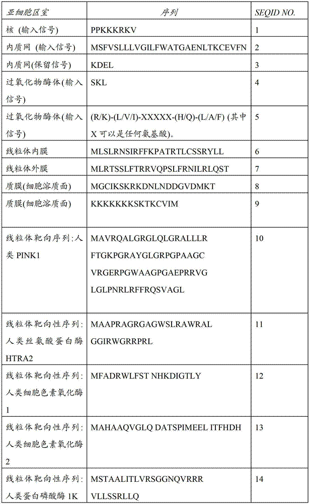
定位信号的其他例子是有描述的,例如在,Stryer,L.和W.H.Freeman的生物化学(第4版)中35章(1995)的“蛋白靶向”(“Protein Targeting”,chapter35of Stryer,L.,Biochemistry(4th ed.).W.H.Freeman,1995),和Alberts等人的细胞分子生物学第12章(页码551-598),第3版,(1994),加兰出版公司(Chapter12(pages551-598)of Molecular Biology of the Cell,Alberts et al.third edition,(1994)Garland Publishing Inc.)。在一些具体实施方式中,多于一个分离定位基序通过细胞机器用于提供正确的定位安排。例如,通过采用N-末端信号序列和C-末端GPI锚着点或跨膜区,可以将蛋白精确定位安排到质膜的细胞外的一面。Other examples of localization signals are described, for example, in "Protein Targeting", chapter 35 of Stryer, L. Freeman, Biochemistry (4th Edition), Chapter 35 (1995). ., Biochemistry (4th ed.). W.H. Freeman, 1995), and Alberts et al. Cell Molecular Biology Chapter 12 (pages 551-598), 3rd Edition, (1994), Garland Publishing Company (Chapter 12 (pages 551 -598) of Molecular Biology of the Cell, Alberts et al. third edition, (1994) Garland Publishing Inc.). In some embodiments, more than one separate localization motif is used by the cellular machinery to provide the correct localization arrangement. For example, by employing an N-terminal signal sequence and a C-terminal GPI anchor or transmembrane region, proteins can be precisely localized to the extracellular side of the plasma membrane.
典型的是,定位序列几乎可位于蛋白的氨基酸序列的任何位置。在一些情况下,定位序列通过可变数量的氨基酸可被分裂为彼此分离的两块。通过标准重组DNA的方法制备该构建体在本领域是公知的。例如:描述在Maniatis等人的分子克隆实验手册,冷泉巷实验室,纽约,1989(Maniatis,et al.,Molecular Cloning A Laboratory Manual,Cold Spring Harbor Laboratory,N.Y,1989)。Typically, the positioning sequence can be located almost anywhere in the amino acid sequence of the protein. In some cases, the positioning sequence can be split into two pieces separated from each other by a variable number of amino acids. Preparation of such constructs by standard recombinant DNA methods is well known in the art. For example: described in Maniatis et al. Molecular Cloning A Laboratory Manual, Cold Spring Lane Laboratory, New York, 1989 (Maniatis, et al., Molecular Cloning A Laboratory Manual, Cold Spring Harbor Laboratory, N.Y, 1989).
靶向于质膜:在一些具体实施方式中,设计的构建体包括信号序列用来优化蛋白使其定位于质膜。这些可包括,例如,来自于β2烟碱乙酰胆碱受体(MRGTPLLLVVSLFSLLQD(SEQ ID NO:17),用一个字母氨基酸代码表示)的C-末端信号序列,和/或来自于Kir2.1,(FCYENEV)(SEQ ID NO:18)的内质网输出基序。Targeting to the plasma membrane: In some embodiments, the designed construct includes a signal sequence to optimize the localization of the protein to the plasma membrane. These may include, for example, the C-terminal signal sequence from the β2 nicotinic acetylcholine receptor (MRGTPLLLLVVSLFSLLQD (SEQ ID NO: 17), represented by the one-letter amino acid code), and/or from Kir2.1, (FCYENEV) Endoplasmic reticulum export motif of (SEQ ID NO:18).
原生质定位的附加改进可通过加入高尔基输出序列(例如.源自Kir2.1:RSRFVKKDGHCNVQFINV(SEQ ID NO:19))和膜定位序列(例如,源自Kir2.1:KSRITSEGEYIPLDQIDINV(SEQ ID NO:20))(Gradinaru,V.等.细胞(2010))(Gradinaru,V.et al.Cell(2010))获得。在一些具体实施方式中,选择靶向序列用于调控细胞内蛋白的传输使其达到预期的亚细胞结构。在一种具体实施方式中,蛋白靶向于真核细胞中的质膜。在这种情况下,靶向序列可按照下面的策略进行设计:Gradinaru等人的,“多元化和扩展光遗传学的分子和细胞方法”期刊:细胞第141卷,页码:154-165(2010年)(Molecular and CellularApproaches for Diversifying and Extending Optogenetics,”Cell141,154-165(2010))。术语“信号序列”指将蛋白靶向于亚细胞位置的N-末端区域,例如,内质网(ER),且因此通向质膜。在光遗传学电压传感器中使用的信号序列是源自于蛋白β2-n-乙酰胆碱受体(SS B2nAChR)和PPL的。此外,细菌视紫红质蛋白上有内源性信号序列可以被固定以用于适宜的亚细胞靶向作用。运输信号(TS)可以任意插入到细菌视紫红质的基因组C-末端和附属荧光蛋白N末端的。在一种具体实施方式中,像Gradinaru等人说明的那样运输信号是源自于Kir2.1蛋白。在另外的具体实施方式中,ER输出基序插入到附属荧光蛋白的C末端。Additional improvements in proplasm localization can be achieved by adding Golgi export sequences (e.g. from Kir2.1:RSRFVKKDGHCNVQFINV (SEQ ID NO:19)) and membrane localization sequences (e.g. from Kir2.1:KSRITSEGEYIPLDQIDINV (SEQ ID NO:20) ) (Gradinaru, V. et al. Cell (2010)) (Gradinaru, V. et al. Cell (2010)). In some embodiments, targeting sequences are selected to regulate intracellular delivery of proteins to desired subcellular structures. In a specific embodiment, the protein is targeted to the plasma membrane in eukaryotic cells. In this case, targeting sequences can be designed according to the following strategy: Gradinaru et al., Molecular and Cellular Approaches to Multiplexed and Expanded Optogenetics Journal: Cell Vol. 141, Pages: 154-165 (2010 2010) (Molecular and Cellular Approaches for Diversifying and Extending Optogenetics, "Cell141,154-165(2010)). The term "signal sequence" refers to the N-terminal region that targets proteins to subcellular locations, for example, the endoplasmic reticulum (ER ), and thus leads to the plasma membrane. The signal sequence used in the optogenetic voltage sensor is derived from the proteins β2-n-acetylcholine receptor (SS B2nAChR) and PPL. In addition, bacteriorhodopsin has internal The derived signal sequence can be fixed for appropriate subcellular targeting. A trafficking signal (TS) can be inserted arbitrarily at the genomic C-terminus of bacteriorhodopsin and the N-terminus of the accessory fluorescent protein. In one embodiment In, the trafficking signal is derived from the Kir2.1 protein as described by Gradinaru et al. In another embodiment, the ER export motif is inserted into the C-terminus of the accessory fluorescent protein.
靶向于线粒体:为了测定线粒体膜电位或研究线粒体,人们希望将PROPS定位于线粒体内膜或线粒体外膜,这种情况下,可在视紫红质蛋白加入适宜的信号序列。Targeting to mitochondria: In order to measure the mitochondrial membrane potential or study mitochondria, it is hoped that PROPS will be located on the inner mitochondrial membrane or the outer mitochondrial membrane. In this case, an appropriate signal sequence can be added to the rhodopsin protein.
光遗传学电压传感器可以靶向于线粒体内膜。按照在例如A.Hoffmann,V.Hildebrandt,J.Heberle,和G.Büldt,“光活性线粒体:光驱动的质子泵体内转移到裂殖酵母属稷酒的线粒体内膜,”美国科学院院报,PNAS,页码:9367-9371(1994年)(A.Hoffmann,V.Hildebrandt,J.Heberle,andG.Büldt,“Photoactive mitochondria:in vivo transfer of a light-driven proton pumpinto the inner mitochondrial membrane of Schizosaccharomyces pombe,”Proc.Natl.Acad.Sci.USA91,PNAS9367-9371(1994))中描述的方法就可以实现。Optogenetic voltage sensors can be targeted to the inner mitochondrial membrane. As per A. Hoffmann, V. Hildebrandt, J. Heberle, and G. Büldt, "Photoactive Mitochondria: Light-Driven Proton Pump In vivo Transfer to the Mitochondrial Inner Membrane of Schizosaccharomyces spp.," Proceedings of the National Academy of Sciences, PNAS, page number: 9367-9371 (1994) (A.Hoffmann, V.Hildebrandt, J.Heberle, and G.Büldt, "Photoactive mitochondria: in vivo transfer of a light-driven proton pumpinto the inner mitochondrial membrane of Schizosaccharomyces pombe, "Proc.Natl.Acad.Sci.USA91, PNAS9367-9371 (1994)) described in the method can be realized.
细胞cell
根据发明所述有用细胞包括真核细胞和原核细胞。真核细胞包括非哺乳无脊椎动物细胞,例如酵母,植物,和线虫,与非哺乳脊椎动物例如鱼和鸟。细胞还包括哺乳动物细胞,包括人类细胞。细胞还包括无限增殖化的细胞系例如HEK,HeLa,CHO,3T3,它们在药物筛选方法应用中尤其有用。这些细胞还包括干细胞,多能细胞,祖细胞,和诱导多能细胞。分化的细胞包括从干细胞分化的细胞,还包括多能细胞和祖细胞。Useful cells according to the invention include eukaryotic cells and prokaryotic cells. Eukaryotic cells include cells of non-mammalian invertebrates such as yeast, plants, and nematodes, and non-mammalian vertebrates such as fish and birds. Cells also include mammalian cells, including human cells. Cells also include immortalized cell lines such as HEK, HeLa, CHO, 3T3, which are especially useful in drug screening method applications. These cells also include stem cells, pluripotent cells, progenitor cells, and induced pluripotent cells. Differentiated cells include cells differentiated from stem cells, and also include pluripotent cells and progenitor cells.
在一些具体实施方式中,细胞进行体外培养或离体培养。在一些具体实施方式中,细胞是部分的器官或生物体。In some embodiments, the cells are cultured in vitro or ex vivo. In some embodiments, a cell is part of an organ or organism.
在一些具体实施体重,细胞是“人工细胞”或生物工程学制造的“合成细胞”。(见,例如,通过化学合成基因组控制的细菌细胞的产生,作者:Daniel G.Gibson等人,期刊:科学2010年7月2日:第329卷,编码:5987页码:52-56;Cans,Ann-Sofie,Andes-Koback,Meghan,和Keating,Christine D.通过水细胞质模仿的微型组织在巨型小囊泡定位类脂膜区。美国化学会志,2008).(Creation of a Bacterial Cell Controlled by a ChemicallySynthesized Genome,Daniel G.Gibson et al.,Science2July2010:Vol.329no.5987pp.52-56;Cans,Ann-Sofie,Andes-Koback,Meghan,and Keating,ChristineD.Positioning Lipid Membrane Domains in Giant Vesicles by Micro-organizationof Aqueous Cytoplasm Mimic.J.Am.Chem.Soc.,2008)。In some embodiments, the cells are "artificial cells" or "synthetic cells" produced by bioengineering. (See, e.g., Generation of Bacterial Cells Controlled by Chemically Synthesized Genomes, by Daniel G. Gibson et al., Journal:
该方法也可应用于任何其他膜结合结构,其不一定是分类为细胞。这种膜结合结构可用于携带发明的细菌视紫红质蛋白,例如通过将携带发明的细菌视紫红质蛋白细胞膜碎片与该膜融合。This method can also be applied to any other membrane-bound structures, which are not necessarily classified as cells. Such membrane-bound structures can be used to carry the inventive bacteriorhodopsin, for example by fusing fragments of the cell membrane carrying the inventive bacteriorhodopsin to the membrane.
细胞还包括斑马鱼心肌细胞;免疫细胞(除了下面注明的特定的细胞株,所有的初级鼠类和人类培养,与iPS衍生细胞株),包括B细胞(例如,人类Raji细胞株,和DT40鸡细胞株),T细胞(例如,人类Jurkat细胞株),巨噬细胞,树突细胞,和中性白细胞(例如,HL-60株)。此外,可采用神经胶质细胞:星形细胞和少突胶质细胞;胰脏β细胞;肝细胞;非心肌细胞;内分泌细胞例如滤泡旁细胞和嗜铬细胞;和酵母细胞。细胞还包括神经细胞,例如神经元。Cells also include zebrafish cardiomyocytes; immune cells (all primary murine and human cultures, with iPS-derived cell lines, except specific cell lines noted below), including B cells (e.g., human Raji cell lines, and DT40 chicken cell line), T cells (eg, human Jurkat cell line), macrophages, dendritic cells, and neutrophils (eg, HL-60 strain). In addition, glial cells may be employed: astrocytes and oligodendrocytes; pancreatic beta cells; hepatocytes; noncardiomyocytes; endocrine cells such as parafollicular cells and chromaffin cells; and yeast cells. Cells also include nerve cells, such as neurons.
细胞也可以是革兰阳性细菌或革兰阴性细菌,和任一革兰类型的病原性细菌。病原性细胞有助于该方法应用,例如,在筛选新的抗生素方面,所述新的抗生素是通过影响膜电位来加速细菌细胞毁灭的抗生素,或与影响膜电位的试剂组合来影响膜电位进而加速细菌细胞毁灭的抗生素,或在抑制抗生素外排的化合物研究方面。The cells can also be Gram-positive bacteria or Gram-negative bacteria, and pathogenic bacteria of any Gram type. Pathogenic cells facilitate the application of this method, for example, in the screening of new antibiotics that accelerate the destruction of bacterial cells by affecting the membrane potential, or in combination with agents that affect the membrane potential to affect the membrane potential and thereby Antibiotics that accelerate the destruction of bacterial cells, or in the study of compounds that inhibit the efflux of antibiotics.
基本上任何细胞膜电位,或任何磷脂双层围成的结构的均可采用在此描述的方法和组合物进行测定。可测定的细胞例子是原代细胞,例如,原代肝细胞,原代神经细胞,原代成肌细胞,原代间质干细胞,原代祖细胞,或是已建立的细胞株细胞。细胞能够进行分裂可以是不必要的;在此描述的方法中可使用终末分化的细胞。在本文上下文中,细胞可以是任何类型的细胞,包括但不限于:上皮的,内皮的,神经的,脂肪的,心脏的,骨骼肌的,纤维母细胞,免疫细胞,肝脏的,脾的,肺的,循环的血细胞,生殖细胞,肠胃的,肾脏的,骨髓的和胰脏的细胞。细胞可以是细胞株,干细胞,或从任何组织分离的原代细胞,包括但不限于脑,肝,肺,肠,胃,脂肪,肌肉,睾丸,子宫,卵巢,皮肤,脾脏,内分泌器官和骨骼等。当细胞在体外条件下,和常规组织培养条件下培养,该方法可以使用,这对本领域技术人员来说是熟知。用于不同细胞的分离和培养方法是本领域的技术人员的常识。细胞可以是原核细胞,真核细胞,哺乳动物细胞或人类细胞。在一种具体实施方式中,细胞是神经元或其他脑细胞。在一些具体实施方式中,细胞是心肌细胞。在一些具体实施方式中,细胞从诱导多能细胞中分化得来的心肌细胞。Essentially any cell membrane potential, or any structure enclosed by a phospholipid bilayer, can be determined using the methods and compositions described herein. Examples of measurable cells are primary cells, eg, primary hepatocytes, primary neural cells, primary myoblasts, primary mesenchymal stem cells, primary progenitor cells, or cells of established cell lines. It may not be necessary for the cells to be capable of dividing; terminally differentiated cells may be used in the methods described herein. In this context, a cell may be any type of cell, including but not limited to: epithelial, endothelial, neural, adipose, cardiac, skeletal muscle, fibroblast, immune cell, hepatic, splenic, Lung, circulating blood cells, germ cells, gastrointestinal, renal, bone marrow and pancreatic cells. Cells can be cell lines, stem cells, or primary cells isolated from any tissue, including but not limited to brain, liver, lung, intestine, stomach, fat, muscle, testis, uterus, ovary, skin, spleen, endocrine organs, and bone wait. This method can be used when the cells are cultured under in vitro conditions, as well as conventional tissue culture conditions, which are well known to those skilled in the art. Isolation and culture methods for different cells are common knowledge of those skilled in the art. Cells can be prokaryotic, eukaryotic, mammalian or human. In a specific embodiment, the cells are neurons or other brain cells. In some embodiments, the cells are cardiomyocytes. In some embodiments, the cells are cardiomyocytes differentiated from induced pluripotent cells.
参考值Reference
发明提供在表达编码细菌视紫红质蛋白的核酸的细胞中测定膜电位的方法,该方法包括如下步骤:(a)用至少一个波长的光激发至少一个包含细菌视紫红质蛋白编码核酸的细胞,与(b),检测来自至少一个细胞的至少一个光学信号,其中和参考相比,由至少一个细胞发射的荧光强度水平可指示细胞的膜电位。The invention provides a method for measuring membrane potential in cells expressing a nucleic acid encoding a bacteriorhodopsin protein, the method comprising the steps of: (a) exciting at least one cell comprising a bacteriorhodopsin-encoding nucleic acid with light of at least one wavelength, With (b), at least one optical signal from at least one cell is detected, wherein the level of fluorescence intensity emitted by the at least one cell compared to a reference is indicative of the cell's membrane potential.
在此使用的术语“参考”是指本领域技术人员在该方法中可使用的任何类型的基线值。在一些具体实施方式中,参考是未暴露于能够改变或预期能够改变膜电位刺激的细胞。在一种具体实施方式中,参考是用细菌视紫红质转染的相同细胞但不同时间点观察到的。在另外的具体实施方式中,参考是可操作地与细菌视紫红质融合的的绿色荧光蛋白(GFP)同系物的荧光。The term "reference" as used herein refers to any type of baseline value available to a person skilled in the art in the method. In some embodiments, the reference is a cell that has not been exposed to a stimulus that alters or is expected to alter membrane potential. In a specific embodiment, the reference is the same cell transfected with bacteriorhodopsin but observed at a different time point. In another embodiment, the reference is the fluorescence of a green fluorescent protein (GFP) homologue operably fused to bacteriorhodopsin.
检测修饰的细菌视紫红质的荧光Detection of Fluorescence of Modified Bacteriorhodopsin
在发明的方法中,光源激发细胞,以致发射可检测到的荧光。激发光的波长取决于荧光分子。例如,在实施例中的古细菌视紫红质构建体采用波长在λ=594nm和λ=645nm范围变化的光是可激发的。可以选择地,范围可以在λ=630-645nm之间。例如,经常使用的氦氖激光器发射在λ=632.8nm,可用于激发在这些分子的荧光发射。In the inventive method, the light source excites the cells so that detectable fluorescence is emitted. The wavelength of the excitation light depends on the fluorescent molecule. For example, the archaorhodopsin constructs in the examples are excitable with light having wavelengths varying between λ = 594 nm and λ = 645 nm. Alternatively, the range may be between λ=630-645nm. For example, frequently used HeNe lasers emitting at λ = 632.8 nm can be used to excite fluorescence emission in these molecules.
在一些具体实施方式中使用第二个光。例如,如果细胞表达参考荧光分子或用于测定细胞另一个特征的荧光分子,例如pH或钙浓度。这种情况下,第二波长是不同于第一波长。有用波长的例子包括在λ=447-594nm范围内的波长,例如,λ=473nm,λ=488nm,λ=514nm,λ=532nm,和λ=561nm。In some embodiments a second light is used. For example, if the cell expresses a reference fluorescent molecule or a fluorescent molecule used to measure another characteristic of the cell, such as pH or calcium concentration. In this case, the second wavelength is different from the first wavelength. Examples of useful wavelengths include wavelengths in the range of λ=447-594 nm, eg, λ=473 nm, λ=488 nm, λ=514 nm, λ=532 nm, and λ=561 nm.
VIPs最大优势的发挥所需要的硬件和软件取决于测定类型,这对于本领域技术人员基于在此提供的信息容易优化和选择的。目前的仪器容易使用或适应于VIPs和PROPs的检测。决定仪器类型的因素包括,精密度和准确度,速度,穿透深度,多路递送和通量。The hardware and software required to maximize the benefits of VIPs depends on the type of assay and is readily optimized and selected by one skilled in the art based on the information provided herein. Current instruments are easy to use or adapt to the detection of VIPs and PROPs. Factors that determine the type of instrument include precision and accuracy, speed, depth of penetration, multiplex delivery, and throughput.
精密度与准确度:在确定检测系统中,人们应该估计是否需要电压的绝对或相对测量。膜电位的绝对测定是通常是采用两个激发波长或两个激发波长的比率完成的。单个细胞中的相对电压变化可采用单谱带激发和检测来执行。Precision vs. Accuracy: In determining a detection system, one should evaluate whether an absolute or relative measurement of voltage is required. Absolute determination of membrane potential is usually accomplished using two excitation wavelengths or a ratio of two excitation wavelengths. Relative voltage changes in individual cells can be performed using single-band excitation and detection.
速度:为了测定神经元的动作电位,需要亚毫秒时间分辨率。这需要高速度的CCDs,或可扫描定制轨迹的高速共聚焦显微镜。缓慢的动力学和准稳态的电压可通过常规相机测定。这些测定可用在,例如,在针对心脏细胞筛选试剂的方法和测定中使用,例如心肌细胞。Velocity: To measure action potentials in neurons, sub-millisecond temporal resolution is required. This requires high-speed CCDs, or high-speed confocal microscopes that scan custom trajectories. Slow kinetics and quasi-steady-state voltages can be measured with conventional cameras. These assays can be used, for example, in methods and assays for screening agents for cardiac cells, such as cardiomyocytes.
穿透深度:在深度组织中的成像可能需要共聚焦显微镜或侧板照明显微镜。可选地,深度成像可能需要非线性显微镜的发展,包括双光子荧光或倍频效应。常规的落射荧光显微镜成像对培养的细胞效果很好,且全内反射荧光(TIRF)在粘连细胞成像中提供特别高的信噪比。Penetration Depth: Imaging in deep tissue may require confocal microscopy or side-plate illumination microscopy. Alternatively, depth imaging may require the development of nonlinear microscopy, including two-photon fluorescence or frequency-doubling effects. Conventional epifluorescence microscopy imaging works well for cultured cells, and total internal reflection fluorescence (TIRF) provides a particularly high signal-to-noise ratio in imaging adherent cells.
与其他光学成像和调控的多路递送:可以将VIPs成像与其他结构和功能的成像组合,例如pH,钙,或ATP。还可以将VIPs成像和膜电位的光遗传学控制进行组合,所述膜电位的光遗传学控制是例如采用通道视紫红质,嗜盐菌紫质,和古细菌视紫红质。如果光学测定和控制被结合于反馈回路中,其就可以启用所有的光学膜片箝来探测任何膜的动态电响应。Multiplex delivery with other optical imaging and modulation: VIPs imaging can be combined with imaging of other structures and functions, such as pH, calcium, or ATP. It is also possible to combine imaging of VIPs with optogenetic control of membrane potential using, for example, channelrhodopsin, halorhodopsin, and archaerhodopsin. If optical measurement and control are combined in a feedback loop, it can enable all optical patch clamping to probe the dynamic electrical response of any membrane.
通量:还可以将机器人技术和定制软件进行整合,用于在高通量药物筛选方法中筛选经常会遇到的大的数据或大量的条件。Throughput: Robotics and custom software can also be integrated for screening large data or a large number of conditions often encountered in high-throughput drug screening methods.
细菌视紫红质中电压诱导位移的分光镜读出装置Spectroscopic readout of voltage-induced shifts in bacteriorhodopsin
细菌视紫红质的分光镜状态一般按照他们的吸收光谱分类。然而,有些情况在单个细胞中的蛋白不足以仅通过吸光度检测光学移位。所有的下述光学成像技术均可用于探测其他状态依赖性的光谱性质。The spectroscopic states of bacteriorhodopsins are generally classified according to their absorption spectra. However, there are situations where the protein in a single cell is insufficient to detect optical shifts by absorbance alone. All of the optical imaging techniques described below can be used to probe other state-dependent spectral properties.
a)荧光a) Fluorescence
我们发现很多细菌视紫红质蛋白及其突变体产生可测定的荧光。例如,PROPS荧光采用500和650nm之间波长的光激发,发射的峰在710nm。在较长的激发波长下PROPS的光漂白作用速率下降,因此更好的激发波长光谱在红波段,近似633nm。这些波长与任何其他荧光蛋白的激发和发射波长相比距离红光更远,这是体内成像非常期待的特性。此外,PROPS的荧光显示可忽略的光漂白作用,与所有其他已知荧光完全相反。当在633nm激发时,在光漂白作用之前PROPS和GFP发射相当可观数量的光子。因此,细菌视紫红质构成了一类新的高度耐光、膜结合荧光标记物。We have found that many bacteriorhodopsin proteins and their mutants produce measurable fluorescence. For example, PROPS fluorescence is excited with light at wavelengths between 500 and 650 nm, with emission peaking at 710 nm. The photobleaching rate of PROPS decreases at longer excitation wavelengths, so the better excitation wavelength spectrum is in the red band, approximately 633nm. These wavelengths are farther from the red than the excitation and emission wavelengths of any other fluorescent protein, a highly desirable property for in vivo imaging. Furthermore, the fluorescence of PROPS shows negligible photobleaching, in complete contrast to all other known fluorophores. When excited at 633 nm, PROPS and GFP emit a considerable number of photons before photobleaching. Thus, bacteriorhodopsin constitutes a new class of highly photostable, membrane-bound fluorescent markers.
由于只有质子化形式荧光,我们还发现PROPS的荧光对希夫碱基的质子化状态极其敏感。因此在质子化中电压诱导的变化导致荧光的变化。Since only the protonated form fluoresces, we also found that the fluorescence of PROPS is extremely sensitive to the protonation state of Schiff bases. Voltage-induced changes in protonation thus lead to changes in fluorescence.
在一些具体实施方式中,PROPS荧光的检测是采用例如荧光显微镜,荧光分析仪,荧光激活细胞分类术(FACS)的荧光细胞分类等进行检测。In some specific embodiments, the detection of PROPS fluorescence is performed using, for example, a fluorescence microscope, a fluorescence analyzer, fluorescent cell sorting by fluorescence activated cell sorting (FACS), and the like.
b)荧光共振能量转移(FRET)的光谱移动b) Spectral Shift by Fluorescence Resonance Energy Transfer (FRET)
FRET是在生物物理学和生物化学中,是有用的定量分子动力学工具,例如蛋白-蛋白相互作用,蛋白-DNA相互作用和蛋白构象变化。为了监测两个分子间的复合物的形成(例如,视黄醛和细菌视紫红质),他们中的一个用供体标记,另一个用受体标记,并将这些荧光标记的分子混合。当他们被分裂时,通过供体激发检测供体发射。另一方面,由于两分子的相互作用当供体和受体邻近时(1-10nm),主要观察到受体发射,由于从供体到受体的分子间的FRET。FRET is a useful quantitative molecular dynamics tool in biophysics and biochemistry, such as protein-protein interactions, protein-DNA interactions, and protein conformational changes. To monitor the formation of a complex between two molecules (for example, retinal and bacteriorhodopsin), one of them is labeled with a donor and the other with an acceptor, and these fluorescently labeled molecules are mixed. When they are split, donor emission is detected by donor excitation. On the other hand, acceptor emission is mainly observed when the donor and acceptor are in close proximity (1-10 nm) due to the interaction of the two molecules, due to intermolecular FRET from the donor to the acceptor.
附加于细菌视紫红质的荧光分子可将其激发能转移到视黄醛,但只有当视黄醛的吸收光谱与荧光基团发射光谱重叠时。视黄醛吸收光谱的变化导致荧光基团荧光亮度的改变。为了执行FRET光谱移动,荧光蛋白和细菌视紫红质电压传感器相融合,而监测蛋白的荧光。和直接荧光相比,这个方法的优点在于荧光蛋白的放射比视黄醛的更加明亮,但缺点在于较小分数变化的荧光的非直接读出装置。Fluorescent molecules attached to bacteriorhodopsin can transfer their excitation energy to retinal, but only if the absorption spectrum of retinal overlaps with the emission spectrum of the fluorophore. Changes in the absorption spectrum of retinal lead to changes in the fluorescence brightness of the fluorophore. To perform FRET spectral shifting, a fluorescent protein is fused to a bacteriorhodopsin voltage sensor, which monitors the protein's fluorescence. Compared with direct fluorescence, this method has the advantage that the emission of fluorescent proteins is brighter than that of retinal, but the disadvantage is the indirect readout of smaller fractional changes in fluorescence.
在一些具体实施方式中,在细菌视紫红质中的吸收光谱中的电压诱导变化是通过采用FRET的光谱移动检测的。In some embodiments, voltage-induced changes in the absorption spectrum in bacteriorhodopsin are detected by spectral shift using FRET.
c)视紫红质光学光锁定成像(ROLI)c) Rhodopsin Optical Light Locking Imaging (ROLI)
很多视黄醛状态的吸收光谱被短暂脉冲光暂时改变。在ROLI,“泵”束的周期脉冲可传递到样品。第二“探针”束测定在某一波长下样品的吸光度,该波长下泵束光诱发了吸光度的巨大变化。因此,泵束在探针束的透射强度中表示了周期调节。这些周期性的强度的变化通过锁定成像系统检测。与常规的吸收成像相反,ROLI提供视黄醛特异性的对照。高频率的泵调节使其可以检测吸光度的微小变化。The absorption spectra of many retinal states are temporarily altered by brief pulses of light. In ROLI, periodic pulses of a "pump" beam are delivered to the sample. The second "probe" beam measures the absorbance of the sample at the wavelength at which the pump light induces a large change in absorbance. Thus, the pump beam represents a periodic modulation in the transmitted intensity of the probe beam. These periodic intensity changes are detected by a lock-in imaging system. In contrast to conventional absorption imaging, ROLI provides a retinal-specific control. The high frequency of pump regulation makes it possible to detect small changes in absorbance.
在一些具体实施方式中,PROPS的荧光采用细菌视紫红质锁定成像系统检测。In some embodiments, the fluorescence of PROPS is detected using a bacteriorhodopsin lock-in imaging system.
d)拉曼d) Raman
拉曼光谱是一种可以在系统中检测震动,转动和其他低频率震荡模式的技术。该技术依赖于单色光的非弹性散射(例如,可见光,近红外光或近紫外光)。单色光和分子震动,震动量子或系统中的其他刺激相互作用,导致激光光子的能量变化。能量的转移提供系统中有关震动量子模式的信息。Raman spectroscopy is a technique that can detect vibrations, rotations and other low frequency oscillation modes in a system. The technique relies on the inelastic scattering of monochromatic light (eg, visible, near-infrared, or near-ultraviolet light). The monochromatic light interacts with molecular vibrations, vibration quanta, or other stimuli in the system, resulting in a change in the energy of the laser photon. The transfer of energy provides information about the quantum patterns of vibrations in the system.
已知在细菌视紫红质分子中的视黄醛有很强的共振拉曼信号。该信号依赖于生色团周围的静电环境,因此对电压是敏感。Retinaldehyde in the bacteriorhodopsin molecule is known to have a strong resonant Raman signal. The signal is dependent on the electrostatic environment surrounding the chromophore and is therefore sensitive to voltage.
在一些具体实施方式中,在细菌视紫红质拉曼光谱中的电压诱导的变化通过拉曼显微镜进行检测。In some embodiments, voltage-induced changes in the bacteriorhodopsin Raman spectrum are detected by Raman microscopy.
e)二次谐波产生(SHG)e) Second Harmonic Generation (SHG)
二次谐波产生,在本领域也称为“倍频”,是非线性光学过程,在该过程中,与非线性物质相互作用的光子被有效“组合”进而形成具有两倍能量的光子,从而具有最初光量子两倍的频率,一半的波长。Second harmonic generation, also known in the art as "frequency doubling," is a nonlinear optical process in which photons interacting with nonlinear matter are effectively "combined" to form photons with twice the energy, thereby It has twice the frequency and half the wavelength of the original photon.
从细胞膜内的菌视紫质的定向薄膜上可观察到SHG信号。SHG是光学电压传感器视黄醛周围静电环境的有效探针。此外,SHG成像包括用穿透深入组织的红外光激发。因此,采用在此描述的光学传感器的SHG成像可用于三维光学电压传感。SHG signals were observed from oriented thin films of bacteriorhodopsin within the cell membrane. SHG is an effective probe of the electrostatic environment around retinal in optical voltage sensors. Additionally, SHG imaging involves excitation with infrared light that penetrates deep into tissue. Therefore, SHG imaging with the optical sensor described here can be used for three-dimensional optical voltage sensing.
在一些具体实施方式中,采用SHG成像可检测到细菌视紫红质的倍频光谱中的电压诱导变化。In some embodiments, voltage-induced changes in the frequency-octave spectrum of bacteriorhodopsin are detectable using SHG imaging.
具有产生光学信号部分的融合蛋白Fusion proteins with optical signal generating moieties
虽然响应电压变化时细菌视紫红质蛋白是自身有荧光性,在一些应用中,希望或有必要提高荧光水平或提供另外的光学信号(例如,比色信号)进而检测电压变化。而且,产生光学信号的部分可附属于细菌视紫红质来监测视紫红质蛋白的亚细胞定位。因此,在一些具体实施方式中,修饰细菌视紫红质蛋白还包括产生光学信号的部分,因此增强从修饰细菌视紫红质测得的光学信号或实施视紫红质蛋白的定位研究。Although bacteriorhodopsin is itself fluorescent in response to voltage changes, in some applications it may be desirable or necessary to increase the level of fluorescence or provide an additional optical signal (eg, a colorimetric signal) to detect voltage changes. Furthermore, moieties that generate optical signals can be attached to bacteriorhodopsin to monitor the subcellular localization of the rhodopsin protein. Thus, in some embodiments, the modified bacteriorhodopsin further comprises a moiety that generates an optical signal, thereby enhancing the optical signal measured from the modified bacteriorhodopsin or performing localization studies of the bacteriorhodopsin.
例如,GFP家族或其同系物荧光蛋白基因可以任意附加或如权利要求书提到的“可操作地连接”于编码细菌视紫红质的核酸。在一种具体实施方式中,荧光蛋白的鉴定,其与电压传感复合物接头,和接头定位,是为了实现两个功能的其中之一而整个蛋白序列对进行选择:作为基因表达产物水平和分布的指示器;或者是作为电压的可选读出装置,独立于视黄醛的内源性荧光。For example, GFP family or its homologue fluorescent protein gene can be arbitrarily attached or "operably linked" to the nucleic acid encoding bacteriorhodopsin as mentioned in the claims. In a specific embodiment, the identification of fluorescent proteins, their junctions with the voltage sensing complex, and the location of the junctions, are performed with the entire protein sequence selected for one of two functions: as gene expression product levels and indicator of distribution; or as an optional readout of voltage, independent of the endogenous fluorescence of retinal.
例如,当荧光蛋白作为蛋白定位的指示器时,它可实现定量光学电压测定,所述定量光学电压测定是由细胞间的表达水平差异不能引起混乱。荧光蛋白和细菌视紫红质的荧光可以同时测定,且这两中信号比率提供了独立于浓度的膜电位测定。For example, when a fluorescent protein is used as an indicator of protein localization, it enables quantitative optical voltage measurements that cannot be confounded by differences in expression levels between cells. The fluorescence of fluorescent proteins and bacteriorhodopsin can be measured simultaneously, and the ratio of the two signals provides a concentration-independent measurement of membrane potential.
在一种具体实施方式中,细菌视紫红质蛋白是包括荧光蛋白的PROPS融合蛋白。例如,PROPS的N末端与荧光蛋白Venus融合。该蛋白提供稳定的参考,该参考通过细胞内PROPS定位指示,且可以进行Venus和PROPS荧光的比率成像。因为这技术对细胞内蛋白总量不敏感,比率成像使膜电位的定量测定成为可能。其他荧光蛋白可用于代替Venus而具有类似效果。在一些具体实施方式中,荧光多肽选自如下组中:GFP,YFP,EGFP,EYFP,EBFB,DsRed,RFP及其荧光变异体In a specific embodiment, the bacteriorhodopsin is a PROPS fusion protein that includes a fluorescent protein. For example, the N-terminus of PROPS is fused to the fluorescent protein Venus. This protein provides a stable reference indicated by intracellular PROPS localization and allows ratiometric imaging of Venus and PROPS fluorescence. Because the technique is insensitive to the total amount of intracellular protein, ratiometric imaging enables quantitative determination of membrane potential. Other fluorescent proteins can be used in place of Venus to similar effect. In some embodiments, the fluorescent polypeptide is selected from the group consisting of GFP, YFP, EGFP, EYFP, EBFB, DsRed, RFP and fluorescent variants thereof
在一种具体实施方式中,细菌视紫红质是古细菌视紫红质融合或可操作地连接于附加荧光蛋白,例如GFP或其同系物。In a specific embodiment, the bacteriorhodopsin is an archaealrhodopsin fused or operably linked to an additional fluorescent protein, such as GFP or a homologue thereof.
生色团Chromophores
在自然环境下,细菌视紫红质包括作为光学活性元素的视黄醛的结合分子。这些蛋白也结合和折叠在很多其他有相似结构的,可能的较好光学性质的生色团周围。具有锁定环的视黄醛类似物不能进行顺-反异构化,因此具有较高的荧光量子产量(Brack,T.等.生物物理学杂志,第65卷,页码:964-972(1993年))(Brack,T.et al.Biophys.J.65,964-972(1993))。具有吸电子取代基的视黄醛类似物具有的希夫碱基其具有比天然视黄醛更低pKa的,因此可能对电压更敏感。(Sheves,M.,等.美国国家科学院院报.第83卷,页码:3262-3266(1986);Rousso,I.,等.生物化学,第34卷,页码:12059-12065(1995)).(Sheves,M.,et al.Proc.Nat.Acad.Sci.U.S.A.83,3262-3266(1986);Rousso,I.,et al.Biochemistry34,12059-12065(1995)).视黄醛分子的共价键改性可能导致光学电压传感器具有显著提高的光学特征和电压灵敏度。In nature, bacteriorhodopsin includes a binding molecule of retinal as an optically active element. These proteins also bind and fold around many other chromophores with similar structures and possibly better optical properties. Retinaldehyde analogs with locked rings cannot carry out cis-trans isomerization, so there is a higher fluorescence quantum yield (Brack, T. etc. Journal of Biophysics, the 65th volume, page number: 964-972 (1993 )) (Brack, T. et al. Biophys. J. 65, 964-972 (1993)). Retinaldehyde analogs with electron-withdrawing substituents have Schiff bases which have a lower pKa than native retinal and thus may be more sensitive to voltage. (Sheves, M., et al. Proceedings of the National Academy of Sciences of the United States of America. Vol. 83, page number: 3262-3266 (1986); Rousso, I., et al. Biochemistry, volume 34, page number: 12059-12065 (1995)) .(Sheves, M., et al.Proc.Nat.Acad.Sci.U.S.A.83,3262-3266(1986); Rousso, I., et al.Biochemistry34,12059-12065(1995)).Retinaldehyde molecule The covalent modification of , may lead to optical voltage sensors with significantly improved optical characteristics and voltage sensitivity.
在此描述的方法和组合物的优势Advantages of the methods and compositions described herein
光学电压传感器的关键品质因素是其响应速度和其灵敏度(每100mV膜电位变化时荧光的变化分数)。图10比较了以前使用基于蛋白的荧光电压指示器和在此关注的那些指示器的这些属性。附加重要的属性包括:将指示器靶向于特定细胞或亚细胞结构的能力,耐光性和低光毒性。The key quality factors of an optical voltage sensor are its speed of response and its sensitivity (fraction of change in fluorescence per 100mV change in membrane potential). Figure 10 compares these properties of protein-based fluorescent voltage indicators previously used with those of interest here. Additional important properties include: ability to target indicators to specific cellular or subcellular structures, photostability, and low phototoxicity.
前期基于蛋白的工作集中于将一个或多个荧光蛋白融合到跨膜电压传感区域。电压变化诱导了在电压传感区域中的构象变化,其移动荧光蛋白且改变他们的荧光。多重大型蛋白区构象转换的依赖性使这些方法不可避免的变缓慢。另外,大多数电压传感区域的构象转换是微小的,导致荧光的小的变化。Previous protein-based work has focused on fusing one or more fluorescent proteins to transmembrane voltage-sensing domains. The voltage change induces a conformational change in the voltage sensing region, which moves the fluorescent proteins and changes their fluorescence. The dependence on conformational switching of multiple large protein domains makes these methods inevitably slow. In addition, the conformational switch of most voltage-sensing domains is subtle, resulting in small changes in fluorescence.
来自VSFP2.x家族的最敏感指示器在每100mV的荧光变化为ΔF/F=10%。VSFP2.x蛋白响应在大约100毫秒,太慢而不能检测神经元中1ms的动作电位。(Perron,A.等.内分泌分子神经科学.第2卷(2009);Mutoh,H.等.公共科学图书馆-综合,第4卷,页码:e4555(2009))(Perron,A.et al.Front Mol Neurosci.2(2009);Mutoh,H.et al.PLoS One4,e4555(2009)).电压传感器的SPARC家族有1ms的响应时间,但显示每100mV的荧光变化<1%。(Baker,B.J.等.神经科学方法杂志。第161卷,页码:32-38(2007);Ataka,K.和Pieribone,V.A.生物物理学杂志.第82卷,页码:509-516(2002))(Baker,B.J.et al.J.Neurosci.Methods161,32-38(2007);Ataka,K.&Pieribone,V.A.Biophys.J.82,509-516(2002))。在此描述的本研究之前,二十年的荧光电压传感器研究尚未得出可以表示体内个体神经动作电位的蛋白。The most sensitive indicators from the VSFP2.x family have a change in fluorescence per 100mV of ΔF/F=10%. VSFP2.x proteins respond at about 100 ms, too slow to detect 1 ms action potentials in neurons. (Perron, A. et al. Endocrine Molecular Neuroscience. Vol. 2 (2009); Mutoh, H. et al. PLoS One, Vol. 4, Page: e4555 (2009)) (Perron, A. et al .Front Mol Neurosci.2(2009); Mutoh, H. et al.PLoS One4, e4555(2009)). The SPARC family of voltage sensors has a response time of 1 ms but shows <1% change in fluorescence per 100 mV. (Baker, B.J. et al. Journal of Neuroscience Methods. Vol. 161, Pages: 32-38 (2007); Ataka, K. and Pieribone, V.A. Biophysical Journal. Vol. 82, Pages: 509-516 (2002)) (Baker, B. J. et al. J. Neurosci. Methods 161, 32-38 (2007); Ataka, K. & Pieribone, V. A. Biophys. J. 82, 509-516 (2002)). Prior to the present study described here, two decades of fluorescent voltage sensor research had not yielded proteins that could represent individual neural action potentials in vivo.
科学家已开发了显示电压敏感的荧光的有机小分子染料。这些亲脂分子掺入到细胞膜,在该处电压导致构象转换或电子能级跃迁,和因此导致的光学性质的变化。这些分子响应迅速(一般小于1ms),灵敏度高达每100mV34%,但是不能定向,常难以传输,且毒性较大。(Krauthamer,V.,等.荧光杂志,第1卷,页码:207-213(1991);Fromherz,P.,等。欧洲生物物理学杂志.第37卷,页码:509-514(2008);Sjulson,L.和Miesenbock,G.神经科学杂志.第28卷,页码:5582(2008)),见例如美国专利,专利号:7867282,6107066和5661035).(Krauthamer,V.,et al.J.Fluoresc.1,207-213(1991);Fromherz,P.,et al.Eur.Biophys.J.37,509-514(2008);Sjulson,L.&Miesenbock,G.J.Neurosci.28,5582(2008)),see e.g.US Patent Nos.7,867,282,6,107,066,and5,661,035)。这些光学电压传感器中,无一使用配置“向后”运行的细菌视紫红质蛋白,该蛋白使膜电位变化转换为光学可测的信号的变化。Scientists have developed small organic molecule dyes that display voltage-sensitive fluorescence. These lipophilic molecules are incorporated into the cell membrane where an electrical voltage causes a conformational switch or electronic energy level transition, and thus a change in optical properties. These molecules respond quickly (generally less than 1ms) and have a sensitivity as high as 34% per 100mV, but they cannot be directed, are often difficult to transmit, and are highly toxic. (Krauthamer, V., et al. Journal of Fluorescence, Vol. 1, Pages: 207-213 (1991); Fromherz, P., et al. European Journal of Biophysics. Vol. 37, Pages: 509-514 (2008); Sjulson, L. and Miesenbock, G. Journal of Neuroscience. Vol. 28, p. 5582 (2008)), see e.g. U.S. Pat. Nos. 7867282, 6107066 and 5661035). .Fluoresc.1, 207-213(1991); Fromherz, P., et al.Eur.Biophys.J.37, 509-514(2008); Sjulson, L. & Miesenbock, G.J.Neurosci.28, 5582(2008) ), see e.g. US Patent Nos. 7,867,282, 6,107,066, and 5,661,035). None of these optical voltage sensors use bacteriorhodopsin configured to operate "backwards," converting changes in membrane potential into changes in an optically measurable signal.
在此描述的光学电压传感器方法是不同于前期努力的方向的。如在此所述,使用的蛋白在自然环境下有强的光电耦合。在自然环境下的细菌视紫红质将太阳光转换为膜电位。在此描述的光学电压传感器利用其逆转方面的作用,将膜电位转换为容易检测的光学信号。如图12所示,就关键品质因素而言,典型的细菌视紫红质电压传感器(PROPS,Arch3WT,Arch3D95N)胜过其他基于蛋白的指示器。The optical voltage sensor approach described here is a different direction from previous efforts. As described herein, the proteins used are strongly optocoupled in their natural environment. Bacteriorhodopsin in nature converts sunlight into membrane potential. The optical voltage sensor described here exploits its inversion aspect to convert membrane potential into an easily detectable optical signal. As shown in Figure 12, typical bacteriorhodopsin voltage sensors (PROPS, Arch3WT, Arch3D95N) outperformed other protein-based indicators in terms of critical quality factors.
光学传感器递送介导的膜融合Optical sensor delivery-mediated membrane fusion
膜融合反应在真核细胞中是普遍的。膜通过如下过程进行细胞内融合,包括:内吞作用,细胞器形成,细胞器间运输,与构成的和调节的胞吐作用。细胞间的,膜融合发生在精子卵融合和肌原细胞融合。Membrane fusion reactions are common in eukaryotic cells. Membranes fuse intracellularly through processes including: endocytosis, organelle formation, inter-organelle trafficking, and constitutive and regulated exocytosis. Between cells, membrane fusion occurs in sperm-egg fusion and myogenic cell fusion.
膜融合已通过使用脂质体进行人工诱导,其中的细胞膜和脂质体膜融合,还通过不同化学试剂,诱导细胞-细胞融合进而产生异核体。显示诱导生物膜融合的自然存在的蛋白主要是包膜病毒的融合蛋白。因此,在一些具体实施方式中,光学传感器采用包括促融合蛋白的脂质体。Membrane fusion has been artificially induced by using liposomes, in which cell membranes and liposome membranes fuse, and by different chemical reagents, to induce cell-cell fusion to generate heterokaryons. The naturally occurring proteins shown to induce biofilm fusion are mainly fusion proteins of enveloped viruses. Thus, in some embodiments, optical sensors employ liposomes comprising fusogenic proteins.
普遍认为在生理条件下的膜融合是蛋白介导的。这已致使了包括促进融合蛋白(质蛋白体)的毒性下降的脂质体的开发。(见,例如,Cheng,Hum.基因疗法.第7卷,页码:275-282(1996);Hara等,基因,第159卷,页码:167-174(1995);和Findeis等,生物工程进展,第11卷,页码:202-205(1993))(Cheng,Hum.Gene Ther.7:275-282(1996);Hara et al.,Gene159:167-174(1995);andFindeis et al.,Trends Biotechnol.,11:202-205(1993))。It is generally accepted that membrane fusion under physiological conditions is protein-mediated. This has led to the development of liposomes including liposomes that promote reduced toxicity of fusion proteins (proteosomes). (See, eg, Cheng, Hum. Gene Therapy. Vol. 7, pp. 275-282 (1996); Hara et al., Genes, Vol. 159, pp. 167-174 (1995); and Findeis et al., Advances in Bioengineering , Vol. 11, Page Number: 202-205(1993)) (Cheng, Hum. Gene Ther. 7:275-282(1996); Hara et al., Gene159: 167-174(1995); and Findeis et al., Trends Biotechnol., 11:202-205 (1993)).
最终显示诱导生物膜细胞间融合的唯一蛋白是那些包膜病毒和源自无包膜病毒的两个蛋白。所有的膜病毒编码蛋白,是融合病毒外膜和细胞膜的结果。这些病毒融合蛋白对于易感细胞的感染是必要的。来自包膜病毒的融合蛋白的作用机制已成为蛋白介导的膜融合的范例。(见,例如,White,生理学年鉴,第52卷,页码:675-697(1990);和White,科学,第258卷,页码:917-924(1992)).(White,Ann.Rev.Physiol.,52:675-697(1990);and White,Science,258:917-924(1992)).The only proteins conclusively shown to induce fusion between cells of biofilms were those of enveloped viruses and two proteins derived from non-enveloped viruses. All membrane viruses encode proteins that are the result of fusion of the viral outer and cellular membranes. These viral fusion proteins are necessary for the infection of susceptible cells. The mechanism of action of fusion proteins from enveloped viruses has served as a paradigm for protein-mediated membrane fusion. (See, eg, White, Annals of Physiology, Vol. 52, pp. 675-697 (1990); and White, Science, Vol. 258, pp. 917-924 (1992)). (White, Ann. Rev. Physiol ., 52:675-697(1990); and White, Science, 258:917-924(1992)).
大多数包膜病毒融合蛋白是相对大的,多聚体的,I型膜蛋白,以流行性感冒病毒HA蛋白为代表的,低pH活化的融合蛋白,和在中性pH发挥作用的仙台病毒F蛋白。这些是病毒的结构性蛋白,其具有定向在在病毒体外表面具有多数融合蛋白,进而促进了在病毒颗粒和细胞膜之间的相互作用。Most enveloped virus fusion proteins are relatively large, multimeric, type I membrane proteins, typified by the influenza virus HA protein, low pH activated fusion proteins, and Sendai virus that function at neutral pH F protein. These are structural proteins of the virus with most fusion proteins oriented on the outer surface of the virion, which in turn facilitates the interaction between the virus particle and the cell membrane.
根据来自包膜病毒的融合蛋白作用机制,病毒膜和细胞膜的融合由两性α螺旋区域介导,即指存在于病毒融合蛋白的融合肽基序。这种类型融合肽基序一般是17到28残基长,疏水的(平均疏水性约为0.6±0.1),且包括高含量的甘氨酸和丙氨酸,一般为36%±7%。(White,生理学年鉴,第52卷,页码:675-697(1990年)(White,Annu.Rev.Physiol.,52:675-697(1990).According to the mechanism of action of the fusion protein from an enveloped virus, the fusion of the viral membrane and the cell membrane is mediated by the amphipathic α-helical region, which refers to the fusion peptide motif present in the viral fusion protein. This type of fusion peptide motif is typically 17 to 28 residues long, hydrophobic (average hydrophobicity about 0.6 ± 0.1), and includes a high content of glycine and alanine, typically 36% ± 7%. (White, Annals of Physiology, Vol. 52, page number: 675-697 (1990) (White, Annu.Rev.Physiol., 52:675-697 (1990).
所有的包膜病毒融合蛋白被认为是通过大量的构象变化发挥作用,通过利用能量克服热力学屏障,促进膜融合。这些构象变化通过形成卷曲螺旋结构的七个重复区域频繁地介导。(见Skehel和Wiley,细胞,第95卷,页码:871-874(1998))(Skehel and Wiley,Cell,95:871-874(1998)).识别在触发膜融合中的重要的融合肽基序已导致使用包括融合肽基序的小肽,进而提高脂质体-细胞的融合(见,例如,Muga等,生物化学,第33卷,页码:4444-4448(1994))(see,for example,Muga et al.,Biochemistry33:4444-4448(1994))。All enveloped viral fusion proteins are thought to function through a large number of conformational changes that facilitate membrane fusion by harnessing energy to overcome thermodynamic barriers. These conformational changes are frequently mediated by seven repeat regions forming a coiled-coil structure. (See Skehel and Wiley, Cell, Vol. 95, Pages: 871-874 (1998)) (Skehel and Wiley, Cell, 95:871-874 (1998)). Identification of Fusion Peptides Important in Triggering Membrane Fusion The sequence has led to the use of small peptides comprising fusion peptide motifs to improve liposome-cell fusion (see, for example, Muga et al., Biochemistry, Vol. 33, p. 4444-4448 (1994)) (see, for example, Muga et al., Biochemistry 33:4444-4448 (1994)).
包膜病毒融合蛋白也触发细胞-细胞融合,导致多核体的形成(合胞体)。在感染细胞内的病毒融合蛋白的合成导致融合蛋白通过内质网和高尔基传输系统到细胞膜的传输,是感染细胞的传染性子病毒颗粒的组装和出芽中的必要步骤(Petterson,微生物和免疫学的当代课题,第170卷,页码:67-106(1991)).(Petterson,Curr.Top.Micro.Immunol.,170:67-106(1991))。融合蛋白的合成,运输和折叠是由多个成分促进,包括用以使蛋白靶向于细胞间的传输途径的信号肽,用以N连接的糖类附加于蛋白的糖基化信号,和使蛋白锚定在细胞膜内的跨膜区。这些蛋白已用于重建的脂蛋白体(‘病毒小体’),以增强在细胞培养和体内的蛋白介导的脂质体细胞融合。(见,例如,Ramani等,欧洲生物化学学会联盟通讯,第404卷,页码:164-168(1997);Scheule等,美国呼吸系统细胞和分子生物学杂质,第13卷,页码:330-343(1995);和Grimaldi,病毒学研究,第146卷,页码:289-293(1995))(see,for example,Ramaniet al.,FEBS Lett.,404:164-168(1997);Scheule et al.,Am.J.Respir.Cell Mol.Biol.,13:330-343(1995);and Grimaldi,Res.Virol.,146:289-293(1995))。Enveloped virus fusion proteins also trigger cell-cell fusion, leading to the formation of polykaryons (syncytia). Synthesis of viral fusion proteins within infected cells results in transport of fusion proteins to the cell membrane via the endoplasmic reticulum and Golgi transport system, an essential step in the assembly and budding of infectious daughter virus particles from infected cells (Petterson, Microbiology and Immunology ). Contemporary Issues, Volume 170, Pages: 67-106 (1991)). (Petterson, Curr. Top. Micro. Immunol., 170:67-106 (1991)). The synthesis, trafficking, and folding of fusion proteins are facilitated by multiple components, including signal peptides to target proteins to intercellular transport pathways, glycosylation signals to attach N-linked carbohydrates to proteins, and The protein is anchored in the transmembrane region of the cell membrane. These proteins have been used in reconstituted liposomes ('virions') to enhance protein-mediated liposome-cell fusion in cell culture and in vivo. (See, eg, Ramani et al., FEBS Communication, Vol. 404, pp.: 164-168 (1997); Scheule et al., Impurities in Cellular and Molecular Biology of the American Respiratory System, Vol. 13, pp.: 330-343 (1995); and Grimaldi, Virological Research, Vol. 146, pp.: 289-293 (1995)) (see, for example, Ramani et al., FEBS Lett., 404:164-168 (1997); Scheule et al ., Am. J. Respir. Cell Mol. Biol., 13:330-343 (1995); and Grimaldi, Res. Virol., 146:289-293 (1995)).
因此,在一些在此描述的方法和组合物的具体实施方式中,给予细胞或受试者包括修饰细菌视紫红质和融合蛋白的胶粒,脂质体或其他的人工膜,进而通过膜融合介导光学传感器蛋白递送到达细胞。在优选的具体实施方式中,组合物还包括靶向序列使递送系统靶向于特定细胞型。必要时,人工膜组合物的外源性类脂还包括靶向基序(例如,配基),该基序结合于哺乳动物细胞以利于进入。例如,该组合物可包括作为配基的结合于哺乳动物外源凝集素的无唾液酸糖蛋蛋白(例如,肝脏无唾液酸糖蛋白受体),帮助进入哺乳动物细胞。可靶向于特定细胞表面标记物的单链抗体在此也考虑用作靶向基序。靶向部分可包括,例如,药物,受体,抗体,抗体碎片,适体,肽,维生素,糖类,蛋白,黏附分子,糖蛋白,糖残基或氨基葡聚糖,治疗试剂,药物或这些的组合。根据采用在此描述的光学感受器评估的预期靶细胞,技术人员可轻易设计各种修饰人工膜的靶向部分。Accordingly, in some embodiments of the methods and compositions described herein, micelles, liposomes, or other artificial membranes comprising modified bacteriorhodopsin and fusion proteins are administered to cells or subjects, and then undergo membrane fusion Mediates delivery of optical sensor proteins to cells. In a preferred embodiment, the composition further includes a targeting sequence to target the delivery system to a particular cell type. Optionally, the exogenous lipids of the artificial membrane composition also include targeting motifs (eg, ligands) that bind to mammalian cells to facilitate entry. For example, the composition can include an asialoglycoprotein (eg, liver asialoglycoprotein receptor) that binds to a mammalian lectin as a ligand to facilitate entry into mammalian cells. Single chain antibodies targetable to specific cell surface markers are also contemplated herein as targeting motifs. Targeting moieties can include, for example, drugs, receptors, antibodies, antibody fragments, aptamers, peptides, vitamins, carbohydrates, proteins, adhesion molecules, glycoproteins, sugar residues or aminodextran, therapeutic agents, drugs or combinations of these. Depending on the intended target cell assessed using the photoreceptors described herein, one of skill can readily design various targeting moieties that modify the artificial membrane.
对于采用递送介导的膜融合方法,考虑使用的光学传感器在异源表达系统中表达和制造。为了用于多种细胞型或种属,可制作不同表达载体,该载体包括编码光学传感器的核酸,或包括如在此描述的用于表达光学传感器的衍生物。为了在预期细胞中有效基因转录和翻译,表达载体应有必需的5’上游和3’下游的调控元件,例如启动子序列,核蛋白体识别和结合TATA框,和3’UTR AAUAAA转录终止序列。在一些具体实施方式中,光学传感器在外源表达系统中生成然后纯化,以生产脂质介导的传输试剂与预期细胞型融合。在这样的具体实施方式中,表达载体可有附加的序列如6X-组氨酸(SEQID NO:21),V5,硫氧还蛋白,谷胱甘肽-S-转移酶,c-Myc,VSV-G,HSV,FLAG,麦芽糖结合肽,金属结合肽,HA和“分泌物"信号肽(例如,蜜蜂蜂毒肽Pho,BiP),这些被掺合到表达重组光学传感器中以便于纯化。此外,在这些序列后面可有掺合的酶消化位点,在附加序列不需要以后,便于附加序列的酶排除。这些附加序列在光学传感表达的检测是有用的,用于亲和力色谱的蛋白纯化,以提高重组蛋白在宿主细胞质中的溶解度,用于更好的蛋白特别是小肽的表达,和/或用于排泄出表达的重组蛋白进入培养基,进入到原核细菌的周质或酵母细胞的原生质球。重组光学传感器的表达可在宿主细胞中构成或可被诱导,例如采用硫酸铜,糖,例如半乳糖,甲醇,甲胺,硫胺素,四环素,杆状病毒感染,和(异丙基-β-D-硫代半乳糖吡喃糖苷)IPTG,稳定的人造乳糖类似物,取决于选择的宿主和载体系统的。For methods employing delivery-mediated membrane fusion, contemplate using optical sensors expressed and manufactured in heterologous expression systems. For use in various cell types or species, different expression vectors can be made that include nucleic acid encoding the optical sensor, or include derivatives for expression of the optical sensor as described herein. For efficient gene transcription and translation in the intended cell, the expression vector should have essential 5'upstream and 3'downstream regulatory elements, such as a promoter sequence, a ribosome recognition and binding TATA box, and a 3'UTR AAUAAA transcription termination sequence . In some embodiments, optical sensors are generated in exogenous expression systems and then purified to produce lipid-mediated delivery reagents for fusion with desired cell types. In such embodiments, the expression vector may have additional sequences such as 6X-histidine (SEQ ID NO: 21), V5, thioredoxin, glutathione-S-transferase, c-Myc, VSV -G, HSV, FLAG, maltose-binding peptides, metal-binding peptides, HA and "secretion" signal peptides (e.g., honeybee melittin Pho, BiP), which were incorporated into expressed recombinant optical sensors for ease of purification. In addition, these sequences may be followed by incorporated enzymatic digestion sites to facilitate enzymatic removal of additional sequences after they are no longer required. These additional sequences are useful in the detection of expression by optical sensing, for protein purification by affinity chromatography, to improve the solubility of recombinant proteins in the host cytoplasm, for better expression of proteins, especially small peptides, and/or with The expressed recombinant protein is excreted into the medium, into the periplasm of prokaryotic bacteria or the spheroplast of yeast cells. Expression of recombinant optical sensors can be constructed in host cells or can be induced, for example, with copper sulfate, sugars such as galactose, methanol, methylamine, thiamine, tetracycline, baculovirus infection, and (isopropyl-β -D-thiogalactopyranoside) IPTG, a stable artificial lactose analog, depending on the choice of host and vector system.
其他表达载体和宿主细胞的例子是pET载体(Novagen),pGEX载体(Amersham Pharmacia),和pMAL载体(纽英伦技术有限公司)(New Englandlabs.Inc.)用于在大肠杆菌宿主细胞中的蛋白表达,例如BL21,BL21(DE3)和AD494(DE3)pLysS,Rosetta(DE3)和Origami(DE3)(Novagen);基于CMV强启动子的pcDNA3.1(Invitrogen)和pCIneo载体(Promega)用以在哺乳动物细胞系中表达,例如在CHO,COS,HEK-293,Jurkat和MCF-7;机能不全的腺病毒载体的复制载体pAdeno X,pAd5F35,pLP-Adeno-X-CMV(Clontech),pAd/CMV/V5-DEST,pAd-DEST载体(Invitrogen)用以在哺乳动物细胞中腺病毒介导的基因转移和表达;pLNCX2,pLXSN和pLAPSN逆转录病毒载体与来自Clontech的Retro-XTM系统应用,用以在哺乳动物细胞中逆转录介导的基因转移和表达;pLenti4/V5-DESTTM,pLenti6/V5-DESTTM,和pLenti6.2/V5-GW/lacZ(Invitrogen)用以在哺乳动物细胞中慢病毒介导的基因转移和表达;腺伴随病毒表达载体如pAAV-MCS,pAAV-IRES-hrGFP,和pAAV-RC载体(Stratagene)用以在哺乳动物细胞中腺伴随病毒介导的基因转移和表达;BACpak6杆状病毒(Clontech)和pFastBacTMHT(Invitrogen)用以在草地夜蛾(Spodopera frugiperda9)(Sf9)和Sf11昆虫细胞株中表达;pMT/BiP/V5-His(Invitrogen)用以在旋乃德果蝇S2(Drosophila Schneider S2)细胞中表达;毕赤酵母属表达载体pPICZα,pPICZ,pFLDα和pFLD(Invitrogen)用以在毕赤酵母(Pichia pastoris)和载体中表达,pMETα和pMET用以在甲醇赤酵母中表达;pYES2/GS和pYD1(Invitrogen)载体用以在酿酒酵母中表达。在莱茵衣藻(Chlamydomonas reinhardtii)中的大规模表达异源蛋白的最新进展由Griesbeck C等人描述在2006分子生物技术,第34卷,页码:213-33和Fuhrmann M.2004,分子医学方法,第94卷,页码:191-5(2006Mol.Biotechnol.34:213-33and Fuhrmann M.2004,Methods Mol Med.94:191-5.)。通过同源重组的方法将外来的异源编码序列插入到细胞核,叶绿体和线粒体基因组中。叶绿体表达载体p64可用于在叶绿体中表达外源蛋白,该叶绿体表达载体p64携带多能叶绿体可选择的标记物氨基糖苷类腺嘌呤转移酶(aadA),其可以抗大观霉素或链霉素。生物发射基因枪方法可用于将载体引入到藻类。一旦它进入叶绿体,就从基因枪颗粒中释放外源性DNA,并通过同源重组整合到叶绿体基因组。Examples of other expression vectors and host cells are pET vectors (Novagen), pGEX vectors (Amersham Pharmacia), and pMAL vectors (New England Labs. Inc.) for protein expression in E. coli host cells , such as BL21, BL21(DE3) and AD494(DE3) pLysS, Rosetta(DE3) and Origami(DE3) (Novagen); pcDNA3.1 (Invitrogen) and pCIneo vectors (Promega) based on CMV strong Expression in animal cell lines such as CHO, COS, HEK-293, Jurkat and MCF-7; replication vectors pAdeno X, pAd5F35, pLP-Adeno-X-CMV (Clontech), pAd/CMV for incompetent adenoviral vectors /V5-DEST, pAd-DEST vector (Invitrogen) for adenovirus-mediated gene transfer and expression in mammalian cells; pLNCX2, pLXSN and pLAPSN retroviral vectors were used with the Retro-X TM system from Clontech, using Reverse transcription-mediated gene transfer and expression in mammalian cells; pLenti4/V5-DEST ™ , pLenti6/V5-DEST ™ , and pLenti6.2/V5-GW/lacZ (Invitrogen) for use in mammalian cells Lentivirus-mediated gene transfer and expression; adeno-associated virus expression vectors such as pAAV-MCS, pAAV-IRES-hrGFP, and pAAV-RC vectors (Stratagene) for adeno-associated virus-mediated gene transfer and expression in mammalian cells Expression; BACpak6 baculovirus (Clontech) and pFastBac TM HT (Invitrogen) for expression in Spodopera frugiperda9 (Sf9) and Sf11 insect cell lines; pMT/BiP/V5-His (Invitrogen) for expression in Expression in Drosophila Schneider S2 cells; Pichia expression vectors pPICZα, pPICZ, pFLDα and pFLD (Invitrogen) for expression in Pichia pastoris and vectors for pMETα and pMET for expression in Saccharomyces cerevisiae; pYES2/GS and pYD1 (Invitrogen) vectors for expression in Saccharomyces cerevisiae. Recent advances in large-scale expression of heterologous proteins in Chlamydomonas reinhardtii are described by Griesbeck C et al. 2006 in Molecular Biotechnology, Vol. 34, pp. 213-33 and Fuhrmann M. 2004, Methods in Molecular Medicine, Vol. 94, Pages: 191-5 (2006 Mol. Biotechnol. 34:213-33 and Fuhrmann M. 2004, Methods Mol Med. 94:191-5.). Foreign heterologous coding sequences are inserted into the nucleus, chloroplast and mitochondrial genomes by homologous recombination. The chloroplast expression vector p64 can be used to express foreign proteins in chloroplasts, and the chloroplast expression vector p64 carries a pluripotent chloroplast selectable marker aminoglycoside adenyltransferase (aadA), which can resist spectinomycin or streptomycin. Biolistic biolistic methods can be used to introduce vectors into algae. Once it enters the chloroplast, exogenous DNA is released from the particle gun particle and integrated into the chloroplast genome by homologous recombination.
无细胞表达系统也给予关注。无细胞表达系统提供很多优于基于传统细胞表达方法的优势,包括支持蛋白折叠的反应条件修饰容易,对产物毒性的灵敏度降低,和适合于高通量策略,例如快速表达筛选或生产大量蛋白,归因于反应时间和反应体积的降低。无细胞表达系统可使用质粒或线状DNA。并且,翻译效率的提高使产量为每1毫升反应混合液大于1毫克蛋白。能够高产量生产蛋白的无细胞翻译系统的例子在Spirin AS.等,科学杂志,第242卷,页码:1162(1988)中做了描述(AS.et.al.,Science242:1162(1988))。该方法使用了缓冲液加液的恒流设计以遍及反应混合液和翻译的多肽产品的连续除去,其中恒流缓冲液包括的氨基酸,三磷酸腺苷(ATP),和鸟苷三磷酸(GTP)。该系统使用大肠杆菌溶胞产物以提供无细胞的连续性加液缓冲液。连续流动系统是与真核和原核表达载体均可兼容。作为例子,膜本体蛋白EmrE多药物运载体的大规模无细胞生产在Chang G.等,科学,第310卷,页码:1950-3(2005)中有所描述(Chang G.el.al.,Science310:1950-3(2005))。其他商品化的可用的无细胞表达系统包括ExpresswayTM无细胞表达系统(Invitrogen),其利用基于大肠杆菌的体外系统,进行有效的,耦合的转录和翻译反应进而在试管反应容量中生产出多达毫克量的活性重组蛋白;快速翻译系统(RTS)(RocheApplied Science)也采用基于大肠杆菌的体外系统;且TNT偶联网织红细胞裂解物系统(Promega)采用基于家兔的网状细胞的体外系统。Cell-free expression systems have also been given attention. Cell-free expression systems offer many advantages over traditional cell-based expression methods, including easy modification of reaction conditions to support protein folding, reduced sensitivity to product toxicity, and suitability for high-throughput strategies such as rapid expression screening or production of large amounts of protein, Attributed to the reduction in reaction time and reaction volume. Cell-free expression systems can use plasmid or linear DNA. Also, increased translation efficiency resulted in yields greater than 1 mg of protein per 1 ml of reaction mixture. An example of a cell-free translation system capable of high-yield protein production is described in Spirin AS. et al., Science 242, p. 1162 (1988) . The method uses a constant-flow design of buffer addition, where the constant-flow buffer includes amino acids, adenosine triphosphate (ATP), and guanosine triphosphate (GTP), throughout the reaction mixture and continuous removal of translated polypeptide products. The system uses E. coli lysates to provide a cell-free continuous dosing buffer. The continuous flow system is compatible with both eukaryotic and prokaryotic expression vectors. As an example, the large-scale cell-free production of the membrane bulk protein EmrE multidrug carrier is described in Chang G. et al., Science, Vol. 310, p. 1950-3 (2005) (Chang G.el.al., Science 310:1950-3(2005)). Other commercially available cell-free expression systems include the Expressway ™ cell-free expression system (Invitrogen), which utilizes an E. coli-based in vitro system for efficient, coupled transcription and translation reactions to produce up to milligram quantities of active recombinant protein; the Rapid Translation System (RTS) (Roche Applied Science) also uses an E. coli-based in vitro system; and the TNT-coupled reticulocyte lysate system (Promega) uses a rabbit reticulocyte-based in vitro system.
光学电压传感器的应用Applications of Optical Voltage Sensors
在此提供的领域中,改进的光学电压指示器可用于商业和科研事业。In the field provided herein, an improved optical voltage indicator can be used in commercial and scientific endeavors.
药物筛选drug screening
最新文献报道“在销售前100的药物中,15个是总市场值大于$150亿的离子通道调节器,其总市场值大于$150亿”(Molokanova,E.和Savchenko,A.,当日药物发现,第13卷,页码:14-22(2008))(Molokanova,E.&Savchenko,A.Drug Discov.Today13,14-22(2008))。然而,由于缺乏好的膜电位指示器,对于新型离子通道调节器的研究受到限制。(Przybylo,M.,等,荧光杂志,页码:1-19(2010))(Przybylo,M.,et al.J.Fluoresc.,1-19(2010)).在一些具体实施方式中,在此描述的光学传感器用于测定或监控响应于候选离子通道调节剂的膜电位变化。这种筛选方法可通过同时筛选细胞内多个候选离子通道调节剂而实现高通量模式。A recent literature reports that "of the top 100 selling drugs, 15 are ion channel modulators with a total market value greater than $15 billion" (Molokanova, E. and Savchenko, A., Drug Discovery of the Day, Volume 13, page number: 14-22 (2008)) (Molokanova, E. & Savchenko, A. Drug Discov. Today 13, 14-22 (2008)). However, research on novel ion channel regulators has been limited by the lack of good indicators of membrane potential. (Przybylo, M., et al., Journal of Fluorescence, p. 1-19 (2010)) (Przybylo, M., et al. J. Fluoresc., 1-19 (2010)). In some embodiments, in The optical sensors described herein are used to measure or monitor changes in membrane potential in response to candidate ion channel modulators. This screening method enables a high-throughput format by simultaneously screening multiple candidate ion channel modulators in cells.
干细胞stem cell
神经系统和心脏中的很多遗传疾病缺乏好的动物模型。在一些具体实施方式中,在此描述的光学传感器在干细胞,诱导多能细胞或从脐带血或羊水分离的干细胞,或已知携带或侵袭有遗传缺陷源于人类或胎儿的胚胎干细胞中表达。在一些具体实施方式中,胚胎干细胞是非人类起源的。可选地,光学传感器在干细胞子代中表达,祖细胞,或是分化细胞,例如心脏或神经细胞。电压指示器的在这些细胞型中的表达提供有关这些细胞的电生理学和膜电位对候选试剂或周围环境变化的响应信息(例如,缺氧)。此外,电压指示器在干细胞中的表达使干细胞分化和发展为电活性的细胞型和组织的研究得以开展。Many genetic disorders in the nervous system and heart lack good animal models. In some embodiments, the optical sensors described herein are expressed in stem cells, induced pluripotent cells or stem cells isolated from umbilical cord blood or amniotic fluid, or embryonic stem cells known to carry or invade genetic defects of human or fetal origin. In some embodiments, the embryonic stem cells are of non-human origin. Optionally, the optical sensor is expressed in stem cell progeny, progenitor cells, or differentiated cells such as cardiac or neural cells. Expression of voltage indicators in these cell types provides information about the electrophysiology and membrane potential response of these cells to candidate agents or changes in the surrounding environment (eg, hypoxia). In addition, expression of voltage indicators in stem cells enables the study of stem cell differentiation and development into electrically active cell types and tissues.
遗传缺陷可能是单个基因改变,或缺失,插入,复制或重排或一个或多个核酸,包括大型的染色体改变。A genetic defect may be a single gene alteration, or a deletion, insertion, duplication, or rearrangement of one or more nucleic acids, including large chromosomal alterations.
根据本领域技术人员熟知的方法,干细胞可被分离和处理。描述制作和使用方法的专利,例如,灵长类胚胎干细胞在,如美国专利号:7,582,479;6,887,706;6,613,568;6,280,718;6,200,806;和5,843,780中描述。此外,例如源于的人类脐带血衍生的非限制性体细胞干细胞在美国专利7,560,280中描述,来自人类脐带血的华顿氏胶祖细胞在美国专利7,547,546中描述。Stem cells can be isolated and processed according to methods well known to those skilled in the art. Patents describing methods of making and using, for example, primate embryonic stem cells are described in, for example, US Pat. Nos.: 7,582,479; 6,887,706; 6,613,568; 6,280,718; In addition, non-limiting somatic stem cells derived from, for example, human umbilical cord blood are described in US Patent 7,560,280, and Wharton's jelly progenitor cells from human umbilical cord blood are described in US Patent 7,547,546.
诱导多能干细胞可通过下述描述的方法生产,例如通过美国专利申请公开号20110200568和欧洲专利申请公开号01970446和美国专利申请公开号US2008/0233610中描述的方法生产。其他生产和使用诱导多能干细胞的方法在美国专利申请10/032,191,题目“采用改编的供体染色质或供体细胞克隆哺乳动物的方法”和10/910,156,题目“改变细胞命运的方法”中有所描述。这些专利申请与改变细胞状态的技术相联系,例如人类皮肤细胞,通过将细胞DNA暴露于另外的具有不同属性的重编程细胞中的细胞质。在制备诱导多能干细胞中使用重编程因素包括OCT4,SOX2,NANOG,cMYC,LIN28基因表达的详细描述,可在例如PCT/US2006/030632中找到。Induced pluripotent stem cells can be produced by the methods described below, for example, by the methods described in US Patent Application Publication No. 20110200568 and European Patent Application Publication No. 01970446 and US Patent Application Publication No. US2008/0233610. Other methods of producing and using induced pluripotent stem cells are described in U.S. Patent Application Nos. 10/032,191, entitled "Methods for Cloning Mammals Using Adapted Donor Chromatin or Donor Cells" and 10/910,156, entitled "Cell Fate Alteration method" is described. These patent applications are linked to techniques for changing the state of cells, such as human skin cells, by exposing the cell's DNA to the cytoplasm of otherwise reprogrammed cells with different properties. A detailed description of the use of reprogramming factors including OCT4, SOX2, NANOG, cMYC, LIN28 gene expression in making induced pluripotent stem cells can be found in eg PCT/US2006/030632.
分化干细胞或多能细胞使之成为分化的细胞的方法是本领域普通技术人员公知的。Methods of differentiating stem cells or pluripotent cells into differentiated cells are well known to those of ordinary skill in the art.
脑成像brain imaging
人类大脑通过在其1011神经元中传递电脉冲发挥作用。这些放电模式是每个人类思想和动作的起因。但目前没有很好的方法观察在未受损的大脑中大规模模式的电活动性。(Baker,B.J.等,神经科学方法,第161卷,页码:32-38(2007);Baker,B.J.等.脑细胞生物学,第36卷,页码:53-67(2008)).(Baker,B.J.et al.J.Neurosci.Methods161,32-38(2007);Baker,B.J.et al.Brain Cell Biology36,53-67(2008)).The human brain functions by transmitting electrical impulses across its 10 11 neurons. These discharge patterns are the cause of every human thought and action. But there is currently no good way to observe large-scale patterns of electrical activity in the undamaged brain. (Baker, BJ et al. Methods in Neuroscience, Vol. 161, Pages: 32-38 (2007); Baker, BJ et al. Brain Cell Biology, Vol. 36, Pages: 53-67 (2008)). (Baker, BJ et al. J. Neurosci. Methods 161, 32-38 (2007); Baker, BJ et al. Brain Cell Biology 36, 53-67 (2008)).
改进的光学电压传感器可在神经科学中的前所未有的洞察力。该装置可以对病人脑活动成像和/或有精神病或神经疾病的病人细胞的成像,和在外伤受害者或模仿上述疾病或伤害的动物模型中。Improved optical voltage sensors could provide unprecedented insight in neuroscience. The device allows imaging of brain activity in patients and/or imaging of cells in patients with psychiatric or neurological disorders, and in trauma victims or animal models mimicking the aforementioned diseases or injuries.
神经活动的光学成像也可形成为带有残疾的人的改进大脑-机器界面的基础。对于在大脑中的成像,通过将光学传感器直接注射到待测位点(带有或不带有附随的电穿孔)或采用病毒载体递送光学传感器。可选地,也可能通过转基因生物体,或通过应用Cre-Lox重组系统给予光学传感器。Optical imaging of neural activity could also form the basis for improved brain-machine interfaces for people with disabilities. For imaging in the brain, optical sensors are delivered by direct injection at the site of interest (with or without concomitant electroporation) or with viral vectors. Alternatively, it is also possible to administer optical sensors by transgenic organisms, or by applying the Cre-Lox recombination system.
微生物学microbiology
细菌是很多未知功能的离子通道的宿主(Martinac,B.,等.生理学评论,第88卷,页码:1449(2008))(Martinac,B.,et al.Physiol.Rev.88,1449(2008))。大多数细菌太小而不能直接电生理测定,因此它们电学特征几乎是完全未知的。Bacteria are hosts of many ion channels of unknown function (Martinac, B., etc. Physiological Reviews, Volume 88, page number: 1449 (2008)) (Martinac, B., et al.Physiol.Rev.88, 1449 (2008 )). Most bacteria are too small for direct electrophysiological assays, so their electrical characteristics are almost completely unknown.
一旦在大肠杆菌中表达PROPS,我们发现大肠杆菌进行先前未知的电脉冲行为。在实施例部分描述的数据是对任何细菌中的自发电脉冲的首次报道。这个结果确立了电压传感器在微生物的有用性。Once PROPS were expressed in E. coli, we found that E. coli performed a previously unknown electrical impulse behavior. The data described in the Examples section are the first report of spontaneous electrical pulses in any bacterium. This result establishes the usefulness of voltage sensors in microorganisms.
此外,我们发现在大肠杆菌中的电子脉冲与阳离子膜可渗透的染料外流物耦合。因此电子脉冲和其他阳离子物质,包括抗生素,相互关联是合理的。光学电压指示器证明在筛选抗生素外流抑制剂方面是有用的.Furthermore, we found that electron pulses in E. coli are coupled to cationic membrane-permeable dye efflux. It is therefore plausible that electron pulses are correlated with other cationic species, including antibiotics. Optical voltage indicators proved useful in screening antibiotic efflux inhibitors.
光学电压传感器会开启成千上万个微生物种属的电生理学,这些微生物已被证实太小而不能通过传统的电生理学探测。该信息有助于理解细菌生理学在医学,工业和生态的应用。Optical voltage sensors could unlock the electrophysiology of thousands of microbial species that have proven too small to be probed by conventional electrophysiology. This information contributes to the understanding of bacterial physiology with applications in medicine, industry and ecology.
线粒体和新陈代谢疾病Mitochondrial and Metabolic Diseases
线粒体是膜围成的细胞器,在真核细胞中作为ATP工厂。膜电压为线粒体ATP合酶提供能量。线粒体的机能障碍已经涉及多种神经变性疾病,糖尿病,癌症,心血管疾病和衰老。因此对测定体内线粒体膜电位也很大兴趣,但是目前可用的技术却非常有限。(Verburg,J.和Hollenbeck,P.J.神经科学杂志,第28卷,页码:8306(2008);Ichas,F.,等,细胞,第89卷,页码:1145-1154(1997);Johnson,L.V.,等。美国科学院院刊,第77卷,页码:990(1980)).(Verburg,J.&Hollenbeck,P.J.J.Neurosci.28,8306(2008);Ichas,F.,et al.Cell89,1145-1154(1997);Johnson,L.V.,et al.Proc.Natl.Acad.Sci.U.S.A.77,990(1980)).Mitochondria are membrane-enclosed organelles that serve as ATP factories in eukaryotic cells. Membrane voltage powers mitochondrial ATP synthase. Mitochondrial dysfunction has been implicated in a variety of neurodegenerative diseases, diabetes, cancer, cardiovascular disease and aging. There is therefore also great interest in measuring mitochondrial membrane potential in vivo, but currently available techniques are very limited. (Verburg, J. and Hollenbeck, P.J. Journal of Neuroscience, vol. 28, pp. 8306 (2008); Ichas, F., et al., Cell, vol. 89, pp. 1145-1154 (1997); Johnson, L.V., etc. Proceedings of the American Academy of Sciences, Vol. 77, page number: 990(1980)). (Verburg, J.&Hollenbeck, P.J.J.Neurosci.28, 8306(2008); Ichas, F., et al.Cell89, 1145-1154( 1997); Johnson, L.V., et al. Proc. Natl. Acad. Sci. U.S.A. 77, 990(1980)).
在此描述的光学电压传感器(PROPS)的例子可用肽序列标记(Hoffmann,A.,等.美国科学院院刊,第91卷,页码:9367(1994))(Hoffmann,A.,et al.Proc.Nat.Acad.Sci.U.S.A.91,9367(1994).),该序列指向于线粒体内膜或线粒体外膜,在此处它作为线粒体膜电位的光学指示器发挥作用。Examples of optical voltage sensors (PROPS) described here can be labeled with peptide sequences (Hoffmann, A., et al. Proceedings of the National Academy of Sciences, Vol. 91, p. 9367 (1994)) (Hoffmann, A., et al. Proc. . Nat. Acad. Sci. U.S.A. 91, 9367 (1994).), this sequence is directed to the inner or outer mitochondrial membrane, where it functions as an optical indicator of the mitochondrial membrane potential.
在人类细胞和脊椎动物模型中成像(例如,大鼠,小鼠,斑马鱼)Imaging in human cells and vertebrate models (e.g., rat, mouse, zebrafish)
如在实施例2中所述,我们也在HEK293T细胞中表达了Arch3。在HEK293T细胞中Arch3的荧光在倒置荧光显微镜中容易成像,显微镜具有红外照射(λ=640nm,I=540W/cm2),高数值孔径物镜,Cy5滤光镜套件和EMCCD相机。细胞展示的荧光主要定位于质膜(图.6C)。不表达Arch3的细胞不具有荧光性。细胞在连续10分钟曝光下显示17%光漂白作用,且在此间隔保持正常的形态学。As described in Example 2, we also expressed Arch3 in HEK293T cells. Fluorescence of Arch3 in HEK293T cells was easily imaged in an inverted fluorescence microscope with infrared illumination (λ=640nm, I=540W/cm2), high numerical aperture objective, Cy5 filter set and EMCCD camera. The fluorescence displayed by the cells was mainly localized to the plasma membrane (Fig. 6C). Cells that do not express Arch3 are not fluorescent. Cells exhibited 17% photobleaching under continuous 10 min exposure and maintained normal morphology over this interval.
如通过整个细胞电压箝所测,表达Arch3的HEK细胞的荧光对膜电位非常敏感。我们开发了一套规则系统已在加权和中结合像素强度,其输出的结果是对采用常规电生理学测定的膜电位的最佳估计。图6C显示像素-权重矩阵的例子,其指示电压敏感的蛋白定位于细胞膜,细胞内的Arch3产生荧光,但没有电压依赖性信号。在-150mV和+150mV之间荧光通过因子2增加,在整个范围响应接近线性(图6D)。在上升和下降边缘,膜电位每一步的荧光响应发生在我们成像系统的500μs时间分辨率内(图6E)。正弦式变化的膜电位的应用导致正弦式变化的荧光;在f=1kHz,荧光摆动保持其低频率振幅的55%(图18)。Arch3报告电压阶梯小到10mV,在时间量程<12s准确度为625μV/(Hz)1/2(图19)。Fluorescence of Arch3-expressing HEK cells was very sensitive to membrane potential as measured by whole cell voltage clamp. We developed a system of algorithms to combine pixel intensities in a weighted sum whose output is the best estimate of the membrane potential measured using conventional electrophysiology. Figure 6C shows an example of a pixel-weight matrix indicating that voltage-sensitive proteins localize to the cell membrane, and Arch3 in the cell produces fluorescence, but no voltage-dependent signal. Fluorescence increased by a factor of 2 between -150 mV and +150 mV, with a nearly linear response over the entire range (Fig. 6D). On the rising and falling edges, the fluorescence response for each step of the membrane potential occurred within the 500 μs temporal resolution of our imaging system (Figure 6E). Application of a sinusoidally varying membrane potential resulted in sinusoidally varying fluorescence; at f = 1 kHz, the fluorescence swing maintained 55% of its low frequency amplitude (Fig. 18). Arch3 reports voltage steps as small as 10mV with an accuracy of 625μV/(Hz) 1/2 over a time scale of <12s (Figure 19).
采用病毒递送,我们测定了Arch3在培养的大鼠海马神经元中作为电压指示器。表达Arch3的神经元显示定位于细胞膜的荧光的电压依赖性变化。在整个细胞电流箝下,给予细胞注射200pA的电流脉冲,细胞显示脉冲。个别脉冲伴有清晰的可辨认的荧光增加(图7A)。在2kHz图像采集速率下,荧光(脉冲振幅:基线噪音)的信噪比是10.5。发现脉冲的规则系统正确的鉴定了99.6%的脉冲(基于和同时记录的膜电位相比)(图7B),其中假阳性率为0.7%(n=269脉冲)。观察累计曝光4分钟的单个细胞,没有检测到在静息电位或脉冲频率的变化。Using viral delivery, we assayed Arch3 as a voltage indicator in cultured rat hippocampal neurons. Neurons expressing Arch3 show voltage-dependent changes in fluorescence localized to the cell membrane. Under whole cell current clamp, the cells were injected with a current pulse of 200 pA, and the cells showed pulses. Individual pulses were accompanied by clearly identifiable increases in fluorescence (Fig. 7A). The signal-to-noise ratio for fluorescence (pulse amplitude:baseline noise) was 10.5 at an image acquisition rate of 2 kHz. The algorithm for finding spikes correctly identified 99.6% of spikes (based on comparison with simultaneously recorded membrane potentials) (Fig. 7B), with a false positive rate of 0.7% (n = 269 spikes). Observing individual cells with cumulative exposures of 4 minutes, no changes in resting potential or pulse frequency were detected.
在亚细胞分辨率下通过平均单个脉冲的多个瞬时记录的胶片,我们将动作电位动力学成像(图7C)。在体细胞中和体细胞附近,光学测定的动作电位波形是一致的,且与电记录的波形相匹配。然而,在很窄的动作电位峰中,延迟达1ms(图7D)。这些观察结果和多个箝在单个神经元中的记录一致(Stuart,G.J.和Sakmann,B.体细胞动作电位到大脑神经锥体细胞细胞树突的有效繁殖,自然,第367卷,页码:69-72(1994));(Stuart,G.J.&Sakmann,B.Active propagation of somatic action potentials into neocortical pyramidal celldendrites.Nature367,69-72(1994));但是该记录是需要技术的,且仅探测膜电位在少数点上的变化。我们显示Arch3可用于描绘动作电位的细胞内动力学。类似的,希望其他古细菌视紫红质可作为膜电位指示器在真核细胞中发挥作用。By averaging multiple temporally recorded films of single pulses at subcellular resolution, we imaged action potential dynamics (Fig. 7C). In and near somatic cells, the optically measured action potential waveforms were consistent and matched those recorded electrically. However, in very narrow action potential peaks, there was a delay of up to 1 ms (Fig. 7D). These observations are consistent with recordings of multiple clamps in single neurons (Stuart, G.J. and Sakmann, B. Efficient propagation of somatic action potentials to dendrites of neuropyramidal cells in the brain, Nature, vol. 367, p. 69 -72(1994)); (Stuart, G.J.&Sakmann, B.Active propagation of somatic action potentials into neocortical pyramidal celldendrites. Nature367, 69-72(1994)); Changes in a few points. We show that Arch3 can be used to map the intracellular dynamics of action potentials. Similarly, it is hoped that other archaeal rhodopsins may function as membrane potential indicators in eukaryotic cells.
因此,利用Arch3模型系统,我们显示通过古细菌视紫红质和修饰的古细菌视紫红质可检测哺乳动物膜电位。Thus, using the Arch3 model system, we show that mammalian membrane potential is detectable by archaerhodopsins and modified archaerhodopsins.
基因递送方法gene delivery method
采用常规的基因递送方法,将编码发明的细菌视紫红质蛋白的核酸引入细胞或器官或感兴趣的生物体。为了受试者细胞膜电位变化的成像,将其给予受试者。在一种具体实施方式中,光学传感器通过表达载体引入细胞。The nucleic acid encoding the inventive bacteriorhodopsin protein is introduced into a cell or organ or organism of interest using conventional gene delivery methods. It is administered to a subject for imaging changes in the subject's cell membrane potential. In a specific embodiment, the optical sensor is introduced into the cell via an expression vector.
目前应用于干细胞工程的不同基因递送方法包括病毒和非病毒载体,和生物或化学转染方法。这些方法在使用的系统中可产生稳定的或瞬时的基因表达。Different gene delivery methods currently applied to stem cell engineering include viral and nonviral vectors, and biological or chemical transfection methods. These methods can result in stable or transient gene expression in the systems used.
病毒基因递送系统Viral gene delivery system
由于转染的高效率,遗传修饰病毒已被广泛应用于基因到干细胞的递送。Due to the high efficiency of transfection, genetically modified viruses have been widely used for gene delivery to stem cells.
DNA病毒载体DNA virus vector
(i)腺病毒(i) Adenovirus
腺病毒是双链的,非包膜的和包括36kb病毒基因组的十二面体病毒(Kojaoghlanian等,2003)(Kojaoghlanian et al.,2003)。根据他们的表达是发生在DNA复制之前还是之后,他们的基因被分为早期(E1A,E1B,E2,E3,E4),和延迟型(IX,IVa2)和主要晚期(L1,L2,L3,L4,L5)基因。描述了可在很宽范围的器官上传染和复制的多于51种人类腺病毒血清型。该病毒分为如下亚组:A—包括高频率和短潜伏期的肿瘤,B—致癌微弱的,和C—不形成肿瘤的(Cao等,2004;Kojaoghlanian等,2003)(Cao et al.,2004;Kojaoghlanian et al.,2003)。Adenoviruses are double-stranded, non-enveloped and dodecahedral viruses comprising a 36kb viral genome (Kojaoghlanian et al., 2003)(Kojaoghlanian et al., 2003). According to whether their expression occurs before or after DNA replication, their genes are divided into early (E1A, E1B, E2, E3, E4), delayed (IX, IVa2) and mainly late (L1, L2, L3, L4, L5) genes. More than 51 human adenovirus serotypes have been described that can infect and replicate in a wide range of organs. The viruses are divided into subgroups as follows: A—includes tumors with high frequency and short latency, B—is weakly oncogenic, and C—nontumorigenic (Cao et al., 2004; Kojaoghlanian et al., 2003)(Cao et al., 2004 ; Kojaoghlanian et al., 2003).
这些病毒用于产生一系列用于基因转移细胞工程的载体。通过去除产生有4kb克隆能力的载体的E1基因(病毒复制所需的)生产病毒载体的初始代。E3(对宿主免疫反应相关)附加的去除使其具有8kb的克隆能力(Bett等,1994;Danthinne和Imperiale,2000;Danthinne和Werth,2000)(Bett etal.,1994;Danthinne and Imperiale,2000;Danthinne and Werth,2000)。载体的第二代通过去除E2区(病毒复制所需的)和/或E4区(参与宿主细胞凋亡抑制)并结合E1或E3删除。该得到的载体有10-13kb的克隆能力(Armentano等,1995)。通过删除除了末端反向重复(ITRs)和顺式作用包装信号的整个病毒序列生产第三代"易消化的"载体。这些载体有25kb的克隆能力(Kochanek等,2001)且在休眠细胞和分裂细胞中保留高转染率。These viruses are used to generate a range of vectors for gene transfer cell engineering. The initial generation of viral vectors was produced by removing the El gene (required for viral replication) which produces a vector with cloning capacity of 4 kb. Additional removal of E3 (associated with host immune response) confers 8 kb clonability (Bett et al., 1994; Danthinne and Imperiale, 2000; Danthinne and Werth, 2000) (Bett et al., 1994; Danthinne and Imperiale, 2000; Danthinne and Werth, 2000). The second generation of vectors is performed by removing the E2 region (required for viral replication) and/or the E4 region (involved in host cell apoptosis inhibition) combined with E1 or E3 deletions. The resulting vector has a cloning capacity of 10-13 kb (Armentano et al., 1995). Third-generation "digestible" vectors were produced by deleting the entire viral sequence except inverted terminal repeats (ITRs) and the cis-acting packaging signal. These vectors have a cloning capacity of 25 kb (Kochanek et al., 2001) and retain high transfection efficiencies in quiescent and dividing cells.
重要的是,腺病毒载体不能正常地整合到宿主细胞的基因组中。但是他们显示具有瞬时基因递送到成体干细胞的效力。这些载体有一系列优势和劣势。重要的优势是他们在高滴定度下可以放大,且能够传染大范围的细胞。(Benihoud等,1999;Kanerva和Hemminki,2005)(Benihoud et al.,1999;Kanerva and Hemminki,2005).由于他们在不同储藏条件的稳定性,该载体一般容易处理。腺病毒类型5(Ad5)已被成功地用于在人类和小鼠干细胞中递送基因(Smith-Arica等,2003)(Smith-Arica et al.,2003)。在很多情况下,腺病毒不能整合到宿主细胞遗传物质中被认为是一个劣势,因为其使用仅可以进行治疗基因的瞬时表达。Importantly, adenoviral vectors do not integrate properly into the host cell's genome. But they showed efficacy in transient gene delivery to adult stem cells. These vectors have a range of advantages and disadvantages. An important advantage is that they are scalable at high titers and capable of infecting a wide range of cells. (Benihoud et al., 1999; Kanerva and Hemminki, 2005) (Benihoud et al., 1999; Kanerva and Hemminki, 2005). Due to their stability under different storage conditions, the vectors are generally easy to handle. Adenovirus type 5 (Ad5) has been successfully used to deliver genes in human and mouse stem cells (Smith-Arica et al., 2003)(Smith-Arica et al., 2003). In many cases, the inability of adenoviruses to integrate into the genetic material of the host cell is considered a disadvantage, since its use only allows for the transient expression of therapeutic genes.
下面提供实施例是用来表明,本领域技术人员采用表达本发明的细菌视紫红质的构建体,可轻易使细胞转导到真核细胞中,例如哺乳动物细胞。例如,在评估间充质干细胞的软骨形成能力研究中,当TGF-beta1和骨形成蛋白-2(BMP-2)通过腺病毒介导的表达被递送,发现软骨形成与瞬时表达蛋白的水平和持续时间密切相关。转基因表达在所有的聚集物中是极短暂的,7天后显示显著降低。在修饰表达>100ng/ml TGF-beta1或BMP-2的聚集物中软骨形成被抑制;然而,这部分由于曝光于高腺病毒载量的抑制效应(分子理论.2005,8月;12(2):219-28.体外初级间质的干细胞的基因诱导的软骨形成.Palmer G D,Steinert A,Pascher A,Gouze E,Gouze J N,Betz O,Johnstone B,Evans C H,Ghivizzani S C).(Mol.Ther.2005August;12(2):219-28.Gene-induced chondrogenesis of primary mesenchymal stem cells in vitro.PalmerG D,Steinert A,Pascher A,Gouze E,Gouze J N,Betz O,Johnstone B,EvansC H,Ghivizzani S C)。在第二种模式,采用携带重组人类骨形成蛋白(BMP-7)基因的腺病毒转导的大鼠脂肪衍生干细胞,对于用于BMP基因治疗的自体来源的干细胞显示希望的结果。然而,通过体外测定碱性磷酸酶评价的活性是暂时的,且第8天达到峰值。因此结果与在软骨形成模式中发现的结果相似。(细胞疗法。2005;7(3):273-81)(Cytotherapy.2005;7(3):273-81).The following examples are provided to demonstrate that cells skilled in the art can easily transduce cells into eukaryotic cells, such as mammalian cells, using constructs expressing the bacteriorhodopsin of the present invention. For example, in a study evaluating the chondrogenic capacity of mesenchymal stem cells, when TGF-beta1 and bone morphogenic protein-2 (BMP-2) were delivered via adenovirus-mediated expression, chondrogenesis was found to be correlated with transiently expressed protein levels and Duration is closely related. Transgene expression was extremely transient in all aggregates, showing a marked decrease after 7 days. Chondrogenesis was inhibited in aggregates modified to express >100 ng/ml TGF-beta1 or BMP-2; however, this was in part due to the inhibitory effect of exposure to high adenoviral loads (Molecular Theory. 2005, Aug; 12(2 ):219-28. Gene-induced chondrogenesis of primary mesenchymal stem cells in vitro. Palmer G D, Steinert A, Pascher A, Gouze E, Gouze J N, Betz O, Johnstone B, Evans CH, Ghivizzani S C) .(Mol.Ther.2005August; 12(2):219-28.Gene-induced chondrogenesis of primary mesenchymal stem cells in vitro.PalmerG D, Steinert A, Pascher A, Gouze E, Gouze J N, Betz O, Johnstone B , Evans CH, Ghivizzani S C). In the second model, rat adipose-derived stem cells transduced with adenovirus carrying the recombinant human bone morphogenic protein (BMP-7) gene showed promising results for autologous derived stem cells for BMP gene therapy. However, the activity assessed by the in vitro assay of alkaline phosphatase was transient and peaked at
因此对不需要稳定基因表达的实验,腺病毒载体是一个好的选择。Therefore, adenoviral vectors are a good choice for experiments that do not require stable gene expression.
基于Ad类型5的腺病毒载体已显示通过初级受体,柯萨奇病毒和腺病毒受体(CAR),有效且瞬时引入外源基因。然而,有些种类的干细胞,例如MSC和造血干细胞,由于缺乏CAR表达,不能采用传统的基于Ad类型5的腺病毒载体有效转导。为了克服这个问题,纤维修饰的腺病毒载体和基于腺病毒另外血清型的腺病毒载体已被开发。(分子药物科学.2006年3月-4月,第3卷(2):95-103.腺病毒载体介导的基因转移进入干细胞。Kawabata K,Sakurai F,Koizumi N,Hayakawa T,Mizuguchi H.基因转移和调节实验室,国家生物创新学院,大阪,567-0085,Japan)(Mol.Pharm.2006March-April;3(2):95-103.Adenovirus vector-mediated gene transfer into stem cells.Kawabata K,Sakurai F,Koizumi N,Hayakawa T,Mizuguchi H.Laboratory of Gene Transferand Regulation,National Institute of Biomedical Innovation,Osaka567-0085,Japan)..Ad type 5-based adenoviral vectors have been shown to efficiently and transiently introduce foreign genes through primary receptors, coxsackieviruses and adenovirus receptors (CARs). However, some types of stem cells, such as MSCs and hematopoietic stem cells, cannot be efficiently transduced with traditional Ad type 5-based adenoviral vectors due to lack of CAR expression. To overcome this problem, fiber-modified adenoviral vectors and adenoviral vectors based on additional serotypes of adenoviruses have been developed. (Molecular Pharmaceutical Sciences. March-April 2006, Vol. 3(2): 95-103. Adenoviral vector-mediated gene transfer into stem cells. Kawabata K, Sakurai F, Koizumi N, Hayakawa T, Mizuguchi H. Gene Transfer and Regulation Laboratory, National Institute for Bioinnovation, Osaka, 567-0085, Japan) (Mol.Pharm.2006March-April;3(2):95-103. Adenovirus vector-mediated gene transfer into stem cells. Kawabata K , Sakurai F, Koizumi N, Hayakawa T, Mizuguchi H. Laboratory of Gene Transfer and Regulation, National Institute of Biomedical Innovation, Osaka567-0085, Japan)..
这些修饰可以容易地应用于在此描述的细菌视紫红质构建体的应用用,特别是干细胞相关应用。These modifications can readily be applied to the applications of the bacteriorhodopsin constructs described herein, particularly stem cell related applications.
(ii)腺-相关病毒(ii) Adeno-associated virus
腺相关病毒(AAV)是普遍存在的,非细胞病变的,非复制性的,其是ssDNA细小病毒家族动物病毒成员(G.Gao等,AAV载体的新重组血清类型。现代基因治疗。2005年6月;5(3):285-97)(G.Gao et al.,New recombinantserotypes of AAV vectors.Curr Gene Ther.2005June;5(3):285-97).AAV是有4.7kb基因组的微小二十面体病毒。这些病毒有特征末端,其包括折叠于发夹构建体的回文重复。他们复制在辅助病毒的帮助下,常是很多腺病毒血清型中一个。在没有辅助病毒时,他们整合到人类基因组(AAVS1)在染色体的特异性部位19且持续潜伏形式,直到辅助病毒感染发生。(Atchison等,1965,1966)(Atchison et al.,1965,1966)。AAV可从不同的种属转导细胞型,包括小鼠,大鼠和猴子。在这些血清类型中,AAV2是研究最多的且广泛应用的基因传递载体。其基因组编码两个大的开放阅读框(ORFs)复制初始蛋白(rep)和冷适应蛋白(cap)。rep基因编码4个蛋白Rep78,Rep68,Rep52和Rep40,他们在病毒生命周期的在不同阶段具有重要作用。(例如DNA复制,转录调控,位点特异性整合,用于病毒包装的单链基因组累积).cap基因编码3个病毒衣壳蛋白VP1,VP2,VP3(Becerra等.,1988;Buning等,2003)(Becerraet al.,1988;Buning et al.,2003)。基因组的3'末端作为第二条链合成的引物且有末端决定位置(TRS),该序列和染色体19的序列是相同的,可作病毒的整合序列使用。(Young和Samulski,2001;Young等,2000)(Young andSamulski,2001;Young et al.,2000).Adeno-associated virus (AAV) is a ubiquitous, non-cytopathic, non-replicating member of the animal virus family of ssDNA parvoviruses (G. Gao et al., New recombinant serotypes of AAV vectors. Modern Gene Therapy. 2005 June; 5(3):285-97) (G.Gao et al., New recombinantserotypes of AAV vectors.Curr Gene Ther.2005 June; 5(3):285-97).AAV is a tiny genome with 4.7kb Icosahedral virus. These viruses have characteristic ends that include palindromic repeats folded in hairpin constructs. They replicate with the help of a helper virus, usually one of many adenovirus serotypes. In the absence of helper virus, they integrate into the human genome (AAVS1) at a specific site on chromosome 19 and remain in a latent form until helper virus infection occurs. (Atchison et al., 1965, 1966) (Atchison et al., 1965, 1966). AAV can transduce cell types from different species, including mouse, rat and monkey. Among these serotypes, AAV2 is the most studied and widely used gene delivery vehicle. Its genome encodes two large open reading frames (ORFs) replication initiation protein (rep) and cold adaptation protein (cap). The rep gene encodes four proteins, Rep78, Rep68, Rep52 and Rep40, which play important roles in different stages of the virus life cycle. (e.g. DNA replication, transcriptional regulation, site-specific integration, single-stranded genome accumulation for viral packaging). cap gene encodes 3 viral capsid proteins VP1, VP2, VP3 (Becerra et al., 1988; Buning et al., 2003 ) (Becerra et al., 1988; Buning et al., 2003). The 3' end of the genome is used as a primer for the synthesis of the second strand and has a terminal determined position (TRS). This sequence is identical to the sequence of chromosome 19 and can be used as the integration sequence of the virus. (Young and Samulski, 2001; Young et al., 2000)(Young and Samulski, 2001; Young et al., 2000).
这些病毒与腺病毒相似在于他们能够感染大范围的分裂细胞和非分裂的细胞。与腺病毒不同的是,他们在人类基因组的特异性位点可整合到宿主基因组。不幸的是,由于他们是相当庞大的基因组,AAV载体转移外源基因插入物的能力是有限的(Wu和Ataai,2000)(Wu and Ataai,2000)。These viruses are similar to adenoviruses in that they are able to infect a wide range of dividing and non-dividing cells. Unlike adenoviruses, they integrate into the host genome at specific sites in the human genome. Unfortunately, due to their rather large genomes, the ability of AAV vectors to transfer foreign gene insertions is limited (Wu and Ataai, 2000)(Wu and Ataai, 2000).
RNA病毒载体RNA viral vector
(i)逆转录病毒(i) Retroviruses
逆转录病毒的基因组包括2个相同的单链正义RNAs拷贝,7-10kb长,编码3个基因;核心蛋白(gag),反转录酶(pol)和病毒外壳蛋白(env),长末端重复序列(LTR)位于于侧面(Yu和Schaffer,2005)(Yu and Schaffer,2005)。gag基因编码核心蛋白衣壳,其包括基质和核壳体成分,所述核壳体成分是gag前体蛋白的分裂产物。pol基因编码病毒蛋白酶,逆转录酶和衍生于gag-pol前体基因的整合酶。env基因编码介导病毒侵入的包膜糖蛋白。逆转录病毒基因组重要特征的是在每个基因组末端有LTRs的存在。这些序列促进病毒DNA合成的开启,调节前病毒DNA到宿主基因组的整合和在病毒基因转录调节中作为启动子发挥作用。逆转录病毒被再分为3个普通组:肿瘤逆转录病毒(莫洛尼鼠白血病病毒,MoMLV),慢病毒(HIV),和泡沫病毒(泡沫病毒)(foamy virus)(Trowbridge等,2002)(Trowbridge et al.,2002)。The genome of a retrovirus consists of 2 identical copies of single-stranded positive-sense RNAs, 7-10 kb long, encoding 3 genes; core protein (gag), reverse transcriptase (pol) and viral coat protein (env), long terminal repeats The sequence (LTR) is located on the side (Yu and Schaffer, 2005)(Yu and Schaffer, 2005). The gag gene encodes the core protein capsid, which includes a matrix and a nucleocapsid component, which is a cleavage product of the gag precursor protein. The pol gene encodes the viral protease, reverse transcriptase and integrase derived from the gag-pol precursor gene. The env gene encodes an envelope glycoprotein that mediates viral entry. An important feature of retroviral genomes is the presence of LTRs at each genome end. These sequences promote the onset of viral DNA synthesis, regulate the integration of proviral DNA into the host genome and function as promoters in the regulation of viral gene transcription. Retroviruses are subdivided into 3 general groups: oncoretroviruses (Moloney murine leukemia virus, MoMLV), lentiviruses (HIV), and foamy viruses (foamy virus) (Trowbridge et al., 2002) (Trowbridge et al., 2002).
基于逆转录病毒的载体一般用于基因治疗的整合载体。这些载体一般有大约8kb的克隆能力,通过完全删除除了LTRs和顺式作用包装信号以外的病毒序列而产生。Retroviral-based vectors are generally used as integrative vectors for gene therapy. These vectors typically have a cloning capacity of approximately 8 kb and are generated by complete deletion of viral sequences except for LTRs and cis-acting packaging signals.
逆转录病毒的载体在基因组的随机位点整合。与此相关问题包括潜在的插入诱变,和源自于LTR的潜在致癌活性。LTR的U3区隐藏启动子和增强子元件,因此当该区域从载体删除时导致自身灭活载体的产生,LTR驱动的转录由此被阻止。然后内部启动子可用于驱动转基因的表达。Retroviral vectors integrate at random sites in the genome. Issues related to this include potential insertional mutagenesis, and potential oncogenic activity from LTRs. The U3 region of the LTR hides the promoter and enhancer elements, thus when this region is deleted from the vector results in the production of a self-inactivating vector whereby LTR-driven transcription is prevented. The internal promoter can then be used to drive expression of the transgene.
大鼠中干细胞基因转移的初步研究,增加了将基因转移到人类将同样有效的希望(O'Connor和Crystal,2006)(O'Connor and Crystal,2006)。采用可用的逆转录病毒载体转染多谱系的长期再生干细胞的基因转移在小鼠中依旧是更加有效的。Preliminary studies of stem cell gene transfer in rats have raised hopes that gene transfer to humans will be equally effective (O'Connor and Crystal, 2006)(O'Connor and Crystal, 2006). Gene transfer of multilineage long-term regenerative stem cells transfected with available retroviral vectors remains more efficient in mice.
(ii)慢病毒(ii) Lentivirus
慢病毒是逆转录病毒家族的成员。(M.Scherr等,采用慢病毒载体将基因转移到造血干细胞。现代基因治疗。2002年2月;2(1):45-55),与肿瘤逆转录病毒相比,他们有更复杂的基因组和复制循环(Beye等,2002)(Beyer et al.,2002)。他们与简单逆转录病毒的不同在于因为他们拥有附加的调控基因和成分,例如介导病毒转录的转活的反式作用转录基因(tat)(Sodroski等,1996)(Sodroski et al.,1996)和介导非拼接病毒RNA的核输出的rev基因(Cochrane等,1990;Emerman和Temin,1986)(Cochrane et al.,1990;Emerman and Temin,1986)。Lentiviruses are members of the retrovirus family. (M. Scherr et al., Gene transfer to hematopoietic stem cells using lentiviral vectors. Modern Gene Therapy. 2002 Feb;2(1):45-55), they have more complex genomes than oncoretroviruses and replication cycles (Beye et al., 2002)(Beyer et al., 2002). They differ from simple retroviruses in that they possess additional regulatory genes and components, such as the transacting transcription gene (tat) that mediates the transactivation of viral transcription (Sodroski et al., 1996)(Sodroski et al., 1996) and the rev gene that mediates nuclear export of non-spliced viral RNA (Cochrane et al., 1990; Emerman and Temin, 1986) (Cochrane et al., 1990; Emerman and Temin, 1986).
慢性病毒病毒载体是从人类免疫缺陷病毒(HIV-1),通过去除导致病毒复制的病毒无活性所需的基因得来。虽然他们缺乏复制基因,载体依旧可有效整合到宿主基因组,使其可以进行转基因稳定表达。这些载体有的其他优势包括低毒性和感染不同细胞型的能力。慢病毒载体也可以从猿,马和猫的起源开发而来,但是源自人类免疫缺陷病毒(HIV)的载体最为常见。(Young等,2006).(Young et al.,2006).Lentiviral viral vectors are derived from the human immunodeficiency virus (HIV-1) by removing genes required for viral inactivity that result in viral replication. Although they lack replicative genes, the vectors can still efficiently integrate into the host genome, allowing stable transgene expression. Other advantages that these vectors have include low toxicity and the ability to infect different cell types. Lentiviral vectors can also be developed from simian, equine and feline origins, but vectors derived from the human immunodeficiency virus (HIV) are most common. (Young et al., 2006). (Young et al., 2006).
通过删除除了LTRs和顺式作用包装信号以外的完全病毒序列而生产慢病毒载体。最终载体有大约8kb的克隆能力。这些载体与逆转录病毒载体的区别特征是他们能够转导分裂细胞和非分裂细胞与终末分化细胞(Kosaka等,2004).(Kosaka et al.,2004)。慢病毒递送系统在人类间质细胞和胚胎干细胞具有高感染率能力。在Clements等人的研究中,对慢病毒骨架进行修饰以表达单的和双的顺反子转基因,并用于传递用于人类干细胞基因表达的特定沉默的小发夹核糖核酸。(组织工程,2006年7月;12(7):1741-51.人类成年和胚胎干细胞中基因表达的慢病毒控制。Clements M O,Godfrey A,Crossley J,Wilson S J,Takeuchi Y,Boshoff C).(Tissue Eng.2006July;12(7):1741-51.Lentiviral manipulation of gene expression in human adult andembryonic stem cells.Clements M O,Godfrey A,Crossley J,Wilson S J,TakeuchiY,Boshoff C).Lentiviral vectors are produced by deleting the entire viral sequence except for LTRs and cis-acting packaging signals. The final vector has a cloning capacity of approximately 8 kb. A distinguishing feature of these vectors from retroviral vectors is their ability to transduce both dividing and non-dividing and terminally differentiated cells (Kosaka et al., 2004). (Kosaka et al., 2004). The lentiviral delivery system is capable of high infection rates in human mesenchymal cells and embryonic stem cells. In the study by Clements et al., the lentiviral backbone was modified to express single and double cistronic transgenes and used to deliver specific silent small hairpin RNAs for gene expression in human stem cells. (Tissue Eng. 2006 Jul;12(7):1741-51. Lentiviral control of gene expression in human adult and embryonic stem cells. Clements M O, Godfrey A, Crossley J, Wilson S J, Takeuchi Y, Boshoff C ).(Tissue Eng.2006July; 12(7):1741-51.Lentiviral manipulation of gene expression in human adult and embryonic stem cells.Clements M O, Godfrey A, Crossley J, Wilson S J, TakeuchiY, Boshoff C).
病毒载体的一些特性的总结在下表Some properties of viral vectors are summarized in the table below

非病毒递送系统non-viral delivery system
(i)促进基因融合的方法(i) Methods to facilitate gene fusion
除了上述讨论的基于病毒的载体外,可使用缺乏病毒序列的其它载体系统。可选择的策略包括常规质粒转移和通过使用整合酶和转位酶技术应用靶向基因的整合。这些代表载体整合的重要新技术,具有效率高的并且在他们的整合中定位是特异性的优势。目前3个重组酶系统可用于基因工程:噬菌体P1的cre重组酶(Lakso等,1992;Orban等,1992)(Lakso et al.,1992;Orban et al.,1992),酵母2微米质粒的FLP(翻转酶)(Dymecki,1996;Rodriguez等,2000)(Dymecki,1996;Rodriguez et al.,2000),和从链霉菌噬菌体IC31分离的整合酶(Ginsburg和Calos,2005)(Ginsburg and Calos,2005)。这些重组酶每一个识别特定的靶整合位点。Cre和FLP重组酶分别催化称为lox P(交换部位)在34bp回文序列和FRT(FLP重组酶靶)整合。噬菌体整合酶催化哺乳动物基因组中两个短att识别位点之间的位点特异性的,单向重组。重组结果是当att的位点在两个不同DNA分子存在时进行整合,当att位点在同一个分子时进行删除或转化。人们已发现其在组织培养细胞(体外)和小鼠(体内)发挥作用。In addition to the viral-based vectors discussed above, other vector systems that lack viral sequences can be used. Alternative strategies include conventional plasmid transfer and the application of targeted gene integration through the use of integrase and translocase techniques. These represent an important new technique for vector integration, with the advantages of being efficient and localized in their integration. Three recombinase systems are currently available for genetic engineering: cre recombinase from bacteriophage P1 (Lakso et al., 1992; Orban et al., 1992) (Lakso et al., 1992; Orban et al., 1992), FLP from
睡美人(SB)转座子含有2个末端反向重复序列,其各自含有340个碱基对(Izsvak等,2000)(Izsvak et al.,2000)。该系统指导特定构建体从供体质粒精确转移到哺乳动物染色体。从质粒载体到染色体位点的转座子的切除和整合SB转位酶介导的,其可以通过顺式或反式方式传递到细胞内。(Kaminski等,2002)(Kaminski et al.,2002)。染色体整合的转座子中的基因可在整个细胞生命中表达。SB转座子在TA-二核苷酸碱基对进行随机整合,虽然侧翼序列可影响整合。The Sleeping Beauty (SB) transposon contains 2 terminal inverted repeats, each of 340 base pairs (Izsvak et al., 2000)(Izsvak et al., 2000). This system directs the precise transfer of specific constructs from donor plasmids to mammalian chromosomes. Excision and integration of the transposon from the plasmid vector to the chromosomal locus is mediated by the SB translocase, which can be delivered into the cell in cis or trans. (Kaminski et al., 2002)(Kaminski et al., 2002). Genes in chromosomally integrated transposons can be expressed throughout the life of the cell. SB transposons integrate randomly at TA-dinucleotide base pairs, although flanking sequences can affect integration.
将载体引入细胞的物理方法Physical method of introducing the vector into the cell
(i)电穿孔(i) Electroporation
电穿孔依赖于简单、高电压电脉冲的使用,其通过克服其电容在膜上产生瞬时的孔。该方法的一个优点是其可以用于在大多数细胞型中稳定表达和瞬时表达。该技术依赖于磷脂膜间相对微弱的疏水和亲水相互作用性质,且能够在紊乱之后恢复最初状态。一旦膜被透化,极性分子可被高效的递送到细胞。大的带电荷分子,像DNA和RNA,通过电泳梯度驱动的过程进入细胞。脉冲的振幅支配细胞表面上可被透化的总面积,脉冲持续时间决定了透化程度(Gabriel和Teissie,1997)(Gabriel and Teissie,1997)。细胞的透化状态取决于脉冲的强度。强脉冲可导致不可逆的透化,对细胞不可修补的损伤并最终导致细胞死亡。正是由于这个原因,电穿孔可能是基因递送方法中最粗糙的,且它一般需要更大量的DNA和细胞。该方法的效果取决于很多决定性因素,例如细胞大小,复制和脉冲应用过程中的温度(Rols和Teissie,1990)(Rols andTeissie,1990)。Electroporation relies on the use of simple, high-voltage electrical pulses, which create transient pores in membranes by overcoming their capacitance. An advantage of this method is that it can be used for both stable and transient expression in most cell types. This technology relies on the relatively weak hydrophobic and hydrophilic interaction properties between phospholipid membranes, and can restore the original state after disorder. Once the membrane is permeabilized, polar molecules can be efficiently delivered to cells. Large charged molecules, like DNA and RNA, enter cells through electrophoretic gradient-driven processes. The amplitude of the pulse governs the total area on the cell surface that can be permeabilized, and the pulse duration determines the degree of permeabilization (Gabriel and Teissie, 1997)(Gabriel and Teissie, 1997). The permeabilization state of the cells depends on the strength of the pulse. Intense pulses can lead to irreversible permeabilization, irreparable damage to cells and eventually cell death. It is for this reason that electroporation is perhaps the crudest of gene delivery methods, and it generally requires larger amounts of DNA and cells. The effectiveness of this method depends on many determinants such as cell size, replication and temperature during pulse application (Rols and Teissie, 1990)(Rols and Teissie, 1990).
该技术最大的有益的特征是DNA可以直接转移到细胞核中,增加了其被整合到宿主细胞组的可能性。即使是难于转染的细胞采用该方法也能被稳定转染。(Aluigi等,2005;Zernecke等,2003).(Aluigi et al.,2005;Zerneckeet al.,2003)。在电穿孔中使用的转染过程的修饰已导致了称为为核转染的有效基因转移方法的发展。核转染TM.技术是基于电穿孔的非病毒基因转移技术,该技术已被证明是转染难以转染的细胞株和包括MSC的原代细胞的有效工具。(Michela Aluigi,干细胞第24卷,No.2,2006年2月,页码:454-461).(Michela Aluigi,Stem Cells Vol.24,No.2,February2006,pp.454-461).The most beneficial feature of this technology is that the DNA can be transferred directly into the nucleus, increasing the likelihood of its being integrated into the host cell set. Even difficult-to-transfect cells can be stably transfected using this method. (Aluigi et al., 2005; Zernecke et al., 2003). (Aluigi et al., 2005; Zernecke et al., 2003). Modifications of the transfection procedure used in electroporation have led to the development of an efficient gene transfer method known as nucleofection. NucleofectionTM. technology is a non-viral gene transfer technology based on electroporation, which has been proven to be an effective tool for transfecting difficult-to-transfect cell lines and primary cells including MSC. (Michela Aluigi, Stem Cells Vol.24, No.2, February 2006, Pages: 454-461).(Michela Aluigi, Stem Cells Vol.24, No.2, February2006, pp.454-461).
基于生物分子的方法Biomolecule-Based Approaches
(i)蛋白转导区(PTD)(i) Protein transduction domain (PTD)
PTD是短肽,无需使用内吞途径或蛋白通道即可被运输到细胞。它们的输入机制不是很清楚,但它甚至可以在低温下发生。(Derossi等.1996)(Derossi et al.1996)。2个最常用的自然存在的PTDs是人类免疫缺陷病毒转录区域(TAT)的反式激活的激活子和控制触角基因转录因子的同源结构域。除了这些自然存在的PTDs,还有一些能够自发的跨越细胞膜的人工肽。(Joliot和Prochiantz,2004)(Joliot and Prochiantz,2004).这些肽可以共价地连接于PNA(肽核酸)的拟肽骨架以帮助它们递送到细胞。PTDs are short peptides that can be transported into cells without the use of endocytic pathways or protein channels. The mechanism of their import is not well understood, but it can occur even at low temperatures. (Derossi et al. 1996) (Derossi et al. 1996). The 2 most commonly used naturally occurring PTDs are the activator of transactivation of the human immunodeficiency virus transcription domain (TAT) and the homeodomain of the transcription factor controlling the antennal genes. In addition to these naturally occurring PTDs, there are also some artificial peptides that can spontaneously cross cell membranes. (Joliot and Prochiantz, 2004)(Joliot and Prochiantz, 2004). These peptides can be covalently linked to the peptidomimetic backbone of PNA (peptide nucleic acid) to aid their delivery to cells.
(ii)脂质体(ii) Liposomes
脂质体是类似于细胞膜的合成囊泡。当脂质分子用水搅动时,它们自发形成球状双膜区室,包围水中心形成脂质体。它们可与细胞融合,并使“包装”材料转移到细胞。脂质体已成功的用于把基因,药物,报告蛋白和其他分子递送到细胞内(Felnerova等,2004)(Felnerova et al.,2004)。脂质体的优势在于其是天然生物分子(类脂)制成,并且具有非免疫原性的。Liposomes are synthetic vesicles that resemble cell membranes. When lipid molecules are agitated with water, they spontaneously form spherical double-membrane compartments that surround the aqueous center to form liposomes. They can fuse with cells and enable the transfer of "packaging" material to cells. Liposomes have been successfully used to deliver genes, drugs, reporter proteins and other molecules into cells (Felnerova et al., 2004)(Felnerova et al., 2004). The advantage of liposomes is that they are made of natural biomolecules (lipids) and are non-immunogenic.
在形成过程中不同的亲水分子可被掺入它们。例如,当带正电荷首基的类脂和重组DNA混合时,它们可形成脂复合物,在该脂复合物中负电荷的DNA与脂分子的正电荷首基进行复合。然后这些复合物可通过内吞途径进入细胞并把DNA递送到溶酶体室。DNA分子在二油酰磷脂酰乙醇胺(DOPE)的帮助下可逃出区室,并被递送到可被转录核内(Tranchant等,2004)(Tranchant et al.,2004)。Different hydrophilic molecules can be incorporated into them during formation. For example, when lipids with positively charged headgroups and recombinant DNA are mixed, they can form lipoplexes in which negatively charged DNA complexes with the positively charged headgroups of lipid molecules. These complexes can then enter the cell via the endocytic pathway and deliver DNA to the lysosomal compartment. With the help of dioleoylphosphatidylethanolamine (DOPE), DNA molecules can escape the compartment and be delivered into the nucleus where it can be transcribed (Tranchant et al., 2004)(Tranchant et al., 2004).
尽管其简单,由于血浆蛋白的吸附作用,他们被网状内皮系统迅速清除,因此脂质体的转染效率低。稳定化处理脂质体的很多方法已采用,包括用低聚糖修饰脂质体表面,由此立体稳定化处理脂质体。(Xu等,2002)(Xuet al.,2002).Despite its simplicity, liposome transfection efficiency is low due to the adsorption of plasma proteins and their rapid clearance by the reticuloendothelial system. Many methods of stabilizing liposomes have been employed, including modifying the liposome surface with oligosaccharides, thereby sterically stabilizing the liposomes. (Xu et al., 2002)(Xu et al., 2002).
(iii)免疫脂质体(iii) Immunoliposomes
免疫脂质体是带有被插入到其膜特异性抗体的脂质体。抗体选择性地结合于目标细胞上的特定表面分子以促进摄取。靶向于抗体的表面分子是优选被细胞吸收的分子,因此进行结合,整个复合物被吸收。该方法通过增强脂质体成分的细胞内释放提高转染效率。这些抗体可通过不同的类脂锚被插入到脂质体表面或连接到移植脂质体表面的聚乙二醇末端。除了提供基因递送的特异性,抗体还可为脂质体提供保护罩,在被内源性RNAses或蛋白水解酶摄取后帮助限制其降解(Bendas,2001)。为了进一步阻止脂质体和内容物在溶酶体区室的降解,可使用pH敏感的脂质体(Torchilin,2006)。这些脂质体通过细胞器内与内涵体膜的融合增强了脂质体内容物到胞质溶胶的释放,因为他们变得不稳定且易于在酸的pH下融合。Immunoliposomes are liposomes with specific antibodies inserted into their membranes. Antibodies selectively bind to specific surface molecules on target cells to facilitate uptake. The surface molecule targeted to the antibody is one that is preferentially taken up by the cell, thus binding and the entire complex is taken up. This method increases transfection efficiency by enhancing the intracellular release of liposome components. These antibodies can be inserted into the liposome surface via different lipid anchors or attached to the polyethylene glycol termini on the surface of grafted liposomes. In addition to providing specificity for gene delivery, antibodies can also provide a protective shield for liposomes, helping to limit their degradation after uptake by endogenous RNAses or proteolytic enzymes (Bendas, 2001). To further prevent degradation of liposomes and contents in the lysosomal compartment, pH-sensitive liposomes can be used (Torchilin, 2006). Fusion of these liposomes within the organelle with the endosomal membrane enhances the release of the liposomal contents to the cytosol as they become unstable and prone to fusion at acidic pH.
一般而言,非病毒基因递送系统没有像病毒基因递送系统一样,作为基因递送到干细胞的方式被广泛应用。然而,在观察转染成活力,成体神经干/祖细胞增殖和分化到3个神经谱系神经元的研究中显示了有前途的结果。非病毒,非脂质体基因递送系统(ExGen500和FuGene6)的转染率在细胞的16%(ExGen500)到11%(FuGene6)之间。FuGene6处理的细胞在其生育力或增殖率上并未不同于未转染细胞,然而在转染ExGen500后这些特征是显著降低的。重要的是,两个试剂都不能影响转染后的分化模式。在移植到体外器官型海马体切片培养后,两个试剂都可用于遗传标记细胞,并追踪其到3个神经谱系的分化(基因医学2006年1月;8(1):72-81.成体神经干/祖细胞的有效非病毒转染,不影响生育力,增殖或分化。Tinsley R B,Faijerson J,ErikssonP S)(J Gene Med.2006January;8(1):72-81.Efficient non-viral transfection ofadult neural stem/progenitor cells,without affecting viability,proliferation ordifferentiation.Tinsley R B,Faijerson J,Eriksson P S)。In general, non-viral gene delivery systems are not as widely used as a means of gene delivery to stem cells, as are viral gene delivery systems. However, promising results have been shown in studies looking at transfection viability, proliferation and differentiation of adult neural stem/progenitor cells into neurons of the 3 neural lineages. The transfection efficiencies of the non-viral, non-liposomal gene delivery systems (ExGen500 and FuGene6) ranged from 16% (ExGen500) to 11% (FuGene6) of cells. FuGene6-treated cells did not differ from untransfected cells in their fertility or proliferation rate, however these characteristics were significantly reduced after ExGen500 transfection. Importantly, neither reagent was able to affect the differentiation pattern after transfection. Both reagents can be used to genetically label cells and follow their differentiation into the 3 neural lineages following transplantation into in vitro organotypic hippocampal slice cultures (Gene Med 2006 Jan;8(1):72-81. Adult Efficient non-viral transfection of neural stem/progenitor cells without affecting fertility, proliferation or differentiation. Tinsley RB, Faijerson J, Eriksson P S) (J Gene Med. 2006 January; 8(1):72-81.Efficient non- Viral transfection of adult neural stem/progenitor cells, without affecting viability, proliferation differentiation. Tinsley RB, Faijerson J, Eriksson P S).
(iv)基于聚合物的方法(iv) Polymer-based methods
聚左旋赖氨酸(PLL)T质子化的ε-氨基和带负电荷的DNA分子相互作用形成可用于基因递送的复合物。这些复合物相当不稳定并显示凝聚倾向(Kwoh等,1999)(Kwoh et al.,1999)。发现与聚乙二醇(PEG)的结合导致复合物的稳定性增加。(Lee等,2005,Harada-Shiba等,2002)(Lee et al.,2005,Harada-Shiba et al.,2002)。为了具有一定程度的组织特异性,也可以使用靶分子例如组织特异性抗体(Trubetskoy等,1992,Suh等,2001)(Trubetskoy et al.,1992,Suh et al.,2001)。The protonated ε-amino group of poly-L-lysine (PLL) T interacts with negatively charged DNA molecules to form complexes that can be used for gene delivery. These complexes are rather unstable and show a tendency to aggregate (Kwoh et al., 1999)(Kwoh et al., 1999). Conjugation with polyethylene glycol (PEG) was found to result in increased stability of the complex. (Lee et al., 2005, Harada-Shiba et al., 2002) (Lee et al., 2005, Harada-Shiba et al., 2002). To have a degree of tissue specificity, target molecules such as tissue-specific antibodies (Trubetskoy et al., 1992, Suh et al., 2001) can also be used (Trubetskoy et al., 1992, Suh et al., 2001).
另外的已被用于转染细胞的基因携带者是聚乙烯亚胺(PEI),其也与DNA形成复合物。由于胺的存在而具有不同pKa值,它有能力从内涵体区室逃脱。(Boussif等,1995)(Boussif et al.,1995).研究发现PEG移植到PEI复合物上可降低细胞毒性和这些复合物的聚集反应。这也可用于结合的抗体组合以使其具有组织特异性。(Mishra等,2004,Shi等,2003,Chiu等,2004,Merdan等,2003)(Mishra et al.,2004,Shi et al.,2003,Chiu et al.,2004,Merdan et al.,2003).Another gene carrier that has been used to transfect cells is polyethyleneimine (PEI), which also forms a complex with DNA. Different pKa values due to the presence of amines, which have the ability to escape from endosomal compartments. (Boussif et al., 1995) (Boussif et al., 1995). Studies have found that PEG transplantation to PEI complexes can reduce cytotoxicity and aggregation of these complexes. This can also be used to combine antibody combinations to make them tissue specific. (Mishra et al., 2004, Shi et al., 2003, Chiu et al., 2004, Merdan et al., 2003)(Mishra et al., 2004, Shi et al., 2003, Chiu et al., 2004, Merdan et al., 2003) .
靶向基因传递-位点特异性的重组Targeted gene delivery - site-specific recombination
在某些具体实施方式中,在特定启动子调控下,包括靶载体和进一步含有重组位点(例如.,Lox位点,FRT位点)的非人类,转基因动物可与包含重组酶(例如,Cre重组酶,FLP重组酶)的非人类,转基因动物进行杂交。已经证明这些位点特异性的重组系统,尽管大多数是微生物起源,在更高级的真核生物中发挥作用,例如植物,昆虫和小鼠。在经常使用的位点特异性重组系统中,可能有提到的Cre/Lox和FLP/FRT系统。常用的策略包括通过同源重组或常规基因转移将loxP(或FRT)位点插入到ES细胞染色体,然后递送Cre(或FLP)到后者以进行催化重组反应。在两个loxP(或FRT)位点间的重组可在ES细胞或受精卵,通过Cre瞬时表达或利用Cre转基因小鼠获得。这样一个体细胞诱变策略可以进行重组的空间调控,因为重组表达是由对给定组织或给定细胞特异性的启动子调控。In certain embodiments, a non-human transgenic animal comprising a targeting vector and further comprising a recombination site (e.g., Lox site, FRT site) under the control of a specific promoter can be combined with a recombinase (e.g., Cre recombinase, FLP recombinase) non-human, transgenic animals were crossed. These site-specific recombination systems, although mostly of microbial origin, have been shown to function in higher eukaryotes such as plants, insects and mice. Among the frequently used site-specific recombination systems there may be mentioned the Cre/Lox and FLP/FRT systems. Commonly used strategies include inserting loxP (or FRT) sites into ES cell chromosomes by homologous recombination or conventional gene transfer, and then delivering Cre (or FLP) to the latter for catalytic recombination reactions. Recombination between two loxP (or FRT) sites can be achieved in ES cells or fertilized eggs by transient expression of Cre or by using Cre transgenic mice. Such a somatic mutagenesis strategy allows spatial regulation of recombination, since recombinant expression is regulated by a promoter specific for a given tissue or a given cell.
FRT系统的详细描述在例如美国专利号7736897中找到。A detailed description of the FRT system is found, for example, in US Patent No. 7,736,897.
当再生产时,P1噬菌体采用Cre-lox重组来循环和促进其基因组DNA的复制。由于这样的发现,噬菌体的重组策略已发展为一项基因组操作的技术。因为对小鼠基因组而言cre基因和loxP位点不是天然的,它们通过转基因技术引入到小鼠基因组中(Nagy A.2000.Cre重组酶:基因组剪裁的普遍性试剂.生殖26:99-109)(Nagy A.2000.Cre recombinase:the universalreagent for genome tailoring.Genesis26:99-109).loxP位点的定向和定位决定Cre重组是否诱发删除,倒位或染色体易位(Nagy A.2000.Cre重组酶:基因组剪裁的普遍性试剂.生殖26:99-109)(Nagy A.2000.Cre recombinase:theuniversal reagent for genome tailoring.Genesis26:99-109)。cre/lox系统已被成功地应用于哺乳动物细胞培养,酵母,植物,小鼠,和其他生物体。(Araki K,Imaizumi T,Okuyama K,Oike Y,Yamamura K.1997.Cre在胚胎干细胞中的瞬时表达的重组效率:与各种启动子的比较。生物化学杂志(东京)122:977-82)(Araki K,Imaizumi T,Okuyama K,Oike Y,Yamamura K.1997.Efficiency of recombination by Cre transient expression in embryonic stem cells:comparison of various promoters.J Biochem(Tokyo)122:977-82).Cre-lox的大部分成功是由于其简易性。它仅需要两个成分:(a)Cre重组酶:催化在两个loxP位点之间重组的酶;和(b)LoxP位点:特异性的34-碱基对bp序列,包括一个8-bp的核心序列,重组在此发生,和两个侧面13-bp反向重复序列。When reproducing, P1 phages employ Cre-lox recombination to recycle and facilitate replication of their genomic DNA. As a result of such discoveries, the phage recombination strategy has been developed as a technique for genome manipulation. Because the cre gene and the loxP site are not native to the mouse genome, they were introduced into the mouse genome by transgenic technology (Nagy A. 2000. Cre recombinase: a universal reagent for genome tailoring. Reproduction 26:99-109 )(Nagy A.2000.Cre recombinase: the universal reagent for genome tailoring.Genesis26:99-109). The orientation and location of the loxP site determine whether Cre recombination induces deletion, inversion or chromosomal translocation (Nagy A.2000.Cre Recombinase: the universal reagent for genome tailoring. Genesis 26:99-109) (Nagy A. 2000. Cre recombinase: the universal reagent for genome tailoring. Genesis 26:99-109). The cre/lox system has been successfully used in cell culture in mammals, yeast, plants, mice, and other organisms. (Araki K, Imaizumi T, Okuyama K, Oike Y, Yamamura K. 1997. Recombination efficiency of transient expression of Cre in embryonic stem cells: comparison with various promoters. Journal of Biochemistry (Tokyo) 122:977-82) (Araki K, Imaizumi T, Okuyama K, Oike Y, Yamamura K.1997. Efficiency of recombination by Cre transient expression in embryonic stem cells: comparison of various promoters. J Biochem (Tokyo) 122:977-82). Cre-lox Much of its success is due to its simplicity. It requires only two components: (a) Cre recombinase: an enzyme that catalyzes recombination between two loxP sites; and (b) LoxP site: a specific 34-base pair sequence comprising an 8- bp core sequence, where recombination occurs, and two flanking 13-bp inverted repeats.
细胞介导的递送cell-mediated delivery
在一种具体实施方式中,采用,例如表达光学传感器的细胞递送本发明的光学传感器。细胞给予受试者的多种方式为本领域技术人员所熟知。该方法可包括全身注射,例如静脉注射或将细胞植入到受试者的目标位点。细胞可被插入递送设备,其便于通过注射促进引入或促进植入受试者。该设备可包括试管,例如导管用于注射细胞和液体进入受试者身体。在一种优选的具体实施方式中,导管另外还有针头,例如注射器,通过其将本发明的细胞引入到受试者的预期位置。用于递送的细胞可以以不同形式被制备。例如,当其包括于这样的递送设备时,细胞悬浮在溶液或凝胶中或嵌入到载体基质中。细胞可能与药物可接受载体或稀释液混合,本发明的细胞在其中保持活力。药物可接受载体和稀释液包括盐,缓冲水溶液,溶剂和/或分散介质。该载体和稀释液的使用在本领域公知的。溶液优选是无菌的和流动的。优选地,溶液在制造和储存条件下是稳定的,且通过使用,例如,尼泊金类,三氯叔丁醇,苯酚,抗坏血酸,硫汞柳酸钠及其类似物预防微生物例如细菌和真菌的污染。如在此所述,发明的溶液,通过将细胞掺合到药物可接受的载体或稀释液进行制备,如果需要的话,可在虑过灭菌后加入上面列举的所需的其他成分制备。优选的细胞给药方法是相对非侵袭性的,例如,通过静脉注射,吸入法的肺递送,口服递送,口腔,阴道,局部或鼻内给药。In one embodiment, optical sensors of the invention are delivered using, for example, cells expressing the optical sensors. Various ways of administering cells to a subject are well known to those skilled in the art. The method may include systemic injection, such as intravenous injection or implantation of the cells at a target site in the subject. Cells can be inserted into a delivery device that facilitates introduction by injection or facilitates implantation into a subject. The device may include tubes, such as catheters, for injecting cells and fluids into the subject's body. In a preferred embodiment, the catheter additionally has a needle, such as a syringe, through which the cells of the invention are introduced to the desired location in the subject. Cells for delivery can be prepared in different forms. For example, when included in such a delivery device, the cells are suspended in a solution or gel or embedded in a carrier matrix. The cells may be mixed with a pharmaceutically acceptable carrier or diluent in which the cells of the invention remain viable. Pharmaceutically acceptable carriers and diluents include saline, buffered aqueous solutions, solvents and/or dispersion media. The use of such carriers and diluents is well known in the art. Solutions are preferably sterile and fluid. Preferably, the solution is stable under the conditions of manufacture and storage and is protected against microorganisms such as bacteria and fungi by using, for example, parabens, chlorobutanol, phenol, ascorbic acid, sodium thimerosalate and the like pollution. Solutions of the invention, as described herein, are prepared by incorporating the cells into a pharmaceutically acceptable carrier or diluent, if desired, with the addition of the desired other ingredients enumerated above, after filtration. Preferred methods of cell administration are relatively non-invasive, eg, by intravenous injection, pulmonary delivery by inhalation, oral delivery, buccal, vaginal, topical or intranasal administration.
然而,细胞给予途径取决于待治疗的组织,可以包括植入或直接注射。细胞递送的方法为本领域的技术人员所熟知,且药物领域技术人员可以推断使用在此描述的方法和组分。细胞给药的直接注射技术也可用于刺激在整个脉管系统的血细胞渗出,或者到特定器官的脉管系统,例如,肝,或肾或任何其他器官。这包括脉管系统非特异性的靶。通过选择特定的注射位点可以靶向任何器官,例如,肝门小静脉。可选地,可以进行全身以进入到身体的任何小静脉。该方法有助于提高年老患者的干细胞数量。此外,细胞可发挥作用来填充空白的干细胞壁龛或产生新的干细胞以补充器官,进而改进器官功能。例如,细胞可能占据周皮细胞在脉管系统内的位置。细胞递送可用于定位活性血管生成的位点。如果希望这样,将哺乳动物或受试者可用试剂进行预处理,例如给予试剂以增强细胞对组织的靶向性(例如,归巢因子),且被置于该位点以促进细胞对目标组织的靶向作用。例如,在全身递送配体靶向细胞的系统递送之前,可将归巢因子)直接注射到组织。However, the route of cell administration depends on the tissue to be treated and can include implantation or direct injection. Methods of cell delivery are well known to those skilled in the art, and those skilled in the art of medicine can deduce the use of the methods and components described herein. The direct injection technique of cell administration can also be used to stimulate blood cell extravasation throughout the vasculature, or to the vasculature of a specific organ, eg, liver, or kidney or any other organ. This includes targets that are not specific to the vasculature. Any organ can be targeted by choosing a specific injection site, for example, the hepatic portal venule. Optionally, a systemic procedure can be performed to access any venule in the body. The approach helps boost stem cell numbers in older patients. In addition, the cells can act to fill empty stem cell niches or generate new stem cells to replenish organs, thereby improving organ function. For example, cells may occupy the place of pericytes within the vasculature. Cell delivery can be used to target sites of active angiogenesis. If so desired, the mammal or subject may be pretreated with an agent, e.g., administered an agent to enhance the targeting of the cells to the tissue (e.g., a homing factor), and placed at the site to promote the targeting of the cells to the target tissue. targeting effect. For example, homing factors) can be injected directly into tissues prior to systemic delivery of systemic delivery ligands to target cells.
我们已描述采用干细胞的方法,例如,采用神经干细胞通过全身给药和通过颅内给药以递送试剂,导向目标至肿瘤或大脑受伤部位。(见,例如,美国专利号:7655224和7393526)(U.S.Patent Nos.7,655,224;and7,393,526).因此,人们可修饰该细胞使其表达希望电压传感器以递送进入到器官,例如大脑。We have described methods using stem cells, for example, using neural stem cells, by systemic administration and by intracranial administration to deliver agents that are targeted to tumors or brain injuries. (See, e.g., U.S. Patent Nos.: 7655224 and 7393526) (U.S. Patent Nos. 7,655,224; and 7,393,526). Thus, one can modify the cell to express a desired voltage sensor for delivery into the organ, For example the brain.
如在说明书和所附权利要求书中使用的,单数形式“一个”“一”和“该”包括复数引用,除非上下文清楚的指明了其他情况。因此,例如,引用“该方法”包括一个或多个方法,和/或在此描述的类型的步骤,和/或本领域技术人员一旦读到该公开资料和等等就将会明白这些信息。As used in the specification and the appended claims, the singular forms "a", "an" and "the" include plural referents unless the context clearly dictates otherwise. Thus, for example, reference to "the method" includes one or more methods, and/or steps of the type described herein, and/or would become apparent to those skilled in the art upon reading this disclosure and the like.
当然,上面的详细描述和下面的实施例仅是用作示例性说明的,且不应当作为对本发明的保护范围的限制。在不背离本发明的精神和范围的前提下,对公开的具体实施方式可做各种变化和修饰,这对本领域技术人员是显而易见的。进一步地,出于描述和公开的目的,所有的专利,专利申请和经鉴定的出版物在此通过引用明确并入本文。例如,出版物中所述的方法可能与本发明一起使用。仅提供这些在本申请日之前公开的出版物。这方面的任何内容不应被解释为发明人承认其没有权利依据在先发明或任何其他原因先于该出版公开的。所有针对日期的声明或针对这些文档内容的描述是以对于申请人来说可用的信息为基础,而不包含针对这些文件的日期和内容的正确性的任何承诺。Of course, the above detailed description and the following examples are only for illustration, and should not be regarded as limiting the protection scope of the present invention. It will be apparent to those skilled in the art that various changes and modifications can be made in the specific embodiments disclosed without departing from the spirit and scope of the invention. Further, all patents, patent applications and identified publications are hereby expressly incorporated herein by reference for the purposes of description and disclosure. For example, methods described in publications may be used with the present invention. Only those publications published prior to the filing date of this application are presented. Nothing in this regard should be construed as an admission by the inventors that they have no right to antedate this publication by virtue of prior invention or for any other reason. All statements as to the date or descriptions as to the contents of these documents are based on information available to the applicant and do not contain any representations as to the correctness of the dates and contents of these documents.
一些定义some definitions
在此使用的术语“光学传感器”指用于产生指示跨膜电压降的光学信号的细菌视紫红质蛋白,其植入于该膜。The term "optical sensor" as used herein refers to the bacteriorhodopsin protein used to generate an optical signal indicative of a voltage drop across a membrane, which is embedded in the membrane.
在此使用的短语“下降的离子泵活性”是指与源自于天然的细菌视紫红质蛋白的内源性离子泵活性相比,修饰细菌视紫红蛋的内源性离子泵活性降低至少10%,所述修饰的细菌视紫红质蛋白衍生自天然的细菌视紫红质蛋白。细菌视紫红质最常泵的离子是H+和Cl--。在一些具体实施方式中,修饰细菌视紫红质蛋白的离子泵的活性比相应的天然细菌视紫红质蛋白的内源性离子泵活性低至少20%,至少30%,至少40%,至少50%,至少60%,至少70%,至少80%,至少90%,至少95%,或至少99%。As used herein, the phrase "decreased ion pump activity" means that the endogenous ion pump activity of modified bacteriorhodopsin is reduced by at least 10% compared to the endogenous ion pump activity derived from native bacteriorhodopsin. %, the modified bacteriorhodopsin is derived from natural bacteriorhodopsin. The ions most commonly pumped by bacteriorhodopsin are H + and Cl -- . In some embodiments, the ion pump activity of the modified bacteriorhodopsin is at least 20%, at least 30%, at least 40%, at least 50% lower than the endogenous ion pump activity of the corresponding native bacteriorhodopsin , at least 60%, at least 70%, at least 80%, at least 90%, at least 95%, or at least 99%.
在一些具体实施方式中,修饰细菌视紫红质无可检测的离子泵活性。In some embodiments, the modified bacteriorhodopsin has no detectable ion pump activity.
如在此所用,术语“内源性离子泵活性”指对光刺激应答时发生的通过天然细菌视紫红质蛋白的离子运动。As used herein, the term "endogenous ion pump activity" refers to the movement of ions through native bacteriorhodopsin in response to light stimulation.
如在此所用,术语“天然细菌视紫红质蛋白"或”自然细菌视紫红质蛋白”指从细菌(例如,细菌的,古细菌的,或真核的)来源中制备的或分离的视紫红质蛋白。该天然细菌视紫红质蛋白,当分离后,大体上保留和天然环境(例如,在细菌细胞中)中细菌视紫红质蛋白相似的特性(例如pKa,离子泵活性等)。在此描述方法中一些有用的细菌视紫红质蛋白的非限制例子包括,绿色吸收变形菌视紫红质(GPR;GenBank登录号AF349983),蓝色吸收变形菌视紫红质(BPR,GenBank登录号AF349981),嗜盐菌(Natromonas pharaonis)传感视紫红质II(NpSRII;GenBank登录号Z35086.1),和菌视紫质(BR;GenBank序列NC_010364.1,核苷酸1082241-1083029编码的蛋白,,其中1082241被指定为此处的1,GenBank登录号M11720.1,或如,例如Beja O,等人所述,(2000).科学289(5486):1902–1904),和古细菌视紫红质(见例如,Chow B.Y等,自然463:98-102(2010)和本申请的实施例).As used herein, the term "native bacteriorhodopsin" or "native bacteriorhodopsin" refers to a rhodopsin prepared or isolated from a bacterial (e.g., bacterial, archaeal, or eukaryotic) source protein. The native bacteriorhodopsin, when isolated, generally retains similar properties (eg, pKa, ion pump activity, etc.) to bacteriorhodopsin in its natural environment (eg, in bacterial cells). Non-limiting examples of some useful bacteriorhodopsin proteins in the methods described herein include, green-absorbing Proteus rhodopsin (GPR; GenBank Accession No. AF349983), blue-absorbing Proteus rhodopsin (BPR, GenBank Accession No. AF349981 ), Natromonas pharaonis sensing rhodopsin II (NpSRII; GenBank accession number Z35086.1), and bacteriorhodopsin (BR; GenBank sequence NC_010364.1, nucleotides 1082241-1083029 encoded proteins, , where 1082241 is designated here as 1, GenBank accession number M11720.1, or as described, for example, in Beja O, et al., (2000). Science 289(5486):1902–1904), and Archaeal rhododendron quality (see, e.g., Chow B.Y et al., Nature 463:98-102 (2010) and the Examples of this application).
如在此所用,术语”修饰细菌视紫红质蛋白”指包括至少一个突变的天然细菌视紫红质蛋白。突变可以在核酸序列(例如,基因组或mRNA序列),可选地,可以包括氨基酸置换。这种氨基酸置换可以使保守突变或非保守突变。如本领域所公知的,氨基酸的“保守置换”或多肽的“保守置换突变体”指的氨基酸置换包括:1)多肽骨架结构(例如,β层或α螺旋结构);2)氨基酸的电荷或疏水性;或3)侧链的庞大特性。更具体来说,熟知的术语"亲水残基"涉及丝氨酸或苏氨酸。"疏水集团"指亮氨酸,异亮氨酸,苯丙氨酸,缬氨酸或丙氨酸。"正电荷残基"涉及赖氨酸,精氨酸或组氨酸。"负电荷残基"指天冬氨酸或谷氨酸。具有"庞大侧链"的残基指苯丙氨酸,色氨酸或络氨酸。为了避免有关命名规则带来的疑惑,术语“D97N”或指定其他氨基酸置换的相似术语是指在蛋白序列的97位置的Asp(D)被Asn(N)置换。D97N的“保守置换突变体”可置换保守氨基酸变体的Asn(N),而不是D。As used herein, the term "modified bacteriorhodopsin" refers to a native bacteriorhodopsin protein that includes at least one mutation. Mutations may be in nucleic acid sequences (eg, genomic or mRNA sequences), and may optionally include amino acid substitutions. Such amino acid substitutions can make conservative or non-conservative mutations. As known in the art, amino acid substitutions referred to by "conservative substitutions" of amino acids or "conservative substitution mutants" of polypeptides include: 1) polypeptide backbone structure (for example, β-layer or α-helical structure); 2) charge or Hydrophobicity; or 3) The bulky nature of the side chains. More specifically, the well-known term "hydrophilic residue" relates to serine or threonine. "Hydrophobic group" means leucine, isoleucine, phenylalanine, valine or alanine. "Positively charged residues" relate to lysine, arginine or histidine. "Negatively charged residue" refers to aspartic acid or glutamic acid. Residues with "bulky side chains" refer to phenylalanine, tryptophan or tyrosine. To avoid confusion regarding nomenclature conventions, the term "D97N" or similar terms designating other amino acid substitutions refers to the replacement of Asp (D) by Asn (N) at position 97 of the protein sequence. A "conservative substitution mutant" of D97N substitutes Asn (N) instead of D of the conservative amino acid variant.
专有名词"保守氨基酸置换"是本领域公知的,指用相似特性的氨基酸置换特定氨基酸(例如,相似电荷或疏水性,相似的大小)。例子包括门冬氨酸置换谷氨酸,或异亮氨酸置换亮氨酸。在下面的表5中给出了保守氨基酸置换的例子。保守置换突变体和变体会具有:1)有与母体序列相关的唯一的保守氨基酸置换,2)有与母体序列至少90%序列相同优选是至少95%相同,96%相同,97%相同,98%相同或99%;和3)如在此定义的术语,保持电压敏感的活性。The term "conservative amino acid substitution" is well known in the art and refers to the replacement of a specific amino acid with an amino acid of similar properties (eg, similar charge or hydrophobicity, similar size). Examples include aspartic acid for glutamic acid, or isoleucine for leucine. Examples of conservative amino acid substitutions are given in Table 5 below. Conservative substitution mutants and variants will have: 1) have a unique conservative amino acid substitution relative to the parent sequence, 2) have at least 90% sequence identity with the parent sequence, preferably at least 95%, 96% identity, 97% identity, 98% % the same or 99%; and 3) maintaining voltage sensitive activity as the term is defined herein.

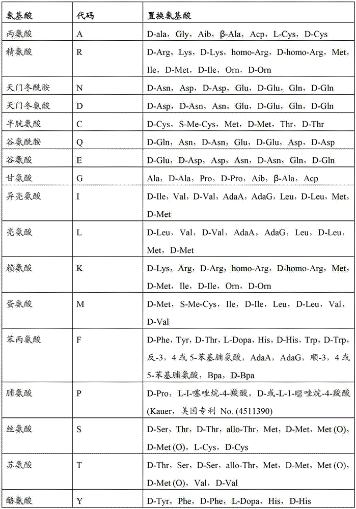
非保守突变是除了上表提到的保守置换的任何其他氨基酸置换。A non-conservative mutation is any other amino acid substitution other than the conservative substitutions mentioned in the table above.
制造保守氨基酸置换为本领域的技术人员所公知的,包括但不限于,采用寡核苷酸定点突变和聚合酶链式反应。通过本领域已知的和/或在此描述的方法,可以表达和测定光学传感器变异体的电压传感活性,pKa,和荧光检测,以验证光学传感器期望的活性通过氨基酸置换还得以保持或增强。令人期待的是在此描述的光学传感器的保守氨基酸置换变异体在传感电压上比母体光学传感器具有增强的活性或更加优的特性。通过不改变编码光学传感器的编码氨基酸序列的突变,也可在编码光学传感器的核酸中进行某些的沉默或中性的错义突变。这些突变类型有助于优化密码子选择,该密码子选择用于改进在希望细胞型中的重组蛋白的表达和生产。在载体中编码光学传感器的核酸的特异性位点定向诱变可用于创造氨基酸突变体和置换体。采用例如来自的
如在此所用,术语”膜电位”指细胞内外之间的电压的计算差。在一种具体实施方式中,膜电位,ΔV,通过等式ΔV=V内r–V外.确定。例如,如果外侧电位是100mV,内侧电位是30mV,那么差值是–70mV。在静息条件下,膜电位主要是由具有极大电导率的离子跨膜决定的。在很多细胞中,膜电位是由钾决定的,其获得约为-70mV的静息膜电位。因此按照惯例,静息状态下的细胞具有负的膜电位。在一些细胞中,当膜电位达到即为等于阈电位,或大于阈电位时,触发动作电位,并且细胞进行去极化(例如,膜电位大幅增加)。在很多情况下,当细胞进行去极化时,膜电位翻转并达到正值(例如,55mV)。在去极化向静息膜电位的后紧跟膜电位解析过程中,细胞可“超极化”。术语“超极化”指膜电位比静息电位更加负的,而术语“去极化”指膜电位比静息电位更少的负(甚至是正的)。膜电位变化可由离子通过嵌入膜的离子通道或离子泵的运动引发。任何细胞的跨膜电位都是可以测定的,包括维持跨膜(例如,质膜,线粒体内膜和外膜等)离子梯度的离子通道或离子泵。As used herein, the term "membrane potential" refers to the calculated difference in voltage between the inside and outside of a cell. In a specific embodiment, the membrane potential, ΔV, is determined by the equation ΔV=Vin r −Vout . For example, if the outer potential is 100mV and the inner potential is 30mV, then the difference is –70mV. Under resting conditions, the membrane potential is primarily determined by the transmembrane movement of ions with extremely high conductivity. In many cells, the membrane potential is determined by potassium, which achieves a resting membrane potential of approximately -70 mV. Thus by convention, cells in the resting state have a negative membrane potential. In some cells, when the membrane potential reaches, equal to, or greater than, a threshold potential, an action potential is triggered and the cell undergoes depolarization (eg, a large increase in membrane potential). In many cases, when a cell undergoes depolarization, the membrane potential flips and reaches a positive value (eg, 55mV). Cells can be "hyperpolarized" during depolarization to resting membrane potential followed by membrane potential resolution. The term "hyperpolarization" refers to a membrane potential that is more negative than the resting potential, while the term "depolarization" refers to a membrane potential that is less negative (or even positive) than the resting potential. Changes in membrane potential can be induced by the movement of ions through ion channels or ion pumps embedded in the membrane. The transmembrane potential of any cell can be measured, including ion channels or ion pumps that maintain ion gradients across membranes (eg, plasma membrane, mitochondrial inner and outer membranes, etc.).
如在此所用,术语“膜电位变化”是指相比于在对照条件(例如,无试剂,损伤的细胞沟通等)的静息膜电位,ΔV增加(或减少)至少1mV,其可以是自发的或对例如环境或化学刺激(例如,细胞与细胞之间的沟通,离子通道调节,与候选试剂的接触等)的响应。在一些具体实施方式中,相比于类似细胞在对照条件下的膜电位,膜电位ΔV增加至少10mV,至少15mV,至少20mV,至少25mV,至少30mV,至少35mV,至少40mV,至少45mV,至少50mV,至少55mV,至少60mV,至少65mV,至少70mV,至少75mV,至少80mV,至少85mV,至少90mV,至少95mV,至少100mV,至少105mV,至少110mV,至少115mV,至少120mV,至少125mV,至少130mV,至少135mV,至少140mV,至少145mV,至少150mV,至少155mV,至少160mV,至少165mV,至少170mV,至少180mV,至少190mV,至少200mV或更多。在另外的具体实施方式中,膜电位相比于参考条件下的类似细胞的膜电位,降低至少3mV,至少5mV,至少10mV,至少15mV,至少20mV,至少25mV,至少30mV,至少35mV,至少40mV,至少45mV,至少50mV,至少55mV,至少60mV,至少65mV,至少70mV,至少75mV,至少80mV,至少85mV,至少90mV,至少95mV,至少100mV,至少105mV,至少110mV,至少115mV,至少120mV,至少125mV,至少130mV,至少135mV,至少140mV,至少145mV,至少150mV或更多。As used herein, the term "membrane potential change" refers to a ΔV increase (or decrease) of at least 1 mV, which may be spontaneous, compared to the resting membrane potential under control conditions (e.g., no reagent, damaged cell communication, etc.). or in response to, for example, environmental or chemical stimuli (eg, cell-to-cell communication, ion channel modulation, exposure to candidate agents, etc.). In some embodiments, the membrane potential ΔV is increased by at least 10 mV, at least 15 mV, at least 20 mV, at least 25 mV, at least 30 mV, at least 35 mV, at least 40 mV, at least 45 mV, at least 50 mV compared to the membrane potential of a similar cell under control conditions , at least 55mV, at least 60mV, at least 65mV, at least 70mV, at least 75mV, at least 80mV, at least 85mV, at least 90mV, at least 95mV, at least 100mV, at least 105mV, at least 110mV, at least 115mV, at least 120mV, at least 125mV, at least 130mV, at least 135mV, at least 140mV, at least 145mV, at least 150mV, at least 155mV, at least 160mV, at least 165mV, at least 170mV, at least 180mV, at least 190mV, at least 200mV or more. In additional specific embodiments, the membrane potential is reduced by at least 3 mV, at least 5 mV, at least 10 mV, at least 15 mV, at least 20 mV, at least 25 mV, at least 30 mV, at least 35 mV, at least 40 mV compared to the membrane potential of a similar cell under reference conditions , at least 45mV, at least 50mV, at least 55mV, at least 60mV, at least 65mV, at least 70mV, at least 75mV, at least 80mV, at least 85mV, at least 90mV, at least 95mV, at least 100mV, at least 105mV, at least 110mV, at least 115mV, at least 120mV, at least 125mV, at least 130mV, at least 135mV, at least 140mV, at least 145mV, at least 150mV or more.
如在此所用,表达"集定位于细胞的膜"指修饰细菌视紫红质蛋白优先定位于细胞膜,且可通过例如修饰细菌视紫红质使其包括使视紫红质蛋白对准细胞的膜(例如,质膜,线粒体外膜,线粒体内膜等)的信号序列来实现。。在一些具体实施方式中,至少细胞中40%的修饰细菌视紫红质蛋白定位到预期的细胞膜区室(例如,质膜,线粒体膜等)。在其他的具体实施方式中,至少50%,至少60%,至少70%,至少75%,至少80%,至少85%,至少90%,至少95%,至少99%的修饰的细菌视紫红质蛋白定位到预期细胞的膜区室。类似的,术语"定位于亚细胞区室"指细菌视紫红质蛋白优先定位于特定的亚细胞区室(例如,线粒体,内质网,过氧化物酶体等)。在一些具体实施方式中,细胞中至少40%的修饰的细菌视紫红质蛋白定位于预期的亚细胞区室;在另外的具体实施方式中,至少50%,至少60%,至少70%,至少75%,至少80%,至少85%,至少90%,至少95%,或至少99%的修饰细菌视紫红质蛋白定位预期的亚细胞区室。As used herein, the expression "localizes to the membrane of the cell" refers to the preferential localization of the modified bacteriorhodopsin protein to the cell membrane, and can be achieved by, for example, modifying the bacteriorhodopsin to include the alignment of the bacteriorhodopsin protein to the membrane of the cell (e.g. , plasma membrane, mitochondrial outer membrane, mitochondrial inner membrane, etc.) to achieve the signal sequence. . In some embodiments, at least 40% of the modified bacteriorhodopsin protein in the cell localizes to a desired cell membrane compartment (eg, plasma membrane, mitochondrial membrane, etc.). In other specific embodiments, at least 50%, at least 60%, at least 70%, at least 75%, at least 80%, at least 85%, at least 90%, at least 95%, at least 99% modified bacteriorhodopsin The protein localizes to the membrane compartment of the expected cell. Similarly, the term "localized to subcellular compartments" means that bacteriorhodopsin protein is preferentially localized to specific subcellular compartments (eg, mitochondria, endoplasmic reticulum, peroxisomes, etc.). In some embodiments, at least 40% of the modified bacteriorhodopsin in the cell is localized to the intended subcellular compartment; in other embodiments, at least 50%, at least 60%, at least 70%, at least 75%, at least 80%, at least 85%, at least 90%, at least 95%, or at least 99% of the modified bacteriorhodopsin protein localizes to the expected subcellular compartment.
在一些具体实施方式中,约100%定位于预期细胞膜或区室。In some embodiments, about 100% localizes to the intended cell membrane or compartment.
如在此所用,术语“引入到细胞内”指将编码光学传感器的表达载体或在此描述的重组光学传感器引入到宿主细胞的任何方法。将表达载体引入细胞的一些非限制例子包括,例如,磷酸钙转染,电穿孔,脂质转染法,或采用基因枪的方法或类似的方法。在一种具体实施方式中,重组光学传感器蛋白通过膜融合被引入到细胞,膜融合采用类脂介导的递送系统,例如微粒,脂质体等。As used herein, the term "introducing into a cell" refers to any method of introducing an expression vector encoding an optical sensor or a recombinant optical sensor described herein into a host cell. Some non-limiting examples of introducing expression vectors into cells include, for example, calcium phosphate transfection, electroporation, lipofection, or methods using a gene gun or the like. In a specific embodiment, the recombinant optical sensor protein is introduced into cells through membrane fusion using a lipid-mediated delivery system, such as microparticles, liposomes, and the like.
如在此所用,术语“产生光学信号的部分”是指可产生可测量的信号,例如荧光,化学发光,比色信号等,的分子(例如,视黄醛)或分子的部分。在一种具体实施方式中,修饰细菌视紫红质包括具有可产生光学信号的部分的融合分子。As used herein, the term "optical signal-producing moiety" refers to a molecule (eg, retinal) or a portion of a molecule that produces a measurable signal, such as a fluorescent, chemiluminescent, colorimetric signal, etc. In a specific embodiment, the modified bacteriorhodopsin comprises a fusion molecule having a moiety that produces an optical signal.
如在此所用,术语“荧光水平的变化”或“光学信号水平的变化”指修饰细菌视紫红质蛋白荧光水平的增加或减小或光学信号的增加或减小,其是电压或膜电位变化诱导的。在一些具体实施方式中,相比于对照状态下的相同细胞或类似细胞,细胞中荧光水平或光学信号水平增加至少2%,至少5%,至少10%,至少20%,至少30%,至少40%,至少50%,至少60%,至少70%,至少80%,至少90%,至少95%,至少99%,至少1倍,至少2倍,至少5倍,至少10倍,至少50倍,至少100倍,至少500倍,至少600倍,至少700倍,至少800倍,至少900倍,至少1000倍,至少2000倍,至少5000倍,至少10000倍或更多。可选地,相比于对照状态下的相同细胞或类似细胞,细胞中荧光水平或光学信号水平下降至少20%,至少30%,至少40%,至少50%,至少60%,至少70%,至少80%,至少90%,至少95%,至少99%,或甚至100%(例如,没有检测到信号)。As used herein, the term "change in fluorescence level" or "change in optical signal level" refers to an increase or decrease in the fluorescence level of modified bacteriorhodopsin or an increase or decrease in optical signal, which is a change in voltage or membrane potential induced. In some embodiments, the level of fluorescence or optical signal in the cell is increased by at least 2%, at least 5%, at least 10%, at least 20%, at least 30%, at least 40%, at least 50%, at least 60%, at least 70%, at least 80%, at least 90%, at least 95%, at least 99%, at least 1 times, at least 2 times, at least 5 times, at least 10 times, at least 50 times , at least 100 times, at least 500 times, at least 600 times, at least 700 times, at least 800 times, at least 900 times, at least 1000 times, at least 2000 times, at least 5000 times, at least 10000 times or more. Optionally, the level of fluorescence or optical signal in the cell is reduced by at least 20%, at least 30%, at least 40%, at least 50%, at least 60%, at least 70%, compared to the same cell or similar cells in a control state, At least 80%, at least 90%, at least 95%, at least 99%, or even 100% (eg, no signal detected).
如在此所用,术语“调节离子通道活性”指增加或减小离子通道的一个或多个体现细胞膜电位变化的特征。这些特征包括例如导电性的开放或闭合状态,电压阈值,动力学和/或配体亲和力。在一些具体实施方式中,细胞离子通道的一个或多个有用的特征通过例如,细胞膜电位变化测定。在一些具体实施方式中,与没有试剂存在时的离子通道活性相比,存在试剂的离子通道活性增加至少20%,至少30%,至少40%,至少50%,至少60%,至少70%,至少80%,至少90%,至少95%,至少99%,至少1倍,至少2倍,至少5倍,至少10倍,至少20倍,至少50倍,至少100倍,至少1000倍或更多。在其他的具体实施方式中,与没有试剂存在时的离子通道活性相比,存在试剂的离子通道的有用的参数下降至少20%,至少30%,至少40%,至少50%,至少60%,至少70%,至少80%,至少90%,至少95%,或至少99%。在一些具体实施方式中,与没有试剂存在的离子通道活性相比,在有试剂存在的离子通道参数是缺乏的。As used herein, the term "modulates ion channel activity" refers to increasing or decreasing one or more characteristics of an ion channel that reflect changes in the cell's membrane potential. These characteristics include, for example, the open or closed state of conductivity, voltage threshold, kinetics and/or ligand affinity. In some embodiments, one or more useful characteristics of a cellular ion channel are determined, for example, by a change in cellular membrane potential. In some embodiments, the activity of the ion channel is increased by at least 20%, at least 30%, at least 40%, at least 50%, at least 60%, at least 70% in the presence of the agent compared to the activity of the ion channel in the absence of the agent, At least 80%, at least 90%, at least 95%, at least 99%, at least 1 times, at least 2 times, at least 5 times, at least 10 times, at least 20 times, at least 50 times, at least 100 times, at least 1000 times or more . In other specific embodiments, the useful parameter of the ion channel is reduced by at least 20%, at least 30%, at least 40%, at least 50%, at least 60% in the presence of the agent compared to the activity of the ion channel in the absence of the agent, At least 70%, at least 80%, at least 90%, at least 95%, or at least 99%. In some embodiments, the parameter of the ion channel in the presence of the agent is absent compared to the activity of the ion channel in the absence of the agent.
如在此所用,术语“抗生素外流抑制剂”是指细胞中的抗生素外流水平降低至少20%的试剂,与没有试剂存在的抗生素外流水平相比。在一些具体实施方式中,与没有试剂存在的抗生素外流水平相比,存在试剂时抗生素外流降低至少30%,至少40%,至少50%,至少60%,至少70%,至少80%,至少90%,至少95%,至少99%,或甚至100%(例如,不存在)。As used herein, the term "antibiotic efflux inhibitor" refers to an agent that reduces the level of antibiotic efflux in a cell by at least 20%, compared to the level of antibiotic efflux in the absence of the agent. In some embodiments, antibiotic efflux is reduced by at least 30%, at least 40%, at least 50%, at least 60%, at least 70%, at least 80%, at least 90% in the presence of the agent compared to the level of antibiotic efflux in the absence of the agent %, at least 95%, at least 99%, or even 100% (eg, absent).
如在此所用,术语“靶向序列”指对准于或优先缔合或结合到特定组织,细胞型,受体,细胞器,或其他有用区域的部分或序列。将靶向序列附加到光学传感器组合物上将增强该组合物到预期的细胞型或亚细胞位置的递送。细胞中具有光学传感器的靶向序列的附加,或表达,增强了光学传感器在动物或受试者预期位置的定位。As used herein, the term "targeting sequence" refers to a portion or sequence that aligns with or preferentially associates with or binds to a specific tissue, cell type, receptor, organelle, or other useful region. Attachment of targeting sequences to optical sensor compositions will enhance delivery of the compositions to desired cell types or subcellular locations. Attachment, or expression, of the targeting sequence with the optical sensor in the cell enhances localization of the optical sensor at the desired location in the animal or subject.
如在此所用,短语“相当于菌视紫质中的氨基酸突变的在另外的细菌视紫红质中的同源突变”指在预期细菌视紫红质中残基的突变,其希望具有与菌视紫质大体相似突变的类似效果。本领域技术人员通过对期望的的细菌视紫红质的保守区域与菌视紫质序列进行对比,可以同采用电脑程序例如ClustalW进行对比,可容易地将同源残基放置在其期望的细菌视紫红质中。As used herein, the phrase "a homologous mutation in another bacteriorhodopsin equivalent to an amino acid mutation in a bacteriorhodopsin" refers to a mutation of a residue in a predicted bacteriorhodopsin that is expected to have the same Similar effects of rhodopsin broadly similar mutations. Those skilled in the art can easily place homologous residues in their desired bacteriorhodopsin by comparing the expected conserved region of bacteriorhodopsin with the bacteriorhodopsin sequence, using a computer program such as ClustalW. In purple red.
同源突变的例子包括本申请中陈述的实施例中制造的突变。Examples of homologous mutations include mutations made in Examples set forth in this application.
在此使用的术语“包括”或“包含”的使用涉及组合物,方法及其各自成分,它们是发明所必需的,其对不特定的组分是开放的,无论这些组分是否是必需的。As used herein, the use of the terms "comprising" or "comprising" refers to compositions, methods and their respective components, which are essential to the invention, which are open to unspecified components, whether or not those components are essential .
在此所用的术语“基本上含有”指给定具体实施方式所需要的成分。该术语能够使存在成分而大体上不影响发明具体实施方式的基本的和新颖的或功能的特征。As used herein, the term "consisting essentially of" refers to as many ingredients as are required for a given embodiment. The term enables the presence of elements that do not substantially affect the basic and novel or functional characteristics of particular embodiments of the invention.
术语"由……组成"指在此所述的组合物,方法及其各自成分,不计在具体实施方式的描述中未列举的任何成分。The term "consisting of" refers to the compositions, methods and their respective components described herein, excluding any components not listed in the description of a specific embodiment.
在此提供光学传感器,包括细菌视紫红质或修饰的细菌视紫红质蛋白,相比于源自于的天然细菌视紫红质蛋白,离子泵活性降低。在一种具体实施方式中,组合物包括编码载体或工程化细胞,工程化细胞包括细菌视紫红质蛋白或修饰细菌视紫红质蛋白,相比于源自于天然的细菌视紫红质蛋白,离子泵活性降低。在一些具体实施方式中,细菌视紫红质定向于膜,例如质膜或线粒体膜。Provided herein are optical sensors comprising bacteriorhodopsins or modified bacteriorhodopsins having reduced ion pump activity compared to native bacteriorhodopsins derived from them. In a specific embodiment, the composition comprises an encoding vector or an engineered cell, the engineered cell comprises a bacteriorhodopsin or a modified bacteriorhodopsin, compared to the bacteriorhodopsin derived from nature, the ion Reduced pump activity. In some embodiments, bacteriorhodopsin is localized to a membrane, such as the plasma membrane or the mitochondrial membrane.
因此,发明提供测定细胞内的膜电位的方法,该细胞表达编码细菌视紫红质蛋白的核酸,该方法包括如下步骤:(a)采用至少一个波长的光,体外,离体或体内激发至少一个细胞,该细胞包括编码细菌视紫红质蛋白的核酸;和(b)体外检测来自至少一个细胞的至少一个光学信号,其中至少一个细胞发射的荧光水平,与参考相比,可指示细胞膜电位。Accordingly, the invention provides a method for determining membrane potential in a cell expressing a nucleic acid encoding a bacteriorhodopsin protein, the method comprising the steps of: (a) exciting at least one a cell comprising a nucleic acid encoding a bacteriorhodopsin protein; and (b) detecting in vitro at least one optical signal from at least one cell, wherein the at least one cell emits a level of fluorescence indicative of cell membrane potential compared to a reference.
在发明的一些或任何具体实施方式或方面中,细菌视紫红质蛋白是修饰细菌视紫红质蛋白,与源自于天然的细菌视紫红质蛋白相比,其离子泵活性降低。In some or any specific embodiments or aspects of the invention, the bacteriorhodopsin is a modified bacteriorhodopsin having reduced ion pump activity compared to bacteriorhodopsin derived from nature.
在发明的一些或任何具体实施方式或方面中,修饰的细菌视紫红质蛋白包括邻近希夫碱基的突变质子受体。In some or any specific embodiments or aspects of the invention, the modified bacteriorhodopsin comprises a mutated proton acceptor adjacent to a Schiff base.
在发明的一些或任何具体实施方式或方面中,细菌视紫红质蛋白是变形菌视紫红质蛋白家族的成员。In some or any specific embodiments or aspects of the invention, the bacteriorhodopsin protein is a member of the Proteus rhodopsin protein family.
在发明的一些或任何具体实施方式或方面中,细菌视紫红质蛋白是古细菌视紫红质蛋白家族中的成员。In some or any specific embodiments or aspects of the invention, the bacteriorhodopsin protein is a member of the family of archaealrhodopsin proteins.
在发明的一些或任何具体实施方式或方面中,至少一个波长在λ=594-645nm波长之间。In some or any specific embodiments or aspects of the invention, at least one wavelength is between λ = 594-645 nm wavelength.
在发明的一些或任何具体实施方式或方面中,细胞是原核细胞。In some or any specific embodiments or aspects of the invention, the cells are prokaryotic cells.
在发明的一些或任何具体实施方式或方面中,细胞是真核细胞。In some or any specific embodiments or aspects of the invention, the cells are eukaryotic cells.
在发明的一些或任何具体实施方式或方面中,真核细胞是哺乳动物细胞。In some or any specific embodiments or aspects of the invention, the eukaryotic cells are mammalian cells.
在发明的一些或任何具体实施方式或方面中,真核细胞是干细胞或多能细胞或祖细胞。In some or any specific embodiments or aspects of the invention, the eukaryotic cells are stem cells or pluripotent cells or progenitor cells.
在发明的一些或任何具体实施方式或方面中,真核细胞是诱导多能细胞。In some or any specific embodiments or aspects of the invention, the eukaryotic cells are induced pluripotent cells.
在发明的一些或任何具体实施方式或方面中,真核细胞是神经元。In some or any specific embodiments or aspects of the invention, the eukaryotic cells are neurons.
在发明的一些或任何具体实施方式或方面中,真核细胞是心肌细胞。In some or any specific embodiments or aspects of the invention, the eukaryotic cells are cardiomyocytes.
在发明的一些或任何具体实施方式或方面中,该方法还包括用含有编码细菌视紫红质蛋白核酸的载体,体外,离体或体内转染至少一个细胞的步骤。In some or any specific embodiments or aspects of the invention, the method further comprises the step of transfecting at least one cell in vitro, ex vivo or in vivo with a vector comprising a bacteriorhodopsin-encoding nucleic acid.
在发明的一些或任何具体实施方式或方面中,编码细菌视紫红质蛋白的核酸可操作地连接于细胞型特异的启动子。In some or any specific embodiments or aspects of the invention, the nucleic acid encoding a bacteriorhodopsin protein is operably linked to a cell type specific promoter.
在发明的一些或任何具体实施方式或方面中,编码细菌视紫红质蛋白的核酸可操作地连接于膜靶向核酸序列。In some or any specific embodiments or aspects of the invention, the nucleic acid encoding a bacteriorhodopsin protein is operably linked to a membrane targeting nucleic acid sequence.
在发明的一些或任何具体实施方式或方面中,膜靶向核酸是质膜靶向的核酸序列。In some or any specific embodiments or aspects of the invention, the membrane-targeting nucleic acid is a plasma membrane-targeting nucleic acid sequence.
在发明的一些或任何具体实施方式或方面中,膜靶向核酸序列是亚细胞区室靶向的核酸序列。In some or any specific embodiments or aspects of the invention, the membrane targeting nucleic acid sequence is a subcellular compartment targeting nucleic acid sequence.
在发明的一些或任何具体实施方式或方面中,亚细胞区室选自线粒体膜,内质网,肌质网,突触小泡,内涵体或吞噬小体。In some or any specific embodiments or aspects of the invention, the subcellular compartment is selected from mitochondrial membranes, endoplasmic reticulum, sarcoplasmic reticulum, synaptic vesicles, endosomes or phagosomes.
在发明的一些或任何具体实施方式或方面中,编码细菌视紫红质蛋白的核酸可操作地连接于编码至少一个附加荧光蛋白或生色团的核酸。In some or any specific embodiments or aspects of the invention, a nucleic acid encoding a bacteriorhodopsin protein is operably linked to a nucleic acid encoding at least one additional fluorescent protein or chromophore.
在发明的一些或任何具体实施方式或方面中,至少一个附加荧光蛋白能够指示细胞内的离子浓度的荧光蛋白。.In some or any embodiments or aspects of the invention, at least one additional fluorescent protein is a fluorescent protein capable of indicating ion concentration within the cell. .
在发明的一些或任何具体实施方式或方面中,能够指示细胞内的离子浓度的荧光蛋白是钙指示器。In some or any specific embodiments or aspects of the invention, the fluorescent protein capable of indicating ion concentration within a cell is a calcium indicator.
在发明的一些或任何具体实施方式或方面中,能够指示细胞内的离子浓度的荧光蛋白是pH指示器。In some or any specific embodiments or aspects of the invention, a fluorescent protein capable of indicating ion concentration within a cell is a pH indicator.
在发明的一些或任何具体实施方式或方面中,至少一个附加荧光蛋白能够进行非放射性荧光共振,使能量转移到细菌视紫红质,其能量转移速率取决于膜电位。In some or any specific embodiments or aspects of the invention, at least one additional fluorescent protein is capable of non-radioactive fluorescence resonance, enabling energy transfer to bacteriorhodopsin at a rate dependent on membrane potential.
在发明的一些或任何具体实施方式或方面中,至少一个附加荧光蛋白是绿色荧光蛋白或其同系物。In some or any specific embodiments or aspects of the invention, the at least one additional fluorescent protein is green fluorescent protein or a homologue thereof.
在发明的一些或任何具体实施方式或方面中,荧光蛋白的亮度对膜电位或局部化学环境不敏感。In some or any specific embodiments or aspects of the invention, the brightness of the fluorescent protein is insensitive to membrane potential or local chemical environment.
在发明的一些或任何具体实施方式或方面中,该方法进一步包括如下步骤:采用至少第一和第二波长的光体外,离体或体内激发至少一个细胞;和体外,离体或体内检测至少第一和第二光学信号,该信号是由来自至少一个细胞的至少第一和第二波长的激发引起。In some or any specific embodiments or aspects of the invention, the method further comprises the steps of: exciting at least one cell in vitro, ex vivo or in vivo with light of at least first and second wavelengths; and detecting at least one cell in vitro, ex vivo or in vivo First and second optical signals resulting from excitation at least first and second wavelengths from at least one cell.
在发明的一些或任何具体实施方式或方面中,至少第二波长在λ=447-594nm之间。In some or any specific embodiments or aspects of the invention, at least the second wavelength is between λ = 447-594 nm.
在发明的一些或任何具体实施方式或方面中,该方法还包括下述步骤:计算细菌视紫红质的荧光发射与至少一个附加荧光蛋白的荧光发射的比率,以获取不依赖于表达水平差异的膜电位测定。In some or any specific embodiments or aspects of the invention, the method further comprises the step of calculating the ratio of the fluorescent emission of bacteriorhodopsin to the fluorescent emission of at least one additional fluorescent protein to obtain a Membrane Potential Determination.
在发明的一些或任何具体实施方式或方面中,该方法还包括下述步骤:将至少一个细胞体外,离体或体内暴露于能够或预期能够改变膜电位的刺激。In some or any specific embodiments or aspects of the invention, the method further comprises the step of exposing at least one cell to a stimulus capable or expected to alter membrane potential in vitro, ex vivo or in vivo.
在发明的一些或任何具体实施方式或方面中,刺激是候选试剂。In some or any specific embodiments or aspects of the invention, a stimulus is a candidate agent.
在发明的一些或任何具体实施方式或方面中,刺激是细胞培养基组分的变化。In some or any specific embodiments or aspects of the invention, the stimulus is a change in the composition of the cell culture medium.
在发明的一些或任何具体实施方式或方面中,刺激是电流。In some or any specific embodiments or aspects of the invention, the stimulus is an electrical current.
在发明的一些或任何具体实施方式或方面中,还包括在第一和至少第二时间点体外,离体或体内测定至少一个光学信号的步骤。In some or any specific embodiments or aspects of the invention, further comprising the step of measuring at least one optical signal at the first and at least the second time point in vitro, ex vivo or in vivo.
在发明的一些或任何具体实施方式或方面中,第一时间点是在将至少一个细胞暴露于刺激之前,至少第二时间点是将至少一个细胞暴露于刺激之后。In some or any specific embodiments or aspects of the invention, the first time point is before exposing the at least one cell to the stimulus and at least the second time point is after exposing the at least one cell to the stimulus.
在发明的一些或任何具体实施方式或方面中,其中该方法包括多个细胞,例如在高通量测定中。In some or any embodiments or aspects of the invention wherein the method comprises a plurality of cells, for example in a high throughput assay.
在另外的具体实施方式中,发明提供分离和纯化的编码古细菌视紫红质蛋白家族修饰成员的核酸,与源自于天然的古细菌视紫红质蛋白家族成员相比,离子泵活性降低。In another embodiment, the invention provides an isolated and purified nucleic acid encoding a modified member of the archaealrhodopsin family having reduced ion pump activity compared to a member of the archaealrhodopsin family derived from nature.
在发明的一些或任何具体实施方式或方面中,古细菌视紫红质蛋白家族的修饰成员包括邻近希夫碱基的突变质子受体。In some or any specific embodiments or aspects of the invention, the modified member of the archaealrhodopsin protein family comprises a mutated proton acceptor adjacent to a Schiff base.
在发明的一些或任何具体实施方式或方面中,与源自于天然的古细菌视紫红质蛋白家族的成员相比,古细菌视紫红质蛋白家族的修饰成员的离子泵活性降低,该古细菌视紫红质蛋白家族的修饰成员可操作地连接于膜靶向的核酸序列。In some or any specific embodiments or aspects of the invention, the modified member of the archaeal rhodopsin protein family has reduced ion pump activity compared to a member of the archaeal rhodopsin protein family derived from natural The modified member of the rhodopsin protein family is operably linked to a membrane-targeting nucleic acid sequence.
在发明的一些或任何具体实施方式或方面中,膜靶向的核酸序列是质膜靶向的核酸序列。In some or any specific embodiments or aspects of the invention, the membrane-targeting nucleic acid sequence is a plasma membrane-targeting nucleic acid sequence.
在发明的一些或任何具体实施方式或方面中,膜靶向的核酸序列是亚细胞膜靶向的核酸序列。In some or any specific embodiments or aspects of the invention, the membrane-targeting nucleic acid sequence is a subcellular membrane-targeting nucleic acid sequence.
在发明的一些或任何具体实施方式或方面中,亚细胞膜是线粒体膜,内质网,肌质网,突触小泡,内涵体或吞噬小体。In some or any specific embodiments or aspects of the invention, the subcellular membrane is a mitochondrial membrane, endoplasmic reticulum, sarcoplasmic reticulum, synaptic vesicle, endosome or phagosome.
在发明的一些或任何具体实施方式或方面中,古细菌视紫红质家族的修饰的细菌视紫红质可操作地连接于细胞型特异的启动子。In some or any specific embodiments or aspects of the invention, a modified bacteriorhodopsin of the archaealrhodopsin family is operably linked to a cell type specific promoter.
在发明的一些或任何具体实施方式或方面中,古细菌视紫红质家族的修饰的细菌视紫红质可操作地连接于编码至少一个附加荧光蛋白或生色团的核酸。In some or any specific embodiments or aspects of the invention, a modified bacteriorhodopsin of the archaealrhodopsin family is operably linked to a nucleic acid encoding at least one additional fluorescent protein or chromophore.
在发明的一些或任何具体实施方式或方面中,其中至少一个附加荧光蛋白是绿色荧光蛋白或其同系物。In some or any embodiments or aspects of the invention, at least one of the additional fluorescent proteins is green fluorescent protein or a homologue thereof.
在发明的一些或任何具体实施方式或方面中,至少一个附加荧光蛋白能够指示细胞内的离子浓度。In some or any embodiments or aspects of the invention, at least one additional fluorescent protein is capable of indicating ion concentration within the cell.
在发明的一些或任何具体实施方式或方面中,能够指示细胞内离子浓度的荧光蛋白是钙指示器。In some or any specific embodiments or aspects of the invention, the fluorescent protein capable of indicating intracellular ion concentration is a calcium indicator.
在发明的一些或任何具体实施方式或方面中,能够指示细胞内离子浓度的荧光蛋白是pH指示器。In some or any specific embodiments or aspects of the invention, a fluorescent protein capable of indicating intracellular ion concentration is a pH indicator.
在发明的一些或任何具体实施方式或方面中,至少一个附加荧光蛋白能够进行非放射性荧光共振,使能量转移到细菌视紫红质,其能量转移速率取决于膜电位。In some or any specific embodiments or aspects of the invention, at least one additional fluorescent protein is capable of non-radioactive fluorescence resonance, enabling energy transfer to bacteriorhodopsin at a rate dependent on membrane potential.
在发明的一些或任何具体实施方式或方面中,分离和纯化的核酸还包括载体。In some or any specific embodiments or aspects of the invention, the isolated and purified nucleic acid also includes a vector.
在发明的一些或任何具体实施方式或方面中,载体是病毒载体,例如慢病毒载体或腺伴随病毒载体。In some or any specific embodiments or aspects of the invention, the vector is a viral vector, such as a lentiviral vector or an adeno-associated viral vector.
在另外的具体实施方式中,发明提供试剂盒,包括合适容器中分离和纯化的核酸,该核酸编码在此公开的古细菌视紫红质家族的修饰的细菌蛋白。核酸可以以干燥形式或适宜缓冲液的形式提供。In another embodiment, the invention provides a kit comprising isolated and purified nucleic acid encoding a modified bacterial protein of the archaealrhodopsin family disclosed herein in a suitable container. Nucleic acids may be provided in dry form or in a suitable buffer.
发明另外的具体实施方式提供分离的细胞,其包括编码细菌视紫红质蛋白的核酸,其中细菌视紫红质蛋白是修饰的细菌视紫红质蛋白,与源自于天然的细菌视紫红质蛋白相比,离子泵活性降低。An additional embodiment of the invention provides an isolated cell comprising a nucleic acid encoding a bacteriorhodopsin, wherein the bacteriorhodopsin is a modified bacteriorhodopsin compared to a bacteriorhodopsin derived from natural , the ion pump activity is reduced.
在发明的一些或任何具体实施方式或方面中,修饰的细菌视紫红质蛋白包括邻近希夫碱基的突变质子受体。In some or any specific embodiments or aspects of the invention, the modified bacteriorhodopsin comprises a mutated proton acceptor adjacent to a Schiff base.
在发明的一些或任何具体实施方式或方面中,细菌视紫红质是变形菌视紫红质家族的成员。In some or any specific embodiments or aspects of the invention, the bacteriorhodopsin is a member of the Proteorhodopsin family.
在发明的一些或任何具体实施方式或方面中,细菌视紫红质是古细菌视紫红质家族的成员。In some or any specific embodiments or aspects of the invention, the bacteriorhodopsin is a member of the archaeal rhodopsin family.
在发明的一些或任何具体实施方式或方面中,细胞是真核细胞。In some or any specific embodiments or aspects of the invention, the cells are eukaryotic cells.
在发明的一些或任何具体实施方式或方面中,细胞是原核细胞。In some or any specific embodiments or aspects of the invention, the cells are prokaryotic cells.
在发明的一些或任何具体实施方式或方面中,修饰的细菌视紫红质基因可操作地连接于启动子。In some or any specific embodiments or aspects of the invention, the modified bacteriorhodopsin gene is operably linked to a promoter.
在发明的一些或任何具体实施方式或方面中,启动子是细胞型特异的启动子。In some or any specific embodiments or aspects of the invention, the promoter is a cell type specific promoter.
在发明的一些或任何具体实施方式或方面中,编码细菌视紫红质蛋白的核酸可操作地连接于膜靶向的核酸序列。In some or any specific embodiments or aspects of the invention, the nucleic acid encoding a bacteriorhodopsin protein is operably linked to a membrane-targeting nucleic acid sequence.
在发明的一些或任何具体实施方式或方面中,膜靶向核酸是质膜靶向的核酸序列。In some or any specific embodiments or aspects of the invention, the membrane-targeting nucleic acid is a plasma membrane-targeting nucleic acid sequence.
在发明的一些或任何具体实施方式或方面中,膜靶向核酸是亚细胞区室靶向核酸序列。In some or any specific embodiments or aspects of the invention, the membrane targeting nucleic acid is a subcellular compartment targeting nucleic acid sequence.
在发明的一些或任何具体实施方式或方面中,亚细胞区室是选自于线粒体膜,内质网,肌质网,突触小泡,内涵体或吞噬小体。In some or any embodiments or aspects of the invention, the subcellular compartment is selected from mitochondrial membranes, endoplasmic reticulum, sarcoplasmic reticulum, synaptic vesicles, endosomes or phagosomes.
在发明的一些或任何具体实施方式或方面中,编码修饰的细菌视紫红质蛋白的核酸可操作地连接于编码至少一个附加荧光蛋白或生色团的核酸。In some or any specific embodiments or aspects of the invention, a nucleic acid encoding a modified bacteriorhodopsin protein is operably linked to a nucleic acid encoding at least one additional fluorescent protein or chromophore.
在发明的一些或任何具体实施方式或方面中,至少一个附加荧光蛋白是绿色荧光蛋白或其同系物。In some or any specific embodiments or aspects of the invention, the at least one additional fluorescent protein is green fluorescent protein or a homologue thereof.
在发明的一些或任何具体实施方式或方面中,至少一个附加荧光蛋白能够进行非放射性荧光共振,使能量转移到细菌视紫红质,其能量转移速率取决于膜电位。In some or any specific embodiments or aspects of the invention, at least one additional fluorescent protein is capable of non-radioactive fluorescence resonance, enabling energy transfer to bacteriorhodopsin at a rate dependent on membrane potential.
在发明的一些或任何具体实施方式或方面中,至少一个附加荧光蛋白对膜电位或局部化学环境不敏感In some or any specific embodiments or aspects of the invention, at least one additional fluorescent protein is insensitive to membrane potential or local chemical environment
在发明的一些或任何具体实施方式或方面中,至少一个附加荧光蛋白能够指示细胞内的离子浓度。In some or any embodiments or aspects of the invention, at least one additional fluorescent protein is capable of indicating ion concentration within the cell.
在发明的一些或任何具体实施方式或方面中,能够指示细胞内离子浓度的荧光蛋白是钙指示器。In some or any specific embodiments or aspects of the invention, the fluorescent protein capable of indicating intracellular ion concentration is a calcium indicator.
在发明的一些或任何具体实施方式或方面中,能够指示细胞内离子浓度荧光蛋白是pH指示器。In some or any embodiments or aspects of the invention, the fluorescent protein capable of indicating intracellular ion concentration is a pH indicator.
在发明的一些或任何具体实施方式或方面中,细胞是干细胞,多能细胞,或诱导多能细胞,或其分化或未分化子代细胞。In some or any specific embodiments or aspects of the invention, the cells are stem cells, pluripotent cells, or induced pluripotent cells, or differentiated or undifferentiated progeny cells thereof.
在发明的一些或任何具体实施方式或方面中,细胞是分化细胞。In some or any specific embodiments or aspects of the invention, the cells are differentiated cells.
在发明的一些或任何具体实施方式或方面中,分化细胞是神经元。In some or any specific embodiments or aspects of the invention, the differentiated cells are neurons.
在发明的一些或任何具体实施方式或方面中,分化细胞是心肌细胞。In some or any specific embodiments or aspects of the invention, the differentiated cells are cardiomyocytes.
在一种具体实施方式中,发明还包括分离细胞,含有任何分离和纯化的核酸或核酸构建体的分离和纯化的核酸,该分离和纯化的核酸或核酸构建体含有上面描述的古细菌视紫红质家族的修饰的细菌视紫红质。In a specific embodiment, the invention also includes isolated cells containing isolated and purified nucleic acid of any isolated and purified nucleic acid or nucleic acid construct comprising the above-described archaeal rhododendron Modified bacteriorhodopsins of the protein family.
在一种具体实施方式中,发明提供试剂盒,如在此描述的,包括适宜细胞培养基上的多个细胞或容器。In one embodiment, the invention provides a kit, as described herein, comprising a plurality of cells or containers in a suitable cell culture medium.
在另外的具体实施方式中,发明提供用于光学测定膜电位的工程细胞的制造方法,包括用编码细菌视紫红质蛋白的核酸转导细胞步骤。In another embodiment, the invention provides a method of manufacturing an engineered cell for the optical measurement of membrane potential, comprising the step of transducing the cell with a nucleic acid encoding a bacteriorhodopsin protein.
在发明的一些或任何具体实施方式或方面中细菌视紫红质蛋白是修饰的细菌视紫红质蛋白,相比于天然细菌视紫红质蛋白,其离子泵活性降低。In some or any specific embodiments or aspects of the invention, the bacteriorhodopsin is a modified bacteriorhodopsin having reduced ion pump activity compared to native bacteriorhodopsin.
在发明的一些或任何具体实施方式或方面中,修饰的细菌视紫红质包括邻近希夫碱基的突变质子受体。In some or any specific embodiments or aspects of the invention, the modified bacteriorhodopsin comprises a mutated proton acceptor adjacent to a Schiff base.
在发明的一些或任何具体实施方式或方面中,编码细菌视紫红质蛋白的核酸可操作地连接于膜靶向核酸序列。In some or any specific embodiments or aspects of the invention, the nucleic acid encoding a bacteriorhodopsin protein is operably linked to a membrane targeting nucleic acid sequence.
在发明的一些或任何具体实施方式或方面中,膜靶向核酸序列是质膜靶向核酸序列。In some or any specific embodiments or aspects of the invention, the membrane targeting nucleic acid sequence is a plasma membrane targeting nucleic acid sequence.
在发明的一些或任何具体实施方式或方面中,膜靶向性核酸序列是亚细胞膜靶向的核酸序列。In some or any specific embodiments or aspects of the invention, the membrane-targeting nucleic acid sequence is a subcellular membrane-targeting nucleic acid sequence.
在发明的一些或任何具体实施方式或方面中,亚细胞膜是线粒体膜,内质网,肌质网,突触小泡,内涵体或吞噬小体。In some or any specific embodiments or aspects of the invention, the subcellular membrane is a mitochondrial membrane, endoplasmic reticulum, sarcoplasmic reticulum, synaptic vesicle, endosome or phagosome.
在发明的一些或任何具体实施方式或方面中,编码细菌视紫红质蛋白的核酸可操作地连接于细胞型特异性启动子。In some or any specific embodiments or aspects of the invention, the nucleic acid encoding a bacteriorhodopsin protein is operably linked to a cell type specific promoter.
在发明的一些或任何具体实施方式或方面中,编码细菌视紫红质蛋白的核酸可操作地连接于编码至少一个附加荧光蛋白或生色团的附加核酸。In some or any specific embodiments or aspects of the invention, a nucleic acid encoding a bacteriorhodopsin protein is operably linked to an additional nucleic acid encoding at least one additional fluorescent protein or chromophore.
在发明的一些或任何具体实施方式或方面中,至少一个附加荧光蛋白是绿色荧光蛋白或其同系物。In some or any specific embodiments or aspects of the invention, the at least one additional fluorescent protein is green fluorescent protein or a homologue thereof.
在发明的一些或任何具体实施方式或方面中,至少一个附加荧光蛋白能够指示细胞内的离子浓度。In some or any embodiments or aspects of the invention, at least one additional fluorescent protein is capable of indicating ion concentration within the cell.
在发明的一些或任何具体实施方式或方面中,至少一个能够指示细胞内离子浓度的附加荧光蛋白是钙指示器。In some or any specific embodiments or aspects of the invention, at least one additional fluorescent protein capable of indicating intracellular ion concentration is a calcium indicator.
在发明的一些或任何具体实施方式或方面中,至少一个能够指示细胞内离子浓度的附加荧光蛋白是pH指示器。In some or any specific embodiments or aspects of the invention, at least one additional fluorescent protein capable of indicating intracellular ion concentration is a pH indicator.
在发明的一些或任何具体实施方式或方面中,细胞是分化的细胞。In some or any specific embodiments or aspects of the invention, the cells are differentiated cells.
在发明的一些或任何具体实施方式或方面中,分化细胞是神经元。In some or any specific embodiments or aspects of the invention, the differentiated cells are neurons.
在发明的一些或任何具体实施方式或方面中,分化细胞是心肌细胞。In some or any specific embodiments or aspects of the invention, the differentiated cells are cardiomyocytes.
在发明的一些或任何具体实施方式或方面中,细胞是多能细胞,干细胞或诱导多能干细胞。In some or any specific embodiments or aspects of the invention, the cells are pluripotent cells, stem cells or induced pluripotent stem cells.
在发明的一些或任何具体实施方式或方面中,该方法还包括分化多能细胞,干细胞或诱导多能干细胞为分化细胞的步骤。In some or any specific embodiments or aspects of the invention, the method further comprises the step of differentiating the pluripotent cells, stem cells or induced pluripotent stem cells into differentiated cells.
根据权利要求书80-97任一项所述的方法,其中转导采用瞬时转染来实施。The method according to any one of claims 80-97, wherein the transduction is performed by transient transfection.
在发明的一些或任何具体实施方式或方面中,转导采用稳定转染来实施。In some or any specific embodiments or aspects of the invention, transduction is performed using stable transfection.
在发明的一些或任何具体实施方式或方面中,采用在此描述的分离和纯化的核酸转导细胞。In some or any specific embodiments or aspects of the invention, cells are transduced with the isolated and purified nucleic acid described herein.
在一些具体实施方式中,在非人类细胞实施方法。In some embodiments, the methods are performed in non-human cells.
在一些具体实施方式和方面中,发明提供用于表达细菌视紫红质的遗传工程细胞,和使用这种细胞的方法,所述的细菌视紫红质在细胞中不是自然存在的。In some embodiments and aspects, the invention provides genetically engineered cells for expressing bacteriorhodopsin, which is not naturally present in the cells, and methods of using such cells.
在此还提供制作光学传感器的方法,包括:(a)修饰细菌视紫红质蛋白,使其与源自于天然的细菌视紫红质蛋白相比离子泵活性降低,和(b)将修饰的细菌视紫红质蛋白引入到细胞内,从而产生遗传编码的光学传感器。在一种具体实施方式中,引入的步骤包括将修饰的细菌视紫红质蛋白的基因引入到细胞中。Also provided herein is a method of making an optical sensor comprising: (a) modifying bacteriorhodopsin to reduce ion pump activity compared to naturally derived bacteriorhodopsin, and (b) converting the modified bacterial The rhodopsin protein is introduced into the cell, resulting in a genetically encoded optical sensor. In a specific embodiment, the step of introducing comprises introducing the gene of the modified bacteriorhodopsin into the cell.
在另一个方面中,发明提供测定细胞的膜电位变化的方法,该方法包括:测定细胞中的光学信号,其中细胞包括天然的细菌视紫红质,和修饰的细菌视紫红质蛋白,其与源自于天然的细菌视紫红质蛋白相比,离子泵活性降低,其中细菌视紫红质或修饰细菌视紫红质蛋白定位于细胞膜,并且其中视紫红质或修饰的细菌视紫红质蛋白的荧光水平变化指示细胞的膜电位变化。In another aspect, the invention provides a method of measuring a change in membrane potential of a cell, the method comprising: measuring an optical signal in a cell, wherein the cell includes native bacteriorhodopsin, and a modified bacteriorhodopsin protein that is derived from a source Reduced ion pump activity compared to native bacteriorhodopsin, in which bacteriorhodopsin or modified bacteriorhodopsin localizes to the cell membrane, and in which the fluorescence level of bacteriorhodopsin or modified bacteriorhodopsin changes Indicates changes in the membrane potential of the cell.
在此描述的还有筛选直接或通过影响离子通道活性调节膜电位的候选试剂的方法,该方法包括:(a)用候选试剂接触细胞,其中细胞包括细菌视紫红质或修饰的细菌视紫红质蛋白,其与源自于天然的细菌视紫红质蛋白相比,离子泵活性降低,其中细菌视紫红质或修饰的细菌视紫红质蛋白定位于细胞的膜上,和(b)测定光学信号,其中在候选试剂存在下,视紫红质或修饰的细菌视紫红质蛋白的荧光水平变化指示细胞跨膜的膜电位的变化,从而指示候选试剂直接或通过影响离子通道活性调节膜电位。Also described herein is a method of screening for candidate agents that modulate membrane potential directly or by affecting ion channel activity, the method comprising: (a) contacting a cell with the candidate agent, wherein the cell includes bacteriorhodopsin or modified bacteriorhodopsin protein, which has reduced ion pump activity compared to bacteriorhodopsin derived from natural sources, wherein bacteriorhodopsin or modified bacteriorhodopsin is localized on the membrane of the cell, and (b) measuring the optical signal, Wherein in the presence of the candidate agent, the change in the fluorescence level of rhodopsin or the modified bacteriorhodopsin protein indicates the change in the membrane potential of the cell transmembrane, thereby indicating that the candidate agent regulates the membrane potential directly or by affecting ion channel activity.
此外,在此提供筛选细菌细胞中抗生素外流调节剂的方法,该方法包括:(a)用候选试剂接触细胞,其中该细胞包括细菌视紫红质或修饰的细菌视紫红质蛋白,与源自于的天然的细菌视紫红质蛋白相比,其离子泵活性降低,其中细菌视紫红质或修饰的细菌视紫红质蛋白定位于细胞膜,和(b)测定细菌视紫红质或修饰的细菌视紫红质的荧光水平的时间依赖性波动,其中在候选试剂存在下波动的变化指示细胞跨膜的膜电位的变化,从而指示候选试剂是细菌细胞中抗生素外流调节剂。在一种具体实施方式中,候选试剂是抗生素外流的抑制剂。在可选择具体实施方式中,候选试剂增强了抗生素外流。Additionally, provided herein are methods of screening for modulators of antibiotic efflux in bacterial cells, the methods comprising: (a) contacting the cell with a candidate agent, wherein the cell comprises bacteriorhodopsin or a modified bacteriorhodopsin protein with a protein derived from bacteriorhodopsin, which has reduced ion pump activity compared to native bacteriorhodopsin protein, in which bacteriorhodopsin or modified bacteriorhodopsin localizes to the cell membrane, and (b) measuring bacteriorhodopsin or modified bacteriorhodopsin A time-dependent fluctuation of the fluorescence level of , wherein a change in the fluctuation in the presence of the candidate agent is indicative of a change in the membrane potential of the cell across the membrane, thereby indicating that the candidate agent is a modulator of antibiotic efflux in bacterial cells. In a specific embodiment, the candidate agent is an inhibitor of antibiotic efflux. In an alternative embodiment, the candidate agent enhances antibiotic efflux.
还在此描述的表达细菌视紫红质和修饰的细菌视紫红质蛋白的核酸构建体,所述修饰的细菌视紫红质蛋白是与源自于的天然细菌视紫红质蛋白相比,离子泵活性降低的,该核酸构建体至少包括一个编码修饰的细菌视紫红质蛋白的核酸序列,所述修饰的细菌视紫红质蛋白是通过适于真核细胞的密码子选择的修饰。Also described herein are nucleic acid constructs expressing bacteriorhodopsin and a modified bacteriorhodopsin protein having ion pump activity as compared to the native bacteriorhodopsin protein derived from Reduced, the nucleic acid construct comprises at least one nucleic acid sequence encoding a modified bacteriorhodopsin protein modified by codon usage suitable for eukaryotic cells.
在另外的具体实施方式中,在此描述的载体包括至少一个编码电压指示器的核酸可操作地连接于启动子,例如,组织特异性的启动子,例如神经特异性的启动子或心脏组织,例如心肌细胞特异性的启动子。In additional embodiments, the vectors described herein comprise at least one nucleic acid encoding a voltage indicator operably linked to a promoter, for example, a tissue-specific promoter, such as a nerve-specific promoter or cardiac tissue, For example cardiomyocyte-specific promoters.
在一些具体实施方式中,载体还包括靶向于膜的膜靶向信号序列,例如质膜,线粒体,内质网,肌质网,突触小泡和吞噬小体。在一些具体实施方式中,载体至少包括编码不同电压指示器蛋白的两个核酸。在一些具体实施方式中,载体还包括编码例如,pH指示器(例如,pHluorin)或钙指示器(例如,GCaMP3)的核酸。使用这些组合能够同时监测电压和浓度(分别为pH或Ca2+)。In some embodiments, the vector further includes a membrane targeting signal sequence that targets membranes, such as the plasma membrane, mitochondria, endoplasmic reticulum, sarcoplasmic reticulum, synaptic vesicles, and phagosomes. In some embodiments, the vector includes at least two nucleic acids encoding different voltage indicator proteins. In some embodiments, the vector also includes nucleic acid encoding, for example, a pH indicator (eg, pHluorin) or a calcium indicator (eg, GCaMP3). Using these combinations enables simultaneous monitoring of voltage and concentration (pH or Ca2 + , respectively).
在另外的具体实施方式中,本发明提供细胞表达,稳定或瞬时,至少一个核酸构建体,或编码说明书中描述细菌视紫红质或修饰细菌视紫红质的载体。In another embodiment, the present invention provides cells expressing, stably or transiently, at least one nucleic acid construct, or vector encoding bacteriorhodopsin or modified bacteriorhodopsin described in the specification.
在另外的方面中,提供测定细胞回路中至少一个细胞的膜电位变化的方法,该方法包括:测定细胞回路中至少一个细胞中的细菌视紫红质或修饰细菌视紫红质的荧光水平,其中细胞回路中至少一个细胞包括细菌视紫红质或修饰的细菌视紫红质蛋白,其与源自于的天然细菌视紫红质蛋白相比,离子泵活性降低,其中细菌视紫红质或修饰的细菌视紫红质蛋白定位于细胞的膜上,且其中荧光水平变化指示在所述的细胞回路中至少一个细胞的跨膜的膜电位变化。在发明的一些和所有方面中,细菌视紫红质靶向于细胞的质膜。In a further aspect, there is provided a method of determining a change in membrane potential of at least one cell in a cell circuit, the method comprising: determining the fluorescence level of bacteriorhodopsin or modified bacteriorhodopsin in at least one cell in the cell circuit, wherein the cell At least one cell in the circuit comprises bacteriorhodopsin or modified bacteriorhodopsin having reduced ion pump activity compared to native bacteriorhodopsin protein derived from The proteoprotein is localized to a membrane of a cell, and wherein a change in the level of fluorescence is indicative of a change in membrane potential across the membrane of at least one cell in said cellular circuit. In some and all aspects of the invention, bacteriorhodopsin is targeted to the plasma membrane of the cell.
在此提供的另外方面是用于光学测定膜电位的高通量系统,该系统包括:(a)多个工程细胞,在其膜上包括至少一个修饰细菌视紫红质蛋白,与源自于的天然的细菌视紫红质蛋白相比,其离子泵活性降低,和(b)检测修饰的细菌视紫红质蛋白荧光的工具。An additional aspect provided herein is a high-throughput system for optically measuring membrane potential, the system comprising: (a) a plurality of engineered cells comprising at least one modified bacteriorhodopsin on their membranes, and derived from Compared with native bacteriorhodopsin, its ion pump activity is reduced, and (b) a tool for detecting the fluorescence of modified bacteriorhodopsin.
在上述描述的方法或细胞的一个具体实施方式中,细胞是真核细胞或原核细胞。In a specific embodiment of the method or the cell described above, the cell is a eukaryotic cell or a prokaryotic cell.
在一个具体实施方式中,真核细胞是哺乳动物细胞。In a specific embodiment, the eukaryotic cells are mammalian cells.
在另外的具体实施方式中,细胞是干细胞或多能细胞,包括诱导多能细胞或干细胞。In other specific embodiments, the cells are stem cells or pluripotent cells, including induced pluripotent cells or stem cells.
在一些具体实施方式中,细胞是祖细胞,例如神经祖细胞或心脏祖细胞。In some embodiments, the cells are progenitor cells, such as neural progenitor cells or cardiac progenitor cells.
在一些具体实施方式中,细胞是分化细胞,例如神经细胞或心肌细胞。In some embodiments, the cells are differentiated cells, such as neural cells or cardiomyocytes.
在上述方法或细胞的另外的具体实施方式中,原核细胞是细菌细胞。In another specific embodiment of the above method or cell, the prokaryotic cell is a bacterial cell.
在一个具体实施方式中,引入步骤包括将细菌视紫红质或修饰的视紫红质蛋白的基因引入到细胞。In a specific embodiment, the step of introducing comprises introducing a gene for bacteriorhodopsin or a modified rhodopsin protein into the cell.
在所述具体实施方式的一个或所有方面中,细胞是真核细胞。在所述的一个或所有具体实施方式中,细胞不天然表达细菌视紫红质。In one or all aspects of the described embodiments, the cells are eukaryotic cells. In one or all of the specific embodiments described, the cell does not naturally express bacteriorhodopsin.
在上述方法或细胞的另外的具体实施方式中,通过病毒载体进行引入。In another embodiment of the above method or cell, the introduction is by means of a viral vector.
在上述方法或细胞的另外的具体实施方式中,通过电穿孔进行引入。In another embodiment of the method or cell described above, the introduction is by electroporation.
在上述方法或细胞的其他具体实施方式中,通过脂质转染法,CaPO4,类脂介导的递送(例如,脂质体,胶束等),转染或转化进行引入。In the above methods or other embodiments of the cells, introduction is by lipofection, CaPO4 , lipid-mediated delivery (eg, liposomes, micelles, etc.), transfection or transformation.
在上述方法或细胞的另外的具体实施方式中,在受试者体内的至少一个细胞中进行引入。In another specific embodiment of the above methods or cells, the introducing is performed in at least one cell in the body of the subject.
在上述方法或细胞的另外的具体实施方式中,细胞是遗传工程细胞用于编码修饰细菌视紫红质蛋白。In another embodiment of the above method or cell, the cell is genetically engineered to encode a modified bacteriorhodopsin protein.
在上述方法或细胞的另外的具体实施方式中,细胞是干细胞。In another specific embodiment of the above method or cell, the cell is a stem cell.
在上述方法或细胞的另外的具体实施方式中,干细胞选自如下组:包括多能细胞,胚胎干细胞,成体干细胞和神经干细胞。In another specific embodiment of the above methods or cells, the stem cells are selected from the group consisting of pluripotent cells, embryonic stem cells, adult stem cells and neural stem cells.
在上述方法或细胞的另外的具体实施方式中,修饰的细菌视紫红质蛋白还包括产生光学信号的部分。In additional embodiments of the above method or cell, the modified bacteriorhodopsin further comprises an optical signal generating moiety.
在上述方法或细胞的另外的具体实施方式中,相比于衍生源天然细菌视紫红质蛋白,修饰的细菌视紫红质蛋白包括氨基酸突变体。In additional embodiments of the methods or cells described above, the modified bacteriorhodopsin protein comprises amino acid mutants compared to the derived source native bacteriorhodopsin protein.
在一些具体实施方式中,细菌视紫红质是变形菌视紫红质。In some embodiments, the bacteriorhodopsin is a proteorhodopsin.
在一些具体实施方式中,细菌视紫红质是古细菌视紫红质。在一些具体实施方式中,古细菌视紫红质是古细菌视紫红质3(Arch3)或其任何同系物。In some embodiments, the bacteriorhodopsin is an archaealrhodopsin. In some embodiments, the Archaerhodopsin is Archaerhodopsin 3 (Arch3) or any homologue thereof.
在上述方法或细胞的另外的具体实施方式中,包括突变体的修饰的细菌视紫红质蛋白是绿色吸收变形菌视紫红质。In another specific embodiment of the above method or cell, the modified bacteriorhodopsin protein, including the mutant, is green-absorbing Proteus rhodopsin.
在上述方法或细胞的另外的具体实施方式中,突变体选自下述组,组包括:D97N,E108Q,E142Q,和L217D。In another specific embodiment of the above method or cell, the mutant is selected from the group consisting of: D97N, E108Q, E142Q, and L217D.
在上述方法或细胞的另外的具体实施方式中,包括突变体的修饰的细菌视紫红质蛋白是蓝色吸收变形菌视紫红质。In another specific embodiment of the above method or cell, the modified bacteriorhodopsin protein comprising a mutant is a blue-absorbing Proteus rhodopsin.
在上述方法或细胞的另外的具体实施方式中,突变体是菌视紫质中的D85N或蓝色吸收变形菌视紫红质中的D99N。In another specific embodiment of the above method or cell, the mutant is D85N in bacteriorhodopsin or D99N in blue-absorbing Proteus rhodopsin.
在上述方法或细胞的另外的具体实施方式中,氨基酸突变体是在菌视紫质中的残基V48,P49,Y57,L92,W86,I119,M145,W182,Y199,D212,V213上(例如,由核酸序列GenBank NC_010364.1,序列1082241-1083029编码的蛋白,其中核苷酸1082241在此认定为核酸残基1或GenBank登录序列M11720.1)或在其他的细菌视紫红质中的同源的突变体。In another specific embodiment of the above method or cell, the amino acid mutant is at residues V48, P49, Y57, L92, W86, I119, M145, W182, Y199, D212, V213 in bacteriorhodopsin (e.g. , the protein encoded by the nucleic acid sequence GenBank NC_010364.1, sequence 1082241-1083029, wherein nucleotide 1082241 is identified as
在具体实施方式中,细菌视紫红质是古细菌视紫红质,例如Arh-3(见,例如,Chow,B.Y.等,自然,第463卷,页码:98-102(2010),以引用的方式在此全部并入)(Chow,B.Y.et al.,Nature463:98-102(2010))。在另外的具体实施方式中,古细菌视紫红质Arh-3包括D95N氨基酸突变体或在其他的古细菌视紫红质中相应位置上的突变体。其他的古细菌视紫红质包括,但不仅限于,在嗜盐古菌(Halorubrum sp.)中发现的古细菌视紫红质-1和古细菌视紫红质-2(见,例如,Enami等.分子生物杂志,2006年5月5日,第358卷3期,页码:675-85.电子版2006年3月3日)(Enami et al.J Mol Biol.2006May5;358(3):675-85.Epub2006Mar3)。In a specific embodiment, the bacteriorhodopsin is an archaealrhodopsin, such as Arh-3 (see, e.g., Chow, B.Y. et al., Nature, Vol. 463, pp.: 98-102 (2010), by reference incorporated herein in its entirety) (Chow, B.Y. et al., Nature 463:98-102 (2010)). In another specific embodiment, the archaerhodopsin Arh-3 comprises a D95N amino acid mutant or a mutant at the corresponding position in other archaerhodopsins. Other archaerhodopsins include, but are not limited to, Archaorhodopsin-1 and Archaerhodopsin-2, which are found in Halorrubrum sp. (see, e.g., Enami et al. Molecular Journal of Biology, May 5, 2006, Vol. 358,
在上述方法或细胞的其他的具体实施方式中,修饰细菌视紫红质蛋白定位于亚细胞区室。In other embodiments of the above methods or cells, the modified bacteriorhodopsin is localized to a subcellular compartment.
在上述方法或细胞的其他的具体实施方式中,修饰细菌视紫红质蛋白定位于质膜,线粒体内膜或线粒体外膜。In other specific embodiments of the above methods or cells, the modified bacteriorhodopsin is localized to the plasma membrane, the inner mitochondrial membrane or the outer mitochondrial membrane.
在上述方法或细胞的其他的具体实施方式中,修饰的细菌视紫红质蛋白还包括靶向序列。In other specific embodiments of the above method or cell, the modified bacteriorhodopsin further comprises a targeting sequence.
在上述方法或细胞的其他的具体实施方式中,修饰的细菌视紫红质蛋白包括Golgi输出序列,膜定位序列,和/或内质网输出序列。In other embodiments of the above methods or cells, the modified bacteriorhodopsin protein comprises a Golgi export sequence, a membrane localization sequence, and/or an endoplasmic reticulum export sequence.
在上述方法或细胞的其他的具体实施方式中,膜定位序列包括来自于β2烟碱乙酰胆碱受体的C-末端信号序列。In other embodiments of the above methods or cells, the membrane localization sequence comprises a C-terminal signal sequence from a β2 nicotinic acetylcholine receptor.
在上述方法或细胞的其他的具体实施方式中,修饰的细菌视紫红质蛋白还包括荧光基序或生色团。In other specific embodiments of the above methods or cells, the modified bacteriorhodopsin further comprises a fluorescent motif or a chromophore.
在上述方法或细胞的其他的具体实施方式中,荧光或光学信号通过荧光,谱移荧光共振能量转移(FRET),视紫红质光锁定成像(ROLI),拉曼,或倍频效应(SHG)检测。In other embodiments of the methods or cells described above, the fluorescent or optical signal is transmitted via fluorescence, spectrally shifted fluorescence resonance energy transfer (FRET), rhodopsin photolocking imaging (ROLI), Raman, or frequency doubling (SHG) detection.
在上述方法或细胞的其他的具体实施方式中,真核细胞是神经细胞。In other specific embodiments of the above methods or cells, the eukaryotic cells are neural cells.
在上述方法或细胞的其他的具体实施方式中,修饰的细菌视紫红质蛋白在细胞中表达。In other embodiments of the above methods or cells, the modified bacteriorhodopsin is expressed in the cell.
在上述方法或细胞的其他的具体实施方式中,修饰的细菌视紫红质蛋白从编码修饰的细菌视紫红质蛋白的核酸序列表达,该视紫红质蛋白有修饰的密码子选择,这样密码子选择适于真核细胞,但氨基酸序列保持和修饰的细菌视紫红质蛋白大体相似。In other embodiments of the methods or cells described above, the modified bacteriorhodopsin protein is expressed from a nucleic acid sequence encoding a modified bacteriorhodopsin protein having a modified codon usage such that the codon usage Suitable for eukaryotic cells, but the amino acid sequence remains roughly similar to the modified bacteriorhodopsin protein.
在上述方法或细胞的其他的具体实施方式中,细菌细胞是对抗生素不敏感的细菌菌种。In other embodiments of the above methods or cells, the bacterial cell is a bacterial species that is not susceptible to antibiotics.
在上述方法或细胞的其他的具体实施方式中,视紫红质蛋白还包括产生光学信号的部分。In other specific embodiments of the above methods or cells, the rhodopsin protein further includes an optical signal generating moiety.
在上述方法或细胞的其他的具体实施方式中,产生光学信号的部分包括荧光部分或生色团。In other embodiments of the above methods or cells, the moiety that produces the optical signal comprises a fluorescent moiety or a chromophore.
在上述方法或细胞的其他的具体实施方式中,构建体还包括真核启动子。In other embodiments of the above methods or cells, the construct further comprises a eukaryotic promoter.
在上述方法或细胞的其他的具体实施方式中,真核启动子是诱导型启动子或组织特异性启动子。In other embodiments of the above methods or cells, the eukaryotic promoter is an inducible promoter or a tissue specific promoter.
在上述方法或细胞的其他的具体实施方式中,细胞回路包括至少两个神经细胞。In other embodiments of the above methods or cells, the cellular circuit comprises at least two neural cells.
在上述方法或细胞的其他的具体实施方式中,修饰的细菌视紫红质蛋白的离子泵活性被抑制。In other embodiments of the above methods or cells, the ion pump activity of the modified bacteriorhodopsin is inhibited.
在上述方法或细胞的其他的具体实施方式中,修饰的细菌视紫红质测量不到离子泵活性。In other embodiments of the above methods or cells, the modified bacteriorhodopsin has no measurable ion pump activity.
在上述方法或细胞的其他的具体实施方式中,候选试剂抑制,增加,或降低离子通道活性。In other embodiments of the above methods or cells, the candidate agent inhibits, increases, or decreases ion channel activity.
实施例Example
电压通过生物系统用于信息细胞或生物范围传递到。传统的电压探针依赖于电极与细胞接触,仅可用于有限数量的细胞。在此描述的是基于细菌视紫红质的新型荧光蛋白,其荧光对细胞的膜电位极其敏感。该探针,具有远红外荧光激发,近红外发射,毫秒响应应时间,且极其耐光。Voltage is used to transmit information through biological systems to cells or biological areas. Traditional voltage probes rely on electrodes in contact with cells and can only be used with a limited number of cells. Described here are novel fluorescent proteins based on bacteriorhodopsin, whose fluorescence is extremely sensitive to the membrane potential of the cell. The probe has far-infrared fluorescence excitation, near-infrared emission, millisecond response time, and is extremely light-resistant.
实施例1Example 1
变形菌视紫红质作为质子荧光传感器Proteorhodopsin as a proton fluorescence sensor
变形菌视紫红质是最近在海洋菌中发现的光活性的跨膜蛋白大家族。绿色吸收变形菌视紫红质(GPR)是光驱动性质子泵,通过其宿主将太阳能转换为质子动力势,为细胞机器提供动力。质子通过GPR的定向移动伴随一系列蛋白的引人注目的颜色变化。该研究的方法是测定GPR是否反向运行:跨膜电位能否驱动质子穿过蛋白并进而改变其颜色?在此描述的是采用该方法的基于蛋白的膜电位比色指示器。Proteorhodopsins are a large family of photoactive transmembrane proteins recently discovered in marine bacteria. Green-absorbing proteus rhodopsins (GPRs) are light-driven proton pumps that convert solar energy into proton kinetic potential through their hosts to power cellular machinery. The directed movement of protons through the GPR is accompanied by a series of dramatic color changes in the protein. The approach of the study was to determine whether the GPR works in reverse: Could the transmembrane potential drive protons across the protein and thereby change its color? Described here is a protein-based colorimetric indicator of membrane potential that employs this method.
共价键结合的亚视黄基是GPR的可见的生色团。视黄醛通过希夫碱基连接于赖氨酸231的ε-氨基上。在野生型光循环的早期,质子离开SB,导致吸收峰从λmax=535nm到λmax=421nm的移动。pH的增加还诱导颜色变化且指示SB的pKa>12。这是由于膜电位的变化将会改变SB上质子的电化学电位,进而影响pKa。能斯特方程式指示在SB上59mV的ΔV对应于ΔpKa的1pH单位。如果pKa低于周围pH,SB失去质子继而发生染色变化。当SB平衡离子Asp85发生点突变时,电化学响应显示在紧密相关的细菌视紫红质蛋白上大量发生。The covalently bonded retinylene group is the visible chromophore of GPR. Retinal is linked to the ε-amino group of lysine 231 via a Schiff base. Early in the wild-type photocycle, protons leave the SB, resulting in a shift of the absorption peak from λmax = 535 nm to λmax = 421 nm. An increase in pH also induced a color change and indicated a pK a >12 for SB. This is because the change of membrane potential will change the electrochemical potential of protons on SB, and then affect pK a . The Nernst equation indicates that a ΔV of 59 mV on SB corresponds to 1 pH unit of ΔpK a . If the pK a is lower than the surrounding pH, SB loses protons and undergoes a color change. When the SB counterion Asp85 was point mutated, the electrochemical response was shown to occur abundantly on the closely related bacteriorhodopsin protein.
GPR平衡离子的突变,Asp97Asn,已被构建。已报告该突变对光响应时,不泵质子,且有pKa=9.9的SB,与生理pH更接近。纯化蛋白在pH8.4和10.4之间进行可见颜色的变化。虽然pKa比周围依然高出>2pH单位,该蛋白作为电压传感器测定。该工程蛋白在此是指变形菌视紫红质光学质子传感器(PROPS)。A mutation for the GPR counterion, Asp97Asn, has been constructed. This mutant has been reported to not pump protons in response to light and to have a SB of pK a = 9.9, which is closer to physiological pH. Purified protein undergoes a visible color change between pH 8.4 and 10.4. Although the pK a was still >2 pH units higher than around, the protein was measured as a voltage sensor. The engineered protein is referred to herein as Proteorhodopsin Optical Proton Sensor (PROPS).
光学吸收的灵敏不足以检测单个细胞中小量PROPS中的颜色变化。因此,需要寻找不同的分光镜读出装置。令人惊讶的是,在大肠杆菌中PROPS的表达显示清晰的定位于膜上的可见荧光(λexc=633nm,λem=700–750nm)。并且纯化的PROPS的荧光在高pH消失,这表明仅有质子化的SB在633nm激发下是具有荧光的。显示大肠杆菌–PROPS/+视黄醛,或+PROPS/-视黄醛的荧光比细胞+PROPS/+视黄醛(SOM)低100倍,这证实发射来自功能蛋白。Optical absorption is not sensitive enough to detect color changes in small numbers of PROPS in single cells. Therefore, different spectroscopic readout devices need to be sought. Surprisingly, expression of PROPS in E. coli showed clearly localized visible fluorescence on the membrane (λ exc =633 nm, λ em =700-750 nm). And the fluorescence of purified PROPS disappeared at high pH, which indicated that only protonated SB was fluorescent under 633nm excitation. coli –PROPS/+retinal, or +PROPS/-retinal was shown to be 100-fold less fluorescent than cellular +PROPS/+retinal (SOM), confirming that the emission is from a functional protein.
PROPS有很多有利于成像的光物理特性。红色激发和近红外发射落入了无背景自动荧光的谱带中。在pH7.4,纯化PROPS有荧光量子产率(QY)=1.0×10⁻³,但WT GPR具有(QY)=1.3×10⁻³。PROPS的低QY被其显著的耐光性部分抵消。在350mW/cm2激光强度下,在>30分钟内PROPS的光漂白作用是其初始强度的50%,但在相同的测定条件下,有机荧光基团Alexa647在24s光漂白作用达到其的50%。PROPS包含一类新的远红荧光蛋白,其与GFP不同源。PROPS have many photophysical properties that are beneficial for imaging. Red excitation and near-infrared emission fall into bands free of background autofluorescence. At pH 7.4, purified PROPS had a fluorescence quantum yield (QY)=1.0×10 −3 , but WT GPR had (QY)=1.3×10 −3 . The low QY of PROPS is partially offset by its remarkable photostability. At 350mW/ cm2 laser intensity, the photobleaching effect of PROPS is 50% of its initial intensity within >30 minutes, but under the same assay conditions, the photobleaching effect of the organic fluorophore Alexa647 reaches 50% of its original intensity in 24s . PROPS comprise a new class of far-red fluorescent proteins that are not homologous to GFP.
PROPS对质子的局部浓度敏感PROPS are sensitive to the local concentration of protons
为检测PROPS对质子的灵敏度,通过pH敏感染料,BCECF,用CCCP处理以平衡内外pH的内侧完整的大肠杆菌,同时成像蛋白。两个荧光基团的荧光pH滴定显示PROPS随着pH增加荧光降低,确实对质子敏感。To test the sensitivity of PROPS to protons, proteins were imaged simultaneously from inside intact E. coli bacteria treated with CCCP to balance the internal and external pH with a pH-sensitive dye, BCECF. Fluorescent pH titration of the two fluorophores showed that PROPS decreased fluorescence with increasing pH and was indeed proton sensitive.
为了检测PROPS对电压的响应,采用EDTA和缬氨霉素处理大肠杆菌膜使其可渗透K+,并采用自制的流式小室使其受KCl电击。强度差异倍计算用于发挥作强加的膜电压功能。PROPS显示对KCl增强的电击显示显著的荧光降低,与模型一致,该模型中高的膜电压降低来自质子化状态的荧光。采用这些测定方法,计算每100mV的ΔF/F为500%。这些实验证实PROPS对细胞内pH和电压的线性组合是敏感的。In order to test the response of PROPS to voltage, the E. coli membrane was treated with EDTA and valinomycin to make it permeable to K + , and it was shocked with KCl using a self-made flow chamber. Intensity-fold difference calculations are used as a function of the imposed membrane voltage. PROPS showed that enhanced shocks to KCl showed a marked reduction in fluorescence, consistent with the model in which high membrane voltage reduces fluorescence from the protonated state. Using these measurement methods, calculate ΔF/F per 100mV as 500%. These experiments demonstrate that PROPS are sensitive to a linear combination of intracellular pH and voltage.
表达PROPS的大肠杆菌有周期的荧光闪烁Escherichia coli expressing PROPS has periodic fluorescent flashes
在大肠杆菌中表达PROPS,细胞固定化在玻璃盖玻片上,在倒置落射荧光显微镜上成像,在pH7下使细胞上的基本培养基缓缓流动。很多细胞显示细胞范围的荧光闪烁,其中荧光以高达8的因子增加。在细胞范围内这些闪烁是同时的(在10ms成像分辨率内)和同质的。闪烁与邻近细胞间是不相关的。For expression of PROPS in E. coli, cells were immobilized on glass coverslips and imaged on an inverted epifluorescence microscope with minimal medium flowing over cells at
在单个显微镜视野范围内标准的的同源种属细胞中,观察到各种瞬时动力学。最普遍的闪烁的上升时间是350ms,持续1s,并用1000ms回到基线。很多细胞是周期性闪烁,典型频率是0.2Hz。一些细胞偶尔有“缓慢闪烁”,持续20s到40s。快速闪烁和缓慢闪烁达到相同的最大强度。第3个部分是“铃声模式”,其中荧光振动振幅变小,频率升高,直到细胞在中等强度下沉淀。一些细胞在数分钟内休眠,后闪烁一次,然后回归黑暗;其他处于休眠周期的细胞被的突然发作的短暂闪烁打断。一些细胞永久明亮,一些细胞永久黑暗。具有两个不同的编码PROPS(阿拉伯糖或IPTG诱导的)的质粒的4株大肠杆菌显示闪烁。闪烁发生在通过聚左旋赖氨酸固定的细胞中,细胞放在裸的盖玻片,固定在琼脂糖垫上。在两个独立的显微镜成像系统中观察到闪烁。A variety of transient dynamics are observed in standard homogeneous cells within a single microscopic field of view. The most common flicker rise time is 350ms, lasts 1s, and takes 1000ms to return to baseline. Many cells flash periodically, typically at a frequency of 0.2 Hz. Some cells have occasional "slow flickers" lasting 20s to 40s. Fast blinking and slow blinking reach the same maximum intensity. The 3rd part is the "ring mode", in which the fluorescent vibrations become smaller in amplitude and higher in frequency until the cells settle at moderate intensities. Some cells go dormant for several minutes, then flash once before returning to darkness; other cells in their dormant cycle are interrupted by sudden onsets of brief flashes. Some cells are permanently bright and some cells are permanently dark. Four E. coli strains with two different plasmids encoding PROPS (arabinose or IPTG induced) showed flickering. Blinking occurs in poly-L-lysine-fixed cells on bare coverslips mounted on agarose pads. Blinking was observed in two independent microscope imaging systems.
探索测定闪烁是否由PROPS表达诱导和/或成像中采用的激光诱导。通过在将激光强度增加2个数量级下监测相同视野内的细胞。闪烁保持不变直到达到100W/cm2的阈值强度,此时闪烁显著增加。增加的闪烁原因不明。闪烁的迅速增加使PROPS处于两个状态(i)在低强度下监测内源性活性(<50W/cm2)和(ii)在高强度下增强活性(>100W/cm2)。为了增加发生闪烁细胞的分数,在高强度状态下进行其余的研究。It was explored to determine whether blinking was induced by PROPS expression and/or the laser employed in the imaging. Cells within the same field of view were monitored by increasing the laser intensity by 2 orders of magnitude. The flicker remained constant until a threshold intensity of 100 W/ cm2 was reached, at which point the flicker increased significantly. The cause of the increased flickering is unknown. The rapid increase in blinking puts PROPS in two states (i) monitoring endogenous activity at low intensities (<50 W/cm 2 ) and (ii) enhancing activity at high intensities (>100 W/cm 2 ). To increase the fraction of cells undergoing flashing, the remainder of the study was performed in the high-intensity state.
还观察到菌种JY29显示一些“亚-阈值”闪烁,其升降时间短暂到4ms。该现象具有重要意义,因为其确立在时间量程上PROPS对电压波动的应答与神经元的动作电位的持续时间(1ms)进行对比。目前仍不清楚观察到的4ms时间范围是否是由PROPS的响应速度引起的或是由大肠杆菌中的内在的电压动力学引起的。It was also observed that strain JY29 displayed some "sub-threshold" flickering with ebb and flow times as short as 4 ms. This phenomenon is significant because it establishes the response of PROPS to voltage fluctuations on the time scale compared to the duration of a neuron's action potential (1 ms). It remains unclear whether the observed 4 ms time frame is caused by the response speed of PROPS or by intrinsic voltage dynamics in E. coli.
闪烁是由于电压摆动,而不是pHFlicker is due to voltage swing, not pH
PROPS对电压和pH都敏感,但荧光中的瞬时闪烁可以是细胞内pH(pHi),细胞外pH(pHo)和膜电位(ΔV)发挥作用。该研究力求测定这种未知的响应功能,并进一步测定闪烁是否是通过pHi,ΔV的变化或两者的变化引起。这些数量的独立测定认为是必需的。PROPS are sensitive to both voltage and pH, but the transient flicker in fluorescence can be a function of intracellular pH (pH i ), extracellular pH (pH o ) and membrane potential (ΔV). This study sought to determine the function of this unknown response and further determine whether the flickering was caused by changes in pH i , ΔV, or both. Independent determination of these quantities was deemed necessary.
为了测量pHi,细胞用EDTA和pH荧光指示器,BCECF-AM孵化。该染料被细胞占用,转换为荧光膜不可透过的形式。BCECF的荧光和PROPS的荧光同时测定。pHo在6.5–9范围变化时,BCECF荧光是大体不变的,与早期的发现一致,即在该pHo范围内大肠杆菌维持pHi~7.8的稳态。在该pHo范围内PROPS基线(非闪烁)荧光也是大体不变的,这表明PROPS中的SB没有暴露于细胞外媒介。然后加入离子载体羰基氰化物间氯苯腙(CCCP),致使细胞膜对质子是可渗透的,以便于pHo的变化诱发pHi的相应变化。对变化的pHo的响应,BCECF和PROPS荧光变化更多,表明PROPS对pHi敏感。采用CCCP处理带有BCECF的的细胞对pHo的阶跃变化的响应是足够迅速的,以致于如果在闪烁中pHi变化,BCECF荧光的变化也会是可检测的。To measure pH i , cells were incubated with EDTA and a pH fluorescent indicator, BCECF-AM. The dye is taken up by cells, converting to a form impermeable to the fluorescent membrane. The fluorescence of BCECF and that of PROPS were measured simultaneously. The BCECF fluorescence was largely invariant when pH o varied from 6.5–9, consistent with earlier findings that E. coli maintained a homeostasis at pH i ~7.8 within this pH o range. Baseline (non-blinking) fluorescence of PROPS was also largely unchanged over this pH O range, suggesting that SBs in PROPS were not exposed to the extracellular medium. The ionophore carbonyl cyanide m-chlorophenylhydrazone (CCCP) is then added to render the cell membrane permeable to protons so that a change in pH o induces a corresponding change in pH i . In response to changing pH o , BCECF and PROPS fluorescence changed more, indicating that PROPS is sensitive to pH i . Cells treated with CCCP with BCECF respond rapidly enough to a step change in pH o that if pH i changes during blinking, a change in BCECF fluorescence is also detectable.
用EDTA和BCECF制备闪烁细胞的新鲜样品,双色的胶片记录显示BCECF和PROPS同时发生的荧光(数据未显示)。这些细胞继续在PROPS通道中闪烁,但在测量误差范围内BCECF荧光保持不变。这表明在闪烁中pHi不变化或变化很小。Fresh samples of scintillation cells were prepared with EDTA and BCECF, and two-color film recordings showed simultaneous fluorescence of BCECF and PROPS (data not shown). These cells continued to flash in the PROPS channel, but BCECF fluorescence remained constant within the error of the measurement. This indicates that pH i does not change or changes little during the scintillation.
为将闪烁和ΔV关联起来,同时观察PROPS荧光和大肠杆菌菌株JY29的鞭毛转动。这些细胞有粘性的鞭毛,粘合于玻璃盖玻片,导致细胞体以与细胞PMF相称的角速度转动。菌株JY29缺少CheY,因此其鞭毛运动不能反转方向。闪烁与转动的缓慢或间歇相关,表明闪烁随细胞PMF的降低而发生。既然pHo和pHi保持不变,膜电位必须变小来解释PMF的变化。To correlate blinking and ΔV, PROPS fluorescence and flagellar rotation of E. coli strain JY29 were observed simultaneously. These cells have sticky flagella that adhere to the glass coverslip, causing the cell body to rotate at an angular velocity commensurate with the cellular PMF. Strain JY29 lacks CheY, so its flagellar movement cannot reverse direction. Blinking was associated with slow or intermittent rotation, suggesting that blinking occurs with a decrease in cellular PMF. Since pH o and pH i remain constant, the membrane potential must become smaller to account for the change in PMF.
闪烁对pH,苯甲酸酯,以及[Na+]和[K+]敏感Blinking is pH, benzoate, and [Na + ] and [K + ] sensitive
给予闪烁细胞化学干扰用以推导电压变化的原理。在密封室的未搅动培养基中,闪烁经过~15分钟停止;但一旦加入新鲜的含氧培养基又重新开始。采用去氧酶(Oxyrase)从培养基中去除氧气也可以可逆的去除闪烁。从这些实验可得出结论,闪烁需要有氧呼吸。在充氧的基本培养基持续的温和的流动下,细胞闪烁持续>1hr。当加热到室温时,在基本培养基下储存多日的细胞在4℃下开始闪烁。Chemical interference given to scintillation cells for deriving the rationale for voltage changes. Blinking ceased over ~15 minutes in unstirred medium in a sealed chamber; but resumed upon addition of fresh oxygenated medium. Scintillation can also be reversibly removed by removing oxygen from the medium with Oxyrase. From these experiments it can be concluded that flickering requires aerobic respiration. Cells flickered for >1 hr under continuous gentle flow of oxygenated minimal medium. Cells stored in minimal medium for several days began to blink at 4°C when warmed to room temperature.
闪烁对pHo高度敏感,仅在pH7和9之间发生。闪烁频率增加且在低pH时准时降低。在低pH时闪烁更可能是周期性的。pH末端(<6or>9.5)不可逆的去除闪烁,引起细胞在突然的转变后进入明亮状态。加入CCCP(20μg/mL)迅速地和不可逆地去除闪烁,使所有细胞变明亮。Blinking is highly sensitive to pH o and only occurs between
检测离子冲击以测定在电压摆动中的传导的离子。pH7.5时在仅包括Na+,K+,Cl-,and PO4缓冲液中细胞能够闪烁>30分钟。仅去除Na+或K+不会立即停止闪烁,但去除两种离子引起显著的荧光变化。同样地,2MKCl的冲击引起显著效果,但2M NaCl的冲击不会。该迹象并不针对特定离子,而是能够传输多种类型离子的通道。通过基因敲除进一步测试关于闪烁和溢出的机制。Ion impact is detected to measure conducted ions in voltage swings. Cells were able to blink for >30 minutes at pH 7.5 in a buffer consisting only of Na + , K + , Cl - , and PO 4 . Removal of Na + or K + alone did not immediately stop blinking, but removal of both ions caused significant fluorescence changes. Likewise, a shock of 2M KCl caused a significant effect, but a shock of 2M NaCl did not. The sign is not ion-specific, but a channel capable of transporting many types of ions. Mechanisms regarding blinking and spillover were further tested by gene knockout.
PROPS的发展代表在分子探针方面的重要步骤。光遗传工具用于控制基于通道视紫红质II和嗜盐菌紫质的神经活性,随着该光遗传工具来临,细菌视紫红质作为神经科学的工具,已获得关注。PROPS增加了神经科学中一个重要工具,通过其用光探测电活动性的能力,。PROPS的远红外移动的激发和发射光谱可与视紫红质的当前成就简单组合,以充分提供膜电位的光学控制和读出装置。The development of PROPS represents an important step in molecular probes. With the advent of optogenetic tools for controlling neural activity based on channelrhodopsin II and halorhodopsin, bacteriorhodopsin has gained attention as a tool for neuroscience. PROPS adds an important tool in neuroscience through its ability to probe electrical activity with light. The far-infrared shifted excitation and emission spectra of PROPS can be simply combined with current achievements in rhodopsin to adequately provide an optical control and readout device of membrane potential.
利用PROPS的独特能力,在呼吸的大肠杆菌中发现了其自发的电活动性。在Ca++适应中,脉冲的分离特性表明Crz1蛋白定位于酵母的细胞核,虽然酵母的定位以更缓慢的时间量程发生(以分钟计而不是秒)。大肠杆菌中电脉冲的生物学意义目前尚不清楚。Taking advantage of the unique capabilities of PROPS, their spontaneous electrical activity was discovered in breathing Escherichia coli. During Ca ++ adaptation, the segregated nature of the pulses indicated that the Crz1 protein localized to the nucleus of yeast, although localization in yeast occurred on a slower timescale (minutes rather than seconds). The biological significance of electrical pulses in E. coli is currently unknown.
PROPS也可以用于测定(i)分子成分和细菌闪烁后面的机制,(ii)三种闪烁中的每一种在生物学上扮演的角色,(iii)其他的细胞行为和(iv)其他细菌菌株的闪烁模式。在一种具体实施方式,GPR被工程化为具有更高的荧光量子产率。GPR,其突变体,和其他细菌视紫红质是膜电位的荧光指示器有希望的家族。在一种具体实施方式中,PROPS及其变异体可用于监测膜电位在其他类型的电活性细胞中的变化,例如神经元。PROPS can also be used to determine (i) the molecular components and mechanisms behind bacterial blinks, (ii) the biological roles played by each of the three blinks, (iii) other cellular behaviors and (iv) other bacterial The blinking pattern of the strain. In a specific embodiment, GPRs are engineered to have higher fluorescence quantum yields. GPR, its mutants, and other bacteriorhodopsins are promising families of fluorescent indicators of membrane potential. In a specific embodiment, PROPS and variants thereof can be used to monitor changes in membrane potential in other types of electrically active cells, such as neurons.
实施例2:Arch3可以反向运行从而提供电压传感器Example 2: Arch3 can be run in reverse to provide a voltage sensor
来自苏打嗜盐菌(Halorubrum sodomense)的古细菌视紫红质3(Arch3)是光驱动的外向质子泵,为其宿主捕获太阳能(Ihara,K.等.古生菌视紫红质的进展:基因复制和机能分化的进展速率变化。分子生物学杂志,第285卷,页码:163-174(1999))(Ihara,K.et al.Evolution of the archaeal rhodopsins:evolution rate changes by gene duplication and functional differentiation.J.Mol.Biol.285,163-174(1999))。最近将Arch3在哺乳动物神经元中表达,其中它可使神经活性光学沉默,且显示对黑暗中的内源性功能有最低限度的扰乱性(Chow,B.Y.等.通过光驱动质子泵引起的高性能的基因的靶向神经沉默。自然,第463卷,页码:98-102(2010))(Chow,B.Y.et al.High-performancegenetically targetable optical neural silencing by light-driven proton pumps.Nature463,98-102(2010))。我们现在已经证明Arch3可以反向运行:膜电位可以改变蛋白的光学特性,从而提供通过与PROPS相似的机制发挥作用的电压传感器。Archaorhodopsin 3 (Arch3) from Halorubrum sodomense is a light-driven outward proton pump that captures solar energy for its host (Ihara, K. et al. Progress in archaeal rhodopsin: gene replication Evolution of the archaeal rhodopsins: evolution rate changes by gene duplication and functional differentiation. J. Mol. Biol. 285, 163-174 (1999)). Arch3 was recently expressed in mammalian neurons, where it optically silences neuronal activity and was shown to be minimally disruptive to endogenous functions in the dark (Chow, B.Y. et al. Hyperactivity induced by light-driven proton pumps High-performance genetically targetable optical neural silencing by light-driven proton pumps. Nature, Vol. 463, Page: 98-102 (2010)) (Chow, B.Y. et al. High-performance genetically targetable optical neural silencing by light-driven proton pumps. Nature463, 98-102 (2010)). We have now shown that Arch3 works in reverse: the membrane potential can alter the optical properties of the protein, providing a voltage sensor that functions through a mechanism similar to PROPS.
中性pH Arch3是粉色的,在高pH时,蛋白变黄色(图8A),在其pKa10.1为临界。基于其他细菌视紫红质的同源性(Lanyi,J.K.菌视紫质。生理学年评,第66卷,页码:665-688(2004);riedrich,T.等。变形菌视紫红质是具有各种载体性质的光驱动质子泵。分子生物学杂志。第321卷,页码:821-838(2002))(Lanyi,J.K.Bacteriorhodopsin.Annu.Rev.Physiol.66,665-688(2004);Friedrich,T.et al.Proteorhodopsin is a light-driven proton pump withvariable vectoriality.J.Mol.Biol.321,821-838(2002)),我们把pH诱导的颜色变化归于希夫碱基(SB)的去质子化,希夫碱基连接视黄醛生色团和蛋白核心(Dioumaev,A.K.等。变形菌视紫红质的光化学反应循环中的质子递送,生物化学,第41卷,页码:5348-5358(2002))(Dioumaev,A.K.et al.Proton transfersin the photochemical reaction cycle of Proteorhodopsin.Biochemistry41,5348-5358(2002))。我们推断膜电位的变化可能改变SB中质子的局部电化学电位,翻倒了酸碱平衡和诱导相似的颜色变化(图6A)。电压诱导的颜色变化在菌视紫质的干燥薄膜的机制先前已经报道(Kolodner,P.,Lukashev,E.P.,Ching,Y.和Rousseau,D.L.在D85N突变体菌视紫质中电子领域诱导的希夫碱基去质子化。美国国家科学院学报,第93卷,页码:11618-11621(1996))(Kolodner,P.,Lukashev,E.P.,Ching,Y.&Rousseau,D.L.Electric-field-inducedSchiff-base deprotonation in D85N mutant bacteriorhodopsin.Proc.Nat.Acad.Sci.U.S.A.93,11618-11621(1996)),且形成PROPS的电压灵敏度基础(Kralj,J.M.,Hochbaum,D.R.,Douglass,A.D.和Cohen,A.E.采用荧光电压指示蛋白探测大肠杆菌的电子脉冲,科学,第333卷,页码:345-348(2011))(Kralj,J.M.,Hochbaum,D.R.,Douglass,A.D.&Cohen,A.E.Electrical spiking inEscherichia coli probed with a fluorescent voltage indicating protein.Science333,345-348(2011))。Arch3 is pink at neutral pH, and at high pH the protein turns yellow (Fig. 8A), critical at its pKa 10.1. Based on the homology of other bacteriorhodopsins (Lanyi, J.K. Bacteriorhodopsins. Annual Review of Physiology, Vol. 66, pp.: 665-688 (2004); Riedrich, T. et al. A light-driven proton pump with carrier properties. Journal of Molecular Biology. Volume 321, page number: 821-838 (2002)) (Lanyi, J.K.Bacteriorhodopsin.Annu.Rev.Physiol.66, 665-688 (2004); Friedrich, T. et al. Proteorhodopsin is a light-driven proton pump with variable vectoriality. J. Mol. Biol. 321, 821-838 (2002)), we attribute the pH-induced color change to the deprotonation of Schiff base (SB) , Schiff bases link the retinal chromophore to the protein core (Dioumaev, A.K. et al. Proton delivery in the photochemical reaction cycle of Proteobacterhodopsin, Biochemistry, Vol. 41, pp.: 5348-5358 (2002)) (Dioumaev, A.K. et al. Proton transfers in the photochemical reaction cycle of Proteorhodopsin. Biochemistry 41, 5348-5358 (2002)). We reasoned that changes in membrane potential might alter the local electrochemical potential of protons in SBs, flipping the acid-base balance and inducing similar color changes (Fig. 6A). The mechanism of the voltage-induced color change in the dry film of bacteriorhodopsin has been previously reported (Kolodner, P., Lukashev, E.P., Ching, Y. and Rousseau, D.L. Electron-field-induced hysteropsin in D85N mutant bacteriorhodopsin). Schiff-base deprotonation. Proceedings of the National Academy of Sciences of the United States of America, Vol. 93, Pages: 11618-11621 (1996)) (Kolodner, P., Lukashev, E.P., Ching, Y. & Rousseau, D.L. Electric-field-induced Schiff-base deprotonation in D85N mutant bacteriodopsin.Proc.Nat.Acad.Sci.U.S.A.93,11618-11621(1996)), and form the basis of the voltage sensitivity of PROPS (Kralj, J.M., Hochbaum, D.R., Douglass, A.D. and Cohen, A.E. Electrical spiking in Escherichia coli probed with a fluorescent voltage indicating protein . Science 333, 345-348 (2011)).
由于可得蛋白数量小,光学吸收的变化对单个细胞的检测是有挑战性的。然而,大部分细菌视紫红质的荧光是微弱的(Lenz,M.O.等。在变形菌视紫红质中视黄醛光异构化的第一步,生物物理学杂志,第91卷,页码:255-262(2006))(Lenz,M.O.et al.First steps of retinal photoisomerization inproteorhodopsin.Biophys.J.91,255-262(2006)),我们将纯化的Arch3作为前瞻性的荧光指示器(表6)。在中性pH时,Arch发射远红外荧光(λem=687nm),在高pH时Arch没有荧光(图6B,图17)。Arch的荧光量子产率较低(9×10⁻⁴),但耐光性与GFP家族成员相比是可以媲美的(Shaner,N.C.,Steinbach,P.A.和Tsien,R.Y,荧光蛋白选择指南,自然方法学,第2卷,页码:905(2005))(Shaner,N.C.,Steinbach,P.A.&Tsien,R.Y.A guide to choosing fluorescentproteins.Nat.Meth.2,905(2005)),产生光漂白作用之前大约eGFP质子的25%。宽吸收峰能够在波长λ=640nm激发,其他细胞成分在该波长无吸收,且远红外发射在几乎无背景自身荧光的光谱区发生。Changes in optical absorption are challenging to detect in single cells due to the small amount of protein available. However, the fluorescence of most bacteriorhodopsins is weak (Lenz, MO et al. The first step in the photoisomerization of retinal in Proteobacterhodopsin, Journal of Biophysics, vol. 91, pp. 255-262( 2006)) (Lenz, MO et al. First steps of retinal photoisomerization inproteorhodopsin. Biophys. J. 91, 255-262 (2006)), we used purified Arch3 as a prospective fluorescent indicator (Table 6). At neutral pH, Arch emits far-infrared fluorescence (λem=687nm), and at high pH, Arch has no fluorescence (Fig. 6B, Fig. 17). Arch has a lower fluorescence quantum yield (9 × 10 -4 ), but photostability is comparable to that of GFP family members (Shaner, NC, Steinbach, PA and Tsien, RY, Guidelines for the Selection of Fluorescent Proteins, Nature Methods , Vol. 2, Page: 905(2005)) (Shaner, NC, Steinbach, PA & Tsien, RYA guide to choosing fluorescent proteins. Nat. Meth. 2, 905(2005)), about 25% of eGFP protons before photobleaching . The broad absorption peak can be excited at the wavelength λ=640nm, and other cellular components have no absorption at this wavelength, and the far-infrared emission occurs in the spectral region with almost no background autofluorescence.
HEK293细胞中Arch3的荧光容易在倒置荧光显微镜中成像,所述倒置荧光显微镜具有红色照明(λ=640nm,I=540W/cm2),高数值孔径的物镜,Cy5滤光镜套件和EMCCD相机。细胞显示荧光主要位于质膜(图6C)。不表达Arch的细胞不具有荧光性。细胞曝露持续10分钟后显示17%光漂白作用,且在该期间保持正常的形态学。Fluorescence of Arch3 in HEK293 cells was readily imaged in an inverted fluorescence microscope with red illumination (λ=640nm, I=540W/cm 2 ), high numerical aperture objectives, Cy5 filter set and EMCCD camera. Cells showed fluorescence mainly localized to the plasma membrane (Fig. 6C). Cells that do not express Arch are not fluorescent. Cells exhibited 17% photobleaching after 10 minutes of exposure and maintained normal morphology during this period.
如通过整细胞电压箝所测,表达Arch的HEK细胞的荧光对膜电压极度敏感。我们开发了一套规则系统已在加权和中结合像素强度,其输出结果是对采用常规电生理学测定的膜电位的最佳估计。图6C显示电压像素-权重矩阵的例子,表明电压敏感的蛋白定位于细胞膜;细胞内Arch产生荧光,但没有电压依赖性信号。在-150mV和+150mV之间荧光通过因子2增加,在整个范围的响应接近线性(图6D)。在上升和下降边缘,膜电位每一步的荧光相应发生在我们成像系统的500μs时间分辨率以内(图6E)。应用正弦式变化的膜电位导致正弦式变化的荧光;在f=1kHz,荧光摆动保持低频率振幅的55%(图18)。Arch报告电压梯度小到10mV,在时间量程<12s准确度为625μV/(Hz)1/2(图19)。经过更长的时间量程,激光能量波动和细胞运动降低了准确度。Fluorescence of Arch-expressing HEK cells was extremely sensitive to membrane voltage as measured by whole-cell voltage clamp. We developed a system of algorithms to combine pixel intensities in a weighted sum whose output is the best estimate of the membrane potential measured using conventional electrophysiology. Figure 6C shows an example of a voltage pixel-weight matrix, showing that voltage-sensitive proteins localize to the cell membrane; intracellular Arch produces fluorescence, but no voltage-dependent signal. Fluorescence increased by a factor of 2 between -150 mV and +150 mV, with a nearly linear response over the entire range (Fig. 6D). On the rising and falling edges, the fluorescence response at each step of the membrane potential occurred within the 500 μs temporal resolution of our imaging system (Fig. 6E). Application of a sinusoidally varying membrane potential resulted in sinusoidally varying fluorescence; at f = 1 kHz, the fluorescence swing remained at 55% of the low frequency amplitude (Fig. 18). Arch reports voltage gradients as small as 10mV with an accuracy of 625μV/(Hz) 1/2 in the time scale <12s (Figure 19). Over longer timescales, laser energy fluctuations and cell motion degrade accuracy.
采用病毒递送,我们测定了Arch3在培养的大鼠海马神经元中作为电压指示器。表达Arch的神经元显示定位于细胞膜的荧光的电压依赖性变化。在整个细胞电流箝下,给予细胞注射200pA的电流脉冲,细胞显示脉冲。个体脉冲伴随清晰的可辨认的荧光增加(图7A)。在2kHz图像采集率下,荧光(脉冲振幅:基线噪音)的信噪比是10.5发现脉冲的规则系统正确的鉴定了99.6%的脉冲(基于与同时记录的膜电位相比)(图7B),其中假阳性率为0.7%(n=269脉冲)。观察累计曝光4分钟的单个细胞,没有检测到在静息电位或脉冲频率的变化。Using viral delivery, we assayed Arch3 as a voltage indicator in cultured rat hippocampal neurons. Neurons expressing Arch show voltage-dependent changes in fluorescence localized to the cell membrane. Under whole cell current clamp, the cells were injected with a current pulse of 200 pA, and the cells showed pulses. Individual pulses were accompanied by clearly identifiable increases in fluorescence (Fig. 7A). At an image acquisition rate of 2 kHz, the signal-to-noise ratio for fluorescence (pulse amplitude:baseline noise) was 10.5. The regular system of pulses was found to correctly identify 99.6% of the pulses (based on comparison with simultaneously recorded membrane potentials) (Fig. 7B), The false positive rate was 0.7% (n=269 pulses). Observing individual cells with cumulative exposures of 4 minutes, no changes in resting potential or pulse frequency were detected.
在亚细胞分辨率下通过平均单个脉冲的多个瞬时记录的胶片,我们将动作电位动力学成像(图7C)。在体细胞中和体细胞附近,光学测定的动作电位波形是一致的,且与电记录的波形相匹配。然而,在很窄的动作电位峰中,延迟达1ms(图7D)。这些观察结果和多个箝在单个神经元中的记录一致(Stuart,G.J.和Sakmann,B.体细胞动作电位到大脑神经锥体细胞细胞树突的有效繁殖,自然,第367卷,页码:69-72(1994));(Stuart,G.J.&Sakmann,B.Active propagation of somatic action potentials into neocortical pyramidal celldendrites.Nature367,69-72(1994));但是该记录是需要技术的,且仅探测膜电位在少数点上的变化。本结果表明Arch可用于描绘动作电位的细胞内动力学。By averaging multiple temporally recorded films of single pulses at subcellular resolution, we imaged action potential dynamics (Fig. 7C). In and near somatic cells, the optically measured action potential waveforms were consistent and matched those recorded electrically. However, in very narrow action potential peaks, there was a delay of up to 1 ms (Fig. 7D). These observations are consistent with recordings of multiple clamps in single neurons (Stuart, G.J. and Sakmann, B. Efficient propagation of somatic action potentials to dendrites of neuropyramidal cells in the brain, Nature, vol. 367, p. 69 -72(1994)); (Stuart, G.J.&Sakmann, B.Active propagation of somatic action potentials into neocortical pyramidal celldendrites. Nature367, 69-72(1994)); Changes in a few points. The present results demonstrate that Arch can be used to map the intracellular dynamics of action potentials.
在没有添加的视黄醛时,表达Arch的神经元显示伴随单独脉冲的清晰的可辨认的荧光闪烁(图20),表明神经元包括足够的内源性视黄醛以提供一些蛋白。Arch和其他细菌视紫红质的体内实验已表明内源性的视黄醛足以控制光遗传神经活性25。因此Arch无需外源性视黄醛可在体内作为电压指示器发挥作用。In the absence of added retinal, Arch-expressing neurons showed clearly recognizable flashes of fluorescence accompanying individual pulses (Figure 20), indicating that the neurons included sufficient endogenous retinal to provide some protein. In vivo experiments with Arch and other bacteriorhodopsins have shown that endogenous retinal is sufficient to control optogenetic neural activity 25 . Arch thus functions as a voltage indicator in vivo without the need for exogenous retinal.
640nm的光照与Arch吸收光谱(λ=558nm)相差很远,但成像激光依然在表达Arch3的HEK细胞中诱发10–20pA的光电流(图8A)。我们努力开发一种不扰乱膜电位但能维持电压灵敏度的突变体。菌视紫质中去除质子泵26的突变体D85N,因此我们将其同源突变体D95N引入到Arch3。该突变体去除光电流(图8A),并改变了几个对电压传感重要的光物理性质(表6,图8,图21)。制作对膜电位变化的荧光响应的胶片,并显示视觉的结果。与Arch3WT相比Arch D95N更加灵敏和明亮,但响应更慢(图8A-8C)。The 640nm illumination was far from the Arch absorption spectrum (λ=558nm), but the imaging laser still induced a photocurrent of 10–20pA in Arch3-expressing HEK cells (Fig. 8A). We worked hard to develop a mutant that does not perturb membrane potential but maintains voltage sensitivity. The mutant D85N that removes proton pump 26 in bacteriorhodopsin, so we introduced its homologous mutant D95N into Arch3. This mutant abolished the photocurrent (Fig. 8A) and altered several photophysical properties important for voltage sensing (Table 6, Fig. 8, Fig. 21). Make films of the fluorescent response to changes in membrane potential and display the results visually. Arch D95N is more sensitive and brighter than Arch3WT, but slower in response (Fig. 8A-8C).
在用于成像神经活性的照明条件下(I=1800W/cm2全内反射(TIR)模式),在表达Arch3WT的神经元中光诱导的光电流一般是10pA。在电流钳条件下,该光电流将神经元的静息电位转移到-20mV。对于激活阈值附近的神经元,光电流可抑制激发(图9A),因此我们探索作为神经元中的电压指示器的非泵变异体D95N。照明Arch D95N不干扰神经元中的膜电位(图9B)。Under the illumination conditions used to image neural activity (I = 1800 W/cm 2 total internal reflection (TIR) mode), light-induced photocurrents in Arch3WT-expressing neurons were typically 10 pA. This photocurrent shifts the resting potential of the neuron to -20 mV under current clamp conditions. For neurons near the activation threshold, photocurrent suppresses firing (Fig. 9A), so we explored the non-pump variant D95N as an indicator of voltage in neurons. Illumination of Arch D95N did not disturb membrane potential in neurons (Fig. 9B).
Arch3D95N报告了单个试验基础上的神经动作电位(图9C)。对去极化电流脉冲的响应是受瞬态特性的缓慢成分支配;但该响应的快泳成分足以指示动作电位。Arch3D95N reported neural action potentials on a single trial basis (Fig. 9C). The response to depolarizing current pulses is dominated by a slow component of the transient nature; however, the fast component of the response is sufficient to indicate an action potential.
蛋白纯化protein purification
编码Arch3-EGFP的慢病毒骨架质粒是来自Dr.Edward Boyden(MIT)的慷慨赠送。采用EcoRI和NcoI限定性酶切位点将该视紫红质的基因克隆到pet28b载体。利用正向引物(5’-TTATGCCAGGTACGCCAACTGGCTGTTTACCAC)(SEQ ID NO:22)和反向引物(5’-GTGGTAAACAGCCAGTTGGCGTACCTGGCATAA)(SEQ IDNO:23)采用QuikChangeII试剂盒(安捷伦)制作D95N突变体。The lentiviral backbone plasmid encoding Arch3-EGFP was a generous gift from Dr. Edward Boyden (MIT). The rhodopsin gene was cloned into the pet28b vector by using EcoRI and NcoI restricted enzyme cutting sites. D95N mutants were prepared using the forward primer (5'-TTATGCCAGGTACGCCAACTGGCTGTTTACCAC) (SEQ ID NO:22) and the reverse primer (5'-GTGGTAAACAGCCAGTTGGCGTACCTGGCATAA) (SEQ IDNO:23) using the QuikChangeII kit (Agilent).
根据Bergo,V.等人公开的内容(在传感视紫红质II-传感器复合物检测的构象变化。生物化学杂志,第278卷,页码:36556-36562(2003))(Conformational changes detected in a sensory rhodopsin II-transducer complex.J.Biol.Chem.278,36556-36562(2003)),表达Arch及其D95N突变体并从大肠杆菌中纯化。。简单的说,在37℃温度下大肠杆菌(菌株BL21,pet28b质粒)在有100μg/mL卡那霉素的1L LB中培养到O.D600为0.4。加入全反视黄醛(5μM)和诱导剂(IPTG0.5mM),细胞在黑暗中另外培养3.5小时。将其离心后,在pH7.3的50mM Tris,2mM MgCl2中进行混悬反复悬浮,用超声破碎器溶解5分钟,由此收集细胞。溶解物离心然后将沉淀悬浮在加有1.5%十二烷基麦芽苷(DM)的PBS中。该混合物用玻璃/聚四氟乙烯树脂的PotterElvehjem匀浆器匀浆,然后再次离心。浓缩上清液,并用30k微量离心浓缩器(Centricon)MWCO清洗后过滤,最终得到3mL体积。According to Bergo, V. et al. (Conformational changes detected in a sensor rhodopsin II-sensor complex detection. Biological Chemistry Journal, volume 278, page number: 36556-36562 (2003)) (Conformational changes detected in a sensory rhodopsin II-transducer complex. J. Biol. Chem. 278, 36556-36562 (2003)), expressed Arch and its D95N mutant and purified from E. coli. . Briefly, E. coli (strain BL21, pet28b plasmid) was grown to an O.D600 of 0.4 in 1 L of LB with 100 μg/mL kanamycin at 37°C. All-trans-retinal (5 μM) and inducer (IPTG 0.5 mM) were added and cells were incubated for an additional 3.5 hours in the dark. After centrifuging, the cells were repeatedly suspended in 50 mM Tris at pH 7.3, 2 mM MgCl 2 , and lysed with a sonicator for 5 minutes to collect the cells. The lysate was centrifuged and the pellet was suspended in PBS supplemented with 1.5% dodecylmaltoside (DM). The mixture was homogenized using a glass/PTFE resin Potter Elvehjem homogenizer and centrifuged again. The supernatant was concentrated and filtered after washing with a 30k microcentrifuge (Centricon) MWCO to a final volume of 3 mL.
Arch WT和D95N的分光镜鉴定Spectroscopic identification of Arch WT and D95N
采用具有DT-MINI-2-GS光源的海洋光学USB4000(Ocean OpticsUSB4000)分光计测定纯化的Arch3WT和D95N的吸收光谱。对于Arch3WT和D95N,吸收光谱在pH6和11之间发挥pH功能进行测量。为了测定荧光发射光谱,用100mW,532nm激光(Dragon Lasers,532GLM100)或25mW,633nm HeNe激光(Spectra-Physics)的非准直的光束照射石英管中的蛋白。用532nm拉曼陷波滤波器(Omega Optical,XR03)或710/100发射过滤器(Chroma)阻止散射激光,用垂直于照明方向的1000微米纤维采集荧光,其将光传送到海洋光学QE65000分光计。光谱积分2秒。The absorption spectra of purified Arch3WT and D95N were measured using an Ocean Optics USB4000 (Ocean OpticsUSB4000) spectrometer with a DT-MINI-2-GS light source. Absorption spectra were measured between
通过比较积分的发射强度与染料ALEXA

其中Fl是660到760nm的积分荧光,ε是633nm的消光系数和c是浓度。where Fl is the integrated fluorescence from 660 to 760 nm, ε is the extinction coefficient at 633 nm and c is the concentration.
Arch3和eGFP的相对耐光性Relative photostability of Arch3 and eGFP
为了直接比较Arch3和eGFP的耐光性,我们研究了Arch3-eGFP融合体的光漂白作用。该策略保证两种荧光基团的1:1化学计量学,简化分析。实验在透化细胞中进行,在显微镜下,与光漂白细胞一起显像记录。我们首先记录了640nm照明下Arch3的光漂白作用胶片;然后在相同的视野区域内,我们记录了488nm照明下eGFP的光漂白作用,调整其照射强度以产生与Arch3大约相同的初始计数率。从每个胶片的无蛋白区附近获得荧光背景水平,且从含有蛋白区域的强度中剔除。计算在每个光漂白时间轨迹下面积,得到每个荧光基团的可检测的光子总数的估计。通过不同的发射滤光器采集eGFP发射(λmax=509nm)和Arch发射(λmax=687nm),因此原始计数被校正,以用于滤光器的传导光谱和EMCCD相机的波长依赖性的量子产率。结果是比光漂白作用之前发射的光子相对数对于eGFP:Arch3WT是3.9:1,对于eGFP:Arch D95N这个比率是10:1。To directly compare the photostability of Arch3 and eGFP, we investigated the photobleaching of Arch3-eGFP fusions. This strategy guarantees a 1:1 stoichiometry of the two fluorophores, simplifying the analysis. Experiments were performed in permeabilized cells and imaged together with photobleached cells under a microscope. We first recorded the photobleaching of Arch3 film under 640nm illumination; then within the same field of view, we recorded the photobleaching of eGFP under 488nm illumination, whose illumination intensity was adjusted to produce approximately the same initial count rate as Arch3. Fluorescence background levels were obtained from the vicinity of protein-free regions of each film and subtracted from the intensity of protein-containing regions. Calculate the area under each photobleaching time trace to obtain an estimate of the total number of detectable photons for each fluorophore. The eGFP emission ( λmax = 509nm) and Arch emission ( λmax = 687nm) were collected through different emission filters, so the raw counts were corrected for the transmission spectrum of the filter and the wavelength-dependent quanta of the EMCCD camera Yield. The result was a relative number of photons emitted before photobleaching of 3.9:1 for eGFP:Arch3WT and 10:1 for eGFP:Arch D95N.
HEK细胞培养HEK cell culture
在37℃温度,5%CO2,加入10%FBS和盘尼西林-链霉素的DMEM溶液中培养HEK-293细胞。根据生产说明书,采用LIPOFECTAMINETM和PLUS试剂(Invitrogen)转染质粒,在48-72小时后进行分析。记录前一天,以~5000细胞/cm2密度在玻璃底皿(MatTek)重新平板培养细胞。HEK-293 cells were cultured at 37°C, 5% CO 2 , 10% FBS and penicillin-streptomycin in DMEM solution. According to the manufacturer's instructions, the plasmids were transfected with LIPOFECTAMINE ™ and PLUS reagent (Invitrogen), and analyzed after 48-72 hours. The day before recording, cells were re-plated in glass bottom dishes (MatTek) at a density of -5000 cells/ cm2 .
HEK细胞中内源性视黄醛的浓度是未知的,因此细胞补加视黄醛,是通过在生长培养基中稀释视黄醛储备溶液(40mM,DMSO),使最终浓度为5μM,然后将细胞放回到孵化器1–3小时。中实施全部成像和电生理学实验均在台氏液(Tyrode)(含有,以nM计:125NaCl,2KCl,3CaCl2,1MgCl2,10HEPES,30葡萄糖,pH7.3,和用蔗糖调整到305-310mOsm)中进行。分析中仅包括在-10and-40mV之间具有反转电位的HEK细胞。The concentration of endogenous retinal in HEK cells is unknown, so cells were supplemented with retinal by diluting retinal stock solution (40 mM, DMSO) in growth medium to a final concentration of 5 μM, and then adding Cells were returned to the incubator for 1–3 hours. All imaging and electrophysiological experiments were performed in Tyrode's solution (Tyrode) (containing, in nM: 125NaCl, 2KCl, 3CaCl 2 , 1MgCl 2 , 10HEPES, 30 glucose, pH7.3, and adjusted to 305-310mOsm with sucrose ) in. Only HEK cells with reversal potentials between -10 and -40 mV were included in the analysis.
显微镜检查法microscopy
室温下在自制的倒置落射荧光显微镜中获得同时发生的荧光和整个细胞膜片箝记录。在100mW,640nm(CrystaLaser,DL638-100-O)下操作的二极管激光提供宽视野的照明,其强度一般在500–2000W/cm2之间。为了使不聚焦碎片的背景信号最小化,常进行穿透物镜的全内反射荧光模式(TIRF)的照明。使用60x,1.45NA油浸物镜(Olympus)采集荧光发射,采用660–760nm通带放射滤光器(Chroma)从散射激发光中分离。为了动态荧光响应和动作电位的快速成像,在操作上至2000桢/s的Andor iXon+860相机上获取影像(采用小的有用区域和像素合并)。具有高空间分辨率的缓慢成像在Andor iXon+897EMCCD上获取。在LabView(国家仪器)编写的定制软件可用于同步化照明,采集成像,记录膜电位和细胞电流,并将电刺激应用于细胞。Simultaneous fluorescence and whole-cell patch-clamp recordings were acquired in a home-made inverted epifluorescence microscope at room temperature. A diode laser operating at 100 mW at 640 nm (CrystaLaser, DL638-100-O) provides illumination over a wide field of view with an intensity typically between 500–2000 W/cm 2 . To minimize background signal from out-of-focus debris, total internal reflection fluorescence (TIRF) illumination through the objective is often performed. Fluorescence emission was collected using a 60x, 1.45NA oil immersion objective (Olympus) and separated from scattered excitation light using a 660–760 nm bandpass emission filter (Chroma). For fast imaging of dynamic fluorescent responses and action potentials, images were acquired on an Andor iXon + 860 camera operating up to 2000 frames/s (with small useful fields and binning). Slow imaging with high spatial resolution was acquired on an Andor iXon + 897EMCCD. Custom software written in LabView (National Instruments) was used to synchronize illumination, acquire images, record membrane potential and cell currents, and apply electrical stimulation to cells.
电生理学Electrophysiology
丝状玻璃微量加液器(WPI)被拉伸到耐受3-10MΩ的尖端,火抛光,并用内部溶液(包括,以mM计:125葡萄糖酸钾,8NaCl,0.6MgCl2,0.1CaCl2,1EGTA,10HEPES,4Mg-ATP,0.4Na-GTP,pH7.3;和用蔗糖调整到295mOsm)充满。将微量加液器放置在Burleigh PCS5000显微操作器。采用AxoPatch200B放大器(Molecular Devices)获取整个细胞电压箝记录,用内部Bessel滤光器以2kHz过滤,后用国家仪器PCIE-6323采集板在10kHz数字化。采用HumBug噪音清除器(AutoMate Scientific)去除周围的60Hz噪音。对于需要快速调节跨膜电位的实验,预测串联电阻和整个细胞电容到95%,并校正到~50%。采用PCIE-6323采集板生产电刺激,并将其传送到AXOPATCHTM,然后将这些信号应用到恒定电流或恒定电压模式。Wire-like glass micropipettes (WPI) were stretched to 3-10 MΩ resistant tips, fire-polished, and filled with internal solutions (including, in mM: 125 potassium gluconate, 8 NaCl, 0.6 MgCl 2 , 0.1 CaCl 2 , 1EGTA, 10HEPES, 4Mg-ATP, 0.4Na-GTP, pH7.3; and adjusted to 295mOsm with sucrose). Place the micropipette on a Burleigh PCS5000 micromanipulator. Whole cell voltage-clamp recordings were acquired with an AxoPatch200B amplifier (Molecular Devices), filtered at 2 kHz with an internal Bessel filter, and digitized at 10 kHz with a National Instruments PCIE-6323 acquisition board. Ambient 60Hz noise was removed using the HumBug noise remover (AutoMate Scientific). For experiments requiring rapid modulation of transmembrane potential, series resistance and whole cell capacitance were predicted to 95% and corrected to ~50%. Electrical stimuli are generated using a PCIE-6323 acquisition board and transmitted to the AXOPATCH ™ , which then applies these signals in either constant current or constant voltage mode.
当暴露于强度为1800W/cm2的640nm照明的短暂(200ms)脉冲时,光电流测定在电压箝保持在0mV的HEK细胞上进行。Photocurrent measurements were performed on HEK cells with the voltage clamp held at 0 mV when exposed to brief (200 ms) pulses of 640 nm illumination at an intensity of 1800 W/cm 2 .
所有的实验在室温下进行。All experiments were performed at room temperature.
从荧光成像估计膜电位Estimating membrane potential from fluorescence imaging
特征化荧光电压指示器的一般做法是报告每100mV膜电位的ΔF/F值。我们感觉由于很多原因,该参数使用是有限的。首先,ΔF/F值对背景扣除的方法极度敏感,尤其对于指示器其在某些电压下F接近零。第二,ΔF/F没有包括关于信噪比的信息:一个指示器的F和ΔF值可能较小,其他的指示器可能有较大值,但是比例ΔF/F可能是相同的。第三,比例ΔF/F没有包括有关荧光瞬时稳定性的信息。由于细胞内运输,光漂白作用,或其他光物理学可能会导致出现浮动。The general practice for characterizing fluorescent voltage indicators is to report ΔF/F values per 100 mV of membrane potential. We feel the use of this parameter is limited for a number of reasons. First, the value of ΔF/F is extremely sensitive to background subtraction methods, especially for indicators where F approaches zero at certain voltages. Second, ΔF/F includes no information about the signal-to-noise ratio: one indicator may have smaller values of F and ΔF, other indicators may have larger values, but the ratio ΔF/F may be the same. Third, the ratio ΔF/F does not include information about the transient stability of fluorescence. Floating may occur due to intracellular transport, photobleaching, or other photophysics.
因此我们力求找到电压指示器的性能的测定方法,其报告荧光信号的信息内容。我们找到从一系列荧光成像推测膜电位的规则系统。我们使用准确度作为指示器性能的测量方法,其中准确度是将估计的膜电位与真实膜电位(如膜片箝记录报告)匹配得到。We therefore sought to find a measure of the performance of a voltage indicator that reports the information content of the fluorescent signal. We find a regular system for inferring membrane potential from a series of fluorescence imaging. We use accuracy as a measure of indicator performance, where accuracy is obtained by matching the estimated membrane potential to the true membrane potential (as reported in patch-clamp recordings).
用两个步骤从荧光确定估计膜电位
Si(t)=ai+biV(t)+εi(t),[S1]S i (t)=a i +b i V(t)+ε i (t), [S1]
其中ai和bi是位置依赖性的但是时间独立性的常数,膜电位V(t)是具有时间依赖性的但位置独立性的,和εi(t)是时间和空间不相关的高斯白噪音,该噪音具有像素依赖性的变量:where a i and b i are position-dependent but time-independent constants, the membrane potential V(t) is time-dependent but position-independent, and εi (t) is a time- and space-independent Gaussian White noise with pixel-dependent variables:
其中<>指时间上的平均。where <> refers to the average over time.
该模型忽略了荧光对电压响应的非线性,蛋白对电压变化,光漂白作用,细胞运动或梯度漂移的有限响应时间,和以下事实:如果εi(t)由散射噪音支配,那么该变量应该与Si(t)是成比例的,其分布应该是泊松分布(Poisson),不是高斯分布。尽管进行了这些简化,当这些相同数据组校准其应用的资料,Eq.S1模型提供了好的膜电位估计。The model ignores the non-linearity of fluorescence response to voltage, the finite response time of proteins to voltage changes, photobleaching, cell motility or gradient drift, and the fact that if εi (t) is dominated by scattering noise, then this variable should It is proportional to S i (t), and its distribution should be Poisson distribution (Poisson), not Gaussian distribution. Despite these simplifications, the Eq.S1 model provides good estimates of membrane potential when these same data sets are calibrated to its applied data.
在Eq.1中像素特异性的参数通过如下的最小二乘法测定。我们从平均荧光和平均电压规定偏差,通过The pixel-specific parameters in Eq. 1 were determined by the least squares method as follows. We specify the deviation from the mean fluorescence and mean voltage, by
δSi(t)=Si(t)-<Si(t)>δS i (t)=S i (t)-<S i (t)>
δV(t)=V(t)-<V(t)>.δV(t)=V(t)-<V(t)>.
然后估计斜度
电压的像素-像素估计形成于:A pixel-to-pixel estimate of the voltage is formed by:
确定估计的准确度,由:Determine the accuracy of the estimate, given by:
定义最大的可能权重矩阵,由:Define the largest possible weight matrix by:
该权重矩阵支持像素,其荧光是在训练集中对电压的准确估计。This weight matrix supports pixels whose fluorescence is an accurate estimate of the voltage in the training set.
为了估计膜电位,与像素-像素估计组合,根据:For estimating the membrane potential, combined with pixel-by-pixel estimation, according to:
在Eq.S1的近似值中,Eq.S3是V(t)最大可能的估计值。Among the approximations of Eq.S1, Eq.S3 is the largest possible estimate of V(t).
Arch3WT和D95N的斜坡和瞬时特性Ramp and Transient Characteristics of Arch3WT and D95N
为了测定发挥膜电位功能的荧光,应用三角曲线,在-150mV到+150mV幅度内,周期是12s,视频记录是每桢100ms条件下进行。根据Eq.S2计算像素权重矩阵,将矩阵应用于胶片影像,以性每个桢上的荧光数量。这些荧光值被分为其最小值(V=-150mV)。该结果在图7和8作为V功能进行标绘的。该程序优选在细胞膜上从像素加权数据,但不需要任何背景去除。由手动选择的相当于质膜区域的像素获取类似的结果,无背景去除时作为V功能标绘其强度。来自原始荧光的背景扣除会产生相当大的ΔF/F值。In order to measure the fluorescence functioning as membrane potential, a triangular curve was applied, in the range of -150mV to +150mV, the period was 12s, and the video recording was carried out under the condition of 100ms per frame. Calculate the pixel weight matrix according to Eq. S2, and apply the matrix to the film image to determine the amount of fluorescence on each frame. These fluorescence values are divided into their minimum values (V=-150mV). The results are plotted in Figures 7 and 8 as a function of V. This procedure preferably weights data from pixels on the cell membrane, but does not require any background removal. Similar results were obtained from manually selected pixels corresponding to plasma membrane regions, whose intensity was plotted as a function of V without background removal. Background subtraction from raw fluorescence yields considerable ΔF/F values.
以相似的方式测定瞬态特性,除了试验波形包括一系列电压脉冲,幅度是从-70mV到+30mV,持续时间300ms,周期是1s。细胞受试于20个波形重复,荧光响应是所有反复的平均。The transient characteristics were determined in a similar manner, except that the test waveform consisted of a series of voltage pulses with an amplitude from -70mV to +30mV, a duration of 300ms, and a period of 1s. Cells were tested in 20 waveform replicates and fluorescence responses were averaged across all replicates.
Arch3WT和D95N的频率依赖性的响应功能Frequency-dependent response functions of Arch3WT and D95N
测试包括连环系列正弦波的波形,每个持续时间2s,幅度100mV,零平均,频率在1Hz和1kHz之间在对数范围内均匀隔开(共31个频率)。波形在10kHz离散然后应用于细胞,荧光胶片以2kHz的桢频率获取。The test consisted of a waveform of a chain series of sine waves, each of duration 2 s, amplitude 100 mV, zero averaging, and frequencies evenly spaced in the logarithmic range between 1 Hz and 1 kHz (total of 31 frequencies). Waveforms were discretized at 10 kHz and applied to cells, and fluorescent films were acquired at a frame rate of 2 kHz.
从低频率电压的荧光响应,计算提取
将应用电压降采样到2kHz,以达到模拟瞬时响应的电压指示器的响应。对每个应用的频率,通过降采样的V(t)的傅里叶转换计算或分开傅里叶转换
在常数V=0条件下,测定能谱
分子生物学和病毒生产Molecular Biology and Virus Production
编码Arch-EGFP(FCK:Arch-EGFP)的质粒直接用于HEK细胞实验,或根据已发表方法首先用于生产VSVg-类病毒((Chow,B.Y.等.由通过光驱动质子泵引起的高性能的基因的靶向神经沉默。自然,第463卷,页码:98-102(2010))(Chow,B.Y.et al.High-performance genetically targetable opticalneural silencing by light-driven proton pumps.Nature463,98-102(2010))。对假型化,通过LIPOFECTAMINE和PLUS试剂(Invitrogen)用pDelta8.74,VSVg,和古细菌视紫红质的任何骨架质粒共同转染细胞。48小时候收集病毒上清液,然后用0.45μm的膜进行过滤。病毒培养基用于感染没有进一步浓缩的神经元。Plasmids encoding Arch-EGFP (FCK:Arch-EGFP) were used directly in HEK cell experiments, or were first used to produce VSVg-viroids according to published methods ((Chow, B.Y. et al. Targeted neural silencing of genes. Nature, volume 463, page number: 98-102 (2010) (Chow, B.Y. et al.High-performance genetically targetable optical neural silencing by light-driven proton pumps.Nature463, 98-102( 2010)). For pseudotyping, cells were co-transfected with pDelta8.74, VSVg, and any backbone plasmid of archaerhodopsin by LIPOFECTAMINE and PLUS reagents (Invitrogen). Viral supernatants were collected at 48 hours and then treated with 0.45 μm membrane for filtration.Virus medium was used to infect neurons without further concentration.
使用与大肠杆菌质粒相同的引物,根据生产说明,采用
神经细胞培养Nerve cell culture
从BrainBits购买的E18大鼠海马,将其在1mg/mL木瓜蛋白酶(Worthington)中进行机械离解,然后将其在聚左旋赖氨酸和基质胶涂层的(BDBiosciences)玻璃底皿按照每皿5000–30000细胞进行平板培养。在N+培养基(100mL神经细胞培养基,2mL B27补充物,0.5mM谷氨酰胺,25μM谷氨酸,盘尼西林-链霉素)中孵化细胞3小时。在细胞中加入另外的300μL病毒培养基,过夜孵化,最终置于2mL体积的N+培养基中。两天后,将1.5mL体积的N+培养基加入到细胞中。在细胞中在4DIV加入1mL无谷氨酸的N+培养基,之后每隔3-4天附加1mL。使细胞生长直到10-14DIV,将该点时细胞用于实验。The E18 rat hippocampus purchased from BrainBits was mechanically dissociated in 1 mg/mL papain (Worthington), and then placed in poly-L-lysine and Matrigel-coated (BD Biosciences) glass-bottom dishes at 5000 per dish. -30000 cells were plated. Incubate cells in N+ medium (100 mL neuronal medium, 2 mL B27 supplement, 0.5 mM glutamine, 25 μM glutamate, penicillin-streptomycin) for 3 h. Add an additional 300 µL of virus medium to the cells, incubate overnight, and finally place in a volume of 2 mL of N+ medium. Two days later, a volume of 1.5 mL of N+ medium was added to the cells. Add 1 mL of glutamate-free N+ medium to the cells at 4 DIV, followed by additional 1 mL every 3-4 days thereafter. Cells were grown until 10-14 DIV, at which point cells were used for experiments.
在与HEK细胞记录相同的条件下,从成熟的神经元获取整个细胞电流箝记录。校正串联电阻和吸管电容。仅用在-50和-70mV之间有静息电位的神经元用于分析。Whole-cell current-clamp recordings were acquired from mature neurons under the same conditions as HEK cell recordings. Correct for series resistance and straw capacitance. Only neurons with a resting potential between -50 and -70 mV were used for analysis.
脉冲排序pulse sequencing
已经开发可用于电子记录V(t)或光学测定
表6显示Arch3WT和Arch D95N的光学和电子响应。(1)在λ=532nm激发。(2)用假定与菌视紫质相同的峰消光系数63000M-1cm-1(Ref.32),校正吸收光谱。(3)通过将λ=633nm激发与Alexa647比较测定。(4)测定用eGFP的1:1融合.(5)在吸收光谱通过奇异值分解测定。(6)从瞬态特性测定。ArchD95N有其快速(<0.5ms)响应(20%)的次要成分。(7) 
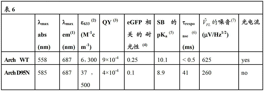
在此以引用的方式将引用的文献全部并入说明书和实施例。All cited documents are hereby incorporated by reference into the specification and examples.


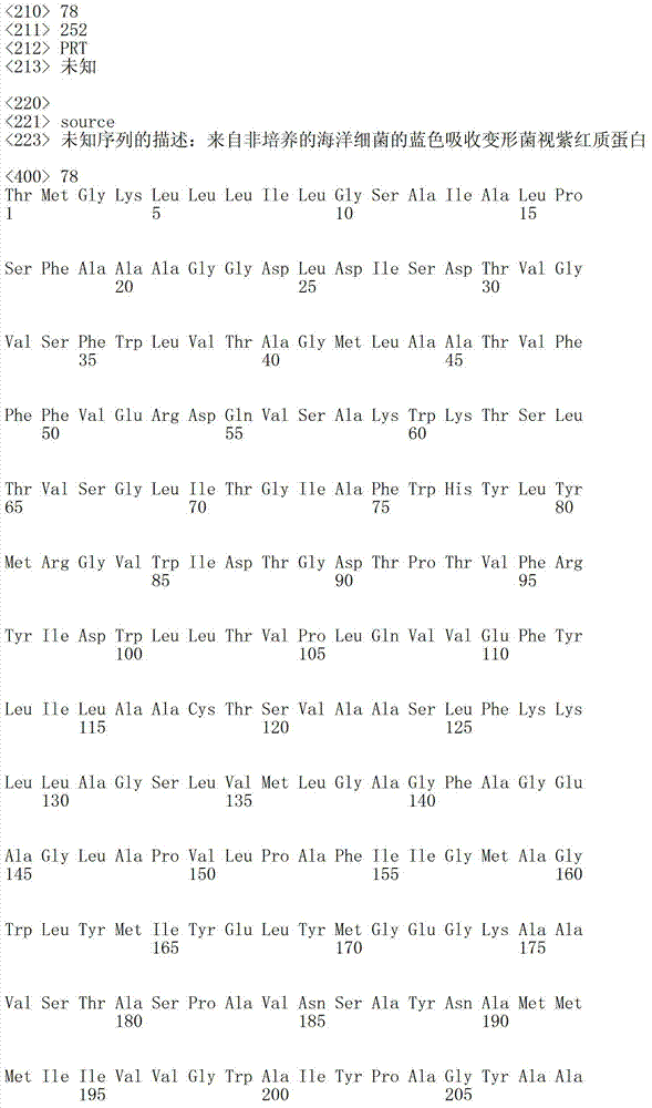


Claims (137)
Applications Claiming Priority (5)
| Application Number | Priority Date | Filing Date | Title |
|---|---|---|---|
| US37604910P | 2010-08-23 | 2010-08-23 | |
| US61/376,049 | 2010-08-23 | ||
| US41297210P | 2010-11-12 | 2010-11-12 | |
| US61/412,972 | 2010-11-12 | ||
| PCT/US2011/048793 WO2012027358A1 (en) | 2010-08-23 | 2011-08-23 | Optogenetic probes for measuring membrane potential |
Publications (2)
| Publication Number | Publication Date |
|---|---|
| CN103168236A true CN103168236A (en) | 2013-06-19 |
| CN103168236B CN103168236B (en) | 2016-01-20 |
Family
ID=44515070
Family Applications (1)
| Application Number | Title | Priority Date | Filing Date |
|---|---|---|---|
| CN201180050734.5A Expired - Fee Related CN103168236B (en) | 2010-08-23 | 2011-08-23 | Optogenetic Probes for Membrane Potential Measurements |
Country Status (5)
| Country | Link |
|---|---|
| US (3) | US9057734B2 (en) |
| EP (1) | EP2609425A1 (en) |
| CN (1) | CN103168236B (en) |
| CA (1) | CA2838330C (en) |
| WO (1) | WO2012027358A1 (en) |
Cited By (7)
| Publication number | Priority date | Publication date | Assignee | Title |
|---|---|---|---|---|
| CN104672314A (en) * | 2013-11-29 | 2015-06-03 | 华东师范大学 | Recombinant archaerhodopsin 4 protein as well as preparation method and application thereof |
| CN107677655A (en) * | 2017-09-20 | 2018-02-09 | 北京大学 | Rhodopsin fluorescence molecule complex and its preparation method and application |
| CN108210116A (en) * | 2016-12-09 | 2018-06-29 | 马克斯·普朗克科学促进学会 | Adjustable neuroid and synthetic eye |
| CN110483645A (en) * | 2018-05-14 | 2019-11-22 | 郑州大学第一附属医院 | Mitochondria positioning proton pump-type rhodopsin, its mutain and its application |
| CN113832187A (en) * | 2018-11-13 | 2021-12-24 | 四川横竖生物科技股份有限公司 | Vector, reagent and application for constructing hNPY gene overexpression chimeric animal model |
| CN115718089A (en) * | 2022-10-28 | 2023-02-28 | 江南大学 | A method for rapid identification of sample types based on Raman characteristics of bacterial flora |
| WO2024092891A1 (en) * | 2022-11-02 | 2024-05-10 | 中国科学院深圳先进技术研究院 | Method and system for detecting virus or pseudovirus infecting cell |
Families Citing this family (65)
| Publication number | Priority date | Publication date | Assignee | Title |
|---|---|---|---|---|
| US9274099B2 (en) | 2005-07-22 | 2016-03-01 | The Board Of Trustees Of The Leland Stanford Junior University | Screening test drugs to identify their effects on cell membrane voltage-gated ion channel |
| US9238150B2 (en) | 2005-07-22 | 2016-01-19 | The Board Of Trustees Of The Leland Stanford Junior University | Optical tissue interface method and apparatus for stimulating cells |
| US8926959B2 (en) | 2005-07-22 | 2015-01-06 | The Board Of Trustees Of The Leland Stanford Junior University | System for optical stimulation of target cells |
| US10052497B2 (en) | 2005-07-22 | 2018-08-21 | The Board Of Trustees Of The Leland Stanford Junior University | System for optical stimulation of target cells |
| EP2465925A1 (en) | 2005-07-22 | 2012-06-20 | The Board Of Trustees Of The Leland | Light-activated cation channel and uses thereof |
| WO2008086470A1 (en) | 2007-01-10 | 2008-07-17 | The Board Of Trustees Of The Leland Stanford Junior University | System for optical stimulation of target cells |
| WO2008101128A1 (en) | 2007-02-14 | 2008-08-21 | The Board Of Trustees Of The Leland Stanford Junior University | System, method and applications involving identification of biological circuits such as neurological characteristics |
| WO2008106694A2 (en) | 2007-03-01 | 2008-09-04 | The Board Of Trustees Of The Leland Stanford Junior University | Systems, methods and compositions for optical stimulation of target cells |
| US10035027B2 (en) | 2007-10-31 | 2018-07-31 | The Board Of Trustees Of The Leland Stanford Junior University | Device and method for ultrasonic neuromodulation via stereotactic frame based technique |
| US10434327B2 (en) | 2007-10-31 | 2019-10-08 | The Board Of Trustees Of The Leland Stanford Junior University | Implantable optical stimulators |
| JP5801188B2 (en) | 2008-04-23 | 2015-10-28 | ザ ボード オブ トラスティーズ オブ ザ レランド スタンフォード ジュニア ユニバーシティー | Systems, methods, and compositions for photostimulating target cells |
| US10711242B2 (en) | 2008-06-17 | 2020-07-14 | The Board Of Trustees Of The Leland Stanford Junior University | Apparatus and methods for controlling cellular development |
| US9101759B2 (en) | 2008-07-08 | 2015-08-11 | The Board Of Trustees Of The Leland Stanford Junior University | Materials and approaches for optical stimulation of the peripheral nervous system |
| NZ602416A (en) | 2008-11-14 | 2014-08-29 | Univ Leland Stanford Junior | Optically-based stimulation of target cells and modifications thereto |
| US8741591B2 (en) | 2009-10-09 | 2014-06-03 | The Research Foundation For The State University Of New York | pH-insensitive glucose indicator protein |
| SG10201505162QA (en) | 2010-03-17 | 2015-08-28 | Univ Leland Stanford Junior | Light-sensitive ion-passing molecules |
| US9057734B2 (en) | 2010-08-23 | 2015-06-16 | President And Fellows Of Harvard College | Optogenetic probes for measuring membrane potential |
| US9207237B2 (en) | 2010-08-23 | 2015-12-08 | President And Fellows Of Harvard College | Systems, methods, and workflows for optogenetics analysis |
| JP6002140B2 (en) | 2010-11-05 | 2016-10-05 | ザ ボード オブ トラスティーズ オブ ザ レランド スタンフォード ジュニア ユニバーシティー | Stabilized step function opsin protein and method of use thereof |
| AU2011323235B2 (en) | 2010-11-05 | 2015-10-29 | The Board Of Trustees Of The Leland Stanford Junior University | Optogenetic control of reward-related behaviors |
| CN106376525A (en) | 2010-11-05 | 2017-02-08 | 斯坦福大学托管董事会 | Control and characterization of memory function |
| CN103313752B (en) | 2010-11-05 | 2016-10-19 | 斯坦福大学托管董事会 | Upconversion of Light for Optogenetic Approaches |
| CN103384469B (en) | 2010-11-05 | 2016-06-15 | 斯坦福大学托管董事会 | Light-controlled CNS dysfunction |
| EP2635108B1 (en) | 2010-11-05 | 2019-01-23 | The Board of Trustees of the Leland Stanford Junior University | Light-activated chimeric opsins and methods of using the same |
| US8696722B2 (en) | 2010-11-22 | 2014-04-15 | The Board Of Trustees Of The Leland Stanford Junior University | Optogenetic magnetic resonance imaging |
| US9651494B2 (en) * | 2011-05-19 | 2017-05-16 | The Regents Of The University Of California | Fluorescent probes for monitoring voltage by photo-induced electron transfer |
| US20150004637A1 (en) | 2011-11-23 | 2015-01-01 | President And Fellows Of Harvard College | Systems and methods for imaging at high spatial and/or temporal precision |
| CN107936097A (en) | 2011-12-16 | 2018-04-20 | 斯坦福大学托管董事会 | Opsin polypeptide and its application method |
| CN104363961B (en) | 2012-02-21 | 2017-10-03 | 斯坦福大学托管董事会 | Composition and method for treating basin bottom neurogenic illness |
| WO2013187148A1 (en) * | 2012-06-15 | 2013-12-19 | オリンパス株式会社 | Image processing device, microscope system, endoscope system, and image processing method |
| JP6308134B2 (en) * | 2012-12-27 | 2018-04-11 | ソニー株式会社 | Cell analysis system, cell analysis program, and cell analysis method |
| ES2742492T3 (en) | 2013-03-15 | 2020-02-14 | Univ Leland Stanford Junior | Optogenetic control of behavioral status |
| US9636380B2 (en) | 2013-03-15 | 2017-05-02 | The Board Of Trustees Of The Leland Stanford Junior University | Optogenetic control of inputs to the ventral tegmental area |
| WO2014160772A1 (en) * | 2013-03-27 | 2014-10-02 | Uab Research Foundation | Stepwise differentiation of stem cells for the production of eukaryotic membrane proteins |
| US10220092B2 (en) | 2013-04-29 | 2019-03-05 | The Board Of Trustees Of The Leland Stanford Junior University | Devices, systems and methods for optogenetic modulation of action potentials in target cells |
| US9907496B1 (en) | 2013-06-25 | 2018-03-06 | National Technology & Engineering Solutions Of Sandia, Llc | Optoelectronic system and apparatus for connection to biological systems |
| US10307609B2 (en) | 2013-08-14 | 2019-06-04 | The Board Of Trustees Of The Leland Stanford Junior University | Compositions and methods for controlling pain |
| US10163203B2 (en) | 2013-11-08 | 2018-12-25 | Sony Corporation | Cell analysis system, cell analysis program and cell analysis method |
| US9606100B2 (en) | 2013-11-10 | 2017-03-28 | The Board Of Trustees Of The Leland Stanford Junior University | Fluorescent protein voltage sensors for measuring membrane potential and imaging high-frequency neuronal electrical activity |
| US9355459B2 (en) | 2014-01-29 | 2016-05-31 | Cellogy, Inc. | Method and system for characterizing cell motion |
| CA2946514C (en) | 2014-04-22 | 2023-08-29 | Q-State Biosciences, Inc. | Optogenetic analysis of compounds |
| WO2015195769A2 (en) * | 2014-06-18 | 2015-12-23 | President And Fellows Of Harvard College | Optogenetic probes for measuring membrane potential |
| US10077463B2 (en) | 2015-01-15 | 2018-09-18 | President And Fellows Of Harvard College | Optical selection of cells |
| US10048275B2 (en) | 2015-03-13 | 2018-08-14 | Q-State Biosciences, Inc. | Cardiotoxicity screening methods |
| EP3298448B1 (en) * | 2015-05-21 | 2024-09-04 | President and Fellows of Harvard College | Optogenetics microscope |
| WO2016209654A1 (en) | 2015-06-22 | 2016-12-29 | The Board Of Trustees Of The Leland Stanford Junior University | Methods and devices for imaging and/or optogenetic control of light-responsive neurons |
| US11801301B2 (en) | 2015-09-15 | 2023-10-31 | The Board Of Trustees Of The Leland Stanford Junior University | Light-responsive polypeptides and methods of use thereof |
| WO2017075275A1 (en) | 2015-10-29 | 2017-05-04 | The Board Of Trustees Of The Leland Stanford Junior University | Methods and systems for imaging a biological sample |
| WO2018013546A1 (en) * | 2016-07-11 | 2018-01-18 | The Regents Of The University Of California | Bic inhibitor of cry-cry and cry-cib oligomerization/clustering |
| KR101864958B1 (en) * | 2016-11-25 | 2018-06-05 | 포항공과대학교 산학협력단 | MAM-specific Fluorescent Subcellular Marker and Use thereof |
| US11236360B2 (en) * | 2016-12-09 | 2022-02-01 | Regents Of The University Of Minnesota | Adeno-associated viruses engineered for selectable tropism |
| EP3568677A4 (en) | 2017-01-10 | 2020-10-21 | Photoswitch Biosciences, Inc. | SYSTEMS AND METHODS OF DETECTION |
| US11294165B2 (en) | 2017-03-30 | 2022-04-05 | The Board Of Trustees Of The Leland Stanford Junior University | Modular, electro-optical device for increasing the imaging field of view using time-sequential capture |
| GB201709551D0 (en) * | 2017-06-15 | 2017-08-02 | Ucl Business Plc | Expression vectors comprising engineered genes |
| US11484731B2 (en) * | 2017-11-09 | 2022-11-01 | International Business Machines Corporation | Cognitive optogenetics probe and analysis |
| WO2019183376A1 (en) | 2018-03-21 | 2019-09-26 | The Board Of Trustees Of The Leland Stanford Junior University | Methods for temporal and spatial multiplexing of spatial light modulators and systems for same |
| US10829727B2 (en) | 2018-04-11 | 2020-11-10 | Trustees Of Boston University | Engineered platform to generate 3D cardiac tissues |
| IL317123A (en) | 2018-08-27 | 2025-01-01 | Regeneron Pharma | Use of Raman Spectroscopy in Downstream Purification |
| EP3860337A4 (en) * | 2018-10-01 | 2022-07-13 | Cornell University | Kits and methods for performing optical dynamic clamp on excitable cells |
| WO2022008720A1 (en) * | 2020-07-10 | 2022-01-13 | Life & Brain Gmbh | Novel hybrid optical voltage sensors |
| CN116685359A (en) * | 2020-11-12 | 2023-09-01 | 英联邦高等教育系统天普大学 | Gram stain differentiation using nonlinear light scattering |
| US12364395B2 (en) | 2020-11-23 | 2025-07-22 | Arizona Board Of Regents On Behalf Of Arizona State University | Dual brillouin-photoacoustic microscopy |
| US12082938B2 (en) | 2020-11-23 | 2024-09-10 | Arizona Board Of Regents On Behalf Of Arizona State University | Photoacoustic voltage indicators |
| EP4047355A1 (en) | 2021-02-23 | 2022-08-24 | Universitat Politècnica De Catalunya | A method and a system for detecting microwave signals which carry information of the functional dynamics of biological particles |
| CA3232099A1 (en) | 2021-09-23 | 2023-03-30 | Adam Ezra Cohen | Genetically encoded voltage indicators and uses thereof |
Citations (4)
| Publication number | Priority date | Publication date | Assignee | Title |
|---|---|---|---|---|
| WO2001083701A2 (en) * | 2000-05-03 | 2001-11-08 | Monterey Bay Aquarium Research Institute | Light-driven energy generation using proteorhodopsin |
| WO2004063326A2 (en) * | 2002-11-26 | 2004-07-29 | Genencor International, Inc. | Proteorhodopsin mutants with improved optical characteristics |
| EP2112510A1 (en) * | 2008-04-22 | 2009-10-28 | Max-Planck-Gesellschaft zur Förderung der Wissenschaften e.V. | Use of biological photo-receptors as direct light-controlled ion channels |
| WO2010056970A2 (en) * | 2008-11-14 | 2010-05-20 | The Board Of Trustees Of The Leland Stanford Junior University | Optically-based stimulation of target cells and modifications thereto |
Family Cites Families (60)
| Publication number | Priority date | Publication date | Assignee | Title |
|---|---|---|---|---|
| US3219101A (en) | 1962-05-10 | 1965-11-23 | Martin Fleit | Awning |
| US4184917A (en) | 1974-04-01 | 1980-01-22 | Sandoz Ltd. | Process for producing a structurally modified interferon |
| US4321365A (en) | 1977-10-19 | 1982-03-23 | Research Corporation | Oligonucleotides useful as adaptors in DNA cloning, adapted DNA molecules, and methods of preparing adaptors and adapted molecules |
| US4293652A (en) | 1979-05-25 | 1981-10-06 | Cetus Corporation | Method for synthesizing DNA sequentially |
| US4351901A (en) | 1980-03-24 | 1982-09-28 | Cetus Corporation | Method for single nucleotide alteration |
| DE4037342A1 (en) | 1990-11-23 | 1992-05-27 | Max Planck Gesellschaft | BACTERIORHODOPSIN DOUBLE MUTANTS |
| US5843780A (en) | 1995-01-20 | 1998-12-01 | Wisconsin Alumni Research Foundation | Primate embryonic stem cells |
| US5661035A (en) | 1995-06-07 | 1997-08-26 | The Regents Of The University Of California | Voltage sensing by fluorescence resonance energy transfer |
| US5925523A (en) | 1996-08-23 | 1999-07-20 | President & Fellows Of Harvard College | Intraction trap assay, reagents and uses thereof |
| DE19644662C2 (en) | 1996-10-25 | 2000-04-13 | Leica Microsystems | Illumination device for a microscope |
| US7186409B2 (en) | 1998-08-14 | 2007-03-06 | The Children's Medical Center Corporation | Neural stem cells and use thereof for brain tumor therapy |
| US6280718B1 (en) | 1999-11-08 | 2001-08-28 | Wisconsin Alumni Reasearch Foundation | Hematopoietic differentiation of human pluripotent embryonic stem cells |
| GB0003069D0 (en) | 2000-02-11 | 2000-03-29 | Cambridge Drug Discovery Ltd | Improved assay |
| CA2406664C (en) | 2000-03-15 | 2011-05-10 | Children's Medical Center Corporation | Systemic delivery of neural stem cells to treat cancer |
| JP2002031758A (en) | 2000-07-17 | 2002-01-31 | Olympus Optical Co Ltd | microscope |
| US7019376B2 (en) | 2000-08-11 | 2006-03-28 | Reflectivity, Inc | Micromirror array device with a small pitch size |
| US7560280B2 (en) | 2000-11-03 | 2009-07-14 | Kourion Therapeutics Gmbh | Human cord blood derived unrestricted somatic stem cells (USSC) |
| JP2004531691A (en) | 2000-11-22 | 2004-10-14 | アラーガン、インコーポレイテッド | High-throughput screen to identify channel blockers that selectively discriminate between transient and persistent sodium channels |
| JP5048899B2 (en) | 2001-09-28 | 2012-10-17 | オリンパス株式会社 | microscope |
| US6887706B2 (en) | 2001-10-03 | 2005-05-03 | Wisconsin Alumni Research Foundation | Method of in vitro differentiation of transplantable neural precursor cells from primate embryonic stem cells |
| US6885492B2 (en) | 2001-11-08 | 2005-04-26 | Imaginative Optics, Inc. | Spatial light modulator apparatus |
| DE10216005A1 (en) | 2002-04-11 | 2003-10-30 | Max Planck Gesellschaft | Use of biological photoreceptors as direct light-controlled ion channels |
| DE602004028803D1 (en) | 2003-02-11 | 2010-10-07 | John E Davies | PRECURSOR CELLS FROM THE WHARTON SULCE OF HUMAN NECK LOCKS |
| US20070087959A1 (en) * | 2003-08-19 | 2007-04-19 | University Of Pittsburgh Of The Commonwealth System Of Higher Education | Method of inducing biomineralization method of inducing bone regeneration and methods related thereof |
| US7358282B2 (en) | 2003-12-05 | 2008-04-15 | Kimberly-Clark Worldwide, Inc. | Low-density, open-cell, soft, flexible, thermoplastic, absorbent foam and method of making foam |
| US7867282B2 (en) | 2004-12-17 | 2011-01-11 | Depuy Products, Inc. | Modular implant system and method with diaphyseal implant and adapter |
| BRPI0612862A2 (en) | 2005-07-18 | 2010-11-30 | Pioneer Hi Bred Int | isolated polynucleotide, method for obtaining a transformed cell, method for obtaining a transformed plant, specific site DNA recombination production method, isolated library, molecule library generation method, and method for selecting modified frt recombination site |
| US20100167404A1 (en) | 2005-08-03 | 2010-07-01 | Advanced Cell Technology, Inc. | Methods of Reprogramming Animal Somatic Cells |
| EP1970446B1 (en) | 2005-12-13 | 2011-08-03 | Kyoto University | Nuclear reprogramming factor |
| CA2685900A1 (en) * | 2006-05-04 | 2007-11-15 | Wayne State University | Restoration of visual responses by in vivo delivery of rhodopsin nucleic acids |
| JP5307539B2 (en) | 2006-05-31 | 2013-10-02 | オリンパス株式会社 | Biological sample imaging method and biological sample imaging apparatus |
| US9201063B2 (en) | 2006-11-16 | 2015-12-01 | General Electric Company | Sequential analysis of biological samples |
| EP1953579B1 (en) | 2007-02-05 | 2010-01-13 | Olympus Corporation | Scanning laser microscope |
| WO2008101128A1 (en) | 2007-02-14 | 2008-08-21 | The Board Of Trustees Of The Leland Stanford Junior University | System, method and applications involving identification of biological circuits such as neurological characteristics |
| SG193652A1 (en) | 2007-03-23 | 2013-10-30 | Wisconsin Alumni Res Found | Somatic cell reprogramming |
| ES2907452T3 (en) | 2007-04-12 | 2022-04-25 | The Provost Fellows Found Scholars & The Other Members Of Board Of The College Of The Holy & Undiv T | Genetic deletion and replacement |
| US20110165681A1 (en) | 2009-02-26 | 2011-07-07 | Massachusetts Institute Of Technology | Light-Activated Proton Pumps and Applications Thereof |
| US7940978B2 (en) | 2007-06-05 | 2011-05-10 | General Electric Company | Automatic characterization of cellular motion |
| JP2009018772A (en) | 2007-07-13 | 2009-01-29 | Honda Motor Co Ltd | Driving force interrupt mechanism |
| WO2009012173A2 (en) | 2007-07-13 | 2009-01-22 | Dharmacon, Inc. | Enhanced biotherapeutic production using inhibitory rna |
| JP5841302B2 (en) | 2007-09-10 | 2016-01-13 | オリンパス株式会社 | Gene expression analysis method and gene expression analysis system |
| US8551407B2 (en) | 2007-11-29 | 2013-10-08 | Michigan Technological University | Bacteriorhodopsin-based sensors |
| WO2009072123A2 (en) | 2007-12-06 | 2009-06-11 | Technion Research & Development Foundation Ltd. | Method and system for optical stimulation of neurons |
| US8883719B2 (en) | 2008-01-16 | 2014-11-11 | University Of Connecticut | Bacteriorhodopsin protein variants and methods of use for long term data storage |
| US9023989B2 (en) | 2008-02-19 | 2015-05-05 | University Of Connecticut | Protein-based photovoltaics and methods of use |
| JP5801188B2 (en) | 2008-04-23 | 2015-10-28 | ザ ボード オブ トラスティーズ オブ ザ レランド スタンフォード ジュニア ユニバーシティー | Systems, methods, and compositions for photostimulating target cells |
| US10047346B2 (en) | 2008-08-08 | 2018-08-14 | Mayo Foundation For Medical Education And Research | Method of treating heart tissue using induced pluripotent stem cells |
| JP5789513B2 (en) | 2008-09-04 | 2015-10-07 | ガレニア コーポレーション | Synaptic vesicle cycling assay and system |
| JP5340799B2 (en) | 2009-05-08 | 2013-11-13 | オリンパス株式会社 | Laser scanning microscope |
| WO2011088398A1 (en) | 2010-01-15 | 2011-07-21 | President And Fellows Of Harvard College | Convex lens-induced confinement for measuring distributions of molecular size |
| US8532398B2 (en) | 2010-03-26 | 2013-09-10 | General Electric Company | Methods and apparatus for optical segmentation of biological samples |
| JP5806450B2 (en) | 2010-07-02 | 2015-11-10 | オリンパス株式会社 | Cell observation method |
| US9057734B2 (en) | 2010-08-23 | 2015-06-16 | President And Fellows Of Harvard College | Optogenetic probes for measuring membrane potential |
| US9207237B2 (en) | 2010-08-23 | 2015-12-08 | President And Fellows Of Harvard College | Systems, methods, and workflows for optogenetics analysis |
| US8580937B2 (en) | 2011-03-14 | 2013-11-12 | Board Of Regents Of The University Of Texas System | cDNA-derived nucleic acids encoding red-shifted channelrhodopsins |
| US9651494B2 (en) | 2011-05-19 | 2017-05-16 | The Regents Of The University Of California | Fluorescent probes for monitoring voltage by photo-induced electron transfer |
| US20150004637A1 (en) | 2011-11-23 | 2015-01-01 | President And Fellows Of Harvard College | Systems and methods for imaging at high spatial and/or temporal precision |
| US20140120557A1 (en) | 2012-10-04 | 2014-05-01 | The Board Of Trustees Of The Leland Stanford Junior University | Photocrosslinkable Peptide-MHC Complexes for Antigen-Specific T Cells and Methods of Using the Same |
| WO2015195769A2 (en) | 2014-06-18 | 2015-12-23 | President And Fellows Of Harvard College | Optogenetic probes for measuring membrane potential |
| US10077463B2 (en) | 2015-01-15 | 2018-09-18 | President And Fellows Of Harvard College | Optical selection of cells |
-
2011
- 2011-08-23 US US13/818,432 patent/US9057734B2/en active Active
- 2011-08-23 CA CA2838330A patent/CA2838330C/en active Active
- 2011-08-23 EP EP11749320.5A patent/EP2609425A1/en not_active Withdrawn
- 2011-08-23 WO PCT/US2011/048793 patent/WO2012027358A1/en not_active Ceased
- 2011-08-23 CN CN201180050734.5A patent/CN103168236B/en not_active Expired - Fee Related
-
2015
- 2015-06-15 US US14/739,908 patent/US9791455B2/en active Active
-
2017
- 2017-10-13 US US15/783,886 patent/US10352945B2/en active Active
Patent Citations (4)
| Publication number | Priority date | Publication date | Assignee | Title |
|---|---|---|---|---|
| WO2001083701A2 (en) * | 2000-05-03 | 2001-11-08 | Monterey Bay Aquarium Research Institute | Light-driven energy generation using proteorhodopsin |
| WO2004063326A2 (en) * | 2002-11-26 | 2004-07-29 | Genencor International, Inc. | Proteorhodopsin mutants with improved optical characteristics |
| EP2112510A1 (en) * | 2008-04-22 | 2009-10-28 | Max-Planck-Gesellschaft zur Förderung der Wissenschaften e.V. | Use of biological photo-receptors as direct light-controlled ion channels |
| WO2010056970A2 (en) * | 2008-11-14 | 2010-05-20 | The Board Of Trustees Of The Leland Stanford Junior University | Optically-based stimulation of target cells and modifications thereto |
Cited By (8)
| Publication number | Priority date | Publication date | Assignee | Title |
|---|---|---|---|---|
| CN104672314A (en) * | 2013-11-29 | 2015-06-03 | 华东师范大学 | Recombinant archaerhodopsin 4 protein as well as preparation method and application thereof |
| CN108210116A (en) * | 2016-12-09 | 2018-06-29 | 马克斯·普朗克科学促进学会 | Adjustable neuroid and synthetic eye |
| CN107677655A (en) * | 2017-09-20 | 2018-02-09 | 北京大学 | Rhodopsin fluorescence molecule complex and its preparation method and application |
| CN110483645A (en) * | 2018-05-14 | 2019-11-22 | 郑州大学第一附属医院 | Mitochondria positioning proton pump-type rhodopsin, its mutain and its application |
| CN110483645B (en) * | 2018-05-14 | 2023-05-12 | 郑州大学第一附属医院 | Mitochondrial localized proton pump rhodopsin, its mutant protein and its application |
| CN113832187A (en) * | 2018-11-13 | 2021-12-24 | 四川横竖生物科技股份有限公司 | Vector, reagent and application for constructing hNPY gene overexpression chimeric animal model |
| CN115718089A (en) * | 2022-10-28 | 2023-02-28 | 江南大学 | A method for rapid identification of sample types based on Raman characteristics of bacterial flora |
| WO2024092891A1 (en) * | 2022-11-02 | 2024-05-10 | 中国科学院深圳先进技术研究院 | Method and system for detecting virus or pseudovirus infecting cell |
Also Published As
| Publication number | Publication date |
|---|---|
| US9791455B2 (en) | 2017-10-17 |
| US20130224756A1 (en) | 2013-08-29 |
| EP2609425A1 (en) | 2013-07-03 |
| US20180031572A1 (en) | 2018-02-01 |
| US20150285820A1 (en) | 2015-10-08 |
| US9057734B2 (en) | 2015-06-16 |
| WO2012027358A1 (en) | 2012-03-01 |
| CA2838330A1 (en) | 2012-03-01 |
| CN103168236B (en) | 2016-01-20 |
| US10352945B2 (en) | 2019-07-16 |
| CA2838330C (en) | 2021-01-26 |
Similar Documents
| Publication | Publication Date | Title |
|---|---|---|
| CN103168236B (en) | Optogenetic Probes for Membrane Potential Measurements | |
| US10800829B2 (en) | Optogenetic probes for measuring membrane potential | |
| US10161937B2 (en) | Systems, methods, and workflows for optogenetics analysis | |
| CN103492564B (en) | Photoactivatable chimeric opsins and methods of use thereof | |
| WO2016054503A1 (en) | System and methods for assessing response to stimuli | |
| Porotto et al. | Mechanism of fusion triggering by human parainfluenza virus type III: communication between viral glycoproteins during entry | |
| US20220118271A1 (en) | Light-responsive polypeptides and methods of use thereof | |
| US20230064644A1 (en) | Compositions and Methods for Controlling Production of Polypeptides in Cells | |
| US20240400622A1 (en) | Genetically encoded voltage indicators and uses thereof | |
| Zou et al. | Bright and fast voltage reporters across the visible spectrum via electrochromic FRET (eFRET) | |
| CN110078834B (en) | A kind of short peptide, auxiliary membrane permeable agent and application thereof | |
| WO2016118902A1 (en) | Minimally-invasive and activity-dependent control of excitable cells | |
| Kwiatkowski | Enhancing membrane repair as a therapeutic strategy for various muscular dystrophies | |
| Prorok | Studies on the role of disturbed Ca 2 homeostasis in the pathomechanism of the cardiac effects of experimental diabetes using conventional and novel experimental techniques | |
| Muster et al. | Respiratory Chain Complexes in Dynamic Mitochondria Display a Patchy Distribution in Life | |
| Prorok | Studies on the role of disturbed Ca2+ homeostasis in the pathomechanism of the cardiac effects of experimental diabetes using conventional and novel experimental techniques |
Legal Events
| Date | Code | Title | Description |
|---|---|---|---|
| C06 | Publication | ||
| PB01 | Publication | ||
| C10 | Entry into substantive examination | ||
| SE01 | Entry into force of request for substantive examination | ||
| C14 | Grant of patent or utility model | ||
| GR01 | Patent grant | ||
| CF01 | Termination of patent right due to non-payment of annual fee |
Granted publication date: 20160120 Termination date: 20190823 |
|
| CF01 | Termination of patent right due to non-payment of annual fee |