CN102711674A - Medical device for placement into a lumen and manufacturing method thereof - Google Patents
Medical device for placement into a lumen and manufacturing method thereof Download PDFInfo
- Publication number
- CN102711674A CN102711674A CN2010800484372A CN201080048437A CN102711674A CN 102711674 A CN102711674 A CN 102711674A CN 2010800484372 A CN2010800484372 A CN 2010800484372A CN 201080048437 A CN201080048437 A CN 201080048437A CN 102711674 A CN102711674 A CN 102711674A
- Authority
- CN
- China
- Prior art keywords
- drug
- indwelling
- medical device
- particles
- particle
- Prior art date
- Legal status (The legal status is an assumption and is not a legal conclusion. Google has not performed a legal analysis and makes no representation as to the accuracy of the status listed.)
- Pending
Links
Images


Classifications
-
- A—HUMAN NECESSITIES
- A61—MEDICAL OR VETERINARY SCIENCE; HYGIENE
- A61L—METHODS OR APPARATUS FOR STERILISING MATERIALS OR OBJECTS IN GENERAL; DISINFECTION, STERILISATION OR DEODORISATION OF AIR; CHEMICAL ASPECTS OF BANDAGES, DRESSINGS, ABSORBENT PADS OR SURGICAL ARTICLES; MATERIALS FOR BANDAGES, DRESSINGS, ABSORBENT PADS OR SURGICAL ARTICLES
- A61L31/00—Materials for other surgical articles, e.g. stents, stent-grafts, shunts, surgical drapes, guide wires, materials for adhesion prevention, occluding devices, surgical gloves, tissue fixation devices
- A61L31/14—Materials characterised by their function or physical properties, e.g. injectable or lubricating compositions, shape-memory materials, surface modified materials
- A61L31/16—Biologically active materials, e.g. therapeutic substances
-
- A—HUMAN NECESSITIES
- A61—MEDICAL OR VETERINARY SCIENCE; HYGIENE
- A61F—FILTERS IMPLANTABLE INTO BLOOD VESSELS; PROSTHESES; DEVICES PROVIDING PATENCY TO, OR PREVENTING COLLAPSING OF, TUBULAR STRUCTURES OF THE BODY, e.g. STENTS; ORTHOPAEDIC, NURSING OR CONTRACEPTIVE DEVICES; FOMENTATION; TREATMENT OR PROTECTION OF EYES OR EARS; BANDAGES, DRESSINGS OR ABSORBENT PADS; FIRST-AID KITS
- A61F2/00—Filters implantable into blood vessels; Prostheses, i.e. artificial substitutes or replacements for parts of the body; Appliances for connecting them with the body; Devices providing patency to, or preventing collapsing of, tubular structures of the body, e.g. stents
- A61F2/82—Devices providing patency to, or preventing collapsing of, tubular structures of the body, e.g. stents
-
- A—HUMAN NECESSITIES
- A61—MEDICAL OR VETERINARY SCIENCE; HYGIENE
- A61L—METHODS OR APPARATUS FOR STERILISING MATERIALS OR OBJECTS IN GENERAL; DISINFECTION, STERILISATION OR DEODORISATION OF AIR; CHEMICAL ASPECTS OF BANDAGES, DRESSINGS, ABSORBENT PADS OR SURGICAL ARTICLES; MATERIALS FOR BANDAGES, DRESSINGS, ABSORBENT PADS OR SURGICAL ARTICLES
- A61L29/00—Materials for catheters, medical tubing, cannulae, or endoscopes or for coating catheters
- A61L29/04—Macromolecular materials
- A61L29/043—Polysaccharides
-
- A—HUMAN NECESSITIES
- A61—MEDICAL OR VETERINARY SCIENCE; HYGIENE
- A61L—METHODS OR APPARATUS FOR STERILISING MATERIALS OR OBJECTS IN GENERAL; DISINFECTION, STERILISATION OR DEODORISATION OF AIR; CHEMICAL ASPECTS OF BANDAGES, DRESSINGS, ABSORBENT PADS OR SURGICAL ARTICLES; MATERIALS FOR BANDAGES, DRESSINGS, ABSORBENT PADS OR SURGICAL ARTICLES
- A61L29/00—Materials for catheters, medical tubing, cannulae, or endoscopes or for coating catheters
- A61L29/04—Macromolecular materials
- A61L29/06—Macromolecular materials obtained otherwise than by reactions only involving carbon-to-carbon unsaturated bonds
-
- A—HUMAN NECESSITIES
- A61—MEDICAL OR VETERINARY SCIENCE; HYGIENE
- A61L—METHODS OR APPARATUS FOR STERILISING MATERIALS OR OBJECTS IN GENERAL; DISINFECTION, STERILISATION OR DEODORISATION OF AIR; CHEMICAL ASPECTS OF BANDAGES, DRESSINGS, ABSORBENT PADS OR SURGICAL ARTICLES; MATERIALS FOR BANDAGES, DRESSINGS, ABSORBENT PADS OR SURGICAL ARTICLES
- A61L29/00—Materials for catheters, medical tubing, cannulae, or endoscopes or for coating catheters
- A61L29/08—Materials for coatings
- A61L29/085—Macromolecular materials
-
- A—HUMAN NECESSITIES
- A61—MEDICAL OR VETERINARY SCIENCE; HYGIENE
- A61L—METHODS OR APPARATUS FOR STERILISING MATERIALS OR OBJECTS IN GENERAL; DISINFECTION, STERILISATION OR DEODORISATION OF AIR; CHEMICAL ASPECTS OF BANDAGES, DRESSINGS, ABSORBENT PADS OR SURGICAL ARTICLES; MATERIALS FOR BANDAGES, DRESSINGS, ABSORBENT PADS OR SURGICAL ARTICLES
- A61L29/00—Materials for catheters, medical tubing, cannulae, or endoscopes or for coating catheters
- A61L29/14—Materials characterised by their function or physical properties, e.g. lubricating compositions
- A61L29/16—Biologically active materials, e.g. therapeutic substances
-
- A—HUMAN NECESSITIES
- A61—MEDICAL OR VETERINARY SCIENCE; HYGIENE
- A61L—METHODS OR APPARATUS FOR STERILISING MATERIALS OR OBJECTS IN GENERAL; DISINFECTION, STERILISATION OR DEODORISATION OF AIR; CHEMICAL ASPECTS OF BANDAGES, DRESSINGS, ABSORBENT PADS OR SURGICAL ARTICLES; MATERIALS FOR BANDAGES, DRESSINGS, ABSORBENT PADS OR SURGICAL ARTICLES
- A61L31/00—Materials for other surgical articles, e.g. stents, stent-grafts, shunts, surgical drapes, guide wires, materials for adhesion prevention, occluding devices, surgical gloves, tissue fixation devices
- A61L31/02—Inorganic materials
- A61L31/022—Metals or alloys
-
- A—HUMAN NECESSITIES
- A61—MEDICAL OR VETERINARY SCIENCE; HYGIENE
- A61L—METHODS OR APPARATUS FOR STERILISING MATERIALS OR OBJECTS IN GENERAL; DISINFECTION, STERILISATION OR DEODORISATION OF AIR; CHEMICAL ASPECTS OF BANDAGES, DRESSINGS, ABSORBENT PADS OR SURGICAL ARTICLES; MATERIALS FOR BANDAGES, DRESSINGS, ABSORBENT PADS OR SURGICAL ARTICLES
- A61L31/00—Materials for other surgical articles, e.g. stents, stent-grafts, shunts, surgical drapes, guide wires, materials for adhesion prevention, occluding devices, surgical gloves, tissue fixation devices
- A61L31/04—Macromolecular materials
- A61L31/042—Polysaccharides
-
- A—HUMAN NECESSITIES
- A61—MEDICAL OR VETERINARY SCIENCE; HYGIENE
- A61L—METHODS OR APPARATUS FOR STERILISING MATERIALS OR OBJECTS IN GENERAL; DISINFECTION, STERILISATION OR DEODORISATION OF AIR; CHEMICAL ASPECTS OF BANDAGES, DRESSINGS, ABSORBENT PADS OR SURGICAL ARTICLES; MATERIALS FOR BANDAGES, DRESSINGS, ABSORBENT PADS OR SURGICAL ARTICLES
- A61L31/00—Materials for other surgical articles, e.g. stents, stent-grafts, shunts, surgical drapes, guide wires, materials for adhesion prevention, occluding devices, surgical gloves, tissue fixation devices
- A61L31/04—Macromolecular materials
- A61L31/06—Macromolecular materials obtained otherwise than by reactions only involving carbon-to-carbon unsaturated bonds
-
- A—HUMAN NECESSITIES
- A61—MEDICAL OR VETERINARY SCIENCE; HYGIENE
- A61L—METHODS OR APPARATUS FOR STERILISING MATERIALS OR OBJECTS IN GENERAL; DISINFECTION, STERILISATION OR DEODORISATION OF AIR; CHEMICAL ASPECTS OF BANDAGES, DRESSINGS, ABSORBENT PADS OR SURGICAL ARTICLES; MATERIALS FOR BANDAGES, DRESSINGS, ABSORBENT PADS OR SURGICAL ARTICLES
- A61L31/00—Materials for other surgical articles, e.g. stents, stent-grafts, shunts, surgical drapes, guide wires, materials for adhesion prevention, occluding devices, surgical gloves, tissue fixation devices
- A61L31/08—Materials for coatings
- A61L31/10—Macromolecular materials
-
- A—HUMAN NECESSITIES
- A61—MEDICAL OR VETERINARY SCIENCE; HYGIENE
- A61P—SPECIFIC THERAPEUTIC ACTIVITY OF CHEMICAL COMPOUNDS OR MEDICINAL PREPARATIONS
- A61P7/00—Drugs for disorders of the blood or the extracellular fluid
- A61P7/02—Antithrombotic agents; Anticoagulants; Platelet aggregation inhibitors
-
- A—HUMAN NECESSITIES
- A61—MEDICAL OR VETERINARY SCIENCE; HYGIENE
- A61P—SPECIFIC THERAPEUTIC ACTIVITY OF CHEMICAL COMPOUNDS OR MEDICINAL PREPARATIONS
- A61P9/00—Drugs for disorders of the cardiovascular system
- A61P9/08—Vasodilators for multiple indications
-
- A—HUMAN NECESSITIES
- A61—MEDICAL OR VETERINARY SCIENCE; HYGIENE
- A61F—FILTERS IMPLANTABLE INTO BLOOD VESSELS; PROSTHESES; DEVICES PROVIDING PATENCY TO, OR PREVENTING COLLAPSING OF, TUBULAR STRUCTURES OF THE BODY, e.g. STENTS; ORTHOPAEDIC, NURSING OR CONTRACEPTIVE DEVICES; FOMENTATION; TREATMENT OR PROTECTION OF EYES OR EARS; BANDAGES, DRESSINGS OR ABSORBENT PADS; FIRST-AID KITS
- A61F2250/00—Special features of prostheses classified in groups A61F2/00 - A61F2/26 or A61F2/82 or A61F9/00 or A61F11/00 or subgroups thereof
- A61F2250/0058—Additional features; Implant or prostheses properties not otherwise provided for
- A61F2250/0067—Means for introducing or releasing pharmaceutical products into the body
-
- A—HUMAN NECESSITIES
- A61—MEDICAL OR VETERINARY SCIENCE; HYGIENE
- A61L—METHODS OR APPARATUS FOR STERILISING MATERIALS OR OBJECTS IN GENERAL; DISINFECTION, STERILISATION OR DEODORISATION OF AIR; CHEMICAL ASPECTS OF BANDAGES, DRESSINGS, ABSORBENT PADS OR SURGICAL ARTICLES; MATERIALS FOR BANDAGES, DRESSINGS, ABSORBENT PADS OR SURGICAL ARTICLES
- A61L2300/00—Biologically active materials used in bandages, wound dressings, absorbent pads or medical devices
- A61L2300/40—Biologically active materials used in bandages, wound dressings, absorbent pads or medical devices characterised by a specific therapeutic activity or mode of action
- A61L2300/416—Anti-neoplastic or anti-proliferative or anti-restenosis or anti-angiogenic agents, e.g. paclitaxel, sirolimus
-
- A—HUMAN NECESSITIES
- A61—MEDICAL OR VETERINARY SCIENCE; HYGIENE
- A61L—METHODS OR APPARATUS FOR STERILISING MATERIALS OR OBJECTS IN GENERAL; DISINFECTION, STERILISATION OR DEODORISATION OF AIR; CHEMICAL ASPECTS OF BANDAGES, DRESSINGS, ABSORBENT PADS OR SURGICAL ARTICLES; MATERIALS FOR BANDAGES, DRESSINGS, ABSORBENT PADS OR SURGICAL ARTICLES
- A61L2300/00—Biologically active materials used in bandages, wound dressings, absorbent pads or medical devices
- A61L2300/40—Biologically active materials used in bandages, wound dressings, absorbent pads or medical devices characterised by a specific therapeutic activity or mode of action
- A61L2300/42—Anti-thrombotic agents, anticoagulants, anti-platelet agents
-
- A—HUMAN NECESSITIES
- A61—MEDICAL OR VETERINARY SCIENCE; HYGIENE
- A61L—METHODS OR APPARATUS FOR STERILISING MATERIALS OR OBJECTS IN GENERAL; DISINFECTION, STERILISATION OR DEODORISATION OF AIR; CHEMICAL ASPECTS OF BANDAGES, DRESSINGS, ABSORBENT PADS OR SURGICAL ARTICLES; MATERIALS FOR BANDAGES, DRESSINGS, ABSORBENT PADS OR SURGICAL ARTICLES
- A61L2300/00—Biologically active materials used in bandages, wound dressings, absorbent pads or medical devices
- A61L2300/60—Biologically active materials used in bandages, wound dressings, absorbent pads or medical devices characterised by a special physical form
- A61L2300/606—Coatings
-
- A—HUMAN NECESSITIES
- A61—MEDICAL OR VETERINARY SCIENCE; HYGIENE
- A61L—METHODS OR APPARATUS FOR STERILISING MATERIALS OR OBJECTS IN GENERAL; DISINFECTION, STERILISATION OR DEODORISATION OF AIR; CHEMICAL ASPECTS OF BANDAGES, DRESSINGS, ABSORBENT PADS OR SURGICAL ARTICLES; MATERIALS FOR BANDAGES, DRESSINGS, ABSORBENT PADS OR SURGICAL ARTICLES
- A61L2300/00—Biologically active materials used in bandages, wound dressings, absorbent pads or medical devices
- A61L2300/60—Biologically active materials used in bandages, wound dressings, absorbent pads or medical devices characterised by a special physical form
- A61L2300/62—Encapsulated active agents, e.g. emulsified droplets
- A61L2300/624—Nanocapsules
-
- A—HUMAN NECESSITIES
- A61—MEDICAL OR VETERINARY SCIENCE; HYGIENE
- A61L—METHODS OR APPARATUS FOR STERILISING MATERIALS OR OBJECTS IN GENERAL; DISINFECTION, STERILISATION OR DEODORISATION OF AIR; CHEMICAL ASPECTS OF BANDAGES, DRESSINGS, ABSORBENT PADS OR SURGICAL ARTICLES; MATERIALS FOR BANDAGES, DRESSINGS, ABSORBENT PADS OR SURGICAL ARTICLES
- A61L2400/00—Materials characterised by their function or physical properties
- A61L2400/12—Nanosized materials, e.g. nanofibres, nanoparticles, nanowires, nanotubes; Nanostructured surfaces
-
- A—HUMAN NECESSITIES
- A61—MEDICAL OR VETERINARY SCIENCE; HYGIENE
- A61L—METHODS OR APPARATUS FOR STERILISING MATERIALS OR OBJECTS IN GENERAL; DISINFECTION, STERILISATION OR DEODORISATION OF AIR; CHEMICAL ASPECTS OF BANDAGES, DRESSINGS, ABSORBENT PADS OR SURGICAL ARTICLES; MATERIALS FOR BANDAGES, DRESSINGS, ABSORBENT PADS OR SURGICAL ARTICLES
- A61L2400/00—Materials characterised by their function or physical properties
- A61L2400/18—Modification of implant surfaces in order to improve biocompatibility, cell growth, fixation of biomolecules, e.g. plasma treatment
Landscapes
- Health & Medical Sciences (AREA)
- Life Sciences & Earth Sciences (AREA)
- Animal Behavior & Ethology (AREA)
- Veterinary Medicine (AREA)
- Public Health (AREA)
- General Health & Medical Sciences (AREA)
- Epidemiology (AREA)
- Heart & Thoracic Surgery (AREA)
- Vascular Medicine (AREA)
- Surgery (AREA)
- Engineering & Computer Science (AREA)
- Chemical & Material Sciences (AREA)
- Biomedical Technology (AREA)
- Medicinal Chemistry (AREA)
- Chemical Kinetics & Catalysis (AREA)
- Cardiology (AREA)
- Molecular Biology (AREA)
- Transplantation (AREA)
- Oral & Maxillofacial Surgery (AREA)
- Inorganic Chemistry (AREA)
- General Chemical & Material Sciences (AREA)
- Nuclear Medicine, Radiotherapy & Molecular Imaging (AREA)
- Organic Chemistry (AREA)
- Pharmacology & Pharmacy (AREA)
- Bioinformatics & Cheminformatics (AREA)
- Diabetes (AREA)
- Hematology (AREA)
- Materials For Medical Uses (AREA)
- Media Introduction/Drainage Providing Device (AREA)
- Medicinal Preparation (AREA)
- Pharmaceuticals Containing Other Organic And Inorganic Compounds (AREA)
Abstract
Description
技术领域 technical field
本发明涉及一种涂覆有药物的药物洗脱型的管腔内留置用医疗设备及其制造方法,所述管腔内留置用医疗设备是留置于生物体内的管腔中所生成的狭窄部或堵塞部以维持开放状态的支架或导管等管腔内留置用医疗设备。 The present invention relates to a drug-eluting medical device for indwelling in a lumen, which is a stenosis formed in a lumen in a living body, and a manufacturing method thereof. Or medical devices for indwelling in the lumen such as stents or catheters to maintain the open state of the blocked part.
背景技术 Background technique
随着近年来医疗的进步,以感染症为代表对各种疾病的治疗、预防均有了令人瞩目的发展,但由于生活习惯所导致的动脉硬化性疾病等,其患者数仍有增加的倾向。特别是,随着生活习惯的欧美化以及老龄化,日本国的心肌梗塞、心绞痛、中风、末梢血管疾病等动脉硬化性疾病日益增加。作为针对这种动脉硬化性疾病的确实的治疗方法,广泛使用例如以心脏的冠状动脉的经皮冠状动脉成形术为代表的、使血管的狭窄部或堵塞部外科扩开的经皮血管腔内血管成形术(Percutaneous Transluminal Angioplasty;以下称作PTA)。 With the advancement of medical treatment in recent years, the treatment and prevention of various diseases represented by infectious diseases have made remarkable progress. However, the number of patients with arteriosclerosis caused by lifestyle habits is still increasing. tendency. In particular, arteriosclerotic diseases such as myocardial infarction, angina pectoris, stroke, and peripheral vascular disease are increasing in Japan along with Westernization of lifestyle and aging. As a reliable treatment method for such arteriosclerotic diseases, for example, percutaneous intravascular coronary angioplasty, which surgically dilates narrowed or blocked parts of blood vessels, is widely used. Angioplasty (Percutaneous Transluminal Angioplasty; hereinafter referred to as PTA).
PTA是指将前端带有气球(气球)的细管子(气球导管)或支架从手腕或大腿部的动脉插入、到达心脏冠状动脉的狭窄部后,使前端的气球膨胀,将狭窄的血管扩开,从而使血流恢复的技术。由此,病变部的血管内腔被扩张、从而使经过血管内腔的血流增加。该PTA除了被用于动脉硬化性疾病之外,还被用于在血液透析患者手腕上形成的分流血管的狭窄治疗等。 PTA is to insert a thin tube (balloon catheter) or stent with a balloon (balloon) at the tip from the arteries of the wrist or thigh to reach the narrowed part of the coronary artery of the heart, and then expand the balloon at the tip to dilate the narrowed blood vessel. technology that allows blood flow to be restored. As a result, the lumen of the blood vessel in the lesion is expanded to increase the blood flow passing through the lumen of the blood vessel. This PTA is used not only for arteriosclerotic diseases but also for narrowing treatment of shunt blood vessels formed on the wrists of hemodialysis patients.
一般来说,进行PTA的血管部位会受到内皮细胞的剥离或弹性板损伤等的伤害,引起作为血管壁治愈反应的血管内膜的增殖,利用PTA成功地扩开了狭窄病变部的其中约30~40%会发生再狭窄。 In general, the vascular site where PTA is performed is damaged by detachment of endothelial cells or damage to the elastic plate, which causes the proliferation of the vascular intima as a healing response to the vascular wall, and about 30 of the stenotic lesions were successfully expanded by PTA. Restenosis occurs in ~40%.
更详细地说,人体的再狭窄的成因认为主要是PTA的1~3日后发生的可见单核细胞的粘接・浸润的炎症过程;约45日后增殖性最为峰值的平滑肌细胞所导致的内膜肥厚形成过程。发生再狭窄时,由于有必要再次进行PTA,因而当务之急是确立其预防方法、治疗方法。 More specifically, the cause of human restenosis is considered to be the inflammatory process of monocyte adhesion and infiltration that occurs 1 to 3 days after PTA; The process of hypertrophy formation. When restenosis occurs, it is necessary to perform PTA again, so it is urgent to establish its prevention method and treatment method.
因此,提出了通过使用在由金属或高分子材料形成的支架或气球导管的表面担载了抗炎剂或平滑肌细胞的增殖抑制剂的药物洗脱型的管腔内留置用医疗设备,从而在管腔内的留置部位长期、局部地释放药物,以尝试实现再狭窄率的降低。例如专利文献1提出了在支架主体上涂覆了内包有用于治疗的生理活性物质的生物相容性纳米粒子的药物洗脱型支架(Drug-Eluting
Stent:以下略记为DES)及其制造方法,作为生物相容性纳米粒子的制造方法记载了球形晶析法。
Therefore, it has been proposed to use a drug-eluting medical device for intraluminal indwelling in which an anti-inflammatory agent or a smooth muscle cell growth inhibitor is carried on the surface of a stent or a balloon catheter formed of a metal or polymer material. An indwelling site within the lumen delivers long-term, local drug release in an attempt to achieve a reduction in restenosis rates. For example,
但是,具有抗血栓作用的普罗布考、西洛他唑等几乎不溶于水的难水溶性药物难以使用球形晶析法而内包在生物相容性纳米粒子中。目前,当利用相同的球形晶析法将普罗布考内包在PLGA纳米粒子中时,PLGA纳米粒子中的普罗布考的含有率停留在0.5%左右,几乎无法进行内包。这样,利用专利文献1的方法则无法制造表面涂布有足够量的难水溶性药物的管腔内留置用医疗设备。
However, hardly water-soluble drugs such as probucol and cilostazol, which have an antithrombotic effect, are hardly soluble in water, and it is difficult to encapsulate them in biocompatible nanoparticles using the spherical crystallization method. At present, when probucol is encapsulated in PLGA nanoparticles by the same spherical crystallization method, the content of probucol in the PLGA nanoparticles remains at about 0.5%, and it is almost impossible to encapsulate it. Thus, by the method of
另外,专利文献2中公开了将作为载体的支架或导管浸渍在不溶于水的药物的溶液中、将其干燥、使药物附着的方法。但是,专利文献2的方法中由于附着量有限,因而难以使所需充分量的药物附着。另外,由于所附着的药物在短时间内被释放,因而难以控制释放时间。
In addition,
另一方面,专利文献3公开了将药物成分和生物相容性高分子作为2次涂层涂覆在支架表面的药物释放调节型多层涂覆支架,作为药物成分的例子记载了普罗布考。另外,专利文献4公开了经含医药物质的生物相容性基质被覆的医疗设备,专利文献5公开了使用至少一部分被含药物和载体的覆膜所被覆的气球和植入型假体(支架)的药物传输系统。专利文献4、5的任一者中作为药物均记载了普罗布考。
On the other hand,
但是,专利文献3~5的方法中,由于药物成分的洗脱伴随着生物相容性高分子层的分解,因而任一种药物的洗脱都需要必要以上的时间、具有无法获得充分的药物效果的问题。另外,通过这些方法制备溶解有药物成分和生物相容性高分子的涂覆液时,需要使用能够将生物相容性高分子和药物两者溶解的溶剂,但根据生物相容性高分子和药物的组合,有时可使用的溶剂受到限制,因而具有作为涂覆技术的通用性缺乏的问题。
However, in the methods of
另外,对于作为其它难水溶性药物的具有血小板凝集抑制作用等的西洛他唑来说,也尝试了在与上述同样的医疗设备中的应用,但具有与上述相同的问题(专利文献3、6~19)。
In addition, for cilostazol, which is another poorly water-soluble drug, which has a platelet aggregation inhibitory effect, etc., it has also been tried to use the same medical equipment as above, but it has the same problems as above (
专利文献1 : 日本特开2007-215620号公报 Patent Document 1: Japanese Patent Laid-Open No. 2007-215620
专利文献2 : 日本特表2005-538812号公报 Patent Document 2: Japanese Patent Application Publication No. 2005-538812
专利文献3 : 日本特开2006-198390号公报 Patent Document 3: Japanese Patent Laid-Open No. 2006-198390
专利文献4 : 日本特表2007-528275号公报 Patent Document 4: Japanese Patent Application Publication No. 2007-528275
专利文献5 : 日本特表2007-529285号公报 Patent Document 5: Japanese PCT Publication No. 2007-529285
专利文献6 : 日本特开2007-117742号公报 Patent Document 6: Japanese Patent Laid-Open No. 2007-117742
专利文献7 : 日本特开2003-2900360号公报 Patent Document 7: Japanese Patent Laid-Open No. 2003-2900360
专利文献8 : 日本特开2001-190687号公报 Patent Document 8: Japanese Patent Laid-Open No. 2001-190687
专利文献9 : 日本专利4473390号 Patent Document 9: Japanese Patent No. 4473390
专利文献10 : 日本特表2010-506837号公报 Patent Document 10 : Japanese Patent Application Publication No. 2010-506837
专利文献11 : 日本特表2010-506849号公报 Patent Document 11 : Japanese PCT Publication No. 2010-506849
专利文献12 : 日本特表2009-511195号公报 Patent Document 12: Japanese PCT Publication No. 2009-511195
专利文献13 : 日本特表2009-511205号公报 Patent Document 13: Japanese PCT Publication No. 2009-511205
专利文献14 : 日本特表2008-533044号公报 Patent Document 14: Japanese PCT Publication No. 2008-533044
专利文献15 : 日本特表2008-505126号公报 Patent Document 15: Japanese PCT Publication No. 2008-505126
专利文献16 : 日本特表2006-526652号公报 Patent Document 16: Japanese PCT Publication No. 2006-526652
专利文献17 : 日本特表2005-531391号公报 Patent Document 17: JP 2005-531391 Gazette
专利文献18 : 日本特表2005-508671号公报 Patent Document 18: JP 2005-508671 Gazette
专利文献19 : 日本特表2004-523275号公报。 Patent Document 19: Japanese PCT Publication No. 2004-523275.
发明内容 Contents of the invention
发明要解决的技术问题 The technical problem to be solved by the invention
本发明鉴于上述问题,其目的在于通过在表面均一且充分地涂覆有难水溶性药物的支架或导管等从而提供药物具有一定的缓释性且在所需的期间可充分洗脱的管腔内留置用医疗设备及其简单且廉价的制造方法。 In view of the above problems, the purpose of the present invention is to provide a lumen in which the drug has a certain sustained release property and can be fully eluted during a desired period by uniformly and fully coating the surface of a stent or catheter with a poorly water-soluble drug. Indwelling medical device and its simple and inexpensive manufacturing method.
用于解决技术问题的方法 Methods used to solve technical problems
本发明人等为了解决上述课题进行了深入研究,结果发现不将难水溶性药物包埋在生物相容性纳米粒子中,而是通过对难水溶性药物的表面进行正电荷修饰制成药物粒子、并在其中混入有生物相容性纳米粒子的状态下层叠于设备主体,从而成功地以均一且充分的量将以往难以担载于生物相容性纳米粒子的药物涂覆于设备主体,从而完成了本发明。 The inventors of the present invention conducted intensive studies to solve the above-mentioned problems, and found that instead of embedding poorly water-soluble drugs in biocompatible nanoparticles, drug particles were produced by modifying the surface of poorly water-soluble drugs with positive charges. , and laminated on the main body of the device in a state where biocompatible nanoparticles are mixed, and successfully coated the main body of the device with a uniform and sufficient amount of drugs that were difficult to load on biocompatible nanoparticles in the past, thereby The present invention has been accomplished.
为了达成上述目的,本发明的第1构成是以混存的状态使表面经正电荷修饰的难水溶性的药物粒子和生物相容性纳米粒子层叠于设备主体的管腔内留置用医疗设备。这里,药物粒子可以使用2种以上的药物制备2种以上的药物粒子。 In order to achieve the above object, the first configuration of the present invention is a medical device for indwelling a lumen in which poorly water-soluble drug particles whose surfaces are positively charged and biocompatible nanoparticles are laminated on a device body in a mixed state. Here, as for the drug particles, two or more kinds of drugs can be used to prepare two or more kinds of drug particles.
另外,本发明的第2构成为上述构成的管腔内留置用医疗设备,其特征在于,所述药物粒子通过使阳离子性高分子附着于表面而进行正电荷修饰。这里,作为阳离子性高分子,优选壳聚糖或壳聚糖衍生物。另外,阳离子性高分子在药物粒子表面上的附着与聚乙烯醇等水溶性高分子一起附着。 In addition, a second configuration of the present invention is the medical device for indwelling in a lumen of the above configuration, wherein the drug particles are positively charged by attaching a cationic polymer to the surface. Here, chitosan or a chitosan derivative is preferable as a cationic polymer. In addition, cationic polymers are attached to the surface of drug particles together with water-soluble polymers such as polyvinyl alcohol.
本发明的第3构成为上述构成的管腔内留置用医疗设备,其特征在于,所述药物粒子的平均粒径为0.1μm以上且5μm以下。 A third configuration of the present invention is the medical device for indwelling a lumen of the above configuration, wherein the average particle diameter of the drug particles is 0.1 μm or more and 5 μm or less.
另外,本发明第3构成的其它方式为上述构成的管腔内留置用医疗设备,其特征在于,所述生物相容性纳米粒子的平均粒径小于1,000nm、优选为300nm以下。 In addition, another aspect of the third configuration of the present invention is the medical device for indwelling in the lumen of the above configuration, wherein the average particle diameter of the biocompatible nanoparticles is less than 1,000 nm, preferably 300 nm or less.
另外,本发明的第4构成为上述构成的管腔内留置用医疗设备,其特征在于,除了所述药物粒子以外,在所述生物相容性纳米粒子的内部或表面的至少一方担载有相同或不同的药物。 In addition, a fourth configuration of the present invention is the medical device for indwelling a lumen of the above-mentioned configuration, wherein, in addition to the drug particles, at least one of the inside or the surface of the biocompatible nanoparticles is loaded with the same or different medicines.
另外,本发明的第5构成为上述构成的管腔内留置用医疗设备,其特征在于,所述生物相容性纳米粒子由聚乳酸、聚乙醇酸、乳酸・乙醇酸共聚物、或乳酸・天冬氨酸共聚物中的任一者构成。这里,所述生物相容性纳米粒子可以由上述单一的化合物构成、还可以由2个以上的化合物混存而构成。 In addition, a fifth configuration of the present invention is the medical device for indwelling in the lumen of the above configuration, wherein the biocompatible nanoparticles are made of polylactic acid, polyglycolic acid, lactic acid-glycolic acid copolymer, or lactic acid-glycolic acid copolymer. Any one of the aspartic acid copolymers constitutes. Here, the biocompatible nanoparticles may be composed of the above-mentioned single compound, or two or more compounds may be mixed.
另外,本发明的第6构成为上述构成的管腔内留置用医疗设备,其为在支架主体上层叠了所述药物粒子及生物相容性纳米粒子的药物洗脱型支架。 In addition, a sixth configuration of the present invention is the medical device for indwelling a lumen of the above configuration, which is a drug-eluting stent in which the drug particles and biocompatible nanoparticles are laminated on the stent main body.
另外,本发明的第7构成为上述构成的管腔内留置用医疗设备,其为将所述药物粒子及所述生物相容性纳米粒子层叠在导管表面的药物洗脱型导管,特别是层叠于具备可扩张部分的导管的可扩张部分的表面的可扩张的药物洗脱型导管。这里,具备可扩张部分的导管包含气球导管。 In addition, a seventh configuration of the present invention is the medical device for indwelling a lumen of the above configuration, which is a drug-eluting catheter in which the drug particles and the biocompatible nanoparticles are laminated on the surface of the catheter, in particular, the laminated An expandable drug-eluting catheter on the surface of an expandable portion of a catheter having an expandable portion. Here, catheters with an expandable portion include balloon catheters.
另外,本发明的第8构成涉及上述构成的管腔内留置用医疗设备层叠用的涂覆液。这里,所述涂覆液的特征在于,其为将通过使阳离子性高分子附着于表面而进行正电荷修饰的药物粒子和生物相容性纳米粒子分散在水溶液中的悬浊液。这里,通过使阳离子性高分子附着于表面而进行正电荷修饰的药物粒子可以在制备药物粒子的工序中预先附着有阳离子性高分子和/或可以通过在分散药物粒子和生物相容性纳米粒子时添加阳离子性高分子、从而使阳离子性高分子同时附着在药物粒子和生物相容性纳米粒子上。这里所用的经正电荷修饰的药物粒子、生物相容性高分子和阳离子性高分子与上述管腔内留置用医疗设备的构成中所述的相同。 In addition, an eighth configuration of the present invention relates to a coating solution for laminating the medical device for indwelling in a lumen of the above configuration. Here, the coating liquid is characterized in that it is a suspension in which positively charged drug particles and biocompatible nanoparticles are dispersed in an aqueous solution by attaching a cationic polymer to the surface. Here, the positively charged drug particles modified by attaching cationic polymers to the surface may be prepared by attaching cationic polymers in advance and/or by dispersing drug particles and biocompatible nanoparticles. When adding cationic polymers, cationic polymers are attached to drug particles and biocompatible nanoparticles at the same time. The positively charged drug particles, biocompatible polymer, and cationic polymer used here are the same as those described above for the configuration of the medical device for intraluminal indwelling.
另外,本发明的第9构成为管腔内留置用医疗设备的制造方法,其特征在于,包含以下工序:(1)在溶解有水溶性高分子的水溶液中添加溶解于有机溶剂的药物溶液而生成药物粒子的药物粒子形成工序;(2)在溶解有水溶性高分子的水溶液中添加溶解于有机溶剂的生物相容性高分子溶液而生成生物相容性纳米粒子的纳米粒子形成工序;(3)制备分散有在上述工序中获得的药物粒子和生物相容性纳米粒子的涂覆液的涂覆液制备工序;(4)将阳离子性高分子溶解于所述药物粒子形成工序(1)中的水溶液和所述涂覆液制备工序(3)的涂覆液的至少一者中而对药物粒子的表面进行正电荷修饰的正电荷修饰工序;和(5)使所述涂覆液与设备主体接触而形成药物粒子和生物相容性纳米粒子混存状态的粒子层的粒子附着工序。 In addition, a ninth configuration of the present invention is a method of manufacturing a medical device for indwelling in a lumen, characterized by including the following steps: (1) adding a drug solution dissolved in an organic solvent to an aqueous solution in which a water-soluble polymer is dissolved to form A drug particle forming process for producing drug particles; (2) A nanoparticle forming process for producing biocompatible nanoparticles by adding a biocompatible polymer solution dissolved in an organic solvent to an aqueous solution in which a water-soluble polymer is dissolved; ( 3) A coating liquid preparation step of preparing a coating liquid in which the drug particles and biocompatible nanoparticles obtained in the above steps are dispersed; (4) Dissolving a cationic polymer in the drug particle formation step (1) In at least one of the aqueous solution in the coating solution and the coating solution of the coating solution preparation step (3), the positive charge modification process is carried out to the surface of the drug particle with positive charge modification; and (5) making the coating solution and Particle attachment process in which the main body of the device contacts to form a particle layer in which drug particles and biocompatible nanoparticles are mixed.
另外,本发明的第10构成为上述构成的管腔内留置用医疗设备的制造方法,其特征在于,将阳离子性高分子溶解于所述药物粒子形成工序(1)中的水溶液和所述涂覆液制备工序(3)的涂覆液的两者中。 In addition, a tenth configuration of the present invention is a method of manufacturing the medical device for indwelling in a lumen of the above configuration, wherein a cationic polymer is dissolved in the aqueous solution in the drug particle forming step (1) and the coating In both of the coating liquids in the coating liquid preparation step (3).
另外,本发明的第11构成为上述构成的管腔内留置用医疗设备的制造方法,其特征在于,在所述药物粒子形成工序(1)之后设置将药物粒子粉碎的粉碎工序。 In addition, an eleventh configuration of the present invention is the method of manufacturing the medical device for indwelling in a lumen of the above configuration, wherein a pulverization step of pulverizing the drug particles is provided after the drug particle formation step (1).
另外,本发明的第12构成为上述构成的管腔内留置用医疗设备的制造方法,其特征在于,在所述药物粒子形成工序(1)中,使作为水溶性高分子的聚乙烯醇、及作为阳离子性高分子的壳聚糖溶解。 In addition, a twelfth configuration of the present invention is a method of manufacturing the medical device for indwelling in a lumen of the above configuration, wherein in the drug particle forming step (1), polyvinyl alcohol, which is a water-soluble polymer, And chitosan which is a cationic polymer dissolves.
另外,本发明的第13构成为上述构成的管腔内留置用医疗设备的制造方法,其特征在于,所述粒子附着工序(5)通过电泳法、超声波雾化法、喷雾法、气刷法、抹拭法或浸渍法中的任一者来进行。 In addition, a thirteenth configuration of the present invention is the method of manufacturing the medical device for indwelling in the lumen of the above configuration, wherein the particle adhering step (5) is performed by electrophoresis, ultrasonic atomization, spraying, or air brushing. , wiping method or dipping method to carry out.
另外,本发明的第14构成为上述构成的管腔内留置用医疗设备的制造方法,其特征在于,将含有2种以上药物粒子的粒子层形成为层叠状或镶嵌状。 In addition, a fourteenth configuration of the present invention is a method of manufacturing the medical device for indwelling in a lumen of the above configuration, wherein particle layers containing two or more kinds of drug particles are formed in a laminated or mosaic shape.
另外,通过上述第9~第14的构成制造的管腔内留置用医疗设备也包含在本发明内。 In addition, the medical devices for intraluminal indwelling manufactured by the above ninth to fourteenth configurations are also included in the present invention.
另外,本发明的第15构成为上述构成的管腔内留置用医疗设备、涂覆液或管腔内留置用医疗设备的制造方法,其特征在于,药物粒子的药物为西洛他唑。 In addition, a fifteenth configuration of the present invention is the method of manufacturing the medical device for indwelling in a lumen, the coating solution, or the medical device for indwelling in a lumen of the above-mentioned configuration, wherein the drug in the drug particles is cilostazol.
另外,本发明的第16构成的特征在于,上述构成的管腔内留置用医疗设备用于对血栓、狭窄和/或再狭窄进行预防和/或治疗。特别优选在医疗设备为支架、导管、气球导管等时,其效果得以充分地发挥,另外作为药物粒子的药物优选使用具有如西洛他唑的血小板凝集抑制作用的药物。 In addition, a sixteenth configuration of the present invention is characterized in that the medical device for indwelling in a lumen of the above configuration is used for the prevention and/or treatment of thrombus, stenosis and/or restenosis. It is particularly preferable that when the medical device is a stent, a catheter, a balloon catheter, etc., the effect can be fully exhibited, and a drug having a platelet aggregation inhibitory effect such as cilostazol is preferably used as the drug particle.
另外,以使用上述构成的管腔内留置用医疗设备为特征的血栓、狭窄和/或再狭窄的预防和/或治疗方法也包含在本发明的方式中,另外,用于对血栓、狭窄和/或再狭窄进行预防和/或治疗的上述构成的管腔内留置用医疗设备的用途也包含在本发明的方式中。 In addition, the prevention and/or treatment method of thrombosis, stenosis and/or restenosis characterized by using the medical device for indwelling in the lumen of the above-mentioned structure is also included in the aspect of the present invention. Use of the medical device for indwelling in a lumen configured as described above for preventing and/or treating restenosis is also included in the aspects of the present invention.
进而,上述构成的涂覆液用于对血栓、狭窄和/或再狭窄进行预防和/或治疗者也包含在本发明的方式中。此时,上述构成的涂覆液直接或者通过与管腔内留置用医疗设备不同的介质在体内进行使用。 Furthermore, the use of the above-mentioned coating solution for preventing and/or treating thrombus, stenosis and/or restenosis is also included in the aspect of the present invention. At this time, the coating solution having the above-mentioned configuration is used in vivo directly or through a medium different from that of the medical device for indwelling in the lumen.
发明效果 Invention effect
根据本发明的第1构成,由于可以在混存状态下将表面带有正电的难水溶性药物粒子和生物相容性纳米粒子层叠于设备主体,因而可以提供以均一且充分的量对设备主体涂覆了以往难以担载于生物相容性纳米粒子的药物的药物洗脱型管腔内留置用医疗设备。另外,利用以往的球形晶析法制造的粒子的表面一般来说具有负的ξ电位,生物体内的细胞壁也带负电,因而具有电排斥力使得粒子的细胞粘附性变差的问题,但本发明中由于药物粒子的表面带正电,因而药物粒子相对于带负电的细胞壁的细胞粘附性增高、药物到达细胞内的效率提高。而且,由于为将药物粒子和生物相容性纳米粒子直接层叠于设备主体的构成,因而与将药物包埋在生物相容性高分子层中的方法相比,药物洗脱不需要必要以上的时间,可获得充分的药物效果。 According to the first configuration of the present invention, since the poorly water-soluble drug particles with positively charged surfaces and biocompatible nanoparticles can be laminated on the device main body in a mixed state, it is possible to provide a uniform and sufficient amount for the device. This is a drug-eluting medical device for intraluminal indwelling, whose main body is coated with drugs that were conventionally difficult to load on biocompatible nanoparticles. In addition, the surface of the particles produced by the conventional spherical crystallization method generally has a negative zeta potential, and the cell wall in the body is also negatively charged, so there is a problem that the cell adhesion of the particles is deteriorated due to the electric repulsion force, but this In the invention, since the surface of the drug particle is positively charged, the cell adhesion of the drug particle to the negatively charged cell wall is increased, and the efficiency of the drug reaching the cell is improved. In addition, since the drug particles and biocompatible nanoparticles are directly laminated on the main body of the device, compared with the method of embedding the drug in a biocompatible polymer layer, drug elution does not require more than necessary steps. time, the full effect of the drug can be obtained.
另外,根据本发明的第2构成,在上述第1构成的管腔内留置用医疗设备中,通过使用阳离子性高分子对药物粒子表面进行正电荷修饰,可以使粒子表面容易地带正电。 In addition, according to the second configuration of the present invention, in the medical device for indwelling in the lumen of the first configuration, the surface of the drug particle can be positively charged with a cationic polymer to easily positively charge the particle surface.
根据本发明的第3构成,在上述第1或第2构成的药物洗脱型医疗设备中,通过使药物粒子的平均粒径为0.1μm以上且5μm以下,药物粒子向生物体内的摄入性提高。另外,通过使生物相容性纳米粒子的平均粒径小于1,000nm、优选为300nm以下,可以提高在设备表面的层叠性或对靶细胞内的浸透效果。通过上述尺寸的限制,成为更为均一地层叠有药物粒子的药物洗脱型的管腔内留置用医疗设备。另外,本发明的药物粒子的平均粒径是指通过后述的激光衍射散射法计算的平均粒径,生物相容性纳米粒子的平均粒径是指通过动态光散射法计算的平均粒径。 According to a third configuration of the present invention, in the drug-eluting medical device of the above-mentioned first or second configuration, the uptake property of the drug particles into the living body is improved by setting the average particle diameter of the drug particles to be 0.1 μm or more and 5 μm or less. improve. In addition, by making the average particle diameter of the biocompatible nanoparticles smaller than 1,000 nm, preferably 300 nm or smaller, the laminarity on the device surface and the penetration effect into target cells can be improved. Due to the above size limitation, it becomes a drug-eluting medical device for indwelling in a lumen in which drug particles are laminated more uniformly. In addition, the average particle diameter of the drug particles of the present invention refers to the average particle diameter calculated by the laser diffraction scattering method described later, and the average particle diameter of the biocompatible nanoparticles refers to the average particle diameter calculated by the dynamic light scattering method.
根据本发明的第4构成,在上述第1~第3任一项构成的管腔内留置用医疗设备中,除了所述药物粒子以外,通过在生物相容性纳米粒子的内部或表面的至少一方担载与药物粒子的药物相同或不同的药物,可以进一步增加药物在设备主体的附着量。另外,成为兼具将医疗设备插入到目标部位后从设备表面被迅速释放的药物粒子所产生的即效性、和从生物相容性纳米粒子的内部或表面缓慢释放的药物所产生的效果持续性的管腔内留置用医疗设备。另外,通过使用与药物粒子中的药物不同的药物,例如若担载多种效能或作用机理不同的成分时,则可期待由各成分的协同效果来促进药效。予以说明,本说明书中,有时将在生物相容性纳米粒子的内部担载药物称为“内包”。 According to a fourth configuration of the present invention, in the medical device for indwelling a lumen of any one of the first to third configurations above, in addition to the drug particles, at least One side carries the same or different drug as that of the drug particle, which can further increase the amount of drug attached to the device main body. In addition, it has both the immediate effect of drug particles released quickly from the surface of the device after the medical device is inserted into the target site, and the sustained effect of drug particles released slowly from the inside or surface of biocompatible nanoparticles. Medical device for permanent intraluminal indwelling. In addition, by using a drug different from the drug contained in the drug particles, for example, if multiple components with different potencies or mechanisms of action are loaded, the synergistic effect of each component can be expected to enhance the drug effect. In addition, in this specification, carrying a drug inside a biocompatible nanoparticle may be referred to as "encapsulation".
另外,根据本发明的第5构成,上述第1~第4任一项构成的管腔内留置用医疗设备中,生物相容性纳米粒子用聚乳酸(PLA)、聚乙醇酸(PGA)、乳酸・乙醇酸共聚物(PLGA)、或乳酸・天冬氨酸共聚物(PAL)中的任一者构成,成为对生物体的刺激・毒性低、且在粒子内内包有药物时通过生物相容性高分子的分解可进行药物缓释的管腔内留置用医疗设备。 In addition, according to the fifth configuration of the present invention, in the medical device for intraluminal indwelling in any one of the first to fourth configurations above, the biocompatible nanoparticles are made of polylactic acid (PLA), polyglycolic acid (PGA), Copolymer of lactic acid and glycolic acid (PLGA) or copolymer of lactic acid and aspartic acid (PAL) has low irritation and toxicity to the living body, and when the drug is contained in the particle, it passes through the biological phase. A medical device for intraluminal indwelling that enables sustained drug release through the decomposition of capacitive polymers.
另外,根据本发明的第6构成,上述第1~第5任一项构成的管腔内留置用医疗设备中,通过将药物粒子及生物相容性纳米粒子层叠于支架主体,成为可以使药物在管腔内的留置部位长期局部地释放、有效地减少再狭窄的药物洗脱型支架。 In addition, according to the sixth configuration of the present invention, in the medical device for intraluminal indwelling according to any one of the first to fifth configurations, by laminating drug particles and biocompatible nanoparticles on the stent main body, it becomes possible to make the drug A drug-eluting stent that is locally released at the indwelling site in the lumen for a long time and effectively reduces restenosis.
根据本发明的第7构成,上述第1~第5任一项构成的管腔内留置用医疗设备中,通过将药物粒子及生物相容性纳米粒子层叠在可扩张部分所具备的导管的可扩张部分的表面上,成为将导管插入至血管内的狭窄部后,在使可扩张部分膨胀而扩张狭窄部的同时,使药物从可扩张部分局部地释放,有效减少再狭窄的可扩张药物洗脱型导管。 According to the seventh configuration of the present invention, in the medical device for intraluminal indwelling according to any one of the first to fifth configurations above, by laminating drug particles and biocompatible nanoparticles on the catheter provided in the expandable part, the After the catheter is inserted into the narrowed part of the blood vessel on the surface of the expanded part, the expandable part is inflated to dilate the narrowed part, and at the same time, the drug is locally released from the expandable part to effectively reduce restenosis. Stripped catheter.
另外,根据本发明的第8构成,通过提供上述构成的管腔内留置用医疗设备层叠用的涂覆液,可以适用于以上述支架或导管为代表的各种体内留置用医疗设备的层叠。 In addition, according to the eighth configuration of the present invention, by providing the coating liquid for laminating the medical device for indwelling in the lumen of the above-mentioned configuration, it can be applied to lamination of various medical devices for indwelling in the body represented by the above-mentioned stent or catheter.
另外,根据本发明的第9构成,通过在混存的状态下使表面进行了正电荷修饰的细胞粘附性高的药物粒子和生物相容性纳米粒子附着于设备主体,在设备主体形成均一的粒子层,因而能够简单且低成本地制造可以将药物粒子高效地传递至细胞内的操作性也优异的药物洗脱型的管腔内留置用医疗设备。另外,由于不必选择将生物相容性高分子和药物两者溶解的溶剂,因而与药物和生物相容性高分子的组合无关,成为通用性高的制造方法。 In addition, according to the ninth configuration of the present invention, by adhering to the device body the highly cell-adhesive drug particles and biocompatible nanoparticles whose surfaces are positively charged in a mixed state, a uniform particle size is formed in the device body. Therefore, a drug-eluting medical device for intraluminal indwelling that can efficiently deliver drug particles into cells and is excellent in operability can be manufactured simply and at low cost. In addition, since it is not necessary to select a solvent for dissolving both the biocompatible polymer and the drug, it becomes a highly versatile production method regardless of the combination of the drug and the biocompatible polymer.
另外,根据本发明的第10构成,在上述第9构成的管腔内留置用医疗设备的制造方法中,通过使阳离子性高分子溶解在药物粒子形成工序中的水溶液和涂覆液的两者中,可以显著地抑制药物粒子形成工序中的药物粒子的凝集,同时使药物粒子的表面充分地带正电。另外,本构成的情形中,由于生物相容性纳米粒子的表面也带正电,因而在设备主体为导电性时,通过使涂覆液与在粒子附着工序中通电的设备主体接触,在复杂形状的设备主体上也可均一地形成粒子层。另外,通过电压或通电时间的调整,可以控制粒子层的层厚。 In addition, according to the tenth configuration of the present invention, in the method of manufacturing the medical device for indwelling in the lumen of the above-mentioned ninth configuration, the cationic polymer is dissolved in both the aqueous solution and the coating liquid in the drug particle forming step. In this method, the aggregation of the drug particles in the drug particle forming process can be significantly suppressed, and the surface of the drug particles can be sufficiently positively charged. In addition, in the case of this configuration, since the surface of the biocompatible nanoparticle is also positively charged, when the device body is conductive, by bringing the coating liquid into contact with the device body that is energized in the particle attachment step, complex The particle layer can be uniformly formed on the main body of the device with a uniform shape. In addition, the layer thickness of the particle layer can be controlled by adjusting the voltage or the energization time.
另外,根据本发明的第11构成,在上述第9或第10构成的管腔内留置用医疗设备的制造方法中,通过在药物粒子形成工序之后设置将药物粒子粉碎的粉碎工序,可以将药物粒子微细化而增加向生物体内的摄入量。另外,可以在设备主体上形成均一的粒子层的同时增加药物粒子对设备主体的附着量。这里的药物粒子的粉碎在上述第1~第8的管腔内留置用医疗设备和涂覆液中也可实施,可期待与上述相同的效果。 In addition, according to the eleventh configuration of the present invention, in the method of manufacturing the medical device for indwelling in a lumen of the above-mentioned ninth or tenth configuration, by providing the pulverization step of pulverizing the drug particles after the drug particle formation step, the drug can be The micronization of the particles increases the uptake into the living body. In addition, it is possible to increase the adhesion amount of drug particles to the device body while forming a uniform particle layer on the device body. The pulverization of the drug particles here can also be carried out in the above-mentioned first to eighth medical devices for indwelling in the lumen and the coating liquid, and the same effects as above can be expected.
另外,根据本发明的第12构成,在上述第9~第11任一项构成的管腔内留置用医疗设备的制造方法中,通过在药物粒子形成工序中使作为水溶性高分子的聚乙烯醇及壳聚糖溶解,药物粒子在水中的分散性变得良好,也可显著地抑制药物粒子的凝集,因而药物粒子形成工序中的合格率提高。 In addition, according to the twelfth configuration of the present invention, in the method of manufacturing the medical device for intraluminal indwelling according to any one of the above-mentioned ninth to eleventh configurations, polyethylene, which is a water-soluble polymer, is formed in the drug particle forming step. Alcohol and chitosan are dissolved, the dispersibility of drug particles in water becomes good, and the aggregation of drug particles can be significantly suppressed, so the yield rate in the drug particle forming process is improved.
另外,根据本发明的第13构成,在上述第9~第12任一项构成的管腔内留置用医疗设备的制造方法中,通过利用电泳法、超声波雾化法、喷雾法、气刷法、抹拭法或浸渍法中的任一者进行粒子附着工序,可以利用简单的方法高效地形成均一的粒子层。 In addition, according to the thirteenth configuration of the present invention, in the method of manufacturing the medical device for intraluminal indwelling in any one of the above-mentioned ninth to twelfth configurations, by using electrophoresis method, ultrasonic atomization method, spray method, air brush method By performing the particle attachment step by any one of the wiping method, the wiping method, or the dipping method, a uniform particle layer can be efficiently formed by a simple method.
另外,根据本发明的第14构成,在上述第9~第13任一项构成的管腔内留置用医疗设备的制造方法中,通过将含有不同药物粒子的粒子层形成为层叠状或镶嵌状,若使含有希望在留置于生物体内后在短时间内洗脱的药物粒子的粒子层附着于外层,使含有希望经过规定时间后洗脱的药物粒子的粒子层附着于内层,则可以制造可有计划地控制2种以上药物的洗脱时间的管腔内留置用医疗设备。 In addition, according to the fourteenth configuration of the present invention, in the method of manufacturing the medical device for intraluminal indwelling according to any one of the above-mentioned ninth to thirteenth configurations, by forming particle layers containing different drug particles in a laminated or mosaic shape , if the particle layer containing drug particles expected to be eluted in a short time after indwelling in the living body is attached to the outer layer, and the particle layer containing drug particles expected to be eluted after a predetermined period of time is attached to the inner layer, then it is possible Manufacture of medical devices for intraluminal indwelling that can control the elution time of two or more drugs in a planned manner.
另外,根据本发明的第15构成,使用西洛他唑作为药物粒子的药物来提供上述构成的管腔内留置用医疗设备,从而可以利用西洛他唑所具有的平滑肌细胞增殖抑制作用或血小板凝集抑制作用、消炎作用等,使得对例如支架来说可期待防止再狭窄,另外基于西洛他唑所具有的其它各种作用也可期待各种有用的效果。 In addition, according to the fifteenth configuration of the present invention, the medical device for intraluminal indwelling of the above-mentioned configuration is provided using cilostazol as a drug of drug particles, so that the smooth muscle cell proliferation inhibitory effect of cilostazol or the platelet The anti-agglutination effect, anti-inflammatory effect, and the like are expected to prevent restenosis in, for example, stents, and various useful effects are also expected based on various other effects possessed by cilostazol.
另外,根据本发明的第16构成,通过使用上述构成的管腔内留置用医疗设备,可以期待给药部位的脏器的功能障碍的改善。具体地说,期待心力衰竭、心肌梗塞、脑梗塞、慢性肾病(CKD)等疾病的改善。 In addition, according to the sixteenth configuration of the present invention, by using the medical device for indwelling a lumen of the above-mentioned configuration, improvement of dysfunction of organs at the site of administration can be expected. Specifically, improvement of diseases such as heart failure, myocardial infarction, cerebral infarction, and chronic kidney disease (CKD) is expected.
附图说明 Description of drawings
[图1] 表示本发明的管腔内留置用医疗设备中使用的粒子表面经正电荷修饰的药物粒子的构造的模式图。 [ Fig. 1 ] A schematic diagram showing the structure of drug particles whose particle surfaces are positively charged and used in the medical device for indwelling in a lumen of the present invention.
[图2] 表示本发明的管腔内留置用医疗设备中使用的粒子表面经正电荷修饰的生物相容性纳米粒子的构造的模式图。 [ Fig. 2 ] A schematic diagram showing the structure of a biocompatible nanoparticle whose particle surface is positively charged and used in the medical device for indwelling in a lumen of the present invention.
[图3] 表示本发明的DES的制造中使用的一例电泳装置的概略图。 [ Fig. 3 ] A schematic diagram showing an example of an electrophoresis device used in the production of the DES of the present invention.
[图4] 表示在构成支架主体的金属纤维上形成有粒子层的状态的截面模式图。 [ Fig. 4 ] A schematic cross-sectional view showing a state in which a particle layer is formed on metal fibers constituting a stent main body.
[图5] 表示在构成支架主体的金属纤维上形成有粒子层及生物分解性高分子层的状态的截面模式图。 [ Fig. 5 ] A schematic cross-sectional view showing a state where a particle layer and a biodegradable polymer layer are formed on metal fibers constituting a stent main body.
[图6] 表示在导管的气球部层叠有带负电性树脂层及粒子层的状态的截面模式图。 [ Fig. 6 ] A schematic cross-sectional view showing a state in which a negatively charged resin layer and a particle layer are laminated on the balloon portion of the catheter.
[图7] 表示试验例3的最小血管直径的变化结果。 [ Fig. 7 ] shows the results of changes in the minimum blood vessel diameter in Test Example 3.
[图8] 表示在试验例3中的支架留置28日后摘出的血管的截面上进行了HE染色的病理组织标本中、对照组的标本。 [ Fig. 8 ] shows the specimens of the control group among the pathological tissue specimens stained with HE on the section of the blood vessel extracted 28 days after the stent placement in Test Example 3.
[图9] 表示在试验例3中的支架留置28日后摘出的血管的截面上进行了HE染色的病理组织标本中、附着有西洛他唑的支架组的标本。 [ Fig. 9 ] shows the specimens of the cilostazol-attached stent group among the pathological tissue specimens stained with HE on the section of the blood vessel extracted 28 days after the stent placement in Test Example 3.
符号说明 Symbol Description
11 物粒子 11 matter particles
2 生物相容性纳米粒子 2 Biocompatible nanoparticles
3 聚乙烯醇 3 Polyvinyl alcohol
4 阳离子性高分子 4 Cationic polymers
5 电泳装置 5 Electrophoresis device
6 浴槽 6 bath
7 涂覆液 7 Coating fluid
8 支架主体 8 Bracket body
9 正极 9 Positive pole
10 金属纤维 10 metal fiber
11 粒子层 11 particle layer
12 生物分解性高分子层 12 Biodegradable polymer layer
13 气球部 13 Balloon department
15 带负电性树脂层。 15 Negatively charged resin layer.
具体实施方式 Detailed ways
以下对本发明的实施方式一边参照附图一边详细地进行说明。本发明的管腔内留置用医疗设备的制造方法包含以下工序:(1)制备药物粒子的药物粒子形成工序;(2)将药物粒子粉碎的粉碎工序;(3)形成生物相容性纳米粒子的纳米粒子形成工序;(4)分散药物粒子和生物相容性纳米粒子而制备涂覆液的涂覆液制备工序;(5)对药物粒子的表面进行正电荷修饰的正电荷修饰工序;以及(6)使药物粒子和生物相容性纳米粒子附着于设备主体而形成混存有药物粒子和生物相容性纳米粒子的粒子层的粒子附着工序。以下,依次对药物粒子形成工序至粒子附着工序进行说明。 Embodiments of the present invention will be described in detail below with reference to the drawings. The manufacturing method of the medical device for indwelling in the lumen of the present invention includes the following steps: (1) a drug particle forming step of preparing drug particles; (2) a crushing step of crushing the drug particles; (3) forming biocompatible nanoparticles (4) a coating liquid preparation process of dispersing drug particles and biocompatible nanoparticles to prepare a coating liquid; (5) a positive charge modification process of positively modifying the surface of drug particles; and (6) Particle attachment step of attaching drug particles and biocompatible nanoparticles to the device main body to form a particle layer in which drug particles and biocompatible nanoparticles are mixed. Hereinafter, the drug particle forming step to the particle attaching step will be described in order.
(( 11 )药物粒子形成工序) Drug particle formation process
本发明中使用的药物粒子可以使用晶析法进行制造。 The drug particles used in the present invention can be produced by crystallization.
晶析法中使用可溶解本发明中所用的难水溶性药物的良溶剂、与其相反的不溶解药物的不良溶剂这两种溶剂。该良溶剂中使用溶解药物、且会在不良溶剂中混合的丙酮等有机溶剂。不良溶剂通常使用聚乙烯醇水溶液等。 In the crystallization method, two solvents are used: a good solvent that can dissolve the poorly water-soluble drug used in the present invention, and a poor solvent that does not dissolve the drug. As the good solvent, an organic solvent such as acetone that dissolves the drug and mixes with the poor solvent is used. A polyvinyl alcohol aqueous solution etc. are usually used as a poor solvent.
作为操作顺序,首先,将药物溶解在良溶剂中。在搅拌下将该药物的溶解液滴加至不良溶剂中。此时,当良溶剂与药物的相互作用强于良溶剂与不良溶剂的相互作用(相互溶解性)时,则在混合液中形成乳液滴。而良溶剂与药物的相互作用弱于良溶剂与不良溶剂的相互作用时,则药物的结晶在混合液中析出,结果生成悬浮液(悬浊液)。 As a sequence of operations, first, a drug is dissolved in a good solvent. Add the drug solution dropwise into the poor solvent with stirring. At this time, when the interaction between the good solvent and the drug is stronger than the interaction between the good solvent and the poor solvent (mutual solubility), emulsion droplets are formed in the mixed solution. On the other hand, when the interaction between the good solvent and the drug is weaker than the interaction between the good solvent and the poor solvent, the crystals of the drug are precipitated in the mixed solution, resulting in a suspension (suspension).
形成药物粒子的药物的种类并无特别限定,本发明中可以使用以往在生物相容性纳米粒子中的内包及在设备表面上的涂覆困难的难水溶性药物。本说明书中所说的难水溶性药物并非严格地限定于该规定,是指相对于水的溶解度例如为日本药典(第15修正)所规定的“极难溶或基本不溶;溶解溶质1g或1mL所需要的溶剂量需要为1,000mL以上”的药物。 The type of drug forming the drug particle is not particularly limited, and in the present invention, poorly water-soluble drugs that have been difficult to encapsulate in biocompatible nanoparticles and to coat on the device surface can be used. The poorly water-soluble drug mentioned in this specification is not strictly limited to this definition, and means that the solubility in water is, for example, "extremely poorly soluble or substantially insoluble; dissolve 1 g or 1 mL of solute" as stipulated in the Japanese Pharmacopoeia (15th Amendment). The required amount of solvent needs to be 1,000mL or more".
作为难水溶性药物,可举出西洛他唑、普罗布考、辛伐他汀、他克莫司、西罗莫司、唑他莫司、依维莫司、紫杉醇等,但并非限定于这些药物。 Examples of poorly water-soluble drugs include, but are not limited to, cilostazol, probucol, simvastatin, tacrolimus, sirolimus, zotarolimus, everolimus, and paclitaxel. drug.
上述晶析法中使用的良溶剂的种类根据药物的种类等决定,由于药物粒子作为在人体内留置的医疗设备的材料使用,因而有必要使用对人体的安全性高、且环境负荷少的药物粒子,期待例如厚生劳动省医药安全局审查管理课长通告《医药品的残留溶剂指导方针》所规定的分类为安全性级别3的有机溶剂。具体地说,可举出作为低沸点有机溶剂的丙酮、乙醇、1-丁醇、乙酸乙酯、二乙基醚、四氢呋喃等,例如优选使用对环境或人体的不良影响少的丙酮或丙酮和乙醇的混合液。
The type of good solvent used in the above-mentioned crystallization method depends on the type of drug, etc. Since drug particles are used as a material for medical equipment that is left in the human body, it is necessary to use a drug that is highly safe to the human body and has little environmental impact. Particles are, for example, expected to be organic solvents classified as
与良溶剂同样,不良溶剂也根据药物的种类等决定,可举出水或添加有表面活性剂的水溶液。例如,优选使用添加有作为表面活性剂的聚乙烯醇等水溶性高分子的水溶液。作为聚乙烯醇以外的水溶性高分子,可举出聚乙二醇、聚乙烯吡咯烷酮、羟甲基纤维素、羟丙基纤维素等。 Like a good solvent, a poor solvent is also determined according to the kind of drug etc., Water or the aqueous solution to which a surfactant was added is mentioned. For example, an aqueous solution to which a water-soluble polymer such as polyvinyl alcohol is added as a surfactant is preferably used. Examples of water-soluble polymers other than polyvinyl alcohol include polyethylene glycol, polyvinylpyrrolidone, hydroxymethylcellulose, and hydroxypropylcellulose.
聚乙烯醇水溶液的浓度、或者丙酮与乙醇的混合比、或者结晶析出时的条件也无特别限定,可根据形成药物粒子的药物的种类或结晶的粒径等适当决定,聚乙烯醇水溶液的浓度越高,则聚乙烯醇在粒子表面上的附着变得越良好、干燥后在水中的再分散性越提高,而当聚乙烯醇水溶液的浓度达到规定以上时,则不良溶剂的粘度提高、对良溶剂的扩散性造成不良影响。 The concentration of the polyvinyl alcohol aqueous solution, or the mixing ratio of acetone and ethanol, or the conditions during crystallization are not particularly limited, and can be appropriately determined according to the type of drug forming drug particles or the particle size of the crystals. The concentration of the polyvinyl alcohol aqueous solution The higher the value, the better the adhesion of polyvinyl alcohol on the surface of the particles, the better the redispersibility in water after drying, and when the concentration of the aqueous solution of polyvinyl alcohol is above the specified level, the viscosity of the poor solvent increases, and the viscosity of the poor solvent increases. Diffusion of good solvents has adverse effects.
因而,随聚乙烯醇的聚合度或皂化度而不同,当设置药物粒子形成后将有机溶剂蒸馏除去、进而将剩余的聚乙烯醇除去的工序时,作为聚乙烯醇水溶液的浓度,优选为0.1重量%以上且10重量%以下、更优选为2%左右。予以说明,从粒子形成后的悬浊液中将有机溶剂蒸馏除去、经过后述的粉碎工序直接用于粒子附着工序时,优选为0.5重量%以下、特别优选为0.1重量%左右。 Therefore, depending on the degree of polymerization or saponification of polyvinyl alcohol, when the step of distilling off the organic solvent after the formation of the drug particles and further removing the remaining polyvinyl alcohol is provided, the concentration of the aqueous solution of polyvinyl alcohol is preferably 0.1 % by weight to 10% by weight, more preferably about 2%. In addition, when the organic solvent is distilled off from the suspension after particle formation, and it is directly used in the particle attachment step through the pulverization step described later, it is preferably 0.5% by weight or less, particularly preferably about 0.1% by weight.
上述良溶剂和不良溶剂的添加量随所用药物和两溶剂的种类而不同,例如相对于难水溶性药物1g,良溶剂为10~1000mL、优选为20~500mL,不良溶剂为20~2000mL、优选为50~1000mL。 The amount of the above-mentioned good solvent and poor solvent to be added varies with the drug used and the types of the two solvents. For example, with respect to 1 g of poorly water-soluble drug, the good solvent is 10 to 1000 mL, preferably 20 to 500 mL, and the poor solvent is 20 to 2000 mL, preferably 50-1000mL.
一般来说,分散于液体中的粒子多带正电或负电,形成具有相反电荷的离子被强烈吸引至粒子表面而固定的层(固定层)和存在于其外侧的层(扩散层)的所谓扩散双电层,推测扩散层内侧的一部分和固定层会与粒子一起移动。 In general, particles dispersed in a liquid are mostly positively or negatively charged, forming a layer in which ions with opposite charges are strongly attracted to the surface of the particle (fixed layer) and a layer existing outside it (diffusion layer). The electric double layer is diffused, and a part of the inside of the diffused layer and the fixed layer are presumed to move together with the particles.
ξ电位是以充分远离粒子的电中性区域的电位为基准时的发生上述移动的面(滑动面)的电位。ξ电位的绝对值增加时,则粒子间的排斥力变强、粒子的稳定性增高,相反随着ξ电位接近于0、则粒子易于引起凝集。因而,ξ电位可用作粒子的分散状态的指标。 The ξ potential is the potential of the surface (sliding surface) where the above-mentioned movement occurs based on the potential sufficiently far from the electrically neutral region of the particles. When the absolute value of the ξ potential increases, the repulsive force between the particles becomes stronger and the stability of the particles increases, and conversely, as the ξ potential approaches 0, the particles tend to aggregate. Thus, the zeta potential can be used as an index of the dispersed state of the particles.
因此,通过在药物粒子形成工序中使用溶解有水溶性高分子的水溶液作为不良溶剂,由于药物粒子的表面被水溶性高分子被覆,因而药物粒子的ξ电位增高、在水中的分散性提高,也抑制了在后述粉碎工序中的药物粒子的凝集。特别是,作为水溶性高分子随聚乙烯醇一起添加壳聚糖等阳离子性高分子,由此所形成的药物粒子的表面通过阳离子性高分子进行正电荷修饰(带正电)、具有正的ξ电位,因而凝集得到显著抑制。 Therefore, by using an aqueous solution in which a water-soluble polymer is dissolved in the drug particle formation process as a poor solvent, since the surface of the drug particle is coated with a water-soluble polymer, the zeta potential of the drug particle increases, the dispersibility in water improves, and Aggregation of drug particles in a pulverization step described later is suppressed. In particular, when a cationic polymer such as chitosan is added together with polyvinyl alcohol as a water-soluble polymer, the surface of the drug particle thus formed is positively charged (positively charged) by the cationic polymer, and has a positive ξ potential, so agglutination is significantly inhibited.
(( 22 )粉碎工序) crushing process
而通过晶析法获得的药物粒子特别在药物并非为非晶质时,根据药物的结晶形状,多具有百nm~数百μm的大范围的粒径分布。直接将该药物粒子层叠于设备主体时,药物向生物体内的摄入性低、且无法均一地层叠药物粒子、无法将足够量的药物涂覆于设备主体。因而,可以设置以使适当药物粒子的平均粒径达到规定范围的方式进行物理粉碎的工序。 On the other hand, the drug particles obtained by the crystallization method often have a wide particle size distribution ranging from hundreds of nm to several hundreds of μm depending on the crystal shape of the drug, especially when the drug is not amorphous. When the drug particles are directly layered on the device body, the uptake of the drug into the body is low, the drug particles cannot be uniformly layered, and a sufficient amount of the drug cannot be applied to the device body. Therefore, it is possible to provide a step of physically pulverizing the appropriate drug particles so that the average particle diameter falls within a predetermined range.
通过将药物粒子加工成平均粒径为5μm以下的粒子,与未粉碎药物粒子相比,在后述的粒子附着工序中可以使药物粒子与生物相容性纳米粒子一起均一地层叠于设备主体。另外,在留置支架或导管时,药物与靶细胞的亲和性提高、药物粒子向细胞内的摄入性也提高。而平均粒径达到0.1μm以下时,作为单一粒子难以维持分散状态,会立即发生再凝集、失去将药物粒子微细化的效果。因此,优选粉碎后的药物粒子的平均粒径为0.1μm~5μm的范围。 By processing the drug particles to have an average particle size of 5 μm or less, the drug particles can be uniformly laminated on the device body together with the biocompatible nanoparticles in the particle attachment step described later, compared to unpulverized drug particles. In addition, when a stent or a catheter is indwelling, the affinity of the drug to target cells is improved, and the uptake of drug particles into the cells is also improved. On the other hand, when the average particle diameter is 0.1 μm or less, it is difficult to maintain a dispersed state as a single particle, and re-aggregation occurs immediately, so that the effect of miniaturizing drug particles is lost. Therefore, it is preferable that the average particle diameter of the pulverized drug particles is in the range of 0.1 μm to 5 μm.
作为粉碎方法,优选使用乳化机(ホソカワミクロン制)、Aquamizer(ホソカワミクロン制)、MSC磨机(三井矿山制)、台式球磨机(入江商会制)等湿式粉碎机、以悬浊液的状态将药物粒子粉碎的方法。 As the pulverization method, it is preferable to pulverize the drug particles in the state of a suspension using a wet pulverizer such as an emulsifier (manufactured by Hosokawa Micron), Aquamizer (manufactured by Hosokawa Micron), MSC mill (manufactured by Mitsui Mining Co., Ltd.), and table-top ball mill (manufactured by Irie Shokai). Methods.
(( 33 )生物相容性纳米粒子形成工序) Biocompatible Nanoparticle Formation Process
本发明中使用的生物相容性纳米粒子使用用作药物粒子制备方法的一种晶析法即乳化溶剂扩散法(ESD法)进行制造。ESD法是可以通过控制化合物合成的最终工序的结晶的生成・成长工艺来设计球状的结晶粒子、直接控制其物性进行加工的方法。上述球形晶析法中,可利用物理化学的手法形成微粒子、而且由于所得粒子是大致球形,因而不用考虑催化剂或原料化合物的残留等问题,可容易地形成均质的粒子。 The biocompatible nanoparticles used in the present invention are produced using the emulsification solvent diffusion method (ESD method), which is a crystallization method used as a method for preparing drug particles. The ESD method is a method in which spherical crystal particles can be designed by controlling the formation and growth process of crystals in the final process of compound synthesis, and their physical properties can be directly controlled for processing. In the above-mentioned spherical crystallization method, microparticles can be formed by physicochemical techniques, and since the obtained particles are approximately spherical, homogeneous particles can be easily formed without concern about the residue of catalysts or raw material compounds.
ESD法中使用可溶解生物相容性高分子的良溶剂、和与之相反的不溶解生物相容性高分子的不良溶剂的两种溶剂。该良溶剂中使用溶解生物相容性高分子、且会混合到不良溶剂中的丙酮等有机溶剂。而且,不良溶剂通常使用聚乙烯醇水溶液等。 In the ESD method, two types of solvents are used: a good solvent that dissolves the biocompatible polymer and a poor solvent that does not dissolve the biocompatible polymer. Organic solvents such as acetone that dissolve biocompatible polymers and are mixed with poor solvents are used as the good solvents. Moreover, a polyvinyl alcohol aqueous solution etc. are usually used as a poor solvent.
作为操作顺序,在搅拌下将在良溶剂中溶解生物相容性高分子而得的溶解液滴加到不溶解生物相容性高分子的不良溶剂中时,混合液中的良溶剂急速地扩散移动至不良溶剂中。结果,发生良溶剂的自乳化,形成亚微米尺寸的良溶剂的乳液滴。进而,通过良溶剂与不良溶剂的相互扩散、乳液滴内的生物相容性高分子的溶解度降低,生成平均粒径小于1,000nm的生物相容性高分子的结晶性粒子。 As an operation procedure, when the solution obtained by dissolving the biocompatible polymer in a good solvent is added dropwise to a poor solvent that does not dissolve the biocompatible polymer while stirring, the good solvent in the mixed solution rapidly diffuses. Move to a poor solvent. As a result, self-emulsification of the good solvent occurs, forming submicron-sized emulsion droplets of the good solvent. Furthermore, the solubility of the biocompatible polymer in the emulsion droplet decreases due to interdiffusion of the good solvent and the poor solvent, and crystalline particles of the biocompatible polymer with an average particle diameter of less than 1,000 nm are produced.
在ESD法中,可利用物理化学的手法形成纳米粒子、且所得纳米粒子为大致球形,因而不必考虑催化剂或原料化合物的残留等问题,可容易地形成均质的纳米粒子。之后,通过将作为良溶剂的有机溶剂减压蒸馏除去(溶剂蒸馏除去工序)、进行干燥,获得纳米粒子粉末。对于良溶剂及不良溶剂的种类、作为不良溶剂使用的聚乙烯醇水溶液的浓度等,由于与药物粒子形成工序同样,因而省略说明。 In the ESD method, nanoparticles can be formed by physicochemical methods, and the obtained nanoparticles are approximately spherical, so it is possible to easily form homogeneous nanoparticles without considering problems such as residues of catalysts or raw material compounds. Thereafter, the organic solvent as a good solvent is distilled off under reduced pressure (solvent distillation step) and dried to obtain nanoparticle powder. The types of the good solvent and the poor solvent, the concentration of the aqueous solution of polyvinyl alcohol used as the poor solvent, and the like are the same as those in the drug particle forming step, so descriptions are omitted.
作为生物相容性高分子,期待对生物体的刺激・毒性低、为生物相容性、且给药后发生分解而代谢的生物体内分解性的物质。另外,在纳米粒子中内包有药物时,优选使所内包的药物持续、缓慢地释放的粒子。作为这种原材料,可优选使用PLGA(乳酸・乙醇酸共聚物:Copoly lactic acid/glycolic acid)。 Biocompatible polymers are expected to be biodegradable substances that have low irritation and toxicity to the living body, are biocompatible, and are decomposed and metabolized after administration. In addition, when a drug is encapsulated in the nanoparticle, it is preferable to release the encapsulated drug continuously and slowly. As such a raw material, PLGA (copoly lactic acid/glycolic acid acid).
PLGA的分子量优选为5,000~200,000的范围内、更优选为15,000~25,000的范围内。当乳酸与乙醇酸的组成比为1∶99~99∶1时,相对于乳酸1优选乙醇酸为0.333。 The molecular weight of PLGA is preferably in the range of 5,000 to 200,000, more preferably in the range of 15,000 to 25,000. When the composition ratio of lactic acid to glycolic acid is 1:99 to 99:1, the ratio of glycolic acid to 1 lactic acid is preferably 0.333.
作为生物体内分解性的生物相容性高分子,另外可举出作为化学合成系的生物相容性高分子的聚乙醇酸、聚乳酸、聚苹果酸、多羟基酸、聚羟基丁酸、聚丁二酸丁二醇酯、聚己内酯等。另外,可以使用它们的共聚物、也可具有如氨基酸的荷电基团或可官能团化的基团。作为上述以外的生物相容性高分子,可举出天然物来源的醋酸纤维素、几丁质、壳聚糖、明胶、胶原或微生物产生来源的聚羟基丁酸酯等。 As biocompatible biocompatible polymers that can be degraded in vivo, polyglycolic acid, polylactic acid, polymalic acid, polyhydroxy acid, polyhydroxybutyric acid, poly Butylene succinate, polycaprolactone, etc. In addition, copolymers thereof may be used, and may also have charged groups such as amino acids or functionalizable groups. Examples of biocompatible polymers other than those described above include cellulose acetate, chitin, chitosan, gelatin, collagen, or polyhydroxybutyrate derived from microorganisms derived from natural products.
之后,将所得纳米粒子的悬浊液直接、或者在根据需要将作为良溶剂的有机溶剂减压蒸馏除去(溶剂蒸馏除去工序)、进而根据需要通过冷冻干燥等暂时将纳米粒子粉末化后,用于之后的涂覆液制备工序。当将纳米粒子以悬浊液的状态用于之后工序时,不必进行冷冻干燥等,可以简化制造工序,因而优选。 Afterwards, the suspension of the obtained nanoparticles is directly, or if necessary, an organic solvent as a good solvent is distilled off under reduced pressure (solvent distillation step), and if necessary, the nanoparticles are temporarily powdered by freeze-drying or the like, and then used In the subsequent coating liquid preparation process. When the nanoparticles are used in the subsequent process in the state of a suspension, it is not necessary to perform freeze-drying and the like, and the production process can be simplified, which is preferable.
本发明中使用的生物相容性纳米粒子的主要目的是通过以与药物粒子混存的状态层叠于设备主体来增加药物粒子的附着量。因而,对在上述纳米粒子形成工序中仅由生物相容性高分子形成的生物相容性纳米粒子的制造方法进行了说明,但通过预先与生物相容性高分子一起使药物溶解在良溶剂中,还可以制造在粒子内部或表面担载有药物的纳米粒子。使用这种纳米粒子时,药物在设备主体上的总附着量增加,同时利用药物粒子和担载于纳米粒子的药物的效能或作用机理的差异及洗脱速度差,可以提高治疗效果。 The main purpose of the biocompatible nanoparticles used in the present invention is to increase the amount of drug particles adhered by laminating on the device main body in a mixed state with drug particles. Therefore, the method for producing biocompatible nanoparticles formed only from biocompatible polymers in the above-mentioned nanoparticle forming step has been described, but by dissolving the drug in a good solvent together with the biocompatible polymer in advance, In addition, it is also possible to manufacture nanoparticles with drugs loaded inside or on the surface. When such nanoparticles are used, the total amount of drug attached to the main body of the device increases, and the difference in potency or mechanism of action and the difference in elution speed between the drug particles and the drug loaded on the nanoparticles can be used to improve the therapeutic effect.
作为担载于生物相容性纳米粒子的药物,可以是效能或作用机理与形成药物粒子的药物相同的成分,也可以是不同的成分。作为效能或作用机理不同的成分,例如可举出阿司匹林、双嘧达莫、肝素、抗凝血酶制剂、鱼油等抗血小板药、低分子肝素、血管紧张素转化酶抑制剂等平滑肌增殖抑制剂、硫酸长春新碱、硫酸长春碱、硫酸长春地辛、盐酸伊立替康、紫杉醇、多西他赛水合物、甲氨蝶呤、环磷酰胺等抗癌剂、丝裂霉素C等抗生素,西罗莫司、他克莫司水合物等免疫抑制剂,类固醇等抗炎剂,普伐他汀钠、洛伐他汀等脂质改善剂,质粒DNA、基因、siRNA、诱饵型核酸医药(decoy)、多聚核苷酸、寡核苷酸、反义寡核苷酸、核酶、适体、白介素、细胞间信息传递物质(细胞因子)等核酸化合物、伊马替尼或PTK787等受体酪氨酸激酶抑制剂等,但并非限定于这些物质。予以说明,可以仅担载上述药物中的任一种,但担载多种效能或作用机理不同的成分时,通过各成分的协同作用,可以期待药效的促进。 The drug to be carried on the biocompatible nanoparticles may be a component having the same potency or mechanism of action as the drug forming the drug particle, or a different component. Components with different efficacy or mechanism of action include, for example, antiplatelet agents such as aspirin, dipyridamole, heparin, antithrombin preparations, fish oil, and smooth muscle growth inhibitors such as low-molecular-weight heparin and angiotensin-converting enzyme inhibitors , vincristine sulfate, vinblastine sulfate, vindesine sulfate, irinotecan hydrochloride, paclitaxel, docetaxel hydrate, methotrexate, cyclophosphamide and other anticancer agents, mitomycin C and other antibiotics, Immunosuppressants such as sirolimus and tacrolimus hydrate, anti-inflammatory agents such as steroids, lipid-improving agents such as pravastatin sodium and lovastatin, plasmid DNA, gene, siRNA, decoy nucleic acid medicine (decoy) , polynucleotides, oligonucleotides, antisense oligonucleotides, ribozymes, aptamers, interleukins, intercellular information transfer substances (cytokines) and other nucleic acid compounds, receptors such as imatinib or PTK787 amino acid kinase inhibitors, etc., but are not limited to these substances. It should be noted that only any one of the above-mentioned drugs may be loaded, but when multiple components with different potencies or mechanisms of action are loaded, the synergistic effect of each component can be expected to enhance the drug effect.
予以说明,这里担载于生物相容性纳米粒子的药物的量随药物的种类而不同,为20重量%以下。 Here, the amount of the drug carried on the biocompatible nanoparticles varies depending on the type of drug, and is 20% by weight or less.
另外,当为相对于水的溶解度较高、相对于有机溶剂的溶解度非常小的水溶性药物时,预先将药物溶解在不良溶剂中、在其中滴加溶解有生物相容性高分子的良溶剂。通过该方法,将药物担载于粒子表面、将药物部分地内包在生物相容性高分子的基质中,因而可以制造以高浓度含有水溶性药物的纳米粒子。予以说明,这里所说的不良溶剂和良溶剂是指相对于生物相容性高分子的不良溶剂和良溶剂。 In addition, in the case of a water-soluble drug with high solubility in water and very low solubility in organic solvents, the drug is dissolved in a poor solvent in advance, and a good solvent in which a biocompatible polymer is dissolved is added dropwise. . By this method, the drug is supported on the particle surface and the drug is partially encapsulated in the biocompatible polymer matrix, so that nanoparticles containing a water-soluble drug at a high concentration can be produced. In addition, the poor solvent and the good solvent mentioned here refer to the poor solvent and the good solvent with respect to a biocompatible polymer.
另外,当要制造内包有在水中作为阴离子分子存在的阴离子性药物的纳米粒子时,分散混合在良溶剂中的水溶性的阴离子性药物漏出并溶解至不良溶剂中,只有形成纳米粒子的高分子发生沉积,因而阴离子性药物基本不被内包。此时,通过在不良溶剂中添加壳聚糖等阳离子性高分子,吸附在纳米粒子表面的阳离子性高分子与存在于乳液滴表面的阴离子性药物发生相互作用,可以抑制阴离子性药物漏出至不良溶剂中,因而可以提高阴离子性药物在纳米粒子内部的内包率。予以说明,这里所说的不良溶剂和良溶剂是指相对于生物相容性高分子的不良溶剂和良溶剂。 In addition, when manufacturing nanoparticles containing anionic drugs that exist as anionic molecules in water, the water-soluble anionic drugs dispersed and mixed in a good solvent leak out and dissolve in a poor solvent, and only the polymer that forms the nanoparticles Deposition occurs, so that the anionic drug is basically not included. At this time, by adding a cationic polymer such as chitosan to the poor solvent, the cationic polymer adsorbed on the surface of the nanoparticles interacts with the anionic drug present on the surface of the emulsion droplet, and the leakage of the anionic drug to the poor solvent can be suppressed. Solvent, thus can increase the inclusion rate of anionic drugs inside the nanoparticles. In addition, the poor solvent and the good solvent mentioned here refer to the poor solvent and the good solvent with respect to a biocompatible polymer.
本发明中使用的生物相容性纳米粒子只要具有小于1,000nm的平均粒径则无特别限定,但需要以与药物粒子混存的状态均一地层叠于支架或导管等设备表面。另外,在纳米粒子上担载药物时,为了将药物导入至留置有支架或导管的狭窄部,需要将纳米粒子摄入到细胞内。如此,为了提高对设备表面的层叠性或向靶细胞内的浸透效果,优选使平均粒径为300nm以下,被传递至靶部位的纳米粒子受到细胞膜的胞吞作用而易于被摄入到细胞内。 The biocompatible nanoparticles used in the present invention are not particularly limited as long as they have an average particle diameter of less than 1,000 nm, but they need to be uniformly laminated on the surface of devices such as stents and catheters in a mixed state with drug particles. In addition, when a drug is loaded on nanoparticles, it is necessary to take the nanoparticles into the cells in order to introduce the drug into the stenosis where the stent or catheter is placed. In this way, in order to improve the laminarity on the surface of the device or the penetration effect into the target cells, the average particle diameter is preferably 300 nm or less, and the nanoparticles delivered to the target site are easily taken up into the cells by endocytosis of the cell membrane .
(( 44 )涂覆液制备工序) Coating solution preparation process
使如上获得的药物粒子及生物相容性纳米粒子分散于水中制成悬浊液,制备在后述的粒子附着工序中使用的涂覆液。 The above-obtained drug particles and biocompatible nanoparticles are dispersed in water to form a suspension, and a coating solution to be used in the particle attachment step described later is prepared.
涂覆液所含的药物粒子与生物相容性纳米粒子的混合比例以重量比计为10∶90~90∶10的范围内、优选为15∶85~85∶15的范围内,可根据药物粒子的种类或粒度、药物粒子对设备主体的附着量或释放速度等适当设定。例如,当增多生物相容性纳米粒子的混合比例时,推测药物粒子的附着量减少、药物粒子由设备主体的释放速度也变慢,当增多药物粒子的混合比例时,则显示相反的行为。使用西洛他唑时,药物粒子与生物相容性纳米粒子的混合比例以上述重量比使用,优选以50∶50~15∶85使用、特别是1∶2时可获得良好的效果。 The mixing ratio of the drug particles and biocompatible nanoparticles contained in the coating liquid is in the range of 10:90 to 90:10 by weight, preferably in the range of 15:85 to 85:15, and can be determined according to the drug. The type and particle size of the particles, the amount of drug particles adhered to the device main body, the release rate, and the like are appropriately set. For example, when the mixing ratio of biocompatible nanoparticles is increased, the adhesion amount of drug particles decreases and the release rate of drug particles from the main body of the device is estimated to slow down. When the mixing ratio of drug particles is increased, the opposite behavior is shown. When cilostazol is used, the mixing ratio of drug particles and biocompatible nanoparticles is the above weight ratio, preferably 50:50 to 15:85, especially 1:2 to obtain good effect.
(( 55 )正电荷修饰工序) positive charge modification process
药物粒子的表面通过预先用阳离子性高分子进行被覆而被正电荷修饰(带正电)。利用以往的球形晶析法制造的粒子的表面一般具有负的ξ电位,由于生物体内的细胞壁也带负电,因而具有因电排斥力使粒子的细胞粘附性变差的问题。因此,使用阳离子性高分子、按照药物粒子的表面带有正的ξ电位的方式使其带电,可以增大药物粒子相对于带负电细胞壁的粘附性、提高药物的细胞内移动性。 The surface of the drug particle is positively charged (positively charged) by coating in advance with a cationic polymer. The surface of the particles produced by the conventional spherical crystallization method generally has a negative zeta potential, and since the cell wall in the living body is also negatively charged, there is a problem that the cell adhesion of the particles is deteriorated due to the electric repulsion force. Therefore, by using a cationic polymer and charging the surface of the drug particle so that it has a positive zeta potential, the adhesion of the drug particle to the negatively charged cell wall can be increased and the intracellular mobility of the drug can be improved.
作为阳离子性高分子,可举出壳聚糖及壳聚糖衍生物、阳离子化纤维素、聚乙烯亚胺、聚乙烯基胺、聚烯丙基胺等聚氨基化合物,聚鸟氨酸、聚赖氨酸、聚精氨酸等聚氨基酸,聚乙烯基咪唑、聚乙烯基吡啶氯化物、烷基氨基甲基丙烯酸酯季盐聚合物(DAM)、烷基氨基甲基丙烯酸酯季盐・丙烯酰胺共聚物(DAA)等,特别优选使用壳聚糖或其衍生物。 Examples of cationic polymers include polyamino compounds such as chitosan and chitosan derivatives, cationized cellulose, polyethyleneimine, polyvinylamine, polyallylamine, polyornithine, poly Polyamino acids such as lysine and polyarginine, polyvinylimidazole, polyvinylpyridinium chloride, alkylaminomethacrylate quaternary salt polymer (DAM), alkylaminomethacrylate quaternary salt, propylene Among amide copolymers (DAA) and the like, chitosan or its derivatives are particularly preferably used.
壳聚糖是虾或蟹的外壳中所含的、多个作为1种具有氨基的糖的葡糖胺键合而成的阳离子性的天然高分子,由于具有乳化稳定性、形状保持性、生物分解性、生物相容性、抗菌性等特征,因而作为化妆品、食品、衣料品、医药品等原料被广泛使用。通过将该壳聚糖添加至相对于药物为不良溶剂的溶液中,可以制造对生物体无不良影响、安全性高的药物粒子。 Chitosan is a cationic natural polymer that is contained in the shell of shrimp or crab, and a plurality of glucosamines, which are one type of amino group, are bonded. It has emulsification stability, shape retention, biological Decomposability, biocompatibility, and antibacterial properties make it widely used as a raw material for cosmetics, food, clothing, and pharmaceuticals. By adding this chitosan to a solution that is a poor solvent for a drug, it is possible to manufacture drug particles that have no adverse effect on a living body and are highly safe.
予以说明,阳离子性高分子中,通过使用阳离子性更强的阳离子性高分子,ξ电位变成更大的正值,因而后述粒子附着工序中的电吸附力增大、同时粒子间的排斥力增强、悬浊液中的粒子的稳定性也提高。例如,优选使用通过使原来为阳离子性的壳聚糖的一部分季盐化,从而使阳离子性进一步提高的氯化N-[2-羟基-3-(三甲基铵基)丙基]壳聚糖等壳聚糖衍生物(阳离子壳聚糖)。 In addition, among cationic polymers, by using a cationic polymer with stronger cationic properties, the ξ potential becomes a larger positive value, so that the electric adsorption force in the particle attachment step described later increases, and the repulsion between particles becomes larger. The force is enhanced, and the stability of the particles in the suspension is also improved. For example, it is preferable to use N-[2-hydroxy-3-(trimethylammonium)propyl]chitosan chloride which further improves the cationicity by quaternizing a part of chitosan which is originally cationic. Sugar and other chitosan derivatives (cationic chitosan).
作为正电荷修饰工序,可以在之前所述的药物粒子形成工序中将阳离子性高分子溶解在水溶液中、或者将阳离子性高分子添加至在涂覆液制备工序中制备的涂覆液中。特别是,当在药物粒子形成工序中的水溶液中溶解阳离子性高分子、进而使阳离子性高分子溶解在涂覆液中时,药物粒子形成工序中的药物粒子的凝集被显著抑制、同时在涂覆液的制备时可确保充分的带电量,因而优选。 As the positive charge modification step, a cationic polymer may be dissolved in an aqueous solution in the aforementioned drug particle forming step, or a cationic polymer may be added to a coating solution prepared in a coating solution preparation step. In particular, when the cationic polymer is dissolved in the aqueous solution in the drug particle forming step, and then the cationic polymer is dissolved in the coating solution, the aggregation of the drug particles in the drug particle forming step is significantly suppressed, and the coating It is preferable to ensure a sufficient charge amount at the time of preparation of the covering liquid.
另外,通过使阳离子性高分子溶解在涂覆液中,与药物粒子一起,通常为负电荷的生物相容性纳米粒子的表面也可进行正电荷修饰。由此,在后述的粒子附着工序中,可以使药物粒子及生物相容性纳米粒子电附着于已经通电的设备主体,在提高粒子的附着效率的同时,由于暂时附着的粒子固着,因而可以防止制造工序中或插入生物体内及扩张时的粒子的脱离。 In addition, by dissolving a cationic polymer in a coating solution, the surface of biocompatible nanoparticles, which are usually negatively charged, can also be positively charged together with drug particles. Thus, in the particle attachment step described later, the drug particles and biocompatible nanoparticles can be electrically attached to the device main body that has been energized, and the attachment efficiency of the particles can be improved, and the temporarily attached particles can be fixed. Prevent particle detachment during the manufacturing process or when inserted into the living body and expanded.
将分散于涂覆液中的药物粒子及生物相容性纳米粒子的构造示于图1及图2。药物粒子1及生物相容性纳米粒子2的表面被聚乙烯醇3被覆、进而其表面被阳离子性高分子4被覆,通过阳离子性高分子4而具有正的ξ电位。
The structures of drug particles and biocompatible nanoparticles dispersed in the coating solution are shown in Fig. 1 and Fig. 2 . The surface of the
(( 66 )粒子附着工序) Particle attachment process
接着,对使药物粒子和生物相容性纳米粒子附着于设备主体而形成粒子层的方法进行说明。本发明中,由于以药物粒子和生物相容性纳米粒子混存的状态形成粒子层,因而通过在粒径分布较宽范围的药物粒子中混存小径的生物相容性纳米粒子,粒子间的间隙减小、可进行最密填充。因而,与仅层叠药物粒子时相比,可以致密地层叠粒子层,结果粒子层变得均一、药物粒子的附着量也增多。作为粒子附着工序,可以使用将涂覆液涂布于设备主体后将其干燥的抹拭法或者将设备主体浸渍在涂覆液中提起后将其干燥的浸渍法等。 Next, a method of forming a particle layer by attaching drug particles and biocompatible nanoparticles to the device main body will be described. In the present invention, since the particle layer is formed in a state where drug particles and biocompatible nanoparticles are mixed, by mixing small-diameter biocompatible nanoparticles with drug particles having a wide particle size distribution, the inter-particle The gap is reduced and the densest packing is possible. Therefore, compared with the case where only drug particles are stacked, the particle layer can be densely stacked, and as a result, the particle layer becomes uniform and the amount of drug particles attached increases. As the particle attaching step, a wiping method in which a coating liquid is applied to the device main body and then dried, or a dipping method in which the device main body is dipped in the coating liquid and then pulled up and then dried can be used.
另外,在所述的正电荷修饰工序中,通过使阳离子性高分子溶解在涂覆液中、药物粒子及生物相容性纳米粒子的表面带正电时,作为粒子附着工序,可以使用使药物粒子及生物相容性纳米粒子电附着在设备主体上的方法。这里,对使粒子附着于导电性的支架主体的方法以及使粒子附着于非导电性的导管的可扩张部分(气球部)的方法进行说明。 In addition, in the above-mentioned positive charge modification step, when the surface of the drug particle and the biocompatible nanoparticle is positively charged by dissolving the cationic polymer in the coating solution, as the particle attachment step, it is possible to use the drug Methods of electrically attaching particles and biocompatible nanoparticles to device bodies. Here, a method of attaching particles to a conductive stent body and a method of attaching particles to an expandable portion (balloon portion) of a nonconductive catheter will be described.
作为使粒子附着在导电性的支架主体以制造DES的方法,可举出在药物粒子及生物相容性纳米粒子的悬浊液中使支架主体作为负极进行通电的电泳法;使含有药物粒子及生物相容性纳米粒子的液滴附着于带负电的支架表面的喷雾法。图3为表示本发明的DES制造中使用的电泳装置的构成的概略图。电泳装置5在浴槽6中充满药物粒子1及生物相容性纳米粒子2的悬浊液(涂覆液)7、浸渍连接于电路的负极侧而成为负极的支架主体8、和连接于电路的正极侧的正极9,从而构成。予以说明,这里,使用用金属纤维成形为圆筒形网状的支架主体8。
As a method for making DES by attaching particles to a conductive stent body, there are electrophoresis methods in which the stent body is used as a negative electrode in a suspension of drug particles and biocompatible nanoparticles; A spray method in which droplets of biocompatible nanoparticles are attached to a negatively charged scaffold surface. Fig. 3 is a schematic diagram showing the configuration of an electrophoretic device used in the production of DES according to the present invention. In the electrophoretic device 5, the
如上所述,通过药物粒子形成工序及粉碎工序获得的药物粒子1以及通过纳米粒子形成工序获得的生物相容性纳米粒子2通过在正电荷修饰工序中用阳离子性高分子进行修饰(被覆),粒子表面的ξ电位变为正。因而,通过以图3的状态通电至电路,由于在支架主体8表面上产生负的电位,因而带正电的药物粒子1及生物相容性纳米粒子2被吸引而主动地附着。予以说明,涂覆液7中的水分子也被电解、氢离子(H+)也被吸引至支架主体8侧、从支架主体8的表面接受电子而生成氢。而羟基离子(OH-)被吸引至正极9侧、向正极9释放电子而生成氧和水。
As described above, the
通过化学反应进行,形成在支架主体8的表面上混存有药物粒子1及生物相容性纳米粒子2的状态的覆膜(粒子层)。而且,粒子层完成的部分的导电性消失、无法进行进一步的膜形成,因而可形成均一的粒子层。另外,粒子附着工序的自动化也容易、层厚的控制也可通过电压或通电时间的调整容易地进行,因而也适于工业化。进而,由于使被涂物(支架主体)为阴极,因而没有金属离子的洗脱,可以使粒子也附着在铁、镁等上。
The chemical reaction proceeds to form a coating (particle layer) in which the
图4为表示粒子通过电泳法附着在构成支架主体的金属纤维上的状态的截面放大图。带负电的金属纤维10的表面完全被带正电的药物粒子1及生物相容性纳米粒子2被覆、形成粒子层11。利用该电泳形成的粒子层11与不向支架主体通电而是浸渍于涂覆液中后提起从而形成的粒子层相比,药物粒子1和生物相容性纳米粒子2以相互混合的状态致密地层叠,从而均一、密合性良好、耐腐蚀性也优异。
Fig. 4 is an enlarged cross-sectional view showing a state in which particles are adhered to metal fibers constituting a stent main body by electrophoresis. The surface of the negatively charged
因此,可以在之后的制造工序或插入生物体器官内时及支架扩张时,防止粒子层11从支架主体8上的剥离。作为通过电泳法使粒子层11在支架主体8上的附着力增大的原因,认为是由于在粒子之间发挥作用的范德华力等所导致的。
Therefore, it is possible to prevent the
作为支架主体的形状,除了使用纤维材料进行编织、成形的形状之外,还可使用利用激光等将金属制的管切削成网眼状的形状,可以使用冠状、筒状等以往公知的各种形状。另外,支架主体可以是气球扩张型、自扩张型的任一种,支架主体的大小也可根据适用位置适当选择。例如,用于心脏的冠状动脉时,通常优选扩张前的外径为1.0~3.0mm、长度为5.0~50mm左右。 As the shape of the stent main body, in addition to the shape of braiding and molding using a fiber material, a metal tube cut into a mesh shape with a laser or the like can be used, and various conventionally known shapes such as a crown shape and a cylindrical shape can be used. . In addition, the stent main body can be either balloon-expandable or self-expandable, and the size of the stent main body can also be appropriately selected according to the applicable location. For example, when used in coronary arteries of the heart, it is generally preferable that the outer diameter before expansion is 1.0 to 3.0 mm and the length is about 5.0 to 50 mm.
另外,通过电泳法使粒子附着时,支架主体需要使用金属等导电性材料。作为支架主体中使用的金属,可举出不锈钢、镁、钽、钛、镍-钛合金、因科镍、金、铂、铱、钨、钴系合金等。支架为自扩张型时,由于变为原来形状的复原性是必要的,因而优选镍钛等超弹性合金等。而为气球扩张型时,优选扩张后的形状恢复难以发生的不锈钢等,其中优选使用耐腐食性最优异的SUS316L。 In addition, when particles are attached by electrophoresis, a conductive material such as metal needs to be used for the main body of the stent. Examples of metals used for the stent main body include stainless steel, magnesium, tantalum, titanium, nickel-titanium alloys, Inconel, gold, platinum, iridium, tungsten, and cobalt-based alloys. When the stent is a self-expanding type, since restoration to the original shape is required, a superelastic alloy such as nickel-titanium or the like is preferable. In the case of a balloon-expandable type, stainless steel or the like that hardly causes shape recovery after expansion is preferable, and among them, SUS316L having the best corrosion resistance is preferably used.
作为支架主体的金属以外的导电性材料,可举出聚苯胺、聚吡咯、聚噻吩、聚异硫茚、聚乙撑二氧噻吩等导电性聚合物或导电性陶瓷等。另外,还可以添加导电性填料,或者可以使用通过涂覆等对表面进行导电性处理而对非导电性树脂赋予了导电性的物质。 Examples of the conductive material other than the metal of the stent body include conductive polymers such as polyaniline, polypyrrole, polythiophene, polyisothiane, polyethylenedioxythiophene, and conductive ceramics. In addition, a conductive filler may be added, or a substance that imparts conductivity to the non-conductive resin by performing a conductive treatment on the surface by coating or the like may be used.
予以说明,作为支架主体的材料使用不锈钢等在生物体内不被分解的材料时,由于有时会因长期的支架留置而在血管内壁引起炎症、成为再狭窄的病因的情况,因而需要每隔数月进行PTCA(经皮冠状动脉成形术:percutaneous transluminal coronary angioplasty)以再次将支架留置,患者的负担大。因此,用生物分解性的原材料形成支架主体时,由于支架主体在生物体内缓慢地被分解、留置后数月内消失,因而可以抑制由于支架留置所导致的炎症的发生。 In addition, when a material that is not decomposed in the body such as stainless steel is used as the material of the stent body, inflammation may be caused on the inner wall of the blood vessel due to long-term indwelling of the stent, which may become a cause of restenosis. Performing PTCA (percutaneous transluminal coronary angioplasty: percutaneous transluminal coronary angioplasty) to indwell the stent again poses a heavy burden on the patient. Therefore, when the stent main body is formed of a biodegradable material, the stent main body is slowly decomposed in the living body and disappears within several months after indwelling, thereby suppressing the occurrence of inflammation caused by stent indwelling.
使用电泳法时,施加于正极及负极之间的电压越高,则单位时间被吸引至支架表面的粒子量也变得越多,因而可以在短时间内进行支架表面上的粒子层的形成,但由于大量的粒子一次性附着,因而难以形成均一的粒子层。因此,在电泳时施加的电压可以根据所要求的粒子层的均一性或粒子层的形成效率适当地设定。 When the electrophoresis method is used, the higher the voltage applied between the positive electrode and the negative electrode, the more particles are attracted to the surface of the stent per unit time, so the formation of the particle layer on the surface of the stent can be carried out in a short time. However, since a large number of particles are attached at once, it is difficult to form a uniform particle layer. Therefore, the voltage applied during electrophoresis can be appropriately set according to the required uniformity of the particle layer or the formation efficiency of the particle layer.
接着,对喷雾法进行说明。喷雾法是使经正电荷修饰的药物粒子及生物相容性纳米粒子的微小悬浊液滴电附着在由通电而带负电的支架主体表面上的方法,利用超声波将涂覆液雾化的超声波雾化法、使用喷雾装置或气刷将涂覆液吹至支架表面的喷雾法、气刷法等。 Next, the spray method will be described. The spraying method is a method in which tiny suspension droplets of positively charged drug particles and biocompatible nanoparticles are electrically attached to the surface of the stent body that is negatively charged by electrification, and ultrasonic waves are used to atomize the coating solution. An atomization method, a spray method in which a coating liquid is blown onto the surface of a stent using a spray device or an air brush, an air brush method, and the like.
该喷雾法中,由于预先对支架主体进行通电使其带负电,因而与电泳法的情况相同,可以制造与不使支架主体带电而直接进行喷雾处理的情况相比,支架主体与经正电荷修饰的粒子的密合性良好、耐腐蚀性也优异的DES。进而,由于液滴中的药物粒子及生物相容性纳米粒子主动地附着于支架主体,因而还可以提高粒子对经雾化或喷雾的液滴难以直接附着的支架的侧面或背面的附着效率。予以说明,对于喷雾法中使用的支架主体的形状或材质,由于与电泳法的情况相同,因而省略说明。 In this spraying method, since the stent body is negatively charged by energizing in advance, as in the case of the electrophoresis method, it is possible to produce a stent body that is modified with a positive charge compared with the case where the stent body is directly sprayed without charging the stent body. DES with good particle adhesion and excellent corrosion resistance. Furthermore, since the drug particles and biocompatible nanoparticles in the droplets are actively attached to the stent body, the adhesion efficiency of the particles to the side or back of the stent where the atomized or sprayed droplets are difficult to directly adhere can also be improved. Note that the shape and material of the stent main body used in the spray method are the same as those in the electrophoresis method, so descriptions are omitted.
予以说明,形成于支架表面的粒子层原样留置在生物体内后,在短时间内一次性洗脱,难以控制药效的持续性。因而,优选在利用上述粒子附着工序形成粒子层后、粒子层完全干燥之前,浸渗生物分解性高分子的溶液(浸渗工序),之后将粒子层干燥(干燥工序)将生物分解性高分子固化,形成生物分解性高分子层。 It should be noted that after the particle layer formed on the surface of the stent is indwelled in the living body as it is, it is eluted all at once in a short period of time, and it is difficult to control the persistence of the drug effect. Therefore, it is preferable to impregnate the solution of the biodegradable polymer after the particle layer is formed in the above-mentioned particle attachment step and before the particle layer is completely dried (impregnation step), and then dry the particle layer (drying step) to remove the biodegradable polymer. Cured to form a biodegradable polymer layer.
将在形成有粒子层的支架(图4)上通过浸渗工序及干燥工序形成生物分解性高分子层的状态示于图5。在形成于金属纤维10表面的粒子层11完全干燥之前浸渗生物分解性高分子的溶液时,生物分解性高分子的溶液渗透至形成粒子层11的药物粒子1及生物相容性纳米粒子2的缝隙间。而且,当将生物分解性高分子的溶解所使用的溶剂及残留于粒子层11的水分干燥时,粒子层11的缝隙被生物分解性高分子层12填充。由此,各个药物粒子1及生物相容性纳米粒子2不会因生物分解性高分子而发生凝集,从而得以保持,将DES留置在生物体内后,由于生物分解性高分子层12的分解,药物粒子1缓慢地洗脱,例如被摄入到血管壁细胞内。
FIG. 5 shows a state in which a biodegradable polymer layer is formed on the stent ( FIG. 4 ) on which the particle layer is formed through the impregnation step and the drying step. When the solution of the biodegradable polymer is impregnated before the
作为生物分解性高分子,例如可举出聚羟基丁酸酯、聚羟基戊酸酯等微生物产生系的高分子;或胶原、醋酸纤维素、细菌纤维素、高直链玉米淀粉、淀粉、壳聚糖等天然高分子等。其中,优选使用生物体内分解速度较纳米粒子的形成所用的PLGA等生物相容性高分子更快的胶原等。通过适当选择这些生物分解性高分子的种类或分子量等,可以控制附着在支架表面的药物粒子1的洗脱速度。予以说明,作为生物分解性高分子还可使用PGA、PLA、PLGA、PAL等,此时可以以比生物相容性纳米粒子2的分解速度更快的方式使用分子量小者。
Examples of biodegradable polymers include polymers produced by microorganisms such as polyhydroxybutyrate and polyhydroxyvalerate; collagen, cellulose acetate, bacterial cellulose, high-amylose cornstarch, starch, and chitosan and other natural polymers. Among them, it is preferable to use collagen or the like whose decomposition rate in vivo is faster than that of biocompatible polymers such as PLGA used for the formation of nanoparticles. By appropriately selecting the type and molecular weight of these biodegradable polymers, the elution rate of the
接着,对使粒子附着于导管的气球部的方法进行说明。作为气球部的材质,使用聚乙烯、聚丙烯、乙烯-丙烯共聚物、乙烯-醋酸乙烯酯共聚物、交联型乙烯-丙烯共聚物、交联型乙烯-醋酸乙烯酯共聚物、聚氯乙烯等热塑性树脂或聚酰胺、聚氨酯、聚酯、聚亚芳基硫醚等非导电性的树脂,因而无法通过通电带负电。 Next, a method for attaching particles to the balloon portion of the catheter will be described. As the material of the balloon part, polyethylene, polypropylene, ethylene-propylene copolymer, ethylene-vinyl acetate copolymer, cross-linked ethylene-propylene copolymer, cross-linked ethylene-vinyl acetate copolymer, polyvinyl chloride are used. Thermoplastic resins such as polyamide, polyurethane, polyester, polyarylene sulfide and other non-conductive resins, so they cannot be negatively charged by electrification.
因此,预先对气球部进行负电荷修饰、使带正电的药物粒子及生物相容性纳米粒子电附着,从而可以牢固且均一地将粒子层叠于气球部。作为对气球部进行负电荷修饰的方法,优选使用聚羧酸或聚羧酸衍生物等带负电性的树脂而在气球部的表面上形成带负电性树脂层的方法。 Therefore, the balloon part can be negatively charged in advance, and the positively charged drug particles and biocompatible nanoparticles can be electro-attached, so that the particles can be firmly and uniformly laminated on the balloon part. As a method of negatively charging the balloon portion, a method of forming a negatively chargeable resin layer on the surface of the balloon portion using a negatively chargeable resin such as polycarboxylic acid or a polycarboxylic acid derivative is preferable.
作为负电荷修饰中使用的聚羧酸,可举出丙烯酸、甲基丙烯酸、马来酸、富马酸、天冬氨酸或谷氨酸的聚合物,淀粉、纤维素或聚乙烯醇的羧甲基衍生物,海藻酸、果胶等,使用其中的1种或2种以上混合使用。 Examples of polycarboxylic acids used in negative charge modification include polymers of acrylic acid, methacrylic acid, maleic acid, fumaric acid, aspartic acid, or glutamic acid; Methyl derivatives, alginic acid, pectin, etc., are used alone or in combination of two or more.
另外,作为聚羧酸衍生物,可举出上述的聚羧酸的酸酐或酯衍生物。其中,通过使用丙烯酸、甲基丙烯酸、马来酸的聚合物的酸酐衍生物或酯衍生物,可进行对生物体的刺激或毒性少的负电荷修饰。作为优选的聚羧酸衍生物,可举出获得容易、处理性也优异的马来酸酐-甲基乙烯基醚共聚物、马来酸酐-苯乙烯共聚物、马来酸酐-乙烯共聚物等马来酸酐的共聚物(共聚合物),特别优选使用马来酸酐-甲基乙烯基醚共聚物。 Moreover, the acid anhydride or ester derivative of the above-mentioned polycarboxylic acid is mentioned as a polycarboxylic acid derivative. Among them, by using acid anhydride derivatives or ester derivatives of polymers of acrylic acid, methacrylic acid, and maleic acid, negative charge modification with little irritation or toxicity to living bodies can be performed. Examples of preferred polycarboxylic acid derivatives include maleic anhydride-methyl vinyl ether copolymers, maleic anhydride-styrene copolymers, and maleic anhydride-ethylene copolymers, which are easily available and excellent in handling properties. As the copolymer (copolymer) of maleic anhydride, it is particularly preferable to use a maleic anhydride-methyl vinyl ether copolymer.
作为涂覆带负电性树脂层的方法,可举出在带负电性树脂的溶液中浸渍(浸渍)导管的气球部的方法;利用超声波雾化法、喷雾法、气刷法等将带负电性树脂溶液的微细液滴吹附至气球部表面的方法;将带负电性树脂溶液涂布(抹拭)在气球部表面的方法等。 As a method of coating the negatively charged resin layer, a method of immersing (dipping) the balloon portion of the catheter in a solution of the negatively charged resin; A method of blowing fine droplets of a resin solution onto the surface of the balloon; a method of coating (wiping) a negatively charged resin solution on the surface of the balloon, etc.
作为使粒子附着于气球部的表面的方法,可举出将形成有带负电性树脂层的导管的气球部浸渍在涂覆液中的方法;或利用超声波雾化法、喷雾法、气刷法等使涂覆液的液滴附着于气球部的方法等。 As a method of attaching the particles to the surface of the balloon part, a method of immersing the balloon part of the catheter having a negatively charged resin layer formed thereon in a coating solution; or using an ultrasonic atomization method, a spray method, or an air brush method A method of attaching a droplet of a coating liquid to a balloon portion, etc.
图6为表示粒子附着在导管的气球部的状态的截面放大图。气球部13的表面通过带负电性树脂层15而被负电荷修饰,带负电性树脂层15的表面被带正电的药物粒子1及生物相容性纳米粒子2所完全被覆,形成粒子层11。
Fig. 6 is an enlarged cross-sectional view showing a state in which particles adhere to the balloon portion of the catheter. The surface of the
因此,在之后的制造工序或导管插入生物体器官内时及气球扩张时可以防止粒子层11从气球部13上剥离。作为粒子层11对带负电性树脂层15的附着力增大的原因,认为是在粒子之间发挥作用的范德华力等所导致的。
Therefore, the
作为导管的形状,可以使用以往公知的各种形状。另外,导管的大小也可根据适用位置进行适当选择。例如用于心脏冠状动脉时,通常优选扩张前的外径为1.0~3.0mm、长度为5.0~50mm左右。 As the shape of the catheter, various conventionally known shapes can be used. In addition, the size of the catheter can also be properly selected according to the applicable position. For example, when used in coronary arteries of the heart, it is generally preferable that the outer diameter before expansion is 1.0 to 3.0 mm and the length is about 5.0 to 50 mm.
予以说明,与支架主体上的粒子附着工序同样,导管的气球部13上的粒子附着工序中,也可利用浸渗工序及干燥工序形成生物分解性高分子层12。
In addition, in the particle attachment process to the
如上获得的管腔内留置用医疗设备由于附着于设备主体的药物粒子的表面带正电,因而从设备表面洗脱的药物粒子的细胞粘附性增大。由此,与以往相比,可以提高药物向留置有医疗设备的狭窄部位细胞的导入效率。 In the above-obtained medical device for indwelling in the lumen, since the surface of the drug particles attached to the device main body is positively charged, the cell adhesion of the drug particles eluted from the device surface increases. This makes it possible to increase the efficiency of drug introduction into the cells of the stenosis in which the medical device is indwelling, compared to conventional methods.
另外,还可制作2种以上的药物粒子,使各个粒子以层状或镶嵌状附着。此时,若使设备留置在生物体内后希望在短时间内洗脱的药物粒子附着于外层、使希望经过规定时间后洗脱的药物粒子附着于内层时,则可以有计划地控制2种以上的药物的洗脱时间。 In addition, two or more types of drug particles may be produced, and each particle may be attached in a layered or mosaic form. At this time, if the drug particles that are expected to be eluted in a short time after the device is placed in the living body are attached to the outer layer, and the drug particles that are expected to be eluted after a predetermined period of time are attached to the inner layer, it is possible to control in a planned way. The elution time of more than one drug.
进而,形成生物分解性高分子层时,通过在浸渗于粒子层的生物分解性高分子的溶液中也添加药物,可以使药物粒子和包埋在生物分解性高分子层中的药物同时且即效地发挥作用。添加于生物分解性高分子的溶液中的药物的种类及添加量可以根据药物的作用机理、所要求的即效性、持续性的程度等适当设定。 Furthermore, when forming the biodegradable polymer layer, by adding a drug to the solution of the biodegradable polymer impregnated in the particle layer, the drug particles and the drug embedded in the biodegradable polymer layer can be simultaneously and simultaneously Works instantly. The type and amount of the drug added to the biodegradable polymer solution can be appropriately set according to the mechanism of action of the drug, the required immediate effect, the degree of persistence, and the like.
即,当为要求给药后长期间的效果持续性的药物时,可以担载于生物相容性纳米粒子的内部或表面,当为要求刚给药后即表现效果的药物时,可以制成药物粒子或者添加至生物分解性高分子层中。作为添加于生物分解性高分子层中的药物,可以使用作为被内包至生物相容性纳米粒子中的药物所示例的各种药物。 That is, in the case of a drug requiring long-term effect persistence after administration, it can be carried on the inside or surface of biocompatible nanoparticles, and in the case of a drug requiring an effect immediately after administration, it can be prepared Drug particles may be added to the biodegradable polymer layer. As the drug added to the biodegradable polymer layer, various drugs exemplified as the drug included in the biocompatible nanoparticle can be used.
实施例 Example
另外,本发明并非受上述各实施方式所限制,可进行各种变更,适当组合不同实施方式中分别公开的技术手段所获得的实施方式也包含在本发明的技术范围内。以下,作为药物使用难水溶性的普罗布考和西洛他唑、作为形成纳米粒子的生物相容性高分子使用PLGA,根据实施例具体地说明对表面进行正电荷修饰的药物(普罗布考、西洛他唑)粒子及PLGA纳米粒子的制备及涂覆有其的DES的制作。 In addition, the present invention is not limited by the above-mentioned embodiments, and various changes can be made. Embodiments obtained by appropriately combining technical means disclosed in different embodiments are also included in the technical scope of the present invention. Hereinafter, using poorly water-soluble probucol and cilostazol as drugs, and using PLGA as a biocompatible macromolecule forming nanoparticles, the drug (probucol with positive charge modification) on the surface will be specifically described according to the examples. , cilostazol) particles and PLGA nanoparticles and the fabrication of DES coated with them.
实施例Example 11 (药物粒子悬浊液的制备(Preparation of drug particle suspension 11 ))
将普罗布考1g溶解在作为良溶剂的丙酮40mL、乙醇20mL的混合液中制备溶液。另外,在2重量%的聚乙烯醇(ゴーセノールEG05、日本合成化学工业制)水溶液100mL中混合2重量%的壳聚糖(CHITOSAN GH-400EF、日油制)水溶液15g,制备相对于普罗布考为不良溶剂的溶液。在该溶液中在40℃、400rpm搅拌下,以一定速度(20mL/分)滴加之前的普罗布考溶液,通过所谓的良溶剂向不良溶剂中的扩散作用,获得结晶性药物粒子(普罗布考)的悬浊液。 A solution was prepared by dissolving 1 g of probucol in a mixed solution of 40 mL of acetone and 20 mL of ethanol as a good solvent. In addition, 2% by weight of chitosan (CHITOSAN 15 g of GH-400EF, manufactured by NOF) aqueous solution, and prepared the solution which is a poor solvent with respect to probucol. In this solution, under stirring at 40°C and 400rpm, the previous probucol solution was added dropwise at a certain speed (20mL/min), and the crystalline drug particles (probucol) were obtained through the diffusion of the so-called good solvent into the poor solvent. test) suspension.
接着,在减压下将丙酮、乙醇蒸馏除去。利用电泳法(Malvern Instruments制、ZETASIZER Nano-Z)测定粒子表面的ξ电位。结果,药物粒子的ξ电位为+14mV,在水中的分散性良好。 Next, acetone and ethanol were distilled off under reduced pressure. The zeta potential on the particle surface was measured by electrophoresis (Malvern Instruments, ZETASIZER Nano-Z). As a result, the zeta potential of the drug particles was +14 mV, and the dispersibility in water was good.
之后,利用离心分离操作(20,000rpm、40分)将剩余的聚乙烯醇、壳聚糖除去。用水将沉淀的药物粒子再分散,再次重复离心分离操作。 Thereafter, excess polyvinyl alcohol and chitosan were removed by centrifugation (20,000 rpm, 40 minutes). The precipitated drug particles were redispersed with water, and the centrifugation operation was repeated again.
将所得的悬浊液35g和氧化锆球(φ1mm)130g放入100mL的圆筒形的聚乙烯制容器中进行密封,使其在台式球磨机(V-2M、入江商会制)的水平排列的2根辊上旋转(600rpm、2小时),粉碎药物粒子。之后,对内容物进行过滤器过滤,除去氧化锆球,获得1.2重量%的药物粒子悬浊液。 35 g of the obtained suspension and 130 g of zirconia balls (φ1mm) were placed in a 100 mL cylindrical container made of polyethylene, sealed, and placed in 2 horizontally arranged ball mills (V-2M, manufactured by Irie Shokai). Rotate (600rpm, 2 hours) on the root roller to pulverize the drug particles. Thereafter, the contents were filtered through a filter to remove the zirconia balls to obtain a 1.2% by weight drug particle suspension.
液中的药物粒子的粒度利用激光衍射散射法(日机装制、MICROTRAC MT3300)测定。另外,药物粒子中的普罗布考含有率用高效液相色谱(岛津制作所制、检测器SPD-20A,UV=242nm)进行定量。 The particle size of the drug particles in the liquid was measured by a laser diffraction scattering method (Nikkiso, MICROTRAC MT3300). In addition, the content rate of probucol in the drug particles was quantified by high performance liquid chromatography (manufactured by Shimadzu Corporation, detector SPD-20A, UV=242 nm).
利用激光衍射散射法的粒度分布测定中,药物粒子的平均粒径为2.9μm。这是由于利用激光衍射散射法的粒度测定中,凝集粒子也作为一个粒子被测定。予以说明,在以下的实施例・比较例中,只要没有特别叙述,则将利用激光衍射散射法测定的平均粒径作为药物粒子的平均粒径。 In the particle size distribution measurement by the laser diffraction scattering method, the average particle diameter of the drug particles was 2.9 μm. This is because aggregated particles are also measured as one particle in the particle size measurement by the laser diffraction scattering method. In addition, in the following examples and comparative examples, unless otherwise stated, the average particle diameter measured by the laser diffraction scattering method was used as the average particle diameter of the drug particle.
另外,药物粒子中的普罗布考含有率为65%、合格率为75%。 In addition, the content rate of probucol in the drug particles was 65%, and the passing rate was 75%.
实施例Example 22 (药物粒子悬浊液的制备(Preparation of drug particle suspension 22 ))
除了不使用台式球磨机粉碎药物粒子之外,在与实施例1的制造方法相同的条件下制备药物粒子,药物粒子的平均粒径为21.5μm。 The drug particles were prepared under the same conditions as in the manufacturing method of Example 1 except that the drug particles were not pulverized by a desktop ball mill, and the average particle diameter of the drug particles was 21.5 μm.
实施例Example 33 (药物粒子悬浊液的制备(Preparation of drug particle suspension 33 ))
除了使添加至相当于不良溶剂的溶液的2重量%的壳聚糖水溶液为60g之外,在与实施例1的制造方法相同的条件下制备药物粒子。利用激光衍射散射法的药物粒子的平均粒径为4.5μm。ξ电位为+45mV、药物粒子中的普罗布考含有率为53%、合格率为65%。 Drug particles were prepared under the same conditions as in the production method of Example 1 except that 60 g of the 2% by weight chitosan aqueous solution added to the solution corresponding to the poor solvent was used. The average particle diameter of the drug particles by the laser diffraction scattering method was 4.5 μm. The ξ potential was +45mV, the content rate of probucol in the drug particles was 53%, and the passing rate was 65%.
比较例comparative example 11 (药物粒子悬浊液的制备(Preparation of drug particle suspension 44 ))
除了不在相当于不良溶剂的溶液中添加聚乙烯醇、壳聚糖之外,在与实施例1的制造方法相同的条件下制备药物粒子,合格率为1%,药物粒子在水中的再分散极为困难。
Except not adding polyvinyl alcohol, chitosan in the solution that is equivalent to poor solvent, prepare drug particle under the conditions identical with the manufacture method of
比较例comparative example 22 (药物粒子悬浊液的制备(Preparation of drug particle suspension 55 ))
除了不在相当于不良溶剂的溶液中添加聚乙烯醇之外,在与实施例1的制造方法相同的条件下制备药物粒子,合格率为30%,药物粒子在水中的再分散困难。 Except that polyvinyl alcohol was not added to the solution corresponding to the poor solvent, drug particles were prepared under the same conditions as in the production method of Example 1, the pass rate was 30%, and the redispersion of drug particles in water was difficult.
比较例comparative example 33 (药物粒子悬浊液的制备(Preparation of drug particle suspension 66 ))
除了不在相当于不良溶剂的溶液中添加壳聚糖之外,在与实施例1的制造方法相同的条件下制备药物粒子,合格率为25%,药物粒子在水中的再分散困难。 Except not adding chitosan in the solution equivalent to the poor solvent, the drug particles were prepared under the same conditions as the manufacturing method of Example 1, the pass rate was 25%, and the redispersion of the drug particles in water was difficult.
实施例Example 44 (生物相容性纳米粒子的制备)(Preparation of Biocompatible Nanoparticles)
将作为生物相容性高分子的乳酸・乙醇酸共聚物(和光纯药工业制、PLGA7520、分子量20,000、乳酸/乙醇酸摩尔比=75/25)2g溶解于作为良溶剂的丙酮320mL中,添加乙醇160mL进行混合,制备溶液。另外,制备相对于之前的乳酸・乙醇酸共聚物为不良溶剂的0.18重量%的聚乙烯醇水溶液560mL。在该溶液中在40℃、400rpm搅拌下,以一定速度(20mL/分)滴加之前的乳酸・乙醇酸共聚物溶液,获得作为生物相容性纳米粒子的PLGA纳米粒子的悬浊液。 Dissolve 2 g of lactic acid-glycolic acid copolymer (manufactured by Wako Pure Chemical Industries, PLGA7520, molecular weight 20,000, lactic acid/glycolic acid molar ratio = 75/25) as a biocompatible polymer in 320 mL of acetone as a good solvent, and add 160 mL of ethanol was mixed to prepare a solution. Separately, 560 mL of a 0.18% by weight polyvinyl alcohol aqueous solution as a poor solvent with respect to the previous lactic acid-glycolic acid copolymer was prepared. To this solution, under stirring at 40°C and 400 rpm, the previous lactic acid-glycolic acid copolymer solution was added dropwise at a constant rate (20 mL/min) to obtain a suspension of PLGA nanoparticles as biocompatible nanoparticles.
接着,在减压下将丙酮、乙醇蒸馏除去。利用电泳法测定粒子表面的ξ电位。结果,ξ电位为-10mV。 Next, acetone and ethanol were distilled off under reduced pressure. The zeta potential of the particle surface was measured by electrophoresis. As a result, the ξ potential was -10 mV.
之后,进行冷冻干燥,获得PLGA纳米粒子粉末。PLGA纳米粒子的粒度分布利用动态光散射法测定。结果,利用动态光散射法测定的平均粒径为0.28μm。 Thereafter, freeze-drying was performed to obtain PLGA nanoparticle powder. The particle size distribution of PLGA nanoparticles was determined by dynamic light scattering. As a result, the average particle diameter measured by the dynamic light scattering method was 0.28 μm.
实施例Example 55 (涂覆液的制备(Preparation of coating solution 11 ))
一边用搅拌器搅拌实施例1中制备的药物粒子悬浊液,一边添加实施例4中制备的PLGA纳米粒子使其分散,之后添加2重量%的壳聚糖水溶液搅拌1小时,制备涂覆液。使药物粒子∶PLGA纳米粒子∶壳聚糖的混合比例以重量比计为6∶25∶1。 While stirring the drug particle suspension prepared in Example 1 with a stirrer, add the PLGA nanoparticles prepared in Example 4 to disperse it, then add 2% by weight of chitosan aqueous solution and stir for 1 hour to prepare a coating solution . The mixing ratio of drug particles: PLGA nanoparticles: chitosan was 6:25:1 by weight.
实施例Example 66 (涂覆液的制备(Preparation of coating solution 22 ))
使药物粒子∶PLGA纳米粒子∶壳聚糖的混合比例以重量比计为6∶12∶1,在与实施例5相同的条件下制备涂覆液。 The mixing ratio of drug particles: PLGA nanoparticles: chitosan was 6:12:1 by weight, and a coating solution was prepared under the same conditions as in Example 5.
实施例Example 77 (涂覆液的制备(Preparation of coating solution 33 ))
使药物粒子∶PLGA纳米粒子∶壳聚糖的混合比例以重量比计为6∶3∶1,在与实施例5相同的条件下制备涂覆液。 The mixing ratio of drug particles: PLGA nanoparticles: chitosan was 6:3:1 by weight, and a coating solution was prepared under the same conditions as in Example 5.
实施例Example 88 (涂覆液的制备(Preparation of coating solution 44 ))
使药物粒子∶PLGA纳米粒子∶壳聚糖的混合比例以重量比计为6∶1∶1,在与实施例5相同的条件下制备涂覆液。 The mixing ratio of drug particles: PLGA nanoparticles: chitosan was 6:1:1 by weight, and a coating solution was prepared under the same conditions as in Example 5.
实施例Example 99 (涂覆液的制备(Preparation of coating solution 55 ))
使用实施例2中制备的药物粒子悬浊液,在与实施例5相同的条件下制备涂覆液。 Using the drug particle suspension prepared in Example 2, a coating liquid was prepared under the same conditions as in Example 5.
实施例Example 1010 (涂覆液的制备(Preparation of coating solution 66 ))
使用实施例3中制备的药物粒子悬浊液,不添加2重量%的壳聚糖水溶液,除此之外在与实施例5相同的条件下制备涂覆液。 A coating solution was prepared under the same conditions as in Example 5 except that the drug particle suspension prepared in Example 3 was not added to a 2% by weight aqueous solution of chitosan.
比较例comparative example 44 (涂覆液的制备(Preparation of coating solution 77 ))
不添加PLGA纳米粒子粉末、2重量%的壳聚糖水溶液,用搅拌器搅拌实施例1中制备的药物粒子悬浊液1小时,制备涂覆液。 Without adding PLGA nanoparticle powder and 2% by weight chitosan aqueous solution, the drug particle suspension prepared in Example 1 was stirred with a stirrer for 1 hour to prepare a coating solution.
比较例comparative example 55 (涂覆液的制备(Preparation of coating solution 88 ))
一边用搅拌器搅拌实施例1中制备的药物粒子悬浊液,一边添加2重量%的壳聚糖水溶液,搅拌1小时,制备涂覆液。 While stirring the drug particle suspension prepared in Example 1 with a stirrer, a 2% by weight aqueous solution of chitosan was added and stirred for 1 hour to prepare a coating solution.
实施例Example 1111 (支架主体上的电沉积涂覆(Electrodeposition coating on stent body 11 ))
以使预先测定过重量的外径2.3mm、长16mm的不锈钢(SUS316L)制支架主体(n=3)为负极,直径20mm的圆形SUS板为正极的方式连接外部电源,在实施例5所制备的涂覆液中均浸渍长度10mm。以电压10V、电流1mA的条件下通电1分钟后,取下支架主体进行风干,制作DES。测定干燥后的DES的重量,由重量的增加部分求得层叠于支架主体的固体成分重量的平均值。 Connect the external power supply in such a way that the stent body (n=3) made of stainless steel (SUS316L) with a previously measured weight of 2.3 mm in outer diameter and 16 mm in length is the negative electrode, and a circular SUS plate with a diameter of 20 mm is the positive electrode. The average immersion length in the prepared coating solution was 10 mm. After energizing with a voltage of 10V and a current of 1mA for 1 minute, the main body of the bracket was removed and air-dried to make a DES. The weight of the DES after drying was measured, and the average value of the weight of the solid components laminated on the stent main body was obtained from the weight increase.
实施例Example 1212 (支架主体上的电沉积涂覆(Electrodeposition coating on stent body 22 ))
除了使用实施例6所制备的涂覆液之外,在与实施例11相同的条件下将粒子层叠于支架主体(n=3),制作DES。测定干燥后的DES的重量,由重量的增加部分求得层叠于支架主体的固体成分重量的平均值。 Except for using the coating solution prepared in Example 6, the particles were laminated on the stent body (n=3) under the same conditions as in Example 11 to produce DES. The weight of the DES after drying was measured, and the average value of the weight of the solid components laminated on the stent main body was obtained from the weight increase.
实施例Example 1313 (支架主体上的电沉积涂覆(Electrodeposition coating on stent body 33 ))
除了使用实施例7所制备的涂覆液之外,在与实施例11相同的条件下将粒子层叠于支架主体(n=3),制作DES。测定干燥后的DES的重量,由重量的增加部分求得层叠于支架主体的固体成分重量的平均值。 Except for using the coating solution prepared in Example 7, the particles were laminated on the stent main body (n=3) under the same conditions as in Example 11 to fabricate DES. The weight of the DES after drying was measured, and the average value of the weight of the solid components laminated on the stent main body was obtained from the weight increase.
实施例Example 1414 (支架主体上的电沉积涂覆(Electrodeposition coating on stent body 44 ))
除了使用实施例8所制备的涂覆液之外,在与实施例11相同的条件下将粒子层叠于支架主体(n=3),制作DES。测定干燥后的DES的重量,由重量的增加部分求得层叠于支架主体的固体成分重量的平均值。 Except for using the coating solution prepared in Example 8, the particles were laminated on the stent main body (n=3) under the same conditions as in Example 11 to fabricate DES. The weight of the DES after drying was measured, and the average value of the weight of the solid components laminated on the stent main body was obtained from the weight increase.
实施例Example 1515 (支架主体上的电沉积涂覆(Electrodeposition coating on stent body 55 ))
除了使用实施例9所制备的涂覆液之外,在与实施例11相同的条件下将粒子层叠于支架主体(n=3),制作DES。测定干燥后的DES的重量,由重量的增加部分求得层叠于支架主体的固体成分重量的平均值。 Except for using the coating solution prepared in Example 9, the particles were laminated on the stent body (n=3) under the same conditions as in Example 11 to prepare DES. The weight of the DES after drying was measured, and the average value of the weight of the solids laminated on the stent main body was obtained from the weight increase.
实施例Example 1616 (支架主体上的电沉积涂覆(Electrodeposition coating on stent body 66 ))
除了使用实施例10所制备的涂覆液之外,在与实施例11相同的条件下将粒子层叠于支架主体(n=3),制作DES。测定干燥后的DES的重量,由重量的增加部分求得层叠于支架主体的固体成分重量的平均值。 Except for using the coating solution prepared in Example 10, the particles were laminated on the stent main body (n=3) under the same conditions as in Example 11 to prepare DES. The weight of the DES after drying was measured, and the average value of the weight of the solid components laminated on the stent main body was obtained from the weight increase.
比较例comparative example 66 (支架主体上的电沉积涂覆(Electrodeposition coating on stent body 77 ))
除了使用比较例4所制备的涂覆液之外,在与实施例11相同的条件下将粒子层叠于支架主体(n=3),制作DES。测定干燥后的DES的重量,由重量的增加部分求得层叠于支架主体的固体成分重量的平均值。 The particles were laminated on the stent main body (n=3) under the same conditions as in Example 11 except for using the coating solution prepared in Comparative Example 4 to produce DES. The weight of the DES after drying was measured, and the average value of the weight of the solid components laminated on the stent main body was obtained from the weight increase.
比较例comparative example 77 (支架主体上的电沉积涂覆(Electrodeposition coating on stent body 88 ))
除了使用比较例5所制备的涂覆液之外,在与实施例11相同的条件下将粒子层叠于支架主体(n=3),制作DES。测定干燥后的DES的重量,由重量的增加部分求得层叠于支架主体的固体成分重量的平均值。 Except for using the coating solution prepared in Comparative Example 5, the particles were laminated on the stent main body (n=3) under the same conditions as in Example 11 to fabricate DES. The weight of the DES after drying was measured, and the average value of the weight of the solid components laminated on the stent main body was obtained from the weight increase.
实施例Example 1717 (支架主体上的浸渍涂覆(dip coating on stent body 11 ))
将预先测定过重量的外径2.3mm、长16mm的不锈钢(SUS316L)制支架主体(n=3)在实施例5所制备的涂覆液中均浸渍长度10mm。浸渍1小时后,在干燥器内进行真空干燥(40℃、3小时)。测定干燥后的DES的重量,由重量的增加部分求得层叠于支架主体的固体成分重量的平均值。 A stent body (n=3) made of stainless steel (SUS316L) with an outer diameter of 2.3 mm and a length of 16 mm measured in advance was immersed in the coating solution prepared in Example 5 for a length of 10 mm. After immersion for 1 hour, it vacuum-dried (40 degreeC, 3 hours) in a desiccator. The weight of the DES after drying was measured, and the average value of the weight of the solid components laminated on the stent main body was obtained from the weight increase.
实施例Example 1818 (支架主体上的浸渍涂覆(dip coating on stent body 22 ))
除了使用实施例9所制备的涂覆液之外,在与实施例17相同的条件下将粒子层叠于支架主体(n=3),制作DES。测定干燥后的DES的重量,由重量的增加部分求得层叠于支架主体的固体成分重量的平均值。 Except for using the coating solution prepared in Example 9, the particles were laminated on the stent body (n=3) under the same conditions as in Example 17 to produce DES. The weight of the DES after drying was measured, and the average value of the weight of the solid components laminated on the stent main body was obtained from the weight increase.
实施例Example 1919 (支架主体上的浸渍涂覆(dip coating on stent body 33 ))
除了使用实施例10所制备的涂覆液之外,在与实施例17相同的条件下将粒子层叠于支架主体(n=3),制作DES。测定干燥后的DES的重量,由重量的增加部分求得层叠于支架主体的固体成分重量的平均值。 Except for using the coating solution prepared in Example 10, the particles were laminated on the stent main body (n=3) under the same conditions as in Example 17 to fabricate DES. The weight of the DES after drying was measured, and the average value of the weight of the solid components laminated on the stent main body was obtained from the weight increase.
比较例comparative example 88 (支架主体上的浸渍涂覆(dip coating on stent body 44 ))
除了使用比较例4所制备的涂覆液之外,在与实施例17相同的条件下将粒子层叠于支架主体(n=3),制作DES。测定干燥后的DES的重量,由重量的增加部分求得层叠于支架主体的固体成分重量的平均值。 The particles were laminated on the stent main body (n=3) under the same conditions as in Example 17 except that the coating solution prepared in Comparative Example 4 was used to fabricate DES. The weight of the DES after drying was measured, and the average value of the weight of the solid components laminated on the stent main body was obtained from the weight increase.
比较例comparative example 99 (支架主体上的浸渍涂覆(dip coating on stent body 55 ))
除了使用比较例5所制备的涂覆液之外,在与实施例17相同的条件下将粒子层叠于支架主体(n=3),制作DES。测定干燥后的DES的重量,由重量的增加部分求得层叠于支架主体的固体成分重量的平均值。 Except for using the coating solution prepared in Comparative Example 5, the particles were laminated on the stent main body (n=3) under the same conditions as in Example 17 to fabricate DES. The weight of the DES after drying was measured, and the average value of the weight of the solid components laminated on the stent main body was obtained from the weight increase.
试验例Test case 11 (附着于支架主体的普罗布考量的定量)(quantification of probe considerations attached to the body of the stent)
利用高效液相色谱对层叠在实施例11~19及比较例6~9中制作的DES上的固体成分中的普罗布考量进行定量(各组n=3)、计算普罗布考的平均附着量。将结果与层叠于支架主体的固体成分重量的平均值一并示于表1~表3。 The amount of probucol in the solid content layered on the DES prepared in Examples 11 to 19 and Comparative Examples 6 to 9 was quantified by high performance liquid chromatography (n=3 for each group), and the average adhesion amount of probucol was calculated . The results are shown in Tables 1 to 3 together with the average value of the weight of the solid content laminated on the stent main body.
[表1] [Table 1]

[表2] [Table 2]
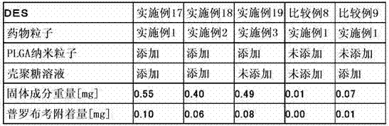
[表3] [table 3]

由表1可确认,当将药物粒子及PLGA纳米粒子电沉积涂覆于支架主体以制造DES时,在涂覆液中添加有PLGA纳米粒子和壳聚糖的实施例11、15的DES中,普罗布考在支架主体上的附着量增加。另外可确认,在使用药物粒子形成时经强烈正电荷修饰的实施例3的药物粒子的实施例16的DES中,即便在涂覆液制备时未添加壳聚糖,普罗布考在支架主体上的附着量也增加。特别是使用经球磨机微细化至一次粒径为2.9μm左右的实施例1的药物粒子的实施例11的DES中,附着了0.14mg的普罗布考,与未对药物粒子进行粉碎的实施例15(附着量0.08mg)相比,普罗布考附着量约为1.8倍。 It can be confirmed from Table 1 that when the drug particles and PLGA nanoparticles are electrodeposited and coated on the stent main body to manufacture DES, in the DES of Examples 11 and 15 in which PLGA nanoparticles and chitosan are added to the coating solution, Increased attachment of probucol to the body of the stent. In addition, it was confirmed that in the DES of Example 16 using the drug particles of Example 3 modified with a strong positive charge when the drug particles were formed, probucol remained on the stent main body even if chitosan was not added when the coating solution was prepared. The amount of adhesion also increased. In particular, in the DES of Example 11, in which the drug particles of Example 1, which were micronized by a ball mill to a primary particle size of about 2.9 μm, 0.14 mg of probucol was attached, the difference was compared with that of Example 15 in which the drug particles were not pulverized. (Adhesion amount 0.08mg), compared with that of probucol adhesion amount is about 1.8 times.
与此相对,在涂覆液中未添加PLGA纳米粒子和壳聚糖的比较例6的DES中,支架主体上基本未附着固体成分。另外,在仅添加壳聚糖的比较例7中,普罗布考的附着量为0.01mg、多于比较例6,但粒子层不均一、附着量远远少于实施例11、15。 On the other hand, in the DES of Comparative Example 6 in which PLGA nanoparticles and chitosan were not added to the coating liquid, almost no solid matter adhered to the stent main body. In addition, in Comparative Example 7 where only chitosan was added, the adhesion amount of probucol was 0.01 mg, which was more than that of Comparative Example 6, but the particle layer was not uniform, and the adhesion amount was far less than that of Examples 11 and 15.
另外,由表2可确认,在将药物粒子及PLGA纳米粒子浸渍涂覆于支架主体以制造DES时,与通过电沉积涂覆制造的实施例11、15、16的DES相比,虽然附着量少了一些,但在涂覆液中添加有PLGA纳米粒子和壳聚糖的实施例17、18的DES中,与未添加PLGA纳米粒子和壳聚糖的比较例8的DES、仅添加壳聚糖的比较例9的DES相比,普罗布考发生附着。另外确认到,在使用药物粒子形成时经强烈正电荷修饰的实施例3的药物粒子的实施例19的DES中,即便在涂覆液制备时未添加壳聚糖,普罗布考在支架主体上的附着量也增加。 In addition, it can be confirmed from Table 2 that when DES is manufactured by dip-coating drug particles and PLGA nanoparticles on the stent main body, compared with the DES of Examples 11, 15, and 16 manufactured by electrodeposition coating, although the adhesion amount A little less, but in the DES of Examples 17 and 18 in which PLGA nanoparticles and chitosan were added to the coating solution, compared with the DES of Comparative Example 8 without PLGA nanoparticles and chitosan, and only chitosan Compared with the DES of Comparative Example 9 of sugar, probucol adhered. In addition, it was confirmed that in the DES of Example 19 using the drug particles of Example 3 that were strongly positively charged when the drug particles were formed, probucol remained on the stent main body even if chitosan was not added when the coating solution was prepared. The amount of adhesion also increased.
而且,对于药物粒子微细化的有无所导致的普罗布考附着量的差别(实施例17、18的比较)及PLGA纳米粒子的有无所导致的附着量的差别(比较例8、9的比较)而言,可见与电沉积涂覆时相同的倾向。 Moreover, the difference in the amount of probucol adhesion caused by the presence or absence of micronization of drug particles (comparison of Examples 17 and 18) and the difference in the amount of adhesion caused by the presence or absence of PLGA nanoparticles (comparison of Comparative Examples 8 and 9) In comparison), the same tendency as in electrodeposition coating is seen.
予以说明,上述实施例中对使用难水溶性药物普罗布考的情况进行了调查,但对于使用其它难水溶性药物的情况也可期待相同的效果。 In addition, in the said Example, the case where the poorly water-soluble drug probucol was used was investigated, but the same effect can be expected also when using other poorly water-soluble drug.
接着,将改变药物粒子、PLGA纳米粒子和壳聚糖的混合比例进行电沉积涂覆而制造的DES中的普罗布考附着量的结果总结于表3。 Next, Table 3 summarizes the results of the probucol adhesion amount in DES produced by electrodeposition coating with varying mixing ratios of drug particles, PLGA nanoparticles, and chitosan.
随着相对于药物的PLGA纳米粒子的比率增加,层叠于支架主体的固体成分重量增加,但普罗布考的组成比降低,因而普罗布考附着量减少。 As the ratio of PLGA nanoparticles to the drug increases, the weight of the solid component laminated on the stent main body increases, but the composition ratio of probucol decreases, so the amount of probucol adhered decreases.
随着PLGA纳米粒子的比率的增加,形成于支架主体的粒子层变得难以剥离。认为这是由于随着PLGA纳米粒子的比率的增加,粒子间的缝隙减小、形成了牢固的粒子层。 As the ratio of PLGA nanoparticles increases, the particle layer formed on the main body of the stent becomes difficult to peel off. This is considered to be because, as the ratio of the PLGA nanoparticles increases, the gaps between the particles decrease and a firm particle layer is formed.
实施例 20 (西洛他唑粒子悬浊液的制备) 将西洛他唑0.75g溶解在作为良溶剂的丙酮108mL、乙醇60mL的混合液中制备溶液。另外,在2重量%的聚乙烯醇(ゴーセノールEG05、日本合成化学工业制)水溶液300mL中混合2重量%的壳聚糖(CHITOSAN GH-400EF、日油制)水溶液45g,制备相对于西洛他唑为不良溶剂的溶液。在该溶液中在40℃、400rpm搅拌下,以一定速度(20mL/分)滴加之前的西洛他唑溶液,通过所谓的良溶剂向不良溶剂中的扩散作用,获得结晶性药物粒子(西洛他唑)的悬浊液。 Example 20 (Preparation of cilostazol particle suspension) 0.75 g of cilostazol was dissolved in a mixture of 108 mL of acetone and 60 mL of ethanol as a good solvent to prepare a solution. In addition, 45 g of an aqueous solution of 2 wt % chitosan (CHITOSAN GH-400EF, manufactured by NOF) was mixed with 300 mL of an aqueous solution of 2 wt % polyvinyl alcohol (Gosenol EG05, manufactured by Nippon Synthetic Chemical Industry Co., Ltd.). azole as a solution in a poor solvent. In this solution, under stirring at 40°C and 400rpm, the previous cilostazol solution was added dropwise at a certain speed (20mL/min). Through the diffusion of the so-called good solvent into the poor solvent, crystalline drug particles (West lostazol) suspension.
接着,在减压下将丙酮、乙醇蒸馏除去。通过电泳法(Malvern Instruments制、ZETASIZER Nano-Z)测定药物粒子表面的ξ电位。结果,药物粒子的ξ电位为+7.4mV,在水中的分散性良好。 Next, acetone and ethanol were distilled off under reduced pressure. The zeta potential on the drug particle surface was measured by electrophoresis (Malvern Instruments, ZETASIZER Nano-Z). As a result, the zeta potential of the drug particles was +7.4 mV, and the dispersibility in water was good.
之后,利用离心分离操作(20,000rpm、40分)将剩余的聚乙烯醇、壳聚糖除去。用水将沉淀的药物粒子再分散,再次重复离心分离操作。 Thereafter, excess polyvinyl alcohol and chitosan were removed by centrifugation (20,000 rpm, 40 minutes). The precipitated drug particles were redispersed with water, and the centrifugation operation was repeated again.
将所得的悬浊液35g和氧化锆球(φ1mm)130g放入100mL的圆筒形的聚乙烯制容器中进行密封,使其在台式球磨机(V-2M、入江商会制)的水平排列的2根辊上旋转(600rpm、2小时),粉碎药物粒子。之后,对内容物进行过滤器过滤,除去氧化锆球,获得0.45重量%的药物粒子悬浊液。 35 g of the obtained suspension and 130 g of zirconia balls (φ1mm) were placed in a 100 mL cylindrical container made of polyethylene, sealed, and placed in 2 horizontally arranged ball mills (V-2M, manufactured by Irie Shokai). Rotate (600rpm, 2 hours) on the root roller to pulverize the drug particles. Thereafter, the contents were filtered through a filter to remove the zirconia balls to obtain a 0.45% by weight drug particle suspension.
液中的药物粒子的粒度利用激光衍射散射法(日机装制、MICROTRAC MT3300)测定。另外,药物粒子中的西洛他唑含有率用高效液相色谱(岛津制作所制、检测器SPD-20A,UV=254nm)进行定量。 The particle size of the drug particles in the liquid was measured by a laser diffraction scattering method (Nikkiso, MICROTRAC MT3300). In addition, the content of cilostazol in the drug particles was quantified by high performance liquid chromatography (manufactured by Shimadzu Corporation, detector SPD-20A, UV=254 nm).
利用激光衍射散射法的粒度分布份测定中,药物粒子的平均粒径为4.2μm。这是由于利用激光衍射散射法的粒度测定中,凝集粒子也作为一个粒子被测定。予以说明,在以下的实施例・比较例中,只要没有特别叙述,则将利用激光衍射散射法测定的平均粒径作为药物粒子的平均粒径。 In the particle size distribution measurement by the laser diffraction scattering method, the average particle diameter of the drug particles was 4.2 μm. This is because aggregated particles are also measured as one particle in the particle size measurement by the laser diffraction scattering method. In addition, in the following examples and comparative examples, unless otherwise stated, the average particle diameter measured by the laser diffraction scattering method was used as the average particle diameter of the drug particle.
另外,药物粒子中的西洛他唑含有率为98.6%、合格率为82%。 In addition, the content rate of cilostazol in the drug particles was 98.6%, and the pass rate was 82%.
实施例Example 21twenty one (含西洛他唑的生物相容性纳米粒子的制备)(Preparation of biocompatible nanoparticles containing cilostazol)
将作为生物相容性高分子的乳酸・乙醇酸共聚物(和光纯药工业制、PLGA7520、分子量20,000、乳酸/乙醇酸摩尔比=75/25)2g和西洛他唑10mg溶解于作为良溶剂的丙酮80mL中,添加乙醇40mL进行混合,制备溶液。另外,在相对于之前的乳酸・乙醇酸共聚物为不良溶剂的0.5重量%的聚乙烯醇水溶液200mL中,混合2重量%的壳聚糖(CHITOSAN GH-400EF、日油制)水溶液4.8g,制备溶液。向该溶液中在40℃、400rpm搅拌下,以一定速度(20mL/分)滴加之前的乳酸・乙醇酸共聚物溶液,获得作为生物相容性纳米粒子的含西洛他唑的PLGA纳米粒子的悬浊液。 Dissolve 2 g of biocompatible lactic acid-glycolic acid copolymer (manufactured by Wako Pure Chemical Industries, PLGA7520, molecular weight 20,000, lactic acid/glycolic acid molar ratio = 75/25) and 10 mg of cilostazol in a good solvent To 80 mL of acetone, 40 mL of ethanol was added and mixed to prepare a solution. In addition, 2 wt% chitosan (CHITOSAN 4.8 g of an aqueous solution of GH-400EF, manufactured by NOF) was prepared to prepare a solution. To this solution, under stirring at 40°C and 400rpm, the previous lactic acid-glycolic acid copolymer solution was added dropwise at a constant rate (20mL/min) to obtain cilostazol-containing PLGA nanoparticles as biocompatible nanoparticles of the suspension.
接着,在减压下将丙酮、乙醇蒸馏除去。利用电泳法测定粒子表面的ξ电位。结果,ξ电位为+46.7mV。 Next, acetone and ethanol were distilled off under reduced pressure. The zeta potential of the particle surface was measured by electrophoresis. As a result, the ξ potential was +46.7 mV.
之后,进行冷冻干燥,获得PLGA纳米粒子粉末。PLGA纳米粒子的粒度分布利用动态光散射法测定。PLGA纳米粒子中的西洛他唑含有率利用高效液相色谱(岛津制作所制、检测器SPD-20A,UV=254nm)进行定量。结果,利用动态光散射法测定的平均粒径为0.30μm,生物相容性纳米粒子中的西洛他唑含有率为0.33%。 Thereafter, freeze-drying was performed to obtain PLGA nanoparticle powder. The particle size distribution of PLGA nanoparticles was determined by dynamic light scattering. The content of cilostazol in the PLGA nanoparticles was quantified by high performance liquid chromatography (manufactured by Shimadzu Corporation, detector SPD-20A, UV=254 nm). As a result, the average particle diameter measured by the dynamic light scattering method was 0.30 μm, and the content of cilostazol in the biocompatible nanoparticles was 0.33%.
实施例Example 22twenty two (涂覆液的制备(Preparation of coating solution 99 ))
一边用搅拌器搅拌实施例20中制备的药物粒子悬浊液,一边添加实施例21中制备的含西洛他唑的PLGA纳米粒子并使其分散,然后搅拌30分钟,制备涂覆液。使药物粒子∶PLGA纳米粒子的混合比例以重量比计为1∶2。 While stirring the drug particle suspension prepared in Example 20 with a stirrer, the cilostazol-containing PLGA nanoparticles prepared in Example 21 were added and dispersed, followed by stirring for 30 minutes to prepare a coating solution. The mixing ratio of drug particles:PLGA nanoparticles was 1:2 by weight.
实施例Example 23twenty three (涂覆液的制备(Preparation of coating solution 1010 ))
除了使药物粒子∶PLGA纳米粒子的混合比例以重量比计为1∶4.2之外,在与实施例22相同的条件下制备涂覆液。 A coating liquid was prepared under the same conditions as in Example 22 except that the mixing ratio of drug particles:PLGA nanoparticles was 1:4.2 by weight.
比较例comparative example 1010 (涂覆液的制备(Preparation of coating solution 1111 ))
不添加实施例21中制备的含西洛他唑的PLGA纳米粒子,用搅拌器对实施例20所制备的药物粒子悬浊液进行搅拌30分钟,制备涂覆液。 Without adding the cilostazol-containing PLGA nanoparticles prepared in Example 21, the drug particle suspension prepared in Example 20 was stirred with a stirrer for 30 minutes to prepare a coating solution.
比较例comparative example 1111 (涂覆液的制备(Preparation of coating solution 1212 ))
不添加实施例20中制备的药物粒子,在实施例21所制备的含西洛他唑的PLGA纳米粒子中按照达到与实施例22及23相同的浓度的方式添加纯水,用搅拌器搅拌30分钟,制备涂覆液。 Without adding the drug particles prepared in Example 20, pure water was added to the cilostazol-containing PLGA nanoparticles prepared in Example 21 in such a way as to reach the same concentration as in Examples 22 and 23, and stirred with a stirrer for 30 minutes to prepare the coating solution.
实施例Example 24twenty four (支架主体上的电沉积涂覆(Electrodeposition coating on stent body 99 ))
以预先测定过重量的外径2.3mm、长16mm的不锈钢(SUS316L)制支架主体(n=3)为负极;直径20mm的圆形SUS板为正极的方式连接外部电源,在实施例22所制备的涂覆液中均浸渍长度10mm。以电压10V、电流1mA的条件通电1分钟后,取下支架主体进行风干,制作DES。测定干燥后的DES的重量,由重量的增加部分求得层叠于支架主体的固体成分重量的平均值。 The main body (n=3) made of stainless steel (SUS316L) with a previously measured weight of 2.3 mm in diameter and 16 mm in length is used as the negative electrode; the circular SUS plate with a diameter of 20 mm is used as the positive electrode to connect the external power supply, prepared in Example 22 The average dipping length in the coating liquid is 10mm. After energizing with a voltage of 10V and a current of 1mA for 1 minute, the main body of the bracket was removed and air-dried to prepare a DES. The weight of the DES after drying was measured, and the average value of the weight of the solids laminated on the stent main body was obtained from the weight increase.
实施例Example 2525 (支架主体上的电沉积涂覆(Electrodeposition coating on stent body 1010 ))
除了使用实施例23所制备的涂覆液之外,在与实施例24相同的条件下将粒子层叠于支架主体(n=3),制作DES。测定干燥后的DES的重量,由重量的增加部分求得层叠于支架主体的固体成分重量的平均值。 Except for using the coating solution prepared in Example 23, the particles were laminated on the stent main body (n=3) under the same conditions as in Example 24 to fabricate DES. The weight of the DES after drying was measured, and the average value of the weight of the solid components laminated on the stent main body was obtained from the weight increase.
比较例comparative example 1212 (支架主体上的电沉积涂覆(Electrodeposition coating on stent body 1111 ))
除了使用比较例10所制备的涂覆液之外,在与实施例24相同的条件下将粒子层叠于支架主体(n=3),制作DES。测定干燥后的DES的重量,由重量的增加部分求得层叠于支架主体的固体成分重量的平均值。 Except for using the coating solution prepared in Comparative Example 10, the particles were laminated on the stent main body (n=3) under the same conditions as in Example 24 to fabricate DES. The weight of the DES after drying was measured, and the average value of the weight of the solid components laminated on the stent main body was obtained from the weight increase.
比较例comparative example 1313 (支架主体上的电沉积涂覆(Electrodeposition coating on stent body 1212 ))
除了使用比较例11所制备的涂覆液之外,在与实施例24相同的条件下将粒子层叠于支架主体(n=3),制作DES。测定干燥后的DES的重量,由重量的增加部分求得层叠于支架主体的固体成分重量的平均值。 Except for using the coating solution prepared in Comparative Example 11, the particles were laminated on the stent main body (n=3) under the same conditions as in Example 24 to fabricate DES. The weight of the DES after drying was measured, and the average value of the weight of the solid components laminated on the stent main body was obtained from the weight increase.
试验例Test case 22 (附着于支架主体的西洛他唑量的定量)(Quantification of the amount of cilostazol attached to the main body of the stent)
利用高效液相色谱对层叠于实施例24、25及比较例12、13所制作的DES上的固体成分中的西洛他唑量进行定量(各组n=3),计算西洛他唑的平均附着量。将结果与层叠于支架主体的固体成分重量的平均值、固体成分中的西洛他唑比例一并示于表4。 The amount of cilostazol in the solid content layered on the DES prepared in Examples 24, 25 and Comparative Examples 12 and 13 was quantified by high performance liquid chromatography (n=3 for each group), and the amount of cilostazol was calculated. Average adhesion. The results are shown in Table 4 together with the average weight of the solid content laminated on the stent main body and the ratio of cilostazol in the solid content.
[表4] [Table 4]
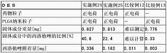
由表4可确认,将药物粒子及PLGA纳米粒子电沉积涂覆于支架主体以制造DES时,在涂覆液中添加有PLGA纳米粒子的实施例24、25的DES中,西洛他唑在支架主体上的附着量增加。 It can be confirmed from Table 4 that when the drug particles and PLGA nanoparticles are electrodeposited and coated on the stent main body to manufacture DES, in the DES of Examples 24 and 25 in which PLGA nanoparticles are added to the coating solution, cilostazol is The amount of attachment on the body of the stent is increased.
而在涂覆液中未添加PLGA纳米粒子的比较例12的DES中,虽然固体成分附着于支架主体,但粒子层不均一。 On the other hand, in the DES of Comparative Example 12 in which PLGA nanoparticles were not added to the coating liquid, although the solid content adhered to the stent main body, the particle layer was not uniform.
另外,在涂覆液中未添加药物粒子的比较例4的DES中,固体成分量多于比较例12,但由于PLGA纳米粒子中的西洛他唑含有率低,因而支架上的西洛他唑附着量为0.01mg以下,远远少于实施例24、25。 In addition, in the DES of Comparative Example 4 in which no drug particles were added to the coating liquid, the amount of solid content was larger than that of Comparative Example 12, but the content of cilostazol in the PLGA nanoparticles was low, so the amount of cilostazol on the stent The amount of azole adhesion was less than 0.01 mg, far less than that of Examples 24 and 25.
试验例Test case 33 (使用附着有药物的支架的体内试验)(in vivo tests using drug-attached stents)
将上述附着有西洛他唑的实施例24的支架适用于迷你猪,对其所适用的血管的伤害进行确认。 The above-mentioned stent of Example 24 to which cilostazol was attached was applied to minipigs, and damage to blood vessels to which it was applied was confirmed.
动物种animal species
迷你猪(系统:NIBS、日生研株式会社) Mini pig (system: NIBS, Nisseiken Co., Ltd.)
雄8只、雌4只 8 males, 4 females
月龄:10~16个月(获得时) Age in months: 10 to 16 months (at the time of acquisition)
体重:20~28kg(获得时) Weight: 20-28kg (at the time of acquisition)
试验组test group
附着有西洛他唑的支架组:n=12 Stent group with cilostazol attached: n=12
(支架系统使用实施例24的支架。) (The stent system used the stent of Example 24.)
对照组:n=12 Control group: n=12
(支架系统使用未实施涂覆的相同支架。) (Stent system uses the same stent without coating.)
操作方法operation method
利用盐酸氯胺酮(500~750mg/迷你猪、肌注、ケタラール50、三共エール药品(有限公司))进行麻醉。插入导丝(ラジフォーカスガイドワイヤーM、テルモ(有限公司))和导引导管(Heartrail II、AMPLATZ LEFT Short Tip、テルモ(有限公司)),导入至心脏。沿着导丝,插入各试验组的支架系统,挤入至作为留置部位的左前降支或左回旋支。膨胀30秒钟、将支架留置。支架留置后给予硝酸异山梨酯,进行冠状动脉造影。取下导管,对切开部消毒(动物用聚乙烯吡咯酮碘液、明治制果)将肌肉和皮肤缝合。对各迷你猪,分别埋入附着有西洛他唑的支架和对照支架各1个。 Anesthesia was performed with ketamine hydrochloride (500 to 750 mg/minipig, intramuscular injection, Ketara 50, Sankyo El Pharmaceutical Co., Ltd.). A guide wire (ラジフォーカスガイドワイヤー M, テルモ(Co., Ltd.)) and a guide catheter (Heartrail II, AMPLATZ LEFT Short Tip, テルモ(Co., Ltd.)) were inserted into the heart. Along the guide wire, insert the stent system of each test group, squeeze into the left anterior descending branch or left circumflex branch as the indwelling site. Inflate for 30 seconds and leave the stent in place. Isosorbide dinitrate was given after stent placement, and coronary angiography was performed. Remove the catheter, sterilize the incision (polyvinylpyrrolidone-iodine solution for animals, Meiji Seika), and suture the muscle and skin. One cilostazol-attached stent and one control stent were embedded in each minipig.
血管造影Angiography
对支架留置部位的血管进行造影。将从大腿动脉插入的导管的前端引至冠状动脉开口部,从导管注入造影剂,对支架留置部的血管造影照片进行拍摄。拍摄在支架留置前、支架留置后和解剖时共计进行3次。 Angiography was performed on the blood vessels at the stent placement site. The distal end of the catheter inserted from the femoral artery is guided to the opening of the coronary artery, a contrast medium is injected through the catheter, and angiographic photographs of the stent-dwelling part are taken. The photographs were taken 3 times in total before stent placement, after stent placement, and during dissection.
并用药、给药量、给药方法和给药期间Concomitant medication, dosage, method of administration and period of administration
阿司匹林 100mg 1次/日(混合饲料给药)
给药期间:从手术2日前至实验期间中 Administration period: from 2 days before the operation to the middle of the experiment period
硫酸氯吡格雷 50mg 1次/日(混合饲料给药)
给药期间:从手术2日前至包含手术日的术后2周 Administration period: from 2 days before surgery to 2 weeks after surgery including the day of surgery
抗生素 100mg/kg 1次/日(肌肉内给药)
Antibiotics 100mg/
给药期间:包含手术日的术后3天 Administration period: 3 days after surgery including the surgery day
◎由于预测到由于施行手术会发生痉挛、心律失常、血压过低等,因而准备硝酸异山梨酯(硝酸异山梨酯)、利多卡因(赛罗卡因)、去甲肾上腺素、硫酸阿托品等。 ◎Isosorbide dinitrate (isosorbide dinitrate), lidocaine (xirocaine), norepinephrine, atropine sulfate, etc. .
留置有支架的血管的摘出Extraction of blood vessels with stents
在支架留置28日后,与留置时同样,在麻醉下将迷你猪腹股沟部切开,插入血管造影用导管,进行冠状动脉确认造影。造影结束后,将动物放血处死,采集留置有支架的冠状动脉。摘出的血管用10%中性缓冲福尔马林固定,送至株式会社组织科学研究所。对支架的起始部侧约1.5mm、中央部和末梢侧约1.5mm的3个位置制作支架标本,进行HE染色。 Twenty-eight days after the indwelling of the stent, the inguinal part of the minipig was incised under anesthesia in the same manner as during the indwelling, a catheter for angiography was inserted, and coronary angiography was performed for confirmation. After the angiography, the animals were sacrificed by bleeding, and the coronary arteries with indwelling stents were collected. The extracted blood vessels were fixed with 10% neutral buffered formalin and sent to the Institute of Tissue Science Co., Ltd. Stent specimens were prepared at three positions of about 1.5 mm from the initial side of the stent, and about 1.5 mm from the central and distal sides of the stent, and HE staining was performed.
结果result
由血管造影照片,测量支架留置前、支架留置后和解剖时的最小血管直径,将其变化结果示于图7。附着有西洛他唑的支架组与对照组相比,显著地抑制了血管直径的收缩。 From the angiographic photographs, the minimum vessel diameter was measured before stent placement, after stent placement, and during dissection, and the changes are shown in FIG. 7 . The stent group attached with cilostazol significantly suppressed the contraction of the vessel diameter compared with the control group.
另外,将在支架留置28日后摘出的血管的截面进行了HE染色的病理组织标本的对照组示于图8、附着有西洛他唑的支架组示于图9。图8和图9所示的结果从上开始按照支架的起始部侧、中央部和末梢侧的顺序,左右纵列地显示由任意2只迷你猪获得的标本。附着有西洛他唑的支架组与对照组相比,抑制了血管内部的狭窄。 In addition, the control group of pathological tissue samples stained with HE in the section of the blood vessel extracted 28 days after stent indwelling is shown in FIG. 8 , and the cilostazol-attached stent group is shown in FIG. 9 . The results shown in FIGS. 8 and 9 show specimens obtained from any two minipigs in a left-right column in the order of the starting part side, the central part, and the distal side of the scaffold from the top. The stent group attached with cilostazol suppressed narrowing inside the blood vessel compared with the control group.
产业实用性Industrial applicability
本发明的管腔内留置用医疗设备由于以表面经正电荷修饰的药物粒子和生物相容性纳米粒子混存的状态层叠于设备主体,因此粒子层变得均一、且能够均一地涂覆足够量的药物粒子。另外,将医疗设备留置于生物体内后,由于药物粒子在短时间内洗脱,因而可期待药物的即效作用,同时通过正电荷修饰使得在生物体内洗脱的药物粒子的细胞粘附性高、向细胞内的移动性也提高。 The medical device for indwelling in the lumen of the present invention is laminated on the main body of the device in a state where positively charged surface-modified drug particles and biocompatible nanoparticles are mixed, so the particle layer becomes uniform and can be coated uniformly enough. amount of drug particles. In addition, after the medical device is placed in the living body, since the drug particles are eluted in a short time, the immediate effect of the drug can be expected, and the cell adhesion of the drug particles eluted in the living body is high by positive charge modification. , Mobility into cells is also improved.
另外,通过使表面经正电荷修饰的药物粒子和生物相容性纳米粒子附着于设备主体的本发明的管腔内留置用医疗设备的制造方法,可以简单且低成本地制造在设备主体的表面高效且均一地附着有以往难以附着于设备主体的难水溶性药物的管腔内留置用医疗设备。作为粒子附着工序,优选使用电泳法、超声波雾化法、喷雾法、气刷法、抹拭法或浸渍法。 In addition, by the method of manufacturing the medical device for intraluminal indwelling of the present invention, in which positively-charged surface-modified drug particles and biocompatible nanoparticles are attached to the device body, it is possible to easily and cost-effectively manufacture on the surface of the device body A medical device for intraluminal indwelling that efficiently and uniformly adheres poorly water-soluble drugs that have traditionally been difficult to adhere to the device body. As the particle attaching step, it is preferable to use an electrophoresis method, an ultrasonic atomization method, a spray method, an air brush method, a wiping method, or a dipping method.
Claims (25)
Applications Claiming Priority (3)
| Application Number | Priority Date | Filing Date | Title |
|---|---|---|---|
| JP2009195350 | 2009-08-26 | ||
| JP2009-195350 | 2009-08-26 | ||
| PCT/JP2010/064330 WO2011024831A1 (en) | 2009-08-26 | 2010-08-25 | Medical device for placement into a lumen and manufacturing method thereof |
Publications (1)
| Publication Number | Publication Date |
|---|---|
| CN102711674A true CN102711674A (en) | 2012-10-03 |
Family
ID=43627931
Family Applications (1)
| Application Number | Title | Priority Date | Filing Date |
|---|---|---|---|
| CN2010800484372A Pending CN102711674A (en) | 2009-08-26 | 2010-08-25 | Medical device for placement into a lumen and manufacturing method thereof |
Country Status (11)
| Country | Link |
|---|---|
| US (1) | US20120213838A1 (en) |
| EP (1) | EP2471497A4 (en) |
| JP (1) | JP5693456B2 (en) |
| KR (1) | KR20120068883A (en) |
| CN (1) | CN102711674A (en) |
| BR (1) | BR112012004148A2 (en) |
| PH (1) | PH12012500393A1 (en) |
| RU (1) | RU2012111217A (en) |
| SG (2) | SG178577A1 (en) |
| TW (1) | TW201111008A (en) |
| WO (1) | WO2011024831A1 (en) |
Cited By (3)
| Publication number | Priority date | Publication date | Assignee | Title |
|---|---|---|---|---|
| CN105392363A (en) * | 2013-03-01 | 2016-03-09 | A·S·戈尔兹伯勒 | Sample Fixation and Stabilization |
| CN107106309A (en) * | 2014-10-28 | 2017-08-29 | 株式会社Jimro | drug eluting stent |
| CN109183131A (en) * | 2018-07-16 | 2019-01-11 | 东南大学 | A kind of SiO2The preparation method of the compound super-hydrophobic metal surface of base |
Families Citing this family (20)
| Publication number | Priority date | Publication date | Assignee | Title |
|---|---|---|---|---|
| US10058634B2 (en) | 2009-04-28 | 2018-08-28 | Surmodics, Inc. | Devices and methods for delivery of bioactive agents |
| US9492652B2 (en) * | 2010-03-02 | 2016-11-15 | University Of Rochester | Implantable devices that generate low intensity electric fields for the treatment of atherosclerotic disease and prevention of ischemic vascular events and methods of manufacture |
| US9861727B2 (en) | 2011-05-20 | 2018-01-09 | Surmodics, Inc. | Delivery of hydrophobic active agent particles |
| US10213529B2 (en) | 2011-05-20 | 2019-02-26 | Surmodics, Inc. | Delivery of coated hydrophobic active agent particles |
| US9731017B2 (en) | 2012-09-13 | 2017-08-15 | W. L. Gore & Associates, Inc. | Polytetrafluoroethylene co-polymer emulsions |
| JP6068921B2 (en) * | 2012-10-19 | 2017-01-25 | ホソカワミクロン株式会社 | Manufacturing method of drug eluting device |
| CA2890205C (en) | 2012-11-05 | 2020-12-22 | Surmodics, Inc. | Composition and method for delivery of hydrophobic active agents |
| US11246963B2 (en) | 2012-11-05 | 2022-02-15 | Surmodics, Inc. | Compositions and methods for delivery of hydrophobic active agents |
| MX2015015589A (en) * | 2013-05-16 | 2016-07-05 | Surmodics Inc | Compositions and methods for delivery of hydrophobic active agents. |
| TWI697337B (en) | 2013-08-07 | 2020-07-01 | 學校法人近畿大學 | Process for produing nano particle or nano-particle composition, and process for manufacturing stent or balloon catheter |
| JP6511399B2 (en) * | 2013-10-31 | 2019-05-15 | 株式会社Efr | Needle and method of manufacturing the same |
| WO2015063910A1 (en) * | 2013-10-31 | 2015-05-07 | 株式会社Efr | Needle and method for manufacturing same |
| KR20150130049A (en) * | 2014-05-13 | 2015-11-23 | 주식회사 엠아이텍 | Biodegradable polymer coated vascular stent for drug eluting |
| US9821090B2 (en) * | 2014-09-30 | 2017-11-21 | The Spectranetics Corporation | Electrodeposition coating for medical devices |
| JP6956009B2 (en) * | 2014-12-29 | 2021-10-27 | ボストン サイエンティフィック サイムド,インコーポレイテッドBoston Scientific Scimed,Inc. | Compositions, Devices, and Methods for Multi-Step Release of Chemotherapeutic Agents |
| CN105797220B (en) * | 2014-12-31 | 2020-07-31 | 先健科技(深圳)有限公司 | Degradable iron-based alloy stent |
| US20180085498A1 (en) * | 2016-09-23 | 2018-03-29 | Micell Technologies, Inc. | Prolonged drug-eluting products |
| US10898446B2 (en) | 2016-12-20 | 2021-01-26 | Surmodics, Inc. | Delivery of hydrophobic active agents from hydrophilic polyether block amide copolymer surfaces |
| US12023265B2 (en) * | 2019-01-16 | 2024-07-02 | Boston Scientific Scimed, Inc. | Stent including anti-migration capabilities |
| US12226552B2 (en) | 2019-09-30 | 2025-02-18 | Surmodics, Inc. | Active agent depots formed in situ |
Citations (5)
| Publication number | Priority date | Publication date | Assignee | Title |
|---|---|---|---|---|
| CN1355005A (en) * | 2001-12-13 | 2002-06-26 | 华东理工大学 | Drug-eluting cardiovascular stent and preparation method thereof |
| US20040249450A1 (en) * | 2003-06-05 | 2004-12-09 | Terumo Kabushiki Kaisha | Stent and method of manufacturing stent |
| JP2007215620A (en) * | 2006-02-15 | 2007-08-30 | Hosokawa Funtai Gijutsu Kenkyusho:Kk | Drug eluting stent and manufacturing method thereof |
| CN101170965A (en) * | 2005-03-09 | 2008-04-30 | 田纳西大学研究基金会 | Barrier brackets and their uses |
| WO2008140482A2 (en) * | 2006-11-09 | 2008-11-20 | Boston Scientific Scimed, Inc. | Medical devices having coatings for controlled therapeutic agent delivery |
Family Cites Families (24)
| Publication number | Priority date | Publication date | Assignee | Title |
|---|---|---|---|---|
| US20030083733A1 (en) * | 1997-10-10 | 2003-05-01 | Neorx Corporation | Therapeutic inhibitor of vascular smooth muscle cells |
| US8057816B2 (en) | 1997-09-26 | 2011-11-15 | Abbott Laboratories | Compositions and methods of administering paclitaxel with other drugs using medical devices |
| US8394398B2 (en) | 1997-09-26 | 2013-03-12 | Abbott Laboratories | Methods of administering rapamycin analogs with anti-inflammatories using medical devices |
| JP4473390B2 (en) | 2000-01-07 | 2010-06-02 | 川澄化学工業株式会社 | Stent and stent graft |
| JP2004523275A (en) | 2000-12-22 | 2004-08-05 | アバンテク バスキュラー コーポレーション | Delivery of therapeutic drugs |
| US20040142013A1 (en) * | 2001-07-13 | 2004-07-22 | Flow Focusing, Inc. | Implantable orthopedic surgical devices with controlled release antimicrobial component |
| JP4347044B2 (en) | 2001-07-26 | 2009-10-21 | アバンテク バスキュラー コーポレーション | Device for delivering a therapeutic agent having a variable release profile |
| JP4245302B2 (en) | 2002-04-05 | 2009-03-25 | 川澄化学工業株式会社 | Stent |
| JP2005531391A (en) | 2002-06-27 | 2005-10-20 | 微創医療器械(上海)有限公司 | Drug release stent |
| DE10244847A1 (en) | 2002-09-20 | 2004-04-01 | Ulrich Prof. Dr. Speck | Medical device for drug delivery |
| CA2528032A1 (en) | 2003-06-03 | 2004-12-16 | Beth Israel Deaconess Medical Center | Methods and compounds for the treatment of vascular stenosis |
| EP1753476B1 (en) | 2004-03-10 | 2015-07-29 | OrbusNeich Medical, Inc. | Progenitor endothelial cell capturing with a drug eluting implantable medical device |
| WO2005089855A1 (en) | 2004-03-19 | 2005-09-29 | Abbott Laboratories | Multiple drug delivery from a balloon and a prosthesis |
| US20050220853A1 (en) * | 2004-04-02 | 2005-10-06 | Kinh-Luan Dao | Controlled delivery of therapeutic agents from medical articles |
| AU2005259735A1 (en) | 2004-07-07 | 2006-01-12 | Medicure International Inc. | Combination therapies employing platelet aggregation drugs |
| US8119153B2 (en) * | 2004-08-26 | 2012-02-21 | Boston Scientific Scimed, Inc. | Stents with drug eluting coatings |
| KR100511618B1 (en) | 2005-01-17 | 2005-08-31 | 이경범 | Multi-layer coating of drug release controllable coronary stent and method for manufacturing the same |
| EP1865945A4 (en) | 2005-03-11 | 2008-05-21 | Hong Kong Nitric Oxide Ltd | Combination therapy for endothelial dysfunction, angina and diabetes |
| WO2007046935A2 (en) | 2005-10-14 | 2007-04-26 | Abbott Laboratories | Compositions, systems, kits, and methods of administering rapamycin analogs with paclitaxel using medical devices |
| US8784860B2 (en) | 2005-10-27 | 2014-07-22 | Cordis Corporation | Local administration of a combination of rapamycin and cilostazol for the treatment of vascular disease |
| US20070128342A1 (en) * | 2005-12-02 | 2007-06-07 | Stenzel Eric B | Method and system for coating a medical device |
| US20080243113A1 (en) * | 2006-11-08 | 2008-10-02 | Shastri V Prasad | Modification of stent surfaces to impart functionality |
| US8048441B2 (en) * | 2007-06-25 | 2011-11-01 | Abbott Cardiovascular Systems, Inc. | Nanobead releasing medical devices |
| JP4340744B2 (en) * | 2009-03-17 | 2009-10-07 | 株式会社ホソカワ粉体技術研究所 | Drug eluting stent |
-
2010
- 2010-08-25 CN CN2010800484372A patent/CN102711674A/en active Pending
- 2010-08-25 SG SG2012013488A patent/SG178577A1/en unknown
- 2010-08-25 PH PH1/2012/500393A patent/PH12012500393A1/en unknown
- 2010-08-25 RU RU2012111217/15A patent/RU2012111217A/en not_active Application Discontinuation
- 2010-08-25 TW TW099128388A patent/TW201111008A/en unknown
- 2010-08-25 KR KR1020127007520A patent/KR20120068883A/en not_active Withdrawn
- 2010-08-25 EP EP10811880.3A patent/EP2471497A4/en not_active Withdrawn
- 2010-08-25 SG SG10201405144VA patent/SG10201405144VA/en unknown
- 2010-08-25 WO PCT/JP2010/064330 patent/WO2011024831A1/en not_active Ceased
- 2010-08-25 BR BR112012004148A patent/BR112012004148A2/en not_active IP Right Cessation
- 2010-08-25 JP JP2011528812A patent/JP5693456B2/en active Active
- 2010-08-26 US US13/392,028 patent/US20120213838A1/en not_active Abandoned
Patent Citations (5)
| Publication number | Priority date | Publication date | Assignee | Title |
|---|---|---|---|---|
| CN1355005A (en) * | 2001-12-13 | 2002-06-26 | 华东理工大学 | Drug-eluting cardiovascular stent and preparation method thereof |
| US20040249450A1 (en) * | 2003-06-05 | 2004-12-09 | Terumo Kabushiki Kaisha | Stent and method of manufacturing stent |
| CN101170965A (en) * | 2005-03-09 | 2008-04-30 | 田纳西大学研究基金会 | Barrier brackets and their uses |
| JP2007215620A (en) * | 2006-02-15 | 2007-08-30 | Hosokawa Funtai Gijutsu Kenkyusho:Kk | Drug eluting stent and manufacturing method thereof |
| WO2008140482A2 (en) * | 2006-11-09 | 2008-11-20 | Boston Scientific Scimed, Inc. | Medical devices having coatings for controlled therapeutic agent delivery |
Cited By (8)
| Publication number | Priority date | Publication date | Assignee | Title |
|---|---|---|---|---|
| CN105392363A (en) * | 2013-03-01 | 2016-03-09 | A·S·戈尔兹伯勒 | Sample Fixation and Stabilization |
| US9696247B2 (en) | 2013-03-01 | 2017-07-04 | Rnassist Ltd. | Sample fixation and stabilisation |
| CN105392363B (en) * | 2013-03-01 | 2017-09-29 | A·S·戈尔兹伯勒 | Sample Fixation and Stabilization |
| US10393633B2 (en) | 2013-03-01 | 2019-08-27 | Rnassist Ltd. | Sample fixation and stabilisation |
| US10539488B2 (en) | 2013-03-01 | 2020-01-21 | Rnassist Ltd. | Sample fixation and stabilisation |
| CN107106309A (en) * | 2014-10-28 | 2017-08-29 | 株式会社Jimro | drug eluting stent |
| CN107106309B (en) * | 2014-10-28 | 2019-11-08 | 株式会社Jimro | drug eluting stent |
| CN109183131A (en) * | 2018-07-16 | 2019-01-11 | 东南大学 | A kind of SiO2The preparation method of the compound super-hydrophobic metal surface of base |
Also Published As
| Publication number | Publication date |
|---|---|
| EP2471497A1 (en) | 2012-07-04 |
| KR20120068883A (en) | 2012-06-27 |
| JP5693456B2 (en) | 2015-04-01 |
| WO2011024831A1 (en) | 2011-03-03 |
| BR112012004148A2 (en) | 2017-05-30 |
| RU2012111217A (en) | 2013-10-10 |
| SG10201405144VA (en) | 2014-10-30 |
| US20120213838A1 (en) | 2012-08-23 |
| PH12012500393A1 (en) | 2012-10-22 |
| JPWO2011024831A1 (en) | 2013-01-31 |
| TW201111008A (en) | 2011-04-01 |
| EP2471497A4 (en) | 2013-10-09 |
| SG178577A1 (en) | 2012-04-27 |
Similar Documents
| Publication | Publication Date | Title |
|---|---|---|
| JP5693456B2 (en) | Intraluminal medical device and method for manufacturing the same | |
| JP4297221B2 (en) | Method for producing drug-eluting stent | |
| JP5591103B2 (en) | Drug-eluting catheter and method for producing the same | |
| JP5106415B2 (en) | Nanoparticle emitting medical device | |
| EP2136853B1 (en) | Medical product for treating stenosis of body passages and for preventing threatening restenosis | |
| JP5594534B2 (en) | Bioabsorbable coating with adjustable hydrophobicity | |
| CN101346156B (en) | Drug-eluting stent with biodegradable release layer attached to electrografted primer | |
| JP5500397B2 (en) | Implantable medical device coating with improved mechanical stability | |
| JP6955553B2 (en) | Drug-eluting stents and their use to enable recovery of functional endothelial cell layers | |
| US20140287134A1 (en) | Coating system and method for drug elution management | |
| US9737395B2 (en) | Systems and methods for reducing scarring | |
| DE102008044316A1 (en) | Drug-coated medical device, process for its preparation and its uses | |
| JP2007533409A (en) | Medical article having therapeutic agent containing region formed from coalesced polymer particles | |
| US8945663B2 (en) | Method for biostable inclusion of a biobeneficial agent on an outermost surface of an implantable medical device | |
| JP4340744B2 (en) | Drug eluting stent | |
| US8728150B2 (en) | Medical device loaded with formulation for targeted delivery of biologically active material/s and method of manufacture thereof | |
| JP2016510608A (en) | Implantable medical device comprising a macrocyclic triene lactone drug and a minimum amount of an antioxidant stabilizer and method of manufacture | |
| JP6068921B2 (en) | Manufacturing method of drug eluting device | |
| HK1167587A (en) | Medical device for placement into a lumen and manufacturing method thereof | |
| CN101309706A (en) | Drug release control composition and drug-releasing medical device | |
| HK1146248A (en) | Drug elution-type catheter and method for manufacturing the drug elution-type catheter | |
| HK1126695B (en) | Drug eluting stent with a biodegradable release layer attached with an electro-grafted primer coating |
Legal Events
| Date | Code | Title | Description |
|---|---|---|---|
| C06 | Publication | ||
| PB01 | Publication | ||
| C10 | Entry into substantive examination | ||
| SE01 | Entry into force of request for substantive examination | ||
| C02 | Deemed withdrawal of patent application after publication (patent law 2001) | ||
| WD01 | Invention patent application deemed withdrawn after publication |
Application publication date: 20121003 |