AU2014236582B2 - Methods of inhibiting cataracts and presbyopia - Google Patents
Methods of inhibiting cataracts and presbyopia Download PDFInfo
- Publication number
- AU2014236582B2 AU2014236582B2 AU2014236582A AU2014236582A AU2014236582B2 AU 2014236582 B2 AU2014236582 B2 AU 2014236582B2 AU 2014236582 A AU2014236582 A AU 2014236582A AU 2014236582 A AU2014236582 A AU 2014236582A AU 2014236582 B2 AU2014236582 B2 AU 2014236582B2
- Authority
- AU
- Australia
- Prior art keywords
- crystallin
- poly
- pct
- protein
- ophthalmic composition
- Prior art date
- Legal status (The legal status is an assumption and is not a legal conclusion. Google has not performed a legal analysis and makes no representation as to the accuracy of the status listed.)
- Active
Links
- XFLVBMBRLSCJAI-UHFFFAOYSA-N NC(CCCCC(C1N2)SCC1NC2=O)=O Chemical compound NC(CCCCC(C1N2)SCC1NC2=O)=O XFLVBMBRLSCJAI-UHFFFAOYSA-N 0.000 description 1
Classifications
-
- A—HUMAN NECESSITIES
- A61—MEDICAL OR VETERINARY SCIENCE; HYGIENE
- A61K—PREPARATIONS FOR MEDICAL, DENTAL OR TOILETRY PURPOSES
- A61K31/00—Medicinal preparations containing organic active ingredients
- A61K31/185—Acids; Anhydrides, halides or salts thereof, e.g. sulfur acids, imidic, hydrazonic or hydroximic acids
- A61K31/19—Carboxylic acids, e.g. valproic acid
- A61K31/195—Carboxylic acids, e.g. valproic acid having an amino group
-
- A—HUMAN NECESSITIES
- A61—MEDICAL OR VETERINARY SCIENCE; HYGIENE
- A61K—PREPARATIONS FOR MEDICAL, DENTAL OR TOILETRY PURPOSES
- A61K31/00—Medicinal preparations containing organic active ingredients
- A61K31/185—Acids; Anhydrides, halides or salts thereof, e.g. sulfur acids, imidic, hydrazonic or hydroximic acids
- A61K31/19—Carboxylic acids, e.g. valproic acid
- A61K31/20—Carboxylic acids, e.g. valproic acid having a carboxyl group bound to a chain of seven or more carbon atoms, e.g. stearic, palmitic, arachidic acids
-
- A—HUMAN NECESSITIES
- A61—MEDICAL OR VETERINARY SCIENCE; HYGIENE
- A61K—PREPARATIONS FOR MEDICAL, DENTAL OR TOILETRY PURPOSES
- A61K31/00—Medicinal preparations containing organic active ingredients
- A61K31/33—Heterocyclic compounds
- A61K31/395—Heterocyclic compounds having nitrogen as a ring hetero atom, e.g. guanethidine or rifamycins
- A61K31/40—Heterocyclic compounds having nitrogen as a ring hetero atom, e.g. guanethidine or rifamycins having five-membered rings with one nitrogen as the only ring hetero atom, e.g. sulpiride, succinimide, tolmetin, buflomedil
- A61K31/4015—Heterocyclic compounds having nitrogen as a ring hetero atom, e.g. guanethidine or rifamycins having five-membered rings with one nitrogen as the only ring hetero atom, e.g. sulpiride, succinimide, tolmetin, buflomedil having oxo groups directly attached to the heterocyclic ring, e.g. piracetam, ethosuximide
-
- A—HUMAN NECESSITIES
- A61—MEDICAL OR VETERINARY SCIENCE; HYGIENE
- A61K—PREPARATIONS FOR MEDICAL, DENTAL OR TOILETRY PURPOSES
- A61K31/00—Medicinal preparations containing organic active ingredients
- A61K31/33—Heterocyclic compounds
- A61K31/395—Heterocyclic compounds having nitrogen as a ring hetero atom, e.g. guanethidine or rifamycins
- A61K31/41—Heterocyclic compounds having nitrogen as a ring hetero atom, e.g. guanethidine or rifamycins having five-membered rings with two or more ring hetero atoms, at least one of which being nitrogen, e.g. tetrazole
- A61K31/4164—1,3-Diazoles
- A61K31/4188—1,3-Diazoles condensed with other heterocyclic ring systems, e.g. biotin, sorbinil
-
- A—HUMAN NECESSITIES
- A61—MEDICAL OR VETERINARY SCIENCE; HYGIENE
- A61K—PREPARATIONS FOR MEDICAL, DENTAL OR TOILETRY PURPOSES
- A61K31/00—Medicinal preparations containing organic active ingredients
- A61K31/74—Synthetic polymeric materials
- A61K31/765—Polymers containing oxygen
-
- A—HUMAN NECESSITIES
- A61—MEDICAL OR VETERINARY SCIENCE; HYGIENE
- A61K—PREPARATIONS FOR MEDICAL, DENTAL OR TOILETRY PURPOSES
- A61K9/00—Medicinal preparations characterised by special physical form
- A61K9/0012—Galenical forms characterised by the site of application
- A61K9/0048—Eye, e.g. artificial tears
-
- A—HUMAN NECESSITIES
- A61—MEDICAL OR VETERINARY SCIENCE; HYGIENE
- A61K—PREPARATIONS FOR MEDICAL, DENTAL OR TOILETRY PURPOSES
- A61K9/00—Medicinal preparations characterised by special physical form
- A61K9/06—Ointments; Bases therefor; Other semi-solid forms, e.g. creams, sticks, gels
-
- A—HUMAN NECESSITIES
- A61—MEDICAL OR VETERINARY SCIENCE; HYGIENE
- A61M—DEVICES FOR INTRODUCING MEDIA INTO, OR ONTO, THE BODY; DEVICES FOR TRANSDUCING BODY MEDIA OR FOR TAKING MEDIA FROM THE BODY; DEVICES FOR PRODUCING OR ENDING SLEEP OR STUPOR
- A61M37/00—Other apparatus for introducing media into the body; Percutany, i.e. introducing medicines into the body by diffusion through the skin
- A61M37/0092—Other apparatus for introducing media into the body; Percutany, i.e. introducing medicines into the body by diffusion through the skin using ultrasonic, sonic or infrasonic vibrations, e.g. phonophoresis
-
- A—HUMAN NECESSITIES
- A61—MEDICAL OR VETERINARY SCIENCE; HYGIENE
- A61N—ELECTROTHERAPY; MAGNETOTHERAPY; RADIATION THERAPY; ULTRASOUND THERAPY
- A61N1/00—Electrotherapy; Circuits therefor
- A61N1/18—Applying electric currents by contact electrodes
- A61N1/32—Applying electric currents by contact electrodes alternating or intermittent currents
- A61N1/325—Applying electric currents by contact electrodes alternating or intermittent currents for iontophoresis, i.e. transfer of media in ionic state by an electromotoric force into the body
-
- A—HUMAN NECESSITIES
- A61—MEDICAL OR VETERINARY SCIENCE; HYGIENE
- A61P—SPECIFIC THERAPEUTIC ACTIVITY OF CHEMICAL COMPOUNDS OR MEDICINAL PREPARATIONS
- A61P25/00—Drugs for disorders of the nervous system
-
- A—HUMAN NECESSITIES
- A61—MEDICAL OR VETERINARY SCIENCE; HYGIENE
- A61P—SPECIFIC THERAPEUTIC ACTIVITY OF CHEMICAL COMPOUNDS OR MEDICINAL PREPARATIONS
- A61P25/00—Drugs for disorders of the nervous system
- A61P25/14—Drugs for disorders of the nervous system for treating abnormal movements, e.g. chorea, dyskinesia
- A61P25/16—Anti-Parkinson drugs
-
- A—HUMAN NECESSITIES
- A61—MEDICAL OR VETERINARY SCIENCE; HYGIENE
- A61P—SPECIFIC THERAPEUTIC ACTIVITY OF CHEMICAL COMPOUNDS OR MEDICINAL PREPARATIONS
- A61P25/00—Drugs for disorders of the nervous system
- A61P25/28—Drugs for disorders of the nervous system for treating neurodegenerative disorders of the central nervous system, e.g. nootropic agents, cognition enhancers, drugs for treating Alzheimer's disease or other forms of dementia
-
- A—HUMAN NECESSITIES
- A61—MEDICAL OR VETERINARY SCIENCE; HYGIENE
- A61P—SPECIFIC THERAPEUTIC ACTIVITY OF CHEMICAL COMPOUNDS OR MEDICINAL PREPARATIONS
- A61P27/00—Drugs for disorders of the senses
- A61P27/02—Ophthalmic agents
-
- A—HUMAN NECESSITIES
- A61—MEDICAL OR VETERINARY SCIENCE; HYGIENE
- A61P—SPECIFIC THERAPEUTIC ACTIVITY OF CHEMICAL COMPOUNDS OR MEDICINAL PREPARATIONS
- A61P27/00—Drugs for disorders of the senses
- A61P27/02—Ophthalmic agents
- A61P27/06—Antiglaucoma agents or miotics
-
- A—HUMAN NECESSITIES
- A61—MEDICAL OR VETERINARY SCIENCE; HYGIENE
- A61P—SPECIFIC THERAPEUTIC ACTIVITY OF CHEMICAL COMPOUNDS OR MEDICINAL PREPARATIONS
- A61P27/00—Drugs for disorders of the senses
- A61P27/02—Ophthalmic agents
- A61P27/10—Ophthalmic agents for accommodation disorders, e.g. myopia
-
- A—HUMAN NECESSITIES
- A61—MEDICAL OR VETERINARY SCIENCE; HYGIENE
- A61P—SPECIFIC THERAPEUTIC ACTIVITY OF CHEMICAL COMPOUNDS OR MEDICINAL PREPARATIONS
- A61P27/00—Drugs for disorders of the senses
- A61P27/02—Ophthalmic agents
- A61P27/12—Ophthalmic agents for cataracts
-
- A—HUMAN NECESSITIES
- A61—MEDICAL OR VETERINARY SCIENCE; HYGIENE
- A61M—DEVICES FOR INTRODUCING MEDIA INTO, OR ONTO, THE BODY; DEVICES FOR TRANSDUCING BODY MEDIA OR FOR TAKING MEDIA FROM THE BODY; DEVICES FOR PRODUCING OR ENDING SLEEP OR STUPOR
- A61M37/00—Other apparatus for introducing media into the body; Percutany, i.e. introducing medicines into the body by diffusion through the skin
- A61M2037/0007—Other apparatus for introducing media into the body; Percutany, i.e. introducing medicines into the body by diffusion through the skin having means for enhancing the permeation of substances through the epidermis, e.g. using suction or depression, electric or magnetic fields, sound waves or chemical agents
Landscapes
- Health & Medical Sciences (AREA)
- Veterinary Medicine (AREA)
- Public Health (AREA)
- General Health & Medical Sciences (AREA)
- Animal Behavior & Ethology (AREA)
- Life Sciences & Earth Sciences (AREA)
- Pharmacology & Pharmacy (AREA)
- Medicinal Chemistry (AREA)
- Chemical & Material Sciences (AREA)
- Epidemiology (AREA)
- Engineering & Computer Science (AREA)
- Biomedical Technology (AREA)
- Nuclear Medicine, Radiotherapy & Molecular Imaging (AREA)
- Bioinformatics & Cheminformatics (AREA)
- Ophthalmology & Optometry (AREA)
- General Chemical & Material Sciences (AREA)
- Chemical Kinetics & Catalysis (AREA)
- Organic Chemistry (AREA)
- Neurosurgery (AREA)
- Neurology (AREA)
- Heart & Thoracic Surgery (AREA)
- Hematology (AREA)
- Radiology & Medical Imaging (AREA)
- Dermatology (AREA)
- Medical Informatics (AREA)
- Anesthesiology (AREA)
- Psychology (AREA)
- Hospice & Palliative Care (AREA)
- Psychiatry (AREA)
- Medicines That Contain Protein Lipid Enzymes And Other Medicines (AREA)
- Medicinal Preparation (AREA)
- Acyclic And Carbocyclic Compounds In Medicinal Compositions (AREA)
- Pharmaceuticals Containing Other Organic And Inorganic Compounds (AREA)
- Materials For Medical Uses (AREA)
- Emergency Medicine (AREA)
- Peptides Or Proteins (AREA)
Abstract
Described herein are methods of inhibiting or reversing the progression of cataract formation or presbyopia in an eye by administering a γ-crystallin charge masking agent. Both presbyopia and cataracts are caused by aggregation of the soluble crystalline lens proteins called the crystallins.
Description
The invention is further illustrated by the following non-limiting examples.
UMA0039P'WO 2014/152818
PCT/US2014/027852
Examples
Example 1: Cloning of γ-Crystallin [0069] The yD- and yS-crystallin DNA sequences are in pQel plasmids that were provided by the King Labs at Massachusetts Institute of Technology (Cambridge, MA), ycrystallin protein sequences contain a 6x N-terminal histidine tag (his tag) for purification purposes. The plasmids were transformed into a cloning competent cell line to create additional plasmid DNA. Plasmids were subsequently transformed into an expression competent cell line for protein synthesis (TAM 1 E. coli cells (Active Motif. Carlsbad, CA)).
[0070] y-crystallin plasmid DNA was chemically transformed into M15pRep E. coli cells for protein synthesis. 1 L cultures were grown for protein purification, y-crystallin protein was purified by Ni affinity chromatography. The N-terminal His tag contained on the y-crystallin proteins preferentially binds to the Ni column. The bound protein can be eluted with an imidazole gradient which competitively binds to the Ni, releasing the purified protein. Purity was confirmed by SDS PAGE gel electrophoresis and fast protein liquid chromatography (FPLC).
Example 2: Effect of pH and Salt on Purified yD- and yS- crystallin [0071] Because pH and salt have an effect on the aggregation of β-crystallin, the effect of pH and salt on γ-crystallin was also studied. Particle sizes were measured using dynamic light scattering (DLS).
[0072] Experimental dynamic light scattering (DLS) was measured using an ALV goniometer instrument which had an ALV-5000/E correlator equipped with 288 channels (ALV, Langen Germany) and a 2W argon laser (Coherent Inc., Santa Clara, CA), with a working power of approximately 40 mW. Scattering intensity was measured at angles between 30° and 90° at 5° intervals, corresponding to a scattering wave vector (q) range between 8.41xl06 and 2.30xl07 m’1. The scattering wave vector is defined as q = 4πη sin (Θ/2) / λ, where Θ is the scattering angle, and λ= 514.5, the wavelength of the argon laser in vacuum, and n is 1.33, the refractive index of water. The temperature of the sample was held at constant temperature of 4 to 37 ± 0.1°C by a circulating water bath.
[0073] The correlation function was analyzed using CONTIN analysis to calculate lag times for the correlation functions at measured angles. The peaks represent the diffusive mode of the proteins. The delay times (τ) were converted to Γ and graphed versus q2. The
UMA0039P^yo 2014/152818
PCT/US2014/027852 slope of the fitted line is the diffusion coefficient (D). The lines are fitted such that the intercept is zero because a non-zero value of q=0 is unphysical. The Γ versus q2 plots were linear with r2 values of 0.95 or greater.
[0074] DLS measurements were performed on both modified and unmodified yDand yS-crystallin proteins at 150mM NaCl, 20mM Na2HPO4/NaH2PO4 buffer pH 6.8 at a concentration of 0.5 mg/mL. All solutions were filtered with 0.22 pm hydrophobic PVDF membranes (Fisher Scientific) into 10 mm diameter borosilicate glass tubes and sealed. Solutions were allowed to equilibrate for thirty minutes prior to measuring light scattering.
[0075] Individual solutions of α-Α, a-B, γϋ- and γ-S crystallin proteins were investigated to understand the size scale and how the proteins behave in dilute solution. (Table 1) Temperature had little effect on protein size in solution as measured by DLS.
Table 1: DLS measurements performed on both modified and unmodified yD- and γ-S crystallin proteins
| aA | aB | yD | yS | |||
| Temp | Rh | Rh | Rhf | Rhs | Rhf | Rhs |
| (°C) | (nm) | (nm) | (nm) | (nm) | (nm) | (nm) |
| 4° | 12 | 12 | 3 | 116 | 3 | too |
| 22° | 12 | 13 | 2.8 | 104 | 2.7 | 102 |
| 37° | 13 | 13 | 2.7 | 109 | 2.6 | 95 |
[0076] Considering that the molecular weight of αΑ-crystallin is 19.9 kDa and a-B crystalline is 20.16 kDa, the size seen in the DLS experiment is consistent with a-crystallin’s known assembly into 300-1200 kDa species in solution as well as in the human eye.
[0077] The 3 nm Rh for the γ-crystallins represents a single protein species which correlates well with the molecular weights of 20.6 kDa and 20.9 kDa for monomeric yD- and γ-S crystallin, respectively. Both species also show aggregates with Rh values of 110 and 100 nm for yD- and yS- crystallins, respectively. Aggregation of γ-crystallin has previously been attributed to the attractive interactions between γ-crystallins. The lack of specific proteinprotein interactions in the γ-crystallins allows them to form large aggregates in solution.
[0078] γ-crystallins were subjected to a variety of experimental conditions, including a range of pHs (5,6,7,8,9,10,11), concentrations of KC1 (lOOmM, 150 mM, 300 mM, 500
UMA0039P^yo 2014/152818
PCT/US2014/027852 mM, 1000 mM) and temperature (4.5°C, 22°C, 37°C). The pH and salt concentration were adjusted via overnight dialysis at 4°C and their final concentration adjusted to 1.0 g/L.
Table 2: Effect of pH and salt on yD- and γ-S crystallin
| YD | ys | |||
| Variable | Rhf (nm) | Rhs (nm) | Rhf (nm) | Rhs (nm) |
| lOOmMNaCl | 2.8 | 108 | 2.7 | 104 |
| 150 mM NaCl | 2.8 | 104 | 2.7 | 102 |
| 300 mM NaCl | 2.6 | 91 | 2.9 | 100 |
| 400 mM NaCl | 2.7 | - | 2.6 | - |
| 500 mM NaCl | 2.8 | - | 2.5 | - |
| 1000 mM NaCl | 2.5 | - | 2.6 | - |
| pH 5 | 3.9 | 100 | 4.8 | 141 |
| pH 6 | 2.7 | 118 | 2.5 | 110 |
| pH 8 | 2.9 | 102 | 3.0 | 96 |
| pH 9 | 3.5 | 97 | 2.9 | 106 |
| pH 10 | 3.2 | - | 3.4 | 90 |
| pH 11 | 3.1 | - | 3.1 | - |
[0079] As can be seen in Table 2, varying pH above 10 removed the aggregates from γϋ- and yS-crystallin solutions. Salt concentrations over 300 mm KC1 did reduce γ-crystallin aggregates to individual protein particles. These results indicate that the large aggregates of γ-crystallin can be disrupted or prevented by interfering with the electrostatic interactions between the γ-crystallins.
[0080] It had been hypothesized that disulfide bonds might mediate the observed ycrystallin aggregates. The DLS of the γ-crystallin proteins was also measured in 5 mM DTT, and in the presence of 1.0 g/L α-crystallin. As shown in Table 3, DTT had no effect on the size of the γ-crystallin aggregates. Thus, this hypothesis was incorrect.
[0081] Further, the synthesized α-crystallin proteins were mixed with γ-crystallin proteins to determine if the chaperoning ability of α-crystallin disrupts the γ-crystallin aggregates. The α-A and α-B crystallins were each individually mixed with yD- or yScrystallin in a molar ratio of 3:1, respectively, mimicking the ratio found in the human eye lens. All solutions were allowed to equilibrate for an hour at 4°C. Upon incubation with a-A or a-B crystallin, the large γ-crystallin aggregate of several hundred nanometers disappeared and the individual γ-crystallin and α-crystallin macromolecules were seen. (Table 3) These
UMA0039P'WO 2014/152818
PCT/US2014/027852 data support previous work demonstrating that the α-crystallins suppress nonspecific protein aggregation thus preventing the aggregation of γ-crystallin proteins.
Table 3: Effect of DTT and chaperones on γϋ- and γ-S crystallin
| y] | D | yS | ||
| Additive | Rhf (nm) | Rhs (nm) | Rhf (nm) | Rhs (nm) |
| — | 2.8 | 104 | 2.7 | 102 |
| 5 mM DTT | 2.6 | 108 | 3 | 102 |
| aA | 2.7 | 16 | 2.8 | 18 |
| aB | 3.2 | 17 | 3 | 19 |
[0082] Without being held to theory, it is hypothesized that the increase in size of acrystallins (from 63-68 nm to 75nm) is due to the α-crystallin interaction with denatured or misfolded γ-crystallin. It is clear that the addition of α-crystallin prevents or disrupts the large γ-crystallin species from forming in solution and by disrupting electrostatic charges between the γ-crystallins. The α-crystallins are able to disrupt the large aggregates of γcrystallin that appear at high concentrations of γ-crystallin. Thus, in the absence of acrystallin, γ-crystallin will form large soluble aggregates through electrostatic forces that can be interrupted at high pH and high salt concentrations.
Materials and Methods for Characterization of Aggregates [0083] Chemical modification of yD- and yS- crystallin was undertaken to modify the aggregation behavior of these proteins. The methods used to characterize the modified yDand yS- crystalline are Matrix Assisted Laser Desorption Ionization - Time of Flight (MAFDI-TOF), circular dichroism, and dynamic light scattering.
Matrix Assisted Faser Desorption Ionization - Time of Flight [0084] Approximately 2mg of regular or modified y-crystallin protein was dialyzed overnight into 5mM tris (hydroxymethyl) aminomethane hydrochloride (Tris HCl) pH 7. The solution was lyophilized (freeze dried) overnight to obtain a dry crystallin protein powder.
[0085] Mass spectrometry data were obtained on an Omniflex MALDI-TOF mass spectrometer (Bruker Daltonics, Inc., Billerica MA) equipped with a 337nm nitrogen laser. Samples (2mg/mL) were mixed (1:1) with a matrix consisting of 0.1% trifluoroacetic acid
UMA0039P^yo 2014/152818
PCT/US2014/027852 (TFA), 50% acetonitrile and 3,5-dimethoxy-4-hydroxycinnamic acid. 1 pL of solution was subsequently deposited on a stainless steel target. The instrument was used in linear mode for data acquisition.
Circular Dichroism [0086] Crystallin samples were dialyzed overnight into lOmM Na2HPO4/NaH2PO4 buffer pH 6.8 and measured at a concentration of 0.5mg/mL. CD spectra were measured on a Jasco J715 spectropolarimeter at 22°C using a quartz cell of 1mm path length. After allowing the sample to equilibrate for five minutes, spectra were obtained in the range of 250 to 195 nm.
Dynamic Light Scattering [0087] Experimental dynamic light scattering (DLS) was measured using an ALV goniometer instrument which had an ALV-5000/E correlator equipped with 288 channels (ALV, Langen Germany) and a 2W argon laser (Coherent Inc., Santa Clara, CA), with a working power of approximately 40 mW. Scattering intensity was measured at angles between 30° and 90° at 5° intervals, corresponding to a scattering wave vector (q) range between 8.41xl06 and 2.30xl07 m’1. The scattering wave vector is defined as q = 4πη sin (0/2) / λ, where Θ is the scattering angle, and λ= 514.5, the wavelength of the argon laser in vacuum, and n is 1.33, the refractive index of water. The temperature of the sample was held at constant temperature of 4 to 37 ± 0.1°C by a circulating water bath.
[0088] The correlation function was analyzed using CONTIN analysis to calculate lag times for the correlation functions at measured angles. The peaks represent the diffusive mode of the proteins. The delay times (τ) were converted to Γ and graphed versus q2. The slope of the fitted line is the diffusion coefficient (D). The lines are fitted such that the intercept is zero because a non-zero value of q=0 is unphysical. The Γ versus q2 plots were linear with r2 values of 0.95 or greater.
[0089] DLS measurements were performed on both modified and unmodified yDand yS-crystallin proteins at 150mM NaCl, 20mM Na2HPO4/NaH2PO4 buffer pH 6.8 at a concentration of 0.5 mg/mL. All solutions were filtered with 0.22 pm hydrophobic PVDF membranes (Fisher Scientific) into 10 mm diameter borosilicate glass tubes and sealed. Solutions were allowed to equilibrate for thirty minutes prior to measuring light scattering.
UMA0039P'WO 2014/152818
PCT/US2014/027852
Example 3- Chemical Modification of yD- and yS- crystalline with PEG24 [0090] The first modification of y-crystallin was done with NHSPEG24. Amino acids containing primary amines, lysine and arginine, can perform a nucleophilic substitution onN-hydroxy succinimide (NHS) functionalized poly(ethylene glycol) (PEG). NHS is an activated ester which accelerates the Sn2 reaction mechanism because it is a good leaving group. The nucleophilic substitution produces a protein modified with PEG or a PEGylated y-crystallin protein. PEGylation was chosen because modification of proteins with PEG has been shown to increase solubility and not affect the three dimensional structure or properties. In particular, PEG24 was chosen because of its reasonable molecular weight (1100.39) added and spacer arm length (8.82nm).

[0091] Modification of y-crystallin with PEG24 was successful as demonstrated by the large increase in y-crystallin molecular weight observed in MALDI-TOF. The maximum relative intensity for yD-crystallin was at 24,148 m/z or 2 PEG24 units while for yS-crystallin the peak occurred at 26,711 m/z which corresponds to 4 PEG24 units. (Figure 1) The excess reactant and reaction conditions were sufficient as there was no unmodified y-crystallin present in solution. The higher resolution MALDI-TOF data of yD-crystallin shows an additional two distinct peaks at 23,056 m/z and 25,206 m/z corresponding to 1 and 3 PEG24 modifications.
[0092] CD spectroscopy demonstrated that both crystallin proteins appear to keep their native state despite modification. yD-crystallin showed increased peaks and depths which could be attributed to a slight difference in protein concentration. The good fit with experimental data is expected as it has been previously demonstrated that PEGylation does not interfere with a protein’s secondary structure, (data not shown) [0093] DLS was performed on yD- and yS-crystallin modified with PEG24 at 22°C and 37°C. The NICF provided a distribution function with a single set of peaks which has an angular dependency that can be seen in the graph of Γ versus q2. (Figures 2-4) The PEG24 modified y-crystallin protein had a small size distribution with no additional mode at longer
UMA0039P'WO 2014/152818
PCT/US2014/027852 relaxation times which would indicate aggregate formation. For yD-crystallin, the Rh calculated from the diffusion coefficient was 3.1nm at 22° and 37°C, while yS-crystallin had an Rh of 3,2nm at 22° and 37°C. The Rh values correlated well with the 24.1 kDa and 26.7kDa molecular weights of yD- and yS-crystallin, respectively, measured by MALDITOF.
[0094] PEGylation of y-crystallin by NHSPEG24 effectively prevented any aggregation events in dilute solutions as evident in DLS. PEG is a hydrophilic polymer and has been shown to increase the solubility of proteins in solution. Without being held to theory, it is believed that if the aggregation phenomena are a result of solubility issues associated with hydrophobic interactions or electrostatics, then the increased solubility associated with PEGylation prevents the y-crystallin proteins from aggregation.
[0095] Along the same line of thought, without being held to theory, it is believed that the overall surface charge of the proteins has been altered by the reaction with PEG. At a protein’s isoelectric point, there is a decrease in solubility which results from the charge neutrality associated with the isoelectric point. Amino acids containing primary amines were the target sites for this type of modification. The primary amine of lysine and arginine can have a positive charge associated with it depending on the protein makeup and solution conditions. By having NHSPEG react onto the lysine and arginine groups the potentially charged sites were occupied by the hydrophilic PEG, thereby changing the protein surface charge. If the aggregation event is a result of electrostatic interactions or a result of the proteins proximity to the isoelectric point then this rational would explain why the aggregate is absent from solution. Modification of y-crystallin protein with PEG via other reaction mechanisms will be used to examine this possibility.
[0096] Without being held to theory, a final explanation for the lack of aggregates in solution is a spacer issue. In the field of hard spherical colloids it has been established that the addition of spacer molecules can reduce aggregation. Adding the PEG24 moiety to ycrystallin provides a hydrophilic spacer molecule on the surface of the protein. The spacing between proteins provided by PEG could be all that is needed to prevent aggregation of ycrystallin protein.
Example 4- Chemical Modification of yD- and yS- crystalline with PEG4 [0097] y -crystallin proteins were modified with PEG4 to investigate the effect of spacer arm length on y-crystallin protein aggregation. The reaction was again performed
UMA0039P^yo 2014/152818
PCT/US2014/027852 with NHS functionalized PEG (NHSPEG4) to keep the method of modification the same. PEG4 has a molecular weight added of 219.33 g/mol and spacer arm of 1.6 nm, both of which are smaller than PEG24.

[0098] MAFDI-TOF data showed an increase in the overall y-crystallin molecular weight indicating modification with PEG4. (Figure 5) The highest relative intensity seen for yD-crystallin occurred at 22,820 m/z, while for yS-crystallin this occurred at 23,329 m/z which correspond to 4 and 5 PEG4 units, respectively, being added to either protein. It should be noted that there is a Gaussian distribution around the relative intensity peak suggesting that there are proteins which contain both a greater and lesser degree of modification. MALDI-TOF data also showed that no unmodified crystallin protein is present.
[0099] CD spectroscopy again showed that PEGylation did not significantly affect the secondary structures of y and α-crystallin proteins, (data not shown) Excellent agreement was seen between the native and PEG4 tailored y-crystallin proteins.
[0100] The NICF of y-crystallin protein modified with PEG4 had a monoexponetial decay which indicates a single size scale present in solution. (Figure 6) Similar to ycrystallin modified with PEG24, no aggregate was present in solution. The distribution function (Figure 7) again showed a single set of peaks that demonstrate linear angular dependence in the Γ versus q2 graph (Figure 8). The Rh of yD-crystallin measured by DFS was 2.8 and 2.9 nm at 22°C and 37°C respectively. The Rh size correlates well with the modified protein weight of nearly 22.8 kDa. A slight increase of the protein monomeric size can also been observed in DFS as the Rh of the modified yD-crystallin protein is slightly larger than its unmodified predecessor. The PEG4 modified yS-crystallin protein had an Rh of 2.9 at 22°C and 37°C. The overall size of the modified protein did increase in comparison to unmodified yS-crystallin and the Rh is consistent with a molecular weight of approximately 23.3 kDa.
UMA0039P^yo 2014/152818
PCT/US2014/027852 [0101] y-crystallin protein modified with NHSPEG4 and NHSPEG24 showed no aggregation at 22° and 37°C. Modification with PEG4 generally resulted in four to five low molecular weight additions while in the case of PEG24 two to three groups were added per ycrystallin protein. The higher number of modifications made per protein with PEG4 provided no significant benefit in preventing aggregation. Similarly, adding a greater total weight with PEG24 to y-crystallin provided no advantage to preventing aggregation.
[0102] The y-crystallin proteins modified with PEG24 had a slightly larger Rh in comparison to the PEG4 modification. As both modifications resulted in no aggregate it was concluded that spacer arm length does not significantly contribute to the prevention of aggregation. It is predicted that larger PEG chains would produce similar results.
Example 5- Chemical Modification of yD- and yS- crystalline with CAPEG [0103] Modification by CAPEG4 was performed to investigate an alternative reaction mechanism for the PEGylation of y-crystallin. At physiological pH (6.8), the primary amine of CAPEG4 is slightly more positive and capable of reacting with the negatively charged amino acids, aspartic acid and glutamic acid. Alternatively, positively charged amino acids are still capable of reacting with the carboxylic acid of CAPEG4 (CA). CAPEG4 was selected for similar reasons as NHSPEG4, those being a small added molecular weight added per unit (265.3 g/mol) and a short spacer arm (1.81 nm).
O

CAPEG4 [0104] The MALDI-TOF showed a considerable increase in y-crystallin molecular weight which is the result of modification by CAPEG4. (Figure 9) There was no unmodified y-crystallin in either solution which suggests that reaction conditions were sufficient for protein modification. The relative intensity peak at 23,273 m/z for yD-crystallin represents 5 CAPEG units having been added to the protein. A higher resolution MALDI-TOF spectrum for yS-crystallin showed several distinct mass peaks to include 22,867 m/z or 2 CAPEG4 units, a relative intensity maximum at 23,089 m/z or 3 CAPEG4 units, and 23,591 m/z or 5 CAPEG4 units.
[0105] The CD spectroscopy of CAPEG4 modification of y-crystallin showed a similar trend to modifications made with PEG4 and PEG24. (Data not shown) yS-crystallin + CAPEG4 showed excellent correlation with the unmodified yS-crystallin protein. The
UMA0039P^yo 2014/152818
PCT/US2014/027852 modified yD-crystallin again showed a slight variation at 220nm but overall the curve is of a similar shape as the native yD-crystallin.
[0106] Modifying y-crystallin with CAPEG also prevented aggregation of proteins. The NICF can be described by a monoexponetial function which produces a distribution function that has a linear Γ vs q2 dependence. (Figure 10-12) The modified y-crystallin protein was seen in its monomeric form at both 22° and 37°C where the Rh was temperature independent. For yD-crystallin, the diffusion coefficient provided a calculated Rh of 2.8nm, while for yS-crystallin this value was 2.9m. The Rh values correlate well with the 23kDa and 23.5kDa modified molecular weights of yD- and yS-crystallin, respectively, in addition to being similar in size to y-crystallin modified with PEG4.
[0107] PEGylation with CAPEG at physiological pH (6.8) targeted acidic amino acids. Similar to the reactions done with NHSPEG, the CAPEG modification should alter the overall surface charge of the protein. By reacting with negatively charaged amino acids the hydrophilic PEG chain occupies the potentially charged amino acid. Having a similar size and number of modifications (four to five) as NHSPEG4, y-crystallin tailored with CAPEG4 offers a very similar modification. A potential advantage to using CAPEG4 over NHSPEG is that there is no NHS leaving group in the reaction.
[0108] Without being held to theory, the CAPEG modification again suggests that the protein’s surface charge is a key factor to the formation of y-crystallin aggregates. As a PEG moiety is being added to the protein it is also possible that the hydrophobic nature of the surface is altered, preventing aggregation. The crucial information gained from this experiment is that modification through acidic or basic amino acids can prevent aggregation.
Example 6- Chemical Modification of yD- and yS- crystalline with MMPEG [0109] Maleimide functionalized PEG is yet another reaction mechanism by which the y-crystallin proteins may be PEGylated. Thiol groups are capable of Michael addition over the double bond of the maleimide functional group. Both y-crystallin proteins contain multiple cysteine amino acid residues which contain a thiol end group. PEGylation of ycrystallin with MMPEG24 (MM) thus occurs by cysteine amino acids. Modification via this reaction mechanism is unique to previous PEGylation experiments because the overall protein charge should not be affected. In particular, MMPEG24 was chosen because the high molecular weight added per unit (1239.44 g/mol) and long spacer arm (9.53 nm) will be comparable with PEG24.
UMA0039P^yo 2014/152818
PCT/US2014/027852

[0110] The MALDI-TOF spectra of y-crystallin modified with MMPEG24 showed three distinct peaks can be observed with minimal to no unmodified y-crystallin. (data not shown) Four yD-crystallin peaks were observed at 23187, 24426, 25666 m/z corresponding to 1,2, and 3 modifications. In the case of yS-crystallin peaks were observed at 23565, 24426, and 25666 m/z corresponding to 1,2, and 3 modifications. The highest relative intensity for both crystallin proteins occurred at 2 modifications.
[0111] Excellent agreement is again seen between the native and PEGylated ycrystallin proteins. It is known that in some instances the thiol groups of cysteine are involved in intramolecular disulfide bonding which give structural integrity providing the secondary structure. As both MM modified yD- and yS-crystallin spectra show a secondary structure similar to native y-crystallin state it was concluded that the modification did not alter the proteins structure, (data not shown) [0112] PEGylating y-crystallin through its cysteine residues did not prevent aggregation. The NICF curves (data not shown) have an angular dependence which resulted in the distribution function having two distinct sets of peaks (Figure 13). The Γ versus q2 graph showed two slopes with a linear angular dependence indicating the presence of a slow and fast diffusion coefficient, (data not shown) At 37°C, the slow mode resulted in an Rhof 80nm for yD-crystallin and 85nm for yS-crystallin. The large size indicateed that the protein was aggregating in solution. The fast mode corresponded to monomeric y-crystallin protein, with an Rh of 2.8nm and 3.0 nm for yD- and yS-crystallin at 37°C.
[0113] The fact that PEGylation via maleimide functionality did not prevent aggregation provides critical information regarding the formation of y-crystallin aggregates. Due to MMPPEG24 and NHSPEG24 both resulting in a similar number of modifications the surface coverage and spacer groups around the y-crystallin proteins must also be similar. The aggregate observed with MMPEG24 modification demonstrates that prevention of aggregation cannot be not entirely dependent on hydrophilic spacer molecules.
UMA0039P^yo 2014/152818
PCT/US2014/027852 [0114] A key insight into the aggregation mechanism is gained in realizing that PEGylating the protein via uncharged amino acids does not prevent aggregation. The key difference between MMPEG and NHSPEG or CAPEG is that PEGylation via maleimide reaction does not target charged amino acids of the protein. Without being held to theory, this suggests that surface charge and electrostatics play a major role in the formation of aggregates and must be targeted when disrupting aggregation.
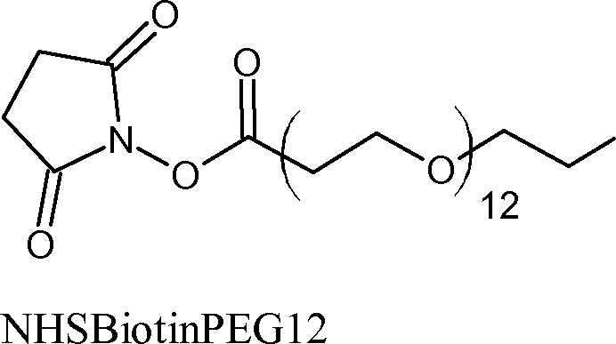
Example 7- Chemical Modification of yD- and yS- crystalline with BIOTIN [0115] Biotin is a molecule commonly used for protein modification due to its high binding affinity for avidin. Proteins can be tagged with biotin, undergo reactions in vivo or in vitro, then be separated or purified from the bulk using an avidin column. The NHSBiotin molecule was chosen because it reacts through the same mechanism as PEG4 and PEG24 however the added molecule will no longer be hydrophilic but rather contain a highly charged end group. The molecular weight added per unit is 226.38 g/mole with a spacer arm of 1.38 nm.
NHSBiotin [0116] The y-crystallin proteins were successfully modified with biotin as shown in the MALDI-TOF. (data not shown) yD-crystallin’s peak modification was eight units (23758 m/z) while yS-crystallin’s peak occurrence was four units (23325 m/z). Both systems show no unmodified y-crystallin indicating reaction conditions were sufficient for thorough modification.
[0117] The CD spectra showed a sizeable shift upwards as compared with the native structure of the protein, (data not shown) A shallower well was observed around 220nm which goes against the trend of good correlation between modification and secondary structure. The change in spectra could be an indication that the secondary structure of the
UMA0039P^yo 2014/152818
PCT/US2014/027852 proteins has been perturbed by the modification. Despite the shift in spectra, the overall shape of the curve is similar and there was no indication of a disordered or random coil.
[0118] Aggregation of y-crystallin protein was observed for y-crystallin modified with biotin. The NICF showed a slow and a fast mode which resulted in the distribution function having two sets of peaks (Figure 14). Angular dependence of the distribution function can be seen in the Γ versus q2 graph, where a qualitative difference was seen between the slow and fast modes. At 22°C, the Rh of the fast and slow modes were 2.8nm and 150nm for yD-crystallin, while for yS-crystallin these values were 2.6nm and 180nm. A slow and fast mode was also observed at 37°C with the Rh values being similar to those at 22°C.
[0119] The aggregation of y-crystallin modified with NHSBiotin shows that the functionality of the modifying molecule (i.e., the molecular bristle) is important. Mechanistically, biotin modified y-crystallin in the same mannor as NHSPEG. Only modifying charged amino acids of y-crystallin is thus not sufficient in preventing aggregation. A comparable degree of modification occurred with biotin and PEG4 meaning a similar shift of the protein’s isoelectric point. Due to aggregation occuring in the case of ycrystallin modified with biotin it is not sufficient to purely shift the isoelectric point of the ycrystallin protein to prevent aggregation. DLS measurements of the biotinylated system also further support the notion that aggregation cannot be prevented merely by adding spacer molecules onto the surface of y-crystallin.
[0120] The stark contrast in chemical properties between PEG and biotin is the reason one prevents aggregation and the other does not. PEG is a flexible hydrophilic molecule whereas the biotin functionaly is capable of hydrogen bonding and stabilizing a negative charge. The properties of PEG thus help the solubility of proteins whereas biotin would contribute to the hydrophobic and electrostatic nature of the aggregates.
Example 8- Chemical Modification of yD- and yS- crystalline with BIOTIN-PEG [0121] Modifying y-crystallin proteins with biotin resulted in aggregation so it was proposed to use NHSBiotinPEG which incorporates a hydrophilic spacer between the protein and biotin functionality. It was proposed that the hydrophilic nature of PEG would be the key factor in the prevention of y-crystallin aggregation. The NHS functionalization is used to keep the reaction mechanism constant. The molecular weight added per unit was 825.64 g/mol and the spacer arm was 5.6 nm.
UMA0039P'WO 2014/152818
PCT/US2014/027852

[0122] An appreciable shift in molecular weight was observed in MALDI-TOF indicating modification of the y-crystallin proteins with BiotinPEG12. (data not shown) yDcrystallin had three distinct peaks observable at 22760, 23582, and 24404 m/z corresponding to one, two, and three modifications. The resolution of yS-crystallin provided MALDI-TOF peaks at 23001, 23816, 24669, 25470, and 26122 m/z corresponding to one-five modifications. The MALDI-TOF spectra also showed no unmodified y-crystallin demonstrating sufficient reaction conditions.
[0123] The modified y-crystallin protein’s secondary structure was in good agreement with that of the native y-crystallin as seen in the CD spectra, (data not shown) The well observed with CD spectra for y-crystallin modified with just biotin was no longer evident.
The PEG portion of the modification most likely allowed for increased solubility in addition to providing a spacer between the protein and biotin functionality.
[0124] Despite the incorporation of PEG on the biotin functionality there was still aggregate present in solution as can be seen in the slow and fast mode of the NICF. (datya not shown) The distribution function (Figire 15) contained two sets of peaks that demonstrate an angular dependence. The Γ versus q2 graph clearly showed two distinct linear fits, the slope of which provide a slow and fast diffusive mode, (data not shown) The fast diffusion coefficient corresponds to the monomeric y-crystallin where yD-crystallin had an Rh of 2.7nm at 22°C, while yS-crystallin had the value was 2.6nm. The slow diffusion coefficient represented the large aggregate present in solution with an Rh of 105nm for yDcrystallin and 115nm for yS-crystallin at 22°C. Aggregate of a similar Rh was also observed at 37°C.
[0125] The use of PEG as a spacer between the protein and biotin functionality did not aid in preventing aggregation. Without being held to theory and with reference to the discussion earlier concerning biotin shifting the isoelectric point of y-crystallin, these experimental results also suggest hydrophobics to dominate over electrostatics in aggregates
UMA0039P^yo 2014/152818
PCT/US2014/027852 forming. The long spacer arm of BiotinPEG also did not prevent aggregation giving additional support to the trend of spacer molecule length not being a crucial factor in deturing aggregation. It should be noted that the y-crystallin tailored with BiotinPEG has a slow mode or aggregate Rh which was smaller than the biotin modification suggesting that PEG does aid in curbbing aggregation.
Example 9- Chemical Modification of yD- and yS- crystalline with Sulfo-Nhydroxysuccinimide acetate (SA) [0126] Due to previous modifications with large molecular weights and spacer arms still leading to aggregation, a minimally invasive modification was studied. The final modification was done with sulfo-N-hydroxysuccinimide acetate, which incorporated a minimal molecular weight (43 g/mol) and spacer arm (acetate molecule) per unit added. The reaction mechanism is similar to that of PEG4 so by reacting through the charged amino acid groups the isoelectric point of y-crystallin should be affected.
[0127] A subtle shift in the weight of the y-crystallin proteins was observed in MALDI-TOF. (data not shown) The peak relative intensity for yD-crystallin occurred at 22209 m/z and for yS-crystallin at 22562 m/z corresponding to six and seven acetate groups, respectively. The peaks showed no unmodified protein, indicating sufficient reaction conditions. A Gaussian distribution around the peak relative intensity indicated that some crystallin proteins have a higher and a lower degree of modification.
[0128] A CD spectrum for both modified y-crystallin proteins demonstrated a smaller well around 220nm as compared to the native y-crystallin. (data not shown) The change in CD spectra was also observed with the biotin modification. There is reason to believe that the secondary structure might be slightly affected by the modification although there are no indications of a random or disordered state.
[0129] Looking at the NICF it is evident that modification of y-crystallin with acetate groups did not prevent aggregation, (data not shown) The distribution function is shown in Figure 16. The angular dependence of the slow and fast diffusive modes can be seen in the Γ versus q2 graph, (data not shown) For yD-crystallin at 37°C, the fast and slow diffusion coefficients correspond to an Rh of 2.6nm and 85nm. The Rh values for yS-crystallin at 37°C were 2.5nm and 90nm. The fast mode Rh is an appropriate size for the modified y-crystallin proteins whose molecular weight is approximately 22kDa. Rh values obtained from measurements made at room temperature showed similar results.
UMA0039P^yo 2014/152818
PCT/US2014/027852 [0130] The SA modification was performed in an attempt to shift the protein’s isoelectric point with a minimal spacer arm. Despite an aggregate being present in solution, the aggregate size was smaller in comparison to unmodified y-crystallin. Through a high number of modifications per protein it is possible that a substantial shift in isoelectric point did reduce the size of the aggregate. Withour being held to theory, as aggregation is still present in solution it was concluded that the y-crystallin protein’s proximity to the isoelectric point is not solely responsible for the aggregation phenomena.
[0131] The results of Examples 3-9 are summarized in the following table:
Table 4: Summary of results with bifunctional charge masking agents
YS yD
| Rh (nm) | Rh (nm) | Rh (nm) | Rh (nm) | |
| PEG4 22°C | 2.9 | 2.8 | ||
| PEG4 37°C | 2.9 | 2.9 | ||
| PEG24 22°C | 3.2 | 3.1 | ||
| PEG24 37°C | 3.2 | 3.1 | ||
| CAPEG4 22°C | 2.8 | 2.9 | ||
| CAPEG4 37°C | 3 | 2.8 | ||
| MMPEG24 22°C | 3.1 | too | 2.9 | 85 |
| MMPEG24 37°C | 3 | 85 | 2.8 | 80 |
| Biotin 22°C | 2.6 | 180 | 2.8 | 150 |
| Biotin 37°C | 2.8 | 210 | 2.7 | 170 |
| BiotinPEG 22°C | 2.6 | 115 | 2.7 | 105 |
| BiotinPEG 37°C | 2.8 | 110 | 2.6 | too |
| SA 22°C | 2.5 | 92.5 | 2.5 | 82.5 |
| SA 37°C | 2.5 | 90 | 2.6 | 85 |
[0132] The results shown herein show that electrostatics are an important component of γ-crystallin aggregation. The electrostatic aggregation can be effectively interrupted using high salt concentrations or bifuncional charge masking agents. A increased understanding of the mechanism of γ-crystallin aggregation has provided a new class of agents that are particularly useful in the treatment of cataracts and presbyopia.
UMA0039P'WO 2014/152818
PCT/US2014/027852
Methods: Harvesting and characterization of human cadaver lenses [0133] Within about 24 hours of death, the eyeball is harvested, sliced and the vitrteous is removed. The lens is excised and placed in an incubation medium called Optisol®. Optisol® is a comeal storage medium conatining chondroitin sulfate, dextran 40, optisol base powder, sodium bicarbonate, gentamycin, amino acids, sodium pyruvate, Lglutamine, 2-mercaptoethanol and purified water. OD is the right eye and OS is the left eye.
O

CAPEG4 [0134] Lens opacities are classified according to the LOCS III system. LOCS III measurements are taken with a slit lamp miroscope. The LOGS ill contains an expanded set of standards selected from the Longitudinal Study of Cataract slide library at the Center for Clinical Cataract Research, Boston, Mass, it includes six slit lamp images for grading nuclear color (NC) and nuclear opalescence (NO), five retro!Ilumination images lor grading cortical cataract (C), and five retroillumination images for grading posterior suheapsular (P) cataract. Cataract severity is graded on a decimal scale, with the standards have regularly spaced intervals on the scale.
Example 10: Testing of CAPEG4 in human cadaver lenses- transport across the epithelium [0135] The transport of the CAPEG4 across an epithelium construct was studied. The epithelium is the outer layer of the cornea and transport across the epithelium has proven to be challenging. The goal was to deomnstrate the CAPEG4 construct could be used in a eye drop formulation for transport through the front of the eye.
[0136] Specifically, the CAPEG4 construct in solution was added to the top of an apparatus containing culture medium and an epithelium construct. (Figure 19). The epithelium construct consists of a layer(s) of human corneal epithelial cells. Aliquots were taken at the top and the bottom of the cultre medium from 15 to 120 minutes. Figure 20 shows the CAPEG4 that was transported to the bottom of the cell.
[0137] Figure 21 shows the pH of Opisol® versus the mg/ml of added CAPEG4. The data is also presented in tables 5 and 6. Overall, it is demonstrated that the reduction in corticle and posterior suheapsular cataracts is due to the CAPEG4 and not a pH effect of the
UMA0039P^yo 2014/152818
PCT/US2014/027852 medium.because here was no reduction in corticle cataracts by simply changing (lowering) the pH of the Optisol® media with citric acid to the same pH of Optisol® containing the CaPEG amine. Reduction in opacity required the presence of the active agent.
Table 5: 1 mg/ml CAPEG4
| Time | Top | Bottom | |||
| Min | mcg/well | Average | mcg/well | Average | % (stock) |
| 15 | 427 472 | 449 | 4 22 | 13 | 2.4 |
| 30 | 448 445 | 446 | 26 37 | 31 | 5.8 |
| 60 | 454 388 | 421 | 8 11 | 9 | 1.7 |
| 45 | 459 428 | 443 | 9 8 | 8 | 1.5 |
| 90 | 467 459 | 463 | 17 45 | 31 | 5.7 |
| 120 | 453 510 | 481 | 21 30 | 25 | 4.7 |
| 1000 mcg/mL | 543 | 543 | |||
| Time 0 harvest: | 446 453 | 450 | 4 3 | 3 | 0.6 |
Table 5: 16 mg/mL CAPEG4
| Time | Top | Bottom | |||
| Min | mcg/well | Average | mcg/well | Average | % (stock) |
| 15 | 5842 6908 | 6375 | 58 62 | 60 | 0.7 |
| 30 | 6099 6526 | 6312 | 93 105 | 99 | 1.2 |
| 60 | 6450 7494 | 6972 | 160 139 | 149 | 1.8 |
| 45 | 6313 6356 | 6335 | 677 418 | 548 | 6.7 |
| 90 | 6122 6792 | 6457 | 525 na | 525 | 6.4 |
| 16000 mcg/mL | 8186 | 8186 | |||
| Blank insret at 15 min | 7857 | 7857 | 1837 | 1837 | 23 |
UMA0039P'WO 2014/152818
PCT/US2014/027852
Example 11: Testing of CAPEG4 with human cadaver lenses- reduction of cataracts in human cadaver lenses [0138] In all cases, the control is Optisol® medium. The remaining data points are for isolated human cadaver lenses treated with CAPEG4 solutions with Optisol®.
[0139] Table 6 shows the results for lenses incubated with control medium or 10 mg/mL CAPEG4 versus time. In table 6, under the heading cortical, there is a LOCS III grade of 1.0, 1.0 and 0.9 for a lens incubated with 10 mg/ml CAPEG4. The LOCS grades for the control are 1.0, 1.2 and 0.9. At 10 mg/mL there was no significant change in the experimental versus control samples.
Table 6- LOCS III Grading of cataracts incubated with Optisol® or 10 mg/ml CAPEG4 versus time
| [CaPEG] (mg/mL) | Time (HR) | Cortical | NO | NC | PS |
| 10 (OD) | 0 | 1.0 | 5.0 | 5.0 | 0.9 |
| Contol (OS) Optisol Only | 0 | 1.2 | 5.0 | 5.0 | 0.9 |
| 10 | 24 | 1.0 | 5.0 | 5.0 | 0.9 |
| Control Optisol Only | 24 | 1.2 | 5.0 | 5.0 | 0.9 |
| 10 | 72 | 0.9 | 5.0 | 5.0 | 0.9 |
| Control | 72 | 0.9 | 5.0 | 5.0 | 1.0 |
[0140] Table 7 shows the results for lenses incubated with control medium or 50 mg/mL CAPEG4 versus time. In table 7, under the heading cortical, there is a LOCS III grade of 1.0 for all samples and a Nuclear opalescence (NO) grade of 2.0 for all samples. There were no significant differences between the controls and 50 mg/mL incubation up tp 72 hours for cortical cataracts or for NO. Nuclear color (NC) increased slightly with time for both the control and upon incubation in the 50 mg/mL CAPEG4 solution. There was a slight improvement in posterior subcapsular cataraact from 1.1 to 0.9 at o and 24 hours, respectively.
Table 7- LOCS III Grading of cataracts incubated with Optisol® or 50 mg/ml CAPEG4 versus time
UMA0039P^yo 2014/152818
PCT/US2014/027852
| [CaPEG] (mg/mL) | Time (HR) | Cortical | NO | NC | PS |
| 50 (OD) | 0 | 1.0 | 2.0 | 1.7 | 1.1 |
| Contol (OS) Optisol Only | 0 | 1.0 | 2.0 | 1.8 | 0.9 |
| 50 | 24 | 1.0 | 2.0 | 1.7 | 0.9 |
| Control Optisol Only | 24 | 1.0 | 2.0 | 1.8 | 0.9 |
| 50 | 72 | 1.0 | 2.0 | 1.9 | 0.9 |
| Control | 72 | 1.0 | 2.0 | 2.0 | 0.9 |
[0141] Table 8 shows the results for lenses incubated with control medium or 100 mg/mL CAPEG4 versus time. There is a decrease in opalescence (NO from 1.8 to 1.2) at 0 and 24 hours, respectively for CAPEG4 incubation and minor improvements in cortical and nuclear color up to 72 hours.
Table 8- LOCS III Grading of cataracts incubated with Optisol® or 100 mg/ml CAPEG4 versus time
| [CaPEG] (mg/ml.) | Time (HR) | Cortical | NO | NC | PS |
| 100 (OD) | 0 | 3.0 | 1.8 | 1.3 | 0.9 |
| Contol (OS) Optisol Only | 0 | 0.9 | 1.9 | 1.5 | 0.9 |
| 100 | 24 | 2.9 | 1.2 | 1.1 | 0.9 |
| Control Optisol Only | 24 | 0.9 | 1.8 | 2.0 | 0.9 |
| 100 | 72 | 2.7 | 1.2 | 1.1 | 0.9 |
| Control | 72 | 0.9 | 1.9 | 2.0 | 0.9 |
[0142] Table 9 is a repeat of the experiment in Table 8 with different eyes. Under PS (Posterior subcapsular cataracts) the sample incubated at 100 mg/ml CAPEG4 had values of 1.8, 1.5, and 1.0 at 0, 24 and 72 hours, respectively. There was also a reduction in cortical cataracts from 3.0 to 2.6 to 2.0 at 0, 24, and 72 hours, respectively.
Table 9- LOCS III Grading of cataracts incubated with Optisol® or 100 mg/ml CAPEG4 versus time
UMA0039P^yo 2014/152818
PCT/US2014/027852
| [CaPEG] (mg/mL) | Time (HR) | Cortical | NO | NC | PS |
| 100 (OD) | 0 | 3.0 | 3.0 | 3.0 | 1.8 |
| Contol (OS) Optisol Only | 0 | 1.0 | 3.0 | 3.0 | 1.5 |
| 100 | 24 | 2.6 | 3.0 | 3.0 | 1.5 |
| Control Optisol Only | 24 | 1.0 | 3.0 | 3.0 | 1.5 |
| 100 | 72 | 2.0 | 3.0 | 3.2 | 1.0 |
| Control | 72 | 1.0 | 3.0 | 3.1 | 1.5 |
[0143] Table 10 shows the results for lenses incubated with control medium or 200 mg/mL CAPEG4 versus time. Cortical cataracts were reduced from 2.5 to 2.2 to 2.0 at 0, 24, and 72 hours, respectively. The control also exhibit a slight reduction in cortical cataracts from 3.3 to 3.0 at 24 and 72 hours, respectively.
Table 10- LOCS III Grading of cataracts incubated with Optisol® or 200 mg/ml CAPEG4 versus time
| [CaPEG] (mg/ml.) | Time (HR) | Cortical | NO | NC | PS |
| 200 (OD) | 0 | 2.5 | 2.4 | 2.4 | 0 |
| Contol (OS) Optisol Only | 0 | 3.3 | 2.5 | 2.5 | 0 |
| 200 | 24 | 2.2 | 2.4 | 2.4 | 0 |
| Control Optisol Only | 24 | 3.3 | 2.5 | 2.5 | 0 |
| 200 | 72 | 2.0 | 2.4 | 2.4 | 0 |
| Control | 72 | 3.0 | 2.5 | 2.5 | 0 |
[0144] Table 11 shows the results for lenses incubated with control medium or 200 mg/mL CAPEG4 versus time. The only decrease in cataract was observed in cortical cataracts for the CAPEG4 solution going from 1.3 to 1.0 to 0.9 at 0, 24, and 72 hours, respectively.
Table 11- LOCS III Grading of cataracts incubated with Optisol® or 200 mg/ml CAPEG4 (in Optisol®) versus time
UMA0039P^yo 2014/152818
PCT/US2014/027852
| [CaPEG] (mg/mL) | Time (HR) | Cortical | NO | NC | PS |
| 200 (OD) | 0 | 1.3 | 3.2 | 2.8 | 0.9 |
| Contol (OS) Optisol Only | 0 | 1.4 | 3.2 | 3.0 | 0.9 |
| 200 | 24 | 1.0 | 3.2 | 2.8 | 0.9 |
| Control Optisol Only | 24 | 1.4 | 3.2 | 3.0 | 0.9 |
| 200 | 72 | 0.9 | 3.2 | 2.8 | 0.9 |
| Control | 72 | 1.4 | 3.2 | 3.0 | 0.9 |
[0145] Table 12 shows the results of changing the pH of Optisol® with no added CAPEG4. The pH was lowered via the addition of citric acid. There was no effect of changing the pH of the Optisol® on the cataracts.
Table 12: Efect of pH of Optisol® on cataracts
| pH | Time (HR) | Cortical | NO | NC | PS |
| 6.470 (OD) | 0 | 2.0 | 5.0 | 5.2 | 0.9 |
| 6.085 (OD) | 0 | 2.7 | 2.3 | 2.0 | 0.9 |
| 6.470 | 24 | 2.0 | 5.0 | 5.2 | 0.9 |
| 6.075 | 24 | 2.7 | 2.3 | 2.0 | 0.9 |
| 6.470 | 72 | 2.0 | 5.0 | 5.2 | 0.9 |
| 6.075 | 72 | 2.7 | 2.3 | 2.0 | 0.9 |
[0146] For cadaver lens studies, the different starting point for LOCSIII grade is due to the varying starting condition in the patients. As the concentration of the CAPEG4 solutions in Optisol increased, wrinkling of the lens capsule was observed and subjectively exhibited some loss of lens volume.
[0147] The terms “a” and “an” do not denote a limitation of quantity, but rather denote the presence of at least one of the referenced item. The term “or” means “and/or”.
The terms “comprising”, “having”, “including”, and “containing” are to be construed as open-ended terms (i.e., meaning “including, but not limited to”). All ranges disclosed herein are inclusive and combinable.
[0148] Embodiments are described herein, including the best modes known to the inventors. Variations of such embodiments will become apparent to those of ordinary skill in the art upon reading the foregoing description. The skilled artisan is expected to employ such variations as appropriate, and the disclosed methods are expected to be practiced otherwise than as specifically described herein. Accordingly, all modifications and equivalents of the
UMA0039P'WO 2014/152818
PCT/US2014/027852 subject matter recited in the claims appended hereto are included to the extent permitted by applicable law. Moreover, any combination of the above-described elements in all possible variations thereof is encompassed unless otherwise indicated herein or otherwise clearly contradicted by context.
2014236582 08 Mar 2018
Claims (15)
- THE CLAIMS DEFINING THE INVENTION ARE AS FOLLOWS:1. A method of inhibiting or reversing the progression of cataract formation presbyopia, or age related degeneration of a crystalline lens in an eye comprising contacting the eye with an effective cataract-inhibiting amount of an ophthalmic composition comprising at least one γ-crystallin charge masking agent, wherein the γ crystallin charge masking agent is a bifunctional molecule containing a reactive group covalently linked to a molecular bristle, wherein the reactive group is NH2, a succinimide, a carboxylic acid, isocyanate, isothiocyanate, sulfonyl chloride, aldehyde, carbodiimide, acyl azide, anhydride, fluorobenzene, carbonate, N-hydroxysuccinimide ester, imidoester, epoxide and fluorophenyl ester, and wherein the molecular bristle is a polyethylene glycol, an alkoxy-polyethylene glycol, or an alkoxypolyethylene glycol having 4 to 200 oxyethylene, alkoxyethylene or aryloxyethylene groups; poly(2-hydroxypropyl)methacrylamide (HPMA); poly(2hydroxyethyl)methacrylate (HEMA);a poly(2-oxaziline), poly(m-phosphocholine, polylysine, or polyglutamic acid, the molecular bristle having a molecular weight of 150 to 8000.
- 2. The method of claim 1, wherein the reactive group is NH2, Nhydroxysuccinimide, or COOH.
- 3. The method of claim 1, wherein the γ-crystallin charge masking agent is
- 4. The method of any one of claims 1, to 3 wherein the ophthalmic composition is an eye drop or an ophthalmic device.
- 5. The method of claim 4, wherein the ophthalmic device is a contact lens or a punctal plug.
- 6. The method of any one of claims 1 to 3, wherein the ophthalmic composition is administered by injection, iontophoresis or ultrasound enhancement.2014236582 08 Mar 2018
- 7. An ophthalmic composition comprising a bifunctional molecule containing a reactive group covalently linked to a molecular bristle, wherein the reactive group is NH2, a succinimide, a carboxylic acid, isocyanate, isothiocyanate, sulfonyl chloride, aldehyde, carbodiimide, acyl azide, anhydride, fluorobenzene, carbonate, N-hydroxysuccinimide ester, imidoester, epoxide and fluorophenyl ester,and wherein the molecular bristle is a polyethylene glycol, an alkoxy-polyethylene glycol, or an alkoxypolyethylene glycol having 4 to 200 oxyethylene, alkoxyethylene or aryloxyethylene groups; poly(2-hydroxypropyl)methacrylamide (HPMA); poly(2hydroxyethyl)methacrylate (HEMA);a poly(2-oxaziline), poly(m-phosphocholine, polylysine, or polyglutamic acid, the molecular bristle having a molecular weight of 150 to 8000.
- 8. The ophthalmic composition of claim 7, wherein the reactive group is NH2, Nhydroxysuccinimide or COOH.
- 9. The ophthalmic composition of claim 7, wherein the bifunctional molecule is o4-24NH2
- 10. The ophthalmic composition of claim 7, wherein the ophthalmic composition is an eye drop or an ophthalmic device.
- 11. The ophthalmic composition of claim 10, wherein the ophthalmic device is a contact lens or a punctal plug.
- 12. A method of treating a diseases relating to protein folding in a patient in need thereof, comprising administering a therapeutically effective amount of a bifunctional molecule containing a leaving group covalently linked to a molecular bristle, wherein the leaving group is a succinimide functional group, a carboxylic acid functional group, isocyanate, isothiocyanate, sulfonyl chloride, aldehyde, carbodiimide, acyl2014236582 08 Mar 2018 azide, anhydride, fluorobenzene, carbonate, N-hydroxysuccinimide ester, imidoester, epoxide and fluorophenyl ester,and wherein the molecular bristle is a polyethylene glycol, an alkoxy-polyethylene glycol, or an alkoxypolyethylene glycol having 4 to 200 oxyethylene, alkoxyethylene or aryloxyethylene groups; poly(2-hydroxypropyl)methacrylamide (HPMA); poly(2hydroxyethyl)methacrylate (HEMA);a poly(2-oxaziline), poly(m-phosphocholine, polylysine, or polyglutamic acid, the molecular bristle having a molecular weight of 150 to 8000.
- 13. The method of claim 12, wherein the leaving group is N-hydroxysuccinimide or COOH.
- 14. The method of claim 12, wherein the bifunctional molecule is
- 15. The method of claim 12, wherein the disease relating to protein folding is selected from Alzheimer’s disease, Parkinson’s disease, and Huntington’s disease.WO 2014/152818PCT/US2014/0278521/9Figure 1Figure 2Lag Time (ms)- 35 -40 — 70 80 90100 1000Figure 3 £o ΰ£LX.£OXIΈ t?0.250.20.150.10.050.004Distribution Function: yS + PEG24 22°C — 35--40 — 45 — 60.........70WO 2014/152818PCT/US2014/0278522/9Figure 4300002500020000 'w >15000 l-i1000050000 0Γ versus q2: yS + PEG24 22°C q2 (m-2) 5E+14Figure 51.2 cO.:«0.4 ^0.215000Sam in at)Sain in aD + PES41.2 £ ΰ·8 0*4-*- 0.6 : on0.2Figure 620000 m/z 25000 300001500020000Sain in aS GainmaS +F’ES4 a w, :'<|,.·ί *')[ S|.l, 1_. .2! !m/z2500030000 •40100 1000WO 2014/152818PCT/US2014/0278523/9Figure 70.140.12 eoΈ 0.1 c^0.08 c'§0.06J3Distribution Function ts0.040.020.004Figure 8WO 2014/152818PCT/US2014/0278524/9Figure 9 m/ϊNICFFigure 100.001 0.010.1 1 10 Lag Time (ms)100 1000----30Distribution Function:Figure 110.004 0.04 0.4 4 40Lag Time (ms)WO 2014/152818PCT/US2014/0278525/9Figure 12Figure 130.3 cO.25 o§0.15 •-PX 0·!s 5 0.05 00.004Figure 140.25Distribution Function: yS + Biotin 22°C § 0.2B50.15 £o '§ 0.1 £(30.050.004 0,04 Lag Time (ms) 4WO 2014/152818PCT/US2014/0278526/9Figure 150.5Distribution Function:yS + BiotinPEG 22°C c 0.4 oB1 0.30.004 0.04 0.4 4Lag Time (ms)Figure 16 •600.5Distribution Function:yS + SA 22°C e 0.4 o '6 = 0.3 ‘u.V,So.i0.004.. -Z>i\/\Z\0.04 0.4 — 30 — 35 — 40 ——45----50 · 60.........70....................80Lag Time (ms)WO 2014/152818PCT/US2014/0278527/9Figure 17 ru-M-c-sIsotliioeyaiiateR-N=C=OIsocyaitaleIIAide h yd uI, ciCarlwdiiinideΛAcyl AzidoQ.o λΛIIB-S-CI itSulfonyl ChlorideNIIS Ester
Anhydride Fluorobenzeim F NHj R^A0^ch:i 0 ΒΧ0,ΐγΧρ F Imidoester Epoxide Fluoro phenyl ester Figure 18Poly(2-Hydroxypropyl) methacrylamide (HPMA)Poly(2-hydroxyethyl) methacrylate (HEMA)A,Poly(2-oxazilines)Jo iX*OHHI·.IJH-OWCHjHQPoly lysine Poly glutamic acid —Gil,HjC—tkQllPoly(m phospho choline)WO 2014/152818PCT/US2014/0278528/9Ο u0s*ΙΛ r* m_φ Φ ο. 3 ω ε φ ω .3 3 4-» ’+» S Φ 3 Φ ε υ Φ 3 φ 3 ω U Ο 3 4-» ω 'ο. 3 Η LU U ...i:to *Ά<φ4-» υcD φDD πLUQ_ <ε .3 φε φ*2 'D φI Ε +* ΦΦDΟ .X ω ΐί — Φ Ο Λ ο 3 ω £Ο ·“3 Sσ ω — = 4± ο < < φ £ υ ωU φ οC LU Φ Ο.WO 2014/152818PCT/US2014/0278529/9Figure 20800 η700 600 500 400 300 200 100 0 1 mg/mL 16 mg/mL30 60 45 90Figure 21 pH versus CaPEG Amine Concentration (mg/mL)
Applications Claiming Priority (3)
| Application Number | Priority Date | Filing Date | Title |
|---|---|---|---|
| US201361782860P | 2013-03-14 | 2013-03-14 | |
| US61/782,860 | 2013-03-14 | ||
| PCT/US2014/027852 WO2014152818A1 (en) | 2013-03-14 | 2014-03-14 | Methods of inhibiting cataracts and presbyopia |
Publications (2)
| Publication Number | Publication Date |
|---|---|
| AU2014236582A1 AU2014236582A1 (en) | 2015-11-05 |
| AU2014236582B2 true AU2014236582B2 (en) | 2018-03-29 |
Family
ID=50686164
Family Applications (1)
| Application Number | Title | Priority Date | Filing Date |
|---|---|---|---|
| AU2014236582A Active AU2014236582B2 (en) | 2013-03-14 | 2014-03-14 | Methods of inhibiting cataracts and presbyopia |
Country Status (19)
| Country | Link |
|---|---|
| US (3) | US9789091B2 (en) |
| EP (1) | EP2968239B1 (en) |
| JP (1) | JP6397477B2 (en) |
| KR (1) | KR102198622B1 (en) |
| CN (1) | CN105209033B (en) |
| AU (1) | AU2014236582B2 (en) |
| BR (1) | BR112015023348B1 (en) |
| CA (1) | CA2904657C (en) |
| CL (1) | CL2015002689A1 (en) |
| CR (1) | CR20150537A (en) |
| EA (1) | EA029070B1 (en) |
| ES (1) | ES2727293T3 (en) |
| IL (1) | IL241306B (en) |
| MX (1) | MX366115B (en) |
| PE (1) | PE20151747A1 (en) |
| SA (1) | SA515361106B1 (en) |
| SG (1) | SG11201507209SA (en) |
| WO (1) | WO2014152818A1 (en) |
| ZA (1) | ZA201506987B (en) |
Families Citing this family (7)
| Publication number | Priority date | Publication date | Assignee | Title |
|---|---|---|---|---|
| AU2014236582B2 (en) | 2013-03-14 | 2018-03-29 | The University Of Massachusetts | Methods of inhibiting cataracts and presbyopia |
| JP2018526423A (en) * | 2015-09-08 | 2018-09-13 | ビューポイント セラピューティクス, インコーポレイテッド | Compounds and formulations for treating ophthalmic diseases |
| WO2017083619A1 (en) * | 2015-11-13 | 2017-05-18 | The University Of Massachusetts | Bifunctional molecules containing peg for use in inhibiting cataracts and presbyopia |
| US20220241247A1 (en) * | 2019-05-31 | 2022-08-04 | Plex Pharmaceuticals, Inc. | Pharmacological agents for treating protein aggregation diseases of the eye |
| US11607064B2 (en) * | 2019-08-06 | 2023-03-21 | Dart Industries Inc. | Reusable drinking straw |
| KR102587830B1 (en) * | 2021-07-22 | 2023-10-10 | 사회복지법인 삼성생명공익재단 | The method and system for cataract diagnosis using deep learning |
| US20230310380A1 (en) * | 2022-04-05 | 2023-10-05 | Alan Neil Glazier | Methods, devices, and systems for treating lens protein aggregation diseases |
Citations (4)
| Publication number | Priority date | Publication date | Assignee | Title |
|---|---|---|---|---|
| US4526789A (en) * | 1980-02-29 | 1985-07-02 | Massachusetts Institute Of Technology | Process for preventing or reversing cataract formation |
| EP0641563A1 (en) * | 1992-05-20 | 1995-03-08 | Taisho Pharmaceutical Co. Ltd | Remedy for cataract and production thereof |
| US20060147415A1 (en) * | 2005-01-03 | 2006-07-06 | Shaker Mousa | Composition and method for treating occlusive vascular diseases, nerve regeneration, and wound healing |
| WO2008145721A2 (en) * | 2007-06-01 | 2008-12-04 | Novo Nordisk A/S | N-terminal modification of polypeptides for protection against degradation by aminopeptidases |
Family Cites Families (83)
| Publication number | Priority date | Publication date | Assignee | Title |
|---|---|---|---|---|
| US4351826A (en) | 1980-02-29 | 1982-09-28 | Massachusetts Institute Of Technology | Process for preventing or reversing cataract formation using acrylamides |
| US4665089A (en) | 1985-03-21 | 1987-05-12 | Massachusetts Institute Of Technology | Process for preventing or reversing cataract formation using protein modification reagents |
| US4620979A (en) | 1985-08-02 | 1986-11-04 | Schachar Ronald A | Ophthalmological irrigating solution containing ascorbate |
| US4771036A (en) | 1986-02-10 | 1988-09-13 | Trustees Of Columbia University In The City Of New York | Method and ophthalmic composition for the prevention and reversal of cataracts |
| CA1269327A (en) | 1986-04-24 | 1990-05-22 | Massachusetts Institute Of Technology | Process for preventing or reversing cataract formation using protein modification reagents |
| US5055291A (en) | 1986-11-04 | 1991-10-08 | Baylor College Of Medicine | Compositions for preventing secondary cataracts |
| US4808182A (en) | 1986-11-26 | 1989-02-28 | Nestle, S.A. | Deswelled, hydrogel intraocular lenses |
| US5091421A (en) | 1987-06-04 | 1992-02-25 | Massachusetts Institute Of Technology | Chemical prevention or reversal of cataract by phase separation inhibitors |
| US5338545A (en) | 1987-06-04 | 1994-08-16 | Oculon Corporation | Chemical prevention or reversal of cataract by phase separation inhibitors |
| US5614587A (en) * | 1988-11-21 | 1997-03-25 | Collagen Corporation | Collagen-based bioadhesive compositions |
| DE3906311A1 (en) | 1989-02-28 | 1990-08-30 | Adatomed Pharma & Med | Treatment system for preventing secondary cataract formation after a cataract operation |
| WO1992000748A1 (en) | 1990-07-06 | 1992-01-23 | Enzon, Inc. | Poly(alkylene oxide) amino acid copolymers and drug carriers and charged copolymers based thereon |
| EP0489991B1 (en) | 1990-12-10 | 1996-06-05 | Bio-Physio Pharmaceutical Research And Development Company Limited | Indeno-d-pyrimidone compounds for use in medicine |
| WO1993000109A1 (en) | 1991-06-28 | 1993-01-07 | Genentech, Inc. | Method of stimulating immune response using growth hormone |
| US5375611A (en) | 1993-01-26 | 1994-12-27 | Pharmacia Ab | Method for preventing secondary cataract |
| CN1045383C (en) | 1993-04-07 | 1999-10-06 | 王慧康 | Medicine composite for curing senile cataract |
| US5663304A (en) | 1993-08-20 | 1997-09-02 | Genentech, Inc. | Refolding of misfolded insulin-like growth factor-I |
| DE69434617D1 (en) | 1993-11-19 | 2006-04-06 | Univ Sydney | PROCEDURE FOR PROPHYLAXIS OR CONTROL OF THE CATARACT |
| US5516534A (en) | 1993-11-26 | 1996-05-14 | Rensselaer Polytechnic Institute | Composition and method for reducing structural defects |
| US5591773A (en) | 1994-03-14 | 1997-01-07 | The Trustees Of Columbia University In The City Of New York | Inhibition of cataract formation, diseases resulting from oxidative stress, and HIV replication by caffeic acid esters |
| WO1995031223A1 (en) | 1994-05-13 | 1995-11-23 | Kuraray Co., Ltd. | Medical polymer gel |
| EP0733918B1 (en) | 1995-03-24 | 2003-07-30 | Ocular Research of Boston, Inc. | Hydrogel lens pre-coated with lipid layer |
| US5672662A (en) | 1995-07-07 | 1997-09-30 | Shearwater Polymers, Inc. | Poly(ethylene glycol) and related polymers monosubstituted with propionic or butanoic acids and functional derivatives thereof for biotechnical applications |
| JP3599848B2 (en) | 1995-09-11 | 2004-12-08 | 株式会社メニコン | Material for hydrous soft ophthalmic lens, molded article for hydrous soft ophthalmic lens comprising the same, and hydrous soft ophthalmic lens comprising the same and method for producing the same |
| EP0781777A1 (en) | 1995-12-28 | 1997-07-02 | Menicon Co., Ltd. | Silicon-containing compound and ocular lens material |
| US5807944A (en) * | 1996-06-27 | 1998-09-15 | Ciba Vision Corporation | Amphiphilic, segmented copolymer of controlled morphology and ophthalmic devices including contact lenses made therefrom |
| EP0816398B1 (en) | 1996-06-28 | 2002-08-14 | Ube Industries, Ltd. | Process for producing polybutadiene |
| US5817630A (en) | 1997-03-18 | 1998-10-06 | Austin Nutriceutical Corporation | Glutathione antioxidant eye drops |
| JPH10339857A (en) | 1997-06-05 | 1998-12-22 | Menicon Co Ltd | Method for producing drug-releasing contact lenses and drug-releasing contact lenses obtained thereby |
| US6015787A (en) | 1997-11-04 | 2000-01-18 | New England Medical Center Hospitals, Inc. | Cell-permeable protein inhibitors of calpain |
| US6291466B1 (en) | 1998-07-30 | 2001-09-18 | Allergan Sales, Inc. | Cholinergic agents in the treatment of presbyopia |
| EP1149909A1 (en) | 2000-04-28 | 2001-10-31 | Boehringer Mannheim Gmbh | Methods for regulating protein conformation using molecular chaperones |
| US6103756A (en) | 1999-08-11 | 2000-08-15 | Vitacost Inc. | Ocular orally ingested composition for prevention and treatment of individuals |
| CA2389917A1 (en) | 1999-11-04 | 2001-05-10 | Kazunori Kataoka | A polymer micelle as monolayer or layer-laminated surface |
| US6835394B1 (en) | 1999-12-14 | 2004-12-28 | The Trustees Of The University Of Pennsylvania | Polymersomes and related encapsulating membranes |
| US7977385B2 (en) | 2000-03-02 | 2011-07-12 | Numoda Biotechnologies, Inc. | Agents for corneal or intrastromal administration to treat or prevent disorders of the eye |
| WO2001091775A2 (en) | 2000-05-31 | 2001-12-06 | Cerus Corporation | Preparation of a pathogen inactivated solution of red blood cells having reduced immunogenicity |
| ES2272351T3 (en) | 2000-12-15 | 2007-05-01 | Sigma-Tau Industrie Farmaceutiche Riunite S.P.A. | USE OF L-CARNITINE AS A PROTEIN STABILIZING AGENT. |
| US6958224B2 (en) | 2001-03-28 | 2005-10-25 | Council Of Scientific And Industrial Research | Chimeric protein α BNAC crystallin with extraordinarily high chaperone-like activity and a method related to the use thereof |
| US20030020870A1 (en) | 2001-06-27 | 2003-01-30 | Zms, Llc | Biomedical molding materials from semi-solid precursors |
| EP1437143A4 (en) | 2001-09-28 | 2007-05-09 | Santen Pharmaceutical Co Ltd | Injections for eye tissue containing drug bound to polyethylene glycol |
| US20030223957A1 (en) * | 2002-04-10 | 2003-12-04 | Schwartz Daniel M. | Biodegradable PEG based polymer formulations in ocular applications |
| US6924154B2 (en) | 2002-08-20 | 2005-08-02 | Quest Diagnostics Investments Incorporated | Hydrophilic chemilumescent acridinium labeling reagents |
| JP2004161731A (en) | 2002-09-25 | 2004-06-10 | Nof Corp | Immobilizing agent for biological substances |
| EA009383B1 (en) | 2003-03-05 | 2007-12-28 | Хэлозим, Инк. | SOLUBLE HYALURONIDASE (sHASEGP), PROCESS FOR PREPARING THE SAME, USES AND PHARMACEUTICAL COMPOSITIONS COMPRISING THEREOF |
| CN1621091A (en) | 2003-11-28 | 2005-06-01 | 付经国 | Intestine targeted formulation of PEGylated rotavirus and its synthesizing method |
| WO2005076998A2 (en) | 2004-02-05 | 2005-08-25 | Intradigm Corporation | Rnai therapeutics for treatment of eye neovascularization diseases |
| WO2005112977A2 (en) | 2004-04-23 | 2005-12-01 | Pharmain, Ltd. | Compositions for treatment with glucagon-like peptide, and methods of making and using the same |
| US7887847B2 (en) | 2004-05-08 | 2011-02-15 | Paul Jr Edward L | Nutritional supplement for treatment of ocular diseases |
| WO2005117987A1 (en) | 2004-06-01 | 2005-12-15 | Glazier Alan N | Antibody conjugates targeting to ocular proteins |
| US6945971B1 (en) | 2004-07-19 | 2005-09-20 | Gwon Arlene E | Controlled ocular lens regeneration |
| WO2006052821A2 (en) * | 2004-11-04 | 2006-05-18 | University Of Washington | Compositions and methods for treatment of protein misfolding and protein aggregation diseases |
| CN1660920A (en) | 2005-02-01 | 2005-08-31 | 华东理工大学 | Polyglycolic acid or active ester with ω-amino acid connected at the end, its preparation method and application |
| DE102005041570A1 (en) | 2005-09-01 | 2007-03-22 | Celares Gmbh | Highly branched reagents for modifaction of biopharmaceuticals, their preparation and use |
| US7832875B2 (en) | 2005-09-29 | 2010-11-16 | Virtek Vision International Inc. | Modulated diode pumped microchip laser projector |
| US20080094573A1 (en) | 2006-04-04 | 2008-04-24 | Vermette Patrick | Surface-modified materials, such as contact lenses, methods and kits for their preparation, and uses thereof |
| US20070275098A1 (en) | 2006-05-19 | 2007-11-29 | T.R.P. Company, Inc, A Nevada Corporation | Formulation and methodology for the treatment for eye impairment symptoms |
| AU2008295449A1 (en) | 2007-09-07 | 2009-03-12 | Industrial Research Limited | Agents with angiogenic and wound healing activity |
| CA2702885A1 (en) | 2007-10-19 | 2009-04-23 | R-Tech Ueno, Ltd. | Pharmaceutical composition for treatment of cataract |
| KR20100000203A (en) | 2008-06-24 | 2010-01-06 | 인하대학교 산학협력단 | Targeted delivery system for anti-cancer drugs using au nanoparticles |
| JP2011526805A (en) | 2008-06-30 | 2011-10-20 | ジョンソン・アンド・ジョンソン・ビジョン・ケア・インコーポレイテッド | Method and apparatus for ophthalmic allergy treatment |
| DK2300514T3 (en) * | 2008-07-14 | 2016-06-27 | Biocon Ltd | A method of synthesizing a substantially monodispersed mixture of oligomers |
| WO2010065024A1 (en) | 2008-12-05 | 2010-06-10 | Kador Peter F | Topical treatment of cataracts in dogs |
| EP3246048B1 (en) * | 2009-02-18 | 2024-04-03 | Calm Water Therapeutics LLC | Bi-functional co-polymer use for ophthalmic applications |
| WO2010130638A1 (en) | 2009-05-14 | 2010-11-18 | Evotec Ag | Sulfonamide compounds, pharmaceutical compositions and uses thereof |
| WO2011075430A1 (en) * | 2009-12-14 | 2011-06-23 | University Of Massachusetts | Methods of inhibiting cataracts and presbyopia |
| US20130084262A1 (en) | 2010-04-19 | 2013-04-04 | Andrea S. Gobin | Repairing bruch's membrane with hydrogels |
| US9676871B2 (en) | 2010-11-05 | 2017-06-13 | Pfizer Inc. | Engineered polypeptide conjugates and methods for making thereof using transglutaminase |
| CN102172410B (en) | 2011-01-14 | 2013-08-07 | 华南理工大学 | Construction method of targeted nano particle transmission system for cancer diagnosis and treatment |
| CN102634492B (en) | 2011-02-14 | 2015-06-10 | 重庆富进生物医药有限公司 | Polyethylene glycol dog source urate oxidase analogue, and preparation method and applications thereof |
| CA2831855C (en) * | 2011-03-31 | 2019-05-21 | Dh Technologies Development Pte. Ltd. | Composition, method, and kit for calibrating a mass spectrometer |
| WO2012142659A1 (en) | 2011-04-19 | 2012-10-26 | Baker Idi Heart And Diabetes Institute Holdings Limited | Site-selective modification of proteins |
| CN103582640B (en) | 2011-06-03 | 2015-11-25 | 3M创新有限公司 | Diazanyl 1H-imidazoquinoline-4-amine and the conjugate be made up of it |
| CN202172410U (en) | 2011-08-23 | 2012-03-21 | 东莞市铭普实业有限公司 | Radio frequency intelligent optical module with keys based on internet of things |
| SG10201601327RA (en) | 2011-09-04 | 2016-03-30 | Glytech Inc | Glycosylated polypeptide and drug composition containing said polypeptide |
| EP2765986A1 (en) | 2011-10-12 | 2014-08-20 | Ascendis Pharma Ophthalmology Division A/S | Prevention and treatment of ocular conditions |
| CN102579353B (en) | 2012-03-30 | 2013-12-04 | 吉林大学 | Pegylated liposome of folate-targeted anticancer drug and preparation method |
| BR112015007778B1 (en) | 2012-10-11 | 2023-04-25 | Ascendis Pharma Ophthalmology Division A/S | PHARMACEUTICAL COMPOSITION OF VEGF NEUTRALIZING PRODRUGS OF VEGF FOR THE TREATMENT OF OCULAR CONDITIONS |
| WO2014071132A1 (en) | 2012-11-01 | 2014-05-08 | The Johns Hopkins University | Contact lens surface modification with hyaluronic acid (ha) binding peptide for ha accumulation and retention |
| AU2014236582B2 (en) | 2013-03-14 | 2018-03-29 | The University Of Massachusetts | Methods of inhibiting cataracts and presbyopia |
| WO2014171842A1 (en) | 2013-04-19 | 2014-10-23 | Living Cell Technologies New Zealand Limited | Biocompatible encapsulation system |
| WO2015085173A1 (en) | 2013-12-05 | 2015-06-11 | Wu Nian | Polymer-carbohydrate conjugates for drug delivery technology |
| WO2017083619A1 (en) | 2015-11-13 | 2017-05-18 | The University Of Massachusetts | Bifunctional molecules containing peg for use in inhibiting cataracts and presbyopia |
-
2014
- 2014-03-14 AU AU2014236582A patent/AU2014236582B2/en active Active
- 2014-03-14 CN CN201480026327.4A patent/CN105209033B/en active Active
- 2014-03-14 PE PE2015001991A patent/PE20151747A1/en active IP Right Grant
- 2014-03-14 MX MX2015013034A patent/MX366115B/en active IP Right Grant
- 2014-03-14 JP JP2016502644A patent/JP6397477B2/en active Active
- 2014-03-14 BR BR112015023348-1A patent/BR112015023348B1/en active IP Right Grant
- 2014-03-14 EP EP14722877.9A patent/EP2968239B1/en active Active
- 2014-03-14 SG SG11201507209SA patent/SG11201507209SA/en unknown
- 2014-03-14 CA CA2904657A patent/CA2904657C/en active Active
- 2014-03-14 EA EA201591677A patent/EA029070B1/en unknown
- 2014-03-14 KR KR1020157027595A patent/KR102198622B1/en active Active
- 2014-03-14 WO PCT/US2014/027852 patent/WO2014152818A1/en not_active Ceased
- 2014-03-14 ES ES14722877T patent/ES2727293T3/en active Active
- 2014-03-14 US US14/772,254 patent/US9789091B2/en active Active
-
2015
- 2015-09-08 IL IL24130615A patent/IL241306B/en active IP Right Grant
- 2015-09-14 SA SA515361106A patent/SA515361106B1/en unknown
- 2015-09-14 CL CL2015002689A patent/CL2015002689A1/en unknown
- 2015-09-18 ZA ZA2015/06987A patent/ZA201506987B/en unknown
- 2015-10-13 CR CR20150537A patent/CR20150537A/en unknown
- 2015-11-17 US US14/943,471 patent/US9675589B2/en active Active
-
2017
- 2017-04-28 US US15/581,349 patent/US10413529B2/en active Active
Patent Citations (4)
| Publication number | Priority date | Publication date | Assignee | Title |
|---|---|---|---|---|
| US4526789A (en) * | 1980-02-29 | 1985-07-02 | Massachusetts Institute Of Technology | Process for preventing or reversing cataract formation |
| EP0641563A1 (en) * | 1992-05-20 | 1995-03-08 | Taisho Pharmaceutical Co. Ltd | Remedy for cataract and production thereof |
| US20060147415A1 (en) * | 2005-01-03 | 2006-07-06 | Shaker Mousa | Composition and method for treating occlusive vascular diseases, nerve regeneration, and wound healing |
| WO2008145721A2 (en) * | 2007-06-01 | 2008-12-04 | Novo Nordisk A/S | N-terminal modification of polypeptides for protection against degradation by aminopeptidases |
Also Published As
Similar Documents
| Publication | Publication Date | Title |
|---|---|---|
| AU2014236582B2 (en) | Methods of inhibiting cataracts and presbyopia | |
| ES2905614T3 (en) | Recombinant Fusion Protein Formulation | |
| WO2016045627A1 (en) | Recombinant fusion protein formulation | |
| KR102696354B1 (en) | Bifunctional molecule containing PEG for use in suppressing cataracts and presbyopia | |
| RU2728795C2 (en) | Compositions containing anakinra | |
| ES2833506T3 (en) | Liquid pharmaceutical composition | |
| EP3996680A1 (en) | Stable formulations of recombinant proteins | |
| HK1218390B (en) | Methods of inhibiting cataracts and presbyopia | |
| NZ713007B2 (en) | Methods of inhibiting cataracts and presbyopia | |
| BR112018009582B1 (en) | BIFUNCTIONAL PEG-CONTAINING MOLECULES FOR USE IN INHIBITING CATARACTS AND PRESBYOPIA | |
| HK1260927A1 (en) | Bifunctional molecules containing peg for use in inhibiting cataracts and presbyopia | |
| HK1260927B (en) | Bifunctional molecules containing peg for use in inhibiting cataracts and presbyopia |
Legal Events
| Date | Code | Title | Description |
|---|---|---|---|
| FGA | Letters patent sealed or granted (standard patent) |