Synergistic Effects of Weightlessness, Isoproterenol, and Radiation on DNA Damage Response and Cytokine Production in Immune Cells

 ,
, <p>Cell recovery and cell viability after 24 h incubation in rotating-wall vessels (RWVs): (<b>A</b>) Change in the initial cell concentration with incubation time at a rotation speed of 10 rpm (6 independent experiments); (<b>B</b>) cell recovery of nontreated and treated cells over experimental conditions before and after 24 h incubation in RWVs rotating at 8.5 rpm (18 independent experiments); and (<b>C</b>) the percentage of live nontreated cells that remained alive after 24 h incubation rotating at a speed of 8.5 rpm (20 independent experiments).</p> "> Figure 2
<p>Apoptosis rate (calculated as indicated in <a href="#ijms-19-03689-t001" class="html-table">Table 1</a>) and residual DNA strand breaks after 24 h incubation in RWVs in µg or 1<span class="html-italic">g</span>. Apoptosis rate is represented by the mean transformed values (<b>A</b>) and (<b>B</b>) and DNA strand breaks are means expressed in Gy dose equivalent (<b>C</b>). Nontreated cells (C), isoproterenol (Iso) and radiation (R) treated cells. Error bars represent +1 SEM. Statistical analysis are summarized in <a href="#ijms-19-03689-t001" class="html-table">Table 1</a>.</p> "> Figure 3
<p>Radiation effects on gene expression relative to non-irradiated cells incubated in (<b>A)</b> 1<span class="html-italic">g</span> and (<b>B</b>) µg. Cells were irradiated either with 0.8 Gy (light bars) or 2 Gy (dark bars) and immediately incubated in 1<span class="html-italic">g</span> (grey bars) or µg (blue bars) for 24 h. Error bars represent SEM. Asterisks represent significant differences in gene expression in cells irradiated with 0.8 Gy compared to cells irradiated with 2 Gy. Statistical method: Krieger. <span class="html-italic">p</span>-value threshold: 0.018 after controlling the FDR (false-discovery rate—see statistical methods) to 5%.</p> "> Figure 4
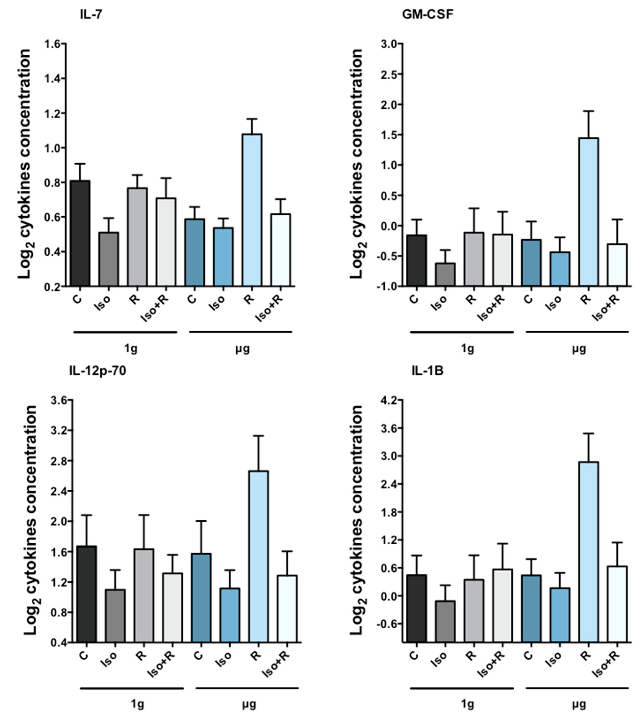
<p>Cytokine concentration in cell culture medium. Cells were irradiated 2 Gy and subsequently incubated in 1g or µg. After 24 h cytokine concentration was measured. Radiation induced cytokine production in µg but not in 1<span class="html-italic">g</span>. Isoproterenol treatment prior to radiation prevented the production of all cytokines. Bars represent mean + 1 SEM from 10 independent experiments. The synergistic effect of isoproterenol and radiation in µg (Iso × R(µg)) was significant for all four cytokines. Statistical analyses are summarized in <a href="#ijms-19-03689-t004" class="html-table">Table 4</a> and <a href="#ijms-19-03689-t005" class="html-table">Table 5</a>.</p> "> Figure 5
<p>Association between changes in cytokines and corresponding changes in gene expression over the eight experimental conditions for the group of samples irradiated with 2 Gy. BAX, CASP3, PCNA, LIG4, and MDM2 gene expressions were positively associated, while AKT1, TP53, PARP1, OGG1, and APXE1 were negatively associated with cytokines.</p> "> Figure 6
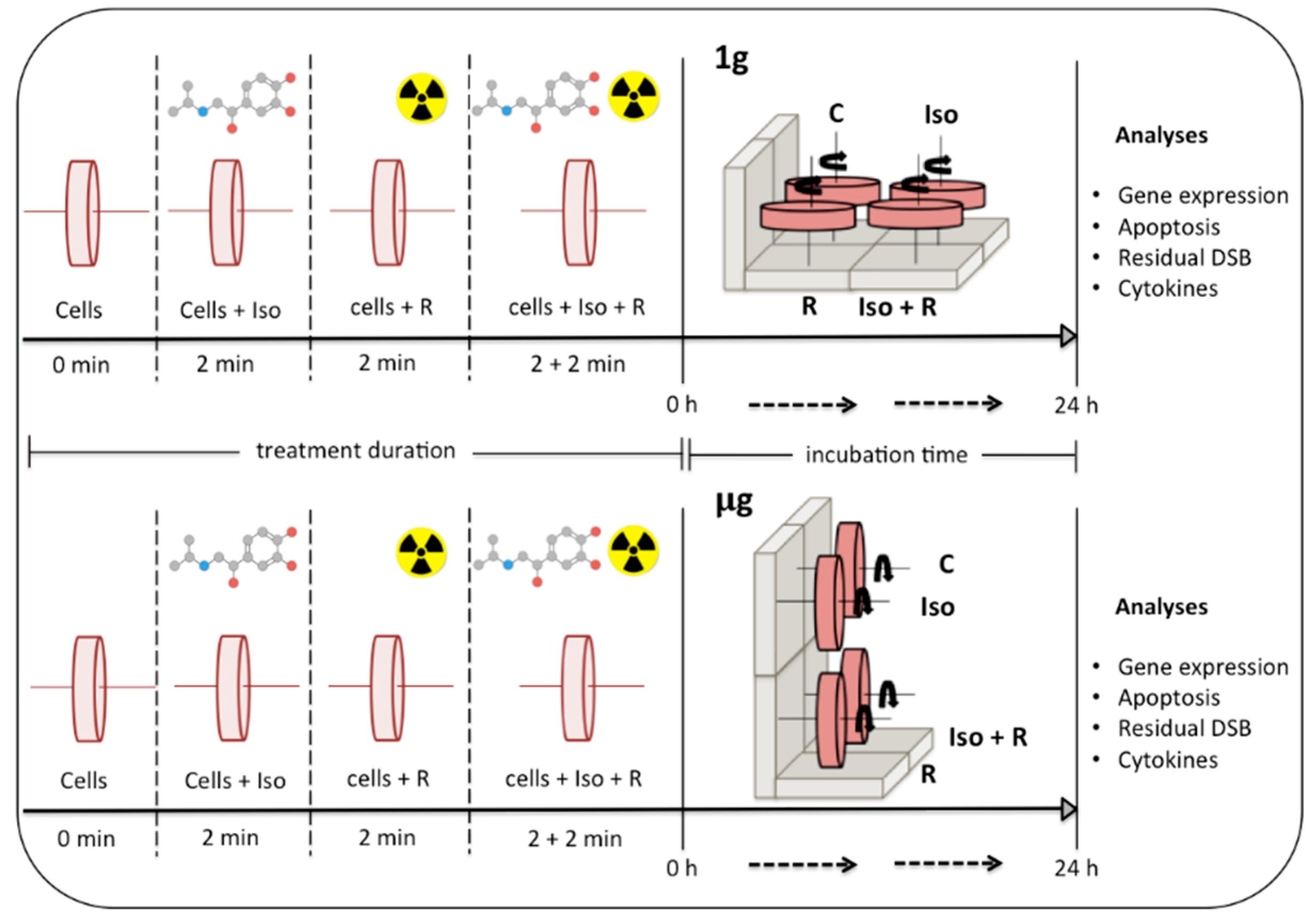
<p>Schematic representation of the treatment conditions. Cells were distributed in eight RCCSVs. Isoproterenol treatment (Iso) and radiation exposure (R) were performed separately and in combination (Iso + R). For the combined treatment, cells were irradiated for 2 min immediately after 2 min isoproterenol (Iso) treatment. Vessels were placed in an incubator and rotated at 8.5 rpm on a vertical (Earth gravity = 1<span class="html-italic">g</span>) or on a horizontal axis (simulated microgravity = µg). Vessels with nontreated cells (C) were incubated in 1<span class="html-italic">g</span> and in µg. After 24 h cells were recovered from the vessels and analyses were performed (DSB = DNA strand breaks).</p> ">
Abstract
:1. Introduction
1.1. Exogenous Factors Affecting DNA Damage Response (DDR)
1.2. Endogenous Factors Affecting DNA Damage Response
2. Results
2.1. Optimization of the Cell Concentration and Rotation Speed in Rotatory Wall Vessels (RWVs)
2.2. Synergistic Effect of Gravity, Radiation and Isoproterenol on Apoptosis
2.3. Effects of Radiation, Microgravity, and Isoproterenol on Gene Expression
2.4. Synergistic Effects of Gravity, Radiation, and Isoproterenol on Cytokine Release
3. Discussion
4. Materials and Methods
4.1. Isolation of PBMCs from Whole Blood
4.2. Experimental Design
4.3. Gene Expression
4.4. Apoptosis
4.5. Detection of DNA Strand Breaks
4.6. Cytokine Quantification
4.7. Statistical Analysis
4.8. Comparison of Cytokine and Gene Expression Data
5. Conclusions
Author Contributions
Funding
Conflicts of Interest
References
- Schwarz, R.P.; Goodwin, T.J.; Wolf, D.A. Cell culture for three-dimensional modeling in rotating-wall vessels: An application of simulated microgravity. J. Tissue Cult. Methods 1992, 14, 51–57. [Google Scholar] [CrossRef] [PubMed]
- Wolf, D.A.; Schwartz, R.P. Analysis of Gravity-Induced Particle Motion and Fluid Perfusion Flow in the NASA-Designed Rotating Zero-Head-Space Tissue Culture Vessel; NASA Tech. Paper 3143; NASA: Washington, DC, USA, 1991.
- Wolf, D.A.; Schwartz, R.P. Experimental Measurement of the Orbital Paths of Particles Sedimenting within a Rotating Viscous Fluid as Influenced by Gravity; NASA Tech. Paper 3200; NASA: Washington, DC, USA, 1992.
- Spaulding, G.F.; Jessup, J.M.; Goodwin, T.J. Advances in cellular construction. J. Cell. Biochem. 1993, 51, 249–251. [Google Scholar] [CrossRef] [PubMed] [Green Version]
- Jhala, D.V.; Kale, R.K.; Singh, R.P. Microgravity alters cancer growth and progression. Curr. Cancer Drug Targets 2014, 14, 394–406. [Google Scholar] [CrossRef] [PubMed]
- Grimm, D.; Wehland, M.; Pietsch, J.; Aleshcheva, G.; Wise, P.; van Loon, J.; Ulbrich, C.; Magnusson, N.E.; Infanger, M.; Bauer, J. Growing tissues in real and simulated microgravity: New methods for tissue engineering. Tissue Eng. Part B Rev. 2014, 20, 555–566. [Google Scholar] [CrossRef] [PubMed]
- Barzegari, A.; Saei, A.A. An update to space biomedical research: Tissue engineering in microgravity bioreactors. Bioimpacts 2012, 2, 23–32. [Google Scholar] [PubMed]
- Blaber, E.A.; Finkelstein, H.; Dvorochkin, N.; Sato, K.Y.; Yousuf, R.; Burns, B.P.; Globus, R.K.; Almeida, E.A. Microgravity reduces the differentiation and regenerative potential of embryonic stem cells. Stem Cells Dev. 2015, 24, 2605–2621. [Google Scholar] [CrossRef] [PubMed]
- Blaber, E.; Sato, K.; Almeida, E.A. Stem cell health and tissue regeneration in microgravity. Stem Cells Dev. 2014, 23 (Suppl. 1), 73–78. [Google Scholar] [CrossRef]
- Hauschild, S.; Tauber, S.; Lauber, B.S.; Thiel, S.C.; Layer, L.E.; Ullrich, O. T cell regulation in microgravity—The current knowledge from in vitro experiments conducted in space, parabolic flights and ground-based facilities. Acta Austronautica 2014, 104, 365–377. [Google Scholar] [CrossRef]
- Kamal, K.Y.; Herranz, R.; van Loon, J.; Medina, F.J. Simulated microgravity, mars gravity, and 2g hypergravity affect cell cycle regulation, ribosome biogenesis, and epigenetics in arabidopsis cell cultures. Sci. Rep. 2018, 8, 6424. [Google Scholar] [CrossRef] [PubMed]
- Shinde, V.; Brungs, S.; Henry, M.; Wegener, L.; Nemade, H.; Rotshteyn, T.; Acharya, A.; Baumstark-Khan, C.; Hellweg, C.E.; Hescheler, J.; et al. Simulated microgravity modulates differentiation processes of embryonic stem cells. Cell Physiol. Biochem. 2016, 38, 1483–1499. [Google Scholar] [CrossRef] [PubMed]
- Tan, X.; Xu, A.; Zhao, T.; Zhao, Q.; Zhang, J.; Fan, C.; Deng, Y.; Freywald, A.; Genth, H.; Xiang, J. Simulated microgravity inhibits cell focal adhesions leading to reduced melanoma cell proliferation and metastasis via fak/rhoa-regulated mtorc1 and ampk pathways. Sci. Rep. 2018, 8, 3769. [Google Scholar] [CrossRef] [PubMed]
- Wang, Y.; An, L.; Jiang, Y.; Hang, H. Effects of simulated microgravity on embryonic stem cells. PLoS ONE 2011, 6, e29214. [Google Scholar] [CrossRef] [PubMed]
- Harper, J.W.; Elledge, S.J. The DNA damage response: Ten years after. Mol. Cell 2007, 28, 739–745. [Google Scholar] [CrossRef] [PubMed]
- Kumari, R.; Singh, K.P.; Dumond, J.W., Jr. Simulated microgravity decreases DNA repair capacity and induces DNA damage in human lymphocytes. J. Cell. Biochem. 2009, 107, 723–731. [Google Scholar] [CrossRef] [PubMed]
- Degan, P.; Sancandi, M.; Zunino, A.; Ottaggio, L.; Viaggi, S.; Cesarone, F.; Pippia, P.; Galleri, G.; Abbondandolo, A. Exposure of human lymphocytes and lymphoblastoid cells to simulated microgravity strongly affects energy metabolism and DNA repair. J. Cell. Biochem. 2005, 94, 460–469. [Google Scholar] [CrossRef] [PubMed]
- Nowsheen, S.; Yang, E.S. The intersection between DNA damage response and cell death pathways. Exp. Oncol. 2012, 34, 243–254. [Google Scholar] [PubMed]
- Wang, J.Y. DNA damage and apoptosis. Cell Death Differ. 2001, 8, 1047–1048. [Google Scholar] [CrossRef] [PubMed] [Green Version]
- Risin, D.; Pellis, N.R. Modeled microgravity inhibits apoptosis in peripheral blood lymphocytes. In Vitro Cell. Dev. Biol. Anim. 2001, 37, 66–72. [Google Scholar] [CrossRef]
- Thiel, C.S.; Paulsen, K.; Bradacs, G.; Lust, K.; Tauber, S.; Dumrese, C.; Hilliger, A.; Schoppmann, K.; Biskup, J.; Golz, N.; et al. Rapid alterations of cell cycle control proteins in human t lymphocytes in microgravity. Cell Commun. Signal. 2012, 10, 1. [Google Scholar] [CrossRef] [PubMed] [Green Version]
- Bogdandi, E.N.; Balogh, A.; Felgyinszki, N.; Szatmari, T.; Persa, E.; Hildebrandt, G.; Safrany, G.; Lumniczky, K. Effects of low-dose radiation on the immune system of mice after total-body irradiation. Radiat. Res. 2010, 174, 480–489. [Google Scholar] [CrossRef] [PubMed]
- Indo, H.P.; Tomiyoshi, T.; Suenaga, S.; Tomita, K.; Suzuki, H.; Masuda, D.; Terada, M.; Ishioka, N.; Gusev, O.; Cornette, R.; et al. Mnsod downregulation induced by extremely low 0.1 mgy single and fractionated x-rays and microgravity treatment in human neuroblastoma cell line, nb-1. J. Clin. Biochem. Nutr. 2015, 57, 98–104. [Google Scholar] [CrossRef] [PubMed]
- Beck, M.; Tabury, K.; Moreels, M.; Jacquet, P.; Van Oostveldt, P.; De Vos, W.H.; Baatout, S. Simulated microgravity decreases apoptosis in fetal fibroblasts. Int. J. Mol. Med. 2012, 30, 309–313. [Google Scholar] [CrossRef] [PubMed]
- Canova, S.; Fiorasi, F.; Mognato, M.; Grifalconi, M.; Reddi, E.; Russo, A.; Celotti, L. “Modeled microgravity” affects cell response to ionizing radiation and increases genomic damage. Radiat. Res. 2005, 163, 191–199. [Google Scholar] [CrossRef] [PubMed]
- Fontes, F.L.; Pinheiro, D.M.; Oliveira, A.H.; Oliveira, R.K.; Lajus, T.B.; Agnez-Lima, L.F. Role of DNA repair in host immune response and inflammation. Mutat. Res. Rev. Mutat. Res. 2015, 763, 246–257. [Google Scholar] [CrossRef] [PubMed]
- Dragos, D.; Tanasescu, M.D. The effect of stress on the defense systems. J. Med. Life 2010, 3, 10–18. [Google Scholar] [PubMed]
- Marino, F.; Cosentino, M. Adrenergic modulation of immune cells: An update. Amino Acids 2013, 45, 55–71. [Google Scholar] [CrossRef] [PubMed]
- Bierhaus, A.; Wolf, J.; Andrassy, M.; Rohleder, N.; Humpert, P.M.; Petrov, D.; Ferstl, R.; von Eynatten, M.; Wendt, T.; Rudofsky, G.; et al. A mechanism converting psychosocial stress into mononuclear cell activation. Proc. Natl. Acad. Sci. USA 2003, 100, 1920–1925. [Google Scholar] [CrossRef] [PubMed] [Green Version]
- Torgersen, K.M.; Vang, T.; Abrahamsen, H.; Yaqub, S.; Tasken, K. Molecular mechanisms for protein kinase a-mediated modulation of immune function. Cell Signal. 2002, 14, 1–9. [Google Scholar] [CrossRef]
- Myklebust, J.H.; Josefsen, D.; Blomhoff, H.K.; Levy, F.O.; Naderi, S.; Reed, J.C.; Smeland, E.B. Activation of the camp signaling pathway increases apoptosis in human b-precursor cells and is associated with downregulation of mcl-1 expression. J. Cell Physiol. 1999, 180, 71–80. [Google Scholar] [CrossRef]
- Parvathenani, L.K.; Buescher, E.S.; Chacon-Cruz, E.; Beebe, S.J. Type i camp-dependent protein kinase delays apoptosis in human neutrophils at a site upstream of caspase-3. J. Biol. Chem. 1998, 273, 6736–6743. [Google Scholar] [CrossRef] [PubMed]
- Gu, C.; Ma, Y.C.; Benjamin, J.; Littman, D.; Chao, M.V.; Huang, X.Y. Apoptotic signaling through the beta -adrenergic receptor. A new gs effector pathway. J. Biol. Chem. 2000, 275, 20726–20733. [Google Scholar] [CrossRef] [PubMed]
- Naderi, E.H.; Findley, H.W.; Ruud, E.; Blomhoff, H.K.; Naderi, S. Activation of camp signaling inhibits DNA damage-induced apoptosis in bcp-all cells through abrogation of p53 accumulation. Blood 2009, 114, 608–618. [Google Scholar] [CrossRef] [PubMed] [Green Version]
- Padgett, D.A.; Glaser, R. How stress influences the immune response. Trends Immunol. 2003, 24, 444–448. [Google Scholar] [CrossRef] [Green Version]
- Qi, X.F.; Kim, D.H.; Yoon, Y.S.; Li, J.H.; Song, S.B.; Jin, D.; Huang, X.Z.; Teng, Y.C.; Lee, K.J. The adenylyl cyclase-camp system suppresses tarc/ccl17 and mdc/ccl22 production through p38 mapk and nf-kappab in hacat keratinocytes. Mol. Immunol. 2009, 46, 1925–1934. [Google Scholar] [CrossRef] [PubMed]
- Flierl, M.A.; Rittirsch, D.; Huber-Lang, M.; Sarma, J.V.; Ward, P.A. Catecholamines-crafty weapons in the inflammatory arsenal of immune/inflammatory cells or opening pandora’s box? Mol. Med. 2008, 14, 195–204. [Google Scholar] [PubMed]
- Barbe, P.; Galitzky, J.; Thalamas, C.; Langin, D.; Lafontan, M.; Senard, J.M.; Berlan, M. Increase in epinephrine-induced responsiveness during microgravity simulated by head-down bed rest in humans. J. Appl. Physiol. 1999, 87, 1614–1620. [Google Scholar] [CrossRef] [PubMed]
- Convertino, V.A.; Polet, J.L.; Engelke, K.A.; Hoffler, G.W.; Lane, L.D.; Blomqvist, C.G. Evidence for increased beta-adrenoreceptor responsiveness induced by 14 days of simulated microgravity in humans. Am. J. Physiol. 1997, 273, R93–R99. [Google Scholar] [CrossRef] [PubMed]
- Mednieks, M.; Khatri, A.; Rubenstein, R.; Burleson, J.A.; Hand, A.R. Microgravity alters the expression of salivary proteins. Oral Health Dent. Manag. 2014, 13, 211–216. [Google Scholar] [PubMed]
- Chiang, M.C.; Lin, H.; Cheng, Y.C.; Yen, C.H.; Huang, R.N.; Lin, K.H. Beta-adrenoceptor pathway enhances mitochondrial function in human neural stem cells via rotary cell culture system. J. Neurosci. Methods 2012, 207, 130–136. [Google Scholar] [CrossRef] [PubMed]
- Mills, P.J.; Meck, J.V.; Waters, W.W.; D’Aunno, D.; Ziegler, M.G. Peripheral leukocyte subpopulations and catecholamine levels in astronauts as a function of mission duration. Psychosom. Med. 2001, 63, 886–890. [Google Scholar] [CrossRef] [PubMed]
- Stowe, R.P.; Sams, C.F.; Pierson, D.L. Effects of mission duration on neuroimmune responses in astronauts. Aviat. Space Environ. Med. 2003, 74, 1281–1284. [Google Scholar] [PubMed]
- Fritsch-Yelle, J.M.; Charles, J.B.; Jones, M.M.; Beightol, L.A.; Eckberg, D.L. Spaceflight alters autonomic regulation of arterial pressure in humans. J. Appl. Physiol. 1994, 77, 1776–1783. [Google Scholar] [CrossRef] [PubMed]
- Slota, C.; Shi, A.; Chen, G.; Bevans, M.; Weng, N.P. Norepinephrine preferentially modulates memory cd8 t cell function inducing inflammatory cytokine production and reducing proliferation in response to activation. Brain Behav. Immun. 2015, 46, 168–179. [Google Scholar] [CrossRef] [PubMed]
- Shimamiya, T.; Wakabayashi, S.; Terada, N. Effects of adrenaline and cortisone on the early activation of lymphocytes. Biol. Sci. Space 2003, 17, 221–222. [Google Scholar] [PubMed]
- Berger, A. Th1 and th2 responses: What are they? BMJ 2000, 321, 424. [Google Scholar] [CrossRef] [PubMed]
- Wahle, M.; Neumann, R.P.; Moritz, F.; Krause, A.; Buttgereit, F.; Baerwald, C.G. Beta2-adrenergic receptors mediate the differential effects of catecholamines on cytokine production of pbmc. J. Interferon Cytokine Res. 2005, 25, 384–394. [Google Scholar] [CrossRef] [PubMed]
- Agarwal, S.K.; Marshall, G.D., Jr. Beta-adrenergic modulation of human type-1/type-2 cytokine balance. J. Allergy Clin. Immunol. 2000, 105, 91–98. [Google Scholar] [CrossRef]
- Crucian, B.E.; Zwart, S.R.; Mehta, S.; Uchakin, P.; Quiriarte, H.D.; Pierson, D.; Sams, C.F.; Smith, S.M. Plasma cytokine concentrations indicate that in vivo hormonal regulation of immunity is altered during long-duration spaceflight. J. Interferon Cytokine Res. 2014, 34, 778–786. [Google Scholar] [CrossRef] [PubMed]
- Hammond, T.G.; Hammond, J.M. Optimized suspension culture: The rotating-wall vessel. Am. J. Physiol. Renal Physiol. 2001, 281, F12–F25. [Google Scholar] [CrossRef] [PubMed]
- The Rotary Cell Culture System TM RCCS-1SC through 4SCQ Operation Manual; Synthecon, Incorporated: Houston, TX, USA, 2018.
- Nagata, S. Apoptotic DNA fragmentation. Exp. Cell Res. 2000, 256, 12–18. [Google Scholar] [CrossRef] [PubMed]
- Mognato, M.; Girardi, C.; Fabris, S.; Celotti, L. DNA repair in modeled microgravity: Double strand break rejoining activity in human lymphocytes irradiated with gamma-rays. Mutat. Res. 2009, 663, 32–39. [Google Scholar] [CrossRef] [PubMed]
- Ward, N.E.; Pellis, N.R.; Risin, S.A.; Risin, D. Gene expression alterations in activated human t-cells induced by modeled microgravity. J. Cell. Biochem. 2006, 99, 1187–1202. [Google Scholar] [CrossRef] [PubMed]
- Beerman, I.; Seita, J.; Inlay, M.A.; Weissman, I.L.; Rossi, D.J. Quiescent hematopoietic stem cells accumulate DNA damage during aging that is repaired upon entry into cell cycle. Cell Stem Cell 2014, 15, 37–50. [Google Scholar] [CrossRef] [PubMed]
- Hyka-Nouspikel, N.; Lemonidis, K.; Lu, W.T.; Nouspikel, T. Circulating human b lymphocytes are deficient in nucleotide excision repair and accumulate mutations upon proliferation. Blood 2011, 117, 6277–6286. [Google Scholar] [CrossRef] [PubMed]
- Jones, G.G.; Reaper, P.M.; Pettitt, A.R.; Sherrington, P.D. The atr-p53 pathway is suppressed in noncycling normal and malignant lymphocytes. Oncogene 2004, 23, 1911–1921. [Google Scholar] [CrossRef] [PubMed]
- Tanaka, T.; Kajstura, M.; Halicka, H.D.; Traganos, F.; Darzynkiewicz, Z. Constitutive histone h2ax phosphorylation and atm activation are strongly amplified during mitogenic stimulation of lymphocytes. Cell Prolif. 2007, 40, 1–13. [Google Scholar] [CrossRef] [PubMed]
- Redwine, L.; Jenkins, F.; Baum, A. Relation between beta-adrenergic receptor density and lymphocyte proliferation associates with acute stress. Int. J. Behav. Med. 1996, 3, 337–353. [Google Scholar] [CrossRef] [PubMed]
- Goldkorn, T.; Balaban, N.; Shannon, M.; Matsukuma, K. Egf receptor phosphorylation is affected by ionizing radiation. Biochim. Biophys. Acta 1997, 1358, 289–299. [Google Scholar] [CrossRef]
- Schmidt-Ullrich, R.K.; Mikkelsen, R.B.; Dent, P.; Todd, D.G.; Valerie, K.; Kavanagh, B.D.; Contessa, J.N.; Rorrer, W.K.; Chen, P.B. Radiation-induced proliferation of the human a431 squamous carcinoma cells is dependent on egfr tyrosine phosphorylation. Oncogene 1997, 15, 1191–1197. [Google Scholar] [CrossRef] [PubMed]
- Bowers, G.; Reardon, D.; Hewitt, T.; Dent, P.; Mikkelsen, R.B.; Valerie, K.; Lammering, G.; Amir, C.; Schmidt-Ullrich, R.K. The relative role of erbb1-4 receptor tyrosine kinases in radiation signal transduction responses of human carcinoma cells. Oncogene 2001, 20, 1388–1397. [Google Scholar] [CrossRef] [PubMed]
- Hara, M.R.; Kovacs, J.J.; Whalen, E.J.; Rajagopal, S.; Strachan, R.T.; Grant, W.; Towers, A.J.; Williams, B.; Lam, C.M.; Xiao, K.; et al. A stress response pathway regulates DNA damage through beta2-adrenoreceptors and beta-arrestin-1. Nature 2011, 477, 349–353. [Google Scholar] [CrossRef] [PubMed]
- Henriquez-Hernandez, L.A.; Carmona-Vigo, R.; Pinar, B.; Bordon, E.; Lloret, M.; Nunez, M.I.; Rodriguez-Gallego, C.; Lara, P.C. Combined low initial DNA damage and high radiation-induced apoptosis confers clinical resistance to long-term toxicity in breast cancer patients treated with high-dose radiotherapy. Radiat. Oncol. 2011, 6, 60. [Google Scholar] [CrossRef] [PubMed] [Green Version]
- Wilkins, R.C.; Kutzner, B.C.; Truong, M.; Sanchez-Dardon, J.; McLean, J.R. Analysis of radiation-induced apoptosis in human lymphocytes: Flow cytometry using annexin v and propidium iodide versus the neutral comet assay. Cytometry 2002, 48, 14–19. [Google Scholar] [CrossRef] [PubMed]
- Cioca, D.P.; Watanabe, N.; Isobe, M. Apoptosis of peripheral blood lymphocytes is induced by catecholamines. Jpn. Heart J. 2000, 41, 385–398. [Google Scholar] [PubMed]
- Cho, E.A.; Kim, E.J.; Kwak, S.J.; Juhnn, Y.S. Camp signaling inhibits radiation-induced atm phosphorylation leading to the augmentation of apoptosis in human lung cancer cells. Mol. Cancer 2014, 13, 36. [Google Scholar] [CrossRef] [PubMed]
- Liao, X.; Che, X.; Zhao, W.; Zhang, D.; Long, H.; Chaudhary, P.; Li, H. Effects of propranolol in combination with radiation on apoptosis and survival of gastric cancer cells in vitro. Radiat. Oncol. 2010, 5, 98. [Google Scholar] [CrossRef] [PubMed] [Green Version]
- Meijer, A.E.; Saeidi, A.B.; Zelenskaya, A.; Czene, S.; Granath, F.; Harms-Ringdahl, M. Influence of dose-rate, post-irradiation incubation time and growth factors on interphase cell death by apoptosis and clonogenic survival of human peripheral lymphocytes. Int. J. Radiat. Biol. 1999, 75, 1265–1273. [Google Scholar] [PubMed]
- Sato, C. Change in the type of radiation cell-killing on human lymphocytes after blast formation by phytohaemagglutinin. Int. J. Radiat. Biol. Relat. Stud. Phys. Chem. Med. 1970, 18, 483–485. [Google Scholar] [CrossRef] [PubMed]
- Schrek, R.; Stefani, S. Radioresistance of phytohemagglutinin- treated normal and leukemic lymphocytes. J. Natl. Cancer Inst. 1964, 32, 507–521. [Google Scholar] [PubMed]
- Heinrichs, S.; Deppert, W. Apoptosis or growth arrest: Modulation of the cellular response to p53 by proliferative signals. Oncogene 2003, 22, 555–571. [Google Scholar] [CrossRef] [PubMed]
- Seki, H.; Kanegane, H.; Iwai, K.; Konno, A.; Ohta, K.; Yachie, A.; Taniguchi, N.; Miyawaki, T. Ionizing radiation induces apoptotic cell death in human tcr-gamma/delta+ t and natural killer cells without detectable p53 protein. Eur. J. Immunol. 1994, 24, 2914–2917. [Google Scholar] [CrossRef] [PubMed]
- Coller, H.A.; Sang, L.; Roberts, J.M. A new description of cellular quiescence. PLoS Biol. 2006, 4, e83. [Google Scholar] [CrossRef] [PubMed]
- Schaue, D.; Kachikwu, E.L.; McBride, W.H. Cytokines in radiobiological responses: A review. Radiat. Res. 2012, 178, 505–523. [Google Scholar] [CrossRef] [PubMed]
- Shan, Y.X.; Jin, S.Z.; Liu, X.D.; Liu, Y.; Liu, S.Z. Ionizing radiation stimulates secretion of pro-inflammatory cytokines: Dose-response relationship, mechanisms and implications. Radiat. Environ. Biophys. 2007, 46, 21–29. [Google Scholar] [CrossRef] [PubMed]
- Gridley, D.S.; Rizvi, A.; Makinde, A.Y.; Luo-Owen, X.; Mao, X.W.; Tian, J.; Slater, J.M.; Pecaut, M.J. Space-relevant radiation modifies cytokine profiles, signaling proteins and foxp3+ t cells. Int. J. Radiat. Biol. 2013, 89, 26–35. [Google Scholar] [CrossRef] [PubMed]
- Aihara, M.; Dobashi, K.; Iizuka, K.; Nakazawa, T.; Mori, M. Comparison of effects of y-27632 and isoproterenol on release of cytokines from human peripheral t cells. Int. Immunopharmacol. 2003, 3, 1619–1625. [Google Scholar] [CrossRef]
- Van Walleghem, M.; Tabury, K.; Fernandez-Gonzalo, R.; Janssen, A.; Buchheim, J.I.; Chouker, A.; Baatout, S.; Moreels, M. Gravity-related immunological changes in human whole blood cultured under simulated microgravity using an in vitro cytokine release assay. J. Interferon Cytokine Res. 2017, 37, 531–540. [Google Scholar] [CrossRef] [PubMed]
- Zhou, Y.; Ni, H.; Li, M.; Sanzari, J.K.; Diffenderfer, E.S.; Lin, L.; Kennedy, A.R.; Weissman, D. Effect of solar particle event radiation and hindlimb suspension on gastrointestinal tract bacterial translocation and immune activation. PLoS ONE 2012, 7, e44329. [Google Scholar] [CrossRef] [PubMed]
- Centurione, L.; Aiello, F.B. DNA repair and cytokines: Tgf-beta, il-6, and thrombopoietin as different biomarkers of radioresistance. Front. Oncol. 2016, 6, 175. [Google Scholar] [CrossRef] [PubMed]
- Schwarz, T.; Schwarz, A. DNA repair and cytokine responses. J. Investig. Dermatol. Symp. Proc. 2009, 14, 63–66. [Google Scholar] [CrossRef] [PubMed]
- Cohen, I.; Rider, P.; Vornov, E.; Tomas, M.; Tudor, C.; Wegner, M.; Brondani, L.; Freudenberg, M.; Mittler, G.; Ferrando-May, E.; et al. Il-1alpha is a DNA damage sensor linking genotoxic stress signaling to sterile inflammation and innate immunity. Sci. Rep. 2015, 5, 14756. [Google Scholar] [PubMed]
- Mansilla-Soto, J.; Cortes, P. Vdj recombination: Artemis and its in vivo role in hairpin opening. J. Exp. Med. 2003, 197, 543–547. [Google Scholar] [CrossRef] [PubMed]
- Moreno-Villanueva, M.; Pfeiffer, R.; Sindlinger, T.; Leake, A.; Muller, M.; Kirkwood, T.B.; Burkle, A. A modified and automated version of the ‘fluorimetric detection of alkaline DNA unwinding’ method to quantify formation and repair of DNA strand breaks. BMC Biotechnol. 2009, 9, 39. [Google Scholar] [CrossRef] [PubMed]
- Moreno-Villanueva, M.; Eltze, T.; Dressler, D.; Bernhardt, J.; Hirsch, C.; Wick, P.; von Scheven, G.; Lex, K.; Burkle, A. The automated fadu-assay, a potential high-throughput in vitro method for early screening of DNA breakage. ALTEX 2011, 28, 295–303. [Google Scholar] [CrossRef] [PubMed]
- Junk, M.; Salzwedel, J.; Sindlinger, T.; Burkle, A.; Moreno-Villanueva, M. Mathematical modelling of the automated fadu assay for the quantification of DNA strand breaks and their repair in human peripheral mononuclear blood cells. BMC Biophys. 2014, 7, 9. [Google Scholar] [CrossRef] [PubMed]
- Benjamini, Y.K.A.M.; Yekutieli, D. Adaptive linear step-up procedures that control the false discovery rate. Biometrika 2006, 93, 491–507. [Google Scholar] [CrossRef]
- Holm, S. A simple sequentially rejective multiple test procedure. Scand. J. Stat. 1979, 6, 65–70. [Google Scholar]
- Newson, R. Confidence intervals for rank statistics: Somers d and extensions. Stata J. 2006, 6, 309–334. [Google Scholar] [CrossRef]
- Somers, R.H. A new asymmetric measure of association for ordinal variables. Am. Sociol. Rev. 1962, 27, 799–811. [Google Scholar] [CrossRef]
- Sanders, V.M. The beta2-adrenergic receptor on t and b lymphocytes: Do we understand it yet? Brain Behav. Immun. 2012, 26, 195–200. [Google Scholar] [CrossRef] [PubMed]
- Scanzano, A.; Cosentino, M. Adrenergic regulation of innate immunity: A review. Front. Pharmacol. 2015, 6, 171. [Google Scholar] [CrossRef] [PubMed]
- Xu, B. The importance of beta-adrenergic receptors in immune regulation: A link between neuroendocrine and immune system. Med. Hypotheses 2001, 56, 273–276. [Google Scholar] [CrossRef] [PubMed]
- Arai, M.; Nagasawa, T.; Koshihara, Y.; Yamamoto, S.; Togari, A. Effects of beta-adrenergic agonists on bone-resorbing activity in human osteoclast-like cells. Biochim. Biophys. Acta 2003, 1640, 137–142. [Google Scholar] [CrossRef]
- Ma, Y.; Nyman, J.S.; Tao, H.; Moss, H.H.; Yang, X.; Elefteriou, F. Beta2-adrenergic receptor signaling in osteoblasts contributes to the catabolic effect of glucocorticoids on bone. Endocrinology 2011, 152, 1412–1422. [Google Scholar] [CrossRef] [PubMed]
- Pierroz, D.D.; Bonnet, N.; Bianchi, E.N.; Bouxsein, M.L.; Baldock, P.A.; Rizzoli, R.; Ferrari, S.L. Deletion of beta-adrenergic receptor 1, 2, or both leads to different bone phenotypes and response to mechanical stimulation. J. Bone Miner. Res. 2012, 27, 1252–1262. [Google Scholar] [CrossRef] [PubMed]
- Lynch, G.S.; Ryall, J.G. Role of beta-adrenoceptor signaling in skeletal muscle: Implications for muscle wasting and disease. Physiol. Rev. 2008, 88, 729–767. [Google Scholar] [CrossRef] [PubMed]
- Sato, S.; Shirato, K.; Tachiyashiki, K.; Imaizumi, K. Muscle plasticity and beta(2)-adrenergic receptors: Adaptive responses of beta(2)-adrenergic receptor expression to muscle hypertrophy and atrophy. J. Biomed. Biotechnol. 2011, 2011, 729598. [Google Scholar] [CrossRef] [PubMed]
- Gordan, R.; Gwathmey, J.K.; Xie, L.H. Autonomic and endocrine control of cardiovascular function. World J. Cardiol. 2015, 7, 204–214. [Google Scholar] [CrossRef] [PubMed]
- Lukhanina, E.P.; Kolomiets, B.P.; Pilkevich, N.A. Effect of stimulation of beta-adrenergic receptors on neuronal activity in primary motor cortex of the rat. Neurosci. Lett. 2003, 340, 99–102. [Google Scholar] [CrossRef]
- Doze, V.A.; Papay, R.S.; Goldenstein, B.L.; Gupta, M.K.; Collette, K.M.; Nelson, B.W.; Lyons, M.J.; Davis, B.A.; Luger, E.J.; Wood, S.G.; et al. Long-term alpha1a-adrenergic receptor stimulation improves synaptic plasticity, cognitive function, mood, and longevity. Mol. Pharmacol. 2011, 80, 747–758. [Google Scholar] [CrossRef] [PubMed]
- Marzo, A.; Bai, J.; Otani, S. Neuroplasticity regulation by noradrenaline in mammalian brain. Curr. Neuropharmacol. 2009, 7, 286–295. [Google Scholar] [CrossRef] [PubMed]
- O’Dell, T.J.; Connor, S.A.; Guglietta, R.; Nguyen, P.V. Beta-adrenergic receptor signaling and modulation of long-term potentiation in the mammalian hippocampus. Learn Mem. 2015, 22, 461–471. [Google Scholar] [CrossRef] [PubMed]
- Wang, H.M.; Liao, Z.X.; Komaki, R.; Welsh, J.W.; O’Reilly, M.S.; Chang, J.Y.; Zhuang, Y.; Levy, L.B.; Lu, C.; Gomez, D.R. Improved survival outcomes with the incidental use of beta-blockers among patients with non-small-cell lung cancer treated with definitive radiation therapy. Ann. Oncol. 2013, 24, 1312–1319. [Google Scholar] [CrossRef] [PubMed] [Green Version]
- Liao, X.; Chaudhary, P.; Qiu, G.; Che, X.; Fan, L. The role of propranolol as a radiosensitizer in gastric cancer treatment. Drug Des. Dev. Ther. 2018, 12, 639–645. [Google Scholar] [CrossRef] [PubMed] [Green Version]
- Sahebi, R.; Alizadeh, A. The role of microgravity in cancer: A dual-edge sword. Multidiscip. Cancer Investig. 2017, 1, 1–5. [Google Scholar] [CrossRef]
- Sonnenfeld, G. The immune system in space and microgravity. Med. Sci. Sports Exerc. 2002, 34, 2021–2027. [Google Scholar] [CrossRef] [PubMed] [Green Version]
- Siddappa, R.; Martens, A.; Doorn, J.; Leusink, A.; Olivo, C.; Licht, R.; van Rijn, L.; Gaspar, C.; Fodde, R.; Janssen, F.; et al. Camp/pka pathway activation in human mesenchymal stem cells in vitro results in robust bone formation in vivo. Proc. Natl. Acad. Sci. USA 2008, 105, 7281–7286. [Google Scholar] [CrossRef] [PubMed]


| Effect | Diff | p Value |
|---|---|---|
| Apoptosis 0.8 Gy | ||
| R (1g) | 0.13 | 0.0000138 # |
| µg | −0.1 | 0.0156 |
| Iso × g | −0.1 | 0.0202 |
| R (µg) | 0.13 | 0.0498 |
| Iso (1g) | 0.06 | 0.103 |
| Iso (µg) | −0.05 | 0.149 |
| Iso × R × g | 0.06 | 0.278 |
| Iso (1g, R) | 0.04 | 0.313 |
| Iso × R (µg) | 0.04 | 0.34 |
| Iso × R (1g) | −0.02 | 0.684 |
| Iso (µg, R) | −0.01 | 0.724 |
| R × g | 0 | 0.941 |
| Apoptosis 2 Gy | ||
| R (µg) | 0.37 | 5.32 × 10⁻¹⁵ # |
| R (1g) | 0.39 | 0.0000189 # |
| Iso × R (1g) | −0.2 | 0.0000635 # |
| Iso (1g) | 0.11 | 0.000782 # |
| Iso × g | −0.12 | 0.00092 # |
| Iso (1g, R) | −0.09 | 0.00276 # |
| Iso (µg, R) | −0.06 | 0.00955 |
| Iso × R × g | 0.15 | 0.0148 |
| µg | −0.07 | 0.0156 |
| Iso × R (µg) | −0.05 | 0.123 |
| Iso (µg) | −0.01 | 0.218 |
| R × g | −0.02 | 0.612 |
| DNA strand breaks 2 Gy | ||
| µg | 0.78 | 0.000 # |
| Iso (1g, R) | 0.50 | 0.000 # |
| Iso (µg) | −0.60 | 0.000 # |
| Iso × R (µg) | −0.37 | 0.007 # |
| R (1g) | 0.35 | 0.010 # |
| R (µg) | 0.30 | 0.024 |
| Iso (1g) | −0.18 | 0.167 |
| Iso × R (1g) | 0.15 | 0.259 |
| Iso (µg, R) | −0.07 | 0.607 |
| Gene | Effect | Diff | Se | Df | p Value |
|---|---|---|---|---|---|
| ADRB2 | R (µg) | 0.3327 | 0.0715 | 36.7 | 0.0000417 |
| ADRB2 | Iso (1 g) | 0.3107 | 0.0781 | 58.4 | 0.000195 |
| BAX | R (µg) | 1.4426 | 0.1567 | 9.69 | 4.22 × 10⁻⁶ |
| BAX | R (1g) | 1.2942 | 0.1659 | 6.76 | 0.000128 |
| CASP3 | Iso (µg) | 0.3497 | 0.094 | 20.5 | 0.0013 |
| CASP3 | R (µg) | 0.435 | 0.1185 | 15.2 | 0.00224 |
| CASP3 | Iso × R (µg) | −0.4005 | 0.1342 | 61.3 | 0.00408 |
| LIG4 | R × g | 0.2239 | 0.0713 | 58.6 | 0.00264 |
| LIG4 | Iso (1g, R) | −0.3771 | 0.0976 | 10.2 | 0.00302 |
| LIG4 | Iso × R (1g) | −0.308 | 0.0989 | 16.3 | 0.00655 |
| LIG4 | Iso (µg, R) | −0.3202 | 0.1 | 12.5 | 0.00722 |
| MDM2 | R (1g) | 0.8083 | 0.0571 | 67.4 | 7.40 × 10⁻²² |
| MDM2 | R (µg) | 0.8957 | 0.1039 | 12.9 | 1.01 × 10⁻⁶ |
| MDM2 | Iso (1g) | −0.1626 | 0.0553 | 16.3 | 0.00938 |
| OGG1 | Iso (1g) | −0.22 | 0.0722 | 49.6 | 0.00368 |
| PARP1 | R (1g) | −0.1836 | 0.0468 | 44.9 | 0.000295 |
| PARP1 | µg | −0.2918 | 0.0809 | 21.6 | 0.0016 |
| PARP1 | Iso (1g, R) | −0.129 | 0.0427 | 64.3 | 0.00365 |
| PARP1 | Iso (µg, R) | −0.1946 | 0.0647 | 25.4 | 0.00584 |
| PARP1 | Iso (1g) | −0.1066 | 0.0409 | 91.6 | 0.0107 |
| PCNA | R (1g) | 0.6586 | 0.0666 | 55.3 | 7.80 × 10⁻¹⁴ |
| PCNA | R (µg) | 0.8327 | 0.1228 | 9.58 | 0.00006 |
| PTEN | Iso (1g, R) | −0.1878 | 0.0446 | 18.5 | 0.000494 |
| PTEN | Iso × R (µg) | −0.3164 | 0.0717 | 9.44 | 0.00151 |
| PTEN | Iso × g | 0.2721 | 0.0893 | 46.3 | 0.0038 |
| PTEN | Iso (µg) | 0.179 | 0.059 | 35.4 | 0.00453 |
| TP53 | R (1g) | −0.349 | 0.0851 | 22.2 | 0.000466 |
| XRCC5 | Iso (1g, R) | −0.3624 | 0.0402 | 26.2 | 1.62 × 10⁻⁹ |
| XRCC5 | Iso × R (µg) | −02794 | 0.049 | 30.1 | 3.18 × 10⁻⁶ |
| XRCC5 | Iso (µg, R) | −0.2963 | 0.0526 | 16.6 | 0.0000326 |
| XRCC5 | Iso × R (1g) | −0.3697 | 0.0794 | 23.2 | 0.000108 |
| Gene | Effect | Diff | Se | Df | p Value |
|---|---|---|---|---|---|
| ADRB2 | R × g | −0.3369 | 0.0992 | 34.4 | 0.00174 |
| ADRB2 | R (µg) | 0.481 | 0.0848 | 34.4 | 2.20 × 10⁻⁶ |
| ADRB2 | Iso (1g) | 0.5047 | 0.1009 | 33.7 | 0.0000175 |
| ADRB2 | Iso (µg, R) | 0.5138 | 0.1528 | 19.1 | 0.00324 |
| ADRB2 | Iso × R × g | 0.6219 | 0.2297 | 48.1 | 0.00935 |
| ADRB2 | R (1g) | 0.8179 | 0.1239 | 19.2 | 2.42 × 10⁻⁶ |
| AKT1 | R (1g) | −0.2202 | 0.0598 | 18.9 | 0.00158 |
| AKT1 | R (µg) | −0.1696 | 0.0509 | 29.3 | 0.00234 |
| APEX1 | R (µg) | −0.4306 | 0.054 | 52.7 | 1.27 × 10⁻¹⁰ |
| BAX | R (1g) | 1.9229 | 0.102 | 5.96 | 1.54 × 10⁻⁶ |
| BAX | R (µg) | 1.9254 | 0.0481 | 28.2 | 1.97 × 10⁻²⁶ |
| CASP3 | Iso (µg, R) | −0.4001 | 0.0794 | 55.4 | 5.38 × 10⁻⁶ |
| MDM2 | R (µg) | 1.6218 | 0.0694 | 9.65 | 8.14 × 10⁻¹⁰ |
| MDM2 | R (1g) | 1.6469 | 0.1196 | 7.75 | 1.00 × 10⁻⁶ |
| PARP1 | R (µg) | −0.3947 | 0.0465 | 32.5 | 9.41 × 10⁻¹⁰ |
| PARP1 | R (1g) | −0.2701 | 0.0602 | 20.8 | 0.000208 |
| PCNA | R (1g) | 1.2425 | 0.0831 | 13.2 | 1.19 × 10⁻⁹ |
| PCNA | R (µg) | 1.3845 | 0.046 | 80 | 0 |
| TP53 | R (µg) | −0.6312 | 0.056 | 47.6 | 5.04 × 10⁻¹⁵ |
| TP53 | R (1g) | −0.4132 | 0.0749 | 25.7 | 8.95 × 10⁻⁶ |
| XRCC5 | Iso (µg, R) | −0.2727 | 0.0225 | 146 | 7.45 × 10⁻²⁴ |
| XRCC5 | Iso × R (µg) | −0.2678 | 0.0538 | 30.4 | 0.000024 |
| Cyt | Effect | Diff | Se | Df | p Value |
|---|---|---|---|---|---|
| GM-CSF | R (µg) | −0.3165 | 0.0939 | 15.5 | 0.00404 |
| IL-10 | R (µg) | 0.9723 | 0.3034 | 23 | 0.00393 |
| IL-12p70 | Iso (µg, R) | −2 | 0.3567 | 30.7 | 3.87 × 10⁻⁶ |
| IL-12p70 | Iso (µg) | −2.6 | 0.4945 | 108 | 7.38 × 10⁻⁷ |
| IL-12p70 | R (µg) | 2.3 | 0.5767 | 32.6 | 0.000354 |
| IL-1B | R (µg) | 1.309 | 0.3461 | 8.67 | 0.00463 |
| IL-1B | R × g | 1.4022 | 0.4936 | 117 | 0.00531 |
| IL-2 | R (µg) | 0.1426 | 0.0432 | 65.4 | 0.00156 |
| IL-4 | R (µg) | 0.0851 | 0.0262 | 42.4 | 0.00223 |
| IL-5 | Iso (µg) | 0.1321 | 0.0407 | 105 | 0.00155 |
| IL-6 | Iso (µg, R) | −2 | 0.4747 | 11.2 | 0.0014 |
| IL-6 | Iso (µg) | −2 | 0.5868 | 26.8 | 0.00208 |
| IL-6 | R (µg) | 2 | 0.4385 | 11.8 | 0.000685 |
| TNFα | µg | −0.0355 | 0.0098 | 26.1 | 0.00118 |
| TNFα | Iso (µg, R) | 0.0359 | 0.007 | 97.8 | 1.41 × 10⁻⁶ |
| TNFα | Iso (µg) | 0.0402 | 0.0047 | 542 | 7.43 × 10⁻¹⁷ |
| TNFα | Iso (1g, R) | 0.0675 | 0.0189 | 14.7 | 0.0029 |
| TNFα | R (µg) | −0.028 | 0.0069 | 13.3 | 0.0013 |
| Cyt | Effect | Diff | Se | Df | p Value |
|---|---|---|---|---|---|
| GM-CSF | Iso (µg, R) | −4.4 | 0.5784 | 31.2 | 1.36 × 10⁻⁸ |
| GM-CSF | Iso (1g) | −2.8 | 0.624 | 24.5 | 0.000147 |
| GM-CSF | Iso × R (µg) | −2.95 | 0.8635 | 43.9 | 0.00138 |
| GM-CSF | Iso × R × g | −6.5 | 1.5547 | 32.9 | 0.000202 |
| GM-CSF | R (µg) | 2.85 | 0.4337 | 16.1 | 6.28 × 10⁻⁶ |
| GM-CSF | R × g | 4.15 | 0.9974 | 18.5 | 0.000556 |
| IL-10 | Iso (µg, R) | −3.05 | 0.6062 | 7.7 | 0.00114 |
| IL-10 | Iso (µg) | −1.5 | 0.4324 | 36.2 | 0.00137 |
| IL-10 | R (µg) | 2.9 | 0.3257 | 19.9 | 2.26 × 10⁻⁸ |
| IL-12p70 | Iso (µg, R) | 0.2468 | 0.0496 | 8.39 | 0.000941 |
| IL-12p70 | Iso × R (µg) | 0.171 | 0.049 | 15.4 | 0.0032 |
| IL-12p70 | R (µg) | −0.2024 | 0.0416 | 18.8 | 0.000111 |
| IL-13 | Iso (µg, R) | −3.55 | 0.4825 | 90.5 | 8.18 × 10⁻¹¹ |
| IL-13 | Iso (1g) | −2.8 | 0.5063 | 68.6 | 5.42 × 10⁻⁷ |
| IL-1B | Iso (µg, R) | −2.4713 | 0.5444 | 13.6 | 0.000493 |
| IL-1B | Iso (1g) | −0.9541 | 0.214 | 73.4 | 0.0000292 |
| IL-1B | Iso × R (µg) | −2.1307 | 0.5551 | 12.8 | 0.0021 |
| IL-1B | R (µg) | 2.709 | 0.4346 | 11.7 | 0.0000485 |
| IL-2 | µg | −0.1161 | 0.0403 | 106 | 0.00475 |
| IL-2 | Iso (µg. R) | −0.2931 | 0.0611 | 20.6 | 0.000103 |
| IL-2 | Iso (1g) | −0.1642 | 0.0402 | 48 | 0.000164 |
| IL-2 | R (µg) | 0.2543 | 0.0444 | 51.2 | 5.46 × 10⁻⁷ |
| IL-2 | R × g | 0.3121 | 0.0776 | 22.8 | 0.000542 |
| IL-4 | Iso (µg, R) | −0.1665 | 0.0475 | 12 | 0.00432 |
| IL-4 | Iso (µg) | −0.0993 | 0.0326 | 29.2 | 0.00488 |
| IL-4 | R (µg) | 0.1426 | 0.0414 | 44.9 | 0.00124 |
| IL-5 | Iso (µg, R) | −4.4 | 0.5475 | 29.3 | 6.85 × 10⁻⁹ |
| IL-5 | Iso (1g) | −3.1 | 0.7242 | 29.5 | 0.000181 |
| IL-5 | Iso × R × g | −5.9 | 1.5538 | 23 | 0.000932 |
| IL-5 | R (µg) | 2.4 | 0.5134 | 60.8 | 0.0000168 |
| IL-6 | Iso (µg, R) | −3.7 | 0.7387 | 8.3 | 0.000933 |
| IL-6 | Iso (µg) | −2.3 | 0.3979 | 240 | 2.30 × 10⁻⁸ |
| IL-6 | Iso (1g) | −3.4 | 0.587 | 11.1 | 0.000115 |
| IL-6 | R (µg) | 2.3 | 0.3138 | 41.5 | 5.26 × 10⁻⁹ |
| IL-7 | µg | 0.0751 | 0.0154 | 73.1 | 6.50 × 10⁻⁶ |
| IL-7 | Iso (µg, R) | 0.1521 | 0.0246 | 23.2 | 2.47 × 10⁻⁶ |
| IL-7 | Iso (1g) | 0.1062 | 0.0216 | 23.3 | 0.0000547 |
| IL-7 | Iso × g | −0.0889 | 0.023 | 30.5 | 0.000532 |
| IL-7 | Iso × R (µg) | 0.1348 | 0.0289 | 23.2 | 0.000108 |
| IL-7 | Iso × R × g | 0.2149 | 0.0417 | 20.5 | 0.0000453 |
| IL-7 | R (µg) | −0.161 | 0.0216 | 30.3 | 2.47 × 10⁻⁸ |
| IL-7 | R × g | −0.1724 | 0.0254 | 36.2 | 6.06 × 10⁻⁸ |
| IL-8 | Iso (µg, R) | −3.2 | 0.5387 | 21.8 | 5.82 × 10⁻⁶ |
| IL-8 | R (µg) | 2.5 | 0.6377 | 53.7 | 0.000252 |
| TNFα | Iso (µg, R) | −4 | 0.4773 | 16.4 | 2.48 × 10⁻⁷ |
| TNFα | Iso (µg) | −2.9 | 0.511 | 26.6 | 5.24 × 10⁻⁶ |
| TNFα | Iso (1g) | −4.2 | 0.5913 | 24.1 | 2.38 × 10⁻⁷ |
| TNFα | R (µg) | 2.7 | 0.4469 | 22.6 | 3.96 × 10⁻⁶ |
| TNFα | R × g | 2.8 | 0.693 | 10.2 | 0.00227 |
| Cellular Parameters | Nr. of Experiments Performed | Statistical Model | Dependent Variable | Multiple Testing | |
|---|---|---|---|---|---|
| 0.8 Gy | 2 Gy | ||||
| Apoptosis and DNA strand breaks | 15 | 16 | Mixed model regression. Random effects at sample level | 2 sin−1 √p where p is the proportion of cells undergoing apoptosis | FWER control to 5% (12 tests) |
| -- | 6 | Mixed model regression. Random effects at sample level. | equivalent dose (Gy) | FWER control to 5% (9 tests) | |
| Gene expression | 9 | 10 | Mixed model regression. Random effects at PCR-plate level. | log2 cycles normalized to average values of three housekeeping genes | FDR control to 5% (180 tests) |
| Cytokines | 10 | 10 | Mixed model regression. Random effects at sample level. | log concentration | FDR control to 5% (0.8 Gy) control to 1% (2 Gy) (144 tests) |
| Analysis Effects (Contrasts) | Description |
|---|---|
| R (µg) | Effect of radiation alone in simulated microgravity |
| R (1g) | Effect of radiation alone in Earth gravity |
| Iso (µg) | Effect of isoproterenol alone in simulated microgravity |
| Iso (1g) | Effect of isoproterenol alone in Earth gravity |
| Iso (µg, R) | Effect of isoproterenol on irradiated cells in simulated microgravity |
| Iso (1g, R) | Effect of isoproterenol on irradiated cells in Earth gravity |
| Iso × R (µg) | Synergistic effect of Isoproterenol and radiation in simulated microgravity |
| Iso × R (1g) | Synergistic effect of Isoproterenol and radiation in Earth gravity |
| µg | Average effect of microgravity over all combinations of the other factors (R and Iso) |
| Iso × g | Average synergistic effect of Isoproterenol and gravity for non-irradiated cells |
| R × g | Average synergistic effect of radiation and gravity for cells without Isoproterenol |
| Iso × R × g | Three-way synergistic effect of Isoproterenol, radiation, and gravity |
© 2018 by the authors. Licensee MDPI, Basel, Switzerland. This article is an open access article distributed under the terms and conditions of the Creative Commons Attribution (CC BY) license (http://creativecommons.org/licenses/by/4.0/).
Share and Cite
Moreno-Villanueva, M.; Feiveson, A.H.; Krieger, S.; Kay Brinda, A.; Von Scheven, G.; Bürkle, A.; Crucian, B.; Wu, H. Synergistic Effects of Weightlessness, Isoproterenol, and Radiation on DNA Damage Response and Cytokine Production in Immune Cells. Int. J. Mol. Sci. 2018, 19, 3689. https://doi.org/10.3390/ijms19113689
Moreno-Villanueva M, Feiveson AH, Krieger S, Kay Brinda A, Von Scheven G, Bürkle A, Crucian B, Wu H. Synergistic Effects of Weightlessness, Isoproterenol, and Radiation on DNA Damage Response and Cytokine Production in Immune Cells. International Journal of Molecular Sciences. 2018; 19(11):3689. https://doi.org/10.3390/ijms19113689
Chicago/Turabian StyleMoreno-Villanueva, Maria, Alan H. Feiveson, Stephanie Krieger, AnneMarie Kay Brinda, Gudrun Von Scheven, Alexander Bürkle, Brian Crucian, and Honglu Wu. 2018. "Synergistic Effects of Weightlessness, Isoproterenol, and Radiation on DNA Damage Response and Cytokine Production in Immune Cells" International Journal of Molecular Sciences 19, no. 11: 3689. https://doi.org/10.3390/ijms19113689