
Uro 2024, 4(3), 145-196; https://doi.org/10.3390/uro4030012 - 22 Sep 2024
Abstract
►
Show Figures
Bladder cancer (BCa) is the fourth most common cancer in men and one of the most common urinary tract cancers, especially in developed countries. The aim of this paper is to comprehensively analyze the biology of bladder cancer, including its epidemiology, etiology, histological
[...] Read more.
Bladder cancer (BCa) is the fourth most common cancer in men and one of the most common urinary tract cancers, especially in developed countries. The aim of this paper is to comprehensively analyze the biology of bladder cancer, including its epidemiology, etiology, histological types, risk factors, clinical symptoms, and diagnostic methods. The paper presents the dominant histological types of bladder cancer, such as transitional cell carcinoma (TCC), which accounts for 90–95% of cases, squamous cell carcinoma (SCC), and adenocarcinoma, which is much rarer. Risk factors, such as smoking, occupational exposure to chemicals, schistosomiasis, and genetic factors, which significantly affect the pathogenesis of bladder cancer, are also discussed. The paper focuses on modern diagnostic methods, including blue light cystoscopy (BLC) and computed tomography urography (CTU), which show increased sensitivity and specificity in detecting early neoplastic changes. The importance of TNM classification and the role of neoadjuvant chemotherapy in improving patient prognosis are also discussed. Based on a review of the scientific literature, the paper emphasizes the need for early diagnosis and an individualized therapeutic approach, which may contribute to improving the survival and quality of life of patients with bladder cancer. The potential for prevention, including quitting smoking and limiting exposure to harmful chemicals, has also been demonstrated to significantly reduce the risk of disease. Patient education and monitoring high-risk groups are key to reducing the incidence of bladder cancer.
Full article

Figure 1
Figure 1
<p>Bladder cancer incidence (yellow line) and mortality (blue line) in Poland in both sexes.</p> Full article ">Figure 2
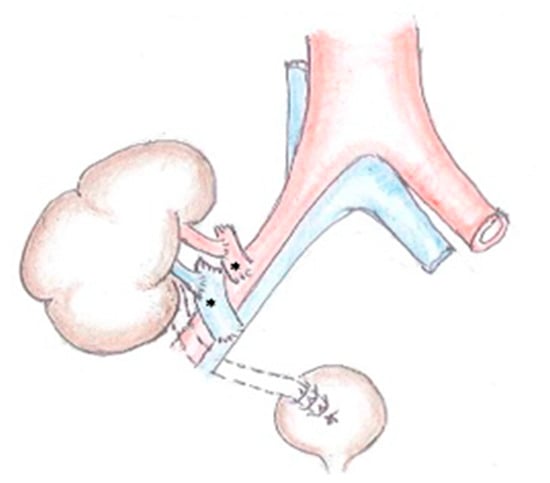
<p>Anatomy of the urinary bladder.</p> Full article ">Figure 3
<p>Papillary urothelial carcinoma of the urinary bladder. Own study based on Ethical Approval of the University of Rzeszow, No. 29/05/2019. Titled 12. 2019, Evaluation of the efficacy of the in vitro photodynamic method in superficial bladder cancer, by M.D. Dominik Godlewski.</p> Full article ">Figure 4
<p>Papillary urothelial carcinoma of the urinary bladder—visible pathological vascularization within the exophytic part of the tumor, as well as at its base. Own study based on Ethical Approval of the University of Rzeszow, No. 29/05/2019. Titled 12. 2019, Evaluation of the efficacy of the in vitro photodynamic method in superficial bladder cancer, by M.D. Dominik Godlewski.</p> Full article ">Figure 5
<p>Multiple papillary urothelial carcinoma of the urinary bladder—in the lower part of the image, at 8 o’clock, the vesical orifice of the right ureter is visible. Own study based on Ethical Approval of the University of Rzeszow, No. 29/05/2019. Titled 12. 2019, Evaluation of the efficacy of the in vitro photodynamic method in superficial bladder cancer, by M.D. Dominik Godlewski.</p> Full article ">Figure 6
<p>Possible symptoms of bladder cancer.</p> Full article ">Figure 7
<p>Bladder cancer stages.</p> Full article ">Figure 8
<p>Number of studies in PubMed for 2020–2024.</p> Full article ">
<p>Bladder cancer incidence (yellow line) and mortality (blue line) in Poland in both sexes.</p> Full article ">Figure 2
<p>Anatomy of the urinary bladder.</p> Full article ">Figure 3
<p>Papillary urothelial carcinoma of the urinary bladder. Own study based on Ethical Approval of the University of Rzeszow, No. 29/05/2019. Titled 12. 2019, Evaluation of the efficacy of the in vitro photodynamic method in superficial bladder cancer, by M.D. Dominik Godlewski.</p> Full article ">Figure 4
<p>Papillary urothelial carcinoma of the urinary bladder—visible pathological vascularization within the exophytic part of the tumor, as well as at its base. Own study based on Ethical Approval of the University of Rzeszow, No. 29/05/2019. Titled 12. 2019, Evaluation of the efficacy of the in vitro photodynamic method in superficial bladder cancer, by M.D. Dominik Godlewski.</p> Full article ">Figure 5
<p>Multiple papillary urothelial carcinoma of the urinary bladder—in the lower part of the image, at 8 o’clock, the vesical orifice of the right ureter is visible. Own study based on Ethical Approval of the University of Rzeszow, No. 29/05/2019. Titled 12. 2019, Evaluation of the efficacy of the in vitro photodynamic method in superficial bladder cancer, by M.D. Dominik Godlewski.</p> Full article ">Figure 6
<p>Possible symptoms of bladder cancer.</p> Full article ">Figure 7
<p>Bladder cancer stages.</p> Full article ">Figure 8
<p>Number of studies in PubMed for 2020–2024.</p> Full article ">


