[go: up one dir, main page]

Next Issue
Volume 10, December
Previous Issue
Volume 10, October


Animals, Volume 10, Issue 11 (November 2020) – 256 articles

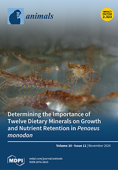
Cover Story (view full-size image): The importance of many dietary minerals has been established in many species. This is not the case for Penaeus monodon, where only three mineral requirements have been reported. Thus, many minerals with known necessity in other prawn species have not been tested in P. monodon. In the past, mineral mixtures have been used to determine the need for individual minerals in the presence of other minerals. Screening designs, such as the Plackett–Burman, provide a mathematically proofed allocation for mineral mixtures to increase the accuracy of the observed mineral response. Although mineral requirements cannot be determined, this design provides an efficient way to scope the importance of many minerals which have not been considered in past mineral research. This study provides fundamental knowledge required to formulate optimised mineral premixes for prawn feeding. View this paper.
  • Issues are regarded as officially published after their release is announced to the table of contents alert mailing list.
  • You may sign up for e-mail alerts to receive table of contents of newly released issues.
  • PDF is the official format for papers published in both, html and pdf forms. To view the papers in pdf format, click on the "PDF Full-text" link, and use the free Adobe Reader to open them.
Order results
Result details
Section
Select all
Export citation of selected articles as:
16 pages, 13876 KiB  
Article
Effect of Phlorotannins from Brown Seaweeds on the In Vitro Digestibility of Pig Feed
by Lauren Ford, Chloe Curry, Mairead Campbell, Katerina Theodoridou, Gary Sheldrake, Jaimie Dick, Lorenzo Stella and Pamela J. Walsh
Animals 2020, 10(11), 2193; https://doi.org/10.3390/ani10112193 - 23 Nov 2020
Cited by 15 | Viewed by 3377
Abstract
Phlorotannins have been reported to have positive effects on pig health, including improved gut health and digestibility. In this study, we investigate the effect of phenolics found in two brown seaweeds, Ascophyllum nodosum and Fucus serratus, on in vitro dry matter digestibility [...] Read more.
Phlorotannins have been reported to have positive effects on pig health, including improved gut health and digestibility. In this study, we investigate the effect of phenolics found in two brown seaweeds, Ascophyllum nodosum and Fucus serratus, on in vitro dry matter digestibility of seaweeds and commercial pig feed. Phlorotannin extracts and whole seaweeds were supplemented into pig feed to test their effect on digestibility. Solid-phase extraction was used to purify the phenolics to phlorotannins. The results showed a slight decrease in the digestibility of pig feed that was found to be significant when phlorotannin extracts were added from either seaweed. However, when whole A. nodosum was added to the pig feed, the effect on digestibility was less pronounced. Specifically, no significant difference in digestibility was observed at inclusion rates up to 5%, and thereafter results varied. A difference in digestibility was also observed in the same species at the same inclusion rate, collected from different seasons. This suggests that other compounds, e.g., polysaccharides, are having an effect on digestibility when whole seaweeds are supplemented to animal feed. This research has also highlighted the need to base supplementation on phenolic concentration as opposed to a standardised percentage inclusion of seaweeds to ensure that digestibility is not adversely affected. Full article
(This article belongs to the Collection Sustainable Animal Nutrition and Feeding)
Show Figures

Figure 1

Figure 1
<p>Correlation of phenolic content in phloroglucinol equivalents mg/g (DM) to in vitro digestibility of dry matter (%IVDMD) for <span class="html-italic">A. nodosum</span> (<b>A</b>) and <span class="html-italic">F. serratus</span> (<b>B</b>).</p>
Full article ">Figure 2
<p>Change in absorbance of the FC assay upon binding to different PEG MW for purified phlorotannins from (<b>A</b>) <span class="html-italic">A. nodosum</span> and (<b>B</b>) <span class="html-italic">F. serratus</span> extracted from samples collected in May.</p>
Full article ">Figure 3
<p>Gel electrophoresis of pepsin with and without binding to the tannins and PEG. From left to right after protein ladder: (<b>a</b>) pure pepsin, (<b>b</b>) pepsin + Asco phlorotannin, (<b>c</b>) pepsin + Fucus phlorotannin, (<b>d</b>) pepsin + Asco phlorotannin + PEG 2000MW, (<b>e</b>) pepsin + Asco phlorotannin + 4000MW, (<b>f</b>) pepsin + Fucus phlorotannin + PEG 2000MW and (<b>g</b>) pepsin + Fucus phlorotannin + PEG 4000MW.</p>
Full article ">Figure 4
<p>In vitro DM digestibility (IVDMD) profiles of (<b>A</b>) <span class="html-italic">A. nodosum</span> and (<b>B</b>) <span class="html-italic">F. serratus</span> with and without PEG. Three different ratios of PEG were used: 1:1, 1:3 and 1:5 phlorotannin:PEG (<span class="html-italic">p</span> &lt; 0.05, * = significant decrease; ♦ = significant increase).</p>
Full article ">Figure 5
<p>In vitro DM digestibility (IVDMD) of a commercial pig feed when different concentrations of phlorotannins isolated from <span class="html-italic">A. nodosum</span> (purple) and <span class="html-italic">F. serratus</span> (green) in May were added. Control (commercial pig feed, grey) (* = <span class="html-italic">p</span> &lt; 0.05; ** = <span class="html-italic">p</span> &lt; 0.001).</p>
Full article ">Figure 6
<p>Change in in vitro DM digestibility (IVDMD) of a commercial pig feed when different percentages of <span class="html-italic">A. nodosum</span> were added from samples collected in Jan and May. * = <span class="html-italic">p</span> &lt; 0.05 between control and % inclusion; ⋆ = <span class="html-italic">p</span> &lt; 0.05 between pairs.</p>
Full article ">
9 pages, 1390 KiB  
Article
A Descriptive Study of Keel Bone Fractures in Hens and Roosters from Four Non-Commercial Laying Breeds Housed in Furnished Cages
by Käthe Elise Kittelsen, Randi Oppermann Moe, Tone Beate Hansen, Ingrid Toftaker, Jens Peter Christensen and Guro Vasdal
Animals 2020, 10(11), 2192; https://doi.org/10.3390/ani10112192 - 23 Nov 2020
Cited by 6 | Viewed by 3544
Abstract
The presence of keel bone fractures (KBF) in laying hens has been documented and discussed by several authors, nevertheless the causative factors behind KBF remain uncertain. High prevalence of KBF have been reported in all commercial egg production systems, in different genetic lines [...] Read more.
The presence of keel bone fractures (KBF) in laying hens has been documented and discussed by several authors, nevertheless the causative factors behind KBF remain uncertain. High prevalence of KBF have been reported in all commercial egg production systems, in different genetic lines and at different ages. Several of the proposed causal mechanisms behind KBF are linked to selection for efficient production. It is, therefore, of interest to explore whether less selected breeds have a lower occurrence of keel bone fractures compared to reports from highly selected, modern laying hen breeds. Thus, the aim of the current study was to investigate keel bones of hens from four non-commercial layer breeds. Birds were housed in furnished cages and keel bones examined at 30 and 63 weeks of age, using a portable X-ray equipment. The results from this descriptive study indicate a low prevalence of KBF at both ages in all four breeds, with only five KBF detected in 213 X-ray pictures taken from 126 birds. Of these, four of the KBF were observed in the most genetically selected breed, with an early onset of lay. None of the roosters examined exhibited KBF. The overall low numbers of KBF found indicate that genetic factors may be involved in KBF and, thus that selective breeding may help to reduce the susceptibility to KBF. Finally, this study highlights the importance of poultry conservation to secure genetic diversity, which may be an important resource in future selection schemes. Full article
(This article belongs to the Special Issue Recent Advances in Poultry Welfare)
Show Figures

Figure 1

Figure 1
<p>Normal, unfractured keel bone, 63 weeks of age.</p>
Full article ">Figure 2
<p>A keel bone with one fracture, 30 weeks of age.</p>
Full article ">Figure 3
<p>A keel bone with multiple fractures, age 63 weeks of age.</p>
Full article ">
17 pages, 2966 KiB  
Article
Influence of Parental Fatty Acid Desaturase 2 (fads2) Expression and Diet on Gilthead Seabream (Sparus aurata) Offspring fads2 Expression during Ontogenesis
by Hanlin Xu, Shajahan Ferosekhan, Serhat Turkmen, Juan Manuel Afonso, María Jesús Zamorano and Marisol Izquierdo
Animals 2020, 10(11), 2191; https://doi.org/10.3390/ani10112191 - 23 Nov 2020
Cited by 6 | Viewed by 2879
Abstract
Previous studies have shown that it is possible to increase the ability of marine fish to produce long-chain polyunsaturated fatty acid from their 18C precursors by nutritional programming or using broodstock with a higher fatty acyl desaturase 2 (fads2) expression. However, [...] Read more.
Previous studies have shown that it is possible to increase the ability of marine fish to produce long-chain polyunsaturated fatty acid from their 18C precursors by nutritional programming or using broodstock with a higher fatty acyl desaturase 2 (fads2) expression. However, those studies failed to show the effect of these interventions on the expression of the fads2 gene in the developing egg. Moreover, there were no studies on the temporal expression of the fads2 during ontogeny in the gilthead sea bream (Sparus aurata). In order to determine the changes in expression of fads2 during ontogeny, gilthead sea bream broodstock with a high (HRO) or low (LRO) fads2 expression fed a diet previously used for nutritional programming, or a fish oil-based diet (LFO) were allowed to spawn. The samples were taken at the stages of spawning, morula, high blastula, gastrula, neurula, heart beating, hatch and 3 day-old first exogenous feeding larvae to determine fads2 expression throughout embryonic development. The results showed the presence of fads2 mRNA in the just spawned egg, denoting the maternal mRNA transfer to the developing oocyte. Later, fads2 expression increased after the neurula, from heart beating until 3-day-old larvae, denoting the transition from maternal to embryonic gene expression. In addition, the eggs obtained from broodstock with high fads2 expression showed a high docosahexaenoic acid content, which correlated with the downregulation of the fads2 expression found in the developing embryo and larvae. Finally, feeding with the nutritional programming diet with the partial replacement of fish oil by rapeseed oil did not affect the long chain polyunsaturated fatty acid (LC-PUFA) contents nor fads2 expression in the gilthead sea bream developing eggs. Full article
(This article belongs to the Special Issue Nutrition, Nutrigenomics and Epigenetics in Fish)
Show Figures

Figure 1

Figure 1
<p>Timeline of the selected point during embryogenesis. The timeline is based on the description of stage during the embryogenesis of gilthead sea bream by Kamacı et al. 2005 [<a href="#B35-animals-10-02191" class="html-bibr">35</a>] under experimental conditions.</p>
Full article ">Figure 2
<p>Temporal changes in <span class="html-italic">fatty acid desaturase 2</span> (<span class="html-italic">fads2</span>) mRNA copies during gilthead seabream (<span class="html-italic">Sparus aurata</span>) ontogenesis, from 0 to 125 h after spawning. Different letters denote significant differences among developmental stages (<span class="html-italic">p</span> &lt; 0.05, <span class="html-italic">n</span> = 15). Bars bearing with different letter showed significant difference by one-way ANOVA analysis (<span class="html-italic">p</span> &lt; 0.05). y, <span class="html-italic">fads2</span> expression of egg; x, time after spawning; R<sup>2</sup>, coefficient of determination.</p>
Full article ">Figure 3
<p>Influence of parental <span class="html-italic">fatty acid desaturase 2</span> (<span class="html-italic">fads2</span>) expression (high: HRO or low: LRO) on temporal changes in <span class="html-italic">fads2</span> mRNA copies during gilthead seabream (<span class="html-italic">Sparus aurata</span>) ontogenesis, from 0 to 125 h after spawning (<span class="html-italic">n</span> = 3). No significant difference was observed in the comparison using t-test (<span class="html-italic">p</span> &gt; 0.05). y, <span class="html-italic">fads2</span> expression of egg; x, time after spawning; R<sup>2</sup>, coefficient of determination.</p>
Full article ">Figure 4
<p>Relations of <span class="html-italic">fads2</span> mRNA copies between the egg at neurula stage (16 hps) and female (<b>a</b>) or male (<b>b</b>) broodstock (including both HRO and LRO broodstock), or between egg at the heart appearance stage (30 hps) and female (<b>c</b>) or male (<b>d</b>) broodstock. y, <span class="html-italic">fads2</span> expression of egg; x, <span class="html-italic">fads2</span> expression of broodstock; R<sup>2</sup>, coefficient of determination.</p>
Full article ">Figure 5
<p>Relations between the DHA contents in the egg at 24 h after spawning and <span class="html-italic">fads2</span> mRNA copies of female (<b>a</b>) or male (<b>b</b>) broodstock (including both HRO and LRO broodstock). y, DHA content is eggs; x, <span class="html-italic">fads2</span> expression of broodstock; R<sup>2</sup>, coefficient of determination.</p>
Full article ">Figure 6
<p>Relations between the <span class="html-italic">fads2</span> mRNA copies of egg at 16 h after spawning and the content of DHA (<b>a</b>), ratio 20:4n-3/20:3n-3 (<b>b</b>), 18:2n-9 (<b>c</b>) and 18:3n-6 (<b>d</b>) in eggs at 24 h after spawning (including eggs from both HRO and LRO broodstock). y, respective fatty acids in eggs at 24 h after spawning; x, <span class="html-italic">fads2</span> expression of egg at 16 h after spawning; R<sup>2</sup>, coefficient of determination.</p>
Full article ">Figure 7
<p>Relations between <span class="html-italic">fads2</span> mRNA copies of egg at 30 h after spawning with the content of DHA (<b>a</b>), 18:2n-9 (<b>b</b>), ratio 18:3n-6/18:2n-6 (<b>c</b>) and 18:4n-3/18:3n-3 (<b>d</b>) in eggs at 24 h after spawning (including eggs from both HRO and LRO broodstock). y, respective fatty acids in eggs at 24 h after spawning; x, <span class="html-italic">fads2</span> expression of eggs at 30 h after spawning; R<sup>2</sup>, coefficient of determination.</p>
Full article ">Figure 7 Cont.
<p>Relations between <span class="html-italic">fads2</span> mRNA copies of egg at 30 h after spawning with the content of DHA (<b>a</b>), 18:2n-9 (<b>b</b>), ratio 18:3n-6/18:2n-6 (<b>c</b>) and 18:4n-3/18:3n-3 (<b>d</b>) in eggs at 24 h after spawning (including eggs from both HRO and LRO broodstock). y, respective fatty acids in eggs at 24 h after spawning; x, <span class="html-italic">fads2</span> expression of eggs at 30 h after spawning; R<sup>2</sup>, coefficient of determination.</p>
Full article ">Figure 8
<p>Changes in <span class="html-italic">fads2</span> expression during gilthead seabream (<span class="html-italic">Sparus aurata</span>) ontogenesis, from 0 to 125 h after spawning (<span class="html-italic">n</span> = 3) from broodstock with a low expression of <span class="html-italic">fads2</span> and fed either a FO (LFO) or a RO (LRO) diet during spawning. y, <span class="html-italic">fads2</span> expression of egg; x, time after spawning; R<sup>2</sup>, coefficient of determination.</p>
Full article ">
18 pages, 2857 KiB  
Article
Alleviation of the Adverse Effect of Dietary Carbohydrate by Supplementation of Myo-Inositol to the Diet of Nile Tilapia (Oreochromis niloticus)
by Jiahua Zhu, Jingyu Pan, Xiaodan Wang, Yuxing Huang, Chuanjie Qin, Fang Qiao, Jianguang Qin and Liqiao Chen
Animals 2020, 10(11), 2190; https://doi.org/10.3390/ani10112190 - 23 Nov 2020
Cited by 20 | Viewed by 3133
Abstract
This study investigated the effect of dietary myo-inositol (MI) on alleviating the adverse effect of the high carbohydrate diet in Nile tilapia (Oreochromis niloticus). Six diets contained either low carbohydrate (LC 30%) or high carbohydrate (HC 45%) with three levels [...] Read more.
This study investigated the effect of dietary myo-inositol (MI) on alleviating the adverse effect of the high carbohydrate diet in Nile tilapia (Oreochromis niloticus). Six diets contained either low carbohydrate (LC 30%) or high carbohydrate (HC 45%) with three levels of MI supplementation (0, 400 and 1200 mg/kg diet) to each level of the carbohydrate diet. After an 8-week trial, the fish fed 400 mg/kg MI under HC levels had the highest weight gain and fatness, but the fish fed 1200 mg/kg MI had the lowest hepatosomatic index, visceral index and crude lipid in the HC group. The diet of 1200 mg/kg MI significantly decreased triglyceride content in the serum and liver compared with those fed the MI supplemented diets regardless of carbohydrate levels. Dietary MI decreased triglyceride accumulation in the liver irrespective of carbohydrate levels. The content of malondialdehyde decreased with increasing dietary MI at both carbohydrate levels. Fish fed 1200 mg/kg MI had the highest glutathione peroxidase, superoxide dismutase, aspartate aminotransferase and glutamic-pyruvic transaminase activities. The HC diet increased the mRNA expression of key genes involved in lipid synthesis (DGAT, SREBP, FAS) in the fish fed the diet without MI supplementation. Dietary MI significantly under expressed fatty acid synthetase in fish fed the HC diets. Moreover, the mRNA expression of genes related to lipid catabolism (CPT, ATGL, PPAR-α) was significantly up-regulated with the increase of dietary MI levels despite dietary carbohydrate levels. The gene expressions of gluconeogenesis, glycolysis and MI biosynthesis were significantly down-regulated, while the expression of the pentose phosphate pathway was up-regulated with the increase of MI levels. This study indicates that HC diets can interrupt normal lipid metabolism and tend to form a fatty liver in fish. Dietary MI supplement can alleviate lipid accumulation in the liver by diverging some glucose metabolism into the pentose phosphate pathway and enhance the antioxidant capacity in O. niloticus. Full article
Show Figures

Figure 1

Figure 1
<p>Histological characteristics of liver (40×). (<b>A</b>) LC-0 mg/kg; (<b>B</b>) LC-400 mg/kg; (<b>C</b>) LC-1200 mg/kg; (<b>D</b>) HC-0 mg/kg; (<b>E</b>) HC-400 mg/kg; (<b>F</b>) HC-1200 mg/kg; (<b>G</b>) lipid droplet area of the section area. Double asterisks (**) represent a significant difference of <span class="html-italic">p</span> &lt; 0.01 between same level of MI groups. a, b and A, B Values on bars without a common superscript letter are significantly different (<span class="html-italic">p</span> &lt; 0.05).</p>
Full article ">Figure 2
<p>Effects of <span class="html-italic">myo</span>-inositol at different carbohydrate levels on mRNA levels of genes involved in lipid metabolism parameters in the liver of <span class="html-italic">O. niloticus</span>. Values are means (<span class="html-italic">n</span> = 3 replicate tanks) with standard errors represented by vertical bars. Asterisk (*) represents a significant difference of <span class="html-italic">p</span> &lt; 0.05 between same level of MI groups. Double asterisks (**) represent a significant difference of <span class="html-italic">p</span> &lt; 0.01 between same level of MI groups. a, b, c and A, B Values on bars without a common superscript letter are significantly different (<span class="html-italic">p</span> &lt; 0.05) (a/A indicated the lowest value). (<b>A</b>) <span class="html-italic">DGAT2</span>: diacyltransferase; (<b>B</b>) <span class="html-italic">FAS</span>: fatty acid synthetase; (<b>C</b>) <span class="html-italic">SREBP</span>: sterol regulatory element binding protein; (<b>D</b>) <span class="html-italic">CPT</span>: carnitine palmityl transferase; (<b>E</b>) <span class="html-italic">PPAR-α</span>: peroxisome proliferator activated receptor-α; (<b>F</b>) <span class="html-italic">ATGL</span>: triglyceride lipase.</p>
Full article ">Figure 3
<p>Effects of <span class="html-italic">myo</span>-inositol at different carbohydrate levels on mRNA levels of genes involved in glucose metabolism in the liver of <span class="html-italic">O. niloticus</span>. Values are means (<span class="html-italic">n</span> = 3 replicate tanks) with standard errors represented by vertical bars. Asterisk (*) represents a significant difference of <span class="html-italic">p</span> &lt; 0.05 between same level of MI groups. a, b and A, B Values on bars without a common superscript letter are significantly different (<span class="html-italic">p</span> &lt; 0.05) (a/A indicated the lowest value). (<b>A</b>) <span class="html-italic">GK</span>: hexokinase; (<b>B</b>) <span class="html-italic">PK</span>: pyruvate kinase; (<b>C</b>) <span class="html-italic">G6Pase</span>: glucose-6-phosphatase; (<b>D</b>) <span class="html-italic">G6PDH</span>: glucose-6-phosphate dehydrogenase; (<b>E</b>) <span class="html-italic">MIPS</span>: <span class="html-italic">myo</span>-inositol-1-phosphate synthase; (<b>F</b>) <span class="html-italic">IMPA1</span>: <span class="html-italic">myo</span>-inositol monophosphatase.</p>
Full article ">Figure 4
<p>Effects of <span class="html-italic">myo</span>-inositol at different carbohydrate levels on immune-related parameters in the liver of <span class="html-italic">O. niloticus</span>. Values are means (<span class="html-italic">n</span> = 3 replicate tanks) with standard errors represented by vertical bars. Asterisk (*) represents a significant difference of <span class="html-italic">p</span> &lt; 0.05 between same level of MI groups. a, b and A, B Values on bars without a common superscript letter are significantly different (<span class="html-italic">p</span> &lt; 0.05) (a/A indicated the lowest value). (<b>A</b>) MDA: malonaldehyde. (<b>B</b>) GSH-Px: glutathione peroxidase; (<b>C</b>) SOD: superoxide dismutase; (<b>D</b>) AST/GOT: spartate aminotransferase; (<b>E</b>) ALT/GPT: glutamic-pyruvic transaminase; (<b>F</b>) AKP: alkaline phosphatase; (<b>G</b>) ACP: Acid phosphatas.</p>
Full article ">
6 pages, 416 KiB  
Article
Serum Anti-Mullerian Hormone and Estradiol Concentrations in Gilts and Their Age at Puberty
by Nutthee Am-in, Junpen Suwimonteerabutr and Roy N. Kirkwood
Animals 2020, 10(11), 2189; https://doi.org/10.3390/ani10112189 - 23 Nov 2020
Cited by 7 | Viewed by 2448
Abstract
For experiment one, blood samples were obtained from 200 gilts at 90, 120, 150, 180, and 200 days of age. Serum samples from the 30 youngest (166.1 ± 0.7 days) and 30 oldest (198.8 ± 0.6 days) gilts exhibiting estrus by 200 days, [...] Read more.
For experiment one, blood samples were obtained from 200 gilts at 90, 120, 150, 180, and 200 days of age. Serum samples from the 30 youngest (166.1 ± 0.7 days) and 30 oldest (198.8 ± 0.6 days) gilts exhibiting estrus by 200 days, and a further 18 gilts that remained anestrus at 200 days, were assayed for serum concentrations of anti-Mullerian hormone (AMH) and estradiol (E2). Gilts younger at puberty had higher (p < 0.05) AMH levels than those older at puberty, and both groups had higher AMH levels than anestrus gilts (p < 0.05). Regardless of age, serum E2 was higher (p < 0.05) in gilts that achieved puberty than in gilts remaining anestrus. At spontaneous pubertal estrus detection, there was no effect of pubertal age on the number of preovulatory ovarian follicles. For experiment two, 152 prepubertal gilts received an intramuscular (IM) injection of 400 IU eCG plus 200 IU hCG and then received fence-line boar contact to detect estrus onset. Serum AMH concentrations were higher (p < 0.05) in the first 25 gilts to exhibit puberty than the last 28 gilts, with the first gilts also having more preovulatory follicles (p < 0.0001). Taken together, these data support an association between serum AMH concentrations and degree of physiological maturity and ovarian follicular development in gilts. Full article
(This article belongs to the Special Issue Advances in Pig Reproduction)
Show Figures

Figure 1

Figure 1
<p>Distribution of intervals to estrus detection in gilts following injection of 400 IU eCG plus 200 IU hCG (PG600).</p>
Full article ">
15 pages, 423 KiB  
Commentary
Dog Welfare, Well-Being and Behavior: Considerations for Selection, Evaluation and Suitability for Animal-Assisted Therapy
by Melissa Winkle, Amy Johnson and Daniel Mills
Animals 2020, 10(11), 2188; https://doi.org/10.3390/ani10112188 - 23 Nov 2020
Cited by 33 | Viewed by 15585
Abstract
Health care and human service providers may include dogs in formal intervention settings to positively impact human physical, cognitive and psychosocial domains. Dogs working within this context are asked to cope with a multitude of variables including settings, populations, activities, and schedules. In [...] Read more.
Health care and human service providers may include dogs in formal intervention settings to positively impact human physical, cognitive and psychosocial domains. Dogs working within this context are asked to cope with a multitude of variables including settings, populations, activities, and schedules. In this article, the authors highlight how both the preparation and operation of dogs within animal-assisted therapy (AAT) differs from less structured animal-assisted activities (AAA) and more exclusive assistance animal work; the authors highlight the gaps in our knowledge in this regard, and propose an ethically sound framework for pragmatic solutions. This framework also emphasizes the need for good dog welfare to safeguard all participants. If dogs are not properly matched to a job or handler, they may be subjected to unnecessary stress, anxiety, and miscommunication that can lead to disinterest in the work, overt problematic behavioral or health outcomes, or general unsuitability. Such issues can have catastrophic outcomes for the AAT. The authors propose standards for best practices for selection, humane-based preparation and training, and ongoing evaluation to ensure the health, welfare and well-being of dogs working in AAT, which will have concomitant benefits for clients and the professionalism of the field. Full article
(This article belongs to the Special Issue Companion animals welfare and behaviour)
Show Figures

Figure 1

Figure 1
<p>Framework for selection, preparation and evaluation of dogs working in AAT.</p>
Full article ">
14 pages, 980 KiB  
Article
What Factors Predispose Households in Trans-Himalaya (Central Nepal) to Livestock Predation by Snow Leopards?
by Mahesh P. Tiwari, Bishnu P. Devkota, Rodney M. Jackson, Bir Bahadur Khanal Chhetri and Sistata Bagale
Animals 2020, 10(11), 2187; https://doi.org/10.3390/ani10112187 - 23 Nov 2020
Cited by 11 | Viewed by 6121
Abstract
Livestock depredation across the trans-Himalaya causes significant economic losses to pastoralist communities. Quantification of livestock predation and the assessment of variables associated with depredation are crucial for designing effective long-term mitigation measures. We investigated the patterns and factors of livestock depredation by snow [...] Read more.
Livestock depredation across the trans-Himalaya causes significant economic losses to pastoralist communities. Quantification of livestock predation and the assessment of variables associated with depredation are crucial for designing effective long-term mitigation measures. We investigated the patterns and factors of livestock depredation by snow leopards (Panthera uncia) using semi-structured questionnaires targeting herders in the Narphu valley of the Annapurna Conservation Area, Nepal. During the two years (2017/18 and 2018/19), 73.9% of the households interviewed (n = 65) lost livestock to snow leopards, with an annual average loss of two livestock per household. Of the total depredation attributed to snow leopards, 55.4% were yak (mainly female: 79%), 31.7% goat, 6.8% sheep, 3.2% horse and 2.8% cattle. Results from applying Generalized Linear Mixed Models (GLMMs) revealed the total number of livestock owned and the number of larger bodied livestock species as the main explanatory covariates explaining livestock depredation. Forty-one (41%) of all herders considered snow leopard’s preference for domestic livestock as the main factor in livestock predation, whereas only 5% perceived poor herding practice as the main reason for the loss. Our study found poor and changing herding practices in the valley, whereby 71% herders reported careful herding as a solution to snow leopard depredation, and 15% of herders considered the complete extermination of snow leopards as the best solution to the problem. Tolerance levels and awareness among herders towards snow leopard conservation is increasing, mainly due to the Buddhist religion and strict law enforcement within this protected area. We recommend the effective implementation of a community-based livestock insurance scheme to compensate the economic loss of herders due to predation and improved herding practices as the recommended mitigation measures for ensuring livestock security and snow leopards’ conservation in the valley. Full article
(This article belongs to the Section Human-Animal Interactions, Animal Behaviour and Emotion)
Show Figures

Figure 1

Figure 1
<p>Map showing the location of study area, Narphu valley, Gandaki Province, Nepal.</p>
Full article ">Figure 2
<p>Depredation of livestock by snow leopards across seasons among surveyed households (<span class="html-italic">n</span> = 65) during years 2017/18 and 2018/19.</p>
Full article ">
12 pages, 1567 KiB  
Article
Genome-Wide Association Analysis Identified BMPR1A as a Novel Candidate Gene Affecting the Number of Thoracic Vertebrae in a Large White × Minzhu Intercross Pig Population
by Qian Liu, Jingwei Yue, Naiqi Niu, Xin Liu, Hua Yan, Fuping Zhao, Xinhua Hou, Hongmei Gao, Lijun Shi, Lixian Wang, Ligang Wang and Longchao Zhang
Animals 2020, 10(11), 2186; https://doi.org/10.3390/ani10112186 - 22 Nov 2020
Cited by 15 | Viewed by 2748
Abstract
The number of vertebrae (NV), especially the number of thoracic vertebrae (NTV), varies among pig breeds. The NTV is controlled by vertebral segmentation and the number of somites during embryonic development. Although there is a high correlation between the NTV and NV, studies [...] Read more.
The number of vertebrae (NV), especially the number of thoracic vertebrae (NTV), varies among pig breeds. The NTV is controlled by vertebral segmentation and the number of somites during embryonic development. Although there is a high correlation between the NTV and NV, studies on a fixed NV have mainly considered the absolute numbers of thoracic vertebrae instead of vertebral segmentation. Therefore, this study aimed to discover variants associated with the NTV by considering the effect of the NV in pigs. The NTV and NV of 542 F2 individuals from a Large White × Minzhu pig crossbreed were recorded. All animals were genotyped for VRTN g.19034 A > C, LTBP2 c.4481A > C, and 37 missense or splice variants previously reported in a 951-kb interval on SSC7 and 147 single nucleotide polymorphisms (SNPs) on SSC14. To identify NTV-associated SNPs, we firstly performed a genome-wide association study (GWAS) using the Q + K (population structure + kinship matrix) model in TASSEL. With the NV as a covariate, the obtained data were used to identify the SNPs with the most significant genome-wide association with the NTV by performing a GWAS on a PorcineSNP60K Genotyping BeadChip. Finally, a conditional GWAS was performed by fixing this SNP. The GWAS showed that 31 SNPs on SSC7 have significant genome-wide associations with the NTV. No missense or splice variants were found to be associated with the NTV significantly. A linkage disequilibrium analysis suggested the existence of quantitative trait loci (QTL) in a 479-Kb region on SSC7, which contained a critical candidate gene FOS for the NTV in pigs. Subsequently, a conditional GWAS was performed by fixing M1GA0010658, the most significant of these SNPs. Two SNPs in BMPR1A were found to have significant genome-wide associations and a significant dominant effect. The leading SNP, S14_87859370, accounted for 3.86% of the phenotypic variance. Our study uncovered that regulation variants in FOS on SSC7 and in BMPR1A on SSC14 might play important roles in controlling the NTV, and thus these genetic factors may be harnessed for increasing the NTV in pigs. Full article
Show Figures

Figure 1

Figure 1
<p>The Manhattan plot for genome-wide association studies for the NTV. The genome-wide association study (GWAS) was run on the NV treated as a covariate based on the 147 SNPs on SSC17 and 39 variants on SSC7 that merged with porcine SNP60K genotyping BeadChip assays. The red horizontal line indicated the Bonferroni significance threshold (1.10 × 10<sup>−6</sup>).</p>
Full article ">Figure 2
<p>Haplotypes on a 25.76-Mb region on SSC7 containing all SNPs associated with the NTV significantly. The analysis was obtained using the HAPLOVIEW 3.31 program. A total of four blocks were identified and marked with solid lines.</p>
Full article ">Figure 3
<p>Identification of <span class="html-italic">BMPR1A</span> using conditional GWAS of the NTV. (<b>A</b>) Manhattan plot displaying the GWAS results of the NTV. The red horizontal line indicated the Bonferroni significance threshold (1.10 × 10<sup>−</sup><sup>6</sup>). Dots represent SNPs and the S14_87859370 is highlighted at the top dot. (<b>B</b>) The Q-Q plots obtained from the conditional GWAS for the NTV. (<b>C</b>) Red arrow represents the most significant SNP signal of the NTV. The S14_87859370 in the intron 4 of BMPR1A is marked in red. (<b>D</b>) The difference analysis among three genotypes of S14_87859370 for NTV in Songliao Black (SLB) and Large White (LW) pig populations. * indicates <span class="html-italic">p</span> &lt; 0.05. (<b>E</b>) Allele frequencies of S14_87859370 in SLB and LW pig populations.</p>
Full article ">
7 pages, 232 KiB  
Communication
Streptococcus dysgalactiae—Contagious or Environmental?
by Nicole Wente and Volker Krömker
Animals 2020, 10(11), 2185; https://doi.org/10.3390/ani10112185 - 22 Nov 2020
Cited by 18 | Viewed by 3507
Abstract
Streptococcus dysgalactiae is among the most important pathogens causing bovine mastitis. Unfortunately, there is presently a lack of clear knowledge about the mode of transmission—contagious or environmental—of this pathogen. To obtain more information on this, knowledge of the genetic diversity of the isolated [...] Read more.
Streptococcus dysgalactiae is among the most important pathogens causing bovine mastitis. Unfortunately, there is presently a lack of clear knowledge about the mode of transmission—contagious or environmental—of this pathogen. To obtain more information on this, knowledge of the genetic diversity of the isolated microorganisms at the farm level can be useful. To observe the strain variety in different herds of cattle, isolates of Strep. dysgalactiae were collected from clinical mastitis samples at different farms, and the strains were typed using the pulsed-field gel electrophoresis (PFGE) method. Overall, we performed strain typing on 93 isolates from 16 farms in Germany and used an index to describe the degree of contagiosity of Strep. dysgalactiae at each farm. This index (CI) represents the number of isolates divided by the number of strains found in mastitis milk of clinical cases within a period of 14 months. The results differed between the farms. In one farm, all six Strep. dysgalactiae cases that occurred during the study period were caused by a single strain (CI = 6), while in another farm the six cases that occurred were caused by five different strains (CI = 1.2). All other farms fell between these two extremes. This indicates that Strep. dysgalactiae infections can occur via several routes of transmission. At the farm level, strain comparisons are necessary to determine the routes of transmission. Two strains were able to survive on the farm for a minimum of 14 months. Full article
(This article belongs to the Special Issue The Role of Microorganism in Bovine Mastitis)
16 pages, 3616 KiB  
Article
Liver Expression of IGF2 and Related Proteins in ZBED6 Gene-Edited Pig by RNA-Seq
by Haidong Zhao, Mingli Wu, Shirong Liu, Xiaoqin Tang, Xiaohua Yi, Qi Li, Shuhui Wang and Xiuzhu Sun
Animals 2020, 10(11), 2184; https://doi.org/10.3390/ani10112184 - 22 Nov 2020
Cited by 5 | Viewed by 2925
Abstract
Zinc finger BED-type containing 6 (ZBED6), a highly conservative transcription factor of placental mammals, has conservative interaction of insulin-like growth factor 2 (IGF2) based on the 16 bp binding sites of ZBED6 on the IGF2 sequence. IGF2 is related [...] Read more.
Zinc finger BED-type containing 6 (ZBED6), a highly conservative transcription factor of placental mammals, has conservative interaction of insulin-like growth factor 2 (IGF2) based on the 16 bp binding sites of ZBED6 on the IGF2 sequence. IGF2 is related to embryo growth and cell proliferation. At the same time, its functions in muscle and adipose in mammals have been widely mentioned in recent studies. To further investigate the mechanism of ZBED6 on IGF2, we detected the expression of IGF2 and related genes in ZBED6 single allele knockout (ZBED6-SKO) pig tissues and analyzed the transcriptome of ZBED6-SKO pig liver. Through RNA-seq, we captured nine up-regulated genes and eight down-regulated genes which related to lipid metabolism. The results showed that the mRNA of IGF2 had an upward trend after the partial knockout of ZBED6 in liver and had no significant difference in protein expression of IGF2. In summary, ZBED6-SKO could affect the secretion of IGF2 in pig liver and its own lipid metabolism. Our research has provided basic information for revealing the regulatory mechanism of the interaction between ZBED6 and IGF2 in mammals. Full article
(This article belongs to the Special Issue Pig Genetics)
Show Figures

Figure 1

Figure 1
<p><span class="html-italic">ZBED6</span> and <span class="html-italic">IGF2</span> expression in <span class="html-italic">ZBED6</span>-single allele knockout (<span class="html-italic">ZBED6</span>-SKO) and WT pigs. (<b>A</b>) The schematic of <span class="html-italic">ZBED6</span> partial knockout efficiency detection; (<b>B</b>) <span class="html-italic">ZBED6</span> partial knockout efficiency in <span class="html-italic">ZBED6</span>-SKO and WT pigs in five tissues; (<b>C</b>) <span class="html-italic">IGF2</span> expression in <span class="html-italic">ZBED6</span>-SKO and WT pigs in five tissues; (<b>D</b>) Western blotting of IGF2 in <span class="html-italic">ZBED6</span>-SKO and WT pigs in five tissues; (<b>E</b>) the metabolism of <span class="html-italic">IGF2</span> in <span class="html-italic">ZBED6</span>-KO pigs. <span class="html-italic">ZBED6</span>: <span class="html-italic">zinc finger</span>, <span class="html-italic">BED-type containing 6</span>; <span class="html-italic">IGF2</span>: <span class="html-italic">Insulin-like growth factor 2</span>; KO: biallelic knockout; SKO: one allele knockout; (+/+): wild type; (+/-): single knock type. ** (<span class="html-italic">p</span> &lt; 0.01).</p>
Full article ">Figure 2
<p>qPCR of <span class="html-italic">IGF2</span> and related genes expression in <span class="html-italic">ZBED6</span>-SKO and WT pig tissues. (<b>A</b>) The expression of <span class="html-italic">IGF1</span> in five tissues of <span class="html-italic">ZBED6</span>-SKO and WT pigs. (<b>B</b>) The expression of <span class="html-italic">IGF1R</span> in five tissues of <span class="html-italic">ZBED6</span>-SKO and WT pigs. (<b>C</b>) The expression of <span class="html-italic">IGF2R</span> in five tissues of <span class="html-italic">ZBED6</span>-SKO and WT pigs. (<b>D</b>) The expression of <span class="html-italic">IGFBP1</span> in five tissues of <span class="html-italic">ZBED6</span>-SKO and WT pigs. (<b>E</b>) The expression of <span class="html-italic">IGFBP2</span> in five tissues of <span class="html-italic">ZBED6</span>-SKO and WT pigs. (<b>F</b>) The expression of <span class="html-italic">IGFBP3</span> in five tissues of <span class="html-italic">ZBED6</span>-SKO and WT pigs. (<b>G</b>) The expression of <span class="html-italic">IGFBP4</span> in five tissues of <span class="html-italic">ZBED6</span>-SKO and WT pigs. (<b>H</b>) The expression of <span class="html-italic">IGFBP5</span> in five tissues of <span class="html-italic">ZBED6</span>-SKO and WT pigs. (<b>I</b>) The expression of <span class="html-italic">IGFBP7</span> in five tissues of <span class="html-italic">ZBED6</span>-SKO and WT pigs. Notes: IGF1: insulin-like growth factor 1; IGF1R: insulin like growth factor 1 receptor; IGF2R: insulin like growth factor 2 receptor; IGFBP 1-7: insulin-like growth factor binding protein 1-7; SKO: one allele knockout; WT: wild type; (+/+): wild type; (+/-): single knock type.</p>
Full article ">Figure 3
<p>RNA-seq of IGF2 and related genes expression in <span class="html-italic">ZBED6</span>-SKO and WT pig livers. (<b>A</b>) The expression (FPKM) of IGF1, IGF2 and INS in <span class="html-italic">ZBED6</span>-SKO and WT pig livers. (<b>B</b>) The expression (FPKM) of IGF1R, IGF2R and INSR in <span class="html-italic">ZBED6</span>-SKO and WT pig livers. (<b>C</b>) The expression (FPKM) of IGFBP family and the total expression (FPKM) of IGFBPs in <span class="html-italic">ZBED6</span>-SKO and WT pig livers. Notes: SKO: one allele knockout; WT: wild type; FPKM: fragments per kilobase per million; IGF1-2: insulin-like growth factor 1-2; IGF1R: insulin like growth factor 1 receptor; IGF2R: insulin like growth factor 2 receptor; INS: insulin; INSR: insulin receptor; IGFBP1-7: insulin-like growth factor binding protein 1-7; (+/+): wild type; (+/-): single knock type.</p>
Full article ">Figure 4
<p>Volcano plot and heatmap of differentially expressed genes (DEGs) between <span class="html-italic">ZBED6</span>-SKO and WT pig livers. (<b>A</b>) Volcano plot of all significant DEGs (<span class="html-italic">P</span> &lt; 0.05) between <span class="html-italic">ZBED6</span>-SKO and WT pig livers, including 139 up-regulated genes and 97 down-regulated genes. (<b>B</b>) Volcano plot of all significant DEGs after the FDR correction (FDR &lt; 0.05) between <span class="html-italic">ZBED6</span>-SKO and WT pig livers, including 9 up-regulated genes and 8 down-regulated genes. (<b>C</b>) Heatmap of all significant DEGs (<span class="html-italic">P</span> &lt; 0.05). Notes: FDR: false discovery rates; FC: fold change; TPM: transcripts per million reads.</p>
Full article ">Figure 5
<p>Gene set enrichment analysis (GSEA) analysis between <span class="html-italic">ZBED6</span>-SKO and WT pig livers (<b>A</b>) up-regulated signaling pathway enriched in <span class="html-italic">ZBED6</span>-SKO pig liver; (<b>B</b>) down-regulated signaling pathway enriched in <span class="html-italic">ZBED6</span>-SKO pig liver. Notes: NES: normalized enrichment score; FDR: false discovery rates.</p>
Full article ">Figure 6
<p>Secretion and function of IGF2 in <span class="html-italic">ZBED6</span>-SKO pig hepatocyte and muscle cells.</p>
Full article ">
18 pages, 4277 KiB  
Article
Maternal Folic Acid Supplementation Differently Affects the Small Intestinal Phenotype and Gene Expression of Newborn Lambs from Differing Litter Sizes
by Zhen Li, Bo Wang, Heqiong Li, Luyang Jian, Hailing Luo, Bing Wang, Can Zhang, Xingang Zhao, Ying Xue, Sijia Peng and Shuxian Zuo
Animals 2020, 10(11), 2183; https://doi.org/10.3390/ani10112183 - 22 Nov 2020
Cited by 6 | Viewed by 2372
Abstract
The purpose of this study was to investigate the effect of maternal dietary folic acid (FA) supplementation during gestation on small intestinal development of newborn lambs of different litter sizes, focusing on the intestinal morphology and development-, apoptosis- and digestion-related genes expression. One [...] Read more.
The purpose of this study was to investigate the effect of maternal dietary folic acid (FA) supplementation during gestation on small intestinal development of newborn lambs of different litter sizes, focusing on the intestinal morphology and development-, apoptosis- and digestion-related genes expression. One hundred and twenty Hu ewes (Ovis aries) were inseminated and randomly allotted to three groups. One group received a control diet [without FA supplementation, control (CON)] and the other two groups received control diets supplemented with different amount of FA [16 or 32 mg FA per kg dry matter (DM), i.e., F16 and F32] during pregnancy. After lambing, according to the dietary FA levels and litter size (twins, TW; triplets, TR), the neonatal lambs were divided into 6 (TW-CON, TW-F16, TW-F32, TR-CON, TR-F16, TR-F32) treatment groups. The results showed that the ratio of small intestinal weight to live body weight and the thickness of the intestinal muscle layer in the offspring was enhanced significantly with increasing maternal FA supplementation (p < 0.05). Meanwhile, the expression levels of insulin-like growth factor I (IGF-I), B-cell lymphoma-2 (BCL-2) and sodium/glucose co-transporter-1 (SGLT1) in the small intestines of the newborn lambs were increased, while the opposite was true for Bcl2-associated × (BAX) in response to FA supplementation (p < 0.05). Moreover, the small intestinal weights of twins were significantly higher than those of triplets (p < 0.01), and the expression levels of IGF-I (p < 0.05), sucrase-isomaltase (SI) (p < 0.05) and solute carrier family 2 member 5 (SLC2A5) (p < 0.01) were significantly lower than those in triplets. These findings suggest that maternal FA supplementation could improve the offspring’s small intestinal phenotype and the expression of development-, apoptosis- and digestion-related genes, so it could promote the small intestinal development of newborn lambs. Furthermore, the small intestine phenotypic development of twins was generally better than that of triplets, while the expression levels of the above genes of twins were lower than those of triplets. Full article
(This article belongs to the Special Issue Ruminant Nutrition and Lactation Physiology)
Show Figures

Figure 1

Figure 1
<p>Duodenal villus height (<b>A</b>), duodenal muscle layer thickness (<b>B</b>), duodenal crypt depth (<b>C</b>), jejunal villus height (<b>D</b>), jejunal muscle layer thickness (<b>E</b>) and jejunal crypt depth (<b>F</b>) affected by maternal folic acid supplementation. TW-CON, TW-F16 and TW-F32, twins born from ewes fed 0, 16 or 32 mg·(kg DM)<sup>−1</sup> folic acid in the control diet, respectively; TR-CON, TR-F16 and TR-F32, triplets born from ewes fed 0, 16 or 32 mg·(kg DM)<sup>−1</sup> folic acid in the control diet, respectively. <span class="html-italic">p</span><sub>LS</sub>, <span class="html-italic">p</span>-value of litter size; <span class="html-italic">p</span><sub>D</sub>, <span class="html-italic">p</span>-value of diet folic acid supplementation; <span class="html-italic">p</span><sub>I</sub>, <span class="html-italic">p</span>-value of interaction effects between litter size and folic acid supplementation.</p>
Full article ">Figure 2
<p>Schematic diagram of small intestinal morphological characteristics.</p>
Full article ">Figure 3
<p>Morphological characteristics of duodenal cross-section of newborn lambs that were fed folic acid by the mother during pregnancy. (<b>A</b>–<b>C</b>) are cross-sections from twin-born lambs from ewes fed 0, 16 or 32 mg·(kg DM)<sup>−1</sup> folic acid in the basal diet, respectively; (<b>D</b>–<b>F</b>) are cross-sections from triplet-born lambs from ewes fed 0, 16 or 32 mg·(kg DM)<sup>−1</sup> folic acid in the basal diet, respectively.</p>
Full article ">Figure 3 Cont.
<p>Morphological characteristics of duodenal cross-section of newborn lambs that were fed folic acid by the mother during pregnancy. (<b>A</b>–<b>C</b>) are cross-sections from twin-born lambs from ewes fed 0, 16 or 32 mg·(kg DM)<sup>−1</sup> folic acid in the basal diet, respectively; (<b>D</b>–<b>F</b>) are cross-sections from triplet-born lambs from ewes fed 0, 16 or 32 mg·(kg DM)<sup>−1</sup> folic acid in the basal diet, respectively.</p>
Full article ">Figure 4
<p>The duodenal gene expression of <span class="html-italic">BCL-2</span> (<b>A</b>), <span class="html-italic">BAX</span> (<b>B</b>) and jejunal gene expression of <span class="html-italic">BCL-2</span> (<b>C</b>) and <span class="html-italic">BAX</span> (<b>D</b>) affected by maternal folic acid supplementation. TW-CON, TW-F16 and TW-F32, twins born from ewes fed 0, 16 or 32 mg·(kg DM)<sup>−1</sup> folic acid in the control diet, respectively; TR-CON, TR-F16 and TR-F32, triplets born from ewes fed 0, 16 or 32 mg·(kg DM)<sup>−1</sup> folic acid in the control diet, respectively. <span class="html-italic">p</span><sub>LS</sub>, <span class="html-italic">p</span>-value of litter size; <span class="html-italic">p</span><sub>D</sub>, <span class="html-italic">p</span>-value of diet folic acid supplementation; <span class="html-italic">p</span><sub>I</sub>, <span class="html-italic">p</span>-value of interaction effects between litter size and folic acid supplementation.</p>
Full article ">Figure 5
<p>The duodenal gene expression of <span class="html-italic">SGLT1</span> (<b>A</b>), <span class="html-italic">SI</span> (<b>B</b>), <span class="html-italic">SLC2A5</span> (<b>C</b>) and jejunal gene expression of <span class="html-italic">LCT</span> (<b>D</b>) and <span class="html-italic">SLC2A5</span> (<b>E</b>) affected by maternal folic acid supplementation. TW-CON, TW-F16 and TW-F32, twins born from ewes fed 0, 16 or 32 mg·(kg DM)<sup>−1</sup> folic acid in the control diet, respectively; TR-CON, TR-F16 and TR-F32, triplets born from ewes fed 0, 16 or 32 mg·(kg DM)<sup>−1</sup> folic acid in the control diet, respectively. <span class="html-italic">p</span><sub>LS</sub>, <span class="html-italic">p</span>-value of litter size; <span class="html-italic">p</span><sub>D</sub>, <span class="html-italic">p</span>-value of diet folic acid supplementation; <span class="html-italic">p</span><sub>I</sub>, <span class="html-italic">p</span>-value of interaction effects between litter size and folic acid supplementation.</p>
Full article ">
13 pages, 1272 KiB  
Article
Effects of Roughage Quality and Particle Size on Rumen Parameters and Fatty Acid Profiles of Longissimus Dorsi Fat of Lambs Fed Complete Feed
by Abdulkareem M. Matar, Mutassim M. Abdelrahman, Ibrahim A. Alhidary, Moez A. Ayadi, Mohsen M. Alobre and Riyadh S. Aljumaah
Animals 2020, 10(11), 2182; https://doi.org/10.3390/ani10112182 - 22 Nov 2020
Cited by 6 | Viewed by 2648
Abstract
The fatty acid composition for the longissimus dorsi (LD) fat of carcass sheep is a crucial factor impacting meat quality. We performed a 90-day feeding trial of 25 Naemi lambs to investigate the effects of roughage sources (alfalfa or wheat straw) of two [...] Read more.
The fatty acid composition for the longissimus dorsi (LD) fat of carcass sheep is a crucial factor impacting meat quality. We performed a 90-day feeding trial of 25 Naemi lambs to investigate the effects of roughage sources (alfalfa or wheat straw) of two sizes (regular and 1 cm chopped) when fed with pelleted total mixed ration (TMR) on the growth performance, fermentation patterns, and fatty acid (FA) composition of longissimus dorsi (LD) fat. Lambs were randomly assigned to individual pens with five treatment diets, as follows: C, control group with TMR; T1, TMR and regular alfalfa hay; T2, TMR and alfalfa hay chopped to 1 cm; T3, TMR and regular wheat straw; and T4, TMR and wheat straw chopped to 1 cm. Four lambs were randomly selected from each treatment (20 total) and sacrificed. LD fat of the carcass was extracted and analyzed for FA using a gas chromatography-mass spectrometry. Significantly increased feed intake was found in T1 and T2. The FA composition of LD fat in T2 had higher unsaturated fatty acid (UFA), omega-6 (n6), and omega-3 (n3) FA content. Conjugated linoleic acid (CLA) and α-linoleic acid were highest in lambs fed T1 and T2. Feeding different types of roughage, especially alfalfa hay, either regular or chopped, with total pelleted mixed ration is crucial to improving feed intake and body weight gain, as it positively enhances the rumen microbial fermentation process by controlling rumen pH. The FA profiles of meat from lambs fed TMR with regular or 1 cm particle size alfalfa hay (T1 and T2) are recommended for human consumption as a source of healthy FAs. Full article
Show Figures

Figure 1

Figure 1
<p>Effect of treatments on α-linoleic acid (ALA %) content of longissimus dorsi (LD) fat muscle in Naemi lambs.</p>
Full article ">Figure 2
<p>Effect of treatments on linoleic acid (LA %) content of longissimus dorsi (LD) fat muscle in Naemi lambs.</p>
Full article ">Figure 3
<p>Effect of treatments on n-6/n-3 % content of longissimus dorsi (LD) fat muscle in Naemi lambs.</p>
Full article ">
14 pages, 729 KiB  
Article
Genome-Wide Analysis of Nubian Ibex Reveals Candidate Positively Selected Genes That Contribute to Its Adaptation to the Desert Environment
by Vivien J. Chebii, Samuel O. Oyola, Antoinette Kotze, Jean-Baka Domelevo Entfellner, J. Musembi Mutuku and Morris Agaba
Animals 2020, 10(11), 2181; https://doi.org/10.3390/ani10112181 - 22 Nov 2020
Cited by 14 | Viewed by 3997
Abstract
The domestic goat (Capra hircus) is an important livestock species with a geographic range spanning all continents, including arid and semi-arid regions of Africa and Asia. The Nubian ibex (Capra nubiana), a wild relative of the domestic goat inhabiting [...] Read more.
The domestic goat (Capra hircus) is an important livestock species with a geographic range spanning all continents, including arid and semi-arid regions of Africa and Asia. The Nubian ibex (Capra nubiana), a wild relative of the domestic goat inhabiting the hot deserts of Northern Africa and the Arabian Peninsula, is well-adapted to challenging environments in hot deserts characterized by intense solar radiation, thermal extremes, and scarce water resources. The economic importance of C. hircus breeds, as well as the current trends of global warming, highlights the need to understand the genetic basis of adaptation of C. nubiana to the desert environments. In this study, the genome of a C. nubiana individual was sequenced at an average of 37x coverage. Positively selected genes were identified by comparing protein-coding DNA sequences of C. nubiana and related species using dN/dS statistics. A total of twenty-two positively selected genes involved in diverse biological functions such as immune response, protein ubiquitination, olfactory transduction, and visual development were identified. In total, three of the twenty-two positively selected genes are involved in skin barrier development and function (ATP binding cassette subfamily A member 12, Achaete-scute family bHLH transcription factor 4, and UV stimulated scaffold protein A), suggesting that C. nubiana has evolved skin protection strategies against the damaging solar radiations that prevail in deserts. The positive selection signatures identified here provide new insights into the potential adaptive mechanisms to hot deserts in C. nubiana. Full article
Show Figures

Figure 1

Figure 1
<p>An illustration of the ATP binding cassette subfamily A member 12 (<span class="html-italic">ABCA12</span>) phylogeny tree and alignment data used for dN/dS analysis. (<b>A</b>) The maximum likelihood phylogenetic tree from <span class="html-italic">ABCA12</span> amino acid sequences of 16 species used for dN/dS analysis. (<b>B</b>) The multiple sequence alignment of the 16 species used of dN/dS analysis showing amino acid site (M570T) under positive selection in <span class="html-italic">ABCA12</span> gene.</p>
Full article ">
14 pages, 822 KiB  
Article
Influence of Environmental and Productive Factors on the Biodiversity of Lactic Acid Bacteria Population from Sheep Milk
by Álvaro Rafael Quintana, José Manuel Perea, María Llanos Palop, Ana Garzón and Ramón Arias
Animals 2020, 10(11), 2180; https://doi.org/10.3390/ani10112180 - 22 Nov 2020
Cited by 4 | Viewed by 2537
Abstract
Milk is a typical and satisfactory medium for the growth of lactic acid bacteria (LAB). These microorganisms are of vital importance in the quality of the milk since they contribute to its preservation and give differential organoleptic properties to the final product. Furthermore, [...] Read more.
Milk is a typical and satisfactory medium for the growth of lactic acid bacteria (LAB). These microorganisms are of vital importance in the quality of the milk since they contribute to its preservation and give differential organoleptic properties to the final product. Furthermore, LABs can act as biocontrol agents in the dairy industry by inhibiting the growth of undesirable bacteria present in milk and by improving the quality of dairy products such as cheese. In this context, knowing the transfer routes used by LABs from the livestock environment to the milk is of great importance within the dairy industry. Therefore, the objectives of the present study were to expand the knowledge of the LAB population present in the milk of Manchego ewe by means of DNA sequencing techniques and to evaluate the possible transfers of LAB species based on the management of each dairy farm. Samples of bulk tank milk, air (from the milking parlour and from the livestock housing), animal feed and teat surface (taken from 10 sheep per farm) were collected in 12 traditional livestock farms in Castilla-La Mancha (Spain), where each farm presented differences regarding their farming practices. A mixed-effects model was used to evaluate the effects of livestock practices on the distribution of LAB species. Results showed that the vast majority of species identified in the milk had an isolate that was also found in other matrices, which could indicate a microbial transference via the livestock environment to the milk. In addition, the mixed model showed that the factors that positively influence the LAB count were the low-line milking system and the daily use of acid detergent in cleaning the milking machine. Full article
(This article belongs to the Special Issue New Insights into the Milk)
Show Figures

Figure 1

Figure 1
<p>Percentage of each of the species present in the different samples analysed.</p>
Full article ">Figure 2
<p>Percentage of biodiversity of the species identified in the analysed samples.</p>
Full article ">
12 pages, 291 KiB  
Review
Haemoparasites—Challenging and Wasting Infections in Small Ruminants: A Review
by Snorre Stuen
Animals 2020, 10(11), 2179; https://doi.org/10.3390/ani10112179 - 22 Nov 2020
Cited by 29 | Viewed by 6691
Abstract
Haemoparasites include bacteria, mycoplasma, protozoa and flagellates inhabiting the bloodstream of living hosts. These infections occur worldwide and are transmitted by vectors, especially ticks and tsetse flies. Geographical distribution varies due to movements of animals and vectors between geographical areas, and even between [...] Read more.
Haemoparasites include bacteria, mycoplasma, protozoa and flagellates inhabiting the bloodstream of living hosts. These infections occur worldwide and are transmitted by vectors, especially ticks and tsetse flies. Geographical distribution varies due to movements of animals and vectors between geographical areas, and even between countries and continents. These changes may be caused by climate change, directly and indirectly, and have a huge effect on the epidemiology of these microbes. Active and ongoing surveillance is necessary to obtain reliable maps concerning the distribution of these infections in order to do proper risk assessment and efficient prophylactic treatment. Genera Anaplasma, Ehrlichia, Mycoplasma, Babesia, Theileria and Trypanosoma include common haemoparasite species in small ruminants causing a variety of clinical manifestations from high fatality rates to more subclinical infections, depending on the species or strain involved. These infections may also cause ill-thift or long-lasting wasting conditions. Life-long infections are a common feature of these pathogens. The present review will focus on haemoparasites in small ruminants, especially related to challenging and wasting infections. Full article
(This article belongs to the Special Issue Wasting Diseases Affecting Sheep)
8 pages, 390 KiB  
Article
Baiting/Luring Improves Detection Probability and Species Identification—A Case Study of Mustelids with Camera Traps
by Christoph Randler, Tobias Katzmaier, Jochen Kalb, Nadine Kalb and Thomas K. Gottschalk
Animals 2020, 10(11), 2178; https://doi.org/10.3390/ani10112178 - 22 Nov 2020
Cited by 11 | Viewed by 3709
Abstract
Motion-triggered trail cameras (hereafter camera traps) are powerful tools which are increasingly used in biological research, especially for species inventories or the estimation of species activity. However, camera traps do not always reliably detect animal visits, as a target species might be too [...] Read more.
Motion-triggered trail cameras (hereafter camera traps) are powerful tools which are increasingly used in biological research, especially for species inventories or the estimation of species activity. However, camera traps do not always reliably detect animal visits, as a target species might be too fast, too small, or too far away to trigger an image. Therefore, researchers often apply attractants, such as food or glandular scents, to increase the likelihood of capturing animals. Moreover, with attractants, individuals might remain in front of a camera trap for longer periods leading to a higher number of images and enhanced image quality, which in turn might aid in species identification. The current study compared how two commonly used attractants, bait (tuna) and glandular scent (mustelid mix), affected the detection and the number of images taken by camera traps compared to control camera sites with conventional camera traps. We used a before–after control group design, including a baseline. Attractants increased the probability of detecting the target species and number of images. Tuna experiments produced on average 7.25 times as many images per visit than control camera traps, and scent lures produced on average 18.7 times as many images per visit than the control traps. Full article
(This article belongs to the Special Issue Mammal Trapping, Wildlife Conservation, and Animal Welfare)
Show Figures

Figure 1

Figure 1
<p>Naïve occupancy (detection probability during the experimental period) of martens at 54 locations dependent on the experimental treatment (tuna or scent) and the control. A total of 18 sites was applied per treatment.</p>
Full article ">
16 pages, 339 KiB  
Article
Effect of Dietary Crude Protein on Animal Performance, Blood Biochemistry Profile, Ruminal Fermentation Parameters and Carcass and Meat Quality of Heavy Fattening Assaf Lambs
by Cristina Saro, Javier Mateo, Irma Caro, Diego Eloy Carballo, Miguel Fernández, Carmen Valdés, Raúl Bodas and Francisco Javier Giráldez
Animals 2020, 10(11), 2177; https://doi.org/10.3390/ani10112177 - 21 Nov 2020
Cited by 14 | Viewed by 4008
Abstract
Thirty Assaf male lambs (30 ± 1.9 kg of body weight) were allocated to three groups fed diets differing in their crude protein (CP) contents (low protein (LP), 134 g CP/kg dry matter (DM); medium protein (MP), 157 g CP/kg DM; and high [...] Read more.
Thirty Assaf male lambs (30 ± 1.9 kg of body weight) were allocated to three groups fed diets differing in their crude protein (CP) contents (low protein (LP), 134 g CP/kg dry matter (DM); medium protein (MP), 157 g CP/kg DM; and high protein (HP), 173 g CP/kg DM) to test the effect of dietary protein content on animal performance, rumen function, animal health, and carcass and meat quality. Feed intake was recorded daily, and animals were weighed every second week. Lambs were blood-sampled to determine their acid–base status and biochemical profile. After 70 days of trial, lambs were slaughtered, and the ruminal content was collected to assess ruminal fermentation. Finally, carcass and meat quality were evaluated. Dry matter intake and average daily gain increased (p < 0.05) when increasing the level of dietary CP. There were not significant differences (p > 0.05) in the evaluated parameters in the rumen fluid of lambs. There were not significant differences in carcass or meat quality (p > 0.05) and in those parameters related to blood acid–base status. Several biochemical parameters showed differences depending on diet CP level (urea, protein, albumin, glucose, and calcium; p < 0.05). Feeding costs calculated in relation to cold carcass weight decreased when dietary CP decreased. The results suggested that a dietary protein content greater than 157 g/kg DM would be required to maximize growth performance in Assaf male fattening lambs under 50 kg of body weight. However, a protein content beyond that level was not found to improve either carcass or meat quality and could worsen profitability. Full article
13 pages, 553 KiB  
Article
The Effect of Frequency of Fresh Pasture Allocation on Pasture Utilisation and the Performance of High Yielding Dairy Cows
by Jessica G. Pollock, Alan W. Gordon, Kathryn M. Huson and Deborah A. McConnell
Animals 2020, 10(11), 2176; https://doi.org/10.3390/ani10112176 - 21 Nov 2020
Cited by 4 | Viewed by 2324
Abstract
Pasture allocation frequency (PAF) can influence pasture availability and grazing behaviour, which subsequently may impact on animal performance. Limited research to-date has investigated grazing management methods to improve the performance of high production dairy cows whilst also achieving high grass utilisation rates. This [...] Read more.
Pasture allocation frequency (PAF) can influence pasture availability and grazing behaviour, which subsequently may impact on animal performance. Limited research to-date has investigated grazing management methods to improve the performance of high production dairy cows whilst also achieving high grass utilisation rates. This study evaluated the effect of three different PAF’s (12, 24 and 36 h) on pasture utilisation, the performance of high yielding dairy cows and the interaction with parity. The experiment included two 60-day periods, 90 spring calving dairy cows (27 primiparous animals) in period one and 87 (24 primiparous animals) in period two. The average pre-grazing sward height (11.4 cm) was similar for all treatments in both periods. In period one, pasture utilisation rate was significantly higher (8%) in the 36 h compared to the 12 h treatment. In period two, milk energy output was significantly greater for primiparous animals in the 36 h treatment relative to the other treatments. Full article
(This article belongs to the Section Animal System and Management)
Show Figures

Figure 1

Figure 1
<p>Diagram of 72 h grazing block with six 12 h (0.14 ha), three 24 h (0.28 ha) and two 36 h (0.42 ha) paddocks.</p>
Full article ">
19 pages, 1508 KiB  
Article
Effects and Safe Inclusion of Narbonne Vetch (Vicia narbonensis) in Rainbow Trout (Oncorhynchus mykiss) Diets: Towards a More Sustainable Aquaculture
by Cristina Tomás-Almenar, Francisco Javier Toledo-Solís, Ana M. Larrán, Eduardo de Mercado, Francisco Javier Alarcón, Daniel Rico, Ana Belén Martín-Diana and Ignacio Fernández
Animals 2020, 10(11), 2175; https://doi.org/10.3390/ani10112175 - 21 Nov 2020
Cited by 10 | Viewed by 3126
Abstract
Aquaculture’s sustainability deeply relies on the identification and inclusion of alternative raw materials. Although meals from insects and/or byproducts from different industries are being recently tested, the meal from terrestrial vegetable species is still the main substitution candidate for fish meal. Here the [...] Read more.
Aquaculture’s sustainability deeply relies on the identification and inclusion of alternative raw materials. Although meals from insects and/or byproducts from different industries are being recently tested, the meal from terrestrial vegetable species is still the main substitution candidate for fish meal. Here the effects of 0% (Control), 10% (A10) and 30% (A30) inclusion of Narbonne vetch (Vicia narbonensis; ZV-156 strain) meal in rainbow trout (Oncorhynchus mykiss) diets was assessed in a 63-day feeding trial by means of growth performance, histopathological, nutritional value of the fish fillet and blood biochemistry analyses. A dose-response trial was conducted in triplicate with 25 rainbow trout juveniles (20 g average body weight) per 500 L tank. Narbonne vetch meal decreased total intestine protease activity in vitro (from 26.81% to 48% inhibition), although high temperature partially inhibited the action of antinutritional factors (ANFs). No differences in fish growth performance and no severe histopathological alterations on the proximal intestine were observed between 10% Narbonne vetch inclusion and Control groups. In contrast, high inclusion (30%) of Narbonne vetch led to poor growth performance (30% reduction on final growth) and severe histopathological alterations (e.g., loss of brush border integrity, high number of villi fusion, reduced goblet cells density as well as reduced width of submucosa, muscular and serosa layers). Furthermore, while the A30 diet decreased docosahexaenoic fatty acid (FA) content in fish fillets, the A10 diet improved monounsaturated FA content when compared to that of the Control group. No altered levels of cholesterol, glucose or triglycerides in blood plasma and/or histopathological effects on the liver were observed among fish fed the different experimental diets. Although further research efforts (e.g., identifying potential enzymatic treatments to decrease the action of ANFs from Narbonne vetch meal) might be required, present results show that a low inclusion (10%) of Narbonne vetch in rainbow trout diets is possible. The inclusion of locally produced legumes such a Narbonne vetch might be an interesting approach to reduce carbon footprint in European aquaculture and the dependency on other alternative raw materials such as soybean (Glycine max) imported from third countries. Full article
(This article belongs to the Special Issue Feeding Research for Nutrition and Health Improvement in Fish)
Show Figures

Figure 1

Figure 1
<p>Histopathology analysis of proximal intestine showing the height of villi (<b>A</b>) and enterocytes (<b>B</b>), the percentage of supranuclear vacuolization (<b>C</b>), the percentage of brush border integrity (<b>D</b>) and the number of villi fusions per section (<b>E</b>) in rainbow trouts fed experimental diets containing increasing levels of Narbonne vetch meal: 0% (Control), 10% (A10) and 30% (A30) of inclusion. Please, note examples (arrows) of brush border integrity fully preserved (<b>F</b>) or partially degraded (<b>G</b>) and proximal intestine sections without (<b>H</b>) or showing villi fusions (asterisks; (<b>I</b>)). Different letters denote significant differences among experimental groups at each sampling day (ANOVA, <span class="html-italic">p</span> &lt; 0.05; n = 3).</p>
Full article ">Figure 2
<p>Histopathology analysis of proximal intestine showing goblet cell’s density (<b>A</b>), width of submucosa (<b>B</b>), muscular (<b>C</b>) and serosa (<b>D</b>) layers in rainbow trouts fed experimental diets containing increasing levels of Narbonne vetch meal: 0% (Control), 10% (A10) and 30% (A30) of inclusion. Please, note examples of proximal intestine sections with high (Control; (<b>E</b>)) and low (A30; (<b>F</b>)) density of goblet cells, and an example of measured width of submucosa, muscular and serosa layers (<b>G</b>). Different letters denote significant differences among experimental groups at each sampling day (ANOVA, <span class="html-italic">p</span> &lt; 0.05; n = 3).</p>
Full article ">
14 pages, 1924 KiB  
Article
Dietary ARA Improves COX Activity in Broodstock and Offspring Survival Fitness of a Model Organism (Medaka Oryzias latipes)
by Agata Kowalska, Maciej Kamaszewski, Marta Czarnowska-Kujawska, Piotr Podlasz and Radosław K. Kowalski
Animals 2020, 10(11), 2174; https://doi.org/10.3390/ani10112174 - 21 Nov 2020
Cited by 3 | Viewed by 2259
Abstract
A 3-week feeding trial was conducted in medaka broodstock (age five months) to examine the effect of dietary arachidonic acid (ARA) level (range: 4–23 mg g−1 of total fatty acids (TFAs)) on fertility, cyclooxygenase (COX) activity, egg size, sperm motility parameters, hatching [...] Read more.
A 3-week feeding trial was conducted in medaka broodstock (age five months) to examine the effect of dietary arachidonic acid (ARA) level (range: 4–23 mg g−1 of total fatty acids (TFAs)) on fertility, cyclooxygenase (COX) activity, egg size, sperm motility parameters, hatching rate and weight of hatch, survival and growth of larvae. After spawn induction and dietary exposure to 4 mg ARA g−1 TFA, broodstock were fed a diet containing ARA in the amounts: 4 (continued, as control), 5, 14 and 23 mg g−1 TFA. COX1 activity in the liver and the number of COX2-positive cells in the ovaries was increased in females fed the diets containing the two highest amounts of ARA. The highest sperm motility parameters were observed in males fed a diet containing 23 mg ARA g−1 TFA. The hatchability rate and bodyweight of hatchlings were higher in the group obtained from broodstock fed a diet containing 23 mg ARA g−1 TFA (79% and 0.66 mg fish−1, respectively) compared with 4 mg ARA g−1 TFA (50% and 0.40 mg fish−1). The average mortality of offspring obtained from this group at 7 days post hatching was significantly higher than that of all other groups. Full article
(This article belongs to the Special Issue Reproduction of Wild and Cultured Fish)
Show Figures

Figure 1

Figure 1
<p>Effect of dietary ARA level (4, 5, 14, 23 mg ARA g<sup>−1</sup> TFA) in broodstock diet on the fertility of medaka females during three weeks of experimental diet feeding. Values are means ± standard deviation (SD); each dietary treatment was carried out in triplicate (<span class="html-italic">n</span> = 3).</p>
Full article ">Figure 2
<p>Medaka sperm motility parameters after three weeks of experimental diet feeding with different amounts of ARA as follows: 4, 5, 14, 23 mg ARA g<sup>−1</sup> of total fatty acid (TFA). Data are expressed as means ± standard deviation (SD) (<span class="html-italic">n</span> = 15). Following sperm motility parameters are given: The percentage of motile sperm (MOT, %), progressive motility (%), average path velocity (VAP, μm s<sup>−1</sup>), straight line velocity (VSL, μm s<sup>−1</sup>), curvilinear velocity (VCL, μm s<sup>−1</sup>) and linearity (LIN = 100 × VSL/VCL, %). Different letters above the bars indicate significant differences (<span class="html-italic">p</span> &lt; 0.05) between groups.</p>
Full article ">Figure 3
<p>Effect of ARA on the hatching rate derived from medaka broodstock fed diets with different amounts of ARA (4, 5, 14, 23 mg g<sup>−1</sup> TFA) for three weeks. The values are means ± standard deviation (SD); each dietary treatment was carried out in triplicate (<span class="html-italic">n</span> = 3).</p>
Full article ">Figure 4
<p>Larval mortality derived from medaka broodstock fed diets containing different amounts of ARA (4, 5, 14, 23 mg g<sup>−1</sup> TFA) for three weeks. The values are means ± standard deviation (SD) of each dietary treatment in triplicate (<span class="html-italic">n</span> = 3). Different letters above the bars indicate significant differences (<span class="html-italic">p</span> &lt; 0.05) between groups in the given week of rearing.</p>
Full article ">Figure 5
<p>Localization of cyclooxygenase 2 (COX2)-positive cells (arrows) in the medaka ovaries.(<b>A</b>) Germinal epithelium; (<b>B</b>) growing follicles without the COX2-positive reaction and the COX2-positive cells were localized in germinal epithelium; (<b>C</b>) oocyte of medaka from control fish fed 4 mg ARA g<sup>−1</sup> of TFA at stage VII (vitellogenic phase) with COX2-positive cells present in the follicular layers; (<b>D</b>) oocyte of medaka from fish supplemented with 5 mg g<sup>−1</sup> of TFA at stage IX (postvitellogenic phase) with COX2-positive cells present in the follicular layers; (<b>E</b>) oocyte of medaka from fish supplemented with 14 mg g<sup>−1</sup> of TFA at stages VIII (vitellogenic phase) with COX2-positive reaction in theca cells; (<b>F</b>) oocyte of medaka from fish supplemented with 23 mg g<sup>−1</sup> of TFA at stage IX (postvitellogenic phase) with COX2-positive cells present in follicular layers.</p>
Full article ">Figure 6
<p>COX2-positive cells in the germinal epithelium and follicular layers in ovary of medaka females fed experimental diets containing 4, 5, 14, 23 mg ARA g<sup>−1</sup> of TFA. Data are expressed as means ± standard deviation (SD) (<span class="html-italic">n</span> = 15). <sup>A,B</sup> Different letters above the bars indicate significant differences (<span class="html-italic">p</span> &lt; 0.05) between groups.</p>
Full article ">
13 pages, 262 KiB  
Article
Heat Stress Increases In Vitro Hindgut Fermentation of Distinct Substrates in Iberian Pigs
by Zaira Pardo, Iván Mateos, Rómulo Campos, Andrea Francisco, Manuel Lachica, María José Ranilla and Ignacio Fernández-Fígares
Animals 2020, 10(11), 2173; https://doi.org/10.3390/ani10112173 - 21 Nov 2020
Cited by 1 | Viewed by 2652
Abstract
Heat stress reduces the feed intake and growth of pigs. We hypothesized that heat stress affects the intestinal fermentation capacity of pigs. Sixteen Iberian pigs (44 ± 1.0 kg) were randomly assigned to one of two treatments (eight pigs/treatment) for 4 weeks—heat stress [...] Read more.
Heat stress reduces the feed intake and growth of pigs. We hypothesized that heat stress affects the intestinal fermentation capacity of pigs. Sixteen Iberian pigs (44 ± 1.0 kg) were randomly assigned to one of two treatments (eight pigs/treatment) for 4 weeks—heat stress (HS; 30 °C) ad libitum or thermoneutral (TN; 20 °C) pair feeding. Frozen rectum contents were used as inocula for 24 h in vitro incubations in which a mixture of starches, citrus pectin, inulin from chicory, and cellulose were the substrates. Cellulose was poorly degraded, whereas pectin and the mixture of starches were the most fermentable substrates according to total short-chain fatty acid (SCFA) production. The mixture of starches and inulin produced the greatest amount of gas. For all substrates, heat stress enhanced gas production (8%, p = 0.001), total SCFA production (16%, p = 0.001), and the production of acetate and propionate (12% and 42%, respectively; p = 0.001). The increased isoacid production (33%, p = 0.001) and ammonia concentration (12%, p = 0.001) may indicate protein fermentation under heat stress. In conclusion, the in vitro intestinal fermentation capacity of pigs under heat stress was increased compared to thermoneutral conditions, which may indicate an adaptive response to heat stress. Full article
(This article belongs to the Section Pigs)
13 pages, 1298 KiB  
Article
Effects of Different Laying Hen Species on Odour Emissions
by Dongdong Lu, Jiandui Mi, Yinbao Wu, Juanboo Liang, Xindi Liao and Yan Wang
Animals 2020, 10(11), 2172; https://doi.org/10.3390/ani10112172 - 21 Nov 2020
Cited by 5 | Viewed by 2718
Abstract
Odour is one of the main environmental concerns in the laying hen industry and may also influence animal health and production performance. Previous studies showed that odours from the laying hen body are primarily produced from the microbial fermentation (breakdown) of organic materials [...] Read more.
Odour is one of the main environmental concerns in the laying hen industry and may also influence animal health and production performance. Previous studies showed that odours from the laying hen body are primarily produced from the microbial fermentation (breakdown) of organic materials in the caecum, and different laying hen species may have different odour production potentials. This study was conducted to evaluate the emissions of two primary odorous gases, ammonia (NH3) and hydrogen sulphide (H2S), from six different laying hen species (Hyline, Lohmann, Nongda, Jingfen, Xinghua and Zhusi). An in vitro fermentation technique was adopted in this study, which has been reported to be an appropriate method for simulating gas production from the microbial fermentation of organic materials in the caecum. The results of this study show that Jingfen produced the greatest volume of gas after 12 h of fermentation (p < 0.05). Hyline had the highest, while Lohmann had the lowest, total NH3 emissions (p < 0.05). The total H2S emissions of Zhusi and Hyline were higher than those of Lohmann, Jingfen and Xinghua (p < 0.05), while Xinghua exhibited the lowest total H2S emissions (p < 0.05). Of the six laying hen species, Xinghua was identified as the best species because it produced the lowest total amount of NH3 + H2S (39.94 µg). The results for the biochemical indicators showed that the concentration of volatile fatty acids (VFAs) from Zhusi was higher than that for the other five species, while the pH in Zhusi was lower (p < 0.01), and the concentrations of ammonium nitrogen (NH4+), uric acid and urea in Xinghua were lower than those in the other species (p < 0.01). Hyline had the highest change in SO42− concentration during the fermentation processes (p < 0.05). In addition, the results of the correlation analysis suggested that NH3 emission is positively related to urease activities but is not significantly related to the ureC gene number. Furthermore, H2S emission was observed to be significantly related to the reduction of SO42− but showed no connection with the aprA gene number. Overall, our findings provide a reference for future feeding programmes attempting to reduce odour pollution in the laying hen industry. Full article
(This article belongs to the Collection Current Advances in Poultry Research)
Show Figures

Figure 1

Figure 1
<p>The experimental design, showing treatments and replicates.</p>
Full article ">Figure 2
<p>The changes in SO<sub>4</sub><sup>2−</sup> concentration during the fermentation processes (<span class="html-italic">n</span> = 5). Superscript letters a-d indicate differences between means within each species (<span class="html-italic">p</span> &lt; 0.05).</p>
Full article ">Figure 3
<p>The quantitation of the gene ureC and gene aprA in six laying hen species (<span class="html-italic">n</span> = 5). (<b>A</b>) Total number of ureC genes. (<b>B</b>) Total number of aprA genes. Superscript letters a-c indicate differences between means within each species (<span class="html-italic">p</span> &lt; 0.05).</p>
Full article ">
17 pages, 974 KiB  
Review
Extracellular Vesicles, the Road toward the Improvement of ART Outcomes
by Maria G. Gervasi, Ana J. Soler, Lauro González-Fernández, Marco G. Alves, Pedro F. Oliveira and David Martín-Hidalgo
Animals 2020, 10(11), 2171; https://doi.org/10.3390/ani10112171 - 21 Nov 2020
Cited by 13 | Viewed by 4063
Abstract
Nowadays, farm animal industries use assisted reproductive technologies (ART) as a tool to manage herds’ reproductive outcomes, for a fast dissemination of genetic improvement as well as to bypass subfertility issues. ART comprise at least one of the following procedures: collection and handling [...] Read more.
Nowadays, farm animal industries use assisted reproductive technologies (ART) as a tool to manage herds’ reproductive outcomes, for a fast dissemination of genetic improvement as well as to bypass subfertility issues. ART comprise at least one of the following procedures: collection and handling of oocytes, sperm, and embryos in in vitro conditions. Therefore, in these conditions, the interaction with the oviductal environment of gametes and early embryos during fertilization and the first stages of embryo development is lost. As a result, embryos obtained in in vitro fertilization (IVF) have less quality in comparison with those obtained in vivo, and have lower chances to implant and develop into viable offspring. In addition, media currently used for IVF are very similar to those empirically developed more than five decades ago. Recently, the importance of extracellular vesicles (EVs) in the fertility process has flourished. EVs are recognized as effective intercellular vehicles for communication as they deliver their cargo of proteins, lipids, and genetic material. Thus, during their transit through the female reproductive tract both gametes, oocyte and spermatozoa (that previously encountered EVs produced by male reproductive tract) interact with EVs produced by the female reproductive tract, passing them important information that contributes to a successful fertilization and embryo development. This fact highlights that the reproductive tract EVs cargo has an important role in reproductive events, which is missing in current ART media. This review aims to recapitulate recent advances in EVs functions on the fertilization process, highlighting the latest proposals with an applied approach to enhance ART outcome through EV utilization as an additive to the media of current ART procedures. Full article
Show Figures

Figure 1

Figure 1
<p>Schematic representation of the sperm travel through the female reproductive tract. Sperm enter in contact with the different extracellular vesicles produced in the vagina, the uterus and the oviduct. Once fertilization takes place, the embryo will come into contact with the EVs produced by the oviduct and the uterus where the embryo and the future fetus will remain for the rest of the pregnancy until delivery. Note that the embryo also produces EVs that allow bidirectional communication with the mother tissue (oviduct).</p>
Full article ">Figure 2
<p>Schematic representation of embryo production using a Chip-model or an in vitro model (in a dish). Hypothetically in a Chip-model, the system mimics estrus cycle hormones concentrations that allows oviductal cells to develop cilia and produce EVs that will vary their cargo along the estrus cycle, increasing the pregnancy and delivery rate. In contrast, in embryo production by classic ART in a dish there is no interaction with EVs oviduct, consequently the embryos produced have lower quality and less chance of implantation and ending in delivery.</p>
Full article ">
13 pages, 990 KiB  
Article
Transport, Associated Handling Procedures and Behaviour of Calves Marketed through Chilean Auction Markets
by Viviana M. Bravo, Toby G. Knowles and Carmen Gallo
Animals 2020, 10(11), 2170; https://doi.org/10.3390/ani10112170 - 21 Nov 2020
Cited by 8 | Viewed by 2454
Abstract
In Chile, selling animals through livestock markets is common. At markets, stressful events like loading, unloading and travel are at least duplicated. We described procedures associated with transport of calves at 20 markets and evaluated compliance with Chilean law by performing a survey [...] Read more.
In Chile, selling animals through livestock markets is common. At markets, stressful events like loading, unloading and travel are at least duplicated. We described procedures associated with transport of calves at 20 markets and evaluated compliance with Chilean law by performing a survey of drivers who transport calves from origin farms to markets (OM) and from markets to destination (MD). During loading and unloading, we evaluated handling by stockpersons, facilities, fitness for transport, and behavioural indicators of the calves through direct observation using protocols. A total of 80% of drivers claimed having the training required by law. The mean travel time was 1 h 31 min for OM and 1 h 44 min for MD journeys (overall range 5 min–40.5 h). Most drivers used bedding material and provided adequate space availability. A total of 99.2% of the observed calves were assessed as fit to transport; slipping, turning back, vocalizing and balking were frequent behaviours observed during loading and unloading. Prohibited practices like prodding and hitting using driving devices were still observed, mainly during loading. Compliance with the law during transport of calves was adhered to; however, the associated handling within markets was still inadequate, evidencing need for training in order to improve animal welfare. Full article
(This article belongs to the Special Issue Animal Transport on the Road: In Practice)
Show Figures

Figure 1

Figure 1
<p>Travel time for calves marketed in livestock markets in the south of Chile according to drivers surveyed transporting calves from the farm of origin to the market and from market to destination farm. Circles and asterisks represent mild and extreme outliers, respectively.</p>
Full article ">Figure 2
<p>Separators (poles) inside the truck acting as an obstacle during unloading.</p>
Full article ">
10 pages, 4879 KiB  
Article
Stage-Dependent Expression of Protein Gene Product 9.5 in Donkey Testes
by Yeonju Choi, Youngwook Jung, Seongmin Kim, Junyoung Kim, Heejun Jung and Minjung Yoon
Animals 2020, 10(11), 2169; https://doi.org/10.3390/ani10112169 - 20 Nov 2020
Cited by 10 | Viewed by 2625
Abstract
Molecular markers can be used to identify and isolate specific developmental stages of germ cells and Leydig cells. Protein gene product (PGP)9.5 expression in spermatogonia and Leydig cells has been reported in several species. The stages of spermatogonia and Leydig cells expressing PGP9.5 [...] Read more.
Molecular markers can be used to identify and isolate specific developmental stages of germ cells and Leydig cells. Protein gene product (PGP)9.5 expression in spermatogonia and Leydig cells has been reported in several species. The stages of spermatogonia and Leydig cells expressing PGP9.5 vary depending on the species and reproductive stages. Thus, the objectives of this study were (1) to identify the localization of PGP9.5 in donkey testicular cells, and (2) to compare the expression patterns of PGP9.5 in donkey testicular cells between pre- and post-pubertal stages. Testes samples were collected following the routine field castration of six donkeys. Western blotting was performed to verify the cross-reactivity of the rabbit anti-human PGP9.5 antibody to donkey testes. Immunofluorescence was performed to investigate the expression pattern of PGP9.5 in testicular tissues at different reproductive stages. In Western blotting, the protein band of the PGP9.5 antibody appeared at approximately 27 kDa, whereas the band was not observed in the negative control treated with normal mouse IgG. In the pre-pubertal stage, the expression of deleted in azoospermia-like (DAZL) was found in some spermatogonia in pre-pubertal testicular tissues. However, the immunolabeling of PGP9.5 in testicular tissue was not observed in the seminiferous tubules. In stages 1 and 2, spermatogonia were immunolabeled with either PGP9.5 or DAZL. In contrast, PGP9.5 and DAZL were co-immunolabeled in some of the spermatogonia in stages 3 to 8. Interestingly, some Leydig cells were immunolabeled with PGP9.5 in both pre- and post-pubertal stages. In conclusion, the PGP9.5 antibody can be used as a tool to identify and isolate spermatogonia from seminiferous tubules. Full article
(This article belongs to the Special Issue Advances in Donkey and Mule Research)
Show Figures

Figure 1

Figure 1
<p>Cross-reactivity of protein gene product (PGP)9.5 in donkey testes. The PGP9.5 protein band appeared at the molecular size of approximately 27 kDa. The β-actin band was also detected at approximately 45 kDa in donkey testes. The negative control lane was treated with normal mouse IgG with the same concentration as the primary antibody and showed no bands.</p>
Full article ">Figure 2
<p>Morphological characteristics of seminiferous tubules of pre- and post-pubertal donkey with H&amp;E staining. Pre-pubertal donkey testes had undeveloped seminiferous tubules with lumen closing (<b>a</b>,<b>b</b>). In contrast, post-pubertal donkeys had all developmental stage of germ cells with lumen opening gin the seminiferous tubules (<b>c,d</b>). The size bar = 50 μm.</p>
Full article ">Figure 2 Cont.
<p>Morphological characteristics of seminiferous tubules of pre- and post-pubertal donkey with H&amp;E staining. Pre-pubertal donkey testes had undeveloped seminiferous tubules with lumen closing (<b>a</b>,<b>b</b>). In contrast, post-pubertal donkeys had all developmental stage of germ cells with lumen opening gin the seminiferous tubules (<b>c,d</b>). The size bar = 50 μm.</p>
Full article ">Figure 3
<p>Immunostaining of PGP9.5 and deleted in azoospermia-like (DAZL) in the seminiferous tubules of pre-pubertal donkeys. Testicular tissue stained with IgG showed no immunolabeling (<b>a</b>). PGP9.5 immunolabeling was detected in the cytoplasms of myoid cells (<b>b</b>,<b>d</b>,<b>e</b>). The DAZL immunolabeling was detected in the cytoplasm of germ cells (<b>c</b>,<b>d</b>,<b>e</b>). The white arrowhead indicates myoid cells immunolabeled with PGP9.5. The red arrowhead indicates germ cells immunolabeled with DAZL. The size bar = 50 μm.</p>
Full article ">Figure 4
<p>Reproductive stage-dependent expression of PGP9.5 and DAZL in the spermatogonia of a post-pubertal donkey. PGP9.5 immunolabeling was discovered in the cytoplasm of spermatogonia in the post-pubertal stage co-stained with DAZL (<b>a</b>–<b>h</b>). The white arrowhead indicates germ cells immunolabeled with PGP9.5. The red arrowhead indicates germ cells immunolabeled with DAZL. The size bar = 50 μm.</p>
Full article ">Figure 5
<p>Expression of PGP9.5 in Leydig cells of pre- and post-pubertal donkeys. Testicular tissue stained with IgG showed no immunolabeling (<b>a</b>,<b>e</b>). The expression of PGP9.5 was observed in the testes of pre-pubertal (<b>b</b>–<b>d</b>) and post-pubertal (<b>f</b>–<b>h</b>) donkeys. The white arrowheads indicate Leydig cells stained with PGP9.5. The yellow arrowheads indicate Leydig cells without PGP9.5 staining. The white bar = 50 μm.</p>
Full article ">
17 pages, 3056 KiB  
Article
The Mediation of miR-34a/miR-449c for Immune Cytokines in Acute Cold/Heat-Stressed Broiler Chicken
by Tao Li, Yiping Song, Xiuyu Bao and Jianqin Zhang
Animals 2020, 10(11), 2168; https://doi.org/10.3390/ani10112168 - 20 Nov 2020
Cited by 7 | Viewed by 2281
Abstract
An increasing amount of evidence has revealed that microRNAs (miRNAs) participated in immune regulation and reaction to acute cold and heat stresses. As a new type of post-transcriptional regulatory factor, miRNA has received widespread attention; However, the specific mechanism used for this regulation [...] Read more.
An increasing amount of evidence has revealed that microRNAs (miRNAs) participated in immune regulation and reaction to acute cold and heat stresses. As a new type of post-transcriptional regulatory factor, miRNA has received widespread attention; However, the specific mechanism used for this regulation still needs to be determined. In this study, thirty broilers at the same growth period were divided into three groups and treated with different temperature and humidity of CS (10–15 °C and 90% Relative Humidity (RH)), HS (39 °C and 90% RH), and NS (26 °C and 50–60% RH) respectively. After 6 h, splenic tissues were collected from all study groups. miRNA sequencing was performed to identify the differentially expressed miRNAs (DEMs) between HS, CS, and NS. We found 33, 37, and 7 DEMs in the HS-NS, HS-CS, CS-NS group. Gene ontology (GO) and Kyoto Encyclopedia of Genes and Genomes (KEGG) pathway analysis showed that DEMs were significantly enriched in cytokine–cytokine receptor interaction and functioned as the cellular responders to stress. We chose two miRNA, miR-34a and miR-449c, from the same family and differential expressed in HS-CS and HS-NS group, as the research objects to predict and verify the target genes. The dual-luciferase reporter assay and quantitative real-time PCR (qRT-PCR) confirmed that two cytokines, IL-2 and IL-12α, were the direct target genes of miR-34a and miR-449c. To further understand the mediation mechanism of miRNAs in acute cold/heat-stressed broiler chicken, a splenic cytokines profile was constructed. The results showed that IL-1β was strongly related to acute heat stress in broiler chicken, and from this we predicted that the increased expression of IL-1β might promote the expression of miR-34a, inducing the upregulation of interferon-γ (INF-γ) and IL-17. Our finds have laid a theoretical foundation for the breeding of poultry resistance and alleviation of the adverse effects of stress. Full article
(This article belongs to the Collection Genetic Diversity in Livestock and Companion Animals)
Show Figures

Figure 1

Figure 1
<p>(<b>A</b>): Statistics of overlap between differentially expressed miRNAs (DEMs) groups: The circles represented by different elements represent different conditions, and the numbers represent the number of different microRNAs (miRNAs); (<b>B</b>): Statistics of DEMs among different groups: The abscissa represents the comparison between samples, and the ordinate represents the number of significantly DEMs. Red represents up-regulated miRNA, and blue represents down-regulated miRNA.</p>
Full article ">Figure 2
<p>Cluster map of differential expression miRNA. Each column represents an experimental condition, and each row represents a gene. Colors represent expression level (logarithm). The redder the color, the higher the expression of miRNA, vice versa for the blue color. Gray indicates that the miRNA does not exist in the corresponding sample.</p>
Full article ">Figure 3
<p>Gene ontology (GO) enrichment analysis of differentially expressed miRNA target genes. BP represents biological process; CC represents cellular component; MF represents molecular function; the symbol * means <span class="html-italic">p</span>-value &lt; 0.05.</p>
Full article ">Figure 4
<p>Pathway enrichment analysis of differentially expressed miRNA target genes for normal (NS)-vs-heat stress (HS). The larger circle area means the more differently expressed gene number. The Q value represents a significant level (<span class="html-italic">p</span>-value &lt; 0.05).</p>
Full article ">Figure 5
<p>Pathway enrichment analysis of differentially expressed miRNA target genes for NS-vs-cold stress (CS). The larger circle area means the more differently expressed gene number. The Q value represents a significant level (<span class="html-italic">p</span>-value &lt; 0.05).</p>
Full article ">Figure 6
<p>Pathway enrichment analysis of differentially expressed miRNA target genes for CS-vs-HS. The larger circle area means the more differently expressed gene number. The Q value represents a significant level (<span class="html-italic">p</span>-value &lt; 0.05).</p>
Full article ">Figure 7
<p>Dual-luciferase reporter gene assay confirmed targeted binding between IL-2, IL-12α, and miR-34a, miR-449c. (<b>A</b>): 293-T cells were transfected with miR-449c mimic or an NC vector and then analyzed for miR-34a expression. (<b>B</b>): 293-T cells were transfected with miR-34c mimic or an NC vector and then analyzed for miR-449c expression. ** <span class="html-italic">p</span> &lt; 0.05 compared with the NC group.</p>
Full article ">Figure 8
<p>Expression analysis of selected two DEMs and two target genes in splenic tissue of different broiler (HS, NS, CS) by RT-qPCR methods. The <span class="html-italic">x</span>-axis represents different miRNA, and the <span class="html-italic">y</span>-axis represents the relative expression level of miRNAs or mRNA targets. Data are presented as mean ± standard error. * on the top of lines or bars indicated a significant difference (** <span class="html-italic">p</span> &lt; 0.05) between heat stressed and normal condition.</p>
Full article ">Figure 9
<p>Expression analysis of immune cytokines profiles in splenic tissue of different groups (HS, NS, CS) broiler by RT-qPCR methods. The <span class="html-italic">x</span>-axis represents different cytokines, and the <span class="html-italic">y</span>-axis represents the relative expression level of cytokines. Data are presented as mean ± standard error. * on the top of lines or bars indicated a significant difference (** <span class="html-italic">p</span> &lt; 0.05) between heat stressed and normal condition.</p>
Full article ">
9 pages, 218 KiB  
Article
Ileal Digestibility and Total Tract Retention of Phosphorus in Inorganic Phosphates Fed to Broiler Chickens Using the Direct Method
by Su Hyun An, Jung Yeol Sung and Changsu Kong
Animals 2020, 10(11), 2167; https://doi.org/10.3390/ani10112167 - 20 Nov 2020
Cited by 16 | Viewed by 2769
Abstract
The objective of this study was to determine and compare the phosphorus (P) utilization of inorganic phosphates fed to broiler chickens using the direct method. On day 15 of age, six hundred forty 15-day-old male Ross 308 broiler chickens (initial body weight = [...] Read more.
The objective of this study was to determine and compare the phosphorus (P) utilization of inorganic phosphates fed to broiler chickens using the direct method. On day 15 of age, six hundred forty 15-day-old male Ross 308 broiler chickens (initial body weight = 399 ± 38 g) were assigned to five experimental diets with 16 birds per cage in a randomized complete block design. The experimental diets consisted of four semi-purified diets containing monocalcium phosphate, monodicalcium phosphate, dicalcium phosphate, and tricalcium phosphate as the sole P sources. Additionally, a P-free diet was prepared to measure basal endogenous P loss. Chromic oxide was added to the experimental diets as an indigestible index. Excreta were collected per cage on days 17 to 18 of age, and all birds were asphyxiated with carbon dioxide on day 19 of age for ileal digesta collection. The cage was an experimental unit, and the number of replications per each treatment was eight except for the tricalcium phosphate treatment (n = 4). There was no interaction observed between the P source and the collection site (ileal digestibility vs. total tract retention). Phosphorus utilization differed (p < 0.05) among inorganic phosphates and the ileal digestibility of P was greater (p < 0.05) than the total tract retention. In conclusion, the standardized ileal digestibility of P in inorganic phosphates ranged from 56.7% to 89.8% and ileal digestibility was greater than the total tract retention. Full article
(This article belongs to the Special Issue Feed Ingredients for Swine and Poultry)
9 pages, 12801 KiB  
Article
Meningitis Caused by Streptococcus agalactiae in Nile Tilapia (Oreochromis niloticus): Infection and Inflammatory Response
by Silas Fernandes Eto, Dayanne Carla Fernandes, Alessandra Cristina de Moraes, João Victor da Costa Alecrim, Pedro Galdino de Souza, Fabíola Christian Almeida de Carvalho, Ives Charlie-Silva, Marco Antonio de Andrade Belo and João Martins Pizauro
Animals 2020, 10(11), 2166; https://doi.org/10.3390/ani10112166 - 20 Nov 2020
Cited by 9 | Viewed by 3828
Abstract
Streptococcus agalactiae (Sta) of Lancefield group B is the primary etiological agent of bacterial meningitis in Nile tilapia and newborn humans. Thus, the study of this disease is of fundamental importance for aquaculture and human medicine. Additionally, elucidation of the mechanisms [...] Read more.
Streptococcus agalactiae (Sta) of Lancefield group B is the primary etiological agent of bacterial meningitis in Nile tilapia and newborn humans. Thus, the study of this disease is of fundamental importance for aquaculture and human medicine. Additionally, elucidation of the mechanisms involved in the host–pathogenic response is important for the success of new therapies. In the present study, we elucidated important aspects of the innate immune response in the brain tissue of Nile tilapia (Oreochromis niloticus) infected by Sta. The neuroinflammatory process in the meninges started with the migration of MHC class II and CD68 + cells, production of TNF-alpha, and the effective immune response to Sta was mediated by the increased iNOs+. In conclusion, the present study brings a partial understanding of the pathophysiological and neuroinflammatory mechanisms in meningitis in Sta infected tilapia, enabling important advances in the therapy of this disease as well as the possibility of using this biological model to understand human meningitis. Full article
(This article belongs to the Special Issue Bacteria-Related Diseases in Fish Species)
Show Figures

Graphical abstract

Graphical abstract
Full article ">Figure 1
<p>Mechanism of <span class="html-italic">S. agalactiae</span> (<span class="html-italic">Sta</span>) infection in the brain of Nile tilapia (<span class="html-italic">Oreochromis niloticus</span>). (<b>a</b>) Histopathological lesions in cerebral tissue of Nile tilapia infected with <span class="html-italic">Sta</span>. The dotted arrows indicate leukocyte infiltrates, asterisks indicate suppurative lesions, and arrows indicate microglial migration. (<b>b</b>) Microbiological identification by immunofluorescence of <span class="html-italic">Sta</span> isolated from the brain of tilapias 7 days after infection. (<b>c</b>) Detection of <span class="html-italic">Sta</span> in brain tissue by IHC (<b>c</b>) immunofluorescence (IMF) (<b>d</b>) <span class="html-italic">Sta</span> in the light of meningeal endothelium (<b>d.1</b>) intracellular phase without phagocyte (<b>d.2</b>) and, free form colonies of <span class="html-italic">Sta</span> in the interstice of brain tissue (<b>d.3</b>). Bar = 25–50–100 μm: hematoxylin and eosin staining (H&amp;E); IMF stain: DAPI = nucleus (blue) and fluorescein isothiocyanate (FITC) = IgY anti-<span class="html-italic">S. agalactiae</span> (green).</p>
Full article ">Figure 2
<p>Neuroinflammatory process in the brain of Nile tilapia (<span class="html-italic">Oreochromis niloticus</span>) infected with <span class="html-italic">S. agalactiae</span>. (<b>a</b>) Representative images of the distribution of MHC class II+, CD68+, TNF-α and iNOS+ cells in meningeal. (<b>b</b>) Software ImageJ, using Deconvolution tools that separate the image in nuclear bodies (hematoxylin) (<b>b.1</b>) and the immunostaining in DAB (brown) (<b>b.2</b>) and (<b>c</b>,<b>c.1</b>) the quantification analysis. Means and respective standard deviations (<span class="html-italic">n</span> = 10) are shown; different letters indicate significant differences (Tukey’s test, <span class="html-italic">p</span> &lt; 0.05) between the observed histologically stained regions; ns = not significant. Bar = 50 μm, DAB = immunostaining (brown), counter stained with Harris hematoxylin.</p>
Full article ">
9 pages, 229 KiB  
Article
Twin Pregnancies in Dairy Cattle: Observations in a Large Herd of Holstein-Friesian Dairy Cows
by Fernando López-Gatius, Irina Garcia-Ispierto and Ronald H. F. Hunter
Animals 2020, 10(11), 2165; https://doi.org/10.3390/ani10112165 - 20 Nov 2020
Cited by 19 | Viewed by 3174
Abstract
Multiple pregnancies have devastating consequences on the herd economy of dairy cattle. This observational study examines incidence patterns based on data from the ultrasonographic examination of 1130 multiple pregnancies in cows in their third lactation or more carrying twins (98.8%), triplets (1.1%), or [...] Read more.
Multiple pregnancies have devastating consequences on the herd economy of dairy cattle. This observational study examines incidence patterns based on data from the ultrasonographic examination of 1130 multiple pregnancies in cows in their third lactation or more carrying twins (98.8%), triplets (1.1%), or quadruplets (0.08%), and 3160 of their peers carrying singletons. Cows became pregnant following a spontaneous estrus with no previous hormone treatments. Irrespective of a significant decrease (p < 0.0001) in the conception rate (28–34 days post-insemination) during the warm period of the year, the multiple pregnancy rate was similar for both warm (26.5%) and cool (26.3%) periods. The incidence of unilateral multiple pregnancies (all embryos in the same uterine horn) was higher than that of bilateral pregnancies (at least one embryo in each uterine horn): 54.4% versus 45.6% (p < 0.0001). This difference rose to 17% during the warm season (p = 0.03). Pregnancy was monitored in unilateral multiple pregnancies until abortion or parturition (n = 615). In the warm period, the parturition rate was 43% compared to 61% recorded in the cool period (p < 0.0001). Thus, a warm climate is the main factor compromising the fate of multiple pregnancies. Some clinical suggestions are provided. Full article
(This article belongs to the Special Issue Therapeutics of Twin Pregnancies in Dairy Cattle)
16 pages, 2358 KiB  
Article
Intravenous Administration of Heat Shock-Treated MSCs Can Improve Neuroprotection and Neuroregeneration in Canine Spinal Cord Injury Model
by Woo Keyoung Kim, Wan Hee Kim, Oh-Kyeong Kweon and Byung-Jae Kang
Animals 2020, 10(11), 2164; https://doi.org/10.3390/ani10112164 - 20 Nov 2020
Cited by 3 | Viewed by 3074
Abstract
Transplantation of mesenchymal stem cells (MSCs) is a promising treatment for spinal cord injury (SCI). However, many transplanted cells die within a few days, eventually limiting the efficacy of cellular therapy. To overcome this problem, we focused on the potential of heat shock [...] Read more.
Transplantation of mesenchymal stem cells (MSCs) is a promising treatment for spinal cord injury (SCI). However, many transplanted cells die within a few days, eventually limiting the efficacy of cellular therapy. To overcome this problem, we focused on the potential of heat shock (HS) proteins in facilitating recovery from cell damage and protecting against cytotoxicity. PCR results showed that the expression of neurotrophic factor, anti-inflammatory, stemness, and homing genes increased in HS-treated MSCs. We investigated whether HS-treated MSCs could promote recovery of hindlimb function in an acute canine SCI model. We compared the effects of intravenous transplantation with (i) lactated Ringer’s solution as a control, (ii) green fluorescent protein-expressing MSCs (MSCs-GFP), and (iii) GFP-expressing and HS-treated MSCs (MSCs-GFP-HS). Spinal cords were harvested at four weeks and used for Western blot and histopathological analyses. The MSCs-GFP-HS group showed significant improvements in hindlimb function from weeks 3 and 4 compared with the other groups. This group also showed higher expression of neural markers, fewer intervening fibrotic changes, and pronounced myelination. These results suggest that induction of an HS response in MSCs could promote neural sparing. In conclusion, transplantation of HS-treated MSCs could improve neuroprotection and neuroregeneration in acute SCI. Full article
(This article belongs to the Special Issue Regeneration in Animals)
Show Figures

Figure 1

Figure 1
<p>GFP labeling and heat shock-treated mesenchymal stem cells (MSCs). (<b>A</b>) Expression of GFP was identified in the MSCs-GFP and MSCs-GFP-heat shock (HS) groups using a fluorescence microscope. All cells showed fibroblast-like morphology. (<b>B</b>) mRNA expression of (<b>a</b>) HSP-27, (<b>b</b>) HSP-70, (<b>c</b>) glial cell-derived neurotrophic factor (GDNF), (<b>d</b>) brain-derived neurotrophic factor (BDNF), (<b>e</b>) IL-10, (<b>f</b>) HO-1, (<b>g</b>) OCT-4, (<b>h</b>) SOX-2, (<b>i</b>) CXCR-4, and (<b>j</b>) CCL7. The scale bar indicates 400 μm. The data were obtained by experiments repeated three times; each bar represents average of the gene expression calculated with the formula 2<sup>−ΔΔCT</sup> and was normalized to the MSCs-GFP group. Error bars represent standard deviation. * denotes significance at <span class="html-italic">p</span> ≤ 0.05.</p>
Full article ">Figure 2
<p>Histopathological analysis of the stained injured spinal cords. (<b>A</b>,<b>B</b>) Images of the injured spinal cord sections of the control, MSCs-GFP, and MSCs-GFP-HS groups. The sections were stained with hematoxylin and eosin (H&amp;E) and Luxol fast blue (LFB) to identify fibrotic changes, hemorrhages, vacuolar formation, parenchymal change, and degree of myelination. Scale bar represents 200 μm for all images. In the LFB staining image, the lined box is the magnified image of the grey matter and the dotted box is the magnified image of the white matter. (<b>C</b>) Quantitative comparison of the fibrotic changes is shown as red and pink (arrow) in the H&amp;E staining. (<b>D</b>,<b>E</b>) Quantitative comparison of the grey and white matter myelin level, which is depicted by the intensity of the blue color. (<b>C</b>–<b>E</b>) Each bar indicates the average of four samples per group and was normalized to the control group. Error bars represent standard deviation. * denotes significance compared with the control group at <span class="html-italic">p</span> ≤ 0.05. <sup>^</sup> denotes significance compared with the MSCs-GFP group at <span class="html-italic">p</span> ≤ 0.05.</p>
Full article ">Figure 3
<p>Immunohistochemical assessments and Western blot analysis at four weeks after transplantation. (<b>A</b>) Injured spinal cord lesions were stained with nestin, GFAP, and β-III-tubulin as red; transplanted cells (green fluorescent protein (GFP]); the nucleus was stained with DAPI as blue. Scale bar represents 400 µm for all images. (<b>B</b>) Percentage of GFP positive cells. (<b>C</b>–<b>E</b>) Percentage of neural marker positive cells. (<b>F</b>) Representative densities of neural markers. (<b>G</b>–<b>I</b>) Quantitative analysis of densities obtained for nestin, GFAP, and β-III-tubulin normalized to the control. Each bar indicates the average of four samples per group. Error bars represent standard deviation. * denotes significance compared to the control group at <span class="html-italic">p</span> ≤ 0.05. <sup>^</sup> denotes significance compared to the MSCs-GFP group at <span class="html-italic">p</span> ≤ 0.05.</p>
Full article ">Figure 4
<p>Behavioral analysis using canine Basso, Beattie, and Bresnahan (cBBB) scores and Tarlov scales until four weeks. (<b>A</b>) Improvements in cBBB scores before and during the four weeks following transplantation. Hindlimb locomotion at four weeks after transplantation were measured by two grading systems: (<b>B</b>) revised Tarlov scales and (<b>C</b>) modified Tarlov scales. Error bars represent standard deviation. * denotes significance compared with the control group at <span class="html-italic">p</span> ≤ 0.05. <sup>^</sup> denotes significance compared with the MSCs-GFP group at <span class="html-italic">p</span> ≤ 0.05.</p>
Full article ">
Previous Issue
Next Issue
Back to TopTop