[go: up one dir, main page]

Next Issue
Volume 24, May-1
Previous Issue
Volume 24, April-1
 
 
ijms-logo

Journal Browser

Journal Browser

Int. J. Mol. Sci., Volume 24, Issue 8 (April-2 2023) – 807 articles

Cover Story (view full-size image): 
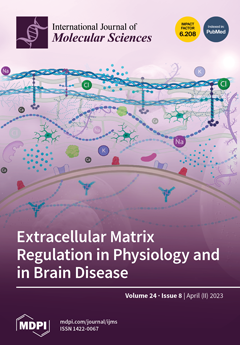
The extracellular matrix (ECM) provides structural and functional support to brain cells and has important roles during development, adulthood, and in brain diseases. Altered expression of ECM-associated genes is associated with seizure, neuropathic pain, cerebellar ataxia, and age-related neurodegenerative disorders. Evidence implicates the transcription factor hypoxia-inducible factor 1 in regulating the expression of ECM-associated genes in different brain cell types. Gene expression changes in microglia play an important role in regulating a brain specific form of ECM called perineuronal nets (PNNs). View this paper
  • Issues are regarded as officially published after their release is announced to the table of contents alert mailing list.
  • You may sign up for e-mail alerts to receive table of contents of newly released issues.
  • PDF is the official format for papers published in both, html and pdf forms. To view the papers in pdf format, click on the "PDF Full-text" link, and use the free Adobe Reader to open them.
Order results
Result details
Section
Select all
Export citation of selected articles as:
18 pages, 4948 KiB  
Article
Dickkopf-1 Acts as a Profibrotic Mediator in Progressive Chronic Kidney Disease
by Yung-Chien Hsu, Cheng-Chih Chang, Ching-Chuan Hsieh, Yu-Ting Huang, Ya-Hsueh Shih, Hsiu-Ching Chang, Pey-Jium Chang and Chun-Liang Lin
Int. J. Mol. Sci. 2023, 24(8), 7679; https://doi.org/10.3390/ijms24087679 - 21 Apr 2023
Viewed by 1790
Abstract
Chronic kidney disease (CKD) is a serious public health problem. Due to a high variability in the speed of CKD progression to end-stage renal disease (ESRD) and the critical involvement of Wnt/β-catenin signaling in CKD, we investigated the role of the Wnt antagonist [...] Read more.
Chronic kidney disease (CKD) is a serious public health problem. Due to a high variability in the speed of CKD progression to end-stage renal disease (ESRD) and the critical involvement of Wnt/β-catenin signaling in CKD, we investigated the role of the Wnt antagonist Dickkopf-1 (DKK1) in CKD progression. Our data revealed that patients with CKD stages 4–5 had higher DKK1 levels in their serum and renal tissues than the control subjects. In an 8-year follow-up, the serum DKK1-high group in the enrolled CKD patients showed a faster progression to ESRD than the serum DKK1-low group. Using a rat model of 5/6 nephrectomy (Nx)-induced CKD, we consistently detected elevated serum levels and renal production of DKK1 in 5/6 Nx rats compared to sham-operated rats. Importantly, the knockdown of the DKK1 levels in the 5/6 Nx rats markedly attenuated the CKD-associated phenotypes. Mechanistically, we demonstrated that the treatment of mouse mesangial cells with recombinant DKK1 protein induced not only the production of multiple fibrogenic proteins, but also the expression of endogenous DKK1. Collectively, our findings suggest that DKK1 acts as a profibrotic mediator in CKD, and elevated levels of serum DKK1 may be an independent predictor of faster disease progression to ESRD in patients with advanced CKD. Full article
(This article belongs to the Section Molecular Endocrinology and Metabolism)
Show Figures

Figure 1

Figure 1
<p>Elevated levels of serum DKK1 are associated with fast progression to ESRD in patients with CKD stages 4–5. (<b>A</b>) Serum DKK1 levels in normal controls (n = 15) and in patients with CKD stages 4–5 (n = 50). Data are presented as mean ± SD. (<b>B</b>) Western blot analysis for the expression of DKK1, fibronectin (FBN) and TGF-β1 in renal tissues from non-CKD and CKD subjects. Quantitative data are expressed as mean ± SD. * <span class="html-italic">p</span> &lt; 0.05 versus control subjects (n = 6). (<b>C</b>) Chromogenic IHC staining for DKK1, FBN and TGF-β1 in renal glomeruli of representative non-CKD subjects (#1 and #2) and CKD subjects (#1 and #2). Scale bars: 20 μm. (<b>D</b>) Chromogenic IHC staining for DKK1, FBN and TGF-β1 in renal tubules of representative non-CKD subjects (#1 and #2) and CKD subjects (#1 and #2). Scale bars: 20 μm. (<b>E</b>) One minus dialysis-free survival analysis of CKD 4–5 patients with high serum levels of DKK1 (≥1526.4 pg/mL; n = 22) or low serum levels of DKK1 (&lt;1526.4 pg/mL; n = 28) in an 8-year follow-up. (<b>F</b>) Stratification by 3-year follow-up in CKD 4–5 patients with low or high serum levels of DKK1.</p>
Full article ">Figure 2
<p>Treatment of 5/6-nephrectomized (5/6 Nx) rats with the DKK1-antisense (DKK1-AS) oligonucleotide alleviates CKD-associated phenotypes. Body weight (<b>A</b>), blood urea nitrogen (BUN) level (<b>B</b>), serum creatinine (Cr) level (<b>C</b>), urine albumin/creatinine ratio (ACR) (<b>D</b>), urine total protein/creatinine ratio (TP/Cr) (<b>E</b>) and serum DKK1 level (<b>F</b>) were analyzed in the sham-operated rats (NC) and in the 5/6 Nx rats that were left untreated or treated with the DKK1-sense (DKK1-S) or DKK1-antisense (DKK1-AS) oligonucleotide. Data are expressed as mean ± SD (n = 6 for each group). * <span class="html-italic">p</span> &lt; 0.01 versus the sham group, # <span class="html-italic">p</span> &lt; 0.01 versus the untreated 5/6 Nx group. (<b>G</b>) Representative images of PAS staining and IHC staining for DKK1 in renal glomeruli of the indicated treated groups. Scale bars: 50 μm. Intensities of PAS staining and IHC staining for DKK1 in kidney sections from different groups were determined by quantitative integrated optical density (IOD) analysis and plotted in panels (<b>i</b>) and (<b>ii</b>), respectively. Data are represented as mean ± SD (n = 6 for each group). * <span class="html-italic">p</span> &lt; 0.05 versus the sham group, # <span class="html-italic">p</span> &lt; 0.05 versus the untreated 5/6 Nx group.</p>
Full article ">Figure 3
<p>Evaluation of the expression levels of profibrotic and proinflammatory factors in renal tissues of the sham-operated rats and the 5/6 Nx rats left untreated or treated with DKK1-S or DKK-AS. (<b>A</b>) Representative photographs of renal sections before and after capturing glomerular compartments using laser capture microdissection (LCM). Scale bars: 100 μm. (<b>B</b>) Quantitative RT-PCR analysis of DKK1, TGF-β1 and fibronectin (FBN) mRNAs in isolated glomeruli microdissected from kidney sections of different groups. Quantitative data are represented as mean ± SD (n = 6 for each group). * <span class="html-italic">p</span> &lt; 0.05 versus the sham group, # <span class="html-italic">p</span> &lt; 0.05 versus the untreated 5/6 Nx group. (<b>C</b>) Representative images of Western blot analysis for the expression of DKK1, β-catenin, TGF-β1 and FBN in renal tissues of different groups. (<b>D</b>) Quantitative analysis of Western blots for DKK1, β-catenin, TGF-β1 and FBN in renal tissues of different groups. Data are plotted as mean ± SD (n = 6 for each group). * <span class="html-italic">p</span> &lt; 0.05 versus the sham group, # <span class="html-italic">p</span> &lt; 0.05 versus the untreated 5/6 Nx group. (<b>E</b>) IHC staining for TGF-β1, collagen IV (COL-IV), fibronectin (FBN) and IL-1β in renal glomeruli of different treated groups. Scale bars: 50 μm. Bottom panels show quantitative IOD analysis of the intensities of TGF-β1 (<b>i</b>), COL-IV (<b>ii</b>), FBN (<b>iii</b>) and IL-1β (<b>iv</b>) in renal glomeruli of different groups (n = 6 for each group). Data are represented as mean ± SD. * <span class="html-italic">p</span> &lt; 0.05 versus the sham group, # <span class="html-italic">p</span> &lt; 0.05 versus the untreated 5/6 Nx group.</p>
Full article ">Figure 4
<p>The renal expression of DKK1 positively correlates with accumulation of β-catenin in 5/6 Nx rats. (<b>A</b>) Representative images of Western blot analysis for the expression of DKK1 and β-catenin in renal tissues of the sham group and the 5/6 Nx groups that were untreated or treated with DKK1-S or DKK1-AS. (<b>B</b>) Quantitative analysis of Western blots for β-catenin in renal tissues of different groups (n = 6 for each group). * <span class="html-italic">p</span> &lt; 0.05 versus the sham group, # <span class="html-italic">p</span> &lt; 0.05 versus the untreated 5/6 Nx group. (<b>C</b>) Immunofluorescence analysis of β-catenin in kidney sections from the sham-operated rats and the 5/6 Nx rats left untreated or treated with DKK1-S or DKK-AS. Scale bars: 100 μm. (<b>D</b>) Immunofluorescence images of renal glomeruli stained for β-catenin in different treated groups. Scale bars: 50 μm. (<b>E</b>) Quantification of relative fluorescence intensities for β-catenin in renal glomeruli of different groups (n = 6 for each group). * <span class="html-italic">p</span> &lt; 0.05 versus the sham group, # <span class="html-italic">p</span> &lt; 0.05 versus the untreated 5/6 Nx group.</p>
Full article ">Figure 5
<p>Uremic serum promotes fibrogenesis of renal mesangial cells through the DKK1-mediated signaling pathway. (<b>A</b>) Western blot analysis of fibronectin (FBN), α-smooth muscle actin (α-SMA), and collagen IV (COL-IV) expressed in mouse mesangial cells that were cultured in media with increasing concentrations (5, 10 and 20%) of healthy control serum or uremic serum. The healthy serum and uremic serum samples were pooled at equal volumes from blood samples of five healthy control subjects and five uremic patients, respectively. Quantitative data from Western blots are plotted as mean ± SD (n = 3). * <span class="html-italic">p</span> &lt; 0.05 versus the healthy controls at the corresponding serum concentrations. (<b>B</b>) Western blot analysis of FBN, α-SMA and COL-IV expressed in mouse mesangial cells cultured in media with 20% healthy control serum or uremic serum for 24, 48 and 72 h. Quantitative data from Western blots are plotted as mean ± SD (n = 3). * <span class="html-italic">p</span> &lt; 0.05 versus the healthy controls at the corresponding time points. (<b>C</b>) Serum DKK1 levels of five healthy control subjects and five uremic patients included in the in vitro experiments. (<b>D</b>) Effects of increasing amounts of anti-DKK1 neutralizing antibody (16.5, 66 and 200 ng/mL) on the expression of FBN, α-SMA and COL-IV in mesangial cells cultured in media with 20% healthy control serum or uremic serum. Quantitative data from Western blots are plotted as mean ± SD (n = 3). * <span class="html-italic">p</span> &lt; 0.05 versus the healthy controls, # <span class="html-italic">p</span> &lt; 0.05 versus the untreated uremic serum group. (<b>E</b>) Effects of increasing amounts of an IgG control and anti-DKK1 antibody on the expression of profibrotic proteins in mesangial cells cultured in media with 20% uremic serum. Quantitative data from Western blots are plotted as mean ± SD (n = 3). # <span class="html-italic">p</span> &lt; 0.05 versus the untreated uremic serum group.</p>
Full article ">Figure 6
<p>DKK1 acts as a profibrotic mediator in mouse mesangial cells. (<b>A</b>) Effects of increasing amounts of recombinant human DKK1 (100, 200 and 400 ng/mL) on the expression of FBN, COL-IV, TGF-β1, α-SMA, DKK1 and β-catenin in mouse mesangial cells. The quantitative results from Western blots are plotted as mean ± SD (n = 3). * <span class="html-italic">p</span> &lt; 0.05 versus the untreated control. (<b>B</b>) Effects of increasing amounts of recombinant mouse DKK1 (100, 200 and 400 ng/mL) on the expression of the indicated proteins in mouse mesangial cells. The quantitative data from Western blots are plotted as mean ± SD (n = 3). * <span class="html-italic">p</span> &lt; 0.05 versus the untreated control. (<b>C</b>) Quantitative RT-PCR analysis of FBN, TGF-β1 and DKK1 mRNAs expressed in mouse mesangial cells that were left untreated or treated with increasing amounts of recombinant human DKK1 (100, 200 and 400 ng/mL). * <span class="html-italic">p</span> &lt; 0.05 versus the untreated control (n = 3). (<b>D</b>) Quantitative RT-PCR analysis of FBN, TGF-β1 and DKK1 mRNAs expressed in mouse mesangial cells that were left untreated or treated with increasing amounts of recombinant mouse DKK1 (100, 200 and 400 ng/mL). * <span class="html-italic">p</span> &lt; 0.05 versus the untreated control (n = 3).</p>
Full article ">Figure 6 Cont.
<p>DKK1 acts as a profibrotic mediator in mouse mesangial cells. (<b>A</b>) Effects of increasing amounts of recombinant human DKK1 (100, 200 and 400 ng/mL) on the expression of FBN, COL-IV, TGF-β1, α-SMA, DKK1 and β-catenin in mouse mesangial cells. The quantitative results from Western blots are plotted as mean ± SD (n = 3). * <span class="html-italic">p</span> &lt; 0.05 versus the untreated control. (<b>B</b>) Effects of increasing amounts of recombinant mouse DKK1 (100, 200 and 400 ng/mL) on the expression of the indicated proteins in mouse mesangial cells. The quantitative data from Western blots are plotted as mean ± SD (n = 3). * <span class="html-italic">p</span> &lt; 0.05 versus the untreated control. (<b>C</b>) Quantitative RT-PCR analysis of FBN, TGF-β1 and DKK1 mRNAs expressed in mouse mesangial cells that were left untreated or treated with increasing amounts of recombinant human DKK1 (100, 200 and 400 ng/mL). * <span class="html-italic">p</span> &lt; 0.05 versus the untreated control (n = 3). (<b>D</b>) Quantitative RT-PCR analysis of FBN, TGF-β1 and DKK1 mRNAs expressed in mouse mesangial cells that were left untreated or treated with increasing amounts of recombinant mouse DKK1 (100, 200 and 400 ng/mL). * <span class="html-italic">p</span> &lt; 0.05 versus the untreated control (n = 3).</p>
Full article ">Figure 7
<p>Proposed model of DKK1-mediated renal fibrosis. During renal injury, DKK1 can be initially activated by the Wnt/β-catenin signaling pathway. Subsequently, DKK1 may autoregulate its own expression to further increase its levels. The DKK1 autoregulation may be unrelated to the Wnt/β-catenin signaling pathway. Increased expression of DKK1 is involved in renal fibrosis and thus promotes CKD progression. Under the circumstances of renal injury, DKK1 is no longer acting as a Wnt/β-catenin antagonist (via interaction with LRP5/6). Conversely, DKK1 may activate the Wnt/β-catenin signaling by some unknown mechanisms in renal cells or in infiltrating immune cells.</p>
Full article ">
11 pages, 663 KiB  
Article
The ACE rs1799752 Variant Is Associated with COVID-19 Severity but Is Independent of Serum ACE Activity in Hospitalized and Recovered Patients
by Ingrid Fricke-Galindo, Ivette Buendia-Roldan, Daniel I. Ponce-Aguilar, Gloria Pérez-Rubio, Leslie Chavez-Galan, Jesús Alanis-Ponce, Karina Pérez-Torres, Daniela Valencia-Pérez Rea, Fernanda Téllez-Quijada, Karol J. Nava-Quiroz, Rafael de Jesús Hernández-Zenteno, Angélica Gutiérrez-Nava and Ramcés Falfán-Valencia
Int. J. Mol. Sci. 2023, 24(8), 7678; https://doi.org/10.3390/ijms24087678 - 21 Apr 2023
Cited by 2 | Viewed by 2108
Abstract
This paper assesses the association of the insertion/deletion ACE (angiotensin-converting enzyme) variant (rs1799752 I/D) and the serum ACE activity with the severity of COVID-19 as well as its impact on post-COVID-19, and we compare these associations with those for patients with non-COVID-19 respiratory [...] Read more.
This paper assesses the association of the insertion/deletion ACE (angiotensin-converting enzyme) variant (rs1799752 I/D) and the serum ACE activity with the severity of COVID-19 as well as its impact on post-COVID-19, and we compare these associations with those for patients with non-COVID-19 respiratory disorders. We studied 1252 patients with COVID-19, 104 subjects recovered from COVID-19, and 74 patients hospitalized with a respiratory disease different from COVID-19. The rs1799752 ACE variant was assessed using TaqMan® Assays. The serum ACE activity was determined using a colorimetric assay. The DD genotype was related to risk for invasive mechanical ventilation (IMV) requirement as an indicator of COVID-19 severity when compared to the frequencies of II + ID genotypes (p = 0.025, OR = 1.428, 95% CI = 1.046–1.949). In addition, this genotype was significantly higher in COVID-19 and post-COVID-19 groups than in the non-COVID-19 subjects. The serum ACE activity levels were lower in the COVID-19 group (22.30 U/L (13.84–32.23 U/L)), which was followed by the non-COVID-19 (27.94 U/L (20.32–53.36 U/L)) and post-COVID-19 subjects (50.00 U/L (42.16–62.25 U/L)). The DD genotype of the rs1799752 ACE variant was associated with the IMV requirement in patients with COVID-19, and low serum ACE activity levels could be related to patients with severe disease. Full article
(This article belongs to the Special Issue Molecular Research on SARS-CoV-2)
Show Figures

Figure 1

Figure 1
<p>Serum ACE activity levels in the COVID-19 (<span class="html-italic">n</span> = 66), post-COVID-19 (<span class="html-italic">n</span> = 69), and non-COVID-19 (<span class="html-italic">n</span> = 26) groups. The comparisons were performed using the Kruskal–Wallis test corrected with the Benjamini–Hochberg method.</p>
Full article ">Figure 2
<p>Serum ACE activity according to the rs1799752 <span class="html-italic">ACE</span> genotypes in the COVID-19 (<span class="html-italic">n</span> = 66), post-COVID-19 (<span class="html-italic">n</span> = 69), and non-COVID-19 (<span class="html-italic">n</span> = 26) groups. The ACE activity was not different among the three genotypes in any of the studied groups (Kruskal–Wallis test, <span class="html-italic">p</span> &gt; 0.05).</p>
Full article ">
20 pages, 635 KiB  
Review
Advances in Platelet-Rich Plasma Treatment for Spinal Diseases: A Systematic Review
by Soya Kawabata, Koji Akeda, Junichi Yamada, Norihiko Takegami, Tatsuhiko Fujiwara, Nobuyuki Fujita and Akihiro Sudo
Int. J. Mol. Sci. 2023, 24(8), 7677; https://doi.org/10.3390/ijms24087677 - 21 Apr 2023
Cited by 15 | Viewed by 4795
Abstract
Spinal diseases are commonly associated with pain and neurological symptoms, which negatively impact patients’ quality of life. Platelet-rich plasma (PRP) is an autologous source of multiple growth factors and cytokines, with the potential to promote tissue regeneration. Recently, PRP has been widely used [...] Read more.
Spinal diseases are commonly associated with pain and neurological symptoms, which negatively impact patients’ quality of life. Platelet-rich plasma (PRP) is an autologous source of multiple growth factors and cytokines, with the potential to promote tissue regeneration. Recently, PRP has been widely used for the treatment of musculoskeletal diseases, including spinal diseases, in clinics. Given the increasing popularity of PRP therapy, this article examines the current literature for basic research and emerging clinical applications of this therapy for treating spinal diseases. First, we review in vitro and in vivo studies, evaluating the potential of PRP in repairing intervertebral disc degeneration, promoting bone union in spinal fusion surgeries, and aiding in neurological recovery from spinal cord injury. Second, we address the clinical applications of PRP in treating degenerative spinal disease, including its analgesic effect on low back pain and radicular pain, as well as accelerating bone union during spinal fusion surgery. Basic research demonstrates the promising regenerative potential of PRP, and clinical studies have reported on the safety and efficacy of PRP therapy for treating several spinal diseases. Nevertheless, further high-quality randomized controlled trials would be required to establish clinical evidence of PRP therapy. Full article
(This article belongs to the Special Issue Regeneration for Spinal Diseases 3.0)
Show Figures

Figure 1

Figure 1
<p>Schematic model for mechanism of PRP on intervertebral disc cells. ↑: increase; ↓: decrease.</p>
Full article ">
19 pages, 2831 KiB  
Article
β-Conglutins’ Unique Mobile Arm Is a Key Structural Domain Involved in Molecular Nutraceutical Properties of Narrow-Leafed Lupin (Lupinus angustifolius L.)
by Elena Lima-Cabello, Julia Escudero-Feliu, Andreina Peralta-Leal, Pedro Garcia-Fernandez, Kadambot H. M. Siddique, Karam B. Singh, Maria I. Núñez, Josefa León and Jose C. Jimenez-Lopez
Int. J. Mol. Sci. 2023, 24(8), 7676; https://doi.org/10.3390/ijms24087676 - 21 Apr 2023
Cited by 1 | Viewed by 2330
Abstract
Narrow-leafed lupin (NLL; Lupinus angustifolius L.) has multiple nutraceutical properties that may result from unique structural features of β-conglutin proteins, such as the mobile arm at the N-terminal, a structural domain rich in α-helices. A similar domain has not been found in other [...] Read more.
Narrow-leafed lupin (NLL; Lupinus angustifolius L.) has multiple nutraceutical properties that may result from unique structural features of β-conglutin proteins, such as the mobile arm at the N-terminal, a structural domain rich in α-helices. A similar domain has not been found in other vicilin proteins of legume species. We used affinity chromatography to purify recombinant complete and truncated (without the mobile arm domain, tβ5 and tβ7) forms of NLL β5 and β7 conglutin proteins. We then used biochemical and molecular biology techniques in ex vivo and in vitro systems to evaluate their anti-inflammatory activity and antioxidant capacity. The complete β5 and β7 conglutin proteins decreased pro-inflammatory mediator levels (e.g., nitric oxide), mRNA expression levels (iNOS, TNFα, IL-1β), and the protein levels of pro-inflammatory cytokine TNF-α, interleukins (IL-1β, IL-2, IL-6, IL-8, IL-12, IL-17, IL-27), and other mediators (INFγ, MOP, S-TNF-R1/-R2, and TWEAK), and exerted a regulatory oxidative balance effect in cells as demonstrated in glutathione, catalase, and superoxide dismutase assays. The truncated tβ5 and tβ7 conglutin proteins did not have these molecular effects. These results suggest that β5 and β7 conglutins have potential as functional food components due to their anti-inflammatory and oxidative cell state regulatory properties, and that the mobile arm of NLL β-conglutin proteins is a key domain in the development of nutraceutical properties, making NLL β5 and β7 excellent innovative candidates as functional foods. Full article
(This article belongs to the Special Issue Structural/Functional Characterization of Plant Proteins 2.0)
Show Figures

Figure 1

Figure 1
<p><b>Figure 1</b>. Purification and identification of tβ5 and tβ7 conglutin proteins. Two purified conglutin tβ5 and tβ7 proteins stained with Coomassie Brilliant Blue had a high level of purity (&gt;95%) (<b>A</b>); Immunoblotting identified the same two purified β-conglutin proteins as the anti-β-conglutin antibody. MW, molecular weight standard (kDa) (<b>B</b>).</p>
Full article ">Figure 2
<p>Assessment of mRNA expression levels of TNF-α, IL-1β, and iNOS genes. Culture cells of HepG2 (<b>A</b>), T2D (<b>B</b>), and healthy control subjects (<b>C</b>). Each group of cells was incubated for 24 h in LPS, LPS+tβ5, LPS+β5, LPS+tβ7, or LPS+β7. Bars show TNF-α (black), IL-1β (punted white), and iNOS (gray) color in each cell type described above. * <span class="html-italic">p</span> ˂ 0.05 LPS vs. control; ** <span class="html-italic">p</span> ˂ 0.05 LPS+tβ5 or tβ7 vs. LPS; # <span class="html-italic">p</span> ˂ 0.05 LPS+β5 vs. LPS; ◆ <span class="html-italic">p</span> ˂ 0.05 LPS+β7 vs. LPS. ◊ <span class="html-italic">p</span> ˂ 0.05 LPS+β5 vs. T2D, and ○ <span class="html-italic">p</span> ˂ 0.05 LPS+β7 vs. T2D in T2D cell cultures. Data represent the mean ± SD of three independent experiments.</p>
Full article ">Figure 3
<p>β5 and β7 conglutins reduced the protein levels of TNF-α, IL-1β, and iNOS. Culture cells of HepG2 (<b>A</b>), T2D (<b>B</b>), and healthy control subjects (<b>C</b>). Each group of cells was incubated for 24 h in the presence of LPS, LPS+tβ5, LPS+β5, LPS+tβ7, and LPS+β7. Bars show TNF-α (dark gray), IL-1β (gray), and iNOS (light gray) for each cell type described above. * <span class="html-italic">p</span> ˂ 0.05 LPS vs. control; ** <span class="html-italic">p</span> ˂ 0.05 LPS+tβ5 or tβ7 vs. LPS; # <span class="html-italic">p</span> ˂ 0.05 LPS+β5 vs. LPS; ◆ <span class="html-italic">p</span> ˂ 0.05 LPS+β7 vs. LPS. ★ <span class="html-italic">p</span> ˂ 0.05 β5 vs. T2D, and <b>+</b> <span class="html-italic">p</span> ˂ 0.05 β7 vs. T2D in T2D cell cultures. Data represent the mean ± SD of three independent experiments.</p>
Full article ">Figure 4
<p>Effect of NLL β5 and β7 conglutins on CAT and SOD enzymatic activities and GSH production in HepG2 culture cells. CAT (<b>A</b>) and SOD (<b>B</b>) activities and GSH production (<b>C</b>). * <span class="html-italic">p</span> ˂ 0.05 LPS vs. control; ** <span class="html-italic">p</span> ˂ 0.05 LPS+tβ5 or LPS+tβ7 vs. LPS; # or ●# <span class="html-italic">p</span> ˂ 0.05 LPS+β5 vs. LPS; ◆ or ◆# <span class="html-italic">p</span> ˂ 0.05 LPS+β7 vs. LPS. Data represent mean ± SD from three independent experiments.</p>
Full article ">Figure 5
<p>Effect of NLL β5 and β7 conglutins on CAT and SOD activities and GSH production in Type II diabetes (T2D) culture cells. CAT (<b>A</b>) and SOD (<b>B</b>) activities and GSH production (<b>C</b>). <b>★</b> <span class="html-italic">p</span> ˂ 0.05 T2D vs. tβ5 or tβ7; <b>★★</b> <span class="html-italic">p</span> ˂ 0.05 β5 vs. T2D; ++ <span class="html-italic">p</span> ˂ 0.05 β7 vs. T2D. Data represent mean ± SD from three independent experiments.</p>
Full article ">Figure 6
<p>Effect of NLL β5 and β7 conglutins on CAT and SOD activities and GSH production in healthy subject culture cells. CAT (<b>A</b>) and SOD (<b>B</b>) activities and GSH production (<b>C</b>). * <span class="html-italic">p</span> ˂ 0.05 LPS vs. control; # <span class="html-italic">p</span> ˂ 0.05 LPS+β5 vs. LPS; ◆ <span class="html-italic">p</span> ˂ 0.05 LPS+β7 vs. LPS. Data represent mean ± SD from three independent experiments.</p>
Full article ">Figure 7
<p>Effect of β5 and β7 conglutins on nitric oxide production. Culture cells of HepG2 (<b>A</b>), T2D (<b>B</b>), and healthy subjects (<b>C</b>). Each group of cells was incubated for 24 h in the presence of LPS, LPS+tβ5, LPS+β5, LPS+tβ7, or LPS+β7. * <span class="html-italic">p</span> ˂ 0.05 LPS vs. control; ** <span class="html-italic">p</span> ˂ 0.05 LPS+tβ5 or tβ7 vs. LPS; # <span class="html-italic">p</span> ˂ 0.05 LPS+β5 vs. LPS; ◆ <span class="html-italic">p</span> ˂ 0.05 LPS+β7 vs. LPS. ★ <span class="html-italic">p</span> ˂ 0.05 β5 vs. T2D and + <span class="html-italic">p</span> ˂ 0.05 β7 vs. T2D in T2D cell cultures. Data represent the mean ± SD of three independent experiments.</p>
Full article ">
18 pages, 4340 KiB  
Article
Skin Microbiome in Prurigo Nodularis
by Klaudia Tutka, Magdalena Żychowska, Anna Żaczek, Karolina Maternia-Dudzik, Jakub Pawełczyk, Dominik Strapagiel, Jakub Lach and Adam Reich
Int. J. Mol. Sci. 2023, 24(8), 7675; https://doi.org/10.3390/ijms24087675 - 21 Apr 2023
Cited by 6 | Viewed by 3378
Abstract
Prurigo nodularis (PN) is a chronic condition characterized by the presence of nodular lesions accompanied by intense pruritus. The disease has been linked to several infectious factors, but data on the direct presence of microorganisms in the lesions of PN are scarce. The [...] Read more.
Prurigo nodularis (PN) is a chronic condition characterized by the presence of nodular lesions accompanied by intense pruritus. The disease has been linked to several infectious factors, but data on the direct presence of microorganisms in the lesions of PN are scarce. The aim of this study was to evaluate the diversity and composition of the bacterial microbiome in PN lesions by targeting the region V3-V4 of 16S rRNA. Skin swabs were obtained from active nodules in 24 patients with PN, inflammatory patches of 14 patients with atopic dermatitis (AD) and corresponding skin areas of 9 healthy volunteers (HV). After DNA extraction, the V3-V4 region of the bacterial 16S rRNA gene was amplified. Sequencing was performed using the Illumina platform on the MiSeq instrument. Operational taxonomic units (OTU) were identified. The identification of taxa was carried out using the Silva v.138 database. There was no statistically significant difference in the alpha-diversity (intra-sample diversity) between the PN, AD and HV groups. The beta-diversity (inter-sample diversity) showed statistically significant differences between the three groups on a global level and in paired analyses. Staphylococcus was significantly more abundant in samples from PN and AD patients than in controls. The difference was maintained across all taxonomic levels. The PN microbiome is highly similar to that of AD. It remains unclear whether the disturbed composition of the microbiome and the domination of Staphylococcus in PN lesions may be the trigger factor of pruritus and lead to the development of cutaneous changes or is a secondary phenomenon. Our preliminary results support the theory that the composition of the skin microbiome in PN is altered and justify further research on the role of the microbiome in this debilitating condition. Full article
(This article belongs to the Special Issue Microbial Enzymes and Metabolites)
Show Figures

Figure 1

Figure 1
<p>Stacked bar plots presenting the individual’s distribution of microorganisms in prurigo nodularis (PN), healthy volunteers (HV) and atopic dermatitis (AD) (<b>a</b>) at the phylum level (level 2), (<b>b</b>) at the class level (level 3) and (<b>c</b>) at the genus level (level 6).</p>
Full article ">Figure 1 Cont.
<p>Stacked bar plots presenting the individual’s distribution of microorganisms in prurigo nodularis (PN), healthy volunteers (HV) and atopic dermatitis (AD) (<b>a</b>) at the phylum level (level 2), (<b>b</b>) at the class level (level 3) and (<b>c</b>) at the genus level (level 6).</p>
Full article ">Figure 2
<p>Total composition of cutaneous bacteria microbiome at the genus level in patients with prurigo nodularis (PN).</p>
Full article ">Figure 3
<p>Total composition of the cutaneous bacteria microbiome at the genus level in patients with atopic dermatitis (AD).</p>
Full article ">Figure 4
<p>Total composition of the cutaneous bacteria microbiome at the genus level in healthy volunteers (HV).</p>
Full article ">Figure 5
<p>Clinical image of prurigo nodularis lesions.</p>
Full article ">
14 pages, 985 KiB  
Review
Molecular and Physiological Determinants of Amyotrophic Lateral Sclerosis: What the DJ-1 Protein Teaches Us
by Federica Sandrelli and Marco Bisaglia
Int. J. Mol. Sci. 2023, 24(8), 7674; https://doi.org/10.3390/ijms24087674 - 21 Apr 2023
Cited by 3 | Viewed by 2149
Abstract
Amyotrophic lateral sclerosis (ALS) is an adult-onset disease which causes the progressive degeneration of cortical and spinal motoneurons, leading to death a few years after the first symptom onset. ALS is mainly a sporadic disorder, and its causative mechanisms are mostly unclear. About [...] Read more.
Amyotrophic lateral sclerosis (ALS) is an adult-onset disease which causes the progressive degeneration of cortical and spinal motoneurons, leading to death a few years after the first symptom onset. ALS is mainly a sporadic disorder, and its causative mechanisms are mostly unclear. About 5–10% of cases have a genetic inheritance, and the study of ALS-associated genes has been fundamental in defining the pathological pathways likely also involved in the sporadic forms of the disease. Mutations affecting the DJ-1 gene appear to explain a subset of familial ALS forms. DJ-1 is involved in multiple molecular mechanisms, acting primarily as a protective agent against oxidative stress. Here, we focus on the involvement of DJ-1 in interconnected cellular functions related to mitochondrial homeostasis, reactive oxygen species (ROS) levels, energy metabolism, and hypoxia response, in both physiological and pathological conditions. We discuss the possibility that impairments in one of these pathways may affect the others, contributing to a pathological background in which additional environmental or genetic factors may act in favor of the onset and/or progression of ALS. These pathways may represent potential therapeutic targets to reduce the likelihood of developing ALS and/or slow disease progression. Full article
Show Figures

Figure 1

Figure 1
<p>The physiological functions of DJ-1 possibly affected in ALS pathology. DJ-1 participates in the maintenance of mitochondrial and cellular redox homeostasis. These functions are fundamental to sustaining the high energetic demand of MNs without producing excessive levels of ROS, and collectively, they contribute to MNs’ healthiness (<b>on the left</b>). Alterations in DJ-1 physiological functions might affect mitochondria functionality, reducing the levels of ATP below MNs’ needs. At the same time, mitochondria dysfunctions might promote increased levels of ROS. Altogether, these alterations might constitute a pathological background enhancing the probability of developing ALS (<b>on the right</b>) (created with BioRender.com, accessed on 6 April 2023).</p>
Full article ">Figure 2
<p>DJ-1-mediated protection against hypoxia. Under normoxia conditions, HIF-1α is constantly hydroxylated, ubiquitinated, and degraded at the proteasome. Under hypoxic conditions, HIF-1α translocates into the nucleus, where it promotes the expression of numerous genes involved in hypoxic adaptation. Among them, the VEGF protein stimulates vessel growth and promotes the survival of motor neurons during hypoxia. DJ-1 has been proposed to act as a negative regulator of VHL stabilizing the transcription factor HIF-1α. Acronyms are mentioned in the main text. (Created with BioRender.com, accessed on 6 April 2023.)</p>
Full article ">
20 pages, 6380 KiB  
Article
Molecular Integrative Analysis of the Inhibitory Effects of Dipeptides on Amyloid β Peptide 1–42 Polymerization
by Nan Yuan, Lianmeng Ye, Yan Sun, Hao Wu, Zhengpan Xiao, Wanmeng Fu, Zuqian Chen, Yechun Pei, Yi Min and Dayong Wang
Int. J. Mol. Sci. 2023, 24(8), 7673; https://doi.org/10.3390/ijms24087673 - 21 Apr 2023
Cited by 1 | Viewed by 1810
Abstract
The major pathological feature of Alzheimer’s disease (AD) is the aggregation of amyloid β peptide (Aβ) in the brain. Inhibition of Aβ42 aggregation may prevent the advancement of AD. This study employed molecular dynamics, molecular docking, electron microscopy, circular dichroism, staining of [...] Read more.
The major pathological feature of Alzheimer’s disease (AD) is the aggregation of amyloid β peptide (Aβ) in the brain. Inhibition of Aβ42 aggregation may prevent the advancement of AD. This study employed molecular dynamics, molecular docking, electron microscopy, circular dichroism, staining of aggregated Aβ with ThT, cell viability, and flow cytometry for the detection of reactive oxygen species (ROS) and apoptosis. Aβ42 polymerizes into fibrils due to hydrophobic interactions to minimize free energy, adopting a β-strand structure and forming three hydrophobic areas. Eight dipeptides were screened by molecular docking from a structural database of 20 L-α-amino acids, and the docking was validated by molecular dynamics (MD) analysis of binding stability and interaction potential energy. Among the dipeptides, arginine dipeptide (RR) inhibited Aβ42 aggregation the most. The ThT assay and EM revealed that RR reduced Aβ42 aggregation, whereas the circular dichroism spectroscopy analysis showed a 62.8% decrease in β-sheet conformation and a 39.3% increase in random coiling of Aβ42 in the presence of RR. RR also significantly reduced the toxicity of Aβ42 secreted by SH-SY5Y cells, including cell death, ROS production, and apoptosis. The formation of three hydrophobic regions and polymerization of Aβ42 reduced the Gibbs free energy, and RR was the most effective dipeptide at interfering with polymerization. Full article
(This article belongs to the Section Molecular Informatics)
Show Figures

Figure 1

Figure 1
<p>Molecular dynamics study of the structures of Aβ<sub>42</sub> monomer, trimer, and pentamer in 0.1 M sodium chloride solution, demonstrating the association of Aβ<sub>42</sub> polymerization with β-sheet conformation. (<b>A</b>) The front view of Aβ<sub>42</sub> monomer. (<b>B</b>) The side view of Aβ<sub>42</sub> monomer. (<b>C</b>) The top view of Aβ<sub>42</sub> monomer. (<b>D</b>) The front view of Aβ<sub>42</sub> trimer. (<b>E</b>) The side view of Aβ<sub>42</sub> trimer. (<b>F</b>) The top view of Aβ<sub>42</sub> trimer. (<b>G</b>) The front view of Aβ<sub>42</sub> pentamer. (<b>H</b>) The side view of Aβ<sub>42</sub> pentamer. (<b>I</b>) The top view of Aβ<sub>42</sub> pentamer. The monomer, trimer, and pentamer of Aβ<sub>42</sub> were modeled using the near atomic structure (PDB ID #5oqv) as starting conformations. Each analysis simulated 500-ns interaction and movements, with a data size of roughly 160 Gb.</p>
Full article ">Figure 2
<p>Hydrophobic interactions play a leading role in the polymerization of Aβ<sub>42</sub>. (<b>A</b>) In the polymerized Aβ<sub>42</sub> conformation, there are three hydrophobic zones, which are indicated by the numbers in circles. Hydrophobic amino acid residues and those that create hydrogen bonding with side chains are labeled in the graph. (<b>B</b>) The effects of changing hydrophobic amino acids to glycine in the three hydrophobic zones on binding energy between two Aβ<sub>42</sub> polypeptide chains. The binding energy of the wild-type Aβ<sub>42</sub> dimer is represented by the red line; the binding energy of the first hydrophobic zone-mutated Aβ<sub>42</sub> dimer is represented by the magenta line; the binding energy of the second hydrophobic zone-mutated Aβ<sub>42</sub> dimer is represented by the blue line; and the binding energy of the third hydrophobic zone-mutated Aβ<sub>42</sub> dimer is represented by the green line. The hydrophobic amino acid residues were labeled around each region. However, the residues that formed hydrogen bond were also labeled, including Asn27, His6, Ser8, and Glu11.</p>
Full article ">Figure 3
<p>Schematic procedure of Aβ<sub>42</sub> aggregation.</p>
Full article ">Figure 4
<p>Binding stability and interaction energy of the dipeptides at different binding sites of Aβ<sub>42</sub> pentamer. (<b>A</b>) The binding stability of dipeptides to Aβ<sub>42</sub> pentamer. (<b>B</b>) The interaction energy between Aβ<sub>42</sub> pentamer and the dipeptides. HR: Histidine–arginine; HW: Histidine–tryptophan; RF: Arginine–phenylalanine; RR: Arginine dipeptide; RW: Arginine–tryptophan; RY: Arginine–tyrosine; WR: Tryptophan–arginine; RM: Arginine–methionine. RMSD: The root-mean-square deviation of the positions of the heavy elements of a dipeptide at different binding sites over time. The interaction energy is the algebraic sum of Lennard-Jones and Coulombic potential energy.</p>
Full article ">Figure 5
<p>Effects of the eight dipeptides on aggregation of Aβ<sub>42</sub>. (<b>A</b>) Control experiment to show that dipeptides themselves have no effect on ThT fluorescent intensity. (<b>B</b>) The effects of the dipeptides on Aβ<sub>42</sub> aggregation detected by ThT fluorometry. * <span class="html-italic">p</span> &lt; 0.05 and ** <span class="html-italic">p</span> &lt; 0.01 versus Aβ control group, analyzed by one-way ANOVA followed by the Tukey’s post-hoc test for multiple comparison, n = 5. HR: Histidine-arginine; HW: Histidine–tryptophan; RF: Arginine–phenylalanine; RR: Arginine dipeptide; RW: Arginine–tryptophan; RY: Arginine–tyrosine; WR: Tryptophan–arginine; RM: Arginine–methionine.</p>
Full article ">Figure 6
<p>Binding stability and interaction energy of arginine dipeptide at different binding sites of Aβ<sub>42</sub>. (<b>A</b>) The root-mean-square deviation (RMSD) of the heavy elements of arginine dipeptide (RR) at different binding sites over time. (<b>B</b>) The interaction energy between Aβ<sub>42</sub> pentamer and arginine dipeptide. The labels of RR 01-12 correspond to A to L in <a href="#app1-ijms-24-07673" class="html-app">Supplementary Figure S3</a>, respectively. The interaction energy is the algebraic sum of Lennard-Jones and Coulombic potential energy.</p>
Full article ">Figure 7
<p>Effects of arginine dipeptide on the interaction between Aβ<sub>42</sub> double strands. (<b>A</b>) The root-mean-square deviation (RMSD) of one of the Aβ<sub>42</sub> double strands in absence or presence of arginine dipeptide (RR). (<b>B</b>) The interaction energy between Aβ<sub>42</sub> double strands in absence or presence of RR. (<b>C</b>) The change in the free energy in the system when pulling the Aβ<sub>42</sub> double strands apart along reaction axis ζ in absence of RR. (<b>D</b>) The change in the free energy in the system when pulling the Aβ<sub>42</sub> double strands apart along reaction axis ζ in presence of RR.</p>
Full article ">Figure 8
<p>Effects of arginine dipeptide on aggregation of Aβ<sub>42</sub>. (<b>A</b>) Control experiment of the effects of arginine dipeptide on Thioflavin T (ThT) fluorescence intensity. At each data point, the sample size, n, is equal to 4. (<b>B</b>) Time-dependent effects of RR on Aβ<sub>42</sub> aggregation detected by ThT fluorometry. Fluorescence intensity was measured at the wave length of 485 nm. <span class="html-italic">p</span> &lt; 0.01, analyzed by one-way ANOVA, n = 4. (<b>C</b>) Dose-dependent effects of RR on Aβ<sub>42</sub> aggregation detected by ThT fluorometry. Data are expressed as mean ± SE. ** <span class="html-italic">p</span> &lt; 0.01 versus RR non-treated group, analyzed by one-way ANOVA followed by the Tukey’s post-hoc test for multiple comparison, n = 4. (<b>D</b>) Effects of arginine dipeptide (RR) on formation of Aβ<sub>42</sub> fibrils detected by transmission electron microscopy. (<b>E</b>) Far-UV circular dichroism (CD) spectrometry of Aβ<sub>42</sub> in absence of RR. Aβ<sub>42</sub>-1 to Aβ<sub>42</sub>-5 represents different concentration of Aβ<sub>42</sub>, which correspond to 12.5, 25, 50, 100, and 200 μM. (<b>F</b>) Far-UV circular dichroism (CD) spectrometry of Aβ<sub>42</sub> in presence of RR. RR-1 to RR-5 represents different concentration of RR, which correspond to 31.25, 62.5, 125, 250, and 500 μM, respectively.</p>
Full article ">Figure 9
<p>Effects of arginine dipeptide on cell toxicity induced by Aβ<sub>42</sub> secreted from SH-SY5Y cells. (<b>A</b>) Effects of arginine dipeptide (RR) on cell toxicity induced by 48-h treatment with Aβ<sub>42</sub> tested by MTT method. (<b>B</b>) Time-dependent effects of RR on cell toxicity of Aβ<sub>42</sub>. <sup>##</sup> <span class="html-italic">p</span> &lt; 0.01 versus Aβ<sub>42</sub> and RR non-treated group; * <span class="html-italic">p</span> &lt; 0.05, ** <span class="html-italic">p</span> &lt; 0.01 versus RR non-treated group, analyzed by one-way ANOVA followed by the Tukey’s post-hoc test for multiple comparison, n = 6.</p>
Full article ">Figure 10
<p>Flow cytometry analysis of the effects of arginine dipeptide on reactive oxygen species (ROS) production. (<b>A</b>) ROS production in Aβ<sub>42</sub>-nonsecreting SH-SY5Y cells treated with DMSO (Con). (<b>B</b>) ROS production in Aβ<sub>42</sub>-nonsecreting SH-SY5Y cells treated with arginine dipeptide (RR) at 50 μM. (<b>C</b>) ROS production in SH-SY5Y cells secreting Aβ<sub>42</sub>. (<b>D</b>–<b>F</b>) ROS production in Aβ<sub>42</sub>-secreting SH-SY5Y cells treated with RR at 1, 10, and 50 μM, respectively. (<b>G</b>) Overlay of the flow cytometric figures, (<b>A</b>–<b>F</b>). (<b>H</b>) Quantification of ROS production. All experiments were repeated at least three times. Results are expressed as mean ± SEM, ns: not significant, ** <span class="html-italic">p</span> &lt; 0.01, analyzed by one-way ANOVA, followed by the Tukey’s post-hoc test for multiple comparison.</p>
Full article ">Figure 11
<p>Flow cytometry analysis of the effects of arginine dipeptide on cell apoptosis. (<b>A</b>) Apoptosis in Aβ<sub>42</sub>-nonsecreting SH-SY5Y cells treated with control medium (Con). (<b>B</b>) Apoptosis in Aβ<sub>42</sub>-nonsecreting SH-SY5Y cells treated with arginine dipeptide (RR) at 50 μM. (<b>C</b>) Apoptosis in SH-SY5Y cells secreting Aβ<sub>42</sub>. (<b>D</b>–<b>F</b>) Apoptosis in Aβ<sub>42</sub>-secreting SH-SY5Y cells treated with RR at 1, 10 and 50 μM of Aβ<sub>42</sub>, respectively. (<b>G</b>–<b>I</b>) SH-SY5Y cells incubated with PI, Alexa Fluor 488 annexin V conjugate, and both of them, respectively. (<b>J</b>) Quantification of cell apoptosis with FlowJo<sup>TM</sup>. Results are expressed as mean ± SEM. ** <span class="html-italic">p</span> &lt; 0.01, analyzed by one-way ANOVA, followed by the Tukey’s post-hoc test for multiple comparison, n = 13.</p>
Full article ">
34 pages, 1640 KiB  
Review
Anti-Glucotoxicity Effect of Phytoconstituents via Inhibiting MGO-AGEs Formation and Breaking MGO-AGEs
by Neera Yadav, Jyoti Dnyaneshwar Palkhede and Sun-Yeou Kim
Int. J. Mol. Sci. 2023, 24(8), 7672; https://doi.org/10.3390/ijms24087672 - 21 Apr 2023
Cited by 6 | Viewed by 3038
Abstract
The therapeutic benefits of phytochemicals in the treatment of various illnesses and disorders are well documented. They show significant promise for the discovery and creation of novel medications for treating a variety of human diseases. Numerous phytoconstituents have shown antibiotic, antioxidant, and wound-healing [...] Read more.
The therapeutic benefits of phytochemicals in the treatment of various illnesses and disorders are well documented. They show significant promise for the discovery and creation of novel medications for treating a variety of human diseases. Numerous phytoconstituents have shown antibiotic, antioxidant, and wound-healing effects in the conventional system. Traditional medicines based on alkaloids, phenolics, tannins, saponins, terpenes, steroids, flavonoids, glycosides, and phytosterols have been in use for a long time and are crucial as alternative treatments. These phytochemical elements are crucial for scavenging free radicals, capturing reactive carbonyl species, changing protein glycation sites, inactivating carbohydrate hydrolases, fighting pathological conditions, and accelerating the healing of wounds. In this review, 221 research papers have been reviewed. This research sought to provide an update on the types and methods of formation of methylglyoxal-advanced glycation end products (MGO-AGEs) and molecular pathways induced by AGEs during the progression of the chronic complications of diabetes and associated diseases as well as to discuss the role of phytoconstituents in MGO scavenging and AGEs breaking. The development and commercialization of functional foods using these natural compounds can provide potential health benefits. Full article
(This article belongs to the Special Issue Novel Natural Compound for Wound and Tissue Repair and Regeneration)
Show Figures

Figure 1

Figure 1
<p>Inhibition of AGE-induced disease conditions by phytoconstituents. AGEs induce different molecular pathways leading to oxidative stress, inflammation, reduced tissue regeneration, ED, DN, and neurodegeneration. Phytochemicals inhibit or prevent these adverse effects by entrapping or scavenging MGO intracellularly. PI3K, NF-kB, and TNF-α signaling pathways are most commonly affected by AGE-induced ROS generation and oxidative stress that can be ameliorated by potential phytoconstituents.</p>
Full article ">Figure 2
<p>Chemical structures of potential phytoconstituents of pharmacological importance in antiglycation and antihyperglycemic effects.</p>
Full article ">Figure 2 Cont.
<p>Chemical structures of potential phytoconstituents of pharmacological importance in antiglycation and antihyperglycemic effects.</p>
Full article ">
16 pages, 5112 KiB  
Article
Oscillation of Autophagy Induction under Cellular Stress and What Lies behind It, a Systems Biology Study
by Bence Hajdú, Luca Csabai, Margita Márton, Marianna Holczer, Tamás Korcsmáros and Orsolya Kapuy
Int. J. Mol. Sci. 2023, 24(8), 7671; https://doi.org/10.3390/ijms24087671 - 21 Apr 2023
Cited by 4 | Viewed by 1930
Abstract
One of the main inducers of autophagy-dependent self-cannibalism, called ULK1, is tightly regulated by the two sensor molecules of nutrient conditions and energy status, known as mTOR and AMPK kinases, respectively. Recently, we developed a freely available mathematical model to explore the oscillatory [...] Read more.
One of the main inducers of autophagy-dependent self-cannibalism, called ULK1, is tightly regulated by the two sensor molecules of nutrient conditions and energy status, known as mTOR and AMPK kinases, respectively. Recently, we developed a freely available mathematical model to explore the oscillatory characteristic of the AMPK-mTOR-ULK1 regulatory triangle. Here, we introduce a systems biology analysis to explain in detail the dynamical features of the essential negative and double-negative feedback loops and also the periodic repeat of autophagy induction upon cellular stress. We propose an additional regulatory molecule in the autophagy control network that delays some of AMPK’s effect on the system, making the model output more consistent with experimental results. Furthermore, a network analysis on AutophagyNet was carried out to identify which proteins could be the proposed regulatory components in the system. These regulatory proteins should satisfy the following rules: (1) they are induced by AMPK; (2) they promote ULK1; (3) they down-regulate mTOR upon cellular stress. We have found 16 such regulatory components that have been experimentally proven to satisfy at least two of the given rules. Identifying such critical regulators of autophagy induction could support anti-cancer- and ageing-related therapeutic efforts. Full article
(This article belongs to the Special Issue Complexity and Networking in Molecular Systems)
Show Figures

Figure 1

Figure 1
<p>The characteristic of the time-delayed ULK1-mTORC1-AMPK regulatory triangle. The simple wiring diagram of autophagy induction controlled by an extra regulatory protein (REG). Dashed lines show how the molecules can influence each other. Blocked end lines denote inhibition.</p>
Full article ">Figure 2
<p>The characteristic of the time-delayed ULK1-mTOR-AMPK regulatory triangle upon cellular stress. (<b>A</b>) The simple wiring diagram of autophagy induction controlled by an extra protein upon (<b>upper panel</b>) cellular stress (stress = 0.5) or (<b>lower panel</b>) rapamycin treatment (mTORT = 0.5). Dashed lines show how the molecules can influence each other. Blocked end lines denote inhibition. (<b>B</b>) Phase plane diagrams are plotted upon (<b>upper panel</b>) cellular stress (stress = 0.5) or (<b>lower panel</b>) rapamycin treatment (mTORT = 0.5). The balance curves of ULK1 (green) and AMPK (blue) are plotted. Intersections of nullclines represent unstable (unfilled circle) steady states. Trajectories are depicted with dotted grey lines. (<b>C</b>) The temporal dynamics is simulated under (<b>upper panel</b>) cellular stress (stress = 0.5) or (<b>lower panel</b>) rapamycin treatment (mTORT = 0.5). The relative activity of mTOR, AMPK, ULK1 and REG is shown.</p>
Full article ">Figure 3
<p>The characteristic of double-negative feedback between ULK1 and mTOR. (<b>A</b>) Wiring diagram of the ULK1-mTOR double-negative feedback loop. Dashed lines show how the molecules can influence each other. Blocked end lines denote inhibition. Grey colour means that the connection and component are removed from the network. (<b>B</b>) Phase plane diagrams are plotted upon various levels of stress. The balance curves of ULK1 (green) and mTOR (red) are plotted. The phase plane is shown for stress = 0.1, 0.5, 1 and mTORT = 0.1, 1. Intersections of nullclines represent the stable (filled circle) and unstable (unfilled circle) steady states. Signal response curves of (<b>C</b>) ULK1 and (<b>D</b>) mTOR are shown. The solid lines in the signal response curve denote stable states, while dashed lines depict the unstable state.</p>
Full article ">Figure 4
<p>The introduction of the negative feedback loop between ULK1 and AMPK. (<b>A</b>) The wiring diagram of the ULK1-AMPK-mTOR regulatory network upon (<b>upper panel</b>) cellular stress (stress = 5) or (<b>lower panel</b>) rapamycin treatment (mTORT = 0.01). Dashed lines show how the molecules can influence each other. Blocked end lines denote inhibition. Grey colour means the connection and component are removed from the network. (<b>B</b>) Phase plane diagrams are plotted upon (<b>upper panel</b>) starvation (stress = 5) or (<b>lower panel</b>) rapamycin treatment (mTORT = 0.01). The balance curves of ULK1 (green) and AMPK (blue) are plotted. Intersections of nullclines represent the stable (filled circle) and unstable (unfilled circle) steady states. Trajectories are depicted with dotted grey lines.</p>
Full article ">Figure 5
<p>The characteristic of the ULK1-mTOR-AMPK regulatory triangle. (<b>A</b>) The wiring diagram of ULK1-AMPK-mTOR regulatory network upon (upper panel) cellular stress (stress = 3) or (lower panel) rapamycin treatment (mTORT = 0.01). Dashed lines show how the molecules can influence each other. Blocked end lines denote inhibition. Grey colour means that the connection and component are removed from the network. (<b>B</b>) Phase plane diagrams are plotted upon (upper panel) cellular stress (stress = 3) or (lower panel) rapamycin treatment (mTORT = 0.01). The balance curves of ULK1 (green) and mTORC1 (red) are plotted. Intersections of nullclines represent the stable (filled circle) and unstable (unfilled circle) steady states. Trajectories are depicted with dotted grey lines.</p>
Full article ">Figure 6
<p>Results of network analysis. (<b>A</b>) Network image of filtered dataset from AutophagyNet. The AMPK-mTOR-ULK1 triangle is shown at the bottom, with proteins meeting all (CDC37) or two of the defined criteria. Above, the network of indirect regulators is shown. (<b>B</b>) Top 10 significantly enriched biological functions of the upstream network of CDC37. (<b>C</b>) Top 10 significantly enriched biological functions of the upstream network of CFTR. (<b>D</b>) Top 10 significantly enriched biological functions of the upstream network of PDPK1. (<b>E</b>) Top 10 significantly enriched biological functions of the upstream network of TRAF6.</p>
Full article ">Figure 7
<p>The extra protein acts like a key “regulator” of autophagy induction upon cellular stress. (<b>A</b>) The simple wiring diagram of autophagy induction controlled by an extra protein upon cellular stress when (<b>upper panel</b>) REG -&gt; ULK1 (kaulk’ = 0) or (<b>lower panel</b>) REG -| mTOR (kimtor”’ = 0) regulatory connections are missing (see grey lines). Dashed lines show how the molecules can influence each other. Blocked end lines denote inhibition. (<b>B</b>) Phase plane diagrams are plotted upon cellular stress when (<b>upper panel</b>) REG -&gt; ULK1 (kaulk’ = 0) or (<b>lower panel</b>) REG -| mTOR (kimtor”’ = 0) regulatory connections are missing. The balance curves of ULK1 (green) and mTOR (red) are plotted. Intersections of nullclines represent the stable (filled circle) and unstable (unfilled circle) steady states. Trajectories are depicted with dotted grey lines. (<b>C</b>) The temporal dynamics is simulated under cellular stress when (<b>upper panel</b>) REG -&gt; ULK1 (kaulk’ = 0) or (<b>lower panel</b>) REG -| mTOR (kimtor”’ = 0) regulatory connections are missing. The relative activity of mTOR, AMPK, ULK1 and REG is shown.</p>
Full article ">
18 pages, 2728 KiB  
Article
Kinetic and Regulatory Properties of Yarrowia lipolytica Aconitate Hydratase as a Model-Indicator of Cell Redox State under pH Stress
by Tatyana I. Rakhmanova, Varvara Yu. Sekova, Natalya N. Gessler, Elena P. Isakova, Yulia I. Deryabina, Tatyana N. Popova, Yevgeniya I. Shurubor and Boris F. Krasnikov
Int. J. Mol. Sci. 2023, 24(8), 7670; https://doi.org/10.3390/ijms24087670 - 21 Apr 2023
Viewed by 1639
Abstract
This paper presents an analysis of the regulation activity of the partially purified preparations of cellular aconitate hydratase (AH) on the yeast Yarrowia lipolytica cultivated at extreme pH. As a result of purification, enzyme preparations were obtained from cells grown on media at [...] Read more.
This paper presents an analysis of the regulation activity of the partially purified preparations of cellular aconitate hydratase (AH) on the yeast Yarrowia lipolytica cultivated at extreme pH. As a result of purification, enzyme preparations were obtained from cells grown on media at pH 4.0, 5.5, and 9.0, purified by 48-, 46-, and 51-fold and having a specific activity of 0.43, 0.55 and 0.36 E/mg protein, respectively. The kinetic parameters of preparations from cells cultured at extreme pH demonstrated: (1) an increase in the affinity for citrate and isocitrate; and (2) a shift in the pH optima to the acidic and alkaline side in accordance with the modulation of the medium pH. The regulatory properties of the enzyme from cells subjected to alkaline stress showed increased sensitivity to Fe2+ ions and high peroxide resistance. Reduced glutathione (GSH) stimulated AH, while oxidized glutathione (GSSG) inhibited AH. A more pronounced effect of both GSH and GSSG was noted for the enzyme obtained from cells grown at pH 5.5. The data obtained provide new approaches to the use of Y. lipolytica as a model of eukaryotic cells demonstrating the development of a stress-induced pathology and to conducting a detailed analysis of enzymatic activity for its correction. Full article
Show Figures

Figure 1

Figure 1
<p>The effect of ambient pH on the growth of <span class="html-italic">Y. lipolytica</span> W29 in glycerol-containing (1%) medium. Absorbance was assessed in cell suspensions at the wavelength of 590 nm (A590).</p>
Full article ">Figure 2
<p>Fluorescence intensity (units), reflecting total ROS generation, 60 min after cell suspension staining with <span class="html-italic">Y. lipolytica</span> W29 H<sub>2</sub>DCF-DA at various pH values. Cells exposed to pro-oxidant 600 μM 2,2′-azobis(2-methylpropionamidine) dihydrochloride (AAPH) were used as a positive control. The incubation medium for the experiments contained 50 mM KPi, pH 5.5; and 1% glucose. Values are mean ± standard deviation from 5–6 independent experiments. No statistically significant differences compared to the corresponding control.</p>
Full article ">Figure 3
<p>Respiratory activity and alternative oxidase induction of the <span class="html-italic">Y. lipolytica</span> W29 yeast at various pH values. Values are mean ± standard deviation from 5–6 independent experiments. **—Statistically significant difference compared to the corresponding control, <span class="html-italic">p</span> &lt; 0.005.</p>
Full article ">Figure 4
<p>Dependence of the rate of the enzymatic reaction on the concentration of the substrates of citrate and isocitrate, determined for AH isolated from the <span class="html-italic">Y. lipolytica</span> cells grown at pH of 4.0, 5.5, and 9.0. Values are mean ± standard deviation from 5–6 independent experiments.</p>
Full article ">Figure 5
<p>Dependence of the enzymatic reaction rate on the concentration of citrate and isocitrate in Lineweaver–Burk coordinates for AH isolated from <span class="html-italic">Y. lipolytica</span> cells grown at pH 4.0, 5.5, and 9.0.</p>
Full article ">Figure 6
<p>Hydrogen ion concentration dependence of the enzymatic reaction rate catalyzed by AH isolated from <span class="html-italic">Y. lipolytica</span> cells grown at pH 4.0; pH 5.5; and pH 9.0. Values are mean ± standard deviation from 5–6 independent experiments.</p>
Full article ">Figure 7
<p>Effect of iron ions (Fe<sup>2+</sup>) and H<sub>2</sub>O<sub>2</sub> on the activity of AH from <span class="html-italic">Y. lipolytica</span> under cultivation conditions at pH 5.5 (Black circles) and 9.0 (Red triangles).</p>
Full article ">Figure 8
<p>Effect of GSH and GSSG on the activity of <span class="html-italic">Y. lipolytica</span> AH under cultivation conditions at pH 5.5 (Black circles) and 9.0 (Red triangles).</p>
Full article ">
22 pages, 2399 KiB  
Review
Biochemical Screening for Fetal Trisomy 21: Pathophysiology of Maternal Serum Markers and Involvement of the Placenta
by Jean Guibourdenche, Marie-Clémence Leguy, Guillaume Pidoux, Marylise Hebert-Schuster, Christelle Laguillier, Olivia Anselem, Gilles Grangé, Fidéline Bonnet and Vassilis Tsatsaris
Int. J. Mol. Sci. 2023, 24(8), 7669; https://doi.org/10.3390/ijms24087669 - 21 Apr 2023
Cited by 5 | Viewed by 5486
Abstract
It is now well established that maternal serum markers are often abnormal in fetal trisomy 21. Their determination is recommended for prenatal screening and pregnancy follow-up. However, mechanisms leading to abnormal maternal serum levels of such markers are still debated. Our objective was [...] Read more.
It is now well established that maternal serum markers are often abnormal in fetal trisomy 21. Their determination is recommended for prenatal screening and pregnancy follow-up. However, mechanisms leading to abnormal maternal serum levels of such markers are still debated. Our objective was to help clinicians and scientists unravel the pathophysiology of these markers via a review of the main studies published in this field, both in vivo and in vitro, focusing on the six most widely used markers (hCG, its free subunit hCGβ, PAPP-A, AFP, uE3, and inhibin A) as well as cell-free feto–placental DNA. Analysis of the literature shows that mechanisms underlying each marker’s regulation are multiple and not necessarily directly linked with the supernumerary chromosome 21. The crucial involvement of the placenta is also highlighted, which could be defective in one or several of its functions (turnover and apoptosis, endocrine production, and feto–maternal exchanges and transfer). These defects were neither constant nor specific for trisomy 21, and might be more or less pronounced, reflecting a high variability in placental immaturity and alteration. This explains why maternal serum markers can lack both specificity and sensitivity, and are thus restricted to screening. Full article
(This article belongs to the Special Issue Placental Related Disorders of Pregnancy 2.0.)
Show Figures

Graphical abstract

Graphical abstract
Full article ">Figure 1
<p>Structure and secretion of hCG in vivo (<b>A</b>) and in vitro (<b>B</b>). hCG is a dimeric glycoprotein composed of a common α subunit (hCGα), and a specific ß subunit (hCGβ). The secretion of hCG and its free ß subunit in maternal blood increase during the first trimester of gestation, and decrease thereafter; the secretion of its free α subunit increases all through pregnancy in line with placental mass (<b>A</b>). In vitro and vivo, the morphological differentiation of the villous cytotrophoblasts (VCT) into a syncytiotrophoblast (ST) is mainly associated with functional differentiation, as assessed by the increasing production of hCG and its subunits (<b>B</b>).</p>
Full article ">Figure 2
<p>Human placental definitive villi and trophoblastic tissue. From the end of the first trimester of gestation, the human placenta is composed of two types of villi: the anchoring villi with extravillous cytotrophoblasts (EVCT) that proliferate, migrate, and invade the maternal uterine wall to reach the spiral artery; the floating villi composed of villous cytotrophoblasts (VCT), aggregating and fusing to form the syncytiotrophoblast (ST) on the borders of floating villi in contact with maternal blood as from 10 to 12 WG. EVCT is likely to be the major source of maternal serum markers in early pregnancy, whereas ST becomes the major source at the end of the first trimester of gestation.</p>
Full article ">Figure 3
<p>Structure and main functions of pregnancy-associated plasmatic protein A (PAPP-A).</p>
Full article ">Figure 4
<p>Synthesis and secretion of alpha feto protein (AFP) during pregnancy. AFP is mainly synthetized by the fetal liver and secreted in fetal blood. It is then excreted by the immature fetal kidneys via urine in amniotic fluid, peaking at around 14 WG. Thereafter, renal AFP filtration decreases with kidney maturation and therefore its amniotic levels fall until the end of pregnancy. AFP reaches the maternal circulation through the membranes and placenta. This transfer, which may involve lectins, is very low and reaches a plateau at about 32 WG.</p>
Full article ">Figure 5
<p>Synthesis and secretion of estriol during pregnancy. The human placenta lacks cytochrome P450 17alpha-hydroxylase-17:20 lyase, which is required to convert progestins into androgens (a). Thus, the placenta participates in the enzymatic conversion of fetal 16α-OH-DHEA-S, DHEA-S (b), and the maternal DHEA-S (b’) into E3, by sulfatase (STS), and aromatase activities (c). Estrogens diffuse into both the fetal and maternal compartments (d). estriol (E3), estradiol (E2), estrone (E1), dehydroepiandrostenedione (DHEA), sulfate of dehydroepiandrostenedione (SDHEA), delta 4 androstenedione (Δ4A).</p>
Full article ">Figure 6
<p>Physiopathology of maternal serum markers in trisomy 21-affected pregnancies. The human placenta releases soluble markers into the maternal blood as well as cells and cell-free DNA. Soluble markers can be of fetal origin, such as the alpha feto protein (AFP), of feto–placental origin, such as the unconjugated estriol (uE3), or of placental origin, such as the human chorionic gonadotropin (hCG) and its free beta subunit (hCGβ), the pregnancy-associated plasmatic protein A (PAPP-A), and the inhibin A (inhibin A). In trisomy 21-affected pregnancies, both placental and fetal functions are disrupted, leading to the decrease (↓) or the increase (↑) of their concentrations in maternal blood.</p>
Full article ">
25 pages, 1560 KiB  
Article
Sow-Offspring Diets Supplemented with Probiotics and Synbiotics Are Associated with Offspring’s Growth Performance and Meat Quality
by Qian Zhu, Md. Abul Kalam Azad, Haibo Dong, Chenjian Li, Ruixuan Li, Yating Cheng, Yang Liu, Yulong Yin and Xiangfeng Kong
Int. J. Mol. Sci. 2023, 24(8), 7668; https://doi.org/10.3390/ijms24087668 - 21 Apr 2023
Cited by 5 | Viewed by 2379
Abstract
Probiotics and synbiotics supplementation have been shown to play potential roles in animal production. The present study aimed to evaluate the effects of dietary probiotics and synbiotics supplementation to sows during gestation and lactation and to offspring pigs (sow-offspring) on offspring pigs’ growth [...] Read more.
Probiotics and synbiotics supplementation have been shown to play potential roles in animal production. The present study aimed to evaluate the effects of dietary probiotics and synbiotics supplementation to sows during gestation and lactation and to offspring pigs (sow-offspring) on offspring pigs’ growth performance and meat quality. Sixty-four healthy Bama mini-pigs were selected and randomly allocated into four groups after mating: the control, antibiotics, probiotics, and synbiotics groups. After weaning, two offspring pigs per litter were selected, and four offspring pigs from two litters were merged into one pen. The offspring pigs were fed a basal diet and the same feed additive according to their corresponding sows, representing the control group (Con group), sow-offspring antibiotics group (S-OA group), sow-offspring probiotics group (S-OP group), and sow-offspring synbiotics group (S-OS group). Eight pigs per group were euthanized and sampled at 65, 95, and 125 d old for further analyses. Our findings showed that probiotics supplementation in sow-offspring diets promoted growth and feed intake of offspring pigs during 95–125 d old. Moreover, sow-offspring diets supplemented with probiotics and synbiotics altered meat quality (meat color, pH45min, pH24h, drip loss, cooking yield, and shear force), plasma UN and AMM levels, and gene expressions associated with muscle-fiber types (MyHCI, MyHCIIa, MyHCIIx, and MyHCIIb) and muscle growth and development (Myf5, Myf6, MyoD, and MyoG). This study provides a theoretical basis for the maternal-offspring integration regulation of meat quality by dietary probiotics and synbiotics supplementation. Full article
Show Figures

Graphical abstract

Graphical abstract
Full article ">Figure 1
<p>Effects of probiotics and synbiotics supplementation in sow-offspring diets on mRNA expressions of myosin heavy chain (MyHC) isoforms and myogenic regulatory factors (MRFs) in the skeletal muscle of offspring pigs at 65, 95, and 125 d old. (<b>A</b>) and (<b>E</b>) are mRNA expressions of MyHC isoforms in the <span class="html-italic">longissimus doris muscle</span> (LDM) and <span class="html-italic">psoas major</span> muscle (PMM), respectively. (<b>B</b>–<b>D</b>) and (<b>F</b>−<b>H</b>) are mRNA expressions of MRFs in the LDM and PMM, respectively. <sup>a–c</sup> Different letters mean significant differences (<span class="html-italic">p</span> &lt; 0.05). <span class="html-italic">MyHCI</span>, myosin heavy chain I; <span class="html-italic">MyHCIIa</span>, myosin heavy chain IIa; <span class="html-italic">MyHCIIb</span>, myosin heavy chain IIb; <span class="html-italic">MyHCIIx</span>, myosin heavy chain IIx; <span class="html-italic">MyoD</span>, myogenic differentiation factor; <span class="html-italic">MyoG</span>, myogenin; <span class="html-italic">Myf5</span>, myogenic factor 5; <span class="html-italic">Myf6</span>, myogenic factor 6; <span class="html-italic">IGF1</span>, insulin-like growth factor 1; <span class="html-italic">MAFbx</span>, muscle atrophy Fbox-1 protein; <span class="html-italic">MSTN</span>, myostatin. Con group: sow and offspring pigs fed with a basal diet; S-OA group: sow and offspring pigs fed with antibiotics; S-OP group: sow and offspring pigs fed with probiotics; S-OS group: sow and offspring pigs fed with synbiotics. The replicates per group at 65 d old were 8. The replicates of the Con, S-OA, S-OP, and S-OS groups at 95 d old were 8, 8, 8, and 7, respectively. The replicates of the Con, S-OA, S-OP, and S-OS groups at 125 d old were 8, 5, 6, and 6, respectively.</p>
Full article ">Figure 2
<p>Schematic presentation of the experimental design.</p>
Full article ">
24 pages, 9827 KiB  
Article
Hybrid Silver-Containing Materials Based on Various Forms of Bacterial Cellulose: Synthesis, Structure, and Biological Activity
by Alexander Vasil’kov, Ivan Butenko, Alexander Naumkin, Anastasiia Voronova, Alexandre Golub, Mikhail Buzin, Eleonora Shtykova, Vladimir Volkov and Vera Sadykova
Int. J. Mol. Sci. 2023, 24(8), 7667; https://doi.org/10.3390/ijms24087667 - 21 Apr 2023
Cited by 5 | Viewed by 2436
Abstract
Sustained interest in the use of renewable resources for the production of medical materials has stimulated research on bacterial cellulose (BC) and nanocomposites based on it. New Ag-containing nanocomposites were obtained by modifying various forms of BC with Ag nanoparticles prepared by metal–vapor [...] Read more.
Sustained interest in the use of renewable resources for the production of medical materials has stimulated research on bacterial cellulose (BC) and nanocomposites based on it. New Ag-containing nanocomposites were obtained by modifying various forms of BC with Ag nanoparticles prepared by metal–vapor synthesis (MVS). Bacterial cellulose was obtained in the form of films (BCF) and spherical BC beads (SBCB) by the Gluconacetobacter hansenii GH-1/2008 strain under static and dynamic conditions. The Ag nanoparticles synthesized in 2-propanol were incorporated into the polymer matrix using metal-containing organosol. MVS is based on the interaction of extremely reactive atomic metals formed by evaporation in vacuum at a pressure of 10−2 Pa with organic substances during their co-condensation on the cooled walls of a reaction vessel. The composition, structure, and electronic state of the metal in the materials were characterized by transmission and scanning electron microscopy (TEM, SEM), powder X-ray diffraction (XRD), small-angle X-ray scattering (SAXS) and X-ray photoelectron spectroscopy (XPS). Since antimicrobial activity is largely determined by the surface composition, much attention was paid to studying its properties by XPS, a surface-sensitive method, at a sampling depth about 10 nm. C 1s and O 1s spectra were analyzed self-consistently. XPS C 1s spectra of the original and Ag-containing celluloses showed an increase in the intensity of the C-C/C-H groups in the latter, which are associated with carbon shell surrounding metal in Ag nanoparticles (Ag NPs). The size effect observed in Ag 3d spectra evidenced on a large proportion of silver nanoparticles with a size of less than 3 nm in the near-surface region. Ag NPs in the BC films and spherical beads were mainly in the zerovalent state. BC-based nanocomposites with Ag nanoparticles exhibited antimicrobial activity against Bacillus subtilis, Staphylococcus aureus, Escherichia coli bacteria and Candida albicans and Aspergillus niger fungi. It was found that AgNPs/SBCB nanocomposites are more active than Ag NPs/BCF samples, especially against Candida albicans and Aspergillus niger fungi. These results increase the possibility of their medical application. Full article
(This article belongs to the Special Issue Recent Advances in Cellulose Chemistry)
Show Figures

Figure 1

Figure 1
<p>Scheme for obtaining BCF and SBCB with Ag nanoparticles.</p>
Full article ">Figure 2
<p>SEM images of the morphology of BCF (<b>a</b>,<b>c</b>) and SBCB (<b>b</b>,<b>d</b>) at different magnifications.</p>
Full article ">Figure 3
<p>Synthesis of Ag-containing nanocomposites.</p>
Full article ">Figure 4
<p>TEM micrographs of Ag nanoparticles on a light background (electron diffraction in the lower left corner) (<b>a</b>) and a dark background (<b>b</b>); histogram of particle size distribution (<b>c</b>).</p>
Full article ">Figure 5
<p>SEM images of the films of BC and composites with Ag NPs: (<b>a</b>) Ag NPs/BCF composite; (<b>b</b>) Ag NPs/SBCB composite, dark field.</p>
Full article ">Figure 6
<p>Elemental distribution of C, O, Ag for Ag NPs/BCF (<b>a</b>,<b>c</b>,<b>e</b>) and Ag NPs/BCS (<b>b</b>,<b>d</b>,<b>f</b>) and energy dispersive X-ray spectra of Ag NPs/BCF: C—45.4 at.%; O—39.9 at.%; Si—0.6 at.%; Ag—13.6 at. %; and Al—0.5 at. % (<b>g</b>) and Ag NPs/SBCB: C—40.8 at.%; O—46.2 at.%; Si—1.3 at.%; Ag—11.7 at. % (<b>h</b>) nanocomposites.</p>
Full article ">Figure 6 Cont.
<p>Elemental distribution of C, O, Ag for Ag NPs/BCF (<b>a</b>,<b>c</b>,<b>e</b>) and Ag NPs/BCS (<b>b</b>,<b>d</b>,<b>f</b>) and energy dispersive X-ray spectra of Ag NPs/BCF: C—45.4 at.%; O—39.9 at.%; Si—0.6 at.%; Ag—13.6 at. %; and Al—0.5 at. % (<b>g</b>) and Ag NPs/SBCB: C—40.8 at.%; O—46.2 at.%; Si—1.3 at.%; Ag—11.7 at. % (<b>h</b>) nanocomposites.</p>
Full article ">Figure 7
<p>The C 1s photoelectron spectra of samples BCF (<b>a</b>), Ag NPs/BCF (<b>b</b>), SBCB (<b>c</b>), Ag NPs/SBCB (<b>d</b>) and Ag NPs (<b>e</b>).</p>
Full article ">Figure 7 Cont.
<p>The C 1s photoelectron spectra of samples BCF (<b>a</b>), Ag NPs/BCF (<b>b</b>), SBCB (<b>c</b>), Ag NPs/SBCB (<b>d</b>) and Ag NPs (<b>e</b>).</p>
Full article ">Figure 8
<p>The O 1s photoelectron spectra of samples BCF (<b>a</b>), Ag NPs/BCF(<b>b</b>), SBCB (<b>c</b>), Ag NPs/SBCB (<b>d</b>) and Ag NPs (<b>e</b>).</p>
Full article ">Figure 9
<p>The Ag 3d photoelectron spectra of samples Ag NPs/BCF (a), Ag NPs/SBCB (b) and Ag NPs (c).</p>
Full article ">Figure 10
<p>(<b>a</b>) X-ray diffraction pattern of initial SBCB (<b>a</b>) and composite Ag NPs/SBCB (<b>b</b>) and their fits. The main cellulose reflections and reflections of Ag metal are designated by figures.</p>
Full article ">Figure 11
<p>(<b>a</b>) X-ray diffraction pattern of initial BCF (<b>a</b>) and composite Ag NPs/BCF (<b>b</b>) and their fits. The main cellulose reflections and reflections of Ag metal are designated by figures.</p>
Full article ">Figure 12
<p>X-ray diffraction pattern of Ag NPs obtained by MVS and its fit.</p>
Full article ">Figure 13
<p>SAXS patterns from the nanocomposites of bacterial cellulose containing Ag nanoparticles: (<b>a</b>) experimental SAXS curves from the SBCB gel (1) and Ag NPs/SBCB gel (2). Insert: volume size distribution functions <span class="html-italic">D<sub>V</sub>(R)</span> calculated from the curves 1 and 2. (<b>b</b>) Experimental SAXS curves from the lyophilized SBCB gel (1) and lyophilized Ag NPs/SBCB gel (2). Insert: volume size distribution functions <span class="html-italic">D<sub>V</sub>(R)</span> calculated from the curves 1 and 2.</p>
Full article ">Figure 14
<p>TGA curves BCF (<b>a</b>), Ag NPs/BCF (<b>b</b>), SBCB (<b>c</b>), Ag NPs/SBCB (<b>d</b>) and Ag NPs (<b>e</b>) at a heating rate of 10 °C/min in air.</p>
Full article ">Figure 15
<p>DTG curves of BCF (<b>a</b>), Ag NPs/BCF (<b>b</b>), SBCB (<b>c</b>), Ag NPs/SBCB (<b>d</b>) at a heating rate of 10 °C/min in air.</p>
Full article ">
13 pages, 7521 KiB  
Case Report
Coexisting Nodular Sclerosis Hodgkin Lymphoma and Kimura’s Disease: A Case Report and Literature Review
by Chih-Chun Lee, Sing-Ya Chang, Wen-Chieh Teng, Chih-Ju Wu, Chi-Hung Liu, Szu-Wei Huang, Chiao-En Wu, Kuang-Hui Yu and Tien-Ming Chan
Int. J. Mol. Sci. 2023, 24(8), 7666; https://doi.org/10.3390/ijms24087666 - 21 Apr 2023
Viewed by 2077
Abstract
Kimura’s disease (KD) is a rare lymphoproliferative fibroinflammatory disorder that commonly affects the subcutaneous tissue and lymph nodes of the head and neck. The condition is a reactive process involving T helper type 2 cytokines. Concurrent malignancies have not been described. Differential diagnosis [...] Read more.
Kimura’s disease (KD) is a rare lymphoproliferative fibroinflammatory disorder that commonly affects the subcutaneous tissue and lymph nodes of the head and neck. The condition is a reactive process involving T helper type 2 cytokines. Concurrent malignancies have not been described. Differential diagnosis with lymphoma can be challenging without tissue biopsy. Here, we present the first reported case of coexisting KD and eosinophilic nodular sclerosis Hodgkin lymphoma of the right cervical lymphatics in a 72-year-old Taiwanese man. Full article
(This article belongs to the Section Molecular Immunology)
Show Figures

Figure 1

Figure 1
<p>Histopathological examination of an enlarged right cervical lymph node; collected via complete surgical resection at our institution. (<b>a</b>–<b>c</b>): Hematoxylin–eosin staining showed the follicular hyperplasia of the lymphoid tissue with well-formed germinal centers, interfollicular dense eosinophilic infiltrates, eosinophilic microabscesses, vascular proliferation, and prominent interstitial fibrosis.</p>
Full article ">Figure 1 Cont.
<p>Histopathological examination of an enlarged right cervical lymph node; collected via complete surgical resection at our institution. (<b>a</b>–<b>c</b>): Hematoxylin–eosin staining showed the follicular hyperplasia of the lymphoid tissue with well-formed germinal centers, interfollicular dense eosinophilic infiltrates, eosinophilic microabscesses, vascular proliferation, and prominent interstitial fibrosis.</p>
Full article ">Figure 2
<p>Histopathological examination of enlarged right cervical lymph node; obtained from his previous healthcare provider. (<b>a</b>–<b>c</b>): Hematoxylin–eosin staining demonstrated a thick fibrous capsule, focal nodular sclerosis, near-total nodal structure effacement, and increased vascularity. Scattered Hodgkin-like and Reed–Sternberg-like cells were seen in the background of small lymphocytes, plasma cells, neutrophils, histiocytes, and an excess of eosinophils.</p>
Full article ">Figure 2 Cont.
<p>Histopathological examination of enlarged right cervical lymph node; obtained from his previous healthcare provider. (<b>a</b>–<b>c</b>): Hematoxylin–eosin staining demonstrated a thick fibrous capsule, focal nodular sclerosis, near-total nodal structure effacement, and increased vascularity. Scattered Hodgkin-like and Reed–Sternberg-like cells were seen in the background of small lymphocytes, plasma cells, neutrophils, histiocytes, and an excess of eosinophils.</p>
Full article ">Figure 3
<p>Immunohistochemical studies of an enlarged right cervical lymph node; obtained from his previous healthcare provider. (<b>a</b>–<b>c</b>): The Reed–Sternberg-like cells were positive for CD20, CD30, and PAX5, respectively.</p>
Full article ">Figure 3 Cont.
<p>Immunohistochemical studies of an enlarged right cervical lymph node; obtained from his previous healthcare provider. (<b>a</b>–<b>c</b>): The Reed–Sternberg-like cells were positive for CD20, CD30, and PAX5, respectively.</p>
Full article ">Figure 4
<p>(<b>a</b>,<b>b</b>): Whole-body positron emission tomography-computed tomography revealed malignant involvement of the right cervical level III and V and supraclavicular lymph nodes. The uptake of radiation was elevated to standardized uptake value (SUV) 9.19, 3.79, and 10.15, respectively, corresponding to a score of 4 (high probability of malignancy).</p>
Full article ">Figure 4 Cont.
<p>(<b>a</b>,<b>b</b>): Whole-body positron emission tomography-computed tomography revealed malignant involvement of the right cervical level III and V and supraclavicular lymph nodes. The uptake of radiation was elevated to standardized uptake value (SUV) 9.19, 3.79, and 10.15, respectively, corresponding to a score of 4 (high probability of malignancy).</p>
Full article ">
3 pages, 458 KiB  
Editorial
For Special Issue “Molecular Mechanisms of Responses to Low-Intensity Exposures 2.0” of International Journal of Molecular Sciences
by Nadezhda S. Kudryasheva
Int. J. Mol. Sci. 2023, 24(8), 7665; https://doi.org/10.3390/ijms24087665 - 21 Apr 2023
Viewed by 1169
Abstract
The intention of this Special Issue is to highlight the peculiarities of low-intensity/low-concentration exposures for organisms and to examine the molecular mechanisms of the organismal responses [...] Full article
(This article belongs to the Special Issue Molecular Mechanisms of Responses to Low-Intensity Exposures 2.0)
Show Figures

Figure 1

Figure 1
<p>(<b>A</b>) Relative bioluminescence intensity, <span class="html-italic">I<sup>rel</sup></span>, at different concentrations of Gd-containing fullerenol in bacterial suspension (1) and enzymatic system (2). (<b>B</b>) Scheme of hormesis dose–effect model is presented according to Rozhko et al. [<a href="#B10-ijms-24-07665" class="html-bibr">10</a>]. Hormetic stages: I—stress recognition, II—physiological activation, III—inhibition of vital functions. The figure was reproduced from Sushko et al. [<a href="#B9-ijms-24-07665" class="html-bibr">9</a>].</p>
Full article ">
15 pages, 8949 KiB  
Article
Human Embryonic Stem-Cell-Derived Exosomes Repress NLRP3 Inflammasome to Alleviate Pyroptosis in Nucleus Pulposus Cells by Transmitting miR-302c
by Yawen Yu, Wenting Li, Tinghui Xian, Mei Tu, Hao Wu and Jiaqing Zhang
Int. J. Mol. Sci. 2023, 24(8), 7664; https://doi.org/10.3390/ijms24087664 - 21 Apr 2023
Cited by 9 | Viewed by 2175
Abstract
Recent studies have shown that the NOD-, LRR-, and pyrin domain-containing protein 3 (NLRP3) inflammasome is extensively activated in the process of intervertebral disc degeneration (IVDD), leading to the pyroptosis of nucleus pulposus cells (NPCs) and the exacerbation of the pathological development of [...] Read more.
Recent studies have shown that the NOD-, LRR-, and pyrin domain-containing protein 3 (NLRP3) inflammasome is extensively activated in the process of intervertebral disc degeneration (IVDD), leading to the pyroptosis of nucleus pulposus cells (NPCs) and the exacerbation of the pathological development of the intervertebral disc (IVD). Exosomes derived from human embryonic stem cells (hESCs-exo) have shown great therapeutic potential in degenerative diseases. We hypothesized that hESCs-exo could alleviate IVDD by downregulating NLRP3. We measured the NLRP3 protein levels in different grades of IVDD and the effect of hESCs-exo on the H2O2-induced pyroptosis of NPCs. Our results indicate that the expression of NLRP3 was upregulated with the increase in IVD degeneration. hESCs-exo were able to reduce the H2O2-mediated pyroptosis of NPCs by downregulating the expression levels of NLRP3 inflammasome-related genes. Bioinformatics software predicted that miR-302c, an embryonic stem-cell-specific RNA, can inhibit NLRP3, thereby alleviating the pyroptosis of NPCs, and this was further verified by the overexpression of miR-302c in NPCs. In vivo experiments confirmed the above results in a rat caudal IVDD model. Our study demonstrates that hESCs-exo could inhibit excessive NPC pyroptosis by downregulating the NLRP3 inflammasome during IVDD, and miR-302c may play a key role in this process. Full article
(This article belongs to the Special Issue Regeneration for Spinal Diseases 3.0)
Show Figures

Figure 1

Figure 1
<p>NLRP3 inflammasome was excessively activated in NP tissues with the grades of IVDD (<b>A</b>). HE, Alcian blue/Picrosirius red staining, and IHC staining of NLRP3 in different degenerative NP tissues (II–IV groups according to Pfirrmann grade), n = 3. (<b>B</b>). Statistical analysis of NLRP3 immunohistochemistry of NP tissues with different degeneration grades. (<b>C</b>). Western blot of NLRP3 protein in different degenerative NP tissues. The data are shown as the means  ± SD, n = 3. *, compared to II group; * <span class="html-italic">p</span> &lt; 0.05, **** <span class="html-italic">p</span> &lt; 0.0001.</p>
Full article ">Figure 2
<p>hESCs-exo attenuate pyroptosis of NPCs treated with H<sub>2</sub>O<sub>2</sub>. (<b>A</b>). IF staining analysis of pluripotency-related markers SOX2 and OCT4 of hESCs, n = 3. (<b>B</b>). WB showed the presence of exosomes markers, including CD63 and TSG101, but not the negative marker calnexin, n = 3. (<b>C</b>). Viability of NPCs cultured with 0–2000 μM H<sub>2</sub>O<sub>2</sub> for 24 h was evaluated using CCK-8. The data are shown as the means ± SD, n = 5. Compared to the control group; *** <span class="html-italic">p</span> &lt; 0.001, **** <span class="html-italic">p</span> &lt; 0.0001. (<b>D</b>). Representative TEM images of NPCs of control and treatment groups of H<sub>2</sub>O<sub>2</sub> and H<sub>2</sub>O<sub>2</sub> + hESCs-exo. The red arrow shows the characterization of pyroptosis of NPCs. Scale bar: 5 μm and 2 μm, n = 3. (<b>E</b>). Quantitative real-time polymerase chain reaction (qRT-PCR) was used to evaluate the expressions of <span class="html-italic">NLRP3</span>, <span class="html-italic">caspase 1</span>, <span class="html-italic">IL-1β</span>, and <span class="html-italic">GSDMD</span> in NPCs after corresponding treatments. The data are shown as the means ± SD, n = 3. (<b>F</b>,<b>G</b>). WB analysis of NLRP3, cleaved-caspase 1, cleaved-IL-1β, and cleaved-GSDMD in NPCs after corresponding treatments. The data are shown as the means ± SD, n = 3. The H<sub>2</sub>O<sub>2</sub> group compared to the control group, the H<sub>2</sub>O<sub>2</sub> + hESCs-exo group compared to the H<sub>2</sub>O<sub>2</sub> group; * <span class="html-italic">p</span> &lt; 0.05, ** <span class="html-italic">p</span> &lt; 0.01, *** <span class="html-italic">p</span> &lt; 0.001. **** <span class="html-italic">p</span> &lt; 0.0001.</p>
Full article ">Figure 3
<p>hESCs-exo-derived miR-302c targets NLRP3 to alleviate pyroptosis of NPCs. (<b>A</b>). Venn graph showing the prediction results of NLRP3 targets in DIANA, miRcode, and TargetScan software packages. (<b>B</b>). Sequence alignment of one putative miR-302c-binding site within the 3′UTR of NLRP3 mRNA shows a high level of sequence conservation and complementarity with miR-302c. (<b>C</b>). qRT-PCR was used to evaluate the expression of miR-302c in NPCs treated with corresponding treatments, n = 4. The data are shown as the means ± SD. ns, non-significant; the hESCs-exo group compared to the control group, the miR-302c negative controls (NC) group compared to the control group, the miR-302c mimic group compared to the miR-302c NC group. (<b>D</b>). CCK8 assay was used to evaluate the cell viability in NPCs treated with corresponding treatments, n = 5. (<b>E</b>). qRT-PCR was used to evaluate the expressions of <span class="html-italic">NLRP3</span>, <span class="html-italic">caspase 1</span>, <span class="html-italic">IL-1β</span>, and <span class="html-italic">GSDMD</span> in NPCs after corresponding treatments, n = 3. (<b>F</b>). WB analysis of NLRP3, cleaved-caspase 1, cleaved-IL-1β, and cleaved-GSDMD in NPCs after corresponding treatments, n = 3. The data are shown as the means ± SD. The H<sub>2</sub>O<sub>2</sub> group compared to the control group, the H<sub>2</sub>O<sub>2</sub> + hESCs-exo group compared to the H<sub>2</sub>O<sub>2</sub> group, the H<sub>2</sub>O<sub>2</sub> + miR-302c NC group compared to the H<sub>2</sub>O<sub>2</sub> group, the H<sub>2</sub>O<sub>2</sub> + miR-302c mimic group compared to the H<sub>2</sub>O<sub>2</sub> + miR-302c NC group, the H<sub>2</sub>O<sub>2</sub> + MCC950 group compared to the H<sub>2</sub>O<sub>2</sub> group; ns, non-significant, * <span class="html-italic">p</span> &lt; 0.05, ** <span class="html-italic">p</span> &lt; 0.01, *** <span class="html-italic">p</span> &lt; 0.001, **** <span class="html-italic">p</span> &lt; 0.0001.</p>
Full article ">Figure 4
<p>hESCs-exo-derived miR302c could retard IVDD in a rat model. (<b>A</b>). Image of the construction of a disc degeneration model created with BioRender.com. (<b>B</b>). Images of HE, Alcian blue/Picrosirius red staining, IHC staining of NLRP3, and qualification analysis in the different groups are shown. The data are shown as the means ± SD, n = 3. The PBS group compared to the control group, the hESCs-exo group compared to the PBS group, the hESCs-exo + miR-302c antagomir group compared to the hESCs-exo group, the miR-302c agomir group compared to the PBS group. **** <span class="html-italic">p</span> &lt; 0.0001.</p>
Full article ">
17 pages, 5589 KiB  
Article
Plasma Surface Modification of 3Y-TZP at Low and Atmospheric Pressures with Different Treatment Times
by Sung Un Kang, Chul-Ho Kim, Sanghyun You, Da-Young Lee, Yu-Kwon Kim, Seung-Joo Kim, Chang-Koo Kim and Hee-Kyung Kim
Int. J. Mol. Sci. 2023, 24(8), 7663; https://doi.org/10.3390/ijms24087663 - 21 Apr 2023
Viewed by 2084
Abstract
The efficiency of plasma surface modifications depends on the operating conditions. This study investigated the effect of chamber pressure and plasma exposure time on the surface properties of 3Y-TZP with N2/Ar gas. Plate-shaped zirconia specimens were randomly divided into two categories: [...] Read more.
The efficiency of plasma surface modifications depends on the operating conditions. This study investigated the effect of chamber pressure and plasma exposure time on the surface properties of 3Y-TZP with N2/Ar gas. Plate-shaped zirconia specimens were randomly divided into two categories: vacuum plasma and atmospheric plasma. Each group was subdivided into five subgroups according to the treatment time: 1, 5, 10, 15, and 20 min. Following the plasma treatments, we characterized the surface properties, including wettability, chemical composition, crystal structure, surface morphology, and zeta potential. These were analyzed through various techniques, such as contact angle measurement, XPS, XRD, SEM, FIB, CLSM, and electrokinetic measurements. The atmospheric plasma treatments increased zirconia’s electron donation (γ) capacity, while the vacuum plasma treatments decreased γ parameter with increasing times. The highest concentration of the basic hydroxyl OH(b) groups was identified after a 5 min exposure to atmospheric plasmas. With longer exposure times, the vacuum plasmas induce electrical damage. Both plasma systems increased the zeta potential of 3Y-TZP, showing positive values in a vacuum. In the atmosphere, the zeta potential rapidly increased after 1 min. Atmospheric plasma treatments would be beneficial for the adsorption of oxygen and nitrogen from ambient air and the generation of various active species on the zirconia surface. Full article
(This article belongs to the Special Issue Advances and Challenges in Dental Materials)
Show Figures

Figure 1

Figure 1
<p>Changes in water contact angles as a function of plasma treatment time with atmospheric plasma and with vacuum plasma. The contact angle decreased with increasing treatment time for both plasma systems. The means within each plasma system that share identical letters are not significantly different from each other (<span class="html-italic">p</span> &gt; 0.05).</p>
Full article ">Figure 2
<p>XPS spectra correspond to the C1s (<b>A</b>), N 1s (<b>B</b>), and O 1s (<b>C</b>) regions of all experimental groups. (<b>D</b>) Chemical composition obtained from SEM-EDS analysis. (<b>E</b>) Atomic percentages of C, N, and O on the zirconia surfaces were obtained from XPS spectra. (<b>F</b>) The relative ratios of lattice oxygen (O<sub>L</sub>), OH(a), and OH(b) in the O 1s core level XPS spectra. (<b>G</b>) N atomic percentage as a function of plasma treatment time obtained by XPS analysis.</p>
Full article ">Figure 3
<p>The surface texture parameters (Sa, Sq, and Sv) of all test groups. No significant differences in Sa, Sq, and Sv were observed among all plasma-treated groups. Means with identical letters are not significantly different from each other (<span class="html-italic">p</span> &gt; 0.05).</p>
Full article ">Figure 4
<p>SEM images at 40,000× magnification (<b>left</b>) and FIB cross-sectional images of cross-sections at 6000× magnification (<b>right</b>) of each group. The vacuum plasma caused surface erosion due to electrical discharges, although it did not alter the subsurface microstructures.</p>
Full article ">Figure 5
<p>(<b>A</b>) X-ray diffraction patterns of all experimental groups; Rietveld quantitative analyses as a function of the plasma exposure time for vacuum plasma groups (<b>B</b>) and atmospheric plasma groups (<b>C</b>); (<b>D</b>) The unit cell parameter (Å) of cubic phase as a function of the plasma exposure time.</p>
Full article ">Figure 6
<p>Changes in zeta potential as a function of the plasma treatment time with atmospheric plasma and with vacuum plasma. The zeta potentials of control, atmospheric plasma groups, and V1 are negative, while those of the vacuum plasma groups except V1 are positive.</p>
Full article ">Figure 7
<p>Schematic diagrams of experimental setups for plasma surface modifications: (<b>A</b>) ICP vacuum plasma system, (<b>B</b>) DBD atmospheric plasma system, and (<b>C</b>) mechanism of the plasma surface modification of 3Y-TZP.</p>
Full article ">
20 pages, 3320 KiB  
Article
A Pseudomonas Lysogenic Bacteriophage Crossing the Antarctic and Arctic, Representing a New Genus of Autographiviridae
by Zhenyu Liu, Wenhui Jiang, Cholsong Kim, Xiaoya Peng, Cong Fan, Yingliang Wu, Zhixiong Xie and Fang Peng
Int. J. Mol. Sci. 2023, 24(8), 7662; https://doi.org/10.3390/ijms24087662 - 21 Apr 2023
Cited by 2 | Viewed by 3043
Abstract
Polar regions tend to support simple food webs, which are vulnerable to phage-induced gene transfer or microbial death. To further investigate phage-host interactions in polar regions and the potential linkage of phage communities between the two poles, we induced the release of a [...] Read more.
Polar regions tend to support simple food webs, which are vulnerable to phage-induced gene transfer or microbial death. To further investigate phage-host interactions in polar regions and the potential linkage of phage communities between the two poles, we induced the release of a lysogenic phage, vB_PaeM-G11, from Pseudomonas sp. D3 isolated from the Antarctic, which formed clear phage plaques on the lawn of Pseudomonas sp. G11 isolated from the Arctic. From permafrost metagenomic data of the Arctic tundra, we found the genome with high-similarity to that of vB_PaeM-G11, demonstrating that vB_PaeM-G11 may have a distribution in both the Antarctic and Arctic. Phylogenetic analysis indicated that vB_PaeM-G11 is homologous to five uncultured viruses, and that they may represent a new genus in the Autographiviridae family, named Fildesvirus here. vB_PaeM-G11 was stable in a temperature range (4–40 °C) and pH (4–11), with latent and rise periods of about 40 and 10 min, respectively. This study is the first isolation and characterization study of a Pseudomonas phage distributed in both the Antarctic and Arctic, identifying its lysogenic host and lysis host, and thus provides essential information for further understanding the interaction between polar phages and their hosts and the ecological functions of phages in polar regions. Full article
(This article belongs to the Section Molecular Microbiology)
Show Figures

Figure 1

Figure 1
<p>(<b>A</b>) Phage plaques formed by vB_PaeM-G11 on the lawn of its sensitive strain, <span class="html-italic">Pseudomonas</span> sp. G11. (<b>B</b>) Transmission electron micrograph (TEM) of phage vB_PaeM-G11. (<b>C</b>) Adsorption curves of phage vB_PaeM-G11 on <span class="html-italic">Pseudomonas</span> sp. G11. (<b>D</b>) One-step growth curve of vB_PaeM-G11. (<b>E</b>) Temperature adaptation range of vB_PaeM-G11. (<b>F</b>) pH adaptation range of vB_PaeM-G11.</p>
Full article ">Figure 2
<p>Circular diagram of functional assignments of the <span class="html-italic">Pseudomonas</span> phage vB_PaeM-G11 genome. The outermost circle represents the 50 ORFs contained by phage vB-PaeM-G11. Arrows represent the direction of gene transcription, and different colors indicate genes with different functions: integrase (dark blue); auxiliary metabolic genes (green); transcription- and translation-related genes (purple); DNA replication and metabolism genes (light blue); transcriptional regulatory genes (brown); packaging and structural protein genes (orange); host cleavage genes (red); and hypothetical protein (gray). The GC skew of the genome sequence is indicated by the internal purple or green histograms.</p>
Full article ">Figure 3
<p>Analysis of 12% SDS-PAGE for phage vB_PaeM-G11 structural proteins. M represents the protein molecular weight marker. P represents the structure protein solution of vB_PaeM-G11. On the right are the descriptions of the molecular sizes based on the protein sequences and their possible functions.</p>
Full article ">Figure 4
<p>Phylogenetic and comparative genomic analysis of vB_PaeM-G11. (<b>A</b>) Proteomic tree of viral genome sequences based on the whole genome represented in the circular view, and a red pentagram marks vB_PaeM-G11. (<b>B</b>) Regenerated proteomic tree of vB_PaeM-G11 and 30 closely related phage genome sequences. (<b>C</b>) Intergenomic similarity between vB_PaeM-G11 and the most similar phage calculated using VIRIDIC. The heat map indicates the similarity between genomes; for display purposes, only the top 20 high-similarity species are shown. (<b>D</b>) Genome alignment between vB_PaeM-G11 and the high-similarity phage from NCBI RefSeq. High-similarity regions detected by tBLASTx are color-coded in the alignment based on the reported identity %. Dot plots summarizing these high-similarity regions are shown beside the alignment.</p>
Full article ">Figure 5
<p>Proteome-based similarity network showing all vB_PaeM-G11 <span class="html-italic">Autographiviridae</span> phages from the NCBI RefSeq, and five related UViGs from the IMG/VR database. Each node represents a single phage genome, and the edge represents similarity scores between proteomes of related phages. Different colors indicate the clustered viruses. Circles represent phages from the NCBI RefSeq database, the hexagon represents vB_PaeM-G11, and triangles indicate UViGs from the IMG/VR database. Gray squares represent singleton nodes. The network was visualized using Cytoscape 3.9.1.</p>
Full article ">Figure 6
<p>Genome-wide phylogenetic tree constructed by VICTOR with the formula d0. The phylogenic tree consists of 100 phage genomes (vB_PaeM-G11, 94 <span class="html-italic">Autographiviridae</span> phages from NCBI RefSeq and five related UViGs from IMG/VR). Each unique color indicates each ICTV genus. The vB_PaeM-G11 genus is shown in red, and a red hexagon marks vB_PaeM-G11. Bootstrap values of ≥50 are shown.</p>
Full article ">Figure 7
<p>Biogeography of the <span class="html-italic">Fildesvirus</span>. A blue hexagon represents <span class="html-italic">Pseudomonas</span> sp. G11. A red hexagon represents <span class="html-italic">Pseudomonas</span> sp. D3. Two UViGs (no. IMGVR_UViG_3300005980_000007 and IMGVR_UViG_3300026272_000008) that have over 95% similarity to vB_PaeM-G11 are represented by red triangles. The other three UViGs (no. IMGVR_UViG_3300048920_000060, IMGVR_UViG_3300048921_000055, and IMGVR_UViG_3300048921_000616) are represented by green triangles. The labels show the ecosystem type, sample collection date, and geographic location of the microbiome.</p>
Full article ">
24 pages, 1780 KiB  
Review
Ferroptosis in Haematological Malignancies and Associated Therapeutic Nanotechnologies
by Rachel L. Mynott, Ali Habib, Oliver G. Best and Craig T. Wallington-Gates
Int. J. Mol. Sci. 2023, 24(8), 7661; https://doi.org/10.3390/ijms24087661 - 21 Apr 2023
Cited by 6 | Viewed by 4577
Abstract
Haematological malignancies are heterogeneous groups of cancers of the bone marrow, blood or lymph nodes, and while therapeutic advances have greatly improved the lifespan and quality of life of those afflicted, many of these cancers remain incurable. The iron-dependent, lipid oxidation-mediated form of [...] Read more.
Haematological malignancies are heterogeneous groups of cancers of the bone marrow, blood or lymph nodes, and while therapeutic advances have greatly improved the lifespan and quality of life of those afflicted, many of these cancers remain incurable. The iron-dependent, lipid oxidation-mediated form of cell death, ferroptosis, has emerged as a promising pathway to induce cancer cell death, particularly in those malignancies that are resistant to traditional apoptosis-inducing therapies. Although promising findings have been published in several solid and haematological malignancies, the major drawbacks of ferroptosis-inducing therapies are efficient drug delivery and toxicities to healthy tissue. The development of tumour-targeting and precision medicines, particularly when combined with nanotechnologies, holds potential as a way in which to overcome these obstacles and progress ferroptosis-inducing therapies into the clinic. Here, we review the current state-of-play of ferroptosis in haematological malignancies as well as encouraging discoveries in the field of ferroptosis nanotechnologies. While the research into ferroptosis nanotechnologies in haematological malignancies is limited, its pre-clinical success in solid tumours suggests this is a very feasible therapeutic approach to treat blood cancers such as multiple myeloma, lymphoma and leukaemia. Full article
(This article belongs to the Special Issue Emerging Topics in Ferroptosis)
Show Figures

Figure 1

Figure 1
<p>Biochemical pathways involved in the regulation of ferroptosis. ACSL4, acyl-CoA synthetase long-chain family member 4; CO, carbon monoxide; Fe<sup>2+</sup>, ferrous iron; GPX4, glutathione peroxidase 4; HO-1, heme oxygenase 1; LOXs, lipoxygenases; LPCAT3, lysophosphatidylcholine acyltransferase 3; NCOA4, nuclear receptor coactivator 4; PL, phospholipid; PUFA, polyunsaturated fatty acid; ROS, reactive oxygen species; TfR, transferrin receptor.</p>
Full article ">Figure 2
<p>Schematic representation of the mevalonate pathway and its role in inhibition of ferroptosis. CoQ<sub>10</sub>, ubiquinone; CoQ<sub>10</sub>-H<sub>2,</sub> ubiquinol; FDPS, farnesyl diphosphate synthase; FPP, farnesyl phosphate; FSP1, ferroptosis suppressor protein 1; GPX4, glutathione peroxidase 4; IPP, isopentenyl phosphate; MDD, mevalonate diphosphate decarboxylase; PLOO−, lipid peroxyl radicals; Sec, selenocysteine. Dotted arrows represent multiple steps within a pathway.</p>
Full article ">Figure 3
<p>Basic structure of common nanotechnologies used to induce ferroptosis. Sizes: dendrimers 1–10 nm [<a href="#B141-ijms-24-07661" class="html-bibr">141</a>], iron oxide nanoparticles 10–20 nm [<a href="#B142-ijms-24-07661" class="html-bibr">142</a>], micelles 10–100 nm [<a href="#B143-ijms-24-07661" class="html-bibr">143</a>], liposomes 30 nm to several microns [<a href="#B143-ijms-24-07661" class="html-bibr">143</a>]. Created with BioRender.com (accessed on 22 March 2023).</p>
Full article ">Figure 4
<p>Example of a tumour-targeting, ferroptosis-inducing liposome. Created with BioRender.com (accessed on 27 March 2023).</p>
Full article ">
15 pages, 2999 KiB  
Article
Inflammatory Response and Exosome Biogenesis of Choroid Plexus Organoids Derived from Human Pluripotent Stem Cells
by Laureana Muok, Chang Liu, Xingchi Chen, Colin Esmonde, Peggy Arthur, Xueju Wang, Mandip Singh, Tristan Driscoll and Yan Li
Int. J. Mol. Sci. 2023, 24(8), 7660; https://doi.org/10.3390/ijms24087660 - 21 Apr 2023
Cited by 2 | Viewed by 3301
Abstract
The choroid plexus (ChP) is a complex structure in the human brain that is responsible for the secretion of cerebrospinal fluid (CSF) and forming the blood–CSF barrier (B-CSF-B). Human-induced pluripotent stem cells (hiPSCs) have shown promising results in the formation of brain organoids [...] Read more.
The choroid plexus (ChP) is a complex structure in the human brain that is responsible for the secretion of cerebrospinal fluid (CSF) and forming the blood–CSF barrier (B-CSF-B). Human-induced pluripotent stem cells (hiPSCs) have shown promising results in the formation of brain organoids in vitro; however, very few studies to date have generated ChP organoids. In particular, no study has assessed the inflammatory response and the extracellular vesicle (EV) biogenesis of hiPSC-derived ChP organoids. In this study, the impacts of Wnt signaling on the inflammatory response and EV biogenesis of ChP organoids derived from hiPSCs was investigated. During days 10–15, bone morphogenetic protein 4 was added along with (+/−) CHIR99021 (CHIR, a small molecule GSK-3β inhibitor that acts as a Wnt agonist). At day 30, the ChP organoids were characterized by immunocytochemistry and flow cytometry for TTR (~72%) and CLIC6 (~20%) expression. Compared to the −CHIR group, the +CHIR group showed an upregulation of 6 out of 10 tested ChP genes, including CLIC6 (2-fold), PLEC (4-fold), PLTP (2–4-fold), DCN (~7-fold), DLK1 (2–4-fold), and AQP1 (1.4-fold), and a downregulation of TTR (0.1-fold), IGFBP7 (0.8-fold), MSX1 (0.4-fold), and LUM (0.2–0.4-fold). When exposed to amyloid beta 42 oligomers, the +CHIR group had a more sensitive response as evidenced by the upregulation of inflammation-related genes such as TNFα, IL-6, and MMP2/9 when compared to the −CHIR group. Developmentally, the EV biogenesis markers of ChP organoids showed an increase over time from day 19 to day 38. This study is significant in that it provides a model of the human B-CSF-B and ChP tissue for the purpose of drug screening and designing drug delivery systems to treat neurological disorders such as Alzheimer’s disease and ischemic stroke. Full article
(This article belongs to the Special Issue The Link between Stem Cells and Nervous System)
Show Figures

Graphical abstract

Graphical abstract
Full article ">Figure 1
<p><b>ChP organoid differentiation from hiPSCs.</b> (<b>A</b>) Illustration of ChP differentiation; (<b>B</b>) the ChP organoid morphology for CHIR+/− conditions during the differentiation. Scale bar: 200 µm. BMP: bone morphogenetic protein; EBs: embryoid bodies. EV: extracellular vesicles.</p>
Full article ">Figure 2
<p><b>Expression of ChP markers for CHIR+/− conditions.</b> (<b>A</b>) Immunostaining of ChP markers at day 32. Scale bar: 50 µm. (<b>B</b>) Flow cytometry histograms of ChP markers for CHIR+/− conditions. The assays were performed at day 30. Black line: negative control. Red line: marker of interest. TTR (transthyretin) and CLIC6 (chloride intracellular channel 6) are ChP markers. CHIR(+/−) indicates treatment with CHIR99021.</p>
Full article ">Figure 3
<p><b>mRNA expression of ChP markers at days 18 and 30 for CHIR+/− conditions.</b> mRNA expression was determined by RT-qPCR. (<b>A</b>) Day 18; (<b>B</b>) day 30 of differentiation. N = 3. * indicates <span class="html-italic">p</span> &lt; 0.05. CHIR(+/−) indicates treatment with CHIR99021.</p>
Full article ">Figure 4
<p><b>Inflammatory response to Aβ42 oligomer stimulation for ChP organoids of CHIR+/− conditions</b>. mRNA expression determined by RT-qPCR for (<b>A</b>) proinflammatory markers (day 33); (<b>B</b>) anti-inflammatory markers (day 33); these markers were relative to the CHIR+ condition. (<b>C</b>) MMP2, MMP3, and MMP9 expression affected by Aβ42 oligomers (day 33); (<b>D</b>) MMP2, MMP3, and MMP9 expression affected by CHIR at days 19 and 38. N = 3. * indicates <span class="html-italic">p</span> &lt; 0.05. The MMP2, MMP3, and MMP9 expression was relative to the CHIR−D19 condition. CHIR(+/−) indicates treatment with CHIR99021, D19 and D38 indicates days 19 and 38, AB(+/−) indicates treatment with amyloid β 42 oligomers. The CHIR+ group is more responsive than the CHIR− group.</p>
Full article ">Figure 5
<p><b>Extracellular vesicle biogenesis of ChP organoids with CHIR+/− conditions.</b> (<b>A</b>) mRNA expression determined by RT-qPCR for (i) ESCRT-dependent EV biogenesis markers; (ii) ESCRT-independent EV biogenesis markers; N = 3. * indicates <span class="html-italic">p</span> &lt; 0.05 for the compared conditions, and # indicates <span class="html-italic">p</span> &lt; 0.05 compared to the CHIR− condition at the same time point. CHIR(+/−) indicates treatment with CHIR99021 (3 µM), D19 and D38 indicates days 19 and 38 during differentiation. (<b>B</b>) Nanoparticle tracking analysis (NTA) for the isolated ChP organoid-EVs; (i) particle size distribution and zeta potential by ZetaView; (ii) particle size distribution by NanoSight. (<b>C</b>) Transmission electron microscopy images showing exosome morphology; scale bar: 100 nm.</p>
Full article ">
4 pages, 219 KiB  
Editorial
Cardiovascular Disease, Atherosclerosis and Familial Hypercholesterolemia: From Molecular Mechanisms Causing Pathogenicity to New Therapeutic Approaches
by Shifa Jebari-Benslaiman, Asier Larrea-Sebal, Asier Benito-Vicente and César Martín
Int. J. Mol. Sci. 2023, 24(8), 7659; https://doi.org/10.3390/ijms24087659 - 21 Apr 2023
Cited by 1 | Viewed by 1472
Abstract
This Special Issue, “Cardiovascular Disease, Atherosclerosis and Familial Hypercholesterolemia: From Molecular Mechanisms Causing Pathogenicity to New Therapeutic Approaches”, contributes to advancing our knowledge of the molecular mechanisms that drive cardiovascular disease, atherosclerosis and familial hypercholesterolemia and the development of state-of-the-art research in the [...] Read more.
This Special Issue, “Cardiovascular Disease, Atherosclerosis and Familial Hypercholesterolemia: From Molecular Mechanisms Causing Pathogenicity to New Therapeutic Approaches”, contributes to advancing our knowledge of the molecular mechanisms that drive cardiovascular disease, atherosclerosis and familial hypercholesterolemia and the development of state-of-the-art research in the field [...] Full article
22 pages, 5262 KiB  
Article
TDP-43 Controls HIV-1 Viral Production and Virus Infectiveness
by Romina Cabrera-Rodríguez, Silvia Pérez-Yanes, Iria Lorenzo-Sánchez, Judith Estévez-Herrera, Jonay García-Luis, Rodrigo Trujillo-González and Agustín Valenzuela-Fernández
Int. J. Mol. Sci. 2023, 24(8), 7658; https://doi.org/10.3390/ijms24087658 - 21 Apr 2023
Cited by 4 | Viewed by 2137
Abstract
The transactive response DNA-binding protein (TARDBP/TDP-43) is known to stabilize the anti-HIV-1 factor, histone deacetylase 6 (HDAC6). TDP-43 has been reported to determine cell permissivity to HIV-1 fusion and infection acting on tubulin-deacetylase HDAC6. Here, we studied the functional involvement of TDP-43 in [...] Read more.
The transactive response DNA-binding protein (TARDBP/TDP-43) is known to stabilize the anti-HIV-1 factor, histone deacetylase 6 (HDAC6). TDP-43 has been reported to determine cell permissivity to HIV-1 fusion and infection acting on tubulin-deacetylase HDAC6. Here, we studied the functional involvement of TDP-43 in the late stages of the HIV-1 viral cycle. The overexpression of TDP-43, in virus-producing cells, stabilized HDAC6 (i.e., mRNA and protein) and triggered the autophagic clearance of HIV-1 Pr55Gag and Vif proteins. These events inhibited viral particle production and impaired virion infectiveness, observing a reduction in the amount of Pr55Gag and Vif proteins incorporated into virions. A nuclear localization signal (NLS)-TDP-43 mutant was not able to control HIV-1 viral production and infection. Likewise, specific TDP-43-knockdown reduced HDAC6 expression (i.e., mRNA and protein) and increased the expression level of HIV-1 Vif and Pr55Gag proteins and α-tubulin acetylation. Thus, TDP-43 silencing favored virion production and enhanced virus infectious capacity, thereby increasing the amount of Vif and Pr55Gag proteins incorporated into virions. Noteworthy, there was a direct relationship between the content of Vif and Pr55Gag proteins in virions and their infection capacity. Therefore, for TDP-43, the TDP-43/HDAC6 axis could be considered a key factor to control HIV-1 viral production and virus infectiveness. Full article
(This article belongs to the Special Issue Molecular Advances in Infectious Disease)
Show Figures

Figure 1

Figure 1
<p>Overexpression of TDP-43 increases HDAC6 mRNA and protein levels. (<b>A</b>) Quantitative biochemical analysis of whole cell lysate of HEK-293T cells overexpressing Flag-wt-TDP-43 (line 2 condition) or the Flag-NLS-mut-TDP-43 construct (line 3 condition) and their effect on endogenous HDAC6 and MTs α-tubulin acetylation, with respect to control condition (line 1). Acetylated α-tubulin levels are a read-out for MT stabilization that decrease after increasing tubulin-deacetylase HDAC6. Total α-tubulin for protein load control is shown, under each experimental condition. A representative experiment of three is shown (see replicate data in <a href="#app1-ijms-24-07658" class="html-app">Figure S1, Supplementary Materials</a> section). <span class="html-italic">Bottom</span>: Histograms quantify the intensities of western blot bands from the left panel experiment, representing the amounts of TDP-43 constructs (Flag-wt-TDP-43 and Flag-NLS-mut-TDP-43) compared to endogenous TDP-43 (control), HDAC6, and acetylated α-tubulin. Data are normalized by total α-tubulin load control per each experimental condition. Data are mean ± standard error of the mean (S.E.M.) of three independent experiments (data associated with the quantification of replicates in <a href="#app1-ijms-24-07658" class="html-app">Figure S1, Supplementary Materials</a> section). When indicated, *** <span class="html-italic">p</span> &lt; 0.001 and ** <span class="html-italic">p</span> &lt; 0.05 are <span class="html-italic">p</span> values for Student’s <span class="html-italic">t</span> test. ns stands for non-significant. (<b>B</b>) Relative mRNA quantification by RT-qPCR (4 repeats) of TDP-43 and HDAC6 genes are represented in histograms under the overexpression of TDP-43 constructs. When indicated, * <span class="html-italic">p</span> &lt; 0.05, ** <span class="html-italic">p</span> &lt; 0.01, *** <span class="html-italic">p</span> &lt; 0.001, and **** <span class="html-italic">p</span> &lt; 0.0001 are <span class="html-italic">p</span> values for Student’s <span class="html-italic">t</span> test; ns, not significant.</p>
Full article ">Figure 2
<p>TDP-43 overexpression stabilizes HDAC6 and degrades HIV-1 Pr55<sup>Gag</sup> and Vif viral proteins. (<b>A</b>) Biochemical western blot analysis of Flag-wt-TDP-43 overexpression in cell lysates of virus-producing HEK-293T cells (Env-BaL + viral backbone pNL4-3.luc.E-R- (see Materials and Methods section)) transduced with the Flag-wt-TDP-43 construct in an increasing plasmid dose (0, 0.25, 0.50, 1, and 1.5 μg). The level of expression of the Pr55<sup>Gag</sup> and Vif viral proteins were also analyzed together with stabilized HDAC6 and its acetylated α-tubulin substrate. Total α-tubulin for protein load control is shown under each experimental condition. A representative experiment of three is shown (see replicate data in <a href="#app1-ijms-24-07658" class="html-app">Figure S2A, Supplementary Materials</a> section). The quantification of the Pr55<sup>Gag</sup> and Vif clearance, with respect to the load control protein, total α-tubulin, was analyzed in histograms on the right. Data are mean ± S.E.M. of three independent experiments (data associated with the quantification of replicates in <a href="#app1-ijms-24-07658" class="html-app">Figure S2A, Supplementary Materials</a> section). (<b>B</b>) Biochemical western blot analysis and quantification (right histograms, with respect to the load control protein total α-tubulin) of the wt-TDP-43- and NLS-TDP-43 mutant-mediated effect on HIV-1 Pr55<sup>Gag</sup> and Vif protein clearance. Total α-tubulin for protein load control is shown under each experimental condition. Histogram-associated data for HIV-1 Pr55<sup>Gag</sup> and Vif degradation are the mean ± S.E.M. of three independent experiments (data associated with the quantification of replicates in <a href="#app1-ijms-24-07658" class="html-app">Figure S2B, Supplementary Materials</a> section). The quantification of the Pr55<sup>Gag</sup> and Vif clearance, with respect to the load control protein total α-tubulin, was analyzed in histograms on the right. Data are mean ± S.E.M. of three independent experiments (data associated with the quantification of replicates in <a href="#app1-ijms-24-07658" class="html-app">Figure S2B, Supplementary Materials</a> section). When indicated, ** <span class="html-italic">p</span> &lt; 0.01 is the <span class="html-italic">p</span> value for Student’s <span class="html-italic">t</span> test; ns, not significant.</p>
Full article ">Figure 3
<p>TDP-43 controls HIV-1 viral production and infection capacity by regulating the amount of Pr55<sup>Gag</sup> and Vif viral proteins incorporated into viral particles. (<b>A</b>) <span class="html-italic">Left</span>: ELISA-HIV-1 p24 protein quantification of produced viral particles, under different TDP-43-experimental conditions. The ELISA-p24 technique was standardized with respect to the viral production control, which corresponded to non-treated cell experimental conditions (control; endogenous TDP-43). Data are the mean ± S.E.M. of three independent experiments. <span class="html-italic">Right</span>: HIV-1 infection was then carried out with synchronous doses of luciferase-pseudoviruses produced under the TDP-43 experimental conditions of panel (<b>A</b>), in permissive CEM.NKR-CCR5 CD4+ T cells. CD4-treated cells showed the neutralization of HIV-1 infection by an anti-CD4 antibody (Ab). Data are the mean ± S.E.M. of three independent experiments. (<b>B</b>) Biochemical western blot analysis of the content of Pr55<sup>Gag</sup>, p24, and Vif proteins of the pseudoviruses used to infect permissive cells of the experiment of panel (<b>A</b>); experimental condition of <a href="#ijms-24-07658-f002" class="html-fig">Figure 2</a>B. A representative experiment of three is shown (see replicate data in <a href="#app1-ijms-24-07658" class="html-app">Figure S3, Supplementary Materials</a> section). The quantification of the viral content in Pr55<sup>Gag</sup> and Vif proteins, normalized by the p24 protein as a control for synchronous viral input, is shown in the right histograms under each experimental condition. Data associated with the quantification of replicates are shown in <a href="#app1-ijms-24-07658" class="html-app">Figure S3, Supplementary Materials</a> section. In (<b>A</b>,<b>B</b>), data are the mean ± S.E.M. of three experiments independent in triplicate. When indicated, * <span class="html-italic">p</span> &lt; 0.05, ** <span class="html-italic">p</span> &lt; 0.01, and *** <span class="html-italic">p</span> &lt; 0.001 are the <span class="html-italic">p</span> value for Student’s <span class="html-italic">t</span> test; ns, not significant; a.u., arbitrary light units.</p>
Full article ">Figure 4
<p>TDP-43 modulates the level of expression of the HIV-1 Pr55<sup>Gag</sup> and Vif proteins by promoting HDAC6 protein expression and autophagy degradation. (<b>A</b>) The biochemical western blot analysis of Pr55<sup>Gag</sup> and Vif degradation together with HDAC6, p62, and acetylated α-tubulin stabilization in virus-producing HEK-293T cells transfected with Flag-wt-TDP-43 and Flag-NLS-mut-TDP-43 and treated with the 3-MA inhibitor (5 mM) or vehicle control (PBS). Total α-tubulin for protein load control is shown under each experimental condition. A representative experiment of three is shown (see replicate data in <a href="#app1-ijms-24-07658" class="html-app">Figure S4, Supplementary Materials</a> section). (<b>B</b>) Histograms show the quantification of western blot bands for the Pr55<sup>Gag</sup>/or Vif/α-tubulin ratio under inhibitor (3-MA) or control (PBS) experimental conditions. Data associated with the quantification of replicates are shown in <a href="#app1-ijms-24-07658" class="html-app">Figure S4, Supplementary Materials</a> section. Data are represented as the mean ± S.E.M. of three independent experiments. When indicated, * <span class="html-italic">p</span> &lt; 0.05 is the <span class="html-italic">p</span> value for Student’s <span class="html-italic">t</span> test.</p>
Full article ">Figure 5
<p>TDP-43 mediated clearance of the HIV-1 Pr55<sup>Gag</sup> and Vif viral proteins does not occur by the proteasome pathway. (<b>A</b>) The biochemical western blot analysis of HIV-1 Pr55<sup>Gag</sup> and Vif degradation together with HDAC6 and p62 protein expression levels in virus-producing HEK-293T cells transfected with Flag-wt-TDP-43 or Flag-NLS-mut-TDP-43, treated or not with the proteasome chemical inhibitors MG132 (20 µM). DMSO is the vehicle control. Total α-tubulin for protein load control is shown under each experimental condition. A representative experiment of three is shown (see replicate data in <a href="#app1-ijms-24-07658" class="html-app">Figure S5, Supplementary Materials</a> section). (<b>B</b>) Histograms show the quantification of western blot bands for the Pr55<sup>Gag</sup>/or Vif/α-tubulin ratio under inhibitor (MG132) or control (DMSO) experimental conditions. Data associated with the quantification of replicates are shown in <a href="#app1-ijms-24-07658" class="html-app">Figure S5, Supplementary Materials</a> section. Data are represented as the mean ± S.E.M. of three independent experiments. When indicated, * <span class="html-italic">p</span> &lt; 0.05 is the <span class="html-italic">p</span> value for Student’s <span class="html-italic">t</span> test.</p>
Full article ">Figure 6
<p>Specific TDP-43 mRNA silencing diminishes mRNA and protein levels of HDAC6. (<b>A</b>) The biochemical western blot analysis of endogenous TDP-43 and HDAC6 protein expression levels in HEK-293T cells transfected with a mix of siRNA oligonucleotides (siRNA-TDP-43 (B + C)) against TDP-43 (siRNATDP-43 experimental condition) or the control, which was scrambled-treated cells. The HDAC6 subtract acetylated α-tubulin in MTs is shown. Total α-tubulin for protein load control is shown under each experimental condition. Data are represented as the mean ± S.E.M. of three independent experiments (see replicate data in <a href="#app1-ijms-24-07658" class="html-app">Figure S6, Supplementary Materials</a> section). Histograms show the quantification of western blot bands for the TDP-43/, HDAC6/, and acetylated α-tubulin/α-tubulin ratio under this experimental condition. Data associated with the quantification of replicates are shown in <a href="#app1-ijms-24-07658" class="html-app">Figure S6, Supplementary Materials</a> section). (<b>B</b>) The relative quantification of TDP-43 and HDAC6 mRNA by RT-qPCR (3 replicates) is represented in histograms in siRNA-TDP-43-treated HEK-293T cells or the control, which was scrambled-treated cells. In (<b>A</b>,<b>B</b>), when indicated, ** <span class="html-italic">p</span> &lt; 0.001 and *** <span class="html-italic">p</span> &lt; 0.001 are the <span class="html-italic">p</span> values for Student’s <span class="html-italic">t</span> test.</p>
Full article ">Figure 7
<p>The specific silencing of TDP-43 in virus-producing cells decreases the protein level of HDAC6, stabilizing acetylated α-tubulin and HIV-1 Pr55<sup>Gag</sup> and Vif viral proteins, thereby enhancing the production of viral particles and their infectivity. (<b>A</b>) The biochemical western blot analysis of TDP-43, HDAC6, acetylated α-tubulin, HIV-1 Pr55<sup>Gag</sup>, and Vif proteins in virus-producing HEK-293T cells treated with a pull of siRNA oligonucleotides specific against TDP-43 (siRNATDP-43) or in the control (scrambled-treated cells). Total α-tubulin for protein load control is shown under each experimental condition. Data are represented as the mean ± S.E.M. of three independent experiments (see replicate data in <a href="#app1-ijms-24-07658" class="html-app">Figure S7A, Supplementary Materials</a> section). <span class="html-italic">Right</span>: Histograms show the quantification of western blot bands for the TDP-43/, HDAC6/, Pr55<sup>Gag</sup>/, and Vif/α-tubulin ratios under this experimental condition. Data associated with the quantification of replicates are shown in <a href="#app1-ijms-24-07658" class="html-app">Figure S7A, Supplementary Materials</a> section. (<b>B</b>) <span class="html-italic">Left</span>: ELISA-HIV-1 p24 protein quantification of produced viral particles and siRNA-TDP-43- or scrambled-treated virus-producing HEK-293T cells. The ELISA-p24 technique was standardized with respect to the viral production control, which corresponded to the experimental condition of the scrambled-treated cells. Data are mean ± S.E.M. of three independent experiments. <span class="html-italic">Right</span>: HIV-1 infection was then carried out with synchronous doses of luciferase-pseudoviruses produced under the experimental conditions of panels (<b>A</b>,<b>B</b>) in the permissive CEM.NKR-CCR5 CD4+ T cells. CD4-treated cells showed the neutralization of HIV-1 infection by an anti-CD4 Ab. Data are mean ± S.E.M. of three independent experiments. (<b>C</b>) The biochemical western blot analysis of the content of Pr55<sup>Gag</sup>, p24, and Vif proteins of the pseudoviruses used to infect permissive cells of the experiment of panel (<b>B</b>). A representative experiment of three is shown (see replicate data in <a href="#app1-ijms-24-07658" class="html-app">Figure S7C, Supplementary Materials</a> section). The quantification of the viral content in Pr55<sup>Gag</sup> and Vif proteins, normalized by the p24 protein as a control for synchronous viral input, is shown in the right histograms, under each experimental condition. Data associated with the quantification of replicates are shown in <a href="#app1-ijms-24-07658" class="html-app">Figure S7C, Supplementary Materials</a> section. In (<b>A</b>–<b>C</b>), when indicated, * <span class="html-italic">p</span> &lt; 0.05 and ** <span class="html-italic">p</span> &lt; 0.01 are the <span class="html-italic">p</span> values for Student’s <span class="html-italic">t</span> test; a.u., arbitrary light units.</p>
Full article ">Figure 8
<p>Scheme summarizing the TDP-43 control of HIV-1 viral particle production and virus infectiveness. (<b>A</b>) The overexpression of functional TDP-43 leads to an increase of HDAC6 mRNA and protein levels, which promote the autophagic clearance of HIV-1 Pr55<sup>Gag</sup> and Vif proteins, thereby inhibiting viral particle production and virus infectivity. These viral particles incorporate a reduced amount of HIV-1 Pr55<sup>Gag</sup> and Vif proteins. (<b>B</b>) Endogenous TDP-43 silencing reduces HDAC6 expression (i.e., mRNA and protein), stabilizes HIV-1 Vif and Pr55<sup>Gag</sup> proteins, and favors their incorporation into virions, thereby promoting HIV-1 viral particle production and infectious capacity. Thus, there is a direct relationship between the content of HIV-1 Vif and Pr55<sup>Gag</sup> proteins in virions and their infection capacity. Therefore, TDP-43 (i.e., the TDP-43/HDAC6 axis) could be considered a key factor to control HIV-1 viral production and virus infectiveness. Designs and templates were created with BioRender.</p>
Full article ">
22 pages, 2558 KiB  
Article
Carrageenans and Their Oligosaccharides from Red Seaweeds Ahnfeltiopsis flabelliformis and Mastocarpus pacificus (Phyllophoraceae) and Their Antiproliferative Activity
by Anna O. Kravchenko, Ekaterina S. Menchinskaya, Vladimir V. Isakov, Valery P. Glazunov and Irina M. Yermak
Int. J. Mol. Sci. 2023, 24(8), 7657; https://doi.org/10.3390/ijms24087657 - 21 Apr 2023
Cited by 5 | Viewed by 2087
Abstract
Comparative structural analysis of gelling polysaccharides from A. flabelliformis and M. pacificus belonging to Phyllophoraceae and the effect of their structural features and molecular weight on human colon cancer cell lines (HT-29, DLD-1, HCT-116) was carried out. According to chemical analysis, IR and [...] Read more.
Comparative structural analysis of gelling polysaccharides from A. flabelliformis and M. pacificus belonging to Phyllophoraceae and the effect of their structural features and molecular weight on human colon cancer cell lines (HT-29, DLD-1, HCT-116) was carried out. According to chemical analysis, IR and NMR spectroscopies, M. pacificus produces kappa/iota-carrageenan with a predominance of kappa units and minor amounts of mu and/or nu units, while the polysaccharide from A. flabelliformis is iota/kappa-carrageenan (predominance of iota units) and contains negligible amounts of beta- and nu-carrageenans. Iota/kappa- (Afg-OS) and kappa/iota-oligosaccharides (Mp-OS) were obtained from the original polysaccharides through mild acid hydrolysis. The content of more sulfated iota units in Afg-OS (iota/kappa 7:1) was higher than in Mp-OS (1.0:1.8). The poly- and oligosaccharides up to 1 mg/mL did not show a cytotoxic effect on all tested cell lines. Polysaccharides showed an antiproliferative effect only at 1 mg/mL. Oligosaccharides had a more pronounced effect on HT-29 and HCT-116 cells than the original polymers, while HCT-116 cells were slightly more sensitive to their action. Kappa/iota-oligosaccharides exhibit a greater antiproliferative effect and more strongly decrease the number of colonies forming in HCT-116 cells. At the same time, iota/kappa-oligosaccharides inhibit cell migration more strongly. Kappa/iota-oligosaccharides induce apoptosis in the SubG0 and G2/M phases, while iota/kappa-oligosaccharides in the SubG0 phase. Full article
(This article belongs to the Special Issue Advances and Applications of Polysaccharides)
Show Figures

Figure 1

Figure 1
<p><sup>13</sup>C-<sup>1</sup>H HSQC NMR spectra of Afg-OS (<b>A</b>) and Mp-OS (<b>B</b>).</p>
Full article ">Figure 2
<p>Antiproliferative effect of oligosaccharides Mp-OS (<b>A</b>) and Afg-OS (<b>B</b>) against three types of tumor cells (HCT-116, DLD-1, HT-29). Incubation time with cells—2 h.</p>
Full article ">Figure 3
<p>Cell migration into wound areas was observed under EVOS XL Core microscope at 0, 6, 24 and 48 h. ImageJ 1.52 software was used to analyze cell migration into wound areas. Data are presented as means ± SEM. * <span class="html-italic">p</span> value &lt; 0.05 considered significant.</p>
Full article ">Figure 4
<p>Colony images under different treatments. Oligosaccharides Mp-OS and Afg-OS reduced number of HCT-116 cell colonies in a dose-dependent manner. ImageJ 1.52 software was used to count the cell colonies. Data are presented as means ± SEM. * <span class="html-italic">p</span> value &lt; 0.05 considered significant.</p>
Full article ">Figure 5
<p>Hoechst 33342 staining showing chromatin condensation and DNA fragmentation in apoptotic cells under treatment of control, Mp-OS 300 and 200 mg/mL and Afg-OS 300 and 200 mg/mL. Arrows indicate nuclei with condensed chromatin. Apoptotic profile (Muse™ Annexin V and Dead Cell Assay) for HCT-116 cells, control, Mp-OS and Afg-OS. In all cases, the profiles were determined 72 h after incubation with substances. Each graph has 4 quadrant markers representing different cellular conditions: upper-left quadrant contains dead cells (dead cells/necrosis), upper-right quadrant contains late apoptotic/dead cells (cells positive for both annexin V and cell death marker 7-AAD), lower left contains live cells and lower right contains early apoptotic cells (cells positive for annexin V only). Data are presented as means ± SEM. * <span class="html-italic">p</span> value &lt; 0.05 considered significant.</p>
Full article ">Figure 6
<p>Histograms of the distribution of HCT-116 cells by phases in the control (<b>A</b>). Effect of Mp-OS ((<b>B</b>)-300 μg/mL, (<b>C</b>)-200 μg/mL) and Afg-OS (<b>D</b>)-300 μg/mL, (<b>E</b>)-200 μg/mL) on the distribution of HCT-116 cells by cell cycle phases. Graph of cell distribution by cell cycle phases (<b>F</b>). The time of incubation of oligosaccharides with cells is 72 h. Data are presented as means ± SEM. * <span class="html-italic">p</span> value &lt; 0.05 considered significant.</p>
Full article ">
24 pages, 1538 KiB  
Review
Drug Discovery Targeting Post-Translational Modifications in Response to DNA Damages Induced by Space Radiation
by Dafei Xie, Qi Huang and Pingkun Zhou
Int. J. Mol. Sci. 2023, 24(8), 7656; https://doi.org/10.3390/ijms24087656 - 21 Apr 2023
Cited by 1 | Viewed by 3678
Abstract
DNA damage in astronauts induced by cosmic radiation poses a major barrier to human space exploration. Cellular responses and repair of the most lethal DNA double-strand breaks (DSBs) are crucial for genomic integrity and cell survival. Post-translational modifications (PTMs), including phosphorylation, ubiquitylation, and [...] Read more.
DNA damage in astronauts induced by cosmic radiation poses a major barrier to human space exploration. Cellular responses and repair of the most lethal DNA double-strand breaks (DSBs) are crucial for genomic integrity and cell survival. Post-translational modifications (PTMs), including phosphorylation, ubiquitylation, and SUMOylation, are among the regulatory factors modulating a delicate balance and choice between predominant DSB repair pathways, such as non-homologous end joining (NHEJ) and homologous recombination (HR). In this review, we focused on the engagement of proteins in the DNA damage response (DDR) modulated by phosphorylation and ubiquitylation, including ATM, DNA-PKcs, CtIP, MDM2, and ubiquitin ligases. The involvement and function of acetylation, methylation, PARylation, and their essential proteins were also investigated, providing a repository of candidate targets for DDR regulators. However, there is a lack of radioprotectors in spite of their consideration in the discovery of radiosensitizers. We proposed new perspectives for the research and development of future agents against space radiation by the systematic integration and utilization of evolutionary strategies, including multi-omics analyses, rational computing methods, drug repositioning, and combinations of drugs and targets, which may facilitate the use of radioprotectors in practical applications in human space exploration to combat fatal radiation hazards. Full article
(This article belongs to the Special Issue Cellular and Molecular Signaling Meet the Space Environment 2.0)
Show Figures

Graphical abstract

Graphical abstract
Full article ">Figure 1
<p>Post-translational modifications (PTMs) and their representative essential factors in regulation of non-homologous end joining (NHEJ) and homologous recombination (HR) in response to ionizing radiation (IR)-induced DNA damages. DSB = double-strand break.</p>
Full article ">Figure 2
<p>Compounds targeting post-translational modifications (PTMs) in DNA damage response (DDR) as radiosensitivity regulators, from which potential space radioprotectors may emerge.</p>
Full article ">
19 pages, 4932 KiB  
Article
HD-ZIP Transcription Factors and Brassinosteroid Signaling Play a Role in Capitulum Patterning in Chrysanthemum
by Annemarie Castricum, Erin H. Bakker, Nick C. M. H. de Vetten, Mieke Weemen, Gerco C. Angenent, Richard G. H. Immink and Marian Bemer
Int. J. Mol. Sci. 2023, 24(8), 7655; https://doi.org/10.3390/ijms24087655 - 21 Apr 2023
Cited by 3 | Viewed by 2203
Abstract
Chrysanthemum is a genus in the Asteraceae family containing numerous cut flower varieties with high ornamental value. It owes its beauty to the composite flower head, which resembles a compact inflorescence. This structure is also known as a capitulum, in which many ray [...] Read more.
Chrysanthemum is a genus in the Asteraceae family containing numerous cut flower varieties with high ornamental value. It owes its beauty to the composite flower head, which resembles a compact inflorescence. This structure is also known as a capitulum, in which many ray and disc florets are densely packed. The ray florets are localized at the rim, are male sterile, and have large colorful petals. The centrally localized disc florets develop only a small petal tube but produce fertile stamens and a functional pistil. Nowadays, varieties with more ray florets are bred because of their high ornamental value, but, unfortunately, this is at the expense of their seed setting. In this study, we confirmed that the disc:ray floret ratio is highly correlated to seed set efficiency, and therefore, we further investigated the mechanisms that underlie the regulation of the disc:ray floret ratio. To this end, a comprehensive transcriptomics analysis was performed in two acquired mutants with a higher disc:ray floret ratio. Among the differentially regulated genes, various potential brassinosteroid (BR) signaling genes and HD-ZIP class IV homeodomain transcription factors stood out. Detailed follow-up functional studies confirmed that reduced BR levels and downregulation of HD-ZIP IV gene Chrysanthemum morifolium PROTODERMAL FACTOR 2 (CmPDF2) result in an increased disc:ray floret ratio, thereby providing ways to improve seed set in decorative chrysanthemum varieties in the future. Full article
(This article belongs to the Special Issue Advances in Research for Ornamental Plants Breeding)
Show Figures

Figure 1

Figure 1
<p>Chrysanthemum capitulum development and the correlation between seed set efficiency and the relative number of disc florets in the capitulum. (<b>A</b>) Morphology differences between a full-grown mature disc floret (left) and ray floret (right). The corolla tube of the disc floret was partially removed to display the anthers (arrow); the ray floret ligule was cut at the top. (<b>B</b>) Representative examples of a Daisy-type (DAI), Half-Decorative-type (HDEC), and a Decorative-type (DEC) capitulum, going from mainly disc florets to mainly ray florets, respectively. Per type, the average seed set on a single capitulum is shown upon hand pollination with the same amount of pollen. (<b>C</b>) Six subsequent developmental stages (S0–S5) of a chrysanthemum capitulum. For each stage, the closed capitulum bud is shown on the left. Next to it on the right, dissected capitulum buds are shown in which the initial stages of floret development are visible. For S2 and S3, part of the capitulum (boxed region) is enlarged. In this zoom-in representation, the radially symmetrical disc florets (marked with an asterisk) can be distinguished from zygomorphically symmetrical ray florets. (<b>D</b>) Expression of <span class="html-italic">LFY</span>, <span class="html-italic">AG</span>, and <span class="html-italic">CYC2c</span> in the six capitulum developmental stages as determined by qRT-PCR. Significance in (<b>B</b>) determined by ANOVA, <span class="html-italic">p</span> &lt; 0.05. Error bars represent standard error. Number of measurements: DAI, 7486; HDE, 3086; DEC, 17,987. Size bar in (<b>C</b>) = 1 mm.</p>
Full article ">Figure 2
<p>Phenotypic characterization of a spontaneous chrysanthemum mutant with decreased disc:ray floret ratio. (<b>A</b>) Top view of a mature capitulum from variety 1 (V1; left) and its spontaneous mutant (M1; right) that showed a strong increase in the number of disc florets and yellow instead of white ligules. (<b>B</b>) Boxplot showing the number of disc and ray florets and the total number of florets in V1 and M1 capitula (<span class="html-italic">n</span> = 7). (<b>C</b>) GO-term enrichment analysis with the DEGs of M1 vs. V1 at stages 0 and 1. The legend for the category numbers is displayed on the right. The <span class="html-italic">y</span>-axis displays the fold enrichment of the number of genes in the category compared to the Arabidopsis genome; the <span class="html-italic">x</span>-axis displays the different significantly enriched categories (FDR &lt; 0.05). The categories discussed in the text are displayed in yellow and green. (<b>D</b>) Expression plots for five selected genes from the RNA-seq experiment (RPKM read count values are displayed). The transcript identifiers are as follows: <span class="html-italic">CmDWF1</span>, DN96938; <span class="html-italic">CmBEN1</span>, DN45086; <span class="html-italic">CmPDF2</span>, DN65126; <span class="html-italic">CmGIR1</span>, DN43323; and <span class="html-italic">CmFWA</span>, DN60377. The numbers at the <span class="html-italic">x</span>-axis indicate the stages. Error bars indicate the SE of the three biological replicates; asterisks indicate significance at Padj &lt; 0.01.</p>
Full article ">Figure 3
<p>DEG analysis in a second genetic background. (<b>A</b>) Pictures of variety 2 (left panels) and mutant 2 (right panels) of a complete inflorescence (top) and transversely sectioned inflorescence (bottom). (<b>B</b>) Overlap of DEGs between V1/M1 and V2/M2 in stages 0 and 1. (<b>C</b>) Read count data from the second RNA-seq experiment (V2/M2) for <span class="html-italic">CmDWF5</span> (DN57668), <span class="html-italic">CmHERK1</span> (DN68032), and <span class="html-italic">CmPDL2</span> (DN38497). (<b>D</b>) qPCR data for <span class="html-italic">CYC2c</span> and read count data for <span class="html-italic">CYC2d</span> from both RNA-seq experiments (V1/M1 left; M2/V2 right). Error bars indicate SE; asterisks indicate significance (qPCR: <span class="html-italic">p</span> &lt; 0.05; RNA-seq: Padj &lt; 0.01).</p>
Full article ">Figure 4
<p>Functional characterization of <span class="html-italic">CmPDF2</span>, <span class="html-italic">CmGIR1</span>, and <span class="html-italic">CmDWF1</span>. (<b>A</b>) qPCR analysis of <span class="html-italic">CmPDF2</span>, <span class="html-italic">CmGIR1</span>, and <span class="html-italic">CmDWF1</span> expression in different tissues. B1, bud stage 1; DF, mature disc florets; RF, mature ray florets; L, leaf; St, stem. (<b>B</b>) Downregulation of <span class="html-italic">CmDWF1</span> transcripts in different RNAi lines (top panel) and the quantification of the corresponding capitulum phenotypes (bottom panel) (<b>C</b>) Transverse sections of the capitula of two lines with <span class="html-italic">CmDWF1</span> downregulation compared to the WT (top). A higher number of disc florets is visible in the center of the transgenic capitula. (<b>D</b>) Downregulation of <span class="html-italic">CmPDF2</span> transcripts in different RNAi lines (top panel) and the quantification of the corresponding capitulum phenotypes (bottom panel). (<b>E</b>) Transverse sections of the capitula of two lines with <span class="html-italic">CmPDF2</span> downregulation compared to the WT (top). A higher number of disc florets is visible in the center of the transgenic capitula. (<b>F</b>) Brassinozole treatment of WT inflorescence buds results in a higher number of disc florets. Error bars indicate SE; asterisks indicate significance (qPCR: <span class="html-italic">p</span> &lt; 0.05; RNA-seq: Padj &lt; 0.01).</p>
Full article ">
28 pages, 7434 KiB  
Article
Integrative Proteomics and Metabolomics Analysis Reveals the Role of Small Signaling Peptide Rapid Alkalinization Factor 34 (RALF34) in Cucumber Roots
by Julia Shumilina, Alexey S. Kiryushkin, Nadezhda Frolova, Valeria Mashkina, Elena L. Ilina, Vera A. Puchkova, Katerina Danko, Svetlana Silinskaya, Evgeny B. Serebryakov, Alena Soboleva, Tatiana Bilova, Anastasia Orlova, Elizaveta D. Guseva, Egor Repkin, Katharina Pawlowski, Andrej Frolov and Kirill N. Demchenko
Int. J. Mol. Sci. 2023, 24(8), 7654; https://doi.org/10.3390/ijms24087654 - 21 Apr 2023
Cited by 4 | Viewed by 3311
Abstract
The main role of RALF small signaling peptides was reported to be the alkalization control of the apoplast for improvement of nutrient absorption; however, the exact function of individual RALF peptides such as RALF34 remains unknown. The Arabidopsis RALF34 (AtRALF34) peptide [...] Read more.
The main role of RALF small signaling peptides was reported to be the alkalization control of the apoplast for improvement of nutrient absorption; however, the exact function of individual RALF peptides such as RALF34 remains unknown. The Arabidopsis RALF34 (AtRALF34) peptide was proposed to be part of the gene regulatory network of lateral root initiation. Cucumber is an excellent model for studying a special form of lateral root initiation taking place in the meristem of the parental root. We attempted to elucidate the role of the regulatory pathway in which RALF34 is a participant using cucumber transgenic hairy roots overexpressing CsRALF34 for comprehensive, integrated metabolomics and proteomics studies, focusing on the analysis of stress response markers. CsRALF34 overexpression resulted in the inhibition of root growth and regulation of cell proliferation, specifically in blocking the G2/M transition in cucumber roots. Based on these results, we propose that CsRALF34 is not part of the gene regulatory networks involved in the early steps of lateral root initiation. Instead, we suggest that CsRALF34 modulates ROS homeostasis and triggers the controlled production of hydroxyl radicals in root cells, possibly associated with intracellular signal transduction. Altogether, our results support the role of RALF peptides as ROS regulators. Full article
(This article belongs to the Special Issue Meristem and Stem Cells and Stem Cell Regulation in Plants)
Show Figures

Graphical abstract

Graphical abstract
Full article ">Figure 1
<p>Overexpression of <span class="html-italic">Cucumis sativus RALF34</span> in transgenic roots. (<b>A</b>) Expression of <span class="html-italic">CsRALF34</span> in control roots (GUS control) and in roots with an overexpression construct (RALF34-OE). RT-qPCR analysis was performed using RNA isolated from the control group (mix of roots) and from individual transgenic roots. Statistical analysis using Wilcoxon’s signed-rank test showed a significant increase (*, <span class="html-italic">p</span> &lt; 0.001) of expression levels in the overexpression group compared with the control. The <span class="html-italic">y</span>-axis indicates the relative transcript level (2<sup>–ΔΔCT</sup> method). (<b>B</b>) The Lateral Root Initiation Index in three outer cortical layers of control roots (GUS control) and of roots overexpressing <span class="html-italic">RALF34</span> (RALF34-OE). Statistical analysis using Wilcoxon’s signed-rank test showed no significant differences (<span class="html-italic">p</span> &gt; 0.05) in the I<sub>LRI</sub> of individual roots in the overexpression group compared with control roots.</p>
Full article ">Figure 2
<p>Comparison of <span class="html-italic">Cucumis sativus</span> roots treated with 2 μM synthetic <span class="html-italic">Cs</span>RALF34 peptide for 48 h. (<b>A</b>) Comparison of average root length between the control group (mock, <span class="html-italic">n</span> = 100) and treated roots (<span class="html-italic">+Cs</span>RALF34, <span class="html-italic">n</span> = 100). Statistical analysis using Wilcoxon’s signed-rank test showed no significant differences in root length before treatment (ns) and significant differences in root length (*, <span class="html-italic">p</span> &lt; 0.001) after treatment. (<b>B</b>) Comparison of the Lateral Root Initiation Index in three outer cortical layers. Statistical analysis using Wilcoxon’s signed-rank test showed no significant differences (<span class="html-italic">p</span> &gt; 0.05) between the I<sub>LRI</sub> values of control roots (mock, <span class="html-italic">n</span> = 14) and treated roots (+<span class="html-italic">Cs</span>RALF34; <span class="html-italic">n</span> = 10).</p>
Full article ">Figure 3
<p>Phylogenetic tree of E2F/DP proteins from <span class="html-italic">Arabidopsis thaliana</span> and <span class="html-italic">Cucumis sativus</span>. The three different clades of E2F/DP proteins are presented in different colors. Gene ID prefixes: AT, <span class="html-italic">A. thaliana</span> based on the Arabidopsis Information Resource; Csa, <span class="html-italic">C. sativus</span> cv. Chinese Long v2 based on the Cucurbit Genomics Database v1. Scale bar: 0.05 amino acid substitutions per site.</p>
Full article ">Figure 4
<p>Relative transcript levels of <span class="html-italic">Cucumis sativus RALF34</span>, <span class="html-italic">GATA14</span>, <span class="html-italic">GATA24</span>, and <span class="html-italic">E2F/DP</span> genes in control roots and in roots overexpressing <span class="html-italic">CsRALF34</span>. GUS control, transgenic roots expressing <span class="html-italic">GUS</span>; RALF34-OE, transgenic roots overexpressing <span class="html-italic">CsRALF34</span>. Statistical analysis using Wilcoxon’s signed-rank test showed significant differences (*, <span class="html-italic">p</span> &lt; 0.05) between expression levels in the overexpression group compared with the control only for <span class="html-italic">CsRALF34</span> (asterisk). The <span class="html-italic">y</span>-axis indicates relative transcript levels (2<sup>−ΔΔCT</sup> method).</p>
Full article ">Figure 5
<p>Biochemical characterization of <span class="html-italic">Cucumis sativus</span> transgenic control roots (<span class="html-italic">p35S::gusA</span>, GUS-control) compared with roots overexpressing <span class="html-italic">CsRALF34</span> (<span class="html-italic">p35S::CsRALF34</span>, RALF34-OE). (<b>A</b>) Hydrogen peroxide contents in control roots and roots overexpressing <span class="html-italic">CsRALF34</span>; (<b>B</b>) contents of thiobarbituric acid-reactive substance (TBARS) in control roots and roots overexpressing <span class="html-italic">CsRALF34</span> (determined as malondialdehyde equivalents). Asterisks denote statistically significant differences between groups of samples, <span class="html-italic">t</span>-test: <span class="html-italic">p</span> &lt; 0.05. The raw data are summarized in <a href="#app1-ijms-24-07654" class="html-app">Supplementary Information S2</a>.</p>
Full article ">Figure 6
<p>Illustration of quantitative differences in the profiles of primary metabolites associated with <span class="html-italic">CsRALF34</span> overexpression. (<b>A</b>) Results of hierarchical clustering with a heatmap representation of the 30 most abundant metabolites obtained from <span class="html-italic">Cucumis sativus</span> control roots and roots overexpressing <span class="html-italic">CsRALF34</span> (GUS control and RALF34-OE, respectively). (<b>B</b>) Principal component analysis with a score plot and (<b>C</b>) volcano plot with a graphical representation of differentially abundant analytes (Benjamini–Hochberg false discovery rate correction at <span class="html-italic">p</span> ≤ 0.05 and fold change (FC) ≥ 1.5) obtained for GUS control and RALF34-OE, respectively.</p>
Full article ">Figure 7
<p>Principal component analysis with a score plot for transgenic <span class="html-italic">Cucumis sativus</span> roots containing either <span class="html-italic">p35S::gusA</span> (GUS controls) or <span class="html-italic">p35S::CsRALF34</span> (RALF34-OE). Principal component 1 (PC1) showed the inter-group differences (93% of explained variability) associated with <span class="html-italic">CsRALF34</span> overexpression.</p>
Full article ">Figure 8
<p>Functional annotation of the proteins identified as differentially abundant in <span class="html-italic">Cucumis sativus</span> transgenic roots overexpressing <span class="html-italic">CsRALF34</span> compared with transgenic GUS control roots. Numerical values indicate the numbers of proteins constituting individual up- (green) or downregulated (red) functional classes. The individual proteins comprising each functional group (in addition to all related information) are listed in <a href="#app1-ijms-24-07654" class="html-app">Supplementary Information S5</a>.</p>
Full article ">Figure 9
<p>Prediction of the sub-cellular localization of proteins identified as up- (<b>A</b>) or downregulated (<b>B</b>) in <span class="html-italic">Cucumis sativus</span> transgenic roots overexpressing <span class="html-italic">CsRALF34</span>. Numerical values indicate the numbers of proteins with locations predicted to specific compartments. The individual proteins annotated to specific predicted compartments are listed in <a href="#app1-ijms-24-07654" class="html-app">Supplementary Information S6</a>.</p>
Full article ">Figure 10
<p>Signaling pathway involved in the regulation of the cell cycle and response to abiotic stress was activated in <span class="html-italic">CsRALF34</span> overexpressors. The up- or downregulated proteins are marked by green and red color, respectively. The pathway mapping relied on the KEGG signal pathway database, Shimotohno et al. [<a href="#B48-ijms-24-07654" class="html-bibr">48</a>], Dekomah et al. [<a href="#B49-ijms-24-07654" class="html-bibr">49</a>], and Asano et al. [<a href="#B50-ijms-24-07654" class="html-bibr">50</a>]. CDKA, cyclin-dependent kinase A (2.7.11.22); CDPK12, calcium-dependent protein kinase 12 (2.7.11.1); PP2C, protein phosphatase 2C (3.1.3.16); PYR-PYL, pyrabactin resistance, pyrabactin resistance-like component of abscisic acid receptor; ROS, reactive oxygen species; KRB, kip-related protein. Narrow arrows indicate individual steps of a pathway, dotted arrows indicate several steps of a pathway, while broad arrows point to the final effect of a pathway.</p>
Full article ">Figure 11
<p>Metabolic pathways affected by the overexpression of <span class="html-italic">CsRALF34</span>. The up- or downregulated proteins are marked by green and red, respectively. Pathway mapping relied on the KEGG signal pathway database. AMPK, adenosine monophosphate-activated protein kinase; TBC1D1, TBC1 domain family member 1 (TBC1, tre-2/USP6, BUB2, CDC16); Rab, member of the Ras superfamily of small G proteins (Rab, Ras-related in brain); ATP, adenosine triphosphate; 5.4.2.8, mannose phosphomutase; 6.3.3.1, phosphoribosyl formylglycinamidine cyclo-ligase; 4.3.2.2, adenylosuccinate lyase; 2.7.1.11, 6-phosphofructokinase; 2.7.1.90, diphosphate-dependent phosphofructokinase; 2.2.1.1, transketolase; 2.2.1.6, acetolactate synthase; 4.2.3.4, 3-dehydroquinate synthase; 1.1.1.42, isocitrate dehydrogenase; 6.2.1.5, succinyl-CoA synthetase. Narrow arrows indicate one step of the pathway; dotted arrows indicate several steps.</p>
Full article ">Figure 12
<p>The pathways of protein synthesis and transport activated by <span class="html-italic">CsRALF34</span> overexpression (based on the KEGG signal pathway database). Green, upregulated; red, downregulated proteins. PP2A, serine/threonine-protein phosphatase 2A; S10, 12, 24, 25, 29, 32, and 38, proteins of the small ribosomal subunit; L14, 15, 18, 19, and 35, proteins of the large ribosomal subunit; GlcII, mannosyl-oligosaccharide alpha-1,3-glucosidase; TRAP, translocon-associated protein subunit alpha; Hsp70, heat shock 70 kDa protein; p97, transitional endoplasmic reticulum ATPase; VAMP7, vesicle-associated membrane protein 7; Sec23/24, protein transporter from ER to Golgi.</p>
Full article ">
29 pages, 3288 KiB  
Review
Methods to Identify Cognitive Alterations from Animals to Humans: A Translational Approach
by Daniela Navarro, Ani Gasparyan, Silvia Martí Martínez, Carmen Díaz Marín, Francisco Navarrete, María Salud García Gutiérrez and Jorge Manzanares
Int. J. Mol. Sci. 2023, 24(8), 7653; https://doi.org/10.3390/ijms24087653 - 21 Apr 2023
Cited by 7 | Viewed by 2533
Abstract
The increasing prevalence of cognitive dysfunction and dementia in developed countries, associated with population aging, has generated great interest in characterizing and quantifying cognitive deficits in these patients. An essential tool for accurate diagnosis is cognitive assessment, a lengthy process that depends on [...] Read more.
The increasing prevalence of cognitive dysfunction and dementia in developed countries, associated with population aging, has generated great interest in characterizing and quantifying cognitive deficits in these patients. An essential tool for accurate diagnosis is cognitive assessment, a lengthy process that depends on the cognitive domains analyzed. Cognitive tests, functional capacity scales, and advanced neuroimaging studies explore the different mental functions in clinical practice. On the other hand, animal models of human diseases with cognitive impairment are essential for understanding disease pathophysiology. The study of cognitive function using animal models encompasses multiple dimensions, and deciding which ones to investigate is necessary to select the most appropriate and specific tests. Therefore, this review studies the main cognitive tests for assessing cognitive deficits in patients with neurodegenerative diseases. Cognitive tests, the most commonly used functional capacity scales, and those resulting from previous evidence are considered. In addition, the leading behavioral tests that assess cognitive functions in animal models of disorders with cognitive impairment are highlighted. Full article
(This article belongs to the Special Issue Cognitive Impairment in Neurological Diseases)
Show Figures

Figure 1

Figure 1
<p>Types of memory and related brain structures. Depending on how the information is received and stored, two main types of memory can be distinguished: implicit memory, which does not require conscious learning, and explicit memory, which is the product of conscious cognitive effort.</p>
Full article ">Figure 2
<p>Main tests used to assess cognitive impairment in patients. BDAE: Boston Diagnostic Aphasia Evaluation; CVLT: California Verbal Learning; F-A-S: Controlling verbal fluency; FNAME: Face–Name Memory Exam; MASTsp: Mississippi Aphasia Screening, Spanish version; MIS: Memory Impairment Test; MMSE: Mini-Mental Status Exam; Mini-Cog: Brief Cognitive Screening test; MoCa: Montreal Cognitive Assessment; NPI-Q: Neuropsychiatric Inventory Questionary; PALPA: Language Processing in Aphasia; M@T: Memory Alteration test; TAVEC: Verbal Learning test Spain—Complutense; WAB: Western Aphasia Battery.</p>
Full article ">Figure 3
<p>Five-choice serial reaction time task used to assess sustained and selective attention. Rodents are trained to respond to an unpredictable stimulus in one of the five locations. After training, their ability to select the correct target to achieve food reward is evaluated.</p>
Full article ">Figure 4
<p>Go/no-go test. This test is performed in operant behavioral chambers where the animal receives go stimuli and must respond by pressing a lever, and will receive a reward, or no-go stimuli, for which it must withhold its response.</p>
Full article ">Figure 5
<p>Object recognition test. The experimenter can study short- and long-term memory by modifying the time interval between the first exposure to two identical objects and the following exposures, in which the degree of exploration of the familiar object is compared to a new one.</p>
Full article ">Figure 6
<p>Social recognition test. Repeated exposure of the animal to a familiar individual precedes a final confrontation with an unknown individual, at which time the degree of interaction with the previous one is compared to the degree of interaction with respect to the previous one.</p>
Full article ">Figure 7
<p>Morris water maze. This test evaluates the latency time that the animal takes to reach the submerged platform, which represents a reinforcing escape stimulus. If the platform remains in the same place, the spatial reference memory is evaluated, and if it varies, the spatial working memory is analyzed.</p>
Full article ">Figure 8
<p>Barnes maze. The animal is first positioned in the center of the apparatus and then allowed to explore the various orifices upon exposure to aversive stimuli, such as lights and sounds. The time it takes to find the exit leading to the refuge box is evaluated.</p>
Full article ">Figure 9
<p>Object location test used to evaluate the spatial memory. During the habituation phase (<b>A</b>), mice are invited to explore the two objects located in the open field. After a resting period, mice are re-exposed to the same arena by changing the position of one of the objects (<b>B</b>) and measuring the exploration time of each one.</p>
Full article ">Figure 10
<p>Y-maze test. Working memory is evaluated by measuring spontaneous alternation. In (<b>A</b>), red arrows correspond to the normal consecutive exploration of all three arms. In (<b>B</b>), the black arrow shows the corresponding arm’s low exploration, indicating spontaneous alternation and working memory problems.</p>
Full article ">
15 pages, 957 KiB  
Review
Improving the Treatment Effect of Carotenoids on Alzheimer’s Disease through Various Nano-Delivery Systems
by Wenjing Su, Wenhao Xu, Enshuo Liu, Weike Su and Nikolay E. Polyakov
Int. J. Mol. Sci. 2023, 24(8), 7652; https://doi.org/10.3390/ijms24087652 - 21 Apr 2023
Cited by 7 | Viewed by 3345
Abstract
Natural bioactive compounds have recently emerged as a current strategy for Alzheimer’s disease treatment. Carotenoids, including astaxanthin, lycopene, lutein, fucoxanthin, crocin and others are natural pigments and antioxidants, and can be used to treat a variety of diseases, including Alzheimer’s disease. However, carotenoids, [...] Read more.
Natural bioactive compounds have recently emerged as a current strategy for Alzheimer’s disease treatment. Carotenoids, including astaxanthin, lycopene, lutein, fucoxanthin, crocin and others are natural pigments and antioxidants, and can be used to treat a variety of diseases, including Alzheimer’s disease. However, carotenoids, as oil-soluble substances with additional unsaturated groups, suffer from low solubility, poor stability and poor bioavailability. Therefore, the preparation of various nano-drug delivery systems from carotenoids is a current measure to achieve efficient application of carotenoids. Different carotenoid delivery systems can improve the solubility, stability, permeability and bioavailability of carotenoids to a certain extent to achieve Alzheimer’s disease efficacy. This review summarizes recent data on different carotenoid nano-drug delivery systems for the treatment of Alzheimer’s disease, including polymer, lipid, inorganic and hybrid nano-drug delivery systems. These drug delivery systems have been shown to have a beneficial therapeutic effect on Alzheimer’s disease to a certain extent. Full article
(This article belongs to the Special Issue The Role of Carotenoids in Health and Disease)
Show Figures

Figure 1

Figure 1
<p>Chemical structures of some carotenoids.</p>
Full article ">Figure 2
<p>Different types of carotenoids nano-delivery systems for Alzheimer’s disease treatment ((<b>A</b>) Polymeric micelles; (<b>B</b>) Polymeric nanoparticles; (<b>C</b>) Dendrimers; (<b>D</b>) Liposomes; (<b>E</b>) Solid lipid nanoparticles; (<b>F</b>) Nanostructured lipid carriers; (<b>G</b>) Inorganic nanocarriers; and (<b>H</b>) Hybrid nanocarriers).</p>
Full article ">
21 pages, 1108 KiB  
Review
HBV Infection and Host Interactions: The Role in Viral Persistence and Oncogenesis
by Riccardo Nevola, Domenico Beccia, Valerio Rosato, Rachele Ruocco, Davide Mastrocinque, Angela Villani, Pasquale Perillo, Simona Imbriani, Augusto Delle Femine, Livio Criscuolo, Maria Alfano, Marco La Montagna, Antonio Russo, Raffaele Marfella, Domenico Cozzolino, Ferdinando Carlo Sasso, Luca Rinaldi, Aldo Marrone, Luigi Elio Adinolfi and Ernesto Claar
Int. J. Mol. Sci. 2023, 24(8), 7651; https://doi.org/10.3390/ijms24087651 - 21 Apr 2023
Cited by 8 | Viewed by 3384
Abstract
Hepatitis B virus (HBV) is a major cause of chronic hepatitis, liver cirrhosis, and hepatocellular carcinoma. Despite the advent of vaccines and potent antiviral agents able to suppress viral replication, recovery from chronic HBV infection is still an extremely difficult goal to achieve. [...] Read more.
Hepatitis B virus (HBV) is a major cause of chronic hepatitis, liver cirrhosis, and hepatocellular carcinoma. Despite the advent of vaccines and potent antiviral agents able to suppress viral replication, recovery from chronic HBV infection is still an extremely difficult goal to achieve. Complex interactions between virus and host are responsible for HBV persistence and the risk of oncogenesis. Through multiple pathways, HBV is able to silence both innate and adaptive immunological responses and become out of control. Furthermore, the integration of the viral genome into that of the host and the production of covalently closed circular DNA (cccDNA) represent reservoirs of viral persistence and account for the difficult eradication of the infection. An adequate knowledge of the virus–host interaction mechanisms responsible for viral persistence and the risk of hepatocarcinogenesis is necessary for the development of functional cures for chronic HBV infection. The purpose of this review is, therefore, to analyze how interactions between HBV and host concur in the mechanisms of infection, persistence, and oncogenesis and what are the implications and the therapeutic perspectives that follow. Full article
(This article belongs to the Special Issue Host and Human Oncovirus Interaction)
Show Figures

Figure 1

Figure 1
<p>Schematic representation of the HBV life cycle and mechanisms of genomic integration.</p>
Full article ">Figure 2
<p>HBV and host interactions in hepatocarcinogenesis.</p>
Full article ">
17 pages, 1876 KiB  
Review
The Novel Role of Noncoding RNAs in Modulating Platelet Function: Implications in Activation and Aggregation
by Giovanni Cimmino, Stefano Conte, Domenico Palumbo, Simona Sperlongano, Michele Torella, Alessandro Della Corte and Paolo Golino
Int. J. Mol. Sci. 2023, 24(8), 7650; https://doi.org/10.3390/ijms24087650 - 21 Apr 2023
Cited by 1 | Viewed by 3256
Abstract
It is currently believed that plaque complication, with the consequent superimposed thrombosis, is a key factor in the clinical occurrence of acute coronary syndromes (ACSs). Platelets are major players in this process. Despite the considerable progress made by the new antithrombotic strategies (P2Y12 [...] Read more.
It is currently believed that plaque complication, with the consequent superimposed thrombosis, is a key factor in the clinical occurrence of acute coronary syndromes (ACSs). Platelets are major players in this process. Despite the considerable progress made by the new antithrombotic strategies (P2Y12 receptor inhibitors, new oral anticoagulants, thrombin direct inhibitors, etc.) in terms of a reduction in major cardiovascular events, a significant number of patients with previous ACSs treated with these drugs continue to experience events, indicating that the mechanisms of platelet remain largely unknown. In the last decade, our knowledge of platelet pathophysiology has improved. It has been reported that, in response to physiological and pathological stimuli, platelet activation is accompanied by de novo protein synthesis, through a rapid and particularly well-regulated translation of resident mRNAs of megakaryocytic derivation. Although the platelets are anucleate, they indeed contain an important fraction of mRNAs that can be quickly used for protein synthesis following their activation. A better understanding of the pathophysiology of platelet activation and the interaction with the main cellular components of the vascular wall will open up new perspectives in the treatment of the majority of thrombotic disorders, such as ACSs, stroke, and peripheral artery diseases before and after the acute event. In the present review, we will discuss the novel role of noncoding RNAs in modulating platelet function, highlighting the possible implications in activation and aggregation. Full article
(This article belongs to the Special Issue Drug Discovery and Novel Platelet Signaling in Thrombogenesis)
Show Figures

Figure 1

Figure 1
<p>Overview of hemostasis. Endothelial damage induces activation of the primary hemostasis. Subendothelial thrombogenic material is exposed to the flowing blood. Vasoconstriction and coagulation cascade activation occur. Moreover, the subendothelial matrix proteins bind to receptors on the platelet surface finally resulting in platelet activation and aggregation, leading to platelet plug formation. Secondary hemostasis leads to the formation of fibrin through coagulation proteins and the formation of a blood clot including activated platelets. Once the vessel wall is repaired, the clot is dissolved by fibrinolysis. These processes are regulated via different RNA-related mechanisms.</p>
Full article ">Figure 2
<p>The central dogma of biology in platelets: from megakaryocyte genome to platelet proteome via platelet transcriptome modulation. The focus is on noncoding RNAs and alternatively spliced mRNAs.</p>
Full article ">Figure 3
<p>Schematic view of platelet transcriptome/proteome modulation upon activation. This diagram illustrates the complex interplay between platelets’ transcriptome and proteome via miRNAs and mRNA alternative splicing. It is also reported that noncoding RNAs might affect the transcriptome (see text for details). Finally, post-translational modifications may occur once platelet proteins are synthesized. MiRNA: microRNA; lncRNA: long-noncoding RNA; snoRNA: small-nucleolar RNAs; circRNA: circular RNA; piRNA: piwi RNA.</p>
Full article ">
Previous Issue
Next Issue
Back to TopTop