[go: up one dir, main page]

Next Issue
Volume 24, April-2
Previous Issue
Volume 24, March-2
 
 
ijms-logo

Journal Browser

Journal Browser

Int. J. Mol. Sci., Volume 24, Issue 7 (April-1 2023) – 860 articles

Cover Story (view full-size image): 
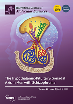
Schizophrenia is a severe mental disorder with a chronic, progressive course. The etiology of this condition is linked to the interactions of multiple genes and environmental factors. The earlier age of onset of schizophrenia, the higher frequency of negative symptoms in the clinical presentation, and the poorer response to antipsychotic treatment in men compared to women suggest the involvement of sex hormones in these processes. This article aims to draw attention to the possible relationship between testosterone and some clinical features in male schizophrenic patients and discuss the complex nature of these phenomena based on data from the literature. View this paper
  • Issues are regarded as officially published after their release is announced to the table of contents alert mailing list.
  • You may sign up for e-mail alerts to receive table of contents of newly released issues.
  • PDF is the official format for papers published in both, html and pdf forms. To view the papers in pdf format, click on the "PDF Full-text" link, and use the free Adobe Reader to open them.
Order results
Result details
Section
Select all
Export citation of selected articles as:
12 pages, 2681 KiB  
Article
Strong Metal Support Effect of Pt/g-C3N4 Photocatalysts for Boosting Photothermal Synergistic Degradation of Benzene
by Zhongcheng Huang, Xiaorong Cai, Shaohong Zang, Yixin Li, Dandan Zheng and Fuying Li
Int. J. Mol. Sci. 2023, 24(7), 6872; https://doi.org/10.3390/ijms24076872 - 6 Apr 2023
Cited by 1 | Viewed by 2134
Abstract
Catalysis is the most efficient and economical method for treating volatile organic pollutants (VOCs). Among the many materials that are used in engineering, platinized carbon nitride (Pt/g-C3N4) is an efficient and multifunctional catalyst which has strong light absorption and [...] Read more.
Catalysis is the most efficient and economical method for treating volatile organic pollutants (VOCs). Among the many materials that are used in engineering, platinized carbon nitride (Pt/g-C3N4) is an efficient and multifunctional catalyst which has strong light absorption and mass transfer capabilities, which enable it to be used in photocatalysis, thermal catalysis and photothermal synergistic catalysis for the degradation of benzene. In this work, Pt/g-C3N4 was prepared by four precursors for the photothermal synergistic catalytic degradation of benzene, which show different activities, and many tests were carried out to explore the possible reasons for the discrepancy. Among them, the Pt/g-C3N4 prepared from dicyanamide showed the highest activity and could convert benzene (300 ppm, 20 mL·min−1) completely at 162 °C under solar light and 173 °C under visible light. The reaction temperature was reduced by nearly half compared to the traditional thermal catalytic degradation of benzene at about 300 °C. Full article
Show Figures

Figure 1

Figure 1
<p>(<b>a</b>) XRD spectra, (<b>b</b>) FT-IR spectra of PDA, PMA, PUR and PCA samples.</p>
Full article ">Figure 2
<p>The morphology and atomic structure of Pt/CN. (<b>a</b>) block CN, (<b>b</b>) nanosheet CN, (<b>c</b>–<b>e</b>) TEM image, (<b>f</b>) EDX mapping of PDA.</p>
Full article ">Figure 3
<p>XPS spectra of (<b>a</b>) survey, (<b>b</b>) Pt 4f, (<b>c</b>) C 1s, (<b>d</b>) N 1s, (<b>e</b>) BET patterns, (<b>f</b>) thermalgravimetric curves of PDA.</p>
Full article ">Figure 4
<p>(<b>a</b>) Conversion rate, (<b>b</b>) mineralization rate of benzene by PDA, PCA, PMA and PUR under 150 °C and solar light conditions, (<b>c</b>) conversion rate, (<b>d</b>) mineralization rate of benzene for PDA under only heat, visible-light photothermal or solar light photothermal conditions, (<b>e</b>) conversion and mineralization rate of benzene for PDA under visible light or solar light photothermal conditions, (<b>f</b>) conversion/mineralization rate of four-cycle experiments of PDA under 162 °C and solar light conditions.</p>
Full article ">Figure 5
<p>(<b>a</b>) DRS patterns, (<b>b</b>) photoluminescence spectra, (<b>c</b>) EIS Nyquist plots, (<b>d</b>) Photocurrent responses of PDA, PCA, PUR and PMA samples, EPR spectra of PDA for the detection of (<b>e</b>) DMPO–·OH, (<b>f</b>) DMPO–·O<sub>2</sub><sup>−</sup>.</p>
Full article ">Scheme 1
<p>The mechanism of photothermal catalytic reaction by PDA.</p>
Full article ">
15 pages, 2348 KiB  
Article
Enhancement of the Antioxidant Effect of Natural Products on the Proliferation of Caco-2 Cells Produced by Fish Protein Hydrolysates and Collagen
by Mercedes Taroncher, Yelko Rodríguez-Carrasco, Francisco J. Barba and María-José Ruiz
Int. J. Mol. Sci. 2023, 24(7), 6871; https://doi.org/10.3390/ijms24076871 - 6 Apr 2023
Cited by 5 | Viewed by 1752
Abstract
A large amount of fish side streams are produced each year, promoting huge economic and environmental problems. In order to address this issue, a potential alternative is to isolate the high-added-value compounds with beneficial properties on human health. The objectives of this study [...] Read more.
A large amount of fish side streams are produced each year, promoting huge economic and environmental problems. In order to address this issue, a potential alternative is to isolate the high-added-value compounds with beneficial properties on human health. The objectives of this study were to determine the effect of hydrolyzed fish protein and collagen samples on cell proliferation, as well as to determine the specific influence of minerals and metals on this effect and whether dietary antioxidants can enhance cell proliferation. The results of hydrolyzed fish protein and collagen samples showed negative effects on Caco-2 cell proliferation at the highest concentrations tested. Moreover, the pre-treatment of these hydrolyzates with vitamin C and E, quercetin and resveratrol increased the proliferation of bioaccessible fractions of hydrolyzated fish protein and collagen samples compared to the bioaccessible fractions without pre-treatment. The highest mineral concentrations were found for P, Ca and Mg. The metals found in the pure hydrolyzates were As, Cd, Hg and Pb; however, they appeared at almost undetectable levels in bioavailable fractions. It can be concluded that the consumption of hydrolyzates of fish by-products is an interesting strategy for complying with EFSA recommendations regarding fish consumption while at the same time reducing fish waste. Full article
(This article belongs to the Special Issue Natural Compounds and Oxidative Stress)
Show Figures

Figure 1

Figure 1
<p>Effect of bioaccessible fractions of product 1 (<b>A</b>), product 2 (<b>B</b>), product 3 (<b>C</b>) and collagen (<b>D</b>) on cell proliferation in Caco-2 cells after 24 h of exposure at increasing concentrations from 0.05 to 0.5 mg/mL as assessed by the MTT assay. Values are expressed as mean ± SEM (<span class="html-italic">n</span> = 3). Values in the same figure with different superscript letters are significantly different (<span class="html-italic">p</span> &lt; 0.05). CRL: control.</p>
Full article ">Figure 2
<p>Protective effect of vit C (200 and 400 µM) on the bioaccessible fractions (0.1 and 0.2 mg/mL) of product 1 (<b>A</b>), product 2 (<b>B</b>), product 3 (<b>C</b>) and collagen (<b>D</b>) in Caco-2 cells after 24 h of exposure as assessed by the MTT assay. Values are expressed as mean ± SEM (<span class="html-italic">n</span> = 3). Values in the same figure with different superscript letters are significantly different (<span class="html-italic">p &lt;</span> 0.05). CRL: control.</p>
Full article ">Figure 3
<p>Protective effect of vit E (10 and 25 µM) on the bioaccessible fractions (0.1 and 0.2 mg/mL) of product 1 (<b>A</b>), product 2 (<b>B</b>), product 3 (<b>C</b>) and collagen (<b>D</b>) in Caco-2 cells after 24 h of exposure as assessed by the MTT assay. Values are expressed as mean ± SEM (<span class="html-italic">n</span> = 3). Values in the same figure with different superscript letters are significantly different (<span class="html-italic">p &lt;</span> 0.05). CRL: control.</p>
Full article ">Figure 4
<p>Protective effect of QUE (5 and 10 µM) on the bioaccessible fractions (0.1 and 0.2 mg/mL) of product 1 (<b>A</b>), product 2 (<b>B</b>), product 3 (<b>C</b>) and collagen (<b>D</b>) in Caco-2 cells after 24 h of exposure as assessed by the MTT assay. Values are expressed as mean ± SEM (<span class="html-italic">n</span> = 3). Values in the same figure with different superscript letters are significantly different (<span class="html-italic">p &lt;</span> 0.05). CRL: control; QUE: quercetin.</p>
Full article ">Figure 5
<p>Protective effect of RSV (10 and 25 µM) on the bioaccessible fractions (0.1 and 0.2 mg/mL) of product 1 (<b>A</b>), product 2 (<b>B</b>), product 3 (<b>C</b>) and collagen (<b>D</b>) in Caco-2 cells after 24 h of exposure as assessed by the MTT assay. Values are expressed as mean ± SEM (<span class="html-italic">n</span> = 3). Values in the same figure with different superscript letters are significantly different (<span class="html-italic">p &lt;</span> 0.05). CRL: control; RSV: resveratrol.</p>
Full article ">
19 pages, 3207 KiB  
Article
Nonsteroidal Anti-Inflammatory Drug Conjugated with Gadolinium (III) Complex as an Anti-Inflammatory MRI Agent
by Bokyung Sung, Hee-Kyung Kim, Ah-Rum Baek, Byeong-Woo Yang, Yeoun-Hee Kim, Garam Choi, Hyun-Jin Park, Minsup Kim, Jongmin Lee and Yongmin Chang
Int. J. Mol. Sci. 2023, 24(7), 6870; https://doi.org/10.3390/ijms24076870 - 6 Apr 2023
Cited by 2 | Viewed by 2071
Abstract
Studies have been actively conducted to ensure that gadolinium-based contrast agents for magnetic resonance imaging (MRI) are accompanied by various biological functions. A new example is the anti-inflammatory theragnostic MRI agent to target inflammatory mediators for imaging diagnosis and to treat inflammatory diseases [...] Read more.
Studies have been actively conducted to ensure that gadolinium-based contrast agents for magnetic resonance imaging (MRI) are accompanied by various biological functions. A new example is the anti-inflammatory theragnostic MRI agent to target inflammatory mediators for imaging diagnosis and to treat inflammatory diseases simultaneously. We designed, synthesized, and characterized a Gd complex of 1,4,7-tris(carboxymethylaza) cyclododecane-10-azaacetylamide (DO3A) conjugated with a nonsteroidal anti-inflammatory drug (NSAID) that exerts the innate therapeutic effect of NSAIDs and is also applicable in MRI diagnostics. Gd-DO3A-fen (0.1 mmol/kg) was intravenously injected into the turpentine oil-induced mouse model, with Gd-DO3A-BT as a control group. In the in vivo MRI experiment, the contrast-to-noise ratio (CNR) was higher and persisted longer than that with Gd-DO3A-BT; specifically, the CNR difference was almost five times at 2 h after injection. Gd-DO3A-fen had a binding affinity (Ka) of 6.68 × 10⁶ M−1 for the COX-2 enzyme, which was 2.1-fold higher than that of fenbufen, the original NSAID. In vivo evaluation of anti-inflammatory activity was performed in two animal models. In the turpentine oil-induced model, the mRNA expression levels of inflammatory parameters such as COX-2, TNF-α, IL-1β, and IL-6 were reduced, and in the carrageenan-induced edema model, swelling was suppressed by 72% and there was a 2.88-fold inhibition compared with the saline group. Correlation analysis between in vitro, in silico, and in vivo studies revealed that Gd-DO3A-fen acts as an anti-inflammatory theragnostic agent by directly binding to COX-2. Full article
Show Figures

Figure 1

Figure 1
<p>Diagram of Gd-DO3A-fen. (<b>a</b>) Representation of the mechanism of action of Gd-DO3A-fen. (<b>b</b>) The synthetic procedure of the anti-inflammatory MRI CAs Gd-DO3A-fen.</p>
Full article ">Figure 2
<p>In vivo assessment in the inflammation-induced mouse model. (<b>a</b>–<b>c</b>) <span class="html-italic">T<sub>1</sub></span>-weighted MR images were acquired before and after the administration of Gd-DO3A-fen and Gd-DO3A-BT (0.1 mmol/kg) by 1.5-T MRI. (<b>a</b>) Anatomical axial images of the inflammation site are displayed in gray scale and (<b>b</b>) CNR profile is shown as a function of time. Values are expressed as mean ± S.D. (<span class="html-italic">n</span> = 3). (<b>c</b>) Color-map at maximum-enhanced time. (<b>d</b>) Histogram of signal intensity values in the inflammatory region. The blue bars represent the signal intensity distribution for preinjection data, and the red bars represent the data obtained at 2 h postinjection of Gd-DO3A-fen (left) and Gd-DO3A-BT (right). Each line and value on the graph represent the average values for each distribution. (<b>e</b>) The inflammation-induced mouse model was sacrificed after the administration of Gd-DO3A-fen to quantify the in vivo biodistribution at each time point (<span class="html-italic">n</span> = 4). The results show the amount of Gd (mmol) in each tissue as a percentage (%) of total Gd administration dose (0.1 mmol/kg). The amount of Gd (mmol) in different tissues was determined by ICP-AES. GB, gallbladder; intest, intestines; inflam, inflamed tissue.</p>
Full article ">Figure 3
<p>The binding sites and binding affinity of COX-2 for Gd-DO3A-fen and the NSAID. (<b>a</b>) The drug-binding site of COX-2 consists of the main hydrophobic and secondary pockets. (<b>b</b>) The docking results for fenbufen. This drug was bound to Arg 513, and Trp 387 deep in the hydrophobic pocket, with ionic and π–π stacking interactions. (<b>c</b>) The docking results for Gd-DO3A-fen. The binding conformation of Gd-DO3A-fen was associated with polar interactions with surface protein and π–π stacking interactions in the hydrophobic pocket. (<b>d</b>) COX-2-binding affinity of fenbufen in UV–vis spectra. Samples were prepared in DMSO. COX-2 was dissolved in Tris-HCl buffer. (<b>e</b>) COX-2-binding affinity of Gd-DO3A-fen in UV–vis spectra. Samples were prepared in Tris-HCl (1:9, pH 7.0) buffer. (<b>d</b>,<b>e</b>) The absorbance of fenbufen and Gd-DO3A-fen were decreased by the addition of COX-2 enzyme. The inset graphs represent the fitting curve for the calculation of the binding constant (<span class="html-italic">K</span><sub>a</sub>).</p>
Full article ">Figure 4
<p>Antiinflammatory effect of drugs on PA-induced inflammation in C2C12 cells and in a turpentine oil-induced inflammation mouse model. (<b>a</b>) Effect of drugs on the viability of C2C12 cells (*** <span class="html-italic">p</span> &lt; 0.001 vs. nontreated control; <span class="html-italic">n</span> = 3). (<b>b</b>) The protein levels of COX-2 analyzed in total cell lysates by Western blotting. The bar graph represents the average densitometry values relative to β-actin (*** <span class="html-italic">p</span> &lt; 0.001 vs. nontreated control, <sup>###</sup> <span class="html-italic">p</span> &lt; 0.001 vs. PA, <sup>†††</sup> <span class="html-italic">p</span> &lt; 0.001 vs. fenbufen; <span class="html-italic">n</span> = 3). (<b>c</b>) Comparison of the mRNA expression levels of the proinflammatory enzyme and cytokines in the inflammation-induced mouse model injected with turpentine oil. The saline group was intravenously injected saline instead of drug in turpentine oil-induced model. The control group was injected with saline instead of turpentine oil. The bar graph represents the relative ratio of saline (** <span class="html-italic">p</span> &lt; 0.05, *** <span class="html-italic">p</span> &lt; 0.001 vs. nontreated control, <sup>#</sup> <span class="html-italic">p</span> &lt; 0.01, <sup>##</sup> <span class="html-italic">p</span> &lt; 0.05, <sup>###</sup> <span class="html-italic">p</span> &lt; 0.001 vs. PA, <sup>†</sup> <span class="html-italic">p</span> &lt; 0.01 vs. fenbufen; <span class="html-italic">n</span> = 3).</p>
Full article ">Figure 5
<p>The inhibitory effect of Gd-DO3A-fen in the carrageenan-induced rat hind paw edema rat model. (<b>a</b>) The procedure for monitoring the anti-inflammatory effect of Gd-DO3A-fen on paw edema. The edema volume was measured before injection and at 1, 2, 3, 4, 5, and 6 h after the injection of carrageenan. (<b>b</b>) Images showing the rat paw before injection and at 5 h after the injection of carrageenan, respectively: carrageenan, carrageenan + saline, carrageenan + Gd-DO3A-fen. Yellow arrows indicate the carrageenan-induced rat paw. (<b>c</b>) Percentage swelling observed in the carrageenan-induced paw edema. (<b>d</b>) Comparison of % swelling of Gd-DO3A-fen group and that of saline group at 5 h after injection. (<b>e</b>) Percentage inhibition of paw edema by Gd-DO3A-fen and saline. (<b>f</b>) Comparison of % inhibition of Gd-DO3A-fen group and that of saline group at 5 h after injection. All values represent mean ± S.E.M. (<span class="html-italic">n</span> = 4 per group; *<span class="html-italic">p</span> &lt; 0.05, **<span class="html-italic">p</span> &lt; 0.01, ***<span class="html-italic">p</span> &lt; 0.001 vs. saline group).</p>
Full article ">
25 pages, 4413 KiB  
Article
The Role of Tyr-His-Trp Triad and Water Molecule Near the N1-Atom of 2-Hydroperoxycoelenterazine in Bioluminescence of Hydromedusan Photoproteins: Structural and Mutagenesis Study
by Pavel V. Natashin, Ludmila P. Burakova, Margarita I. Kovaleva, Mikhail B. Shevtsov, Daria A. Dmitrieva, Elena V. Eremeeva, Svetlana V. Markova, Alexey V. Mishin, Valentin I. Borshchevskiy and Eugene S. Vysotski
Int. J. Mol. Sci. 2023, 24(7), 6869; https://doi.org/10.3390/ijms24076869 - 6 Apr 2023
Cited by 2 | Viewed by 2177
Abstract
Hydromedusan photoproteins responsible for the bioluminescence of a variety of marine jellyfish and hydroids are a unique biochemical system recognized as a stable enzyme-substrate complex consisting of apoprotein and preoxygenated coelenterazine, which is tightly bound in the protein inner cavity. The binding of [...] Read more.
Hydromedusan photoproteins responsible for the bioluminescence of a variety of marine jellyfish and hydroids are a unique biochemical system recognized as a stable enzyme-substrate complex consisting of apoprotein and preoxygenated coelenterazine, which is tightly bound in the protein inner cavity. The binding of calcium ions to the photoprotein molecule is only required to initiate the light emission reaction. Although numerous experimental and theoretical studies on the bioluminescence of these photoproteins were performed, many features of their functioning are yet unclear. In particular, which ionic state of dioxetanone intermediate decomposes to yield a coelenteramide in an excited state and the role of the water molecule residing in a proximity to the N1 atom of 2-hydroperoxycoelenterazine in the bioluminescence reaction are still under discussion. With the aim to elucidate the function of this water molecule as well as to pinpoint the amino acid residues presumably involved in the protonation of the primarily formed dioxetanone anion, we constructed a set of single and double obelin and aequorin mutants with substitutions of His, Trp, Tyr, and Ser to residues with different properties of side chains and investigated their bioluminescence properties (specific activity, bioluminescence spectra, stopped-flow kinetics, and fluorescence spectra of Ca2+-discharged photoproteins). Moreover, we determined the spatial structure of the obelin mutant with a substitution of His64, the key residue of the presumable proton transfer, to Phe. On the ground of the bioluminescence properties of the obelin and aequorin mutants as well as the spatial structures of the obelin mutants with the replacements of His64 and Tyr138, the conclusion was made that, in fact, His residue of the Tyr-His-Trp triad and the water molecule perform the “catalytic function” by transferring the proton from solvent to the dioxetanone anion to generate its neutral ionic state in complex with water, as only the decomposition of this form of dioxetanone can provide the highest light output in the light-emitting reaction of the hydromedusan photoproteins. Full article
Show Figures

Figure 1

Figure 1
<p>Two-dimensional drawing of the hydrogen bonding network in the binding cavities of active (<b>A</b>) and Ca<sup>2+</sup>-discharged (<b>B</b>) wild-type obelins. Molecular surface representation of “proton channel” in active (<b>C</b>) and Ca<sup>2+</sup>-discharged (<b>D</b>) wild-type obelins. Amino acid residues and the molecules of 2-hydroperoxycoelenterazine and coelenteramide are shown as stick models. Water molecules are represented as red balls. Hydrogen bonds are shown as dashed lines. Partially represented surface is gray colored. (<b>E</b>) Sequence alignment of obelin from <span class="html-italic">O. longissima</span> [<a href="#B37-ijms-24-06869" class="html-bibr">37</a>] and aequorin from <span class="html-italic">A. victoria</span> [<a href="#B38-ijms-24-06869" class="html-bibr">38</a>]. The residues in obelin and aequorin mutated in this study are shown in blue. The Ca<sup>2+</sup> binding loops are highlighted in gray. The helices are indicated by capital letters A–H based on the wild-type obelin structure (PDB code 1QV0). Aequorin numeration starts from Val, shown with an arrow, according to the original numbering of the amino acid sequence determined for the natural aequorin and aequorin structure (PDB code 1EJ3) [<a href="#B1-ijms-24-06869" class="html-bibr">1</a>,<a href="#B22-ijms-24-06869" class="html-bibr">22</a>].</p>
Full article ">Figure 2
<p>Normalized bioluminescence and fluorescence spectra of obelin (<b>A</b>,<b>C</b>), aequorin (<b>B</b>,<b>D</b>) and their mutants.</p>
Full article ">Figure 3
<p>High-speed (<b>A</b>,<b>B</b>) and low-speed (<b>C</b>,<b>D</b>) stopped-flow plots of the bioluminescence signals of obelin (<b>A</b>,<b>C</b>), aequorin (<b>B</b>,<b>D</b>), and their mutants. The curves are individual shots.</p>
Full article ">Figure 4
<p>Crystal structure of OL_H64F mutant. (<b>A</b>) Overall structure of OL_H64F mutant. The helices are marked by letters A–H. (<b>B</b>) Superimposition of OL_H64F mutant (blue, PDB 8C6O, chain A), wild-type obelin (red, PDB 1QV0), and OL_Y138F mutant (green, PDB 4MRX).</p>
Full article ">Figure 5
<p>Two-dimensional representation of the hydrogen bond network. (<b>A</b>) OL_H64F mutant (PDB 8C6O, chain A), (<b>B</b>) wild-type obelin (PDB 1QV0), (<b>C</b>) active OL_Y138F mutant (PDB: 4MRX), and (<b>D</b>) Ca<sup>2+</sup>-discharged OL_Y138F mutant (PDB 4MRY). Mutated residues H64(F64) and Y138(F138) are shown in blue and red, respectively. Hydrogen bonds are shown as dashed lines, distances between atoms are shown as arrows, and “W” stands for a water molecule. Distances are given in Å.</p>
Full article ">Figure 6
<p>Stereoview of the superimposition of 2-hydroperoxycoelenterazine molecules with the residues residing near their N1 atoms: (<b>A</b>) OL_H64F mutant (gray) vs. wild-type obelin (cyan) and (<b>B</b>) OL_H64F mutant (gray) vs. OL_Y138F mutant (green). Hydrogen bonds are shown as dashed lines. Oxygen and nitrogen atoms are colored with red and blue, respectively.</p>
Full article ">Figure 7
<p>Dioxetanone derivatives that might be formed in bioluminescent reaction of hydromedusan photoproteins.</p>
Full article ">Scheme 1
<p>Proposed reaction scheme for photoprotein bioluminescence [<a href="#B47-ijms-24-06869" class="html-bibr">47</a>]. X—an intermediate state; Y*—the product, coelenteramide, in the excited state; Y—coelenteramide in the ground state.</p>
Full article ">
11 pages, 3087 KiB  
Article
Germination and Growth of Plasma-Treated Maize Seeds Planted in Fields and Exposed to Realistic Environmental Conditions
by Nina Recek, Rok Zaplotnik, Alenka Vesel, Gregor Primc, Peter Gselman, Miran Mozetič and Matej Holc
Int. J. Mol. Sci. 2023, 24(7), 6868; https://doi.org/10.3390/ijms24076868 - 6 Apr 2023
Cited by 1 | Viewed by 1615
Abstract
In this study, we applied an inductively coupled, radio frequency oxygen plasma to maize seeds and investigated its effects on seedling emergence, plant number at tasseling, and crop yield of maize in realistic field conditions. Maize seeds of seven different hybrids were treated [...] Read more.
In this study, we applied an inductively coupled, radio frequency oxygen plasma to maize seeds and investigated its effects on seedling emergence, plant number at tasseling, and crop yield of maize in realistic field conditions. Maize seeds of seven different hybrids were treated over two harvest years. In addition to plasma-treated seeds, a control sample, fungicide-treated seeds, an eco-layer, and a plasma and eco-layer combination, were planted. Seedling emergence, plant number at tasseling (plants/m2), and yield (kg/ha), were recorded. In the first harvest year, results were negatively affected by the presence of an insect pest. In the second harvest year, plant number and yield results were more uniform. In both years, for two and three hybrids, respectively, the highest yield arose from plants from plasma-treated seeds, but the differences were only partially significant. Considering our results, plasma treatment of maize seeds appears to have a positive effect on the yield of the plant. Full article
(This article belongs to the Special Issue Advances in Molecular Plant Sciences)
Show Figures

Figure 1

Figure 1
<p>Number of maize plants in two vegetative growth stages of maize in the harvest year 2020: (<b>a</b>) at seedling emergence; and (<b>b</b>) at tasseling time. The error bars represent standard error. Different lowercase letters above data points represent statistically significant differences (<span class="html-italic">p</span> &lt; 0.05; post hoc Tukey’s range test) between treatments.</p>
Full article ">Figure 2
<p>Correlation between the number of emerged seedlings (May) and the number of maize plants at tasseling (August) for the harvest year 2020.</p>
Full article ">Figure 3
<p>Number of maize plants in two vegetative growth stages of maize in the harvest year 2021: (<b>a</b>) at seedling emergence; and (<b>b</b>) at tasseling time. The error bars represent standard error. Different lowercase letters above data points represent statistically significant differences (<span class="html-italic">p</span> &lt; 0.05; post hoc Tukey’s range test) between treatments.</p>
Full article ">Figure 4
<p>Correlation between the number of emerged seedlings (May) and the number of maize plants at tasseling (August) for the harvest year 2021.</p>
Full article ">Figure 5
<p>(<b>a</b>) Maize yield; and (<b>b</b>) correlation between number of plants at tasseling time and maize yield in the harvest year 2020. The error bars represent standard error. Different lowercase letters above data points represent statistically significant differences (<span class="html-italic">p</span> &lt; 0.05; post hoc Tukey’s range test) between treatments.</p>
Full article ">Figure 6
<p>(<b>a</b>) Maize yield; and (<b>b</b>) correlation between the number of plants at tasseling time and maize yield in the harvest year 2021. The error bars represent standard error. Different lowercase letters above data points represent statistically significant differences (<span class="html-italic">p</span> &lt; 0.05; post hoc Tukey’s range test) between treatments.</p>
Full article ">Figure 7
<p>Average daily temperatures and precipitation for each month in years 2020 and 2021 measured on the nearest permanent weather station.</p>
Full article ">Figure 8
<p>Schematic representation of industrial plasma reactor used to treat maize.</p>
Full article ">
22 pages, 1529 KiB  
Review
Imbalance of Essential Metals in Traumatic Brain Injury and Its Possible Link with Disorders of Consciousness
by Rosanna Squitti, Giuseppe Reale, Vincenzo Tondolo, Daniela Crescenti, Sonia Bellini, Marco Moci, Pietro Caliandro, Luca Padua and Mauro Rongioletti
Int. J. Mol. Sci. 2023, 24(7), 6867; https://doi.org/10.3390/ijms24076867 - 6 Apr 2023
Cited by 5 | Viewed by 2798
Abstract
Dysfunction of the complex cerebral networks underlying wakefulness and awareness is responsible for Disorders of Consciousness (DoC). Traumatic Brain Injury (TBI) is a common cause of DoC, and it is responsible for a multi-dimensional pathological cascade that affects the proper functioning of the [...] Read more.
Dysfunction of the complex cerebral networks underlying wakefulness and awareness is responsible for Disorders of Consciousness (DoC). Traumatic Brain Injury (TBI) is a common cause of DoC, and it is responsible for a multi-dimensional pathological cascade that affects the proper functioning of the brainstem and brain consciousness pathways. Iron (Fe), Zinc (Zn), and Copper (Cu) have a role in the neurophysiology of both the ascending reticular activating system, a multi-neurotransmitter network located in the brainstem that is crucial for consciousness, and several brain regions. We aimed to summarize the role of these essential metals in TBI and its possible link with consciousness alterations. We found that TBI alters many neuronal molecular mechanisms involving essential metals, causing neurodegeneration, neural apoptosis, synaptic dysfunction, oxidative stress, and inflammation. This final pattern resembles that described for Alzheimer’s disease (AD) and other neurological and psychiatric diseases. Furthermore, we found that amantadine, zolpidem, and transcranial direct current stimulation (tDCS)—the most used treatments for DoC recovery—seem to have an effect on essential metals-related pathways and that Zn might be a promising new therapeutic approach. This review summarizes the neurophysiology of essential metals in the brain structures of consciousness and focuses on the mechanisms underlying their imbalance following TBI, suggesting their possible role in DoC. The scenario supports further studies aimed at getting a deeper insight into metals’ role in DoC, in order to evaluate metal-based drugs, such as metal complexes and metal chelating agents, as potential therapeutic options. Full article
Show Figures

Figure 1

Figure 1
<p>Mesocircuit—Frontoparietal Model. The diagram shows the interrelationship between the forebrain mesocircuit, the frontoparietal network, and the ascending reticular activating system. The function of each system is reported in italics. Cx: cortex.</p>
Full article ">Figure 2
<p>Physiological and pathological neuromodulation of Zn-containing pre- and postsynaptic neurons. Most vesicular Zn co-localizes with glutamate in subsets of glutamatergic zinc-enriched neurons, and it is also contained in the synaptic vesicles of subpopulations of glycinergic and GABAergic neurons. Zn<sup>2+</sup>-level regulation between cellular compartments, organelles, and extracellular space is ensured by ZIP and ZnT protein families, and by metallothioneins (MTs), which buffer cytoplasmic Zn<sup>2+</sup>, functioning as a temporary store for cellular Zn<sup>2+</sup>. In the presynaptic terminals, Zn<sup>2+</sup> is transported into presynaptic vesicles by the Zn transporter ZnT3. During synaptic transmission, free Zn is released in the synaptic cleft, where it may be recycled back into the presynaptic boutons by ZIP/Zn<sup>2+</sup> transporters or modulate excitatory (NMDA, AMPA) and inhibitory (GABA, glycine) amino acid receptors of the postsynaptic terminal; Zn can inhibit GABAAR and NMDAR, and potentiate/inhibit AMPAR and GlyR at low/high concentrations, respectively. Extracellular Zn can also alter the excitability of neurons through effects on voltage-gated ion channels (e.g., VGCC), affecting ions’ influx and neurotransmitter release. Ion channels and AMPAR/KAR allow synaptically released Zn to enter presynaptic and postsynaptic neurons to modulate intracellular Zn signaling functions. Excessive Zn<sup>2+</sup> accumulation inside postsynaptic cells, as per excitotoxic stimulation, can lead to a series of toxic effects involving mitochondrial dysfunction and ROS/NOS production, eventually leading to oxidative damage to proteins and DNA, neuronal apoptosis, and/or necrosis. Glu, glutamate; GABA<sub>A</sub>R, GABA A receptor; NMDAR, NMDA receptor; AMPAR, AMPA receptor; GlyR, glycine receptor; VGCC, voltage-gated calcium channel; KAR, kainate receptor.</p>
Full article ">Figure 3
<p>Model of Beta amyloid (Aβ), glutamate, oxidative stress, and ionic dyshomeostasis in neurodegenerative processes associated with Traumatic Brian Injury (TBI) and Alzheimer’s disease (AD). Aβ, alpha synuclein (α-syn), and hyper-phosphorylated Tau are among the most frequently reported molecules upregulated in TBI and are also closely related to AD. Experimental models of TBI [<a href="#B58-ijms-24-06867" class="html-bibr">58</a>] show that Cu concentrations were increased in the ipsilateral cortex adjacent, but not closest, to the impact zone only 28 days after the injury, and it may be cause for concern in relation to the potential chronic oxidative stress toxicity based on Cu abnormal metabolism, as has been observed in neurodegenerative disorders and more specifically for AD. The model proposed to highlight the main Cu toxic mechanisms that can be triggered by TBI in the long term and in AD. In a complex scenario, Aβ, oxidative stress, excitotoxicity, and Cu<sup>2+</sup> dyshomeostasis act in concert to promote synaptic dysfunction and neuronal loss. Upon the production of excessive glutamate levels (1), Ca<sup>2+</sup> ions enter the cell through the NMDA receptor (2) and (3) induce Cu-ATPase7A/B (ATP7A/B) translocation at synapses where vesicular Cu is released in the synaptic cleft. The released Cu<sup>2+</sup> (in concentrations up to 100 µmol/L) may inhibit the NMDA receptor, thereby protecting neurons from glutamatergic excitotoxicity (4), or catalyze Fenton-type and Haber–Weiss reactions, thereby generating reactive oxygen species (ROS) (5). Enhanced ROS generation can damage proteins, lipids, and nucleic acids, eventually leading to cell death (5). Ca<sup>2+</sup> overload can increase superoxide anion (O2•−) production from mitochondria (6), and nitric oxide (NO) generation via Ca<sup>2+</sup>-dependent activation of NO synthase (NOS) (7). Reactive oxygen and nitrosative (RNS) species mobilize Cu<sup>2+</sup> from metallothionein 3 (MT-3) (8), leading to increased intracellular toxic Cu<sup>2+</sup> concentrations (9) and promoting mitochondrial dysfunction, as well as release of pro-apoptotic factors (10). ROS-driven Cu<sup>2+</sup> mobilization can further aggravate oxidative stress and initiate Aβ oligomerization (11). Altered trafficking of APP and/or elevated Aβ oligomer secretion can generate an intracellular Cu<sup>2+</sup> deficiency, thereby causing oxidative stress by the loss of SOD-1 function (12). Aβ, α-synuclein, and PrP increased after TBI can modulate neurotransmission as [Cu<sup>2+</sup>] buffers within the synaptic cleft or amplify the vicious cycle by increasing oxidative stress (13). Furthermore, glutamate-driven mitochondrial Ca<sup>2+</sup> overload can mobilize Cu<sup>2+</sup> from these organelles (14). Excess Non-Cp Cu in the bloodstream is a source for the buildup of labile Cu<sup>2+</sup> into the intermembrane space of mitochondria (15), promoting the ATPase7A/B translocation of Cu<sup>2+</sup> into vesicles of the trans-Golgi network and endoplasmic reticulum (ER) (16). These processes, working intracellularly at the level of synaptic spines, in the synaptic cleft, and in the neurovascular unit (ref. [<a href="#B8-ijms-24-06867" class="html-bibr">8</a>]), can facilitate synaptic dysfunction, neuronal deafferentation, and ultimately brain cell death.</p>
Full article ">
16 pages, 1234 KiB  
Article
The Nitric Oxide (NO) Donor Molsidomine Counteract Social Withdrawal and Cognition Deficits Induced by Blockade of the NMDA Receptor in the Rat
by Lamprini Katsanou, Evangelia Fragkiadaki, Sotirios Kampouris, Anastasia Konstanta, Aikaterini Vontzou and Nikolaos Pitsikas
Int. J. Mol. Sci. 2023, 24(7), 6866; https://doi.org/10.3390/ijms24076866 - 6 Apr 2023
Cited by 2 | Viewed by 1865
Abstract
The deficiency of the gaseous molecule nitric oxide (NO) seems to be critically involved in the pathogenesis of schizophrenia. Thus, molecules that can normalize NO levels, as are NO donors, might be of utility for the medication of this psychiatric disease. The aim [...] Read more.
The deficiency of the gaseous molecule nitric oxide (NO) seems to be critically involved in the pathogenesis of schizophrenia. Thus, molecules that can normalize NO levels, as are NO donors, might be of utility for the medication of this psychiatric disease. The aim of the present study was to detect the ability of the NO donor molsidomine to reduce schizophrenia-like impairments produced by the blockade of the N-methyl-D-aspartate (NMDA) receptor in rats. Molsidomine’s ability to attenuate social withdrawal and spatial recognition memory deficits induced by the NMDA receptor antagonist ketamine were assessed using the social interaction and the object location test, respectively. Further, the efficacy of the combination of sub-effective doses of molsidomine with sub-effective doses of the atypical antipsychotic clozapine in alleviating non-spatial recognition memory deficits was evaluated utilizing the object recognition task. Molsidomine (2 and 4 mg/kg) attenuated social withdrawal and spatial recognition memory deficits induced by ketamine. Co-administration of inactive doses of molsidomine (1 mg/kg) and clozapine (0.1 mg/kg) counteracted delay-dependent and ketamine-induced non-spatial recognition memory deficits. The current findings suggest that molsidomine is sensitive to glutamate hypofunction since it attenuated behavioral impairments in animal models mimicking the negative symptoms and cognitive deficits of schizophrenia. Additionally, the present results support the potential of molsidomine as an adjunctive drug for the therapy of schizophrenia. Full article
(This article belongs to the Special Issue Molecular Mechanisms of Schizophrenia and Novel Targets 2.0)
Show Figures

Figure 1

Figure 1
<p>Social interaction test. The histogram represents the mean ± S.E.M of 8 pairs of rats per treatment group. (<b>A</b>) Social interaction levels expressed by different groups of rats. * <span class="html-italic">p &lt;</span> 0.05 vs. all the other groups; + <span class="html-italic">p &lt;</span> 0.05 vs. the vehicle + vehicle, vehicle + molsidomine 2 mg/kg and vehicle + molsidomine 4 mg/kg groups. (<b>B</b>) Locomotor activity expressed by different groups of rats during the social interaction test.</p>
Full article ">Figure 2
<p>Object location task. The histogram represents the mean ± S.E.M of 8 rats per treatment group. The 1-h ITI was used. (<b>A</b>) Discrimination index D performance expressed by different groups of rats during T2. * <span class="html-italic">p</span> &lt; 0.05 vs. all the other groups. (<b>B</b>) Total exploration times.</p>
Full article ">Figure 3
<p>Object recognition task. The histograms represent the mean ± S.E.M of 8 rats per treatment group. The 24-h ITI was used. (<b>A</b>) Discrimination index D performance expressed by different groups of rats during T2. * <span class="html-italic">p</span> &lt; 0.05 vs. all the other groups. (<b>B</b>) Total exploration times.</p>
Full article ">Figure 4
<p>Object recognition task. The histograms represent the mean ± S.E.M of 8 rats per treatment group. The 1-h ITI was used. (<b>A</b>) Discrimination index D performance expressed by different groups of rats during T2. * <span class="html-italic">p</span> &lt; 0.05 vs. all the other groups. (<b>B</b>) Total exploration times.</p>
Full article ">
26 pages, 6225 KiB  
Article
Novel Insights into circRNA Saga Coming from Spermatozoa and Epididymis of HFD Mice
by Francesco Manfrevola, Teresa Chioccarelli, Vincenza Grazia Mele, Veronica Porreca, Monica Mattia, Donatella Cimini, Antonella D’Agostino, Gilda Cobellis, Silvia Fasano, Chiara Schiraldi, Rosanna Chianese and Riccardo Pierantoni
Int. J. Mol. Sci. 2023, 24(7), 6865; https://doi.org/10.3390/ijms24076865 - 6 Apr 2023
Cited by 4 | Viewed by 2178
Abstract
Obesity is a pathophysiological disorder associated with adiposity accumulation, oxidative stress, and chronic inflammation state that is progressively increasing in younger population worldwide, negatively affecting male reproductive skills. An emerging topic in the field of male reproduction is circRNAs, covalently closed RNA molecules [...] Read more.
Obesity is a pathophysiological disorder associated with adiposity accumulation, oxidative stress, and chronic inflammation state that is progressively increasing in younger population worldwide, negatively affecting male reproductive skills. An emerging topic in the field of male reproduction is circRNAs, covalently closed RNA molecules produced by backsplicing, actively involved in a successful spermatogenesis and in establishing high-quality sperm parameters. However, a direct correlation between obesity and impaired circRNA cargo in spermatozoa (SPZ) remains unclear. In the current work, using C57BL6/J male mice fed with a high-fat diet (HFD, 60% fat) as experimental model of oxidative stress, we investigated the impact of HFD on sperm morphology and motility as well as on spermatic circRNAs. We performed a complete dataset of spermatic circRNA content by a microarray strategy, and differentially expressed (DE)-circRNAs were identified. Using a circRNA/miRNA/target network (ceRNET) analysis, we identified circRNAs potentially involved in oxidative stress and sperm motility pathways. Interestingly, we demonstrated an enhanced skill of HFD sperm in backsplicing activity together with an inefficient epididymal circRNA biogenesis. Fused protein in sarcoma (FUS) and its ability to recruit quaking (QKI) could be involved in orchestrating such mechanism. Full article
(This article belongs to the Special Issue Novel Insights into the Biology of Spermatozoa 2.0)
Show Figures

Figure 1

Figure 1
<p><b>Characterization of HFD testis and sperm parameters.</b> (<b>A</b>,<b>B</b>) H&amp;E staining of testis (<b>A</b>) and <span class="html-italic">cauda</span> SPZ (<b>B</b>) collected from CTRL (n = 6) and HFD (n = 6) mice. (<b>A</b>) In testis sections, germ cell lumen infiltration was indicated by black arrowheads; scale bar: 100 μm. (<b>B</b>) Anomalous sperm heads were indicated by black arrowheads; scale bar: 50 μm; scale bar inset: 10 μm. (<b>C</b>) Percentage of anomalous sperm heads in CTRL and HFD <span class="html-italic">cauda</span> SPZ; data were reported as the percentage of anomalous sperm heads/total SPZ. Sperm viability (<b>D</b>) and motility (<b>E</b>) assay in CTRL and HFD <span class="html-italic">cauda</span> SPZ; data were expressed as the percentage of nonviable/total SPZ and motile/live SPZ, respectively, and reported as mean ± SEM; ** <span class="html-italic">p</span> &lt; 0.01.</p>
Full article ">Figure 2
<p><b>Overview of circRNA expression in mouse SPZ.</b> (<b>A</b>) The distribution of up- and downregulated circRNAs in HFD (n = 3) compared with CTRL (n = 3) SPZ among a total of 9838 circRNAs. (<b>B</b>) The proportion of different types of circRNAs among all predicted circRNAs. (<b>C</b>) Chromosomal distribution of SPZ-derived circRNAs on strand + and strand –, according to their host gene location.</p>
Full article ">Figure 3
<p><b>Differential expression of circRNAs between CTRL and HFD SPZ.</b> (<b>A</b>) The distribution of up- and downregulated DE-circRNAs, among a total of 109 DE-circRNAs, in HFD compared with CTRL SPZ. (<b>B</b>) Hierarchical clustering analysis of DE-circRNAs in CTRL SPZ (samples CTRL-1, CTRL-2, CTRL-3) and HFD SPZ (samples HFD-1, HFD-2, HFD-3); the expression values (fold change &gt; 1.5, <span class="html-italic">p</span> &lt; 0.05) were represented in different colors, indicating expression levels above and below the median expression level across all samples. (<b>C</b>) The volcano plot was constructed using fold change and <span class="html-italic">p</span>-values; in detail, the values on X and Y axes are log2 (FC = fold change) and –log10 (<span class="html-italic">p</span>-values), respectively. Red points in the volcano plot represent the DE-circRNAs with statistical significance. (<b>D</b>) The distribution of up- and downregulated DE-circRNAs in the mouse genome, according to their host gene location.</p>
Full article ">Figure 4
<p><b>Validation of circRNA microarray results in testis, epididymis, and SPZ from CTRL and HFD mice.</b> (<b>A</b>) Expression analysis of 9 circRNAs upregulated in HFD compared with CTRL-derived SPZ. (<b>B</b>) Expression analysis of 9 circRNAs downregulated in HFD compared with CTRL-derived SPZ. qRT-PCR data are normalized using <span class="html-italic">cyclophilin</span> and <span class="html-italic">ribosomal protein S18</span> (<span class="html-italic">RPS18</span>) for SPZ and tissues, respectively, expressed as fold expression (nfe) and reported as mean value ± S.E.M. **: <span class="html-italic">p</span> &lt; 0.01; *: <span class="html-italic">p</span> &lt; 0.05.</p>
Full article ">Figure 5
<p><b>Characterization of backsplicing machinery in SPZ and epididymis of CTRL and HFD mice.</b> (<b>A</b>) Immunofluorescence analysis of FUS protein in <span class="html-italic">caput</span> and <span class="html-italic">cauda</span> SPZ of CTRL and HFD mice. White arrowheads and white asterisks represent FUS localization (RED) in sperm head and tail, respectively. Nuclei were labeled with DAPI (blue). Scale bar: 5 µm. (<b>B</b>) Western blot analysis of FUS protein (75 KDa) in <span class="html-italic">caput</span> and <span class="html-italic">cauda</span> SPZ of CTRL and HFD mice (n = 5 different samples for each experimental group in triplicate). Signals were quantified by densitometry analysis and normalized to Ponceau Red (Pon.S). Data were expressed in OD values and reported as mean ± SEM. Experimental groups with statistically significant differences (<span class="html-italic">p</span> &lt; 0.01) were indicated with different letters. (<b>C</b>) The enrichment levels of circADAM10 in the products of RIP assay (FUS-IP compared with IgG-IP) in <span class="html-italic">caput</span> and <span class="html-italic">cauda</span> SPZ of CTRL and HFD mice detected by qRT-PCR. Data are reported as mean  ±  SEM from three independent experiments. ** <span class="html-italic">p</span> &lt; 0.01. (<b>D</b>) Immunocytochemistry of FUS in Bouin’s fixed <span class="html-italic">caput</span> and <span class="html-italic">cauda</span> epididymis sections (7 μm) of CTRL and HFD mice (n = 5 different samples for each experimental group in triplicate). The FUS protein localization in principal cells was indicated by black arrows. Scale bar: 50 μm; inset: H&amp;E staining of Bouin’s fixed <span class="html-italic">caput</span> and <span class="html-italic">cauda</span> epididymis sections (7 μm) of CTRL and HFD mice; scale bar: 20 μm. (<b>E</b>) Western blot analysis of FUS protein in <span class="html-italic">caput</span> and <span class="html-italic">cauda</span> epididymis of CTRL and HFD mice. Signals were quantified by densitometry analysis and normalized to Ponceau Red (Pon.S). Data were expressed in OD values and reported as mean ± SEM. Experimental groups with statistically significant differences (<span class="html-italic">p</span> &lt; 0.01) were indicated with different letters. (<b>F</b>) The enrichment levels of circPCSK6 in the products of RIP assay (FUS-IP compared with IgG-IP) in <span class="html-italic">caput</span> and <span class="html-italic">cauda</span> epididymis of CTRL and HFD mice detected by qRT-PCR. Data are reported as mean ± SEM from three independent experiments. ** <span class="html-italic">p</span> &lt; 0.01. (<b>G</b>) Western blot analysis of QKI protein (40 KDa) in <span class="html-italic">caput</span> and <span class="html-italic">cauda</span> epididymis of CTRL and HFD mice. Signals were quantified by densitometry analysis and normalized to Ponceau Red (Pon.S). Data were expressed in OD values and reported as mean ± SEM. Experimental groups with statistically significant differences (<span class="html-italic">p</span> &lt; 0.01) were indicated with different letters. (<b>H</b>) IP in <span class="html-italic">caput</span> and <span class="html-italic">cauda</span> epididymis of CTRL and HFD mice. Total proteins collected were immunoprecipitated using FUS antibody. Protein interaction between FUS and QKI was detected by Western blot analysis.</p>
Full article ">Figure 6
<p><b>In vitro experiment of vesicle shuttle: from CTRL epididymis to HFD SPZ.</b> (<b>A</b>) CeRNET of one circRNA downregulated in HFD SPZ. CircSMAD2 tethers a group of miRNAs as targets, all involved in sperm motility pathways. Networks were built using Cytoscape. Hexagonal and rectangular symbols represent circRNAs and miRNAs, respectively. The arrow indicates the tethering activity of circRNAs toward miRNAs, while the dotted arrow indicates the pathways upstream of the miRNAs. (<b>B</b>,<b>C</b>) qRT-PCR analysis of circSMAD2 (<b>B</b>) and <span class="html-italic">PLAG1</span>-mRNA (<b>C</b>) in CTRL <span class="html-italic">cauda</span> SPZ and HFD <span class="html-italic">cauda</span> SPZ in vitro coincubated with: PBS, CTRL-ELF, and CTRL-ELF pretreated with anti-CD9 antibody (n = 6 for each experimental group). qRT-PCR data were normalized using <span class="html-italic">cyclophilin</span>, expressed as fold expression (nfe) relative to CTRL SPZ and reported as mean value ± S.E.M. Experimental groups with statistically significant differences (<span class="html-italic">p</span> &lt; 0.01) were indicated with different letters. (<b>D</b>) Sperm motility assay in CTRL <span class="html-italic">cauda</span> SPZ and HFD <span class="html-italic">cauda</span> SPZ in vitro coincubated with: PBS, CTRL-ELF, and CTRL-ELF pretreated with anti-CD9 antibody (n = 6 for each experimental group); data were expressed as the percentage of motile/live SPZ and reported as mean ± SEM. Experimental groups with statistically significant differences (<span class="html-italic">p</span> &lt; 0.01) were indicated with different letters.</p>
Full article ">Figure 7
<p>Schematic representation of experimental design.</p>
Full article ">
12 pages, 1994 KiB  
Article
Conformational Changes of Acyl Carrier Protein Switch the Chain Length Preference of Acyl-ACP Thioesterase ChFatB2
by Tianxiang Yang, Yunlong Yang, Ming Yang, Jiangang Ren, Changying Xue, Yanbin Feng and Song Xue
Int. J. Mol. Sci. 2023, 24(7), 6864; https://doi.org/10.3390/ijms24076864 - 6 Apr 2023
Viewed by 1987
Abstract
Microbial fatty acids are synthesized by Type II fatty acid synthase and could be tailored by acyl-ACP thioesterase. With the prospects of medium-chain fatty-acid-derivative biofuels, the selectivity of thioesterase has been studied to control the fatty acid product chain length. Here, we report [...] Read more.
Microbial fatty acids are synthesized by Type II fatty acid synthase and could be tailored by acyl-ACP thioesterase. With the prospects of medium-chain fatty-acid-derivative biofuels, the selectivity of thioesterase has been studied to control the fatty acid product chain length. Here, we report an alternative approach by manipulating the acyl carrier protein portion of acyl-ACP to switch the chain length propensity of the thioesterase. It was demonstrated that ChFatB2 from Cuphea hookeriana preferred C10-ACP to C8-ACP with ACP from E. coli, while converting preference to C8-ACP with ACP from Cuphea lanceolate. Circular dichroism (CD) results indicated that the C8-EcACP encountered a 34.4% α-helix increment compared to C10-EcACP, which resulted in an approximate binding affinity decrease in ChFatB2 compared to C10-EcACP. Similarly, the C10-ClACP2 suffered a 45% decrease in helix content compared to C8–ClACP2, and the conformational changes resulted in an 18% binding affinity decline with ChFatB2 compared with C10-ClACP2. In brief, the study demonstrates that the ACP portion of acyl-ACP contributes to the selectivity of acyl-ACP thioesterase, and the conformational changes of EcACP and ClACP2 switch the chain length preference of ChFatB2 between C8 and C10. The result provides fundamentals for the directed synthesis of medium-chain fatty acids based on regulating the conformational changes of ACPs. Full article
(This article belongs to the Section Biochemistry)
Show Figures

Figure 1

Figure 1
<p>Comparisons of the structures and sequences of ACPs. (<b>A</b>) Structure overlay of EcACP by apo form (PDB entry 1T8K, colored gray) and C7-acylation form (PDB entry 2FAD, colored cyan). (<b>B</b>) Structure overlay of EcACP by C7-acylation form and C10-acylation form (PDB entry 2FAE, colored purple). (<b>C</b>) Structure overlay of SoACP by C10-acylation form (PDB entry 2FVF colored salmon) and C18:0-acylation form (PDB entry 2FVA, colored yellow). (<b>D</b>) Overlay of SoACP (PDB entry 2FVE, colored green) and ClACP2 (colored pink). (<b>E</b>) Overlay of EcACP by apo form and ClACP2. (<b>F</b>) Sequence comparison of EcACP, SoACP, and ClACP2. The red boxes show the 100% conserved residues; residues having similar properties are shown in blue boxes.</p>
Full article ">Figure 2
<p>Relative activity of ChFatB2 with acyl-ACPs. (<b>A</b>) Relative activity of ChFatB2 with C8-EcACP and C8-ClACP2 as the substrate. (<b>B</b>) Relative activity of ChFatB2 with C10-EcACP and C10-ClACP2 as the substrate.</p>
Full article ">Figure 3
<p>Circular dichroism spectra of the ACPs (<b>A</b>) holo-, C8-, and C10-EcACP from 190 to 260 nm wavelength. (<b>B</b>) holo-, C8-, and C10-ClACP2 from 190 to 260 nm wavelength. The mean residue ellipticity (θ) in degrees cm<sup>2</sup> dmol<sup>−1</sup>.</p>
Full article ">Figure 4
<p>Melting temperature curves of ACP. (<b>A</b>) The thermal-induced melting temperature curves of holo-, C8- and C10-EcACP. (<b>B</b>) The thermal-induced melting temperature curves of holo-, C8- and C10-ClACP2.</p>
Full article ">Figure 5
<p>Binding affinity of ChFatB2 with the ACPs determined by liquid crystals assay. (<b>A</b>,<b>B</b>) show the optical micrographs of a thin layer of liquid crystal under polarized microscope. The liquid crystals were sandwiched between a DMOAP-coated glass slide and a DMOAP-coated glass slide with circular domain of immobilized ChFatB2 and EC-holo-ACP, C8-, and C10-EcACP (<b>A</b>), and immobilized ChFatB2 and holo-ClACP2, C8-ClACP2, and C10-ClACP2 (<b>B</b>). (<b>C</b>,<b>D</b>) are the grayscale values of the optical images of (<b>A</b>,<b>B</b>) obtained by Image J software. The analysis was repeated three times (#1, #2, and #3).</p>
Full article ">
13 pages, 982 KiB  
Communication
Redox Proteomic Profile of Tirapazamine-Resistant Murine Hepatoma Cells
by Aušra Nemeikaitė-Čėnienė, Per Haberkant, Dalius Kučiauskas, Frank Stein and Narimantas Čėnas
Int. J. Mol. Sci. 2023, 24(7), 6863; https://doi.org/10.3390/ijms24076863 - 6 Apr 2023
Viewed by 2051
Abstract
3-Amino-1,2,4-benzotriazine-1,4-dioxide (tirapazamine, TPZ) and other heteroaromatic N-oxides (ArN→O) exhibit tumoricidal, antibacterial, and antiprotozoal activities. Their action is attributed to the enzymatic single-electron reduction to free radicals that initiate the prooxidant processes. In order to clarify the mechanisms of aerobic mammalian cytotoxicity of [...] Read more.
3-Amino-1,2,4-benzotriazine-1,4-dioxide (tirapazamine, TPZ) and other heteroaromatic N-oxides (ArN→O) exhibit tumoricidal, antibacterial, and antiprotozoal activities. Their action is attributed to the enzymatic single-electron reduction to free radicals that initiate the prooxidant processes. In order to clarify the mechanisms of aerobic mammalian cytotoxicity of ArN→O, we derived a TPZ-resistant subline of murine hepatoma MH22a cells (resistance index, 5.64). The quantitative proteomic of wild-type and TPZ-resistant cells revealed 5818 proteins, of which 237 were up- and 184 down-regulated. The expression of the antioxidant enzymes aldehyde- and alcohol dehydrogenases, carbonyl reductases, catalase, and glutathione reductase was increased 1.6–5.2 times, whereas the changes in the expression of glutathione peroxidase, superoxide dismutase, thioredoxin reductase, and peroxiredoxins were less pronounced. The expression of xenobiotics conjugating glutathione-S-transferases was increased by 1.6–2.6 times. On the other hand, the expression of NADPH:cytochrome P450 reductase was responsible for the single-electron reduction in TPZ and for the 2.1-fold decrease. These data support the fact that the main mechanism of action of TPZ under aerobic conditions is oxidative stress. The unchanged expression of intranuclear antioxidant proteins peroxiredoxin, glutaredoxin, and glutathione peroxidase, and a modest increase in the expression of DNA damage repair proteins, tend to support non-site-specific but not intranuclear oxidative stress as a main factor of TPZ aerobic cytotoxicity. Full article
(This article belongs to the Section Biochemistry)
Show Figures

Figure 1

Figure 1
<p>The volcano plot illustrates significantly changed protein levels in TPZ-resistant MH22a cells compared to the parental cell line. Highlighted in red are significantly down- and upregulated proteins (fold-change of at least 50% and false discovery rate &lt; 0.05). Proteins not listed in <a href="#ijms-24-06863-t001" class="html-table">Table 1</a>, <a href="#ijms-24-06863-t002" class="html-table">Table 2</a> and <a href="#ijms-24-06863-t003" class="html-table">Table 3</a> but those mentioned in the text are marked with empty black circles.</p>
Full article ">Scheme 1
<p>Pathways of the reductive transformation of tirapazamine (<b>a</b>) in the cell. Reduction products: TPZ anionradical (<b>b</b>), TPZ benzotriazinyl radical (<b>c</b>), TPZ 1-<span class="html-italic">N</span>-oxide (<b>d</b>), TPZ 4-<span class="html-italic">N</span>-oxide (<b>e</b>), and TPZ nor-oxide (<b>f</b>).</p>
Full article ">
13 pages, 2000 KiB  
Brief Report
Doxorubicin–Mediated miR–433 Expression on Exosomes Promotes Bystander Senescence in Multiple Myeloma Cells in a DDR–Independent Manner
by Elisabetta Vulpis, Lorenzo Cuollo, Cristiana Borrelli, Fabrizio Antonangeli, Laura Masuelli, Marco Cippitelli, Cinzia Fionda, Giulio Caracciolo, Maria Teresa Petrucci, Angela Santoni, Alessandra Zingoni and Alessandra Soriani
Int. J. Mol. Sci. 2023, 24(7), 6862; https://doi.org/10.3390/ijms24076862 - 6 Apr 2023
Cited by 4 | Viewed by 2148
Abstract
The success of senescence-based anticancer therapies relies on their anti-proliferative power and on their ability to trigger anti-tumor immune responses. Indeed, genotoxic drug-induced senescence increases the expression of NK cell-activating ligands on multiple myeloma (MM) cells, boosting NK cell recognition and effector functions. [...] Read more.
The success of senescence-based anticancer therapies relies on their anti-proliferative power and on their ability to trigger anti-tumor immune responses. Indeed, genotoxic drug-induced senescence increases the expression of NK cell-activating ligands on multiple myeloma (MM) cells, boosting NK cell recognition and effector functions. Senescent cells undergo morphological change and context-dependent functional diversification, acquiring the ability to secrete a vast pool of molecules termed the senescence-associated secretory phenotype (SASP), which affects neighboring cells. Recently, exosomes have been recognized as SASP factors, contributing to modulating a variety of cell functions. In particular, evidence suggests a key role for exosomal microRNAs in influencing many hallmarks of cancer. Herein, we demonstrate that doxorubicin treatment of MM cells leads to the enrichment of miR-433 into exosomes, which in turn induces bystander senescence. Our analysis reveals that the establishment of the senescent phenotype on neighboring MM cells is p53- and p21-independent and is related to CDK-6 down-regulation. Notably, miR-433-dependent senescence does not induce the up-regulation of activating ligands on MM cells. Altogether, our findings highlight the possibility of miR-433-enriched exosomes to reinforce doxorubicin-mediated cellular senescence. Full article
(This article belongs to the Special Issue Cellular Senescence in Physiological and Pathological Processes 2.0)
Show Figures

Figure 1

Figure 1
<p>DOX-treated SKO-007(J3) multiple myeloma cells release exosomes able to induce a senescence phenotype. (<b>A</b>) SKO-007(J3) cells were incubated with doxorubicin (DOX) (0.05 μM) for 48 h and left for an additional 24 h in the absence of the drug. Exosomes were then isolated from the supernatants and characterized by transmission electron microscopy. A representative picture of SKO-007(J3)-derived exosomes is shown. Scale bar, 100 nm. (<b>B</b>) A representative experiment of exosome size distribution by Dynamic Light Scattering (DLS) m (nm scale) is shown. (<b>C</b>) Exosomes released by untreated and DOX-treated SKO-007(J3) were quantified by Bradford assay, normalized for 1 million cells and plotted as fold increase between untreated and DOX-treated cells. The mean of five independent experiments is shown. (<b>D</b>) SKO-007(J3) cells were incubated with exosomes (20 μg/mL) derived from untreated, DOX-treated and MEL-treated SKO-007(J3) cells. After 5 days, cells were washed and then incubated 1 h with bafilomycin A1 (100 nM) to induce lysosomal alkalinization, followed by 1 h incubation with C12FDG (33 μM). SA-β-gal<sup>high</sup> senescent cells were analyzed by flow cytometry. Data are expressed as a fold increase in respect to untreated control cells and are the result of five different experiments. (<b>E</b>) MM cells were treated with a higher number of exosomes (50 μg/mL) for 24 h and analyzed by flow cytometry for the presence of senescent cells. A representative experiment is shown. (<b>F</b>) In parallel, SKO-007(J3) cells were fixed and incubated overnight at 37 °C without CO<sub>2</sub> with SA-β-gal stain solution (“Senescence-associated β-galactosidase staining”). Senescent cells were identified as blue-stained cells and counted using microscopy. Magnification 200×. Results are representative of one of two independent experiments. (<b>G</b>) SKO-007(J3) cells were left untreated (T0) or treated for 48 h (T48) with exo-NT and exo-DOX, live cells were then counted by Trypan Blue staining using light microscopy. (<b>H</b>) SKO-007(J3) cells were treated with exo-NT and exo-DOX (50 μg/mL) for 24 and 48 h; the percentage of dead cells was measured by flow cytometry using Zombie Green. SA-β-gal activity was measured at 48 h to confirm the induction of senescence in the same experiment. A representative experiment is shown. Statistical analysis was performed with Student’s <span class="html-italic">t</span>-test (* <span class="html-italic">p</span> &lt; 0.05, unpaired <span class="html-italic">t</span>-test, ** <span class="html-italic">p</span> &lt; 0.01, unpaired <span class="html-italic">t</span>-test).</p>
Full article ">Figure 2
<p>miRNA profiling of MM cell-derived exosomes and parental SKO-007(J3) cells. Total RNA was isolated from both SKO–007(J3) cells and SKO-007(J3)-derived exosomes and analyzed for microRNA expression profiling using Megaplex Pool cards A and B, as described in Materials and Methods. The Venn diagram was created using an online program (<a href="http://www.bioinformatics.lu/venn.php" target="_blank">http://www.bioinformatics.lu/venn.php</a>, accessed on 1 April 2023), and the data are reported in the table. In the left column of the table, the miRNAs expressed only in SKO-007(J3) cells are shown, the central column displays the miRNAs present in both SKO-007(J3) cells and SKO-007(J3)-derived exosomes and the right column shows those expressed only in SKO-007(J3)-derived exosomes. The results of the representative experiments are shown.</p>
Full article ">Figure 3
<p>Exosomes derived from DOX-treated MM cells carry miR-433. (<b>A</b>) SKO-007(J3) cells were left untreated or treated with DOX and MEL, as described above. The expression of mir-433 was evaluated on both SKO-007(J3)-derived exosomes and SKO-007(J3) cells by real-time PCR. U6 and miR-48 were used to normalize miR-433 expression. (<b>B</b>) SKO-007(J3) cells were treated as described above and the expression level of miR-433 on the exosomes was verified by real-time PCR. The average of four different experiments is shown. Statistical analysis was performed using Student’s <span class="html-italic">t</span>-test (** <span class="html-italic">p</span> &lt; 0.01, unpaired <span class="html-italic">t</span>-test, *** <span class="html-italic">p</span> &lt; 0.001, unpaired <span class="html-italic">t</span>-test). (<b>C</b>) Bone marrow mononuclear cells (BMMCs) derived from 4 MM patients were left untreated or treated with DOX (0.05 μM) for 48 h. RNA was extracted from the exosomes (isolated from supernatants) and analyzed for the expression of miR-433 by real-time PCR (normalized with U6).</p>
Full article ">Figure 4
<p>Exosomal transfer of miR-433 can promote a senescent phenotype through CDK6 down-modulation on MM cells. (<b>A</b>) Lentiviral-vectors overexpressing miR-433 or miR-CTR fused in-frame with mCherry were used to infect SKO-007(J3) cells. Cells were washed with PBS and visualized by fluorescence microscopy to determine the presence of mCherry-positive cells. (<b>B</b>) The expression of mir-433 was evaluated on infected SKO-007(J3) cells and their derived exosomes by real-time PCR 6 days post-infection. A representative experiment is shown. (<b>C</b>) Exosomes isolated from SKO-007(J3) cells infected with the lentiviral constructs were incubated with MM cells; SA-β-gal activity on MM cells was measured using flow cytometry upon 48 h of incubation. (<b>D</b>) The average of three different experiments is shown (incubation time of 6 days). Statistical analysis was performed with Student’s <span class="html-italic">t</span>-test (* <span class="html-italic">p</span> &lt; 0.05, unpaired <span class="html-italic">t</span>-test). (<b>E</b>) Western blot analysis of p53, phospho-p53 (Ser15), p21 and γH2AX in SKO-007(J3) cells treated with exo-miR-CTR and exo-miR-433 for 48 and 72 h. p85 was used as protein loading control. A representative Western blot is shown. Irrelevant lanes were removed between lanes (dotted line). (<b>F</b>) SKO-007(J3) cells were treated with exo-miR-CTR and exo-miR-433 for the indicated times. Cell lysates were immunoblotted with anti-CDK6 or with anti-p85, used as loading control. A representative Western blot is shown. Irrelevant lanes were removed between lanes (dotted line).</p>
Full article ">
19 pages, 5011 KiB  
Review
Recent Advances in Manganese-Based Materials for Electrolytic Water Splitting
by Jing Hu, Yuru Zhou, Yinan Liu, Zhichao Xu and Haijin Li
Int. J. Mol. Sci. 2023, 24(7), 6861; https://doi.org/10.3390/ijms24076861 - 6 Apr 2023
Cited by 7 | Viewed by 3530
Abstract
Developing earth-abundant and highly effective electrocatalysts for electrocatalytic water splitting is a prerequisite for the upcoming hydrogen energy society. Recently, manganese-based materials have been one of the most promising candidates to replace noble metal catalysts due to their natural abundance, low cost, adjustable [...] Read more.
Developing earth-abundant and highly effective electrocatalysts for electrocatalytic water splitting is a prerequisite for the upcoming hydrogen energy society. Recently, manganese-based materials have been one of the most promising candidates to replace noble metal catalysts due to their natural abundance, low cost, adjustable electronic properties, and excellent chemical stability. Although some achievements have been made in the past decades, their performance is still far lower than that of Pt. Therefore, further research is needed to improve the performance of manganese-based catalytic materials. In this review, we summarize the research progress on the application of manganese-based materials as catalysts for electrolytic water splitting. We first introduce the mechanism of electrocatalytic water decomposition using a manganese-based electrocatalyst. We then thoroughly discuss the optimization strategy used to enhance the catalytic activity of manganese-based electrocatalysts, including doping and defect engineering, interface engineering, and phase engineering. Finally, we present several future design opportunities for highly efficient manganese-based electrocatalysts. Full article
Show Figures

Figure 1

Figure 1
<p>Schematic diagram of the engineering strategy for manganese-based materials.</p>
Full article ">Figure 2
<p>Structure of MnS at different temperatures. Reproduced with permission from Ref. [<a href="#B60-ijms-24-06861" class="html-bibr">60</a>]. Copyright 2020, The Royal Society of Chemistry.</p>
Full article ">Figure 3
<p>(<b>a</b>) Specific OER activities of different polymorphs MnO<sub>2</sub> [β/N-, γ/N-NN<sub>1</sub>-, r/NN<sub>1</sub>-, α/NN<sub>2</sub>-, and δ/NN<sub>3</sub>-MnO<sub>2</sub>] with their oxidation state of Mn (activated oxygen species (AOS)) and bulk electronic conductivities. Blue and green dots represent projection in specific activity–electronic conductivity and specific activity–AOS planes, respectively. (<b>b</b>) Volcano-based relationship for the specific OER activity of MnO<sub>2</sub> polymorphs with the computed universal descriptor ΔG<sub>O*</sub>−ΔG<sub>HO*</sub> (eV) (relative to the native phase β/N-MnO<sub>2</sub>). Reproduced with permission from Ref. [<a href="#B69-ijms-24-06861" class="html-bibr">69</a>]. Copyright 2019, American Chemical Society.</p>
Full article ">Figure 4
<p>Proposed mechanism for toluene oxidation on a manganese-based catalyst. Reproduced with permission from Ref. [<a href="#B70-ijms-24-06861" class="html-bibr">70</a>]. Copyright 2020, Elsevier B.V.</p>
Full article ">Figure 5
<p>(<b>a</b>,<b>b</b>) Scanning electron microscopy (SEM) image and (<b>c</b>) High-resolution transmission electron microscopy (HRTEM) image of NF/Ni<sub>3</sub>S<sub>2</sub>/MnS nanosheets. (<b>d</b>) Schematics of the HER pathways with Mn and Ni atoms adjacent to OV as active sites. Only one OV near the Mn or Ni atom is illustrated (<b>e</b>) HER polarization curve with a scan rate of 10 mV s<sup>−1</sup> at NF/T(Ni<sub>3</sub>S<sub>2</sub>/MnS-O), NF/Ni<sub>3</sub>S<sub>2</sub>/MnS, NF/MnO<sub>2</sub>, and the commercial Pt/C in 1.0 M KOH. (<b>f</b>) Schematics of the OER pathways with Mn and Ni atoms adjacent to OV as active sites. Only one OV near Mn or Ni atom is illustrated. (<b>g</b>) OER polarization curve with a scan rate at 10 mV s<sup>−1</sup> of NF/T(Ni<sub>3</sub>S<sub>2</sub>/MnS-O), NF/Ni<sub>3</sub>S<sub>2</sub>/MnS, NF/MnO<sub>2</sub>, and commercial IrO<sub>2</sub> in 1.0 M KOH. Reproduced with permission from Ref. [<a href="#B75-ijms-24-06861" class="html-bibr">75</a>]. Copyright 2019, Elsevier B.V.</p>
Full article ">Figure 6
<p>(<b>a</b>) SEM and (<b>b</b>) TEM images of Se-MnS/NiS. (<b>c</b>) Selected area electron diffraction (SAED) pattern of Se-MnS/NiS. (<b>d</b>–<b>f</b>) Structures. (<b>g</b>) Free energy diagram of HER over MnS/NiS and Se-MnS/NiS. (<b>h</b>) HER polarization curves of NiMn LDH, MnS/NiS, Se-MnS/NiS, NF, and Pt/C in 1 M KOH at 10 mV s<sup>−1</sup>. (<b>i</b>) OER polarization curves of Ni-Mn LDH, MnS/NiS, and Se-MnS/NiS in 1 M KOH with a scan rate of 10 mV s<sup>−1</sup>. Reproduced with permission from Ref. [<a href="#B76-ijms-24-06861" class="html-bibr">76</a>]. Copyright 2019, The Royal Society of Chemistry.</p>
Full article ">Figure 7
<p>(<b>a</b>) Illustration of interfacial engineering of heterogeneous catalysts. Reproduced with permission from Ref. [<a href="#B85-ijms-24-06861" class="html-bibr">85</a>]. Copyright 2021, Elsevier B. V. (<b>b</b>) Illustration for an ideal simple triple-phase boundary structure where the electron path, proton path, and solution phases are presented together with active sites. Reproduced with permission from Ref. [<a href="#B84-ijms-24-06861" class="html-bibr">84</a>]. Copyright 2015, Elsevier B. V. (<b>c</b>) A catalyst composed of component A and component B with an interface between both of them. In case I, A provides the active sites for the catalytic reaction. B modifies the surface environment of A for optimal electrocatalysis. In case II, the adsorption and desorption processes occur on A and B separately. Reproduced with permission from Ref. [<a href="#B83-ijms-24-06861" class="html-bibr">83</a>]. Copyright 2018, Wiley-VCH.</p>
Full article ">Figure 8
<p>(<b>a</b>) Preparation process for Co/Co<sub>9</sub>S<sub>8</sub>/MnS-NMC. (<b>b</b>) HRTEM images of the Co/Co<sub>9</sub>S<sub>8</sub>/MnS-NMC. (<b>c</b>) S 2p XPS spectra and (<b>d</b>) Co 2p XPS spectra for Co/Co<sub>9</sub>S<sub>8</sub>/MnS-NMC and Co/Co<sub>9</sub>S<sub>8</sub>-NMC. (<b>e</b>) The electron transfer between Mott–Schottky Co/Co<sub>9</sub>S<sub>8</sub> structure and MnS species in Co/Co<sub>9</sub>S<sub>8</sub>/MnS-NMC. (<b>f</b>) OER polarization curves of Co/Co<sub>9</sub>S<sub>8</sub>/MnS-NMC in 1.0 M KOH, with a scan rate of 10 mV s<sup>−1</sup>. (<b>g</b>) Corresponding Tafel plots for all investigated catalysts. (<b>h</b>) Comparison of OER and ORR activities of Co/Co<sub>9</sub>S<sub>8</sub>/MnS-NMC with representative samples. Reproduced with permission from Ref. [<a href="#B90-ijms-24-06861" class="html-bibr">90</a>]. Copyright 2022, Elsevier B.V.</p>
Full article ">Figure 9
<p>(<b>a</b>) Illustration of the interfacial engineering of heterogeneous catalysts. (<b>b</b>) Synthesis of self-supported catalyst systems for the electrocatalytic water splitting. Reproduced with permission from Ref. [<a href="#B97-ijms-24-06861" class="html-bibr">97</a>]. Copyright 2020, Wiley-VCH.</p>
Full article ">Figure 10
<p>(<b>a</b>) FESEM image, (<b>b</b>) TEM image, and (<b>c</b>) HRTEM of MILN-based Co<sub>3</sub>S<sub>4</sub>/MnS<sub>2</sub>. (<b>d</b>,<b>e</b>) XPS spectra of Mn 2p, Co 2p. (<b>f</b>) HER and (<b>g</b>) OER polarization curves in 1.0 M KOH, with a scan rate of 1 mV s<sup>−1</sup>. (<b>h</b>) Comparing the activity between MILN-based Co<sub>3</sub>S<sub>4</sub>/MnS<sub>2</sub>, MILN-based Co<sub>3</sub>S<sub>4</sub> and MILN-based MnS<sub>2</sub> in 1.0 M KOH. (<b>i</b>) Chronopotentiometry test of MILN-based Co<sub>3</sub>S<sub>4</sub>/MnS<sub>2</sub> at 20 mA cm<sup>−2</sup> current density under overall water splitting. Reproduced with permission from Ref. [<a href="#B40-ijms-24-06861" class="html-bibr">40</a>]. Copyright 2021, Elsevier B.V.</p>
Full article ">
15 pages, 3122 KiB  
Article
Electrochemical Behavior of Reduced Graphene Oxide Supported Germanium Oxide, Germanium Nitride, and Germanium Phosphide as Lithium-Ion Battery Anodes Obtained from Highly Soluble Germanium Oxide
by Alexey A. Mikhaylov, Alexander G. Medvedev, Dmitry A. Grishanov, Timur M. Fazliev, Vasilii Chernyshev, Elena A. Mel’nik, Tatiana A. Tripol’skaya, Ovadia Lev and Petr V. Prikhodchenko
Int. J. Mol. Sci. 2023, 24(7), 6860; https://doi.org/10.3390/ijms24076860 - 6 Apr 2023
Cited by 5 | Viewed by 2546
Abstract
Germanium and germanium-based compounds are widely used in microelectronics, optics, solar cells, and sensors. Recently, germanium and its oxides, nitrides, and phosphides have been studied as active electrode materials in lithium- and sodium-ion battery anodes. Herein, the newly introduced highly soluble germanium oxide [...] Read more.
Germanium and germanium-based compounds are widely used in microelectronics, optics, solar cells, and sensors. Recently, germanium and its oxides, nitrides, and phosphides have been studied as active electrode materials in lithium- and sodium-ion battery anodes. Herein, the newly introduced highly soluble germanium oxide (HSGO) was used as a versatile precursor for germanium-based functional materials. In the first stage, a germanium-dioxide-reduced graphene oxide (rGO) composite was obtained by complete precipitation of GeO2 nanoparticles on the GO from an aqueous solution of HSGO and subsequent thermal treatment in argon at low temperature. The composition of the composite, GeO2-rGO (20 to 80 wt.% of crystalline phase), was able to be accurately determined by the HSGO to GO ratio in the initial solution since complete deposition and precipitation were achieved. The chemical activity of germanium dioxide nanoparticles deposited on reduced graphene oxide was shown by conversion to rGO-supported germanium nitride and phosphide phases. The GeP-rGO and Ge3N4-rGO composites with different morphologies were prepared in this study for the first time. As a test case, composite materials with different loadings of GeO2, GeP, and Ge3N4 were evaluated as lithium-ion battery anodes. Reversible conversion–alloying was demonstrated in all cases, and for the low-germanium loading range (20 wt.%), almost theoretical charge capacity based on the germanium content was attained at 100 mA g−1 (i.e., 2595 vs. 2465 mAh g−1 for Ge3N4 and 1790 vs. 1850 mAh g−1 for GeP). The germanium oxide was less efficiently exploited due to its lower conversion reversibility. Full article
(This article belongs to the Special Issue State-of-the-Art Materials Science in Russia—2nd Edition)
Show Figures

Figure 1

Figure 1
<p>Powder X-ray diffractograms of GeO<sub>2</sub>-rGO-20 (<b>a</b>), GeO<sub>2</sub>-rGO-50 (<b>b</b>), GeO<sub>2</sub>-rGO-80 (<b>c</b>), Ge<sub>3</sub>N<sub>4</sub>-rGO-20 (<b>d</b>), Ge<sub>3</sub>N<sub>4</sub>-rGO-50 (<b>e</b>), Ge<sub>3</sub>N<sub>4</sub>-rGO-80 (<b>f</b>), GeP-rGO-20 (<b>g</b>), GeP-rGO-50 (<b>h</b>), and GeP-rGO-80 (<b>i</b>) powders. Vertical bars below represent the standard diffraction data for corresponding crystalline germanium compounds.</p>
Full article ">Figure 2
<p>SEM images of GeO<sub>2</sub>-rGO-80 (<b>a</b>,<b>b</b>), Ge<sub>3</sub>N<sub>4</sub>-rGO-80 (<b>c</b>,<b>d</b>), and GeP-rGO-80 (<b>e</b>,<b>f</b>).</p>
Full article ">Figure 3
<p>Cycling voltammetry of GeO<sub>2</sub>-rGO-80 (<b>a</b>), Ge<sub>3</sub>N<sub>4</sub>-rGO-80 (<b>b</b>), and GeP-rGO-80 (<b>c</b>) at 0.1 mV s<sup>−1</sup>. First and second charge−discharge curves for GeO<sub>2</sub>-rGO-80 (<b>d</b>), Ge<sub>3</sub>N<sub>4</sub>-rGO-80 (<b>e</b>), and GeP-rGO-80 (<b>f</b>) at 100 mA g<sup>−1</sup>.</p>
Full article ">Figure 4
<p>Cycling voltammetry of GeO<sub>2</sub>-rGO-80 (<b>a</b>), Ge<sub>3</sub>N<sub>4</sub>-rGO-80 (<b>b</b>), and GeP-rGO-80 (<b>c</b>) at 0.1 mV s<sup>−1</sup>. First and second charge−discharge curves for GeO<sub>2</sub>-rGO-80 (<b>d</b>), Ge<sub>3</sub>N<sub>4</sub>-rGO-80 (<b>e</b>), and GeP-rGO-80 (<b>f</b>) at 100 mA g<sup>−1</sup>.</p>
Full article ">
13 pages, 5244 KiB  
Article
Folic Acid Ameliorates Renal Injury in Experimental Obstructive Nephropathy: Role of Glycine N-Methyltransferase
by Ko-Lin Kuo, Chin-Wei Chiang, Yi-Ming Arthur Chen, Chih-Chin Yu and Tzong-Shyuan Lee
Int. J. Mol. Sci. 2023, 24(7), 6859; https://doi.org/10.3390/ijms24076859 - 6 Apr 2023
Cited by 1 | Viewed by 2267
Abstract
Folic acid exerts both anti-inflammatory and antifibrotic effects. Glycine N-methyltransferase (GNMT), the major folic acid-binding protein in the liver, is a crucial enzyme that regulates the cellular methylation process by maintaining S-adenosylmethionine levels. However, as yet neither the therapeutic effects of folic acid [...] Read more.
Folic acid exerts both anti-inflammatory and antifibrotic effects. Glycine N-methyltransferase (GNMT), the major folic acid-binding protein in the liver, is a crucial enzyme that regulates the cellular methylation process by maintaining S-adenosylmethionine levels. However, as yet neither the therapeutic effects of folic acid in renal fibrosis nor whether GNMT is involved in these folic acid-associated mechanisms has been investigated. First, the expression of GNMT was examined in human kidneys with or without obstructive nephropathy. Later, wild-type and GNMT knockout (GNMT−/−) mice were subjected to unilateral ureteral obstruction (UUO) and then treated with either folic acid or vehicle for 14 days. Renal tubular injury, inflammation, fibrosis, and autophagy were evaluated by histological analysis and Western blotting. We observed increased expression of GNMT in humans with obstructive nephropathy. Furthermore, UUO significantly increased the expression of GNMT in mice; in addition, it caused renal injury as well as the development of both hydronephrosis and tubular injury. These were all alleviated by folic acid treatment. In contrast, GNMT−/− mice exhibited exacerbated UUO-induced renal injury, but the protective effect of folic acid was not observed in GNMT−/− mice. We propose a novel role for folic acid in the treatment of renal fibrosis, which indicates that GNMT may be a therapeutic target. Full article
Show Figures

Figure 1

Figure 1
<p>GNMT expression is upregulated in patients with obstructive nephropathy. The expression of GNMT in the renal interstitium of kidney tissues was determined by immunohistochemistry. The data are presented as means ± standard errors. * <span class="html-italic">p</span> &lt; 0.05 versus patients without obstructive nephropathy.</p>
Full article ">Figure 2
<p>UUO upregulated GNMT expression in wild-type mice. (<b>A</b>) GNMT level in WT mice 3, 7 and 14 days after undergoing UUO surgery. The protein level of GNMT was analyzed by Western blotting. GAPDH was used as a loading control. (<b>B</b>) WT mice were observed for 14 days after undergoing UUO surgery. The protein expression of GNMT was measured by immunofluorescence. Neg: negative control. Magnification: 200×. Scale bar: 50 μm. The data are presented as means ± standard errors. * <span class="html-italic">p</span> &lt; 0.05 versus the sham group.</p>
Full article ">Figure 3
<p>Effect of folic acid on UUO-induced hydronephrosis in WT and <span class="html-italic">GNMT<sup>−/−</sup></span> mice. (<b>A</b>) WT and <span class="html-italic">GNMT<sup>−/−</sup></span> mice underwent UUO and received folic acid for 14 days. At the end of the experiment, mice were euthanized by CO2 and the kidneys were then photographed. (<b>B</b>) Both sham and UUO-treated kidneys were harvested and weighed. The development of hydronephrosis was evaluated via normalization to body weight. The data are presented as means ± standard errors. * <span class="html-italic">p</span> &lt; 0.05 versus the sham group, <sup>#</sup> <span class="html-italic">p</span> &lt; 0.05 versus non-folic acid-treated mice, <sup><span>$</span></sup> <span class="html-italic">p</span> &lt; 0.05 versus WT mice.</p>
Full article ">Figure 4
<p>Effect of folic acid on UUO-induced renal tubular injury, leukocyte infiltration, and interstitial fibrosis in WT and <span class="html-italic">GNMT<sup>−/−</sup></span> mice. Paraffin-embedded kidney specimens were cut into 8 µm sections for histological examination. Magnification: 200×. Scale bar: 50 µm. (<b>A</b>) Representative hematoxylin and eosin staining of renal tubular injury and leukocyte infiltration of kidney tissue. Both the degree of (<b>B</b>) renal tubular injury and (<b>C</b>) leukocyte infiltration were recorded. (<b>D</b>) Masson’s trichrome staining of collagen deposition in kidney tissue. (<b>E</b>) The degree of fibrotic area was recorded. (<b>F</b>) The average tubular injury, leukocyte infiltration, and fibrotic area scores were used as disease activity indices to indicate the degree of renal injury. The data are presented as means ± standard errors. * <span class="html-italic">p</span> &lt; 0.05 versus the sham group, <sup>#</sup> <span class="html-italic">p</span> &lt; 0.05 versus non-folic acid-treated mice, <sup><span>$</span></sup> <span class="html-italic">p</span> &lt; 0.05 versus WT mice.</p>
Full article ">Figure 5
<p>Effect of folic acid on UUO-induced renal inflammation and leukocyte infiltration in WT and <span class="html-italic">GNMT<sup>−/−</sup></span> mice. (<b>A</b>,<b>B</b>) Tissue extracts from kidneys were analyzed via immunoblotting with antibodies against the adhesion molecules ICAM-1 and VCAM-1, as well as inflammatory markers of iNOS. (<b>C</b>,<b>D</b>) Tissue extracts from kidneys were also analyzed via immunoblotting with antibodies against leukocyte markers, including F4/80, CD3, and MPO. * <span class="html-italic">p</span> &lt; 0.05 versus the sham group, <sup>#</sup> <span class="html-italic">p</span> &lt; 0.05 versus non-folic acid-treated mice, <sup><span>$</span></sup> <span class="html-italic">p</span> &lt; 0.05 versus WT mice.</p>
Full article ">Figure 6
<p>Effect of folic acid on UUO-induced expression of fibrosis-related proteins in WT and <span class="html-italic">GNMT<sup>−/−</sup></span> mice. (<b>A</b>,<b>B</b>) Tissue extracts from kidneys were analyzed by immunoblotting with antibodies against the fibrosis-related proteins TGF-β, α-SMA, COL1A1, and COL4A2; GAPDH was used as a loading control. * <span class="html-italic">p</span> &lt; 0.05 versus the sham group, <sup>#</sup> <span class="html-italic">p</span> &lt; 0.05 versus non-folic acid-treated mice, <sup><span>$</span></sup> <span class="html-italic">p</span> &lt; 0.05 versus WT mice.</p>
Full article ">Figure 7
<p>Effect of folic acid on UUO-induced autophagy and its regulatory pathway in WT and <span class="html-italic">GNMT<sup>−/−</sup></span> mice. (<b>A</b>–<b>C</b>) Tissues extracted from kidneys were analyzed via immunoblotting with antibodies against the autophagy markers LC-3 and p62; GAPDH was used as a loading control. (<b>D</b>–<b>G</b>) Tissues extracted from kidneys were analyzed via immunoblotting with antibodies against autophagy-related signaling pathway components: these included phosphorylated Akt, mTOR, and p70s6k, as well as total Akt, mTOR, and p70s6k. * <span class="html-italic">p</span> &lt; 0.05 versus the sham group of WT mice, <sup>#</sup> <span class="html-italic">p</span> &lt; 0.05 versus non-folic acid-treated UUO WT mice, <sup><span>$</span></sup> <span class="html-italic">p</span> &lt; 0.05 versus folic acid-treated UUO WT mice.</p>
Full article ">Figure 8
<p>Schematic representation of the protective effect of folic acid on experimental obstructive nephropathy. UUO-induced obstructive nephropathy leads to the development of renal tubular injury, leukocyte infiltration, interstitial fibrosis, and hydronephrosis via an increase in autophagy. Administration of folic acid reduces UUO-induced renal injury by decreasing the level of autophagy in a GNMT-dependent manner.</p>
Full article ">
19 pages, 1215 KiB  
Review
Inclisiran—A Revolutionary Addition to a Cholesterol-Lowering Therapy
by Adrianna Dec, Aleksandra Niemiec, Eliza Wojciechowska, Mateusz Maligłówka, Łukasz Bułdak, Aleksandra Bołdys and Bogusław Okopień
Int. J. Mol. Sci. 2023, 24(7), 6858; https://doi.org/10.3390/ijms24076858 - 6 Apr 2023
Cited by 8 | Viewed by 2821
Abstract
Hypercholesterolemia plays a crucial role in the development of atherosclerosis, but it remains an undertreated and underdiagnosed disease. Taking into consideration the high prevalence of lipid disorders, long duration of the asymptomatic course of the disease, life-threatening complications resulting from inaccurate therapy, and [...] Read more.
Hypercholesterolemia plays a crucial role in the development of atherosclerosis, but it remains an undertreated and underdiagnosed disease. Taking into consideration the high prevalence of lipid disorders, long duration of the asymptomatic course of the disease, life-threatening complications resulting from inaccurate therapy, and stringent treatment goals concerning LDL cholesterol level in the prevention of cardiovascular events, novel lipid-lowering therapies have been introduced in the last few years. In this article, a drug belonging to the group of small interfering RNA (siRNA) called inclisiran is described. It is a novel molecule that increases the number of LDL receptors (LDLRs) on the surface of hepatic cells by preventing the formation of proprotein convertase subtilisin/kexin type 9 (PCSK9) responsible for the degradation of LDLRs. With great potential for lowering plasma LDL cholesterol level, high liver specificity, comfortable dosing regimen, and good tolerance without significant adverse effects, it could play an important part in future hypolipemic therapies. Full article
Show Figures

Figure 1

Figure 1
<p>Schematic representation of siRNA structure.</p>
Full article ">Figure 2
<p>Mechanism of action of inclisiran.</p>
Full article ">Figure 3
<p>Dosing schedule and time points assessment in ORION-9, -10 and -11 clinical trials.</p>
Full article ">
16 pages, 1142 KiB  
Communication
The Effect of Metformin and Carbohydrate-Controlled Diet on DNA Methylation and Gene Expression in the Endometrium of Women with Polycystic Ovary Syndrome
by Elizabeth García-Gómez, Yadira Inés Gómez-Viais, Martin Mizael Cruz-Aranda, Luis Daniel Martínez-Razo, Christian Reyes-Mayoral, Lizeth Ibarra-González, Araceli Montoya-Estrada, Mauricio Osorio-Caballero, Otilia Perichart-Perera, Ignacio Camacho-Arroyo, Marco Cerbón, Enrique Reyes-Muñoz and Edgar Ricardo Vázquez-Martínez
Int. J. Mol. Sci. 2023, 24(7), 6857; https://doi.org/10.3390/ijms24076857 - 6 Apr 2023
Cited by 2 | Viewed by 3044
Abstract
Polycystic ovary syndrome (PCOS) is an endocrine disease associated with infertility and metabolic disorders in reproductive-aged women. In this study, we evaluated the expression of eight genes related to endometrial function and their DNA methylation levels in the endometrium of PCOS patients and [...] Read more.
Polycystic ovary syndrome (PCOS) is an endocrine disease associated with infertility and metabolic disorders in reproductive-aged women. In this study, we evaluated the expression of eight genes related to endometrial function and their DNA methylation levels in the endometrium of PCOS patients and women without the disease (control group). In addition, eight of the PCOS patients underwent intervention with metformin (1500 mg/day) and a carbohydrate-controlled diet (type and quantity) for three months. Clinical and metabolic parameters were determined, and RT-qPCR and MeDIP-qPCR were used to evaluate gene expression and DNA methylation levels, respectively. Decreased expression levels of HOXA10, GAB1, and SLC2A4 genes and increased DNA methylation levels of the HOXA10 promoter were found in the endometrium of PCOS patients compared to controls. After metformin and nutritional intervention, some metabolic and clinical variables improved in PCOS patients. This intervention was associated with increased expression of HOXA10, ESR1, GAB1, and SLC2A4 genes and reduced DNA methylation levels of the HOXA10 promoter in the endometrium of PCOS women. Our preliminary findings suggest that metformin and a carbohydrate-controlled diet improve endometrial function in PCOS patients, partly by modulating DNA methylation of the HOXA10 gene promoter and the expression of genes implicated in endometrial receptivity and insulin signaling. Full article
(This article belongs to the Special Issue New Insight to Polycystic Ovarian Syndrome)
Show Figures

Figure 1

Figure 1
<p>Expression of genes involved in endometrium function and insulin signaling in patients diagnosed with PCOS. Relative expression levels of <span class="html-italic">HOXA10</span> (<b>A</b>), <span class="html-italic">PAX6</span> (<b>B</b>), <span class="html-italic">ESR1</span> (<b>C</b>), <span class="html-italic">ESR2</span> (<b>D</b>), <span class="html-italic">IGFBP1</span> (<b>E</b>), <span class="html-italic">GAB1</span> (<b>F</b>), <span class="html-italic">SLC2A4</span> (<b>G</b>), and <span class="html-italic">ISR1</span> (<b>H</b>) are shown for healthy women (CONTROL) and PCOS patients. Data were obtained using the ΔΔCt method, normalizing mRNA levels with <span class="html-italic">ACTB</span>. Results are expressed as mean ± SE. * <span class="html-italic">p</span> ≤ 0.05, *** <span class="html-italic">p</span> ≤ 0.001.</p>
Full article ">Figure 2
<p>Content of DNA methylation at global markers and promoters of genes related to endometrium function in PCOS patients and controls. The relative levels of 5-methylcytosine (5mC) enrichment on <span class="html-italic">H19</span> gene (<b>A</b>), <span class="html-italic">LINE-1</span> (<b>B</b>), and promoters of genes <span class="html-italic">HOXA10</span> (<b>C</b>), <span class="html-italic">PAX6</span> (<b>D</b>), and <span class="html-italic">ESR1</span> (<b>E</b>) are shown for healthy women (CONTROL) and PCOS patients without treatment (PCOS). Data were obtained using MeDIP-qPCR and analyzed with the ΔΔCt method, normalizing with IgG enrichment. Results are expressed as mean ± SE. * <span class="html-italic">p</span> ≤ 0.05 vs. PCOS.</p>
Full article ">Figure 3
<p>Effect of intervention with metformin and a carbohydrate-controlled diet on the expression of <span class="html-italic">HOXA10</span>, <span class="html-italic">ESR1</span>, <span class="html-italic">GAB1</span>, and <span class="html-italic">SLC2A4</span> genes in patients diagnosed with PCOS. Relative expression levels of <span class="html-italic">HOXA10</span> (<b>A</b>), <span class="html-italic">ESR1</span> (<b>B</b>), <span class="html-italic">GAB1</span> (<b>C</b>), and <span class="html-italic">SLC2A4</span> (<b>D</b>) are shown for PCOS patients without treatment (PCOS) and after intervention (PCOS + MET). Data were obtained using the ΔΔCt method, normalizing mRNA levels with <span class="html-italic">ACTB</span>. Results are expressed as mean ± SE. * <span class="html-italic">p</span> ≤ 0.05, ** <span class="html-italic">p</span> ≤ 0.01, *** <span class="html-italic">p</span> ≤ 0.001 vs. PCOS.</p>
Full article ">Figure 4
<p>Effect of intervention with metformin and a carbohydrate-controlled diet on the content of DNA methylation at the promoter of <span class="html-italic">HOXA10</span> gene in PCOS patients. The relative level of 5-methylcytosine (5mC) enrichment on the promoter of <span class="html-italic">HOXA10</span> is shown for PCOS patients without treatment (PCOS) and after intervention (PCOS + MET). Data were obtained using MeDIP-qPCR and analyzed with the ΔΔCt method, normalizing with IgG enrichment. Results are expressed as mean ± SE. * <span class="html-italic">p</span> ≤ 0.05 vs. PCOS.</p>
Full article ">
18 pages, 5293 KiB  
Review
Computer-Assisted Design of Peptide-Based Radiotracers
by Vincenzo Patamia, Chiara Zagni, Ilaria Brullo, Erika Saccullo, Alessandro Coco, Giuseppe Floresta and Antonio Rescifina
Int. J. Mol. Sci. 2023, 24(7), 6856; https://doi.org/10.3390/ijms24076856 - 6 Apr 2023
Cited by 5 | Viewed by 2102
Abstract
In medical imaging, techniques such as magnetic resonance imaging, contrast-enhanced computerized tomography, positron emission tomography (PET), and single-photon emission computed tomography (SPECT) are extensively available and routinely used for disease diagnosis. PET probes with peptide-based targeting are typically composed of small peptides especially [...] Read more.
In medical imaging, techniques such as magnetic resonance imaging, contrast-enhanced computerized tomography, positron emission tomography (PET), and single-photon emission computed tomography (SPECT) are extensively available and routinely used for disease diagnosis. PET probes with peptide-based targeting are typically composed of small peptides especially developed to have high affinity and specificity for a range of cellular and tissue targets. These probes’ key benefits include being less expensive than traditional antibody-based PET tracers and having an effective chemical modification process that allows them to be radiolabeled with almost any radionuclide, making them highly appealing for clinical usage. Currently, as with every pharmaceutical design, the use of in silico strategies is steadily growing in this field, even though it is not part of the standard toolkit used during radiopharmaceutical design. This review describes the recent applications of computational design approaches in the design of novel peptide-based radiopharmaceuticals. Full article
(This article belongs to the Special Issue Latest Review Papers in Molecular Informatics 2023)
Show Figures

Figure 1

Figure 1
<p>Structure of Aβ42 binding with compound <b>1</b>. Only the most relevant calculated interactions are shown.</p>
Full article ">Figure 2
<p>The 2D interactions of <b>2</b>.</p>
Full article ">Figure 3
<p>The 2D interactions of <b>3</b>.</p>
Full article ">Figure 4
<p>Structure of <b>4</b>.</p>
Full article ">Figure 5
<p>Structures of <b>5</b> and <b>6</b>.</p>
Full article ">Figure 5 Cont.
<p>Structures of <b>5</b> and <b>6</b>.</p>
Full article ">Figure 6
<p>The 2D interactions of CXCR4/5.</p>
Full article ">Figure 7
<p>Structures of compounds <b>7</b> and <b>8</b>.</p>
Full article ">Figure 7 Cont.
<p>Structures of compounds <b>7</b> and <b>8</b>.</p>
Full article ">Figure 8
<p>The 2D interactions of <b>7</b>.</p>
Full article ">Figure 9
<p>Main hydrophobic interactions of Trp8 with <b>8</b>.</p>
Full article ">Figure 10
<p>Structure of compound <b>9</b>.</p>
Full article ">Figure 11
<p>Structure of the peptide <b>10</b>.</p>
Full article ">Figure 12
<p>Structures of cyclic pentapeptides.</p>
Full article ">Figure 13
<p>Structure of polyphemusin II.</p>
Full article ">Figure 14
<p>Structure of <b>11</b>.</p>
Full article ">Figure 15
<p>The 2D interactions of <b>11</b> inside the CCK2R receptor pocket.</p>
Full article ">Figure 16
<p>Structure of <b>12</b>.</p>
Full article ">Figure 17
<p>Structure of <b>13</b>.</p>
Full article ">Figure 18
<p>Structure of <b>14</b>.</p>
Full article ">Figure 19
<p>Structure of <b>15</b>.</p>
Full article ">
13 pages, 3282 KiB  
Article
RanBP1: A Potential Therapeutic Target for Cancer Stem Cells in Lung Cancer and Glioma
by Yeon-Jee Kahm, In-Gyu Kim and Rae-Kwon Kim
Int. J. Mol. Sci. 2023, 24(7), 6855; https://doi.org/10.3390/ijms24076855 - 6 Apr 2023
Cited by 3 | Viewed by 2063
Abstract
Cancer stem cells (CSCs) are known to be one of the factors that make cancer treatment difficult. Many researchers are thus conducting research to efficiently destroy CSCs. Therefore, we sought to suggest a new target that can efficiently suppress CSCs. In this study, [...] Read more.
Cancer stem cells (CSCs) are known to be one of the factors that make cancer treatment difficult. Many researchers are thus conducting research to efficiently destroy CSCs. Therefore, we sought to suggest a new target that can efficiently suppress CSCs. In this study, we observed a high expression of Ran-binding protein 1 (RanBP1) in lung cancer stem cells (LCSCs) and glioma stem cells (GSCs). Upregulated RanBP1 expression is strongly associated with the expression of CSC marker proteins and CSC regulators. In addition, an elevated RanBP1 expression is strongly associated with a poor patient prognosis. CSCs have the ability to resist radiation, and RanBP1 regulates this ability. RanBP1 also affects the metastasis-associated epithelial–mesenchymal transition (EMT) phenomenon. EMT marker proteins and regulatory proteins are affected by RanBP1 expression, and cell motility was regulated according to RanBP1 expression. The cancer microenvironment influences cancer growth, metastasis, and cancer treatment. RanBP1 can modulate the cancer microenvironment by regulating the cytokine IL-18. Secreted IL-18 acts on cancer cells and promotes cancer malignancy. Our results reveal, for the first time, that RanBP1 is an important regulator in LCSCs and GSCs, suggesting that it holds potential for use as a potential therapeutic target. Full article
(This article belongs to the Special Issue Novel Biological Molecules for Cancer Treatments 2.0)
Show Figures

Figure 1

Figure 1
<p>High expression of RanBP1 in A549 cells. (<b>A</b>) Comparison of the gene expression of CSC markers in A549 and H460 cells. GAPDH is used as a loading control. (<b>B</b>) Comparison of the expression of CSC marker proteins in A549 cells and H460 cells. β-actin is used as a loading control. WB: Western blotting. (<b>C</b>) Comparison of CSC marker protein expression through an ICC assay.</p>
Full article ">Figure 2
<p>RanBP1 regulates the properties of CSCs in lung cancer cells. (<b>A</b>) A549 cells are transfected with si-RNA targeting RanBP1, and the sphere formation ability is analyzed. (<b>B</b>) <span class="html-italic">RanBP1</span> gene inhibition using si-RNA followed by single cell assay. Experiments are performed in duplicate five times. (<b>C</b>) Limiting dilution assay is performed on 96-well plates. Here, 1, 50, 100, 150, and 200 cells per well are seeded. Results are confirmed 10 days after cell seeding. (<b>D</b>) Expression analysis of CSC marker proteins ALDH1A1, ALDH1A3, and CD44 according to the expression of RanBP1. (<b>E</b>) Western blot expression analysis of the CSC regulatory proteins SOX2, Oct-4, and Nanog. A549 cells are transfected with si-RNA targeting RanBP1. (<b>F</b>) Immunocytochemical analysis of CSC marker proteins using si-RanBP1-treated A549 cells. An antibody against the CSC marker protein is used as the primary antibody, and an antibody labeled with Alexa Fluor 488 is used as the secondary antibody. (<b>G</b>) Colony formation assay to view clonogenesis with A549 cells transfected with siRNA. Cells are irradiated with a 3Gy dose after 24 h. After 10 days of culture, colonies are stained with crystal violet. Error bars represent mean ± SD. Triplicate samples. * <span class="html-italic">p</span> &lt; 0.05, ** <span class="html-italic">p</span> &lt; 0.0005 versus control. Scale bar = 50 μm.</p>
Full article ">Figure 3
<p>EMT is regulated by RanBP1 in lung cancer cells. (<b>A</b>) Western blot analysis of EMT marker proteins E-cadherin, N-cadherin, and Vimentin. A549 cells are transfected with si-RNA targeting RanBP1. (<b>B</b>) The EMT regulatory proteins Snail, Slug, TWIST, and ZEB1 are also analyzed by Western blot. The expression of RanBP1 is suppressed using si-RNA. (<b>C</b>) Immunocytochemical analysis of EMT marker proteins after transfection with si-RanBP1 in A549 cells. Primary antibodies targeting each marker protein are used, and secondary antibodies are labeled with Alexa Fluor 488. (<b>D</b>) Migration and invasion assay of cells by si-RNA transfected A549 cells. (<b>E</b>) Wound healing assay using RanBP1 knockdown A549 cells. Error bars represent mean ± SD. Triplicate samples. * <span class="html-italic">p</span> &lt; 0.0005, ** <span class="html-italic">p</span> &lt; 0.001 versus control. Scale bar = 50 μm.</p>
Full article ">Figure 4
<p>Cytokines regulated by <span class="html-italic">RanBP1</span> in lung cancer. (<b>A</b>) Identification of secreted factors regulated by <span class="html-italic">RanBP1</span> using cytokine arrays. A549 cells are used, and the expression of <span class="html-italic">RanBP1</span> is suppressed using si-RNA. (<b>B</b>) Comparison of the gene expression of cytokines regulated by RanBP1. (<b>C</b>) Analysis of cytokines regulated by RanBP1 using WB. (<b>D</b>) Comparison of the sphere formation ability after each neutralizing antibody treatment. The p value is less than 0.0001 versus the IgG Ab value. (<b>E</b>) Comparison of migration and invasion ability of cells after each neutralizing antibody treatment. Experiments performed in triplicate. (<b>F</b>) CSC marker protein analysis in A549 after treatment with IL-18 neutralizing antibody. (<b>G</b>) Confirmation of the expression levels of EMT marker proteins after treatment with the IL-18 neutralizing antibody. (<b>H</b>) Expression analysis of RanBP1 regulated by IL-18 using neutralizing antibodies. Error bars represent mean ± SD. Triplicate samples. * <span class="html-italic">p</span> &lt; 0.05, ** <span class="html-italic">p</span> &lt; 0.001, versus control. Scale bar = 50 μm.</p>
Full article ">Figure 5
<p>Characteristics of CSC and EMT events regulated by <span class="html-italic">RanBP1</span> in glioma. (<b>A</b>) Comparison of the expression of GSC marker proteins by <span class="html-italic">RanBP1</span> gene suppression in GSCs. U87 cells are differentiated into GSCs using TM. (<b>B</b>) Comparison of the marker protein expression according to the expression of RanBP1 using ICC in GSCs. (<b>C</b>) Comparison of differences in sphere formation according to the expression level of RanBP1 in GSCs. Experiments performed in triplicate. (<b>D</b>) Comparison of the EMT marker protein expression after <span class="html-italic">RanBP1</span> gene suppression using si-RNA. (<b>E</b>) Confirmation of EMT marker protein expression according to the expression of RanBP1 using ICC. (<b>F</b>) Analysis of cell migration and invasion ability after the suppression of RanBP1 expression using si-RNA. (<b>G</b>) Analysis of regulation of <span class="html-italic">IL-18</span> expression by RanBP1 in GSCs. Error bars represent mean ± SD. Triplicate samples. * <span class="html-italic">p</span> &lt; 0.001, ** <span class="html-italic">p</span> &lt; 0.0005, versus control. Scale bar = 50 μm.</p>
Full article ">
17 pages, 4832 KiB  
Article
Delivery of Doxorubicin by Ferric Ion-Modified Mesoporous Polydopamine Nanoparticles and Anticancer Activity against HCT-116 Cells In Vitro
by Mengwen Guo, Junhong Ling, Xinyi Xu and Xiaokun Ouyang
Int. J. Mol. Sci. 2023, 24(7), 6854; https://doi.org/10.3390/ijms24076854 - 6 Apr 2023
Cited by 6 | Viewed by 2340
Abstract
In clinical cancer research, photothermal therapy is one of the most effective ways to increase sensitivity to chemotherapy. Here, we present a simple and effective method for developing a nanotherapeutic agent for chemotherapy combined with photothermal therapy. The nanotherapeutic agent mesoporous polydopamine-Fe(III)-doxorubicin-hyaluronic acid [...] Read more.
In clinical cancer research, photothermal therapy is one of the most effective ways to increase sensitivity to chemotherapy. Here, we present a simple and effective method for developing a nanotherapeutic agent for chemotherapy combined with photothermal therapy. The nanotherapeutic agent mesoporous polydopamine-Fe(III)-doxorubicin-hyaluronic acid (MPDA-Fe(III)-DOX-HA) was composed of mesoporous polydopamine modified by ferric ions and loaded with the anticancer drug doxorubicin (DOX), as well as an outer layer coating of hyaluronic acid. The pore size of the mesoporous polydopamine was larger than that of the common polydopamine nanoparticles, and the particle size of MPDA-Fe(III)-DOX-HA nanoparticles was 179 ± 19 nm. With the presence of ferric ions, the heat generation effect of the MPDA-Fe(III)-DOX-HA nanoparticles in the near-infrared light at 808 nm was enhanced. In addition, the experimental findings revealed that the active targeting of hyaluronic acid to tumor cells mitigated the toxicity of DOX on normal cells. Furthermore, under 808 nm illumination, the MPDA-Fe(III)-DOX-HA nanoparticles demonstrated potent cytotoxicity to HCT-116 cells, indicating a good anti-tumor effect in vitro. Therefore, the system developed in this work merits further investigation as a potential nanotherapeutic platform for photothermal treatment of cancer. Full article
(This article belongs to the Special Issue Functional Nanomaterials for Healthcare)
Show Figures

Figure 1

Figure 1
<p>Schematic representation of the preparation of MPDA-Fe(III)-DOX-HA nanoparticles and their anti-tumor activity.</p>
Full article ">Figure 2
<p>N<sub>2</sub> Adsorption and desorption isotherms, as well as size analysis of PDA nanoparticles (<b>a</b>) and MPDA nanoparticles (<b>b</b>). The abbreviations Ads and Des in the figures refer to adsorption and desorption, respectively.</p>
Full article ">Figure 3
<p>SEM and particle size analysis template-free dopamine nanoparticles (<b>a</b>), acetone–ethanol elution dopamine nanoparticles (<b>b</b>), anhydrous ethanol elution dopamine nanoparticles (<b>c</b>), m (MPDA-Fe(III)-DOX:HA) = 1:1 (<b>d</b>), m (MPDA-Fe(III)-DOX:HA) = 1:2 (<b>e</b>), m (MPDA-Fe(III)-DOX:HA) = 1:3 (<b>f</b>)).</p>
Full article ">Figure 4
<p>Zeta potentials of (<b>a</b>) MPDA, (<b>b</b>) MPDA-Fe(III)-DOX, (<b>c</b>) MPDA-Fe(III)-DOX-HA, (<b>d</b>) m (MPDA-Fe(III)-DOX): m (HA) = 1:1, (<b>e</b>) m (MPDA-Fe(III)-DOX): m (HA) = 1:2, (<b>f</b>) m (MPDA-Fe(III)-DOX): m (HA) = 1:3.</p>
Full article ">Figure 5
<p>Comparison of the IR spectra of nanoparticles at different steps.</p>
Full article ">Figure 6
<p>Spectrum of XPS (<b>a</b>), spectral comparison of Fe 2p in XPS before and after HA inclusion (<b>b</b>). The green and red lines in (<b>b</b>) are both background lines.</p>
Full article ">Figure 7
<p>After 5 min of 2 W/cm<sup>2</sup> NIR laser irradiation, the temperature difference curve of different DA:Fe molar ratio (<b>a</b>), materials (<b>b</b>), and MPDA-Fe(III)-DOX-HA concentration (<b>c</b>). Temperature difference curve for a 1 mg/mL MPDA-Fe(III)-DOX-HA solution at various laser powers (<b>d</b>). The temperature change per 10 s of 5 min irradiation by a 2 W/cm<sup>2</sup> NIR laser after three cycles of cooling to room temperature (<b>e</b>).</p>
Full article ">Figure 8
<p>The ability of MPDA-Fe(III)-DOX-HA to release DOX in different pH environments.</p>
Full article ">Figure 9
<p>Cytotoxicity analysis using the MTT method (toxicity of different concentrations of drug delivery system on L929 cells (<b>a</b>), toxicity of different concentrations of drug delivery system to HCT-116 cells under different NIR (<b>b</b>)) (* <span class="html-italic">p</span> &lt; 0.05; *** <span class="html-italic">p</span> &lt; 0.001; **** <span class="html-italic">p</span> &lt; 0.0001).</p>
Full article ">Figure 10
<p>The cell survival and uptake capacity of the free DOX, MPDA-Fe(III)-DOX, MPDA-Fe(III)-DOX-HA, and MPDA-Fe(III)-DOX-HA-NIR groups cultured in HCT-116 cells for 4 h and 8 h at the same DOX concentration.</p>
Full article ">Figure 10 Cont.
<p>The cell survival and uptake capacity of the free DOX, MPDA-Fe(III)-DOX, MPDA-Fe(III)-DOX-HA, and MPDA-Fe(III)-DOX-HA-NIR groups cultured in HCT-116 cells for 4 h and 8 h at the same DOX concentration.</p>
Full article ">
17 pages, 768 KiB  
Review
Neuroendocrine Neoplasms of the Gastrointestinal Tract versus Neuroendocrine Neoplasms of the Gynaecological Tract—Comparison of the Risk Factors and Non-Surgical Treatment Efficacy
by Anna Lorenz, Sebastian Lenkiewicz, Mateusz Kozłowski, Sebastian Kwiatkowski and Aneta Cymbaluk-Płoska
Int. J. Mol. Sci. 2023, 24(7), 6853; https://doi.org/10.3390/ijms24076853 - 6 Apr 2023
Viewed by 1651
Abstract
Neuroendocrine tumours of the gastrointestinal tract are rare. The incidence has increased in recent years due to improvements in diagnostic methods for detecting these lesions. These tumours have a poor prognosis, especially when detected at an advanced stage. The basis of the treatment [...] Read more.
Neuroendocrine tumours of the gastrointestinal tract are rare. The incidence has increased in recent years due to improvements in diagnostic methods for detecting these lesions. These tumours have a poor prognosis, especially when detected at an advanced stage. The basis of the treatment is resection, and non-surgical treatments are also standard in the treatment process. The situation is similar in even rarer neuroendocrine tumours of the reproductive tract, which are associated with an equally poor prognosis. In this article, we focus on learning about the risk factors (including genetic mutations) that increase the risk of the disease and comparing the effectiveness of non-surgical treatments—chemotherapy, radiotherapy, peptide receptor radionuclide therapy, somatostatin analogues, and immunotherapy. The efficacy of these treatments varies, and immunotherapy appears to be a promising form of treatment; however, this requires further research. Full article
(This article belongs to the Special Issue Gynecological Cancer 2023)
Show Figures

Figure 1

Figure 1
<p>GEP NENs and their most common mutations.</p>
Full article ">Figure 2
<p>Gynaecological tract NENs and their most common mutations.</p>
Full article ">
4 pages, 182 KiB  
Editorial
The Cellular Response to DNA Damage: From DNA Repair to Polyploidy and Beyond
by Razmik Mirzayans
Int. J. Mol. Sci. 2023, 24(7), 6852; https://doi.org/10.3390/ijms24076852 - 6 Apr 2023
Cited by 1 | Viewed by 1377
Abstract
A major challenge in treating patients with solid tumors is posed by intratumor heterogeneity, with different sub-populations of cancer cells within the same tumor exhibiting therapy resistance through different biological processes [...] Full article
25 pages, 9097 KiB  
Article
Design, Synthesis and Biological Evaluation of 6-(Imidazo[1,2-a]pyridin-6-yl)quinazoline Derivatives as Anticancer Agents via PI3Kα Inhibition
by Mei Li, Daoping Wang, Qing Li, Fang Luo, Ting Zhong, Hongshan Wu, Liang Xiong, Meitao Yuan, Mingzhi Su and Yanhua Fan
Int. J. Mol. Sci. 2023, 24(7), 6851; https://doi.org/10.3390/ijms24076851 - 6 Apr 2023
Cited by 4 | Viewed by 2283
Abstract
Aberrant expression of the phosphatidylinositol 3-kinase (PI3K) signalling pathway is often associated with tumourigenesis, progression and poor prognosis. Hence, PI3K inhibitors have attracted significant interest for the treatment of cancer. In this study, a series of new 6-(imidazo[1,2-a]pyridin-6-yl)quinazoline derivatives were designed, synthesized and [...] Read more.
Aberrant expression of the phosphatidylinositol 3-kinase (PI3K) signalling pathway is often associated with tumourigenesis, progression and poor prognosis. Hence, PI3K inhibitors have attracted significant interest for the treatment of cancer. In this study, a series of new 6-(imidazo[1,2-a]pyridin-6-yl)quinazoline derivatives were designed, synthesized and characterized by 1H NMR, 13C NMR and HRMS spectra analyses. In the in vitro anticancer assay, most of the synthetic compounds showed submicromolar inhibitory activity against various tumour cell lines, among which 13k is the most potent compound with IC50 values ranging from 0.09 μΜ to 0.43 μΜ against all the tested cell lines. Moreover, 13k induced cell cycle arrest at G2/M phase and cell apoptosis of HCC827 cells by inhibition of PI3Kα with an IC50 value of 1.94 nM. These results suggested that compound 13k might serve as a lead compound for the development of PI3Kα inhibitor. Full article
(This article belongs to the Section Molecular Pathology, Diagnostics, and Therapeutics)
Show Figures

Figure 1

Figure 1
<p>Design strategies for target compounds.</p>
Full article ">Figure 2
<p>The time-dependent activity of <b>13k</b> on HCC827 cells. Cells were treated with <b>13k</b> (0.03 to 0.50 µM) for 24 h to 72 h, and the survival rates were detected via MTT assay.</p>
Full article ">Figure 3
<p>Compound <b>13k</b> inhibited the expression of PI3K and its downstream related proteins (<b>A</b>–<b>E</b>). Expression of PI3K-related proteins was analysed by immunoblotting after treatment of cells with different concentrations of compound <b>13k</b> (0, 0.08, 0.16 and 0.32 μM) for 48 h. Expression of the associated proteins was analysed using Image J. Each bar data are expressed as mean ± SD from three parallel experiments (* <span class="html-italic">p</span> &lt; 0.05, ** <span class="html-italic">p</span> &lt; 0.01, *** <span class="html-italic">p</span> &lt; 0.001 vs. control).</p>
Full article ">Figure 4
<p>Effect of compound <b>13k</b> on the MAPK signalling pathway (<b>A</b>–<b>D</b>). Expression of related proteins was analysed by immunoblotting after treatment of cells with different concentrations of compound <b>13k</b> (0, 0.08, 0.16 and 0.32 μM) for 48 h. Expression of the associated proteins was analysed using Image J. Each bar Data are expressed as mean ± SD from three parallel experiments (* <span class="html-italic">p</span> &lt; 0.05, ** <span class="html-italic">p</span> &lt; 0.01, *** <span class="html-italic">p</span> &lt; 0.001 vs. control).</p>
Full article ">Figure 5
<p>Molecular docking model of compound <b>13k</b> with PI3Kα. (<b>A</b>) Docking of <b>13k</b> to the active site of PI3Kα (PDB code: 4ZOP); (<b>B</b>) <b>13k</b> docked in the ATP-binding pocket of PI3Kα; (<b>C</b>) 2D binding model of <b>13k</b> and PI3Kα. The image was observed with BIOVIA Discovery Studio Visualizer 4.5.</p>
Full article ">Figure 6
<p>Effect of <b>13k</b> on the cell cycle of HCC827. (<b>A</b>) Compound <b>13k</b> alters the distribution of the cell cycle. Cells were treated with compound <b>13k</b> for 48 h, stained with propidium iodide mixed with RNase, incubated for 30 min at room temperature and protected from light and analysed by flow cytometry. ‘Ctrl’ refers to the control without the addition of compound <b>13k</b>. (<b>B</b>) Quantitative histograms of the different phases of the cell cycle. (<b>C</b>–<b>G</b>) Western blot analysis of protein expression associated with G2/M phase. Changes in the corresponding proteins were quantified using Image j. Each bar represents the mean ± SD (<span class="html-italic">n</span> = 3) and was considered statistically significant when compared to the corresponding control values at * <span class="html-italic">p</span> &lt; 0.05, ** <span class="html-italic">p &lt;</span> 0.01 and *** <span class="html-italic">p</span> &lt; 0.001.</p>
Full article ">Figure 7
<p>Compound <b>13k</b> induces apoptosis in HCC827 cells. (<b>A</b>) Apoptosis as well as nuclear morphology was measured by Hoechst 33342 staining after treatment of cells with compound <b>13k</b>, scale bar = 250 μM. (<b>B</b>) Apoptosis was quantified by flow cytometry using Annexin V-FITC/PI double staining. ‘Ctrl’ refers to the control without the addition of compound <b>13k</b>. (<b>C</b>–<b>F</b>) Western blot analysis was used to measure the regulation of apoptosis-associated proteins, using Image J for analysis. Each data is expressed as the mean ± SD of three parallel experiments (* <span class="html-italic">p</span> &lt; 0.05, <span class="html-italic">** p</span> &lt; 0.01, <span class="html-italic">*** p</span> &lt; 0.001 vs. control).</p>
Full article ">Figure 8
<p>Effect of <b>13k</b> on HCC827 spheroid formation. HCC827 cells were seeded in ultralow attachment 96-well U bottom plates (40,000 cells/well) to generate tumour spheroids and treated with 5 fold of IC<sub>50</sub> concentrations of <b>13k</b> for the spheroid assay. After initiation, the spheroids were treated with <b>13k</b> at the indicated concentrations every 3 days. After 12 days, pictures were taken with a ZEISS LSM 900 Airyscan 2 confocal laser scanning microscopy. ‘Ctrl’ refers to the control without the addition of compound <b>13k</b>.</p>
Full article ">Scheme 1
<p>Preparation of <b>7a</b>–<b>7p</b> reagents and conditions: (i) DIPEA, POCl<sub>3</sub>, Toluene, 80 °C, 4 h; (ii) isopropanol, 60 °C, 2 h; (iii) K<sub>2</sub>CO<sub>3</sub>, Pd(dppf)Cl<sub>2</sub>, 1,4-dioxacyclohexane/H<sub>2</sub>O, 100 °C, 5 h.</p>
Full article ">Scheme 2
<p>Preparation of <b>10a</b>–<b>10u</b> reagents and conditions: (iv) NaHCO<sub>3</sub>, EtOH, 80 °C, 4 h.</p>
Full article ">Scheme 3
<p>Preparation of <b>13a</b>–<b>13k</b> reagents and conditions: (i) NaHCO<sub>3</sub>, EtOH, 80 °C, 4 h; (ii) K<sub>2</sub>CO<sub>3</sub>, Pd(dppf)Cl<sub>2</sub>, 1,4-dioxacyclohexane/H<sub>2</sub>O, 100 °C, 5 h.</p>
Full article ">
9 pages, 233 KiB  
Communication
A Method and Formula for the Quantitative Analysis of the Total Bioactivity of Natural Products
by Shintu Mathew, Ritesh Raju, Xian Zhou, Francis Bodkin, Suresh Govindaraghavan and Gerald Münch
Int. J. Mol. Sci. 2023, 24(7), 6850; https://doi.org/10.3390/ijms24076850 - 6 Apr 2023
Cited by 4 | Viewed by 2369
Abstract
Identification of bioactive natural products from plants starts with the screening of extracts for a desired bioactivity such as antimicrobial, antifungal, anti-cancer, anti-inflammatory, or neuroprotective. When the bioactivity shows sufficient potency, the plant material is subjected to bio-activity-guided fractionation, which involves, e.g., sequential [...] Read more.
Identification of bioactive natural products from plants starts with the screening of extracts for a desired bioactivity such as antimicrobial, antifungal, anti-cancer, anti-inflammatory, or neuroprotective. When the bioactivity shows sufficient potency, the plant material is subjected to bio-activity-guided fractionation, which involves, e.g., sequential extraction followed by chromatographic separation, including HPLC. The bioactive compounds are then structurally identified by high-resolution mass spectrometry and nuclear magnetic resonance (NMR). One of the questions that come up during the purification process is how much of the bioactivity originally present in the crude extract is preserved during the purification process. If this is the case, it is interesting to investigate if the loss of total bioactivity is caused by the loss of material during purification or by the degradation or evaporation of potent compounds. A further possibility would be the loss of synergy between compounds present in the mixture, which disappears when the compounds are separated. In this publication, a novel formula is introduced that allows researchers to calculate total bioactivity in biological samples using experimental data from our research into the discovery of anti-inflammatory compounds from Backhousia myrtifolia (Grey Myrtle). The results presented show that a raw ethanolic extract retains slightly more bioactivity than the sum of all sequential extracts per gram of starting material and that—despite a large loss of material during HPLC purification—the total bioactivity in all purified fractions is retained, which is indicative of rather an additive than a synergistic principle. Full article
25 pages, 21045 KiB  
Article
POLD1 as a Prognostic Biomarker Correlated with Cell Proliferation and Immune Infiltration in Clear Cell Renal Cell Carcinoma
by Junjie Tian, Cheng Cheng, Jianguo Gao, Guanghou Fu, Zhijie Xu, Xiaoyi Chen, Yunfei Wu and Baiye Jin
Int. J. Mol. Sci. 2023, 24(7), 6849; https://doi.org/10.3390/ijms24076849 - 6 Apr 2023
Cited by 4 | Viewed by 2629
Abstract
DNA polymerase delta 1 catalytic subunit (POLD1) plays a vital role in genomic copy with high fidelity and DNA damage repair processes. However, the prognostic value of POLD1 and its relationship with tumor immunity in clear cell renal cell carcinoma (ccRCC) remains to [...] Read more.
DNA polymerase delta 1 catalytic subunit (POLD1) plays a vital role in genomic copy with high fidelity and DNA damage repair processes. However, the prognostic value of POLD1 and its relationship with tumor immunity in clear cell renal cell carcinoma (ccRCC) remains to be further explored. Transcriptional data sets and clinical information were obtained from the TCGA, ICGC, and GEO databases. Differentially expressed genes (DEGs) were derived from the comparison between the low and high POLD1 expression groups in the TCGA–KIRC cohort. KEGG and gene ontology (GO) analyses were performed for those DEGs to explore the potential influence of POLD1 on the biological behaviors of ccRCC. The prognostic clinical value and mutational characteristics of patients were described and analyzed according to the POLD1 expression levels. TIMER and TISIDB databases were utilized to comprehensively investigate the potential relevance between the POLD1 levels and the status of the immune cells, as well as the tumor infiltration of immune cells. In addition, RT-qPCR, Western blot, immunohistochemistry and several functional and animal experiments were performed for clinical, in vitro and in vivo validation. POLD1 was highly expressed in a variety of tumors including ccRCC, and further verified in a validation cohort of 60 ccRCC samples and in vitro cell line experiments. POLD1 expression levels in the ccRCC samples were associated with various clinical characteristics including pathologic tumor stage and histologic grade. ccRCC patients with high POLD1 expression have poor clinical outcomes and exhibit a higher rate of somatic mutations than those with low POLD1 expression. Cox regression analysis also showed that POLD1 could act as a potential independent prognostic biomarker. The DEGs associated with POLD1 were significantly enriched in the immunity-related pathways. Moreover, further immune infiltration analysis indicated that high POLD1 expression was associated with high NK CD56bright cells, Treg cells, and myeloid-derived suppressor cells’ (MDSCs) infiltration scores, as well as their marker gene sets of immune cell status. Meanwhile, POLD1 exhibited resistance to various drugs when highly expressed. Finally, the knockdown of POLD1 inhibited the proliferation and migration, and promoted the apoptosis of ccRCC cells in vitro and in vivo, as well as influenced the activation of oncogenic signaling. Our current study demonstrated that POLD1 is a potential prognostic biomarker for ccRCC patients. It might create a tumor immunosuppressive microenvironment and inhibit the susceptibility to ferroptosis leading to a poor prognosis. Full article
(This article belongs to the Section Molecular Genetics and Genomics)
Show Figures

Figure 1

Figure 1
<p>Relationship between POLD1 expression and ccRCC. (<b>A</b>) TCGA database analysis showed the POLD1 expression levels in various cancer tissues and their counterpart normal tissues. Yellow background of *** represent KIRC patients. (<b>B</b>–<b>F</b>) POLD1 expression levels were significantly higher in the ccRCC tissues compared to the counterpart/paired peritumoral normal renal tissues in the (<b>B</b>,<b>C</b>) TCGA, (<b>D</b>) ICGC, (<b>E</b>) GSE46699 and (<b>F</b>) GSE53757. (<b>G</b>) The mRNA level of POLD1 in 60 pairs of ccRCC tissues and their paired normal adjacent tissues. (<b>H</b>,<b>I</b>) POLD1 IHC staining and statistical results show the protein levels of POLD1 in the validation cohort (scale bar, 50 um; magnification, 100× and 200×). (<b>J</b>) The mRNA and protein levels of POLD1 were evaluated in a normal human renal epithelial cell line and RCC cell lines by RT-qPCR and Western blot. Data were shown as mean ± SD. Differences were considered significant at <span class="html-italic">p</span> &lt; 0.05 (** <span class="html-italic">p</span> &lt; 0.01, *** <span class="html-italic">p</span> &lt; 0.001).</p>
Full article ">Figure 2
<p>Association between POLD1 expression and the clinical parameters of ccRCC patients and its prognostic significance. (<b>A</b>–<b>D</b>) Relationship of POLD1 mRNA levels with individual cancer stages (T stage, N stage and M stage) and tumor grade of ccRCC patients. (<b>E</b>–<b>G</b>) Relationship of POLD1 expression levels with overall survival (OS), progression-free survival (PFS), and disease-specific survival (DSS) in the TCGA-KIRC cohort. (<b>H</b>,<b>I</b>) Univariate and multivariate survival analyses for selecting prognostic factors. (<b>J</b>,<b>K</b>) Establishment (<b>J</b>) and estimate (<b>K</b>) of the overall survival nomogram for ccRCC patients.</p>
Full article ">Figure 3
<p>Relationship between somatic mutations and POLD1 expression in ccRCC patients. (<b>A</b>) Kaplan–Meier survival curves show the association between the CNV of the POLD1 gene with the OS, PFS, and DSS rates in ccRCC patients. (<b>B</b>) Kaplan–Meier survival curves show the association between the SNV of the POLD1 gene and overall survival (OS), progression-free survival (PFS), and disease-specific survival (DSS) in ccRCC patients. (<b>C</b>) Somatic mutations in the POLD1-high and POLD1-low expression groups. (<b>D</b>) Comparison of the mutations between the high expression and low expression groups of POLD1. * <span class="html-italic">p</span> &lt; 0.05, ** <span class="html-italic">p</span> &lt; 0.01.</p>
Full article ">Figure 4
<p>Functional enrichment analysis of the differentially expressed genes (DEGs) based on the POLD1 expression levels in ccRCC. (<b>A</b>) The volcano plot described 3443 DEGs (2241 upregulated and 1202 downregulated genes). (<b>B</b>,<b>C</b>) The heat maps depicted the expression of 20 significant upregulated (<b>B</b>) and downregulated (<b>C</b>) genes in the ccRCC samples with POLD1<sup>high</sup> and POLD1<sup>low</sup> expression. (<b>D</b>,<b>E</b>) GO (<b>D</b>) and KEGG pathway (<b>E</b>) enrichment results of DEGs in ccRCC samples with POLD1<sup>high</sup> and POLD1<sup>low</sup> expression. (<b>F</b>–<b>H</b>) The functional enrichment of POLD1-associated DEGs also analyzed via Metascape was shown in (<b>F</b>) the bar graph of the top 20 enriched terms, colored by <span class="html-italic">p</span>-value. (<b>G</b>) colored by cluster ID, where nodes with the same cluster ID are usually close to each other; (<b>H</b>) colored by <span class="html-italic">p</span>-value, where the terms containing more genes tend to have greater significance. * <span class="html-italic">p</span> &lt; 0.05, *** <span class="html-italic">p</span> &lt; 0.001.</p>
Full article ">Figure 5
<p>The relationship of POLD1 expression with marker gene sets of immune cells, immunomodulators and chemokines. (<b>A</b>–<b>C</b>) The bubble plot shows the association of the POLD1 gene with the marker sets of 16 diverse immune cells or their status (<b>A</b>); markers of immunomodulators (<b>B</b>); as well as markers of chemokines/chemokine receptors (<b>C</b>).</p>
Full article ">Figure 6
<p>Characteristics of immune cell infiltration in tumors with POLD1 expression. (<b>A</b>,<b>B</b>) The relationship between POLD1 expression and immune cell infiltrates was analyzed by the xCell (<b>A</b>) and TISIDB (<b>B</b>) platforms. (<b>C</b>) Proportions of 22 immune cells in the subgroups (POLD1<sup>high</sup> and POLD1<sup>low</sup>) of ccRCC. (<b>D</b>) The tumor immune microenvironment (TME) scores between the POLD1<sup>high</sup> and POLD1<sup>low</sup> groups (including stromal score, immune score, and estimate score). (<b>E</b>) Differences in the activities of diverse immune cells between the POLD1<sup>high</sup> and POLD1<sup>low</sup> groups. (<b>F</b>) Analysis of the activity of pro-tumor suppression and anti-tumor immunity in the POLD1<sup>high</sup> and POLD1<sup>low</sup> groups. ns, No significance, * <span class="html-italic">p</span> &lt; 0.05, ** <span class="html-italic">p</span> &lt; 0.01, *** <span class="html-italic">p</span> &lt; 0.001.</p>
Full article ">Figure 7
<p>The association between POLD1 expression and anticancer drug sensitivity. (<b>A</b>) TIDE score, MSI, T cell Dysfunction and Exclusion in different POLD1 expression groups. (<b>B</b>) Gene-set drug-resistance analysis from GDSC and CTRP IC50 drug data. Spearman’s correlation represents an association between gene expression and the drug. A positive association (red bubbles) means that high gene expression confers resistance to the drug, and a negative one (blue bubbles) means it does not. ns, No significance, * <span class="html-italic">p</span> &lt; 0.05, ** <span class="html-italic">p</span> &lt; 0.01, *** <span class="html-italic">p</span> &lt; 0.001.</p>
Full article ">Figure 8
<p>Knockdown of POLD1 inhibited the malignant biological behaviors of ccRCC cells in vitro. (<b>A</b>,<b>B</b>) The level of POLD1 was evaluated by RT-qPCR (<b>A</b>) and Western blot (<b>B</b>) after the knockdown of POLD1 in ccRCC cells. (<b>C</b>–<b>E</b>) The proliferation of ccRCC cells was detected by CCK-8 (<b>C</b>), colony-formation (<b>D</b>), and EdU assays (<b>E</b>). (<b>F</b>,<b>G</b>) Inhibition of POLD1 resulted in increased apoptosis in ccRCC cells via flow cytometry (<b>F</b>) and Western blot assay (<b>G</b>). (<b>H</b>,<b>I</b>) The migration of ccRCC cells was detected by Transwell assay (<b>H</b>) and wound healing assay (<b>I</b>). Differences were considered significant at <span class="html-italic">p</span> &lt; 0.05 (** <span class="html-italic">p</span> &lt; 0.01, *** <span class="html-italic">p</span> &lt; 0.001).</p>
Full article ">Figure 9
<p>Oncogenic signaling pathways in ccRCC cells and tumor growth in xenograft mouse model after POLD1 knockdown. (<b>A</b>) Western blot analysis of Autophagy and Ferroptosis pathway-related proteins with POLD1 knockdown in the 786-O and A498 cells. Tumor growth curve (<b>C</b>) of stable POLD1 knockdown 786-O cells (or negative control) in the xenograft mouse model was presented, followed by the collection of tumor nodules (<b>B</b>) and tumor weight records (<b>D</b>). Differences were considered significant at <span class="html-italic">p</span> &lt; 0.05 (*** <span class="html-italic">p</span> &lt; 0.001).</p>
Full article ">
10 pages, 2013 KiB  
Article
Genetic Variation among Heterodera schachtii Populations Coincided with Differences in Invasion and Propagation in Roots of a Set of Cruciferous Plants
by Rasha Haj Nuaima and Holger Heuer
Int. J. Mol. Sci. 2023, 24(7), 6848; https://doi.org/10.3390/ijms24076848 - 6 Apr 2023
Cited by 4 | Viewed by 1387
Abstract
Genes of host plants and parasitic nematodes govern the plant–nematode interaction. The biological receptors and parasitism effectors are variable among plant species and nematode populations, respectively. In the present study, hatch testing and bioassays on cabbage, oilseed radish, and mustard were conducted to [...] Read more.
Genes of host plants and parasitic nematodes govern the plant–nematode interaction. The biological receptors and parasitism effectors are variable among plant species and nematode populations, respectively. In the present study, hatch testing and bioassays on cabbage, oilseed radish, and mustard were conducted to compare the biological characteristics among six populations of the beet cyst nematode Heterodera schachtii. Genetic patterns of the vap1 gene for the studied populations were distinct as shown by denaturing the gradient gel electrophoresis of PCR-amplified gene fragments. Concurrently, significant differences in the hatching rates, number of penetrated J2 in roots, and eggs/cyst ratios among the six nematode populations for the three cruciferous species were observed. In conclusion, analyzing the population genetic structure of H. schachtii plays a pivotal role in illustrating the variability in the plant–nematode interaction among its populations and plant species, which in its role leads to developing nematode management depending on plant resistance. Full article
(This article belongs to the Special Issue Plant–Nematode Interactions)
Show Figures

Figure 1

Figure 1
<p>Egg hatching percentages of <span class="html-italic">H. schachtii</span> populations in zinc chloride solution over eight weeks. Each color represents the percentage ratio of J2 collected in the corresponding week to the egg content of 500 cysts. The percentage above each column refers to the total hatch percentage for each population during the eight weeks.</p>
Full article ">Figure 2
<p>Numbers of second-stage juveniles (J2) from six populations of <span class="html-italic">H. schachtii</span> that penetrated roots of the susceptible cabbage (<span class="html-italic">Brassica oleracea</span> L., cv. Storema) or susceptible oilseed radish (<span class="html-italic">Raphanus sativus</span> L., cv. Siletina) or resistant white mustard (<span class="html-italic">Sinapis alba</span> L., cv. Serval). In total, 450 J2 were added to a seven-day seedling grown in the loess substrate and incubated under greenhouse conditions (20/16 °C for a 16/8 h day/night cycle) for seven days. The counting was for J2 in the roots, stained by acid fuchsin. (a,b,c,d,e,f) compare the mean numbers of J2 among populations that penetrated the same plant species. (A,B,C) compare the mean numbers of J2 of the same population that penetrated the three plant species. Different letters indicate significant differences (Wilcox’s test, <span class="html-italic">p</span> &lt; 0.05, <span class="html-italic">n</span> = 8).</p>
Full article ">Figure 3
<p>Numbers of eggs per propagated cyst of six populations of <span class="html-italic">H. schachtii</span> reared for two nematode generations on the susceptible cabbage (<span class="html-italic">Brassica oleracea</span> L., cv. Storema) or susceptible oilseed radish (<span class="html-italic">Raphanus sativus</span> L., cv. Siletina) or resistant white mustard (<span class="html-italic">Sinapis alba</span> L., cv. Serval). For each two-week seedling, 450 J2 were added and incubated under greenhouse conditions (20/16 °C for a 16/8 h day/night cycle) for three months. The counting was for eggs contained in the cysts extracted from the loess substrate by floatation on MgSO<sub>4</sub> solution (1.28 specific density). (a,b,c,d,e) compare the mean numbers of egg/cyst ratio among populations reared on the same plant species. (A,B,C) compare the mean numbers of egg/cyst ratio of the same population reared on the three plant species. Different letters indicate significant differences (Wilcox’s test, <span class="html-italic">p</span> &lt; 0.05, <span class="html-italic">n</span> = 8).</p>
Full article ">Figure 4
<p>UPGMA clustering based on the Jaccard similarity coefficient, showing the genetic differences in <span class="html-italic">vap1</span> patterns among six populations of <span class="html-italic">H. schachtii</span> based on DGGE profile analysis. Each color represents <span class="html-italic">vap1</span> profiles from one population. Bootstrap values refer to the similarity percentage of <span class="html-italic">vap1</span> patterns within and among populations.</p>
Full article ">
19 pages, 4012 KiB  
Article
The Influence of Edaphic Factors on DNA Damage and Repair in Wild Wheat Triticum dicoccoides Körn. (Poaceae, Triticeae)
by Olga Raskina, Boris Shklyar and Eviatar Nevo
Int. J. Mol. Sci. 2023, 24(7), 6847; https://doi.org/10.3390/ijms24076847 - 6 Apr 2023
Cited by 1 | Viewed by 2064
Abstract
A complex DNA repair network maintains genome integrity and genetic stability. In this study, the influence of edaphic factors on DNA damage and repair in wild wheat Triticum dicoccoides was addressed. Plants inhabiting two abutting microsites with dry terra rossa and humid basalt [...] Read more.
A complex DNA repair network maintains genome integrity and genetic stability. In this study, the influence of edaphic factors on DNA damage and repair in wild wheat Triticum dicoccoides was addressed. Plants inhabiting two abutting microsites with dry terra rossa and humid basalt soils were studied. The relative expression level of seven genes involved in DNA repair pathways—RAD51, BRCA1, LigIV, KU70, MLH1, MSH2, and MRE11—was assessed using quantitative real-time PCR (qPCR). Immunolocalization of RAD51, LigIV, γH2AX, RNA Polymerase II, and DNA-RNA hybrid [S9.6] (R-loops) in somatic interphase nuclei and metaphase chromosomes was carried out in parallel. The results showed a lower expression level of genes involved in DNA repair and a higher number of DNA double-strand breaks (DSBs) in interphase nuclei in plants growing in terra rossa soil compared with plants in basalt soil. Further, the number of DSBs and R-loops in metaphase chromosomes was also greater in plants growing on terra rossa soil. Finally, RAD51 and LigIV foci on chromosomes indicate ongoing DSB repair during the M-phase via the Homologous Recombination and Non-Homologous End Joining pathways. Together, these results show the impact of edaphic factors on DNA damage and repair in the wheat genome adapted to contrasting environments. Full article
(This article belongs to the Collection Advances in Cytomolecular Organisation of the Nuclear Genome)
Show Figures

Figure 1

Figure 1
<p>Real-time qPCR expression analysis of the genes involved in DNA repair pathways in individual plants from the Basalt (B) and the Terra Rossa (TR) microsites of the Tabigha population. The relative expression values of <span class="html-italic">RAD51</span> (<b>A</b>), <span class="html-italic">BRCA1</span> (<b>B</b>), <span class="html-italic">MLH1</span> (<b>C</b>), <span class="html-italic">MSH2</span> (<b>D</b>), <span class="html-italic">LigIV</span> (<b>E</b>), <span class="html-italic">KU70</span> (<b>F</b>), and <span class="html-italic">MRE11</span> (<b>G</b>) are represented. Standard deviation bars are shown.</p>
Full article ">Figure 2
<p>Real-time qPCR analysis of the mean expression of the genes involved in DNA repair pathways in plants from the Basalt (B) and the Terra Rossa (TR) microsites of the Tabigha population. The mean expression values of <span class="html-italic">RAD51</span> (<b>A</b>), <span class="html-italic">BRCA1</span> (<b>B</b>), <span class="html-italic">MLH1</span> (<b>C</b>), <span class="html-italic">MSH2</span> (<b>D</b>), <span class="html-italic">LigIV</span> (<b>E</b>), <span class="html-italic">KU70</span> (<b>F</b>), and <span class="html-italic">MRE11</span> (<b>G</b>) are represented for the B and TR microsites. Standard deviation bars are shown.</p>
Full article ">Figure 3
<p>Immunodetection of RAD51, LigIV, and γH2AX on metaphase chromosomes. (<b>A</b>,<b>D</b>,<b>G</b>) Counterstaining with DAPI. Immunofluorescent localization of (<b>A</b>–<b>C</b>) anti-RAD51 antibody; genotype TR52, (<b>D</b>–<b>F</b>) anti-LigIV antibody, genotype B81, and (<b>G</b>–<b>I</b>) anti-γH2AX antibody; genotype B60. (<b>B</b>,<b>E</b>,<b>H</b>) Anti-rabbit Alexa Fluor<sup>®</sup>488-conjugated secondary antibody (green fluorescence) was used. (<b>I</b>) Ectopic association between chromosomes is indicated by an arrow (enlargement in the small box). Bar represents 10 µm.</p>
Full article ">Figure 4
<p>Number of clusters of RAD51 (<b>A</b>), LigIV (<b>B</b>), and γH2AX (<b>C</b>) on metaphase chromosomes in individual plants of the Basalt (B) and the Terra Rossa (TR) microsites of the Tabigha population. (<b>D</b>) The mean numbers of clusters are represented for the B and TR microsites. Standard error bars are shown.</p>
Full article ">Figure 5
<p>Immunodetection of clusters of RNAPII, S9.6, and γH2AX on metaphase chromosomes; genotype B81. (<b>A</b>) Counterstaining with DAPI. Immunofluorescent localization of (<b>B</b>) anti-RNAPII Alexa Fluor<sup>®</sup>546-conjugated antibody (in yellow), (<b>C</b>) anti-DNA-RNA hybrid [S9.6] FluoroProbes<sup>®</sup>647-conjugated (in red) antibody, (<b>D</b>) anti-γH2AX antibody; anti-rabbit Alexa Fluor<sup>®</sup>488-conjugated secondary antibody (in green). (<b>E</b>) Merged image. (<b>F</b>) The fragments in dashed boxes 1 and 2 in (<b>E</b>) are shown as enlargements in small boxes. The seven co-localized clusters of RNAPII, S9.6, and γH2AX are indicated by arrows. Bar represents 10 µm.</p>
Full article ">Figure 6
<p>Number of clusters of RNAPII (<b>A</b>) and S9.6 (<b>B</b>) on metaphase chromosomes in individual plants of the Basalt (B) and the Terra Rossa (TR) microsites in the Tabigha population. (<b>C</b>) The mean numbers of clusters are represented for B and TR microsites. Standard error bars are shown.</p>
Full article ">Figure 7
<p>Immunodetection of clusters of RNAPII, S9.6, and γH2AX in interphase nuclei; genotype TR34. (<b>A</b>) Counterstaining with DAPI. Immunofluorescent localization of (<b>B</b>) anti-RNAPII Alexa Fluor<sup>®</sup>546-conjugated antibody (in yellow); (<b>C</b>) anti-DNA-RNA hybrid [S9.6] FluoroProbes<sup>®</sup>647-conjugated antibody (in red); (<b>D</b>) anti-γH2AX antibody; anti-rabbit Alexa Fluor<sup>®</sup>488-conjugated secondary antibody (in green). (<b>E</b>) Merged image. The co-localization of RNAPII, S9.6, and γH2AX large clusters is indicated with arrows. Bar represents 10 µm.</p>
Full article ">Figure 8
<p>Number of clusters of γH2AX (<b>A</b>), RNAPII (<b>B</b>), and S9.6 (<b>C</b>) in interphase nuclei in individual plants of the Basalt (B) and the Terra Rossa (TR) microsites of the Tabigha population. Average number of clusters of γH2AX (<b>D</b>), RNAPII (<b>E</b>), and S9.6 (<b>F</b>) in the B and TR microsites. Standard error bars are represented.</p>
Full article ">
15 pages, 1296 KiB  
Review
Uses of Papaya Leaf and Seaweed Supplementations for Controlling Glucose Homeostasis in Diabetes
by Benard B. Nyakundi and Jinzeng Yang
Int. J. Mol. Sci. 2023, 24(7), 6846; https://doi.org/10.3390/ijms24076846 - 6 Apr 2023
Cited by 6 | Viewed by 4573
Abstract
Studies from laboratory animal models and complementary medical practices have implied that nutrients from special plants or herbs contain antidiabetic, antioxidant, anti-obese, anti-hypertensive, and anti-inflammatory properties. Seaweed and tropical papaya, which are widely available in Asian and Pacific countries, have been used as [...] Read more.
Studies from laboratory animal models and complementary medical practices have implied that nutrients from special plants or herbs contain antidiabetic, antioxidant, anti-obese, anti-hypertensive, and anti-inflammatory properties. Seaweed and tropical papaya, which are widely available in Asian and Pacific countries, have been used as home remedies for centuries. The bioactive extracts from these plants contain vitamins A, C, B and E complexes, as well as polysaccharides, phenolic compounds, essential fatty acids, flavonoids, saponins, fucoidan, and phlorotannin. In this review, the authors examine the pathogenesis of diabetes characterized by hyperglycemia due to the dysregulation of glucose homeostasis, antidiabetic/antihyperglycemic seaweed or/and papaya derived bioactive phytochemicals and their proposed mechanisms of action in the management of Type 2 Diabetes Mellitus (T2DM). The authors also propose combining papaya and seaweed to enhance their antidiabetic effects, leveraging the advantages of herb-to-herb combination. Papaya and seaweed have demonstrated antidiabetic effects through in vitro assays, cellular models, and animal studies despite the limited clinical trials. Nutraceuticals with antidiabetic effects, such as secondary metabolites isolated from seaweed and papaya, could be combined for a synergistic effect on T2DM management. However, the application of these compounds in their purified or mixed forms require further scientific studies to evaluate their efficacy against diabetes-related complications, such as hyperlipidemia, elevated free radicals, pro-inflammatory molecules, insulin insensitivity, and the degeneration of pancreatic beta cells. Full article
(This article belongs to the Special Issue Bioactive Compounds in Metabolic Syndrome)
Show Figures

Figure 1

Figure 1
<p>Schematic representation of papaya and seaweed antihyperglycemic mechanisms. At digestion consumption of high fiber Papaya and seaweed slows blood sugar increase, intestinal absorption inhibitor of α-amylase and α-glucosidase are inhibited by fucoidan, DPHC, Fucoxanthin, phlorotannin. In the peripheral organs insulin mediated uptake is activated by Octaphlorethol, Fucoidan, sagrachromenol, alcohol and water crude extracts. Crude extracts stimulate pancreatic islet and improve hepatic NAFLD.</p>
Full article ">Figure 2
<p>Combined bioactive compounds from papaya and seaweed may have higher potency against diabetes. While papaya leaves, fruit and roots contain alkaloids, flavonoids, tannins, saponins, steroids, and quinones, some of the active compounds in brown, red, and green seaweeds include phenolic and halogenated compounds. Phlorotannins, fucoxanthins, and octaphlorethol, small peptides, are also antidiabetic, anti-obesity, and anti-inflammatory, as well as acting as beta cells regenerators and inhibiting glucose metabolic enzymes. A combination of bioactive compounds during extraction, or in their crude form may have synergistic effect and increase therapeutic potential of these natural products.</p>
Full article ">
31 pages, 11986 KiB  
Article
Identification of Orbivirus Non-Structural Protein 5 (NS5), Its Role and Interaction with RNA/DNA in Infected Cells
by Fauziah Mohd Jaafar, Baptiste Monsion, Peter P. C. Mertens and Houssam Attoui
Int. J. Mol. Sci. 2023, 24(7), 6845; https://doi.org/10.3390/ijms24076845 - 6 Apr 2023
Cited by 5 | Viewed by 2589
Abstract
Bioinformatic analyses have predicted that orbiviruses encode an additional, small non-structural protein (NS5) from a secondary open reading frame on genome segment 10. However, this protein has not previously been detected in infected mammalian or insect cells. NS5-specific antibodies were generated in mice [...] Read more.
Bioinformatic analyses have predicted that orbiviruses encode an additional, small non-structural protein (NS5) from a secondary open reading frame on genome segment 10. However, this protein has not previously been detected in infected mammalian or insect cells. NS5-specific antibodies were generated in mice and were used to identify NS5 synthesised in orbivirus-infected BSR cells or cells transfected with NS5 expression plasmids. Confocal microscopy shows that although NS5 accumulates in the nucleus, particularly in the nucleolus, which becomes disrupted, it also appears in the cell cytoplasm, co-localising with mitochondria. NS5 helps to prevent the degradation of ribosomal RNAs during infection and reduces host-cell protein synthesis However, it helps to extend cell viability by supporting viral protein synthesis and virus replication. Pulldown studies showed that NS5 binds to ssRNAs and supercoiled DNAs and demonstrates interactions with ZBP1, suggesting that it modulates host-cell responses. Full article
(This article belongs to the Collection Feature Papers in “Molecular Biology”)
Show Figures

Figure 1

Figure 1
<p>Secondary structure predictions for the amino acid sequence of AHSV NS5 (KP939687 (AHSV-4), KP939916.1 (AHSV-6), KT030659.1 (AHSV-8), and KP940226.1 (AHSV-9)). In the known structures of human ZBP1 (<b>A</b>), mouse ZBP1 (<b>B</b>), or hADAR (<b>C</b>), blue arrows indicate beta sheets and green coils indicate alpha helices; T is a hydrogen-bonded turn; B is a residue in an isolated β-bridge and S is a bend.</p>
Full article ">Figure 2
<p>Alignment of the hADAR sequence (aa 1–92) with selected sequences of AHSV NS5. Conserved motifs/residues in the aligned sequences are shown in red.</p>
Full article ">Figure 3
<p>Structural models for NS5 of BTV (<b>A</b>) and AHSV (<b>B</b>). BTV NS5’s secondary structure is mainly predicted to be helical, while that of AHSV NS5 is typical of the winged tri-helical bundle structure of proteins that interact with Z-forms of nucleic acids.</p>
Full article ">Figure 4
<p>Prediction of secondary structures of orbivirus NS5 proteins and their relatedness to the Z-alpha domain of mammalian ZBP1 proteins and vaccinia virus E3L. Amino acid sequences were aligned with the help of the Clustal × programme. Predictions were made in JPred (<b>A</b>) and predictprotein (<b>B</b>). Amino acids are coloured according to their physicochemical properties: residues ILVAM (aliphatic hydrophobic: pink), FWY (aromatic: orange), KRH (positively charged: purple), DE (negatively charged: red), STNQ (hydrophilic: bright green), PG (conformationally special: magenta), and C (cysteine: yellow). The alignment in (<b>A</b>) shows that the various proteins have conserved residues/motifs, which is consistent with a Z-alpha domain organization. Arrows represent sequences predicted as beta sheets, and bars represent sequences predicted as alpha helices. In (<b>B</b>), amino acids 1–83 (full-length) of AHSV NS5 and 1–91 (Z-alpha domain) of VV E3L were subjected to secondary structure predictions. A comparison of the two predicted secondary structural organisations of NS5 and E3L indicates that the two proteins contain certain positionally conserved beta sheets and alpha helices.</p>
Full article ">Figure 5
<p>Western blot analysis of BTV-1-infected BSR (left panel) or KC (left panel) cell lysates. Mouse anti-NS5 antiserum was initially adsorbed onto non-infected BSR cells to remove non-specific antibodies. Non-infected and BTV-1-infected BSR (<b>A</b>) or KC (<b>B</b>) cell lysates were probed using the pre-adsorbed anti-sera, showing a protein band in infected lysates at ~15 KDa (as indicated by the arrowhead). This band was not detected in the non-infected lysates. M = size markers.</p>
Full article ">Figure 6
<p>Confocal immunofluorescence microscopy analysis of NS5 expression in BTV-1-infected BSR cells. Anti-NS5 antibodies identify NS5 both in the cytoplasm and, to a lesser extent, in the nucleus. Mouse serum was adsorbed onto non-infected BSR cells to remove any potential non-specific antibodies. (<b>A</b>): nuclei coloured by DAPI; (<b>B</b>): NS5 signal identified by mouse anti-NS5 of BTV-1 and revealed by Alexa Fluor 488 (green fluorescence) conjugated anti-mouse Ig; (<b>C</b>): a merge of (<b>A</b>,<b>B</b>) panels; (<b>D</b>): non-infected BSR cells tested with anti-NS5 of BTV1 and Alexa Fluor 488 conjugated anti-mouse IgG (negative for NS5 detection). The scale bar represents 5 µm.</p>
Full article ">Figure 7
<p>Immunofluorescence microscopy analysis of NS5 expression in AHSV-4 Morocco-infected BSR cells. Anti-NS5 antibodies from AHSV-4 identified NS5 in both the cytoplasm and nucleus of infected BSR cells by confocal microscopy. (<b>A</b>): nuclei coloured by DAPI. (<b>B</b>): NS5 is identified by anti-NS5 AHSV-4 antibodies and Alexa Fluor 488 (green fluorescence) conjugated anti-mouse IgG. (<b>C</b>): a merge of (<b>A</b>,<b>B</b>) panels and (<b>D</b>): non-infected BSR cells tested with anti-NS5 AHSV-4 antibodies and Alexa Fluor 488 conjugated anti-mouse IgG (negative for NS5 detection).</p>
Full article ">Figure 8
<p>Detection of NS5 in KEMV-infected BSR cells by fluorescence microscopy: Nuclei were stained with DAPI and probed with mouse anti-KEMV NS5 antibodies and Alexa Fluor 488-conjugated anti-mouse IgG. Epifluorescence microscopy of immunofluorescence microscopy of KEMV-infected (<b>A</b>) and non-infected (<b>B</b>) BSR cells, using anti-NS5 KEMV antibodies. NS5 is mainly identified in the cytoplasm of BSR cells infected with KEMV. Confocal immunofluorescence microscopy of KEMV-infected BSR cells using anti-NS5 anti-sera (<b>C</b>–<b>E</b> and <b>C′</b>–<b>E′</b>). Two focal planes of the same cell are shown. (<b>C</b>,<b>C′</b>) show the nuclei coloured by DAPI. (<b>D</b>,<b>D′</b>) show the detection of NS5 (green fluorescence). (<b>E</b>,<b>E′</b>) show a merge of the (<b>C</b>,<b>C′</b>) and (<b>D</b>,<b>D′</b>) panels. The scale bar represents 5 µm.</p>
Full article ">Figure 9
<p>Detection of EHDV-NS5 expression by confocal immunofluorescence microscopy of infected BSR cells. Anti EHDV-NS5 anti-sera identified NS5, mainly in the cytoplasm of EHDV-infected BSR cells. (<b>A</b>): EHDV-infected BSR cells stained with DAPI. (<b>B</b>): cells probed with anti-EHDV NS5 antibodies and Alexa Fluor 488-conjugated anti-mouse IgG (green). (<b>C</b>): cells probed with anti-fibrillarin antibodies and Alexa Fluor 568 anti-rabbit IgG. (<b>D</b>): merge of (<b>A</b>–<b>C</b>). Panel (<b>E</b>) shows non-infected BSR cells stained with DAPI and probed with anti-EHDV NS5 antibodies, Alexa Fluor 488 anti-mouse IgG, anti-fibrillarin antibodies, and Alexa Fluor 568 anti-rabbit IgG. The scale bar represents 5 µm.</p>
Full article ">Figure 10
<p>Confocal immunofluorescence microscopy of BSR cells transfected with pCI-BTV1NS5-GFP (expressing NS5-GFP of BTV-1) at 18 h post-transfection. (<b>A</b>): cells were stained with DAPI. (<b>B</b>): green fluorescence of the NS5-GFP fusion protein is visible in the nucleus. (<b>C</b>): fibrillarin in the nucleoli, and (<b>D</b>): the merging of (<b>A</b>–<b>C</b>). The green fluorescence of the fusion protein is located in the vicinity of nucleolar material (red fluorescence: anti-fibrillarin antibodies) and throughout the nucleus. A different focal plane (<b>E</b>) shows the top cell expressing the NS5-GFP fusion protein having punctate nucleolar matter (red) both in the nucleus and the cytoplasm (indicated by white arrows). The scale bar represents 5 µm.</p>
Full article ">Figure 11
<p>Co-localisation of NS5 with mitochondria in BSR cells infected with BTV-1 or AHSV-4. Infected cells were stained with MitoTracker<sup>®</sup> Red CMXRos (red) and probed with mouse anti-NS5 anti-sera and Alexa Fluor 488 conjugated anti-mouse IgG (green fluorescence). Panels (<b>A</b>–<b>D</b>): Immunofluorescence microscopy analysis of BSR cells infected for 24 h with AHSV-4. Panels (<b>E</b>–<b>H</b>), BSR cells infected for 24 h with BTV-1. Cells were stained with DAPI (<b>A</b>,<b>E</b>), with MitoTracker<sup>®</sup> Red CMXRos (red) staining the mitochondrial matrix (<b>B</b>,<b>F</b>); probed with anti-AHSV-NS5 antibodies, panel (<b>C</b>) (green), or anti-BTV-NS5 panel (<b>G</b>) (green). Panel (<b>D</b>): merge of (<b>A</b>–<b>C</b>); panel (<b>H</b>): merge of (<b>E</b>–<b>G</b>). Both (<b>D</b>) and (<b>H</b>) show co-localised NS5 and mitochondria (yellow).</p>
Full article ">Figure 11 Cont.
<p>Co-localisation of NS5 with mitochondria in BSR cells infected with BTV-1 or AHSV-4. Infected cells were stained with MitoTracker<sup>®</sup> Red CMXRos (red) and probed with mouse anti-NS5 anti-sera and Alexa Fluor 488 conjugated anti-mouse IgG (green fluorescence). Panels (<b>A</b>–<b>D</b>): Immunofluorescence microscopy analysis of BSR cells infected for 24 h with AHSV-4. Panels (<b>E</b>–<b>H</b>), BSR cells infected for 24 h with BTV-1. Cells were stained with DAPI (<b>A</b>,<b>E</b>), with MitoTracker<sup>®</sup> Red CMXRos (red) staining the mitochondrial matrix (<b>B</b>,<b>F</b>); probed with anti-AHSV-NS5 antibodies, panel (<b>C</b>) (green), or anti-BTV-NS5 panel (<b>G</b>) (green). Panel (<b>D</b>): merge of (<b>A</b>–<b>C</b>); panel (<b>H</b>): merge of (<b>E</b>–<b>G</b>). Both (<b>D</b>) and (<b>H</b>) show co-localised NS5 and mitochondria (yellow).</p>
Full article ">Figure 12
<p>Detection of NS5-6xHis and ZBP1 by Western blotting in protein complexes pulled from BSR cell lysates expressing NS5-6xHis. Lanes 1 and 4 show Western blots of products pulled from lysates of non-transfected BSR cells. Lane 2: contains products pulled from lysates of BSR cells expressing BTV-1 NS5-6xHis (transfected with plasmid pCI-BTV1NS5-6xHis). Lane 3: products pulled from lysates of BSR cells expressing AHSV-4 NS5-6xHis (transfected with plasmid pCI-AHSVNS5-6xHis). In each case, protein complexes were pulled down using Dynabeads as described in materials and methods, which were then analysed by Western blotting using anti-his-tag antibodies and anti-ZBP1 antibodies. Protein bands that migrated in appropriate positions for NS5-6xHis (~15 KDa), and ZBP1 (~40 KDa), were detected in both lanes 2 and 3, indicating an association of these proteins in the transfected cell. Non-specific bands having sizes of about 65 and 75 kDa were also detected in non-transfected cells in lanes 1–4. These non-specific bands can be observed in Western blots of BSR lysates, both mock-transfected and transfected with pCI-BTV1NS5-6xHis (<a href="#app1-ijms-24-06845" class="html-app">Figure S17</a>).</p>
Full article ">Figure 13
<p>Light microscopy of BSR cell monolayers infected with BTV-1RG<sub>C7</sub>, BTV-1ΔNS5, BTV-1ΔNS4, or BTV-1ΔNS5/ΔNS4. BSR cell monolayers were infected with BTV-1RG<sub>C7</sub> (derived by reverse genetics from the wild type BTV1-RSArrrr/01), BTV-1ΔNS5, BTV-1ΔNS4 or BTV-1ΔNS5/ΔNS4 and monitored by light microscopy. The images shown were taken 48 h post-infection. Cells infected with BTV-1RG<sub>C7</sub> or BTV-1ΔNS4 are significantly less lysed than cells infected with BTV-1ΔNS5 or BTV-1ΔNS5/ΔNS4.</p>
Full article ">Figure 14
<p>Analysis of total RNA extracted from BSR cells infected with BTV-1RG<sub>C7</sub>, BTV-1ΔNS4 or BTV-1ΔNS5 at 34 h post-infection. Total RNA from BSR cells infected with BTV-1RG<sub>C7</sub> (lanes 1, 5 and 10), BTV-1ΔNS4 (lanes 2 and 6), BTV-1ΔNS5 (lane 3), or BTV-1ΔNS5/ΔNS4 (lane 4) analysed by agarose gel electrophoresis. Lanes 1 and 2 contained similar amounts of viral dsRNA, which were higher than those visible in lanes 3 and 4. Lane 7: total RNA extract of BTV-1ΔNS5 infected cells (five times the quantity shown in lane 3). Lane 8: dsRNA of BTV-1RG<sub>C7</sub>, in which 28S (**) and 18S (*) rRNA bands had been removed by LiCl purification. Lanes 9 and 10: total RNA extracts of non-infected or BTV-1RG<sub>C7</sub> infected BSR cells, respectively. Lanes 3, 4, and 7 show little rRNA as compared to lanes 1, 2, 5, 6, 9, and 10. Lane M: size marker labelled in bp. The 28S and 18S rRNA bands are indicated by ** and * respectively.</p>
Full article ">Figure 15
<p>Ribosomal RNA profile of A549 cells infected with wild-type and recombinant vaccinia viruses at 24 h post-infection. Lanes 1 and 8: DNA size marker. Lanes 2 and 7: non-infected A549 cells. Lane 3: wild-type VVC. Lane 4: VV-VP1080-E3L. Lane 5: the ΔE3L virus VV-VP1080. Lane 6: VV-VP1080-NS5BTV1. The positions of 28S rRNA and 18S rRNA are indicated with double and single asterisks, respectively. Lane 5, where cells were infected with the ΔE3L virus VV-VP1080, shows extensive degradation of both 28S and 18S rRNA, although adding back the E3L or NS5 gene into VV-VP1080 (VV-VP1080-E3L or VV-VP1080-NS5BTV1) protected rRNA from degradation.</p>
Full article ">Figure 16
<p>Pulse and chase puromycin labelling of cells infected with BTV-1RG<sub>C7</sub> or BTV-1ΔNS5. BSR cells infected with BTV-1RG<sub>C7</sub> or BTV-1ΔNS5 were pulse-labelled with puromycin for 15 min at 9 or 24 h post-infection (as indicated), then harvested 3 h later. Western blot analysis was performed using a monoclonal anti-puromycin antibody. NI indicates analysis of non-infected cells.</p>
Full article ">Figure 17
<p>Agarose gel electrophoresis of GST-fused NS5 of BTV-8. Lane 1: pACT-5C plasmid (250 ng); lane 2: pACT-5C plasmid (250 ng) treated with Turbo DNAse (0.2 units, 1× reaction buffer, 1 h at 37 °C) showing degradation products; lanes 3 and 7: DNA size marker; lane 4: GST-fused NS5 of BTV-8 (denatured by boiling for 10 min); lane 5: GST-fused NS5 of BTV-8 (denatured by boiling for 10 min) treated with Turbo DNAse (0.2 units, 1× reaction buffer, 1 h at 37 °C); and lane 6: GST-fused NS5 of BTV-8 (denatured by boiling for 10 min) treated with RNAse A (0.4 µg in 1× reaction buffer, 1 h at 37 °C). The products were run in a 1.5% agarose gel in 0.5× TBE buffer.</p>
Full article ">Figure 18
<p>Electrophoretic mobility shift assays after interaction with GST-fused NS5 of KEMV or BTV-8. Protein-DNA binding reactions were performed in 0.5× TBE buffer containing 100 mM NaCl and 5 mM MgCl<sub>2</sub> for 30 min at room temperature. The reaction products were analysed by electrophoresis in 1% agarose gel containing TBE 0.5× and 100 mM NaCl. Lane 1: pCIneo plasmid (100 ng) plus 100 ng of KEMV GST-fused NS5; lane 2: pCIneo-24CG plasmid (100 ng) plus 100 ng of KEMV GST-fused NS5; lane 3: GST-Fused NS5 of KEMV; lane 4: pCIneo plasmid (100 ng); lane 5: pCIneo-24CG plasmid (100 ng); lane 6: pCIneo plasmid (100 ng) plus 100 ng of BTV-8 GST-fused NS5; lane 7: pCIneo-24CG plasmid (100 ng) plus 100 ng of BTV-8 GST-fused NS5; lane 8: GST-fused NS5 of BTV-8. Nucleic acids were visualised after electrophoresis by UV transillumination in the presence of ethidium bromide. Supercoiled forms of the pCIneo or pCIneo-24CG are indicated by asterisks. Plasmid-NS5 complexes are indicated by arrow heads.</p>
Full article ">
18 pages, 5955 KiB  
Article
Salvianolic-Acid-B-Loaded HA Self-Healing Hydrogel Promotes Diabetic Wound Healing through Promotion of Anti-Inflammation and Angiogenesis
by Guoying Zhou, Jiayan Zhu, Liang Jin, Jing Chen, Ruojiao Xu, Yali Zhao, Tingzi Yan and Haitong Wan
Int. J. Mol. Sci. 2023, 24(7), 6844; https://doi.org/10.3390/ijms24076844 - 6 Apr 2023
Cited by 12 | Viewed by 3119
Abstract
Inflammatory dysfunction and angiogenesis inhibition are two main factors leading to the delayed healing of diabetic wounds. Hydrogels with anti-inflammatory and angiogenesis-promoting effects have been considered as promising wound care materials. Herein, a salvianolic acid B (SAB)-loaded hyaluronic acid (HA) self-healing hydrogel (HA/SAB) [...] Read more.
Inflammatory dysfunction and angiogenesis inhibition are two main factors leading to the delayed healing of diabetic wounds. Hydrogels with anti-inflammatory and angiogenesis-promoting effects have been considered as promising wound care materials. Herein, a salvianolic acid B (SAB)-loaded hyaluronic acid (HA) self-healing hydrogel (HA/SAB) with anti-inflammatory and pro-angiogenesis capacities for diabetic wound healing is reported. The HA hydrogel was prepared via the covalent cross-linking of aldehyde groups in oxidized HA (OHA) and hydrazide groups in adipic dihydrazide (ADH)-modified HA (HA-ADH) with the formation of reversible acylhydrazone bonds. The obtained HA hydrogel exhibited multiple favorable properties such as porous structures, excellent self-healing properties, a sustainable release capacity of SAB, as well as excellent cytocompatibility. In addition, the effects of the SAB-loaded HA self-healing hydrogel were investigated via a full-thickness skin defect model using diabetic rats. The HA/SAB hydrogel showed enhanced skin regeneration effects with accelerated wound closure, shorter remaining dermal space length, thicker granulation tissue formation, and more collagen deposition. Furthermore, reduced inflammatory response and enhanced vascularization were found with HA/SAB2.5 hydrogel-treated wounds, indicating that the hydrogel promotes diabetic wound healing through the promotion of anti-inflammation and angiogenesis. Our results suggest that the fabricated SAB-loaded HA self-healing hydrogel is promising as a wound dressing for the treatment of diabetic wounds. Full article
(This article belongs to the Special Issue Advanced Therapies and Functional Materials for Wound Healing)
Show Figures

Figure 1

Figure 1
<p>Characterization of the basic properties of the HA hydrogel. (<b>A</b>) Synthesis of adipic dihydrazide (ADH)-modified HA (HA-ADH) and oxidized HA (OHA). (<b>B</b>) FT-IR spectra of HA, ADH, OHA, and HA/SAB2.5 hydrogel. (<b>C</b>) <sup>1</sup>H NMR spectrum of HA, ADH, and HA-ADH. (<b>D</b>) Photographs of the injectable (<b>i</b>), moldable (<b>ii</b>), and self-healing (<b>iii</b>) properties of the HA hydrogel. (<b>E</b>) Schematic illustration of the HA hydrogel formation and self-healing properties.</p>
Full article ">Figure 2
<p>(<b>A</b>) SEM images of the blank HA and salvianolic acid B (SAB)-loaded HA hydrogels (HA/SAB1, HA/SAB2.5, and HA/SAB5) (Scale bar = 100 μm). (<b>B</b>) Pore size distribution of the blank HA, HA/SAB1, HA/SAB2.5, and HA/SAB5 hydrogels (n = 20). (<b>C</b>) Storage modulus (G’) and loss modulus (G’’) of the HA hydrogel cross-linked by adipic dihydrazide (ADH)-modified HA (HA-ADH) and oxidized HA (OHA) at different times at 1% strain and 10 rad/s angular frequency. (<b>D</b>,<b>E</b>) Strain sweep of the HA hydrogel with strain from 0.1% to 1000% at an angular frequency of 10 rad/s. (<b>F</b>) Angular frequency sweep of the HA hydrogel from 0.1 to 10 rad/s at a constant strain of 1%. (<b>G</b>) Self-healing property of the blank HA and HA/SAB2.5 hydrogels at continuous step strain test of 1% and 300% strain every 200 s at an angular frequency of 10 rad/s. (<b>H</b>) Swelling and degradation behaviors of the blank HA, HA/SAB1, HA/SAB2.5, and HA/SAB5 hydrogels in PBS at 37 °C. (<b>I</b>) SAB release profile of the HA/SAB hydrogels in PBS at 37 °C. Data represent mean ± SD, n = 3.</p>
Full article ">Figure 3
<p>Cytocompatibility of the blank HA and salvianolic acid B (SAB)-loaded HA hydrogels (HA/SAB1, HA/SAB2.5, and HA/SAB5) in vitro. Cytotoxicity analysis of the HA hydrogels with CCK-8 assay (<b>A</b>) and LIVE/DEAD staining assay (<b>B</b>) using NIH/3T3 cells on day 1 and 3. (Scale bar = 200 μm). Data represent mean ± SD, n = 3; * <span class="html-italic">p</span> &lt; 0.05; ** <span class="html-italic">p</span> &lt; 0.01; *** <span class="html-italic">p</span> &lt; 0.001.</p>
Full article ">Figure 4
<p>The mRNA expression level of (<b>A</b>) IL-1β and (<b>B</b>) ccr7 in (LPS + IFN-γ)-stimulated THP-1-derived macrophages following treatment of salvianolic acid B (SAB, 100 μM). (<b>C</b>) The mRNA expression level of CD206 in IL-4-stimulated THP-1-derived macrophages following treatment of SAB (100 μM). Data represent mean ± SD, n = 3; * <span class="html-italic">p</span> &lt; 0.05; *** <span class="html-italic">p</span> &lt; 0.001.</p>
Full article ">Figure 5
<p>(<b>A</b>) Schematic of the establishment, treatment, and assessment of diabetic wound model. (<b>B</b>) Representative photographs of diabetic wounds on day 0, 3, 7, and 14 for control, blank HA, and salvianolic acid B (SAB)-loaded HA hydrogel (HA/SAB2.5). (<b>C</b>) Schematic diagram of the wound healed by different treatments during 14 days. (<b>D</b>) Quantitative data of wound healing rate on day 3, 7, and 14 following treatment of blank HA hydrogel and HA/SAB2.5 hydrogel, n = 6. (<b>E</b>,<b>F</b>) The mRNA expression level of IL-1β and IL-10 in wound sites on day 7 following treatment of blank HA hydrogel and HA/SAB2.5 hydrogel, n = 3. Data represent mean ± SD; * <span class="html-italic">p</span> &lt; 0.05; ** <span class="html-italic">p</span> &lt; 0.01; *** <span class="html-italic">p</span> &lt; 0.001.</p>
Full article ">Figure 6
<p>Histological evaluation of regenerated skin. (<b>A</b>) Representative images of H&amp;E staining (<b>A</b>) (scale bar = 1 mm; red arrow: wound site length; black arrow: granulation tissue) and Masson trichrome staining (<b>B</b>) (scale bar = 1 mm/200 μm) for control, blank HA, and salvianolic acid B (SAB)-loaded HA hydrogel (HA/SAB2.5) on day 14. (<b>C</b>–<b>E</b>) Quantification of the wound site length, granulation tissue thickness, and collagen deposition of wound sites on day 14. Data represent mean ± SD, n = 3; * <span class="html-italic">p</span> &lt; 0.05; ** <span class="html-italic">p</span> &lt; 0.01; *** <span class="html-italic">p</span> &lt; 0.001.</p>
Full article ">Figure 7
<p>HA hydrogels promoted angiogenesis during wound healing process. (<b>A</b>) Immunofluorescence staining images of α-SMA (green), CD31 (red) and DAPI (blue) on day 14 post wounding for control, blank HA, and salvianolic acid B (SAB)-loaded HA hydrogel (HA/SAB2.5) (scale bar = 100 μm). (<b>B</b>) Mean intensity of CD31-positive cells in wounds. (<b>C</b>) Quantitative graph of blood vessel density on day 14 corresponding to α-SMA-positive staining in diabetic wounds. Data represent mean ± SD, n = 3; *** <span class="html-italic">p</span> &lt; 0.001.</p>
Full article ">Figure 8
<p>Salvianolic acid B-loaded HA self-healing hydrogel promotes diabetic wound healing through promotion of anti-inflammation and angiogenesis. Red arrow: HA/SAB hydrogel increased the expression of IL-10, CD31 and α-SMA. Blue arrow: HA/SAB hydrogel decreased the expression of IL-1β and ccr7. Red circular: wound sites established on the back of diabetic rats. Blue circular: wound sites treated with hydrogels.</p>
Full article ">
17 pages, 9565 KiB  
Article
Engineering a HER2-CAR-NK Cell Secreting Soluble Programmed Cell Death Protein with Superior Antitumor Efficacy
by Wenjiao Xia, Jiaxin Chen, Wenqing Hou, Junsheng Chen, Ying Xiong, Hongyan Li, Xin Qi, Hui Xu, Zuoquan Xie, Mingfeng Li, Xiaomin Zhang and Jing Li
Int. J. Mol. Sci. 2023, 24(7), 6843; https://doi.org/10.3390/ijms24076843 - 6 Apr 2023
Cited by 9 | Viewed by 3317
Abstract
A new therapy strategy for relapsing patients who have received trastuzumab treatment urgently needs to be explored. HER2-specific chimeric antigen receptor (CAR)-expressing NK cells are being rapidly developed for solid tumor therapy, as they have many advantages over HER2-CAR-T cells. Endogenous soluble PD-1 [...] Read more.
A new therapy strategy for relapsing patients who have received trastuzumab treatment urgently needs to be explored. HER2-specific chimeric antigen receptor (CAR)-expressing NK cells are being rapidly developed for solid tumor therapy, as they have many advantages over HER2-CAR-T cells. Endogenous soluble PD-1 (sPD-1) from the PD-1 extracellular domain blocks PD-1/PD-L1 interaction to promote cancer immunology. Herein, we engineered a new HER2-CAR-NK cell that co-expresses sPD-1 (designed as sPD-1-CAR-NK cells) and assessed its cytotoxic activities toward various cancer cells, activation of immunity and sPD-1 release in vitro and in mouse models bearing breast cancer cells with high HER2 expression, with or without trastuzumab resistance. We demonstrated that sPD-1-CAR-NK cells were able to release bioactive sPD-1, thereby enhancing the cytolytic activities of HER2-CAR-NK cells against HER2 and PD-L1 highly expressing target cells accompanied by increases in the secretion of perforin, granzyme B and IFN-γ. In vivo, sPD-1-CAR-NK cells had superior immunological anticancer efficacy compared to HER2-CAR-NK cells, and they had advantages over HER2-CAR-NK cells in the intraperitoneal injection of sPD-1. Moreover, the infiltration and activation of NK and T cells into tumor tissue were increased in mice with sPD-1-CAR-NK cells. There was no significant change in the body temperature, organ tissue and body weight in all groups except for the group with the PD-1 injection. Together, these data indicate that HER2-specific sPD-1-CAR-NK cells can transport sPD-1 into cancer tissues with high HER2 expression, further improving the efficacy of HER-CAR-NK cells without obvious side effects. sPD-1-CAR-NK is a promising cytotherapeutic agent for patients bearing HER2-positive breast cancer, including those with trastuzumab resistance. Full article
(This article belongs to the Special Issue The Discovery, Synthesis and Development of Cancer Therapeutic Agents)
Show Figures

Figure 1

Figure 1
<p>Generation of HER2-CAR-NK cells and sPD-1-CAR-NK cells. (<b>A</b>) Schematic representation of the lentiviral cassette encoding the HER2-CAR and sPD-1-CAR. PD1 ECD, an extracellular domain fragment of PD-1; F2A, 2A self-cleaving peptides; HER2 scFv, a single-chain variable fragment targeting HER2; Hinger, the hinge domain of the CD8α molecule; CD28 TM, the transmembrane region of the human CD28 molecule; CD28 ICD, the intracellular signaling domain of the human CD28 molecule; CD3ζ, the intracellular signaling domain of the CD3ζ molecule; copGFP, Cop Green fluorescent protein. CAR expression is driven by CMV promoter and EF1α promoter. (<b>B</b>) The expression of HER2-CAR and sPD-1-CAR on the surface of non-transduced (left) and transduced (middle and right) NK-92 cells were detected by flow cytometry. (<b>C</b>) Western blot analysis of the expression of CD3ζ in NK-92 cells, HER2-CAR-NK cells and sPD-1-CAR-NK cells. (<b>D</b>) Western blot analysis of the expression of sPD-1 in NK-92 cell supernatant, sPD-1-CAR-NK cell supernatant, NK-92 cells and sPD-1-CAR-NK cells. (<b>E</b>) NK-92 cells, HER2-CAR-NK cells and sPD-1-CAR-NK cells were cultured alone in αMEM medium containing 12.5% horse serum and 12.5% FBS for 24 h. The expression of sPD-1 was measured using an ELISA kit. (<b>F</b>) Flow cytometry of the expression of PD-L1 on the surface of NIH3T3-hHER2 cells. (<b>G</b>) Western blot analysis of the expression of HER2 and P-HER2 in NIH3T3-pCDH cells and NIH3T3-hHER2 cells. (<b>H</b>) NK-92 cells, HER2-CAR-NK cells and sPD-1-CAR-NK cells were placed in culture with NIH3T3-pCDH cells or NIH3T3-hHER2 cells at different effector–target ratios for 6 h. The cytotoxicity of NK-92 cells, HER2-CAR-NK cells and sPD-1-CAR-NK cells was measured by an LDH release assay. The data shown are representative of three experiments. Data represent the mean ± SD. n.s., nonsignificant; *** <span class="html-italic">p</span> &lt; 0.001 by two-tailed Student’s <span class="html-italic">t</span>-test.</p>
Full article ">Figure 2
<p>The cytotoxic capacity of sPD-1-CAR-NK cells is associated with the expression levels of HER2 and PD-L1 in cancer cells. (<b>A</b>,<b>B</b>) The expression of HER2 and PD-L1 on the surface of MDA-MB-231, MCF-7 and JIMT-1 cells. (<b>C</b>) NK-92 cells, HER2-CAR-NK cells and sPD-1-CAR-NK cells were placed in culture with cancer cell lines at different effector–target ratios for 6 h. The cytotoxicity of these three kinds of effector cells was measured by an LDH release assay. (<b>D</b>,<b>E</b>) NK-92 cells, HER2-CAR-NK cells and sPD-1-CAR-NK cells were incubated with JIMT-1 cells or MDA-MB-231 cells for 4 h. NK-92 cells which were not incubated with target cells were used as control group. The numbers of CD56+CD107a+ cells were detected by flow cytometry. (<b>F</b>) NK-92 cells, HER2-CAR-NK cells and sPD-1-CAR-NK cells were co-cultured with JIMT-1 cells (1 × 10<sup>5</sup>) for 12 h. The supernatants were collected, and an ELISA kit was used to detect the release of perforin (left), granzyme B (center) and IFN-γ (right). The data shown are representative of three experiments. Data represent the mean ± SD. n.s., nonsignificant; *** <span class="html-italic">p</span> &lt; 0.001 by two-tailed Student’s <span class="html-italic">t</span>-test.</p>
Full article ">Figure 3
<p>sPD-1-CAR-NK cells exhibit superior antitumor effects in immunocompetent BALB/c mice. (<b>A</b>,<b>B</b>) The expression of HER2 and PD-L1 on the surface of EMT6-hHER2 cells. A total of 5 × 10<sup>6</sup> EMT6-hHER2 cells were stimulated with or without 50 ng/mL IFN-γ. The concentration was a proportional conversion based on the result of <a href="#ijms-24-06843-f002" class="html-fig">Figure 2</a>F. (<b>C</b>) NK-92 cells, HER2-CAR-NK cells and sPD-1-CAR-NK cells were placed in culture with EMT6-hHER2 cells at different effector–target ratios for 6 h. The cytotoxicity of these three kinds of effector cells was measured by an LDH release assay. (<b>D</b>) Experimental timeline. BALB/c mice were injected with 5 × 10<sup>5</sup> EMT6-hHER2 cells (i.v.); from the seventh day, 6 × 10<sup>6</sup> NK-92 cells, HER2-CAR-NK cells or sPD-1-CAR-NK cells were injected into the tail vein every seven days. After 34 days, the mice were euthanized. (<b>E</b>,<b>F</b>) Photographs of tumors and statistical chart of tumor volume. (<b>G</b>,<b>H</b>) Statistical chart of tumor weight and mouse body weight. (<b>I</b>) The summarized data and representative results of CD8 and CD56 staining of tumors by immunohistochemical staining analysis. Data shown represents at least three independent experiments. n.s., nonsignificant; ** <span class="html-italic">p</span> &lt; 0.01; *** <span class="html-italic">p</span> &lt; 0.001 by two-tailed Student’s <span class="html-italic">t</span>-test or two-way ANOVA with Dunnett’s test.</p>
Full article ">Figure 4
<p>The antitumor effects of sPD-1-CAR-NK cells have more potential than those of HER2-CAR-NK cells combined with sPD-1 protein. (<b>A</b>) Experimental timeline. BALB/c mice were injected with 5 × 10<sup>5</sup> EMT6-hHER2 cells (i.v.). From the seventh day, 1 × 10<sup>7</sup> NK-92 cells, HER2-CAR-NK cells or sPD-1-CAR-NK cells were injected into the tail vein every 7 days. sPD-1 protein was administered daily in mice in the sPD-1 group (i.p.), with a concentration of approximately 2.4 ng/mL. The combination group was administrated sPD-1 protein daily and HER2-CAR-NK cells every week. The experiment ended on the 22nd day, and the mice were euthanized. (<b>B</b>,<b>C</b>) Tumor photographs and statistical chart of tumor volume. (<b>D</b>) Statistical chart of tumor weight. (<b>E</b>) An ELISA kit was used to detect the sPD-1 content in serum and homogenates of various organs. (<b>F</b>) The body temperature of the mice was measured at the end of the experiment. (<b>G</b>) Statistical chart of mice body weight. (<b>H</b>) The organ indices of mice. Data shown represents at least three independent experiments. n.s., nonsignificant; * <span class="html-italic">p</span> &lt; 0.05, *** <span class="html-italic">p</span> &lt; 0.001 by two-tailed Student’s <span class="html-italic">t</span>-test or two-way ANOVA with Dunnett’s test.</p>
Full article ">Figure 5
<p>sPD-1-CAR-NK cells show significant antitumor effects in immune humanized NOG mice. (<b>A</b>) Experimental timeline. NOG mice were injected with 1 × 10<sup>7</sup> PBMC (i.v.), seven days later (day 7), 5 × 10<sup>6</sup> JIMT-1 cells were injected subcutaneously. Then, 1 × 10<sup>7</sup> NK-92 cells, HER2-CAR-NK cells or sPD-1-CAR-NK cells were injected into the tail vein every 7 days. (<b>B</b>,<b>C</b>) Tumor photographs and statistical chart of tumor volume. (<b>D</b>) Statistical chart of tumor weight. (<b>E</b>) At the end of the experiment, an ELISA kit was used to detect the sPD-1 in the serum of different groups of NOG mice. (<b>F</b>) Statistical chart of mouse body weight. (<b>G</b>) The summarized data and representative results of sPD-1, granzyme B and CD56 staining of tumors by immunohistochemical staining analysis. Control tissues were incubated with the antibody diluent alone, followed by incubation with secondary antibodies and detection reagents. (<b>H</b>) Histological images of hematoxylin–eosin (H&amp;E) staining. All images were obtained at 200× magnification. Data shown represents at least three independent experiments. n.s., nonsignificant; * <span class="html-italic">p</span> &lt; 0.05, ** <span class="html-italic">p</span> &lt; 0.01, *** <span class="html-italic">p</span> &lt; 0.001 by two-tailed Student’s <span class="html-italic">t</span>-test or two-way ANOVA with Dunnett’s test.</p>
Full article ">
Previous Issue
Next Issue
Back to TopTop