[go: up one dir, main page]

Next Issue
Volume 23, May-1
Previous Issue
Volume 23, April-1
 
 
ijms-logo

Journal Browser

Journal Browser

Int. J. Mol. Sci., Volume 23, Issue 8 (April-2 2022) – 401 articles

Cover Story (view full-size image): 
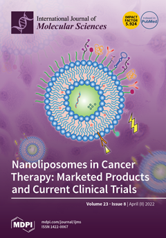
Current cancer treatment drugs damage both tumor cells and healthy cells, and are usually poorly soluble. The development of effective gene delivery systems is thus the key to achieving successful gene therapy. Transporting drugs or genes in nanoliposomes may resolve such issues; since these agents can be released specifically in the affected cells, this approach improves their solubility, bioavailability and efficacy, reducing their adverse effects. These enhancements can improve current treatments and offer the possibility of administering certain antitumor drugs orally. For these purposes, functionalized liposome delivery systems are being developed, such as surface-PEGylated liposomes and responsive liposomes that trigger a release under specific environment conditions. View this paper 
  • Issues are regarded as officially published after their release is announced to the table of contents alert mailing list.
  • You may sign up for e-mail alerts to receive table of contents of newly released issues.
  • PDF is the official format for papers published in both, html and pdf forms. To view the papers in pdf format, click on the "PDF Full-text" link, and use the free Adobe Reader to open them.
Order results
Result details
Section
Select all
Export citation of selected articles as:
22 pages, 4078 KiB  
Article
Dendrobium nobile Lindley Administration Attenuates Atopic Dermatitis-like Lesions by Modulating Immune Cells
by Sooyeon Hong, Eun-Young Kim, Seo-Eun Lim, Jae-Hyun Kim, Youngjoo Sohn and Hyuk-Sang Jung
Int. J. Mol. Sci. 2022, 23(8), 4470; https://doi.org/10.3390/ijms23084470 - 18 Apr 2022
Cited by 20 | Viewed by 3837
Abstract
Atopic dermatitis (AD) is a chronic inflammatory skin disease that can significantly affect daily life by causing sleep disturbance due to extreme itching. In addition, if the symptoms of AD are severe, it can cause mental disorders such as ADHD and suicidal ideation. [...] Read more.
Atopic dermatitis (AD) is a chronic inflammatory skin disease that can significantly affect daily life by causing sleep disturbance due to extreme itching. In addition, if the symptoms of AD are severe, it can cause mental disorders such as ADHD and suicidal ideation. Corticosteroid preparations used for general treatment have good effects, but their use is limited due to side effects. Therefore, it is essential to minimize the side effects and study effective treatment methods. Dendrobium nobile Lindley (DNL) has been widely used for various diseases, but to the best of our knowledge, its effect on AD has not yet been proven. In this study, the inhibitory effect of DNL on AD was confirmed in a DNCB-induced Balb/c mouse. In addition, the inhibitory efficacy of inflammatory cytokines in TNF-α/IFN-γ-induced HaCaT cells and PMACI-induced HMC-1 cells was confirmed. The results demonstrated that DNL decreased IgE, IL-6, IL-4, scratching behavior, SCORAD index, infiltration of mast cells and eosinophils and decreased the thickness of the skin. Additionally, DNL inhibited the expression of cytokines and inhibited the MAPK and NF-κB signaling pathways. This suggests that DNL inhibits cytokine expression, protein signaling pathway, and immune cells, thereby improving AD symptoms in mice. Full article
(This article belongs to the Special Issue Biological Interactions of Bioactive Natural Products)
Show Figures

Figure 1

Figure 1
<p>The toxicity of DNL and effect of DNL on scratching behavior and SCORAD index in DNCB-treated Balb/c mice. (<b>A</b>) Experimental schedule for topical application of DNL to dorsal skin. An amount of 1% DNCB was applied for three consecutive days to proceed with the first sensitization and had an incubation period of 4 days. After the incubation period, 0.5% DNCB was applied once every 2 or 3 days, DNL was applied every day, and sacrifices were performed on the 23rd day of the experiment. (<b>B</b>) The body weight of mice was measured using an electric scale once a week. (<b>C</b>) Liver weights were measured at the end of the animal experiment on the Balb/c mice DNL. (<b>D</b>) On the day of sacrifice, blood was collected through cardiac puncture, and serum was obtained by centrifugation. AST was confirmed by ELISA with the serum. (<b>E</b>) Serum ALT concentration was measured by ELISA. (<b>F</b>) The scratching behavior was photographed for 20 min the day before sacrifice, and the number of scratches was counted by the observer. (<b>G</b>) For the SCORAD index, six items were evaluated according to the atopic dermatitis severity standardization method, and the scores were summed. (<b>H</b>) Representative images of normal group (normal) and DNCB induction group (control), DNCB application and low-DNL-concentration-treatment group (DNL_L), DNCB application and high-DNL-concentration treatment group (DNL_H). The data are expressed as mean ± SEM (<span class="html-italic">n</span> = 3). ## <span class="html-italic">p</span> &lt; 0.01 compared with the normal group. ** <span class="html-italic">p</span> &lt; 0.01, compared with the control group.</p>
Full article ">Figure 2
<p>Effect of inflammatory cytokines and MAPK phosphorylation of DNL in DNCB-treated Balb/c mice. (<b>A</b>) Levels of IgE were measured by ELISA in serum. (<b>B</b>,<b>C</b>) The levels of IL-4 and IL-6 were measured by ELISA in the skin. (<b>D</b>) MAPK protein expression in DNCB-induced Balb/c mice skin was confirmed by Western blot. (<b>E</b>) MAPK was normalized to the total form of each indicator. The data are expressed as mean ± SEM (<span class="html-italic">n</span> = 3). # <span class="html-italic">p</span> &lt; 0.05 and ## <span class="html-italic">p</span> &lt; 0.01, compared with the normal group. * <span class="html-italic">p</span> &lt; 0.05 and ** <span class="html-italic">p</span> &lt; 0.01, compared with the control group.</p>
Full article ">Figure 3
<p>Effects of DNL on inhibition of immune cell invasion and reduction of skin thickness in DNCB-applied Balb/c mice. (<b>A</b>) To measure the thickness of the epidermis and dermis of DNCB-induced Balb/c mice, skin tissue sections were stained with H&amp;E (magnification 100×; scale bar 200 μm). (<b>B</b>) To check the infiltration of mast cells (black arrows) in the dermis of DNCB-induced Balb/c mice, skin sections were stained with toluidine blue (magnification 200×; scale bar 100 μm). (<b>C</b>) To confirm the infiltration of eosinophils (red arrows) in the dermis of DNCB-induced Balb/c mice, skin sections were stained with H&amp;E (magnification 400×; scale bar 50 μm). (<b>D</b>,<b>E</b>) The thickness of the epidermis and dermis was captured in three fields at a magnification of 100× per tissue. Next, three parts were measured per field of view and the values were averaged. (<b>F</b>) The number of infiltrated mast cells was counted and summed in three fields at a magnification of 200× per tissue. (<b>G</b>) The number of infiltrated eosinophils was counted in 10 fields at a magnification of 400× per tissue and summed. The data are expressed as mean ± SEM (<span class="html-italic">n</span> = 3). ## <span class="html-italic">p</span> &lt; 0.01, compared with the normal group. * <span class="html-italic">p</span> &lt; 0.05 and ** <span class="html-italic">p</span> &lt; 0.01, compared with the control group. E, epidermal thickness; D, dermal thickness.</p>
Full article ">Figure 4
<p>Effect of DNL in DNCB-induced Balb/c mice on CD4, CD8 T cell infiltration. (<b>A</b>) CD4 (black arrows) and CD8 (blue arrows) infiltration were confirmed in DNCB-induced Balb/c mice skin tissue by IHC (400×, scale bar 50 μm). (<b>B</b>,<b>C</b>) The numbers of CD4 and CD8 were counted and summarized in 10 fields at a magnification of 400× per tissue. The data are expressed as mean ± SEM (<span class="html-italic">n</span> = 3). ## <span class="html-italic">p</span> &lt; 0.01, compared with the normal group. * <span class="html-italic">p</span> &lt; 0.05 and ** <span class="html-italic">p</span> &lt; 0.01, compared with the control group.</p>
Full article ">Figure 5
<p>Effects of DNL on the expression of inflammatory chemokines and cytokines in TNF-α/IFN-γ-induced HaCaT cells. (<b>A</b>) HaCaT cells were treated with DNL at various concentrations for 24 h, followed by CCK-8 analysis. (<b>B</b>) TARC and IL-6 gene expression in HaCaT cells was evaluated by RT-PCR. (<b>C</b>,<b>D</b>) TARC and IL-6 were normalized to glyceraldehyde 3-phosphate dehydrogenase (GAPDH) through the ImageJ program. (<b>E</b>–<b>I</b>) The levels of inflammatory chemokines and cytokines were confirmed in HaCaT cells by ELISA. The data are expressed as mean ± SEM (<span class="html-italic">n</span> = 3). ## <span class="html-italic">p</span> &lt; 0.01, compared with the normal group. * <span class="html-italic">p</span> &lt; 0.05 and ** <span class="html-italic">p</span> &lt; 0.01, compared with the control group.</p>
Full article ">Figure 6
<p>Effects of DNL on inflammatory chemokines and cytokines in PMACI-induced HMC-1 cells. (<b>A</b>) Viability of DNL in HMC-1 cells. After treating HMC-1 cells with various concentrations of DNL, they were reacted for 24 h, and then MTS analysis was performed. (<b>B</b>–<b>E</b>) Expression of inflammatory chemokines and cytokines was confirmed by ELISA in PMACI-induced HMC-1 cells. The data are expressed as mean ± SEM (<span class="html-italic">n</span> = 3). ## <span class="html-italic">p</span> &lt; 0.01, compared with the normal group. * <span class="html-italic">p</span> &lt; 0.05 and ** <span class="html-italic">p</span> &lt; 0.01, compared with the control group.</p>
Full article ">Figure 7
<p>Effect of DNL on protein expression of MAPK and NF-κB/IκB in TNF-α/IFN-induced HaCaT cells. (<b>A</b>) MAPK protein expression in TNF-α/IFN-γ-induced HaCaT cells was confirmed by Western blot. (<b>B</b>) p-ERK, p-JNK, and p-p38 were normalized using the ImageJ program by T-ERK, t-JNK, and t-p38, respectively. (<b>C</b>) The phosphorylation of the signaling pathway of NF-κB/IκB in TNF-α/IFN-γ-induced HaCaT cells was confirmed by Western blot. (<b>D</b>) Phosphorylation of p-NF-κB was normalized by Lamin B, and IκB was normalized by Actin. Normalization was performed through the ImageJ program. The data are expressed as mean ± SEM (<span class="html-italic">n</span> = 3). ## <span class="html-italic">p</span> &lt; 0.01, compared with the normal group. * <span class="html-italic">p</span> &lt; 0.05 compared with the control group.</p>
Full article ">Figure 8
<p>Liquid chromatography–mass spectrometry (LC–MS) of (<b>A</b>) gigantol and (<b>B</b>) DNL.</p>
Full article ">
16 pages, 3912 KiB  
Article
Coinfection of Porcine Circovirus 2 and Pseudorabies Virus Enhances Immunosuppression and Inflammation through NF-κB, JAK/STAT, MAPK, and NLRP3 Pathways
by Xue Li, Si Chen, Liying Zhang, Guyu Niu, Xinwei Zhang, Lin Yang, Weilong Ji and Linzhu Ren
Int. J. Mol. Sci. 2022, 23(8), 4469; https://doi.org/10.3390/ijms23084469 - 18 Apr 2022
Cited by 25 | Viewed by 4372
Abstract
Porcine circovirus 2 (PCV2) and pseudorabies virus (PRV) are economically important pathogens in swine. PCV2 and PRV coinfection can cause more severe neurological and respiratory symptoms and higher mortality of piglets. However, the exact mechanism involved in the coinfection of PRV and PCV2 [...] Read more.
Porcine circovirus 2 (PCV2) and pseudorabies virus (PRV) are economically important pathogens in swine. PCV2 and PRV coinfection can cause more severe neurological and respiratory symptoms and higher mortality of piglets. However, the exact mechanism involved in the coinfection of PRV and PCV2 and its pathogenesis remain unknown. Here, porcine kidney cells (PK-15) were infected with PCV2 and/or PRV, and then the activation of immune and inflammatory pathways was evaluated to clarify the influence of the coinfection on immune and inflammatory responses. We found that the coinfection of PCV2 and PRV can promote the activation of nuclear factor-κB (NF-κB), c-Jun N-terminal protein kinases (JNK), p38, and nod-like receptor protein 3 (NLRP3) pathways, thus enhancing the expression of interferon-γ (IFN-γ), interferon-λ1 (IFN-λ1), interferon-stimulated gene (ISG15), interleukin 6 (IL6), and interleukin 1β (IL1β). Meanwhile, PCV2 and PRV also inhibit the expression and signal transduction of IFN-β, tumor necrosis factor α (TNFα), and the Janus kinase-signal transducer and activator of transcription (JAK/STAT) pathway. In addition, PCV2 and PRV infection can also weaken extracellular-signal-regulated kinase (ERK) activity. These results indicate that the regulations of cellular antiviral immune responses and inflammatory responses mediated by NF-κB, JAK/STAT, mitogen-activated protein kinase (MAPK), and NLRP3 pathways, contribute to immune escape of PCV2 and PRV and host antiviral responses. Full article
(This article belongs to the Special Issue The Interaction between Cell and Virus)
Show Figures

Figure 1

Figure 1
<p><b>Coinfection of PCV2 and PRV affects in PK-15 cells.</b> Viral genomic copies were evaluated using real-time PCR at indicated hour post-infection. The data are presented as the means ± SD. **, <span class="html-italic">p</span>-value &lt; 0.01; ***, <span class="html-italic">p</span>-value &lt; 0.001; ****, <span class="html-italic">p</span>-value &lt; 0.0001. (<b>A</b>) PCV2. (<b>B</b>) PRV.</p>
Full article ">Figure 2
<p>Coinfection of PCV2 and PRV modulates IFNs. Western blotting was performed using Interferon alpha 1 Antibody, Interferon beta/IFNB, IFN-γ, IL28/29 (H-1), and Anti-β-Actin Antibody as primary antibodies, respectively. HRP-labeled Goat Anti-mouse IgG (H+L) and HRP-labeled Goat Anti-rabbit IgG (H+L) were used as the secondary antibody. β-actin was used as a control. The average expression level of the target protein in each group is shown below each lane. The protein amount of the PK-15 group is set to 1, and the values of other groups are the ratio with the PK-15 group. Unprocessed original images can be found in <a href="#app1-ijms-23-04469" class="html-app">Supplementary Figure S1</a>.</p>
Full article ">Figure 3
<p><b>Coinfection of PCV2 and PRV regulates IRFs.</b> (<b>A</b>,<b>B</b>) Expression levels of IRFs. The mRNA levels of <span class="html-italic">IRF3</span> (<b>A</b>) and <span class="html-italic">IRF7</span> (<b>B</b>) were evaluated via real-time PCR. (<b>C</b>) Protein levels of IRF7. Western blotting was performed using Rabbit Anti-IRF7 antibody and Anti-β-Actin Antibody as primary antibodies, respectively. HRP-labeled Goat Anti-mouse IgG (H+L) and HRP-labeled Goat Anti-rabbit IgG (H+L) were used as the secondary antibody. β-actin was used as a control. The average expression level of the target protein in each group is shown below each lane. The protein amount of the PK-15 group is set to 1, and the values of other groups are the ratio with the PK-15 group. (<b>D</b>,<b>E</b>) Expression levels of ISGs. The mRNA levels of <span class="html-italic">ISG15</span> (<b>D</b>) and <span class="html-italic">ISG56</span> (<b>E</b>) were evaluated via real-time PCR. *, <span class="html-italic">p</span>-value &lt; 0.05; **, <span class="html-italic">p</span>-value &lt; 0.01; ***, <span class="html-italic">p</span>-value &lt; 0.001; ****, <span class="html-italic">p</span>-value &lt; 0.0001. The data are presented as the means ± SD. Unprocessed original images can be found in <a href="#app1-ijms-23-04469" class="html-app">Supplementary Figure S2</a>.</p>
Full article ">Figure 4
<p>Coinfection of PCV2 and PRV suppresses STAT1-related JAK/STAT pathways. (<b>A</b>–<b>D</b>) Expression levels of JAK/STATs. The relative mRNA levels of <span class="html-italic">JAK1</span> (<b>A</b>), <span class="html-italic">SOCS1</span> (<b>B</b>), <span class="html-italic">SOCS3</span> (<b>C</b>), and <span class="html-italic">STAT1</span> (<b>D</b>) were detected using real-time PCR. (<b>E</b>,<b>F</b>) Protein levels of STAT1 and IRF9. Western blotting was performed using p-STAT1, Anti-STAT1 Antibody, IRF9 Antibody, and Anti-β-Actin Antibody as primary antibodies, respectively. HRP-labeled Goat Anti-mouse IgG (H+L) and HRP-labeled Goat Anti-rabbit IgG (H+L) were used as the secondary antibody. β-actin was used as a control. The average expression level of the target protein in each group is shown below each lane. The protein amount of the PK-15 group is set to 1, and the values of other groups are the ratio with the PK-15 group. (<b>G</b>) Expression levels of <span class="html-italic">IRF9</span> were detected using real-time PCR. The data are presented as the means ± SD. *, <span class="html-italic">p</span>-value &lt; 0.05; **, <span class="html-italic">p</span>-value &lt; 0.01; ***, <span class="html-italic">p</span>-value &lt; 0.001; ****, <span class="html-italic">p</span>-value &lt; 0.0001. Unprocessed original images can be found in <a href="#app1-ijms-23-04469" class="html-app">Supplementary Figure S3</a>.</p>
Full article ">Figure 5
<p>Coinfection of PCV2 and PRV modulates the NF-κB signal pathway. (<b>A</b>,<b>B</b>) Expression levels of <span class="html-italic">iκB</span> and <span class="html-italic">p65</span>. The relative mRNA levels of <span class="html-italic">iκB</span> (<b>A</b>) and <span class="html-italic">p65</span> (<b>B</b>) were examined using real-time PCR. The data are presented as the means ± SD. **, <span class="html-italic">p</span>-value &lt; 0.01; ***, <span class="html-italic">p</span>-value &lt; 0.001; ****, <span class="html-italic">p</span>-value &lt; 0.0001. (<b>C</b>–<b>E</b>) Protein levels of p65 and iκB. Western blotting was conducted using P-IΚBα (Ser32 Ser36), IΚB-α, NFκB p65 Polyclonal Antibody, Phospho-NFκB p65 (Thr276) Polyclonal Antibody, and Anti-β-Actin Antibody as primary antibody, respectively. HRP-labeled Goat Anti-mouse IgG (H+L) and HRP-labeled Goat Anti-rabbit IgG (H+L) were used as the secondary antibody. β-actin was used as a control. The average expression level of the target protein in each group is shown below each lane. The protein amount of the PK-15 group is set to 1, and the values of other groups are the ratio with the PK-15 group. Unprocessed original images can be found in <a href="#app1-ijms-23-04469" class="html-app">Supplementary Figure S4</a>.</p>
Full article ">Figure 6
<p>Coinfection of PCV2 and PRV modulates expressions of host pro-inflammatory factors. (<b>A</b>–<b>D</b>) Expression levels of pro-inflammatory factors. The relative mRNA levels of <span class="html-italic">IL1α</span> (<b>A</b>), <span class="html-italic">TNFα</span> (<b>B</b>), <span class="html-italic">IL6</span> (<b>C</b>), and <span class="html-italic">IL1β</span> (<b>D</b>) were evaluated using real-time PCR. The data are presented as the means ± SD. *, <span class="html-italic">p</span>-value &lt; 0.05; **, <span class="html-italic">p</span>-value &lt; 0.01; ***, <span class="html-italic">p</span>-value &lt; 0.001; ****, <span class="html-italic">p</span>-value &lt; 0.0001. (<b>E</b>) Protein levels of pro-inflammatory factors. Western blotting was performed using IL1α, IL1B Antibody, IL6, TNFα, and Anti-β-Actin Antibody as primary antibodies, respectively. HRP-labeled Goat Anti-mouse IgG (H+L) and HRP-labeled Goat Anti-rabbit IgG (H+L) were used as the secondary antibody. β-actin was used as a control. The average expression level of the target protein in each group is shown below each lane. The protein amount of the PK-15 group is set to 1, and the values of other groups are the ratio with the PK-15 group. Unprocessed original images can be found in <a href="#app1-ijms-23-04469" class="html-app">Supplementary Figure S5</a>.</p>
Full article ">Figure 7
<p>Coinfection of PCV2 and PRV activates the NLRP3 pathway. (<b>A</b>,<b>B</b>) Expression levels of NLRP3 and COX2. The relative mRNA levels of <span class="html-italic">NLRP3</span> (<b>A</b>) and <span class="html-italic">COX2</span> (<b>B</b>) were examined via Real-time PCR. The data are presented as the means ± SD. *, <span class="html-italic">p</span>-value &lt; 0.05; **, <span class="html-italic">p</span>-value &lt; 0.01; ***, <span class="html-italic">p</span>-value &lt; 0.001; ****, <span class="html-italic">p</span>-value &lt; 0.0001. (<b>C</b>) Protein levels of NLRP3 and COX2. Western blotting was performed using NLRP3, Cox-2, and Anti-β-Actin Antibody as primary antibodies, respectively. HRP-labeled Goat Anti-mouse IgG (H+L) and HRP-labeled Goat Anti-rabbit IgG (H+L) were used as the secondary antibody. β-actin was used as a control. The average expression level of the target protein in each group is shown below each lane. The protein amount of the PK-15 group is set to 1, and the values of other groups are the ratio with the PK-15 group. Unprocessed original images can be found in <a href="#app1-ijms-23-04469" class="html-app">Supplementary Figure S6</a>.</p>
Full article ">Figure 8
<p>Coinfection of PCV2 and PRV activates inflammatory and immune responses via p38 and JNK1/2. (<b>A</b>–<b>C</b>) Expression levels of MAPKs. The relative mRNA levels of <span class="html-italic">p38</span> (<b>A</b>), <span class="html-italic">ERK1/2</span> (<b>B</b>), and <span class="html-italic">JNK1/2</span> (<b>C</b>) were examined via real-time PCR. The data are presented as the means ± SD. *, <span class="html-italic">p</span>-value &lt; 0.05; **, <span class="html-italic">p</span>-value &lt; 0.01; ***, <span class="html-italic">p</span>-value &lt; 0.001; ****, <span class="html-italic">p</span>-value &lt; 0.0001. (<b>D</b>–<b>G</b>) Protein levels of MAPKs. Western blotting was conducted using p-p38 (Thr180/Tyr182), P38, JNK (FL), p-JNK (Thr 183/Tyr 185), ERK p44/42 MAPK (Erk1/2) (137F5) Rabbit mAb, Phospho-p44/42 MAPK (Erk1/2) (Thr202/Tyr204) (D13.14.4E) XP<sup>®</sup> Rabbit mAb, and Anti-β-Actin Antibody as primary antibody, respectively. HRP-labeled Goat Anti-mouse IgG (H+L), HRP-labeled Donkey Anti-Goat IgG (H+L), and HRP-labeled Goat Anti-rabbit IgG (H+L) were used as the secondary antibody. β-actin was used as a control. The average expression level of the target protein in each group is shown below each lane. The protein amount of the PK-15 group is set to 1, and the values of other groups are the ratio with the PK-15 group. Unprocessed original images can be found in <a href="#app1-ijms-23-04469" class="html-app">Supplementary Figure S7</a>.</p>
Full article ">Figure 9
<p>Proposed immune and inflammatory reactions caused by coinfection of PCV2 and PRV. Arrow (→), enhance; T line (⊥) means inhibition.</p>
Full article ">
15 pages, 3815 KiB  
Article
The Discovery of Potent SHP2 Inhibitors with Anti-Proliferative Activity in Breast Cancer Cell Lines
by Rose Ghemrawi, Mostafa Khair, Shaima Hasan, Raghad Aldulaymi, Shaikha S. AlNeyadi, Noor Atatreh and Mohammad A. Ghattas
Int. J. Mol. Sci. 2022, 23(8), 4468; https://doi.org/10.3390/ijms23084468 - 18 Apr 2022
Cited by 2 | Viewed by 5486
Abstract
Despite available treatments, breast cancer is the leading cause of cancer-related death. Knowing that the tyrosine phosphatase SHP2 is a regulator in tumorigenesis, developing inhibitors of SHP2 in breast cells is crucial. Our study investigated the effects of new compounds, purchased from NSC, [...] Read more.
Despite available treatments, breast cancer is the leading cause of cancer-related death. Knowing that the tyrosine phosphatase SHP2 is a regulator in tumorigenesis, developing inhibitors of SHP2 in breast cells is crucial. Our study investigated the effects of new compounds, purchased from NSC, on the phosphatase activity of SHP2 and the modulation of breast cancer cell lines’ proliferation and viability. A combined ligand-based and structure-based virtual screening protocol was validated, then performed, against SHP2 active site. Top ranked compounds were tested via SHP2 enzymatic assay, followed by measuring IC50 values. Subsequently, hits were tested for their anti-breast cancer viability and proliferative activity. Our experiments identified three compounds 13030, 24198, and 57774 as SHP2 inhibitors, with IC50 values in micromolar levels and considerable selectivity over the analogous enzyme SHP1. Long MD simulations of 500 ns showed a very promising binding mode in the SHP2 catalytic pocket. Furthermore, these compounds significantly reduced MCF-7 breast cancer cells’ proliferation and viability. Interestingly, two of our hits can have acridine or phenoxazine cyclic system known to intercalate in ds DNA. Therefore, our novel approach led to the discovery of SHP2 inhibitors, which could act as a starting point in the future for clinically useful anticancer agents. Full article
(This article belongs to the Special Issue Computational Studies of Drugs and Biomolecules)
Show Figures

Figure 1

Figure 1
<p>(<b>a</b>) The four features of the top pharmacophore query no. 4. (<b>b</b>) Two known inhibitors overlaid on the said pharmacophore. (<b>c</b>) The measured distances and angles between the four pharmacoohoric features (<b>d</b>) The effect of using pharmacophore query no. 4 as pre-docking filter in a pilot showing the number of known SHP2 inhibitors retrieved (%Enrichment) at any given percentage of the top-ranked ligand library.</p>
Full article ">Figure 2
<p>The workflow of the filter-based VS used against the SHP2 active site.</p>
Full article ">Figure 3
<p>Remaining activity (%) of the SHP2 enzyme after treatment with the 35 tested compounds (at 100 μM) along with the control suramin. Data presented are the mean +/− SEM of three independent experiments.</p>
Full article ">Figure 4
<p>The effect of the compounds NSC <b>13030</b>, <b>24198</b>, <b>57774</b>, and <b>137420</b> on MCF-7 and MDA-MB-231 proliferation (<b>a</b>) and viability (<b>b</b>). Cells were incubated with increasing concentrations of compounds in culture medium for 48 h. The viability and the proliferative response were assessed. Data presented are the mean ± SEM of three independent experiments. * <span class="html-italic">p</span> &lt; 0.05. Continuous lines are for compounds, and dashed lines are for the vehicle DMSO.</p>
Full article ">Figure 5
<p>(<b>a</b>) The RMSD plot of the 500 ns MD simulations of compound <b>13030</b> along with (<b>b</b>) the hydrogen bonding plot, (<b>c</b>) the top-cluster conformation filling (blue spheres) in the SHP2 active site (gray surface), and (<b>d</b>) the ligand 3D binding mode showing the key interactions made with the surrounding residues.</p>
Full article ">Figure 6
<p>(<b>a</b>) The RMSD plot of the 500 ns MD simulations of compound <b>24198</b> along with (<b>b</b>) the hydrogen bonding plot, (<b>c</b>) the top-cluster conformation filling (blue spheres) in the SHP2 active site (gray surface), and (<b>d</b>) the ligand 3D binding mode showing the key interactions made with the surrounding residues.</p>
Full article ">Figure 7
<p>(<b>a</b>) The RMSD plot of the 500 ns MD simulations of compound <b>57774</b> along with (<b>b</b>) the hydrogen bonding plot, (<b>c</b>) the top-cluster conformation filling (blue spheres) in the SHP2 active site (gray surface), and (<b>d</b>) the ligand 3D binding mode showing the key interactions made with the surrounding residues.</p>
Full article ">
17 pages, 793 KiB  
Review
Recent Experimental Studies of Maternal Obesity, Diabetes during Pregnancy and the Developmental Origins of Cardiovascular Disease
by Stephanie M. Kereliuk and Vernon W. Dolinsky
Int. J. Mol. Sci. 2022, 23(8), 4467; https://doi.org/10.3390/ijms23084467 - 18 Apr 2022
Cited by 22 | Viewed by 8279
Abstract
Globally, cardiovascular disease remains the leading cause of death. Most concerning is the rise in cardiovascular risk factors including obesity, diabetes and hypertension among youth, which increases the likelihood of the development of earlier and more severe cardiovascular disease. While lifestyle factors are [...] Read more.
Globally, cardiovascular disease remains the leading cause of death. Most concerning is the rise in cardiovascular risk factors including obesity, diabetes and hypertension among youth, which increases the likelihood of the development of earlier and more severe cardiovascular disease. While lifestyle factors are involved in these trends, an increasing body of evidence implicates environmental exposures in early life on health outcomes in adulthood. Maternal obesity and diabetes during pregnancy, which have increased dramatically in recent years, also have profound effects on fetal growth and development. Mounting evidence is emerging that maternal obesity and diabetes during pregnancy have lifelong effects on cardiovascular risk factors and heart disease development. However, the mechanisms responsible for these observations are unknown. In this review, we summarize the findings of recent experimental studies, showing that maternal obesity and diabetes during pregnancy affect energy metabolism and heart disease development in the offspring, with a focus on the mechanisms involved. We also evaluate early proof-of-concept studies for interventions that could mitigate maternal obesity and gestational diabetes-induced cardiovascular disease risk in the offspring. Full article
(This article belongs to the Special Issue Risk Factors and Molecular Mechanisms of Gestational Diabetes III)
Show Figures

Figure 1

Figure 1
<p>Influence of maternal obesity and gestational diabetes mellitus (GDM) on the developmental origins of cardiometabolic disease in the offspring. Obesity, dysglycemia and hyperlipidemia pre-pregnancy and during pregnancy contribute to the development of GDM. GDM increases the supply of glucose and fatty acids to the fetus which results in fetal adaptations and developmental programming that include alterations in gene expression, organogenesis, cellular metabolism and epigenetic modifications. GDM exposure appears to affect a wide range of cellular, tissue and organ systems in a manner that increases the risk of cardiometabolic disease in the offspring with increasing evidence from rodent model systems that the cardiovascular system is sensitive to GDM-induced effects. Created with BioRender.com.</p>
Full article ">
11 pages, 2822 KiB  
Article
AcidoCEST-UTE MRI Reveals an Acidic Microenvironment in Knee Osteoarthritis
by Alecio F. Lombardi, Yajun Ma, Hyungseok Jang, Saeed Jerban, Qingbo Tang, Adam C. Searleman, Robert Scott Meyer, Jiang Du and Eric Y. Chang
Int. J. Mol. Sci. 2022, 23(8), 4466; https://doi.org/10.3390/ijms23084466 - 18 Apr 2022
Cited by 23 | Viewed by 5317 | Correction
Abstract
A relationship between an acidic pH in the joints, osteoarthritis (OA), and pain has been previously demonstrated. Acidosis Chemical Exchange Saturation Transfer (acidoCEST) indirectly measures the extracellular pH through the assessment of the exchange of protons between amide groups on iodinated contrast agents [...] Read more.
A relationship between an acidic pH in the joints, osteoarthritis (OA), and pain has been previously demonstrated. Acidosis Chemical Exchange Saturation Transfer (acidoCEST) indirectly measures the extracellular pH through the assessment of the exchange of protons between amide groups on iodinated contrast agents and bulk water. It is possible to estimate the extracellular pH in the osteoarthritic joint using acidoCEST MRI. However, conventional MR sequences cannot image deep layers of cartilage, meniscus, ligaments, and other musculoskeletal tissues that present with short echo time and fast signal decay. Ultrashort echo time (UTE) MRI, on the other hand, has been used successfully to image those joint tissues. Here, our goal is to compare the pH measured in the knee joints of volunteers without OA and patients with severe OA using acidoCEST-UTE MRI. Patients without knee OA and patients with severe OA were examined using acidoCEST-UTE MRI and the mean pH of cartilage, meniscus, and fluid was calculated. Additionally, the relationship between the pH measurements and the Knee Injury and Osteoarthritis Outcome Score (KOOS) was investigated. AcidoCEST-UTE MRI can detect significant differences in the pH of knee cartilage, meniscus, and fluid between joints without and with OA, with OA showing lower pH values. In addition, symptoms and knee-joint function become worse at lower pH measurements. Full article
(This article belongs to the Special Issue New Advances in Osteoarthritis)
Show Figures

Figure 1

Figure 1
<p>Representative image examples from patients without OA (<b>A</b>) and with OA (<b>B</b>). Sagittal PD-weighted (first row), low-power acido-CEST UTE (second row), high-power acido-CEST UTE (third row), and pH pixel maps (fourth row) of cartilage, meniscus, and fluid. The pH is directly correlated with the radiofrequency power mismatch (RPM) measurements, as described in Equations (3) and (4). Note the higher pH values (yellow and red colors) in patients without OA compared with patients with OA (blue colors).</p>
Full article ">Figure 2
<p>Boxplots of mean pH measurements versus groups for all ROIs. Significant differences are observed between the two groups. ***: <span class="html-italic">p</span> values lower than 0.001.</p>
Full article ">Figure 3
<p>Boxplots of mean pH measurements versus each group of participants (No OA and OA) for ROIs drew in cartilage (<b>A</b>), meniscus (<b>B</b>), and fluid (<b>C</b>). Significant differences in pH measurements are observed between groups with lower pH in patients with OA compared with patients without OA. “***”: <span class="html-italic">p</span> values lower than 0.001; “**”: <span class="html-italic">p</span> values lower than 0.01; “*”: <span class="html-italic">p</span> values lower than 0.05.</p>
Full article ">Figure 4
<p>Boxplots of mean pH measurements versus ROIs in patients without OA (<b>A</b>) and with advanced OA (<b>B</b>). No significant differences were found, except for the pH of cartilage and meniscus in the OA group (<span class="html-italic">p</span> = 0.024). “*”: <span class="html-italic">p</span> values lower than 0.05; “•”: outliers.</p>
Full article ">Figure 5
<p>Scatterplots of pH versus KOOS scores and the visual analog pain score. Strong direct correlations were observed for all KOOS subscale scores (<b>A</b>–<b>F</b>). There was a strong inverse correlation between pH measurements and the visual analog pain score (<b>G</b>). KOOS: Knee Injury and Osteoarthritis Outcome Score; ADL: activities of daily living; Sports/Rec: sports and recreation activities; QOL: quality of life; PF: patellofemoral.</p>
Full article ">
27 pages, 4740 KiB  
Article
Functional Characterization of Cardiac Actin Mutants Causing Hypertrophic (p.A295S) and Dilated Cardiomyopathy (p.R312H and p.E361G)
by Roua Hassoun, Constanze Erdmann, Sebastian Schmitt, Setsuko Fujita-Becker, Andreas Mügge, Rasmus R. Schröder, Matthias Geyer, Mina Borbor, Kornelia Jaquet, Nazha Hamdani and Hans Georg Mannherz
Int. J. Mol. Sci. 2022, 23(8), 4465; https://doi.org/10.3390/ijms23084465 - 18 Apr 2022
Cited by 5 | Viewed by 2969
Abstract
Human wild type (wt) cardiac α-actin and its mutants p.A295S or p.R312H and p.E361G correlated with hypertrophic or dilated cardiomyopathy, respectively, were expressed by using the baculovirus/Sf21 insect cell system. The c-actin variants inhibited DNase I, indicating maintenance of their native state. Electron [...] Read more.
Human wild type (wt) cardiac α-actin and its mutants p.A295S or p.R312H and p.E361G correlated with hypertrophic or dilated cardiomyopathy, respectively, were expressed by using the baculovirus/Sf21 insect cell system. The c-actin variants inhibited DNase I, indicating maintenance of their native state. Electron microscopy showed the formation of normal appearing actin filaments though they showed mutant specific differences in length and straightness correlating with their polymerization rates. TRITC-phalloidin staining showed that p.A295S and p.R312H exhibited reduced and the p.E361G mutant increased lengths of their formed filaments. Decoration of c-actins with cardiac tropomyosin (cTm) and troponin (cTn) conveyed Ca2+-sensitivity of the myosin-S1 ATPase stimulation, which was higher for the HCM p.A295S mutant and lower for the DCM p.R312H and p.E361G mutants than for wt c-actin. The lower Ca2+-sensitivity of myosin-S1 stimulation by both DCM actin mutants was corrected by the addition of levosimendan. Ca2+-dependency of the movement of pyrene-labeled cTm along polymerized c-actin variants decorated with cTn corresponded to the relations observed for the myosin-S1 ATPase stimulation though shifted to lower Ca2+-concentrations. The N-terminal C0C2 domain of cardiac myosin-binding protein-C increased the Ca2+-sensitivity of the pyrene-cTM movement of bovine, recombinant wt, p.A295S, and p.E361G c-actins, but not of the p.R312H mutant, suggesting decreased affinity to cTm. Full article
(This article belongs to the Section Molecular Biology)
Show Figures

Figure 1

Figure 1
<p>Purification of cardiac actins after expression by the <span class="html-italic">baculovirus/Sf21</span> system. (<b>A</b>,<b>B</b>) SDS-PAGE of purification steps of (<b>A</b>) wt c-actin and (<b>B</b>) the p.E361G mutant c-actin. Lanes: (1) Sf21 cell homogenate, (2) supernatant after mixture with His-tagged G4-6, and (3) pellet after centrifugation, (M) pre-stained marker proteins, lanes (4,5) purified c-actins after elution of Ni-NTA agarose with EGTA (for details see text), and lane (6) Ni-NTA agarose with His-tagged G4-6 still bound. (<b>C</b>) SDS-PAGE of the purified cardiac actin variants used in this study. Lanes: (1) wt recombinant; (2) p.A295S; (3) p.R312K; (4) p.E361G; (5) skeletal muscle actin; and (6) bovine cardiac actin. Actins shown in lanes (5) and (6) were conventionally prepared from acetone powders. (<b>D</b>) Dot immunoblots of the actins shown in the same sequence as in (<b>C</b>) using the anti-cardiac actin monoclonal antibody. Note that the p.R312K mutant is only weakly and skeletal muscle actin is not stained by anti-c-actin mAB. (<b>E</b>) <b>(Upper row)</b> SDS-PAGE of the following actins: Lanes: (1) skeletal muscle actin; (2) wt cardiac actin (recombinant); (3) bovine cardiac actin; (4) cytoplasmic β-actin; (5) actin purified from <span class="html-italic">Acanthameba castellani</span>; and (6) cytoplasmic β-actin. <b>(Middle row</b>) Western blots immunostained with anti-cardiac actin mAB. (<b>Lower row)</b> Western blots stained with anti-cytoplasmic β-actin. (<b>F</b>) 3D structural model of G-actin derived from the skeletal muscle actin:DNase I complex [<a href="#B25-ijms-23-04465" class="html-bibr">25</a>] indicating the positions of the mutated residues of the c-actin mutants investigated. In addition, subdomains (SD) as well as the position of ATP and Ca<sup>2+</sup> are indicated.</p>
Full article ">Figure 2
<p>Properties of the purified c-actins indicating native state. (<b>A</b>) Inhibition of DNase I activity as measured by the hyperchromicity assay. Human DNase I (0.1 mg/mL = 3.22 μM) was mixed with the amounts of c-actins indicated in the abscissa. DNase activity was determined as detailed [<a href="#B26-ijms-23-04465" class="html-bibr">26</a>]. Ordinate gives the relative remaining activity calculated. (<b>B</b>) Comparison of the rates of polymerization of purified sk-actin and bovine c-actin purified from acetone powders with recombinant wt c-actin, each at 5 μM. Note the faster polymerization rate of sk-actin and the equal rates of c-actins. Polymerization was initiated after the addition of 2 mM MgCl<sub>2</sub> at t = 2 min and determined by the pyrene-assay using 0.5 μM pyrene-labeled skeletal muscle actin (see Materials and Methods). (<b>C</b>) Polymerization of the cardiac actin variants: recombinant wt-c-actin, p.A295S, p.R312H, and p.E361G (all c-actins at 5 µM; for details see text). (<b>D</b>–<b>H</b>) Electron microscopy after negative staining of the polymerized c-actins before and after decoration with cTm/cTn: (<b>D</b>) bovine and (<b>E</b>) wt recombinant c-actin. (<b>F</b>) p.A295S, (<b>G</b>) p.R312H, and (<b>H</b>) p.E361G mutant. Bar in (<b>H</b>) corresponds to 100 nm (applicable to all images).</p>
Full article ">Figure 3
<p>Properties of the purified p.R312K c-actin variant. (<b>A</b>) Inhibition of DNase I by the c-actin variants. The inhibition by p.R312K is shown by the trendline. (<b>B</b>) Rates of polymerization of the c-actin variants. The rate of 5 µM p.R312K is shown by (♦) and coincides with the rate of wt c-actin. (<b>C</b>) Influence of 50 nM Arp2/3 complex and 50 nM mDia3-FH2 of the rate of polymerization of p.R312K. (<b>C</b>) EM images of polymerized p.R312K without and (<b>D</b>) after decoration with cTm and cTn at 7:1:1 ratio to actin. Bar in (<b>D</b>) corresponds to 100 nm.</p>
Full article ">Figure 4
<p>Filament length determination after staining with TRITC-phalloidin. (<b>A</b>) Filaments of the cardiac actin variants were labelled with TRITC-phalloidin (see Materials and Methods) and the alterations of their length distribution were followed over time for filaments of a length between 0.1 and 0.2 µm and over 1 µm. Note that the number of filaments between 0.1 and 0.2 µm remained almost constant, whereas the number of filaments over 1 µm increased, except for the p.A295S mutant. The largest increase was noted for the p.E361G mutant. (<b>B</b>) The appearance of the phalloidin labelled filaments was visualized by EM after about 60 min and (<b>C</b>) by fluorescence microscopy. (<b>D</b>) Shows the percental length distributions after about 60 min as determined by ImageJ Ridge Detection (see Materials and Methods). Bars in (<b>B</b>,<b>C</b>) correspond to 100 nm and 10 µm, respectively.</p>
Full article ">Figure 5
<p>(<b>A</b>) Gives the dependence of the myosin-S1 ATPase stimulaztion on the concentration of bovine and recombinant wt c-actin. (<b>A′</b>,<b>A″</b>) give examples of the double reciprocal evaluation of the myosin-S1 ATPase stimulation by bovine and recombinant wt c-actin, respectively. The intercept of the linear slopes with Y-axis gives 1/V<sub>max</sub> and the X-axis gives minus 1/K<sub>M</sub>, which like a Michaelis-Menten constant can be taken as an apparent binding constant. Since both values are not determined kinetically, they are named “apparent”. The results of an identical evaluation of the c-actin mutants are given in <a href="#ijms-23-04465-t002" class="html-table">Table 2</a>. (<b>B</b>–<b>E</b>) Stimulation of the myosin-S1 ATPase by increasing concentrations of the c-actin variants in the absence and presence of cTm/cTn. The ability of filamentous c-actin variants was determined by an enzyme-linked optical assay [<a href="#B30-ijms-23-04465" class="html-bibr">30</a>]. The c-actins were polymerized by 2 mM MgCl<sub>2</sub> plus 50 mM KCl. The ATPase activity was determined for 1 µM rabbit skeletal muscle myosin-S1 prepared according to [<a href="#B31-ijms-23-04465" class="html-bibr">31</a>] in 5 mM HEPES-HCl, pH 7.4; 0.1 mM CaCl<sub>2</sub>, 2mM MgCl<sub>2</sub>, 50 mM KCl NADH phosphoenolpyruvate plus lactate dehydrogenase and pyruvate kinase. The time dependent decrease in optical density at 340 nm was determined with a Beckman DU640 photometer.</p>
Full article ">Figure 6
<p>Ca<sup>2+</sup>-concentration dependence of the stimulation of the myosin-S1 ATPase by c-actins decorated with cTm and cTn at a 6:1:1 molar ratio. (<b>A</b>) Bovin c-actin, (<b>B</b>) WT recombinant actin, (<b>C</b>) Bovine vs. WT recombinant actin, (<b>D</b>) A295S c-actin, (<b>E</b>) R312H c-actin, (<b>F</b>) E361G c-actin, (<b>G</b>) mutant actins vs. WT recombinant actin.</p>
Full article ">Figure 7
<p>Influence of levosimendan on the stimulation of myosin-S1 by the DCM c-actin mutants. The Ca<sup>2+</sup>-concentration dependence of myosin-S1 ATPase stimulation by the DCM mutants p.R312H and p.E361G were determined in the presence and absence of 20 µM levosimendan by the enzyme-linked optical assay using 1 µM skeletal muscle myosin-S1 and 1.5 µM of the polymerized c-actin mutants decorated with cTm/cTn at a molar ratio to the polymerized c-actin mutants of 6:1:1. (<b>A</b>): p.R312H ± Levo; (<b>B</b>): p.R312H ± Levo and wt c-actin; (<b>C</b>): p.E361G ± Levo; (<b>D</b>): p.E361G ± Levo and wt c-actin. Data are given as mean values ± SEM. Abscissa gives pCa-values (-log molar Ca<sup>2+</sup>-concentration). All measurements were performed in triplicate.</p>
Full article ">Figure 8
<p>Comparison of the Ca<sup>2+</sup>-concentration dependence of the fluorescence increase (movement) in pyrene-labeled cTm on bovine and recombinant c-actin variants.</p>
Full article ">
19 pages, 3521 KiB  
Article
Chaperone Hsp70 (HSPA1) Is Involved in the Molecular Mechanisms of Sleep Cycle Integration
by Valentina V. Simonova, Mikhail A. Guzeev, Irina V. Ekimova and Yuri F. Pastukhov
Int. J. Mol. Sci. 2022, 23(8), 4464; https://doi.org/10.3390/ijms23084464 - 18 Apr 2022
Cited by 5 | Viewed by 2802
Abstract
The molecular mechanisms of sleep cycle integration at the beginning and the end of the inactive period are not clear. Sleep cycles with a predominance of deep slow-wave sleep (SWS) seem to be associated with accelerated protein synthesis in the brain. The inducible [...] Read more.
The molecular mechanisms of sleep cycle integration at the beginning and the end of the inactive period are not clear. Sleep cycles with a predominance of deep slow-wave sleep (SWS) seem to be associated with accelerated protein synthesis in the brain. The inducible Hsp70 chaperone corrects protein conformational changes and has protective properties. This research explores (1) whether the Hspa1 gene encoding Hsp70 protein activates during the daily rapid-eye-movement sleep (REMS) maximum, and (2) whether a lower daily deep SWS maximum affects the Hspa1 expression level during the subsequent REMS. Combining polysomnography in male Wistar rats, RT-qPCR, and Western blotting, we reveal a three-fold Hspa1 upregulation in the nucleus reticularis pontis oralis, which regulates REMS. Hspa1 expression increases during the daily REMS maximum, 5–7 h after the natural peak of deep SWS. Using short-term selective REMS deprivation, we demonstrate that REMS rebound after deprivation exceeds the natural daily maximum, but it is not accompanied by Hspa1 upregulation. The results suggest that a high proportion of deep SWS, usually observed after sleep onset, is a necessary condition for Hspa1 upregulation during subsequent REMS. The data obtained can inform the understanding of the molecular mechanisms integrating SWS and REMS and key biological function(s) of sleep. Full article
(This article belongs to the Collection State-of-the-Art Molecular Neurobiology in Russia)
Show Figures

Figure 1

Figure 1
<p>Total time of vigilance states comprising the natural daily sleep–wake cycle in Wistar rats (<span class="html-italic">n</span> = 10). The natural daily maximum of slow-wave sleep (SWS) is registered after the sleep onset at ZT00-03, while REM sleep (REMS) peaks at the end of the inactive (lights-on) period at ZT06-09. Y-axis: % of total registration time. X-axis: zeitgeber time, hours. Arrows indicate the time points when 6 groups of rats (<span class="html-italic">n</span> = 6 in each group) were sacrificed at daily maximums of wakefulness (ZT12-15; 18-21), SWS (ZT00-03) and REMS (ZT06-09) for the subsequent real-time qPCR analysis of the brain tissue. Colours signify the preceding vigilance state: black—wakefulness, blue—slow-wave sleep, red—REM sleep. Values are shown as mean ± s.e.m, <span class="html-italic">n</span> = 10 rats.</p>
Full article ">Figure 2
<p><span class="html-italic">Hspa1</span> expression increases in the nucleus reticularis pontis oralis during the natural daily maximum of REM sleep in the lights-on period. Relative <span class="html-italic">Hspa1</span> expression in (<b>a</b>,<b>d</b>) the sensorimotor cortex (CORTEX), (<b>b</b>,<b>e</b>) the preoptic area of the hypothalamus (PREOPTIC AREA) and (<b>c</b>,<b>f</b>) the nucleus reticularis pontis oralis (NRPO) in slow-wave sleep (SWS, upper row) or REM sleep (REMS, lower row). The values were calculated using the 2<sup>−ΔΔCt</sup> method, where the reference group was in wakefulness either at ZT01-02 (for SWS) or at ZT07-08 (for REMS). WAKE (grey)—rats were sacrificed after a 10 min episode of wakefulness at ZT01-02 or ZT07-08, SWS (blue)—after 5–5.5 min episode of slow-wave sleep at ZT01-02, REMS (red)—after 2.5–3.5 min episode of REM sleep at ZT07-08. Values are shown as median ± 75/25 percentile. Each dot represents an independent sample (animal). <span class="html-italic">p</span> values were calculated using the Mann–Whitney U test with a corresponding “WAKE” group as a control.</p>
Full article ">Figure 3
<p>Hsp70 protein levels in (<b>a</b>) the sensorimotor cortex, (<b>b</b>) the preoptic area of the hypothalamus and (<b>c</b>) nucleus reticularis pontis oralis (NRPO) during slow-wave sleep (SWS) and REM sleep (REMS). Western blot analysis of the brain structures was run with the antibodies against inducible Hsp70. Hsp70 protein level is presented in relative units of optical density against the level of house-keeping protein GAPDH. Data from rats sacrificed after 5–5.5 min of SWS at ZT01-02 or after 2.5–3.5 min of REMS at ZT07-08 were compared with the data from rats after 10 min of wake at the same time points. X-axis—vigilance states: WAKE—rats were sacrificed after an episode of wakefulness, SWS—after slow-wave sleep, REMS—after REM sleep. Values are shown as mean ± s.e.m, <span class="html-italic">n</span> = 3 for each group. <span class="html-italic">p</span> values were calculated using the Mann–Whitney U test with a corresponding “WAKE” group as a control. Representative immunoblots are shown under the respective graphs.</p>
Full article ">Figure 4
<p>Circadian rhythm has no significant effect on <span class="html-italic">Hspa1</span> expression within the studied rat brain areas during wakefulness. Relative <span class="html-italic">Hspa1</span> expression level in (<b>a</b>) the sensorimotor cortex, (<b>b</b>) the preoptic area of the hypothalamus and (<b>c</b>) the nucleus reticularis pontis oralis (NRPO) during wakefulness over the day. To access <span class="html-italic">Hspa1</span> expression across 24 h, 4 groups of rats were decapitated after 10 min episode of wakefulness at: ZT13-14, ZT19-20, ZT01-02, ZT07-09. The values were calculated using the 2<sup>−ΔΔCt</sup> method, where the reference group was wakefulness at the beginning of the lights-off (inactive) period (ZT13-14). Each dot represents an independent sample (animal). <span class="html-italic">p</span> values were calculated using Kruskal–Wallis H test.</p>
Full article ">Figure 5
<p>The reduction of deep slow-wave sleep during acute REM sleep deprivation (lights-on period, ZT00-06, REMSD) in Wistar rats without its subsequent rebound in the recovery period (ZT06-12, REC). Control—baseline recordings (<span class="html-italic">n</span> = 5 rats). REMSD—6 h selective REM sleep deprivation using an orbital shaker (<span class="html-italic">n</span> = 5 rats). <span class="html-italic">Y</span>-axis: % of deep SWS stage of total registration time. <span class="html-italic">X</span>-axis: zeitgeber time, h. Values are shown as mean ± s.e.m. *—<span class="html-italic">p</span> &lt; 0.05 vs. control group, t test.</p>
Full article ">Figure 6
<p>Acute selective REM sleep deprivation (REMSD) does not induce <span class="html-italic">Hspa1</span> upregulation during the highest rebound of REM sleep in the recovery period (REC). Relative <span class="html-italic">Hspa1</span> expression level in (<b>a</b>) the sensorimotor cortex, (<b>b</b>) the preoptic area of the hypothalamus and (<b>c</b>) the nucleus reticularis pontis oralis (NRPO) under REMSD conditions. The values were calculated using the 2<sup>−ΔΔCt</sup> method, where the control group of undisturbed animals was used as the reference, ZT06 (CONTROL, grey). REMSD, red—rats were sacrificed after REMSD, ZT06; REC, green—rats were sacrificed after an episode of REMS 2 h into the recovery period, ZT08. Values are shown as median ± 75/25 percentile. Each dot represents an independent sample (animal). <span class="html-italic">p</span> values were calculated using the Kruskal–Wallis H test.</p>
Full article ">Figure 7
<p>Hsp70 protein levels in (<b>a</b>) the sensorimotor cortex, (<b>b</b>) the preoptic area of the hypothalamus and (<b>c</b>) the nucleus reticularis pontis oralis (NRPO) after 6 h selective REM sleep deprivation (REMSD) and during the highest REM sleep rebound in the recovery period (REC). CONTROL, grey—undisturbed animals, ZT06. REMSD, red—rats were sacrificed after REMSD, ZT06; REC, green—rats were sacrificed after an episode of REMS 2 h into the recovery period, ZT08. Values are shown as mean ± s.e.m, <span class="html-italic">n</span> = 3 for each group. <span class="html-italic">p</span> values were calculated using the Kruskal–Wallis H test. Representative immunoblots are shown under the respective graphs.</p>
Full article ">Figure 8
<p>Total time of deep slow-wave sleep (DEEP SWS) and REM sleep (REMS) over the natural daily sleep–wake cycle (daily minimum and maximum of both states), during 6 h selective REM sleep deprivation (REMS deprivation) and the recovery period after the deprivation. Values are shown as mean ± s.e.m.</p>
Full article ">Figure 9
<p>Experiment 1 scheme. To access <span class="html-italic">Hspa1</span> expression across the daily sleep–wake cycle and compare it with the polysomnographic data, six groups of rats were used (<span class="html-italic">n</span> = 6 in each group) according to the time of decapitation and the preceding vigilance state, where zeitgeber time (ZT) 00-12 is the lights-on period: (1) ZT13-14, wake; (2) ZT19-20, wake; (3) ZT01-02, wake; (4) ZT01-02, SWS; (5) ZT07-08, wake; (6) ZT07-08, REMS. Following real-time sleep recording, non-anaesthetized rats were immediately sacrificed after 10 min of wake, or 5–5.5 min of SWS, or 2.5–3.5 min of REMS.</p>
Full article ">Figure 10
<p>Experiment 2 scheme. Twenty-four-hour baseline recording was followed by 6 h selective REM sleep deprivation (REMSD, ZT00-06, lights on) and a 6 h recovery period (ZT06-12, lights on) for each animal. Three groups of rats were used (<span class="html-italic">n</span> = 4 in each group) for <span class="html-italic">Hspa1</span> expression analysis under REMSD conditions: (1) undisturbed control, ZT06; (2) REMS-deprived rats immediately after REMSD, ZT06; (3) REMS-deprived rats 2 h into the rebound period, ZT08.</p>
Full article ">
13 pages, 1149 KiB  
Review
MAPK Cascades and Transcriptional Factors: Regulation of Heavy Metal Tolerance in Plants
by Shaocui Li, Xiaojiao Han, Zhuchou Lu, Wenmin Qiu, Miao Yu, Haiying Li, Zhengquan He and Renying Zhuo
Int. J. Mol. Sci. 2022, 23(8), 4463; https://doi.org/10.3390/ijms23084463 - 18 Apr 2022
Cited by 61 | Viewed by 5569
Abstract
In nature, heavy metal (HM) stress is one of the most destructive abiotic stresses for plants. Heavy metals produce toxicity by targeting key molecules and important processes in plant cells. The mitogen-activated protein kinase (MAPK) cascade transfers the signals perceived by cell membrane [...] Read more.
In nature, heavy metal (HM) stress is one of the most destructive abiotic stresses for plants. Heavy metals produce toxicity by targeting key molecules and important processes in plant cells. The mitogen-activated protein kinase (MAPK) cascade transfers the signals perceived by cell membrane surface receptors to cells through phosphorylation and dephosphorylation and targets various effector proteins or transcriptional factors so as to result in the stress response. Signal molecules such as plant hormones, reactive oxygen species (ROS), and nitric oxide (NO) can activate the MAPK cascade through differentially expressed genes, the activation of the antioxidant system and synergistic crosstalk between different signal molecules in order to regulate plant responses to HMs. Transcriptional factors, located downstream of MAPK, are key factors in regulating plant responses to heavy metals and improving plant heavy metal tolerance and accumulation. Thus, understanding how HMs activate the expression of the genes related to the MAPK cascade pathway and then phosphorylate those transcriptional factors may allow us to develop a regulation network to increase our knowledge of HMs tolerance and accumulation. This review highlighted MAPK pathway activation and responses under HMs and mainly focused on the specificity of MAPK activation mediated by ROS, NO and plant hormones. Here, we also described the signaling pathways and their interactions under heavy metal stresses. Moreover, the process of MAPK phosphorylation and the response of downstream transcriptional factors exhibited the importance of regulating targets. It was conducive to analyzing the molecular mechanisms underlying heavy metal accumulation and tolerance. Full article
(This article belongs to the Special Issue Heavy Metals Accumulation, Toxicity and Detoxification in Plants 2.0)
Show Figures

Figure 1

Figure 1
<p>MAPK cascade and transcriptional factors in response to heavy metal stresses in plants. Heavy metal exposure triggers multiple signaling pathways such as NO, ROS and phytohormones. These signals interact and activate the MAPK cascade. Subsequently, MAPK cascade phosphorylates and activates related transcriptional factors including bZIP, WRKY, MYB, HSF and other transcriptional factors, which further induce the expression of defense genes, metal transporter genes, PCs, MTs, antioxidant related genes, etc. Finally, heavy metal tolerance or accumulation is enhanced in plants.</p>
Full article ">
30 pages, 1201 KiB  
Review
Bone Marrow Niches of Hematopoietic Stem and Progenitor Cells
by Oleg Kandarakov, Alexander Belyavsky and Ekaterina Semenova
Int. J. Mol. Sci. 2022, 23(8), 4462; https://doi.org/10.3390/ijms23084462 - 18 Apr 2022
Cited by 40 | Viewed by 9074
Abstract
The mammalian hematopoietic system is remarkably efficient in meeting an organism’s vital needs, yet is highly sensitive and exquisitely regulated. Much of the organismal control over hematopoiesis comes from the regulation of hematopoietic stem cells (HSCs) by specific microenvironments called niches in bone [...] Read more.
The mammalian hematopoietic system is remarkably efficient in meeting an organism’s vital needs, yet is highly sensitive and exquisitely regulated. Much of the organismal control over hematopoiesis comes from the regulation of hematopoietic stem cells (HSCs) by specific microenvironments called niches in bone marrow (BM), where HSCs reside. The experimental studies of the last two decades using the most sophisticated and advanced techniques have provided important data on the identity of the niche cells controlling HSCs functions and some mechanisms underlying niche-HSC interactions. In this review we discuss various aspects of organization and functioning of the HSC cell niche in bone marrow. In particular, we review the anatomy of BM niches, various cell types composing the niche, niches for more differentiated cells, metabolism of HSCs in relation to the niche, niche aging, leukemic transformation of the niche, and the current state of HSC niche modeling in vitro. Full article
Show Figures

Figure 1

Figure 1
<p>Cellular and selected molecular components of the HSC niche in BM. The reader is referred to the <a href="#sec3-ijms-23-04462" class="html-sec">Section 3</a> for more detailed information on properties and roles of specific niche cell types.</p>
Full article ">
19 pages, 2377 KiB  
Review
Serrated Colorectal Lesions: An Up-to-Date Review from Histological Pattern to Molecular Pathogenesis
by Martino Mezzapesa, Giuseppe Losurdo, Francesca Celiberto, Salvatore Rizzi, Antonio d’Amati, Domenico Piscitelli, Enzo Ierardi and Alfredo Di Leo
Int. J. Mol. Sci. 2022, 23(8), 4461; https://doi.org/10.3390/ijms23084461 - 18 Apr 2022
Cited by 33 | Viewed by 10256
Abstract
Until 2010, colorectal serrated lesions were generally considered as harmless lesions and reported as hyperplastic polyps (HPs) by pathologists and gastroenterologists. However, recent evidence showed that they may bear the potential to develop into colorectal carcinoma (CRC). Therefore, the World Health Organization (WHO) [...] Read more.
Until 2010, colorectal serrated lesions were generally considered as harmless lesions and reported as hyperplastic polyps (HPs) by pathologists and gastroenterologists. However, recent evidence showed that they may bear the potential to develop into colorectal carcinoma (CRC). Therefore, the World Health Organization (WHO) classification has identified four categories of serrated lesions: hyperplastic polyps (HPs), sessile serrated lesions (SSLs), traditional serrated adenoma (TSAs) and unclassified serrated adenomas. SSLs with dysplasia and TSAs are the most common precursors of CRC. CRCs arising from serrated lesions originate via two different molecular pathways, namely sporadic microsatellite instability (MSI) and the CpG island methylator phenotype (CIMP), the latter being considered as the major mechanism that drives the serrated pathway towards CRC. Unlike CRCs arising through the adenoma–carcinoma pathway, APC-inactivating mutations are rarely shown in the serrated neoplasia pathway. Full article
(This article belongs to the Special Issue 25th Anniversary of IJMS: Advances in Biochemistry)
Show Figures

Figure 1

Figure 1
<p>Histopathological picture of a hyperplastic polyp. The arrow highlights mucin vescicles. Hematoxylin eosin stain, magnification 200×.</p>
Full article ">Figure 2
<p>Histopathological picture of an SSL. L- and T-shaped crypts are highlighted by arrows. Hematoxylin eosin stain, magnification 40×.</p>
Full article ">Figure 3
<p>Histopathological picture of an SSL with dysplasia (red arrow) and pseudo-invasive pattern (black arrow). Hematoxylin eosin stain, magnification 40×.</p>
Full article ">Figure 4
<p>Histopathological picture of a TSA. Penicillate nuclei are indicated in the insert by an arrow. Hematoxylin eosin stain, magnification 20×.</p>
Full article ">Figure 5
<p>Adenoma–carcinoma sequence: molecular and morphologic pathways.</p>
Full article ">Figure 6
<p>Sessile serrated lesion and traditional serrated adenoma pathogenesis: the “serrated pathway”.</p>
Full article ">
14 pages, 8625 KiB  
Review
The Structural and the Functional Aspects of Intercellular Communication in iPSC-Cardiomyocytes
by Eva Kiss, Carolin Fischer, Jan-Mischa Sauter, Jinmeng Sun and Nina D. Ullrich
Int. J. Mol. Sci. 2022, 23(8), 4460; https://doi.org/10.3390/ijms23084460 - 18 Apr 2022
Cited by 5 | Viewed by 3677
Abstract
Recent advances in the technology of producing novel cardiomyocytes from induced pluripotent stem cells (iPSC-cardiomyocytes) fuel new hope for future clinical applications. The use of iPSC-cardiomyocytes is particularly promising for the therapy of cardiac diseases such as myocardial infarction, where these cells could [...] Read more.
Recent advances in the technology of producing novel cardiomyocytes from induced pluripotent stem cells (iPSC-cardiomyocytes) fuel new hope for future clinical applications. The use of iPSC-cardiomyocytes is particularly promising for the therapy of cardiac diseases such as myocardial infarction, where these cells could replace scar tissue and restore the functionality of the heart. Despite successful cardiogenic differentiation, medical applications of iPSC-cardiomyocytes are currently limited by their pronounced immature structural and functional phenotype. This review focuses on gap junction function in iPSC-cardiomyocytes and portrays our current understanding around the structural and the functional limitations of intercellular coupling and viable cardiac graft formation involving these novel cardiac muscle cells. We further highlight the role of the gap junction protein connexin 43 as a potential target for improving cell–cell communication and electrical signal propagation across cardiac tissue engineered from iPSC-cardiomyocytes. Better insight into the mechanisms that promote functional intercellular coupling is the foundation that will allow the development of novel strategies to combat the immaturity of iPSC-cardiomyocytes and pave the way toward cardiac tissue regeneration. Full article
Show Figures

Figure 1

Figure 1
<p>Intercellular connections and intercalated discs in iPSC-cardiomyocytes. (<b>A</b>) Ultrastructure of two neighboring iPSC-cardiomyocytes showing the intercalated disc-like structures between both cells reminiscent of a stair case. (<b>a</b>) Transmission electron micrograph; (<b>b</b>) enlargement of the white box indicated in (<b>a</b>), (<b>c</b>) detail of (<b>b</b>), visualizing points of cell adhesion and gap junction plaques between both cardiomyocyte membranes (arrows). (<b>B</b>) Immunolabeling of Cx43 in iPSC-cardiomyocytes. (<b>a</b>) Overview image taken by a confocal microscope; (<b>b</b>) super-resolution confocal image of the magnified area indicated in (<b>a</b>), recorded with a STED microscope (with courtesy to Abberior Instruments GmbH, Heidelberg, Germany). White arrows indicate examples of the appearance of gap junction plaques built from Cx43 proteins. Abbreviations: MF: myofibrils, N: nucleus, SR: sarcoplasmic reticulum.</p>
Full article ">
19 pages, 1861 KiB  
Article
Metabolic Alteration Analysis of Steroid Hormones in Niemann–Pick Disease Type C Model Cell Using Liquid Chromatography/Tandem Mass Spectrometry
by Ai Abe, Masamitsu Maekawa, Toshihiro Sato, Yu Sato, Masaki Kumondai, Hayato Takahashi, Masafumi Kikuchi, Katsumi Higaki, Jiro Ogura and Nariyasu Mano
Int. J. Mol. Sci. 2022, 23(8), 4459; https://doi.org/10.3390/ijms23084459 - 18 Apr 2022
Cited by 11 | Viewed by 3523
Abstract
Niemann–Pick disease type C (NPC) is an autosomal recessive disease caused by a functional deficiency of cholesterol-transporting proteins in lysosomes, and exhibits various clinical symptoms. Since mitochondrial dysfunction in NPC has recently been reported, cholesterol catabolism to steroid hormones may consequently be impaired. [...] Read more.
Niemann–Pick disease type C (NPC) is an autosomal recessive disease caused by a functional deficiency of cholesterol-transporting proteins in lysosomes, and exhibits various clinical symptoms. Since mitochondrial dysfunction in NPC has recently been reported, cholesterol catabolism to steroid hormones may consequently be impaired. In this study, we developed a comprehensive steroid hormone analysis method using liquid chromatography/tandem mass spectrometry (LC–MS/MS) and applied it to analyze changes in steroid hormone concentrations in NPC model cells. We investigated the analytical conditions for simultaneous LC–MS/MS analysis, which could be readily separated from each other and showed good reproducibility. The NPC phenotype was verified as an NPC model with mitochondrial abnormalities using filipin staining and organelle morphology observations. Steroid hormones in the cell suspension and cell culture medium were also analyzed. Steroid hormone analysis indicated that the levels of six steroid hormones were significantly decreased in the NPC model cell and culture medium compared to those in the wild-type cell and culture medium. These results indicate that some steroid hormones change during NPC pathophysiology and this change is accompanied by mitochondrial abnormalities. Full article
(This article belongs to the Special Issue Mass Spectrometry Techniques for Biomarker Discovery)
Show Figures

Figure 1

Figure 1
<p>Chemical structures of steroid hormones analyzed in this study.</p>
Full article ">Figure 2
<p>Comparison of peak intensities in negative ion detection mode for various mobile phases. (<b>a</b>) Acidic conditions using mobile phase A: Formic acid/water (0.1:100, <span class="html-italic">v</span>/<span class="html-italic">v</span>) and mobile phase B: Formic acid/MeOH/ACN (0.1:50:50, <span class="html-italic">v</span>/<span class="html-italic">v</span>/<span class="html-italic">v</span>); (<b>b</b>) weak basic conditions using mobile phase A: Formic acid/water (0.1:100, <span class="html-italic">v</span>/<span class="html-italic">v</span>) and mobile phase B: 1 M ammonium acetate aqueous solution/MeOH/ACN (0.1:50:50, <span class="html-italic">v</span>/<span class="html-italic">v</span>/<span class="html-italic">v</span>); (<b>c</b>) basic conditions using mobile phase A: 28% ammonium aqueous solution/water (0.1:100, <span class="html-italic">v</span>/<span class="html-italic">v</span>) and mobile phase B: 28% aqueous ammonium solution/MeOH/ACN (0.1:50:50, <span class="html-italic">v</span>/<span class="html-italic">v</span>/<span class="html-italic">v</span>). ACN, acetonitrile; MeOH, methanol.</p>
Full article ">Figure 3
<p>SRM chromatogram under optimized conditions. (<b>a</b>) Positive ion detection mode, (<b>b</b>) negative ion detection mode. SRM, selected reaction monitoring.</p>
Full article ">Figure 4
<p>The amounts of steroid hormones in the cells. (<b>a</b>) Androgens, (<b>b</b>) glucocorticoids and mineralocorticoid, (<b>c</b>) progestins, and (<b>d</b>) estrogens. White and black bars indicate wild-type and NPC model cells, respectively. * indicates significant differences observed using Wilcoxon’s test (<span class="html-italic">p</span> &lt; 0.05). NPC, Niemann–Pick disease type C; N.Q., not quantified.</p>
Full article ">Figure 5
<p>Steroid hormone concentrations in the medium. (<b>a</b>) Androgens, (<b>b</b>) glucocorticoids and mineralocorticoid, (<b>c</b>) progestins, (<b>d</b>) estrogens. White and black bars indicate wild-type and NPC model cells, respectively. * indicates significant differences observed using Wilcoxon’s test (<span class="html-italic">p</span> &lt; 0.05). NPC, Niemann–Pick disease type C; N.Q., not quantified.</p>
Full article ">Figure 6
<p>The hypothesis of an altered steroid hormone metabolism in NPC. Blue boxes indicate a decrease; gray boxes indicate no change. The open text indicates the absence of analytes in this study. CYP11A1, cytochrome P450 family 11 subfamily A member 1; 3β-HSD, 3β-hydroxysteroid dehydrogenase; 17β-HSD, 17β-hydroxysteroid dehydrogenase; CYP11B1, 11β-hydroxylase; CYP11B2, aldosterone synthase; CYP17A, 17α-hydroxylase/17,20 lyase; CYP19A, aromatase; CYP21A, 21-hydroxylase; DHEA, dehydroepiandrosterone; StAR, steroidogenic acute regulatory protein.</p>
Full article ">
19 pages, 2118 KiB  
Review
Applications of Metal-Organic Frameworks as Drug Delivery Systems
by Bianca Maranescu and Aurelia Visa
Int. J. Mol. Sci. 2022, 23(8), 4458; https://doi.org/10.3390/ijms23084458 - 18 Apr 2022
Cited by 119 | Viewed by 9437
Abstract
In the last decade, metal organic frameworks (MOFs) have shown great prospective as new drug delivery systems (DDSs) due to their unique properties: these materials exhibit fascinating architectures, surfaces, composition, and a rich chemistry of these compounds. The DSSs allow the release of [...] Read more.
In the last decade, metal organic frameworks (MOFs) have shown great prospective as new drug delivery systems (DDSs) due to their unique properties: these materials exhibit fascinating architectures, surfaces, composition, and a rich chemistry of these compounds. The DSSs allow the release of the active pharmaceutical ingredient to accomplish a desired therapeutic response. Over the past few decades, there has been exponential growth of many new classes of coordination polymers, and MOFs have gained popularity over other identified systems due to their higher biocompatibility and versatile loading capabilities. This review presents and assesses the most recent research, findings, and challenges associated with the use of MOFs as DDSs. Among the most commonly used MOFs for investigated-purpose MOFs, coordination polymers and metal complexes based on synthetic and natural polymers, are well known. Specific attention is given to the stimuli- and multistimuli-responsive MOFs-based DDSs. Of great interest in the COVID-19 pandemic is the use of MOFs for combination therapy and multimodal systems. Full article
(This article belongs to the Section Molecular Pharmacology)
Show Figures

Figure 1

Figure 1
<p>MOFs synthesis—alternative reaction conditions. Features shown in blue are advantages and black are disadvantages for all described methods.</p>
Full article ">Figure 2
<p>Number of publications from Web of Science: (<b>A</b>) “Metal Organic Frameworks” and (<b>B</b>) “Metal Organic Frameworks and Drug Delivery Systems”, from 1996 and 2009, through 21 March 2022.</p>
Full article ">Figure 3
<p>Some examples of drugs delivered by MOFs as DDS: (<b>a</b>)—indocianine green, (<b>b</b>)—doxorubicin hydrochloride, (<b>c</b>)—5-fluorouracil, (<b>d</b>)—caffeine, (<b>e</b>)—cidofovir, (<b>f</b>)—acid folic, (<b>g</b>)—calcein, (<b>h</b>)—curcumin.</p>
Full article ">Figure 4
<p>Encapsulation, direct assembly, and post-synthesis of cargo-loading strategies for MOFs. Reproduced with permission from [<a href="#B51-ijms-23-04458" class="html-bibr">51</a>]; Copyright (Wang, 2018), <span class="html-italic">RSC J. Mater. Chem. B</span>.</p>
Full article ">Figure 5
<p>Synthesis of O<sub>2</sub>@UiO-66@ICG@RBC (<b>A</b>). Schematic mechanism of NIR-triggered O<sub>2</sub> enhanced and discharging PDT (<b>B</b>). Reproduced with permission from [<a href="#B81-ijms-23-04458" class="html-bibr">81</a>]; Copyright (Gao, 2018), Elsevier Biomaterials.</p>
Full article ">Figure 6
<p>Schematic illustration of metal–organic frameworks (MOFs)-based stimuli-responsive system for drug delivery. Reproduced from reference [<a href="#B110-ijms-23-04458" class="html-bibr">110</a>]; Copyright (Cai, 2018) Wiley Online Library.</p>
Full article ">
15 pages, 1005 KiB  
Review
Psoriasis and Systemic Inflammatory Disorders
by Tomoko Tashiro and Yu Sawada
Int. J. Mol. Sci. 2022, 23(8), 4457; https://doi.org/10.3390/ijms23084457 - 18 Apr 2022
Cited by 47 | Viewed by 6873
Abstract
Psoriasis is a representative inflammatory skin disease occupied by large surface involvement. As inflammatory cells and cytokines can systemically circulate in various organs, it has been speculated that psoriatic skin inflammation influences the systemic dysfunction of various organs. Recent updates of clinical studies [...] Read more.
Psoriasis is a representative inflammatory skin disease occupied by large surface involvement. As inflammatory cells and cytokines can systemically circulate in various organs, it has been speculated that psoriatic skin inflammation influences the systemic dysfunction of various organs. Recent updates of clinical studies and experimental studies showed the important interaction of psoriasis to systemic inflammatory diseases. Furthermore, the importance of systemic therapy in severe psoriasis is also highlighted to prevent the development of systemic inflammatory diseases. In this review, we introduced representative systemic inflammatory diseases associated with psoriasis and the detailed molecular mechanisms. Full article
(This article belongs to the Section Molecular Pathology, Diagnostics, and Therapeutics)
Show Figures

Figure 1

Figure 1
<p>The pathogenesis of psoriasis. External triggers of trauma or infection induce host cell-derived nucleotides, which make a complex with keratinocytes-derived antimicrobial peptides. This complex is recognized by antigen-presenting cells, such as plasmacytoid dendritic cells, and activates antigen-specific T cell expansion in the skin and lymph nodes. Plasmacyte dendritic cell produces type I interferons, which activate the secretion of IL-23 and TNF by myeloid dendritic cells. These cytokines enhance the production of IL-17 and IL-22 by Th17 cells, which are activated by IL-1. IL-17 activates the production of TNF, CCL20, and antimicrobial peptides to enhance the inflammatory reaction in the skin and the proliferation of keratinocytes. The importance of these inflammatory cytokines has been proven by the specific cytokine inhibitors, which show strong anti-inflammatory action against psoriatic skin inflammation.</p>
Full article ">Figure 2
<p>Interactions of psoriatic inflammation to systemic inflammatory disorders. Psoriatic inflammation is involved in the development of systemic organ dysfunctions. As skin occupies a large surface field in the human body, the characteristics of psoriasis as large surface involvement influences the extension of abundant inflammatory involvement in systemic inflammatory disorders.</p>
Full article ">
6 pages, 218 KiB  
Editorial
Microgravity and Space Medicine 2.0
by Daniela Grimm
Int. J. Mol. Sci. 2022, 23(8), 4456; https://doi.org/10.3390/ijms23084456 - 18 Apr 2022
Cited by 1 | Viewed by 3188
Abstract
This Special Issue (SI), “Microgravity and Space Medicine 2 [...] Full article
(This article belongs to the Special Issue Microgravity and Space Medicine 2.0)
20 pages, 6030 KiB  
Article
Transcriptomic Data Meta-Analysis Sheds Light on High Light Response in Arabidopsis thaliana L.
by Aleksandr V. Bobrovskikh, Ulyana S. Zubairova, Eugeniya I. Bondar, Viktoriya V. Lavrekha and Alexey V. Doroshkov
Int. J. Mol. Sci. 2022, 23(8), 4455; https://doi.org/10.3390/ijms23084455 - 18 Apr 2022
Cited by 8 | Viewed by 4050
Abstract
The availability and intensity of sunlight are among the major factors of growth, development and metabolism in plants. However, excessive illumination disrupts the electronic balance of photosystems and leads to the accumulation of reactive oxygen species in chloroplasts, further mediating several regulatory mechanisms [...] Read more.
The availability and intensity of sunlight are among the major factors of growth, development and metabolism in plants. However, excessive illumination disrupts the electronic balance of photosystems and leads to the accumulation of reactive oxygen species in chloroplasts, further mediating several regulatory mechanisms at the subcellular, genetic, and molecular levels. We carried out a comprehensive bioinformatic analysis that aimed to identify genetic systems and candidate transcription factors involved in the response to high light stress in Arabidopsis thaliana L. using resources GEO NCBI, string-db, ShinyGO, STREME, and Tomtom, as well as programs metaRE, CisCross, and Cytoscape. Through the meta-analysis of five transcriptomic experiments, we selected a set of 1151 differentially expressed genes, including 453 genes that compose the gene network. Ten significantly enriched regulatory motifs for TFs families ZF-HD, HB, C2H2, NAC, BZR, and ARID were found in the promoter regions of differentially expressed genes. In addition, we predicted families of transcription factors associated with the duration of exposure (RAV, HSF), intensity of high light treatment (MYB, REM), and the direction of gene expression change (HSF, S1Fa-like). We predicted genetic components systems involved in a high light response and their expression changes, potential transcriptional regulators, and associated processes. Full article
(This article belongs to the Special Issue Plant Biology and Biotechnology: Focus on Genomics and Bioinformatics)
Show Figures

Figure 1

Figure 1
<p>Interaction of data searching and processing steps in the framework large-scale systematic analysis of the high light response regulation at different levels.</p>
Full article ">Figure 2
<p>Sets of up- and downregulated differentially expressed genes (DEGs) allowed us to identify the correspondence between enriched transcription factor families and directions of expression changes, as well as PPFD levels and duration of high light expose in treatments. (<b>a</b>) The distribution of DEGs between datasets (up- and downregulated genes are marked by red and blue, respectively); (<b>b</b>) the distribution of upregulated DEGs (marked by red) and downregulated DEGs (marked by blue) in individual experimental datasets indicated by PPFD levels and duration of high light exposure (gray color indicate the Ws-0 ecotype); (<b>c</b>) the distribution of the enriched transcription factor families corresponding to individual experimental datasets; (<b>d</b>–<b>f</b>) the results of a multivariate analysis between experimental conditions (B1) and the sets of transcriptional regulators (B2) by the 2B-PLS method. The percentages of covariance for each axis, as well as families of transcription factors that have more than 0.15 of load modulus, are shown in the corresponding scatter plots. The point size represents the treatment duration. The point intensity represents the PPFD. Red color corresponds to upregulated genes, blue color corresponds to downregulated genes. The main differentiator of the first axis (<b>d</b>) is the light intensity. The second axis (<b>e</b>) distinguishes between upregulating and downregulating DEGs. The third axis (<b>f</b>) classifies the treatment by their duration.</p>
Full article ">Figure 3
<p>Prediction of enriched motifs in promoters of selected DEGs. Prediction of cis-regulatory elements in the upstream regions enriched with hexamers and their genetic targets based on <span class="html-italic">Arabidopsis thaliana</span> L. transcriptomic meta-analysis. (<b>a</b>) The general scheme of pipeline of transcriptome regulators predictions. Promoters 1500 bp upstream regions of top 1151 genes (from transcriptomic meta-analysis) were used to predict 61 overpresented hexamers (<span class="html-italic">p</span>-value &lt; 0.05) (see <a href="#app1-ijms-23-04455" class="html-app">Supplementary Tables S5–S7</a>). After, 10 enriched motifs were identified by STREME and Tomtom tools (see <a href="#app1-ijms-23-04455" class="html-app">Supplementary File S10</a>). (<b>b</b>) Cluster analysis of identified motifs. (<b>c</b>) Venn diagrams for sets of genes for which isolated motifs are enriched in their promoter regions.</p>
Full article ">Figure 4
<p>The reconstructed gene network split by 21 connectivity clusters corresponding to functional modules of high light stress response regulation.</p>
Full article ">Figure 5
<p>The high light response gene network layouts for distributions of (<b>a</b>) genes associated with red, blue, and visible light response, photoreceptors, and transcription factors; (<b>b</b>) genes associated with circadian rhythm, photosynthesis, flavonoid biosynthesis, response to jasmonic acid, nitrogen compound, and ROS, and developmental processes; (<b>c</b>) selected motifs; (<b>d</b>) phylostratigraphic index (PAI), low PAI corresponds to evolutionary ancient genes.</p>
Full article ">Figure 6
<p>General scheme of molecular genetic mechanisms involved in high light response according to our meta-analysis. (<b>a</b>) Major high light response regulators; (<b>b</b>) the functional modules of the gene network identified as a result of the meta-analysis correspond to the main response pathways according to the color coding in (<b>a</b>).</p>
Full article ">
27 pages, 3449 KiB  
Review
CRISPR-Based Genome Editing: Advancements and Opportunities for Rice Improvement
by Workie Anley Zegeye, Mesfin Tsegaw, Yingxin Zhang and Liyong Cao
Int. J. Mol. Sci. 2022, 23(8), 4454; https://doi.org/10.3390/ijms23084454 - 18 Apr 2022
Cited by 22 | Viewed by 6666
Abstract
To increase the potentiality of crop production for future food security, new technologies for plant breeding are required, including genome editing technology—being one of the most promising. Genome editing with the CRISPR/Cas system has attracted researchers in the last decade as a safer [...] Read more.
To increase the potentiality of crop production for future food security, new technologies for plant breeding are required, including genome editing technology—being one of the most promising. Genome editing with the CRISPR/Cas system has attracted researchers in the last decade as a safer and easier tool for genome editing in a variety of living organisms including rice. Genome editing has transformed agriculture by reducing biotic and abiotic stresses and increasing yield. Recently, genome editing technologies have been developed quickly in order to avoid the challenges that genetically modified crops face. Developing transgenic-free edited plants without introducing foreign DNA has received regulatory approval in a number of countries. Several ongoing efforts from various countries are rapidly expanding to adopt the innovations. This review covers the mechanisms of CRISPR/Cas9, comparisons of CRISPR/Cas9 with other gene-editing technologies—including newly emerged Cas variants—and focuses on CRISPR/Cas9-targeted genes for rice crop improvement. We have further highlighted CRISPR/Cas9 vector construction model design and different bioinformatics tools for target site selection. Full article
(This article belongs to the Section Molecular Plant Sciences)
Show Figures

Figure 1

Figure 1
<p>Components of CRISPR/Cas9 system: (<b>A</b>). Genomic structures of the CRISPR/Cas system (top) and the engineered CRISPR/Cas9 system (bottom); (<b>B</b>). A schematic representation of the Cas9 protein structure. Domains includes REC (large recognition lobe) and RuvC (a nuclease domain), which is linked with an arginine-rich region. HNH is a second nuclease domain. PI is PAM-interacting domain; (<b>C</b>). The conformation of the Cas9–sgRNA complex in the process of DNA cleavage. The Cas9 endonuclease is targeted to DNA by a guide RNA which can be supplied as a two-part system consisting of crRNA and tracrRNA or as a single guide RNA, where the crRNA and tracrRNA are connected by a linker. Target recognition is facilitated by the protospacer-adjacent motif (PAM). Cleavage occurs on both strands (scissors) 3 bp upstream of the PAM.</p>
Full article ">Figure 2
<p>The advantages and disadvantages of the CRISPR/Cas9 system over other approaches for genome editing. (<b>A</b>). Conventional gene targeting. (<b>B</b>). ZNFs and TALENs. (<b>C</b>). CRISPR/SpCas9. (<b>D</b>). CRISPR/NmCas9. The red arrow indicates the corresponding gene editing method with its features, advantages, and disadvantages.</p>
Full article ">Figure 3
<p>Comparison of CRISPR/Cas9 with newly emerging CRISPR/Cas GE tools. (<b>A</b>) In the CRISPR/Cas9 system, Cas9 is a multicomponent protein and recognizes a G-rich PAM at the 3′ end of the target site. Both tracrRNA and crRNA are required to recruit Cas9. Then, the Cas9 creates a DSB, resulting in blunt ends. (<b>B</b>) In the CRISPR/Cas12a System, Cas12a is a single-component protein which recognizes T-rich PAM at the 5′ end of the target sequence; tracrRNA is not required. The DSB results in a 5′ overhang sticky ends with staggered cuts. (<b>C</b>) In the nuclear base-editing system, cytidine deaminase fused with dCas9 is used to target the desired site. There is no DSB, cytidine deaminase converts C directly into U, and during mismatch repair a C→ T substitution can be corrected when the modified strand is used as template. (<b>D</b>) Base editing in RNA. In the REPAIR system, “A-to-I” editing uses dCas13 fused to ADAR2. REPAIR uses 50- nucleotide RNA with a 50-nucleotide mRNA–gRNA duplex. “A–C” mismatch in the RNA–gRNA duplex determines the target A. RESCUE system editing “C-to-U.” The optimum results are achieved with a gRNA with a 30-nucleotide spacer. The target “C” is specified by an induced “C–C” or “C–U” mismatch in the mRNA–gRNA duplex. (<b>E</b>) Prime editing. (a) Nicking the desired DNA sequence at the PAM strand by the fusion protein, (b) the exposed 3′ hydroxyl group primes the reverse transcription (RT) of the RT template of the prime editing gRNA (pegRNA), (c) reverse transcription, (d) the branched intermediate form containing two flaps of DNA: a 3′ flap (containing the edited sequence), and a 5′ flap (containing the dispensable, unedited DNA sequence) followed by flap cleavage, and (e) ligation and mismatch repair; either incorporating the edited strand or removing it.</p>
Full article ">Figure 4
<p>T-DNA vector with all the components necessary for Cas9-induced mutagenesis. The 20 bp protospacer sequences of each target site are subcloned or integrated between the sgRNA scaffold and the <span class="html-italic">U3</span> promoter by ligation of primers into an <span class="html-italic">AarI</span>-digested SK-sgRNA vector. Then, this vector is again re-ligated with a pC1300-Cas9 vector by ligation of <span class="html-italic">BamHI</span> and <span class="html-italic">KnpI</span>-digested enzymes. The whole sgRNA cassette is then delivered into a pC1300-Cas9 vector (contains the <span class="html-italic">Cas9</span> gene under the control of the 2 × 35S promoter) for plant transformation.</p>
Full article ">
13 pages, 1486 KiB  
Review
Revisiting the Role of Astrocytic MAOB in Parkinson’s Disease
by Min-Ho Nam, Moonsun Sa, Yeon Ha Ju, Mingu Gordon Park and C. Justin Lee
Int. J. Mol. Sci. 2022, 23(8), 4453; https://doi.org/10.3390/ijms23084453 - 18 Apr 2022
Cited by 31 | Viewed by 7005
Abstract
Monoamine oxidase-B (MAOB) has been believed to mediate the degradation of monoamine neurotransmitters such as dopamine. However, this traditional belief has been challenged by demonstrating that it is not MAOB but MAOA which mediates dopamine degradation. Instead, MAOB mediates the aberrant synthesis of [...] Read more.
Monoamine oxidase-B (MAOB) has been believed to mediate the degradation of monoamine neurotransmitters such as dopamine. However, this traditional belief has been challenged by demonstrating that it is not MAOB but MAOA which mediates dopamine degradation. Instead, MAOB mediates the aberrant synthesis of GABA and hydrogen peroxide (H2O2) in reactive astrocytes of Parkinson’s disease (PD). Astrocytic GABA tonically suppresses the dopaminergic neuronal activity, whereas H2O2 aggravates astrocytic reactivity and dopaminergic neuronal death. Recently discovered reversible MAOB inhibitors reduce reactive astrogliosis and restore dopaminergic neuronal activity to alleviate PD symptoms in rodents. In this perspective, we redefine the role of MAOB for the aberrant suppression and deterioration of dopaminergic neurons through excessive GABA and H2O2 synthesis of reactive astrocytes in PD. Full article
Show Figures

Figure 1

Figure 1
<p>The traditional view on MAOB as a DA- and MPTP-metabolizing enzyme. MAOA and MAOB have been traditionally believed to be responsible for dopamine (DA) metabolism. DA taken up via DA transporter (DAT) is known to be metabolized into homovanillic acid (HVA) through two different pathways. First, MAO converts DA to 3,4-dihydroxyphenylacetaldehyde (DOPAL), which is then converted to 3,4-dihydroxyphenylacetic acid (DOPAC) by aldehyde dehydrogenases (ALDH). DOPAC is finally converted to HVA through catechol-o-methyltransferase (COMT). Second, COMT converts DA to 3-methoxytyramine (3-MT), which is then converted to 3-methoxy-4-hydroxyphenylacetaldehyde (MHPA) by MAO. Then, MHPA is finally converted to HVA by ALDH. Due to the presence of these enzymes in both DAergic presynaptic terminals and astrocytes, it has been assumed that DA is degraded by MAOA and MAOB in both cell types.</p>
Full article ">Figure 2
<p>The recent findings on MAOB as a GABA- and H<sub>2</sub>O<sub>2</sub>-synthesizing enzyme. According to recent discoveries, it is not MAOB but MAOA that is responsible for dopamine (DA) metabolism to HVA. Instead, MAOB is responsible for astrocytic GABA and H<sub>2</sub>O<sub>2</sub> synthesis. In detail, under the PD pathogenesis, the putrescine level in reactive astrocytes is increased, which could be linked to the accumulation of misfolded α-synuclein. Putrescine is converted to N-acetylputrescine by putrescine aminotransferase (PAT). MAOB converts the N-acetylputrescine to N-acetyl-γ-aminobutyraldehyde and produces H<sub>2</sub>O<sub>2</sub> as a byproduct. N-acetyl-γ-aminobutyraldehyde is sequentially converted to N-acetyl-γ-aminobutyrate and GABA. GABA, released via Bestrophin 1 (BEST1) from astrocytes, inhibits the excitability of neighboring DAergic neurons. On the other hand, H<sub>2</sub>O<sub>2</sub> exacerbates DAergic neuronal degeneration. The aberrant suppression and deterioration of DAergic neurons lead to reduced expression of tyrosine hydroxylase (TH) and DA deficiency.</p>
Full article ">
23 pages, 1954 KiB  
Review
Bio-Synthesized Nanoparticles in Developing Plant Abiotic Stress Resilience: A New Boon for Sustainable Approach
by Sarika Kumari, Risheek Rahul Khanna, Faroza Nazir, Mohammed Albaqami, Himanshu Chhillar, Iram Wahid and M. Iqbal R. Khan
Int. J. Mol. Sci. 2022, 23(8), 4452; https://doi.org/10.3390/ijms23084452 - 18 Apr 2022
Cited by 49 | Viewed by 5389
Abstract
Agriculture crop development and production may be hampered in the modern era because of the increasing prevalence of ecological problems around the world. In the last few centuries, plant and agrarian scientific experts have shown significant progress in promoting efficient and eco-friendly approaches [...] Read more.
Agriculture crop development and production may be hampered in the modern era because of the increasing prevalence of ecological problems around the world. In the last few centuries, plant and agrarian scientific experts have shown significant progress in promoting efficient and eco-friendly approaches for the green synthesis of nanoparticles (NPs), which are noteworthy due to their unique physio-biochemical features as well as their possible role and applications. They are thought to be powerful sensing molecules that regulate a wide range of significant physiological and biochemical processes in plants, from germination to senescence, as well as unique strategies for coping with changing environmental circumstances. This review highlights current knowledge on the plant extract-mediated synthesis of NPs, as well as their significance in reprogramming plant traits and ameliorating abiotic stresses. Nano particles-mediated modulation of phytohormone content in response to abiotic stress is also displayed. Additionally, the applications and limitations of green synthesized NPs in various scientific regimes have also been highlighted. Full article
(This article belongs to the Special Issue Nanoparticles: From Synthesis to Applications)
Show Figures

Figure 1

Figure 1
<p>A summary of physiological and biochemical responses in plants on exogenous application of green synthesized nanoparticles by foliar application or direct administration in the soil.</p>
Full article ">Figure 2
<p>Schematic diagram showing the preparation of green synthesized nanoparticles from different parts of the plant. Plant extract preparation from different plant parts such as roots, leaves, flower, fruits and seeds are used in the synthesis of nanoparticles (NPs). The bio-reduction mediated synthesis of NPs is controlled by several factors including concentration of plant extracts and metal ions, pH of the solution, reaction time, and temperature at which reaction is carried out. Purifications and characterization of NPs play a determinant role in the synthesis of desired NPs, which could be beneficial in plant science and research-oriented disciplines. The reaction should be restarted from the bio-reduction process if the synthesized NPs do not meet the desired morphological characteristics. Black, red and dotted arrows show the steps involved in NPs synthesis.</p>
Full article ">Figure 3
<p>Summarization of cellular parameters regulated by the application of green synthesized nanoparticles to plants under different abiotic stresses. The presented NPs are a generalization of various green synthesized NPs administered in abiotic stressed plants, and reported to impart tolerance traits by regulating the various cellular/physiological aspects. Membrane transporters bring about nutrient uptake and efflux, the efficiency of which is hampered by various abiotic stress conditions but is restored and maintained by green synthesized NPs’ supplementation. Enzymatic antioxidants up-regulated by the action of green synthesized NPs aid in ameliorating abiotic stress-induced oxidative damages and maintain cellular homeostasis. Photosynthetic efficiency, nitrogen metabolism and osmolyte concentrations were enhanced upon NPs’ application, and thus play a crucial role in imparting abiotic stress tolerance. Genes and proteins represented in green are up-regulated, while those represented in red are down-regulated. Black arrow lines represent effects imposed by NPs and/or a further step in a natural series of cellular events. Red line with a flat head represents the repression effect. <span class="html-italic">APX</span>, ascorbate peroxidase gene; Ca<sup>2+</sup>, calcium ions; <span class="html-italic">CAT</span>, catalase gene; Cd<sup>2+</sup>, cadmium ions; Fe<sup>2+</sup>, ferrous ions; GK, glutamyl kinase; <span class="html-italic">GPX</span>, glutathione peroxidase gene; K<sup>+</sup>; potassium ions; Mn<sup>2+</sup>, manganese ions; Na<sup>+</sup>, sodium ions; NiR, nitrite reductase; NR, nitrate reductase; NPs, nanoparticles; POX, proline oxidase; ROS, reactive oxygen species; <span class="html-italic">SOD,</span> superoxide dismutase gene.</p>
Full article ">Figure 4
<p>Potential targets for nanoparticles-mediated abiotic stress mitigation. The synthesis of nanoparticles (NPs) by green approaches is an environment-friendly method and can be efficiently used to trigger various abiotic stress-induced responses upon exogenous application to stressed plants. Chemically synthesized NPs show nanotoxicity and limit the ameliorative responses for abiotic stress alleviation (indicated by the red inhibitory arrow). Strategies focused on eliciting plant defense responses by triggering osmotic adjustment, antioxidant machinery for amelioration of cellular damage by ROS, and improving protein activity, can be efficient in conferring resistance to various abiotic stresses. Green synthesized NPs prove to be a suitable candidate for stress mitigation as they help in altering the gene expression of enzymatic antioxidants (SOD, CAT, and GR), and enhance osmotic content and nutrient homeostasis. Furthermore, green synthesized NPs favor the enhancement in protein activity, particularly of enzymes involved in N and proline metabolism which help in accomplishing the abiotic stress tolerance in crops. CAT, catalase; GR, glutathione reductase; N, nitrogen; ROS, reactive oxygen species; SOD, superoxide dismutase.</p>
Full article ">Figure 5
<p>Applications of green synthesized nanoparticles in different fields. Black straight line represents the different applications of plant-synthesized nanoparticles in various fields of plant science and research-oriented disciplines.</p>
Full article ">
22 pages, 8971 KiB  
Article
E2F1 Expression and Apoptosis Initiation in Crayfish and Rat Peripheral Neurons and Glial Cells after Axonal Injury
by Valentina Dzreyan, Moez Eid, Stanislav Rodkin, Maria Pitinova and Svetlana Demyanenko
Int. J. Mol. Sci. 2022, 23(8), 4451; https://doi.org/10.3390/ijms23084451 - 18 Apr 2022
Cited by 7 | Viewed by 3029
Abstract
Neurotrauma is among the main causes of human disability and mortality. The transcription factor E2F1 is one of the key proteins that determine the fate of cells. The involvement of E2F1 in the regulation of survival and death of peripheral nerve cells after [...] Read more.
Neurotrauma is among the main causes of human disability and mortality. The transcription factor E2F1 is one of the key proteins that determine the fate of cells. The involvement of E2F1 in the regulation of survival and death of peripheral nerve cells after axotomy has not been previously studied. We, for the first time, studied axotomy-induced changes in the expression and localization of E2F1 following axonal injury in rats and crayfish. Immunoblotting and immunofluorescence microscopy were used for the analysis of the expression and intracellular localization of E2F1 and its changes after axotomy. To evaluate whether this transcription factor promotes cell apoptosis, we examined the effect of pharmacological inhibition of E2F activity in axotomized rat models. In this work, axotomy caused increased expression of E2F1 as early as 4 h and even 1 h after axotomy of mechanoreceptor neurons and ganglia of crayfish ventral nerve cord (VNC), as well as rat dorsal root ganglia (DRG). The level of E2F1 expression increased both in the cytoplasm and the nuclei of neurons. Pharmacological inhibition of E2F demonstrated a pronounced neuroprotective activity against axotomized DRGs. E2F1 and downstream targets could be considered promising molecular targets for the development of potential neuroprotective agents. Full article
(This article belongs to the Special Issue Cell Apoptosis)
Show Figures

Graphical abstract

Graphical abstract
Full article ">Figure 1
<p>Immunoblotting. Changes in E2F1 level in nuclear (<b>a</b>) and cytoplasmic (<b>b</b>) fractions of axotomized ipsilateral DRG in 1, 4 and 24 h after sciatic nerve transection in rats in comparison with contralateral ganglia of the same animals. Denotation: rel.un—the ratio of the optical density of the strip of the studied protein to the optical density of the strip of the protein load marker (actin), ipsi—axotomized ipsilateral ganglion, contra—contralateral control ganglion. The columns are numbered for comparison. Two Way ANOVA. M ± SEM. n = 7. * <span class="html-italic">p</span> &lt; 0.05; ** <span class="html-italic">p</span> &lt; 0.01.</p>
Full article ">Figure 2
<p>Immunoblotting. Changes in E2F1 level in nuclear (<b>a</b>) and cytoplasmic (<b>b</b>) fractions of bilaterally axotomized ventral nerve cord ganglia of crayfish in 1 and 4 h after the transection of interganglionic connectives: Denotation: rel.un.—the ratio of the optical density of the strip of the studied protein to the optical density of the strip of the protein load marker (actin), control—intact control. The columns are numbered for comparison. One Way ANOVA. M ± SEM. n = 7. * <span class="html-italic">p</span> &lt; 0.05; ** <span class="html-italic">p</span> &lt; 0.01.</p>
Full article ">Figure 3
<p>Fluorescence microscopy. (<b>a</b>) Expression of E2F1 (green fluorescence) in rat DRG neurons in 1, 4, and 24 h after sciatic nerve transection. Scale bar 50 µm. (<b>b</b>) Fluorescence intensity of anti-E2F1 antibody in nuclei and cytoplasm of neurons in axotomized ipsilateral and control contralateral DRG in 1, 4, and 24 h after sciatic nerve transection. (<b>c</b>) Colocalization coefficient M1 of E2F1 and neuronal nuclear marker in axotomized ipsilateral and control contralateral DRG in 1, 4, and 24 h after sciatic nerve transection. Denotation: ipsi—axotomized ipsilateral ganglion, contra—contralateral control ganglion. NeuN—neuronal nuclear marker; E2F1 + NeuN—the overlapping of E2F1 antibody fluorescence and NeuN fluorescence. Hoechst—Hoechst 33342 fluorescence, imaging all neuronal and glial cell nuclei. The columns are numbered for comparison. Two Way ANOVA. M ± SEM. n = 7. * <span class="html-italic">p</span> &lt; 0.05; ** <span class="html-italic">p</span> &lt; 0.01; *** <span class="html-italic">p</span> &lt; 0.001.</p>
Full article ">Figure 4
<p>Colocalization of E2F1 with the glial marker GFAP (glial fibrillary acidic protein) and the nuclear chromatin marker Hoechst 33342 in the DRG at 4 h after unilateral transection of the right sciatic nerve compared to control ganglia of the contralateral intact side of the same animal. (<b>a</b>) Immunofluorescence of E2F1 (green), GFAP (red), or Hoechst 33342 (blue), and merged images. Scale bar 100 μm. (<b>b</b>) Colocalization coefficient M1 of E2F1 and GFAP in axotomized ipsilateral and control contralateral DRG in 4 h after sciatic nerve transection. Student’s <span class="html-italic">t</span>-test. M ± SEM. n = 7.</p>
Full article ">Figure 5
<p>Axotomy-induced apoptosis in the DRG and nerve of the rat. (<b>a</b>) Typical images of the rat DRG and nerves stained with anti-E2F1 (green), and visualization of apoptosis by TUNEL (red) in control and axotomized groups at 7 days after axotomy. Scale bar 100 μm and 50 μm. (<b>b</b>) The changes in the apoptotic index (AI, %) in control and axotomized groups. (<b>c</b>) Manders’ coefficient M1 displays the axotomy-induced co-localization of E2F1 with TUNEL-positive nuclei. The columns are numbered for comparison. Student’s <span class="html-italic">t</span>-test. M  ±  SEM. n  =  7. * <span class="html-italic">p</span>  &lt;  0.05 relative to the control groups, ** <span class="html-italic">p</span>  &lt;  0.01 relative to the control groups.</p>
Full article ">Figure 6
<p>Fluorescence microscopy. Intracellular distribution of E2F1 (<b>a</b>) in crayfish stretch receptor in 4 and 8 h post-axotomy and its colocalization with nuclear marker Hoechst 33342. Scale bar 100 µm. Fluorescence of anti-E2F1 (<b>b</b>) antibody in different parts of stretch receptor neuron: nucleus, perikaryon, axons, and dendrite of intact neurons (Int), connected with ventral nerve cord ganglia and in axotomized neurons in 4 h and 8 h after axon transection. Rel.un.—the unit after estimating the average fluorescence (by area) of the nucleus and perikaryon, subtracting the background and dividing by the background. The columns are numbered for comparison. One Way ANOVA. M ± SEM. n = 10. ** <span class="html-italic">p</span> &lt; 0.01; *** <span class="html-italic">p</span> &lt; 0.001.</p>
Full article ">Figure 7
<p>The effect of E2F chemical inhibitor HLM006474 on the apoptotic index (%) both in the damaged ipsilateral DRG and in the damaged ipsilateral nerve (HLM006474 + axotomy) compared with the control group. Rats were injected daily with DMSO, or HLM006474, for 7 days after axotomy (DMSO + axotomy). The columns are numbered for comparison. Student’s <span class="html-italic">t</span>-test. M ± SEM; ** <span class="html-italic">p</span> &lt; 0.01.</p>
Full article ">Figure 8
<p>The effect of E2F chemical inhibitor HLM006474 on the level of cleaved caspase 3 (<b>a</b>) and p53 (<b>b</b>) in the total fraction of the axotomized right dorsal root ganglion (ipsi) as compared with the undamaged contralateral (left) ganglion (contra) at 7 days after axotomy (AT). The columns are numbered for comparison. Two Way ANOVA. M ± SEM; ** <span class="html-italic">p</span>  &lt;  0.01.</p>
Full article ">Figure 9
<p>Experimental models of axotomy. (<b>a</b>)—stretch receptor neuron (SRN), stained with Hoechst 33342, which imparts blue fluorescence to nuclear chromatin, and propidium iodide, staining necrotic cell nuclei in red. The schematic picture of SRN morphology: N—nucleus, P—perikaryon, D—dendrites, g—glial cells, a—axon, RM—receptor muscle. (<b>b</b>)—axotomized ventral nerve cord ganglia. (<b>c</b>)—dorsal root ganglia of rat.</p>
Full article ">
16 pages, 2380 KiB  
Article
Arabidopsis KCS5 and KCS6 Play Redundant Roles in Wax Synthesis
by Haodong Huang, Asma Ayaz, Minglü Zheng, Xianpeng Yang, Wajid Zaman, Huayan Zhao and Shiyou Lü
Int. J. Mol. Sci. 2022, 23(8), 4450; https://doi.org/10.3390/ijms23084450 - 18 Apr 2022
Cited by 34 | Viewed by 4274
Abstract
3-ketoacyl-CoA synthases (KCSs), as components of a fatty acid elongase (FAE) complex, play key roles in determining the chain length of very-long-chain fatty acids (VLCFAs). KCS6, taking a predominate role during the elongation from C26 to C28, is well known to play an [...] Read more.
3-ketoacyl-CoA synthases (KCSs), as components of a fatty acid elongase (FAE) complex, play key roles in determining the chain length of very-long-chain fatty acids (VLCFAs). KCS6, taking a predominate role during the elongation from C26 to C28, is well known to play an important role in wax synthesis. KCS5 is one paralog of KCS6 and its role in wax synthesis remains unknown. Wax phenotype analysis showed that in kcs5 mutants, the total amounts of wax components derived from carbon 32 (C32) and C34 were apparently decreased in leaves, and those of C26 to C32 derivatives were obviously decreased in flowers. Heterologous yeast expression analysis showed that KCS5 alone displayed specificity towards C24 to C28 acids, and its coordination with CER2 and CER26 catalyzed the elongation of acids exceeding C28, especially displaying higher activity towards C28 acids than KCS6. BiLC experiments identified that KCS5 physically interacts with CER2 and CER26. Wax phenotype analysis of different organs in kcs5 and kcs6 single or double mutants showed that KCS6 mutation causes greater effects on the wax synthesis than KCS5 mutation in the tested organs, and simultaneous repression of both protein activities caused additive effects, suggesting that during the wax biosynthesis process, KCS5 and KCS6 play redundant roles, among which KCS6 plays a major role. In addition, simultaneous mutations of two genes nearly block drought-induced wax production, indicating that the reactions catalyzed by KCS5 and KCS6 play a critical role in the wax biosynthesis in response to drought. Full article
(This article belongs to the Collection Feature Papers in Molecular Plant Sciences)
Show Figures

Figure 1

Figure 1
<p>Wax profile in rosette leaves and flowers of <span class="html-italic">kcs5-1, kcs5-2</span> mutants, and Col-0. (<b>A</b>,<b>D</b>) Amount of each wax component in rosette leaves and flowers; wax coverage is expressed as wax amounts per stem surface area (μg.dm<sup>−2</sup>). Each wax constituent is designated by carbon chain length and labeled by chemical class along the x axis. (<b>B</b>,<b>E</b>) Amount of all wax components with equal carbon chain lengths in rosette leaves and flowers. (<b>C</b>,<b>F</b>) Total wax amounts. Data are means ± SE of five biological replicates. (* <span class="html-italic">p</span> &lt;0.01; ** <span class="html-italic">p</span> &lt; 0.05; *** <span class="html-italic">p</span> &lt; 0.001, **** <span class="html-italic">p</span> &lt; 0.0001).</p>
Full article ">Figure 2
<p>The catalytic activity of <span class="html-italic">KCS5</span> and <span class="html-italic">KCS6</span> examined by the yeast system. <span class="html-italic">KCS5</span>, <span class="html-italic">KCS6,</span> or empty vector (EV) were alone expressed or co-expressed with <span class="html-italic">CER2</span> and <span class="html-italic">CER26</span> in yeast strain <span class="html-italic">BY4741 Δelo3Δfah1,</span> and the generated products are displayed in the form of VLCFA-FAMEs (<b>A</b>–<b>D</b>). FAMEs were synthesized by transmethylation before GC analysis. The values shown are mean ± SD (<span class="html-italic">n</span> = 4). * <span class="html-italic">p</span> &lt; 0.05; ** <span class="html-italic">p</span> &lt; 0.01; *** <span class="html-italic">p</span> &lt; 0.001.</p>
Full article ">Figure 3
<p>Interactions between <span class="html-italic">KCS5</span> and <span class="html-italic">CER2</span> or <span class="html-italic">CER26</span> by BiLC experiments. <span class="html-italic">Agrobacterium</span> strain <span class="html-italic">GV3101</span> containing different combinations (<b>A</b>–<b>D</b>) was transiently infiltrated into tobacco leaves. After 2–3 days, luminescence signals were captured by Tanon 5200 luminescence imaging system (Tianneng Technology Co., Ltd., Shanghai, China). The colors labeled beside the figures indicate signal intensity.</p>
Full article ">Figure 4
<p>Wax profile in different organs of different plants. Rosette leaves (<b>A</b>), stems (<b>B</b>), and flowers (<b>C</b>) of <span class="html-italic">Col-0</span>, <span class="html-italic">kcs5-1</span>, <span class="html-italic">kcs6-1</span>, and <span class="html-italic">kcs5-1 kcs6-1</span> were collected for wax analysis. Wax coverage is expressed as wax amounts per leaf surface area (μg.dm<sup>−2</sup>). Each wax constituent was designated by carbon chain length and was labeled by chemical class along the x-axis. The values shown are means ± SD (<span class="html-italic">n</span> = 4). * <span class="html-italic">p</span> &lt; 0.05; ** <span class="html-italic">p</span> &lt; 0.01.</p>
Full article ">Figure 5
<p>Water loss assay of detached leaves. Excised leaves’ water loss rates were recorded over 120 min and measured as a percentage of the initial weight of fully hydrated leaves. Values are the mean of five replicate assays. Error bar = SD. The experiments were repeated once with similar results.</p>
Full article ">Figure 6
<p>Wax production of different plants in response to water deficit conditions. Wax analysis was performed in different plants grown under normal and water deficit conditions (<b>A</b>). Total wax amounts of plants grown under different conditions are shown (<b>B</b>). The values shown are means ± SD (<span class="html-italic">n</span> = 4). The experiments were repeated once with similar results. * <span class="html-italic">p</span> &lt; 0.05; **** <span class="html-italic">p</span> &lt; 0.0001.</p>
Full article ">
18 pages, 4623 KiB  
Article
The Ameliorative Effect of Dexamethasone on the Development of Autoimmune Lung Injury and Mediastinal Fat-Associated Lymphoid Clusters in an Autoimmune Disease Mouse Model
by Yaser Hosny Ali Elewa, Md Abdul Masum, Sherif Kh. A. Mohamed, Md Rashedul Islam, Teppei Nakamura, Osamu Ichii and Yasuhiro Kon
Int. J. Mol. Sci. 2022, 23(8), 4449; https://doi.org/10.3390/ijms23084449 - 18 Apr 2022
Cited by 2 | Viewed by 2707
Abstract
In our previous study, we revealed the ameliorative therapeutic effect of dexamethasone (Dex) for Lupus nephritis lesions in the MRL/MpJ-Fas lpr/lpr (Lpr) mouse model. The female Lpr mice developed a greater number of mediastinal fat-associated lymphoid clusters (MFALCs) and inflammatory lung lesions [...] Read more.
In our previous study, we revealed the ameliorative therapeutic effect of dexamethasone (Dex) for Lupus nephritis lesions in the MRL/MpJ-Fas lpr/lpr (Lpr) mouse model. The female Lpr mice developed a greater number of mediastinal fat-associated lymphoid clusters (MFALCs) and inflammatory lung lesions compared to the male mice. However, the effect of Dex, an immunosuppressive drug, on both lung lesions and the development of MFALCs in Lpr mice has not been identified yet. Therefore, in this study, we compared the development of lung lesions and MFALCs in female Lpr mice that received either saline (saline group “SG”) or dexamethasone (dexamethasone group “DG”) in drinking water as a daily dose along with weekly intraperitoneal injections for 10 weeks. Compared to the SG group, the DG group showed a significant reduction in the levels of serum anti-dsDNA antibodies, the size of MFALCs, the degree of lung injury, the area of high endothelial venules (HEVs), and the number of proliferating and immune cells in both MFALCs and the lungs. A significant positive correlation was observed between the size of MFALCs and the cellular aggregation in the lungs of Lpr mice. Therefore, this study confirmed the ameliorative effect of Dex on the development of lung injury and MFALCs via their regressive effect on both immune cells’ proliferative activity and the development of HEVs. Furthermore, the reprogramming of MFALCs by targeting immune cells and HEVs may provide a therapeutic strategy for autoimmune-disease-associated lung injury. Full article
Show Figures

Figure 1

Figure 1
<p>Analysis of the effect of dexamethasone on the degree of lung injury, development of mediastinal fat associated-lymphoid clusters (MFALCs), and autoimmunity in female Lpr mice. (<b>A</b>,<b>B</b>) HE staining of the lung (<b>A</b>) and mediastinal fat tissues “MFTs” (<b>B</b>) in saline and dexamethasone administered groups. Notice the mononuclear cellular aggregations “MNCA” (black arrows) and MFALCs (blue arrows) associated with the MFTs. (<b>C</b>,<b>D</b>) Graphs indicating the morphometrical data of the percentages for the ratios of the area of MNCA/area of the lung field (mm<sup>2</sup>) and the area of LCs/the total area of MFTs (mm<sup>2</sup>). (<b>E</b>) Graph indicating the serum levels of anti-double-stranded DNA autoantibodies. Highly significant values (**) were observed between the saline and dexamethasone groups (<span class="html-italic">p</span> &lt; 0.01), where <span class="html-italic">n</span> = 5 in each experimental group. Analysis was conducted using the Mann–Whitney <span class="html-italic">U</span> test. Data are presented as mean values ± SE.</p>
Full article ">Figure 2
<p>Morphometrical analysis of the effect of dexamethasone on immune cell populations in the lungs and mediastinal fat associated-lymphoid clusters (MFALCs) in female Lpr mice. (<b>A</b>,<b>B</b>) Representative graphs showing the percentage of the positive index ratios for CD3<sup>+</sup> T-lymphocytes (<a href="#app1-ijms-23-04449" class="html-app">Supplementary Figure S1A</a>), B220<sup>+</sup> B-lymphocytes (<a href="#app1-ijms-23-04449" class="html-app">Supplementary Figure S1B</a>), Iba-1<sup>+</sup> macrophages (<a href="#app1-ijms-23-04449" class="html-app">Supplementary Figure S1C</a>), and Gr-1<sup>+</sup> granulocytes (<a href="#app1-ijms-23-04449" class="html-app">Supplementary Figure S1D</a>) in the lungs (<b>A</b>) and MFALCs (<b>B</b>). Significant (*) and highly significant values (**) were observed between the saline and dexamethasone groups (<span class="html-italic">p</span>  values &lt; 0.05, and &lt;0.01, respectively), where <span class="html-italic">n</span> = 5 in each experimental group. Analysis was conducted using the Mann–Whitney <span class="html-italic">U</span> test. Data are presented as mean values ± SE.</p>
Full article ">Figure 3
<p>Analysis of the degree of proliferation of immune cells in the lungs and mediastinal fat associated-lymphoid clusters (MFALCs) in female Lpr mice. (<b>A</b>,<b>C</b>) Representative images of the immunohistochemical-stained lung (<b>A</b>) and mediastinal fat tissue sections (<b>C</b>) stained with anti-Ki67 antibody in both the saline and dexamethasone groups. (<b>B</b>,<b>D</b>) Representative graphs showing the percentage for the ratio of Ki67<sup>+</sup> proliferating cell density in the lungs (<b>B</b>) and Ki67<sup>+</sup> proliferating cell number/total immune cell number in the MFALCs (<b>D</b>). A highly significant value (**) was observed between the saline and dexamethasone groups (<span class="html-italic">p</span> values &lt; 0.01), where <span class="html-italic">n</span> = 5 in each experimental group. Analysis was conducted using the Mann–Whitney <span class="html-italic">U</span> test. Data are presented as mean values ± SE.</p>
Full article ">Figure 4
<p>Analysis of the effect of dexamethasone on the degree of the development of HEVs in the lungs and mediastinal fat associated-lymphoid clusters (MFALCs) in female Lpr mice. (<b>A</b>,<b>B</b>) Representative images of the immunohistochemical-stained lung (<b>A</b>) and mediastinal fat tissue sections (<b>B</b>) stained with anti-PNAd antibody in both the saline and dexamethasone groups. Notice PNAd<sup>+</sup> HEVs (arrows). (<b>C</b>,<b>D</b>) Representative graphs showing the percentage of the relative area ratio of PNAd<sup>+</sup> HEVs in the lungs (<b>C</b>) and MFALCs (<b>D</b>). A highly significant value (**) was observed between saline and dexamethasone groups (<span class="html-italic">p</span> values &lt; 0.01), where <span class="html-italic">n</span> = 5 in each experimental group. Analysis was conducted using the Mann–Whitney <span class="html-italic">U</span> test. Data are presented as mean values ± SE.</p>
Full article ">Figure 5
<p>Analysis of the effect of dexamethasone on the degree of the development of lymphatic vessels (LVs) in the lungs and mediastinal fat associated-lymphoid clusters (MFALCs) in female Lpr mice. (<b>A</b>) Representative images of the dual immunofluorescent stained lung section with anti- CD68<sup>+</sup> macrophages (red) and anti- LYVE-1 (white) antibodies. Notice CD68<sup>+</sup> macrophages (red arrows), LYVE-1<sup>+</sup> LVs (white arrows), and engorged LVs with immune cells (*). (<b>B</b>,<b>C</b>) Representative graphs showing the percentages of LYVE-1<sup>+</sup> LV relative area ratios in the immunohistochemical-stained lungs (<b>B</b>) and MFALCs (<b>C</b>) tissue sections. Highly significant values (**) were observed between the saline and dexamethasone groups (<span class="html-italic">p</span> values &lt; 0.01), where <span class="html-italic">n</span> = 5 in each experimental group. Analysis was conducted using the Mann–Whitney <span class="html-italic">U</span> test. Data are presented as mean values ± SE.</p>
Full article ">Figure 6
<p>Analysis of the effect of dexamethasone on the expression of chemokine in the lungs of female Lpr mice. (<b>A</b>,<b>B</b>) Representative merged images of double immunofluorescent staining for CXCR5 “red” and CD79a “green” positive cells, and Hoechst nuclear staining “blue” (<b>A</b>) along with CXCL13 “green” and B220 “blue” positive cells, and Hoechst nuclear staining “white” (<b>B</b>). Notice positive reactions for CXCR5 (red arrows), B-lymphocytes (blue arrows), CXCL13 (violet arrows), and CXCR5<sup>+/</sup>CD79a<sup>+</sup> co-stained cells (yellow arrows). (<b>C</b>,<b>D</b>) Representative graphs showing the percentage for the ratio of positive cell density for the CXCR5 (<b>C</b>) and CXCL13 chemokines (<b>D</b>). A highly significant value (**) was observed between saline and dexamethasone groups (<span class="html-italic">p</span> values &lt; 0.01), where <span class="html-italic">n</span> = 5 in each experimental group. Analysis was conducted using the Mann–Whitney <span class="html-italic">U</span> test. Data are presented as mean values ± SE.</p>
Full article ">Figure 7
<p>Analysis of the correlations among histoplanimetrical measurements of the lungs and mediastinal fat associated-lymphoid clusters (MFALCs) in the female Lpr mice. (<b>A</b>,<b>B</b>) Representative graphs showing Spearman’s correlations among the size of MFALCs with mononuclear cell aggregates in the lung (<b>A</b>) and HEVs in the MFALCs (<b>B</b>). (<b>C</b>,<b>D</b>) Representative graphs showing Spearman’s correlations among the lung HEVs with MFALC HEVs (<b>C</b>) and mononuclear cell aggregation in the lungs (<b>D</b>); The data were analyzed by the Spearman’s correlation test, where <span class="html-italic">n</span> = 10, <span class="html-italic">ρ</span>: Spearman’s rank-order correlation coefficient. * Significant, <span class="html-italic">p</span> &lt; 0.05, ** Highly significant, <span class="html-italic">p</span> &lt; 0.01.</p>
Full article ">Figure 8
<p>Analysis of the correlations between the immune cell populations and the proliferating cells in the lungs and mediastinal fat associated-lymphoid clusters (MFALCs) in saline and dexamethasone group mice. (<b>A</b>–<b>E</b>) Representative graphs showing Spearman’s correlation between the immune cell populations of the lungs and MFALCs (<b>A</b>), B-lymphocyte populations of the lungs and MFALCs (<b>B</b>), macrophage populations of the lungs and MFALCs (<b>C</b>), granulocyte populations of the lungs and MFALCs (<b>D</b>), proliferating cell populations of the lungs and MFALCs (<b>E</b>). The data were analyzed by Spearman’s correlation test, where <span class="html-italic">n</span> = 10, <span class="html-italic">ρ</span>: Spearman’s rank-order correlation coefficient. * Significant, <span class="html-italic">p</span> &lt; 0.05, ** Highly significant, <span class="html-italic">p</span> &lt; 0.01.</p>
Full article ">
14 pages, 1450 KiB  
Perspective
Small RNAs beyond Model Organisms: Have We Only Scratched the Surface?
by Emilie Boutet, Samia Djerroud and Jonathan Perreault
Int. J. Mol. Sci. 2022, 23(8), 4448; https://doi.org/10.3390/ijms23084448 - 18 Apr 2022
Cited by 6 | Viewed by 3310
Abstract
Small RNAs (sRNAs) are essential regulators in the adaptation of bacteria to environmental changes and act by binding targeted mRNAs through base complementarity. Approximately 550 distinct families of sRNAs have been identified since their initial characterization in the 1980s, accelerated by the emergence [...] Read more.
Small RNAs (sRNAs) are essential regulators in the adaptation of bacteria to environmental changes and act by binding targeted mRNAs through base complementarity. Approximately 550 distinct families of sRNAs have been identified since their initial characterization in the 1980s, accelerated by the emergence of RNA-sequencing. Small RNAs are found in a wide range of bacterial phyla, but they are more prominent in highly researched model organisms compared to the rest of the sequenced bacteria. Indeed, Escherichia coli and Salmonella enterica contain the highest number of sRNAs, with 98 and 118, respectively, with Enterobacteriaceae encoding 145 distinct sRNAs, while other bacteria families have only seven sRNAs on average. Although the past years brought major advances in research on sRNAs, we have perhaps only scratched the surface, even more so considering RNA annotations trail behind gene annotations. A distinctive trend can be observed for genes, whereby their number increases with genome size, but this is not observable for RNAs, although they would be expected to follow the same trend. In this perspective, we aimed at establishing a more accurate representation of the occurrence of sRNAs in bacteria, emphasizing the potential for novel sRNA discoveries. Full article
(This article belongs to the Special Issue Bacterial Non-coding RNA)
Show Figures

Figure 1

Figure 1
<p>Number of distinct annotated sRNAs per bacterial strain in Proteobacteria. The iceberg is intended to be a graphical representation of the knowledge we have about the prevalence of sRNAs in Proteobacteria (gray section) as opposed to what we could be missing (hatched section). The ratio of the surface versus underwater portions of the iceberg is proportional to results represented in the graph, where the gray region is what is known (i.e., the visible part of the iceberg), and the hatched area under that region is what could be left to discover (that is, the underwater section of the iceberg). Percentages also represent this ratio. This figure represents a compilation of 2629 strains. Only sRNAs with an E-value lower than 0.0005 were considered.</p>
Full article ">Figure 2
<p>Top 20 bacterial species with the highest number of distinct annotated sRNAs in (<b>A</b>) Proteobacteria and in (<b>B</b>) bacteria from the Terrabacteria group. Species denoted with “sp.” represent instances where only the genus of the bacteria was noted. It can be observed that in (<b>A</b>), all species are from the same family, <span class="html-italic">Enterobacteriaceae</span>. In (<b>B</b>), species from different orders are emphasized by their own color. The number of distinct sRNAs considers all strains for each species. Only sRNAs with an E-value lower than 0.0005 were considered.</p>
Full article ">Figure 3
<p>Top 20 sRNAs annotated in bacteria. Each individual occurrence of sRNAs were counted, even if some were found multiple times within the same genome. Only sRNAs with an E-value lower than 0.0005 were taken into consideration.</p>
Full article ">Figure 4
<p>Number of annotated genes and RNAs in bacteria. Data required for the creation of this graph were taken from RiboGap [<a href="#B15-ijms-23-04448" class="html-bibr">15</a>]. (<b>A</b>) The number of annotated genes is graphed according to the genome size, which comprises all chromosomes and plasmids of each individual strain if applicable. (<b>B</b>) The number of annotated RNAs is graphed according to the “fragment size”, which considers chromosomes and plasmids separately for each individual strain. RNAs are not limited only to sRNAs but also include CRISPR RNAs, antisense RNAs, sRNAs, long non-coding RNAs (lncRNAs), rRNAs, ribozymes, tRNAs and cis-regulatory elements. Species from Terrabacteria group and Proteobacteria that were found to have the most annotated sRNAs (<a href="#ijms-23-04448-f002" class="html-fig">Figure 2</a>) are represented by black and blue dots, respectively; all other strains are shown in gray.</p>
Full article ">
12 pages, 2345 KiB  
Article
Thrombin Preconditioning Improves the Therapeutic Efficacy of Mesenchymal Stem Cells in Severe Intraventricular Hemorrhage Induced Neonatal Rats
by So Yeon Jung, Young Eun Kim, Won Soon Park, So Yoon Ahn, Dong Kyung Sung, Se In Sung, Kyeung Min Joo, Seong Gi Kim and Yun Sil Chang
Int. J. Mol. Sci. 2022, 23(8), 4447; https://doi.org/10.3390/ijms23084447 - 18 Apr 2022
Cited by 15 | Viewed by 2948
Abstract
Severe intraventricular hemorrhage (IVH) remains a major cause of high mortality and morbidity in extremely preterm infants. Mesenchymal stem cell (MSC) transplantation is a possible therapeutic option, and development of therapeutics with enhanced efficacy is necessary. This study investigated whether thrombin preconditioning improves [...] Read more.
Severe intraventricular hemorrhage (IVH) remains a major cause of high mortality and morbidity in extremely preterm infants. Mesenchymal stem cell (MSC) transplantation is a possible therapeutic option, and development of therapeutics with enhanced efficacy is necessary. This study investigated whether thrombin preconditioning improves the therapeutic efficacy of human Wharton’s jelly-derived MSC transplantation for severe neonatal IVH, using a rat model. Severe neonatal IVH was induced by injecting 150 μL blood into each lateral ventricle on postnatal day (P) 4 in Sprague-Dawley rats. After 2 days (P6), naïve MSCs or thrombin-preconditioned MSCs (1 × 10⁵/10 μL) were transplanted intraventricularly. After behavioral tests, brain tissues and cerebrospinal fluid of P35 rats were obtained for histological and biochemical analyses, respectively. Thrombin-preconditioned MSC transplantation significantly reduced IVH-induced ventricular dilatation on in vivo magnetic resonance imaging, which was coincident with attenuations of reactive gliosis, cell death, and the number of activated microglia and levels of inflammatory cytokines after IVH induction, compared to naïve MSC transplantation. In the behavioral tests, the sensorimotor and memory functions significantly improved after transplantation of thrombin-preconditioned MSCs, compared to naïve MSCs. Overall, thrombin preconditioning significantly improves the therapeutic potential and more effectively attenuates brain injury, including progressive ventricular dilatation, gliosis, cell death, inflammation, and neurobehavioral functional impairment, in newborn rats with induced severe IVH than does naïve MSC transplantation. Full article
Show Figures

Figure 1

Figure 1
<p>Effect of thrombin preconditioning on the neuroprotective efficacy of MSCs in the in vitro thrombin-induced neuronal injury model. (<b>A</b>–<b>C</b>) Cell viability, expressed as the relative proliferation rate (%) to the normal control, cytotoxicity, expressed as the relative LDH level, MDA level, (<span class="html-italic">n</span> = 7/group) and (<b>D</b>) the number of TUNEL-positive cells captured using florescent microscopy (green: original magnification; ×400, scale bars: 20 μm) evaluated in the rat primary cultured cortical neurons 24 h after co-culture with naïve or thrombin-preconditioned MSCs (<span class="html-italic">n</span> = 12/group). The enlarged images are shown in <a href="#app1-ijms-23-04447" class="html-app">Supplementary Figure S2 (see Supplementary Materials)</a> (<b>E</b>) BDNF protein levels measured in culture medium of naïve MSCs and thrombin-preconditioned MSCs. All the in vitro analyses were performed using MSCs prepared at one time (<span class="html-italic">n</span> = 6/group). Data are expressed as means ± SEM. ** <span class="html-italic">p</span> &lt; 0.01 compared to the normal control group, <sup>#</sup> <span class="html-italic">p</span> &lt; 0.05 and <sup>##</sup> <span class="html-italic">p</span> &lt; 0.01 compared to thrombin induction control group, <sup><span>$</span><span>$</span></sup> <span class="html-italic">p</span> &lt; 0.01 compared to thrombin induction with naïve MSC treatment group. BDNF, brain-derived neurotrophic factor; LDH, lactate dehydrogenase; MDA, malondialdehyde; MSCs, mesenchymal stem cells; SEM, standard error of the mean; TUNEL, terminal deoxynucleotidyl transferase dUTP nick end labeling.</p>
Full article ">Figure 2
<p>Reduced ventricular dilatation induced by severe IVH observed after transplantation of thrombin- preconditioned MSCs. Transplantation of thrombin-preconditioned MSCs reduced ventricular dilatation after IVH induction. (<b>A</b>) Serial brain magnetic resonance images of the four groups and (<b>B</b>) ventricle-to-whole brain volume ratio (<span class="html-italic">n</span> = 18, 29, 23, 28 in the NC group, IC group, INM group and ITM group, respectively). Data are expressed as means ± SEM. * <span class="html-italic">p</span> &lt; 0.05 and ** <span class="html-italic">p</span> &lt; 0.01 compared to the NC group, <sup>#</sup> <span class="html-italic">p</span> &lt; 0.05 compared to the IC group. NC, normal control; IC, IVH control; INM, IVH with transplantation of naïve MSCs; ITM, IVH with transplantation of thrombin-preconditioned MSCs; IVH, intraventricular hemorrhage; MSCs, mesenchymal stem cells; SEM, standard error of the mean.</p>
Full article ">Figure 3
<p>Decreases in reactive gliosis, activated microglia, and cell death induced by severe IVH after treatment with thrombin-preconditioned MSCs. Immunofluorescence micrographs and quantified averages of the ventricular area with (<b>A</b>) GFAP (red), (<b>B</b>) TUNEL (green), and (<b>C</b>) ED-1 (red) staining. The nucleus was visualized with DAPI (blue) (original magnification, ×200). (<span class="html-italic">n</span> = 7, 18, 15 and 19 in the NC group, IC group, INM group and ITM group, respectively.) Data are expressed as means ± SEM. * <span class="html-italic">p</span> &lt; 0.05 and ** <span class="html-italic">p</span> &lt; 0.01 compared to the NC group, <sup>#</sup> <span class="html-italic">p</span> &lt; 0.05 and <sup>##</sup> <span class="html-italic">p</span> &lt; 0.01 compared to the IC group, <sup><span>$</span></sup> <span class="html-italic">p</span> &lt; 0.05 and <sup><span>$</span><span>$</span></sup> <span class="html-italic">p</span> &lt; 0.01 compared to the treatment with naïve MSCs group; NC, normal control; IC, IVH control; INM, IVH with transplantation of naïve MSCs; ITM, IVH with transplantation of thrombin-preconditioned MSCs; IVH, intraventricular hemorrhage; MSCs, mesenchymal stem cells; GFAP, glial fibrillary acidic protein; TUNEL, terminal deoxynucleotidyl transferase dUTP nick end labeling; DAPI, 4′,6-diamidine-2′-phenylindole dihydrochloride; SEM, standard error of the mean.</p>
Full article ">Figure 4
<p>Reduced levels of inflammatory cytokines (IL-1α, IL-1β, IL-6 and TNF-α) in the CSF observed in the rats with severe IVH after transplantation with thrombin-preconditioned MSCs (<span class="html-italic">n</span> = 8, 16, 13 and 16 in the NC group, IC group, INM group and ITM group, respectively). Data are expressed as means ± SEM. ** <span class="html-italic">p</span> &lt; 0.01 compared with the NC group, <sup>##</sup> <span class="html-italic">p</span> &lt; 0.01 compared with the IC group, <sup><span>$</span><span>$</span></sup> <span class="html-italic">p</span> &lt; 0.01 compared with the treatment with naïve Wharton’s jelly-derived MSCs group. NC, normal control; IC, IVH control; INM, IVH with transplantation of naïve MSCs; ITM, IVH with transplantation of thrombin-preconditioned MSCs; IL, interleukin; TNF, tumor necrosis factor; CSF, cerebrospinal fluid; IVH, intraventricular hemorrhage; MSCs, mesenchymal stem cells; SEM, standard error of the mean.</p>
Full article ">Figure 5
<p>Improvement of the sensorimotor and memory functions in the rats with severe IVH after transplantation with thrombin-preconditioned MSCs. (<b>A</b>) Neurobehavioral functional outcomes in the negative geotaxis test, (<b>B</b>) rotarod test, and (<b>C</b>) passive avoidance test. (<span class="html-italic">n</span> = 18, 29, 23, 28 in the NC group, IC group, INM group and ITM group, respectively) Data are expressed as means ± SEM. * <span class="html-italic">p</span> &lt; 0.05 and ** <span class="html-italic">p</span> &lt; 0.01 compared with the NC group, <sup>#</sup> <span class="html-italic">p</span> &lt; 0.05 and <sup>##</sup> <span class="html-italic">p</span> &lt; 0.01 compared with the IC group. P, postnatal day; NC, normal control; IC, IVH control; INM, IVH with transplantation of naïve MSCs; ITM, IVH with transplantation of thrombin-preconditioned MSCs; IVH, intraventricular hemorrhage; MSCs, mesenchymal stem cells; SEM, standard error of the mean.</p>
Full article ">Figure 6
<p>IVH experimental protocol. All injections were performed directly into the lateral ventricle. IVH, intraventricular hemorrhage; MRI, magnetic resonance imaging; MSCs, mesenchymal stem cells; th.MSCs, thrombin-preconditioned mesenchymal stem cell.</p>
Full article ">
13 pages, 6982 KiB  
Article
Leaf Mutant 7 Encoding Heat Shock Protein OsHSP40 Regulates Leaf Size in Rice
by Fuhua Wang, Zhengbin Tang, Ya Wang, Jing Fu, Wenbo Yang, Shengxuan Wang, Yuetao Wang, Tao Bai, Zhibo Huang, Haiqing Yin and Zhoufei Wang
Int. J. Mol. Sci. 2022, 23(8), 4446; https://doi.org/10.3390/ijms23084446 - 18 Apr 2022
Cited by 9 | Viewed by 3024
Abstract
Leaf size is an important agronomic trait directly affecting yield in rice, and thus understanding the genes determining leaf size is important in breeding. In this study, one Leaf Mutant 7 (lm7) with small leaf size was isolated using ethyl methane [...] Read more.
Leaf size is an important agronomic trait directly affecting yield in rice, and thus understanding the genes determining leaf size is important in breeding. In this study, one Leaf Mutant 7 (lm7) with small leaf size was isolated using ethyl methane sulphonate (EMS) mutagenesis from the japonica Zhenggeng 1925. MutMap by whole genome resequencing of phenotypic bulks revealed that LM7 is likely located in the 133 kb region on chromosome 7 using F2 population from a cross between lm7 and wild-type (WT) Zhenggeng 1925. The candidate gene encoding heat shock protein OsHSP40 for LM7 was functionally validated. Disruption of this gene in Oshsp40 mutants significantly reduced the leaf size compared with that of WT in rice. Microscopic examination showed that OsHSP40 modulated leaf size via regulating the veins formation and cell size/cell number. Nucleotide diversity analysis indicated that a single nucleotide polymorphism (SNP) variation of C to T in the coding region of OsHSP40 may cause small leaves among rice accessions. Therefore, the natural variation of OsHSP40 contributing to leaf size might be useful for rice breeding. Full article
(This article belongs to the Section Molecular Plant Sciences)
Show Figures

Figure 1

Figure 1
<p>Phenotype characterization of <span class="html-italic">lm7</span>. (<b>a</b>) Representative image of mature plant of wild-type (WT) Zhenggeng 1925 and <span class="html-italic">lm7</span>. Scale bar is 20 cm. Representative image of the (<b>b</b>) top, (<b>c</b>) second, and (<b>d</b>) third leaf in WT and <span class="html-italic">lm7</span>. Scale bar is 5 cm. Representative image of the (<b>e</b>) panicle, (<b>f</b>) grain length, and (<b>g</b>) grain width. Scale bar is 1 cm. The mean value of (<b>h</b>) plant height, (<b>i</b>) leaf width, (<b>j</b>) leaf length, (<b>k</b>) panicle length, (<b>l</b>) grain length, (<b>m</b>) grain width, and (<b>n</b>) 1000-grain weight. Significant differences compared with the WT were determined using Student’s <span class="html-italic">t</span>-test: ** <span class="html-italic">p</span> &lt; 0.01.</p>
Full article ">Figure 2
<p>Leaf tissue and cytological observation of <span class="html-italic">lm7.</span> Representative image of epidermal cells of (<b>a</b>) wild-type (WT) Zhenggeng 1925 and (<b>b</b>) <span class="html-italic">lm7</span> leaves. Scale bar is 50 μm. Cross section of (<b>c</b>) WT and (<b>d</b>) <span class="html-italic">lm7</span> leaves. Asterisks represent large veins and solid circles represent small veins. Scale bar is 1 mm. A clear show of epidermal cells of (<b>e</b>) WT and (<b>f</b>) <span class="html-italic">lm7</span> leaves. Scale bars = 50 μm. The mean value of (<b>g</b>) large veins number, (<b>h</b>) small veins number, (<b>i</b>) epidermal cell width, (<b>j</b>) epidermal cell length, and (<b>k</b>) epidermal cell number along leaf-width axis. Significant differences compared with the WT were determined using Student’s <span class="html-italic">t</span>-test: ** <span class="html-italic">p</span> &lt; 0.01. n.s. means not significant.</p>
Full article ">Figure 3
<p>Mapping and cloning of <span class="html-italic">LM7</span> by bulked segregant analysis using sequencing. (<b>a</b>) Distribution of Δ index of single nucleotide polymorphism (SNP) across 12 chromosomes. Δ (SNP-index) means the absolute value of the difference of SNP index between the bulked pool and Zhenggeng 1925. (<b>b</b>) SNP screening of 99 F<sub>2</sub> individuals originating from <span class="html-italic">lw7</span> × Zhenggeng 1925 narrowed down the location of the <span class="html-italic">LW7</span> locus to a 133 kb region bounded by markers SNP3036 and SNP36 on chromosome 7. Numbers below the chromosome indicate the physical position of markers. White and gray indicates the <span class="html-italic">lw7</span> and Zhenggeng 1925 background respectively. (<b>c</b>) Physical position of the <span class="html-italic">LW7</span> locus. The arrows represent 13 annotated ORFs in the 133 kb fine-mapping interval according to the rice MSU 7 reference genome. LOC_Os07g09450 in red is the candidate gene for <span class="html-italic">LW7.</span> (<b>d</b>) CDS structure of candidate gene <span class="html-italic">LW7/OsHSP40</span> (LOC_Os07g09450) and mutation site. Sequence analysis revealed a C-to-T nucleotide mutation, which results in forming stop codon in <span class="html-italic">lw7</span>.</p>
Full article ">Figure 4
<p>Function verification of the candidate gene <span class="html-italic">OsHSP40</span> for <span class="html-italic">LM7.</span> (<b>a</b>) <span class="html-italic">Oshsp40-1</span> and <span class="html-italic">Oshsp40-2</span> mutants generated in wild-type (WT) Zhenggeng 1925 by CRISPR/Cas9 approach. A total of 66 and 2 bp nucleotides were deleted in <span class="html-italic">Oshsp40-1</span> and <span class="html-italic">Oshsp40-2</span> respectively. The two sgRNA:Cas9 target sites are labelled in red lines; gray rectangles indicate exons. (<b>b</b>) Representative image of mature plant of WT and <span class="html-italic">Oshsp40</span> mutants. Scale bar is 20 cm. (<b>c</b>) Representative image of the top, second, and third leaf in WT and <span class="html-italic">Oshsp40</span> mutants. Scale bar is 1 cm. The mean value of (<b>d</b>) leaf length and (<b>e</b>) leaf width. Cross section of (<b>f</b>) WT, (<b>g</b>) <span class="html-italic">Oshsp40-1</span>, and (<b>h</b>) <span class="html-italic">Oshsp40-2</span> leaves. Asterisks represent large veins and solid circles represent small veins. Scale bar is 1 mm. A clear show of epidermal cells of (<b>i</b>) WT, (<b>j</b>) <span class="html-italic">Oshsp40-1</span>, and (<b>k</b>) <span class="html-italic">Oshsp40-2</span> leaves. Scale bars = 50 μm. The mean value of (<b>l</b>) large veins number, (<b>m</b>) small veins number, (<b>n</b>) epidermal cell width, (<b>o</b>) epidermal cell length, and (<b>p</b>) epidermal cell number along leaf-width axis. Significant differences compared with the WT were determined using Student’s <span class="html-italic">t</span>-test: ** <span class="html-italic">p</span> &lt; 0.01. n.s. means not significant.</p>
Full article ">Figure 5
<p>Characterization of <span class="html-italic">OsHSP40</span> and its expression pattern. (<b>a</b>) Protein sequence alignment of OsHSP40 in different plants. Full-length of amino acid sequences from NCBI were used for analyses. (<b>b</b>) Phylogenetic tree showing the relationship between <span class="html-italic">OsHSP40</span> homologs in monocots (Group A) and dicots (Group B). (<b>c</b>) Relative expression of <span class="html-italic">OsHSP40</span> in various rice tissues determined by quantitative RT-PCR in wild type Zhenggeng 1925. R, roots; LS, leaf sheath; ML, mature leaves; P, panicles (10–15 cm); S, seeds (11–20 days after pollination); N, nodes; IN, internodes. Expression is relative to that in the root, the value of which was set as 1. <span class="html-italic">OsActin</span> gene was used as the internal control. (<b>d</b>) Subcellular localization of <span class="html-italic">OsHSP40</span> tagged at the C-terminus with GFP in rice protoplasts.</p>
Full article ">Figure 6
<p>Functional SNP and nucleotide diversity analysis of <span class="html-italic">OsHSP40.</span> (<b>a</b>) The position of functional SNP in the CDS region of <span class="html-italic">OsHSP40.</span> (<b>b</b>) Comparisons of phenotype in the accessions with different functional SNPs of <span class="html-italic">OsHSP40</span>. FLW, flag leaf width; PH, plant height; PL, panicle length; GL, grain length; GW, grain width; TGW, 1000-grain weight. (<b>c</b>) Nucleotide diversity of <span class="html-italic">OsHSP40</span> in <span class="html-italic">japonica</span>, <span class="html-italic">indica</span>, and wild rice. Red box denotes the position of <span class="html-italic">OsHSP40</span>. (<b>d</b>) Average nucleotide diversity of the 20 kb region surrounding <span class="html-italic">OsHSP40</span>. The different letters indicate the significant differences determined using ANOVA test: <span class="html-italic">p</span> &lt; 0.05.</p>
Full article ">
4 pages, 222 KiB  
Comment
Comment on Tremmel et al. In Vitro Metabolism of Six C-Glycosidic Flavonoids from Passiflora incarnata L. Int. J. Mol. Sci. 2021, 22, 6566
by Monika Beszterda and Rafał Frański
Int. J. Mol. Sci. 2022, 23(8), 4445; https://doi.org/10.3390/ijms23084445 - 18 Apr 2022
Viewed by 2717
Abstract
In recent years, growing attention has been paid to the chemical composition of aerial parts extracts and the bioavailability of active compounds from different species of Passiflora genus [...] Full article
(This article belongs to the Section Molecular Plant Sciences)
17 pages, 3595 KiB  
Article
HPR1 Is Required for High Light Intensity Induced Photorespiration in Arabidopsis thaliana
by Zi Wang, Yetao Wang, Yukun Wang, Haotian Li, Zhiting Wen and Xin Hou
Int. J. Mol. Sci. 2022, 23(8), 4444; https://doi.org/10.3390/ijms23084444 - 18 Apr 2022
Cited by 13 | Viewed by 2999
Abstract
High light intensity as one of the stresses could lead to generation of large amounts of reactive oxygen species (ROS) in plants, resulting in severe plant growth retardation. The photorespiration metabolism plays an important role in producing and removing a variety of ROS, [...] Read more.
High light intensity as one of the stresses could lead to generation of large amounts of reactive oxygen species (ROS) in plants, resulting in severe plant growth retardation. The photorespiration metabolism plays an important role in producing and removing a variety of ROS, maintaining the dynamic balance of the redox reaction, and preventing photoinhibition. Arabidopsis hydroxypyruvate reductase 1 (HPR1) is a primary metabolic enzyme in the photorespiration cycle. However, the role of HPR1 in plants response to high light is not clear. Here, we found that the expression of HPR1 could be induced by high light intensity. The growth and photosynthetic capacity of hpr1 mutants are seriously affected under high light intensity. The absence of HPR1 suppresses the rates of photorepair of Photosystem II (PSII), aggravates the production of ROS, and accelerates photorespiration rates. Moreover, the activity of ROS scavenging enzymes in the hpr1 mutants is significantly higher. These results indicate that HPR1 is involved in plant response to high light intensity and is essential for maintaining the dynamic balance of ROS and photorespiration. Full article
(This article belongs to the Special Issue Light Reactions and Oxidative Stress in Photosynthesis)
Show Figures

Figure 1

Figure 1
<p>The expression pattern analysis of <span class="html-italic">HPR1</span>, <span class="html-italic">HPR2</span> and <span class="html-italic">HPR3</span>. (<b>A</b>) <span class="html-italic">Cis</span>-acting regulatory elements analysis. The 2-kb DNA fragments at upstream of the ATG starting code of the <span class="html-italic">HPR1</span>, <span class="html-italic">HPR2</span> and <span class="html-italic">HPR3</span> genes were analyzed using PlantCARE. (PlantCARE address: <a href="http://bioinformatics.psb.ugent.be/webtools/plantcare/html/" target="_blank">http://bioinformatics.psb.ugent.be/webtools/plantcare/html/</a>, accessed on 17 December 2021). (<b>B</b>) Expression patterns of <span class="html-italic">HPR1</span>, <span class="html-italic">HPR2</span> and <span class="html-italic">HPR3</span> in various organs of four-week-old <span class="html-italic">Arabidopsis</span> analyzed via quantitative RT-PCR. Data shown are means ± SD, n = 3, with three independent replicates. <span class="html-italic">ACTIN</span> was used as an internal control. (<b>C</b>) Expression of <span class="html-italic">HPR1</span>, <span class="html-italic">HPR2</span> and <span class="html-italic">HPR3</span> in leaves of <span class="html-italic">Arabidopsis</span> plants exposed to continuous high light intensity 350 µmol·m<sup>−2</sup>·s<sup>−1</sup>. Values represent means ± SD, n = 3, with three independent replicates. <span class="html-italic">ACTIN</span> was used as an internal control. (<b>D</b>) Subcellular localizations of HPR1. Confocal microscopic images of YFP-HPR1 fusion proteins expressed transiently in protoplasts of <span class="html-italic">Arabidopsis</span> wild types. Proteins fused with mCherry, which located in the peroxisome as the marker. Bars = 10 µm. (<b>E</b>) Subcellular localizations of HPR2 and HPR3. Confocal microscopic images of YFP-HPRs fusion proteins expressed transiently in protoplasts of <span class="html-italic">Arabidopsis</span> wild types. Free YFP of pUGW42 was used as a control. YFP fluorescence, chloroplast autofluorescence and merged images are shown. Bars = 10 µm. (<b>F</b>) Subcellular localizations of HPR2 and HPR3. Confocal microscopic images of HPRs-YFP fusion proteins expressed transiently in protoplasts of <span class="html-italic">Arabidopsis</span> wild types. Free YFP of pUGW41 was used as a control. YFP fluorescence, chloroplast autofluorescence and merged images are shown. Bars = 10 µm.</p>
Full article ">Figure 2
<p>Identification of <span class="html-italic">hpr1</span>. (<b>A</b>) Genomic structure model of <span class="html-italic">HPR1</span>. Gray rectangles show the open reading frame, the black line shows intron, white rectangles show untranslated regions, and the triangle represents T-DNA insertion. (<b>B</b>) PCR analysis of genomic DNA from the WT and <span class="html-italic">hpr1</span> plants. LB, RP and FP indicate primers which locations are shown in (<b>A</b>). (<b>C</b>) The expression of <span class="html-italic">HPR1</span> in WT and <span class="html-italic">hpr1</span> plants was analyzed by qRT-PCR. (<b>D</b>) Phenotypes of four-week-old WT and <span class="html-italic">hpr1</span> mutants. We transferred three-week-old plants grown in soil to growth light (GL, 80 µmol·m<sup>−2</sup>·s<sup>−1</sup>) and high light (HL, 350 µmol·m<sup>−2</sup>·s<sup>−1</sup>) for another week, respectively. (<b>E</b>) Fresh weight (left), chlorophyll content (middle), and chlorophyll fluorescence (right) of WT and <span class="html-italic">hpr1</span> plants grown in soil. They were treated with the same light intensity in (<b>D</b>), respectively. Data shown are means ± SD, n = 3, with three independent replicates. Asterisks show significant differences compared to WT: ** <span class="html-italic">p</span> &lt; 0.01 (Student’s <span class="html-italic">t</span> test). (<b>F</b>) Molecular complementation assay by expressing full length CDS of <span class="html-italic">HPR1</span> in the <span class="html-italic">hpr1</span> mutants. WT, <span class="html-italic">hpr1</span>, and two complementary lines treated by growth light with 80 µmol·m<sup>−2</sup>·s<sup>−1</sup> and high light intensity with 350 µmol·m<sup>−2</sup>·s<sup>−1</sup> for one week. (<b>G</b>) The <span class="html-italic">HPR1</span> gene expression level, fresh weight, chlorophyll content and chlorophyll fluorescence of WT, <span class="html-italic">hpr1</span>, and two complementary lines grown under growth light (GL, 80 µmol·m<sup>−2</sup>·s<sup>−1</sup>) and high light intensity (HL, 350 µmol·m<sup>−2</sup>·s<sup>−1</sup>). Data shown are means ± SD, n = 3, with three independent replicates. Asterisks show significant differences compared to WT: * <span class="html-italic">p</span> &lt; 0.05; ** <span class="html-italic">p</span> &lt; 0.01 (Student’s <span class="html-italic">t</span>-test).</p>
Full article ">Figure 3
<p>Photosystem analysis in WT and <span class="html-italic">hpr1</span> mutants. (<b>A</b>) Slow chlorophyll fluorescence induction kinetics of four-week WT and <span class="html-italic">hpr1</span> cultivated at growth light (80 µmol·m<sup>−2</sup>·s<sup>−1</sup>) and high light (350 µmol·m<sup>−2</sup>·s<sup>−1</sup>). Y(II), quantum yield of PSII photochemistry; ETR, electron transport rate through PSII. Values shown are means ± SD, n = 3, with three independent replicates. Asterisks show significant differences compared to WT: *, <span class="html-italic">p</span> &lt; 0.05; ** <span class="html-italic">p</span> &lt; 0.01 (Student’s <span class="html-italic">t</span>-test). (<b>B</b>) Light response curves of PSII of four-week-old WT and <span class="html-italic">hpr1</span> cultivated at growth light of 80 µmol·m<sup>−2</sup>·s<sup>−1</sup> and high light intensity of 350 µmol·m<sup>−2</sup>·s<sup>−1</sup>. Values shown are means ± SD, n = 3, with three independent replicates. Asterisks show significant differences compared to WT: * <span class="html-italic">p</span> &lt; 0.05; **, <span class="html-italic">p</span> &lt; 0.01 (Student’s <span class="html-italic">t</span>-test). (<b>C</b>) Western blot analysis of photosystem proteins in four-week-old WT and <span class="html-italic">hpr1</span> mutant plants under growth light (GL, 80 µmol·m<sup>−2</sup>·s<sup>−1</sup>) and high light intensity (HL, 350 µmol·m<sup>−2</sup>·s<sup>−1</sup>). Immunoblot analysis was performed with antibodies against the indicated thylakoid membrane proteins. (<b>D</b>) Blue native gel analysis of photosynthetic complexes of four-week-old WT and <span class="html-italic">hpr1</span> mutant plants. Annotation of the different complexes: 1, NDH-PSI; 2, PSII supercomplexes; 3, PSI monomer, PSII dimer and PSII monomer with LHCII trimers; 4, PSI monomer and CF<sub>1</sub> complex; 5, PSII monomer; 6, LHCII assembly; 7, LHCII trimers; 8, LHCII monomers. (<b>E</b>) Thylakoid proteins separated by BN gel in (<b>D</b>) were further subjected to the second dimension SDS PAGE.</p>
Full article ">Figure 4
<p>PSII photoinhibition analysis. (<b>A</b>,<b>B</b>) Photoinhibition of PSII in wild-type and <span class="html-italic">hpr1</span> mutants was examined. Detached leaves from wild types and mutants were exposed to light at 1000 µmol·m<sup>−2</sup>·s<sup>−1</sup> in the absence or presence of 1 mM lincomycin. The maximal photochemical efficiency of PSII (<span class="html-italic">F<sub>v</sub></span>/<span class="html-italic">F<sub>m</sub></span>) was measured after dark adaptation for 15 min. Data shown are means ± SD, n = 3, with three independent replicates. Asterisks show significant differences compared to WT: * <span class="html-italic">p</span> &lt; 0.05; ** <span class="html-italic">p</span> &lt; 0.01 (Student’s <span class="html-italic">t</span>-test). (<b>C</b>,<b>D</b>) Recovery of <span class="html-italic">F<sub>v</sub></span>/<span class="html-italic">F<sub>m</sub></span> after photoinhibition in wild-types and <span class="html-italic">hpr1</span> mutants. Detached leaves from wild types and mutants were exposed to light at 1000 µmol·m<sup>−2</sup>·s<sup>−1</sup> in the absence or presence of 1 mM lincomycin until the PSII activity was reduced to 60%, and then subsequently, shifted to low light (60 µmol·m<sup>−2</sup>·s<sup>−1</sup>) to recover. Data shown are means ± SD, n = 3, with three independent replicates. Asterisks show significant differences compared to WT: * <span class="html-italic">p</span> &lt; 0.05; ** <span class="html-italic">p</span> &lt; 0.01 (Student’s <span class="html-italic">t</span>-test).</p>
Full article ">Figure 5
<p>Detection of ROS production and removal. (<b>A</b>,<b>B</b>) Detection of ROS production. Protoplasts from WT and <span class="html-italic">hpr1</span> mutants were treated with or without high light (1000 µmol·m<sup>−2</sup>·s<sup>−1</sup>) for 30 min, incubated with H<sub>2</sub>DCFDA (at a final concentration of 5 µM), and observed using LCSM as described in the Materials and Methods. (<b>C</b>) Superoxide dismutase (SOD) and catalase (CAT) activities in WT and <span class="html-italic">hpr1</span> grown under growth light (GL, 80 µmol·m<sup>−2</sup>·s<sup>−1</sup>) and high light (HL, 350 µmol·m<sup>−2</sup>·s<sup>−1</sup>). Values shown are means ± SD, n = 3, with three independent replicates. Asterisks show significant differences compared to WT: * <span class="html-italic">p</span> &lt; 0.05; ** <span class="html-italic">p</span> &lt; 0.01 (Student’s <span class="html-italic">t</span> test).</p>
Full article ">Figure 6
<p>Detection of photorespiratory enzymes and intermediates. (<b>A</b>) Expression of photorespiratory enzymes genes in WT and <span class="html-italic">hpr1</span> mutants cultivated under growth light (GL, 80 µmol·m<sup>−2</sup>·s<sup>−1</sup>) and high light (HL, 350 µmol·m<sup>−2</sup>·s<sup>−1</sup>). <span class="html-italic">GOX1</span>, glycolate oxidase 1; <span class="html-italic">GGT1</span>, glutamic acid glyoxalate aminotransferase 1; <span class="html-italic">SHMT1</span>, serine hydroxymethyltransferase 1. Data shown are means ± SD, n = 3, with three independent replicates. (<b>B</b>) Selected photorespiratory intermediates in WT and <span class="html-italic">hpr1</span> mutants. Plants were grown in growth light (GL, 80 µmol·m<sup>−2</sup>·s<sup>−1</sup>) and high light (HL, 350 µmol·m<sup>−2</sup>·s<sup>−1</sup>) with a 16/8 h day/night cycle, and then leaf materials were harvested. Selected metabolites were quantified by GC–MS analysis. Data shown are means ± SD, n = 3, with three independent replicates. Asterisks show significant differences compared to WT under growth light: * <span class="html-italic">p</span> &lt; 0.05; ** <span class="html-italic">p</span> &lt; 0.01 (Student’s <span class="html-italic">t</span>-test).</p>
Full article ">
15 pages, 2841 KiB  
Article
A Non-Hazardous Deparaffinization Protocol Enables Quantitative Proteomics of Core Needle Biopsy-Sized Formalin-Fixed and Paraffin-Embedded (FFPE) Tissue Specimens
by Georgia Mitsa, Qianyu Guo, Christophe Goncalves, Samuel E. J. Preston, Vincent Lacasse, Adriana Aguilar-Mahecha, Naciba Benlimame, Mark Basik, Alan Spatz, Gerald Batist, Wilson H. Miller, Jr., Sonia V. del Rincon, René P. Zahedi and Christoph H. Borchers
Int. J. Mol. Sci. 2022, 23(8), 4443; https://doi.org/10.3390/ijms23084443 - 18 Apr 2022
Cited by 9 | Viewed by 7296
Abstract
Most human tumor tissues that are obtained for pathology and diagnostic purposes are formalin-fixed and paraffin-embedded (FFPE). To perform quantitative proteomics of FFPE samples, paraffin has to be removed and formalin-induced crosslinks have to be reversed prior to proteolytic digestion. A central component [...] Read more.
Most human tumor tissues that are obtained for pathology and diagnostic purposes are formalin-fixed and paraffin-embedded (FFPE). To perform quantitative proteomics of FFPE samples, paraffin has to be removed and formalin-induced crosslinks have to be reversed prior to proteolytic digestion. A central component of almost all deparaffinization protocols is xylene, a toxic and highly flammable solvent that has been reported to negatively affect protein extraction and quantitative proteome analysis. Here, we present a ‘green’ xylene-free protocol for accelerated sample preparation of FFPE tissues based on paraffin-removal with hot water. Combined with tissue homogenization using disposable micropestles and a modified protein aggregation capture (PAC) digestion protocol, our workflow enables streamlined and reproducible quantitative proteomic profiling of FFPE tissue. Label-free quantitation of FFPE cores from human ductal breast carcinoma in situ (DCIS) xenografts with a volume of only 0.79 mm3 showed a high correlation between replicates (r2 = 0.992) with a median %CV of 16.9%. Importantly, this small volume is already compatible with tissue micro array (TMA) cores and core needle biopsies, while our results and its ease-of-use indicate that further downsizing is feasible. Finally, our FFPE workflow does not require costly equipment and can be established in every standard clinical laboratory. Full article
(This article belongs to the Special Issue New Insights on Mass Spectometry Applied to Bioscience)
Show Figures

Figure 1

Figure 1
<p>Experimental Design. Xenografts were generated from human DCIS cells and tumors were resected after 1.5 weeks, followed by formalin-fixation and paraffin-embedding, as described in [<a href="#B17-ijms-23-04443" class="html-bibr">17</a>]. One-millimeter-diameter FFPE cores were used to optimize individual steps of the FFPE sample preparation: (1) deparaffinization, (2) homogenization, (3) extraction, and (4) digestion. Peptide samples were analyzed by nano-LC-MS/MS label-free quantitation (LFQ) to compare the performance of the evaluated protocols for each step of the sample preparation workflow.</p>
Full article ">Figure 2
<p>Water-based deparaffinization is a ‘green’ alternative. (<b>A</b>) Total protein extracted after deparaffinization with either water (<span class="html-italic">depW</span>) or xylene (<span class="html-italic">depX</span>) (<span class="html-italic">n</span> = 5, unpaired <span class="html-italic">t</span>-test, <span class="html-italic">p</span> = 0.54). (<b>B</b>) Intra-method %CVs based on quantified proteins, median %CV are given. (<b>C</b>) Pearson correlation plot based on all proteins quantified by both methods. (<b>D</b>) Volcano plot highlighting proteins significantly enriched by either method (Benjamini–Krieger multiple hypothesis testing, FDR 1%). Cytosolic ribosomal proteins significantly enriched with <span class="html-italic">depX</span> are shown in orange. EIF4E, EGFR, and AKT1S1 are highlighted in dark grey.</p>
Full article ">Figure 3
<p>Efficient tissue homogenization using micropestles. (<b>A</b>) Total protein extracted from 1 mm cores with a dry-weight &lt; 1 mg (<span class="html-italic">n</span> = 5; unpaired <span class="html-italic">t</span>-test; <span class="html-italic">p</span> = 0.70). (<b>B</b>) Intra-method %CVs based on all quantified proteins, median %CV are given. (<b>C</b>) Pearson correlation plot based on all quantified proteins. (<b>D</b>) Volcano plot highlighting proteins that were significantly enriched by one method (multiple hypothesis testing using the FDR-based approach by Benjamini–Krieger, FDR 1%).</p>
Full article ">Figure 4
<p>An SDC–TCEP-based buffer improves overall protein recovery from FFPE tissues. (<b>A</b>) Total protein extracted from 1 mm cores with dry-weight &lt; 1 mg (<span class="html-italic">n</span> = 5; unpaired <span class="html-italic">t</span>-test; <span class="html-italic">p</span> = 0.0341). (<b>B</b>) Intra-method %CVs based on all quantified proteins, median %CVs are given (unpaired <span class="html-italic">t</span>-test; <span class="html-italic">p</span> &lt; 0.0001). (<b>C</b>) Pearson correlation plot based on all proteins quantified with both methods. (<b>D</b>) Volcano plot highlighting proteins that were significantly enriched by either method (multiple hypothesis testing using FDR-based approach by Benjamini–Krieger, FDR 1%).</p>
Full article ">Figure 5
<p>Comparison of PAC and STRAP with FASP. (<b>A</b>) Efficacy of tryptic digestion shown by percentage of missed cleavages. (<b>B</b>) Intra-method %CVs based on all quantified proteins, median %CVs are given. (<b>C</b>) Hierarchical clustering [<a href="#B49-ijms-23-04443" class="html-bibr">49</a>] of all quantified proteins, colors reflect log2-normalized abundances. (<b>D</b>–<b>F</b>) Volcano plots highlighting proteins that were significantly enriched by either method (multiple hypothesis testing using FDR-based approach by Benjamini–Krieger, FDR 1%).</p>
Full article ">Figure 6
<p>Representative size of FFPE core used in this study. After deparaffinization, the core volume was approximately 0.4 mm<sup>3</sup>.</p>
Full article ">Figure 7
<p>Representative tubes after deparaffinization. The molten paraffin in the <span class="html-italic">depW</span> approach forms a layer on the surface of the hot water and residual paraffin ‘flakes’ precipitate to the bottom of the tube during centrifugation. The deparaffinized tissue floats in the water and can be easily transferred into a new tube for further sample preparation.</p>
Full article ">
22 pages, 1378 KiB  
Review
Linear and Circular Long Non-Coding RNAs in Acute Lymphoblastic Leukemia: From Pathogenesis to Classification and Treatment
by Yasen Maimaitiyiming, Linyan Ye, Tao Yang, Wenjuan Yu and Hua Naranmandura
Int. J. Mol. Sci. 2022, 23(8), 4442; https://doi.org/10.3390/ijms23084442 - 18 Apr 2022
Cited by 11 | Viewed by 4298
Abstract
The coding regions account for only a small part of the human genome, and the remaining vast majority of the regions generate large amounts of non-coding RNAs. Although non-coding RNAs do not code for any protein, they are suggested to work as either [...] Read more.
The coding regions account for only a small part of the human genome, and the remaining vast majority of the regions generate large amounts of non-coding RNAs. Although non-coding RNAs do not code for any protein, they are suggested to work as either tumor suppressers or oncogenes through modulating the expression of genes and functions of proteins at transcriptional, posttranscriptional and post-translational levels. Acute Lymphoblastic Leukemia (ALL) originates from malignant transformed B/T-precursor-stage lymphoid progenitors in the bone marrow (BM). The pathogenesis of ALL is closely associated with aberrant genetic alterations that block lymphoid differentiation and drive abnormal cell proliferation as well as survival. While treatment of pediatric ALL represents a major success story in chemotherapy-based elimination of a malignancy, adult ALL remains a devastating disease with relatively poor prognosis. Thus, novel aspects in the pathogenesis and progression of ALL, especially in the adult population, need to be further explored. Accumulating evidence indicated that genetic changes alone are rarely sufficient for development of ALL. Recent advances in cytogenic and sequencing technologies revealed epigenetic alterations including that of non-coding RNAs as cooperating events in ALL etiology and progression. While the role of micro RNAs in ALL has been extensively reviewed, less attention, relatively, has been paid to other non-coding RNAs. Herein, we review the involvement of linear and circular long non-coding RNAs in the etiology, maintenance, and progression of ALL, highlighting the contribution of these non-coding RNAs in ALL classification and diagnosis, risk stratification as well as treatment. Full article
(This article belongs to the Special Issue Molecular Research on Acute Lymphoblastic Leukemia 2.0)
Show Figures

Figure 1

Figure 1
<p>Schematic representation of major non-coding RNA types produced in cells. Size of each ncRNA type and main functions are included in the same panel as well. The figure is created with Biorender (<a href="http://Biorender.com" target="_blank">Biorender.com</a> (accessed on 28 January 2022)).</p>
Full article ">Figure 2
<p>Main biological functions of lncRNAs in cells. In general, lncRNAs with multiple exons are exported to the cytoplasm similar to mRNAs, while intronic lncRNAs are retained in the nucleus. Due to the flexible structure and long size, lncRNAs interact with DNA, RNA, and protein, thereby regulating chromatin state (histone modification, DNA methylation), transcription, pre-mRNA stability as well as splicing and processing, nuclear condensate formation (i.e., paraspeckles, nuclear speckles), mRNA stability, translation, and sponging miRNAs as well as orchestrating protein complex formation. The figure is created with Biorender (<a href="http://Biorender.com" target="_blank">Biorender.com</a> (accessed on 28 January 2022)).</p>
Full article ">Figure 3
<p>Main biogenesis pathways and functions of circRNAs in cells. (<b>A</b>), Biogenesis of exonic circRNA from back-splicing of pre-mRNA. RNA-binding proteins (RBPs) or orientation-opposite complementary sequences (OOCS) that form loop-like structures facilitate back-splicing and circRNAs formation; these circRNAs are generally exported to the cytoplasm and act as a miRNA sponge, protein complex coordinators as well as small peptide producers to modulate protein function; (<b>B</b>), Biogenesis of lncRNAs from intron lariats. A consensus RNA motif containing a 7-nt GU-rich element near the 5′ splice site and an 11-nt C-rich element near the branch point prevents detaching of the intron lariats and promotes intronic circRNA formation; these circRNAs are retained in the nucleus and mainly regulate Pol II-mediated transcription. The figure is created with Biorender (<a href="http://Biorender.com" target="_blank">Biorender.com</a> (accessed on 28 January 2022)).</p>
Full article ">
25 pages, 2347 KiB  
Review
Circulating Tumor DNA in Precision Oncology and Its Applications in Colorectal Cancer
by Maria F. Arisi, Efrat Dotan and Sandra V. Fernandez
Int. J. Mol. Sci. 2022, 23(8), 4441; https://doi.org/10.3390/ijms23084441 - 18 Apr 2022
Cited by 46 | Viewed by 10302
Abstract
Circulating tumor DNA (ctDNA) is a component of cell-free DNA (cfDNA) that is shed by malignant tumors into the bloodstream and other bodily fluids. ctDNA can comprise up to 10% of a patient’s cfDNA depending on their tumor type and burden. The short [...] Read more.
Circulating tumor DNA (ctDNA) is a component of cell-free DNA (cfDNA) that is shed by malignant tumors into the bloodstream and other bodily fluids. ctDNA can comprise up to 10% of a patient’s cfDNA depending on their tumor type and burden. The short half-life of ctDNA ensures that its detection captures tumor burden in real-time and offers a non-invasive method of repeatedly evaluating the genomic profile of a patient’s tumor. A challenge in ctDNA detection includes clonal hematopoiesis of indeterminate potential (CHIP), which can be distinguished from tumor variants using a paired whole-blood control. Most assays for ctDNA quantification rely on measurements of somatic variant allele frequency (VAF), which is a mutation-dependent method. Patients with certain types of solid tumors, including colorectal cancer (CRC), can have levels of cfDNA 50 times higher than healthy patients. ctDNA undergoes a precipitous drop shortly after tumor resection and therapy, and rising levels can foreshadow radiologic recurrence on the order of months. The amount of tumor bulk required for ctDNA detection is lower than that for computed tomography (CT) scan detection, with ctDNA detection preceding radiologic recurrence in many cases. cfDNA/ctDNA can be used for tumor molecular profiling to identify resistance mutations when tumor biopsy is not available, to detect minimal residual disease (MRD), to monitor therapy response, and for the detection of tumor relapse. Although ctDNA is not yet implemented in clinical practice, studies are ongoing to define the appropriate way to use it as a tool in the clinic. In this review article, we examine the general aspects of ctDNA, its status as a biomarker, and its role in the management of early (II–III) and late (IV; mCRC) stage colorectal cancer (CRC). Full article
(This article belongs to the Special Issue Liquid Biopsies in Oncology)
Show Figures

Figure 1

Figure 1
<p>ctDNA during cancer progression. Created with BioRender.com.</p>
Full article ">Figure 2
<p>Potential applications of ctDNA detection assays in early and late stages of solid tumors. Adapted from Wan et al. [<a href="#B25-ijms-23-04441" class="html-bibr">25</a>]. Created with BioRender.com.</p>
Full article ">Figure 3
<p>The number of tracked mutations impacts the ctDNA detection rate. Overview of MRD testing, including why tracking many mutations is important, particularly when there is a low fraction of cancerous cfDNA in the bloodstream. (<b>A</b>) Tumor tissue is available; (<b>B</b>) Tumor tissue is not available. WES, whole-exome sequencing. Created with BioRender.com.</p>
Full article ">Figure 4
<p>Kaplan–Meier estimates of recurrence-free interval according to ctDNA status in patients with stage II and III colon cancer after surgery (postoperative). In (<b>A</b>) Recurrence-free survival (RFS) in colorectal stage II patients post-surgery not treated with adjuvant chemotherapy. Patients with ctDNA-positive status postoperative had a markedly reduced RFS compared with those with a ctDNA-negative status. From Tie et al. [<a href="#B21-ijms-23-04441" class="html-bibr">21</a>]; (<b>B</b>) Kaplan–Meier estimates of recurrence-free interval according to ctDNA status in patients with stage III colon cancer after surgery. From Tie et al. [<a href="#B123-ijms-23-04441" class="html-bibr">123</a>].</p>
Full article ">Figure 5
<p>Kaplan–Meier estimates of recurrence-free interval according to ctDNA in colon cancer patients after adjuvant chemotherapy. ctDNA status after adjuvant chemotherapy in patients with colon cancer. (<b>A</b>) Stage II; ctDNA positivity immediately after completion of chemotherapy was associated with poorer RFS. From Tie et al., 2016 [<a href="#B21-ijms-23-04441" class="html-bibr">21</a>]. (<b>B</b>) In Stage III. From Tie et al. [<a href="#B123-ijms-23-04441" class="html-bibr">123</a>].</p>
Full article ">Figure 6
<p>Kaplan–Meier estimates of recurrence-free interval according to ctDNA in stage III colon cancer patients after chemotherapy. (<b>A</b>) Postoperative positive ctDNA; (<b>B</b>) Postoperative negative ctDNA. From Tie et al. [<a href="#B123-ijms-23-04441" class="html-bibr">123</a>].</p>
Full article ">
Previous Issue
Next Issue
Back to TopTop