[go: up one dir, main page]

Next Issue
Volume 25, February-2
Previous Issue
Volume 25, January-2
 
 
molecules-logo

Journal Browser

Journal Browser

Molecules, Volume 25, Issue 3 (February-1 2020) – 334 articles

Cover Story (view full-size image): 
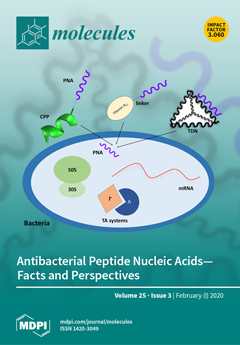
Oligonucleotides, such as peptide nucleic acid (PNA), designed to block the synthesis of essential bacterial proteins, could be used as antibacterials. The limitation precluding PNA use is that bacterial cells do not uptake PNA. Thus, both PNA carriers and new targets in bacteria have been extensively searched for. The cover presents carriers of PNA to bacteria and possible targets for sequence-specific inhibition by PNA. The delivery strategies comprise covalent conjugation of PNA with cell-penetrating peptides (CPP) and with vitamin B12, as well as complementary base pairing between PNA and DNA to form a tetrahedral DNA nanostructure (TDN). PNA targets tested in Gram-negative bacteria include mRNA, ribosome (small 30S and large 50S subunit), and toxin–antitoxin (TA) systems. View this paper.
  • Issues are regarded as officially published after their release is announced to the table of contents alert mailing list.
  • You may sign up for e-mail alerts to receive table of contents of newly released issues.
  • PDF is the official format for papers published in both, html and pdf forms. To view the papers in pdf format, click on the "PDF Full-text" link, and use the free Adobe Reader to open them.
Order results
Result details
Section
Select all
Export citation of selected articles as:
8 pages, 1326 KiB  
Article
Synthesis and Anticancer Cytotoxicity of Azaaurones Overcoming Multidrug Resistance
by Szilárd Tóth, Áron Szepesi, Viet-Khoa Tran-Nguyen, Balázs Sarkadi, Katalin Német, Pierre Falson, Attilio Di Pietro, Gergely Szakács and Ahcène Boumendjel
Molecules 2020, 25(3), 764; https://doi.org/10.3390/molecules25030764 - 10 Feb 2020
Cited by 18 | Viewed by 4100
Abstract
The resistance of tumors against anticancer drugs is a major impediment for chemotherapy. Tumors often develop multidrug resistance as a result of the cellular efflux of chemotherapeutic agents by ABC transporters such as P-glycoprotein (ABCB1/P-gp), Multidrug Resistance Protein 1 (ABCC1/MRP1), or Breast Cancer [...] Read more.
The resistance of tumors against anticancer drugs is a major impediment for chemotherapy. Tumors often develop multidrug resistance as a result of the cellular efflux of chemotherapeutic agents by ABC transporters such as P-glycoprotein (ABCB1/P-gp), Multidrug Resistance Protein 1 (ABCC1/MRP1), or Breast Cancer Resistance Protein (ABCG2/BCRP). By screening a chemolibrary comprising 140 compounds, we identified a set of naturally occurring aurones inducing higher cytotoxicity against P-gp-overexpressing multidrug-resistant (MDR) cells versus sensitive (parental, non-P-gp-overexpressing) cells. Follow-up studies conducted with the P-gp inhibitor tariquidar indicated that the MDR-selective toxicity of azaaurones is not mediated by P-gp. Azaaurone analogs possessing pronounced effects were then designed and synthesized. The knowledge gained from structure–activity relationships will pave the way for the design of a new class of anticancer drugs selectively targeting multidrug-resistant cancer cells. Full article
Show Figures

Graphical abstract

Graphical abstract
Full article ">Figure 1
<p>Scaffolds of the derivatives (library of 140 compounds) tested in the primary screen (at 10 and 100 µM).</p>
Full article ">Scheme 1
<p>Synthesis of targeted azaaurones.</p>
Full article ">
16 pages, 3030 KiB  
Article
Discrimination of Cultivated Regions of Soybeans (Glycine max) Based on Multivariate Data Analysis of Volatile Metabolite Profiles
by So-Yeon Kim, So Young Kim, Sang Mi Lee, Do Yup Lee, Byeung Kon Shin, Dong Jin Kang, Hyung-Kyoon Choi and Young-Suk Kim
Molecules 2020, 25(3), 763; https://doi.org/10.3390/molecules25030763 - 10 Feb 2020
Cited by 11 | Viewed by 4315
Abstract
Soybean (Glycine max) is a major crop cultivated in various regions and consumed globally. The formation of volatile compounds in soybeans is influenced by the cultivar as well as environmental factors, such as the climate and soil in the cultivation areas. [...] Read more.
Soybean (Glycine max) is a major crop cultivated in various regions and consumed globally. The formation of volatile compounds in soybeans is influenced by the cultivar as well as environmental factors, such as the climate and soil in the cultivation areas. This study used gas chromatography-mass spectrometry (GC-MS) combined by headspace solid-phase microextraction (HS-SPME) to analyze the volatile compounds of soybeans cultivated in Korea, China, and North America. The multivariate data analysis of partial least square-discriminant analysis (PLS-DA), and hierarchical clustering analysis (HCA) were then applied to GC-MS data sets. The soybeans could be clearly discriminated according to their geographical origins on the PLS-DA score plot. In particular, 25 volatile compounds, including terpenes (limonene, myrcene), esters (ethyl hexanoate, butyl butanoate, butyl prop-2-enoate, butyl acetate, butyl propanoate), aldehydes (nonanal, heptanal, (E)-hex-2-enal, (E)-hept-2-enal, acetaldehyde) were main contributors to the discrimination of soybeans cultivated in China from those cultivated in other regions in the PLS-DA score plot. On the other hand, 15 volatile compounds, such as 2-ethylhexan-1-ol, 2,5-dimethylhexan-2-ol, octanal, and heptanal, were related to Korean soybeans located on the negative PLS 2 axis, whereas 12 volatile compounds, such as oct-1-en-3-ol, heptan-4-ol, butyl butanoate, and butyl acetate, were responsible for North American soybeans. However, the multivariate statistical analysis (PLS-DA) was not able to clearly distinguish soybeans cultivated in Korea, except for those from the Gyeonggi and Kyeongsangbuk provinces. Full article
(This article belongs to the Special Issue Progress in Volatile Organic Compounds Research)
Show Figures

Graphical abstract

Graphical abstract
Full article ">Figure 1
<p>Partial least square-discriminant analysis (PLS-DA) score plot of soybean samples from different cultivation areas; (<b>a</b>) 3D score plot; (<b>b</b>) score plot PLS[<a href="#B1-molecules-25-00763" class="html-bibr">1</a>]-PLS[<a href="#B2-molecules-25-00763" class="html-bibr">2</a>], indicating the separation between different cultivation areas.</p>
Full article ">Figure 1 Cont.
<p>Partial least square-discriminant analysis (PLS-DA) score plot of soybean samples from different cultivation areas; (<b>a</b>) 3D score plot; (<b>b</b>) score plot PLS[<a href="#B1-molecules-25-00763" class="html-bibr">1</a>]-PLS[<a href="#B2-molecules-25-00763" class="html-bibr">2</a>], indicating the separation between different cultivation areas.</p>
Full article ">Figure 2
<p>Heatmap generated by a hierarchical clustering analysis of 146 metabolites.</p>
Full article ">Figure 3
<p>PLS-DA score plot of soybeans from Korea on the basis of volatile metabolites: (<b>a</b>) 3D score plot; (<b>b</b>) score plot PLS[<a href="#B1-molecules-25-00763" class="html-bibr">1</a>]-PLS[<a href="#B2-molecules-25-00763" class="html-bibr">2</a>]. GGIC—Gyeonggi province Anseong, GGAS—Gyeonggi province Icheon, GWCC—Gangwon province Chuncheon, GWYW—Gangwon province Yeongwol, CBES—Chungcheongbuk province Eumseong, CNCA— Chungcheongnam province Cheonan, CNGJ—Chungcheongnam province Gongju, JBGJ—Jeollabuk province Gimje, JBIS—Jeollabuk province Imsil, JNNJ—Jeollanam province Naju, JNYG—Jeollanam province Yeonggwang, KBCD—Kyeongsangbuk province Cheongdo, KBES—Kyeongsangbuk province Uiseong, KBYC—Kyeongsangbuk province Yeongcheon, KNCN—Kyeongsangnam province Changnyeong, KNMY—Kyeongsangnam province Miryang, KNGC—Kyeongsangnam province Geochang.</p>
Full article ">
35 pages, 1953 KiB  
Review
Therapeutic Potential of Flavonoids in Pain and Inflammation: Mechanisms of Action, Pre-Clinical and Clinical Data, and Pharmaceutical Development
by Camila R. Ferraz, Thacyana T. Carvalho, Marília F. Manchope, Nayara A. Artero, Fernanda S. Rasquel-Oliveira, Victor Fattori, Rubia Casagrande and Waldiceu A. Verri, Jr.
Molecules 2020, 25(3), 762; https://doi.org/10.3390/molecules25030762 - 10 Feb 2020
Cited by 176 | Viewed by 17127
Abstract
Pathological pain can be initiated after inflammation and/or peripheral nerve injury. It is a consequence of the pathological functioning of the nervous system rather than only a symptom. In fact, pain is a significant social, health, and economic burden worldwide. Flavonoids are plant [...] Read more.
Pathological pain can be initiated after inflammation and/or peripheral nerve injury. It is a consequence of the pathological functioning of the nervous system rather than only a symptom. In fact, pain is a significant social, health, and economic burden worldwide. Flavonoids are plant derivative compounds easily found in several fruits and vegetables and consumed in the daily food intake. Flavonoids vary in terms of classes, and while structurally unique, they share a basic structure formed by three rings, known as the flavan nucleus. Structural differences can be found in the pattern of substitution in one of these rings. The hydroxyl group (–OH) position in one of the rings determines the mechanisms of action of the flavonoids and reveals a complex multifunctional activity. Flavonoids have been widely used for their antioxidant, analgesic, and anti-inflammatory effects along with safe preclinical and clinical profiles. In this review, we discuss the preclinical and clinical evidence on the analgesic and anti-inflammatory proprieties of flavonoids. We also focus on how the development of formulations containing flavonoids, along with the understanding of their structure-activity relationship, can be harnessed to identify novel flavonoid-based therapies to treat pathological pain and inflammation. Full article
(This article belongs to the Special Issue Flavonoids and Their Disease Prevention and Treatment Potential)
Show Figures

Figure 1

Figure 1
<p>The chemical structures of the flavonoid groups discussed in this review.</p>
Full article ">Figure 2
<p>The number of manuscripts published on flavonoids, pain, and inflammation during the last 20 years at PubMed. The keywords search at PubMed was “flavonoids and pain and inflammation”, and only original research papers were considered.</p>
Full article ">Figure 3
<p>The anti-inflammatory and analgesic effects of flavonoids. Intracellular targets of <b>Rutin</b>: NF-κB [<a href="#B39-molecules-25-00762" class="html-bibr">39</a>,<a href="#B40-molecules-25-00762" class="html-bibr">40</a>] and Nrf2 [<a href="#B40-molecules-25-00762" class="html-bibr">40</a>], <b>Trans-chalcone</b>: NF-κB [<a href="#B41-molecules-25-00762" class="html-bibr">41</a>] and STAT3 [<a href="#B41-molecules-25-00762" class="html-bibr">41</a>] and NLRP3 [<a href="#B42-molecules-25-00762" class="html-bibr">42</a>], <b>Hesperidin:</b> PI3K/ AKT [<a href="#B43-molecules-25-00762" class="html-bibr">43</a>] and NF-κB [<a href="#B44-molecules-25-00762" class="html-bibr">44</a>], <b>Epigallocatechin-3-gallate:</b> NF-κB [<a href="#B45-molecules-25-00762" class="html-bibr">45</a>], <b>Apigerin</b>: NF-κB [<a href="#B46-molecules-25-00762" class="html-bibr">46</a>], <b>Diosmin</b>: NF-κB [<a href="#B47-molecules-25-00762" class="html-bibr">47</a>], and <b>Hesperidin methyl chalcone:</b> NF-κB [<a href="#B48-molecules-25-00762" class="html-bibr">48</a>,<a href="#B49-molecules-25-00762" class="html-bibr">49</a>,<a href="#B50-molecules-25-00762" class="html-bibr">50</a>] and Nrf2 [<a href="#B49-molecules-25-00762" class="html-bibr">49</a>,<a href="#B50-molecules-25-00762" class="html-bibr">50</a>]. ROS and inflammatory stimuli that activate specific receptors trigger intracellular signaling that will result in pain and inflammation. The <b>blue</b> arrows indicate endogenous pathways that are stimulated by flavonoids resulting in the reduction of pain and inflammation. The <b>red</b> arrows represent endogenous pathways that are inhibited by flavonoids resulting in reduced pain and inflammation.</p>
Full article ">Figure 4
<p>The anti-inflammatory and analgesic effects of Vitexin, Quercetin, and Naringenin. (<b>A</b>) Intracellular targets of <b>Vitexin</b>: MAPK [<a href="#B51-molecules-25-00762" class="html-bibr">51</a>], NF-κB [<a href="#B51-molecules-25-00762" class="html-bibr">51</a>] and Nrf2 [<a href="#B52-molecules-25-00762" class="html-bibr">52</a>], <b>Quercetin</b>: MAPK [<a href="#B53-molecules-25-00762" class="html-bibr">53</a>], NF-κB [<a href="#B53-molecules-25-00762" class="html-bibr">53</a>,<a href="#B54-molecules-25-00762" class="html-bibr">54</a>,<a href="#B55-molecules-25-00762" class="html-bibr">55</a>], AKT [<a href="#B56-molecules-25-00762" class="html-bibr">56</a>], Nrf2 [<a href="#B33-molecules-25-00762" class="html-bibr">33</a>,<a href="#B54-molecules-25-00762" class="html-bibr">54</a>], and NLRP3 [<a href="#B57-molecules-25-00762" class="html-bibr">57</a>] and <b>Naringenin:</b> NF-κB [<a href="#B58-molecules-25-00762" class="html-bibr">58</a>,<a href="#B59-molecules-25-00762" class="html-bibr">59</a>,<a href="#B60-molecules-25-00762" class="html-bibr">60</a>] and Nrf2 [<a href="#B59-molecules-25-00762" class="html-bibr">59</a>,<a href="#B61-molecules-25-00762" class="html-bibr">61</a>,<a href="#B62-molecules-25-00762" class="html-bibr">62</a>]. (<b>B</b>) Ion channels expressed by neurons that are targeted by Vitexin, Quercetin, and Naringenin to reduce pain. <b>Vitexin</b>: TRPV1 [<a href="#B38-molecules-25-00762" class="html-bibr">38</a>], <b>Quercetin</b>: TRPV1 [<a href="#B63-molecules-25-00762" class="html-bibr">63</a>], and <b>Naringenin:</b> TRPV1 [<a href="#B58-molecules-25-00762" class="html-bibr">58</a>], TRPA1 [<a href="#B58-molecules-25-00762" class="html-bibr">58</a>], TRPM3 [<a href="#B64-molecules-25-00762" class="html-bibr">64</a>], Nav 1.8 [<a href="#B65-molecules-25-00762" class="html-bibr">65</a>], and TRPM8 [<a href="#B64-molecules-25-00762" class="html-bibr">64</a>]. In panel (<b>A</b>), ROS and inflammatory stimuli that activate specific receptors trigger intracellular signaling that will result in pain and inflammation. The <b>blue</b> arrows indicate endogenous pathways that are stimulated by flavonoids, and the <b>red</b> arrows represent endogenous pathways that are inhibited by flavonoids resulting in reduced pain and inflammation.</p>
Full article ">Figure 5
<p>The chemical structures of the flavonoids discussed in this review.</p>
Full article ">
12 pages, 3002 KiB  
Article
Direct Recovery of the Rare Earth Elements Using a Silk Displaying a Metal-Recognizing Peptide
by Nobuhiro Ishida, Takaaki Hatanaka, Yoichi Hosokawa, Katsura Kojima, Tetsuya Iizuka, Hidetoshi Teramoto, Hideki Sezutsu and Tsunenori Kameda
Molecules 2020, 25(3), 761; https://doi.org/10.3390/molecules25030761 - 10 Feb 2020
Cited by 3 | Viewed by 4016
Abstract
Rare earth elements (RE) are indispensable metallic resources in the production of advanced materials; hence, a cost- and energy-effective recovery process is required to meet the rapidly increasing RE demand. Here, we propose an artificial RE recovery approach that uses a functional silk [...] Read more.
Rare earth elements (RE) are indispensable metallic resources in the production of advanced materials; hence, a cost- and energy-effective recovery process is required to meet the rapidly increasing RE demand. Here, we propose an artificial RE recovery approach that uses a functional silk displaying a RE-recognizing peptide. Using the piggyBac system, we constructed a transgenic silkworm in which one or two copies of the gene coding for the RE-recognizing peptide (Lamp1) was fused with that of the fibroin L (FibL) protein. The purified FibL-Lamp1 fusion protein from the transgenic silkworm was able to recognize dysprosium (Dy3+), a RE, under physiological conditions. This method can also be used with silk from which sericin has been removed. Furthermore, the Dy-recovery ability of this silk was significantly improved by crushing the silk. Our simple approach is expected to facilitate the direct recovery of RE from an actual mixed solution of metal ions, such as seawater and industrial wastewater, under mild conditions without additional energy input. Full article
(This article belongs to the Special Issue Silk Fibroin Materials)
Show Figures

Graphical abstract

Graphical abstract
Full article ">Figure 1
<p>Breeding of the transgenic silkworm. (<b>a</b>) Construction of the expression vector. Each plasmid contains expression units for selection marker gene (<span class="html-italic">DsRed2</span>) and cDNA of fibroin L-chain (<span class="html-italic">FibL</span> cDNA) between the <span class="html-italic">piggyBac</span> inverted terminal repeat (ITR). Three gene fragments containing Lamp1 or His Tag were inserted into the 3’ ends of <span class="html-italic">FibL</span> cDNA using restriction site of <span class="html-italic">Bam</span>H I and <span class="html-italic">Hin</span>d III. 3xP3 pro., 3xP3 promoter; SV40 polyA, polyA signal of SV40; FibLpro., promoter of fibroin L-chain; and FibL 3’ UTR, 3’ untranslated region of fibroin L-chain. (<b>b</b>) SDS-PAGE analysis of purified FibL-Lamp1-His protein from Ex1 strain and the control strain. (<b>c</b>) Western blotting analysis using Lamp1, and His-tag antibody. Each arrow indicates the expected molecular weight of the target protein. Lane 1, FibL sample before purification; Lane 2, flow-through of FibL column; Lane 3, purified FibL; Lane 4, buffer; Lane 5, FibL-Lamp1 sample before purification; Lane 6, flow-through of FibL-Lamp1 column; and Lane 7, purified FibL-Lamp1.</p>
Full article ">Figure 2
<p>Dy<sup>3+</sup> recognition with purified FibL-Lamp1-His protein. (<b>a</b>) Optical image of precipitation induced with protein adding. (<b>b</b>) SEM image of precipitation. Scale bar, 100 µm. (<b>c</b>) EDX spectrum. The red frame area in the SEM image of (b) was analyzed. Each arrow indicates the spectrum of Dy. (<b>d</b>) Elemental mapping of SEM image (Dy, dysprosium; N, nitrogen; and S, sulfur). Scale bar, 100 µm.</p>
Full article ">Figure 3
<p>La<sup>3+</sup> and Lu<sup>3+</sup> recognition with the purified FibL-Lamp1-His protein. (<b>a</b>–<b>c</b>) Reaction with La<sup>3+</sup>, and (<b>d</b>–<b>f</b>) reaction with Lu<sup>3+</sup>. (<b>a</b>–<b>d</b>) Optical image of precipitation induced by the purified FibL-Lamp1-His protein. (<b>b</b>–<b>e</b>) SEM image (left) and elemental mapping image (right) of precipitation. Scale bars, 100 µm. (<b>c</b>–<b>f</b>) EDX spectrum. The red frame area in the SEM image was analyzed. Each arrow indicates the spectrum of La or Lu.</p>
Full article ">Figure 4
<p>The recovery efficiency of Dy<sup>3+</sup> with the purified protein. The supernatant solution after reaction (mentioned in <a href="#molecules-25-00761-f002" class="html-fig">Figure 2</a>a) was collected, and the Dy<sup>3+</sup> concentration in the solution was determined. Average and standard deviations from three independent experiments were represented.</p>
Full article ">Figure 5
<p>RE recovery process with silk powder: (<b>a</b>) Schematic diagram of the process for RE-recovery, and optical image of the prepared silk and (<b>b</b>) SEM image of roughly crushed silk.</p>
Full article ">Figure 6
<p>Dy<sup>3+</sup> recognition with silk powder: (<b>a</b>) SEM (left) and elemental mapping (right) images of silk powder after Dy<sup>3+</sup> absorption reaction. The yellow signal indicates a Dy. Scale bar, 80 µm, (<b>b</b>) EDX spectrum, each arrow indicates the Dy signal and (<b>c</b>) the recovery efficiency of Dy<sup>3+</sup> of silk with harvested silk from Ex2-2. Averages and standard deviations for three independent experiments were represented.</p>
Full article ">Figure 7
<p>Reusability of silk powder for Dy<sup>3+</sup> recovery. (<b>a</b>) The relative degree to the initial Dy<sup>3+</sup> recovery efficiency is shown. Averages and standard deviations for three independent experiments were represented. (<b>b</b>) SEM-EDX analysis, the captured Dy (yellow signal) was eluted with acetic buffer (50 mM) at pH 4.0, and the silk powder was recycled. Scale bars: 100 µm.</p>
Full article ">
19 pages, 5817 KiB  
Article
Anticancer Activities of the Quinone-Methide Triterpenes Maytenin and 22-β-hydroxymaytenin Obtained from Cultivated Maytenus ilicifolia Roots Associated with Down-Regulation of miRNA-27a and miR-20a/miR-17-5p
by Camila Hernandes, Lucyene Miguita, Romario Oliveira de Sales, Elisangela de Paula Silva, Pedro Omori Ribeiro de Mendonça, Bruna Lorencini da Silva, Maria de Fatima Guarizo Klingbeil, Monica Beatriz Mathor, Erika Bevilaqua Rangel, Luciana Cavalheiro Marti, Juliana da Silva Coppede, Fabio Daumas Nunes, Ana Maria Soares Pereira and Patricia Severino
Molecules 2020, 25(3), 760; https://doi.org/10.3390/molecules25030760 - 10 Feb 2020
Cited by 17 | Viewed by 4107
Abstract
Natural triterpenes exhibit a wide range of biological activities. Since this group of secondary metabolites is structurally diverse, effects may vary due to distinct biochemical interactions within biological systems. In this work, we investigated the anticancer-related activities of the quinone-methide triterpene maytenin and [...] Read more.
Natural triterpenes exhibit a wide range of biological activities. Since this group of secondary metabolites is structurally diverse, effects may vary due to distinct biochemical interactions within biological systems. In this work, we investigated the anticancer-related activities of the quinone-methide triterpene maytenin and its derivative compound 22-β-hydroxymaytenin, obtained from Maytenus ilicifolia roots cultivated in vitro. Their antiproliferative and pro-apoptotic activities were evaluated in monolayer and three-dimensional cultures of immortalized cell lines. Additionally, we investigated the toxicity of maytenin in SCID mice harboring tumors derived from a squamous cell carcinoma cell line. Both isolated molecules presented pronounced pro-apoptotic activities in four cell lines derived from head and neck squamous cell carcinomas, including a metastasis-derived cell line. The molecules also induced reactive oxygen species (ROS) and down-regulated microRNA-27a and microRNA-20a/miR-17-5p, corroborating with the literature data for triterpenoids. Intraperitoneal administration of maytenin to tumor-bearing mice did not lead to pronounced histopathological changes in kidney tissue, suggesting low nephrotoxicity. The wide-ranging activity of maytenin and 22-β-hydroxymaytenin in head and neck cancer cells indicates that these molecules should be further explored in plant biochemistry and biotechnology for therapeutic applications. Full article
Show Figures

Figure 1

Figure 1
<p>UPLC-MS chromatograms of maytenin (<b>A-a</b>), 22-β-hydroxymaytenin (<b>B-b</b>) and of dichloromethane roots extract from <span class="html-italic">M. ilicifolia</span> cultivated in vitro (<b>C</b>).</p>
Full article ">Figure 2
<p>Cell viability results from MTT assay after incubation with maytenin or 22-β-hydroxymaytenin. (<b>A</b>) IC<sub>50</sub> following 24 h incubation with maytenin or 22-β-hydroxymaytenin for each cell type; (<b>B</b>) Viability of SCC9 cell line following incubation for 6, 24, 48 or 72 h with maytenin or 22-β-hydroxymaytenin with IC<sub>50</sub> value; (<b>C</b>) Viability of SCC25 cell line following incubation for 6, 24, 48 or 72 h with maytenin or 22-β-hydroxymaytenin with IC<sub>50</sub> value; (<b>D</b>) Viability of FaDu cell line following incubation for 6, 24, 48 or 72 h with maytenin or 22-β-hydroxymaytenin with IC<sub>50</sub> value. * <span class="html-italic">p</span> &lt; 0.05, ** <span class="html-italic">p</span> &lt; 0.01, *** <span class="html-italic">p</span> &lt; 0.001 (one-way ANOVA).</p>
Full article ">Figure 3
<p>Size and cell viability in maytenin and 22-β-hydroxymaytenin treated spheroids. (<b>A</b>) Spheroids were treated for 48 h with the corresponding IC<sub>50</sub> obtained in 2D cell culture. No size reduction was detectable using a light microscope for any of the treatments. <span class="html-italic">n</span> = 4. (<b>B</b>) Cell viability in the outer layers of the spheroids following treatment with the IC<sub>50</sub> obtained in 2D cell culture and with 10× the IC<sub>50</sub> obtained in 2D cell culture. <span class="html-italic">n</span> = 4. Live cells fluoresce green, and dead cells fluoresce red. A clear effect on cell viability was observed at the outer cell layer when 10× the IC<sub>50</sub> was used for every treatment.</p>
Full article ">Figure 4
<p>Anti-proliferative effects of maytenin, 22-β-hydroxymaytenin and cisplatin measured by EdU incorporation into DNA. Column graphs (<b>A</b>,<b>C</b>,<b>E</b>,<b>G</b>) show the percentage of cells staining for EdU (red) compared to Hoechst 33,342 (blue) stained nuclei (± SD). There were two replicates in each experiment and all cells were counted for each treatment. Images (<b>B</b>,<b>D</b>,<b>F</b>,<b>H</b>) were taken on a fluorescence microscope (magnification ×20) and depict EdU incorporation (red) and Hoechst 33,342 nuclei staining. EdU incorporation occurs during DNA synthesis, EdU positive cells indicate DNA replication and consequent cell cycle progression. * <span class="html-italic">p</span> &lt; 0.01. ** <span class="html-italic">p</span> &lt; 0.05.</p>
Full article ">Figure 5
<p>Effect of triterpenes on oxidative stress in SCC cell lines. (<b>A</b>) SCC9, Detroit and FaDu cell lines treated with cisplatin. ROS induction following cisplatin treatment is well established and these results were used as positive controls. The oxidative stress increases in tumor cells, compared with control when treated with maytenin or 22-β-hydroxymaytenin in SCC9 (<b>B</b>), FaDu (<b>C</b>) and Detroit (<b>D</b>). Total ROS after treatment with the IC<sub>50</sub> value for 18 h was assessed by fluorescent probe detection using confocal microscopy. <span class="html-italic">n</span> = 2.</p>
Full article ">Figure 6
<p>Apoptotic cell death measured by Annexin V and 7-ADD assay and caspase 3/7 activation. Keratinocytes and FaDu cell line were treated with 22-β-hydroxymaytenin, maytenin, or cisplatin and the effect on cell death induction was evaluated by flow cytometry using Annexin-V and 7-AAD staining. Keratinocytes: (<b>A</b>) Early apoptosis, (<b>B</b>) late apoptosis, (<b>C</b>) total cell death and (<b>D</b>) individual flow cytometry plots for each treatment. FaDu cell line: (<b>E</b>) Early apoptosis, (<b>F</b>) late apoptosis, (<b>G</b>) total cell death and (<b>H</b>) individual flow cytometry plots of each treatment. (<b>I</b>) caspase-3/7 activation in FaDu cell line following treatment by 22-β-hydroxymaytenin, maytenin, or cisplatin.</p>
Full article ">Figure 7
<p>(<b>A</b>) Comparison of IC<sub>50</sub> of maytenin for SCC9<sub>ZS GREEN</sub> and SCC9<sub>ZS GREEN</sub> LN-1. Maytenin showed similar effect in cell viability for SCC9<sub>ZS GREEN</sub> and SCC9<sub>ZS GREEN</sub> LN-1 cell lines. (<b>B</b>) Non-invasive in vivo imaging of mice harboring a human tumor xenograph prior and following treatment with maytenin and cisplatin: SCID mice were injected with SCC9<sub>ZS GREEN</sub> or SCC9<sub>ZS GREEN</sub> LN-1 cells and, 15 days later, mice with similar tumor burden determined by IVIS fluorescence imaging were assigned to each treatment group: control (PBS), maytenin (2 mg/Kg, i.p. Monday-Wednesday-Friday), or cisplatin (5 mg/Kg, i.p. once a week). b, c, d, f, g, h: representative IVIS images of mice from each experimental group at each stage the correspondent treatment: at 15 days following injection of cancer derived cells at the tongue mice had not received any treatment yet, at days 21 and 29, the respective treatment schemes were being applied. The graphical representation of the fluorescence in specific regions of interest in mice is indicated by a multi-color distribution: the high intensity of fluorescence appears as yellow color and low intensity of fluorescence as red. (<b>C</b>) Body weight of mice during the treatment cycle. There was no significant difference in body weight when maytenin and cisplatin were considered, but mice harbouring tumours derived from SCC9<sub>ZS GREEN</sub> LN-1 cells lost weight due to tumour burden. (<b>D</b>) Photomicrograph of mouse kidney tissues with H&amp;E staining: For untreated mice (A1, detailed in A2 and A3) and maytenin treated mice following injection of SCC9<sub>ZS GREEN</sub> LN-1 cells at tongue (B1, detailed in B2 and B3) the cross section of kidney shows proximal tubules (PT) exhibiting brush border and distal tubules (DT) with normal morphology, the renal cortex exhibits normal glomerular (G) and tubular morphology structures. In cisplatin treated mice (C1, detailed in C2 and C3), renal cortex features suggest acute tubular necrosis as shown by patchy or diffuse denudation of the renal cells with loss of brush border (arrows), flattening of the renal tubular cells due to tubular dilation (arrowheads), pyknotic nucleus (red arrow) and loss of tubular architecture. Images illustrate the biological effects in tissue samples collected 29 days after the injection of SCC9<sub>ZS GREEN</sub> LN-1 cells. Scale bars represent 20 µm.</p>
Full article ">
18 pages, 6292 KiB  
Article
Stabilizing the Oil-in-Water Emulsions Using the Mixtures of Dendrobium Officinale Polysaccharides and Gum Arabic or Propylene Glycol Alginate
by Bo Wang, Haiyan Tian and Dong Xiang
Molecules 2020, 25(3), 759; https://doi.org/10.3390/molecules25030759 - 10 Feb 2020
Cited by 33 | Viewed by 6784
Abstract
Coconut oil-in-water emulsions were prepared using three polysaccharides: Dendrobium officinale polysaccharide (DOP), propylene glycol alginate (PGA), gum arabic (GA) and their polysaccharide complexes as emulsifiers. The effects of the ratio of the compounded polysaccharides on their apparent viscosity and interfacial activity were explored [...] Read more.
Coconut oil-in-water emulsions were prepared using three polysaccharides: Dendrobium officinale polysaccharide (DOP), propylene glycol alginate (PGA), gum arabic (GA) and their polysaccharide complexes as emulsifiers. The effects of the ratio of the compounded polysaccharides on their apparent viscosity and interfacial activity were explored in this study. The average particle size, zeta potential, microstructure, rheological properties, and physical stability of the emulsions prepared with different compound-polysaccharides were studied. The results showed that mainly DOP contributed to the apparent viscosity of the compound-polysaccharide, while the interfacial activity and zeta potential were mainly influenced by PGA or GA. Emulsions prepared with compound-polysaccharides exhibited smaller average particle sizes, and microscopic observations showed smaller droplets and less droplet aggregation. In addition, the stability analysis of emulsions by a dispersion analyzer LUMiSizer showed that the emulsion prepared by compounding polysaccharides had better physical stability. Finally, all of the above experimental results showed that the emulsions prepared by PGA:DOP = 2:8 (total concentration = 1.5 wt%) and 2.0% GA + 1.5% DOP were the most stable. Full article
Show Figures

Graphical abstract

Graphical abstract
Full article ">Figure 1
<p>(<b>A</b>) Relationship between apparent viscosity and concentration of three polysaccharides. (<b>B</b>) Apparent viscosity of the compound polysaccharides at different ratios (total concentration was 1 wt%); a–e indicate significant differences (<span class="html-italic">p</span> &lt; 0.01).</p>
Full article ">Figure 2
<p>(<b>A</b>) Relationship between the interfacial tension (IFT) and concentration of three polysaccharides. (<b>B</b>) Interfacial tension (IFT) of the compound polysaccharides at different ratios (total concentration was 1 wt%), a–d indicate significant differences (<span class="html-italic">p</span> &lt; 0.01).</p>
Full article ">Figure 3
<p>(<b>A</b>) Particle size distribution of emulsions prepared by compounding polysaccharides of PGA and DOP as emulsifiers (total emulsifier concentration was 1 wt%); (<b>B</b>) Particle size distribution of emulsions prepared by compounding polysaccharides of GA and DOP as emulsifiers (total emulsifier concentration was 1 wt%); (<b>C</b>) The average particle size of emulsions prepared by compounding polysaccharides of PGA and DOP as emulsifiers (total emulsifier concentration was 1 wt%); (<b>D</b>) The average particle size of emulsions prepared by compounding polysaccharides of GA and DOP as emulsifiers (total emulsifier concentration was 1 wt%), a–g indicate significant differences (<span class="html-italic">p</span> &lt; 0.01).</p>
Full article ">Figure 4
<p>(<b>A</b>) The zeta potential of emulsions prepared by compounding polysaccharides of PGA and DOP as emulsifiers (total emulsifier concentration was 1 wt%); (<b>B</b>) The zeta potential of emulsions prepared by compounding polysaccharides of GA and DOP as emulsifiers (total emulsifier concentration was 1 wt%), a–f indicate significant differences (<span class="html-italic">p</span> &lt; 0.01).</p>
Full article ">Figure 5
<p>The microstructure of emulsions prepared by compounding polysaccharides of PGA and DOP or compounding polysaccharides of GA and DOP as emulsifiers (total emulsifier concentration was 1 wt%).</p>
Full article ">Figure 5 Cont.
<p>The microstructure of emulsions prepared by compounding polysaccharides of PGA and DOP or compounding polysaccharides of GA and DOP as emulsifiers (total emulsifier concentration was 1 wt%).</p>
Full article ">Figure 6
<p>Visual observations (total emulsifier concentration was 1 wt%).</p>
Full article ">Figure 7
<p>(<b>A</b>) The apparent viscosity and zeta potential. (<b>B</b>) The average particle size and particle size distribution. a–d, cd, e, de, f indicate significant differences (<span class="html-italic">p</span> &lt; 0.01).</p>
Full article ">Figure 8
<p>(<b>A</b>): The microstructure of emulsions (secondary screening of the concentration and ratio of compound polysaccharide emulsifiers); (<b>B</b>) The visual observations of emulsions (secondary screening of the concentration and ratio of compound polysaccharide emulsifiers).</p>
Full article ">Figure 9
<p>Rheological properties. (<b>A</b>) Steady-state flow curve. (<b>B</b>) Linear viscoelastic zone. (<b>C1</b>) Dynamic frequency sweep of the emulsion prepared by PGA: DOP=2:8 (1.5%); (<b>C2</b>) Dynamic frequency sweep of the emulsion prepared by 2.0% GA + 1.5% DOP.</p>
Full article ">Figure 10
<p>(<b>A</b>) Light transmittance curve. (<b>B</b>) Instability analysis and interface tracking analysis. The analysis range is from the meniscus to the bottom; a–f indicate significant differences (<span class="html-italic">p</span> &lt; 0.01).</p>
Full article ">
10 pages, 2605 KiB  
Review
Bone Mineral Affinity of Polyphosphodiesters
by Yasuhiko Iwasaki
Molecules 2020, 25(3), 758; https://doi.org/10.3390/molecules25030758 - 10 Feb 2020
Cited by 16 | Viewed by 4033
Abstract
Biomimetic molecular design is a promising approach for generating functional biomaterials such as cell membrane mimetic blood-compatible surfaces, mussel-inspired bioadhesives, and calcium phosphate cements for bone regeneration. Polyphosphoesters (PPEs) are candidate biomimetic polymer biomaterials that are of interest due to their biocompatibility, biodegradability, [...] Read more.
Biomimetic molecular design is a promising approach for generating functional biomaterials such as cell membrane mimetic blood-compatible surfaces, mussel-inspired bioadhesives, and calcium phosphate cements for bone regeneration. Polyphosphoesters (PPEs) are candidate biomimetic polymer biomaterials that are of interest due to their biocompatibility, biodegradability, and structural similarity to nucleic acids. While studies on the synthesis of PPEs began in the 1970s, the scope of their use as biomaterials has increased in the last 20 years. One advantageous property of PPEs is their molecular diversity due to the presence of multivalent phosphorus in their backbones, which allows their physicochemical and biointerfacial properties to be easily controlled to produce the desired molecular platforms for functional biomaterials. Polyphosphodiesters (PPDEs) are analogs of PPEs that have recently attracted interest due to their strong affinity for biominerals. This review describes the fundamental properties of PPDEs and recent research in the field of macromolecular bone therapeutics. Full article
Show Figures

Figure 1

Figure 1
<p>General structure of polyphosphodiesters (PPDEs).</p>
Full article ">Figure 2
<p>Synthetic routes to polyphosphodiesters (PPDEs).</p>
Full article ">Figure 3
<p>(<b>a</b>) Chemical structure of polyphosphoester copolymers (P(EP<sub>x</sub>/EEP<sub>y</sub>)) and (<b>b</b>) the effect of the phosphodiester composition of the copolymers on their mineral affinity.</p>
Full article ">Figure 4
<p>(<b>a</b>) Chemical structure of the polyphosphodiester PEP·Na macromonomer and (<b>b</b>) scanning electron micrographs of poly(ether ether ketone) (PEEK) and poly(PEP·Na)-immobilized PEEK specimens that had been soaked in ×1.5 simulated body fluid (1.5 SBF) for 28 days.</p>
Full article ">Figure 5
<p>(<b>a</b>) Chemical structure of amphophilic PPDEs. (<b>b</b>) Amphiphilic polyphosphodiester copolymer [CH-P(EP<sub>x</sub>/EEP<sub>y</sub>)]-immobilized 1,2-dioleoyl-sn-glycero-3-phosphocholine (DOPC) vesicles. Scale bar represents 500 nm. (<b>c</b>) Poly L-lactic acid nanoparticles bearing amphiphilic poly(ethylene sodium phosphate) (CH-PEP·Na). Scale bar represents 200 nm. (<b>d</b>) Bovine serum albumin/CH-PEP·Na conjugates. Scale bar represents 200 nm.</p>
Full article ">Figure 6
<p>(<b>a</b>) Viability and (<b>b</b>) morphology of MC3T3-E1 cells following contact with poly(ethylene sodium phosphate) (PEP·Na) or poly(phosphate) (polyP). ●: PEP·Na; ●: polyP. Reproduced with permission from reference [<a href="#B33-molecules-25-00758" class="html-bibr">33</a>]. Copyright 2018 by The Royal Society of Chemistry.</p>
Full article ">Figure 7
<p>(<b>a</b>) Densities of adherent osteoclasts on bovine bone slices after incubation with poly(ethylene sodium phosphate) (PEP·Na) for 24 h (<span class="html-italic">n</span> = 4). Student’s t-test was performed on the samples to test the statistical significance of differences (*<span class="html-italic">p</span> &lt; 0.005). (<b>b</b>) Optical micrographs of adherent osteoclasts on a bovine bone slice after cultivation with PEP·Na for 24 h. Scale bars represent 100 μm. Reproduced with permission from reference [<a href="#B36-molecules-25-00758" class="html-bibr">36</a>]. Copyright 2015 by Wiley Interscience.</p>
Full article ">Figure 8
<p>(<b>a</b>) Chemical structure of fluorescence-labeled poly(ethylene sodium phosphate) (Cy5-PEP·Na) and (<b>b</b>) in vivo fluorescence imaging of ICR mice at 0, 3, 30, and 75 h after the intravenous injection of Cy5-PEP·Na or Cy5-Az. Reproduced with permission from reference [<a href="#B33-molecules-25-00758" class="html-bibr">33</a>]. Copyright 2018 by The Royal Society of Chemistry.</p>
Full article ">
15 pages, 5676 KiB  
Article
Quercetin Inhibits Cell Survival and Metastatic Ability via the EMT-Mediated Pathway in Oral Squamous Cell Carcinoma
by So Ra Kim, Eun Young Lee, Da Jeong Kim, Hye Jung Kim and Hae Ryoun Park
Molecules 2020, 25(3), 757; https://doi.org/10.3390/molecules25030757 - 10 Feb 2020
Cited by 25 | Viewed by 4603
Abstract
This study aimed to investigate whether quercetin exerts anticancer effects on oral squamous cell carcinoma (OSCC) cell lines and to elucidate its mechanism of action. These anticancer effects in OSCC cells were assessed using an MTT assay, flow cytometry (to assess the cell [...] Read more.
This study aimed to investigate whether quercetin exerts anticancer effects on oral squamous cell carcinoma (OSCC) cell lines and to elucidate its mechanism of action. These anticancer effects in OSCC cells were assessed using an MTT assay, flow cytometry (to assess the cell cycle), wound-healing assay, invasion assay, Western blot analysis, gelatin zymography, and immunofluorescence. To investigate whether quercetin also inhibits transforming growth factor β1 (TGF-β1)-induced epithelial–mesenchymal transition (EMT) in human keratinocyte cells, HaCaT cells were treated with TGF-β1. Overall, our results strongly suggest that quercetin suppressed the viability of OSCC cells by inducing cell cycle arrest at the G2/M phase. However, quercetin did not affect cell viability of human keratinocytes such as HaCaT (immortal keratinocyte) and nHOK (primary normal human oral keratinocyte) cells. Additionally, quercetin suppresses cell migration through EMT and matrix metalloproteinase (MMP) in OSCC cells and decreases TGF-β1-induced EMT in HaCaT cells. In conclusion, this study is the first, to our knowledge, to demonstrate that quercetin can inhibit the survival and metastatic ability of OSCC cells via the EMT-mediated pathway, specifically Slug. Quercetin may thus provide a novel pharmacological approach for the treatment of OSCCs. Full article
(This article belongs to the Special Issue Antitumor and Anti-HIV Agents from Natural Products)
Show Figures

Graphical abstract

Graphical abstract
Full article ">Figure 1
<p>Quercetin reduced cell viability and arrested the G2/M phase cell cycle in oral squamous cell carcinoma (OSCC) cells. (<b>A</b>) Cell viability was investigated by an MTT assay. Oral squamous cell carcinoma cell lines (OSC20, SAS, and HN22 cells) were treated with quercetin (10, 20, 40, 80, and 160 μM). (<b>B</b>) Quercetin was shown to induce cell cycle arrest in OSC20, SAS, and HN22 cells. Data are the means ± SEM. * <span class="html-italic">p</span> &lt; 0.05 and ** <span class="html-italic">p</span> &lt; 0.01 vs. corresponding control (quercetin 0 μM).</p>
Full article ">Figure 2
<p>Cell migration ability assessed by a wound-healing assay. (<b>A</b>) Changes in the wound area were observed after 24 h. In the quercetin-treated cells, the wound area was less closed. This indicates a decrease in migration capacity. (<b>B</b>) The wound area was calculated and presented as a graph. Data are the means ± SEM. * <span class="html-italic">p</span> &lt; 0.05 and ** <span class="html-italic">p</span> &lt; 0.01 vs. the corresponding control (quercetin 0 μM).</p>
Full article ">Figure 3
<p>Quercetin is shown to induce regulation of epithelial-mesenchymal transition (EMT) and matrix metalloproteinase (MMP). (<b>A</b>) Western blotting was conducted to examine the changes in the EMT inducers. The results showed that the epithelial markers (E-cadherin and claudin-1) were upregulated, and the mesenchymal markers (fibronectin, vimentin, and alpha-smooth muscle actin (α-SMA)) were downregulated upon treatment with quercetin. (<b>B</b>) Quantitation of A. The band intensities of each target protein were measured using an image analyzer and presented as relative ratio. (<b>C</b>) Gelatin zymography shows the MMP-2 and MMP-9 activities in oral cancer cell lines (OSC20, SAS, and HN22 cells) upon quercetin treatment. Data are the means ± SEM. * <span class="html-italic">p</span> &lt; 0.05, and ** <span class="html-italic">p</span> &lt; 0.01 vs. the corresponding control (quercetin 0 μM).</p>
Full article ">Figure 4
<p>EMT-activating transcription factors were detected using Western blot and immunofluorescence. (<b>A</b>) The Western blot showed that quercetin downregulated EMT transcription factors at the protein level. (<b>B</b>) Quantitation of A. The band intensities of each target protein were measured using an image analyzer and presented as relative ratio. (<b>C</b>) Representative fluorescence microscopy images of Twist in OSCC cell lines. (<b>D</b>) Representative fluorescence microscopy images of Slug in OSCC cell lines. Scale bars: 20 μM. Data are the means ± SEM. * <span class="html-italic">p</span> &lt; 0.05, ** <span class="html-italic">p</span> &lt; 0.01, and *** <span class="html-italic">p</span> &lt; 0.001 vs. the corresponding control (quercetin 0 μM).</p>
Full article ">Figure 5
<p>Quercetin was not toxic in normal keratocyte and transforming growth factor β1 (TGF-β1) stimulated EMT in HaCaT cells. (<b>A</b>) To examine the effect of quercetin on cell viability, an MTT assay was conducted on HaCaT and nHOK cells. Data are means ± SEM. * <span class="html-italic">p</span> &lt; 0.05 vs. the corresponding control (quercetin 0 μM). (<b>B</b>) Expression of EMT-related markers such as in TGF-β1-treated HaCaT cells were analyzed by Western blotting. (<b>C</b>–<b>H</b>) Quantitation of B. The band intensities of each target protein were measured using an image analyzer and presented as relative ratio. Data are means ± SEM. * <span class="html-italic">p</span> &lt; 0.05 and *** <span class="html-italic">p</span> &lt; 0.005 vs. corresponding control (TGF-β1 0 ng/mL).</p>
Full article ">Figure 6
<p>Quercetin inhibited TGF-β1-induced EMT. (<b>A</b>) Experimental setup. (<b>B</b>) Western blot results showed that quercetin also regulated TGF-β1-induced EMT markers. (<b>C</b>) Treatment of TGF-β1 also changed the morphology of HaCaT. Further, quercetin induced morphological recovery. Scale bars: 50 μM. (<b>D</b>–<b>I</b>) Quantification of hRPTECs viability by MTT assay. (<b>J</b>,<b>K</b>) A wound-healing assay was conducted to evaluate the TGF-β1-induced EMT migration ability; quercetin attenuated EMT-induced migration in HaCaT cells. (<b>L</b>) Quercetin inhibited the invasion capacity of EMT-induced HaCaT cells. Data are the means ± SEM. * <span class="html-italic">p</span> &lt; 0.05, and *** <span class="html-italic">p</span> &lt; 0.001 vs. the corresponding control (without TGF-β1 and quercetin); <sup>#</sup> <span class="html-italic">p</span> &lt; 0.05 vs. the corresponding control (with TGF-β1 and without quercetin).</p>
Full article ">
20 pages, 14933 KiB  
Article
An Interpenetrating Alginate/Gelatin Network for Three-Dimensional (3D) Cell Cultures and Organ Bioprinting
by Qiuhong Chen, Xiaohong Tian, Jun Fan, Hao Tong, Qiang Ao and Xiaohong Wang
Molecules 2020, 25(3), 756; https://doi.org/10.3390/molecules25030756 - 10 Feb 2020
Cited by 54 | Viewed by 6571
Abstract
Crosslinking is an effective way to improve the physiochemical and biochemical properties of hydrogels. In this study, we describe an interpenetrating polymer network (IPN) of alginate/gelatin hydrogels (i.e., A-G-IPN) in which cells can be encapsulated for in vitro three-dimensional (3D) cultures and organ [...] Read more.
Crosslinking is an effective way to improve the physiochemical and biochemical properties of hydrogels. In this study, we describe an interpenetrating polymer network (IPN) of alginate/gelatin hydrogels (i.e., A-G-IPN) in which cells can be encapsulated for in vitro three-dimensional (3D) cultures and organ bioprinting. A double crosslinking model, i.e., using Ca2+ to crosslink alginate molecules and transglutaminase (TG) to crosslink gelatin molecules, is exploited to improve the physiochemical, such as water holding capacity, hardness and structural integrity, and biochemical properties, such as cytocompatibility, of the alginate/gelatin hydrogels. For the sake of convenience, the individual ionic (i.e., only treatment with Ca2+) or enzymatic (i.e., only treatment with TG) crosslinked alginate/gelatin hydrogels are referred as alginate-semi-IPN (i.e., A-semi-IPN) or gelatin-semi-IPN (i.e., G-semi-IPN), respectively. Tunable physiochemical and biochemical properties of the hydrogels have been obtained by changing the crosslinking sequences and polymer concentrations. Cytocompatibilities of the obtained hydrogels are evaluated through in vitro 3D cell cultures and bioprinting. The double crosslinked A-G-IPN hydrogel is a promising candidate for a wide range of biomedical applications, including bioartificial organ manufacturing, high-throughput drug screening, and pathological mechanism analyses. Full article
(This article belongs to the Special Issue Biomedical Hydrogels: Synthesis and Applications)
Show Figures

Figure 1

Figure 1
<p>Structure units of alginate molecule (from Wikipedia, the free encyclopedia).</p>
Full article ">Figure 2
<p>Chemical description of ionic crosslinking of alginate molecules, covalent crosslinking of gelatin molecules, and both covalent and ionic crosslinking of alginate/gelatin molecules. (<b>a</b>) G-blocks in two alginate chains are chemically (i.e., ionic) crosslinked by Ca<sup>2+</sup>. (<b>b</b>) Transglutaminase (TG) catalyzed covalent linkages between two gelatin molecules. (<b>c</b>) An interpenetrating network in an alginate/gelatin hydrogel formed through both TG covalent and Ca<sup>2+</sup> ionic crosslinks.</p>
Full article ">Figure 3
<p>Scanning electron microscopy (SEM) micrographs of the alginate/gelatin hydrogels: (<b>a</b>) alginate-semi-interpenetrating polymer network (A-semi-IPN), (<b>b</b>) gelatin-semi-interpenetrating polymer network (G-semi-IPN), (<b>c</b>) alginate-gelatin-interpenetrating polymer network (A-G-IPN), (<b>d</b>) a magnified image of (<b>a</b>,<b>e</b>) a magnified image of (<b>b</b>), and (<b>f</b>) a magnified image of (<b>c</b>).</p>
Full article ">Figure 4
<p>Water holding capacities (WHCs) of the alginate-semi-interpenetrating polymer network (A-semi-IPN) and alginate-gelatin-interpenetrating polymer network (A-G-IPN). (<b>a</b>) WHCs of the A-semi-IPN and A-G-IPN hydrogels along with different alginate concentrations. (<b>b</b>) WHCs of the A-semi-IPN and A-G-IPN hydrogels along with different gelatin concentrations. There are no statistical significance between the A-semi-IPN and A-G-IPN hydrogels in WHCs.</p>
Full article ">Figure 5
<p>Hardness of the alginate-semi-interpenetrating polymer network (A-semi-IPN), and alginate-gelatin-interpenetrating polymer network (A-G-IPN): (<b>a</b>) hardness of the A-semi-IPN and A-G-IPN with different alginate concentrations. (<b>b</b>) Hardness of the A-semi-IPN and A-G-IPN hydrogels with different gelatin concentrations. * means that there are statistical significances between the A-semi-IPN and A-G-IPN groups (<span class="html-italic">p</span> &lt; 0.05).</p>
Full article ">Figure 6
<p>Human neuroblastoma SH-SY5Y cells cultured on a 2D plastic and in a 3D alginate-gelatin-interpenetrating polymer network (A-G-IPN): (<b>a</b>) an optical microscope image of SH-SY5Y cells cultured on a 2D plastic for 1 day. (<b>b</b>) An optical microscope image of SH-SY5Y cells cultured in a 3D A-G-IPN hydrogel for 1 day. <b>(c)</b> An optical microscope image of SH-SY5Y cells cultured in a 3D A-G-IPN hydrogel for 4 days. (<b>d</b>) An optical microscope image of SH-SY5Y cells cultured in a 3D A-G-IPN hydrogel for 7 days.</p>
Full article ">Figure 7
<p>Human neuroblastoma SH-SY5Y cells encapsulated in the alginate-gelatin-interpenetrating polymer network (A-G-IPN) after freeze-drying. (<b>a</b>) A scanning electron microscopy (SEM) image showing SH-SY5Y cells encapsulated in the A-G-IPN hydrogel after 7 days of an in vitro culture. (<b>b</b>) A magnified image of (<b>a</b>). (<b>c</b>) A SEM image showing SH-SY5Y cells encapsulated in the A-G-IPN hydrogel after 14 days of an in vitro culture. (<b>d</b>) A magnified image of (<b>c</b>).</p>
Full article ">Figure 8
<p>Viability of human neuroblastoma SH-SY5Y cells encapsulated in the alginate-gelatin-interpenetrating polymer network (A-G-IPN) with acridine orange (AO)/propidium iodide (PI), (i.e., AO/PI), staining: (<b>a</b>) a laser confocal microscope (LSM) image showing that all the SH-SY5Y cells are alive in a green color with AO/PI staining after 1 day of an in vitro culture. (<b>b</b>) A LSM image showing that all the SH-SY5Y cells are alive in a green color with AO/PI staining after 3 days of an in vitro culture. (<b>c</b>) A LSM image showing that all the SH-SY5Y cells are alive in a green color with AO/PI staining after 5 days of an in vitro culture. (<b>d</b>) A magnified image of (<b>a</b>). (<b>e</b>) A magnified image of (<b>b</b>). (<b>f</b>) A magnified image of (<b>c</b>).</p>
Full article ">Figure 9
<p>Human neuroblastoma SH-SY5Y cell proliferation rate in the alginate-gelatin-interpenetrating polymer network (A-G-IPN). * means that there are statistical significances between the two adjacent culture periods (<span class="html-italic">p</span> &lt; 0.05).</p>
Full article ">Figure 10
<p>Haematoxylin and eosin (HE) staining of the cell-laden alginate-gelatin-interpenetrating polymer network (A-G-IPN): (<b>a</b>) a HE image of SH-SY5Y cell-loaded A-G-IPN hydrogel after 7 days of an in vitro culture. (<b>b</b>) A HE image of SH-SY5Y cell-loaded A-G-IPN hydrogel after 14 days of an in vitro culture. (<b>c</b>) A magnified image of (<b>a</b>), (<b>d</b>) A magnified image of (<b>b</b>).</p>
Full article ">Figure 11
<p>Three-dimensional (3D) bioprinting of cell-laden alginate/gelatin constructs for in vitro cultures: (<b>a</b>) a home-made three-nozzle 3D bioprinter, (<b>b</b>) a grid 3D construct made of SH-SY5Y cell-laden alginate/gelatin hydrogel, (<b>c</b>) an optical microscope image of SH-SY5Y cells in the 3D construct after 1 day of in vitro culture, (<b>d</b>) a magnified image of (<b>c</b>), (<b>e</b>) an optical microscope image of SH-SY5Y cells in the 3D construct after 3 days of in vitro culture, (<b>f</b>) a magnified image of (<b>e</b>), (<b>g</b>) an optical microscope image of SH-SY5Y cells in the 3D construct after 5 days of an in vitro culture, (<b>h</b>) a magnified image of (<b>g</b>), (<b>i</b>) an acridine orange/propidium iodide staining of the SH-SY5Y cells in the 3D construct after 5 days of in vitro culture.</p>
Full article ">Figure 12
<p>Schemical description of the hardness testing equipment (<b>a</b>) and measurement mechanism (<b>b</b>).</p>
Full article ">
15 pages, 1084 KiB  
Article
An Optimised Di-Boronate-ChemMatrix Affinity Chromatography to Trap Deoxyfructosylated Peptides as Biomarkers of Glycation
by Monika Kijewska, Francesca Nuti, Magdalena Wierzbicka, Mateusz Waliczek, Patrycja Ledwoń, Agnieszka Staśkiewicz, Feliciana Real-Fernandez, Giuseppina Sabatino, Paolo Rovero, Piotr Stefanowicz, Zbigniew Szewczuk and Anna Maria Papini
Molecules 2020, 25(3), 755; https://doi.org/10.3390/molecules25030755 - 10 Feb 2020
Cited by 10 | Viewed by 4599
Abstract
We report herein a novel ChemMatrix® Rink resin functionalised with two phenylboronate (PhB) moieties linked on the N-α and N-ε amino functions of a lysine residue to specifically capture deoxyfructosylated peptides, compared to differently glycosylated peptides in complex mixtures. The [...] Read more.
We report herein a novel ChemMatrix® Rink resin functionalised with two phenylboronate (PhB) moieties linked on the N-α and N-ε amino functions of a lysine residue to specifically capture deoxyfructosylated peptides, compared to differently glycosylated peptides in complex mixtures. The new PhB-Lys(PhB)-ChemMatrix® Rink resin allows for exploitation of the previously demonstrated ability of cis diols to form phenylboronic esters. The optimised capturing and cleavage procedure from the novel functionalised resin showed that only the peptides containing deoxyfructosyl-lysine moieties can be efficiently and specifically detected by HR-MS and MS/MS experiments. We also investigated the high-selective affinity to deoxyfructosylated peptides in an ad hoc mixture containing unique synthetic non-modified peptides and in the hydrolysates of human and bovine serum albumin as complex peptide mixtures. We demonstrated that the deoxyfructopyranosyl moiety on lysine residues is crucial in the capturing reaction. Therefore, the novel specifically-designed PhB-Lys(PhB)-ChemMatrix® Rink resin, which has the highest affinity to deoxyfructosylated peptides, is a candidate to quantitatively separate early glycation peptides from complex mixtures to investigate their role in diabetes complications in the clinics. Full article
(This article belongs to the Section Bioorganic Chemistry)
Show Figures

Figure 1

Figure 1
<p>Analytical characterisation after performing the capturing procedure of the hydrolysate of Human Serum Albumin (HSA) with 2% <span class="html-italic">w/w</span> [(1-DeoxyFru)Lys7]CSF114 (<b>2</b>). Panel (<b>A</b>): Total Ion Chromatogram. Panel (<b>B</b>): ESI-MS. Panel (<b>C</b>): ESI-MS/MS.</p>
Full article ">Figure 2
<p>Analytical characterisation before performing the capturing procedure of the hydrolysate of Bovine Serum Albumin (500 pmol, BSA, BioLabs) with the deoxyfructosylated peptide DTEK(1-DeoxyFru)QIKKQT (<b>18</b>) (300 pmol). Panel (<b>A</b>): Total Ion Chromatogram. Panel (<b>B</b>): Extracted Ion Chromatogram. Panel (<b>C</b>): ESI-MS.</p>
Full article ">Figure 3
<p>Analytical characterisation after performing the capturing procedure of the hydrolysate of Bovine Serum Albumin (500 pmol, BSA, BioLabs) with the deoxyfructosylated peptide DTEK(1-DeoxyFru)QIKKQT (<b>18</b>) (300 pmol). Panel (<b>A</b>): Total Ion Chromatogram, expanded range. Panel (<b>B</b>): ESI-MS. Panel (<b>C</b>): ESI-MS/MS.</p>
Full article ">Scheme 1
<p>Synthesis of the new functionalised PhB-Lys(PhB)-ChemMatrix<sup>®</sup> Rink resin.</p>
Full article ">
10 pages, 1446 KiB  
Article
Effect of Polyvinyl Alcohol on the Rheological Properties of Cement Mortar
by Fang Liu, Baomin Wang, Yunqing Xing, Kunkun Zhang and Wei Jiang
Molecules 2020, 25(3), 754; https://doi.org/10.3390/molecules25030754 - 10 Feb 2020
Cited by 27 | Viewed by 3825
Abstract
Polyvinyl alcohol (PVA) is a kind of water-soluble polymer, which has been widely used in different industries due to its excellent mechanical and chemical properties. In this paper, the effects of polyvinyl alcohol with different hydrolysis and polymerization degrees on the rheological properties [...] Read more.
Polyvinyl alcohol (PVA) is a kind of water-soluble polymer, which has been widely used in different industries due to its excellent mechanical and chemical properties. In this paper, the effects of polyvinyl alcohol with different hydrolysis and polymerization degrees on the rheological properties of cement mortar are studied. The results show that the rheological properties of PVA-modified cement mortar can be described by the modified Bingham model. The yield stress of modified cement mortar is less than that of unmodified mortar when the degree of polymerization and the content of PVA are small. With the increase of polyvinyl alcohol content and polymerization degree, the yield stress and plastic viscosity of modified cement mortar increase sharply, which are larger than those of the unmodified cement mortar. However, the effect of hydrolysis degree of PVA on yield stress and plastic viscosity of modified cement mortar is not obvious. Full article
(This article belongs to the Special Issue Structural Mechanics of Composite Materials and Structures)
Show Figures

Figure 1

Figure 1
<p>The test procedure.</p>
Full article ">Figure 2
<p>Regression flow curves of different PVA-modified cement mortar: (<b>a</b>) PVA 105, (<b>b</b>) PVA 205, (<b>c</b>) PVA 1799, (<b>d</b>) PVA 1788, (<b>e</b>) PVA 224.</p>
Full article ">Figure 2 Cont.
<p>Regression flow curves of different PVA-modified cement mortar: (<b>a</b>) PVA 105, (<b>b</b>) PVA 205, (<b>c</b>) PVA 1799, (<b>d</b>) PVA 1788, (<b>e</b>) PVA 224.</p>
Full article ">Figure 3
<p>The effect of degree of polymerization and content of PVA on yield stress of cement mortar.</p>
Full article ">Figure 4
<p>The effect of degree of hydrolysis and content of PVA on yield stress of cement mortar.</p>
Full article ">Figure 5
<p>The effect of degree of polymerization and content of PVA on plastic viscosity of cement mortar.</p>
Full article ">Figure 6
<p>The effect of degree of hydrolysis and content of PVA on plastic viscosity of cement mortar.</p>
Full article ">
12 pages, 2502 KiB  
Article
Melatonin Accumulation in Sweet Cherry and Its Influence on Fruit Quality and Antioxidant Properties
by Hui Xia, Yanqiu Shen, Tian Shen, Xin Wang, Xuefeng Zhang, Peng Hu, Dong Liang, Lijin Lin, Honghong Deng, Jin Wang, Qunxian Deng and Xiulan Lv
Molecules 2020, 25(3), 753; https://doi.org/10.3390/molecules25030753 - 10 Feb 2020
Cited by 55 | Viewed by 4787
Abstract
Although the effects of melatonin on plant abiotic and biotic stress resistance have been explored in recent decades, the accumulation of endogenous melatonin in plants and its influence on fruit quality remains unclear. In the present study, melatonin accumulation levels and the expression [...] Read more.
Although the effects of melatonin on plant abiotic and biotic stress resistance have been explored in recent decades, the accumulation of endogenous melatonin in plants and its influence on fruit quality remains unclear. In the present study, melatonin accumulation levels and the expression profiles of five synthesis genes were investigated during fruit and leaf development in sweet cherry (Prunus avium L.). Melatonin was strongly accumulated in young fruits and leaves, then decreased steadily with maturation. Transcript levels of PacTDC and PacSNAT were highly correlated with melatonin content in both fruit and leaves, indicating their importance in melatonin accumulation. Furthermore, application of 50 and 100 μmol·L−1 of melatonin to leaves had a greater influence on fruit quality than treatments applied to fruits, by significantly improving fruit weight, soluble solids content, and phenolic content including total phenols, flavanols, total anthocyanins, and ascorbic acid. Meanwhile, melatonin application promoted the antioxidant capacity of fruit assayed by 2,2-diphenyl-1-picrylhydrazyl (DPPH), 2,2′-azinobis (3-ethylben zothiazoline-6-sulfonic acid) (ABTS), and ferric reducing antioxidant power (FRAP). These results provide insights into the physiological and molecular mechanisms underlying melatonin metabolism of sweet cherry. Full article
(This article belongs to the Section Chemical Biology)
Show Figures

Figure 1

Figure 1
<p>(<b>A</b>) HPLC chromatogram for melatonin and its concentrations in (<b>B</b>) fruit and (<b>C</b>) leaves of Hongdeng. YL: young leaves; ML: mature leaves; OL: old leaves. Data are shown as mean ± SE with five biological replicates, different letters indicate significant differences at <span class="html-italic">p</span> &lt; 0.05 level.</p>
Full article ">Figure 2
<p>The transcriptional expression level of <span class="html-italic">PacTDC, PacT5H1, PacT5H2, PacSNAT,</span> and <span class="html-italic">PacASMT</span> during fruit development in Hongdeng. Data are shown as mean ± SE with three biological replicates, different letters indicate significant differences at <span class="html-italic">p</span> &lt; 0.05 level.</p>
Full article ">Figure 3
<p>The transcriptional expression level of <span class="html-italic">Pa</span><span class="html-italic">cTDC, Pa</span><span class="html-italic">cT5H1, Pa</span><span class="html-italic">cT5H2, Pa</span><span class="html-italic">cSNAT</span>, and <span class="html-italic">Pa</span><span class="html-italic">cASMT</span> during leaf development in Hongdeng. YL: young leaves; ML: mature leaves; OL: old leaves. Data are shown as mean ± SE with three biological replicates, different letters indicate significant differences at <span class="html-italic">p</span> &lt; 0.05 level.</p>
Full article ">Figure 4
<p>(<b>A</b>) The melatonin content in fruits, and the changes of (<b>B</b>) fruit weight, (<b>C</b>) soluble solids content, and (<b>D</b>) titrable acid content after exogenous melatonin application in Hongdeng sweet cherry. Data are shown as mean ± SE with five biological replicates, different letters indicate significant differences at <span class="html-italic">p</span> &lt; 0.05 level.</p>
Full article ">Figure 5
<p>The changes of (<b>A</b>) total phenolics content (TPC), (<b>B</b>) total flavonoids content (TFC), (<b>C</b>) total flavanols content (TFAC), (<b>D</b>) total monomeric anthocyanins, (<b>E</b>) total ascorbic acid, and (<b>F</b>) ascorbic acid after exogenous melatonin application in Hongdeng sweet cherry. Data are shown as mean ± SE, with five biological replicates, different letters indicate significant differences at <span class="html-italic">p</span> &lt; 0.05 level.</p>
Full article ">Figure 6
<p>Antioxidant activity determined by (<b>A</b>) 2,2-diphenyl-1-picrylhydrazyl (DPPH), (<b>B</b>) 2,2′-azinobis (3-ethylben zothiazoline-6-sulfonic acid) (ABTS), and (<b>C</b>)ferric reducing antioxidant power (FRAP) assays of Hongdeng sweet cherry after exogenous melatonin application. Data are shown as mean ± SE, with five biological replicates, different letters indicate significant differences at <span class="html-italic">p</span> &lt; 0.05 level.</p>
Full article ">
18 pages, 5347 KiB  
Review
Analytical Approaches for Analysis of Safety of Modern Food Packaging: A Review
by Magdalena Wrona and Cristina Nerín
Molecules 2020, 25(3), 752; https://doi.org/10.3390/molecules25030752 - 10 Feb 2020
Cited by 50 | Viewed by 8317
Abstract
Nowadays, food packaging is a crucial tool for preserving food quality and has become an inseparable part of our daily life. Strong consumer demand and market trends enforce more advanced and creative forms of food packaging. New packaging development requires safety evaluations that [...] Read more.
Nowadays, food packaging is a crucial tool for preserving food quality and has become an inseparable part of our daily life. Strong consumer demand and market trends enforce more advanced and creative forms of food packaging. New packaging development requires safety evaluations that always implicate the application of complex analytical methods. The present work reviews the development and application of new analytical methods for detection of possible food contaminants from the packaging origin on the quality and safety of fresh food. Among food contaminants migrants, set-off migrants from printing inks, polymer degradation products, and aromatic volatile compounds can be found that may compromise the safety and organoleptic properties of food. The list of possible chemical migrants is very wide and includes antioxidants, antimicrobials, intentionally added substances (IAS), non-intentionally added substances (NIAS), monomers, oligomers, and nanoparticles. All this information collected prior to the analysis will influence the type of analyzing samples and molecules (analytes) and therefore the selection of a convenient analytical method. Different analytical strategies will be discussed, including techniques for direct polymer analysis. Full article
(This article belongs to the Special Issue Food Packaging Materials)
Show Figures

Figure 1

Figure 1
<p>Dependency diagram of analytes from food contact materials and example of analytical methods that can be applied for their analysis. GC-MS—gas chromatography coupled to mass spectrometry; GC-O-MS—gas chromatography with olfactometry coupled to MS detector; APGC-Q-TOF-MS<sup>E</sup>—atmospheric pressure gas chromatography coupled to quadrupole-time of flight mass spectrometry<sup>Elevated Energy</sup>; GC-Q-Orbitrap-MS—gas chromatography coupled to quadrupole-Orbitrap mass spectrometry; LTQ-Orbitrap—hybrid linear ion trap-high resolution mass spectrometry combined with mass spectrometry; Vion IMS Q-TOF-MS<sup>E</sup>—Vion ion mobility quadruple time-of-flight with MS<sup>E</sup> technology; ASAP-Q-TOF-MS—atmospheric solids analysis probe coupled to quadruple time-of-flight with MS<sup>E</sup> technology; DART-MS—direct analysis in-real-time coupled to mass detector; LESA-nESI-MS—liquid extraction surface analysis nano-electrospray mass spectrometry; FFF-ICP-MS—field-flow fractionation coupled to inductively coupled plasma mass spectrometry; SP-IC-MS—single particle mode coupled to inductively coupled plasma mass spectrometry.</p>
Full article ">Figure 2
<p>Structures of PLA oligomers. Reproduced from [<a href="#B15-molecules-25-00752" class="html-bibr">15</a>].</p>
Full article ">Figure 3
<p>Bar chart of the sensory test for comparison between (<b>a</b>) 4 starch-based films (BP1, BP2, BP3—different starch-based polymers manufactured from starch powder provided by a Packaging Company; BP2—biopolymer manufactured from pellets provided by a Packaging Company; BP4—starch-based polymer from different origin) and (<b>b</b>) starch, pellets and film BP2. Reproduced from [<a href="#B49-molecules-25-00752" class="html-bibr">49</a>].</p>
Full article ">Figure 4
<p>Chromatograms of polypropylene sample in (<b>a</b>) APGC-Q-TOF-MS and (<b>b</b>) GC-MS. Reproduced from [<a href="#B54-molecules-25-00752" class="html-bibr">54</a>].</p>
Full article ">Figure 5
<p>Surface contour plots from the optimization experimental set-up: effect of time and temperature of extraction over the total area counts. Reproduced from [<a href="#B57-molecules-25-00752" class="html-bibr">57</a>].</p>
Full article ">Figure 6
<p>Identification of ethoxytriethylene glycol methacrylate in 50% ethanol using 15 V and 30 V cone voltages. Reproduced from [<a href="#B20-molecules-25-00752" class="html-bibr">20</a>].</p>
Full article ">
11 pages, 4907 KiB  
Article
Organic Salts of p-Coumaric Acid and Trans-Ferulic Acid with Aminopicolines
by Sosthene Nyomba Kamanda and Ayesha Jacobs
Molecules 2020, 25(3), 751; https://doi.org/10.3390/molecules25030751 - 10 Feb 2020
Cited by 3 | Viewed by 3313
Abstract
p-Coumaric acid (pCA) and trans-ferulic acid (TFA) were co-crystallised with 2-amino-4-picoline (2A4MP) and 2-amino-6-picoline (2A6MP) producing organic salts of (pCA)(2A4MP+) (1), (pCA̶ )(2A6MP+) (2) [...] Read more.
p-Coumaric acid (pCA) and trans-ferulic acid (TFA) were co-crystallised with 2-amino-4-picoline (2A4MP) and 2-amino-6-picoline (2A6MP) producing organic salts of (pCA)(2A4MP+) (1), (pCA̶ )(2A6MP+) (2) and (TFA̶ )(2A4MP+)·( 3 2 H2O) (3). For salt 3, water was included in the crystal structure fulfilling a bridging role. pCA formed a 1:1 salt with 2A4MP (Z’ = 1) and a 4:4 salt with 2A6MP (Z’ = 4). The thermal stability of the salts was determined using differential scanning calorimetry (DSC). Salt 2 had the highest thermal stability followed by salt 1 and salt 3. The salts were also characterised using Fourier transform infrared (FTIR) spectroscopy. Hirshfeld surface analysis was used to study the different intermolecular interactions in the three salts. Solvent-assisted grinding was also investigated in attempts to reproduce the salts. Full article
Show Figures

Graphical abstract

Graphical abstract
Full article ">Figure 1
<p>Hydrogen bonding with motif ring of organic salt <b>1</b>.</p>
Full article ">Figure 2
<p>Packing diagram of salt <b>1</b> along [100].</p>
Full article ">Figure 3
<p>Hydrogen bonding with motif ring of organic salt <b>2</b>.</p>
Full article ">Figure 4
<p>Hydrogen bonding with the ring motif of organic salt <b>3</b>.</p>
Full article ">Figure 5
<p>Packing diagram of salt <b>3</b> along [010].</p>
Full article ">Figure 6
<p>PXRD patterns for <span class="html-italic">p</span>CA, the ground products <b>1g</b> and <b>2g</b> after 30 min and the calculated organic salts <b>1</b> and <b>2</b>.</p>
Full article ">Figure 7
<p>PXRD pattern for TFA compared with the ground product <b>3g</b> after 20 min and the calculated organic salt <b>3</b>.</p>
Full article ">Figure 8
<p>PXRD analyses of the ground product of TFA and 2A6MP after 60 min (black), TFA (red) and 2A6MP (blue).</p>
Full article ">Figure 9
<p>Differential scanning calorimetry (DSC) plot of <span class="html-italic">p</span>CA (blue), TFA (brown), salt <b>1</b> (black), salt <b>2</b> (green) and salt <b>3</b> (red).</p>
Full article ">Figure 10
<p>Torsion angles of <span class="html-italic">p</span>CA and TFA.</p>
Full article ">Scheme 1
<p>Chemical structures of <span class="html-italic">p</span>-coumaric acid (<span class="html-italic">p</span>CA), <span class="html-italic">trans</span>-ferulic acid (TFA), 2-amino-4-picoline (2A4MP) and 2-amino-6-picoline (2A6MP).</p>
Full article ">
28 pages, 870 KiB  
Article
Assessment of the Quality of Polluted Areas in Northwest Romania Based on the Content of Elements in Different Organs of Grapevine (Vitis vinifera L.)
by Florin Dumitru Bora, Claudiu Ioan Bunea, Romeo Chira and Andrea Bunea
Molecules 2020, 25(3), 750; https://doi.org/10.3390/molecules25030750 - 9 Feb 2020
Cited by 18 | Viewed by 3204
Abstract
The purpose of this study was to evaluate the environmental quality of polluted areas near the Baia Mare Mining and Smelting Complex for future improvements the quality of the environment in polluted areas, such as the city of Baia Mare and its surroundings. [...] Read more.
The purpose of this study was to evaluate the environmental quality of polluted areas near the Baia Mare Mining and Smelting Complex for future improvements the quality of the environment in polluted areas, such as the city of Baia Mare and its surroundings. Samples of soil and organs of grapevine (Vitis vinifera L.) were collected from Baia Mare, Baia Sprie and surrounding areas (Simleul Silvaniei) and their content of Cu, Zn, Pb, Cd, Ni, Co, As, Cr, Hg were analyzed. Most soil and plant samples showed higher metal concentrations in Baia Mare and Baia Sprie areas compared to Simleul Silvaniei, exceeding the normal values. The results obtained from the translocation factors, mobility ratio, as well as from Pearson correlation study confirmed that very useful information is recorded in plant organs: root, canes, leaves and fruit. Results also indicated that Vitis vinifera L. has some highly effective strategies to tolerate heavy metal-induced stress, may also be useful as a vegetation protection barrier from considerable atmospheric pollution. At the same time, berries are safe for consumption to a large degree, which is a great advantage of this species. Full article
(This article belongs to the Special Issue Analytical Methods for Toxics Determination)
Show Figures

Figure 1

Figure 1
<p>Map of the Mining and Smelting Complex Baia Mare (Northwest Romania) with the sampling points.</p>
Full article ">
14 pages, 697 KiB  
Review
Health Properties and Composition of Honeysuckle Berry Lonicera caerulea L. An Update on Recent Studies
by Marta Gołba, Anna Sokół-Łętowska and Alicja Z. Kucharska
Molecules 2020, 25(3), 749; https://doi.org/10.3390/molecules25030749 - 9 Feb 2020
Cited by 65 | Viewed by 7327
Abstract
Lonicera caerulea L., also known as haskap or honeysuckle berry, is a fruit commonly planted in eastern Europe, Canada and Asia. The fruit was registered as a traditional food from a third country under European Union regulations only on December 2018. It is [...] Read more.
Lonicera caerulea L., also known as haskap or honeysuckle berry, is a fruit commonly planted in eastern Europe, Canada and Asia. The fruit was registered as a traditional food from a third country under European Union regulations only on December 2018. It is resistant to cold, pests, various soil acidities and diseases. However, its attractiveness is associated mostly with its health properties. The fruit shows anticancer, anti-inflammatory, and antioxidant activity—important factors in improving health. These features result from the diverse content of phytochemicals in honeysuckle berries with high concentrations of phytocompounds, mainly hydroxycinnamic acids, hydroxybenzoic acids, flavanols, flavones, isoflavones, flavonols, flavanones and anthocyanins but also iridoids, present in the fruit in exceptional amounts. The content and health properties of the fruit were identified to be dependent on cultivar, genotype and the place of harvesting. Great potential benefits of this nutritious food are its ability to minimize the negative effects of UV radiation, diabetes mellitus and neurodegenerative diseases, and to exert hepato- and cardioprotective activity. Full article
Show Figures

Figure 1

Figure 1
<p><span class="html-italic">Lonicera caerulea</span> L. with fruit.</p>
Full article ">
15 pages, 2390 KiB  
Review
Recent Advances in Copper Catalyzed Alcohol Oxidation in Homogeneous Medium
by Telma F. S. Silva and Luísa M. D. R. S. Martins
Molecules 2020, 25(3), 748; https://doi.org/10.3390/molecules25030748 - 9 Feb 2020
Cited by 44 | Viewed by 6961
Abstract
The development of sustainable processes and products through innovative catalytic materials and procedures that allow a better use of resources is undoubtedly one of the most significant issues facing researchers nowadays. Environmental and economically advanced catalytic processes for selective oxidation of alcohols are [...] Read more.
The development of sustainable processes and products through innovative catalytic materials and procedures that allow a better use of resources is undoubtedly one of the most significant issues facing researchers nowadays. Environmental and economically advanced catalytic processes for selective oxidation of alcohols are currently focused on designing new catalysts able to activate green oxidants (dioxygen or peroxides) and applying unconventional conditions of sustainable significance, like the use of microwave irradiation as an alternative energy source. This short review aims to provide an overview of the recently (2015–2020) discovered homogeneous aerobic and peroxidative oxidations of primary and secondary alcohols catalyzed by copper complexes, highlighting new catalysts with potential application in sustainable organic synthesis, with significance in academia and industry. Full article
Show Figures

Figure 1

Figure 1
<p>Mononuclear Cu(II) complexes [Cu(OOCC(C<sub>6</sub>H<sub>5</sub>)<sub>3</sub>)(bipy)(H<sub>2</sub>O)][ClO<sub>4</sub>](CH<sub>3</sub>OH) (<b>1</b>), bipy = 2-2′-bipyridyl) and [Cu(OOC(C<sub>6</sub>H<sub>5</sub>)Br)(C<sub>10</sub>H<sub>9</sub>N<sub>3</sub>)][ClO<sub>4</sub>] (<b>2</b>), and the di-nuclear Cu(II) complex [Cu<sub>2</sub>(OOCC<sub>6</sub>H<sub>4</sub>Br)(OCH<sub>3</sub>)(bipy)<sub>2</sub>(ClO<sub>4</sub>)<sub>2</sub>] (<b>3</b>).</p>
Full article ">Figure 2
<p>Mononuclear Cu(II) complexes [Cu(κ<span class="html-italic">ONN</span>’-HL)(NO<sub>3</sub>)(DMF)](NO<sub>3</sub>)∙H<sub>2</sub>O (<b>8</b>) and [Cu(κ<span class="html-italic">ONN</span>’-HL)Cl<sub>2</sub>]∙½DMSO (<b>9</b>).</p>
Full article ">Figure 3
<p>Mononuclear Cu(II) complexes [CuL(H<sub>2</sub>O)<sub>2</sub>] (<b>10</b>) and [CuL(bipy)]·DMF·H<sub>2</sub>O (<b>11</b>), and the diphenoxo-bridged dicopper compounds [CuL(py)]<sub>2</sub> (<b>12</b>) and [CuL(EtOH)]<sub>2</sub>·2H<sub>2</sub>O (<b>13</b>).</p>
Full article ">Figure 4
<p>Dicopper(II) complex [Cu<sub>2</sub>(L-κ<span class="html-italic">ONO</span><sup>´</sup>)<sub>2</sub>(µ-4,4′-bipy)(DMF)<sub>2</sub>] (<b>14</b>) and the dicopper(II)-based coordination polymer [Cu<sub>2</sub>(µ-L-1κ<span class="html-italic">ONO</span>´:2κO)<sub>2</sub>(µ-4,4′-bipy)]<sub>n</sub>·nH<sub>2</sub>O·nDMF (<b>15</b>).</p>
Full article ">Figure 4 Cont.
<p>Dicopper(II) complex [Cu<sub>2</sub>(L-κ<span class="html-italic">ONO</span><sup>´</sup>)<sub>2</sub>(µ-4,4′-bipy)(DMF)<sub>2</sub>] (<b>14</b>) and the dicopper(II)-based coordination polymer [Cu<sub>2</sub>(µ-L-1κ<span class="html-italic">ONO</span>´:2κO)<sub>2</sub>(µ-4,4′-bipy)]<sub>n</sub>·nH<sub>2</sub>O·nDMF (<b>15</b>).</p>
Full article ">Figure 5
<p>Mononuclear Cu(II) complexes (<b>16</b>) and [Cu(Hdmpzc)<sub>2</sub>] (<b>17</b>).</p>
Full article ">Figure 6
<p>Cu(II) complexes [Cu(1κ<span class="html-italic">NOO</span>’,2κ<span class="html-italic">O</span>’,3κ<span class="html-italic">O</span>′′-L)]<sub>n</sub> (<b>18</b>), [Cu(κ<span class="html-italic">NOO</span>′-HL)Cl(CH<sub>3</sub>OH)] (<b>19</b>) and [Cu((k<span class="html-italic">NN</span>′<span class="html-italic">O</span>-HL)(H<sub>2</sub>O)<sub>2</sub>](NO<sub>3</sub>) (<b>20</b>).</p>
Full article ">Figure 7
<p>Dicopper(II) macrocyclic compounds [n = 2+, X = DMF, Y = OSO<sub>2</sub>CF<sub>3</sub> (<b>21</b>); X = DMF, Y = OSO<sub>2</sub>C<sub>6</sub>H<sub>4</sub>Me (<b>22</b>); X = DMF, Y = ONO<sub>2</sub> (<b>23</b>); X = DMF, Y = OClO<sub>3</sub> (<b>24</b>); X = H<sub>2</sub>O, Y = OCOPh (<b>25</b>); n = 0, X = Y = OCOMe (<b>26</b>)].</p>
Full article ">Figure 8
<p>Copper complexes [CuCl<sub>2</sub>(4′-Xtpy)] [X = H (<b>27</b>), Cl (<b>28</b>) or CN (<b>29</b>)], [Cu<sup>I</sup>Cu<sup>II</sup>(tpy)Cl<sub>3</sub>] (<b>30</b>), [Cu(4′-Cltpy)Cl(CuCl<sub>2</sub>)]<span class="html-italic"><sub>n</sub></span> (<b>31</b>), [Cu<sup>II</sup>(µ-CH<sub>3</sub>COO)<sub>2</sub>(<span class="html-italic">k</span>O-DAPTA = O)]<sub>2</sub> (<b>32</b>)<b>,</b> [CuI(NMI)(NCMe)<sub>2</sub>] (<b>33</b>), [Cu(L<sup>NNMePh</sup>)<sub>2</sub>] (<b>34</b>) and [CuX(C<sub>11</sub>H<sub>8</sub>FNO)<sub>2</sub>] [ X = Br (<b>35</b>) or X = OTf (<b>36</b>)].</p>
Full article ">Figure 9
<p>Proposed intermediate formed in the oxidation of 3,5-di-tert-butylcatechol catalyzed by complex <b>32</b> (the DAPTA = O ligands are omitted for clarity).</p>
Full article ">Scheme 1
<p>Selective oxidation of primary (R<sub>1</sub> = H, R<sub>2</sub> ≠ H) and secondary (R<sub>1</sub>, R<sub>2</sub> ≠ H) alcohols.</p>
Full article ">Scheme 2
<p>Direct oxidation of benzyl alcohol to benzoic acid catalyzed by the mononuclear Cu(II) complex [CuCl<sub>2</sub>(H<sub>2</sub>O)L] (<b>4</b>).</p>
Full article ">Scheme 3
<p>Possible pathways for the Cu-assisted catalytic peroxidative oxidation of an alcohol (R’-OH).</p>
Full article ">
13 pages, 5662 KiB  
Article
Selection of Aptamers Specific for DEHP Based on ssDNA Library Immobilized SELEX and Development of Electrochemical Impedance Spectroscopy Aptasensor
by Qi Lu, Xixia Liu, Jianjun Hou, Qiuxue Yuan, Yani Li and Sirui Chen
Molecules 2020, 25(3), 747; https://doi.org/10.3390/molecules25030747 - 9 Feb 2020
Cited by 28 | Viewed by 4327
Abstract
A selection of aptamers specific for di(2-ethylhexyl) phthalate (DEHP) and development of electrochemical impedance spectroscopy (EIS) aptasensor are described in this paper. The aptamers were selected from an immobilized ssDNA library using the systematic evolution of ligands by exponential enrichment (SELEX). The enrichment [...] Read more.
A selection of aptamers specific for di(2-ethylhexyl) phthalate (DEHP) and development of electrochemical impedance spectroscopy (EIS) aptasensor are described in this paper. The aptamers were selected from an immobilized ssDNA library using the systematic evolution of ligands by exponential enrichment (SELEX). The enrichment was monitored using real-time quantitative PCR (Q-PCR), and the aptamers were identified by high-throughput sequencing (HTS), gold nanoparticles (AuNPs) colorimetric assay, and localized surface plasmon resonance (LSPR). The EIS aptasensor was developed to detect DEHP in water samples. After eight rounds of enrichment, HTS, AuNPs colorimetric assay, and LSPR analysis indicated that four aptamers had higher binding activity, and aptamer 31 had the highest affinity (Kd = 2.26 ± 0.06 nM). The EIS aptasensor had a limit of detection (LOD) of 0.103 pg/mL with no cross-reactivity to DEHP analogs and a mean recovery of 76.07% to 141.32% for detection of DEHP in water samples. This aptamer is novel with the highest affinity and sensitivity. Full article
Show Figures

Graphical abstract

Graphical abstract
Full article ">Figure 1
<p>Q-PCR monitoring the process of selection. (<b>A</b>) The quantitative analytical curve of Q-PCR, (<b>B</b>) the retention rate for each round of selection.</p>
Full article ">Figure 2
<p>Preliminarily selection of active aptamers using the AuNPs colomertric assay.</p>
Full article ">Figure 3
<p>Binding kinetic curve of four aptamers interacting with di(2-ethylhexyl) phthalate (DEHP) by localized surface plasmon resonance (LSPR) assay.</p>
Full article ">Figure 4
<p>The secondary structure of aptamer 31.</p>
Full article ">Figure 5
<p>Electrochemical impedance spectroscopy (EIS) aptasensor. (<b>A</b>) Behavior of EIS aptasensor: (a) bare AuE, (b) aptamer 31 anchored on the gold electrode surface, (c) incubated with MCH, (d) incubated with DEHP. (<b>B</b>) Optimization of the incubation time between DEHP and aptamer.</p>
Full article ">Figure 6
<p>Specificity and sensitivity of EIS aptasensor. (<b>A</b>) Identification of the specificity of aptamer 31 against DEHP. The concentration of DEHP and analogs was 30.518 pg/mL. (<b>B</b>) Analytical curve of DEHP detection based on ultrasensitive EIS aptasensor.</p>
Full article ">Figure 7
<p>Graphical abstract.</p>
Full article ">Figure 8
<p>Structure of carboxy-modified DEHP.</p>
Full article ">
20 pages, 3764 KiB  
Article
A Novel Dimeric Exoglucanase (GH5_38): Biochemical and Structural Characterisation towards its Application in Alkyl Cellobioside Synthesis
by Mpho S. Mafa, Heinrich W. Dirr, Samkelo Malgas, Rui W. M. Krause, Konanani Rashamuse and Brett I. Pletschke
Molecules 2020, 25(3), 746; https://doi.org/10.3390/molecules25030746 - 9 Feb 2020
Cited by 5 | Viewed by 3621
Abstract
An exoglucanase (Exg-D) from the glycoside hydrolase family 5 subfamily 38 (GH5_38) was heterologously expressed and structurally and biochemically characterised at a molecular level for its application in alkyl glycoside synthesis. The purified Exg-D existed in both dimeric and monomeric forms in solution, [...] Read more.
An exoglucanase (Exg-D) from the glycoside hydrolase family 5 subfamily 38 (GH5_38) was heterologously expressed and structurally and biochemically characterised at a molecular level for its application in alkyl glycoside synthesis. The purified Exg-D existed in both dimeric and monomeric forms in solution, which showed highest activity on mixed-linked β-glucan (88.0 and 86.7 U/mg protein, respectively) and lichenin (24.5 and 23.7 U/mg protein, respectively). They displayed a broad optimum pH range from 5.5 to 7 and a temperature optimum from 40 to 60 °C. Kinetic studies demonstrated that Exg-D had a higher affinity towards β-glucan, with a Km of 7.9 mg/mL and a kcat of 117.2 s−1, compared to lichenin which had a Km of 21.5 mg/mL and a kcat of 70.0 s−1. The circular dichroism profile of Exg-D showed that its secondary structure consisted of 11% α-helices, 36% β-strands and 53% coils. Exg-D performed transglycosylation using p-nitrophenyl cellobioside as a glycosyl donor and several primary alcohols as acceptors to produce methyl-, ethyl- and propyl-cellobiosides. These products were identified and quantified via thin-layer chromatography (TLC) and liquid chromatography–mass spectrometry (LC-MS). We concluded that Exg-D is a novel and promising oligomeric glycoside hydrolase for the one-step synthesis of alkyl glycosides with more than one monosaccharide unit. Full article
Show Figures

Figure 1

Figure 1
<p>Size exclusion chromatogram of purified dimeric and monomeric forms of Exg-D (<b>A</b>). The submolecular sizes of the dimeric and monomeric Exg-D were predicted to be 42 kDa under reducing conditions on SDS-PAGE, as shown in (<b>B</b>). Exg-D displayed a homodimer with a molecular mass of about 84 kDa, and the monomer of 42 kDa was confirmed by HPLC size exclusion (<b>C</b>). <b>A, B, C, D</b> and <b>E</b> represent the protein standards with molecular masses of 670,158, 44, 17 and 1.350 kDa, respectively (dotted plot).</p>
Full article ">Figure 2
<p>The pH optima of the dimeric and monomeric forms of Exg-D during the hydrolysis of β-glucan (<b>A</b>) and lichenin (<b>B</b>). The temperature optima of the dimeric and monomeric forms of Exg-D during the hydrolysis of β-glucan (<b>C</b>) and lichenin (<b>D</b>). The thermal unfolding of the monomeric and dimeric forms of Exg-D (<b>E</b>). The experiments were performed in triplicate and error bars represents standard deviations. Experiments were performed in triplicate and the values represent means ± SD.</p>
Full article ">Figure 3
<p>Thin-layer chromatography (TLC) showing the oligosaccharide hydrolysis products of the Exg-D enzyme and the controls (<b>A</b>). C2 to C6 represent the cello-oligosaccharides with degrees of polymerisation of 2 to 6, respectively. C3 to C6 were borohydride-reduced, while C2 and C5* were not reduced with borohydride. The CG and GC represent the 3<sup>1</sup>-β-<span class="html-small-caps">d</span>-cellotriosyl-glucose and 3<sup>2</sup>-glucosyl-cellobiose, while L represents the laminaripetaose. Cellopentaose, 3<sup>2</sup>-glucosyl-cellobiose and 3<sup>1</sup>-β-<span class="html-small-caps">d</span>-cellotriosyl-glucose were used to indicate Exg-D hydrolysis cleavage sites (β-1,4-glycosidic bonds (red arrows)) and the sites that were not hydrolysed by the enzyme (β-1,3-glycosidic bonds (red cross)) (<b>B</b>). The blue arrow shows the potential hydrolysis cleavage site (β-1,4-glycosidic bonds) but the requirement for Exg-D hydrolysis action is two β-1,4-glycosidic bonds, not one.</p>
Full article ">Figure 4
<p>The secondary structure of the dimeric and monomeric forms of Exg-D. The circular dichroism profile shows that both forms of the Exg-D enzyme existed as α-helices and β-strands and coils (<b>A</b>). I-TASSER predicted that the Exg-D secondary structure consisted mostly of α-helices, β-strands and coils (<b>B</b>). Circular dichroism experiments were performed in triplicate and the values represent the means ± SD.</p>
Full article ">Figure 5
<p>The Exg-D 3D structure model generated using I-TASSER. The structure formed a TIM barrel (β/α)<sub>8</sub>, which is a classical structural for enzymes found in GH family 5 (<b>A</b>). The amino acid resides found in the active site (which are predicted to interact with the ligands) are labelled in the cartoon 3D structure (<b>A</b>). The surface of the modelled structure shows that Exg-D’s active site forms a tunnel-like-cleft and the positions of amino acids residues which are predicted to interact with the ligands are indicated in red (<b>B</b>). The Exg-D modelled structure was visualised and labelled using PyMol.</p>
Full article ">Figure 6
<p>Optimisation of alkyl cellobioside production. The effects of alcohol concentration, time, enzyme, and substrate concentrations are shown in (<b>A</b>), (<b>B</b>), (<b>C</b>) and (<b>D</b>), respectively. All experiments were performed in triplicate and the values represent the means ± SD.</p>
Full article ">Figure 6 Cont.
<p>Optimisation of alkyl cellobioside production. The effects of alcohol concentration, time, enzyme, and substrate concentrations are shown in (<b>A</b>), (<b>B</b>), (<b>C</b>) and (<b>D</b>), respectively. All experiments were performed in triplicate and the values represent the means ± SD.</p>
Full article ">Figure 7
<p>The electrospray ionisation (ESI) profiles of the Exg-D synthesised methyl-, ethyl- and propyl-cellobiosides shown in (<b>A</b>), (<b>B</b>) and (<b>C</b>)<b>,</b> respectively. The red stars represent cellobiose and its formic adduct, while the green stars represent alkyl-cellobioside and their formic adducts.</p>
Full article ">
13 pages, 3300 KiB  
Review
The Pharmaceutical Industry in 2019. An Analysis of FDA Drug Approvals from the Perspective of Molecules
by Beatriz G. de la Torre and Fernando Albericio
Molecules 2020, 25(3), 745; https://doi.org/10.3390/molecules25030745 - 9 Feb 2020
Cited by 132 | Viewed by 13285
Abstract
During 2019, the US Food and Drug Administration (FDA) approved 48 new drugs (38 New Chemical Entities and 10 Biologics). Although this figure is slightly lower than that registered in 2018 (59 divided between 42 New Chemical Entities and 17 Biologics), a year [...] Read more.
During 2019, the US Food and Drug Administration (FDA) approved 48 new drugs (38 New Chemical Entities and 10 Biologics). Although this figure is slightly lower than that registered in 2018 (59 divided between 42 New Chemical Entities and 17 Biologics), a year that broke a record with respect to new drugs approved by this agency, it builds on the trend initiated in 2017, when 46 drugs were approved. Of note, three antibody drug conjugates, three peptides, and two oligonucleotides were approved in 2019. This report analyzes the 48 new drugs of the class of 2019 from a strictly chemical perspective. The classification, which was carried out on the basis of chemical structure, includes the following: Biologics (antibody drug conjugates, antibodies, and proteins); TIDES (peptide and oligonucleotides); drug combinations; natural products; and small molecules. Full article
Show Figures

Figure 1

Figure 1
<p>New chemical entities and biologics approved by the FDA in the last two decades [<a href="#B1-molecules-25-00745" class="html-bibr">1</a>,<a href="#B6-molecules-25-00745" class="html-bibr">6</a>].</p>
Full article ">Figure 2
<p>Structure of enfortumab vedotin and polatuzumab vedotin.</p>
Full article ">Figure 3
<p>Structure of Fam-trastuzumab deruxtecan.</p>
Full article ">Figure 4
<p>Structure of Ga 68 dodecanetetraacetic acid-Tyr3-octeotride (DOTA-TOC).</p>
Full article ">Figure 5
<p>Structure of α-melanocyte-stimulating hormone (αMSH) vs. afamelanotide.</p>
Full article ">Figure 6
<p>Structure of bremelanotide.</p>
Full article ">Figure 7
<p>Comparison of the sequences of afamelanotide and bremelanotide (in pink, the common part).</p>
Full article ">Figure 8
<p>Structure of Golodirseen.</p>
Full article ">Figure 9
<p>Structure of givosiran.</p>
Full article ">Figure 10
<p>Structure of Trikafta<sup>TM</sup>, a drug combination (in green the structure of pyrazole, in blue the fluorine).</p>
Full article ">Figure 11
<p>Structure of Recarbrio<sup>TM</sup>, a combination drug.</p>
Full article ">Figure 12
<p>Structure of the natural product-based drugs (in blue the fluorine).</p>
Full article ">Figure 13
<p>Structure of drugs containing fluoroaryl moieties.</p>
Full article ">Figure 14
<p>Structures of drugs containing trifluoromethyl groups.</p>
Full article ">Figure 15
<p>Structure of pretomanid.</p>
Full article ">Figure 16
<p>Structure of drugs, containing pyrazole/indazole moieties (* denotes a chiral center).</p>
Full article ">Figure 17
<p>Structure of cenobamate, triclabendazole, and tafamidis.</p>
Full article ">Figure 18
<p>Structure of tenapanor, trifarotene, and pitolisant.</p>
Full article ">Figure 19
<p>Structure of ferric maltol and Tissue Blue.</p>
Full article ">Figure 20
<p>Drugs approved by the FDA in 2019 and classified on the basis of their chemical structure (drugs could belong to two different classes).</p>
Full article ">
14 pages, 2476 KiB  
Article
Identification and Characterization of Nematicidal Volatile Organic Compounds from Deep-Sea Virgibacillus dokdonensis MCCC 1A00493
by Dian Huang, Chen Yu, Zongze Shao, Minmin Cai, Guangyu Li, Longyu Zheng, Ziniu Yu and Jibin Zhang
Molecules 2020, 25(3), 744; https://doi.org/10.3390/molecules25030744 - 9 Feb 2020
Cited by 35 | Viewed by 3924
Abstract
Root-knot nematode diseases cause severe yield and economic losses each year in global agricultural production. Virgibacillus dokdonensis MCCC 1A00493, a deep-sea bacterium, shows a significant nematicidal activity against Meloidogyne incognita in vitro. However, information about the active substances of V. dokdonensis MCCC 1A00493 [...] Read more.
Root-knot nematode diseases cause severe yield and economic losses each year in global agricultural production. Virgibacillus dokdonensis MCCC 1A00493, a deep-sea bacterium, shows a significant nematicidal activity against Meloidogyne incognita in vitro. However, information about the active substances of V. dokdonensis MCCC 1A00493 is limited. In this study, volatile organic compounds (VOCs) from V. dokdonensis MCCC 1A00493 were isolated and analyzed through solid-phase microextraction and gas chromatography–mass spectrometry. Four VOCs, namely, acetaldehyde, dimethyl disulfide, ethylbenzene, and 2-butanone, were identified, and their nematicidal activities were evaluated. The four VOCs had a variety of active modes on M. incognita juveniles. Acetaldehyde had direct contact killing, fumigation, and attraction activities; dimethyl disulfide had direct contact killing and attraction activities; ethylbenzene had an attraction activity; and 2-butanone had a repellent activity. Only acetaldehyde had a fumigant activity to inhibit egg hatching. Combining this fumigant activity against eggs and juveniles could be an effective strategy to control the different developmental stages of M. incognita. The combination of direct contact and attraction activities could also establish trapping and killing strategies against root-knot nematodes. Considering all nematicidal modes or strategies, we could use V. dokdonensis MCCC 1A00493 to set up an integrated strategy to control root-knot nematodes. Full article
(This article belongs to the Section Natural Products Chemistry)
Show Figures

Figure 1

Figure 1
<p>Nematicidal activity of <span class="html-italic">V. dokdonensis</span> (Treatment) and control group (CK) against <span class="html-italic">M. incognita</span>.</p>
Full article ">Figure 2
<p>Chromatograms of VOCs in the TIC mode via CAR/DVB extraction. (<b>A</b>) Culture of <span class="html-italic">V. dokdonensis</span> 1A00493; (<b>B</b>) 2216E medium.</p>
Full article ">Figure 3
<p>Morphological variations in <span class="html-italic">M. incognita</span> J2s after acetaldehyde and dimethyl disulfide treatments. (<b>A</b>) Treated with H<sub>2</sub>O for 24 h; (<b>B</b>) treated with 10 mg/mL acetaldehyde for 24 h; (<b>C</b>) treated with 0.3% Tween 20 for 24 h; (<b>D</b>) treated with 10 mg/mL dimethyl disulfide for 24 h.</p>
Full article ">Figure 4
<p>Attracting effect of VOCs on <span class="html-italic">M. incognita</span>.</p>
Full article ">Figure 5
<p>Chemotaxis mode. (<b>A</b>) Test location, (<b>B</b>) control location, and (<b>C</b>) center of the plate.</p>
Full article ">
13 pages, 4297 KiB  
Article
Temperature Effect on the Adsorption and Volumetric Properties of Aqueous Solutions of Kolliphor®ELP
by Katarzyna Szymczyk, Magdalena Szaniawska and Joanna Krawczyk
Molecules 2020, 25(3), 743; https://doi.org/10.3390/molecules25030743 - 9 Feb 2020
Cited by 6 | Viewed by 3292
Abstract
Density, viscosity and surface tension of Kolliphor® ELP, the nonionic surfactant aqueous solutions were measured at temperature T = 293–318 K and at 5K interval. Steady-state fluorescence measurements have been also made using pyrene as a probe. On the basis of the [...] Read more.
Density, viscosity and surface tension of Kolliphor® ELP, the nonionic surfactant aqueous solutions were measured at temperature T = 293–318 K and at 5K interval. Steady-state fluorescence measurements have been also made using pyrene as a probe. On the basis of the obtained results, a number of thermodynamic, thermo-acoustic and anharmonic parameters of the studied surfactant have been evaluated and interpreted in terms of structural effects and solute–solvent interactions. The results suggest that the molecules of studied surfactant at concentrations higher than the critical micelle concentration act as structure makers of the water structure. Full article
(This article belongs to the Section Physical Chemistry)
Show Figures

Figure 1

Figure 1
<p>A plot of the values of the surface tension (<math display="inline"><semantics> <mrow> <msub> <mi>γ</mi> <mrow> <mi>L</mi> <mi>V</mi> </mrow> </msub> </mrow> </semantics></math>) of the aqueous solution of Kolliphor <sup>®</sup> ELP (ELP) at T = 293 K, 298 K, 303 K, 308 K, 313 K and 318 K vs. the logarithm of the surfactant concentration, log <span class="html-italic">C</span>.</p>
Full article ">Figure 2
<p>A plot of the values of <math display="inline"><semantics> <mi>η</mi> </semantics></math> of aqueous solutions of ELP at <span class="html-italic">C</span> from 10<sup>−4</sup> to 10<sup>−2</sup> M vs. the temperature, T, as well as the values of <math display="inline"><semantics> <mi>η</mi> </semantics></math> of the aqueous solutions of ELP at T = 293 K vs. log <span class="html-italic">C</span>.</p>
Full article ">Figure 3
<p>A plot of the values of the <math display="inline"><semantics> <mrow> <mi>ln</mi> <msub> <mi>γ</mi> <mrow> <mi>L</mi> <mi>V</mi> </mrow> </msub> </mrow> </semantics></math> vs. <math display="inline"><semantics> <mrow> <mn>1</mn> <mo>/</mo> <mi>η</mi> </mrow> </semantics></math> as well as <math display="inline"><semantics> <mrow> <mi>η</mi> <mo>/</mo> <msub> <mi>η</mi> <mn>0</mn> </msub> </mrow> </semantics></math> vs. <span class="html-italic">C</span> for the aqueous solutions of ELP at T = 293 K.</p>
Full article ">Figure 4
<p>A plot of the values of shear activation energy (<math display="inline"><semantics> <mrow> <msub> <mi>E</mi> <mi>a</mi> </msub> </mrow> </semantics></math>) of the aqueous solutions of ELP vs. log <span class="html-italic">C</span>, as well as the values of the partial molar volume (<math display="inline"><semantics> <mrow> <msub> <mi>V</mi> <mi>M</mi> </msub> </mrow> </semantics></math>) of the aqueous solutions of ELP vs. the temperature, T.</p>
Full article ">Figure 5
<p>A plot of the values of the enthalpy of activation (<math display="inline"><semantics> <mrow> <mo>Δ</mo> <msup> <mi>H</mi> <mo>*</mo> </msup> </mrow> </semantics></math>) of the aqueous solutions of ELP at <span class="html-italic">C</span> from 10<sup>−4</sup> to 10<sup>−2</sup> M vs. the temperature, T, as well as the values of <math display="inline"><semantics> <mrow> <mo>Δ</mo> <msup> <mi>H</mi> <mo>*</mo> </msup> </mrow> </semantics></math> of the aqueous solutions of ELP at T = 293 K vs. log <span class="html-italic">C</span>.</p>
Full article ">Figure 6
<p>A plot of the values of the change in heat capacity of activation (<math display="inline"><semantics> <mrow> <mo>Δ</mo> <msubsup> <mi>C</mi> <mi>p</mi> <mo>*</mo> </msubsup> </mrow> </semantics></math>) of the aqueous solutions of ELP at <span class="html-italic">C</span> from 10<sup>−4</sup> to 10<sup>−2</sup> M vs. the temperature, T, as well as the values of <math display="inline"><semantics> <mrow> <mo>Δ</mo> <msubsup> <mi>C</mi> <mi>p</mi> <mo>*</mo> </msubsup> </mrow> </semantics></math> of the aqueous solutions of ELP at T = 293 K vs. log <span class="html-italic">C</span>.</p>
Full article ">Figure 7
<p>A plot of the values of <math display="inline"><semantics> <mrow> <mrow> <mo>(</mo> <mrow> <msub> <mi>η</mi> <mi>r</mi> </msub> <mo>−</mo> <mn>1</mn> </mrow> <mo>)</mo> </mrow> <msup> <mi>C</mi> <mrow> <mo>−</mo> <mn>0.5</mn> </mrow> </msup> </mrow> </semantics></math> of the aqueous solutions of ELP at T = 293 K, 298 K, 303 K, 308 K, 313 K and 318 K vs. <math display="inline"><semantics> <mrow> <msup> <mi>C</mi> <mrow> <mn>0.5</mn> </mrow> </msup> </mrow> </semantics></math></p>
Full article ">Figure 8
<p>A plot of the values of <math display="inline"><semantics> <mi>A</mi> </semantics></math> and <math display="inline"><semantics> <mi>B</mi> </semantics></math> coefficients determined from the Jones–Dole equation and viscosity of the aqueous solutions of ELP vs. T.</p>
Full article ">Figure 9
<p>A plot of the values of <math display="inline"><semantics> <mi>ρ</mi> </semantics></math> of the aqueous solutions of ELP at <span class="html-italic">C</span> from 10<sup>−4</sup> to 10<sup>−2</sup> M vs. the temperature, T, as well as the values of <math display="inline"><semantics> <mi>ρ</mi> </semantics></math> of the aqueous solutions of ELP at T = 293 K vs. log <span class="html-italic">C</span>.</p>
Full article ">Figure 10
<p>A plot of the values of the apparent molar volume, <math display="inline"><semantics> <mrow> <msub> <mi>φ</mi> <mi>V</mi> </msub> </mrow> </semantics></math>, of the aqueous solutions of ELP at <span class="html-italic">C</span> from 10<sup>−4</sup> to 10<sup>−2</sup> M vs. the temperature, T.</p>
Full article ">Figure 11
<p>A plot of the volume expansivity <math display="inline"><semantics> <mi>α</mi> </semantics></math> of the aqueous solutions of ELP at <span class="html-italic">C</span> from 10<sup>−4</sup> to 10<sup>−2</sup> M vs. the temperature, T, as well as the values of <math display="inline"><semantics> <mi>α</mi> </semantics></math> of the aqueous solutions of ELP at T = 293 K vs. log <span class="html-italic">C</span>.</p>
Full article ">Figure 12
<p>Plot of the values of <math display="inline"><semantics> <mrow> <msub> <mi>I</mi> <mi>E</mi> </msub> <mo>/</mo> <msub> <mi>I</mi> <mi>M</mi> </msub> </mrow> </semantics></math> and <math display="inline"><semantics> <mrow> <msub> <mi>I</mi> <mn>1</mn> </msub> <mo>/</mo> <msub> <mi>I</mi> <mn>3</mn> </msub> </mrow> </semantics></math> determined from the fluorescence spectra of pyrene in the aqueous solutions of ELP vs. log <span class="html-italic">C</span>.</p>
Full article ">
22 pages, 5337 KiB  
Article
Sterically Stabilised Polymeric Mesoporous Silica Nanoparticles Improve Doxorubicin Efficiency: Tailored Cancer Therapy
by Thashini Moodley and Moganavelli Singh
Molecules 2020, 25(3), 742; https://doi.org/10.3390/molecules25030742 - 8 Feb 2020
Cited by 27 | Viewed by 4613
Abstract
The fruition, commercialisation and clinical application combining nano-engineering, nanomedicine and material science for utilisation in drug delivery is becoming a reality. The successful integration of nanomaterial in nanotherapeutics requires their critical development to ensure physiological and biological compatibility. Mesoporous silica nanoparticles (MSNs) are [...] Read more.
The fruition, commercialisation and clinical application combining nano-engineering, nanomedicine and material science for utilisation in drug delivery is becoming a reality. The successful integration of nanomaterial in nanotherapeutics requires their critical development to ensure physiological and biological compatibility. Mesoporous silica nanoparticles (MSNs) are attractive nanocarriers due to their biodegradable, biocompatible, and relative malleable porous frameworks that can be functionalized for enhanced targeting and delivery in a variety of disease models. The optimal formulation of an MSN with polyethylene glycol (2% and 5%) and chitosan was undertaken, to produce sterically stabilized, hydrophilic MSNs, capable of efficient loading and delivery of the hydrophobic anti-neoplastic drug, doxorubicin (DOX). The pH-sensitive release kinetics of DOX, together with the anticancer, apoptosis and cell-cycle activities of DOX-loaded MSNs in selected cancer cell lines were evaluated. MSNs of 36–60 nm in size, with a pore diameter of 9.8 nm, and a cumulative surface area of 710.36 m2/g were produced. The 2% pegylated MSN formulation (PCMSN) had the highest DOX loading capacity (0.98 mgdox/mgmsn), and a sustained release profile over 72 h. Pegylated-drug nanoconjugates were effective at a concentration range between 20–50 μg/mL, inducing apoptosis in cancer cells, and affirming their potential as effective drug delivery vehicles. Full article
(This article belongs to the Special Issue Applications of Materials in Drug Delivery)
Show Figures

Graphical abstract

Graphical abstract
Full article ">Figure 1
<p>(<b>A</b>) Size distributions of MSNs from TEM images, (<b>B</b>) HRTEM image of MSN (Bar = 100 nm), (<b>C</b>) SEM image of 2% PCMSN (bar = 200 nm), and (<b>D</b>) DOX loaded 2% PCMSN (50 kV) (Bar = 500 nm.).</p>
Full article ">Figure 2
<p>(<b>A</b>) Nitrogen adsorption-desorption isotherm and (<b>B</b>) pore size distribution of MSN.</p>
Full article ">Figure 3
<p>FTIR spectra of MSN, chitosan-MSN (CMSN), 2% PEG-chitosan-MSN (2% PCMSN), and 5% PEG-chitosan-MSN (5% PCMSN).</p>
Full article ">Figure 4
<p>Representation of polyethylene glycol and chitosan-coated MSN loaded with DOX.</p>
Full article ">Figure 5
<p>Drug release profiles of DOX at pH 7.4 (red/dark red series) and pH 4.2 (grey/black series) for 2% PCMSNs (solid line) and 5% PCMSNs (dashed line).</p>
Full article ">Figure 6
<p>MTT cell viability assay of MSNs and DOX-loaded MSNs administered at various concentrations (20, 50 and 100 μg/mL) in (<b>A</b>) HEK293, <b>(B)</b> Caco-2, (<b>C</b>) MCF-7 and (<b>D</b>) HeLa cells for 48 h. Data is represented as means ± SD (<span class="html-italic">n</span> = 3). * <span class="html-italic">p &lt;</span> 0.05, ** <span class="html-italic">p &lt;</span> 0.01 were considered statistically significant.</p>
Full article ">Figure 7
<p>Selected fluorescent micrographs of dual acridine orange/ethidium bromide-stained cells showing induced morphological changes in (<b>A</b>) HEK293 cells, (<b>B</b>) Caco-2 cells treated with DOX loaded 2% PCMSN (<b>C</b>) MCF-7 cells treated with DOX loaded 5% PCMSN, and (<b>D</b>) HeLa cells treated with DOX loaded 5% PCMSN at 20× magnification.</p>
Full article ">Figure 8
<p>Apoptotic indices calculated from fluorescent micrographs taken of each cell line treated with MSNs, drug and DOX-loaded MSNs.</p>
Full article ">Figure 9
<p>Cell cycle distribution in HEK293, Caco-2, MCF-7, and HeLa cells.</p>
Full article ">
20 pages, 2807 KiB  
Article
Synthesis, Structural, and Cytotoxic Properties of New Water-Soluble Copper(II) Complexes Based on 2,9-Dimethyl-1,10-Phenanthroline and Their One Derivative Containing 1,3,5-Triaza-7-Phosphaadamantane-7-Oxide
by Ewelina I. Śliwa, Urszula Śliwińska-Hill, Barbara Bażanów, Miłosz Siczek, Julia Kłak and Piotr Smoleński
Molecules 2020, 25(3), 741; https://doi.org/10.3390/molecules25030741 - 8 Feb 2020
Cited by 12 | Viewed by 4453
Abstract
A series of water-soluble copper(II) complexes based on 2,9-dimethyl-1,10-phenanthroline (dmphen) and mixed-ligands, containing PTA=O (1,3,5-triaza-7-phosphaadamantane-7-oxide) have been synthesized and fully characterized. Two types of complexes have been obtained, monocationic [Cu(NO3)(O-PTA=O)(dmphen)][PF6] (1), [Cu(Cl)(dmphen)2][PF6] ( [...] Read more.
A series of water-soluble copper(II) complexes based on 2,9-dimethyl-1,10-phenanthroline (dmphen) and mixed-ligands, containing PTA=O (1,3,5-triaza-7-phosphaadamantane-7-oxide) have been synthesized and fully characterized. Two types of complexes have been obtained, monocationic [Cu(NO3)(O-PTA=O)(dmphen)][PF6] (1), [Cu(Cl)(dmphen)2][PF6] (2), and neutral [Cu(NO3)2(dmphen)] (3). The solid-state structures of all complexes have been determined by single-crystal X-ray diffraction. Magnetic studies for the complex 13 indicated a very weak antiferromagnetic interaction between copper(II) ions in crystal lattice. Complexes were successfully evaluated for their cytotoxic activities on the normal human dermal fibroblast (NHDF) cell line and the antitumor activity using the human lung carcinoma (A549), epithelioid cervix carcinoma (HeLa), colon (LoVo), and breast adenocarcinoma (MCF-7) cell lines. Complexes 1 and 3 revealed lower toxicity to NHDF than A549 and HeLa cells, meanwhile compound 2 appeared to be more toxic to NHDF cell line in comparison to all cancer lines. Additionally, interactions between the complexes and human apo-transferrin (apo-Tf) using fluorescence and circular dichroism (CD) spectroscopy were also investigated. All compounds interacted with apo-transferrin, causing same changes of the protein conformation. Electrostatic interactions dominate in the 1/2 – apo- Tf systems and hydrophobic and ionic interactions in the case of 3. Full article
(This article belongs to the Special Issue Bipyridines: Synthesis, Functionalization and Applications)
Show Figures

Graphical abstract

Graphical abstract
Full article ">Figure 1
<p>Molecular structure of compound <b>1</b>. Ellipsoids shown at the 50% probability. Color code: Grey, carbon; blue, nitrogen; red, oxygen; violet, phosphor; green, fluoride; cyan, copper; light grey, hydrogen. Selected bond lengths [Å] and angles [°]: Cu-N21, 1.972(1); Cu-N11, 1.995(1); Cu-O12, 2.102(1); Cu-O13, 2.028(1); Cu-O23, 2.083(1); P12-O12,1.506(1); N11-Cu-N21, 84.66(5); Cu-N11-C11, 130.80(9); Cu-O12-P12, 134.23(7); N11-Cu-O12, 112.83(4); N11-Cu-O13, 102.90(5); N11-Cu-O23, 148.04(5); N11-Cu-O12, 100.93(5); N21-Cu-O13, 161.17(5); N21-Cu-O23, 101.54(5); N21-Cu-O13, 92.08(4); O12-Cu-O23, 96.90(5); O13-Cu-O23, 63.04(5).</p>
Full article ">Figure 2
<p>Molecular structure of compound <b>3</b> with atom labelling scheme. Ellipsoids shown at the 50% probability. Color code: Grey, carbon; blue, nitrogen; red, oxygen; cyan, copper; light grey, hydrogen. Selected bond lengths (Å) and angles (°): Cu-O12, 2.42; Cu-O22, 2.006; Cu-N11, 1.989; O12-Cu-O22, 57.62; O12-Cu-N11, 96.32; O12-Cu-N11, 119.68; O12-Cu-O12, 131.58; O12-Cu-O22, 87.13; O22-Cu-N11, 152.35; O22-Cu-N11, 99.67; O22-Cu-O22, 88.88; N11-Cu-N11, 84.89; N11-Cu-O22, 99.67.</p>
Full article ">Figure 3
<p>The π-π stacking interactions represented in red dashed line in compound <b>1</b> (<b>A</b>) and compound <b>3</b> (<b>B</b>).</p>
Full article ">Figure 4
<p>Temperature dependences of experimental <span class="html-italic">χ<sub>m</sub>T</span> vs. <span class="html-italic">T</span> (<span class="html-italic">χ</span><sub>m</sub> per one Cu<sup>II</sup> atom) for <b>1–3.</b> The solid lines are the calculated curves derived from Equations (1) and (2). The inset shows field dependences of the magnetization (<span class="html-italic">M</span> per one Cu<sup>II</sup> atom) at 2 K for <b>1–3</b>. The solid line is the Brillouin function curve for one uncoupled spin with <span class="html-italic">S</span> = <sup>1</sup>/<sub>2</sub> and <span class="html-italic">g</span> = 2.0.</p>
Full article ">Figure 5
<p>Fluorescence emission spectra of apo-Tf - <b>3</b> systems under physiological conditions (Phosphate-Buffered Saline-PBS, pH = 7.40, T = 310K), λ<sub>ex</sub> = 280 nm, C<sub>apo-Tf</sub> = 2·10<sup>−6</sup>M.</p>
Full article ">Figure 6
<p>Modified Stern–Volmer (<b>A</b>) and log((F<sub>0</sub> – F)/F) vs. log(Q) (<b>B</b>) plots of the apo-Tf- <b>3</b> system at 300 K and 310 K (PBS, pH = 7.40, λex = 280nm).</p>
Full article ">Figure 7
<p>Circular dichroism -CD spectra of apo-Tf – <b>3</b> systems under physiological conditions (PBS, pH = 7.40, T = 310 K) (C<sub>apo-Tf</sub> = 5 × 10<sup>−6</sup> M).</p>
Full article ">Scheme 1
<p>Structural formulas of the ligands.</p>
Full article ">Scheme 2
<p>Schematic representation of the synthesis and structural formulae for <b>1</b>–<b>3</b>.</p>
Full article ">
15 pages, 7672 KiB  
Article
Ultrasound and Radiation-Induced Catalytic Oxidation of 1-Phenylethanol to Acetophenone with Iron-Containing Particulate Catalysts
by Mohamed M. A. Soliman, Maximilian N. Kopylovich, Elisabete C. B. A. Alegria, Ana P. C. Ribeiro, Ana M. Ferraria, Ana M. Botelho do Rego, Luís M. M. Correia, Marta S. Saraiva and Armando J. L. Pombeiro
Molecules 2020, 25(3), 740; https://doi.org/10.3390/molecules25030740 - 8 Feb 2020
Cited by 6 | Viewed by 3686
Abstract
Iron-containing particulate catalysts of 0.1–1 µm size were prepared by wet and ball-milling procedures from common salts and characterized by FTIR, TGA, UV-Vis, PXRD, FEG-SEM, and XPS analyses. It was found that when the wet method was used, semi-spherical magnetic nanoparticles were formed, [...] Read more.
Iron-containing particulate catalysts of 0.1–1 µm size were prepared by wet and ball-milling procedures from common salts and characterized by FTIR, TGA, UV-Vis, PXRD, FEG-SEM, and XPS analyses. It was found that when the wet method was used, semi-spherical magnetic nanoparticles were formed, whereas the mechanochemical method resulted in the formation of nonmagnetic microscale needles and rectangles. Catalytic activity of the prepared materials in the oxidation of 1-phenylethanol to acetophenone was assessed under conventional heating, microwave (MW) irradiation, ultrasound (US), and oscillating magnetic field of high frequency (induction heating). In general, the catalysts obtained by wet methods exhibit lower activities, whereas the materials prepared by ball milling afford better acetophenone yields (up to 83%). A significant increase in yield (up to 4 times) was observed under the induction heating if compared to conventional heating. The study demonstrated that MW, US irradiations, and induction heating may have great potential as alternative ways to activate the catalytic system for alcohol oxidation. The possibility of the synthesized material to be magnetically recoverable has been also verified. Full article
(This article belongs to the Special Issue Mechanically Responsive Materials and Their Applications)
Show Figures

Graphical abstract

Graphical abstract
Full article ">Figure 1
<p>Powder X-ray diffraction (PXRD) patterns of <b>1</b>–<b>4</b>.</p>
Full article ">Figure 2
<p>Scanning electron microscopy (SEM) images of <b>1</b> (<b>a</b>), <b>2</b> (<b>b</b>), <b>3</b> (<b>c</b>) and <b>4</b> (<b>d</b>).</p>
Full article ">Figure 2 Cont.
<p>Scanning electron microscopy (SEM) images of <b>1</b> (<b>a</b>), <b>2</b> (<b>b</b>), <b>3</b> (<b>c</b>) and <b>4</b> (<b>d</b>).</p>
Full article ">Figure 3
<p>Fe 2p X-ray photoelectron spectroscopy (XPS) regions of <b>1</b>–<b>4</b>.</p>
Full article ">Figure 4
<p>Effect of the catalyst amount (0.05–5 mmol) on acetophenone yield (mol%, vs. substrate) in the MW-assisted peroxidative oxidation of 1-phenylethanol, catalyzed by <b>1</b>–<b>4</b>. Reaction conditions: 5 mmol substrate, 5 mmol <span class="html-italic">t</span>-BuOOH (aq. 70%), 80 °C, 1 h, MW irradiation (5–10 W).</p>
Full article ">Figure 5
<p>Effect of temperature (<b>a</b>) and reaction time (<b>b</b>) on acetophenone yield in the MW-assisted peroxidative oxidation of 1-phenylethanol, catalyzed by <b>3</b> and <b>4</b>. Reaction conditions: 5 mmol substrate, 5 mmol <span class="html-italic">t</span>-BuOOH (aq. 70%), 60–120 °C, 0.1–3 h, MW irradiation (5–10 W).</p>
Full article ">Figure 6
<p>Influence of different additives on the yield of acetophenone. Reaction conditions: 5 mmol of substrate, 0.33 mmol of catalyst <b>3</b> or <b>4</b>, 5 mmol of <span class="html-italic">t</span>-BuOOH, 0.125 mmol of additive, 1 h, 80 °C, 5 W.</p>
Full article ">Figure 7
<p>Influence of multiwalled carbon nanotube (MWCNTs) additive on the catalytic activity of <b>4</b>. Reaction conditions: 5 mmol of substrate, 0.33 mmol of catalyst, 5 mmol of <span class="html-italic">t</span>-BuOOH, 1 h, 80 °C, 5 W.</p>
Full article ">Figure 8
<p>Influence of different energy-inputs on the yield of acetophenone. Reaction conditions: 5 mmol of substrate, 0.33 mmol of catalyst, 5 mmol of <span class="html-italic">t</span>-BuOOH, 1 h, 80 °C.</p>
Full article ">Figure 9
<p>Recycling studies for <b>3</b> and <b>4</b>. Reaction conditions: 2.5 mmol of substrate, 2.5 mmol of <span class="html-italic">t</span>-BuOOH (aq. 70%), TEMPO additive (2.5 mol %), 80 °C, 3 h, microwave irradiation (5 W).</p>
Full article ">Scheme 1
<p>Solvent-free oxidation of 1-phenylethanol to acetophenone.</p>
Full article ">
20 pages, 3259 KiB  
Article
Towards an Understanding of the Mode of Action of Human Aromatase Activity for Azoles through Quantum Chemical Descriptors-Based Regression and Structure Activity Relationship Modeling Analysis
by Chayawan Chayawan, Cosimo Toma, Emilio Benfenati and Ana Y. Caballero Alfonso
Molecules 2020, 25(3), 739; https://doi.org/10.3390/molecules25030739 - 8 Feb 2020
Cited by 8 | Viewed by 4915
Abstract
Aromatase is an enzyme member of the cytochrome P450 superfamily coded by the CYP19A1 gene. Its main action is the conversion of androgens into estrogens, transforming androstenedione into estrone and testosterone into estradiol. This enzyme is present in several tissues and it has [...] Read more.
Aromatase is an enzyme member of the cytochrome P450 superfamily coded by the CYP19A1 gene. Its main action is the conversion of androgens into estrogens, transforming androstenedione into estrone and testosterone into estradiol. This enzyme is present in several tissues and it has a key role in the maintenance of the balance of androgens and estrogens, and therefore in the regulation of the endocrine system. With regard to chemical safety and human health, azoles, which are used as agrochemicals and pharmaceuticals, are potential endocrine disruptors due to their agonist or antagonist interactions with the human aromatase enzyme. This theoretical study investigated the active agonist and antagonist properties of “chemical classes of azoles” to determine the relationships of azole interaction with CYP19A1, using stereochemical and electronic properties of the molecules through classification and multilinear regression (MLR) modeling. The antagonist activities for the same substituent on diazoles and triazoles vary with its chemical composition and its position and both heterocyclic systems require aromatic substituents. The triazoles require the spherical shape and diazoles have to be in proper proportion of the branching index and the number of ring systems for the inhibition. Considering the electronic aspects, triazole antagonist activity depends on the electrophilicity index that originates from interelectronic exchange interaction (ωHF) and the LUMO energy ( E LUMO PM 7 ), and the diazole antagonist activity originates from the penultimate orbital ( E HOMONL PM 7 ) of diazoles. The regression models for agonist activity show that it is opposed by the static charges but favored by the delocalized charges on the diazoles and thiazoles. This study proposes that the electron penetration of azoles toward heme group decides the binding behavior and stereochemistry requirement for antagonist activity against CYP19A1 enzyme. Full article
(This article belongs to the Special Issue Integrated QSAR)
Show Figures

Graphical abstract

Graphical abstract
Full article ">Figure 1
<p>Scatter plots of the experimentally measured property and predicted activity obtained for four regression relations Equations (1), (2), (4), and (5) along with the statistical parameters for (<b>a</b>) agonist monazoles (thiazole/oxazole); (<b>b</b>) agonist diazoles (imidazoles and benzimidazole); (<b>c</b>) antagonist diazoles (imidazoles and benzimidazole); and (<b>d</b>) antagonist triazoles.</p>
Full article ">
16 pages, 1174 KiB  
Review
Current Approaches to and Future Perspectives on Methomyl Degradation in Contaminated Soil/Water Environments
by Ziqiu Lin, Wenping Zhang, Shimei Pang, Yaohua Huang, Sandhya Mishra, Pankaj Bhatt and Shaohua Chen
Molecules 2020, 25(3), 738; https://doi.org/10.3390/molecules25030738 - 8 Feb 2020
Cited by 57 | Viewed by 6649
Abstract
Methomyl is a broad-spectrum oxime carbamate commonly used to control arthropods, nematodes, flies, and crop pests. However, extensive use of this pesticide in agricultural practices has led to environmental toxicity and human health issues. Oxidation, incineration, adsorption, and microbial degradation methods have been [...] Read more.
Methomyl is a broad-spectrum oxime carbamate commonly used to control arthropods, nematodes, flies, and crop pests. However, extensive use of this pesticide in agricultural practices has led to environmental toxicity and human health issues. Oxidation, incineration, adsorption, and microbial degradation methods have been developed to remove insecticidal residues from soil/water environments. Compared with physicochemical methods, biodegradation is considered to be a cost-effective and ecofriendly approach to the removal of pesticide residues. Therefore, micro-organisms have become a key component of the degradation and detoxification of methomyl through catabolic pathways and genetic determinants. Several species of methomyl-degrading bacteria have been isolated and characterized, including Paracoccus, Pseudomonas, Aminobacter, Flavobacterium, Alcaligenes, Bacillus, Serratia, Novosphingobium, and Trametes. The degradation pathways of methomyl and the fate of several metabolites have been investigated. Further in-depth studies based on molecular biology and genetics are needed to elaborate their role in the evolution of novel catabolic pathways and the microbial degradation of methomyl. In this review, we highlight the mechanism of microbial degradation of methomyl along with metabolic pathways and genes/enzymes of different genera. Full article
(This article belongs to the Special Issue Biodegradation of Conventional and Emerging Pollutants)
Show Figures

Figure 1

Figure 1
<p>The chemical structure of methomyl.</p>
Full article ">Figure 2
<p>Contamination and removal of methomyl from soil environments.</p>
Full article ">Figure 3
<p>Methomyl degradation pathways by physicochemical methods, adapted from [<a href="#B24-molecules-25-00738" class="html-bibr">24</a>,<a href="#B65-molecules-25-00738" class="html-bibr">65</a>].</p>
Full article ">Figure 4
<p>Proposed microbial degradation pathways of methomyl, adapted from [<a href="#B28-molecules-25-00738" class="html-bibr">28</a>,<a href="#B29-molecules-25-00738" class="html-bibr">29</a>,<a href="#B83-molecules-25-00738" class="html-bibr">83</a>].</p>
Full article ">
20 pages, 4197 KiB  
Review
Spider Silk for Tissue Engineering Applications
by Sahar Salehi, Kim Koeck and Thomas Scheibel
Molecules 2020, 25(3), 737; https://doi.org/10.3390/molecules25030737 - 8 Feb 2020
Cited by 135 | Viewed by 19415
Abstract
Due to its properties, such as biodegradability, low density, excellent biocompatibility and unique mechanics, spider silk has been used as a natural biomaterial for a myriad of applications. First clinical applications of spider silk as suture material go back to the 18th century. [...] Read more.
Due to its properties, such as biodegradability, low density, excellent biocompatibility and unique mechanics, spider silk has been used as a natural biomaterial for a myriad of applications. First clinical applications of spider silk as suture material go back to the 18th century. Nowadays, since natural production using spiders is limited due to problems with farming spiders, recombinant production of spider silk proteins seems to be the best way to produce material in sufficient quantities. The availability of recombinantly produced spider silk proteins, as well as their good processability has opened the path towards modern biomedical applications. Here, we highlight the research on spider silk-based materials in the field of tissue engineering and summarize various two-dimensional (2D) and three-dimensional (3D) scaffolds made of spider silk. Finally, different applications of spider silk-based materials are reviewed in the field of tissue engineering in vitro and in vivo. Full article
(This article belongs to the Special Issue Silk Fibroin Materials)
Show Figures

Graphical abstract

Graphical abstract
Full article ">Figure 1
<p>Schematic illustration showing the development of the recombinant spider silk-based biomaterials. Natural spider silk serves as a blue print for recombinant spider silk production. Different production hosts (bacteria, yeast, eukaryotic and insect cells) are shown, as well as various spider silk morphologies (foam, fiber, film, hydrogel, and non-woven mesh) and finally potential applications in tissue engineering.</p>
Full article ">Figure 2
<p>Adhesion and survival of human urothelial cells (HUCs) on natural spider silk fibers (<b>A</b>) Live (green) and dead (red) staining of cells cultured on spider silk. Insert: Magnified view of the indicated (white box) area. (<b>B</b>,<b>C</b>) Confocal images of actin filaments and filopodia development of HUC cells cultured on spider silk meshes after one day of culture showing DAPI (blue) and phalloidin (red)-staining. Attachment sites of cells on fiber meshes are shown with white arrows (<b>B</b>), and cells bridging the gap between two fibers are shown with a yellow arrow (<b>C</b>). (<b>D</b>) Scanning electron microscopy (SEM) image showing cells cultured on spider silk meshes after one day of culture. Consecutive magnifications are shown in sequence. The black arrow points towards the filopodia of cells spread on the fiber surface. Adopted and modified with permission [<a href="#B45-molecules-25-00737" class="html-bibr">45</a>]. Copyright 2015, Public Library of Science (PLOS).</p>
Full article ">Figure 3
<p>Model of the influence of a spider silk coating on the encapsulation of silicone implants. Adopted and modified with permission [<a href="#B55-molecules-25-00737" class="html-bibr">55</a>]. Copyright 2014, John Wiley and Sons.</p>
Full article ">Figure 4
<p>Formation of cell-loaded 3D spider silk foams. (<b>A</b>) Foam formation and cell encapsulation. Cells in media mixed with growth medium (pink) are added to a protein solution of fibronectin-silk hybrids (FN-silk) (blue) (I). After gently introducing air bubbles for 5–10 s using a pipette tip (II), the 3D foam containing cells formed. Extra culture medium was added to cover the foam after 30 min (III) [<a href="#B59-molecules-25-00737" class="html-bibr">59</a>]. (<b>B</b>) Presence of the differentiated human embryonic stem cells (hESC) was visualized by detecting mCherry after 48 h cell integration into the FN-silk foam. (<b>C</b>,<b>D</b>) Immunostaining visualizing the endodermal markers SOX17 (green) and FOX2A (red) after 3 days of differentiation confirmed the RTqPCR analysis. Expression of genes like SOX17, CER1, NANOG for hESC in an FN-silk foam was compared to that of a 2D culture at day 3 of endodermal induction. Bars represent the mean fold change ± standard deviation (<span class="html-italic">n</span> = 4). (<b>E</b>) Differentiation of human mesenchymal stem cells (HMSC) in an FN-silk foam into the adipogenic linage, containing lipids were stained by Red Oil (red) (<span class="html-italic">n</span> = 2, <span class="html-italic">n</span> = 4). Adopted and modified with permission [<a href="#B59-molecules-25-00737" class="html-bibr">59</a>]. Copyright 2019, Nature Research.</p>
Full article ">Figure 5
<p>(<b>A</b>) Scheme showing the putative mechanism of eADF4(C16) hydrogel formation. (<b>a</b>) Chemical modification of spider silk protein with, e.g., fluorescein influences the packing of self-assembling spider silk nanofibrils. (<b>b</b>) The nonspecific fluorescein-coupling to tyrosine residues lowers the efficiency of chemical crosslinking of the self-assembled nanofibrils (<b>c</b>) In the absence of fluorescein, tightly packed eADF4(C16) hydrogels are formed. Images of the hydrogel and microscopical SEM images of (<b>B</b>) non-crosslinked and (<b>C</b>) crosslinked eADF4(C16) hydrogels in the presence of 70 mg/mL fluorescein. Adopted and modified with permission [<a href="#B67-molecules-25-00737" class="html-bibr">67</a>]. Copyright 2011, American Chemical Society.</p>
Full article ">Figure 6
<p>(<b>A</b>) Scheme showing the preparation of bioinks containing eADF4(C16) with or without fibroblasts and 3D printing using robotic dispensing. Human fibroblasts were either cultivated on the printed construct (<b>1</b>) or encapsulated prior to printing (<b>2</b>). Macroscopic images of (<b>B</b>) 2-layers of eADF4(C16), (<b>C</b>) 8-layer structures and (<b>D</b>) confocal laser scanning microscopy of encapsulated cells (live/dead staining) in a printed 2-layer scaffold after 48 h of incubation. Adopted and modified with permission [<a href="#B68-molecules-25-00737" class="html-bibr">68</a>]. Copyright 2015, John Wiley and Sons.</p>
Full article ">Figure 7
<p>Fluorescent microscopy images of various cell types cultured on eADF4(κ16) films or glass coated with fibronectin after 3 and 48 h. (<b>A</b>,<b>B</b>) Cardiomyocytes after 48 h of culture on the indicated matrices in the presence of 0.2% (<b>A</b>) or 10% FBS (<b>B</b>) were stained with sarcomeric-α-actinin (actinin, green) and Hoechst 33342 (nuclei, blue). The expression of sarcomeric-α-actinin in cardiomyocytes and nonmyocytes was analyzed and is presented in (<b>C</b>) and (<b>D</b>). Data are mean ± SD (<span class="html-italic">n</span> = 4), *: <span class="html-italic">p</span> &lt; 0.05. n.s.: statistically not significant. Scale bars: 50 µm. (<b>D</b>) Cardiomyocytes cultured on eADF4(κ16) films and glass coated with fibronectin (stimulated with 10% FBS) were also stained for connexin 43 (red), sarcomeric-α-actinin (green) and DNA (Hoechst 33342, nuclei, blue). The cardiomyocytes on spider silk films expressed the tight junction marker (connexin 43, yellow arrowheads) indicating cell-to-cell communication. Adopted and modified with permission [<a href="#B50-molecules-25-00737" class="html-bibr">50</a>]. Copyright 2017, John Wiley and Sons.</p>
Full article ">Figure 8
<p>Schematic representation showing the development of functionalized silk substrates for wound healing and skin tissue engineering applications. Silkworm silk fibroin (SF) was used as support material covered by spider silk (4RepCT). The spider silk was functionalized with fused binding motifs from fibronectin containing the RGD sequence (Blue), a growth factor (basic fibroblast growth factor, FGF2) (red), or cationic peptides with antimicrobial properties (AMP) (gray), to enhance the cell-binding activity and cellular growth. Adopted and modified with permission [<a href="#B84-molecules-25-00737" class="html-bibr">84</a>]. Copyright 2018, American Chemical Society.</p>
Full article ">Figure 9
<p>(<b>A</b>–<b>D</b>) Immunostaining of neurofilaments (green) of regenerated axons and S100 (red) for Schwann cells after transplantation of spider silk constructs in a nerve defect. These images are presenting the regenerated axons and migrated Schwann cells throughout the construct (<b>C</b> and <b>D</b> showing the cross-sections). Cell nuclei are stained with DAPI (blue) (<b>E</b>) Comparison between autologous transplanted nerves and the implanted spider silk construct concerning the number of myelinated axons. Adopted and modified with permission [<a href="#B85-molecules-25-00737" class="html-bibr">85</a>]. Copyright 2011, Public Library of Science (PLOS).</p>
Full article ">Figure 10
<p>Nerve guidance conduits (NGCs) with cultured NG108-15 cells (<b>A</b>,<b>B</b>) cells were grown in tubes made of recombinant spider silk nonwoven meshes filled with collagen fibers and differentiated on the collagen fibers into fully functional neurons after three weeks of culture. (<b>C</b>) SEM image showing the tubules containing collagen fibers as a cell contact guidance structure. (<b>D</b>) Whole-cell patch clamp recordings from cells grown on NGCs, demonstrating the maturation of cells and generation of action potentials in response to current injections (100 pA, 300 ms). Adopted and modified with permission [<a href="#B87-molecules-25-00737" class="html-bibr">87</a>]. Copyright 2019, American Chemical Society.</p>
Full article ">
18 pages, 4445 KiB  
Article
Smart, Tunable CQDs with Antioxidant Properties for Biomedical Applications—Ecofriendly Synthesis and Characterization
by Łukasz Janus, Julia Radwan-Pragłowska, Marek Piątkowski and Dariusz Bogdał
Molecules 2020, 25(3), 736; https://doi.org/10.3390/molecules25030736 - 8 Feb 2020
Cited by 20 | Viewed by 3813
Abstract
Carbon quantum dots (CQDs) are nanoobjects of a size below 10 nm. Due to their favorable features, such as tunable luminescence, unique optical properties, water solubility, and lack of cytotoxicity, they are willingly applied in biomedicine. They can be obtained via bottom-up and [...] Read more.
Carbon quantum dots (CQDs) are nanoobjects of a size below 10 nm. Due to their favorable features, such as tunable luminescence, unique optical properties, water solubility, and lack of cytotoxicity, they are willingly applied in biomedicine. They can be obtained via bottom-up and top-down methods. However, to increase their quantum yield they must undergo post-processing. The aim of the following research was to obtain a new type of CQDs modified with a rhodamine b derivative to enhance their fluorescence performance without biocompability deterioration. For their preparation glucose was used as a precursor and four different carbonizing agents which affected semi- and final products luminescence properties. The ready nanomaterials were investigated over their chemical structure by FTIR and NMR, whereas morphology was investigated by the TEM method. Their optical properties were determined by UV–VIS spectroscopy. Fluorescence behavior, photo- and pH-stability, as well as solvatochromism showed their applicability in various biomedical applications due to the controlled properties. The samples exhibited excellent antioxidant activity and lack of cytotoxicity on L929 mouse fibroblasts. The results showed that proposed strategy enables preparation of the superior nanomaterials with outstanding luminescence properties such as quantum yield up to 17% which can be successfully applied in cell labelling, bioimaging, and theranostics. Full article
(This article belongs to the Special Issue Carbon Dots—Promising Nanomaterials)
Show Figures

Graphical abstract

Graphical abstract
Full article ">Figure 1
<p>(<b>a</b>) Rhodamine b modification pathway; (<b>b</b>) fluorescence spectra of the rhodamine b and modified rhodamine; (<b>c</b>) FTIR of the rhodamine b and its derivative, <span class="html-italic">N</span>-(9-(2-carboxyphenyl)-6-(diethylamino)-3<span class="html-italic">H</span>-xanthen-3-ylidene)-<span class="html-italic">N</span>-ethylethanaminium (Rhod-OH).</p>
Full article ">Figure 2
<p>Chemical structure of the <span class="html-italic">N</span>-(9-(2-carboxyphenyl)-6-(diethylamino)-3<span class="html-italic">H</span>-xanthen-3-ylidene)-<span class="html-italic">N</span>-ethylethanaminium (Rhod-OH): (<b>a</b>) <sup>1</sup>H-NMR; (<b>b</b>) <sup>13</sup>C-NMR.</p>
Full article ">Figure 3
<p>FTIR spectra of the raw CQDs, CQDs with free carboxyl groups and modified CQDs (<b>a</b>) Dot-1-Na (Dot-1 sample containing sodium ions), Dot-1-COOH (sample with removed sodium ions and unblocked free carboxylic groups), Dot-1-R (dots modified with Rhod-OH); (<b>b</b>) Dot-2-Na (Dot-2 sample containing sodium ions), Dot-2-COOH (sample with removed sodium ions and unblocked free carboxylic groups), Dot-2-R (dots modified with Rhod-OH); (<b>c</b>) Dot-1-Na (Dot-3 sample containing sodium ions), Dot-3-COOH (sample with removed sodium ions and unblocked free carboxylic groups), Dot-3-R (dots modified with Rhod-OH); (<b>d</b>) Dot-4-Na (Dot-4 sample containing sodium ions), Dot-4-COOH (sample with removed sodium ions and unblocked free carboxylic groups), Dot-4-R (dots modified with Rhod-OH).</p>
Full article ">Figure 4
<p>TEM images (<b>a</b>) Dot-1 sample; (<b>b</b>) Dot-2 sample; (<b>c</b>) Dot-3 sample; (<b>d</b>) Dot-4 sample.</p>
Full article ">Figure 5
<p>(<b>a</b>) UV–VIS spectra of the prepared CQDs from glucose; (<b>b</b>) UV–VIS spectra of the CQDs modified with Rhod-OH.</p>
Full article ">Figure 6
<p>Chitosan sponges saturated with CQDs solutions visualized under UV light (<b>a</b>) Dot-1 sample; (<b>b</b>) Dot-1-R sample.</p>
Full article ">Figure 7
<p>PL spectra of the prepared samples: (<b>a</b>) Dot-1 fluorescence spectrum; (<b>b</b>) Dot-R fluorescence spectrum; (<b>c</b>) Dot-1-R fluorescence dependence on pH; (<b>d</b>) Dot-2 fluorescence spectrum; (<b>e</b>) Dot-2-R fluorescence spectrum; (<b>f</b>) Dot-2-R fluorescence dependence on pH; (<b>g</b>) Dot-3 fluorescence spectrum; (<b>h</b>) Dot-3-R fluorescence spectrum; (<b>i</b>) Dot-3-R fluorescence dependence on pH; (<b>j</b>) Dot-4 fluorescence spectrum; (<b>k</b>) Dot-4-R fluorescence spectrum; (<b>l</b>) Dot-4-R fluorescence dependence on pH.</p>
Full article ">Figure 8
<p>(<b>a</b>) Solvatochromism of the surface-modified carbon quantum dot (Dot-2-R); (<b>b</b>) prepared samples under UV light (365 nm) dissolved in distilled water.</p>
Full article ">Figure 9
<p>Results of cytotoxicity study (XTT assay).</p>
Full article ">Scheme 1
<p>Carbon quantum dots with the free carboxylic group preparation pathway.</p>
Full article ">Scheme 2
<p>Surface-modified carbon quantum dot synthesis route.</p>
Full article ">
21 pages, 1115 KiB  
Review
Peptide Nucleic Acids and Gene Editing: Perspectives on Structure and Repair
by Nicholas G. Economos, Stanley Oyaghire, Elias Quijano, Adele S. Ricciardi, W. Mark Saltzman and Peter M. Glazer
Molecules 2020, 25(3), 735; https://doi.org/10.3390/molecules25030735 - 8 Feb 2020
Cited by 44 | Viewed by 7866
Abstract
Unusual nucleic acid structures are salient triggers of endogenous repair and can occur in sequence-specific contexts. Peptide nucleic acids (PNAs) rely on these principles to achieve non-enzymatic gene editing. By forming high-affinity heterotriplex structures within the genome, PNAs have been used to correct [...] Read more.
Unusual nucleic acid structures are salient triggers of endogenous repair and can occur in sequence-specific contexts. Peptide nucleic acids (PNAs) rely on these principles to achieve non-enzymatic gene editing. By forming high-affinity heterotriplex structures within the genome, PNAs have been used to correct multiple human disease-relevant mutations with low off-target effects. Advances in molecular design, chemical modification, and delivery have enabled systemic in vivo application of PNAs resulting in detectable editing in preclinical mouse models. In a model of β-thalassemia, treated animals demonstrated clinically relevant protein restoration and disease phenotype amelioration, suggesting a potential for curative therapeutic application of PNAs to monogenic disorders. This review discusses the rationale and advances of PNA technologies and their application to gene editing with an emphasis on structural biochemistry and repair. Full article
(This article belongs to the Special Issue Peptide Nucleic Acids: Applications in Biomedical Sciences)
Show Figures

Figure 1

Figure 1
<p>(<b>A</b>) Phosphodiester and polyamide backbone structures of DNA and PNA polymers, (<b>B</b>) Simplified schematic of triplex-forming PNA-mediated gene editing.</p>
Full article ">Figure 2
<p>PNA structural variations to drive exergonic strand invasion. (<b>A</b>) single-stranded (monomeric) PNA; (<b>B</b>) bis (dimeric) PNA; (<b>C</b>) tcPNA; (<b>D</b>) γtcPNA; (<b>E</b>) γssPNA; (<b>F</b>) pcPNA.</p>
Full article ">Figure 3
<p>Structural modifications in PNA backbone and nucleobases to enhance strand invasion. (<b>A</b>) Hydrogen bonding of C<sup>+</sup>GC and JGC triplets (<b>B</b>) PNA and gamma(γ) modified PNA monomers (<b>C</b>) A:T, Dap:T, and A:sU hydrogen binding pairs</p>
Full article ">
Previous Issue
Back to TopTop