[go: up one dir, main page]

Next Issue
Volume 25, December-1
Previous Issue
Volume 25, November-1
 
 
molecules-logo

Journal Browser

Journal Browser

Molecules, Volume 25, Issue 22 (November-2 2020) – 265 articles

Cover Story (view full-size image): 
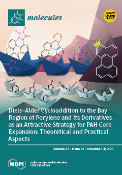
It is impossible to consider organic synthesis without cycloadditions. Due to this fact, ”Theory, Practice, and Applications” devoted to these reactions are still "on the top" for the chemical community. This review is devoted to modifications of polycyclic aromatics hydrocarbons (PAHs) realized via the Diels–Alder (DA) cycloaddition of various dienophiles to the bay regions of PAHs. This type of annulative π-extension (APEX) strategy has emerged as a powerful and efficient synthetic method for the construction of PAHs and their functionalized derivatives. This subject is discussed from the organic synthesis point of view but supported with theoretical calculations. The possible applications and the prospects for the development of DA cycloaddition to PAH bay regions are also discussed. View this paper.
  • Issues are regarded as officially published after their release is announced to the table of contents alert mailing list.
  • You may sign up for e-mail alerts to receive table of contents of newly released issues.
  • PDF is the official format for papers published in both, html and pdf forms. To view the papers in pdf format, click on the "PDF Full-text" link, and use the free Adobe Reader to open them.
Order results
Result details
Section
Select all
Export citation of selected articles as:
28 pages, 461 KiB  
Review
Comprehensive Review on Application of FTIR Spectroscopy Coupled with Chemometrics for Authentication Analysis of Fats and Oils in the Food Products
by Abdul Rohman, Mohd Al’Ikhsan B. Ghazali, Anjar Windarsih, Irnawati, Sugeng Riyanto, Farahwahida Mohd Yusof and Shuhaimi Mustafa
Molecules 2020, 25(22), 5485; https://doi.org/10.3390/molecules25225485 - 23 Nov 2020
Cited by 58 | Viewed by 8615
Abstract
Currently, the authentication analysis of edible fats and oils is an emerging issue not only by producers but also by food industries, regulators, and consumers. The adulteration of high quality and expensive edible fats and oils as well as food products containing fats [...] Read more.
Currently, the authentication analysis of edible fats and oils is an emerging issue not only by producers but also by food industries, regulators, and consumers. The adulteration of high quality and expensive edible fats and oils as well as food products containing fats and oils with lower ones are typically motivated by economic reasons. Some analytical methods have been used for authentication analysis of food products, but some of them are complex in sampling preparation and involving sophisticated instruments. Therefore, simple and reliable methods are proposed and developed for these authentication purposes. This review highlighted the comprehensive reports on the application of infrared spectroscopy combined with chemometrics for authentication of fats and oils. New findings of this review included (1) FTIR spectroscopy combined with chemometrics, which has been used to authenticate fats and oils; (2) due to as fingerprint analytical tools, FTIR spectra have emerged as the most reported analytical techniques applied for authentication analysis of fats and oils; (3) the use of chemometrics as analytical data treatment is a must to extract the information from FTIR spectra to be understandable data. Next, the combination of FTIR spectroscopy with chemometrics must be proposed, developed, and standardized for authentication and assuring the quality of fats and oils. Full article
(This article belongs to the Special Issue Chemometrics Tools Used in Analytical Chemistry)
Show Figures

Graphical abstract

Graphical abstract
Full article ">Figure 1
<p>The analytical procedure for analysis of meat-based food products using FTIR spectroscopy method.</p>
Full article ">
23 pages, 3784 KiB  
Article
Antiproliferative Homoleptic and Heteroleptic Phosphino Silver(I) Complexes: Effect of Ligand Combination on Their Biological Mechanism of Action
by Khouloud Dammak, Marina Porchia, Michele De Franco, Mirella Zancato, Houcine Naïli, Valentina Gandin and Cristina Marzano
Molecules 2020, 25(22), 5484; https://doi.org/10.3390/molecules25225484 - 23 Nov 2020
Cited by 18 | Viewed by 2964
Abstract
A series of neutral mixed-ligand [HB(pz)3]Ag(PR3) silver(I) complexes (PR3 = tertiary phosphine, [HB(pz)3] = tris(pyrazolyl)borate anion), and the corresponding homoleptic [Ag(PR3)4]BF4 compounds have been synthesized and fully characterized. Silver compounds [...] Read more.
A series of neutral mixed-ligand [HB(pz)3]Ag(PR3) silver(I) complexes (PR3 = tertiary phosphine, [HB(pz)3] = tris(pyrazolyl)borate anion), and the corresponding homoleptic [Ag(PR3)4]BF4 compounds have been synthesized and fully characterized. Silver compounds were screened for their antiproliferative activities against a wide panel of human cancer cells derived from solid tumors and endowed with different platinum drug sensitivity. Mixed-ligand complexes were generally more effective than the corresponding homoleptic derivatives, but the most active compounds were [HB(pz)3]Ag(PPh3) (5) and [Ag(PPh3)4]BF4 (10), both comprising the lipophilic PPh3 phosphine ligand. Detailed mechanistic studies revealed that both homoleptic and heteroleptic silver complexes strongly and selectively inhibit the selenoenzyme thioredoxin reductase both as isolated enzyme and in human ovarian cancer cells (half inhibition concentration values in the nanomolar range) causing the disruption of cellular thiol-redox homeostasis, and leading to apoptotic cell death. Moreover, for heteroleptic Ag(I) derivatives, an additional ability to damage nuclear DNA has been detected. These results confirm the importance of the type of silver ion coordinating ligands in affecting the biological behavior of the overall corresponding silver complexes, besides in terms of hydrophilic–lipophilic balance, also in terms of biological mechanism of action, such as interaction with DNA and/or thioredoxin reductase. Full article
(This article belongs to the Special Issue Metal-Based Drugs Ⅱ)
Show Figures

Figure 1

Figure 1
<p>Structure of the heteroleptic and homoleptic complexes studied in this work.</p>
Full article ">Figure 2
<p>Comet assay; 2008 cells were treated for 6 h with IC<sub>50</sub> values of tested compounds and then processed for comet assay. (<b>A</b>) Number of cells with well-formed comets measured by ImageJ software. The error bars indicate the S.D. * <span class="html-italic">p</span> &lt; 0.01; ** <span class="html-italic">p</span> &lt; 0.05(<b>B</b>) Representative images of 2008 cells.</p>
Full article ">Figure 3
<p>Effects of silver(I) complexes on human TrxR1. Thioredoxin reductase 1 activity was assayed by measuring NADPH-dependent reduction of DTNB at 412 nm as described in the Experimental Section. Error bars indicate S.D.</p>
Full article ">Figure 4
<p>Effects of compounds <b>1</b>–<b>10</b> and AgNO<sub>3</sub> on redox enzymes in human ovarian cancer cells. The 2008 cells were incubated for 18 h with tested compounds. Subsequently, cells were washed twice with Phosphate Buffer Solution (PBS) and lysed. TrxR activity was tested by measuring NADPH-dependent reduction of DTNB and glutathione peroxidase (GPx) activity was followed at 340 nm.</p>
Full article ">Figure 5
<p>Cellular total thiol state and ROS production. (<b>A</b>) Sulfhydryl content in 2008-treated cancer cells incubated for 24 h or 48 h with tested silver(I) compounds. The sulfhydryl group amount was determined by the DTNB assay. Error bars indicate S.D. (<b>B</b>) effect of silver(I) compounds on hydrogen peroxide formation in 2008 cells. 2008 cells were pre-incubated in PBS/10 mM glucose medium for 20 min at 37 °C in presence of 10 µM CM-DCFDA and then treated with IC<sub>50</sub> of silver(I) compounds.</p>
Full article ">Figure 6
<p>Mitochondrial membrane potential. Effects of silver(I) complexes on mitochondrial membrane potential. 2008 cells were treated for 24 h or 48 h with IC<sub>50</sub> concentrations of tested complexes or CCCP (3 μM). The percentage of cells with hypopolarized mitochondrial membrane potential was determined by Mito-ID<sup>®</sup> Membrane Potential Kit. Data are the means of three independent experiments. Error bars indicate S.D.</p>
Full article ">Figure 7
<p>TEM analysis. Transmission electron micrographs of 2008 cells after 48 h treatment with silver(I) complexes (<b>5</b>) and (<b>10</b>). (<b>a</b>) and (<b>b</b>): controls; (<b>c</b>,<b>d</b>): (<b>5</b>); (<b>e</b>) and (<b>f</b>): (<b>10</b>).</p>
Full article ">Figure 8
<p>Caspase-3 and -9 activation. Effect of silver(I) compounds on caspase-3 and -9 activation. 2008 cells were incubated for 24 or 48 h with IC<sub>50</sub> of compounds (<b>5</b>) or (<b>10</b>), broad-spectrum caspase inhibitor zVAD, and processed for caspase-3 and -9 activity. Data are the means of at least three independent experiments. Error bars indicate SD.</p>
Full article ">Scheme 1
<p>Structures of phosphine <sup>a</sup> and KHB(pz)<sub>3</sub> ligands used in this work. <sup>a</sup> PTA = 1,3,5-triaza-7-phosphaadamantane; DAPTA = 3,7-diacetyl-1,3,7-triaza-5-phosphabicyclo [3.3.1]nonane; PTA-SO<sub>2</sub> = 2-thia-1,3,5-triaza-phosphaadamantane 2,2-dioxide; PCN = tris(cyanoethyl)phosphine; PPh<sub>3</sub> = triphenylphosphine.</p>
Full article ">
14 pages, 911 KiB  
Review
Chagas Disease: Perspectives on the Past and Present and Challenges in Drug Discovery
by Felipe Raposo Passos Mansoldo, Fabrizio Carta, Andrea Angeli, Veronica da Silva Cardoso, Claudiu T. Supuran and Alane Beatriz Vermelho
Molecules 2020, 25(22), 5483; https://doi.org/10.3390/molecules25225483 - 23 Nov 2020
Cited by 33 | Viewed by 3646
Abstract
Chagas disease still has no effective treatment option for all of its phases despite being discovered more than 100 years ago. The development of commercial drugs has been stagnating since the 1960s, a fact that sheds light on the question of how drug [...] Read more.
Chagas disease still has no effective treatment option for all of its phases despite being discovered more than 100 years ago. The development of commercial drugs has been stagnating since the 1960s, a fact that sheds light on the question of how drug discovery research has progressed and taken advantage of technological advances. Could it be that technological advances have not yet been sufficient to resolve this issue or is there a lack of protocol, validation and standardization of the data generated by different research teams? This work presents an overview of commercial drugs and those that have been evaluated in studies and clinical trials so far. A brief review is made of recent target-based and phenotypic studies based on the search for molecules with anti-Trypanosoma cruzi action. It also discusses how proteochemometric (PCM) modeling and microcrystal electron diffraction (MicroED) can help in the case of the lack of a 3D protein structure; more specifically, Trypanosoma cruzi carbonic anhydrase. Full article
Show Figures

Figure 1

Figure 1
<p>Chemical structures of the two nitroheterocyclic compounds developed for Chagas disease. The chemical structure of Nifurtimox and Benznidazole were retrieved from Drug Bank (<a href="http://www.drugbank.com" target="_blank">http://www.drugbank.com</a>).</p>
Full article ">Figure 2
<p>Scheme proposed for coupling the workflow used by Llanos et al. [<a href="#B21-molecules-25-05483" class="html-bibr">21</a>] with the MicroED technique applied by Clabbers et al. [<a href="#B39-molecules-25-05483" class="html-bibr">39</a>]. For illustrative purposes, the human carbonic anhydrase (hCA) image was used; however, the idea of the scheme is this application for studying TcCA with the inhibitors selected by the workflow proposed by Llanos et al. [<a href="#B21-molecules-25-05483" class="html-bibr">21</a>]. The MicroED process illustration is from the work of Nannenga and Gonen [<a href="#B41-molecules-25-05483" class="html-bibr">41</a>].</p>
Full article ">
25 pages, 3373 KiB  
Article
Chemical Characterization and Antibacterial Activity of Essential Oil of Medicinal Plants from Eastern Serbia
by Milica Aćimović, Miroslav Zorić, Valtcho D. Zheljazkov, Lato Pezo, Ivana Čabarkapa, Jovana Stanković Jeremić and Mirjana Cvetković
Molecules 2020, 25(22), 5482; https://doi.org/10.3390/molecules25225482 - 23 Nov 2020
Cited by 21 | Viewed by 3508
Abstract
The objective of this study was to evaluate wild growing Satureja kitaibelii, Thymus serpyllum, Origanum vulgare, Achillea millefolium and Achillea clypeolata with respect to their essential oil (EO) content, composition and antimicrobial activity. The five species were collected at Mt. [...] Read more.
The objective of this study was to evaluate wild growing Satureja kitaibelii, Thymus serpyllum, Origanum vulgare, Achillea millefolium and Achillea clypeolata with respect to their essential oil (EO) content, composition and antimicrobial activity. The five species were collected at Mt. Rtanj and the village of Sesalac, Eastern Serbia. The main EO constituents of Lamiaceae plants were p-cymene (24.4%), geraniol (63.4%) and germacrene D (21.5%) in Satureja kitaibelii, Thymus serpyllum and Origanum vulgare ssp. vulgare, respectively. A. millefolium EO had multiple constituents with major ones being camphor (9.8%), caryophyllene oxide (6.5%), terpinen-4-ol (6.3%) and 1,8-cineole (5.6%), while the main EO constituents of A. clypeolata were 1,8-cineole (45.1%) and camphor (18.2%). Antimicrobial testing of the EO showed that Staphylococcus aureus (Gram-positive) was more sensitive to all of the tested EOs than Escherichia coli (Gram-negative). S. kitaibelii EO showed the highest antimicrobial activity against both tested bacterial strains. This is the first study to characterize the EO composition and antimicrobial activity of these five medicinal species from Eastern Serbia in comparison with comprehensive literature data. The results can be utilized by the perfumery, cosmetics, food and pharmaceutical industries, but also for healing purposes in self-medication. Full article
Show Figures

Figure 1

Figure 1
<p>(<b>a</b>) Dendogram of the EO constituents of <span class="html-italic">S. kitaibelii</span> from this study and from literature reports (the samples are marked according to <a href="#molecules-25-05482-t003" class="html-table">Table 3</a>); (<b>b</b>) correlation network based on <span class="html-italic">S. kitaibelii</span> EO constituents (P-CYM—<span class="html-italic">p</span>-cymene; CRV—Carvacrol; A-PIN—<span class="html-italic">α</span>-pinene; LIM—limonene; SAB-H—<span class="html-italic">cis</span>-sabinene hydrate; T-4-OL—terpinen-4-ol; LIN—linalool; BOR—borneol; GER—geraniol; B-BRB—<span class="html-italic">β</span>-bourbonene; B-CAR—<span class="html-italic">β</span>-caryophyllene; SPA—spathulenol; CAR-O—caryophyllene oxide).</p>
Full article ">Figure 2
<p>(<b>a</b>) Dendogram of the EO constituents of <span class="html-italic">T. serpyllum</span> from this study and from literature reports (the samples are marked according to <a href="#molecules-25-05482-t004" class="html-table">Table 4</a>); (<b>b</b>) correlation network based on <span class="html-italic">T. serpyllum</span> EO constituents (THY—thymol; CRV—carvacrol; MYR—myrcene; G-TER—<span class="html-italic">γ</span>-terpinene; NER—nerol; GER—geraniol; LIN—linalool; CIT—citronellol; A-TRP—<span class="html-italic">α</span>-terpinyl acetate; B-CAR—<span class="html-italic">β</span>-caryophyllene; GRM—germacrene; CAR-O—caryophyllene oxide; T-NER—<span class="html-italic">trans</span>-nerolidol).</p>
Full article ">Figure 2 Cont.
<p>(<b>a</b>) Dendogram of the EO constituents of <span class="html-italic">T. serpyllum</span> from this study and from literature reports (the samples are marked according to <a href="#molecules-25-05482-t004" class="html-table">Table 4</a>); (<b>b</b>) correlation network based on <span class="html-italic">T. serpyllum</span> EO constituents (THY—thymol; CRV—carvacrol; MYR—myrcene; G-TER—<span class="html-italic">γ</span>-terpinene; NER—nerol; GER—geraniol; LIN—linalool; CIT—citronellol; A-TRP—<span class="html-italic">α</span>-terpinyl acetate; B-CAR—<span class="html-italic">β</span>-caryophyllene; GRM—germacrene; CAR-O—caryophyllene oxide; T-NER—<span class="html-italic">trans</span>-nerolidol).</p>
Full article ">Figure 3
<p>(<b>a</b>) Dendogram of the EO constituents of <span class="html-italic">O. vulgare</span> from this study and from literature reports (the samples are marked according to <a href="#molecules-25-05482-t005" class="html-table">Table 5</a>); (<b>b</b>) correlation network based on <span class="html-italic">O. vulgare</span> EO constituents (P-CYM—<span class="html-italic">p</span>-cymene; THY—thymol; CRV—carvacrol; SAB—sabinene; G-TER—<span class="html-italic">γ</span>-terpinene; 1,8-CIN—1,8-cineole; LIN—linalool; A-TER—<span class="html-italic">α</span>-terpineol; B-CIT—<span class="html-italic">β</span>-citronellol; LIN-A—linalyl acetate; B-CAR—<span class="html-italic">β</span>-caryophyllene; GER-D—germacrene D; CAR-O—caryophyllene oxide).</p>
Full article ">Figure 3 Cont.
<p>(<b>a</b>) Dendogram of the EO constituents of <span class="html-italic">O. vulgare</span> from this study and from literature reports (the samples are marked according to <a href="#molecules-25-05482-t005" class="html-table">Table 5</a>); (<b>b</b>) correlation network based on <span class="html-italic">O. vulgare</span> EO constituents (P-CYM—<span class="html-italic">p</span>-cymene; THY—thymol; CRV—carvacrol; SAB—sabinene; G-TER—<span class="html-italic">γ</span>-terpinene; 1,8-CIN—1,8-cineole; LIN—linalool; A-TER—<span class="html-italic">α</span>-terpineol; B-CIT—<span class="html-italic">β</span>-citronellol; LIN-A—linalyl acetate; B-CAR—<span class="html-italic">β</span>-caryophyllene; GER-D—germacrene D; CAR-O—caryophyllene oxide).</p>
Full article ">Figure 4
<p>(<b>a</b>) Dendogram of the <span class="html-italic">A. millefolium</span> EO constituents from this study and from literature reports (the samples are marked according to <a href="#molecules-25-05482-t006" class="html-table">Table 6</a>); (<b>b</b>) correlation network based on <span class="html-italic">A. millefolium</span> EO constituents (SAB—sabinene; B-PIN—<span class="html-italic">β</span>-pinene; CMP—camphor; 1,8-CIN—1,8-cineole; BOR—borneole; T-4-OL—terpinene-4-ol; BOR-A—bornyl acetate; CHR-A—chrysanthenyl acetate; LAV-A—lavandulyl acetate; CHA—chamazulene; B-CAR—<span class="html-italic">β</span>-caryophyllene; D-CAD—δ-cadinene; ELE—elemol.</p>
Full article ">Figure 5
<p>(<b>a</b>) Dendogram of the EO constituents of <span class="html-italic">A. clypeolata</span> from this study and from literature reports (the samples are marked according to <a href="#molecules-25-05482-t007" class="html-table">Table 7</a>); (<b>b</b>) correlation network based on <span class="html-italic">A. clypeolata</span> EO constituents (CAM—camphene; SAB—sabinene; B-PIN—<span class="html-italic">β</span>-pinene; P-CYM—p-cymene; 1,8-CIN—1,8-cineole; CMP—camphor; BOR—borneol; T-4-OL—terpinene-4-ol; A-TER—<span class="html-italic">α</span>-terpineol; A-ARO—allo-aromadendrene; CAR-O—caryophyllene oxide; E-A-CAD—epi-<span class="html-italic">α</span>-cadinol; A-CAD—<span class="html-italic">α</span>-cadinol.</p>
Full article ">
12 pages, 1455 KiB  
Article
Src Mediates Epigallocatechin-3-O-Gallate-Elicited Acid Sphingomyelinase Activation
by Motofumi Kumazoe, Mai Kadomatsu, Jaehoon Bae, Yushi Otsuka, Yoshinori Fujimura and Hirofumi Tachibana
Molecules 2020, 25(22), 5481; https://doi.org/10.3390/molecules25225481 - 23 Nov 2020
Cited by 7 | Viewed by 2515
Abstract
Epigallocatechin-3-O-gallate (EGCG) is one of the major bioactive compounds known to be present in green tea. We previously reported that EGCG shows selective toxicity through activation of the protein kinase B (Akt)/cyclic guanosine monophosphate (cGMP)/acid sphingomyelinase (ASM) axis via targeting its [...] Read more.
Epigallocatechin-3-O-gallate (EGCG) is one of the major bioactive compounds known to be present in green tea. We previously reported that EGCG shows selective toxicity through activation of the protein kinase B (Akt)/cyclic guanosine monophosphate (cGMP)/acid sphingomyelinase (ASM) axis via targeting its receptor 67-kDa laminin receptor (67LR), which is overexpressed in cancer. However, little is known about upstream mechanisms of EGCG-elicited ASM activation. In this study we show that the proto-oncogene tyrosine-protein kinase Src, also known as c-src, plays a crucial role in the anticancer effect of EGCG. We showed that EGCG elicits phosphorylation of Src at Tyr 416, a crucial phosphorylation site for its activity, and that the pharmacological inhibition of Src impedes the upstream events in EGCG-induced cell death signaling including upregulation of Akt activity, increase in cGMP levels, and activation of ASM. Moreover, focal adhesion kinase (FAK), which is involved in the phosphorylation of Src, is colocalized with 67LR. EGCG treatment enhanced interaction of FAK and 67LR. Consistent with these findings, pharmacological inhibition of FAK significantly neutralized EGCG-induced upregulation of Akt activity and activation of ASM. Taken together, FAK/Src play crucial roles in the upstream signaling of EGCG. Full article
(This article belongs to the Special Issue Molecular Pharmacology of Green Tea)
Show Figures

Figure 1

Figure 1
<p>Pharmacological inhibition of Src attenuated the cell death inducing effect of EGCG. (<b>A</b>) U266 human multiple myeloma cells were treated with EGCG (10 μM) and SKI1 (2.5 μM, from 1 h before EGCG treatment) for 72 h. Cell viability was measured using the trypan blue method (<span class="html-italic">n</span> = 4). (<b>B</b>) U266 human multiple myeloma cells were treated with EGCG (10 μM) and SKI1 (2.5 μM; from 1 h before EGCG treatment) for 1 h. Akt activity was evaluated using the K-LISA kit (<span class="html-italic">n</span> = 4). (<b>C</b>) U266 human multiple myeloma cells were treated with EGCG (10 μM) and SKI1 (2.5 μM; from 1 h before EGCG treatment) for 3 h. cGMP levels were evaluated using competitive immunoassay (<span class="html-italic">n</span> = 4). (<b>D</b>) U266 human multiple myeloma cells were treated with EGCG (10 μM) and SKI1 (2.5 μM; from 1 h before EGCG treatment) for 3 h and ASM activity was evaluated (<span class="html-italic">n</span> = 3).</p>
Full article ">Figure 2
<p>EGCG induced phosphorylation of Tyr 416 in multiple myeloma cells. (<b>A</b>) U266 human multiple myeloma cells were treated with EGCG (10 μM) for 30 min. Phosphorylation levels of Src were evaluated using Western blotting (<span class="html-italic">n</span> = 3). (<b>B</b>) U266 human multiple myeloma cells were pretreated with the anti-67LR antibody or with the isotype control antibody and treated with EGCG (10 μM) for 30 min. Src phosphorylation levels were evaluated using Western blotting (<span class="html-italic">n</span> = 4).</p>
Full article ">Figure 3
<p>Pharmacological inhibition of FAK attenuated the Akt/ASM axis elicited by EGCG. (<b>A</b>) U266 human multiple myeloma cells were treated with EGCG (10 μM) and PF-573228 (1 μM; from 1 h before EGCG treatment) for 3 h (<span class="html-italic">n</span> = 3). (<b>B</b>) U266 human multiple myeloma cells were treated with EGCG (10 μM) and PF-573228 (1 μM; from 1 h before EGCG treatment). Akt activity was evaluated using the K-LISA kit (<span class="html-italic">n</span> = 3). (<b>C</b>) U266 human multiple myeloma cells were treated with EGCG (10 μM) and PF-573228 (1 μM; 1 h before EGCG treatment) for 30 min. Src phosphorylation levels were evaluated using western blotting (<span class="html-italic">n</span> = 4). Data are presented as means ± SEM.</p>
Full article ">Figure 4
<p>FAK is the adaptor protein of 67LR. (<b>A</b>) Immunofluorescence staining for FAK (Red) and 67LR (Green) and Z stack images were determined in HeLa cells using Fluorescence microscopy (×40). (<b>B</b>) His-tagged 67LR was immunoprecipitated using the anti-FAK antibody after 30 min EGCG (10 μM) treatment. (<b>C</b>) The schematic diagram to summarize the mechanism of EGCG in human multiple myeloma cells. XYZ axis of Z focus image was described above.</p>
Full article ">
16 pages, 5792 KiB  
Article
Anti-Quorum Sensing Activity of Stevia Extract, Stevioside, Rebaudioside A and Their Aglycon Steviol
by Victor Markus, Orr Share, Kerem Teralı, Nazmi Ozer, Robert S. Marks, Ariel Kushmaro and Karina Golberg
Molecules 2020, 25(22), 5480; https://doi.org/10.3390/molecules25225480 - 23 Nov 2020
Cited by 11 | Viewed by 25433
Abstract
Governments are creating regulations for consumers to reduce their sugar intake, prompting companies to increase the ratio of artificial sweeteners in their products. However, there is evidence of some deleterious effects ascribed to the aforementioned synthetic agents and therefore consumers and food manufacturers [...] Read more.
Governments are creating regulations for consumers to reduce their sugar intake, prompting companies to increase the ratio of artificial sweeteners in their products. However, there is evidence of some deleterious effects ascribed to the aforementioned synthetic agents and therefore consumers and food manufacturers have turned their attention to natural dietary sweeteners, such as stevia, to meet their sweetening needs. Stevia is generally considered safe; however, emerging scientific evidence has implicated the agent in gut microbial imbalance. In general, regulation of microbial behavior is known to depend highly on signaling molecules via quorum sensing (QS) pathways. This is also true for the gut microbial community. We, therefore, evaluated the possible role of these stevia-based natural sweeteners on this bacterial communication pathway. The use of a commercial stevia herbal supplement resulted in an inhibitory effect on bacterial communication, with no observable bactericidal effect. Purified stevia extracts, including stevioside, rebaudioside A (Reb A), and steviol revealed a molecular interaction, and possible interruption of Gram-negative bacterial communication, via either the LasR or RhlR receptor. Our in-silico analyses suggest a competitive-type inhibitory role for steviol, while Reb A and stevioside are likely to inhibit LasR-mediated QS in a non-competitive manner. These results suggest the need for further safety studies on the agents. Full article
Show Figures

Figure 1

Figure 1
<p>Anti-QS activity of CSHS. (<b>A</b>) Inhibitory effect of CSHS on the bioluminescence emission of K802NR reporter strain; (<b>B</b>) inhibitory effect of CSHS on K802NR bioluminescence emission relative to the control, consistent with the data in panel A; (<b>C</b>) growth response of K802NR reporter strain in the presence of CSHS; (<b>D</b>) growth response of K802NR in the presence of CSHS relative to the control, consistent with the data in panel C. Error bars are not shown in A and C to ensure clarity of figures. The final concentration of 3-oxo-C<sub>12</sub>-HSL was 5 × 10<sup>−10</sup> M. All presented concentrations are the final concentrations. The amount of luminescence is expressed in relative light units or RLUs. * <span class="html-italic">p</span> &lt; 0.05, ** <span class="html-italic">p</span> &lt; 0.01, **** <span class="html-italic">p</span> &lt; 0.0001, and ns not significant. Values represent mean ± SD, <span class="html-italic">n</span> = 3 (three different experimental readings).</p>
Full article ">Figure 2
<p>Experimental and theoretical mass spectrum of CSHS using ion trap MS in positive ion mode. (<b>A</b>) Stevioside; (<b>B</b>) Reb A; (<b>C</b>) steviol. On the right are the structures of the glycosides, along with their molecular formulas.</p>
Full article ">Figure 3
<p>The bioluminescent response of K802NR reporter strain in the presence of pure stevia-derived components. (<b>A</b>) Stevia extract; (<b>B</b>) stevia extract relative to the control, corresponding to the data in panel A; (<b>C</b>) stevioside; (<b>D</b>) stevioside relative to the control, corresponding to the data in panel C; (<b>E</b>) Reb A; (<b>F</b>) Reb A relative to the control, corresponding to the data in panel E (<b>G</b>); steviol; (<b>H</b>) steviol relative to control, corresponding to the data in panel G. Error bars are not shown in A, C, E, and G to ensure clarity of figures. The final concentration of 3-oxo-C<sub>12</sub>-HSL was 5 × 10<sup>−10</sup> M. All presented concentrations are the final concentrations. The amount of luminescence is expressed in relative light units or RLUs. * <span class="html-italic">p</span> &lt; 0.05, ** <span class="html-italic">p</span> &lt; 0.01, *** <span class="html-italic">p</span> &lt; 0.001, and ns not significant. Values represent mean ± SD, <span class="html-italic">n</span> = 3 (three different experimental readings).</p>
Full article ">Figure 3 Cont.
<p>The bioluminescent response of K802NR reporter strain in the presence of pure stevia-derived components. (<b>A</b>) Stevia extract; (<b>B</b>) stevia extract relative to the control, corresponding to the data in panel A; (<b>C</b>) stevioside; (<b>D</b>) stevioside relative to the control, corresponding to the data in panel C; (<b>E</b>) Reb A; (<b>F</b>) Reb A relative to the control, corresponding to the data in panel E (<b>G</b>); steviol; (<b>H</b>) steviol relative to control, corresponding to the data in panel G. Error bars are not shown in A, C, E, and G to ensure clarity of figures. The final concentration of 3-oxo-C<sub>12</sub>-HSL was 5 × 10<sup>−10</sup> M. All presented concentrations are the final concentrations. The amount of luminescence is expressed in relative light units or RLUs. * <span class="html-italic">p</span> &lt; 0.05, ** <span class="html-italic">p</span> &lt; 0.01, *** <span class="html-italic">p</span> &lt; 0.001, and ns not significant. Values represent mean ± SD, <span class="html-italic">n</span> = 3 (three different experimental readings).</p>
Full article ">Figure 4
<p>Inhibition of bioluminescence emission of PAO-JP2 (pKD-<span class="html-italic">rhlA</span>) by pure stevia-derived components. (<b>A</b>) stevia extract; (<b>B</b>) stevia extract relative to the control, correlating to the data in panel A; (<b>C</b>) stevioside, (<b>D</b>) stevioside relative to the control, correlating to the data in panel C; (<b>E</b>) Reb A, (<b>F</b>) Reb A relative to the control, correlating to the data in panel E; (<b>G</b>) steviol; (<b>H</b>) steviol relative to control, correlating to the data in panel G. Error bars are not shown in A, C, E, and G to ensure clarity of figures. The final concentration of C4-HSL was 10 µM. All presented concentrations are the final concentrations. The amount of luminescence is expressed in relative light units or RLUs. * <span class="html-italic">p</span> &lt; 0.05, ** <span class="html-italic">p</span> &lt; 0.01, *** <span class="html-italic">p</span> &lt; 0.001, and ns not significant. Values represent mean ± SD, <span class="html-italic">n</span> = 3 (three different experimental readings).</p>
Full article ">Figure 4 Cont.
<p>Inhibition of bioluminescence emission of PAO-JP2 (pKD-<span class="html-italic">rhlA</span>) by pure stevia-derived components. (<b>A</b>) stevia extract; (<b>B</b>) stevia extract relative to the control, correlating to the data in panel A; (<b>C</b>) stevioside, (<b>D</b>) stevioside relative to the control, correlating to the data in panel C; (<b>E</b>) Reb A, (<b>F</b>) Reb A relative to the control, correlating to the data in panel E; (<b>G</b>) steviol; (<b>H</b>) steviol relative to control, correlating to the data in panel G. Error bars are not shown in A, C, E, and G to ensure clarity of figures. The final concentration of C4-HSL was 10 µM. All presented concentrations are the final concentrations. The amount of luminescence is expressed in relative light units or RLUs. * <span class="html-italic">p</span> &lt; 0.05, ** <span class="html-italic">p</span> &lt; 0.01, *** <span class="html-italic">p</span> &lt; 0.001, and ns not significant. Values represent mean ± SD, <span class="html-italic">n</span> = 3 (three different experimental readings).</p>
Full article ">Figure 5
<p>Molecular structures of the bona fide LasR ligands 3-oxo-C12-HSL (native autoinducer) and compound <b>19</b> (non-native agonist).</p>
Full article ">Figure 6
<p>Results of the redocking calculations. <b>Left panel</b>: close-up view of the superposed structures of native (gold) and predicted (silver) 3-oxo-C<sub>12</sub>-HSL in the ligand-binding cavity of LasR in the closed-loop conformation. <b>Right panel</b>: close-up view of the superposed structures of native (gold) and predicted (silver) compound <b>19</b> in the ligand-binding cavity of LasR in the open-loop conformation. Images were prepared and rendered using the PyMOL Molecular Graphics System, version 1.8 (Schrödinger LLC, Portland, OR, USA).</p>
Full article ">Figure 7
<p>Favorable interactions between the LasR-LBD and steviol. <b>Left panel:</b> relative position of steviol with respect to the ligand-binding cavity of LasR in the closed-loop conformation. <b>Right panel:</b> relative position of steviol with respect to the ligand-binding cavity of LasR in the open-loop conformation. Images were prepared and rendered using Discovery Studio Visualizer, version 16.1.0 (Dassault Systèmes BIOVIA Corp., San Diego, CA, USA).</p>
Full article ">Figure 8
<p>Representative (i.e., top-ranking) binding poses of the steviol glycosides from each cluster around the LasR LBD in the open-loop conformation. <b>Left panel:</b> Reb A. <b>Right panel:</b> stevioside. Images were prepared using the PyMOL Molecular Graphics System, version 1.8 (Schrödinger LLC, Portland, OR, USA).</p>
Full article ">
16 pages, 5113 KiB  
Article
Water-Soluble O-, S- and Se-Functionalized Cyclic Acetyl-triaza-phosphines. Synthesis, Characterization and Application in Catalytic Azide-alkyne Cycloaddition
by Abdallah G. Mahmoud, Piotr Smoleński, M. Fátima C. Guedes da Silva and Armando J. L. Pombeiro
Molecules 2020, 25(22), 5479; https://doi.org/10.3390/molecules25225479 - 23 Nov 2020
Cited by 11 | Viewed by 2839
Abstract
The 3,7-diacetyl-1,3,7-triaza-5-phosphabicyclo[3.3.1]nonane (DAPTA) derivatives, viz. the already reported 3,7-diacetyl-1,3,7-triaza-5-phosphabicyclo[3.3.1]nonane 5-oxide (DAPTA=O, 1), the novel 3,7-diacetyl-1,3,7-triaza-5-phosphabicyclo[3.3.1]nonane-5-sulfide (DAPTA=S, 2), and 3,7-diacetyl-1,3,7-triaza-5-phosphabicyclo[3.3.1]nonane-5-selenide (DAPTA=Se, 3), have been synthesized under mild conditions. They are soluble in water and most common organic solvents and have been [...] Read more.
The 3,7-diacetyl-1,3,7-triaza-5-phosphabicyclo[3.3.1]nonane (DAPTA) derivatives, viz. the already reported 3,7-diacetyl-1,3,7-triaza-5-phosphabicyclo[3.3.1]nonane 5-oxide (DAPTA=O, 1), the novel 3,7-diacetyl-1,3,7-triaza-5-phosphabicyclo[3.3.1]nonane-5-sulfide (DAPTA=S, 2), and 3,7-diacetyl-1,3,7-triaza-5-phosphabicyclo[3.3.1]nonane-5-selenide (DAPTA=Se, 3), have been synthesized under mild conditions. They are soluble in water and most common organic solvents and have been characterized using 1H and 31P NMR spectroscopy and, for 2 and 3, also by single crystal X-ray diffraction. The effect of O, S, or Se at the phosphorus atom on the structural features of the compounds has been investigated, also through the analyses of Hirshfeld surfaces. The presence of 13 enhances the activity of copper for the catalytic azide-alkyne cycloaddition reaction in an aqueous medium. The combination of cheaply available copper (II) acetate and compound 1 has been used as a catalyst for the one-pot and 1,4-regioselective procedure to obtain 1,2,3-triazoles with high yields and according to ‘click rules’. Full article
(This article belongs to the Special Issue Metal-Induced Molecule Activation and Coupling Reactions)
Show Figures

Graphical abstract

Graphical abstract
Full article ">Figure 1
<p>3,7-diacetyl-1,3,7-triaza-5-phosphabicyclo[3.3.1]nonane (DAPTA).</p>
Full article ">Figure 2
<p>Isomeric forms of compounds <b>1</b>–<b>3</b>.</p>
Full article ">Figure 3
<p><sup>31</sup>P{<sup>1</sup>H} NMR spectrum of DAPTA=Se (<b>3</b>) in DMSO-<span class="html-italic">d</span><sub>6</sub> (500 MHz).</p>
Full article ">Figure 4
<p><sup>1</sup>H NMR spectra of DAPTA and compounds <b>1</b>–<b>3</b> in DMSO-<span class="html-italic">d</span><sub>6</sub>.</p>
Full article ">Figure 5
<p>ORTEP diagrams of compounds <b>2</b> (left) and <b>3</b> (right) with displacement ellipsoids shown at 50% probability level and atom numbering schemes. H-atoms are omitted for clarity.</p>
Full article ">Figure 6
<p>2D fingerprint plots (<span class="html-italic">d<sub>i</sub></span> and <span class="html-italic">d<sub>e</sub></span> values in Å) for DAPTA and P-functionalized derivatives. The offset plots refer to the indicated specific interactions.</p>
Full article ">Figure 7
<p>Plot of the percentage contribution of the diverse contacts to the total volume of the surface.</p>
Full article ">Figure 8
<p>The structure of complex [Cu(μ-CH<sub>3</sub>COO)<sub>2</sub>(κ<span class="html-italic">O-</span>DAPTA=O)]<sub>2</sub> (<b>4</b>).</p>
Full article ">Scheme 1
<p>Synthesis of DAPTA=O by acylation of PTA=O.</p>
Full article ">Scheme 2
<p>Synthesis of compounds <b>1</b>–<b>3</b>.</p>
Full article ">Scheme 3
<p>Schiff base N=C bond formation through the π electronic resonance of the amide group.</p>
Full article ">Scheme 4
<p>Proposed catalytic cycle for the CuAAC reaction.</p>
Full article ">
14 pages, 4100 KiB  
Article
Natural Approaches for Neurological Disorders—The Neuroprotective Potential of Codium tomentosum
by Joana Silva, Alice Martins, Celso Alves, Susete Pinteus, Helena Gaspar, Amparo Alfonso and Rui Pedrosa
Molecules 2020, 25(22), 5478; https://doi.org/10.3390/molecules25225478 - 23 Nov 2020
Cited by 11 | Viewed by 3481
Abstract
Parkinson’s disease (PD) is the second most common neurodegenerative disorder, and is characterized by a progressive degeneration of the dopaminergic neurons in the substantianigra. Although not completely understood, several abnormal cellular events are known to be related with PD progression, such [...] Read more.
Parkinson’s disease (PD) is the second most common neurodegenerative disorder, and is characterized by a progressive degeneration of the dopaminergic neurons in the substantianigra. Although not completely understood, several abnormal cellular events are known to be related with PD progression, such as oxidative stress, mitochondrial dysfunction and apoptosis. Accordingly, the aim of this study was to evaluate the neuroprotective effects of Codium tomentosum enriched fractions in a neurotoxicity model mediated by 6-hydroxydopamine (6-OHDA) on SH-SY5Y human cells, and the disclosure of their mechanisms of action. Additionally, a preliminary chemical screening of the most promising bioactive fractions of C. tomentosum was carried out by GC-MS analysis. Among the tested fractions, four samples exhibited the capacity to revert the neurotoxicity induced by 6-OHDA to values higher or similar to the vitamin E (90.11 ± 3.74% of viable cells). The neuroprotective effects were mediated by the mitigation of reactive oxygen species (ROS) generation, mitochondrial dysfunctions and DNA damage, together with the reduction of Caspase-3 activity. Compounds belonging to different chemical classes, such as terpenes, alcohols, carboxylic acids, aldehydes, esters, ketones, saturated and unsaturated hydrocarbons were tentatively identified by GC-MS. The results show that C. tomentosum is a relevant source of neuroprotective agents, with particular interest for preventive therapeutics. Full article
Show Figures

Graphical abstract

Graphical abstract
Full article ">Figure 1
<p>Extraction and fractionation flowchart of the green seaweed <span class="html-italic">Codium tomentosum</span>.</p>
Full article ">Figure 2
<p>(<b>A</b>) Neurotoxicity of <span class="html-italic">Codium tomentosum</span> fractions (100 µg/mL, 24 h) and (<b>B</b>) neuroprotective effects of non-neurotoxic fractions (100 µg/mL, 24 h) in the presence of 6-OHDA (100 µM) on SH-SY5Y cells. (-) 6-OHDA; Vit E (Vitamin E). The values in each column represent the mean ± standard error of the mean (SEM) of three or four independent experiments. Symbols represent significant differences (ANOVA, Dunnett’s test, <span class="html-italic">p</span> &lt; 0.05) when compared to: * vehicle and # 6-OHDA.</p>
Full article ">Figure 3
<p>PD hallmarks evaluation on SH-SY5Y cells when exposed to 6-OHDA (100 µM) and <span class="html-italic">Codium tomentosum</span> fractions (100 µg/mL; 6 h). (<b>A</b>) ROS production; (<b>B</b>) changes in mitochondrial membrane potential; (<b>C</b>) Caspase-3 activity. (-) 6-OHDA. The values in each column represent the mean ± standard error of the mean (SEM) of three or four independent experiments. Symbols represent significant differences (ANOVA, Dunnett’s test, <span class="html-italic">p</span> &lt; 0.05) when compared to: * vehicle and # 6-OHDA.</p>
Full article ">
16 pages, 1621 KiB  
Communication
Cyst Reduction by Melatonin in a Novel Drosophila Model of Polycystic Kidney Disease
by Cassandra Millet-Boureima, Roman Rozencwaig, Felix Polyak and Chiara Gamberi
Molecules 2020, 25(22), 5477; https://doi.org/10.3390/molecules25225477 - 23 Nov 2020
Cited by 8 | Viewed by 8281
Abstract
Autosomal dominant polycystic kidney disease (ADPKD) causes progressive cystic degeneration of the renal tubules, the nephrons, eventually severely compromising kidney function. ADPKD is incurable, with half of the patients eventually needing renal replacement. Treatments for ADPKD patients are limited and new effective therapeutics [...] Read more.
Autosomal dominant polycystic kidney disease (ADPKD) causes progressive cystic degeneration of the renal tubules, the nephrons, eventually severely compromising kidney function. ADPKD is incurable, with half of the patients eventually needing renal replacement. Treatments for ADPKD patients are limited and new effective therapeutics are needed. Melatonin, a central metabolic regulator conserved across all life kingdoms, exhibits oncostatic and oncoprotective activity and no detected toxicity. Here, we used the Bicaudal C (BicC) Drosophila model of polycystic kidney disease to test the cyst-reducing potential of melatonin. Significant cyst reduction was found in the renal (Malpighian) tubules upon melatonin administration and suggest mechanistic sophistication. Similar to vertebrate PKD, the BicC fly PKD model responds to the antiproliferative drugs rapamycin and mimics of the second mitochondria-derived activator of caspases (Smac). Melatonin appears to be a new cyst-reducing molecule with attractive properties as a potential candidate for PKD treatment. Full article
Show Figures

Graphical abstract

Graphical abstract
Full article ">Figure 1
<p>Protocol for testing the melatonin anti-cystic activity. <span class="html-italic">BicC<sup>Δ/YC33</sup></span> and <span class="html-italic">BicC<sup>Δ/IIF34</sup></span> flies (aged 0–2d) were placed in food-containing vials mixed with either vehicle (ethanol) or 150 µM melatonin at nighttime. Malpighian tubules were micro-dissected after 18 days of treatment and cysts scored (population aged 18–20d).</p>
Full article ">Figure 2
<p>Melatonin reduced cysts in the renal tubule of <span class="html-italic">BicC<sup>Δ/YC33</sup></span> flies. (<b>a</b>) Representative Malpighian tubules micro-dissected from <span class="html-italic">BicC<sup>Δ/YC33</sup></span> flies treated with either vehicle (ethanol) or 150 µM melatonin were photographed ex vivo. Arrows indicate exemplary cysts. (<b>b</b>) Nested plots reporting overall number of cysts found in each anterior and posterior tubule pair of 50 vehicle- and 50 melatonin-treated cystic flies, with mean and standard deviation. (<b>c</b>) Regional analyses. Nested plots indicating the number of cysts found in the proximal, intermediate, and terminal region of the anterior and posterior tubule pairs of the flies in b, with mean and standard deviation. <span class="html-italic">p</span> values (with Welch’s correction) are indicated. Treatments are shown with color: vehicle, left, grey; melatonin, right, green. Melatonin treatment significantly reduced cysts in all regions of the Malpighian tubules of <span class="html-italic">BicC<sup>Δ/YC33</sup></span> flies (<span class="html-italic">p</span> &lt; 0.05), except for the proximal region in the posterior tubules.</p>
Full article ">Figure 3
<p>Melatonin did not reduce cysts in <span class="html-italic">BicC<sup>Δ/IIF34</sup></span> flies. (<b>a</b>) Representative Malpighian tubules micro-dissected from <span class="html-italic">BicC<sup>Δ/IIF34</sup></span> flies treated with either vehicle (ethanol) or 150 µM melatonin were photographed ex vivo. Arrows indicate exemplary cysts. (<b>b</b>) Nested plots reporting overall number of cysts found in each anterior and posterior tubule pair of 50 vehicle- and 50 melatonin-treated cystic flies, with mean and standard deviation. (<b>c</b>) Regional analyses. Nested plots indicating the number of cysts found in the proximal, intermediate, and terminal region of the anterior and posterior tubule pairs of the flies in b, with mean and standard deviation. <span class="html-italic">p</span> values (with Welch’s correction) are indicated. Treatments are color-coded: vehicle, left, grey; melatonin, right, green. Melatonin treatment of <span class="html-italic">BicC<sup>Δ/IIF34</sup></span> flies produced a trend in cyst reduction, however, did not significantly differentiate the treated vs. untreated populations.</p>
Full article ">
15 pages, 2828 KiB  
Article
Cytotoxic and Antiproliferative Effects of Diarylheptanoids Isolated from Curcuma comosa Rhizomes on Leukaemic Cells
by Natsima Viriyaadhammaa, Aroonchai Saiai, Waranya Neimkhum, Wariya Nirachonkul, Wantida Chaiyana, Sawitree Chiampanichayakul, Singkome Tima, Toyonobu Usuki, Suwit Duangmano and Songyot Anuchapreeda
Molecules 2020, 25(22), 5476; https://doi.org/10.3390/molecules25225476 - 23 Nov 2020
Cited by 10 | Viewed by 3036
Abstract
Curcuma comosa belongs to the Zingiberaceae family. In this study, two natural compounds were isolated from C. comosa, and their structures were determined using nuclear magnetic resonance. The isolated compounds were identified as 7-(3,4-dihydroxyphenyl)-5-hydroxy-1-phenyl-(1E)-1-heptene (1) and trans-1,7-diphenyl-5-hydroxy-1-heptene [...] Read more.
Curcuma comosa belongs to the Zingiberaceae family. In this study, two natural compounds were isolated from C. comosa, and their structures were determined using nuclear magnetic resonance. The isolated compounds were identified as 7-(3,4-dihydroxyphenyl)-5-hydroxy-1-phenyl-(1E)-1-heptene (1) and trans-1,7-diphenyl-5-hydroxy-1-heptene (2). Compound 1 showed the strongest cytotoxicity effect against HL-60 cells, while its antioxidant and anti-inflammatory properties were stronger than those of compound 2. Compound 1 proved to be a potent antioxidant, compared to ascorbic acid. Neither compounds had any effect on red blood cell haemolysis. Furthermore, compound 1 significantly decreased Wilms’ tumour 1 protein expression and cell proliferation in KG-1a cells. Compound 1 decreased the WT1 protein levels in a time- and dose- dependent manner. Compound 1 suppressed cell cycle at the S phase. In conclusion, compound 1 has a promising chemotherapeutic potential against leukaemia. Full article
(This article belongs to the Special Issue Cytotoxic Activity of Plant Extracts)
Show Figures

Graphical abstract

Graphical abstract
Full article ">Figure 1
<p>The chemical structure of compound <b>1</b> (7-(3,4-dihydroxyphenyl)-5-hydroxy-1-phenyl-(1E)-1-heptene).</p>
Full article ">Figure 2
<p>The chemical structure of compound <b>2</b> (<span class="html-italic">trans</span>-1,7-diphenyl-5-hydroxy-1-heptene).</p>
Full article ">Figure 3
<p>Cytotoxicity of crude fractional extracts and pure compounds from <span class="html-italic">C. comosa</span> to cancer cell lines. K562, KG-1a, HL-60, A549, and MCF-7 cells were treated with (<b>A</b>) ethyl acetate fraction (F-EtOAc), (<b>B</b>) compound <b>1</b>, (<b>C</b>) hexane fraction (F-Hex), or (<b>D</b>) compound <b>2</b> for 48 h. Cell viability was determined using the MTT assay. Each point represents the mean ± standard deviation (SD) of three independent experiments, each performed in triplicate.</p>
Full article ">Figure 4
<p>Cytotoxicity of crude fractional extracts and pure compounds from <span class="html-italic">C. comosa</span> to peripheral blood mononuclear cells (PBMCs). PBMCs were treated with ethyl acetate fraction (F-EtOAc), compound <b>1</b>, hexane fraction (F-Hex), and compound <b>2</b> for 48 h. The MTT assay determined the cell viability. Each point represents the mean ± SE of five independent experiments performed in triplicate.</p>
Full article ">Figure 5
<p>The effect of compound <b>1</b> and compound <b>2</b> on the viability of RAW 264.7 cell line using MTT assay. Each point represents the mean ± SD of three independent experiments performed in triplicate.</p>
Full article ">Figure 6
<p>Dose response curve of dexamethasone, compound <b>1</b>, and compound <b>2</b> on (<b>A</b>) IL-6 and (<b>B</b>) TNF-α. Each curve represents the mean ± SD of three independent experiments performed in triplicate.</p>
Full article ">Figure 7
<p>RBC hemolysis following incubation with F-EtOAc, compound <b>1</b>, F-Hex, and compound <b>2</b>. Each bar represents the mean ± SD of three independent experiments performed in triplicate. Asterisks (*) denote significant differences between C. comosa extracts and positive control (*** <span class="html-italic">p</span> &lt; 0.001).</p>
Full article ">Figure 8
<p>Effect of F-EtOAc, compound <b>1</b>, F-Hex, and compound <b>2</b> on KG-1a cells. (<b>A</b>) The level of WT1 protein following treatment F-EtOAc (4.58 μg/mL), compound <b>1</b> (comp. <b>1</b>; 2.30 μg/mL), F-Hex (26.29 μg/mL), or compound <b>2</b> (comp. <b>2</b>; 29.90 μg/mL) for 48 h. Protein levels were evaluated using Western blotting and analysed using scan densitometer. The levels of WT1 were normalised using glyceraldehyde phosphate dehydrogenase (GAPDH) protein levels. (<b>B</b>) Total cell number after treatment with F-EtOAc, compound <b>1</b>, F-Hex, and compound <b>2</b> for 48 h was determined via the trypan blue exclusion method. Each bar represented mean ± SD of three independent experiments performed in triplicate. Asterisks (*) denote significant differences between <span class="html-italic">C. comosa</span> extracts and vehicle control (* <span class="html-italic">p</span> &lt; 0.05).</p>
Full article ">Figure 9
<p>Effect of F-EtOAc and compound <b>1</b> on WT1 protein expression in KG-1a cells. (<b>A</b>) The levels of WT1 protein following treatments with F-EtOAc (4.58 μg/mL) and compound <b>1</b> (comp.1; 2.30 μg/mL) for 24, 48, and 72 h. Protein levels were evaluated using Western blotting and analysed using a scanning densitometer. The levels of WT1 were normalised using GAPDH protein levels. WT1 and GAPDH proteins following vehicle control at 24 h (No. 1), F-EtOAc at 24 h (No. 2), compound <b>1</b> at 24 h (No. 3), vehicle control at 48 h (No. 4), F-EtOAc at 48 h (No. 5), compound <b>1</b> at 48 h (No. 6), vehicle control at 72 h (No. 7), F-EtOAc at 72 h (No. 8), and compound <b>1</b> at 72 (No. 9). (<b>B</b>) Total cell number following treatment with F-EtOAc and compound <b>1</b> for 24, 48, and 72 h. Total cell numbers were determined using the trypan blue exclusion method. Each bar represented mean ± SD of three independent experiments performed in triplicate. Asterisk (*) denotes significant differences from the vehicle control; * <span class="html-italic">p</span> &lt; 0.05; ** <span class="html-italic">p</span> &lt; 0.01; *** <span class="html-italic">p</span> &lt; 0.001.</p>
Full article ">Figure 10
<p>Effect of F-EtOAc and compound <b>1</b> at various concentrations on KG-1a cells. (<b>A</b>) The level of WT1 protein following treatment with F-EtOAc for 48 h. Protein levels were evaluated using western blot and analysed using a scanning densitometer. The levels of WT1 were normalised using GAPDH protein levels. (<b>B</b>) Total cell number following treatment with F-EtOAc for 48 h. Total cell numbers were determined via the trypan blue exclusion method. (<b>C</b>) The level of WT1 protein after treatment with compound <b>1</b> for 72 h. Protein levels were evaluated using Western blotting and analysed using a scanning densitometer. The levels of WT1 were normalised using GAPDH protein levels. (<b>D</b>) Total cell number following treatment with compound <b>1</b> for 72 h. Total cell numbers were determined via the trypan blue exclusion method. Each bar represented mean ± SD of three independent experiments performed in triplicate. Asterisks (*) denote significant differences from the vehicle control (* <span class="html-italic">p</span> &lt; 0.01).</p>
Full article ">Figure 11
<p>Cell cycle arrested following F-EtOAc and compound <b>1</b> treatments for 48 h. (<b>A</b>) KG-1a cells at the density of 1.5 × 10<sup>5</sup> cells/mL treated with F-EtOAc or compound <b>1</b> at various inhibition concentrations (IC) from IC<sub>5</sub> to IC<sub>20</sub>, and DMSO as vehicle control for 48 h. Cell cycle was analysed using flow cytometry. (<b>B</b>) Cell cycle distribution post F-EtOAc treatments. (<b>C</b>) Cell cycle distribution post compound <b>1</b> treatments. Data are expressed as mean ± SD of three independent experiments. Asterisks (*) denote significant differences from the vehicle control (* <span class="html-italic">p</span> &lt; 0.05).</p>
Full article ">
17 pages, 2799 KiB  
Article
1,3,5-Triaryl-1,3,5-Triazinane-2,4,6-Trithiones: Synthesis, Electronic Structure and Linear Optical Properties
by Ismaël Rabouel, Nicolas Richy, Anissa Amar, Abdou Boucekkine, Thierry Roisnel, Olivier Mongin, Mark G. Humphrey and Frédéric Paul
Molecules 2020, 25(22), 5475; https://doi.org/10.3390/molecules25225475 - 23 Nov 2020
Cited by 2 | Viewed by 2825
Abstract
The synthesis of four new 1,3,5-triaryl-1,3,5-triazinane-2,4,6-trithione derivatives (thioisocyanurates) and two new partially thionated analogues from the corresponding 1,3,5-triaryl-1,3,5-triazinane-2,4,6-triones (isocyanurates) is reported, together with their spectroscopic properties. DFT calculations and comparison with the corresponding isocyanurates evidence the impact of the oxygen-for-sulfur replacement on the [...] Read more.
The synthesis of four new 1,3,5-triaryl-1,3,5-triazinane-2,4,6-trithione derivatives (thioisocyanurates) and two new partially thionated analogues from the corresponding 1,3,5-triaryl-1,3,5-triazinane-2,4,6-triones (isocyanurates) is reported, together with their spectroscopic properties. DFT calculations and comparison with the corresponding isocyanurates evidence the impact of the oxygen-for-sulfur replacement on the electronic structure and linear optical properties of these heterocycles. A bathochromic shift of the absorption bands and more efficient quenching of the fluorescence was observed. Full article
(This article belongs to the Section Photochemistry)
Show Figures

Graphical abstract

Graphical abstract
Full article ">Figure 1
<p>ORTEP representation of <b>4-Me</b> at the 50% probability level. Hydrogen atoms have been omitted for clarity. Selected distances [Å]: S1–C1 1.650(2), S2–C11 1.637(2), S3–C21 1.637(2), C1–N1 1.384(2), C11–N1 1.391(3), C11–N2 1.388(3), C21–N2 1.390(2), C1–N3 1.385(2), C21–N3 1.396 (2), N1–C2 1.456(2), N2–C12 1.461(2), N3–C22 1.458(2).</p>
Full article ">Figure 2
<p>ORTEP representation of <b>6-Me</b> at the 50% probability level. Hydrogen atoms have been omitted for clarity. Selected distances [Å]: O1–C10 1.242(4), O2–C2 1.203(6), S1–C1 1.666(4), C10–N1 1.389(4), C2–N1 1.388 (5), C1–N2 1.386(4), C10–N2 1.393(4), C1–N3 1.385(4), C2–N3 1.339(5), N1–C13 1.448(4), N2–C3 1.456(4), N3–C23 1.456(4).</p>
Full article ">Figure 3
<p>(<b>a</b>) IR (ATR) and (<b>b</b>) Raman spectra of <b>1-Me</b> vs. <b>4-Me</b> in the solid state (20 °C). The symmetric <span class="html-italic">A</span> and asymmetric <span class="html-italic">E</span> ν(CO) modes are indicated on the spectra of <b>1-Me</b>. (<b>c</b>) Raman spectra of <b>4-X</b> (X = Br, I, H, Me) and <b>5</b> in the solid state (20 °C). Selected bands for <b>4-Me</b> are attributed to specific vibrational modes based on DFT calculations. The symmetric “breathing mode” of the core coupled with the C=S stretch is indicated as “SCBM”.</p>
Full article ">Figure 4
<p>UV-vis spectra for <b>1-H</b>, <b>1-Me</b>, and <b>4-X</b> (X = Br, I, H, Me) (<b>a</b>), <b>3</b> and <b>5</b> (<b>b</b>), <b>1-Me</b>, <b>6-Me</b>, <b>7-Me</b> and <b>4-Me</b> (<b>c</b>), and the details of the 350–600 nm spectral region for <b>4-X</b> (X = Br, I, H, Me) and <b>5</b> (<b>d</b>) in CH<sub>2</sub>Cl<sub>2</sub> (20 °C).</p>
Full article ">Figure 5
<p>Frontier molecular orbitals involved in the lowest-energy (intense) allowed transitions for <b>1-Me</b>, <b>6-Me</b>, <b>7-Me</b>, and <b>4-Me</b> at the MPW1PW91/6-31G* level in CH<sub>2</sub>Cl<sub>2</sub> (isocontour 0.03 [e/bohr<sup>3</sup>]<sup>1/2</sup>).</p>
Full article ">Figure 6
<p>Frontier molecular orbitals involved in the lowest-energy (intense) allowed transitions for <b>3′</b> and <b>5′</b> in conformation C1 at the MPW1PW91/6-31G* level in CH<sub>2</sub>Cl<sub>2</sub> (isocontour 0.03 [e/bohr<sup>3</sup>]<sup>1/2</sup>).</p>
Full article ">Scheme 1
<p>Molecular structures of known isocyanurates and of some the targeted analogues.</p>
Full article ">Scheme 2
<p>Synthesis of <b>4-X</b> derivatives.</p>
Full article ">Scheme 3
<p>Mesomerism in <b>4-X</b> compounds.</p>
Full article ">Scheme 4
<p>Synthetic routes toward the thioisocyanurate <b>5</b> featuring an extended π-manifold.</p>
Full article ">
31 pages, 1504 KiB  
Review
An Overview of Nrf2 Signaling Pathway and Its Role in Inflammation
by Sarmistha Saha, Brigitta Buttari, Emiliano Panieri, Elisabetta Profumo and Luciano Saso
Molecules 2020, 25(22), 5474; https://doi.org/10.3390/molecules25225474 - 23 Nov 2020
Cited by 715 | Viewed by 26436
Abstract
Inflammation is a key driver in many pathological conditions such as allergy, cancer, Alzheimer’s disease, and many others, and the current state of available drugs prompted researchers to explore new therapeutic targets. In this context, accumulating evidence indicates that the transcription factor Nrf2 [...] Read more.
Inflammation is a key driver in many pathological conditions such as allergy, cancer, Alzheimer’s disease, and many others, and the current state of available drugs prompted researchers to explore new therapeutic targets. In this context, accumulating evidence indicates that the transcription factor Nrf2 plays a pivotal role controlling the expression of antioxidant genes that ultimately exert anti-inflammatory functions. Nrf2 and its principal negative regulator, the E3 ligase adaptor Kelch-like ECH- associated protein 1 (Keap1), play a central role in the maintenance of intracellular redox homeostasis and regulation of inflammation. Interestingly, Nrf2 is proved to contribute to the regulation of the heme oxygenase-1 (HO-1) axis, which is a potent anti-inflammatory target. Recent studies showed a connection between the Nrf2/antioxidant response element (ARE) system and the expression of inflammatory mediators, NF-κB pathway and macrophage metabolism. This suggests a new strategy for designing chemical agents as modulators of Nrf2 dependent pathways to target the immune response. Therefore, the present review will examine the relationship between Nrf2 signaling and the inflammation as well as possible approaches for the therapeutic modulation of this pathway. Full article
(This article belongs to the Special Issue Chemistry, Biology and Pharmacology of Modulators of Oxidative Stress)
Show Figures

Figure 1

Figure 1
<p>A surface presentation of the Kelch domain (carton) with peptide from Neh2 domain of Nrf2 (sticks in mesh) from crystal structures: 2FLU.</p>
Full article ">Figure 2
<p>Under normal homeostatic conditions, Keap1 homodimerizes through the N-terminal BTB domain and binds to the cullin-based (Cul3) E3 ligase, forming Keap1-Cul3-RBX1 (Ring box protein-1) E3 ligase complex, leading to Nrf2 ubiquitination and degradation. Under stress (electrophiles or ROS or endoplasmic reticulum (ER) stress) conditions, Nrf2 is released from Keap1-Cul3-RBX1 complex and translocates into the nucleus wherein it heterodimerizes with small Maf proteins (sMaf) and binds to the antioxidant response elements (AREs), leading to the transcription of ARE-driven genes.</p>
Full article ">Figure 3
<p>Interplay of Nrf2/HO-1 axis and NF-κB in inflammation. Upon TLR signaling, NF-κB is liberated from IκB, by phosphorylation by the IKK complex. NF-κB then translocates into the nucleus and induces the expression of proinflammatory cytokines, CAMs and other molecules. Nrf2 induces increase in the cellular HO-1 expression and inhibits oxidative stress-mediated NF-κB activation and blocks the degradation of IκB-α. In addition, Nrf2 negatively regulates the IκB-α proteasomal degradation and nuclear translocation of NF-κB.</p>
Full article ">
16 pages, 1181 KiB  
Review
Exploitation of Ultrasound Technique for Enhancement of Microbial Metabolites Production
by Asma Behzadnia, Marzieh Moosavi-Nasab, Shikha Ojha and Brijesh K. Tiwari
Molecules 2020, 25(22), 5473; https://doi.org/10.3390/molecules25225473 - 23 Nov 2020
Cited by 16 | Viewed by 4295
Abstract
Microbial metabolites have significant impacts on our lives from providing valuable compounds for nutrition to agriculture and healthcare. Ever-growing demand for these natural compounds has led to the need for smart and efficient production techniques. Ultrasound is a multi-applicable technology widely exploited in [...] Read more.
Microbial metabolites have significant impacts on our lives from providing valuable compounds for nutrition to agriculture and healthcare. Ever-growing demand for these natural compounds has led to the need for smart and efficient production techniques. Ultrasound is a multi-applicable technology widely exploited in a range of industries such as chemical, medical, biotechnological, pharmaceutical, and food processes. Depending on the type of ultrasound employed, it can be used to either monitor or drive fermentation processes. Ultrasonication can improve bioproduct productivity via intensifying the performance of living organisms. Controlled ultrasonication can influence the metabolites’ biosynthesis efficiency and growth rates by improvement of cell permeability as well as mass transfer and nutrient uptake rates through cell membranes. This review contains a summarized description about suitable microbial metabolites and the applications of ultrasound technique for enhancement of the production of these metabolites as well as the associated downstream processing. Full article
Show Figures

Figure 1

Figure 1
<p>Application of ultrasound in different stages of microbial metabolite processing.</p>
Full article ">Figure 2
<p>Effect of ultrasonication on the elimination of microbial cell bunches.</p>
Full article ">Figure 3
<p>Mechanism of the cavitation phenomenon creation.</p>
Full article ">
17 pages, 1819 KiB  
Article
Oleuropein Aglycone Peracetylated (3,4-DHPEA-EA(P)) Attenuates H2O2-Mediated Cytotoxicity in C2C12 Myocytes via Inactivation of p-JNK/p-c-Jun Signaling Pathway
by Monica Nardi, Sara Baldelli, Maria Rosa Ciriolo, Paola Costanzo, Antonio Procopio and Carmela Colica
Molecules 2020, 25(22), 5472; https://doi.org/10.3390/molecules25225472 - 23 Nov 2020
Cited by 6 | Viewed by 2542
Abstract
Oleuropein, a glycosylated secoiridoid present in olive leaves, is known to be an important antioxidant phenolic compound. We studied the antioxidant effect of low doses of oleuropein aglycone (3,4-DHPEA-EA) and oleuropein aglycone peracetylated (3,4-DHPEA-EA(P)) in murine C2C12 myocytes treated with hydrogen peroxide (H [...] Read more.
Oleuropein, a glycosylated secoiridoid present in olive leaves, is known to be an important antioxidant phenolic compound. We studied the antioxidant effect of low doses of oleuropein aglycone (3,4-DHPEA-EA) and oleuropein aglycone peracetylated (3,4-DHPEA-EA(P)) in murine C2C12 myocytes treated with hydrogen peroxide (H2O2). Both compounds were used at a concentration of 10 μM and were able to inhibit cell death induced by the H2O2 treatment, with 3,4-DHPEA-EA(P) being more. Under our experimental conditions, H2O2 efficiently induced the phosphorylated-active form of JNK and of its downstream target c-Jun. We demonstrated, by Western blot analysis, that 3,4-DHPEA-EA(P) was efficient in inhibiting the phospho-active form of JNK. This data suggests that the growth arrest and cell death of C2C12 proceeds via the JNK/c-Jun pathway. Moreover, we demonstrated that 3,4-DHPEA-EA(P) affects the myogenesis of C2C12 cells; because MyoD mRNA levels and the differentiation process are restored with 3,4-DHPEA-EA(P) after treatment. Overall, the results indicate that 3,4-DHPEA-EA(P) prevents ROS-mediated degenerative process by functioning as an efficient antioxidant. Full article
Show Figures

Figure 1

Figure 1
<p>H<sub>2</sub>O<sub>2</sub> treatment induces a decrease in cell viability of murine C2C12 myocytes. C2C12 cells were differentiated in DM for 2 days. (<b>A</b>) On day 2, the C2C12 myocytes were treated with H<sub>2</sub>O<sub>2</sub> (20, 50 and 100 μM) for 24 h. C2C12 myocytes viability was assayed by MTS. Data are expressed as means ± S.D. (n = 8, ** <span class="html-italic">p</span> &lt; 0.001). (<b>B</b>) C2C12 myocytes were treated with 100 μM H<sub>2</sub>O<sub>2</sub> for 24 h and viable cells were determined through Trypan Blue exclusion. 1 μM Catalase was added in culture medium 1 h prior to H<sub>2</sub>O<sub>2</sub>, and maintained throughout the experiment. Data are expressed as means ± S.D. (n = 6, * <span class="html-italic">p</span> &lt; 0.01 vs. Ctr cells; <sup>#</sup> <span class="html-italic">p</span> &lt; 0.01 vs. H<sub>2</sub>O<sub>2</sub> treated cells). (<b>C</b>) C2C12 myocytes were treated with 100 μM H<sub>2</sub>O<sub>2</sub> for 24 h and dead cells were counted through Trypan Blue exclusion. Data are expressed as means ± S.D. (n = 4, * <span class="html-italic">p</span> &lt; 0.01). (<b>D</b>) C2C12 myocytes were treated with 100 μM H<sub>2</sub>O<sub>2</sub> for 24 h and cell numbers were determined through immunofluorescence detection of incorporated BrdU. Data are expressed as means ± S.D. (n = 4, * <span class="html-italic">p</span> &lt; 0.01). (<b>E</b>) Total RNA was isolated and relative mRNA level of MyoD was analyzed by RT-qPCR. Data are expressed as means ± standard deviation (S.D.) (n = 3, * <span class="html-italic">p</span> &lt; 0.01).</p>
Full article ">Figure 2
<p>3,4-DHPEA-EA(P) prevents growth arrest and cell death of H<sub>2</sub>O<sub>2</sub>-treated C2C12 myocytes. C2C12 cells were differentiated in a DM for 2 days. On day 2, C2C12 myocytes were treated with H<sub>2</sub>O<sub>2</sub> (100 μM) for 24 h. (<b>A</b>) 3,4-DHPEA-EA and 3,4-DHPEA-EA(P) (5, 10, 50, 100 μM) were added 1 h before the H<sub>2</sub>O<sub>2</sub> treatment and maintained throughout the experiment. C2C12 myocytes were assayed by MTS. Data are expressed as means ± standard deviation (S.D.) (n = 4, * <span class="html-italic">p</span> &lt; 0.01 vs. Ctr cells; <sup>#</sup> <span class="html-italic">p</span> &lt; 0.01 vs. H<sub>2</sub>O<sub>2</sub>-treated cells). (<b>B</b>) 3,4-DHPEA-EA and 3,4-DHPEA-EA(P) (10 μM) were added 1 h before the H<sub>2</sub>O<sub>2</sub> treatment and maintained throughout the experiment. C2C12 myocytes were counted by Trypan Blue exclusion. Data are expressed as means ± S.D. (n = 5, * <span class="html-italic">p</span> &lt; 0.01 vs. Ctr cells; <sup>ο</sup> <span class="html-italic">p</span> &lt; 0.01 vs. H<sub>2</sub>O<sub>2</sub>-treated cells). (<b>C</b>) Dead cells were counted by Trypan Blue exclusion. Data are expressed as means ± S.D. (n = 3, * <span class="html-italic">p</span> &lt; 0.01 vs. Ctr cells; <sup>ο</sup> <span class="html-italic">p</span> &lt; 0.01 vs. H<sub>2</sub>O<sub>2</sub>-treated cells). (<b>D</b>) C2C12 myocytes cell viability was determined through immunofluorescence detection of incorporated BrdU. Data are expressed as means ± S.D. (n = 3, * <span class="html-italic">p</span> &lt; 0.01 vs. Ctr cells; <sup>ο</sup> <span class="html-italic">p</span> &lt; 0.01 vs. H<sub>2</sub>O<sub>2</sub>-treated cells). (<b>E</b>) C2C12 myocytes cell viability was assayed by MTS assay. Data are expressed as means ± standard deviation (S.D.) (n = 6, * <span class="html-italic">p</span> &lt; 0.01 vs. Ctr cells; <sup>ο</sup> <span class="html-italic">p</span> &lt; 0.01 vs. H<sub>2</sub>O<sub>2</sub>-treated cells).</p>
Full article ">Figure 3
<p>3,4-DHPEA-EA(P) plays an antioxidant function in H<sub>2</sub>O<sub>2</sub>-treated C2C12 myocytes. C2C12 cells were differentiated in DM for 2 days. On day 2, C2C12 myocytes were treated with H<sub>2</sub>O<sub>2</sub> (100 μM) for 24 h. 3,4-DHPEA-EA(P) (10 μM) was added 1 h before H<sub>2</sub>O<sub>2</sub> treatment and maintained throughout the experiment. (<b>A</b>) C2C12 myocytes were incubated with DCF-DA for 1 h before the end of the experiments. ROS increase was evaluated measuring DCF fluorescence by cytofluorimentric analysis. Data are expressed as means ± standard deviation (S.D.) (n = 3, <span class="html-italic">p</span> &lt; 0.01 vs. Ctr cells; * <span class="html-italic">p</span> &lt; 0.01 vs. H<sub>2</sub>O<sub>2</sub>treated cells; <sup>ο</sup> <span class="html-italic">p</span> &lt; 0.01 vs. H<sub>2</sub>O<sub>2</sub>+3,4-DHPEA-EA(P) treated cells). (<b>B</b>) C2C12 cells were pre-stained (DHE) and O<sub>2</sub><sup>−</sup> concentration determined by cytofluorimetric analysis. Concentration of O<sub>2</sub><sup>−</sup> was reported as percentage of DHE positive (DHE+) cells and expressed as means ± standard deviation (S.D.) (n = 3, <span class="html-italic">p</span> &lt; 0.01 vs. Ctr cells; * <span class="html-italic">p</span> &lt; 0.01 vs. H<sub>2</sub>O<sub>2</sub>--treated cells; <sup>ο</sup> <span class="html-italic">p</span> &lt; 0.01 vs. H<sub>2</sub>O<sub>2</sub>+3,4-DHPEA-EA(P) treated cells). (<b>C</b>) Cells were lysed and 20 μg of proteins were loaded for Western blot analysis of pH2Ax. Tubulin was used as loading control. All the immunoblots reported are from one experiment which is representative of four and gave similar results. (<b>D</b>) Twenty μg of total proteins were derivatized with DNP and carbonylation was detected by Western blot with DNP antibody (upper panel). Tubulin was used as the loading control. All reported immunoblots are from one experiment representative of four that gave similar results. (<b>E</b>) Total RNA was isolated and relative mRNA levels of SOD1, Catalase and Gpx1 were analyzed by RT-qPCR. Data are expressed as means ± standard deviation (S.D.) (n = 4, <span class="html-italic">p</span> &lt; 0.01 vs. Ctr cells; * <span class="html-italic">p</span> &lt; 0.01 vs. H<sub>2</sub>O<sub>2</sub> or 3,4-DHPEA-EA(P) -treated cells; <sup>ο</sup> <span class="html-italic">p</span> &lt; 0.01 vs. H<sub>2</sub>O<sub>2</sub>+3,4-DHPEA-EA(P) treated cells).</p>
Full article ">Figure 4
<p>3,4-DHPEA-EA(P) inhibits p-JNK/c-Jun axis and degeneration-related cellular molecular markers. C2C12 cells were differentiated in DM for 2 days. On day 2, C2C12 myocytes were treated with H<sub>2</sub>O<sub>2</sub> (100 μM) for 24 h. 3,4-DHPEA-EA(P) (10 μM) was added 1 h before the H<sub>2</sub>O<sub>2</sub> treatment and maintained throughout the experiment. (<b>A</b>) Cells were lysed and 20 μg of proteins were loaded for a Western blot analysis of p-JNK, JNK, p-c-Jun. Tubulin was used as loading control. All reported immunoblots were from one experiment, which is representative of four that gave similar results. Density of immunoreactive bands (reported bottom the immunoblots) was calculated by using the Quantity one (Bio-Rad) software, normalized for tubulin and reported as arbitrary units (a.u.). (<b>B</b>) C2C12 cells were differentiated in DM for 2 days. On day 2, C2C12 myocytes were treated with H<sub>2</sub>O<sub>2</sub> (100 μM) for 1 and 3 h. Cells were lysed, and 20 μg of proteins were loaded for the Western blot analysis of p-JNK. Tubulin was used as loading control. (<b>C</b>) C2C12 cells were differentiated in DM for 2 days. On day 2, C2C12 myocytes were treated with H<sub>2</sub>O<sub>2</sub> (100 μM) for 24 h. SP600125 (10 μM) was added concomitantly with H<sub>2</sub>O<sub>2</sub> and maintained throughout the experiments. Cells were lysed and 20 μg of proteins were loaded for the Western blot analysis of p-JNK, and p-c-Jun. Tubulin was used as loading control. All reported immunoblots are from one experiment representative of four that gave similar results. Density of immunoreactive bands (reported bottom the immunoblots) was calculated by using the Quantity one (Bio-Rad) software, normalized for tubulin and reported as arbitrary units (a.u.).</p>
Full article ">Figure 5
<p>3,4-DHPEA-EA(P) inhibits the degeneration-related cellular molecular markers. (<b>A</b>) C2C12 cells were differentiated for 8 days. On day 8, C2C12 myotubes were treated with H<sub>2</sub>O<sub>2</sub> (100 μM) for 24 h. 3,4-DHPEA-EA(P) (10 μM) was added 1 h before H<sub>2</sub>O<sub>2</sub> treatment and maintained throughout the experiment. Cells were lysed and 20 μg of proteins were loaded for Western blot analysis of p-JNK, p-NF-kB and TNFα. Tubulin was used as loading control. All the immunoblots reported are from one experiment representative of four that gave similar results. (<b>B</b>) C2C12 cells were differentiated in DM for 2 days. On day 2, C2C12 myocytes were treated with H<sub>2</sub>O<sub>2</sub> (100 μM) for 24 h. 3,4-DHPEA-EA(P) (10 μM) was added 1 h before the H<sub>2</sub>O<sub>2</sub> treatment and maintained throughout the experiment. Total RNA was isolated and relative mRNA levels of MyoD was analyzed by RT-qPCR. Data are expressed as means ± S.D. (n = 3, * <span class="html-italic">p</span> &lt; 0.01 vs. H<sub>2</sub>O<sub>2</sub>-treated cells; <sup>ο</sup> <span class="html-italic">p</span> &lt; 0.01 vs. H<sub>2</sub>O<sub>2</sub>+ 3,4-DHPEA-EA(P) treated cells). (<b>C</b>) Total RNA was isolated and relative mRNA levels of Atrogin-1 and Murf-1 were analyzed by RT-qPCR. Data are expressed as means ± S.D. (n = 4, * <span class="html-italic">p</span> &lt; 0.01 vs. Ctr cells; <sup>ο</sup> <span class="html-italic">p</span> &lt; 0.01 vs. H<sub>2</sub>O<sub>2</sub>-treated cells).</p>
Full article ">Scheme 1
<p>Synthesis of 3,4-DHPEA-EA(P) Derivatives.</p>
Full article ">
10 pages, 451 KiB  
Review
Gentamicin-Coated Tibia Nail in Fractures and Nonunion to Reduce Fracture-Related Infections: A Systematic Review
by Daniele De Meo, Federico M. Cannari, Luisa Petriello, Pietro Persiani and Ciro Villani
Molecules 2020, 25(22), 5471; https://doi.org/10.3390/molecules25225471 - 23 Nov 2020
Cited by 23 | Viewed by 2923
Abstract
The incidence of a fracture-related infection (FRI) can reach 30% of open tibia fractures (OTF). The use of antibiotic-coated implants is one of the newest strategies to reduce the risk of infection in orthopedic surgery. The aim of this study was to investigate [...] Read more.
The incidence of a fracture-related infection (FRI) can reach 30% of open tibia fractures (OTF). The use of antibiotic-coated implants is one of the newest strategies to reduce the risk of infection in orthopedic surgery. The aim of this study was to investigate the efficacy and safety of a gentamicin-coated tibia nail in primary fracture fixation (FF) and revision surgery (RS) of nonunion cases in terms of FRI incidence. We conducted a systematic review according to the PRISMA checklist on Pub-Med, Cochrane, and EMBASE. Of the 32 studies, 8 were included, for a total of 203 patients treated: 114 were FF cases (63% open fractures) and 89 were RS cases, of which 43% were infected nonunion. In the FF group, four FRI were found (3.8%): three OTF (Gustilo-Anderson III) and one closed fracture; bone healing was achieved in 94% of these cases. There were four relapses of infection and one new onset in the RS group; bone healing occurred in 88% of these cases. No side effects were found. There were no significant differences in terms of FRI, nonunion, and healing between the two groups. Gentamicin-coated tibia nail is an effective therapeutic option in the prophylaxis of high-risk fracture infections and in complex nonunion cases. Full article
Show Figures

Figure 1

Figure 1
<p>PRISMA flow diagram of the systematic review.</p>
Full article ">
12 pages, 472 KiB  
Article
Cocoa Shell as a Step Forward to Functional Chocolates—Bioactive Components in Chocolates with Different Composition
by Veronika Barišić, Milica Cvijetić Stokanović, Ivana Flanjak, Kristina Doko, Antun Jozinović, Jurislav Babić, Drago Šubarić, Borislav Miličević, Ines Cindrić and Đurđica Ačkar
Molecules 2020, 25(22), 5470; https://doi.org/10.3390/molecules25225470 - 23 Nov 2020
Cited by 17 | Viewed by 4016
Abstract
Chocolate is considered as both caloric and functional food. Its nutritional properties may be improved by addition of fiber; however, this may reduce polyphenols content. The aim of this research was to determine the influence of cocoa shell addition (as a source of [...] Read more.
Chocolate is considered as both caloric and functional food. Its nutritional properties may be improved by addition of fiber; however, this may reduce polyphenols content. The aim of this research was to determine the influence of cocoa shell addition (as a source of fiber) and its combination with different ingredients (cocoa butter equivalents (CBE), emulsifiers, dairy ingredients) on polyphenols of dark and milk chocolates. Total polyphenol (TPC) and total flavonoid (TFC) contents were determined spectrophotometrically, identification and quantification of individual compounds by high pressure liquid chromatography and antioxidant capacity by ferric reducing antioxidant power (FRAP) assay. Results showed that even though addition of cocoa shell to chocolate results in reduced contents of TPC, TFC, and individual compounds, it is not significant compared to ones reported by other authors for commercial chocolates. Other ingredients influence determined values for all investigated parameters; however, additional research is needed to reveal exact mechanisms and implications. Full article
(This article belongs to the Section Food Chemistry)
Show Figures

Figure 1

Figure 1
<p>HPLC chromatograms of identified bioactive components in dark chocolate sample (<b>a</b>) and dark chocolate with cocoa shell (<b>b</b>) recorded at 278 nm. Peaks: GA (gallic acid), TEO (theobromine), CAT ((+)-catechin), CAF (caffeine), EPI ((−)-epicatechin), CA (caffeic acid), EPG ((−)-epicatechin gallate) and <span class="html-italic">p</span>-CA (<span class="html-italic">p</span>-coumaric acid).</p>
Full article ">
14 pages, 712 KiB  
Article
Anacyclus pyrethrum (L): Chemical Composition, Analgesic, Anti-Inflammatory, and Wound Healing Properties
by Fatima Zahra Jawhari, Abdelfattah El Moussaoui, Mohammed Bourhia, Hamada Imtara, Hamza Mechchate, Imane Es-Safi, Riaz Ullah, Essam Ezzeldin, Gamal A. Mostafa, Andriy Grafov, Samir Ibenmoussa, Dalila Bousta and Amina Bari
Molecules 2020, 25(22), 5469; https://doi.org/10.3390/molecules25225469 - 23 Nov 2020
Cited by 39 | Viewed by 5385
Abstract
Background: Anacyclus pyrethrum (A. pyrethrum) is a wild species belonging to the family Asteraceae, which is used in traditional medicines. Aim of the study: This work was undertaken to study the chemical composition, analgesic, anti-inflammatory, and wound healing properties of hydroalcoholic [...] Read more.
Background: Anacyclus pyrethrum (A. pyrethrum) is a wild species belonging to the family Asteraceae, which is used in traditional medicines. Aim of the study: This work was undertaken to study the chemical composition, analgesic, anti-inflammatory, and wound healing properties of hydroalcoholic extracts of different parts (roots, seeds, leaves, and capitula) of A. pyrethrum. Material and Methods: The phytochemical analysis of the studied extracts was conducted by GC-MS. The analgesic activity was evaluated in mice using acetic acid and formaldehyde methods. The anti-inflammatory activity was tested using the inhibitory method of edema induced in rats. The healing activity of the hydroethanolic extracts was explored by excision and incision wound healing models in rats. Results: The phytochemical analysis of the studied plant extracts affirmed the presence of interesting compounds, including some newly detected elements, such as sarcosine, N-(trifluoroacetyl)-butyl ester, levulinic acid, malonic acid, palmitic acid, morphinan-6-One, 4,5.alpha.-epoxy-3-hydroxy-17-methyl, 2,4-undecadiene-8,10-diyne-N-tyramide, and isovaleric acid. The extracts of different parts (roots, seeds, leaves, and capitula) exhibited promising anti-inflammatory, analgesic, and wound healing effects, with percentages of inhibition up to 98%, 94%, and 100%, respectively. Conclusion: This study might contribute towards the well-being of society as it provides evidence on the potential analgesic, anti-inflammatory, and wound healing properties of A. pyrethrum. Full article
Show Figures

Figure 1

Figure 1
<p>Chromatographic profile of different extracts (roots, seeds, leaves, and capitula) of <span class="html-italic">A. pyrethrum</span> (L).</p>
Full article ">
17 pages, 2448 KiB  
Article
Comparative Study of the Chemical Compositions and Antioxidant Activities of Fresh Juices from Romanian Cucurbitaceae Varieties
by Anna Cazanevscaia Busuioc, Andreea-Veronica Dediu Botezatu, Bianca Furdui, Costel Vinatoru, Filippo Maggi, Giovanni Caprioli and Rodica-Mihaela Dinica
Molecules 2020, 25(22), 5468; https://doi.org/10.3390/molecules25225468 - 23 Nov 2020
Cited by 36 | Viewed by 4182
Abstract
Cucurbitaceae is a family of health-promoting plants due to their compounds with beneficial effects. The aim of this study was to analyze, for the first time, the chemical composition, the antioxidant activity and the metal chelating properties of fruit juices obtained from four [...] Read more.
Cucurbitaceae is a family of health-promoting plants due to their compounds with beneficial effects. The aim of this study was to analyze, for the first time, the chemical composition, the antioxidant activity and the metal chelating properties of fruit juices obtained from four different species of the Cucurbitaceae family cultivated in Romania, namely Momordica charantia, Cucumis metuliferus, Benincasa hispida and Trichosanthes cucumerina. The samples of juice were analyzed by high-performance liquid chromatography (HPLC) and all the four species displayed high levels of the two triterpenes, oleanolic and ursolic acids, and also in phenolic compounds, including catechin, (−)-epicatechin and gallic acid. The juices demonstrated significant antioxidant activity against the free radical 2,2-diphenyl-1-picrylhydrazyl (ranging from 20 to 95%,), a good iron binding ability (ranging from 7.45 ± 0.28% to 86.95 ± 0.97%) and also promising antioxidant potential against the ABTS radical (ranging from 4.97 to 32.60 μETx/mL juice). Our findings raise interesting questions for further research on Cucurbitaceae fruit juices and, consequently, their very good antioxidant potential suggests these fruits should be further explored for their protective effect against oxidative damage. This is the first time the chemical composition and antioxidant activities of fruit juices from these four Romanian Cucurbitaceae varieties have been investigated. Full article
(This article belongs to the Special Issue Bioactive Phenolic and Polyphenolic Compounds)
Show Figures

Graphical abstract

Graphical abstract
Full article ">Figure 1
<p>Romanian plant varieties: (<b>a</b>) <span class="html-italic">Cucumis metuliferus</span> “Tempus” variety, (<b>b</b>) <span class="html-italic">Momordica charantia</span> “Brâncuși” variety, (<b>c</b>) <span class="html-italic">Benincasa hispida</span> “Zefir” variety and (<b>d</b>) <span class="html-italic">Trichosanthes cucumerina</span> “Felix” variety.</p>
Full article ">Figure 2
<p>Total polyphenol content of fresh juices. The error bars represent ±standard deviation of three replicates.</p>
Full article ">Figure 3
<p>Total flavonoids content of tested samples. The error bars represent ±standard deviation of three replicates.</p>
Full article ">Figure 4
<p>Ascorbic acid (AA) content of samples. The error bars represent ±standard deviation of three replicates.</p>
Full article ">Figure 5
<p>DPPH radical scavenging activity; where AA-ascorbic acid, Cm-<span class="html-italic">C. metuliferus</span>, Tc-<span class="html-italic">T. cucumerina</span>, Mc-<span class="html-italic">M. charantia,</span> Bh-<span class="html-italic">B. hispida,</span> GA-gallic acid, Ec-(−)-epicatechin. The error bars represent ±standard deviation of three replicates.</p>
Full article ">Figure 6
<p>ABTS radical scavenging activity of (<b>a</b>) Cm, Mc, Cm + Mc, (<b>b</b>) Cm, Tc, Cm + Tc, (<b>c</b>) Cm, Bh, Cm + Bh, (<b>d</b>) Mc, Tc, Mc + Tc, (<b>e</b>) Mc, Bh, Mc + Bh, (<b>f</b>) Tc, Bh, Bh + Tc; where Cm-<span class="html-italic">C. metuliferus</span>, Tc-<span class="html-italic">T. cucumerina</span>, Mc-<span class="html-italic">M. charantia,</span> Bh-<span class="html-italic">B. hispida.</span></p>
Full article ">Figure 7
<p>Iron binding ability of the samples; where Cm-<span class="html-italic">C. metuliferus,</span> Tc-<span class="html-italic">T. cucumerina</span>, Mc-<span class="html-italic">M. charantia,</span> Bh-<span class="html-italic">B. hispida.</span> The error bars represent ±standard deviation of three replicates.</p>
Full article ">Figure 8
<p>TAC assay; where Cm-<span class="html-italic">C.metuliferus,</span> Tc-<span class="html-italic">T. cucumerina</span>, Mc-<span class="html-italic">M. charantia,</span> Bh-<span class="html-italic">B. hispida,</span> AA-ascorbic acid. The error bars represent ±standard deviation of three replicates.</p>
Full article ">Figure 9
<p>Main components of analyzed Cucurbitaceae juices and their biologic activities.</p>
Full article ">
14 pages, 2399 KiB  
Article
Herbal Composition LI73014F2 Alleviates Articular Cartilage Damage and Inflammatory Response in Monosodium Iodoacetate-Induced Osteoarthritis in Rats
by Hae Lim Kim, Hae Jin Lee, Dong-Ryung Lee, Bong-Keun Choi and Seung Hwan Yang
Molecules 2020, 25(22), 5467; https://doi.org/10.3390/molecules25225467 - 23 Nov 2020
Cited by 11 | Viewed by 3740
Abstract
The aim of this study was to determine the anti-osteoarthritic effects of LI73014F2, which consists of Terminalia chebula fruit, Curcuma longa rhizome, and Boswellia serrata gum resin in a 2:1:2 ratio, in the monosodium iodoacetate (MIA)-induced osteoarthritis (OA) rat model. LI73014F2 was orally [...] Read more.
The aim of this study was to determine the anti-osteoarthritic effects of LI73014F2, which consists of Terminalia chebula fruit, Curcuma longa rhizome, and Boswellia serrata gum resin in a 2:1:2 ratio, in the monosodium iodoacetate (MIA)-induced osteoarthritis (OA) rat model. LI73014F2 was orally administered once per day for three weeks. Weight-bearing distribution and arthritis index (AI) were measured once per week to confirm the OA symptoms. Synovial membrane, proteoglycan layer, and cartilage damage were investigated by histological examination, while synovial fluid interleukin-1β level was analyzed using a commercial kit. Levels of pro-inflammatory mediators/cytokines and matrix metalloproteinases (MMPs) in the cartilage tissues were investigated to confirm the anti-osteoarthritic effects of LI73014F2. LI73014F2 significantly inhibited the MIA-induced increase in OA symptoms, synovial fluid cytokine, cartilage damage, and expression levels of pro-inflammatory mediators/cytokines and MMPs in the articular cartilage. These results suggest that LI73014F2 exerts anti-osteoarthritic effects by regulating inflammatory cytokines and MMPs in MIA-induced OA rats. Full article
Show Figures

Figure 1

Figure 1
<p>Effects of LI73014F2 on changes in body weight in monosodium iodoacetate (MIA)-induced osteoarthritis in rats. Body weight was evaluated once a week for 3 weeks and data are presented as mean ± SEM (<span class="html-italic">n</span> = 8/group). No significant difference was detected between all the groups.</p>
Full article ">Figure 2
<p>Effects of LI73014F2 on synovial fluid levels of inflammatory factors in MIA-induced osteoarthritis in rats. The collected synovial fluid was analyzed for the levels of IL-1β using commercial ELISA kits. Data are expressed as mean ± SEM (<span class="html-italic">n</span> = 8/group). ** <span class="html-italic">p</span> &lt; 0.01, compared to the MIA-induced control group; <sup>##</sup> <span class="html-italic">p</span> &lt; 0.01, compared to the non-MIA-induced control group.</p>
Full article ">Figure 3
<p>Effects of LI73014F2 on the expression levels of inflammation-related proteins in articular cartilage. The expression levels of (<b>A</b>) pro-inflammatory cytokines interleukin-1β (IL-1β), IL-6, and tumor necrosis factor-alpha (TNF-α), and (<b>B</b>) inflammatory mediators cyclooxygenase-2 (COX-2), prostaglandin E2 (PGE<sub>2</sub>), 5-lipoxygenase (5-LOX), and leukotriene B<sub>4</sub> (LTB<sub>4</sub>) were measured using Western blot analysis; density of the protein bands was quantified and calculated using ImageJ software. Protein expression levels were normalized to those of β-actin and are expressed as mean ± SEM of independent experiments (<span class="html-italic">n</span> = 3/group). ** <span class="html-italic">p</span> &lt; 0.01, compared to the MIA-induced control group; <sup>##</sup> <span class="html-italic">p</span> &lt; 0.01, compared to the non-MIA-induced control group.</p>
Full article ">Figure 3 Cont.
<p>Effects of LI73014F2 on the expression levels of inflammation-related proteins in articular cartilage. The expression levels of (<b>A</b>) pro-inflammatory cytokines interleukin-1β (IL-1β), IL-6, and tumor necrosis factor-alpha (TNF-α), and (<b>B</b>) inflammatory mediators cyclooxygenase-2 (COX-2), prostaglandin E2 (PGE<sub>2</sub>), 5-lipoxygenase (5-LOX), and leukotriene B<sub>4</sub> (LTB<sub>4</sub>) were measured using Western blot analysis; density of the protein bands was quantified and calculated using ImageJ software. Protein expression levels were normalized to those of β-actin and are expressed as mean ± SEM of independent experiments (<span class="html-italic">n</span> = 3/group). ** <span class="html-italic">p</span> &lt; 0.01, compared to the MIA-induced control group; <sup>##</sup> <span class="html-italic">p</span> &lt; 0.01, compared to the non-MIA-induced control group.</p>
Full article ">Figure 4
<p>Effects of LI73014F2 on the histological evaluation of joint activity in MIA-induced osteoarthritis in rats. (<b>A</b>) Cartilage and synovial membrane in knee joints were stained with hematoxylin and eosin (H&amp;E), (<b>B</b>) cartilage cell and proteoglycan layer in knee joints stained with Safranin O, and (<b>C</b>) graded on a 0–13 scale using the Mankin scoring system. (a) Normal Control group, (b) MIA Control group, (c) MIA + LI73014F2 25 mg/kg, (d) MIA + LI73014F2 50 mg/kg, (e) MIA + LI73014F2 100 mg/kg, and (f) MIA + ibuprofen 20 mg/kg. Data are expressed as mean ± SEM (<span class="html-italic">n</span> = 5/group). * <span class="html-italic">p</span> &lt; 0.05, compared to the MIA-induced control group; <sup>##</sup> <span class="html-italic">p</span> &lt; 0.01, compared to the non-MIA-induced control group.</p>
Full article ">Figure 5
<p>Effects of LI73014F2 on the expression levels of matrix metalloproteinases (MMPs) in articular cartilage. The expression levels of MMP-2, MMP-3, and MMP-9 were determined using Western blot analysis; density of the protein bands was quantified and calculated using ImageJ software. Protein expression levels were normalized to those of β-actin and are expressed as mean ± SEM of independent experiments (<span class="html-italic">n</span> = 3/group). * <span class="html-italic">p</span> &lt; 0.05 and ** <span class="html-italic">p</span> &lt; 0.01, compared to the MIA-induced control group; <sup>##</sup> <span class="html-italic">p</span> &lt; 0.01, compared to the non-MIA-induced control group.</p>
Full article ">
15 pages, 539 KiB  
Article
Contents of Functionally Bioactive Peptides, Free Amino Acids, and Biogenic Amines in Dutch-Type Cheese Models Produced with Different Lactobacilli
by Monika Garbowska, Antoni Pluta and Anna Berthold-Pluta
Molecules 2020, 25(22), 5465; https://doi.org/10.3390/molecules25225465 - 22 Nov 2020
Cited by 6 | Viewed by 2500
Abstract
Cheese ripening involves a number of biochemical processes, mainly of a proteolytic nature, which are initially triggered principally by milk-coagulating enzymes and, afterward, by microorganisms or enzymes of microbial origin. The proteolytic reactions affect, primarily, the synthesis of macro- and medium-molecular peptides from [...] Read more.
Cheese ripening involves a number of biochemical processes, mainly of a proteolytic nature, which are initially triggered principally by milk-coagulating enzymes and, afterward, by microorganisms or enzymes of microbial origin. The proteolytic reactions affect, primarily, the synthesis of macro- and medium-molecular peptides from casein. In turn, the advanced proteolysis ends in the formation of short peptides and free amino acids. Further reactions may lead to the formation of nutritionally unfavorable biogenic amines. The present study aimed to determine changes in the contents of bioactive peptides (anserine and L-carnosine), free amino acids, and biogenic amines throughout the ripening of cheese models produced with the addition of Lactobacillus genus bacteria. The contents of amino acids varied considerably in the cheese models, depending on the bacterial strain added and ripening time. After five weeks of ripening, the total content of free amino acids in the cheese models ranged from 611.02 (a cheese model with Lactobacillus casei 2639) to 1596.64 mg kg−1 (a cheese model with Lb. acidophilus 2499). After the same time, the contents of the total biogenic amines in the cheese models with the addition of lactobacilli were lower than in the control cheese model (except for the model with Lb. rhamnosus 489). Anserine was detected in all cheese models (79.29–119.02 mg kg−1), whereas no L-carnosine was found over a five-week ripening period in the cheese models with Lb. delbrueckii 490 and Lb. casei 2639. After a five-week ripening, the highest total content of bioactive peptides was determined in the cheese models containing Lb. acidophilus 2499 (136.11 mg kg−1). Full article
Show Figures

Figure 1

Figure 1
<p>The molecular structures of common amines.</p>
Full article ">
14 pages, 879 KiB  
Article
Steroidal Glycosides from Allium tuberosum Seeds and Their Roles in Promoting Testosterone Production of Rat Leydig Cells
by Da-Bing Zhang and Xian-Yong Wei
Molecules 2020, 25(22), 5464; https://doi.org/10.3390/molecules25225464 - 22 Nov 2020
Viewed by 2110
Abstract
A systematic phytochemical study on the components in the seeds of Allium tuberosum was performed, leading to the isolation of 27 steroidal glycosides (SGs 127). The structures of SGs were identified mainly by nuclear magnetic resonance and mass spectrometries as [...] Read more.
A systematic phytochemical study on the components in the seeds of Allium tuberosum was performed, leading to the isolation of 27 steroidal glycosides (SGs 127). The structures of SGs were identified mainly by nuclear magnetic resonance and mass spectrometries as well as the necessary chemical evidence. In the SGs, 110 and 2226 are new steroidal saponin analogues. An in vitro bioassay indicates that 1, 2, 7, 8, 10, 1315, 20, 23, and 26 display promotional roles in testosterone production of rat Leydig cells with the EC50 values of 1.0 to 4.5 μM, respectively. Full article
(This article belongs to the Section Natural Products Chemistry)
Show Figures

Graphical abstract

Graphical abstract
Full article ">Figure 1
<p>Structures of <b>1</b>–<b>27.</b></p>
Full article ">Figure 2
<p>(<b>a</b>) Cell morphology of purified rat Leydig cells. (<b>b</b>) 3<span class="html-italic">β</span>-HSD staining of purified rat Leydig cells. The positive cells were stained in dark blue color. (<b>c</b>) Effects of compounds <b>15</b>, <b>21</b>, and <b>26</b> on testosterone secretion in Leydig cells.</p>
Full article ">
17 pages, 2114 KiB  
Article
Synthesis, Kinetic and Conformational Studies of 2-Substituted-5-(β-d-glucopyranosyl)-pyrimidin-4-ones as Potential Inhibitors of Glycogen Phosphorylase
by Konstantinos F. Mavreas, Dionysios D. Neofytos, Evangelia D. Chrysina, Alessandro Venturini and Thanasis Gimisis
Molecules 2020, 25(22), 5463; https://doi.org/10.3390/molecules25225463 - 22 Nov 2020
Cited by 2 | Viewed by 3003
Abstract
Dysregulation of glycogen phosphorylase, an enzyme involved in glucose homeostasis, may lead to a number of pathological states such as type 2 diabetes and cancer, making it an important molecular target for the development of new forms of pharmaceutical intervention. Based on our [...] Read more.
Dysregulation of glycogen phosphorylase, an enzyme involved in glucose homeostasis, may lead to a number of pathological states such as type 2 diabetes and cancer, making it an important molecular target for the development of new forms of pharmaceutical intervention. Based on our previous work on the design and synthesis of 4-arylamino-1-(β-d-glucopyranosyl)pyrimidin-2-ones, which inhibit the activity of glycogen phosphorylase by binding at its catalytic site, we report herein a general synthesis of 2-substituted-5-(β-d-glucopyranosyl)pyrimidin-4-ones, a related class of metabolically stable, C-glucosyl-based, analogues. The synthetic development consists of a metallated heterocycle, produced from 5-bromo-2-methylthiouracil, in addition to protected d-gluconolactone, followed by organosilane reduction. The methylthio handle allowed derivatization through hydrolysis, ammonolysis and arylamine substitution, and the new compounds were found to be potent (μM) inhibitors of rabbit muscle glycogen phosphorylase. The results were interpreted with the help of density functional theory calculations and conformational analysis and were compared with previous findings. Full article
(This article belongs to the Special Issue Targeting Carbohydrate–Protein Interactions)
Show Figures

Figure 1

Figure 1
<p>Known and target <span class="html-italic">N</span>- and <span class="html-italic">C</span>-glycosyl compounds.</p>
Full article ">Figure 2
<p>Measured values of the half maximal inhibitory concentration (IC<sub>50</sub>) for <b>4</b>–<b>6</b> and comparison with known derivatives. The structural changes between the new <span class="html-italic">C</span>- and the reported <span class="html-italic">N</span>-glycosyl RMGP inhibitors are indicated in magenta and blue.</p>
Full article ">Figure 3
<p>Possible tautomeric forms of Ψ-GLAC (<b>A</b>, <b>B</b> and <b>C</b>) that may affect the dihedral angle between the two chromophores.</p>
Full article ">Figure 4
<p>Definition of dihedral angles ψ<sub>1</sub> and ψ<sub>2</sub> and conformations in GLAC and Ψ-GLAC.</p>
Full article ">Figure 5
<p>(<b>a</b>) Conformation of GLAC within the catalytic site of RMGP. (<b>b</b>) Τhe top two lower-energy conformations calculated for GLAC in a polar aqueous environment. (<b>c</b>) Τhe top two lower-energy conformations calculated for Ψ-GLAC in a polar aqueous environment.</p>
Full article ">Figure 6
<p>Superimposition of the ψ<sub>1</sub>(s-cis)/ ψ<sub>2</sub>(s-trans) conformers of GLAC (red) and Ψ-GLAC (cyan), with the glucose moieties occupying the same space.</p>
Full article ">Scheme 1
<p>(a) (i) POCl<sub>3</sub>, 120 °C, 3 h, (ii) KO<sup>t</sup>Bu, THF, rt, 24 h, 90% over 2 steps. (b) (i) <b>8</b>, iPrMgCl·LiCl, THF, −22 °C, 4 h, (ii) <b>9</b>, THF, rt, 16 h, 74%. (c) NaH, Ac<sub>2</sub>O, THF, 0 °C—rt, 96 h, quantitative. (d) Et<sub>3</sub>SiH, TMSOTf, DCM, rt, 1 h, 78%.</p>
Full article ">Scheme 2
<p>(a) H<sub>2</sub>O<sub>2</sub>, AcOH, rt-70 °C, 3 h, 87%. (b) Pd/C, H<sub>2,</sub> 5% DCM in MeOH, rt, 24 h, quantitative. (c) CH<sub>3</sub>COONH<sub>4</sub>, 140 °C, 16 h, 78%. (d) Pd/C, Pd(OH)<sub>2</sub>/C, H<sub>2</sub>, 5% DCM in MeOH, rt, 96 h, 60%. (e) 2-aminoacridin-9(10<span class="html-italic">H</span>)-one, PivOH, 120 °C, 16 h, 61%.</p>
Full article ">Scheme 3
<p>(a) Ac<sub>2</sub>O, TMSOTf, DCM, 0 °C–rt, 24 h, 78%. (b) (i) 2-aminoacridin-9(10<span class="html-italic">H</span>)-one, PivOH, 140 °C, 4 h, (ii) NH<sub>3</sub>, MeOH, rt, 16 h, 54%.</p>
Full article ">
14 pages, 1447 KiB  
Article
Content of Phenolic Compounds in Meadow Vegetation and Soil Depending on the Isolation Method
by Anna Ziolkowska, Bozena Debska and Magdalena Banach-Szott
Molecules 2020, 25(22), 5462; https://doi.org/10.3390/molecules25225462 - 22 Nov 2020
Cited by 1 | Viewed by 1964
Abstract
The aim of this paper was to determine the effect of the hydrolysis method on the amounts of phenolic compounds in the plant material in soil and, as a consequence, on the parameters to determine the degree of lignins transformation in soils. The [...] Read more.
The aim of this paper was to determine the effect of the hydrolysis method on the amounts of phenolic compounds in the plant material in soil and, as a consequence, on the parameters to determine the degree of lignins transformation in soils. The study included the plant material (hay, sward, and roots) and soil—Albic Brunic Arenosol (horizon A, AE, and Bsv) samples. Phenolic compounds were isolated at two stages by applying acid hydrolysis followed by alkaline re-hydrolysis. The quantitative and qualitative analysis of phenolic compounds was performed with high-performance liquid chromatography with a DAD. The content of phenolic compounds in the extracts depended on the hydrolysis method and it was determined by the type of the research material. The amounts of phenolic compounds contained in the alkaline hydrolysates accounted for 55.7% (soil, horizon Bsv)—454% (roots) of their content in acid hydrolysates. In the extracts from acid hydrolysates, chlorogenic and p-hydroxybenzoic acids were dominant. In the alkaline extracts from the plant material, the highest content was recorded for p-coumaric and ferulic acids, and in the extracts from soil, ferulic and chlorogenic acids. A combination of acid and alkaline hydrolysis ensures the best extraction efficiency of insoluble-bound forms of polyphenols from plant and soil material. Full article
(This article belongs to the Section Natural Products Chemistry)
Show Figures

Figure 1

Figure 1
<p>Selected RP-HPLC chromatogram of phenolic compounds of the extracts from hay: (<b>a</b>) following acid hydrolysis, (<b>b</b>) following alkaline re-hydrolysis.</p>
Full article ">Figure 2
<p>Content of vanillyl (V), syringyl (S), and cinnamyl (C) compounds following acid hydrolysis: (<b>a</b>) in the plant material (H, St, R), (<b>b</b>) in soil (SA, SAE, SBsv).</p>
Full article ">Figure 3
<p>Content of vanillyl (V), syringyl (S), and cinnamyl (C) compounds following both hydrolyses (acid and alkaline): (<b>a</b>) in the plant material (H, St, R), (<b>b</b>) in soil (SA, SAE, SBsv).</p>
Full article ">Figure 4
<p>Share of vanillyl (V), syringyl (S), and cinnamyl (C) compounds in V+S+C, following acid hydrolysis (<b>AH</b>) and both hydrolyses (<b>SH</b>, acid and alkaline).</p>
Full article ">Figure 5
<p>Extraction procedure for phenolic compounds from plant material and soil (modification of the methods described by Verma et al. 2009 and Ross et al. 2009).</p>
Full article ">Figure 6
<p>Phenolic compounds’ reference mixture chromatogram.</p>
Full article ">
20 pages, 3704 KiB  
Article
Synthesis of Polycyclic Ether-Benzopyrans and In Vitro Inhibitory Activity against Leishmania tarentolae
by Sarita Singh, Jacob P. Grabowski, Shilpa Pohani, C. Fiore Apuzzo, David C. Platt, Marjorie A. Jones and T. Andrew Mitchell
Molecules 2020, 25(22), 5461; https://doi.org/10.3390/molecules25225461 - 21 Nov 2020
Cited by 2 | Viewed by 2311
Abstract
Construction of a focused library of polycyclic ether-benzopyrans was undertaken in order to discover new therapeutic compounds that affect Leishmania growth and infectivity. This is especially of interest since there are few drug therapies for leishmaniasis that do not have serious drawbacks such [...] Read more.
Construction of a focused library of polycyclic ether-benzopyrans was undertaken in order to discover new therapeutic compounds that affect Leishmania growth and infectivity. This is especially of interest since there are few drug therapies for leishmaniasis that do not have serious drawbacks such high cost, side effects, and emerging drug resistance. The construction of these polycyclic ether-benzopyrans utilized an acetoxypyranone-alkene [5+2] cycloaddition and the Suzuki-Miyaura cross-coupling. The multi-gram quantity of the requisite aryl bromide was obtained followed by effective Pd-catalyzed coupling with boronic acid derivatives. Compounds were tested in vitro using the parasitic protozoan, Leishmania tarentolae. Effects of concentration, time, and exposure to light were evaluated. In addition, the effects on secreted acid phosphatase activity and nitric oxide production were investigated, since both have been implicated in parasite infectivity. The data presented herein are indicative of disruption of the Leishmania tarentolae and thus provide impetus for the development and testing of a more extensive library. Full article
(This article belongs to the Special Issue Organic Synthesis in Natural Products and Bioactive Compounds)
Show Figures

Figure 1

Figure 1
<p>Hybrid polycyclic ether-benzopyran (<b>1</b>)<b>.</b></p>
Full article ">Figure 2
<p>Diels–Alder [4+2] cycloaddition versus oxidopyrylium-alkene [5+2] cycloaddition.</p>
Full article ">Figure 3
<p>3-(4,5-dimethylthiazol-2-yl)-2,5-diphenyltetrazolium bromide (MTT) absorbance values from cells exposed to a single dose of 30 µM polycyclic ether-benzopyrans in DMSO were obtained at each interval (0 h and every 24 h subsequently). Values are the mean for <span class="html-italic">n</span> = 4 replicates and all SD were smaller than 2% of the mean. DMSO cells are considered to be the appropriate control.</p>
Full article ">Figure 4
<p>MTT values at 24 h intervals post addition of a single dose of a polycyclic ether-benzopyran. The compounds (30 µM) were added directly after first reading. Arrow indicates time that cells were given fresh medium with no added compound (recovery phase). Values are the mean values from <span class="html-italic">n</span> = 4 replicates.</p>
Full article ">Figure 5
<p>Absorbance (A 405 nm) after 24 h incubation with <span class="html-italic">p</span>NPP. Values are mean ± SD for <span class="html-italic">n</span> = 4 replicates.</p>
Full article ">Figure 6
<p>Normalized secreted acid phosphatase (SAP) assay absorbance to MTT absorbance per condition.</p>
Full article ">Figure 7
<p>Absorbance values (A 405 nm) resulting from differing pre-incubation with polycyclic ether-benzopyrans. Values are the mean ± SD for <span class="html-italic">n</span> = 4 replicates. Blue: 15 min; orange: 18 h; gray: 46 h.</p>
Full article ">Figure 8
<p>The dose-response curve at 24 and 48 h after a single addition of test compound <b>1e</b> at concentrations ranging from 4.5 to 200 µM.</p>
Full article ">Scheme 1
<p>Previous synthesis of a polycyclic ether-benzopyran <b>3.</b></p>
Full article ">Scheme 2
<p>Proposed Suzuki-Miyaura pathway for the construction of ether-benzopyran analogues <b>1.</b></p>
Full article ">Scheme 3
<p>Synthesis of Suzuki-Miyaura cross-coupling partner <b>6.</b></p>
Full article ">
17 pages, 3251 KiB  
Article
(E)-1-(Furan-2-yl)-(substituted phenyl)prop-2-en-1-one Derivatives as Tyrosinase Inhibitors and Melanogenesis Inhibition: An In Vitro and In Silico Study
by Hee Jin Jung, Sang Gyun Noh, Il Young Ryu, Chaeun Park, Ji Young Lee, Pusoon Chun, Hyung Ryong Moon and Hae Young Chung
Molecules 2020, 25(22), 5460; https://doi.org/10.3390/molecules25225460 - 21 Nov 2020
Cited by 14 | Viewed by 2902
Abstract
A series of (E)-1-(furan-2-yl)prop-2-en-1-one derivatives (compounds 18) were synthesized and evaluated for their mushroom tyrosinase inhibitory activity. Among these series, compound 8 (2,4-dihydroxy group bearing benzylidene) showed potent tyrosinase inhibitory activity, with respective IC50 values of 0.0433 [...] Read more.
A series of (E)-1-(furan-2-yl)prop-2-en-1-one derivatives (compounds 18) were synthesized and evaluated for their mushroom tyrosinase inhibitory activity. Among these series, compound 8 (2,4-dihydroxy group bearing benzylidene) showed potent tyrosinase inhibitory activity, with respective IC50 values of 0.0433 µM and 0.28 µM for the monophenolase and diphenolase as substrates in comparison to kojic acid as standard compound 19.97 µM and 33.47 µM. Moreover, the enzyme kinetics of compound 8 were determined to be of the mixed inhibition type and inhibition constant (Ki) values of 0.012 µM and 0.165 µM using the Lineweaver-Burk plot. Molecular docking results indicated that compound 8 can bind to the catalytic and allosteric sites 1 and 2 of tyrosinase to inhibit enzyme activity. The computational molecular dynamics analysis further revealed that compound 8 interacted with two residues in the tyrosinase active site pocket, such as ASN260 and MET280. In addition, compound 8 attenuated melanin synthesis and cellular tyrosinase activity, simulated by α-melanocyte-stimulating hormone and 1-methyl-3-isobutylxanthine. Compound 8 also decreased tyrosinase expressions in B16F10 cells. Based on in vitro and computational studies, we propose that compound 8 might be a worthy candidate for the development of an antipigmentation agent. Full article
(This article belongs to the Section Chemical Biology)
Show Figures

Graphical abstract

Graphical abstract
Full article ">Figure 1
<p>Concentration-dependent mushroom tyrosinase activity of compound <b>8</b> (red line) or kojic acid (blue line), using <span class="html-small-caps">l</span>-tyrosine (<b>A</b>) and <span class="html-small-caps">l</span>-3,4-dihydroxyphenyl-alanine<span class="html-small-caps"> (l</span>-DOPA) (<b>B</b>) as substrates. Data shown in the figure are mean ± SEM of three different experiments.</p>
Full article ">Figure 2
<p>Enzyme kinetic studies of tyrosinase inhibition in the presence of compound <b>8</b>. Lineweaver-Burk plots for the inhibition of the monophenolase activity of mushroom tyrosinase at various concentrations ((0 nM (closed circle), 20 nM (open circle), 40 nM (closed triangle) and 80 nM (open triangle)) of compound <b>8</b> in the presence of different concentrations (0.5, 1, 2, 4 and 8 mM) of <span class="html-small-caps">l</span>-tyrosine (<b>A</b>) and the diphenolase activity of mushroom tyrosinase at different concentrations (0 nM (closed circle), 125 nM (open circle), 250 nM (closed triangle) and 500 nM (open triangle)) of compound <b>8</b> in the presence of <span class="html-small-caps">l</span>-DOPA (0.5, 1, 2, 4 and 8 mM) (<b>D</b>). The slopes for<span class="html-small-caps"> l</span>-tyrosine (<b>B</b>) and for <span class="html-small-caps">l</span>-DOPA (<b>E</b>) and the intercepts for<span class="html-small-caps"> l</span>-tyrosine (<b>C</b>) and for <span class="html-small-caps">l</span>-DOPA (<b>F</b>) are plotted as a function of the inhibitor concentration. Error bars indicate the standard error of the mean (SEM).</p>
Full article ">Figure 3
<p>Molecular docking simulation models of tyrosinase inhibition by compound <b>8</b> (purple), kojic acid (green), phthalic acid (yellow) and cinnamic acid (black) (<b>A</b>). Inhibition mode of <b>8</b> at the tyrosinase catalytic site with the catalytic inhibitor, kojic acid, (<b>B</b>) and at allosteric sites 1 and 2 with the allosteric inhibitors, phthalic acid (<b>C</b>) and cinnamic acid (<b>D</b>), respectively. Red: oxygen and orange: copper. 2D ligand interaction diagrams of the catalytic (<b>E</b>) and allosteric (<b>F</b>, <b>G</b>) of tyrosinase by compound <b>8</b>, and kojic acid (<b>H</b>), phthalic acid (<b>I</b>) and cinnamic acid (<b>J</b>) as catalytic and allosteric inhibitors, respectively. Green and red arrows: hydrogen-bonding, yellow: hydrophobic interaction and red bold line: negative ionizable area.</p>
Full article ">Figure 4
<p>Molecular dynamics (MD) simulation results. Root mean square deviation (RMSD) time profiles of compound <b>8</b> during the MD simulations (<b>A</b>). Root mean square fluctuation (RMSF) curve of the residues (sequence 0–400) of compound <b>8</b> and kojic acid during the MD simulation (<b>B</b>). The time profile of the distance between ASN260 in compound <b>8</b> (<b>C</b>) and MET280 in compound <b>8</b> or kojic acid (<b>D</b>), respectively.</p>
Full article ">Figure 5
<p>Cell viability of compound <b>8</b> in B16F10 melanoma cells. B16F10 cells were seeded in 96-well plates, and various concentrations of <b>8</b> were treated for 24 h (<b>A</b>) or 48 h (<b>B</b>).</p>
Full article ">Figure 6
<p>Effects of compound <b>8</b> on the melanin contents in B16F10 cells. Cells were pretreated with compound <b>8</b> at varied concentrations (1, 5, 10 and 20 µM) or kojic acid (20 µM) for 2 h and then stimulated with α-melanocyte-stimulating hormone (α-MSH) (1 µM) and 3-isobutyl-1-methylxanthine (IBMX) (200 µM) for 48 h. Extracellular (<b>A</b>) or intracellular (<b>B</b>) melanin contents were measured as described in the Methods section. The means ± SEM of three independent experiments are presented. <sup>###</sup> <span class="html-italic">p</span> &lt; 0.001 vs. vehicle; <sup>*</sup> <span class="html-italic">p</span> &lt; 0.05, <sup>**</sup> <span class="html-italic">p</span> &lt; 0.01 and <sup>***</sup> <span class="html-italic">p</span> &lt; 0.001 vs. α-MSH and IBMX-treated.</p>
Full article ">Figure 7
<p>Effects of compound <b>8</b> on the cellular tyrosinase activities and tyrosinase levels. B16F10 cells were pretreated with compound <b>8</b> at varied concentrations (1, 5, 10 and 20 µM) or kojic acid (20 µM) for 2 h and then stimulated with α-MSH (1 µM) and IBMX (200 µM) for 48 h. Cell lysates were used for the tyrosinase activity assay (<b>A</b>) and Western blotting of the tyrosinase protein compared with β-actin (<b>B</b>). The means ± SEM of three independent experiments are presented. <sup>###</sup> <span class="html-italic">p</span> &lt; 0.001 vs. vehicle; <sup>*</sup> <span class="html-italic">p</span> &lt; 0.05, <sup>**</sup> <span class="html-italic">p</span> &lt; 0.01 and <sup>***</sup> <span class="html-italic">p</span> &lt; 0.001 vs. α-MSH and IBMX-treated.</p>
Full article ">Scheme 1
<p>Synthesis scheme of (<span class="html-italic">E</span>)-1-(furan-2-yl)-(substituted phenyl)prop-2-en-1-one derivatives (Compounds <b>1</b>–<b>8</b>).</p>
Full article ">
19 pages, 6883 KiB  
Article
Design, Synthesis, and Antitumor Activity of a Series of Novel 4-(Aromatic Sulfonyl)-1-oxa-4-azaspiro[4.5]deca-6,9-dien-8-ones
by Naiguo Xing, Chen Chen, Qiu Zhong, Shilong Zheng, Guangdi Wang and Ling He
Molecules 2020, 25(22), 5459; https://doi.org/10.3390/molecules25225459 - 21 Nov 2020
Cited by 3 | Viewed by 2217
Abstract
Many sulfonamides show anticancer activity. Based on benzenesulfonylazaspirodienone (HL-X9) identified in our previous work, we optimized the lead compound for better efficacy, thereby synthesizing a series of novel 4-(aromatic sulfonyl)-1-oxa-4-azaspiro[4.5]deca-6,9-dien-8-one derivatives through a key step of metal-catalyzed cascade cyclization. The preliminary antiproliferative tests [...] Read more.
Many sulfonamides show anticancer activity. Based on benzenesulfonylazaspirodienone (HL-X9) identified in our previous work, we optimized the lead compound for better efficacy, thereby synthesizing a series of novel 4-(aromatic sulfonyl)-1-oxa-4-azaspiro[4.5]deca-6,9-dien-8-one derivatives through a key step of metal-catalyzed cascade cyclization. The preliminary antiproliferative tests have shown that the anticancer activities of acetyl-protected mannose-linked sulfonylazaspirodienone derivatives (7i7l) have been greatly improved. Among them, 7j is the most potent derivative, with IC50 values of 0.17 µM, 0.05 µM, and 0.07 µM for A549, MDA-MB-231, and HeLa cell lines, respectively. Flow cytometry analysis shows that 7j arrests MDA-MB-231 cells in the G2/M phase and has a certain effect on the apoptosis of MDA-MB-231 cells. In addition, the acute toxicity of 7j was lower than that of adriamycin. Full article
(This article belongs to the Section Bioorganic Chemistry)
Show Figures

Figure 1

Figure 1
<p>Sulfonamide, quinone, and spirocyclic antitumor agents.</p>
Full article ">Figure 2
<p>Design of novel sulfonylazaspirodienone derivatives from 4-tosyl-1-oxa-4-azaspire[4.5]deca-6,9-dien-8-one (HL-X9).</p>
Full article ">Figure 3
<p>MDA-MB-231 cell cycle distribution of CA4 (1 μM) and <b>7j</b> (1 μM).</p>
Full article ">Figure 4
<p>MDA-MB-231 cell apoptosis. <b>A</b>: DMSO; <b>B</b>: CA4 (1 μM); <b>C</b>: <b>7j</b> (1 μM); <b>D</b>: <b>7j</b> (0.1 μM).</p>
Full article ">Figure 5
<p>Weight change. <b>A</b>: Female; <b>B</b>: Male.</p>
Full article ">Figure 6
<p>H&amp;E staining of main tissues female mice after administration.</p>
Full article ">Scheme 1
<p>Synthesis route of sulfonylazaspirodienone derivatives. Reagents and conditions: (<b>i</b>) Et<sub>3</sub>N, CH<sub>2</sub>Cl<sub>2</sub>, r.t.; (<b>ii</b>)Trimethyl silyl acetylene, Pd(PPh<sub>3</sub>)<sub>2</sub>Cl<sub>2</sub>, CuI, Et<sub>3</sub>N, THF, r.t; (<b>iii</b>) Phenol derivatives, NaH, DMF, 50 °C, 2 h; (<b>iv</b>) PhI(CF<sub>3</sub>COO)<sub>2</sub>, Rh<sub>2</sub>(OAc)<sub>4</sub>, CH<sub>2</sub>Cl<sub>2</sub>, r.t., 5 h; (<b>v</b>) Sodium <span class="html-small-caps">l</span>-Ascorbate, CuSO<sub>4</sub>.5H<sub>2</sub>O, R<sub>3</sub>N<sub>3</sub>, DMSO, H<sub>2</sub>O, r.t., 3 h; (<b>vi</b>) K<sub>2</sub>CO<sub>3</sub>, MeOH, r.t., 30 min.</p>
Full article ">Scheme 2
<p>Structure-activity relationship of sulfonylazaspirodienone derivatives.</p>
Full article ">
18 pages, 2841 KiB  
Article
The Potential Effects of Curcumin on Pulmonary Fibroblasts of Idiopathic Pulmonary Fibrosis (IPF)—Approaching with Next-Generation Sequencing and Bioinformatics
by Wei-An Chang, Chia-Min Chen, Chau-Chyun Sheu, Ssu-Hui Liao, Ya-Ling Hsu, Ming-Ju Tsai and Po-Lin Kuo
Molecules 2020, 25(22), 5458; https://doi.org/10.3390/molecules25225458 - 21 Nov 2020
Cited by 7 | Viewed by 3504
Abstract
Idiopathic pulmonary fibrosis (IPF) is a chronic and progressive interstitial lung disease. Currently, therapeutic options are limited for this fatal disease. Curcumin, with its pleiotropic effects, has been studied for its potential therapeutic utilities in various diseases, including pulmonary fibrosis. However, the detailed [...] Read more.
Idiopathic pulmonary fibrosis (IPF) is a chronic and progressive interstitial lung disease. Currently, therapeutic options are limited for this fatal disease. Curcumin, with its pleiotropic effects, has been studied for its potential therapeutic utilities in various diseases, including pulmonary fibrosis. However, the detailed mechanisms have not been studied comprehensively. We conducted a next-generation sequencing and bioinformatics study to investigate changes in the profiles of mRNA and microRNA after curcumin treatment in IPF fibroblasts. We identified 23 downregulated and 8 upregulated protein-coding genes in curcumin-treated IPF fibroblasts. Using STRING and IPA, we identified that suppression of cell cycle progression was the main cellular function associated with these differentially expressed genes. We also identified 13 downregulated and 57 upregulated microRNAs in curcumin-treated IPF fibroblasts. Further analysis identified a potential microRNA-mediated gene expression alteration in curcumin-treated IPF fibroblasts, namely, downregulated hsa-miR-6724-5p and upregulated KLF10. Therefore, curcumin might decrease the level of hsa-miR-6724-5p, leading to increased KLF10 expression, resulting in cell cycle arrest in curcumin-treated IPF fibroblasts. In conclusion, our findings might support the potential role of curcumin in the treatment of IPF, but further in-depth study is warranted to confirm our findings. Full article
Show Figures

Graphical abstract

Graphical abstract
Full article ">Figure 1
<p>Schematic illustration of study design and analysis strategy. Idiopathic pulmonary fibrosis (IPF) fibroblasts were treated with either 10 μM curcumin or 0.1% dimethyl sulfoxide (DMSO) (control) for 48 h. Then, the harvested RNAs were sequenced using the next-generation sequencing (NGS) platform to obtain the microRNA and mRNA expression profiles. Differentially expressed protein-coding genes were selected for further bioinformatic analyses using the Search Tool for the Retrieval of Interacting Genes (STRING) database and the Ingenuity<sup>®</sup> Pathway Analysis (IPA) database. The upregulated and downregulated microRNAs were analyzed with miRmap for target prediction. Potential microRNA–mRNA interactions associated with curcumin treatment were identified, and Venn diaphragm analysis was used to focus on those related to cell cycle progression. These interactions were further validated using another prediction database (TargetScan).</p>
Full article ">Figure 2
<p>Curcumin inhibited the proliferation/viability of pulmonary fibroblasts. Primary human lung fibroblasts from a normal healthy subject (NHLF) and an idiopathic pulmonary fibrosis (IPF) patient (IPF fibroblast) were treated with either curcumin (10, 15, or 20 μM) or the carrier solvent (0.1% DMSO, the control) for 48 h. The cell proliferation/viability was assessed using the WST-1 Cell Proliferation Assay. The results of four independent experiments are presented in means ± standard deviations. The results of cells treated with the carrier solvent (control groups) were compared using the <span class="html-italic">t</span>-test. The results of the same cell were compared between groups using analysis of variance, followed by Dunnett’s test (* <span class="html-italic">p</span> &lt; 0.05; ** <span class="html-italic">p</span> &lt; 0.0001, as compared to the corresponding control group).</p>
Full article ">Figure 3
<p>Display of gene expression data in curcumin-treated IPF fibroblasts. (<b>a</b>) The density plots illustrate smoothed frequency distribution of the fragments per kilobase of transcript per million mapped reads (FPKM) in curcumin-treated IPF fibroblasts and controls. (<b>b</b>) Volcano plot of differentially expressed genes in IPF fibroblasts treated with 10 μM curcumin versus the control. Significantly differentially expressed genes (fold change &gt; 2 and –log<sub>10</sub>(<span class="html-italic">q</span>-value) &gt; 1.3) are shown in green (downregulation) and red (upregulation).</p>
Full article ">Figure 4
<p>STRING analysis for protein–protein interactions and biological processes of the differentially expressed genes in curcumin-treated IPF fibroblasts. (<b>a</b>) Protein–protein interaction network analysis. The 31 differentially expressed genes (8 upregulated and 23 downregulated) were input into the Search Tool for the Retrieval of Interacting Genes (STRING) database for protein–protein interaction (PPI) network analysis. The minimum required interaction score was set to a high confidence (score = 0.700). This analysis obtained a highly interactive PPI network of 31 nodes and 35 edges, with a PPI enrichment <span class="html-italic">p</span>-value of 1.0 × 10<sup>−16</sup>. Nodes represent proteins and edges represent protein–protein associations. The disconnected nodes are not shown. Most genes in the core PPI network were related to the following biological processes: cell division (9 genes, shown in red), cell cycle (12 genes, shown in blue), cell cycle process (10 genes, shown in green), cell population proliferation (9 genes, shown in purple), and mitotic cell cycle process (8 genes, shown in yellow) (all FDR-<span class="html-italic">p</span> &lt; 0.001). (<b>b</b>) Gene ontology analysis of biological processes. The biological processes with a false discovery rate-adjusted <span class="html-italic">p</span>-value (FDR-<span class="html-italic">p</span>) of &lt;0.015 are presented. The area represents the significance of each biological process, based on −log<sub>10</sub>(FDR-<span class="html-italic">p</span>). The numbers of differentially expressed genes involved in each biological process are marked in parentheses.</p>
Full article ">Figure 5
<p>Using Ingenuity<sup>®</sup> Pathway Analysis, the diseases and functions of the differentially expressed genes in curcumin-treated IPF fibroblasts were analyzed. The cellular functions with <span class="html-italic">q</span>-values (<span class="html-italic">p</span>-values adjusted by the Benjamini–Hochberg procedure) of &lt;0.5 and available activation z-scores were selected. The numbers of differentially expressed genes involved in each cellular function are marked in parentheses. The bars represent the activation z-score (the length represents the absolute value; negative and positive scores are presented in green and orange colors, respectively) and the dark blue dots indicate −log<sub>10</sub>(<span class="html-italic">q</span>-value).</p>
Full article ">Figure 6
<p>Venn diagrams showing the numbers of (<b>a</b>) altered genes and (<b>b</b>) altered microRNAs in IPF fibroblasts [<a href="#B6-molecules-25-05458" class="html-bibr">6</a>] and curcumin-treated IPF fibroblasts.</p>
Full article ">Figure 7
<p>The possible effect of curcumin on IPF fibroblasts. Curcumin decreased the level of hsa-miR-6724-5p, leading to increased <span class="html-italic">KLF10</span> expression, resulting in cell cycle arrest.</p>
Full article ">
10 pages, 5985 KiB  
Article
Longitudinal Bone Growth Stimulating Effect of Allium macrostemon in Adolescent Female Rats
by Hyung-Joong Kim, Sun Haeng Lee, Sung Hyun Lee, Jihong Lee, Hocheol Kim, Gyu Tae Chang and Donghun Lee
Molecules 2020, 25(22), 5449; https://doi.org/10.3390/molecules25225449 - 21 Nov 2020
Cited by 7 | Viewed by 3132
Abstract
Allium macrostemon (AM) may affect bone growth by regulating bone formation and resorption. To examine the effect of AM on bone growth, 48 rats were divided into four administration groups in which either distilled water, AM (100 and 300 mg/kg), or recombinant human [...] Read more.
Allium macrostemon (AM) may affect bone growth by regulating bone formation and resorption. To examine the effect of AM on bone growth, 48 rats were divided into four administration groups in which either distilled water, AM (100 and 300 mg/kg), or recombinant human growth hormone (rhGH; 20 μg/kg) was administered for 10 days. On day 9, all animals were intraperitoneally injected with tetracycline hydrochloride (20 mg/kg), and 48 h after the injection, the rats were sacrificed. Their tibial sections were photographed to measure bone growth. Antigen-specific immunohistochemistry was performed to detect insulin-like growth factor-1 (IGF-1) and bone morphogenetic protein-2 (BMP-2). The food intake of the AM 100 mg/kg group was higher; however, the food intake of the AM 300 mg/kg group was less than that of the control group. The rhGH and AM 100 mg/kg groups showed greater rates of bone growth (359.0 ± 23.7 and 373.1 ± 28.0 μm/day, respectively) compared with the control group. IGF-1 and BMP-2 in the AM and rhGH groups were highly expressed. Indigestion at higher doses of AM led to nonsignificant bone growth in spite of increased IGF-1 and BMP-2 expression. Therefore, a suitable amount of AM could increase bone growth. Full article
Show Figures

Figure 1

Figure 1
<p>Fluorescence photomicrograph of a longitudinal section of the proximal tibia. The fluorescent line corresponds to the tetracycline hydrochloride (20 mg/kg) injection. The arrow indicates the length of growth. (<b>A</b>) Control (distilled water) group, (<b>B</b>) recombinant human growth hormone (20 μg/kg, s.c.) group, (<b>C</b>) <span class="html-italic">A. macrostemon</span> 100 mg/kg group, (<b>D</b>) <span class="html-italic">A. macrostemon</span> 300 mg/kg group. Scale bar = 200 μm.</p>
Full article ">Figure 2
<p>The longitudinal bone growth rate. Control: distilled water (10 mL/kg, p.o.) group; rhGH: recombinant human growth hormone (20 μg/kg, s.c.) group; AM 100: <span class="html-italic">A. macrostemon</span> (100 mg/kg, p.o.) group; AM 300: <span class="html-italic">A. macrostemon</span> (300 mg/kg, p.o.) group. Each value is the mean ± standard deviation of 12 rats. Statistical significance was determined using a <span class="html-italic">t</span>-test: * <span class="html-italic">p</span> &lt; 0.05, ** <span class="html-italic">p</span> &lt; 0.01, compared with control.</p>
Full article ">Figure 3
<p>Immunohistochemical localization of insulin-like growth factor-1 in the growth plate. Control: distilled water (10 mL/kg, p.o.) group; rhGH: recombinant human growth hormone (20 μg/kg, s.c.) group; AM 100: <span class="html-italic">A. macrostemon</span> (100 mg/kg, p.o.) group; AM 300: <span class="html-italic">A. macrostemon</span> (300 mg/kg, p.o.) group. RZ: resting zone; PZ: proliferative zone; HZ: hypertrophic zone. Scale bar = 50 μm.</p>
Full article ">Figure 4
<p>Immunohistochemical localization of bone morphogenetic protein-2 in the growth plate. Control: distilled water (10 mL/kg, p.o.) group; rhGH: recombinant human growth hormone (20 μg/kg, s.c.) group; AM 100: <span class="html-italic">A. macrostemon</span> (100 mg/kg, p.o.) group; AM 300: <span class="html-italic">A. macrostemon</span> (300 mg/kg, p.o.) group. RZ: resting zone; PZ: proliferative zone; HZ: hypertrophic zone. Scale bar = 50 μm.</p>
Full article ">
13 pages, 296 KiB  
Article
Anti-Microbial, Anti-Oxidant, and α-Amylase Inhibitory Activity of Traditionally-Used Medicinal Herbs: A Comparative Analyses of Pharmacology, and Phytoconstituents of Regional Halophytic Plants’ Diaspora
by Mohsen S. Al-Omar, Hamdoon A. Mohammed, Salman A. A. Mohammed, Essam Abd-Elmoniem, Yasser I. Kandil, Hussein M. Eldeeb, Sridevi Chigurupati, Ghassan M. Sulaiman, Hadeel K. Al-Khurayyif, Basma S. Almansour, Prarthana M. Suryavamshi and Riaz A. Khan
Molecules 2020, 25(22), 5457; https://doi.org/10.3390/molecules25225457 - 20 Nov 2020
Cited by 24 | Viewed by 4108
Abstract
Halophytes are the category of plants growing under harsh conditions of super-salinity, and are wide-spread in the coastal Mediterranean climatic conditions and desert oasis. They are adept at surviving through maintaining excessive production of enzymatic, and non-enzymatic secondary metabolites, especially phenolics and flavonoids [...] Read more.
Halophytes are the category of plants growing under harsh conditions of super-salinity, and are wide-spread in the coastal Mediterranean climatic conditions and desert oasis. They are adept at surviving through maintaining excessive production of enzymatic, and non-enzymatic secondary metabolites, especially phenolics and flavonoids that primarily work as anti-oxidants and phytoalexins. Five major halophyte species growing in the kingdom’s Qassim’s high-salted desert regions were investigated for confirming their traditionally used biological activity of sugar-control and anti-infectious properties. In this context, the comparative presence of phenolics, and flavonoids together with anti-microbial, anti-oxidants, and the anti-diabetic potentials of the plants’ extracts were investigated through the α-amylase inhibition method. The highest concentrations of phenolics and flavonoids were detected in Salsola imbricata (360 mg/g of the extract as Gallic-Acid-Equivalents/GAE, and 70.5 mg/g of the extract as Rutin-Equivalents/RE). In contrast, the lowest concentrations of phenolics and flavonoids were detected in Salsola cyclophylla (126.6 mg/g GAE, and 20.5 mg/g RE). The halophytes were found rich in trace elements, a factor for water-retention in high-salinity plants, wherein iron and zinc elements were found comparatively in higher concentrations in Aeluropus lagopoides (4113 µg/kg, and 40.1 µg/kg, respectively), while the copper was detected in higher concentration (11.1 µg/kg) in S. imbricata, analyzed through Inductively Coupled Plasma Optical Emission Spectrometric (ICP-OES) analysis. The anti-oxidant potentials and α-amylase enzyme inhibition-based anti-diabetic activity of S. imbricata was significantly higher than the other halophytes under study, wherein S. cyclophylla exhibited the lowest level of α-amylase inhibition. The maximum DPPH radicals’ (52.47 mg/mL), and α-amylase inhibitions (IC50 22.98 µg/mL) were detected in A.lagopoides. The anti-microbial activity against the Methicillin-Resistant Staphylococcus aureus was strongly exhibited by Zygophyllum simplex (33 mm Inhibition Zone-Diameter, 50 µg/mL Minimum-Inhibitory-Concentration), while Escherichia coli, Enterococcus faecalis, and Candida albicans growths were moderately inhibited by Tamarix aphylla. The current findings exhibited significant differences among the locally distributed halophytic plants species with regards to their bioactivity levels, anti-oxidant potentials, and the presence of trace elements. The ongoing data corroborated the plants’ traditional uses in infections and diabetic conditions. The enhanced local distribution of the plants’ diaspora and higher density of occurrence of these plants species in this region, in comparison to their normal climatic condition’s counterparts, seemed to be affected by humans’ use of the species as part of the traditional and alternative medicine over a period of long time. Full article
20 pages, 3980 KiB  
Article
Ultrasound-Assisted Deep Eutectic Solvent Extraction of Anthocyanins from Blueberry Wine Residues: Optimization, Identification, and HepG2 Antitumor Activity
by Hongkun Xue, Jiaqi Tan, Qian Li, Jintian Tang and Xu Cai
Molecules 2020, 25(22), 5456; https://doi.org/10.3390/molecules25225456 - 20 Nov 2020
Cited by 23 | Viewed by 3224
Abstract
Blueberry wine residues produced during the wine-brewing process contain abundant anthocyanins and other bioactive compounds. To extract anthocyanins from blueberry wine residues more efficiently, a novel procedure of ultrasound-assisted deep eutectic solvent extraction (UADESE) was proposed in this work. The extraction process was [...] Read more.
Blueberry wine residues produced during the wine-brewing process contain abundant anthocyanins and other bioactive compounds. To extract anthocyanins from blueberry wine residues more efficiently, a novel procedure of ultrasound-assisted deep eutectic solvent extraction (UADESE) was proposed in this work. The extraction process was optimized by response surface methodology coupled with genetic algorithm. The optimum extraction parameters to achieve the highest yield of anthocyanins (9.32 ± 0.08 mg/g) from blueberry wine residues by UADESE were obtained at water content of 29%, ultrasonic power of 380 W, extraction temperature of 55 °C, and extraction time of 40 min. The AB-8 macroporous resin combined with Sephadex LH-20 techniques was used to purify the crude extract (CE) obtained under optimum extraction conditions and analyze the anthocyanins composition by HPLC-ESI-MS/MS. The cyanidin-3-rutinoside with purity of 92.81% was obtained. The HepG2 antitumor activity of CE was better than that of the purified anthocyanins component. Moreover, CE could increase the intracellular reactive oxygen species levels and the apoptosis, and arrest HepG2 cells in the S phases. These findings provided an effective and feasible method for anthocyanins extraction, and reduced the environmental burden of this waste. Full article
(This article belongs to the Special Issue Anthocyanins: Extraction, Purification and Applications)
Show Figures

Figure 1

Figure 1
<p>Effect of different deep eutectic solvents (DESs) on the yield of anthocyanins from blueberry wine residues. Note: Different lowercase letters indicate significant differences between groups, <span class="html-italic">p</span> &lt; 0.05.</p>
Full article ">Figure 2
<p>The effects of different extraction variables on the anthocyanins yield from blueberry wine residues using DESs-6 as a eutectic solvent: (<b>a</b>) water content, (<b>b</b>) ultrasonic power, (<b>c</b>) extraction temperature, (<b>d</b>) extraction time. Note: different letters indicate significant difference (<span class="html-italic">p</span> &lt; 0.05, the same below).</p>
Full article ">Figure 3
<p>Diagnostic plots for model adequacy. Predicted versus actual (<b>a</b>), normal % probability (<b>b</b>), internal residuals (<b>c</b>), and Cook’s distance (<b>d</b>).</p>
Full article ">Figure 4
<p>Three-dimensional response surface plots and corresponding contour plots. Influence of water content and ultrasonic power (<b>a</b>,<b>b</b>), ultrasonic power and extraction temperature (<b>c</b>,<b>d</b>) on anthocyanins yield from blueberry wine residues.</p>
Full article ">Figure 5
<p>Best fitness and best individual graph.</p>
Full article ">Figure 6
<p>Effect of different concentrations of crude extract (CE) and component Ⅰ on the cell viability of HepG2 cells at (<b>a</b>) 24 h and (<b>b</b>) 48 h and HL-7702 cells at (<b>c</b>) 24 h, (<b>d</b>) 48 h. Note: Different lowercase letters indicate significant differences among different sample concentrations (<span class="html-italic">p</span> &lt; 0.05), and different capital letters indicate that there are significant differences among different samples of the same concentration (<span class="html-italic">p</span> &lt; 0.05).</p>
Full article ">Figure 7
<p>Effect of CE on the intracellular ROS levels in HepG2 cells for 48 h. ROS was determined by fluorescence microscope (<b>a</b>) and flow cytometry (<b>b</b>) detection. Note: Different lowercase letters indicate significant differences between groups, <span class="html-italic">p</span> &lt; 0.05.</p>
Full article ">Figure 8
<p>Effect of CE on apoptosis of HepG2 cells for 48 h. Apoptosis flow chart of HepG2 cells (<b>a</b>) and analysis of statistical results of flow cytometry detection (<b>b</b>). Note: Different lowercase letters indicate significant differences between groups, <span class="html-italic">p</span> &lt; 0.05.</p>
Full article ">Figure 9
<p>Effect of CE on cell cycle in HepG2 cells for 48 h. Cell cycle flow chart of HepG2 cells (<b>a</b>) and analysis of statistical results of flow cytometry detection (<b>b</b>). Note: Different lowercase letters indicate significant differences between groups, <span class="html-italic">p</span> &lt; 0.05.</p>
Full article ">
Previous Issue
Back to TopTop