Farkas et al. Journal of Ovarian Research
(2020) 13:25
https://doi.org/10.1186/s13048-020-00624-9
RESEARCH
Open Access
Comparative analysis of abdominal fluid
cytokine levels in ovarian hyperstimulation
syndrome (OHSS)
Balint Farkas1,2* , Ferenc Boldizsar3, Noemi Bohonyi1, Nelli Farkas4, Saska Marczi5,6, Gabor L. Kovacs7,8,2,
Jozsef Bodis1,2 and Miklos Koppan1
Abstract
Background: Ovarian hyperstimulation syndrome (OHSS) is a rare, yet severe, iatrogenic complication of ovulation
induction therapy during assisted reproductive procedures. Our group previously detected atypical cells in the
ascitic fluid of OHSS patients, although no malignancy developed during follow up. Here, the aim was to perform a
comparative analysis of the cytokines present in the abdominal fluid of patients affected by OHSS versus patients
with advanced ovarian cancer, a benign adnexal mass, or ovarian endometriosis.
Methods: This prospective, non-randomized study was conducted at the Clinical Center of the University of Pecs
Department of Obstetrics and Gynecology/Reproductive Center between October 2016 and March 2018.
Abdominal fluid samples were obtained from 76 patients and subjected to Luminex analysis. The samples were
collected from patients with OHSS (OHSS; n = 16), advanced ovarian cancer (OC; n = 22), a benign adnexal mass
(BAM; n = 21), or ovarian endometriosis (EM; n = 17). Data were subjected to the non-parametric Kruskal-Wallis test
and Spearman’s rank correlation coefficient to identify statistical differences between the four study groups.
Results: Leukocytosis and hemoconcentration were detected in the peripheral blood of OHSS patients. Abdominal
fluid analysis further revealed significantly higher levels of interleukin (IL)-6, IL-8, IL-10, and transforming growth
factor (TGF)-β in both the OHSS and OC groups compared to the BAM and EM groups. The highest concentration
of vascular endothelial growth factor (VEGF) was detected in the OC group, while a significantly lower level was
detected in the OHSS group. Moreover, VEGF levels in OC and OHSS groups were significantly elevated compared
to the levels in the BAM and EM groups.
Conclusions: Vasoactive and hematogenic cytokines were present at higher levels in both the OHSS and OC abdominal
fluid samples compared to the fluid samples obtained from the peritoneal cavity of the BAM patients. It is possible that
these cytokines play an important role in the formation of ascites.
Keywords: Ovarian hyperstimulation syndrome, Ovulation induction therapy, Ovarian cancer, Ovarian endometriosis,
Benign pelvic mass
* Correspondence: dr.balint.farkas@gmail.com
1
Department of Obstetrics and Gynecology, University of Pecs, School of
Medicine, 17 Edesanyak Str., Pecs, Hungary
2
Member of the HAS-UP Human Reproduction Scientific Research Group,
Hungarian Academy of Sciences (HAS), Pecs, Hungary
Full list of author information is available at the end of the article
© The Author(s). 2020 Open Access This article is licensed under a Creative Commons Attribution 4.0 International License,
which permits use, sharing, adaptation, distribution and reproduction in any medium or format, as long as you give
appropriate credit to the original author(s) and the source, provide a link to the Creative Commons licence, and indicate if
changes were made. The images or other third party material in this article are included in the article's Creative Commons
licence, unless indicated otherwise in a credit line to the material. If material is not included in the article's Creative Commons
licence and your intended use is not permitted by statutory regulation or exceeds the permitted use, you will need to obtain
permission directly from the copyright holder. To view a copy of this licence, visit http://creativecommons.org/licenses/by/4.0/.
The Creative Commons Public Domain Dedication waiver (http://creativecommons.org/publicdomain/zero/1.0/) applies to the
data made available in this article, unless otherwise stated in a credit line to the data.
�Farkas et al. Journal of Ovarian Research
Page 2 of 8
(2020) 13:25
Background
Infertility is defined as an individual’s inability to reproduce through a natural process. Currently, infertility
represents a major healthcare issue in the twenty-first
century and it can be the result of male, female, or
combined infertility issues. Worldwide, an estimated
48 million women and approximately 7% of men suffer from infertility [1, 2]. Thus, a growing need for
assisted reproduction techniques, particularly in vitro
fertilization (IVF) procedures, exists to facilitate conception. However, ovarian hyperstimulation syndrome
(OHSS) is a rare, yet potentially life threatening, iatrogenic
complication of ovarian induction therapy (OIT) during
IVF procedures. OHSS is associated with abdominal pain
and/or bloating, nausea, vomiting, and in severe cases,
shortness of breath and chest pain. A diagnosis of OHSS
is confirmed with laboratory findings of hemoconcentration and ultrasound imaging [3]. Manifestations of the
disease can vary from mild to moderate to severe. OHSS
often develops after the administration of gonadotropins
which are needed to facilitate oocyte maturation and
release during IVF procedures. The pathophysiology of
OHSS is characterized by the appearance of multiple large
luteinized cysts in the ovaries. These cysts are accompanied by a simultaneous increase in vascular permeability
which leads to a shift in fluids from the intravascular
system to the abdominal and pleural cavity [4].
Despite greater insights into the etiology of OHSS, the
exact pathomechanism remains unclear. It has been
hypothesized that local vasoactive mediators, such as
vascular endothelial growth factor (VEGF), substances
belonging to the renin-angiotensin system, and cytokines
such as interleukin (IL)-6 and IL-8, play major roles in
disease pathogenesis [5–9]. These factors can potentially
induce fluid redistribution and massive extravasation,
thereby resulting in a state of hypovolemic hyponatremia with hemoconcentration, as well as hypercoagulability [8, 9].
A growing concern among public opinion is a potential
link between IVF procedures and malignant disease, and
this issue may challenge the safety of assisted reproduction.
To date, there is no clear evidence which demonstrates a
causative role for IVF procedures in breast cancer [10, 11]
or ovarian cancer [12]. However, we previously detected
atypical cells in the ascitic fluid of women with severe
OHSS [13]; although, no correlation between the presence
of these cells and subsequent malignancy was observed
[13]. Therefore, the aim of the current study was to analyze
and compare the levels of potentially key mediators of
OHSS in the ascitic fluid of women with OHSS, advanced
ovarian cancer (OC), or ovarian endometriosis, and in the
abdominal fluid of women with benign adnexal masses
(BAMs). We hypothesize that these results will provide a
better understanding of the pathomechanism of OHSS.
Results
Demographic characteristics
The mean age of our study groups were: 34 ± 5 years
(range: 26–44) for the OHSS group; 64 ± 13 years (range:
30–84) for the OC group; 51 ± 15 years (range: 24–78)
for the BAM group; and 34 ± 8 years (range: 18–47) for
the ovarian endometriosis (EM) group.
Peripheral blood serum analysis
The mean serum levels of Na+ and K+, as well as activity
levels for – aspartate transaminase (ASAT), alanine aminotransferase - (ALAT), and lactate dehydrogenase
(LDH), are presented in Fig. 1. In addition, white and
red blood cell counts (WBC and RBC, respectively),
thrombocyte (TCT) count, blood hemoglobin concentration (Hgb), and hematocrit level (Htc) are also presented
in Fig. 1. Application of the non-parametric KruskalWallis test to these data revealed significant differences
between the distribution of several values among the
study groups (Fig. 1).
Abdominal fluid cytokine level analysis
From the OHSS patients, an average of 1.1 l of ascites
were withdrawn. In a Luminex assay, levels of six cytokines were investigated: IL-6, IL-8, IL-10, tumor necrosis
factor (TNF)-α, VEGF, and transforming growth factor
(TGF)-β. The mean concentration values for IL-6, IL-8,
and TGF-β were significantly higher in both the OHSS
and OC groups compared to the BAM and EM groups
(Fig. 2). The level of VEGF was only significantly higher
in the OC group. No statistically significant differences
in TNF-α concentrations were observed among the four
study groups (Fig. 2). With Spearman’s correlation
analysis various significant positive correlations were observed in the OHSS group between the WBC count and
IL-6 level (r = 0.640; p < 0.01), between the IL-6 and IL10 levels (r = 0.677; p < 0.01), between the IL-6 and
VEGF levels (r = 0.652; p < 0.01), and between the IL-10
and VEGF levels (r = 0.615; p < 0.01). In contrast, a significant negative correlation was observed between the
serum CA-125 level and VEGF concentration in ascites
(r = − 0.584; p < 0.01). Meanwhile, a significant positive
correlation was observed between serum CA-125 level
and abdominal fluid VEGF concentration in the EM
group (r = 0.564; p = 0.02). In the OC group, a significant
positive correlation between peripheral blood TCT level
and serum CA-125 level (r = 0.568; p < 0.01), and
between TCT and VEGF concentration (r = 0.624; p <
0.01), were observed. In the BAM group, the level of IL6 in abdominal fluid exhibited a significant positive
correlation with the levels of IL-8, IL-10, VEGF, and
TGF-β. Similarly, IL-8 levels exhibited a significant positive correlation with the levels of IL-10, VEGF, and
�Farkas et al. Journal of Ovarian Research
(2020) 13:25
Page 3 of 8
Fig. 1 Levels of LDH and Hgb in peripheral blood serum and hematogram values for the four study groups. Comparison were made with nonparametric Kruskal-Wallis test with Bonferroni post hoc test
TGF-β, and also between the IL-10 level and the VEGF
and TGF-β levels (See Fig. 3.)).
Discussion
Cytokines are a group of polypeptides which are unable
to penetrate the lipid bilayer of cells. Despite this limitation, it is still hypothesized that these peptides play an
important role in cell signaling. In OHSS, roles for several cytokines have been well-established, thereby suggesting that interactions take place between the immune
system and the ovaries during the development of this
disease [14]. In the current pilot study, significant alterations in the levels of examined cytokines were observed
in the abdominal fluid samples collected from our four
study groups.
The pro-inflammatory cytokine, IL-6, is produced by
various cells, including monocytes, T lymphocytes,
endothelial cells, and fibroblasts [15]. It has also been
proposed that IL-6 is a major mediator of ascites formation based on its involvement in angiogenesis and
hyperpermeability [16]. The present results confirm
that high levels of IL-6 are present in the peritoneal
cavity of patients with severe OHSS and in patients
with advanced ovarian cancer. In contrast, ascites was
not detected in the BAM and EM patients. Recent
studies also propose that increased serum levels and
peritoneal cavity levels of both IL-6 and IL-10 are associated with factors of worse prognosis in ovarian
cancer patients [6, 7].
Numerous studies have demonstrated that the vasoactive protein, VEGF, has a key role in OHSS. VEGF
belongs to a family of heparin-binding proteins and is
able to induce angiogenesis and vascular permeability
[17, 18]. VEGF is secreted by ovarian granulosa cells and
its production is stimulated by human chorionic gonadotropin hormone [4, 19–21]. Elevated levels of VEGF
have been measured in both serum and ascitic fluid in
patients with OHSS [21, 22]. These results, and those of
the current study, are consistent with previous reports
that high levels of VEGF are present in the ascitic fluid
�Farkas et al. Journal of Ovarian Research
(2020) 13:25
Page 4 of 8
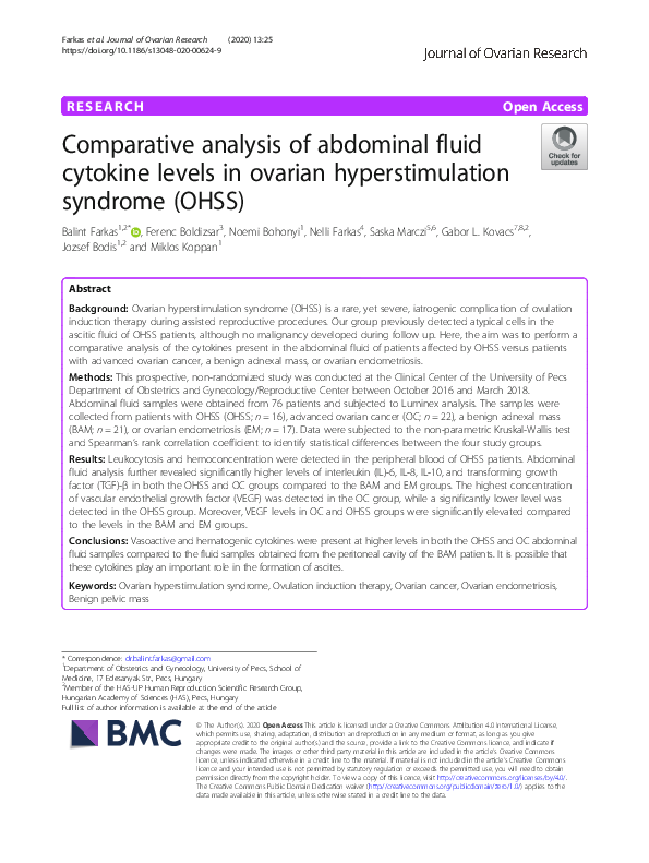
Fig. 2 Cytokine levels in abdominal fluid samples obtained from the peritoneal cavity in the four study groups. Statistical analysis included nonparametric Kruskal-Wallis test with Bonferroni post hoc test
of patients with ovarian cancer [23] and also in the abdominal fluid of OHSS patients [24]. Meanwhile, low
levels of VEGF were detected in BAM and EM patients
in the present study. We hypothesize that increased production of VEGF is a major factor in the formation of
ascites.
Macrophages produce IL-8, an important cytokine in
the immune system. This cytokine induces chemotaxistriggered neutrophil migration toward inflammation
sites and then stimulates phagocytosis once the neutrophils arrive onsite [25]. Previously, levels of IL-8 and
levels of the anti-inflammatory protein, IL-10, were
found at higher concentrations in the ascitic fluid of
OHSS patients [26, 27]. In our current investigation, significantly higher levels of both IL-8 and IL-10 were detected in the ascites of OHSS and OC patients compared
to the levels detected in the abdominal lavage fluid of
BAM and EM patients. Moreover, the levels were highest in the ascites of the OC patients. This finding is
consistent with other recently published data [7] and
with an angiogenetic role for IL-8 in malignancy [28]
and a pivotal immunosuppressive role for IL-10 in OCassociated ascites when activation of dendritic cells via
toll-like receptors is compromised [29].
To date, available literature does not indicate a consensus regarding the role of TNF-α in OHSS. For example, while no statistically significant difference was
previously found in the amount of TNF-α in the ascites
of OHSS patients compared to controls [30], others reported elevated levels in the same experimental setting
[31]. The TNF-α data obtained in the present study support a less important role for TNF-α in OHSS.
TGF-β is a multifunctional cytokine. In its activated
form, it binds TGF-β receptors by forming a serine/
threonine kinase complex [31]. Subsequent activation of
a signaling cascade leads to downstream activation of
various substrates and regulatory proteins. In addition,
the transcription of various target genes is induced,
�Farkas et al. Journal of Ovarian Research
(2020) 13:25
Page 5 of 8
Fig. 3 Spearman correlation matrix of the cytokine values, CA-125 tumor marker parameter and age. Correlation coefficients are shown, red in
case of negative, blue in case of positive correlation. X marks: non significant connection
thereby contributing to differentiation, chemotaxis, proliferation, and activation of many immune cells [31]. In
our study groups, a significant increase in the levels of
TGF-β were detected in the OHSS and OC groups relative to the BAM and EM groups. The OC group had the
highest concentration of TGF-β. Among the immunosuppressive cytokines associated with advanced ovarian
cancer, it has been proposed that TGF-β contributes to
impaired anti-tumor immune function [32]. However,
the role of TGF-β in OHSS remains unknown.
The novelty of our data is that we managed to reveal
similarly increased, with no statistically significant difference, in the peritoneal cavity levels of IL-6, IL-8, IL-10,
VEGF and TGF-ß both in OC and OHSS patients, but
found statistically significantly lower levels of the same
cytokines compared to BAM and EM groups. Despite
the mean age alteration between OC and OHSS groups
the inflammatory responses might be hard to compare,
but the cytokine production trend seem to be similar in
these two groups. This might suggest same kind of
pathomechanism of the ascites formation both in OHSS
and in ovarian malignancy.
There are limitations associated with the present study.
These include a relatively low number of participants, a
lack of serum cytokine concentration measurements, discrepancies of age between the compared groups, which
can influence the inflammatory profile, and some other,
potentially important cytokine concentrations were not investigated as yet. Regarding the latter point, IL-2 would
have been another cytokine of interest to investigate considering that it has been found at high levels in the peritoneal cavity of OHSS patients [33]. Furthermore, we
could not isolate and identify the origin of the atypical
cells present in the ascitic fluid of OHSS patients which
we previously described [13, 34]. However, a strength of
the present study is the broad spectrum of samples which
were examined, including abdominal fluid from patients
with various benign adnexal masses and from patients
with ovarian “chocolate cysts” (e.g., endometrioma), which
served as valid negative controls.
In the future a proposed potential clinical implication
of our study would have been to find anti-inflammatory
citokine agents to reduce the symptoms of OHSS, and
to decrease the severity of the disease.
�Farkas et al. Journal of Ovarian Research
Page 6 of 8
(2020) 13:25
Conclusions
Local pro- and anti-inflammatory cytokines, as well as
vasoactive components, play important roles in both the
formation of free abdominal fluid and in the pathogenesis of advanced ovarian cancer and OHSS compared
with benign ovarian disease and ovarian manifestation of
endometriosis. In further studies serum cytokine levels
and peritoneal cavity immune cell distribution might
worth to investigate, with the aim to reveal which cell
population are colonize and produce the described
cytokines.
Methods
Patients and study design
This prospective, non-randomized study was approved
by the University of Pecs Institutional Ethical Review
Board (#5273–2/2012) and was conducted at the Clinical
Center of the University of Pecs Department of Obstetrics
and Gynecology/Reproductive Center between October
2016 and March 2018. Patient participation was on a
voluntary basis and all enrolled participants were older
than 18 years of age. Written informed consent was
completed if patients had an adnexal mass or if they were
diagnosed with OHSS after OIT.
Evaluation of abdominal fluid
Abdominal fluid samples were obtained during ultrasoundguided culdocentesis of patients with a severe form OHSS
(n = 16), who represented the investigated population; intraoperative ascites sampling was performed during laparotomy of patients with advanced ovarian cancer (OC) (n =
22), who were the malignant disease group; sterile saline
was collected after intraoperative pelvic lavage during laparoscopic cystectomy of patients with a benign adnexal
mass (BAM; n = 21), who acted as negative controls; and intraoperative sampling of free abdominal fluid was performed during operative laparoscopy for patients with
ovarian endometriosis (EM; n = 17), used as transient / benign disease group. Clinical and histological diagnoses of
the participants are summarized in Table 1.
Peripheral blood analysis
Peripheral blood samples were collected preoperatively
from all the study participants, including 60 patients
who were admitted for surgery on the day of intervention, and samples were also collected on the day of
hospitalization for the OHSS patients (n = 16). Serum
levels of Na+, K+, LDH, ASAT, ALAT, and CA-125
tumor marker were determined. A hemogram was also
performed.
Measurement of cytokine levels in abdominal fluid
Cytokine levels were measured by using the R&D Systems
Human Premixed Multi-Analyte Kit Luminex Assay (Cat.
no. LXSAH-6; R&D Systems, Minneapolis, MN, USA) and
a Luminex 200 instrument (R&D Systems). Levels of IL-6,
IL-8, IL-10, TNF-α, and VEGF were measured according
to the manufacturer’s instructions. TGF-β levels were
measured with the R&D Systems Magnetic Luminex
Performance Assay and MAGPIX MILLIPLEX MAP instrument (MilliporeSigma, Danvers, MA, USA) according
to the manufacturer’s instructions. Samples above the
standard curve were considered to be maximum value,
Table 1 Clinical and histological diagnoses of the participants in the four study groups
Study group
Histologic Diagnosis
Number (n)
OHSS
NA
16
Ovarian Cancer
Serous papillary adenocarcinoma
Grade
22
High
16
Clear cell adenocarcinoma
NA
NA
2
Adenosarcoma
NA
Low
2
Adult granulosa cell ovarian tumor
3b
NA
1
Borderline (atypical proliferation)
NA
NA
1
Benign adnexal mass
Endometriosis
FIGO Stage
3b-c
21
Ovarian fibroma
6
Follicular cyst
3
Granulosa lutein cyst
6
Adult type teratoma
1
Borderline tumor (no atypical proliferation)
3
Paraovarian cyst
2
Endometrioma of the ovaries
17
FIGO International Federation of Obstetrics and Gynecology
�Farkas et al. Journal of Ovarian Research
Page 7 of 8
(2020) 13:25
and samples under the curve sensitivity were annotated as
0. All data are displayed in pg/ml.
Statistical analysis
Statistical analyses were performed by using IBM SPSS
Statistic 25 software (IBM Corporation) at the University
of Pecs, Institute of Bioanalysis (performed by NF). The
total sample size (n) was 76. Comparisons were made
between serum and cytokine levels detected for the four
study groups according to the non-parametric KruskalWallis test with Bonferroni post hoc test. To examine
the relationship between cytokine levels and serum parameters, Spearman’s rank correlation coefficient was applied. Mean data are reported ± standard deviation (SD).
Statistical significance was set at *p < 0.05, or **p < 0.01.
Abbreviations
ALAT: Alanine Aminotransferase; ASAT: Aspartate transaminase; BAM: Benign
adnexal mass; EM: Endometriosis; Hgb: Hemoglobin; Htc: Hematocrit, packed
cell volume; IL: Interleukin; IVF: In vitro fertilization; LDH: Lactate
dehydrogenase; OC: Ovarian cancer; OHSS: Ovarian hyperstimulation
syndrome; OIT: Ovulation induction therapy; RBC: Red blood cell;
SD: Standard deviation; TCT: Thrombocyte; TGF-β: Transforming growth
factor-beta; TNF-α: Tumor necrosis factor-alpha; VEGF: Vascular endothelial
growth factor; WBC: White blood cell
Acknowledgements
We would like to express our gratitude to the medical staff of the University
of Pecs, Department of Obstetrics and Gynecology, and the Reproduction
Center. We especially thank Gabriella Boskovits, head of the OR nurses, for
allowing us to collect samples and obtain data. We thank Agnes Kemeny
PhD (Associate Professor at the Department of Pharmacology and
Pharmacotherapy, University of Pecs, Medical School) for her help in the
TGFbeta MagPix measurement. We would also like to thank Prof. Dr. Peter M.
Gocze for his useful comments regarding our manuscript.
Authors’ contributions
BF collected samples, set up the study design, and wrote the manuscript; NB
collected samples; FB and SM performed the Luminex assay; NF performed
the statistical analyses; MK, GLK, and JB provided financial support and
edited the manuscript. All of the authors have read and approved the final
manuscript.
Funding
The current study was funded by grant, GINOP-2.3.2-15-2016-00021, „The
use of chip-technology in increasing the effectiveness of human in vitro
fertilization”. Open access funding provided by University of Pécs (PTE).
Availability of data and materials
The datasets generated and/or analyzed in the current study are not publicly
available in order to prevent compromise of individuals’ privacy. However,
the data are available from the corresponding author upon reasonable
request.
Ethics approval and consent to participate
This prospective cohort study was approved by the University of Pecs
Institutional Ethical Review Board (#5273–2/2012). All of the participating
patients provided written informed consent.
Consent for publication
The current manuscript does not contain any individual person’s data in any form.
Competing interests
The corresponding author (BF) and two other authors (JB and GLK) have
multiple affiliations and JB has received financial support from the Hungarian
Academy of Sciences (HAS; Budapest, Hungary). The remaining authors have
no conflicts of interest to report regarding the present study.
Author details
Department of Obstetrics and Gynecology, University of Pecs, School of
Medicine, 17 Edesanyak Str., Pecs, Hungary. 2Member of the HAS-UP Human
Reproduction Scientific Research Group, Hungarian Academy of Sciences
(HAS), Pecs, Hungary. 3Department of Immunology and Biotechnology,
University of Pecs, School of Medicine, Pecs, Hungary. 4School of Medicine,
Institute of Bioanalysis, University of Pecs, Pecs, Hungary. 5Laboratory of
Molecular and HLA Diagnostics, University Hospital Osijek, Clinical Institute of
Transfusion Medicine, Osijek, Croatia. 6Department of Medical, Chemistry,
Biochemistry and Clinical Chemistry, University of Osijek, Faculty of Medicine,
Osijek, Croatia. 7Szentágothai Research Center, University of Pecs, Pecs,
Hungary. 8Department of Laboratory Medicine, Faculty of Medicine,
University of Pecs, Pecs, Hungary.
1
Received: 11 November 2019 Accepted: 17 February 2020
References
1. Mascarenhas MN, Flaxman SR, Boerma T, Vanderpoel S, Stevens GA.
National, regional, and global trends in infertility prevalence since 1990: a
systematic analysis of 277 health surveys. PLoS Med. 2012;9:e1001356.
2. Lotti F, Maggi M. "ultrasound of the male genital tract in relation to male
reproductive health" (PDF). Hum Reprod Update. 2014;21:56–83.
3. Royal College of Obstetricians and Gynaecologists. The management of
ovarian hyperstimulation syndrome. Green-top Guideline. 2006:5 Available
at: http://www.rcog.org.uk/resources/Public/pdf/green_top_5_
management_ohss_a.pdf. Accessed January 20 2009.
4. Gomez R, Soares SR, Busso C, Garcia-Velasco JA, Simon C, Pellicer A.
Physiology and pathology of ovarian hyperstimulation syndrome. Semin
Reprod Med. 2010;28:448–57.
5. Várnagy A, Bódis J, Mánfai Z, Wilhelm F, Busznyák C, Koppán M. Low-dose
aspirin therapy to prevent ovarian hyperstimulation syndrome. Fertil Steril.
2010;93:2281–4.
6. Sanguinete MMM, Oliveira PH, Martins-Filho A, Micheli DC, Tavares-Murta
BM, Murta EFC, Nomelini RS. Serum IL-6 and IL-8 correlate with prognostic
factors in ovarian Cancer. Immunol Investig. 2017;46:677–88.
7. Rodrigues ISS, Martins-Filho A, Micheli DC, Lima CA, Tavares-Murta BM,
Murta EFC, Nomelini RS. IL-6 and IL-8 as prognostic factors in peritoneal
fluid of ovarian Cancer. Immunol Investig. 2019;22:1–12.
8. Nastri CO, Ferriani RA, Rocha IA, Martins WP. Ovarian hyperstimulation
syndrome: pathophysiology and prevention. J Assist Reprod Genet. 2010;27:
121–8.
9. Nelson SM. Prevention and management of ovarian hyperstimulation
syndrome. Thromb Res. 2017;151(Suppl 1):S61–4.
10. Sergentanis TN, Diamantaras AA, Perlepe C, Kanavidis P, Skalkidou A,
Petridou ET. IVF and breast cancer: a systematic review and meta-analysis.
Hum ReprodUpdate. 2014;20:106–23.
11. Taheripanah R, Balash F, Anbiaee R, Mahmoodi M, Akbari Sene A. Breast cancer
and ovulation induction treatments. Clin Breast Cancer. 2018;18:395–9.
12. Zhao J, Li Y, Zhang Q, Wang Y. Does ovarian stimulation for IVF increase
gynaecological cancer risk? A systematic review and meta-analysis. Reprod
Biomed Online. 2015;31:20–9.
13. Hatzipetros I, Gocze PM, Cziraky K, Kovacs K, Kalman E, Farkas B. Assessment
of cells in the ascitic fluid of women with ovarian hyperstimulation
syndrome: the clinical implications for subsequent ovarian malignancy.
Reprod Biol Endocrinol. 2013;11:91–5.
14. Mathur RS, Jenkins JM, Bansal AS. The possible role of the immune system
in the aetiopathogenesis of ovarian hyperstimulation syndrome. Hum
Reprod. 1997;12:2629–34.
15. Aboulghar MA, Mansour RT, Serour GI, El Helw BA, Shaarawy M. Elevated
levels of interleukin-2, soluble interleukin-2 receptor alpha, interleukin-6,
soluble interleukin-6 receptor and vascular endothelial growth factor in
serum and ascitic fluid of patients with severe ovarian hyperstimulation
syndrome. Eur J Obstet Gynecol Reprod Biol. 1999;87:81–5.
16. Andus T, Gross V, Holstege A, Weber M, Ott M, Gerok W, Schölmerich J.
Evidence for the production of high amounts of interleukin-6 in the
peritoneal cavity of patients with ascites. J Hepatol. 1992;15:378–81.
17. Hoeben A, Landuyt B, Highley MS, Wildiers H, Van Oosterom AT, De Bruijn
EA. Vascular endothelial growth factor and angiogenesis. Pharmacol Rev.
2004;56:549–80.
�Farkas et al. Journal of Ovarian Research
(2020) 13:25
18. Bates DO. Vascular endothelial growth factors and vascular permeability.
Cardiovasc Res. 2010;87:262–71.
19. Elchalal U, Schenker JG. The pathophysiology of ovarian hyperstimulation
syndrome--views and ideas. Hum Reprod. 1997;12:1129–37.
20. Namavar Jahromi B, Parsanezhad ME, Shomali Z, Bakhshai P, Alborzi M,
Moin Vaziri N, Anvar Z. Ovarian hyperstimulation syndrome: a narrative
review of its pathophysiology, risk factors, prevention, classification, and
management. Iran J Med Sci. 2018;43:248–60.
21. Krasnow JJ, Berga SL, Guzick DS, et al. Vascular permeability factor and
vascular endothelial growth factor in ovarian hyperstimulation syndrome.
Fertil Steril. 1996;65:552–5.
22. Abramov Y, Barak V, Nisman B, Schenker JG. Vascular endothelial growth
factor plasma levels correlate to the clinical picture in severe ovarian
hyperstimulation syndrome. Fertil Steril. 1997;67:261–5.
23. Dalal V, Kumar R, Kumar S, Sharma A, Kumar L, Sharma JB, Roy KK, Singh N,
Vanamail P. Biomarker potential of IL-6 and VEGF-A in ascitic fluid of
epithelial ovarian cancer patients. Clin Chim Acta. 2018;482:27–32.
24. Chen CD, Wu MY, Chen HF, Chen SU, Ho HN, Yang YS. Prognostic
importance of serial cytokine changes in ascites and pleural effusion in
women with severe ovarian hyperstimulation syndrome. Fertil Steril. 1999;
72:286–92.
25. Köhidai L, Csaba G. Chemotaxis and chemotactic selection induced with
cytokines (IL-8, RANTES and TNF-alpha) in the unicellular Tetrahymena
pyriformis. Cytokine. 1998;10:481–6.
26. Revel A, Barak V, Lavy Y, Anteby E, Abramov Y, Schenker JJ, Amit A, FinciYeheskel Z, Mayer M, Simon A, Laufer N, Hurwitz A. Characterization of
intraperitoneal cytokines and nitrites in women with severe ovarian
hyperstimulation syndrome. Fertil Steril. 1996;66:66–71.
27. Manolopoulos K, Lang U, Gips H, Braems GA. Elevated interleukin-10 and sex
steroid levels in peritoneal fluid of patients with ovarian hyperstimulation
syndrome. Eur J Obstet Gynecol Reprod Biol. 2001;99:226–31.
28. Brat DJ, Bellail AC, Van Meir EG. The role of interleukin-8 and its receptors in
gliomagenesis and tumoral angiogenesis. Neuro-Oncology. 2005;7:122–33.
29. Brencicova E, Jagger AL, Evans HG, Georgouli M, Laios A, Attard Montalto S,
Mehra G, Spencer J, Ahmed AA, Raju-Kankipati S, Taams LS, Diebold SS.
Interleukin-10 and prostaglandin E2 have complementary but distinct
suppressive effects on toll-like receptor-mediated dendritic cell activation in
ovarian carcinoma. PLoS One. 2017;12:e0175712.
30. Friedlander MA. Loret de Mola JR, Goldfarb JM: elevated levels of
interleukin-6 in ascites and serum from women with ovarian
hyperstimulation syndrome. Fertil Steril. 1993;60:826–33.
31. Massagué J. TGFβ signalling in context. Nat Rev Mol Cell Biol. 2012;13:616–30.
32. Santin AD, Bellone S, Ravaggi A, Roman J, Smith CV, Pecorelli S, Cannon MJ,
Parham GP. Increased levels of interleukin-10 and transforming growth
factor-beta in the plasma and ascitic fluid of patients with advanced ovarian
cancer. BJOG. 2001;108:804–8.
33. Lisa C. Grossman, Konstantinos G. Michalakis, hyacinth Browne, mark D.
Payson, James H. Segars: the pathophysiology of ovarian hyperstimulation
syndrome: an unrecognized compartment syndrome. Fertil Steril. Author
manuscript; available in PMC 2011 Jun 27. Fertil Steril. 2010;94:1392–8.
34. Bódis J, Papp S, Vermes I, Sulyok E, Tamás P, Farkas B, Zámbó K, Hatzipetros
I, Kovács GL. "Platelet-associated regulatory system (PARS)" with particular
reference to female reproduction. J Ovarian Res. 2014;16(7):55–60.
Publisher’s Note
Springer Nature remains neutral with regard to jurisdictional claims in
published maps and institutional affiliations.
Page 8 of 8
�
 Balint Farkas