134
Journal of Pharmacy and Nutrition Sciences, 2011, 1, 134-139
Flavonoids of Neotorularia aculeolata Plant
Fatma A. Ahmed1, Inas M. Abd El–Wahab Khamis1 and Samar Y. Desoukey2,*
1
Medicinal and Aromatic Plant Dept., Desert Research Center, El–Matariya, Cairo, Egypt
2
Pharmacognosy Dept., Faculty of Pharmaceutical Sciences, Future University, New Cairo, Egypt
Abstract: Neotorularia aculeolata belongs to the family Cruciferae that has several uses in the Egyptian folk medicine
for many years. Nothing could be traced about the chemical composition of the plant. Extraction, isolation and
purification of the air-dried plant material using different chromatographic techniques (PC, TLC & CC) provided seven
1
flavonoids. Identification of the isolated compounds using different chemical and physical techniques (UV, H-NMR and
13
C
NMR spectroscopy) allowed to characterize these compounds as kaempferol, kaempferol-7-O-rhamnoglucoside
{Kaempferol-7-neohesperidoside}, quercetin, rutin, quercetin-3-O- β-D-glucoside-7-O-α-L-rhamnoside-3`-methylether,
quercetin-3,7-di-O-α-L-rhamnoside-3`-methylether and myricetin.
Keywords: Neotorularia aculeolata, Cruciferae, kaempferol, quercetin, rutin, myricetin.
1. INTRODUCTION
luteolin and apigenin and their derivatives were isolated
[6-8].
Cruciferae (Brassicaceae) is one of the largest
families in the plant kingdom that is rich in medicinal
plants. It comprises approximately 380 genera and
about 3350 species in 10 poorly defined tribes [1]. The
family is represented in Egypt by 53 genera and 107
species mostly annual, biennial or perennial herbs. N.
aculeolata (Boiss.) Hedge & J. Leonard is one of these
annual plants in this family. It grows at Sinai proper; it
always grows at the entire Sinai Peninsula including
the coastal Mediterranean strip and El–Tih Desert east
of Suez Canal, rock crevices and hillsides [2]. It is
widely spread at Abo Egaila – El Qusayema road
(North Sinai) from where it was collected for this study.
Cruciferous plants have been used since ages and
are grown as vegetables, sources of oils and as
condiments. They are known for their stimulant,
diuretic,
thermogenic,
depurative,
rubefacient,
galactogogue, emmenagogue, tonic, aphrodisiac,
ophthalmic activities and are used for scurvy, peptic
ulcers,
hepatopathy,
splenomegaly,
dyspepsia,
diarrhea, dysentery, lumbago, syphilis, leucorrhoea,
seminal weakness, asthma, cough, hiccough,
tenesmus, hemorrhoids as well as anticancer activity
especially as androgen receptor antagonist in human
prostate cancer [3, 4]. Cruciferous plants are inducers
of microsomal cytochrome P450 enzyme [5].
It was reported that kaempferol, quercetin and
isorhamnetin glycosides, in addition to myricetin,
*Address corresponding to this author at the Pharmacognosy Dept., Faculty of
Pharmaceutical Sciences, Future University, New Cairo, Egypt; Tel: 00202
22402046; E-mail: dr_samar_yehia@yahoo.com
ISSN: 2223-3806 / E-ISSN: 1927-5951/11
The presence of phenolic acids as: caffeic, ferulic,
P–coumaric and vanillic acids beside the presence of
the previous flavonoids and rutin were also isolated
from Brassica alba, B. oleraceae, B. campestris and
other cruciferous species [9-14].
2. RESULTS AND DISCUSIONS
2.1. Isolated Flavonoids
Seven flavonoids were isolated, purified by CC, PC
and TLC and identified through Rf-values, UV spectra
in methanol with different shift reagents (Table 1) and
1
13
H & C-NMR. These compounds were coded as A1A7.
2.1.1. Compound A1
This compound was obtained as yellow crystals,
soluble in methanol, Rf-values of 0.85 in BAW and 0.4
in acetic acid 15%. It showed two major absorption
bands in MeOH; band I at 367nm and band II at
268nm, which indicated a flavonol nucleus with free
hydroxyl group at the C-3 [15, 16]. Addition of sodium
methoxide resulted in a bathochromic shift in band I
(+49 nm), which proved the presence of a free OH\
group at C-4 . A bathochromic shift in band I (+53nm)
with aluminum chloride, which was not affected by the
addition of hydrochloric acid, indicating the presence of
free hydroxyl group at C-3 and C-5. A bathochromic
shift in band II (+7 nm) with sodium acetate indicated
the presence of free hydroxyl group at C-7. Addition of
H3BO3 gave no shift, which proved the absence of any
catecholic hydroxyl group. From the UV analysis,
compound A1 is probably kaempferol. The identity of
© 2011 Lifescience Global
�Flavonoids of Neotorularia Aculeolata Plant
Table 1:
Journal of Pharmacy and Nutrition Sciences, 2011 Vol. 1, No. 2
135
UV Spectral Data of the Isolated Compounds
UV Data
AcONa/ H3BO 3
AcONa
AlCl3/ HCl
AlCl3
MeONa
MeOH
274, 296 (sh), 320
(sh), 372
275, 302 (sh), 385
266, 305 (sh), 350,
420
266, 305 (sh), 350,
420
280, 318 (sh), 416
253 (sh), 268, 324
(sh), 367
A1
260, 325, (sh), 370
266, 323, 385, 418
(sh),
244 (sh), 258, 266,
300 (sh), 350, 422
259 (sh), 266, 299
(sh), 353, 424
245, 267, 335 (sh),
425
253, 266, 323, 354
A2
264, 292 (sh), 384
274, 320 (sh), 428
268, 300, (sh), 362
(sh), 428
272, 328, 445
262, 332, 440
267, 306 (sh), 370
A3
220, 298, 387
271, 325, 393
271, 300, 340 (sh),
402
275, 303, (sh), 433
272, 327, 410
259, 266 (sh), 299
(sh), 350
A4
262, 360
260, 360
270, 310, 350, 410
270, 310, 350, 410
270, 398
275, 355
A5
256, 310, 360, 400
256, 310, 360, 400
256, 360
256, 360
270, 410
256, 350
A6
258, 304 (sh), 382
269, 335
266, 275 (sh), 308
(sh), 366, 428
271, 316 (sh), 450
262 (sh), 285 (sh),
322, 423
254, 272, (sh),,
374
A7
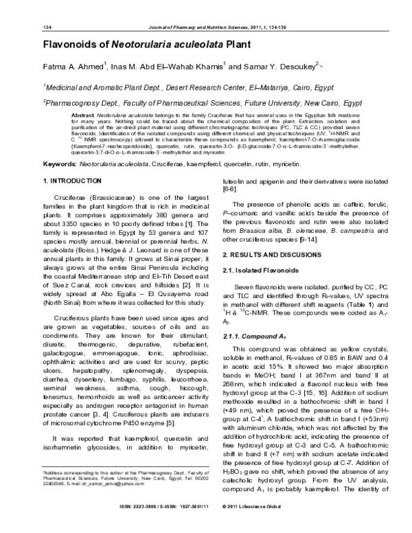
R
OH
R3
O
R1
R2
OH
Comp.
Name
A1
Kaempferol
O
R
R1
R2
R3
H
H
OH
OH
OH
Oglrh
A2
Kaempferol-7-O-neohesperidoside
H
H
A3
Quercitin
OH
H
OH
OH
A4
Rutin
OH
H
Oglrh
OH
A5
Quercitin-3-O-β-D-gluc-7-α-L-rham-3`-methylether
OMe
H
Ogl
Orh
A6
Quercitin-3,7di-O-α-L-rham-3`-methylether
OMe
H
Orh
Orh
A7
Myrecitin
OH
OH
OH
OH
Figure 1: Structures of compounds from Neotorularia aculeolata.
compound A1 was further confirmed as kaempferol by
1
H-NMR spectrum in DMSO-d6, which showed signals
at δ (ppm) 8.0 (2H, d, J= 8 Hz, H-2` and H-6`), 6.9 (2H,
d, J= 8Hz, H-3` and H-5`), 6.4 (1H, d, J= 1.5 Hz, H-8),
and 6.2 (1H, d, J= 1.5 Hz, H-6). Thus, from the above
data and current literature this compound A1 is
identified as kaempferol [17].
2.1.2. Compound A2
This compound was obtained as dull yellow
crystals, soluble in methanol, Rf-values 0.3 in BAW and
0.35 in acetic acid 15%. It showed two major
absorption bands in MeOH; the absorption maximal in
methanol, band I (354nm) indicated that it was a
flavonol with a 3-OH free. The addition of NaOMe
caused a bathochromic shift in both band I and II, a fact
which proved the presence of a free OH at 4` position.
After addition of AlCl3, a bathochromic shift proved the
presence of a free OH at 5 positions. However, on
addition of HCl, no change was observed indicating the
absence of any catecholic hydroxyl groups. Meanwhile
the addition of NaOAc caused no shift in band II thus
suggesting the occupation of 7-position. Addition of
H3BO3 caused no shift, this suggested the absence of
any catecholic hydroxyl groups.
1
The H-NMR spectral data of compound A2 showed
the signals characteristic for kaempferol with additional
signal for the sugar moieties. Two signals for the two
anomeric sugar protons at δ 5.4 (1H, d, J=2.5Hz, H-1``
�136
Journal of Pharmacy and Nutrition Sciences, 2011 Vol. 1, No. 2
glucose) and δ 5.2 (1H, d, J=2.5Hz, H-1``` rhamnose).
The remaining sugar proton as m at 3.2-3.9, signal at
1.2 (3H, d, J=6Hz, CH3 rhamnose).
13
C-NMR spectrum data of compound A2 showed a
ketonic carbon at 176.1 at C-4 and the most acidic
carbon at C-7 at 162.4 followed by C-4` at 159.4 and
C-3 at 135.9. Two anomeric sugar carbons at 98.4 and
100.5 for C-1`` and C-1```, respectively indicating the
disaccharide nature of compound A2. One methyl
carbon of rhamnose was shown at 20.9. In A2, C-2``` of
rhamnose appeared at 70.5. Thus from the obtained
1
13
Rf-values, UV, H-NMR and C-NMR spectral data of
compound A2, showed that it is identified as
kaempferol-7-O-glucose (1→2)-rhamnose (Kaempferol-7-O-Neohesperidoside).
2.1.3. Compound A3
This compound was obtained as yellow crystals,
soluble in methanol, Rf-values 0.73 in BAW and 0.29 in
acetic acid. Compound A3 showed two major
absorption bands in MeOH; band I at 370nm and band
II at 267nm, which indicated a flavonol nucleus with
free hydroxyl group at the 3 position [15, 16]. Addition
of sodium methoxide resulted in a bathochromic shift in
band I (+70nm), which proved the presence of a free
\
OH-group at 4 -position. A bathochromic shift in band I
(+75nm) with aluminum chloride, indicated the
presence of free hydroxyl group at C-3 and C-5. The
hypthochromic shift of AlCl3 spectrum in band I (-17
nm) after the addition of HCl indicated the presence of
orthodihydroxy group in B-ring (3`, 4` position). A
bathochromic shift in band I (+14 nm) with sodium
acetate indicating the presence of free hydroxyl group
at C-7, which was detected by H3BO3 addition,
indicates the presence of orthodihydroxy group (3`, 4`
position). Thus, from the UV analysis and Rf-values,
compound A3 may be identified as quercetin. The
compound A3 was further confirmed as quercetin by
1
H-NMR spectrum in DMSO-d6, which showed signals
at δ (ppm) 7.7 (1H, d, J = 8.5 Hz, H-2`), δ 7.5 (1H, dd,
J= 2.5, H-6`) and δ 6.8 (1H, d, J = 8.5, H= 5`), indicated
the presence of aromatic ring with two substitutions, in
m, p-substitution {δ 6.5 (1H, d, J = 1.5 H-6) and δ 6.2
(1H, d, J= 1.5, H-8)}. Thus, from the above mentioned
data, compound A3 is Quercetin [17].
2.1.4. Compound A4
This compound was obtained as yellow crystals, Rfvalues of 0.49 in BAW and 0.54 in acetic acid
respectively. The absorption maxima in methanol, band
Fatma et al.
I at 350 nm, indicates that it is a flavonol with 3-OH
substitution. The remaining UV spectral data were
found to be similar to that of quercetin type compound.
1
H-NMR spectrum of the compound A4 in DMSOd6, showed signals at δ (ppm) 7.6 (1H, d, J = 2.5 Hz,
H-2`), δ 7.5 (1H, dd, J = 8.5, 2.5 H-6`), δ 6.8 (1H, d, J =
8.5, H= 5`), δ 6.4 (1H, d, = 1.5 H-8), δ 6.2 (1H, d, J =
1.5, H-6) and for sugar moiety δ (ppm): 5.3 (1H, d, J=
8Hz, H-1`` glucose), 4.5 (1H, d, J= 2.5Hz, H-1```
rhamnose), 3.4 (m, remaining sugar protons) and 0.8
13
(3H, d, J= 6Hz, CH3 rhamnose). C-NMR of A4 gave
the following peaks in DMSO-d6: δ (ppm): 146.9 (C-2),
135.5 (C-3), 175.8 (C-4), 160.7 (C-5), 98.2 (C-6), 163.9
(C-7), 93.3 (C-8), 156.2 (C-9), 103.1 (C-10), 122.1 (C1′), 115.3 (C-2′), 145.0 (C-3′), 147.6 (C-4′), 115.6 (C5′), 120.0 (C-6′), and for sugar moiety, 101.5 (C-1′′),
74.3 (C-2′′),75.9 (C-3′′), 70.2 (C-4′′), 76.2 (C-5′′), 67.4
(C-6′′), 101.2 (C-1′′′), 70.8 (C-2′′′), 71.0 (C-3′′′), 72.2
(C-4′′′), 69.1 (C-5′′′) and 18.1 (C-6′′′). Complete acid
hydrolysis yielded glucose and rhamnose in the
aqueous phase and quercetin in the organic phase in
(a), (e) and (f) using specific spray reagents. From the
above data and by comparison with published data,
compound A4 is identified as Rutin (quercetin-3-O-α-Lrhamnoside (1→6)-β-D-glucoside) [17].
2.1.5. Compound A5
This compound was obtained as yellow crystals, Rfvalues of 0.51 in BAW and 0.7 in acetic acid
respectively. The absorption maxima in methanol, band
I at 355 nm, indicates that it is a flavonol with 3-OH
substitution. The remaining UV spectral data were
found to be similar to that of quercetin type compound.
1
H-NMR spectrum of the compound A5 in DMSO-d6,
showed signals at δ (ppm) 7.95 (1H, d, J= 8.5 Hz, H2′), 7.65 (1H, dd, J= 8.5 Hz, H-6′), 6.94 (1H, d, J=
8.5Hz, H-5′), 6.75 (1H, d, J= 2.5Hz, H-8), 6.45(1H, d,
J=2.5 Hz, H-6), 5.6 (1H, d, J= 2.5Hz, H-1′′ rhamnose),
5.4 (1H, d, J=2.5 Hz, H -1′′′ glucose), 3.92 (s, OCH3)
and 1.2 (3H, d, J=6Hz, OCH3 rhamnose). The isolated
compound A5 when subjected to partial acid hydrolysis
afforded quercetine-3′-methoxide and the sugars were
glucose and rhamnose. On the other hand, a known
amount of the compound A5 was subjected to complete
acid hydrolysis using 2N HCl. It was observed that
compound A5 resisted acid hydrolysis, which coincided
with C-glycoside flavonoid. From the data above and
by comparison with published data, compound A5 is
identified as Quercetin-3-O-β-D-glucoside -7-O-α-Lrhamnoside-3`-methyether [17].
�Flavonoids of Neotorularia Aculeolata Plant
Journal of Pharmacy and Nutrition Sciences, 2011 Vol. 1, No. 2
137
2.1.6. Compound A6
3. SUMMERY AND CONCLUSION
This compound was obtained as yellow crystals, Rfvalues 0.52 in BAW and 0.69 in acetic acid. The
absorption maxima in methanol, band I at 350 nm,
indicated that it was a flavonol with 3-OH substitution.
The remaining UV spectral data was found to be similar
1
to that of quercetin type compound. H-NMR spectrum
of the compound A6 in DMSO-d6, showed signals at δ
(ppm) 7.6 (1H, d, J= 8.5 Hz, H-2′), 7.4 (1H, dd, J= 8.5
Hz, H-6′), 6.9 (1H, d, J= 8.5Hz, H-5′), 6.7 (1H, d, J=
2.5Hz, H-8), 6.4 (1H, d, J=2.5 Hz, H-6), 5.6 (1H, d, J=
2.5Hz, H-1′′ rhamnose), 5.5 (1H, d, J=2.5 Hz, H -1′′′
rhamnose), 3.9 (s, OCH3), 1.2 (3H, d, J=6Hz, OCH 3
rhamnose) and 0.8 (3H, d, J=6Hz, CH3 rhamnose). The
isolated compound A6 when subjected to partial acid
hydrolysis afforded quercetine-3′-methylether and the
sugar was rhamnose. On other hand a known weight of
the compound A6 was subjected to complete acid
hydrolysis using 2N HCl, which gave the aglycone
quercetine-3′-methoxide and the sugar was rhamnose.
From above data and by comparison with the published
data, compound A6 is identified as Quercetin-3,7-di-Oα-L-rhamnoside-3`-methylether [17].
In the present study, the defatted desalted
methanolic extract of the air-dried whole plant of
Neotorularia aculeolata was purified by CC, PC and
TLC ,to afford seven flavonoids {A1 to A7} these
compounds are identified as:- kaempferol, kaempferol7-O-rhamnoglucoside
{Kaempferol-7-Neohesperidoside}, Quercetin, Rutin, Quercetin-3-O- β-D-glucoside7-O-α-L-rhamnoside-3`-methylether , Quercetin-3,7-diO-α-L-rhamnoside-3`-methylether and Myricetin.
2.1.7. Compound A7
This compound was obtained as yellow crystals, Rfvalues of 0.31 in BAW and 0.16 in acetic acid
respectively. It showed two major absorption bands in
MeOH; band I at 374 nm and band II at 254 nm, which
indicated a flavonol nucleus with free hydroxyl group at
the 3 position [15, 16]. Addition of sodium methoxide
resulted in a bathochromic shift in band I (+49nm),
which proved the presence of a free OH-group at C4`position. A bathochromic shift in band I (+76nm) with
aluminum chloride, indicated the presence of a free
hydroxyl group at C-3 and C-5. The hypthochromic shift
of AlCl3 spectrum in band I (-22 nm), after the addition
of HCl, indicated the presence of orthodihydroxy group
in B-ring (3`and 4` position). A bathochromic shift in
band II (+15 nm) with sodium acetate indicated the
presence of a free hydroxyl group at C-7, which was
detected by H3BO3 addition, that indicated the
presence of orthodihydroxy group (3`and 4` position).
Thus, from the UV analysis and Rf-values compound
1
A7 may be identified as myricetin. H-NMR spectrum of
the compound A7 in DMSO-d6, showed signals at δ
(ppm) 6.25 (1H, d, J= 2.5 Hz, H-6), 6.33 (1H, d, J= 2.5
Hz, H-8), 7.32 (s, H-2′ and H-6). From the above data
and by comparison with published data, Compound A7
is identified as Myricetin [17].
To our knowledge this represents the first report for
the isolation of these compounds from N. aculeolata.
4. EXPERIMENTAL
4.1. Material, Methods and Techniques
4.1.1. Plant Materials
Neotorularia aculeolata (Boiss.) plant was collected
from North Sinai (Abo Egaila–El Qusayema road) in
June 2006 and identified by Prof. Dr. Nahed El-Hadidi,
Botany Department, Faculty of Science, Cairo
University and by comparison with herbarium
specimens at the Desert Research Center {DRC}. A
voucher
specimen
was
deposited
in
the
Pharmacognosy lab, Future University, Egypt. The
plant material was ground to fine powder and kept in an
amber light proof container.
4.1.2. Extraction and Purification of Flavonoids:
The whole plant {2 kg} was defatted with petroleum
ether (B.p. 40 -60°C): ether (1:1 v/v). The defatted
powder was percolated with methyl alcohol (80%) till
exhaustion to obtain a brownish alcoholic extract. This
extract was concentrated by evaporation under
reduced pressure at not more than 45°C to yield a
sticky brownish residue (90g) which was suspended in
hot distilled water, filtered and desalted.
4.1.3. Chromatographic Investigation:
4.1.3.1. PC
The concentrated aqueous extract was applied on
Whatmman No.1 paper chromatography using
descending technique with suitable solvent systems
[15].
4.1.3.2. TLC
Precoated TLC plates silica gel 60SF254 (E- Merck)
20 x 20 cm were used for investigation of the
�138
Journal of Pharmacy and Nutrition Sciences, 2011 Vol. 1, No. 2
flavonoids using suitable solvent systems
precoated preparative TLC silica gel F254
and
• Solvent systems used for PC and TLC:
a)
n-butanol-acetic acid- water ( BAW) (4:1:5)
organic phase.
b)
Acetic acid 15%.
c)
Ethyl acetate-methanol-water (30:5:4)and
(30:2.5:2)one phase, for TLC only.
d)
Phenol-water(80:20)
• Spray Reagent for PC and TLC
Aluminum chloride reagent: 1 % aluminum chloride
reagent solution in ethanol [18].
4.1.3.3. Isolation and Purification of The Flavonoidal
Compounds
The desalted alcoholic extract was submitted to
column chromatography and gradient elution was
performed, using firstly chloroform with increasing
concentration of ethyl acetate and methanol,
respectively. Fractions of 50 ml were collected, then
subjected to TLC using system (c) and similar fractions
were pooled together. The pooled fractions were
subjected to preparative TLC using system (a), the
bands corresponding to the flavonoidal compounds
were visualized under UV, eluted with methanol and
water {1:1}. The eluted bands were purified on a
Sephadex LH-20 column using methanol and water as
eluting system where seven compounds could be
isolated.
4.1.3.4. Identification of Flavonoids:
4.1.3.4.1. Spectral data
Ultraviolet (UV) with shift reagents and nuclear
I
13
magnetic resonance ( H-NMR and C-NMR).
4.1.3.4.2. Acid Hydrolysis
• Partial and Complete Acid Hydrolysis
5 mg of each compound were dissolved in 5 ml
methanol and an equal volume of 0.1N and 2N HCl
aqueous solution {each separately} was added for mild
and complete acid hydrolysis, respectively. The mixture
was refluxed on a boiling water bath for 3 hours. The
methanol was evaporated and the aglycone was
extracted with ether. The aqueous layer containing the
Fatma et al.
sugar part was evaporated to dryness and the residue
was dissolved in 10% isopropanol solution.
• Identification of the Aglycone:
The ether extract of each compound containing the
aglycone moiety was applied on paper chromatogram
along side with authentic flavonoid aglycones using the
solvent systems (a) and (b), visualized under UV,
exposed to ammonia vapor and sprayed with AlCl3.
• Identification of the Sugars:
The isopropanol solution of each compound
containing the sugar moiety was applied on PC along
side with authentic sugars using the solvent systems
(a) and (d). After development, they were visualized by
spraying with aniline hydrogen phthalate reagent and
heating at 110°C for 10 min..
5. AUTHENTIC REFERENCE
5.1. Flavonoid
Kaempferol ,Quercetin & myricetin were available at
the medicinal and aromatic plants department,
Phytochemistry unit, Desert Research Center Egypt.
5.2. Sugars
Glucose and rhamnose, (Sigma).
5.3. Apparatus
Shimadzu UV 240 (P/N 204 – 28000) instrument
was used for recording UV spectra and measuring the
absorbance under UV range.
A JEOL Ex – 270 NMR spectrometer apparatus
1
13
(270 MHz for H –NMR and 67.5 MHz for C – NMR)
was used for identification of the proton and / or carbon
in methanol, DMSO, CCl4 and CDCl3 using
trimethylsilyl ether (TMS) as an internal standard.
Chemical shift values were recorded in δ ppm.
6. REFERENCES
[1]
Heywood VH. Cruciferae, the Mustard Family. "Flowering
Plants of the World". Bastsford, London 1993; 119-22.
[2]
Boulos L. "Flora of Egypt Checklist". Revised Annotated
Edition. Al-Hadara Publishing, Cairo, Egypt. 2009: 410pp.
[3]
Narayan, DP and Kumer, U"Agro's Dictionary of Medicinal
Plants". Agrobions, India, Shayam printing press, Jodhpar,
India 2005: 400pp. 278: 21136-45.
[4]
Le TH, Schaldach MC, Firestone LG, Bjeldanes FL. "plantderived 3,3`-Diindolylmethane is a Strong Androgen
Antagonist in Human Prostate Cancer Cells" J Biol Chem
2003;
http://dx.doi.org/10.1074/jbc.M300588200
�Flavonoids of Neotorularia Aculeolata Plant
[5]
[6]
[7]
Journal of Pharmacy and Nutrition Sciences, 2011 Vol. 1, No. 2
Lampe WJ, King BI, Li Sue Grate, et al. "Brassica vegetables
increase and apiaceous vegetables decrease cytochrome
P450 1A2 activity in humans: changes in caffeine
metabolites ratios in response to controlled vegetable diets
"Carcinogenesis 2000; 21: 1157-62 Cancer Prevention
Research Program and Program in Epidemiology, Division of
Public Health Science,1100 Fairview Avenue North, Seattle,
WA 98109-1024,USA.
Venere DDI, Calabrese N, Linsalata V, et al. "Influence of
sowing time on phenolic composition of rocket". 8th
International Symposium on Timing of Field Production in
Vegetable Crops. Bari, Italy, Acta-Horticultura 2000; 533:
343-9.
KooHui M, Mohamed S, Miean KH. "Flavonoid (myricetin,
quercetin, kaempferol, luteolin, and apigenin) content of
edible tropical plants". Journal of Agricultural and Food
Chemistry 2001; 49: 3106- 12.
[8]
Sanchez YMD, "Study of flavonoid patterns in some species
of Erucastrum. Cruciferae", Newsletter 2001; 23: 5-6.
[9]
Sanchez YMD, "Comparative analysis of phenolic
compounds among some species of the Brassica from sect.
Sinapistrum and sect. Micropodium Cruciferae"-Newsletter.
2002; 24: 19-20.
[10]
Onyilagha JC, Bala A, Hallett R, Gruber M, Soroka J,
Westcott, N. "Leaf flavonoids of the Cruciferous species,
Camelina sativa, Crambe spp., Thlaspi arvense and several
other genera of the family Brassicaceae". Biochemical
Systematics and Ecology 2003; 31: 1309-2.
http://dx.doi.org/10.1016/S0305-1978(03)00074-7
[11]
Zhang JM, Satterfield MB, Brodbelt JS, Britz SJ, Clevidence
B, Novotny JA. "Structural characterization and detection of
Received on 19-11-2011
DOI: http://:dx.doi.org:10.6000:1927‐5951.2011.01.02.08
139
kale
flavonoids
by
electrospray
ionization
mass
spectrometry". Analytical Chemistry Washington 2003; 75:
6401-7.
http://dx.doi.org/10.1021/ac034795e
[12]
Ponce MA, Scervino JM, Erra-Balsells R, Ocampo JA,
Godeas AM. "Flavonoids from shoots, roots and roots
exudates of Brassica alba". Phytochemistry 2004; 65: 31314.
http://dx.doi.org/10.1016/j.phytochem.2004.08.031
[13]
Vallejo
F,
Tomas-Barberan
FA,
Ferreres,
F.
"Characterization of flavonols in broccoli (Brassica oleracea
L. var. italica) by liquid chromatography UV diode array
detection-electrospray ionisation mass spectrometry".
Journal of Chromatography. 2004; 1054: 181-93.
http://dx.doi.org/10.1016/j.chroma.2004.05.045
[14]
Wei YS, Zheng MY, Wang, YN, Shi-YP, Lu T. "A study on the
content of flavonoids in bee-pollen of Brassica campestris
from Qinghai." Acta Botanica Boreali Occidentalia Sinica
2004; 24: 301-5.
[15]
Harborne JB. "Phytochemical Methods". A Guide to Modern
Techniques of Plant Analysis. 2nd Ed. Chapman and Hall
Ltd. New Fetterlane, London, New York.1984; 142-50.
[16]
Liu YL, Neuman P, Borbara NT, Mabry JJ. "Techniques for
flavonoids analysis. " Rev. Latinamer. Quim. Suppl. 1989; 1:
90-130.
[17]
Mabry TJ, Markham KR, Thomas MB. "The Systematic
Identification of Flavonoids". Springer Verlag., New York.
1970. Pp. 2204.
[18]
Markham KR. "Techniques of Flavonoids Identification".
Academic press, London. 1982.
Accepted on 28-11-2011
Revised on 28-12-2011
�
 Fatma Aly Ahmed