BJD
C L I N I C A L A N D LA B O R A T O R Y I N V E S T I G A T I O N S
British Journal of Dermatology
Lymphocyte proliferation response in patients with delayed
hypersensitivity reactions to heparins
S. Lopez, M.J. Torres,* R. Rodrı́guez-Pena, N. Blanca-Lopez, T.D. Fernandez, C. Antunez, G. Canto,
V. de Luqueà and C. Mayorga
Research Laboratory, Carlos Haya Hospital-Fundación IMABIS, 29009 Malaga, Spain
*Allergy Service, Carlos Haya Hospital, Malaga, Spain
Allergy Service, 12 de Octubre Hospital, Madrid, Spain
àAllergy Service, Virgen Macarena Hospital, Seville, Spain
Summary
Correspondence
Cristobalina Mayorga.
E-mail: mayorga.lina@gmail.com
Accepted for publication
18 July 2008
Key words
allergy, delayed reactions, dendritic cells, heparins,
lymphocytes
Conflicts of interest
None declared.
DOI 10.1111/j.1365-2133.2008.08875.x
Background Heparins can induce delayed-type hypersensitivity (DTH) reactions
mediated by specific T lymphocytes. However, the interaction between heparins
and lymphocytes has not been sufficiently studied.
Objectives To analyse the lymphocyte response to heparins using different types of
antigen-presenting cells in patients with DTH reactions to these drugs.
Methods We studied seven patients with DTH reactions to heparins diagnosed by
delayed reading of intradermal skin testing (n = 5) or drug provocation test
(n = 2) and nine tolerant controls. Biopsies were obtained from intradermal testing or during the acute reaction. Peripheral blood mononuclear cells were used
to obtain T and B lymphocytes, monocytes and monocyte-derived dendritic
cells (DC). T-lymphocyte proliferation was assayed by means of 3H-thymidine
incorporation.
Results Skin testing showed a high degree of cross-reactivity within low molecular
weight heparins with good tolerance to sodium heparin, fondaparinux and
lepirudin in most cases. The proliferative response was positive in six patients to
most of the heparins tested with both monocytes and B cells (the classical lymphocyte transformation test) or immature DC as antigen-presenting cells, giving
a higher response with DC. At a second evaluation 1 year later, the proliferative
response was found only with DC, and mainly to the culprit drug.
Conclusions A model using DC in the lymphocyte proliferation test is a more
appropriate way to assess the immunological response in DTH to heparins;
additionally, it can detect a response over a longer time. These findings may be
useful for the diagnostic evaluation of drug reactions.
Heparins are anticoagulant drugs widely used in the prophylaxis and treatment of thromboembolic disorders.1 The common structure of heparins is characterized by sulphate
proteoglycan chains of different molecular weights. The compounds available for therapeutic use are: unfractionated heparins (UH); low molecular weight heparins (LMWH),
generated by fragmentation of polysaccharide chains from
UH; heparinoids, which include heparin derivatives, such as
highly sulphated polysaccharides, both natural and synthetic;
and fondaparinux, which is a synthetic selective inhibitor of
factor Xa with a chemical structure identical to heparin.1,2
Important cross-reactivity exists between LMWH,3–5 with low
cross-reactivity with the synthetic fondaparinux, although
higher than previously thought.6 No cross-reactivity with
hirudins, a group of new anticoagulants, has been detected,
although they have been associated with a high rate of anaphylaxis, sometimes lethal.7
The chemical structure of heparins, composed of polyvalent
anions, gives the drugs a high capacity to bind to proteins
and, theoretically, to form conjugates that can induce specific
immune responses.1 Despite this, and bearing in mind that
heparins are commonly used drugs, hypersensitivity reactions
to this structure are uncommon.3 One reason may be their
anti-inflammatory properties, which have been reported to
have clinical benefits in the treatment of asthma8 or to prolong the survival of allografts in some experimental models.9
Proposed mechanisms include the inhibition of interferon-c
responses,9,10 downexpression of P- and L-selectin,11 the
� 2008 The Authors
Journal Compilation � 2008 British Association of Dermatologists • British Journal of Dermatology 2009 160, pp259–265
259
�260 Heparin lymphocyte proliferation response, S. Lopez et al.
binding of IP-10, I-TAC and Mig, or the disruption of CXCR3
Th1 cell trafficking.12 Another reason could be the possibility
that exogenous heparins may avoid the immune responses by
means of tolerance mechanisms developed against the endogenous homologous proteins.
Nevertheless, some patients treated with heparins have
experienced allergic reactions,3 which can be delayed-type
hypersensitivity (DTH) cell-mediated reactions (which form
the subject of this study) or immediate antibody-mediated
reactions. Manifestations like pruritus, urticaria, rhinoconjunctivitis, bronchial asthma and anaphylaxis have been attributed
to preservatives in the preparations, although with the progress in pharmaceutical processing they are now extremely
rare.13 Clinical manifestations of DTH reactions to heparins
include erythematous plaques at the injection site, maculopapular exanthema (MPE) and more severe symptoms, like skin
necrosis.14 Regarding the mechanisms involved, no description
of specific IgE responses exists and studies based on the lymphocyte transformation test (LTT) are usually negative.15
This has been attributed to the anti-inflammatory effects of
heparins.3,16
The aim of this work was to set up an in vitro system to
evaluate subjects with DTH to heparins by using dendritic cells
(DC), the most potent antigen-presenting cells (APC) in the
immune system. We obtained specific in vitro proliferation
against heparins, with or without DC, in patients who had
experienced a delayed allergic reaction, but not in controls
who used and tolerated the drug. The use of DC improved the
lymphocyte stimulation induced by heparin, suggesting that
these cells are able to present the hapten more efficiently.
Materials and methods
Patients and controls
The study was carried out in patients who had had a DTH
reaction to heparins over a 3-year period. The diagnosis was
established by the clinical history, intradermal skin testing
with delayed reading, and a drug provocation test (DPT) if
necessary. A skin biopsy was also obtained from the affected
skin during the reaction or the positive skin test to confirm an
immunological mechanism.
A control group was composed of nine healthy women
(mean age 52 years), five of whom had tolerated enoxaparin,
three nadroparin and one sodium heparin and nadroparin.
The institutional review board approved the study, and
informed consent for all the diagnostic procedures was
obtained from all patients and controls.
Reagents and drugs
Anticoagulants tested in skin testing and in vitro assays were:
(i) UH: sodium heparin (Rovi, Madrid, Spain); (ii) LMWH:
dalteparin (Fragmin�; Pfizer, Madrid, Spain), bemiparin
(Hibor�; Rovi), nadroparin (Fraxiparina�; GSK, Madrid,
Spain), tinzaparin (Innohep�; Farmacusı́, Madrid, Spain),
enoxaparin (Clexane�; Sanofi-Aventis, Barcelona, Spain);
(iii) heparinoids: sulodexide (Aterina�; Tedec-Meiji Farma,
Barcelona, Spain); (iv) fondaparinux (Arixtra�; GSK); and
(v) lepirudin (Refludin�; Schering, Madrid, Spain).
Immunohistochemical analysis was performed with the
monoclonal antibodies anti-CD1a, CD4, CD69, CD25, CD57,
perforin and granzyme B (Novocastra Laboratories, Newcastle
upon Tyne, U.K.), CD3, CD8 and HLA-DR (Dako, Ely, U.K.),
CLA (BD Pharmingen, San Diego, CA, U.S.A.), antimouse IgG
conjugated to peroxidase-labelled dextran polymer (Zymed
Lab., San Francisco, CA, U.S.A.) and 3,3¢-diaminobenzidine
substrate kit (Sigma, St Louis, MO, U.S.A.).
Allergological tests
The first step was intradermal skin testing, with all the heparins described above at a maximum dilution of 1 ⁄10, as
described.3 Readings were made at 24, 48 and 72 h. A reaction was considered positive if an infiltrated erythema with a
diameter > 5 mm appeared.
If skin-test negative, a DPT was done to confirm the diagnosis, by administering different anticoagulants undiluted and
subcutaneously in the abdominal wall, except for sodium heparin which was administered intravenously. We tested just one
per week, beginning with 0Æ2 mL of the undiluted dose and
increasing at 1-h intervals, until the therapeutic dose was
reached. Patients were followed up to day 5–7, as positive
reactions may occur only after several days.
Skin immunohistochemical studies
Four-millimetre punch skin biopsies were obtained during the
acute phase of the reactions or from skin-test-positive reactions, fixed in 10% formalin and paraffin embedded. Microtome sections (8 lm) were processed for haematoxylin and
eosin and immunohistochemical staining, as described.17
Cell isolation from peripheral blood and generation
of immature dendritic cells
From patients and controls, 40 mL of fresh peripheral blood
was obtained in anticoagulated ethylenediamine tetraacetic
acid (EDTA) tubes, and peripheral blood mononuclear cells
(PBMC) were isolated by Ficoll-Paque density gradient. A portion of PBMC was used to isolate monocytes by magnetic separation using Miltenyi Biotec (Bergisch Gladbach, Germany)
CD14 microbeads. CD14+ monocytes were cultured for
5 days at 5% CO2 and 37 �C in complete medium supplemented with 200 ng mL)1 of recombinant human (rh)
granulocyte ⁄macrophage colony-stimulating factor and
100 ng mL)1 rh interleukin-4 to generate immature DC
(imDC), as described.18 A portion of PBMC and the negative
CD14 fraction were used to obtain T lymphocytes by magnetic
separation using Miltenyi Biotec CD3 microbeads. In the case
of PBMC the negative fraction containing monocytes and
B cells was used as APC. Both fractions were then frozen in
� 2008 The Authors
Journal Compilation � 2008 British Association of Dermatologists • British Journal of Dermatology 2009 160, pp259–265
�Heparin lymphocyte proliferation response, S. Lopez et al. 261
10% dimethylsulphoxide for later experiments while the DC
were being generated.
3
H-thymidine incorporation assay for lymphocyte
proliferation test
The studies were made in parallel by using monocytes and
B cells (the classical LTT) or imDC as APC to stimulate autologous T lymphocytes in the presence of different heparins.
This was carried out as described, with a few modifications.18
T lymphocytes were seeded in triplicate at 1Æ5 · 105 cells per
well on to 96-well, U-bottom plates. The T lymphocyte ⁄APC
ratio was 1 : 4 for monocytes and B cells and 1 : 15 for
imDC. The negative control was T lymphocytes + APC without drugs and the positive control T lymphocytes + APC with
5 lg mL)1 tetanus toxoid. Due to differences in heparin preparations, the heparin doses used were adapted in all cases to
the same international units (IU). The doses used were 5 and
0Æ5 IU, selected based on their null cytotoxicity in healthy
controls and null inhibitory effect on phytohaemagglutininstimulated lymphocytes. After 6 days, 1 lCi of 3H-thymidine
was added to each well, and the cells were incubated for an
additional 18 h. The cells were then harvested on to glass fibre
filters (Skatron AS, Tranby, Norway), and the beta cell-associated radioactivity was assessed by scintillation spectroscopy
with a TopCount scintillation counter (Packard, Downers
Grove, IL, U.S.A.). Results were detected in counts per minute
(c.p.m.) and expressed as the stimulation index (SI), calculated as the ratio of the mean of triplicate c.p.m. of the target
sample minus the mean of triplicate c.p.m. of the negative
controls to the mean of triplicate c.p.m. of the negative controls. A SI > 3 with any of the doses used was considered
positive.
Results
The study included seven women with DTH reactions to heparin; their mean age was 56Æ6 years and the mean time
between the reaction and sample collection was 5Æ7 months
(Table 1). The diagnosis was confirmed in five patients by a
positive intradermal test to the culprit heparin, and in two
cases the drug needed to be re-administered to confirm the
diagnosis. In all patients with a positive skin test, results were
positive at 24–48 h, except patient 7, in whom results were
positive at 72 h.
In all patients who were skin-test positive, cross-reactivity
between different LMWH was detected and in two this was
extended to heparinoids (sulodexide). All patients were skintest negative and had good tolerance in DPT to sodium heparin, to synthetic fondaparinux and to another antithrombotic
drug of a different origin, lepirudin, except patient 6 in
whom no DPT was performed. Figure 1 shows a typical positive intradermal test and delayed local reaction.
We obtained skin biopsies from all patients who were skintest positive and from two patients during the acute phase of
the reaction. In all of these, we detected a T-cell infiltrate
composed mainly of CD4 and, to a lesser extent, CD8 lymphocytes. The T cells were activated, expressing CD25 and
HLA-DR, with no detection of the early activation marker,
CD69. Cells expressed high levels of CLA and were localized
to the upper dermis and lower level of the epidermis. Cytotoxic markers (perforin and granzyme B) were either not
detected or were detected to just a low degree. Other findings
were the presence of Langerhans cells in the epidermis and
natural killer cells in the dermis (Fig. 2).
We analysed the specific proliferative response by using
monocytes and B cells (the classical LTT) or imDC as APC. In
six patients, including the two who were skin-test negative
(patients 2 and 4), there was a positive lymphocyte proliferation against different heparins tested with the two APC,
although the SI was higher when we used DC (Fig. 3). In all
patients and controls the LTT with sodium heparin, fondaparinux and lepirudin was negative (data not shown). Neither the
classical nor the DC-enriched cultures showed any lymphocyte
proliferation in controls.
In two patients (patients 1 and 3) the study was performed
twice: during the first month after the reaction (T1) and
1 year later (T2) (Fig. 4). In patient 1, the first in vitro analysis showed lymphocyte proliferation against enoxaparin,
nadroparin and sulodexide, whichever APC were used. Never-
Table 1 Clinical characteristics, time interval between the reaction and sample collection and the results of the allergological tests
Patient
Age
(years) ⁄ sex
1
2
3
4
5
6
7
50 ⁄ F
46 ⁄ F
63 ⁄ F
60 ⁄ F
33 ⁄ F
62 ⁄ F
82 ⁄ F
Reaction
Drug
Interval
(months)a
MPE
MPE
Local
Local
Local
Local
Local
Local
EP
NP
EP
EP
NP
EP
EP
BP
1
12
0Æ5
7
12
3
2
3
Skin-test positive
Skin-test negative
DPT
DP, BP, NP, TZ, EP, SD
SH,
SH,
SH,
SH,
SH,
SH,
SH,
SH), FP), LP)
NP+, SH), FP), LP)
SH), FP), LP)
EP+, SH), FP), LP)
SH), FP), LP)
Not done
SH), FP), LP)
BP, NP, EP
DP, BP, NP, TZ, SD, EP
EP, NP
BP, EP
FP, LP
DP, BP, NP, TZ, EP, SD, FP, LP
DP, TZ, SD, FP, LP
DP, BP, NP, TZ, EP, SD, FP, LP
FP, LP
DP, BP, TZ, SD, FP, LP
DP, NP, TZ, SD, FP, LP
a
Time interval between the reaction and the study. DPT, drug provocation test; MPE, maculopapular exanthema; SH, sodium heparin; DP,
dalteparin; BP, bemiparin; NP, nadroparin; TZ, tinzaparin; EP, enoxaparin; SD, sulodexide; FP, fondaparinux; LP, lepirudin.
� 2008 The Authors
Journal Compilation � 2008 British Association of Dermatologists • British Journal of Dermatology 2009 160, pp259–265
�262 Heparin lymphocyte proliferation response, S. Lopez et al.
ID skin test
as APC. Patient 3 showed lymphocyte proliferation with the
two types of APC present in the cultures, but slightly higher
with imDC in the first evaluation. In the second evaluation,
we obtained no proliferation in the classical LTT to any
heparin tested, but with DC we observed positive results to
nadroparin and enoxaparin, higher with the latter, which was
the culprit drug.
Discussion
Acute reaction
Fig 1. Positive intradermal (ID) skin test to bemiparin at two different
concentrations in patient 3 (upper). Local reaction with enoxaparin on
abdomen of patient 3 (lower).
theless, in cultures with DC, the SI was higher than in those
with monocytes and B cells for the three heparins. In the second evaluation, positive proliferation was detected only to
enoxaparin, the culprit drug, and only when DC were used
DTH reactions consisting of erythematous plaques and sometimes MPE are the most common manifestations induced by
heparins.3 The skin biopsies obtained from the acute reaction
or the positive intradermal test suggested a cell-mediated
reaction. The cellular pattern detected was similar to those
previously reported in DTH to other drugs,17 indicating that
regardless of the anti-inflammatory effects of heparins, a specific inflammatory T-cell response occurs in the skin after
contact with the heparin. Moreover, these findings, especially
the lymphocyte infiltrate, enable us to distinguish these reactions from heparin-induced thrombocytopenia.19
The key point of this study was to evaluate the lymphocyte
response to heparins in patients with DTH. This response has
been reported to be low and this has been attributed to the
self-immunosuppressive characteristic of heparins.20,21 However, in our study we observed a specific proliferative
response by using both monocytes and B cells (the classical
LTT) and imDC as APC in six of seven patients evaluated.
This indicates that lymphocytes from patients with DTH
responded specifically to heparin, but more efficiently when
DC were used, as we have previously found with amoxicillin.18 These differences in the results may have several
explanations. Firstly, the use of heparin-treated tubes to
collect blood samples,21 which must be rejected in order to
avoid the stimulation of T cells from heparin-sensitized
patients or to inhibit the proliferative response by the heparin
Fig 2. Immunohistochemical analysis of a skin biopsy obtained during the acute phase of the reaction and during the positive intradermal (ID)
skin test in patient 3.
� 2008 The Authors
Journal Compilation � 2008 British Association of Dermatologists • British Journal of Dermatology 2009 160, pp259–265
�Heparin lymphocyte proliferation response, S. Lopez et al. 263
Patient 1
20
imDC
15
S.I.
S.I.
15
10
10
5
0
5
Dp
Bp
20
Np
Tz
Ep
0
Sd
Patient 3
S.I.
S.I.
10
Dp
Bp
Np
Tz
Ep
0
Sd
Patient 5
Sd
Ep
Sd
Ep
Sd
Ep
Sd
Patient 4
Dp
Bp
Np
Tz
Patient 6
20
S.I.
S.I.
Ep
15
10
10
5
5
Dp
Bp
20
Np
Tz
Ep
0
Sd
5
Bp
Np
Tz
Bp
Ep
Np
Tz
Control
15
Mean S.I.
10
Dp
Dp
20
Patient 7
15
S.I.
Tz
10
15
0
Np
5
20
0
Bp
15
5
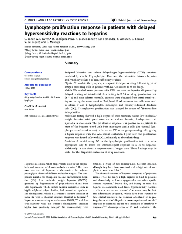
Fig 3. Lymphocyte transformation test (LTT)
to different heparins (Dp, dalteparin;
Bp, bemiparin; Np, nadroparin; Tz,
tinzaparin; Ep, enoxaparin; Sd, sulodexide) in
seven patients and the control group. The
heparin doses used were 5 IU and 0Æ5 IU. A
stimulation index (SI) > 3 with any of the
doses used was considered positive. LTTs to
sodium heparin, fondaparinux and lepirudin
were negative in all cases and controls (data
not shown). Mo ⁄ Bcell, monocytes and B cells;
imDC, immature dendritic cells.
Dp
20
15
0
Patient 2
20
mo/Bcell
10
5
0
Sd
Dp
Bp
Np
Tz
Patient 1
10
Bp
Tz
Sd
Mo/Bcell
10
DC
8
6
S.I.
S.I.
8
Dp
Np
Ep
4
6
Ep
4
2
2
0
0
T1
T2
T1
T2
Patient 3
10
Mo/Bcell
10
DC
8
8
6
S.I.
S.I.
Fig 4. Lymphocyte transformation test to
different heparins (Dp, dalteparin; Bp,
bemiparin; Np, nadroparin; Tz, tinzaparin;
Ep, enoxaparin; Sd, sulodexide) in patients 1
and 3 at two different time points
(T1, during the first month after the reaction
and T2, 1 year later). Mo ⁄ Bcell, monocytes
and B cells; DC, dendritic cells.
4
2
Ep
6
Np
4
2
0
0
T1
immunosuppressive effect. For this reason, in our study we
used EDTA as an anticoagulant, which proved to be a good
substitute in those samples manipulated within 16–18 h after
collection. Secondly, the time interval between the occurrence
of the reaction and the sample collection could be an important factor and the reason why in our study, with a short
time interval, only one patient was negative with the classical
LTT. The effect of this factor on the induction of the positive
response can be better observed in two cases (patients 1 and
T2
T1
T2
3) where the study was performed very soon after the reaction, during the first month, and also 1 year later. In both
patients the first in vitro analysis showed lymphocyte proliferation against the culprit LMWH, with a high degree of crossreactivity, whichever APC was used. Nevertheless, in cultures
with DC, the SI was higher than in those with monocytes
and B cells for the different heparins. In the second evaluation, positive proliferation was detected only to the culprit
drug, and only when DC were used as APC. We thus show
� 2008 The Authors
Journal Compilation � 2008 British Association of Dermatologists • British Journal of Dermatology 2009 160, pp259–265
�264 Heparin lymphocyte proliferation response, S. Lopez et al.
that DC are useful for improving lymphocyte responses, especially after a longer time interval.
The only patient with no proliferation (patient 7) was an
old woman (82 years) who had had two episodes, the first
with enoxaparin and the second with bemiparin, studied by
skin testing and LTT each time, 2 and 3 months after the
reaction. On both occasions skin testing was positive at a
much delayed reading (72 h) to both bemiparin and enoxaparin. The LTT, with and without DC as APC, was negative
for all heparins tested. This case was special, as the patient
had a very late skin-test positive response, with lower crossreactivity and with a negative lymphocyte response. This
may be due to the age of the patient, which may have interfered with the specific immune response,22 as it was also
detected when tetanus toxoid was used as a positive control
(data not shown).
A high degree of cross-reactivity was found between different LMWH, and even in some cases with heparinoids (sulodexide), by both skin testing and LTT. Moreover, others have
shown that this cross-reactivity does not depend on the
molecular weight of these compounds.23 However, there was
no correlation between the lymphocyte response and skin testing, as has been previously shown with beta-lactams.24 As
reported by others,25 although the chemical structure of
sodium heparin is very similar to LMWH, all our patients
were skin-test and LTT negative, and tolerated this drug. This
may be due to the fact that sodium heparin is administered
intravenously and, as has been reported, intravenous injection
of heparin is tolerated despite a DTH response appearing when
applied topically. Although there is no clear explanation for
this, differences in the absorption of heparin from skin and
different processing ⁄presentation of antigens depending on
the route could be involved.25 Nowadays, fondaparinux is
known to have a higher cross-reactivity with heparins than
previously thought,3,6 although all our patients were negative
in the different tests. In this study we also included lepirudin,
an anticoagulant of different origin and, as expected, all
patients tolerated the drug. Unfortunately, this drug may not
be considered an optimal alternative as it can induce severe
anaphylactic reactions on re-exposure.7
In summary, we found that the lymphocyte proliferation
test using DC as APC can be useful for the evaluation of
the immunological response in DTH reactions to heparins. It
has the advantage of being able to detect a positive
response over a longer period of time than the classical
LTT, by preserving the sensitivity to the culprit drug. This
finding may be relevant for the in vitro evaluation of DTH
reactions to heparins.
Acknowledgments
We thank Ian Johnstone for help with the English language
version of the manuscript. This work was supported by grants
from the Fondo de Investigación Sanitaria (PIO5290), the FIS
Network RIRAAF (RD07 ⁄0064) and the Junta de Andalucı́a
(199 ⁄04).
References
1 Hirsh J, Warkentin TE, Shaughnessy SG et al. Heparin and low-molecular-weight heparin: mechanisms of action, pharmacokinetics, dosing, monitoring, efficacy, and safety. Chest 2001; 119:S64–94.
2 Hirsh J, O’Donnell M, Weitz JI. New anticoagulants. Blood 2005;
105:453–63.
3 Bircher AJ, Harr T, Hohenstein L, Tsakiris DA. Hypersensitivity
reactions to anticoagulant drugs: diagnosis and management
options. Allergy 2006; 61:432–40.
4 Grassegger A, Fritsch P, Reider N. Delayed-type hypersensitivity
and cross-reactivity to heparins and danaparoid: a prospective
study. Dermatol Surg 2001; 27:47–52.
5 Poza-Guedes P, Gonzalez-Perez R, Canto G. Different patterns of
cross-reactivity in non-immediate hypersensitivity to heparins:
from localized to systemic reactions. Contact Dermatitis 2002;
47:244–5.
6 Utikal J, Peitsch WK, Booken D et al. Hypersensitivity to the pentasaccharide fondaparinux in patients with delayed-type heparin
allergy. Thromb Haemost 2005; 94:895–6.
7 Greinacher A, Lubenow N, Eichler P. Anaphylactic and anaphylactoid reactions associated with lepirudin in patients with heparininduced thrombocytopenia. Circulation 2003; 108:2062–5.
8 Molinari JF, Campo C, Shakir S, Ahmed T. Inhibition of antigeninduced airway hyperresponsiveness by ultralow molecular-weight
heparin. Am J Respir Crit Care Med 1998; 157:887–93.
9 Ali S, Hardy LA, Kirby JA. Transplant immunobiology: a crucial
role for heparan sulfate glycosaminoglycans? Transplantation 2003;
75:1773–82.
10 Douglas MS, Ali S, Rix DA et al. Endothelial production of MCP-1:
modulation by heparin and consequences for mononuclear cell
activation. Immunology 1997; 92:512–18.
11 Lever R, Hoult JR, Page CP. The effects of heparin and related
molecules upon the adhesion of human polymorphonuclear
leucocytes to vascular endothelium in vitro. Br J Pharmacol 2000;
129:533–40.
12 Ranjbaran H, Wang Y, Manes TD et al. Heparin displaces interferon-gamma-inducible chemokines (IP-10, I-TAC, and Mig)
sequestered in the vasculature and inhibits the transendothelial
migration and arterial recruitment of T cells. Circulation 2006;
114:1293–300.
13 Bottio T, Pittarello G, Bonato R et al. Life-threatening anaphylactic
shock caused by porcine heparin intravenous infusion during
mitral valve repair. J Thorac Cardiovasc Surg 2003; 126:1194–5.
14 Jappe U. Allergy to heparins and anticoagulants with a similar
pharmacological profile: an update. Blood Coagul Fibrinolysis 2006;
17:605–13.
15 Bircher AJ. Diagnostic tests for delayed type-hypersensitivity reactions to heparins and safety of intravenous heparin. Am J Contact
Dermat 1994; 5:56–7.
16 Ji SL, Cui HF, Shi F et al. Inhibitory effect of heparin-derived oligosaccharides on secretion of interleukin-4 and interleukin-5 from
human peripheral blood T lymphocytes. World J Gastroenterol 2004;
10:3490–4.
17 Torres MJ, Mayorga C, Fernández TD et al. T cell assessment in
allergic drug reactions during the acute phase according to the
time of occurrence. Int J Immunopathol Pharmacol 2006; 19:119–30.
18 Rodrı́guez-Pena R, Lopez S, Mayorga C et al. Potential involvement
of dendritic cells in delayed-type hypersensitivity reactions to betalactams. J Allergy Clin Immunol 2006; 118:949–56.
19 Ludwig RJ, Schindewolf M, Utikal J et al. Management of cutaneous
type IV hypersensitivity reactions induced by heparin. Thromb
Haemost 2006; 96:611–17.
� 2008 The Authors
Journal Compilation � 2008 British Association of Dermatologists • British Journal of Dermatology 2009 160, pp259–265
�Heparin lymphocyte proliferation response, S. Lopez et al. 265
20 Bircher AJ, Itin PH, Tsakiris DA, Surber C. Delayed hypersensitivity to one low-molecular-weight heparin with tolerance of
other low-molecular-weight heparins. Br J Dermatol 1995; 132:461–
3.
21 Ludwig RJ, Schindewolf M, Alban S et al. Molecular weight determines the frequency of delayed type hypersensitivity reactions to
heparin and synthetic oligosaccharides. Thromb Haemost 2005;
94:1265–9.
22 Pawelec G, Wagner W, Adibzadeh M, Engel A. T cell immunosenescence in vitro and in vivo. Exp Gerontol 1999; 34:419–29.
23 Grims RH, Weger W, Reiter H et al. Delayed-type hypersensitivity
to low molecular weight heparins and heparinoids: cross-reactivity
does not depend on molecular weight. Br J Dermatol 2007;
157:514–17.
24 Luque I, Leyva L, Torres MJ et al. In vitro T-cell responses to betalactam drugs in immediate and nonimmediate allergic reactions.
Allergy 2001; 56:611–18.
25 Gaigl Z, Pfeuffer P, Raith P et al. Tolerance to intravenous heparin
in patients with delayed-type hypersensitivity to heparins: a prospective study. Br J Haematol 2005; 128:389–92.
� 2008 The Authors
Journal Compilation � 2008 British Association of Dermatologists • British Journal of Dermatology 2009 160, pp259–265
�
 T. Fernandez