J. Mol. Biol. (1984) 172, 507-52]
The Functional Orion of Bacteriophage fl D N A Replication
Its Signals and Domains
GIAN PAOLO DOTTO, KENSUKE HORIUCHI AND NORTON D. ZINDER
The Rockefeller University
New York, N.Y. 10021, U.S.A.
(Received 13 June 1983, and in revised form 6 October 1983)
The origin of DNA replication of bacteriophage fl functions as a signal, not only
for initiation of viral strand synthesis, but also for its termination. Viral (plus)
strand synthesis initiates and terminates at a specific site (plus origin) that is
recognized and nicked by the viral gene II protein. Mutational analysis of the 5'
side (upstream) of the origin of plus strand replication of phage fl led us to
postulate tile existence of a set of overlapping functional domains. These included
ones for strand nicking, and initiation and termination of DNA synthesis.
Mutational analysis of the 3' side (downstream) of the origin has verified the
existence of these domains and determined their extent. The results indicate that
the fl "functional origin" can be divided into two domains: (1) a "core region",
about 40 nucleotides long, that is absolutely required for plus strand synthesis
and contains three distinct but partially overlappingsignais, (a)the gene II
protein recognition sequence, which is necessary both for plus strand initiation
and termination, (b)the termination signal, which extends for eight more
nucleotides on the 5' side of the gene II protein recognition sequence, (c) the
initiation signal that extends for about ten more nucleotides on the 3' side of the
gene II protein recognition sequence; (2) a "secondary region", 10O nucleotides
long, required exclusively for plus strand initiation. Disruption of the secondary
region .does not completely abolish the functionality of the fl origin but does
drastically reduce it (1% residual biological activity). W e discuss a possible
explanation of the fact that this region can be interrupted (e.g. fl, M]3 cloning
vectors) by large insertions of foreign DNA without significantly affecting
replication.
1. Introduction
The sinai[ icosahedral or filamentous single-stranded D N A bc,cteriophages such as
¢ X 1 7 4 or fI (fd, MI3) have served a s very useful model systems f o r s t u d y i n g
DNA replication ( D e n h a r d t et al., 1978).A w e a l t h of information i.~ now available
concerning both the e n z y m e s involved and their m e c h a n i s m s of action (Kornberg,
1980). W h a t seems still to be missing is detailed knowledge o f t h e signals p r e s e n t
in t h e p h a g e genome t h a t a r e r e q u i r e d for D N A replication to specificallyinitiate
a n d terminate, and how these signals are recognized a n d function. We have been
u n d e r t a k i n g a detailed analysis of t h e region a r o u n d t h e origin of replication of
bacteriophage fl t h a t is required for efficient viral s t r a n d synthesis t o occur
507
{X~22-2836184/040~)7-15 $03.(~qI0
© 1984 Academic Press Inc. (London) Ltd,
�~-~t.~
G.P. DOTTO, K. HORIUCH! AND N. D. ZINDER
(l)otto et ¢zl., 1983). The present study conclusively shows that this region
contains three distinct but partially overlapping domains that are required,
respectively, for initiation of DNA replication, for its termination, and for both
initiation and termination.
After entering the bacterial cell, the fl viral (plus} strand serves as template for
the synthesis by host. enzymes of the complementary {minus} strand. I n
particular, the host RNA polymerase recognizes a specific signal on the fl plus
strand attd, beginning at the "minus origin", synthesizes an RNA primer of about
30 nucleotides (Geider et al., 1978). The primer is subsequently elongated by DNA
polymerase IIl and eventually a double-stranded, circular, superhelicat molecule
(RFI) is formed. Viral {plus} strand synthesis is then initiated by the viral gene II
protein, which introduces a r~ick at a specific site {plus origin) on the plus strand
of the RF] molecule {Meyer et al., 1979). Elongation of the 3' end of the nick by
l)NA polymerase I l l is accompanied by displacement of the old viral strand via a
rolling circle mechanism {Gilbert & Dressier, 1968). In addition to its role in
initiation, gone II protein is also able, after one round of plus strand synthesis, to
cleave the n~scent single-stranded tail from the replicative intermediate and seal
it to form a covalent|y closed circle (Meyer & Geider, 1982). The final products,
in vivo, are a closed, single-stranded molecule and a closed, double-stranded
replicative form (RF) (Horiuchi et al., 1978b). In the early stages of phage
infection, the single-stranded circular DNA formed serves as template for minus
strand synthesis to yield more+ RF molecules. In later stages, the single strands
interact with the viral gene V protein (Mazur & Model, 1973; Mazur & Zinder,
1975) and are subsequently packaged into viral particles.
The phage "functional origin" of replication has been previously defined as the
minimal fi m:quenee that, when harhored in a plasmid, causes it to enter the fl
mode of replication provided that helper phage is present (Dotto et al., 1981a).
This is manifested in three ways: (1)stimulation of plasmid RF synthesis;
(2) ability of the plasmid to interfere with fl DNA replication; and (3) synthesis
of plasmid single-stranded DNA that is packaged into virion-tike particles that
are able to transduce resistance to antibiotics (transducing particles). Such a
sequence includes the fl plus origin and extends for only 12 nucleotides on its 5'
side (upstream) (Dotto et al., 1982b), but for more than 100 on its 3' side
(downstream) (Dotto et al., 1981a,1982a; Cleary & Ray, 1981). (The fl minus
origin, which lies about 30 nucieotides on the 5' side of the plus origin, is
dispensable in this system because other sequences elsewhere in the plasmid DNA
can serve for minus strand initiation (Cieary & Ray, 1981; Dotto & Zinder,
l.~m3).)
Deletion analysis of the 5' side of the f i functional origin has revealed that this
region contains two domains essential for function: (I) the signal for initiation of
plus st,rand synthesis and (2) the signal for its termination (Dot.to & Horiuchi,
1981; Dotto et al., 1982a). The 5' boundary of the signal for plus strand initiation
might coincide with the 5' boundary of the gene I ! protein in vitro recognition
sequence {four nue/eotides on the 5' side of the gene I I protein nicking site) while
the 5' boundary of the signal for termination extends for eighb nucleotides move
beyond it (Dotto el ai., 1982a).
�ORIGIN OF fl DNA REPLICATION
51~)
In this paper, we describe several d e l e t i o n a n d insertion m u t a n t s on t h e 3' side
o f the f l plus origin t h a t were c o n s t r u c t e d a n d t e s t e d for t h e i r effect on t h e ability
o f the fl origin to f u n c t i o n in vitro, as a s u b s t r a t e for gene I I protein nicking
a c t i v i t y , a n d in vivo, as a signal for e i t h e r initiation or t e r m i n a t i o n o f plus s t r a n d
synthesis. This has led to a detailed analysis o f the 3' side o f the f l origin a n d a
d e t e r m i n a t i o n o f its functions and boundaries.
2. Materials a n d M e t h o d s
(a) Bacteria, phage and plasmids
The bacterial strain used was Escherichia coli K38 (Lyons & Zinder, 1972). fl and M13
phages were from our laboratory stock. R283 is an fl variant with a Pet linker
(G-C-T-G-C-A-G-C) inserted at position 5766 of the fl map (Dotto et al., 1982b). pBR322
(Bolivar et al., 1977), pDl2 and pDl7 (Dotto et al., 1982b) have been described, pD27 and
1)1)37 were obtained from pDl7 (a pBR derivative containing the fl HaeIIi-F fragment at
its EcoRl site) by destroying either its HindIII or Aval sites by end-filling with Klenow
fragment, followed by eireularization with T4 DNA ligase, pD30 is a pBR322 derivative
containing the HpaII-H fragment of R283 into which a HindIII linker was inserted at its
HinfI site. It was obtained as follows: DNA of pD12, which is a pBR322 derivative
containing the HpaII-H fragment of R283 flanked by BamHI linkers at the BamHI site
(Dotto el al., 1982b), was digested with HinfI (which cleaves the R283 fragment only once
at position 5788), and the 2 fragments of interest (containing either the left side of the
R283 fragment, for pD28, or the right side, for pD29, plus flanking plasmid sequences)
were purified. Following end-filling with Klenow fragment, HindIII linkers
(C-C-A-A-G-C-T-T-G-G) were attached and, after HindIII and BamHI cleavage, the
fragments were inserted at the HindIII-BamHI sites of pBR322 (pD28 and pD29), pD30
was obtained by inserting the two R283 fragments, purified from pD28 and pD29, into the
BamHI site of pD27. pD38 was obtained by inserting the Ml3 HpaII-H fragment at the
BamHl site of pl)37 by use of Klenow fragment and BamHI linkers
(C-C-G-G-A-T-C-C-G-G). The constructiGn of pD39, pD40 and pD48 is described in Results.
(b) Plasmid and phage R F preparation
The volumes given below are for a 20-ml culture. Cells containing plasmids were grown
to saturation in fortified broth (2.5% (w/v) Bacto-tryptone, 0.750/0 (w/v) Bacto-yeast
extract, 0.6% (w/v) NaCI, 0.1% (w/v) glucose, 50 m~-Tris,HCl, pH 7.6). For preparing
phage RF, cells (K38 or derivative) were grown at 37°C to an 0.0.660 of 0.4 to 0.8 in
fortified broth and infected with phage at a multiplicity of infection of 100. After 15 min at
37°C, chloramphenicol was added to 15#g/ml, and the cells were allowed to grow for
another 60 to 120 min. Cells were collected by eentrifugation and resuspended in 0.75 ml of
buffer A (25% (w/v) sucrose, 50 m,~-Tris- HCI, pH 8.0, 40 mM-EDTA). Lysozyme (0.2 ml of
15 mg/ml in buffer A) was added and incubated for 5 rain on ice. E D T A (0.15 ml of 0.5 M)
was added and incubation was continued for 10 min. Buffer B (1 ml; 0.1% Triton X-100,
50 mM-EDTA, 50 m.~-Tris. HCI, pH 8.0) was added and incubation was continued for
10 min. It was then centrifuged at 25,000 revs/min in a type 40 rotor for 6 0 min, the
supernatant was collected, and then heated to 65°C for 15 min. The floceulent.precipitate
was removed by centrifuging for 15 rain at 10,000 revs/min in a Sorvall'SA-600 or 8S:34
rotor. Buffer C (1 : 3 vol.; 40% (w/v) polyethylene glycol [Carbowax 6000], 2-5 ~-NaCI) was
added a n d incubated at 4°C for 2 to 4 h. The DNA was recovered by eentrifugation at
10,000 revs/min for 5 min. The supernatnnt fluid was discarded and the wet pellet was
resuspended in 0-3 ml DB (DNA buffer; 10 mM-Tris-HCl, pH 8.0, 10 mM-NaCI, 0.2MEDTA): Pancreatic RNase (5/al of n 2 mg/ml solution in ,water (Worthington
Bioehemieals)) was added and the mixture was incubated at 37°C for 60 min. Proteins were
�•
510
G. P. DOTTO, K. H O R I U C H I
AND
N. D. Z I N D E R
removed by extraction with phenol. The aqueous phase was precipitated with ethanol and
the pellet was washed once with 70°/o (v/v) ethanol and dried in vacuo. The DNA was
dissolved in 100/~! DB. The yield was typically 100 gg. When neees~ry, the R F I was
purified by isopycnic centrifugation in CsCl/ethidium bromide.
{c) DNA manipulations
All DNA manipulations have been described (Dotto et al., 1981a). Sticky end ligation of
cohesive ends was carried out by use of E. coli DNA ligase (New England Biolabs) under
the conditions described by the manufacturer. The linkers were purchased from
Collaborative Research.
(d) Deletion mutants
pD38 DNA, linearized with AvaI, was treated with exonuclease Bal31 (BRL) (Gray et
al., 1975): 0.4 unit of enzyme per pmol of DNA at a concentration of 0.1 pg per pl in
600 mM-NaCI, 12 mM-NaCI, 12 mM-MgSO4, 12 mM-CaCI 2, 20 mM-Tris. HCI (pH 8), 1 mMEDTA, was incubated at 12°C for 6 rain or at 30°C for 30 s. The rate of Bal31 digestion is
not exactly reproducible from one experiment to another, and it was necessary to titrate
the enzyme each time. The reaction was stopped by addition of EGTA, 160 mM final
concentration. After eireularization with T4 DNA ligase, the DNA was again treated with
Aval and then used to transform calcium-treated E. coli K38 cells. Ampicillin-resistant
colonies were isolated and the plasmid DNA characterized.
The nucleotide sequence of the deletions obtained from pD38 was determined as follows.
The DNAs of the various mutants were cleaved with BamHI and the 3' ends were labeled
by addition of Klenow fragment and [32P]dGTP (Maxam & Gilbert, 1980). The DNAs were
subsequently cleaved with Hinfl "and also with TaqI in the eases where one of the two
labeled pBR322 bands was expected to co-migra:'~e with the desired fl fragment. The
fragments of interest were gel-purified and their sequence determined as described by
Maxam & Gilbert (! 980). The procedure for determining the nueleotide sequence of A + 29
was different. A + 2 9 DNA was cleaved with AsuI and the new fragment, which
corresponded to the f l - p B R fusion caused by the deletion, was gel-purified. After labeling
of the 3' ends, the DNA fragment was cleaved with HpaII and the fragment of interest was
gel-purified prior to nucleotide sequencing. In the case of insertion mutants pD39 and
pD40, the fl H p a I I - H fragments were purified after BamHI cleavage and digested with
Asul; the 3' ends were labeled and the DNA was subsequently cleaved with HaeIII. After
gel purification, the sequence of the fragment of interest was determined.
(e) Prod.uetion of phage and antibiotic resistance transduci~u2 particles
Cells harboring the various plasmids, at a density of 2 × 10⁸/ml, were infected with phage
at a multiplicity of 20 for 20 rain; they were then washed and incubated in trypt0ne broth
for 3 h. The supernatant was incubated at 56°C for 30 rain (a procedure tliat results in the
killing of the cells without, inactivation of the phage) and the yield of plaque-forming units
(p.f.u.) was determined. Phage stocks prepared in this way were used to infect
exponentially growing cells at a multiplicity of infection of i (to minimize multiple
infection). After 10 rain the infected cells were diluted into anti-fl antiserum and infective
centers (i.e.) were determined by plating on K38 cells. Antiserum-treated cells were also
plated as colony-forming units (e.f.u.) and incubated for 3 h at 37°C before overlaying with
soft agar plus ampicillin (1 mg]plate), The number of transductants (Amp a colonies) was
taken as a measure of the Amp~-transducing particles produced.
(f) In vitro gene I I protein assay
Gene II protein was purified asdescribed (Dotto eta/., 1982b) from E. coli K38 (pD2)
cells that contain a plasm;d into which gene I I has been cloned and that produce ~large
�ORIGIN OF fl DNA REPLICATION
511
amounts of gene II protein (Dotto et al., 1981b). One unit of enzyme is defined by the
complete conversion of 0.5/~g of fi RFI DNA'into RFII and RFIV in 20/~1 of 20 mrdTris. HCI (pH 8), 5 mM-MgCI2, 5 ms-dithiothreitol at 30oC within 30 rain. The reactions
were stopped with 2/d of 20% (w/v) sucrose, 0.5% (w/v) sodium dodecyl sulfate, 200 mmEDTA, and 0 . ] ~ (w/v) bromphenol blue and analyzed by agarose gel (1%) electrophoresis
in the presence of ethidium bromide (0.5 pg/ml).
3. Results
(a) Isolation of deletion and insertion mutants at the f l origin
R283 is an fl v a r i a n t with the H i n f I site at position 5767 (13 nucleotides
u p s t r e a m of t h e plus origin) s u b s t i t u t e d by a unique Pst site (octamer). This
s u b s t i t u t i o n does n o t affect the functional activity of t h e origin (Dotto et al.,
1982b). Thus, the functional origin of this phage (HpaII-H f r a g m e n t , see Fig. l)
has a unique HinfI site left, at position 5788, eight nucleotides d o w n s t r e a m from
the plus origin. I t was of interest to see if this site could be used for s t u d y i n g t h e
replicative function of t h e 3' side of t h e phage origin. F o r this purpose, we started
from pD12, a pB1~322 derivative t h a t contains t h e R283 functional origin
(HpaTI-H fragment), and we inserted into the H i n f I site of the R283 f r a g m e n t a
H i n d I I I decamer. T h e insertion, however, completely i n a c t i v a t e d t h e . R283
origin. This was indicated by the fact t h a t t h e resulting plasmid, pD30, did n o t
interfere with fl and did n o t yield t r a n s d u c i n g particles at any detectable level in
the presence of helper phage (Table 1}. Therefore, pD30 could n o t be used for
f u r t h e r studies.
i~
~,,--"-Hoe l~ - F
,
:
i
H~II-H
~,:
HoeI~-D.--~
:
I ~ e
5600
Gene .~E I
•
)~*.-HoeI~-G.-~:4
5800
lG
[ Gene ]I
Biol. ocfivity
;
(~i'~.
: pD30
:
@$
;
pD38
-
."-+.40,56
1%
I ,~+41070
1%
~
i
( ,~
Ep_~
)
i
"" ~ -
pD48
:
@
,~+29
:
~'.
:
@
:
~
)
,
(0001%
100 %
1%
O'OI %
i Z~*ll
<0001%
~)
: pO39
30 %
~
:
pD40
1%
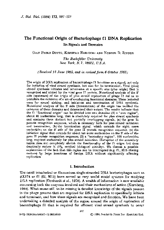
Fie. 1. Map of the intergenic region (IG) of bacteriophage fl (Horiuchi el al., 1978a) (upper line) and
of the fl fragments present in the various plaamids (lower lines). The numbers indicate the nucleotide
position on the fl map (Beck & Zink, 1981; Hill & Petersen, 1982}. (+) and (--) represent the origins
of plus and minus strand syntbesis, respectively. The positions of the HaeIII-F, G, D and HpaII-H
fragments are shown. The HaeIII site between F.and G is also cut by AsuI. X, Indicates the HinfI
cleavage sites; ~, indicates the Aval site, present in MI3. The sites in pD30, pD39 and pD40 where
extra nucleotides (10, 8, and 16, respectively) were inserted, are indicated. Blank spaces between
brackets indicate the deleted sequences in A+40, 56, A+41,70 and A-t-lli The relative biological
activity is as measured in Table 1.
�512
G. P. DOTTO, K. HORIUCHI AND N. D. ZINDER
The fl and Ml3 nucleotide sequences in the origin region as well as in the rest of
the phage genome differ from each other by only a few nucleotides (Beck & Zink~
1981; Hill & Petersen, 1982). These two phages should therefore ~'~'e
interchangeable and may be used according to convenience. In particular, because
of a T to G difference at position 5830, M13 rims a unique AvaI site conveniently
located 40 nucleotides downstream from the plus origin (Fig. 2). For this reason,
we chose to use M13 to study the 3' side of the phage functional origin.
pD37 is a pBR322 derivative that has lost its AvaI site and contains the fl
HaeIII-F fragment at its EcoRI site. We have shown previously that this
fragment contains a signal important for virion morphogenesis ("morphogenetic
signal") (Dotto et al., 1981a; Dotto & Zinder, 1983); in its presence, the yield of
transducing particles from chimeras that contain a phage functional origin of
replication is enhanced about 100-fold. The ability of a chimeric plasmid to yield
transducing particles can be taken as a measure of the functionality of its phage
origin and the presence in it of the morphogenetic signal allows a more accurate
quantification of the assay (Dotto et al., 1982b).
Piasmid pD38 was obtained by inserting the Ml3 functional origin (HpaII-H
fragment) into the BamHI site of pD37. From pD38, by double digestion of its
DNA with AvaI (cleaving only inside the M13 origin) and PvuII (cleaving inside
pBR sequences, at map position 2067 (Sutcliffe, 1978)) and subsequent
recircularization, a second plasmid was derived (pD48). pD48 has lost the part of
the MI3 (fl) functional origin on the 3' side of the AvaI site (Figs I and 2) as well
as a large fragment of the plasmid sequence next to it.
In a second set of experiments, pD38 DNA was linearized with AvaI, treated
with exonuclease Bal31 (Gray et al., 1975) under limiting conditions, and religated
to transform calcium-treated E. coil cells. The resulting clones were screened for
their ability to yield transducing particles after fl infection, pD38 was used as a
standard. Several clones with absent, reduced, or nearly normal ability to yield
transducing particles were chosen and the nucleotide sequences of their fl
fragments were determined. Figures l and 2 show the deletion map on the 3' side
of the plus origin that was obtained.
In a third set of experiments, pD38 DNA was again linearized with AvaI, the
ends were made blunt with Kienow fragment, and BgllI linkers (C-A-G-A-TC-T-G) were inserted before recircularization. The DNA of the resulting clones was
checked for sensitivity to BgIII and also PstI. If two (or more) BgtII linkers were
inserted in tandem, a new Pst site would be formed. Two plasmids, one with a new
BglII site (pD39) and the other with both a new BffIII and a Pst site (pD40), were
chosen for characterization by nucleotide sequencing. They were confirmed to
contain at their AvaI sites a BgllI linker inserted, respectively, as a monomer
(8 nucleotides, pD39) or as a dimer (16 nueleotides, pD40).
(b) Plus strand synthesis ie strongly affected by the
deletion~ and iusertion.s
The in vivo biological activities of the deletion and insertion mutants, measured
either as their ability to interfere with fl replication or to yield transducing
�513
O R I G I N OF fl DNA R E P L I C A T I O N
TABLE l
Biological activity of the origin m u t a n t s
1'lasmid
p.f.u./mlt
Transducing
particles
per m l : ~
pD30
A+ll
1-1 x 10 ~'
1 . 4 x l 0 t~'
< 1 x 10 "~
<lxl0
"7
A+29
pD48
A+40, 56
1.3x
1.3x
1.6×
l-3x
4-5 x
9x
5x
A+41,70
p1)39
pD40
pD38
10I=
1012
1012
1 x 10⁸
1.1 x 101°
1.4x 10)0
1.3x 10 j°
1"5 x 10¹¹
1 x 101°
5 x 1OI°
l 0 t2
1011
101 z
101°
% Relative
biological
activity§
.
.
Gene I I
protein
nicking[ I
.
.
0.01
1
1
.
.
Plus
Plus
strand
strand
initiation¶ termination¶
.
.
+
+
+
-NT
--
+
NT
+
1
+
--
+
30
1
100
+
+
+
-t=
+
+
+
+
p.f.u., plaque-forming units; NT, not tested.
1" Phage stocks were obtained from cells harboring the various plasmids as described in Materials
and Methods.
:[: The number of transductants (ampicillin-resistant colonies} is taken as a measure of the ampicillin
resistance transducing particles produced. I t was determined as described in Materials and Methods.
§ The relative biological activity of the plus origin is defined in each case as the ratio between the
yield of transducing particles in the phage stock obtained with a certain deletion and that obtained
with pD38, the biological activity of which is arbitrarily set at 100%. The yield of transducing
particles cannot be taken as an absolute number, but. needs to be normalized to the yield of phage
(transducing particles/p.f.u.) given the interfering activity of the various plasmids.
II Gene II protein nicking was tested ~s described in the legend to Fig. 3.
¶ Initiation and termination of the plus strand synthesis were determined as explained in Figs
4 and 5.
CCTATTGGTT
G 70
G
"CT T T A G,,
C
C60
G
T
T
T
T
,,T~T~,
,..,.---~ C~_ = ATe°
= G e n e - v II protein
=A
fA~,
G TA A
C , G .~BOO"A=T"c
A=T
C=C
C=G
C=G
7oC • G
T =A
r _ ^M
/
!r ~*
=
~---
^
I-~]O
~
("7"C T = AC ACT C A AC C T A T C T C
A
ABO
A
A
A
A
T
A
A
"r90
GGGCTATTCITTTTGATTT
ATAA /
A
CAATTTATAAACAAT
5909
5900
FIo. 2. Nucleotide sequence of the fl (M13) functional origin. The numbers indicate the nucleotide
positions in the fl map (Beck & Zink, 1981; Hill & Petersen, 1982}. The precise 5' boundary of the
functional origin was previously determined (Dotto et al., 1982b). The 3' boundary of this region lies
somewhere between positions 5867 and 5909 (Dotto et al., 1981a; Cleary & Ray, 1981). The only
nucleotide by which the fl sequence differs from that of M13 in this region is indicated between
brackets (position 5830). Filled arrow, gene II protein nicking site (plus origin). The deletions have
been named after their relative distance from the fl plus origin. The 5' endpoints of deletions A+ 11
and A+29 are indicated. The sequence deleted in A+40, 56 and A+41, 70 is also shown. A - 4 and
A - 1 2 indicate the 3' endpoints of 2 deletions on the 5' side of the plus origin already described (Dotto
et al., 1982a,b). Tile open arrow on the left indicates the positio n (HinfI site) of the HindIII decamer
in~rtion in pD30. The oi~.n arrow "on the right indicates the position. (At~zI site) in pD39 and pD40
where 8 or 16 nucleotides (Bf/lII linkers), respectively, Were inserted. 7.~yphens have been omitted from
the sequence for clarity and the usual convention has not been followed.
�514
G. P. DOTTO, K. HORIUCHI AND N. D. ZINDER
particles, were compared with those of the parental plasmid, pD38 (Table l). It is
clear that two classes of deletion mutant can be distinguished according to their
residual activities. With deletions A+40, 56 and A+41, 70 (which extend from
the 40th to the 56th and from the 41st to the 70th nucleotide from the gene II
protein nicking site, respectively) the activity drops to about 1% of the control.
The same effect is seen also with pD48, where the M13 sequence extending beyond
the 50th nucleotide from the gene II protein nicking site had been totally
removed. A much more drastic drop in biological activity (0.01% or less residual
activity) was observed with two other deletions, A+29, which extends from the
29th nueleotide from the gene II protein nicking site to inside pBR sequences
(position 1567 of the pBR map), and A + l l , which extends from the l lth
nucleotide from the gene II protein nicking site to about 100 nucleotides
downstream (position 5890 of the fl map).
An insertion of eight nucleotides at the AvaI site (pD39) has relatively little
effecton the function of the origin (30~/o residual activity) whereas an insertion of
16 nucleotides in the same position (pD40) causes a drop in biological activity to
about 1%.
(c) The 3' boundary of the gene II protein recognition sequence
The effects observed with the various deletion and insertion mutants could
result from the disruption of the gene 1I protein recognition sequence. In
particular, this might explain the very drastic loss of biological activity observed
with some of the deletions and insertions (A+ 11, A+29, and pD30). To test such
a possibility, DNAs from the various mutants were used in vitro as substrates for
purified gene II protein. In addition to its nicking function, this protein possesses
a closing activity so that, in vitro, in the presence of Mg 2+, it converts fl I~FI
molecules into RFII (relaxed nicked circles) and RFIV (relaxed, closed circles) in
approximately equimolar amounts (Meyer & Geider, 1979). When this reaction
was used with all the mutants described, we found that gene II protein did not
cut either deletion A + l l or insertion pD30. All the others, including deletion
A+29, were nicked by gene 11 protein as efficiently as fl. The minimal amount of
gene II protein necessary to convert 50% of fl RFI molecules into R F I I and
RFIV was approximately the same for all the mutants {Fig. 3), other than A + 11
and pl)30.
From these results we can conclude that the 3' boundary of the gene II protein
recognition ~equence lies somewhere between position 5791 and 5809 (the
endpoints of A+ 11 and A+29) and that. the insertion of ten nucleotides in this
sequence (position 5791; pD30) can completely inactivate it.
(d} The 3' boundaries of the signals for initiation and termination
of plus strand synthesis
The f l functional origin contains two specific signals, one for initiation and the
other for termination of plus strand synthesis (Horiuchi, 1980; Dotto & Horiuchi,
1981). With chimeric plasmids containing two f l functional origins (inserted in
�ORIGIN
O F fl D N A R E P L I C A T I O N
515
pD30
&+11
~,+29
pD48 &40,56 A41,70
pD39
pD40
pD38
+ -
+-
+-
+-
+-
+-
+-
+-
+-
fl
+-
RF ] I " ~
RFT
RF . ~ ' ~
FIo. 3. Conversion of deletion and insertion mutant R F I DNA into R F I I and R F I V by purified
gene I I protein in vitro. R F I DNA (l pg) from the various m.utants, as well as from fl, was incubated
with ( + ) or without ( - ) purified gene II protein (1 unit) for 30 min at 30°C in a 40/d volume reaction.
The assays were performed as described in Materials and Methods. A DNA band migrating slightly
faster than the plasmid R F I I DNA is visible in all samples and corresponds to some contaminant
chromosomal DNA. The relative positions of RFIV and R F I I of A + 2 9 and pD48 are indicated by
arrows in the respective lanes.
the same orientation), synthesis of chimeric plus strand DNA is initiated, after fl
infection, at either one of the two fl origins and is terminated at the other. Hence,
the chimeric plasmids segregate into two replicons, each of them containing on|y
one origin (Dotto & Horiuchi, 1981). The effidiency of different fl origins to
function as signals for either initiation or termination of plus strand synthesis can
be assessed easily with this system, provided that the two components of the
chimera (A and B, Fig. 4) are distinguishable from each other by size. This system
has previously allowed us to map the 5' boundaries of the signals for plus strand
initiation and termination (Dotto et al., 1982a) and we have employed it here to
determine their 3' boundaries.
pD16 is a pBR322 derivative that contains two fl wild-type functional origins
(HpaII-H fragment), one inserted at the EcoRI site (or|I) and the other at the
B a m H I site (oriII) (Dotto et al., 1982a). A set of plasmids analogous to pDl6 was
constructed, with oriI still wild type, but with one of the various deletion and
insertion mutants described above substituting for oriII. In this system, if oriII
lacks the signal for initiation but retains that for termination, then, upon f l
infection, DNA synthesis should occur only from oriI t o oriII, and of the two
components of the chimera, only A should be found inside t h e cell as an
independent replicon. On the other hand, i f oriII lacks the:signal for termination
but not for initiation, D N A synthesis should start from both oriI and oriII but
should terminate only at or|I, a n d only component B a n d the starting chimera
should be synthesized inside the cell. If. oriII lacks the signals for both initiation
• "fl
~fl
RF]]
RFI
fl RF117
�516
G. P. D O T T O , K. H O R I U C H I
//
~
N,L
I ~'r"
',,\
\\
B.-.
~
~
A N D N. D. Z I N D E R
A
J,)
//
i
Fit;. 4. Map of plasmids containing two fl origins, or/I and orill (see the text). A and B., lesser and
greater ares of the chimeras. The orientation of the fl fragments is indicated by thin arrows. The
position of the insertions or deletions on the 3' sideof the gene II protein nicking site (thick arrows), is
indicated by a blank space between brackets. > - , and -], Signals for initiation and termination of
plus-strand synthesis, respectively.
and termination, the only kind of plasmid DNA found inside the cell should be
that of the starting chimera. Cells harboring pDl6 and infected with fl contained
two new species of DNA of the expected size (Fig. 5). With cells harboring a
p[asmid containing at its oriIl deletion A4. 1 i, neither component A DNA nor
component B could be detected. This result was not unexpected, since deletion
A 4- [ ! completely inactivates the fl origin in vivo and also blocks gene II protein
recognition in vitro. With a plasmid containing deletion 54.29 at its oriII,
component B DNA again could not be detected, indicating that this deletion
severely impairs initiation. However, component A D.NA was synthesized in good
quantity, indicating that deletion A 4. 29 retains the signal for termination. The 3'
boundary of this signal lies somewhere between position 5791 and 5089 (the endpoints of A4.11 and A4.29) and might well coincide with the 3' boundary of the
gene II protein in vitro recognition sequence.
Similar results were obtained with plasmids containing at their oriII site either
deletion A+40, 56 or A+41, 70; initiation is severely compromised (very little
component B synthesized) while termination is not (component A synthesized in
good amounts). The fact that some component B DNA can be detected with
plasmids containing A 4.40, 56 and A + 41,70, but not A 4-29, correlates well with
the fact that the latter deletion affects much more drastically the functionality of
the fl origin (0.01% versus 1% residual biological activity). A situation similar to
that described for A4.40,56 and A + 4 1 , 7 0 i s also f o u n d w i t h a plasmid
containing at, its or/II the 16-nucleotide insertion of pD40. This insertion blocks
initiation of plus strand synthesis (i.e. component B synthesis), while termination
(i.e. component A synthesis) is unaffected. The eight-nucleotide insertion of pD39
has very little effect on initiation, and none at all on termination. This finding
correlates well with the fact that with pD39 the biological activity drops only
two-or threefold, while with p D 4 0 it drops about 100=fold.
�ORIGIN
j~.1-11
A',29 &+40,56 &t'41,76 pD39
pD40
+-
+-
+ - + -
+-+
517
O F fl D N A R E P L I C A T I O N
I +-
pD16
fl
_
fl RFI
C
B, fl ss
--A
Fio. 5. Proficiency of the various insertion and deletion mutants as signals for either initiation or
termination of plus-strand synthesis. Exponentially growing K38 cells (20 ml) harboring plasmids with
two fl origins (see the text) were infected with fl (multiplicity of infection, 50). After 30 min at 37°C,
cells were harvested and the intracellular DNA was purified as described in Materials and Methods.
The DNA was analyzed on a 1% (w/v) agarose gel (in the presence of ethidium bromide; 0.5 pg/ml). A
and B, Positions expected for the DNAs of components A and B of the chimeras, if replicating as
autonomous molecules; C, RFI DNA of the Starting chimeras. Unlabeled arrow on the left, R F I DNA
of A+29, starting chimera. The positions corresponding to fl R F I and ssDNA are also indicated, fl
ssDNA co-migrates with component B RFI DNA of the various plasmids. Treatment with
endonuclease S l showed that with all the chimeras, DNA migrating at this position is double-stranded
(Si-resistant) and therefore must correspond to component B DNA (data not shown), ssDNA, singlestranded DNA.
4. Discussion
The analysis of the fl functional origin presented in this paper has conclusively
identified three distinct domains that are required for efficient plus strand
replication (Fig. 6). (l) The gene II protein recognition sequence, (2) the signal for
initiation of plus strand synthesis, and (3) the signal for its termination.
The gene H protein recognition sequence, as determined by the in vitro nicking
assay, extends from not more than four nucleotides on the 5' side of the gene II
protein nicking site (Dotto et al., 1982b) t o I 1 to 29 nucieotides on its 3' side. This
relatively long sequence is strongly asymmetric. It does not include the
palindrome around the gene II protein nicking .site, which was originally thought
to be important for gene II protein recognition, and it does not contain any other
feature t h a t might explain the fact that gene II protei n reacts with fl DNA (and
related plasmids) only when it is in a supercoiled form (Meyer & Geider, 1979).-In
the presence of Mn 2÷, gene II protein cuts both strands of DNA.at the f l origin
instead of introducing a nick (Dotto etal., 1981b). This effect was ascribed by
others to two gene I I protein molecules t h a t bind .to t h e same:palindromic
sequence around the plus origin on opposite strands of the,DNA molecule (Meyer
& Geider, 1982)..However, deletion mutants with the 5' half of the-palindrome
�51~
G. P. DOTTO, K. HORIUCHI AND N. D. ZINDER
fl funclionol
A
origin
B
( core )
V'
B1
~o
5769
Gene n prolein
Termination
Initiation
J~o
B2
~0
~o
~o
5909
[I---:-:--- 1
~,,'/7"/'/~
ii
I
Fro. 6. The fl flmetional origin, its signals and its domains. Numbers indicate nucleotide positions
on the fl map. ~=b, Gene II protein nicking site; ~ ~-, palindromic region around the gene II protein
nicking site. The locations of the gene I] protein recognition sequence and of the signals for initiation
and termination of plus strand synthesis am indicated. Also shown are the domains into which the fl
functional origin can be divided. For more details, see the text.
around the plus origin removed (e.g. A - 4 , Fig. 2) are still efficiently cleaved by
gene I1 protein in the presence of Mn 2+ (Dotto, unpublished results). Therefore,
even under these conditions, the region of palindromic s y m m e t r y around the plus
origin is not necessary for gene II protein recognition.
The sequence required for nicking by gene I I protein in vivo may well be the
same as t h a t recognized in vitro. This is indicated by the fact that the 5' and 3'
boundaries of the gene II protein in vitro recognition sequence seem to coincide,
respectively, with the 5' b o u n d a r y of the signal for initiation of in vivo plus strand
synthesis and the 3' b g u n d a r y of the signal for termination. This fact is unlikely
to be coincidental and suggests t h a t the gene II protein recognition sequence is an
important determinant, even if not the only one, in both initiation and
termination of plus strand synthesis. The notion t h a t gene II protein is required
not only for initiation but also for termination of fl plus strand synthesis has
already been d e m o n s t r a t e d by previous in vitro studies (see below), and is
confirmed by the present results.
The signal for termination of fl plus strand synthesis extends from 12
nucleotides on the 5; side of the gene II protein nicking site (Dotto et al., 1982a) to
11 to 29 nueleotides on its 3' side. It is likely to include only two elements: (1) the
gene II protein recognition sequence, discussed above, and (2) the palindrome
located around the gene II protein nicking site and extending for eight nucleotides
more on the 5' side of the gene II protein recognition site. The invoh,ement of
gene I] protein in the termination of plus strand synthesis has been studied
in vitro (Meyer & Geider, 1982). After one round of replication, gene I I protein is
able to cleave the displaced single-stranded tail from the rolling circle
intermediate and then seal it to form a covalently closed circle. I t is reasonable to
assume t h a t g e n e II protein recognizes the same sequence to nick the R F I
molecule at the onset of replication and to cleave the n a s c e n t strand after one
round of synthesis. The DNA conformation, however, could be quite different in
the two situations. The palindrome around the gene II protein nicking site might
then be required for the gene II protein recognition sequence to assume the proper
�ORIGIN OF fi DNA REPLICATION
514}
conformation after one round of synthesis. Alternatively, the palindrome might be
required only after cleavage to bring together the 5' and 3' ends of the .singlestranded molecule for circularization. In this respect, it is important to remember
that the fl gene II protein, unlike the qbX gene A protein (Eisenberg.& Kornberg,
1979), does not remain covalently attached to the 5' end of the nick it produces.
i n vitro replication studies, using deletion mutants with the 5' half of the
palindrome removed, should test the two possibilities.
The signal for initiation includes the gene II protein recognition sequence and
extends for about 100 nucleotides downstream (Dotto et al., 1982a, and the
present data). Therefore, it consists of the entire fl functional origin (Dotto et al.,
1981a; Cleary & Ray, 1981), excluding the 5' half of the palindrome around the
gene II protein nicking site. According to the results presented here, the f l
functional origin can be divided into two domains, domain A, extending up to
about position 5819, and domain B, extending beyond it (Fig. 6).
Domain A is the "core" of the f l origin. It contains the gene II protein
recognition sequence, the termination signal and, on its 3' side, an additional
sequence of about ten nucleotides that is absolutely necessary for plus strand
initiation. If this ten-nucleotide sequence is deleted the functionality of the fl
origin drops to almost undetectable levels (0.01% residual biological activity with
A + 2 9 versus l~o with A+40, 56, A+41, 70 and pD48, see Fig. 1), even if the gene
II protein recognition sequence and the signal for termination are retained. The
insertion of 16 instead of eight nucleotides close to the border between domain A
and B has striking effects on plus strand initiation (l~/o versus 30~o residual
biological activity of the fl origin). This raises the possibility that the distance
between the two domains is crucial for efficient initiation to occur.
Domain B contains sequences required exclusively for plus strand initiation.
Deletions in this region drastically reduce the functionality of the fl origin, but
not as severely as in domain A (approximately 1o~) of residual biological activity
with pD48, A÷40, 56 and A+41, 70). Domain B contains the site most frequently
used for cloning in the single-stranded DNA phages (Messing et al., 1977; Zinder &
Boeke, 1982). Thus, this domain can be separated in a phage into two
subdomains, B1 and B2 (Fig. 6), by large insertions of foreign DNA without
significantly affecting replication. Domain B1, on the 5' side, consists of a stretch
of about 50 nucleotides while domain B2, on the 3' side, contains about 40
nucleotides, rich in A and T. W h y large stretches of foreign DNA can be inserted
in such a crucial region of the phage genome and not affect its replicative function
remains to be determined. However, recent results indicate that a mutation in one
of the viral genes is necessary for replication to occur under these conditions (see
below) (Dotto & Zinder, 1984).
The requirement for plus strand initiation of about 1O0 nueleotides on the 3'
side of the gene II protein recognition sequence might be due to a specific
interaction of this region with some host, or, alternatively, some phage proteins
(most likely gene II or gene V proteins, th e only fl proteins known to be involved
in DNA replication (Horiuehi et al., 1978a). This second possibility is now
supported by the finding that phage vectors, such as those described above,
when used as helper phage instead of fl wild type, are able tc rescue chimeric
�520
G. P. DOTTO, K. HORIUCHI AND N. D. ZINDER
plasmids partially or totally lacking domain B b u t retaining only domain A
(Dotto & Zinder, 1984). pD48, A + 4 0 , 56 and A + 4 1 , 70 are induced to
replicate when infected with these phages, while A + 2 9 is not. These fl helper
variants have suffered a mutation in one of their structural genes.
Characterization of this surprising mutation(s) t h a t reduces the sequence required
for a functional origin from a minimum of 140 nuc[eotides to one of 40, will aid in
u n d e m t a n d i n g the specific interactions t h a t occur between the origin of fl and its
gene products.
We acknowledge the skilful technical assistance of Judith Schurko. This work was
supported in part by grants from the National Science Foundation and the National
Institutes of Health.
REFERENCES
Beck, E. & gink, B. (1981). Gene, 16, 35-58.
Bolivar, F., Rodriguez, R. L., Green, P. J., Betlaeh, M. C., Heyneker, H. L., Boyer, H.W.,
Crosa, J. H. & Faikow, S. (1977). Gene, 2, 95-113.
Cleary, J. M. & Ray, D. S. (1981). J. Virol. 40, 197-203.
|)enhardt, D. T., Dressier, D. & Ray, D. S. (1978). Editors of The 8inffle-Stranded DNA
Phages, Cold Spring Harbor Laboratory, Cold Spring Harbor, New York.
Dotto, G. P. & Horiuchi, K. (1981). J. Mol. Biol. 153, 169-176.
l)otto, G. P. & Zinder, N. D. (1983). Virology, 130, 252-256.
l)otto, G. P. & Zinder, N. D. (1984). Proc. Nat. Acad. Sci., U.S.A. In the press.
Dotto, G. P., Enea, V. & Zinder, N. D. (1981a). Virology, 114, 463-473.
Dotto, G. P., Enea, V. & Zinder, N. D. (1981b). Proc. Nat. Acad. Sci., U.S.A. 78, 5421.5424.
Dotto, G. P., Horiuchi, K. & Zinder, N. D. (1982a). Proc. Nat. Acad. 8ci., U.S.A. 79, 71227126.
Dotto, G. P., Horiuchi, K., Jakes, K. S. & Zinder, N. D. (1982b). J. Mol. Biol. 162, 335343.
Dotto, G. P., Horiuchi, K., Jakes, K. S. & Zinder, N. D. (1983). Cold Spring Harbor Syrup.
Quant. Biol. 47, 7 ] 7-722.
Eisenberg, S. & Kornberg, A. (1979). J. Biol. Chem. 254, 5328-5332.
Geider, K., Beck, E. & Schaller, H. (1978). Proc. Nat. Acad. Sci., U.S.A. 75, 645-649.
Gilbert, W. & Dressier, D. (1968). Cold Spring Harbor Syrup. Quant. Biol. 33,473-485.
Gray, H. B., Ostrander, D. A., Hodnett, J. L., Legerski, R. J. & Robberson, D. L. (1975).
Nucl. Acids Res. 2, 1459:1492.
Hill, D. F. & Peteraen, G. B. (1982). J. Virol. 44, 32--46.
Horiuehi, K. (1980). Proc. Nat. Acad. Sci., U.S.A. 77, 5226-5229.
Horiuchi, K., Vovis, G. F. & Model, P. (1978a). In The 8ingle.StraTwled DNA Phage~
(Denhardt, D., Dressier, D. S. & Ray, D., eds), pp. 113-137, Cold Spring Harbor
Laboratory, ColdSpring Harbor, New York.
Horiuchi, K , Ravetch, J. V. & Zinder, N. D. (1978b). Cold Spring Harbor Syrup. Quant.
Biol. 43, 389-399.
Kornberg, A.:(1980). D N A replication, Freeman, San Francisco.
Lyons, L. B. & Zinder, N.D. (1972). Virology, 49,45-60.
Maxam, A. M. & Gilbert, W. (1980). In Method8 in Enzymology (Grossman, L. & Moldave,
K., "eds), vo]. 65, pp. 499-560, Academic Press, New York.
Mazur, B. J . & Model, P. '(1973). J. Mol. Biol. 78, 285-309.
Mazur: B. J'.'& Zinder, N. D. (1975). Virology, 68, 490-502.
Messing, J., Gronenborn, B., Muller-Hill,.B. & Hofschneider, P. H. (1977). Proc. Nat. Acad.
8ci., U.S.A. 74, 3642-3646.
�ORIGIN
O F fl D N A
REPLICATION
521
Meyer, T. F. & Geider, K. (1979}. J. Biol. Chem. 254, 12642-12646.
Meyer, ~". F. & Geider, K. (1982). Nature (London), 296, 828-832.
Meyer, T. F., Geider, K., Kurz, C. & Seha|ler, H. (1979). Nature (London), 278, 365-367.
Sutcliffe, J. G. (1978). Cold Sprinq Harbor Symp. Quant. Biol. 43, 77-90.
Zinder, N. D. & Boeke, J. D. (1982). Gene, 19, 1-10.
Edited by M. Gottesman
�
 Gian-paolo Dotto