CASE REPORT – OPEN ACCESS
International Journal of Surgery Case Reports 34 (2017) 126–129
Contents lists available at ScienceDirect
International Journal of Surgery Case Reports
journal homepage: www.casereports.com
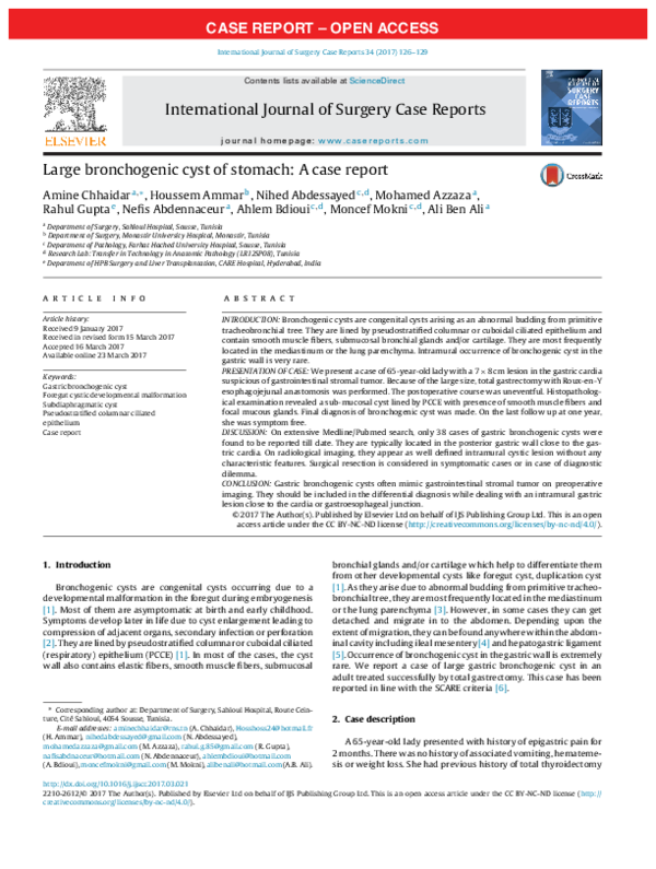
Large bronchogenic cyst of stomach: A case report
Amine Chhaidar a,∗ , Houssem Ammar b , Nihed Abdessayed c,d , Mohamed Azzaza a ,
Rahul Gupta e , Nefis Abdennaceur a , Ahlem Bdioui c,d , Moncef Mokni c,d , Ali Ben Ali a
a
Department of Surgery, Sahloul Hospital, Sousse, Tunisia
Department of Surgery, Monastir University Hospital, Monastir, Tunisia
c
Department of Pathology, Farhat Hached University Hospital, Sousse, Tunisia
d
Research Lab: Transfer in Technology in Anatomic Pathology (LR12SP08), Tunisia
e
Department of HPB Surgery and Liver Transplantation, CARE Hospital, Hyderabad, India
b
a r t i c l e
i n f o
Article history:
Received 9 January 2017
Received in revised form 15 March 2017
Accepted 16 March 2017
Available online 23 March 2017
Keywords:
Gastric bronchogenic cyst
Foregut cystic developmental malformation
Subdiaphragmatic cyst
Pseudostratified columnar ciliated
epithelium
Case report
a b s t r a c t
INTRODUCTION: Bronchogenic cysts are congenital cysts arising as an abnormal budding from primitive
tracheobronchial tree. They are lined by pseudostratified columnar or cuboidal ciliated epithelium and
contain smooth muscle fibers, submucosal bronchial glands and/or cartilage. They are most frequently
located in the mediastinum or the lung parenchyma. Intramural occurrence of bronchogenic cyst in the
gastric wall is very rare.
PRESENTATION OF CASE: We present a case of 65-year-old lady with a 7 × 8 cm lesion in the gastric cardia
suspicious of gastrointestinal stromal tumor. Because of the large size, total gastrectomy with Roux-en-Y
esophagojejunal anastomosis was performed. The postoperative course was uneventful. Histopathological examination revealed a sub-mucosal cyst lined by PCCE with presence of smooth muscle fibers and
focal mucous glands. Final diagnosis of bronchogenic cyst was made. On the last follow up at one year,
she was symptom free.
DISCUSSION: On extensive Medline/Pubmed search, only 38 cases of gastric bronchogenic cysts were
found to be reported till date. They are typically located in the posterior gastric wall close to the gastric cardia. On radiological imaging, they appear as well defined intramural cystic lesion without any
characteristic features. Surgical resection is considered in symptomatic cases or in case of diagnostic
dilemma.
CONCLUSION: Gastric bronchogenic cysts often mimic gastrointestinal stromal tumor on preoperative
imaging. They should be included in the differential diagnosis while dealing with an intramural gastric
lesion close to the cardia or gastroesophageal junction.
© 2017 The Author(s). Published by Elsevier Ltd on behalf of IJS Publishing Group Ltd. This is an open
access article under the CC BY-NC-ND license (http://creativecommons.org/licenses/by-nc-nd/4.0/).
1. Introduction
Bronchogenic cysts are congenital cysts occurring due to a
developmental malformation in the foregut during embryogenesis
[1]. Most of them are asymptomatic at birth and early childhood.
Symptoms develop later in life due to cyst enlargement leading to
compression of adjacent organs, secondary infection or perforation
[2]. They are lined by pseudostratified columnar or cuboidal ciliated
(respiratory) epithelium (PCCE) [1]. In most of the cases, the cyst
wall also contains elastic fibers, smooth muscle fibers, submucosal
∗ Corresponding author at: Department of Surgery, Sahloul Hospital, Route Ceinture, Cité Sahloul, 4054 Sousse, Tunisia.
E-mail addresses: aminechhaidar@rns.tn (A. Chhaidar), Hosshoss24@hotmail.fr
(H. Ammar), nihedabdessayed@gmail.com (N. Abdessayed),
mohamedazzaza@gmail.com (M. Azzaza), rahul.g.85@gmail.com (R. Gupta),
nafisabdnaceur@hotmail.com (N. Abdennaceur), ahlembdioui@hotmail.com
(A. Bdioui), moncefmokni@gmail.com (M. Mokni), alibenali@hotmail.com (A.B. Ali).
bronchial glands and/or cartilage which help to differentiate them
from other developmental cysts like foregut cyst, duplication cyst
[1]. As they arise due to abnormal budding from primitive tracheobronchial tree, they are most frequently located in the mediastinum
or the lung parenchyma [3]. However, in some cases they can get
detached and migrate in to the abdomen. Depending upon the
extent of migration, they can be found anywhere within the abdominal cavity including ileal mesentery [4] and hepatogastric ligament
[5]. Occurrence of bronchogenic cyst in the gastric wall is extremely
rare. We report a case of large gastric bronchogenic cyst in an
adult treated successfully by total gastrectomy. This case has been
reported in line with the SCARE criteria [6].
2. Case description
A 65-year-old lady presented with history of epigastric pain for
2 months. There was no history of associated vomiting, hematemesis or weight loss. She had previous history of total thyroidectomy
http://dx.doi.org/10.1016/j.ijscr.2017.03.021
2210-2612/© 2017 The Author(s). Published by Elsevier Ltd on behalf of IJS Publishing Group Ltd. This is an open access article under the CC BY-NC-ND license (http://
creativecommons.org/licenses/by-nc-nd/4.0/).
�CASE REPORT – OPEN ACCESS
A. Chhaidar et al. / International Journal of Surgery Case Reports 34 (2017) 126–129
127
Fig. 1. Contrast enhanced computed tomography of abdomen showing a large gastric mass with heterogenous content and enhancing wall arising from the posterior wall
of the gastric cardia and its relation with the esophagus, diaphragm and splenic hilum in the axial (a, b), coronal (b) and sagittal view (d).
for thyroid nodule and laparoscopic cholecystectomy for gallstones. On examination, there was no anemia, icterus or palpable
abdominal lump. Routine blood investigations and tumor markers
were within normal range. Abdominal ultrasound revealed a heterogenous lesion in the subdiaphragmmatic location close to the
medial surface of spleen measuring 6 × 7 cm with echogenic center. Computed tomography (CT) showed a large mass of 7 × 8 cm
in relation to the gastric cardia with regular outlines and heterogeneous enhancement (Fig. 1). Upper gastrointestinal endoscopy
found large ulcerated fundic folds with a bulge in to the lumen suggestive of extrinsic compression. There was presence of congestive
gastropathy. Esophagus was normal. Biopsy from fundic mucosa
showed moderately active chronic gastritis with absence of intestinal metaplasia or H. pylori infection or malignancy in the samples
examined. Based on the above findings, gastrointestinal stromal
tumor was suspected and she was planned for tumor excision.
On abdominal exploration, an exophytic mass measuring 8 × 7 cm
was found arising from the gastric cardia adherent to the adjoining diaphragm and encroaching upon the splenic hilum (Fig. 2).
After dissection, the mass could be separated from the diaphragm
Fig. 2. Schematic diagram showing the location of the bronchogenic cyst and its
relation with the surrounding structures. The dotted lines represent the extent of
surgical resection.
�CASE REPORT – OPEN ACCESS
128
A. Chhaidar et al. / International Journal of Surgery Case Reports 34 (2017) 126–129
Fig. 3. Microscopic examination of the cyst wall shows pseudostratified columnar ciliated epithelium without cellular atypia along with the surrounding smooth
muscle fiber bundles (HE × 200).
Fig. 4. Microscopic examination of the cyst wall revealed focal areas of mucous
glands, hemorrhagic remodeling and hemosiderin deposits. Areas of chronic gastritis without intestinal metaplasia were also noted (HE × 40).
and splenic hilum. Because of the large size, total gastrectomy
with Roux-en-Y esophagojejunal anastomosis was performed. The
postoperative course was uneventful and patient was discharged
on postoperative day 7. The cyst contained thick mucinous fluid.
Histopathological examination of the resected specimen revealed
a sub-mucosal cyst lined by PCCE without cellular atypia (Fig. 3).
There was no communication between the cyst and the gastric lumen. The cystic wall consisted of smooth muscle fibers
with focal mucous glands showing hemorrhagic remodeling with
hemosiderin deposits (Figs. 3 and 4). The gastric mucosa showed
mild chronic gastritis with presence of H. pylori without intestinal
metaplasia. Based on these findings, final diagnosis of bronchogenic
cyst was made. On the last follow up at one year, she was symptom
free with no evidence of recurrence.
3. Discussion
Congenital cysts are rare benign lesions of the stomach. There
are several types of congenital cysts reported in literature including
foregut cyst, gastric duplication cyst and bronchogenic cyst. They
are differentiated from each other by the type of lining epithelium
and the surrounding layer which may contain smooth muscles,
cartilage, bronchial glands or may be absent. Bronchogenic cyst
is the term, traditionally, used to describe a cyst lined by PCCE
with the presence of cartilage or glandular tissue in the cyst wall.
However, some recent studies have grouped intra mural gastric
cyst with a lining of PCCE into a single entity as gastric foregut
cystic developmental malformation which includes foregut cyst,
bronchogenic cyst and duplication lined by PCCE [7,8]. On extensive Medline/Pubmed search, we found only 38 reported cases of
classical gastric bronchogenic cyst till date.
Bronchogenic cysts are more common in females than males [1].
In most of the cases, cysts become symptomatic after third decade
of life with epigastric pain being the most common symptom [1].
They are typically located on the posterior or the posterolateral
gastric wall near the gastroesophageal junction or the gastric cardia [1,9]. The cyst size is variable. In the present case, due to the
large size, the cyst was occupying proximal two-third part of the
stomach unlike the previously reported cases. Most of them are
present in the submucosal or the smooth layer of the stomach
without any communication with the gastric lumen as seen in our
case. On gastroscopy, the larger cyst may appear as submucosal
lesion bulging in to the lumen like in the present case. On conventional imaging, such as CT, they appear as a well-defined cystic
lesion with or without calcifications [9]. On magnetic resonance
imaging (MRI), the lesion appears as iso-intense to hyper-intense
on T1-weighted images and hyper-intense on T2-weighted images
resembling solid tumors like GIST with cystic degeneration [1,9,10].
High signal intensity on T1-weighted images seen in bronchogenic
cysts, in contrast to low signal intensity seen in other cystic lesions
is likely due to the presence of methemoglobin, mucin and proteins within the cyst [1,9]. However, the wall is thin with regular
borders in bronchogenic cysts unlike the irregular thick wall seen
in cystic degeneration [9]. Endoscopic ultrasound (EUS) may help
is determining the exact location of the cyst within the gastric wall.
CT or EUS guided needle biopsy may reveal mucoid material providing a clue towards the correct diagnosis [10,11]. However, it is
not pathognomonic of bronchogenic cyst. Due to their submucosal
location and hypoechoic appearance, they are often misdiagnosed
as gastrointestinal stromal tumor (GIST) preoperatively similar to
the current case [1,9].
Surgical resection remains the most suitable treatment due to
the presence of diagnostic dilemma and rare possibility of malignant transformation [10,12]. However, there are anecdotal reports
of successful conservative management when the diagnosis was
confirmed on EUS guided biopsy [11]. Type of surgical resection
depends upon the location, size of the lesion and the surgical expertise. Small lesions can be managed by minimally invasive partial
gastrectomy while larger size like the one seen in our case are better
treated by open conventional gastrectomy [13].
4. Conclusion
Gastric bronchogenic cysts are rare benign lesions, often misdiagnosed as GIST due to similar presentation and radiological
appearance on CT scan and ultrasound. These cysts should be considered in the differential diagnosis especially when dealing with
an intramural gastric lesion close to the cardia or gastroesophageal
junction.
Conflict of interest
The authors declare that they have no conflict of interest.
Funding
This study has not received any funding.
Ethical approval
The study was approved by Ethics Committee of Hospital
Sahloul.
�CASE REPORT – OPEN ACCESS
A. Chhaidar et al. / International Journal of Surgery Case Reports 34 (2017) 126–129
Consent
Written informed consent was obtained from the patient.
Authors’ contribution
Amine Chhaidar – data collection, Editing of manuscript.
Houssem Ammar – Data collection, Editing of the manuscript.
Nihed Abdessaied – Histopathological examination of the specimen and reporting, data collection.
Mohamed Azzaza – Editing of manuscript.
Rahul Gupta – Editing of manuscript, literature review.
Ahlem Bdioui – Drafting of manuscript.
Nefis Abdennaceur – Editing of manuscript.
Moncef Mokni – Editing of the manuscript.
Ali Ben Ali – Editing of the manuscript.
Guarantors
Amine Chhaidar.
Houssem Ammar.
Acknowledgement
None.
129
[2] X. Yang, K. Guo, Bronchogenic cyst of stomach: two cases report and review of
the English literature, Wien Klin Wochenschr 125 (2013) 283–287.
[3] S. Aktoğu, G. Yuncu, H. Halilcolar, S. Ermete, T. Buduneli, Bronchogenic cysts:
clinicopathological presentation and treatment, Eur. Respir. J. 9 (1996)
2017–2021.
[4] A. Petrina, C. Boselli, R. Cirocchi, P. Covarelli, E. Eugeni, M. Badolato, et al.,
Bronchogenic cyst of the ileal mesentery: a case report and a review of
literature, J. Med. Case Rep. 4 (2010) 313.
[5] Y.G. Wang, H. Fang, X. Xu, W. Yu, K. Zhang, Y. Yu, Bronchogenic cyst in the
hepatogastric ligament masquerading as an esophageal mesenchymal tumor:
a case report, Int. J. Clin. Exp. Pathol. 8 (2015) 15307–15311.
[6] R.A. Agha, A.J. Fowler, A. Saeta, I. Barai, S. Rajmohan, D.P. Orgill, for the SCARE
Group, The SCARE statement: consensus-based surgical case report
guidelines, Int. J. Surg. 34 (2016) 180–186.
[7] S. Sharma, N. Nezakatgoo, P. Sreenivasan, J. Vanatta, N. Jabbour, Foregut cystic
developmental malformation: new taxonomy and classification—unifying
embryopathological concepts, Indian J. Pathol. Microbiol. 52 (2009) 461–472.
[8] Y.H. Geng, C.X. Wang, J.T. Li, Q.Y. Chen, X.Z. Li, H. Pan, Gastric foregut cystic
developmental malformation: case series and literature review, World J.
Gastroenterol. 21 (2015) 432–438.
[9] L. Sun, W. Lu Li Fu, W. Li, T. Liu, Gastric bronchogenic cyst presenting as a
gastrointestinal stromal tumor, Int. J. Clin. Exp. Pathol. 8 (2015) 13606–13612.
[10] S. Belli, T. Noyan, F. Kayaselçuk, G. Erbay, Gastric duplication (bronchogenic)
cyst mimicking a gastrointestinal stromal tumour, Ulusal Cer Derg 29 (2013)
35–37.
[11] M. Sato, A. Irisawa, M.S. Bhutani, V. Schnadig, T. Takagi, G. Shibukawa, et al.,
Gastric bronchogenic cyst diagnosed by endosonographically guided fine
needle aspiration biopsy, J. Clin. Ultrasound 36 (2008) 237–239.
[12] S.M. Sullivan, S. Okada, M. Kudo, Y. Ebihara, A retroperitoneal bronchogenic
cyst with malignant change, Pathol. Int. 49 (1999) 338–341.
[13] T. Kurokawa, M. Yamamoto, T. Ueda, T. Enomoto, K. Inoue, A. Uchida, et al.,
Gastric bronchogenic cyst histologically diagnosed after laparoscopic
excision: report of a case, Int. Surg. 98 (2013) 455–460.
References
[1] C. Tu, J. Zhu, C. Shao, W. Mao, X. Zhou, Q. Lin, et al., Gastric bronchogenic cysts:
a case report and literature review, Exp. Ther. Med. 11 (2016) 1265–1270.
Open Access
This article is published Open Access at sciencedirect.com. It is distributed under the IJSCR Supplemental terms and conditions, which
permits unrestricted non commercial use, distribution, and reproduction in any medium, provided the original authors and source are
credited.
�
 Amine Chhaidar