Academia.edu no longer supports Internet Explorer.
To browse Academia.edu and the wider internet faster and more securely, please take a few seconds to upgrade your browser.
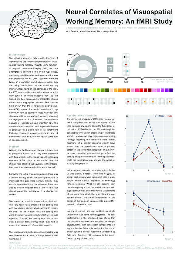
The following research falls into the long line of inquiries into the functional localization of visuospatial working memory (VSWM). Using functional magnetic resonance imaging (fMRI), we have attempted to reaffirm some of the hypotheses, previously established when it comes to the way the prefrontal cortex (PFC) codifies different types of information about objects, when they are being manipulated by the visual working memory. Depending on the demands of the task, the PFC can encode information either in a domain- general or domain-specific way [1]. We looked into how processing of integrated stimuli differs from segregated stimuli. EEG studies have shown that the contralateral delay activation (CDA) - a wave of activation seen in such cognitive functions as attention - rises with each new stimulus held in our working memory, reaching an asymptote at 3 – 4 stimuli, the maximum number of objects we can maintain [2]. The question here is whether an integrated stimulus is perceived as a single item or its constituent features represent unique objects in and of themselves, and what are the neural correlates of this processing.
Brain Research
Segregation of function in the lateral prefrontal cortex during visual object working memory2007 •
2000 •
Journal of Cognitive Neuroscience
“What” and “Where” in Visual Working Memory: A Computational Neurodynamical Perspective for Integrating fMRI and Single-Neuron Data2004 •
2013 •
2006 •
2007 •
Experimental Brain Research
Prefrontal cortical contributions to working memory: evidence from event-related fMRI studies2000 •
2002 •
The Faith and Beliefs of "Nonbelievers"
"MY LIFE IS ABSOLUTELY MEANINGLESS, BUT I LIKE IT!" RESPONDING TO NONBELIEVERS FROM A CHRISTIAN PERSPECTIVE2024 •
Teknologi Pendidikan, Frezy Paputungan, Pancasia, Universitas Bina Mandiri Gorontalo
Pancasila sebagai Sistem Etika2022 •
IAEME PUBLICATION
CONSUMER BUYING BEHAVIOUR OF VIRGIN EDIBLE OILS -A LITERATURE SURVEY AND CONCEPTUAL FRAMEWORK2019 •
Nuevo Comentario del Código Civil Peruano
Leysser León - Comentario sub art. 1317 del Código Civil (2022)2022 •
2009 •
Results in Physics
Chemical reaction in peristaltic motion of MHD couple stress fluid in channel with Soret and Dufour effects2018 •
International Journal of Behavioral Nutrition and Physical Activity
Healthier side dishes at restaurants: an analysis of children’s perspectives, menu content, and energy impacts2014 •
2016 •
Lecture Notes in Electrical Engineering
Towards Improving the Security of Low-Interaction Honeypots: Insights from a Comparative Analysis2017 •
International Journal of Production Economics
Sustainable supply chain management2008 •
1997 •
Avances en Ciencias Veterinarias
Evaluación del bienestar animal de aves rapaces en rehabilitación, descripción de técnicas que lo promuevan y mejoren su tasa de reintroducción2014 •
Journal of Rare Earths
Solvent extraction of neodymium(III) from acidic nitrate medium using Cyanex 921 in kerosene2012 •
 Aleš Oblak
 Anka Slana
 Nina Demšar