Nonlin. Processes Geophys., 16, 159–168, 2009
www.nonlin-processes-geophys.net/16/159/2009/
© Author(s) 2009. This work is distributed under
the Creative Commons Attribution 3.0 License.
Nonlinear Processes
in Geophysics
Characterizing water fingering phenomena in soils using magnetic
resonance imaging and multifractal theory
A. Posadas1,3 , R. Quiroz1 , A. Tannús2 , S. Crestana3 , and C. M. Vaz3
1 International
Potato Center – CIP, P.O. Box 1558, Lima 12, Peru
de Fı́sica e Informática, Instituto de Fı́sica de São Carlos, Universidade de São Paulo, Av. Dr. Carlos Botelho
1465, CEP 13560-250 São Carlos – SP, Brazil
3 Embrapa Instrumentação Agropecuária, Rua XV de Novembro, 1452, São Carlos, SP – CEP 13560-970, Brazil
2 Departamento
Received: 1 September 2008 – Revised: 4 February 2009 – Accepted: 4 February 2009 – Published: 26 February 2009
Abstract. The study of water movement in soils is of fundamental importance in hydrologic science. It is generally
accepted that in most soils, water and solutes flow through
unsaturated zones via preferential paths or fingers. This
paper combines magnetic resonance imaging (MRI) with
both fractal and multifractal theory to characterize preferential flow in three dimensions. A cubic double-layer column filled with fine and coarse textured sand was placed
into a 500 gauss MRI system. Water infiltration through
the column (0.15×0.15×0.15 m3 ) was recorded in steady
state conditions. Twelve sections with a voxel volume of
0.1×0.1×10 mm3 each were obtained and characterized using fractal and multifractal theory. The MRI system provided
a detailed description of the preferential flow under steady
state conditions and was also useful in understanding the dynamics of the formation of the fingers. The f (α) multifractal spectrum was very sensitive to the variation encountered
at each horizontally-oriented slice of the column and provided a suitable characterization of the dynamics of the process identifying four spatial domains. In conclusion, MRI
and fractal and multifractal analysis were able to characterize and describe the preferential flow process in soils. Used
together, the two methods provide a good alternative to study
flow transport phenomena in soils and in porous media.
1
Introduction
The fluid flow through preferential paths or fingers is extremely important in hydrological and agricultural processes
such as infiltration of water and the transport of agrochemiCorrespondence to: A. Posadas
(a.posadas@cgiar.org)
cals through the soil profile. Preferential paths increase the
probability of underground water contamination – through a
faster transportation of pesticides, heavy metals, radioactive
waste and other contaminants – and thus constitute a phenomenon of particular interest. To understand this complex
problem, some researchers concentrate their efforts in studying fluid transport in connection to the geometry of porous
media (Lu et al., 1994). In a review of the principal theories of the fingering phenomena (Steenhuis et al., 1996),
a model using a modification of the invasion percolation
model (Glass and Yarrington, 1996), based on both laboratory and field studies, was introduced. Many experiments
have shown that the fluid transport-porous medium coupling
has auto-similarity or fractal characteristics in a range of
defined scales (Katz and Thompson, 1985). The fingering
phenomenon in soils, which is controlled by both capillarity and gravity force (Hill and Parlange, 1972; Glass et al.,
1988, 1989; Crestana and Posadas, 1998), also presents fractal characteristics (Chang et al., 1994; Posadas and Crestana,
1993; Crestana and Posadas, 1998). These fractal characteristics have led to the creation of simulation models such as
the Diffusion Limited Aggregation-DLA (Chen and Wilkinson, 1985) that simulates the viscous fingering phenomenon
and the invasion percolation model (Wilkinson and Willemsen, 1983), which in turn simulates the capillary fingering
phenomenon. Other researchers have confirmed the suitability of fractal and multifractal theory to describe and simulate
preferential flow. Ogawa et al. (2002) applied fractal analysis
to study preferential flow on field soils and obtained a good
correlation between the surface fractal dimension and the exponent of a Van Genuchten expression applied to the particle
size distribution of the soil. A multifractal analysis was successfully employed by Nittmann et al. (1987) and Mâløy et
al. (1987) on viscous fingering structures observed in HeleShaw cells and in a mono-layer of glass beads, respectively.
Published by Copernicus Publications on behalf of the European Geosciences Union and the American Geophysical Union.
�160
A. Posadas et al.: Characterizing water fingering phenomena in soils
Recent publications suggest that the multifractal formalism
is applicable to three-dimensional systems. For example,
it has been shown (Held and Illangasekare, 1995) that the
width (internal energy or 1α) of the f (α) curve (multifractal spectrum), in the range of positive moments, quantifies
displacement instability. On the other hand, a basic technical challenge in investigations of mass transport in soils is
the need for a quantitative, visible and nondestructive monitoring of spatial and temporal water distribution, as a prelude to its quantitative analysis. Magnetic resonance imaging
(MRI) is becoming an important tool for studies of patterns
and mechanisms of water infiltration into soils (Amin et al.,
1998; Posadas et al., 1996). This paper proposes an innovation in the characterization of preferential flow by combining MRI (Posadas et al., 1996; Crestana and Posadas, 1998)
with multifractal theory (Chhabra et al., 1989b; Posadas et
al., 2001, 2003) for a three dimensional description of the
dynamics of fingers in sandy soils.
2
2.1
Materials and methods
Experiment
In order to visualize and characterize the fingering phenomenon in three dimensions, magnetic resonance imaging (MRI) and fractal and multifractal theory were employed in steady state conditions (Posadas, 1994; Onody
et al., 1995; Posadas et al., 1996). The work was conducted at CNPDIA/EMBRAPA Laboratory, Brazil. A cubic 0.15×0.15×0.15 m3 double-layer quartz sand column
was built, with the aim to generate preferential flow as described by Glass et al. (1989). The top layer was 2.0 cm
high, consisting of fine texture quartz sand (particle size
diameter in mm: 0.106<d<0.149), with a porosity (φ)
of 0.448±0.030 m3 m−3 , saturated hydraulic conductivity
(Ks ) of (6.3±0.08) 10−5 m s−1 and bulk density (ρb ) of
1500±80 kg m−3 and the lower 0.12 m layer was filled with
coarse sand (particle size diameter in mm: 0.212<d<0.500),
with a porosity (φ) of 0.312±0.030 m3 m−3 , saturated hydraulic conductivity (Ks ) of (26.7±0.15) 10−4 m s−1 and
bulk density (ρb ) of 1800±100 kg m−3 . On the surface of
the top layer, an acrylic plate of 0.15×0.15×0.03 m3 with
1.0 mm diameter holes was placed, in order to spread water uniformly over the surface. The top 2 mm section of the
column remained free for water application. This column
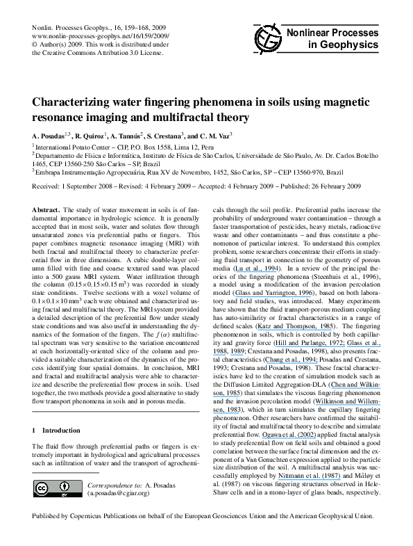
was placed into the head coils of the 500 gauss MRI systems
(Fig. 1).
The infiltration of water through the cubic column was
studied under hydrodynamic steady state conditions. Porosity and saturated hydraulic conductivity for each soil type –
fine and coarse – were previously estimated on other columns
filled with either fine sand soil or coarse sand. The gravimetric method with water saturation and the constant head soil
core (tank) method (Dane and Topp, 2002) with 10 replicates, were used respectively. All these parameters were
Nonlin. Processes Geophys., 16, 159–168, 2009
determined before running the MRI experiments. The cubic column was first filled with the coarse material carefully
placed at the bottom to achieve a homogeneous distribution.
A single layer of filter paper was then placed to separate the
soil types. A similar procedure was followed to place the
fine layer on the top of the column. The process was repeated until a homogeneous substrate and the bulk density
(ρb ), required for the desired saturated hydraulic conductivity were approximated. A second layer of filter paper was
placed to separate the fine sand from the acrylic plate. A
constant positive pressure of 2 mm of water was applied on
top of the column by watering the system through an inlet
placed above the acrylic plate, with 1 mm diameter holes.
This system was coupled to a medical infusion pump hanged
on a IV-stand equipment (Fig. 1a). This pump delivered
a constant water flow and pressure and facilitated the halting of water flowing into the column. A water volume of
600 ml was applied during 3 min to the top of the cubic column at a constant rate of 200 ml min−1 . The steady-state
flow of water was established when the first finger reached
the bottom of the box. At that moment the water inflow to
the column was turned off. The steady-state condition was
demonstrated through NMR Spin-Echo experiments using
the methodology described by Crestana and Posadas (1998);
Posadas (1994); Posadas et al. (1996). Different 2-D and 3D experiments were conducted to verify the diffusion process
for water redistribution, when the steady-state was reached.
It has been observed previously that even after 24 h the fingers formed initially remained virtually unchanged (Crestana
and Posadas, 1998). Horizontally-oriented, transverse and
sagittal images of the column were recorded during 11.2 s
just after the steady-state conditions were achieved as described by Posadas et al. (1996) and Crestana and Posadas
(1998). All the recorded MRI images were obtained in 2-D
slices and then processed using the software Image Processing and Analysis in Java-ImageJ (Rasband, 2007) and FracLab (Levy-Vehel and Mignot, 1994) developed by INRIA
(2005). The contrast in each slice of grey images was enhanced with different tools from ImageJ and FracLab which
permitted a good binarization using a thresholding filtering
in the range 62–72 of the grey level (Fig. 2). These 2-D binary images were utilized for fractal and multifractal analysis
using box-counting method following the gravitational force
(horizontally-oriented slices). The binarization error was estimated by calculating the total area (160×160 pixels) of the
first top slice and subtracting the binarized wet-area, which
was estimated in 7.0% of error (see Table 1).
2.2
2.2.1
Theory
MRI basics
Subatomic particles such as protons have the quantum mechanical property of spin. Certain nuclei such as 1 H (protons) have a non-zero spin and therefore a magnetic moment.
www.nonlin-processes-geophys.net/16/159/2009/
�A. Posadas et al.: Characterizing water fingering phenomena in soils
161
Fig. 1. (a) 500 gauss MRI system (IFSC-USP Laboratory, Brazil) showing the cubic column used (b) Sketch of the cubic column.
When these spins are placed in a strong external magnetic
field they precess synchronously at a unique frequency, the
Larmor frequency, which is characteristic of the species of
atom and the strength of the magnetic field. Changes of
this microscopic magnetization induced by a synchronous radiofrequency (RF) pulse result in a large proportion of the
nuclei being excited to a higher energy state. When the RF
pulses are discontinued the nuclei decay back to equilibrium.
During that time, the energy dissipates from the excited protons into their environment (spin-lattice or spin-spin relaxations) and that energy is detected as an electrical signal by
the RF receiver coil that has been previously tuned to detect the radiation at the Larmor frequency of protons associated with water. These signals are localized in space by the
field gradient coils installed in the magnet. Therefore, by adjusting the strength of the magnetic field with gradient coils,
the spatial differences in source of signals needed to produce
an image are obtained. Images are reconstructed from the
signals through the use of a Fourier transform (Mansfield
and Morris, 1982; Shaw, 1984; Brown et al., 1998; Van As,
2007).
2.2.2
Fractal and multifractal analysis
It is now widely accepted that physical systems that exhibit chaotic behavior are generic in nature. Since these
systems lose information exponentially fast it is possible to
follow and predict their motion in any detail only for short
time scales (Chhabra et al., 1989b). To describe their longterm dynamic behavior, one must resort to suitable statistical descriptions. One such description is multifractal formalism (Chhabra et al., 1989b; Hentschel and Procaccia, 1983;
Halsey et al., 1986; Chhabra and Jensen, 1989a). Multifractal
theory permits the characterization of complex phenomena
in a fully quantitative manner, for both temporal and spatial
variations. Multifractal techniques and notions are increasingly widely recognized as the most appropriate and straightwww.nonlin-processes-geophys.net/16/159/2009/
Fig. 2. Image binarization processes. (a) Thresholding filtering in
the range 62–72 of the grey level; (b) binarized image.
forward framework within which to analyze and simulate not
only the scale dependency of the geophysical observations,
but also their variability over a wide range of scales (Mandelbrot, 1982; Schertzer and Lovejoy, 1996).
The basic equation of the fractal theory expresses the relationship between the number and the size of the objects
(Feder, 1988):
N (ε) ∼ ε−D0 ,
(1)
where N (ε) is the number of objects, ε is the scale and D0
is the fractal or capacity dimension. In this paper we will
use the capacity dimension name, following the convention
of Beck and Schlogl (1995). The box-counting technique is
used to estimate the scaling properties of an image by covering it with boxes of size ε and counting the number of boxes
containing at least one pixel representing the object under
study:
D0 = − lim
ε→0
log N (ε)
.
log(ε)
(2)
Nonlin. Processes Geophys., 16, 159–168, 2009
�162
A. Posadas et al.: Characterizing water fingering phenomena in soils
Table 1. Selected nparameters of preferential flow infiltration and multifractal parameters.
MRI slices
numbers
–
12
11
10
09
08
07
06
05
04
03
02
01
Depth (cm)
±error r
0.00
2.2±0.1
3.3±0.1
4.4±0.1
5.5±0.1
6.6±0.1
7.7±0.1
8.8±0.1
9.9±0.1
11.0±0.1
12.1±0.1
13.2±0.1
14.3±0.1
Wet-area percent
±error b
Capacity dimension
D0 (q=0)±MSE
R 2 moments
variation
1q
–
100.00±7
77.40±7
53.42±7
44.64±7
36.55±7
35.20±7
30.60±7
25.20±7
22.70±7
21.30±7
19.30±7
14.80±7
–
2.00±0.01
1.94±0.02
1.81±0.05
1.79±0.04
1.76±0.05
1.73±0.04
1.71±0.04
1.59±0.05
1.60±0.02
1.44±0.03
1.39±0.02
1.34±0.02
–
1.0000
0.9995
0.9949
0.9944
0.9956
0.9933
0.9872
0.9828
0.9916
0.9748
0.9467
0.9985
–
14.20
11.20
11.76
6.56
4.44
3.26
2.92
2.22
1.22
0.74
0.72
0.28
1α±MSE domains
Spatial
0.00
0.2098±0.04
1.3602±0.14
1.5187±0.16
1.4429±0.16
1.4784±0.13
1.1053±0.12
0.3437±0.03
0.4919±0.05
0.1767±0.05
0.0536±0.005
0.0774±0.008
0.0227±0.002
HZ
LSH
LSH-IP
LSH-IP
LSH-IP
LSH-IP
WSH
WSH
WSH
EZ-Fractal
EZ-Fractal
EZ-Fractal
Note: all the probability values (p-value) computed were, p<0.0001 error r is the reading error introduced for the measure rule; error b is
the binarization error; MSE is defined as the mean square error; R 2 is the correlation coefficient; 1q is defined as qmax −qmin ; 1α is the
internal energy variation defined as αmax −αmin ; HZ is the homogeneous zone; LSH is large spatial heterogeneity; IP is invasion percolation;
WSH is weak spatial heterogeneity; EZ is the equilibrium zone.
Provided the limit exists, the infinitum of N (ε) is approximated by varying the origin of the grid until the smallest
number is found. Using Eq. (2), the capacity dimension D0
can be determined as the negative slope of log N (ε) versus
log(ε), measured over a range of box widths. In a homogeneous system, the probability (P ) of a measured quantity
(measure) varies with scale ε as (Chhabra et al., 1989b; Evertsz and Mandelbrot, 1992; Vicsek, 1992):
P (ε) ∼ εD ,
(3)
where D is a fractal dimension. For heterogeneous or nonuniform systems the probability within the ith region Pi
varies as:
αi
Pi (ε) ∼ ε ,
(4)
where f (α) can be considered as the generalized fractal dimension of the set of boxes with singularities α (Kohmoto,
1988). The exponent α can take on values from the interval
[α−∞ , α+∞ ], and f (α) is usually a function with a single
maximum at df (α(q))/dα(q)=0 (where q is the order moment of a statistic distribution). Thus, when q=0, fmax is
equal to the capacity dimension, D0 (Gouyet, 1996; Vicsek,
1992).
Multifractal sets can also be characterized on the basis of
the generalized dimensions of the qth order moment of a distribution, Dq , defined as (Hentschel and Procaccia, 1983):
Dq = lim (
ε→0
1 log µ(q, ε)
),
q − 1 log(ε)
(6)
where µ(q, ε) is the partition function (Chhabra et al.,
1989b):
where αi is the Lipschitz-Hölder exponent or singularity
strength, characterizing scaling in the ith region or spatial
location (Feder, 1988). The parameter αi quantifies the degree of regularity in point xi . Loosely speaking, any measure
µ of an interval [xi , xi +1x], behaves as (1x)αi (Halsey et
al., 1986). For a uniform distribution one finds αi (x)=1 for
all x. More generally, for any real value a>0 the distribution
with density x a−1 on [0, 1] has αi (0)=a and αi (x)=1 for all
xǫ[0, 1]. Values αi (x)<1 indicate, thus, a burst of the event
around x on all levels, while αi (x)>1 is found in regions
where events occur sparsely (Riedi, 1999). Similar αi values
might be found at different positions in the space. The number of boxes N (α) where the probability Pi has singularity
strengths between α and α+dα is found to scale as (Chhabra
et al., 1989b; Halsey et al., 1986):
where τ (q) is the correlation exponent of the qth order moment defined as (Halsey et al., 1986; Vicsek, 1992):
N(α) ∼ ε−f (α) ,
τ (q) = (q − 1)Dq ,
Nonlin. Processes Geophys., 16, 159–168, 2009
(5)
µ(q, ε) =
N(ε)
X
q
Pi (ε),
(7)
i=1
The generalized dimension Dq is a monotonic decreasing
function for all real q’s within the interval [−∞+∞]. When
q<0, µ emphasizes regions in the distribution with less concentration of a measure, whereas the opposite is true for q>0
(Chhabra and Jensen, 1989a).
Also, the partition function scales as:
µ(q, ε) ∼ ετ (q) ,
(8)
(9)
www.nonlin-processes-geophys.net/16/159/2009/
�A. Posadas et al.: Characterizing water fingering phenomena in soils
Fig. 3. Images obtained with the MRI system, showing three
vertically-oriented sections of the fingering phenomenon in static
conditions. Each section represents a slice, 2 cm thick, 15 cm wide
and 15 cm high, of the 15×15×15 cm cubic soil column. The white
areas represent wet zones and the grey areas represent dry zones.
The connection between the power exponents f (α) (Eq. 5)
and τ (q) (Eq. 9) is made via the Legendre transformation
(Callen, 1985; Chhabra and Jensen, 1989a; Halsey et al.,
1986):
f (α(q)) = qα(q) − τ (q) and α(q) =
dτ (q)
dq
(10)
f (α) is a concave downward function with a maximum at
q=0. When q takes the values of q=0, 1 or 2, (Eq. 6) is
reduced to:
log(N(ε))
D0 = lim
, D1 = lim
ε→0 log(ε)
ε→0
N(ε)
X
µi (ε) log(µi (ε))
i=1
log(ε)
log(C(ε))
,
ε→0 log(ε)
D2 = lim
Fig. 4. Images obtained by MRI showing twelve horizontaloriented sections (slices 1.0 mm stick) of the cubic soil column following the direction of the infiltration. (a) Saturated section near
the surface of the column (first layer); (l) section corresponding to
the bottom of the soil column; and (b) through (k) represent intermediate situations. Light grey areas correspond to invading water
within the pore networks and black areas correspond to both the
non-invaded porous medium and quartz solid phase. Grey tones are
function of the sand water content.
(or average) information about a system (Voss, 1988). The
entropy dimension is related to the information (or Shannon)
entropy (Shannon and Weaver, 1949). The correlation dimension D2 is mathematically associated with the correlation function (Grassberger and Procaccia, 1983) and computes the correlation of measures contained in a box of size
ε. The relationship between D0 ,D1 ,and D2 is,
D2 ≤ D1 ≤ D0 ,
,
(11)
respectively, with C(ε) being the correlation function.
The values D0 , D1 and D2 are known as the capacity dimension, the entropy dimension and the correlation dimension, respectively. The capacity dimension provides global
www.nonlin-processes-geophys.net/16/159/2009/
163
(12)
The equality D0 =D1 =D2 occurs only if the fractal is statistically or exactly self-similar and homogeneous (Korvin,
1992).
Following the methodology used by Posadas et al. (2001,
2003), multifractal theory was applied to the MR images
of the fingering phenomena. The size of each 2-D image considered for the multifractal analysis was 160×160
pixels (or 15×15 cm2 ). Twelve binary-images slices of
Nonlin. Processes Geophys., 16, 159–168, 2009
�164
A. Posadas et al.: Characterizing water fingering phenomena in soils
2.5
y = -0.0503x + 2.0957
2
R = 0.966
f(q=0)
2
1.5
1
0.5
0
0
2
4
6
8
10
12
14
16
Depth (cm)
Fig. 6. Capacity dimension, of the fingers, measured along the vertical depth.
Fig. 5. Box counting plots for each binary image slices analyzed for
q=0 with upper and lower boxes sizes. These plots show the scale
range of fractal behavior of the systems. Also, two linear regressions estimating the capacity dimension for bottom and top layer
slices are shown.
horizontally-oriented sections, following the gravitational
force, were analyzed using the multifractal algorithm (CIPMASS -downloadable after subscription at http://inrm.cip.
cgiar.org/vlab). This algorithm was developed based on the
method described in Chhabra and Jensen (1989a) and implemented in Matlab by Posadas et al. (2001). The spatial
distribution of water concentration through each slice was
partitioned in boxes size L, for L=8, 10, 16, 20, 32, 40 and
80 pixels (see Fig. 5). These upper and lower box sizes considered prevent the systematic biases in the small and large
scales as mentioned by Vignes-Adler et al. (1991). The capacity dimension D0 was obtained from the maximum value
of the multifractal spectrum when q=0 (Beck and Schlogl,
1995). The moments (q) in each slice considered ranged
from 1q=14.20 to 0.28, as shown in Table 1, with steps variation of 0.02 which is fine enough to show the multifractal
behavior in the very narrow range of q’s.
3
3.1
Results and discussion
Magnetic resonance image analysis
The results obtained with the MRI system are shown in Table 1 and depicted in Figs. 3 and 4. Figure 3 shows three
acquisitions of the transverse plane of the cubic sand column
at steady-state flow. In these three images it is possible to
observe the three-dimensional character of the fingering pheNonlin. Processes Geophys., 16, 159–168, 2009
nomena and its spatial variability. After a few seconds of
turning off water inflow, it was observed that the formation
of fingers was completely halted. These findings were consistent across the five experiments conducted using different
packed columns, but keeping the same physical parameters.
Similar finger occupation behavior and steady-state conditions were observed. As expected the only thing that varied
was the zoned occupied by the fingers. The accumulation of
water at the bottom of the column can be seen in panel A.
Since there is no evidence of water flow except through the
preferential paths it can be inferred that, if the experiments
replicate the process occurring in real systems, the movement
of water and the solutes conveyed within it could reach shallow or deep water pools in the soil faster than in conditions
where preferential flow is absent.
The column under steady-state flow was “dissected” into
12 horizontally-oriented slices of 1.0 cm thick, as shown in
Fig. 4. The light grey area depicts the concentration of water
in the cross section. It can be seen that the water concentration throughout the profile follows a fixed path. Dark grey
paths in the images represent dry sand areas. These findings
support the existence of preferential flow under the conditions studied (Posadas et al., 1996).
3.2
Fractal and multifractal analysis
Figures 5, 6, 7, 8 and Table 1 summarize the fractal and
multifractal analysis. The capacity dimension (D0 , Fig. 6)
evidences differences in the spatial distribution among the
twelve layers of the columns depicted in Fig. 4. When D0
is near 2.0 (top layer), the system is more homogeneous i.e.
most of the pores are filled with water (saturation of porous
media), and corresponds to an apparent single water phase.
The infiltration process throughout the entire top layer was
slow and uniform with a constant vertical velocity like a
plane wave (panel a in Fig. 4), following the gravitational
force. As soon as the water crosses the interface between the
fine and the coarse layers, a hydrodynamic instability seems
to dominate de infiltration process and the fingers began to
www.nonlin-processes-geophys.net/16/159/2009/
�A. Posadas et al.: Characterizing water fingering phenomena in soils
165
MRI12
3
f(α)
2
1
0
0
1
2
3
α
4
MRI11
3
f(α)
2
1
0
0
1
2
3
α
4
Fig. 8. Variation of the capacity dimension and the internal energy
of the system as a function of the depth along the cubic column.
MRI05
3
f(α)
2
1
0
0
1
2
3
α
4
MRI01
3
f(α)
2
1
0
0
1
2
α
3
4
Fig. 7. Magnetic Resonance Images from a three-dimensional
fingering phenomena and its multifractal spectrums following the
gravitational direction (from top MRI12 to bottom MRI01). These
sets represent binary images, where the black areas correspond to
water and the white one correspond to the dry area.
develop. This is depicted in the panel b in Fig. 4. This corresponds to a section with high heterogeneity in the distribution
of water, which decreases towards the bottom of the column.
This seems to be a good descriptor of the steady state conditions at which the images were taken.
Figure 7 shows the multifractal analysis used to describe
the heterogeneity of the spatial variability of the water in
each cross-section throughout the profile of the column. A
quick inspection along the column indicates how dynamic
the system is, even though the analysis was made when
the system reached a steady-state flow; a condition reached
after 137 s of infiltration. It goes from the wetting instability – the first condition of the coarse texture substrate
(Panel b in Fig. 4) to a hydrodynamic stability at the bottom
(Panel l in Fig. 4), passing through chaotic sections in the
intermediate portion (Panels c through k in Fig. 4).
www.nonlin-processes-geophys.net/16/159/2009/
The spatial variations in all these conditions seem to be
well characterized with the f (α) α spectrum as shown on
the right panels in Fig. 7. For instance, the cross-section
labeled MRI11 represents the wetting instability conditions
in the column. This hydrodynamic instability is attributed
to the change produced when the water flows from the fine
to the coarse texture. The multifractal spectrum is characteristic of a heterogeneous system with variations on both
sides of the maximum value. From the maximal capacity
dimension (D0 ) to the left, the spectrum describes the behavior of the areas where water is present (positive q’s). The
asymmetry toward the right from α=2 indicates domination
of small or extremely small values of water. This is an indication of the existence of preference paths. The condition prevails, with small variations, down through the cross-section
MRI07. Inspection of the variation in the internal energy
(1α) shows that the 1α value for MRI11 is 1.36 (Table 1).
Compared to the saturation condition presented in the layer
with fine texture (1α=0.21) one can contrast how this multifractal parameter changes when the condition changes from a
homogeneous (MRI12) to a heterogeneous wetting instability. The cross-sections MRI05 and MRI04 seem to be a transition section from instability to hydrodynamic stability. The
corresponding 1α is around 0.4. The bottom three crosssections correspond to hydrodynamic stability, behaving as
a pure fractal with a capacity dimension D0 changing from
1.44 to 1.34 and 1α<0.07
3.3
Spatial domains
Different spatial domains can be identified following the
depth of the soil column and the multifractal parameters.
These domains are summarized in Table 1 and the spatial behavior of each slice can be seen in Fig. 4. The
spatial domains were labeled as HZ (homogeneous zone),
Nonlin. Processes Geophys., 16, 159–168, 2009
�166
A. Posadas et al.: Characterizing water fingering phenomena in soils
LSH (large spatial heterogeneity), WSH (weak spatial heterogeneity) and EZ (equilibrium zone). The equilibrium
zone is attributed to the fact that multifractality does not occur and it behaves as a pure fractal.
As an example let us analyze the slices MRI10 to MRI07,
corresponding to the onset of observed fingers, with a rather
constant capacity dimension, D0 of 1.73±0.10−1.81±0.10
(see also panels c through f in Fig. 4). The average D0 ∼
=1.82,
might be associated with the invasion percolation capacity
dimension without trapping (Gouyet, 1996). In the light of
the invasion percolation theory (Wilkinson and Willemsen,
1983; Onody et al., 1995), this seems to be an unstable zone
characteristic of a percolative pore network. This pore networks feature, from homogeneous to heterogeneous behavior, suggests that the gradient invasion percolation might be
a good model to simulate preferential-infiltration processes
in soils (Frette et al., 1992).
An interesting feature of the multifractal analysis is that
in spite of the fact that the image was taken under steadystate flow, the dynamic characteristics present in the profile
is described by the f (α) α spectrum and its parameters. Figure 8 captures these characteristics by showing the dynamic
of D0 and 1α across the column profile. Following the column depth, there is a reduction in the capacity dimension
D0 , represented with the grey level. This can be seen as a
first and gross approximation of the fingers’ dynamic. The
inclusion of the internal energy 1α or the “ensemble average” of the total energy of the system (Reichl, 1998) – visualized through a proportional thickness of the line – enriches
the description of the phenomena. It is evident that as soon
as the water reaches the coarse layer there is a multifractal
behavior associated to a spatial domain of high heterogeneity, which decays quasi exponentially as a function of the
column depth until it dies down to an equilibrium zone with
fractal behavior. The four spatial domains described above
– HZ, LSH, WSH and EZ – are clearly differentiated when
these two multifractal parameters are jointly interpreted. In
terms of the magnitude, a stable zone can be inferred when
D0 is around 2 and 1α∼0−0.2. The maximum instability
zone from 4.4 to 7.7 cm of depth, corresponds to the invasion
– percolation zone with D0 ∼1.73–1.81 and 1α∼1–1.5. The
transition zone from 9 to 11 cm of depth presented D0 ∼1.59–
1.71 and 1α∼0.5–0.2. In turn, the equilibrium zone, from
12 to 14 cm of depth, showed D0 ∼1.34–1.44 and 1α∼0.02–
0.05; depth at which the hydrodynamic stability has been
achieved.
4
Conclusions
1. Both the multifractal theory and the MRI system were
good tools to characterize preferential water flow in soils.
The MRI system is useful in visualizing the phenomenon for
a better understanding of the process. The technique presented very innovative and encouraging results for the study
Nonlin. Processes Geophys., 16, 159–168, 2009
of preferential flow in soils and porous media, allowing the
observation, in a non-disturbing way, of the 3-D and random character of the phenomenon. Nonetheless, very few
soil or geophysical research groups have access to this type
of equipment. 2. The use of multifractal theory facilitates
describing the dynamics of preferential flow and can be used
to predict the outcomes under real conditions and to improve
the accuracy of existing models, as the invasion percolation
model, for example. The combination of these techniques
opens a new set of options that must be further tested for different soil types and management conditions. 3. Also, using
the multifractal parameters, capacity dimension D0 and the
internal energy 1α it was possible to identify four spatial
domains along the soil columns studied. These novel results
open new research alternatives to study infiltration processes
in soils and thus require further research. This is a challenge
to be faced in the near future.
Acknowledgements. The authors are indebted to several institutions and people. The final data analysis and the production
of the manuscript were supported by the CIDA-Canada through
the ALTAGRO Project and FAPESP (Brazilian Agency) Process
No. 2007/58561-7.
The authors would like to thank Victor Mares for the revision and
improvement of the manuscripts and Ivonne Valdizan for patiently
going through the process of learning Latex and converting the
manuscript into that format, in addition to her normal contribution
in guaranteeing the consistency and adequacy of the papers to the
journal’s format.
Edited by: A. Provenzale
Reviewed by: an anonymous referee
References
Amin, M. H. G., Hall, L. D., Chorley, R. J., and Richards, K. S.:
Infiltration into soils, with particular reference to its visualization
and measurement by magnetic resonance imaging(MRI), Prog.
Phys. Geog., 22(2), 135–165, 1998.
Beck, C. and Schlogl, F.: Thermodynamics of Chaotic Systems: An
introduction. Cambridge Nonlinear Science Series 4, Cambridge
University Press, 306 pp., 1995.
Brown, J. M., Kramer, P. J., Cofer, G. P., and Johnson, G. A.: Use of
nuclear magnetic resonance microscopy for noninvasive observations of root-soil water relations, Theor. Appl. Climatol., 42(4),
229–236, 1998.
Callen, H. B.: Thermodynamics and an introduction to thermostatics, John Wiley and Sons, New York, 2nd edn., 512 pp., 1985.
Chang, W.-L., Biggar, J. W., and Nielsen, D. R.: Fractal description
of wetting front instability in layered soils, Water Resour. Res.,
30(1), 125–132, 1994.
Chen, J. D. and Wilkinson, D.: Pore-scale viscous fingering in
porous media, Phys. Rev. Lett., 58(18), 1892–1895, 1985.
www.nonlin-processes-geophys.net/16/159/2009/
�A. Posadas et al.: Characterizing water fingering phenomena in soils
Chhabra, A. B. and Jensen, R. V.: Direct determination of the
f (α) singularity spectrum, Phys. Rev. Lett., 62(12), 1327–1330,
1989a.
Chhabra, A. B., Meneveu, C., Jensen, R. V., and Sreenivasan, K.
R.: Direct determination of the f (α) singularity spectrum and its
application to fully developed turbulence, Phys. Rev. A, 40(9),
5284–5294, 1989b.
Crestana, S. and Posadas, D. A. N.: 2-D and 3-D fingering phenomenon in unsaturated soils investigated by fractal analysis, invasion percolation modeling and non-destructive image processing, in: Fractals in Soils Science, edited by: Baveye, P., Parlange,
J.Y., and Stewart, B. A., Boca Raton, Florida, USA CRC Press,
293–332, 1998.
Dane, J. H. and Topp, G. C. (Eds.): Methods of Soil Analysis: Part
4, Physical Methods, SSSA Book Series No 5, Soil Science Society of America, Madison, WI, 2002.
Evertsz, C. J. G. and Mandelbrot, B. B.: Multifractal measures,
in: Chaos and Fractals, edited by: Peitgen, H.-O., Jurgens, H.,
and Saupe, D., New Frontiers of Science, Springer-Verlag, New
York, 921–953, 1992.
Feder, J.: Fractals, Plenum Press, New York, 283 pp., 1988.
Frette, V., Feder, J., Målfy, K. F., Jfssang, T., and Meaking., P.:
Buoyancy-driven fluid migration in porous media, Phys. Rev.
Lett., 68(21), 3164–3167, 1992.
Glass, R. J. and Yarrington, L.: Simulation of gravity fingering in
porous media using a modified invasion percolation model, Geoderma, 70(2–4), 231–252, 1996.
Glass, R. J., Steenhuis, T. S., and Parlange, J.-Y.: Wetting front
instability as a rapid and far-reaching hydrologic process in the
vadoze zone, J. Contam. Hydrol, 3, 207–226, 1988.
Glass, R. J., Steenhuis, T. S., and Parlange, J.-Y.: Wetting front
instability: experimental determination of relationships between
system parameters and two-dimensional unstable flow field behavior in initially dry porous media, Water Res. Res., 25(6),
1195–1207, 1989.
Gouyet, J. F.: Physics and fractals structure, Springer, New York,
234 pp., 1996.
Grassberger, P. and Procaccia, I.: Characterization of strange attractors, Phys. Rev. Lett., 50(5), 346–349, 1983.
Halsey, T. C., Jensen, M. H., Kadanoff, L. P., Procaccia, I., and
Shraiman, B. I.: Fractal measures and their singularities: The
characterization of strange sets, Phys. Rev. A, 33(2), 1141–1151,
1986.
Held, R. J. and Illangasekare, T. H.: Fingering of dense nonaqueous phase liquids in porous media 2. Analysis and classification,
Water. Resour. Res., 31(5), 1223–1231, 1995.
Hentschel, H. G. E. and Procaccia, I.: The infinite number of generalized dimensions of fractals and strange attractors, Physica D:
Nonlinear Phenomena, 8(3), 435–444, 1983.
Hill, D. E. and Parlange, J.-Y.: Wetting front instability in layered
soils, Soil Sci. Soc. Am. J., 36(5), 697–702, 1972.
INRIA: FracLab: APIS, http://complex.futurs.inria.fr/FracLab/
index.html, 2005.
Katz, A. J. and Thompson, A. H.: Fractal sandstone pores: Implications for conductivity and pore formation, Phys. Rev. Lett.,
54(12), 1325–1328, 1985.
Kohmoto, M.: Entropy function for multifractals, Phys. Rev. A,
37(4), 1345–1350, 1988.
www.nonlin-processes-geophys.net/16/159/2009/
167
Korvin, G.: Fractals Models in the Earth Sciences, Elsevier, Amsterdam, The Netherlands, 396 pp., 1992.
Levy-Vehel, J. and Mignot, P.: Multifractal segmentation of images,
Fractals, 2, 371–377, 1994.
Lu, T. X., Biggar, J. W., and Nielsen, D. R.: Water movement in
glass bead porous media 2. Experiments of infiltration and finger
flow, Water Resour. Res., 30(12), 3283–3290, 1994.
Mâløy, K. J., Boger, F., Feder, J., and Jøssang, T.: Dynamics
and structure of viscous fingering in porous media, in: timedependent effects in disordered materials, edited by: Pynn, R.
and Riste, T., Plenum Press, NY, 111–118, 1987.
Mandelbrot, B. B.: The fractal geometry of nature, W.H. Freeman
and Company, NY, 2nd edn., 468 pp., 1982.
Mansfield, P. and Morris, P. G.: NMR Imaging in biomedicine, Supplement 2, Advances in Magnetic Resonance, Academic Press,
New York, 354 pp., 1982.
Nittmann, J., Stanley, H. E., Toubul, E., and Daccord, G.: Experimental evidence of multifractality, Phys. Rev. Lett., 58(6), 619–
622, 1987.
Ogawa, S., Baveye, P., Parlange, J.-Y., and Steenhuis, T.: Preferential flow in the field soils, Forma, 17(1), 31–53, 2002.
Onody, R. N., Posadas, D. A. N., and Crestana, S.: Experimental
studies of fingering phenomena in two dimensions and simulation using a modified invasion percolation model, J. Appl. Phys.,
78(5), 2970–2976, 1995.
Posadas, D. A. N. and Crestana, S.: Aplicação da teoria fractal
na caracterização do fenômeno “fingering” em solos, Revista
Brasileira de Ciências Sociais, 17(1), 1–8, 1993.
Posadas, D. A. N.: Estudo do fenômeno “fingering” em um meio
poroso através de imagens e teoria da percolação por invasão,
Ph.D. thesis, IFSC/USP, São Carlos, SP-Brazil, 187 pp., 1994.
Posadas, D. A. N., Tanns, A., Panepucci, C. H., and Crestana, S.:
Magnetic resonance imaging as a non-invasive technique for investigating 3-D preferential flow occurring within stratified soil
samples, Computers and Electronics in Agriculture, 14(4), 255–
267, 1996.
Posadas, D. A. N., Gimenez, D., Bittelli, M., Vaz, C. M. P., and
Flury, M.: Multifractal Characterization of soil particle-size distributions, Soil Sci. Soc. Am. J., 65(5), 1361–1367, 2001.
Posadas, D. A. N., Gimenez, D., Quiroz, R., and Protz, R.: Multifractal characterization of soil pore systems, Soil Sci. Soc. Am.
J., 67(5),1361–1369, 2003.
Rasband, W.: ImageJ 1.38x, National Institute of Health, USA,
available at: http://rsb.info.nih.gov/ij/, 2007.
Reichl, L. E.: A modern course in statistical physics, John Wiley
and Sons, Inc. NY, 2nd edn., 580 pp., 1998.
Riedi, R. H.: An Introduction to Multifractals, Technical Report,
Rice University, Unpublished Version, 5 September 1999.
Shaw, D.: Fourier transform N.M.R. spectroscopy, Studies in physical and theoretical chemistry, NY Elsevier Publishing Company,
2nd edn., 304 pp., 1984.
Schertzer, D. and Lovejoy, S.: EGS Richardson AGU Chapman
NVAG3 Conference: Nonlinear variability in geophysics: scaling and multifractal processes, Nonlinear Proc. Geoph., 1(2–3),
77–79, 1996.
Shannon, C. E. and Weaver, W.: The mathematical theory of communication, Urbana, University of Illinois Press, 125 pp., 1949.
Nonlin. Processes Geophys., 16, 159–168, 2009
�168
A. Posadas et al.: Characterizing water fingering phenomena in soils
Steenhuis, T. S., Ritsema, C. J., and Dekker, L. W. (Eds): Fingered
flow in unsaturated soil: from nature to model, Geoderma, Special Issue, 70(2–4), 83–326, 1996.
Van As, H.: Intact plant MRI for the study of cell water relations, membrane permeability, cell-to-cell and long distance water transport, J. Exp. Bot., 58(4), 743–756, 2007.
Vicsek, T.: Fractal growth phenomena, World Scientific Publishing
Co., Singapore, 2nd edn., 380 pp., 1992.
Nonlin. Processes Geophys., 16, 159–168, 2009
Vignes-Adler, M., Le Page, A., and Adler, P. M.: Fractal analysis
of fracturing in two African regions, from satellite imagery to
ground scale, Tectonophysics, 196(1–2), 69–86, 1991.
Voss, R. F.: Fractals in nature: From characterization to simulation,
in: The Science of Fractal Image, edited by: Peitgen, H.-O. and
Saupe, D., Springer-Verlag, NY, 21–70, 1988.
Wilkinson, D. and Willemsen, J. F.: Invasion percolation: A new
form of percolation theory, J. Phys. A-Math. Gen., 16(14), 3365–
3376, 1983.
www.nonlin-processes-geophys.net/16/159/2009/
�
 R. Quiroz
 Alberto Tannús